Highly elastomeric poly(glycerol sebacate)-co-poly(ethylene glycol) amphiphilic block copolymers Alpesh Patel a, b,1 , Akhilesh K. Gaharwar c, d, 1 , Giorgio Iviglia a, b, e, 1 , Hongbin Zhang a, b, f , Shilpaa Mukundan a, b , Silvia M. Mihaila a, b , Danilo Demarchi e, g , Ali Khademhosseini a, b, d, * a Center for Biomedical Engineering, Department of Medicine, Brigham and Women’s Hospital, Harvard Medical School, Cambridge, MA 02139, USA b Harvard-MIT Division of Health Sciences and Technology, Massachusetts Institute of Technology, Cambridge, MA 02139, USA c Wyss Institute for Biologically Inspired Engineering, Harvard University, Boston, MA 02115, USA d David H. Koch Institute for Integrative Cancer Research, Massachusetts Institute of Technology, Cambridge, MA 02139, USA e Department of Electronics and Telecommunications, Politecnico di Torino, Torino 10129, Italy f School of Materials Science and Engineering, University of Science and Technology, Beijing, Beijing 100083, China g Center for Space Human Robotics, Italian Institute of Technology, Torino, Italy article info Article history: Received 20 December 2012 Accepted 7 January 2013 Available online 1 March 2013 Keywords: Block copolymers Elastomer Mechanical properties Poly(glycerol sebacate) Poly(ethylene glycol) abstract Poly(glycerol sebacate) (PGS), a tough elastomer, has been proposed for tissue engineering applications due to its desired mechanical properties, biocompatibility and controlled degradation. Despite inter- esting physical and chemical properties, PGS shows limited water uptake capacity (w2%), thus con- straining its utility for soft tissue engineering. Therefore, a modification of PGS that would mimic the water uptake and water retention characteristics of natural extracellular matrix is beneficial for enhancing its utility for biomedical applications. Here, we report the synthesis and characterization of highly elastomeric poly(glycerol sebacate)-co-polyethylene glycol (PGS-co-PEG) block copolymers with controlled water uptake characteristics. By tailoring the water uptake property, it is possible to engineer scaffolds with customized degradation and mechanical properties. The addition of PEG results in almost 15-fold increase in water uptake capacity of PGS, and improves its mechanical stability under dynamic loading conditions. PGS-co-PEG polymers show elastomeric properties and can be subjected to serve deformation such as bending and stretching. The Young’s modulus of PGS-co-PEG can be tuned from 13 kPa to 2.2 MPa by altering the amount of PEG within the copolymer network. Compared to PGS, more than six-fold increase in elongation was observed upon PEG incorporation. In addition, the rate of degradation increases with an increase in PEG concentration, indicating that degradation rate of PGS can be regulated. PGS-co-PEG polymers also support cell proliferation, and thus can be used for a range of tissue engineering applications. Ó 2013 Elsevier Ltd. All rights reserved. 1. Introduction Development of biodegradable materials has stimulated interest in a range of biotechnological and biomedical applications [1e5]. Amongst them, synthetic biodegradable polymers have been extensively investigated owning to their tunable physical and chemical properties, low batch-to-batch variation, ease of fabrication and modification, and low risk of disease transmission [6e10]. In the last few years, several biodegradable polymers such as poly(lactic acid) (PLA), poly(glycol acid) (PGA), poly(ε-caprolactone) (PCL), pol- y(hydroxybutyrate) (PHB) and their block copolymers have been explored for the development of emerging technologies in bio- medical and biotechnological industries [10e12]. This is mainly attributed to their high mechanical strength, and in vivo bio- compatibility. Despite the interesting physical and chemical prop- erties, some of these polyesters create an acidic local environment upon degradation that causes inflammation to the surrounding tis- sues [10,13]. Moreover, the conventional polyesters follow the bulk degradation mechanism and display exponential decay in their mechanical properties with degradation [14]. Poly(glycerol sebacate) (PGS), a tough elastomer, has been proposed for tissue engineering applications due to controlled and * Corresponding author. Center for Biomedical Engineering, Department of Medicine, Brigham and Women’s Hospital, Harvard Medical School, Cambridge, MA 02139, USA. E-mail address: [email protected] (A. Khademhosseini). 1 Dr. A. Patel, Dr. A. K. Gaharwar and G. Iviglia contributed equally. Contents lists available at SciVerse ScienceDirect Biomaterials journal homepage: www.elsevier.com/locate/biomaterials 0142-9612/$ e see front matter Ó 2013 Elsevier Ltd. All rights reserved. http://dx.doi.org/10.1016/j.biomaterials.2013.01.045 Biomaterials 34 (2013) 3970e3983

Welcome message from author
This document is posted to help you gain knowledge. Please leave a comment to let me know what you think about it! Share it to your friends and learn new things together.
Transcript
at SciVerse ScienceDirect
Biomaterials 34 (2013) 3970e3983
Contents lists available
Biomaterials
journal homepage: www.elsevier .com/locate/biomateria ls
Highly elastomeric poly(glycerol sebacate)-co-poly(ethylene glycol)amphiphilic block copolymers
Alpesh Patel a,b,1, Akhilesh K. Gaharwar c,d,1, Giorgio Iviglia a,b,e,1, Hongbin Zhang a,b,f,Shilpaa Mukundan a,b, Silvia M. Mihaila a,b, Danilo Demarchi e,g, Ali Khademhosseini a,b,d,*aCenter for Biomedical Engineering, Department of Medicine, Brigham and Women’s Hospital, Harvard Medical School, Cambridge, MA 02139, USAbHarvard-MIT Division of Health Sciences and Technology, Massachusetts Institute of Technology, Cambridge, MA 02139, USAcWyss Institute for Biologically Inspired Engineering, Harvard University, Boston, MA 02115, USAdDavid H. Koch Institute for Integrative Cancer Research, Massachusetts Institute of Technology, Cambridge, MA 02139, USAeDepartment of Electronics and Telecommunications, Politecnico di Torino, Torino 10129, Italyf School of Materials Science and Engineering, University of Science and Technology, Beijing, Beijing 100083, ChinagCenter for Space Human Robotics, Italian Institute of Technology, Torino, Italy
a r t i c l e i n f o
Article history:Received 20 December 2012Accepted 7 January 2013Available online 1 March 2013
Keywords:Block copolymersElastomerMechanical propertiesPoly(glycerol sebacate)Poly(ethylene glycol)
* Corresponding author. Center for BiomedicalMedicine, Brigham and Women’s Hospital, Harvard M02139, USA.
E-mail address: [email protected] (A. Khad1 Dr. A. Patel, Dr. A. K. Gaharwar and G. Iviglia con
0142-9612/$ e see front matter � 2013 Elsevier Ltd.http://dx.doi.org/10.1016/j.biomaterials.2013.01.045
a b s t r a c t
Poly(glycerol sebacate) (PGS), a tough elastomer, has been proposed for tissue engineering applicationsdue to its desired mechanical properties, biocompatibility and controlled degradation. Despite inter-esting physical and chemical properties, PGS shows limited water uptake capacity (w2%), thus con-straining its utility for soft tissue engineering. Therefore, a modification of PGS that would mimic thewater uptake and water retention characteristics of natural extracellular matrix is beneficial forenhancing its utility for biomedical applications. Here, we report the synthesis and characterization ofhighly elastomeric poly(glycerol sebacate)-co-polyethylene glycol (PGS-co-PEG) block copolymers withcontrolled water uptake characteristics. By tailoring the water uptake property, it is possible to engineerscaffolds with customized degradation and mechanical properties. The addition of PEG results in almost15-fold increase in water uptake capacity of PGS, and improves its mechanical stability under dynamicloading conditions. PGS-co-PEG polymers show elastomeric properties and can be subjected to servedeformation such as bending and stretching. The Young’s modulus of PGS-co-PEG can be tuned from13 kPa to 2.2 MPa by altering the amount of PEG within the copolymer network. Compared to PGS, morethan six-fold increase in elongation was observed upon PEG incorporation. In addition, the rate ofdegradation increases with an increase in PEG concentration, indicating that degradation rate of PGS canbe regulated. PGS-co-PEG polymers also support cell proliferation, and thus can be used for a range oftissue engineering applications.
� 2013 Elsevier Ltd. All rights reserved.
1. Introduction
Development of biodegradable materials has stimulated interestin a range of biotechnological and biomedical applications [1e5].Amongst them, synthetic biodegradable polymers have beenextensively investigated owning to their tunable physical andchemical properties, lowbatch-to-batch variation, ease of fabricationandmodification, and low risk of disease transmission [6e10]. In the
Engineering, Department ofedical School, Cambridge, MA
emhosseini).tributed equally.
All rights reserved.
last few years, several biodegradable polymers such as poly(lacticacid) (PLA), poly(glycol acid) (PGA), poly(ε-caprolactone) (PCL), pol-y(hydroxybutyrate) (PHB) and their block copolymers have beenexplored for the development of emerging technologies in bio-medical and biotechnological industries [10e12]. This is mainlyattributed to their high mechanical strength, and in vivo bio-compatibility. Despite the interesting physical and chemical prop-erties, some of these polyesters create an acidic local environmentupon degradation that causes inflammation to the surrounding tis-sues [10,13]. Moreover, the conventional polyesters follow the bulkdegradation mechanism and display exponential decay in theirmechanical properties with degradation [14].
Poly(glycerol sebacate) (PGS), a tough elastomer, has beenproposed for tissue engineering applications due to controlled and
A. Patel et al. / Biomaterials 34 (2013) 3970e3983 3971
linear degradation profiles [14e16]. The surface erodible nature ofPGS makes it preferable and unique over the other polyesters forcontrolled drug delivery and scaffolding applications [16e20]. Theelastic modulus of PGS can be easily tuned by controlling variousparameters such as reaction time, reaction temperature and timeof curing [16]. Additionally, both the reactants, glycerol and se-bacic acid, used in the synthesis of PGS are inexpensive andapproved by FDA for biomedical applications [21e23]. As a result,PGS has been explored for numerous tissue engineering applica-tions such as myocardial tissue [17], vascular graft [24], cartilagetissue [25], nerve guide [26], retinal transplantation [27], andsurgical sealant [28].
To incorporate different functionalities and tailor phys-icochemical properties for specific tissue engineering applications,various PGS based copolymeric systems and blends were developed[15,28,29]. For example, lactic acid was incorporated within the PGSbackbone and a range of copolymers were developed by varying themolar ratio of glycerol, sebacic acid and lactic acid [30]. The additionof lactic acid to PGS resulted in the increased mechanical propertiesand decreased degradation rates [30]. However, with an increase inlactic acid concentration, the surface degradation characteristic ofPGS was compromised with the bulk erosion behavior [30,31]. Inanother study, poly(glycerol sebacate)-co-glycolic acid (PGS-co-GA)with different reactant ratios was developed [31]. The addition ofglycolic acid to PGS decreases the elastic modulus and increases thedegradation rate of the copolymer network [31]. Most of thesestudies aimed at tuning either the degradation behavior or themechanical properties of PGSwith a very limited focus on improvingthe hydration properties.
Hydration properties of biomaterials is an important parameterfor tissue engineering applications as it directly determines themechanical stability, degradation rate, and diffusion characteristicof the scaffolds under dynamic in vivo conditions [32e34]. Forexample, water uptake within a tissue engineered scaffold shouldbe high enough to promote the mechanical deformation withminimum hysteresis under dynamic/cyclic stresses [32,33]. More-over, the water uptake and diffusion characteristics of polymericscaffolds also decide the degradation mechanism of scaffolds aswell as cellular behavior [13,35]. For these reasons, there is a needto better control hydration properties of PGS to tailor the me-chanical properties, and degradation characteristic.
Recently, citric acid was incorporated within PGS backbone toincrease the hydrophilicity of the developed poly(sebacate-glyc-erol-citrate) (PGSC) copolymers [36]. The presence of additionalcarboxyl groups enhances the water uptake ability of the PGSC[36]. Another approach to tune the hydration property of poly-esters is to design polyether-polyester amphiphilic block co-polymers [9,37]. A range of polyesters such as PCL, PGA, PLGA, PLAand PHB, have been copolymerized with polyethylene glycol (PEG),a polyether, to tune swelling degrees [38].
Here, we report synthesis of amphiphilic, biodegradable blockcopolymers from PGS and PEG. We hypothesized that the incor-poration of PEG segments within PGS backbone will allow us totune the hydrophilicity while maintaining the controlled degra-dation behavior of PGS. A range of PGS-co-PEG polymers frommechanically stiff to elastomeric soft was synthesized. The effect ofaddition of PEG to PGS on hydration properties was monitored byhydration kinetics and contact angle measurements. The elasto-meric properties of the copolymers were studied by uniaxial ten-sile, unconfined compression, cyclic tensile and cyclic compressiontesting. In vitro behaviors of PGS-co-PEG polymers were evaluatedby degradation rate, protein adsorption/absorption and cell adhe-sion properties. We aim to create PGS based amphiphilic blockcopolymers with tailored chemical and physical properties fora wide range of biomedical and biotechnological applications.
2. Materials and methods
2.1. Synthesis of poly (glycerol sebacate) (PGS)
The PGS pre-polymer was synthesized by polycondensation of equimolar glyc-erol (SigmaeAldrich, Milwaukee, WI) and sebacic acid (SigmaeAldrich) according topreviously published methods [16]. Briefly, an equimolar amount of glycerol andsebacic acid were melted and stirred for 2 h in a 250 mL two necked round bottomreactor under Argon at 130 �C. The reaction pressurewas slowly reduced to 50mTorrover 5 h and the reaction was continued under vacuum for another 48 h at 130 �C.The prepolymer samples were collected for spectroscopic analysis. The remainingprepolymer was poured into Teflon crucibles and thermally cured in the vacuumoven at 130 �C for 48 h.
2.2. Synthesis of poly (glycerol-sebacate)-co-polyethylene glycol (PGS-co-PEGpolymer)
PGS-co-PEG pre-polymers were synthesized via two steps condensation pol-ymerization. The first step involved the polycondensation of sebacic acid andpolyethylene glycol (Alfa Aesar, Mw ¼ 1000 g/mol) under stirring condition. PEGwas dried in vacuum chamber at 90 �C before its use. The reaction was then carriedout at 130 �C under the flow of Argon for 2 h and under vacuum of 50 mTorr foranother 24 h. In the second step, a specific amount of glycerol was added into thereactor, mixed thoroughly under the flow of Argon and the reaction was furthercarried out at 130 �C under reduced pressure of 50 mTorr for 48 h. The samples ofpre-polymers were collected for spectroscopic analysis. The viscous pre-polymersolutions were poured in Teflon crucibles and thermally cured in the vacuum ovenat 130 �C for 48 h. The overall diol to dicarboxylic acid molar ratio was keptconstant. Three molar ratios of PEG to glycerol (20/80, 40/60 and 60/40) were usedto develop PGS-co-PEG polymers with different degrees of PEG segments withinthe resulting copolymer system.
2.3. Chemical characterization
The molecular weight of pre-polymer of PGS and PGS-co-PEG polymers wasdetermined using gel permeation chromatography (GPC, Waters, Milford, MA). Thesamples were dissolved in tetrahydrofuran (THF) (0.5% w/v) and injected at the flowrate of 1 mL/min. Polystyrene standards were used for the calibration. FourierTransform Infrared (FTIR) spectra of the samples were recorded using Alpha Brukerspectrometer. The average value of 48 scans at 4 cm�1 resolutions were collected foreach sample. The FTIR spectra were analyzed for PGS and PGS-co-PEG polymersbefore and after thermal curing. The pre-polymer samples of PGS and PGS-co-PEGpolymers were also analyzed using Nuclear magnetic resonance (1H NMR) spec-troscopy (Varian Inova 500). The pre-polymer samples were dissolved in CDCl3 andthe spectra were recorded at 500 MHz. The resulting data were processed andanalyzed using ACDLABS/1D NMR software. The peak assignment in the NMRspectra for PGS and PGS-co-PEG pre-polymers are listed below. 1H NMR (PGS)(500 MHz, CDCl3) d/ppm: 1.30 (37H, m, eCH2e), 1.62 (18H, d, -CH2CH2O(CO)-), 2.35(18H, m,eCH2O(CO)e), 3.50-3.85 (6H, m, OHCH2CHOe), 3.94 (1H, m,eOCH2CHOH),4.05e4.35 (15H, m, eOCH2CHOe), 5.09 (1H, s, OHCH2CHOe), 5.26 (1H, s, e
OCH2CHOe). 1H NMR (PGS-co-20PEG) (500 MHz, CDCl3) d/ppm: 1.30 (12H, m, eCH2e), 1.62 (6H, d, eCH2CH2O(CO)e), 2.35 (6H, m, eCH2O(CO)e), 3.64 (25H, m, eOCH2e), 3.94 (1H, m, eOCH2CHOH), 4.05e4.35 (5H, m, eOCH2CHOe), 5.09 (1H, s,OHCH2CHOe), 5.26 (1H, s,eOCH2CHOe). 1H NMR (PGS-co-40PEG) (500MHz, CDCl3)d/ppm: 1.30 (19H, m, eCH2e), 1.62 (9H, d, eCH2CH2O(CO)e), 2.35 (9H, m, e
CH2O(CO)e), 3.64 (76H, m, eOCH2e), 3.94 (1H, m, eOCH2CHOH), 4.05-4.35 (6H,m, eOCH2CHOe), 5.09 (1H, d, OHCH2CHOe), 5.26 (1H, s, eOCH2CHOe). 1H NMR(PGS-co-60PEG) (500 MHz, CDCl3) d/ppm: 1.30 (14H, m, eCH2e), 1.62 (7H, d, eCH2CH2O(CO)e), 2.35 (7H, m, eCH2O(CO)e), 3.64 (85H, m, eOCH2e), 3.94 (1H, m,eOCH2CHOH), 4.05e4.35 (4H,m,eOCH2CHOe), 5.09 (1H, s, OHCH2CHOe), 5.26 (1H,s, eOCH2CHOe).
The degree of crosslinked network was determined by sol and gel contentsanalysis. Here, samples (4 mm in diameter, 1.2e1.9 mm thickness and initial weight(Wo)) were allowed to swell in tetrahydrofuran (THF) for 24 h to elute out the solcontents. The remaining gel contents were weighed after drying (Wd) the sampleovernight. The percentage of sol contents was calculated by Eq. (1).
Solð%Þ ¼ ðWo � WdÞWo
� 100 (1)
2.4. Mechanical properties
The mechanical properties of PGS and PGS-co-PEG polymers were evaluatedusing uniaxial tensile, unconfined compression, cyclic tensile and cyclic com-pression testing using Instron 5943 Materials Testing System Capacity (Nor-wood, MA, USA) equipped with 50 N load cell. For uniaxial tensile and cyclictensile testing, thermally crosslinked samples were cut in a rectangular shapewith 10 mm gauge length, 5 mm wide and approximately 1.2e1.9 mm thick. The
A. Patel et al. / Biomaterials 34 (2013) 3970e39833972
mechanical properties were performed in both as-prepared and hydrated con-ditions (soaked in PBS at 37 �C for 24 h). For uniaxial tensile test, samples werestretched until failure at the crosshead speed of 10 mm/min. Force-displace-ment curves that is obtained from the machine were converted to stress-straincurves. The stress (stens, MPa) was obtained by dividing the applied force (N)with cross section area (mm2) and strain was obtained from the displacementusing ((L�Lo) � 100/(Lo)), where Lo was initial gauge length and L was instan-taneous gauge length. Young’s Modulus was calculated from the linear stress-strain region by fitting a straight line between 5 and 15% strain and toughness ofthe copolymer network was determined by total area under the stress-straincurve. The ultimate tensile stress, fracture stress and failure strain were alsocalculated. The uniaxial compression testing was performed with a crossheadspeed of 1 mm/min on circular samples with 4 mm in diameter and 1.2e1.9 mmthickness. The 5e15% strain region was used to measure the compressivemodulus of the samples and instantaneous drop in more than 20% stress wasconsidered as a fracture point.
For cyclic testing, 5 loading and unloading cycles were performed between 0 and20% strain. To emphasize the elastic behavior of the copolymer network, the cyclicstress-strain curves (tensile and compression) were represented in Mooney-Rivlinplot using Eq (2) and Eq. (3) [39]. For tensile test, the Mooney’s stress (sMooney) isplotted as a function of 1/ltens ¼ L/Lo, where L is the instantaneous gauge length andLo is the initial gauge length, and the Mooney’ stress (sMooney) was calculated asfollows [39e41]:
sMooney ¼ stens
l4tens � 1=l2tens
(2)
For compression test, compressive stress (scomp, MPa) is obtained by dividingapplied force (N) by cross-section area (mm2). Mooney’s stress is plotted as a func-tion of 1/lbiax, where lbiax is defined as 1/Olcomp and lcomp ¼ h/ho (ho is the initialheight and h is the current height of the sample). Mooney stress was calculated asfollow [39e41]:
sMooney ¼ sComp
lComp � 1=l2Comp
¼ sComp
l4biax � 1=l2biax
(3)
2.5. Hydration properties and physiological stability
The hydration properties of copolymeric network were determined by contactangle measurements, and hydration kinetics. The contact angle of water on cross-linked samples was measured by Dynamic Contact Angle Analyzer (Kruss-DSA-100)using sensible drop method (n ¼ 5). A droplet of de-ionized water was deposited onthe sample film using 21-gauge needle and high-resolution image of the droplet wascaptured after 10 s. For hydration kinetic, samples (4 mm diameter, 1.2e1.9 mmthickness and Wo (initial weight)) were allowed to swell in physiological condi-tions (37 �C in Dulbecco’s phosphate buffer saline (PBS)). The swollen disks weretaken out of the PBS at regular time intervals, blotted with a filter paper to removeexcess surface water, and their swollen weights (Ws) were noted. The water uptakeby the polymeric network was determined by Eq. (4):
Hydration Degreeð%Þ ¼ ðWs � WoÞWs
� 100 (4)
The physiological stability of copolymeric network was investigated by in vitrodegradation studies. The sample discs (4 mm diameter and 1.2e1.9 mm height) wereimmersed in PBS at 37 �C after recording their initial weight (Wo). The degradationstudy was carried out for 21 days where the samples were taken out at specifictime intervals, dried overnight and weighed (Wf). The mass loss was calculated usingEq. (5):
Mass Lossð%Þ ¼�Wo � Wf
�
Wo� 100 (5)
2.6. Protein adsorption/absorption
Sample disks (n ¼ 3) of PGS and PGS-co-PEG polymers (20%, 40% and 60% PEG)having 4 mm diameter were soaked at 37 �C in PBS for 24 h. The PBS was aspiratedand disks were soaked in 500 ml of protein solution for 24 h at 37 �C. For proteinadsorption from fetal bovine serum (FBS, Gibco, USA)), 10% (v/v) FBS in 1 � PBS wasused, whereas for fibronectin adsorption study, 50 mg/mL of fibronectin in 1 � PBSwas used. The samples were then washed 3 times in PBS to extract any non-specificadsorbed proteins. The samples were then treated with 2% SDS solution for 6 h ina shaker maintained at 50 rpm to extract the adsorbed proteins. The supernatantwas collected separately by centrifuging the samples and the eluted proteins wereanalyzed using micro Bicinchoninic acid (BCA) protein assay reagent (Pierce BCA,Thermo Scientific) and quantified using a UV/Vis spectrophotometer (Epoch BiotechInstruments) at 562 nm.
2.7. In vitro studies
The cell adhesion properties of the polymers were assessed by seeding NIH 3T3fibroblast cells on different compositions of PGS-co-PEG polymers. Briefly, the cellswere cultured in Dulbecco’s Modified Eagle Medium (DMEM, Gibco, USA), supple-mented with 10% FBS, and 1% antibiotic (penicillin/streptomycin, Gibco, USA), ina humidified atmosphere with 5% of CO2, at 37 �C. When the culture reached 80%confluence, the cells were trypsinized (0.05% Trypsin/EDTA, Gibco, USA) from thetissue culture flask, subsequently re-suspended in culture medium and seeded onPGS-co-PEG polymers at a density of 5 � 105 cells per sample in a low cell-adhesive24-well plate. Cells were allowed to adhere for 1 h and then 500 mL of medium wasadded. The proliferation rate of the adhered cells on day 1, 4 and 10 was assessedusing an Alamar Blue assay (Invitrogen) following standard protocol. Tissue culturepolystyrene (TCPS) surface was used as a positive control.
2.8. Statistics
Experimental data were presented as mean � standard deviation. Statisticaldifferences between the groups were analyzed using one-way ANOVA using Tukeypost-hoc analysis and two-way ANOVA. Statistical significance was represented as*p < 0.05, **p < 0.01, ***p < 0.001.
3. Results and discussion
3.1. Synthesis of PGS-co-PEG polymers
The synthesis of PGS-co-PEG polymers was performed in threesteps (Fig. 1a). In the first step, polycondensation reaction of PEGand sebacic acid was carried out in order to get a linear prepolymerchain and to avoid any crosslinking. In the second step, glycerol wasadded to obtain a block copolymer of PGS-co-PEG (pre-polymer)with different ratio of PEG segments. In the third step, the pre-polymer was thermally crosslinked. The crosslinking density of thepre-polymers can also be altered by varying the time and temper-ature of curing [15], however the curing conditions were keptconstant in this study to investigate the effect of PEG on PGSnetwork.
A series of PGS-co-PEG polymers was designed by altering theglycerol/PEG molar ratios. The nomenclature of synthesized PGS-co-PEG polymers was based on the glycerol/PEG molar ratios andwas represented as PGS-co-xPEG, where “x” represents the molarconcentration of PEG within the PGS. For example, the PGS-co-20PEG represents the copolymer with 20% PEG and 80% PGS. Thehydrophilicity of the final block copolymer was tuned by addingPEG in three different ratios (20%, 40% and 60%). The presence ofPEG chains within materials increases hydrophilic nature (Fig. 1b).Furthermore, the hydrophilic nature of copolymer network willfacilitate the hydrolysis of ester bond. Thus, it is expected that thechemical, mechanical and degradation properties can be tailored byaltering the amount of PEG within PGS backbone.
3.2. Chemical characterization of PGS-co-PEG polymers
The structure of pre-polymer (after the second step of poly-condensation) was investigated using 1H NMR spectroscopy (Fig. 2aand Fig. S1). In the 1H NMR of PGS, the methylene peaks related tosebacic acid were identified at 1.30, 1.62 and 2.35 ppm, whereaspeaks between 4.05-4.35 ppm and 5.05e5.30 ppm were observedfor glycerol. The presence of an additional methylene peak fromPEG segment was observed in the 1H NMR spectra of PGS-co-PEGpolymers, indicating the presence of PEG segment within prepol-ymer solution. The ratio of methylene hydrogen within PEG andsebacic acid were calculated from NMR data (Fig. 2b). The exper-imental ratio from NMR correlates well with the theoretical esti-mation indicating close control over the polymer synthesis process.Furthermore, the presence of ester bonds in the pre-polymer wasinvestigated by using FTIR. A strong peak around 1730 cm�1
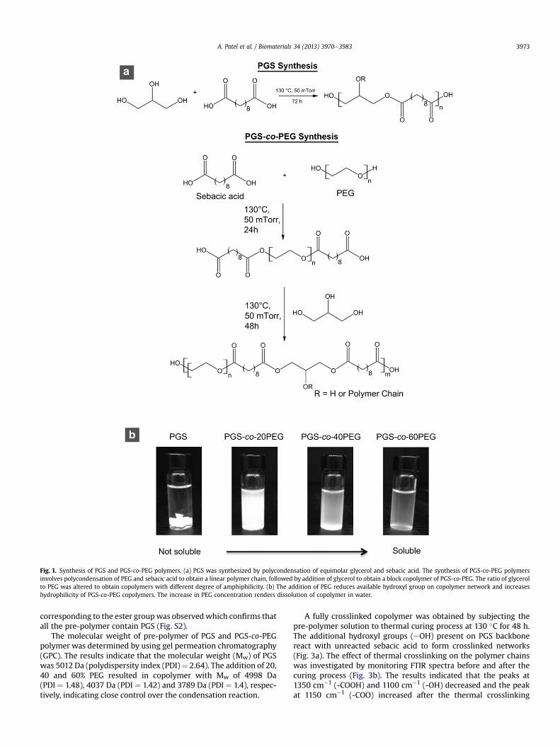
Fig. 1. Synthesis of PGS and PGS-co-PEG polymers. (a) PGS was synthesized by polycondensation of equimolar glycerol and sebacic acid. The synthesis of PGS-co-PEG polymersinvolves polycondensation of PEG and sebacic acid to obtain a linear polymer chain, followed by addition of glycerol to obtain a block copolymer of PGS-co-PEG. The ratio of glycerolto PEG was altered to obtain copolymers with different degree of amphiphilicity. (b) The addition of PEG reduces available hydroxyl group on copolymer network and increaseshydrophilicity of PGS-co-PEG copolymers. The increase in PEG concentration renders dissolution of copolymer in water.
A. Patel et al. / Biomaterials 34 (2013) 3970e3983 3973
corresponding to the ester groupwas observedwhich confirms thatall the pre-polymer contain PGS (Fig. S2).
The molecular weight of pre-polymer of PGS and PGS-co-PEGpolymer was determined by using gel permeation chromatography(GPC). The results indicate that the molecular weight (Mw) of PGSwas 5012 Da (polydispersity index (PDI)¼ 2.64). The addition of 20,40 and 60% PEG resulted in copolymer with Mw of 4998 Da(PDI ¼ 1.48), 4037 Da (PDI ¼ 1.42) and 3789 Da (PDI ¼ 1.4), respec-tively, indicating close control over the condensation reaction.
A fully crosslinked copolymer was obtained by subjecting thepre-polymer solution to thermal curing process at 130 �C for 48 h.The additional hydroxyl groups (eOH) present on PGS backbonereact with unreacted sebacic acid to form crosslinked networks(Fig. 3a). The effect of thermal crosslinking on the polymer chainswas investigated by monitoring FTIR spectra before and after thecuring process (Fig. 3b). The results indicated that the peaks at1350 cm�1 (-COOH) and 1100 cm�1 (-OH) decreased and the peakat 1150 cm�1 (-COO) increased after the thermal crosslinking
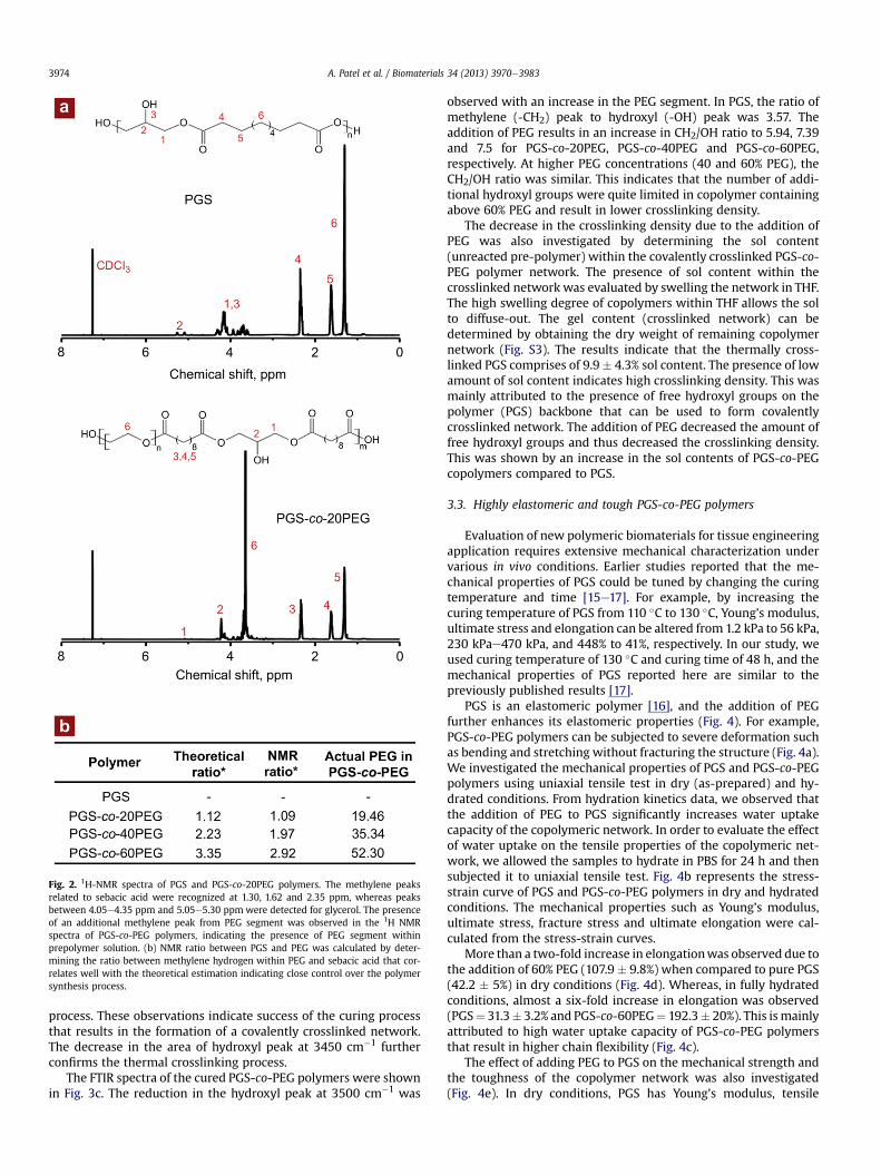
Fig. 2. 1H-NMR spectra of PGS and PGS-co-20PEG polymers. The methylene peaksrelated to sebacic acid were recognized at 1.30, 1.62 and 2.35 ppm, whereas peaksbetween 4.05e4.35 ppm and 5.05e5.30 ppm were detected for glycerol. The presenceof an additional methylene peak from PEG segment was observed in the 1H NMRspectra of PGS-co-PEG polymers, indicating the presence of PEG segment withinprepolymer solution. (b) NMR ratio between PGS and PEG was calculated by deter-mining the ratio between methylene hydrogen within PEG and sebacic acid that cor-relates well with the theoretical estimation indicating close control over the polymersynthesis process.
A. Patel et al. / Biomaterials 34 (2013) 3970e39833974
process. These observations indicate success of the curing processthat results in the formation of a covalently crosslinked network.The decrease in the area of hydroxyl peak at 3450 cm�1 furtherconfirms the thermal crosslinking process.
The FTIR spectra of the cured PGS-co-PEG polymers were shownin Fig. 3c. The reduction in the hydroxyl peak at 3500 cm�1 was
observed with an increase in the PEG segment. In PGS, the ratio ofmethylene (-CH2) peak to hydroxyl (-OH) peak was 3.57. Theaddition of PEG results in an increase in CH2/OH ratio to 5.94, 7.39and 7.5 for PGS-co-20PEG, PGS-co-40PEG and PGS-co-60PEG,respectively. At higher PEG concentrations (40 and 60% PEG), theCH2/OH ratio was similar. This indicates that the number of addi-tional hydroxyl groups were quite limited in copolymer containingabove 60% PEG and result in lower crosslinking density.
The decrease in the crosslinking density due to the addition ofPEG was also investigated by determining the sol content(unreacted pre-polymer) within the covalently crosslinked PGS-co-PEG polymer network. The presence of sol content within thecrosslinked network was evaluated by swelling the network in THF.The high swelling degree of copolymers within THF allows the solto diffuse-out. The gel content (crosslinked network) can bedetermined by obtaining the dry weight of remaining copolymernetwork (Fig. S3). The results indicate that the thermally cross-linked PGS comprises of 9.9� 4.3% sol content. The presence of lowamount of sol content indicates high crosslinking density. This wasmainly attributed to the presence of free hydroxyl groups on thepolymer (PGS) backbone that can be used to form covalentlycrosslinked network. The addition of PEG decreased the amount offree hydroxyl groups and thus decreased the crosslinking density.This was shown by an increase in the sol contents of PGS-co-PEGcopolymers compared to PGS.
3.3. Highly elastomeric and tough PGS-co-PEG polymers
Evaluation of new polymeric biomaterials for tissue engineeringapplication requires extensive mechanical characterization undervarious in vivo conditions. Earlier studies reported that the me-chanical properties of PGS could be tuned by changing the curingtemperature and time [15e17]. For example, by increasing thecuring temperature of PGS from 110 �C to 130 �C, Young’s modulus,ultimate stress and elongation can be altered from 1.2 kPa to 56 kPa,230 kPae470 kPa, and 448% to 41%, respectively. In our study, weused curing temperature of 130 �C and curing time of 48 h, and themechanical properties of PGS reported here are similar to thepreviously published results [17].
PGS is an elastomeric polymer [16], and the addition of PEGfurther enhances its elastomeric properties (Fig. 4). For example,PGS-co-PEG polymers can be subjected to severe deformation suchas bending and stretching without fracturing the structure (Fig. 4a).We investigated the mechanical properties of PGS and PGS-co-PEGpolymers using uniaxial tensile test in dry (as-prepared) and hy-drated conditions. From hydration kinetics data, we observed thatthe addition of PEG to PGS significantly increases water uptakecapacity of the copolymeric network. In order to evaluate the effectof water uptake on the tensile properties of the copolymeric net-work, we allowed the samples to hydrate in PBS for 24 h and thensubjected it to uniaxial tensile test. Fig. 4b represents the stress-strain curve of PGS and PGS-co-PEG polymers in dry and hydratedconditions. The mechanical properties such as Young’s modulus,ultimate stress, fracture stress and ultimate elongation were cal-culated from the stress-strain curves.
More than a two-fold increase in elongationwas observed due tothe addition of 60% PEG (107.9 � 9.8%) when compared to pure PGS(42.2 � 5%) in dry conditions (Fig. 4d). Whereas, in fully hydratedconditions, almost a six-fold increase in elongation was observed(PGS¼ 31.3� 3.2% and PGS-co-60PEG¼ 192.3� 20%). This ismainlyattributed to high water uptake capacity of PGS-co-PEG polymersthat result in higher chain flexibility (Fig. 4c).
The effect of adding PEG to PGS on the mechanical strength andthe toughness of the copolymer network was also investigated(Fig. 4e). In dry conditions, PGS has Young's modulus, tensile
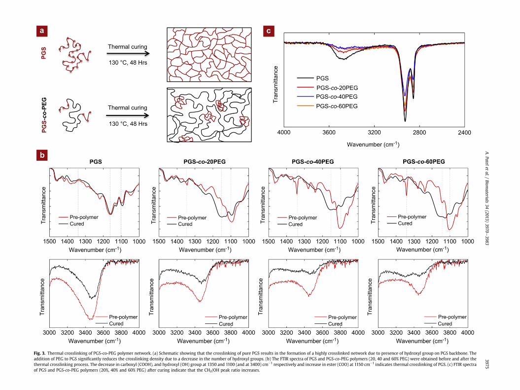
Fig. 3. Thermal crosslinking of PGS-co-PEG polymer network. (a) Schematic showing that the crosslinking of pure PGS results in the formation of a highly crosslinked network due to presence of hydroxyl group on PGS backbone. Theaddition of PEG to PGS significantly reduces the crosslinking density due to a decrease in the number of hydroxyl groups. (b) The FTIR spectra of PGS and PGS-co-PEG polymers (20, 40 and 60% PEG) were obtained before and after thethermal crosslinking process. The decrease in carboxyl (COOH), and hydroxyl (OH) group at 1350 and 1100 (and at 3400) cm�1 respectively and increase in ester (COO) at 1150 cm�1 indicates thermal crosslinking of PGS. (c) FTIR spectraof PGS and PGS-co-PEG polymers (20%, 40% and 60% PEG) after curing indicate that the CH2/OH peak ratio increases.
A.Patelet
al./Biom
aterials34
(2013)3970
e3983
3975
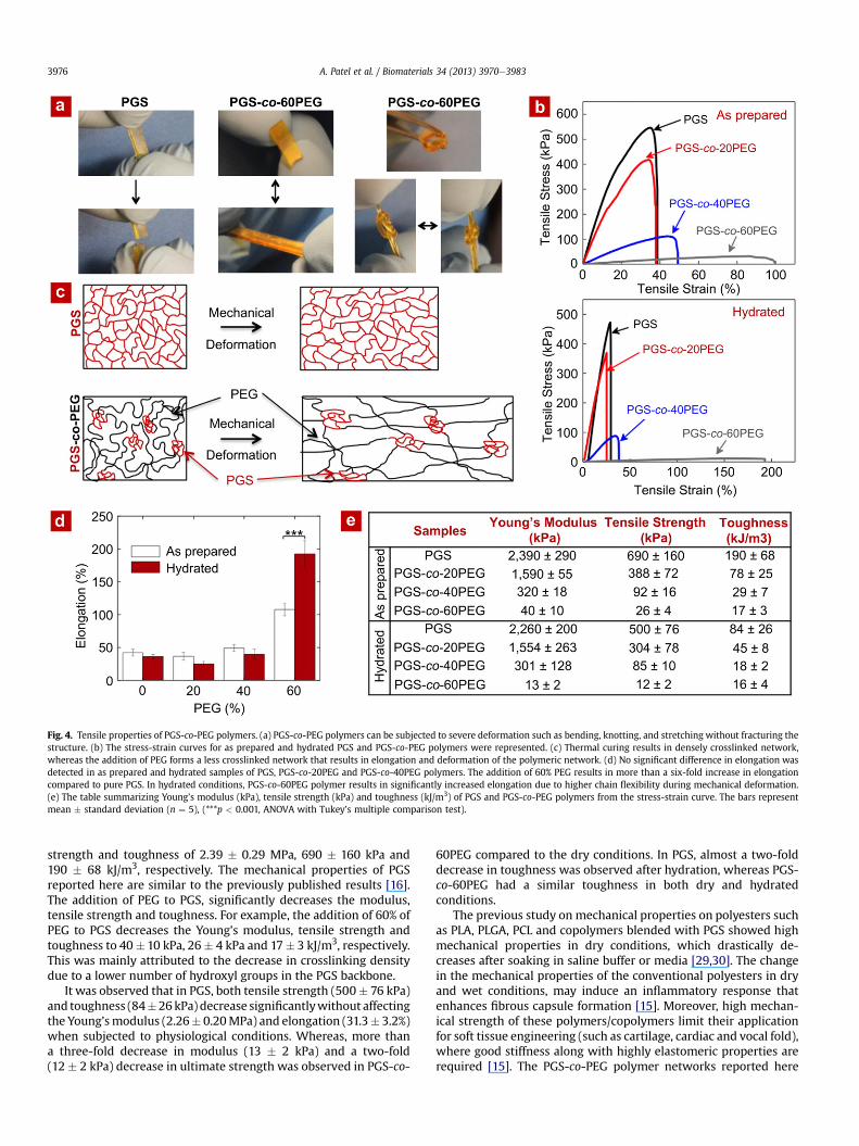
Fig. 4. Tensile properties of PGS-co-PEG polymers. (a) PGS-co-PEG polymers can be subjected to severe deformation such as bending, knotting, and stretching without fracturing thestructure. (b) The stress-strain curves for as prepared and hydrated PGS and PGS-co-PEG polymers were represented. (c) Thermal curing results in densely crosslinked network,whereas the addition of PEG forms a less crosslinked network that results in elongation and deformation of the polymeric network. (d) No significant difference in elongation wasdetected in as prepared and hydrated samples of PGS, PGS-co-20PEG and PGS-co-40PEG polymers. The addition of 60% PEG results in more than a six-fold increase in elongationcompared to pure PGS. In hydrated conditions, PGS-co-60PEG polymer results in significantly increased elongation due to higher chain flexibility during mechanical deformation.(e) The table summarizing Young’s modulus (kPa), tensile strength (kPa) and toughness (kJ/m3) of PGS and PGS-co-PEG polymers from the stress-strain curve. The bars representmean � standard deviation (n ¼ 5), (***p < 0.001, ANOVA with Tukey’s multiple comparison test).
A. Patel et al. / Biomaterials 34 (2013) 3970e39833976
strength and toughness of 2.39 � 0.29 MPa, 690 � 160 kPa and190 � 68 kJ/m3, respectively. The mechanical properties of PGSreported here are similar to the previously published results [16].The addition of PEG to PGS, significantly decreases the modulus,tensile strength and toughness. For example, the addition of 60% ofPEG to PGS decreases the Young’s modulus, tensile strength andtoughness to 40� 10 kPa, 26� 4 kPa and 17� 3 kJ/m3, respectively.This was mainly attributed to the decrease in crosslinking densitydue to a lower number of hydroxyl groups in the PGS backbone.
It was observed that in PGS, both tensile strength (500� 76 kPa)and toughness (84�26kPa) decrease significantlywithout affectingthe Young’smodulus (2.26� 0.20MPa) and elongation (31.3� 3.2%)when subjected to physiological conditions. Whereas, more thana three-fold decrease in modulus (13 � 2 kPa) and a two-fold(12 � 2 kPa) decrease in ultimate strength was observed in PGS-co-
60PEG compared to the dry conditions. In PGS, almost a two-folddecrease in toughness was observed after hydration, whereas PGS-co-60PEG had a similar toughness in both dry and hydratedconditions.
The previous study on mechanical properties on polyesters suchas PLA, PLGA, PCL and copolymers blended with PGS showed highmechanical properties in dry conditions, which drastically de-creases after soaking in saline buffer or media [29,30]. The changein the mechanical properties of the conventional polyesters in dryand wet conditions, may induce an inflammatory response thatenhances fibrous capsule formation [15]. Moreover, high mechan-ical strength of these polymers/copolymers limit their applicationfor soft tissue engineering (such as cartilage, cardiac and vocal fold),where good stiffness along with highly elastomeric properties arerequired [15]. The PGS-co-PEG polymer networks reported here
A. Patel et al. / Biomaterials 34 (2013) 3970e3983 3977
have high stiffness along with elastomeric properties, making themsuitable to engineer scaffolds for a range of soft tissues. Forexample, the tensile modulus of PGS-co-PEG polymers is similar tohuman cardiac muscles (0.02e0.15 MPa) [42,43]. Hence, PGS-co-PEG polymers can potentially be used to engineer cardiac patches.
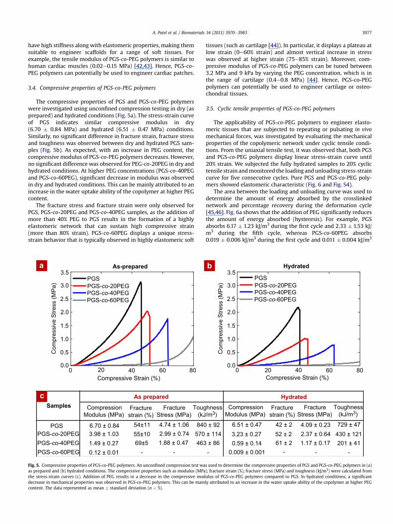
3.4. Compressive properties of PGS-co-PEG polymers
The compressive properties of PGS and PGS-co-PEG polymerswere investigated using unconfined compression testing in dry (asprepared) and hydrated conditions (Fig. 5a). The stress-strain curveof PGS indicates similar compressive modulus in dry(6.70 � 0.84 MPa) and hydrated (6.51 � 0.47 MPa) conditions.Similarly, no significant difference in fracture strain, fracture stressand toughness was observed between dry and hydrated PGS sam-ples (Fig. 5b). As expected, with an increase in PEG content, thecompressivemodulus of PGS-co-PEG polymers decreases. However,no significant difference was observed for PEG-co-20PEG in dry andhydrated conditions. At higher PEG concentrations (PGS-co-40PEGand PGS-co-60PEG), significant decrease in modulus was observedin dry and hydrated conditions. This can be mainly attributed to anincrease in the water uptake ability of the copolymer at higher PEGcontent.
The fracture stress and fracture strain were only observed forPGS, PGS-co-20PEG and PGS-co-40PEG samples, as the addition ofmore than 40% PEG to PGS results in the formation of a highlyelastomeric network that can sustain high compressive strain(more than 80% strain). PGS-co-60PEG displays a unique stress-strain behavior that is typically observed in highly elastomeric soft
PGSPGS-co-20PEGPGS-co-40PEGPGS-co-60PEG
0 20 40 60 80Compressive Strain (%)
0.0
0.5
1.0
1.5
2.0
2.5
3.0
3.5
Com
pres
sive
Stre
ss (M
Pa)
As-prepared
Samples CompressionModulus (MPa)
Fracturestrain (%)
FractureStress (MPa)
Toug (kJ
PGS-co-20PEGPGS-co-40PEGPGS-co-60PEG
1.49 ± 0.273.98 ± 1.036.70 ± 0.84 4.74 ± 1.06 840
570463
2.99 ± 0.741.88 ± 0.47
55±1054±11
69±50.12 ± 0.01 - -
PGS
As prepared
a
c
Fig. 5. Compressive properties of PGS-co-PEG polymers. An unconfined compression test waas prepared and (b) hydrated conditions. The compressive properties such as modulus (MPathe stress-strain curves (c). Addition of PEG results in a decrease in the compressive moddecrease in mechanical properties was observed in PGS-co-PEG polymers. This can be maincontent. The data represented as mean � standard deviation (n ¼ 5).
tissues (such as cartilage [44]). In particular, it displays a plateau atlow strain (0e60% strain) and almost vertical increase in stresswas observed at higher strain (75e85% strain). Moreover, com-pressive modulus of PGS-co-PEG polymers can be tuned between3.2 MPa and 9 kPa by varying the PEG concentration, which is inthe range of cartilage (0.4e0.8 MPa) [44]. Hence, PGS-co-PEGpolymers can potentially be used to engineer cartilage or osteo-chondral tissues.
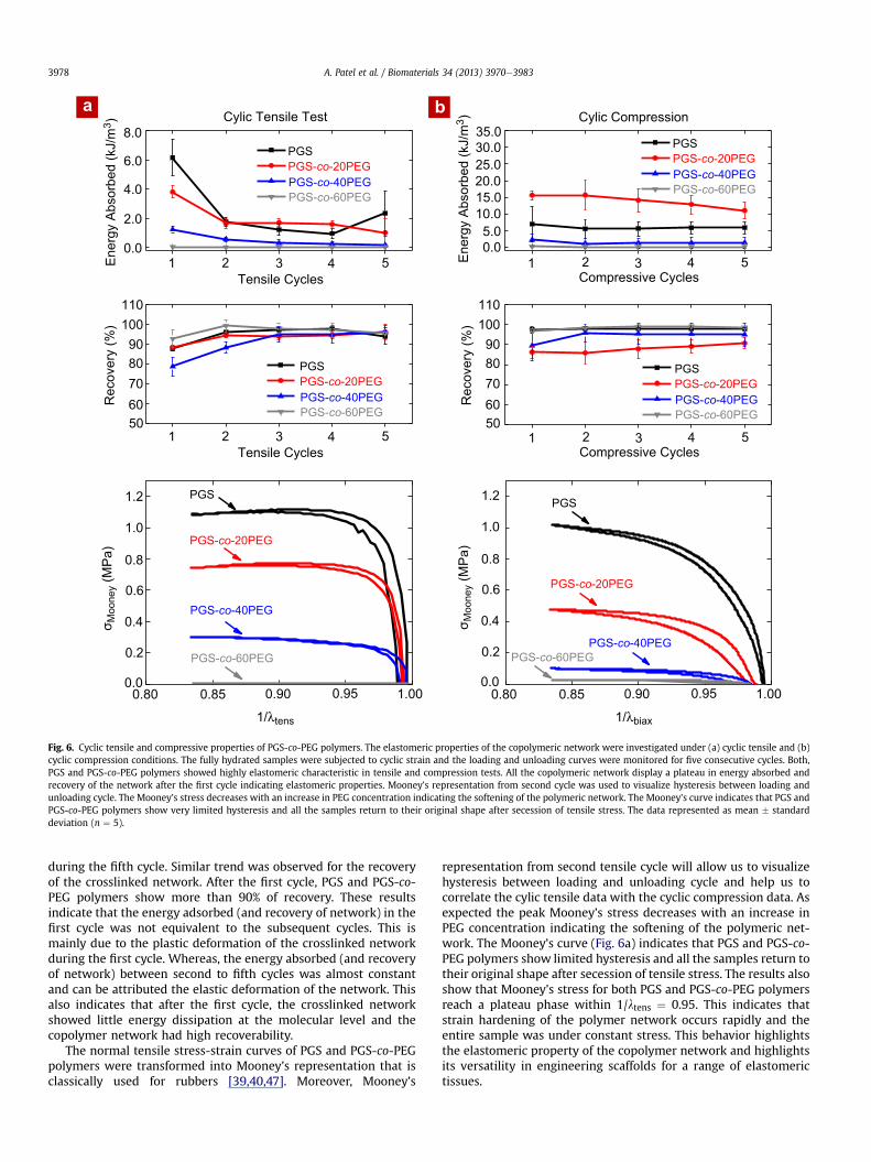
3.5. Cyclic tensile properties of PGS-co-PEG polymers
The applicability of PGS-co-PEG polymers to engineer elasto-meric tissues that are subjected to repeating or pulsating in vivomechanical forces, was investigated by evaluating the mechanicalproperties of the copolymeric network under cyclic tensile condi-tions. From the uniaxial tensile test, it was observed that, both PGSand PGS-co-PEG polymers display linear stress-strain curve until20% strain. We subjected the fully hydrated samples to 20% cyclictensile strain andmonitored the loading andunloading stress-straincurve for five consecutive cycles. Pure PGS and PGS-co-PEG poly-mers showed elastomeric characteristic (Fig. 6 and Fig. S4).
The area between the loading and unloading curve was used todetermine the amount of energy absorbed by the crosslinkednetwork and percentage recovery during the deformation cycle[45,46]. Fig. 6a shows that the addition of PEG significantly reducesthe amount of energy absorbed (hysteresis). For example, PGSabsorbs 6.17 � 1.23 kJ/m3 during the first cycle and 2.33 � 1.53 kJ/m3 during the fifth cycle, whereas PGS-co-60PEG absorbs0.019 � 0.006 kJ/m3 during the first cycle and 0.011 � 0.004 kJ/m3
PGSPGS-co-20PEGPGS-co-40PEGPGS-co-60PEG
0 20 40 60 80Compressive Strain (%)
Hydrated
0.0
0.5
1.0
1.5
2.0
2.5
3.0
3.5
Com
pres
sive
Stre
ss (M
Pa)
hness/m3)
CompressionModulus (MPa)
Fracturestrain (%)
FractureStress (MPa)
Toughness (kJ/m3)
6.51 ± 0.473.23 ± 0.27 2.37 ± 0.64
4.09 ± 0.23 729 ± 47430 ± 121201 ± 411.17 ± 0.170.59 ± 0.14
0.009 ± 0.001
± 92 42 ± 252 ± 261 ± 2
± 114 ± 86- - - -
Hydrated
b
s used to determine the compressive properties of PGS and PGS-co-PEG polymers in (a)), fracture strain (%), fracture stress (MPa) and toughness (kJ/m3) were calculated fromulus of PGS-co-PEG polymers compared to PGS. In hydrated conditions, a significantly attributed to an increase in the water uptake ability of the copolymer at higher PEG
Cylic Tensile Test Cylic Compression8.0
6.0
4.0
2.0
0.0
Ener
gy A
bsor
bed
(kJ/
m3 )
Ener
gy A
bsor
bed
(kJ/
m3 )
1101009080706050
Rec
over
y (%
)
1101009080706050
Rec
over
y (%
)
1.2
1.0
0.8
0.6
0.4
0.2
0.0
σ Moo
ney (
MPa
)
1.2
1.0
0.8
0.6
0.4
0.2
0.0
σ Moo
ney (
MPa
)
0.850.80 0.90 0.95 1.00
1/λtens
0.850.80 0.90 0.95 1.00
1/λbiax
35.030.025.020.015.010.05.00.0
1 2 3 4 5Tensile Cycles
1 2 3 4 5Tensile Cycles
1 2 3 4 5Compressive Cycles
1 2 3 4 5Compressive Cycles
PGSPGS
PGSPGS-co-20PEGPGS-co-40PEGPGS-co-60PEG
PGSPGS-co-20PEGPGS-co-40PEGPGS-co-60PEG
PGSPGS-co-20PEG
PGS-co-20PEG
PGS-co-20PEG
PGS-co-40PEG
PGS-co-40PEG
PGS-co-40PEG
PGS-co-60PEG
PGSPGS-co-20PEGPGS-co-40PEGPGS-co-60PEG
PGS-co-60PEGPGS-co-60PEG
ba
Fig. 6. Cyclic tensile and compressive properties of PGS-co-PEG polymers. The elastomeric properties of the copolymeric network were investigated under (a) cyclic tensile and (b)cyclic compression conditions. The fully hydrated samples were subjected to cyclic strain and the loading and unloading curves were monitored for five consecutive cycles. Both,PGS and PGS-co-PEG polymers showed highly elastomeric characteristic in tensile and compression tests. All the copolymeric network display a plateau in energy absorbed andrecovery of the network after the first cycle indicating elastomeric properties. Mooney’s representation from second cycle was used to visualize hysteresis between loading andunloading cycle. The Mooney’s stress decreases with an increase in PEG concentration indicating the softening of the polymeric network. The Mooney’s curve indicates that PGS andPGS-co-PEG polymers show very limited hysteresis and all the samples return to their original shape after secession of tensile stress. The data represented as mean � standarddeviation (n ¼ 5).
A. Patel et al. / Biomaterials 34 (2013) 3970e39833978
during the fifth cycle. Similar trend was observed for the recoveryof the crosslinked network. After the first cycle, PGS and PGS-co-PEG polymers show more than 90% of recovery. These resultsindicate that the energy adsorbed (and recovery of network) in thefirst cycle was not equivalent to the subsequent cycles. This ismainly due to the plastic deformation of the crosslinked networkduring the first cycle. Whereas, the energy absorbed (and recoveryof network) between second to fifth cycles was almost constantand can be attributed the elastic deformation of the network. Thisalso indicates that after the first cycle, the crosslinked networkshowed little energy dissipation at the molecular level and thecopolymer network had high recoverability.
The normal tensile stress-strain curves of PGS and PGS-co-PEGpolymers were transformed into Mooney’s representation that isclassically used for rubbers [39,40,47]. Moreover, Mooney’s
representation from second tensile cycle will allow us to visualizehysteresis between loading and unloading cycle and help us tocorrelate the cylic tensile data with the cyclic compression data. Asexpected the peak Mooney’s stress decreases with an increase inPEG concentration indicating the softening of the polymeric net-work. The Mooney’s curve (Fig. 6a) indicates that PGS and PGS-co-PEG polymers show limited hysteresis and all the samples return totheir original shape after secession of tensile stress. The results alsoshow that Mooney’s stress for both PGS and PGS-co-PEG polymersreach a plateau phase within 1/ltens ¼ 0.95. This indicates thatstrain hardening of the polymer network occurs rapidly and theentire sample was under constant stress. This behavior highlightsthe elastomeric property of the copolymer network and highlightsits versatility in engineering scaffolds for a range of elastomerictissues.
A. Patel et al. / Biomaterials 34 (2013) 3970e3983 3979
3.6. Cyclic compressive properties of PGS-co-PEG polymers
The mechanical behavior polymeric network under unconfinedcyclic compression can be used to evaluate the applicability of thecopolymeric networks for soft tissue engineering [45,46]. PGS andPGS-co-PEG polymers display elastomeric properties under com-pression, which is similar to our observations in tensile test(Fig. 6b). The amount of energy absorbed by the PGS network wasconstant between first and fifth compressive cycles. The addition of20% of PEG significantly increased the amount of energy that wasabsorbed. This might be due to the deformation of swollen surfacesof the samples that do not return instantaneously to the originalshape after cessation of compressive stress. Whereas, at higher PEGconcentrations (PGS-co-40PEG and PGS-co-60PEG), negligiblehysteresis was observed and the network displayed high recover-ability (>95%). This is mainly attributed to the formation of softerstructures that are able to absorb energy during the loading cycleand release it almost completely during the unloading cycle.
To provide an easy comparison between uniaxial compressionand uniaxial stretching we used lbiax as our deformation, sinceuniaxial compression is equivalent to biaxial stretching in terms ofdeformation [41]. The Mooney’s representation indicate that
Fig. 7. Effect of PEG on hydrophilicity of PGS-co-PEG polymers. (a) Schematic showing thatwater absorption in PGS-co-PEG polymers. (b) The contact angle measurements indicate adecrease in contact angle of water on PGS-co-PEG polymers. (c) The hydration kinetic of PGS-copolymer reaches saturated hydration degree within 72 h. (d) The equilibriumwater contenmean � standard deviation (n ¼ 5) (**p < 0.01, ***p < 0.001, ANOVA with Tukey’s multiple
crosslinked network behave similarly in cyclic tensile and cycliccompression conditions. The rapid increase in Mooney’s stress atsmaller strain, indicates strain hardening of the polymeric network.The extent of strain hardening directly depends on the amount ofPEGwithin the copolymer networks. Overall, PGS-co-PEG polymersexhibit unparalleled elastomeric properties that can be used toengineer a range of scaffolds for soft tissues that are subjected topulsating/cyclic mechanical forces such as blood vessels, cardiac,cartilage and muscle tissues.
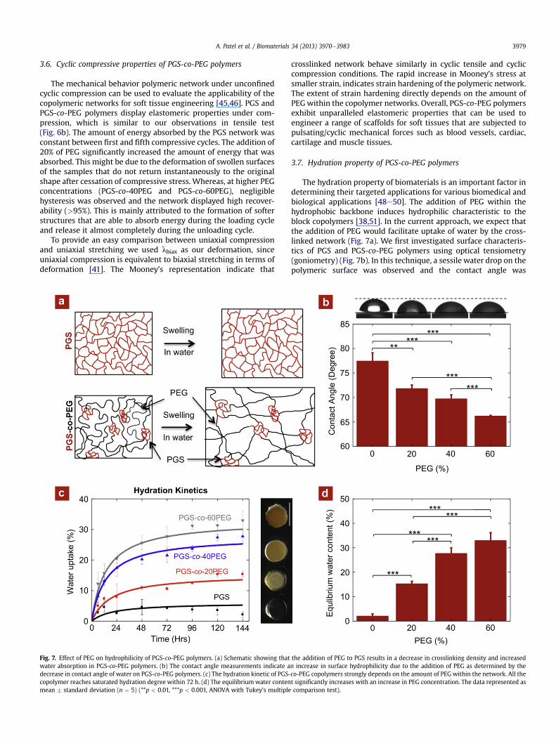
3.7. Hydration property of PGS-co-PEG polymers
The hydration property of biomaterials is an important factor indetermining their targeted applications for various biomedical andbiological applications [48e50]. The addition of PEG within thehydrophobic backbone induces hydrophilic characteristic to theblock copolymers [38,51]. In the current approach, we expect thatthe addition of PEG would facilitate uptake of water by the cross-linked network (Fig. 7a). We first investigated surface characteris-tics of PGS and PGS-co-PEG polymers using optical tensiometry(goniometry) (Fig. 7b). In this technique, a sessile water drop on thepolymeric surface was observed and the contact angle was
the addition of PEG to PGS results in a decrease in crosslinking density and increasedn increase in surface hydrophilicity due to the addition of PEG as determined by theco-PEG copolymers strongly depends on the amount of PEG within the network. All thet significantly increases with an increase in PEG concentration. The data represented ascomparison test).
A. Patel et al. / Biomaterials 34 (2013) 3970e39833980
determined by measuring the angle between the polymer surfaceand a tangent to the water drop surface. Pure PGS shows a contactangle of 77.5 � 1.7� with water. As expected, an increase in the PEGsegment increases the hydrophilic nature. Addition of 60% PEG toPGS reduces the contact angle to 66.2 � 0.1�.
The hydration properties of PGS and PGS-co-PEG polymers werefurther investigated by evaluating bulk hydration characteristic(Fig. 7c). The crosslinked polymer samples were subjected tophysiological conditions (37 �C and PBS) and uptake of water wasmonitored. The swelling study reveals the maximumwater uptakewithin 48 h for all PGS-co-PEG polymers, while PGS reaches theequilibriumwater content (EWC) within 24 h (Fig. 7d). The EWC forPGSwas 2.11�0.88%whereas for PGS-co-60PEGwas 32.98� 3.17%.Addition of 60% PEG results in almost 15-fold increase in wateruptake capacity. Moreover, as the PEG content in the blockcopolymer increases, the network becomes translucent in theswollen conditions, which is the characteristic of an amphiphiliccopolymer network.
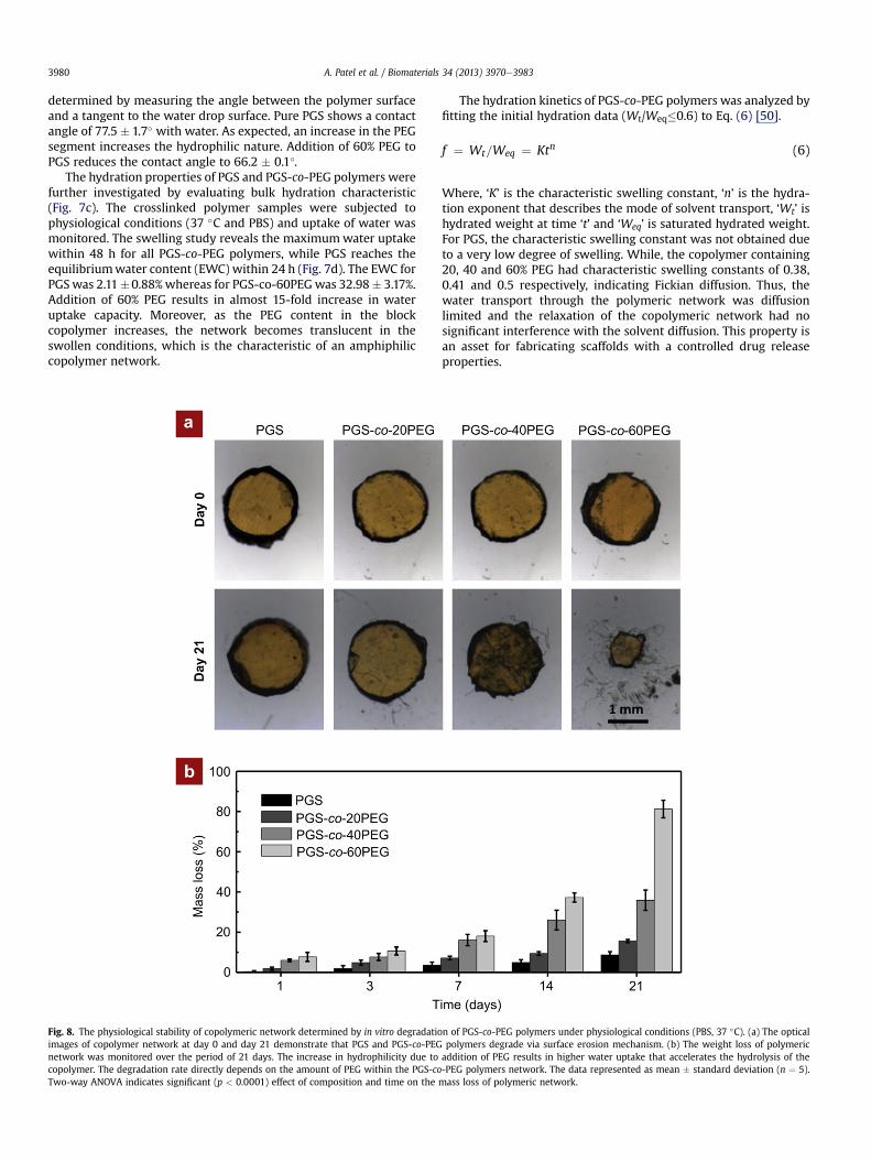
Fig. 8. The physiological stability of copolymeric network determined by in vitro degradatioimages of copolymer network at day 0 and day 21 demonstrate that PGS and PGS-co-PEGnetwork was monitored over the period of 21 days. The increase in hydrophilicity due tocopolymer. The degradation rate directly depends on the amount of PEG within the PGS-coTwo-way ANOVA indicates significant (p < 0.0001) effect of composition and time on the
The hydration kinetics of PGS-co-PEG polymers was analyzed byfitting the initial hydration data (Wt/Weq�0.6) to Eq. (6) [50].
f ¼ Wt=Weq ¼ Ktn (6)
Where, ‘K’ is the characteristic swelling constant, ‘n’ is the hydra-tion exponent that describes the mode of solvent transport, ‘Wt’ ishydrated weight at time ‘t’ and ‘Weq’ is saturated hydrated weight.For PGS, the characteristic swelling constant was not obtained dueto a very low degree of swelling. While, the copolymer containing20, 40 and 60% PEG had characteristic swelling constants of 0.38,0.41 and 0.5 respectively, indicating Fickian diffusion. Thus, thewater transport through the polymeric network was diffusionlimited and the relaxation of the copolymeric network had nosignificant interference with the solvent diffusion. This property isan asset for fabricating scaffolds with a controlled drug releaseproperties.
n of PGS-co-PEG polymers under physiological conditions (PBS, 37 �C). (a) The opticalpolymers degrade via surface erosion mechanism. (b) The weight loss of polymericaddition of PEG results in higher water uptake that accelerates the hydrolysis of the-PEG polymers network. The data represented as mean � standard deviation (n ¼ 5).mass loss of polymeric network.
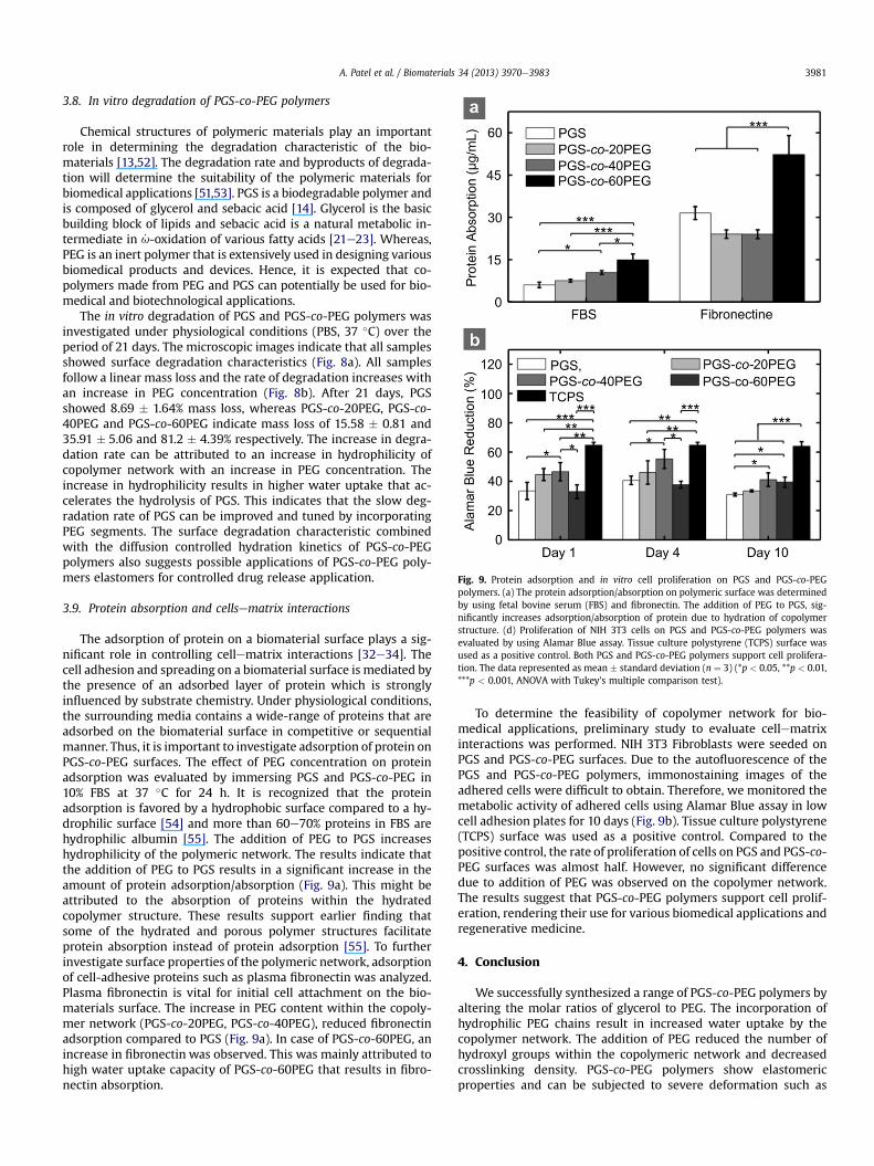
Fig. 9. Protein adsorption and in vitro cell proliferation on PGS and PGS-co-PEGpolymers. (a) The protein adsorption/absorption on polymeric surface was determinedby using fetal bovine serum (FBS) and fibronectin. The addition of PEG to PGS, sig-nificantly increases adsorption/absorption of protein due to hydration of copolymerstructure. (d) Proliferation of NIH 3T3 cells on PGS and PGS-co-PEG polymers wasevaluated by using Alamar Blue assay. Tissue culture polystyrene (TCPS) surface wasused as a positive control. Both PGS and PGS-co-PEG polymers support cell prolifera-tion. The data represented as mean � standard deviation (n ¼ 3) (*p < 0.05, **p < 0.01,***p < 0.001, ANOVA with Tukey’s multiple comparison test).
A. Patel et al. / Biomaterials 34 (2013) 3970e3983 3981
3.8. In vitro degradation of PGS-co-PEG polymers
Chemical structures of polymeric materials play an importantrole in determining the degradation characteristic of the bio-materials [13,52]. The degradation rate and byproducts of degrada-tion will determine the suitability of the polymeric materials forbiomedical applications [51,53]. PGS is a biodegradable polymer andis composed of glycerol and sebacic acid [14]. Glycerol is the basicbuilding block of lipids and sebacic acid is a natural metabolic in-termediate in _u-oxidation of various fatty acids [21e23]. Whereas,PEG is an inert polymer that is extensively used in designing variousbiomedical products and devices. Hence, it is expected that co-polymers made from PEG and PGS can potentially be used for bio-medical and biotechnological applications.
The in vitro degradation of PGS and PGS-co-PEG polymers wasinvestigated under physiological conditions (PBS, 37 �C) over theperiod of 21 days. The microscopic images indicate that all samplesshowed surface degradation characteristics (Fig. 8a). All samplesfollow a linear mass loss and the rate of degradation increases withan increase in PEG concentration (Fig. 8b). After 21 days, PGSshowed 8.69 � 1.64% mass loss, whereas PGS-co-20PEG, PGS-co-40PEG and PGS-co-60PEG indicate mass loss of 15.58 � 0.81 and35.91 � 5.06 and 81.2 � 4.39% respectively. The increase in degra-dation rate can be attributed to an increase in hydrophilicity ofcopolymer network with an increase in PEG concentration. Theincrease in hydrophilicity results in higher water uptake that ac-celerates the hydrolysis of PGS. This indicates that the slow deg-radation rate of PGS can be improved and tuned by incorporatingPEG segments. The surface degradation characteristic combinedwith the diffusion controlled hydration kinetics of PGS-co-PEGpolymers also suggests possible applications of PGS-co-PEG poly-mers elastomers for controlled drug release application.
3.9. Protein absorption and cellsematrix interactions
The adsorption of protein on a biomaterial surface plays a sig-nificant role in controlling cellematrix interactions [32e34]. Thecell adhesion and spreading on a biomaterial surface is mediated bythe presence of an adsorbed layer of protein which is stronglyinfluenced by substrate chemistry. Under physiological conditions,the surrounding media contains a wide-range of proteins that areadsorbed on the biomaterial surface in competitive or sequentialmanner. Thus, it is important to investigate adsorption of protein onPGS-co-PEG surfaces. The effect of PEG concentration on proteinadsorption was evaluated by immersing PGS and PGS-co-PEG in10% FBS at 37 �C for 24 h. It is recognized that the proteinadsorption is favored by a hydrophobic surface compared to a hy-drophilic surface [54] and more than 60e70% proteins in FBS arehydrophilic albumin [55]. The addition of PEG to PGS increaseshydrophilicity of the polymeric network. The results indicate thatthe addition of PEG to PGS results in a significant increase in theamount of protein adsorption/absorption (Fig. 9a). This might beattributed to the absorption of proteins within the hydratedcopolymer structure. These results support earlier finding thatsome of the hydrated and porous polymer structures facilitateprotein absorption instead of protein adsorption [55]. To furtherinvestigate surface properties of the polymeric network, adsorptionof cell-adhesive proteins such as plasma fibronectin was analyzed.Plasma fibronectin is vital for initial cell attachment on the bio-materials surface. The increase in PEG content within the copoly-mer network (PGS-co-20PEG, PGS-co-40PEG), reduced fibronectinadsorption compared to PGS (Fig. 9a). In case of PGS-co-60PEG, anincrease in fibronectin was observed. This was mainly attributed tohigh water uptake capacity of PGS-co-60PEG that results in fibro-nectin absorption.
To determine the feasibility of copolymer network for bio-medical applications, preliminary study to evaluate cellematrixinteractions was performed. NIH 3T3 Fibroblasts were seeded onPGS and PGS-co-PEG surfaces. Due to the autofluorescence of thePGS and PGS-co-PEG polymers, immonostaining images of theadhered cells were difficult to obtain. Therefore, we monitored themetabolic activity of adhered cells using Alamar Blue assay in lowcell adhesion plates for 10 days (Fig. 9b). Tissue culture polystyrene(TCPS) surface was used as a positive control. Compared to thepositive control, the rate of proliferation of cells on PGS and PGS-co-PEG surfaces was almost half. However, no significant differencedue to addition of PEG was observed on the copolymer network.The results suggest that PGS-co-PEG polymers support cell prolif-eration, rendering their use for various biomedical applications andregenerative medicine.
4. Conclusion
We successfully synthesized a range of PGS-co-PEG polymers byaltering the molar ratios of glycerol to PEG. The incorporation ofhydrophilic PEG chains result in increased water uptake by thecopolymer network. The addition of PEG reduced the number ofhydroxyl groups within the copolymeric network and decreasedcrosslinking density. PGS-co-PEG polymers show elastomericproperties and can be subjected to severe deformation such as
A. Patel et al. / Biomaterials 34 (2013) 3970e39833982
bending and stretching without fracturing the structure. Undertension, PGS-co-PEG polymers displayed a stress-strain behaviorthat is typically observed in highly elastomeric soft tissues. TheYoung’s modulus of PGS-co-PEG can be tuned from 13 kPa to2.2 MPa by altering the amount of PEG within the network. Com-pared to PGS, more than a six-fold increase in elongation wasobserved in PGS-co-60PEG. Similarly, under compression, theaddition of PEG results in a soft and elastomeric network. Bothsurface and bulk characterization indicates that the addition of PEGincreases the hydrophilicity and the rate of degradation of thecopolymer network. Moreover, PGS-co-60PEG supports proteinadsorption/absorption and cell proliferation, and thus can be usedfor a range of tissue engineering applications.
Authors contributions
AP, AKG and GI contributed equally. AP, AKG and AK conceivedthe idea and designed the experiments. AP, AKG and GI synthesizedPGS and PGS-co-PEG polymers and performed the chemical (NMR,FTIR, GPC),mechanical (tensile, compression and cyclic testing), andstructural characterizations (solegel, hydration kinetics, contactangle, degradation studies). SMM performed cells proliferationstudies. HZ and SM performed protein adsorption studies. AP, AKG,GI andAKanalyzed experimental data andwrote themanuscript. Allauthors discussed the results and commented on the manuscript.
Acknowledgment
We would like to thank Dr. Shilpa Sant, and Hyeongho Shin fortechnical input and NMR experiment, respectively. AP would like toacknowledge postdoctoral fellowship (Fellowship No. PDF-388346-2010) awarded by Natural Science and Engineering ResearchCouncil, Canada. AKG would like to thanks Prof. Robert Langer andMIT Portugal Program for financial support (MPP-09Call-Langer-47). GI would like to acknowledge Politecnico di Torino for financialsupport. HZ would like to acknowledge National Natural ScienceFund for Distinguished Young Scholar (Grant No. 51025313). Thisresearch was funded by the US Army Engineer Research andDevelopment Center, the Institute for Soldier Nanotechnology, theNIH (EB009196; DE019024; EB007249; HL099073; AR057837), andthe National Science Foundation CAREER award (AK).
Appendix A. Supplementary data
Supplementary data related to this article can be found athttp://dx.doi.org/10.1016/j.biomaterials.2013.01.045.
References
[1] Langer R, Tirrell DA. Designing materials for biology and medicine. Nature2004;428:487e92.
[2] Khademhosseini A, Vacanti JP, Langer R. Progress in tissue engineering. Sci Am2009;300:64e71.
[3] Slaughter BV, Khurshid SS, Fisher OZ, Khademhosseini A, Peppas NA. Hydro-gels in regenerative medicine. Adv Mater 2009;21:3307e29.
[4] Fisher OZ, Khademhosseini A, Langer R, Peppas NA. Bioinspired materials forcontrolling stem cell fate. Acc Chem Res 2010;43:419e28.
[5] Yoo J, Kuruvilla DJ, D’Mello SR, Salem AK, Bowden NB. New class of bio-degradable polymers formed from reactions of an inorganic functional group.Macromolecules 2012;45:2292e300.
[6] Athanasiou KA, Agrawal CM, Barber FA, Burkhart SS. Orthopaedic applicationsfor PLA-PGA biodegradable polymers. Arthroscopy 1998;14:726e37.
[7] Patel A, Fine B, Sandig M, Mequanint K. Elastin biosynthesis: the missing linkin tissue-engineered blood vessels. Cardiovasc Res 2006;71:40e9.
[8] Jeong JH, Lim DW, Han DK, Park TG. Synthesis, characterization and proteinadsorption behaviors of PLGA/PEG di-block co-polymer blend films. ColloidSurf B 2000;18:371e9.
[9] Shi R, Chen D, Liu Q, Wu Y, Xu X, Zhang L, et al. Recent advances in syntheticbioelastomers. Int J Mol Sci 2009;10:4223e56.
[10] Liu X, Holzwarth JM, Ma PX. Functionalized synthetic biodegradable polymerscaffolds for tissue engineering. Macromol Biosci 2012;12:911e9.
[11] Khan M, Ong ZY, Wiradharma N, Attia ABE, Yang Y-Y. Advanced materials forco-delivery of drugs and genes in cancer therapy. Adv Healthcare Mater 2012;1:373e92.
[12] Li SM. Hydrolytic degradation characteristics of aliphatic polyesters derivedfrom lactic and glycolic acids. J Biomed Mater Res 1999;48:342e53.
[13] Nair LS, Laurencin CT. Biodegradable polymers as biomaterials. Prog Polym Sci2007;32:762e98.
[14] Wang Y, Kim YM, Langer R. In vivo degradation characteristics of poly(glycerolsebacate). J Biomed Mater Res A 2003;66A:192e7.
[15] Rai R, Tallawi M, Grigore A, Boccaccini AR. Synthesis, properties and bio-medical applications of poly(glycerol sebacate) (PGS): a review. Prog PolymSci 2012;37:1051e78.
[16] Wang YD, Ameer GA, Sheppard BJ, Langer R. A tough biodegradable elastomer.Nat Biotechnol 2002;20:602e6.
[17] Chen QZ, Bismarck A, Hansen U, Junaid S, Tran MQ, Harding SE, et al. Char-acterisation of a soft elastomer poly(glycerol sebacate) designed to match themechanical properties of myocardial tissue. Biomaterials 2008;29:47e57.
[18] Jaafar IH, Ammar MM, Jedlicka SS, Pearson RA, Coulter JP. Spectroscopicevaluation, thermal, and thermomechanical characterization of poly(glycerol-sebacate) with variations in curing temperatures and durations. J Mater Sci2010;45:2525e9.
[19] Sun ZJ, Sun CW, Sun B, Lu XL, Dong DL. The polycondensing temperaturerather than time determines the degradation and drug release of poly(-glycerol-sebacate) doped with 5-fluorouracil. J Biomater Sci Polym Ed 2012;23:833e41.
[20] Sun ZJ, Chen C, Sun MZ, Ai CH, Lu XL, Zheng YF, et al. The application of poly(glycerolesebacate) as biodegradable drug carrier. Biomaterials 2009;30:5209e14.
[21] Liu GY, Hinch B, Beavis AD. Mechanisms for the transport of alpha, omega-dicarboxylates through the mitochondrial inner membrane. J Biol Chem 1996;271:25338e44.
[22] Grego AV, Mingrone G. Dicarboxylic acid, an alternate fuel substrate inparenternal nutrition:an update. Clin Nutr 1995;14:143e8.
[23] Mortensen PB, Gregersen N. The biological origin of ketotic dicarboxylicaciduria. In vivo and in vitro investigations of the omega-oxidation of C6-C16-monocarboxylic acid in unstarved, starved and diabetic rats. Biochim BiophysActa 1981;666:394e404.
[24] Motlagh D, Yang J, Lui KY, Webb AR, Ameer GA. Hemocompatibility evaluationof poly (glycerol-sebacate) in vitro for vascular tissue engineering. Bio-materials 2006;27:4315e24.
[25] Kemppainen JM, Hollister SJ. Tailoring the mechanical properties of 3D-designed poly (glycerol sebacate) scaffolds for cartilage applications. J BiomedMater Res A 2010;94:9e18.
[26] Sundback CA, Shyu JY, Wang Y, Faquin WC, Langer RS, Vacanti JP, et al. Bio-compatibility analysis of poly (glycerol sebacate) as a nerve guide material.Biomaterials 2005;26:5454e64.
[27] Pritchard CD, Arnér KM, Langer RS, Ghosh FK. Retinal transplantation usingsurface modified poly (glycerol-co-sebacic acid) membranes. Biomaterials2010;31:7978e84.
[28] Sant S, Iyer D, Gaharwar AK, Patel A, Khademhosseini A. Effect of bio-degradation and de novo matrix synthesis on the mechanical properties ofVIC-seeded PGS-PCL scaffolds. Acta Biomater 2013. http://dx.doi.org/10.1016/j.actbio.2012.11.014.
[29] Sant S, Hwang CM, Lee S-H, Khademhosseini A. Hybrid PGSePCL microfibrousscaffolds with improved mechanical and biological properties. J Tissue EngRegen M 2011;5:283e91.
[30] Sun ZJ, Wu L, HuangW, Zhang XL, Lu XL, Zheng YF, et al. The influence of lacticon the properties of poly (glycerol-sebacate-lactic acid). Mat Sci Eng C-Bio-mim 2009;29:178e82.
[31] Chu CC. The in vitro degradation of poly(glycolic acid) sutureseeffect of pH.J Biomed Mater Res 1981;15:795e804.
[32] Huebsch N, Mooney DJ. Inspiration and application in the evolution of bio-materials. Nature 2009;462:426e32.
[33] Place ES, Evans ND, Stevens MM. Complexity in biomaterials for tissue engi-neering. Nat Mater 2009;8:457e70.
[34] Drury JL, Mooney DJ. Hydrogels for tissue engineering: scaffold design vari-ables and applications. Biomaterials 2003;24:4337e51.
[35] Mihaila SM, Gaharwar AK, Reis RL, Marques AP, Gomes ME, Kha-demhosseini A. Photocrosslinkable kappa-carrageenan hydrogels for tissueengineering applications. Adv Healthc Mater. http://dx.doi.org/10.1002/adhm201200317.
[36] Liu Q-Y, Wu S-Z, Tan T-W, Weng J-Y, Zhang L-Q, Liu L, et al. Preparation andproperties of a novel biodegradable polyester elastomer with functionalgroups. J Biomater Sci Polym Ed 2009;20:1567e78.
[37] Penco M, Marcioni S, Ferruti P, Dantone S, Deghenghi R. Degradation behav-iour of block copolymers containing poly(lactic-glycolic acid) and poly(eth-ylene glycol) segments. Biomaterials 1996;17:1583e90.
[38] Martina M, Hutmacher DW. Biodegradable polymers applied in tissue engi-neering research: a review. Polym Int 2007;56:145e57.
[39] FloryPJ. Principlesofpolymerchemistry. Ithaca,NY:CornellUniversityPress;1953.[40] Akagi Y, Katashima T, Katsumoto Y, Fujii K, Matsunaga T, Chung U-i, et al.
Examination of the theories of rubber elasticity using an ideal polymer net-work. Macromolecules 2011;44:5817e21.
A. Patel et al. / Biomaterials 34 (2013) 3970e3983 3983
[41] Webber RE, Creton C. Large strain hysteresis and mullins effect of toughdouble-network hydrogels. Macromolecules 2007;40:2919e27.
[42] Nakano K, Sugawara M, Ishihara K, Kanazawa S, Corin WJ, Denslow S, et al.Myocardial stiffness derived from end-systolic wall stress and logarithm ofreciprocal of wall thickness. Contractility index independent of ventricularsize. Circulation 1990;82:1352e61.
[43] Chen Q-Z, Harding SE, Ali NN, Lyon AR, Boccaccini AR. Biomaterials in cardiactissue engineering: ten years of research survey. Mat Sci Eng R 2008;59:1e37.
[44] Athanasiou KA, Agarwal A, Dzida FJ. Comparative study of the intrinsic me-chanical properties of the human acetabular and femoral head cartilage.J Orthop Res 1994;12:340e9.
[45] Gaharwar AK, Dammu SA, Canter JM, Wu C-J, Schmidt G. Highly extensible,tough, and elastomeric nanocomposite hydrogels from poly(ethylene glycol)and hydroxyapatite nanoparticles. Biomacromolecules 2011;12:1641e50.
[46] Gaharwar AK, Rivera CP, Wu C-J, Schmidt G. Transparent, elastomeric andtough hydrogels from poly(ethylene glycol) and silicate nanoparticles. ActaBiomater 2011;7:4139e48.
[47] Katti DS, Lakshmi S, Langer R, Laurencin CT. Toxicity, biodegradation andelimination of polyanhydrides. Adv Drug Deliv Rev 2002;54:933e61.
[48] Chen S, Li L, Zhao C, Zheng J. Surface hydration: principles and applicationstoward low-fouling/nonfouling biomaterials. Polymer 2010;51:5283e93.
[49] Gaharwar AK, Kishore V, Rivera C, Bullock W, Wu C-J, Akkus O, et al. Physicallycrosslinked nanocomposites from silicate-crosslinked PEO: mechanicalproperties and osteogenic differentiation of human mesenchymal stem cells.Macromol Biosci 2012;12:779e93.
[50] Gaharwar AK, Schexnailder PJ, Kline BP, Schmidt G. Assessment of usinglaponite� cross-linked poly(ethylene oxide) for controlled cell adhesion andmineralization. Acta Biomater 2011;7:568e77.
[51] Mohanty AK, Misra M, Hinrichsen G. Biofibres, biodegradable polymersand biocomposites: an overview. Macromol Mater Eng 2000;276-277:1e24.
[52] Göpferich A. Mechanisms of polymer degradation and erosion. Biomaterials1996;17:103e14.
[53] Taylor MS, Daniels AU, Andriano KP, Heller J. Six Bioabsorbable polymers:invitro acute toxicity of accumulated degradation products. J Appl Biomater1994;5:151e7.
[54] Mequanint K, Patel A, Bezuidenhout D. Synthesis, swelling behavior, andbiocompatibility of novel physically cross-linked polyurethane-block-poly(glycerol methacrylate) hydrogels. Biomacromolecules 2006;7:883e91.
[55] Patel A, Mequanint K. Synthesis and characterization of polyurethane-block-poly(2-hydroxyethyl methacrylate) hydrogels and their surface modificationto promote cell affinity. J Bioact Compat Pol 2011;26:114e29.
Related Documents