Highly delayed systemic translocation of aluminum-based adjuvant in CD1 mice following intramuscular injections Guillemette Crépeaux a, ⁎, Housam Eidi a,b , Marie-Odile David b , Eleni Tzavara c , Bruno Giros c , Christopher Exley d , Patrick A. Curmi b , Christopher A. Shaw e , Romain K. Gherardi a,1 , Josette Cadusseau a,f,1 a INSERM U955 E10, Paris Est University, Créteil, France b INSERM U1204, Evry University, Evry, France c INSERM U1130, CNRS UMR 8246, UPMC UM CR18, Paris, France d Birchall Centre, Keele University, Staffordshire, UK e Department of Ophthalmology, University of British Columbia, Vancouver, BC, Canada f Faculté des Sciences & Technologie UPEC, Créteil, France abstract article info Article history: Received 30 April 2015 Received in revised form 8 July 2015 Accepted 9 July 2015 Available online xxxx Keywords: Alum Vaccine-adjuvant Fluorescent-nanodiamonds Delayed-translocation Neurotoxicity CD1 mice Concerns regarding vaccine safety have emerged following reports of potential adverse events in both humans and animals. In the present study, alum, alum-containing vaccine and alum adjuvant tagged with fluorescent nanodiamonds were used to evaluate i) the persistence time at the injection site, ii) the translocation of alum from the injection site to lymphoid organs, and iii) the behavior of adult CD1 mice following intramuscular injec- tion of alum (400 μg Al/kg). Results showed for the first time a strikingly delayed systemic translocation of adju- vant particles. Alum-induced granuloma remained for a very long time in the injected muscle despite progressive shrinkage from day 45 to day 270. Concomitantly, a markedly delayed translocation of alum to the draining lymph nodes, major at day 270 endpoint, was observed. Translocation to the spleen was similarly delayed (highest number of particles at day 270). In contrast to C57BL/6J mice, no brain translocation of alum was ob- served by day 270 in CD1 mice. Consistently neither increase of Al cerebral content, nor behavioral changes were observed. On the basis of previous reports showing alum neurotoxic effects in CD1 mice, an additional experiment was done, and showed early brain translocation at day 45 of alum injected subcutaneously at 200 μg Al/kg. This study confirms the striking biopersistence of alum. It points out an unexpectedly delayed diffu- sion of the adjuvant in lymph nodes and spleen of CD1 mice, and suggests the importance of mouse strain, route of administration, and doses, for future studies focusing on the potential toxic effects of aluminum-based adjuvants. © 2015 Elsevier Inc. All rights reserved. 1. Introduction Aluminum (Al) is the third most abundant element in the Earth's crust and it is ubiquitously present in our everyday life in a great va- riety of objects (cooking utensils, food packaging, housing materials, pharmaceutical products, cosmetics, etc.). Al is found in all body fluids (blood, cerebral spinal fluid, interstitial fluid of the brain, lymph, sweat, seminal fluids and urine) [1]. Despite the widespread use of Al in our environment leading to this increase of its bioavail- ability, Al has no known biological role [2]. Furthermore, it is widely accepted that Al and Al compounds are neurotoxic for animals and humans [3,4]. For instance, Al exposure has been implicated in the pathology of several neurodegenerative dis- eases associated with cognitive impairments, as Alzheimer's disease [5–7]. The molecular mechanisms by which it causes neuronal damage are not fully understood [8], but it is generally accepted that the nervous system is particularly sensitive to oxidant-mediated damage [9], and that the neurotoxicity of Al is caused by its ability to increase oxidative damage in the brain [10]. Finally, the bioavailability of Al, its ability to cross the blood–brain barrier, and the relatively slow rate of elimination from the brain contribute to progressive accumulation of Al into the brain [11–13], and enhance neurotoxicological risk [14]. Many severe infectious diseases can be prevented by vaccine and some of them have been eradicated. Furthermore novel vaccine strate- gies are now being developed as promising therapies to overcome dis- eases such as cancer. However, though vaccines are commonly and safely used, and are generally well tolerated by most people, they occa- sionally cause adverse effects, such as ill-defined conditions usually manifesting as symptoms such as myalgia, arthralgia, chronic fatigue and development of autoantibodies [15]. No consensus exists so far on a cause-to-effect relationship, but vaccine adjuvants have been suspected to be associated with several inflammatory/neurodegenera- tive or autoimmune conditions impacting the central nervous system Journal of Inorganic Biochemistry xxx (2015) xxx–xxx ⁎ Corresponding author. E-mail address: [email protected] (G. Crépeaux). 1 These authors contributed equally to this work. JIB-09757; No of Pages 7 http://dx.doi.org/10.1016/j.jinorgbio.2015.07.004 0162-0134/© 2015 Elsevier Inc. All rights reserved. Contents lists available at ScienceDirect Journal of Inorganic Biochemistry journal homepage: www.elsevier.com/locate/jinorgbio Please cite this article as: G. Crépeaux, et al., Highly delayed systemic translocation of aluminum-based adjuvant in CD1 mice following intramuscular injections, J. Inorg. Biochem. (2015), http://dx.doi.org/10.1016/j.jinorgbio.2015.07.004

Welcome message from author
This document is posted to help you gain knowledge. Please leave a comment to let me know what you think about it! Share it to your friends and learn new things together.
Transcript

Journal of Inorganic Biochemistry xxx (2015) xxx–xxx
JIB-09757; No of Pages 7
Contents lists available at ScienceDirect
Journal of Inorganic Biochemistry
j ourna l homepage: www.e lsev ie r .com/ locate / j inorgb io
Highly delayed systemic translocation of aluminum-based adjuvant in CD1 micefollowing intramuscular injections
Guillemette Crépeaux a,⁎, Housam Eidi a,b, Marie-Odile David b, Eleni Tzavara c, Bruno Giros c,Christopher Exley d, Patrick A. Curmi b, Christopher A. Shaw e, Romain K. Gherardi a,1, Josette Cadusseau a,f,1
a INSERM U955 E10, Paris Est University, Créteil, Franceb INSERM U1204, Evry University, Evry, Francec INSERM U1130, CNRS UMR 8246, UPMC UM CR18, Paris, Franced Birchall Centre, Keele University, Staffordshire, UKe Department of Ophthalmology, University of British Columbia, Vancouver, BC, Canadaf Faculté des Sciences & Technologie UPEC, Créteil, France
⁎ Corresponding author.E-mail address: [email protected] (G. C
1 These authors contributed equally to this work.
http://dx.doi.org/10.1016/j.jinorgbio.2015.07.0040162-0134/© 2015 Elsevier Inc. All rights reserved.
Please cite this article as: G. Crépeaux, etintramuscular injections, J. Inorg. Biochem. (
a b s t r a c t
a r t i c l e i n f oArticle history:Received 30 April 2015Received in revised form 8 July 2015Accepted 9 July 2015Available online xxxx
Keywords:AlumVaccine-adjuvantFluorescent-nanodiamondsDelayed-translocationNeurotoxicityCD1 mice
Concerns regarding vaccine safety have emerged following reports of potential adverse events in both humansand animals. In the present study, alum, alum-containing vaccine and alum adjuvant tagged with fluorescentnanodiamonds were used to evaluate i) the persistence time at the injection site, ii) the translocation of alumfrom the injection site to lymphoid organs, and iii) the behavior of adult CD1mice following intramuscular injec-tion of alum (400 μg Al/kg). Results showed for the first time a strikingly delayed systemic translocation of adju-vant particles. Alum-induced granuloma remained for a very long time in the injectedmuscle despite progressiveshrinkage from day 45 to day 270. Concomitantly, a markedly delayed translocation of alum to the draininglymph nodes, major at day 270 endpoint, was observed. Translocation to the spleen was similarly delayed(highest number of particles at day 270). In contrast to C57BL/6J mice, no brain translocation of alum was ob-served by day 270 in CD1 mice. Consistently neither increase of Al cerebral content, nor behavioral changeswere observed. On the basis of previous reports showing alum neurotoxic effects in CD1 mice, an additionalexperiment was done, and showed early brain translocation at day 45 of alum injected subcutaneously at200 μg Al/kg. This study confirms the striking biopersistence of alum. It points out an unexpectedly delayed diffu-sion of the adjuvant in lymph nodes and spleen of CD1mice, and suggests the importance ofmouse strain, route ofadministration, and doses, for future studies focusing on the potential toxic effects of aluminum-based adjuvants.
© 2015 Elsevier Inc. All rights reserved.
1. Introduction
Aluminum (Al) is the third most abundant element in the Earth'scrust and it is ubiquitously present in our everyday life in a great va-riety of objects (cooking utensils, food packaging, housing materials,pharmaceutical products, cosmetics, etc.). Al is found in all bodyfluids (blood, cerebral spinal fluid, interstitial fluid of the brain,lymph, sweat, seminal fluids and urine) [1]. Despite the widespreaduse of Al in our environment leading to this increase of its bioavail-ability, Al has no known biological role [2].
Furthermore, it is widely accepted that Al and Al compounds areneurotoxic for animals and humans [3,4]. For instance, Al exposurehas been implicated in the pathology of several neurodegenerative dis-eases associated with cognitive impairments, as Alzheimer's disease[5–7]. The molecular mechanisms by which it causes neuronal damage
répeaux).
al., Highly delayed systemic2015), http://dx.doi.org/10.10
are not fully understood [8], but it is generally accepted that the nervoussystem is particularly sensitive to oxidant-mediated damage [9], andthat the neurotoxicity of Al is caused by its ability to increase oxidativedamage in the brain [10].
Finally, the bioavailability of Al, its ability to cross the blood–brainbarrier, and the relatively slow rate of elimination from the braincontribute to progressive accumulation of Al into the brain [11–13],and enhance neurotoxicological risk [14].
Many severe infectious diseases can be prevented by vaccine andsome of them have been eradicated. Furthermore novel vaccine strate-gies are now being developed as promising therapies to overcome dis-eases such as cancer. However, though vaccines are commonly andsafely used, and are generally well tolerated by most people, they occa-sionally cause adverse effects, such as ill-defined conditions usuallymanifesting as symptoms such as myalgia, arthralgia, chronic fatigueand development of autoantibodies [15]. No consensus exists so far ona cause-to-effect relationship, but vaccine adjuvants have beensuspected to be associated with several inflammatory/neurodegenera-tive or autoimmune conditions impacting the central nervous system
translocation of aluminum-based adjuvant in CD1 mice following16/j.jinorgbio.2015.07.004

2 G. Crépeaux et al. / Journal of Inorganic Biochemistry xxx (2015) xxx–xxx
such as multiple sclerosis [16], amyotrophic lateral sclerosis [17] andautism [6]. A new syndrome has thus been identified by Shoenfeld in2011, the autoimmune/auto-inflammatory syndrome induced by adju-vants (ASIA) [18].
Several papers from the literature suggest that vaccines containingaluminum adjuvants may be insidiously unsafe over the long-term.This is in line with the role of environmental aluminum that is continu-ously suspected to represent a possible co-factor of several chronic dis-eases [19–21,1].
Amongunusual reactions to aluminumhydroxide (alum) containingvaccines, macrophagic myofasciitis (MMF) is an inflammatory lesiondescribed in 1998 [22], and recognized as a “distinctive histopatho-logical entity that may be caused by intramuscular injection of Al-containing vaccines” [23].
MMF affects mainly women (N70% of total known cases), and ischaracterized by highly specific myopathological alterations observedin patients suffering from a combination of diffuse myalgias, arthralgia,chronic fatigue and cognitive impairment such as alterations affectingworking memory and attention [22,24–27].
Alum-adjuvanted vaccines are usually administered in Francethrough intramuscular injection into the deltoid muscle in adults [28].In MMF patients deltoid muscle biopsies showed crystalline cytoplas-mic inclusions in macrophages corresponding to alum agglomerates ofvaccine origin [29]. The constant detection of these agglomerates inMMF assesses the unusually long persistence time of alum in affectedindividuals [30].
Both Al oxyhydroxide and Al hydroxyphosphate are used as vaccineadjuvants [31,32]. Indeed, Al has been added to vaccines since the earlypart of the twentieth century to enhance the primary immunization[33]. The role of Al adjuvants was believed to prolong the retention ofadsorbed antigens at the injection site, thus reducing the amount of an-tigen needed per dose and the number of required doses [34,35]. How-ever, the “depot” theory has been challenged by early ablation of theinjected site [36] and mechanisms of alum immunopotentiation onlybegin to be progressively understood [31].
Al containing vaccines are commonly used, such as vaccines againsttetanus, hepatitis A, hepatitis B, human papillomavirus, haemophilusinfluenzae B, pneumococcal andmeningococcal infections, and anthrax[37]. FDA regulations limit the Al content of an individual vaccinal doseto 0.85 mg of elemental Al [38].
Previous results have shown that Al particles, as other poorly de-gradable particles, do not stay localized in the injected muscle tissue,but can rather disseminate within phagocyte cells to lymph nodesand distant sites including the spleen and brain [39]. A previousstudy of our group looked at aluminum translocation after intramuscu-lar injection of alum-containing vaccine in C57BL/6J mice. Aluminumwas detected in the injected muscle, but also in distant organs such asthe spleen, a few days after injection, and then in the brain where itwas still detected one year later. Using surrogate labeled particles con-taining precipitated alum, a rapid phagocytosis of injected particles bymuscle monocyte lineage cells and their translocation via lymph andblood vessels were confirmed. Particles reached the brain as soon as3weeks post-injection andwere shown to accumulate albeit very slow-ly and in small numbers [39]. Recently, we developed a new toolallowing tracing of Al(OH)3 particles in the tissues at very low levelsand over the long-term [40]. This method consists of tagging Al adju-vant itself (Alhydrogel®) with fluorescent nanodiamonds (fNDs) func-tionalized with hyperbranched polyglycerol (HPG). The complex alum-nanodiamonds (AluDia) had physico-chemical properties similar toHBV vaccine [40]. When injected in the tibialis anterior (TA) muscle ofC57BL/6J mice, it allowed the monitoring of lymphatic and systemicbiodistribution of AluDia particles and their presence in the brain tissue,3 weeks after the intramuscular injection.
The potential impact of aluminum adjuvant on the nervous systemhas been studied in mouse models. Aluminum adjuvant, dosed at100 μg Al/kg and subcutaneously injected in CD1 mice, induced motor
Please cite this article as: G. Crépeaux, et al., Highly delayed systemicintramuscular injections, J. Inorg. Biochem. (2015), http://dx.doi.org/10.10
deficits and anxiety increases associated with motor neuron deathand astrogliosis [17]. Although no motoneuron death was observedwhen the dose was increased 3-fold, Shaw and Petrik [41] observeda microglial and astrocytic reactivity in the spinal cord of CD1 micethat present with an increase in anxiety, significant impairments ina number of motor functions and diminished spatial memory capac-ity. A neuroinflammatory syndrome has been described in sheepafter the repetitive administration of Al-containing vaccines [42]. Re-cently, impairment of neurocognitive functions and brain gliosis wasreported in a murine model of systemic lupus erythematosus-likedisease following intramuscular injection of Al hydroxide or vaccineagainst the hepatitis B virus (HBV) (200 μg/mouse) [43].
Although progressive shrinkage of the local granuloma [44,45] andrapid translocation of alum from the injected site to draining lymphnodes (dLNs) and spleen have been repeatedly demonstrated [39,40],long-term biodisposition of alum particles trapped in the local granulo-ma remains unexplored. To examine this pointwe designed a longitudi-nal study inwhich alum, alum-containing vaccine and alum taggedwithfluorescent nanodiamonds were used in adult CD1 mice to evaluatei) the persistence time at the injection site, ii) the long-term transloca-tion of alum from the injection site to the lymphoid organs, and iii)the behavior andmotricity of animals following intramuscular injectionof alum.
2. Materials and methods
2.1. Dose of exposure
The dose of 400 μg Al/kg was chosen to model a plurivaccinationwith the HBV ENGERIX® vaccine. Medical histories of MMF patientsrevealed that 100% (50/50 patients) of them received 1–9 (median4) doses of an Al-containing vaccine within 10 years prior to their di-agnosis [29]. A 60-kg woman injected with 1 dose of HBV ENGERIX®vaccine receives 500 μg of Al, i.e. 8.3 μg Al/kg of body weight. The al-lometric conversion from human to mouse (FDA guidance 5541)gives a final amount of approximately 100 μg Al/kg. 400 μg Al/kgwas used to mimic a cumulative effect induced by 4 shots.
2.2. Animals
155 female CD1mice, weighing 25–30 g (7weeks old), were obtain-ed from Charles Rivers Laboratories (France). Upon arrival, the femaleswere housed at 5 per cage. Animals were maintained under a 12:12light cycle, at a constant temperature (22 ± 2 °C) and a relative humid-ity of 55 ± 10%. Mice were protected from Al‐containing materials andwere given free access to food and water. After a 1-week period for ac-climatization, mice were separated in two experimental series.
All these experiments on animals were performed with respect tothe guidelines provided by the European Union (Directive 2010/63/EU) [46].
2.2.1. AluDia translocation seriesAfter the acclimatization period, 35 8-week old females were sepa-
rated into 7 experimental groups of 5 animals each receiving 3 intra-muscular (im) injections in the left tibialis anteriormuscle or 3 subcuta-neous (sc) injections in the neck, each of 20 μL with a 4-day interval be-tween each injection. The 7 groups received AluDia: 200 μg Al/kg, im;400 μg Al/kg, im; 200 μg Al/kg, sc; and 400 μg Al/kg, sc. The AluDia com-plex used was identical to the one prepared by Eidi et al. [40]. Briefly,the functionalized fluorescent nanodiamonds (fNDs) were preparedby milling synthetic HPHT (High Pressure High Temperature) micronpowder holding nitrogen-vacancy centers (at the origin of permanentfluorescence) created by electronic irradiation and annealing [47]. Af-terwards, the fNDs are functionalized with hyperbranched polyglycerol(HPG) synthesized from glycidol (Sigma Aldrich, Saint QuentinFallavier, France) [48] which ensures the colloidal stability of the
translocation of aluminum-based adjuvant in CD1 mice following16/j.jinorgbio.2015.07.004

3G. Crépeaux et al. / Journal of Inorganic Biochemistry xxx (2015) xxx–xxx
suspension in buffer and the formation of the complex with aluminumparticles. The AluDia complex was prepared by mixing fND–HPG(1.3 g/L) and Alhydrogel® (10 g/L) suspensions at a ratio of 1:17 v/vand followed by a thorough agitation and a few minutes sonication.AluDia suspensionwas thendiluted to reach the appropriate concentra-tion in PBS. In the physiological conditions we used, AluDia particle sizeand zeta potential were very similar to those of Alhydrogel® alone orHBV vaccine [40].
2.2.2. Adjuvant/vaccine seriesAfter the acclimatization period, 120 8-week old females were sepa-
rated into 3 experimental subgroups of 40 animals each receiving 3intra-muscular injections of 20 μL in TA, with a 4-day interval betweeneach injection.
The 3 groups were: Alhydrogel® group (400 μg Al/kg) (InvivoGen,Toulouse, France); Vaccine HBV ENGERIX® group (400 μg Al/kg)(Glaxo, Rixensart, Belgium) and a PBS control group (InvivoGen,Toulouse, France).
2.2.3. Behavioral tests and endpoint for sacrificeAnimals were enrolled in a battery of 8 complementary tests two
weeks before the endpoint. At the end of the behavioral tests (45, 135,180, 270 days post-injection), animals were sacrificed with an overdoseof pentobarbital (100–150 mg/kg, intraperitoneal injection) and sam-ples (TA muscles, dLNs, spleen, and brain) were removed and quicklyfrozen in isopentane, then stored at −80 °C until use. Precautionswere taken to avoid external environmental aluminum contaminationof the samples.
Muscle samples of 3 animals from each groupwere dedicated to theanalyses of the granuloma size in the injected muscle whereas brainsamples of 5 animals were dedicated to the measurement of Alconcentration.
2.3. Muscle granuloma size at the injection site
The granuloma sizewas semi-quantitatively assessed onmuscle sec-tions stainedwith hematoxylin–eosin in treatment groups that receivedeither the adjuvant Alhydrogel® or the HBV vaccine (n= 3muscles pergroup). Sections were observed with 20× objectives and granulomawas assessed according to its size. Four granuloma groups were deter-mined: without (0), small (+), medium (++) and large (+++) gran-ulomas. Then, the percentage of each size group was calculated at eachtime point.
2.4. AluDia translocation
AluDia translocation from injection site to target organs (dLNs,spleen, and brain) was assessed as previously described by Eidi et al.[40] for 7 AluDia groups: 400 μg Al/kg, im 45, 135, 180 or 270 days fol-lowing injection; 200 μg Al/kg, im; 200 μg Al/kg, sc, and 400 μg Al/kg, sc45 days post-injection.
Table 1A semi-quantitative study of the progressive decrease of granuloma size in the injected muscle
Group Days No granuloma (0) Small granuloma (+
Alhydrogel® 400 μg Al/kg, im D45 7% 14%D135 35% 21%D180 24% 28%D270 65% 18%
HBV vaccine® 400 μg Al/kg, im D45 32% 42%D135 21% 35%D180 35% 41%D270 69% 25%
According to their size, the observed granulomas were divided to four types: without (0), smalobserved muscles was calculated, for n = 3 animals per group.
Please cite this article as: G. Crépeaux, et al., Highly delayed systemicintramuscular injections, J. Inorg. Biochem. (2015), http://dx.doi.org/10.10
2.4.1. Tissue preparation and particle countingSerial cryosections of the muscle and spleen (20 μm thick), inguinal
lymph node (12 μm thick) and brain (coronal plane, 40 μm thick) werecut and stored at −20 °C until particle counting or treatment. Tissuesections were successively deposited on 10 different SuperFrost®-plusslides in order to obtain 10 identical series. The total number of particlesper organ was assessed by multiplying by 10 the number of particlesfound in a single series.
2.4.2. Epifluorescence microscopy and microspectrometryFor fND detection, a DPSSL 532 nm (200 mW) laser beam was used
as the illumination source and was guided to the microscope by a fiberoptic. A long pass 600 nm emission filter was used to collect only wave-lengths higher than 600 nm. Fluorescence images were obtained with aPrinceton Instruments EMCCD Camera Rolera EM-C2, with typical expo-sure times. Spectra of the fluorescent spots were acquired by focusingthe fluorescent object emission from the microscope onto an ActonSP2150i spectrometer (Princeton instruments), and detected with aPIXIS-100B-eXcelon CCD camera (Princeton Instruments).
2.5. Brain Al concentration
Analyses were carried out on 5 brains per group (groups PBS,Alhydrogel® (400 μg Al/kg) and HBV vaccine (400 μg Al/kg), 45,135, 180 or 270 days following injection) according to the publishedmethod of House et al. [49]. Significant precautions were takenthroughout the study to minimize contamination. These includedstorage of all plastic-based laboratory-ware in 5% v/v conc. HCl and,before use, rinsing of all such apparatus in several volumes of ultrapurewater (cond. b0.067 mS cm−1). Where required, the rinsed apparatuswas air-dried in a dedicated incubator at 37 °C. Al concentrationswere determined by TH GFAAS in half brains dried to a constant weightat 37 °C and digested in a microwave (MARS Xpress CEM MicrowaveTechnology Ltd.) in a mixture of 1 mL 15.8 M HNO3 (Fischer AnalyticalGrade) and 1 mL of 30% w/v H2O2 (BDH Aristar Grade). Digests wereclear and colorless or light yellowwith novisible precipitate or fatty res-idue. Upon cooling each digest was diluted to a total volume of 5 mLwith ultrapure water.
Total Al was measured immediately post-digestion using anAAnalyst 600 atomic absorption spectrometer with a transverselyheated graphite atomizer (THGA) and longitudinal Zeeman-effectbackground corrector and an AS-800 autosampler with WinLab32software (Perkin Elmer, UK). Standard THGA pyrolitically-coatedgraphite tubes with integrated L'Vov platform (Perkin Elmer, UK)were used. The Zeeman background corrected peak area of the atom-ic absorption signal was used for the determinations.
Resultswere expressed as μgAl/g tissue dryweight. Each determina-tionwas the arithmeticmeanof three injectionswith a relative standarddeviation b10%.
with Alhydrogel or HBV vaccine.
) Medium granuloma (++) Large granuloma (+++) Total granuloma
46% 32% 93%18% 26% 65%43% 5% 76%10% 6% 35%22% 4% 67%31% 13% 79%25% 0% 65%6% 0% 31%
l (+), medium (++) and large (+++) granulomas. Then, percentage of each size in the
translocation of aluminum-based adjuvant in CD1 mice following16/j.jinorgbio.2015.07.004
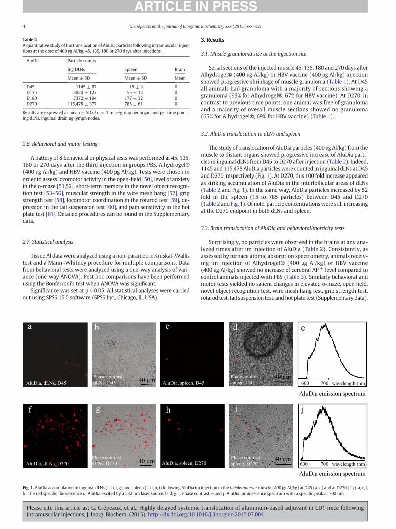
Table 2A quantitative study of the translocation of AluDia particles following intramuscular injec-tions at the dose of 400 μg Al/kg, 45, 135, 180 or 270 days after injections.
AluDia Particle counts
Ing DLNs Spleen Brain
Mean ± SD Mean ± SD Mean
D45 1145 ± 87 15 ± 3 0D135 3820 ± 123 55 ± 12 0D180 7372 ± 194 177 ± 32 0D270 115,478 ± 377 785 ± 61 0
Results are expressed as mean ± SD of n = 3 mice/group per organ and per time point.Ing dLNs, inguinal draining lymph nodes.
4 G. Crépeaux et al. / Journal of Inorganic Biochemistry xxx (2015) xxx–xxx
2.6. Behavioral and motor testing
A battery of 8 behavioral or physical tests was performed at 45, 135,180 or 270 days after the third injection in groups PBS, Alhydrogel®(400 μg Al/kg) and HBV vaccine (400 μg Al/kg). Tests were chosen inorder to assess locomotor activity in the open-field [50], level of anxietyin the o-maze [51,52], short-term memory in the novel object recogni-tion test [53–56], muscular strength in the wire mesh hang [57], gripstrength test [58], locomotor coordination in the rotarod test [59], de-pression in the tail suspension test [60], and pain sensitivity in the hotplate test [61]. Detailed procedures can be found in the Supplementarydata.
2.7. Statistical analysis
Tissue Al datawere analyzed using a non-parametric Kruskal–Wallistest and a Mann–Whitney procedure for multiple comparisons. Datafrom behavioral tests were analyzed using a one-way analysis of vari-ance (one-way ANOVA). Post hoc comparisons have been performedusing the Bonferroni's test when ANOVA was significant.
Significance was set at p b 0.05. All statistical analyses were carriedout using SPSS 16.0 software (SPSS Inc., Chicago, IL, USA).
Fig. 1.AluDia accumulation in inguinal dLNs (a, b, f, g) and spleen (c, d, h, i) following AluDia imh: The red specific fluorescence of AluDia excited by a 532 nm laser source. b, d, g, i: Phase con
Please cite this article as: G. Crépeaux, et al., Highly delayed systemicintramuscular injections, J. Inorg. Biochem. (2015), http://dx.doi.org/10.10
3. Results
3.1. Muscle granuloma size at the injection site
Serial sections of the injectedmuscle 45, 135, 180 and 270 days afterAlhydrogel® (400 μg Al/kg) or HBV vaccine (400 μg Al/kg) injectionshowed progressive shrinkage of muscle granuloma (Table 1). At D45all animals had granuloma with a majority of sections showing agranuloma (93% for Alhydrogel®, 67% for HBV vaccine). At D270, incontrast to previous time points, one animal was free of granulomaand a majority of overall muscle sections showed no granuloma(65% for Alhydrogel®, 69% for HBV vaccine) (Table 1).
3.2. AluDia translocation to dLNs and spleen
The study of translocation of AluDia particles (400 μg Al/kg) from themuscle to distant organs showed progressive increase of AluDia parti-cles in inguinal dLNs fromD45 to D270 after injection (Table 2). Indeed,1145 and 115,478AluDia particleswere counted in inguinal dLNs atD45and D270, respectively (Fig. 1). At D270, this 100 fold increase appearedas striking accumulation of AluDia in the interfollicular areas of dLNs(Table 2 and Fig. 1). In the same way, AluDia particles increased by 52fold in the spleen (15 to 785 particles) between D45 and D270(Table 2 and Fig. 1). Of note, particle concentrationswere still increasingat the D270 endpoint in both dLNs and spleen.
3.3. Brain translocation of AluDia and behavioral/motricity tests
Surprisingly, no particles were observed in the brains at any ana-lyzed times after im injection of AluDia (Table 2). Consistently, asassessed by furnace atomic absorption spectrometry, animals receiv-ing im injection of Alhydrogel® (400 μg Al/kg) or HBV vaccine(400 μg Al/kg) showed no increase of cerebral Al3+ level compared tocontrol animals injected with PBS (Table 3). Similarly behavioral andmotor tests yielded no salient changes in elevated o-maze, open field,novel object recognition test, wire mesh hang test, grip strength test,rotarod test, tail suspension test, and hot plate test (Supplementary data).
injection in the tibialis anteriormuscle (400 μg Al/kg) at D45 (a–e) and at D270 (f–j). a, c, f,trast. e and j: AluDia luminescence spectrum with a specific peak at 700 nm.
translocation of aluminum-based adjuvant in CD1 mice following16/j.jinorgbio.2015.07.004

Table 3Aluminum cerebral concentration measured by furnace atomic absorption spectrometry (μg/g of dry weight).
Cerebral Al concentration Control Alhydrogel® group 400 μg Al/kg, im HBV vaccine group 400 μg Al/kg, im Kruskal–Wallis test
D45 0.54095 0.57335 0.90625 n.s.(0.3250–1.4837) (0.0234–8.8778) (0.6104–1.3623)
D135 0.02485 0.4317 0.6843 n.s.(0.0179–0.1877) (0.0200–33.3432) (0.1214–1.2061)
D180 0.0956 0.0143 0.0451 n.s.(0.0174–0.8776) (0.0133–0.3540) (0.0158–0.6317)
D270 1.0534 0.01495⁎ 0.0141⁎ p b 0.05(0.3975–2.8053) (0.0123–0.1859) (0.0122–0.0206)
Results are expressed as median and quartiles (in brackets) of n = 5 brains/group. Non-parametric Kruskal–Wallis test followed by a Mann–Whitney procedure was used for multiplecomparisons.⁎ p b 0.05, statistical significant difference from controls.
5G. Crépeaux et al. / Journal of Inorganic Biochemistry xxx (2015) xxx–xxx
Taking into account that neurotoxic effectswere previously reportedin CD1 mice after sc injection of Alhydrogel® at 100 μg Al/kg [17] and300 μg Al/kg [41], we examined whether the route of administrationor the dose could influence brain translocation of AluDia. We observedthat 3 out of 4 CD1 mice injected by the sc route with 200 μg Al/kgshowed particle incorporation into the brain 45 days after injection(Table 4 and Fig. 2). Notably, this was not observed at higher dose(400 μg Al/kg) for the sc route, and at any dose for the im route.
4. Discussion
This longitudinal study showed that alum (Alhydrogel® or HBV vac-cine) injected into themuscle constantly induces a granuloma similar toMMF that shrinks with time with marked clearance of granulomatouslesions observed from D180 to D270. This is similar to what was previ-ously observed with the AluDia complex [40]. Granuloma shrinkage inthe muscle was associated with concurrent replenishment of inguinaldLNs (100 fold increase of AluDia particles from D45 to D270). Similartranslocation of alum from themuscle to dLNs was previously observedat much earlier time points in C57BL/6J mice [39]. We assume that twowaves of lymphatic translocation may occur after im injection of alum:an early one peaking at D4 [39] and a markedly delayed one associatedwithmuscle granuloma shrinkage observed in the present study thanksto a long-term evaluation not performed in previous studies. We as-sume that this delayed lymphatic draining flux is the normal way ofclearance for alum trapped in the post-vaccinal granuloma. Similarlyto translocation to dLNs, we observedmarkedly delayed AluDia translo-cation to the spleen, with amaximumnumber of particles being detect-ed in this organ at D270. Alum translocation from the muscle to spleenwas previously shown to assess particle exit from lymphatic pathwaysto the blood stream [39]. Since the spleen was previously shown to in-corporate a first peak of particles at D7 post-im injection in C57BL/6Jmice [40], thepresent study suggests a delayed secondwave of adjuvanttranslocation to the spleen in line with that observed in dLNs.
The present study confirms that alum is extremely biopersistent [29,37] and that alum biopersistence can be observed in both the injectedmuscle and distant organs, including dLNs and spleen. Regarding thestrong immunostimulatory effects of alum and the unrequired depotformation for its adjuvant activity [36], long-term biopersistence of
Table 4A qualitative study of the translocation of AluDia particles following intramuscular or sub-cutaneous injections at the doses of 200 or 400 μg/kg, 45 days after injections.
AluDia Particle counts
im 200 μg Al/kg im 400 μg Al/kg sc 200 μg Al/kg sc 400 μg Al/kg
Brain 0 0 15 ± 7 0
Results are expressed as mean ± SD of n = 4 mice/group per organ and per time point.im, intramuscular; sc, subcutaneous.
Please cite this article as: G. Crépeaux, et al., Highly delayed systemicintramuscular injections, J. Inorg. Biochem. (2015), http://dx.doi.org/10.10
alum in lymphoid organs is clearly undesirable, and may cast doubtson the exact level of long-term safety of alum-adjuvanted vaccines [37].
The lack of brain translocation alum after im injection of400 μg Al/kg was puzzling. Notably, neither elevated Al concentrationin the brain nor neurobehavioral changeswere observed in these exper-imental conditions, ruling out significant translocation of soluble Al tothe brain in the absence of physical incorporation of alum particles,and the induction of neurobehavioral effects by chronic peripheral im-mune activation linked to persistence of alum within the immunecells [35].
It is not excluded that the observed difference in the biodispositionof alum in C57BL/6J and CD1 mice, including diffusion kinetics and theoccurrence of brain translocation, may in part reflect differences in thegenetic background of the two strains [62]. We previously demon-strated that the size of the alum-induced granuloma in rats is dra-matically influenced by their genetic background, the granulomabeing much smaller in Lewis rats with Th1 biased immune responsescompared to Sprague–Dawley rats with balanced Th1/Th2 immunity[45]. The C57BL/6mouse strain is known to exhibit a Th1-prone, pro-inflammatory type response to injury [63,64]. To our knowledge, theT helper immunity status of CD1 mice is not known.
Interestingly, C57BL/6 mice produce more MCP-1/CCL2 than otherstrains [64], and this major inflammatory monocyte chemoattractantis crucially involved in both systemic biodistribution and neurodeliveryof Al particles captured by monocyte-lineage cells [39]. Notably, in-creased circulating MCP-1/CCL2 is the sole identified biomarker in my-algic encephalomyelitis patients with MMF [65]. Moreover, humanMMF is mainly observed in middle aged or elderly individuals, a timewhen MCP-1/CCL2 production increases and immuno-senescence oc-curs [66]. Clarification of the influence of mouse strains Th1 and Th2-biased immune responses in AluDia brain translocation clearly deservesfuture studies.
In previously published studies, motor and behavioral impairmentswere observed following sc (behind the neck) Alhydrogel® injectionto CD1 mice with doses of 100 and 300 μg Al/kg [17,41]. These effectswere associated with Al deposits in the central nervous system (spinalcord) assessed by Morin stain. To examine if the route of exposuremay represent an important factor for alum toxicity, a nested studywas conducted herein, showing that alum particles may penetrate thebrain at D45 after the sc (and not im) injection, performed at the doseof 200 μg Al/kg (and not at the dose of 400 μg Al/kg). A higher rate ofbrain translocation after sc injectionmay be explained by amuch higherdensity of dendritic cells with high migrating properties, in the skincompared to the muscle. The fact that half dose resulted in brain trans-location, which was not observed at higher dose, is reminiscent of thenon-monotonic dose/response curves previously observed with envi-ronmental toxins, including particulate compounds [67]. In anotherstudy, we similarly observed neurobehavioral changes at 200 but not400 μg Al/kg (Crépeaux et al., manuscript in preparation). Theexact significance of such observations is unknown, but one mayspeculate that huge quantities of alum injected in the tissue may
translocation of aluminum-based adjuvant in CD1 mice following16/j.jinorgbio.2015.07.004

Fig. 2. AluDia in the brain (animal 1: a, b, c; animal 2: d, e, f) following AluDia sc injection (200 μg Al/kg) at D45. a and d: The red specific fluorescence of AluDia excited by a 532 nm lasersource. b and e: Phase contrast. c and f: AluDia luminescence spectrum with a specific peak at 700 nm.
6 G. Crépeaux et al. / Journal of Inorganic Biochemistry xxx (2015) xxx–xxx
induce blockade of critical macrophage functions such as migrationand xeno/autophagic disposition of particles, as previously reportedfor infectious particles [37].
5. Conclusion
We observed a strikingly delayed, previously unknown, systemictranslocation of alum particles injected into the muscle, with conspicu-ous alum accumulations in the lymphatic system and spleen 9 monthsafter injection. In addition to the crucial “t” factor, our results stronglysuggest the influence of the mouse strain, the dose and the route of ad-ministration on alum biodisposition. All these parameters should betaken into account in the design of future alum toxicological studies.
List of abbreviations
AluDia complex alum-nanodiamondsASIA autoimmune/auto-inflammatory syndrome induced by
adjuvantsdLNs draining lymph nodesFDA Food and Drug AdministrationHBV hepatitis B virusHPG hyperbranched polyglycerolHPHT High Pressure High Temperatureim intramuscularfNDs fluorescent nanodiamondsMMF macrophagic myofasciitisMCP-1/CCL2 monocyte chemoattractant protein 1/chemokine ligand 2PBS phosphate buffer salineTA tibialis anteriorTHGA spectrometer with a transversely heated graphite atomizerTH GFAAS graphite furnace atomic absorptionTh1 & Th2 T helper 1 & T helper 2sc subcutaneous
Please cite this article as: G. Crépeaux, et al., Highly delayed systemicintramuscular injections, J. Inorg. Biochem. (2015), http://dx.doi.org/10.10
Conflicts of interest
The authors declare that there are no conflicts of interest.
Acknowledgments
The authors thank Marina Bermudez de Castro Rubio, AnthonyDeust and Frédéric Ros for their technical help, and Marie De Antoniofor her statistical explanations. This studywas supported by the Univer-sity of British Columbia in Vancouver (UBC PG#20R17162), DwoskinFamily Foundation (UBC PG#20R65375), ANSM, and Région Ile-de-France DIM NeRF (“Nano-in-brain” project).
Appendix A. Supplementary data
Supplementary data to this article can be found online at http://dx.doi.org/10.1016/j.jinorgbio.2015.07.004.
References
[1] C. Exley, Human exposure to aluminium, Environ. Sci. Process. Impacts 15 (2013)1807–1816.
[2] C. Exley, L. Swarbrick, R.K. Gherardi, F.-J. Authier, A role for the body burden of alu-minium in vaccine-associated macrophagic myofasciitis and chronic fatigue syn-drome, Med. Hypotheses 72 (2009) 135–139.
[3] M. Kawahara, Effects of aluminum on the nervous system and its possible link withneurodegenerative diseases, J. Alzheimers Dis. 8 (2005) 171–182.
[4] S. Bondy, The neurotoxicity of environmental aluminum is still an issue,Neurotoxicology 31 (2010) 575–581.
[5] S. Kumar, Biphasic effect of aluminium on cholinergic enzyme of rat brain, Neurosci.Lett. 248 (1998) 121–123.
[6] L. Tomljenovic, C.A. Shaw, Do aluminum vaccine adjuvants contribute to the risingprevalence of autism? J. Inorg. Biochem. 105 (2011) 1489–1499.
[7] C. Exley, What is the risk of aluminium as a neurotoxin? Expert Rev. Neurother. 14(2014) 589–591.
[8] P. Zatta, M. Ibn-Lkhayat-Idrissi, P. Zambenedetti, M. Kilyen, T. Kiss, In vivo andin vitro effects of aluminum on the activity of mouse brain acetylcholinesterase,Brain Res. Bull. 59 (2002) 41–45.
translocation of aluminum-based adjuvant in CD1 mice following16/j.jinorgbio.2015.07.004

7G. Crépeaux et al. / Journal of Inorganic Biochemistry xxx (2015) xxx–xxx
[9] S.V. Verstraeten, L. Aimo, P.I. Oteiza, Aluminium and lead: molecular mechanisms ofbrain toxicity, Arch. Toxicol. 82 (2008) 789–802.
[10] R. Moumen, N. Ait-Oukhatar, F. Bureau, C. Fleury, D. Bouglé, P. Arhan, et al., Alumin-ium increases xanthine oxidase activity and disturbs antioxidant status in the rat, J.Trace Elem. Med. Biol. Organ Soc. Miner. Trace Elem. GMS. 15 (2001) 89–93.
[11] P. Nayak, Aluminum: impacts and disease, Environ. Res. 89 (2002) 101–115.[12] R.A. Yokel, Brain uptake, retention, and efflux of aluminum andmanganese, Environ.
Health Perspect. 110 (Suppl. 5) (2002) 699–704.[13] C. Exley, When an aluminium adjuvant is not an aluminium adjuvant used in
human vaccination programmes, Vaccine 30 (2012) 2042.[14] S. Sánchez-Iglesias, R. Soto-Otero, J. Iglesias-González, M.C. Barciela-Alonso, P.
Bermejo-Barrera, E. Méndez-Alvarez, Analysis of brain regional distribution of alu-minium in rats via oral and intraperitoneal administration, J. Trace Elem. Med.Biol. Organ Soc. Miner. Trace Elem. GMS. 21 (Suppl. 1) (2007) 31–34.
[15] H. Rosenblum, Y. Shoenfeld, H. Amital, The common immunogenic etiology ofchronic fatigue syndrome: from infections to vaccines via adjuvants to the ASIA syn-drome, Infect. Dis. Clin. North Am. 25 (2011) 851–863.
[16] M.A. Hernán, S.S. Jick, M.J. Olek, H. Jick, Recombinant hepatitis B vaccine and the riskof multiple sclerosis: a prospective study, Neurology 63 (2004) 838–842.
[17] M.S. Petrik, M.C.Wong, R.C. Tabata, R.F. Garry, C.A. Shaw, Aluminum adjuvant linkedto Gulf War illness induces motor neuron death in mice, Neuromolecular Med. 9(2007) 83–100.
[18] Y. Shoenfeld, N. Agmon-Levin, “ASIA”— autoimmune/inflammatory syndrome in-duced by adjuvants, J Autoimmun. 36 (1) (2011) 4–8.
[19] S.J. van Rensburg, F.C. Potocnik, T. Kiss, F. Hugo, P. van Zijl, E. Mansvelt, et al., Serumconcentrations of some metals and steroids in patients with chronic fatigue syn-drome with reference to neurological and cognitive abnormalities, Brain Res. Bull.55 (2001) 319–325.
[20] A. Lerner, Aluminum as an adjuvant in Crohn's disease induction, Lupus 21 (2012)231–238.
[21] P. De Sole, C. Rossi, M. Chiarpotto, G. Ciasca, B. Bocca, A. Alimonti, et al., Possible re-lationship between Al/ferritin complex and Alzheimer's disease, Clin. Biochem. 46(2013) 89–93.
[22] R.K. Gherardi, M. Coquet, P. Chérin, et al., Macrophagicmyofasciitis: an emerging en-tity, Lancet 352 (1998) 347–352.
[23] World Health Organization Vaccine Safety Advisory Committee, Macrophagicmyofasciitis and aluminum-containing vaccines, Wkly. Epidemiol. Rec. 74 (1999)338–340.
[24] M. Couette, M.-F. Boisse, P. Maison, P. Brugieres, P. Cesaro, X. Chevalier, et al., Long-term persistence of vaccine-derived aluminum hydroxide is associated with chroniccognitive dysfunction, J. Inorg. Biochem. 103 (2009) 1571–1578.
[25] E. Passeri, C. Villa, M. Couette, E. Itti, P. Brugieres, P. Cesaro, et al., Long-term follow-up of cognitive dysfunction in patients with aluminum hydroxide-induced macro-phagic myofasciitis (MMF), J. Inorg. Biochem. 105 (2011) 1457–1463.
[26] R.K. Gherardi, F.J. Authier, Macrophagic myofasciitis: characterization and patho-physiology, Lupus 21 (2012) 184–189.
[27] T. Santiago, O. Rebelo, L. Negrão, A. Matos, Macrophagic myofasciitis and vaccina-tion: consequence or coincidence? Rheumatol. Int. 35 (2014) 189–192.
[28] F.J. Authier, P. Cherin, A. Creange, B. Bonnotte, X. Ferrer, A. Abdelmoumni, et al., Cen-tral nervous system disease in patients with macrophagic myofasciitis, Brain J.Neurol. 124 (2001) 974–983.
[29] R.K. Gherardi, M. Coquet, P. Cherin, L. Belec, P. Moretto, P.A. Dreyfus, et al., Macro-phagic myofasciitis lesions assess long-term persistence of vaccine-derived alumin-ium hydroxide in muscle, Brain J. Neurol. 124 (2001) 1821–1831.
[30] N. Ragunathan-Thangarajah, C. Le Beller, P. Boutouyrie, G. Bassez, R.K. Gherardi, S.Laurent, et al., Distinctive clinical features in arthro-myalgic patients with and with-out aluminum hydroxide-induced macrophagic myofasciitis: an exploratory study,J. Inorg. Biochem. 128 (2013) 262–266.
[31] C. Exley, P. Siesjö, H. Eriksson, The immunobiology of aluminium adjuvants: how dothey really work? Trends Immunol. 31 (2010) 103–109.
[32] C.A. Shaw, L. Tomljenovic, Aluminum in the central nervous system (CNS): toxicityin humans and animals, vaccine adjuvants, and autoimmunity, Immunol. Res. 56(2013) 304–316.
[33] A.T. Glenny, C.G. Pope, H. Waddington, U. Wallace, XXIII—the antigenic value of tox-oid precipitated by potassium alum, J. Pathol. Bacteriol. 29 (1926) 38–39.
[34] T.C. Eickhoff, M. Myers, Conference report: workshop summary aluminum in vac-cines, Vaccine 20 (2002) S1–S4.
[35] Y. Kashiwagi, M. Maeda, H. Kawashima, T. Nakayama, Inflammatory responsesfollowing intramuscular and subcutaneous immunization with aluminum-adjuvanted or non-adjuvanted vaccines, Vaccine 32 (2014) 3393–3401.
[36] S. Hutchinson, R.A. Benson, V.B. Gibson, A.H. Pollock, P. Garside, J.M. Brewer, Antigendepot is not required for alum adjuvanticity, FASEB J. 26 (2012) 1272–1279.
[37] R.K. Gherardi, H. Eidi, G. Crépeaux, F.J. Authier, J. Cadusseau, Biopersistence andbrain translocation of aluminum adjuvants of vaccines, Front. Neurol. 6 (2015) 4.
[38] R. Flarend, T. Bin, D. Elmore, S.L. Hem, A preliminary study of the dermal absorptionof aluminium from antiperspirants using aluminium-26, Food Chem. Toxicol. Int. J.Publ. Br. Ind. Biol. Res. Assoc. 39 (2001) 163–168.
[39] Z. Khan, C. Combadière, F.-J. Authier, V. Itier, F. Lux, C. Exley, et al., Slow CCL2-dependent translocation of biopersistent particles from muscle to brain, BMCMed. 11 (2013) 99.
Please cite this article as: G. Crépeaux, et al., Highly delayed systemicintramuscular injections, J. Inorg. Biochem. (2015), http://dx.doi.org/10.10
[40] H. Eidi, M.O. David, G. Crépeaux, L. Henry, V. Joshi, M.H. Berger, M. Sennour, J.Cadusseau, R.K. Gherardi, P.A. Curmi, Fluorescent nanodiamonds as a relevant tagfor the assessment of alum adjuvant particle biodisposition, BMC Med. 13 (1)(2015) 144.
[41] C.A. Shaw, M.S. Petrik, Aluminum hydroxide injections lead to motor deficits andmotor neuron degeneration, J. Inorg. Biochem. 103 (2009) 1555–1562.
[42] L. Luján, M. Pérez, E. Salazar, N. Álvarez, M. Gimeno, P. Pinczowski, et al., Autoim-mune/autoinflammatory syndrome induced by adjuvants (ASIA syndrome) in com-mercial sheep, Immunol. Res. 56 (2013) 317–324.
[43] N. Agmon-Levin, M.-T. Arango, S. Kivity, A. Katzav, B. Gilburd, M. Blank, et al., Immu-nization with hepatitis B vaccine accelerates SLE-like disease in a murine model, J.Autoimmun. 54 (2014) 21–32.
[44] F. Verdier, R. Burnett, C. Michelet-Habchi, P. Moretto, F. Fievet-Groyne, E. Sauzeat,Aluminium assay and evaluation of the local reaction at several time points after in-tramuscular administration of aluminium containing vaccines in the cynomolgusmonkey, Vaccine 23 (2005) 1359–1367.
[45] F.-J. Authier, S. Sauvat, C. Christov, P. Chariot, G. Raisbeck, M.-F. Poron, et al., AlOH3-adjuvanted vaccine-induced macrophagic myofasciitis in rats is influenced by thegenetic background, Neuromuscul. Disord. 16 (2006) 347–352.
[46] European Union Directive, European Union Directive, 2010/63/EU of 22 September2010 on the Approximation of Laws, Regulations and Administrative Provisions ofthe Member States Regarding the Protection of Animals Used for Experimentaland Other Scientific Purposes2010.
[47] J.P. Boudou, P.A. Curmi, F. Jelezko, J. Wrachtrup, P. Aubert, M. Sennour, G.Balasubramanian, R. Reuter, A. Thorel, E. Gaffet, High yield fabrication of fluorescentnanodiamonds, Nanotechnology 20 (2009) 235602.
[48] J.P. Boudou, M.O. David, V. Joshi, H. Eidi, P.A. Curmi, Hyperbranched polymers: struc-ture of hyperbranched polyglycerol and amphiphilic poly(glycerol ester)s in diluteaqueous and nonaqueous solution, Diam. Relat. Mater. 37 (2013) 131–138.
[49] E. House, M. Esiri, G. Forster, P.G. Ince, C. Exley, Aluminium, iron and copper inhuman brain tissues donated to the Medical Research Council's Cognitive Functionand Ageing Study, Met. Integr. Biometal Sci. 4 (2012) 56–65.
[50] R.N. Walsh, R.A. Cummins, The Open-Field Test: a critical review, Psychol. Bull. 83(1976) 482–504.
[51] J.K. Shepherd, S.S. Grewal, A. Fletcher, D.J. Bill, C.T. Dourish, Behavioural and pharma-cological characterisation of the elevated “zero-maze” as an animal model of anxi-ety, Psychopharmacology (Berl) 116 (1994) 56–64.
[52] L. Coutellier, A.-C. Friedrich, K. Failing, V. Marashi, H. Würbel, Effects of foraging de-mand on maternal behaviour and adult offspring anxiety and stress response inC57BL/6 mice, Behav. Brain Res. 196 (2009) 192–199.
[53] A. Ennaceur, J. Delacour, A new one-trial test for neurobiological studies of memoryin rats. 1: Behavioral data, Behav. Brain Res. 31 (1988) 47–59.
[54] P.A. Dudchenko, An overview of the tasks used to test working memory in rodents,Neurosci. Biobehav. Rev. 28 (2004) 699–709.
[55] A. Ennaceur, One-trial object recognition in rats and mice: methodological and the-oretical issues, Behav. Brain Res. 215 (2010) 244–254.
[56] S.J. Moore, K. Deshpande, G.S. Stinnett, A.F. Seasholtz, G.G. Murphy, Conversion ofshort-term to long-term memory in the novel object recognition paradigm,Neurobiol. Learn. Mem. 105 (2013) 174–185.
[57] W. Kondziela, Eine neue method zur messung der muskularen relaxation beiweissen mausen, Arch. Int. Pharmacodyn. 152 (1964) 277–284.
[58] J.P.J. Maurissen, B.R. Marable, A.K. Andrus, K.E. Stebbins, Factors affecting gripstrength testing, Neurotoxicol. Teratol. 25 (2003) 543–553.
[59] M. Pratte, N. Panayotis, A. Ghata, L. Villard, J.-C. Roux, Progressive motor and respi-ratorymetabolism deficits in post-weaningMecp2-null male mice, Behav. Brain Res.216 (2011) 313–320.
[60] L. Steru, R. Chermat, B. Thierry, P. Simon, The tail suspension test: a newmethod forscreening antidepressants in mice, Psychopharmacology (Berl) 85 (1985) 367–370.
[61] E.F. Espejo, D. Mir, Structure of the rat's behaviour in the hot plate test, Behav. BrainRes. 56 (1993) 171–176.
[62] C.D. Mills, K. Kincaid, J.M. Alt, M.J. Heilman, A.M. Hill, M-1/M-2macrophages and theTh1/Th2 paradigm, J. Immunol. 164 (2000) 6166–6173.
[63] G.S. Whitehead, J.K.L. Walker, K.G. Berman, W.M. Foster, D.A. Schwartz, Allergen-induced airway disease is mouse strain dependent, Am. J. Physiol. Lung Cell. Mol.Physiol. 285 (2003) L32–L42.
[64] C.D. Mills, K. Ley, M1 and M2macrophages: the chicken and the egg of immunity, J.Innate Immun. 6 (2014) 716–726.
[65] J. Cadusseau, N. Ragunathan-Thangarajah, M. Surenaud, S. Hue, F.-J. Authier, R.K.Gherardi, Selective elevation of circulating CCL2/MCP1 levels in patients withlongstanding post-vaccinal macrophagic myofasciitis and ASIA, Curr. Med. Chem.21 (2014) 511–517.
[66] A.S. Mansfield, W.K. Nevala, R.S. Dronca, A.A. Leontovich, L. Shuster, S.N. Markovic,Normal ageing is associated with an increase in Th2 cells, MCP-1 (CCL2) andRANTES (CCL5), with differences in sCD40L and PDGF-AA between sexes, Clin.Exp. Immunol. 170 (2) (2012) 186–193.
[67] I. Iavicoli, L. Fontana, V. Leso, E.J. Calabrese, Hormetic dose-responses in nanotech-nology studies, Sci. Total Environ. 487 (2014) 361–374.
translocation of aluminum-based adjuvant in CD1 mice following16/j.jinorgbio.2015.07.004
Related Documents