Welcome message from author
This document is posted to help you gain knowledge. Please leave a comment to let me know what you think about it! Share it to your friends and learn new things together.
Transcript

HIGHLIGHTSFebruary 2015

PAGE INDEX
Join the ENLIGHT network. Register to become a member here.https://indico.cern.ch/confRegistrationFormDisplay.py/display?confId=180036
From the Coordinator Charting the future
3.
From Physics toMedicine the vision from CERN
18.
20th anniversary of HIMAC Hirohiko Tsujii
8.
ENTERVISION
28.
Horizon 2020 The philosophy behind
4.
ENVISION
22.
60 Years of Particle Therapy Eleanor A. Blakely
10.
THE ENTERVISION Biological Dosimetric Phantom
30.
ENLIGHT Annual Meeting 2014
6.
ULICE
24.
ICTR-PHE 2014 Uniting sciences to treatcancer
14.
IAEA CONSULTANTS MEETING on Particle Therapy in the 21st Century
32.
ENLIGHT COORDINATORManjit Dosanjh
ENLIGHT COORDINATION OFFICEAudrey BallantineDimitrios ChlorokostasManuela CirilliHelen Dixon-AltaberSparsh Navin
PHOTO CREDITSCERNPARTNERULICEENTERVISIONENVISIONCNAOHITIBAIAEALBNLNYMUS 3DNIRSEugene Theodore
ENLIGHT HIGHLIGHTS is distributed free of charge. This work is licensed under the Creative Commons Attribution-NonCommercial-NoDerivs 3.0 Unported License. To view a copy of this license, visit http://creativecommons.org/licenses/by-nc-nd/3.0/.
For more information and contact details please visit the ENLIGHT website - CERN.CH/ENLIGHT
PAG
E IN
DEX
2 HIGHLIGHTS | CERN.CH/ENLIGHT
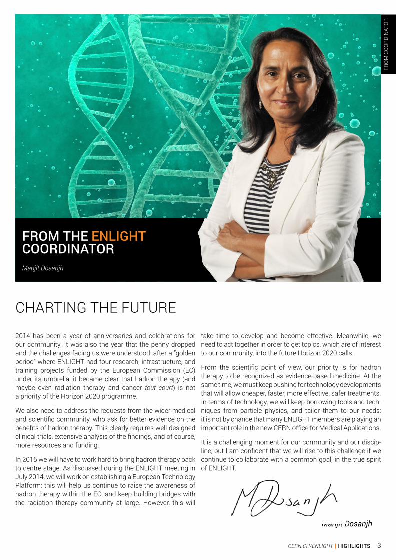
CHARTING THE FUTURE
Manjit Dosanjh
FROM THE ENLIGHTCOORDINATORManjit Dosanjh
2014 has been a year of anniversaries and celebrations for our community. It was also the year that the penny dropped and the challenges facing us were understood: after a “golden period” where ENLIGHT had four research, infrastructure, and training projects funded by the European Commission (EC) under its umbrella, it became clear that hadron therapy (and maybe even radiation therapy and cancer tout court) is not a priority of the Horizon 2020 programme.
We also need to address the requests from the wider medical and scientific community, who ask for better evidence on the benefits of hadron therapy. This clearly requires well-designed clinical trials, extensive analysis of the findings, and of course, more resources and funding.
In 2015 we will have to work hard to bring hadron therapy back to centre stage. As discussed during the ENLIGHT meeting in July 2014, we will work on establishing a European Technology Platform: this will help us continue to raise the awareness of hadron therapy within the EC, and keep building bridges with the radiation therapy community at large. However, this will
take time to develop and become effective. Meanwhile, we need to act together in order to get topics, which are of interest to our community, into the future Horizon 2020 calls.
From the scientific point of view, our priority is for hadron therapy to be recognized as evidence-based medicine. At the same time, we must keep pushing for technology developments that will allow cheaper, faster, more effective, safer treatments. In terms of technology, we will keep borrowing tools and tech-niques from particle physics, and tailor them to our needs: it is not by chance that many ENLIGHT members are playing an important role in the new CERN office for Medical Applications.
It is a challenging moment for our community and our discip-line, but I am confident that we will rise to this challenge if we continue to collaborate with a common goal, in the true spirit of ENLIGHT.
FRO
M C
OO
RDIN
ATO
R
3CERN.CH/ENLIGHT | HIGHLIGHTS

The philosophyWith Europe still recovering from a crisis where available national budgets for research, development and innovation in many Member States seem uncertain for the years to come the European Commission programme Horizon 2020 appears to be one of the most stable options to consider.
Nevertheless after the first round of calls it is clear that competition is as fierce as ever. Moreover nothing seems to indicate that this situation will change in future rounds. Given the landscape, it will be very difficult to succeed without reflecting and addressing in any project proposal the very philosophy behind Horizon 2020; its rationale.
The European Commission defines Horizon 2020 as the
financial instrument implementing the Innovation Union, a Europe 2020 flagship initiative aimed at boosting Europe’s competitiveness, economic growth and create jobs. By coupling research and innovation, Horizon 2020 is contributing to achieve these major goals with its emphasis on excellent science, industrial leadership and tackling societal challenges.
It is the task therefore for any project proposal to explain in concrete and quantifiable terms its contribution to the above mentioned objectives which ultimately means the translation of innovation into jobs. In other words, the project “value proposition”. Achieving this normally requires the close cooperation and coordination of all actors across
HO
RIZO
N 2
020
by Pablo Garcia Tello, CERN
4 HIGHLIGHTS | CERN.CH/ENLIGHT

the innovation value chain of any disciplines that the project ensembles: from the research communities to the so called end users.
The new Commission has even made more explicit this point: “…to overcome silo mentalities by working jointly on those areas where we can really make a difference”. Equally explicit has been the new Commissioner of Research, Science and Innovation in his clear statement aiming at further pursuing the completion of the European Research Area and accelerating the actions towards the Innovation Union. Making literal use of the new Commissioner’s words: “Research, science and innovation have the potential to improve all areas of the economy and society, and help us
tackle our main societal challenges. Working in silos is not an option”.
With the next edition of Horizon 2020 still in preparation it will be crucial to start getting ready for taking the tide when it comes.
In this sense it will be imperative to identify those areas based on evidence in which a difference can be made in and for Europe by delivering on innovation and ultimately jobs. It will be as well critical therefore to align and coordinate the efforts and goals of the collaborating actors and communities across the value chain.
behindH
ORI
ZON
202
0
5CERN.CH/ENLIGHT | HIGHLIGHTS

ENLIGHT Annual Meeting 2014
ENLI
GH
T 20
14
6 HIGHLIGHTS | CERN.CH/ENLIGHT

The ENLIGTH community met at CERN from 10-12 July 2014 to mark the 12th year since the creation of the light ion therapy network in Europe. Twelve years ago, ENLIGHT had its inaugural meeting at CERN, attended by around 70 specialists from different disciplines. This was a considerable achievement at a time when the concept of multi-disciplinarity was still in its infancy. Today multi-disciplinarity is an indispensible com-ponent of healthcare. 145 participants from around the world working in different fields attended this meeting.
The meeting was opened by Rolf Heuer, the Director General of CERN, who congratulated the network on its achievements and encouraged the collaborative spirit to prevail. Leading experts like Prof. Tsujii (NIRS), Prof. Blakeley (LBNL) and Prof. Combs (Technical university of Munich) discussed the current status of particle therapy in Japan, the Americas and Europe. Damien Bertrand (IBA) and Prof. Enghardt (Dresden) discussed the state of the art in industry and medical imaging, along with a vision for the near future. This set the stage for the afternoon discussion on future direction.
The discussion on future objectives revolved around four main areas: imaging and diagnostics, treatment planning, clinical trials and training. Accuracy in diagnosis is the key to an effective treatment. “Today diagnostic accuracy is well behind progress in irradiation techniques”, said Prof. Tsujii. Some of the areas where work is on-going to bridge this gap include functional aspects of multi-modality imaging, addressing the challenges of motion during imaging and adding mathematical models to imaging tools. Accuracy of treatment can be improved by automatic real-time feedback for dose control. Going hand-in-hand with these new techniques is software development for diagnostics and therapeutics.
Several aspects need to be considered before devising an effective treatment plan. The more information in a treatment plan, the more personalised it is to the patient. Information on the biological aspects of the tumour, functional imaging and the impact of compounds need to be integrated into the treatment planning system.
Comparative studies are needed on patient preparation and irradiation techniques in order to be able to make ion therapy a more accessible mainstream treatment option. Clinical trials with long-term follow-up help evaluate late-effects of radiation. Since the radiobiological effectiveness (RBE) is the
biggest unknown, randomised trials are needed where the RBE assumptions are tested. “RBE is the fifth dimension”, says Bleddyn Jones (Oxford).
Together with technological progress, appropriate training of researchers in the latest technology and machinery is an essential part of the efficient use of a treatment centre. Training should be modelled based on research topics and should include in-depth specific workshops and inter-disciplinary courses. According to Damien Bertrand, “Training comes with an added value”. The young researchers we train today become the experts of tomorrow.
The 3-day ENLIGHT meeting not only served as a platform to discuss the future of the ion therapy community but also provided the opportunity for young researchers to present their work in the form of scientific posters. The CERN knowledge transfer prize was awarded to three of the researchers, who were also invited to briefly make an oral presentation of their work on the final day of the meeting. The three winning posters were those of Thiago V.M Lima on biological dosimetric phantoms; Joakim da Silva on near real-time dose calculation for hadron therapy on GPU; and Ander Biguri on dual modality Electrical Impedance Tomography (EIT)- Cone Beam Computed Tomography (CBCT) for lung radiation therapy.
The ENLIGHT meeting at CERN was also the perfect occasion to co-celebrate 60 years since the birth of CERN and the treatment of the first patient with protons in Berkley. Prof. Eleanor Blakely, a senior biophysicist at the Lawrence Berkley National Laboratory, gave the first in a series of public seminars on “Accelerating innovation in medicine” to mark the occasion. She shared her reflections and perspectives on the 60-year journey of particle therapy starting from the invention of the cyclotron by Earnest Lawrence and Robert Wilson’s proposal to use proton beams therapeutically. Between 1954 and 1957, 30 patients were treated at Berkeley using protons. The first clinical trials were set up in 1975, with 2054 patients being treated with Helium ions and 433 with Neon ions between 1975 and 1992. She concluded emphasising the role of the ENLIGHT network in promoting international research and development, networking and training for students and staff from treatment centres across Europe - both current and those in the making.
Rolf Heuer, the Director General of CERN opens the 2014 ENLIGHT meeting.
Manjit Dosanjh and Steve Myers present the CERN Knowledge Transfer Prize to Thiago, Joakim and Ander.
ENLI
GH
T 20
14
7CERN.CH/ENLIGHT | HIGHLIGHTS

On December 6th 2014, we held a public seminar in commemoration of 20 years having passed since the start of carbon-ion radiotherapy (C-ion RT) with HIMAC/NIRS. The seminar successfully brought an audience of around 500 from all over Japan to share 20-year achievements of C-ion RT. Presentations at the seminar were: “Milestone of HIMAC” by Tsujii, “Clinical results of C-ion RT at NIRS” by T. Kamada, “What is expected in HIMAC” by Dr Ebihara, and “Development of therapeutic techniques and accelerators” by K. Noda. In the last session of the seminar, a round‐table talk was given by 4 speakers to discuss broad aspects of C-ion RT at present and in the future.
In January of 2015, we will also hold the symposium with the title of “HIMAC International Symposium 2015: 20th Year Anniversary Event“. This will take place as a special international event to commemorate the 20-year anniversary of HIMAC/NIRS. There will be presentations by more than 20 invited speakers whose institutions are in operation, under
construction or under active planning for C-ion RT. The NIRS staff will also present the 20-year experiences of C-ion RT for each tumor site. Therefore, the participants attending the symposium should learn the current status and future plan of C-ion RT in the world as well as clinical results so far obtained at NIRS.
Since 1981, cancer has been a leading cause of death in Japan; thereafter the number of the patients dying of cancer has increased year by year. Being encouraged by circumstances and needs of the time, the decision was made in 1984 to build the HIMAC, which was named by taking the initials of “Heavy Ion Medical Accelerator in Chiba”, as an integral part of the nation’s “Overall Ten-Year Anti-Cancer Strategy”. The HIMAC took almost a decade to build and was completed by the end of 1993. While the proton accelerator built at Loma Linda University in 1990 was the first proton accelerator put primarily into therapeutic service, the HIMAC can claim to be the world’s first facility dedicated to cancer therapy using heavy ion beams.
NIRS celebrated the 20th anniversary of HIMAC
HIROHIKO TSUJIIResearch Center for Charged Particle Therapy, National Institute of Radiological Sciences, Chiba, Japan
NIR
S
8 HIGHLIGHTS | CERN.CH/ENLIGHT

The HIMAC has been operated as a multipurpose facility for the joint use of cancer therapy and biology/physics research. The researchers have been invited from many countries including Japan.
Among several types of ion species, carbon-ions were chosen for cancer therapy at NIRS because they appeared to have the most optimal properties in terms of possessing, both physically and biologically, the most effective dose distribution in the body. The RBE (Relative biological effectiveness) of the carbon-ion beam for clinical use at NIRS ranged from 2.0 to 3.0 along the SOBP (Spread Out Bragg Peak) for acute skin reactions.
In June 1994, a clinical study was begun with HIMAC to investigate the efficacy of C-ion RT against a variety of tumors as well as to develop effective techniques for delivering a sufficient dose to the tumor. As of August 2014, the total number of patients treated at NIRS has reached approximately 9,000. Most of the patients have been treated based on Phase I/II or Phase II studies, and clinical results have shown that C-ion RT has the potential ability to provide a sufficient dose to the tumor with acceptable morbidity in the surrounding normal tissues. After accumulating clinical experiences in various types of tumors, NIRS was successful in obtaining approval from the Ministry of Health, Welfare and Labor to carry out “Highly Advanced Medical Technology” in 2003. This means that C-ion RT achieved for itself a solid place in the general practice of cancer treatment in Japan. Currently, we are making extensive efforts so that particle therapy including proton and C-ion RT could be covered by the social insurance of Japan.
Tumors that appear to respond favorably to carbon ions include locally advanced tumors and those with histologically non-squamous type of tumors such as adenocarcinoma, adenoid cystic carcinoma, malignant melanoma, hepatoma, and bone and soft tissue sarcoma. By taking advantage of the biological and physical properties of high-LET (Linear energy transfer) radiation, the efficacy of treatment regimens with small fractions in short treatment times has been confirmed for almost all types of tumors. For example, stage I lung cancer and liver cancer can be treated with only one or two fractions, respectively, and even for prostate cancers 12 fractions have been feasible. Currently, the average number of fractions per patient is 12 fractions given in 3 weeks.
In 2010, the NIRS built a new treatment facility with the beam lines extended from the existing HIMAC accelerators. In the new facility, we have started to treat patients with pencil beam scanning and develop a rotating gantry, which will be completed and put in practice by the end of 2015. We are convinced that, with these developments, the treatment outcome will be further improved with less toxicity in the normal tissues.
This year we published a textbook which, based on 20-year experience of C-ion RT at NIRS and overall literature reviews, describes about the rationale of employing carbon ions for cancer therapy and optimal treatment planning. The treatments, which unfortunately resulted in development of severe toxicities or tumor control failures, are also described. Accordingly, readers should learn successful techniques to avoid potential adverse effects, as well as learn how to overcome or treat them if they unfortunately develop.
A treatment room with vertical/horizontal beam lines for broad beam irradiation
Entrance hall of the new building
A treatment room with a robotic couch in the new building
Waiting hall of the new building
NIR
S
9CERN.CH/ENLIGHT | HIGHLIGHTS
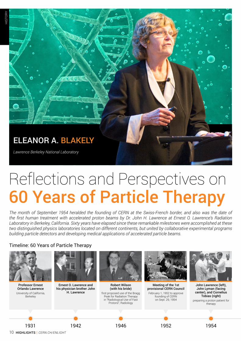
Reflections and Perspectives on 60 Years of Particle Therapy
Professor Ernest Orlando Lawrence
University of California, Berkeley
Ernest O. Lawrence and his physician brother John
H. Lawrence
Robert Wilson (with his bride)
first proposed use of the Bragg Peak for Radiation Therapy in “Radiological Use of Fast
Protons”, Radiology
Timeline: 60 Years of Particle Therapy
1931 1942 1946
John Lawrence (left), John Lyman (facing
center), and Cornelius Tobias (right)
preparing a proton patient for therapy
1954
Meeting of the 1st provisional CERN CouncilFebruary 1, 1952 to approve
founding of CERN on Sept. 29, 1954
1952
ELEANOR A. BLAKELYLawrence Berkeley National Laboratory
The month of September 1954 heralded the founding of CERN at the Swiss-French border, and also was the date of the first human treatment with accelerated proton beams by Dr. John H. Lawrence at Ernest O. Lawrence’s Radiation Laboratory in Berkeley, California. Sixty years have elapsed since these remarkable milestones were accomplished at these two distinguished physics laboratories located on different continents, but united by collaborative experimental programs building particle detectors and developing medical applications of accelerated particle beams.
HIS
TORY
10 HIGHLIGHTS | CERN.CH/ENLIGHT

The history of particle accelerators began with Ernest Lawrence’s invention of the cyclotron in 1931, and for which he was awarded the Nobel Prize in Physics in 1939. The first cyclotron constructed had a 13-cm diameter and was capable of accelerating protons to 80,000 volts using less than 1,000 volts. With time, Ernest began building larger and larger cyclotrons that could be used to produce particle beams of various atomic numbers with high enough energy to treat lesions deep inside the human body, as first proposed by Robert Wilson in 1946. Ernest’s brother John Lawrence, a physician trained at Yale, led the medical studies at Berkeley, and was responsible for treating the first proton therapy patient in 1954.
The 184-inch cyclotron (known by the dimension of the dee (magnet) used to accelerate the ions) was used in Berkeley to treat patients from 1954 until 1986. Fifteen hundred patients were treated at this machine using careful alignment procedures made possible by using a mechanical table/chair that was controlled with electronically-driven precision. The device was called ISAH (Irradiation Stereotaxic Apparatus for Humans), and it was key to controlling where the particle beams deposited their energies in the patients. Protons, deuterons and helium ion beams were all used. It was during this time that CERN was founded on September 29, 1954 after the first provisional CERN Council met in February 15, 1952 to approve the plan.
Albert Ghiorso, a high-energy nuclear physicist in Berkeley contributed a creative solution to funding a higher-energy accelerator during dwindling budgets in the 1960’s. He proposed linking two existing accelerators, a heavy-ion linear accelerator called the Super HILAC, and the BEVATRON, a particle accelerator (specifically, a weak-focusing proton synchrotron which began operating in 1954). The antiproton was discovered there in 1955, resulting in the 1959 Nobel Prize in physics for Emilio Segrè and Owen Chamberlain. Connecting the Super HILAC by a pipe running down the hill to the BEVATRON created the BEVALAC that was capable of accelerating ions from low atomic number up to uranium at high relativistic energies up to 960 MeV/amu.
The BEVALAC was used from 1975 until 1992 for Phase I/II clinical trials to treat various cancer sites with helium, carbon, neon, silicon and argon ion beams under the leadership of Joseph R. Castro and Theodore L. Phillips from UCSF.
Radiobiology of human and rodent cell lines was completed to determine cell killing potential under aerobic and hypoxic conditions at different locations along the range of carbon, neon, silicon and argon beams with pristine and extended Bragg peaks. Select in vivo investigations were completed as well. The data indicated that carbon and neon ions were similar in their effectiveness at beam energies yielding ~14 cm depth, but that neon ions were somewhat more effective at longer beam energies yielding ~24 cm depth. Beams of higher atomic number (e.g., silicon, or argon) were indeed exceeding effective, especially in eradicating hypoxic cells at the Bragg peak, but they were not ideal because there was cell killing at the entrance plateau of the beam that negated the depth-dose sparing benefits of these beams.
The Berkeley Lab treated 2054 patients with helium and 433 patients with neon. The helium ions were used primarily to treat uveal melanoma and arteriovenous malformations successfully. Clinical results with neon ions indicated that 5 tumor sites responded well compared to conventional X-ray therapy, and considering that these were Phase I/II trials with patients having advanced disease. The five sites were macroscopic salivary gland carcinoma, paranasal sinus carcinoma, soft tissue sarcoma, sarcoma of the bone, and locally advanced prostate carcinoma. It was noted that several of these tumor sites also demonstrated an effective response to neutron therapy with a similar radiation quality to carbon and neon ion beams.
During the several decades of charged particle research at Berkeley Lab, novel beam particle detectors and delivery methods were developed to take advantage of the advances ongoing in structural and functional imaging of human normal tissues and tumors. Patient immobilization and verification methods also advanced, as well as beam-delivery control and treatment planning codes. These all contributed to an unblemished safety record in human cancer therapy.
In 1992, despite the many successes of the program, a decision was made to close the clinical charged particle radiotherapy of cancer study, and dismantle the old accelerators to make way for new programs. The 184” accelerator was converted into an Advanced Light Source. The land that the BEVATRON/BEVALAC stood on however, still lies empty today.
The BEVALACThe dotted line shows the link between the Super HILAC and the BEVATRON on the Berkeley
hillside
Dr. Albert Ghiorsowho proposed linking
the Super HILAC and the BEVATRON
Joseph R. Castro Theodore L. PhillipsIrradiation Stereotaxic Apparatus for HumansPatient being aligned at the
184-inch accelerator with the stereotaxic
table/chair ISAH
1957 1970 1974 1975 1992
HIS
TORY
nder the leadership of Joseph R. Castro and Theodore L. Phillips, The BEVALAC was used from 1975 until 1992 for Phase I/II clinical
trials with charged particle beams to treat various cancer sites.
11CERN.CH/ENLIGHT | HIGHLIGHTS

Fortunately, hadron therapy has continued internationally with carbon ions, first in Japan at the National Institute of Radiological Sciences’ Heavy Ion Medical Accelerator at Chiba (HIMAC) under the leadership of Professor Hirohiko Tsujii and Professor Tadashi Kamada, and more recently at the Heidelberg Ion Therapy (HIT) facility in Heidelberg, Germany, led by Professor Gerhard Kraft and Professor Dr. Juergen Debus. In Japan, proton and carbon-ion technology and radiotherapy has continued to expand with several additional new facilities being built, and a new compact carbon ion accelerator being designed.
Professor Ugo Amaldi at CERN spearheaded the TERA Foundation’s development of a Proton Ion Medical Machine Study (PIMMS) that optimized accelerator design for medical treatments. This program also contributed to the building of CNAO (Centro Nazionale di Androterapia Oncologica) in Pavia, Italy led by Professor Roberto Orecchia and Professor Sandro Rossi. Physics collaborations between CERN and LBNL continued with the building of the ATLAS detector. Berkeley Lab’s Center for Beam Physics continues to contribute to the LHC through the US-LARP program. Dr. Manjit Dosanjh led CERN’s effort to coordinate and catalyze the ENLIGHT European platform promoting International Research and Development, Networking and Training for students and staff from facilities planning, or using hadron therapy across Europe with programs like PARTNER, ULICE, ENVISION and
ENTERVISION. This support of the infrastructure for hadron therapy is invaluable.
The situation for hadron therapy in the United States at present is still evolving. Despite the role of the U.S in developing the early technology for this field, there are several significant proton therapy facilities treating cancer patients, but no carbon ion facility exists. The U.S. Department of Energy and the National Cancer Institute convened a workshop on Ion Beam Therapy in Bethesda, MD in January 2013. More than 60 participants from medicine, physics, biology and business were charged with addressing key topics to help lead a path forward. This workshop led to two separate calls for P20 proposals for “Planning for a National Center for Particle Beam Radiation Therapy Research”. The proposals selected will be announced in January 2015.
In summary, LBNL and CERN share a similar history, and a focus on the physics and medical applications of accelerators, and each have benefitted from the research of the other. At present, the challenge for the U.S. is support for the design and construction of medical accelerators to allow radiobiological research and medical trials to proceed to Phase III trials for cancer therapy.
Dr. Albert Ghiorso proposed linking the Super HILAC and the BEVATRON
HIS
TORY
12 HIGHLIGHTS | CERN.CH/ENLIGHT
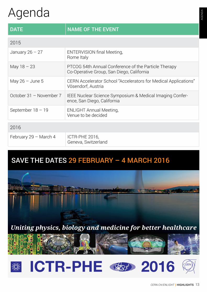
AgendaDATE NAME OF THE EVENT
2015
January 26 – 27 ENTERVISION final Meeting,Rome Italy
May 18 – 23 PTCOG 54th Annual Conference of the Particle Therapy Co-Operative Group, San Diego, California
May 26 – June 5 CERN Accelerator School “Accelerators for Medical Applications” Vösendorf, Austria
October 31 – November 7 IEEE Nuclear Science Symposium & Medical Imaging Confer-ence, San Diego, California
September 18 – 19 ENLIGHT Annual Meeting, Venue to be decided
2016
February 29 – March 4 ICTR-PHE 2016, Geneva, Switzerland
AGEN
DA
SAVE THE DATES 29 FEBRUARY – 4 MARCH 2016
Uniting physics, biology and medicine for better healthcare
ICTR-PHE 201613CERN.CH/ENLIGHT | HIGHLIGHTS
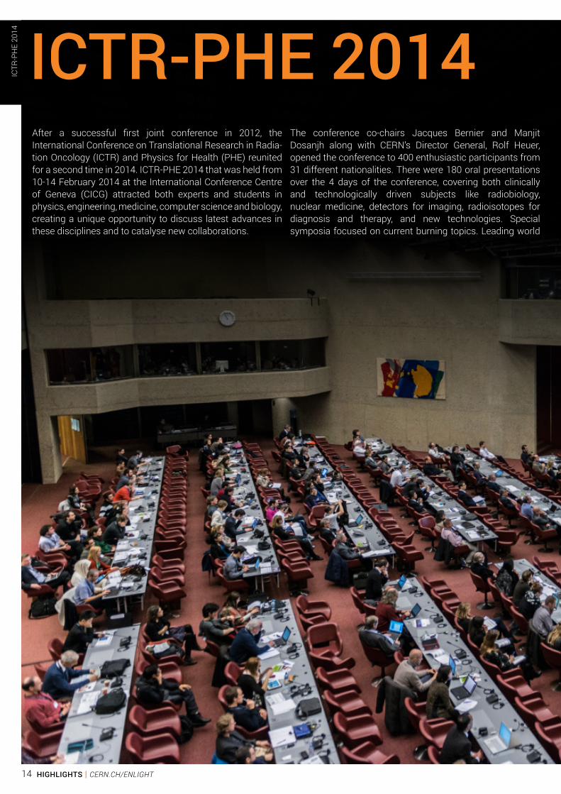
After a successful first joint conference in 2012, the International Conference on Translational Research in Radia-tion Oncology (ICTR) and Physics for Health (PHE) reunited for a second time in 2014. ICTR-PHE 2014 that was held from 10-14 February 2014 at the International Conference Centre of Geneva (CICG) attracted both experts and students in physics, engineering, medicine, computer science and biology, creating a unique opportunity to discuss latest advances in these disciplines and to catalyse new collaborations.
The conference co-chairs Jacques Bernier and Manjit Dosanjh along with CERN’s Director General, Rolf Heuer, opened the conference to 400 enthusiastic participants from 31 different nationalities. There were 180 oral presentations over the 4 days of the conference, covering both clinically and technologically driven subjects like radiobiology, nuclear medicine, detectors for imaging, radioisotopes for diagnosis and therapy, and new technologies. Special symposia focused on current burning topics. Leading world
ICTR-PHE 2014 ICTR
-PH
E 20
14
14 HIGHLIGHTS | CERN.CH/ENLIGHT
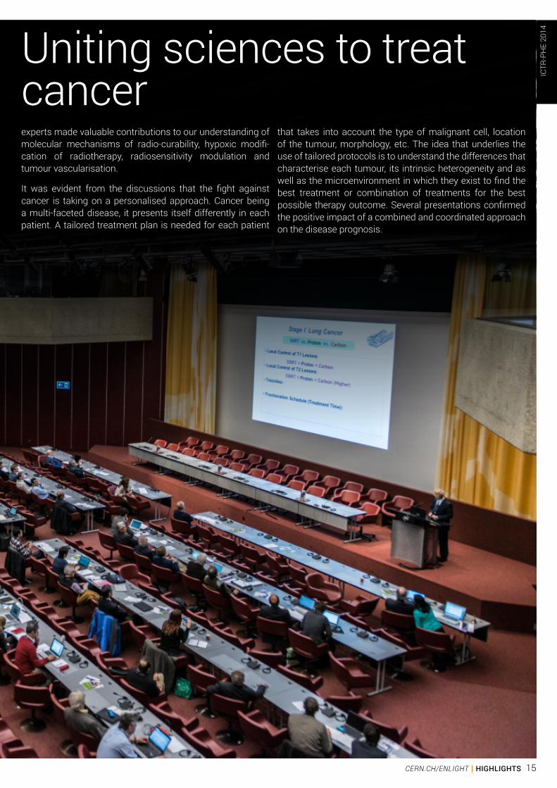
experts made valuable contributions to our understanding of molecular mechanisms of radio-curability, hypoxic modifi-cation of radiotherapy, radiosensitivity modulation and tumour vascularisation.
It was evident from the discussions that the fight against cancer is taking on a personalised approach. Cancer being a multi-faceted disease, it presents itself differently in each patient. A tailored treatment plan is needed for each patient
that takes into account the type of malignant cell, location of the tumour, morphology, etc. The idea that underlies the use of tailored protocols is to understand the differences that characterise each tumour, its intrinsic heterogeneity and as well as the microenvironment in which they exist to find the best treatment or combination of treatments for the best possible therapy outcome. Several presentations confirmed the positive impact of a combined and coordinated approach on the disease prognosis.
Uniting sciences to treat cancer
ICTR
-PH
E 20
14
15CERN.CH/ENLIGHT | HIGHLIGHTS
Conventional radiotherapy using a beam of photons has progressed many folds with the onset of techniques such as intensity modulation, volumetric arc techniques and image guided treatment. Never the less the specific ways in which ions deposit their energy (Bragg peak) makes ion therapy more precise in targeting tumours. There are many factors that influence treatment effectiveness including drugs, patient immunology, hypoxia rate and the inner nature of the tumour. For example, recent studies have shown that malignant cells infected with the HPV virus respond better to radiation. A through understanding of the biological effects of different ion species and a combination of different treatment options leads to better therapy, hence radiobiology plays an important role.
The effectiveness of therapy partly depends on how well the tumour is defined making imaging one of the major challenges. Imaging has reached a spatial resolution of 2mm. The resolution and precision of imaging is continuing to grow with the use of combined modalities like PET/CT and PET/MRI. These are important clinical breakthroughs as they combine modalities that capture the anatomical and functional aspects of tissue. Compared to PET/CT, PET/MRI machines reduce the dose of ionising radiation given to a patient. However, the challenge of operating a PET machine within a strong magnetic field was overcome. After 30 years of research, the challenge of operating a PET machine within a strong magnetic field was overcome.
An improvement in imaging also depends on more sophisticated software and algorithms to integrate the information collected.
For example, the anatomical data coming from CT or MRI imaging, including 4D acquisition, organ movement and deformation, is injected into the PET acquisition system. The reconstruction of the combined information from both systems can produce extremely accurate images. Software has other major applications including beam monitoring, dose distribution and treatment plans. Simulations made with GEANT4 and FLUKA are used to determine the most suitable treatment plan. The first 3D mapping of the dose distribution over a known distance along the entire length of a 62 MeV proton beam was presented. Such studies are extremely useful in assessing collateral damage caused by the therapeutic beam.
The role of radiochemistry cannot be ignored – both in imaging and in therapy. Different tumours require different isotopes as malignant cells react differently to different types of radiation. The same carrier molecules that bring radioisotopes to malignant cells making them detectable could be used with more powerful isotopes to carry a lethal dose to destroy cancer cells. The appropriate radionuclide should be identified to selectively reach the target while minimizing the effect on normal tissue. Specific peptides associated with isotopes obtained at particle detectors and innovative nanoparticles are being studied. A challenge in this area is the proximity of a particle accelerator where radioisotopes are produced before being transported to hospitals. The Proton Isotope Producer (PIP), an innovative accelerator design based on recirculation aims at overcoming the typical issues related to the installation and operation of cyclotrons in the medical environment.
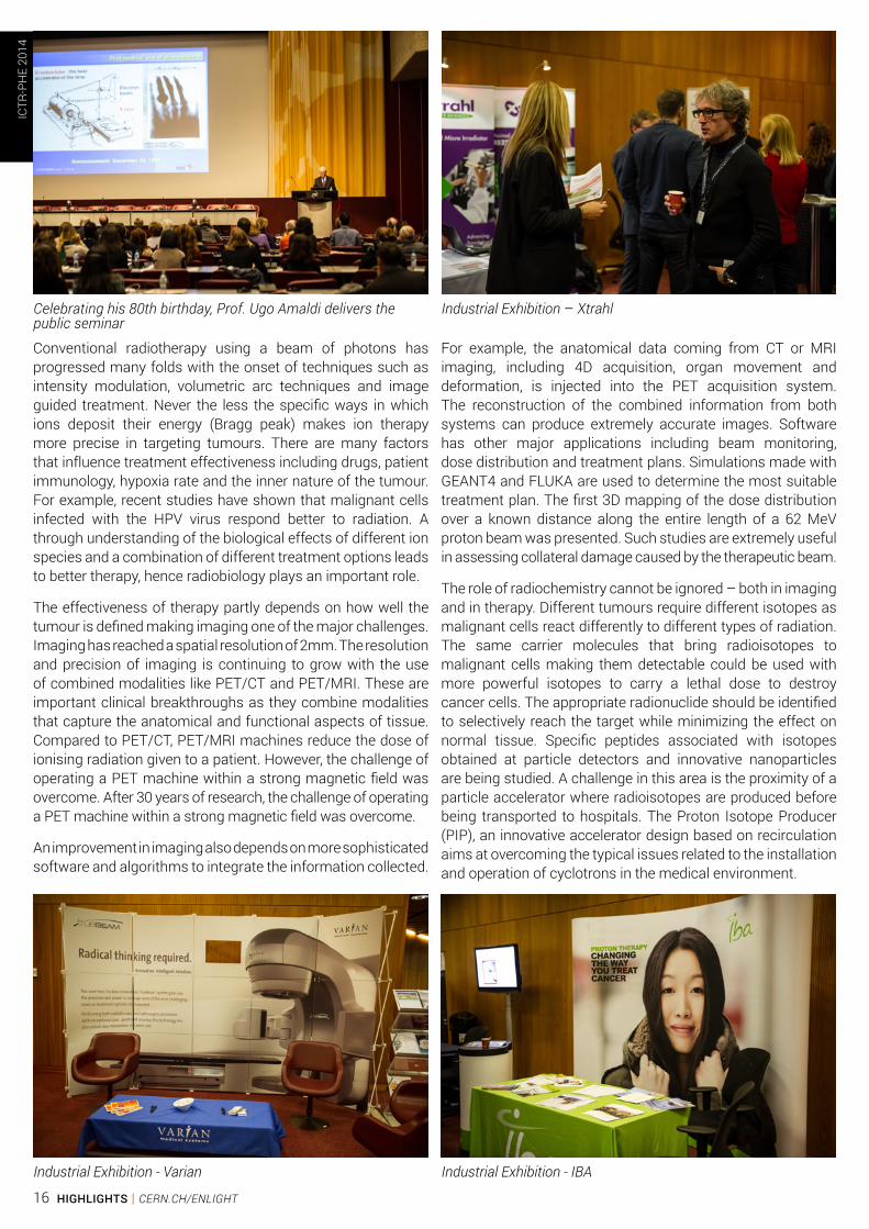
Celebrating his 80th birthday, Prof. Ugo Amaldi delivers the public seminar
Industrial Exhibition - Varian
Industrial Exhibition – Xtrahl
Industrial Exhibition - IBA
ICTR
-PH
E 20
14
16 HIGHLIGHTS | CERN.CH/ENLIGHT
Evidence-based medicine is the way forward. Each idea and method has to be tested and preclinical and clinical tests need to be performed. Proton beams have been used therapeutically for 60 years, and carbon ion beams for 20 years. Yet all phases of clinical trials have not been conducted. A panel discussion dedicated to address the topic of randomised clinical trials proved to be a very popular event of the conference. Topics being studied in trials include the integrated dose being delivered, fractionation and types of tumours being treated. The hurdles to overcome before a meaningful clinical trial is conducted are ethical issues and methodological concerns. Some tumour types being rare, data is scarce. Data confidentiality is a major concern especially when setting up multi-centre trials.
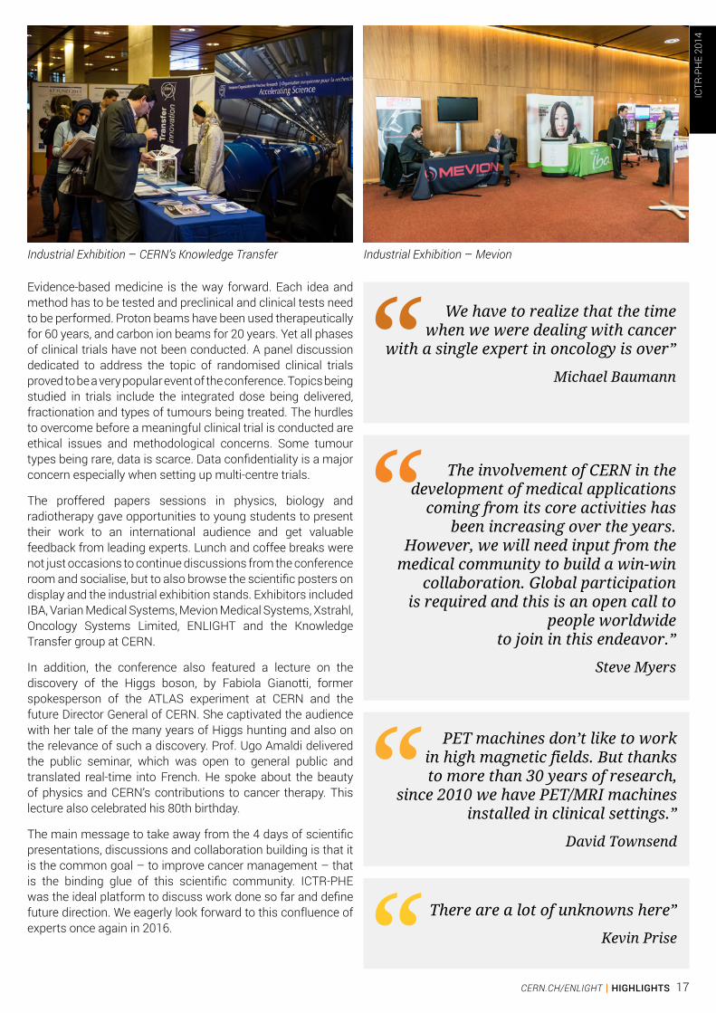
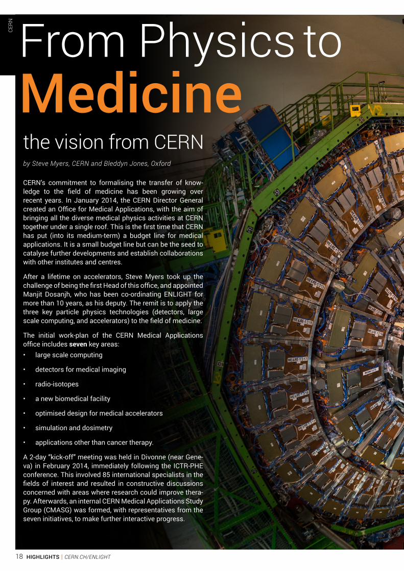
The proffered papers sessions in physics, biology and radiotherapy gave opportunities to young students to present their work to an international audience and get valuable feedback from leading experts. Lunch and coffee breaks were not just occasions to continue discussions from the conference room and socialise, but to also browse the scientific posters on display and the industrial exhibition stands. Exhibitors included IBA, Varian Medical Systems, Mevion Medical Systems, Xstrahl, Oncology Systems Limited, ENLIGHT and the Knowledge Transfer group at CERN.
In addition, the conference also featured a lecture on the discovery of the Higgs boson, by Fabiola Gianotti, former spokesperson of the ATLAS experiment at CERN and the future Director General of CERN. She captivated the audience with her tale of the many years of Higgs hunting and also on the relevance of such a discovery. Prof. Ugo Amaldi delivered the public seminar, which was open to general public and translated real-time into French. He spoke about the beauty of physics and CERN’s contributions to cancer therapy. This lecture also celebrated his 80th birthday.
The main message to take away from the 4 days of scientific presentations, discussions and collaboration building is that it is the common goal – to improve cancer management – that is the binding glue of this scientific community. ICTR-PHE was the ideal platform to discuss work done so far and define future direction. We eagerly look forward to this confluence of experts once again in 2016.
We have to realize that the time when we were dealing with cancer
with a single expert in oncology is over”
Michael Baumann
The involvement of CERN in the development of medical applications
coming from its core activities has been increasing over the years.
However, we will need input from the medical community to build a win-win
collaboration. Global participation is required and this is an open call to
people worldwide to join in this endeavor.”
Steve Myers
There are a lot of unknowns here”
Kevin Prise
PET machines don’t like to work in high magnetic fields. But thanks to more than 30 years of research,
since 2010 we have PET/MRI machines installed in clinical settings.”
David Townsend
Industrial Exhibition – CERN’s Knowledge Transfer Industrial Exhibition – Mevion
ICTR
-PH
E 20
14
17CERN.CH/ENLIGHT | HIGHLIGHTS
CERN’s commitment to formalising the transfer of know-ledge to the field of medicine has been growing over recent years. In January 2014, the CERN Director General created an Office for Medical Applications, with the aim of bringing all the diverse medical physics activities at CERN together under a single roof. This is the first time that CERN has put (into its medium-term) a budget line for medical applications. It is a small budget line but can be the seed to catalyse further developments and establish collaborations with other institutes and centres.
After a lifetime on accelerators, Steve Myers took up the challenge of being the first Head of this office, and appointed Manjit Dosanjh, who has been co-ordinating ENLIGHT for more than 10 years, as his deputy. The remit is to apply the three key particle physics technologies (detectors, large scale computing, and accelerators) to the field of medicine.
The initial work-plan of the CERN Medical Applications office includes seven key areas:• large scale computing
• detectors for medical imaging
• radio-isotopes
• a new biomedical facility
• optimised design for medical accelerators
• simulation and dosimetry
• applications other than cancer therapy.
A 2-day “kick-off” meeting was held in Divonne (near Gene-va) in February 2014, immediately following the ICTR-PHE conference. This involved 85 international specialists in the fields of interest and resulted in constructive discussions concerned with areas where research could improve thera-py. Afterwards, an internal CERN Medical Applications Study Group (CMASG) was formed, with representatives from the seven initiatives, to make further interactive progress.
From Physics to Medicine the vision from CERN
CERN
by Steve Myers, CERN and Bleddyn Jones, Oxford
18 HIGHLIGHTS | CERN.CH/ENLIGHT
CERN
19CERN.CH/ENLIGHT | HIGHLIGHTS
In summer 2014, an International Strategy Committee was established, with members being the most prominent experts from major institutes in Europe, USA, China and Japan. The first meeting was held at CERN in November 2014, and the second is foreseen in Brussels in April 2015. In the first meeting, the strategy for the seven initiatives was discussed at length and endorsed by the committee. A commitment was also made to lobby for funding for the most important and urgent initiatives.
Transferring technologies from particle physics to the health sector is a multidisciplinary venture. It is important that the wide scientific community and the general public are aware of the complexity and of the challenges lying ahead: for this reason, a series of Medical Applications Seminars was started.
LARGE SCALE COMPUTING, DATA STORAGE AND ANALYSIS
The reputation of CERN in data management and storage is first rate. The opportunities to use clusters and clouds for data storage and interpretation using a basket of models are considerable. The Zenodo (http://zenodo.org/) infrastructure already provides a secure and reliable digital data repository for a wide range of research communities who have adopted it as their primary means of sharing publications, data sets, and associated software. Zenodo supports a range of access policies that can be tailored according to legislative and ethical requirements. The cloud-based data analysis platform is currently being extended to incorporate services such as Zenodo, thus allowing researchers to execute complex data mining and analysis.
MEDICAL DETECTORS AND IMAGING
Over recent years, impressive progress continues in detection techniques for the large physics experiments of the future. The principle advances have been in fast timing, high flux
Data Acquisition (DAQ), and fast, complex, and detailed image reconstruction.
Hybrid pixel detectors were developed to record the position and arrival time of each track coming from the LHC particle detectors. The Medipix Collaborations have adapted the same technology to x-ray imaging. Unlike conventional x-ray imaging detectors, each x-ray photon is individually detected and the associated energy measurement allows the production of ‘colour’ x-ray images, with prospects of high resolution spectroscopic x-ray imaging.
The Crystal Clear Collaboration has been active in the research and development of inorganic scintillation materials for novel ionizing radiation detectors. Recent programmes include: the development of Time-Of-Flight PET devices; an endoscopic ultrasound probe coupled to a highly miniaturised PET module, for pancreatic and prostate cancer imaging; a multimodal PET-ultrasound system for breast imaging.
Further R&D on higher precision time analysis is now being undertaken for the LHC upgrade and for new facilities such as FAIR in Germany. It is clear that precise timing is important for medical devices with the TOF-PET imager as a clear example.
Another important application of these large data flows is visualisation during hadron therapy and constant on-line monitoring for correction of beam misalignments.
RADIOISOTOPE PRODUCTION
CERN has been hosting the Isolde facility for about 50 years. Over 1000 radioisotopes of more than 70 chemical elements have become available for fundamental and applied research. The technique of isotope mass-separation for medical isotope production will now be taken further in the new CERN-MEDICIS facility.
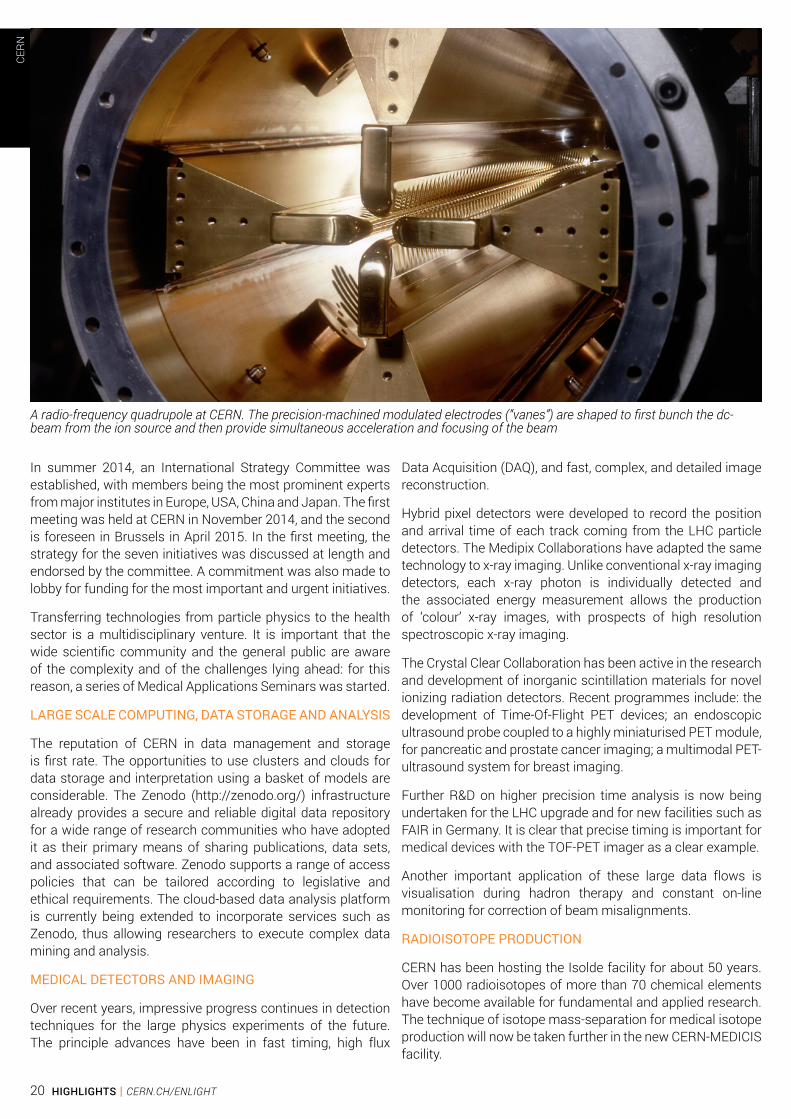
A radio-frequency quadrupole at CERN. The precision-machined modulated electrodes (“vanes”) are shaped to first bunch the dc-beam from the ion source and then provide simultaneous acceleration and focusing of the beam
CERN
20 HIGHLIGHTS | CERN.CH/ENLIGHT
A BIO-MEDICAL TEST-BED FACILITY: OPENMED
CERN has many accelerators that feed the LHC. The Low Energy Ion Ring (LEIR), supplies Lead Ions to the LHC injector chair for a period of one month per year. The beam energy, size and availability make LEIR an ideal candidate for conversion to a biomedical test-bed facility. The energy reach of the LEIR main magnet system is about 430 MeV/nucleon for ions with a charge over mass ratio of ½ as for example He2+, 12C6+ or 16O8+. Fairly minor modifications to LEIR would allow:• A horizontal beam line for particle energies of up to 400
MeV per nucleon, with up to 30 cm range in tissues for testing comparative particle ballistics for different ions, dosimetry and cellular responsiveness in humanoid phantoms.
• A vertical beam line with lower particle energies, as low as 75 MeV for in vitro work using larger cell numbers for radio-sensitising drug experiments.
• A pencil beam of 5-10 mm FWHM and broad beam of 5×5 cm are relatively easy options.
Adjacent to the LEIR hall, there is sufficient ground space of around 500 square metres (with 3-4 potential floors) - for a well-equipped biological laboratory for cell culture, bio-assays and analysis. This will be built to the best standards available elsewhere.
Also there would be scope for testing better beam delivery systems, improved dosimetry, and radiation detectors. Monte-Carlo simulation techniques, remote monitoring of dose deposition, proton radiography and tomography, pencilled and collimated beams at variable dose rates, advanced quality assurance measures and meeting the most demanding national standards for reference dose could all be pursued.
Thus LEIR would become an established facility with international users focussing on radiobiology, medical instrumentation, acceleration and beam delivery, diagnostics, and dosimetry, as well as basic physics studies such as nuclear fragmentation.
SPECIFICATION REQUIREMENTS FOR NEW ACCELERATORS AND BEAM DELIVERY SYSTEMS
The next generation of facilities for hadron therapy must be more compact, significantly more cost effective, and incorporate real time imaging, diagnostics and beam control.The ideal facility should:• Irradiate the tumour with minimal collateral radiation
outside the target volume, and be capable of tracking moving tumours. This may require simultaneous imaging and particle treatment such as “MR proton”.
• Be affordable: This means reduced installation and operating costs, with an increase in patient through-put.
• Have a reduced area footprint for hospital considerations, including gantries and switching magnet efficiency to different rooms.
The requirements on the beam delivery will be crucial. In particular:• Type(s) of particle(s)
• Beam dimensions, energies, required beam energies
• Beam scanning protocols, angular coverage (gantry), real-time feedback control of beam on tumour
• Real-time imaging requirements update rate, precision, and resolution.
Many of these requirements are inter-related. Some of these requirements may be obtained by consensus with the medical practitioners, but many will need theoretical developments and computer simulations followed by experimental testing in test-bed facilities such as the proposed OPENMED. Most importantly the final specifications will have a crucial impact on the type of accelerator and gantries to be used.
PHYSICAL AND RADIOBIOLOGICAL MODELLING
CERN has the correct expertise and capabilities to virtually simulate the entire process of particle beam therapy, from within the accelerator itself to within the patient and to the ultimate biological effects. The effectiveness of particle therapy will depend on improved knowledge of the links between the beam physics and detailed maps of energy absorption in the body, including linear energy transfer (LET), all translated into clinically relevant outcomes.
Detector modelling, accelerator studies for medical use, shielding design, radioisotopes production, and treatment planning for hadron therapy and nuclear medicine all require the use of computer simulations. Particle physics excels in the domain of Monte Carlo simulations with two main codes Geant4 and FLUKA.
The FLUKA Monte Carlo code has been already extensively used at HIT and CNAO for their treatment planning systems. The simulation code and tools are in a fairly advanced stage with the main line of development focusing on the implementation of simulations tools for Monte Carlo based Therapy Planning Systems (MCTPS) for hadron therapy, radiologic dosimetry, and for nuclear medicine.
APPLICATIONS OTHER THAN CANCER
Some focal clinical conditions are targeted by ablative therapy. These span from electrophysiological anomalies, such as epilepsy and cardiac arrhythmias, to syndromes such as Parkinson’s disease. In principle, selective ablation could be achieved with sophisticated external radiation sources.
During this first year of activity of the CERN Medical Applications office, the overall structure and several internal and external collaboration boards and committees have been set up. Some external funding has already been donated, and avenues are being explored for additional funding. A purposeful start has been made, and this momentum must be preserved with full international cooperation in order to make vital further progress in association with other Universities and Hospital facilities worldwide.
CERN
21CERN.CH/ENLIGHT | HIGHLIGHTS
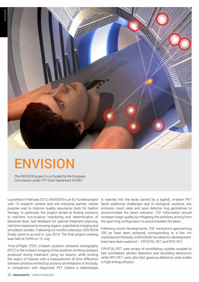
Launched in February 2010, ENVISION is an EC-funded project with 15 research centres and one industrial partner, whose purpose was to improve quality assurance tools for hadron therapy. In particular, the project aimed at finding solutions to real-time non-invasive monitoring and determination of delivered dose; fast feedback for optimal treatment planning, real-time response to moving organs, quantitative imaging and simulation studies. Following a 6 months extension, ENVISION finally came to an end in July 2014. The final project meeting was held at CERN on 10 July.
Time-of-flight (TOF) in-beam positron emission tomography (PET) is the in-beam imaging of the positron emitting isotopes produced during treatment using ion beams, while limiting the region of interest with a measurement of time difference between photons emitted by positron annihilations in the body. In comparison with diagnostic PET (where a radioisotope
is injected into the body carried by a ligand), in-beam PET faces additional challenges due to biological washout, low emission count rates and open detector ring geometries to accommodate the beam entrance. TOF information should increase image quality by mitigating the artefacts arising from the open-ring configuration to accommodate the beam.
Following recent developments, TOF resolutions approaching 200 ps have been achieved, corresponding to a few cm resolutions in the body. In ENVISION, two detector-development-lines have been explored – CRYSTAL-PET and RPC-PET.
CRYSTAL-PET uses arrays of scintillating crystals coupled to fast scintillation photon detectors and recording electronics while RPC-PET uses ultra fast gaseous detectors used widely in high-energy physics.
ENVISION The ENVISION project is co-funded by the EuropeanCommission under FP7 Grant Agreement 241851
ENVI
SIO
N
22 HIGHLIGHTS | CERN.CH/ENLIGHT

Single photon emission computed tomography (SPECT) is a technique that, similar to PET, uses a radioactive tracer material and detects gamma rays. However, the tracers used in SPECT emit gamma rays measured directly. SPECT scans while having a lower spatial resolution than PET are less expensive and can use longer-lived, more easily obtained radioisotopes. The ENVISION work package working on in-beam single particle tomography focuses mainly on the application of prompt gamma rays for in-vivo dosimetry (in-beam SPECT).
Current applications of in-vivo PET monitoring are limited to tumours in stationary parts of the body, or with reduced motion amplitude. To ensure safe and conformal irradiation, we would need methods of in-vivo treatment verification of a moving ion beam and a physiologically moving anatomy. Pioneering work is on-going on the integration of a prototype ultrasound tracking system in the PET imaging and dose delivery workflows, promising the possibility to provide more thorough and dose-free information on internal tumour motion. The biomechanical approach for motion compensated PET image reconstruction based on tetrahydral meshes and finite element modelling has been further developed and tested on clinical data.
Software was written to automatically detect deviations in treatment delivery in order to adapt subsequent treatment fractions to conform as closely as possible to the prescribed dose. Initial range assessments and positron distributions were performed by visual inspection, which was very time consuming. For an automated comparison of PET images, two approaches were developed in parallel. Both the approaches are based on the underlying idea that while the positron activity distributions are highly influenced by stochastic fluctuations, the distribution at the end of the particle track depends mainly on the ion type, energy and the composition of matter within the track. Both algorithms were validated on 12 in-beam PET studies; specificity and sensitivity of both methods were found
to be similar and comparable to visual inspections of in-beam PET data. Special phantoms have been constructed for detailed ion beam dosimetry studies.
Monte Carlo (MC) simulation is an essential tool in particle therapy and in image-based monitoring. Models are based on complex physics describing interactions of radiation with matter and are able to process intricate geometry of detectors, detailed description of patient anatomy, interfaces between different materials, and accurate transport. The management of tissue inhomogeneities is where MC simulations reveal their full strength. Transport codes can predict the production of radionuclides and the propagation and detection of secondary radiation. These predictions can be compared with measured data to assess the delivered treatment with the proposed treatment plan.
ENVISION activities capitalised on pre-existing developments in GEANT4/Gate, FLUKA and MCNPX and focused on three aspects: accuracy of the physics models used, enhanced code features to support complex geometries, ease of use of the code.
In the final year of the project, research was fast-paced in order to meet all the goals set. There were more than 20 peer-reviewed journal publications, and 15 conference proceedings by ENVISION members. Two PhD theses were submitted. A 3D animation was produced showing the layout and working of a typical particle therapy centre housing both protons and carbon ions.
Members of the ENVISION collaboration at the project’s final meeting
ENVI
SIO
N
23CERN.CH/ENLIGHT | HIGHLIGHTS
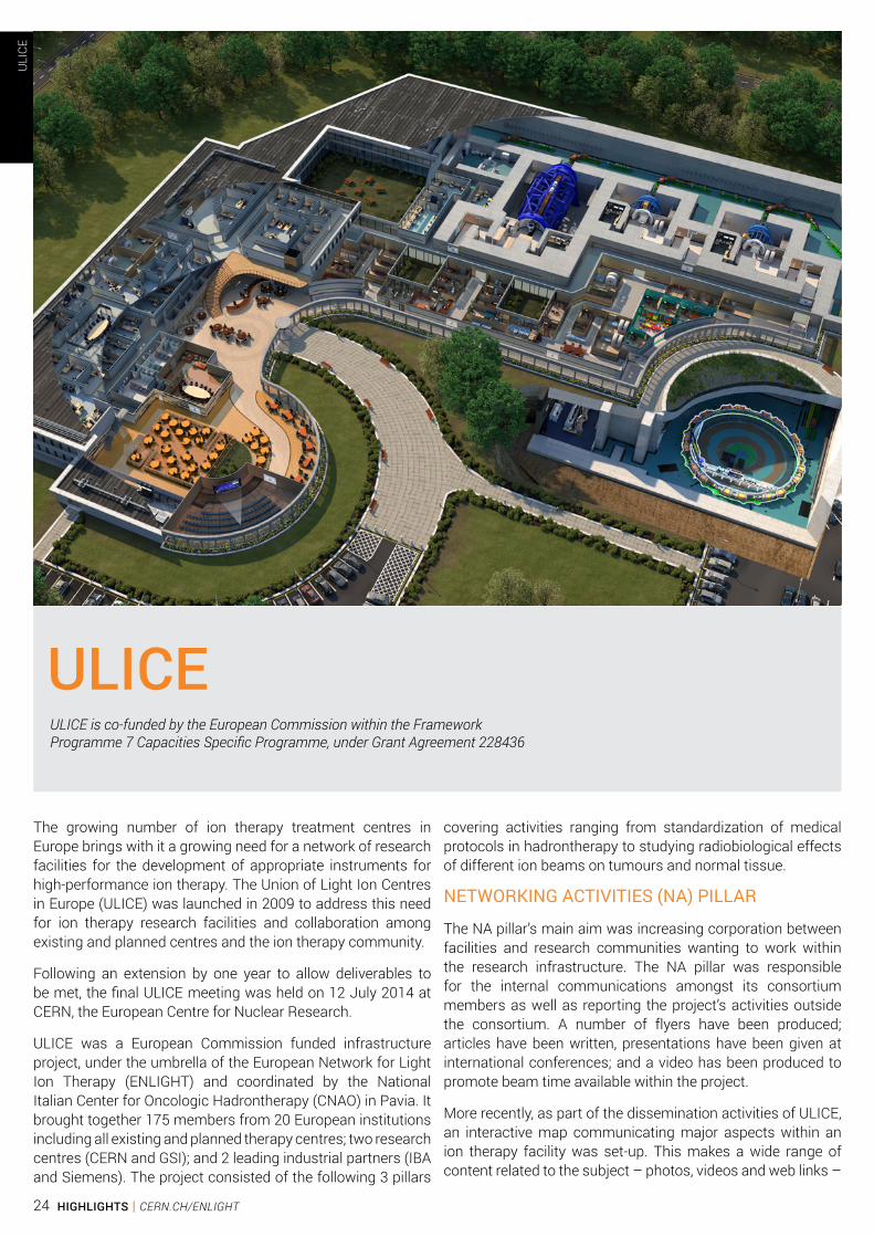
The growing number of ion therapy treatment centres in Europe brings with it a growing need for a network of research facilities for the development of appropriate instruments for high-performance ion therapy. The Union of Light Ion Centres in Europe (ULICE) was launched in 2009 to address this need for ion therapy research facilities and collaboration among existing and planned centres and the ion therapy community.
Following an extension by one year to allow deliverables to be met, the final ULICE meeting was held on 12 July 2014 at CERN, the European Centre for Nuclear Research.
ULICE was a European Commission funded infrastructure project, under the umbrella of the European Network for Light Ion Therapy (ENLIGHT) and coordinated by the National Italian Center for Oncologic Hadrontherapy (CNAO) in Pavia. It brought together 175 members from 20 European institutions including all existing and planned therapy centres; two research centres (CERN and GSI); and 2 leading industrial partners (IBA and Siemens). The project consisted of the following 3 pillars
covering activities ranging from standardization of medical protocols in hadrontherapy to studying radiobiological effects of different ion beams on tumours and normal tissue.
NETWORKING ACTIVITIES (NA) PILLAR
The NA pillar’s main aim was increasing corporation between facilities and research communities wanting to work within the research infrastructure. The NA pillar was responsible for the internal communications amongst its consortium members as well as reporting the project’s activities outside the consortium. A number of flyers have been produced; articles have been written, presentations have been given at international conferences; and a video has been produced to promote beam time available within the project.
More recently, as part of the dissemination activities of ULICE, an interactive map communicating major aspects within an ion therapy facility was set-up. This makes a wide range of content related to the subject – photos, videos and web links –
ULICEULICE is co-funded by the European Commission within the FrameworkProgramme 7 Capacities Specific Programme, under Grant Agreement 228436
ULI
CE
24 HIGHLIGHTS | CERN.CH/ENLIGHT

accessible in an intuitive way. The map can be found at: www.cern.ch/virtual-hadron-therapy-centre
A Training and Education Committee (TEC) was set up in 2009 with a representative from CERN, CNAO and ESTRO, to provide travel grants to attend courses and workshops and to facilitate exchange visits.
Some of the courses covered within the ULICE framework included:
1. Radiotherapy with protons and ions held in Uppsala, Sweden in March 2012. Among the 56 participants, 3 were junior scientists from the consortium supported by ULICE grants.
2. Radiotherapy with protons and ions held in CNAO, Italy in March 2013. There were 58 participants, 4 of them were junior scientists from the consortium supported by ULICE grants.
3. Clinical practice and implementation of image guided stereotactic body radiotherapy held at Wurzburg, Germany in Sept 2012.
The course on radiotherapy with protons and ions provided knowledge on particle treatment planning, dosimetry, treatment delivery, and latest technological developments; and targeted radiation oncologists and radiation physicists while being open to general public.
The NA pillar was also responsible for organizing 5 annual workshops at different locations around Europe, which were attended by over 100 participants each year, bringing together the ULICE consortium and other members of the ENLIGHT community.
TRANS-NATIONAL ACCESS (TNA) PILLAR
The TNA pillar took on a 2-step approach and was responsible for providing beamtime using a combination of pre-defined clinical trial programmes to allow researchers to visit the facility, and radiobiological and physics experiments to take place.
Within the ULICE project, there were 2 access providers – CNAO in Italy and the Heidelberg Ion-Beam Therapy Centre (HIT) in Germany. CNAO has been operational since 2010 with 3 treatment rooms and 1 experimental room. It has a synchrotron for protons (60-250 MeV) and carbon ions (400 MeV/n).
During the last phase of the project, the budgeted number of hours was provided mainly for preclinical research projects.
Beamtime was promoted in several ways: through a video which was shown at main scientific events related to ion therapy, publications, announcements on the project’s public website, communications to International Atomic Energy Agency (IAEA) and others. The coordinator via several
Members of the ULICE collaboration at the project’s final meeting
ULI
CE
25CERN.CH/ENLIGHT | HIGHLIGHTS

messages to beneficiaries communicated availability of beamtime for research activities within the consortium. Ten applications were received and evaluated by a selection panel. In order to guarantee transparency in the selection procedure, the selection panel had both internal and external members from the two facilities providing access – HIT and CNAO.
In 2012, HIT began the transnational access activities providing beam time to two groups of researchers who had been working on detectors for radiation. Research groups consisted of young researchers of 7 different nationalities brought together by ULICE. The last phase of the project saw many research groups visit CNAO from January to August to use beamtime. Their main goal was to test the instruments they were developing, to gather hands-on experience in radiation measurements, and to develop long-lasting networks with experts in radiation detection around the world. Both proton and carbon ion beams were used for more than 35 hours.
It is worth noting that CNAO only recently received authorization for the clinical use of the synchrotron, consequently the clinical routine procedure was only initiated at the beginning of 2014. Despite the strict patient enrolling criteria CNAO had the
Treating patients 12h (08:00-20:00)
QA 8h (24:00-08:00)
TNA 4h (20:00-24:00)
Beam time for TNA at CNAO
on weekdays (24h on weekends)
possibility to test the referral of patients from outside Italy through some foreign patients coming from France and United Kingdom.
The gantry at HIT is 3 stories high, 25 metres long, 13 metres in diametre and weighs 670 tonnes (photo courtesy of HIT)
ULI
CE
26 HIGHLIGHTS | CERN.CH/ENLIGHT
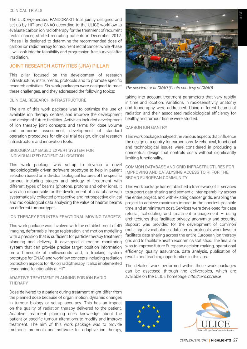
CLINICAL TRIALS
The ULICE-generated PANDORA-01 trial, jointly designed and set-up by HIT and CNAO according to the ULICE-workflow to evaluate carbon ion radiotherapy for the treatment of recurrent rectal cancer, started recruiting patients in December 2012. Phase I is designed to determine the recommended dose of carbon ion radiotherapy for recurrent rectal cancer, while Phase II will look into the feasibility and progression-free survival after irradiation.
JOINT RESEARCH ACTIVITIES (JRA) PILLAR
This pillar focused on the development of research infrastructure, instruments, protocols and to promote specific research activities. Six work packages were designed to meet these challenges, and they addressed the following topics:
CLINICAL RESEARCH INFRASTRUCTURE
The aim of this work package was to optimize the use of available ion therapy centres and improve the development and design of future facilities. Activities included development of ion therapy joint concepts and terms for dose volume and outcome assessment, development of standard operation procedures for clinical trial design, clinical research infrastructure and innovation tools.
BIOLOGICALLY BASED EXPERT SYSTEM FOR INDIVIDUALIZED PATIENT ALLOCATION
This work package was set-up to develop a novel radiobiologically-driven software prototype to help in patient selection based on individual biological features of the specific tumour, including stages and biology of treatment with different types of beams (photons, protons and other ions). It was also responsible for the development of a database with systematically collected prospective and retrospective clinical and radiobiological data analysing the value of hadron beams on different tumour types.
ION THERAPY FOR INTRA-FRACTIONAL MOVING TARGETS
This work package was involved with the establishment of 4D imaging, deformable image registration, and motion modelling with a precision that is sufficient for particle therapy treatment planning and delivery. It developed a motion monitoring system that can provide precise target position information on a timescale of milliseconds and, a tracking system prototype for CNAO and workflow concepts including radiation protection aspects for 4D ion radiotherapy. It also implemented rescanning functionality at HIT.
ADAPTIVE TREATMENT PLANNING FOR ION RADIO THERAPY
Dose delivered to a patient during treatment might differ from the planned dose because of organ motion, dynamic changes in tumour biology or set-up accuracy. This has an impact on the quality of radiation therapy delivered to the patient. Adaptive treatment planning uses knowledge about the patient or specific tumour alterations to modify and improve treatment. The aim of this work package was to provide methods, protocols and software for adaptive ion therapy,
taking into account treatment parameters that vary rapidly in time and location. Variations in radiosensitivity, anatomy and topography were addressed. Using different beams of radiation and their associated radiobiological efficiency for healthy and tumour tissue were studied.
CARBON ION GANTRY
This work package analysed the various aspects that influence the design of a gantry for carbon ions. Mechanical, functional and technological issues were considered in producing a conceptual design that controls costs without significantly limiting functionality.
COMMON DATABASE AND GRID INFRASTRUCTURES FOR IMPROVING AND CATALYSING ACCESS TO RI FOR THE BROAD EUROPEAN COMMUNITY
This work package has established a framework of IT services to support data sharing and semantic inter-operability across the entire project, and with existing cancer grids, enabling the project to achieve maximum impact in the shortest possible time, and at minimum cost. Services were developed for case referral, scheduling and treatment management – using architectures that facilitate privacy, anonymity and security. Support was provided for the development of common multilingual vocabularies, data items, protocols, workflows to facilitate data sharing across the entire European ion therapy grid and to facilitate health economics statistics. The final aim was to improve future European decision making, operational efficiency, quality assurance, data analysis, publication of results and teaching opportunities in this area.
The detailed work performed within these work packages can be assessed through the deliverables, which are available on the ULICE homepage: http://cern.ch/ulice
The accelerator at CNAO (Photo courtesy of CNAO)
ULI
CE
27CERN.CH/ENLIGHT | HIGHLIGHTS
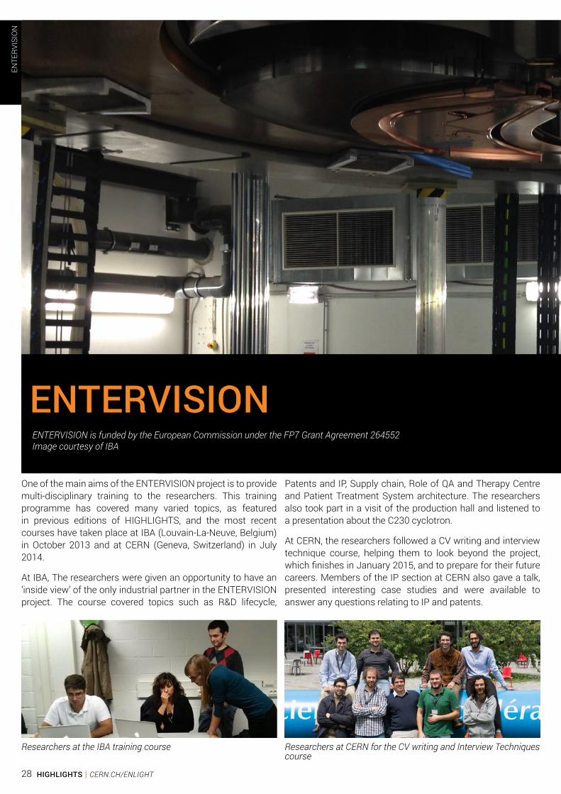
One of the main aims of the ENTERVISION project is to provide multi-disciplinary training to the researchers. This training programme has covered many varied topics, as featured in previous editions of HIGHLIGHTS, and the most recent courses have taken place at IBA (Louvain-La-Neuve, Belgium) in October 2013 and at CERN (Geneva, Switzerland) in July 2014.
At IBA, The researchers were given an opportunity to have an ‘inside view’ of the only industrial partner in the ENTERVISION project. The course covered topics such as R&D lifecycle,
Patents and IP, Supply chain, Role of QA and Therapy Centre and Patient Treatment System architecture. The researchers also took part in a visit of the production hall and listened to a presentation about the C230 cyclotron.
At CERN, the researchers followed a CV writing and interview technique course, helping them to look beyond the project, which finishes in January 2015, and to prepare for their future careers. Members of the IP section at CERN also gave a talk, presented interesting case studies and were available to answer any questions relating to IP and patents.
ENTERVISIONENTERVISION is funded by the European Commission under the FP7 Grant Agreement 264552Image courtesy of IBA
Researchers at the IBA training course Researchers at CERN for the CV writing and Interview Techniques course
ENTE
RVIS
ION
28 HIGHLIGHTS | CERN.CH/ENLIGHT
Hopefully, the training that the researchers have undertaken during their time in the ENTERVISON project will have enhanced their skills portfolio and will make them highly employable scientists with not only the necessary technical skills but also valuable ‘soft’ skills.
The course at CERN was followed by the annual ENTERVISION meeting along with the final meetings for both ENVISION and ULICE. Two of the poster prizes awarded at the meetings were won by ENTERVISION researchers. Congratulations to Thiago and Joakim!
The researchers are lucky enough to have a generous travel allowance and have attended conferences and workshops throughout the year travelling as far as Japan, China and Korea. Many of them are gaining valuable experience presenting posters and publications. At the ICTR-PHE held in February 2014, 4 of them presented posters and 6 gave oral presentations.
We look forward to meeting in Rome in January 2015 where the researchers will share the progress of their research projects and we will of course take the time to celebrate together the end of another successful project.
ENTE
RVIS
ION
29CERN.CH/ENLIGHT | HIGHLIGHTS

The development of clinical treatment protocols for conven-tional photon and charged particle radiation therapy is depen-dent on the availability of information on the biological efficacy of radiation doses. For this reason, it is important to be able to verify the biological effects of complex dose distributions in homeomorphic phantoms, alongside measurements of the physical dose. With that in mind, one of the ENTERVISION work packages was designed to focus on the development and testing of a phantom for biological experiments.
Biological experiments aim to replicate certain conditions in a reproducible manner; therefore an ideal phantom would be able to reduce the different sources of uncertainties in existing systems. A comparison study was performed to evaluate different commercial and in-house solution phantoms available for dosimetric and/or biological purposes, but it was concluded that in order to achieve the project’s goal, more than one type of phantom would be required. It was found that the
uncertainties are both related to dosimetry (physical output) and the statistics of the biological output; the dosimetric uncertainties can explain the large error bars related to the reported dose; and separately, the statistical uncertainties as documented by the error bars associated with the reported cell survival.
The positive traits from these studied phantoms in respect to their structures, detectors, and materials were subsequently gathered to develop the ENTERVISION design. The phantom design includes FLUKA Monte Carlo dosimetry studies, which focus on density disturbance effects in the region of the cells compartment from objects located in the beam path, such as detectors and other inhomogeneities. Once the design was evaluated and finalised, the phantom was constructed, and subsequently modified at different stages by the Istituto de Fisica at Turin University in cooperation with INFN.
The ENTERVISION Biological Dosimetric Phantom
ENTE
RVIS
ION
by Thiago Viana Miranda Lima, ENTERVISION researcher
30 HIGHLIGHTS | CERN.CH/ENLIGHT

The ENTERVISION phantom consists of Polymethyl methacrylate (PMMA) slabs, which were precisely machined to accommodate standard multi-well plates or T-25 cell flasks and different types of detectors. PMMA was chosen as a suitable material due to its similar properties to water so as to enable direct dose-to-water conversions.
The phantom’s design enables on-line data acquisition of the requested Spread-Out Bragg Peak at the time of cell irradiation. It can also be used for beam profiling and treatment plan verification purposes as it gives two options: the first focuses on the physical properties of the delivered beam, while the second offers information on the delivered biological dose.
The main benefits of the ENTERVISION phantom include its improved dosimetry, which reduces uncertainties related to reported dose; its cell irradiation orientation, which allows for cell irradiation positions in terms of dose and Linear Energy Transfer (LET) distribution with depth as in a clinical treatment, and for multiple simultaneous irradiations to be performed on the same day without the need for sterilisation of equipment, thereby reducing uncertainties due to delayed cell preparation. The phantom also allows for a more accurate Relative biological effectiveness RBE estimation with more precise confidence limits by application of a more formal statistical method, which takes into account the improved physical and biological estimations
The ultimate aim of the ENTERVISION phantom is to provide a dosimetric tool for studying biological dose with LET and dose in the delivered beam. It can be used to assess the biological effects from different radiation types and evaluate the performance of different treatment planning software (TPS) systems and technical variations of dose delivery systems and for inter-comparisons between different treatment centres.
Under the ENLIGHT and its Transnational Access to Research programme, the phantom was able to be irradiated at both the Italian National Centre for Hadrontherapy (CNAO) and the Heidelberg Ion-Beam Therapy Centre (HIT) facilities with protons and carbon ions so as to evaluate its performance and reproducibility. At both centres, the phantom followed the patient pathway: imaging, treatment planning and irradiation. For dosimetry, ionisation-chambers were used in order to obtain a live display of the beam profile and accumulated final absorbed dose. The results were then compared with both the TPS calculations and MC simulations. Cell culture irradiation and clonogenic survival assay procedures were also performed to assess the biological effect of the ionising radiation in respect of given dose and LET. The results are currently being evaluated to establish the phantom’s dosimetric and biological characteristics and its overall ability to reduce statistical uncertainties. Some of the preliminary results can be found under the ENTERVISION deliverable number 8.
HIT R & D Bunker
Treatment room at HIT
Treatment room at CNAO
ENTE
RVIS
ION
31CERN.CH/ENLIGHT | HIGHLIGHTS

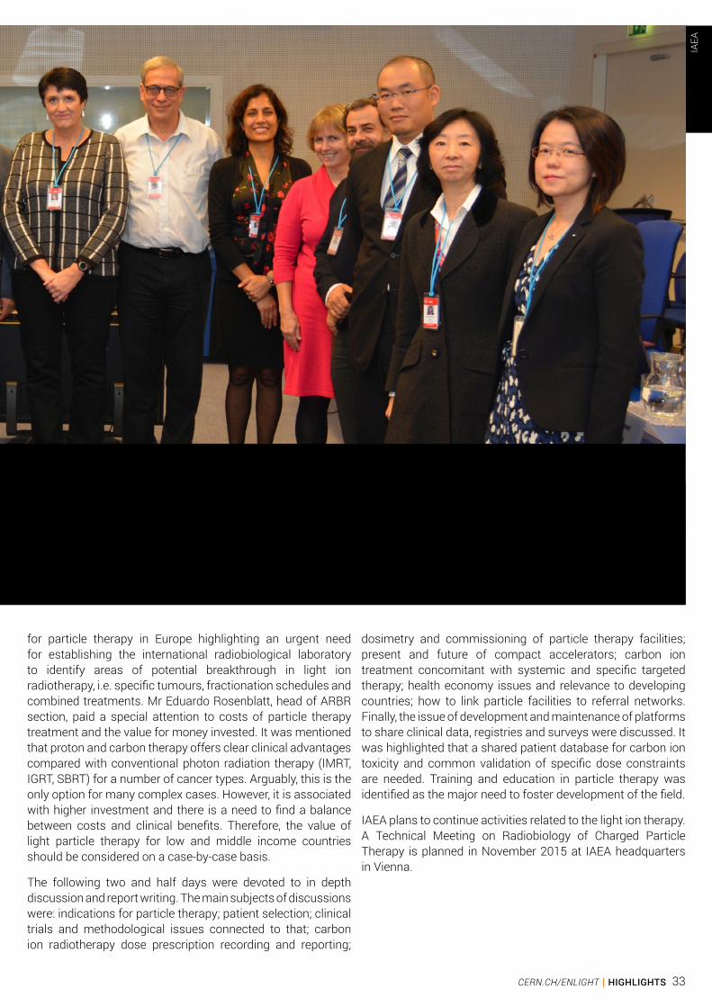
An International Atomic Energy Agency (IAEA) Consultants Meeting on Particle Therapy with particular attention to middle and low income countries took place on 11-14 November 2014. It was organised by the Section of Applied Radiation Biology and Radiotherapy (ARBR). The meeting brought together 11 experts from IAEA, CERN, Austria, Belgium, France, Germany, Italy, Japan, and USA to discuss the state of the art of light ion therapy and future trends in the development of this fast growing field.
The meeting was opened by the Director of the IAEA Division of Human Health May Abdel-Wahab and the Head of Dosimetry and Medical Radiation Physics Section Ahmed Meghzifene.
The first part of the meeting was devoted to physical (Alejandro Mazal, France) and radiobiological aspects of particle therapy (Oleg Belyakov, IAEA), implementation of light ion beam therapy facilities (Stan Vatnisky, MedAustron, Austria), clinical and operational aspects of proton beam delivery (Zelig Tochner, USA), epidemiology of ion beam therapy (Ramona Mayer, MedAustron, Austria) and evolving role of proton therapy for paediatric patients (Anita Mahajan, USA). It was followed by
a report on recent progress in carbon ion radiotherapy by Kumiko Karasawa from the National Institute of Radiological Sciences (NIRS), Chiba, Japan with particular attention to treatment of bone and soft tissue sarcomas (Reiko Imai, IAEA/NIRS). The current status of carbon ion radiotherapy in Gunma University, Japan was reviewed by Hiroyuki Katoh. Discussion on the potential role of high LET particle therapy in the multidisciplinary cancer care system lead by Piero Fossati, Italy, who revealed the need for cooperative research and planned structural investments. The topic of current challenges in particle beam therapy in Germany, it terms of perspectives for radiation oncology (Beate Timmerman), finalised the first day of the meeting.
During the second day the history and the future of proton therapy was elaborated by Yves Jongen, Chief Research Officer of the IBA. It was highlighted that, contrary to common opinion, the modern proton accelerator is not the most expensive part of the treatment system. The gantry and treatment room equipment would be a major investment. Manjit Dosanjh reported the progress of CERN and the ENLIGHT programme
IAEA CONSULTANTS MEETINGon Particle Therapy in the 21st Century:Relevance to Developing Countries, IAEA headquarters, Vienna, Austriaby Oleg BELYAKOV, International Atomic Energy Agency
IAEA
32 HIGHLIGHTS | CERN.CH/ENLIGHT
IAEA CONSULTANTS MEETINGon Particle Therapy in the 21st Century:
for particle therapy in Europe highlighting an urgent need for establishing the international radiobiological laboratory to identify areas of potential breakthrough in light ion radiotherapy, i.e. specific tumours, fractionation schedules and combined treatments. Mr Eduardo Rosenblatt, head of ARBR section, paid a special attention to costs of particle therapy treatment and the value for money invested. It was mentioned that proton and carbon therapy offers clear clinical advantages compared with conventional photon radiation therapy (IMRT, IGRT, SBRT) for a number of cancer types. Arguably, this is the only option for many complex cases. However, it is associated with higher investment and there is a need to find a balance between costs and clinical benefits. Therefore, the value of light particle therapy for low and middle income countries should be considered on a case-by-case basis.
The following two and half days were devoted to in depth discussion and report writing. The main subjects of discussions were: indications for particle therapy; patient selection; clinical trials and methodological issues connected to that; carbon ion radiotherapy dose prescription recording and reporting;
dosimetry and commissioning of particle therapy facilities; present and future of compact accelerators; carbon ion treatment concomitant with systemic and specific targeted therapy; health economy issues and relevance to developing countries; how to link particle facilities to referral networks. Finally, the issue of development and maintenance of platforms to share clinical data, registries and surveys were discussed. It was highlighted that a shared patient database for carbon ion toxicity and common validation of specific dose constraints are needed. Training and education in particle therapy was identified as the major need to foster development of the field.
IAEA plans to continue activities related to the light ion therapy. A Technical Meeting on Radiobiology of Charged Particle Therapy is planned in November 2015 at IAEA headquarters in Vienna.
IAEA
33CERN.CH/ENLIGHT | HIGHLIGHTS

ART
& PH
YSIC
S
34 HIGHLIGHTS | CERN.CH/ENLIGHT

ART
& PH
YSIC
S
Contributing artist: Grigory Fiofilov35CERN.CH/ENLIGHT | HIGHLIGHTS
Related Documents