Method High-throughput semiquantitative analysis of insertional mutations in heterogeneous tumors Marco J. Koudijs, 1,6,9 Christiaan Klijn, 1,9 Louise van der Weyden, 2 Jaap Kool, 3,7 Jelle ten Hoeve, 1 Daoud Sie, 4,8 Pramudita R. Prasetyanti, 1 Eva Schut, 1 Sjors Kas, 1 Theodore Whipp, 2 Edwin Cuppen, 5 Lodewyk Wessels, 1,10 David J. Adams, 2,10 and Jos Jonkers 1,10 1 Division of Molecular Biology and Cancer Systems Biology Center, Netherlands Cancer Institute, 1066 CX Amsterdam, The Netherlands; 2 Experimental Cancer Genetics, The Wellcome Trust Sanger Institute, Hinxton, Cambridge CB10 1SA, United Kingdom; 3 Division of Molecular Genetics, Netherlands Cancer Institute, 1066 CX Amsterdam, The Netherlands; 4 Central Microarray Facility, Netherlands Cancer Institute, 1066 CX Amsterdam, The Netherlands; 5 Hubrecht Institute and University Medical Center Utrecht, Cancer Genomics Center, 3584 CG Utrecht, The Netherlands Retroviral and transposon-based insertional mutagenesis (IM) screens are widely used for cancer gene discovery in mice. Exploiting the full potential of IM screens requires methods for high-throughput sequencing and mapping of transposon and retroviral insertion sites. Current protocols are based on ligation-mediated PCR amplification of junction fragments from restriction endonuclease-digested genomic DNA, resulting in amplification biases due to uneven genomic dis- tribution of restriction enzyme recognition sites. Consequently, sequence coverage cannot be used to assess the clon- ality of individual insertions. We have developed a novel method, called shear-splink, for the semiquantitative high- throughput analysis of insertional mutations. Shear-splink employs random fragmentation of genomic DNA, which reduces unwanted amplification biases. Additionally, shear-splink enables us to assess clonality of individual insertions by determining the number of unique ligation points (LPs) between the adapter and genomic DNA. This parameter serves as a semiquantitative measure of the relative clonality of individual insertions within heterogeneous tumors. Mixing experiments with clonal cell lines derived from mouse mammary tumor virus (MMTV)-induced tumors showed that shear-splink enables the semiquantitative assessment of the clonality of MMTV insertions. Further, shear-splink analysis of 16 MMTV- and 127 Sleeping Beauty (SB)–induced tumors showed enrichment for cancer-relevant insertions by exclusion of irrelevant background insertions marked by single LPs, thereby facilitating the discovery of candidate cancer genes. To fully exploit the use of the shear-splink method, we set up the Insertional Mutagenesis Database (iMDB), offering a publicly available web-based application to analyze both retroviral- and transposon-based insertional mu- tagenesis data. [Supplemental material is available for this article.] Transposons and retroviruses are widely used in insertional mu- tagenesis (IM) screens in mice to discover candidate cancer genes (Collier et al. 2005; Dupuy et al. 2005, 2009; Theodorou et al. 2007; Keng et al. 2009; Starr et al. 2009; Copeland and Jenkins 2010; Rad et al. 2010). IM screens have also been instrumental for the identi- fication of genetic interactions between genes driving tumor evo- lution (Uren et al. 2008; Kool and Berns 2009; Kool et al. 2010) and for the identification of genes that confer resistance to anticancer drugs (Lauchle et al. 2009) or pathogens (Carette et al. 2009). Finally, retroviruses and transposons are used for germline mutagenesis in a range of experimental organisms (Amsterdam et al. 1999; Golling et al. 2002; Ding et al. 2005; Keng et al. 2005; de Wit et al. 2010). In transposon- or retrovirus-induced tumors, efficient isola- tion of insertion sites is required for the effective identification of candidate cancer genes and genetic interactions between these genes. Insertion sites are typically analyzed by high-throughput sequencing and mapping of PCR-amplified junction fragments from integrated transposons or retroviruses (Largaespada and Collier 2008; Uren et al. 2009). Genomic loci that are found to contain insertional mutations in multiple independent samples are termed common insertion sites (CISs) and are likely to be causally implicated in tu- morigenesis. During the multistep process of tumorigenesis, se- quential insertions in cancer-relevant loci will result in the for- mation of cells with increasingly malignant potential (Fig. 1A). Insertional mutations may drive tumorigenesis when they confer a selective advantage to the affected cell such that it expands clonally to give rise to a significant proportion of the tumor mass. To understand how individual insertions contribute to tumori- genesis, clonal, and near-clonal insertion events should be dis- tinguished from background mutations that did not give rise to clonal expansion and are, therefore, present in a single cell, or a few cells, within the tumor. Methods that allow for (semi)- Present addresses: 6 University Medical Center Utrecht, Division of Experimental Oncology, 3584 CG Utrecht, The Netherlands; 7 Intervet Innovation GmbH, Zur Propstei, 55270 Schwabenheim, Germany; 8 VU Medical Center, 1007 MB Amsterdam, The Netherlands. 9 These authors contributed equally to this work. 10 Corresponding authors. E-mail [email protected]. E-mail [email protected]. E-mail [email protected]. Article published online before print. Article, supplemental material, and pub- lication date are at http://www.genome.org/cgi/doi/10.1101/gr.112763.110. 21:2181–2189 Ó 2011 by Cold Spring Harbor Laboratory Press; ISSN 1088-9051/11; www.genome.org Genome Research 2181 www.genome.org Cold Spring Harbor Laboratory Press on March 26, 2016 - Published by genome.cshlp.org Downloaded from

Welcome message from author
This document is posted to help you gain knowledge. Please leave a comment to let me know what you think about it! Share it to your friends and learn new things together.
Transcript

Method
High-throughput semiquantitative analysisof insertional mutations in heterogeneous tumorsMarco J. Koudijs,1,6,9 Christiaan Klijn,1,9 Louise van der Weyden,2 Jaap Kool,3,7
Jelle ten Hoeve,1 Daoud Sie,4,8 Pramudita R. Prasetyanti,1 Eva Schut,1 Sjors Kas,1
Theodore Whipp,2 Edwin Cuppen,5 Lodewyk Wessels,1,10 David J. Adams,2,10
and Jos Jonkers1,10
1Division of Molecular Biology and Cancer Systems Biology Center, Netherlands Cancer Institute, 1066 CX Amsterdam,
The Netherlands; 2Experimental Cancer Genetics, The Wellcome Trust Sanger Institute, Hinxton, Cambridge CB10 1SA, United
Kingdom; 3Division of Molecular Genetics, Netherlands Cancer Institute, 1066 CX Amsterdam, The Netherlands; 4Central Microarray
Facility, Netherlands Cancer Institute, 1066 CX Amsterdam, The Netherlands; 5Hubrecht Institute and University Medical Center
Utrecht, Cancer Genomics Center, 3584 CG Utrecht, The Netherlands
Retroviral and transposon-based insertional mutagenesis (IM) screens are widely used for cancer gene discovery in mice.Exploiting the full potential of IM screens requires methods for high-throughput sequencing and mapping of transposonand retroviral insertion sites. Current protocols are based on ligation-mediated PCR amplification of junction fragmentsfrom restriction endonuclease-digested genomic DNA, resulting in amplification biases due to uneven genomic dis-tribution of restriction enzyme recognition sites. Consequently, sequence coverage cannot be used to assess the clon-ality of individual insertions. We have developed a novel method, called shear-splink, for the semiquantitative high-throughput analysis of insertional mutations. Shear-splink employs random fragmentation of genomic DNA, whichreduces unwanted amplification biases. Additionally, shear-splink enables us to assess clonality of individual insertionsby determining the number of unique ligation points (LPs) between the adapter and genomic DNA. This parameterserves as a semiquantitative measure of the relative clonality of individual insertions within heterogeneous tumors.Mixing experiments with clonal cell lines derived from mouse mammary tumor virus (MMTV)-induced tumors showedthat shear-splink enables the semiquantitative assessment of the clonality of MMTV insertions. Further, shear-splinkanalysis of 16 MMTV- and 127 Sleeping Beauty (SB)–induced tumors showed enrichment for cancer-relevant insertions byexclusion of irrelevant background insertions marked by single LPs, thereby facilitating the discovery of candidatecancer genes. To fully exploit the use of the shear-splink method, we set up the Insertional Mutagenesis Database (iMDB),offering a publicly available web-based application to analyze both retroviral- and transposon-based insertional mu-tagenesis data.
[Supplemental material is available for this article.]
Transposons and retroviruses are widely used in insertional mu-
tagenesis (IM) screens in mice to discover candidate cancer genes
(Collier et al. 2005; Dupuy et al. 2005, 2009; Theodorou et al. 2007;
Keng et al. 2009; Starr et al. 2009; Copeland and Jenkins 2010; Rad
et al. 2010). IM screens have also been instrumental for the identi-
fication of genetic interactions between genes driving tumor evo-
lution (Uren et al. 2008; Kool and Berns 2009; Kool et al. 2010) and
for the identification of genes that confer resistance to anticancer
drugs (Lauchle et al. 2009) or pathogens (Carette et al. 2009). Finally,
retroviruses and transposons are used for germline mutagenesis
in a range of experimental organisms (Amsterdam et al. 1999;
Golling et al. 2002; Ding et al. 2005; Keng et al. 2005; de Wit et al.
2010).
In transposon- or retrovirus-induced tumors, efficient isola-
tion of insertion sites is required for the effective identification of
candidate cancer genes and genetic interactions between these genes.
Insertion sites are typically analyzed by high-throughput sequencing
and mapping of PCR-amplified junction fragments from integrated
transposons or retroviruses (Largaespada and Collier 2008; Uren
et al. 2009). Genomic loci that are found to contain insertional
mutations in multiple independent samples are termed common
insertion sites (CISs) and are likely to be causally implicated in tu-
morigenesis. During the multistep process of tumorigenesis, se-
quential insertions in cancer-relevant loci will result in the for-
mation of cells with increasingly malignant potential (Fig. 1A).
Insertional mutations may drive tumorigenesis when they confer
a selective advantage to the affected cell such that it expands
clonally to give rise to a significant proportion of the tumor mass.
To understand how individual insertions contribute to tumori-
genesis, clonal, and near-clonal insertion events should be dis-
tinguished from background mutations that did not give rise
to clonal expansion and are, therefore, present in a single cell, or
a few cells, within the tumor. Methods that allow for (semi)-
Present addresses: 6University Medical Center Utrecht, Division ofExperimental Oncology, 3584 CG Utrecht, The Netherlands; 7IntervetInnovation GmbH, Zur Propstei, 55270 Schwabenheim, Germany;8VU Medical Center, 1007 MB Amsterdam, The Netherlands.9These authors contributed equally to this work.10Corresponding authors.E-mail [email protected] [email protected] [email protected] published online before print. Article, supplemental material, and pub-lication date are at http://www.genome.org/cgi/doi/10.1101/gr.112763.110.
21:2181–2189 � 2011 by Cold Spring Harbor Laboratory Press; ISSN 1088-9051/11; www.genome.org Genome Research 2181www.genome.org
Cold Spring Harbor Laboratory Press on March 26, 2016 - Published by genome.cshlp.orgDownloaded from
quantitative analysis of insertional mutations are, therefore, of
critical importance.
Quantitative assessment of the clonality of insertional mu-
tations is not possible with current methods, which are based on
fragmentation of genomic DNA with restriction endonucleases
(REs), subsequent splinkerette adapter ligation-mediated PCR (LM-
PCR) amplification, and sequencing of the genomic DNA fragments
flanking the insertion (Largaespada and Collier 2008; Uren et al.
2009). An important limitation of these methods is the fixed
position of RE recognition sites throughout the genome, which
results in PCR amplicons of distinct length for each insertion
(Fig. 1B). Shorter amplicons will be preferentially amplified over
longer amplicons, thereby introducing PCR amplification bia-
ses that disturb the linear relationship between the clonality
of each insertion and its representation within the pool of se-
quence reads. Furthermore, many RE-based amplicons are larger
than the maximum fragment length that can be analyzed on next-
generation sequencing platforms. A direct consequence of these
limitations is that RE-based methods are not quantitative. Indeed,
the majority of IM studies published to date considered all
Figure 1. Schematic overview showing the clonality of insertional mutations in tumorigenesis screens, and methods to identify the insertion sites. (A)Clonal expansion of a cell containing an insertion giving a certain growth advantage, which initiates tumorigenesis. In time, additional insertions occur,resulting in a heterogeneous tumor containing a complex collection of insertional mutations. (B) Overview of restriction enzyme based LM-PCR (RE-splink). Amplification of insertion sites using restriction enzymes results in amplicons with a fixed size introducing amplification and sequencing biases,thereby hampering a quantitative identification of insertional mutations within a tumor. (C ) Overview of shearing-based LM-PCR (shear-splink), whichreduces amplification and sequencing biases and allows the identification of unique ligation points of the splinkerette adapter, each representing a cellwithin the tumor. (D) Numbers of unique LPs identified for 18 piggyBac insertions in a clonal cell line. The average and 95% confidence interval areindicated by a solid line and dashed line, respectively.
2182 Genome Researchwww.genome.org
Koudijs et al.
Cold Spring Harbor Laboratory Press on March 26, 2016 - Published by genome.cshlp.orgDownloaded from

insertions to have equal importance irrespective of their sequence
coverage.
ResultsIn order to circumvent the aforementioned limitations of RE-based
splinkerette adapter LM-PCR (‘‘RE-splink’’) methods, we developed
a modified method, termed ‘‘shear-splink’’, which employs ran-
dom fragmentation of tumor DNA, instead of RE digestion, prior to
LM-PCR amplification (Fig. 1C). This method results in a control-
lable size distribution of DNA fragments for all insertions, thereby
limiting amplification and sequencing biases. More importantly,
since the splinkerette adapter is ligated to randomly fragmented
DNA, we can identify unique ligation points (LPs) which barcode
tumor cells with a unique identifier. This feature allows us to use
the number of LPs at each insertion site as an estimate of the relative
clonality of individual insertions in heterogeneous tumor samples.
Shear-splink analysis of clonal piggyBac insertions
In order to test the utility of the shear-splink method for semi-
quantitative analysis of insertional mutations, we used shear-splink
to identify all piggyBac (PB) insertions in a clonal embryonic stem
(ES) cell line (Bouwman et al. 2010), where we expect to identify
similar numbers of unique LPs for each PB insertion. In total, we
identified 18 piggyBac insertions with an average of 1137 sequence
reads per insertion (Supplemental Fig. S1), yielding on average 67
unique LPs per insertion (Supplemental Table S1). To test the sig-
nificance of the identified number of unique LPs, we performed
permutation analysis (1000 permutations) to calculate the expec-
ted number of raw reads or unique LPs that one would find if all
insertions were completely clonal and not influenced by other bia-
ses. When examining unique LPs, we find that 11 out of 18 (61%) PB
insertions fall within the 95% confidence interval (Fig. 1D). For the
raw read counts, only four out of 18 (22%) insertions fall within the
expected interval (Supplemental Fig. S1). These data support our
hypothesis that unique LPs are a better representation of clonality
than sequence coverage. The observed differences in numbers of
unique LPs per insertion are most likely due to amplification and
sequencing biases caused by differences in GC content or other
features of the genomic DNA sequences flanking the insertions.
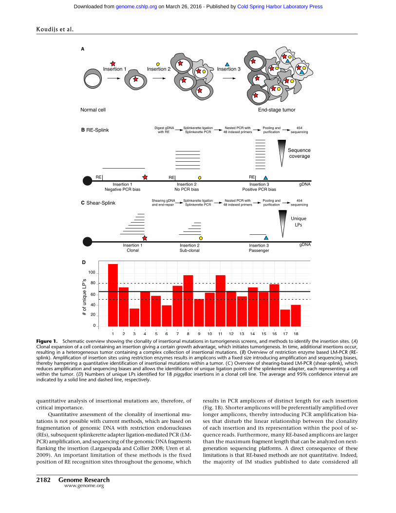
Clonality analysis of MMTV insertions
IM screens for cancer gene discovery in mice yield, in most cases,
heterogeneous tumors containing different populations of tumor
cells with distinct patterns of insertional mutations. We, therefore,
set out to test the utility of shear-splink for clonality analysis of
mouse mammary tumor virus (MMTV) insertions by analyzing
mixtures of two independent clonal cell lines (BB12 and AE6) de-
rived from one MMTV-induced mammary tumor. Both tumor cell
lines contained, in addition to three endogenous MMTV copies and
one shared somatic MMTV insertion, a number of unique somatic
MMTV insertions (Fig. 2A). We mixed genomic DNA from both cell
lines in varying ratios and analyzed the MMTV insertions using
shear-splink (absolute read numbers per sample are shown in Fig.
2B). We next determined the number of unique LPs for each
MMTV insertion. The BB12 and AE6 cell lines contained five and
four somatic MMTV insertions, respectively, that were represented
by 15 or more unique LPs. One of the insertions in BB12 was ex-
cluded from further analysis because it yielded repeat-rich sequences
that could not be uniquely mapped to the mouse reference genome
(data not shown). One somatic MMTV insertion site was shared
between BB12 and AE6, showing the common origin of these two
tumor cell lines. As expected the number of unique LPs for this
shared MMTV insertion did not correlate with the ratios at which
DNA from both cell lines was mixed (Fig. 2C). In contrast, five out
of six cell line-specific MMTV insertions showed a strong correla-
tion (R2 $ 0.86) between the number of unique LPs and the DNA
mixing ratios (Fig. 2D,E). These data illustrate that shear-splink
permits semiquantitative analysis of insertion events within het-
erogeneous samples, with a sensitivity of ;10% for biclonal tumors.
Insertional Mutagenesis Database (iMDB)
In order to fully exploit the increased efficiency of IM screens using
shear-splink, we developed a new analysis platform called the In-
sertional Mutagenesis Database (iMDB). iMDB is a web-based ap-
plication capable of analyzing mapped retroviral and transposon
insertional mutagenesis data. Further, it is designed to handle
clonality scores for each insertion by exploiting LP counts. For
finding CISs, we implemented the Gaussian Kernel Convolution
(GKC) approach, which is able to find statistically significant CISs
(de Ridder et al. 2006). Furthermore, we developed tools for calling
significantly co-occurring or mutually exclusive insertions and
implemented a tool to associate insertions with potential target
genes and combine insertion site data with gene expression data
(de Ridder et al. 2010). Users may upload their own data, which is
kept private, and perform these analyses on the data. All data pre-
sented in this paper are publicly accessible from the iMDB. The
iMBD can be accessed from http://imdb.nki.nl.
Shear-splink analysis of MMTV-induced mammary tumors
To further test the potential of shear-splink and to compare its ef-
ficiency with current RE-splink methods, we analyzed 16 MMTV-
induced mouse mammary tumors by both RE-splink and shear-
splink. Tumor DNA samples were either sheared or digested with
BfaI or NlaIII restriction enzymes, which are widely used for RE-
splink analysis of insertional mutations (Dupuy et al. 2009; Keng
et al. 2009; Starr et al. 2009; Uren et al. 2009). MMTV insertion sites
with one sequence read or one unique LP were excluded from
further analysis. On average, we obtained 380 mappable sequence
reads, corresponding to 27 unique MMTV insertions sites, per tu-
mor (Supplemental Fig. S2; sequencing details per experiment and
sample are listed in Supplemental Table S2A,B; all identified inser-
tions are listed in Supplemental Table S3). All identified insertions
were plotted on the mouse reference genome (Supplemental Fig.
S3) showing a difference in MMTV insertion spectra for the two RE-
splink data sets and the shear-splink data set. Despite the fact that
the cutoff of >1 LP for shear-splink results in lower numbers of MMTV
insertions, there is a significant overlap between the known MMTV
common insertion sites identified by shear-splink and both RE-splink
methods (Fig. 3A). Of note, the overlapping CISs are strongly en-
riched for components of the Wnt and Fgf signaling pathways,
which are known to cooperate during mammary tumorigenesis in
mice (Kwan et al. 1992). This suggests that shear-splink enriches
for the most relevant insertions. To further test this, we determined
which percentage of reads or unique LPs mapped within 150 kb of
published MMTV CISs that are likely to contribute to tumorigenesis
(Supplemental Table S4). While the absolute number of MMTV
insertions identified in the 16 tumors is higher for the NlaIII and
BfaI RE-splink analyses (Fig. 3B), the shear-splink method shows a
higher percentage of MMTV insertions near known CISs (Fig. 3C).
Semiquantitative analysis of insertional mutations
Genome Research 2183www.genome.org
Cold Spring Harbor Laboratory Press on March 26, 2016 - Published by genome.cshlp.orgDownloaded from
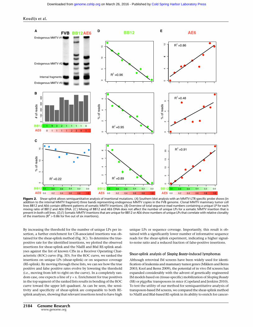
By increasing the threshold for the number of unique LPs per in-
sertion, a further enrichment for CIS-associated insertions was ob-
tained for the shear-splink method (Fig. 3C). To determine the true-
positive rate for the identified insertions, we plotted the observed
insertions for shear-splink and the NlaIII and BfaI RE-splink anal-
yses against the list of known CISs in a Receiver Operating Char-
acteristic (ROC) curve (Fig. 3D). For the ROC curve, we ranked the
insertions on unique LPs (shear-splink) or on sequence coverage
(RE-splink). By moving through these lists, we can see how the true
positive and false positive rates evolve by lowering the threshold
(i.e., moving from left to right on the curve). In a completely ran-
dom case, one expects a line of y = x. Enrichment for true positives
in the top segment of the ranked lists results in bending of the ROC
curve toward the upper left quadrant. As can be seen, the sensi-
tivity and specificity of shear-splink are comparable to both RE-
splink analyses, showing that relevant insertions tend to have high
unique LPs or sequence coverage. Importantly, this result is ob-
tained with a significantly lower number of informative sequence
reads for the shear-splink experiment, indicating a higher signal-
to-noise ratio and a reduced fraction of false-positive insertions.
Shear-splink analysis of Sleeping Beauty–induced lymphomas
Although retroviral IM screens have been widely used for identi-
fication of leukemia and mammary tumor genes (Mikkers and Berns
2003; Kool and Berns 2009), the potential of in vivo IM screens has
expanded considerably with the advent of genetically engineered
IM models based on (tissue-specific) mobilization of Sleeping Beauty
(SB) or piggyBac transposons in mice (Copeland and Jenkins 2010).
To test the utility of our method for semiquantitative analysis of
transposon-based IM screens, we compared the shear-splink method
to NlaIII and BfaI-based RE-splink in its ability to enrich for cancer-
Figure 2. Shear-splink allows semiquantitative analysis of insertional mutations. (A) Southern blot analysis with an MMTV-LTR specific probe shows (inaddition to the internal MMTV fragment) three bands representing endogenous MMTV copies in the FVB genome. Clonal MMTV mammary tumor celllines BB12 and AE6 contain different patterns of somatic MMTV insertions. (B) Overview of total sequence read numbers containing a unique LP for eachmixing ratio of BB12 and AE6 DNA. (C ) Mixing of BB12 and AE6 DNA does not affect the number of unique LPs for a somatic MMTV insertion that ispresent in both cell lines. (D,E ) Somatic MMTV insertions that are unique for BB12 or AE6 show numbers of unique LPs that correlate with relative clonalityof the insertions (R2 > 0.86 for five out of six insertions).
Koudijs et al.
2184 Genome Researchwww.genome.org
Cold Spring Harbor Laboratory Press on March 26, 2016 - Published by genome.cshlp.orgDownloaded from

Figure 3. Analysis of insertions in MMTV-induced mammary tumors by shear-splink and RE-splink. (A) Venn diagram showing strong overlap betweenMMTV insertions at known CISs for the shear-splink and RE-splink methods. The overlapping CISs are strongly enriched for components of the Wnt and Fgfsignaling pathways, which are known to cooperate during mammary tumorigenesis. (B) Total number of unique insertions (>0 and >1 sequence coverageand unique LPs) identified in a panel of 16 MMTV-induced tumors using shear-splink or RE-splink with BfaI or NlaIII. A high level of variability is observed inthe absolute number of insertions not linked to a CIS, in contrast to a comparable number of insertions mapping to known CISs. (C ) Bar diagrams showingpercentages of insertions representing known CISs for shear-splink and for RE-splink with BfaI and NlaIII. Increasing the threshold to higher sequencecoverage of unique LPs increases the fraction of insertions representing known CISs. For all thresholds tested (>0, >1, >2, >5, >10), the percentage ofinsertions mapping to known CISs is higher for shear-splink than for RE-splink. (D) Receiver Operating Characteristic (ROC) curves for the RE-splink andshear-splink methods show for shear-splink that enrichment in the identification of relevant insertions does not result in reduced sensitivity. The ROCcurves are built upon unique LPs for the shear-splink analysis and sequence coverage for the RE-splink experiments. By moving along the ROC curves fromleft to right, the ratios between true positives (sensitivity) and false positives (specificity) are visualized.
Semiquantitative analysis of insertional mutations
Genome Research 2185www.genome.org
Cold Spring Harbor Laboratory Press on March 26, 2016 - Published by genome.cshlp.orgDownloaded from

relevant transposon insertions in a panel of 127 SB-induced lym-
phomas. For the shear-splink analysis, we identified, in total, 3292
insertions with more than one LP, and 7318 and 6124 insertions
with more than 1 sequence read for NlaIII and BfaI, respectively
(sequencing details per experiment and sample are listed in Sup-
plemental Table 2A,C), all identified insertions are listed in Sup-
plemental Table S5). Using our iMDB web-based application, we
identified 32 and 35 CISs for the BfaI and NlaIII based RE-splink
experiments, respectively, and 48 CISs for the shear-splink exper-
iment (Supplemental Table S6). We manually assigned target genes
for these CISs and determined the overlap between unique target
genes recovered for each experiment (Fig. 4A). Shear-splink iden-
tified 13 unique target genes, whereas the RE-splink method yiel-
ded only four and three unique target genes for BfaI and NlaIII,
respectively. We next determined for each of the three experiments
how many CISs were identified per 1000 insertions. Shear-splink
identified 14.6 CISs for each 1000 insertions, whereas RE-splink
yielded 5.2 and 4.8 CISs per 1000 insertions for the BfaI and NlaIII
experiments, respectively (Fig. 4B). Interestingly, combining both
RE-splink datasets before CIS calling did not increase the number
of CISs per 1000 insertions. The efficiency of shear-splink is further
illustrated by the fact that, of all insertions identified by shear-
spink, 14.0% contributes to a CIS, compared to 5.6% for the in-
dividual RE-splink experiments and 7.7% for the combined RE-
splink data (Fig. 4C). These results confirm that the shear-splink
method outperforms RE-splink in its efficiency to identify CISs.
We next asked whether SB insertions with a high number of
unique LPs are more frequently linked to cancer-relevant genes, as
described in the Cancer Gene Census (CGC) database (http://www.
sanger.ac.uk/genetics/CGP/Census/). For this, we separated SB in-
sertions that mapped within 150-kb distance of CGC genes from
insertions in nonCGC regions and plotted a smooth histogram of
the unique LPs for CGC- and nonCGC-related SB insertions (Fig.
4D), showing that insertions near known cancer related genes are
represented by a higher number of unique LPs. In addition, we
plotted the density of insertions as a function of unique LP number
(Supplemental Fig. S4). We observed a significant increase in unique
LPs for SB insertions within all tested distances (25–250 kb) from
cancer-relevant genes.
In summary, the shear-splink method allows us to apply an
intelligent filter on insertional mutagenesis data by discarding back-
ground insertions marked by single LPs. Consequently, two sepa-
rate RE-splink analyses are outranked by
a single shear-splink experiment, which
requires less sequence read and as such
reduces the costs and labor involved in
large scale IM screens.
DiscussionThe implementation of next-generation
sequencing has greatly increased the
throughput of IM screens, resulting in the
identification of hundreds of insertions
per tumor. Current methods, however,
consider each unique insertion site as
equally relevant, independent of their
prevalence within the tumor cell popu-
lation. Subsequent determination of CISs
is, consequently, hampered by a low sig-
nal-to-noise ratio, resulting in long lists
of CISs that are likely to contain a sub-
stantial fraction of false positives. We show
that the shear-splink method permits
semiquantitative analysis of insertions in
genetically heterogeneous tumor samples,
resulting in efficient recovery of relevant
insertion sites with less sequencing ef-
fort and improved signal-to-noise ratio.
One limitation of the 454 Life Sciences
(Roche) sequence data is the relatively
low number of reads containing the LP,
because ;30% of all 454 reads lack the
splinkerette sequence at the 39 end. We
attempted to increase this efficiency by
optimizing the shearing conditions to
obtain 200–300 bp fragments, which are
ideally suited for the 454 GS FLX system.
Unfortunately, this did not result in in-
creased numbers of informative reads,
probably due to the amplification bias of
short, unmappable fragments and/or the
automatic clipping of sequence data with
Figure 4. Analysis of insertions in Sleeping Beauty–induced lymphomas by shear-splink and RE-splink.(A) Venn diagram showing the overlap of CISs in 127 Sleeping Beauty–induced lymphomas, as identifiedby shear-splink and RE-splink using BfaI or NlaIII . In total, 53 CISs are identified. Shear-splink detectedmore CISs (45) than BfaI- and NlaIII-based RE-splink (31 and 33 CISs, respectively). (B) Shear-splinkenriches for insertions contributing to CISs, as shown for the number of CISs identified per 1000 in-sertions. Combining both RE-splink data sets does not increase the efficiency, since the number of CISsper 1000 insertions is similar to the individual RE-splink analysis. (C ) The percentage of insertionscontributing to a CIS is higher for shear-splink than for the individual or combined RE-splink datasets,confirming that shear-splink enriches for relevant insertions. (D) Insertions near cancer-related genes arerepresented by higher numbers of unique LPs. Density plot showing the distribution of unique LPs for SBinsertions neighboring a Cancer Genome Census (CGC) gene (red) vs. those not flanking a CGC gene(gray).
Koudijs et al.
2186 Genome Researchwww.genome.org
Cold Spring Harbor Laboratory Press on March 26, 2016 - Published by genome.cshlp.orgDownloaded from
low quality scores by the Roche 454 software. However, this limi-
tation can be overcome by generating paired-end sequencing data
on the Illumina or Applied Biosystems (Life Technologies) SOLiD
next-generation sequencing platforms.
The most important implication of semiquantitative identi-
fication of insertional mutations is the ability to apply experimental
filters to IM data. Insertion sites from individual tumors can be
excluded or rated by the number of unique LPs prior to CIS analysis
(de Ridder et al. 2006). This is of particular interest for SB-based IM
screens, where RE-splink analysis of tumor samples yields, on av-
erage, between 100 and 150 SB insertions, of which 50%–80% are
represented by only one sequence read (Dupuy et al. 2009). Without
intelligent filtering, the very large numbers of SB insertions may
show random clustering on the genome, resulting in the identifi-
cation of false-positive CISs. Shear-splink enables exclusion of ran-
dom insertions represented by single LPs and thus enriches for bi-
ologically relevant insertions present in a substantial fraction of the
tumor mass, as was demonstrated by our comparative analysis of
shear-splink and RE-splink data from 127 SB-induced lymphomas.
One major challenge in the field of insertional mutagenesis
screens is to distinguish early versus late insertions in the multistep
process of tumorigenesis. This analysis is hampered by the fact that
RE-splink and shear-splink methods cannot discriminate between
heterogeneity resulting from clonal evolution of a monoclonal
tumor vs. the existence of multiple distinct cell clones within a bi-
or oligoclonal tumor. Nevertheless, the unique features of shear-
splink, in combination with iMDB, our publicly available database
and analysis pipeline, will significantly improve the efficiency of
in vivo IM screens and facilitate the analysis of IM-based in vitro
fitness screens, in which abundance of individual cell clones in a
polyclonal population is measured under selective vs. nonselective
conditions. In in vivo IM screens, shear-plink allows us to study co-
occurring or mutually exclusive mutations implicated in tumor
formation and in acquired traits, such as metastasis or drug re-
sistance, with a higher reliability. In conclusion, shear-splink ef-
fectively enriches for the most relevant insertions by providing a
rationale to exclude irrelevant insertions with single or few unique
ligation points.
Methods
Generation of tumorsMMTV-induced mammary tumors were derived from MMTV-C3H-infected FVB mice according to Theodorou et al. (2007). SB-in-duced lymphoma samples (spleen or thymus) were isolated frombitransgenic mice (mixed C57Bl6/129S5SvEvBrd background),carrying the SB11 Transposase (Dupuy et al. 2005) and a transgenicarray of T2Onc transposable elements on chromosome 1 (Collieret al. 2005).
Amplification and sequencing of MMTV insertions
Identification of MMTV insertion sites using restriction enzymeswas performed as described previously (Uren et al. 2009). Two mg ofgenomic DNA in 100 ml H2O was sheared to 100 bp–1 kb fragmentsusing a Covaris S2 sonicator for 45 s (6316 AFA fiber Tube, dutycycle: 5%, intensity: 1, cycles/burst: 200, frequency sweeping).Sheared DNA was precipitated and dissolved in 20 ml MQ andblunt-ended using the End-it kit (Epicentre Biotechnologies) byadding 2.94 ml dATP, 2.94 ml dNTPs, 2.94 ml buffer, and 0.57mlenzyme mix and incubated for 45 min at RT. End-repaired DNAwas purified using the Qiagen PCR purification kit, and concen-
trations were normalized to 20 ng/ml. Splinkerette adapters weregenerated by denaturing 400 pmol of oligonucleotides at 95°C, re-annealing by gradually decreasing temperature to 20°C, and di-luted to a concentration of 40 pmol. Blunt-ended splinkeretteadapters were ligated in 100-fold excess to 200 ng sheared DNAfragments using 4 U T4 DNA ligase (Roche) at 16°C for 16 h. Toprevent amplification of internal MMTV fragments, samples weredigested with DraI (New England Biolabs) for 3 h and inactivatedat 70°C for 20 min. Primary amplification was performed usingThermoStart Taq (Thermo Scientific) polymerase with a primer spe-cifically amplifying the exogenous MMTV-LTR (primer MMTV4contains mismatch with the endogenous sequence at the most 39
end) and a splinkerette-specific primer (P7) with the followingcycle conditions: 94°C for 30 s, 68°C for 30 s, and 72°C for 1 minfor 15 cycles, followed by 10 rounds of amplification with an-nealing temperature of 66°C. Secondary PCR was performed using454B-T7 primers for the splinkerette adapter and oligonucleotidescontaining 48 different 10-bp indexes and a 9- bp spacer in com-bination with the 454-A adapter sequence on the viral end. Cycleconditions of the secondary PCR were similar to primary amplifi-cation, with the exception that annealing was performed at 55°C.All oligonucleotides were purchased from Integrated DNA Tech-nologies. Primer, adapter, and barcode sequences are listed in Sup-plemental Table S7. Secondary PCR samples were pooled (generally483), purified using the Qiagen PCR purification kit, and sequencedon one quarter of a picotiter plate on the 454 GS FLX platform,according to the manufacturer’s protocol (Roche).
Amplification and sequencing of PB and SBtransposon insertions
PB insertions were identified as described previously (Bouwmanet al. 2010). SB insertions were identified using the method de-scribed for MMTV insertions, except that XhoI was used to removeinternal SB fragments. SB samples are sequenced on the 454-Tita-nium platform according to the manufacturer’s protocol. Primersequences are listed in Supplemental Table S7.
Mapping and processing of sequence reads
A general overview of the complete analysis pipeline of MMTV-and SB-induced tumors is presented in Supplemental Figure S5. Allraw and processed sequencing data and barcode information aresubmitted to the NCBI Gene Expression Omnibus (http://www.ncbi.nlm.nih.gov/geo/) under accession number GSE22657. Se-quences from each sample were sorted on barcodes, filtered for thepresence of the 59 end of the viral or transposon genome (notpresent in the oligonucleotides sequence) and the splinkerette se-quence using Exonerate, mapped to the genome using BLAT (95%similarity) and aggregated by genomic coordinate of the transitionof viral or transposon sequence to genomic DNA. A reference ge-nome was generated using all unique BLAT hits in order to includeshort sequence reads that originally could not be mapped in a sec-ond round of mapping against this reference sequence. Unique LPswere determined by determining unique BLATcoordinates of readsper insertion site. All reads were processed using Perl, and all in-formation was stored in an SQLite database.
Read filtering and insertion determination
All subsequent filtering and processing steps were performed in R.To remove background and nontumor-related insertions, we ex-cluded all unique genomic coordinates that were supported by oneread (for the restriction enzyme experiments) or one unique ligationpoint (for the shear-splink experiment). To determine insertion sites
Semiquantitative analysis of insertional mutations
Genome Research 2187www.genome.org
Cold Spring Harbor Laboratory Press on March 26, 2016 - Published by genome.cshlp.orgDownloaded from
and to compensate for small (1–10 bp) shifts in genomic coordinateat the viral or transposon end, we used hierarchical clustering to ag-gregate reads into insertions. Clustering was performed with abso-lute genomic distance in bp and single linkage. The resulting den-drogram was cut at a distance of 10 bp. This cutoff resulted in a meanwithin-cluster distance that was seven orders of magnitude lowerthan the mean nearest between-cluster distance, indicating a verytight clustering. The resulting clusters represent the unique inser-tions that we recovered in the analyses.
Analysis of PB insertions in ES cells
We determined PB insertions by selecting unique genomic co-ordinates for which we could determine at least 20 unique LPs.Expected unique LPs and reads were calculated by sampling froma uniform distribution of 18 PB insertions. The number of sam-plings was equal to the total unique LPs or sequence reads. Sam-plings were repeated 1000 times to determine a 95% confidenceinterval per insertion.
Analysis of MMTV insertions in tumor cell lines
We determined MMTV insertions in tumor cell lines by selectingunique genomic coordinates for which we could determine at least15 unique LPs. We determined the number of unique LPs for theselected MMTV insertions for all dilutions. Correlation coefficients(R2) were determined using the stats package in the R software.
Analysis of MMTV and SB insertions in mouse tumors
Endogenous MMTV sequences and potential cross-contaminationswere eliminated by removing all MMTV and SB reads that reporteda unique, identical genomic coordinate over more than two tumors.In the case where two tumors contained reads reporting the sameunique genomic coordinate, the reads were only kept if they had afivefold higher read-depth in one tumor compared to the other. Inall other cases, the reads were discarded. SB insertions on chromo-some 1 were excluded because local transposition on the chromo-some containing the multicopy array of T2Onc transposon elementsis a known feature of SB that complicates the identification ofcommon insertion sites (Collier et al. 2005).
Statistical analyses
The Fisher-Exact test as implemented in the R software was used tocompare the significance of the enrichment for known CIS genesbetween the restriction enzyme experiments and the shearingexperiments. ROC curves were calculated and visualized using theROCR package for R. The ranking required by the ROC analysis wasbased on the total number of reads per insertion for the restrictionenzyme experiments and the total number of unique ligation pointsfor the shearing experiments.
Generation of clonal cell lines from MMTV-induced tumors
Cryopreserved MMTV-induced tumor fragments were grown inDMEM medium, supplemented with 10% FCS, 50 U/mL penicillin,50 mg/mL streptomycin, 10 mg/mL bovine insulin, and 60 mg/mLEGF. Cells were immortalized by overexpression of SV40 large Tantigen as described previously ( Jat and Sharp 1986). Epithelialcells were enriched using partial trypsinization, and single-cell di-lutions were seeded in 96-well plates (1 and 0.3 cells per well) andselected for single colonies. Cell lines were expanded and passagedfor 4 wk, harvested, and analyzed for MMTV insertions accordingto the aforementioned protocol.
Southern blot analysis of clonal cell lines
Southern blot analysis was performed using an MMTV-LTR probegenerated by PCR using the LTR-specific primers 59-GTTGTTTCCCACCAAGGAC-39 and 59-TTCTAGGCCTGTGGTCAATAG-39. Ge-nomic DNA was digested using PstI, which enables detection of the39 LTR plus the flanking genomic region as well as an internal MMTVfragment, which is identical for all MMTV insertions. Hybridizationwas performed according to standard protocols. A schematic over-view of the probe design is shown in Supplemental Figure S6.
Data accessThe sequence data from this study have been submitted to theNCBI Gene Expression Omnibus (GEO) (http://www.ncbi.nlm.nih.gov/geo/) under accession number GSE22657.
AcknowledgmentsWe thank Maarten van Lohuizen, Joost Moes, and Wendy Lagcher(Netherlands Cancer Institute, Division of Molecular Genetics,Amsterdam, The Netherlands) for providing tumor samples, andEwart de Bruijn and Michal Mokry (Hubrecht Institute, Utrecht,The Netherlands) for technical assistance. We thank Anthony Urenand Waseem Akhtar for critically reading the manuscript. This re-search was funded by AICR grant 07-0585, NWO Horizon Break-through grant 40-41009-98-9109, and the Cancer Systems BiologyCenter (CSBC). D.J.A. is supported by Cancer Research-UK and theWellcome Trust.
References
Amsterdam A, Burgess S, Golling G, Chen W, Sun Z, Townsend K, FarringtonS, Haldi M, Hopkins N. 1999. A large-scale insertional mutagenesisscreen in zebrafish. Genes Dev 13: 2713–2724.
Bouwman P, Aly A, Escandell JM, Pieterse M, Bartkova J, van der Gulden H,Hiddingh S, Thanasoula M, Kulkarni A, Yang Q , et al. 2010. 53BP1 lossrescues BRCA1 deficiency and is associated with triple-negative andBRCA-mutated breast cancers. Nat Struct Mol Biol 17: 688–695.
Carette JE, Guimaraes CP, Varadarajan M, Park AS, Wuethrich I, Godarova A,Kotecki M, Cochran BH, Spooner E, Ploegh HL, et al. 2009. Haploidgenetic screens in human cells identify host factors used by pathogens.Science 326: 1231–1235.
Collier LS, Carlson CM, Ravimohan S, Dupuy AJ, Largaespada DA. 2005.Cancer gene discovery in solid tumors using transposon-based somaticmutagenesis in the mouse. Nature 436: 272–276.
Copeland NG, Jenkins NA. 2010. Harnessing transposons for cancer genediscovery. Nat Rev Cancer 10: 696–706.
de Ridder J, Uren A, Kool J, Reinders M, Wessels L. 2006. Detectingstatistically significant common insertion sites in retroviral insertionalmutagenesis screens. PLoS Comput Biol 2: e166. doi: 10.1371/journal.pcbi.0020166.
de Ridder J, Gerrits A, Bot J, de Haan G, Reinders M, Wessels L. 2010.Inferring combinatorial association logic networks in multimodalgenome-wide screens. Bioinformatics 26: i149–i157.
de Wit T, Dekker S, Maas A, Breedveld G, Knoch TA, Langeveld A, SzumskaD, Craig R, Bhattacharya S, Grosveld F, et al. 2010. Tagged mutagenesisby efficient Minos-based germ line transposition. Mol Cell Biol 30: 68–77.
Ding S, Wu X, Li G, Han M, Zhuang Y, Xu T. 2005. Efficient transposition ofthe piggyBac (PB) transposon in mammalian cells and mice. Cell 122:473–483.
Dupuy AJ, Akagi K, Largaespada DA, Copeland NG, Jenkins NA. 2005.Mammalian mutagenesis using a highly mobile somatic Sleeping Beautytransposon system. Nature 436: 221–226.
Dupuy AJ, Rogers LM, Kim J, Nannapaneni K, Starr TK, Liu P, LargaespadaDA, Scheetz TE, Jenkins NA, Copeland NG. 2009. A modified SleepingBeauty transposon system that can be used to model a wide variety ofhuman cancers in mice. Cancer Res 69: 8150–8156.
Golling G, Amsterdam A, Sun Z, Antonelli M, Maldonado E, Chen W,Burgess S, Haldi M, Artzt K, Farrington S, et al. 2002. Insertionalmutagenesis in zebrafish rapidly identifies genes essential for earlyvertebrate development. Nat Genet 31: 135–140.
Koudijs et al.
2188 Genome Researchwww.genome.org
Cold Spring Harbor Laboratory Press on March 26, 2016 - Published by genome.cshlp.orgDownloaded from
Jat PS, Sharp PA. 1986. Large Tantigens of simian virus 40 and polyomavirusefficiently establish primary fibroblasts. J Virol 59: 746–750.
Keng VW, Yae K, Hayakawa T, Mizuno S, Uno Y, Yusa K, Kokubu C, KinoshitaT, Akagi K, Jenkins NA, et al. 2005. Region-specific saturation germlinemutagenesis in mice using the Sleeping Beauty transposon system. NatMethods 2: 763–769.
Keng VW, Villanueva A, Chiang DY, Dupuy AJ, Ryan BJ, Matise I, SilversteinKA, Sarver A, Starr TK, Akagi K, et al. 2009. A conditional transposon-based insertional mutagenesis screen for genes associated with mousehepatocellular carcinoma. Nat Biotechnol 27: 264–274.
Kool J, Berns A. 2009. High-throughput insertional mutagenesis screens inmice to identify oncogenic networks. Nat Rev Cancer 9: 389–399.
Kool J, Uren AG, Martins CP, Sie D, de Ridder J, Turner G, van Uitert M,Matentzoglu K, Lagcher W, Krimpenfort P, et al. 2010. Insertionalmutagenesis in mice deficient for p15Ink4b, p16Ink4a, p21Cip1, andp27Kip1 reveals cancer gene interactions and correlations with tumorphenotypes. Cancer Res 70: 520–531.
Kwan H, Pecenka V, Tsukamoto A, Parslow TG, Guzman R, Lin TP, MullerWJ, Lee FS, Leder P, Varmus HE. 1992. Transgenes expressing the Wnt-1and int-2 proto-oncogenes cooperate during mammary carcinogenesisin doubly transgenic mice. Mol Cell Biol 12: 147–154.
Largaespada DA, Collier LS. 2008. Transposon-mediated mutagenesis insomatic cells: Identification of transposon-genomic DNA junctions.Methods Mol Biol 435: 95–108.
Lauchle JO, Kim D, Le DT, Akagi K, Crone M, Krisman K, Warner K, BonifasJM, Li Q , Coakley KM, et al. 2009. Response and resistance to MEK
inhibition in leukemias initiated by hyperactive Ras. Nature 461: 411–414.
Mikkers H, Berns A. 2003. Retroviral insertional mutagenesis: Taggingcancer pathways. Adv Cancer Res 88: 53–99.
Rad R, Rad L, Wang W, Cadinanos J, Vassiliou G, Rice S, Campos LS, Yusa K,Banerjee R, Li MA, et al. 2010. PiggyBac transposon mutagenesis: A toolfor cancer gene discovery in mice. Science 330: 1104–1107.
Starr TK, Allaei R, Silverstein KA, Staggs RA, Sarver AL, Bergemann TL, GuptaM, O’Sullivan MG, Matise I, Dupuy AJ, et al. 2009. A transposon-basedgenetic screen in mice identifies genes altered in colorectal cancer.Science 323: 1747–1750.
Theodorou V, Kimm MA, Boer M, Wessels L, Theelen W, Jonkers J, Hilkens J.2007. MMTV insertional mutagenesis identifies genes, gene families,and pathways involved in mammary cancer. Nat Genet 39: 759–769.
Uren AG, Kool J, Matentzoglu K, de Ridder J, Mattison J, van Uitert M,Lagcher W, Sie D, Tanger E, Cox T, et al. 2008. Large-scale mutagenesis inp19(ARF)- and p53-deficient mice identifies cancer genes and theircollaborative networks. Cell 133: 727–741.
Uren AG, Mikkers H, Kool J, van der Weyden L, Lund AH, Wilson CH, RanceR, Jonkers J, van Lohuizen M, Berns A, et al. 2009. A high-throughputsplinkerette-PCR method for the isolation and sequencing of retroviralinsertion sites. Nat Protoc 4: 789–798.
Received July 14, 2010; accepted in revised form August 11, 2011.
Semiquantitative analysis of insertional mutations
Genome Research 2189www.genome.org
Cold Spring Harbor Laboratory Press on March 26, 2016 - Published by genome.cshlp.orgDownloaded from

10.1101/gr.112763.110Access the most recent version at doi:2011 21: 2181-2189 originally published online August 18, 2011Genome Res.
Marco J. Koudijs, Christiaan Klijn, Louise van der Weyden, et al. in heterogeneous tumorsHigh-throughput semiquantitative analysis of insertional mutations
Material
Supplemental
http://genome.cshlp.org/content/suppl/2011/08/17/gr.112763.110.DC1.html
References
http://genome.cshlp.org/content/21/12/2181.full.html#ref-list-1
This article cites 26 articles, 10 of which can be accessed free at:
License
Commons Creative
http://creativecommons.org/licenses/by-nc/3.0/.described at
a Creative Commons License (Attribution-NonCommercial 3.0 Unported License), as ). After six months, it is available underhttp://genome.cshlp.org/site/misc/terms.xhtml
first six months after the full-issue publication date (see This article is distributed exclusively by Cold Spring Harbor Laboratory Press for the
ServiceEmail Alerting
click here.top right corner of the article or
Receive free email alerts when new articles cite this article - sign up in the box at the
http://genome.cshlp.org/subscriptionsgo to: Genome Research To subscribe to
Copyright © 2011 by Cold Spring Harbor Laboratory Press
Cold Spring Harbor Laboratory Press on March 26, 2016 - Published by genome.cshlp.orgDownloaded from
Related Documents