Nucleic Acids Research, 2015 1 doi: 10.1093/nar/gkv265 High-throughput assay and engineering of self-cleaving ribozymes by sequencing Shungo Kobori 1 , Yoko Nomura 1 , Anh Miu 1 and Yohei Yokobayashi 1,2,* 1 Department of Biomedical Engineering, University of California, Davis, CA 95616, USA and 2 Nucleic Acid Chemistry and Engineering Unit, Okinawa Instituteof Science and Technology Graduate University, Onna, Okinawa 904 0495, Japan Received January 16, 2015; Revised February 22, 2015; Accepted March 17, 2015 ABSTRACT Self-cleaving ribozymes are found in all domains of life and are believed to play important roles in biol- ogy. Additionally, self-cleaving ribozymes have been the subject of extensive engineering efforts for ap- plications in synthetic biology. These studies often involve laborious assays of multiple individual vari- ants that are either designed rationally or discovered through selection or screening. However, these as- says provide only a limited view of the large sequence space relevant to the ribozyme function. Here, we re- port a strategy that allows quantitative characteriza- tion of greater than 1000 ribozyme variants in a sin- gle experiment. We generated a library of predefined ribozyme variants that were converted to DNA and analyzed by high-throughput sequencing. By count- ing the number of cleaved and uncleaved reads of every variant in the library, we obtained a complete activity profile of the ribozyme pool which was used to both analyze and engineer allosteric ribozymes. INTRODUCTION Recent advances in genome sequencing and bioinformat- ics have revealed the ubiquitous presence of self-cleaving ribozymes in all domains of life (1–4). The evidence that some natural RNAs can catalyze chemical reactions is one of the arguments supporting the RNA World hypothesis (5). As such, chemists have long been studying natural and artificial ribozymes with nucleolytic and other chemical activities. More recently, we and other groups have engi- neered allosteric ribozymes (aptazymes) by strategically fus- ing an RNA aptamer (6,7)––a molecular recognition RNA motif––with a self-cleaving ribozyme to chemically control gene expression in living cells (8,9). Regardless of the ob- jectives, ribozyme studies often involve biochemical char- acterization of multiple individual ribozyme mutants, for example, to test hypotheses regarding the roles of specific nucleotides or to validate the activities of mutants obtained from screening or selection experiments. However, the num- ber of variants that can be examined by conventional assays is severely limited because each ribozyme variant must be prepared and assayed individually. Here, we describe a simple strategy that allows quantita- tive assay of >1000 predefined ribozyme variants in parallel aided by high-throughput sequencing (HTS). The general approach is depicted in Figure 1. First, a library of ribozyme mutants is generated by in vitro transcription and allowed to undergo self-cleavage reaction under a desired condition. Second, the ribozyme library is converted to DNA and pro- cessed to attach adapter and barcode sequences necessary for HTS. At this stage, each DNA molecule carries the fol- lowing information that originates from a single ribozyme molecule: the sequence of the varied bases, whether the ri- bozyme was cleaved or not, and the library and the reaction conditions encoded in the barcode. As in other HTS appli- cations, barcoding allows one to run multiple experiments (e.g. different reaction conditions or ribozyme libraries) in a single sequencing session. Finally, the sequencing data are sorted to count the number of cleaved and uncleaved reads to assign a relative activity (fraction cleaved) to every vari- ant in the library. It should be noted that this strategy has some important distinctions from conventional selection or screening of ribozyme libraries. Selection or screening typi- cally identifies the sequences of a very small fraction of ‘win- ners’ in a large pool of variants that must be further char- acterized in detail as mentioned above. Our HTS ribozyme assay provides a complete sequence–activity profile of all variants in the library, including ‘losers’ or other mediocre performers. Such information can greatly facilitate our un- derstanding of and our ability to engineer ribozymes. Using this method, we analyzed 1024 mutants of the re- cently discovered twister ribozyme (3) in which five bases in- volved in or neighboring a pseudoknot interaction (10,11) were randomized. In addition, we assayed the ligand- dependent activities of two aptazyme libraries based on a hepatitis delta virus (HDV)-like ribozyme each of which consisting of 256 variants of the four bases connecting a guanine aptamer and the ribozyme. The ribozyme activi- ties inferred by sequencing showed good correlation with * To whom correspondence should be addressed. Tel: +1 530 754 9676; Fax: +1 530 754 5739; Email: [email protected] C The Author(s) 2015. Published by Oxford University Press on behalf of Nucleic Acids Research. This is an Open Access article distributed under the terms of the Creative Commons Attribution License (http://creativecommons.org/licenses/by/4.0/), which permits unrestricted reuse, distribution, and reproduction in any medium, provided the original work is properly cited. Nucleic Acids Research Advance Access published March 30, 2015 by guest on June 4, 2016 http://nar.oxfordjournals.org/ Downloaded from

Welcome message from author
This document is posted to help you gain knowledge. Please leave a comment to let me know what you think about it! Share it to your friends and learn new things together.
Transcript
Nucleic Acids Research, 2015 1doi: 10.1093/nar/gkv265
High-throughput assay and engineering ofself-cleaving ribozymes by sequencingShungo Kobori1, Yoko Nomura1, Anh Miu1 and Yohei Yokobayashi1,2,*
1Department of Biomedical Engineering, University of California, Davis, CA 95616, USA and 2Nucleic Acid Chemistryand Engineering Unit, Okinawa Institute of Science and Technology Graduate University, Onna, Okinawa 904 0495,Japan
Received January 16, 2015; Revised February 22, 2015; Accepted March 17, 2015
ABSTRACT
Self-cleaving ribozymes are found in all domains oflife and are believed to play important roles in biol-ogy. Additionally, self-cleaving ribozymes have beenthe subject of extensive engineering efforts for ap-plications in synthetic biology. These studies ofteninvolve laborious assays of multiple individual vari-ants that are either designed rationally or discoveredthrough selection or screening. However, these as-says provide only a limited view of the large sequencespace relevant to the ribozyme function. Here, we re-port a strategy that allows quantitative characteriza-tion of greater than 1000 ribozyme variants in a sin-gle experiment. We generated a library of predefinedribozyme variants that were converted to DNA andanalyzed by high-throughput sequencing. By count-ing the number of cleaved and uncleaved reads ofevery variant in the library, we obtained a completeactivity profile of the ribozyme pool which was usedto both analyze and engineer allosteric ribozymes.
INTRODUCTION
Recent advances in genome sequencing and bioinformat-ics have revealed the ubiquitous presence of self-cleavingribozymes in all domains of life (1–4). The evidence thatsome natural RNAs can catalyze chemical reactions is oneof the arguments supporting the RNA World hypothesis(5). As such, chemists have long been studying natural andartificial ribozymes with nucleolytic and other chemicalactivities. More recently, we and other groups have engi-neered allosteric ribozymes (aptazymes) by strategically fus-ing an RNA aptamer (6,7)––a molecular recognition RNAmotif––with a self-cleaving ribozyme to chemically controlgene expression in living cells (8,9). Regardless of the ob-jectives, ribozyme studies often involve biochemical char-acterization of multiple individual ribozyme mutants, forexample, to test hypotheses regarding the roles of specificnucleotides or to validate the activities of mutants obtained
from screening or selection experiments. However, the num-ber of variants that can be examined by conventional assaysis severely limited because each ribozyme variant must beprepared and assayed individually.
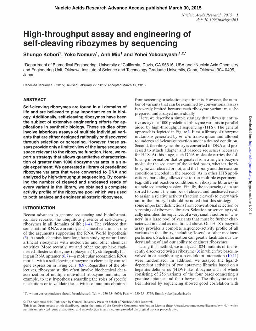
Here, we describe a simple strategy that allows quantita-tive assay of >1000 predefined ribozyme variants in parallelaided by high-throughput sequencing (HTS). The generalapproach is depicted in Figure 1. First, a library of ribozymemutants is generated by in vitro transcription and allowedto undergo self-cleavage reaction under a desired condition.Second, the ribozyme library is converted to DNA and pro-cessed to attach adapter and barcode sequences necessaryfor HTS. At this stage, each DNA molecule carries the fol-lowing information that originates from a single ribozymemolecule: the sequence of the varied bases, whether the ri-bozyme was cleaved or not, and the library and the reactionconditions encoded in the barcode. As in other HTS appli-cations, barcoding allows one to run multiple experiments(e.g. different reaction conditions or ribozyme libraries) ina single sequencing session. Finally, the sequencing data aresorted to count the number of cleaved and uncleaved readsto assign a relative activity (fraction cleaved) to every vari-ant in the library. It should be noted that this strategy hassome important distinctions from conventional selection orscreening of ribozyme libraries. Selection or screening typi-cally identifies the sequences of a very small fraction of ‘win-ners’ in a large pool of variants that must be further char-acterized in detail as mentioned above. Our HTS ribozymeassay provides a complete sequence–activity profile of allvariants in the library, including ‘losers’ or other mediocreperformers. Such information can greatly facilitate our un-derstanding of and our ability to engineer ribozymes.
Using this method, we analyzed 1024 mutants of the re-cently discovered twister ribozyme (3) in which five bases in-volved in or neighboring a pseudoknot interaction (10,11)were randomized. In addition, we assayed the ligand-dependent activities of two aptazyme libraries based on ahepatitis delta virus (HDV)-like ribozyme each of whichconsisting of 256 variants of the four bases connecting aguanine aptamer and the ribozyme. The ribozyme activi-ties inferred by sequencing showed good correlation with
*To whom correspondence should be addressed. Tel: +1 530 754 9676; Fax: +1 530 754 5739; Email: [email protected]
C© The Author(s) 2015. Published by Oxford University Press on behalf of Nucleic Acids Research.This is an Open Access article distributed under the terms of the Creative Commons Attribution License (http://creativecommons.org/licenses/by/4.0/), whichpermits unrestricted reuse, distribution, and reproduction in any medium, provided the original work is properly cited.
Nucleic Acids Research Advance Access published March 30, 2015 by guest on June 4, 2016
http://nar.oxfordjournals.org/D
ownloaded from
2 Nucleic Acids Research, 2015
Figure 1. Library construction strategy. First, a partially randomized ri-bozyme library is transcribed in vitro from a DNA template. The cleavedand uncleaved ribozymes are reverse transcribed into cDNAs using aprimer that contains a barcode and an adapter sequence. After removingthe RNAs, the 3′ adapter is attached and amplified by PCR to obtain thesequencing library. The core ribozyme sequence is shown in black with thedegenerate bases depicted in red. Other sequence elements: T7 promoter(purple), barcode (yellow), adapter sequences for sequencing (green andblue).
the conventional biochemical assay results of individualmutants. Furthermore, we tested some of the efficient ap-tazymes identified by sequencing and found that they func-tion as gene switches in mammalian cells when embeddedin the 3′ untranslated region (UTR) of a reporter gene. Ourstrategy described here will facilitate deeper understandingof the sequence–function relationships of natural and engi-neered ribozymes.
MATERIALS AND METHODS
General information
All oligonucleotides were purchased from IDT. The de-generate bases (N) in the oligonucleotides used for libraryconstruction were synthesized using IDT’s ‘hand-mix’ op-tion (equimolar mix of A, C, G and T). Ethanol precipita-tion was performed using Quick-Precip Plus Solution (EdgeBioSystems).
Library sequences
The DNA sequences of the libraries used to perform in vitrotranscription are as follows.
Lib-Tw: 5′TAATACGACTCACTATAGGGCGCGGCATTAATGCAGCTTTATTGGAAACAATAAAGCGNNNNNAAGCCCGCAAAAATAGCAGAGTAATGTCGCG3′
Lib-J1/2: 5′TAATACGACTCACTATAGGGCCGCGACTCTAGAAGTGATGCTCTGCNNNNATAATCGCGTGGATATGGCACGCAAGTTTCTACCGGGCACCGTAAATGTCCGACTACTCCTATTCCGGCACGTCCACGTCGTGCAGAGCGGTAACATGCGTTACTAGGGGTGCAAGAGCTCTTTTTGAGGAGGAGCTCTTTTTGCTGCACTAGTTGCATCAGATGGTAACGCATGGCTAAGCCGGAAAGGGGGAGAC3′
Lib-P4: 5′TAATACGACTCACTATAGGGCCGCGACTCTAGAAGTGATGCTCTGCAAATGGGGTAGGAGGCGATGCCTCGTCCTCATACCCAACTCCTATTCCGGCACGTCCACGTCGTGCAGAGCGGTANNNNTATAATCGCGTGGATATGGCACGCAAGTTTCTACCGGGCACCGTAAATGTCCGACTAGTAGCTAAGCCGGAAAGGGGGAGAC3′
(underline: T7 promoter, bold: degenerate bases)
Construction of HTS libraries
DNA templates encoding a T7 promoter and the ri-bozyme libraries shown above were prepared using syn-thetic oligonucleotides and polymerase chain reaction(PCR). The Lib-Tw DNA template was in vitro transcribed(37◦C, 3 h) in the transcription buffer (40-mM Tris-HCl pH8.0, 2-mM spermidine, 10-mM DTT) containing T7 RNApolymerase (2.5 U/�l, New England Biolabs), NTPs (2 mMeach), MgCl2 (4 mM), RiboLock RNase Inhibitor (2 U/�l,Thermo Scientific) and the DNA template (200 nM) in 30-�l volume. For Lib-J1/2 and Lib-P4, 3-mM MgCl2 and300-nM DNA template were used and the reactions wereperformed in 180-�l scale with or without guanine (500�M). Upon completion of the transcription reaction of Lib-Tw, 10 U of DNase I (New England Biolabs) in 30 �l (16.6-mM Tris-HCl pH 7.6, 4.2-mM MgCl2, 0.83-mM CaCl2)was added and incubated on ice for 15 min. For Lib-J1/2and Lib-P4, 72 U of DNase I in 180 �l (20-mM Tris-HCl pH7.6, 5-mM MgCl2, 1-mM CaCl2) was added and incubatedon ice for 45 min. RNAs were recovered by ethanol pre-cipitation (Lib-J1/2, Lib-P4) or RNA Clean-up & Concen-tration kit (Lib-Tw) (Zymo Research) and resuspended in20–30 �l 0.1-mM ethylenediaminetetraacetic acid (EDTA).
Approximately 60 pmol of the RNAs were mixed with120 pmol of the reverse transcription primer (Supplemen-tary Table S1) in 17 �l and heated to 95◦C for 2 minfollowed by incubation at 65◦C for 5 min and placed onice. Reverse transcription was initiated by adding 12.3-�lRT buffer (157-mM Tris, pH 8.3, 236-mM KCl, 9.4-mMMgCl2, 1.57-mM each dNTPs, 15.7-mM DTT) and 1.5-�l(300 U) SuperScript III Reverse Transcriptase (Life Tech-nologies, 45◦C for 1 min, 52◦C for 25 min, 65◦C for 5 min).The reaction was quenched by addition of 1.5-�l NaOH (5M) and heat (95◦C for 5 min). The cDNAs were purified bydenaturing polyacrylamide gel electrophoresis (PAGE) (8%
by guest on June 4, 2016http://nar.oxfordjournals.org/
Dow
nloaded from
Nucleic Acids Research, 2015 3
polyacrylamide, 8-M urea, 19:1 acrylamide:bisacrylamide)and recovered in 17 �l 0.1-mM EDTA solution.
The 5′ phosphorylated adapter ADP B (SupplementaryTable S1) was ligated to the cDNAs using Quick LigationKit (New England Biolabs) in the presence of appropriatesplint oligos (Supplementary Table S1). The ligation reac-tion contained ∼1-pmol cDNAs, 200 pmol each of splintoligos (for cleaved and uncleaved cDNAs), and 400-pmolADP B in 38 �l 1× Quick Ligation Reaction Buffer whichwas heated to 95◦C for 10 min and slowly cooled to 25◦C be-fore adding 2-�l Quick T4 DNA Ligase and reacted at 37◦Cfor 6 h. The Lib-Tw ligation was performed at half scale.
The ligated cDNAs were recovered by ethanol precipi-tation and resuspended in 20 �l of H2O. The splint oligoswere designed to be complementary to the 5′ end of ADP Band the 3′ end of the cleaved or uncleaved cDNAs (Supple-mentary Table S1). To avoid amplification during PCR, mis-matched sequences were added to the 3′ ends of the splintoligos. Finally, the sequencing libraries were generated byPCR with primers ADB T-2 and ADB B-2 (SupplementaryTable S1) using Phusion Flash High-Fidelity PCR MasterMix (Thermo Scientific) in 100-�l volumes containing 5-�lligation products and 0.5 �M each primer. Nine cycles of 10s at 98◦C followed by 20 s at 72◦C were performed and thePCR products were purified by agarose gel electrophoresisusing Zymoclean Gel DNA Recovery Kit (Zymo Research).
Sequencing and data analysis
The libraries were sequenced on an Illumina MiSeq se-quencer using MiSeq Reagent Kit v3 (150 cycles, single-end)with 8.1% PhiX control by UC Davis DNA TechnologiesCore. The reads were first sorted into −guanine and +gua-nine pools using the 6-base barcode and further sorted ac-cording to the sequence identity into the respective libraries.From each sequence read, the identity of the library (Lib-Tw, Lib-J1/2 or Lib-P4), variant (randomized bases) and itsstatus (cleaved or uncleaved) were analyzed and recorded.Finally, the numbers of cleaved and uncleaved reads for eachvariant were calculated.
In vitro ribozyme assays
Individual ribozyme variants tested were first cloned in aplasmid and sequence verified. The plasmids were used toprepare in vitro transcription templates by PCR as describedabove for the library construction. In vitro transcription wasperformed under the same conditions as the library con-struction with the exception of the reaction volume (10 �l).The RNAs were separated by 8% (Lib-J1/2 and P4) or 10%(Lib-Tw) denaturing PAGE and stained by SYBR Gold(Life Technologies). The gels were photographed and an-alyzed using Bio-Rad ChemiDoc MP System. Intensities ofthe cleaved and uncleaved bands were corrected by the cor-responding RNA lengths to estimate the molar fraction ofthe cleaved ribozyme.
Aptazyme assay in mammalian cells
Aptazyme sequences were cloned in the 3′ UTR of theEGFP transcript in pEGFP-N1 (Clontech) as previously
described (12). HEK293 cells were maintained in a 5%CO2 humidified incubator at 37◦C in Dulbecco’s modifiedEagle’s medium (Mediatech) supplemented with 10% fe-tal bovine serum (Gibco) and 1× antibiotic-antimycotic(Gibco). One day before transfection, HEK293 cells weretrypsinized and diluted appropriately with fresh com-plete medium, and 2.4 × 10⁴ cells/well (∼100 �l) wereseeded onto 96-well plates. Fifty nanograms of an EGFP-aptazyme plasmid, 50 ng of pL22 (13) (used as an in-active DNA to optimize the transfection condition) and10 ng of pCMV-mCherry plasmid (constitutively expressesmCherry) were cotransfected using 1 �l of PolyFect reagent(QIAGEN) per well according to the manufacturer’s in-struction. After 4-h incubation, the medium was removedand replaced with 100-�l fresh complete medium with orwithout guanine (500 �M). Guanine was first dissolved at50 mM in 0.2-M NaOH, and was diluted 100-fold with thecomplete medium immediately before use. The cells were in-cubated for additional 19 h before EGFP assay.
Cellular fluorescence was measured and normalizedaccording to our previous report (12). Briefly, the cellculture medium was replaced with phosphate bufferedsaline (PBS) (150 �l per well) and incubated at 37◦Cuntil measurement. Fluorescence intensities were mea-sured for EGFP (484-nm excitation/510-nm emission/5-nm bandwidth) and mCherry (587-nm excitation/610-nm emission/10-nm bandwidth) using Safire2 microplatereader (Tecan). The raw fluorescence values were first sub-tracted with that of the untransfected cells (background).For each well, EGFP fluorescence was normalized bymCherry ([EGFP fluorescence]/[mCherry fluorescence]) toaccount for variations in transfection efficiency. The val-ues were further normalized by the cells transfected withpEGFP-AgamRz (inactive)/pCMV-mCherry (=1.0). Plas-mid pEGFP-AgamRz (inactive) is a control EGFP expres-sion vector in which an inactivated drz-Agam-2-1 was in-serted in the same location as the other aptazymes. The re-ported values are mean ± SD from three replicate samples.
RESULTS
Library design and construction
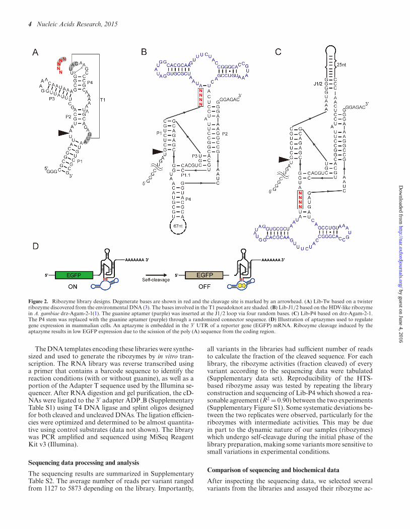
Three libraries were designed as illustrated in Figure 2. Lib-Tw was based on one of the twister ribozymes recentlydiscovered by the Breaker group (3). Specifically, the ri-bozyme referred to as ‘environmental sequence’ was modi-fied so that the transcription starts and ends at the P1 helix.Five consecutive bases shown in Figure 2A were random-ized. The first two bases (GU) are conserved in >97% ofthe twister ribozymes and the latter three bases (UAC) en-gage in a pseudoknot interaction (3,10,11). Lib-J1/2 andLib-P4 (Figure 2B and C) were based on an HDV-like ri-bozyme found in Anopheles gambiae (drz-Agam-2-1) (1).This particular HDV-like ribozyme contains extensive stem-loop structures in both P4 and J1/2 positions which wespeculated may be substituted with an RNA aptamer to en-gineer an allosteric regulation of the ribozyme activity. Theguanine aptamer from a riboswitch in Bacillus subtilis (14)was inserted to these positions via four consecutive degen-erate bases (Figure 2B and C).
by guest on June 4, 2016http://nar.oxfordjournals.org/
Dow
nloaded from
4 Nucleic Acids Research, 2015
Figure 2. Ribozyme library designs. Degenerate bases are shown in red and the cleavage site is marked by an arrowhead. (A) Lib-Tw based on a twisterribozyme discovered from the environmental DNA (3). The bases involved in the T1 pseudoknot are shaded. (B) Lib-J1/2 based on the HDV-like ribozymein A. gambiae drz-Agam-2-1(1). The guanine aptamer (purple) was inserted at the J1/2 loop via four random bases. (C) Lib-P4 based on drz-Agam-2-1.The P4 stem was replaced with the guanine aptamer (purple) through a randomized connector sequence. (D) Illustration of aptazymes used to regulategene expression in mammalian cells. An aptazyme is embedded in the 3′ UTR of a reporter gene (EGFP) mRNA. Ribozyme cleavage induced by theaptazyme results in low EGFP expression due to the scission of the poly (A) sequence from the coding region.
The DNA templates encoding these libraries were synthe-sized and used to generate the ribozymes by in vitro tran-scription. The RNA library was reverse transcribed usinga primer that contains a barcode sequence to identify thereaction conditions (with or without guanine), as well as aportion of the Adapter T sequence used by the Illumina se-quencer. After RNA digestion and gel purification, the cD-NAs were ligated to the 3′ adapter ADP B (SupplementaryTable S1) using T4 DNA ligase and splint oligos designedfor both cleaved and uncleaved DNAs. The ligation efficien-cies were optimized and determined to be almost quantita-tive using control substrates (data not shown). The librarywas PCR amplified and sequenced using MiSeq ReagentKit v3 (Illumina).
Sequencing data processing and analysis
The sequencing results are summarized in SupplementaryTable S2. The average number of reads per variant rangedfrom 1127 to 5873 depending on the library. Importantly,
all variants in the libraries had sufficient number of readsto calculate the fraction of the cleaved sequence. For eachlibrary, the ribozyme activities (fraction cleaved) of everyvariant according to the sequencing data were tabulated(Supplementary data set). Reproducibility of the HTS-based ribozyme assay was tested by repeating the libraryconstruction and sequencing of Lib-P4 which showed a rea-sonable agreement (R2 = 0.90) between the two experiments(Supplementary Figure S1). Some systematic deviations be-tween the two replicates were observed, particularly for theribozymes with intermediate activities. This may be duein part to the dynamic nature of our samples (ribozymes)which undergo self-cleavage during the initial phase of thelibrary preparation, making some variants more sensitive tosmall variations in experimental conditions.
Comparison of sequencing and biochemical data
After inspecting the sequencing data, we selected severalvariants from the libraries and assayed their ribozyme ac-
by guest on June 4, 2016http://nar.oxfordjournals.org/
Dow
nloaded from
Nucleic Acids Research, 2015 5
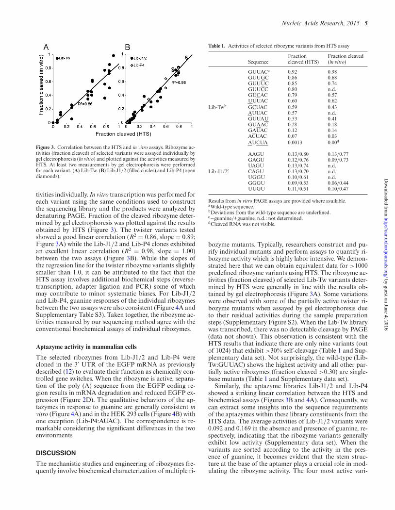
Figure 3. Correlation between the HTS and in vitro assays. Ribozyme ac-tivities (fraction cleaved) of selected variants were assayed individually bygel electrophoresis (in vitro) and plotted against the activities measured byHTS. At least two measurements by gel electrophoresis were performedfor each variant. (A) Lib-Tw. (B) Lib-J1/2 (filled circles) and Lib-P4 (opendiamonds).
tivities individually. In vitro transcription was performed foreach variant using the same conditions used to constructthe sequencing library and the products were analyzed bydenaturing PAGE. Fraction of the cleaved ribozyme deter-mined by gel electrophoresis was plotted against the resultsobtained by HTS (Figure 3). The twister variants testedshowed a good linear correlation (R2 = 0.86, slope = 0.89;Figure 3A) while the Lib-J1/2 and Lib-P4 clones exhibitedan excellent linear correlation (R2 = 0.98, slope = 1.00)between the two assays (Figure 3B). While the slopes ofthe regression line for the twister ribozyme variants slightlysmaller than 1.0, it can be attributed to the fact that theHTS assay involves additional biochemical steps (reverse-transcription, adapter ligation and PCR) some of whichmay contribute to minor systematic biases. For Lib-J1/2and Lib-P4, guanine responses of the individual ribozymesbetween the two assays were also consistent (Figure 4A andSupplementary Table S3). Taken together, the ribozyme ac-tivities measured by our sequencing method agree with theconventional biochemical assays of individual ribozymes.
Aptazyme activity in mammalian cells
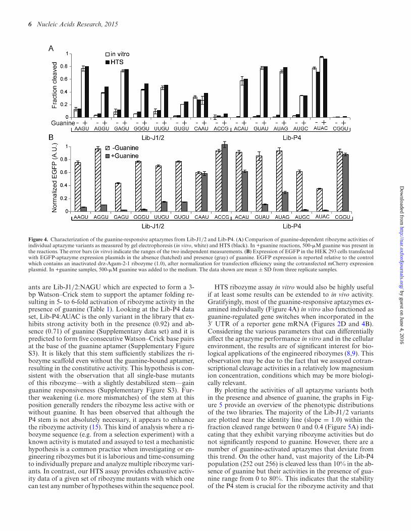
The selected ribozymes from Lib-J1/2 and Lib-P4 werecloned in the 3′ UTR of the EGFP mRNA as previouslydescribed (12) to evaluate their function as chemically con-trolled gene switches. When the ribozyme is active, separa-tion of the poly (A) sequence from the EGFP coding re-gion results in mRNA degradation and reduced EGFP ex-pression (Figure 2D). The qualitative behaviors of the ap-tazymes in response to guanine are generally consistent invitro (Figure 4A) and in the HEK 293 cells (Figure 4B) withone exception (Lib-P4:AUAC). The correspondence is re-markable considering the significant differences in the twoenvironments.
DISCUSSION
The mechanistic studies and engineering of ribozymes fre-quently involve biochemical characterization of multiple ri-
Table 1. Activities of selected ribozyme variants from HTS assay
SequenceFractioncleaved (HTS)
Fraction cleaved(in vitro)
GUUACa 0.92 0.98GUUGC 0.86 0.68GUUUC 0.85 0.74GUUCC 0.80 n.d.GUCAC 0.79 0.57UUUAC 0.60 0.62
Lib-Twb GCUAC 0.59 0.43AUUAC 0.57 n.d.GUUAU 0.53 0.41GUAAC 0.28 0.18GAUAC 0.12 0.14ACUAC 0.07 0.03AUCUA 0.0013 0.00d
AAGU 0.13/0.80 0.13/0.77GAGU 0.12/0.76 0.09/0.73UAGU 0.13/0.74 n.d.
Lib-J1/2c CAGU 0.13/0.70 n.d.UGGU 0.10/0.61 n.d.GGGU 0.09/0.53 0.06/0.44UUGU 0.11/0.51 0.10/0.47
Results from in vitro PAGE assays are provided where available.aWild-type sequence.bDeviations from the wild-type sequence are underlined.c−guanine/+guanine. n.d.: not determined.dCleaved RNA was not visible.
bozyme mutants. Typically, researchers construct and pu-rify individual mutants and perform assays to quantify ri-bozyme activity which is highly labor intensive. We demon-strated here that we can obtain equivalent data for >1000predefined ribozyme variants using HTS. The ribozyme ac-tivities (fraction cleaved) of selected Lib-Tw variants deter-mined by HTS were generally in line with the results ob-tained by gel electrophoresis (Figure 3A). Some variationswere observed with some of the partially active twister ri-bozyme mutants when assayed by gel electrophoresis dueto their residual activities during the sample preparationsteps (Supplementary Figure S2). When the Lib-Tw librarywas transcribed, there was no detectable cleavage by PAGE(data not shown). This observation is consistent with theHTS results that indicate there are only nine variants (outof 1024) that exhibit >30% self-cleavage (Table 1 and Sup-plementary data set). Not surprisingly, the wild-type (Lib-Tw:GUUAC) shows the highest activity and all other par-tially active ribozymes (fraction cleaved >0.30) are single-base mutants (Table 1 and Supplementary data set).
Similarly, the aptazyme libraries Lib-J1/2 and Lib-P4showed a striking linear correlation between the HTS andbiochemical assays (Figures 3B and 4A). Consequently, wecan extract some insights into the sequence requirementsof the aptazymes within these library constituents from theHTS data. The average activities of Lib-J1/2 variants were0.092 and 0.169 in the absence and presence of guanine, re-spectively, indicating that the ribozyme variants generallyexhibit low activity (Supplementary data set). When thevariants are sorted according to the activity in the pres-ence of guanine, it becomes evident that the stem struc-ture at the base of the aptamer plays a crucial role in mod-ulating the ribozyme activity. The four most active vari-
by guest on June 4, 2016http://nar.oxfordjournals.org/
Dow
nloaded from
6 Nucleic Acids Research, 2015
Figure 4. Characterization of the guanine-responsive aptazymes from Lib-J1/2 and Lib-P4. (A) Comparison of guanine-dependent ribozyme activities ofindividual aptazyme variants as measured by gel electrophoresis (in vitro, white) and HTS (black). In +guanine reactions, 500-�M guanine was present inthe reactions. The error bars (in vitro) indicate the ranges of the two independent measurements. (B) Expression of EGFP in the HEK 293 cells transfectedwith EGFP-aptazyme expression plasmids in the absence (hatched) and presence (gray) of guanine. EGFP expression is reported relative to the controlwhich contains an inactivated drz-Agam-2-1 ribozyme (1.0), after normalization for transfection efficiency using the cotransfected mCherry expressionplasmid. In +guanine samples, 500-�M guanine was added to the medium. The data shown are mean ± SD from three replicate samples.
ants are Lib-J1/2:NAGU which are expected to form a 3-bp Watson–Crick stem to support the aptamer folding re-sulting in 5- to 6-fold activation of ribozyme activity in thepresence of guanine (Table 1). Looking at the Lib-P4 dataset, Lib-P4:AUAC is the only variant in the library that ex-hibits strong activity both in the presence (0.92) and ab-sence (0.71) of guanine (Supplementary data set) and it ispredicted to form five consecutive Watson–Crick base pairsat the base of the guanine aptamer (Supplementary FigureS3). It is likely that this stem sufficiently stabilizes the ri-bozyme scaffold even without the guanine-bound aptamer,resulting in the constitutive activity. This hypothesis is con-sistent with the observation that all single-base mutantsof this ribozyme––with a slightly destabilized stem––gainguanine responsiveness (Supplementary Figure S3). Fur-ther weakening (i.e. more mismatches) of the stem at thisposition generally renders the ribozyme less active with orwithout guanine. It has been observed that although theP4 stem is not absolutely necessary, it appears to enhancethe ribozyme activity (15). This kind of analysis where a ri-bozyme sequence (e.g. from a selection experiment) with aknown activity is mutated and assayed to test a mechanistichypothesis is a common practice when investigating or en-gineering ribozymes but it is laborious and time-consumingto individually prepare and analyze multiple ribozyme vari-ants. In contrast, our HTS assay provides exhaustive activ-ity data of a given set of ribozyme mutants with which onecan test any number of hypotheses within the sequence pool.
HTS ribozyme assay in vitro would also be highly usefulif at least some results can be extended to in vivo activity.Gratifyingly, most of the guanine-responsive aptazymes ex-amined individually (Figure 4A) in vitro also functioned asguanine-regulated gene switches when incorporated in the3′ UTR of a reporter gene mRNA (Figures 2D and 4B).Considering the various parameters that can differentiallyaffect the aptazyme performance in vitro and in the cellularenvironment, the results are of significant interest for bio-logical applications of the engineered ribozymes (8,9). Thisobservation may be due to the fact that we assayed cotran-scriptional cleavage activities in a relatively low magnesiumion concentration, conditions which may be more biologi-cally relevant.
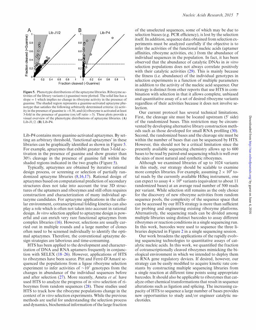
By plotting the activities of all aptazyme variants bothin the presence and absence of guanine, the graphs in Fig-ure 5 provide an overview of the phenotypic distributionsof the two libraries. The majority of the Lib-J1/2 variantsare plotted near the identity line (slope = 1.0) within thefraction cleaved range between 0 and 0.4 (Figure 5A) indi-cating that they exhibit varying ribozyme activities but donot significantly respond to guanine. However, there are anumber of guanine-activated aptazymes that deviate fromthis trend. On the other hand, vast majority of the Lib-P4population (252 out 256) is cleaved less than 10% in the ab-sence of guanine but their activities in the presence of gua-nine range from 0 to 80%. This indicates that the stabilityof the P4 stem is crucial for the ribozyme activity and that
by guest on June 4, 2016http://nar.oxfordjournals.org/
Dow
nloaded from
Nucleic Acids Research, 2015 7
Figure 5. Phenotypic distributions of the aptazyme libraries. Ribozyme ac-tivities of the library variants (±guanine) were plotted. The solid line has aslope = 1 which implies no change in ribozyme activity in the presence ofguanine. The shaded region represents a guanine-activated aptazyme phe-notype that satisfies the following arbitrarily determined criteria: (i) activ-ity in the presence of guanine is >0.30, and (ii) ribozyme is activated at least3-fold in the presence of guanine (on/off ratio >3). These plots provide avisual overview of the phenotypic distributions of aptazyme libraries. (A)Lib-J1/2. (B) Lib-P4.
Lib-P4 contains more guanine-activated aptazymes. By set-ting an arbitrary threshold, ‘functional aptazymes’ in theselibraries can be graphically identified as shown in Figure 5.For example, aptazymes that exhibit greater than 3-fold ac-tivation in the presence of guanine and show greater than30% cleavage in the presence of guanine fall within theshaded regions indicated in the two graphs (Figure 5).
Typically, aptazymes are obtained by iterative rationaldesign process, or screening or selection of partially ran-domized aptazyme libraries (8,16,17). Rational design ofaptazymes based on computational prediction of secondarystructures does not take into account the true 3D struc-tures of the aptamers and ribozymes and still often requiresconstruction and characterization of many individual ap-tazyme candidates. For aptazyme applications in the cellu-lar environment, cotranscriptional folding kinetics can alsoplay a role which is often not taken into account in rationaldesign. In vitro selection applied to aptazyme design is pow-erful and can enrich very rare functional aptazymes fromcomplex libraries (16). However, selections are usually car-ried out in multiple rounds and a large number of clonesoften need to be screened individually to identify the opti-mal aptazymes. Therefore, the conventional aptazyme de-sign strategies are laborious and time-consuming.
HTS has been applied to the development and character-ization of DNA and RNA aptamers frequently in conjunc-tion with SELEX (18–26). However, applications of HTSto ribozymes have been scarce. Pitt and Ferre-D’Amare se-quenced the populations from a ligase ribozyme selectionexperiment to infer activities of ∼107 genotypes from thechanges in abundance of the individual sequences beforeand after selection (27). More recently, Ameta et al. haveused HTS to analyze the progress of in vitro selection of ri-bozymes from random sequences (28). These studies usedHTS to track how the ribozyme populations change in thecontext of in vitro selection experiments. While the previousmethods are useful for understanding the selection processand dynamics, biochemical information of the large fraction
of the unselected sequences, some of which may be due toselection biases (e.g. PCR efficiency), is lost by the selectionitself. In addition, sequence data obtained from selection ex-periments must be analyzed carefully if the objective is toinfer the activities of the functional nucleic acids (aptameraffinities, ribozyme activities, etc.) from the abundance ofindividual sequences in the population. In fact, it has beenobserved that the abundance of catalytic DNAs in in vitroselection populations does not always correlate positivelywith their catalytic activities (29). This is mainly becausethe fitness (i.e. abundance) of the individual genotypes inselection experiments is a function of multiple parametersin addition to the activity of the nucleic acid sequence. Ourstrategy is distinct from other reports that use HTS in com-bination with selection in that it allows complete, unbiasedand quantitative assay of a set of desired ribozyme variantsregardless of their activities because it does not involve se-lection.
Our current protocol has several technical limitations.First, the cleavage site must be located upstream (5′ side)of the randomized bases. This restriction may be circum-vented by developing alternative library construction meth-ods such as those developed for small RNA profiling (30).Second, the randomized bases and the cleavage site must bewithin the number of bases that can be sequenced by HTS.However, this should not be a critical limitation since thepresently available sequencing chemistry allows up to 600bases to be read by paired-end sequencing which is well overthe sizes of most natural and synthetic ribozymes.
Although we examined libraries of up to 1024 variantsin this study, our strategy should be scalable to examinemore complex libraries. For example, assuming 2 × 10⁹ to-tal reads by the currently available HiSeq instrument, onecan expect to assay 4 × 10⁶ variants (equivalent to about 11randomized bases) at an average read number of 500 readsper variant. While selection still remains as the only choicefor the discovery of new ribozyme activities from randomsequence pools, the complexity of the sequence space thatcan be accessed by our HTS strategy is more than sufficientfor probing and engineering existing ribozyme platforms.Alternatively, the sequencing reads can be divided amongmultiple libraries using distinct barcodes to assay differentribozymes or reaction conditions in a single sequencing run.In this work, barcodes were used to sequence the three li-braries depicted in Figure 2 in a single sequencing session.
Our work broadens the applications of the rapidly evolv-ing sequencing technologies to quantitative assays of cat-alytic nucleic acids. In this work, we quantified the fractionof cotranscriptionally cleaved ribozymes mimicking the bi-ological environment in which we intended to deploy themas RNA gene regulatory devices. If desired, however, ourstrategy can be easily modified to acquire kinetic rate con-stants by constructing multiple sequencing libraries froma single reaction at different time points using appropriatebarcodes. It should also be applicable to ribozymes that cat-alyze other chemical transformations that result in sequencealterations such as ligation and splicing. The increasing ca-pacity of HTS to sequence a large number of bases providesnew opportunities to study and/or engineer catalytic nu-cleotides.
by guest on June 4, 2016http://nar.oxfordjournals.org/
Dow
nloaded from
8 Nucleic Acids Research, 2015
SUPPLEMENTARY DATA
Supplementary Data are available at NAR Online.
ACKNOWLEDGEMENT
The authors thank Andrew Yao and Marc Facciotti forhelpful discussion.
FUNDING
The National Institutes of Health [GM099748 to Y.Y.].Funding for open access charge: National Institutes ofHealth [GM099748 to Y.Y.].Conflict of interest statement. None declared.
REFERENCES1. Webb,C.H., Riccitelli,N.J., Ruminski,D.J. and Luptak,A. (2009)
Widespread occurrence of self-cleaving ribozymes. Science, 326, 953.2. Webb,C.H. and Luptak,A. (2011) HDV-like self-cleaving ribozymes.
RNA Biol., 8, 719–727.3. Roth,A., Weinberg,Z., Chen,A.G., Kim,P.B., Ames,T.D. and
Breaker,R.R. (2014) A widespread self-cleaving ribozyme class isrevealed by bioinformatics. Nat. Chem. Biol., 10, 56–60.
4. Hammann,C., Luptak,A., Perreault,J. and de la Pena,M. (2012) Theubiquitous hammerhead ribozyme. RNA, 18, 871–885.
5. Chen,X., Li,N. and Ellington,A.D. (2007) Ribozyme catalysis ofmetabolism in the RNA world. Chem. Biodivers., 4, 633–655.
6. Ellington,A.D. and Szostak,J.W. (1990) In vitro selection of RNAmolecules that bind specific ligands. Nature, 346, 818–822.
7. Tuerk,C. and Gold,L. (1990) Systematic evolution of ligands byexponential enrichment: RNA ligands to bacteriophage T4 DNApolymerase. Science, 249, 505–510.
8. Frommer,J., Appel,B. and Muller,S. (2015) Ribozymes that can beregulated by external stimuli. Curr. Opin. Biotechnol., 31, 35–41.
9. Wittmann,A. and Suess,B. (2012) Engineered riboswitches:expanding researchers’ toolbox with synthetic RNA regulators.FEBS Lett., 586, 2076–2083.
10. Eiler,D., Wang,J. and Steitz,T.A. (2014) Structural basis for the fastself-cleavage reaction catalyzed by the twister ribozyme. Proc. Natl.Acad. Sci. U.S.A., 111, 13028–13033.
11. Liu,Y., Wilson,T.J., McPhee,S.A. and Lilley,D.M. (2014) Crystalstructure and mechanistic investigation of the twister ribozyme. Nat.Chem. Biol., 10, 739–744.
12. Nomura,Y., Zhou,L., Miu,A. and Yokobayashi,Y. (2013) Controllingmammalian gene expression by allosteric hepatitis delta virusribozymes. ACS Synth. Biol., 2, 684–689.
13. Kumar,D., An,C.I. and Yokobayashi,Y. (2009) Conditional RNAinterference mediated by allosteric ribozyme. J. Am. Chem. Soc., 131,13906–13907.
14. Mandal,M., Boese,B., Barrick,J.E., Winkler,W.C. and Breaker,R.R.(2003) Riboswitches control fundamental biochemical pathways inBacillus subtilis and other bacteria. Cell, 113, 577–586.
15. Thill,G., Vasseur,M. and Tanner,N.K. (1993) Structural andsequence elements required for the self-cleaving activity of thehepatitis delta virus ribozyme. Biochemistry, 32, 4254–4262.
16. Breaker,R.R. (2002) Engineered allosteric ribozymes as biosensorcomponents. Curr. Opin. Biotechnol., 13, 31–39.
17. Soukup,G.A. and Breaker,R.R. (2000) Allosteric nucleic acidcatalysts. Curr. Opin. Struct. Biol., 10, 318–325.
18. Beier,R., Pahlke,C., Quenzel,P., Henseleit,A., Boschke,E.,Cuniberti,G. and Labudde,D. (2014) Selection of a DNA aptameragainst norovirus capsid protein VP1. FEMS Microbiol. Lett., 351,162–169.
19. Cho,M., Oh,S.S., Nie,J., Stewart,R., Eisenstein,M., Chambers,J.,Marth,J.D., Walker,F., Thomson,J.A. and Soh,H.T. (2013)Quantitative selection and parallel characterization of aptamers.Proc. Natl. Acad. Sci. U.S.A., 110, 18460–18465.
20. Ditzler,M.A., Lange,M.J., Bose,D., Bottoms,C.A., Virkler,K.F.,Sawyer,A.W., Whatley,A.S., Spollen,W., Givan,S.A. and Burke,D.H.(2013) High-throughput sequence analysis reveals structural diversityand improved potency among RNA inhibitors of HIV reversetranscriptase. Nucleic Acids Res., 41, 1873–1884.
21. Hoon,S., Zhou,B., Janda,K.D., Brenner,S. and Scolnick,J. (2011)Aptamer selection by high-throughput sequencing and informaticanalysis. Biotechniques, 51, 413–416.
22. Jimenez,J.I., Xulvi-Brunet,R., Campbell,G.W., Turk-MacLeod,R.and Chen,I.A. (2013) Comprehensive experimental fitness landscapeand evolutionary network for small RNA. Proc. Natl. Acad. Sci.U.S.A., 110, 14984–14989.
23. Kupakuwana,G.V., Crill,J.E. II, McPike,M.P. and Borer,P.N. (2011)Acyclic identification of aptamers for human alpha-thrombin usingover-represented libraries and deep sequencing. PLoS ONE, 6,e19395.
24. Schutze,T., Wilhelm,B., Greiner,N., Braun,H., Peter,F., Morl,M.,Erdmann,V.A., Lehrach,H., Konthur,Z., Menger,M. et al. (2011)Probing the SELEX Process with Next-Generation Sequencing.PLoS ONE, 6, e29604.
25. Tome,J.M., Ozer,A., Pagano,J.M., Gheba,D., Schroth,G.P. andLis,J.T. (2014) Comprehensive analysis of RNA-protein interactionsby high-throughput sequencing-RNA affinity profiling. Nat.Methods, 11, 683–688.
26. Wilson,R., Bourne,C., Chaudhuri,R.R., Gregory,R., Kenny,J. andCossins,A. (2014) Single-step selection of bivalent aptamers validatedby comparison with SELEX using high-throughput sequencing.PLoS ONE, 9, e100572.
27. Pitt,J.N. and Ferre-D’Amare,A.R. (2010) Rapid construction ofempirical RNA fitness landscapes. Science, 330, 376–379.
28. Ameta,S., Winz,M.L., Previti,C. and Jaschke,A. (2014)Next-generation sequencing reveals how RNA catalysts evolve fromrandom space. Nucleic Acids Res., 42, 1303–1310.
29. Schlosser,K., Lam,J.C. and Li,Y. (2009) A genotype-to-phenotypemap of in vitro selected RNA-cleaving DNAzymes: implications foraccessing the target phenotype. Nucleic Acids Res., 37, 3545–3557.
30. Hafner,M., Renwick,N., Farazi,T.A., Mihailovic,A., Pena,J.T. andTuschl,T. (2012) Barcoded cDNA library preparation for small RNAprofiling by next-generation sequencing. Methods, 58, 164–170.
by guest on June 4, 2016http://nar.oxfordjournals.org/
Dow
nloaded from
Related Documents