J. Anat. (2008) 213, pp217–227 doi: 10.1111/j.1469-7580.2008.00950.x © 2008 The Authors Journal compilation © 2008 Anatomical Society of Great Britain and Ireland Blackwell Publishing Ltd METHODS High-resolution visualization of airspace structures in intact mice via synchrotron phase-contrast X-ray imaging (PCXI) David W. Parsons, 1,2,3 Kaye Morgan, 4 Martin Donnelley, 1 Andreas Fouras, 5 Jeffrey Crosbie, 4,6 Ivan Williams, 4 Richard C. Boucher, 7 Kentaro Uesugi, 8 Naoto Yagi 8 and Karen K. W. Siu 4,6 1 Department of Pulmonary Medicine, Women’s and Children’s Hospital 2 Department of Paediatrics, University of Adelaide 3 Women’s & Children’s Child Health Research Institute, Adelaide 4 School of Physics; 5 Division of Biological Engineering; and 6 Monash Centre for Synchrotron Science, Monash University, Victoria, Australia 7 CF Research and Treatment Center, University of North Carolina at Chapel Hill, NC, USA 8 SPring-8/JASRI, Hyogo, Japan Abstract Anatomical visualization of airspace-containing organs in intact small animals has been limited by the resolution and contrast available from current imaging methods such as X-ray, micro-computed tomography and magnetic resonance imaging. Determining structural relationships and detailed anatomy has therefore relied on suitable fixation, sectioning and histological processing. More complex and informative analyses such as orthogonal views of an organ and three-dimensional structure visualizations have required different animals and image sets, laboriously processed to gather this complementary structural information. Precise three-dimensional anatomical views have always been difficult to achieve in small animals. Here we report the ability of phase-contrast synchrotron X-ray imaging to provide detailed two- and three-dimensional visualization of airspace organ structures in intact animals. Using sub-micrometre square pixel charge-coupled device array detectors, the structure and anatomy of hard and soft tissues, and of airspaces, is readily available using phase-contrast synchrotron X-ray imaging. Moreover, software-controlled volume-reconstructions of tomographic images not only provide unsurpassed image clarity and detail, but also selectable anatomical views that cannot be obtained with established histological techniques. The morphology and structure of nasal and lung airways and the middle ear are illustrated in intact mice, using two- and three-dimensional representations. The utility of phase-contrast synchrotron X-ray imaging for non- invasively localizing objects implanted within airspaces, and the detection of gas bubbles transiting live airways, are other novel features of this visualization methodology. The coupling of phase-contrast synchrotron X-ray imaging technology with software-based reconstruction techniques holds promise for novel and high-resolution non-invasive examination of airspace anatomy in small animal models. Key words airspaces micro CT; airway surface; imaging; mice; non-invasive; phase-contrast; synchrotron; X-ray. Introduction In developing airway gene transfer procedures for the treatment of cystic fibrosis airway disease, we seek new approaches to non-invasive detection of airway health and disease in live mouse models. Although traditional light and electron microscopy histological methods are used to produce high-resolution images and measurements of airway structure in animal models, they require invasive tissue sampling and are usually terminal procedures. In humans, similar levels of histological analysis are possible via invasive biopsy, but often require anaesthesia and are not suited to repeat assessment to track, for example, the durability of a therapeutic treatment. The non-invasive options for intact imaging in humans, such as ultrasound, diagnostic X-ray, high-resolution spiral CT, and standard or hyperpolarized- helium MRI provide an extraordinary range of complementary Correspondence Dr D. W. Parsons, Department of Pulmonary Medicine, Women’s and Children’s Hospital, 72 King William Road, North Adelaide, South Australia. E: [email protected] Accepted for publication 8 April 2008

Welcome message from author
This document is posted to help you gain knowledge. Please leave a comment to let me know what you think about it! Share it to your friends and learn new things together.
Transcript

J. Anat.
(2008)
213
, pp217–227 doi: 10.1111/j.1469-7580.2008.00950.x
© 2008 The Authors Journal compilation © 2008 Anatomical Society of Great Britain and Ireland
Blackwell Publishing Ltd
METHODS
High-resolution visualization of airspace structures in intact mice via synchrotron phase-contrast X-ray imaging (PCXI)
David W. Parsons,
1,2,3
Kaye Morgan,
4
Martin Donnelley,
1
Andreas Fouras,
5
Jeffrey Crosbie,
4,6
Ivan Williams,
4
Richard C. Boucher,
7
Kentaro Uesugi,
8
Naoto Yagi
8
and Karen K. W. Siu
4,6
1
Department of Pulmonary Medicine, Women’s and Children’s Hospital
2
Department of Paediatrics, University of Adelaide
3
Women’s & Children’s Child Health Research Institute, Adelaide
4
School of Physics;
5
Division of Biological Engineering; and
6
Monash Centre for Synchrotron Science, Monash University, Victoria, Australia
7
CF Research and Treatment Center, University of North Carolina at Chapel Hill, NC, USA
8
SPring-8/JASRI, Hyogo, Japan
Abstract
Anatomical visualization of airspace-containing organs in intact small animals has been limited by the resolutionand contrast available from current imaging methods such as X-ray, micro-computed tomography and magneticresonance imaging. Determining structural relationships and detailed anatomy has therefore relied on suitablefixation, sectioning and histological processing. More complex and informative analyses such as orthogonal viewsof an organ and three-dimensional structure visualizations have required different animals and image sets,laboriously processed to gather this complementary structural information. Precise three-dimensional anatomicalviews have always been difficult to achieve in small animals. Here we report the ability of phase-contrast synchrotronX-ray imaging to provide detailed two- and three-dimensional visualization of airspace organ structures in intactanimals. Using sub-micrometre square pixel charge-coupled device array detectors, the structure and anatomy ofhard and soft tissues, and of airspaces, is readily available using phase-contrast synchrotron X-ray imaging. Moreover,software-controlled volume-reconstructions of tomographic images not only provide unsurpassed image clarityand detail, but also selectable anatomical views that cannot be obtained with established histological techniques.The morphology and structure of nasal and lung airways and the middle ear are illustrated in intact mice, usingtwo- and three-dimensional representations. The utility of phase-contrast synchrotron X-ray imaging for non-invasively localizing objects implanted within airspaces, and the detection of gas bubbles transiting live airways,are other novel features of this visualization methodology. The coupling of phase-contrast synchrotron X-rayimaging technology with software-based reconstruction techniques holds promise for novel and high-resolutionnon-invasive examination of airspace anatomy in small animal models.
Key words
airspaces micro CT; airway surface; imaging; mice; non-invasive; phase-contrast; synchrotron; X-ray.
Introduction
In developing airway gene transfer procedures for thetreatment of cystic fibrosis airway disease, we seek newapproaches to non-invasive detection of airway healthand disease in live mouse models.
Although traditional light and electron microscopyhistological methods are used to produce high-resolutionimages and measurements of airway structure in animalmodels, they require invasive tissue sampling and areusually terminal procedures. In humans, similar levels ofhistological analysis are possible via invasive biopsy, butoften require anaesthesia and are not suited to repeatassessment to track, for example, the durability of atherapeutic treatment. The non-invasive options for intactimaging in humans, such as ultrasound, diagnostic X-ray,high-resolution spiral CT, and standard or hyperpolarized-helium MRI provide an extraordinary range of complementary
Correspondence
Dr D. W. Parsons, Department of Pulmonary Medicine, Women’s and Children’s Hospital, 72 King William Road, North Adelaide, South Australia. E: [email protected]
Accepted for publication
8 April 2008

Non-invasive visualization of airspaces, D. W. Parsons et al.
© 2008 The AuthorsJournal compilation © 2008 Anatomical Society of Great Britain and Ireland
218
information. However, when directed towards smallanimal model systems, these methods lack adequatecontrast and they have poor spatial and temporal resolution.Laboratory X-ray imaging systems designed for small animalsprovide improved resolution, but airspace structures arepoorly detected, contrast remains low, and high resolutionscans can involve lengthy image capture and processingtimes.
Phase-contrast imaging utilizes X-ray refraction effects,in addition to conventional absorption, to create imagecontrast. It is particularly useful for soft tissue contrast,where the absorption differences are small, and tissueboundaries are enhanced by phase effects due to variationsin refractive indices. Propagation-based phase contrastX-ray imaging (PCXI) is the simplest method, renderingphase effects visible as intensity changes by utilizing anincreased distance between the sample and the detector(Fig. 1). The technique demands a source with highcoherence (i.e. the source size is sufficiently small or,equivalently, the incident X-rays may be considered parallel).This requirement is easily realized using a synchrotron X-ray source, particularly at long beamlines such as those atthe Biomedical Imaging Centre at the SPring-8 synchrotronin Japan (Gotto et al. 2001).
PCXI can provide the substantially increased resolutionand contrast needed for non-invasive imaging in smallanimals. Soft tissues and airspaces such as airways andlungs can now be imaged and processed to give clarity ofimages that is similar to that provided by bone. Here wereveal the capability of this method for use in anatomicalstudy via two- and three-dimensional imaging in selectedmouse airspace organs.
Materials and methods
Adult C57Bl/6 mice weighing 18–20 g were imaged under approvalsfrom the Animal Ethics Committee of SPring-8, and of the Women’sand Children’s Hospital, CYWHS, Adelaide. All PCXI images werecollected at the Biomedical Imaging Centre, SPring-8 Synchrotronfacility, Hyogo, Japan. Live mice were anaesthetized with nembutal(45 mg kg
−
1
, i.p.). Post-mortem mice were imaged fresh (Figs 2, 3)or at room temperature after having been kept chilled for up to2 days (4 ºC, and passive rewarming over several hours – Figures andsupplementary material numbered 5, 6, and 7). Fur produced verystrong phase-contrast effects, but removal with depilatory creamprior to imaging improved the clarity of the two-dimensional images.
Two-dimensional airway studies in live and post-mortem mice
Figure 1 shows the Biomedical Imaging Beamline layout at SPring-8.Post-mortem, or anaesthetized, mice (Fig. 4) were secured head-high in a stereotaxic frame. The dorsal incisors were hooked overa stainless-steel wire loop and metal bars were positioned at therear of the skull or in the external ear canal to minimize respiratorymovements during live imaging. The torso was supported frombelow with foam blocks placed under the hindquarters. Thedepth of anaesthesia was monitored by foot pinch and changes inrespiration, and when required, top-up injections were given athalf the starting dose of nembutal. Mice remained anaesthe-tized until humanely killed by nembutal overdose (approximately500 mg kg
−
1
i.p.) at the end of each imaging study.Two dimensional studies were conducted on the undulator
Beamline BL20XU where the imaging hutch is located 245 m fromthe storage ring. Monochromatic X-rays of 25 keV (
λ
= 0.5Å) wereselected using a standard double-crystal monochromator. At theimaging station, the beam size was approximately 10 (H)
×
6 (V) mm.The restraint frame was mounted on the imaging baseplate,which permitted translations perpendicular to the beam direction
Fig. 1 Arrangement for synchrotron imaging of mouse airway in vivo, or post-mortem (for CT slices). Mice were held vertically and the beam directed through the mouse as required for 2D or CT imaging. Rotation of the stereotactic frame was provided remotely via the baseplate. For CT slice imaging mice were constrained in a plastic tube.

Non-invasive visualization of airspaces, D. W. Parsons et al.
© 2008 The Authors Journal compilation © 2008 Anatomical Society of Great Britain and Ireland
219
and rotation in the horizontal plane. The beam was directed dorso-ventrally through the mouse to image the nasal airways and lowerairways, using a propagation (sample to detector) distance of 100–150 cm. Images were captured using a high-resolution X-ray converter(10 mm diameter field of view: AA50 and AA40P HamamatsuPhotonics) with a charge-coupled device (CCD) detector. The con-verter used a scintillator (Lu
2
SiO
5
:Ce) to convert X-rays to visible light,which was then directed to the CCD using a microscope objectivelens. Two detectors were used: C4742-98-24ER (1344
×
1024 with6.6
μ
m native pixel size, Hamamatsu Photonics), and a pco. 4000(4000
×
2672 with 9
μ
m native pixel size, PCO Imaging). By changinga combination of the X-ray converter, its objective lens and theCCD used, effective pixel sizes between 6.6
μ
m (objective lensmagnification,
×
1, AA40P, C4742-98-24ER) and 0.45
μ
m (
×
20,AA50, pco4000) square were available. Exposure times between100 ms and 300 ms were used; with the shorter time it was possibleto minimize movement artefact whilst maintaining an adequatesignal to noise ratio for the smaller pixel size detector.
In addition, as some regions of interest (ROI) were larger thanthe CCD field of view the (post-mortem) mice were translatedthrough the beam in a grid pattern to form a raster-scan of multipleimages (e.g. Figs 2, 3). All captured images were conventionallycorrected for dark-current offset and flat-field non-uniformitybefore being digitally tiled to form a composite image. A rasterscan of 2 (H)
×
6 (V) images using the 6.6
μ
m pixel detector allowedviewing of the entire length of the adult mouse head and trachea,whilst similar sized rasters with an effective pixel of 0.45
μ
m (i.e.
×
20 magnification, AA50 X-ray converter and pco4000 CCD) wereused to view the airways in detail.
Three-dimensional studies – ultra high-resolution CT slices, and volume reconstructions
Mice were humanely killed using nembutal overdose, then drawninto a securely fitting (30 mL) plastic syringe barrel by a loop ofthread secured around their upper incisors. This imaging systemlimited slow body and organ shifting during the longer imagingperiods required for these CT studies (between 30 and 90 min).Data were acquired with mice held upright at the bendingmagnet Beamline 20B2 using a monochromatic beam of 17 keV(
λ
= 0.7Å) and a propagation distance of 65 cm. In some mice asmall diameter polyethylene recording cannula was inserted to adepth of 2.5 mm or 5 mm into one nostril, to test the ability todirect the cannula tip to specific regions of airway epithelium.Projection images were acquired using the AA60 X-ray Converter
Fig. 2 Low resolution (6 × 6 μm pixel detector) image of adult mouse post-mortem showing the oral and nasal airways of the head. Nostrils, septum and the ventral septum expansion housing the vomeronasal organ (VN) associated with pheromone detection are clear. The loop of 500 μm diameter stainless steel wire suspends the mouse via its upper incisors within the restraint frame. The lower jaw (LJ) and molar teeth (M) are shown. One side of the lateral/posterior edge of the olfactory turbinate region is indicated by white open arrows. Double-ended black arrows indicate the nasopharyngeal airway below the brain. BSL is a ventral bone suture line containing cartilage (see Discussion; a smaller similar suture is present more anteriorly, between the two pairs of black arrows, and is the BSL shown in Fig. 4). Bilateral globular structures are the cochlea (C). In this mouse the trachea (T) is deviated, and its diameter reduced, by the positioning of the two metal fingers used for rear head restraint. E is the epiglottis at the anterior end of the trachea.

Non-invasive visualization of airspaces, D. W. Parsons et al.
© 2008 The AuthorsJournal compilation © 2008 Anatomical Society of Great Britain and Ireland
220
(35 mm field of view; see http://jp.hamamatsu.com/products/x-ray/pd450/xrayimag/index_en.html for specifications for this and forthe AA50 converter noted earlier) and C4880-41S CCD (HamamatsuPhotonics) in 2
×
2 binning mode. This gave a maximum field ofview of 2000 (H)
×
1312 (V)
×
12
μ
m pixels, i.e. 24 (H)
×
15 (V) mm,necessitating CT acquisition in several vertical sections to collectdata for the entire mouse head. CT reconstructions were performedusing a Hanning filter and conventional filtered backprojectionalgorithm without any phase reconstruction techniques, producinga volume with an effective isotropic voxel size of 12
μ
m. The intrinsicedge enhancement in the phase contrast projection images meansthat the reconstructed volumes also demonstrate this characteristicemphasis of boundaries, rendering airway surfaces particularly wellwithout the necessity for further processing. In volume renderings(Software: V
OL
V
IEW
2.0, Kitware Inc. USA; and Amira, MercurySystems) we applied a linear gradient to the greyscale palette foroptimal display of these surfaces in the volume rendering.
Fig. 4 Live airway: detection of gas bubbles. At 17 min after a 20 μL bolus of saline was introduced into the right nostril this image was captured from the nasopharyngeal airway. A distinctive bone suture-line (BSL) with an associated anterior central bilateral curvature provides a landmark for positioning of the imaging beam. Before fluid was instilled, the lumen edge on the left appeared similar to that shown on the right side (star); the original position of the airway surface is shown by the dotted line. At least three bubbles (of many 100s that appeared and transited posteriorly during the live imaging period) are associated on the left wall of the lumen. Two large flattened bubbles are situated adjacent dorso-ventrally to each other, and we propose the one small circular bubble is present separately either in front of or behind the large bubble pair, overlapping the larger bubbles and providing a ‘lensing’ effect that increases the image intensity inside the bubble area. See Supplementary material Fig. 4.avi for additional information.
Fig. 3
Oblique image of the mouse shown in Fig. 2 provides improved image clarity for the trachea (T, arrows) due to elimination of bone from the projection. The lack of interference from the teeth allowed the olfactory turbinates (O, arrows) to be more clearly seen. The presence and structure of the molar teeth (M) and the upper (UI) and lower (LI) incisors are also revealed with greater clarity in this oblique profile. Inset at three-fold magnification shows the tracheal wall, with slight indentations and density increases (two arrows) associated with the tracheal cartilage rings. The presence of these rings is most obvious on the left hand edge of the trachea, and they can also be seen clearly in the rendering shown in Fig. 5B.

Non-invasive visualization of airspaces, D. W. Parsons et al.
© 2008 The Authors Journal compilation © 2008 Anatomical Society of Great Britain and Ireland
221
Results
Two-dimensional imaging – live and post-mortem mice
Because of our interest in airways, we primarily examinedthe nose, trachea, and lung. The murine nasal airways areof special interest because of their routine use as a modelsite for assessing the success of reporter gene and CFTR(cystic fibrosis transmembrane conductance regulator)gene transfer in pre-clinical studies of gene transfer andexpression (Grubb & Boucher, 1999). Nasal airways werewell suited to live imaging because they displayed littlemovement during respiration. In contrast, tracheal imagingcould suffer from movement artefact in live animal studies.
Synchrotron PCXI revealed unprecedented detail of themouse body, and the organs associated with airspaces.Figure 2 (typical of the four mice in that series) showsnasal, tracheal and middle ear airspaces at low resolutionin a dorsal-ventral view. The anatomical relationshipsbetween the bony structures of the head and the airspacesare readily apparent. The olfactory regions are difficult tosee in this view due to the overlap of the jaw, teeth andskull. In these X-ray images, the airway interface was well-delineated in the trachea where surrounding structuresdid not interfere with the image. Figure 3 is an obliqueview of the same mouse, and the trachea is now clearlyvisible (see inset).
This method is able to resolve individual hairs (see nosetip, Fig. 2) in high contrast. The anatomical relationship ofthe airspace, the cartilage rings, the soft tissues of the neck,and the bone of the vertebrae demonstrates the power ofPCXI to simultaneously visualize a range of tissue types.
Gas bubbles: detection in live airways
PCXI can detect hitherto unobservable features on and inairways. Bubble presence during normal respiration wasproduced when a bolus of saline was instilled into thenasal airway of an anaesthetized mouse. A short timelater, gas bubbles appeared in the airway, and these con-tinued to transit the field of view for much of the imagingperiod (Fig. 4, and Fig. 4.avi in Supplementary material).Of interest is that individual and conjoined bubbles, aswell as bubble interstices, were all readily apparent. The non-invasive detection of airway surface bubbles within a livinganimal non-invasively is a unique feature of PCXI thatextends the possibilities of the method for high resolutionstudies and the novel detection of
in vivo
airway activity.
High resolution CT – two-dimensional and three-dimensional anatomical imaging
Upper body and head CT studies were performed on 17mice. The results show that high-resolution CT imaging via
PCXI produces striking illustrative images of small animalsand animal organs. Histology-like thin slices, as well asthree-dimensional volume reconstructions with arbitraryviewpoints chosen as desired, are both available.
Figure 5 shows a short series of CT slices through thelung of one mouse, revealing details of bifurcations ofairways down to approximately 85
μ
m diameter with slicereconstruction; a representative volume-rendered view ofthe upper body of another animal is displayed in Fig. 5B.The three-dimensional relationships of the airways, airwaycartilage (for trachea), bone, fur, and other anatomicalstructures are displayed in striking detail and contrast. Theimage was taken from an animation (see Fig. 5B.avi inSupplementary material for 360º rotation animation) con-structed using V
OL
V
IEW
software and clearly shows the spatialrelationships between these anatomical structures. Toenable volume rendering of this large volume, the datasetsize was reduced by 2
×
2
×
2 binning, giving an effectivevoxel size of 24
μ
m
3
. The azimuth and elevation of the volumecan be changed interactively, and quantitative measure-ments (such as airway width/diameter, and organ size) canbe made on screen if desired. Note again the simultaneouspresentation of skin, bone, and cartilage, each with clarityand in high detail.
In 10 of the 17 mice we completed CT slice acquisitionand volume reconstructions with dummy polyethylenecannulae inserted into the nostril. This was done tosimulate the cannula placement used during electricalrecording of nasal airway transepithelial potential differencefor gene transfer assessment (Fig. 6). Both the CT slices andthree-dimensional reconstructions were used to localizethe position of the cannula tip after placement at thesame insertion depths used in our mouse gene-transferstudies in mice. We found the small diameter (~300
μ
m)heat-thinned cannula tips rested in either the upper ormedial nasal airway regions, close to the midline. In contrastto the thinner cannulae, when an unmodified (610
μ
mdiameter) PE10 size cannula was inserted either 5 mm or2.5 mm into the nose, these cannulae (Fig. 6C) were largeenough to displace the nasal septum or turbinates physically.When these high resolution CT slices were volume-renderedfor one animal (Fig. 6.avi in Supplementary material) thethree-dimensional relationship between the cannula andthe nostril airways was clearly demonstrated.
Lung airways and gas-exchange regions
The detailed morphology of the alveolar and conductingairspaces of a mouse lung is shown in the reconstructed CTslices (Fig. 7A). These images provide similar informationto that obtainable from human lung CT slice images,though at a much higher resolution (12
μ
m isotropicvoxels compared to the current best possible CT resolutionof approximately 350
μ
m isotropic voxels). Conductingairways, alveolar spaces, and the edges of lung lobes in the

Non-invasive visualization of airspaces, D. W. Parsons et al.
© 2008 The AuthorsJournal compilation © 2008 Anatomical Society of Great Britain and Ireland
222
chest cavity can be seen. The muscle bundles that surroundthe chest are also apparent.
Volume-rendering of CT slices from the lung provided aunique three-dimensional view of selected portions of thelung. The clarity of the airway wall and lumen suggeststhat airway obstructions produced by mucous accumulation,or disorders of airway wall structure, may be detectednon-invasively in rodent models.
The unique visualization abilities of synchrotron PCXI inanatomical studies are further demonstrated in Fig. 7.Starting with CT slices like those shown in Fig. 5A, differentvolume-rendered portions of mouse chest, lung, airway,and alveolar regions were produced from the same data.Different representations of the enclosed lung airway andalveolar regions were created using the dataset thatproduced Fig. 7A (see Fig. 7A-1, 7A-2.avi in Supplementarymaterial). A rendering of the details of lung structure inanother orientation provides supporting understandingsof the lung structure (Fig. 7B, and Fig. 7B.avi in supple-mentary material). The relative importance of the types of
tissue to be displayed, based on their radiographic inten-sity, was adjusted using the volume-rendering software.
A CT slice through a mouse middle ear and the corre-sponding tissue below that slice are shown in Fig. 7C,D.Note that the cross-section in Fig. 7C was of a single CTslice, and the uncovering of the three-dimensional organdetail below that slice (in Fig. 7D) was performed bymanipulating the volume-rendering software. The mouseremained intact, no dissection or tissue removal wasrequired, and a wide range of other user-selected positionsand views can be readily obtained by altering settings in theimage-manipulation software. In Supplementary materialFig. 7D.avi extends the appreciation of the normally hiddenear structures via a three-dimensional animation.
The traditional visualization of airspace structure inorganisms has relied on destructive cross-sectioning andstaining, and this has limited the viewpoints available ineach sample. Virtual journeys through these airspaces cannow greatly improve the understanding of the organ andits spatial relationships within the body. In Fig. 8.mpg
Fig. 5 A) Four consecutive mouse lung CT slices (2.06 × 2.06 cm image with 12-μm-square pixels and a slice spacing of 12 μm) show a small part of a branching progression (e.g. at arrow) of airways in the lung. The heart (H), forearm bones and musculature (FA), vertebra (V) and the space between the mouse body and the plastic imaging tube are clear. B) Lateral view of the volume-reconstructed mouse (12 μm isotropic voxels), with software-selected removal of the right upper quarter of the head to reveal detail of the left jaw, molars, and exposed olfactory turbinate region (black arrow). Skull bone sutures lines are also apparent. In the chest region the trachea and associated cartilage rings, sternum, ribcage, spine and shoulder structures are shown. In this instance, the method of restraint in the tube induced hyperextension of the neck. An animation of this dataset is provided in Supplementary material Fig. 5B.avi.

Non-invasive visualization of airspaces, D. W. Parsons et al.
© 2008 The Authors Journal compilation © 2008 Anatomical Society of Great Britain and Ireland
223
(Supplementary material) an example ‘virtual journey’through the mouse nose and lung has been constructedusing suitable software (
Amira
; see Materials and methods)and is able to provide user-definable virtual travel into andthrough the airspaces.
Discussion
The inherent high coherence and intensity of synchrotronX-ray sources allows the exploitation of phase-contrasteffects to enhance airway edge visibility well above thatpossible from conventional X-ray imaging and can do so ata potentially lower dose of radiation (Lewis, 2004), at least
for two-dimensional imaging. The intrinsic high brightnessof X-rays from a synchrotron source – typically many ordersof magnitude greater than conventional sources – coupledwith fast, high resolution detectors, provides a method ofimaging that has shown considerable potential for highresolution imaging of airways and lungs. Furthermore, nocontrast agents or other invasive procedures are required.
With the improvements in digital image-capture tech-nology and storage, and technical advances in the controland use of synchrotron X-rays for biological and medicalimaging, PCXI imaging studies of animals and organs havebegun to appear. The first demonstration of the phase-contrast methodology by Snigirev (Snigirev et al. 1995)and Wilkins , and the subsequent use by other groups(Wilkins et al. 1996; Yagi et al. 1999; Suzuki et al. 2002;Westneat et al. 2003; Sera et al. 2005) has demonstratedits technical simplicity, with only a separation betweensample and the detector needed to provide substantialcontrast improvement. The ability to detect tissue typesthat could not be easily revealed with conventional X-raysis reviewed by Lewis (2004). Of some interest to respiratorybiologists is the ability to detect airway cartilage (seeFig. 5B), a tissue that is largely invisible using conventionalX-rays. Figure 2 demonstrates cartilage well, as it reveals aventral skull suture that can otherwise only be detectedhistologically. This suture separates the basi-sphenoid andbasi-occipital bones; it is formed from the somatic meso-derm, which produces a sandwich of cartilage in the sutureline. In contrast, the flat bones of the skull are formedfrom neural-crest derived neuroectoderm, which producespurely bony sutures that are separated by the types ofsutures easily seen on the skull surface (Fig. 5B).
One of the early significant applications of PCXI to thebiological anatomy and physiology of small airspaces wasthat reported by the Westneat group (Westneat et al.2003; Socha et al. 2007). Their study superbly revealed thedetailed anatomy of the tracheal system of a 1.5-cm cara-bid beetle,
in situ
, and their
in vivo
studies provided thefirst evidence of tracheal pumping in this species. In smallinsects and organisms less than 1 mm
3
, a recent Europeanstudy has comprehensively described the basis and specifictechniques for PCXI imaging of muscle groups and tissuesin preserved samples (Betz et al. 2007).
Since the studies of lung airway using synchrotron phasecontrast have appeared (Yagi et al. 1999; Suzuki et al.2002; Sera et al. 2003), these techniques have beenadopted by others interested in lung anatomy and physi-ology. More recently, the short exposure times and thehigh contrast between airspace and tissue have been usedto capture extraordinary images in rabbit pups, showingthe first
in-vivo
demonstration of lung aeration and lungliquid clearance at birth (Hooper et al. 2007).
The combination of imaging with histological techniqueshas similarly progressed, and this combination offers uniqueopportunities for anatomical studies. Co-registration of
Fig. 6 CT sections of mouse airway showing the position of PE cannulae inserted to specified depths, based on ink markings on the cannula at 2.5 mm and 5.0 mm from the tip. Arrows show the cannula location in the nose. For clarity, images are taken approximately four CT slices prior to the tip, but note the cannula tip may appear indistinct (compare Left cannula with Right cannula in C) because of deformation by the cutting blade during tip preparation, and the presence of fluid bridges between the cannula and the airway walls. The ring-like structures in the images are artefacts of the reconstruction algorithm, and derive from a non-uniform response of the detector in certain pixels. Although standard correction procedures are employed to reduce these effects (see Materials and methods) they sometimes cannot correct all artefacts. In five of the 10 animals an inserted cannula (TC) lay in the upper nasal airway (e.g. in A), the remaining five lay in the medial airway adjacent to the septum (similar to that shown for the left-side PE10 size cannula in C). Both the thinned (B) and PE10 cannula (D) could enter the boomerang-shaped maxillary sinus when inserted to the nominal 5 mm depth; in D the cannula is filled with fluid and neither the lumen nor the cannula walls can be seen within the fluid bridge produced. TC, heat-pulled thinned cannula; PE10, standard diameter PE10 polyethylene cannula. A volume-rendered animation of a thinned-cannula placement in the mouse nose is provided in supplementary material (Supplementary material Fig. 6.avi file).

Non-invasive visualization of airspaces, D. W. Parsons et al.
© 2008 The AuthorsJournal compilation © 2008 Anatomical Society of Great Britain and Ireland
224
sample coordinates across X-ray and histological methods hasproduced the ‘Digimouse’ atlas (Dogdas et al. 2007). Althoughconventional X-ray images were used in the Dogdas study,resulting in far lower resolution and contrast than is avail-able with synchrotron PCXI, the resulting system for iden-tification and viewing of the intact spatial relationships oforgans and tissues provides a novel tool that could be appliedto visualization and understanding of airspace organs.
Small-animal laboratory micro-CT systems are approach-ing the resolution levels suitable for preclinical study insmall animals like mice, and might be considered for imag-ing similar to that described here. However, laboratoryX-ray radiation is polychromatic and cannot provide thedirect quantification that is possible with monochromaticsynchrotron X-ray sources. Furthermore, polychromaticlight gives lower effective resolution when used in con-junction with phase contrast imaging.
Resolution of airspace edges – bubble wall detection
Based on the Nyquist sampling theorem, in these studiesthe maximum resolution achievable using a 0.45-
μ
m-squarepixel detector array (Fig. 4) in these studies is theoretically~1
μ
m. However, scattering in the phosphor scintillatorand (objective lens) optics that convert the X-rays to visiblelight for recording by the CCD produce a practical limit of~2
μ
m here. Accordingly, at least four adjacent pixels are
needed reliably to detect the smallest difference in contrastcreated by a real anatomical structure.
Figure 4 shows the ability of PCXI to resolve gas bubbles
in situ.
Based on interferometry analyses of soap-bubblefilms in visible light, a bubble wall thickness is likely to beless than several 100 nm, and thus is well below the directresolution of the detector system here. However, PCXI isable to detect bubble walls due to the phase contrasteffects that the latter produce. Effectively, PCXI ‘inflates’the width of the bubble wall because the wall offersextreme differences in refractive index and thus phasecontrast: between the air inside the bubble, across thenanometre-thin fluid of the bubble wall, and then to theairspace outside the bubble wall. The resulting image is awider representation of the real bubble wall with highcontrast that is consequently highly visible. These airwaybubble images were captured in live anaesthetized mice,and appear to be the first report of non-invasive detectionof airway-surface bubbles
in vivo
.By comparison with the unaffected right side of the air-
way, the gas bubbles detected on the live airway surface(Fig. 4, and see also Supplementary material Fig. 4.avi)show novel features of surface-bound bubble behaviour inlive airway. The flattened bases of the two larger-diameterbubbles indicate their dynamic distensibility when embeddedwithin a smoothly enveloping fluid mass on the airwaysurface. The darkening of image intensity in the interstitial
Fig. 7 Chest and lung images; middle ear. A) Volume reconstruction of a block of lung tissue, derived from cropped CT slices. In this representation, bone, airway, blood (including heart), interstitial tissues and gas exchange regions are all visible. See Supplementary material Fig. 7A.avi and Fig. 7B.avi for the associated animations using different software settings able to reveal other anatomical features and relationships that cannot be represented using static images. B) Mouse chest cavity, rendered to optimize visualization of bone and lung airways. The associated animation in Supplementary material (Fig. 7B.avi) dynamically illustrates the organization of the airway branching pattern amongst the lightly rendered lung lobes and alveolar tissues. C) The middle ear spaces and the cochlea are shown in one 12-μm-thick CT slice. D) Volume reconstruction of the middle-ear region on one side of the section shown in Fig. 7C enhances appreciation of the anatomy of the organ and the external ear canal. In Supplementary material (Fig. 7D.avi) a 360º animation of this reconstructed region provides an even higher level of understanding of the complexity and interrelatedness of the structures of the external and middle ear. The width of the imaged area is 7.2 mm.

Non-invasive visualization of airspaces, D. W. Parsons et al.
© 2008 The Authors Journal compilation © 2008 Anatomical Society of Great Britain and Ireland
225
fluid-filled space between the large bubbles (where thesmall circular bubble overlaps them, see Fig. 4) indicatedthe excellent sensitivity of PCXI to the additional fluidpresent there.
Imaging airspace surfaces
The ability to reveal airway surfaces and bubbles lodgedalong the airway surface is unique to PCXI. The strongcontrast between the air, fluid and tissue layers permitsnon-invasive detection of airway diameter, and thusairway narrowing or obstruction could be detected inanimal-disease models. These two-dimensional andthree-dimensional techniques provide complementaryinformation about airspaces in these small animals. Ourtwo-dimensional studies in live airways have been able toreveal the dynamic activity in major airways, such as bubblestransiting airways after fluid instillations.
Three-dimensional CT studies in post-mortem animalscan reveal striking anatomical detail that is very difficult toachieve in any other way. In deep lung airways the resolutionwas sufficient to display small conducting airways toapproximately 80
μ
m in diameter. With such resolution,changes in diameter due to a disease-based mucusaccumulation, physical object obstruction, or an inheriteddisorder, could be easily identified. The clarity of themouse cochlea (Fig. 7C,D) exceeded that reported recentlyusing the diffraction enhanced imaging (DEI) method ofphase contrast in much larger preserved guinea-pigcochlea specimens (Gao et al. 2006). There are several rea-sons for the improved visualization here. The detector usespixels of 10-fold better resolution; the X-rays are detectedunder phase-contrast conditions that boost edgedetection and detail; and the cochlea remains
in situ,
thuspreserving spatial relationships within the mouse ear.Furthermore, DEI is reliant on using a perfect crystalanalyser to render the phase differences visible as intensitydifferences. Although DEI is a more sensitive method thanpropagation-based phase contrast in theory, in practicethe crystal is prone to stability problems and has a limitedfield of view compared to propagation-based methods.
Our results show that three-dimensional PCXI imagingcan reveal considerable detail about the spatial relation-ships of organ structures, and should provide usefulsupplementary information to support and sometimessurpass the capabilities of traditional dissection and histo-logical cross-sectioning to obtain structural informationfrom small animals.
An application of PCXI: localization of recording electrode position in airways
Physiological recordings of airway electrical potentialdifference from airways and airspaces in small animals arecomplicated by the inability to know exactly where a
recording electrode is placed. When recording airwaypotential difference, researchers can only rely on cannulaplacement according to the depth of insertion into thenose. It is not possible to know where the recording tiprests within this complex structure, and thus whetherrecorded signals come from the desired regions of ciliatedairway epithelium. In a previous study we noted thatcorrect depth placement of the cannula tip in the nasalairway was important (Parsons et al. 2000). These newimages, however, provide evidence of the variabilityinherent in this recording method. Because cannula tipswere localized to the respiratory epithelium of either theupper (Fig. 6A) or the medial (Fig. 6C) nasal airway equallyoften in our studies, and as the deeper upper regions ofepithelium are exclusively olfactory, this finding confirmsour earlier data, using electrochemical marking of tipposition (Parsons et al. 2000), that a shallow (2.5 mm)recording depth is more likely, but not certain, to access nasalrespiratory epithelium. We also noted cannulae insertedto 5 mm could enter the maxillary sinus (Fig. 6B,D) wherethe entire surface is known to be ciliated respiratoryepithelium. These PCXI studies have thus identified thepossibility for cannulations of mouse sinuses. Given thecomplex topography of the mouse nose this possibilitycould not have been predicted before, nor readily testedusing invasive dissection techniques. If this type of sinusaccess is confirmed in further studies, the maxillary sinusmay have utility for the creation, study and manipulationof sinus disorders and disease in mouse models.
New options for visualizing animal airspaces
The imaging potential offered by the combination of PCXIand current image manipulation software is apparent inFigs 7 and 8. The images in Fig. 7 were taken from oneanimal, with the different organs, views, colouring, detail,and ability to return to the dataset for new visualizationsall controlled by the user.
Supplemental material Fig. 8.mpg suggests one of thenovel directions now available for representation ofanatomical and morphological analyses in small animals,echoing the developing virtual bronchoscopy techniqueswithin human CT analyses (Finkelstein et al. 2004; Bauer &Steiner, 2007). Exploration of normally inaccessible or hiddenairspaces such as sinuses, nasal airways, and the airwaysof the lung is possible at resolutions already sufficientlyhigh to detect obstructions and congenital defects non-invasively in intact small animals such as mice. With appro-priate beamline and detector technology in place, largerorganisms and animals could also be studied in this way.
Concluding remarks
Our findings show that synchrotron PCXI can provideseveral complementary visualization approaches able to

Non-invasive visualization of airspaces, D. W. Parsons et al.
© 2008 The AuthorsJournal compilation © 2008 Anatomical Society of Great Britain and Ireland
226
improve anatomical, biological, and physiological studiesin small animals. High resolution two-dimensional imagesof airspace structures in living or deceased animals (such asthose shown in Figs 2–4) can be readily produced. Anima-tions and ‘virtual tours’ based on the three-dimensionalreconstructions (Fig. 8) strengthen the understanding ofthe spatial relationships within and around the organsstudied. Given the high resolution and clarity achievablenow, the expected improvements in detectors, data captureand image processing, advances in these techniques foruse in small animal models and live non-invasive imagingof airspace biology can be expected.
The unprecedented structural contrast and resolutionavailable from PCXI CT slicing offers researchers nearhistological-quality sections and resolutions of complexorgans such as lung (Figs 4, 5A). With volume-reconstructionsthe overall structure of organs can be interactively viewedintact or cut away from any angle (Figs 5B, 6B, 7A,B). Tailor-ing the parameters of the reconstruction software allowsspecified regions of interest to be rendered with emphasison different components of the tissue (e.g. conductingairway vs. gas-exchange regions) if tissue absorption orphase-contrast properties permit. Although these studieswere developed in mice we speculate that with technicaladvances and tailoring of radiation dose, PCXI may even-tually have a role in specialized diagnostic imaging studiesat very high resolution in larger animals, and potentiallyin humans. With a recent report providing the basis fordevelopment of a compact laser-based synchrotron X-raysource (Schlenvoigt et al. 2008) the utility of PCXI in biologyand medicine for imaging investigations at the tissue,organ and organism level may become substantial.
Acknowledgements
Studies supported by NH&MRC Australia, USA CF Foundation, &philanthropic donors. SPring-8 experiments completed underproposals J05A20XU-0533N, 2006A1066, & 2007A1287. Prof. IanGibbins, Flinders University, advised on the identity and embryologyof the cartilaginous skull-bone sutures. Dr Peter Self, Adelaide Micro-scopy, provided advice and datasets concerning non-synchrotonX-ray imaging. D.W.P., K.K.W.S., J.C., and I.W. acknowledgethe support of the Access to Major Research Facilities Program,Commonwealth of Australia.
References
Bauer TL, Steiner KV
(2007) Virtual bronchoscopy: clinical applicationsand limitations.
Surg Oncol Clin N Am
16
, 323–328.
Betz O, Wegst U, Weide D, et al.
(2007) Imaging applications ofsynchrotron X-ray phase-contrast microtomography in biologicalmorphology and biomaterials science. I. General aspects of thetechnique and its advantages in the analysis of millimetre-sizedarthropod structure.
J Microsc
227
, 51–71.
Dogdas B, Stout D, Chatziioannou AF, Leahy RM
(2007) Digimouse:a 3D whole body mouse atlas from CT and cryosection data.
PhysMed Biol
52
, 577–587.
Finkelstein SE, Summers RM, Nguyen DM, Schrump DS
(2004)
Virtual bronchoscopy for evaluation of airway disease.
ThoracSurg Clin
14
, 79–86.Gao X, Luo S, Yin H, et al. (2006) A micro-tomography method
based on X-ray diffraction enhanced imaging for the visualiza-tion of micro-organs and soft tissues. Comput Med ImagingGraph 30, 339–347.
Gotto K, Takeshita K, Suzuki Y, et al. (2001) Construction and com-missioning of a 215-m-long beamline at SPring-8. In 7th Inter-national Conference on Synchrotron Radiation Instrumentation,Nuclear Instruments & Methods in Physics Research, Section A(Accelerators, Spectrometers, Detectors and Associated Equipment),pp. 682–685.
Grubb BR, Boucher RC (1999) Pathophysiology of gene-targetedmouse models for cystic fibrosis. Physiol Rev 79, S193–S214.
Hooper SB, Kitchen MJ, Wallace MJ, et al. (2007) Imaging lung aer-ation and lung liquid clearance at birth. FASEB J 21, 3329–3337.
Lewis RA (2004) Medical phase contrast x-ray imaging: currentstatus and future prospects. Phys Med Biol 49, 3573–3583.
Parsons DW, Hopkins PJ, Bourne AJ, Boucher RC, Martin AJ (2000)Airway gene transfer in mouse nasal-airways: Importance ofidentification of epithelial type for assessment of gene transfer.Gene Therapy 7, 1810–1815.
Schlenvoigt H-P, Haupt K, Debus A, et al. (2008) A compact syn-chrotron radiation source driven by a laser-plasma wakefieldaccelerator. Nature Physics 4, 103–133.
Sera T, Fujioka H, Yokota H, et al. (2003) Three-dimensionalvisualization and morphometry of small airways from microfocalX-ray computed tomography. J Biomech 36, 1587–1594.
Sera T, Uesugi K, Yagi N (2005) Refraction-enhanced tomographyof mouse and rabbit lungs. Med Phys 32, 2787–2792.
Snigirev A, Snigreva I, Kohn V, Kuznetsov S, Schelokov I (1995) Onthe possibilities of X-ray phase contrast microimaging by coher-ent high-energy synchrotron radiation. Rev Sci Instrum 66,5486–5492.
Socha JJ, Westneat MW, Harrison JF, Waters JS, Lee WK (2007)Real-time phase-contrast x-ray imaging: a new technique forthe study of animal form and function. BMC Biol March, 6–21.
Suzuki Y, Yagi N, Uesugi K (2002) X-ray refraction-enhancedimaging and a method for phase retrieval for a simple object. JSynchrotron Radiat 9, 160–165.
Westneat MW, Betz O, Blob RW, Fezzaa K, Cooper J, Lee W (2003)Tracheal respiration in insects visualized with synchrotron X-rayimaging. Science 299, 558–560.
Wilkins SW, Gureyev TE, Gao D, Pogany A, Stevenson AW (1996)Phase-contrast imaging using polychromatic hard X-rays.Nature 384, 335–338.
Yagi N, Suzuki Y, Umetani K, Kohmura Y, Yamasaki K (1999)Refraction-enhanced x-ray imaging of mouse lung using syn-chrotron radiation source. Med Phys 26, 2190–2193.
Supplementary material
The following supplemental material is available for thisarticle:
Fig. 4.avi Example of gas bubbles detected non-invasivelywhilst transiting intact live airway. The first six frames (allframes were taken 10 s apart) show 1 min of baselineimages prior to fluid instillation. After four separatingblack frames, the 19 following frames reveal a range ofbubble sizes and groups moving down the airway from the
Non-invasive visualization of airspaces, D. W. Parsons et al.
© 2008 The Authors Journal compilation © 2008 Anatomical Society of Great Britain and Ireland
227
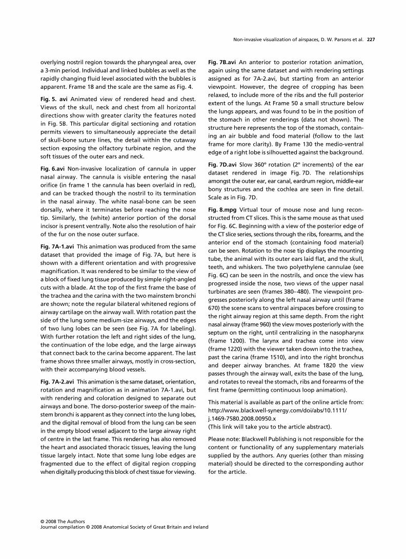
overlying nostril region towards the pharyngeal area, overa 3-min period. Individual and linked bubbles as well as therapidly changing fluid level associated with the bubbles isapparent. Frame 18 and the scale are the same as Fig. 4.
Fig. 5. avi Animated view of rendered head and chest.Views of the skull, neck and chest from all horizontaldirections show with greater clarity the features notedin Fig. 5B. This particular digital sectioning and rotationpermits viewers to simultaneously appreciate the detailof skull-bone suture lines, the detail within the cutawaysection exposing the olfactory turbinate region, and thesoft tissues of the outer ears and neck.
Fig. 6.avi Non-invasive localization of cannula in uppernasal airway. The cannula is visible entering the nasalorifice (in frame 1 the cannula has been overlaid in red),and can be tracked though the nostril to its terminationin the nasal airway. The white nasal-bone can be seendorsally, where it terminates before reaching the nosetip. Similarly, the (white) anterior portion of the dorsalincisor is present ventrally. Note also the resolution of hairof the fur on the nose outer surface.
Fig. 7A-1.avi This animation was produced from the samedataset that provided the image of Fig. 7A, but here isshown with a different orientation and with progressivemagnification. It was rendered to be similar to the view ofa block of fixed lung tissue produced by simple right-angledcuts with a blade. At the top of the first frame the base ofthe trachea and the carina with the two mainstem bronchiare shown; note the regular bilateral whitened regions ofairway cartilage on the airway wall. With rotation past theside of the lung some medium-size airways, and the edgesof two lung lobes can be seen (see Fig. 7A for labeling).With further rotation the left and right sides of the lung,the continuation of the lobe edge, and the large airwaysthat connect back to the carina become apparent. The lastframe shows three smaller airways, mostly in cross-section,with their accompanying blood vessels.
Fig. 7A-2.avi This animation is the same dataset, orientation,rotation and magnification as in animation 7A-1.avi, butwith rendering and coloration designed to separate outairways and bone. The dorso-posterior sweep of the main-stem bronchi is apparent as they connect into the lung lobes,and the digital removal of blood from the lung can be seenin the empty blood vessel adjacent to the large airway rightof centre in the last frame. This rendering has also removedthe heart and associated thoracic tissues, leaving the lungtissue largely intact. Note that some lung lobe edges arefragmented due to the effect of digital region croppingwhen digitally producing this block of chest tissue for viewing.
Fig. 7B.avi An anterior to posterior rotation animation,again using the same dataset and with rendering settingsassigned as for 7A-2.avi, but starting from an anteriorviewpoint. However, the degree of cropping has beenrelaxed, to include more of the ribs and the full posteriorextent of the lungs. At Frame 50 a small structure belowthe lungs appears, and was found to be in the position ofthe stomach in other renderings (data not shown). Thestructure here represents the top of the stomach, contain-ing an air bubble and food material (follow to the lastframe for more clarity). By Frame 130 the medio-ventraledge of a right lobe is silhouetted against the background.
Fig. 7D.avi Slow 360º rotation (2º increments) of the eardataset rendered in image Fig. 7D. The relationshipsamongst the outer ear, ear canal, eardrum region, middle-earbony structures and the cochlea are seen in fine detail.Scale as in Fig. 7D.
Fig. 8.mpg Virtual tour of mouse nose and lung recon-structed from CT slices. This is the same mouse as that usedfor Fig. 6C. Beginning with a view of the posterior edge ofthe CT slice series, sections through the ribs, forearms, and theanterior end of the stomach (containing food material)can be seen. Rotation to the nose tip displays the mountingtube, the animal with its outer ears laid flat, and the skull,teeth, and whiskers. The two polyethylene cannulae (seeFig. 6C) can be seen in the nostrils, and once the view hasprogressed inside the nose, two views of the upper nasalturbinates are seen (frames 380–480). The viewpoint pro-
gresses posteriorly along the left nasal airway until (frame670) the scene scans to ventral airspaces before crossing tothe right airway region at this same depth. From the rightnasal airway (frame 960) the view moves posteriorly with theseptum on the right, until centralizing in the nasopharynx(frame 1200). The larynx and trachea come into view(frame 1220) with the viewer taken down into the trachea,past the carina (frame 1510), and into the right bronchusand deeper airway branches. At frame 1820 the viewpasses through the airway wall, exits the base of the lung,and rotates to reveal the stomach, ribs and forearms of thefirst frame (permitting continuous loop animation).
This material is available as part of the online article from:http://www.blackwell-synergy.com/doi/abs/10.1111/j.1469-7580.2008.00950.x(This link will take you to the article abstract).
Please note: Blackwell Publishing is not responsible for thecontent or functionality of any supplementary materialssupplied by the authors. Any queries (other than missingmaterial) should be directed to the corresponding authorfor the article.
Related Documents