High molecular weight hyaluronan reduced renal PKC activation in geneti- cally diabetic mice Giuseppe M. Campo, Angela Avenoso, Antonio Micali, Giancarlo Nas- tasi, Francesco Squadrito, Domenica Altavilla, Alessandra Bitto, Francesca Polito, Maria Grazia Rinaldi, Alberto Calatroni, Angela D’Ascola, Salvatore Campo PII: S0925-4439(10)00166-3 DOI: doi: 10.1016/j.bbadis.2010.08.004 Reference: BBADIS 63154 To appear in: BBA - Molecular Basis of Disease Received date: 30 June 2010 Revised date: 4 August 2010 Accepted date: 10 August 2010 Please cite this article as: Giuseppe M. Campo, Angela Avenoso, Antonio Micali, Gi- ancarlo Nastasi, Francesco Squadrito, Domenica Altavilla, Alessandra Bitto, Francesca Polito, Maria Grazia Rinaldi, Alberto Calatroni, Angela D’Ascola, Salvatore Campo, High molecular weight hyaluronan reduced renal PKC activation in genetically diabetic mice, BBA - Molecular Basis of Disease (2010), doi: 10.1016/j.bbadis.2010.08.004 This is a PDF file of an unedited manuscript that has been accepted for publication. As a service to our customers we are providing this early version of the manuscript. The manuscript will undergo copyediting, typesetting, and review of the resulting proof before it is published in its final form. Please note that during the production process errors may be discovered which could affect the content, and all legal disclaimers that apply to the journal pertain.

Welcome message from author
This document is posted to help you gain knowledge. Please leave a comment to let me know what you think about it! Share it to your friends and learn new things together.
Transcript

�������� ����� ��
High molecular weight hyaluronan reduced renal PKC activation in geneti-cally diabetic mice
Giuseppe M. Campo, Angela Avenoso, Antonio Micali, Giancarlo Nas-tasi, Francesco Squadrito, Domenica Altavilla, Alessandra Bitto, FrancescaPolito, Maria Grazia Rinaldi, Alberto Calatroni, Angela D’Ascola, SalvatoreCampo
PII: S0925-4439(10)00166-3DOI: doi: 10.1016/j.bbadis.2010.08.004Reference: BBADIS 63154
To appear in: BBA - Molecular Basis of Disease
Received date: 30 June 2010Revised date: 4 August 2010Accepted date: 10 August 2010
Please cite this article as: Giuseppe M. Campo, Angela Avenoso, Antonio Micali, Gi-ancarlo Nastasi, Francesco Squadrito, Domenica Altavilla, Alessandra Bitto, FrancescaPolito, Maria Grazia Rinaldi, Alberto Calatroni, Angela D’Ascola, Salvatore Campo,High molecular weight hyaluronan reduced renal PKC activation in genetically diabeticmice, BBA - Molecular Basis of Disease (2010), doi: 10.1016/j.bbadis.2010.08.004
This is a PDF file of an unedited manuscript that has been accepted for publication.As a service to our customers we are providing this early version of the manuscript.The manuscript will undergo copyediting, typesetting, and review of the resulting proofbefore it is published in its final form. Please note that during the production processerrors may be discovered which could affect the content, and all legal disclaimers thatapply to the journal pertain.

ACC
EPTE
D M
ANU
SCR
IPT
ACCEPTED MANUSCRIPT
1
HIGH MOLECULAR WEIGHT HYALURONAN REDUCED RENAL PKC ACTIVATION
IN GENETICALLY DIABETIC MICE
Giuseppe M. Campo, Angela Avenoso, Antonio Micali#, Giancarlo Nastasi, Francesco
Squadrito*, Domenica Altavilla*, Alessandra Bitto*, Francesca Polito* Maria Grazia Rinaldi*,
Alberto Calatroni, Angela D’Ascola, Salvatore Campo.
Department of Biochemical, Physiological and Nutritional Sciences, section of Medical Chemistry,
#Department of Biomorphology and *Department of Clinical and Experimental Medicine and
Pharmacology, Section of Pharmacology, School of Medicine, University of Messina, Policlinico
Universitario, 98125 – Messina, Italy
Running title: Hyaluronan reduced PKC in experimental diabetes
Key words: hyaluronan; NF-kB; PKC; cytokines; diabetes; CD44
________________________________________________________________________________
Correspondence to: Giuseppe M. Campo, PhD, Department of Biochemical, Physiological and
Nutritional Sciences, School of Medicine, University of Messina, Policlinico Universitario, Torre
Biologica, 5° piano, Via C. Valeria – 98125 - Messina, Italy; phone +39 90 221 3334; fax +39 90
221 3898; e-mail [email protected]
ACC
EPTE
D M
ANU
SCR
IPT
ACCEPTED MANUSCRIPT
2
Abstract
The cluster determinant (CD44) seems to play a key role in tissues injured by diabetes type II.
CD44 stimulation activates the protein kinase C (PKC) family which in turn activates the
transcriptional nuclear factor kappaB (NF-kB) responsible for the expression of the inflammation
mediators such as tumor necrosis factor alpha (TNF-α), interleukin-6 (IL-6), interleukin-18 (IL-18),
inducible nitric oxide synthase (iNOS) and matrix metalloproteinases (MMPs). Regulation of CD44
interaction with its ligands depends greatly upon PKC. We investigated the effect of the treatment
with high molecular weight hyaluronan (HA) on diabetic nephropathy in genetically diabetic mice.
BKS.Cg-m+/+Leprdb mice had elevated plasma insulin from 15 days of age and high blood sugar
levels at four weeks. The severe nephropathy that developed was characterized by a marked
increased in CD44 receptors, protein kinase C betaI, betaII and epsilon (PKCβI, PKCβII and PKCε)
mRNA expression and the related protein products in kidney tissue. High levels of mRNA and
related protein levels were also detected in the damaged kidney for NF-kB, TNF-α, IL-6, IL-18,
MMP-7 and iNOS.
Chronic daily administration of high molecular mass HA for two weeks significantly reduced
CD44, PKCβI, PKCβII and PKCα gene expression and the related protein production in kidney
tissue and TNF-α, IL-6, IL-18, MMP-7 and iNOS expression and levels also decreased.
Histological analysis confirmed the biochemical data. However, blood parameters of diabetes were
unchanged.
These results suggest that the CD44 and PKC play an important role in diabetes and interaction of
high molecular weight HA with these proteins may reduce inflammation and secondary pathologies
due to this disease.

ACC
EPTE
D M
ANU
SCR
IPT
ACCEPTED MANUSCRIPT
3
1. Introduction
Over the last decade, considerable evidence has demonstrated the molecular and cellular links
between metabolism and inflammation, particularly in obesity and diabetes. Diabetic nephropathy is
one of the major microvascular complications in diabetes and it is the most common single cause of
end-stage renal disease worldwide [1]. The development of diabetic nephropathy is described as a
process progressing from moderate glomerular hyperfiltration and nephromegaly to frank
proteinuria with severe hypertension and marked renal insufficiency until end-stage renal disease
[2]. Chronic inflammation, characterized by elevated circulating levels of inflammatory markers,
appears to play a critical role in the pathogenesis of type 2 diabetes and its associated
complications, particularly diabetic nephropathy and the consequent cardiovascular risk [3]. Recent
evidence indicates that chemokines, interleukins, adhesion molecules, growth factors, and free
radicals are deeply involved in the development of diabetic nephropathy [4-5]. The cytokines that
seem to play a key role in diabetic renal failure include pro-inflammatory cytokines such as tumor
necrosis alpha (TNF-α), interleukin-6 (IL-6), and interleukin-18 (IL-18) [6-8]. Inducible nitric
oxide synthase (iNOS), and cytokine-stimulated metalloproteinases (MMPs), especially MMP-7,
are also greatly involved in the development of diabetic complications and kidney damage [9-10].
One ROS, nitric oxide (NO), is a highly reactive, cytotoxic molecule that is heavily implicated in
diabetes [11] Increased levels of NO promotes numerous effects on podocytes, including alteration
of extracellular matrix synthesis [12], and activation of apoptosis [13].
Biologics that inhibit the signalling cascade mediated by pro-inflammatory cytokines are effective
in treating diabetic pathologies by reducing both inflammation and renal failure [6, 14-15].
However, blocking IL-18 and/or TNF-α does not lead to total protection from diabetic
complications, indicating that other signalling pathways that mediate inflammation and tissue injury
have still to be elucidated [6, 14-15].
Hyaluronan (HA) is a linear polymer composed of repeating disaccharides of D-glucuronic acid -
N-acetyl D-glucosamine [16]. HA can exist as a high-molecular-weight polymer (106 D) or in lower

ACC
EPTE
D M
ANU
SCR
IPT
ACCEPTED MANUSCRIPT
4
molecular weight forms. Low molecular mass HA in particular is produced after tissue injury [17].
Several findings have shown that HA functions are determined in part by the size of the molecule.
High molecular weight HA is suggested to play a structural role and to promote tissue integrity,
while low molecular weight HA may be a signal of tissue injury [18-20].
The best-known HA receptor is CD44 [21], this is a transmembrane glycoprotein which is widely
found in T lymphocytes, granulocytes, monocytes, fibroblasts, keratinocytes, and epithelial cells.
CD44 stimulation with HA plays a role in various physiological functions, such as cell-adhesion,
cell-substrate interactions, and lymphocyte recruitment, as well as in pathological processes such as
chronic inflammation and metastasis of malignant cells [22]. A role for CD44, as HA receptor, in
the regulation of inflammation in vivo has been shown by studies in which anti-CD44 treatment
reduces not only inflammation in experimental diabetes [23-24], but also leukocyte migration in
skin-associated immune disease [25].
A number of reports have shown PKC members to be greatly involved in the mediation of
inflammation [26]. The regulation of CD44 interaction with its ligands depends heavily on PKC
which modulates the phosphorylation state of CD44 [27] and the various anchoring proteins [28].
Interestingly, PKC activation leads to the redistribution of CD44 receptors in membrane ruffles
located at the leading edge of glioma cells [29]; a key feature of inflammation and invading cells.
PKC therefore has the potential to modulate both the affinity of CD44 for HA [30] as well as the
localization of the receptor over the cell surface [29]. It has also been reported that the activation of
the CD44 receptor by HA produces PKC activation [31], thus establishing a concatenated
mechanism between the CD44 receptor and PKC. In addition, these processes are to a large extent
mediated by the increased expression of pro-inflammatory cytokines and other detrimental
molecules such as NO and MMPs, which are in turn controlled by the activation of key
transcriptional nuclear factor kB (NF-kB) [32].
As previous investigations reported that the inhibition of PKC isoforms such as PKCα, PKCβI and
PKCβII reduces diabetic complications, especially diabetic nephropathy [33-35], and since we

ACC
EPTE
D M
ANU
SCR
IPT
ACCEPTED MANUSCRIPT
5
demonstrated in a previous study that high molecular mass HA is able to reduce phorbol-12-
myristate-13-acetate (PMA)-induced inflammation in mouse chondrocytes by interacting with PKC
[19], the aim of this study was to investigate whether high molecular weight HA administration, at
different concentrations, influences CD44 and PKC function in congenic diabetic BKS.Cg-
m+/+Leprdb mice.
2. Materials and Methods
2.1. Animals.
Male congenic diabetic BKS.Cg-m+/+Leprdb mice 15 weeks old with a mean weight of 25-30 g
were used in our study. Mice, purchased from The Jackson Laboratory (Bar Harbor, Maine, USA)
were maintained under climate-controlled conditions with a 12-h light/dark cycle. The animals were
fed standard rodent chow and provided water ad libitum. The health status of the animal colony was
monitored in accordance with Italian Veterinary Board guidelines. Mice were divided into the
following groups: 1) Control (n=12); 2) Control + HA (30 mg/kg) (n=12); 3) Diabetic (n=17); 4)
Diabetic + HA (7.5 mg/kg) (n=17); 5) Diabetic + HA (15 mg/kg) (n=17); 6) Diabetic + HA (30
mg/kg) (n=17).
2.2. Materials.
High molecular weight HA (4,000,000 Da) was purchased from Sigma-Aldrich Srl, (Milan, Italy).
Mouse TNF-α (cat. IB49688), IL-6 (cat. IB49686), IL-18 (cat. IB49697) commercial ELISA kits
were provided by Immuno-Biological laboratories Inc. (Minneapolis, MN, USA). Mouse MMP-7
(cat. E0102Mu) and iNOS (cat. E0837Mu) commercial ELISA kits were obtained from USCN Life
Science Inc. Wuhan (Wuhan, China). Mouse CD44 monoclonal antibodies were supplied by
Millipore (Billerica, MA, USA), mouse PKCα monoclonal antibodies were supplied by Abcam plc
(Cambridge, UK), mouse PKCβII polyclonal antibodies were supplied by Sigma-Aldrich (Milan,
Italy) and PKCβI polyclonal antibodies and Horseradish peroxidase-labeled goat anti-rabbit

ACC
EPTE
D M
ANU
SCR
IPT
ACCEPTED MANUSCRIPT
6
antibodies were obtained from Santa Cruz Biotechnology (Santa Cruz, CA, USA). All other
reagents used were purchased from Fluka (division of Sigma-Aldrich Srl, Milan, Itay).
2.3. HA treatment.
HA was dissolved in saline solution (0.9% NaCl) at three different concentrations (7.5, 15 and 30
mg/kg and administered intraperitoneally at a volume of 1.0 ml/kg body weight, once a day starting
from day one up until day 15.
2.4. Plasma glucose and insulin
Glucose was evaluated in plasma samples (100 µl) at the end of the experiment. Concentrations
were assayed using commercial clinical test kits (cat.GAGO20) obtained from Sigma-Aldrich
(Milan, Italy). Insulin levels were also measured in plasma samples (10 µl) at the end of the
experiment using an ELISA commercial kit (cat. EZRMI-13K) provided by Millipore (Billerica,
MA, USA).
2.5. RNA isolation, cDNA synthesis and real-time quantitative PCR amplification
Total RNA was isolated from renal tissue for reverse-PCR real time analysis of CD44, PKCβI,
PKCβII, PKCα, TNF-α, IL-6, IL-18, MMP-7 and iNOS (RealTime PCR system, Mod. 7500,
Applied Biosystems, USA) using an Omnizol Reagent Kit (Euroclone, West York, UK). The first
strand of cDNA was synthesized from 1.0 µg total RNA using a high capacity cDNA Archive kit
(Applied Biosystems, USA). β-actin mRNA was used as an endogenous control to allow the relative
quantification of. CD44, PKCβI, PKCβII, PKCα, TNF-α, IL-6, IL-18, MMP-7 and iNOS. PCR
RealTime was performed by means of ready-to-use assays (Assays on demand, Applied Biosystems)
on both targets and endogenous controls. The amplified PCR products were quantified by measuring
the calculated cycle thresholds (CT) of CD44, PKCβI, PKCβII, PKCα, TNF-α, IL-6, IL-18, MMP-7,
iNOS, and β-Actin mRNA. The amounts of specific mRNA in samples were calculated using the

ACC
EPTE
D M
ANU
SCR
IPT
ACCEPTED MANUSCRIPT
7
∆∆CT method. The mean value of normal cartilage target levels became the calibrator (one per
sample) and the results are expressed as the n-fold difference relative to normal controls (relative
expression levels).
2.6. Western blot assay of CD44, PKCβI, PKCβII, and PKCα, proteins
For SDS-PAGE and Western blotting, the kidney tissue samples were washed twice in ice-cold PBS
and subsequently dissolved in SDS sample buffer (62.5 mM Tris/HCl, pH 6.8, 2% w/v SDS, 10%
glycerol, 50 mM dithiothreitol, 0.01% w/v bromophenol blue). Aliquots of whole cell protein extract
(10–25 µl/well) were separated on a mini gel (10%). The proteins were blotted onto polyvinylidene
difluoride membranes (Amersham Biosciences) using a semi-dry apparatus (Bio-Rad). The
membranes were then incubated overnight in a roller bottle with the specific diluted (1:1,000)
primary antibody in 5% bovine serum albumin, 1x PBS, and 0.1% Tween 20 at 4 °C. After being
washed in three stages in wash buffer (1x PBS, 0.1% Tween 20), the blots were incubated with the
diluted (1:2,500) secondary polyclonal antibody (Goat anti-rabbit conjugated with peroxidase), in
TBS/Tween-20 buffer, containing 5% non-fat dried milk. After 45 min of gentle shaking, the blots
were washed five times in wash buffer, and the proteins, after coloration with DAB liquid substrate
(Sigma-Aldrich, Milan, Italy) were made visible using a UV/visible transilluminator (EuroClone,
Milan, Italy) and Kodak BioMax MR films. A densitometric analysis was also run in order to
quantify each band.
2.7. NF-kB p50/65 transcription factor assay
NF-kB p50/65 DNA binding activity in nuclear extracts of kidney tissue samples was evaluated in
order to measure the degree of NF-kB activation. Analysis was performed in line with the
manufacturer’s protocol for a commercial kit (NF-kB p50/65 Transcription Factor Assay
Colorimetric, cat. n°SGT510, Chemicon International, USA). In brief, one of the kidney was

ACC
EPTE
D M
ANU
SCR
IPT
ACCEPTED MANUSCRIPT
8
removed from the animals at the end of the experimental phase, washed in ice-cold 10 mM Tris-
HCl, pH 7.4, and blotted on absorbent paper. Samples were then plotted, using an automatic plotter,
in order to isolate renal cells. Cytosolic and nuclear extraction was performed by lysing the cell
membrane with an appropriate hypotonic lysis buffer containing protease inhibitor cocktail and
tributylphosphine (TBP) as reducing agent. After centrifugation at 8,000 x g, the supernatant
containing the cytosolic fraction was discarded, while the pellet containing the nuclear portion was
then re-suspended in the apposite extraction buffer and the nuclei were disrupted by a series of
drawing and ejecting actions. The nuclear suspension was then centrifuged at 16,000 x g. The
supernatant fraction was the nuclear extract. After the determination of protein concentration and
adjustment to a final concentration of approximately 4.0 mg/ml, this extract was stored in aliquots
at -80° C for the subsequent NF-kB assay. After incubation with primary and secondary antibodies,
colour development was observed following the addition of the substrate TMB/E. Finally, the
absorbance of the samples was measured using a spectrophotometric microplate reader set at λ 450
nm. Values are expressed as relative optical density (OD) per mg protein.
2.8. TNF-α, IL-6, IL-18, MMP-7 and iNOS ELISA assay
Tissue samples obtained from mice kidneys in the presence of 1.0 nM PMSF and protease inhibitor
cocktail were first lysed using a suitable hypotonic lysis buffer and then centrifuged at 13,000 rpm
for 10 min at 4°C. The analysis of TNF-α, IL-6, and IL-18 was carried out using a specific
commercial kit. Briefly, 50 µl of standards, samples and controls were added to each well of the
coated microplate. 50 µl of each specific biotin-conjugate antibody was then added to each well.
After 120 min of incubation at 20-22°C, the liquid from the wells was discarded, the wells were
washed three times and 100 µl of Streptavidin-HRP was added. After further incubation for 60 min
and having washed the wells once more, 100 µl of a substrate chromogen solution was added. After
10 min incubation and the addition of 100 µl of stop solution, the absorbance of each well was read
ACC
EPTE
D M
ANU
SCR
IPT
ACCEPTED MANUSCRIPT
9
spectrophotometrically at λ 450 nm. TNF-α IL-6 and IL-18 values are expressed as pg/mg protein.
For MMP-7 and iNOS, 100 µl of previously diluted samples, standards and controls were added to
each well of the coated microplate. After incubation for 2 hours at 37° C, the liquid from each well
was discarded. After adding 100 µl of the detection reagent A and incubation at 37° C for 1 hour,
the liquid was aspirated and wells were washed with wash buffer. At this point, 100 ml of detection
reagent B was added to each well, and after a further incubation for 1 hour at 37° C the liquid of
wells was first aspired then wells were washed with wash buffer. 90 µl of the substrate solution was
added and after incubation at room temperature for 30 minutes, 50 µl of stop solution was added.
Within 30 minutes, the absorbance of each well was read spectrophotometrically at λ 450 nm.
MMP-7 values are expressed as pg/mg protein, while iNOS values are expressed as U/mg protein.
2.9. Histology
At the end of the experimental phase, the right kidney was first excised and then perfused with 4%
paraformaldeyde (PFA)/0.1 M phosphate buffer (PB), decapsulated and immersed in 4% PFA/0.1
M PB. Then kidneys were dehydrated in graded ethanol and embedded in a special tissue
embedding media (Paraplast, McCormick Scientific, Richmond, VA, USA). Sections of 4 µm
thickness were cut with a microtome (mod. RM 2125 RT, Leica Camera AG, Solms, Germany),
stained with Hematoxilin-Eosin technique and viewed and photographed with a light microscope
(mod. BH2, Olympus America Inc., Melville, USA).
2.10. Protein analysis
The amount of protein was determined using the Bio-Rad protein assay system (Bio-Rad Lab.,
Richmond, CA, USA) with bovine serum albumin as a standard in accordance with the published
method [36].
ACC
EPTE
D M
ANU
SCR
IPT
ACCEPTED MANUSCRIPT
10
2.11. Statistical analysis
Data are expressed as means ± S.D. of no less than seven experiments for each test. Statistical
analysis was performed by one-way analysis of variance (ANOVA) followed by the Student-
Newman-Keuls test. The statistical significance of differences was set at p<0.05.
2.12. Statement of animal care
The studies reported in this manuscript were carried out in accordance with the Helsinki declaration
and the NIH guidelines for the Care and Use of Laboratory Animals.
3. Results
3.1. Effects of HA treatment on plasma glucose and insulin
At the age of four weeks the acute expression of the db/db mutation commonly induces noticeable
alterations in body phenotype, tissue indexes, and the endocrine-related parameters of BKS.Cg-
m+/+Leprdb mice relative to controls [37]. Increased body weight in these mice is related to
concomitant elevations in blood glucose and insulin levels when compared with control values [37].
The 15-day HA treatment period , at all doses, had no effect on plasma glucose and insulin in both
diabetic and control mice (data not shown).
3.2. CD44, PKCβI, PKCβII and PKCα mRNA expression and Western blot analysis.
CD44, PKCβI, PKCβII and PKCα (Figs. 1, 2, 3 and 4) mRNA evaluation (panels A of each Figure)
and Western blot analysis with densitometric evaluation (Figs. 1, 2, 3 and 4, panels B and C of each
Figure) were assayed in order to estimate the degree of CD44 activation and the consequent cell
signalling pathway booster that culminates in NF-kB factor activation. Very low gene expression
and the related protein production of CD44, PKCβI, PKCβII and PKCα were obtained in non-diabetic

ACC
EPTE
D M
ANU
SCR
IPT
ACCEPTED MANUSCRIPT
11
mice (CTRL and CTRL+HA). In contrast, a marked increase in the expression and protein synthesis
of the CD44 receptor and their signal mediators PKCβI, PKCβII and PKCα were found in the kidney
tissue of diabetic mice. This means that CD44 receptor and the PKC pathway are involved in the
inflammatory mechanism consequent to this pathology.
The treatment with high molecular weight HA for 15 days significantly reduced CD44 receptor
activation and its signal mediator levels in diabetic mice. As reported in Figs. 1, 2, 3 and 4, HA was
able to lower CD44, PKCβI, PKCβII and PKCα expression and protein synthesis at all concentrations
in a dose-dependent manner.
3.3. NF-kB activation.
Fig. 5 shows the changes in the NF-kB p50/p65 heterodimer translocation over the course of the
experiment. NF-kB DNA binding was present at very low levels in the renal tissue of non-diabetic
mice. In contrast, diabetic complications induced massive NF-kB translocation into the nucleus of
the damaged podocytes; the treatment of diabetic mice with HA at different concentrations reduced
NF-kB activation in a dose-dependent manner, thereby confirming the effect on CD44, PKCβI,
PKCβII and PKCα parameters.
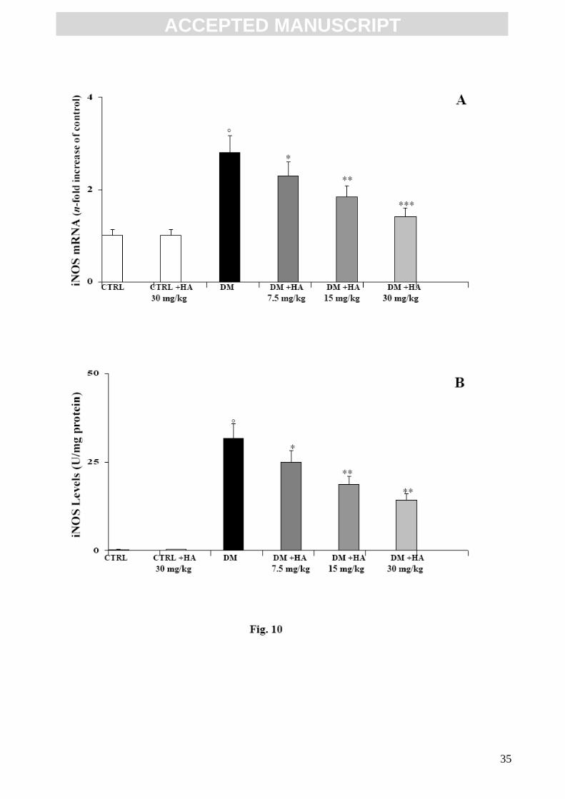
3.4. TNF-α, IL-6, IL-18, MMP-7 and iNOS mRNA expression and ELISA assay.
TNF-α, IL-6, IL-18, MMP-7 and iNOS were assayed because of their active role in inflammation
and diabetic nephropathy. (Figs. 6, 7, 8, 9 and 10). Their mRNA evaluation (panel A of each
Figure) and protein concentration (panel B of each Figure) showed that the mRNA of these
detrimental mediators was not stimulated in non-diabetic mice and protein level was very low.
However, in diabetic mice, TNF-α, IL-6, IL-18, MMP-7 and iNOS expression and the related
protein concentration were significantly up-regulated. The administration of high molecular weight
HA was able to reduce this increment in mRNA expression at all doses. As reported (Figs. 6, 7, 8,
9, and 10; panels A and B of each Figure), the increase in mRNA expression of TNF-α, IL-6, IL-18,

ACC
EPTE
D M
ANU
SCR
IPT
ACCEPTED MANUSCRIPT
12
MMP-7 and iNOS in diabetic mice correlated well with the increment in protein synthesis. This
correlation was also maintained in mice treated with HA. In fact, the reduction in mRNA expression
resulted in a similar diminution in protein formation.
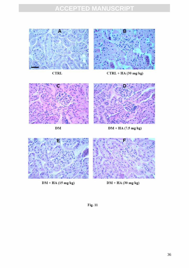
3.5. Histological analysis
Glomerular injury in the diabetic mice can be early characterized by glomerular hyperplasia and
mesangial matrix expansion. Fig. 11 demonstrates a representative kidney histopathology of all the
experimental groups. Panels A and B show a normal kidney tissue section from undiabetic control
mice untreated or treated with HA. In contrast, as reported in Figure 11 panel C, the glomerular
hyperplasia and mainly the mesangial cell increment is clear evident. In panels D, E, and F a
representative selection of kidney tissue sections from diabetic mice receiving HA treatment shows
that a gradual reduction in glomerular hyperplasia and mesangial matrix expansion occurred. As
indicated by the intensive coloration the number of mesangial cells increased dramatically.
4. Discussion
Diabetes is a chronic disease in which patients develop dramatic and devastating complications. The
most severe complication of diabetes is nephropathy, which frequently causes death in diabetic
patients and accounts for a large number of cases of end-stage renal disease.
In the present study, we investigated the effects of high molecular weight HA, at different
concentrations, on CD44 receptor modulation in congenitally diabetic mice. This study suggests
that highly polymerized HA may reduce diabetes-induced renal injury by modulating CD44
signalling, in contrast with low molecular weight HA which may exert the opposite effects as
previously demonstrated [17-20]. HA effects were dose dependent and were able to reduce not only
the expression of CD44 receptor and of PKCβI, PKCβII, and PKCα but also NF-kB activation and
the increment of inflammatory cytokines, such as IL-6 and IL-18, and of MMP-7 and iNOS that

ACC
EPTE
D M
ANU
SCR
IPT
ACCEPTED MANUSCRIPT
13
were up-regulated in diabetic mice. Biochemical results were further confirmed by the reduction in
glomerulosclerosis as revealed by the histological analysis.
HA possesses an extraordinarily rapid half.life. It is quickly eliminated by means of CD44 receptors
on liver endothelium. At a normal plasma concentration the half-life is approximately 4-6 minutes,
while at higher plasma concentrations the HA half-life is considerable prolonged to 18-43 minutes
[38]. The intraperitoneal HA half-life is unknown, but is probably also very rapid, and also in this
case an increase in the HA dose administered may result in a prolonged HA half-life with a
consequent major bioavailability to different organs. Therefore we might expect a linear
progression of effect by increasing the HA range concentrations administered. However, due to its
viscous properties, it is really difficult to administer concentrations higher than 30 mg/kg.
The interaction between cell surface CD44 and extracellular HA are implicated in a variety of
physiological and pathological processes, such as the uptake and degradation of HA, T cell
activation, leukocyte extravasation at inflammation sites, and tumor metastasis [39]. CD44
expression in the renal tissue correlates with the degree of the nephropathy in diabetic rats [40], and
at high levels in several other form of nephropathy stimulates the increment in inflammatory
mediators that are the main cause of renal damage [41-42].
Protein kinase C (PKC) is a family of phospholipid-dependent serine/threonine kinases that consists
of a number of isoforms [43]. The differences between the PKC isoforms dependent on their
substrate specificity suggest that a particular PKC isoform within a single cell may regulate specific
physiological responses. PKCs regulate a variety of biological functions including ion channel
activity, cell differentiation, and other biochemical and physiological responses to various stimuli
[44-45]. The PKC/PKD family exerts numerous effects on cellular pathways that are important in
inflammation and has previously been considered as a potential therapeutic target in diabetes [33-
35, 46]. PKC is able to modify the phosphorylation state of the CD44 receptor and the interaction
with HA [27], and PKC in turn may be activated through different pathways, among these by CD44
stimulation [31]. Therefore, CD44 and PKC are interdependent and greatly involved in the

ACC
EPTE
D M
ANU
SCR
IPT
ACCEPTED MANUSCRIPT
14
inflammation cascade. NF-kB promotes the transcription of several genes including cytokines and
other inflammation products [32]. CD44 stimulation activates members of the PKC family, such as
PKCα and PKCβ [47−48], which in turn activate the NF-kB responsible for the expression of
inflammation mediators such as TNF-α, IL-6, IL-18, iNOS, its dangerous free radical product, NO,
and MMPs responsible for diabetic nephropathy [5-13, 49-51].
Cell interaction with the surrounding extracellular matrix is fundamental in many physiological and
pathological mechanisms. Proteoglycans (PGs) may influence cell behaviour through binding
events mediated by their glycosaminoglycan (GAG) chains. The binding affinity of the interaction
depends on the ability of the oligosaccharide sequence to provide an optimal charge and surface
with the protein [52]. The interaction of HA degradation products with CD44 provides signals to
initiate inflammation [22-23, 25]. Other data support the idea that a balance between low molecular
weight HA and high molecular weight HA may control the activation of inflammation [53]. In fact,
these preliminary findings suggest that high molecular weight HA is inactive for CD44 modulation,
while low molecular weight HA originating from depolymerised HA at the inflammation site is
active. [17-18, 54]. We previously reported that high molecular weight HA is able to reduce
inflammation stimulated by PMA in mouse chondrocytes. By using a specific CD44 blocking
antibody it was demonstrated that PKC and not CD44 receptor is the target for HA action [19]. We
suggest the same mechanism to explain the findings of the present study, in which chronic daily
administration of high molecular mass HA for 2 weeks significantly reduced CD44, PKCβI, PKCβII
and PKCα gene expression and the related protein production in kidney tissue and TNF-α, IL-6, IL-
18, MMP-7 and iNOS expression levels.
The effect exerted by HA on the parameters considered could be due to the ability of HA to bind
protein structures, such as PKC, thereby exerting a block that reduced NF-kB activation. A similar
effect exerted directly on the CD44 receptor should be excluded, since the high molecular mass HA
interaction with CD44 seems to be just one step in a pathway of intermediary metabolism involving
the catabolism of native HA into small fragments [55]. The rationale for the use of high molecular

ACC
EPTE
D M
ANU
SCR
IPT
ACCEPTED MANUSCRIPT
15
mass HA in place of a specific antibody targeted at CD44 may be explained by the fact that a block
of the CD44 receptor leads to the abrogation of several fundamental CD44 functions, such as cell
adhesion, cell-substrate interaction, and lymphocyte recruitment [39].
Blocking PKC isoforms using specific agents was also previously attempted but the effects are not
very encouraging because of the high toxicity of these agents [56]. The choice of HA as modulating
agent derived essentially from its natural endogenous origin. The number of active charged HA
sites (carboxylic groups) is responsible for HA action. In fact, high molecular weight HA contains
many charged groups that may complete wrap protein structures such as PKC, thereby preventing
its activation. The mechanism could be similar to a well known lock-and-key model which applies
to all the better-known effector-receptors where the effector is chemically specific and a similar
molecule with several charged groups may block the receptor. In this work, the larger size of HA
molecule was able to mask the PKC active site completely. The resultant inhibition of PKC thereby
produced a reduction in NF-kB activation that in turn limited the transcription of the detrimental
inflammatory intermediates with a consequent reduction in tissue injury, in particular in the
limitation of diabetic nephropathy as the results of this study reported.
However, further studies are needed to fully confirm these hypotheses.
Acknowledgements
This study was supported by a grant PRA (Research Athenaeum Project 2007-2008) from the
University of Messina, Italy.
5. References
[1] T. Zelmanovitz, F. Gerchman, A.P. Balthazar, F.C. Thomazelli, J.D. Matos, L.H. Canani,
Diabetic nephropathy, Diabetol. Metab. Syndr. 1 (2009) 1-17.
[2] T.W. Tervaert, A.L. Mooyaart, K. Amann, A.H. Cohen, H.T. Cook, C.B. Drachenberg, F.
Ferrario, A.B. Fogo, M. Haas, E.D. Heer, K. Joh, L.H. Noel, J. Radhakrishnan, S.V. Seshan,

ACC
EPTE
D M
ANU
SCR
IPT
ACCEPTED MANUSCRIPT
16
I.M. Bajema, J.A. Bruijn, Pathological classification of diabetic nephropathy, J. Am. Soc.
Nephrol. (2010) E-pub.
[3] J.F. Navarro-Gonzales, C. Mora-Fernandez, The role of inflammatory cytokines in diabetic
nephropathy, J. Am. Soc. Nephrol. 19 (2008) 433-442.
[4] C.C. Wu, J.S. Chen, K.C. Lu, C.C. Chen, S.H. Lin, P. Chu, H.K. Sytwu, Y.F. Lin, Aberrant
cytokines/chemokines production correlate with proteinuria in patients with overt diabetic
nephropathy, Clin. Chim. Acta (2010) E-pub.
[5] F.A. Hakim, A. Pflueger, Role of oxidative stress in diabetic kidney disease, Med. Sci. Monit.
16 (2010) RA37-48.
[6] J.F. Nvarro-Gonzales, A. Jarque, M. Muros, C. Mora, J. Garcia, Tumor necrosis factor-alpha as
a therapeutic target for diabetic nephropathy, Cytokine Growth Factor Rev. 20 (2009) 165-173.
[7] R.B. Goldberg, Cytokine and cytokine-like inflammation markers, endothelial dysfunction, and
imbalanced coagulation in development of diabetes and its compications, J. Clin. Endocrinol.
Metab. 94 (2009) 3171-3182.
[8] K. Miyauchi, Y. Takiyama, J. Honjyo, M Tateno, M. Haneda, Upregulated IL-18 expression in
type 2 diabetic subjects with nephropathy: TGF-β1 enhanced IL-18 expression in human renal
proximal tubular epithelial cells, Diabetes Res. Clin. Pract. 83 (2009) 190-199.
[9] H. Xiao, Y. Li, J. Qi, H. Wang, K. Liu, Peroxynitrite plays a key role in glomerular lesions in
diabetic rats, J. Nephrol. 22 (2009) 800-808.
[10]K.M. Thrailkill, R.C. Bunn, J.L. Fowlkes, Matrix metalloproteinases: their potential role in the
pathogenesis of diabetic nephropathy, Endocrine 35 (2009) 1-10.
[11]S. Okada, M. Saito, E. Kazuyama, T. Hanada, Y. Kawaba, A. Hayashi, K. Satoh, S. Kanzaki,
Effects of N-hexacosanol on nitric oxide synthase system in diabetic rat nephropathy, Mol. Cell.
Biochem. 315 (2008) 169-177.
[12] G. Gruden, P.C. Perin, G. Camussi, Insight on the pathogenesis of diabetic nephropathy from
the study of podocyte and mesangial cell biology, Curr. Diabetes Rev. 1 (2005) 27-40.

ACC
EPTE
D M
ANU
SCR
IPT
ACCEPTED MANUSCRIPT
17
[13] G. Nagy, A. Koncz, D. Fernandez, A. Perl, Nitric oxide, mitochondrial hyperpolarization,
and T cell activation, Free Radic. Biol. Med. 42 (2007) 1625-1631.
[14] K.H. Han, S.Y. Han, H.S. Kim, Y.S. Kang, D.R. Cha, Prolonged administration enhanced the
renoprotective effect of pentoxifylline via anti-inflammatory activity in streptozotocin-induced
diabetic nephropathy, Inflammation (2009) E-pub.
[15]J.J. Li, S.H. Lee, D.K. Kim, R. Jin, D.S. Jung, S.J. Kwak, S.H. Kim, S.H. Han, J.E. Lee, S.J.
Moon, D.R. Ryu, T.H. Yoo, D.S. Han, S.W. Kang, Colchicine attenuates inflammatory cell
infiltration and extracellular matrix accumulation in diabetic nephropathy, Am. J. Physiol. Renal
Physiol. 297 (2009) F200-209.
[16]J.R. Fraser, T.C. Laurent, U.B. Laurent, Hyaluronan: its nature, distribution, function and
turnover, J. Intern. Med. 242 (1997) 27-33.
[17] P.W. Noble, Hyaluronan and its catabolic products in tissue injury and repair, Matrix Biol. 21
(2002) 25-29.
[18] D. Jiang, J. Liang, P.W. Noble, Hyaluronan in tissue injury and repair, Annu. Rev. Cell. Dev.
Biol. 23 (2007) 435-461.
[19] G.M. Campo, A. Avenoso, S. Campo, A. D’Ascola, P. Traina, A. Calatroni, Differential effect
of molecular size HA in mouse chondrocytes stimulated with PMA, Biochim. Biophys. Acta
1790 (2009) 1353-1367.
[20] G.M. Campo, A. Avenoso, S. Campo, A. D’Ascola, G. Nastasi, A. Calatroni, Molecular size
hyaluronan differently modulates toll-like receptor-4 in LPS-induced inflammation in mouse
chondrocytes, Biochimie 92 (2010) 204-215.
[21] A. Aruffo, I. Stamenkovic, M. Melnick, C.B. Underhill, B. Seed, CD44 is the principal cell
surface receptor for hyaluronate, Cell 61 (1990) 1303-1313.
[22] P. Heldin, E. Karousou, B. Bernert, H. Porsch, K. Nishitsuka, S.S. Skandalis, Importance of
hyaluronan-CD44 interactions in inflammation and tumorigenesis, Connect. Tissue Res. 49
(2008) 215-218.
ACC
EPTE
D M
ANU
SCR
IPT
ACCEPTED MANUSCRIPT
18
[23] L. Weiss, S. Slavin, S. Reich, P. Cohen, S. Shuster, R. Stern, E. Kaganovsky, E. Okon, A.M.
Rubinstein, D. Naor, Induction of resistance to diabetes in non-obese diabetic mice by
targeting CD44 with a specific monoclonal antibody, Proc. Natl. Acad. Sci. USA 97 (2000)
285-290
[24] L.. Weiss, A.M. Botero-Anug, C. Hand, S. Slavin, D. Naor, CD44 gene vaccination for insulin-
dependent diabetes mellitus in non-obese diabetic mice, Isr. Med. Assoc. J. 10 (2008) 20-25.
[25] M. Zoller, P. Gupta, R. Marhaba, M. Vitacolonna, P. Freyschmidt-Paul, Anti-CD44-mediated
blockade of leukocyte migration in skin-associated immune diseases, J. Leukoc. Biol. 82
(2007) 57-71.
[26] G. Baier, J. Wagner, PKC inhibitors: potential in T cell-dependent immune diseases, Curr.
Opin. Cell. Biol. (2009) -Epub-
[27] R.F. Thorne, J.W. Legg, C.M. Isacke, The role of the CD44 transmembrane and cytoplasmic
domains in co-ordinating adhesive and signalling events, J. Cell. Sci. 117 (2004) 373-380.
[28] C. Larsson, Protein kinase C and the regulation of the actin cytoskeleton, Cell. Signal. 18
(2006) 276-284.
[29] I. Okamoto, Y. Kawano, M. Matsumoto, M. Suga, K. Kaibuchi, M. Andò, H. Saya, Regulated
CD44 cleavage under the control of protein kinase C, calcium influx, and the Rho family of
small G proteins, J. Biol. Chem. 274 (1999) 25525-25534.
[30] D. Liu, T. Liu, R. Li, M.S. Sy, Mechanisms regulating the binding activity of CD44 to
hyaluronic acid, Front. Biosci. 3 (1998) d631-d636.
[31] K.A. Fitzgerald, A.G. Bowie, B.S. Skeffington, L.A.J. O’Neill, Ras, protein Kinase Czeta and
IkB kinase 1 and 2 are downstream effectors of CD44 during the activation of NF.kB by
hyaluronic acid fragments in T-24 carcinoma cells, J. Immunol. 164 (2000) 2053-2063.
[32] A. Kumar, Y. Takada, A. M. Boriek, B.B. Aggarwal, Nuclear factor-kappaB: its role in health
and disease, J. Mol. Med. 82 (2004) 434-448.

ACC
EPTE
D M
ANU
SCR
IPT
ACCEPTED MANUSCRIPT
19
[33] J. Menne, M. Meier, J.K. Park, M. Boehne, T. Kirsch, C. Lindschau, R. Ociepka, M. Leitges, J.
Rinta-Valkama, H. Holthofer, H. Haller, Nephrin loss in experimental diabetic nephropathy is
prevented by deletion of protein kinase C alpha signalling in-vivo, Kidney Int. 70 (2006)
1456-1462.
[34]D. Wu, F. Peng, B. Zhang, A.J. Ingram, D.J. Kelly, R.E. Gilbert, B. Gao, J.C. Krepinsky, PKC-
beta1 mediates glucose-induced Akt activation and TGF-beta1 upregulation in mesangial
cells, J. Am. Soc. Nephrol 20 (2009) 554-566.
[35]P.R. Nagareddy, H. Soliman, G. Lin, P.S. Rajput, U. Kumar, J.H. McNeill, K.M. MacLeod,
Selective inhibition of protein kinase C beta (2) attenuates inducible nitric oxide synthase-
mediated cardiovascular abnormalities in streptozotocin-induced diabetic rats, Diabetes 58
(2009) 2355-2364.
[36] M.M. Bradford, A rapid and sensitive method for the quantitation of microgram quantities of
protein utilizing the principle of protein-dye binding, Anal. Biochem. 72 (1976) 248-254.
[37] D.R. Garris, B.L. Garris, Estrogenic restoration of functional pancreatic islet cytoarchitecture
in diabetes (db/db) mutant C57BL/KsJ mice: relationship to estradiol localization, systemic
glycemia, and persistent hyperinsulinemia, Cell Tissue Res. 319 (2005) 231-242.
[38] L. Lebel, J. R. Fraser, W.S. Kimpton, J. Gabrielsson, B. Gerdin, T.C. Laurent, A
pharmacokinetic model of intravenously administered hyaluronan in sheep, Pharm. Res. 6
(1989) 677-682.
[39] D. Naor, S. Nedvetzki, M. Walmsley, A Yayon, E.A. Turley, I. Golan, D. Caspi, L.E. Sebban,
Y. Zick, T. Garin, D. Karussis, N. Assayag-Asherie, I. Raz, L. Weiss, S. Slavin, I. Golan,
CD44 involvement in autoimmune inflammations: the lesson to be learned from CD44-
targeting by antibody or from knockout mice, Ann. N.Y. Acad. Sci. 1110 (2007) 233-247.
[40] .J. Melin, O. Hellberg, K. Funa, R. Hallgren, E. Larsson, B.C. Fellstrom, Ischemia-induced
renal expression of hyaluronan and CD44 in diabetic rats, Nephron Exp Nephrol 103 (2006)
e86-94.

ACC
EPTE
D M
ANU
SCR
IPT
ACCEPTED MANUSCRIPT
20
[41] H.Y. Lan, Role of macrophage migration inhibition factor in kidney disease, Nephron Exp
Nephrol 109 (2008) e79-83.
[42] M. Ascon, D.B. Ascon, M. Liu, C. Cheadle, C. Sarkar, L. Racusen, H.T. Hassoun, H. Rabb,
Renal ischemia-reperfusion leads to long term infiltration of activated and effector-memory T
lymphocytes, Kidney Int 75 (2009) 526-535.
[43] A.C. Newton, Regulation of the ABC kinases by phosphorylation: protein kinase C as a
paradigm, Biochem. J. 370 (part2) (2003) 361-371.
[44] A.M. Martelli, C. Evangelisti, M. Nyakern, F.A. Manzoli, Nuclear protein kinase C, Biochim.
Biophys. Acta 1761 (2006) 542-551.
[45] A.C. Newton, Lipid activation of protein kinases, J. Lipid Res. Suppl 50 (2009) S266-271.
[46] D. Koya, M. Haneda, H. Nakagawa, K. Isshiki, H. Sato, S. Maeda, T. Sugimoto, H. Yasuda, A.
Kashiwagi, D.K. Ways, G.L. King, R. Kikkawa, Amelioration of accelerated diabetic
mesangial expansion by treatment with a PKC β inhibitor in diabetic db/db mice, a rodent
model for type 2 diabetes, FASEB J. 14 (2000) 439-447.
[47] Y. Kim, Y.S. Lee, J. Choe, H. Lee, Y.M. Kim, D. Joung, CD44-epidermal growth factor
receptor interaction mediates hyaluronic acid-promoted cell motility by activating protein
kinase C signalling involving Akt, Rac1, Phox, reactive oxygen species, focal adhesion
kinase, and MMP-2, J. Biol. Chem. 283 (2008) 22513-22528.
[48] P.V. Jensen, L.I. Larsson, Actin microdomains on endothelial cells: association with CD44,
ERM proteins, and signalling molecules during quiescence and wound healing, Histochem.
Cell. Biol. 121 (2004) 361-369.
[49] D.L. Clarke, A. Sutcliffe, K. Deacon, D. Bradbury, L. Corbett, A.J. Knox, PKCbetaII augments
NF-kappaB-dependent transcription at the CCL11 promoter via p300/CBP-Associated factor
recruitment and histone H4 acetylation, J. Immunol. 181 (20089 3503-3514.

ACC
EPTE
D M
ANU
SCR
IPT
ACCEPTED MANUSCRIPT
21
[50] R.P. Thylur, Y.D. Kim, M.S. Kwon, H.M. Oh, H.K. Kwon, S.H. Kim, S.H. Im, J.S. Chun, Z.Y.
Park, C.D. Jun, Swiprosin-1 is expressed in mast cells and up-regulated through the protein
kinase C beta I/eta pathway, J. Cell. Biochem. 108 (2009) 705-715.
[51] M. W. Greene, M.S. Ruhoff, C.M. Burrington, R.S. Garofalo, S.J. Orena, TNFalpha activation
of PKCdelta, mediated by NFkappaB and ER stress, cross.talks with the insulin signalling
cascade, Cell. Signal. 22 (2010) 274-284.
[52] E.A Yates, C.J. Terry, C. Rees, T.R. Rudd, L. Duchesne, M.A. Skidmore, R. Lèvy, N.T.
Thanh, R.J. Nichols, D.T. Clarke, D.G. Fernig, Protein-GAG interactions: new surface-based
techniques, spectroscopies and nanotechnology probes, Biochem. Soc. Trans. 34 (2006) 427-
30.
[53] D.A. Steeber, G.M. Venturi, T.F. Tedder, A new twist to the leukocyte adhesion cascade:
intimate cooperation is key, Trends Immunol. 26 (2005) 9-12.
[54] M. Eberlein, K.A. Scheibner, K.E. Black, S.L. Collins, Y. Chan-Li, J.D. Powell, M. R. Horton,
Anti-oxidant inhibition of hyaluronan fragment-induced inflammatory gene expression, J.
Inflamm. 5 (2008) 5-20.
[55] R. Stern, Hyaluronan catabolism: a new metabolic pathway, Eur. J. Cell. Biol. 83 (2004) 317-
325.
[56] J.A. Beckman, A.B. Goldfine, M.B. Gordon, L. A Garrett, M.A. Creager, Inhibition of protein
kinase Cβ prevents impaired endothelium-dependent vasodilation caused by hyperglycemia
in humans, Circ Res. 90 (2002) 107-111.

ACC
EPTE
D M
ANU
SCR
IPT
ACCEPTED MANUSCRIPT
22
Figure legends
Fig. 1
Effect of HA treatment on CD44 mRNA expression (A) and related protein production (B, C) in
kidney tissue of diabetic mice. Values are the mean ± S.D. of seven experiments and are expressed
as the n-fold increase with respect to the Control (A) and as both Densitometric analysis (C) and
Western Blot analysis (B) for CD44 protein levels. °p<0.001 vs Control; *p<0.01, **p<0.005 and
***p<0.001 vs diabetic.
Fig. 2
Effect of HA treatment on PKCβI mRNA expression (A) and related protein production (B, C) in
kidney tissue of diabetic mice. Values are the mean ± S.D. of seven experiments and are expressed
as the n-fold increase with respect to the Control (A) and as both Densitometric analysis (C) and
Western Blot analysis (B) for PKCβI protein levels. °p<0.001 vs Control; *p<0.01, **p<0.005 and
***p<0.001 vs diabetic.
Fig. 3
Effect of HA treatment on PKCβII mRNA expression (A) and related protein production (B, C) in
kidney tissue of diabetic mice. Values are the mean ± S.D. of seven experiments and are expressed
as the n-fold increase with respect to the Control (A) and as both Densitometric analysis (C) and
Western Blot analysis (B) for PKCβII protein levels. °p<0.001 vs Control; *p<0.01, **p<0.005 and
***p<0.001 vs diabetic.
Fig. 4
Effect of HA treatment on PKCα mRNA expression (A) and related protein production (B, C) in
kidney tissue of diabetic mice. Values are the mean ± S.D. of seven experiments and are expressed
ACC
EPTE
D M
ANU
SCR
IPT
ACCEPTED MANUSCRIPT
23
as the n-fold increase with respect to the Control (A) and as both Densitometric analysis (C) and
Western Blot analysis (B) for PKCα protein levels. °p<0.001 vs Control; *p<0.01, **p<0.005 and
***p<0.001 vs diabetic.
Fig. 5
Effect of HA treatment on NF-kB p50/65 transcription factor DNA binding activity in kidney tissue
of diabetic mice. White bars represent the p/50 subunit, black bars represent the p/65 subunit.
Values are the mean ± S.D. of seven experiments and are expressed as Optical Density at λ 450
nm/mg protein of nuclear extract. °p<0.001 vs Control; *p<0.005 and **p<0.001 vs diabetic.
Fig. 6
Effect of HA treatment on TNF-α mRNA expression (A) and related protein production (B) in
kidney tissue of diabetic mice. Values are the mean ± S.D. of seven experiments and are expressed
as the n-fold increase with respect to the Control (A) and as pg/mg protein (B) for the TNF-α
protein levels. °p<0.001 vs Control; *p<0.01, **p<0.005 and ***p<0.001 vs diabetic.
Fig. 7
Effect of HA treatment on IL-6 mRNA expression (A) and related protein production (B) in kidney
tissue of diabetic mice. Values are the mean ± S.D. of seven experiments and are expressed as the
n-fold increase with respect to the Control (A) and as pg/mg protein (B) for IL-6 protein levels.
°p<0.001 vs Control; *p<0.01, **p<0.005 and ***p<0.001 vs diabetic.
Fig. 8
Effect of HA treatment on IL-18 mRNA expression (A) and related protein production (B) in
kidney tissue of diabetic mice. Values are the mean ± S.D. of seven experiments and are expressed

ACC
EPTE
D M
ANU
SCR
IPT
ACCEPTED MANUSCRIPT
24
as the n-fold increase with respect to the Control (A) and as pg/mg protein (B) for IL-18 protein
levels. °p<0.001 vs Control; *p<0.01, **p<0.005 and ***p<0.001 vs diabetic.
Fig. 9
Effect of HA treatment on MMP-7 mRNA expression (A) and related protein production (B) in
kidney tissue of diabetic mice. Values are the mean ± S.D. of seven experiments and are expressed
as the n-fold increase with respect to the Control (A) and as pg/mg protein (B) for MMP-7 protein
levels. °p<0.001 vs Control; *p<0.05, **p<0.01 and ***p<0.005 vs diabetic.
Fig. 10
Effect of HA treatment on iNOS mRNA expression (A) and related protein production (B) in
kidney tissue of diabetic mice. Values are the mean ± S.D. of seven experiments and are expressed
as the n-fold increase with respect to the Control (A) and as pg/mg protein (B) for iNOS protein
levels. °p<0.001 vs Control; *p<0.05, **p<0.01 and ***p<0.005 vs diabetic.
Fig.11
Effect of HA on the prevention of glomerular hyperplasia and mesangial matrix expansion in
congenic diabetic mice. Hematoxilin-Eosin staining. Bar = 50 µm. ( magnification 2080 X).
ACC
EPTE
D M
ANU
SCR
IPT
ACCEPTED MANUSCRIPT
25
This study could be a an additional step to for a better understanding of the complex mechanism that underlies diabetes. Of particular interest is the possibility to obtain useful data for the future development of new molecules that are active in the prevention and the treatment of diabetes. The research in this field has made significant progress in the last years. Many studies have redefined the molecular basis of diabetes. The conventional view, although fundamental, has been supplanted by a new vision, one clearly supported by experimental and clinical data, of the diabetes disease, especially for type 2 diabetes which is age-related. In fact, it is now clear that inflammation is a key process in the onset of diabetes, and that the inflammatory milieu in this detrimental disease significantly contributes to the development of many of the complications of diabetes, including diabetic nephropathy. The links between this inflammatory state and the development and progression of diabetic nephropathy involve a highly complex network of processes. Diverse inflammatory molecules play significant roles in this scenario, including CD44, PKC isoforms, chemoattractant cytokines, pro-inflammatory cytokines, ROS and MMPs. Consequently, on the basis of this new knowledge, a totally new approach can be planned in order to modulate the inflammatory mechanism that seems to underlie diabetic disease. It is widely recognised that, diabetic nephropathy is one of the worst consequences of this disease. At present, the current treatments available for diabetes complications are still sub-optimal, both in terms of preventing their appearance as well as in halting their progression. A better understanding of the role of inflammatory molecules and processes in the context of diabetes complications will facilitate the development of new and improved therapeutic targets and strategies that can be translated successfully into clinical applications. It is under this new vision of the molecular basis of diabetic complications that we have performed our research study, aiming to clarify some important aspects of this disease and its damaging consequences that are currently not investigated or not well clarified. The obtained results confirmed the central role played by the CD44 receptor and the activation of PKC isoforms as well as the damage exerted by pro-inflammatory cytokines an the other inflammation mediators. The protective role exerted by high molecular weight hyaluronan could be useful for future pharmacological strategies development in the diabetic complication treatment .

ACC
EPTE
D M
ANU
SCR
IPT
ACCEPTED MANUSCRIPT
26

ACC
EPTE
D M
ANU
SCR
IPT
ACCEPTED MANUSCRIPT
27

ACC
EPTE
D M
ANU
SCR
IPT
ACCEPTED MANUSCRIPT
28

ACC
EPTE
D M
ANU
SCR
IPT
ACCEPTED MANUSCRIPT
29

ACC
EPTE
D M
ANU
SCR
IPT
ACCEPTED MANUSCRIPT
30

ACC
EPTE
D M
ANU
SCR
IPT
ACCEPTED MANUSCRIPT
31

ACC
EPTE
D M
ANU
SCR
IPT
ACCEPTED MANUSCRIPT
32

ACC
EPTE
D M
ANU
SCR
IPT
ACCEPTED MANUSCRIPT
33

ACC
EPTE
D M
ANU
SCR
IPT
ACCEPTED MANUSCRIPT
34
ACC
EPTE
D M
ANU
SCR
IPT
ACCEPTED MANUSCRIPT
35
ACC
EPTE
D M
ANU
SCR
IPT
ACCEPTED MANUSCRIPT
36
Related Documents


