ORIGINAL ARTICLE Bali Medical Journal (Bali Med J) 2016, Volume 5, Number 2: 35-45 P-ISSN.2089-1180, E-ISSN.2302-2914 35 Open access: www.balimedicaljournal.org and ojs.unud.ac.id/index.php/bmj CrossMark Published by DiscoverSys and Udayana University/Sanglah Hospital ABSTRACT Background: Breast cancer was, in general, a heterogeneous disease with diverse biological characteristics, types, subtypes and clinical behavior. Its treatment and management need to be personalized and individualized. Breast cancer in young ages, although rare, is usually a unique and more aggressive cancer associated with poorer prognosis. The combination of young age and advanced stages of breast cancer would make this particular breast cancer harder to treat and cure. Unfortunately, majority of Breast Cancer Patients in Bali were in younger ages, and at advanced stages, that the mainstay of treatment was neo-adjuvant chemotherapy followed by other treatment modalities. Improve prognosis only, those patients who had had a complete pathological response involving primary tumor and regional lymph nodes in the axilla. Several factors had been studied and contributed to breast cancer response to combined neo-adjuvant chemotherapy. Usually, younger patients, was associated with high proliferation rate represented by Ki-67 and early distant metastasis represented by VEGF, which also had role as prognostic markers. The purpose of this study was to determine whether high Ki-67 and VEGF expression correlate with response to NAC and hence, they would be important predictive factors for response to NAC. Method: This study was a cross-sectional and a nested case-control study of stage III breast cancers affecting patients 40 years of age or less, at Sanglah General Hospital and Prima Medika Hospital, conducted from September 1 st , 2012 until March 31 st , 2014. Clinical and pathology reports were traced and recorded from both hospitals; routine Immunohistochemistry (IHC) examinations were performed by both pathology labs. Statistical analysis was performed using Chi-Square test, Odds Ratio (OR), and logistic regression analysis with p<0.05. Results: There were 66 Stage III young breast cancer patients, where 35 (53%) showed no or negative response and 31 (47%) showed a positive response. Cross-sectional analysis showed that there was a significant correlation between positive Ki-67 protein expression and response to NAC (p<0.05; OR 5.538; 1.856 – 16.524; 95% C.I.) and between positive VEGF protein expression and response to NAC (p<0.05; OR. 3.455; 1.251-9.539; 95% C.I.). In Case-Control study, bivariate analysis from 62 samples had shown that higher Ki-67 expression in the study group compared to control group (p=0.004; OR. 4.747; 1.575-14.312; 95% C.I.). Bivariate analysis also showed that VEGF expression was also higher in the study group compared to control group (p=0.042; OR.2.879; 1.026-8.074; 95% CI.). Multivariate analysis had shown that an adjusted OR. 4.747 (1.575-14.312; 95% C.I.; p=0.006) for Ki-67, and an adjusted OR. 2.553 (0.860-7.582; 95% C.I.; p=0.091) for VEGF. Conclusion: We concluded that high Ki-67 and VEGF protein expression was associated with less response to combined neo- adjuvant chemotherapy/NAC and that high Ki-67 protein expression was an independent risk factor for poorer response to NAC in Stage III young age breast cancer. The possible role of high VEGF expression as a risk factor for less responsive breast cancer in younger patients had to be considered. Keywords: Ki-67 protein expression, VEGF expression, poor chemotherapy response, young stage III breast cancer. Cite This Article: Sudarsa, I., Manuaba, I., Maliawan, S., Sutirtayasa, I. 2016. High Ki-67 and Vascular Endothelial Growth Factor (VEGF) Protein expression as negative predictive factor for combined neoadjuvant chemotherapy in young age stage III breast cancer. Bali Medical Journal 5(2). DOI:10.15562/bmj.v5i2.207 High Ki-67 and Vascular Endothelial Growth Factor (VEGF) protein expression as negative predictive factor for combined neoadjuvant chemotherapy in young age stage III breast cancer Sudarsa, I.W., 1* Manuaba, I.B. Tjakra.W., 1 Maliawan, Sri, 1 Sutirtayasa, I.W.P. 2 INTRODUCTION Breast cancer was the most common cancer in women and one of the most important health issues in women that should receive serious attention. In developed countries, breast cancer incidence rate continue to rise and is the number two cause of death from cancer aſter lung cancer. 1-3 Early age onset breast cancer, or breast cancer that appeared under 40 years of age, generally have different biological characteristics compared with breast cancer at an older age. Early age onset breast cancer is generally more aggressive, have a worse prognosis and more resistant to conventional therapy compared with breast cancer at an older age, and is the leading cause of death by cancer at a young age. 4 Early age onset breast cancer is generally less frequent than in older age, but epidemiological studies showed a decrease in the age of onset of the disease. In America, it was estimated that the proportion of early age onset breast cancer was 1 Department of Surgery, Faculty of Medicine Udayana University / Sanglah General Hospital, Denpasar, Indonesia 2 Department of Clinical Pathology Faculty of Medicine Udayana University / Sanglah General Hospital, Denpasar, Indonesia * Correspondence to: I Wayan Sudarsa Department of Surgery, Faculty of Medicine Udayana University / Sanglah General Hospital, Denpasar, Indonesia [email protected]

Welcome message from author
This document is posted to help you gain knowledge. Please leave a comment to let me know what you think about it! Share it to your friends and learn new things together.
Transcript
ORIGINAL ARTICLEBali Medical Journal (Bali Med J) 2016, Volume 5, Number 2: 35-45
P-ISSN.2089-1180, E-ISSN.2302-2914
35Open access: www.balimedicaljournal.org and ojs.unud.ac.id/index.php/bmj
CrossMark
Published by DiscoverSys and Udayana University/Sanglah
Hospital
ABSTRACT
Background: Breast cancer was, in general, a heterogeneous disease with diverse biological characteristics, types, subtypes and clinical behavior. Its treatment and management need to be personalized and individualized. Breast cancer in young ages, although rare, is usually a unique and more aggressive cancer associated with poorer prognosis. The combination of young age and advanced stages of breast cancer would make this particular breast cancer harder to treat and cure. Unfortunately, majority of Breast Cancer Patients in Bali were in younger ages, and at advanced stages, that the mainstay of treatment was neo-adjuvant chemotherapy followed by other treatment modalities. Improve prognosis only, those patients who had had a complete pathological response involving primary tumor and regional lymph nodes in the axilla. Several factors had been studied and contributed to breast cancer response to combined neo-adjuvant chemotherapy. Usually, younger patients, was associated with high proliferation rate represented by Ki-67 and early distant metastasis represented by VEGF, which also had role as prognostic markers. The purpose of this study was to determine whether high Ki-67 and VEGF expression correlate with response to NAC and hence, they would be important predictive factors for response to NAC. Method: This study was a cross-sectional and a nested case-control study of stage III breast cancers affecting patients 40 years of age or less, at Sanglah General Hospital and Prima Medika Hospital, conducted from September 1st, 2012 until March 31st, 2014. Clinical and pathology reports were traced and recorded from both hospitals; routine
Immunohistochemistry (IHC) examinations were performed by both pathology labs. Statistical analysis was performed using Chi-Square test, Odds Ratio (OR), and logistic regression analysis with p<0.05. Results: There were 66 Stage III young breast cancer patients, where 35 (53%) showed no or negative response and 31 (47%) showed a positive response. Cross-sectional analysis showed that there was a significant correlation between positive Ki-67 protein expression and response to NAC (p<0.05; OR 5.538; 1.856 – 16.524; 95% C.I.) and between positive VEGF protein expression and response to NAC (p<0.05; OR. 3.455; 1.251-9.539; 95% C.I.). In Case-Control study, bivariate analysis from 62 samples had shown that higher Ki-67 expression in the study group compared to control group (p=0.004; OR. 4.747; 1.575-14.312; 95% C.I.). Bivariate analysis also showed that VEGF expression was also higher in the study group compared to control group (p=0.042; OR.2.879; 1.026-8.074; 95% CI.). Multivariate analysis had shown that an adjusted OR. 4.747 (1.575-14.312; 95% C.I.; p=0.006) for Ki-67, and an adjusted OR. 2.553 (0.860-7.582; 95% C.I.; p=0.091) for VEGF. Conclusion: We concluded that high Ki-67 and VEGF protein expression was associated with less response to combined neo-adjuvant chemotherapy/NAC and that high Ki-67 protein expression was an independent risk factor for poorer response to NAC in Stage III young age breast cancer. The possible role of high VEGF expression as a risk factor for less responsive breast cancer in younger patients had to be considered.
Keywords: Ki-67 protein expression, VEGF expression, poor chemotherapy response, young stage III breast cancer.Cite This Article: Sudarsa, I., Manuaba, I., Maliawan, S., Sutirtayasa, I. 2016. High Ki-67 and Vascular Endothelial Growth Factor (VEGF) Protein expression as negative predictive factor for combined neoadjuvant chemotherapy in young age stage III breast cancer. Bali Medical Journal 5(2). DOI:10.15562/bmj.v5i2.207
High Ki-67 and Vascular Endothelial Growth Factor (VEGF) protein expression as negative predictive
factor for combined neoadjuvant chemotherapy in young age stage III breast cancer
Sudarsa, I.W.,1* Manuaba, I.B. Tjakra.W.,1 Maliawan, Sri,1 Sutirtayasa, I.W.P.2
INTRODUCTIONBreast cancer was the most common cancer in women and one of the most important health issues in women that should receive serious attention. In developed countries, breast cancer incidence rate continue to rise and is the number two cause of death from cancer after lung cancer.1-3
Early age onset breast cancer, or breast cancer that appeared under 40 years of age, generally have different biological characteristics compared with breast cancer at an older age. Early age onset
breast cancer is generally more aggressive, have a worse prognosis and more resistant to conventional therapy compared with breast cancer at an older age, and is the leading cause of death by cancer at a young age.4
Early age onset breast cancer is generally less frequent than in older age, but epidemiological studies showed a decrease in the age of onset of the disease. In America, it was estimated that the proportion of early age onset breast cancer was
1Department of Surgery, Faculty of Medicine Udayana University / Sanglah General Hospital, Denpasar, Indonesia 2Department of Clinical Pathology Faculty of Medicine Udayana University / Sanglah General Hospital, Denpasar, Indonesia
*Correspondence to: I Wayan Sudarsa Department of Surgery, Faculty of Medicine Udayana University / Sanglah General Hospital, Denpasar, Indonesia [email protected]
Volume No.: 5
Issue: 2
First page No.: 35
P-ISSN.2089-1180
E-ISSN.2302-2914
Doi: http://dx.doi.org/10.15562/bmj.v5i2.207
ORIGINAL ARTICLE
36 Published by DiscoverSys and Udayana University/Sanglah Hospital | Bali Med J 2016; 5 (2): 35-45 | doi: 10.15562/bmj.v5i2.207
ORIGINAL ARTICLE
11.5% of all breast cancer patients. The incidence of early age onset breast cancer in the USA was 1.3/100,000 in the age group 20-24 years and up to 61.7/100,000 in the age group 35-39 years.5
The management of early age onset stage III breast cancer or Locally Advanced Breast Cancer (LABC) is the same as stage III breast cancer in general. Stage III breast cancer is a heterogeneous group of breast cancer generally consists of large primary tumors (more than 5 cm) with or without infiltration into the skin or chest wall (T3 / T4), or breast cancer patients with lymph node metastases in the axillary lymph nodes, internal mammary lymph nodes or ipsilateral supraclavicular lymph nodes (N2 / N3).6
Stage III breast cancer provides a unique chal-lenge for physicians who manage breast cancer patients. Stage III breast cancer treatment requires a multidisciplinary approach ranging from diagno-sis is made until further therapy. Patients with stage III breast cancer have a higher risk to be a systemic disease and the risk of micrometastasis remains high despite absence of systemic metastasis.7
There has been a paradigm shift during the 1970s which states that breast cancer is a systemic disease, where its survival rate is dependent on eradicating the presence of micrometastasis, ever since the development of multimodality therapy has been accepted for stage III breast cancer.
Multimodality therapy, which consists of local-regional and systemic chemotherapy (neoad-juvant and adjuvant), is now an option in the treat-ment of patients with stage III breast cancer.7,8
Clinical trials of neoadjuvant combination chemo-therapy (also called induction chemotherapy or preoperative chemotherapy or primary chemother-apy) were first introduced by Skipper and Schabel in 1960, and the first report was published in 1970. The summary of some recent research report states that the combination of neoadjuvant chemotherapy in stage III breast cancer showed Partial Response (PR) 50-98%, and the Complete Clinical Response (cCR) 5-52%, as well as Pathologic Complete Response (pCR) 4-28%.7,9 Research from Sanglah General Hospital showed that 83.3% of patients responded positively, whereas in 16.7% of patients showed negative response.10 Subsequent studies showed a decrease in the response to chemotherapy, 39.4% positive response and 60.6% negative response, this was probably attributed to the larger diameter of the tumor and clinical tumor volume measurement difficulties,11 while Susilo (2008) found 41.3% posi-tive response and 58.8% negative response.12
The main advantage of neoadjuvant combination chemotherapy is the effect of downsizing period of the primary tumor or metastases in axillary lymph nodes (downstaging), eradication of micrometasta-sis presence without delay, to evaluate the response
to chemotherapy given in vivo, and if the down-staging effect is achieved, breast conserving surgery (BCS) can be taken into consideration.13
Many factors influence the presence or absence of breast cancer response to neoadjuvant chemo-therapy combinations, e.g.: patient age (younger patient is more resistant than the older); and the volume of the tumor (bigger tumor is more resis-tant).7,14 Higher histological grade was associated with better response to chemotherapy; certain histopathologic type (tubular carcinoma, medul-lary); and the presence or absence of steroid recep-tors (ER, PR).14 Several molecular markers such as the expression of ERBB2/HER2/neu, proliferation factors expression such as MiB1, Ki67, angiogene-sis factor VEGF/VEGFR, as well as the amount of apoptosis (apoptotic index), affect the response of neoadjuvant combination chemotherapy.11,13
Research in the field of molecular biology to determine the expression of several biomark-ers for neoadjuvant combination chemotherapy response has been underway. Biological markers were expected to be predictive for chemotherapy response.15 This is important because patients who do not respond to neoadjuvant combination chemo-therapy, will experience delay in the selection of treatment modality which will result in inadequate treatment, commonly referred to as loss of window of opportunity.2,7,16
Various studies have shown differences in clinical, pathological, and biological characteristics of early age onset breast cancer. Early age onset breast cancer generally has poor clinical characteristics such as more advanced stage and more aggressive growth. Pathologically: more aggressive, higher histological grading, positive lymphatic vascular invasion (LVI). Biologically more aggressive characteristics such as negative ER, DNA aneuploidy, high proliferation rate: Ki-67 or MIB1 expressed, ERBB2/HER2/neu positive, positive P53, BRCA1/2 gene mutation, and so forth. Thus it will have a poor prognosis as well.4,17
Early age onset breast cancer is generally more aggressive, high proliferation rate with Ki-67 or MIB1 overexpression. High proliferation rate causes hypoxia in the central part of the tumor tissue because of vascularization and nutritional disorders, and increased intratumoral interstitial pressure. This state will stimulate angiogenesis process which is characterized by increased expres-sion of vascular endothelial growth factor / recep-tor (VEGF/VEGFR), and in turn causes decreased response to chemotherapy.14,18
Ki-67 protein overexpression was highly correlated with higher mitotic activity, higher histological grad-ing, and larger tumor S-phase fraction. Clinically, Ki-67 protein expression was correlated with high primary tumor size, presence of axillary lymph node
37Published by DiscoverSys and Udayana University/Sanglah Hospital | Bali Med J 2016; 5 (2): 35-45 | doi: 10.15562/bmj.v5i2.207
ORIGINAL ARTICLE
metastasis, mutant p53 expression, positive ERBB2/HER-2/neu, negative ER/PR, higher chemotherapy resistance and poorer prognosis.14,19
Vascular endothelial growth factor/receptor (VEGF/VEGFR) is a major angiogenic factor in cancer cells, due to its angiogenic and lymphangio-genic properties which can promote the growth and metastasis of cancer. Currently there has been six types of VEGF identified: VEGF-A, placental growth factor (PIGF-1 and PIGF-2), VEGF-B, VEGF-C, VEGF- D, and VEGF-E. VEGF is expressed by tumor cells, macrophages, T cells, smooth muscle cells, kidney cells, keratinocytes, astrocytes and osteoblasts.20
Several angiogenic factors are often overexpressed in breast cancer. Increased expression of VEGF in patients with breast cancer is usually associated with a poor prognosis. High VEGF expression is often found in patients with more aggressive breast cancer, with negative ER/PR, positive p53 expression, posi-tive ERBB2/HER2/neu, or in patients with Triple-Negative Breast Cancer. These properties are often found in early age onset breast cancer. Abnormal angiogenesis in tumors was suspected to affect chemotherapy response. Increased VEGF expression is associated with immunosuppression and poor response to tamoxifen or chemotherapy administra-tion in patients with advanced breast cancer.21-23
Based on the description above, several research problems were formulated:
1. Does high Ki-67 protein expression was asso-ciated with lower response of neoadjuvant combination chemotherapy in stage III early age onset breast cancer?
2. Does high expression of VEGF protein was associated with lower response of neoadjuvant combination chemotherapy in stage III early onset breast cancer?
3. Is high protein expression of Ki-67 a risk factor for poor response of neoadjuvant combination chemotherapy in stage III early age onset breast cancer?
4. Is high expression of VEGF protein a risk factor for poor response of neoadjuvant combination chemotherapy in stage III early age onset breast cancer?
MATERIAL AND METHOD
This was a hybrid design research using cross-sec-tional and nested case-control design. The purpose of this cross-sectional study was to determine the asso-ciation between Ki-67 protein expression and VEGF protein expression with neoadjuvant chemotherapy response in stage III early age onset breast cancer. Results from the cross-sectional study design will be used to complement the case-control design. The
study was performed in General Surgery Department Surgical Oncology Subdivision and Pathology Anatomy Department of Udayana University/Sanglah General Hospital Denpasar. Medical data was recorded from September 2012 until March 2014.
The samples were all patients with stage III breast cancer (Locally Advanced Breast Cancer), young age (40 years and under) who came for treatment in Sanglah General Hospital. Diagnosis was proven by histopathology and the patients were adminis-tered neoadjuvant chemotherapy CAF / CEF 3-4 series. The samples studied (intended sample) were patients with stage III breast cancer of age 40 or less, and have met the inclusion criteria and should have a complete medical records needed for this study.
The independent variables in this study were Ki-67 and VEGF protein expression; the depen-dent variables were chemotherapy response. Confounding variables were primary tumor size, histopathological grade, estrogen receptor expres-sion, progesterone receptor expression, Her-2/neu expression, and p53 protein expression. Data was analyzed descriptively to determine research samples characteristics and an inferential analysis was performed using Chi-square test (p<0.05 was considered significant) to determine the associa-tion between dependent variables Ki-67 and VEGF expression with neoadjuvant combined chemo-therapy response. In the case-control study, data was analyzed with Chi-square test and Odds Ratio was determined (95% Confidence Interval, p<0.05 was considered significant). Confounding variables (primary tumor size, histological grade, ER, PR, Her-2/Neu, p53) were analyzed with unconditional logistic regression analysis.
RESULTS
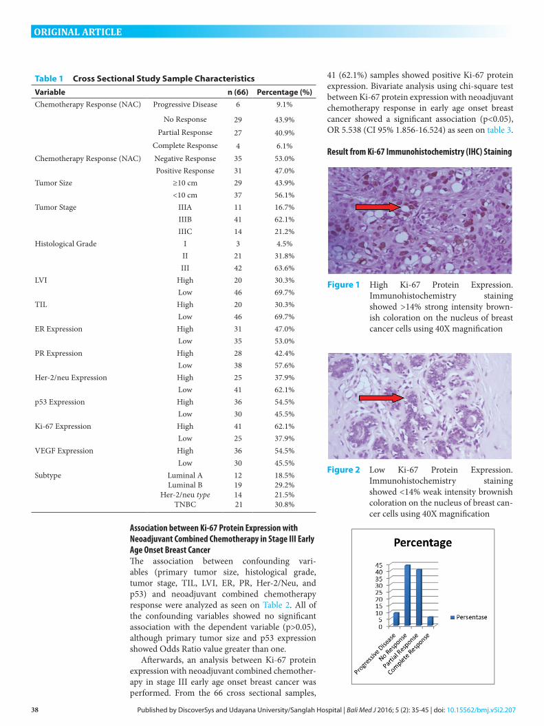
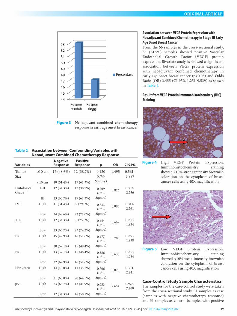
From 1 September 2012 until 31 March 2014 there has been 80 patients with stage III early age onset breast cancer who received CAF neoadju-vant combination chemotherapy from Sanglah General Hospital and Prima Medika Hospital Denpasar. From the 66 samples that met the inclusion criteria, the mean age was 34.53±3.548; the youngest age was 27 years old. The mean value of the tumor diameter was 9.317±4.207 centime-ters. Most tumors (63.6%) were high grade and 30.8% were TNBC subtype (triple negative breast cancer). Positive response with neoadjuvant combination chemotherapy CAF was observed in only 47.0% samples, with a clinical complete response (CCR) of 6.1%, as shown in figure 3. Cross-sectional study sample characteristics are shown in Table 1.
38 Published by DiscoverSys and Udayana University/Sanglah Hospital | Bali Med J 2016; 5 (2): 35-45 | doi: 10.15562/bmj.v5i2.207
ORIGINAL ARTICLE
Association between Ki-67 Protein Expression with Neoadjuvant Combined Chemotherapy in Stage III Early Age Onset Breast CancerThe association between confounding vari-ables (primary tumor size, histological grade, tumor stage, TIL, LVI, ER, PR, Her-2/Neu, and p53) and neoadjuvant combined chemotherapy response were analyzed as seen on Table 2. All of the confounding variables showed no significant association with the dependent variable (p>0.05), although primary tumor size and p53 expression showed Odds Ratio value greater than one.
Afterwards, an analysis between Ki-67 protein expression with neoadjuvant combined chemother-apy in stage III early age onset breast cancer was performed. From the 66 cross sectional samples,
41 (62.1%) samples showed positive Ki-67 protein expression. Bivariate analysis using chi-square test between Ki-67 protein expression with neoadjuvant chemotherapy response in early age onset breast cancer showed a significant association (p<0.05), OR 5.538 (CI 95% 1.856-16.524) as seen on table 3.
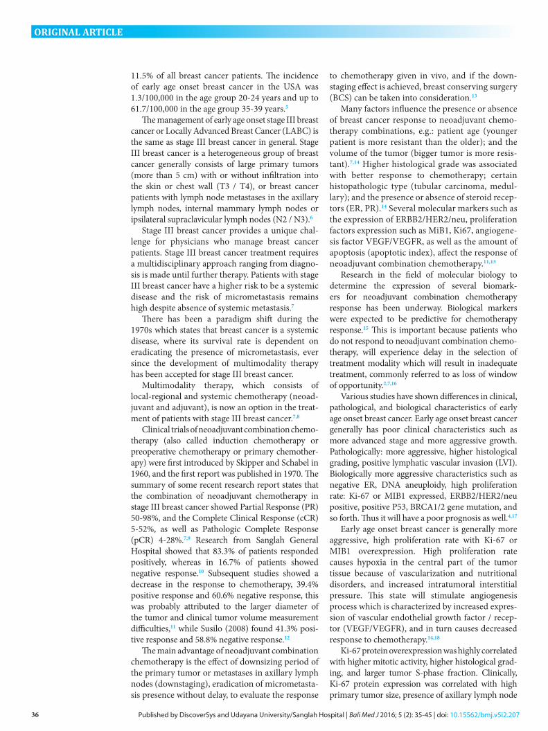
Result from Ki-67 Immunohistochemistry (IHC) Staining
Figure 1 High Ki-67 Protein Expression. Immunohistochemistry staining showed >14% strong intensity brown-ish coloration on the nucleus of breast cancer cells using 40X magnification
Table 1 Cross Sectional Study Sample Characteristics
Variable n (66) Percentage (%)
Chemotherapy Response (NAC) Progressive Disease 6 9.1%
No Response 29 43.9%Partial Response 27 40.9%
Complete Response 4 6.1%Chemotherapy Response (NAC) Negative Response 35 53.0%
Positive Response 31 47.0%Tumor Size ≥10 cm 29 43.9%
<10 cm 37 56.1%Tumor Stage IIIA 11 16.7%
IIIB 41 62.1%IIIC 14 21.2%
Histological Grade I 3 4.5%II 21 31.8%III 42 63.6%
LVI High 20 30.3%Low 46 69.7%
TIL High 20 30.3%Low 46 69.7%
ER Expression High 31 47.0%Low 35 53.0%
PR Expression High 28 42.4%Low 38 57.6%
Her-2/neu Expression High 25 37.9%Low 41 62.1%
p53 Expression High 36 54.5%Low 30 45.5%
Ki-67 Expression High 41 62.1%Low 25 37.9%
VEGF Expression High 36 54.5%Low 30 45.5%
Subtype Luminal A Luminal B
Her-2/neu type TNBC
12 19 14 21
18.5% 29.2% 21.5% 30.8%
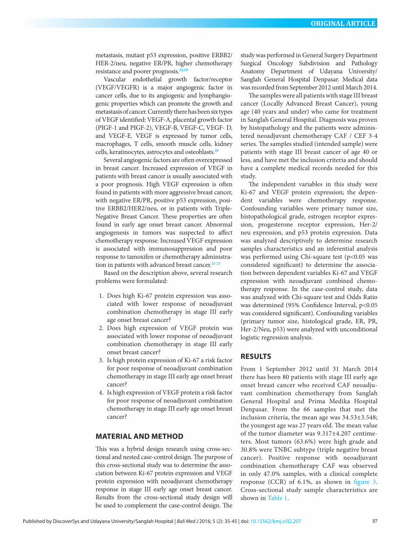
Figure 2 Low Ki-67 Protein Expression. Immunohistochemistry staining showed <14% weak intensity brownish coloration on the nucleus of breast can-cer cells using 40X magnification
39Published by DiscoverSys and Udayana University/Sanglah Hospital | Bali Med J 2016; 5 (2): 35-45 | doi: 10.15562/bmj.v5i2.207
ORIGINAL ARTICLE
Association between VEGF Protein Expression with Neoadjuvant Combined Chemotherapy in Stage III Early Age Onset Breast CancerFrom the 66 samples in the cross-sectional study, 36 (54.5%) samples showed positive Vascular Endothelial Growth Factor (VEGF) protein expression. Bivariate analysis showed a significant association between VEGF protein expression with neoadjuvant combined chemotherapy in early age onset breast cancer (p<0.05) and Odds Ratio (OR) 3.455 (CI 95% 1,251-9,539) as shown in Table 4.
Result from VEGF Protein Immunohistochemistry (IHC) Staining
Case-Control Study Sample CharacteristicsThe samples for the case-control study were taken from the cross-sectional study, 31 samples as case (samples with negative chemotherapy response) and 31 samples as control (samples with positive
Table 2 Association between Confounding Variables with Neoadjuvant Combined Chemotherapy Response
VariablesNegative Response
Positive Response p OR CI 95%
Tumor Size
≥10 cm 17 (48.6%) 12 (38.7%) 0.420 (Chi-
Square)
1.495 0.561-3.987
<10 cm 18 (51.4%) 19 (61.3%)Histological Grade
I-II 12 (34.3%) 12 (38.7%) 0.709 (Chi-
Square)
0.826 0.302-2.256
III 23 (65.7%) 19 (61.3%)LVI High 11 (31.4%) 9 (29.0%) 0.833
(Chi-Square)
0.893 0.311-2.561
Low 24 (68.6%) 22 (71.0%)TIL High 12 (34.3%) 8 (25.8%) 0.454
(Chi-Square)
0.667 0.230-1.934
Low 23 (65.7%) 23 (74.2%)ER High 15 (42.9%) 16 (51.6%) 0.477
(Chi-Square)
0.703 0.266-1.858
Low 20 (57.1%) 15 (48.4%)PR High 13 (37.1%) 15 (48.4%) 0.356
(Chi-Square)
0.630 0.236-1.684
Low 22 (62.9%) 16 (51.6%)Her-2/neu High 14 (40.0%) 11 (35.5%) 0.706
(Chi-Square)
0.825 0.304-2.241
Low 21 (60.0%) 20 (64.5%)p53 High 23 (65.7%) 13 (41.9%) 0.053
(Chi-Square)
2.654 0.978-7.200
Low 12 (34.3%) 18 (58.1%)
Figure 3 Neoadjuvant combined chemotherapy response in early age onset breast cancer
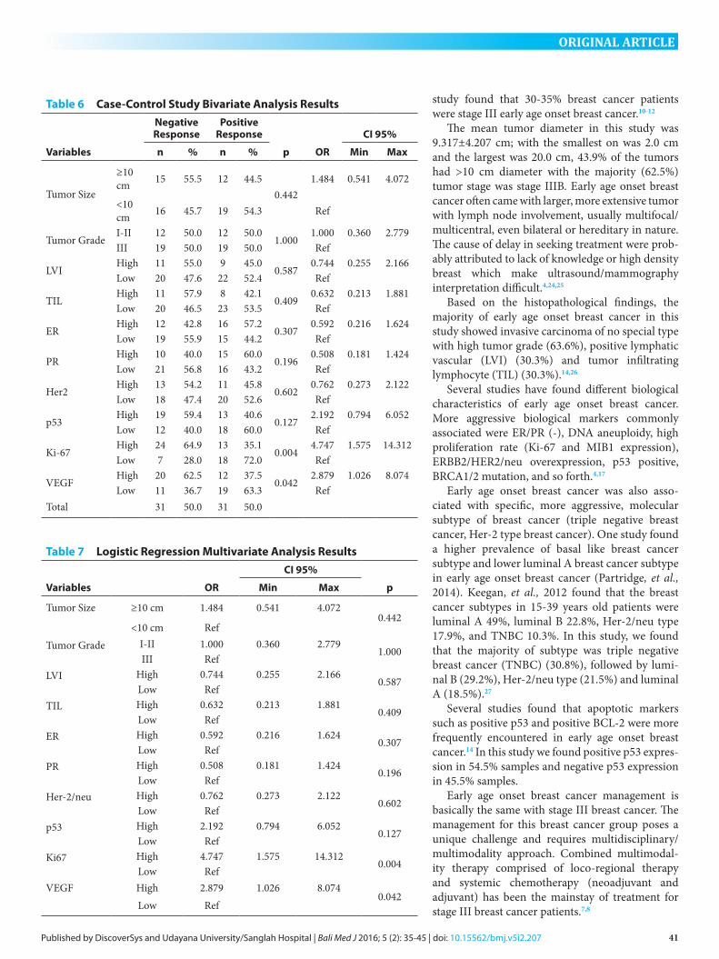
Figure 5 Low VEGF Protein Expression. Immunohistochemistry staining showed <10% weak intensity brownish coloration on the cytoplasm of breast cancer cells using 40X magnification
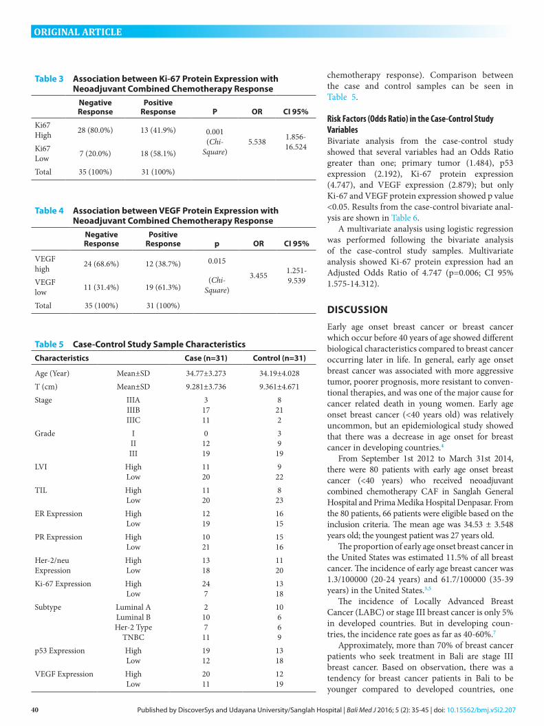
Figure 4 High VEGF Protein Expression. Immunohistochemistry staining showed >10% strong intensity brownish coloration on the cytoplasm of breast cancer cells using 40X magnification
40 Published by DiscoverSys and Udayana University/Sanglah Hospital | Bali Med J 2016; 5 (2): 35-45 | doi: 10.15562/bmj.v5i2.207
ORIGINAL ARTICLE
chemotherapy response). Comparison between the case and control samples can be seen in Table 5.
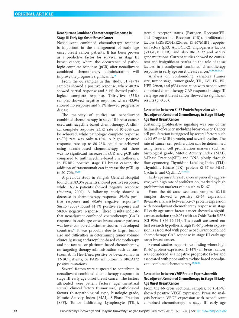
Risk Factors (Odds Ratio) in the Case-Control Study VariablesBivariate analysis from the case-control study showed that several variables had an Odds Ratio greater than one; primary tumor (1.484), p53 expression (2.192), Ki-67 protein expression (4.747), and VEGF expression (2.879); but only Ki-67 and VEGF protein expression showed p value <0.05. Results from the case-control bivariate anal-ysis are shown in Table 6.
A multivariate analysis using logistic regression was performed following the bivariate analysis of the case-control study samples. Multivariate analysis showed Ki-67 protein expression had an Adjusted Odds Ratio of 4.747 (p=0.006; CI 95% 1.575-14.312).
DISCUSSION
Early age onset breast cancer or breast cancer which occur before 40 years of age showed different biological characteristics compared to breast cancer occurring later in life. In general, early age onset breast cancer was associated with more aggressive tumor, poorer prognosis, more resistant to conven-tional therapies, and was one of the major cause for cancer related death in young women. Early age onset breast cancer (<40 years old) was relatively uncommon, but an epidemiological study showed that there was a decrease in age onset for breast cancer in developing countries.4
From September 1st 2012 to March 31st 2014, there were 80 patients with early age onset breast cancer (<40 years) who received neoadjuvant combined chemotherapy CAF in Sanglah General Hospital and Prima Medika Hospital Denpasar. From the 80 patients, 66 patients were eligible based on the inclusion criteria. The mean age was 34.53 ± 3.548 years old; the youngest patient was 27 years old.
The proportion of early age onset breast cancer in the United States was estimated 11.5% of all breast cancer. The incidence of early age breast cancer was 1.3/100000 (20-24 years) and 61.7/100000 (35-39 years) in the United States.3,5
The incidence of Locally Advanced Breast Cancer (LABC) or stage III breast cancer is only 5% in developed countries. But in developing coun-tries, the incidence rate goes as far as 40-60%.7
Approximately, more than 70% of breast cancer patients who seek treatment in Bali are stage III breast cancer. Based on observation, there was a tendency for breast cancer patients in Bali to be younger compared to developed countries, one
Table 5 Case-Control Study Sample Characteristics
Characteristics Case (n=31) Control (n=31)
Age (Year) Mean±SD 34.77±3.273 34.19±4.028T (cm) Mean±SD 9.281±3.736 9.361±4.671Stage IIIA
IIIB IIIC
3 17 11
8 21 2
Grade I II III
0 12 19
3 9
19LVI High
Low11 20
9 22
TIL High Low
11 20
8 23
ER Expression High Low
12 19
16 15
PR Expression High Low
10 21
15 16
Her-2/neu Expression
High Low
13 18
11 20
Ki-67 Expression High Low
24 7
13 18
Subtype Luminal A Luminal B Her-2 Type
TNBC
2 10 7
11
10 6 6 9
p53 Expression High Low
19 12
13 18
VEGF Expression High Low
20 11
12 19
Table 3 Association between Ki-67 Protein Expression with Neoadjuvant Combined Chemotherapy Response
Negative Response
Positive Response P OR CI 95%
Ki67 High 28 (80.0%) 13 (41.9%) 0.001
(Chi-Square)
5.538 1.856-16.524Ki67
Low 7 (20.0%) 18 (58.1%)
Total 35 (100%) 31 (100%)
Table 4 Association between VEGF Protein Expression with Neoadjuvant Combined Chemotherapy Response
Negative Response
Positive Response p OR CI 95%
VEGF high 24 (68.6%) 12 (38.7%) 0.015
(Chi-Square)
3.455 1.251-9.539VEGF
low 11 (31.4%) 19 (61.3%)
Total 35 (100%) 31 (100%)
41Published by DiscoverSys and Udayana University/Sanglah Hospital | Bali Med J 2016; 5 (2): 35-45 | doi: 10.15562/bmj.v5i2.207
ORIGINAL ARTICLE
study found that 30-35% breast cancer patients were stage III early age onset breast cancer.10-12
The mean tumor diameter in this study was 9.317±4.207 cm; with the smallest on was 2.0 cm and the largest was 20.0 cm, 43.9% of the tumors had >10 cm diameter with the majority (62.5%) tumor stage was stage IIIB. Early age onset breast cancer often came with larger, more extensive tumor with lymph node involvement, usually multifocal/multicentral, even bilateral or hereditary in nature. The cause of delay in seeking treatment were prob-ably attributed to lack of knowledge or high density breast which make ultrasound/mammography interpretation difficult.4,24,25
Based on the histopathological findings, the majority of early age onset breast cancer in this study showed invasive carcinoma of no special type with high tumor grade (63.6%), positive lymphatic vascular (LVI) (30.3%) and tumor infiltrating lymphocyte (TIL) (30.3%).14,26
Several studies have found different biological characteristics of early age onset breast cancer. More aggressive biological markers commonly associated were ER/PR (-), DNA aneuploidy, high proliferation rate (Ki-67 and MIB1 expression), ERBB2/HER2/neu overexpression, p53 positive, BRCA1/2 mutation, and so forth.4,17
Early age onset breast cancer was also asso-ciated with specific, more aggressive, molecular subtype of breast cancer (triple negative breast cancer, Her-2 type breast cancer). One study found a higher prevalence of basal like breast cancer subtype and lower luminal A breast cancer subtype in early age onset breast cancer (Partridge, et al., 2014). Keegan, et al., 2012 found that the breast cancer subtypes in 15-39 years old patients were luminal A 49%, luminal B 22.8%, Her-2/neu type 17.9%, and TNBC 10.3%. In this study, we found that the majority of subtype was triple negative breast cancer (TNBC) (30.8%), followed by lumi-nal B (29.2%), Her-2/neu type (21.5%) and luminal A (18.5%).27
Several studies found that apoptotic markers such as positive p53 and positive BCL-2 were more frequently encountered in early age onset breast cancer.14 In this study we found positive p53 expres-sion in 54.5% samples and negative p53 expression in 45.5% samples.
Early age onset breast cancer management is basically the same with stage III breast cancer. The management for this breast cancer group poses a unique challenge and requires multidisciplinary/multimodality approach. Combined multimodal-ity therapy comprised of loco-regional therapy and systemic chemotherapy (neoadjuvant and adjuvant) has been the mainstay of treatment for stage III breast cancer patients.7,8
Table 6 Case-Control Study Bivariate Analysis Results
Variables
Negative Response
Positive Response
p OR
CI 95%
n % n % Min Max
Tumor Size
≥10 cm 15 55.5 12 44.5
0.4421.484 0.541 4.072
<10 cm 16 45.7 19 54.3 Ref
Tumor GradeI-II 12 50.0 12 50.0
1.0001.000 0.360 2.779
III 19 50.0 19 50.0 Ref
LVIHigh 11 55.0 9 45.0
0.5870.744 0.255 2.166
Low 20 47.6 22 52.4 Ref
TILHigh 11 57.9 8 42.1
0.4090.632 0.213 1.881
Low 20 46.5 23 53.5 Ref
ERHigh 12 42.8 16 57.2
0.3070.592 0.216 1.624
Low 19 55.9 15 44.2 Ref
PRHigh 10 40.0 15 60.0
0.1960.508 0.181 1.424
Low 21 56.8 16 43.2 Ref
Her2High 13 54.2 11 45.8
0.6020.762 0.273 2.122
Low 18 47.4 20 52.6 Ref
p53High 19 59.4 13 40.6
0.1272.192 0.794 6.052
Low 12 40.0 18 60.0 Ref
Ki-67High 24 64.9 13 35.1
0.0044.747 1.575 14.312
Low 7 28.0 18 72.0 Ref
VEGFHigh 20 62.5 12 37.5
0.0422.879 1.026 8.074
Low 11 36.7 19 63.3 RefTotal 31 50.0 31 50.0
Table 7 Logistic Regression Multivariate Analysis Results
Variables OR
CI 95%
pMin Max
Tumor Size ≥10 cm 1.484 0.541 4.0720.442
<10 cm RefTumor Grade I-II 1.000 0.360 2.779
1.000III Ref
LVI High 0.744 0.255 2.1660.587
Low RefTIL High 0.632 0.213 1.881
0.409Low Ref
ER High 0.592 0.216 1.6240.307
Low RefPR High 0.508 0.181 1.424
0.196Low Ref
Her-2/neu High 0.762 0.273 2.1220.602
Low Refp53 High 2.192 0.794 6.052
0.127Low Ref
Ki67 High 4.747 1.575 14.3120.004
Low RefVEGF High 2.879 1.026 8.074
0.042Low Ref
42 Published by DiscoverSys and Udayana University/Sanglah Hospital | Bali Med J 2016; 5 (2): 35-45 | doi: 10.15562/bmj.v5i2.207
ORIGINAL ARTICLE
Neoadjuvant Combined Chemotherapy Response in Stage III Early Age Onset Breast CancerNeoadjuvant combined chemotherapy response is important in the management of early age onset breast cancer patients. It has been proven as a predictive factor for survival in stage III breast cancer, where the occurrence of patho-logic complete response (pCR) after neoadjuvant combined chemotherapy administration will improve the prognosis significantly.28
From the 66 samples in this study, 31 (47%) samples showed a positive response, where 40.9% showed partial response and 6.1% showed patho-logical complete response. Thirty-five (53%) samples showed negative response, where 43.9% showed no response and 9.1% showed progressive disease.
The majority of studies on neoadjuvant combined chemotherapy in stage III breast cancer used anthracycline-based chemotherapy. A clini-cal complete response (cCR) rate of 10-20% can be achieved, while pathologic complete response (pCR) rate was only 8-15%. A higher positive response rate up to 80-95% could be achieved using taxane-based chemotherapy, but there was no significant increase in cCR and pCR rate compared to anthracycline-based chemotherapy. In ERBB2 positive stage III breast cancer, the addition of trastuzumab can increase the pCR up to 20-70%.13,28
A previous study in Sanglah General Hospital found that 83.3% patients showed positive response, while 16.7% patients showed negative response (Sudarsa, 2000). A follow-up study showed a decrease in chemotherapy response; 39.4% posi-tive response and 60.6% negative response.11 Susilo (2008) found 41.3% positive response and 58.8% negative response. These results indicate that neoadjuvant combined chemotherapy (CAF) response in early age onset breast cancer patients was lower compared to similar studies in developed countries.12 It was probably due to larger tumor size and difficulties in determining tumor volume clinically, using anthracycline-based chemotherapy and not taxane- or platinum-based chemotherapy, no targeting therapy administration such as tras-tuzumab in Her-2/neu positive or bevacizumab in TNBC patients, or PARP inhibitors in BRCA1/2 positive mutations.
Several factors were suspected to contribute in neoadjuvant combined chemotherapy response in stage III early age onset breast cancer. The factors attributed were patient factors (age, menstrual status), clinical factors (tumor size), pathological factors (histopathological type, histologic grade, Mitotic Activity Index [MAI], S-Phase Fraction [SPF], Tumor Infiltrating Lymphocyte [TIL]),
steroid receptor status (Estrogen Receptor/ER, and Progesterone Receptor (PR)), proliferation factors (ERBB2/HER2/neu, Ki-67/MIB1), apopto-sis factors (p53, AI, BCL-2), angiogenesis factors (VEGF/VEGFR), and also BRCA1/2 and MDR1 gene mutations. Current studies showed no consis-tent and insignificant results on the role of these factors in neoadjuvant combined chemotherapy response in early age onset breast cancer.14,16,19,29,30
Analysis on confounding variables (tumor size, tumor stage, tumor grade, TIL, LVI, ER, PR, HER-2/neu, and p53) association with neoadjuvant combined chemotherapy CAF response in stage III early age onset breast cancer showed no significant results (p>0.05).
Association between Ki-67 Protein Expression with Neoadjuvant Combined Chemotherapy in Stage III Early Age Onset Breast CancerSustaining proliferative signaling was one of the hallmarks of cancer, including breast cancer. Cancer cell proliferation is triggered by several factors such as Ki-67 or MIB1 protein, and several cyclins. The rate of cancer cell proliferation can be determined using several cell proliferation markers such as: histological grade, Mitotic Activity Index (MAI), S-Phase Fraction(SPF) and DNA ploidy through flow cytometry, Thymidine Labeling Index (TLI), Thymidine Kinase (TK), protein Ki-67 or MIB1, Cyclin E, and Cyclin D.14,19,31
Early age onset breast cancer is generally aggres-sive, with high rate of proliferation, marked by high proliferation markers value such as Ki-67.
From the 66 cross sectional samples, 62.1% samples showed a positive Ki-67 expression. Bivariate analysis between Ki-67 protein expression with neoadjuvant chemotherapy response in stage III early age onset breast cancer showed a signifi-cant association (p<0.05) with an Odds Ratio 5.538 (CI 95% 1.856-16.524). The result answered our first research hypothesis, high Ki-67 protein expres-sion is associated with poor neoadjuvant combined chemotherapy CAF response in stage III early age onset breast cancer.
Several studies support our finding where high Ki-67 protein expression (>14%) in breast cancer was considered as a negative prognostic factor and associated with poor anthracycline based neoadju-vant combined chemotherapy.19,32,33
Association between VEGF Protein Expression with Neoadjuvant Combined Chemotherapy in Stage III Early Age Onset Breast CancerFrom the 66 cross sectional samples, 36 (54.5%) showed positive VEGF expression. Bivariate anal-ysis between VEGF expression with neoadjuvant combined chemotherapy in stage III early age
43Published by DiscoverSys and Udayana University/Sanglah Hospital | Bali Med J 2016; 5 (2): 35-45 | doi: 10.15562/bmj.v5i2.207
ORIGINAL ARTICLE
onset breast cancer showed a significant association (p<0.05) with an Odds Ratio 3.455 (95% CI 1.251-9.539). The result confirmed our second research hypothesis, which was high VEGF expression is associated with poor neoadjuvant combined chemotherapy CAF response in stage III early age onset breast cancer.
High VEGF expression is commonly found in more aggressive breast cancer, with negative ER/PR, positive p53, positive ERBB2.Her2/neu, or in TNBC. Those characteristics were often found in early age onset breast cancer.23
From the bivariate analysis of case-control samples, several variables showed an Odds Ratio value greater than one, the variables were: tumor size (1.484), p53 expression (2.192), Ki-67 protein expression (4.747), and VEGF expression (2.879), but only Ki-67 and VEGF protein expression showed p value <0.05.
Ki-67 Protein Expression as a Risk Factor for Neoadju-vant Combined Chemotherapy in Stage III Early Age Onset Breast CancerBivariate analysis from the case-control study found that Ki-67 protein expression was significantly higher in the case group compared to the control group (OR = 4.747 CI 95% 1.575-14.312; p=0.004). While multivariate analysis using logistic regres-sion method found the adjusted OR was 4.747 (CI 95% 1.575-14.312; p=0,006). The result showed that patients with high Ki-67 protein expression will have 5 times higher risk of developing poor neoad-juvant chemotherapy compared to patients with low Ki-67 protein expression. The third research hypothesis is answered by this analysis.
During the past decade, there have been a number of studies on the role of proliferation markers as prognostic or predictive factor in breast cancer. The role of Ki-67 as a proliferation marker in early or advanced breast cancer has been widely studied due to the simplicity of the examination, but unfortunately the results from these studies were not consistent (Colozza, et al., 2005).
Up until now, the role of Ki-67 was considered prognostic in nature, due to the fact that high prolif-eration was associated with poorer prognosis. In this study, we found that high Ki-67 protein expres-sion was an independent prognostic factor for poor neoadjuvant combined chemotherapy response. It was also associated with several poor prognostic factors such as tumor size, lymph node involve-ment, histologic grade, and vascular invasion, also with good prognostic factor ER/PR status.14,34,35
A meta-analysis study of 12,000 patients with positive Ki-67 expression showed a significant association with higher recurrence rate and lower
survival rate in patients with positive or negative lymph node involvement.36
Up until now, there have been no significant findings on the role of Ki-67 as a predictive factor for neoadjuvant chemotherapy. One study stated that high Ki-67 expression will represent a high tumor proliferation rate hence it will lead to positive chemotherapy response.35 But, it was not consistent with the concept where high proliferation rate will lead to central tumor hypoxia which will lead to poor chemotherapy response.31,37 The latter study supports our finding where in stage III early age onset breast cancer patients with aggressive, high proliferating behavior, will have a higher risk of poor neoadjuvant combined chemotherapy CAF.
Vascular Endothelial Growth Factor (VEGF) Expression as a Risk Factor for Neoadjuvant Combined Chemotherapy in Stage III Early Age Onset Breast CancerBivariate analysis from the case-control study found that VEGF expression was significantly higher in the case group compared to the control group (OR = 2.879 CI 95% 1.026-8.074; p=0.042). But, multivariate analysis using logistic regression method found the adjusted OR was 2.553 (CI 95% 0.860-7.582; p=0.091). The result showed that patients with high VEGF expression will have 2.5 times higher risk of developing poor neoadjuvant chemotherapy compared to patients with low VEGF expression. Even though it was not statistically significant, high VEGF expression might have a role as a risk factor for poor neoadjuvant combined chemotherapy response in early age onset breast cancer. As stated in the samples size determination for the case-control study design, the power used in this study was 80%. Therefore it is still possible to gain more significant results by increasing the power of this study to 90%.
The association between high VEGF expression with neoadjuvant combined chemotherapy has not been established in this study. But VEGF/VEGFR overexpression has been associated with poor chemotherapy response due to impaired chemo-therapy delivery inside the tumor.16
The most crucial elements in VEGF gene regu-lation was oxygen tension or hypoxia and tissue acidosis. Changes in pO2 and pH in tumor tissue will cause varieties in VEGF A gene expression, leading to alternative splicing producing several VEGF A isoforms: VEGF121, VEGF165, VEGF189, VEGF206, less common VEGF A isoforms are VEGF145 and VEGF183 (Ferrara et al, 2003; Ferrara, 2004). We suspect that the presence of VEGF A isoforms affects the results of VEGF IHC staining. Several other factors might also contrib-ute to VEGF mRNA expression, such as epidermal
44 Published by DiscoverSys and Udayana University/Sanglah Hospital | Bali Med J 2016; 5 (2): 35-45 | doi: 10.15562/bmj.v5i2.207
ORIGINAL ARTICLE
growth factor (EGF), PDGF-β, TGFβ1, keratino-cyte growth factor (KGF) and interleukin-1 beta (IL- 1β), IL-1 α and IL-6.20
As stated before, high VEGF expression was associated with a more aggressive breast cancer, negative ER/PR, positive e p53 expression, posi-tive ERBB2/HER2/neu, or TNBC. Those features were commonly found in early age onset breast cancer.23
Until now, the main focus of VEGF (specifically VEGF A) studies was its role as a prognosis factor and the outcome in anti-VEGF therapy, bevaci-zumab. High VEGF expression was associated with increased angiogenesis and lymphangiogenesis, immunosuppression, and low apoptosis index. High VEGF expression in breast cancer was associ-ated with poorer prognosis.22,38
Recent studies found that combined chemo-therapy and anti-angiogenesis administration as an adjuvant or neoadjuvant therapy is more effective in breast cancer management.39
Novel FindingsThere were several novel findings in this study: High Ki-67 and VEGF expression were associated with poor neoadjuvant combined chemotherapy response in stage III early age breast cancer. High Ki-67 expression was an independent risk factor for poor neoadjuvant combined chemotherapy response in stage III early age breast cancer. High VEGF expression might be a risk factor for poor neoadjuvant combined chemotherapy response in stage III early age breast cancer.
CONCLUSION
Based from the cross sectional and case-control study in stage III early age onset breast cancer, we concluded:
1. High Ki-67 is associated with poor neoadju-vant combined chemotherapy response in stage III early age breast cancer.
2. High VEGF expression is associated with poor neoadjuvant combined chemotherapy response in stage III early age breast cancer.
3. High Ki-67 expression was an independent risk factor for poor neoadjuvant combined chemo-therapy response in stage III early age breast cancer.
4. High VEGF expression might be a risk factor for poor neoadjuvant combined chemotherapy response in stage III early age breast cancer.
SUGGESTION
Based on the previous conclusions, we suggest:
1. Ki-67 and VEGF expression can be used as a predictive factor for neoadjuvant combined chemotherapy response in early age onset breast cancer.
2. Due to Ki-67 dynamic protein expression, further study on the association between Ki-67 protein expression changes with neoadjuvant chemotherapy response in early age onset breast cancer is needed.
3. Due to the variety in VEGF A isoforms, further study on a specific VEGF isoform with neoad-juvant chemotherapy response in early age onset breast cancer, in genetic/DNA, mRNA, or protein, is needed.
REFERENCES1. Honig SF. Incidence, Trends, and the Epidemiology of
Breast Cancer. In: Spear SL, editor. Surgery of the Breast: Principles and Art. 2 ed. Philadelphia: Lippincott, William and Wilkins; 2006. p. 3-8.
2. Newman LA, Hayes, D.F. Evidence Based Management of Breast Cancer. In: Chang AE, Ganz, P.A., Hayes, D.F., edi-tor. Oncology: An Evidence Based Approach. New York: Springer; 2006. p. 951-75.
3. Jemal A, Siegel R, Ward E, Hao Y, Xu J, Thun MJ. Cancer statistics, 2009. CA: a cancer journal for clinicians. 2009 Jul-Aug;59(4):225-49. PubMed PMID: 19474385.
4. Partridge AH, Goldhisch, A., Gelber, S., Gelber, R.D. Breast Cancer in Younger Women. In: Harris JR, Lippman, M.E., Morrow, M., Osborne, C.K., editor. Diseases of th Breast. 4th ed. Philadelphia: Wolters Kluwer, Lippincott and Wilkins; 2010. p. 1073-82.
5. Yankaskas BC. Epidemiology of breast cancer in young women. Breast disease. 2005;23:3-8. PubMed PMID: 16823161.
6. Sobin LH, Wittekind, C.H. Breast Tumors. Geneva: Wiley-Liss; 2009.
7. Hortobagyi GN, Singletary, S.E., Strom, E.A. Locally Advanced Breast Cancer. In: Harris JR, Lippman, M.E., Morrow, M., Osborne, C.K., editor. Diseases of the Breast. 4th ed. Philadelphia: Wolters Kluwer, Lippincott and Wilkins; 2010. p. 745-61.
8. Zager JS, Solorzano, C.C., Thomas, E., Feig, B.W., Babierra, G.V. Invasive Breast Cancer. In: Feig BW, Berger, D.H., Fuhrman, G.M., editor. The MD Anderson Surgical Oncology Handbook. 2nd ed. Philadelphia: Lippincott Williams and Wilkins; 2006. p. 23-59.
9. Dang CT, Hudis, C. Preoperative Therapy for Operable Breast Cancer. In: Harris JR, Lippman, M.E., Morrow, M., Osborne, C.K., editor. Diseases of the Breast. 4th ed. Philadelphia: Wolters Kluwer, Lippincot and Wilkins; 2010. p. 715-23.
10. Sudarsa W. Respon dari Locally Advanced Breast Cancer (LABC) Terhadap Pemberian Neoadjuvant Chemoterapy. Makassar: Hasanuddin; 2000.
11. Manuaba TW. Indeks Apoptosis, Ekspresi p53 dan Ekspresi BCL-2 Sebagai Faktor Prediktor Respon Terhadap Kemoterapi Kombinasi Neoadjuvant pada Karsinoma Mama Stadium III. Denpasar: Udayana; 2006.
12. Susilo H. Ekspresi Vascular Endothelial Growth Factor (VEGF) sebagai Faktor Prediktor Respon Terhadap Kemoterapi Kombinasi Neoadjuvant pada Karsinoma Mama Stadium III. Denpasar: Udayana; 2009.
13. Rastogi P, Anderson SJ, Bear HD, Geyer CE, Kahlenberg MS, Robidoux A, et al. Preoperative chemotherapy: updates of National Surgical Adjuvant Breast and Bowel Project Protocols B-18 and B-27. Journal of clinical oncology : offi-cial journal of the American Society of Clinical Oncology. 2008 Feb 10;26(5):778-85. PubMed PMID: 18258986.
45Published by DiscoverSys and Udayana University/Sanglah Hospital | Bali Med J 2016; 5 (2): 35-45 | doi: 10.15562/bmj.v5i2.207
ORIGINAL ARTICLE
14. Chang JC, Hilsenbeck, S.G. Prognostic and Predictive Markers. In: Harris JR, Lippman, M.E., Morrow, M., Osborne, C.K., editor. Diseases of the Breast. 4th ed. Philadelphia: Wolters Kluwer, Lippincott and Wilkins; 2010. p. 443-57.
15. Harrington L, Bristow, R.G., Hill, R.P., Tannock, I.F. Introduction to Cancer Biology. In: Tannock IA, Hill, R.P., Bristow, R.G., Harrington, L., editor. The Basic Science of Oncology. 4th ed. New York: McGraw-Hill; 2005. p. 1-3.
16. Wolff AC, Berry D, Carey LA, Colleoni M, Dowsett M, Ellis M, et al. Research issues affecting preoperative sys-temic therapy for operable breast cancer. Journal of clin-ical oncology : official journal of the American Society of Clinical Oncology. 2008 Feb 10;26(5):806-13. PubMed PMID: 18258990.
17. Kauber-DeMore N. Tumor Biology of Breast Cancer in Young Women. Breast disease. 2006;23:9-15.
18. Jain RK, Duda, D.G. Vascular and Interstitial Biology of Tumors. In: Abeloff MD, Armitage, J.O., Niederhuber, J.E., Kastan, M.B., McKenna, W.G., editor. Abeloff ’s Clinical Oncology. 4th ed. Philadelphia2008. p. 105-24.
19. van Diest PJ, de Jong, J.S., Baak, J.P.A., Rob, J.A.M., Michalides, R.J.A.M., van der Wall, E. Prognostic Value of Proliferation and Apoptosis in Breast Cancer. In: Walker RA, Thompson, A.M., editor. Prognostic and Predictive Factors in Breast Cancer. 2nd ed. London: Informa; 2008. p. 18-37.
20. Ferrara N. Vascular endothelial growth factor: basic sci-ence and clinical progress. Endocrine reviews. 2004 Aug;25(4):581-611. PubMed PMID: 15294883.
21. Foekens JA, Peters HA, Grebenchtchikov N, Look MP, Meijer-van Gelder ME, Geurts-Moespot A, et al. High tumor levels of vascular endothelial growth factor predict poor response to systemic therapy in advanced breast can-cer. Cancer research. 2001 Jul 15;61(14):5407-14. PubMed PMID: 11454684.
22. Schneider BP, Miller KD. Angiogenesis of breast can-cer. Journal of clinical oncology : official journal of the American Society of Clinical Oncology. 2005 Mar 10;23(8):1782-90. PubMed PMID: 15755986.
23. Linderholm BK, Hellborg H, Johansson U, Elmberger G, Skoog L, Lehtio J, et al. Significantly higher levels of vascu-lar endothelial growth factor (VEGF) and shorter survival times for patients with primary operable triple-negative breast cancer. Annals of oncology : official journal of the European Society for Medical Oncology / ESMO. 2009 Oct;20(10):1639-46. PubMed PMID: 19549711.
24. Partridge AH, Pagani O, Abulkhair O, Aebi S, Amant F, Azim HA, Jr., et al. First international consensus guidelines for breast cancer in young women (BCY1). Breast. 2014 Jun;23(3):209-20. PubMed PMID: 24767882.
25. Sidoni A, Cavaliere A, Bellezza G, Scheibel M, Bucciarelli E. Breast cancer in young women: clinicopath-ological features and biological specificity. Breast. 2003 Aug;12(4):247-50. PubMed PMID: 14659308.
26. Moffat R, Guth U. Preserving fertility in patients under-going treatment for breast cancer: current perspectives. Breast cancer. 2014;6:93-101. PubMed PMID: 25114587. Pubmed Central PMCID: 4108258.
27. Keegan TH, DeRouen MC, Press DJ, Kurian AW, Clarke CA. Occurrence of breast cancer subtypes in ado-lescent and young adult women. Breast cancer research : BCR. 2012;14(2):R55. PubMed PMID: 22452927. Pubmed Central PMCID: 3446389.
28. Hortobagyi GN. Toward individualized breast cancer ther-apy: translating biological concepts to the bedside. The
oncologist. 2012;17(4):577-84. PubMed PMID: 22474069. Pubmed Central PMCID: 3336824.
29. Cleator S, Parton M, Dowsett M. The biology of neoad-juvant chemotherapy for breast cancer. Endocrine-related cancer. 2002 Sep;9(3):183-95. PubMed PMID: 12237246.
30. George J, Dharanipragada K, Krishnamachari S, Chandrasekaran A, Sam SS, Sunder E. A single-nucleo-tide polymorphism in the MDR1 gene as a predictor of response to neoadjuvant chemotherapy in breast cancer. Clinical breast cancer. 2009 Aug;9(3):161-5. PubMed PMID: 19661039.
31. Hanahan D, Weinberg RA. Hallmarks of cancer: the next generation. Cell. 2011 Mar 4;144(5):646-74. PubMed PMID: 21376230.
32. Aas T, Geisler S, Eide GE, Haugen DF, Varhaug JE, Bassoe AM, et al. Predictive value of tumour cell prolifer-ation in locally advanced breast cancer treated with neo-adjuvant chemotherapy. European journal of cancer. 2003 Mar;39(4):438-46. PubMed PMID: 12751373.
33. Petit T, Wilt M, Velten M, Millon R, Rodier JF, Borel C, et al. Comparative value of tumour grade, hormonal recep-tors, Ki-67, HER-2 and topoisomerase II alpha status as predictive markers in breast cancer patients treated with neoadjuvant anthracycline-based chemotherapy. European journal of cancer. 2004 Jan;40(2):205-11. PubMed PMID: 14728934.
34. Wiesner FG, Magener A, Fasching PA, Wesse J, Bani MR, Rauh C, et al. Ki-67 as a prognostic molecular marker in routine clinical use in breast cancer patients. Breast. 2009 Apr;18(2):135-41. PubMed PMID: 19342238.
35. Dowsett M, Nielsen TO, A’Hern R, Bartlett J, Coombes RC, Cuzick J, et al. Assessment of Ki67 in breast cancer: recom-mendations from the International Ki67 in Breast Cancer working group. Journal of the National Cancer Institute. 2011 Nov 16;103(22):1656-64. PubMed PMID: 21960707. Pubmed Central PMCID: 3216967.
36. Yerushalmi R, Woods R, Ravdin PM, Hayes MM, Gelmon KA. Ki67 in breast cancer: prognostic and predic-tive potential. The Lancet Oncology. 2010 Feb;11(2):174-83. PubMed PMID: 20152769.
37. Taghian AG, Jeong JH, Mamounas EP, Parda DS, Deutsch M, Costantino JP, et al. Low locoregional recur-rence rate among node-negative breast cancer patients with tumors 5 cm or larger treated by mastectomy, with or without adjuvant systemic therapy and without radiother-apy: results from five national surgical adjuvant breast and bowel project randomized clinical trials. Journal of clini-cal oncology : official journal of the American Society of Clinical Oncology. 2006 Aug 20;24(24):3927-32. PubMed PMID: 16921044.
38. Lantzsch T, Hefler, L. Vascular Endothelial Growth Factor in Breast Cancer. In: Harmey JH, editor. VEGF and Cancer. Texax: Landes Bioscience/Eurekah; 2004. p. 40-7.
39. Gerber B, Loibl S, Eidtmann H, Rezai M, Fasching PA, Tesch H, et al. Neoadjuvant bevacizumab and anthracy-cline-taxane-based chemotherapy in 678 triple-negative primary breast cancers; results from the geparquinto study (GBG 44). Annals of oncology : official journal of the European Society for Medical Oncology / ESMO. 2013 Dec;24(12):2978-84. PubMed PMID: 24136883.
This work is licensed under a Creative Commons Attribution
Related Documents


