Review began 03/16/2022 Review ended 03/28/2022 Published 03/30/2022 © Copyright 2022 Muniswammappa et al. This is an open access article distributed under the terms of the Creative Commons Attribution License CC-BY 4.0., which permits unrestricted use, distribution, and reproduction in any medium, provided the original author and source are credited. Polymorphous Adenocarcinoma: High-Grade Transformation With Immunohistochemical Workup Sudhakara Muniswammappa , Radhika Bavle , Soumya Makarla , Reshma Venugopal 1. Department of Oral and Maxillofacial Pathology, Krishnadevaraya College of Dental Sciences and Hospital, Bangalore, IND Corresponding author: Reshma Venugopal, [email protected] Abstract Intraoral tumours associated with mucosa are commonly oral squamous cell carcinomas followed by minor salivary gland carcinomas, the commonest being mucoepidermoid carcinoma, adenoid cystic carcinoma, and polymorphous adenocarcinoma (PAC). PAC is the second most common malignant tumour that is found in the minor salivary glands of the oral cavity and rarely exhibits high-grade transformation (HGT). We report a case of a 50-year-old woman with a tumour on her buccal mucosa for six months. Histopathologically, the tumour showed more than 15 histopathological patterns with areas of HGT. The high-grade transformed areas predominantly showed solid patterns, increased mitosis, necrosis, vascular invasion, and perineural invasion. An immunohistochemical (IHC) panel inclusive of p63, SOX-10, S-100, calponin, vimentin, and Ki- 67 was done to evaluate the tumour and grade PAC. The Ki-67 index was around 25%-30%, confirming the diagnosis of PAC-HGT. This might be the first case of primary PAC-HGT seen on the buccal mucosa on initial clinical presentation. Categories: Pathology, Oncology, Dentistry Keywords: pac, high grade transformation (hgt), primary high grade transformation, invasive pac, comedo necrosis, minor salivary gland, buccal mucosa Introduction Polymorphous adenocarcinoma (PAC) is a salivary gland tumour that has seen an interesting journey in nomenclature and behaviour from its conception as a tumour way back in 1984 until the WHO 2017 classification and categorization of salivary gland tumours. PAC was classically a low-grade carcinoma called polymorphous low-grade adenocarcinoma (PLGA). Innumerable descriptions of this tumour have been woven together with robust data on histopathology and behaviour and its behaviour has become questionable in the past decades. Cases with high-grade transformation (HGT) or dedifferentiation in PAC have been reported. HGT in salivary gland tumours, though rare, does occur. It has been reported in other salivary gland carcinomas like acinic cell carcinoma (AciCC), adenoid cystic carcinoma (AdCC), epithelial- myoepithelial carcinoma (Epi-myoepi CA), secretory carcinoma (SC), and mucoepidermoid carcinoma (MEC) [1]. English literature reports only about seven cases of PAC with HGT, which is a rare finding. De novo HGT is the rarest, as most of it is accounted for in cases of recurrence, post-radiation, and post- treatment [2]. The present case highlights the occurrence of an HGT-PAC on initial presentation, seen on the buccal mucosa close to the retromolar area, which might be the first case report of HGT-PAC on the buccal mucosa. Case Presentation A 50-year-old Indian woman presented with a small swelling, approximately one cm in diameter on the buccal mucosa in relation to the maxillary left molars. When the patient first presented to the clinic, she stated that the lesion had been noticed around eight months prior and was an asymptomatic swelling. The patient presented again after a fortnight to the clinic with complaints of an increase in the size of the swelling and ulceration, which may be due to a cheek bite. On inspection, a spherical soft tissue mass of approximately one-and-half cm in diameter was seen on the buccal mucosa, covered by normal-looking mucosa except in one area, which showed a glistening surface representing a probable cystic area (Figure 1A). On palpation, the lesion was ovoid, measuring approximately two cm in diameter, well-demarcated, mobile, and soft to firm in consistency. Regional and cervical lymph nodes were not palpable. A computed tomography scan showed a well-demarcated tumour nodule in the posterior buccal mucosal area without the involvement of the maxillary bone or sinus region (Figure 1B). Lymph-node involvement was not noted. 1 1 1 1 Open Access Case Report DOI: 10.7759/cureus.23639 How to cite this article Muniswammappa S, Bavle R, Makarla S, et al. (March 30, 2022) Polymorphous Adenocarcinoma: High-Grade Transformation With Immunohistochemical Workup. Cureus 14(3): e23639. DOI 10.7759/cureus.23639

Welcome message from author
This document is posted to help you gain knowledge. Please leave a comment to let me know what you think about it! Share it to your friends and learn new things together.
Transcript
Review began 03/16/2022 Review ended 03/28/2022 Published 03/30/2022
© Copyright 2022Muniswammappa et al. This is an openaccess article distributed under the terms ofthe Creative Commons Attribution LicenseCC-BY 4.0., which permits unrestricted use,distribution, and reproduction in anymedium, provided the original author andsource are credited.
Polymorphous Adenocarcinoma: High-GradeTransformation With ImmunohistochemicalWorkupSudhakara Muniswammappa , Radhika Bavle , Soumya Makarla , Reshma Venugopal
1. Department of Oral and Maxillofacial Pathology, Krishnadevaraya College of Dental Sciences and Hospital,Bangalore, IND
Corresponding author: Reshma Venugopal, [email protected]
AbstractIntraoral tumours associated with mucosa are commonly oral squamous cell carcinomas followed by minorsalivary gland carcinomas, the commonest being mucoepidermoid carcinoma, adenoid cystic carcinoma, andpolymorphous adenocarcinoma (PAC). PAC is the second most common malignant tumour that is found inthe minor salivary glands of the oral cavity and rarely exhibits high-grade transformation (HGT). We report acase of a 50-year-old woman with a tumour on her buccal mucosa for six months. Histopathologically, thetumour showed more than 15 histopathological patterns with areas of HGT. The high-grade transformedareas predominantly showed solid patterns, increased mitosis, necrosis, vascular invasion, and perineuralinvasion. An immunohistochemical (IHC) panel inclusive of p63, SOX-10, S-100, calponin, vimentin, and Ki-67 was done to evaluate the tumour and grade PAC. The Ki-67 index was around 25%-30%, confirming thediagnosis of PAC-HGT. This might be the first case of primary PAC-HGT seen on the buccal mucosa on initialclinical presentation.
Categories: Pathology, Oncology, DentistryKeywords: pac, high grade transformation (hgt), primary high grade transformation, invasive pac, comedo necrosis,minor salivary gland, buccal mucosa
IntroductionPolymorphous adenocarcinoma (PAC) is a salivary gland tumour that has seen an interesting journey innomenclature and behaviour from its conception as a tumour way back in 1984 until the WHO 2017classification and categorization of salivary gland tumours. PAC was classically a low-grade carcinoma calledpolymorphous low-grade adenocarcinoma (PLGA). Innumerable descriptions of this tumour have beenwoven together with robust data on histopathology and behaviour and its behaviour has becomequestionable in the past decades. Cases with high-grade transformation (HGT) or dedifferentiation in PAChave been reported. HGT in salivary gland tumours, though rare, does occur. It has been reported in othersalivary gland carcinomas like acinic cell carcinoma (AciCC), adenoid cystic carcinoma (AdCC), epithelial-myoepithelial carcinoma (Epi-myoepi CA), secretory carcinoma (SC), and mucoepidermoid carcinoma(MEC) [1]. English literature reports only about seven cases of PAC with HGT, which is a rare finding. Denovo HGT is the rarest, as most of it is accounted for in cases of recurrence, post-radiation, and post-treatment [2]. The present case highlights the occurrence of an HGT-PAC on initial presentation, seen on thebuccal mucosa close to the retromolar area, which might be the first case report of HGT-PAC on the buccalmucosa.
Case PresentationA 50-year-old Indian woman presented with a small swelling, approximately one cm in diameter on thebuccal mucosa in relation to the maxillary left molars. When the patient first presented to the clinic, shestated that the lesion had been noticed around eight months prior and was an asymptomatic swelling. Thepatient presented again after a fortnight to the clinic with complaints of an increase in the size of theswelling and ulceration, which may be due to a cheek bite.
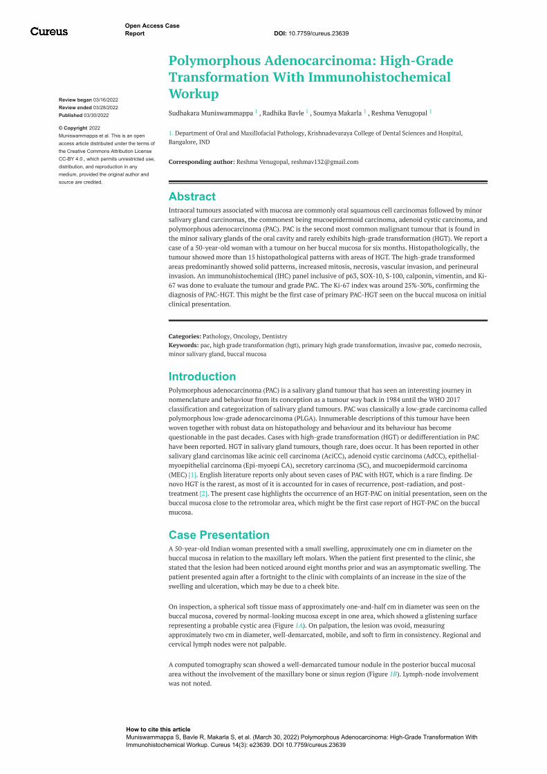
On inspection, a spherical soft tissue mass of approximately one-and-half cm in diameter was seen on thebuccal mucosa, covered by normal-looking mucosa except in one area, which showed a glistening surfacerepresenting a probable cystic area (Figure 1A). On palpation, the lesion was ovoid, measuringapproximately two cm in diameter, well-demarcated, mobile, and soft to firm in consistency. Regional andcervical lymph nodes were not palpable.
A computed tomography scan showed a well-demarcated tumour nodule in the posterior buccal mucosalarea without the involvement of the maxillary bone or sinus region (Figure 1B). Lymph-node involvementwas not noted.
1 1 1 1
Open Access CaseReport DOI: 10.7759/cureus.23639
How to cite this articleMuniswammappa S, Bavle R, Makarla S, et al. (March 30, 2022) Polymorphous Adenocarcinoma: High-Grade Transformation WithImmunohistochemical Workup. Cureus 14(3): e23639. DOI 10.7759/cureus.23639
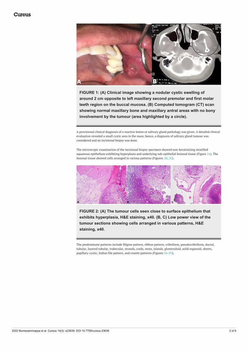
FIGURE 1: (A) Clinical image showing a nodular cystic swelling ofaround 2 cm opposite to left maxillary second premolar and first molarteeth region on the buccal mucosa. (B) Computed tomogram (CT) scanshowing normal maxillary bone and maxillary antral areas with no bonyinvolvement by the tumour (area highlighted by a circle).
A provisional clinical diagnosis of a reactive lesion or salivary gland pathology was given. A detailed clinicalevaluation revealed a small cystic area in the mass; hence, a diagnosis of salivary gland tumour wasconsidered and an incisional biopsy was done.
The microscopic examination of the incisional biopsy specimen showed non-keratinizing stratifiedsquamous epithelium exhibiting hyperplasia and underlying sub-epithelial lesional tissue (Figure 2A). Thelesional tissue showed cells arranged in various patterns (Figures 2B, 2C).
FIGURE 2: (A) The tumour cells seen close to surface epithelium thatexhibits hyperplasia, H&E staining, x40. (B, C) Low power view of thetumour sections showing cells arranged in various patterns, H&Estaining, x40.
The predominant patterns include filigree pattern, ribbon pattern, cribriform, pseudocribriform, ductal,tubular, layered tubular, trabecular, strands, cords, nests, islands, glomeruloid, solid organoid, sheets,papillary-cystic, Indian file pattern, and rosette patterns (Figures 3A-3O).
2022 Muniswammappa et al. Cureus 14(3): e23639. DOI 10.7759/cureus.23639 2 of 9
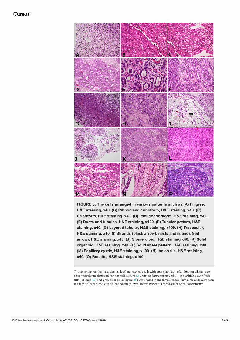
FIGURE 3: The cells arranged in various patterns such as (A) Filigree,H&E staining, x40. (B) Ribbon and cribriform, H&E staining, x40. (C)Cribriform, H&E staining, x40. (D) Pseudocribriform, H&E staining, x40.(E) Ducts and tubules, H&E staining, x100. (F) Tubular pattern, H&Estaining, x40. (G) Layered tubular, H&E staining, x100. (H) Trabecular,H&E staining, x40. (I) Strands (black arrow), nests and islands (redarrow), H&E staining, x40. (J) Glomeruloid, H&E staining x40. (K) Solidorganoid, H&E staining, x40. (L) Solid sheet pattern, H&E staining, x40.(M) Papillary cystic, H&E staining, x100. (N) Indian file, H&E staining,x40. (O) Rosette, H&E staining, x100.
The complete tumour mass was made of monotonous cells with poor cytoplasmic borders but with a largeclear vesicular nucleus and few nucleoli (Figure 4A). Mitotic figures of around 5-7 per 10 high power fields(HPF) (Figure 4B) and a few clear cells (Figure 4C) were noted in the tumour mass. Tumour islands were seenin the vicinity of blood vessels, but no direct invasion was evident in the vascular or neural elements.
2022 Muniswammappa et al. Cureus 14(3): e23639. DOI 10.7759/cureus.23639 3 of 9
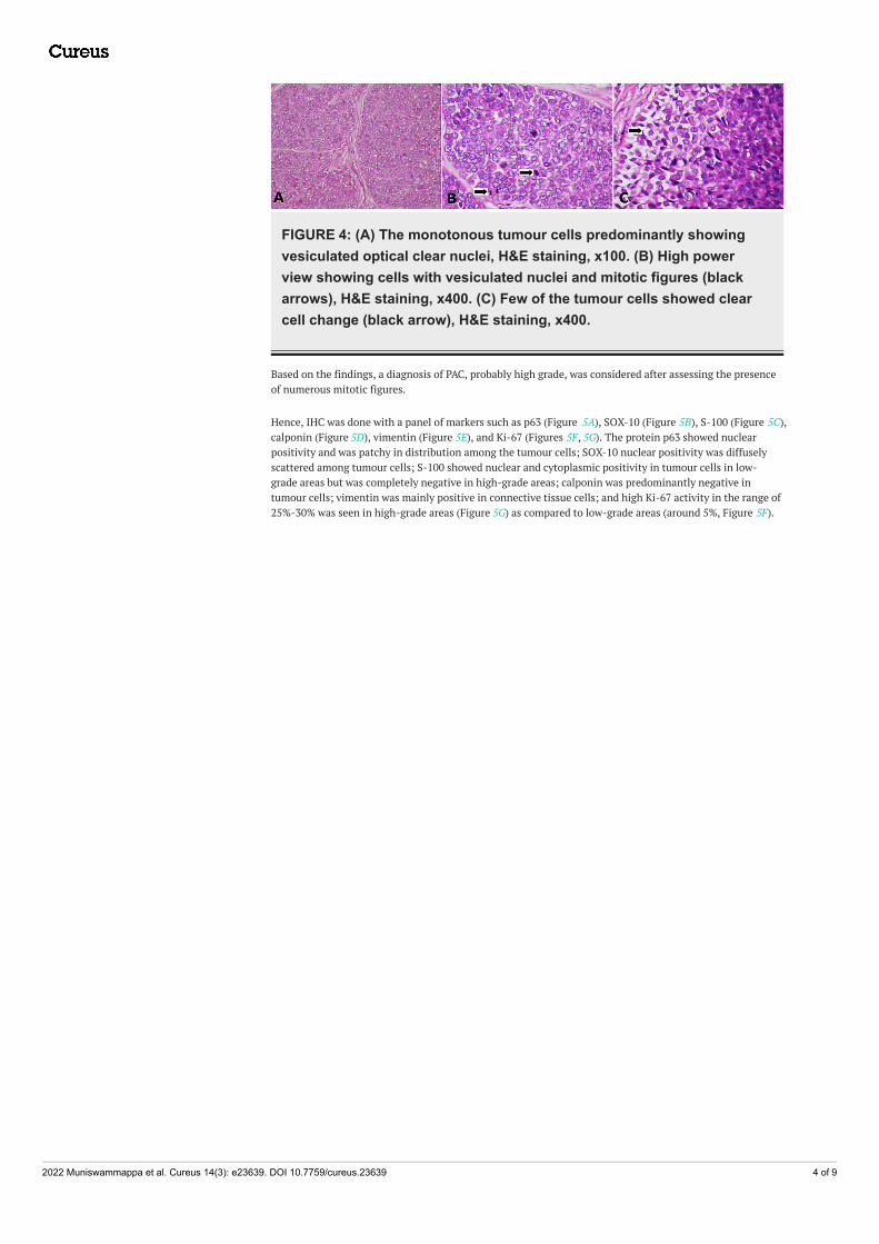
FIGURE 4: (A) The monotonous tumour cells predominantly showingvesiculated optical clear nuclei, H&E staining, x100. (B) High powerview showing cells with vesiculated nuclei and mitotic figures (blackarrows), H&E staining, x400. (C) Few of the tumour cells showed clearcell change (black arrow), H&E staining, x400.
Based on the findings, a diagnosis of PAC, probably high grade, was considered after assessing the presenceof numerous mitotic figures.
Hence, IHC was done with a panel of markers such as p63 (Figure 5A), SOX-10 (Figure 5B), S-100 (Figure 5C),calponin (Figure 5D), vimentin (Figure 5E), and Ki-67 (Figures 5F, 5G). The protein p63 showed nuclearpositivity and was patchy in distribution among the tumour cells; SOX-10 nuclear positivity was diffuselyscattered among tumour cells; S-100 showed nuclear and cytoplasmic positivity in tumour cells in low-grade areas but was completely negative in high-grade areas; calponin was predominantly negative intumour cells; vimentin was mainly positive in connective tissue cells; and high Ki-67 activity in the range of25%-30% was seen in high-grade areas (Figure 5G) as compared to low-grade areas (around 5%, Figure 5F).
2022 Muniswammappa et al. Cureus 14(3): e23639. DOI 10.7759/cureus.23639 4 of 9

FIGURE 5: (A) Nuclear expression of p63 protein, patchy in cells of thetumour lobules, IHC staining x200. (B) The tumour cells showedscattered nuclear expression of SOX 10, IHC staining, x40. (C) Tumourcells strongly positive for S-100 (black arrow) and negative in areas ofdedifferentiation (red arrow), IHC staining, x40. Inset: High power viewshows the tumour cells to be diffusely positive for S-100 in bothnucleus and cytoplasm, IHC staining, x400. (D) Near negativeexpression of calponin in tumour cells, the adjacent smooth musclewall of the artery shows positivity, IHC staining, x40. Inset: High powerview of the same, IHC staining, x200. (E) Few tumour cells and stromalcells show cytoplasmic positivity for vimentin, IHC staining, x200. (F)Low-grade areas of PAC showing 5%-6% Ki-67 positivity. (G) Areas ofhigh-grade transformation showing around 25% to 30% of cells positivefor Ki-67, IHC staining, x200.
The above data confirmed the diagnosis of PAC-HGT. The lesion was excised using both an intraoral andextraoral surgical approach and two large lobular tumour masses, approximately 3.0 x 2.5 cm were removed.The cut surface showed a homogenous soft to firm lesional tissue with no evidence of hemorrhage ornecrosis (Figures 6A-6C).
2022 Muniswammappa et al. Cureus 14(3): e23639. DOI 10.7759/cureus.23639 5 of 9
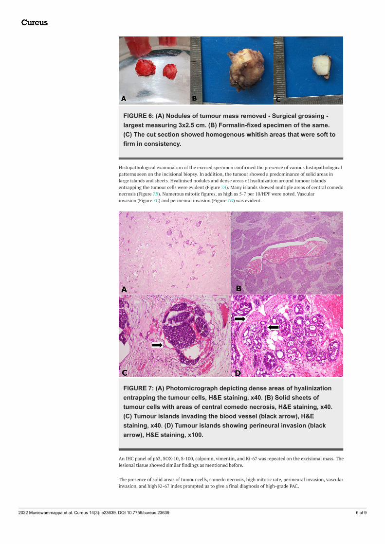
FIGURE 6: (A) Nodules of tumour mass removed - Surgical grossing -largest measuring 3x2.5 cm. (B) Formalin-fixed specimen of the same.(C) The cut section showed homogenous whitish areas that were soft tofirm in consistency.
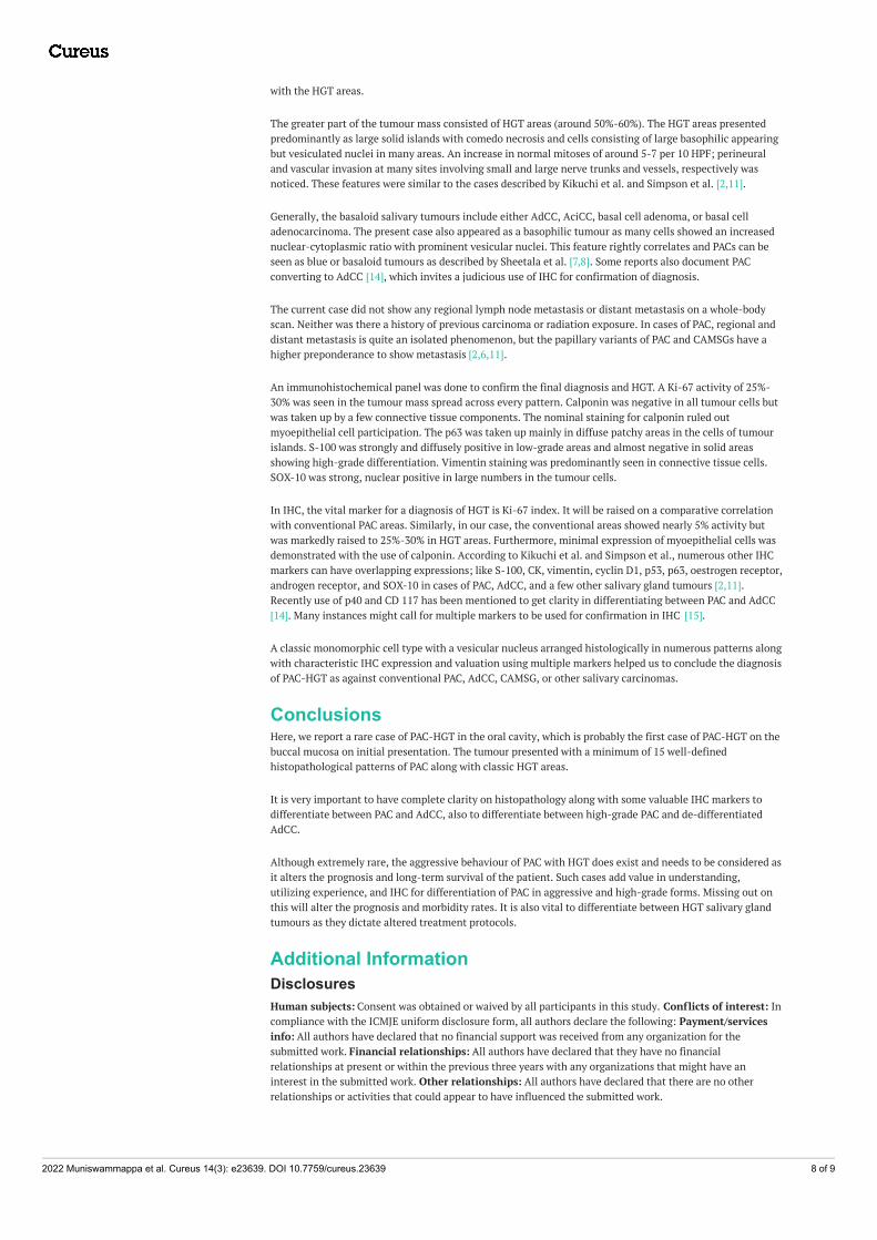
Histopathological examination of the excised specimen confirmed the presence of various histopathologicalpatterns seen on the incisional biopsy. In addition, the tumour showed a predominance of solid areas inlarge islands and sheets. Hyalinised nodules and dense areas of hyalinization around tumour islandsentrapping the tumour cells were evident (Figure 7A). Many islands showed multiple areas of central comedonecrosis (Figure 7B). Numerous mitotic figures, as high as 5-7 per 10/HPF were noted. Vascularinvasion (Figure 7C) and perineural invasion (Figure 7D) was evident.
FIGURE 7: (A) Photomicrograph depicting dense areas of hyalinizationentrapping the tumour cells, H&E staining, x40. (B) Solid sheets oftumour cells with areas of central comedo necrosis, H&E staining, x40.(C) Tumour islands invading the blood vessel (black arrow), H&Estaining, x40. (D) Tumour islands showing perineural invasion (blackarrow), H&E staining, x100.
An IHC panel of p63, SOX-10, S-100, calponin, vimentin, and Ki-67 was repeated on the excisional mass. Thelesional tissue showed similar findings as mentioned before.
The presence of solid areas of tumour cells, comedo necrosis, high mitotic rate, perineural invasion, vascularinvasion, and high Ki-67 index prompted us to give a final diagnosis of high-grade PAC.
2022 Muniswammappa et al. Cureus 14(3): e23639. DOI 10.7759/cureus.23639 6 of 9

The surgical wound healing was uneventful on a three-week follow-up. Post-surgical radiation therapy wasadvised and carried out. On a one-year recall, there is no evidence of recurrence or complications.
AdCC, salivary duct carcinoma, and cribriform adenocarcinoma (CAMSG) were some of the tumoursconsidered for differential diagnosis.
Although a set of common patterns exist and are shared between PAC and AdCC like the tubular, cribriformand solid areas, the presence of various other patterns, like ductal, Indian file, papillary cystic, layeredtubular, tubulo-trabecular, and solid sheet patterns, were also noted, giving clues that lead the diagnosistowards PAC. The tumour cells were predominantly monomorphic with a large vesicular nucleus showingnucleoli sticking to the peripheral nuclear wall, unlike the tumour cells of AdCC, which generally show ahyperchromatic, angular nucleus with prominent central nucleoli, hence ruling out the diagnosis of AdCC.
A cystic tumour with no lymph-node involvement at initial presentation and the presence of multiplepatterns ruled out the diagnosis of CAMSG.
Multiple areas of comedo necrosis were noted, hence a differential diagnosis of salivary duct carcinoma wasconsidered. The presence of different patterns of cell arrangement, monomorphic cells with a vesicularnucleus, and the absence of a characteristic Roman bridge pattern emphasized a diagnosis of PAC. PAC-HGTis an appropriate diagnosis considering the amount of solid pattern seen with comedo necrosis along with ahigh mitotic rate in the tissue, reconfirmed by 25%-30% of Ki-67 activity in the tumour cells. [1-7].
DiscussionSalivary gland tumours of the oral cavity involving the mucous membrane of different areas of the mouth arerelatively rare and complex. They form an immensely diverse category of carcinomas. PAC is the second mostcommon intraoral minor salivary gland carcinoma [1,2], which has seen a nosologic expansion related torecent learning of phenotypic, genotypic, behavioural, and morphological variations. According to the WHO2005 classification, it was called PLGA, a conventional low-grade tumour. The other names given wereterminal duct carcinoma and lobar carcinoma [3,4]. The tumour rubs shoulders with variants seekingindependent recognition, such as papillary cystadenocarcinoma, low-grade papillary adenocarcinoma(LPASO), and CAMSG, the reprisals of which continues to this day [5,6]. Frequently, it is described as atumour with variable morphologic appearance, leading to architectural diversity with numerous patterns,but it has a remarkable cytologic uniformity. A characteristic feature of the tumour is nuclear uniformity,with a round to ovoid nuclei and finely dispersed or ground glass type nuclear chromatin classically calledthe orphan Annie nucleus [7-10].
The tumour is seen in the minor salivary glands of women more frequently (70% of cases), with a male tofemale ratio of 1:2 and a mean age of 59 years. The common sites of presentation are the palate (32%), softpalate (17%), lip (13%), and buccal mucosa (10%). Around 46% of the malignant minor salivary glandtumours are PAC [9,11,12]. The present case was in a 50-year-old woman on the buccal mucosa close to themaxillary left posterior teeth region, presenting as an asymptomatic mass.
An extravagant spectrum of morphological patterns are seen in PAC, namely; solid, glandular, tubular,trabecular, layered tubular, fascicular stream, cord-like cribriform, filigree, cribriform, ductal, cystic,papillary cystic, targetoid, glomeruloid, and Indian file patterns, but the tumour cell type ismonotonous [1,2,4-6]. The presence of a few areas of basaloid cells and clear cells can beencountered [4]. Perineural and perivascular invasion are evident as well. An HGT in the PAC is a rare event,with only a few reported cases till date. Most of the HGT-PACs are related to recurrence and/or radiation.Around seven cases of HGT-PAC have been reported so far in the English literature, arising in the palate (fivecases), nasal region (one case), and maxillary alveolus (one case), and only a small percentage are said to beHGT-de novo on first or primary presentation [2,6,11]. HGT and low-grade areas in a PAC can be well-demarcated or partly admixed with each other [2]. PAC with HGT may present with prominent lobular-solidareas; papillary and papillary cystic areas; clear cell differentiation; and comedo necrosis. Cytologicalfeatures like prominent nucleoli, large nuclei and high mitotic rates are noted [13].
The case we present falls under the rarest category of HGT-PAC. The lesion presented as a cystic tumour onthe buccal mucosa with HGT on initial and primary presentation. It is probably the first case of de novoHGT-PAC reported on the buccal mucosa so far, the other de novo HGT-PAC being reported in the palate [2].
The present case showed a well-demarcated tumour mass but did present many small invasive patches. Onmicroscopic examination, the surface epithelium was hyperplastic and the tumour cells were close to thesurface epithelium as described in the literature [6].
Classic PAC areas with cribriform, pseudocribriform, cords, islands, nests, ribbons, papillary cystic, tubular,layered tubular, trabecular, glomeruloid, single (Indian) file, and sheet patterns were noted. A rare finding ofdensely hyalinized nodules with numerous small islands of tumour cells undergoing degeneration was seen.Hyalinization can be seen in PAC or AdCC [9]. In the present case, hyalinised areas were mostly interspersed
2022 Muniswammappa et al. Cureus 14(3): e23639. DOI 10.7759/cureus.23639 7 of 9
with the HGT areas.
The greater part of the tumour mass consisted of HGT areas (around 50%-60%). The HGT areas presentedpredominantly as large solid islands with comedo necrosis and cells consisting of large basophilic appearingbut vesiculated nuclei in many areas. An increase in normal mitoses of around 5-7 per 10 HPF; perineuraland vascular invasion at many sites involving small and large nerve trunks and vessels, respectively wasnoticed. These features were similar to the cases described by Kikuchi et al. and Simpson et al. [2,11].
Generally, the basaloid salivary tumours include either AdCC, AciCC, basal cell adenoma, or basal celladenocarcinoma. The present case also appeared as a basophilic tumour as many cells showed an increasednuclear-cytoplasmic ratio with prominent vesicular nuclei. This feature rightly correlates and PACs can beseen as blue or basaloid tumours as described by Sheetala et al. [7,8]. Some reports also document PACconverting to AdCC [14], which invites a judicious use of IHC for confirmation of diagnosis.
The current case did not show any regional lymph node metastasis or distant metastasis on a whole-bodyscan. Neither was there a history of previous carcinoma or radiation exposure. In cases of PAC, regional anddistant metastasis is quite an isolated phenomenon, but the papillary variants of PAC and CAMSGs have ahigher preponderance to show metastasis [2,6,11].
An immunohistochemical panel was done to confirm the final diagnosis and HGT. A Ki-67 activity of 25%-30% was seen in the tumour mass spread across every pattern. Calponin was negative in all tumour cells butwas taken up by a few connective tissue components. The nominal staining for calponin ruled outmyoepithelial cell participation. The p63 was taken up mainly in diffuse patchy areas in the cells of tumourislands. S-100 was strongly and diffusely positive in low-grade areas and almost negative in solid areasshowing high-grade differentiation. Vimentin staining was predominantly seen in connective tissue cells.SOX-10 was strong, nuclear positive in large numbers in the tumour cells.
In IHC, the vital marker for a diagnosis of HGT is Ki-67 index. It will be raised on a comparative correlationwith conventional PAC areas. Similarly, in our case, the conventional areas showed nearly 5% activity butwas markedly raised to 25%-30% in HGT areas. Furthermore, minimal expression of myoepithelial cells wasdemonstrated with the use of calponin. According to Kikuchi et al. and Simpson et al., numerous other IHCmarkers can have overlapping expressions; like S-100, CK, vimentin, cyclin D1, p53, p63, oestrogen receptor,androgen receptor, and SOX-10 in cases of PAC, AdCC, and a few other salivary gland tumours [2,11].Recently use of p40 and CD 117 has been mentioned to get clarity in differentiating between PAC and AdCC[14]. Many instances might call for multiple markers to be used for confirmation in IHC [15].
A classic monomorphic cell type with a vesicular nucleus arranged histologically in numerous patterns alongwith characteristic IHC expression and valuation using multiple markers helped us to conclude the diagnosisof PAC-HGT as against conventional PAC, AdCC, CAMSG, or other salivary carcinomas.
ConclusionsHere, we report a rare case of PAC-HGT in the oral cavity, which is probably the first case of PAC-HGT on thebuccal mucosa on initial presentation. The tumour presented with a minimum of 15 well-definedhistopathological patterns of PAC along with classic HGT areas.
It is very important to have complete clarity on histopathology along with some valuable IHC markers todifferentiate between PAC and AdCC, also to differentiate between high-grade PAC and de-differentiatedAdCC.
Although extremely rare, the aggressive behaviour of PAC with HGT does exist and needs to be considered asit alters the prognosis and long-term survival of the patient. Such cases add value in understanding,utilizing experience, and IHC for differentiation of PAC in aggressive and high-grade forms. Missing out onthis will alter the prognosis and morbidity rates. It is also vital to differentiate between HGT salivary glandtumours as they dictate altered treatment protocols.
Additional InformationDisclosuresHuman subjects: Consent was obtained or waived by all participants in this study. Conflicts of interest: Incompliance with the ICMJE uniform disclosure form, all authors declare the following: Payment/servicesinfo: All authors have declared that no financial support was received from any organization for thesubmitted work. Financial relationships: All authors have declared that they have no financialrelationships at present or within the previous three years with any organizations that might have aninterest in the submitted work. Other relationships: All authors have declared that there are no otherrelationships or activities that could appear to have influenced the submitted work.
2022 Muniswammappa et al. Cureus 14(3): e23639. DOI 10.7759/cureus.23639 8 of 9
References1. Xu B, Katabi N: Evolving concepts and new entities in the 2017 WHO classification of salivary gland tumors .
Diagnostic Histopathol. 2018, 24:172-9. 10.1016/j.mpdhp.2018.03.0012. Kikuchi K, Nagao T, Ide F, et al.: Palatal polymorphous adenocarcinoma with high-grade transformation: a
case report and literature review. Head Neck Pathol. 2019, 13:131-9. 10.1007/s12105-018-0916-43. Barnes L, Everson J W, Reichart P, Sidransky D: World Health Organization Classification of Tumours.
Pathology and Genetics of Head and Neck Tumours. IARC Press, Lyon; 2005.4. Ellis GL, Auclair PL, Gnepp DR: Surgical Pathology of the Salivary Glands. W B Saunders, Philadephia, PA;
1991.5. Xu B, Aneja A, Ghossein R, Katabi N: Predictors of outcome in the phenotypic spectrum of polymorphous
low-grade adenocarcinoma (PLGA) and cribriform adenocarcinoma of salivary gland (CASG): a retrospectivestudy of 69 patients. Am J Surg Pathol. 2016, 40:1526-37. 10.1097/PAS.0000000000000705
6. Vander Poorten V, Triantafyllou A, Skálová A, et al.: Polymorphous adenocarcinoma of the salivary glands:reappraisal and update. Eur Arch Otorhinolaryngol. 2018, 275:1681-95. 10.1007/s00405-018-4985-5
7. Seethala RR: Basaloid/blue salivary gland tumors. Mod Pathol. 2017, 30:S84-95.10.1038/modpathol.2016.190
8. Seethala RR, Johnson JT, Barnes EL, Myers EN: Polymorphous low-grade adenocarcinoma: the University ofPittsburgh experience. Arch Otolaryngol Head Neck Surg. 2010, 136:385-92. 10.1001/archoto.2010.39
9. Chatura KR: Polymorphous low grade adenocarcinoma. J Oral Maxillofac Pathol. 2015, 19:77-82.10.4103/0973-029X.157206
10. Bavle RM: Orphan annie-eye nuclei. J Oral Maxillofac Pathol. 2013, 17:154-5. 10.4103/0973-029X.11973711. World Health Organization Classification of Head and Neck Tumours . El-Naggar AK, Chan JKC, Grandis JR,
Takata T, Slootweg (ed): IARC Press, Lyon; 2017.12. Simpson RH, Pereira EM, Ribeiro AC, Abdulkadir A, Reis-Filho JS: Polymorphous low-grade adenocarcinoma
of the salivary glands with transformation to high-grade carcinoma. Histopathology. 2002, 41:250-9.10.1046/j.1365-2559.2002.01439.x
13. Pelkey TJ, Mills SE: Histologic transformation of polymorphous low-grade adenocarcinoma of salivarygland. Am J Clin Pathol. 1999, 111:785-91. 10.1093/ajcp/111.6.785
14. Anjali A K, Tamgadge S , Pereira T, et al.: Polymorphous adenocarcinoma transforming to Adenoid cysticcarcinoma - A case report. Adv Oral Maxillofacial Surg. 2021, 3:1-2. 10.1016/j.adoms.2021.100079
15. Zhu S, Schuerch C, Hunt J: Review and updates of immunohistochemistry in selected salivary gland andhead and neck tumors. Arch Pathol Lab Med. 2015, 139:55-66. 10.5858/arpa.2014-0167-RA
2022 Muniswammappa et al. Cureus 14(3): e23639. DOI 10.7759/cureus.23639 9 of 9
Related Documents