Atherosclerosis 213 (2010) 148–155 Contents lists available at ScienceDirect Atherosclerosis journal homepage: www.elsevier.com/locate/atherosclerosis High-fat diet without excess calories induces metabolic disorders and enhances atherosclerosis in rabbits Ahmed Bilal Waqar a , Tomonari Koike a , Ying Yu a , Tomohiro Inoue a , Tadashi Aoki a , Enqi Liu b , Jianglin Fan a,b,∗ a Department of Molecular Pathology, Interdisciplinary Graduate School of Medicine and Engineering, University of Yamanashi, 1110 Shimokato, Chuo-City, Yamanashi 409-3898, Japan b Research Institute of Atherosclerotic Disease and Laboratory Animal Center, Xi’an Jiaotong University School of Medicine, China article info Article history: Received 5 March 2010 Received in revised form 21 July 2010 Accepted 30 July 2010 Available online 11 August 2010 Keywords: Atherosclerosis Inflammation Rabbits Hypercholesterolemia Animal model abstract Objective: Excess intake of a high-fat diet (HFD) is associated with obesity and metabolic syndrome, which are major risk factors for cardiovascular disease. However, it is unclear whether consumption of an HFD at a normal calorific range would be detrimental to metabolism or affect the development of atherosclerosis. Here, we tested the hypothesis that consumption of a normal-calorie HFD would impair lipid metabolism, insulin sensitivity, and blood pressure. Methods and results: Rabbits fed with an HFD containing either 3% (15% kcal from fat) or 10% (25.8% kcal from fat) coconut oil were compared with control rabbits fed with a standard chow diet (9.3% kcal from fat). All rabbits consumed an equal amount of calories of their respective food. However, HFD feeding induced marked metabolic disorders including increased plasma levels of free fatty acids, insulin resis- tance, and hypertension compared with control rabbits. Metabolic disorders were more pronounced in 10%-HFD-fed rabbits than 3%-HFD-fed rabbits. To examine whether these disorders affected the devel- opment of atherosclerosis, two HFD groups were further fed with a diet containing 0.3% cholesterol for another 18 weeks. We found that 10%-HFD group showed a prominent accumulation of adipose tissue and developed 2-fold greater aortic atherosclerosis than 3%-HFD group. Conclusions: These results suggest that consuming an HFD containing even a normal number of calories can cause insulin resistance, hypertension, and adipose accumulation even without obesity. High amounts of fat in diets apparently accelerate the development of atherosclerosis. © 2010 Elsevier Ireland Ltd. All rights reserved. 1. Introduction There is ample evidence from epidemiological, clinical, and experimental studies to show that an excess intake of a high-fat diet (HFD) is harmful to health [1–4]. It is well known that dietary fat, especially saturated fat (mainly rich in animal fat), is an impor- tant factor that elevates plasma low density lipoprotein (LDL) levels and is closely associated with atherosclerosis [5,6]. Epidemiolog- ical studies have shown that reducing dietary fat intake lowers plasma levels of LDL, thus decreasing the incidence of atheroscle- rosis [2,7]. Moreover, increased intake of HFD coupled with other Abbreviations: AUC, area under the curve; CVD, cardiovascular disease; CRP, C- reactive protein; FFA, free fatty acids; HDL-C, high density lipoprotein-cholesterol; HFD, high-fat diet; HOMA IR, homeostasis model assessment of insulin resistance; IVGTT, intravenous glucose tolerance test; IVITT, intravenous insulin tolerance test; LDL, low density lipoprotein; MetS, metabolic syndrome. ∗ Corresponding author. Tel.: +81 55 273 9520. E-mail address: fan [email protected] (J. Fan). factors such as physical inactivity and genetic factors can impair the balance between energy intake and energy expenditure result- ing in obesity and metabolic syndrome (MetS), which increases the risk of cardiovascular disease (CVD) and death [2–4]. Although this “HFD → obesity → CVD” axis has been widely accepted [5,6], it is still not known whether the intake of a normal-calories-HFD for a long period (such as on a daily basis over a lifetime) is detrimental to metabolism and enhances the development of atherosclerosis. In the current study, we hypothesized that HFD with a normal amount of calories may affect systemic metabolism and the patho- genesis of atherosclerosis. To address this issue, we fed rabbits with diets containing either 3% or 10% coconut oil (containing >90% saturated fat) and compared them with chow-fed rabbits to elucidate the effects of HFD on metabolism and adipose accumu- lation. As herbivores, rabbits are very sensitive to dietary fat and cholesterol, therefore, they can rapidly develop hyperlipidemia and atherosclerosis [8]. In addition, like humans but unlike mice, rab- bits have abundant cholesteryl ester transfer protein in plasma, an important regulator of reverse cholesterol transport, and their lipoprotein profile is LDL-rich [9,10]. Finally, rabbits are a valu- 0021-9150/$ – see front matter © 2010 Elsevier Ireland Ltd. All rights reserved. doi:10.1016/j.atherosclerosis.2010.07.051

Welcome message from author
This document is posted to help you gain knowledge. Please leave a comment to let me know what you think about it! Share it to your friends and learn new things together.
Transcript
He
AEa
Ub
a
ARRAA
KAIRHA
1
edftaipr
rHIL
0d
Atherosclerosis 213 (2010) 148–155
Contents lists available at ScienceDirect
Atherosclerosis
journa l homepage: www.e lsev ier .com/ locate /a therosc leros is
igh-fat diet without excess calories induces metabolic disorders andnhances atherosclerosis in rabbits
hmed Bilal Waqara, Tomonari Koikea, Ying Yua, Tomohiro Inouea, Tadashi Aokia,nqi Liub, Jianglin Fana,b,∗
Department of Molecular Pathology, Interdisciplinary Graduate School of Medicine and Engineering,niversity of Yamanashi, 1110 Shimokato, Chuo-City, Yamanashi 409-3898, JapanResearch Institute of Atherosclerotic Disease and Laboratory Animal Center, Xi’an Jiaotong University School of Medicine, China
r t i c l e i n f o
rticle history:eceived 5 March 2010eceived in revised form 21 July 2010ccepted 30 July 2010vailable online 11 August 2010
eywords:therosclerosis
nflammationabbitsypercholesterolemia
a b s t r a c t
Objective: Excess intake of a high-fat diet (HFD) is associated with obesity and metabolic syndrome,which are major risk factors for cardiovascular disease. However, it is unclear whether consumption ofan HFD at a normal calorific range would be detrimental to metabolism or affect the development ofatherosclerosis. Here, we tested the hypothesis that consumption of a normal-calorie HFD would impairlipid metabolism, insulin sensitivity, and blood pressure.Methods and results: Rabbits fed with an HFD containing either 3% (15% kcal from fat) or 10% (25.8% kcalfrom fat) coconut oil were compared with control rabbits fed with a standard chow diet (9.3% kcal fromfat). All rabbits consumed an equal amount of calories of their respective food. However, HFD feedinginduced marked metabolic disorders including increased plasma levels of free fatty acids, insulin resis-tance, and hypertension compared with control rabbits. Metabolic disorders were more pronounced in
nimal model 10%-HFD-fed rabbits than 3%-HFD-fed rabbits. To examine whether these disorders affected the devel-opment of atherosclerosis, two HFD groups were further fed with a diet containing 0.3% cholesterol foranother 18 weeks. We found that 10%-HFD group showed a prominent accumulation of adipose tissueand developed 2-fold greater aortic atherosclerosis than 3%-HFD group.Conclusions: These results suggest that consuming an HFD containing even a normal number of caloriescan cause insulin resistance, hypertension, and adipose accumulation even without obesity. High amounts
accel
of fat in diets apparently. Introduction
There is ample evidence from epidemiological, clinical, andxperimental studies to show that an excess intake of a high-fatiet (HFD) is harmful to health [1–4]. It is well known that dietaryat, especially saturated fat (mainly rich in animal fat), is an impor-ant factor that elevates plasma low density lipoprotein (LDL) levels
nd is closely associated with atherosclerosis [5,6]. Epidemiolog-cal studies have shown that reducing dietary fat intake lowerslasma levels of LDL, thus decreasing the incidence of atheroscle-osis [2,7]. Moreover, increased intake of HFD coupled with otherAbbreviations: AUC, area under the curve; CVD, cardiovascular disease; CRP, C-eactive protein; FFA, free fatty acids; HDL-C, high density lipoprotein-cholesterol;FD, high-fat diet; HOMA IR, homeostasis model assessment of insulin resistance;
VGTT, intravenous glucose tolerance test; IVITT, intravenous insulin tolerance test;DL, low density lipoprotein; MetS, metabolic syndrome.∗ Corresponding author. Tel.: +81 55 273 9520.
E-mail address: fan [email protected] (J. Fan).
021-9150/$ – see front matter © 2010 Elsevier Ireland Ltd. All rights reserved.oi:10.1016/j.atherosclerosis.2010.07.051
erate the development of atherosclerosis.© 2010 Elsevier Ireland Ltd. All rights reserved.
factors such as physical inactivity and genetic factors can impairthe balance between energy intake and energy expenditure result-ing in obesity and metabolic syndrome (MetS), which increases therisk of cardiovascular disease (CVD) and death [2–4]. Although this“HFD → obesity → CVD” axis has been widely accepted [5,6], it isstill not known whether the intake of a normal-calories-HFD for along period (such as on a daily basis over a lifetime) is detrimentalto metabolism and enhances the development of atherosclerosis.In the current study, we hypothesized that HFD with a normalamount of calories may affect systemic metabolism and the patho-genesis of atherosclerosis. To address this issue, we fed rabbitswith diets containing either 3% or 10% coconut oil (containing>90% saturated fat) and compared them with chow-fed rabbits toelucidate the effects of HFD on metabolism and adipose accumu-lation. As herbivores, rabbits are very sensitive to dietary fat and
cholesterol, therefore, they can rapidly develop hyperlipidemia andatherosclerosis [8]. In addition, like humans but unlike mice, rab-bits have abundant cholesteryl ester transfer protein in plasma,an important regulator of reverse cholesterol transport, and theirlipoprotein profile is LDL-rich [9,10]. Finally, rabbits are a valu-
A.B. Waqar et al. / Atherosclerosis 213 (2010) 148–155 149
Fig. 1. Effects of HFD on metabolism and blood pressure. Plasma glucose, Insulin, and free fatty acid (FFA) levels (A), intravenous glucose tolerance test (IVGTT), intravenousinsulin tolerance test (IVITT), the area under the curve (AUC) of IVITT and homeostasis model assessment of insulin resistance (HOMA IR) (B), blood pressure (C), and plasmaC = 5, 1c
a[
Damlic
RP levels (D) were measured at 22 weeks. Data are expressed as the means ± SE. nontrol.
ble model for the study of obesity, insulin resistance, and MetS11–14].
We performed this study with two questions in mind: (i)oes normal-calorie HFD affect systemic metabolism and adipose
ccumulation? (ii) Does HFD have any effects on the develop-ent of atherosclerosis? Although normal-calorie HFD did notead to excess body weight gain, both HFDs resulted in impairednsulin sensitivity, hypertension, and fatty liver. Furthermore,ompared with 3%-HFD-fed rabbits, 10%-HFD-fed rabbits showed
0 and 10 for control, 3%- and 10%-HFD groups, respectively. *p < 0.05, **p < 0.01 vs.
marked adipose tissue accumulation and a 2-fold increase in aorticatherosclerosis.
2. Methods
2.1. Animals and diets
Male Japanese white rabbits (4 months old) were purchasedfrom Japan SLC Inc. (Shizuoka, Japan) and randomly divided into
150 A.B. Waqar et al. / Atherosclerosis 213 (2010) 148–155
F l levep ocytem 0.05, *
t(agoaAcfUat
2
gwoii(eaisa
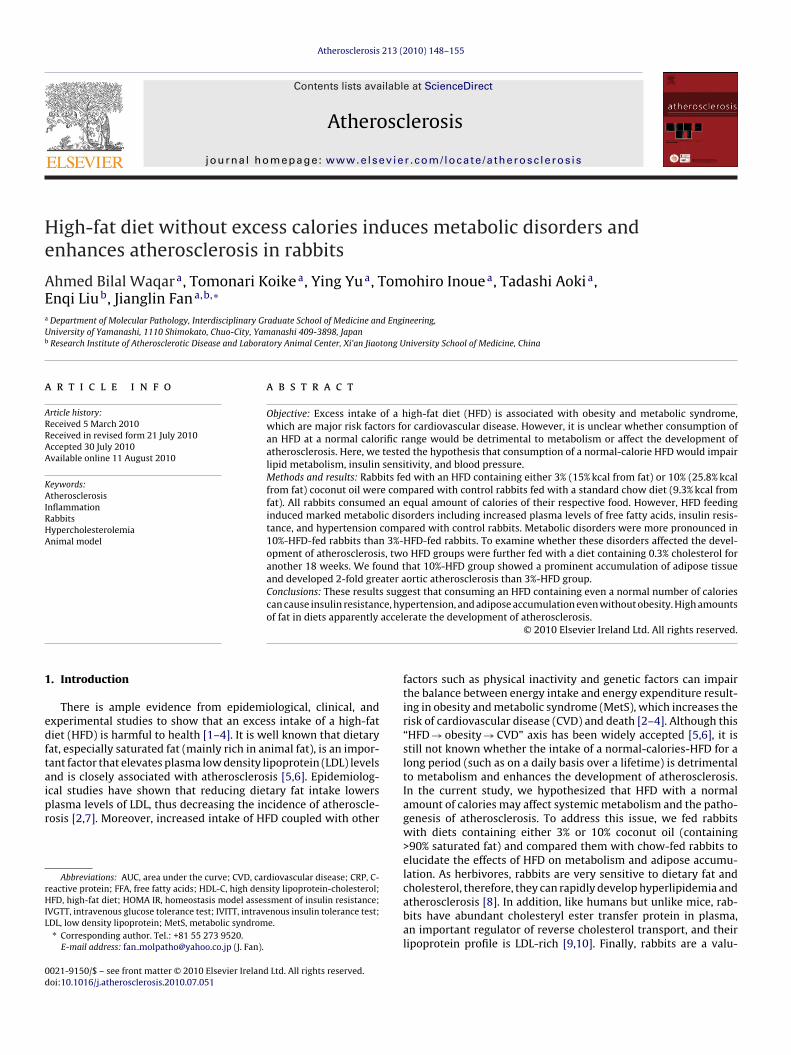
ig. 2. Effects of HFD with high cholesterol on metabolism. Plasma total cholesterolasma glucose, insulin, FFA and portal vein FFA levels at 40 weeks (B) blood moneans ± SE. n = 5, 10 and 10 for control, 3%- and 10%-HFD groups, respectively. *p <
hree groups. Control rabbits were fed with a standard chow dietRM-4), containing 16.5% protein, 4.2% fat derived from soybean oil,nd 13.0% fiber (Funabashi Farms Co. Ltd., Chiba, Japan). Two otherroups were fed with a diet containing either 3% or 10% coconutil by weight (designated as 3%-HFD and 10%-HFD) in which fatccounted for 15% and 26% of total calories (kcal), respectively.ll rabbits were fed with restricted diets with equal numbers ofalories (Supplementary Table 1). All animal experiments were per-ormed with the approval of the Animal Care Committee of theniversity of Yamanashi and conformed to the Guide for the Carend Use of Laboratory Animals published by the US National Insti-utes of Health.
.2 Experimental procedures
To investigate the metabolic disorders including plasma lipids,lucose and insulin metabolism, and blood pressure in rabbits,e fed rabbits with two kinds of HFD for 22 weeks based on
ur previous study [13]. The experimental protocols are shownn Supplementary Fig. 1. To assess whether HFD affected systemicnflammation, we measured blood leukocyte and C-reactive proteinCRP) levels. Our previous study showed that in rabbits, plasma lev-
ls of CRP increase dramatically in response to inflammation suchs atherosclerosis [15]. To evaluate whether metabolic changesnduced by HFD would affect the development of atherosclero-is, HFD-fed rabbits were further fed with the same HFD with andditional 0.3% of cholesterol because HFD alone cannot producels with the calculated area under the curve (AUC), blood pressure at 30 weeks (A),and neutrophil counts and plasma CRP at 40 weeks (C). Data are expressed as the*p < 0.01.
atherosclerosis in rabbits (unpublished data). Rabbits fed with achow diet do not develop spontaneous atherosclerosis. In the cur-rent study, we attempted to compared the effects of two types ofHFDs on the development of atherosclerosis. Therefore, only fat-fedrabbits were induced for atherosclerosis. At the end of the study, allanimals were sacrificed and pathological analysis was performed toexamine liver, pancreas, adipose tissue, and aortic atherosclerosis(see Supplementary methods).
3. Results
3.1. Effects of HFD on metabolism and blood pressure
All three groups of rabbits consumed the same amount ofcalories regardless of diet and gained body weight similarly(Supplementary Fig. 2 and Table 1). HFD feeding did not affectplasma total cholesterol, triglycerides, HDL-C, glucose and bloodcell counts (Supplementary Tables 2 and 3); however, plasma FFAand insulin levels were significantly increased in the 10%-HFDgroup compared with chow-fed control rabbits (Fig. 1A). PlasmaFFA and insulin levels were also increased in the 3%-HFD group butthe difference was not significant. To investigate whether HFD led
to impaired glucose clearance and insulin sensitivity, we performedIVGTT and IVITT. Although glucose catabolism was not significantlychanged, both HFD-fed rabbits exhibited insulin resistance but sta-tistical significance was only observed in the 10%-HFD-fed rabbits(Fig. 1B and Supplementary Fig. 3). Insulin resistance exhibited in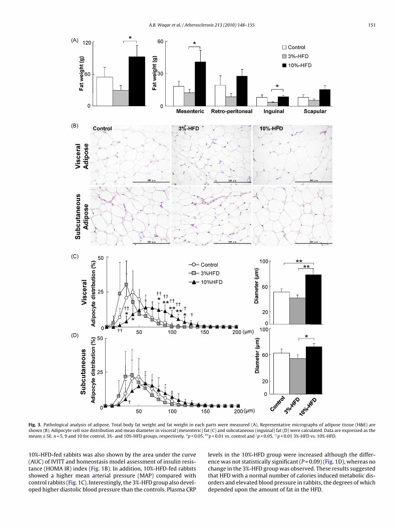
A.B. Waqar et al. / Atherosclerosis 213 (2010) 148–155 151
F ach ps ric) fatm .05, **
1(tsco
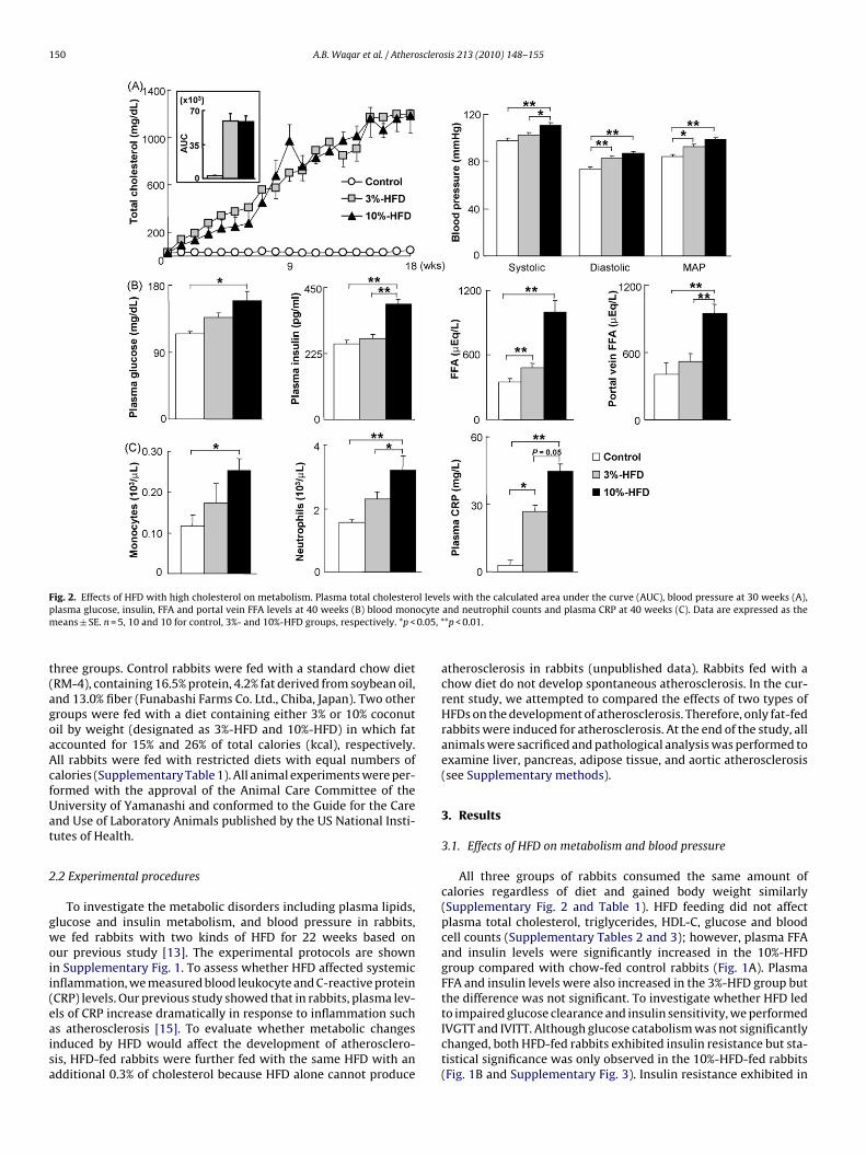
ig. 3. Pathological analysis of adipose. Total body fat weight and fat weight in ehown (B). Adipocyte cell size distribution and mean diameter in visceral (mesenteeans ± SE. n = 5, 9 and 10 for control, 3%- and 10%-HFD groups, respectively. *p < 0
0%-HFD-fed rabbits was also shown by the area under the curve
AUC) of IVITT and homeostasis model assessment of insulin resis-ance (HOMA IR) index (Fig. 1B). In addition, 10%-HFD-fed rabbitshowed a higher mean arterial pressure (MAP) compared withontrol rabbits (Fig. 1C). Interestingly, the 3%-HFD group also devel-ped higher diastolic blood pressure than the controls. Plasma CRParts were measured (A). Representative micrographs of adipose tissue (H&E) are(C) and subcutaneous (inguinal) fat (D) were calculated. Data are expressed as the
p < 0.01 vs. control and †p < 0.05, ††p < 0.01 3%-HFD vs. 10%-HFD.
levels in the 10%-HFD group were increased although the differ-
ence was not statistically significant (P = 0.09) (Fig. 1D), whereas nochange in the 3%-HFD group was observed. These results suggestedthat HFD with a normal number of calories induced metabolic dis-orders and elevated blood pressure in rabbits, the degrees of whichdepended upon the amount of fat in the HFD.
152 A.B. Waqar et al. / Atherosclerosis 213 (2010) 148–155
Fig. 4. Immunohistochemical analysis of adipose tissue. Sections were stained with RAM11 Ab for detecting macrophages (MФ) and number of MФ in visceral (mesenteric)fat and in subcutaneous (inguinal) fat was quantified as described in Supplementary methods. Data are expressed as the means ± SE. n = 5, 6 and 9 in visceral and n = 5, 8 and8
3
bdatdmatce(pit
3
afpgtt3owiwttwns(
in subcutaneous fat for control, 3%- and 10%-HFD, respectively. *p < 0.05.
.2. Effects of cholesterol-rich diets on metabolism
Next, we examined whether the metabolic disorders inducedy the two kinds of HFD affected the development of cholesterol-iet-induced atherosclerosis. Because HFD alone cannot inducetherosclerosis, we fed these HFD rabbits with additional choles-erol as described. As shown in Fig. 2A, 3%- and 10%-HFD groupseveloped similar hypercholesterolemia throughout the experi-ent but their HDL-C levels were unchanged (data not shown)
fter feeding with cholesterol-rich diets. Although insulin resis-ance (data not shown) and high blood pressure (Fig. 2A) wereonstantly present in both HFD-fed groups, 10%-HFD-fed rabbitsxhibited three more prominent changes than 3%-HFD-fed rabbits:1) hyperglycemia and hyperinsulinemia (Fig. 2B left); (2) increasederipheral blood FFA and portal vein FFA (Fig. 2B right); and (3)
ncreased peripheral leukocyte number (both monocytes and neu-rophils) and plasma CRP levels (Fig. 2C).
.3. Pathological examinations
At the end of the experiment, all rabbits were sacrificed forutopsy. To investigate the pathological changes induced by highat and cholesterol diets, we examined liver, pancreas and adi-ose tissue. As shown in Supplementary Fig. 4, the livers of bothroups of HFD-fed rabbits were pale in color and 2-fold heavier thanhose of chow-fed control rabbits. Histological analysis revealedhat there was focal lipid accumulation in the hepatocytes of both%- and 10%-HFD groups (Supplementary Fig. 4). Since both groupsf HFD-fed rabbits developed insulin resistance as shown above,e examined the histological changes in the pancreas. Histolog-
cal examination revealed that the islets of 10%-HFD-fed rabbitsere enlarged to various sizes compared with those of the other
wo groups but the quantitative analysis did not show any sta-
istical significance (Supplementary Fig. 5). The number of isletsas also similar in all three groups. However, the average �-cellumber of each islet in both HFD groups was apparently increased,uggesting that both islet hypertrophy and hyperplasia occurredSupplementary Fig. 5).3.4. Pathological examination of adipose tissue
To examine whether HFD feeding affects adipose tissue, weundertook a quantitative and qualitative analysis of adipose tis-sue collected from different regions. Although all rabbits were fedwith the same number of calories, the amount of adipose tissuein visceral and subcutaneous regions was significantly greater inthe 10%-HFD group than in the other two groups (Fig. 3A). Theamount of adipose tissue of 3%-HFD rabbits was less than thatof chow-fed control rabbits, although the difference was not sta-tistically significant (Fig. 3A). Analysis of adipocyte size showedthat in 10%-HFD-fed rabbits, the average adipocyte in both visceraland subcutaneous regions was larger and that large (hypertrophic)adipocyte predominated in visceral adipose tissue compared withthe other groups (Fig. 3B and C). The subcutaneous adipose of the10%-HFD group was also shifted towards a large cell populationcompared with 3%-HFD and chow-diet control rabbits (Fig. 3D).Because inflammation in the adipose tissue is considered to play akey role in insulin resistance and MetS, we evaluated macrophageinfiltration. Macrophages were seldom observed in normal rabbitadipose tissue; however, their numbers were increased in HFD-fed rabbits (Fig. 4). Macrophage infiltration was focal and sparse inboth visceral and subcutaneous adipose tissue. Macrophages wereeither trapped between adipocytes or surrounded adipocytes form-ing clusters or crown-like structures (Fig. 4). The average numbersof macrophages in both visceral and subcutaneous adipose tissuewere increased in HFD-fed groups.
3.5. Atherosclerosis
To check whether multiple metabolic disorders induced by HFDaffected aortic atherosclerotic lesion development, we performeda pathological analysis of aortas. The sudanophilic en face lesionarea in the aortic trees of 10%-HFD rabbits was found to be 2-fold
greater than that of the 3%-HFD group (Fig. 5A). We also measuredthe microscopic lesion area on the sections in the arch. The 10%-HFDgroup showed an increase in this variable, but the difference wasnot statistically significant (Fig. 5B and C). Immunohistochemicalstaining showed no changes in the proportion of SMCs but the pro-
A.B. Waqar et al. / Atherosclerosis 213 (2010) 148–155 153
Fig. 5. Aortic atherosclerosis. Representative picture of aortic atherosclerosis stained by Sudan IV and lesion area in different parts of aorta are shown in A. For lesion analysis,serial sections were stained with EVG or RAM11 for macrophages (MФ) or HHF35 for smooth muscle cells (SMC). Representative pictures are shown (B). Intimal lesions ont eterme
paa
4
c
he sections were measured using EVG sections. MФ and SMC positive areas were dach group. *p < 0.05.
ortions of macrophages increased 2.9-fold in the 10%-HFD group,lthough this increase did not reach statistical significance (Fig. 5Bnd D).
. Discussion
We have demonstrated that the consumption of a normal-alorie HFD can lead to insulin resistance and high blood pressure
ined (C) by the image analysis system. Data are expressed as the means ± SE. n = 10,
in rabbits, hallmarks of MetS. Although both types of HFDwere detrimental to glucose and lipid metabolism, 10%-HFDinduced much adverse effects (i.e. the higher the level of dietary
fat, the worse the metabolism), suggesting that the amount offat consumed in a diet plays an important role in metabolicdisorders.Nevertheless, in the setting of these metabolic disorders, 10%-HFD rabbits fed with a diet enriched in cholesterol became more

1 sclero
sHaFwwmnttcthtiFitibo�hh
attcomHit
cpnbtirfbc
Haapwipiba(wcr
wloa
[
[
[
[
[
[
[
[
[
54 A.B. Waqar et al. / Athero
usceptible to cholesterol-induced metabolic changes than 3%-FD-fed rabbits: pronounced systemic inflammation (leukocytosisnd high levels of CRP), marked elevation of plasma glucose,FA, and insulin, and prominent accumulation of adipose tissue;hereas both HFD groups developed severe hepatosteatosis. Itas initially surprising to find that 10%-HFD-fed rabbits exhibitedore adipose tissue accumulation (in both visceral and subcuta-
eous regions) than chow-fed control and 3%-HFD-fed rabbits, evenhough their body weights were similar. This raises the possibilityhat visceral obesity can be induced by HFD itself without excessivealorific intake. Several mechanisms may be operative in causinghese fat-content-dependent metabolic changes. First, diets withigh levels of saturated fats can induce accumulation of adiposeissue through the mediation of adipocyte hypertrophy [16]. Anncreased level of visceral adipose tissue in return results in moreFA flowing into the portal vein, thereby inducing severe hepaticnsulin resistance [17]. Secondly, the accumulation of inflamma-ory macrophages in adipose tissue may also be involved in thensulin resistance exhibited by HFD-fed rabbits. Generally, it iselieved that adipocyte hypertrophy can change the productionf inflammatory cytokines, such as MCP-1 [18], PAI-1 [19], TNF-[20], adiponectin [21], and resistin [22]. Finally, pancreas islet
ypertrophy in 10%-HFD-fed rabbits is apparently responsible foryperinsulinemia.
It should be noted, however that, in 3%-HFD-fed rabbits, themount of adipose tissue was actually decreased compared withhat of chow-fed control rabbits. A possible explanation for this ishat high cholesterol diet with 3% dietary fat in rabbits is insuffi-ient for cholesterol absorption, which in turn leads to mobilizationf internal adipose tissue [23]. It is interesting to demonstrate thatacrophage infiltration also occurred in the adipose tissue of 3%-FD-fed rabbits, suggesting that adipocyte hypertrophy or obesity
s not a prerequisite for the induction of inflammation in adiposeissue.
Carroll et al. demonstrated that feeding rabbits with excessalories using 15%-HFD induces obesity, which results in bloodressure being elevated [3]. Surprisingly, 10%-HFD with a normalumber of calories in the current study failed to induce obesityut still resulted in an increase in blood pressure. The eleva-ion of blood pressure induced by HFD was mediated throughmpairments of renal functions including glomerular filtrationate and sodium retention [24]. It is noteworthy that 3%-HFD-ed rabbits also developed high blood pressure, suggesting thatlood pressure was more sensitive than other HFD-inducedhanges.
We next examined whether these MetS-like disorders inFD-fed rabbits increased susceptibility to cholesterol-inducedtherosclerosis. 10%-HFD-fed rabbits developed greater aortictherosclerotic lesions than 3%-HFD-fed rabbits with similarlasma cholesterol levels. The lesions of 10%-HFD-fed rabbitsere rich in macrophages. The insulin resistance, systemic
nflammation, increased fat accumulation along with high bloodressure exhibited by 10%-HFD-fed rabbits would be involved
n the enhancement of atherosclerosis. In cholesterol-fed rab-its, the major atherogenic lipoproteins are those of intestinallynd hepatically derived cholesterol-rich remnant lipoproteins�-very low density lipoprotein) [8]. It is currently unknownhether different HFDs can change the atherogenic lipoprotein
omponents or atherogenic properties (such as oxidation andetention).
In conclusion, we demonstrated that the intake of saturated fat
ith a normal number of calories is detrimental to glucose andipid metabolism and induces MetS-like disorders even withoutbesity. These metabolic disorders enhance the development oftherosclerosis.
[
sis 213 (2010) 148–155
Funding
This work was supported in part by grants-in-aid for scientificresearch from the Ministry of Education, Culture, Sports, Scienceand Technology (MEXT), Japan (19790226, 71790514, 19390099,and 21659078), and a research grant for cardiovascular diseasefrom the Ministry of Health, Labor and Welfare of Japan.
Conflict of Interest
None declared.
Acknowledgements
We thank K. Sato and M. Ohta, Department of Clinical and Lab-oratory Medicine, University of Yamanashi Hospital, for analysis ofrabbit plasma and blood and N. Shibata and A. Sumii, Department ofMolecular Pathology, University of Yamanashi, for help with animalexperiment, tissue preparation and staining.
Appendix A. Supplementary data
Supplementary data associated with this article can be found, inthe online version, at doi:10.1016/j.atherosclerosis.2010.07.051.
References
[1] Bray GA, Paeratakul S, Popkin BM. Dietary fat and obesity: a review of animal,clinical and epidemiological studies. Physiol Behav 2004;83:549–55.
[2] Lutsey PL, Steffen LM, Stevens J. Dietary intake and the development of themetabolic syndrome; the atherosclerosis risk in communities study. Circulation2008;117:754–61.
[3] Carroll JF, Dwyer TM, Grady AW, et al. Hypertension, cardiac hypertrophy,and neurohumoral activity in a new animal model of obesity. Am J Physiol1996;271:H373–8.
[4] Hasty AH, Shimano H, Osuga J, et al. Severe hypercholesterolemia, hypertriglyc-eridemia, and atherosclerosis in mice lacking both leptin and the low densitylipoprotein receptor. J Biol Chem 2001;276:37402–8.
[5] Krauss RM, Deckelbaum RJ, Ernst N, et al. Dietary guidelines for healthy Ameri-can adults: a statement for health professionals from the nutrition committee,American heart Association. Circulation 1996;94:1795–800.
[6] Woollett LA, Spady DK, Dietschy JM. Saturated and unsaturated fatty acids inde-pendently regulate low density lipoprotein receptor and production rate. J LipidRes 1992;33:77–88.
[7] Hata Y, Nakajima K. life-style and serum lipids and lipoproteins. J AtherosclerThromb 2000;7:177–97.
[8] Fan J, Watanabe T. Cholesterol-fed and transgenic rabbit models for study ofatherosclerosis. J Atheroscler Thromb 2000;7:26–32.
[9] Fan J, Challah M, Watanabe T. Transgenic rabbit models for biomedicalresearch: current status, basic methods and future perspectives. Pathol Int1999;49:583–94.
10] Chapman MJ. Animal lipoproteins: chemistry, structure, and comparativeaspects. J Lipid Res 1980;21:789–853.
11] Zhao S, Chu Y, Zhang C, et al. Diet-induced central obesity and insulin resistancein rabbits. J Anim Physiol Anim Nutr 2008;92:105–11.
12] Koike T, Liang J, Wang X, et al. Overexpression of lipoprotein lipase in trans-genic Watanabe heritable hyperlipidemic rabbits improves hyperlipidemia andobesity. J Biol Chem 2004;279:7521–9.
13] Kitajima S, Morimoto M, Liu E, et al. Overexpression of lipoprotein lipaseimproves insulin resistance induced by a high-fat diet in transgenic rabbits.Diabetologia 2004;47:1202–9.
14] Zhang XJ, Chinkes DL, Aarsland A, et al. Lipid metabolism in diet induced obeserabbits is similar to that of obese humans. J Nutr 2008;138:515–8.
15] Sun H, Koike T, Ichikawa T, et al. C-reactive protein in atherosclerotic lesions:its origin and pathophysiological significance. Am J Pathol 2005;167:1139–48.
16] Kubota N, Terauchi Y, Miki H, et al. PPAR� mediates high-fat diet-induced adipocyte hypertrophy and insulin resistance. Mol Cell 1999;4:597–609.
17] Arner P. Insulin resistance in type 2 diabetes: role of fatty acids. Diabetes MetabRes Rev 2002;18:S5–9.
18] Kanda H, Tateya S, Tamori Y, et al. MCP-1 contributes to macrophage infiltration
into adipose tissue, insulin resistance, and hepatic steatosis in obesity. J ClinInvest 2006;116:1494–505.19] Morange PE, Alessi MC, Verdier M, et al. PAI-1 produced ex vivo by humanadipose tissue is relevant to PAI-1 blood level. Arterioscler Thromb Vasc Biol1999;19:1361–5.

sclero
[
[
[
[23] Jayo JM, Schwenke DC, Clarkson TB. Atherosclerosis research. In: Manning PJ,Ringler DH, Newcomer CE, editors. The biology of the laboratory rabbit. 2nd ed.
A.B. Waqar et al. / Athero
20] Hotamisligil GS, Arner P, Caro JF, et al. Increased adipose tissue expression oftumor necrosis factor-� in human obesity and insulin resistance. J Clin Invest
1995;95:2409–15.21] Fu Y, Luo N, Klein RL, et al. Adiponectin promotes adipocyte differentiation,insulin sensitivity, and lipid accumulation. J Lipid Res 2005;46:1369–79.
22] Rajala MW, Obici S, Scherer PE, et al. Adipose-derived resistin and gut-derivedresistin-like molecule-� selectively impair insulin action on glucose produc-tion. J Clin Invest 2003;111:225–30.
[
sis 213 (2010) 148–155 155
CA, San Diego: Academic Press; 1994. p. 367–80.24] Antic V, Tempini A, Montani JP. Serial changes in cardiovascular and renal
function of rabbits ingesting a high-fat, high-calorie diet. Am J Hypertens1999;12:826–9.
Related Documents


