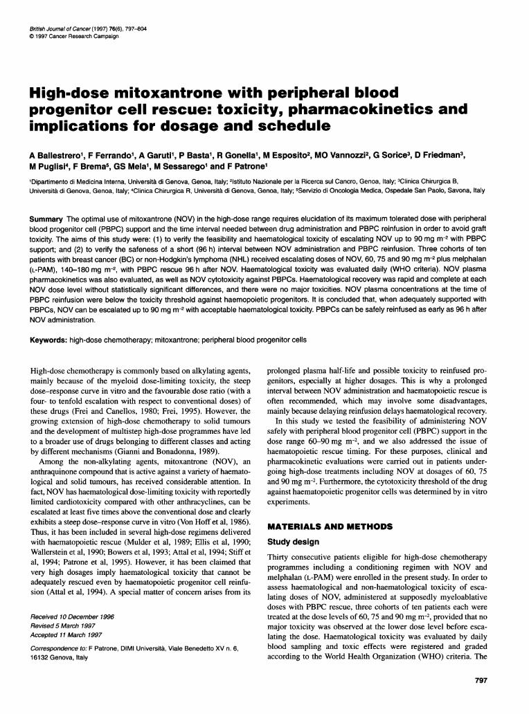
British Joumal of Cancer (1997) 76(6), 797-804 © 1997 Cancer Research Campaign High-dose mitoxantrone with peripheral blood progenitor cell rescue: toxicity, pharmacokinetics and implications for dosage and schedule A Ballestrero1, F Ferrandol, A Garutil, P Basta1, R GonelIa1, M Esposito2, MO Vannozzi2, G Sorice3, D Friedman3, M PugIisi4, F Brema5, GS Mela1, M Sessaregol and F Patrone' 'Dipartimento di Medicina Interna, Universita di Genova, Genoa, Italy; 21stituto Nazionale per la Ricerca sul Cancro, Genoa, Italy; 3Clinica Chirurgica B, Universita di Genova, Genoa, Italy; 4Clinica Chirurgica R, Universita di Genova, Genoa, Italy; 5Servizio di Oncologia Medica, Ospedale San Paolo, Savona, Italy Summary The optimal use of mitoxantrone (NOV) in the high-dose range requires elucidation of its maximum tolerated dose with peripheral blood progenitor cell (PBPC) support and the time interval needed between drug administration and PBPC reinfusion in order to avoid graft toxicity. The aims of this study were: (1) to verify the feasibility and haematological toxicity of escalating NOV up to 90 mg m-2 with PBPC support; and (2) to verify the safeness of a short (96 h) interval between NOV administration and PBPC reinfusion. Three cohorts of ten patients with breast cancer (BC) or non-Hodgkin's lymphoma (NHL) received escalating doses of NOV, 60, 75 and 90 mg m-2 plus melphalan (L-PAM), 140-180 mg m-2, with PBPC rescue 96 h after NOV. Haematological toxicity was evaluated daily (WHO criteria). NOV plasma pharmacokinetics was also evaluated, as well as NOV cytotoxicity against PBPCs. Haematological recovery was rapid and complete at each NOV dose level without statistically significant differences, and there were no major toxicities. NOV plasma concentrations at the time of PBPC reinfusion were below the toxicity threshold against haemopoietic progenitors. It is concluded that, when adequately supported with PBPCs, NOV can be escalated up to 90 mg m-2 with acceptable haematological toxicity. PBPCs can be safely reinfused as early as 96 h after NOV administration. Keywords: high-dose chemotherapy; mitoxantrone; peripheral blood progenitor cells High-dose chemotherapy is commonly based on alkylating agents, mainly because of the myeloid dose-limiting toxicity, the steep dose-response curve in vitro and the favourable dose ratio (with a four- to tenfold escalation with respect to conventional doses) of these drugs (Frei and Canellos, 1980; Frei, 1995). However, the growing extension of high-dose chemotherapy to solid tumours and the development of multistep high-dose programmes have led to a broader use of drugs belonging to different classes and acting by different mechanisms (Gianni and Bonadonna, 1989). Among the non-alkylating agents, mitoxantrone (NOV), an anthraquinone compound that is active against a variety of haemato- logical and solid tumours, has received considerable attention. In fact, NOV has haematological dose-limiting toxicity with reportedly limited cardiotoxicity compared with other anthracyclines, can be escalated at least five times above the conventional dose and clearly exhibits a steep dose-response curve in vitro (Von Hoff et al, 1986). Thus, it has been included in several high-dose regimens delivered with haematopoietic rescue (Mulder et al, 1989; Ellis et al, 1990; Wallerstein et al, 1990; Bowers et al, 1993; Attal et al, 1994; Stiff et al, 1994; Patrone et al, 1995). However, it has been claimed that very high dosages imply haematological toxicity that cannot be adequately rescued even by haematopoietic progenitor cell reinfu- sion (Attal et al, 1994). A special matter of concern arises from its Received 10 December 1996 Revised 5 March 1997 Accepted 11 March 1997 Correspondence to: F Patrone, DIMI Universita, Viale Benedetto XV n. 6, 16132 Genova, Italy prolonged plasma half-life and possible toxicity to reinfused pro- genitors, especially at higher dosages. This is why a prolonged interval between NOV administration and haematopoietic rescue is often recommended, which may involve some disadvantages, mainly because delaying reinfusion delays haematological recovery. In this study we tested the feasibility of administering NOV safely with peripheral blood progenitor cell (PBPC) support in the dose range 60-90 mg m-2, and we also addressed the issue of haematopoietic rescue timing. For these purposes, clinical and pharmacokinetic evaluations were carried out in patients under- going high-dose treatments including NOV at dosages of 60, 75 and 90 mg m-2. Furthermore, the cytotoxicity threshold of the drug against haematopoietic progenitor cells was determined by in vitro experiments. MATERIALS AND METHODS Study design Thirty consecutive patients eligible for high-dose chemotherapy programmes including a conditioning regimen with NOV and melphalan (L-PAM) were enrolled in the present study. In order to assess haematological and non-haematological toxicity of esca- lating doses of NOV, administered at supposedly myeloablative doses with PBPC rescue, three cohorts of ten patients each were treated at the dose levels of 60, 75 and 90 mg m-2, provided that no major toxicity was observed at the lower dose level before esca- lating the dose. Haematological toxicity was evaluated by daily blood sampling and toxic effects were registered and graded according to the World Health Organization (WHO) criteria. The 797

Welcome message from author
This document is posted to help you gain knowledge. Please leave a comment to let me know what you think about it! Share it to your friends and learn new things together.
Transcript
British Joumal of Cancer (1997) 76(6), 797-804© 1997 Cancer Research Campaign
High-dose mitoxantrone with peripheral bloodprogenitor cell rescue: toxicity, pharmacokinetics andimplications for dosage and schedule
A Ballestrero1, F Ferrandol, A Garutil, P Basta1, R GonelIa1, M Esposito2, MO Vannozzi2, G Sorice3, D Friedman3,M PugIisi4, F Brema5, GS Mela1, M Sessaregol and F Patrone''Dipartimento di Medicina Interna, Universita di Genova, Genoa, Italy; 21stituto Nazionale per la Ricerca sul Cancro, Genoa, Italy; 3Clinica Chirurgica B,Universita di Genova, Genoa, Italy; 4Clinica Chirurgica R, Universita di Genova, Genoa, Italy; 5Servizio di Oncologia Medica, Ospedale San Paolo, Savona, Italy
Summary The optimal use of mitoxantrone (NOV) in the high-dose range requires elucidation of its maximum tolerated dose with peripheralblood progenitor cell (PBPC) support and the time interval needed between drug administration and PBPC reinfusion in order to avoid grafttoxicity. The aims of this study were: (1) to verify the feasibility and haematological toxicity of escalating NOV up to 90 mg m-2 with PBPCsupport; and (2) to verify the safeness of a short (96 h) interval between NOV administration and PBPC reinfusion. Three cohorts of tenpatients with breast cancer (BC) or non-Hodgkin's lymphoma (NHL) received escalating doses of NOV, 60, 75 and 90 mg m-2 plus melphalan(L-PAM), 140-180 mg m-2, with PBPC rescue 96 h after NOV. Haematological toxicity was evaluated daily (WHO criteria). NOV plasmapharmacokinetics was also evaluated, as well as NOV cytotoxicity against PBPCs. Haematological recovery was rapid and complete at eachNOV dose level without statistically significant differences, and there were no major toxicities. NOV plasma concentrations at the time ofPBPC reinfusion were below the toxicity threshold against haemopoietic progenitors. It is concluded that, when adequately supported withPBPCs, NOV can be escalated up to 90 mg m-2 with acceptable haematological toxicity. PBPCs can be safely reinfused as early as 96 h afterNOV administration.
Keywords: high-dose chemotherapy; mitoxantrone; peripheral blood progenitor cells
High-dose chemotherapy is commonly based on alkylating agents,mainly because of the myeloid dose-limiting toxicity, the steepdose-response curve in vitro and the favourable dose ratio (with afour- to tenfold escalation with respect to conventional doses) ofthese drugs (Frei and Canellos, 1980; Frei, 1995). However, thegrowing extension of high-dose chemotherapy to solid tumoursand the development of multistep high-dose programmes have ledto a broader use of drugs belonging to different classes and actingby different mechanisms (Gianni and Bonadonna, 1989).Among the non-alkylating agents, mitoxantrone (NOV), an
anthraquinone compound that is active against a variety of haemato-logical and solid tumours, has received considerable attention. Infact, NOV has haematological dose-limiting toxicity with reportedlylimited cardiotoxicity compared with other anthracyclines, can beescalated at least five times above the conventional dose and clearlyexhibits a steep dose-response curve in vitro (Von Hoff et al, 1986).Thus, it has been included in several high-dose regimens deliveredwith haematopoietic rescue (Mulder et al, 1989; Ellis et al, 1990;Wallerstein et al, 1990; Bowers et al, 1993; Attal et al, 1994; Stiff etal, 1994; Patrone et al, 1995). However, it has been claimed thatvery high dosages imply haematological toxicity that cannot beadequately rescued even by haematopoietic progenitor cell reinfu-sion (Attal et al, 1994). A special matter of concern arises from its
Received 10 December 1996Revised 5 March 1997Accepted 11 March 1997
Correspondence to: F Patrone, DIMI Universita, Viale Benedetto XV n. 6,16132 Genova, Italy
prolonged plasma half-life and possible toxicity to reinfused pro-genitors, especially at higher dosages. This is why a prolongedinterval between NOV administration and haematopoietic rescue isoften recommended, which may involve some disadvantages,mainly because delaying reinfusion delays haematological recovery.
In this study we tested the feasibility of administering NOVsafely with peripheral blood progenitor cell (PBPC) support in thedose range 60-90 mg m-2, and we also addressed the issue ofhaematopoietic rescue timing. For these purposes, clinical andpharmacokinetic evaluations were carried out in patients under-going high-dose treatments including NOV at dosages of 60, 75and 90 mg m-2. Furthermore, the cytotoxicity threshold of the drugagainst haematopoietic progenitor cells was determined by in vitroexperiments.
MATERIALS AND METHODS
Study design
Thirty consecutive patients eligible for high-dose chemotherapyprogrammes including a conditioning regimen with NOV andmelphalan (L-PAM) were enrolled in the present study. In order toassess haematological and non-haematological toxicity of esca-lating doses of NOV, administered at supposedly myeloablativedoses with PBPC rescue, three cohorts of ten patients each weretreated at the dose levels of 60, 75 and 90 mg m-2, provided that nomajor toxicity was observed at the lower dose level before esca-lating the dose. Haematological toxicity was evaluated by dailyblood sampling and toxic effects were registered and gradedaccording to the World Health Organization (WHO) criteria. The
797
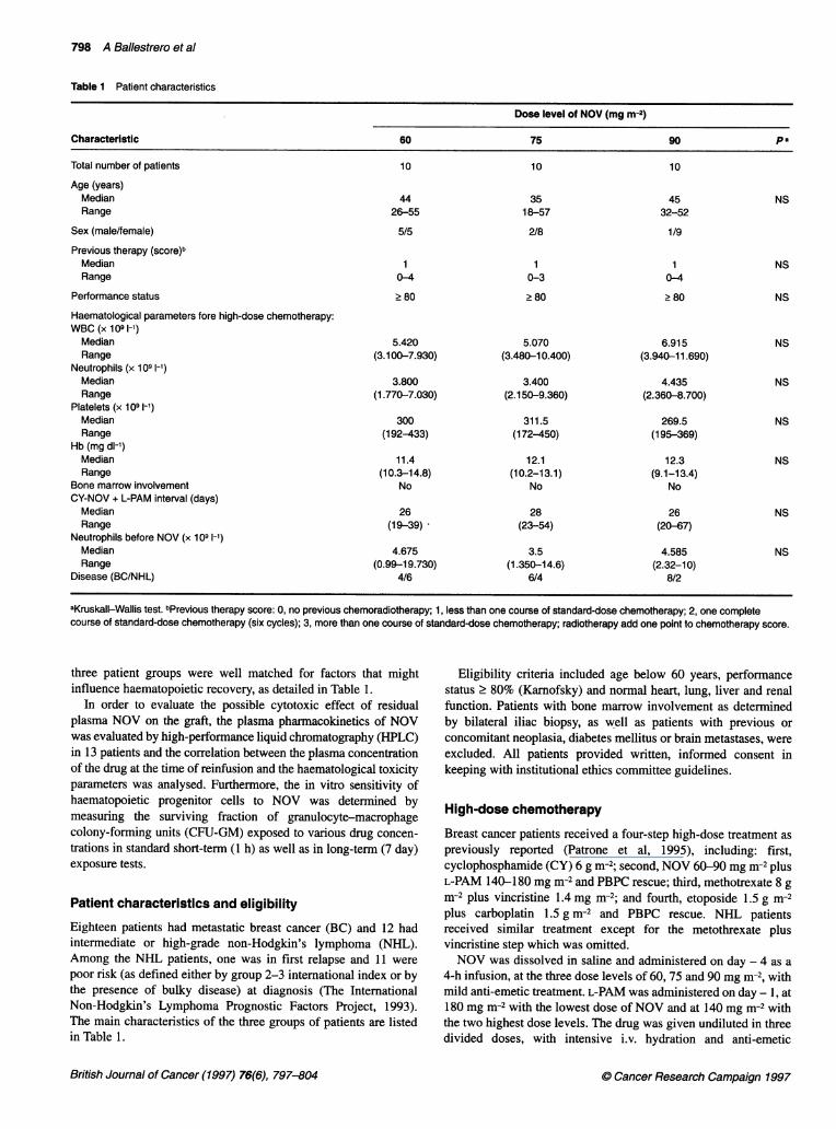
798 A Ballestrero et al
Table 1 Patient characteristics
Dose level of NOV (mg m-2)
Characteristic 60 75 90 Pa
Total number of patients 10 10 10
Age (years)Median 44 35 45 NSRange 26-55 18-57 32-52
Sex (male/female) 5/5 2/8 1/9
Previous therapy (score)bMedian 1 1 1 NSRange 0-4 0-3 0-4
Performance status . 80 > 80 . 80 NS
Haematological parameters fore high-dose chemotherapy:WBC (x 10 1-1)Median 5.420 5.070 6.915 NSRange (3.100-7.930) (3.480-10.400) (3.940-11.690)
Neutrophils (x 109 1-1)Median 3.800 3.400 4.435 NSRange (1.770-7.030) (2.150-9.360) (2.360-8.700)
Platelets (x 109 1-1)Median 300 311.5 269.5 NSRange (192-433) (172-450) (195-369)
Hb (mg dl-1)Median 11.4 12.1 12.3 NSRange (10.3-14.8) (10.2-13.1) (9.1-13.4)
Bone marrow involvement No No NoCY-NOV + L-PAM interval (days)Median 26 28 26 NSRange (19-39)- (23-54) (20-67)
Neutrophils before NOV (x 109 1-1)Median 4.675 3.5 4.585 NSRange (0.99-19.730) (1.350-14.6) (2.32-10)
Disease (BC/NHL) 4/6 6/4 8/2
aKruskall-Wallis test. bPrevious therapy score: 0, no previous chemoradiotherapy; 1, less than one course of standard-dose chemotherapy; 2, one completecourse of standard-dose chemotherapy (six cycles); 3, more than one course of standard-dose chemotherapy; radiotherapy add one point to chemotherapy score.
three patient groups were well matched for factors that mightinfluence haematopoietic recovery, as detailed in Table 1.
In order to evaluate the possible cytotoxic effect of residualplasma NOV on the graft, the plasma pharmacokinetics of NOVwas evaluated by high-performance liquid chromatography (HPLC)in 13 patients and the correlation between the plasma concentrationof the drug at the time of reinfusion and the haematological toxicityparameters was analysed. Furthermore, the in vitro sensitivity ofhaematopoietic progenitor cells to NOV was determined bymeasuring the surviving fraction of granulocyte-macrophagecolony-forming units (CFU-GM) exposed to various drug concen-trations in standard short-term (1 h) as well as in long-term (7 day)exposure tests.
Patient characteristics and eligibilityEighteen patients had metastatic breast cancer (BC) and 12 hadintermediate or high-grade non-Hodgkin's lymphoma (NHL).Among the NHL patients, one was in first relapse and 11 werepoor risk (as defined either by group 2-3 international index or bythe presence of bulky disease) at diagnosis (The InternationalNon-Hodgkin's Lymphoma Prognostic Factors Project, 1993).The main characteristics of the three groups of patients are listedin Table 1.
Eligibility criteria included age below 60 years, performancestatus . 80% (Karnofsky) and normal heart, lung, liver and renalfunction. Patients with bone marrow involvement as determinedby bilateral iliac biopsy, as well as patients with previous orconcomitant neoplasia, diabetes mellitus or brain metastases, wereexcluded. All patients provided written, informed consent inkeeping with institutional ethics committee guidelines.
High-dose chemotherapy
Breast cancer patients received a four-step high-dose treatment aspreviously reported (Patrone et al, 1995), including: first,cyclophosphamide (CY) 6 g m-2; second, NOV 60-90 mg m-2 plusL-PAM 140-180 mg m-2 and PBPC rescue; third, methotrexate 8 gm-2 plus vincristine 1.4 mg in-2; and fourth, etoposide 1.5 g m-2plus carboplatin 1.5 g m-2 and PBPC rescue. NHL patientsreceived similar treatment except for the metothrexate plusvincristine step which was omitted.NOV was dissolved in saline and administered on day - 4 as a
4-h infusion, at the three dose levels of 60, 75 and 90 mg m-2, withmild anti-emetic treatment. L-PAM was administered on day - 1, at180 mg m-2 with the lowest dose of NOV and at 140 mg m-2 withthe two highest dose levels. The drug was given undiluted in threedivided doses, with intensive i.v. hydration and anti-emetic
British Journal of Cancer (1997) 76(6), 797-804 0 Cancer Research Campaign 1997
High-dose NOV with PBPC rescue 799
therapy consisting of dexamethasone and ondansetron. Cryo-preserved PBPCs were reinfused on day 0, i.e. 96 h after NOVadministration. No haematopoietic growth factors were adminis-tered after PBPC reinfusion.
Haematopoietic progenitor cells
After CY administration patients received subcutaneous haemato-poietic growth factors (rhGM-CSF or a sequential combination ofrhGM-CSF and rh interleukin 3) until haematological recovery.PBPCs were collected by continuous flow leucapheresis startingwhen WBC and platelet counts reached 1.0 x 10⁹ 1-' and 50 x 109 1-'respectively. A median number of three aphereses (range 2-5) wasrequired to collect the planned number of CD34+ cells (2 10 x106kg-'), which we assumed would conveniently support twomyeloblative cycles.The apheretic product was cryopreserved in autologous plasma
and 10% dimethylsulphoxide. Cells expressing the CD34 surfaceantigen were enumerated cytofluorimetrically with a CoulterEpics Profile 2 flow cytometer (Coulter, Hialeah, FL, USA) usingthe monoclonal antibody fluorescein isothiocyanate (FITC)-conjugated HPCA-2 (Becton-Dickinson, San Jose, CA, USA)(Siena et al, 1991).
Pharmacological study
In 13 patients, blood samples for analysis of NOV concentrationswere drawn before drug administration as well as at 1, 2 and 3 hduring the infusion, at the end of the 4 h NOV infusion, and there-after at 5, 15, 30, 45 and 60 min and at 2, 4, 6, 24, 48, 72 and 96 h.The samples, collected in heparinized tubes, were immediatelyplaced on ice and then centrifuged at 1800 g for 10 min to separateplasma. Plasma samples, to which was added 10% (v/v) of 5% L-ascorbic acid in citrate buffer (0.1 M, pH 3.0), were frozen at-200C until processing. Urine samples were collected over 4 hduring infusion and then at successive intervals of 2, 4, 6, 24, 48,72 and 96 h after the end of infusion. Each urine volume wasrecorded and an aliquot was frozen until analysis.
Analysis of NOV in plasma and urine was carried out by HPLCaccording to the method described by Peng et al (1982).Quantitation was done by the external method of analysis.Retention time for NOV was 5 min. Extraction efficacy fromplasma and urine was about 90%. The detection limit of themethod was 1 ng ml-'.
Concentrations of NOV vs time were plotted on semiloga-rithmic graphs. Visual inspection of the post-infusion plasmaconcentration-time profiles suggested that the curves weretriphasic in form. Therefore, the plasma concentration-time curvesfor each patient were analysed according to a three-compartmentopen model using a P-Pharm computer program (Simed France)on an IBM/IC computer. All plasma pharmacokinetics were fittedto the multiexponential equation: Cp (t) = A exp(- at) + B exp(,t)+ C exp(- yt), where Cp (t) is the drug concentration at time t, A, Band C are constants and a, [, and y are the apparent first-orderelimination rate constants. The area under the concentration vstime curve (AUC), corrected for the duration of infusion, wascalculated according to Freedman and Workman (1988). Meanresidence time (MRT), steady-state volume of distribution (V,0),total body clearance (Cl) and the elimination half-lives werecalculated from standard pharmacokinetic equations (Gibaldi andPerrier, 1982). The renal clearance (ClR) was calculated using theequation ClR = Du/AUCt, where Du, is the amount of NOVexcreted in urine up to time t after the infusion and AUC, is thearea under the concentration-time curve calculated (trapezoidalrule) for the same time.
Cytotoxicity assay
NOV cytotoxicity against haematopoietic progenitor cells wasdeternmined in a dose-surviving fraction curve by measuring thenumber of CFU-GM after in vitro exposure to various concentra-tions of the drug. PBPC samples from the apheretic product wereexposed to various NOV concentrations that represented theplasma values measured during pharmacokinetic study, includingthe peak plasma concentrations and those measured at the time ofprogenitor cell reinfusion. The tests were performed after either a1-h exposure time or a 7-day exposure time. NOV (Lederle-Cyanamid S.p.A.) was prepared in 0.9% sodium chloride solutionimmediately before use. For the 1-h exposure experiments cellswere suspended in RPMI-1640 medium at 106 ml-', incubated at370C with gentle shaking and then washed twice before clono-genic assay. For the 7-day exposure experiments the drug wasdirectly mixed in clonogemnc assay medium.CFU-GM surviving fraction was determined in a modified
short-term clonogenic assay (Lemoli and Gulati, 1993). Briefly,cells were plated in triplicate in 24-well tissue culture plates(Coming Costar Corporation, Cambridge, MA, USA). The culturemedium consisted of 0.3 ml of Iscove MDM supplemented with
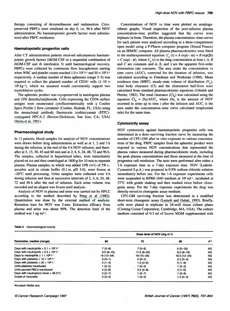
Table 2 Haematological toxicity
Dose level of NOV (mg m-2)
Parameter, median (range) 60 75 90 pa
Days with neutrophils < 0.1 x 10⁹ I-1. 7 (5-9) 7 (5-9) 6 (5-10) NSDays with neutrophils < 0.5 x 10⁹Ol 9.5 (9-15) 11.5 (8-25) 9.5 (8-18) NSDays to neutrophils 2 1 x 10⁹ 1-1 16 (12-54) 18 (15-28) 18.5 (12-24) NSDays with platelets < 10 x 1091- 0 (0-1) 0 (0-1) 0.5 (0-4) NSDays with platelets < 20 x 109 1-' 2 (1-4) 1.5 (0-6) 2 (1-9) NSUnits platelets transfused 1 (0-2) 1 (0-3) 1 (0-3) NSUnits packed RBCs transfused 4 (2-8) 3.5 (2-8) 3 (1-6) NSDays with neutropenic fever < 38.50C 2 (0-7) 1 (0-7) 1 (0-9) NSGrade of mucositis 2 (0-4) 1 (0-4) 1.5 (0-4) NS
aKruskallWallis test.
British Journal of Cancer (1997) 76(6), 797-8040 Cancer Research Campaign 1997
800 A Ballestrero et al
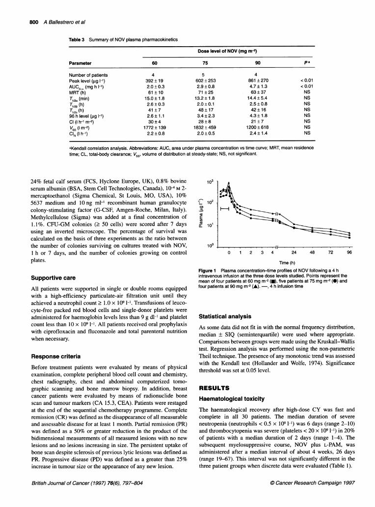
Table 3 Summary of NOV plasma pharmacokinetics
Dose level of NOV (mg m-2)
Parameter 60 75 90 pa
Number of patients 4 5 4Peak level (,g l-1) 392 ± 19 602 ± 253 861 ± 270 < 0.01AUCO_ <(mg h 1-1) 2.0 ± 0.3 2.9 ± 0.8 4.7 ± 1.3 < 0.01MRT (h) 61+ 10 71± 25 63 ± 37 NS
T2,,, (min) 15.0 ±1.8 13.2 ±1.8 14.4 ± 5.4 NS
T,125 (h) 2.6 ± 0.3 2.0 ± 0.1 2.5 ± 0.8 NST1,2Y(h) 41 ±7 48±17 42± 16 NS96 h level (gg I-1) 2.6 ±1.1 3.4 ± 2.3 4.3 ± 1.8 NSCl (I h-1 m-2) 30±4 28±8 21 ±7 NSVSS (I m-2) 1772 ±139 1832 ± 459 1200 ± 618 NSCIR (I h-1) 2.2 ± 0.8 2.0 ± 0.5 2.4 ± 1.4 NS
aKendall correlation analysis. Abbreviations: AUC, area under plasma concentration vs time curve; MRT, mean residencetime; CL, total-body clearance; VSS, volume of distribution at steady-state; NS, not significant.
24% fetal calf serum (FCS, Hyclone Europe, UK), 0.8% bovineserum albumin (BSA, Stem Cell Technologies, Canada), 104 M 2-mercaptoethanol (Sigma Chemical, St Louis, MO, USA), 10%5637 medium and 10 ng ml-1 recombinant human granulocytecolony-stimulating factor (G-CSF, Amgen-Roche, Milan, Italy).Methylcellulose (Sigma) was added at a final concentration of1.1%. CFU-GM colonies (. 50 cells) were scored after 7 daysusing an inverted microscope. The percentage of survival wascalculated on the basis of three experiments as the ratio betweenthe number of colonies surviving on cultures treated with NOV,1 h or 7 days, and the number of colonies growing on controlplates.
Supportive care
All patients were supported in single or double rooms equippedwith a high-efficiency particulate-air filtration unit until theyachieved a neutrophil count 2 1.0 x 10⁹ 1-1. Transfusions of leuco-cyte-free packed red blood cells and single-donor platelets wereadministered for haemoglobin levels less than 9 g dl-1 and plateletcount less than 10 x 109 1-1. All patients received oral prophylaxiswith ciprofloxacin and fluconazole and total parenteral nutritionwhen necessary.
Response criteria
Before treatment patients were evaluated by means of physicalexamination, complete peripheral blood cell count and chemistry,chest radiography, chest and abdominal computerized tomo-graphic scanning and bone marrow biopsy. In addition, breastcancer patients were evaluated by means of radionuclide bonescan and tumour markers (CA 15.3, CEA). Patients were restagedat the end of the sequential chemotherapy programme. Completeremission (CR) was defined as the disappearance of all measurableand assessable disease for at least 1 month. Partial remission (PR)was defined as a 50% or greater reduction in the product of thebidimensional measurements of all measured lesions with no newlesions and no lesions increasing in size. The persistent uptake ofbone scan despite sclerosis of previous lytic lesions was defined asPR. Progressive disease (PD) was defined as a greater than 25%increase in tumour size or the appearance of any new lesion.
103
L 1027cm
EU)
] 101
10°O
I 1
0 1 2 3 4 24 48 72 96
Time (h)
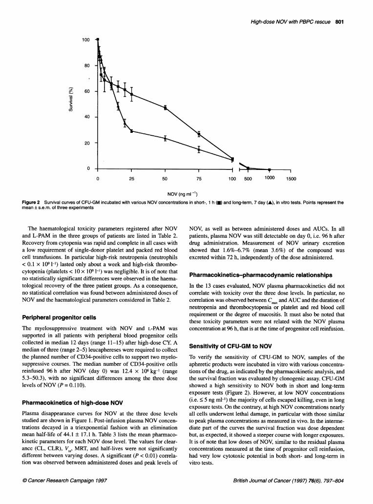
Figure 1 Plasma concentration-time profiles of NOV following a 4 hintravenous infusion at the three dose levels studied. Points represent themean of four patients at 60 mg m-2 (0), five patients at 75 mg m-2 (0) andfour patients at 90 mg m-2 (A). -, 4 h infusion time
Statistical analysis
As some data did not fit in with the normal frequency distribution,median ± SIQ (seminterquartile) were used where appropriate.Comparisons between groups were made using the Kruskall-Wallistest. Regression analysis was performed using the non-parametricTheil technique. The presence of any monotonic trend was assessedwith the Kendall test (Hollander and Wolfe, 1974). Significancethreshold was set at 0.05 level.
RESULTS
Haematological toxicity
The haematological recovery after high-dose CY was fast andcomplete in all 30 patients. The median duration of severeneutropenia (neutrophils < 0.5 x 10⁹ 1-1) was 6 days (range 2-10)and thrombocytopenia was severe (platelets < 20 x 109 1-1) in 20%of patients with a median duration of 2 days (range 1-4). Thesubsequent myelosuppressive course, NOV plus L-PAM, wasadministered after a median interval of about 4 weeks, 26 days(range 19-67). This interval was not significantly different in thethree patient groups when discrete data were evaluated (Table 1).
British Journal of Cancer (1997) 76(6), 797-804 0 Cancer Research Campaign 1997
High-dose NOV with PBPC rescue 801
100
80
600-
2cn
40
20
0
0 25 50 75
NOV (ng r
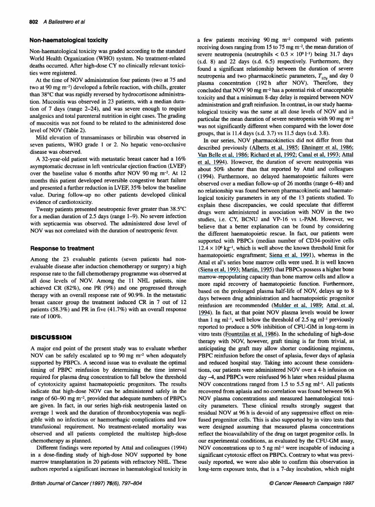
Figure 2 Survival curves of CFU-GM incubated with various NOV concentrationsmean ± s.e.m. of three experiments
The haematological toxicity parameters registered after NOVand L-PAM in the three groups of patients are listed in Table 2.Recovery from cytopenia was rapid and complete in all cases witha low requirement of single-donor platelet and packed red bloodcell transfusions. In particular high-risk neutropenia (neutrophils< 0.1 x 10⁹ 1-') lasted only about a week and high-risk thrombo-cytopenia (platelets < 10 x 109 [-l) was negligible. It is of note thatno statistically significant differences were observed in the haema-tological recovery of the three patient groups. As a consequence,no statistical correlation was found between administered doses ofNOV and the haematological parameters considered in Table 2.
Peripheral progenitor cells
The myelosuppressive treatment with NOV and L-PAM wassupported in all patients with peripheral blood progenitor cellscollected in median 12 days (range 11-15) after high-dose CY. Amedian of three (range 2-5) leucaphereses were required to collectthe planned number of CD34-positive cells to support two myelo-suppressive courses. The median number of CD34-positive cellsreinfused 96 h after NOV (day 0) was 12.4 x 10⁶ kg-' (range5.3-50.3), with no significant differences among the three doselevels of NOV (P = 0. 110).
Pharmacokinetics of high-dose NOV
Plasma disappearance curves for NOV at the three dose levelsstudied are shown in Figure 1. Post-infusion plasma NOV concen-trations decayed in a triexponential fashion with an eliminationmean half-life of 44.1 ± 17.1 h. Table 3 lists the mean pharmaco-kinetic parameters for each NOV dose level. The values for clear-ance (CL, CLR), V 1, MRT, and half-lives were not significantlydifferent between varying doses. A significant (P < 0.01) correla-tion was observed between administered doses and peak levels of
0 0 10 1
100 500 1000 1500
s in short-, 1 h (m) and long-term, 7 day (A), in vitro tests. Points represent the
NOV, as well as between administered doses and AUCs. In allpatients, plasma NOV was still detectable on day 0, i.e. 96 h afterdrug administration. Measurement of NOV urinary excretionshowed that 1.6%-6.7% (mean 3.6%) of the compound wasexcreted within 72 h, independently of the dose administered.
Pharmacokinetics-pharmacodynamic relationships
In the 13 cases evaluated, NOV plasma pharmacokinetics did notcorrelate with toxicity over the three dose levels. In particular, nocorrelation was observed between CmI. and AUC and the duration ofneutropenia and thrombocytopenia or platelet and red blood cellrequirement or the degree of mucositis. It must also be noted thatthese toxicity parameters were not related with the NOV plasmaconcentration at 96 h, that is at the time of progenitor cell reinfusion.
Sensitivity of CFU-GM to NOV
To verify the sensitivity of CFU-GM to NOV, samples of theapheretic products were incubated in vitro with various concentra-tions of the drug, as indicated by the pharmacokinetic analysis, andthe survival fraction was evaluated by clonogenic assay. CFU-GMshowed a high sensitivity to NOV both in short and long-termexposure tests (Figure 2). However, at low NOV concentrations(i.e. < 5 ng ml-') the majority of cells escaped killing, even in longexposure tests. On the contrary, at high NOV concentrations nearlyall cells underwent lethal damage, in particular with those similarto peak plasma concentrations as measured in vivo. In the interme-diate part of the curves the survival fraction was dose dependentbut, as expected, it showed a steeper course with longer exposures.It is of note that low doses of NOV, similar to the residual plasmaconcentrations measured at the time of progenitor cell reinfusion,had very low cytotoxic potential in both short- and long-term invitro tests.
British Journal of Cancer (1997) 76(6), 797-8040 Cancer Research Campaign 1997
802 A Ballestrero et al
Non-haematological toxicity
Non-haematological toxicity was graded according to the standardWorld Health Organization (WHO) system. No treatment-relateddeaths occurred. After high-dose CY no clinically relevant toxici-ties were registered.
At the time of NOV administration four patients (two at 75 andtwo at 90 mg m-2) developed a febrile reaction, with chills, greaterthan 38°C that was rapidly reversed by hydrocortisone administra-tion. Mucositis was observed in 23 patients, with a median dura-tion of 7 days (range 2-24), and was severe enough to requireanalgesics and total parenteral nutrition in eight cases. The gradingof mucositis was not found to be related to the administered doselevel of NOV (Table 2).
Mild elevation of transaminases or bilirubin was observed inseven patients, WHO grade 1 or 2. No hepatic veno-occlusivedisease was observed.A 32-year-old patient with metastatic breast cancer had a 16%
asymptomatic decrease in left ventricular ejection fraction (LVEF)over the baseline value 6 months after NOV 90 mg m-2. At 12months this patient developed reversible congestive heart failureand presented a further reduction in LVEF, 35% below the baselinevalue. During follow-up no other patients developed clinicalevidence of cardiotoxicity.
Twenty patients presented neutropenic fever greater than 38.5°Cfor a median duration of 2.5 days (range 1-9). No severe infectionwith septicaemia was observed. The administered dose level ofNOV was not correlated with the duration of neutropenic fever.
Response to treatment
Among the 23 evaluable patients (seven patients had non-evaluable disease after induction chemotherapy or surgery) a highresponse rate to the full chemotherapy programme was observed atall dose levels of NOV. Among the 11 NHL patients, nineachieved CR (82%), one PR (9%) and one progressed throughtherapy with an overall response rate of 90.9%. In the metastaticbreast cancer group the treatment induced CR in 7 out of 12patients (58.3%) and PR in five (41.7%) with an overall responserate of 100%.
DISCUSSION
A major end point of the present study was to evaluate whetherNOV can be safely escalated up to 90 mg i-2 when adequatelysupported by PBPCs. A second issue was to evaluate the optimaltiming of PBPC reinfusion by determining the time intervalrequired for plasma drug concentration to fall below the thresholdof cytotoxicity against haematopoietic progenitors. The resultsindicate that high-dose NOV can be administered safely in therange of 60-90 mg m-2, provided that adequate numbers of PBPCsare given. In fact, in our series high-risk neutropenia lasted onaverage 1 week and the duration of thrombocytopenia was negli-gible with no infectious or haemorrhagic complications and lowtransfusional requirement. No treatment-related mortality wasobserved and all patients completed the multistep high-dosechemotherapy as planned.
Different findings were reported by Attal and colleagues (1994)in a dose-finding study of high-dose NOV supported by bonemarrow transplantation in 20 patients with refractory NHL. Theseauthors reported a significant increase in haematological toxicity in
a few patients receiving 90 mg m-2 compared with patientsreceiving doses ranging from 15 to 75 mg m-2, the mean duration ofsevere neutropenia (neutrophils < 0.5 x 10⁹ 1-') being 31.7 days(s.d. 8) and 22 days (s.d. 6.5) respectively. Furthermore, theyfound a significant relationship between the duration of severeneutropenia and two pharmacokinetic parameters, Tl4 and day 0plasma concentration (192 h after NOV). Therefore, theyconcluded that NOV 90mg mi-2 has a potential risk of unacceptabletoxicity and that a minimum 8-day delay is required between NOVadministration and graft reinfusion. In contrast, in our study haema-tological toxicity was the same at all dose levels of NOV and inparticular the mean duration of severe neutropenia with 90 mg mr-2was not significantly different when compared with the lower dosegroups, that is 11.4 days (s.d. 3.7) vs 11.5 days (s.d. 3.8).
In our series, NOV pharmacokinetics did not differ from thatdescribed previously (Alberts et al, 1985; Ehninger et al, 1986;Van Belle et al, 1986; Richard et al, 1992; Canal et al, 1993; Attalet al, 1994). However, the duration of severe neutropenia wasabout 50% shorter than that reported by Attal and colleagues(1994). Furthermore, no delayed haematopoietic failures wereobserved over a median follow-up of 26 months (range 6-48) andno relationship was found between pharmacokinetic and haemato-logical toxicity parameters in any of the 13 patients studied. Toexplain these discrepancies, we could speculate that differentdrugs were administered in association with NOV in the twostudies, i.e. CY, BCNU and VP-16 vs L-PAM. However, webelieve that a better explanation can be found by consideringthe different haematopoietic rescue. In fact, our patients weresupported with PBPCs (median number of CD34-positive cells12.4 x 106 kg-', which is well above the known threshold limit forhaematopoietic engraftment; Siena et al, 1991), whereas in theAttal et al's series bone marrow cells were used. It is well known(Siena et al, 1993; Martin, 1995) that PBPCs possess a higher bonemarrow-repopulating capacity than bone marrow cells and allow amore rapid recovery of haematopoietic function. Furthermore,based on the prolonged plasma half-life of NOV, delays up to 8days between drug administration and haematopoietic progenitorreinfusion are recommended (Mulder et al, 1989; Attal et al,1994). In fact, at that point NOV plasma levels would be lowerthan 1 ng ml-, well below the threshold of 2.5 ng ml-' previouslyreported to produce a 50% inhibition of CFU-GM in long-term invitro tests (Fountzilas et al, 1986). In the scheduling of high-dosetherapy with NOV, however, graft timing is far from trivial, asanticipating the graft may allow shorter conditioning regimens,PBPC reinfusion before the onset of aplasia, fewer days of aplasiaand reduced hospital stay. Taking into account these considera-tions, our patients were administered NOV over a 4-h infusion onday -4, and PBPCs were reinfused 96 h later when residual plasmaNOV concentrations ranged from 1.5 to 5.5 ng ml-'. All patientsrecovered from aplasia and no correlation was found between 96 hNOV plasma concentrations and measured haematological toxi-city parameters. These clinical results strongly suggest thatresidual NOV at 96 h is devoid of any suppressive effect on rein-fused progenitor cells. This is also supported by in vitro tests thatwere designed assuming that measured plasma concentrationsreflect the bioavailability of the drug on target progenitor cells. Inour experimental conditions, as evaluated by the CFU-GM assay,NOV concentrations up to 5 ng ml-' were incapable of inducing asignificant cytotoxic effect on PBPCs. Contrary to what was previ-ously reported, we were also able to confirm this observation inlong-term exposure tests, that is a 7-day incubation, which might
British Journal of Cancer (1997) 76(6), 797-804 0 Cancer Research Campaign 1997

High-dose NOV with PBPC rescue 803
be considered a cytotoxicity assay that is more suitable for drugswith prolonged plasma half-life (Fountzilas et al, 1986). Theabsence of graft toxicity with 96 h earlier NOV administration hasbeen recently reported by Stiff et al (1994) in ovarian cancerpatients exposed to high-dose NOV, 75 mg m-2 in three divideddoses, and reinfused with autologous bone marrow.
In our patients non-haematological toxicity was low andsubstantially limited to mucositis. Mucositis is a frequent andimportant complication of high-dose NOV and L-PAM (Mulder etal, 1989; Ellis et al, 1990; Wallerstein et al, 1990; Bowers et al,1993; Attal et al, 1994; Stiff et al, 1994; Patrone et al, 1995). Non-haematological toxicity was observed in 76% of patients in ourseries, and in a third (33%) of them it was of intermediate-highgrade requiring parenteral nutrition and some analgesic treatment.However, mucositis was not associated with particularly severeinfectious complications, healed promptly after resolution ofneutropenia and did not result in any delay of plannedchemotherapy. Unlike Attal's series with bone marrow rescue(Attal et al, 1994), there was no correlation between the NOV doselevel and the severity of mucositis in our patients supported byPBPCS. The duration of neutropenia was uniformly short, and thismay be a critical factor in determining the clinical evolution ofmucositis, as suggested by the findings of Gabrilove et al (1988),who found a significant reduction in the incidence and severity ofmucositis when neutrophil recovery after standard dosechemotherapy was accelerated by G-CSF. In our series, the onlysignificant organ dysfunction was symptomatic heart failureobserved in a breast cancer patient who presented a decline inLVEF 12 months after treatment at the 90-mg dose level. Dose-limiting cardiac toxicity with a maximum tolerated dose as low as50 mg m-2 was found when NOV was administered with high-dosethiotepa (Bowers et al, 1993). However, although decreases inLVEF or clinical signs of heart failure have been noticed, a dose-limiting heart toxicity has not been found in several studies usingdoses of NOV up to 80 mg m-2 in combination with other poten-tially cardiotoxic agents such as CY and Ara-C (Mulder et al,1989; Wallerstein et al, 1990; Feldman et al, 1993; Attal et al,1994; Stiff et al, 1994). The single event observed in our studydoes not allow a conclusion to be made on the possible correlationbetween NOV dose levels and cardiotoxicity. It is hoped furtherinformation will be available from the prospective study we arepresently running on the long-term survey of LVEF.
Both breast cancer and lymphoma patients had high responserates with the present multistep HD treatment. Although it is sugges-tive that NOV plays a significant role in this result, the present studywas designed neither to compare NOV anti-tumour activity at thevarious dose levels nor to establish the relative anti-tumour activityof NOV in the sequential treatment. However, as our data demon-strate that PBPCs can overcome the myelosuppressive effect ofdoses of NOV up to 90 mg m2, and as containing haematologicaltoxicity is a critical factor in reducing the overall morbidity of thetreatment, we conclude that controlled studies with high-dose condi-tioning regimens including NOV up to 90 mg m-2 can be planned forpatients with breast cancer and non-Hodgkin's lymphoma.
ACKNOWLEDGEMENTS
This work was partially supported by Ministero dell'Universit'a edella Ricerca Scientifica e Tecnologica, Rome, Italy (60%) andby Consiglio Nazionale delle Ricerche, Rome, Italy (ProgettoFinalizzato Applicazioni Cliniche della Ricerca Oncologica).
REFERENCES
Alberts DS, Peng YM, Leigh S, Davis TP and Woodward DL (1985) Disposition ofmitoxantrone in cancer patients. Cancer Res 45: 1879-1884
Attal M, Canal P, Schlaifer D, Chatelut E, Dezeuze A, Huguet F, Payen C, Pris J andLaurent G (1994) Escalating dose of mitoxantrone with high-dosecyclophosphamide, carmustine, and etoposide in patients with refractorylymphoma undergoing autologous bone marrow transplantation. J Clin Oncol12: 141-148
Bowers C, Adkins D, Dunphy F, Harrison B, LeMaistre CF and Spitzer G (1993)Dose escalation of mitoxantrone given with thiotepa and autologous bonemarrow transplantation for metastatic breast cancer. Bone Marrow Transplant12: 525-530
Canal P, Attal M, Chatelut E, Guichard S, Huguet F, Muller C, Schlaifer D, LaurentG, Houin G and Bugat R (1993) Plasma and cellular pharmacokinetics ofmitoxantrone in high-dose chemotherapeutic regimen for refractorylymphomas. Cancer Res 53: 4850-4854
Ehninger G, Proksch B, Heinzel G and Woodward DL (1986) Clinical pharmacologyof mitoxantrone. Cancer Treat Rep 70: 1373-1378
Ellis ED, William SF, Moormeier JA, Kaminer LS and Bitran JD (1990) A phaseI-II study of high-dose cyclophosphamide, thiotepa and escalating dose ofmitoxantrone with autologous stem cell rescue in patients with refractorymalignancies. Bone Marrow Transplant 6: 439-442
Feldman EJ, Alberts DS, Arlin Z, Ahmed A, Mittelman A, Baskind, Peng Y-M,Baier M and Plezia P (1993) Phase I clinical and pharmacokinetic evaluation ofhigh-dose mitoxantrone in combination with cytarabine in patients with acuteleukemia. J Clin Oncol 11: 2002-2009
Fountzilas G, Ohnuma T, Rammos K, Mindich B and Holland FJ (1986)Comparison of mitoxantrone and ametantrone in human acute myelocyticleukemia cells in culture and in bone marrow granulocyte-macrophageprogenitor cells. Cancer Drug Delivery 3: 93-100
Freedman LS and Workman P (1988) When can the infusion period be safelyignored in the estimation of pharmacokinetic parameters of drugs in humans?Cancer Chemother Pharmacol 22: 95-103
Frei E (1995) Pharmacologic strategies for high-dose chemotherapy. In High-doseCancer Therapy. Armitage JO and Antman KH (eds), pp. 3-16. Williams &Wilkins: Baltimore
Frei E and Canellos GP (1980) Dose: A critical factor in cancer therapy. Am J Med69: 585-594
Gabrilove JL, Jakubosky A, Sher H, Stenberg C, Wong G, Grous J, Yagoda A, FainK, Malcom MAS, Clarkson B, Oettgen HF, Alton K, Welte K and Souza L(1988) Effect of granulocyte colony-stimulating factor on neutropenia andassociated morbidity due to chemotherapy for transitional-cell carcinoma of theurothelium. NEngl J Med 318: 1414-1422
Gianni AM and Bonadonna G (1989) High-dose chemo-radiotherapy for sensitivetumors: Is sequential better than concurrent drug delivery? Eur J Cancer ClinOncol 25: 1027-1030
Gibaldi M and Perrier D (1982) Pharmacokinetics, 2nd edn. Dekker: New YorkHollander G and Wolfe KH (1974) Nonparametric Statistical Methods. John Wiley:
New YorkLemoli RM and Gulati SC (1993) Effect of stem cell factor (c-kit ligand),
granulocyte-macrophage colony-stimulating factor and interleukin3 onhematopoietic progenitors in human long-term bone marrow cultures. StemCells 11: 435-444
Martin M (1995) High-dose chemotherapy for breast cancer: clinical advantagesof autologous peripheral blood progenitor cells (PBPC) compared withautologous bone marrow transplantation (ABMT) Ann Oncol 6 (suppl. 4):S33-S37
Mulder POM, Sleijfer DT, Willemse PHB, De Vries EGE, Uges DRA and MulderNH (1989) High-dose cyclophosphamide or melphalan with escalating doses ofmitoxantrone and autologous bone marrow transplantation for refractory solidtumors. Cancer Res 49: 4654-4658
Patrone F, Ballestrero A, Ferrando F, Brema F, Moraglio L, Valbonesi M, Basta P,Ghio R, Gobbi M, and Sessarego M (1995) Four-step high-dose sequentialchemotherapy with double hematopoietic progenitor-cell rescue for metastaticbreast cancer. J Clin Oncol 13: 840-846
Peng YM, Ormberg D and Alberts DS (1982) Improved high-performance liquidchromatography of the new antineoplastic agents bisantrene and mitoxantrone.J Chromatog Biomed Appl 233: 235-247
Richard B, Launay-Iliadis MC, Iliadis A, Just-Landi S, Blaise D, Stoppa AM, ViensP, Gaspard MH, Maraninchi D, Cano JP and Carcassonne Y (1992)Pharmacokinetics of mitoxantrone in cancer patients treated by high-dosechemotherapy and autologous bone-marrow transplantation. Br J Cancer 65:399-404
C Cancer Research Campaign 1997 British Joural of Cancer (1997) 76(6), 797-804
804 A Ballestrero et al
Siena S, Bregni M, Brando B, Belli N, Ravagnani F, Gandola L, Stem AC, LansdorpPM, Bonadonna G and Gianni AM (1991) Flow cytometry for clinicalestimation of circulating hematopoietic progenitors for autologoustransplantation in cancer patients. Blood 77: 400-409
Siena S, Bregni M, Bonsi L, Strippoli P, Peccatori F, Magni M, Di Nicola M,Bagnara GP and Gianni AM (1993) Clinical implications of the heterogeneityof hematopoietic progenitors elicited in peripheral blood by anticancer therapywith cyclophosphamide and cytokine(s). Stem Cells 11 (suppl. 2): 72-75
Stiff PJ, McKenzie RS, Alberts DS, Sosman JA, Dolan JR, Rad N and McCloskey T(1994) Phase I clinical and pharmacokinetic study of high-dose mitoxantronecombined with carboplatin, cyclophosphamide, and autologous bone marrowrescue: high response rate for refractory ovarian carcinoma. J Clin Oncol 12:176-183
The Intemational Non-Hodgkin's Lymphoma Prognostic Factors Project (1993) Apredictive model for aggressive non-Hodgkin's lymphoma. N Engl J Med 329:987-994
Van Belle SJP, dE Planque MM, Smith IE, Van Oosterom AT, Schoemaker TJ,DeneveW and McVie JG (1986) Pharmacokinetics of mitoxantrone in humanfollowing single-agent infusion or intra-arterial injection therapy or combined-agent infusion therapy. Cancer Chemother Pharmacol 18: 27-32
Von Hoff DD, Clark GM, Weiss GR, marshall MH, Buchok JB, Knight WA III andLeMaistre CF (1986) Use of in vitro dose response effect to selectantineoplastics for high-dose or regional administration regimens. J Clin Oncol4:1827-1834
Wallerstein R, Spitzer G, Dunphy F, Huan S, Hortobagyi G, Yau J, Buzdar A,Holmes F, Theriault R, Ewer M, LeMaistre CF, Dicke K and Deisseroth A(1990) A phase II study of mitoxantrone, etoposide, and thiotepa withautologous marrow support for patients with relapsed breast cancer. J ClinOncol8: 1782-1788
British Journal of Cancer (1997) 76(6), 797-804 C Cancer Research Campaign 1997
Related Documents