Hierarchical multiscale structure–property relationships of the red-bellied woodpecker (Melanerpes carolinus) beak by Nayeon Lee, M. F. Horstemeyer, Hongjoo Rhee, Ben Nabors, Jun Liao, and Lakiesha N. Williams Interface Volume 11(96):20140274 July 6, 2014 ©2014 by The Royal Society

Hierarchical multiscale structure–property relationships of the red-bellied woodpecker (Melanerpes carolinus) beak by Nayeon Lee, M. F. Horstemeyer, Hongjoo.
Dec 13, 2015
Welcome message from author
This document is posted to help you gain knowledge. Please leave a comment to let me know what you think about it! Share it to your friends and learn new things together.
Transcript
Hierarchical multiscale structure–property relationships of the red-bellied woodpecker (Melanerpes carolinus) beak
by Nayeon Lee, M. F. Horstemeyer, Hongjoo Rhee, Ben Nabors, Jun Liao, and Lakiesha N. Williams
InterfaceVolume 11(96):20140274
July 6, 2014
©2014 by The Royal Society
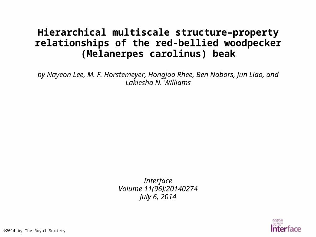
(a) A male red-bellied woodpecker (Photo courtesy of Ken Thomas, http://kenthomas.us/?page_id=140&px=%2FRed-bellied_Woodpecker-male.jpg), (b) upper and lower beaks of the woodpecker, and (c) a schematic of the cross-sectional view of the woodpecker beaks co...
Nayeon Lee et al. J. R. Soc. Interface 2014;11:20140274
©2014 by The Royal Society

(a) Cross-sectional views throughout the length of the woodpeckers' beaks taken by a scanning electron microscope illustrating the change in the geometries with differing ratio of each layer.
Nayeon Lee et al. J. R. Soc. Interface 2014;11:20140274
©2014 by The Royal Society

Microstructure of the woodpeckers' beaks garnered from scanning electron microscopy showing (a) the fractured cross section of the upper beak with the three distinctive structural layers, (b)
the outer rhamphotheca layer containing overlapping scale-like fe...
Nayeon Lee et al. J. R. Soc. Interface 2014;11:20140274
©2014 by The Royal Society

Transmission electron microscopic images of the rhamphotheca of woodpeckers' beaks show the nanostructure: (a) a cross-sectional view reveals the keratin grains in the rhamphotheca; (b)
a cross-sectional view that shows the wavy ‘suture’ lines at the grain ...
Nayeon Lee et al. J. R. Soc. Interface 2014;11:20140274
©2014 by The Royal Society
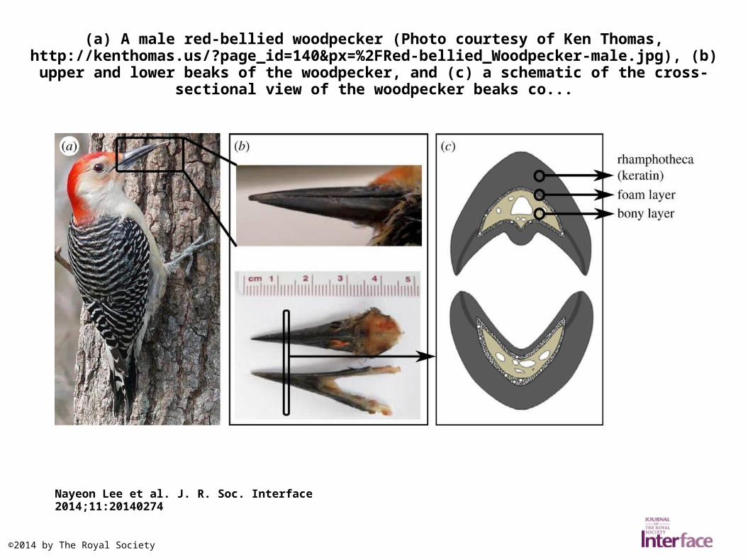
Nanostructure of the foam layer taken by a scanning electron microscope shows (a) the cell wall of the foam layer, (b) part of the cell wall is composed of fibres, and (c) the fibres have a D-period
indicating that they are collagen.
Nayeon Lee et al. J. R. Soc. Interface 2014;11:20140274
©2014 by The Royal Society
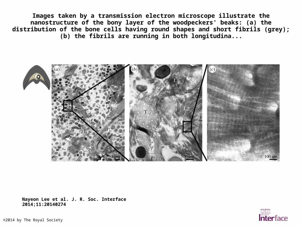
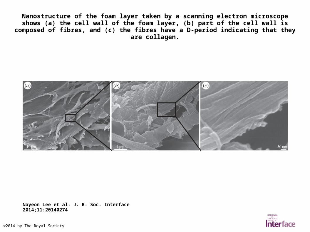
Images taken by a transmission electron microscope illustrate the nanostructure of the bony layer of the woodpeckers' beaks: (a) the distribution of the bone cells having round shapes and
short fibrils (grey); (b) the fibrils are running in both longitudina...
Nayeon Lee et al. J. R. Soc. Interface 2014;11:20140274
©2014 by The Royal Society
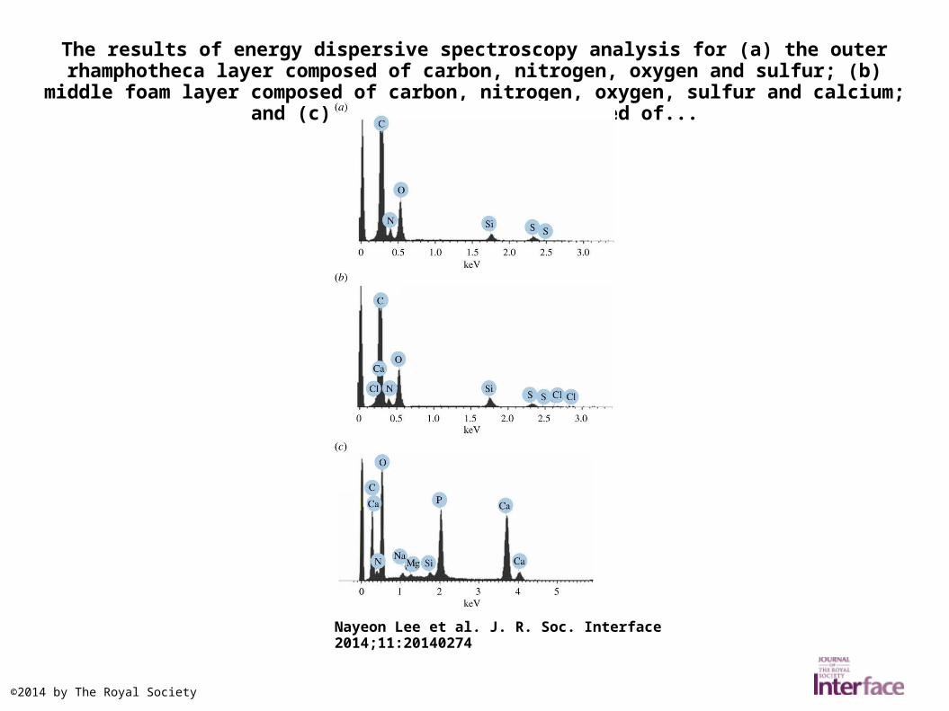
The results of energy dispersive spectroscopy analysis for (a) the outer rhamphotheca layer composed of carbon, nitrogen, oxygen and sulfur; (b) middle foam layer composed of carbon,
nitrogen, oxygen, sulfur and calcium; and (c) inner bony layer composed of...
Nayeon Lee et al. J. R. Soc. Interface 2014;11:20140274
©2014 by The Royal Society

Nanomechanical properties of the rhamphotheca, foam and bony layers obtained from nanoindentation tests: (a) nanohardness and (b) reduced elastic modulus.
Nayeon Lee et al. J. R. Soc. Interface 2014;11:20140274
©2014 by The Royal Society

The scanning electron microscopic image of the polished rhamphotheca of the woodpecker's beak at the transverse plane shows the overlapping pattern of the keratin scales, which provide
resistance to crack propagation in the direction of the arrow.
Nayeon Lee et al. J. R. Soc. Interface 2014;11:20140274
©2014 by The Royal Society

The dimensions and aspect ratios of the height over the width of a keratin scale from each bird are different according to their functions.
Nayeon Lee et al. J. R. Soc. Interface 2014;11:20140274
©2014 by The Royal Society
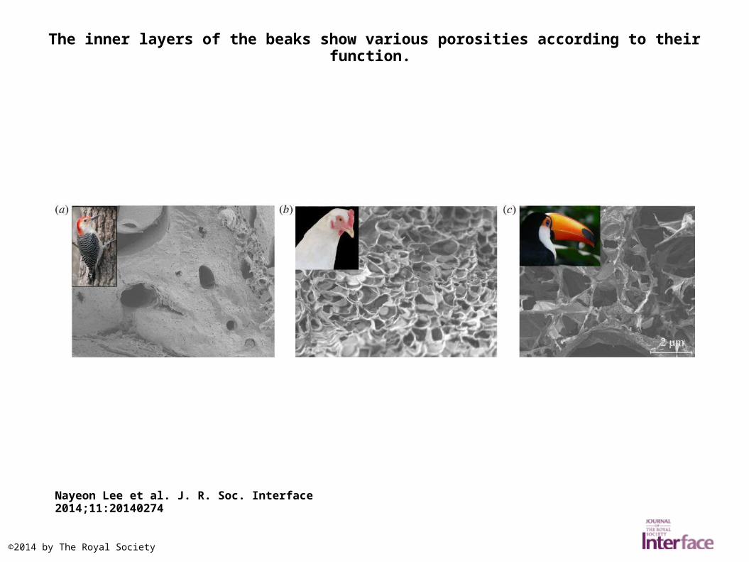
The inner layers of the beaks show various porosities according to their function.
Nayeon Lee et al. J. R. Soc. Interface 2014;11:20140274
©2014 by The Royal Society
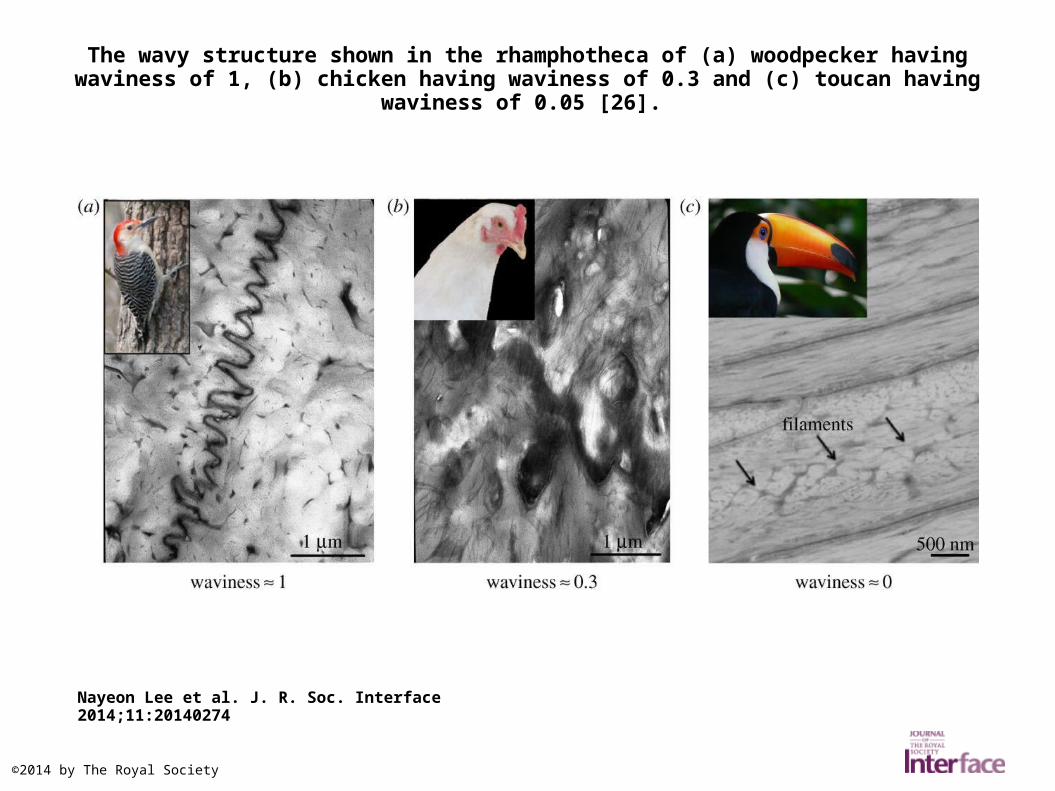
The wavy structure shown in the rhamphotheca of (a) woodpecker having waviness of 1, (b) chicken having waviness of 0.3 and (c) toucan having waviness of 0.05 [26].
Nayeon Lee et al. J. R. Soc. Interface 2014;11:20140274
©2014 by The Royal Society
Related Documents


