Heterosynaptic Metaplastic Regulation of Synaptic Efficacy in CA1 Pyramidal Neurons of Rat Hippocampus Didier Le Ray, David Ferna ´ndez De Sevilla, Ana Bele ´n Porto, Marco Fuenzalida, and Washington Bun ˜o * ABSTRACT: The induction threshold, and the magnitude and direction of changes in synaptic plasticity may depend on the previous history of neuronal activity. This phenomenon, termed “metaplasticity,” could play an important role in integration processes by coordinating the modulation of synapses. Although metaplasticity has been analyzed extensively, its underlying cellular mechanisms remain largely unknown. Using in vitro electrophysiological and computer simulation approaches, we investi- gated the contribution of the slow Ca 2 -dependent afterhyperpolariza- tion (sAHP) in the metaplastic control of the induction of long-term potentiation (LTP) at convergent CA3-CA1 pyramidal neuron synapses. We report that classical conditioning protocols may lead to the simulta- neous induction of a sustained homosynaptic LTP and a potentiation of the sAHP that endured 1 h. The sAHP potentiation dramatically altered the spike responses of the CA1 pyramidal neuron. Of particular interest was the reduction of the CA1 neuron excitability and, consequently, of the capacity of a nonpotentiated synaptic input to elicit spikes while the sAHP was potentiated. This reduction in excitability temporarily prevented nonpotentiated synaptic inputs to exhibit an LTP induced by presynaptic tetanization. This metaplasticity was strongly resistant to increases in the magnitude of synaptic tetanization protocols. We propose that this het- erosynaptic metaplasticity, mediated by intrinsic cellular mechanisms, triggered by brief periods of activity, and relying on changes of a slow Ca 2 -activated K current, may contribute to adjusting the efficacy of synaptic connections and shaping network behavior to regulate integra- tion processes. © 2004 Wiley-Liss, Inc. KEY WORDS: slow afterhyperpolarization; LTP; sI AHP channels; Ca 2 dynamics; simulation INTRODUCTION In the vertebrate central nervous system, brief periods of patterned activity can either potentiate or depress syn- aptic efficacy, the induction of which narrowly depends on the history of synaptic activity. This latter relationship is referred to as “metaplasticity” (Abraham and Bear, 1996). Data on metaplasticity have been gathered mainly from studies of glutamatergic synaptic plasticity (e.g., Huang et al., 1992; Bortolotto and Collingridge, 2000; Goussakov et al., 2000), and it is recognized that meta- plasticity is induced by the same events that trigger syn- aptic plasticity (see Tompa and Friedrich, 1998). How- ever, the cellular mechanisms underlying metaplasticity remain largely unknown. According to Hebb’s postulate, a temporal association between pre- and postsynaptic spike activity is required to induce the synaptic changes that underlie information storage (Hebb, 1949). Action potentials back-propagat- ing within the dendrites play a pivotal role in this scheme, providing the signal of postsynaptic activity required to induce long-term potentiation (LTP) (e.g., Magee and Johnston, 1997; Markram et al., 1997; Linden, 1999; Pike et al., 1999), a long-lasting enhancement in synaptic efficacy regarded as the neural basis for learning and memory. Understanding how modifications of synaptic efficacy translate into spikes and how changes in postsyn- aptic properties contribute to plasticity is essential to gain insights into the processes that regulate long-term plas- ticity and information storage (e.g., Bliss and Lomo, 1973; Armano et al., 2000; Daoudal et al., 2002; Wang et al., 2003; for recent reviews, see Daoudal and De- banne, 2003; Zhang and Linden, 2003). Whether or not a neuron fires a spike depends both on synaptic input and intrinsic cellular properties. Therefore, to comprehend the cellular mechanisms by which metaplasticity regu- lates long-term plasticity, it is necessary to determine both the changes in synaptic strength and postsynaptic excitability. Recent studies suggest a role of the slow afterhyperpo- larization (sAHP) in the modulation of cellular excitabil- ity and synaptic plasticity (e.g., Coulter et al., 1989; Co- hen et al., 1999; Martı ´n et al., 2001; Pedarzani et al., 2001; Carrer et al., 2003; Oh et al., 2003; for recent reviews, see Wu et al., 2002; Daoudal and Debanne, 2003; Zhang and Linden, 2003). In CA1 neurons, the Instituto Cajal, CSIC, Madrid, Spain Abbreviations used: ACSF, artificial cerebrospinal fluid; AHP, afterhyper- polarization; AP-5, D-2-amino-5-phosphonopentanoic acid; EPSC, excita- tory postsynaptic current; EPSP, excitatory postsynaptic potential; ISI, inter- spike interval; LFSD, low-frequency stimulation coupled with the injection of a continuous depolarizing current within the postsynaptic CA1 cell; LTP, long-term potentiation; NMDA, N-methyl-D-aspartate; sAHP, slow afterhy- perpolarization; SC, Schaffer collateral; sI AHP , slow afterhyperpolarization current; TBS, theta burst stimulation; Vr, resting membrane potential. Grant sponsor: Direccio ´ n General de Investigacio ´ n Cientı ´fica y Tecno- lo ´ gica; Grant sponsor: Ministerio de Educacio ´ n y Cultura (Spain); Grant number: PM980113; Grant sponsor: Ministerio de Ciencia y Tecnologı ´a (Spain); Grant number: BFI2002-0110; Grant sponsor: Human Frontier Science Program Organization; Grant number: LT0647/1998-B; Grant number: ST00072/2001-C; Grant sponsor: Consejo Superior de Investiga- cio ´ n Cientı ´ficas (Spain). Didier Le Ray is currently at CNRS-UMR 5543, Universite ´ Victor Se ´galen, Bordeaux, France. Ana B. Porto is currently at Departamento de Tecnologı ´as de la Informacio ´ n y las Comunicaciones, Laboratorio RNASA, Universidade da Corun ˜ a, Corun ˜ a, Spain. M. Fuenza- lida is currently at Fac. Ciencias, Universidad Valparaiso, Chile. *Correspondence to: Washington Bun ˜ o, Instituto Cajal, CSIC, Av. Dr. Arce 37, 28002 Madrid, Spain. E-mail: [email protected] Accepted for publication 19 March 2004 DOI 10.1002/hipo.20021 Published online in Wiley InterScience (www.interscience.wiley. com). HIPPOCAMPUS 00:000 – 000 (2004) © 2004 WILEY-LISS, INC.

Welcome message from author
This document is posted to help you gain knowledge. Please leave a comment to let me know what you think about it! Share it to your friends and learn new things together.
Transcript
Heterosynaptic Metaplastic Regulation of Synaptic Efficacy in CA1Pyramidal Neurons of Rat Hippocampus
Didier Le Ray, David Fernandez De Sevilla, Ana Belen Porto, Marco Fuenzalida, andWashington Buno*
ABSTRACT: The induction threshold, and the magnitude and directionof changes in synaptic plasticity may depend on the previous history ofneuronal activity. This phenomenon, termed “metaplasticity,” could playan important role in integration processes by coordinating the modulationof synapses. Although metaplasticity has been analyzed extensively, itsunderlying cellular mechanisms remain largely unknown. Using in vitroelectrophysiological and computer simulation approaches, we investi-gated the contribution of the slow Ca2�-dependent afterhyperpolariza-tion (sAHP) in the metaplastic control of the induction of long-termpotentiation (LTP) at convergent CA3-CA1 pyramidal neuron synapses.We report that classical conditioning protocols may lead to the simulta-neous induction of a sustained homosynaptic LTP and a potentiation of thesAHP that endured �1 h. The sAHP potentiation dramatically altered thespike responses of the CA1 pyramidal neuron. Of particular interest wasthe reduction of the CA1 neuron excitability and, consequently, of thecapacity of a nonpotentiated synaptic input to elicit spikes while the sAHPwas potentiated. This reduction in excitability temporarily preventednonpotentiated synaptic inputs to exhibit an LTP induced by presynaptictetanization. This metaplasticity was strongly resistant to increases in themagnitude of synaptic tetanization protocols. We propose that this het-erosynaptic metaplasticity, mediated by intrinsic cellular mechanisms,triggered by brief periods of activity, and relying on changes of a slowCa2�-activated K� current, may contribute to adjusting the efficacy ofsynaptic connections and shaping network behavior to regulate integra-tion processes. © 2004 Wiley-Liss, Inc.
KEY WORDS: slow afterhyperpolarization; LTP; sIAHP channels; Ca2�
dynamics; simulation
INTRODUCTION
In the vertebrate central nervous system, brief periodsof patterned activity can either potentiate or depress syn-aptic efficacy, the induction of which narrowly dependson the history of synaptic activity. This latter relationshipis referred to as “metaplasticity” (Abraham and Bear,1996). Data on metaplasticity have been gathered mainlyfrom studies of glutamatergic synaptic plasticity (e.g.,Huang et al., 1992; Bortolotto and Collingridge, 2000;Goussakov et al., 2000), and it is recognized that meta-plasticity is induced by the same events that trigger syn-aptic plasticity (see Tompa and Friedrich, 1998). How-ever, the cellular mechanisms underlying metaplasticityremain largely unknown.
According to Hebb’s postulate, a temporal associationbetween pre- and postsynaptic spike activity is requiredto induce the synaptic changes that underlie informationstorage (Hebb, 1949). Action potentials back-propagat-ing within the dendrites play a pivotal role in this scheme,providing the signal of postsynaptic activity required toinduce long-term potentiation (LTP) (e.g., Magee andJohnston, 1997; Markram et al., 1997; Linden, 1999;Pike et al., 1999), a long-lasting enhancement in synapticefficacy regarded as the neural basis for learning andmemory. Understanding how modifications of synapticefficacy translate into spikes and how changes in postsyn-aptic properties contribute to plasticity is essential to gaininsights into the processes that regulate long-term plas-ticity and information storage (e.g., Bliss and Lomo,1973; Armano et al., 2000; Daoudal et al., 2002; Wanget al., 2003; for recent reviews, see Daoudal and De-banne, 2003; Zhang and Linden, 2003). Whether or nota neuron fires a spike depends both on synaptic input andintrinsic cellular properties. Therefore, to comprehendthe cellular mechanisms by which metaplasticity regu-lates long-term plasticity, it is necessary to determineboth the changes in synaptic strength and postsynapticexcitability.
Recent studies suggest a role of the slow afterhyperpo-larization (sAHP) in the modulation of cellular excitabil-ity and synaptic plasticity (e.g., Coulter et al., 1989; Co-hen et al., 1999; Martın et al., 2001; Pedarzani et al.,2001; Carrer et al., 2003; Oh et al., 2003; for recentreviews, see Wu et al., 2002; Daoudal and Debanne,2003; Zhang and Linden, 2003). In CA1 neurons, the
Instituto Cajal, CSIC, Madrid, SpainAbbreviations used: ACSF, artificial cerebrospinal fluid; AHP, afterhyper-polarization; AP-5, D-2-amino-5-phosphonopentanoic acid; EPSC, excita-tory postsynaptic current; EPSP, excitatory postsynaptic potential; ISI, inter-spike interval; LFSD, low-frequency stimulation coupled with the injectionof a continuous depolarizing current within the postsynaptic CA1 cell; LTP,long-term potentiation; NMDA, N-methyl-D-aspartate; sAHP, slow afterhy-perpolarization; SC, Schaffer collateral; sIAHP, slow afterhyperpolarizationcurrent; TBS, theta burst stimulation; Vr, resting membrane potential.Grant sponsor: Direccion General de Investigacion Cientıfica y Tecno-logica; Grant sponsor: Ministerio de Educacion y Cultura (Spain); Grantnumber: PM980113; Grant sponsor: Ministerio de Ciencia y Tecnologıa(Spain); Grant number: BFI2002-0110; Grant sponsor: Human FrontierScience Program Organization; Grant number: LT0647/1998-B; Grantnumber: ST00072/2001-C; Grant sponsor: Consejo Superior de Investiga-cion Cientıficas (Spain). Didier Le Ray is currently at CNRS-UMR 5543,Universite Victor Segalen, Bordeaux, France. Ana B. Porto is currently atDepartamento de Tecnologıas de la Informacion y las Comunicaciones,Laboratorio RNASA, Universidade da Coruna, Coruna, Spain. M. Fuenza-lida is currently at Fac. Ciencias, Universidad Valparaiso, Chile.*Correspondence to: Washington Buno, Instituto Cajal, CSIC, Av. Dr. Arce37, 28002 Madrid, Spain. E-mail: [email protected] for publication 19 March 2004DOI 10.1002/hipo.20021Published online in Wiley InterScience (www.interscience.wiley.com).
HIPPOCAMPUS 00:000–000 (2004)
© 2004 WILEY-LISS, INC.
Ca2�-activated K� channels that underlie the sAHP (Storm,1990) are mainly localized proximally in apical dendrites and mayregulate the back-propagation of spikes (e.g., Sah and Bekkers,1996; Johnston et al., 2000). Interestingly, work from our groupdemonstrated that the sAHP in CA1 pyramidal cells might un-dergo transient facilitation and modulate synaptic efficacy (Bordeet al., 1995, 1999, 2000). In addition, it was recently shown thatboth facilitation and depression of the sAHP may be induced bytetanic and theta burst stimulation with injected current, respec-tively (Kaczorowski et al., 2003). Therefore, by up- and downregu-lating neuronal excitability the depression or potentiation of thesAHP may elevate or reduce, respectively, the induction thresholdof the LTP.
We describe a novel cellular mechanism of metaplasticity, bywhich classical presynaptic conditioning protocols may lead to thesimultaneous induction of a homosynaptic LTP and a shorter po-tentiation of the Ca2�-activated sAHP at the postsynaptic neuron.This synaptically induced enhancement of the sAHP alters thefiring properties of the postsynaptic CA1 pyramidal neuron byincreasing spike frequency adaptation and decreasing excitability,preventing the induction of LTP at other, nonpotentiated, syn-apses. This cellular mechanism of heterosynaptic metaplasticity,triggered by brief periods of synaptic activity, relies on modifica-tions of an intrinsic property that may control integration pro-cesses at the cellular level.
MATERIALS AND METHODS
Procedures of animal care, surgery, and slice preparation were inaccordance with the guidelines laid down by the European Com-munities Council. These procedures will be described briefly be-cause they have been extensively detailed previously (e.g., Borde etal., 1995, 2000).
Slice Preparation
Young Wistar rats (14–16 days old) were anaesthetized withether, decapitated, and the brain was removed and submerged incold artificial cerebrospinal fluid (ACSF, in mM: 124.00 NaCl,2.69 KCl, 1.25 KH2PO4, 2.00 Mg2SO4, 26.00 NaHCO3, 2.00CaCl2, 10.00 glucose). The pH of the ACSF was stabilized at 7.4by bubbling carbogen (95% O2, 5% CO2), and the temperaturemaintained at 4°C. Transverse hippocampal slices (300–350 �m)were cut with a Vibratome (Pelco 101, St Louis, MO) and incu-bated in the ACSF (1 h, at 20–22°C). Slices were transferred to a2-ml chamber fixed to an inverted microscope stage (Nikon Dia-phot-TMD, Tokyo, Japan) or on an upright microscope equippedwith water immersion objectives and infrared (IR) contrast inter-ference optics and superfused with carbogen-bubbled ACSF (2ml/min) either maintained at room temperature (20–22°C) or at32–34°C (no clear differences in the results were found whentemperature was raised to 32–34°C). To prevent any possible al-teration by inhibitory plastic properties (e.g., Chapman et al.,1998; Chevaleyre and Castillo, 2003), 50 �M picrotoxin wasadded to the ACSF to eliminate �-aminobutyric acid (GABA)A
inhibition. Nevertheless, sAHP potentiation was also observed in afew control experiments (5/7) performed in normal ACSF.
Recordings, Stimulation, and Analysis
Two recording techniques were used: (1) whole-cell patch-clamp recordings using 4–8 M� glass pipettes filled with aKMeSO4 (150 mM)/ATP (2 mM)/GTP (1 mM) solution (Zhanget al., 1994), pH was adjusted to 7.2 with KOH; and (2) intracel-lular recordings with 80–100 M� K-acetate (3 M)-filled sharpmicropipettes. Patch pipettes or sharp electrodes were connectedto an Axoclamp 2B amplifier (Axon Instruments, Foster City, CA),and recording was either in the single-electrode voltage-clamp orcurrent-clamp bridge modes. Recordings with patch pipettes in thevoltage-clamp configuration aimed at reducing the possible con-tamination by voltage-sensitive conductances that could occur incurrent-clamp recordings of Schaffer collateral (SC)-evoked syn-aptic responses and the K� current that mediates the sAHP (sI-
AHP). However, because the prolonged intracellular dialysis associ-ated with whole-cell recordings may prevent the induction ofsynaptic plasticity sharp electrodes were used for studies of meta-plastic events that required conditioning stimulation alternately oftwo separate SC inputs during long duration experiments. Patch orsharp pipettes were placed under direct visualization of the CA1soma layer (Fig. 1A), the typical pyramidal neuron morphology,and the presence of spike frequency accommodation, sag, andpost-inhibitory rebound in response to intracellular current stepsidentified the pyramidal neurons (e.g., Schwartzkroin, 1975;Borde et al., 1995).
Recordings started 5 min after access to the intracellular com-partment in the whole-cell configuration and 20 min after cellpenetration with the sharp electrode, to allow a period for completestabilization of the recorded neurons. In the whole-cell voltage-clamp configuration, neurons were accepted only when seal resis-tance was �1 G� and series resistance changed by �5% duringthe experiment. Sharp electrode current-clamp recordings wererejected if the resting membrane potential (Vr) depolarized above�50 mV. Neurons were voltage-clamped at a holding potential of�50 mV and the sIAHP was induced by applying a 200-ms voltagestep to 0 mV, which allowed a massive Ca2� entry within the cell(e.g., Martın et al., 2001; Carrer et al., 2003). In the current-clampconfiguration, recordings were performed at the Vr, except whenotherwise indicated, and the sAHP was elicited by a high-intensity200-ms intracellular depolarizing current pulse that evoked a highrate spike burst.
Experiments were also performed under block of N-methyl-D-aspartate (NMDA) type glutamate receptors with D-2-amino-5-phosphonopentanoic acid (AP-5; 50 �M) and inhibition of L-typevoltage-activated Ca2� channels with nimodipine (20 �M). Drugswere purchased from Sigma (St. Louis, MO), except KMeSO4,AP-5, and nimodipine, which were from IBN Biomed (Aurora,OH).
Afferent SC stimulation (Grass S88 and SIU, Quincy, MA) wasperformed using thin bipolar nickel–chrome electrodes (80-�mdiameter) placed at the stratum radiatum near the CA2 region(�500 �m from somatic layer). Except during conditioning pro-
2 LE RAY ET AL.
tocols (see below), afferent stimulation was applied at 0.2 Hz, andthe intensity was set such that every single stimulation produced adepolarizing postsynaptic response (EPSP/EPSC) but no actionpotential in the recorded CA1 pyramidal neuron. When two con-vergent presynaptic SC pathways (i.e., SC1 and SC2) were stimu-lated, electrodes were separated by 200 �m (Fig. 1A), and pairedpulse presynaptic facilitation tests established that two distinctgroups of afferents were stimulated.
Several stimulation protocols were used to induce both syn-aptic LTP and sIAHP potentiation: (1) tetanization by ten 200-ms/50 –100-Hz barrages repeated at 2 Hz (e.g., Fig. 2A); (2)1–2-Hz presynaptic stimulation with single pulses coupled withthe injection of a continuous depolarizing current within thepostsynaptic CA1 cell and applied during 120 s (low-frequencystimulation coupled with the injection of a continuous depo-larizing current within the postsynaptic CA1 cell [LFSD]; e.g.,Fig. 2C); or (3) theta-burst stimulation that consisted of 5–10sequences of four pulses at 100-Hz repeated at 5 Hz, and ap-plied every 5 s (TBS; e.g., Fig. 2D). Before tetanization and
TBS protocols were applied, the CA1 pyramidal neuron wasslightly depolarized by injection of continuous current so thatwhen single stimulation at low rate (0.2 Hz) was presentedevery single EPSP evoked one action potential. The input wastetanized in these conditions and then the neuron was returnedto the control membrane potential. In experiments where teta-nization was thereafter applied to the same SC1 or to the otherSC2 input the cell was also depolarized, so that when singlestimulation (at 0.2 Hz) was applied, a spike was evoked by everyEPSP. The input was then tetanized; after tetanization, the cellwas returned to the control membrane potential. In patch-clamp experiments, stimulation protocols to evoke LTP werepresented within the first 20 min after access to the intracellularcompartment in order to reduce the effects of intracellular di-alysis. In current-clamp experiments, single 200-ms/50 –200-Hz barrages were applied at specified times during theexperiment to investigate changes in the spike responses of CA1neurons (see Figs. 3 and 4). Applying such a single barrageinduced neither synaptic LTP nor sAHP enhancement.
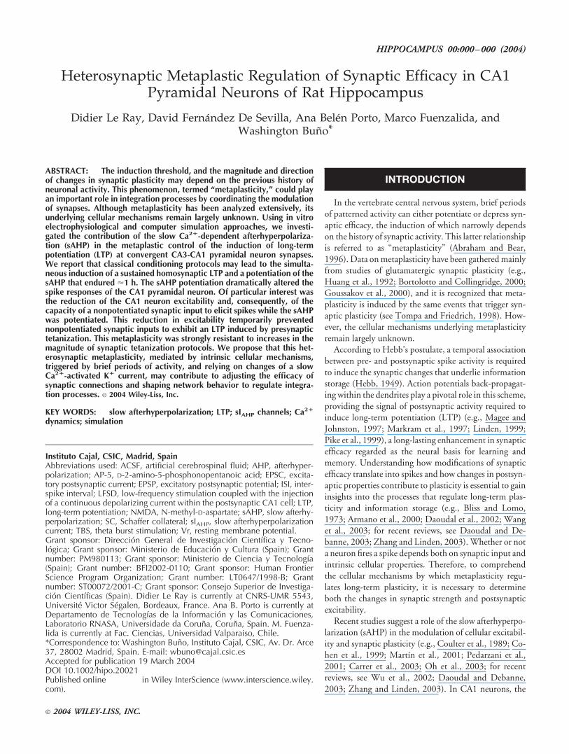
FIGURE 1. Heterosynaptic metaplasticity at Schaffer collateral(SC)-CA1 pyramidal neuron synapses. A: Diagram of experimentalsetup showing recording at the soma of a CA1 pyramidal neuron andstimulation of two Schaffer collateral apical dendritic synaptic inputs(SC1 and SC2). B: Excitatory postsynaptic potential (EPSP) peakamplitudes (empty circles) as a function of time in control conditionsand following tetanization of a Schaffer collateral input (empty bar,SC1 Tet.), which induced LTP of the SC1-evoked EPSP. Tetanizationapplied 45 min later to the SC2 afferent pathway (gray bar, SC21 Tet.)failed to generate LTP (gray circles), but 15 min later the same SC2tetanization (black bar, SC22 Tet.) evoked the LTP of the SC2-evoked
EPSPs. C: Mean peak amplitudes of the slow Ca2�-dependent after-hyperpolarization (sAHP) in the same experiments as in A, showing aprolonged potentiation of the sAHP (filled circles), and control re-cording of the sAHP (empty triangles, without tetanization). B,C:Same cells, n � 5. D: Upper, superimposed control (1) and post-tetanization EPSP (2) averages (three traces each). Lower, same as topbut sAHP. E: Summary data showing sAHP (black bars), SC1 EPSPs(empty bars) and SC2 EPSPs (gray bars) amplitudes relative to con-trols (percentage; n � 5), immediately after the SC21 tetanization(SC21 Tet.) and 60 min after, when the sAHP had returned close tocontrol values (SC22 Tet.).
___________________________________________________________ HETEROSYNAPTIC METAPLASTICITY 3
The voltage-clamp and current-clamp data were high-pass fil-tered at 0.3 and 3.0 kHz, respectively, and sampled at rates of0.7–10.0 kHz, through a TL–1 DMA interface (Axon Instru-ments). The pClamp programs (Axon Instruments) were used togenerate stimulus timing signals and transmembrane currentpulses, and to record and analyze data. The mean peak values ofcontrol synaptic responses and control sAHP/sIAHP were calcu-lated from all recordings prior to the application of a conditioningsynaptic stimulation. These mean values were then used as a refer-ence for the calculation of relative changes of synaptic response andsAHP/sIAHP (percentage of control values) throughout the exper-iment. Prolonged control recordings (�1 h) were also performedto check for the long-term stability of peak values of EPSP andsAHP/sIAHP. The decay time constant () of sAHP and of theunderlying sIAHP recorded in the current-clamp and voltage-clampmodes, respectively, were calculated with single exponential fits ofthe decaying portion of the voltage and current trace. Statisticalanalysis was performed with the SigmaPlot program (SPSS, Chi-cago, IL). The results are given as mean � SEM in text and figures,and statistical significance was calculated by analysis of variance(ANOVA) tests. The significance P � 0.05, 0.01, and 0.001 valuesare indicated by *, **, and ***, respectively, in the figures.
Computer Simulation
The simulation was performed using the NEURON program(Hines, 1994; Carnevale and Hines, 1997) that offers a powerful
and flexible environment especially designed for the study of ioniccurrents and their effects on the integrative properties of neurons.Using this environment, we simulated two converging presynapticSC inputs (SC1 and SC2) that connected the dendritic compart-ment of a CA1 pyramidal neuron model. The model was con-structed according to the electrophysiological characteristics of theCA1 pyramidal neurons (e.g., Schwartzkroin, 1975; see Mainen etal., 1996). It consisted of four parts: (1) a simplified dendriticcompartment, comprising 20 segments (length � 50-�m, diame-ter from 5 �m near the soma to 1 �m for the farthest segment), andsynaptic contacts were simulated at compartment 2 and 10 fromthe soma; (2) a spherical soma with a 60-�m diameter; (3) an axoncomposed of 50 segments (length � 60-�m, diameter 5–1 �m,from the nearest to the farthest segment) where spikes were gener-ated; and (4) a passive intermediary segment (length � 25-�m,diameter � 1 �m) between the dendrite and the soma compart-ment. The passive segment was incorporated in order to counter-act, to some extent, spike back-propagation. Each compartmentcomprised equally distributed leak channels that generated a leakcurrent according to the equation
Ileak�gleak*(Em�Eleak)
where Ileak is the current transported by the channels, gleak theconductance, Em the membrane potential, and Eleak the reversalpotential of the leak current. Active channels, based on Hodgkinand Huxley (1952)-type kinetics, were integrated in the axon andalso sparsely in the soma.
FIGURE 2. sIAHP potentiation. A: Time course of peak excitatorypostsynaptic current (EPSC) (empty circles) and sIAHP (filled circles;n � 6) amplitudes, before and after tetanization of a Schaffer collat-eral input (empty bar, SC Tet.). B: Parallel increase in the decay timeconstant (�; gray circles) and in the peak amplitude (filled circles; n �11) of the sIAHP following potentiation by SC tetanization (SC Tet.).
C: Superimposed averages (three successive responses each) of thesIAHP and EPSCs, illustrating controls and simultaneous LTP andsIAHP potentiation induced by pairing a low-frequency stimulationwith a sustained postsynaptic depolarization to 0 mV (LFSD). D:Same as C, but using a theta-burst stimulation (TBS) induction pro-tocol.
4 LE RAY ET AL.
The dendritic compartment contained the great majority (99.99%)of the sIAHP channels distributed with a proximodistal decreasing gra-dient (Sah and Bekkers, 1996; Bekkers, 2000). The remaining sIAHP
channels (0.01%) were located sparsely in the soma. Because our anal-ysis concerned the sAHP, we also focused on the Ca2� mechanismsthat regulate the sAHP. Therefore, we added: L-type voltage-gatedCa2� channels to the dendritic segment (for review, see Magee et al.,1998) based on Goldman–Hodgkin–Katz-type kinetics (see Yamadaet al., 1989); Ca2� pumps that extrude Ca2� ions out of the pyramidalneuron (Eakin et al., 1995); both radial and longitudinal diffusionmechanisms; and intracellular Ca2� buffers (Regehr and Tank,
1992), the functioning of which was controlled by two constantsaccording to the following equations:
Ca2��bufferO¡k1
Ca � buffer
Ca2��buffer¢Ok2
Ca � buffer
where k1 and k2 are the rate constants regulating the velocity ofcapture and release, respectively, according to the intracellularCa2� concentration. Current pulses applied at two dendritic com-partments simulated the effects of the activation of SC1 and SC2on the CA1 neuron. The amplitude and duration of both pulses,which generated the depolarizations that simulated SC-evoked ex-citatory postsynaptic potentials (EPSPs), were adjusted so that theresponse of the model neuron reproduced our physiological obser-vations.
Both the LTP and the potentiation of the sAHP were inducedwhen presynaptic and postsynaptic spikes coincided in time in thedendritic compartment. The LTP at either of the two convergingsynapses was simulated according to the following simple rules: (1)using suprathreshold stimulation, as soon as 10 coincident pre-and postsynaptic spikes at a minimum instantaneous frequency of40 Hz were detected, the pulse that generated the excitatorypostsynaptic potential was incremented by 0.025 nA (which wasless than the initial threshold current value of 0.0009 nA thatelicited spikes without failures in the CA1 neuron in the absence ofsAHP potentiation); and (2) once the process was initiated (i.e.,after rule 1 was fulfilled), each new coincident pre-postsynapticspike pair occurring at the same minimum instantaneous fre-quency of 40 Hz, increased the magnitude of the EPSP by incre-menting the pulse by 0.025 nA; this process being linear in theabsence of spike failures. In other words, in control conditions, abarrage of 40 suprathreshold stimuli delivered at 40 Hz by any SCinput incremented the EPSP peak amplitude (i.e., by incrementingthe pulse 0.025 nA at the end of the 10-spike burst) in an LTP-like fashion, from the tenth to the fortieth pre-postsynaptic spikepairs. The potentiation of the sAHP followed similar rules andeither involved (1) a 0.85% decrement of the buffering velocity ofintracellular Ca2� stores (buffer velocity; see Fig. 6A), which al-lowed a longer lasting activation the sIAHP channels (cf. Borde etal., 1995), or (2) a 0.25% increment of sIAHP channels closing timeconstant (; Fig. 6B), or (3) both. The best simulation was ob-tained when both of the sAHP parameters were adjusted duringand after the potentiation process and the time course of theirchanges was adjusted to display temporal profiles as those in realexperiments (e.g., time course of sIAHP peak amplitude and ; Fig.2B). In contrast, if there was no further synaptic stimulation thesynaptic strengths remained constant after the end of the potenti-ation process.
Virtual intracellular current-clamp and Ca2� concentration re-cordings were simulated in different parts of the model neuron inorder to verify the behavior of the simulated cellular features (e.g.,Ca2� concentration variations and sAHP). However, only the
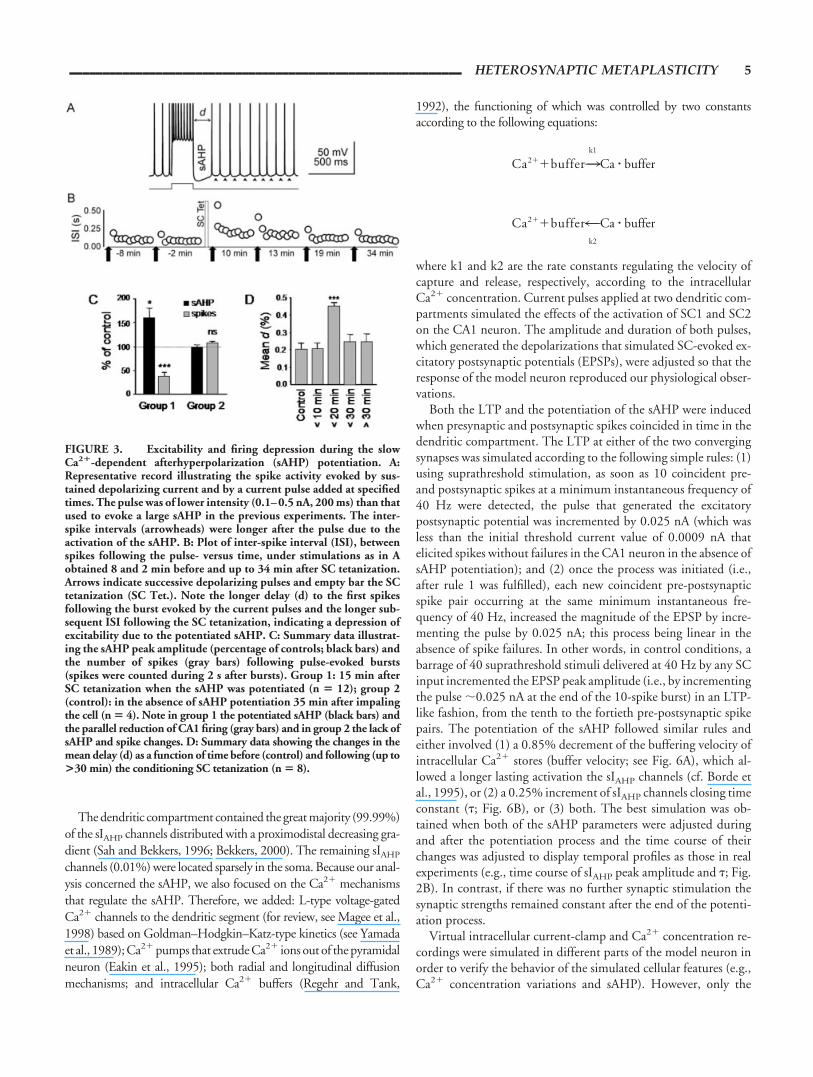
FIGURE 3. Excitability and firing depression during the slowCa2�-dependent afterhyperpolarization (sAHP) potentiation. A:Representative record illustrating the spike activity evoked by sus-tained depolarizing current and by a current pulse added at specifiedtimes. The pulse was of lower intensity (0.1–0.5 nA, 200 ms) than thatused to evoke a large sAHP in the previous experiments. The inter-spike intervals (arrowheads) were longer after the pulse due to theactivation of the sAHP. B: Plot of inter-spike interval (ISI), betweenspikes following the pulse- versus time, under stimulations as in Aobtained 8 and 2 min before and up to 34 min after SC tetanization.Arrows indicate successive depolarizing pulses and empty bar the SCtetanization (SC Tet.). Note the longer delay (d) to the first spikesfollowing the burst evoked by the current pulses and the longer sub-sequent ISI following the SC tetanization, indicating a depression ofexcitability due to the potentiated sAHP. C: Summary data illustrat-ing the sAHP peak amplitude (percentage of controls; black bars) andthe number of spikes (gray bars) following pulse-evoked bursts(spikes were counted during 2 s after bursts). Group 1: 15 min afterSC tetanization when the sAHP was potentiated (n � 12); group 2(control): in the absence of sAHP potentiation 35 min after impalingthe cell (n � 4). Note in group 1 the potentiated sAHP (black bars) andthe parallel reduction of CA1 firing (gray bars) and in group 2 the lack ofsAHP and spike changes. D: Summary data showing the changes in themean delay (d) as a function of time before (control) and following (up to>30 min) the conditioning SC tetanization (n � 8).
___________________________________________________________ HETEROSYNAPTIC METAPLASTICITY 5
voltage changes that occurred in the soma were considered in thepresent study.
RESULTS
Recordings were performed with patch electrodes from 120CA1 pyramidal cells in voltage-clamp conditions (mean Vr mea-sured at zero holding current: �55.4 � 0.6 mV) and with sharpelectrodes from 69 CA1 pyramidal cells in the current-clamp con-
figuration (mean Vr: �61.2 � 3.0 mV). The other cellular char-acteristics (input resistance, firing threshold) were similar to thosedescribed previously (see Borde et al., 2000).
Heterosynaptic MetaplasticityTo test the spatial and temporal characteristics of metaplas-
ticity in rat hippocampus, intracellular current-clamp record-ings with sharp electrodes were performed from single CA1pyramidal neurons (n � 32) while two distinct SC afferentpathways (SC1 and SC2) were stimulated (Fig. 1A). Control
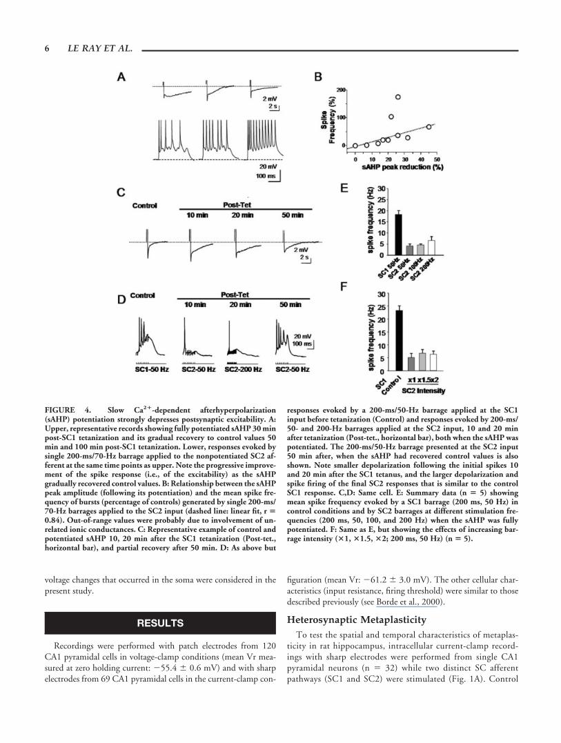
FIGURE 4. Slow Ca2�-dependent afterhyperpolarization(sAHP) potentiation strongly depresses postsynaptic excitability. A:Upper, representative records showing fully potentiated sAHP 30 minpost-SC1 tetanization and its gradual recovery to control values 50min and 100 min post-SC1 tetanization. Lower, responses evoked bysingle 200-ms/70-Hz barrage applied to the nonpotentiated SC2 af-ferent at the same time points as upper. Note the progressive improve-ment of the spike response (i.e., of the excitability) as the sAHPgradually recovered control values. B: Relationship between the sAHPpeak amplitude (following its potentiation) and the mean spike fre-quency of bursts (percentage of controls) generated by single 200-ms/70-Hz barrages applied to the SC2 input (dashed line: linear fit, r �0.84). Out-of-range values were probably due to involvement of un-related ionic conductances. C: Representative example of control andpotentiated sAHP 10, 20 min after the SC1 tetanization (Post-tet.,horizontal bar), and partial recovery after 50 min. D: As above but
responses evoked by a 200-ms/50-Hz barrage applied at the SC1input before tetanization (Control) and responses evoked by 200-ms/50- and 200-Hz barrages applied at the SC2 input, 10 and 20 minafter tetanization (Post-tet., horizontal bar), both when the sAHP waspotentiated. The 200-ms/50-Hz barrage presented at the SC2 input50 min after, when the sAHP had recovered control values is alsoshown. Note smaller depolarization following the initial spikes 10and 20 min after the SC1 tetanus, and the larger depolarization andspike firing of the final SC2 responses that is similar to the controlSC1 response. C,D: Same cell. E: Summary data (n � 5) showingmean spike frequency evoked by a SC1 barrage (200 ms, 50 Hz) incontrol conditions and by SC2 barrages at different stimulation fre-quencies (200 ms, 50, 100, and 200 Hz) when the sAHP was fullypotentiated. F: Same as E, but showing the effects of increasing bar-rage intensity (�1, �1.5, �2; 200 ms, 50 Hz) (n � 5).
6 LE RAY ET AL.
subthreshold EPSPs were evoked by alternatively stimulatingthe SC1 and SC2 inputs with single pulses at a low rate of 0.2/s(see Materials and Methods). In control conditions, SC1 EPSPshad amplitudes of 2.2 � 0.3 mV (n � 10) and SC2 amplitudeswere 1.8 � 0.5 mV, and there were no long-term modificationsof peak EPSP amplitudes (112.6 � 3.2% and 108.1 � 2.0% ofcontrol values, respectively, 1 h; n � 10). Experiments werediscarded when EPSPs fired spikes in control conditions. Atetanization conditioning protocol (see below) was then appliedto the SC1 input to induce LTP, and single stimuli were applied(as the one used to record control EPSPs) at specified timesduring the experiment to test for changes in the synaptic effi-cacy of both SC1 and SC2. The same tetanization conditioningprotocol was also applied to the SC2 pathway at various timepoints during the experiment while tests for changes in synapticefficacy of SC1 and SC2 were repeated.
A group of similar experiments (n � 5) in which tetanization(see Materials and Methods) of the SC1 input was used to induceLTP (e.g., Bortolotto and Collingridge, 2000) are shown in Figure1. Tetanization of the SC1 pathway (Fig. 1A and empty bar, Fig.1B) induced the LTP of the SC1-evoked EPSPs (a mean peakamplitude increment of 265.2 � 20.0%, 30 min post-tetanization;P � 0.001; n � 5; empty circles, Fig. 1B), whereas the SC2-evokedEPSPs showed no statistically significant change from control val-ues (mean peak amplitude 103.3 � 7.1%, 30 min post-tetaniza-tion; filled circles, Fig. 1B).
The homosynaptic LTP of the SC1 EPSPs lasted throughoutthe experiment (i.e., �1 h) in the absence of subsequent SC1conditioning stimulation. The same tetanization protocol ap-plied to the SC2 pathway 45–50 min after the SC1 tetanization(gray bar, Fig. 1B,E) did not induce changes in either EPSP,since the SC1-evoked EPSPs remained potentiated (285.5 �13.7%, 50 min post-SC1 tetanization; P � 0.001; same cells)and the amplitude of the SC2-evoked EPSPs was not signifi-cantly modified (96.7 � 7.2%). However, reapplying the teta-nization protocol to SC2 afferents 15–20 min later (i.e., �1 hafter the SC1 tetanization; black bar, Fig. 1B) induced a signif-icant LTP of the SC2-evoked EPSP (128.8 � 4.2%; P � 0.001;same cells) that lasted throughout the rest of the experiment (seealso Fig. 1E). Interestingly, depending on the time elapsed fromthe tetanization of the SC1, tetanization of the SC2 input eitherfailed to or induced the LTP of the SC2 input. Nevertheless, theLTP of the SC2 input was always of lower magnitude than thatof the SC1 input, suggesting that a sustained partial block of theLTP was induced by the SC1 tetanization. Further experimentswill be required to investigate this possibility.
The above results suggest that a metaplastic phenomenon oc-curred by which the induction of LTP at the SC1 input exerted anegative regulation of the ability of SC2 tetanization to induceLTP. The negative control for the induction of SC2 LTP wasremoved with the time elapsed after the SC1 conditioning stimu-lation, eventually allowing the SC2 input to display LTP afterseveral tens of minutes.
Simultaneous Induction of LTP and sAHPPotentiation
During the above-described experiments, we also analyzedwhether the sAHP contributed to the heterosynaptic metaplastic-ity (n � 32). Depolarizing current pulses were applied through therecording microelectrode to evoke an sAHP at specified timedduring the experiment (see Materials and Methods), and the sAHPpeak amplitude was measured 150 ms after the offset of the pulse,i.e. when the medium AHP had almost completely deactivated (forreview, see Storm, 1990). The magnitude of the sAHP did notchange significantly during prolonged recordings in control con-ditions (triangles, Fig. 1C). Control sAHP peak amplitude valueswere 5.3 � 2.1 mV and 4.9 � 3.1 mV (averages of 5 responses 20min and 1 h after impaling the cell; n � 6). We also monitored thechanges in the peak amplitudes of the sAHP at specified timesduring the experiment before and after the conditioning tetaniza-tion protocol was applied (filled circles, Fig. 1C). The SC1 tetani-zation protocol (empty bar, Fig. 1C) that generated the SC1 LTPalso triggered a potentiation of the sAHP (filled circles: 165.8 �3.2%; P � 0.001; n � 5) that lasted �50 min before it returnedclose to control values. The number of spikes evoked by the depo-larizing current pulses that evoked the sAHP did not changethroughout the experiment (7.40 � 0.54 and 7.32 � 0.55; aver-ages of 8 responses before and after tetanization, respectively; n �5), implying that the sAHP potentiation was not caused by anincreased spike response. Interestingly, the LTP of the SC2 inputcould occur only after the sAHP potentiation faded (Fig. 1B,E).Examples of control and potentiated averaged SC1-evoked EPSPsand sAHP are illustrated in Figure 1D.
Results as those shown in Figure 1 were recorded in 17 of 32experiments (or 53%). In the other 15 cells (or 47%), although thehomosynaptic LTP was present, no clear sAHP amplitude varia-tions were detected. Indeed, the SC1-evoked EPSP showed LTPwhen all the cells were considered (169.4 � 33.3%; P � 0.001;n � 32), whereas the sAHP increased significantly only in a groupof those neurons (141.9 � 10.5%; P � 0.001; n � 17). Never-theless, tetanization of the SC2 afferents never induced LTP(98.4 � 2.5%; n � 17) when the sAHP was potentiated by priorSC1 conditioning (Fig. 1E, left), whereas the same protocol ap-plied after the sAHP returned close to control value (105.9 �6.7%) always triggered LTP of the SC2 input (130.6 � 9.3%; P �0.001; same cells; Fig. 1E, right). Therefore, when SC1 tetaniza-tion induced the potentiation of the sAHP the described het-erosynaptic metaplasticity consisting in the block of the inductionof LTP at the SC2 input was always observed. It should be under-scored that in 6 of the 15 cells where the sAHP was not potentiatedby tetanization of the SC1 input, tetanization of the SC2 readilyinduced LTP of these afferents (139.7 � 16.5%; n � 6), even atshort delays after the SC1 tetanization. Therefore, the above resultssuggest a causal relationship between the potentiation of the sAHPand the heterosynaptic metaplasticity.
The magnitude of the LTP of the SC1 and SC2 EPSPs wasdifferent (cf. SC1: 226.1 � 17.1%; P � 0.001; n � 5; and SC2:126.9 � 5.2%; P � 0.001; n � 5). In addition, since the SC2EPSPs could only be potentiated when the sAHP had declined to
___________________________________________________________ HETEROSYNAPTIC METAPLASTICITY 7
control values, the delay required to induce LTP at the SC2 inputalso varied somewhat (52 � 8 min; same cells). Although we didnot analyze the causes of these differences, they may reside inuncontrolled factors, such as cellular variations, the position of thestimulating electrodes, or the different initial magnitudes of thesAHP and synaptic strengths.
To reduce the possible contamination by voltage-sensitive con-ductances, whole-cell voltage-clamp experiments were performed.Neither the magnitude of the sIAHP (140.5 � 20.3 pA and136.9 � 18.4 nA; 5 min and 1 h after access to the intracellularcompartment; n � 6) nor the amplitude of the EPSCs (50.7 �10.9 pA and 45.2 � 14.7 pA; same time points, same cells)changed significantly during prolonged recordings of �1 h in con-trol conditions. Both the SC-evoked EPSCs and the sIAHP werealso monitored regularly before and after a conditioning tetaniza-tion protocol was applied to an SC pathway after transientlyswitching to the current-clamp mode (n � 31; see Materials andMethods). The time course of both the LTP of the SC1 EPSC(empty circles: 203.8 � 5.1% at 20 min post-tetanization; P �0.001; n � 6) and the potentiation of the sIAHP (filled circles:150.9 � 5.2% at 20 min post-tetanization; P � 0.001; same cells)are shown in Figure 2A. In the voltage-clamp configuration, morethan 71% of the cells (22 out of 31 neurons) showed significantLTP and sIAHP potentiation, and grouped results showed meanincreases in peak EPSC amplitude of 252.1 � 43.3% (P � 0.01)and sIAHP amplitude of 167.9 � 12.1% (P � 0.001). The sIAHP
potentiation developed without significant changes in the re-sponses evoked by the depolarizing command pulses because thenumber of unclamped action currents varied only slightly through-out the experiment (102.2 � 3.3% of the initial control value �1h after impaling the cell; n � 6), implying that the potentiation wasnot caused by an increased response evoked by the depolarizingcurrent pulse.
The similarities of the voltage-clamp and current-clamp resultssuggest that the observed synaptic and sAHP plasticities (e.g., Fig.1B,C) were not caused by modification of the driving force, andsupport a causal relationship between the described metaplasticityand the potentiation of the sAHP/sIAHP.
Synaptic Tetanization Increases Both theAmplitude and Duration of the sAHP
As shown above in Figures 1D and 2A, both the amplitude andduration of the sAHP were modified by the SC1 tetanization. Toconfirm this observation, we monitored the peak amplitude andthe decay time constant of the sIAHP in voltage-clamp conditions,before and after tetanization of the SC input (Fig. 2B). The sIAHP
peak amplitude increased by 175.9 � 15.2% (P � 0.001; n � 11)within the first 20 min post-SC tetanization and then decreased toreturn to control values �50 min after the tetanization. The sIAHP
decay time constant also increased to peak 40 min after the tetani-zation reaching 305.8 � 53.3% of control value (P � 0.001; samecells) and remained potentiated �60 min after. These differencesin profile suggest that at least two cellular mechanisms account forthe observed sIAHP potentiation, one affecting the magnitude ofthe sAHP current (i.e., the number of open sIAHP channels), the
other the duration of the current (i.e., the kinetics of sIAHP chan-nels).
Interestingly, a parallel LTP and sAHP potentiation could alsobe observed using other LTP conditioning protocols. Pairing alow-frequency stimulation with postsynaptic depolarization to 0mV (LFSD; e.g., Otmakhov et al., 1997) induced both the LTP ofthe SC-evoked EPSCs (124.9 � 5.2%; n � 3; P � 0.001) and thesimultaneous potentiation of the sIAHP (184.6 � 29.2%; n � 3;P � 0.05) measured 15 min post-LFSD under voltage-clamp (e.g.,Fig. 2C). Theta-burst stimulation (TBS; e.g., Hoffman et al.,2002) also induced both the LTP of SC-evoked EPSCs (227.1 �27.2%; n � 3; P � 0.001) and the parallel sIAHP potentiation(199.9 � 15.3% 15 min after a TBS; n � 3; P � 0.001; e.g., Fig.2D). Therefore, whatever the LTP induction protocol used in ourstudy, a parallel potentiation of the synaptic response and of thesAHP/sIAHP was induced.
Changes in Excitability and Spike Firing Parallelthe Potentiation of the sAHP
According to Hebb’s postulate (Hebb, 1949), a temporal asso-ciation between pre- and post-synaptic spike firing is required toinduce LTP. Because the sAHP regulates action potential firing weinvestigated under current-clamp conditions the possible correla-tion between the synaptically induced sAHP enhancement andchanges in the pyramidal neuron firings. A group of pyramidalneurons (n � 20) were slightly depolarized by intracellular currentinjection to evoke a steady low rate spike activity (9.4 � 0.4 s�1)and a depolarizing pulse (0.1–0.5 nA; 200 ms), which generated aburst of spikes followed by a large compound sAHP (Fig. 3A), wasadded at specified times during the experiment. In those condi-tions, after tetanization of a SC pathway the CA1 neuron spikeactivity was dramatically affected. Between 10 and 30 min post-tetanization, which coincides on the average with the maximumpotentiation of the sAHP (see above), there was a marked rise inthe inter-spike intervals (ISI) during the sAHP and specially of thedelay (d) to the post-burst spike (Fig. 3A,B). Interestingly, even inthese different experimental conditions where the cell was depo-larized and the estimation of the time course of the sAHP wasdifficult because it was evoked during firings and at potentialsabove resting values, the strongest reduction of spike firing coin-cided in time with the highest potentiation of the sAHP.
In 12 experiments, the firing capacity of CA1 neurons during anevoked sAHP was also tested 15 minutes after an SC tetanizationand showed a mean firing rate 37.1 � 10.2% of control (P �0.001) when the sAHP peak amplitude increased up to 161.8 �21.3% (P � 0.05; same cells) (group 1, Fig. 3C). In contrast, in theabsence of sAHP potentiation (100 � 8.1%) the mean firing wasnot significantly altered (112 � 4.1%; group 2, Fig. 3C). Similarly,the delay (d) to the first spike following the current pulse-generatedburst also increased from 0.20 � 0.01 to 0.45 � 0.05 s (P � 0.05;n � 8) 13 min post-tetanization. On the average d was highestbetween 10 and 20 min post-tetanization (n � 3; Fig. 3D); i.e.,when the sAHP and sIAHP were maximally potentiated followingthe conditioning stimulation (see above). Interestingly, in eachcase the stronger reduction in spike firing coincided in time with
8 LE RAY ET AL.
the highest potentiation of the sAHP (i.e., 10–20 min post-teta-nization).
These results suggest that the sAHP potentiation can modulatefor several tens of minutes the spike response evoked by SC stim-ulation by decreasing the postsynaptic excitability and increasingspike frequency adaptation. In this scenario, especially relevant arethe increased postsynaptic spike failures of the nonpotentiated SC2input during stimulation with barrages. When single 200 ms bar-rages (as the ones that were repeatedly presented to evoke LTP)were applied to the SC2 input during the potentiated sAHP (lowerrecords, Fig. 4A) some of the EPSPs evoked by the presynapticbarrage were suprathreshold and triggered spikes (especially atstimulation on). Each postsynaptic spike was followed by a largerthan control sAHP (i.e., potentiated) that hyperpolarized the neu-ron so that subsequent SC2-evoked EPSPs were subthreshold andfailed to trigger spikes (upper records, Fig. 4A). In many cases, thebarrage evoked only one spike when the sAHP was fully potenti-ated (e.g., Fig. 4D).
As the potentiation of the sAHP faded with time, more spikeswere triggered by the SC2 barrage, because spikes were followed bysmaller sAHP that, on the average, hyperpolarized the cell less aftereach spike (Fig. 4A,D). Although this analysis involved isolatedSC2 barrages, similar mechanisms were at work when repeatedSC2 barrages failed to induce LTP (not shown). Therefore, thepotentiated sAHP led to a reduction of excitability and, thus, of theprobability of temporal association between pre- and postsynapticfiring that is required to induce LTP. Consequently, a potentiatedsAHP may prevent the induction of LTP by stimulation withrepeated barrages of the SC2 input. Conversely, similar repeatedbarrages could evoke sufficient postsynaptic spikes to induce theLTP of the SC2 EPSP when the sAHP potentiation had faded (Fig.4D) or when it had not been potentiated by the SC1 tetanization(see above).
We tested this assumption by tetanization of the SC1 input toinduce the homosynaptic LTP and potentiate the sAHP. Whenthe sAHP showed clear potentiation, the SC2 pathway was stim-ulated with single barrages repeated every 5 min. In these condi-tions, the cell slowly recovered its capacity to fire spikes in responseto SC2 barrages, as the amplitude of the potentiated sAHP gradu-ally decayed with time (Fig. 4C). As a result of this gradual recoveryof excitability, the average postsynaptic spike frequency evoked bythe SC2 barrages showed a negative relationship with the ampli-tude of the potentiated sAHP evoked by the burst itself, whichcorroborated our hypothesis (Fig. 4B).
Following the induction of LTP of the SC1 input (228.5 �26.0%; n � 5) and potentiation of the sAHP (172.7 � 29.3%;same cells) we also analyzed the responses evoked by a single bar-rage applied at the nonpotentiated SC2 input. This single barragewas identical to those that were repeatedly presented to evoke LTP(see Materials and Methods). In those conditions, a single barrageapplied at the SC2 afferents evoked considerably fewer spikes thanthe SC1 conditioning stimulation (24.2 � 5.2%; P � 0.001; n �5) (Fig. 4C–E). The same barrage applied repetitively did notinduce the LTP of the SC2 afferents (105.3 � 13.3%; not shown,but see above). In contrast, 50 min later, when the sAHP potenti-ation had faded, the same conditioning stimulation applied to the
SC2 input evoked many more spikes (262.1 � 83.3%; P � 0.05;n � 5) (Fig. 4C–E), and when repeated induce the LTP of theSC2-EPSPs (200.8 � 26.0%; n � 5; not shown, but see above). Inevery case, the LTP of the SC2 input was observed only when thesAHP had recovered its control amplitude, and the SC2 condition-ing stimulation evoked as many postsynaptic spikes as the SC1conditioning stimulation (ANOVA test showed no significant dif-ferences) (e.g., first and last traces in Fig. 4C,D).
We tested whether increasing the SC2 barrage frequency couldincrease the spike response during a potentiated sAHP and thusovercome the block of the LTP induction caused by a potentiatedsAHP. In control conditions, increasing the frequency of the SC2stimulation increased both the depolarization and the number ofspikes evoked by the SC burst, by incrementing the EPSPs tempo-ral summation without changing the presynaptic fibers being stim-ulated. We stimulated the SC2 input with 200 ms barrages atidentical intensity and 50, 100, and 200 Hz. There were no signif-icant difference in the number of spikes during the barrage, ex-pressed as the frequency of spikes (4.16 � 0.83 Hz 4.66 � 0.33Hz, 6.60 � 1.66 Hz, respectively) when the sAHP was potentiated(Figs. 4C–E), and there was no LTP induction at the SC2 input(n � 5).
We also tested the effects of increasing the intensity of the SC2barrage that raises the number of presynaptic fibers stimulated andtherefore increases spatial summation. The summated SC2 EPSPscould intensify the postsynaptic spike response and overcome theblock of the LTP by the potentiated sAHP. We tested this secondpossibility by incrementing the SC2 barrage intensity up to 1.5 and 2 of control intensities. The increased stimulation intensitiesonly induced small changes in the number of spikes while thesAHP was potentiated (from 5.1 � 1.6 Hz in control to 6.8 � 1.2Hz at 1.5 , and to 6.3 � 1.1 Hz at 2 ; Fig. 4F), and LTPinduction was still prevented at the SC2 synapses (n � 5).
Therefore, the above results suggest that the decreased excitabil-ity caused by the potentiated sAHP exerted a strong control onspike firing that prevented the LTP of the SC2 input and thatcould not be overcome by incrementing the frequency or the in-tensity of the presynaptic SC2 barrage.
Effects of Blocking NMDA and L-Type Voltage-Activated Ca2� channels
The LTP in glutamatergic synapses on CA1 pyramidal neuronsmay depend on both Ca2� inflow through NMDA channels andvia voltage-activated Ca2� channels (e.g., Grover and Teyler,1990; Bliss and Collingridge, 1993; Hanse and Gustafsson, 1995;Cavus and Teyler, 1996; Magee and Johnston, 1997). Influx ofCa2� through L-type voltage-activated Ca2� channels and to alesser extent via NMDA channels (Tanabe et al., 1998; Borde etal., 2000; Lancaster et al., 2001; Shah and Haylett, 2002) may alsocontribute to the activation of the sAHP/sIAHP. Therefore, up-regulation of Ca2� influx both through NMDA or L-type Ca2�
channels by the tetanization of SCs could mediate the potentiationof the sAHP/sIAHP and contribute to the LTP. We tested theeffects of inhibition of NMDA channels with AP-5 (50 �M) andof L-type voltage-activated Ca2� channels with nimodipine (20
___________________________________________________________ HETEROSYNAPTIC METAPLASTICITY 9
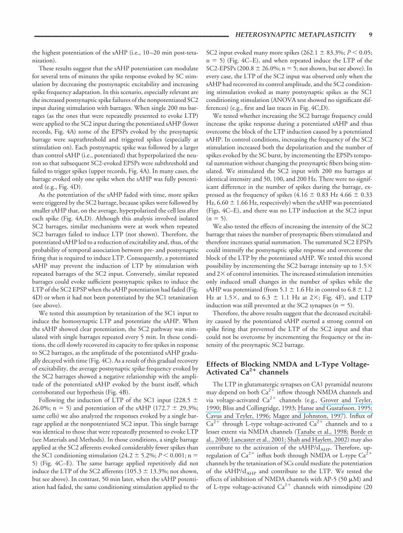
�M). Whereas block of L-type channels prevented the potentia-tion of the sAHP (110.7 � 12.0%; n � 6) without preventing theLTP of the SC1 input (338.3 � 23.0%; same cells; Fig. 5A,B),AP-5 had no effect on the sAHP (131.9 � 5.9%; n � 6) and didnot block the LTP (172.6 � 11.9%; n � 6; Fig. 5C,D). However,simultaneous superfusion of nimodipine and AP-5 inhibited boththe sAHP/sIAHP potentiation and the synaptic LTP (99.0 � 4.6%and 101.0 � 12.5%, respectively; n � 6, not shown). These resultssuggest a contribution of Ca2� influx through L-type channels inthe potentiation of the sAHP/sIAHP, whereas Ca2� influx viaNMDA channels does not participate in the potentiation of thesAHP/sIAHP (see Discussion).
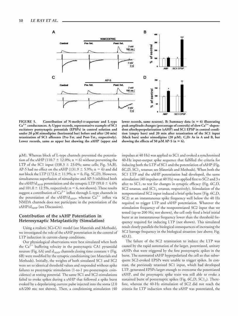
Contribution of the sAHP Potentiation inHeterosynaptic Metaplasticity (Simulation)
Using a realistic SCs-CA1 model (see Materials and Methods),we investigated the role of the sAHP potentiation in the control ofLTP induction in current-clamp conditions.
Our physiological observations were best simulated when boththe Ca2� buffering velocity in the postsynaptic CA1 pyramidalneuron (Fig. 6A) and sIAHP channels closing time constant (Fig.6B) were modified by the synaptic conditioning (see Materials andMethods). Initially, the weights of both simulated SC1 and SC2were set to identical threshold values and responded without spikefailures to presynaptic stimulation (1-to-1 pre-postsynaptic coin-cidence) at resting potential. The same SC1 and SC2 stimulationsfailed to evoke spikes during a sAHP that followed a spike burstevoked by a depolarizing current pulse injected into the soma (2.0nA/200 ms; not shown). Then, a conditioning stimulation (40
impulses at 40 Hz) was applied to SC1 and evoked a synchronized40-Hz input-output spike sequence that fulfilled the criteria forinducing both the LTP of SC1 and the potentiation of sAHP (Fig.6C,D, SC11 tetanus; see Materials and Methods). When both theSC1 LTP and the sAHP potentiation had developed, the samestimulation (40 impulses at 40 Hz) was applied first to SC2 and 3 safter to SC1, to test for changes in synaptic efficacy (Fig. 6C,D,SC2 tetanus, and SC12 tetanus, respectively). Stimulation of thenonpotentiated SC2 input induced a brief spike burst (Fig. 6C,D,SC2) at an instantaneous spike frequency well below the 40 Hzrequired to trigger LTP and sAHP potentiation. Whatever thestimulation frequency of the nonpotentiated SC2 input that wetested (up to 200 Hz; not shown), the cell only fired a brief initialburst at an instantaneous frequency lower than the threshold fre-quency required for inducing LTP (not shown). This simulatedresult closely parallels the biological consequences of increasing theSC2 barrage frequency in the biological situation (see above; Fig.4D,E).
The failure of the SC2 tetanization to induce the LTP wascaused by the rapid summation of the larger, potentiated, unitarysAHPs that were triggered by the first postsynaptic spikes in theburst. The summated sAHP hyperpolarized the cell so that subse-quent SC2-evoked EPSPs were unable to trigger spikes. In con-trast, the previously tetanized SC1 input, which had developedLTP, generated EPSPs larger enough to overcome the potentiatedsAHP, and the presynaptic spike train was still able to evoke asustained burst of postsynaptic spikes (Fig. 6C,D, SC12). There-fore, whereas the 40-Hz stimulation of SC2 did not reach thecriteria for LTP induction when the sAHP was potentiated, the
FIGURE 5. Contribution of N-methyl-D-aspartate and L-typeCa2� conductances. A: Upper records, representative example of SC1excitatory postsynaptic potentials (EPSPs) in control solution andunder 20 �M nimodipine (horizontal bar) before and after (30 min)tetanization of SC1 afferents (Pre-Tet. and Post-Tet., respectively).Lower records, same as upper but showing the sAHP (upper and
lower records, same neuron). B: Summary data (n � 6) illustratingpeak amplitude changes (percentage of controls) of slow Ca2�-depen-dent afterhyperpolarization (sAHP) and SC1 EPSP in control condi-tion (empty bars) and 20 min after tetanization of the SC1 input(black bars) under nimodipine (20 �M). C,D: As in A and B, butshowing the effects of 50 �M AP-5 (n � 6).
10 LE RAY ET AL.
same stimulation applied to the potentiated SC1 input was stillable to induce the LTP, even in the condition of potentiatedsAHP.
The above simulation closely models the biological results illus-trating how tetanization of a nonpotentiated synaptic input may beunable to induce LTP if the neuronal excitability is depressed by apotentiated sAHP, whereas a similar stimulation of the potentiatedsynaptic input could still evoke sufficient postsynaptic firing (dueto the LTP). This latter observation is consistent with a gatingphenomenon in which the potentiated synapses can elicit postsyn-aptic spiking, whereas nonpotentiated synapses remain “blocked”postsynaptically by the larger sAHP. This suggests further a role ofthe synaptically evoked sAHP enhancement in the selection ofconvergent inputs by which the physiologically reliable potenti-ated input may be amplified while the influence of any other inputwould be repressed.
DISCUSSION
We show that the induction of LTP by classical conditioningprotocols triggers a prolonged (�1 h) potentiation of the sAHPmediated by the enhancement of the underlying slow Ca2�-acti-vated K� current, sIAHP. This sAHP/sIAHP potentiation reducesexcitability and increases spike frequency adaptation, thus decreas-ing the capacity of the cell to fire prolonged spike bursts. Thereduced spike firing prevents subsequent tetanization protocols togenerate an LTP at other nonpotentiated synaptic input, withoutmodifying the previously potentiated synapses. Therefore, this po-tentiation of the sAHP, which is triggered by brief periods ofsynaptic activity and relies on modifications of an intrinsic prop-erty, represents a novel cellular mechanism engaged in heterosyn-aptic metaplasticity of synaptic efficacy at CA3-CA1 pyramidalneuron synapses.
Plasticity of the sAHP
An activity-dependent modulation of the sAHP/sIAHP has beendescribed in CA1 pyramidal neurons as a brief facilitation (3–8min) induced either by intracellular current injection or excitatorysynaptic stimulation (Borde et al., 1995, 1999, 2000). We dem-onstrate here that a synaptic stimulation that induces LTP maygenerate a longer lasting potentiation of the sAHP. Interestingly,the recent demonstration that both facilitation and depression ofthe sAHP may be induced by tetanic and theta burst stimulationwith injected current in CA1 pyramidal neurons (Kaczorowski etal., 2003), suggests a possible bidirectional control of postsynapticexcitability by the sAHP/sIAHP.
Several studies have related the amplitude of the sAHP to learn-ing and memory (e.g., Coulter et al., 1989; Disterhoft et al., 1996;for a recent review, see Wu et al., 2002). They showed that areduction of the sAHP in CA1 pyramidal neurons occurs severalhours to days after classical conditioning. This decline of the sAHPmay facilitate subsequent learning (Coulter et al., 1989; Saar et al.,1998; Oh et al., 2003; for recent reviews, see Wu et al., 2002;Daoudal and Debanne, 2003; Zhang and Linden, 2003). In addi-tion, an age-related enhancement of the sAHP in CA1 hippocam-pal pyramidal neurons has also been associated with learning def-
FIGURE 6. Simulation: the potentiated sIAHP mediates het-erosynaptic metaplasticity. A,B: Potentiation of both the sIAHP peakamplitude and decay time constant were simulated by decreasing thevelocity of Ca2� capture by internal buffers (A) and increasing thetime constant of sIAHP channel closure (B), respectively. Both plasticprocesses were initiated by the occurrence of 10 consecutive pre- andpostsynaptic spikes (at a minimum instantaneous frequency of 40 Hz;see Materials and Methods) and incremented for every new spikefulfilling the 40-Hz pre-postsynaptic spike condition. C,D: Simula-tion of the slow Ca2�-dependent afterhyperpolarization (sAHP)-de-pendent heterosynaptic metaplasticity at CA3-CA1 synapses. The first40-Hz tetanization applied to the SC1 input (C, SC11, empty bar)generated sufficient postsynaptic firing (D, left) to induce the homo-synaptic long-term potentiation (LTP) and potentiate the sAHP (notillustrated), but did not modify SC2-evoked excitatory postsynapticpotentials (EPSPs). Subsequent 40-Hz tetanization of the nonpoten-tiated SC2 input when the sAHP was maximally potentiated (C, graybar) did not generate a sufficient firing (D, middle) to meet the criteriafor inducing LTP at the SC2 input and to potentiate the sAHP. Incontrast, the tetanization of the potentiated SC1 input was still able togenerate sufficient spikes to meet the criteria for LTP, even though thesAHP remained potentiated (C, SC12, empty bar; D, right).
__________________________________________________________ HETEROSYNAPTIC METAPLASTICITY 11
icits (e.g., Disterhoft et al., 1996; Meredith et al., 2002; see alsoPike et al., 1999).
The synaptically evoked sAHP potentiation (present results)and the above-discussed learning-related sAHP depression repre-sent different phenomena that exert opposed effects on cellularexcitability and that occur early ( �1 h; present results) and late,respectively.
Rapid increases in neuronal excitability associated with the in-duction of LTP have also been described (e.g., Bliss and Lomo,1973; Armano et al., 2000; Egorov et al., 2002; for recent reviews,see Daoudal and Debanne, 2003; Zhang and Linden, 2003).These increases in excitability are input specific, i.e., are exclusivelyassociated with stimulation of the potentiated synaptic input,whereas a decreased excitability is coupled with stimulation ofconvergent but nonpotentiated synaptic inputs (e.g., Cavus andTeyler, 1996; Daoudal et al., 2002; Wang et al., 2003). The inputspecificity suggests that the intrinsic excitability of different den-dritic areas may be up- or downregulated in an activity-dependentmanner. Although different mechanisms have been proposed forthe above discussed effects, the phenomenological similarities withthe heterosynaptic metaplasticity described here suggests that thelatter may represent an example of input specificity caused by thepotentiation of the sAHP/sIAHP or of the voltage-gated Ca2� chan-nels that activate the sAHP/sIAHP at specific dendritic regions (e.g.,Yasuda et al., 2003; for review, see Daoudal and Debanne, 2003).
Cellular Mechanisms Mediating the Potentiationof the sAHP
The ionic channels mediating the sIAHP are thought to be lo-cated mainly on dendrites close to the soma of hippocampal pyra-midal neurons (Sah and Bekkers, 1996; Bekkers, 2000), suggestinga role for the sAHP/sIAHP in controlling dendritic excitability andspike back-propagation (Hoffman et al., 1997; Magee and Car-ruth, 1999). Interestingly, the blockade of the sAHP by the �-ad-renergic agonist isoprenaline, the metabotropic glutamate receptoragonist t-ACPD, or the cholinergic agonist carbachol facilitates theinduction of LTP (e.g., Hasselmo and Barkai, 1995; Sah and Bek-kers, 1996; Cohen et al., 1999). Unfortunately, we could not usesuch nonspecific sAHP blockers because these drugs also modulatethe release of glutamate from CA3 terminals (e.g., Fernandez deSevilla et al., 2002, 2003), and we would not have been able toconclude if the effects were due to the postsynaptic block of thesAHP or to a presynaptic regulation of glutamate release from SCterminals. Nevertheless, our simulation experiments are consistentwith the proposed role for the sAHP potentiation in the downregu-lation of LTP induction at nonpotentiated synapses through thereduction of excitability (see also Pike et al., 1999).
The channels mediating sIAHP are voltage insensitive, and thekinetics of the sIAHP closely parallel the changes in intracellularconcentration of Ca2� ([Ca2�]i) caused by action potential activ-ity (Regehr and Tank, 1992; Sah and Isaacson, 1995; Martın et al.,2001; Carrer et al., 2003). During the sIAHP potentiation there arechanges in the amplitude and deactivation kinetics that followdifferent time courses (present results), suggesting that at least twocellular mechanisms are contributing. The amplitude changes
probably reflect the number of open sIAHP channels related to[Ca2�]i, and the kinetic modifications are most likely caused bychanges of the Ca2� sensitivity of the channels.
The activation of the sAHP/sIAHP is mainly dependent on Ca2�
influx through L-type Ca2� channels in hippocampal pyramidalneurons, and inhibition of L-type channels with dihydropyridinesmarkedly reduces the sAHP/sIAHP (Tanabe et al., 1998; Borde etal., 2000) and blocks the brief sAHP/sIAHP potentiation inducedby repeated activation of CA1 pyramidal neurons (Borde et al.,2000). Inhibition of L-type channels with nimodipine also blocksthe sAHP/sIAHP potentiation induced by SC tetanization withoutaffecting the LTP induction (present results), suggesting that sim-ilar cellular mechanisms may be at work in both cases and that themodulation of Ca2� influx through L-type channels may play a keyrole in regulating the magnitude of the sAHP/sIAHP (see also Car-rer et al., 2003). Therefore, Ca2� influx through L-type channelsappears to be crucial in the genesis of this form of heterosynapticmetaplasticity.
An additional possibility is that firing activity can also induceCa2� release from intracellular stores in hippocampal pyramidalcells by the mechanism of Ca2�-induced Ca2� release (e.g., Jacobsand Meyer, 1997), and the Ca2� released may contribute both tothe activation and subsequent potentiation of the sAHP/sIAHP
(Borde et al., 2000). Consequently, Ca2� influx through L-typeCa2� channels may directly activate the sAHP/sIAHP (e.g., Tanabeet al., 1998; Borde et al., 2000; Carrer et al., 2003; present results)and also indirectly increase intracellular Ca2� via release from in-tracellular stores.
Influx of Ca2� through NMDA channels may contribute to theLTP (e.g., Bliss and Collingridge, 1993; Magee and Johnston,1997) and may also be sufficient to activate the sIAHP (Lancaster etal., 2001; Shah and Haylett, 2002). Because the NMDA conduc-tance may be enhanced during plasticity (Vogt et al., 1997), onemay suggest a link with the sIAHP potentiation. However, we foundno evidence of this possibility because blocking the NMDA chan-nels with AP-5 did not prevent the potentiation of the sAHP/sIAHP
and did not block the LTP induced by tetanization protocols.Simultaneous block of both NMDA and L-type channels was re-quired to inhibit the LTP (present results; see Grover and Teyler,1990; Hanse and Gustafsson, 1995; Cavus and Teyler, 1996).
Interestingly, when the sAHP was potentiated, tetanization ofthe SC2 input induced neither further sAHP enhancement norLTP of the SC2 input, suggesting that similar cellular mechanismsunderlie both processes. However, the potentiation of the sAHPinvolved activation of L-type Ca2�-channels, whereas the induc-tion of the LTP also required activation of NMDA channels. Thus,our results rather suggest that both the impossibility to furtherpotentiate the sAHP and generate the SC2 LTP might be linked toan activity-dependent regulation of the Ca2� influx through L-type channels.
Synaptic Plasticity and sAHP Potentiation
Recent reports have shown that the sAHP/sIAHP may reduce thecapacity of the CA1 neuron to respond to prolonged high fre-quency synaptic input (Lancaster et al., 2001), a result that is
12 LE RAY ET AL.

consistent with the demonstration that a potentiated sAHP re-duces postsynaptic firing and prevents the induction of LTP atnonpotentiated synaptic inputs (present results; see also Borde etal., 1999; Carrer et al., 2003).
Evidence has accumulated indicating the pivotal role of thecombined effects of back-propagating spikes and Ca2� inflowin the induction of “hebbian” synaptic plasticity (e.g., Mageeand Johnston, 1997; Markram et al., 1997; Linden, 1999; Pikeet al., 1999). Our results suggest that potentiation of the sAHP/sIAHP could prevent the induction of LTP by regulating theseprocesses. First, of key importance is the inverse relationshipbetween the potentiation of the sAHP and the effectiveness ofEPSP barrages to evoke spikes (present results; Pike et al.,1999). Second, the sIAHP is ideally located in the dendrites closeto the soma of CA1 pyramidal neurons to oppose back-propa-gating spikes (Sah and Bekkers, 1996; Bekkers, 2000), whichmay reduce the probability of temporal association betweenpre- and postsynaptic activity. Consistent with this latter hy-pothesis, both spike back-propagation and LTP induction areenhanced by the activation of muscarinic cholinergic receptorsthat block the sIAHP (e.g., Tsubokawa and Ross, 1999; see alsoLinden, 1999).
The sIAHP channels are targets for several protein kinases andphosphatases (for review, see Levitan, 1994) and several transmit-ters regulate the sIAHP via the activation of these enzymes (e.g.,Pedarzani et al., 1998, 2001). The same kinases and phosphatasesare also crucial for the induction of LTP (for review, see Tokudaand Hatase, 1998), suggesting that shared second messenger path-ways may contribute to both the LTP and the sIAHP potentiation.The apparent input specificity of the heterosynaptic metaplasticitydescribed here may depend on the up- or downregulation of thesIAHP via activation of kinases and phosphatases at specified den-dritic sites. This possibility, of great functional importance, re-mains to be investigated.
In conclusion, we propose that the activity-dependent regu-lation of the sAHP/sIAHP may play a key role during the earlyphase of information storage by selecting the functionally reli-able synaptic input to remain potentiated, whereas potentiationof other inputs would be temporarily prevented, thus favoringinformation flow through specific pathways within the net-work.
Acknowledgments
Drs. D. Le Ray and D. Fernandez de Sevilla contributed equallyto the experimental work. The authors thank Dr. G.L. Col-lingridge for very productive discussions during early stages of thiswork. This work was supported by the Direccion General de In-vestigacion Cientıfica y Tecnologica, Ministerio de Educacion yCultura (Spain), grant PM980113 and by the Ministerio de Cien-cia y Tecnologıa (Spain), grant BFI2002-0110 (to W.B.). Addi-tional financial support included a postdoctoral fellowship fromthe Human Frontier Science Program Organization, grantsLT0647/1998-B and ST00072/2001-C (to D.L.R.); a postdoc-toral contract from the Ministerio de Educacion y Cultura andMinisterio de Ciencia y Tecnologıa (Spain) (to D. F.d.S.); a fel-
lowship Iniciacion a la Investigacion from the Consejo Superior deInvestigacion Cientıficas (Spain) (to A.B.P.); and a doctoral fellow-ship from the Ministerio de Relaciones Exteriores–Agencia deCooperacion Internacional (Spain) (to M.F.).
REFERENCES
Abraham WC, Bear MF. 1996. Metaplasticity: the plasticity of synapticplasticity. Trends Neurosci 19:126–130.
Armano S, Rossi P, Taglietti V, D’Angelo, E. 2000. Long-term potentia-tion of intrinsic excitability at the mossy fiber-granule cell synapse ofrat cerebellum. J Neurosci 20:5208–5216.
Bekkers JM. 2000. Distribution of slow sAHP channels on hippocampalCA1 pyramidal neurons. J Neurophysiol 83:1756–1759.
Bliss TV, Collingridge GL. 1993. A synaptic model of memory: long-termpotentiation in the hippocampus. Nature 361:31–39.
Bliss TV, Lomo T. 1973. Long-lasting potentiation of synaptic transmis-sion in the dentate area of the anaesthetized rabbit following stimula-tion of the perforant path. J Physiol (Lond) 232:331–356.
Borde M, Cazalets JR, Buno W. 1995. Activity-dependent response de-pression in rat hippocampal CA1 pyramidal neurons in vitro. J Neu-rophysiol 74:1714–1729.
Borde M, Bonansco C, Buno W. 1999. The activity-dependent potenti-ation of the slow Ca2�-activated K� current regulates synaptic efficacyin rat CA1 pyramidal neurons. Pflugers Arch 437:261–266.
Borde M, Bonansco C, Fernandez de Sevilla D, Le Ray D, Buno W. 2000.Voltage-clamp analysis of the potentiation of the slow Ca2�-activatedK� current in hippocampal pyramidal neurons. Hippocampus 437:261–266.
Bortolotto ZA, Collingridge GL. 2000. A role for protein kinase C in aform of metaplasticity that regulates the induction of long-term po-tentiation at CA1 synapses of the adult rat hippocampus. Eur J Neu-rosci 12:4055–4062.
Carnevale NT, Hines ML. 1997. The NEURON simulation environ-ment. Neural Comp 9:1179–1209.
Carrer H, Araque A, Buno W. 2003. Estradiol regulates the slow Ca2�-activated K� current in hippocampal pyramidal neurons. J Neurosci23:6338–6344.
Cavus I, Teyler T. 1996. Two forms of long-term potentiation in areaCA1 activate different signal transduction cascades. J Neurophysiol76:3038–3047.
Chapman CA, Perez Y, Lacaille JC. 1998. Effects of GABA(A) inhibitionon the expression of long-term potentiation in CA1 pyramidal cells aredependent on tetanization parameters. Hippocampus 8:289–298.
Chevaleyre V, Castillo PE. 2003. Heterosynaptic LTD of hippocampalGABAergic synapses: a novel role of endocannabinoids in regulatingexcitability. Neuron 38:461–472.
Cohen AS, Coussens CM, Raymond CR, Abraham WC. 1999. Long-lasting increase in cellular excitability associated with the priming ofLTP induction in rat hippocampus. J Neurophysiol 82:3139–3148.
Coulter DA, LoTurco JJ, Kubota M, Disterhoft JF, Moore JW, AlkonDL. 1989. Classical conditioning reduces amplitude and duration ofcalcium-dependent afterhyperpolarization in rabbit hippocampal py-ramidal cells. J Neurophysiol 61:971–981.
Daoudal G, Debanne D. 2003. Long-term plasticity of intrinsic excitabil-ity: learning rules and mechanisms. Learn Mem 10:456–465
Daoudal G, Hanada Y, Debanne D. 2002. Bidirectional plasticity ofexcitatory postsynaptic potential (EPSP)-spike coupling in CA1 hip-pocampal pyramidal neurons. Proc Natl Acad Sci USA 99:14512–14517.
__________________________________________________________ HETEROSYNAPTIC METAPLASTICITY 13
Disterhoft JF, Thompson LT, Moyer JR, Mogul DJ. 1996. Calcium-dependent afterhyperpolarization and learning in young and aginghippocampus. Life Sci 59:413–420.
Eakin TJ, Antonelli MC, Malchiodi EL, Baskin DG, Stahl WL. 1995.Localization of the plasma membrane Ca2�-ATPase isoform PMCA3in rat cerebellum, choroid plexus and hippocampus. Mol Brain Res29:71–80.
Egorov AV, Hamam BN, Fransen E, Hasselmo ME, Alonso AA. 2002.Graded persistent activity in entorhinal cortex neurons. Nature 420:173–178.
Fernandez de Sevilla D, Cabezas C, Ochima de Prada AN, Sachez-JimenezA, Buno W. 2002. Selective muscarinic regulation of functional glu-tamatergic Schaffer collateral synapses in rat CA1 pyramidal neurons.J Physiol (Lond) 545:51–63.
Fernandez de Sevilla D, Buno W. 2003. Presynaptic inhibition of Schaffercollateral synapses by stimulation of hippocampal cholinergic afferentfibres. Eur J Neurosci 17:555–558.
Goussakov IV, Fink K, Elger CE, Beck H. 2000. Metaplasticity of mossyfiber synaptic transmission involves altered release probability. J Neu-rosci 20:3434–3441.
Grover LM, Teyler TJ. 1990. Two components of long-term potentiationinduced by different patterns of afferent activation. Nature 347:477–479.
Hanse E, Gustafsson B. 1995. Long-term potentiation in hippocampalCA1 region in the presence of N-methyl-D-aspartate receptor antago-nists. Neuroscience 67:531–539.
Hasselmo ME, Barkai E. 1995. Cholinergic modulation of activity-de-pendent synaptic plasticity in the piriform cortex and associative mem-ory function in a network biophysical simulation. J Neurosci15:6592–604.
Hebb DO. 1949. Organization of behavior. New York: Wiley.Hines ML. 1994. The NEURON simulation program. In: Skrzypek J,
editor. Neural network simulation environments. Norwell, MA: Klu-wer. p 147–163.
Hodgkin AC, Huxley AF. 1952. A quantitative description of membranecurrents and its application to excitation and conduction in nerve.J Physiol (Lond) 117:500–544.
Hoffman DA, Magee JC, Colbert CM, Johnston D. 1997. K� channelregulation of signal propagation in dendrites of hippocampal pyrami-dal neurons. Nature 387:869–75.
Hoffman DA, Sprengel R, Sakmann B. 2002. Molecular dissection ofhippocampal theta-burst paring potentiation. Proc Natl Acad Sci USA99:7740–7745.
Huang YY, Colino A, Selig DK, Malenka RC. 1992. The influence ofprior synaptic activity on the induction of long-term potentiation.Science 255:730–733.
Jacobs JM, Meyer T. 1997. Control of action potential-induced Ca2�
signaling in the soma of hippocampal neurons by Ca2� release fromintracellular stores. J Neurosci 17:4129–4135.
Johnston D, Hoffman DA, Magee JC, Poolos NP, Watanabe S, ColbertCM, Migliore M. 2000. Dendritic potassium channels in hippocam-pal pyramidal neurons. J Physiol (Lond) 525:75–81.
Kaczorowski, CC, Disterhoft JF, Spruston N. 2003. Differential activity-dependent plasticity of the slow afterhyperpolarization evoked by te-tanic vs. theta-burst stimulation in CA1 pyramidal neurons: a role forthe hyperpolarization-activated cation conductance. Soc Neurosci Abs369.13.
Lancaster B, Hu H, Ramakers GMJ, Storm JF. 2001. Interaction betweensynaptic excitation and slow afterhyperpolarization current in rat hip-pocampal pyramidal cells. J Physiol (Lond) 536:809–823.
Levitan IB. 1994. Modulation of ion channels by protein phosphorylationand dephosphorylation. Annu Rev Physiol 56:193–212.
Linden DJ. 1999. The return of the spike: postsynaptic action potentialsand the induction of LTP and LTD. Neuron 22:661–666.
Magee JC, Carruth M. 1999. Dendritic voltage-gated ion channels regu-late the action potential firing mode of hippocampal CA1 pyramidalneurons. J Neurophysiol 82:1895–1901.
Magee J, Johnston D. 1997. A synaptically controlled, associative signalfor hebbian plasticity in hippocampal neurons. Science 275:209–213.
Magee J, Hoffman D, Colbert C, Johnston D. 1998. Electrical and cal-cium signaling in dendrites of hippocampal pyramidal neurons. AnnuRev Physiol 60:327–346.
Mainen ZF, Carnevale NT, Zador AM, Claiborne BJ, Brown TH. 1996.Electrotonic architecture of hippocampal CA1 pyramidal neuronsbased on three-dimensional reconstructions. J Neurophysiol76:1904–1923.
Markram H, Lubke J, Frotscher M, Sakmann B. 1997. Regulation ofsynaptic efficacy by coincidence of postsynaptic APs and EPSPs. Sci-ence 275:213–215.
Martın ED, Araque A, Buno W. 2001. Synaptic regulation of the slowCa2�-activated K� current in hippocampal CA1 pyramidal neurons:implication in epileptogenesis. J Neurophysiol 86:2878–2886.
Meredith RM, Floyer A, Paulsen O. 2002. Developmental profile of pair-ing-induced synaptic plasticity in CA1 hippocampus. Soc NeurosciAbs 152.20.
Oh MM, Kuo AG, Wu WW, Sametsky EA, Disterhoft JF. 2003. Wa-termaze learning enhances excitability of CA1 pyramidal neurons.J Neurophysiol 90:2171–2179.
Otmakhov N, Griffith LC, Lisman JE. 1997. Postsynaptic inhibitors ofcalcium/calmodulin-dependent protein kinase type II block inductionbut not maintenance of paring-induced long-term potentiation.J Neurosci 17:5357–5365.
Pedarzani P, Krause M, Haung T, Storm JF, Stuhmer W. 1998. Modu-lation of the Ca2�-activated K� current sIAHP by a phosphatase-kinase balance under basal conditions in rat CA1 pyramidal neurons.J Neurophysiol 79:3252–3256.
Pedarzani P, Mosbacher J, Rivard A, Cingolani LA, Oliver D, Stocker M,Adelman JP, Fakler B. 2001. Control of electrical activity in centralneurons by modulating the gating of small conductance Ca2�-acti-vated K� channels. J Biol Chem 276:9762–9769.
Pike FG, Meredith RM, Oldingand AWA, Paulsen O. 1999. Postsynapticbursting is essential for ’Hebbian’ induction of associative long-termpotentiation at excitatory synapses in rat hippocampus. J Physiol(Lond) 518:571–576.
Regehr WG, Tank DW. 1992. Calcium concentration dynamics pro-duced by synaptic activation of CA1 hippocampal pyramidal cells.J Neurosci 12:4202–4223.
Saar D, Grossman Y, Barkai E. 1998. Reduced after-hyperpolarization inrat piriform cortex pyramidal neurons is associated with increasedlearning capability during operand-conditioning. Eur J Neurosci 10:1518–1523.
Sah P, Bekkers JM. 1996. Apical dendritic location of slow afterhyperpo-larization current in hippocampal pyramidal neurons: implications forthe integration of long-term potentiation. J Neurosci 16:4537–4542.
Sah P, Isaacson JS. 1995. Channels underlying the slow afterhyperpolar-ization in hippocampal pyramidal neurons: neurotransmitters modu-late the open probability. Neuron 15:435–441.
Schwartzkroin PA. 1975. Characteristics of CA1 neurons recorded intra-cellularly in the hippocampal in vitro slice preparation. Brain Res85:423–36.
Shah MM, Haylett DG. 2002. K� currents generated by NMDA receptoractivation in rat hippocampal pyramidal neurons. J Neurophysiol 87:2983–2989.
Storm JF. 1990. Potassium currents in hippocampal pyramidal cells. ProgBrain Res 83:161–187.
Tanabe O, Gahwiler BH, Gerber U. 1998. L-type Ca2� channels mediatethe slow Ca2�-dependent afterhyperpolarization in rat CA3 pyramidalneurons in vitro. J Neurophysiol 80:2268–2273.
Tokuda M, Hatase O. 1998. Regulation of neuronal plasticity in thecentral nervous system by phosphorylation and dephosphorylation.Mol Neurobiol 17:137–156.
Tompa P, Friedrich P. 1998. Synaptic metaplasticity and the local effectin postsynaptic densities. Trends Neurosci 21:97–102.
14 LE RAY ET AL.

Tsubokawa H, Ross WN. 1997. Muscarinic modulation of spike back-propagation in the apical dendrites of hippocampal CA1 pyramidalneurons. J Neurosci 17:5782–5791.
Vogt K, Streit J, Dityatev A, Luscher HR. 1997. Synaptic plasticity indissociated hippocampal cultures: pre- and postsynaptic contribu-tions. Eur J Neurosci 9:1078–1082.
Wang Z, Xu N, Wu C, Duan S, Poo M. 2003. Bidirectional changes inspatial dendritic integration accompanying long-term synaptic modi-fications. Neuron 37:463–472.
Wu WW, Oh MM, Disterhoft JF. 2002. Age-related biophysical alter-ations of hippocampal pyramidal neurons: implications for learningand memory. Aging Res Rev 1:181–207.
Yamada WM, Koch C, Adams PR. 1989. Multiple channels and calciumdynamics. In: Koch C, Segev I, editors. Methods in neuronal model-ing. Cambridge, MA: MIT Press. p 97–133.
Yasuda R, Sabatini BL, Svoboda K. 2003. Plasticity of calcium channels indendritic spines. Nat Neurosci 6:948–955.
Zhang W, Linden DJ. 2003. The other side of the engram: experience-driven changes in neuronal intrinsic excitability. Nature Neurosci4:885–900.
Zhang L, Weiner JL, Valiante TA, Velumian AA, Watson PL, Jahromi SS,Schertzer S, Pennefather P, Carlen PL. 1994. Whole-cell recording ofthe Ca2�-dependent slow afterhyperpolarization in hippocampal neu-rones: effects of internally applied anions. Pflugers Arch 426:247–253.
__________________________________________________________ HETEROSYNAPTIC METAPLASTICITY 15
Related Documents


