Heterómeros de receptores de dopamina como dianas terapéuticas en la enfermedad de Parkinson y en discinesias inducidas por el tratamiento con L-DOPA Daniel Farré Pérez ADVERTIMENT. La consulta d’aquesta tesi queda condicionada a l’acceptació de les següents condicions d'ús: La difusió d’aquesta tesi per mitjà del servei TDX (www.tdx.cat) i a través del Dipòsit Digital de la UB (diposit.ub.edu) ha estat autoritzada pels titulars dels drets de propietat intel·lectual únicament per a usos privats emmarcats en activitats d’investigació i docència. No s’autoritza la seva reproducció amb finalitats de lucre ni la seva difusió i posada a disposició des d’un lloc aliè al servei TDX ni al Dipòsit Digital de la UB. No s’autoritza la presentació del seu contingut en una finestra o marc aliè a TDX o al Dipòsit Digital de la UB (framing). Aquesta reserva de drets afecta tant al resum de presentació de la tesi com als seus continguts. En la utilització o cita de parts de la tesi és obligat indicar el nom de la persona autora. ADVERTENCIA. La consulta de esta tesis queda condicionada a la aceptación de las siguientes condiciones de uso: La difusión de esta tesis por medio del servicio TDR (www.tdx.cat) y a través del Repositorio Digital de la UB (diposit.ub.edu) ha sido autorizada por los titulares de los derechos de propiedad intelectual únicamente para usos privados enmarcados en actividades de investigación y docencia. No se autoriza su reproducción con finalidades de lucro ni su difusión y puesta a disposición desde un sitio ajeno al servicio TDR o al Repositorio Digital de la UB. No se autoriza la presentación de su contenido en una ventana o marco ajeno a TDR o al Repositorio Digital de la UB (framing). Esta reserva de derechos afecta tanto al resumen de presentación de la tesis como a sus contenidos. En la utilización o cita de partes de la tesis es obligado indicar el nombre de la persona autora. WARNING. On having consulted this thesis you’re accepting the following use conditions: Spreading this thesis by the TDX (www.tdx.cat) service and by the UB Digital Repository (diposit.ub.edu) has been authorized by the titular of the intellectual property rights only for private uses placed in investigation and teaching activities. Reproduction with lucrative aims is not authorized nor its spreading and availability from a site foreign to the TDX service or to the UB Digital Repository. Introducing its content in a window or frame foreign to the TDX service or to the UB Digital Repository is not authorized (framing). Those rights affect to the presentation summary of the thesis as well as to its contents. In the using or citation of parts of the thesis it’s obliged to indicate the name of the author.

Welcome message from author
This document is posted to help you gain knowledge. Please leave a comment to let me know what you think about it! Share it to your friends and learn new things together.
Transcript

Heterómeros de receptores de dopamina como dianas
terapéuticas en la enfermedad de Parkinson y en discinesias inducidas por el tratamiento con L-DOPA
Daniel Farré Pérez
ADVERTIMENT. La consulta d’aquesta tesi queda condicionada a l’acceptació de les següents condicions d'ús: La difusió d’aquesta tesi per mitjà del servei TDX (www.tdx.cat) i a través del Dipòsit Digital de la UB (diposit.ub.edu) ha estat autoritzada pels titulars dels drets de propietat intel·lectual únicament per a usos privats emmarcats en activitats d’investigació i docència. No s’autoritza la seva reproducció amb finalitats de lucre ni la seva difusió i posada a disposició des d’un lloc aliè al servei TDX ni al Dipòsit Digital de la UB. No s’autoritza la presentació del seu contingut en una finestra o marc aliè a TDX o al Dipòsit Digital de la UB (framing). Aquesta reserva de drets afecta tant al resum de presentació de la tesi com als seus continguts. En la utilització o cita de parts de la tesi és obligat indicar el nom de la persona autora. ADVERTENCIA. La consulta de esta tesis queda condicionada a la aceptación de las siguientes condiciones de uso: La difusión de esta tesis por medio del servicio TDR (www.tdx.cat) y a través del Repositorio Digital de la UB (diposit.ub.edu) ha sido autorizada por los titulares de los derechos de propiedad intelectual únicamente para usos privados enmarcados en actividades de investigación y docencia. No se autoriza su reproducción con finalidades de lucro ni su difusión y puesta a disposición desde un sitio ajeno al servicio TDR o al Repositorio Digital de la UB. No se autoriza la presentación de su contenido en una ventana o marco ajeno a TDR o al Repositorio Digital de la UB (framing). Esta reserva de derechos afecta tanto al resumen de presentación de la tesis como a sus contenidos. En la utilización o cita de partes de la tesis es obligado indicar el nombre de la persona autora. WARNING. On having consulted this thesis you’re accepting the following use conditions: Spreading this thesis by the TDX (www.tdx.cat) service and by the UB Digital Repository (diposit.ub.edu) has been authorized by the titular of the intellectual property rights only for private uses placed in investigation and teaching activities. Reproduction with lucrative aims is not authorized nor its spreading and availability from a site foreign to the TDX service or to the UB Digital Repository. Introducing its content in a window or frame foreign to the TDX service or to the UB Digital Repository is not authorized (framing). Those rights affect to the presentation summary of the thesis as well as to its contents. In the using or citation of parts of the thesis it’s obliged to indicate the name of the author.

HETERÓMEROS DE RECEPTORES DE DOPAMINA COMO DIANAS TERAPÉUTICAS EN LA ENFERMEDAD DE PARKINSON Y EN
DISCINESIAS INDUCIDAS POR EL TRATAMIENTO CON L-DOPA
Daniel Farré Pérez
TESIS DOCTORAL 2014
Tesi
s D
octo
ral
HE
TE
RÓ
ME
RO
S D
E R
EC
EP
TO
RE
S D
E D
OPA
MIN
A C
OM
O D
IAN
AS
TE
RA
PÉ
UT
ICA
S E
N L
A
EN
FE
RM
ED
AD
DE
PA
RK
INS
ON
Y E
N D
ISC
INE
SIA
S I
ND
UC
IDA
S P
OR
EL
T
RA
TA
MIE
NT
O C
ON
L-D
OPA
Dan
iel F
arré
Pér
ez


Portada: Modificado de J.-L. Lanciego y A. Vazquez (2012). Montaje gentileza de J.-L. Lanciego. Tinción con técnica de acetilcolinesterasa de un corte parasagital del cerebro del primate Macaca fascicularis

UNIVERSIDAD DE BARCELONA FACULTAD DE BIOLOGÍA
DEPARTAMENTO DE BIOQUÍMICA Y BIOLOGÍA MOLECULAR
HETERÓMEROS DE RECEPTORES DE DOPAMINA
COMO DIANAS TERAPÉUTICAS EN LA ENFERMEDAD
DE PARKINSON Y EN DISCINESIAS INDUCIDAS POR
EL TRATAMIENTO CON L-DOPA
Memoria presentada por el Licenciado en Biología
DANIEL FARRÉ PÉREZ
para optar al grado de Doctor por la Universidad de Barcelona
Esta tesis se ha inscrito dentro del programa de doctorado de Biomedicina .
El trabajo experimental y la redacción de la presente memoria han sido realizados en el Departamento de Bioquímica y Biología Molecular de la Facultad de Biología
(Universidad de Barcelona) por Daniel Farré Pérez bajo la dirección del Dr. Rafael Franco Fernández y el Dr. Vicent Casadó Burillo
Barcelona, Septiembre de 2014
Dr. Rafael Franco Fernández Dr. Vicent Casadó Burillo
Daniel Farré Pérez


A mis padres, Salva y
Manoli, a mi hermana
Ariadna, y a mis abuelas,
Magdalena y Ana


Con esta Tesis se cierra un capítulo de mi vida que abrí un caluroso día de
Junio de 2009 enviando un correo electrónico a Rafa Franco para realizar unas
prácticas de verano, y que ha continuado a lo largo de 5 años dando lugar a
esta Tesis, en 2014. No sólo agradecer a los que menciono aquí sino a todos los
que me han ayudado en algún momento de esta etapa, aún con gestos
insignificantes, a hacer más agradable este camino.
Primero de todo agradecer a los jefes de NBM por haberme ayudado aportando
cada uno su granito de arena a mi Tesis y facilitarme el día a día en el
laboratorio: gracias a Rafa por ser el primero en aceptarme en el grupo, por
concederme la oportunidad de tener la beca, por permitirme hacer la estancia
donde yo quisiera y por apostar tan fuerte con el tema D1-D3, entre otros;
gracias a Vicent por su enorme comprensión en todos mis problemas, por
estar dispuesto siempre a ayudar y resolver cualquier duda (y siempre con una
sonrisa y un entra al meu despatx dejando de lado todo), por su gran
conocimiento sobre la técnica estrella del binding y por muchas otras cosas; a
Antoni por su gran meticulosidad, por su gran trabajo con el ADA, por sus
correcciones tan exhaustivas de mis trabajos que sólo él sabe hacer, por ese
“Bon dia” sonriente que siempre dice al entrar por la puerta y por mucho más;
a Vicent y Antoni, agradecerlos conjuntamente por darme la oportunidad de
permitir llevar experimentalmente el TFG de Júlia; a Carme por su enorme
background de ciencia que no se le escapa nada y que tanto ha beneficiado al
grupo en los proyectos; a Pepi por sus ideas en los seminarios de grupo, por
convertirnos en científicos eficientes y por mover cielo y tierra para lograr tener
los productos necesarios para nuestros experimentos; a Enric por saber liderar
y unir diplomáticamente este gran proyecto/familia que es NBM y a Peter por
su contribución al conocimiento de nuevas técnicas. También agradecer a J.-L.
Lanciego y a su equipo del CIMA de Pamplona por abrirme las puertas de su
laboratorio y dejarme trabajar con uno de los modelos más apasionantes del
Parkinson. Gracias a J.-L. Labandeira y Ana M. Muñoz, de la Universidad de
Santiago de Compostela, por el enorme trabajo con las ratas.
Gracias a los investigadores post-doctorales: a Estefanía o Fifa, por su ayuda
incondicional en todos los experimentos siempre con buena cara, por ser una
genial compañera de prácticas de Lab II, de laboratorio y de congresos (no de
coffee breaks por desgracia) y por su gran trabajo con los programas de
imagen que ha salvado más de un paper y que pocos valoran; también no
gracias por hacerme un poco más estéril de lo que soy, al usar ese estufa… A
Gemma gracias por tu paciencia conmigo, por toda que no es poca de ayuda
recibida (tanto verbal como en forma de cDNAs), por incluirme en tu artículo, y
por haber levantado mi estado de ánimo cuando estaba bajo con nuestros
debates entre casados/solteros, sexo-infidelidad/chicas y muchos otros. Ah y

todo un detalle incluirme en los agradecimientos de tu Tesis aún conociéndonos
poco. A David M. por su gran rigor científico, por sus propuestas hacia donde
llevar los experimentos y por esos seminarios excelentes que hacías y que tanto
me ha servido.
Gracias a los compañeros/ doctorandos: a Jasmina por la bata prestada el
primer día que vine de prácticas al grupo, por el incontable número de ayudas y
favores recibidos y que me han salvado la vida en muchas ocasiones, por las
vueltas a casa con Renfe debatiendo o rajando, por portarte conmigo tan bien
especialmente al principio intentando que me integrara rápidamente con todos
e invitándome a muchas cosas cuando no cobraba (por eso sí que te debo mil
eurus) y por muchas más cosas. A Lucía (ya lo dicen, más vale una maña que
fuerza) por su gran conocimiento sobre cannabinoides y por resolverme muchas
dudas que otros nunca tenían tiempo para resolver. A Edu por la inmensa
paciencia, por dejarme heredar su gran y genial trabajo sobre el ADA y su
poyata, y por haberme irradiado inconscientemente con viales que tenía
escondidos bajo la poyata y contribuir junto a Estefanía, a no poder tener
retoños en el futuro (aunque sí tuvimos Edu y yo retoños vía telefónica). Al dúo
inmunogénico, Isaac Newton y Victor, por sus coffee breaks, por sus grandes
momentos de humor (en ocasiones equivalentes a los momentos de humor
muy malo y fácil), por el incansable deseo de hablar de gineceus y por sus
divertidas salidas nocturnas en antros de mala muerte (acéptalo Isaac, jamás
iremos a la sala Inferno). A Marc (“Stereo love es un temaaaazooo”) y Jana
Bar-kesova (cerveceros desde 2011), porque gracias a que propusieron a Rafa
coger a alguien en prácticas, mi Tesis no hubiera sido posible; también gracias
a enseñarme todo acerca de los clones y demás técnicas en mis inicios. A Jana
por tratarme siempre como uno más aunque fuera un ignorante de la ciencia,
por el viaje a Praga, por las cervezas tomadas en bares de dudosa reputación y
por los consejos; también un no gracias por no entender nunca mis temazos
(Barbra streisand inside) y por grabar vídeos en el Lab difundidos sin permiso
en las Tesis. A Jordi B. por sus consejos en el binding y en las disecciones, por
ceder su casa en Salou para poder ir al congreso de Purinas 2009 y por las
fiestas en The Crows Nest, y por dejarme contribuir a dos artículos muy
importantes para esta Tesis. A Marta por los buenos momentos en el lab y en
el Máster, sobre todo en las clases del curso de experimentación animal, con los
crucigramas infinitos. A Sergio, mucha suerte en tu nuevo camino científico y
felicidades por el gran trabajo en NBM que publicaste junto a otros
compañeros. A Júlia, por tu esfuerzo y dedicación en el TFG y máster (que no
sólo se plasmó en forma de buena nota sino en forma de resultados, con varios
pósters y un artículo de momento, recorda: tot surtirà bé perquè ets la repera),
por aguantarme y mucho cuando te tutelaba y por todos los momentos geniales
que hemos pasado juntos. Sabes que me tendrás para lo que necesites.

A Mireia, mucho ánimo en tu Tesis y mucha fuerza en tu lucha particular con
las becas y el Ministerio. A David A., no haría falta desearte suerte porque lo
tienes perfectamente encarrilado, eres un currante y estás muy bien guiado,
sacaras una genial Tesis hacia adelante. A Mar mucha suerte en tu camino por NBM, lucha por tu Tesis que va por por buen camino y ojalá hagas la estancia donde decidas. A Quim, gracias por los coffee/beer breaks en Farmacia y suerte en tu etapa UK. A Edgar, seguro que pronto consigues la beca que te
mereces. Agradecer también a las personas que aunque han pasado
fugazmente por el grupo, habéis dejado huella en mí: a Arce (mi primer
“esclavo”), Melani (una pena que no te quedaras en el grupo pero una suerte
mantener tu amistad), Carles (el Sr. de la informática), Milena (seguro echas
de menos los Frankfurts Pedralbes) y Sandra (muchos éxitos en tu etapa en el
IIBB-CSIC). También gracias a tod@s los chicos del CIMA, y especialmente del
Lab 2.07 del Dr. Lanciego: a Salva (espero que jamás pierdas ese conocimiento
sobre la neurología que tienes, sabes perfectamente que llegarás muy lejos
tanto como médico como siendo investigador, algo que agradecerá la
neurobiología, aparte de ser el chico de los sin más), a los post-docs Iria (la
experta del microscopio electrónico) y Alberto (envidio tus conocimientos sobre
el modelo animal del macaco y ese trato exquisito y “humano” que les das y
espero que cuides bien de Listillo y de mi mono), a Elvira (por tus consejos
sobre inmunos) y a los dos Diegos, Diego S. y Diego P., desde el primer día
todos os portasteis genial y me tuvisteis en cuenta en todo, como uno más. A la
nueva incorporación del grupo, Irene, suerte en tu etapa NBM.
Gracias por el apoyo recibido por parte de mi familia, a quien le dedico esta
Tesis, tanto a los que están entre nosotros como a los que por desgracia ya no.
Finalmente y no menos importante, agradecer a los miembros del Tribunal de
mi Tesis haber aceptado formar parte del mismo.


Un día mi abuelo me dijo que hay dos tipos de personas: las que trabajan, y las
que buscan el mérito. Me dijo que tratara de estar en el primer grupo: hay
menos competencia ahí.
Indira Gandhi


ÍNDICE


ÍNDICE
1. INTRODUCCIÓN
1.1 RECEPTORES ACOPLADOS A PROTEÍNA G 1
1.1.1. Estructura de los GPCR 2
1.1.2. Clasificación y nomenclatura de los GPCR 4
1.1.3. Vías de señalización de los GPCR 6
1.1.4. Regulación de la actividad de los GPCR por desensibilización 10
1.1.5. Actividad constitutiva y nomenclatura de los ligandos de los GPCR 12
1.1.6. Los GPCR como dianas terapéuticas 14
1.1.7. Interacciones moleculares de los GPCR con otras proteínas 15
1.2 OLIGOMERIZACIÓN DE LOS GPCR 18
1.2.1. Arquitectura de los dímeros de GPCR 20
1.2.2. Papel funcional de la oligomerización de los GPCR 21
1.2.3. Técnicas para el estudio de la oligomerización de GPCR 22
1.2.4. El modelo de receptores diméricos o Two-State Dimer Receptor Model 30
1.3 LA ADENOSINA Y SUS RECEPTORES 34
1.3.1. Funciones en el sistema nervioso central 34
1.3.2. Receptores de adenosina 36
1.4 LA ADENOSINA DESAMINASA (ADA) 40
1.4.1. Características estructurales 40
1.4.2. Mecanismo catalítico 45
1.4.3. Alteraciones en distintas patologías 46
1.4.4. Proteínas de anclaje a la membrana celular (ecto-ADA) 48
1.5 RECEPTORES DE DOPAMINA Y VÍAS DOPAMINÉRGICAS EN EL
SISTEMA NERVIOSO CENTRAL 51
1.5.1. La dopamina como neurotransmisor 51
1.5.2. Estructura, clasificación y función de los receptores de dopamina 53
1.5.3. Las vías dopaminérgicas en el SNC 57

1.5.4. Los ganglios basales 60
1.5.5. La enfermedad de Parkinson 64
1.5.6. Discinesias inducidas por el tratamiento de L-DOPA 67
1.5.7. Modelos animales para el estudio de la enfermedad de Parkinson 68
1.5.7.1. Lesión dopaminérgica inducida por 6-OHDA 69
1.5.7.2. Lesión dopaminérgica inducida por MPTP 70
1.6 EFECTOS DE LA COCAÍNA MEDIADOS POR LOS RECEPTORES
DE DOPAMINA D1 Y D2 72
1.6.1. Proteínas de unión de la cocaína 74
1.6.2. Implicación de los receptores de dopamina D1 y D2 en los efectos de la
cocaína 78
1.7 SISTEMA ENDOCANNABINOIDE 82
1.7.1 Receptor de cannabinoides CB1 84
2. OBJETIVOS 89
3. RESULTADOS 95
3.1 La puerta estructural del sitio catalítico de la adenosina desaminasa modula
alostéricamente la unión de ligandos a los receptores de adenosina.
97
Eduard Gracia, Daniel Farré, Antoni Cortés, Carles Ferrer-Costa, Modesto Orozco, Josefa
Mallol, Carme Lluís, Enric I. Canela, Peter McCormick, Rafael Franco, Francesca Fanelli,
Vicent Casadó. The catalytic site structural gate of adenosine deaminase allosterically
modulates ligand binding to adenosine receptors.
3.2 La cocaína a través de heterómeros de receptores sigma-1 y D2 de dopamina inhibe
la señalización del receptor D2.
112
Gemma Navarro, Estefanía Moreno, Jordi Bonaventura, Marc Brugarolas, Daniel Farré, David
Aguinaga, Josefa Mallol, Antoni Cortés, Vicent Casadó, Carme Lluís, Sergi Ferré, Rafael
Franco, Enric I. Canela, Peter J. McCormick. Cocaine inhibits dopamine D2 receptor signaling
via sigma-1-D2 receptor heteromers.

3.3 El tratamiento de L-DOPA en primates afecta a la expresión de los heterómeros de
receptores de adenosina A2A – cannabinoide CB1 – dopamina D2 en el núcleo caudado.
129
Jordi Bonaventura, Alberto J. Rico, Estefanía Moreno, Salvador Sierra, Marta Sánchez, Natasha
Luquin, Daniel Farré, Christa E. Müller, Eva Martínez-Pinilla, Antoni Cortés, Josefa Mallol,
Marie-Therese Armentero, Annalisa Pinna, Enric I. Canela, Carme Lluís, Peter J. McCormick,
José L. Lanciego, Vicent Casadó, Rafael Franco. L-DOPA-treatment in primates disrupts the
expression of A2A adenosine–CB1 cannabinoid–D2 dopamine receptor heteromers in the
caudate nucleus.
3.4 La L-DOPA afecta al cross-talk del heterómero de receptores de adenosina A2A -
cannabinoide CB1 – dopamina D2 en el estriado de ratas hemiparkinsonianas: estudios
bioquímicos y comportamentales.
142
Annalisa Pinna, Jordi Bonaventura, Daniel Farré, Marta Sánchez, Nicola Simola, Josefa Mallol,
Carme Lluís, Giulia Costa, Younis Baqi, Christa E. Müller, Antoni Cortés, Peter J. McCormick,
Enric I. Canela, Eva Martínez-Pinilla, José L. Lanciego, Vicent Casadó, Marie-Therese
Armentero, Rafael Franco. L-DOPA disrupts adenosine A2A–cannabinoid CB1–dopamine D2
receptor heteromer cross-talk in the striatum of hemiparkinsonian rats: Biochemical and
behavioral studies.
3.5 Falta de lateralización en la neurotransmisión mediada por el receptor de dopamina
D1 en la disquinesia.
156
Daniel Farré, Ana M. Muñoz, Estefanía Moreno, Irene Reyes-Resina, Júlia Canet-Pons, Iria G.
Dopeso-Reyes, Alberto J. Rico, Carme Lluís, Josefa Mallol, Gemma Navarro, Enric I. Canela,
Antonio Cortés, José L. Labandeira-García, Vicent Casadó, José L. Lanciego, Rafael Franco.
Lack of lateralization of dopamine D1-receptor-mediated neurotransmission in dyskinesia.
3.6 Informe de los directores sobre la contribución y el factor de impacto de los
artículos presentados en esta Tesis doctoral. 187
4. RESUMEN DE RESULTADOS Y DISCUSIÓN 191

5. CONCLUSIONES 209
6. BIBLIOGRAFÍA 217


ABREVIATURAS
6-OHDA 6-hidroxidopamina
AC Adenilato ciclasa
ADA Adenosina desaminasa
ADN Ácido desoxirribonucleico
Ado Adenosina
ADP Adenosín difosfato
AMP Adenosín monofosfato
AMPA α-amino-3-hidroxil-5-metil-4-isoxazolepropionic acid hydrobromide
AMPc Adenosín monofosfato cíclico
AXR Receptores de adenosina tipo X
ATP Adenosina trifosfato
Bmax Unión máxima
BRET Bioluminiscence Resonance Energy Transfer
CBXR Receptor de cannabinoide tipo X
CD26 Dipeptidil peptidasa IV
CL Cuerpos de Lewy
DAA Deazaadenosina
DAG Diacilglicerol
DC Índice de cooperatividad
DCL Demencia con cuerpos de Lewy
DCF 8(R)-hidroxi-2’-desoxicoformicina
DIL Discinesias inducidas por el tratamiento con L-DOPA
DPCPX Dipropil-ciclopentil-xantina
DXR Receptor de dopamina de tipo X
EP Enfermedad de Parkinson
EPN Núcleo entopeduncular
ERK1/2 Extracelular Signal-Regulated Kinase 1/2

Flavín adenín dinucleótido
Fluorescence Resonance Energy Transfer
Ácido γ-aminobutírico
Receptores de ácido γ-aminobutírico tipo B
Ganglios basales
Subtipos de los receptores de ácido γ-aminobutírico tipo B
Guanosín difosfato
Green Flourescence Protein
Receptores acoplados a proteína G
Globus pallidus externo
Globus pallidus interno
Quinasas de GPCR
Guanosín trifosfato
Guanosina trifosfatasa
Dipeptidil peptidasa IV humana
(S)-hidroxi-1,6-dihidropurina ribonucleósido
Inositol 1, 4, 5-trifosfato
c-Jun amino-terminal kinase
Constante de afinidad
Constante de disociación de alta afinidad
Constante de disociación de baja afinidad
Levodopa o 1-3,4-dihidroxifenilalanina
Potenciación a largo plazo
Mitogen Activated Protein Kinases
Receptores metabotrópicos de glutamato
1-metil-4-fenil-2,3-dihidropiridoni
1-metil-4-fenilpiridoni
1-metil-4-fenil-1,2,3,6-tetrahidropiridina
Medium-size spiny neurons
FAD
FRET
GABA
GABABR
GB
GBR1/ GBR2
GDP
GFP
GPCR
GPe
GPi
GRK
GTP
GTPasa
hdppiv
HDPR
IP3
JNK
KD
KDH
KDL
L-DOPA
LTP
MAPK
mGluR
MPDP
MPP+
MPTP
MSN
NAD+ Nicotinamide adenine dinucleotide

NADP+ Nicotinamide adenine dinucleotide phosphorylated
NHERF Factor regulador del intercambio Na+/H+
NMDA N-metil-D-aspartato
PDZ Postsynaptic-density-95/discs-large/ZO1
PKA Proteína quinasa A
PKC Proteína quinasa c
PLA Proximity Ligation Assay
PLC Fosfolipasa C
PR Ribósido de purina
R Receptor inactivo
R* Receptor activo (con actividad constitutiva)
RAMPs Receptor Activity Modifying Proteins
RET Resonance Energy Transfer
Rluc Renilla luciferasa
R-PIA R-phenol-isopropil-adenosina
SAH S-adenosilhomocisteína
SAHH S-adenosilhomocisteína hidrolasa
SCID Síndrome de inmunodeficiencia severa combinada
SH2/SH3 Src-Homology 2/3
SN Sustancia negra
SNC Sistema nervioso central
SNc Porción compacta de la sustancia negra
SNr Porción reticulada de la sustancia negra
SSTR Receptor de somatostatina
THC Tetrahidrocannabinol
TIM Triosa fosfato isomerasa (TPI)
TM o TMD Dominios transmembrana
WT Wild type
YFP Yellow Fluorescence Protein


INTRODUCCIÓN


1. Introducción
1. I�TRODUCCIÓ�
1.1 RECEPTORES ACOPLADOS A PROTE�A G
La comunicación celular es un requisito necesario para el mantenimiento y
regulación de la homeostasis de los seres vivos. Para ello, las células del organismo
tienen la capacidad de reconocer moléculas liberadas al medio extracelular, con la
finalidad de procesar gran cantidad de información procedente de otras células. Debido
a que muchas de estas moléculas liberadas no entran dentro de la célula a causa de su
naturaleza polar, es necesaria la presencia de receptores en la membrana citoplasmática
con los que interactuar para ejercer su función. Los receptores acoplados a proteína G
(GPCR) o también llamados receptores de siete dominios de transmembrana (7TMD)
constituyen una importante familia de receptores de la membrana celular (Gudermann et
al. 1997; Marinissen y Gutkind 2001). Estos receptores están codificados por una gran
familia de genes; en el caso del genoma humano, más del 1% codifica para más de 1000
proteínas con esta estructura, de las que más del 90% se expresan en el Sistema
Nervioso Central (SNC) (George et al. 2002).
Los GPCRs son activados por una gran variedad de ligandos tanto endógenos
como exógenos, entre los que se incluyen hormonas, péptidos, aminoácidos, iones y
fotones de luz; y transducen la señal a través de un gran número de efectores como la
adenilato ciclasa (AC), las fosfolipasas o los canales iónicos, entre otros. Muchas de
estas vías de señalización, aunque no todas, están mediadas por el acoplamiento del
receptor a una proteína G y, en su conjunto, desempeñan un papel clave en la fisiología
celular controlando procesos del organismo como la secreción, la diferenciación o la
neurotransmisión, entre otros. Más del 50% de los agentes terapéuticos actualmente
comercializados actúan sobre estas proteínas activando (agonistas) o antagonizando
(antagonistas) estos receptores (Flower 1999; Marinissen y Gutkind 2001). En la
actualidad los GPCRs son considerados como dianas terapéuticas para el desarrollo de
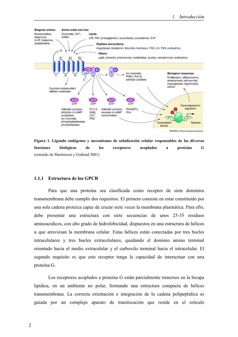
nuevos fármacos en amplios campos de la medicina (Figura 1).
1
1. Introducción
Figura 1. Ligando endógenos y mecanismos de señalización celular responsables de las diversas
funciones biológicas de los receptores acoplados a proteína G
(extraído de Marinissen y Gutkind 2001)
1.1.1 Estructura de los GPCR
Para que una proteína sea clasificada como receptor de siete dominios
transmembrana debe cumplir dos requisitos. El primero consiste en estar constituido por
una sola cadena proteica capaz de cruzar siete veces la membrana plasmática. Para ello,
debe presentar una estructura con siete secuencias de unos 25-35 residuos
aminoacídicos, con alto grado de hidrofobicidad, dispuestos en una estructura de hélices
α que atraviesan la membrana celular. Estas hélices están conectadas por tres bucles
intracelulares y tres bucles extracelulares, quedando el dominio amino terminal
orientado hacia el medio extracelular y el carboxilo terminal hacia el intracelular. El
segundo requisito es que este receptor tenga la capacidad de interactuar con una
proteína G.
Los receptores acoplados a proteína G están parcialmente inmersos en la bicapa
lipídica, en un ambiente no polar, formando una estructura compacta de hélices
transmembrana. La correcta orientación e integración de la cadena polipeptídica es
guiada por un complejo aparato de translocación que reside en el retículo
2

1. Introducción
endoplasmático (RE). Se pueden distinguir dos estados de plegamiento diferentes que se
producen tras la translocación inicial del receptor a través del extremo N-terminal
dentro del lumen del RE. En el primer plegamiento las hélices α se disponen a través de
la bicapa lipídica y el plegamiento de la proteína está dirigido principalmente por los
efectos hidrofóbicos. Para minimizar la superficie polar expuesta al ambiente lipídico,
los dominios transmembrana adoptan una estructura secundaria dejando los
aminoácidos hidrofóbicos enfrentados a la bicapa lipídica y los aminoácidos más
hidrofílicos orientados hacia la hendidura generada por el empaquetamiento de los
dominios transmembrana. Finalmente, en el segundo plegamiento se forma una
estructura terciaria por interacciones específicas hélice-hélice que permite un fuerte
empaquetamiento con estructura tipo anillo de los dominios transmembrana (Scarselli et
al. 2000).
La región N-terminal puede estar glicosilada y la región C-terminal está
expuesta a la interacción con otras moléculas de señalización, como quinasas y
proteínas β-arrestinas, responsables de procesos de sensibilización, desensibilización e
internalización (Lefkowitz 1998; Pfleger et al. 2007; DeFea 2011; Schulte et al 2010;).
Además, la región carboxi terminal y los bucles intracelulares dos y tres son críticos
para la transducción de la señal hacia el interior de la célula ya que son los dominios de
unión a la proteína G responsable, en la mayoría de los casos, de iniciar la señalización
intracelular (Figura 2).
Figura 2. Esquema de la estructura típica de un GPCR (extraído de Lefkowitz 2000)
3

1. Introducción
1.1.2 Clasificación y nomenclatura de los GPCR
Los GPCR se han clasificado según diferentes sistemas. Uno de los más clásicos
es el sistema Kolakowski (Kolakowski Jr. 1994), en el que los GPCR se clasifican en 6
familias (A-F) según su estructura y características genéticas. En la Figura 3 se ilustran
las tres familias mayoritarias.
Figura 3. Representación esquemática de las tres principales familias de receptores acoplados a
proteína G. Los residuos altamente conservados se indican en círculos rojos (extraído de George et al.
2002)
La familia A, también llamada rodopsin-like contiene el 90% de todos los
GPCR, siendo la más grande y estudiada, e incluye a receptores para una gran variedad
de hormonas y neurotransmisores. La homología global entre este tipo de receptores es
baja y limitada a un número de residuos altamente conservados. Los receptores se
caracterizan por tener un residuo conservado en toda la familia que corresponde al ácido
aspártico 130 del motivo Asp-Arg-Tyr (DRY), del tercer segmento transmembrana,
como inicialmente se descubrió en el receptor humano ß-adrenérgico (Fraser et al.
1988), y por tener un puente disulfuro que conecta el primer y el segundo bucle
extracelular. Además, muchos receptores tienen una cisteína palmitoilada en la cola C-
terminal que sirve de anclaje a la membrana plasmática (Figura 3 línea zigzag naranja).
4

1. Introducción
El estudio de la estructura cristalográfica de la rodopsina indica que los dominios
transmembrana están inclinados y enroscados debido al aminoácido prolina que
distorsiona los dominios helicoidales transmembrana. En esta familia, el ligando se une
en una cavidad formada por los dominios transmembrana, aunque para alguna
subfamilia de receptores activados por pequeños péptidos, el reconocimiento se produce
a nivel de los bucles extracelulares y del dominio N-terminal (George et al. 2002;
Jacoby et al. 2006).
La familia B incluye aproximadamente 50 receptores diferentes para una
variedad de hormonas peptídicas y neuropéptidos, como el péptido intestinal vasoactivo
(VIP), la calcitonina, la hormona paratiroidea (PTH) y el glucagón. La principal
característica de esta familia es que posee un extremo amino terminal relativamente
largo (aproximadamente 100 residuos), que contiene diversas cisteínas que forman una
red de puentes disulfuro (Ulrich et al. 1998). Así, los ligando pepiticos son reconocidos
por el extenso dominio amino terminal de estos receptores (George et al. 2002; Jacoby
et al. 2006). Son de morfología similar a la familia A, pero no parecen palmitoilarse y
los residuos y motivos conservados son diferentes. Excepto por el puente disulfuro que
conecta el primer y segundo bucle extracelular, esta familia no tiene ningún elemento en
común con la familia A y el motivo DRY no existe. Se sabe poco de la orientación de
los dominios transmembrana, pero teniendo en cuenta la divergencia de las secuencias
aminoacídicas, probablemente son diferentes de los de la familia A.
La familia 3 o C se caracterizan por tener un largo extremo carboxi y amino
terminal (500-600 aminoácidos). A esta familia pertenece el receptor metabotrópico de
glutamato y el receptor GABA (acido γ-aminobutírico). La estructura del lugar de unión
(representado en la figura 3 en rosado) se ha deducido mediante estudios de
cristalografía del extremo N-terminal del receptor metabotrópico de glutamato
solubilizado y unido a glutamato (He et al. 2002). Se ha visto que forma un dímero
unido por puente disulfuro que puede abrirse y cerrarse en el proceso de unión del
ligando (He et al. 2002). Esta familia no tiene ningún rasgo común con la familia 1 y 2
excepto por la presencia de cisteínas en el bucle extracelular que forman puentes
disulfuro. Una característica única de estos receptores es que el tercer bucle intracelular
es corto y altamente conservado, y al igual que en la familia 2 tampoco se conoce la
5

1. Introducción
orientación de los dominios de transmembrana (TM) (George et al. 2002; Jacoby et al.
2006).
Aunque la clasificación A-F está ampliamente aceptada, Fredriksson y
colaboradores (Fredriksson et al. 2003) efectuaron el primer estudio filogenético de toda
la superfamília de GPCR en el genoma de mamífero, proponiendo una clasificación más
detallada. El análisis muestra que hay 5 familias principales para los GPCR humanos:
Glutamate, Rhodopsin, Adhesion, Frizzled/Tasted2 y Secretin (la clasificación GRAFS
está basada en sus iniciales) y que dentro de cada familia los receptores comparten un
origen evolutivo común. Tres de estas familias, Rhodopsin (A), Secretin (B) y
Glutamate (C) se corresponden con la clasificación A-F, mientras que las otras dos
familias, Adhesion y Frizzled, no están incluidas. En esta clasificación la superfamília
de la rodopsina sigue siendo la mayor, y se ha dividido en 4 grupos principales con 13
ramas distintas. Los autores de este nuevo sistema de clasificación defienden la teoría de
que los receptores acoplados a proteína G surgieron a partir de un único predecesor
común, que a través de duplicaciones génicas, fue evolucionando desde la mayor
simplicidad en cuanto a sus orígenes a la enorme complejidad que muestra la
superfamília de estos receptores en la actualidad. La gran diversidad que alcanza esta
superfamília de proteínas de membrana da a entender el importante papel que juegan en
la fisiología de cualquier organismo.
1.1.3 Vías de señalización de los GPCR
Cuando un receptor es activado por un ligando, se inician una serie de eventos
intracelulares que modulan la función celular. Estos eventos dependen de la proteína G
a la que se encuentran acoplados y de la maquinaria molecular intracelular. Las
proteínas G están presentes en todos los organismos eucariotas y tienen un papel
esencial en la transducción de señales debido a su asociación al receptor y a otras
proteínas citoplasmáticas efectoras. Cada proteína G tiene una estructura heterotrimérica
constituida por la subunidades α (39-46 KDa), β (37 KDa) y γ (8 KDa). La interacción
del ligando con el receptor produce una serie de cambios conformacionales que
modifican la estructura de la proteína G y que hacen que la subunidad α libere GDP y
una GTP (Bourne et al. 1991) Al intercambiar GDP por GTP, la subunidad Gα se activa
y se desensambla tanto del receptor como del complejo estable Gβγ (Marinissen y
Gutkind 2001). Tanto la subunidad Gα como el complejo Gβγ son moléculas
6

1. Introducción
señalizadoras de forma que activan o inhiben a moléculas efectoras, como las adenilato
y guanilato ciclasas, fosfodiesterasas, las fosfolipasas A2 y C, y la fosfoinositol 3-
quinasa entre otras. Ello da lugar a una activación o inhibición de una gran variedad de
segundos mensajeros tales como el AMPc, GMPc, diacilglicerol (DAG), inositol
(1,4,5)-trifosfato (IP3), fosfatidil inositol (3,4,5)-trifosfato, y los ácidos araquidónico y
fosfatídico, por citar algunos (Marinissen y Gutkind 2001).
Dos ejemplos típicos de cascadas de señalización iniciadas por receptores
acoplados a proteína G son las que conducen a la formación de inositol-1,4,5-trifosfato
(IP3)/DAG y AMPc como segundos mensajeros. Para la subunidad Gαq, la proteína
efectora diana es la PLC, enzima que hidroliza fosfoinositoles de membrana generando
IP3 y DAG como segundos mensajeros. El IP3 aumenta la concentración de calcio
intracelular vaciando los depósitos intracelulares, mientras que el DAG activa a la PKC.
En el caso de las subunidades Gαs o Gαi, la proteína efectora es la adenilato ciclasa
(AC), enzima que cataliza la conversión de ATP a AMPc, mientras Gαs estimula esta
enzima, la subunidad Gαi/o la inhibe. El AMPc activa la PKA que igual que la PKC
fosforila a múltiples y diversas proteínas (receptores, canales iónicos, enzimas o
factores de transcripción) regulando así el funcionamiento celular (Marinissen y
Gutkind 2001; Faure et al. 2004; Berridge 2009; Puzianowska-Kuznicka y Kuznicki
2009;).
Figura 4. Representación de algunas de las vías que enlazan los GPCR con la vía de las MAPK
(extraído de Marinissen y Gutkind 2001)
7

1. Introducción
Muchas de las respuestas mediadas por estos receptores no consisten únicamente
en la estimulación de segundos mensajeros convencionales, si no que son el resultado
de la integración de diferentes redes de señalización, entre las que se incluyen la vía de
las MAPKs y las JNKs. Al principio se creía que la activación de la ruta de las MAPK
por GPCR involucraba a la proteína G sensible a la toxina de la Bordetella pertussis
(Gαi) y que dependía fuertemente del complejo Gβγ de la proteína G y de tirosina
quinasas no identificadas (van Corven et al. 1993; Faure et al. 1994; Koch et al. 1994).
Se postuló que, en ausencia de ligando para receptores con actividad tirosina quinasa
(RTK), la activación de receptores acoplados a proteína G podía inducir la estimulación
de un RTK generando señales mitogénicas. Este fenómeno se denominó
transactivacion. Una vez transactivado, el RTK inicia una cascada de señalización
idéntica a la generada por su propio ligando; es decir, la activación de las MAPK es a
través de la vía Ras, Raf, MEK y ERK (Figura 4). El proceso es iniciado con las
subunidades Gβγ dando lugar a que se reclute Sos hacia la membrana. Ello activa el
intercambio de GDP por GTP en la proteína Ras, siendo esta proteína el intermediario
que conecta la cascada de señalización generada por la transactivacion de un RTK con
la fosforilación de ERK (Marinissen y Gutkind 2001).
Sin embargo hay evidencias que indican que la señalización de los receptores de
siete dominios transmembrana es mucho más compleja puesto que pueden activar la vía
de las MAPKs a través de vías de señalización dependientes (Figura 4) e independientes
de proteínas G (Daaka et al. 1998; Lefkowitz 1998; Luttrell et al. 1999; Beaulieu y
Gainetdinov 2011). Un ejemplo paradigmático es la señalización mediada por la
fosforilación del receptor por GRKs (G protein-coupled Receptor Kinases), la unión de
β–arrestinas y el subsiguiente secuestro del receptor de la superficie celular (Krupnick y
Benovic 1998), que no sólo es importante para la finalización de la señal, sino que
también juega un papel importante en el intercambio entre las vías de señalización
dependientes de proteína G e independientes como las utilizadas normalmente por
receptores de factores de crecimiento (Luttrell et al. 1999).
Estudios relativamente recientes muestran que las β–arrestinas desempeñan un
papel en la señalización celular que va más allá del simple desacoplamiento entre
receptor y la proteína G. El hecho de que las β–arrestinas puedan interaccionar
8

1. Introducción
directamente con tirosina quinasas de la familia de las Src y con componentes de la
cascada de MAP quinasas (Perry y Lefkowitz 2002; Reiter et al. 2012), sugiere que las
β–arrestinas pueden funcionar como adaptadores o scaffolds reclutando proteínas
involucradas en la señalización de un determinado receptor (Figura 5). De esta manera,
se ha demostrado la capacidad de diferentes tipos de receptores acoplados a proteína G
de reclutar componentes de las cascadas de las JNKs o las ERKs, incluyendo las
quinasas más relevantes de la cascada, como pueden ser JNK3, Raf-1, MEK1 o
ERK1/2. Estos complejos pueden permanecer unidos incluso durante la internalización
del receptor, presentando diferentes localizaciones subcelulares, presumiblemente en
los endosomas hacia donde el receptor es conducido en su proceso de internalización y
por lo tanto aproximando las quinasas a sus posibles substratos citosólicos. En este
sentido, las β–arrestinas actúan como scaffolds permitiendo al receptor regular la
actividad y la distribución de dichas quinasas en el interior celular, lo que puede tener
unas implicaciones funcionales muy importantes (Violin y Lefkowitz 2007; Kelly et al.
2008; Reiter et al. 2012).
Figura 5. Transducción de señal en los receptores acoplados a proteína G
(extraído de Rosenbaum et al. 2009)
9

1. Introducción
1.1.4 Regulación de la actividad de los GPCR por desensibilización
La rápida atenuación de la respuesta del receptor tras su activación mediante
unión de un agonista recibe el nombre de desensibilización (Golan et al. 2009; Moser et
al. 2010). Este fenómeno puede manifestarse mediante diferentes mecanismos como el
desacoplamiento del receptor de su proteína G en respuesta a la fosforilación del
receptor (Hausdorff et al. 1989; Lohse et al. 1990; Ferguson 2001; Golan et al. 2009), la
internalización de los receptores de la superficie celular a compartimientos
intracelulares (Hermans et al. 1997; Trejo et al. 1998; Ferguson 2001), la disminución
del número de receptores debido a la disminución del RNA mensajero y a la síntesis
proteica, así como la degradación de los receptores preexistentes (Jockers et al. 1999;
Pak et al. 1999). En el caso de las fosforilaciones, estos fenómenos tienen lugar en
segundos, minutos en el caso de las endocitosis y horas cuando es regulada la expresión.
La desensibilización del receptor puede ser completa, como ocurre en el sistema
olfativo y visual o atenuada, disminuyendo la respuesta máxima, como ocurre con el
receptor β2-adrenérgico (Sakmar 1998). La manera más rápida por la cual un GPCR se
desacopla de la proteína G es a través de modificaciones covalentes en el receptor como
consecuencia de su fosforilación por quinasas intracelulares, siendo de especial
importancia las quinasas especificas para GPCR o GRK (Kelly et al. 2008; Golan et al.
2009).
La internalización de GPCR es un fenómeno común tras la estimulación por
agonista. El tráfico de receptores a compartimentos endosomales permite la
desfosforilación y reciclaje del receptor a la superficie celular (Krueger et al. 1997;
Pierce y Lefkowitz 2001; Ferguson 2001; Gainetdinov et al. 2004; Boulay y Rabiet
2005). Parte de los receptores internalizados pueden degradarse tras la exposición
prolongada al agonista, lo que implica que el receptor sea marcado para entrar en la vía
de degradación (Bohm et al. 1997). El mecanismo de internalización de GPCR mejor
caracterizado es a través de la fosforilación del receptor mediada por las proteínas
quinasas especificas de GPCR (GRK) y β-arrestinas (Kelly et al. 2008). Una vez el
receptor es fosforilado por GRK, la β-arrestina actúa como molécula reguladora que
interactúa con componentes de la vía endocítica mediada por vesículas de clatrina. En
respuesta a la activación de los GPCR, la β-arrestina citosólica transloca a la membrana
plasmática uniéndose a los receptores a la vez que se inicia el proceso de endocitosis
10

1. Introducción
mediado por clatrina (Ritter y Hall 2009) (Figura 6). Como se ha mencionado
anteriormente, las β-arrestinas no sólo funcionan en el secuestro de GPCR para la
desensibilización e internalización, sino como proteínas para transducir y
compartimentar las señales alternativas (Golan et al. 2009). Estas proteínas tienen la
habilidad de interaccionar con una gran variedad de proteínas endocíticas y de
señalización como las c-Src (Lutrell et al. 1999), MAPK y Raf (DeFea et al. 2000).
No obstante, no todos los GPCR necesariamente se internalizan por un
mecanismo dependiente de β-arrestinas y clatrina. Existen evidencias experimentales
que sugieren que los GPCR pueden internalizarse por vías endocíticas alternativas.
Algunos GPCR se han encontrado en estructuras de membrana ricas en colesterol
denominadas caveolas (Chun et al. 1994; Huang et al. 1997; Burgueño et al. 2003).
Estos dominios también son dominios de señalización donde los GPCR pueden
localizarse e interaccionar específicamente con proteínas de señalización (Ostrom y
Insel 2004). Además, las caveolas tienen un papel clave en la desensibilizacion y trafico
de los receptores ya que el uso de agentes bioquímicos que disrumpen estas estructuras
son efectivos en la inhibición de la endocitosis de ciertos GPCR (Ginés et al. 2001;
Escriche et al. 2003; Kong et al. 2007; Wu et al. 2008). Por otra parte, ciertos
receptores son susceptibles de usar una tercera vía endocítica alternativa, si bien no se
han identificado ni las proteínas de cubierta, ni las proteínas adaptadoras para la
generación de estas vesículas (Claing et al. 2000).
Una vez internalizados, los receptores son marcados para entrar en vías de
reciclaje o de degradación. Algunos GPCR, entre los que se incluye el receptor β2-
adrenérgico, pueden ser reciclados a la membrana plasmática, como receptores
totalmente competentes después de unos minutos de haber sido internalizados (Pippig et
al. 1995). Otros receptores, como el receptor de vasopresina tipo 2, es retenido dentro
de la célula durante un cierto periodo de tiempo antes de ser reciclado a la membrana
celular (Innamorati et al. 2001), mientras que algunos, como los receptores de σ-
opiodes o de trombina son mayoritariamente degradados (Tsao y von Zastrow 2000).
Sin embargo, para la mayoría de GPCR una parte es reciclada y otra parte es degradada,
como ocurre con los receptores de adenosina (Escriche et al. 2003).
11

1. Introducción
Figura 6. Ejemplo de un modelo propuesto para la desensibilización, internalización y
downregulation de los GPCR (extraído de Pierce y Lefkowitz 2001)
1.1.5 Actividad constitutiva y nomenclatura de los ligandos de los GPCR
La activación de un receptor acoplado a proteína G se basa en un cambio
conformacional de la estructura terciaria debido a la unión al receptor de un ligando
agonista. El receptor pasa de una conformación inactiva a una activa, existiendo una
constante de equilibrio entre los dos estados del receptor. La actividad constitutiva que
presentan estos receptores representa una isomerización del receptor a la conformación
activa en ausencia de ligando (Seifert y Wenzel-Seifert 2002). Como consecuencia se
promueve el intercambio GDP-GTP en la proteína G acoplada, aumentando así la
actividad basal de dicha proteína G y de los siguientes sistemas efectores (Costa y Herz
1989).
Esta actividad constitutiva es inhibida por los compuestos denominados
agonistas inversos, los cuales actúan sobre el receptor de manera que estabilizan la
conformación inactiva y por lo tanto minimizan el intercambio GDP-GTP. Estos
compuestos actúan de forma opuesta a los agonistas, cuya función es estabilizar al
receptor en la conformación activa y, por lo tanto, inducir su señalización intracelular.
Se ha propuesto la existencia de múltiples conformaciones de los receptores con
distintas funciones biológicas (Seifert y Wenzel-Seifert 2002). Estas conformaciones
estarían estabilizadas por diferentes tipos de compuestos, siendo la más favorable para
la señalización aquella conformación del receptor estabilizada por el agonista; seguidas
por los agonistas parciales, que serían compuestos con una menor eficiencia para
12

1. Introducción
estabilizar el receptor en la conformación más activa y que por lo tanto promueven un
menor intercambio GDP-GTP. A continuación vendrían los antagonistas neutros o
simplemente antagonistas que no alterarían el equilibrio entre las conformaciones activa
e inactiva, pero que tienen la capacidad de bloquear el efecto de los agonistas y de los
agonistas inversos. Por último, estarían los agonistas inversos parciales y los agonistas
inversos, que serian capaces de estabilizar al receptor en su estado inactivo, en un menor
y mayor grado respectivamente, reduciendo la actividad basal o constitutiva del receptor
(Figura 7).
Figura 7. Activación de los receptores acoplados a proteína G según el modelo de dos estados. A) El
modelo de dos estados asume que el receptor isomeriza desde un estado inactivo R a uno activo R*. B)
Acción de los diferentes tipos de ligandos sobre la actividad constitutiva del receptor (extraído y
modificado de Seifert y Wenzel-Seifert 2002)
Los ligandos también se pueden clasificar en función del lugar al que se unen al
receptor. La mayoría de los ligandos conocidos de GPCR que actúan como agonistas,
antagonistas o agonistas inversos, se unen al mismo dominio del receptor reconocido
por los agonistas endógenos, es decir, el lugar de unión ortostérico (Neubig et al. 2003).
En cambio, muchos GPCR poseen centros alostéricos, topográficamente distintos del
centro ortostérico. Esto ha llevado a la identificación de ligandos que actúan como
moduladores alostéricos, que pueden regular indirectamente la actividad de los ligando
ortostéricos y/o mediar directamente los efectos de agonistas/agonistas inversos
(Christopoulos y Kenakin 2002; Kenakin 2010).
13

1. Introducción
1.1.6 Los GPCR como dianas terapéuticas
Los GPCR han sido el centro de interés de fisiólogos y farmacólogos mucho
antes de que se supiera que estaban acoplados a proteína G (Fredholm et al. 2007;
Lefkowitz 2007). Estos receptores representan la familia de proteínas de mayor impacto
social, terapéutico y económico. Hoy en día, más del 50% de los fármacos, con unas
ventas anuales en el mundo que superan los 50 billones de dólares, regulan la función
de los GPCR, y un 30% de estos fármacos están directamente dirigidos a los GPCR
(Jacoby et al. 2006; Lundstrom 2006). Los GPCR están involucrados en una amplia
diversidad de enfermedades, como son: alergias, disfunción cardiovascular, obesidad,
cáncer, diabetes, y una variedad de trastornos del sistema nervioso central. Dado que los
GPCR representan alrededor del 1% del genoma humano, sólo una proporción muy
pequeña de todos los GPCR son actualmente diana de fármacos.
Tabla 1. Algunos de los fármacos más vendidos relacionados con GPCR
(extraído y modificado de Jacoby et al. 2006)
14

1. Introducción
Por lo tanto, existe mucho interés en la identificación de nuevos receptores que
puedan ser utilizados para el desarrollo de fármacos donde las necesidades médicas
siguen sin satisfacerse (Lin y Civelli 2004; Fredholm et al. 2007). En la Tabla 1 se
muestra un pequeño ejemplo de los fármacos más vendidos dirigidos a GPCR, donde se
observa el amplio rango de indicaciones terapéuticas que cubren.
1.1.7 Interacciones moleculares de los GPCR con otras proteínas
El correcto funcionamiento de la célula depende, entre otros factores, de los
estímulos que recibe desde su entorno extracelular más inmediato. Éste es un proceso
altamente regulado en el que participan un gran número de moléculas, que interaccionan
entre ellas de forma rápida o bien formando complejos relativamente estables. Ello
permite una serie de respuestas adecuadas al estado general de una célula o de un
sistema en un momento determinado. Además de las interacciones clásicas proteína-
proteína intracelulares involucradas en la transducción de la señal, se han descrito un
gran número de interacciones que son la base para la formación de complejos
macromoleculares responsables de la localización de receptores en determinados
dominios celulares. Muchos GPCR tienen la capacidad de interactuar con una amplia
variedad de proteínas. Estas interacciones determinan diferentes propiedades del
receptor, como por ejemplo: la compartimentalización celular, la selección de una señal,
la promoción de ensamblajes de complejos que integran una función y el favorecimiento
de la internalización, entre otras (Franco et al. 2003).
La topología de los GPCR permite varias caras potenciales para posibilitar
distintos tipos de interacciones proteína-proteína. Así, en el espacio extracelular, donde
tiene lugar la unión a ligando, las regiones de los GPCR implicadas en la interacción
con proteínas son en la mayoría de los casos, las secuencias presentes en el extremo
amino terminal, ya que los bucles extracelulares son muy cortos. Existen evidencias
crecientes de que las interacciones receptor-proteína extracelulares pueden jugar un
papel importante en la farmacología de los GPCR. Un ejemplo lo constituye la
adenosina desaminasa (ADA) que puede actuar como ecto-enzima anclada a diferentes
proteínas tales como los receptores de adenosina A1, A2A y A2B (Ciruela et al. 1996;
Saura et al. 1996; Herrera et al. 2001; Gracia et al. 2011).
15

1. Introducción
En la cara intracelular, tanto el extremo carboxilo terminal como el tercer bucle
intracelular pueden ser considerablemente grandes, razón por la que estas regiones son
las más probables para la interacción con otras proteínas implicadas en el proceso de
señalización (como las proteínas G) o en la localización subcelular, mediadas por
asociación a proteínas del citoesqueleto o a las relacionadas con el tráfico de receptores
(Figura 8).
Figura 8. Representación esquemática de un GPCR con las regiones identificadas implicadas en la
interacción con otras proteínas y su función genérica (extraído de Bouvier 2001)
Los polipéptidos que anclan estos receptores al citoesqueleto constituyen un
ejemplo de proteínas que interactúan con los GPCR, como es el caso de la α-filamina
con el receptor D2 de dopamina (Lin et al. 2001), y la α-actinina con el receptor de
adenosina A2A (Burgueño et al. 2003), y con el receptor metabotrópico de glutamato
tipo 5b (mGlu5bR) (Cabello et al. 2007). También encontramos la familia de proteínas
Shank con el receptor metabotrópico de glutamato tipo 1 (mGlu1R) o el receptor de
somatostatina tipo 2 (SSTR2) (Tu et al. 1999; Böckers et al. 2001). Las proteínas de
andamio o scaffolds (scaffolding proteins) son ricas en dominios tales como los SH2
(Src-homology 2), SH3 (Src-homology 3) o PDZ (Post-synaptic-Density-
95/Discslarge/ZO1), que se han conservado a lo largo de la evolución y que son los
responsables de las interacciones con otras proteínas. En la última década se han
16

1. Introducción
descrito interacciones entre los GPCR y proteínas que contienen el dominio PDZ, las
cuales juegan un papel clave en la modulación de la señal, ya que definen una
composición molecular de complejos de señalización en microcompartimentos y, en
algunos casos, la localización precisa de estos complejos dentro de la célula. Así, por
ejemplo, la interacción del receptor β2-adrenérgico con el factor regulador del
intercambio Na+/H+ (NHERF) podría controlar la internalización y tráfico de este
receptor (Shenolikar et al. 2001). También se ha demostrado que la interacción de la
proteína Homer-1b con el receptor mGlu1R modula la movilización de estos receptores
hacia la membrana (Roche et al. 1999). Por otro lado, se ha comprobado que la
espinofilina (otra proteína con dominio PDZ) puede interaccionar con los receptores D2
de dopamina y α2-adrenérgico a través de un nuevo dominio diferente del PDZ,
actuando como una proteína de andamio que une estos GPCR con proteínas de
señalización como PP-1 (Smith et al. 1999; Richman et al. 2001).
17

1. Introducción
1.2 OLIGOMERIZACIÓ� DE LOS GPCR
La autoasociación de proteínas para formar dímeros o bien oligómeros de orden
superior es un factor clave en la regulación de proteínas tales como enzimas, receptores,
canales iónicos, factores de transcripción etc. (Marianayagam et al. 2004).
Los GPCR, dadas sus características estructurales y su localización subcelular,
además de interaccionar con otras proteínas del lado intracelular y del lado extracelular,
tal como se ha descrito anteriormente (véase 1.1.7), también pueden exhibir
interacciones proteína-proteína a nivel de membrana plasmática con otros receptores o
canales iónicos (Franco et al. 2003). Desde mediados de los años 90, diversos estudios
han demostrado la oligomerización de numerosos GPCR (George et al. 2002). Hoy en
día se acepta que la oligomerización es un hecho común en la biología de estos
receptores y que pueden formar homodímeros, heterodímeros y/u oligómeros de orden
superior (Bouvier 2001; Devi 2001; Rios et al. 2001; Agnati et al. 2003; Franco et al.
2003; Terrillon y Bouvier 2004; Agnati et al. 2005; Prinster et al. 2005; Pin et al. 2007;
Carriba y et al. 2008; Ferré et al. 2009; Rozenfeld y Devi 2010). La homodimerización
se define como la asociación física entre proteínas idénticas, mientras que la
heterodimerización es la asociación entre proteínas distintas. El término dímero se
utiliza con frecuencia entendiendo que es la forma más simple de una unidad estructural
y funcional oligomérica, debido a la dificultad que existe en distinguir entre dímeros y
oligómeros de orden superior con las técnicas actuales. Los dímeros/oligómeros pueden
presentar características funcionales diferentes a las de los receptores que los
constituyen. Así la oligomerización confiere nuevas propiedades a los GPCR, lo que
establece un posible mecanismo para generar funciones distintas en estos receptores.
Este fenómeno ha dado lugar a un nuevo nivel de complejidad que gobierna la
señalización y regulación de estas proteínas (Ferré et al. 2007; Pin et al. 2007; Ferré et
al. 2009). En la tabla 2 se describen algunos ejemplos de homodímeros y de
heterodímeros de diversos receptores.
Las interacciones entre GPCR son cruciales para entender el variado cross-talk
que se observa entre receptores de neurotransmisores. La oligomerización de receptores
neuronales permite formular hipótesis sobre el alto grado de diversidad y plasticidad,
que es característico de una estructura altamente organizada y compleja como es el
cerebro. Se ha descrito un nivel superior de organización por el que los receptores
18

1. Introducción
acoplados a proteína G forman estructuras compuestas no sólo por homo- o
heterodímeros, sino por complejos supramoleculares constituidos por varios receptores
y una variedad de proteínas que modifican la actividad del receptor (RAMPs: Receptor
Activity Modifying Proteins).
Estos complejos interaccionan tanto a lo largo de la membrana (interacciones
horizontales), como a través de ella (interacciones verticales), y al ser activados por
hormonas o neurotransmisores se redistribuyen en la membrana y dan lugar a clusters.
Los clusters supondrían un nivel superior de regulación de los receptores y enzimas
asociadas y podrían ser regulados por otros receptores en estos complejos y también por
otras moléculas que no interaccionan físicamente con ellos, pero sí se comunican entre
ellos en el cluster (Franco et al. 2003).
Debido al número creciente de publicaciones en este campo ha sido necesario
establecer nuevas definiciones y dotar de nomenclatura a los homómeros y heterómeros
de GPCR, como recientemente han publicado Ferré et al (2009).
Tabla 2. Algunos ejemplos de homodímeros y heterodímeros de GPCR.
19

1. Introducción
1.2.1 Arquitectura de los dímeros de GPCR
Para explicar el fenómeno de la dimerización de los receptores acoplados a
proteína G se pueden considerar dos posibilidades: que estas interacciones sean
indirectas o bien que sean directas, implicando un contacto entre ambos receptores. En
el caso de las interacciones indirectas entre GPCR hace falta la mediación de terceras
proteínas, que hacen de puente, tales como las proteínas del citoesqueleto. En las
interacciones indirectas, los dominios intracelulares de los GPCR se unen a un gran
número de proteínas citosólicas, algunas de las cuales, por sus características
intrínsecas, han sido propuestas como posibles candidatas a participar en la
dimerización de los receptores con los que interaccionan. Muchas de estas proteínas son
proteínas de andamio o scaffolding proteins, que proporcionan una estructura compleja
en la cual diversos receptores pueden interaccionar entre ellos y con otras proteínas
involucradas en la transducción de señal, controlando la velocidad y la especificidad de
dicha señalización (Ciruela et al. 2005).
Las interacciones directas entre miembros de la familia de GPCR no precisan de
otras proteínas y se cree que, en la mayoría de casos los oligómeros se forman en el
retículo endoplasmático, por lo que no son modulables por ligando, entendiendo la
modulación como la formación o destrucción del oligómero. La gran complejidad
estructural que existe en esta superfamília no permite pensar en un único mecanismo de
interacción directa. Así pues, las interacciones directas pueden tener lugar mediante
enlaces covalentes (puentes disulfuro) y/o no covalentes (interacciones hidrofóbicas y/o
electroestáticas) entre los dominios transmembrana y/o los dominios intracelulares de
los receptores (Figura 9) (Bouvier 2001).
Se han encontrado distintas interacciones intermoleculares involucradas en la
formación de varios homómeros y heterómeros, que demuestran que en la
oligomerización de los GPCR existen múltiples sitios de interacción implicados en el
ensamblaje y en la estabilización de los dímeros (Maggio et al. 1993; Hebert et al.
1996; Ng et al. 1997; Romano et al. 1996; Cvejic y Devi 1997; White et al. 1998;
Jordan y Devi 1999; Robbins et al. 1999; Gouldson et al. 2000; Margeta-Mitrovic et al.
2000; Scarselli et al. 2000; Schulz et al. 2000; Romano et al. 2001; Woods y Huestis
20

1. Introducción
2001; Ciruela et al. 2004; Ferrada et al. 2008; Navarro et al. 2008; Ferrada et al. 2009;
Navarro et al. 2009; Navarro et al. 2010b; Moreno et al. 2011b; Moreno et al. 2011a).
Figura 9. Determinantes moleculares de la
dimerización de los GPCRs. Varios tipos de
interacciones intermoleculares han sido descritas, a
través de las cuales los GPCR forman dímeros: a) los
puentes disulfuro se han descrito para la dimerización
entre receptores metabotrópicos de glutamato, b)
interacciones mediante enlaces no covalentes de los
extremos carboxi terminales entre los receptores
GBR1 y GRBR2 gabaérgicos, c) interacciones de las
hélices transmembrana que forman especialmente los
receptores de la familia A (extraído de Bouvier et al.
2001)
1.2.2 Papel funcional de la oligomerización de los GPCR
Como se detallará posteriormente (véase 1.2.4), la disponibilidad de un gran
número de técnicas para el estudio de la oligomerización de GPCR ha facilitado
enormemente la investigación del papel funcional de estos receptores. La dimerización
está implicada en la regulación de la funcionalidad del receptor a diferentes niveles,
desde la modulación de la expresión del receptor en la superficie celular hasta el hecho
de conferir nuevas propiedades farmacológicas a los receptores expresados en el
dímero. Esto ha proporcionado una nueva perspectiva para considerar cual es la unidad
de señalización de los GPCR, así como para el desarrollo de ligando que actúan a través
de este tipo de receptores.
Aunque en muchos casos la relevancia fisiológica no se conoce completamente,
diversos estudios llevados a cabo en sistemas de expresión heterólogos han sugerido
21

1. Introducción
distintos papeles funcionales para la oligomerización de GPCRs (Figura 10). Por
ejemplo, la oligomerización puede estar implicada en la ontogénesis de los GPCR, es
decir, en el control de calidad del plegamiento y de la destinación a la membrana de
receptores sintetizados de novo (Figura 10.1). Asimismo, en algunos casos, se ha
observado una regulación de la formación/separación de oligómeros presentes en la
membrana plasmática mediada por ligando (Figura 10.2). También, se ha constatado
que la oligomerización confiere diversidad farmacológica, ya que la unión de un ligando
a un receptor del dímero puede influir en la unión de otro ligando al segundo receptor
dentro del dímero (Ferré et al. 2007; Franco et al. 2008b) (Figura 10.3). La
oligomerización también puede modificar las propiedades de señalización de un
determinado ligando afectando la selectividad de interacción entre el receptor
correspondiente y su proteína G, lo que origina una potenciación, atenuación o
acoplamiento con otra proteína G (Figura 10.4). Finalmente, también se ha visto que la
oligomerización puede alterar el patrón endocítica para un determinado receptor
(Terrillon y Bouvier 2004) (Figura 11.5).
Figura 10. Posibles papeles funcionales de la oligomerización de GPCRs
ER, retículo endoplasmático, L, ligando (extraído de Terrillon y Bouvier 2004)
1.2.3 Técnicas para el estudio de la oligomerización de GPCR
Las primeras evidencias de la existencia de homodímeros entre GPCR se
obtuvieron a partir de estudios farmacológicos. Las complejas curvas de unión, tanto de
agonistas como de antagonistas de estos receptores se interpretaron considerando la
22

1. Introducción
existencia de una cooperatividad positiva o negativa, que se podía explicar mediante
interacciones entre los sitios de unión de los receptores dentro de complejos diméricos
u oligoméricos (Limbird et al. 1975; Mattera et al. 1985; Hirschberg y Schimerlik,
1994; Wreggett y Wells, 1995; Franco et al. 1996)
Una evidencia contundente de la existencia de heteroligómeros la constituyen
los cambios cinéticos en la unión de radioligandos a un receptor provocados por la
unión de ligandos no radioactivos al otro receptor del heterómero, utilizando preparados
de membranas de células o de tejido que expresen los dos receptores. En preparaciones
de membrana no hay ninguna maquinaria celular que pueda producir un cross-talk
indirecto (por ejemplo, un cross-talk a nivel de segundos mensajeros) y la existencia de
una modulación a nivel de unión de radioligandos sólo puede ser explicada por una
interacción molecular entre ambos receptores. En estos casos la unión de un ligando a
un receptor induce cambios conformacionales en el otro receptor que modulan su
capacidad de unir a su ligando y estos cambios conformacionales sólo se pueden
producir si ambas proteínas interaccionan molecularmente directa o indirectamente
(Franco et al. 2007; Franco et al. 2008b). En muchos casos esta clase de interacción se
ha encontrado en tejido nativo, lo que puede ser interpretado como un indicador de la
existencia de receptores heteroméricos in vivo (Gonzalez-Maeso et al. 2008; Marcellino
et al, 2008; Navarro et al. 2010b; Navarro et al. 2010a; Moreno et al. 2011b; Moreno et
al. 2011a).
El elegante trabajo de Maggio (Maggio et al. 1993; Maggio et . 2003), utilizando
quimeras y receptores mutantes de varios GPCR, que ponía de manifiesto que estos
receptores pueden funcionar como dímeros, fue un estudio pionero en la utilización de
mutantes para demostrar la oligomerización de GPCR. En esta línea se ha observado
que diversos receptores mutantes se comportan como mutantes dominante negativos
cuando se expresan con el receptor wild-type (Benkirane et al. 1997; Bai et al 1998; Zhu
y Wess 1998). En estos casos, la respuesta observada se explica únicamente por la
dimerización entre el receptor wild-type y el receptor inactivo.
Una de las técnicas bioquímicas clásicamente más usadas para investigar la
homodimerización de GPCR ha sido la coinmunoprecipitación de receptores marcados
23

1. Introducción
sobre epítopos diferentes. El primer estudio en el que se utilizó esta aproximación
experimental se demostró la interacción específica entre los receptores β2-adrenérgicos
(Hebert et al. 1996).
Desde entonces, estrategias similares han sido usadas para documentar la
homodimerización de los receptores metabotrópico mGlu5R (Romano et al. 1996), δ-
opioides (Cvejic y Devi, 1997) y los receptores de serotonina 5-HT2C (Herrick-Davis
et al. 2004) y 5-HT4 (Berthouze et al. 2007), entre otros.
Los experimentos de coinmunoprecipitación también han sido utilizados para
demostrar la heteromerización entre receptores del mismo neurotransmisor, como
GABAB1 y GABAB2 (Jones et al. 1998; Kaupmann et al. 1998; White et al. 1998) o
como los δ- y κ-opioides (Jordan y Devi, 1999), e incluso entre receptores menos
relacionados como los receptores de adenosina A1 y de dopamina D1 (Gines et al.
2000), los de angiotensina AT1 y bradiquinina B2 (AbdAlla et al. 2000), δ-opioide y β2-
adrenérgico (Jordan et al., 2001), A1 de adenosina y metabotrópico mGlu1R (Ciruela et
al. 2001), A2A de adenosina y metabotrópico mGlu5R (Ferré et al. 2002) o los
receptores de canabinoides CB1 y de dopamina D2 (Kearn et al. 2005). Aunque se
utiliza comúnmente para estudiar las interacciones proteína-proteína, la
coinmunoprecipitación de GPCR requiere de su solubilización mediante detergentes, lo
que no permite descartar que los dímeros observados puedan ser agregados artefactuales
por una solubilización incompleta, debida a la naturaleza hidrofóbica de estos
receptores.
En 1948 Theodor Forster formuló la teoría de transferencia de energía por
resonancia (Förster 1948) que más tarde fue aplicada al estudio de interacciones entre
GPCR. Esta aproximación biofísica está basada en la transferencia de energía no
radiante entre dos dipolos electromagnéticos, es decir, desde un cromóforo en estado
excitado (dador energético) a una molécula cercana que absorbe (aceptor). En el caso de
la transferencia de energía de resonancia fluorescente (FRET; Fluorescence Resonance
Energy Transfer), tanto el dador como el aceptor son moléculas fluorescentes, mientras
que en la transferencia de energía de resonancia bioluminiscente (BRET;
Bioluminescence Resonance Energy Transfer) el dador es bioluminiscente y el aceptor
fluorescente (Bouvier et al.. 2007; Gandía et al.. 2008; Ciruela et al. 2010; Ferré et al.
24

1. Introducción
2010; De 2011; Schaferling y Nagl 2011). Para que este fenómeno tenga lugar es
necesario que se cumplan dos requisitos. El primero, consiste en que el espectro de
emisión del dador y el espectro de excitación del aceptor se solapen, de forma que el
dador no emite completamente la energía que debiera, si no que transfiere parte de su
energía de emisión de forma directa al fluoróforo aceptor, el cual emite como si hubiera
sido excitado directamente. El segundo requisito para que tenga lugar el fenómeno de
transferencia de energía es que tanto el dador como el aceptor han de estar muy
próximos en el espacio (<100 Å o 10 nm). Así, a diferencia de la
coinmunoprecipitación, las técnicas de transferencia de energía ofrecen una
aproximación única que permite detectar la dimerización de proteínas en células vivas,
sin perturbar el entorno donde este fenómeno ocurre.
La dependencia crítica de la distancia entre dador y aceptor para la transferencia
de energía, donde la eficiencia de la transferencia disminuye con la sexta potencia de la
distancia, hace que los sistemas de BRET/FRET sean los elegidos para monitorizar las
interacciones proteína-proteína en cultivos celulares. Hay que destacar que entre 10 y
100 Å se encuentran la mayor parte de complejos multiproteicos biológicos de una
célula (Stryer 1978; Sheng y Hoogenraad 2007).
Para la técnica de FRET se utilizan las diferentes variantes de la proteína verde
fluorescente (GFP: Green Fluorescence Protein) obtenidas por mutación. Estas
mutaciones confieren diferentes propiedades espectrales, de forma que utilizando dos
formas diferentes de mutantes con las características espectrales adecuadas, fusionadas
a las proteínas en estudio, permite determinar si éstas están lo suficientemente cercanas
como para transferirse energía (Pfleger y Eidne 2005; Ferré et al. 2010; Schaferling y
Nagl 2011). La pareja más ampliamente utilizada para los experimentos de FRET son
las variantes YFP (Yellow Fluorescence Protein) y GFP2. Esta ultima variante de la
GFP ha sido optimizada para ser usada como pareja de FRET con la YFP. La GFP2 se
excita a 400 nm y emite a 510 nm, mientras que la YFP se excita a 485 nm y emite a
530 nm. De esta forma, tal y como se muestra en la figura 12, la excitación de la células
que expresan la proteína de fusión receptor-GFP2 con un láser de una longitud de onda
de 393-403 nm, produce la emisión de energía a 510 nm, que es capaz de excitar a la
proteína de fusión receptor-YFP, cuya emisión a 530 nm es cuantificable. Debido a que
hay un cierto solapamiento entre los espectros de emisión de ambas proteínas de fusión,
25

1. Introducción
es necesario separar los dos espectros de emisión para cuantificar la señal de FRET
(Zimmermann et al. 2002).
En la técnica de BRET se utiliza el enzima Renilla luciferasa (Rluc) fusionado a
uno de los receptores. Se produce la degradación catalítica del substrato coelenterazina
H por la luciferasa en presencia de oxígeno, de forma que se genera luz que al ser
transferida a una variante de la proteína GFP fusionada al otro receptor, ésta emite
fluorescencia a su longitud de onda característica si ambas proteínas están lo
suficientemente cercanas (Bouvier et al. 2007; Ciruela et al. 2010; Ferré et al. 2010; De
2011).
Figura 11. Representación esquemática del fenómeno de FRET
En el estudio de la dimerización de GPCRs, se generan proteínas de fusión que
consisten en la unión de la proteína fluorescente GFP o sus variantes (por ejemplo YFP)
en el extremo carboxi terminal de un receptor y la proteína luminiscente Rluc en el
extremo carboxi terminal del otro receptor y, al igual que en la técnica de FRET, se co-
expresan ambas proteínas de fusión en células vivas. Como se muestra en la figura 11a,
en ausencia de dimerización, la adición del sustrato coelenterazina H genera una señal
bioluminiscente característica, mientras que, como se muestra en la figura 11b, si se
produce dimerización entre ambos receptores, la energía es transferida de la Rluc a la
proteína fluorescente, dando lugar a la aparición de una señal adicional fluorescente con
un pico de emisión característico de la variante usada.
26

1. Introducción
Hasta la fecha se han descrito dos variantes principales de esta técnica, la que se
conoce como BRET1 y la denominada BRET2. Aunque el principio biofísico de ambas
técnicas es el mismo, difieren por el sustrato que cataliza la Rluc y por la proteína
aceptora. En el BRET1 el sustrato que se usa es la coelenterazina H, que al ser
metabolizada por la Rluc genera luz con un pico de emisión a 480 nm; emisión que
permite excitar a la YFP (ya que se solapa con su pico de excitación), de forma que esta
emite a 530 nm. En el BRET2 el sustrato es DeepBlueC que al ser oxidado por la Rluc
emite una luz a 400 nm de forma que puede excitar a la GFP2; en este caso la longitud
de onda a la que emite esta variante de la GFP es 510 nm (Figura 12).
Figura 12. Representación esquemática de los fenómenos de BRET1 y BRET2 con sus correspondientes espectros de emisión
Mediante técnicas de transferencia de energía se ha demostrado la existencia de
homodímeros de los receptores β2-adrenergico (Angers et al. 2000), δ-opiodes (McVey
et al. 2001) y A2A de adenosina (Canals et al. 2004) entre muchos otros. También se ha
realizado una aproximación similar para el estudio de heterómeros de receptores
acoplados a proteína G, como por ejemplo entre los receptores de somatostatina SST2A
R y SST1B R (Rocheville et al. 2000), los receptores de somatostatina SST1B R y los D2
de dopamina (Rocheville et al. 2000), los receptores A2A de adenosina y D2 de
dopamina (Canals et al. 2003), los receptores de dopamina D1 y D3 (Fiorentini et al.
27

1. Introducción
2008; Marcellino et al. 2008), los receptores A2A de adenosina y CB1 de cannabinoides
(Carriba et al. 2007), los receptores D1 de dopamina y H3 de histamina (Ferrada et al.
2009) o los receptores D1 de dopamina y los sigma-1 (Navarro et al. 2010b) entre otros.
En los últimos años se han desarrollado multitud de variantes de estas técnicas (véase
Ciruela 2008 como revisión) entre las que cabe destacar la técnica de SRET (Sequential
Resonante Energy Transfer) basada en las técnicas de BRET y FRET (Carriba et al.
2008) y la técnica de BRET y complementación bimolecular (Vidi y Watts 2009) que
permiten la detección de heterómeros de más de dos receptores.
Existen técnicas directas para detectar oligómeros en tejidos nativos. Una de
ellas utiliza la microscopia de fuerza atómica. Palczewski y colaboradores (Fotiadis et
al. 2003) usando microscopia de fuerza atómica demostraron, por primera vez,
oligómeros de rodopsina en la retina con un determinado patrón de distribución. Esta
técnica es factible cuando la concentración de receptores en el tejido es muy elevada
como ocurre con la rodopsina en la retina, pero es de difícil aplicación para la mayoría
de receptores del sistema nervioso central cuya expresión es moderada.
La técnica de la In Situ Proximity Ligation Assay (PLA) es una técnica directa
muy útil si se dispone de anticuerpos específicos para los receptores que heteromerizan
(Gustafsdottir et al. 2005; Soderberg et al. 2006; Thymiakou et al. 2007; Soderberg et
al. 2008; Yu et al. 2008; Massinen et al. 2009; Miyazono et al. 2009; Baan et al. 2010;
Vuoriluoto et al. 2010; Weibrecht et al. 2010; Hervouet et al. 2011; Renfrow et al.
2011). Esta tecnología amplía las capacidades de los inmunoensayos tradicionales al
incluir la detección directa de proteínas, interacciones entre proteínas y modificaciones
de estas interacciones con alta sensibilidad y especificidad. La técnica de la PLA está
basada en el reconocimiento y marcaje de una o dos proteínas que actúan como
antígenos (por ejemplo dos receptores que están interaccionando), por parte de dos
anticuerpos primarios conjugados a unos oligonucleótidos complementarios. Cuando las
dos moléculas de anticuerpo están muy cercanas (< 16 nm), se produce la ligación entre
las cadenas de DNA complementarias. Posteriormente, las cadenas de DNA se
amplifican en presencia de nucleótidos fluorescentes, emitiendo una señal positiva en
forma de punteado fluorescente observable al microscopio confocal. Como la
fluorescencia puede ser cuantificada, la señal PLA proporciona no solo la información
28

1. Introducción
espacial exacta (la localización de los eventos de interacción), sino también una manera
objetiva de cuantificar estos eventos (Gustafsdottir et al. 2005; Soderberg et al. 2008).
La técnica es igualmente aplicable utilizando un anticuerpo específico no
marcado y anticuerpos secundarios unidos a ADN (Figura 13).
Figura 13. Representación de la técnica In situ Proximity Ligation Assay. (Extraído de Olink Bioscience)
La utilización de ligandos para heterómeros constituye otra técnica directa para
su detección. Se pueden seguir varias estrategias dependiendo de las propiedades del
heterodímero (Rozenfeld et al. 2006). Uno de los enfoques consiste en el diseño y
síntesis de ligando bivalentes que interaccionen con los dos receptores del heterodímero.
Estos ligando pueden tener mayor afinidad y selectividad si se compara con los ligando
clásicos de los receptores individuales. Esta estrategia se ha utilizado para determinar la
presencia de heterómeros de receptores de adenosina A2A y de dopamina D2 en el
estriado de cerebro de cordero (Soriano et al. 2009). Otra aproximación sería utilizar
ligandos selectivos de un determinado heterodímero. Estos ligando interaccionan con el
centro de unión de un receptor únicamente cuando forma parte del heterodímero
(Waldhoer et al. 2005; Orru et al. 2011) (Tabla 3).
29

1. Introducción
Existen técnicas indirectas para detectar oligómeros en tejidos nativos. Una
manera bastante eficaz de detectar oligómeros en tejidos nativos es determinar alguna
característica específica de los heterómeros en células donde se haya demostrado la
heteromerización y utilizar esta propiedad como huella dactilar o fingerprint para
detectar el heterómero en tejidos nativos. La determinación de cross-talk entre cascadas
de señalizacion intracelular, el antagonismo cruzado en el que un antagonista especifico
de un receptor inhibe la señalizacion mediada por un agonista del otro receptor, o bien,
el estudio de cambios en la unión de ligando en uno de los receptores en presencia de un
ligando para el otro receptor en preparaciones de membranas obtenidas de tejidos,
pueden constituir una huella dactilar si se ha demostrado previamente que es una
característica del heterómero.
Tabla 3. Ventajas e inconvenientes de los diferentes tipos de ligando usados para detectar la oligomerización de los GPCR. (Extraído de Rozenfeld et al. 2006)
1.2.4 El modelo de receptores diméricos o Two-State Dimer Receptor Model
Durante muchos años el análisis de los estudios de unión de diferentes ligandos a
GPCRs, se ha llevado a cabo utilizando diversos modelos matemáticos en los que estos
receptores eran considerados como entidades proteicas monoméricas (Del Castillo y
Katz 1957; De Lean et al. 1980; Samama et al. 1993; Leff 1995; Weiss et al. 1996). Así
por ejemplo, uando el ajuste de datos experimentales de unión de ligando genera
diagramas de Scatchard lineales los datos se ajustan a un modelo de un centro de unión
que permite calcular la KD (constante de afinidad) del único estado de afinidad del
receptor. Sin embargo, la unión de agonistas a receptores de siete dominios
30

1. Introducción
transmembrana a menudo genera diagramas de Scatchard no lineales y, en estos casos,
los resultados se ajustan tradicionalmente, al modelo de “dos centros independientes”
que considera la existencia de dos estados independientes del receptor (estados no
interconvertibles): un estado de alta afinidad (o acoplado a proteína G) y un estado de
baja afinidad (o desacoplado a proteína G). El ajuste de los datos a este modelo permite
el cálculo de dos KD: una para el estado de alta afinidad (KDH) y otra para el estado de
baja afinidad (KDL). Sin embargo, se ha observado que el agonista induce cambios en la
proporción de los llamados estados de “alta” y “baja” afinidad, lo cual indica que estos
dos estados no pueden existir separadamente, sino que están interconectados (Wong et
al. 1986) y esta aparente interconversión entre estados es independiente de la proteína G
(Casadó et al. 1991). Además, trabajando con receptores de adenosina A1, se demostró
que un agonista total puede provocar un cambio aparente en la proporción de receptores
en estado de alta y baja afinidad (Casadó et al. 1991). Si el agonista es capaz de variar la
proporción de los estados de alta y baja afinidad, estas dos formas deben estar en
equilibrio y, consecuentemente, el modelo de dos centros independientes no puede
representar adecuadamente el comportamiento de los receptores si los estados de
afinidad están en equilibrio.
Puesto que actualmente se conoce que los GPCR forman dímeros (Bouvier
2001; Devi 2001; Gomes et al. 2001; Rios et al. 2001; Agnati et al. 2003; Franco et al.
2003; Terrillon y Bouvier 2004; Agnati et al. 2005; Franco et al. 2005; Prinster et al.
2005; Milligan 2006, Milligan 2007), las isotermas de unión bifásicas (representaciones
de Scatchard no lineales) y las curvas de competición bifásicas pueden interpretarse de
una manera más directa y evidente ya que pueden explicarse como un fenómeno de
cooperatividad. La cooperatividad positiva o negativa puede explicarse de manera
natural asumiendo que la unión de la primera molécula de ligando a uno de los
monómeros del dímero modifica los parámetros de unión de la segunda molécula de
ligando al otro monómero del dímero, como ocurre en el caso de las enzimas.
Recientemente, se han desarrollado modelos que consideran al dímero como la unidad
básica (Durroux 2005; Franco et al. 2005; Albizu et al. 2006; Franco et al. 2006). El
grupo de investigación en el que se ha desarrollado esta Tesis ha formulado el “Two-
State Dimer Receptor Model” (“modelo de receptores diméricos”), que considera el
homodímero como la unidad básica funcional (Franco et al. 2005; Franco et al. 2006)
(Figura 14).
31

1. Introducción
Figura 14. Esquema y ecuaciones del “modelo de receptores dimérico” (“Two-State Dimer Receptor
Model”). El dímero puede ser inactivo o activo y puede estar vacio u ocupado por una o dos moléculas de ligando. a) Modelo macroscópico b) Modelo simplificado que incluye las constantes de disociación macroscópicas en el equilibrio, (KD1 y KD2) que definen la unión de la primera y segunda molécula de ligando al dímero. Se muestran las ecuaciones para el ajuste de los valores de unión del radioligando (L) a los receptores que forman el dímero y para calcular el índice de cooperatividad del dímero (Dc). Ver (Franco et al. 2005; Franco et al. 2006; Casado et al. 2007) para más detalles (extraído de Franco et al.
2008b)
Este modelo considera que el cambio conformacional inducido por un ligando
desde uno de los componentes del dímero es transmitido al otro componente del dímero
a través de un fenómeno de cooperatividad y permite calcular un parámetro que mide el
grado de cooperatividad (Dc). Este modelo es una extensión del modelo de “dos estados
de activación de un receptor”, pero considera que las estructuras diméricas son capaces
de unir una molécula en el centro ortostérico de cada monómero. Asumiendo la
isomerización del receptor entre las especies inactiva (RR) y activa (RR*), el modelo es
capaz de explicar el comportamiento de los receptores de siete dominios transmembrana
para los cuales muchas veces la representación de Scatchard no es lineal (Franco et al.
2005; Franco et al. 2006).
Nuestro grupo de investigación ha profundizado en el desarrollo del “modelo de
receptores diméricos” y ha formulado ecuaciones para la unión de radioligando que
32

1. Introducción
tienen en cuenta las constantes macroscópicas y que permiten ajustar los datos de
experimentos de saturación y de experimentos de competición. Estas ecuaciones
permiten el cálculo de las constantes de disociación macroscópicas correspondientes a
la unión de la primera molécula de ligando al dímero no ocupado (KD1) y a la unión de
la segunda molécula de ligando al dímero semiocupado (KD2). A su vez, las ecuaciones
permiten el cálculo del índice de cooperatividad (Dc) que mide el grado de
cooperatividad que se produce entre la primera entrada de ligando al receptor vacío y la
segunda entrada de ligando al receptor en el dímero semiocupado; es decir, es un
parámetro que detecta los cambios estructurales que ocurren en una molécula de
receptor en el dímero cuando el ligando se une al otro receptor en el dímero (Casadó et
al. 2007, 2009a, 2009b).
La posibilidad de calcular las constantes de disociación de la unión de la primera
molécula de ligando (KD1) y la segunda molécula de ligando (KD2) al homodímeros y el
índice de cooperatividad (Dc) permite una cuantificación sencilla de los efectos de los
reguladores alostéricos. Estos reguladores alostéricos son moléculas naturales o
sintéticas que interaccionan con un centro alostéricos del receptor y alteran la unión del
ligando al centro ortostérico y por consiguiente regulan la activación del receptor. En el
“modelo de receptores diméricos”, o two state dimer receptor model, la proteína G
heterotrimérica se considera un modulador alostérico del dímero ya que se une a un
centro de unión no ortostérico y puede modificar las características de unión de los
centros ortostéricos en el dímero (Hepler y Gilman 1992). El “modelo de receptores
diméricos” considera que un modulador alostéricos puede ser cualquier molécula que se
una a un centro no ortostérico, u otra proteína que interacciona con el receptor y afecta
sus características de unión. Puede afectar tanto a las constantes de disociación como al
índice de cooperatividad.
33

1. Introducción
1.3 LA ADE�OSI�A Y SUS RECEPTORES
La adenosina es un nucleósido de purina formado a partir de la unión de la
adenina con un anillo de ribosa a través de un enlace glucosídico β-N9. Es una purina
endógena que se sintetiza como consecuencia de la degradación de nucleótidos tales
como el ATP, el ADP y el AMP. Juega un papel muy importante en cada uno de los
órganos de todos los sistemas vivos y su función, al igual que su presencia, es amplia y
variada. Está involucrada en las funciones del sistema nervioso central, sistema
reproductivo, cardíaco, renal, hepático y respiratorio (Dunwiddie y Masino 2001; Okusa
2002; Hove-Madsen et al. 2006; Spicuzza et al. 2006; Schuh et al. 2007). Además,
forma parte de moléculas más complejas implicadas en los procesos energéticos de la
célula y en el almacenamiento de la información genética del ADN.
1.3.1 Funciones en el sistema nervioso central
Las funciones bioquímicas de la adenosina, por sí sola o formando parte de
moléculas más complejas, pueden dividirse en cuatro grandes grupos:
1- Arquitectura de los ácidos nucleicos.
2- Procesos energéticos (en sus formas fosforiladas).
3- Funciones intracelulares (AMPc, cofactor de enzimas (NAD+, NADP+,
FAD, ATP)).
4- Moduladora de funciones biológicas (señalización extracelular).
En el sistema nervioso central la adenosina es un importante neuromodulador
que está implicado en una gran variedad de actividades cerebrales. Es secretada por la
mayoría de células del sistema nervioso central, incluyendo neuronas y células gliales,
y modula la actividad nerviosa actuando a nivel presináptico, postsináptico y/o
extrasináptico.
La adenosina está presente constitutivamente en concentraciones muy bajas <1
µM, pero en situaciones de estrés metabólico su concentración aumenta de forma
considerable. Así, los niveles de adenosina en el plasma pueden llegar a concentraciones
de hasta 4-10 µM en pacientes con shock séptico (Martin et al. 2000). Se puede
considerar a la adenosina como un modulador de la actividad nerviosa, que con
pequeños efectos sincroniza las actividades neuronales, controlando la transmisión y la
34

1. Introducción
plasticidad sináptica (Ribeiro et al. 2002). La adenosina además de ser un modulador
por sí solo, es capaz de contribuir a un control más estricto sobre otros moduladores y/o
neurotransmisores.
Esta capacidad de influenciar el efecto de otras moléculas señalizadoras que
actúan sobre sus receptores específicos, es debido a que la adenosina es secretada por
casi todas las células y se encuentra entre las moléculas más relevantes en la
comunicación, tanto en neuronas como en células gliales (Sebastiao y Ribeiro 2009). La
adenosina una vez liberada participa en procesos normales y patofisiológicos (Figura
15), como la inhibición de la liberación de neurotransmisores excitatorios (Phillis et al.
1979; Ciruela et al. 2006), la inhibición de la actividad motora espontánea, la
diferenciación y migración neuronal (Rivkees 1995; Svenningsson et al. 1999; Canals et
al. 2005), el conocimiento y la memoria (Fredholm et al. 2000; Fontinha et al. 2009), la
regulación de la función respiratoria y en particular en aquellos procesos relacionados
con el sueño (Antle et al. 2001; Bjorness et al. 2009; Longordo et al. 2009), la ansiedad
(Johansson et al. 2001) y la excitación, además de la neuroprotección en episodios de
hipoxia/isquemia (Moreau y Huber 1999; Burnstock 2009). También se ha relacionado
con la enfermedad de Alzheimer (Maia y de Mendonca 2002), la enfermedad de
Parkinson (Schwarzschild et al. 2002; Popoli 2008a; Jenner et al. 2009), la enfermedad
de Huntington (Reggio et al. 1999; Popoli et al. 2008b), la esquizofrenia (Ferré 1997;
Wardas 2008), la epilepsia (Dunwiddie y Masino 2001; Ribeiro et al. 2003), la adicción
a las drogas (Maldonado et al. 1996; Manzoni et al. 1998; Knapp et al. 2001; Brown y
Short 2008) y la plasticidad sináptica (Ribeiro et al. 2002).
Figura 15. Funciones descritas para la adenosina en el sistema nervioso central. Azul, algunas funciones cerebrales. Rojo y amarillo, disfunciones y patologías (extraído de Ribeiro y Sebastiao 2010)
35

1. Introducción
1.3.2 Receptores de adenosina
La adenosina lleva a cabo sus funciones a través de la interacción con diferentes
receptores de membrana acoplados a proteína G. Estos receptores se han clasificado en
base a sus propiedades moleculares, bioquímicas y farmacológicas en 4 subtipos: los
receptores A1, A2A, A2B y A3 (Ribeiro et al. 2002) (Figura 16). La adenosina,
mayoritariamente, lleva a cabo la neuromodulación a través de la activación de sus
receptores de alta afinidad: A1 y A2A. Los receptores de adenosina A2B y A3 son de baja
afinidad por lo que su activación puede ser relevante en condiciones en las que la
concentración de adenosina se vea incrementada de forma notoria.
Los receptores de adenosina participan en distintos procesos como el sueño, la
vigilia, el aprendizaje, la memoria, el daño neuronal, la neurodegeneración y la
maduración neuronal. Estas funciones pueden tener implicaciones en enfermedades
neurodegenerativas como el Alzheimer o el Parkinson, en la epilepsia, en episodios de
hipoxia/isquemia e incluso en la adicción a drogas, donde el control del grado de
activación de los receptores de adenosina puede tener muchas aplicaciones terapéuticas
(Ribeiro et al. 2002).
Figura 16. Estructura de los receptores de adenosina humanos
36

1. Introducción
El grado de homología entre los receptores de adenosina es bajo, del orden del
45% (Pierce et al. 1992; Stehle et al. 1992), siendo destacable la larga cola carboxi
terminal que presenta el receptor A2A. Al igual que para otros GPCR, la mayor
homología se presenta en las regiones transmembrana, que se cree están próximas entre
sí formando el centro de unión del ligando conjuntamente con la zona hipervariable
correspondiente a la mitad N-terminal del segundo bucle extracelular (Fredholm et al.
1994; Rivkees et al. 1999).
La interacción con la proteína G tiene lugar, básicamente, en el tercer bucle
intracelular (IC3) y en el extremo C-terminal. Cabe destacar que todos presentan
secuencias consenso de fosforilación en los dominios intracelulares y que esta
fosforilación está implicada en el mecanismo de desensibilización de los receptores
(Ramkumar et al. 1991; Palmer et al. 1994; Saura et al. 1998). Todos los receptores de
adenosina exhiben secuencias consenso de N-glicosilación en el segundo bucle
extracelular (EC2), que se cree que están implicadas en el tránsito del receptor a la
membrana (Klotz y Lohse 1986; Stiles 1986).
Dado que la adenosina participa en una gran variedad de funciones del SNC, es
fácil suponer que los diversos subtipos de receptores estarán acoplados a diferentes
señales intracelulares. Así, el receptor A1 que se acopla a proteínas Gi/o (Freissmuth et
al. 1991; Munshi et al. 1991; Kiesman et al. 2009) provoca la inhibición de la adenilato
ciclasa (AC) (Londos et al. 1980) y la activación de la fosfolipasa C (PLC) con el
consiguiente incremento en los niveles de diacilglicerol (DAG) e inositol trifosfato (IP3)
en el interior celular (Gerwins y Fredholm 1992), lo que incrementa la concentración de
Ca2+ intracelular. También provoca la activación de varios tipos de canales de K+,
probablemente vía las subunidades Gβγ (Trussell y Jackson 1985) e inhibe los canales
de calcio (MacDonald et al. 1986). El receptor A3 también inhibe a la AC mediante el
acoplamiento a la proteína Gi (Zhou et al. 1992), aunque también puede acoplarse a la
proteína Gq (Palmer et al. 1995) activando la PLC e incrementando los niveles
intracelulares de Ca2+ (Abbracchio et al. 1995). La principal vía de señalización de los
receptores A2A y A2B es la estimulación de la formación de AMPc a través de la proteína
Gs (Jenner et al. 2009), lo que a su vez estimula a la proteína quinasa dependiente de
AMPc o PKA, regulando el estado de fosforilación de varios sustratos intracelulares.
Sin embargo, el receptor A2A puede acoplarse también a proteínas Golf (Kull et al.
37

1. Introducción
2000), y el receptor A2B a proteínas Gq mediando la activación de la PLC y la
movilización de Ca2+ intracelular dependiente de DAG e IP3 (Feoktistov y Biaggioni
1995) (Tabla 4).
Se ha observado que todos los receptores de adenosina activan a la vía de las
MAPK (Mitogen Activated Protein Kinases) y en concreto inducen la fosforilación de
ERK1/2 (Extracellular Signal-Regulated Kinase 1/2), pero, dependiendo del contexto
celular, las vías de señalización implicadas pueden variar (Seidel et al. 1999; Schulte y
Fredholm 2003).
Tabla 4. Receptores de adenosina. Clasificación y funciones en el sistema nervioso central.
Aunque la adenosina es el agonista endógeno, no es una buena herramienta para
el estudio de estos receptores debido a su alta susceptibilidad para ser metabolizada por
varios enzimas. Sin embargo, la adenosina es la base estructural de todos los agonistas
38

1. Introducción
conocidos. Existen tres posiciones en la molécula sensibles a ser modificadas para
incrementar la afinidad para cada subtipo específico de receptor sin anular la actividad
como agonista: la posición 5’ de la ribosa y las posiciones 2 y 6 del anillo de la adenina
(Baraldi et al. 2006; Choi et al. 2009) (Figura 17).
Las metilxantinas, como la cafeína y la teofilina, constituyen el prototipo de
antagonista de estos receptores. Las modificaciones sobre esta molécula dan lugar a una
numerosa familia de derivados de xantina muchos de los cuales presentan selectividad
por los distintos subtipos.
Figura 17. Agonistas y antagonistas de los receptores A1 y A2A de adenosina. A) RPIA, agonista de A1R. B) DPCPX, antagonista de A1R. C) CGS21680, agonista de A2AR. D) ZM241385, antagonista de A2AR. E) Cafeína, antagonista de A1R y de A2AR.
39

1. Introducción
1.4 LA ADE�OSI�A DESAMI�ASA (ADA)
La adenosina desaminasa (ADA; EC.3.5.4.4) es un enzima clave en la ruta de las
purinas que cataliza la desaminación irreversible de la adenosina o la 2´-
desoxiadenosina a inosina o 2´- desoxiinosina, respectivamente y amonio (Conway y
Cooke 1939; Cristalli et al. 2001). El incremento de la velocidad de hidrólisis de la
adenosina en la presencia de ADA es de, aproximadamente 2·1012, lo que lo sitúa entre
los enzimas más eficientes descritos hasta el momento (Frick et al. 1986). La ADA está
presente en todos los tejidos de mamífero, si bien los mayores niveles se encuentran en
los nódulos linfáticos, el bazo y el timo (Van der Weyden y Kelley 1976; Chechik et al.
1981).
En humanos, este enzima está codificado por un gen de 32 kb, presente en el
cromosoma 20q (Petersen et al. 1987) y se encuentra en todos los tejidos en forma de un
enzima monomérico de 363 aminoácidos, con un peso molecular de 41 kDa. De todas
las secuencias animoacídicas conocidas de ADA, la correspondiente al enzima bovino
es la más semejante a la ADA humana (91% de homología) (Daddona et al. 1984; Kelly
et al. 1996). Entre las secuencias del enzima bovino y del de ratón hay un 85% de
homología, mientras que la que existe entre la ADA humana y la de ratón es del 83%
(Yeung et al. 1985). En la Figura 18 se muestran las secuencias aminoacídicas de la
ADA humana, de la bovina y de la de ratón.
1.4.1 Características estructurales
En la década de los años 90, se determinaron las estructuras tridimensionales de
diferentes complejos binarios cristalizados de la ADA murina con distintos análogos
estructurales del sustrato natural adenosina, mediante la técnica de difracción de rayos
X. Los complejos binarios obtenidos fueron: el de la deazaadenosina (ADA-DAA), un
análogo del estado basal (Wilson y Quiocho 1993), el de la 8(R)-hidroxi-2’-
desoxicoformicina (pentostatina) (ADA-DCF), un análogo del estado de transición
(Wang y Quiocho 1998) y el del 6(S)-hidroxi-1,6-dihidropurín ribósido (ADA-HDPR),
un análogo casi ideal del estado de transición (Wilson et al. 1991; Wang y Quiocho
1998).
40

1. Introducción
Figura 18. Alineamiento de las secuencias de ADA humana, bovina y de ratón. Los residuos idénticos se representan en rojo con fondo amarillo; los cambios conservativos se representan en azul con fondo gris; los residuos similares se representan en negro con fondo verde y los cambios no conservativos se representan en negro con fondo blanco. El alineamiento ha sido generado con el programa Vector NTI 9.0.0 ©1994-2003 InfoMax
En la Figura 19 se muestra la estructura de la ADA unida al análogo estructural
HDPR. En todos los complejos binarios mencionados se puso de manifiesto que el
enzima tenía una estructura de barril β con ocho hélices α y ocho láminas β, ((β/α)8). El
centro activo se halla situado en una cavidad muy profunda localizada en el
extremocarboxi terminal del barril β, en cuando interior se encuentra un ion Zn2+
fuertemente unido al enzima y que es esencial para su actividad catalítica. La geometría
de coordinación del ión Zinc (Zn2+) en el centro activo es la de una bipirámide trigonal.
Dicho ión está coordinado con: a) tres átomos de nitrógeno (Nε2s) del anillo de
imidazol de las cadenas laterales de los aminoácidos His15, His17 e His214; b) con el
átomo de oxígeno (Oδ2) del grupo carboxilato del aminoácido Asp295 y c) con el grupo
hidroxilo de los ligando (el 6-OH en el HDPR, el 8-OH en la DCF o el del agua en la
DAA) (Wang y Quiocho 1998).
En el caso del complejo ADA-HDPR, aunque la ADA murina fue cristalizada y
obtenida en presencia de ribósido de purina (PR), un análogo del estado basal, el
ligando que se detectó en el centro activo fue el HDPR. Este resultado demostró que
41

1. Introducción
este compuesto podía sintetizarse enzimáticamente por adición estereoespecífica del
grupo hidroxilo de una molécula de agua catalítica, activada por el ión Zn2+, al átomo de
carbono C6 del anillo del purín ribósido (Wilson et al. 1991; Wang y Quiocho 1998).
Años más tarde, Kinoshita et al. (2003) obtuvieron el mismo complejo binario
cristalizado (ADA-HDPR) a partir de ADA bovina y de PR. Se confirma, por tanto, que
el enzima cataliza la hidratación del PR durante el proceso de cristalización
transformándolo en HDPR.
Figura 19. Estructura cristalográfica del complejo binario de la adenosina desaminasa bovina y el HDPR: 6(S)-hidroxi-1,6-dihidropurín ribósido, Z�: ión Zn2+. DOI: 10.2210/pdb1krm/pdb (imagen generada con el programa Python Molecular Viewer Versión.1.5.4)
Figura 20. Análogos estructurales de la adenosina. a) Adenosina, b) inosina, c) 1-deazaadenosina, d) ribósido de purina, e) 6S-hidroxil-1,6-dihidropurín ribonucleósido, f) desoxicoformicina. R, ribosa unida a la adenosina y a sus análogos (extraído y modificado de Kinoshita et al. 2003)
42

1. Introducción
En la Figura 20 se muestran las estructuras de la adenosina, de la inosina y de los
análogos estructurales anteriormente mencionados, mientras que en la Figura 21 se
esquematizan las interacciones entre los distintos grupos funcionales del HDPR y
diferentes aminoácidos del centro activo de la ADA (Kinoshita et al. 2003).
Es un hecho conocido que muchas proteínas experimentan cambios
conformacionales al interaccionar con sus sustratos, con diferentes ligando o con otras
macromoléculas. En el caso de la ADA se ha observado que este enzima tiene dos
conformaciones distintas denominadas forma abierta y forma cerrada (Wilson et al.
1991; Wilson y Quiocho 1993; Wang y Quiocho 1998; Kinoshita et al. 2005).
Figura 21. Interacciones no hidrofóbicas entre la adenosina desaminasa (ADA) y el análogo del estado de transición HDPR. Las líneas discontinuas representan enlaces de hidrógeno y de coordinación. El HDPR interacciona con la ADA a través de 6 enlaces de hidrógeno y un enlace coordinado con el ión Zinc. También se muestra la molécula de agua que interacciona con el HDPR a través de dos enlaces de hidrógeno (extraído y modificado de Kinoshita et al. 2003)
En ausencia de ligandos, la entrada del centro activo, que consiste en una hélice
α (Thr57-Ala73) y el esqueleto peptídico de una hebra β (Leu182-Asp185), está en la
forma abierta. En presencia de sustrato o de ciertos inhibidores se produce un cambio
conformacional y el centro activo se encuentra en la forma cerrada (Wilson et al. 1991;
Wang y Quiocho 1998; Kinoshita et al. 2003; Terasaka et al. 2004; Kinoshita et al.
2005; Kinoshita et al. 2008) (véase Figura 22). Kinoshita y colaboradores, (Kinoshita et
al. 2005) han hipotetizado que la eliminación de una molécula de agua específica,
43

1. Introducción
conocida como “trigger water”, situada en el fondo del centro activo, podría tener una
función importante como interruptor conformacional desde la forma abierta a la forma
cerrada. Un cambio estructural similar pero mucho más drástico, asociado a la unión del
sustrato o de inhibidores, ha sido detectado recientemente en el enzima del parásito de
la malaria humana Plasmodium vivax (Larson et al. 2008). Para la ADA de esta
procedencia, la adenosina se halla prácticamente aislada del entorno en la forma
cerrada, a diferencia del enzima de mamíferos, en el que el sustrato todavía se
encuentra, aunque en pequeña medida, accesible al solvente cuando está unido a la
ADA.
Figura 22. Representación esquemática de las formas abierta y cerrada de la enzima adenosina desaminasa. (a) Forma cerrada y (b) Forma abierta. La conformación global de ambas formas es muy similar, en cambio, el centro activo presenta conformaciones bastante diferentes. El ión Zinc se representa en ambas figuras mediante una esfera roja. (c) Diagrama del centro activo de la forma cerrada que consiste en una zona hidrofílica, S0, y una zona hidrofóbica, F0. La entrada al centro activo, por encima de la zona S0, está formada por el esqueleto polipeptídico correspondiente a la hebra β (Leu182-Asp185) y dos residuos de leucina (Leu58 y Leu62) pertenecientes a la hélice α (Thr57-Ala73), se encuentra cerrada. (d) Diagrama del centro activo de la forma abierta, que consiste en las zonas S0 y F0 de la forma cerrada, más dos zonas hidrofóbicas adicionales, F1 y F2. Estos dos subsitios emergen como consecuencia del movimiento de la hélice α (extraído de Kinoshita et al. 2005)
44

1. Introducción
1.4.2 Mecanismo catalítico
Mediante la combinación de técnicas cristalográficas (Wilson et al. 1991;
Wilson y Quiocho 1993; Wang y Quiocho 1998), de experimentos de mutagénesis
dirigida (Mohamedali et al. 1996; Sideraki et al. 1996b; Sideraki et al. 1996a) y de
aproximaciones teóricas basadas en métodos cuánticos (Wu et al. 2010), se ha avanzado
notablemente en la dilucidación del mecanismo molecular de la reacción catalizada por
la ADA. La desaminación enzimática de la adenosina es un proceso en dos etapas. En
primer lugar se lleva a cabo la adición estereoespecífica de un grupo hidroxilo de la
molécula de agua catalítica (en forma de ión hidróxido) al átomo C6 de la adenosina
para formar un intermediario tetraédrico correspondiente al estado de transición.
Posteriormente, tiene lugar la eliminación de amonio y la liberación consiguiente de
inosina. Los principales eventos que tienen lugar en el mecanismo catalítico son los
siguientes:
a) Una molécula de agua constitutiva del enzima enlaza la molécula de agua
catalítica unida al ión Zn2+ con el residuo de Glu 217 del centro activo antes de la
entrada del substrato. Ello permite la transferencia de un protón desde el agua catalítica
al grupo carboxilato del Glu 217.
b) La molécula de agua catalítica se polariza al interaccionar con el ión Zn, dado
el potente carácter electrofílico de dicho catión, y se orienta adecuadamente respecto al
átomo C6 de la adenosina gracias a los enlaces de hidrógeno que establece con los
residuos de His 238 y de Asp 295.
c) El residuo protonado de Glu 217 forma enlaces de hidrógeno con el N1 y el
grupo 6-NH2 del sustrato. El grupo -COOH de este aminoácido se encuentra
perfectamente alineado para donar su protón al N1 de la adenosina. Ello reduce el
carácter del doble enlace N1=C6 y activa al sustrato al hacer que el átomo C6 sea más
susceptible al posterior ataque nucleofílico y pueda adoptar el estado de hibridación sp3
en el intermediario tetraédrico correspondiente al estado de transición. En la Figura 23
se muestra un diagrama esquemático de dicho intermediario en el que, además del ión
Zn2+, se encuentran los residuos Glu 217, His 238 y Asp 295, respectivamente (Wu et
al. 2010).
45

1. Introducción
d) Los residuos de His 238 y de Asp 295 colaboran en la formación y posterior
estabilización del enlace entre el grupo hidroxilo de la molécula de agua unida al ion
Zn2+ y el C6 de la adenosina. La localización del ion Zn2+ y del residuo Asp295 en el
lado B o Pro-S del anillo de adenosina determinan la estereoespecificidad requerida en
la adición del ión hidróxido. Por su parte, la His238 actúa de lanzadera del protón desde
el grupo hidroxilo coordinado al ión Zn2+ al átomo N6 del grupo amino del sustrato.
e) La protonación del grupo -NH2 origina la ruptura del enlace C6–N6 y la
consiguiente formación de inosina y de amonio. Se ha postulado que el grupo amino
saliente se encuentra en un entorno enzimático muy hidrofóbico (Leu 58, Phe 61, Thr
269 y Phe 300) que le es desfavorable (Wang y Quiocho 1998). Esta localización
incompatible del NH3 haría que su eliminación fuese muy rápida y justificaría el
carácter irreversible de la reacción catalizada por la ADA.
Figura 23. Representación esquemática del intermediario tetrahédrico correspondiente al estado de transición de la reacción catalizada por la ADA (extraído y modificado de Wu et al. 2010) 1.4.3 Alteraciones en distintas patologías
Se han detectado variaciones en la actividad de la ADA en una gran variedad de
patologías (Cristalli et al. 2001). Así se han observado cambios en los niveles del
enzima en enfermedades tales como la anemia hemolítica hereditaria (Valentine et al.
1977), la tuberculosis (Banales et al. 1999), diversas leucemias y linfomas (Hirschhorn
46

1. Introducción
y Ratech 1983; Carlucci et al. 1997), el síndrome de inmunodeficiencia adquirido
(Valenzuela et al. 1997), la enfermedad del Parkinson (Chiba et al. 1995), la artritis
reumatoide (Nakamachi et al. 2003), el estrés (Miyahara et al. 1998), etc. Se ha
demostrado que hay un incremento en las cantidades de que la ADA en enfermedades
tales como la mononucleosis infecciosa, la hepatitis o la cirrosis (Koehler y Benz 1962),
en el líquido cefaloraquídeo en situaciones de meningitis tuberculosa (Piras et al. 1978;
Ribera et al. 1987) o en pacientes con fiebre tifoidea (Piras et al. 1978) y en gota
(Nishizawa et al. 1975). Se ha comprobado que la ADA también juega un papel clave
en los sistemas neurológico y vascular (Senba et al. 1987; Smits et al. 1987), donde se
han descrito niveles elevados de la ADA en células sanguíneas de pacientes con
patologías neurológicas (Norstrand 1987); además, la ADA es importante en la
maduración y diferenciación de las células linfoides (Carson et al. 1982) y que también
se encuentra implicada en la regulación del sistema inmune en mamíferos (Ralevic y
Burnstock 1998).
La ADA desempeña un papel central en el mantenimiento de la competencia
inmune. La deficiencia hereditaria de ADA es un desorden metabólico poco frecuente
que causa linfopenia e inmunodeficiencia debido a los efectos tóxicos de sus substratos
adenosina y 2’-desoxiadenosina. La mayoría de pacientes son niños que presentan el
síndrome de inmunodeficiencia severa combinada (SCID). La deficiencia de ADA va
asociada aproximadamente a un 15% de todas las clases de SCID y a 1/3 de los casos de
SCID autosómicos recesivos, habiéndose detectado más de 30 substituciones puntuales
de aminoácidos en el enzima de dichos pacientes (Hershfield 2003). En ausencia de
tratamiento, esta enfermedad es fatal durante el primer año de vida del niño y requiere,
por tanto, de una intervención muy rápida (Arredondo-Vega et al. 1998). El principal
tratamiento para el SCID es el trasplante de médula ósea, si bien se están utilizando
otras técnicas alternativas tales como la terapia enzimática sustitutiva, suministrando
ADA unida a polietilenglicol, y la terapia génica (Gaspar et al. 2009; Ferrua et al.
2010). La ADA ha sido utilizada, por lo tanto, como herramienta para el diagnóstico de
enfermedades en la práctica clínica como marcador bioquímico de diversos síndromes y
patologías infecciosas.
47

1. Introducción
1.4.4 Proteínas de anclaje a la membrana celular (ecto-ADA)
Algunas formas de ADA asociadas a timocitos medulares, células T activadas y
células epiteliales presentes en varios tejidos, existen como ecto-enzima debido a la
interacción del ADA con la proteína de membrana CD26/dipeptidil peptidasa IV
(Kameoka et al. 1993; Franco et al. 1998). CD26 es una glicoproteína multifuncional
con un dominio transmembrana, que actua como receptor y como enzima proteolítico
(Engel et al. 2003), y que se expresa como homodímero en la mayoría de tipos
celulares, incluyendo los linfocitos T de los cuales es un marcador de activación. La
interacción entre CD26 y ADA resulta crítica para la regulación de la señalización de la
adenosina y para la potenciación de la proliferación de células T, lo que conduce a un
marcado incremento de la sinapsis inmunológica (Dong et al. 1996; Dong y Morimoto
1996; Dong et al. 1997 Pacheco et al. 2005). CD26 también juega un papel importante
en la biología tumoral (Wesley et al. 1999; Kajiyama et al. 2003; Havre et al. 2008) y
junto con la ecto-ADA se expresan selectivamente en células del linfoma de Hodgkin y
en linfomas ALK-positivos (Kameoka et al. 2006).
Utilizando técnicas de mutagénesis dirigida y proteínas híbridas de ADA que
contenían secuencias de procedencia humana y de ratón, Richard et al (2000, 2002)
identificaron los aminoácidos críticos para la unión con CD26, que estaban situados en
la hélice α (Pro126-Asp143). Posteriormente, Weihofen y colaboradores (Weihofen et
al. 2004) cristalizaron el complejo binario constituido por el ectodominio de CD26
humano y por la ADA bovina. La estructura del cristal muestra que cada dímero de
CD26 une dos moléculas de ADA. Asimismo se pone de manifiesto la existencia de dos
contactos intermoleculares que contribuyen y estabilizan la formación del complejo
CD26/ADA (véase Figura 24). La primera interacción se establece entre el bucle A
(Ile287-Asp297) de CD26 y la hélice α1 (Arg76-Ala91) de ADA. En la segunda
interacción intermolecular participan el bucle B (Asp331-Gln344) de CD26 y la hélice
α2 (Pro126-Asp143) de ADA, la misma hélice α descrita por Richard et al. (2000 2002)
a partir de los estudios de mutagénesis.
48
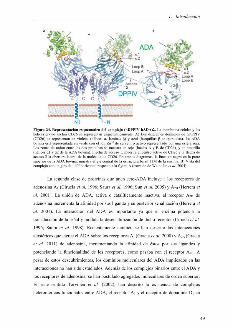
1. Introducción
Figura 24. Representación esquemática del complejo (hDPPIV-bADA)2. La membrana celular y las hélices α que anclan CD26 se representan esquemáticamente. A) Los diferentes dominios de hDPPIV (CD26) se representan en violeta, (hélices α/ láminas β) y azul (horquillas β antiparalelas). La ADA bovina está representada en verde con el ión Zn+2 de su centro activo representado por una esfera roja. Las zonas de unión entre las dos proteínas se muestra en rojo (bucles A y B de CD26), y en amarillo (hélices α1 y α2 de la ADA bovina). Flecha de acceso 1, muestra el centro activo de CD26 y la flecha de acceso 2 la obertura lateral de la molécula de CD26. En ambos diagramas, la línea en negro en la parte superior de la ADA bovina, muestra el eje central de la estructura barril TIM de la enzima. B) Vista del complejo con un giro de ~40º horizontal respecto a la figura A (extraído de Weihofen et al. 2004)
La segunda clase de proteínas que unen ecto-ADA incluye a los receptores de
adenosina A1 (Ciruela et al. 1996; Saura et al. 1996; Sun et al. 2005) y A2B (Herrera et
al. 2001). La unión de ADA, activa o catalíticamente inactiva, al receptor A2B de
adenosina incrementa la afinidad por sus ligando y su posterior señalización (Herrera et
al. 2001). La interacción del ADA es importante ya que el enzima potencia la
transducción de la señal y modula la desensibilización de dicho receptor (Ciruela et al.
1996; Saura et al. 1998). Recientemente también se han descrito las interacciones
alostéricas que ejerce el ADA sobre los receptores A1 (Gracia et al. 2008) y A2A (Gracia
et al. 2011) de adenosina, incrementando la afinidad de éstos por sus ligandos y
potenciando la funcionalidad de los receptores, como pasaba con el receptor A2B. A
pesar de estos descubrimientos, los dominios moleculares del ADA implicados en las
interacciones no han sido estudiados. Además de los complejos binarios entre el ADA y
los receptores de adenosina, se han postulado agregados moleculares de orden superior.
En este sentido Torvinen et al. (2002), han descrito la existencia de complejos
heteroméricos funcionales entre ADA, el receptor A1 y el receptor de dopamina D1 en
49

1. Introducción
neuronas de la corteza cerebral y han postulado que esta agregación podría ser
modulada tanto por dopamina como por adenosina. Por otra parte, Pacheco et al. (2005)
demostraron que el ADA anclada a la superficie de las células dendríticas,
probablemente a través del receptor A2B, se unía a la proteína CD26 expresada en la
membrana de células T, induciendo la coestimulación. Esta señal coestimulatoria
promueve la proliferación de células T con un patrón Th1 y la producción de citocinas
proinflamatorias, con lo que se incrementa la respuesta inmune (Climent et al. 2009;
Martinez-Navio et al. 2009).
50

1. Introducción
1.5 RECEPTORES DE DOPAMI�A Y VÍAS DOPAMI�ÉRGICAS E� EL
SISTEMA �ERVIOSO CE�TRAL
En el sistema nervioso central (SNC) se expresan un elevado número de
receptores acoplados a proteína G: más del 90% de los receptores de siete dominios
transmembrana se expresan en el cerebro y para algunos de ellos su expresión está
restringida a este tejido. La combinación de técnicas de inmunohistoquímica, RT-PCR e
hibridación in situ en diferentes regiones del cerebro ha permitido descubrir que la
expresión de estos receptores presenta patrones diferenciales, lo que sugiere que la
expresión de un grupo de receptores concretos y no otros, es clave en la regulación de
diferentes procesos neurofisiológicos, como la neurotransmisión. Un ejemplo clásico lo
constituyen los receptores de dopamina.
1.5.1 La dopamina como neurotransmisor
La dopamina es la principal catecolamina que actúa como neurotransmisor en el
sistema nervioso central (representa el 80% del contenido total de catecolaminas del
cerebro) y controla una gran variedad de funciones como la modulación de la actividad
sensorial, la actividad motora, la actividad endocrina, el aprendizaje, la memoria, la
emotividad, la afectividad y la motivación. La dopamina también ejerce múltiples
funciones en el sistema periférico como modulador de la liberación de catecolaminas,
de la secreción hormonal, del tono vascular, de la función renal y de la motilidad
gastrointestinal (Cooper et al. 1996; Missale et al. 1998). Como otros
neurotransmisores, la dopamina no es capaz de cruzar la barrera hematoencefálica, pero
sí sus precursores fenilalanina y tirosina. La síntesis de dopamina ocurre en el citosol de
las terminales nerviosas dopaminérgicas tal como se indica en la Figura 25 (Elsworth y
Roth 1997). La liberación de dopamina en la hendidura sináptica tiene lugar mediante
un mecanismo clásico de liberación de neurotransmisores: la entrada de calcio a través
de canales de calcio dependientes de voltaje promueve la fusión de vesículas llenas de
dopamina con la membrana presináptica, formándose un poro y dando lugar a la
exocitosis de la dopamina, que por difusión cruza el espacio de la hendidura sináptica
hasta unirse a sus receptores localizados pre- y postsinápticamente. Después de la unión,
ocurre un cambio conformacional en el receptor que induce una compleja cadena de
eventos intracelulares, y el resultado final de la liberación de dopamina es la activación
o inhibición de la neurona postsináptica.
51

1. Introducción
Figura 25. Síntesis de la dopamina A) La tirosina hidroxilasa emplea oxigeno molecular, tirosina y tetrahidrobiopterina (cofactor) para sintetizar L-DOPA, la cual será descarboxilada por la DOPA descarboxilasa dando lugar a dopamina y CO2. B) Una vez sintetizada, la dopamina es almacenada en vesículas sinápticas hasta su posterior liberación al espacio sináptico. En él, puede interactuar con sus receptores específicos, ser recaptada y degradada (modificado de Kandel et al. 2000)
La señal dopaminérgica finaliza por eliminación de la dopamina del espacio
intersináptico, lo que implica mecanismos de recaptación específicos en el terminal
presináptico donde puede ser almacenada o metabolizada (Elsworth y Roth 1997).
Aunque existen enzimas extraneuronales que catabolizan la dopamina liberada, la
finalización del efecto se debe principalmente, a la recaptación del neurotransmisor por
los propios terminales nerviosos que la liberaron mediante transportadores específicos
(DAT: Dopamine Transporters) que juegan un papel importante en la función,
inactivación y reciclaje de la dopamina liberada (Adell y Artigas 2004; Sotnikova et al.
2006).
Los receptores dopaminérgicos localizados presinápticamente son
principalmente, autoreceptores y constituyen uno de los mecanismos responsables de la
regulación de la transmisión dopaminérgica (Langer 1997; Koeltzow et al. 1998).
Cuando la dopamina es liberada al espacio sináptico, la estimulacion de los
52

1. Introducción
autoreceptores presentes en las terminales nerviosas induce una inhibición de la
liberación continuada de dopamina. Todos los autorreceptores dopaminérgicos
pertenecen a la subfamilia D2-like (Langer 1997; Mercuri et al. 1997; Vallone et al.
2000). Se han desarrollado numerosos agonistas y antagonistas específicos de los
receptores de dopamina lo que ha dado la oportunidad de modular la transmisión
dopaminérgica incrementando o bloqueando la acción de este neurotransmisor con fines
terapéuticos (Sokoloff et al. 2006; Rankin et al. 2010; Rondou et al. 2010). El sistema
dopaminérgico ha sido de gran interés por la relación entre la desregulación de este
sistema y algunas patologías tales como el Parkinson, la esquizofrenia, el síndrome de
Tourette, la hiperprolactinemia y la adicción a drogas (Missale et al. 1998; Segawa
2003; Santini et al. 2008; Dalley y Everitt 2009; Zack y Poulos 2009). De hecho, la
degeneración de la vía nigroestriatal produce la enfermedad de Parkinson en humanos,
caracterizada por una fuerte reducción de la liberación de dopamina (Mercuri et al.
1997; Shimohama et al. 2003).
1.5.2 Estructura, clasificación y función de los receptores de dopamina
Los receptores de dopamina se clasifican en dos subfamilias en función de sus
propiedades bioquímicas y farmacológicas: la subfamilia D1-like, que comprende a los
receptores D1 y D5, y la subfamilia D2-like que incluye a los receptores D2, D3 y D4. Los
receptores D1-like producen incrementos del AMPc intracelular a través de proteínas
Gs/olf que estimulan a la AC y se localizan principalmente en los terminales
postsinápticos (Neve et al. 2004; Nieoullon y Amalric 2002; Missale et al. 1998; Civelli
et al. 1993). Los receptores D2-like, en cambio, inhiben la AC por acoplamiento a
proteínas Gi/o, además de activar canales de K+ y disminuir la entrada de Ca+2 a través
de canales dependientes de voltaje (Gershon et al. 2007; Neve et al. 2004; Nicola et al.
2000; Missale et al. 1998). Los receptores D2-like pueden localizarse en terminales
presinápticas y postsinápticas (Dal Toso et al. 1989). Los dos subgrupos presentan
peculiaridades estructurales diferentes como se muestra en la figura 26: los receptores
D1-like tienen un dominio carboxiterminal unas siete veces más largo que los receptores
D2-like, mientras que estos últimos tienen un tercer bucle intracelular mucho más largo,
característica común en muchos receptores acoplados a proteína Gi (Missale et al.
1998).
53

1. Introducción
Figura 26. Representación esquemática de las dos subfamilias de receptores de dopamina
Existe una homología del 80% en la secuencia entre los dos miembros de la
familia de receptores D1-like. En cambio, la homología es del 75% entre los miembros
D2 y D3 de la familia D2-like y un 53% entre los D2 y D4 de dicha familia. Por contraste,
la homología entre los receptores D1-like y D2-like es solo del 42-46%. La región con
homología más elevada es aquella que se encuentra en los dominios de transmembrana
y en aquellos residuos que son clave para la unión de catecolaminas. El extremo
carboxiterminal, en ambas familias, contiene lugares de fosforilación y palmitoilación
que se cree juegan un papel esencial en la desensibilización del receptor y en la
formación de un cuarto bucle intracelular, respectivamente. Por el contrario, los
receptores de dopamina presentan diferencias en las modificaciones post-
traduccionales, como diferentes lugares consenso de N-glicosilación.
Para el estudio de las propiedades farmacológicas de los receptores de dopamina
se dispone de ligandos que discriminan fácilmente entre las subfamilias D1-like y D2-
like, sin embargo, es mucho más difícil encontrar ligandos selectivos para los miembros
de cada subfamilia. Cada uno de los receptores D1-like muestra alta afinidad por
benzazepinas (SCH23390 y SKF38393) y baja afinidad por butiroferonas (espiperona y
haloperidol) y benzanidas sustituidas (supiride). Se ha detectado una diferencia
remarcable de su afinidad por la dopamina, presentado el receptor D5 una afinidad 10
veces superior a la que presenta el receptor D1 (Missale et al. 1998).
Dentro de los receptores D2-like las afinidades por muchos antagonistas y
agonistas pueden variar en uno o dos órdenes de magnitud. Cada uno de estos
receptores se caracteriza por tener alta afinidad por butiroferonas, como las espiperonas
54

1. Introducción
y el haloperidol y baja afinidad por benzazepinas como el SKF 38393. De los tres
receptores que componen la familia D2-like, el D2 tiene las propiedades farmacológicas
más distintivas, pues en general, presenta baja afinidad por la mayoría de los
antagonistas dopaminérgicos de esta subfamilia, por ejemplo, el raclopride, exhibiendo
a su vez una relativa afinidad por el neuroléptico atípico clozapina (Missale et al. 1998).
En cambio, el receptor D3 presenta una afinidad 20 veces mayor por la dopamina que el
receptor D2, existiendo algunos ligandos comerciales selectivos como el antagonista
GSK 789472. Por último, el receptor D4 es el que presenta menor afinidad por la
dopamina de entre los miembros de la subfamilia D2-like, existiendo también algunos
ligandos sintéticos selectivos para este receptor como el agonista RO 10-5824 o el
antagonista L-745,870.
La diferencia de afinidad que presentan los receptores de dopamina por su
ligando endógeno puede permitir la activación de unos receptores u otros en función de
la cantidad de dopamina liberada. Teniendo en cuenta los diferentes mecanismos de
transducción de señal de cada subtipo de receptor, se puede generar una gran variedad
de respuestas a una misma sustancia. Esta diversidad dentro de los receptores de
dopamina es un reflejo de la diversidad funcional que ejerce este neurotransmisor, sobre
todo si se considera la expresión diferencial de estos receptores dentro del SNC.En la
tabla 5 se resume la información más relevante de los diferentes subtipos de receptores
de dopamina:
Tabla 5. Resumen de las principales características de los receptores de dopamina
55

1. Introducción
La expresión de los distintos subtipos de receptores de dopamina en el cerebro
ha sido determinada mediante la combinación de técnicas de unión de radioligandos y
de hibridación in situ. Así se ha demostrado, por ejemplo, que el receptor D1 es el más
abundante y su distribución es la más amplia de todos los receptores dopaminérgicos
(Beaulieu y Gainetdinov et al. 2011; Barishpolets et al. 2009; Dearry et al. 1990); en
cambio, el receptor D5 presenta una distribución más restringida a regiones tales como
el hipocampo o el tálamo (Centonze et al. 2003). Estudios de RT-PCR y proteómica han
demostrado la expresión del receptor D1 en el estriado (tanto dorsal como ventral), en el
núcleo accumbens, el tubérculo olfatorio, y en menor medida en el sistema límbico, el
hipotálamo y el tálamo. La expresión mayoritaria del receptor D1 se localiza
postsinápticamente en las neuronas GABAérgicas estriatales, junto con la expresión de
la sustancia P (Gerfen et al. 1990). El estudio de Surmeier et al. (1996) mostró que el
receptor D3 se expresa al menos en la mitad de las neuronas GABAérgicas que co-
expresan sustancia P y dinorfina (y que conforman la vía directa
estriatonigroentopeduncular del movimiento) y en una proporción muy pequeña de
neuronas GABAérgicas que expresan encefalina (vía indirecta estriatopalidal). Así, el
receptor D3 podría, de hecho, mediar la mayoría de las interacciones de los receptores
de las familias D1,D2-like en las neuronas estriatales. Marcellino et al. (2008)
evidenciaron la existencia de interacciones a nivel molecular entre los receptores D1 y
D3 en células co-transfectadas y tejido estriatal bovino, mostrando al receptor D3 como
un potenciador de la estimulación de D1 a nivel neuronal y comportamental; también
Fiorentini y colaboradores en el 2008 presentaron resultados que apoyaban la
heteromerizacion D1-D3. Además, el receptor D3, es considerado como una diana
prometedora, por su implicación en las discinesias asociadas al tratamiento con L-
DOPA en la enfermedad de Parkinson (Bordet et al. 1997; Rietschel et al. 2000; Guillin
et al. 2001; Bézard et al. 2003; Visanji et al. 2009; Cote et al. 2014) El receptor D4 se
expresa en interneuronas GABAérgicas tanto piramidales como no-piramidales de la
corteza prefrontal e hipocampo, en el bulbo olfatorio, la amígdala, el mesencéfalo
(Missale et al. 1998), en la glándula pineal (Klein et al. 2010) y en menor medida en el
núcleo accumbens y estriado (de Almeida y Mengod 2010; Gasca-Martinez et al. 2010).
56

1. Introducción
1.5.3 Las vías dopaminérgicas en el S�C
A pesar de que las neuronas que utilizan la dopamina como neurotransmisor en
el cerebro son muy pocas, este sistema de neurotransmisión juega un papel esencial en
la regulación del movimiento, la conducta y liberación de hormonas (Dale 2000). Los
circuitos dopaminérgicos del SNC se pueden dividir en: nigroestriatal, mesolímbico-
mesocortical y tuberohipofisario (Figura 27). Las alteraciones de estas tres vías de
transmisión se han asociado con diversas enfermedades. Así, la enfermedad de
Parkinson se ha asociado con alteraciones en la vía nigroestriatal, la esquizofrenia con
alteraciones en la vía mesolímbica-mesocortical y una gran variedad de alteraciones
hormonales con anomalías en la vía tuberoinfundibular (Dale 2000).
Figura 27. Representación de los circuitos dopaminérgicos (cedido por el Dr. Sergi Ferré)
El sistema nigroestriatal se origina en la sustancia nigra, que es un núcleo de
neuronas localizado en el mesencéfalo. Este se divide en dos partes: la compacta,
formada por neuronas dopaminérgicas, y la reticulata, formada principalmente por
neuronas GABAérgicas. Las neuronas dopaminergicas con origen en la sustancia nigra
constituyen el principal conducto dopaminérgicos en el cerebro, y proyectan axones que
proporcionan una densa inervación al núcleo caudado y al putamen del estriado;
aproximadamente un 80% de toda la dopamina que se encuentra en el cerebro se halla
en el estriado. Este sistema es el implicado en la regulación motora y la ejecución de
tareas, permitiendo que el movimiento se realice de forma armoniosa y obedezca a las
órdenes voluntarias del individuo de acuerdo con patrones motores bien establecidos
(Flórez y Pazos 2003). Un ejemplo es lo que ocurre en los pacientes con Parkinson en
los que se produce una pérdida de neuronas dopaminérgicas de la vía nigroestriatal,
57

1. Introducción
dando lugar a claras anomalías motoras. La inervación dopaminérgica hacia regiones
límbicas y corticales también está alterada, aunque en menor medida y, al parecer, la
enfermedad no se manifiesta hasta que la perdida neuronal en el estriado representa el
80% (Elsworth y Roth 1997).
El sistema mesolímbico-mesocortical tiene su origen en el área tegmental
ventral, también localizada en el mesencéfalo. Dicho núcleo contiene células
dopaminérgicas que envían proyecciones a la corteza frontal y el lóbulo límbico,
conformando los circuitos mesocortical y mesolímbico respectivamente. El sistema
mesolímbico se distribuye por el sistema límbico con excepción del hipocampo;
principalmente se proyecta hacia el núcleo accumbens, tubérculo olfatorio, núcleo
central de la amígdala, septum lateral y núcleo intersticial de la estría terminal (Flórez y
Pazos 2003). El sistema mesocortical se proyecta desde la sustancia nigra y el área
tegmental ventral hacia las cortezas motoras, promotoras y suplementarias y a las
cortezas parietal, temporal y cingular posterior, es decir, hasta las principales aéreas
sensorimotoras y de asociación. Ambos sistemas contribuyen a mantener la atención, la
ideación, la evaluación correcta de la realidad, la motivación y el control del
pensamiento (Flórez y Pazos 2003), es decir, están implicados en todos aquellos
procesos en los que la motivación forma parte esencial de la conducta, ya sea fisiológica
para atender necesidades elementales del individuo, o patológica, creada por
hiperestimulación del sistema, que es lo que ocurre en procesos de adicción a sustancias
de abuso. Los mecanismos implicados en estos últimos procesos se denominan sistemas
de premio o recompensa, ya que son circuitos que al activarse producen un efecto
placentero (Wise 1996). La mayoría de sustancias que provocan adicción, interaccionan
directa o indirectamente con proteínas presentes en las neuronas dopaminérgicos a nivel
de la vía mesolímbico-mesocortical, provocando un incremento de la liberación de
dopamina por la neurona presináptica hacia el espacio extracelular. La continua
administración de estas sustancias produce una activación continua de la liberación de
dopamina, consiguiéndose sensaciones positivas, perdiéndose la sensibilidad a
estímulos habituales. Cuando se interrumpe administración aparecen sensaciones
desagradables, depresión o falta de motivación (Noble et al. 1994) (Figura 28).
El sistema mesolímbico-mesocortical parece jugar un papel importante en el
desarrollo de la esquizofrenia. Las conexiones con el núcleo accumbens tienen una
58

1. Introducción
especial relevancia, ya que la falta de regulación de las vías dopaminérgicas
mesolímbicas provocarían una descoordinación en el núcleo accumbens que, a su vez,
sobreestimularía ciertas regiones implicadas en el procesamiento de la información de
los sentidos, contribuyendo a los síntomas positivos de la esquizofrenia (alucinaciones,
delirios, pensamientos incoherentes,..). Por otro lado, dado que las vías dopaminérgicas
mesocorticales juegan un papel fundamental en el buen funcionamiento cognitivo de la
corteza prefrontal, las alteraciones en este sistema estarían relacionadas con los
síntomas negativos de la esquizofrenia (aislamiento social, retraimiento social, falta de
iniciativa) (Pani 2002; Abi-Dargham 2004).
Figura 28. El circuito mesolímbico (cerebro de roedor) (extraído de Hyman 2007)
Por último, el sistema tuberohipofisario se origina en el hipotálamo y se
proyecta hacia la hipófisis. Las neuronas del sistema tuberohipofisario desempeñan un
papel importante en la regulación de la liberación de las hormonas pituitarias, como por
ejemplo la prolactina, en la que la dopamina juega un papel inhibitorio en la liberación
de esta hormona (Dale 2000).
En las situaciones patológicas que se han comentado anteriormente se han
observado diferencias cuantitativas en cuanto a la expresión de los receptores de
dopamina o bien en su señalizacion. Por ejemplo, los receptores D1 se ven
incrementados en la esquizofrenia y su señalizacion varía en la enfermedad de
Parkinson. La densidad de los receptores D2 localizados postsinápticamente incrementa
en la esquizofrenia y también en los enfermos de Parkinson no tratados con L-DOPA
(profármaco que, a diferencia de la dopamina, puede traspasar la barrera
hematoencefálica, y es un precursor biológico de la dopamina) (Missale et al. 1998;
Vallone et al. 2000; Carlsson et al. 2001; Fuentes et al. 2010; Beaulieu y Gainetdinov
59

1. Introducción
2011). Es por ello que el estudio de los receptores de dopamina es muy importante,
tanto para poder entender una gran cantidad de anomalías funcionales tales como
Parkinson, Alzheimer, esquizofrenia e hiperactividad, como para crear nuevas dianas
terapéuticas para dichas anomalías (Missale et al. 1998; Segawa 2003; Sokoloff et al.
2006; Santini et al. 2008; Dalley y Everitt 2009; Zack y Poulos 2009; Rankin et al.
2010; Rondou et al. 2010).
1.5.4 Los ganglios basales
Los ganglios basales (GB) se hayan en la base del cerebro, y están constituidos
por cinco núcleos principales en roedores: el estriado, la sustancia nigra o negra, el
globus pallidus externo (GPe), el núcleo subtalámico y el núcleo entopeduncular o EPN
(en los primates el globus pallidus interno o GPi). El estriado es la principal estructura
de entrada de los GB y está funcionalmente subdividido en estriado dorsal y ventral (en
rata, Figura 29; en primates no-humanos Figura 30).
Figura 29. Localización del estriado dorsal y ventral en el cerebro de rata. Bregma 2.16mm. CPu: caudado-putamen; AcbC: nucleus accumbens core (extraído de Paxinos y Watson 2005)
60

1. Introducción
Figura 30. Estructura de los GB y del tálamo de Macaca fascicularis. Tinción ACh-E sección coronal 12 ac -5.5mm. CN: núcleo caudado; Put: Putamen; GPi: globo pálido interno; GPe: globo pálido externo (extraído de Lanciego y Vázquez 2012)
Los distintos componentes anatómicos de los GB se encuentran clasificados en
tres grupos: núcleos de entrada, núcleos de salida y núcleos intrínsecos. Como ya se ha
descrito anteriormente, el núcleo de entrada es el cuerpo estriado, que engloba el
estriado dorsal (núcleo caudado y putamen), implicado en la ejecución y aprendizaje de
actos motores complejos, y el estriado ventral (núcleo accumbens), que forma parte de
los circuitos cerebrales implicados en la conversión de la motivación en acción. En el
estriado, más del 90% de las neuronas son GABAérgicas de proyección o medium-size
spiny neurons (MSN) y reciben dos vías de entrada que convergen en sus espinas
dendríticas: por un lado las neuronas dopaminérgicas del mesencéfalo, localizadas en la
sustancia nigra pars compacta y el área ventral tegmental y por otro lado las neuronas
glutamatérgicas procedentes de aéreas corticales, límbicas y talámicas (hipocampo y
amígdala) (Gerfen 2004). Los núcleos de salida comprenden el GPi, ENT y la porción
reticulada de la sustancia negra (SNr). Finalmente, los núcleos intrínsecos son el GPe, el
núcleo subtalámico (STN) y la porción compacta de la sustancia negra (SNc).
Antes de que se ejecute un programa motor, la información es enviada desde la
corteza a los GB, donde la señal se contrasta con entradas sensoriales y se transforma
antes de regresar de nuevo a la corteza a través del tálamo (Schultz 2010).
61

1. Introducción
El modelo de funcionamiento de los GB descrito por Crossman (Crossman
1987), Albin y colaboradores, (Albin et al. 1989) y DeLong (DeLong 1990), explica
cómo se procesa la información a través de los circuitos que constituyen los GB tanto en
condiciones fisiológicas como patológicas (Figura 31).
Figura 31. Funcionamiento simplificado de los GB en primates. Existen dos vías de salida u outputs
del estriado: la vía directa, que proyecta del estriado al núcleo GPi/SNr, y la vía indirecta, que conecta el
estriado con el GPe – STN- GPi/SNr. a) estado “normal” y b) Degeneración de la SNc en la enfermedad
de Parkinson que hace disminuir la liberación de dopamina en el estriado (Cedido por el Dr. Sergi Ferré).
Existen dos subtipos de neuronas GABAérgicas eferentes en el estriado, que
proyectan al tálamo a través de dos vías: las neuronas estriatopalidales externas (del
GPe) y las neuronas estriatonigroentopedunculares en roedores o estriatopalidales
internas (del GPi) en primates. Los dos tipos de neuronas GABAérgicas estriatales se
pueden distinguir neuroanatómicamente. (Levesque y Parent 2005). Así, las neuronas
del estriado que proyectan al GPe forman la vía indirecta, y contienen el péptido
encefalina, receptores de dopamina (predominantemente del subtipo D2) y receptores A1
y A2A de adenosina, entre otros. Las neuronas estriadas que proyectan hacia el GPi, y
que conforman la vía directa, contienen dinorfina, sustancia P, receptores de dopamina
(predominantemente del subtipo D1) (Gerfen et al. 1990; Alexander y Crutcher 1990) y
receptores A1 de adenosina, pero no receptores A2A (Ferré et al. 2007; Schiffmann et al.
2007).
La estimulación de la vía directa produce activación motora, mientras que la de
la vía indirecta produce inactivación motora. Esto es así puesto que la vía directa tiende
62

1. Introducción
a activar los movimientos voluntarios, y la vía indirecta a inhibir la aparición de
componentes involuntarios en el movimiento. Un adecuado equilibrio entre las dos
produce los movimientos normales.
Por tanto, como acabamos de ver, la dopamina liberada en el estriado presenta
un efecto dual, debido a que activa los receptores D1 e inhibe los D2. En primates,
provoca la activación del movimiento por estimulación de los receptores D1 de las
neuronas de proyección de la vía directa, mientras que deprime la actividad de las
neuronas de la vía indirecta actuando sobre los receptores D2 produciendo también,
indirectamente, una activación motora (Alexander y Crutcher 1990). La dopamina por
tanto, estimula el movimiento a través de las dos vías, porque estimula la vía
estimuladora e inhibe a la vía inhibidora (ver Figura 31a).
Aunque en el modelo clásico descrito las neuronas dopaminérgicas de la SNc
proyectan exclusivamente al estriado, se han encontrado proyecciones dopaminéricas de
la SNC hacia otros núcleos de los GB como GPe (Smith y Kieval, 2000), GPi (Jan et al.
2000), STN (François et al. 2000) y varios núcleos talámicos (Sánchez-González et al.
2005).
Por otra parte, según los datos anatómicos, neuroquímicos y electrofisiológicos
publicados en los últimos años, el circuito de los GB no puede considerarse únicamente
como un sistema en el que la información fluye de manera unidireccional desde el
estriado hacia los núcleos de salida a través de una vía directa y otra indirecta. Los
núcleos que componen el circuito forman una red muy compleja donde la información
fluye de manera multidireccional y se originan microcircuitos que regulan de forma
conjunta la señal de salida hacia el tálamo. En la Figura 32 se muestra una visión más
moderna del circuito de los GB en la que se recogen las proyecciones descritas hasta el
momento y que da una idea de la complejidad del sistema (Obeso y Lanciego 2011). En
la actualidad, el esquema de funcionamiento de los GB tiene como base el modelo
clásico descrito anteriormente pero incluye otros núcleos colaterales importantes en la
regulación del circuito y nuevas proyecciones no descritas en el modelo anterior.
63

1. Introducción
Figura 32. Esquema actualizado de las conexiones anatómicas de los núcleos de los GB y núcleos relacionados. Cx, corteza, Str, estriado; GPe, globo pálido externo; GPi, globo pálido interno; STN, núcleo subtalámico; SNpc, sustancia negra pars compacta; SNr, sustancia negra reticulada; Th, tálamo; PPN, núcleo pedunculopontino; CM-PF, núcleo centro mediano-parafascicular del tálamo (modificado de Obeso y Lanciego 2011)
1.5.5 La enfermedad de Parkinson
La enfermedad de Parkinson (EP), descrita por primera vez el 1817 por James
Parkinson, es la segunda patología neurodegenerativa más común en la población, y
está producida por la degeneración progresiva de las neuronas dopaminérgicas
nigroestriatales que proyectan de la sustancia negra al estriado (caudado más putamen).
Esto da lugar a, por un lado, la disminución de la activación de los receptores D1 de
dopamina en las neuronas estriatales de la vía directa, reduciéndose la inhibición que
ejercen estas neuronas sobre los núcleos de salida. Por otro lado, los receptores D2 de
dopamina dejan de estar inhibidos y las neuronas de la vía indirecta están más activas,
aumentando la inhibición que ejercen sobre el GPe (Obeso et al. 2008).
Finalmente, se acaba provocando la inhibición del tálamo, que no enviará la
señal de vuelta a la corteza dando lugar al desarrollo de los síntomas clínicos de la EP
(Figura 33b).
64

1. Introducción
Figura 33. Interconexiones entre las estructuras de los GB en diferentes situaciones patológicas. a) estado sano b) enfermedad de Parkinson c) administración crónica de L-DOPA en patología de Parkinson; 1-vía nigroestriatal 2-vía indirecta inhibidora del movimiento, 3-vía directa excitadora del movimiento, 4-vía corticoestriatal. (Extraído de Jenner 2008)
Los síntomas clínicos más relevantes incluyen bradicinesia (lentitud en los
movimientos), rigidez, temblor en reposo y alteraciones en el equilibrio (Tabla 6).
Cuando se presentan estos síntomas, una gran proporción (50-70%) de las neuronas
dopaminérgicas de la sustancia nigra pars compacta se han perdido, originándose una
reducción de la síntesis y la liberación de dopamina en los terminales que proyectan al
estriado. El distintivo patológico de la EP es la formación de cuerpos de Lewy (CL) en
las neuronas dopaminérgicas que sobreviven. Los CL son inclusiones
intracitoplasmáticas eosinófilas compuestas predominantemente por ubiquitina, α-
sinucleína y parkina (Spillantini et al. 1997; Schlossmacher et al. 2002). Recientemente,
Beyer y colaboradores (Beyer et al. 2010) han destacado la correlación entre las
diferentes isoformas de la α-sinucleína y las enfermedades de CL, entre ellas la EP.
Los principales receptores involucrados en la enfermedad de Parkinson son los
receptores predominantes en las vías alteradas implicadas, los D1 (vía directa) y D2 (vía
indirecta) de dopamina, localizados mayoritariamente en el estriado cerebral (Foley et
al. 2004), aunque otros receptores de GPCR podrían participar en su modulación.
65

1. Introducción
Tabla 6. Principales características de la EP
El factor que se relaciona más significativamente con la aparición de la EP es la
edad o el envejecimiento, aunque se sabe poco cómo está involucrado dicho proceso
(Hindle et al. 2010). La explicación habitual es la mayor vulnerabilidad de las neuronas
dopaminérgicas a causa del fallo fisiológico y bioquímico de los procesos celulares
normales. En general, se ha descrito que la muerte neuronal que tiene lugar en las
enfermedades neurodegenerativas se debe a tres mecanismos moleculares
interrelacionados: estrés oxidativo, disfunción mitocondrial y alteración de los
mecanismos de degradación proteica (Kitada et al. 1998, Malkus et al. 2009, Bove y
Perier 2012). En la etiología de la EP existe también un elemento genético significativo,
al menos en una parte de los pacientes, en los que se han identificado mutaciones en
diferentes genes en lo que se ha denominado casos de enfermedad de Parkinson familiar
(Kitada et al. 1998; Krüger et al. 1998; Leroy et al. 1998; Piccini et al. 1999; Tanner et
al. 1999; Sveinbjornsdottir et al. 2000; Gasser 1998; Bonifati et al. 2002). Por otra
parte, otros autores han estudiado el papel de las modificaciones post-traduccionales
(como la fosforilación, ubicuitinación, nitración y truncamiento) de la α-sinucleína en la
formación de agregados, dentro de la patogénesis de las demencias con cuerpos de
Lewy (DCL) (Beyer y Ariza 2013). Recientemente, fruto del estudio de polimorfismos
genéticos, se ha descubierto el primer biomarcador genético (que comprende hasta 4
polimorfismos de la enzima butirilcolinesterasa) que permite diagnosticar la DCL, y que
se encuentra hasta en un 20% de los casos de esta enfermedad (Beyer 2013); este
hallazgo es importante puesto que en la práctica permite un mejor diagnóstico
diferencial respecto otras demencias.
66

1. Introducción
El tratamiento paliativo de esta enfermedad consiste en suministrar un precursor
de dopamina, la L-DOPA (levodopa o 1-3,4-dihidroxifenilalanina), que aunque es
efectivo en los primeros estadios de la enfermedad, acaba por perder la efectividad y
provoca la aparición de complicaciones motoras como las discinesias (Nutt 1990).
Actualmente, existen avances importantes en el desarrollo de nuevos fármacos
dopaminérgicos y no dopaminérgicos (adrenérgicos, inhibidores de la
monoaminooxidasa, GABAérgicos, etc.) para la EP, así como para las complicaciones
motoras de las terapias en uso (Schapira et al. 2006; Huot et al. 2013).
1.5.6 Discinesias inducidas por el tratamiento de L-DOPA
Aproximadamente del 50% de los pacientes con EP experimentan las discinesias
(movimientos anormales involuntarios) inducidas por L-DOPA (DIL) 4 o 5 años
después del inicio con el tratamiento con levodopa (Fahn 2000). Afortunadamente, el
porcentaje de pacientes que presentan una forma de DIL que empeora sustancialmente
la calidad de vida y requiere de una intervención es mucho menor del 50% (Chapuis et
al. 2005). Con el fin de prevenir o de mejorar la aparición de esta alteración motora, se
reduce la dosis de levodopa (o puede cambiarse a otras formas de liberación sostenida)
y habitualmente se añaden al tratamiento medicamentos no dopaminérgicos, con menor
eficacia anti-parkinsoniana, y que acaban encareciendo el coste farmacológico de la
enfermedad (Maurel et al. 2001).
Las DIL son el resultado de los efectos combinados de la degradación de las
neuronas dopaminérgicas nigroestriatales y del tratamiento pulsátil con levodopa. Así,
aparecen como consecuencia de la hipofunción del GPi/SNr que lleva a la desinhibición
de las neuronas talámicas y a la excesiva actividad de la influencia reforzadora de los
GB sobre la corteza motora (Figura 33c). El tratamiento pulsátil con L-DOPA exagera
más la supersensibilidad de los receptores D1 de dopamina y es la causa de los cambios
en la expresión de genes y proteínas estriatales que establecen y mantienen un estado
propenso a la aparición de las discinesias (Bonito-Oliva et al. 2011; Feyder et al. 2011).
Aunque la causa molecular de las DIL no está del todo clara, diferentes trabajos
han mostrado el receptor de dopamina D3 como una diana prometedora en las DIL, ya
que se encuentra sobreexpresado en los ganglios basales de los modelos de DIL de
roedores y primates no humanos (Todd et al. 1996; Bordet et al. 1997; Bézard et al.
67

1. Introducción
2003; van Kampen y Stoessl 2003; Guigoni et al. 2005). En esta línea se ha visto por
ejemplo que el uso del agonista parcial de D3, BP 897, alivia los síntomas de la
discinesia sin alterar los beneficios del tratamiento con levodopa, mientras que el uso de
antagonistas de D3, tanto selectivos como no selectivos, reducen la disquinesia, a
expensas de empeorar el parkinsonismo (Bézard et al. 2003). Otros candidatos
prometedores que podrían emplearse como adyuvante en la terapia con levodopa, serían
los antagonistas del receptor A2A. El mecanismo de acción de éstos se basaría en que su
uso facilitarían los efectos de la dopamina a través de la vía directa que promueve el
movimiento, mejorando indirectamente los efectos antiparkinsonianos de la activación
del receptor D1 de dopamina (Pinna et al. 1996; Pollack y Fink 1996). Nuestro grupo y
otros, hemos descrito interacciones antagonizantes entre el receptor A2A y los de
dopamina D2 o D3 entre heterómeros (Hillion et al. 2002; Canals et al. 2003; Ferré et al.
2004; Torvinen et al. 2005; Fuxe et al. 2007; Schiffmann et al. 2007). Finalmente, los
receptores de cannabinoides CB1 también tienen un papel clave, llegándose a sugerir
que el bloqueo de receptores CB1 podría prevenir de la bradiquinesia típica del
Parkinson (Garcia-Arencibia et al. 2009; Orgado et al. 2009). Este hecho se basa en los
resultados de Sagredo y colaboradores (Sagredo et al. 2007) que han observado una
regulación a la baja de los receptores CB1, que tendrían un rol de neuroprotección frente
la citotoxicidad estimulada por la progresión de la EP (Sagredo et al. 2007).
1.5.7 Modelos animales para el estudio de la enfermedad de Parkinson
Dado que en la actualidad no existe ninguna cura para la EP y su etiología sigue
siendo desconocida, es necesario estudiar los mecanismos subyacentes a la
neurodegeneración dopaminérgica y buscar nuevas dianas terapéuticas que permitan el
desarrollo de nuevos fármacos para tratar la enfermedad. Para ello, es necesario el uso
de modelos animales que emulen la denervación dopaminérgica y los síntomas
principales de la EP. El modelo perfecto tendría que presentar cuatro características
fundamentales (Beal 2010). En primer lugar, el número de neuronas dopaminérgicas de
la SNc debería ser normal en el momento del nacimiento del animal y debería disminuir
selectiva y gradualmente durante la edad adulta hasta alcanzar una pérdida neuronal de
más del 50%. La muerte neuronal se puede inducir mediante el tratamiento con
neurotoxinas o mediante mutaciones genéticas, en cuyo caso el modelo debería basarse
68

1. Introducción
en una única mutación para facilitar el cruzamiento. En segundo lugar, el animal debería
desarrollar déficits motores que incluyan los síntomas principales de la enfermedad
(bradicinesia, rigidez y temblor en reposo). En tercer lugar, deberían formarse cuerpos
de inclusión a medida que avanza la neurodegeneración. Por último, el desarrollo del
parkinsonismo debería ocurrir en tiempos no más largos de unos pocos meses para
disminuir el coste de los experimentos y permitir así probar distintas estrategias
terapéuticas en menos tiempo. Este modelo animal ideal no existe, por lo que en cada
caso es necesario seleccionar el más adecuado de entre los que hay disponibles.
Los modelos tradicionales de EP se basan en el uso de neurotoxinas que inducen
la muerte selectiva de las neuronas dopaminérgicas. Algunos de estos compuestos son la
reserpina, la metanfetamina, la rotenona, el paraquat, el maneb, la 3-nitrotirosina, la 6-
OHDA y el MPTP (Betarbet et al. 2000, Betarbet et al. 2002; Dauer y Przedborski
2003). Los modelos más utilizados en la actualidad son el modelo de rata lesionada con
6-OHDA y los modelos de ratón y de primate lesionados con MPTP. Por otro lado, tras
la descripción de las mutaciones causantes del parkinsonismo familiar se han generado
nuevos modelos de ratones transgénicos que presentan modificaciones en genes
relacionados con la enfermedad, como por ejemplo la α-sinucleína (Dawson et al.
2010).
1.5.7.1 Lesión dopaminérgica inducida por 6-OHDA
La 6-hidroxidopamina (6-OHDA, Figura 34A) es la toxina más utilizada para
inducir la muerte de las neuronas dopaminérgicas en la rata. La 6-OHDA no atraviesa la
barrera hematoencefálica, por lo que es necesario inyectarla mediante cirugía
estereotáxica para que induzca selectivamente la muerte de las neuronas dopaminérgicas
(Blandini et al. 2008). La citotoxicidad de la 6-OHDA se debe a su actividad pro-
oxidante, ya que tras su inyección es transportada a través del DAT al interior de las
neuronas dopaminérgicas donde promueve la formación de ROS que activan los
mecanismos de muerte neuronal (Figura 35) (Dauer y Przedborski 2003, Betarbet et al.
2002). La lesión obtenida es altamente reproducible y permite controlar el grado de
depleción dopaminérgica según el lugar de la inyección y el tiempo transcurrido desde
la aplicación de la neurotoxina. La inyección de la 6-OHDA en el fascículo
prosencefálico medial (MFB) o en la SN origina una lesión total, en la que la
neurodegeneración comienza a las 24 horas de la inoculación de la toxina. Por otro lado,
69

1. Introducción
la administración de la 6-OHDA en el estriado origina una lesión parcial que es estable
en el tiempo y que permite evaluar la eficacia de compuestos neuroprotectores. El grado
de lesión parcial se puede controlar mediante el número y la localización de las
inyecciones de la 6-OHDA en el estriado (Kirik et al. 1998). Además, en la mayoría de
los casos la inyección es unilateral, de modo que un hemisferio cerebral no presenta
pérdida neuronal y se utiliza como control interno. Este tipo de lesión unilateral provoca
una conducta rotacional asimétrica que depende del grado de lesión dopaminérgica y
que se puede evaluar tras la inyección de anfetamina o apomorfina. Esto supone una
gran ventaja ya que permite cuantificar el grado de lesión in vivo (Kirik et al. 1998,
Yuan et al. 2005). Sin embargo, este modelo no presenta una de las características
principales de la enfermedad: la presencia de cuerpos de inclusión proteicos.
Figura 34. Estructura química de la 6-OHDA (A) y del MPTP (B)
1.5.7.2 Lesión dopaminérgica inducida por MPTP
El MPTP (Figura 34B) es una de las neurotoxinas más utilizadas para generar
modelos animales de EP. Tras su descubrimiento en un grupo de toxicómanos de San
Francisco, se ha estudiado ampliamente el mecanismo de acción por el cual causa la
degeneración selectiva de las neuronas dopaminérgicas. El MPTP es un compuesto
lipofílico que atraviesa la barrera hematoencefálica con facilidad. Una vez en el SNC, el
MPTP es oxidado en células gliales a 1-metil-4-fenil-2,3-dihidropiridina (MPDP+) en
una reacción catalizada por la enzima MAO-B. Tras la oxidación, el MPDP+ se
convierte espontáneamente en MPP+, que es el compuesto activo tóxico. El MPP+ es
una molécula polar y no puede atravesar la membrana plasmática, por lo que necesita un
transportador para entrar o salir de las células. El mecanismo por el cual el MPP+ es
transportado desde la célula glial al espacio extracelular es desconocido, pero en cambio
sí se sabe que es captado por las neuronas dopaminérgicas a través del DAT (Blum et
al. 2001). Una vez en el interior de la neurona, el MPP+ inhibe el complejo I de la
70

1. Introducción
cadena respiratoria mitocondrial, impidiendo la fosforilación oxidativa, disminuyendo
el contenido de ATP de la célula y estimulando la producción de ROS (Figura 35)
(Dauer et al. 2003, Betarbet et al. 2002, Sedelis et al. 2000, Singer et al. 1988, Jakowec
et al. 2004). El MPTP se ha utilizado para inducir la muerte de las neuronas
dopaminérgicas en distintas especies animales, como gatos, ratones o primates.
Figura 35. Mecanismos de acción de las toxinas usadas para la inducción de síntomas
parkinsonianos en los diferentes modelos animales. (Extraído de Schober 2004)
71

1. Introducción
1.6 EFECTOS DE LA COCA�A MEDIADOS POR LOS RECEPTORES DE
DOPAMI�A D1 Y D2
La cocaína es un extracto purificado de la planta de coca, Erythroxylum coca,
procedente originariamente de América del sur (Figura 36). Las hojas de coca han
formado parte de las culturas Inca, Ayamara y Quechua durante siglos. Para conseguir
los efectos estimulantes, de euforia y eliminación del apetito, las hojas de coca eran
masticadas. Originariamente, la coca era administrada, exclusivamente, en forma de
hoja hasta que en 1860, Albert Neiman aisló un extracto de la hoja de coca, la cocaína.
Su utilización se extendió rápidamente por todo el mundo y hoy en día su posesión,
cultivo y distribución son ilegales, exceptuando requisitos médicos o normas
gubernamentales; sin embargo, su uso está ampliamente extendido. Actualmente, la
adicción a cocaína es un problema social y resulta complicado encontrar un buen
tratamiento debido al alto grado de recaída que presenta el consumo de esta sustancia.
Figura 36. Cocaína. A) La planta Erytroxylon coca contiene cocaina en sus hojas B) Estructura molecular del [1R-(exo,exo)]-3-(benzoyloxy)-8-methyl-8-azabicyclo[3.2.1]octane-2-carboxylic acid
methyl ester, también conocida como cocaína. C) Anuncio de unas gotas para el dolor de muelas, que contenían cocaína (1885)
Se conoce desde antiguo que la cocaína actúa inhibiendo la recaptación de
monoaminas como la dopamina (Moore y Gudelsky 1977; Heikkila et al. 1979; Ritz et
al. 1987), norepinefrina (Moore y Gudelsky 1977) y serotonina (Ross y Renyi 1967).
Sin embargo, aunque la cocaína actúa con el mismo grado de efecto en los tres
transportadores, la mayoría de los efectos en el comportamiento (De Wit y Wise 1977;
Colpaert et al. 1978; Miczek y Yoshimura 1982) y la actividad motora (Giros et al.
1996) de esta sustancia se atribuyen al bloqueo de la recaptación de dopamina por
inhibición del transportador de dopamina (DAT) presináptico. Una vez administrada, la
cocaína muestra una acción rápida aumentando los niveles de dopamina en el espacio
72

1. Introducción
sináptico, lo que causa una sobreestimulación de las vías dopaminérgicas (Figura 37).
Los picos de dopamina pueden aparecer a los cinco minutos y no recuperarse los niveles
basales hasta los treinta minutos (Bradberry 2000). Además, la vida media de la cocaína
en rata es corta, puede oscilar entre quince minutos y una hora dependiendo de la vía de
administración utilizada (Nayak et al. 1976) y se cree que una administración repetitiva
podría prolongar los altos niveles de dopamina en el espacio sináptico y la vida media
de esta sustancia (Volkow et al. 1999). Se considera que la hidrolisis del éster de
cocaína es la principal ruta de su catabolismo.
Analizando los efectos de la cocaína desde una perspectiva morfológica, Roberts
y colaboradores describieron una inhibición en los efectos de recompensa de la cocaína
en ratas con el núcleo accumbens lesionado (Roberts et al. 1977). Posteriormente, en
estudios de ratas adictas a la cocaína, se escogió el córtex prefrontal como región
implicada en la autoadministración de microinyecciones de cocaína (Goeders y Smith
1983). Por otro lado las vías nigroestriatales controlan gran variedad de funciones
motoras, además de contener la concentración más elevada de neuronas
dopaminérgicas. Estos datos convierten al córtex prefrontal y las vías nigroestriatales en
las regiones más importantes en la integración de los efectos de la cocaína, aunque no
son las únicas (Bardo 1998). El consumo de cocaína aumenta los niveles de dopamina
en el estriado, principalmente en la parte ventral, y más concretamente en el núcleo
accumbens, el cual ha sido descrito como parte anatómica preferencial en mecanismos
de recompensa (Koob 2006; Di Chiara y Bassareo 2007). La cocaína hace uso del
sistema dopaminérgico para generar parte de sus efectos celulares y de comportamiento
(De Mei et al. 2009). En ratas, la shell del núcleo accumbens es requerida para la
adquisición inicial de la conducta de autoadministración de cocaína mientras que es el
core el que se encarga de la adquisición de la conducta de búsqueda condicionada a
estímulos asociados a dicha droga (Ito et al. 2004). Del mismo modo, una vez que los
estímulos de recompensa asociados a cocaína están consolidados, es el estriado dorsal el
que juega un papel central (Everitt y Robbins 2005). Es decir, se hipotetiza que se pasa
progresivamente de una búsqueda motivada por una recompensa (conducta dependiente
del núcleo accumbens) a hábitos estimulo-respuesta que dependen del estriado dorsal.
Un hipotético mecanismo de los cambios de comportamiento consecuentes a la
administración crónica de cocaína es una alteración de la plasticidad sináptica del
73

1. Introducción
cerebro. Hasta el momento, han sido descritos diferentes cambios en la estructura de las
dendritas (Robinson y Kolb 1999; Robinson et al. 2001). Estos cambios podrían ser
debidos a alteraciones en la expresión de las proteínas de los neurofilamentos, las
proteínas del citoesqueleto y/o las gap junctions, todas ellas muy importantes para la
estabilidad y la buena integración de los receptores en la sinapsis neuronal.
Figura 37. Efecto del bloqueo de los transportadores de dopamina (DAT) por la cocaína (extraído del articulo Stimulant Addiction del
<acional Institute on Drug Abuse (NIDA))
1.6.1 Proteínas de unión de la cocaína
La cocaína ejerce sus funciones por interacción con proteínas específicas. Como
hemos comentado anteriormente, la proteína más reconocida que puede unir cocaína es
el transportador de dopamina DAT. Se ha estudiado a nivel molecular la interacción de
la cocaína con DAT. Mediante el modelaje molecular de DAT basado en la estructura
cristalina de Aquifex aeolicus LeuT(Aa), un transportador de leucina homologo en
bacterias (Ravna et al. 2009), se ha postulado que la unión de la cocaína a DAT ocurre
en un dominio de unión análogo al lugar de unión de la leucina en el transportador
LeuT(Aa).
En el centro de interacción de la cocaína con DAT intervienen las hélices
transmembrana 1, 3, 6 y 8 del transportador. Este centro de unión se superpone con el
sitio de unión de la dopamina y las anfetaminas, pero es claramente diferente del lugar
de unión de distintos antidepresivos (Beuming et al. 2008). Este hecho explica el
bloqueo de la unión de la dopamina a DAT inducido por la cocaína y,
74

1. Introducción
consecuentemente, el incremento en los niveles de dopamina en el espacio extracelular
(Figura 38).
Figura 38. Esquema de la interacción de la cocaína y la dopamina con DAT. A) Representación del transportador de dopamina, DAT. Los círculos pintados corresponden a los lugares donde coincide la interacción de DAT con la dopamina y la cocaína. B) Esquema de la interacción de DAT con la dopamina. C) Esquema de la interacción de DAT con la cocaína (extraído de Bertolino et al. 2009)
Mediante estudios in vitro (Bertolino et al. 2009) e in vivo (Meiergerd et al.
1993; Dickinson et al. 1999; Mortensen y Amara 2003), se ha podido demostrar que
existe una relación directa entre DAT y el receptor D2 de dopamina ya que ambos se
regulan de forma reciproca a nivel presináptico. Bertolino y colaboradores han
demostrado que se produce una interacción molecular entre DAT y los receptores D2
(Bertolino et al. 2009). Esta interacción puede ser fundamental para entender como la
señalizacion de la dopamina se ve claramente regulada, a nivel presináptico, por DAT y
por el receptor de dopamina D2 en el estriado y en el córtex prefrontal.
75

1. Introducción
Además de DAT, la cocaína puede interaccionar con otras proteínas. Hoy en día
es un hecho aceptado que la cocaína interacciona con los receptores sigma a
concentraciones fisiológicas (Cobos et al. 2008). Esta familia de proteínas está formada
por los receptores sigma-1 y sigma-2. La cocaína es la droga de abuso mas estudiada
con referencia a su interacción con los receptores sigma-1 (Hayashi y Su 2005). Una de
las razones es que la cocaína posee una afinidad moderada por sigma-1 en ensayos de
unión de radioligando (Sharkey et al. 1988; Matsumoto et al. 2003). Matsumoto y
colaboradores describieron que los antagonistas del receptor sigma inhiben de forma
significativa las convulsiones y la letalidad inducidas por dosis tóxicas de cocaína
(Matsumoto et al. 2002, Matsumoto et al. 2004). La toxicidad de la cocaína se ve
potenciada con los agonistas del receptor sigma-1 (Matsumoto et al. 2002, Matsumoto
et al. 2004). Este descubrimiento indica que las acciones de la cocaína, al menos en
parte, pueden ser mediadas por su unión al receptor sigma-1. La estructura del receptor
sigma-2 aún no se conoce. Por otro lado, la estructura de sigma-1 es muy distinta de la
de cualquier otra proteína conocida de mamíferos. Aunque muestra un 66% de
homología con una esterol isomerasa fúngica, carece de su actividad enzimática (Su y
Hayashi 2003). Se caracteriza por poseer tres regiones altamente hidrofóbicas que
formarían dos segmentos transmembrana con los extremos amino y carboxilo
terminales en el espacio intracelular (Figura 39).
Figura 39. Esquema de la estructura del receptor sigma-1 (extraído de Steve Beyer’s blog 2009)
El receptor sigma-1 es una chaperona que modula las señales dependientes de
calcio y se localiza principalmente en el retículo endoplasmático de la célula, aunque
también se encuentra en la membrana plasmática, nuclear y mitocondrial (Alonso et al.
76

1. Introducción
2000). Se encuentra ampliamente distribuido por todo el SNC y la periferia. En 1976
fue clasificado como receptor opioide (Martin et al. 1976); sin embargo, la acción de
los ligandos de σ-1 no era bloqueada por los antagonistas opioides, naloxona y
naltrexona, por lo que fue considerado como un receptor huérfano no opioide. El
receptor sigma interacciona con diferentes sustancias, además de la cocaína, entre las
que destacan el haloperidol o los esteroides como la progesterona (Hayashi y Su 2003).
Muy recientemente se ha descubierto un alucinógeno endógeno que interacciona
con el receptor sigma-1, el DMT (<,< dimethyltryptamine). Esta sustancia actuaría
como agonista endógeno del receptor inhibiendo los canales de Na+ dependientes de
voltaje en los miocitos (Guitart et al. 2004; Fontanilla et al. 2009). El receptor sigma-1
también está implicado en la modulación de la liberación de calcio, la modulación de la
contracción cardiaca y la inhibición de los canales de K+ dependientes de voltaje
(Monassier y Bousquet 2002). En 2001 Hayashi describió la interacción del receptor
sigma-1 con los receptores IP3 en el retículo endoplasmático (Hayashi y Su 2001). Más
adelante, se demostró que la activación del receptor sigma-1 mediante sus agonistas
(PRE-084 o carbetapentane) fosforila la subunidad NR1 del receptor NMDA vía PKC y
PKA aumentando su expresión y reforzando la inducción de dolor a través del receptor
NMDA. Esta señal se veía reducida por el tratamiento con el antagonista especifico del
receptor sigma-1 (Kim et al. 2008). Hasta el momento no se ha encontrado ninguna
interacción de estos receptores con ningún GPCR, aunque existen numerosas evidencias
de la amplificación de señales de GPCR mediante los receptores sigma-1. Actualmente,
son considerados como receptores auxiliares o amplificadores de otras señales.
El receptor sigma-1 se encuentra estrechamente relacionado con la acción de la
cocaína en diferentes aspectos, como son la hiperlocomoción (Menkel et al. 1991), la
sensibilización (Ujike et al. 1996), el mecanismo de recompensa (Romieu et al. 2000;
Romieu et al. 2002), las convulsiones y la letalidad (Matsumoto et al. 2001a), aunque se
desconoce el mecanismo de acción de todos ellos. Estudios recientes han descrito que
una reducción del receptor sigma-1 en cerebro, mediante oligonucleótidos anti-sense,
disminuye las acciones convulsivas y locomotoras estimulantes de la cocaína
(Matsumoto et al. 2001b; Matsumoto et al. 2002). Por otro lado, antagonistas sintéticos
del receptor sigma-1 reducen las acciones de la cocaína en modelos animales
(Matsumoto et al. 2003). A pesar de que recientemente han aparecido estudios que
77

1. Introducción
también involucran el receptor sigma-2 con las acciones de la cocaína (Matsumoto et al.
2007), su papel aún no está definido debido a la falta de ligandos completamente
selectivos para este subtipo. Para añadir complejidad, el receptor sigma-2 aun no ha sido
clonado.
1.6.2 Implicación de los receptores de dopamina D1 y D2 en los efectos de la
cocaína
A pesar de que se ha visto que la administración crónica de cocaína induce
cambios en la expresión génica de muchos receptores como el receptor metabotrópico 5
de glutamato (mGluR5) (Ghasemzadeh et al. 1999) o el receptor µ-opioide (Yuferov et
al. 1999), estudios en humanos postmortem no indicaron ningún cambio en el ARNm
de los receptores de dopamina D1 o D2 en individuos adictos a la cocaína con referencia
a los controles (Little et al. 1993; Meador-Woodruff et al. 1993). Estos resultados han
sido controvertidos ya que se ha demostrado una disminución de la expresión de los
receptores D1 y D2 de dopamina en individuos adictos a la cocaína en comparación con
individuos sanos mediante la técnica de PET (Positrón Emisión Tomography) (Martinez
et al. 2004; Martinez et al. 2009; Tsukada et al. 1996). La variación de la expresión de
receptores de dopamina inducida por cocaína puede variar mucho según la especie y la
región del cerebro, ya que en ratas, después de cuatro semanas de autoadministración de
cocaína, se observo un incremento del ARNm del receptor D1 en el telencéfalo anterior
y un incremento del ARNm de los receptores D1 y D2 en la región límbica (Laurier et al.
1994).
A parte de la modificación o no de la expresión de los receptores, la cocaína
induce un incremento de la liberación de dopamina y una sobreestimulación de las vías
dopaminérgicas (Anderson y Pierce 2005; De Mei et al. 2009). Mediante la
potenciación de la transmisión dopaminérgica en el estriado, la cocaína induce los
efectos de recompensa así como la aparición de nuevos estímulos (Volkow et al. 1995;
Volkow y Swanson 2003; Zink et al. 2003). Navarro y colaboradores (Navarro et al.
2010a) han obtenido recientemente nuevos resultados que aportan una explicación
molecular por la cual los heterómeros D1R-sigma-1 desempeñan un papel importante en
los efectos comportamentales de la cocaína. Estos resultados, proporcionan una nueva
78

1. Introducción
perspectiva para entender las bases moleculares involucradas en la adicción a la
cocaína.
Los receptores D2 también están involucrados en la mediación de estos efectos
de recompensa. Los agonistas del receptor D2 reducen la autoadministración de cocaína,
mientras los antagonistas incrementan este comportamiento. Estos resultados sugieren
que el receptor D2 actúe por un mecanismo feed-back para disminuir la
autoadministración de cocaína (Corrigall y Coen 1991; Caine et al. 1999).
Los antagonistas glutamatérgicos y dopaminérgicos reducen la activación de la
transcripción génica inducida por la cocaína (Konradi 1998; Valjent et al. 2005). La
activación de los receptores de dopamina D1 es un requerimiento imprescindible para la
respuesta celular y conductual inducida por la cocaína tal como se demuestra en
estudios realizados con ratones KO para el receptor D1 (Xu et al. 1994). Estudios
recientes utilizando ratones transgénicos, donde las células que expresan los receptores
D1 y D2 se encuentran marcadas mediante proteínas fluorescentes, han confirmado estos
datos, mostrando que la respuesta celular aguda inducida por la cocaína involucra
principalmente las neuronas que expresan los receptores D1 de dopamina (Bertran-
Gonzalez et al. 2008). En este escenario cabría esperar que una inhibición de la
expresión génica del receptor D2S amplificaría los efectos de la cocaína in vivo, debido
a la inhibición ejercida por el receptor D2S de la liberación de dopamina. Sin embargo,
no es esto lo que se observa. El efecto de la cocaína en ratones KO para receptores D2
ha sido estudiado tanto en tratamientos agudos, crónicos como en autoadministración de
cocaína, con el resultado que los ratones KO para D2 tienen alteradas las respuestas a
cocaína. Así, la estimulacion de la actividad motora inducida por cocaína en los ratones
KO para receptores D2 no incrementa de forma dosis-dependiente (Chausmer et al.
2002; Welter et al. 2007). De forma sorprendente, la administración de cocaína en los
ratones KO para D2 no induce la expresión de c-fos (Centonze et al. 2002). Esto
conduce a hipotetizar que en ausencia del receptor D2, aparece un circuito inhibitorio,
normalmente controlado por los receptores D2, provocando la supresión de la inducción
de c-fos. En este contexto, el GABA y la acetilcolina pueden aumentar de forma
considerable debido a la pérdida del control de su liberación por el receptor D2 y jugar
un papel en el bloqueo de la inducción de c-fos (Centonze et al. 2002). De forma
alternativa, la perdida de los receptores D2 afecta la formación de complejos
79

1. Introducción
macromoleculares entre los receptores D2 y otras proteínas que normalmente controlan
la respuesta celular y conductual de la cocaína (Liu et al. 2006).
Los efectos de recompensa de la cocaína en los ratones KO para receptores D2 se
ven atenuados (Welter et al. 2007). Sin embargo, estudios de autoadministración de
cocaína en ratones KO para el receptor D2 demostraron que los ratones KO se
autoadministraban más cocaína que los ratones WT (Caine et al. 2002). No puede
excluirse la contribución de otros neuromoduladores (como por ejemplo la
noradrenalina o la serotonina) y se requiere un análisis más profundo para esclarecer
este fenómeno. Este punto es importante debido a la existencia de numerosos estudios
que muestran una pérdida de los efectos de recompensa de diferentes drogas de abuso
en ratones KO para receptores D2. (Risinger et al. 2000; Elmer et al. 2005).
Por otro lado, los ratones KO para D2L, que por lo tanto siguen expresando los
autoreceptores D2S, mantienen la respuesta locomotora y de recompensa a la cocaína
parecida a los animales WT (Usiello et al. 2000; Wang et al. 2000; Rouge-Pont et al.
2002). Así, parece ser que el receptor D2S es el principal implicado en la respuesta
celular y conductual de las drogas de abuso. Esto sugiere que los efectos presinápticos
de los receptores D2 no involucran únicamente la liberación de dopamina, sino también
de GABA, glutamato y acetilcolina en respuesta a las drogas de abuso. Así, existe una
diferente implicación de las isoformas D2S y D2L en la señalizacion dopaminérgica
inducida por drogas de abuso. La ausencia de la senalizacion del receptor D2L no altera
los efectos motores y de recompensa inducidos por la cocaína. Contrariamente, la
señalizacion del receptor D2S parece ser un requisito imprescindible para los efectos
motores y de recompensa de la cocaína y otras drogas de abuso. De todos modos, se
requieren más estudios para poder decidir que componente presináptico se encuentra
involucrado en estas respuestas y si se encuentra en las neuronas dopaminérgicas o en
las neuronas postsinápticas.
No se han observado cambios significativos en los niveles de expresión del
receptor D2 en el estriado dorsal en animales con diez días de alto consumo de cocaína
(Marcellino et al. 2007). Este es un descubrimiento importante que indica que cambios
en la expresión de los receptores D2 en esta zona del cerebro no están involucrados en la
adaptación de la respuesta a los efectos de recompensa de la cocaína en este modelo.
80

1. Introducción
Recientemente, cambios bifásicos en los receptores D2 en el core del núcleo accumbens
han sido observados después de una retirada de pocos días de acceso a cocaína
intravenosa (Ben-Shahar et al. 2007). Parece ser, que la dosis y la exposición a la
cocaína son las responsables de estas diferencias. La autoadministración crónica de
cocaína en monos rhesus por periodos de varios meses provoca una disminución de la
densidad del receptor D2 en el estriado detectada por autoradiografía (Moore et al. 1998;
Nader et al. 2002). Mediante estudios de imágenes se ha podido detectar una
disminución en la cantidad de receptores D2 en humanos adictos a la cocaína comparado
con los sujetos controles (Volkow et al. 1993; Martinez et al. 2004). De forma similar,
mediante la técnica de PET se ha observado una reducción en los receptores D2 en
primates con autoadministración crónica de cocaína y un ano de abstinencia (Morgan et
al. 2002a; Morgan et al. 2002b; Nader et al. 2006). Una persistente disminución de los
receptores D2 en el estriado puede involucrar la internalización de los receptores D2
después de una autoadministración crónica de cocaína.
Todos estos estudios, nos llevan a pensar en la existencia de un delicado balance
entre la acción de los receptores D1 y D2 en el cerebro, que está controlando tanto los
procesos motivacionales como los de refuerzo en y durante el consumo de las drogas, y
especialmente en el caso de la cocaína.
81

1. Introducción
1.7 SISTEMA E�DOCA��ABI�OIDE
El cannabis, comúnmente conocido como “marihuana”, es una de las drogas de
abuso más extendidas entre la población europea. Su uso intensivo se ha relacionado
con diversos problemas neurológicos no específicos. En contrapunto, el sistema
endocannabinoide, descubierto a partir de la identificación de las sustancias psicoactivas
del cannabis, se ha puesto en el punto de mira de la sociedad en los últimos años como
posible diana terapéutica para muchas enfermedades de tipo neurodegenerativo y
tumoral. Si bien los efectos y propiedades terapéuticas del cannabis ya eran harto
conocidos por antiguas civilizaciones, no fue hasta 1964 que se consiguió aislar el ∆9-
tetrahidrocannabinol (∆9-THC), principal constituyente psicoactivo del cannabis, a
partir de preparaciones de Cannabis Sativa (Mechoulam y Gaoni 1965; Gaoni y
Mechoulam 1971) que, aún a día de hoy, se utiliza para combatir la emesis y la falta de
apetito en los enfermos de cáncer. El carácter lipofílico de esta sustancia hacía pensar
que ejercía sus efectos de manera inespecífica, interaccionando con los lípidos de la
membrana celular. Sin embargo, unos veinte años más tarde, Howlett y colaboradores
(Howlett et al.1990) describieron centros de unión de alta afinidad para el ∆9-THC en
membranas celulares, lo que sugirió que su acción podía estar mediada por receptores
específicos. Hasta ahora, se han clonado y caracterizado inequívocamente dos de estos
receptores: los receptores CB1 y los receptores CB2 (Figura 40). El primer receptor de
esta familia, el CB1, se encontró a partir de un screening de GPCR huérfanos (Matsuda
et al. 1990) y su localización fue predominantemente cerebral y testicular. El
descubrimiento del segundo receptor, el CB2, fue posterior e, inicialmente, su
localización se restringió a la periferia, principalmente en el sistema inmune (Pertwee
1997). Hoy en día se sabe que, si bien es cierta su expresión predominante en células
inmunológicas, los receptores CB2 aparecen también en el sistema nervioso central y su
acción es clave para entender ciertas enfermedades cerebrales, al mismo tiempo que
suponen una diana terapeútica clave en su tratamiento (Rivers y Ashton 2010). Si bien
persiste la idea de que los receptores CB1, por su abundancia y ubicuidad, son la
principal diana de acción de los cannabinoides en el sistema nervioso central, existen
evidencias de que no todas las acciones de los cannabinoides son mediadas por este
receptor y que, probablemente, algunas puedan ser atribuidas a los CB2, o incluso otro
tipo de receptores capaces de mediar los efectos neuronales de los cannabinoides como
82

1. Introducción
el receptor huérfano GPR55 (Ryberg et al. 2007; Pertwee 2007; Sharir y Abood 2010;
Martínez-Pinilla et al. 2014).
La identificación, caracterización y localización de receptores específicos,
capaces de reconocer al ∆9-THC, planteó la cuestión de cuál o cuáles debían ser los
ligandos endógenos capaces de interaccionar con estos receptores, es decir, la posible
existencia endógena de sustancias similares al ∆9-THC que, actuando a modo de
agonistas, mimetizaran sus efectos en el organismo. Aunque en los últimos años han
sido numerosos los endocannabinoides aislados, tanto de tejido nervioso como
periférico, los más importantes son los derivados de eicosanoides como la anandamida
(AEA) (Devane et al. 1992) y el 2-araquidonilglicerol (2-AG) (Mechoulam et al. 1995).
A partir de su identificación, se investigó la ruta bioquímica de su síntesis, liberación
(Di Marzo et al. 1994; Cadas et al. 1996), transporte (Beltramo et al. 1997) y
degradación (Cravatt et al. 1996). Todo ello permitió denominar con el término de
“sistema endocannabinoide” a un conjunto de componentes que comprende: i) Los
receptores de cannabinoides, ii) los ligandos endógenos o endocannabinoides y iii) los
enzimas implicados en su síntesis y degradación. En la actualidad, este sistema aparece
como un relevante modulador fisiológico, no sólo a nivel del sistema nervioso central,
sino también en el periférico y en otros muchos sistemas del organismo.
Figura 40. Estructura de los receptores CB1 y CB2. En negro se representan los aminoácidos comunes en ambos receptores, y los lugares de glicosilación están marcados con el símbolo ψ (extraído de Shire et
al. 1996)
83

1. Introducción
1.7.1 Receptor de cannabinoides CB1
El cDNA de los receptores CB1 fue aislado por primera vez de una librería de
corteza cerebral de rata. El locus del gen en humanos se ha localizado en el cromosoma
6 en la posición 6q14-q15 (Caenazzo et al. 1991; Hoehe et al. 1991). La proteína
codificada posee una alta homología en nucleótidos y aminoácidos entre humanos y
roedores. Pertenece a la familia A de los GPCR, aunque difiere de los receptores de este
grupo en que no presenta el puente disulfuro en el EC2 ni el residuo de prolina en el
TM-5. Puede presentar hasta tres lugares de glicosilación, de tal manera que su peso
puede variar hasta en 10kDa.
Como GPCR, los receptores CB1 se pueden unir tanto a proteína Gi como Go a
través del extremo carboxi-terminal intracelular. Los aminoácidos más próximos a la
membrana de esta región son decisivos para el acoplamiento de la proteína Gi/o,
mientras que los más distales modulan la amplitud y la cinética (Nie y Lewis 2001a; b).
La cola C-terminal está implicada, también, en procesos de desensibilización e
internalización. De esta manera, los aminoácidos de la zona central del extremo carboxi-
terminal contienen residuos fosforilados necesarios para la desensibilización mediada
por quinasas específicas (GRK) y arrestinas (Jin et al. 1999), mientras que cuatro
residuos del final de la cola son importantes en la internalización (Hsieh et al. 1999). La
unión a la proteína G de estos receptores en los terminales presinápticos da lugar a una
inhibición de la adenilato ciclasa, disminuyendo así la producción de AMPc; si bien
esto puede variar en función del receptor con el que heteromerice. Así pues, se observó
que, en presencia de los receptores de dopamina D2, los receptores CB1 se unen a la
proteína Gs estimulando la producción de AMPc (Kearn et al. 2005).
Los receptores CB1 están implicados, además, en otras vías de señalización,
dependiendo de las células en las que se encuentren, estimulando a las quinasas
activadoras de mitógenos (MAPK) tales como ERK 1/2, p38 y c-Jun (JNK), la vía de
PI3K/Akt, además de la expresión de genes tempranos (ej. Krox-24) (Turu y Hunyady
2010). La desensibilización del receptor CB1 se ha descrito que está mediada por un
mecanismo de internalización a través de la acción de β–arrestinas (Breivogel et al.
2008; Daigle et al. 2008). Todos estos mecanismos de señalización pueden mediar los
efectos de los canabinoides como reguladores de la liberación de neurotransmisores. De
84

1. Introducción
esta manera, los endocannabinoides funcionan como mensajeros retrógrados cuando sus
receptores diana se encuentran en la membrana presináptica, inhibiendo la liberación de
glutamato (Shen et al. 1996), GABA (Szabo et al. 1998), acetilcolina (Gifford et al.
1997), noradrenalina (Schlicker et al. 1997a) y serotonina (Nakazi et al. 2000).
El sistema cannabinoide es especialmente relevante en las funciones cognitivas y
emocionales; está implicado en circuitos neuronales de la corteza, hipocampo y
amígdala y en la adicción a drogas en el sistema mesolímbico mediante mecanismos de
modulación sináptica que incluyen DSI (Depolarization-Induced Supression of
Inhibition), en neuronas GABAérgicas, y DSE (Depolarization-Induced Supression of
Excitation), en neuronas glutamatérgicas (Parolaro y Rubino 2002; Gerdeman y
Lovinger 2003; Laviolette y Grace 2006). Aunque la activación presináptica de los
CB1R casi siempre conlleva una disminución en la liberación del neurotransmisor
(Freund et al 2003a), la magnitud de su acción se puede dividir en dos mecanismos
diferentes con respecto a su potencia y su implicación patofisiológica: la depresión a
corto plazo o short-term depression (STD) y la depresión a largo plazo o long-term
depression (LTD). Mientras la primera es de corta duración (menos de un segundo) y
rápida aparición, la segunda, por estimulación prolongada de los receptores de CB1, es
de aparición más tardía y suele durar varias horas (Katona y Freund 2008; Sidhpura y
Parsons 2011).
Los receptores CB1 son unos de los receptores neuronales más abundantes en el
cerebro de mamíferos. De hecho, son considerados los GPCR más abundantes del CNS.
La distribución regional de los CB1 ha sido caracterizada en el cerebro humano y de rata
en correlación con los efectos de los cannabinoides en el comportamiento como el
humor, la coordinación motora, funciones autonómicas, aprendizaje y memoria,
percepción sensorial y mecanismos del dolor, entre otros (Glass et al. 1997b; Westlake
et al. 1994; Valverde 2005).
En cuanto a la distribución en el sistema nervioso, los receptores CB1 se
encuentran en alta densidad, tanto en sinápsis glutamatérgicas como gabaérgicas, en
hipocampo, amígdala, hipotálamo, algunas regiones olfatorias, en interneuronas o
neuronas de proyección de los ganglios basales: caudado-putamen (estriado dorsal),
85

1. Introducción
núcleo accumbens (estriado ventral), substancia nigra pars reticulata (SNr) y globo
pálido; en cerebelo y médula espinal. De forma más moderada se pueden encontrar
también en corteza y tálamo (Mailleux y Vanderhaeghen 1992; Westlake et al. 1994;
Glass et al. 1997a; Farquhar-Smith et al. 2000; Svízenská et al. 2008) (Figura 41).
Figura 41. Distribución de los receptores CB1 en el sistema nervioso de mamíferos. La concentración del receptor de cannabinoides es mayor (verde intenso) en globo pálido (GP), y sustancia negra (SN): alta pero algo menor en cerebelo (Cer), hipocampo (H), núcleo caudado ©, putamen (P), hipotálamo (Hy), y amígdala (Am): moderada en la corteza y baja en la sustancia blanca (amarillo) (extraído de Baker et al. 2003)
86

OBJETIVOS


2. Objetivos
2. OBJETIVOS
La dopamina desarrolla un papel muy importante en la regulación del
funcionamiento de los ganglios basales. La transmisión dopaminérgica está involucrada
en actividades neuronales claves, como el control de la locomoción, la adicción, el
conocimiento, el aprendizaje o las funciones de memoria. La desregulación de este
sistema da lugar a diferentes patologías como son la enfermedad de Parkinson o la
esquizofrenia. La dopamina actúa por interacción con los receptores de dopamina de las
familias D1-like (receptores D1 y D5) y D2-like (receptores D2, D3 y D4) que están
acoplados a proteína G. Los receptores de dopamina sabemos que interaccionan con
receptores tanto de la misma familia (entre otros, los de dopamina D1 con los D3) como
con otros tipos de receptores (los de dopamina D2 con los de adenosina A2A,
cannabinoides CB1 o sigma-1, por ejemplo), para formar complejos heteroméricos
altamente organizados donde la actividad y funcionalidad de cada miembro puede ser
finamente modulada. Los receptores D1 y D2 han estado el principal objetivo de estudio
en esta Tesis así como también conocer algunos mecanismos por los cuales otros
receptores o moduladores alostéricos pueden modificar las propiedades farmacológicas
de estos dos receptores de dopamina.
El efecto antagónico entre la adenosina y la dopamina ha sido propuesto como
una vía para tratar los pacientes de Parkinson y minimizar los efectos negativos del
tratamiento con L-DOPA. También que se ha visto que los receptores de cannabinoides
pueden intervenir y alterar las propiedades farmacológicas del complejo heteromérico
descubierto en nuestro grupo, formado por los receptores A2A – D2 – CB1. Aún así,
poco se sabe acerca de estos heterómeros a medida que evoluciona la enfermedad o con
el tratamiento de L-DOPA, es por eso, que el primer objetivo de esta Tesis ha sido:
Objetivo 1. Determinar los cambios en la expresión, la farmacología y la
heteromerización de los receptores D2 de dopamina, A2A de adenosina y CB1 de
cannabinoides en modelos animales de la enfermedad de Parkinson, en diferentes
estados de progresión.
Dada la importancia de estudiar los GPCR implicados en la Enfermedad de
Parkinson, quisimos investigar más a fondo las interacciones con otras proteínas que
pueden modular la actividad de dos de los recpetores que forman parte del heterotrímero
A2A – D2 – CB1 y que tienen un papel importante en la vía indirecta del movimiento de
89

2. Objetivos
los ganglios basales, la vía que está sobre-activada en la patología. Se trata, por una
parte, el receptor de adenosina A2A y por otra, el receptor de dopamina D2, que
sabíamos que son moduladores alostéricamente por otras proteínas, a partir de diferentes
hallazgos descubiertos inicialmente en el grupo de investigación donde se ha
desarrollado la Tesis.
La adenosina desaminasa (ADA) es una enzima del metabolismo purínico es
capaz de degradar adenosina con lo que regula la cantidad de hormona extracelular que
se une a los receptores y mediante su interacción con éstos es capaz de potenciar la
unión de los ligandos a los receptores A1, A2A, independientemente de su actividad
catalítica. A pesar de ello, al iniciarse esta Tesis no se conocían las regiones del enzima
responsables de modular tanto su actividad catalítica como la modulación sobre la unión
de ligandos a estos receptores de adenosina, por lo que el segundo objetivo de esta
Tesis ha sido:
Objetivo 2. Identificar los dominios estructurales implicados en la interacción de
la ADA con los receptores A1 y A2A, y en la modulación de su actividad catalítica.
Las vías dopaminérgicas y especialmente la señalización mediada por los
receptores D1 y D2 de dopamina, están profundamente implicadas en la adicción a
cocaína. Una gran parte de los efectos mediados por la cocaína se atribuyen a una sobre-
estimulación de la señalización de los receptores de dopamina debida al incremento de
dopamina ocasionado por la inhibición por cocaína del transportador de dopamina
(DAT). Sin embargo, la cocaína, además de interaccionar con DAT, puede unirse a
otras proteínas como los receptores sigma-1. Una vez estudiada la importancia de la
heteromerización entre sigma-1 y D1 en los efectos comportamentales de la cocaína, en
nuestro grupo, consideramos interesante conocer si los receptores sigma-1 también
pueden modular la funcionalidad de los receptores de dopamina D2 mediante un proceso
de heteromerización, por lo que el tercer objetivo de esta Tesis fue:
Objetivo 3. Estudiar si los receptores D2 de dopamina pueden formar heterómeros
con los receptores sigma-1 e investigar el efecto que ejerce la cocaína, mediado por
estos heterómeros, en la transmisión dopaminérgica.
90

2. Objetivos
El tratamiento paliativo de la enfermedad de Parkinson es suministrar un
precursor de dopamina, la L-DOPA, que aunque es efectivo en los primeros estadios de
la enfermedad, acaba por perder la efectividad y provoca la aparición de complicaciones
motoras como las discinesias inducidas por levodopa (DIL). El tratamiento pulsátil con
L-DOPA exagera más la supersensibilidad de los receptores D1 de dopamina y es la
causa de los cambios en la expresión de genes y proteínas estriatales que establecen y
mantienen un estado propenso a la aparición de las discinesias. Aunque la causa
molecular de las DIL no está del todo clara, diferentes trabajos han mostrado el receptor
de dopamina D3 como una diana prometedora en las DIL, ya que se encuentra
sobreexpresado en los ganglios basales de los modelos de DIL de roedores y primates
no humanos. El receptor D3 tiene una expresión relativamente baja en el estriado y
podría, de hecho, mediar la mayoría de las interacciones de los receptores de las
familias D1,D2-like en las neuronas estriatales. En el pasado, nuestro grupo evidenció la
existencia de interacciones a nivel molecular entre los receptores D1 y D3 en células co-
transfectadas y tejido estriatal bovino, mostrando al receptor D3 como un potenciador de
la estimulación de D1 a nivel neuronal y comportamental. Así, el receptor D3 es
considerado como una diana prometedora para las complicaciones motoras asociadas al
tratamiento de la patología del Parkinson, por lo que el cuarto y último objetivo es:
Objetivo 4. Determinar la existencia de heterómeros D1-D3 y demostrar el papel
importante del heterómero en la etiología de las discinesias inducidas por el
tratamiento con L-DOPA, empleando modelos animales de la enfermedad de
Parkinson.
91

92

RESULTADOS


3. Resultados
3. RESULTADOS
Los resultados de la presente Tesis están reflejados en los siguientes artículos o
manuscritos:
3.1 Eduard Gracia ,* Daniel Farré*, Antoni Cortés, Carles Ferrer-Costa, Modesto
Orozco, Josefa Mallol, Carme Lluís, Enric I. Canela, Peter McCormick, Rafael
Franco, Francesca Fanelli, Vicent Casadó. The catalytic site structural gate of
adenosine deaminase allosterically modulates ligand binding to adenosine
receptors.
FASEB J. 2013 Mar; 27(3): 1048-1061
3.2 Gemma Navarro, Estefanía Moreno, Jordi Bonaventura, Marc Brugarolas,
Daniel Farré, David Aguinaga, Josefa Mallol, Antoni Cortés, Vicent Casadó,
Carme Lluís, Sergi Ferre, Rafael Franco*, Enric I. Canela*, Peter J.
McCormick*. Cocaine inhibits dopamine D2 receptor signaling via sigma-1-
D2 receptor heteromers.
PLoS One. 2013 Apr 18 ; 8(4): e61245
3.3 Jordi Bonaventura*, Alberto J. Rico*, Estefanía Moreno, Salvador Sierra, Marta
Sánchez, Natasha Luquin, Daniel Farré, Christa E. Müller, Eva Martínez-
Pinilla, Antoni Cortés, Josefa Mallol, Marie-Therese Armentero, Annalisa
Pinna, Enric I. Canela, Carme Lluís, Peter J. McCormick, José L. Lanciego,
Vicent Casadó*, Rafael Franco*. L-DOPA-treatment in primates disrupts the
expression of A2A adenosine–CB1 cannabinoid–D2 dopamine receptor
heteromers in the caudate nucleus.
Neuropharmacology. 2013 Nov 11; 79C: 90-100.
95

3. Resultados
3.4 Annalisa Pinna, Jordi Bonaventura, Daniel Farré, Marta Sánchez, Nicola
Simola, Josefa Mallol, Carme Lluís, Giulia Costa, Younis Baqi, Christa E.
Müller, Antoni Cortés, Peter J. McCormick, Enric I. Canela, Eva Martínez-
Pinilla, José L. Lanciego, Vicent Casadó*, Marie-Therese Armentero*, Rafael
Franco*. L-DOPA disrupts adenosine A2A–cannabinoid CB1–dopamine D2
receptor heteromer cross-talk in the striatum of hemiparkinsonian rats:
Biochemical and behavioral studies.
Exp Neurol. 2014 Jan 9. Volume 253 (March 2014): 180–191
3.5 Daniel Farré*, Ana M. Muñoz*, Estefanía Moreno*, Irene Reyes-Resina, Júlia
Canet-Pons, Iria G. Dopeso-Reyes, Alberto J. Rico, Carme Lluís, Josefa Mallol,
Gemma Navarro, Enric I. Canela, Antonio Cortés, José L. Labandeira-García,
Vicent Casadó*, José L. Lanciego*, Rafael Franco*. Lack of lateralization of
dopamine D1-receptor-mediated neurotransmission in dyskinesia.
Manuscrito enviado para su publicación a Molecular Neurobiology
* Estos autores contribuyeron equitativamente en este trabajo.
96

3. Resultados
3.1 La puerta estructural del sitio catalítico de la adenosina desaminasa modula
alostéricamente la unión de ligandos a los receptores de adenosina.
Eduard Gracia*,†,1
, Daniel Farré*,†,1
, Antoni Cortés*,†
, Carles Ferrer-Costa‡ , Modesto Orozco
†,‡, Josefa
Mallol*,†
, Carme Lluís*,†
, Enric I. Canela*,†
, Peter J. McCormick*,†
, Rafael Franco†
, Francesca Fanelli
§ ,
Vicent Casadó*,†
* Laboratorio of Neurobiología Molecular, Centro de Investigación Biomédica en Red sobre
Enfermedades Neurodegenerativas (CIBERNED), España
† Departamento de Bioquímica y Biología Molecular, Facultad de Biología, Universidad de Barcelona,
Barcelona, España
‡ Joint Institute for Research in Biomedicine–Barcelona Supercomputing Center (IRB-BSC) Program on
Computational Biology, Institute for Research in Biomedicine, España
§ Dulbecco Telethon Institute and Department of Chemistry, Modena, Italia
1 Estos autores contribuyeron equitativamente en este trabajo.
Manuscrito publicado en The FASEB Journal 2013 Mar; 27(3): 1048-1061
El enzima adenosina desaminasa (ADA) es una proteína multifuncional que puede tanto
degradar la adenosina como unirse extracelularmente a los receptores de adenosina, y
actúa como modulador alostérico regulando los efectos hormonales de la adenosina. Las
regiones moleculares del ADA responsables de esta modulación son hasta ahora
desconocidas. En este trabajo, mediante mutagénesis dirigida a alanina de diversos
aminoácidos presentes en los cuatro segmentos de la secuencia de la ADA, deducidos a
partir de técnicas de docking, se identificaron las regiones del enzima responsables de
modular tanto su actividad catalítica como la modulación sobre la unión de ligandos a
los receptores de adenosina A1 y A2A. La combinación de experimentos
computacionales e in vitro mostraron que la puerta estructural del sitio activo; por
ejemplo, la hélice α-1 que contiene los residuos L58-I72 y el loop que contiene los
residuos A184-I188 del ADA, fueron importantes para mantener tanto la eficiencia
catalítica del enzima como su acción de modulador alostérico sobre los receptores de
adenosina. Estos datos son consistentes con el ensamblaje supramolecular predicho, en
el cual el ADA hace de puente entre los receptores de adenosina y CD26, y están de
acuerdo con la idea de que la interacción del ADA con los receptores de adenosina tiene
un papel importante en la inmunosinapsis. Además, proponemos que es la forma abierta
del ADA, y no la cerrada, la responsable de interaccionar funcionalmente con los
receptores de adenosina A1 y A2A.
97

The FASEB Journal • Research Communication
The catalytic site structural gate of adenosinedeaminase allosterically modulates ligand bindingto adenosine receptors
Eduard Gracia,*,†,1 Daniel Farré,*,†,1 Antoni Cortés,*,† Carles Ferrer-Costa,‡
Modesto Orozco,†,‡ Josefa Mallol,*,† Carme Lluís,*,† Enric I. Canela,*,†
Peter J. McCormick,*,† Rafael Franco,†,2 Francesca Fanelli,§ and Vicent Casadó*,†,3
*Laboratory of Molecular Neurobiology, Centro de Investigación Biomédica en Red sobreEnfermedades Neurodegenerativas (CIBERNED) and †Department of Biochemistry and MolecularBiology, Faculty of Biology, University of Barcelona, Barcelona, Spain; ‡Joint Institute for Research inBiomedicine–Barcelona Computing Center (IRB-BSC) Program on Computational Biology, Institutefor Research in Biomedicine, §Dulbecco Telethon Institute and Department of Chemistry, Modena,Italy
ABSTRACT The enzyme adenosine deaminase(ADA) is a multifunctional protein that can both de-grade adenosine and bind extracellularly to adenosinereceptors, acting as an allosteric modulator regulatingthe hormonal effects of adenosine. The molecularregions of ADA responsible for the latter are unknown.In this work, alanine scanning mutagenesis of variousADA amino acid stretches, selected through in silicodocking experiments, allowed us to identify regions ofthe enzyme responsible for modulating both its cata-lytic activity and its ability to modulate agonist bindingto A1 and A2A adenosine receptors (A1R and A2AR). Thecombination of computational and in vitro experimentsshow that the structural gate to the catalytic site; i.e., the�-1 helix containing residues L58-I72 and the loopcontaining residues A184-I188 of ADA, were importantto maintain both the catalytic efficiency of the enzymeand its action as an allosteric modulator of the adeno-sine receptors. These data are consistent with apredicted supramolecular assembly, in which ADAbridges A2AR and CD26 and are in line with thenotion that the interaction of ADA with adenosinereceptors has an important role in the immunosyn-apse. We propose that it is the ADA open form, butnot the closed one, that is responsible for the func-
tional interaction with A1R and A2AR.—Gracia, E.,Farré, D., Cortés, A., Ferrer-Costa, C., Orozco, M.,Mallol, J., Lluís, C., Canela, E. I., McCormick, P. J.,Franco, R., Fanelli, F., Casadó, V. The catalytic sitestructural gate of adenosine deaminase allostericallymodulates ligand binding to adenosine receptors.FASEB J. 27, 000–000 (2013). www.fasebj.org
Key Words: multifunctional proteins � protein-protein interac-tion � enzyme mutants � in silico docking � immunological synapse
Adenosine deaminase 1 (ADA; EC 3.5.4.4) is a keyenzyme in the purine pathway catalyzing the irrevers-ible deamination of adenosine or 2=-deoxyadenosine toinosine or 2=-deoxyinosine and ammonia (1). In hu-mans, ADA is encoded by the 32-kb Ada gene onchromosome 20q (2) and occurs as a soluble 41-kDamonomer with 363 aa in all tissues. Inherited ADAdeficiency is a rare metabolic disorder that causeslymphopenia and immunodeficiency due to toxic ef-fects of its substrates. Most patients are infants withsevere combined immunodeficiency disease (SCID)but healthy individuals with “partial” ADA deficiencyhave also been identified (3, 4). ADA deficiency ac-counts for �15% of all SCID cases and one-third ofautosomal recessive SCID cases. More than 70 ADAmutations, including �30 amino acid substitutions,have been found in patients (5 and references therein).
1 These authors contributed equally to this work.2 Current address: Centro de Investigación Médica Apli-
cada, Universidad de Navarra, Pamplona, Spain.3 Correspondence: Laboratory of Molecular Neurobiology,
Department of Biochemistry and Molecular Biology, Facultyof Biology, University of Barcelona, Av. Diagonal 643, 08028Barcelona, Spain. E-mail: [email protected]
doi: 10.1096/fj.12-212621This article includes supplemental data. Please visit http://
www.fasebj.org to obtain this information.
Abbreviations: A1R, adenosine receptor A1; A2AR, adenosinereceptor A2A; A2BR, adenosine receptor A2B; ADA, adenosinedeaminase; AR, adenosine receptor; CGS21680, 3-[4-[2-[[6-amino-9-[(2R,3R,4S,5S)-5-(ethylcarbamoyl)-3,4-dihydroxy-oxolan-2-yl]purin-2-yl]amino]ethyl]phenyl]propanoic acid; EL,extracellular loop; GPCR, G-protein-coupled receptor; HDPR,6-hydroxy-1,6-dihydropurine riboside; MD, molecular dynamics;NECA, N-ethyl-5=-carboxyamido-adenosine [(2S,3S,4R,5R)-5-(6-aminopurin-9-yl)-N-ethyl-3,4-dihydroxyoxolane-2-carboxamide];ODA, optimal desolvation area; (R)-PIA, N6-(2-phenyliso-propyl)-adenosine {(2R,3S,4R,5R)-2-(hydroxymethyl)-5-[6-[[(2S)-1-phenylpropan-2-yl]amino]purin-9-yl]oxolane-3,4-diol};SCID, severe combined immunodeficiency disease; SDS-PAGE,sodium dodecyl sulfate-polyacrylamide gel electrophoresis
10892-6638/13/0027-0001 © FASEB
The FASEB Journal article fj.12-212621. Published online November 27, 2012.

Due to the release of ADA to the extracellularmedium and the ability of the enzyme to bind to thecell surface, ADA is also expressed as an ectoenzymewith relevant physiological roles in medullary thymo-cytes, activated T cells, and epithelial cells (6–8). Thefirst identified cell surface ADA binding protein wasCD26/dipeptidyl peptidase IV (6, 7). CD26 is a multi-functional transmembrane glycoprotein that is widelyexpressed in most cell types, including epithelial cellsand T lymphocytes, on which it is a marker of activation(9). The interaction between CD26 and ADA seems tobe critical for the regulation of adenosine signaling inthe immune system, for antigen presentation at theimmunological synapse, and for the regulation of T-cellproliferation (8, 10, 11). CD26 also plays an importantrole in tumor biology (12–15) and together with ecto-ADA is selectively expressed on ALK-positive anaplasticlarge-cell lymphoma and Hodgkin’s lymphoma (16).Using human-mouse ADA hybrids and ADA pointmutants, Richard and coworkers (17, 18) localized theamino acids of ADA critical for CD26 binding into the�-helical segment P126-D143. Later, Weihofen andcoworkers (19) crystallized the complex of the humanCD26 ectodomain with bovine ADA. The crystal struc-ture shows the existence of two intermolecular contactsthat contribute to and stabilize the CD26-ADA complexformation. The first class of interactions is establishedbetween the loop A (I287-D297) of CD26 and theregion successive to the �-1 helix (R76-A91) of ADA.The second class of intermolecular regions are betweenthe loop B (D331-Q344) of CD26 and �-2 helix (P126-D143) of ADA, the same �-helix previously reported byRichard and coworkers (17, 18) from mutagenic studies(see above).
The second type of ecto-ADA-binding proteins in-cludes the adenosine receptors (ARs) A1 (A1R; refs.20–23), A2A (A2AR; ref. 24), and A2B (A2BR; ref. 25),which are members of the rhodopsin family of G-pro-tein-coupled receptors (GPCRs). Binding of enzymati-cally active or Hg2�-inactivated ADA to A2BR increasesthe receptor affinity and signaling through protein-protein interactions (25). The ADA-A1R interaction isimportant because the enzyme potentiates signal trans-duction and modulates A1R desensitization (20, 26).Very recently, we have shown that ADA binds to humanA1R and A2AR, behaving as an allosteric effector thatmarkedly enhances agonist affinity and increases recep-tor functionality (21, 24). The physiological role of theADA-AR interactions is to make these receptors moresensitive to adenosine (21, 24). Besides these binaryADA-AR complexes, higher-order protein complexescontaining both ADA and ARs have been postulated.We hypothesized the existence of functional hetero-meric ADA-A1R-dopamine D1 receptor complexes incortical neurons and proposed that their formation canbe modulated by both dopamine and adenosine (27).In addition, we have shown that ADA anchored to thedendritic cell surfaces, most likely by the A2BR, binds toCD26 expressed on the surface of the T cells (8). In thiscase, it has been postulated that ADA acts as a bridge
between dendritic cells and lymphocytes in the immu-nosynapse triggering costimulation. This costimulatorysignal promotes an augmented T-cell activation with aT-helper 1 pattern and proinflammatory cytokine pro-duction, therefore enabling an enhanced immune re-sponse (28, 29).
Although the intermolecular contacts that contributeto and stabilize the CD26-ADA complex formation areknown, the ADA portions involved in the interactionwith ARs have not been studied. The aim of the presentstudy was to characterize the regions of ADA involved inthe interaction with ARs. For this purpose, we subjectedto alanine scanning mutagenesis a number of ADAamino acid stretches predicted by docking experimentsas likely involved in interaction with the A2AR. Theeffects of the mutations on the catalytic efficiency ofADA and on the ADA-induced increase in agonistbinding to A1R and A2AR led to predict the ADA aminoacid stretches L58-I72 and A183-I188 as involved in ARrecognition.
MATERIALS AND METHODS
Computational analysis
Docking simulations
We performed docking simulations between ADA and theA2AR. Predictions of likely interfaces between ADA and theextracellular portions of the A2AR, which drove in vitroalanine scanning mutagenesis done in this study, were carriedout before the release of the first crystal structure of the A2AR.The ZDOCK program was used (30). Since the original ideawas to investigate whether a likely homodimeric architectureof the A2AR could interact with both protomers in thecrystallographic dimeric complex between ADA and CD26,we first predicted an A2AR homodimer by using a computa-tional model of the protomer (31). Quaternary structurepredictions followed an established computational protocol(32–36). The predicted dimer, characterized by H4-H5 andI2-I2 contacts, was then employed as a fixed protein (i.e.,target), whereas ADA was employed as a mobile protein (i.e.,probe). As for the latter, both the H and G chains extractedfrom the dimeric complex with CD26 [Protein Data Bank(PDB) code 1W1I; ref. 19] were used. To improve sampling,the docking search excluded the transmembrane and cytoso-lic portions of the A2AR (i.e., 8–66, 79–138, 177–253, and271–412) as well as the ADA portions interacting with CD26(i.e., 24–32, 74–94, and 124–145). A rotational samplinginterval of 6° was employed, and the best 4000 solutions wereretained and subjected to cluster analysis by using a C�-rootmean square deviation (RMSD) of 4.0 Å as a threshold. Thelatter was based on the QT clustering algorithm (37). Clustercenters were subjected to visual inspection. Only one dockingpose was characterized by a simultaneous and symmetricinteraction between the two A2AR protomers in the dimerand the two ADA monomers. Only one docking pose turnedout to be reliable and characterized by a simultaneous andsymmetric interaction between the two A2AR protomers in thedimer and two ADA protomers. This docking pose was sharedby 3 low-populated clusters; i.e., each made of 7, 5, and 4solutions. The highest-scored solution belonged to the 7-so-lution cluster and was the 225th best-scored one out of 4000.In this complex, the following amino acid stretches from
2 Vol. 27 March 2013 GRACIA ET AL.The FASEB Journal � www.fasebj.org

ADA; i.e., 58–66, 114–118, 155–158, and 184–188, were found to participate in the ADA-A2AR interface. Those stretches were subjected to the in vitro alanine scanning mutagenesis shown in this study, which highlighted the 58–66 and 184–188 portions as playing a role in A2AR recognition (see below). The current availability of a number of crystal structures of the human A2AR imposed us to repeat the docking simulations to improve the resolution level of the prediction. Indeed, mayor differences in the extracellular portions between rhodopsin-based model and crystal struc-ture reside in extracellular portion 2 (E2), which is signifi-cantly more exposed to the solvent in the crystal structure than in the model (results not shown). Therefore, the crystal structures of the human A2AR in complex with the adenosine and (2S,3S,4R,5R)-5-(6-aminopurin-9-yl)-N-ethyl-3,4-dihydroxyoxo- lane-2-carboxamide [N-ethyl-5=-carboxyamido-adenosine (NECA)] agonists (PDB code 2YDO and 2YDV, respectively; ref. 38) holding the complete 3 extracellular loops (ELs) were employed as targets. As for ADA, the 1W1I:G chain, as well as the structures of isolated ADA in complex with the 6-hydroxy-1,6-dihydropurine riboside (HDPR) transition-state analog (PDB code 1KRM; ref. 39) and of the unbound form (PDB code 1VFL; ref. 40) were employed as probes. The 3 ADA structures differ for the conformation and orientation of the 58–69 helix, which is closer to the 184–188 loop in 1KRM (closed form) than in the 1VFL structure (open form). Such amino acid stretches had indeed been predicted as participating in the ADA-A2AR interface by early docking experiments and validated by in vitro alanine scanning mu-tagenesis (see below). We did not consider the 1WXY struc-ture, since it is quite similar to 1VFL. The 1W1I:G structure is, instead, between 1KRM and 1VFL, closer to the latter. To minimize indeterminations in the structural models of the target and probe proteins, both ADA and A2AR were consid-ered in their monomeric forms. Docking simulations and cluster analysis of the best 4000 docking solutions followed the same protocol described above. The centers of the most populated clusters were finally subjected to visual analysis.
Molecular dynamics (MD) simulations
MD simulations were carried out on the 1VFL and 1WXY structures of ADA, selected based on sequence completeness and resolution. These structures are very related, but 1VFL is bound to Zn ion, and 1WXY is additionally bound to the Fr104783 ligand. These two structures were used to generate starting models for MD simulations in both holo and apo forms (obtained by removing ligands). For this purpose, the monomeric crystal structure was fully solvated with an octa-hedron tip3p water box; ions were added, and final system was minimized and equilibrated to 300 K using amber force field (41) and implemented in the NAMD program (42, 43). Finally, 100 ns of production were collected in the isothermic-isobaric (P=1 atm, T=298 K). Particle mesh Ewald (44) was used to account for long-range electrostatic effects. RESPA (45) algorithm with a minimum integration step of 1 fs was used, keeping all bonds connecting hydrogens constrained using the RATTLE algorithm (46). Additional technical de-tails on simulation setup, curation, and analysis are identical to those described elsewhere (47). Results from 4 dynamics are similar, and we observed a fluctuation from a “closed” state to an “open” one due to the movement of a helix, increasing solvent accessibility to the protein. All four dynam-ics show a normal behavior based on geometrical and ener-getic results. Typical trajectories are available in the MoDel database (ref. 48; http://mmb.pcb.ub.es/MoDEL/). Optimal desolvation area calculations were performed on the col-lected trajectories defining the optimal patches from the center of coordinates of every residue side chain (49, 50).
Bacterial strains and vector
Escherichia coli S~ 3834, a multiple auxotroph (rpsL, Dadd-uid-man, metB, guaA, uraA:Tn 10) with a deletion of add (bacterial ADA gene), and plasmid pZC11-containing TAC-promoted wild-type human ADA cDNA (51) were used. Overnight cultures of pZC11-hADA transformants of S~ 3834 were inoculated into the appropriate volume of Luria-Bertani (LB) medium supplemented with carbenicillin and tetracy-cline (200 and 18.75 µg/ml, respectively) (Sigma-Aldrich, Madrid, Spain). Cells were grown with shaking at 37°C until an A600 nm = 1.0 and then were harvested and frozen at 80°C (18).
Site-directed mutagenesis
Site-directed mutagenesis was performed on the human ADA wt gene cloned into the pZC11 vector as described in the QuickChange Site-Directed Mutagenesis Kit protocol (Strat-agene, La Jolla, CA, USA). Oligonucleotides were designed according to the guidelines described in the Stratagene protocol. One, 2, or 3 nucleotides, depending on the nucle-otide sequence, were changed in order to obtain the desired mutation (Table 1). Polymerase chain reactions were carried out with 0.3 µM of each mutagenic primer (Sigma-Aldrich), 0.2 mM of each dNTP (Promega, Madison, WI, USA), 50–100 ng of the template, 1 µl of 2.5 U/µl of PfuTurbo DNA polymerase (Stratagene), and 5 µl of 10X reaction buffer (200 mM Tris-HCl, pH 8.8; 20 mM MgSO4; 100 mM KCl; 100 mM (NH4)2SO4; 1% Triton X-100; and 1 mg/ml nuclease-free bovine serum albumin) in a final volume of 50 µl. The samples were subjected to 12 cycles of amplification with denaturation at 95°C for 30 s, annealing at 55°C for 1 min, and extension at 72°C for 5 min using an iCycler thermal cycler (Bio-Rad Laboratories, Hercules, CA, USA). Then, the reaction was cooled to 37°C and incubated for 1 h with 2 µl (10 U/µl) of Dpn I (Promega) at 37°C. The reaction product was transformed into E. coli S~ 3834 and grown overnight with the appropriate antibiotic, and colonies were checked in order to determine that only the desired mutation was present.
Partial purification of ADA
Recombinant wild-type and ADA mutants were partially puri-fied from 4-L cultures of E. coli S~ 3834 cells, transformed with the plasmid pZC11 containing the cDNA of ADA. Unless otherwise indicated, all steps were carried out at 4°C. Cell pellets were resuspended in 40 ml of lysis buffer [10 mM Tris-HCl, pH 7.5; 75 mM KCl; 10 mM MgCl2; 1 mM dithio-threitol; and a protease inhibitor cocktail (Sigma-Aldrich) containing 4-(2-aminoethyl) benzenesulfonyl fluoridehydro-chloride to a final concentration of 1 mM]. The suspension was distributed into 20-ml batches, cooled on ice, and soni-cated for 24 X 20 s in a Branson digital sonifier (Branson Ultrasonics Corp., Danbury, CT, USA) at 15% intensity. The resulting homogenate was centrifuged at 105,000 g for 60 min, and protamine sulfate (Sigma-Aldrich) was slowly added to the clarified extract up to a final concentration of 2 mg/ml. After 60 min of constant stirring, the suspension was centrifuged as before, and the supernatant was dialyzed twice (~-12 h) with continuous stirring against a 50X excess volume of 20 mM Tris-HCl buffer (pH 7.4) containing 1 mM dithio-threitol and 3 mg/ml of activated charcoal (Sigma-Aldrich). The dialysate was applied (flow rate 20 ml/h) to a Q Sepharose HP (GE Healthcare Europe, Cerdanyola, Spain) anion exchanger column (5X5 cm) preequilibrated with 20 mM Tris-HCl buffer (pH 7.4) and 1 mM dithiothreitol. The
ARs ARE ALLOSTERICALLY MODULATED BY ADA 3 100

column was washed (20 ml/h) with 2 vol of equilibrationbuffer, and thereafter the enzyme was eluted (5-ml fractions)with 100 ml of a linear NaCl gradient (0–0.25 M) in theequilibration buffer. ADA active fractions were always recov-ered in the same fractions with a similar yield (60–70%).Eluates with highest ADA content were pooled, desalted witha PD10 (GE Healthcare) gel filtration column, preequili-brated with 50 mM, pH 7.4, Tris-HCl buffer, and stored at 4°Cfor their immediate use within the next 24 h. The amount ofADA in these samples was evaluated by immunoblotting,using a standard curve obtained with known amounts of purewild-type human ADA, purified by the method of Gracia et al.(21), performed as internal control in each experiment.
Enzyme activity and ADA inhibition
Unless otherwise indicated, enzyme activity was determined at25°C with 0.1 mM adenosine as substrate in 50 mM Tris-HClbuffer, pH 7.4. The decrease in the absorbance at 265 nm(ε�7800 M�1 cm�1) was monitored in an Ultrospec 3300pro spectrophotometer (Biochrom Ltd., Cambridge, UK);1-ml cuvettes with a 1-cm light path length were used. Oneunit (U) of ADA activity is defined as the amount of enzymerequired to hydrolyze 1 �mol of adenosine per minute in theconditions of the assay. Hg2� or deoxycoformycin inactiva-tion of wild-type ADA was performed by a preincubation (2 hfor Hg2� or 30 min for deoxycoformycin) of 15 U/ml ofdesalted ADA with 100 �M HgCl2 or 10 nM deoxycoformycin,and removal of free inhibitor by two consecutive gel filtra-tions, as described previously (22). No residual activity wasfound after adding a high excess (8 �g/ml) of inhibited
enzyme to 0.1 mM adenosine for 4 h in the conditionsdescribed above or when the activity was determined after 2 hincubation of the enzyme in the buffer used to obtain theenzyme or in the buffer used for ligand binding (Supplemen-tal Table S1). To check that Hg2�-treated ADA remainsinactive during binding assays, we performed a competitioncurve of the A1R agonist [3H] (2R ,3S ,4R ,5R)-2-(hydroxymethyl)-5-[6-[[(2S)-1-phenylpropan-2-yl]amino]purin-9-yl]oxolane-3,4-diol {N6-(2-phenylisopropyl)-adenosine [(R)-PIA]} binding vs. increasing adenosine concentrations in thepresence of untreated ADA or in the presence of Hg2�-inhibited ADA (Supplemental Fig. S1). In the presence ofuntreated ADA the [3H] (R)-PIA binding was unchangedaccording to the degradation of the competing adenosine bythe active ADA. In contrast, in the presence of Hg2�-inhibitedADA, we obtained a competition curve in which the [3H](R)-PIA binding diminished when increasing adenosine con-centration, according to the lack of enzymatic activity ofHg2�-inhibited ADA.
Kinetic parameters
Steady-state kinetic measurements were performed in 50 mMTris-HCl buffer (pH 7.4) using a series of 6 concentrations ofadenosine (ranging from 10 �M to 1 mM) and a constantenzyme concentration for which the initial steady-state veloc-ities were measured with �10% substrate depletion for thefirst 30–120 s of the reaction. Inhibition studies were carriedout by monitoring the hydrolysis rates of adenosine, asoutlined before, in the presence of 3 increasing constantconcentrations of purine riboside (ranging from 5 �M to 0.5
TABLE 1. Mutagenic oligonucleotide primers
Mutation Primer Codon position Sequence
L58A Forward 172–174 5=-GACAAGCCGCTCACCgcGCCAGACTTCCTG-3=Reverse 5=-CAGGAAGTCTGGCgcGGTGAGCGGCTTGTC-3=
D60A Forward 178–180 5=-CCGCTCACCCTTCCAgCCTTCCTGGCCAAG-3=Reverse 5=-CTTGGACAGGAAGGcTGGAAGGGTGAGCGG-3=
F61A Forward 181–183 5=-CTCAACCTTCCAGACgcTCTGGCCAAGTTT-3=Reverse 5=-AAACTTGGCCAGAgcGTCTGGAAGGGTGAG-3=
L62A Forward 184–186 5=-ACCCTTCCAGACTTgcGGGCCAAGTTTGAC-3=Reverse 5=-GTCAAACTTGGCCCgcAAGTCTGGAAGGGT-3=
K64A Forward 190–192 5=-CAGACTTCCTGGCCgcATTTGACTACTACATGC-3=Reverse 5=-GCATGTAGTAGTCAAATgcGGCCAGGAAGTCTG-3=
F65A Forward 193–195 5=-CCTGGCCAAGgcTGACTACTACATGCC-3=Reverse 5=-GGCATGTAGTAGTCAgcCTTGGCCAGG-3=
D66A Forward 196–198 5=-CCTGGCCAAGTTTgcTTACTACATGCCTGC-3=Reverse 5=-GCAGGCATGTAGTAAgcAAACTTGGCCAGG-3=
M69A Forward 205–207 5=-GTTTGACTACTACGgcGCTGCTATCGCGGGCTG-3=Reverse 5=-CAGCCCGCGATAGCAGCgcCGTAGTAGTCAAAC-3=
I115A Forward 343–345 5=-CCAAAGTGGAGCCAgcTCCCTGGAACCAGGC-3=Reverse 5=-GCCTGGTTCCAGGGAgcTGGCTCCACTTTGG-3=
N118A Forward 352–354 5=-GCCAATCCCCTGGgcCCAGGCTGAAGG-3=Reverse 5=-CCTTCAGCCTGGgcCCAGGGGATTGGC-3=
M155A Forward 463–465 5=-TCCATCCTGTGCTGCgcGCGCCACCAGCCC-3=Reverse 5=-GGGCTGGTGGCGCgcGCAGCACAGGATGGA-3=
H157A Forward 469–471 5=-GCTGCATGCGCgcTCAGCCCAACTGG-3=Reverse 5=-CCAGTTGGGCTGAgcGCGCATGCAGC-3=
G184Q Forward 550–552 5=-CGATCCTGGCTcagGATGAGACCATCC-3=Reverse 5=-GGATGGTCTCATCctgAGCCAGGATCG-3=
D185A Forward 553–555 5=-CCTGGCTGGAGcTGAGACCATCCC-3=Reverse 5=-GGGATGGTCTCAgCTCCAGCCAGG-3=
L194A Forward 580–582 5=-GCAGCCTCgcGCCCTGGACATGTCC-3=Reverse 5=-GGACATGTCCAGGGCgcGAGGCTGC-3=
Sequence mismatches are indicated in lowercase letters.
4 Vol. 27 March 2013 GRACIA ET AL.The FASEB Journal � www.fasebj.org

mM; Sigma-Aldrich). In all cases, a minimum of 4 replicatesfor each single experimental point were performed. Kineticparameters were obtained by fitting the experimental data tothe appropriate rate equations by nonlinear regression, usingthe commercial Grafit curve-fitting software (Erithacus Soft-ware, Surrey, UK). Parameters are expressed as values � se.
Protein determination
Protein was quantified by the bicinchoninic acid (PierceChemical Co., Rockford, IL, USA) method (52) using bovineserum albumin dilutions as standard.
Electrophoresis and immunoblotting
Sodium dodecyl sulfate–polyacrylamide gel electrophoresis(SDS-PAGE) was carried out using homogeneous slab gels(12% acrylamide running gel and 4% stacking gel) that wereelectrophoresed at 35 mA/gel constant current for 90 min ina miniprotean system (Bio-Rad Laboratories SA, Barcelona,Spain). The Rainbow molecular weight markers (GE Health-care) were used as standards, and proteins were visualized bystaining with Coomassie Brilliant Blue (Sigma-Aldrich). Forimmunoblotting, proteins separated by SDS-PAGE were trans-ferred to Hybond-P polyvinylidene difluoride membranes(GE Healthcare), using wet Bio-Rad Trans-Blot equipment.Membranes were consecutively incubated with 2.5 �g/mlrabbit anti-human ADA and horseradish peroxidase-conju-gated goat anti-rabbit IgG (Pierce), diluted 1:40,000, asprimary and secondary antibodies, respectively. After wash-ing, they were subsequently incubated with chemilumines-cence detection solutions (SuperSignal West Pico Chemilu-minescent Substrate; Pierce). Band images were obtainedusing an image analyzer (LAS-3000; Fuji Film, Tokyo, Japan)and quantified using the Multi Gauge V3.0 software (FujiFilm).
Brain striatal membrane preparations and radioligandbinding experiments
As a source of A1R and A2AR, we used sheep brain striatalmembranes, since we previously determined that human ADAproduces similar effects on human and sheep A1R and A2AR(ref. 21 and results not shown). Membrane suspensions fromsheep brain striatum were prepared as described previously(53). Tissue was disrupted with a Polytron homogenizer (PTA20 TS rotor, setting 3; Kinematica, Basel, Switzerland) forthree 5-s periods in 10 vol of 50 mM Tris-HCl buffer (pH 7.4)containing a protease inhibitor cocktail (1:1000; Sigma-Aldrich). Membranes were then obtained by centrifugation at105,000 g (40 min, 4°C), after eliminating cell debris bycentrifugation (1000 g, 10 min, 4°C), and the pellet washomogenized and centrifuged under the same conditions.Membranes were stored at �80°C and were washed oncemore as described above and homogenized in 50 mM Tris-HCl buffer for immediate use.
ADA dose-dependent curves were obtained by incubating (2h) striatal membrane suspensions (0.3 mg of protein/ml) withA1R agonist [3H] (R)-PIA (0.5 nM, 30 Ci/mmol; MoravekBiochemicals Inc., Brea, CA, USA) or A2AR agonist [3H] 3-[4-[2-[[6-amino-9-[(2R,3R,4S,5S)-5-(ethylcarbamoyl)-3,4-dihydroxy-oxolan-2-yl]purin-2-yl]amino]ethyl]phenyl]pro-panoic acid (CGS21680; 20 nM, 42.7 Ci/mmol; Perkin Elmer,Boston, MA, USA) in the absence or in the presence of theindicated amounts of the wild-type or mutant ADA at 25°C in 50mM Tris-HCl buffer (pH 7.4) containing 10 mM MgCl2. In allcases, free and membrane-bound radioligand were separated byrapid filtration of 500-�l aliquots in a cell harvester (Brandel,
Gaithersburg, MD, USA) through Whatman GF/C filters embed-ded in 0.3% polyethylenimine (Sigma-Aldrich), which were sub-sequently washed for 5 s with 5 ml of ice-cold Tris-HCl buffer.The filters were incubated with 10 ml of Ecoscint H scintilla-tion cocktail (National Diagnostics, Atlanta, GA, USA) over-night at 23°C, and radioactivity counts were determined usinga Tri-Carb 1600 scintillation counter (Perkin Elmer Life andAnalytical Sciences, Downers Grove, IL, USA) with an effi-ciency of 62% (53). Nonspecific binding was defined as thebinding remaining in the presence of 10 �M (R)-PIA or 10�M CGS21680 (Sigma-Aldrich).
RESULTS
Prediction of likely interfaces between ADA and A2AR
Docking simulations between ADA and the humanA2AR, which served to drive the in vitro experiments,were carried out when no high-resolution structure ofthe A2AR was available (see Materials and Methods).Such simulations highlighted the contribution of the58–66, 114–118, 155–158, and 184–188 portions ofADA to the ADA-A2AR interface. The crystal structuresof the agonist-bound forms of A2AR characterized by acomplete determination of the extracellular portionswere released in May 2011 (38). Therefore, in order toimprove the accuracy of the predicted ADA-A2AR inter-face, we have redone docking experiments by usingboth the crystal structures of the adenosine- and NECA-bound forms of the A2AR (PDB codes 2YDO and 2YDV,respectively). Only the results of such experiments areshown. Since it has been observed that ADA has twodistinct conformations, named the open form andthe closed forms (40, 54–56), in which the structuralgate of the active site pocket (the � helix, T57–A73, andthe peptide backbone of a strand, L182-D185) havedifferent conformations, three different crystal struc-tures of the ADA protomer were probed. These struc-tures differ in the opening of the structural gate to thecatalytic pocket; i.e., in the distance between the aminoacid stretches L58-I72 and A184-I188. The best 4000solutions from docking simulations between the 1YDVA2AR structure and the 1W1I:G ADA structure weredivided into 341 clusters, 48 of which made �20solutions and covered 32% of the total docking poses.Selection of the likely interface was based on a dockingscore, cluster population, reliability of the complex inthe context of the membrane topology of the A2AR, andminimization of bad contacts. Indeed, the selecteddocking pose was the top hit solution, i.e., solution 1; itfell in the second most populated cluster (made of 62solutions), and, in line with the results of early dockingsimulations, it was characterized by a central role forthe amino acid stretches 54–67 and 184–189 (Fig. 1A,violet and orange stretches, respectively) in recognizingthe extracellular portions of the A2AR. In detail, the55–65 helix (Fig. 1A, violet) interacts with the receptorEL2, whereas the 184–189 stretch (Fig. 1A, orange)interacts with the N terminus of the receptor. Saltbridge interactions can form between D60 of ADA and
5ARs ARE ALLOSTERICALLY MODULATED BY ADA

both K150 and K153 of the A2AR, as well as betweenD185 of ADA and the positively charged N terminus ofthe A2AR. Other interactions include K54ADA-D261A2AR,D66ADA-H155A2AR, and D118ADA-H155A2AR. It is worthnoting that a channel connects the adenosine bindingsite in the A2AR and the catalytic pocket of ADA (Fig.1B). Remarkably, the predicted ADA-A2AR dockingmode is such that ADA can make a complex, in whichADA acts as a bridge between A2AR and CD26 (Fig. 1C,D). For the docking between A2AR and the 1VFL(unbound open form) and 1KRM (HDPR-bound struc-ture, closed form) structures, in both cases, dockingsolutions similar to the predicted one could be found,
though lower docking scores were found for the closedform.
Mapping potential protein-protein interaction sites ondifferent conformational states of ADA
We investigated the intrinsic flexibility of ADA by MDsimulations. MD simulations were carried out on the1VFL and 1WXY structures of ADA. Analysis of 4 MDruns clearly yielded 2 states of the enzyme, one in anopen form and the other in a closed form (see Supple-mental Figs. S2–S7). Supplemental Figs. S3 and S6 showthe RMS of the backbone during the trajectory afterminimization for both 1VFL and 1WXY ADA structures.Fluctuation of structures below 2 Å is expected forproteins at that size of runs (10 ns). Supplemental Figs.S4 and S7 show the evolution of solvent accessible areaduring the dynamics for both 1VFL and 1WXY ADAstructures. No changes are observed, as expected, andsolvent accessible area is almost constant. The maindifference between these two states is a change in theconformation of an � helix (residues 58-72) and a loop(residues 182-185) on the catalytic site of the enzyme(see Supplemental Figs. S2 and S5). In the open state,these regions are in a more relaxed conformation,allowing the ligand to enter and leave the catalytic site.In the closed state, the conformation is more rigid anddoes not allow movement of adenosine. These confor-mational states correspond to the ADA open and closedforms detected previously (40, 54–56). The 1KRMcrystallographic structure (a claimed closed form) ismore similar to the closed form obtained by MD(RMSD�1.1 Å) than to the open form obtained by MD(RMSD � 1.9 Å) and the type of open ↔ closedtransition is exactly the one found in MD simulations,giving support to these conformational states corre-sponding to the ADA open and closed forms detectedpreviously. However, it is important to note that the1KRM structure shows a stronger packing of the helixthan that found even for the closed state in our MDsimulations (Supplemental Fig. S8). Optimal desolva-tion area (ODA) is one of the driving forces forprotein-protein interactions, therefore ODA calcula-tions were used to map potential interaction sites onthe surface of ADA. We followed patches of ODAduring the entire simulation period for the open andclosed states. In line with predictions of docking exper-iments, a combination of data coming from ODAcalculations, distance measurements, and binding en-ergy determinations highlighted the L58-Y67 and A183-D185 ADA stretches as potentially involved in protein-protein interactions.
Mutagenesis, expression, and partial purificationof ADA mutants
We have chosen representative ADA mutations thatcover the different putative amino acid stretches in-volved in the interaction of ADA with the AR deducedfrom docking studies. The ADA mutants included
Figure 1. Predicted complex between ADA and A2AR. A) Cartoonsof A2AR (lemon-green) in complex with ADA (gray). Such acomplex is solution 1 of 4000 docking solutions (i.e., the bestone). A2AR is shown in a direction parallel to the membranesurface, with the intracellular side on top. ADA amino acidstretches 58–72 and 183–188 interacting with the receptorare shown in violet and orange, respectively. Adenosine isrepresented as red sticks. B) Same view as in A; the channelconnecting the adenosine binding site in A2AR with thecatalytic site of ADA is highlighted in magenta. Such a crevicewas computed by the AutoDock tool AutoLigand (63). C, D)Two views of the quaternary complexes between adenosine-bound A2AR (lemon-green), ADA (gray), and CD26 (cyan).Adenosine agonist is represented as red sticks.
6 Vol. 27 March 2013 GRACIA ET AL.The FASEB Journal � www.fasebj.org

alanine substitutions for L58, D60, F61, L62, K64, F65,D66, M69, I115, N118, M155, H157, D185, L194, andglutamine substitution for G184. The mutated positionsin the primary and secondary structure of ADA areshown in Fig. 2. ADA mutations were generated bysite-directed mutagenesis directly into the pZC11 plas-mid containing the human ADA cDNA, as described inMaterials and Methods, using the mutagenic oligonu-cleotide primers described in Table 1. Mutants wereexpressed in an ADA-deficient E. coli strain, E. coli S�3834, under standardized conditions. To check theADA expression, immunoblot bands for the enzymewere quantified in supernatants of sonicated ADA-expressing E. coli S� 3834 extracts (see Materials andMethods). As assessed by immunoblotting, ADA mu-tants were usually well expressed, ranging from 29 to141% of the value obtained with the wild-type enzyme,80 �g/ml (Fig. 3; see Supplemental Fig. S9 for thewhole Western blotting images).
To investigate the ADA epitopes involved in theinteraction with ARs, partially purified preparations ofthe wild-type and mutant enzymes were used. Thepurification protocol includes a protamine sulfate treat-ment to remove nucleic acids and an anionic-exchangechromatography through Q-Sepharose, as detailed inMaterials and Methods. In all cases, the enzyme wasrecovered in the same fractions with a similar yield(60–70%). However, the specific activity of several ADAmutants was clearly different from the value corre-sponding to the wild-type enzyme (Table 2). This factstrongly suggests the existence of changes in the cata-lytic efficiency of ADA due to modifications in thekinetic parameters of the enzyme.
Steady-state kinetic parameters for the wild-type andmutant enzymes
To examine the effect of mutations on the catalyticbehavior of ADA, the kinetic parameters of the purifiedwild-type and ADA mutants were determined using
adenosine as substrate. The values of Km, kcat, andcatalytic efficiency (kcat/Km), for the recombinant en-zyme and for the different mutants were determined.In addition, to further probe the extent of the conser-vation of the active site pocket structure, the affinityconstant for a ground-state inhibitor, purine riboside[Ki(PR)], acting as a competitive inhibitor of the enzymewith respect to the adenosine, was also determined.The kinetic data are summarized in Table 3. For D66,I115, and L194 mutants, very similar parameters, com-pared to the wild-type enzyme, were obtained and, onlymoderate differences in some of the kinetic parameterswere detected for the D60, K64, N118, H157, G184, andD185 mutants. Mutations in F61, F65, M69, and M155cause a decrease of one order of magnitude in thecatalytic efficiency. For F61 or M69 mutated enzymes,the decrease in the catalytic efficiency was due to adecrease in kcat but not in Km values, indicating that themutations are important to reach Vmax. For M155 orF65 mutated enzymes, both kcat and Km values werealtered, indicating that both Vmax and the affinity forthe substrate were decreased. Finally, the major altera-tions in the kcat and the Km were detected for L58 andL62 mutants, resulting in a decrease of two orders ofmagnitude on their catalytic efficiency. For these mu-tants, changes detected on both the kcat and Km valuesindicate that both the substrate affinity and the maxi-mum velocity were decreased, suggesting that thesemutations alter the structure of the catalytic pocket.This was corroborated by the 70-fold higher Ki(PR) valueobtained for these mutants compared to the wild-typeenzyme.
Effect of ADA mutations on the agonist bindingto A1R and A2AR
To investigate whether the mutated amino acids on theADA molecule are involved in the interaction with theARs, we compared the agonist binding to A1R and A2ARin the absence of ADA, in the presence of wild-type
Figure 2. Primary and secondary structure ofhuman ADA. Point mutations generated bysite-directed mutagenesis are shown with redtriangles. Primary � helixes are shown inpink, strands are in yellow, hydrogen-bonded turns are in purple, and isolatedresidues forming bridges are in brown.Image generated with the algorithm devel-oped by Frishman and Argos (64).
7ARs ARE ALLOSTERICALLY MODULATED BY ADA

ADA, or in the presence of mutated ADA. We per-formed these experiments with increasing concentra-tions of ADA to determine, from the dose-responsecurves, the amount of the enzyme able to produce the50% of the maximum agonist binding increases (EC50values). This is a parameter related to the enzymeaffinity for the receptors. We also determined theADA-induced maximum effect, i.e., the effect on ligandbinding produced by the highest ADA concentration.
ADA dose-response curves were obtained by incubating(2 h) brain membrane suspensions (0.3 mg of protein/ml) with 0.5 nM [3H] (R)-PIA or 20 nM [3H]CGS21680in the presence or in the absence of increasingamounts of the wild-type or mutant ADA (0.1 pg/ml to100 mg/ml) at 25°C in 50 mM Tris-HCl buffer (pH 7.4)containing 10 mM MgCl2, as described in Materials andMethods. Wild-type ADA dose-responsively enhanced(R)-PIA binding to striatal A1R (Fig. 4A, black curve)and CGS21680 binding to striatal A2AR (Fig. 4C, blackcurve), with EC50 values of 8 and 7 ng/ml, respectively,and with a maximum ligand binding increase of �85%with respect to the ligand binding in the absence ofADA (Table 4). According to our previous results (21,22, 24), the ADA effect was independent of its enzy-matic activity, since a similar effect of wild-type ADA wasobserved using ADA inhibited with HgCl2, as describedin Materials and Methods (Table 4). In contrast, theeffect of wild-type ADA on A1R and A2AR was com-pletely blocked when the enzyme was inhibited bypreincubation (30 min) with 10 nM of the transition-state irreversible inhibitor deoxycoformycin (Table 4).Since deoxycoformycin stabilizes the closed form of theenzyme (54), the results indicated that the closed formis not able to induce the AR modulation.
To analyze the effect of ADA containing mutationswithin the �-1 helix, alanine substitutions for L58, D60,F61, L62, K64, F65, D66, and M69 (stretch L58-I72; Fig.1A, violet) were performed (see Fig. 4E), tested onagonist binding to A1R and A2AR and compared withthat of ADA wild type. From the ADA dose-responsecurves shown in Fig. 4, the EC50 and maximum increaseof binding values were calculated and appear in Table4. Whereas mutation of D66 did not lead to anysignificant differential effect, the other mutants wereless efficient than wild-type ADA in enhancing theagonist binding to one or both receptors. Whereas
Figure 3. Expression of wild-type and mutant ADA. ADAexpression was detected by immunoblotting, as described inMaterials and Methods, using supernatants of sonicated ADA-expressing E. coli S� 3834 extracts. Quantification of theimmunoblot band was done by interpolation in a standardcurve obtained by immunoblotting known amounts of purehuman ADA as an internal control in each experiment (seeMaterials and Methods). Values are means � se of 3 inde-pendent experiments.
TABLE 2. Specific activity of wild-type and ADA mutants
Enzyme Specific activity (�mol min�1 mg�1)
Wild type 41.0L58A 3.1D60A 12.4F61A 3.0L62A 3.2K64A 16.0F65A 3.6D66A 44.0M69A 1.5I115A 24.0N118A 19.0M155A 3.8H157A 38.0G184Q 10.5D185A 6.9L194A 48.0
Wild-type and mutant enzymes were partially purified as indi-cated in Materials and Methods. Specific activity was determinedusing the substrate concentration that gives Vmax, and protein con-centration was measured by the bicinchoninic acid method.
8 Vol. 27 March 2013 GRACIA ET AL.The FASEB Journal � www.fasebj.org

mutations of D60, F61, K64, and F65 moderately af-fected the EC50 value or the maximum binding en-hancement or both, mutation of M69 resulted in a lackof effect on A2AR binding paralleled with a 42-fold
increase in the EC50 value on the effect on A1R binding.Finally, mutations of L58 and L62 were unable tosignificantly affect agonist binding to A1R or to A2AR atconcentrations � 1000 ng/ml. These results indicate
TABLE 3. Steady-state kinetic parameters for the wild-type and ADA mutants
Enzyme kcat (s�1) Km(�M) kcat/Km (M�1 s�1) Ki(PR) (�M)
WT 190 � 20 26 � 3 7.3 106 13 � 2L58A 9 � 1** 183 � 12** 0.051 106 �1000**D60A 87 � 9** 23 � 3 3.8 106 11 � 3F61A 13 � 2** 25 � 2 0.52 106 20 � 2L62A 19 � 2** 250 � 30** 0.074 106 �1000**K64A 92 � 8** 40 � 6 2.3 106 32 � 3F65A 80 � 10** 200 � 17** 0.4 106 �1000**D66A 176 � 19 33 � 4 5.3 106 26 � 2M69A 10 � 1** 25 � 2 0.4 106 11 � 2I115A 140 � 12* 28 � 3 5.0 106 15 � 3N118A 100 � 12** 35 � 2 2.86 106 25 � 3M155A 63 � 5** 100 � 8** 0.63 106 130 � 8**H157A 170 � 20 40 � 6 4.25 106 51 � 4**G184Q 115 � 10** 78 � 4** 1.47 106 120 � 10**D185A 80 � 6** 50 � 3 1.6 106 118 � 8**L194A 173 � 15 25 � 2 6.9 106 15 � 2
Steady state kinetic measurements were performed as indicated in Materials and Methods; valuesare means � se of 3 separate experiments. Statistical differences with regard to control (wild-type ADA)were evaluated using 1-way ANOVA followed by a Dunnett’s multiple comparison post hoc test. PR,purine riboside. *P � 0.05, **P � 0.01.
Figure 4. Effect of ADA helix �-1 mutations on agonist binding to A1R and A2AR. Binding of 0.5 nM [3H] (R)-PIA (A, B) and20 nM [3H]CGS21680 (C, D) to brain striatal membranes (0.3 mg of protein/ml) was performed as described in Materials andMethods, in the absence or in the presence of increasing concentrations of ADA mutants. A, C) Dose-response curvescorresponding to the mutants L58A (blue), D60A (green), F61A (magenta), and L62A (red). B, D) Dose-response curvescorresponding to the mutants K64A (dark magenta), F65A (orange), D66A (gray), and M69A (cyan). Human ADA wild type isrepresented in black (continuous or dotted curve). Data are means � se from a representative experiment (n�3) performedin triplicates. E) Surface representations of human ADA; helix �-1 (L58-I72) is shown in white, and helix �-2 (CD26 binding site,P126-D143) is in yellow. ADA (MMDBID:75950) was drawn with Cn3D4.1 program (http://www.ncbi.nlm.nig.gov).
9ARs ARE ALLOSTERICALLY MODULATED BY ADA

that �-1 helix and its residues 58 and 62 are importantfor the ADA effect on A1R and A2AR and that M69 isaffecting more the binding to A2AR than to A1R.
The effect of mutations in stretches 2 and 3, whichare loops located near the �-1 helix in the tertiarystructure of the enzyme (Fig. 5C, D), were tested as well.These stretches contain residues P114-N118 and M155-Q158 (see Fig. 2). The ADA dose-response effect onagonist binding to A1R and A2AR were determinedusing the same protocol described above. From thesecurves (Fig. 5), the values of EC50 and maximumbinding increase were calculated and appear in Table4. Mutations of I115, N118, and H157 did not produceany significant change in the EC50 values nor in themaximum effect when compared to the effect of wild-type ADA on both A1R and A2AR. Despite the findingthat mutation of M155 moderately increased the EC50values without significantly affecting the maximumeffect, these results suggest that these regions in theenzyme are not very important for the ADA-AR inter-action. The moderate effect of the M155A mutant maybe due to a structural alteration transmitted to thetertiary structure of the protein, affecting the confor-mation of the �-1 helix.
Finally, we tested the effect of mutations in stretch 4,the loop containing the residues A183-I188 (Fig. 1A,orange). This portion is also relatively close to theL58-I72 �-1 helix in the tertiary structure of the enzyme(Fig. 6C, D) and acts with the �-1 helix as a structuralgate to close the catalytic pocket after substrate binding(40). The ADA dose-response effect on agonist binding
to A1R and A2AR was determined using the sameprotocol described above. From these curves (Fig. 6),the values of EC50 and maximum binding increase werecalculated and appear in Table 4. The G184Q andD185A mutants produced moderate and strong in-crease in the EC50 values, respectively, without signifi-cant (G184Q) or moderate (D185A) changes in themaximum effect compared to wild-type ADA. Theseresults suggest that this loop is important for ADA-induced modulation of the ARs. As a control, we testedthe effect of alanine substitution for L194, since thisresidue in ADA does not seem to be related to stretches1 and 4 (see Figs. 2 and 6D). From the dose-responsecurve (Fig. 6), the effect of such a mutant on both EC50and maximum binding was determined (Table 4) andwas similar to those exerted by wild-type ADA (Table 4).As a further negative control,we used purified prepara-tions of nontransformed E. coli S� 3834 extracts that donot contain any significant ADA. The dose-responsecurve using these preparations appears in Fig. 6 andindicates that E. coli S� 3834 preparations devoid ofendogenous or recombinant ADA did not show anyeffect on agonist binding to A1R or A2AR (Table 4).
DISCUSSION
Many proteins are known to undergo big conforma-tional changes on interacting with substrates, ligands,or other macromolecules. In the case of ADA, it hasbeen observed that this enzyme has two distinct confor-
TABLE 4. Effect of wild-type and mutant ADA on the agonist binding to A1R and A2AR
Enzyme
A1R A2AR
[3H](R)PIAbinding (%)
EC50
(ng/ml)[3H]CGS21680
binding (%)EC50
(ng/ml)
WT 84 � 6 8 � 2 88 � 10 7 � 3WT�HgCl2 86 � 4 10 � 3 80 � 5 9 � 3WT� DC 0** �1500** 0** �1500**L58A 62 � 5 �1500** 0** �1500**D60A 54 � 3* 250 � 30** 30 � 7** 120 � 10**F61A 65 � 8 130 � 20** 90 � 10 12 � 4L62A 34 � 7** �1500** 0** �1500**K64A 85 � 7 90 � 10* 95 � 8 200 � 4**F65A 48 � 9** 310 � 40** 50 � 10* 970 � 120**D66A 73 � 6 13 � 2 65 � 10 4 � 2M69A 66 � 4 330 � 30** 25 � 6** �1500**I115A 60 � 10 10 � 3 84 � 8 5 � 2N118A 88 � 8 10 � 4 80 � 8 5 � 2M155A 95 � 10 120 � 20** 92 � 7 210 � 60**H157A 86 � 5 7 � 4 71 � 9 11 � 4G184Q 89 � 7 44 � 7* 75 � 8 24 � 4D185A 70 � 8 112 � 10** 40 � 10** �1500**L194A 87 � 6 12 � 4 81 � 10 9 � 6S�3834 0** �1500** 0** �1500**
EC50 value is the amount of wild type or mutant ADA that is able to produce the 50% of themaximum increase in 0.5 nM [3H] (R)-PIA binding to A1R ([3H](R)PIA binding) or 20 nM of[3H]CGS21680 binding to A2AR ([3H]CGS21680 binding). Values are means � se of 3 separateexperiments. Statistical differences with regard to control (wild-type ADA) were evaluated using 1 -wayANOVA followed by a Dunnett’s multiple comparison post hoc test. DC, deoxycoformycin. *P � 0.05,**P � 0.01.
10 Vol. 27 March 2013 GRACIA ET AL.The FASEB Journal � www.fasebj.org

mations, named the open form and the closed form(40, 54–56). The enzyme holds an �/ -barrel architec-ture and a TIM-barrel topology with a deep active sitepocket and an essential tightly bound Zn2� ion. Con-trolling access to the active site pocket is a structuralgate consisting of an � helix (T57-A73) and the peptidebackbone of a strand (L182-D185). In the absence ofsubstrate, the structural gate is open, and in the pres-ence of substrate or inhibitors, such as deoxycoformy-cin or purine riboside, a conformational change can bedetected, and the active site is in the closed form (39,40, 54, 56–58), yet still largely solvent accessible in theclosed mammalian ADA structures (59). Here we dem-onstrated that mutations of hydrophobic residues L58and L62 on the structural gate �-1 helix results in a2-order-of-magnitude decrease of the catalytic effi-ciency by decreasing both the substrate affinity and themaximum velocity. These results suggest that hydro-phobicity may help to maintain the affinity for adeno-sine and control catalysis.
ADA is able to modulate the ligand binding to ARs.Docking simulations predicted that the two amino acidstretches that participate in the structural gate of the
active site pocket (Fig. 1A, violet and orange) partici-pate in the interface between the agonist-bound formof A2AR and ADA. In line with docking predictions,alanine scanning mutagenesis demonstrated that theseamino acid stretches play a central role in ADA-inducedmodulation of agonist binding to both the A1R andA2AR subtypes. If the interacting portion between ADAand ARs is the structural gate, the region controllingopen and closed forms, it seems reasonable to postulatethat the open and closed forms of the enzyme may notinteract equally with A1R or A2AR. In line with thishypothesis, the open forms of ADA gave better dockingscores than the closed one. We observed that wild-typeADA is able to increase ligand binding to ARs in brainstriatal membranes in the absence of the endogenoussubstrate adenosine. Since ADA is in the open formunder these conditions (39, 40, 56), it appears that it isthe open form that is able to bind to A1R and A2AR.This hypothesis is strengthened by the observation thatin the presence of deoxycoformycin, a transition-stateinhibitor of ADA that stabilizes the closed form (54),the effect of ADA on agonist binding to A1R and A2ARis blocked (Table 4), in agreement with our previouslyFigure 5. Effect of ADA mutations on 114-118 and 155-158
loops on agonists binding to A1R and A2AR. A, B) Binding of0.5 nM [3H] (R)-PIA (A) and 20 nM [3H]CGS21680 (B) tobrain striatal membranes (0.3 mg of protein/ml) was per-formed as described in Materials and Methods, in the absenceor in the presence of increasing concentrations of ADAmutants. Dose-response curves correspond to the mutantsI115A (blue), N118A (green), M155A (magenta), and H157A(red); human ADA wild type is represented in black (dottedcurves). Data are means � se from a representative experi-ment (n�3) performed in triplicates. C, D) Surface represen-tations of human ADA; helix �-1 (L58-I72) is shown in white,helix �-2 (CD26 binding site, P126-D143) is in yellow, P114-N118 residues are in blue, and M155-G158 residues are ingreen (D). ADA (MMDBID:75950) was drawn with Cn3D4.1program (http://www.ncbi.nlm.nig.gov).
Figure 6. Effect of ADA mutation on 183-188 loop on agonistsbinding to A1R and A2AR. A, B) Binding of 0.5 nM [3H](R)-PIA (A) and 20 nM [3H]CGS21680 (B) to brain striatalmembranes (0.3 mg of protein/ml) was performed as de-scribed in Materials and Methods, in the absence or in thepresence of increasing concentrations of ADA mutants. Dose-response curves correspond to the mutants G184G (red),D185A (green), L194A (dark magenta), or E. coli S� 3838extracts not expressing ADA (gray); human ADA wild type isrepresented in black (dotted curves). Data are means � sefrom a representative experiment (n�3) performed in tripli-cates. C, D) Surface representations of human ADA; helix �-1(L58-I72) is shown in white, helix �-2 (CD26 binding site,P126-D143) is in yellow, P114-N118 residues are in blue,M155-G158 residues are in green, A183-I188 residues are in red,and L194 is in orange (D). ADA (MMDBID:75950) was drawnwith Cn3D4.1 program (http://www.ncbi.nlm.nig.gov).
11ARs ARE ALLOSTERICALLY MODULATED BY ADA

published results (27). Note that the enzymatic activityof ADA is not required to produce an increase in theagonist binding to A1R and A2AR, because Hg2�-inhib-ited ADA is able to enhance agonist binding to ARs.These results imply that Hg2� binding to the enzymegives rise to an inhibited open form of the enzyme.
The predicted complex between ADA and A2AR iscompatible with a supramolecular assembly in whichADA bridges A2AR and CD26 (Fig. 1C, D). This is in linewith the notion that the interaction of ADA with ARshas an important role in the immunological synapse. Inthe immunosynapse, by interacting simultaneously withCD26 (on the surface of CD4� T cells) and the AR (onthe cell surface of dendritic cells), ADA triggers acostimulatory signal for human T cells that is critical inpotentiating T-cell proliferation and activation (8, 10,11). The intercellular interaction made by ARs, ADA,and CD26 increases the power of the effector phase ofCD4� T-cell responses and induces generation of mem-ory and regulatory T cells (60). Thus, our resultssupport the role of ADA as a bridge between cellsexpressing ARs and cells expressing CD26.
In the neurological synapse, adenosine acts as apotent neuromodulator by binding to ARs. One impor-tant effect of ADA in this process is to degrade adeno-sine in order to finish the receptor signaling. For manyyears, it had been assumed that this was the only role ofADA in ligand binding to ARs. Because of this, ADA isusually added to in vitro ligand binding experiments toavoid endogenous adenosine competition with AR li-gands for the binding site. The first evidence that ADAalso plays an enzyme-independent role on ARs camefrom the demonstration that ADA is able to bind A1R,A2BR,and A2AR (20–26). This molecular interactionleads to a significant increase in the affinity of receptorsfor the agonist (21, 26). This allosteric effect wasdemonstrated to be independent of the enzymaticactivity, because it happens with exhaustively washedmembranes devoid of detectable adenosine or withinactivated ADA by pretreatment with Hg2� ions (21,24). Thus, the ecto-ADA can perform two roles. First,when the adenosine concentration is high, through itsenzymatic activity, ADA reduces the available adenosinelevels; thus, there is less stimulation of ARs, preventingthe receptor desensitization. Second, independent from thisenzymatic activity, by interacting with receptors, ADAcan act as an allosteric modulator of ARs, increasing theadenosine binding at low adenosine concentrations(21, 24, 25). A new strategy to modulate the activity ofGPCRs is to find allosteric modulators that are ligandsable to bind to an allosteric site and increase ordecrease the ligand binding to the receptor orthostericsite. By this definition, ADA is an allosteric ligand ofA1R and A2AR that positively modulates the agonistbinding to the orthosteric site. The allosteric modula-tors known so far for ARs are small organic compounds.One exception is a recently described antibody frag-ment that prevents agonist but not antagonist bindingto A2AR. Although this antibody fragment providesinsights into the mechanisms of allosteric modulation
of GPCRs, the therapeutic implications are slight, sincethey recognize the intracellular portion of A2AR (61).The allosteric interaction described here suggests anovel strategy to modulate GPCR function, which relieson small molecules acting on extracellular proteinsbound to the GPCR, i.e., drugs acting on ADA, whichallosterically modulates the AR properties. The interestin allosteric modulators is more relevant in the case ofneurotransmitter receptor targets due to the fact thatsynaptic neurotransmission occurs in extremely com-plex circuits implicated in many neurological func-tions. The presence of ADA bound to the cell surface ofneurons has been demonstrated (62), thus reinforcingthe concept that this allosteric effect of ADA is likely tooccur in vivo. More than 70 ADA mutations have beenfound in patients with SCID (5), and it will be interest-ing to know whether some of these mutations interferewith the interaction of ADA with ARs. In this respect, itwould be worth investigating whether the modulatoryrole of ADA on AR function is perturbed due tomutation, which would imply neurological alterationsin addition to immunological alterations as associatedwith selected ADA SCID-causing mutations. Thus, un-veiling the ADA portions involved in the interactionwith the ARs may help discriminate ADA SCID muta-tions characterized by neurological effects from thosehaving only immunological effects.
In summary, the results of this study led to severalmajor conclusions on the interaction of ADA with A1Rand A2AR. First, we described an A1R and A2AR alloste-ric modulation by an extracellular protein. In thisinteraction, the ADA �-1 helix containing residuesL58-I72 and the loop containing residues A183-I188 areimportant to maintain both the catalytic efficiency ofADA and its functional interaction with both ARs.Second, the predicted interaction mode between ADAand A2AR is such that a continuous channel connectsthe adenosine binding site on the A2AR and the cata-lytic pocket of ADA. Third, the predicted architectureof the ADA-A2AR complex is compatible with a supra-molecular assembly, in which ADA acts as a bridgebetween A2AR and CD26. This is consistent with thehypothesis that ADA may bridge dendritic cells andlymphocytes in the immunosynapse triggering costimu-lation. Finally, we suggest that it is the open form ofADA but not the closed one that is able to functionallyinteract with A1R and A2AR. In addition, our resultssuggest the role of ADA hydrophobic residues in main-taining the adenosine affinity and controlling thecatalysis.
The authors acknowledge the technical help obtainedfrom Jasmina Jiménez (Laboratory of Molecular Neurobiol-ogy, University of Barcelona) and Francesco Raimondi (Uni-versity of Modena and Reggio Emilia). This study was sup-ported by grants from the Spanish Ministerio de Ciencia yTecnología (SAF2010-18472, SAF2011-23813 and SAF2008-3229-E/ within the frame of the Era-NET Neuron program),grant for collaborative projects PI2011/02-7 from the Centrode Investigación Biomédica en Red sobre EnfermedadesNeurodegenerativas (CIBERNED), and by FIPSE (a non-
12 Vol. 27 March 2013 GRACIA ET AL.The FASEB Journal � www.fasebj.org

profit foundation including the Spanish Ministry of Health,Abbott Laboratories, Boehringer Ingelheim, Bristol MyersSquibb, GlaxoSmithKline, Merck Sharp and Dohme, andRoche) grant 36750/08. This study was also supported byTelethon-Italy grant S00068TELU (to F.F.). P.J.M. is a Ramóny Cajal Fellow.
REFERENCES
1. Cristalli, G., Costanzi, S., Lambertucci, C., Lupidi, G., Vittori, S.,Volpini, R., and Camaioni, E. (2001) Adenosine deaminase:functional implications and different classes of inhibitors. Med.Res. Rev. 21, 105–128
2. Petersen, M. B., Tranebjaerg, L., Tommerup, N., Nygaard, P.,and Edwards, H. (1987) New assignment of the adenosinedeaminase gene locus to chromosome 20q13 X 11 by study of apatient with interstitial deletion 20q. J. Med. Genet. 24, 93–96
3. Arredondo-Vega, F. X., Santisteban, I., Daniels, S., Toutain, S.,and Hershfield, M. S. (1998) Adenosine deaminase deficiency:genotype-phenotype correlations based on expressed activity of29 mutant alleles. Am. J. Hum. Genet. 63, 1049–1059
4. Resta, R., and Thompson, L. F. (1997) SCID: the role ofadenosine deaminase deficiency. Immunol. Today 18, 371–374
5. Hershfield, M. S. (2003) Genotype is an important determinantof phenotype in adenosine deaminase deficiency. Curr. Opin.Immunol. 15, 571–577
6. Kameoka, J., Tanaka, T., Nojima, Y., Schlossman, S. F., andMorimoto, C. (1993) Direct association of adenosine deaminasewith a T cell activation antigen, CD26. Science 261, 466–469
7. Franco, R., Valenzuela, A., Lluís, C., and Blanco, J. (1998)Enzymatic and extraenzymatic role of ecto-adenosine deami-nase in lymphocytes. Immunol. Rev. 161, 27–42
8. Pacheco, R., Martinez-Navio, J. M., Lejeune, M., Climent, N.,Oliva, H., Gatell, J. M., Gallart, T., Mallol, J., Lluís, C., andFranco, R. (2005) CD26, adenosine deaminase, and adenosinereceptors mediate costimulatory signals in the immunologicalsynapse. Proc. Natl. Acad. Sci. U. S. A. 102, 9583–9588
9. Engel, M., Hoffmann, T., Wagner, L., Wermann, M., Heiser, U.,Kiefersauer, R., Huber, R., Bode, W., Demuth, H. U., andBrandstetter, H. (2003) The crystal structure of dipeptidylpeptidase IV (CD26) reveals its functional regulation and enzy-matic mechanism. Proc. Natl. Acad. Sci. U. S. A. 100, 5063–5068
10. Dong, R. P., and Morimoto, C. (1996) Role of CD26 for CD4memory T cell function and activation. Hum. Cell 9, 153–162
11. Dong, R. P., Tachibana, K., Hegen, M., Munakata, Y., Cho, D.,Schlossman, S. F., and Morimoto, C. (1997) Determination ofadenosine deaminase binding domain on CD26 and its immu-noregulatory effect on T cell activation. J. Immunol. 159, 6070–6076
12. Havre, P. A., Abe, M., Urasaki, Y., Ohnuma, K., Morimoto, C.,and Dang, N. H. (2008) The role of CD26/dipeptidyl peptidaseIV in cancer. Front. Biosci. 13, 1634–1645
13. Kajiyama, H., Kikkawa, F., Khin, E., Shibata, K., Ino, K., andMizutani, S. (2003) Dipeptidyl peptidase IV overexpressioninduces up-regulation of E-cadherin and tissue inhibitors ofmatrix metalloproteinases, resulting in decreased invasive po-tential in ovarian carcinoma cells. Cancer Res. 63, 2278–2283
14. Kikkawa, F., Kajiyama, H., Ino, K., Shibata, K., and Mizutani, S.(2003) Increased adhesion potency of ovarian carcinoma cellsto mesothelial cells by overexpression of dipeptidyl peptidaseIV. Int. J. Cancer 105, 779–783
15. Wesley, U. V., Albino, A. P., Tiwari, S., and Houghton, A. N.(1999) A role for dipeptidyl peptidase IV in suppressing themalignant phenotype of melanocytic cells. J. Exp. Med. 190,311–322
16. Kameoka, J., Ichinohasama, R., Inoue, H., Yamamoto, J.,Yokoyama, H., Tomiya, Y., Yamada, M., Ishizawa, K., Harigae,H., Sawai, T., and Sasaki, T. (2006) CD26, together with cellsurface adenosine deaminase, is selectively expressed on ALK-positive, but not on ALK-negative, anaplastic large cell lym-phoma and Hodgkin’s lymphoma. Leuk. Lymphoma 47, 2181–2188
17. Richard, E., Arredondo-Vega, F. X., Santisteban, I., Kelly, S. J.,Patel, D. D., and Hershfield, M. S. (2000) The binding site of
human adenosine deaminase for CD26/Dipeptidyl peptidaseIV: the Arg142Gln mutation impairs binding to cd26 but doesnot cause immune deficiency. J. Exp. Med. 192, 1223–1236
18. Richard, E., Alam, S. M., Arredondo-Vega, F. X., Patel, D. D.,and Hershfield, M. S. (2002) Clustered charged amino acids ofhuman adenosine deaminase comprise a functional epitope forbinding the adenosine deaminase complexing protein CD26/dipeptidyl peptidase IV. J. Biol. Chem. 277, 19720–19726
19. Weihofen, W. A., Liu, J., Reutter, W., Saenger, W., and Fan, H.(2004) Crystal structure of CD26/dipeptidyl-peptidase IV incomplex with adenosine deaminase reveals a highly amphiphilicinterface. J. Biol. Chem. 279, 43330–43335
20. Ciruela, F., Saura, C., Canela, E. I., Mallol, J., Lluís, C., andFranco, R. (1996) Adenosine deaminase affects ligand-inducedsignalling by interacting with cell surface adenosine receptors.FEBS Lett. 380, 219–223
21. Gracia, E., Cortés, A., Meana, J. J., García-Sevilla, J., Herhsfield,M. S., Canela, E. I., Mallol, J., Lluís, C., Franco, R., and Casadó,V. (2008) Human adenosine deaminase as an allosteric modu-lator of human A(1) adenosine receptor: abolishment of nega-tive cooperativity for [H](R)-pia binding to the caudate nucleus.J. Neurochem. 107, 161–170
22. Saura, C., Ciruela, F., Casadó, V., Canela, E. I., Mallol, J., Lluís,C., and Franco, R. (1996) Adenosine deaminase interacts withA1 adenosine receptors in pig brain cortical membranes. J.Neurochem. 66, 1675–1682
23. Sun, W. C., Cao, Y., Jin, L., Wang, L. Z., Meng, F., and Zhu, X. Z.(2005) Modulating effect of adenosine deaminase on functionof adenosine A1 receptors. Acta Pharmacol. Sin. 26, 160–165
24. Gracia, E., Pérez-Capote, K., Moreno, E., Bakesova, J., Mallol, J.,Lluís, C., Franco, R., Cortés, A., Casadó, V., and Canela, E. I.(2011) A2A adenosine receptor ligand binding and signaling isallosterically modulated by adenosine deaminase. Biochem. J.435, 701–709
25. Herrera, C., Casadó, V., Ciruela, F., Schofield, P., Mallol, J.,Lluís, C., and Franco, R. (2001) Adenosine A2B receptorsbehave as an alternative anchoring protein for cell surfaceadenosine deaminase in lymphocytes and cultured cells. Mol.Pharmacol. 59, 127–134
26. Saura, C. A., Mallol, J., Canela, E. I., Lluís, C., and Franco, R.(1998) Adenosine deaminase and A1 adenosine receptors inter-nalize together following agonist-induced receptor desensitiza-tion. J. Biol. Chem. 273, 17610–17617
27. Torvinen, M., Ginés, S., Hillion, J., Latini, S., Canals, M.,Ciruela, F., Bordoni, F., Staines, W., Pedata, F., Agnati, L. F.,Lluís, C., Franco, R., Ferré, S., and Fuxe, K. (2002) Interactionsamong adenosine deaminase, adenosine A(1) receptors anddopamine D(1) receptors in stably cotransfected fibroblast cellsand neurons. Neuroscience 113, 709–719
28. Climent, N., Martínez-Navio, J. M., Gil, C., Garcia, F., Rovira, C.,Hurtado, C., Miralles, L., Gatell, J. M., Gallart, T., Mallol, J.,Lluís, C., and Franco, R. (2009) Adenosine deaminase enhancesT-cell response elicited by dendritic cells loaded with inactivatedHIV. Immunol. Cell. Biol. 87, 634–639
29. Martínez-Navio, J. M., Climent, N., Pacheco, R., Garcia, F.,Plana, M., Nomdedeu, M., Oliva, H., Rovira, C., Miralles, L.,Gatell, J. M., Gallart, T., Mallol, J., Lluís, C., and Franco, R.(2009) Immunological dysfunction in HIV-1-infected individu-als caused by impairment of adenosine deaminase-inducedcostimulation of T-cell activation. Immunology 128, 393–404
30. Chen, R., Li, L., and Weng, Z. (2003) ZDOCK: an initial-stageprotein-docking algorithm. Proteins 52, 80–87
31. Canals, M., Marcellino, D., Fanelli, F., Ciruela, F., De Benedetti,P., Goldberg, S. R., Neve, K., Fuxe, K., Agnati, L. F., Woods,A. S., Ferré, S., Lluís, C., Bouvier, M., and Franco, R. (2003)Adenosine A2A-dopamine D2 receptor-receptor heteromeriza-tion: qualitative and quantitative assessment by fluorescenceand bioluminescence energy transfer. J. Biol. Chem. 278, 46741–46749
32. Casciari, D., Dell’Orco, D., and Fanelli, F. (2008) Ho-modimerization of neurotensin 1 receptor involves helices 1, 2,and 4: insights from quaternary structure predictions anddimerization free energy estimations. J. Chem. Inf. Model. 48,1669–1678
33. Casciari, D., Seeber, M., and Fanelli, F. (2006) Quaternarystructure predictions of transmembrane proteins starting from
13ARs ARE ALLOSTERICALLY MODULATED BY ADA

the monomer: a docking-based approach. BMC Bioinformatics 7,340
34. Fanelli, F. (2007) Dimerization of the lutropin receptor: insightsfrom computational modeling. Mol. Cell. Endocrinol. 260–262,59–64
35. Fanelli, F., Mauri, M., Capra, V., Raimondi, F., Guzzi, F.,Ambrosio, M., Rovati, G. E., and Parenti, M. (2011) Light on thestructure of thromboxane A2 receptor heterodimers. Cell. Mol.Life Sci. 68, 3109–3120
36. Fanelli, F., and Felline, A. (2011) Dimerization and ligandbinding affect the structure network of A(2A) adenosine recep-tor. Biochim. Biophys. Acta 1808, 1256–1266
37. Heyer, L. J., Kruglyak, S., Yooseph, S. (1999) Exploring expres-sion data: identification and analysis of coexpressed genes.Genome Res. 9, 1106–1115
38. Lebon, G., Warne, T., Edwards, P. C., Bennett, K., Langmead,C. J., Leslie, A. G., and Tate, C. G. (2011) Agonist-boundadenosine A2A receptor structures reveal common features ofGPCR activation. Nature 474, 521–525
39. Kinoshita, T., Nishio, N., Nakanishi, I., Sato, A., and Fujii, T.(2003) Structure of bovine adenosine deaminase complexedwith 6-hydroxy-1,6-dihydropurine riboside. Acta Crystallogr. Al-logr. D Biol. Crystallogr. 59, 299–303
40. Kinoshita, T., Nakanishi, I., Terasaka, T., Kuno, M., Seki, N.,Warizaya, M., Matsumura, H., Inoue, T., Takano, K., Adachi, H.,Mori, Y., and Fujii, T. (2005) Structural basis of compoundrecognition by adenosine deaminase. Biochemistry 44, 10562–10569
41. Cieplak, P., and Kollman, P. A. (1996) A technique to studymolecular recognition in drug design: preliminary applicationof free energy derivatives to inhibition of a malarial cysteineprotease. J. Mol. Recognit. 9, 103–112
42. Lu, H., and Schulten, K. (1999) Steered molecular dynamicssimulations of force-induced protein domain unfolding. Proteins35, 453–463
43. Phillips, J. C., Braun, R., Wang, W., Gumbart, J., Tajkhorshid, E.,Villa, E., Chipot, C., Skeel, R. D., Kale, L., and Schulten, K.(2005) Scalable molecular dynamics with NAMD. J. Comput.Chem. 26, 1781–1802
44. York, D. M., Wlodawer, A., Pedersen, L. G., and Darden, T. A.(1994) Atomic-level accuracy in simulations of large proteincrystals. Proc. Natl. Acad. Sci. U. S. A. 91, 8715–8718
45. van Gunsteren, W. F., and Berendsen, H. J. (1987) Thermody-namic cycle integration by computer simulation as a tool forobtaining free energy differences in molecular chemistry. J.Comput. Aided Mol. Des. 1, 171–176
46. Chandler, D., Weeks, J. D., and Andersen, H. C. (1983) Van derWaals picture of liquids, solids, and phase transformations.Science 220, 787–794
47. Rueda, M., Ferrer-Costa, C., Meyer, T., Perez, A., Camps, J.,Hospital, A., Gelpí, J.L., and Orozco, M. (2007) A consensusview of protein dynamics. Proc. Natl. Acad. Sci. U. S. A. 104,796–801
48. Meyer, T., D’Abramo, M., Hospital, A., Rueda, M., Ferrer-Costa,C., Pérez, A., Carrillo, O., Camps, J., Fenollosa, C., Repchevsky,D., Gelpí, J.L., and Orozco, M. (2010) MoDEL (MolecularDynamics Extended Library): a database of atomistic moleculardynamics trajectories. Structure 18, 1399–1409
49. Fernández-Recio, J., Totrov, M., Skorodumov, C., and Abagyan,R. (2005) Optimal docking area: a new method for predictingprotein-protein interaction sites. Proteins 58, 134–143
50. Cheng, T. M., Blundell, T. L., and Fernández-Recio, J. (2007)pyDock: electrostatics and desolvation for effective scoring ofrigid-body protein-protein docking. Proteins 68, 503–511
51. Chang, Z. Y., Nygaard, P., Chinault, A. C., Kellems, R. E. (1991)Deduced amino acid sequence of Escherichia coli adenosinedeaminase reveals evolutionarily conserved amino acid residues:implications for catalytic function. Biochemistry 30, 2273–2280
52. Smith, P. K., Krohn, R. I., Hermanson, G. T., Mallia, A. K.,Gartner, F. H., Provenzano, M. D., Fujimoto, E. K., Goeke,N. M., Olson, B. J., and Klenk, D. C. (1985) Measurement ofprotein using bicinchoninic acid. Anal. Biochem. 150, 76–85
53. Casadó, V., Cantí, C., Mallol, J., Canela, E. I., Lluís, C., andFranco, R. (1990) Solubilization of A1 adenosine receptor frompig brain: characterization and evidence of the role of the cellmembrane on the coexistence of high- and low-affinity states. J.Neurosci. Res. 26, 461–473
54. Wang, Z., and Quiocho, F. A. (1998) Complexes of adenosinedeaminase with two potent inhibitors: X-ray structures in fourindependent molecules at pH of maximum activity. Biochemistry37, 8314–8324
55. Wilson, D. K., and Quiocho, F. A. (1993) A pre-transition-statemimic of an enzyme: X-ray structure of adenosine deaminasewith bound 1-deazaadenosine and zinc-activated water. Biochem-istry 32, 1689–1694
56. Wilson, D. K., Rudolph, F. B., and Quiocho, F. A. (1991) Atomicstructure of adenosine deaminase complexed with a transition-state analog: understanding catalysis and immunodeficiencymutations. Science 252, 1278–1284
57. Kinoshita, T., Tada, T., and Nakanishi, I. (2008) Conforma-tional change of adenosine deaminase during ligand-exchangein a crystal. Biochem. Biophys. Res. Commun. 373, 53–57
58. Terasaka, T., Kinoshita, T., Kuno, M., and Nakanishi, I. (2004)A highly potent non-nucleoside adenosine deaminase inhibitor:efficient drug discovery by intentional lead hybridization. J. Am.Chem. Soc. 126, 34–35
59. Larson, E. T., Deng, W., Krumm, B. E., Napuli, A., Mueller, N.,Van Voorhis, W. C., Buckner, F. S., Fan, E., Lauricella, A.,DeTitta, G., Luft, J., Zucker, F., Hol, W. G., Verlinde, C. L., andMerritt, E. A. (2008) Structures of substrate- and inhibitor-bound adenosine deaminase from a human malaria parasiteshow a dramatic conformational change and shed light on drugselectivity. J. Mol. Biol. 381, 975–988
60. Martínez-Navío, J. M., Casanova, V., Pacheco, R., Naval-Macabu-hay, I., Climent, N., García, F., Gatell, J. M., Mallol, J., Gallart, T.,Lluís, C., and Franco, R. (2011) Adenosine deaminase potenti-ates the generation of effector, memory, and regulatory CD4�T cells. J. Leukoc. Biol. 89, 127–136
61. Hino,T., Arakawa, T., Iwanari, H., Yurugi-Kobayashi, T., Ikeda-Suno, C., Nakada-Nakura, Y., Kusano-Arai, O., Weyand, S.,Shimamura, T., Nomura, N., Cameron, A. D., Kobayashi, T.,Hamakubo, T., Iwata, S., and Murata, T. (2012) G-protein-coupled receptor inactivation by an allosteric inverse-agonistantibody. Nature 482, 237–240
62. Ruíz, M. A., Escriche, M., Luís, C., Franco, R., Martín, M.,Andrés, A., and Ros, M. (2000) Adenosine A(1) receptor incultured neurons from rat cerebral cortex: colocalization withadenosine deaminase. J. Neurochem. 75, 656–664
63. Harris, R., Olson, A. J., and Goodsell, D. S. (2008) Automatedprediction of ligand-binding sites in proteins. Proteins 70, 1506–1517
64. Frishman, D., and Argos, P. (1995) Knowledge-based proteinsecondary structure assignment. Proteins 23, 566–579
Received for publication July 2, 2012.Accepted for publication November 13, 2012.
14 Vol. 27 March 2013 GRACIA ET AL.The FASEB Journal � www.fasebj.org

3. Resultados
3.2 La cocaína a través de heterómeros de receptores sigma-1 y D2 de dopamina
inhibe la señalización del receptor D2.
Gemma Navarro1 , Estefanía Moreno
1 , Jordi Bonaventura
1 , Marc Brugarolas
1 , Daniel Farré
1 , David
Aguinaga1
, Josefa Mallol1
, Antoni Cortés1
, Vicent Casadó1
, Carme Lluís1 , Sergi Ferré
2, Rafael
Franco3☯ , Enric I. Canela
1☯ , Peter J. McCormick1☯
1 Centro de Investigación Biomédica en Red de Enfermedades Neurodegenerativas (CIBERNED) y
Instituto de Biomedicina de la Universidad de Barcelona (IBUB) y Departamento de Bioquímica y
Biología Molecular, Facultad de Biología, Universidad de Barcelona, Barcelona, España
2 National Institute on Drug Abuse, Intramural Research Program, National Institutes of Health,
Department of Health and Human Services, Baltimore, Maryland, USA
3 Centro de Investigación Médica Aplicada, Universidad de Navarra, Pamplona, España
☯ Estos autores contribuyeron equitativamente en este trabajo.
Manuscrito publicado en PLoS One 2013 Apr 18 ; 8(4): e61245
Bajo condiciones normales, el cerebro mantiene un delicado balance entre los inputs del
sistema de la búsqueda de la recompensa regulado por neuronas que presentan
receptores de dopamina de la família D1-like, y los inputs del sistema de aversión,
controlado por neuronas que contienen receptores de dopamina de la família D2-like. La
cocaína es capaz de subvertir este balance alterando la señalización celular de estas dos
vías de tal manera que acaba dominando el sistema de búsqueda de la recompensa a
través del receptor D1. En el estudio del efecto de la cocaína sobre la funcionalidad del
receptor D2 de dopamina, presentado en este trabajo, demostramos la interacción
molecular y funcional del receptor σ1 con los receptores D2 de dopamina. Mediante
aproximaciones biofísicas y bioquímicas, hemos descubierto que los receptores D2 de
dopamina (la isoforma larga del receptor D2) pueden formar heterómeros con los
receptores σ1, siendo esta interacción específica de los receptores D2, ya que otros
miembros de la familia de receptores D2-like, D3 y D4, no forman heterómeros. Los
heterómeros sigma-1-D2 están constituidos por oligómeros de orden superior, con una
estructura mínima de heterotetrámeros, σ1-σ1-D2-D2. Hemos demostrado que los
heterómeros σ1-D2 se expresan en estriado de ratón y se demuestra que la cocaína, a
través de la unión a los heterómeros σ1-D2, inhibe la señalización de los receptores D2
tanto en cultivos celulares como en estriado de ratón. En contraste, en el estriado de
animales knockout de σ1 estos complejos no se encuentran y no se produce esta
112

3. Resultados
inhibición. En conjunto, todos estos resultados proporcionan un nuevo mecanismo por
el cual la cocaína puede disminuir la señalización de la vía indirecta (neuronas que
expresan el receptor D2) alterando el delicado balance en la señalización que influye en
el mecanismo de la búsqueda de la droga, implicando las neuronas que expresan
receptores D1 y D2 en el cerebro.
113

Cocaine Inhibits Dopamine D2 Receptor Signaling viaSigma-1-D2 Receptor HeteromersGemma Navarro1, Estefania Moreno1, Jordi Bonaventura1, Marc Brugarolas1, Daniel Farre1,
David Aguinaga1, Josefa Mallol1, Antoni Cortes1, Vicent Casado1, Carmen Lluıs1, Sergi Ferre2,
Rafael Franco3., Enric Canela1., Peter J. McCormick1*.
1 Centro de Investigacion Biomedica en Red de Enfermedades Neurodegenerativas (CIBERNED) and Institute of Biomedicine of the University of Barcelona (IBUB) and
Department of Biochemistry and Molecular Biology, Faculty of Biology, University of Barcelona, Barcelona, Spain, 2 National Institute on Drug Abuse, Intramural Research
Program, National Institutes of Health, Department of Health and Human Services, Baltimore, Maryland, United States of America, 3 Centro de Investigacion Medica
Aplicada, Universidad de Navarra, Pamplona, Spain
Abstract
Under normal conditions the brain maintains a delicate balance between inputs of reward seeking controlled by neuronscontaining the D1-like family of dopamine receptors and inputs of aversion coming from neurons containing the D2-likefamily of dopamine receptors. Cocaine is able to subvert these balanced inputs by altering the cell signaling of these twopathways such that D1 reward seeking pathway dominates. Here, we provide an explanation at the cellular and biochemicallevel how cocaine may achieve this. Exploring the effect of cocaine on dopamine D2 receptors function, we presentevidence of s1 receptor molecular and functional interaction with dopamine D2 receptors. Using biophysical, biochemical,and cell biology approaches, we discovered that D2 receptors (the long isoform of the D2 receptor) can complex with s1
receptors, a result that is specific to D2 receptors, as D3 and D4 receptors did not form heteromers. We demonstrate that thes1-D2 receptor heteromers consist of higher order oligomers, are found in mouse striatum and that cocaine, by binding tos1 -D2 receptor heteromers, inhibits downstream signaling in both cultured cells and in mouse striatum. In contrast, instriatum from s1 knockout animals these complexes are not found and this inhibition is not seen. Taken together, thesedata illuminate the mechanism by which the initial exposure to cocaine can inhibit signaling via D2 receptor containingneurons, destabilizing the delicate signaling balance influencing drug seeking that emanates from the D1 and D2 receptorcontaining neurons in the brain.
Citation: Navarro G, Moreno E, Bonaventura J, Brugarolas M, Farre D, et al. (2013) Cocaine Inhibits Dopamine D2 Receptor Signaling via Sigma-1-D2 ReceptorHeteromers. PLoS ONE 8(4): e61245. doi:10.1371/journal.pone.0061245
Editor: Stefan Strack, University of Iowa, United States of America
Received November 5, 2012; Accepted March 8, 2013; Published April 18, 2013
This is an open-access article, free of all copyright, and may be freely reproduced, distributed, transmitted, modified, built upon, or otherwise used by anyone forany lawful purpose. The work is made available under the Creative Commons CC0 public domain dedication.
Funding: This study was supported by grants from the Spanish Ministerio de Ciencia y Tecnologıa (SAF2009-07276, SAF2010-18472, SAF2011-23813), and byIntramural Funds of the National Institute on Drug Abuse to SF. PJM is a Ramon y Cajal Fellow. The funders had no role in study design, data collection andanalysis, decision to publish, or preparation of the manuscript.
Competing Interests: The authors have declared that no competing interests exist.
* E-mail: [email protected]
. These authors contributed equally to this work.
Introduction
The striatum is the main input structure of the basal ganglia and
consists of subcortical structures involved in the processing of
information related with the performance and learning of complex
motor acts and motivational processes and is altered in conditions
such as Parkinson’s, Huntington’s and in drug addiction [1].
GABAergic striatal efferent neurons constitute more than 95% of
the striatal neuronal population [2]. There are two major subtypes
of GABAergic striatal efferent neurons: GABAergic dynorphiner-
gic neurons, which express the peptide dynorphin and dopamine
D1 receptors and GABAergic enkephalinergic neurons, which
express the peptide enkephalin and dopamine D2 receptors [3]. In
the case of drug addiction, and specifically cocaine, the
dopaminergic pathway plays a critical role in the pathology
[4,5], specifically, the two populations of D1 and D2 containing
neurons. These two pathways can control novelty seeking and
reward-dependent learning as well as having opposite effects on
motor activity [6]. Early studies performed in D1 receptor
knockout mice showed the importance of dopamine D1 receptor
in cocaine action as the activation of D1 receptors was an absolute
requirement for the induction of the cellular and behavioral
responses to cocaine [7]. In addition to opposing the locomotor
effects of D1, D2 containing neurons also serve to oppose drug
reinforcement [8]. In the context of cocaine it is known that the D2
is essential for cocaine’s effects [9] as D2 receptors are required to
enhance the rewarding properties of cocaine [10]. In D2 2/2
mutant animals the release of dopamine evoked by cocaine
injection is dramatically higher compared to WT animals, and an
intact D2-mediated signaling is required to elicit the rewarding and
reinforcing effects of cocaine [11]. At the mechanistic level it was
shown there is a switch from D2 to a D1 mediated increase on
GABAA-IPSC in cocaine treated rats [12], and in models of long-
term cocaine treatment it has been shown that D1 increases and
D2 levels decrease [13]. Finally, it has been shown that the
activation of postsynaptic D2 on striatopallidal neurons can
facilitate drug reinforcement via inhibition of these neurons [8].
All of these studies point to a balance between D1 and D2 in
PLOS ONE | www.plosone.org 1 April 2013 | Volume 8 | Issue 4 | e61245

controlling the motivational processes and reinforcement in drugs
of abuse, and specifically cocaine.
The initial mechanistic steps of cocaine binding and its effects
on these two striatal populations of neurons (D1 and D2 receptor
containing neurons) are not well understood. What is known is
cocaine is able to exert part of its behavioral and cellular effect by
elevating dopamine levels in the striatum [14]. It achieves this by
binding to and inhibiting the presynaptic dopamine transporter
(DAT) [15]. Cocaine is a high-affinity inhibitor of DAT and upon
binding to DAT cocaine causes a rapid increase in extracellular
dopamine levels. Although DAT inhibition is required for
cocaine’s effects, it is not the only required mechanism of action
per the effects of D1 and D2 receptors discussed above. In fact,
Cocaine is able to modulate dopamine signaling, via both the D1
and D2 family of dopamine receptors, which when activated can
lead to stimulation or inhibition of signaling pathways. This
provokes the question, how does cocaine seemingly influence two
different receptor pathways? One potential answer lies in the fact
that cocaine does not seem to bind the dopamine receptors directly
but can bind to a receptor heteromer made up of the D1-like
receptor family member, D1 and the s1-receptor [16]. Through
this latter interaction, cocaine can potentiate D1 receptor-
mediated adenylyl cyclase activation, induce ERK1/2 phosphor-
ylation and counteract the MAPK activation induced by D1
receptor stimulation [16]. However, as discussed above, D2 also
plays a role in the early effects of cocaine. Here we explore the
initial molecular events after cocaine exposure on the dopamine
receptor D2 like family and test the hypothesis that s1 receptor
may provide the link between cocaine and the D1 and D2 receptor
signaling balance.
Materials and Methods
Ethics StatementThe study received the approval of the Catalan Ethical
Committee for Animal Use (CEAA/DMAH 4049 and 5664)
and all procedures were performed to minimize animal suffering.
Fusion Proteins and Expression VectorsSequences encoding amino acids residues 1–155 and 155–238
of YFP Venus protein, and amino acids residues 1–229 and 230–
311 of RLuc8 protein were subcloned in pcDNA3.1 vector to
obtain the YFP Venus (nVenus, cVenus) and RLuc8 (nRLuc8,
cRLuc8) hemi-truncated proteins expressed in pcDNA3.1 vector.
The human cDNA for the long isoform of dopamine D2 receptors
(D2 receptors), adenosine A2A or s1 receptors cloned in pcDNA3.1
were amplified without their stop codons using sense and antisense
primers harboring either unique EcoRI and BamHI sites (or EcoRI
and KpnI sites for s1 receptor). The fragments were then subcloned
to be in-frame with Rluc, EYFP or GFP2 into the EcoRI and
BamHI or KpnI restriction site of an Rluc-expressing vector (pRluc-
N1, PerkinElmer, Wellesley, MA), an EYFP expressing vector
(EYFP-N3; enhanced yellow variant of GFP; Clontech, Heidel-
berg, Germany) or an GFP2 expressing vector (GFP2-N2,
Clontech) respectively, to give the plasmids that express receptors
fused to either RLuc, YFP or GFP2 on the C-terminal end of the
receptor (D2-RLuc, D2-YFP, D2-GFP2, s1-Rluc, s1-YFP, A2A-
RLuc or A2A-YFP receptors respectively). The human cDNAs for
D2 and s1 receptors cloned in pcDNA3.1 were amplified without
its stop codon using sense and antisense primers harboring unique
KpnI and EcoRI sites to clone D2 and s1 receptors in pcDNA3.1-
cVenus, pcDNA3.1-nVenus, pcDNA3.1-cRLuc8 or pcDNA3.1-
nRLuc8. The amplified fragments were subcloned to be in-frame
with the multiple cloning sites of the vectors to give the plasmids
that express D2 and s1 receptors fused to either nVenus, cVenus,
nRLuc8 or cRLuc8 on the C-terminal end of the receptor (D2-
cVenus, D2-nVenus, D2-cRLuc8, D2-nRLuc8, s1-nVenus, s1-
cVenus, s1-nRluc8 or s1-cRluc8, respectively). When analyzed by
confocal microscopy, it was observed that all fusion proteins
showed similar subcellular distribution than naıve receptors (see
results and results not shown). Fusion of RLuc and YFP to D2 or
A2A receptors did not modify receptor function as previously
determined by cAMP assays [17].
Cell Culture and Chemical ReagentsHEK-293T cells were grown in Dulbecco’s modified Eagle’s
medium (DMEM) supplemented with 2 mM L-glutamine, 100 U/
ml penicillin/streptomycin, and 5% (v/v) heat inactivated Fetal
Bovine Serum (FBS) (all supplements were from Invitrogen,
Paisley, Scotland, UK). CHO cell lines were maintained in a-
MEM medium without nucleosides, containing 10% fetal calf
serum, 50 mg/mL penicillin, 50 mg/mL streptomycin, and 2 mM
L-glutamine (300 mg/mL). Cells were maintained at 37uC in an
atmosphere of 5% CO2, and were passaged when they were 80–
90% confluent, i.e. approximately twice a week. HEK-293T or
CHO cells were transiently transfected with the corresponding
cDNAs by PEI (PolyEthylenImine, Sigma, St. Louis, MO, USA)
method as previously described [18]or the corresponding siRNA
by lipofectamine (InvitrogenTM, Carlsbad, USA) method following
the instructions of the supplier. siRNA that targets both human
and rodent s1 RNA and a scrambled control siRNA were
purchased from Invitrogen (catalog HSS 145543). All ligands used
are diagrammed in Figure S1. Cocaine-HCl was purchased from
Spanish Agencia del Medicamento nu: 2003C00220. PD144418
and PRE were purchased from Tocris, Bristol, UK. Quinpirole
and raclopride were purchased from Sigma, St. Louis, MO, USA.
ImmunocytochemistryFor immunocytochemistry, cells were fixed in 4% paraformal-
dehyde for 15 min and washed with PBS containing 20 mM
glycine (buffer A) to quench the aldehyde groups. Then, after
permeabilization with buffer A containing 0.2% Triton X-100 for
5 min, cells were treated with PBS containing 1% bovine serum
albumin. After 1 h at room temperature, cells were labeled with
the primary mouse monoclonal anti-Rluc receptor antibody (1/
200, Millipore, CA, USA) or mouse monoclonal anti-s1 receptor
antibody (1/200; Chemicon) for 1 h, washed, and stained with the
secondary Cy3 donkey anti-mouse antibody (1/200, Jackson
Immunoresearch Laboratories, West Grove, PA, USA). D2
receptors fused to YFP protein were detected by their fluorescence
properties. Samples were rinsed and observed in a Leica SP2
confocal microscope (Leica Microsystems, Mannheim, Germany).
BRET and BRET with BiFC AssaysHEK-293T cells growing in six-well plates were transiently co-
transfected with a constant amount of cDNA encoding for the
receptor fused to RLuc or nRLuc8 and cRLuc8 proteins and with
increasingly amounts of cDNA corresponding to the receptor
fused to YFP or nVenus and cVenus proteins (see figure legends).
To quantify receptor-YFP expression or receptor-reconstituted
YFP Venus expression, cells (20 mg protein) were distributed in 96-
well microplates (black plates with a transparent bottom) and
fluorescence was read in a Fluoro Star Optima Fluorimeter (BMG
Labtechnologies, Offenburg, Germany) equipped with a high-
energy xenon flash lamp, using a 10 nm bandwidth excitation
filter at 400 nm reading. Receptor-fluorescence expression was
determined as fluorescence of the sample minus the fluorescence of
cells expressing the BRET donor alone. For BRET or BRET with
Sigma-1 and Dopamine D2 Receptor Heteromers
PLOS ONE | www.plosone.org 2 April 2013 | Volume 8 | Issue 4 | e61245

BiFC measurements, the equivalent of 20 mg of cell suspension
were distributed in 96-well microplates (Corning 3600, white
plates; Sigma) and 5 mM coelenterazine H (Molecular Probes,
Eugene, OR) was added. After 1 minute for BRET or after 5 min
for BRET with BiFC of adding coelenterazine H, the readings
were collected using a Mithras LB 940 that allows the integration
of the signals detected in the short-wavelength filter at 485 nm
(440–500 nm) and the long-wavelength filter at 530 nm (510–
590 nm). To quantify receptor-RLuc or receptor-reconstituted
RLuc8 expression luminescence readings were also performed
after 10 minutes of adding 5 mM coelenterazine H. Both
fluorescence and luminescence of each sample were measured
before every experiment to confirm similar donor expressions
(about 150,000 luminescent units) while monitoring the increase
acceptor expression (10,000–70,000 fluorescent units). The net
BRET is defined as [(long-wavelength emission)/(short-wave-
length emission)]-Cf where Cf corresponds to [(long-wavelength
emission)/(short-wavelength emission)] for the donor construct
expressed alone in the same experiment. BRET is expressed as
mili BRET units, mBU (net BRET61000).
SRET AssaysHEK-293T cells growing in six-well plates were transiently co-
transfected with constant amounts of cDNAs encoding for both
receptor fused to RLuc and GFP2 proteins and with increasingly
amounts of cDNA corresponding to the receptor fused to YFP
protein and SRET was determined as previously described using
a Mithras LB 40 [19].
Striatal Slices PreparationBrains from WT littermates and s1 receptor KO CD1 albino
Swiss male mice (8 weeks old, 25 g of weight) were generously
provided by Laboratorios Esteve (Barcelona, Spain) [20]. Brains
were rapidly removed from animals and striatal slices were
obtained as previously indicated [16,21].
CoimmunoprecipitationStriatal slices from WT littermates and s1 receptor KO mice
were treated with medium or with 150 mM cocaine for 30 min.
The striatal tissue was disrupted with a Polytron homogenizer in
50 mM Tris-HCl buffer, pH 7.4, containing a protease inhibitor
mixture (1/1000, Sigma). The cellular debris was removed by
centrifugation at 13,000 g for 5 min at 4uC, and membranes were
obtained by centrifugation at 105,000 g for 1 h at 4uC.
Membranes were solubilized in ice-cold immunoprecipitation
buffer (phosphate-buffered saline (PBS), pH 7.4, containing 1%
(v/v) Nonidet P-40) and incubated for 30 min on ice before
centrifugation at 105,000 g for 1 h at 4uC. The supernatant
(1 mg/ml of protein) was processed for immunoprecipitation as
described in the immunoprecipitation protocol using a Dyna-
beadsH Protein G kit (Invitrogen) using goat anti-D2 receptor
antibody (1:1000, Santa Cruz Biotechnology, Santa Cruz, CA). As
negative control anti-FLAG antibody (1:1000, Sigma) was used.
Protein was quantified by the bicinchoninic acid method (Pierce)
using bovine serum albumin dilutions as standards. Immunopre-
cipitates were separated on a denaturing 10% SDS-polyacryl-
amide gel and transferred onto PVDF membranes. Membranes
were blocked for 90 min in 5% Bovine (1% fat) dry milk and PBS-
Tween 20 (0.05% V/V). The following primary antibodies were
incubated overnight at 4uC in 5% milk and PBS-Tween 20 (0.05%
V/V): mouse anti-D2 receptor antibody (1:1000, Santa Cruz
Biotechnology, Santa Cruz, CA) or mouse anti-s1 receptor
antibody B-5 (sc-137075) (1:800, Santa Cruz Biotechnology, Santa
Cruz, CA) and, after washing three times for 10 min in PBS
Tween-20 (0.05% V/V), membranes were incubated with the
secondary antibody rabbit anti-mouse-HRP (1:20,000, Dako,
Glostrup, Denmark) for 1 h at room temperature in 5% milk
and PBS-Tween 20 (0.05% V/V). After three washes with PBS
Tween-20 (0.05% V/V) and a final wash with PBS, bands were
detected with the addition of SuperSignal West Pico Chemilumi-
nescent Substrate (Pierce) and visualized with a LAS-3000
(Fujifilm). Analysis of detected bands was performed by Image
Gauge software (version 4.0) and Multi Gauge software (version
3.0).
In Situ Proximity Ligation Assays (PLA)Striatal slices from WT and s1 receptor KO mice treated or not
with 150 mM cocaine for 30 min, were mounted on slide glass and
heteromers were detected using the Duolink II in situ PLA
detection Kit (OLink; Bioscience, Uppsala, Sweden). Slices were
thawed at 4uC, washed in 50 mM Tris-HCl, 0.9% NaCl pH 7.8
buffer (TBS), permeabilized with TBS containing 0.01% Triton
X-100 for 10 min and successively washed with TBS. After 1 h
incubation at 37uC with the blocking solution in a pre-heated
humidity chamber, slices were incubated overnight in the antibody
diluent medium with a mixture of equal amounts of the primary
antibodies mouse anti-s1 receptor antibody B-5 (sc-137075, 1:500,
see above) and the guinea-pig anti-D2 receptor antibody (1:500
Sigma) which specificity for D2 receptors was previously demon-
strated [21]. Slices were washed as indicated by the supplier and
incubated for 2 h in a pre-heated humidity chamber at 37uC with
PLA probes detecting mouse or guinea pig antibodies, Duolink II
PLA probe anti-mouse plus and Duolink II PLA probe anti-guinea
minus (prepared following the instructions of the supplier) diluted
in the antibody diluent to a concentration of 1:5. After washing at
room temperature, slices were incubated in a pre-heated humidity
chamber for 30 min at 37uC, with the ligation solution (Duolink II
Ligation stock 1:5 and Duolink II Ligase 1:40). Detection of the
amplified probe was done with the Duolink II Detection Reagents
Red Kit. After exhaustively washing at room temperature as
indicated in the kit, slices were mounted using the mounting
medium with DAPI. The samples were observed in a Leica SP2
confocal microscope (Leica Microsystems, Mannheim, Germany).
Images were opened and processed with Image J confocal.
ImmunohistochemistryStriatal slices from WT and s1 receptor KO mice were thawed
at 4uC, washed in TBS, permeabilized with TBS containing 0.1%
Triton X-100 for 10 min and successively washed with TBS. Slices
were rocked in Blocking reagent 1% (Roche, Sant Cugat del
Valles, Spain) for 1 h at 37uC in a humidified atmosphere and
incubated overnight at 4uC in a humidified atmosphere with the
primary antibodies: mouse anti-s1 receptor antibody B-5 (sc-
137075, 1:100, see above) or the guinea-pig anti-D2 receptor
antibody (1:100 Frontier Institute, Ishikari, Hokkaido, Japan), in
0.1% TBS-Tween, 0.1% BSA-Acetylated (Aurion, Wageningen,
The Netherlands), 7% SND. Slices were washed in TBS-Tween
0.05% and left for 2 h at room temperature in a humidified
atmosphere with the corresponding secondary antibodies: goat
anti-mouse (1:200, Alexa Fluor 488, Invitrogen) and goat anti-
guinea pig (1:200, Alexa Fluor 488, Invitrogen) in the same
medium. Then, the slices were washed in TBS-Tween 0.05%,
followed by a single wash in TBS before mounting in Mowiol
medium (Calbiochem, Merck, Darmstadt, Germany), covered
with a glass and left to dry at 4uC for 24 h. The sections were
observed and imaged in a Leica SP2 confocal microscope.
Sigma-1 and Dopamine D2 Receptor Heteromers
PLOS ONE | www.plosone.org 3 April 2013 | Volume 8 | Issue 4 | e61245

cAMP DeterminationNon transfected or transiently transfected CHO cells (see figure
legends) were treated for 10 min with the indicated concentrations
of D2 receptor agonist quinpirole, 30 mM cocaine or 100 nM of
the s1 receptor agonist PRE-084 alone or in combination. cAMP
production was determined using [3H]cAMP kit (Amersham
Biosciences, Uppsala, Sweden) following the instructions from the
manufacturer.
ERK 1/2 Phosphorylation AssaysWT and KO ice striatal slices were treated for the indicated
time with the indicated concentrations of cocaine and/or D2
receptor ligands, frozen on dry ice and stored at 280uC. When
ERK1/2 phosphorylation assays were performed in cell cultures,
CHO cells (48 h after transfection) were cultured in serum-free
medium for 16 h before the addition of the indicated concentra-
tion of cocaine or/and D2 receptor ligands for the indicated time.
Both, cells and slices were lysed in ice-cold lysis buffer (50 mM
Tris-HCl pH 7.4, 50 mM NaF, 150 mM NaCl, 45 mM b-
glycerophosphate, 1% Triton X-100, 20 mM phenyl-arsine oxide,
0.4 mM NaVO4 and protease inhibitor cocktail) and ERK 1/2
phosphorylation was determined as indicated elsewhere [16,22].
CellKey Label-free AssaysThe CellKey system provides a universal, label-free, cell-based
assay platform that uses cellular dielectric spectroscopy (CDS) to
measure endogenous and transfected receptor activation in real
time in live cells [23]. Changes in the complex impedance (DZ or
dZ) of a cell monolayer in response to receptor stimulation were
measured. Impedance (Z) is defined by the ratio of voltage to
current as described by Ohm’s law (Z = V/I). CHO cell clones
stably expressing D2 receptors were grown to confluence in
a CellKey Standard 96 well microplate that contains electrodes at
the bottom of each well. For untreated cells or for cells
preincubated (overnight at 37uC) with PTx (10 ng/ml), medium
was replaced by HBSS buffer (Gibco) supplemented with 20 mM
HEPES 30 minutes prior to running the cell equilibration
protocol. A baseline was recorded for 5 minutes and then cells
were treated with increasing concentrations of the D2 receptor
agonist quinpirole or cocaine alone or in combination and data
was acquired for the following 10 minutes. To calculate the
impedance, small voltages at 24 different measurement frequencies
were applied to treated or non-treated cells. At low frequencies,
extracellular currents (iec) that pass around individual cells in the
layer were induced. At high frequencies, transcellular currents (itc)
that penetrate the cellular membrane were induced and the ratio
of the applied voltage to the measured current for each well is the
impedance. The data shown refer to the maximum complex
impedance induced extracellular currents (Ziec) response to the
ligand addition.
Results
s1 Receptors form Heteromers with Dopamine D2
Receptors but not with the Other D2-like Receptor FamilyMembers
We first examined whether the receptors of the D2-like family
could directly interact with s1 receptors and thus be a target for
cocaine binding. To do this we used the Bioluminescence
Resonance Energy Transfer (BRET) technology in HEK-293T
cells expressing a constant amount of D2 (long isoform), D3 or D4
dopamine receptors fused to Renilla Luciferase (RLuc) and in-
creasing amounts of s1 receptors fused to Yellow Fluorescence
Protein (YFP). Clear BRET saturation curves were obtained in
cells expressing D2-RLuc receptors and increasing amounts of s1-
YFP receptors with a BRETmax of 5567 mBU and a BRET50 of
2866 (Fig. 1a). In contrast, in cells expressing D3-RLuc or D4-
RLuc and s1-YFP receptors a low and linear non-specific BRET
signal was obtained thus confirming the specificity of the
interaction between D2-RLuc and s1-YFP receptors (Fig. 1b).
As a further control, cells were cotransfected with s1-YFP
receptors and adenosine A2A-Rluc receptors and no specific
BRET signal was obtained (Fig. 1a). These results indicate that s1
receptors selectively interact with dopamine D2 receptors and not
with the other members of the D2-like receptor family.
The s1 receptors are predominantly found in the endoplasmic
reticulum membrane and the plasma membrane [24] with one
hypothesis that it may be acting as a chaperone protein [25]. The
expression of s1 and D2 receptors at the plasma membrane level
was explored by analyzing the co-localization of both receptors by
confocal microscopy. HEK-293T cells were used in the assays
since they constitutively express s1 receptors, but not DAT [16].
As expected, a punctate s1 receptor staining in naıve (Fig. 1c left
panels, top images) or cocaine-treated (Fig. 1c right panels, top
images) HEK-293T cells was detected. After transfection of the
cDNA corresponding to D2 receptors, a co-localization of s1
receptor and D2 receptors was detected at the plasma membrane
level in cells not treated with cocaine (Fig. 1c left panels, bottom
images) or in cells treated with 30 mM cocaine for 30 min (Fig. 1c
right panels, bottom images).
Higher Order Complex Formation between s1 Receptorsand Dopamine D2 Receptors
Recent crystal structures have demonstrated that homodimers
of GPCRs are possible, a fact that has been confirmed for
dopamine D2 receptors [26–30]. Considering that s1 may act as
a chaperone like molecule we investigated the possible formation
of higher order receptor complexes between s1 and D2 receptor
homomers. To test this we first needed to know whether s1-
receptors could form dimers, something that had not been
reported. First, we tested if s1 receptors can form dimers by
BRET experiments in HEK-293T cells expressing a constant
amount of s1-RLuc receptors and increasing amounts of s1-YFP
receptors. A positive and saturable BRET signal was obtained with
a BRETmax of 165635 mBU and a BRET50 of 22612 (Fig. 2a)
indicating that s1-s1 homodimers can exist and demonstrating,
for the first time, the oligomerization of s1 receptors. Next, we
tested whether D2 receptor homomers could interact with s1-
receptors by a combined BRET and FRET assay termed
Sequential Resonance Energy Transfer (SRET) [19]. This assay
involves two sequential energy transfer events, one bioluminescent
energy transfer between Rluc and a blue shifted GFP2 and
a second fluorescent energy transfer event between excited GFP2
and YFP (see Fig. 2b top scheme). In HEK-293T cells expressing
a constant amount of D2-RLuc and D2-GFP2 receptors and
increasing amounts of s1-YFP receptors, a net SRET saturation
curve was obtained with a SRETmax of 269633 SU and a SRET50
of 92624 (Fig. 2b). Cells expressing constant amounts of
adenosine A2A-RLuc and A2A-GFP2 receptors and increasing
amounts of s1-YFP receptors provided very low and linear SRET,
according to the lack of interaction between A2A receptors and s1
receptors. These results demonstrate that s1 receptors are able to
form heteromers with D2-D2 receptor homomers. A net SRET
saturation curve was also obtained using HEK-293T cells
expressing constant amounts of s1-Rluc and D2-GFP2 and
increasing amounts of s1-YFP (SRETmax: 14068 SU; SRET50:
963) but not when D2-RLuc and D2-GFP2 receptors were
Sigma-1 and Dopamine D2 Receptor Heteromers
PLOS ONE | www.plosone.org 4 April 2013 | Volume 8 | Issue 4 | e61245

replaced by A2A-RLuc and A2A-GFP2 receptors (Fig. 2c). These
results demonstrate that D2 receptors are able to form heteromers
with s1-s1 receptor homomers. Finally, we tested for a higher
order interaction of receptor heteromers constituted by s1 and D2
receptor homomers (s1-s1-D2-D2). This was done using a modi-
fied BRET assay that involves a double complementation assay
[30]. A diagram showing the BRET with luminescence/fluores-
cence complementation approach (BRET with BiFC assay; see
Methods) is shown in Figure 2d (top panel). Briefly, one receptor
fused to the N-terminal fragment (nRluc8) and another receptor
fused to the C-terminal fragment (cRluc8) of the Rluc8 act as
BRET donor after Rluc8 reconstitution by a close receptor-
receptor interaction and one receptor fused to an YFP Venus N-
terminal fragment (nVenus) and another receptor fused to the YFP
Venus C-terminal fragment (cVenus), act as BRET acceptor after
YFP Venus reconstitution by a close receptor-receptor interaction.
Accordingly, cells were co-transfected with a constant amount of
the two cDNAs corresponding to D2-nRLuc8 and D2-cRLuc8
(equal amounts of the two cDNAs) and with a constant amount of
the two cDNAs corresponding to s1-nVenus and s1-cVenus
(equal amounts of the two cDNAs). Specific BRET would only be
possible if RLuc reconstituted by D2-nRLuc8-D2-cRLuc8 di-
merization is close enough to YFP Venus reconstituted by s1-
nVenus-s1-cVenus dimerization. Higher order heterotetramers
were in fact observed as evidenced by a positive BRET signal
(Fig. 2d). As negative controls, cells expressing only three fusion
proteins and the fourth receptor not fused provided neither
a significant fluorescent signal nor a positive BRET (Figure 2d).
Collectively these results indicate that s1-D2 receptor heteromers
seem to be constituted by the interaction of receptor homomers
and the minimal structural unit is the s1-s1-D2-D2 receptor
heterotetramer.
The Effect of s1 Receptor Ligands on s1-D2 ReceptorHeterotetramer
It is known that cocaine can bind to s1 [25,31,32]. We sought
to measure the effect of cocaine binding to s1 receptors on s1-D2
receptor heteromers using BRET. We performed BRET experi-
ments in HEK-293T cells expressing a constant amount of D2-
RLuc receptors and increasing amounts of s1-YFP receptors in
the presence or in the absence of cocaine. The BRET saturation
curve was reduced when cells were treated for 30 min with 30 mM
of cocaine (BRETmax: 3566 mBU; BRET50: 2668) indicating
that cocaine binding to s1 receptors induces structural changes in
the s1-D2 receptor heteromer. The cells treated (10 min) with the
s1 agonist PRE084 (100 nM; BRETmax: 4068 mBU; BRET50:
Figure 1. Molecular interaction between s1 receptors and D2 receptors in living cells. BRET saturation experiments were performed withHEK-293T cells co-transfected with: (a) D2-RLuc cDNA (0.4 mg, squares) or adenosine A2A-RLuc cDNA as negative control (0.2 mg, triangles) andincreasing amounts of s1-YFP cDNA (0.1 to 1 mg cDNA), (b) D3-RLuc cDNA (0.5 mg, squares) or D4-RLuc cDNA (0.5 mg, triangles) and increasingamounts of s1-YFP cDNA (0.1 to 1 mg cDNA). The relative amount of BRET acceptor is given as the ratio between the fluorescence of the acceptorminus the fluorescence detected in cells only expressing the donor, and the luciferase activity of the donor (YFP/Rluc). BRET data are expressed asmeans 6 S.D. of five to six different experiments grouped as a function of the amount of BRET acceptor. In (c) confocal microscopy images of HEK-293T cells transfected with D2-YFP or s1-RLuc (top panels) or co-transfected with D2-YFP and s1-RLuc (bottom panels), treated (right images) or not(left images) with 30 mM cocaine for 30 min. s1 receptors (red) were identified by immunocytochemistry and D2 receptors (green) were identified byits own fluorescence. Co-localization is shown in yellow. Scale bar:10 mm.doi:10.1371/journal.pone.0061245.g001
Sigma-1 and Dopamine D2 Receptor Heteromers
PLOS ONE | www.plosone.org 5 April 2013 | Volume 8 | Issue 4 | e61245

3166) but not with the antagonist PD144418 (1 mM; BRETmax:
4863 mBU; BRET50: 2065) also showed a decrease in the BRET
saturation curves. Interestingly, the s1 antagonist PD144418 is
able to revert the effect induced by cocaine (BRETmax: 5269
mBU; BRET50: 3167 in the presence of cocaine and PD144418)
(Fig. 3a). To know if structural changes in s1-s1 receptor
homomers or in D2-D2 receptor homomers can account for the
ligand-induced effect on s1-D2 receptor heteromers, we per-
formed BRET experiments first in cells expressing s1-RLuc and
s1–YFP receptors as indicated in Fig. 2a. Cells were treated for
10 min with 100 nM of the agonist PRE084 or 1 mM of the
antagonist PD144418 or for 30 min with 30 mM of cocaine alone
Figure 2. Higher order complex formation between s1 receptors and dopamine D2 receptors in living cells. In (a) BRET saturationexperiments were performed with HEK-293T cells co-transfected with s1-RLuc cDNA (0.2 mg) and increasing amounts of s1-YFP cDNA (0.1 to 0.6 mgcDNA). A schematic representation of a BRET process is shown at top in which the receptor fused to RLuc acts as donor and the receptor fused to YFPacts as acceptor. In (b) and (c) SRET saturation experiments were performed with HEK-293T cells co-transfected with: (b) a constant amount of D2-RLuc (0.6 mg) and D2-GFP2 (1 mg) receptor cDNA (squares) or A2A-RLuc (0.3 mg) and A2A-GFP2 (0.5 mg) receptor cDNA, as negative control (triangles),and increasing amounts of s1-YFP receptor (0.2 to 1.5 mg cDNA), (c) a constant amount of s1-Rluc (0.3 mg) and D2-GFP2 (1 mg) (triangles) or A2-GFP2
(0.5 mM) as negative control (squares) receptor cDNA and increasing amounts of s1-YFP receptor cDNA (0.2 to 1.5 mg). The relative amount ofacceptor is given as the ratio between the fluorescence of the acceptor minus the fluorescence detected in cells only expressing the donor, and theluciferase activity of the donor (YFP/Rluc). A schematic representation of a SRET process is shown at top images in which two sequential energytransfer events between Rluc and GFP2 (BRET process) and between GFP2 and YFP (FRET process) occurs. In (d) BRET with luminescence/fluorescencecomplementation approach was performed measuring BRET in cells co-transfected with 1 mg of the two cDNAs corresponding to D2-nRLuc8 and D2-cRLuc8 and with 1.5 mg of the two cDNAs corresponding to s1-nVenus and s1-cVenus (5). As negative controls, cells transfected with the sameamount of cDNA corresponding to D2-nRLuc8, D2-cRLuc8, s1-nVenus and cVenus (1), D2-nRLuc8, D2-cRLuc8, s1-cVenus and nVenus (2), D2-nRLuc8,s1-nVenus, s1-cVenus and cRLuc8 (3), or D2-cRLuc8, s1-nVenus, s1-cVenus and nRLuc8 (4) did not display any significant luminescence or positiveBRET. A schematic representation of a BRET with luminescence/fluorescence complementation approach is given at the top image in which onereceptor fused to the N-terminal fragment (nRluc8) and another receptor fused to the C-terminal fragment (cRluc8) of the Rluc8 act as BRET donorafter Rluc8 reconstitution by a close receptor-receptor interaction and one receptor fused to an YFP Venus N-terminal fragment (nVenus) and anotherreceptor fused to the YFP Venus C-terminal fragment (cVenus), act as BRET acceptor after YFP Venus reconstitution by a close receptor-receptorinteraction. BRET or SRET data are expressed as means 6 S.D. of five to six different experiments grouped as a function of the amount of BRET or SRETacceptor.doi:10.1371/journal.pone.0061245.g002
Sigma-1 and Dopamine D2 Receptor Heteromers
PLOS ONE | www.plosone.org 6 April 2013 | Volume 8 | Issue 4 | e61245

or with 1 mM PD144418. As shown in Fig. 3b, no significant
changes in BRETmax or BRET50 were observed. Then, changes in
the BRET saturation curve obtained in cells expressing a constant
amount of D2-RLuc receptors and increasing amounts of D2-YFP
receptors (BRETmax: 4463 mBU; BRET50: 1264) were analyzed.
The BRET saturation curve changed in cells treated for 10 min
with 100 nM of PRE084 (BRETmax: 2765 mBU; BRET50: 1164)
or 30 min with 30 mM of cocaine (BRETmax: 2962 mBU;
BRET50: 1965) but not in cells treated for 10 min with 1 mM of
PD144418 (BRETmax: 4463 mBU; BRET50: 963). Again the
antagonist, PD144418, was able to revert the effect induced by
cocaine (BRETmax: 4362 mBU; BRET50: 1663 in cells pre-
treated with PD144418 and cocaine) (Fig. 3c). These data suggest
structural changes in the complex brought about by binding of
either the s1 agonist PRE084 or cocaine. To test whether the
effect of PRE084 or cocaine on D2-D2 heteromers are due to the
presence of s1 receptors, assays were performed in cells whose s1
receptor expression was knocked-down using an RNAi approach
(Fig. 3d). When we transfected a specific small interfering RNA
(siRNA), a robust silencing of s1 receptor expression was obtained
(Fig. S2). The treatment with the specific siRNA completely
abolished the effect of cocaine or PRE084 on the BRET saturation
curve. The treatment with PD144418 or PD144418 and cocaine
had no effect on these knocked-down cells (Fig. 3d). These results
suggest that ligand binding to s1 receptors induces strong changes
in the structure of the D2-D2 receptor homomers in the s1-D2
receptor heteromers.
Cocaine Binding to s1 Receptors Modulates the D2
Receptor Signaling in Transfected CellsThe cocaine-induced modifications of the quaternary structure
of D2 receptor homodimers in the s1-D2 receptor heteromer
described above suggest that cocaine can modulate the function-
ality of D2 receptors. To study how cocaine affects D2 receptor-
mediated signaling, Chinese hamster ovary (CHO) cells were used
as they provided a lower baseline of signaling for which to detect
downstream changes and have been shown to constitutively
express s1 receptors but not DAT [16]. The effect of cocaine on
D2 receptor agonist-induced, G protein-mediated signaling was
measured using a label free assay that measures changes in cell
impedance in response to stimulation. In CHO cells stably
expressing D2 receptors, increasing cocaine concentrations (10 nM
to 100 mM) did not give any G protein-mediated signaling, neither
Gi/0, GS or Gq (Fig. 4a) as compared to known control receptors
(Fig. S3). The signaling obtained upon D2 receptor activation with
the agonist quinpirole (0.1 nM to 1 mM) showed a Gi profile
(increases in impedance) that was completely blocked when cells
were treated with the Gi specific pertussis toxin (PTx) (Fig. 4b). We
observed a small but significant decrease in the Gi activation
induced by quinpirole when cells where pre-treated for 1 h with
30 mM cocaine (Fig. 4c). These results indicate that cocaine by
itself is not able to induce a G protein-mediated signaling but can
partially inhibit the ability of D2 receptors to signal through Gi. A
downstream consequence of Gi mediated signaling is the ability to
decrease cAMP signaling. In addition to the label free experiments
above we determined the levels of cAMP in CHO cells stably
expressing D2 receptors using forskolin and then measured
whether cocaine was able to decrease the forskolin-induced cAMP
formation. We found cocaine alone could not decrease the levels of
cAMP after treatment with forskolin compared to the D2 agonist
quinpirole (Fig. 4d). However, cocaine significantly dampened the
quinpirole-induced decreases of forskolin-mediated increases in
cAMP levels (Fig. 4d). This effect was blocked when cells were
transfected with siRNA against the s1 receptor (Fig. 4d),
demonstrating that cocaine’s ability to counteract the action of
quinpirole was mediated by s1 receptors. Similar results were
obtained when instead of cocaine the s1 receptor agonist PRE084
was used (Fig. S4) reinforcing the concept that s1 receptor ligands
induce a significant decrease in the ability of D2 receptors to signal
through Gi.
Apart from G protein-mediated signaling, many GPCRs are
able to signal in a G protein-independent way [33–37]. ERK 1/2
phosphorylation is one of the MAPK pathways that has been
described to be activated in a G protein-independent and arrestin-
dependent mechanism [36]. Several reports have highlighted the
importance of ERK 1/2 activation in D2 receptors containing
neurons for the effects of cocaine [38–41]. We sought to
understand how cocaine might influence s1-D2 receptor hetero-
mer-mediated ERK 1/2 signaling. Varying concentrations of
cocaine and varying the time of treatment did not lead to any
significant change in ERK 1/2 phosphorylation in response to
cocaine in cells not expressing D2 receptors (Fig. S5). Importantly,
cocaine per se dose-dependently (Fig. S6a) and time-dependently
(Fig. S6b) activated ERK 1/2 phosphorylation in cells expressing
D2 receptors. This effect was mediated by s1 receptors since it was
strongly diminished in cells transfected with the s1 receptors
siRNA (Figs. S6a and S6b). The D2 receptor agonist quinpirole
was also dose-dependently (Fig. S6c) and time-dependently (Fig.
S6d) able to activate ERK 1/2 phosphorylation but, as expected,
this effect was not mediated by s1 receptors since it was not
diminished in cells transfected with the s1 receptors siRNA (Figs.
S6c and S6d). These results point out that s1 or D2 receptor
activation in the s1-D2 receptor heteromer induces ERK 1/2
phosphorylation. Thus, cocaine, like quinpirole, can act as an
agonist at the MAPK activation level for the heteromer.
A property of some receptor heteromers is the ability of the
antagonist of one receptor to block the function of the agonist of
the partner receptor, a property defined as cross-antagonism
[22,42]. In cells expressing D2 receptors we looked for cross-
antagonism among s1-D2 receptor heteromers. Indeed we found
the cocaine-induced ERK 1/2 phosphorylation was counteracted
not only by the s1 receptor antagonist PD144418 (1 mM) but also
by the D2 receptor antagonist raclopride (10 mM) (Fig. 5a).
Analogously, the D2 receptor agonist quinpirole-induced ERK 1/
2 phosphorylation was blocked by raclopride but also by
PD144418 (Fig. 5b). These data suggest that antagonist binding
leads to structural changes within the receptor heteromer that
block signaling through the partner receptor. By definition an
antagonist cannot signal on its own, therefore this cross-
antagonism can only derive from the direct protein-protein
interactions established between the receptors in the s1-D2
receptor heteromer. This hypothesis is further supported by the
fact that silencing cells of the s1 receptor led to a complete loss in
this cross-antagonism. That is, the effect of PD144418 on
quinpirole-induced ERK1/2 phosphorylation was not observed
when cells were transfected with the siRNA for s1 receptors
(Fig. 5b).
As mentioned above cocaine can inhibit DAT and increase the
dopamine concentration in the striatum; so, in the presence of
cocaine both receptors in the s1-D2 receptor heteromer could be
activated. Therefore we asked, what happens to ERK 1/2
phosphorylation after co-activation of both receptors? Surprising-
ly, a negative cross-talk was detected. When cells expressing D2
receptors were treated with both 1 mM quinpirole and 30 mM
cocaine there was a decrease in ERK 1/2 phosphorylation
compared to quinpirole alone (Fig. 5c). This difference was not
seen if the cells were depleted of s1 receptors via siRNA (Fig. 5c).
Sigma-1 and Dopamine D2 Receptor Heteromers
PLOS ONE | www.plosone.org 7 April 2013 | Volume 8 | Issue 4 | e61245

s1-D2 Receptor Heteromers are Found in the BrainStriatum
The BRET experiments and the signaling experiments are all
suggestive of functional complexes that can lead to changes in D2
receptor function. However, all of these experiments were
performed in transfected cells. To establish whether these
complexes and their functional implications can be seen in tissue
we obtained striatum from wild type (WT) and s1 knockout (KO)
mice. The striatum express D2 receptor containing neurons of the
indirect motor pathway and is one of the key areas of the brain
where cocaine imposes its effects. First we examined whether s1-
D2 receptor heteromers could be detected in native tissue. We
performed Western blot experiments and found the expression of
both receptors in the striatum of WT mice and the expression of
D2 receptors but not s1 receptors in the striatum of KO mice
(Fig. 6a). Next we performed co-immunoprecipitation experiments
and found the antibody against D2 receptor could indeed co-
precipitate D2 receptors and s1 receptor (Fig. 6a) in WT mice
striatum treated or not with 150 mM cocaine. This co-pre-
cipitation was not observed when tissue from s1 receptor KO
animals was used (Fig. 6a). Although supportive of the BRET
experiments above and highly suggestive of heteromers in
striatum, we wanted to ensure that these complexes were not an
artifact of the detergent solubilization. To test this we used the
recently developed proximity ligation assay on slices of striatum
from both WT and s1 KO mice [42]. Using immunohistochem-
istry, we first checked the expression of s1 receptors in WT
animals but not in KO animals (Fig. S7) and the expression of D2
receptors in both WT and KO animals (Fig. S8). Next we
performed the proximity ligation assay on striatal slices from WT
animals. The slices were treated or not with 150 mM cocaine and
as shown in Figure 6 (b and d) a red punctate fluorescent staining
was observed, indicating both receptors are indeed in a complex in
mice striatum in the presence or absence of cocaine. As a negative
control we repeated this with only one of the two primary
antibodies, and staining was not seen (Fig. S9). As expected, the
Figure 3. Effect of s1 receptor ligands on s1-D2 receptor heteromer. BRET was measured in HEK-293T cells cotransfected with: (a) D2–RluccDNA (0.4 mg) and increasing amounts of s1-YFP receptor cDNA (0.1 to 1 mg), (b) s1–Rluc cDNA (0.2 mg) and increasing amounts of s1-YFP receptorcDNA (0.1 to 1 mg), (c) D2–Rluc cDNA (0.4 mg) and increasing amounts of D2-YFP receptor cDNA (0.2 to 2 mg) or (d) siRNA corresponding to s1
receptor (see Methods), D2–Rluc cDNA (0.4 mg) and increasing amounts of D2-YFP receptor cDNA (0.2 to 2 mg), not treated (black), treated for 30 minwith 30 mM cocaine (red), treated for 10 min with 100 nM PRE084 (blue) or 1 mM PD144418 (green) or treated for 30 min with 30 mM cocaine and1 mM PD144418 (orange). The relative amount of BRET acceptor is given as the ratio between the fluorescence of the acceptor minus thefluorescence detected in cells only expressing the donor, and the luciferase activity of the donor (YFP/Rluc). BRET data are expressed as means 6 SDof four to six different experiments grouped as a function of the amount of BRET acceptor.doi:10.1371/journal.pone.0061245.g003
Sigma-1 and Dopamine D2 Receptor Heteromers
PLOS ONE | www.plosone.org 8 April 2013 | Volume 8 | Issue 4 | e61245

red punctate fluorescent staining was not observed when the
experiments were performed with striatal slices from s1 KO mice
(Fig. 6c and e). These data further support the existence of s1-D2
receptor heteromers in the striatum.
Cocaine Binding to s1 Receptors Modulates the D2
Receptor Signaling in Mouse Brain StriatumThe above data provide strong evidence of s1-D2 receptor
heteromers in vivo but they do not say anything about the
function of these complexes. We decided to test whether the
negative cross-talk seen in signaling in transfected cells could also
be found in the striatum. Striatum slices from WT and KO mice
were tested for the effects of cocaine on ERK 1/2 phosphoryla-
tion. In co-transfected cells a strong and significant effect of
cocaine was observed at 15 mM (see Fig. 5), a striatal level of the
drug reached after pharmacologically significant doses of cocaine
[43]. To allow diffusion into the tissue a ten-fold higher cocaine
concentration, 150 mM, was then used to see clear effects in slices
of mouse striatum. Both the D2 receptor agonist quinpirole (1 mM)
and cocaine (150 mM) induced ERK 1/2 phosphorylation in
striatal slices from WT mice after 10 min activation (Fig. S10) or
after 30 min activation (Fig. 7a). More interestingly, in striatal
slices of WT mice, the co-activation with quinpirole and cocaine
Figure 4. Cocaine binding to s1 receptor modulates the Gi-dependent D2 receptor signaling in transfected cells. In (a to c) CellKeylabel-free assays were performed in CHO cells stable expressing D2 receptors. In (a) cells were stimulated with buffer (B) or with increasingconcentrations of cocaine. In (b) cells were preincubated (black columns) or not (white columns) with PTx (10 ng/ml) overnight and stimulated withbuffer (B) or increasing concentrations of quinpirole. In (c) cells were stimulated with increasing concentrations of cocaine in the presence of 10 nM ofquinpirole. In (d) cAMP production was determined in CHO cells stable expressing D2 receptors not transfected (black columns) or transfected (whitecolumns) with siRNA corresponding to s1 receptor (6.25 mg of oligonucleotides) and stimulated with 5 mM forskolin in absence (100%) or presence of1 mM quinpirole, 30 mM cocaine alone or in combination. Percent of cAMP produced respect to 5 mM forskolin treatment was represented. Resultsare as mean 6 S.E.M from 4–8 independent experiments. Statistical significance was calculated by one way ANOVA followed by Bonferroni multiplecomparison test; in b **p,0.01 and ***p,0.005 compared with cells not transfected with siRNA, in c *p,0.05 compared with cells only treated withquinpirole, in d &&p,0.01 compared to the corresponding quinpirole-treated cells and *p,0.05 and ***p,0.005 compared with forskolin-treatedcells (100%).doi:10.1371/journal.pone.0061245.g004
Sigma-1 and Dopamine D2 Receptor Heteromers
PLOS ONE | www.plosone.org 9 April 2013 | Volume 8 | Issue 4 | e61245

blocked ERK 1/2 phosphorylation (Figs. 7a and S10). Thus, the
negative cross-talk between s1 and D2 receptors on MAPK
signaling detected in cotransfected cells was also observed in
striatal samples from WT mice, meaning that the same bio-
chemical fingerprint seen in transfected cells was also found in WT
mice. When similar experiments were performed in striatal slices
from mice lacking the s1 receptors, cocaine was unable to induce
ERK 1/2 phosphorylation (Figs. 7a and S10) and quinpirole-
induced ERK 1/2 phosphorylation was not modified by cocaine
(Figs. 7a and S10). These results strongly support the existence of
functional s1-D2 receptor heteromers in the striatum and indicate
that all detected cocaine effects are dependent on s1 receptors
expression.
Discussion
The data presented in this paper lead to several major
conclusions on the role s1 receptors play in modulating D2
receptor upon cocaine exposure. First, D2 receptors can form
heteromers with s1 receptors, a result that is specific to D2
receptors as the other members of the D2-like family, D3 and D4
receptors, did not form heteromers. Second, these s1-D2 receptor
heteromers are found in mouse striatum and are functional. Third,
s1-D2 receptor heteromers consist of higher order oligomers with
a minimal structure of s1-s1-D2-D2 receptor heterotetramers.
Finally, cocaine, by binding to s1-D2 receptor heteromers, inhibits
downstream signaling in both cultured cells and in mouse striatum.
Cocaine intake elevates dopamine levels in the striatum,
particularly in its more ventral part, the nucleus accumbens,
which has been shown to be a preferential anatomical substrate for
reward [44,45]. Cocaine exploits the dopaminergic system to elicit
part of its behavioral and cellular effects [14]. Earlier studies have
suggested that the presynaptic dopamine transporter (DAT) is the
primary target for cocaine effects [46–49]. However, not all
cocaine effects are mediated by a dopamine increase derived by
the cocaine inhibition of DAT. Indeed, cocaine interacts with
many proteins, and it is now well established that cocaine interacts
with s1 receptors at physiologically relevant concentrations [50–
55]. In fact, reducing brain s1 receptor levels with antisense
oligonucleotides attenuates the convulsive and locomotor stimu-
lant actions of cocaine [56,57] and antagonists for s1 receptors
have also been shown to mitigate the actions of cocaine in animal
models [50,58]. s1 receptors are highly expressed in the brain
[24,59]. Within the caudate-putamen and nucleus accumbens (the
dorsal and ventral parts of the striatum, respectively), brain regions
that mediate the long-term effects of cocaine, it was demonstrated
that repeated cocaine administration induces up-regulation of s1
receptors, a process mediated by dopamine D1 receptors [60].
Indeed, we have demonstrated earlier the importance of the s1
and D1 receptor interaction on the initial events upon cocaine
exposure [16]. In addition, others have shown s1 can modulate
signaling of a different GPCR family [61]. Through s1-D1
receptor heteromers, cocaine robustly potentiated D1 receptor-
mediated adenylyl cyclase activation, providing a mechanism for
D1 receptor-mediated effects of cocaine [16]. In addition to DAT
and D1 receptors, our work here highlights the importance of s1
receptors. Our data suggest that it is s1 receptors that are able to
Figure 5. Cocaine binding to s1 receptor modulates the ERK 1/2signaling in transfected cells. CHO cells were transfected with D2
receptor cDNA (1 mg, black bars) or cotransfected (white bars) with D2
receptor cDNA and s1 receptor siRNA (6.25 mg of oligonucleotides).Cells were incubated for 30 min (a) or 10 min (b) with medium (basal)or with 30 mM cocaine (a) or 1 mM quinpirole (b) in the absence or inthe presence of 10 mM raclopride or 100 nM PD144418. In (c) cells weretreated with medium (basal), 30 mM cocaine for 30 min, 1 mMquinpirole for 10 min or 30 mM cocaine for 30 min and, during the
last 10 min, with 1 mM quinpirole. In all cases, ERK 1/2 phosphorylationis represented as percentage over basal levels (100%). Results are mean6 SEM of six to eight independent experiments performed in duplicate.Bifactorial ANOVA showed a significant (**p,0.01 and ***P,0.005)effect over basal.doi:10.1371/journal.pone.0061245.g005
Sigma-1 and Dopamine D2 Receptor Heteromers
PLOS ONE | www.plosone.org 10 April 2013 | Volume 8 | Issue 4 | e61245

Figure 6. Expression of s1-D2 receptor heteromers in the striatum. In (a) co-immunoprecipitation experiments are shown. Striatal slices fromWT and KO mice were untreated or treated with 150 mM cocaine for 30 min. From slices solubilized striatal membranes (top panel) andimmunoprecipitates with anti-D2 receptor antibody or anti-FLAG antibody as negative control (NC) (middle and bottom panels) were analyzed bySDS-PAGE and immunoblotted using mouse anti-D2 receptor antibody or mouse anti-s1 receptor antibody. IP: immunoprecipitation; WB: westernblotting; MW, molecular mass. In (b to e) Proximity Ligation Assay (PLA) was performed as indicated in Materials and Methods, using WT (b and d) orKO (c and e) mouse striatal slices not treated (b and c) or treated (d and e) with 150 mM cocaine for 30 min. s1-D2 receptor heteromers werevisualized as red spots around blue colored DAPI stained nucleus. Scale bar: 20 mm.doi:10.1371/journal.pone.0061245.g006
Sigma-1 and Dopamine D2 Receptor Heteromers
PLOS ONE | www.plosone.org 11 April 2013 | Volume 8 | Issue 4 | e61245

Sigma-1 and Dopamine D2 Receptor Heteromers
PLOS ONE | www.plosone.org 12 April 2013 | Volume 8 | Issue 4 | e61245

directly modulate the normally balanced D1 and D2 pathways via
receptor-receptor interactions.
The cocaine effect on s1-D2 receptor heteromer signaling is in
contrast with the cocaine effect on s1-D1 receptor heteromer
signaling described by Navarro et al [16]. In the last case, the D1
receptor-mediated activation of cAMP production was significant-
ly increased by cocaine binding to a s1 protomer in the s1-D1
receptor heteromers, resulting in a cocaine-induced increase in
cAMP production. The results here described and those described
by Navarro et al [16], point to the scenario that is shown in
Figure 7b, where cocaine selectively leads to increased dopamine-
induced signaling through the cAMP pathway in D1 receptor-
containing neurons and to depressed dopamine-induced inhibition
of cAMP formation in D2 receptor-containing neurons. Simulta-
neously, cocaine alters the levels of the initial ERK 1/2
phosphorylation signaling induced by dopamine in both D1
receptor and D2 receptor-containing neurons. These findings
suggest that cocaine exposure leads to a deregulation of a normally
balanced D1/D2 dopamine receptor signaling (Fig. 7b). The
balance of D1 and D2 inputs is designed to avoid addictive
behavior, thus its disruption would have long term consequences.
The data presented here support a key role of s1 receptors in
destabilizing this balance by increasing the D1 receptor-mediated
cAMP production and dampening the D2 receptor signaling in s1-
D2 receptor heteromers, pushing the balance of inputs towards the
D1 containing, pro-reward and motivating pathway. Our data is
supported by the results described by Durieux and colleagues
where they found that striatal D2R neurons can limit both
locomotion and drug reinforcement and are organized in specific
cell types [6,8]. Luo et al [9], have found in vivo evidence for the
existence of D1 and D2 receptor-mediated cellular effects of
cocaine (D1 receptor-mediated increase in Ca2+ influx and D2
receptor-mediated decrease in Ca2+ influx, using in vivo optical
microprobe Ca2+ influx imaging), with significantly slower
dynamics of the effect mediated by D2 receptors. Taking into
account our findings, the observations of Luo et al could in fact be
linked with the signaling brake imposed by cocaine on the s1-D2
receptor heteromer. Further, Ferraro et al. have found cocaine
alone had no effect on striatal glutamate levels but when injected
with a D2 ligand there were significant changes [62]. Xu et al have
shown that a s1 receptor ligand can reverse the effects of cocaine
in rats strongly suggesting that blocking cocaine’s actions via s1
receptor in s1-D2 complexes could serve as an effective strategy to
blunt the cellular signaling effects of cocaine [63]. Finally, Hiranita
et al have shown that a combined strategy of blocking DAT and
s1 is effective at reducing cocaine self-administration. However, in
a follow up study this same group shows that after cocaine self-
administration s1 receptor effects seem to be independent of
dopamine pathways [54]. These are in line with our observations
that the initial effects of cocaine disrupt the D1/D2 pathways. In
summary, the results described here along with the highlighted
previous studies support a model where the initial exposure to
cocaine affects differently the direct (D1 containing) and indirect
(D2 containing) pathways via s1 receptor heteromers which may
significantly influence dopaminergic neurotransmission.
Supporting Information
Figure S1 Chemical structure of compounds used. a)
cocaine, b) s1 receptor agonist PRE084, c) s1 receptor antagonist
PD144418, d) D2 receptor agonist quinpirole, e) D2 receptor
antagonist raclopride.
(TIF)
Figure S2 Effect of s1 receptor siRNA transfection on s1receptor expression. Membranes from non-transfected HEK-
293T cells (wt) or cells transfected with s1 receptor siRNA
(6.25 mg of oligonucleotides) or irrelevant oligonucleotides (oligo,
6.25 mg of oligonucleotides) were analyzed by SDS/PAGE and
immunoblotted with the anti-s1 receptor antibody. Values are
mean 6 SEM of three experiments. ***P,0.001 compared with
non-transfected cells (one-way ANOVA followed by Bonferroni
post hoc tests).
(TIF)
Figure S3 Control CellKey label-free assays. HEK-293T
cells were stably transfected with the Gs protein-coupled adenosine
A2A receptor (a), the Gi protein-coupled adenosine A1 receptor (b)
or untransfected (c) in 96 well Cell-Key plates. Impedance changes
were measured upon addition of 10 nM CGS 21680 (A2A receptor
agonist) in (a), 10 nM CPA (A1 receptor agonist) in (b) or 50 nM
thrombin (the agonist for the endogenous Gq protein-couples
thrombin receptors) in (c). Plot shapes are consistent with the
expected results for the respective G-proteins.
(TIF)
Figure S4 s1 receptor agonist modulates the D2 re-ceptor-mediated cAMP decreases. cAMP production was
determined in CHO cells stable expressing D2 receptors not
transfected (black columns) or transfected (white columns) with
siRNA corresponding to s1 receptor (6.25 mg of oligonucleotides).
Cells were stimulated with 5 mM forskolin in absence (100%) or
presence of 1 mM quinpirole, 100 nM PRE084 alone or in
combination. Percent of cAMP produced respect to forskolin
treatment was represented. Results are as mean 6 S.E.M from five
independent experiments. Statistical significance was calculated by
one way ANOVA followed by Bonferroni multiple comparison
test; ***p,0.005 compared with forskolin-treated cells (100%) and&& p,0.01 compared with the corresponding only quinpirole-
treated cells.
(TIF)
Figure S5 Cocaine effect on ERK 1/2 phosphorylationin cells not expressing D2 receptors. CHO cells were
incubated with increasing cocaine concentrations for 30 min (a) or
with 30 mM cocaine for increasing time periods (b). ERK1/2
phosphorylation is represented as percentage over basal levels
(100%, non-treated cells). Results are mean 6 SEM of three to
four independent experiments performed in duplicate.
Figure 7. Negative cross-talk between cocaine and the D2 receptor agonist quinpirole on ERK 1/2 phosphorylation in mice striatum.In (a) WT (black bars) and s1 receptor KO (white bars) mouse striatal slices were treated with 1 mM quinpirole for 10 min, with 150 mM cocaine for30 min or with cocaine for 30 min and, during the last 10 min, with quinpirole. Immunoreactive bands from six slices obtained from five WT or fiveKO animals were quantified for each condition. Values represent mean 6 SEM of percentage of phosphorylation relative to basal levels found inuntreated slices. No significant differences were obtained between the basal levels of the WT and the s1 receptor KO mice. Bifactorial ANOVA showeda significant (*p,0.05, **p,0.01, ***p,0.005) effect over basal. One-way ANOVA followed by Bonferroni post hoc tests showed a significant cocaine-mediated counteraction of quinpirole (&p,0.05, &&p,0.01). In (b) a representative scheme summarizing the overall results is shown. Top imagesrepresent D2 and D1 receptors signaling in the indirect and direct striatal pathway neurons after dopamine binding. Bottom images represent theeffect of cocaine increasing the dopamine by inhibiting dopamine transporters (DAT) and interacting with s1 receptors within s1-D2 and s1-D1
receptor heteromers, changing the dopamine receptor signaling.doi:10.1371/journal.pone.0061245.g007
Sigma-1 and Dopamine D2 Receptor Heteromers
PLOS ONE | www.plosone.org 13 April 2013 | Volume 8 | Issue 4 | e61245

(TIF)
Figure S6 Cocaine-induced s1-D2 receptor heteromer-mediated ERK 1/2 phosphorylation in transfected cells.CHO cells transfected with D2 receptor cDNA (1 mg, black bars)
or cotransfected (white bars) with D2 receptor cDNA and s1
receptor siRNA (6.25 mg of oligonucleotides) were incubated with
increasing cocaine concentrations for 30 min (a), with 30 mM
cocaine for increasing time periods (b), with increasing quinpirole
concentrations for 10 min (c) or with 1 mM quinpirole for
increasing time periods (d). ERK1/2 phosphorylation is repre-
sented as percentage over basal levels (100%). Results are mean 6
SEM of four to six independent experiments performed in
duplicate. In all samples in (c) and (d) and samples without siRNA
transfection in (a) and (b), Bifactorial ANOVA showed a significant
(p,0.01) effect of cocaine or quinpirole over basal, and Bonferroni
post hoc tests showed a significant counteraction of cocaine effect
by siRNA (*p,0.05, **p,0.01 and ***p,0.005 compared with
sample with the same treatment and with siRNA transfection).
(TIF)
Figure S7 Expression of s1 receptor in the striatum. WT
(a) or s1 receptor KO (b) mouse striatal slices were processed for
immunohistochemistry as indicated in Materials and Methods
using an anti-s1 antibody. Cell nuclei were stained with DAPI
(blue). Scale bar: 20 mm.
(TIF)
Figure S8 Expression of D2 receptor in the striatum.WT (a) or s1 receptor KO (b) mouse striatal slices were
processed for immunohistochemistry as indicated in Materials and
Methods using an anti-D2 antibody (green). Scale bar: 20 mm.
(TIF)
Figure S9 Negative controls for in situ proximityligation assays. Negative controls for in situ proximity ligation
assays (see Materials and Methods) were performed in WT mouse
striatal slices incubated with only anti-s1 (a) or anti-D2 (b)antibody as primary antibodies. Cell nuclei were stained with
DAPI (blue). Scale bar: 20 mm.
(TIF)
Figure S10 Negative cross-talk between cocaine and theD2 receptor agonist quinpirole on ERK 1/2 phosphory-lation in mouse striatum. WT (black bars) and s1 receptor
KO (white bars) mouse striatal slices were treated for 10 min with
1 mM quinpirole, with 150 mM cocaine or with both. Immuno-
reactive bands from six slices obtained from five WT or five KO
animals were quantified for each condition. Values represent
mean 6 SEM of percentage of phosphorylation relative to basal
levels found in untreated slices. No significant differences were
obtained between the basal levels of the wild-type and the KO
mice. Bifactorial ANOVA showed a significant (**p,0.01,
***p,0.005) effect over basal. One-way ANOVA followed by
Bonferroni post hoc tests showed a significant cocaine-mediated
counteraction of quinpirole (&&&P,0.005).
(TIF)
Acknowledgments
We would like to thank Jasmina Jimenez for technical help (University of
Barcelona). We thank Laboratorios Esteve (Barcelona, Spain) for providing
s1 receptor KO and WT mice brains.
Author Contributions
Conceived and designed the experiments: GN VC SF EC CL PJM.
Performed the experiments: GN EM JB MB DF JM AC VC DA. Analyzed
the data: GN EM VC SF EC CL PJM. Contributed reagents/materials/
analysis tools: RF EC. Wrote the paper: EC SF CL PJM.
References
1. Kreitzer AC, Malenka RC (2008) Striatal plasticity and basal ganglia circuit
function. Neuron 60: 543–554. doi:10.1016/j.neuron.2008.11.005.
2. Gerfen CR, Engber TM, Mahan LC, Susel Z, Chase TN, et al. (1990) D1 and
D2 dopamine receptor-regulated gene expression of striatonigral and striato-
pallidal neurons. Science 250: 1429–1432.
3. Schiffmann SN, Vanderhaeghen JJ (1993) Adenosine A2 receptors regulate the
gene expression of striatopallidal and striatonigral neurons. J Neurosci 13: 1080–
1087.
4. Kalivas PW, Volkow ND (2005) The neural basis of addiction: a pathology of
motivation and choice. Am J Psychiatry 162: 1403–1413. doi:10.1176/
appi.ajp.162.8.1403.
5. Di Chiara G, Bassareo V (2007) Reward system and addiction: what dopamine
does and doesn’t do. Current Opinion in Pharmacology 7: 69–76. doi:10.1016/
j.coph.2006.11.003.
6. Durieux PF, Schiffmann SN, De Kerchove d’Exaerde A (2012) Differential
regulation of motor control and response to dopaminergic drugs by D1R and
D2R neurons in distinct dorsal striatum subregions. EMBO J 31: 640–653.
doi:10.1038/emboj.2011.400.
7. Xu M, Hu X-T, Cooper DC, Moratalla R, Graybiel AM, et al. (1994)
Elimination of cocaine-induced hyperactivity and dopamine-mediated neuro-
physiological effects in dopamine D1 receptor mutant mice. Cell 79: 945–955.
doi:16/0092-8674(94)90026-4.
8. Durieux PF, Bearzatto B, Guiducci S, Buch T, Waisman A, et al. (2009) D2R
striatopallidal neurons inhibit both locomotor and drug reward processes. Nat
Neurosci 12: 393–395. doi:10.1038/nn.2286.
9. Luo Z, Volkow ND, Heintz N, Pan Y, Du C (2011) Acute Cocaine Induces Fast
Activation of D1 Receptor and Progressive Deactivation of D2 Receptor Striatal
Neurons: In Vivo Optical Microprobe [Ca2+]i Imaging. The Journal of
Neuroscience 31: 13180–13190. doi:10.1523/JNEUROSCI.2369-11.2011.
10. Welter M, Vallone D, Samad TA, Meziane H, Usiello A, et al. (2007) Absence
of dopamine D2 receptors unmasks an inhibitory control over the brain
circuitries activated by cocaine. Proceedings of the National Academy of
Sciences of the United States of America 104: 6840–6845.
11. Rouge-Pont F, Usiello A, Benoit-Marand M, Gonon F, Vincenzo Piazza P, et al.
(2002) Changes in extracellular dopamine induced by morphine and cocaine:
Crucial control by D2 receptors. Journal of Neuroscience 22: 3293–3301.
12. Krawczyk M, Sharma R, Mason X, DeBacker J, Jones AA, et al. (2011) A
Switch in the Neuromodulatory Effects of Dopamine in the Oval Bed Nucleus of
the Stria Terminalis Associated with Cocaine Self-Administration in Rats. The
Journal of Neuroscience 31: 8928–8935. doi:10.1523/JNEUROSCI.0377-
11.2011.
13. Thompson D, Martini L, Whistler JL (2010) Altered ratio of D1 and D2
dopamine receptors in mouse striatum is associated with behavioral sensitization
to cocaine. PLoS ONE 5: e11038. doi:10.1371/journal.pone.0011038.
14. De Mei C, Ramos M, Iitaka C, Borrelli E (2009) Getting specialized: presynaptic
and postsynaptic dopamine D2 receptors. Curr Opin Pharmacol 9: 53–58.
doi:10.1016/j.coph.2008.12.002.
15. Beuming T, Kniazeff J, Bergmann ML, Shi L, Gracia L, et al. (2008) The
binding sites for cocaine and dopamine in the dopamine transporter overlap.
Nat Neurosci 11: 780–789. doi:10.1038/nn.2146.
16. Navarro G, Moreno E, Aymerich M, Marcellino D, McCormick PJ, et al. (2010)
Direct involvement of sigma-1 receptors in the dopamine D1 receptor-mediated
effects of cocaine. Proc Natl Acad Sci USA 107: 18676–18681. doi:10.1073/
pnas.1008911107.
17. Canals M, Marcellino D, Fanelli F, Ciruela F, De Benedetti P, et al. (2003)
Adenosine A2A-Dopamine D2 Receptor-Receptor Heteromerization. Journal of
Biological Chemistry 278: 46741–46749. doi:10.1074/jbc.M306451200.
18. Gonzalez S, Moreno-Delgado D, Moreno E, Perez-Capote K, Franco R, et al.
(2012) Circadian-Related Heteromerization of Adrenergic and Dopamine D4
Receptors Modulates Melatonin Synthesis and Release in the Pineal Gland.
PLoS Biol 10: e1001347. doi:10.1371/journal.pbio.1001347.
19. Carriba P, Navarro G, Ciruela F, Ferre S, Casado V, et al. (2008) Detection of
heteromerization of more than two proteins by sequential BRET-FRET. Nat
Methods 5: 727–733. doi:10.1038/nmeth.1229.
20. Langa F, Codony X, Tovar V, Lavado A, Gimenez E, et al. (2003) Generation
and phenotypic analysis of sigma receptor type I (sigma 1) knockout mice.
European Journal of Neuroscience 18: 2188–2196. doi:10.1046/j.1460-
9568.2003.02950.x.
21. Moreno E, Hoffmann H, Gonzalez-Sepulveda M, Navarro G, Casado V, et al.
(2011) Dopamine D1-histamine H3 receptor heteromers provide a selective link
to MAPK signaling in GABAergic neurons of the direct striatal pathway. J Biol
Chem 286: 5846–5854. doi:10.1074/jbc.M110.161489.
Sigma-1 and Dopamine D2 Receptor Heteromers
PLOS ONE | www.plosone.org 14 April 2013 | Volume 8 | Issue 4 | e61245

22. Moreno E, Vaz SH, Cai N-S, Ferrada C, Quiroz C, et al. (2011) Dopamine-
Galanin Receptor Heteromers Modulate Cholinergic Neurotransmission in the
Rat Ventral Hippocampus. J Neurosci 31: 7412–7423. doi:10.1523/JNEUR-
OSCI.0191-11.2011.
23. Schroder R, Schmidt J, Blattermann S, Peters L, Janssen N, et al. (2011)
Applying label-free dynamic mass redistribution technology to frame signaling of
G protein-coupled receptors noninvasively in living cells. Nat Protocols 6: 1748–
1760. doi:10.1038/nprot.2011.386.
24. Hayashi T, Su T (2005) The sigma receptor: evolution of the concept in
neuropsychopharmacology. Curr Neuropharmacol 3: 267–280.
25. Kourrich S, Su T-P, Fujimoto M, Bonci A (2012) The sigma-1 receptor: roles in
neuronal plasticity and disease. Trends Neurosci 35: 762–771. doi:10.1016/
j.tins.2012.09.007.
26. Ng GYK, O’Dowd BF, Lee SP, Chung HT, Brann MR, et al. (1996) Dopamine
D2 Receptor Dimers and Receptor-Blocking Peptides. Biochemical and
Biophysical Research Communications 227: 200–204. doi:06/bbrc.1996.1489.
27. Lee SP, O’Dowd BF, Ng GYK, Varghese G, Akil H, et al. (2000) Inhibition of
Cell Surface Expression by Mutant Receptors Demonstrates that D2 Dopamine
Receptors Exist as Oligomers in the Cell. Molecular Pharmacology 58: 120–128.
28. Armstrong D, Strange PG (2001) Dopamine D2 Receptor Dimer Formation.
Journal of Biological Chemistry 276: 22621–22629. doi:10.1074/
jbc.M006936200.
29. Guo W, Shi L, Javitch JA (2003) The Fourth Transmembrane Segment Forms
the Interface of the Dopamine D2 Receptor Homodimer. Journal of Biological
Chemistry 278: 4385–4388. doi:10.1074/jbc.C200679200.
30. Guo W, Urizar E, Kralikova M, Mobarec JC, Shi L, et al. (2008) Dopamine D2
receptors form higher order oligomers at physiological expression levels. EMBO J
27: 2293–2304. doi:10.1038/emboj.2008.153.
31. Sharkey J, Glen KA, Wolfe S, Kuhar MJ (1988) Cocaine binding at [sigma]
receptors. European Journal of Pharmacology 149: 171–174. doi:16/0014-
2999(88)90058-1.
32. Maurice T, Su T-P (2009) The pharmacology of sigma-1 receptors. Pharmacol
Ther 124: 195–206. doi:10.1016/j.pharmthera.2009.07.001.
33. Luttrell LM, Ferguson SS, Daaka Y, Miller WE, Maudsley S, et al. (1999) Beta-
arrestin-dependent formation of beta2 adrenergic receptor-Src protein kinase
complexes. Science 283: 655–661.
34. Shenoy SK, Lefkowitz RJ (2003) Multifaceted roles of beta-arrestins in the
regulation of seven-membrane-spanning receptor trafficking and signalling.
Biochem J 375: 503–515. doi:10.1042/BJ20031076.
35. Beaulieu J-M, Sotnikova TD, Marion S, Lefkowitz RJ, Gainetdinov RR, et al.
(2005) An Akt/[beta]-Arrestin 2/PP2A Signaling Complex Mediates Dopami-
nergic Neurotransmission and Behavior. Cell 122: 261–273. doi:10.1016/
j.cell.2005.05.012.
36. Shenoy SK, Drake MT, Nelson CD, Houtz DA, Xiao K, et al. (2006) beta-
arrestin-dependent, G protein-independent ERK1/2 activation by the beta2
adrenergic receptor. J Biol Chem 281: 1261–1273.
37. DeWire SM, Ahn S, Lefkowitz RJ, Shenoy SK (2007) b-Arrestins and Cell
Signaling. Annu Rev Physiol 69: 483–510. doi:10.1146/annurev.phy-
siol.69.022405.154749.
38. Valjent E, Corvol J-C, Pages C, Besson M-J, Maldonado R, et al. (2000)
Involvement of the Extracellular Signal-Regulated Kinase Cascade for Cocaine-
Rewarding Properties. J Neurosci 20: 8701–8709.
39. Lu L, Koya E, Zhai H, Hope B, Shaham Y (2006) Role of ERK in cocaine
addiction. Trends in Neurosciences 29: 695–703. doi:10.1016/
j.tins.2006.10.005.
40. Lobo MK, Covington HE, Chaudhury D, Friedman AK, Sun H, et al. (2010)
Cell Type-Specific Loss of BDNF Signaling Mimics Optogenetic Control of
Cocaine Reward. Science 330: 385–390. doi:10.1126/science.1188472.
41. Hoffmann HM, Nadal R, Vignes M, Ortiz J (2012) Chronic cocaine self-
administration modulates ERK1/2 and CREB responses to dopamine receptor
agonists in striatal slices. Addict Biol 17: 565–575. doi:10.1111/j.1369-
1600.2011.00353.x.
42. Callen L, Moreno E, Barroso-Chinea P, Moreno-Delgado D, Cortes A, et al.
(2012) Cannabinoid Receptors CB1 and CB2 Form Functional Heteromers in
Brain. J Biol Chem 287: 20851–20865. doi:10.1074/jbc.M111.335273.
43. Pettit HO, Pan HT, Parsons LH, Justice JB Jr (1990) Extracellular
concentrations of cocaine and dopamine are enhanced during chronic cocaine
administration. J Neurochem 55: 798–804.
44. Koob GF (2006) The neurobiology of addiction: a neuroadaptational view
relevant for diagnosis. Addiction 101 Suppl 1: 23–30. doi:10.1111/j.1360-0443.2006.01586.x.
45. Di Chiara G, Bassareo V (2007) Reward system and addiction: what dopamine
does and doesn’t do. Curr Opin Pharmacol 7: 69–76. doi:10.1016/j.coph.2006.11.003.
46. Ritz MC, Lamb RJ, Goldberg SR, Kuhar MJ (1987) Cocaine receptors ondopamine transporters are related to self-administration of cocaine. Science 237:
1219–1223.
47. Giros B, Jaber M, Jones SR, Wightman RM, Caron MG (1996) Hyperlocomo-tion and indifference to cocaine and amphetamine in mice lacking the dopamine
transporter. Nature 379: 606–612. doi:10.1038/379606a0.48. Chen R, Tilley MR, Wei H, Zhou F, Zhou F-M, et al. (2006) Abolished cocaine
reward in mice with a cocaine-insensitive dopamine transporter. Proc Natl AcadSci USA 103: 9333–9338. doi:10.1073/pnas.0600905103.
49. Ferragud A, Velazquez-Sanchez C, Hernandez-Rabaza V, Nacher A, Merino
V, et al. (2009) A dopamine transport inhibitor with markedly low abuse liabilitysuppresses cocaine self-administration in the rat. Psychopharmacology (Berl)
207: 281–289. doi:10.1007/s00213-009-1653-x.50. Matsumoto RR, Liu Y, Lerner M, Howard EW, Brackett DJ (2003) Sigma
receptors: potential medications development target for anti-cocaine agents.
Eur J Pharmacol 469: 1–12.51. Cobos EJ, Entrena JM, Nieto FR, Cendan CM, Del Pozo E (2008)
Pharmacology and therapeutic potential of sigma(1) receptor ligands. CurrNeuropharmacol 6: 344–366. doi:10.2174/157015908787386113.
52. Kourrich S, Hayashi T, Chuang J-Y, Tsai S-Y, Su T-P, et al. (2013) DynamicInteraction between Sigma-1 Receptor and Kv1.2 Shapes Neuronal and
Behavioral Responses to Cocaine. Cell 152: 236–247. doi:10.1016/
j.cell.2012.12.004.53. Hiranita T, Soto PL, Tanda G, Katz JL (2010) Reinforcing Effects of s-
Receptor Agonists in Rats Trained to Self-Administer Cocaine. Journal ofPharmacology and Experimental Therapeutics 332: 515–524. doi:10.1124/
jpet.109.159236.
54. Hiranita T, Mereu M, Soto PL, Tanda G, Katz JL (2013) Self-administration ofcocaine induces dopamine-independent self-administration of sigma agonists.
Neuropsychopharmacology 38: 605–615. doi:10.1038/npp.2012.224.55. Katz JL, Su T-P, Hiranita T, Hayashi T, Tanda G, et al. (2011) A Role for
Sigma Receptors in Stimulant Self Administration and Addiction. Pharmaceu-ticals (Basel) 4: 880–914. doi:10.3390/ph4060880.
56. Matsumoto RR, McCracken KA, Friedman MJ, Pouw B, De Costa BR, et al.
(2001) Conformationally restricted analogs of BD1008 and an antisenseoligodeoxynucleotide targeting sigma1 receptors produce anti-cocaine effects
in mice. Eur J Pharmacol 419: 163–174.57. Matsumoto RR, McCracken KA, Pouw B, Zhang Y, Bowen WD (2002)
Involvement of sigma receptors in the behavioral effects of cocaine: evidence
from novel ligands and antisense oligodeoxynucleotides. Neuropharmacology42: 1043–1055.
58. Hiranita T, Soto PL, Kohut SJ, Kopajtic T, Cao J, et al. (2011) Decreases incocaine self-administration with dual inhibition of the dopamine transporter and
s receptors. J Pharmacol Exp Ther 339: 662–677. doi:10.1124/jpet.111.185025.
59. Alonso G, Phan V, Guillemain I, Saunier M, Legrand A, et al. (2000)
Immunocytochemical localization of the sigma(1) receptor in the adult ratcentral nervous system. Neuroscience 97: 155–170.
60. Zhang D, Zhang L, Tang Y, Zhang Q, Lou D, et al. (2005) Repeated cocaineadministration induces gene expression changes through the dopamine D1
receptors. Neuropsychopharmacology 30: 1443–1454. doi:10.1038/
sj.npp.1300680.61. Kim FJ, Kovalyshyn I, Burgman M, Neilan C, Chien C-C, et al. (2010) Sigma 1
receptor modulation of G-protein-coupled receptor signaling: potentiation ofopioid transduction independent from receptor binding. Mol Pharmacol 77:
695–703. doi:10.1124/mol.109.057083.
62. Ferraro L, Frankowska M, Marcellino D, Zaniewska M, Beggiato S, et al. (2012)A novel mechanism of cocaine to enhance dopamine d2-like receptor mediated
neurochemical and behavioral effects. An in vivo and in vitro study. Neurop-sychopharmacology 37: 1856–1866. doi:10.1038/npp.2012.33.
63. Xu Y-T, Robson MJ, Szeszel-Fedorowicz W, Patel D, Rooney R, et al. (2012)CM156, a sigma receptor ligand, reverses cocaine-induced place conditioning
and transcriptional responses in the brain. Pharmacol Biochem Behav 101: 174–
180. doi:10.1016/j.pbb.2011.12.016.
Sigma-1 and Dopamine D2 Receptor Heteromers
PLOS ONE | www.plosone.org 15 April 2013 | Volume 8 | Issue 4 | e61245

3. Resultados
3.3 El tratamiento de L-DOPA en primates afecta a la expresión de los
heterómeros de receptores de adenosina A2A – cannabinoide CB1 – dopamina D2 en
el núcleo caudado.
Jordi Bonaventuraa,b,1
, Alberto J. Ricob,c,1
, Estefanía Morenoa,b
, Salvador Sierrab,c
, Marta Sáncheza,b
,
Natasha Luquinb,c
, Daniel Farréa,b
, Christa E. Müllerd , Eva Martínez-Pinilla
c , Antoni Cortés
a,b , Josefa
Mallola,b
, Marie-Therese Armenteroe , Annalisa Pinna
f , Enric I. Canela
a,b , Carme Lluís
a,b , Peter J.
McCormicka,b
, José L. Lanciegob,c
, Vicent Casadóa,b,1
, Rafael Francoa,c,1
a Departamento de Bioquímica y Biología Molecular, Facultad de Biología, Universidad de Barcelona,
Barcelona, España
b Centro de Investigación Biomédica en Red sobre Enfermedades Neurodegenerativas, España
c Centro de Investigación Médica Aplicada, Universidad de Navarra, Pamplona, España
d PharmaCenter Bonn, Pharmaceutical Institute, Pharmaceutical Chemistry I, University of Bonn,
Alemania
e Laboratory of Functional Neurochemistry, C. Mondino National Neurological Institute, via Mondino
2, Pavia, Italia
f CNR Institute of Neuroscience, Cagliari, Italia
1 Estos autores contribuyeron equitativamente en este trabajo.
Manuscrito publicado en -europharmacology. 2013 -ov 11; 79C: 90-100.
Las bases moleculares del detonante, en la enfermedad de Parkinson, de las discinesias
inducidas por L-DOPA, las cuales dependen del control motor de la vía indirecta, no
son aún conocidas. En roedores, la vía indirecta contiene neuronas GABAérgicas
estriatopalidales que expresan heterotrímeros compuestos por receptores A2A de
adenosina, CB1 de cannabinoide y D2 de dopamina, que regulan la neurotransmisión
dopaminérgica. Este trabajo fue diseñado para investigar la expresión de estos
heterómeros en el estriado del modelo de primate de la enfermedad de Parkinson y
determinar si su expresión y propiedades farmacológicas están alteradas tras el
tratamiento de L-DOPA. Usando la recientemente desarrollada técnica de la in situ
Proximity Ligation Assay (PLA) y la identificación de la huella heteromérica
bioquímica, hemos descubierto una distribución regional de los heterómeros de
receptores A2A-CB1-D2 que predice la neurotransmisión diferencial dependiente del
receptor D2 en el caudado-putamen de Macaca fascicularis. Mientras los heterómeros
son abundantes en el núcleo caudado de monos tanto naïve (controles) como tratados
129

3. Resultados
con MPTP, el tratamiento con L-DOPA enmascaró la huella bioquímica del heterómero
y condujo a una débil expresión del heterómero. Estos resultados constituyen la primera
evidencia de expresión heteromérica alterada bajo condiciones patológicas, y sugieren
que los fármacos contra el heterómero de receptores A2A-CB1-D2 podrían ser
beneficiosos para normalizar tanto el output de los ganglios basales como prevenir los
efectos secundarios inducidos por L-DOPA.
130

lable at ScienceDirect
Neuropharmacology 79 (2014) 90e100
Contents lists avai
Neuropharmacology
journal homepage: www.elsevier .com/locate/neuropharm
L-DOPA-treatment in primates disrupts the expression of A2AadenosineeCB1 cannabinoideD2 dopamine receptor heteromers inthe caudate nucleus
Jordi Bonaventura a,b,1, Alberto J. Rico b,c,1, Estefanía Moreno a,b, Salvador Sierra b,c,Marta Sánchez a,b, Natasha Luquin b,c, Daniel Farré a,b, Christa E. Müller d,Eva Martínez-Pinilla c, Antoni Cortés a,b, Josefa Mallol a,b, Marie-Therese Armentero e,Annalisa Pinna f, Enric I. Canela a,b, Carme Lluís a,b, Peter J. McCormick a,b,José L. Lanciego b,c, Vicent Casadó a,b,**,1, Rafael Franco a,c,*,1
aDepartment of Biochemistry and Molecular Biology, Faculty of Biology, University of Barcelona, 08028 Barcelona, SpainbCentro de Investigación Biomédica en Red sobre Enfermedades Neurodegenerativas, SpaincCentro de Investigación Médica Aplicada, Universidad de Navarra, 31008 Pamplona, Spaind PharmaCenter Bonn, Pharmaceutical Institute, Pharmaceutical Chemistry I, University of Bonn, Germanye Laboratory of Functional Neurochemistry, C. Mondino National Neurological Institute, via Mondino 2, Pavia, ItalyfCNR Institute of Neuroscience, Cagliari, Italy
a r t i c l e i n f o
Article history:Received 25 April 2013Received in revised form21 October 2013Accepted 23 October 2013
Keywords:CaudatePutamenL-DopaDopamineParkinson’s diseaseReceptor heteromerStriatum
* Corresponding author. CIMA, University of NavarraSpain. Tel.: þ34 948194700; fax: þ34 948194715.** Corresponding author. Department of BiochemiFaculty of Biology, University of Barcelona, 08028 Bar
E-mail addresses: [email protected] (V. Casadó), rgmail.com (R. Franco).
1 These authors contributed equally to this work.
0028-3908/$ e see front matter � 2013 Elsevier Ltd.http://dx.doi.org/10.1016/j.neuropharm.2013.10.036
a b s t r a c t
The molecular basis of priming for L-DOPA-induced dyskinesias in Parkinson’s disease (PD), which de-pends on the indirect pathway of motor control, is not known. In rodents, the indirect pathway containsstriatopallidal GABAergic neurons that express heterotrimers composed of A2A adenosine, CB1 canna-binoid and D2 dopamine receptors that regulate dopaminergic neurotransmission. The present study wasdesigned to investigate the expression of these heteromers in the striatum of a primate model of Par-kinson’s disease and to determine whether their expression and pharmacological properties are alteredupon L-DOPA treatment. By using the recently developed in situ proximity ligation assay and by iden-tification of a biochemical fingerprint, we discovered a regional distribution of A2A/CB1/D2 receptorheteromers that predicts differential D2-mediated neurotransmission in the caudateeputamen of Macacafascicularis. Whereas heteromers were abundant in the caudate nucleus of both naïve and MPTP-treatedmonkeys, L-DOPA treatment blunted the biochemical fingerprint and led to weak heteromer expression.These findings constitute the first evidence of altered receptor heteromer expression in pathologicalconditions and suggest that drugs targeting A2AeCB1eD2 receptor heteromers may be successful toeither normalize basal ganglia output or prevent L-DOPA-induced side effects.
� 2013 Elsevier Ltd. All rights reserved.
1. Introduction
The current model of basal ganglia function (Albin et al., 1989;DeLong, 1990) postulates that stimulation of the direct and indi-rect striatal efferent pathways provokes motor activation and
, Pio XII 55, 31008 Pamplona,
stry and Molecular Biology,celona, [email protected], rfranco123@
All rights reserved.
motor inhibition, respectively, and that smoothmotor performanceis the result of the balanced influence of these pathways on theneural activity of the basal ganglia output structures (Kravitz et al.,2010). In Parkinson’s disease (PD), the equilibrium of the basalganglia circuits is lost due to the depletion in striatal dopaminecaused by degeneration of nigrostriatal dopaminergic neurons(Lewis et al., 2003; Obeso et al., 2008). As the degenerative processprogresses, dopamine replacement therapy becomes necessary toalleviate motor dysfunction. The L-3,4-dihydroxyphenylalanine (L-DOPA) treatment is the most effective means of improving PDmotor symptoms as this compound is converted to dopamineleading to activation of D1-like and D2-like dopamine receptors (see

J. Bonaventura et al. / Neuropharmacology 79 (2014) 90e100 91
Koller, 2000; Hornykiewicz, 2010; Rascol et al., 2011 for review).However, its chronic use is associated with the development ofmotor complications, including on-off fluctuations and dyskinesias(Bezard et al., 2001). Although the mechanisms underlying thesemotor complications are not completely understood, L-DOPA-induced dyskinesias (LIDs) are thought to result from the combinedeffects of nigrostriatal dopaminergic damage and pulsatile L-DOPAtreatment (Obeso et al., 2000; Cenci, 2007). Accordingly, muchresearch has focused on identifying compounds that alleviate theseabnormal involuntary movements, and the development of non-dopaminergic compounds to be used as adjuvants to L-DOPAtherapy in PD has attracted much interest in recent years(Schwarzschild et al., 2006; Fox et al., 2006; Armentero et al., 2011;Gottwald and Aminoff, 2011).
Among several new classes of drugs, A2A adenosine receptor an-tagonists have emerged as the most promising candidates. We, andothers, have described antagonistic interactions between A2A and D2or D3 receptors within heteromers (Hillion et al., 2002; Canals et al.,2003; Ferré et al., 2004; Torvinen et al., 2005; Fuxe et al., 2007;Schiffmann et al., 2007). On the one hand, A2A receptor antagonistsmay prevent the endogenous adenosine-mediated decrease in D2
receptor agonist affinity (Ferré et al., 1991; Ferré and Fuxe, 1992;Ferré et al., 2004; Muller and Ferré, 2007; Pinna, 2009). Thus, A2A
receptor antagonists may enhance the D2 receptor-mediated effectsby attenuating the tonic adenosine-mediated inhibition of D2 re-ceptors in A2AeD2 receptor heteromers (Schwarzschild et al., 2006).On theotherhand, blockadeofA2A receptors also indirectlyenhancesthe effects of D1 receptor activation in striatonigral-projecting neu-rons (Pinna et al., 1996; Pollack and Fink, 1996), suggesting that A2Areceptor antagonists exert a facilitatory effect on dopamine trans-mission that promotes motor activation, and pointing to a potentialbeneficial effect of these compounds onparkinsonianmotor deficits.
Cannabinoid receptors also play a key role in the motor alter-ations associated with PD. CB1 receptors are downregulated in theearly stages of PD (Sagredo et al., 2007), which may render stria-tonigral neurons more vulnerable to the cytotoxic stimuli associ-ated with this disease (Sagredo et al., 2007). By contrast, thecannabinoid system becomes overactive in the advanced stages ofthe disease, when the major PD symptoms appear. This has beendemonstrated in humans by analyzing the number and function ofCB1 receptors in post-mortem basal ganglia tissue (Lastres-Beckeret al., 2001) and by measuring endocannabinoid levels in the ce-rebrospinal fluid of PD patients (Pisani et al., 2005). Similar varia-tions in CB1 receptor levels accompanying the progression of PDhave been described in animal models (Garcia-Arencibia et al.,2009). The reversal of these responses with L-DOPA treatmentsupports the view that overactivation of cannabinoid signaling isinversely correlated with striatal dopaminergic denervation(Romero et al., 2000; Lastres-Becker et al., 2001), strongly sug-gesting that blocking CB1 receptors could protect against parkin-sonian bradykinesia (Garcia-Arencibia et al., 2009; Orgado et al.,2009). Using a resonance energy transfer based technique(Carriba et al., 2008), we demonstrated previously that CB1 re-ceptors can form heteromers with A2A adenosine and D2 dopaminereceptors. We described the expression of A2AeCB1eD2 receptorheterotrimers in transfected cells and in the mouse striatum(Navarro et al., 2010). Moreover, we defined the negative modula-tion of A2A and CB1 receptor agonists upon dopamine binding to D2
receptors as a biochemical fingerprint of this heteromer. Anothercharacteristic of this complex is its selective coupling to themitogen-activated protein kinase pathway (Navarro et al., 2010;Higley and Sabatini, 2010; Marcellino et al., 2008). In the presentstudy, we have analyzed the expression and function of A2AeCB1eD2 receptor heteromers in the striatum of a monkey model of PDand used in situ proximity ligation assay (PLA) to directly detect
molecular proteineprotein interactions. Accordingly, we demon-strate that A2AeCB1, A2AeD2 and CB1eD2 receptor heteromerizationoccurs in the caudate nucleus (CN), and to a lesser extent in theputamen (Put). These results were confirmed by analyzing thebiochemical fingerprint of the A2AeCB1eD2 receptor heteromers atthe ligand binding level. The same techniques were used todemonstrate the absence of heteromers in L-DOPA-treatedparkinsonian monkeys exhibiting involuntary movements. Takentogether, our results demonstrate that such heteromultimers areassociated with PD and constitute the first evidence of changes inG-protein-coupled receptor heteromerization during the course ofa neurological disease.
2. Materials and methods
2.1. Generation of parkinsonian monkeys and L-DOPA treatment
Animals were supplied by Harlan Laboratories. All animals were naïve beforeany treatment. For radioligand binding experiments, fresh tissue from a total of 21young adult male Macaca fascicularis primates (between 4 and 5.5 years old) wereused. Proximity ligation assays were performed in samples from 6 additionalmonkeys (see details in 2.2). Animal handling was conducted in accordance with theEuropean Council Directive 86/609/EEC as well as in agreement with the Society forNeuroscience Policy on the Use of Animals in Neuroscience Research. The experi-mental design was approved by the Ethical Committee for Animal Testing of theUniversity of Navarra (ref: 018/2008) as well as by the Department of Health fromthe Government of Navarra (ref: NA-UNAV-04-08). Of the 21 animals devoted toperform binding assays in freshly isolated tissue, 11 were naïve and 10 monkeyswere treated with systemic delivery of the dopaminergic neurotoxin MPTP (1-Methyl-4-phenyl-1,2,3,6-tetrahydropyridine from Sigma, Madrid, Spain) to inducea bilateral parkinsonian syndrome (Rico et al., 2010). Animals received a weeklyinjection of MPTP (0.2 mg/kg i/v; accumulated doses ranging from 5 to 7 mg/kg)until reaching a non-reversible parkinsonian syndrome. The severity of the MPTP-induced parkinsonism was evaluated using a clinical rating scale (Kurlan et al.,1991). This scale rates parkinsonian motor symptoms such as facial expression(0e3), resting tremor (0e3), action or intention tremor (0e3), posture (0e2), bra-dykinesia (0e4), balance coordination (0e3), gait (0e3), gross motor skills of theupper limb (0e3) and lower limb (0e3), and defense reaction (0e2) in an accu-mulating scalewhere themaximum score (i.e., highest severity) is 29. Once primatesreached minimum score of 21 points and above, the MPTP treatment was dis-continued for a wash-out period of 2 months to ensure that the parkinsonian syn-dromewas fully stabilized. At the end of the stabilization period, the PD scores werebetween 21 and 24 for all primates treated with MPTP. To 3 monkeys with stabilizedparkinsonian syndrome (scoring 21, 22 and 24 points in the Kurlan scale), L-DOPAwas orally administered (in orange juice) at a dose of 25 mg/kg daily (Madopar� L-DOPA/benserazide, 200:50, Roche). Untreated animals were also administered or-ange juice. The changes in the score of Kurlan et al. (1991) from the “off” to the “on”state were monitored and the duration of the “on” response recorded (Lanciegoet al., 2008). LIDs were rated as 1 (mild), when presented only occasionally understress; 2 (moderate) for LIDs present during most of the ‘on’ period without inter-fering with voluntary movements; and 3 (severe) when LIDs were continuous,generalized and they perturbed motor behavior. This scoring system is in keepingwith the scale included in the Core Assessment Program for Intracerebral Trans-plantation for Parkinson’s disease (Langston et al., 1992), subsequently modified andvalidated for the assessment of dyskinesias in patients (Goetz et al., 1994). By thetime of sacrifice, all 3 monkeys treated with L-DOPA showed severe dyskinesia.These primates entered in the “on” state 30 min post-L-DOPA oral delivery and theduration of the “on” period was maintained for 2.5e3 h. Animals were treated dailywith L-DOPA until they showed an overt dyskinetic syndrome; a mild dyskineticsyndrome was displayed by the end of the first month of treatment and the overtdyskinetic symptoms appeared one month later and remained stable until sacrifice.In all cases, primates were sacrificed by decapitation after an intravenous overdoseof T61 (terminal anesthesia, 0.3 mg/kg). L-DOPA-treated animals were killed in theon state, when they showed peak-of-dose dyskinesia. The skull was opened, and thebrain extracted, blocked on ice, and the areas of interest (CN and the pre- and post-commissural Put) were removed from the brain under a dissection microscope,stored in Eppendorf vials, quickly frozen on dry ice and preserved at �80� C untiluse. Basal ganglia-related structures were selected according to the atlas of Lanciegoand Vazquez (2012). Blocks containing the substantia nigra pars compacta (SNc) werefixed by immersion and finally stained using antibodies against tyrosine hydroxylaseto assess the degree of dopaminergic neuronal death induced by the MPTP treat-ment. The extent of the induced nigrostriatal lesion was very similar for all MPTP-treated monkeys and a representative image is shown in Supplementary Fig. 1.
2.2. In situ proximity ligation assays (PLA)
PLA experiments were carried out by using paraformaldehyde-fixed, sagittalsections (40 mm-thick) available in our monkey brain bank. Briefly, sagittal sections

J. Bonaventura et al. / Neuropharmacology 79 (2014) 90e10092
through the caudate nucleus and putamen from 6 additional monkeys (2 control, 2parkinsonian and 2 dyskinetic), with similar features as those displayed by theanimals used in binding experiments, were used. Samples weremounted on gelatin-coated slides and further processed for the PLA technique. Briefly, A2AeD2, CB1eD2,and A2AeCB1 heteromers were detected using the Duolink II in situ PLA detection Kit(OLink; Bioscience, Uppsala, Sweden). Slices were thawed at 4 �C, washed in 50 mMTriseHCl, 0.9% NaCl pH 7.8 buffer (TBS), permeabilized with TBS containing 0.1%Triton X-100 for 10 min and successively washed with TBS. After 1 h incubation at37 �C with the blocking solution in a pre-heated humidity chamber (37 �C), sliceswere incubated overnight in the antibody diluent medium with a mixture of equalamounts of mouse monoclonal anti-A2A receptor antibody (1:200, ref. 05-717;Millipore, Billerica, MA, USA; Rosin et al., 1998) and rabbit polyclonal anti-CB1 re-ceptor antibody (1:200, ref. PA 1-745, Thermo Scientific, Rockford, USA) to detectA2AeCB1 receptor heteromers or the rabbit polyclonal anti-D2 receptor antibody(1:200, ref. AB5084P; Millipore) and monoclonal mouse anti-A2A receptor antibodyto detect A2AeD2 receptor heteromers. Slices were washed as indicated by thesupplier and incubated for 2 h in a pre-heated humidity chamber at 37 �C with PLAprobes detecting mouse or rabbit antibodies, Duolink II PLA probe anti-mouse plusand Duolink II PLA probe rabbit minus (prepared following the instructions of thesupplier) diluted in the antibody diluent to a concentration of 1:5. To detect CB1eD2
receptor heteromers, slices were incubated with a mixture of equal amounts of thepolyclonal rabbit anti-CB1 receptor antibody (1:100) and the polyclonal rabbit anti-D2 receptor antibody (1:100) directly coupled to DNA chain plus or DNA chainminus, respectively, following the instructions of the supplier. After washing at roomtemperature, slices were incubated in a pre-heated humidity chamber for 30 min at37 �C, with the ligation solution (Duolink II Ligation stock 1:5 and Duolink II Ligase1:40). Detection of the amplified probe was done with the Duolink II DetectionReagents Red Kit. After exhaustively washing at room temperature as indicated inthe kit, slices were mounted using a DAPI-containing mounting medium. Thesamples were observed in a Leica SP2 confocal microscope (Leica Microsystems,Mannheim, Germany) equipped with an apochromatic 63X oil-immersion objective(N.A. 1.4), and a 405 nm and a 561 nm laser lines. For each field of view a stack of twochannels (one per staining) and 9 to 15 Z stacks with a step size of 1 mm were ac-quired. A quantification of cells containing one or more red spots versus total cells(blue nucleus) and, in cells containing spots, the ratio r (number of red spots/cell),were determined considering a total 300e400 cells from six different fields withinstriatum from two different animals per group using the Fiji package (http://pacific.mpi-cbg.de/). Nuclei and red spots were counted on the maximum projections ofeach image stack. After getting the projection each channel was processed indi-vidually. The nuclei were segmented by filtering with amedian filter, subtracting thebackground, enhancing the contrast with the Contrast Limited Adaptive HistogramEqualization (CLAHE) plug-in and finally applying a threshold to obtain the binaryimage and the regions of interest (ROI) around each nucleus. Red spots images werealso filtered and thresholded to obtain the binary images. Red spots were counted in
Atotal bound ¼�KDA2Aþ 2A2 þ KDA2AB= KDAB
�RT= KDA1KDA2 þ KDA2Aþ A2 þ KDA2AB=KDAB þ KDA1KDA2B=KDB1 þ KDA1KDA2B
2=�
KDB1KDB2ð ÞÞ þ Anon�specific bound (1)
each of the ROIs obtained in the nuclei images. Two-way ANOVA followed byBonferroni’s post hocmultiple comparison test was used to compare the values (% ofpositive cells or re spots/cell) obtained for each pair of receptors in different diseasestates or in different striatal regions.
2.3. Brain striatal membranes preparation and protein determination
Monkey cerebral tissue was disrupted with a Polytron homogenizer (PTA 7 rotor,setting 5; Kinematica, Basel, Switzerland) for three 5 s-periods in 10 volumes of50 mM TriseHCl buffer, pH 7.4, containing a protease inhibitor cocktail (Sigma, 1/1000). After eliminating cell debris by centrifugation at 1000 g for 10 min, mem-branes were obtained by centrifugation at 105,000 g (40 min, 4 �C). Membraneswere washed in the same medium and the pellet was obtained by centrifugationunder the same conditions and stored at �80 �C until use (Casadó et al., 1990).Membranes were thawed, washed once more and resuspended in 50 mM TriseHClbuffer for immediate use. Protein was quantified by the bicinchoninic acid method
Atotal bound ¼ 4KDA1Aþ2A2þ4KDA1AB=KDAB
�RT=
�4K2
DA1þ4KDA1A�
þAnon�specific bound
(Pierce Chemical Co., Rockford, IL, USA) using bovine serum albumin dilutions tobuild up the standard curve.
2.4. Radioligand binding assays
Binding experiments were performed incubating monkey cerebral membranesuspensions (0.2 mg of protein/ml) for 2 h at 25 �C in 50 mM TriseHCl buffer, pH 7.4,containing 10 mM MgCl2 and 0.2 I.U./ml (1 mg/ml) of ADA (EC 3.5.4.4; Roche, Basel,Switzerland). For competition experiments, this membrane suspensions wereincubated with a constant free concentration of [3H]ZM 241385 (4 nM; 50.0 Ci/mmol, American Radiolabeled Chemicals Inc., St. Louis, MO, USA), [3H]YM-09151-2(0.5 nM; 84.4 Ci/mmol, PerkinElmer, Boston, MA, USA) or [3H]CP 55940 (6 nM;144 Ci/mmol, PerkinElmer) and increasing concentrations of the competitors ZM241385 (Tocris, Bristol, UK), dopamine (Sigma) or CP 55940 (Tocris), for the deter-mination of A2A, D2 or CB1 receptors, respectively. All incubates with [3H]CP 55940were performed in low binding tubes (Eppendorf, Hamburg, Germany) containing1 mg/ml fatty-acid-free BSA (Sigma). When indicated, competition experimentswere developed in the presence of a constant concentration of a ligand acting asmodulator, 250 nM CGS 21680 (Sigma) or 100 nM CP 55940. Nonspecific bindingwas determined in the presence of 50 mM ZM 241385, YM-09151-2 (Tocris) or CP55940 added prior to the radioligand. Free and membrane-bound ligand wereseparated by rapid filtration of 500 ml aliquots in a cell harvester (Brandel, Gai-thersburg, MD, USA) through Whatman GF/C filters embedded in 0.3% poly-ethylenimine (Sigma) that were subsequently washed for 5 s with 5 ml of ice-coldTriseHCl buffer. All incubates with [3H]CP 55940 were filtered through filtersembedded in 50 mM TriseHCl buffer containing 1 mg/ml fatty-acid-free BSA,without polyethylenimine, and washed twice with 4 ml of this same buffer. In allcases, the filters were incubated with 10 ml of Ecoscint H scintillation cocktail(National Diagnostics, Atlanta, GA, USA) overnight at room temperature andradioactivity counts were determined using a Tri-Carb 1600 scintillation counter(PerkinElmer) with an efficiency of 62%.
2.5. Binding data analysis
Radioligand competition curves were analyzed by nonlinear regression usingthe commercial Grafit curve-fitting software (Erithacus Software, Surrey, UK), byfitting the binding data to the mechanistic two-state dimer receptor model (Casadóet al., 2007, 2009). Since there is now abundant evidence for GPCR homomerizationat the membrane level, including A2A, D2 and CB1 receptors (Ferré et al., 2009b), thismodel considers a homodimer as the minimal structural unit of the receptor. Here,we consider the possibility of a homodimer as the minimal structural unit of a re-ceptor forming homomers or forming heteromers with another receptor. To calcu-late the macroscopic equilibrium dissociation constants the following equation for acompetition binding experiment deduced previously (Franco et al., 2006) wasconsidered:
where A represents free radioligand (A2A, D2 or CB1 receptor ligands: [3H]ZM241385, [3H]YM-09151-2 or [3H]CP 55940, respectively) concentration, RT is the totalamount of receptor dimers and KDA1 and KDA2 are the macroscopic equilibriumdissociation constants describing the binding of the first and the second radioligandmolecule (A) to the dimeric receptor; B represents the assayed competing com-pound concentration, and KDB1 and KDB2 are, respectively, the macroscopic equi-librium dissociation constants for the binding of the first competitor molecule (B) toa dimer and for the binding of the second competitor molecule (B) to the semi-occupied dimer; KDAB is the hybrid equilibrium radioligand/competitor dissocia-tion constant, which is the dissociation constant of B binding to a receptor dimersemi-occupied by A.
Since the radioligand A ([3H]ZM 241385, [3H]YM-09151-2 or [3H]CP 55940)shows non-cooperative behavior, eq. (1) can be simplified to eq. (2) due to the factthat KDA2 ¼ 4KDA1 (Casadó et al., 2007, 2009); and, therefore, KDA1 is enough tocharacterize the binding of the radioligand A:
þA2þ4KDA1AB=KDABþ4K2DA1B=KDB1þ4K2
DA1B2= KDB1KDB2ð Þ
�
(2)

J. Bonaventura et al. / Neuropharmacology 79 (2014) 90e100 93
2.6. Binding data e statistical analysis
Goodness of fit was tested according to reduced chi-squared value given by theGrafit program. The test of significance for two different model population varianceswas based upon the F-distribution. Using this F-test, a probability greater than 95%(p < 0.05) was considered the criterion to select a more complex model (coopera-tivity) over the simplest one (non-cooperativity). Competition curves for each ani-mal were performed in triplicates to obtain accurate parameter values. Data in tablesare the mean � SEM of parameters obtained using samples from 11 control animals,7 MPTP-treated animals or 3 dyskinetic animals. Differences were analyzed forsignificance by two-way ANOVA followed by Bonferroni’s post hoc multiple com-parison test considering preincubationwith vehicle or different ligands as one factorand different disease states or different striatal regions as another factor. For one-factor comparisons, one-way ANOVA followed by a Dunnett’s post hoc multiplecomparison test was applied.
3. Results
3.1. Expression of A2AeCB1eD2 receptor heteromers in the monkeystriatum
Having previously demonstrated the presence of A2AeCB1eD2receptor heteromers in transfected cells and in the mouse brain(Navarro et al., 2010), in the present study, we explored theexpression of this heteromer in the striatum of M. fascicularis,taking advantage of the fact that the regions of interest within thestriatum are more clearly delineated in the brains of primates thanin mice. To identify the precise locations of these heteromer
Fig. 1. A2AeCB1eD2 receptor heteromer expression in the monkey striatum. In situ proximityslices of monkey CN (A to C) or Put (D to F), and primary antibodies for A2A and CB1 (A and Dare shown (superimposed sections) in which heteromers appear as red clusters in A, B andnumber of cells containing one or more red spots is expressed as the percentage of the totawere counted only in cells that contained red spots and are shown above each bar. Data (% oFrom data taken from 12 fields, two-way ANOVA analysis showed significant between-regioshowed a statistically significant decrease of cells expressing the three pairs of heteromers
complexes, the CN was separated from the Put and the interactionsbetween A2A, CB1 and D2 receptors were analyzed by in situ PLA.This technique permits the direct detection of molecular in-teractions between two proteins. Labeling heterodimers by PLArequires both receptors to be sufficiently close to allow the twoantibody-DNA probes to form double stranded segments (<17 nm),a signal that is further amplified in the presence of fluorescentnucleotides (Soderberg et al., 2008; Trifilieff et al., 2011). Thus, thedetection of a punctate fluorescent signal by confocal microscopy isdependent on the close proximity of the receptors. In these ex-periments, specific primary antibodies directed against each of thethree receptors were used: a rabbit anti-CB1 receptor antibodywhose specificity has been described previously (Callen et al.,2012), as well as a mouse anti-A2A antibody and a rabbit anti-D2receptor antibody that have been characterized previously for PLA(Trifilieff et al., 2011). By incubating monkey CN slices with theseantibodies we observed the formation of A2AeCB1 (Fig. 1A), A2AeD2(Fig. 1B) and CB1eD2 (Fig. 1C) heteromers, visible as red spots inneurons with DAPI-stained nuclei. In all cases, staining wasobserved in a relatively high percentage of cells (approximately60%), each having 2.3e2.7 red spots/cell (Fig. 1G). Although PLA canonly assess interaction of two proteins and cannot directlydemonstrate the presence of heterotrimers, the fact that the per-centage of positive cells and the number of red spots/cell weresimilar (p > 0.05) for the three pairs of receptors (A2AeCB1, A2AeD2
ligation assays (PLA) were performed as described in the Materials and Methods using), A2A and D2 (B and E), or CB1 and D2 (C and F) receptors. Confocal microscopy imagesC only. Scale bars ¼ 20 mm. In all cases, cell nuclei were stained with DAPI (blue). Thel number of cells (blue nucleus) in the CN (G) and Put (H). r (number of red spots/cell)f positive cells or r) are the mean � SEM of counts in 12 different fields (see Methods).n differences on positive cell or spots/cell values (p < 0.0001). Bonferroni’s post hoc testin Put versus CN (p < 0.001 in all cases).

J. Bonaventura et al. / Neuropharmacology 79 (2014) 90e10094
and CB1eD2) in the CN and in the Put, suggests that A2AeCB1eD2receptor heterotrimers are expressed in the striatum. Very impor-tantly, the number of positive cells or r (red spots/cell) values cor-responding to A2AeCB1 (Fig. 1D), A2AeD2 (Fig. 1E) and CB1eD2(Fig. 1F) heteromers was significantly reduced (p < 0.001 in allcases) in Put slices compared with the CN (Fig. 1H), indicating alower expression of the heteromer in the Put. No red spots wereobserved in slices treated with only one primary antibody and thesecondary antibody-DNA probes (Supplementary Fig. 2).
A common characteristic of receptor heteromers is that ligandbinding to one receptor can modulate ligand binding to the partnerreceptor in the heteromer (Ferré et al., 2009a; Gonzalez et al.,2012). This biochemical cross-talk constitutes a fingerprint of theheteromer and has been described for A2AeD2 receptor heteromersin which agonist binding to A2A receptors modulates the agonistaffinity of D2 receptors (Ferré et al., 1991, 2007). Thus, we sought toidentify cross-talk between A2A, CB1 and D2 receptors at the ligandbinding level in monkey CN and Put membranes. Competition ex-periments were performed on monkey CN membranes to measurethe binding of 0.5 nM radiolabeled D2 receptor antagonist [3H]YM-09151-2 at increasing dopamine concentrations (0.01 nMe30 mM)in the presence or absence of the A2A receptor agonist CGS 21680
Fig. 2. Competition curves of the D2 receptor antagonist [3H]YM-09151-2 bindingversus increasing concentrations of unlabeled dopamine. Competition experimentswere performed as described in the Materials and Methods using 0.2 mg/ml protein ofmonkey CN membranes, 0.5 nM [3H]YM-09151-2 and increasing concentrations ofdopamine (0.01 nMe30 mM) in the presence (red curves) or absence (black curves) of250 nM CGS 21680 (A), 100 nM CP 55940 (B), or both (C). Values represent themean � SEM from a representative experiment (n ¼ 3) performed in triplicate. (Forinterpretation of the references to color in this figure legend, the reader is referred tothe web version of this article.)
(250 nM, Fig. 2A), the CB1 receptor agonist CP 55940 (100 nM,Fig. 2B), or both (Fig. 2C). Biphasic competition curves were ob-tained in all conditions and the affinity constants for dopaminebinding to the D2 receptor were calculated by fitting the bindingdata to equation (2). Binding of both the A2A receptor agonist andthe CB1 receptor agonist induced a decrease in dopamine affinityfor D2 receptors (Table 1). This effect was almost completelyreversed when A2A and CB1 receptor agonists were added simul-taneously. Competition experiments under the same conditionswere also performed using membranes from the pre- and post-commissural Put. Again, biphasic competition curves were ob-tained, and affinity constants for dopamine binding to the D2 re-ceptor were calculated by fitting the binding data to equation (2)(Table 1). No change in dopamine affinity for D2 receptors wasobserved following agonist binding to the A2A and/or CB1 receptorin both the pre- and post-commissural Put. These results areconsistent with the above-described PLA results. By statisticalanalysis it is shown that dopamine binding to D2 receptors in theCN, but not in the Put, is negatively modulated by the activation ofCB1 or A2A receptors in the heteromer, an effect blocked by co-activation of A2A and CB1 receptors (Table 1). Importantly, thequalitative changes in receptor heteromerization observed in thedifferent regions were not due to differences in receptor expres-sion. Indeed, the relative expression of A2A and D2 receptors wasvery similar in both the CN and Put (Fig. 3), while CB1 receptorexpression was higher in the Put, particularly in the post-commissural Put, with respect to the CN (Fig. 3). Considering theheteromer, binding results may be interpreted as an interprotomermolecular interaction, i.e. an allosteric effect reflecting the confor-mational change caused by a given agonist on its receptor andtransmitted to partner receptors in the heteromer (Gracia et al.,2013). The change in dopamine affinity for D2 receptors serves todetect the heteromer fingerprint (Ferré et al., 2009a,b) and thedisappearance of the allosteric effect on dopamine binding uponsimultaneous incubation with A2A and CB1 receptor agonists sug-gests differential changes upon co-activation, perhaps a morerestricted (“frizzed”) conformation of the macromolecularcomplex.
Table 1Effect of A2A and CB1 receptor agonist on dopamine affinity for D2 receptors instriatal membranes. Pharmacological parameters obtained from competitionexperiments.
Competition experiments[3H]YM-09151-02 vs dopamine
Parameters
KDB1 (nM) KDB2 (nM)
Caudate nucleusVehicle 2 � 1 230 � 80þ CGS 21680 (250 nM) 8 � 2** ss 580 � 80þ CP 55940 (100 nM) 6 � 2* s 250 � 130þ CGS 21680 þ CP 55940 2 � 1 380 � 180
Pre-commissural putVehicle 2 � 1 280 � 100þ CGS 21680 (250 nM) 5 � 2 490 � 130þ CP 55940 (100 nM) 2 � 1 400 � 190þ CGS 21680 þ CP 55940 4 � 2 700 � 400
Post-commissural putVehicle 3 � 1 110 � 80þ CGS 21680 (250 nM) 2 � 1 140 � 20þ CP 55940 (100 nM) 1.5 � 0.6 90 � 20þ CGS 21680 þ CP 55940 3 � 1 120 � 20
KDB1 and KDB2 are, respectively, the equilibrium dissociation constants of the firstand the second dopamine binding to the receptor. Two-way ANOVA analysisshowed significant between-region differences (p < 0.05) on KDB1 values and not onKDB2 values.*p< 0.05, **p< 0.01 (respect to vehicle) and s p< 0.05, ss p< 0.01 (respect to CGS21680 þ CP 55940) after Bonferroni’s post hoc test.

Fig. 3. A2A, CB1 and D2 receptor expression in monkey striatum. Maximum bindingwas calculated in competitive binding experiments using 0.2 mg/ml protein of monkeyCN, pre- or post-commissural Put membranes, and an A2A receptor antagonist [3H]ZM241385 (4 nM, A), a D2 receptor antagonist [3H]YM-09151-2 (0.5 nM, B) or a CB1 re-ceptor agonist [3H]CP 55940 (6 nM, C) vs. 50 mM of the same non-radiolabeled ligands,applying the following affinity constants: 1 nM for ZM 241385, 0.6 nM for YM-09151-2and 4 nM for CP 55940. Values represent the mean � SEM of a representativeexperiment performed in triplicate. Statistical significance was calculated by one-wayANOVA followed by a Dunnett’s post-hoc multiple comparison test (*p < 0.05,**p < 0.01 with respect to the CN).
J. Bonaventura et al. / Neuropharmacology 79 (2014) 90e100 95
3.2. Expression of A2AeCB1eD2 receptor heteromers in the striatumof MPTP-treated monkeys
As A2AeCB1eD2 heteromers aremainly expressed in themonkeyCN, we hypothesized that this complex might play a role in thephysiopathology of PD and thus, it may represent a potential ther-apeutic target. Whenwe first used PLA to analyze the expression ofA2AeCB1eD2 heteromers in CN slices fromMPTP-lesionedmonkeys,A2AeCB1 (Fig. 4A), A2AeD2 (Fig. 4B) and CB1eD2 (Fig. 4C) heteromerswere visualized as red spots in several DAPI-stained neurons(Fig. 4D) at levels similar to those found in control monkeys(p> 0.05, two-way ANOVA from Figs.1G and 4D data). These resultsstrongly suggest the presence of A2AeCB1eD2 receptor heteromersin the CN of parkinsonian animals. We next investigated the effectsof A2A and CB1 receptor agonists on dopamine affinity for D2 re-ceptors in the CN of MPTP-lesioned monkeys. Competition experi-ments were performed to measure the binding of 0.5 nMradiolabeled D2 receptor antagonist [3H]YM-09151-2 at increasingdopamine concentrations (0.01 nMe30 mM) in the presence orabsence of 250 nM CGS 21680, 100 nM CP 55940, or both. Biphasiccompetition curves were obtained in all cases and the affinity
constants for dopamine binding to D2 receptor were calculated byfitting the binding data to the equation (2) (Table 2).When the sameexperiment was performed using pre- and post-commissural Putmembranes fromPDmonkeys, the affinity constants obtained for D2receptors were similar to those found in naïve animals (Table 2). Asseen in non-lesioned monkeys, both A2A and CB1 receptor agonistsdecreased the affinity of dopamine for CND2 receptors, an effect thatwas reversed when A2A and CB1 receptor agonists were addedsimultaneously. The fact that similar ligand binding behavior wasobserved for A2AeCB1eD2 receptor heteromers in control and PDmonkeys suggests that MPTP lesions had no effect on either het-eromer expression or intra-heteromer cross-talk.
3.3. Loss of A2AeCB1eD2 receptor heteromers in the CN of L-DOPA-treated monkeys
While dopamine replacement therapy with L-DOPA remains themost effective way to treat parkinsonian hypokinesia, its chronicuse is associatedwithmotor complications that include dyskinesias(Obeso et al., 2000). We investigated the changes that induce achronic L-DOPA treatment to A2AeCB1eD2 receptor heteromerfunction by analyzing A2AeCB1eD2 heteromer expression by PLA inCN slices from monkeys treated with L-DOPA. We investigatedwhether chronic L-DOPA treatment altered A2AeCB1eD2 receptorheteromer function. First, we examined whether there werechanges in A2AeCB1eD2 heteromer expression by PLA in CN slicesfrom monkeys treated with L-DOPA. The expression of A2AeCB1(Fig. 5A), A2AeD2 (Fig. 5B) and CB1eD2 (Fig. 5C) heteromers wasdramatically reduced in CN slices from L-DOPA treated monkeys(Fig. 5D) as compared with control and PD monkeys (p < 0.0001,two-way ANOVA from Figs. 1G, 4D and 5D data), suggesting ablocking of heterotrimer formation. Second, we investigated re-ceptor cross-talk at the ligand binding level using CN membranesfrom MPTP- and L-DOPA-treated monkeys. In competitive bindingassays similar to those described above to measure ligand binding,biphasic competition curves were obtained in all cases and theaffinity constants for dopamine binding to D2 receptor werecalculated by fitting the binding data to the equation (2) (Table 3).In accordance with the decreased heteromer expression seen in thePut (see Fig. 1), dopamine affinity for D2 receptors was unchangedfollowing agonist binding to A2A or CB1 receptors in the pre- andpost-commissural Put of treated animals (Table 3). Interestingly,the dopamine affinity for CN D2 receptorswas unchanged followingagonist binding to A2A and/or CB1 receptors, indicating a loss offunctional cross-talk between receptors, consistent with the loss ofheteromer formation observed in the PLA experiments. Two-wayANOVA showed a significant between-group differences(p < 0.0001) effect of disease state on KDB1 values e not on KDB2values e (data from Tables 1e3) and on percentage of positive cellsand spots/cell (r) in CN e not in Put e (data from Figs. 1G, 4D and5D). The changes in receptor heteromerization in L-DOPA-treatedmonkeys were not due to decreased receptor expression (Fig. 6).Taken together, these results indicate a loss of heteromer-mediatedfunctional cross-talk following chronic L-DOPA treatment.
4. Discussion
The ‘‘receptor heteromer’’ concept postulates that receptorsfrom the same and different gene families combine with oneanother to generate complexes with unique biochemical andfunctional characteristics. This theory is becoming widely acceptedfor G-protein-coupled receptors and it constitutes an emergingarea of interest in the field of receptor signaling (Ferré et al., 2009a).Through heteromerization, receptors generate unique functionalentities and novel potential therapeutic targets (Ferré et al., 2007,

Fig. 4. A2AeCB1eD2 receptor heteromer expression in the CN of MPTP-lesioned monkeys. In situ proximity ligation assays (PLA) were performed as described in the Materials andMethods using CN slices from MPTP-lesioned monkeys and primary antibodies for A2A and CB1 (A), A2A and D2 (B), or CB1 and D2 (C) receptors. Confocal microscopy images(superimposed sections) are shown in which heteromers appear as red clusters. Scale bars ¼ 20 mm. In all cases, cell nuclei were stained with DAPI (blue). (D) The number of cellscontaining one or more red spots is expressed as the percentage of the total number of cells (blue nucleus). r values (number of red spots/cell) were counted only in cells thatcontained red spots and are shown above each bar. Data (% of positive cells or r) are the mean � SEM of counts in 12 different fields (see Methods). From data taken from 12 fields,two-way ANOVA analysis did not show significant differences on positive cell or spots/cell values (p > 0.05) respect to naïve control monkeys (Fig. 1).
Table 2Effect of A2A and CB1 receptor agonist on dopamine affinity for D2 receptors instriatal membranes from PD monkeys. Pharmacological parameters obtained fromcompetition experiments.
Competition experiments[3H]YM-09151-02 vs. dopamine
Parameters
KDB1 (nM) KDB2 (nM)
Caudate nucleusVehicle 6 � 2 1300 � 500þ CGS 21680 (250 nM) 19 � 2*** ss 3000 � 1000þ CP 55940 (100 nM) 18 � 3*** ss 2500 � 800þ CGS 21680 þ CP 55940 9 � 4 1300 � 600
Pre-commissural putVehicle 3 � 1 120 � 40þ CGS 21680 (250 nM) 5 � 1 600 � 200þ CP 55940 (100 nM) 3 � 1 140 � 60þ CGS 21680 þ CP 55940 5 � 2 500 � 100
Post-commissural putVehicle 3 � 1 60 � 20þ CGS 21680 (250 nM) 1 � 1 200 � 60þ CP 55940 (100 nM) 3 � 1 300 � 100þ CGS 21680 þ CP 55940 2 � 1 70 � 20
KDB1 and KDB2 are, respectively, the equilibrium dissociation constants of the firstand the second dopamine binding to the receptor. Two-way ANOVA analysisshowed significant between-region differences (p < 0.001) on KDB1 values and noton KDB2 values.***p< 0.001 (respect to vehicle) andss p< 0.01 (respect to CGS 21680þ CP 55940)after Bonferroni’s post hoc test.
J. Bonaventura et al. / Neuropharmacology 79 (2014) 90e10096
2009a; Pin et al. 2007; Dalrymple et al., 2008; Casadó et al., 2009).A large number of heteromers composed of two different receptorshave been identified to date (Pin et al. 2007; Dalrymple et al., 2008).We have presented evidence supporting the existence of receptorheteromultimers (i.e., heteromers composed of more than twodifferent receptors), containing A2A adenosine, CB1 cannabinoidand D2 dopamine receptors (Carriba et al., 2008; Navarro et al.,2010). The use of mutants in which the quaternary structure ofthe A2AeCB1eD2 heteromer is altered has revealed a keybiochemical characteristic of this complex: activation of adenosineor cannabinoid receptors within the heteromer negatively modu-lates D2 receptor function (Navarro et al., 2010). Although powerfultools are available to identify heteromers in heterologous systems,it remains a challenge to detect heteromer expression in naturaltissue. We identified A2AeCB1eD2 heteromers in a natural sourceusing PLA and radioligand binding techniques. As PLA is limited tothe direct screening of pairs of receptors, we analyzed all possiblecombinations for the A2AeCB1eD2 heterotrimeric complex. The PLAdata and the cross-talk revealed in binding assays, points to theexpression of this complex in the CN of M. fascicularis. The occur-rence of A2AeCB1, CB1eD2 and A2AeD2 heteromers cannot be ruledout and there is not any tool available to know the proportion ofeach of the heteromeric species that might coexist in the sample.These findings demonstrate however that some current (dopaminereceptor agonists) and experimental (A2A receptor antagonists;

Fig. 5. A2AeCB1eD2 receptor heteromer expression in the CN of L-DOPA-treated monkeys. In situ proximity ligation assays (PLA) were performed as described in the Materials andMethods using CN slices from monkeys treated with L-DOPA, and primary antibodies for A2A and CB1 (A), A2A and D2 (B), or CB1 and D2 (C) receptors. Confocal microscopy images(superimposed sections) are shown in which heteromers appear as red clusters. Scale bars ¼ 20 mm. In all cases, cell nuclei were stained with DAPI (blue). (D) The number of cellscontaining one or more red spots is expressed as the percentage of the total number of cells (blue nucleus). r values (number of red spots/cell) were counted only in cells thatcontained red spots and are shown above each bar. Data (% of positive cells or r) are the mean � SEM of counts in 12 different fields (see Methods). From data taken from 12 fields,two-way ANOVA analysis showed significant differences on positive cell or spots/cell values (p < 0.001) respect to naïve control (Fig. 1) or PD monkeys (Fig. 4).
J. Bonaventura et al. / Neuropharmacology 79 (2014) 90e100 97
phase II and III clinical trials) anti-parkinsonian therapies target D2or A2A receptors that in the CN are expressed as heteromers withthemselves and/or with CB1 receptors. It has been demonstrated(Orru et al., 2011) that different A2A receptor antagonists may differ
Table 3Effect of A2A and CB1 receptor agonist on dopamine affinity for D2 receptors instriatal membranes from L-DOPA-treated monkeys. Pharmacological parametersobtained from competition experiments.
Competition experiments[3H]YM-09151-02 vs. dopamine
Parameters
KDB1 (nM) KDB2 (nM)
Caudate nucleusVehicle 3 � 2 200 � 100þ CGS 21680 (250 nM) 4 � 2 600 � 300þ CP 55940 (100 nM) 3 � 1 500 � 200þ CGS 21680 þ CP 55940 3 � 1 360 � 90
Pre-commissural putVehicle 4 � 1 230 � 70þ CGS 21680 (250 nM) 8 � 3 280 � 140þ CP 55940 (100 nM) 6 � 2 170 � 70þ CGS 21680 þ CP 55940 8 � 5 430 � 130
Post-commissural putVehicle 7 � 1 120 � 50þ CGS 21680 (250 nM) 10 � 4 260 � 50þ CP 55940 (100 nM) 7 � 1 200 � 70þ CGS 21680 þ CP 55940 7 � 5 220 � 100
KDB1 and KDB2 are, respectively, the equilibrium dissociation constants of the firstand the second dopamine binding to the receptor. Two-way ANOVA analysisshowed no significant between-region differences (p> 0.05) on KDB1 or KDB2 values.
in the action exerted depending on a preferential pre- versuspostsynaptic location of A2A receptor-containing heteromers.Whereas in striatum A1eA2A receptor heteromers are presynapti-cally located in glutamatergic terminals of cortical neurons, A2Ae
CB1eD2 receptor heteromers are postsynaptically located in thespines of GABAergic enkephalinergic neurons. Targeting pre-versus postsynaptic A2A receptors, or vice versa, may represent auseful approach to differentially combat disorders affecting thebasal ganglia.
Neurons can differentially express heteromers to achieve a di-versity of neurotransmitter-mediated actions. According to thebasal ganglia model (Albin et al., 1989; DeLong, 1990), the CN andthe Put form part of distinct but parallel cortico-basal ganglia-thalamocortical loops (Alexander and Crutcher, 1990). Accordingly,the post-commissural Put primarily mediates motor-related infor-mation processing, while the CN forms part of the associative andlimbic loops. Results from recent high-resolution functional mag-netic resonance imaging (fMRI) studies, performed during areward- and punishment-based probabilistic associative learningtask, have revealed functional specialization within the striatum(ventral Put vs. dorsal CN: Mattfeld et al., 2011). As the fingerprintof D2eA2AeCB1 receptor heteromer was identified in the CN but notin the Put, differential D2-mediated dopaminergic neurotransmis-sion may occur in different striatal areas in primates. Indeed, theA2AeCB1eD2 receptor heteromer becomes an instrument foradenosine or cannabinoid modulation of dopamine binding to D2

Fig. 6. A2A, CB1 and D2 receptor expression in the CN of non-lesioned, MPTP-lesionedand L-DOPA-treated monkeys. Maximum binding was determined from monophasiccompetition curves obtained using an A2A receptor antagonist [3H]ZM 241385 (4 nM,A), a D2 receptor antagonist [3H]YM-09151-2 (0.5 nM, B) or a CB1 receptor agonist [3H]CP 55940 (6 nM, C) and using 50 mM of the same non-radiolabeled ligand ascompetitor. For maximum binding calculation the following affinity constants wereconsidered: 1 nM for ZM 241385, 0.6 nM for YM-09151-2 and 4 nM for CP 55940. CNmembranes (0.2 mg protein/ml) from non-lesioned (C), MPTP-lesioned (L) and L-DOPA-treated (D) monkeys were analyzed. Values represent the mean � SEM of arepresentative experiment performed in triplicate.
J. Bonaventura et al. / Neuropharmacology 79 (2014) 90e10098
receptors in specific parts of the striatum, where heteromerexpression is associated with a significant qualitative change incoupling to the mitogen-activated protein kinase pathway (Navarroet al., 2010; Higley and Sabatini, 2010; Marcellino et al., 2008).Receptor expression in different areas of the striatum revealed thatthe low levels of heteromer formation in the Put vs. the CN cannotbe explained by variations in the total amount of the individualcomponents. This lack of significant differences in receptorexpression is also consistent with previous data obtained by in situhybridization, immunohistochemistry, autoradiography and PETscans (see Morelli et al., 2007 for review). Hence, we propose thatthe abundance of A2AeCB1eD2 heteromers described here inanatomically diverse areas of the striatum may underlie the dif-ferential dopaminergic neurotransmission in these regions.
We recently reported that heteromer formation and functioncan be regulated by physiological conditions, such as circadianrhythms (Gonzalez et al., 2012). However, no changes in heteromerformation have been described to date in neurological disorders. Itis here shown that heteromer expression in the CN significantlydiminished in L-Dopa-treated PD monkeys and that the negativecross-talk between receptors is abolished. Similar to the lack of
receptor downregulation in dyskinetic patients treated for monthsor years with L-DOPA (Kumar et al., 2003; de la Fuente-Fernandezet al., 2004; Mishina et al., 2011) the loss of heteromerization inthe present study was not due to reduced receptor expression.Processes regulating heteromer formation/disruption may includegene regulation, coordinated mRNA translation, neurotransmitter-induced internalization of cell surface complexes or subsequentendosomal sorting and recycling. In PD patients with L-DOPA-induced dyskinesias, PET imaging studies demonstrated reduceddopamine transporter expression (de la Fuente-Fernandez et al.,2004) and higher levels of striatal synaptic dopamine (Troianoet al., 2009). Interestingly, activated dopamine receptors areinternalized faster when heteromerizing with adenosine receptors(Gines et al., 2000). Further studies will be necessary to determinewhether dopamine induces the down-regulation of cell surfaceheteromers, followed by intracellular processing, disruption of thecomplex and sequestering of D2 receptors in intracellular com-partments. Sequestration of D2 receptors may also contribute to theloss of efficacy provoked by prolonged L-DOPA treatment.
While a role for adenosine and endocannabinoids has beendemonstrated in the development of L-DOPA induced dyskinesias,the mechanisms involved remain unclear, and the efficacy of A2A orCB1 receptor antagonists in preventing or delaying the appearanceof L-DOPA induced complications is yet to be established (seeIravani and Jenner, 2011). The loss of A2AeCB1eD2 heteromers inthe CN of L-DOPA animals affects the adenosine and cannabinoidmodulation of dopamine affinity for D2 receptors. Moreover, thecharacteristic and specific coupling of the striatal heteromer to theMAP kinase pathway may also be altered in these animals. Studiesin knockout mice have demonstrated that extracellular signal-regulated MAP kinase signaling is linked to the induction of stria-tal learning (Mazzucchelli et al., 2002). Due to strict limitations onthe availability of primates and brain tissue, we were unable toperform either acute L-DOPA treatment with subsequent sacrifice ofmore animals or MAP kinase assays. However, it has been shownthat the formation of A2AeCB1eD2 receptor heteromers is neces-sary to achieve a specific level of phospho-ERK1/2 expression(Navarro et al., 2010).
Despite it is not possible to know whether heterotrimerdisruption is cause or consequence of dyskinesia, important con-clusions may be drawn from our data. First, A2AeCB1eD2 receptorheteromers are expressed in the CN of parkinsonian monkeys.Second, variations in heteromer expression predict differentialdopaminergic neurotransmission in CN and Put of the primatestriatum. Third, the expression of heteromers formed by A2A, CB1and/or D2 receptors is dramatically diminished in the CN of animalsreceiving chronic L-DOPA treatment, providing the first evidence ofaltered heteromer expression during the course of a human dis-ease. A2AeCB1eD2 receptor heteromers are postsynaptic expressedin the striatopallidal neurons of the indirect pathway (Navarroet al., 2010; Ferré et al., 2011; Orru et al., 2011) where A2A re-ceptors establish reciprocal antagonistic interactions with D2 re-ceptors, for instance by counteracting D2 receptor-mediatedinhibitory modulation of NMDA effects on these neurons. Thismay be one of the mechanisms involved in the locomotor depres-sant and activating effects of, respectively, A2A receptor agonistsand antagonists (Ferré et al., 2011). As CB1 receptor agonists alsocounteract D2 receptor signaling (Navarro et al., 2010), the L-DOPA-induced disruption of A2AeCB1eD2 receptor heteromers removethe “molecular brake” that A2A or CB1 receptors activation exert onD2 receptor function, thus altering balance between the twoefferent pathways of the basal ganglia. The direct pathway,constituted by striatonigral neurons, and the indirect pathway,constituted by striatopallidal neurons, determine the final striataloutput and the facilitation and inhibition of specific motor

J. Bonaventura et al. / Neuropharmacology 79 (2014) 90e100 99
responses. An imbalance in the indirect pathway due to the lack ofthe A2AeCB1eD2-receptor-heteromer functional component, maypromote, at least in part, motor alterations associated to dyskinesia.Taken together, these findings provide a better understanding ofthe role of heteromers containing dopamine and adenosine re-ceptors in the physiopathology of PD and L-DOPA-induced com-plications, and may contribute to the improvement of currenttherapeutic strategies to combat PD.
Conflict of interest
The authors declare no conflict of interest.
Acknowledgments
We thank Jasmina Jiménez (Molecular Neurobiology laboratory,Barcelona University) for technical assistance. This study wassupported by Grants from Spanish Ministerio de Ciencia y Tecno-logía (SAF2012-39875-C02-01, SAF2010-18472 and SAF2008-03118-E, within the framework of the Era-NET Neuron program)and a grant for collaborative projects (PI2011/02-7) from the Centrode Investigación Biomédica en Red sobre Enfermedades Neuro-degenerativas (CIBERNED). PJM is a Ramón y Cajal Fellow.
Appendix A. Supplementary data
Supplementary data related to this article can be found at http://dx.doi.org/10.1016/j.neuropharm.2013.10.036.
References
Albin, R.L., Young, A.B., Penney, J.B., 1989. The functional anatomy of basal gangliadisorders. Trends Neurosci. 12 (10), 366e375.
Alexander, G.E., Crutcher, M.D., 1990. Functional architecture of basal ganglia cir-cuits: neural substrates of parallel processing. Trends Neurosci. 13 (7), 266e271.
Armentero, M.T., Pinna, A., Ferré, S., Lanciego, J.L., Muller, C.E., Franco, R., 2011. Past,present and future of A(2A) adenosine receptor antagonists in the therapy ofParkinson’s disease. Pharmacol. Ther. 132 (3), 280e299.
Bezard, E., Dovero, S., Prunier, C., Ravenscroft, P., Chalon, S., Guilloteau, D., et al.,2001. Relationship between the appearance of symptoms and the level ofnigrostriatal degeneration in a progressive 1-methyl-4-phenyl-1,2,3,6-tetrahydropyridine-lesioned macaque model of Parkinson’s disease.J. Neurosci. 21 (17), 6853e6861.
Callen, L., Moreno, E., Barroso-Chinea, P., Moreno-Delgado, D., Cortes, A., Mallol, J.,et al., 2012. Cannabinoid receptors CB1 and CB2 form functional heteromers inbrain. J. Biol. Chem. 287 (25), 20851e20865.
Canals, M., Marcellino, D., Fanelli, F., Ciruela, F., de Benedetti, P., Goldberg, S.R., et al.,2003. Adenosine A2A-dopamine D2 receptor-receptor heteromerization: quali-tative and quantitative assessment by fluorescence and bioluminescence en-ergy transfer. J. Biol. Chem. 278 (47), 46741e46749.
Carriba, P., Navarro, G., Ciruela, F., Ferré, S., Casadó, V., Agnati, L., et al., 2008.Detection of heteromerization of more than two proteins by sequential BRET-FRET. Nat. Methods 5 (8), 727e733.
Casadó, V., Canti, C., Mallol, J., Canela, E.I., Lluis, C., Franco, R., 1990. Solubilization ofA1 adenosine receptor from pig brain: characterization and evidence of the roleof the cell membrane on the coexistence of high- and low-affinity states.J. Neurosci. Res. 26 (4), 461e473.
Casadó, V., Cortes, A., Ciruela, F., Mallol, J., Ferré, S., Lluis, C., et al., 2007. Old and newways to calculate the affinity of agonists and antagonists interacting with G-protein-coupled monomeric and dimeric receptors: the receptor-dimer coop-erativity index. Pharmacol. Ther. 116 (3), 343e354.
Casadó, V., Ferrada, C., Bonaventura, J., Gracia, E., Mallol, J., Canela, E.I., et al., 2009.Useful pharmacological parameters for G-protein-coupled receptor homo-dimers obtained from competition experiments. Agonist-antagonist bindingmodulation. Biochem. Pharmacol. 78 (12), 1456e1463.
Cenci, M.A., 2007. L-DOPA-induced dyskinesia: cellular mechanisms and ap-proaches to treatment. Parkinsonism Relat. Disord. 13 (Suppl. 3), S263eS267.
Dalrymple, M.B., Pfleger, K.D., Eidne, K.A., 2008. G protein-coupled receptor dimers:functional consequences, disease states and drug targets. Pharmacol. Ther. 118(3), 359e371.
de la Fuente-Fernandez, R., Sossi, V., Huang, Z., Furtado, S., Lu, J.Q., Calne, D.B., et al.,2004. Levodopa-induced changes in synaptic dopamine levels increase withprogression of Parkinson’s disease: implications for dyskinesias. Brain 127 (Pt12), 2747e2754.
DeLong, M.R., 1990. Primate models of movement disorders of basal ganglia origin.Trends Neurosci. 13 (7), 281e285.
Ferré, S., von Euler, G., Johansson, B., Fredholm, B.B., Fuxe, K., 1991. Stimulation ofhigh-affinity adenosine A2 receptors decreases the affinity of dopamine D2receptors in rat striatal membranes. Proc. Natl. Acad. Sci. U. S. A. 88 (16),7238e7241.
Ferré, S., Fuxe, K., 1992. Dopamine denervation leads to an increase in the intra-membrane interaction between adenosine A2 and dopamine D2 receptors inthe neostriatum. Brain Res. 594 (1), 124e130.
Ferré, S., Ciruela, F., Canals, M., Marcellino, D., Burgueno, J., Casadó, V., et al., 2004.Adenosine A2A-dopamine D2 receptor-receptor heteromers. Targets for neuro-psychiatric disorders. Parkinsonism Relat. Disord. 10 (5), 265e271.
Ferré, S., Ciruela, F., Woods, A.S., Lluis, C., Franco, R., 2007. Functional relevance ofneurotransmitter receptor heteromers in the central nervous system. TrendsNeurosci. 30 (9), 440e446.
Ferré, S., Baler, R., Bouvier, M., Caron, M.G., Devi, L.A., Durroux, T., et al., 2009a.Building a new conceptual framework for receptor heteromers. Nat. Chem. Biol.5 (3), 131e134.
Ferré, S., Goldberg, S.R., Lluis, C., Franco, R., 2009b. Looking for the role of canna-binoid receptor heteromers in striatal function. Neuropharmacology 56(Suppl. 1), 226e234.
Ferré, S., Quiroz, C., Orru, M., Guitart, X., Navarro, G., Cortés, A., et al., 2011. Aden-osine A(2A) receptors and A(2A) receptor heteromers as key players in striatalfunction. Front Neuroanat. 5, 36.
Fox, S.H., Lang, A.E., Brotchie, J.M., 2006. Translation of nondopaminergic treat-ments for levodopa-induced dyskinesia from MPTP-lesioned nonhuman pri-mates to phase IIa clinical studies: keys to success and roads to failure. Mov.Disord. 21 (10), 1578e1594.
Franco, R., Casadó, V., Mallol, J., Ferrada, C., Ferré, S., Fuxe, K., et al., 2006. The two-state dimer receptor model: a general model for receptor dimers. Mol. Phar-macol. 69 (6), 1905e1912.
Fuxe, K., Ferré, S., Genedani, S., Franco, R., Agnati, L.F., 2007. Adenosine receptor-dopamine receptor interactions in the basal ganglia and their relevance forbrain function. Physiol. Behav. 92 (1e2), 210e217.
Garcia-Arencibia, M., Garcia, C., Fernandez-Ruiz, J., 2009. Cannabinoids and Par-kinson’s disease. CNS Neurol. Disord. Drug Targets 8 (6), 432e439.
Gines, S., Hillion, J., Torvinen, M., Le Crom, S., Casadó, V., Canela, E.I., et al., 2000.Dopamine D1 and adenosine A1 receptors form functionally interacting het-eromeric complexes. Proc. Natl. Acad. Sci. U. S. A. 97 (15), 8606e8611.
Goetz, C.G., Stebbins, G.T., Shale, H.M., Lang, A.E., Chernik, D.A., Chmura, T.A., et al.,1994. Utility of an objective dyskinesia rating scale for Parkinson’s disease:inter- and intrarater reliability assessment. Mov. Disord. 9 (4), 390e394.
Gonzalez, S., Moreno-Delgado, D., Moreno, E., Perez-Capote, K., Franco, R., Mallol, J.,et al., 2012. Circadian-related heteromerization of adrenergic and dopamineD(4) receptors modulates melatonin synthesis and release in the pineal gland.PLoS Biol. 10 (6), e1001347.
Gottwald, M.D., Aminoff, M.J., 2011. Therapies for dopaminergic-induced dyskine-sias in Parkinson disease. Ann. Neurol. 69 (6), 919e927.
Gracia, E., Moreno, E., Cortés, A., Lluís, C., Mallol, J., McCormick, P.J., et al., 2013.Homodimerization of adenosine A1 receptors in brain cortex explains thebiphasic effect of caffeine. Neuropharmacology 71, 56e69.
Higley, M.J., Sabatini, B.L., 2010. Competitive regulation of synaptic Ca2þ influx by D2dopamine and A2A adenosine receptors. Nat. Neurosci. 13 (8), 958e966.
Hillion, J., Canals, M., Torvinen, M., Casadó, V., Scott, R., Terasmaa, A., et al., 2002.Coaggregation, cointernalization, and codesensitization of adenosine A2A re-ceptors and dopamine D2 receptors. J. Biol. Chem. 277 (20), 18091e18097.
Hornykiewicz, O., 2010. A brief history of levodopa. J. Neurol. 257 (Suppl. 2), S249eS252.
Iravani, M.M., Jenner, P., 2011. Mechanisms underlying the onset and expression oflevodopa-induced dyskinesia and their pharmacological manipulation. J. NeuralTransm. 118 (12), 1661e1690.
Koller, W.C., 2000. Levodopa in the treatment of Parkinson’s disease. Neurology 55(11 Suppl. 4), S2eS7 discussion S8e12.
Kravitz, A.V., Freeze, B.S., Parker, P.R., Kay, K., Thwin, M.T., Deisseroth, K., et al., 2010.Regulation of parkinsonian motor behaviours by optogenetic control of basalganglia circuitry. Nature 466 (7306), 622e626.
Kumar, A., Mann, S., Sossi, V., Ruth, T.J., Stoessl, A.J., Schulzer, M., et al., 2003. [11C]DTBZ-PET correlates of levodopa responses in asymmetric Parkinson’s disease.Brain 126 (Pt 12), 2648e2655.
Kurlan, R., Kim, M.H., Gash, D.M., 1991. Oral levodopa dose-response study in MPTP-induced hemiparkinsonian monkeys: assessment with a new rating scale formonkey parkinsonism. Mov. Disord. 6 (2), 111e118.
Lanciego, J.L., Rodriguez-Oroz, M.C., Blesa, F.J., Alvarez-Erviti, L., Guridi, J., Barroso-Chinea, P., et al., 2008. Lesion of the centromedian thalamic nucleus in MPTP-treated monkeys. Mov. Disord. 23 (5), 708e715.
Lanciego, J.L., Vazquez, A., 2012. The basal ganglia and thalamus of the long-tailedmacaque in stereotaxic coordinates. A template atlas based on coronal, sagittaland horizontal brain sections. Brain Struct. Funct. 217 (2), 613e666.
Langston, J.W., Widner, H., Goetz, C.G., Brooks, D., Fahn, S., Freeman, T., et al., 1992.Core assessment program for intracerebral transplantations (CAPIT). Mov.Disord. 7 (1), 2e13.
Lastres-Becker, I., Cebeira, M., de Ceballos, M.L., Zeng, B.Y., Jenner, P., Ramos, J.A.,et al., 2001. Increased cannabinoid CB1 receptor binding and activation of GTP-binding proteins in the basal ganglia of patients with Parkinson’s syndrome andof MPTP-treated marmosets. Eur. J. Neurosci. 14 (11), 1827e1832.

J. Bonaventura et al. / Neuropharmacology 79 (2014) 90e100100
Lewis, S.J., Caldwell, M.A., Barker, R.A., 2003. Modern therapeutic approaches inParkinson’s disease. Expert Rev. Mol. Med. 5 (10), 1e20.
Marcellino, D., Carriba, P., Filip, M., Borgkvist, A., Frankowska, M., Bellido, I., et al.,2008. Antagonistic cannabinoid CB1/dopamine D2 receptor interactions instriatal CB1/D2 heteromers. A combined neurochemical and behavioral analysis.Neuropharmacology 54 (5), 815e823.
Mattfeld, A.T., Gluck, M.A., Stark, C.E., 2011. Functional specialization within thestriatum along both the dorsal/ventral and anterior/posterior axes duringassociative learning via reward and punishment. Learn Mem. 18 (11), 703e711.
Mazzucchelli, C., Vantaggiato, C., Ciamei, A., Fasano, S., Pakhotin, P., Krezel, W., et al.,2002. Knockout of ERK1MAP kinase enhances synaptic plasticity in the striatumand facilitates striatal-mediated learning and memory. Neuron 34 (5), 807e820.
Mishina, M., Ishiwata, K., Naganawa, M., Kimura, Y., Kitamura, S., Suzuki, M., et al.,2011. Adenosine A(2A) receptors measured with [C]TMSX PET in the striata ofParkinson’s disease patients. PLoS One 6 (2), e17338.
Morelli, M., Di Paolo, T., Wardas, J., Calon, F., Xiao, D., Schwarzschild, M.A., 2007. Roleof adenosine A2A receptors in parkinsonian motor impairment and l-DOPA-induced motor complications. Prog. Neurobiol. 83 (5), 293e309.
Muller, C.E., Ferré, S., 2007. Blocking striatal adenosine A2A receptors: a newstrategy for basal ganglia disorders. Recent Pat. CNS Drug Discov. 2 (1), 1e21.
Navarro,G., Ferré, S., Cordomi, A.,Moreno,E.,Mallol, J., Casadó, V., et al., 2010. Interactionsbetween intracellular domains as key determinants of the quaternary structure andfunction of receptor heteromers. J. Biol. Chem. 285 (35), 27346e27359.
Obeso, J.A., Rodriguez-Oroz, M.C., Rodriguez, M., Lanciego, J.L., Artieda, J.,Gonzalo, N., et al., 2000. Pathophysiology of the basal ganglia in Parkinson’sdisease. Trends Neurosci. 23 (10 Suppl.), S8e19.
Obeso, J.A., Rodriguez-Oroz, M.C., Benitez-Temino, B., Blesa, F.J., Guridi, J., Marin, C.,et al., 2008. Functional organization of the basal ganglia: therapeutic implica-tions for Parkinson’s disease. Mov. Disord. 23 (Suppl. 3), S548eS559.
Orgado, J.M., Fernandez-Ruiz, J., Romero, J., 2009. The endocannabinoid system inneuropathological states. Int. Rev. Psychiatry 21 (2), 172e180.
Orru, M., Bake�sová, J., Brugarolas, M., Quiroz, C., Beaumont, V., Goldberg, S.R.,Lluís, C., Cortés, A., Franco, R., Casadó, V., Canela, E.I., Ferré, S., 2011 Jan 11.Striatal pre- and postsynaptic profile of adenosine A(2A) receptor antagonists.PLoS One 6 (1).
Pin, J.P., Neubig, R., Bouvier, M., Devi, L., Filizola, M., Javitch, J.A., et al., 2007. In-ternational Union of Basic and Clinical Pharmacology. LXVII. Recommendationsfor the recognition and nomenclature of G protein-coupled receptor hetero-multimers. Pharmacol. Rev. 59 (1), 5e13.
Pinna, A., di Chiara, G., Wardas, J., Morelli, M., 1996. Blockade of A2A adenosinereceptors positively modulates turning behaviour and c-Fos expression inducedby D1 agonists in dopamine-denervated rats. Eur. J. Neurosci. 8 (6), 1176e1181.
Pinna, A., 2009. Novel investigational adenosine A2A receptor antagonists for Par-kinson’s disease. Expert Opin. Investig. Drugs 18 (11), 1619e1631.
Pisani, A., Fezza, F., Galati, S., Battista, N., Napolitano, S., Finazzi-Agro, A., et al., 2005.High endogenous cannabinoid levels in the cerebrospinal fluid of untreatedParkinson’s disease patients. Ann. Neurol. 57 (5), 777e779.
Pollack, A.E., Fink, J.S., 1996. Synergistic interaction between an adenosineantagonist and a D1 dopamine agonist on rotational behavior and striatal c-Fos induction in 6-hydroxydopamine-lesioned rats. Brain Res. 743 (1e2),124e130.
Rascol, O., Lozano, A., Stern, M., Poewe, W., 2011. Milestones in Parkinson’s diseasetherapeutics. Mov. Disord. 26 (6), 1072e1082.
Rico, A.J., Barroso-Chinea, P., Conte-Perales, L., Roda, E., Gomez-Bautista, V.,Gendive, M., et al., 2010. A direct projection from the subthalamic nucleus to theventral thalamus in monkeys. Neurobiol. Dis. 39 (3), 381e392.
Romero, J., Berrendero, F., Perez-Rosado, A., Manzanares, J., Rojo, A., Fernandez-Ruiz, J.J., et al., 2000. Unilateral 6-hydroxydopamine lesions of nigrostriataldopaminergic neurons increased CB1 receptor mRNA levels in the caudate-pu-tamen. Life Sci. 66 (6), 485e494.
Rosin, D.L., Robeva, A., Woodard, R.L., Guyenet, P.G., Linden, J., 1998. Immunohis-tochemical localization of adenosine A2A receptors in the rat central nervoussystem. J. Comp. Neurol. 401 (2), 163e186.
Sagredo, O., Garcia-Arencibia, M., de Lago, E., Finetti, S., Decio, A., Fernandez-Ruiz, J.,2007. Cannabinoids and neuroprotection in basal ganglia disorders. Mol. Neu-robiol. 36 (1), 82e91.
Schiffmann, S.N., Fisone, G., Moresco, R., Cunha, R.A., Ferré, S., 2007. Adenosine A2Areceptors and basal ganglia physiology. Prog. Neurobiol. 83 (5), 277e292.
Schwarzschild,M.A., Agnati, L., Fuxe,K., Chen, J.F.,Morelli,M., 2006. TargetingadenosineA2A receptors in Parkinson’s disease. Trends Neurosci. 29 (11), 647e654.
Soderberg, O., Leuchowius, K.J., Gullberg, M., Jarvius, M., Weibrecht, I., Larsson, L.G.,et al., 2008. Characterizing proteins and their interactions in cells and tissuesusing the in situ proximity ligation assay. Methods 45 (3), 227e232.
Torvinen, M., Marcellino, D., Canals, M., Agnati, L.F., Lluis, C., Franco, R., et al.,2005. Adenosine A2A receptor and dopamine D3 receptor interactions: evi-dence of functional A2A/D3 heteromeric complexes. Mol. Pharmacol. 67 (2),400e407.
Trifilieff, P., Rives, M.L., Urizar, E., Piskorowski, R.A., Vishwasrao, H.D., Castrillon, J.,et al., 2011. Detection of antigen interactions ex vivo by proximity ligationassay: endogenous dopamine D2-adenosine A2A receptor complexes in thestriatum. Biotechniques 51 (2), 111e118.
Troiano, A.R., de la Fuente-Fernandez, R., Sossi, V., Schulzer, M., Mak, E., Ruth, T.J.,et al., 2009. PET demonstrates reduced dopamine transporter expression in PDwith dyskinesias. Neurology 72 (14), 1211e1216.

3. Resultados
3.4 La L-DOPA afecta al cross-talk del heterómero de receptores de adenosina A2A
- cannabinoide CB1 – dopamina D2 en el estriado de ratas hemiparkinsonianas:
estudios bioquímicos y comportamentales.
Annalisa Pinnaa,*
, Jordi Bonaventurab,c
, Daniel Farréb,c
, Marta Sánchez
b,c , Nicola Simola
d , Josefa
Mallolb,c
, Carme Lluísb,c
, Giulia Costad
, Younis Baqie , Christa E. Muller
e , Antoni Cortés
b,c , Peter
McCormickb,c
, Enric I. Canelab,c
, Eva Martínez-Pinillaf , José L. Lanciego
c,f , Vicent Casadó
b,c,1 , Marie-
Therese Armenterog,1
, Rafael Francob,f,1
a National Research Council of Italy (CNR), Institute of Neuroscience-Cagliari, 09124 Cagliari, Italia
b Departamento de Bioquímica y Biología Molecular, Facultad de Biología, Universidad de
Barcelona, 08028 Barcelona, España
c Centro de Investigación Biomédica en Red sobre Enfermedades Neurodegenerativas, España
d Department of Biomedical Sciences, University of Cagliari, 09124 Cagliari, Italia
e PharmaCenter Bonn, Pharmaceutical Institute, Pharmaceutical Chemistry I, University of Bonn,
Alemania
f Centro de Investigación Médica Aplicada, Universidad de Navarra, 31008 Pamplona, España
g Laboratory of Functional Neurochemistry, C. Mondino National Neurological Institute, via
Mondino 2, Pavia, Italia
1 Estos autores contribuyeron equitativamente en este trabajo.
Manuscrito publicado en Experimental -eurology 2014 Jan 9. Volume 253 (March 2014): 180–191
La terapia a largo plazo con L-3,4-dihidroxifenilalanina (L-DOPA) sigue siendo el
tratamiento más efectivo en la enfermedad de Parkinson, asociada con graves
desórdenes motores como las disquinesias. Los datos experimentales y clínicos
obtenidos hasta el momento han mostrado que los antagonistas del receptor A2A de
adenosina pueden mejorar la sintomatología potenciando la eficacia de la levodopa y
minimizando sus efectos secundarios. Se conoce que los receptores acoplados a proteína
G A2A de adenosina, CB1 de cannabinoides y D2 de dopamina pueden interaccionar y
formar heterómeros funcionales A2A- CB1-D2 en células cotransfectadas y estriado de
rata. Estos resultados sugieren que el tratamiento combinado con fármacos o
compuestos selectivos que actúen sobre los heterómeros de A2A-CB1-D2 podría
representar una alternativa terapéutica para la enfermedad de Parkinson. En este trabajo,
investigamos la expresión de estos heterómeros en el estriado de ratas tanto naïve como
142

3. Resultados
hemiparkinsonianas (ratas HPD), portadoras de una lesión unilateral con 6-
hidroxidopamina (6-OHDA), y evaluamos como fueron afectados los niveles de
expresión del heterómero y sus propiedades bioquímicas por el tratamiento con
levodopa. Los datos que provienen de los experimentos de unión de radioligandos
mostraron que los heterómeros de receptores A2A-CB1-D2 están presentes tanto en el
estriado de ratas naïve como HPD. Sin embargo, los resultados de los estudios de
comportamiento indicaron que la administración combinada de antagonistas selectivos
de los receptores A2A (MSX-3 o SCH58261) y CB1 (rimonabanto), en presencia de L-
DOPA, no producen una respuesta diferente de la administración sola del antagonista de
A2A. Estos resultados de comportamiento dieron lugar a la identificación de los
heterómeros en animales tratados con L-DOPA. De manera interesante, los resultados
de las uniones de radioligandos en muestras de animales lesionados sugieren que el
heterómero desaparece después del tratamiento agudo o crónico con levodopa.
143

Experimental Neurology 253 (2014) 180–191
Contents lists available at ScienceDirect
Experimental Neurology
j ourna l homepage: www.e lsev ie r .com/ locate /yexnr
L-DOPA disrupts adenosine A2A–cannabinoid CB1–dopamine D2 receptorheteromer cross-talk in the striatum of hemiparkinsonian rats:Biochemical and behavioral studies
Annalisa Pinna a,⁎, Jordi Bonaventura b,c, Daniel Farré b,c, Marta Sánchez b,c, Nicola Simola d, Josefa Mallol b,c,Carme Lluís b,c, Giulia Costa d, Younis Baqi e, Christa E. Müller e, Antoni Cortés b,c, Peter McCormick b,c,Enric I. Canela b,c, Eva Martínez-Pinilla f, José L. Lanciego c,f, Vicent Casadó b,c,1,Marie-Therese Armentero g,1, Rafael Franco b,f,1
a National Research Council of Italy (CNR), Institute of Neuroscience-Cagliari, 09124 Cagliari, Italyb Department of Biochemistry and Molecular Biology, Faculty of Biology, University of Barcelona, 08028 Barcelona, Spainc Centro de Investigación Biomédica en Red sobre Enfermedades Neurodegenerativas, Spaind Department of Biomedical Sciences, University of Cagliari, 09124 Cagliari, Italye PharmaCenter Bonn, Pharmaceutical Institute, Pharmaceutical Chemistry I, University of Bonn, Germanyf Centro de Investigación Médica Aplicada, Universidad de Navarra, 31008 Pamplona, Spaing Laboratory of Functional Neurochemistry, C. Mondino National Neurological Institute, via Mondino 2, Pavia, Italy
Abbreviations: AIMs, abnormal involuntary movemenluminescence resonance energy transfer techniques; FRETtransfer techniques; GABA, gamma-amino-butyric acid; H(unilaterally 6-hydroxydopamine-lesioned rats); L-DOPA,NI, neurologically intact; 6-OHDA, 6-hydroxydopamine; PDsine hydroxylase; TJMs, tremulous jaw movements; SNc,SRET, sequential resonance energy transfer.⁎ Corresponding author at: National Research Coun
Neuroscience -Cagliari, via Ospedale 72, 09124 Cagliari, ItE-mail address: [email protected] (A. Pinna).
1 These authors contributed equally to this work.
0014-4886/$ – see front matter © 2014 Elsevier Inc. All rihttp://dx.doi.org/10.1016/j.expneurol.2013.12.021
a b s t r a c t
a r t i c l e i n f oArticle history:Received 15 August 2013Revised 28 November 2013Accepted 30 December 2013Available online 9 January 2014
Keywords:Parkinson's diseaseL-DOPAG-protein-coupled receptorsA2A antagonistsCB1 antagonistsBehaviorRadioligand binding
Long-term therapy with L-3,4-dihydroxyphenylalanine (L-DOPA), still the most effective treatment in Parkinson'sdisease (PD), is associated with severe motor complications such as dyskinesia. Experimental and clinical datahave indicated that adenosine A2A receptor antagonists can provide symptomatic improvement by potentiatingL-DOPA efficacy andminimizing its side effects. It is known that the G-protein-coupled adenosine A2A, cannabinoidCB1 and dopamineD2 receptorsmay interact and form functional A2A-CB1–D2 receptor heteromers in co-transfectedcells as well as in rat striatum. These data suggest that treatmentwith a combination of drugs or a single compoundselectively acting on A2A–CB1–D2 heteromers may represent an alternative therapeutic treatment of PD. We inves-tigated the expression of A2A–CB1–D2 receptor heteromers in the striatum of both naïve and hemiparkinsonian rats(HPD-rats) bearing a unilateral 6-hydroxydopamine (6-OHDA) lesion, and assessed how receptor heteromerexpression and biochemical properties were affected by L-DOPA treatment. Radioligand binding data showed thatA2A–CB1–D2 receptor heteromers are present in the striatum of both naïve and HPD-rats. However, behavioralresults indicated that the combined administration of A2A (MSX-3 or SCH58261) and CB1 (rimonabant) receptorantagonists, in the presenceof L-DOPAdoes not produce a response different fromadministrationof theA2A receptorantagonist alone. These behavioral results prompted identification of heteromers in L-DOPA-treated animals.Interestingly, the radioligand binding results in samples from lesioned animals suggest that the heteromer is lostfollowing acute or chronic treatment with L-DOPA.
© 2014 Elsevier Inc. All rights reserved.
ts; BG, basal ganglia; BRET, bio-, fluorescence resonance energyPD-rats, hemiparkinsonian ratsL-3,4-dihydroxyphenylalanine;, Parkinson's Disease; TH, tyro-
substantia nigra pars compacta;
cil of Italy (CNR) Institute ofaly. Fax: +39 0706758665.
ghts reserved.
Introduction
Parkinson's disease (PD) is a neurological basal ganglia (BG)-relateddisorder caused by a progressive degeneration of nigrostriatal dopami-nergic neurons and characterized by well-defined motor symptoms,including bradykinesia, rigidity, muscular stiffness, tremor, poorposture and balance, and sensory motor integration deficits (Marsden,1994; Obeso et al., 2000). The most widely used and highly effectivetherapy in PD is the treatment with the dopamine precursor L-3,4-dihydroxyphenylalanine (L-DOPA). After several years of L-DOPAtherapy, however, neuropsychiatric and motor complications, includingfluctuations in motor response and dyskinesias, develop in the majorityof patients (Olanow, 2004). Consequently, one of the main aims in PDresearch is to identify alternative therapeutic approaches to ameliorate

181A. Pinna et al. / Experimental Neurology 253 (2014) 180–191
PD symptoms without inducing motor complications. One of the mostpromising new non-dopaminergic targets for PD is represented byadenosine A2A receptors (Armentero et al., 2011; Jenner et al., 2009;Müller and Ferré, 2007; Schwarzschild et al., 2006). Evidence indicatesthat stimulation of A2A receptors, highly co-expressed post-synapticallywith the dopamine D2 receptors in striato-pallidal neurons, decreasesthe affinity of D2 receptors in striatal membrane preparations as well asin different cell lines (Dasgupta et al., 1996; Ferré et al., 1991; Salimet al., 2000; Svenningsson et al., 1998), and reverses the D2 receptor-mediated inhibition of cAMP formation (Hillion et al., 2002; Kull et al.,1999). These results have provided the anatomical and biochemicalbasis for the existence of a functional antagonistic interaction betweenA2A and D2 receptors. Accordingly, a variety of preclinical studies andclinical trials have shown that A2A receptor antagonists may increasethe therapeutic efficacy of L-DOPA without exacerbating L-DOPA-associatedside effects, suggesting that these drugs could be used as an effective ther-apy combined with dopamine agonists in PD (Armentero et al., 2011;Grondin et al., 1999; Hodgson et al., 2009; Jenner, 2005, 2009; Kandaet al., 2000; Pinna et al., 2001; 2010; Rose et al., 2007; Schwarzschildet al., 2006). In addition to A2A receptors, evidence has indicated thatdrugs antagonizing the cannabinoid CB1 receptor might be beneficial inBG disorders, such as PD (Fernández-Ruiz, 2009, 2005). Several reportshave demonstrated the localization of CB1 and D2 receptors in the BG(Egertová and Elphick, 2000; Mátyás et al., 2006; Pickel et al., 2006;Uchigashima et al., 2007; Yin and Lovinger, 2006), predominantly inthe soma and dendrites of striato-pallidal GABA neurons and in cortico-striatal glutamate terminals where A2A receptors are also present(Carriba et al., 2007; Egertová and Elphick, 2000; Ferré et al., 2007;Pickel et al., 2006; Uchigashima et al., 2007; Yin and Lovinger, 2006).
Co-immunoprecipitation and co-localization assays (Hillion et al.,2002), as well as bioluminescence resonance energy transfer (BRET)and fluorescence resonance energy transfer (FRET) techniques (Canalset al., 2003), have shown that A2A–D2 receptors can form homo- andheteromers (Franco et al., 2008; Hillion et al., 2002). Functionalinteractions between the CB1 receptor and the A2A or D2 receptors,relevant for striatal function, have also been reported (Carriba et al.,2007; Marcellino et al., 2008). In particular, the existence of CB1–D2
receptor heteromers in HEK-293T cell lines (Kearn et al., 2005) and ofA2A-CB1 receptor heteromeric complexes in co-transfected HEK-293Tcells and rat striatum has been shown (Carriba et al., 2007; Marcellinoet al., 2008). CB1 receptor signaling was found to be completely depen-dent on A2A receptor activation in a human neuroblastoma cell line, andthemotor depressant effects induced by the intrastriatal administrationof a cannabinoid CB1 receptor agonist could be fully counteracted byA2A
receptor antagonists in rats (Carriba et al., 2007). In addition, in rats,quinpirole-induced increase in locomotor activity can be counteractedby the CB1 receptor agonist CP55940, at a dose that per se does not affectbasal locomotion. Interestingly both the CB1 receptor antagonistrimonabant and the A2A receptor antagonist MSX-3, were shown toblock the inhibitory effect of CB1 receptor agonists on D2-like receptoragonist-induced hyperlocomotion (Marcellino et al., 2008). Overall,these results provide evidence for the existence of antagonistic CB1–D2
receptor–receptor interactions within CB1–D2 receptor heteromers inwhich A2A receptors may also participate (Marcellino et al., 2008). Thedevelopment of Sequential Resonance Energy Transfer (SRET) hasallowed the detection of heteromultimers (Carriba et al., 2008)and the existence of trimeric complexes formed by CB1, D2, and A2A
receptors has been shown in co-transfected HEK-293T cells and inrodent striatum (Navarro et al., 2008, 2010).
In the present study we evaluated A2A–CB1–D2 receptor heteromersas a potential target for the treatment of PD. The presence of A2A, CB1and D2 receptor heteromers was assessed ex vivo, in striatalmembranesisolated from control rats (neurologically intact rats: NI-rats) and ratsbearing a complete unilateral 6-hydroxydopamine (6-OHDA)-inducednigrostriatal lesion (hemiparkinsonian rats: HPD-rats), either treatedor not with L-DOPA; identification of cross-talk within the D2, A2A and
CB1 receptor heteromer was achieved through radioligand bindingcompetition assays. In addition, the effect of single and combinedtreatment of A2A receptor antagonists – MSX-3 or SCH58261 – anda CB1 receptor antagonist – rimonabant – on motor symptoms inHPD-rats as well as on the expression of the single receptors instriatal samples were evaluated.
Materials and methods
Animals
Male Sprague Dawley rats (Charles River, Calco, Milan, Italy)weighing 200–300 g were used in all experiments. Rats were housedin groups of 4–5 in polycarbonate cages (33w × 56l × 20h, cm), withfree access to food andwater andmaintained under standard conditions(lights on 8.00 a.m.–8.00 p.m., 23°C). Behavioral tests were performedduring the light cycle between 10.00 a.m. and 4.00 p.m.
All experiments were performed in accordance with the EuropeanCommunities Council Directive (2010/63/EEC; D. L., 27.01.1992,number 116) and the guidelines for animal experimentation approvedby theUniversity of Cagliari. Effortsweremade tominimize the numberof animals used and to maximize humane treatment.
Drugs
6-Hydroxydopamine hydrochloride (6-OHDA), L-3,4-dihydroxy-phenylalanine (L-DOPA), 9-amino-1,2,3,4-tetrahydroacridine (tacrine),desipramine hydrochloride, D-amphetamine sulfate, benserazide,CGS 21680 and dopamine were purchased from Sigma-Aldrich Srl(Milan, Italy). ZM241385, YM-09151-02 and CP55940 were purchasedfrom Tocris (Bristol, UK). The radioligands used were: [3H]ZM241385(50.0 Ci/mmol, American Radiolabeled Chemicals Inc., St. Louis, MO,USA), [3H]YM-09151-02 (84.4 Ci/mmol, PerkinElmer, Boston, MA, USA)or [3H]CP55940 (144 Ci/mmol, PerkinElmer).
The adenosine A2A receptor antagonist MSX-3 ((E)-phosphoric acidmono-[3-[8-[2-(3-methoxyphenyl)vinyl]-7-methyl-2,6-dioxo-1-prop-2-ynyl-1,2,6,7-tetrahydropurin-3-yl]propyl] ester disodium salt), is awater-soluble pro-drug of the active adenosine A2A antagonist MSX-2,and was synthesized essentially as described (Hockemeyer et al.,2004). The adenosine A2A receptor antagonist SCH58261 (5-amino-7-(2-phenylethyl)-2-(8-furyl)pyrazolo[4,3-e]-1,2,4-triazolo[1,5c]pyrimi-dine) was provided by Prof. P.G. Baraldi (Baraldi et al., 1996). The CB1cannabinoid receptor antagonist rimonabant (N-(piperidin-1-yl)-5-(4-chlorophenyl)-1-(2,4,-di-chlorophenyl)-4-methyl-1H-pyrazole-3-carboxa-mide hydrochloride)was prepared as described (Kotagiri et al.,2007).
L-DOPA, desipramine, D-amphetamine and benserazide were dis-solved in distilledwater and injected intraperitoneally (i.p.) in a volumeof 0.3 ml/100 g body weight. Tacrine was dissolved in saline andinjected i.p. in a volume of 0.1 ml/100 g body weight. MSX-3 (freeacid) was re-suspended in 0.9% saline and the pH of theMSX-3 solutionwas adjusted by adding 1.0 M NaOH until the drug was completely dis-solved following conversion to its disodium salt (pH 7.1–7.4). The salinesolution, vehicle adjusted accordingly to obtain a similar pH, served as ve-hicle control for MSX-3. SCH58261 was suspended in 0.5% methylcellu-lose and injected i.p. in a volume of 1 ml/100 g body weight. The 0.5%methylcellulose solution served as the vehicle control for SCH58261.Rimonabant was freshly suspended in a vehicle consisting of few dropsof tween 80 dispersed in saline and injected i.p. The tween 80/salinesuspension served as the vehicle control for rimonabant.
For behavioral studies, sub-threshold doses of the A2A andCB1 recep-tor antagonists (2–3 mg/kg of MSX-3; 3 mg/kg of SCH58261; 1 mg/kgof rimonabant) were chosen according to previous studies (Karcz-Kubicha et al., 2003; Kelsey et al., 2009; Masserano et al., 1999; Pinnaet al., 1996; Salamone et al., 2008; Simola et al., 2004) so that com-pounds were not effective on their own in the PD behavioral models.

182 A. Pinna et al. / Experimental Neurology 253 (2014) 180–191
Tacrine-induced jaw movements
NI-rats received an acute administration of the acetylcholinesteraseinhibitor tacrine (2.5 mg/kg i.p.). This validated parkinsonian tremormodel is characterized by administration of tacrine to rats whichinduces perioral tremor; the majority of jaw movements performedby rats occurred in bursts, therefore, the number of tremor burstsand jaw movements within each burst were scored. Tremulous jawmovements (TJMs) were defined as vertical deflections of the lowerjaw not directed at a particular stimulus. Yawns, mouth gapes andtongue protrusions performed by rats were not included in the evalua-tion, because they were thought to reflect non-specific cholinergicperipheral effects (Salamone et al., 1998). In order to allow observationof TJMs, rats were placed into an elevated (40 cm from the bench)plexiglas cagewith ametal grid over the floor. After 10 min habituationto the cage, rats were divided into groups and administered with:
1) MSX-3 (2 or 3 mg/kg) or SCH58261 (3 mg/kg) + vehicle + tacrine(2.5 mg/kg); N = 5–7.
2) Vehicle + rimonabant (1 mg/kg) + tacrine (2.5 mg/kg); N = 7.3) MSX-3 (2 or 3 mg/kg) or SCH58261 (3 mg/kg) + rimonabant
(1 mg/kg) + tacrine (2.5 mg/kg); N = 7–8.4) Vehicle + vehicle + tacrine (2.5 mg/kg); N = 8–10.
MSX-3 or SCH58261 or their vehicle, and rimonabant or its vehicle,were administered 15 and 10 min, respectively, before tacrine adminis-tration. The total number of tacrine-induced bursts of TJMs and the totalnumber of TJMs were recorded for 30 min immediately after tacrineadministration.
6-OHDA lesion
Rats (275–300 g)were anesthetizedwith chloral hydrate (400 mg/kgi.p.), placed on aDavid Kopf stereotaxic apparatus (Tujunga, CA, USA) andinfused, through a stainless steel cannula, into the left medial forebrainbundle [coordinates A = −2.2, L = +1.5 from bregma, V = −7.8from dura, according to the atlas of Pellegrino et al. (1979)] with6-OHDA (8 μg/4 μl of saline containing 0.05% ascorbic acid). Rats werepretreatedwith desipramine (10 mg/kg i.p.) to prevent damage to norad-renergic neurons (Pinna et al., 1996; Ungerstedt, 1971).
Evaluation of turning behavior
Turning behavior was measured by individually placing rats inplexiglas hemispherical bowls (50 cm diameter) covered with sawduston the bottom and connecting them to an automated rotameter systemcapable of detecting the number of full (360°) rotations in any direction(Panlab SLU Barcelona, Spain). Rats were placed in each apparatus30 min before drug administration in order to acclimatize and extinguishany spontaneous turning behavior; and both contralateral and ipsilateralturns were measured every 10 min for 2 h after drug injection.
Drug treatments of unilaterally 6-OHDA lesioned rats (HPD-rats)
Twoweeks after unilateral 6-OHDA-infusion, HPD-rats were primedwith an injection of L-DOPA (50 mg/kg i.p.) plus benserazide (30 mg/kgi.p.); only rats displaying at least 300 contralateral turns during the 2htesting period were included in the study. Three days later, HPD-ratswere randomly divided into groups and treated as follows:
1) MSX-3 (2 or 3 mg/kg) or SCH58261 (3 mg/kg) + vehicle + L-DOPA(3 mg/kg) + benserazide (3 mg/kg); N = 5–7.
2) Vehicle + rimonabant (1 mg/kg) + L-DOPA (3 mg/kg) +benserazide (3 mg/kg); N = 5
3) MSX-3 (2 or 3 mg/kg) or SCH58261 (3 mg/kg) + rimonabant(1 mg/kg i.p.) + L-DOPA (3 mg/kg) + benserazide (3 mg/kg);N = 6–11.
4) MSX-3 (2 or 3 mg/kg) or SCH58261 (3 mg/kg) + rimonabant(1 mg/kg) + vehicle + vehicle; N = 5.
5) Vehicle + vehicle + L-DOPA (3 mg/kg) + benserazide (3 mg/kg);N = 10.
In all experiments benserazide was injected 30 min before L-DOPA.MSX-3 and rimonabantwere administered 15 and 10 min, respectively,before L-DOPA; whereas SCH58261 was injected 40 min before L-DOPA(Fenu et al., 1997). The number of contralateral and ipsilateral turnswere measured every 10 min for 2 h.
Behavioral data analysis
For tacrine experiments, the number of bursts and TJMs (mean ±SEM) were calculated for 30 min after tacrine administration.
For turning behavior experiments, total number of contralateral andipsilateral turns (mean ± SEM) in 2 h testing-period were calculated.
All values are expressed as mean ± SEM. In all experiments and forall parameters, differences between groupswere evaluated by Student'st-test followed by Newman–Keuls post hoc test. Significance was set atp b 0.05.
Generation and behavioral evaluation of rats used in binding studies
Striatal membranes were obtained from unilaterally 6-OHDA-lesioned rats (HPD-rats, lesioned as described above; N = 105) andneurologically intact rats (NI-rats; N = 15). Seventeen days after lesion,HPD-rats were randomly divided into two groups and received an i.p.injection of:
1) Amphetamine (2.5 mg/kg; N = 60)2) Vehicle (N = 45).
Animals that did not perform at least 100 turns/h were discardedfrom further analysis. One week later, HPD-rats, that had received anamphetamine injection, were further randomly divided on two groups(Fig. 1A) and treated as follows:
1) HPD-rats (N = 28) received an i.p. injection of vehicle for 18 daysand an acute treatment with L-DOPA (6 mg/kg) + benserazide(6 mg/kg) on the 19th day. This dose of L-DOPA is known to inducecontralateral turns in HPD-animals (total contralateral turns were370 ± 40 turns/2 h; total AIMs 6.0 ± 0.5 s/min). Evaluation ofAbnormal Involuntary Movements (AIMs) is described below.
2) HPD-rats (N = 28) received a chronic treatment with L-DOPA(6 mg/kg) + benserazide (6 mg/kg) for 19 days. Such treatmentknowingly induces the emergence of typical AIMs and sensitizationof contralateral turning behavior in HPD-animals (Cenci et al., 1998;Pinna et al., 2001, 2006).
The time-dependent increase in the number of contralateral turnsand AIMs was assessed in rats that received a chronic treatment, for19 days with L-DOPA, as described in Pinna et al., 2006. According totheir topographic distribution, AIMswere classified into three subtypes:1) axial: torsion of head, neck and trunk towards the non-lesioned side;2) limb: movements of the forelimb and the paw contralateral to thelesion, and 3) orolingual: tongue protrusion and jaw movements(Cenci et al., 1998). Contralateral turns were counted every 10 min,whereas AIMs were observed individually for 1 min every 20 min,during 2 h testing-period. Results were expressed as mean ± SEM ofseconds spent by rats in total AIMs (axial, limb, orolingual) in 1 mintesting-period (average of six evaluations during 2 h) andmean ± SEMof total turns after acute or chronic L-DOPA (6 mg/kg) administrationduring a 2 h testing-period (Pinna et al., 2006).
HPD-rats, treated chronically with 6 mg/kg L-DOPA showeda sensitization of behavioral response (turns and AIMs) during threeweek-treatment (total contralateral turns at the 1st day were 160 ±40 turns/2 h; total contralateral turns at 19th day were 1300 ±

Fig. 1. A: schematic representation of the experimental procedure for binding studies. B: tyrosine hydroxylase (TH) immunohistochemistry staining. Coronal sections containing thesubstantia nigra parts compacta (SNc) were stained for the enzyme TH using a standard protocol (see Methods).
183A. Pinna et al. / Experimental Neurology 253 (2014) 180–191
100 turns/2 h; for involuntary movements: total AIM count at 1st daywas 3 ± 0.1 s/1 min; total AIM count at day 19th was 30 ± 1 / min).
To exclude any bias thatmight result from improper 6-OHDA lesion,the degree of neuronal cell-loss was confirmed in all animals. Thecorrect nigrostriatal lesion was checked by immunohistochemicalstaining for the enzyme tyrosine hydroxylase (TH) in the substantianigra pars compacta (SNc; Fig. 1B). Briefly, the mesencephalic part ofthe brain of all unilaterally 6-OHDA-lesioned rats was post-fixed in 4%paraformaldehyde for 3 weeks. Coronal sections containing the SNcwere stained for the enzyme TH (T1299, SIGMA) using a standardprotocol and dopaminergic neurons were counted (Pinna et al.,2007).
All rats used for binding studies showed an almost complete lesionwith less than 15% neurons surviving in the lesioned side (Fig. 1B).
Six to eight hours after the last injection of vehicle or L-DOPA(43 days after intracerebral injection of 6-OHDA or vehicle) animals,from all groups (HPD-rats that received vehicle or an acute or chronictreatment with L-DOPA, and NI-rats), were sacrificed by decapitationand brains removed rapidly. Brain areas containing the striatum (fromboth the lesioned and intact hemispheres) were dissected out, immedi-ately frozen in dry ice and kept at−80°C until use for binding studies.
Radioligand binding assays
Preparation of brain striatal membranes and protein determinationRat striatal tissue was disrupted with a Polytron homogenizer (PTA
20 TS rotor, setting 3; Kinematica, Basel, Switzerland) for three 5 s-periods in 10 volumes of 50 mM Tris–HCl buffer, pH 7.4, containing aprotease inhibitor cocktail (Sigma, 1/1000). After eliminating cell debris
by centrifugation at 1000 ×g for 10 min, membranes were obtained bycentrifugation at 105,000 ×g (40 mins, 4°C) and the pellet was resus-pended and centrifuged again under the same conditions. Membranepellets were stored at−80°C andwere washed once more as describedabove and re-suspended in 50 mM Tris–HCl buffer for immediate use.Protein concentration was quantified by the bicinchoninic acid method(Pierce Chemical Co., Rockford, IL, USA) using bovine serum albumindilutions as standard.
Competition assaysFor competition experiments, striatal membrane suspensions
(0.2 mg of protein/ml) were incubated for 2 h at 25ºC in 50 mM Tris–HCl buffer, pH 7.4, containing 10 mM MgCl2 with the indicatedradioligand concentration and increasing concentrations of the compet-itors (triplicates of 11–14 different concentrations) in the absence or inthe presence of the indicated concentrations of the effector ligands.Non-specific binding was determined in the presence of 50 μM of theD2 receptor antagonist YM-09151-02, A2A receptor agonist CGS21680or CB1 receptor agonist CP55940. In all cases, free and membrane-bound ligand were separated by rapid filtration of 500 μl aliquots in acell harvester (Brandel, Gaithersburg, MD, USA) through WhatmanGF/Cfilters embedded in 0.3% polyethylenimine thatwere subsequentlywashed for 5 s with 5 ml of ice-cold Tris–HCl buffer (50 mM, pH 7.4).The filters were incubatedwith 10 ml of Ecoscint H scintillation cocktail(National Diagnostics, Atlanta, GA, USA) overnight at room temperatureand radioactivity counts were determined using a Tri-Carb 1600scintillation counter (PerkinElmer, Boston, MA, USA) with an efficiencyof 62%.

Table 1Effect of A2A and CB1 receptor agonists on dopamine affinity for D2 receptors in striatalmembranes from 6-OHDA lesioned rats. Pharmacological parameters obtained from com-petition experiments.
Competition experiments Parameters
[3H]YM-09151-02 vs dopamine KDB1 (nM) KDB2 (nM)
ControlVehicle 3 ± 1 380 ± 70
+CGS21680 (250 nM) 9 ± 1⁎⁎ 450 ± 110+CP55940 (100 nM) 8 ± 1⁎ 600 ± 100+CGS21680 + CP55940 5 ± 1 800 ± 200
6-OHDA lesioned (unlesioned side)Vehicle 4 ± 1 280 ± 90
+ CGS21680 (250 nM) 9 ± 2⁎ 250 ± 30+ CP55940 (100 nM) 10 ± 2⁎ 1100 ± 400+ CGS21680 + CP55940 3 ± 1 190 ± 90
6-OHDA lesioned (lesioned side)Vehicle 3 ± 1 160 ± 40
+ CGS21680 (250 nM) 9 ± 2⁎ 230 ± 20+ CP55940 (100 nM) 7 ± 1⁎ 200 ± 50+ CGS21680 + CP55940 7 ± 1⁎ 220 ± 70
KDB1 and KDB2 are, respectively, the equilibrium dissociation constants of the first and thesecond dopamine binding to the receptor.⁎ p b 0.05 (with respect to vehicle).⁎⁎ p b 0.01 (with respect to vehicle).
184 A. Pinna et al. / Experimental Neurology 253 (2014) 180–191
Binding data analysisRadioligand competition curves were analyzed by nonlinear regres-
sion using the commercial Grafit curve-fitting software (ErithacusSoftware, Surrey, UK), by fitting the binding data to the mechanistictwo-state dimer receptor model (Casadó et al., 2007, 2009). Here, weconsider the possibility of a homodimer as the minimal structural unitof a receptor forming homomers or forming heteromers with anotherreceptor. To calculate the macroscopic equilibrium dissociationconstants the two-state dimer model was used (Franco et al., 2006)and data from competition binding experiments were fitted using thefollowing equation:
Atotal bound ¼ KDA2Aþ 2A2 þ KDA2AB=KDAB
� �RT=ðKDA1KDA2 þ KDA2Aþ A2
þ KDA2AB=KDAB þ KDA1KDA2B=KDB1
þ KDA1KDA2B2= KDB1KDB2ð ÞÞ þ Anon−specific bound
ð1Þ
where A represents free radioligand (the A2A, D2 or CB1 receptor ligands[3H]ZM241385, [3H]YM09151-02 or [3H]CP55940, respectively) con-centration, RT is the total amount of receptor dimers and KDA1 andKDA2 are the macroscopic equilibrium dissociation constants describingthe binding of the first and the second radioligand molecule (A) to thedimeric receptor; B represents the assayed competing compound(dopamine) concentration, and KDB1 and KDB2 are, respectively, themacroscopic equilibrium dissociation constants for the binding of thefirst competitor molecule (B) to a dimer and for the binding of thesecond competitor molecule (B) to the semi-occupied dimer; KDAB isthe hybrid equilibrium radioligand/competitor dissociation constant,which is the dissociation constant of B binding to a receptor dimersemi-occupied by A.
Since the radioligand A ([3H]ZM241385, [3H]YM-09151-02 or[3H]CP55940) shows non-cooperative behavior, Eq. (1) can be simpli-fied to Eq. (2) due to the fact that KDA2 = 4KDA1 (Casadó et al., 1990,2009); and, therefore, KDA1 is enough to characterize the binding ofthe radioligand A:
Atotal bound ¼ 4KDA1Aþ 2A2 þ 4KDA1AB=KDAB
� �RT=ð4KDA1
2 þ 4KDA1A
þA2 þ 4KDA1AB=KDAB þ 4KDA12B=KDB1
þ 4KDA12B2
= KDB1KDB2ð ÞÞ þ Anon�specific bound
ð2Þ
Goodness of fit was tested according to reduced χ2 value given bythe nonlinear regression program.
Results
Negative cross-talk in ligand binding to A2A–CB1–D2 receptor heteromersfrom control (NI) and hemiparkinsonian (HPD) rat striatum
A characteristic of receptor heteromers is that ligand binding to onereceptor can modulate ligand binding to the partner receptor in theheteromer (Ferré et al., 2009a). This biochemical cross-talk constitutesa fingerprint of the heteromer that for the A2A–D2 receptor heteromersconsists of a modulation of the agonist affinity for D2 receptors byagonist binding to A2A receptors (Ferré et al., 1991, 2007). Consequently,we investigated whether any cross-talk between A2A, CB1 and D2
receptors at the ligand binding level could be detected using striatalmembranes from NI-rats or from HPD-rats. Competition experimentsusing 0.5 nM radiolabeled D2 antagonist, [3H]YM-09151-02, bindingvs increasing dopamine concentrations (0.01 nM to 30 μM) wereperformed in the absence or the presence of 250 nM A2A agonist,CGS21680, or 100 nM CB1 agonist, CP55940, or both. In all cases thecompetition curves were biphasic (data not shown). From fitting thebinding data to Eq. (2) the affinity constants for dopamine binding toD2 receptor were calculated (Table 1). In samples from NI-rats or from
non-lesioned hemisphere of HPD-rats, both A2A and CB1 receptor ago-nists, administered alone, induced a significant decrease of dopamineaffinity for D2 receptors; the effect was significant for the high-affinity(KD1) but not for the low-affinity (KD2) component of the binding.This effect produced by CGS21680 is consistent with the A2–D2 receptorcross-talk reported by Salim et al., 2000, who showed that A2A receptoractivation by CGS21680 decreases the high affinity parameters of D2
receptor but the low affinity ones were less affected.Interestingly, when adenosine and cannabinoid receptor agonists
were added simultaneously the binding to D2 receptors was not altered.Results obtained using striatal samples from the lesioned hemisphere ofHPD-rats showed a significant decrease in dopamine affinity in pres-ence of A2A and CB1 receptor agonists, administered alone. Moreover,in the lesioned hemisphere of HPD-rats binding modulation was pres-ent, also, when both CGS21680 and CP 55940 were added simulta-neously (Table 1). These results indicate the presence of A2A–CB1–D2
receptor heteromers in NI and in 6-OHDA-lesioned striatum thus con-stituting a potential target for anti-parkinsonian therapies. However,the cross-talk observed in the lesioned side when the two agonistswere added together likely reflects differential conformational changesin striatal trimers in non-lesioned versus lesioned side. To demonstratethe specificity of A2A–CB1–D2 receptor heteromer cross-talk found instriatum, binding assayswere performed in cortex and in hippocampus.While the cross-talk could not be assayed in the cortex due to the lowamount of D2 receptors, the cross-talk was not detected in the hippo-campus, which expresses measurable levels of the three receptors(data not shown).
Effect of co-administration of A2A and CB1 receptor antagonists onbehavioral tests in NI- and HPD-rats
To date, compounds able to interact simultaneously with all activesites of receptors forming the A2A-CB1–D2 heteromer are not available,thus to evaluate the anti-parkinsonian efficacy of drugs acting onheteromers we use a combined administration of A2A and CB1 receptorantagonists.
A2A receptor antagonists are in clinical trials for PD and adenosine-cannabinoid-dopamine (A2A–CB1–D2) receptors heteromers are presentin the striatum of NI- and HPD-rats. To better clarify the putative efficacyof a combined therapeutic strategy, we assessed whether co-treatmentwith A2A and CB1 receptor antagonists would result in higher efficacy

185A. Pinna et al. / Experimental Neurology 253 (2014) 180–191
compared to single treatment using two behavioral models of PD:tacrine-induced tremor and L-DOPA-induced turning. The effects of singleor combined administration of the A2A receptor antagonists, MSX-3 (2 or3 mg/kg) or SCH58261 (3 mg/kg), and the CB1 receptor antagonist,rimonabant (1 mg/kg), on tremor bursts and TJMs induced in rat bytacrine (2.5 mg/kg) are shown in Fig. 2. Pre-treatmentwithMSX-3 (at ei-ther dose) or rimonabant per se did not produce any significant effect ontremor bursts or TJMs induced by tacrine. Likewise, co-administration ofMSX-3 (at either dose) plus rimonabant did not significantly reducetotal bursts or TJMs (Fig. 2A/B). SCH58261, when administered alone,produced a significant decrease in both parameters (Fig. 2C). When the
Fig. 2. Effect of the administration of the A2A receptor antagonists, MSX-3 (2 or 3 mg/kg i.p.)(1 mg/kg i.p.), alone or in combination, on tremor bursts and tremulous jawmovements (TJMmovements, recorded for 30 min after tacrine administration. Statistical significance was dcompared to vehicle-pretreated rats.
A2A and CB1 antagonist were co-administered rimonabant reverted theeffect of SCH58261 on TJMs but not on bursts (Fig. 2C).
The effects of single or combined administration of the A2A antago-nists, MSX-3 (2 or 3 mg/kg) or SCH58261 (3 mg/kg), and the CB1 antag-onist rimonabant (1 mg/kg) on the potentiation of contralateral andipsilateral turning behavior induced by a low dose of L-DOPA (3 mg/kg)are shown in Fig. 3. Controls, performed in the absence of L-DOPA, showedthat MSX-3 or rimonabant induced a slight non-significant increase ofboth contralateral and ipsilateral turns, indicative of a motor stimulatingeffect but not of an anti-parkinsonian effect. Similarly, combined adminis-tration of MSX-3 (2 or 3 mg/kg) and rimonabant, in the absence of
(A and B) or SCH58261 (3 mg/kg i.p.)(C), and the CB1 receptor antagonist, rimonabant,s) induced by tacrine (2.5 mg/kg i.p.). Results aremean ± SEMof tremor bursts and jawetermined by Student's t-test followed by Newman–Keuls post hoc test. *p b 0.05 as

-200
-100
0
100
200
300
400
500
600
700 contralateral turning
DOPA (3mg/kg)
Rimonabant (1mg/kg)Rimonabant (1mg/kg) + DOPA (3mg/kg)
MSX-3 (2mg/kg)
ipsilateral turning
MSX-3 (2mg/kg) + DOPA (3mg/kg)
MSX-3 (2mg/kg) + Rimonabant (1mg/kg)MSX-3 (2mg/kg) + Rimonabant (1mg/kg) + DOPA (3mg/kg)
**
A
To
tal T
urn
s / 2
hrs
-200
-100
0
100
200
300
400
500
600
700 contralateral turning
DOPA (3mg/kg)
Rimonabant (1mg/kg)Rimonabant (1mg/kg) + DOPA (3mg/kg)
MSX-3 (3mg/kg)
ipsilateral turning
**
** *
MSX-3 (3mg/kg) + DOPA (3mg/kg)
MSX-3 (3mg/kg) + Rimonabant (1mg/kg)MSX-3 (3mg/kg) + Rimonabant (1mg/kg) + DOPA (3mg/kg)
*
B
To
tal T
urn
s / 2
hrs
-200
-100
0
100
200
300
400
500
600
700contralateral turning
DOPA (3mg/kg)
Rimonabant (1mg/kg)Rimonabant (1mg/kg) + DOPA (3mg/kg)
SCH58261 (3mg/kg)
ipsilateral turning
**
***
SCH58261 (3mg/kg) + DOPA (3mg/kg)
SCH58261 (3mg/kg) + Rimonabant (1mg/kg)SCH58261 (3mg/kg) + Rimonabant (1mg/kg) + DOPA (3mg/kg)
**
C
To
tal T
urn
s / 2
hrs
Fig. 3. Effect of the administration of A2A receptor antagonists, MSX-3 (2 or 3 mg/kg i.p.) (A and B) or SCH58261 (3 mg/kg i.p.)(C), and CB1 receptor antagonist, rimonabant, (1 mg/kg i.p.),alone or in combination, on turning behavior induced by a sub-threshold dose of L-DOPA (3 mg/kg i.p.). Ordinate indicates the total number of turns measured in 2 h; positive andnegative values represent contralateral and ipsilateral turns, respectively. Results are mean ± SEM of total turns. Statistical significance was determined by Student t-test followed byNewman–Keuls post hoc test. *p b 0.05, **p b 0.005, versus L-DOPA alone.
186 A. Pinna et al. / Experimental Neurology 253 (2014) 180–191

187A. Pinna et al. / Experimental Neurology 253 (2014) 180–191
L-DOPA, did not induce any turns but only hyper-motility. Rimonabantgiven in the presence of L-DOPA (3 mg/kg), induced a slight non-significant increase of contralateral and ipsilateral turns compared tothat induced by L-DOPA alone, similarly indicative of hyper-motility butnot of anti-parkinsonian effect. In contrast,MSX-3 (2 or 3 mg/kg) in com-bination with L-DOPA (3 mg/kg) induced a significant, dose-dependentincrease in contralateral turns with respect to L-DOPA alone (Fig. 3A/B).The combined administration of the two antagonists (rimonabant plusMSX-3) led to a potentiation of turning behavior induced by L-DOPAthat was similar to that induced by MSX-3 alone (Fig. 3A/B). The use ofSCH58261 as A2A receptor antagonist led to a similar butmoremarked in-crease in turning behavior, and combined administration of 3 mg/kgSCH58261 and 1 mg/kg rimonabant induced a significant potentiationof L-DOPA-induced contralateral turns, that was similar to that inducedby SCH58261 alone (**p b 0.005 versus L-DOPA alone; Fig. 3C). In theabsence of L-DOPA, administration of SCH58261 alone or in combinationwith rimonabant induced a slight increase of contralateral and ipsilateralturns, indicative once more of motor stimulating but not anti-parkinsonian action (Fig. 3C). These results indicate that the anti-parkinsonian effect achieved by A2A receptor antagonists and lowdoses of L-DOPA cannot be potentiated by CB1 receptor antagonism.
L-DOPA treatment leads to the loss of cross-talk between receptors in theA2A–CB1–D2 receptor heteromer
To try to understand why the combined pharmacological treatmentdid not exert any additional effects in the rat model of PD, competitionexperiments were performed to detect heteromer expression in HPD-rats acutely treated with L-DOPA. The affinity constants for dopaminebinding to D2 receptors obtained from competition experiments of0.5 nM radiolabeled D2 antagonist [3H]YM-09151-02 binding vs in-creasing dopamine concentrations (0.01 nM to 30 μM) in the absenceor the presence of 250 nM CGS21680 or 100 nM CP55940, or both, areshown in Table 2. In HPD-rats acutely treatedwith L-DOPA, the decreaseof dopamine affinity for D2 receptors induced by A2A or CB1 agonists,alone or in combination, was not significant in striatal membranesobtained from either hemisphere, lesioned or intact, (Table 2). Similar
Table 2Effect of A2A and CB1 receptor agonists on dopamine affinity for D2 receptors in striatalmembranes from 6-OHDA lesioned rats treatedwith L-DOPA. Pharmacological parametersobtained from competition experiments.
Competition experiments Parameters
[3H]YM-09151-02 vs dopamine KDB1 (nM) KDB2 (nM)
Acute L-DOPA (unlesioned side)Vehicle 2.5 ± 0.3 200 ± 100
+CGS21680 (250 nM) 4.5 ± 0.3 240 ± 70+CP55940 (100 nM) 3.4 ± 0.5 130 ± 40+CGS21680 + CP55940 1.7 ± 0.6 120 ± 30
Acute L-DOPA (lesioned side)Vehicle 1.5 ± 0.2 140 ± 40
+CGS21680 (250 nM) 1.9 ± 0.3 170 ± 50+CP55940 (100 nM) 2.2 ± 0.5 190 ± 50+CGS21680 + CP55940 3 ± 2 270 ± 70
Chronic L-DOPA (unlesioned side)Vehicle 4.3 ± 0.3 250 ± 50
+CGS21680 (250 nM) 5 ± 1 300 ± 40+CP55940 (100 nM) 5 ±1 280 ± 30+CGS21680 + CP55940 5 ± 1 170 ± 90
Chronic L-DOPA (lesioned side)Vehicle 3.1 ± 0.6 240 ± 60
+CGS21680 (250 nM) 3.3 ± 0.7 400 ± 90+CP55940 (100 nM) 8 ± 2 800 ± 20⁎⁎
+CGS21680 + CP55940 5 ± 1 200 ± 40
KDB1 and KDB2 are, respectively, the equilibrium dissociation constants of the first and thesecond dopamine binding to the receptor.⁎⁎ p b 0.01 (with respect to vehicle).
results were obtained in lesioned and non-lesioned striata of HPD-ratschronically treated with L-DOPA that showed sensitized AIMs and con-tralateral turning behavior (Table 2). These results indicate that L-DOPAtreatment leads to the loss of cross-talk between receptors. Abolish-ment of the intermolecular cross-talk likely reflects functional or struc-tural disruption of the heteromer. It is important to point out thatchanges in receptor heteromer cross-talk in L-DOPA-treated rats werenot due to changes in the relative expression levels of the receptors(Fig. 4). A decrease in receptor expression would account for a loss ofheteromers and, consequently, a reduction of cross-talk on bindingassays. On the other hand, the D2 and CB1 receptor levels were signifi-cantly increased in the lesioned versus non-lesioned striatum in bothHPD-rats treated with vehicle and HPD-rats acutely and chronicallytreated with L-DOPA. The receptor levels in the non-lesioned hemi-sphere were similar to those in NI animals. The level of A2A receptorsin both hemispheres was slightly diminished respect to that found inNI animal striatum, although not reaching statistical significance.
Discussion
The search for new therapeutic strategies to counteract PD symptomsand/or reduce dyskinesias induced by chronic treatment with L-DOPA isinfluenced by the emergence of the “receptor heteromer” concept. Recep-tor heteromers constitute an entity with functional properties different
Fig. 4. A2A, CB1 and D2 receptor expression using membranes (0.2 mg protein/ml) of non-lesioned (white) or lesioned (black) hemi-striata. Maximum binding was calculated fromcompetition experiments of A2A receptor antagonist [3H]ZM241385 (4 nM, A), D2 antagonist[3H] YM-09151-02 (0.5 nM, B) or CB1 agonist [3H]CP55940 (6 nM, C) binding. Unspecificbinding was assessed using 50 μM of the non-radiolabeled ligands, and Bmax values werecalculated using the parameters considering the following affinity constants (KDA1): 1 nMfor ZM241385, 0.12 nM for YM-09151-02 and 4 nM for CP55940. Values are mean ± SEMof triplicates. Statistical significance was determined by ANOVA followed by Dunnett's mul-tiple comparison post-hoc test. *p b 0.05, **p b 0.01 lesioned vs non-lesioned hemisphere.

188 A. Pinna et al. / Experimental Neurology 253 (2014) 180–191
from those of each individual receptor involved (Ferré et al., 2009a,2009b). Biochemical and biophysical technologies have led to the clearidentification of A2A, CB1 and D2 receptor heteromers (Carriba et al.,2008; Héroux et al., 2007; Hillion et al., 2002; Navarro et al., 2008,2010), and studies in vitro have shown that these heteromers areselectively coupled to the mitogen-activated protein kinase pathway(Higley and Sabatini, 2010; Marcellino et al., 2008; Navarro et al., 2010).In addition, the use of mutants that alter the quaternary structure of thecomplex, has demonstrated that activation of A2A or CB1 receptors inthe heteromer negatively modulates D2 receptor function (Navarroet al., 2010). In fact, this cannabinoid-adenosine-dopamine receptorcross-talk, which depends on a correct trimer structure, is considered tobe a fingerprint of the heteromer. The antagonistic interactions betweenstriatal CB1, D2 and A2A receptors have been further highlighted at thebehavioral level in NI-rats (Marcellino et al., 2008). From these consider-ations, we hypothesized that striatal A2A–CB1–D2 receptor heteromerscould be a therapeutic target for the symptomatic treatment of PD.
In this study, the radioligand binding data indicate that the biochem-ical fingerprint of striatal D2-receptor-containing heteromers is found inNI-rats as well as in both intact and lesioned hemispheres of HPD-rats.These results demonstrate that A2A–CB1–D2 receptor heteromers arepresent in the brain not only under physiological conditions but also inneuro-pathological conditions, in particular in the lesioned striatum ofrats bearing a toxin-induced unilateral dopaminergic nigrostriatal degen-eration. These observations indicate that selective targeting of A2A–CB1–D2 receptor heteromers may constitute an alternative therapeutic strate-gy for PD and that receptors in these multimeric complexes are beingtargeted by current dopamine-as well as non-dopaminergic-basedanti-parkinsonian therapies.
Positive effects of CB1 antagonism as well as A2A–CB1–D2 receptorcross-talk have been observed in NI-animals strongly indicating thepresence of receptor heteromers in the intact nigrostriatal pathway(Marcellino et al., 2008). Progression of the nigral pathology and the ap-pearance of major parkinsonian symptoms seem to be associated withover-activity of the cannabinoid signaling system, such as increasedCB1 receptor density and function, and elevated endocannabinoid levelsin many BG nuclei in animal models of PD (Di Marzo et al., 2000;Fernandez-Espejo et al., 2004; Fernández-Ruiz, 2009; Gubellini et al.,2002; Lastres-Becker et al., 2001; Romero et al., 2000; van der Steltet al., 2005), as well as in PD patients (Lastres-Becker et al., 2001; Pisaniet al., 2005). The CB1 receptor antagonist rimonabant has been shown toincrease motor activity as well as a variety of movements, but showedno effect on bradykinesia or posture in a non-human primate modelof PD (van der Stelt et al., 2005). Blockade of CB1 receptors might thusonly be effective on particular aspects of PDmotor deficits and at a par-ticular time along the neurodegeneration process (Fernandez-Espejoet al., 2005; García-Arencibia et al., 2008; Gonzalez et al., 2006). System-ic as well as local central injections of rimonabant may reduce a varietyof behavioral PD symptoms in HPD-rats and increase locomotor activityin rats with bilateral intracerebroventricular 6-OHDA-induced lesions(El-Banoua et al., 2004; Fernandez-Espejo et al., 2005; Gonzalezet al., 2006). Rimonabant could also effectively improve and potentiateL-DOPA effects on contralateral adjusting step impairment in HPD-ratssuggesting that CB1 antagonism may be effective as monotherapy inearly PD patients (Kelsey et al., 2009). Interestingly, motor depressioninduced by the bilateral striatal infusion of a CB1 agonist in rats wascompletely counteracted by systemic administration of CB1 as well asA2A antagonists (Carriba et al., 2007). Existing data thus seem to suggestthat CB1 antagonists have potential anti-parkinsonian activity that maybe hidden/modified in the presence of A2A antagonists.
We used two behavioral models of PD, tacrine-induced tremor andL-DOPA-induced turning, to further determine the therapeutic efficacyof multidrug treatment, using compounds that act on the receptorsforming A2A–CB1–D2 heteromers. Tacrine-induced TJMs show many ofthe characteristics of parkinsonian tremor in humans andmay be atten-uated by anti-parkinsonian drugs, including L-DOPA and dopamine
receptor agonists such as apomorphine, bromocriptine, pergolide, orropinirole (Ishiwari et al., 2005). Our data show that the two A2A recep-tor antagonists used in this study have different therapeutic profilesagainst tacrine-induced tremor. SCH58261 showed a significant anti-tremorigenic effect, in accordance with previous observations (Collinset al., 2010; Hauser et al., 2008; Salamone et al., 2008; Simola et al.,2004, 2006), while MSX-3 had no effect. Similarly, the combinedA2A-CB1 antagonist (SCH58261-rimonabant) administration effectivelyreduced only tremor bursts. These differences between the two A2A
receptor antagonists may be due to the low doses purposively chosenfor this study to assess possible potentiation effects of combined treat-mentwith the CB1 receptor antagonist. In addition, the two compoundsbelong to two different chemical classes of A2A receptor antagonists(Armentero et al., 2011; Müller and Ferré, 2007; Müller and Jacobson,2011), and possess different pharmacokinetic properties that may con-tribute to the distinct in vivo efficacy observed here. Similar differenceshave also been observed for MSX-3 and SCH58261 in other tremormodels and could depend on distinct selective efficacy on dopaminergicor cholinergic transmission (Collins et al., 2010; Salamone et al., 2008;Simola et al., 2004, 2006). While it is recognized that A2A, D2 and CB1
receptors are expressed on striatal cholinergic receptors (Fusco et al.,2004; Tozzi et al., 2011), heteromer formation in these interneurons hasnot been addressed yet. Interestingly, recent data have pointed out thatthe potential of the A2A receptor to form heteromers with other G-protein-coupled receptors, including CB1 and D2 receptors, may varydepending on the cellular and/or subcellular location (Orrú et al., 2011a,2011b). As tacrine-induced tremor could not be attenuated by combinedtreatment with adenosine and cannabinoid antagonists, the presence ofmultimeric A2A–CB1–D2 receptors or functional cannabinoid/adenosineinteractions do not seem relevant for cholinergic transmission.
Turning behavior contralateral to the 6-OHDA-lesioned side of thebrain is typically induced by treatments improving motor deficits orincreasing L-DOPA efficacy. In our study we used doses of the A2A andCB1 antagonists, which per se did not prompt any contralateral turningbehavior, to evaluate a putative increased effect of combined treatmentwith these receptor antagonists. A2A antagonists, MSX-3 or SCH58261,enhanced the turning behavior induced by L-DOPA in HPD-rats, consis-tent with previous studies using L-DOPA, or D1 or D2 receptor agonists(Fenu et al., 1997; Hodgson et al., 2009; Koga et al., 2000; Pinna et al.,1996, 2001, 2010; Rose et al., 2007; Tronci et al., 2007). Conversely,the CB1 receptor antagonist, rimonabant did not potentiate L-DOPA-induced turning. The combined co-administration of either A2A receptorantagonist with rimonabant, produced a significant potentiation ofcontralateral turning induced by L-DOPA alone; this enhancement,however, was not significantly different from that induced by adminis-tration of the A2A receptor antagonists alone. This finding suggests that,following administration of L-DOPA, blockade of CB1 receptors in thepresence of adenosine A2A receptor antagonists does not cause anyadditional anti-parkinsonian efficacy in HPD-rats. Overall, the lack ofadditional anti-parkinsonian efficacy of CB1 antagonist when A2A
receptors are blocked may be due to the loss of A2A–CB1–D2 receptorheteromers in presence of L-DOPA, and/or to a complex functionaland/or pharmacological interaction between A2A and CB1 receptorsnot necessarily engaged in multimeric complexes. Indeed, there iscompelling evidence of a complex interaction of A2A and CB1 receptorsin the striatum (for a review see Ferré et al., 2010); these receptorsmay be located at dendritic spines of striato-pallidal GABA neuronsand/or at glutamatergic terminals making a synaptic contacts withstriato-nigral GABA neurons (Carriba et al., 2007; Egertová andElphick, 2000; Ferré et al., 2007; Pickel et al., 2006; Uchigashima et al.,2007; Yin and Lovinger, 2006). The latter is involved in motor-depressant and rewarding effects of the partial CB1 receptor agonismas deduced from behavioral and microdialysis experiments using tetra-hydrocannabinol (Ferré et al., 2010; Justinová et al., 2011). Biochemicaland electrophysiological findings also suggested that post-synapticmechanisms are involved in striatal A2A receptor-dependent regulation

189A. Pinna et al. / Experimental Neurology 253 (2014) 180–191
of cataleptogenic effects of cannabinoids acting on CB1 receptors(Andersson et al., 2005; Tebano et al., 2009). In both cases, a basallevel of A2A receptor activation is necessary for CB1 receptor function(Ferré et al., 2010).
Although A2A receptor antagonists may produce similar behavioraleffects to CB1 receptor antagonists (Andersson et al., 2005; Carribaet al., 2007; Justinová et al., 2011; Soria et al., 2004), a report byLerner et al. (2010) shows that pharmacological blockade of CB1 recep-tors reduces the locomotor activation induced by an A2A antagonist inmice habituated to the testing environment (Lerner et al., 2010).Interestingly, in non-habituated mice cannabinoid receptor antagonistsdepress locomotion not only in A2A-receptor-antagonist-treated butalso in vehicle-treated animals (Orrú et al., 2011a). In striatal gluta-matergic terminals the CB1-mediated inhibition of glutamate releasedue to A2A activation may also result from an interaction at the secondmessenger level (Ferré et al., 2010; Justinová et al., 2011; Martireet al., 2011). All these results suggest complex relationships betweenCB1 and A2A receptors in what concerns locomotion, not only inlesioned animals but also in NI animals subjected to qualitativelydifferent challenges.
Data in the present report show inHPD-rats the lack of additive anti-parkinsonian activity of the combined administration of A2A and CB1
receptor antagonists. It should be noted that, Orrù et al. (2011b) haveshown that A2A receptor antagonists, previously considered to be phar-macologically similar, may display in striatum preferential pre- or post-synaptic profiles. Istradefylline, for instance, seems the compound withthe best post-synaptic profile thus interacting with A2A receptorsforming heteromers with D2 receptors (Orrú et al., 2011b). In contrast,MSX-3 and SCH58261, which were used in the present study, have amixed pre- and post-synaptic profile (Orrú et al., 2011b). Therefore,further studies using different A2A antagonists and more sophisticatedtechniques are needed to clarify the behavioral influence of thecombined administration of A2A and CB1 antagonists in physiologicaland neuro-pathological conditions.
Taken together our binding data indicate that A2A–CB1–D2 receptorheteromers are present in the striatum of NI- and HPD-rats, and thatthe cross-talk is lost with acute or chronic L-DOPA treatment. Thebehavioral data in HPD-rats go in the same direction, and indicate thatthe combined administration of A2A and CB1 antagonists, in the pres-ence of L-DOPA, does not produce a different response from administra-tion of theA2A receptor antagonist alone. L-DOPA treatmentmay disruptthe receptor cross-talk due to conformational changes in the quaternarystructure of the heteromer. This report shows that a current therapymay modify the expression of heteromers in the central nervoussystem.
Acknowledgments
We thank Jasmina Jiménez (Molecular Neurobiology laboratory,Barcelona University) for her technical assistance. This study wassupported by grants from Spanish Ministerio de Ciencia y Tecnología(SAF2012-39875-C01-01, SAF2010-18472, and SAF2008-03118-E,within the framework of the Era-NET Neuron program), a grant(RC2008MinSal/Era-NET) from the Italian Ministry of Health in theframe of the Era-NET NEURON program and a grant for collaborativeprojects (PI2011/02-7) from the Centro de Investigación Biomédicaen Red sobre Enfermedades Neurodegenerativas (CIBERNED). PJMis a Ramón y Cajal Fellow. Y.B. and C.E.M. were funded by theBMBF, Germany (01EW0911) in the frame of Era-NET NEURON pro-gram. Dr. Nicola Simola gratefully acknowledges Sardinia RegionalGovernment for the financial support (P.O.R. Sardegna F.S.E.Operational Programme of the Autonomous Region of Sardinia,European Social Fund 2007–2013— Axis IV Human Resources, Objec-tive l.3, Line of Activity l.3.1 “Avviso di chiamata per il finanziamento diAssegni di Ricerca”).
References
Andersson, M., Usiello, A., Borgkvist, A., Pozzi, L., Dominguez, C., Fienberg, A.A.,Svenningsson, P., et al., 2005. Cannabinoid action depends on phosphorylation ofdopamine- and cAMP-regulated phosphoprotein of 32 kDa at the protein kinase Asite in striatal projection neurons. J. Neurosci. 25, 8432–8438.
Armentero, M.T., Pinna, A., Ferré, S., Lanciego, J.L., Müller, C.E., Franco, R., 2011. Past,present and future of A(2A) adenosine receptor antagonists in the therapy ofParkinson's disease. Pharmacol. Ther. 132, 280–299.
Baraldi, P.G., Cacciari, B., Spalluto, G., Pineda de las Infantas y Villatoro, M.J., Zocchi, C.,Dionisotti, S., et al., 1996. Pyrazolo[4,3-e]-1,2,4-triazolo[1,5-c]pyrimidine derivatives:potent and selective A(2A) adenosine antagonists. J. Med. Chem. 39, 1164–1171.
Canals, M., Marcellino, D., Fanelli, F., Ciruela, F., de Benedetti, P., Goldberg, S.R., et al., 2003.Adenosine A2A-dopamine D2 receptor-receptor heteromerization: qualitative andquantitative assessment by fluorescence and bioluminescence energy transfer.J. Biol. Chem. 278, 46741–46749.
Carriba, P., Ortiz, O., Patkar, K., Justinova, Z., Stroik, J., Themann, A., et al., 2007. Striataladenosine A2A and cannabinoid CB1 receptors form functional heteromeric com-plexes that mediate the motor effects of cannabinoids. Neuropsychopharmacology32, 2249–2259.
Carriba, P., Navarro, G., Ciruela, F., Ferré, S., Casadó, V., Agnati, L., et al., 2008. Detection ofheteromerization of more than two proteins by sequential BRET–FRET. Nat. Methods5, 727–733.
Casadó, V., Cantí, C., Mallol, J., Canela, E.I., Lluis, C., Franco, R., 1990. Solubilization of A1adenosine receptor from pig brain: characterization and evidence of the role of thecell membrane on the coexistence of high- and low-affinity states. J. Neurosci. Res.26, 461–473.
Casadó, V., Cortés, A., Ciruela, F., Mallol, J., Ferré, S., Lluis, C., et al., 2007. Old and newwaysto calculate the affinity of agonists and antagonists interacting with G-protein-coupled monomeric and dimeric receptors: the receptor-dimer cooperativity index.Pharmacol. Ther. 116, 343–354.
Casadó, V., Ferrada, C., Bonaventura, J., Gracia, E., Mallol, J., Canela, E.I., et al., 2009. Usefulpharmacological parameters for G-protein-coupled receptor homodimers obtainedfrom competition experiments. Agonist–antagonist binding modulation. Biochem.Pharmacol. 78, 1456–1463.
Cenci, M.A., Lee, C.S., Bjorklund, A., 1998. L-DOPA-induced dyskinesia in the rat is associ-ated with striatal overexpression of prodynorphin- and glutamic acid decarboxylasemRNA. Eur. J. Neurosci. 10, 2694–2706.
Collins, L.E., Galtieri, D.J., Brennum, L.T., Sager, T.N., Hockemeyer, J., Müller, C.E., et al.,2010. Oral tremor induced by the muscarinic agonist pilocarpine is suppressed bythe adenosine A2A antagonists MSX-3 and SCH58261, but not the adenosine A1antagonist DPCPX. Pharmacol. Biochem. Behav. 94, 561–569.
Dasgupta, S., Ferré, S., Kull, B., Hedlund, P.B., Finnman, U.B., Ahlberg, S., et al., 1996.Adenosine A2A receptors modulate the binding characteristics of dopamine D2receptors in stably cotransfected fibroblast cells. Eur. J. Pharmacol. 316, 325–331.
Di Marzo, V., Hill, M.P., Bisogno, T., Crossman, A.R., Brotchie, J.M., 2000. Enhanced levels ofendogenous cannabinoids in the globus pallidus are associated with a reduction inmovement in an animal model of Parkinson's disease. FASEB J. 14, 1432–1438.
Egertová, M., Elphick, M.R., 2000. Localisation of cannabinoid receptors in the rat brainusing antibodies to the intracellular C-terminal tail of CB. J. Comp. Neurol. 422,159–171.
El-Banoua, F., Caraballo, I., Flores, J.A., Galan-Rodriguez, B., Fernandez-Espejo, E., 2004.Effects on turning of microinjections into basal ganglia of D(1) and D(2) dopaminereceptors agonists and the cannabinoid CB(1) antagonist SR141716A in a ratParkinson's model. Neurobiol. Dis. 16, 377–385.
Fenu, S., Pinna, A., Ongini, E., Morelli, M., 1997. Adenosine A2A receptor antagonismpotentiates L-DOPA-induced turning behavior and c-fos expression in 6-hydroxydopamine-lesioned rats. Eur. J. Pharmacol. 321, 143–147.
Fernandez-Espejo, E., Caraballo, I., Rodriguez de Fonseca, F., Ferrer, B., El Banoua, F., Flores,J.A., et al., 2004. Experimental parkinsonism alters anandamide precursor synthesis,and functional deficits are improved by AM404: a modulator of endocannabinoidfunction. Neuropsychopharmacology 29, 1134–1142.
Fernandez-Espejo, E., Caraballo, I., de Fonseca, F.R., El Banoua, F., Ferrer, B., Flores, J.A., etal., 2005. Cannabinoid CB1 antagonists possess antiparkinsonian efficacy only inrats with very severe nigral lesion in experimental parkinsonism. Neurobiol. Dis.18, 591–601.
Fernández-Ruiz, J., 2009. The endocannabinoid system as a target for the treatment ofmotor dysfunction. Br. J. Pharmacol. 156, 1029–1040.
Fernández-Ruiz, J., Gonzáles, S., 2005. Cannabinoid control of motor function at the basalganglia. Handb. Exp. Pharmacol. 168, 479–507.
Ferré, S., von Euler, G., Johansson, B., Fredholm, B.B., Fuxe, K., 1991. Stimulation ofhigh-affinity adenosine A2 receptors decreases the affinity of dopamine D2 receptorsin rat striatal membranes. Proc. Natl. Acad. Sci. U. S. A. 88, 7238–7241.
Ferré, S., Agnati, L.F., Ciruela, F., Lluis, C., Woods, A.S., Fuxe, K., et al., 2007. Neurotransmit-ter receptor heteromers and their integrative role in ‘local modules’: the striatal spinemodule. Brain Res. Rev. 55, 55–67.
Ferré, S., Baler, R., Bouvier, M., Caron, M.G., Devi, L.A., Durroux, T., et al., 2009a. Building anew conceptual framework for receptor heteromers. Nat. Chem. Biol. 5, 131–134.
Ferré, S., Goldberg, S.R., Lluis, C., Franco, R., 2009b. Looking for the role of cannabinoidreceptor heteromers in striatal function. Neuropharmacology 56 (Suppl. 1), 226–234.
Ferré, S., Lluís, C., Justinova, Z., Quiroz, C., Orru, M., Navarro, G., Canela, E.I., Franco, R.,Goldberg, S.R., 2010. Adenosine-cannabinoid receptor interactions. Implications forstriatal function. Br. J. Pharmacol. 160, 443–453.
Franco, R., Casadó, V., Mallol, J., Ferrada, C., Ferre, S., Fuxe, K., et al., 2006. The two-statedimer receptor model: a general model for receptor dimers. Mol. Pharmacol. 69,1905–1912.

190 A. Pinna et al. / Experimental Neurology 253 (2014) 180–191
Franco, R., Casadó, V., Cortés, A., Pérez-Capote, K., Mallol, J., Canela, E., et al., 2008. Novelpharmacological targets based on receptor heteromers. Brain Res. Rev. 58, 475–482.
Fusco, F.R., Martorana, A., Giampà, C., De March, Z., Farini, D., D'Angelo, V., et al., 2004.Immunolocalization of CB1 receptor in rat striatal neurons: a confocal microscopystudy. Synapse 53, 159–167.
García-Arencibia, M., Ferraro, L., Tanganelli, S., Fernández-Ruiz, J., 2008. Enhanced striatalglutamate release after the administration of rimonabant to 6-hydroxydopamine-lesioned rats. Neurosci. Lett. 438, 10–13.
Gonzalez, S., Scorticati, C., Garcia-Arencibia, M., de Miguel, R., Ramos, J.A., Fernandez-Ruiz, J., 2006. Effects of rimonabant, a selective cannabinoid CB1 receptor antagonist,in a rat model of Parkinson's disease. Brain Res. 1073–1074, 209–219.
Grondin, R., Bèdard, P.J., Hadj Tahar, A., Grègoire, L., Mori, A., Kase, H., 1999.Antiparkinsonian effect of a new selective adenosine A2A receptor antagonist inMPTP-treated monkeys. Neurology 52, 1673–1677.
Gubellini, P., Picconi, B., Bari, M., Battista, N., Calabresi, P., Centonze, D., et al., 2002.Experimental parkinsonism alters endocannabinoid degradation: implications forstriatal glutamatergic transmission. J. Neurosci. 22, 6900–6907.
Hauser, R.A., Shulman, L.M., Trugman, J.M., Roberts, J.W., Mori, A., Ballerini, R., et al., 2008.Istradefylline 6002-US-013 Study Group. Study of istradefylline in patientswith Parkinson's disease on levodopa with motor fluctuations. Mov. Disord. 23,2177–2185.
Héroux, M., Hogue, M., Lemieux, S., Bouvier, M., 2007. Functional calcitonin gene-relatedpeptide receptors are formed by the asymmetric assembly of a calcitonin receptor-like receptor homo-oligomer and a monomer of receptor activity-modifyingprotein-1. J. Biol. Chem. 282, 31610–31620.
Higley, M.J., Sabatini, B.L., 2010. Competitive regulation of synaptic Ca2+ influx by D2dopamine and A2A adenosine receptors. Nat. Neurosci. 13, 958–966.
Hillion, J., Canals, M., Torvinen, M., Casado, V., Scott, R., Terasmaa, A., et al., 2002.Coaggregation, cointernalization, and codesensitization of adenosine A2A receptorsand dopamine D2 receptors. J. Biol. Chem. 277, 18091–18097.
Hockemeyer, J., Burbiel, J.C., Muller, C.E., 2004. Multigram-scale syntheses, stability, andphotoreactions of A2A adenosine receptor antagonists with 8-styrylxanthinestructure: potential drugs for Parkinson's disease. J. Org. Chem. 69, 3308–3318.
Hodgson, R.A., Bertorelli, R., Varty, G.B., Lachowicz, J.E., Forlani, A., Fredduzzi, S., et al., 2009.Characterization of the potent and highly selective A2A receptor antagonistspreladenant and Sch 412348 [7-[2-[4–2,4-difluorophenyl]-1-piperazinyl]ethyl]-2-(2-furanyl)-7H-pyrazolo[4,3-e][1,2,4]triazol-[1,5c] pyrimidin-5-amine] in rodent modelsof movement disorders and depression. J. Pharmacol. Exp. Ther. 330, 294–303.
Ishiwari, K., Betz, A., Weber, S., Felsted, J., Salamone, J.D., 2005. Validation of the tremulousjaw movement model for assessment of the motor effects of typical and atypicalantipychotics: effects of pimozide (Orap) in rats. Pharmacol. Biochem. Behav. 80,351–362.
Jenner, P., 2005. Istradefylline, a novel adenosine A2A receptor antagonist, for the treat-ment of Parkinson's disease. Expert Opin. Investig. Drugs 14, 729–738.
Jenner, P., Mori, A., Hauser, R., Morelli, M., Fredholm, B.B., Chen, J.F., 2009. Adenosine,adenosine A2A antagonists, and Parkinson's disease. Parkinsonism Relat. Disord. 15,406–413.
Justinová, Z., Ferré, S., Redhi, G.H., Mascia, P., Stroik, J., Quarta, D., Yasar, S., Müller, C.E.,Franco, R., Goldberg, S.R., 2011. Reinforcing and neurochemical effects of cannabinoidCB1 receptor agonists, but not cocaine, are altered by an adenosine A2A receptorantagonist. Addict. Biol. 16, 405–415.
Kanda, T., Jackson, M.J., Smith, L.A., Pearce, R.K., Nakamura, J., Kase, H., et al., 2000.Combined use of the adenosine A(2A) antagonist KW-6002 with L-DOPA or withselective D1 or D2 dopamine agonists increases antiparkinsonian activity but notdyskinesia in MPTP-treated monkeys. Exp. Neurol. 162, 321–327.
Karcz-Kubicha, M., Antoniou, K., Terasmaa, A., Quarta, D., Solinas, M., Justinova, Z., et al.,2003. Involvement of adenosine A1 and A2A receptors in the motor effects ofcaffeine after its acute and chronic administration. Neuropsychopharmacology 28,1281–1291.
Kearn, C.S., Blake-Palmer, K., Daniel, E., Mackie, K., Glass, M., 2005. Concurrent stimulationof cannabinoid CB1 and dopamine D2 receptors enhances heterodimer formation: amechanism for receptor cross-talk? Mol. Pharmacol. 67, 1697–1704.
Kelsey, J.E., Harris, O., Cassin, J., 2009. The CB(1) antagonist rimonabant is adjunctivelytherapeutic as well as monotherapeutic in an animal model of Parkinson's disease.Behav. Brain Res. 203, 304–307.
Koga, K., Kurokawa, M., Ochi, M., Nakamura, J., Kuwana, Y., 2000. Adenosine A(2A)receptor antagonists KF17837 and KW-6002 potentiate rotation induced bydopaminergic drugs in hemi-parkinsonian rats. Eur. J. Pharmacol. 408, 249–255.
Kotagiri, V.K., Suthrapu, S., Reddy, Rao, C.P., Bollugoddu, V., Bhattacharya, A., et al., 2007.An improved synthesis of rimonabant: anti-obesity drug. Org. Process Res. Dev. 11,910–912.
Kull, B., Ferré, S., Arslan, G., Svenningsson, P., Fuxe, K., Owman, C., et al., 1999. Reciprocalinteractions between adenosine A2A and dopamine D2 receptors in Chinese hamsterovary cells co-transfected with the two receptors. Biochem. Pharmacol. 58,1035–1045.
Lastres-Becker, I., Cebeira, M., de Ceballos, M.L., Zeng, B.Y., Jenner, P., Ramos, J.A., et al.,2001. Increased cannabinoid CB1 receptor binding and activation of GTP-bindingproteins in the basal ganglia of patients with Parkinson's syndrome and of MPTP-treated marmosets. Eur. J. Neurosci. 14, 1827–1832.
Lerner, T.N., Horne, E.A., Stella, N., Kreitzer, A.C., 2010. Endocannabinoid signaling mediatespsychomotor activation by adenosine A2A antagonists. J. Neurosci. 30, 2160–2164.
Marcellino, D., Carriba, P., Filip, M., Borgkvist, A., Frankowska, M., Bellido, I., et al., 2008.Antagonistic cannabinoid CB1/dopamine D2 receptor interactions in striatal CB1/D2
heteromers. A combined neurochemical and behavioral analysis. Neuropharmacology54, 815–823.
Marsden, C.D., 1994. Parkinson's disease. J. Neurol. Neurosurg. Psychiatry 57, 672–681.
Martire, A., Tebano, M.T., Chiodi, V., Ferreira, S.G., Cunha, R.A., Köfalvi, A., Popoli, P.,2011. Pre-synaptic adenosine A2A receptors control cannabinoid CB1 receptor-mediated inhibition of striatal glutamatergic neurotransmission. J. Neurochem.116, 273–280.
Masserano, J.M., Karoum, F., Wyatt, R.J., 1999. SR 141716A, a CB1 cannabinoid receptorantagonist, potentiates the locomotor stimulant effects of amphetamine andapomorphine. Behav. Pharmacol. 10, 429–432.
Mátyás, F., Yanovsky, Y., Mackie, K., Kelsch, W., Misgeld, U., Freund, T.F., 2006. Subcellularlocalization of type 1 cannabinoid receptors in the rat basal ganglia. Neuroscience137, 337–361.
Müller, C.E., Ferré, S., 2007. Blocking striatal adenosine A2A receptors: a new strategy forbasal ganglia disorders. Recent Pat. CNS Drug Discov. 2, 1–21.
Müller, C.E., Jacobson, K.A., 2011. Recent developments in adenosine receptor ligands andtheir potential as novel drugs. Biochim. Biophys. Acta 1808, 1290–1308.
Navarro, G., Carriba, P., Gandía, J., Ciruela, F., Casadó, V., Cortés, A., et al., 2008. Detection ofheteromers formed by cannabinoid CB1, dopamine D2, and adenosine A2A G-protein-coupled receptors by combining bimolecular fluorescence complementationand bioluminescence energy transfer. Sci. World J. 8, 1088–1097.
Navarro, G., Ferré, S., Cordomi, A., Moreno, E., Mallol, J., Casadó, V., et al., 2010. Interactionsbetween intracellular domains as key determinants of the quaternary structure andfunction of receptor heteromers. J. Biol. Chem. 285, 27346–27359.
Obeso, J.A., Rodriguez-Oroz, M.C., Rodriguez, M., Macias, R., Alvarez, L., Guridi, J., et al.,2000. Pathophysiologic basis of surgery for Parkinson's disease. Neurology 55 (12Suppl. 6), S7–S12.
Olanow, C.W., 2004. The scientific basis for the current treatment of Parkinson's disease.Annu. Rev. Med. 55, 41–60.
Orrú, M., Quiroz, C., Guitart, X., Ferré, S., 2011a. Pharmacological evidence for differentpopulations of postsynaptic adenosine A2A receptors in the rat striatum. Neurophar-macology 61, 967–974.
Orrú, M., Bakešová, J., Brugarolas, M., Quiroz, C., Beaumont, V., Goldberg, S.R., et al., 2011b.Striatal pre- and postsynaptic profile of adenosine A(2A) receptor antagonists. PLoSOne 6, e16088.
Pellegrino, L.J., Pellegrino, A.S., Cushman, A.J., 1979. a stereotaxic atlas of the rat brain.Plenum Press, New York, NY.
Pickel, V.M., Chan, J., Kearn, C.S., Mackie, K., 2006. Targeting dopamine D2 and cannabinoid-1(CB1) receptors in rat nucleus accumbens. J. Comp. Neurol. 495, 299–313.
Pinna, A., di Chiara, G., Wardas, J., Morelli, M., 1996. Blockade of A2a adenosine receptorspositively modulates turning behavior and c-Fos expression induced by D1 agonistsin dopamine-denervated rats. Eur. J. Neurosci. 8, 1176–1181.
Pinna, A., Fenu, S., Morelli, M., 2001. Motor stimulant effects of the adenosine A2Areceptor antagonist Sch 58261 do not develop tolerance after repeated treatmentsin 6-hydroxydopamine-lesioned rats. Synapse 39, 233–238.
Pinna, A., Pontis, S., Morelli, M., 2006. Expression of dyskinetic movements and turningbehavior in subchronic L-DOPA 6-hydroxydopamine-treated rats is influenced bythe testing environment. Behav. Brain Res. 171, 175–178.
Pinna, A., Pontis, S., Borsini, F., Morelli, M., 2007. Adenosine A2A receptor antagonistsimprove deficits in initiation of movement and sensory motor integration in theunilateral 6-hydroxydopamine rat model of Parkinson's disease. Synapse 61,606–614.
Pinna, A., Tronci, E., Schintu, N., Simola, N., Volpini, R., Pontis, S., et al., 2010. A newethyladenine antagonist of adenosine A(2A) receptors: behavioral and biochemicalcharacterization as an antiparkinsonian drug. Neuropharmacology 58, 613–623.
Pisani, A., Fezza, F., Galati, S., Battista, N., Napolitano, S., Finazzi-Agrò, A., et al., 2005. Highendogenous cannabinoid levels in the cerebrospinal fluid of untreated Parkinson'sdisease patients. Ann. Neurol. 57, 777–779.
Romero, J., Berrendero, F., Pérez-Rosado, A., Manzanares, J., Rojo, A., Fernández-Ruiz, J.J., etal., 2000. Unilateral 6-hydroxydopamine lesions of nigrostriatal dopaminergic neu-rons increased CB1 receptor mRNA levels in the caudate-putamen. Life Sci. 66,485–494.
Rose, S., Ramsay Croft, N., Jenner, P., 2007. The novel adenosine A2a antagonist ST1535potentiates the effects of a threshold dose of L-DOPA in unilaterally 6-OHDA-lesioned rats. Brain Res. 1133, 110–114.
Salamone, J.D., Mayorga, A.J., Trevitt, J.T., Cousins, M.S., Conlan, A., Nawab, A., 1998.Tremulous jaw movements in rats: a model of parkinsonian tremor. Prog. Neurobiol.56, 591–611.
Salamone, J.D., Betz, A.J., Ishiwari, K., Felsted, J., Madson, L., Mirante, B., et al., 2008.Tremorolytic effects of adenosine A2A antagonists: implications for parkinsonism.Front. Biosci. 13, 3594–3605.
Salim, H., Ferrè, S., Dalal, A., Peterfreund, R.A., Fuxe, K., Vincent, J.D., et al., 2000. Activationof adenosine A1 and A2A receptors modulates dopamine D2 receptor-inducedresponses in stably transfected human neuroblastoma cells. J. Neurochem. 74,432–439.
Schwarzschild, M.A., Agnati, L., Fuxe, K., Chen, J.F., Morelli, M., 2006. Targeting adenosineA2A receptors in Parkinson's disease. Trends Neurosci. 29, 647–654.
Simola, N., Fenu, S., Baraldi, P.G., Tabrizi, M.A., Morelli, M., 2004. Blockade of adenosineA2A receptors antagonizes parkinsonian tremor in the rat tacrine model by an actionon specific striatal regions. Exp. Neurol. 189, 182–188.
Simola, N., Fenu, S., Baraldi, P.G., Tabrizi, M.A., Morelli, M., 2006. Dopamine and adenosinereceptor interaction as basis for the treatment of Parkinson's disease. J. Neurol. Sci.248, 48–52.
Soria, G., Castañé, A., Berrendero, F., Ledent, C., Parmentier, M., Maldonado, R., Valverde,O., 2004. Adenosine A2A receptors are involved in physical dependence and placeconditioning induced by THC. Eur. J. Neurosci. 20, 2203–2213.
Svenningsson, P., Le Moine, C., Aubert, I., Burbaud, P., Fredholm, B.B., Bloch, B., 1998.Cellular distribution of adenosine A2A receptor mRNA in the primate striatum.J. Comp. Neurol. 399, 229–240.

191A. Pinna et al. / Experimental Neurology 253 (2014) 180–191
Tebano, M.T., Martire, A., Chiodi, V., Pepponi, R., Ferrante, A., Domenici, M.R., et al., 2009.Adenosine A2A receptors enable the synaptic effects of cannabinoid CB1 receptors inthe rodent striatum. J. Neurochem. 110, 1921–1930.
Tozzi, A., de Iure, A., Di Filippo, M., Tantucci, M., Costa, C., Borsini, F., et al., 2011. Thedistinct role of medium spiny neurons and cholinergic interneurons in the D2/A2Areceptor interaction in the striatum: implications for Parkinson's disease.J. Neurosci. 31, 1850–1862.
Tronci, E., Simola, N., Borsini, F., Schintu, N., Frau, L., Carminati, P., et al., 2007. Character-ization of the antiparkinsonian effects of the new adenosine A2A receptor antagonistST1535: acute and subchronic studies in rats. Eur. J. Pharmacol. 566, 94–102.
Uchigashima, M., Narushima, M., Fukaya, M., Katona, I., Kano, M., Watanabe, M., 2007.Subcellular arrangement of molecules for 2-arachidonoyl-glycerol-mediated
retrograde signaling and its physiological contribution to synaptic modulation inthe striatum. J. Neurosci. 27, 3663–3676.
Ungerstedt, U., 1971. Postsynaptic supersensitivity after 6-hydroxydopamine induceddegeneration of the nigro-striatal dopamine system. Acta Physiol. Scand. Suppl.367, 69–93.
van der Stelt, M., Fox, S.H., Hill, M., Crossman, A.R., Petrosino, S., Di Marzo, V., et al.,2005. A role for endocannabinoids in the generation of parkinsonism andlevodopa-induced dyskinesia in MPTP-lesioned non-human primate modelsof Parkinson's disease. FASEB J. 19, 1140–1142.
Yin, H.H., Lovinger, D.M., 2006. Frequency-specific and D2 receptor-mediated inhibitionof glutamate release by retrograde endocannabinoid signaling. Proc. Natl. Acad. Sci.U. S. A. 103, 8251–8256.

3. Resultados
3.5 Falta de lateralización en la neurotransmisión mediada por el receptor de
dopamina D1 en la disquinesia.
Daniel Farréa,*
, Ana M. Muñozb,d,*
, Estefanía Morenoa,b,*
,Irene Reyes-Resina, Júlia Canet-Ponsa , Iria G.
Dopeso-Reyesb,c
, Alberto J. Ricob,c
, Carmen Lluisa,b
, Josefa Mallola,b
, Gemma Navarroa,b
, Enric I.
Canelaa,b
, Antoni Cortésa,b
, José L. Labandeirad , Vicent Casadó
a,b,& , José L. Lanciegob,
c,& , Rafael
Francoa,&
a Departamento de Bioquímica y Biología Molecular, Facultad de Biología, Universidad de
Barcelona, 08028 Barcelona, España
b Centro de Investigación Biomédica en Red sobre Enfermedades Neurodegenerativas, España
c Centro de Investigación Médica Aplicada, Universidad de Navarra, 31008 Pamplona, España
d Departamento de Ciencias Morfológicas, Facultad de Medicina, Universidad de Santiago de
Compostela, Santiago de Compostela, España
* Estos autores contribuyeron equitativamente en este trabajo.
& Autores sénior del manuscrito.
Manuscrito enviado a Molecular -eurobiology
Los receptores de dopamina D3, que son considerados como dianas terapéuticas para la
disquinesia en los pacientes parkinsonianos, forman heterómeros con los receptores D1
de dopamina. Investigando la expresión de heterómeros de receptores D1-D3
encontramos un incremento marcado en el cerebro de ratas hemilesionadas con la
neurotoxina 6-OHDA que desarrollan disquinesias inducidas por el tratamiento con L-
DOPA. La heteromerización fue evaluada sobre muestras estriatales de modelos
parkinsonianos tanto de rata como de primates no humanos, mediante la aparición de
cross-talk a nivel de unión de radioligandos y/o de ensayos de ligación por proximidad
in situ. Aparte de la detección de heterómeros, los experimentos de unión e una
lateralización de los receptores D1, observándose una afinidad més alta en el estriado
izquierdo. En los animales disquinéticos, pero no en los lesionados o solamente tratados
con L-DOPA, el incremento de los heterómeros de receptores D1-D3 parece ser
responsable de la alta sensitividad en la señalización mediada por el receptor D1. Esto es
debido en particular por el incremento en la afinidad del receptor D1 por la dopamina
mediada por el receptor D3 en el lado derecho, volviéndose similar a la del lado
izquierdo. La disquinesia es consecuencia o causa de un equilibrio en la
neurotransmisión mediada por el D1 en el estriado izquierdo y derecho.
156

Molecular Neurobiology
Stronger D1-receptor mediated neurotransmission in dyskinesia--Manuscript Draft--
Manuscript Number: MOLN-D-14-00274
Full Title: Stronger D1-receptor mediated neurotransmission in dyskinesia
Article Type: Molecular Mechanisms and Synaptic Plasticity in Neurological Disease
Corresponding Author: Rafael FrancoUniversity of BarcelonaBarcelona, SPAIN
Corresponding Author SecondaryInformation:
Corresponding Author's Institution: University of Barcelona
Corresponding Author's SecondaryInstitution:
First Author: Daniel Farré
First Author Secondary Information:
Order of Authors: Daniel Farré
Ana M Muñoz, PhD
Estefania Moreno
Irene Reyes-Resina
Júlia Canet
Iria G Dopeso-Reyes
Alberto J Rico
Carme Lluis
Josefa Mallol
Gemma Navarro, PhD
Enric I Canela
Antonio Cortés
José L Labandeira-García
Vicent Casadó
José L Lanciego
Rafael Franco
Order of Authors Secondary Information:
Abstract: Dopamine D3 receptors, which are considered therapeutic targets for dyskinesia inparkinsonian patients, form heteromers with dopamine D1 receptors. On looking forD1-D3 receptor heteromer expression we found a marked increase in the brain of 6-hydroxy-dopamine-hemilesioned rats rendered dyskinetic by treatment with 3, 4-dihydroxyphenyl-L-alanine (L-DOPA). Heteromerization was assessed in striatalsamples from both rodent and non-human primate parkinsonian models by cross-talkat the radioligand binding level and by in situ proximity ligation assays. Apart fromdetecting heteromers, binding assays showed a lateralization of dopamine D1receptors whose affinity was higher in the left striatum. In dyskinetic animals theincrease in D1-D3 receptor heteromers seems responsible for higher sensitivity in D1receptor-mediated signaling. This is particularly due to the D3 receptor-mediatedincrease in affinity of dopamine for the D1 receptor in the right side that becomes
Powered by Editorial Manager® and ProduXion Manager® from Aries Systems Corporation

Departament de Bioquímica
i Biologia Molecular (Fac Biologia)
Edifici Prevosti
Diagonal 645. 08028 Barcelona
Tel. 93 402 12 08
E-mail: [email protected]
Rafael Franco
Catedràtic
Molecular Neurobiology July 18th, 2014
Dear Editor,
Please find enclosed the original manuscript entitled
Lack of lateralization of dopamine D1-receptor-mediated neurotransmission in
dyskinesia
by Farré et al., in order to be considered for publication in Molecular Neurobiology.
In our opinion the findings in this report constitute an advance in the understanding the molecular mechanism linking abnormal dopamine neurotransmission and dyskinesias appearing in Parkinson’s disease patients after sustained L-DOPA treatment .
We are looking forward to hearing from you
Best regards
Rafael Franco
Cover LetterClick here to download Cover Letter: Coverletter_MNeurobiol.doc

1
Lack of lateralization of dopamine D1-receptor-mediated neurotransmission in dyskinesia
Daniel Farré1*, Ana M. Muñoz2,4,*, Estefanía Moreno1,4,*, Irene Reyes-Resina3, Julia Canet1, Iria
G. Dopeso-Reyes3,4, Alberto J. Rico3,4, Carme Lluis1,4, Josefa Mallol1,4, Gemma Navarro1,4, Enric I.
Canela1,4, Antonio Cortés1,4, José L. Labandeira-García2,4, Vicent Casadó1,4,&, José L. Lanciego3,4,&
and Rafael Franco1,&
1. Laboratory of Molecular Neurobiology. Department of Biochemistry and Molecular Biology.
Faculty of Biology. University of Barcelona. Barcelona. Spain.
2. Laboratory of Neuroanatomy and Experimental Neurology. Department of Morphological
Sciences, Center for Research in Molecular Medicine and Chronic Diseases (CIMUS). University
of Santiago de Compostela. Santiago de Compostela. Spain.
3. Neuroscience Department, Center for Applied Medical Research (CIMA). University of
Navarra. Pamplona. Spain.
4. Centro de Investigación Biomédica en Red sobre Enfermedades Neurodegenerativas
(CIBERNED). Spain.
Correspondence address:
Rafael Franco
Department of Biochemistry and Molecular Biology.
Faculty of Biology. Universitat de Barcelona.
Diagonal 645. Prevosti Building. 08028 Barcelona. Spain
+34-934021208 FAX +34-934021215
* These authors contributed equally to this paper.
& Senior authors of the manuscript
Running title
Dopamine D1-D3 receptor heteromers in dyskinesia
Keywords
Basal ganglia, caudate, D3 receptors, direct pathway, L-DOPA, dopamine receptor heteromers,
Macaca fascicularis, non-human primate, putamen, indirect pathway, Parkinson’s.
ManuscriptClick here to download Manuscript: Farre_et_al_final.docx

2
Abbreviations
6-OHDA: 6-hydroxy dopamine, L-DOPA: 3,4-dihydroxyphenyl-L-alanine; DPAT: 7-hydroxy-N,N-
di-n-propyl-2-aminotetralin. PIPAT: 7-hydroxy-2-[N-n-propyl-N-(3'-iodo-2'- propenyl)-
amino]tetralin.

3
Abstract
Dopamine D3 receptors, which are considered therapeutic targets for dyskinesia in
parkinsonian patients, form heteromers with dopamine D1 receptors. On looking for D1-D3
receptor heteromer expression we found a marked increase in the brain of 6-hydroxy-
dopamine-hemilesioned rats rendered dyskinetic by treatment with 3, 4-dihydroxyphenyl-L-
alanine (L-DOPA). Heteromerization was assessed in striatal samples from both rodent and
non-human primate parkinsonian models by cross-talk at the radioligand binding level and/or
by in situ proximity ligation assays. Apart from detecting heteromers, binding assays showed a
lateralization of dopamine D1 receptors whose affinity was higher in the left striatum. In
dyskinetic, but not in lesioned or L-DOPA-treated animals, the increase in D1-D3 receptor
heteromers seem responsible for higher sensitivity in D1 receptor-mediated signaling. This is
particularly due to the D3 receptor-mediated increase in the affinity of dopamine for the D1
receptor in the right side that becomes similar to that in the left side. Dyskinesia results from
or causes a balancing of D1-mediated transmission in left/right striatum.
Introduction
Parkinson’s disease is a progressive neurodegenerative disorder caused by the degeneration of
the pigmented neurons of the substantia nigra pars compacta that provide dopamine input to
the striatum, which contains two subtypes of GABAergic efferent neurons that express
different dopamine receptors. Those neurons give rise to the direct (motor activation) and
indirect (motor depression) basal ganglia pathways of motor control. Thus, whereas dopamine
D1 receptor is mainly expressed in the GABAergic SP-dynorphinergic neurons (direct pathway),
the dopamine D2 receptor is especially expressed in the GABAergic enkephalinergic neurons
(indirect pathway) [1,2]. The function of the dopaminergic system in the striatum is lateralized
during voluntary movements [3], and alterations in the interhemispheric ratio of dopamine
receptors binding conceivably may disrupt the normally lateralized function of this system, and
thereby contribute to neuropathological changes in human brain [4]. Dopamine D3 receptor
has a relatively low expression in the striatum [5]. The most effective and most commonly
used symptomatic treatment for Parkinson’s disease is still levodopa (L-DOPA: 3,4-
dihydroxyphenyl-L-alanine) combined with a peripheral L-DOPA decarboxylase inhibitor, such
as carbidopa [6-8]. However, as the disease progresses, the therapeutic index of L-DOPA
decreases and its anti-parkinsonian effect is very often associated with adverse effects,
including progressive decline in symptomatic benefit (end-of-dose wearing-off and on
phenomena) and dyskinesia [9,10]. The higher the striatal dopamine denervation, the lower
the threshold at which the dopamine agonist primes for the appearance of dyskinesia.

4
Although the underlying striatal mechanism is not known, L-DOPA-induced dyskinesia, once
“primed”, seems to be persistent and even permanent [9]. L-DOPA-induced dyskinesia is
accompanied by an increase of D1 receptor signaling and an up-regulation of D3 receptors [11].
Hence, dopamine D3 receptors have been proposed as a target in parkinsonian and non-
parkinsonian dyskinesias [12-18].
It is becoming accepted that G-protein-coupled receptors (GPCRs) form homo and heteromers
that are the actual target of L-DOPA-based anti-parkinsonian therapy [19,20]. Interestingly,
heteromers may be formed by receptors for the same neurotransmitter. One example is the
dopamine D1 and D2 receptor heteromer [21-24]. It has been also demonstrated that D3
receptors co-localize, form heteromers and show an intra-membrane cross-talk with dopamine
D1 receptors in living cells and in the striatum [25]. On the one hand, resonance energy
transfer techniques have proved that D1 and D3 receptors heteromerize in heterologous
systems co-expressing the human D1 and D3 receptors. Besides, complementary studies in two
different laboratories have demonstrated the existence of particular properties of the D1-D3
receptor heteromer that impact on signaling and receptor trafficking [26,25].
Heteromerization with D3 receptors could therefore be a mechanism by which striatal D1
receptor could increase its sensitivity to dopamine. The aim of this paper was to assess
whether up-regulation of D3 receptors affects, in dyskinesia, the degree of D1-D3
heteromerization. In this work we identified D1-D3 receptor heteromers in the rat and primate
striatum of both naïve and lesioned animals, and assessed how receptor heteromer expression
and biochemical properties were affected in L-DOPA-induced dyskinesia.
Materials and methods
Animals and treatments
Rats
Male Wistar rats were used. All experiments were carried out in accordance with EU directives
(2010/63/EU and 86/609/CEE) and were approved by the Ethical committee of the University
of Santiago de Compostela. Animals were divided into three groups as follows: non-lesioned
rats (n=10); 6-hydroxydopamine (6-OHDA)-lesioned animals receiving vehicle (n=10) and 6-
OHDA-lesioned animals receiving a chronic treatment with L-DOPA (n=28). Lesioned-group rats
were anesthetized with ketamine/xylazine (1% Ketamine, 75mg/kg; 2% xylazine, 10 mg/kg),
placed in a David Kopf stereotaxic apparatus and received a unilateral injection in the right
medial forebrain bundle of 12 μg of 6-OHDA HBr Sigma; prepared in 4 μL of sterile saline
containing 0.2% ascorbic acid. The stereotaxic coordinates were 3.7 mm posterior to bregma,
−1.6 mm lateral to midline and 8.8 mm ventral to the skull at the midline, in the flat skull

5
position [27]. The solution was injected with a 5-μL Hamilton syringe coupled to a motorized
injector (Stoelting, Wood Dale, Il, USA) at 1 μL/min, and the canula was left in situ for 2
minutes after injection. Three weeks later the efficacy of the lesion was evaluated by the
amphetamine rotation and the cylinder test. The correct nigrostriatal lesion was confirmed by
the loss of tyrosine hydroxylase (TH) immunohistochemistry staining.
Amphetamine-induced rotation was tested in a bank of 8 automated rotometer bowls (Rota-
count 8, Columbus Instruments, Columbus, OH, USA) by monitoring full (360°) body turns in
either direction. Right and left full body turns were recorded over 90 min following an injection
of D-amphetamine (2.5 mg/kg i.p) dissolved in saline. Rats that displayed more than 6 full body
turns/min ipsilateral to the lesion were included in the study (this rate would correspond to
more than 90% depletion of dopamine fibers in the striatum, [28]).
Spontaneous forelimb use was evaluated by the cylinder test [29,30]. Rats were placed
individually in a glass cylinder (20 cm in diameter), and the number of left or right forepaw
contacts were scored by an observer blinded to the animals’ identity and presented as left
(impaired) touches in percentage of total touches. A control animal would thus receive an
unbiased score of 50%, whereas lesion usually reduces performance of the impaired paw to
less than 20% of total wall contacts.
Animals treated with L-DOPA received a daily injection with L-DOPA methyl ester (6 mg/kg) s.c
plus benserazide (10 mg/kg) for three weeks (such treatment knowingly induces dyskinetic
movements). In order to discriminate dyskinetic from non-dyskinetic animals, the
manifestation of L-DOPA-induced AIMS (abnormal involuntary movements) was evaluated
according to the rat dyskinesia scale described in detail previously [31,32]. The severity of
each AIM subtype (limb, orolingual, axial) was assessed using scores from 0 to 4 (1: occasional,
i.e. present < 50% of the time; 2: frequent, i.e present >50% of the time; 3: continuous, but
interrupted by strong sensory stimuli; 4: continuous, not interrupted by strong sensory
stimuli). The animals with higher scores were used for further studies.
Six hours after the last injection of vehicle or L-DOPA, animals were sacrificed by decapitation
and brains removed rapidly. Brain areas containing the striatum (both from the lesioned and
intact hemispheres) were dissected out and immediately frozen on dry ice until use. A
subgroup of animals were perfused, first with 0.9% saline and then with cold 4%
paraformaldehyde in 0.1 M phosphate buffer, pH 7.4. The brains were removed, washed, and
cryoprotected in the same buffer containing 20% sucrose, and finally cut on a freezing
microtome (40 μm thick) for proximity ligation assays.
Monkeys

6
All animals were captive-bred and supplied by Harlan Laboratories: also they were naïve
before treatments. A total of six naïve adult male Macaca fascicularis primates (body weight
3.8 - 4.5 kg) were used in this study. Animal handling was conducted in accordance with the
European Council Directive 86/609/EEC, as well as in agreement with the Society for
Neuroscience Policy on the Use of Animals in Neuroscience Research. The experimental design
was approved by the Ethical Committee for Animal Testing of the University of Navarra as well
as by the Department of Health from the Government of Navarra (ref: NA-UNAV-04-08).
The dopaminergic neurotoxin 1-methyl-4-phenyl-1,2,3,6-tetrahydropyridine (MPTP; Sigma
catalogue number M0896) was administered intravenously to four macaques at a
concentration of 0.3 mg/kg (injected weekly) until animal reached a stable parkinsonian
syndrome. The severity of the MPTP-induced parkinsonism was evaluated using clinical rating
scales [33] where the highest score was 29. The clinical features used in this scale included:
facial expression (0-3), resting tremor (0-3), action or intention tremor (0-3), posture (0-3), gait
(0-2), bradykinesia (0-3), bradykinesia (0-4), balance/coordination (0-3), gross motor skills
upper limb (0-3), gross motor skills lower limb (0-3), defense reaction (0-2). The MPTP-treated
macaque reached a stable score between 19 to 23 points that was maintained over the 3-
month MPTP washout. The extent of the MPTP-induced dopaminergic depletion was
confirmed by immunohistochemical detection of tyrosine hydroxylase, as shown in
Bonaventura et al [19].
Two MPTP-treated monkeys were selected to receive daily oral treatment with L-Dopa and
benserazide (25 mg/kg of Madopar, Roche, France). The changes in the score of Kurlan et al.
[33] from the “off” to the “on” state were monitored and the duration of the “on” response
recorded [34]. LIDs were rated as 1 (mild), when presented only occasionally under stress; 2
(moderate) for LIDs present during most of the ‘on’ period without interfering with voluntary
movements; and 3 (severe) when LIDs were continuous, generalized and they perturbed motor
behavior. This scoring system is in keeping with the scale included in the Core Assessment
Program for Intracerebral Transplantation for Parkinson’s disease [35], subsequently modified
and validated for the assessment of dyskinesias in patients [36]. Animals were treated daily
with L-DOPA until they showed an overt dyskinetic syndrome; a mild dyskinetic syndrome was
displayed by the end of the first month of treatment and the overt dyskinetic symptoms
appeared one month later and remained stable until sacrifice. By the time of sacrifice, two
monkeys treated with L-DOPA showed severe dyskinesia. These primates entered in the “on”
state 30 min post-L-DOPA oral delivery and the duration of the “on” period was maintained for
2.5-3 h. L-DOPA-treated animals were killed in the ON state, when they showed peak-of-dose
dyskinesia.

7
Animals were anesthetized with an overdose of 10% chloral hydrate and perfused
transcardially. The perfusates consisted of a saline Ringer solution followed by 3,000 ml of a
fixative solution containing 4% paraformaldehyde and 0.1% glutaraldehyde in 0.125 M
phosphate buffer (PB), pH 7.4. Perfusion was continued with 1,000 ml of a cryoprotectant
solution containing 10% glycerin and 2% dimethylsulphoxide (DMSO) in 0.125 M PB, pH 7.4.
Once perfusion was completed, the skull was opened, the brain removed and stored for 48 h
in a cryoprotectant solution containing 20% of glycerin and 2% DMSO in 0.125 M PB, pH 7.4.
All solutions used for fixation and cryoprotection were treated with 0.1% diethylpyrocarbonate
(DEPC) and autoclaved prior to their use. Finally, frozen serial sagittal or coronal sections (40
µm-thick) were obtained on a sliding microtome and collected in 0.125 M PB cryoprotectant
solution containing 20% of glycerin and 2% DMSO in 0.125 M PB, pH 7.4, as 10 series of
adjacent sections. Basal ganglia-related structures were selected according to the atlas of
Lanciego and Vazquez [37].
Immunohistochemistry
Rat striatal sections were thawed at 4 ºC, washed in Tris-HCl 50 mM, NaCl 0.9% pH 7.8 buffer
(TBS), treated 5 min with 1% Na2BH4 dissolved in TBS, followed by successive TBS washes until
all Na2BH4 was eliminated and rocked in 7% normal donkey serum (SND, Sigma) in TBS for 1 h
at 37 ºC in a humidified atmosphere. The sections were then incubated overnight at 4 ºC in a
humidified atmosphere with the primary antibody rabbit anti-D3 receptor antibody (1:200, see
above) or guinea pig anti-D1 receptor antibody (1:200, see above) in a solution with 0.1% TBS-
Tween, 0.1% BSA Acetylated (Aurion, Wageningen, The Netherlands), 7% SND. Then, sections
were washed in TBS-Tween 0.05% and left for 2 h at room temperature in a humidified
atmosphere with chicken anti-rabbit (1:200, Alexa Fluor 594, Invitrogen) or goat anti-guinea
pig (1:200, Alexa Fluor 488, Invitrogen) as secondary antibody in 0.1% TBS-Tween, 0.1% BSA,
7% SND. Finally sections were washed in TBS-Tween 0.05%, followed by a single wash in TBS
before mounting in Mowiol medium (Calbiochem, Merck, Darmstadt, Germany), covered with
a glass and left to dry at 4 ºC for 24 h. The sections were observed and imaged in a Leica SP2
confocal microscope.
In situ proximity ligation assays
Monkey and rat sections were mounted on gelatin-coated slides and D1-D3 receptor
heteromers were detected using the Duolink II in situ proximity-ligation-assay (PLA) detection
Kit (OLink; Bioscience, Uppsala, Sweden) following the instructions of the supplier. Sections
were thawed at 4ºC, washed in 50 mM Tris-HCl, 0.9% NaCl pH 7.8 buffer (TBS), permeabilized

8
with TBS containing 0.1% Triton X-100 for 10 min and successively washed with TBS. After 1 h
incubation at 37°C with the blocking solution in a pre-heated humidity chamber (37°C),
sections were incubated overnight in the antibody diluent medium with a mixture of equal
amounts of rabbit anti-D3 receptor antibody (1:200, ref.A1402, LifeSpan BioSciences, Fourth
Avenue, Seattle, USA) and guinea pig anti-D1 receptor antibody (1:200, ref.Af500, Frontier
Institute, Ishikari, Hokkaido, Japan). Sections were washed as indicated by the supplier and
incubated for 2 h in a pre-heated humidity chamber at 37°C with PLA probes detecting rabbit
or guinea pig antibodies, Duolink II PLA probe anti-rabbit plus and Duolink II PLA probe anti-
guinea pig minus. Then samples were processed for ligation and amplification and were
mounted using a DAPI-containing mounting medium. The samples were observed in a Leica
SP2 confocal microscope (Leica Microsystems, Mannheim, Germany) equipped with an
apochromatic 63X oil-immersion objective (N.A. 1.4), and a 405 nm and a 561 nm laser lines.
For each field of view a stack of two channels (one per staining) and 9 to 15 Z stacks with a
step size of 1 µm were acquired. A quantification of cells containing one or more red spots
versus total cells (blue nucleus) and the ratio r (number of red spots/cells containing spots),
were determined using the Fiji package (http://pacific.mpi-cbg.de/). Nuclei and red spots were
counted on the maximum projections of each image stack. After getting the projection each
channel was processed individually. The nuclei were segmented by filtering with a median
filter, subtracting the background, enhancing the contrast with the Contrast Limited Adaptive
Histogram Equalization (CLAHE) plug-in and finally applying a threshold to obtain the binary
image and the regions of interest (ROI) around each nucleus. Red spots images were also
filtered and thresholded to obtain the binary images. Two-way ANOVA followed by
Bonferroni’s post hoc multiple comparison test was used to compare the values (% of positive
cells or ratio r) obtained for each pair of receptors in different disease stages or in different
striatal regions.
Preparation of rat brain striatal membranes and protein determination
Rat striatal tissue was disrupted with a Polytron homogenizer (PTA 20 TS rotor, setting 3;
Kinematica, Basel, Switzerland) for three 5 s- periods in 10 volumes of 50 mM Tris–HCl buffer,
pH 7.4, containing a protease inhibitor cocktail (Sigma, 1/1000). After eliminating cell debris by
centrifugation at 1000 ×g for 10 min, membranes were obtained by centrifugation at 105,000
×g (40 min, 4°C) and the pellet was suspended and centrifuged again under the same
conditions. Membrane pellets were stored at -80°C and were washed once more as described
above and re-suspended in 50 mM Tris–HCl buffer for immediate use. Protein concentration

9
was quantified by the bicinchoninic acid method (Pierce Chemical Co., Rockford, IL, USA) using
bovine serum albumin dilutions as standard.
Radioligand binding assays
Membrane suspensions (0,2 mg of protein/ml) were preincubated 2 h at 25°C in 50 mM Tris-
HCl buffer, pH 7.4, containing 10 mM MgCl2 with 1 nM of the D1 receptor antagonist [3H]R-(+)-
7-chloro-8-hydroxy-3-methyl-1-phenyl-2,3,4,5-tetra-hydro-1H-3-benzazepine ([3H]SCH-23390),
and increasing concentrations of the partial D1 receptor agonist (±)-1-phenyl-2,3,4,5-
tetrahydro-(1H)-3-benzazepine-7,8-diol hydrobromide (SKF-38393, triplicates of 15 different
competitor concentrations from 0.01 nM to 50 μM), in the absence or the presence of 100
nM of the D3 receptor agonist 7-OH-PIPAT (7-hydroxy-2-[N-n-propyl-N-(3'-iodo-2'- propenyl)-
amino]tetralin). For binding to D3 receptors, membranes were incubated with 4 nM of the D3
receptor agonist [3H]7-hydroxy-N,N-di-n-propyl-2-aminotetralin ([3H] 7-OH-DPAT ([3H]7-
hydroxy-N,N-di-n-propyl-2-aminotetralin, Amersham Biosciences) and increasing
concentrations of the D3 receptor agonist 7-OH-DPAT (triplicates of 15 different competitor
concentrations from 0.1 nM to 50 μM; Sigma). Nonspecific binding was determined in the
presence of 50 μM SCH-23390 for the D1 receptor or 50 μM of the D3 receptor ligand 7-OH-
DPAT.
Binding data analysis
Radioligand competition curves were analyzed by nonlinear regression using the commercial
Graphpad and Grafit curve-fitting software (Erithacus Software, Surrey, UK), by fitting the
binding data to the mechanistic two-state dimer receptor model [38,39]. We consider the
possibility of a homodimer as the minimal structural unit of a receptor forming homomers or
forming heteromers with another receptor. To calculate the macroscopic equilibrium
dissociation constants the two-state dimer model was used [40] and data from saturations
assays were fitted using the following equation:
Atotal bound = (KDA2A + 2A2)RT / (KDA1 + KDA2A + A2) + Anon-specific. bound eq. 1
where A represents free radioligand concentration, RT is the total amount of receptor dimers
and KDA1 and KDA2 are the macroscopic equilibrium dissociation constants describing the
binding of the first and the second radioligand molecule (A) to the dimeric receptor. When the
radioligand (A) shows non-cooperative behavior, KDA2 = 4KDA1 [39]; and, therefore, KDA1 is
enough to characterize the binding of A. KDA1 values obtained from saturations experiments

10
were: 0.6 nM for [3H]SCH-23390 binding and 4.0 nM for [3H]DPAT binding. These values are
needed to fit data from competition experiments (see below).
Data from competition binding experiments were fitted using the following equation:
Atotal bound = (KDA2A + 2A2 + KDA2AB / KDAB) RT / (KDA1KDA2 + KDA2A + A2 +
KDA2 AB/ KDAB + KDA1KDA2B /KDB1 + KDA1KDA2B2 / (KDB1KDB2)) + Anon-specific. bound eq. 2
where B represents the assayed competing compound concentration, and KDB1 and KDB2 are,
respectively, the macroscopic equilibrium dissociation constants for the binding of the first
competitor molecule (B) to a dimer and for the binding of the second competitor molecule (B)
to the semi-occupied dimer; KDAB is the hybrid equilibrium radioligand/competitor dissociation
constant, which is the dissociation constant of B binding to a receptor dimer semi-occupied by
A. When the radioligand (A) shows non-cooperative behavior, eq. 2 can be simplified to eq. 3
due to the fact that KDA2 = 4KDA1 [39]; and, therefore, KDA1 is enough to characterize the binding
of A:
Atotal bound = (4KDA1A + 2A2 + 4KDA1AB / KDAB) RT / (4KDA12 + 4KDA1A + A2 +4KDA1AB / KDAB + 4KDA1
2B /
KDB1 + 4KDA12B2 / (KDB1KDB2)) + Anon-specific. bound eq.
3
Goodness of fit [41]; was tested according to reduced chi-squared value given after data
fitting. The test of significance for two different model population variances was based upon
the F-distribution. Using this F-test, a probability greater than 95% (p < 0.05) was considered
the criterion to select a more complex model over the simplest one. Competition curves for
each animal were performed in triplicates to obtain accurate parameter values. Differences
were analyzed for significance by two-way ANOVA followed by Bonferroni’s post-hoc multiple
comparison test.
Results
Expression D1 and D3 receptors in the striatum of L-DOPA-treated parkinsonian rats
The 6-OHDA rat model of Parkinson’s disease is prepared by unilateral neurotoxin injection on
the right hemisphere. Although the expression of the D3 receptor is relatively low in striatum,
repeated L-DOPA treatment in parkinsonian animals induces ectopic striatal D3 expression in
the dorsal striatum [12,14]. Saturation experiments using increasing concentrations of [3H] 7-
OH-DPAT were performed. Saturation curves were monophasic (data not shown) and data

11
were fitted to eq. 1 to calculate RT and, from this value, the maximum binding (Bmax is twice
the number of dimers, RT). In both ipsi- and contralateral hemispheres a very low expression of
D3 receptors was observed in naïve rats and in both hemispheres of lesioned animals.
Interestingly, the D3 receptor expression was significantly increased in the ispsi- and
contralateral sides of rats that developed severe dyskinesia after L-DOPA treatment (Fig. 1).
Saturation experiments using increased concentration of [3H]SCH-23390 were performed to
measure D1 receptor levels. In all cases the competition curves were monophasic (data not
shown). From fitting the binding data to eq. 1 we calculated RT and, from this value, the
maximum binding (Bmax is twice the number of dimers, RT). The level of the D1 receptor,
calculated as Bmax, was similar in all samples (Fig. 1). These results indicate a marked increase
in D3 receptor expression that correlated with the appearance of dyskinesia. The increase in D3
receptor expression in dyskinesia was also observed by immunocytochemistry using sections
from naïve and dyskinetic rats (Fig. 1C-D).
Expression of D1-D3 receptor heteromers in the rat striatum
To test whether the increase of D3 receptor expression in L-DOPA-induced dyskinesia leads to
an increase in D1-D3 receptor heteromers, in situ Proximity Ligation Assays (PLA) were
performed. Labeling heterodimers by PLA requires both receptors to be sufficiently close to
allow the two antibody-DNA probes to form double stranded segments, a signal that is further
amplified in the presence of fluorescent nucleotides [42,43]. Thus, the detection of a
punctuate -red- fluorescent signal by confocal microscopy is dependent on the close proximity
of the receptors. In these experiments, specific primary antibodies directed against each of the
two receptors were used: a guinea pig anti-D1 receptor antibody whose specificity has been
described previously [44], and a rabbit anti-D3, whose specificity for D3 has been characterized
for immunohistochemistry comparing striatal samples from naïve and dyskinetic animals. In
naïve rats, the number of red spots around the DAPI-stained nuclei was low (Fig. 2). Staining
was observed in less than 50% cells, each having around 2 red spots/cell (Fig. 2). Similar results
were obtained in ipsi- and contralateral hemi-striatal sections from lesioned rats (Fig. 2B-C).
These results agree with the D3 receptor expression quantified by radioligand binding (Bmax
values in Fig. 1). Concomitantly, the PLA staining increased in dyskinetic rats (Fig. 2D);
compared to naïve rats, the percentage of cells expressing red spots increased (> 60% of
positive cells) as also increased the number of red spots/cell (around 4) (Fig. 2). Few red spots
were observed in samples treated with only one primary antibody (negative controls in Fig.
2E). These results indicate that the D3 receptor expression induced by dyskinesia correlates

12
with a significant increase in heteromer expression, meaning that D1-D3 receptor heteromers
may be targets for dyskinesia.
Expression of D1-D3 receptor heteromers in the monkey striatum
Having previously demonstrated the presence of D1-D3 receptor heteromers in rat striatum, we
next investigated their expression in Macaca fascicularis, taking advantage of the fact that the
regions of interest within the striatum are more clearly delineated in the brains of primates
than in rats or other non-primate models. In fact, the MPTP-primate model is considered the
gold standard for Parkinson’s disease pre-clinical research due to its resemblance, in
neurodegeneration and symptoms, to the human disease. By incubating monkey caudate
nucleus and putamen sections with the corresponding antibodies we observed red spots in
neurons with DAPI-stained nuclei. Positive neurons were found in control monkeys (Fig. 3B-B’,
F-F’), MPTP-treated monkeys (Fig. 3C-C’, G-G’) and macaques rendered dyskinetic by chronic L-
DOPA treatment (Fig. 3D-D’, H-H’). No red spots were observed in samples treated with only
one primary antibody (Fig. 3A-A’, E-E’). In all cases, staining was observed in a relatively high
percentage of cells, approximately a 50% in naïve and MPTP animals and >65% in dyskinetic
animals. The percentage of positive cells and the number of red spots/cell for D1-D3 receptor
heteromers were dissimilar between samples (p<0.01 by two-way ANOVA) and, compared
with either control of MPTP-treated animals, a significant increase in both caudate nucleus and
putamen was observed in dyskinetic monkeys (Fig. 3 I).
D1–D3 inter-receptor interaction in the striatum of dyskinetic rats
Before analyzing the cross-talk at the ligand binding level, the affinity of the D1 receptor partial
agonist, SKF-38393, was determined by competition assays (using [3H]SCH-23390 as
radioligand) in both hemispheres of naïve, lesioned (non-dyskinetic) and dyskinetic animals.
The results (table 1) show a clear asymmetry in right versus left striatum from naïve animals;
the affinity was significantly different in the two hemispheres and the left one displayed a
significantly lower KDB1 values (p<0.001 by two-way ANOVA), i.e. a higher affinity. Similar
lateralization was detected in samples from lesioned, L-DOPA-treated (non-dyskinetic) and
dyskinetic animals. These results constitute evidence for right/left asymmetry in D1 receptor
affinity for its ligands that likely reflects a different D1 receptor context in the two
hemispheres.
Lateralization may be due to differential right vs. left allosteric interactions affecting the D1
receptor structure. Irrespective of whether the allosteric interactions explaining the
asymmetry are due to heteromerization or not, our next aim was to detect the biochemical

13
fingerprint of the D1-D3 receptor heteromer in striatum. A common characteristic of receptor
heteromers is that ligand binding to one receptor can modulate ligand binding to the partner
receptor in the heteromer [45]. This biochemical cross-talk constitutes a fingerprint of the
heteromer and it is known that D3 receptor stimulation enhances D1 receptor agonist affinity in
transfected cells and calf striatum [25]. Due to both financial and ethical issues, these
experiments could not be attempted using primate samples; in fact the binding experiments
require a high amount of fresh tissue that was not available from primates. Therefore D1-D3
receptor cross-talk identification was performed in the two hemispheres of naïve, lesioned, L-
DOPA-treated (non dyskinetic) and dyskinetic rats. In the different rat striatal samples
competition experiments using 1 nM of the D1 receptor antagonist [3H]SCH-23390 binding vs.
increasing concentrations of the partial D1 receptor agonist SKF-38393, were performed in the
absence or the presence of 100 nM of the D3 receptor agonist 7-OH-PIPAT. Any effect of 7-OH-
PIPAT upon the competition curves would indicate positive cross-talk between D1-D3 receptors,
likely due to receptor heteromerization. In samples from the two hemispheres naïve, lesioned
and L-DOPA-induced dyskinetic animals the competition curves were biphasic due to negative
cooperativity (Fig. 4) and the affinity constants (table 1) for SKF-38393 binding to D1 receptor
were calculated by fitting data to the two-state dimer model (eq. 3; see Methods). The positive
cross-talk consisting in binding with higher SKF-38393 affinity to D1 receptors in the presence
of PIPAT was not observed in naïve or non-dyskinetic animals in any of the hemispheres (Fig. 4
A; table 1). Interestingly, L-DOPA-induced dyskinesia produced the appearance of a notable D1-
D3 heteromer fingerprint only in the ipsilateral (right) side; it consisted of a significant affinity
increase of D1 receptor agonist SKF-38393 in the presence of the D3 receptor agonist PIPAT
(Fig. 4 B; table 1). It should be noted that this cross-talk in the ipsilateral side correlates with
the above-mentioned significant increase in the levels of D3 receptors (Fig. 1) and of D1-D3
receptor heteromers (Fig. 2) in the lesioned hemisphere.
Discussion
The involvement of the direct basal ganglia pathway in dyskinesia is well known. Already in
1997, Bordet and collaborators [12] proved a relationship between D1 and D3 receptors in
dyskinesia. The authors reported the D1-receptor-mediated induction of D3 receptor
expression as a mechanism of behavioral sensitization to L-DOPA. Expression of D3 in L-DOPA-
treated animals correlated with enhanced levels of prodynorphin mRNA thus leading to the
hypothesis of altered balance between dynorphin and substance P in behavioral sensitization.
The occurrence of GPCR heteromers was described in late nineties and D1-D3 receptor
heteromerization was reported in 2008 by Fiorentini et al [26], and by Marcellino et al [25].

14
Heteromers were identified in both transfected cells and in rat striatum, and its expression, in
both transfected cells and in membrane preparations, leads to a synergy in dopamine affinity
thus suggesting a stronger dopaminergic tone in the direct pathway [25]. D3 receptors also
potentiate D1-receptor signaling in terms of cAMP accumulation [26]. As D1 [46] and D3
receptor expression [12] is increased upon chronic L-DOPA treatment the aim of this paper
was to investigate whether the expression of heteromers was also increased in L-DOPA-
induced dyskinesias. Our results consisting of Bmax values in striatal samples showed a high
increase in D3 expression in dyskinetic rats.
As a side but very important result we found in naïve rats a right/left asymmetry in the agonist
affinity binding to striatal D1 receptors. As early as in 1982, Schneider et al [47] reported
increased level of D2 receptors in the left striatum of naïve (male Sprague-Dawley) rats. Their
results from experiments using a single data point could be due to actual differences in
receptor levels or to increased affinity of the striatal D2 receptor in the left hemisphere.
Affinity likely results from the quaternary structure of dopamine receptors that seems to be
different in each striatal side. Quaternary structure of GPCRs may be modified by a number of
allosteric interactions with other proteins (G proteins and else) and also by heteromerization
[48]. The expression of D3 receptors in naïve and lesioned rats is relatively small and,
therefore, it is not expected that differential D3-receptor expression in the two hemispheres
would contribute to the right/left asymmetry in D1 receptors’ affinity. The positive cross-talk,
which was only found in samples from dyskinetic animals, abrogated lateralization found in
naïve and lesioned animals, thus pointing to a physiological disequilibrium in the intact direct
pathway. To be sure that such disequilibrium is needed for appropriate motor control will
require more experimental effort. Cannon et al [4] have identified by positron emission
tomography (PET) a lateral asymmetry in binding of [11C]NNC-112, a D1 receptor PET
radioligand, to the brain of healthy individuals. Brain lateralization in man is a well-known fact
apparently underlying right/left hand/arm preference and even the uniqueness of human
brain. A study by Silva et al., [49] performed in male black tufted-ear marmoset monkeys
showed that the right caudate/putamen displayed higher levels of dopamine than the left one.
Within the behaviors scored the authors did not find any hand preference but complex
interactions (particularly in the left hemisphere) between neurochemical and behavioral data.
The authors concluded that data are evidence for functional brain asymmetry [49].
Lateralization occurs even in invertebrate nervous systems (see [50] for review) thus raising
the possibility that right/left asymmetry is consubstantial with the inter-species evolution
towards a central nervous system. Direct basal pathway seems to be lateralized in mammals
and dyskinesia correlate with a balanced right versus left dopamine D1-receptor

15
neurotransmission (Fig. 5). Interestingly, lateralization persisted in lesioned animals and in
non-dyskinetic lesioned animals treated with L-DOPA. Whether balancing due to L-DOPA-
induced activation of D1 and D3 receptors (see below) is cause or results from dyskinesia merits
further attention.
Heteromerization was tested by detecting the biochemical fingerprint by both PLA and binding
assays. Inter-molecular cross-talk within the heteromer often leads to binding modulation, i.e.
an allosteric effect due to binding of one ligand to one receptor that is transmitted to the
partner receptor [51]. The D1-D3 receptor heteromer may be detected by the change in
dopamine affinity for D1 receptor when PIPAT, a D3-receptor-selective ligand, is included in the
assays [52]. Dyskinesia, but not the lesion or a limited treatment with L-DOPA, induced the
appearance of a D1-D3 receptor heteromer fingerprint consisting of an affinity increase of D1
receptor agonist SKF-38393 in presence of the D3 receptor agonist PIPAT. Fig. 5 summarizes
our PLA and binding modulation data that show a correlation between dyskinesia and increase
in both D3 and heteromer expression. Expression of oligomers formed by D1 receptors and
NMDA glutamate receptors, which were identified by Lee et al [53] and Fiorentini et al. [54], is
altered in 6-OHDA rats rendered dyskinetic by L-DOPA treatment [26,55]. It should be also
noted that the expression of D1-NMDA receptor heteromer decreases in dyskinetic animals.
Taken together these results show that dyskinetic rats display a marked dysbalance in the
proportion of D1-receptor-containing heteromers. The decrease in D1-NMDA receptor
heteromers leads to altered long-term potentiation by inhibiting NMDA-mediated currents
[53] and the increase in D1-D3 receptor heteromers enhances dopamine neurotransmission.
Actually in dyskinetic animals treated with L-DOPA in which D3 receptors are activated within
the D1-D3 receptor heteromer, the D1 receptors in the two hemispheres display high affinity. In
dyskinetic states excess dopamine coming from L-DOPA treatment would activate D3 receptors
in turn leading to a D1-receptor mediated super-sensitivity. Indeed these results are consistent
with the increased sensitization of D1-receptor-mediated signaling in caudate-putamen from
dyskinetic Macaca fascicularis primates [56]. The increase in D1 receptor responsiveness,
mimicked by L-DOPA induction of the cAMP/PKA/DARPP-32 pathway has been linked to
dyskinesia emergence [57-59,11]. Exacerbated activation of DARPP-32 has been also related to
maintenance of L-DOPA-induced dyskinesias [59,60]. Evidence from different laboratories
indicates that chronic activation by L-DOPA of the increased number of D1-D3 receptor
heteromers in dyskinetic animals would lead to a marked potentiation of the direct pathway,
which could be of benefit if the indirect pathway is intact. Aberrant activation of the direct
pathway together with weak signaling via the indirect pathway (Fig. 5) may underlie the motor
imbalance associated to dyskinetic states.

16
Acknowledgements
This study was supported by grants SAF2009-07276 and BFU2012-37907, from the Spanish
Ministerio de Ciencia y Tecnología (current name: Ministerio de Economía y Competitividad),
including FEDER funding (fondo Europeo de desarrollo regional). We thank Manel Bosch, of the
Unitat de Miscroscopia Optica Avançada, Fac. of Biology, University of Barcelona, for the
ImageJ macro to analyze PLA results.

17
References
1. Gerfen CR, Engber TM, Mahan LC, Susel Z, Chase TN, Monsma FJ, Jr., Sibley DR (1990) D1 and D2 dopamine receptor-regulated gene expression of striatonigral and striatopallidal neurons. Science 250 (4986):1429-1432 2. Surmeier DJ, Ding J, Day M, Wang Z, Shen W (2007) D1 and D2 dopamine-receptor modulation of striatal glutamatergic signaling in striatal medium spiny neurons. Trends Neurosci 30 (5):228-235. doi:10.1016/j.tins.2007.03.008 3. Yamamoto BK, Lane RF, Freed CR (1982) Normal rats trained to circle show asymmetric caudate dopamine release. Life Sci 30 (25):2155-2162 4. Cannon DM, Klaver JM, Peck SA, Rallis-Voak D, Erickson K, Drevets WC (2009) Dopamine type-1 receptor binding in major depressive disorder assessed using positron emission tomography and [11C]NNC-112. Neuropsychopharmacology 34 (5):1277-1287. doi:10.1038/npp.2008.194 5. Ridray S, Griffon N, Mignon V, Souil E, Carboni S, Diaz J, Schwartz JC, Sokoloff P (1998) Coexpression of dopamine D1 and D3 receptors in islands of Calleja and shell of nucleus accumbens of the rat: opposite and synergistic functional interactions. Eur J Neurosci 10 (5):1676-1686 6. Cotzias GC, Van Woert MH, Schiffer LM (1967) Aromatic amino acids and modification of parkinsonism. N Engl J Med 276 (7):374-379. doi:10.1056/NEJM196702162760703 7. Papavasiliou PS, Cotzias GC, Duby SE, Steck AJ, Fehling C, Bell MA (1972) Levodopa in Parkinsonism: potentiation of central effects with a peripheral inhibitor. N Engl J Med 286 (1):8-14. doi:10.1056/NEJM197201062860102 8. Birkmayer W, Hornykiewicz O (1998) The effect of l-3,4-dihydroxyphenylalanine (=DOPA) on akinesia in parkinsonism. Parkinsonism Relat Disord 4 (2):59-60 9. Obeso JA, Olanow CW, Nutt JG (2000) Levodopa motor complications in Parkinson's disease. Trends Neurosci 23 (10 Suppl):S2-7 10. Fabbrini G, Brotchie JM, Grandas F, Nomoto M, Goetz CG (2007) Levodopa-induced dyskinesias. Mov Disord 22 (10):1379-1389; quiz 1523. doi:10.1002/mds.21475 11. Feyder M, Bonito-Oliva A, Fisone G (2011) L-DOPA-Induced Dyskinesia and Abnormal Signaling in Striatal Medium Spiny Neurons: Focus on Dopamine D1 Receptor-Mediated Transmission. Front Behav Neurosci 5:71. doi:10.3389/fnbeh.2011.00071 12. Bordet R, Ridray S, Carboni S, Diaz J, Sokoloff P, Schwartz JC (1997) Induction of dopamine D3 receptor expression as a mechanism of behavioral sensitization to levodopa. Proc Natl Acad Sci U S A 94 (7):3363-3367 13. Bordet R, Ridray S, Schwartz JC, Sokoloff P (2000) Involvement of the direct striatonigral pathway in levodopa-induced sensitization in 6-hydroxydopamine-lesioned rats. Eur J Neurosci 12 (6):2117-2123 14. Guillin O, Diaz J, Carroll P, Griffon N, Schwartz JC, Sokoloff P (2001) BDNF controls dopamine D3 receptor expression and triggers behavioural sensitization. Nature 411 (6833):86-89. doi:10.1038/35075076 15. Bezard E, Ferry S, Mach U, Stark H, Leriche L, Boraud T, Gross C, Sokoloff P (2003) Attenuation of levodopa-induced dyskinesia by normalizing dopamine D3 receptor function. Nat Med 9 (6):762-767. doi:10.1038/nm875 16. Rietschel M, Krauss H, Muller DJ, Schulze TG, Knapp M, Marwinski K, Maroldt AO, Paus S, Grunhage F, Propping P, Maier W, Held T, Nothen MM (2000) Dopamine D3 receptor variant and tardive dyskinesia. Eur Arch Psychiatry Clin Neurosci 250 (1):31-35 17. Visanji NP, Fox SH, Johnston T, Reyes G, Millan MJ, Brotchie JM (2009) Dopamine D3 receptor stimulation underlies the development of L-DOPA-induced dyskinesia in animal models of Parkinson's disease. Neurobiol Dis 35 (2):184-192. doi:10.1016/j.nbd.2008.11.010

18
18. Cote SR, Chitravanshi VC, Bleickardt C, Sapru HN, Kuzhikandathil EV (2014) Overexpression of the dopamine D3 receptor in the rat dorsal striatum induces dyskinetic behaviors. Behav Brain Res 263:46-50. doi:10.1016/j.bbr.2014.01.011 19. Bonaventura J, Rico AJ, Moreno E, Sierra S, Sanchez M, Luquin N, Farre D, Muller CE, Martinez-Pinilla E, Cortes A, Mallol J, Armentero MT, Pinna A, Canela EI, Lluis C, McCormick PJ, Lanciego JL, Casado V, Franco R (2014) L-DOPA-treatment in primates disrupts the expression of A(2A) adenosine-CB(1) cannabinoid-D(2) dopamine receptor heteromers in the caudate nucleus. Neuropharmacology 79:90-100. doi:10.1016/j.neuropharm.2013.10.036 20. Pinna A, Bonaventura J, Farre D, Sanchez M, Simola N, Mallol J, Lluis C, Costa G, Baqi Y, Muller CE, Cortes A, McCormick P, Canela EI, Martinez-Pinilla E, Lanciego JL, Casado V, Armentero MT, Franco R (2014) L-DOPA disrupts adenosine A(2A)-cannabinoid CB(1)-dopamine D(2) receptor heteromer cross-talk in the striatum of hemiparkinsonian rats: biochemical and behavioral studies. Exp Neurol 253:180-191. doi:10.1016/j.expneurol.2013.12.021 21. Rashid AJ, So CH, Kong MM, Furtak T, El-Ghundi M, Cheng R, O'Dowd BF, George SR (2007) D1-D2 dopamine receptor heterooligomers with unique pharmacology are coupled to rapid activation of Gq/11 in the striatum. Proc Natl Acad Sci U S A 104 (2):654-659. doi:10.1073/pnas.0604049104 22. Hasbi A, Fan T, Alijaniaram M, Nguyen T, Perreault ML, O'Dowd BF, George SR (2009) Calcium signaling cascade links dopamine D1-D2 receptor heteromer to striatal BDNF production and neuronal growth. Proc Natl Acad Sci U S A 106 (50):21377-21382. doi:10.1073/pnas.0903676106 23. Hasbi A, O'Dowd BF, George SR (2010) Heteromerization of dopamine D2 receptors with dopamine D1 or D5 receptors generates intracellular calcium signaling by different mechanisms. Curr Opin Pharmacol 10 (1):93-99. doi:10.1016/j.coph.2009.09.011 24. Verma V, Hasbi A, O'Dowd BF, George SR (2010) Dopamine D1-D2 receptor Heteromer-mediated calcium release is desensitized by D1 receptor occupancy with or without signal activation: dual functional regulation by G protein-coupled receptor kinase 2. J Biol Chem 285 (45):35092-35103. doi:10.1074/jbc.M109.088625 25. Marcellino D, Ferre S, Casado V, Cortes A, Le Foll B, Mazzola C, Drago F, Saur O, Stark H, Soriano A, Barnes C, Goldberg SR, Lluis C, Fuxe K, Franco R (2008) Identification of dopamine D1-D3 receptor heteromers. Indications for a role of synergistic D1-D3 receptor interactions in the striatum. J Biol Chem 283 (38):26016-26025. doi:10.1074/jbc.M710349200 26. Fiorentini C, Busi C, Gorruso E, Gotti C, Spano P, Missale C (2008) Reciprocal regulation of dopamine D1 and D3 receptor function and trafficking by heterodimerization. Mol Pharmacol 74 (1):59-69. doi:10.1124/mol.107.043885 27. Paxinos G, Watson C, Pennisi M, Topple A (1985) Bregma, lambda and the interaural midpoint in stereotaxic surgery with rats of different sex, strain and weight. J Neurosci Methods 13 (2):139-143 28. Winkler C, Kirik D, Bjorklund A, Cenci MA (2002) L-DOPA-induced dyskinesia in the intrastriatal 6-hydroxydopamine model of parkinson's disease: relation to motor and cellular parameters of nigrostriatal function. Neurobiol Dis 10 (2):165-186 29. Schallert T, Kozlowski DA, Humm JL, Cocke RR (1997) Use-dependent structural events in recovery of function. Adv Neurol 73:229-238 30. Kirik D, Winkler C, Bjorklund A (2001) Growth and functional efficacy of intrastriatal nigral transplants depend on the extent of nigrostriatal degeneration. J Neurosci 21 (8):2889-2896 31. Lee CS, Cenci MA, Schulzer M, Bjorklund A (2000) Embryonic ventral mesencephalic grafts improve levodopa-induced dyskinesia in a rat model of Parkinson's disease. Brain 123 ( Pt 7):1365-1379 32. Lundblad M, Andersson M, Winkler C, Kirik D, Wierup N, Cenci MA (2002) Pharmacological validation of behavioural measures of akinesia and dyskinesia in a rat model of Parkinson's disease. Eur J Neurosci 15 (1):120-132

19
33. Kurlan R, Kim MH, Gash DM (1991) Oral levodopa dose-response study in MPTP-induced hemiparkinsonian monkeys: assessment with a new rating scale for monkey parkinsonism. Mov Disord 6 (2):111-118. doi:10.1002/mds.870060205 34. Lanciego JL, Rodriguez-Oroz MC, Blesa FJ, Alvarez-Erviti L, Guridi J, Barroso-Chinea P, Smith Y, Obeso JA (2008) Lesion of the centromedian thalamic nucleus in MPTP-treated monkeys. Mov Disord 23 (5):708-715. doi:10.1002/mds.21906 35. Langston JW, Widner H, Goetz CG, Brooks D, Fahn S, Freeman T, Watts R (1992) Core assessment program for intracerebral transplantations (CAPIT). Mov Disord 7 (1):2-13. doi:10.1002/mds.870070103 36. Goetz CG, Stebbins GT, Shale HM, Lang AE, Chernik DA, Chmura TA, Ahlskog JE, Dorflinger EE (1994) Utility of an objective dyskinesia rating scale for Parkinson's disease: inter- and intrarater reliability assessment. Mov Disord 9 (4):390-394. doi:10.1002/mds.870090403 37. Lanciego JL, Vazquez A (2012) The basal ganglia and thalamus of the long-tailed macaque in stereotaxic coordinates. A template atlas based on coronal, sagittal and horizontal brain sections. Brain Struct Funct 217 (2):613-666. doi:10.1007/s00429-011-0370-5 38. Casado V, Cortes A, Ciruela F, Mallol J, Ferre S, Lluis C, Canela EI, Franco R (2007) Old and new ways to calculate the affinity of agonists and antagonists interacting with G-protein-coupled monomeric and dimeric receptors: the receptor-dimer cooperativity index. Pharmacol Ther 116 (3):343-354. doi:10.1016/j.pharmthera.2007.05.010 39. Casado V, Ferrada C, Bonaventura J, Gracia E, Mallol J, Canela EI, Lluis C, Cortes A, Franco R (2009) Useful pharmacological parameters for G-protein-coupled receptor homodimers obtained from competition experiments. Agonist-antagonist binding modulation. Biochem Pharmacol 78 (12):1456-1463. doi:10.1016/j.bcp.2009.07.012 40. Franco R, Casado V, Mallol J, Ferrada C, Ferre S, Fuxe K, Cortes A, Ciruela F, Lluis C, Canela EI (2006) The two-state dimer receptor model: a general model for receptor dimers. Mol Pharmacol 69 (6):1905-1912. doi:10.1124/mol.105.020685 41. Casado V, Canti C, Mallol J, Canela EI, Lluis C, Franco R (1990) Solubilization of A1 adenosine receptor from pig brain: characterization and evidence of the role of the cell membrane on the coexistence of high- and low-affinity states. J Neurosci Res 26 (4):461-473. doi:10.1002/jnr.490260409 42. Soderberg O, Leuchowius KJ, Gullberg M, Jarvius M, Weibrecht I, Larsson LG, Landegren U (2008) Characterizing proteins and their interactions in cells and tissues using the in situ proximity ligation assay. Methods 45 (3):227-232. doi:10.1016/j.ymeth.2008.06.014 43. Trifilieff P, Rives ML, Urizar E, Piskorowski RA, Vishwasrao HD, Castrillon J, Schmauss C, Slattman M, Gullberg M, Javitch JA (2011) Detection of antigen interactions ex vivo by proximity ligation assay: endogenous dopamine D2-adenosine A2A receptor complexes in the striatum. Biotechniques 51 (2):111-118. doi:10.2144/000113719 44. Moreno E, Hoffmann H, Gonzalez-Sepulveda M, Navarro G, Casado V, Cortes A, Mallol J, Vignes M, McCormick PJ, Canela EI, Lluis C, Moratalla R, Ferre S, Ortiz J, Franco R (2011) Dopamine D1-histamine H3 receptor heteromers provide a selective link to MAPK signaling in GABAergic neurons of the direct striatal pathway. J Biol Chem 286 (7):5846-5854. doi:10.1074/jbc.M110.161489 45. Gonzalez S, Moreno-Delgado D, Moreno E, Perez-Capote K, Franco R, Mallol J, Cortes A, Casado V, Lluis C, Ortiz J, Ferre S, Canela E, McCormick PJ (2012) Circadian-related heteromerization of adrenergic and dopamine D(4) receptors modulates melatonin synthesis and release in the pineal gland. PLoS Biol 10 (6):e1001347. doi:10.1371/journal.pbio.1001347 46. Guigoni C, Aubert I, Li Q, Gurevich VV, Benovic JL, Ferry S, Mach U, Stark H, Leriche L, Hakansson K, Bioulac BH, Gross CE, Sokoloff P, Fisone G, Gurevich EV, Bloch B, Bezard E (2005) Pathogenesis of levodopa-induced dyskinesia: focus on D1 and D3 dopamine receptors. Parkinsonism Relat Disord 11 Suppl 1:S25-29. doi:10.1016/j.parkreldis.2004.11.005 47. Schneider LH, Murphy RB, Coons EE (1982) Lateralization of striatal dopamine (D2) receptors in normal rats. Neurosci Lett 33 (3):281-284

20
48. Navarro G, Ferre S, Cordomi A, Moreno E, Mallol J, Casado V, Cortes A, Hoffmann H, Ortiz J, Canela EI, Lluis C, Pardo L, Franco R, Woods AS (2010) Interactions between intracellular domains as key determinants of the quaternary structure and function of receptor heteromers. J Biol Chem 285 (35):27346-27359. doi:10.1074/jbc.M110.115634 49. Silva MA, Topic B, Lamounier-Zepter V, Huston JP, Tomaz C, Barros M (2007) Evidence for hemispheric specialization in the marmoset (Callithrix penicillata) based on lateralization of behavioral/neurochemical correlations. Brain Res Bull 74 (6):416-428. doi:10.1016/j.brainresbull.2007.07.012 50. Frasnelli E (2013) Brain and behavioral lateralization in invertebrates. Front Psychol 4:939. doi:10.3389/fpsyg.2013.00939 51. Gracia E, Farre D, Cortes A, Ferrer-Costa C, Orozco M, Mallol J, Lluis C, Canela EI, McCormick PJ, Franco R, Fanelli F, Casado V (2013) The catalytic site structural gate of adenosine deaminase allosterically modulates ligand binding to adenosine receptors. FASEB J 27 (3):1048-1061. doi:10.1096/fj.12-212621 52. Ferre S, Baler R, Bouvier M, Caron MG, Devi LA, Durroux T, Fuxe K, George SR, Javitch JA, Lohse MJ, Mackie K, Milligan G, Pfleger KD, Pin JP, Volkow ND, Waldhoer M, Woods AS, Franco R (2009) Building a new conceptual framework for receptor heteromers. Nat Chem Biol 5 (3):131-134. doi:10.1038/nchembio0309-131 53. Lee FJ, Xue S, Pei L, Vukusic B, Chery N, Wang Y, Wang YT, Niznik HB, Yu XM, Liu F (2002) Dual regulation of NMDA receptor functions by direct protein-protein interactions with the dopamine D1 receptor. Cell 111 (2):219-230 54. Fiorentini C, Gardoni F, Spano P, Di Luca M, Missale C (2003) Regulation of dopamine D1 receptor trafficking and desensitization by oligomerization with glutamate N-methyl-D-aspartate receptors. J Biol Chem 278 (22):20196-20202. doi:10.1074/jbc.M213140200 55. Fiorentini C, Busi C, Spano P, Missale C (2008) Role of receptor heterodimers in the development of L-dopa-induced dyskinesias in the 6-hydroxydopamine rat model of Parkinson's disease. Parkinsonism Relat Disord 14 Suppl 2:S159-164. doi:10.1016/j.parkreldis.2008.04.022 56. Aubert I, Guigoni C, Hakansson K, Li Q, Dovero S, Barthe N, Bioulac BH, Gross CE, Fisone G, Bloch B, Bezard E (2005) Increased D1 dopamine receptor signaling in levodopa-induced dyskinesia. Ann Neurol 57 (1):17-26. doi:10.1002/ana.20296 57. Lebel M, Chagniel L, Bureau G, Cyr M (2010) Striatal inhibition of PKA prevents levodopa-induced behavioural and molecular changes in the hemiparkinsonian rat. Neurobiol Dis 38 (1):59-67. doi:10.1016/j.nbd.2009.12.027 58. Picconi B, Centonze D, Hakansson K, Bernardi G, Greengard P, Fisone G, Cenci MA, Calabresi P (2003) Loss of bidirectional striatal synaptic plasticity in L-DOPA-induced dyskinesia. Nat Neurosci 6 (5):501-506. doi:10.1038/nn1040 59. Santini E, Valjent E, Usiello A, Carta M, Borgkvist A, Girault JA, Herve D, Greengard P, Fisone G (2007) Critical involvement of cAMP/DARPP-32 and extracellular signal-regulated protein kinase signaling in L-DOPA-induced dyskinesia. J Neurosci 27 (26):6995-7005. doi:10.1523/JNEUROSCI.0852-07.2007 60. Santini E, Sgambato-Faure V, Li Q, Savasta M, Dovero S, Fisone G, Bezard E (2010) Distinct changes in cAMP and extracellular signal-regulated protein kinase signalling in L-DOPA-induced dyskinesia. PLoS One 5 (8):e12322. doi:10.1371/journal.pone.0012322

21
Table 1. Parameters for SKF-38393 binding to D1 receptors in the absence and
presence of a D3-receptor-selective agonist (PIPAT). Data were fitted to the two-
state dimer model as indicated in Methods. Values are mean ± SEM (n=3). Two-
way ANOVA showed significant inter-group differences (p<0.001) on KDB1 and
KDB2 in samples from the right hemisphere.
Competition experiments Parameters
[3H]SCH-23390 vs SKF-38393 Right side Left side
KDB1 (nM) KDB2 (nM) KDB1 (nM) KDB2 (nM)
Naïve 22±2 370±40 3±1 80±10
+ 7-OH-PIPAT (100nM) 21±6 330±50 6±2 120±30
Lesioned 28±3 450±20 3±1 120±20
+ 7-OH-PIPAT (100nM) 22±3 470±40 7±3 210±40
Lesioned + L-DOPA-treated 20±2 330±30 2±1 60±20
+ 7-OH-PIPAT (100nM) 28±2 270±30 8±3 90±20
Dyskinetic (L-DOPA-induced) 20±2 340±30 4±1 160±30
+ 7-OH-PIPAT (100nM) 5±1* 160±30** 4±1 160±20
* p<0.05 and ** p<0.01 respect to naïve after Bonferroni’s post-hoc test.

22
Figure Legends (please download high quality images)
Figure 1. D1 (D1R) and D3 (D3R) receptor expression in the rat striatum.
Maximum binding to right and left hemi-striatal membranes from non-lesioned (naïve),
lesioned (parkinsonian) and L-DOPA-treated rats displaying dyskinesia (dyskinetic) was
calculated by radioligand binding saturation experiments. Radioligand binding was performed
with increasing concentrations of either the D1 receptor antagonist [3H]SCH-23390 or the D3
receptor agonist [3H] 7-OH-DPAT ligand (4 nM, B) in competition with increasing
concentrations of the same non-radiolabeled ligand. Maximum binding (Bmax=2RT) was
obtained by fitting saturation binding data to eq. 1. Values represent the mean ± SEM of n = 3.
Two-way ANOVA analysis showed significant inter-group differences (p<0.0010) on Bmax
values for D3 receptors.
*p<0.05 and ***p<0.001 (respect to naïve); #p<0.05 (respect to right hemisphere) after
Bonferroni’s post-hoc test.
Immunohistochemistry for D3 receptors was performed in rat striatal sections from naïve (C)
and dyskinetic (D) animals as described in Methods. Scale bar: 20 µM
Figure 2. D1-D3 receptor heteromer expression in the rat striatum. In situ proximity ligation
assays were performed as described in Materials and Methods using right and left hemistriatal
sections from non-lesioned (A, E) or lesioned (B) rats as well as L-DOPA-treated lesioned rats
not displaying (C) or displaying (D) high dyskinesia. Confocal microscopy images (superimposed
sections) are shown; heteromers appear as red clusters surrounding DAPI-stained nuclei in
blue. In (F), the ratio r (number of red spots/cell-containing spots) for the indicated samples is
expressed. The number of cells containing one or more red spots (expressed in percentage of
the total number of cells) is shown above bars. Data (ratio or percentage of positive cells) are
the mean ± SEM of counts in 5-7 different fields from 3 different animals. Two-way ANOVA
analysis showed significant inter-group differences (p<0.001) on ratio and percentage of
positive cells.
**p<0.01 (respect to naïve); ###p<0.001 (respect to negative control) after Bonferroni’s post-
hoc test.
Scale bars: 20 µM
Figure 3. D1-D3 receptor heteromer expression in monkey striatum.
In situ proximity ligation assays were performed as described in Materials and Methods using
caudate (A-D and A’-D’) and putamen (E-H and E’-H’) sections from Macaca fascicularis,
untreated, treated with MPTP and treated with MPTP and rendered dyskinetic by chronic L-

23
DOPA administration. A’-D’ and E’-H’ are magnification images of fields (white-dotted
rectangles) in, respectively, A-D and E-H. C. In (A, A’, E, E’) negative controls were performed
using only D1 receptor antibody as primary antibodies. Confocal microscopy images
(superimposed sections) are shown; heteromers appear as red clusters surrounding DAPI-
stained nuclei in blue. Scale bar: A-H= 20 μm; A’-H’ 5 µm. In (I), the ratio r (number of red
spots/cells containing spots) for the indicated samples is expressed. The number of cells
containing one or more red spots (expressed in percentage of the total number of cells) is
shown above bars. Data (ratio or percentage of positive cells) are the mean ± SEM of counts in
5-9 different fields from different animals. Two-way ANOVA analysis showed significant inter-
group differences (p<0.001) on ratio and percentage of positive cells.
***p <0.001 (respect to control macaque); ##p<0.01, ###p<0.001 (respect to negative control)
after Bonferroni’s post-hoc test.
Figure 4. D1-D3 receptor cross-talk detected by radioligand binding
Competition by SKF-38393 of [3H]SCH-23390 binding to right striatal membranes from naïve
(A) and dyskinetic (B) animals in the absence or presence of 100 nM PIPAT, a D3 receptor
agonist.
Figure 5. Scheme of the role of D1-D3 receptor heteromers in L-DOPA-induced dyskinesia.
Indirect D2-receptor-expressing and direct-D1-receptor expressing striatal projecting neurons
are altered after dopamine depletion in Parkinson’s disease and by therapy with L-DOPA. The
strength of dopaminergic signaling via D1 and D2 receptors is differentially unbalanced in
dyskinetic animals. In the particular case of the direct pathway, sensitization of D1 receptors
and right/left balancing occurs via increased expression of D1-D3 receptor heteromers.

Fig.
1C
lick
here
to d
ownl
oad
high
reso
lutio
n im
age

Figu
re 2
Clic
k he
re to
dow
nloa
d hi
gh re
solu
tion
imag
e

Figu
re 3
Clic
k he
re to
dow
nloa
d hi
gh re
solu
tion
imag
e

Fig
4C
lick
here
to d
ownl
oad
high
reso
lutio
n im
age

Figure_5
Clic
k he
re to
dow
nloa
d hi
gh re
solu
tion
imag
e

Rafael Franco Fernández y Vicent Casadó Burillo
Grupo de Neurobiología Molecular
Dep. de Bioquímica y Biología Molecular
Av. Diagonal, 643
Edificio Prevosti, planta -2
08028 Barcelona
La Tesis doctoral de Daniel Farré Pérez “Heterómeros de receptores de dopamina como
dianas terapéuticas en la enfermedad de Parkinson y en discinesias inducidas por el
tratamiento con L-DOPA” se presenta como compendio de publicaciones.
En el apartado de RESULTADOS se presentan los siguientes manuscritos:
El manuscrito “The catalytic site structural gate of adenosine deaminase
allosterically modulates ligand binding to adenosine receptors” ha sido publicado en
The Faseb Journal con un factor de impacto de 5,480 (Q1).
El trabajo “Cocaine inhibits dopamine D2 receptor signaling via sigma-1-D2
receptor heteromers” ha sido publicado en PLoS One con un factor de impacto de
3,534 (Q1).
El manuscrito “L-DOPA-treatment in primates disrupts the expression of A2A
adenosine–CB1 cannabinoid–D2 dopamine receptor heteromers in the caudate
nucleus” ha sido publicado en "europharmacology con un factor de impacto de 4,819
(Q1).
El manuscrito “L-DOPA disrupts adenosine A2A–cannabinoid CB1–dopamine D2
receptor heteromer cross-talk in the striatum of hemiparkinsonian rats:
Biochemical and behavioral studies” ha sido publicado en Experimental "eurology
con un factor de impacto de 4,617 (Q1).
El trabajo “Lack of lateralization of dopamine D1-receptor-mediated
neurotransmission in dyskinesia” ha sido enviado para su publicación a la revista
Molecular "eurobiology con un factor de impacto 5,286 (Q1).
En el manuscrito “The catalytic site structural gate of adenosine deaminase
allosterically modulates ligand binding to adenosine receptors” el doctorando
Daniel Farré ha realizado los experimentos de unión de radioligandos, de determinación
187

de la actividad enzimática y de western-blot en colaboración con Eduardo Gracia. En el
trabajo “Cocaine inhibits dopamine D2 receptor signaling via sigma-1-D2 receptor
heteromers”, ha realizado parte de los experimentos de transferencia de energía de
resonancia bioluminiscente entre los diferentes receptores de dopamina D2-like y sigma-
1. En los artículos “L-DOPA-treatment in primates disrupts the expression of A2A
adenosine–CB1 cannabinoid–D2 dopamine receptor heteromers in the caudate
nucleus” y “L-DOPA disrupts adenosine A2A–cannabinoid CB1–dopamine D2
receptor heteromer cross-talk in the striatum of hemiparkinsonian rats:
Biochemical and behavioral studies”, el doctorando Daniel Farré ha realizado parte de
de los experimentos de unión de radioligandos en colaboración con Jordi Bonaventura.
En el trabajo “Lack of lateralization of dopamine D1-receptor-mediated
neurotransmission in dyskinesia”, el doctorando Daniel Farré ha realizado la totalidad
de los experimentos de unión de radioligandos, y parte de los experimentos de PLA en
colaboración con Estefanía Moreno.
Barcelona, a día 15 de Septiembre de 2014
Dr. Rafael Franco Fernández Dr. Vicent Casadó Burillo
188

RESUMEN DE RESULTADOS
Y DISCUSIÓN


4. Resumen de resultados y discusión
4. RESUME DE RESULTADOS Y DISCUSIÓ
Un modelo ampliamente aceptado de la función de los ganglios basales propone
que la estimulación de las vías estriatales eferentes directa e indirecta da como resultado
la activación o inhibición motora, respectivamente (Albin et al. 1989; DeLong 1990) y
que el correcto funcionamiento motor depende de la influencia balanceada de estas vías
en la actividad neuronal de las estructuras de salida de los ganglios basales (Kravitz et
al. 2010). En la enfermedad de Parkinson este equilibrio en los circuitos de los ganglios
basales se pierde debido a una depleción de dopamina en el estriado causada por la
degeneración de las neuronas nigroestriatales dopaminérgicas (Lewis et al. 2003; Obeso
et al. 2008). A medida que avanza el proceso degenerativo, la terapia de reposición se
hace necesaria para ayudar a aliviar la disfunción motora. La levodopa (L-DOPA) es
aún el agente más efectivo para mejorar los síntomas motores de la enfermedad de
Parkinson, ya que actúa como una fuente de dopamina al cerebro capaz de interaccionar
con los receptores de dopamina de las familias D1-like y D2-like (Encarnación y Hauser
2008), pero su uso crónico está asociado con la emergencia de complicaciones motoras
que incluyen la fluctuación de respuesta y las discinesias (Bezard et al. 2001). Aunque
los mecanismos que conducen a las complicaciones motoras no están totalmente
elucidados, la discinesia inducida por L-DOPA se cree que es el resultados de los
efectos combinados del daño dopaminérgico nigroestriatal y del tratamiento pulsátil con
L-DOPA (Obeso et al. 2000; Cenci et al. 2007), y por tanto se ha realizado un gran
esfuerzo para encontrar nuevos compuestos que permitan la sustitución de los actuales.
El desarrollo de compuestos no dopaminérgicos para ser utilizados en terapias
basadas en L-DOPA ha sido objeto de mucho interés en los últimos años (Pinna et al.
2009; Gottwald y Aminoff 2011). Entre las nuevas clases de fármacos, los antagonistas
del receptor A2A de adenosina han surgido como los mejores candidatos, ya que se
considera que ejercen una acción facilitadora de la transmisión dopaminérgica que
puede resultar en una activación motora, a través de interacciones tanto con el receptor
D1 (Pinna et al. 1996; Pollack et al. 1996;) como con el receptor D2 (Ferré et al. 1991;
Ferré y Fuxe 1992; Ferré et al. 2004; Schwarzschild et al. 2006; Müller y Ferré 2007;
Pinna 2009). Otro de los receptores involucrados en las alteraciones motoras en la
enfermedad de Parkinson son los receptores de cannabinoides. En muestras de pacientes
o de animales modelo de la patología se ha demostrado que el sistema
191

4. Resumen de resultados y discusión
endocannabinoide está sobreactivado en los estadios avanzados de la enfermedad pero
paradoxalmente, las primeras fases de la la enfermedad de Parkinson están asociadas a
una regulación a la baja de los receptores CB1 (Sagredo et al. 2007), que podrían
incluso servir como un biomarcador en la fase inicial de la enfermedad. Diferentes
hallazgos refuerzan la teoría que el bloqueo de los receptores de CB1 podría ser útil
contra la bradicinesia parkinsoniana (García-Arencibia et al. 2009; Orgado et al. 2009).
Utilizando una técnica basada en la transferencia de enrgía por resonancia (Carriba et al.
2008), nuestro grupo ha demostrado recientemente que los receptores CB1 pueden
formar heterómeros con los receptores A2A de adenosina y D2 de dopamina, en células
transfectadas y estriado de ratón (Navarro et al. 2010).
Considerando esto, hemos querido estudiar la expresión y la función de los
heterómeros A2A-CB1-D2 en el estriado de modelos primates y murinos de la
enfermedad de Parkinson, para investigar si este heterotrímero es una diana real para ser
tenida en cuenta en el diseño de terapias antidiscinéticas. Los resultados de este primer
objetivo de la Tesis ha dado lugar a dos manuscritos: por una parte, respecto al estudio
en el modelo del primate Macaca fascicularis, a la publicación L-DOPA-treatment in
primates disrupts the expression of A2A adenosine–CB1 cannabinoid–D2 dopamine
receptor heteromers in the caudate nucleus; posteriormente, y en relación a la
investigación con ratas, al artículo L-DOPA disrupts adenosine A2A–cannabinoid
CB1–dopamine D2 receptor heteromer cross-talk in the striatum of
hemiparkinsonian rats: Biochemical and behavioral studies.
En primer lugar, se comenzó la investigación de la expresión del heterotrímero
en el estriado de macacos de la especie Macaca fascicularis, ya que una ventaja del
estriado de los primates es la posibilidad de delinear mejor las diferentes regiones del
estriado. Empleando la técnica de los ensayos in situ de ligación por proximidad (PLA),
que permite la determinación de interacciones moleculares entre dos proteínas
diferentes, detectamos heterómeros A2A-CB1, A2A-D2 y CB1-D2 en alto porcentaje de
células de caudado de macaco con una tinción relativamente alta. La detección de
heterómeros de cada pareja de A2A, CB1 y D2 en cantidades similares sugiere la
expresión del heterómero A2A-CB1-D2 en el caudado de macaco. En contraste y
sorprendentemente, cuando se estudian los cortes de putamen del primate la señal
positiva de PLA correspondiente a las parejas de heterómeros estudiadas anteriormente,
192

4. Resumen de resultados y discusión
se ve muy disminuida, tanto en número de células marcadas como en la intensidad de la
señal, indicando que los heterómeros A2A-CB1- D2 no se expresan en el putamen pre y
post-comisural de Macaca fascicularis. Para obtener más pruebas de la expresión de los
heterómeros de receptores A2A-CB1-D2 en el estriado de macacos y de su funcionalidad,
investigamos el cross-talk a nivel de unión de ligandos entre los receptores A2A, CB1 y
D2 usando membranas de caudado y putamen de macacos. Se realizaron experimentos
de competición para calcular las constantes de afinidad por la unión de la dopamina a
los receptores D2 en las diferentes condiciones heteroméricas. De estos ensayos en
caudado, obtuvimos que no sólo el agonista del receptor A2A sino que también el
agonista de CB1 inducen un descenso de más de tres veces en la afinidad de la dopamina
por los receptores D2, y que este efecto se revierte cuando están presentes
simultáneamente ambos agonistas. Estos resultados pueden explicarse por una
interacción molecular receptor-receptor entre los receptores del heterómero A2A-CB1-D2
dónde la unión del agonista a uno de los receptores induce un cambio conformacional
que afecta a los otros receptores del heterómero. En el putamen, la afinidad de la
dopamina por los receptores D2 no se ve modificada, y estos resultados junto a la escasa
expresión del heterómero observada en los experimentos de PLA indican que solamente
en el caudado, pero no en el putamen, la unión de la dopamina a los receptores D2 se ve
modulada por la activación individual de CB1 o de A2A pero que la coactivación de
ambos receptores bloquea esta modulación negativa. Es importante destacar que los
cambios en la heteromerización en putamen respecto al caudado no son debidos a la
pérdida de expresión de los receptores.
Teniendo en cuenta que los heterómeros A2A-CB1-D2 se expresan en el caudado
de macacos, se ha hipotetizó que este complejo pudiera tener un papel o estar alterado
en la enfermedad de Parkinson, y así servir como una diana potencial en la terapia.
Teniendo en cuenta esta hipótesis, se investigó la expresión de los heterotrímeros en
cortes de caudado de macacos lesionados con MPTP, mediante el uso de la PLA. Así, se
detectaron las parejas de heterómeros A2A-CB1, A2A-D2 y CB1-D2 en un porcentaje de
células y con un nivel de señal que no es significativamente diferente de los macacos
control. Estos resultados sugieren que la expresión del heterómero A2A-CB1-D2 también
se da en el caudado de animales parkinsonianos. De manera similar a lo observado en
los animales no lesionados, tanto el agonista de A2A como el del receptor CB1
provocaron un descenso en la afinidad de la dopamina por los receptores D2 del
193

4. Resumen de resultados y discusión
caudado, y este efecto se veía revertido cuando ambos ligandos se añadían
simultáneamente. Las constantes de afinidad no se modificaban en muestras de putamen
pre y post-comisural. El hecho que se observe un comportamiento similar en la unión de
ligandos al heterotrímero A2A-CB1-D2 en monos control y parkinsonianos indica que la
lesión no cambia la función del heterómero, y que, por lo tanto, el heterómero podría ser
una diana real en el tratamiento de la enfermedad de Parkinson.
Para reforzar la hipótesis anterior, probamos otro modelo animal de la
enfermedad de Parkinson: las ratas lesionadas unilateralmente con 6-OHDA (6-
hidroxidopamina). En las ratas control o en los estriados hemi-no lesionados, tanto el
agonista de A2A como el del receptor CB1 inducen un descenso en la afinidad de la
dopamina por los receptores D2, un efecto que se ve parcialmente revertido cuando los
dos agonistas se añaden al mismo tiempo. Se obtienen resultados similares en las
membranas hemi-estriatales del lado lesionado con 6-OHDA, excepto que el descenso
en la afinidad no se ve revertido con la adición simultánea de los agonistas de los
receptores A2A y CB1. Estos resultados fueron muy similares a los obtenidos con
macacos, reforzando la idea que los heterómeros de receptores A2A-CB1-D2 pueden ser
una diana real para el tratamiento de la enfermedad de Parkinson.
En la enfermedad de Parkinson, la terapia de reposición de la dopamina con la
L-DOPA, sigue siendo aún la más efectiva para mejorar la hipocinesia asociada a la
enfermedad, pero su uso crónico está asociado a complicaciones motoras que incluyen
las discinesias (Obeso et al. 2010). Teniendo en cuenta los resultados obtenidos hasta el
momento, nos preguntamos si estas discinesias están asociadas a cambios en la función
de los heterómeros de receptores A2A-CB1-D2.Con tal de responder a esa pregunta,
usamos cortes de caudado de macacos discinéticos inducidos con L-DOPA y se testó la
expresión del heterotrímero con la PLA. En las muestras de caudado de monos
discinéticos encontramos unos niveles de expresión de las parejas de heterómeros A2A-
CB1, A2A-D2 y CB1-D2 drásticamente disminuidos en comparación a los animales
controles. Por lo tanto, parece que la condición de la discinesia bloquea la formación del
heterotrímero. Para corroborarlo realizamos experimentos de cross-talk a nivel de unión
de ligandos. De manera interesante, en estas condiciones de discinesia, la afinidad en
caudado de la dopamina por el receptor D2 no se vio modificada por la presencia de
agonistas de los otros dos receptores, indicándose una pérdida del cross-talk funcional
194

4. Resumen de resultados y discusión
entre receptores que se corresponde con la pérdida de formación de heterómeros
detectada por PLA. Los cambios en la heteromerización en los macacos discinéticos no
son debidos a cambios en los niveles de expresión relativa entre los animales no
lesionados, lesionados con MPTP y los discinéticos, ya que la expresión de los tres
receptores es similar. En el putamen pre y post-comisural de los monos discinéticos, la
falta de heteromerización entre receptores concuerda con el hecho de que la afinidad
por la dopamina no se ve afectada por la unión de agonistas de A2A y CB1.
Paralelamente investigamos si esta pérdida del cross-talk entre receptores
inducida por la L-DOPA en macacos se puede observar también en otro modelo animal
de la enfermedad, por lo que empleamos ratas lesionadas con 6-OHDA y tratadas aguda
o crónicamente con L-DOPA. En estas muestras, el descenso de la afinidad de la
dopmaina por los receptores D2 inducida por los agonistas de A2A y de CB1 sólos o
combinados, que se veía en los animales control o lesionados con 6-OHDA, se mostró
muy reducida en las membranas del lado lesionado y no lesionado de las ratas tratas con
L–DOPA, sugiriendo que este efecto puede ser debido a tratamiento con L–DOPA más
que a la lesión. De nuevo es importante remarcar, que el nivel de expresión de los
receptores es similar en todos los casos. Globalmente, estos resultados son muy
similares a los obtenidos en macacos, indicando que la pérdida de cross-talk funcional
entre los receptores se observa en diferentes modelos animales discinéticos.
La ADA (adenosina aminohidrolasa/desaminasa) es un enzima que cataliza la
desaminación hidrolítica de la adenosina y de la 2’-desoxiadenosina a inosina y 2’-
desoxiinosina, respectivamente. En 1993, Kameoka y colaboradores identificaron el
CD26 como una proteína de anclaje extracelular de ADA. Posteriormente, el grupo de
investigación en el cual se ha desarrollado esta Tesis doctoral descubrió otras proteínas
de anclaje de ecto-ADA: el receptor de adenosina del subtipo A1 (Ciruela et al. 1996;
Saura et al. 1996) y del tipo A2A (Gracia et al. 2011) y A2B (Herrera et al.2001). Las
interacciones moleculares entre las diferentes proteínas, la ADA y los receptores de
adenosina descritos, se producen incluso cuando el enzima carece de actividad
desaminasa. Desde un punto de vista funcional, las interacciones enzima-receptor son
importantes ya que de este modo la ADA regularía la transducción de señal producida
por el receptor y modularía su proceso de desensibilización, además de facilitar la unión
195

4. Resumen de resultados y discusión
unión de ligandos al receptor de adenosina A1 y A2A (Saura et al.1996; Sun et al. 2005;
Gracia et al. 2008; Gracia et al. 2011).
Además de los complejos binarios ADA-receptores de adenosina A1, A2A y A2B,
estudiados en nuestro grupo (Herrera et al. 2001; Gracia et al. 2008; Gracia et al. 2011),
se han descritos agregados proteicos de orden superior formados por ADA y los
receptores de adenosina. En este sentido, Torvinen y colaboradores (2000) hipotetizaron
la existencia de complejos heteroméricos funcionales en neuronas corticales formados
por la ADA, los receptores A1 de adenosina y los receptores D1 de dopamina, y que
estos agregados son modulables por la dopamina y la adenosina. Por otra parte, Pacheco
y colaboradores (2005) han mostrado que la ADA anclada a la membrana de células
dendríticas, probablemente a través del receptor A2A, interacciona con CD26 expresado
en la superficie de células T. En este caso, se ha propuesto que la ADA actúa como
puente entre células dendríticas y linfocitos en la immunosinapsis, desencadenando la
coestimulación de estas células. Esta señal coestimulatoria promueve la proliferación de
células T con un patrón Th1 y la producción de citocinas proinflamatorias, con lo que se
incrementa la respuesta inmune (Climent et al. 2009; Martinez-Navio et al, 2009).
Aunque los contactos intermoleculares que contribuyen y estabilizan la
formación del complejo ADA-CD26 son conocidos, los dominios de la ADA
involucrados en la interacción con los receptores de adenosina no han sido estudiados.
Por ello, el objetivo segundo de la presente Tesis doctoral, desarrollado en el trabajo
“The catalytic site structural gate of adenosine deaminase allosterically modulates
ligand binding to adenosine receptors”, ha sido caracterizar los dominios moleculares
de la ADA involucrados en la modulación de su actividad catalítica y en la interacción
con los receptores A1 y A2A, previamente demostrados por nosotros en los trabajos
anteriores.
Para este propósito, seleccionamos algunas secuencias aminoacídicas de la
ADA, que podrían contribuir de forma significativa en la interacción con ambos
receptores de adenosina, basándonos en estudios de docking. Estos estudios sugieren
una tendencia significativa de cada uno de los monómeros del enzima a interaccionar
con cada uno de los receptores de A2A adenosina. Los segmentos de la secuencia
aminoacídica seleccionados fueron: segmento 1 Leu58-Met69; segmento 2 Pro114-
196

4. Resumen de resultados y discusión
Asn118; segmento 3 Met155-Gln158; segmento 4 Gly184-Ile188. De estos cuatro
segmentos, el segmento 1 aporta la contribución más importante a la interacción entre
las dos proteínas.
También analizamos a través de estudios de dinámica molecular la estructura de
la ADA. Tales estudios muestran claramente dos estados del enzima: uno en forma
abierta y otro en forma cerrada. La principal diferencia entre estos dos estados es un
cambio en la conformación de una hélice α (residuos Leu58-Iso72) y un bucle (residuos
Leu182-Asp185) en la zona de la catálisis. En el estado de la forma abierta estos
residuos se encuentran en una conformación más laxa, permitiendo la entrada y salida
del ligando. Por el contrario, en el estado de la forma cerrada estos se encuentran más
rígidos impidiendo el movimiento del ligando.
Estos estados conformacionales corresponden a las formas abierta y cerrada
previamente detectadas en diversos estudios (Wilson et al. 1991; Wilson y Quiocho,
1993; Wang y Quiocho, 1998; Kinoshita et al. 2005). Se comprobaron las distancias al
ligando y las energías de interacción en todas las trayectorias posibles y finalmente
obtuvimos una serie de residuos supuestamente involucrados en la interacción. Además,
el Área de Desolvatación Óptima (Optimal Desolvation Area, ODA) es una de las
fuerzas motrices involucradas en las interacciones proteína-proteína, utilizamos este tipo
de cálculos con el fin de averiguar las regiones potencialmente implicadas en las
interacciones enzima-receptor. Analizamos las regiones de ODA durante todo el tiempo
de la simulación, para las formas abierta y cerrada, y encontramos dos regiones
coincidentes con las determinadas por las energías de unión y distancia del ligando. La
combinación de estos análisis nos proporcionó una lista de residuos relacionados con la
unión de ligando y con la interacción proteína-proteína. Algunos de estos residuos están
incluidos en tres de las cuatro regiones deducidas por los estudios de docking y fueron
considerados para ser mutados.
Como consecuencia de todo ello, diseñamos una serie de mutaciones
representativas que cubren los segmentos supuestamente involucrados en la interacción
de la ADA con los receptores de adenosina deducidos por los estudios de docking y de
dinámica molecular. Para ello utilizamos la técnica de mutagénesis sistemática a
alanina, con el objetivo de analizar el efecto de las mutaciones sobre la eficiencia
197

4. Resumen de resultados y discusión
catalítica de la ADA y sobre los incrementos inducidos por el enzima en la unión de
agonistas a los receptores de adenosina A1 y A2A. Las mutaciones sobre la ADA
incluyeron sustituciones a alanina para los aminoácidos Leu-58, Asp-60, Phe-61, Leu-
62, Lys-64, Phe-65, Asp-66, Met-69, Ile-115, Asn-118, Met-155, His-157, Asp-185,
Leu-194 y la mutación del residuo Gly-184 a glutamina (Gln). Las mutaciones fueron
generadas por mutagénesis dirigida directamente en el plásmido pZC11 que contenía el
cDNA de la ADA humana. Los mutantes fueron expresados en una cepa deficiente en
ADA bajo condiciones estandarizadas. Para investigar los epítopos de la ADA
involucrados en la interacción con los receptores de adenosina, se utilizaron
preparaciones de los enzimas wild-type y mutado, purificados parcialmente.
La actividad específica de varios mutantes de la ADA resultaron claramente
diferentes a los valores obtenidos con el enzima wild-type. Este hecho sugiere la
existencia de cambios en la eficiencia catalítica de la ADA debido a modificaciones en
los parámetros cinéticos del enzima. Para examinar el efecto de las mutaciones en el
comportamiento catalítico de la ADA, los parámetros de los enzimas mutados y wild-
type fueron determinados utilizando adenosina como sustrato. Los valores de KM, kcat, y
la eficiencia catalítica (kcat/KM) del enzima recombinante y de los mutantes fueron
determinados. Adicionalmente, para comprobar el efecto en la conservación de la
estructura del centro activo, las constantes de afinidad para el inhibidor del estado basal,
ribósido de purina (Ki(PR)), también fueron determinadas.
Obtuvimos parámetros muy similares al enzima wild-type para los mutantes
Asp66Ala, Ile155Ala y Leu194Ala, y detectamos pequeñas diferencias en algunos de
los parámetros cinéticos para los mutantes Asp60Ala, Lys64Ala, Asn118Ala,
His157Ala, Gly184Ala y Asp185Ala. Las mutaciones en los aminoácidos Phe61,
Met69, Met155 y Phe65, causaron una disminución de un orden de magnitud en la
eficiencia catalítica. Las mutaciones en los aminoácidos Phe61 y Met69 disminuyeron
la eficiencia catalítica debido a una disminución en el valor de kcat, pero no en el valor
de KM, hecho que indica que estas mutaciones son importantes para alcanzar la Vmax.
Las mutaciones en los aminoácidos Met155 y Phe65, alteraron los valores de kcat y de
KM, hecho que indica que el valor de Vmax y la afinidad del sustrato fueron disminuidas.
Por último, las mayores alteraciones observadas en los valores de kcat y de KM se
detectaron en los mutantes Leu58Ala y Leu62Ala, dando lugar a una disminución de
198

4. Resumen de resultados y discusión
dos órdenes de magnitud en la eficiencia catalítica. Para estos dos últimos mutantes, se
detectaron cambios en ambos valores de kcat y de KM, indicando que tanto la afinidad
para el sustrato como la velocidad máxima fueron disminuidas, lo que sugiere que estas
mutaciones alteran la estructura del centro activo. Este hecho se corroboró por un
incremento de 70 veces en el valor de Ki(PR) comparado con el valor obtenido con el
enzima wild-type.
Para investigar si los aminoácidos mutados en la molécula de la ADA están
involucrados en la interacción con los receptores de adenosina, comparamos la unión de
los agonistas de los receptores A1 y A2A en ausencia de la ADA, en presencia de la ADA
wild-type o en presencia de los diferentes enzimas mutados. Realizamos estos
experimentos con concentraciones crecientes de la ADA para determinar, mediante
curvas dosis-repuesta, la cantidad de enzima necesaria para producir el 50% del efecto
máximo de unión del agonista radiomarcado (valores de EC50); este parámetro puede ser
relacionado con la afinidad de la ADA por el receptor. También determinamos el efecto
máximo inducido por la ADA, es decir, el efecto producido por la concentración más
alta de enzima sobre la unión de radioligando.
Al analizar el efecto de las mutaciones en la hélice α-1 (Leu58Ala, Asp60Ala,
Phe61Ala, Leu62Ala, Lys64Ala, Phe65Ala, Aps66Ala y Met69Ala; secuencia Leu58-
Ile72) sobre la unión de los agonistas de los receptores A1 y A2A obtuvimos que,
mientras que la mutación Asp66Ala no modificó significativamente el efecto de la ADA
wild-type, el resto de mutantes se mostraron menos efectivos a la hora de incrementar la
unión de los agonistas. Las mutaciones Asp60Ala, Lys64Ala y Phe65Ala afectaron
moderadamente tanto el valor de EC50 como el incremento de la unión de ligando; la
mutación Met69Ala dio lugar a una falta de efecto en la unión del agonista del receptor
A2A así como a un incremento de 42 veces en el valor de la EC50 sobre el receptor A1.
Por último, las mutaciones Leu58Ala y Leu62Ala a concentraciones inferiores a los 100
ng/ml fueron incapaces de incrementar la unión de los agonistas a ambos receptores.
Estos resultados indican que la hélice α-1 y los residuos Leu58 y Leu62 son importantes
para producir el efecto de la ADA sobre los receptores A1 y A2A, y además el residuo
Met69 de la ADA afecta más a la unión de ligandos del receptor A2A que a los ligandos
del receptor A1.
199

4. Resumen de resultados y discusión
Se comprobó a continuación el efecto de las mutaciones de los segmentos o
secuencias 2 y 3, las cuales forman parte de los bucles situados cerca de la hélice α-1 en
la estructura terciaria del enzima. Estas secuencias contienen los residuos Pro114-
Asn118 y Met155-Gln158. Los mutantes Ile115Ala, Asn118Ala e His157Ala no
produjeron ningún cambio significativo en los valores de EC50 ni en el efecto máximo
sobre la unión de ligandos comparados con los valores de la ADA wild-type sobre ambos
receptores. A pesar de que la mutación Met155Ala incremento moderadamente el valor
de EC50, sin cambios significativos en la unión máxima de ligando, estos resultados
sugieren que estas regiones del enzima no son particularmente importantes para la
interacción ADA-receptores de adenosina. El efecto moderado de la mutación
Met155Ala, puede ser debido a una alteración estructural transmitida a la estructura
terciaria de la proteína que afectaría la conformación de la hélice α -1.
En último lugar, comprobamos el efecto de las mutaciones del segmento 4, bucle
que contiene los residuos Ala183-Ile188. Este dominio se encuentra relativamente cerca
a la hélice α -1 en la estructura terciaria del enzima, actuando como puerta estructural
que cierra el centro catalítico una vez se une el ligando (Kinoshita et al. 2005). Los
mutantes Gly184Gln y Asp185Ala produjeron un efecto moderado (Gly184Gln) o
marcado (Asp185Ala) en el valor de EC50, sin cambios significativos (Gly184Gln) o
moderados (Asp185Ala) en la unión máxima de ligando, comparados con la producida
por la ADA wild-type. Estos resultados indican que este bucle es un dominio importante
de la ADA involucrado en la interacción con los receptores de adenosina.
En definitiva, los datos presentados en este trabajo nos llevan a varias
conclusiones importantes sobre la interacción de la ADA con los receptores de
adenosina A1 y A2A. En primer lugar, los residuos Leu58-Ile72 de la hélice a-1 y los
residuos Ala183-Ile188 del bucle descrito son dominios estructurales importantes para
mantener la eficiencia catalítica del enzima, así como para la interacción funcional con
ambos receptores. En segundo lugar, podemos concluir que la ADA interacciona con
los receptores A1 y A2A a través de dominios importantes también para el centro activo.
Y, por último, nuestros resultados nos permiten sugerir que es la forma abierta de la
ADA y no la cerrada la que interacciona funcionalmente con los receptores A1 y A2A.
200

4. Resumen de resultados y discusión
Es un hecho conocido que muchas proteínas experimentan cambios
conformacionales al interaccionar con sus sustratos, con diferentes ligandos o con otras
macromoléculas. En el caso de la ADA se ha observado que este enzima tiene dos
conformaciones distintas denominadas forma abierta y forma cerrada (Wilson et al.
1991; Wilson y Quiocho, 1993; Wang y Quiocho, 1998; Kinoshita et al. 2005). La
estructura del enzima consiste en una estructura de barril ß con ocho hélices α y ocho
láminas ß, ((ß/α)8). El centro activo se halla situado en una cavidad muy profunda
localizada en el extremo carboxi terminal del barril ß, en cuyo interior se encuentra un
ion Zn2+ fuertemente unido al enzima y que es esencial para su actividad catalítica. En
ausencia de ligandos, la entrada del centro activo, que consiste en una hélice α (Thr57-
Ala73) y el esqueleto peptídico de una hebra ß (Leu182-Asp185), está en la forma
abierta. En presencia de sustrato o de ciertos inhibidores, como la desoxicoformicina o
el ribósido de purina, se produce un cambio conformacional y el centro activo pasa a la
forma cerrada (Wilson et al. 1991; Wang y Quiocho, 1998; Kinoshita et al. 2003;
Terasaka et al. 2004; Kinoshita et al. 2005; Kinoshita et al. 2008), aunque permanece
accesible al solvente incluso en la forma cerrada de la ADA de mamíferos (Larson et al.
2008).
En este trabajo demostramos que la mutación de los residuos hidrofóbicos Leu58
y Leu62, que pertenecen a la hélice α -1 que forma parte de la estructura de la entrada a
la cavidad del centro activo, reducen en dos órdenes de magnitud la eficiencia catalítica
de enzima, afectando tanto la afinidad para el sustrato como su velocidad máxima. Ésta
es la primera demostración del papel de estos dos residuos hidrofóbicos en el
mantenimiento de la afinidad por el sustrato adenosina y en el funcionamiento catalítico
de la ADA. Además, se ha demostrado que la hélice α -1 y la lámina ß del esqueleto
polipeptídico, que forman parte de la estructura que cierra la entrada al centro activo del
enzima, juegan también un papel muy importante en la interacción con los receptores de
A1 y A2A de adenosina. Si el dominio de interacción entre la ADA y los receptores de
adenosina es la estructura responsable de cerrar el centro activo y controla el cambio
conformacional entre la forma abierta y la forma cerrada, parece razonable pensar que
los receptores A1 y A2A interaccionan con una de estas dos formas. En nuestro trabajo
hemos observado que el enzima ADA wild-type es capaz de incrementar la unión de
ligandos a los receptores de adenosina en preparaciones de membranas estriatales de
cerebro tratadas para evitar la presencia de adenosina endógena. En estas condiciones la
201

4. Resumen de resultados y discusión
ADA se encuentra en la forma abierta (Wilson et al. 1991; Kinoshita et al. 2003;
Kinoshita et al. 2005); por lo tanto, parece ser la forma abierta del enzima la que
interacciona con los receptores A1 y A2A.
Es importante destacar que en presencia de desoxicoformicina, inhibidor
del estado de transición del enzima, el efecto de la ADA sobre la unión de los agonistas
de los receptores A1 y A2A de adenosina puede ser bloqueada, de acuerdo con un estudio
previo (Torvinen et al. 2002) en el que se demostró que la ADA bovina inhibida con
desoxicoformicina no era capaz de interaccionar con el receptor A1 de adenosina. En
presencia de este inhibidor irreversible, la ADA adopta la conformación cerrada (Wang
y Quiocho, 1998). Estos resultados apoyan la hipótesis de que es la forma abierta y
no la cerrada la que es capaz de interaccionar con los receptores de adenosina.
El caudado- putamen y el núcleo accumbens (las partes dorsales y ventrales del
estriado, respectivamente), son regiones cerebrales que median los efectos a largo plazo
de la cocaína y en las que abundan tanto receptores D1 como D2; se ha demostrado que
la administración continua de cocaína en estas zonas induce el incremento de los
receptores σ1, un proceso mediado por los receptores D1 de dopamina (Zhang et al.
2005). En nuestro grupo, hemos demostrado la importancia de los receptores σ1 y los
receptores D1 de dopamina en los eventos iniciales que causa la exposicion a la cocaina
(Navarro et al. 2010b). De hecho, a través de los heterómeros σ1- D1, la cocaína
potencia de forma significativa la activación de la adenilato ciclasa a través del receptor
D1. Sin embargo, no se ha descrito la posibilidad de que los receptores σ1 puedan
modular también la función de los receptores D2 de dopamina. De acuerdo con el
objetivo tercero de esta Tesis, estudiar los efectos de la cocaína sobre la función de los
receptores D2, se ha llevado a cabo la investigación que se reseña en el estudio
“Cocaine inhibits D2 receptor signalling via sigma-1-dopamine D2 receptor
heteromers”. En primer lugar, se ha demostrado que los receptores D2 de dopamina
(concretamente, la isoforma larga del receptor) puede formar heterómeros con los
receptores σ1, interacción que es especifica ya que otros miembros de esta familia, los
receptores D3 y D4, no forman heterómeros. En segundo lugar, se ha descubierto que los
heterómeros σ1-D2 están constituidos por oligómeros de orden superior, con una
estructura mínima de heterotetrámeros σ1-σ1-D2-D2. En tercer lugar, se ha descrito que
los heterómeros σ1-D2 se encuentran en el estriado de ratón. Finalmente, se demuestra
202

4. Resumen de resultados y discusión
que la cocaína, a través de la unión con los heterómeros σ1-D2, inhibe la señalización de
segundos mensajeros tanto en cultivos celulares como en estriado de ratón. .
Se ha demostrado que los receptores σ1 y D2 interaccionan a nivel molecular y a
nivel funcional. Mediante el desarrollo de nuevas técnicas basadas en la transferencia de
energía por resonancia, se ha demostrado la oligomerización de los receptores σ1 en
cultivos celulares y que los receptores σ1 y D2 pueden formar heterómeros constituidos
al menos por un homómero de receptor σ1 y un homómero de receptor D2. La unión de
la cocaína al receptor σ1 en el heterómero promueve cambios estructurales en el
heterómero que llevan a modificaciones significativas en la función del receptor D2. La
cocaína por sí sola no es capaz de inducir la señalización mediada por proteína G, pero,
actuando a través del heterómero σ1-D2, puede disminuir la capacidad del receptor D2
para señalizar a través de la proteína Gi. De este modo, la inhibición de la producción de
AMPc mediada por el receptor D2 se reduce significativamente por la unión de la
cocaína al heterómero de receptores σ1-D2. Además, la cocaína por sí sola, activa la
fosforilación de ERK 1/2, un proceso que requiere ambos receptores σ1 y D2. Estos
resultados indican que la cocaína es un agonista del heterómero σ1-D2 a nivel de la
activación de la vía de las MAPK. Paralelamente a lo que ocurre en modelos celulares,
la fosforilación de ERK 1/2 mediada por la cocaína se detecta en cortes estriatales de
ratón, pero no en cortes estriatales de ratón deficientes en el receptor σ1. Dado que la
fosforilación de ERK 1/2 inducida por la cocaína parece ser una característica
bioquímica de los heterómeros σ1-D2, estos resultados proporcionan evidencias de su
presencia en el estriado. Además, se ha descrito que la cocaína puede inhibir la
fosforilación de ERK 1/2 mediada por el receptor D2. Todos estos datos indican que la
unión de la cocaína al heterómero σ1-D2 inhibe la señalización del receptor D2.
El efecto de la cocaína en la señalización del heterómero σ1-D2 es opuesta al
efecto de ésta en la señalización del heterómero σ1-D1 descrita por Navarro y
colaboradores (Navarro et al. 2010b). En el caso del heterómero σ1-D1, la producción de
AMPc mediada por el receptor D1 se incrementa significativamente por la unión de la
cocaína a σ1, mientras que para el heterómero σ1-D2, la disminución de la producción
de AMPc mediada por el receptor D2 se inhibe significativamente por la unión de la
cocaína a σ1. El conjunto de estos resultados, indica que la cocaína produce un
incremento selectivo de la señalización inducida por la dopamina a través de la ruta de
203

4. Resumen de resultados y discusión
la formación de AMPc en neuronas que expresan el receptor D1 a la vez que inhibe la
función mediada por el receptor D2 en neuronas que expresan el receptor D2. De forma
simultánea, la cocaína altera la señalización a través de la fosforilación de ERK 1/2
inducida por la dopamina en neuronas que expresan el receptor D1 y en neuronas que
expresan el receptor D2. Estos hallazgos sugieren que la exposición a la cocaína lleva a
la desregulación de la señalización de los receptores D1/D2 que esta balanceada en
situaciones normales. Los datos obtenidos en nuestro grupo de investigación apoyan un
papel importante de los receptores σ1, por lo menos durante los efectos agudos de la
cocaína, no solo incrementando la producción de AMPc mediada por los receptores D1,
sino también impidiendo la señalización del receptor D2 en los heterómeros σ1- D2.
Estos desequilibrios entre la vía directa e indirecta, pueden sustentar los hallazgos
descritos por Bateup et al. (2010), que encontraron que la respuesta locomotora
inducida por tratamiento agudo con cocaína disminuye después de la eliminación de
DARP-32 en neuronas estriatonigroentopedunculares, indicando un papel esencial de la
vía directa en este comportamiento. En contraposición, las neuronas estriopalidales
externas, que expresan los receptores D2 reducen la activación locomotora inducida por
la cocaína. Más recientemente, Luo et al. (2011) han demostrado in vivo la existencia de
efectos agudos de la cocaína mediados tanto por los receptores D1 como por receptores
D2 de dopamina (incrementos en el flujo de Ca2+ mediado por el receptor D1 y
disminuciones en el flujo de Ca2+ mediado por el receptor D2), con una disminución
significativa en la dinámica de los efectos mediados por el receptor D2. Teniendo en
cuenta nuestros datos, las observaciones de Luo y colaboradores podrían estar
relacionadas con la disminución en la señalización impuesta por la cocaína a través del
heterómero σ1-D2. En resumen, los resultados sugieren que tanto la vía directa como la
indirecta ejercen un papel importante en el comportamiento motor inducido por la
cocaína. En conjunto, todos estos datos proporcionan un nuevo mecanismo global por el
cual la cocaína, al unirse a heterómeros σ1-D1 y σ1-D2 afecta las vías directa (D1) e
indirecta (D2) de manera contrapuesta, proporcionando, al menos en parte, las bases
moleculares para explicar por qué en los efectos de la cocaína hay un papel prevalente
del receptor D1 respecto al receptor D2.
La función del sistema dopaminérgico en el estriado está lateralizada durante la
realización de los movimientos voluntarios (Yamamoto et al. 1982), y alteraciones
interhemisféricas en los niveles de unión de la dopamina a sus receptores, podrían
204

4. Resumen de resultados y discusión
romper esta lateralización y conducir hacia la aparición de cambios neuropatológicos
(Cannon et al. 2009). Uno de estos procesos patológicos podría ser la aparición de las
discinesias asociadas al tratamiento con L-DOPA en los pacientes de Parkinson, que
Bordet y colaboradores ya asociaron en el 1997 con los receptores de dopamina D1 y D3.
Dos grupos de investigación distintos (Fiorentini et al. 2008; Marcellino et al. 2008)
presentaron resultados que apoyaban la heteromerización D1-D3. Los heterómeros
identificados tanto en células transfectadas como en estriado de rata, y su expresión,
mostraba una sinergia sobre la afinidad de la dopamina que sugiere la presencia de una
fuerte regulación del sistema dopaminérgico sobre la vía directa estriatal del
movimiento.
Con el objetivo de investigar la presencia de heterómeros D1-D3 en modelos
animales de la enfermedad de Parkinson, y demostrar el papel que puede jugar el
heterómero e la aparición de las discinesias inducidas por el tratamiento con levodopa,
se ha desarrollado el trabajo “Lack of lateralization of dopamine D1-receptor-
mediated neurotransmission in dyskinesia”. Los resultados consistentes en valores de
unión máxima o Bmax en muestras estriatales mostraron un incremento elevado en la
expresión del receptor D3 en las muestras de ratas discinéticas. Por otra parte,
encontramos, entre ambos lados del estriado de muestras de ratas naïve, una asimetría
en los valores de afinidad en la unión de los agonistas por los receptores estriatales de
dopamina D1. Esta lateralización del sistema dopaminérgico ya fue demostrada para el
receptor D2 (Schneider et al. 1982), y podría deberse a diferencias en la estructura
cuaternaria de los GPCRs de cada lado del estriado. Se ha postulado que la estructura
cuaternaria de los GPCRs puede ser modificada bien por la heteromerización con otros
GPCRs bien por interacciones alostéricas con otras proteínas, como GPCRs u otras
(Navarro et al. 2010a). La expresión del receptor D3 en las ratas naïve y lesionadas es
relativamente baja, por lo que no creemos que las diferencias de expresión del D3
comparando los dos hemisferios sea la causa de la asimetría estriatal en la afinidad del
receptor D1.El cross-talk positivo, consistente en la mejora en la afinidad del receptor
D1 por sus ligandos promovida por la activación del D3, es una huella bioquímica de la
presencia del heterómero en la muestra, y sólo se ha encontrado en las muestras de
animales discinéticos, La identificación del cross-talk coincide con la desaparición de la
lateralización hallada en los animales naïve y lesionados, lo cual apunta a un
desequilibrio fisiológico en la vía estriatal indirecta del movimiento. Nuestros
205

4. Resumen de resultados y discusión
resultados no son los únicos que apoyan la lateralización dopaminérgica en el cerebro
humano sano, ya se que han encontrado diferencias intercerebrales en las afinidades del
D1 empleando la técnica de PET (Cannon et al. 2009), así como una asimetría en los
niveles de la dopamina interestriatales (Silva et al. 2007). La aparición de las discinesias
correlaciona con una neurotransmisión del receptor D1 equilibrada entre ambos
hemisferios, ya que la lateralización persiste en animales lesionados y en animales
lesionados tratados con L-DOPA no discinéticos. Un punto a esclarecer en el futuro,
con más experimentos, es si este equilibrio causado por la L-DOPA podría ser la causa
o una consecuencia de la discinesia. Por otra parte la discinesia pero no la lesión o el
mero tratamiento de L-DOPA, indujo la aparición del cross-talk positivo como huella
bioquímica del heterómero D1-D3, y que junto con los datos de los experimentos de
PLA, permite correlacionar la discinesia con los incrementos de tanto en la expresión
del receptor D3 como del conjunto del heterómero de estudio.
Con todo esto, nuestros resultados apoyan que las ratas discinéticas muestran un
desbalance marcado proporcional al nivel de heterómeros D1-D3. Siguiendo en la línea
de los experimentos de Lee y colaboradores en el 2002, un incremento de los
heterómeros que contienen el receptor D1 mejora la neurotransmisión dopaminérgica.
Existen evidencias de que la activación crónica producida por la L-DOPA
incrementando el número de receptores D1-D3, conduciría hacia una importante
potenciación de la vía directa del movimiento, lo que sería beneficioso si la vía indirecta
del movimiento estuviera intacta. Pero como muestran nuestros resultados, los estados
de discinesia, provocados por un desbalance en las vías motoras del cerebro, serían
provocados por una activación anómala de la vía directa sumada a una débil
señalización de la vía indirecta.
206

CONCLUSIONES


5. Conclusiones
5. CO�CLUSIO�ES
Conclusiones que hacen referencia al primer objetivo de la Tesis: Determinar los
cambios en la expresión, la farmacología y la heteromerización de los receptores D2 de
dopamina, A2A de adenosina y CB1 de cannabinoides en modelos animales de la
enfermedad de Parkinson, en diferentes estados de progresión:
Los receptores de adenosina A2A, de cannabinoides CB1 y de dopamina D2
forman heterómeros en el núcleo caudado de Macaca fascicularis no lesionados o
lesionados con MPTP pero no en el putamen, donde estos receptores se expresan en
niveles similares o superiores. La unión de la dopamina al receptor D2 se modula
negativamente cuando se activan individualmente los receptores A2A o CB1 pero no
cuando se co-activan. Esta propiedad bioquímica, que únicamente se observa en el
caudado, correlaciona la expresión de los heterómeros en el caudado pero no en el
putamen de los macacos no lesionados y lesionados, por lo que constituye una
característica del heterómero.
La interacción negativa a nivel de unión de ligandos se observa también en el
estriado de ratas no lesionadas y lesionadas con 6-hidroxidopamina, indicando la
expresión de heterómeros A2A-CB1-D2 en el estriado de estos animales.
La aparición de discinesia en los macacos o ratas por el tratamiento de L-DOPA
comporta la pérdida de la expresión de los heterómeros A2A-CB1-D2 en el caudado de
macacos y en el estriado de ratas, sin variaciones significativas en la expresión de los
diferentes receptores.
Los heterómeros de receptores A2A-CB1-D2 constituyen una diana terapéutica en
la enfermedad de Parkinson y su disrupción correlaciona con la aparición de las
discinesias inducidas por L-DOPA.
209

5. Conclusiones
Conclusiones que hacen referencia al segundo objetivo de la Tesis: Identificar los
dominios estructurales implicados en la interacción de la ADA con los receptores A1 y
A2A, y en la modulación de su actividad catalítica:
Mediantes técnicas de mutagénesis dirigida hemos demostrado que las
mutaciones Leu58Ala y Leu62Ala sobre la ADA humana disminuyen su eficacia
catalítica (KCAT/KM) en dos órdenes de magnitud, mientras que las mutaciones
Phe61Ala, Phe65Ala, Met69Ala y Met155Ala disminuyen la eficiencia catalítica en un
orden de magnitud; el resto de mutaciones diseñadas en nuestro estudio no modificaron
apreciablemente las características cinéticas respecto al enzima wild-type. Ésta es la
primera demostración del importante papel de residuos de carácter hidrofóbico en el
mantenimiento de la afinidad de la ADA por la adenosina y en el control del proceso
catalítico.
En general, las mutaciones que más afectan las características cinéticas de la
ADA están implicadas en la interacción del enzima con los receptores A1 y A2A de
adenosina, extremo analizado en base a la alteración producida por cada enzima mutado
sobre las afinidades de los correspondientes ligandos de dichos receptores. Los
resultados obtenidos sugieren que la hélice alfa del enzima, que contiene los residuos
Leu58 a Met69, es un determinante estructural clave en su interacción con los dos
receptores de adenosina analizados y que los aminoácidos Leu58 y Leu62 juegan un
papel especialmente relevante en dicha interacción. Por otra parte el residuo Asp185 del
bucle que, junto a la hélice alfa, cierra el centro activo del enzima es también importante
para la interacción con los receptores, especialmente con el A2A. El conjunto de nuestros
resultados sugiere, además, que es la forma abierta de la ADA y no la cerrada la que es
capaz de interaccionar funcionalmente con los receptores de adenosina.
210

5. Conclusiones
Conclusiones que hacen referencia al tercer objetivo de la Tesis: Estudiar si los
receptores D2 de dopamina pueden formar heterómeros con los receptores sigma-1 e
investigar el efecto que ejerce la cocaína, mediado por estos heterómeros, en la
transmisión dopaminérgica:
Los receptores sigma-1 forman heterómeros con los receptores D2 de dopamina
pero no con los otros miembros de la familia D2, los receptores D3 o D4. Estos
heterómeros están constituidos como mínimo por la interacción entre homodímeros de
sus componentes y se expresan tanto en células transfectadas como en el estriado de
ratón.
La unión de la cocaína al receptor sigma-1 induce cambios estructurales y
funcionales en los heterómeros σ-1-D2. La cocaína, interaccionando con sigma-1, es
capaz de incrementar la señalización inducida por agonistas de receptores D1 de
dopamina hacia la producción de AMPc e inhibir los decrementos de AMPc inducidos
por agonistas del receptor D2 y, simultáneamente, la cocaína es capaz de bloquear la
fosforilación de ERK 1/2 inducida por agonistas del receptor D2. Esto podría resultar en
una desregulación del balance entre las vías directa e indirecta del estriado que
contienen neuronas que expresan receptores D1 y D2 respectivamente. Estos resultados
constituyen nuevas perspectivas en el entendimiento de las acciones a corto plazo de
esta droga.
211

5. Conclusiones
Conclusiones que hacen referencia al cuarto objetivo de la Tesis: Determinar la
existencia de heterómeros D1-D3 y demostrar el papel importante del heterómero en la
etiología de las discinesias inducidas por el tratamiento con L-DOPA, empleando
modelos animales de la enfermedad de Parkinson:
Se ha detectado un gran incremento del receptor D3 en ambos hemisferios (más
marcado en el lado ipsilateral) de las ratas discinéticas. Sin embargo, el receptor D1 sólo
incrementa su expresión en el hemisferio lesionado, mientras mantiene sus niveles en el
contralateral. Además, se investigó la formación de cross-talk o interacciones cruzadas
entre en los receptores D1 y D3, como un fingerprint o huella bioquímica de la presencia
de heterómeros entre estos dos receptores, empleando ligandos radioactivos. Solamente
se ha detectado el cross-talk positivo en el lado ipsilateral de las ratas lesionadas
discinéticas, consistente en un aumento en la afinidad del receptor D1 por sus ligandos
cuando el receptor D3 es activado por su agonista selectivo. En todas las condiciones, en
el lado contralateral a la lesión el receptor D1 siempre se muestra más afín a sus
ligandos, así como no se observa ningún tipo de interacción cruzada.
Gracias al uso de la novedosa técnica de la PLA (Proximity Ligation Assay) in
situ, que permite detectar interacciones moleculares entre dos proteínas que se
encuentran muy cercanas directamente en el tejido, detectamos que el mayor número de
células positivas (que presentan el heterómero D1-D3) se encuentra en el estriado de las
ratas lesionadas discinéticas. Además, gracias al poco tejido necesario para llevarla a
cabo, nos permitió confirmar este hallazgo inicial de rata sobre muestras del modelo
parkinsoniano de primate no humano (el macaco lesionado bilateralmente con la
neurotoxina MPTP).
212
Todos estos resultados muestran que el incremento en la expresión del receptor
D3 en los animales discinéticos induce la heteromerización D1-D3 en ambos hemisferios
cerebrales. Además, la activación del receptor D3 incrementa la afinidad del D1 hasta
llegar a equilibrarse con el lado contrario (lográndose una simetría). Estos hechos
podrían ser responsables del patrón de inicio de las discinesias inducidas por el
tratamiento de L-DOPA.


BIBLIOGRAFÍA


6. Bibliografía
Abbracchio, M. P., R. Brambilla, S. Ceruti, H. O. Kim, D. K. von Lubitz, K. A.
Jacobson y F. Cattabeni (1995). G protein-dependent activation of
phospholipase C by adenosine A3 receptors in rat brain. Mol Pharmacol
48(6): 1038-45.
AbdAlla, S., H. Lother y U. Quitterer (2000). AT1-receptor heterodimers show
enhanced G-protein activation and altered receptor sequestration. Nature
407(6800): 94-8.
Abi-Dargham, A., L. S. Kegeles, Y. Zea-Ponce, O. Mawlawi, D. Martinez, V.
Mitropoulou, K. O'Flynn, H. W. Koenigsberg, R. Van Heertum, T. Cooper, M.
Laruelle y L. J. Siever (2004). Striatal amphetamine-induced dopamine
release in patients with schizotypal personality disorder studied with single
photon emission computed tomography and [123I]iodobenzamide. Biol
Psychiatry 55(10): 1001-6.
Adell, A. y F. Artigas (2004). The somatodendritic release of dopamine in the
ventral tegmental area and its regulation by afferent transmitter systems. Neurosci Biobehav Rev 28(4): 415-31.
Agnati, L. F., S. Ferre, C. Lluis, R. Franco y K. Fuxe (2003). Molecular mechanisms
and therapeutical implications of intramembrane receptor/receptor
interactions among heptahelical receptors with examples from the
striatopallidal GABA neurons. Pharmacol Rev 55(3): 509-50.
Agnati, L. F., K. Fuxe y S. Ferre (2005). How receptor mosaics decode transmitter
signals. Possible relevance of cooperativity. Trends Biochem Sci 30(4): 188-
93.
Albin, R. L., A. B. Young y J. B. Penney (1989). The functional anatomy of basal
ganglia disorders. Trends Neurosci 12(10): 366-75.
Albizu, L., M. N. Balestre, C. Breton, J. P. Pin, M. Manning, B. Mouillac, C. Barberis y
T. Durroux (2006). Probing the existence of G protein-coupled receptor
dimers by positive and negative ligand-dependent cooperative binding. Mol
Pharmacol 70(5): 1783-91.
Alexander, G. E., M. D. Crutcher y M. R. DeLong (1990). Basal ganglia-
thalamocortical circuits: parallel substrates for motor, oculomotor,
"prefrontal" and "limbic" functions. Prog Brain Res 85: 119-46.
Alonso, G., V. Phan, I. Guillemain, M. Saunier, A. Legrand, M. Anoal y T. Maurice
(2000). Immunocytochemical localization of the sigma(1) receptor in the
adult rat central nervous system. Neuroscience 97(1): 155-70.
Anderson, S. M. y R. C. Pierce (2005). Cocaine-induced alterations in dopamine
receptor signaling: implications for reinforcement and reinstatement. Pharmacol Ther 106(3): 389-403.
Angers, S., A. Salahpour, E. Joly, S. Hilairet, D. Chelsky, M. Dennis y M. Bouvier
(2000). Detection of beta 2-adrenergic receptor dimerization in living cells
using bioluminescence resonance energy transfer (BRET). Proc Natl Acad
Sci U S A 97(7): 3684-9.
Antle, M. C., N. M. Steen y R. E. Mistlberger (2001). Adenosine and caffeine
modulate circadian rhythms in the Syrian hamster. Neuroreport 12(13):
2901-5.
Arredondo-Vega, F. X., I. Santisteban, S. Daniels, S. Toutain y M. S. Hershfield (1998).
Adenosine deaminase deficiency: genotype-phenotype correlations based on
expressed activity of 29 mutant alleles. Am J Hum Genet 63(4): 1049-59.
Baan, B., E. Pardali, P. ten Dijke y H. van Dam (2010). In situ proximity ligation
detection of c-Jun/AP-1 dimers reveals increased levels of c-Jun/Fra1
217

6. Bibliografía
complexes in aggressive breast cancer cell lines in vitro and in vivo. Mol
Cell Proteomics 9(9): 1982-90.
Bai, M., S. Trivedi y E. M. Brown (1998). Dimerization of the extracellular calcium-
sensing receptor (CaR) on the cell surface of CaR-transfected HEK293
cells. J Biol Chem 273(36): 23605-10.
Baker, D., G. Pryce, G. Giovannoni y A. J. Thompson (2003). The therapeutic
potential of cannabis. Lancet Neurol 2(5): 291-8.
Banales, J. L., E. Rivera-Martinez, L. Perez-Gonzalez, M. Selman, Y. Raymond y A.
Nava (1999). Evaluation of adenosine deaminase activity in the
Mycobacterium tuberculosis culture supernatants. Arch Med Res 30(5): 358-
9.
Baraldi, P. G., R. Romagnoli, D. Preti, F. Fruttarolo, M. D. Carrion y M. A. Tabrizi
(2006). Ligands for A2B adenosine receptor subtype. Curr Med Chem
13(28): 3467-82.
Bardo, M. T. (1998). 9europharmacological mechanisms of drug reward: beyond
dopamine in the nucleus accumbens. Crit Rev Neurobiol 12(1-2): 37-67.
Barishpolets, V. V., O. Fedotova Iu y N. S. Sapronov (2009). [Structural and
functional organization of the cerebral dopaminergic system]. Eksp Klin
Farmakol 72(3): 44-9.
Bateup, H. S., E. Santini, W. Shen, S. Birnbaum, E. Valjent, D. J. Surmeier, G. Fisone,
E. J. Nestler y P. Greengard (2010). Distinct subclasses of medium spiny
neurons differentially regulate striatal motor behaviors. Proc Natl Acad Sci
U S A 107(33): 14845-50.
Beal, M. F. (2010). Parkinson's disease: a model dilemma. Nature 466(7310): S8-10.
Beaulieu, J. M. y R. R. Gainetdinov (2011). The physiology, signaling, and
pharmacology of dopamine receptors. Pharmacol Rev 63(1): 182-217.
Beltramo, M., N. Stella, A. Calignano, S. Y. Lin, A. Makriyannis y D. Piomelli (1997).
Functional role of high-affinity anandamide transport, as revealed by
selective inhibition. Science 277(5329): 1094-7.
Benkirane, M., D. Y. Jin, R. F. Chun, R. A. Koup y K. T. Jeang (1997). Mechanism of
transdominant inhibition of CCR5-mediated HIV-1 infection by
ccr5delta32. J Biol Chem 272(49): 30603-6.
Benkirane, M., D. Y. Jin, R. F. Chun, R. A. Koup y K. T. Jeang (1997). Mechanism of
transdominant inhibition of CCR5-mediated HIV-1 infection by
ccr5delta32. J Biol Chem 272(49): 30603-6.
Ben-Shahar, O., P. Keeley, M. Cook, W. Brake, M. Joyce, M. Nyffeler, R. Heston y A.
Ettenberg (2007). Changes in levels of D1, D2, or 9MDA receptors during
withdrawal from brief or extended daily access to IV cocaine. Brain Res
1131(1): 220-8.
Berridge, M. J. (2009). Inositol trisphosphate and calcium signalling mechanisms.
Biochim Biophys Acta 1793(6): 933-40.
Berthouze, M., L. Rivail, A. Lucas, M. A. Ayoub, O. Russo, S. Sicsic, R. Fischmeister,
I. Berque-Bestel, R. Jockers y F. Lezoualc'h (2007). Two transmembrane Cys
residues are involved in 5-HT4 receptor dimerization. Biochem Biophys Res
Commun 356(3): 642-7.
Bertolino, A., L. Fazio, A. Di Giorgio, G. Blasi, R. Romano, P. Taurisano, G. Caforio,
L. Sinibaldi, G. Ursini, T. Popolizio, E. Tirotta, A. Papp, B. Dallapiccola, E.
Borrelli y W. Sadee (2009). Genetically determined interaction between the
dopamine transporter and the D2 receptor on prefronto-striatal activity
and volume in humans. J Neurosci 29(4): 1224-34.
218

6. Bibliografía
Bertran-Gonzalez, J., C. Bosch, M. Maroteaux, M. Matamales, D. Herve, E. Valjent y J.
A. Girault (2008). Opposing patterns of signaling activation in dopamine D1
and D2 receptor-expressing striatal neurons in response to cocaine and
haloperidol. J Neurosci 28(22): 5671-85.
Betarbet, R., T. B. Sherer, G. MacKenzie, M. Garcia-Osuna, A. V. Panov y J. T.
Greenamyre (2000). Chronic systemic pesticide exposure reproduces
features of Parkinson's disease. Nat Neurosci 3(12): 1301-6.
Betarbet, R., T. B. Sherer, D. A. Di Monte y J. T. Greenamyre (2002). Mechanistic
approaches to Parkinson's disease pathogenesis. Brain Pathol 12(4): 499-510.
Beuming, T., J. Kniazeff, M. L. Bergmann, L. Shi, L. Gracia, K. Raniszewska, A. H.
Newman, J. A. Javitch, H. Weinstein, U. Gether y C. J. Loland (2008). The
binding sites for cocaine and dopamine in the dopamine transporter
overlap. Nat Neurosci 11(7): 780-9.
Beyer, K., M. Domingo-Sabat, C. Santos, E. Tolosa, I. Ferrer y A. Ariza (2010). The
decrease of beta-synuclein in cortical brain areas defines a molecular
subgroup of dementia with Lewy bodies. Brain 133(Pt 12): 3724-33.
Beyer, K. (2013). The first genetic biomarker for dementia with Lewy bodies.
Biomark Med 7(6): 909-11.
Beyer, K. y A. Ariza (2013). Alpha-synuclein posttranslational modification and
alternative splicing as a trigger for neurodegeneration. Mol Neurobiol 47(2):
509-24.
Bezard, E., S. Dovero, C. Prunier, P. Ravenscroft, S. Chalon, D. Guilloteau, A. R.
Crossman, B. Bioulac, J. M. Brotchie y C. E. Gross (2001). Relationship
between the appearance of symptoms and the level of nigrostriatal
degeneration in a progressive 1-methyl-4-phenyl-1,2,3,6-
tetrahydropyridine-lesioned macaque model of Parkinson's disease. J
Neurosci 21(17): 6853-61.
Bezard, E., S. Ferry, U. Mach, H. Stark, L. Leriche, T. Boraud, C. Gross y P. Sokoloff
(2003). Attenuation of levodopa-induced dyskinesia by normalizing
dopamine D3 receptor function. Nat Med 9(6): 762-7.
Bjorness, T. E., C. L. Kelly, T. Gao, V. Poffenberger y R. W. Greene (2009). Control
and function of the homeostatic sleep response by adenosine A1 receptors. J
Neurosci 29(5): 1267-76.
Blandini, F., M. T. Armentero y E. Martignoni (2008). The 6-hydroxydopamine
model: news from the past. Parkinsonism Relat Disord 14 Suppl 2: S124-9.
Blum, D., S. Torch, N. Lambeng, M. Nissou, A. L. Benabid, R. Sadoul y J. M. Verna
(2001). Molecular pathways involved in the neurotoxicity of 6-OHDA,
dopamine and MPTP: contribution to the apoptotic theory in Parkinson's
disease. Prog Neurobiol 65(2): 135-72.
Bockers, T. M., M. G. Mameza, M. R. Kreutz, J. Bockmann, C. Weise, F. Buck, D.
Richter, E. D. Gundelfinger y H. J. Kreienkamp (2001). Synaptic scaffolding
proteins in rat brain. Ankyrin repeats of the multidomain Shank protein
family interact with the cytoskeletal protein alpha-fodrin. J Biol Chem
276(43): 40104-12.
Bohm, S. K., E. F. Grady y N. W. Bunnett (1997). Regulatory mechanisms that
modulate signalling by G-protein-coupled receptors. Biochem J 322 ( Pt 1):
1-18.
Bonifati, V., M. C. Dekker, N. Vanacore, G. Fabbrini, F. Squitieri, R. Marconi, A.
Antonini, P. Brustenghi, A. Dalla Libera, M. De Mari, F. Stocchi, P. Montagna,
V. Gallai, P. Rizzu, J. C. van Swieten, B. Oostra, C. M. van Duijn, G. Meco y P.
219

6. Bibliografía
Heutink (2002). Autosomal recessive early onset parkinsonism is linked to
three loci: PARK2, PARK6, and PARK7. Neurol Sci 23 Suppl 2: S59-60.
Bonito-Oliva, A., M. Feyder y G. Fisone (2011). Deciphering the Actions of
Antiparkinsonian and Antipsychotic Drugs on cAMP/DARPP-32 Signaling. Front Neuroanat 5: 38.
Bordet, R., S. Ridray, S. Carboni, J. Diaz, P. Sokoloff y J. C. Schwartz (1997).
Induction of dopamine D3 receptor expression as a mechanism of
behavioral sensitization to levodopa. Proc Natl Acad Sci U S A 94(7): 3363-7.
Boulay, F. y M. J. Rabiet (2005). The chemoattractant receptors FPR and C5aR:
same functions--different fates. Traffic 6(2): 83-6.
Bourne, H. R., D. A. Sanders y F. McCormick (1991). The GTPase superfamily:
conserved structure and molecular mechanism. Nature 349(6305): 117-27.
Bouvier, M. (2001). Oligomerization of G-protein-coupled transmitter receptors.
Nat Rev Neurosci 2(4): 274-86.
Bouvier, M., N. Heveker, R. Jockers, S. Marullo y G. Milligan (2007). BRET analysis
of GPCR oligomerization: newer does not mean better. Nat Methods 4(1): 3-
4; author reply 4.
Bove, J. y C. Perier (2012). 9eurotoxin-based models of Parkinson's disease.
Neuroscience 211: 51-76.
Bradberry, C. W. (2000). Acute and chronic dopamine dynamics in a nonhuman
primate model of recreational cocaine use. J Neurosci 20(18): 7109-15.
Breivogel, C. S., J. M. Lambert, S. Gerfin, J. W. Huffman y R. K. Razdan (2008).
Sensitivity to delta9-tetrahydrocannabinol is selectively enhanced in beta-
arrestin2 -/- mice. Behav Pharmacol 19(4): 298-307.
Brown, R. M. y J. L. Short (2008). Adenosine A(2A) receptors and their role in drug
addiction. J Pharm Pharmacol 60(11): 1409-30.
Burgueno, J., D. J. Blake, M. A. Benson, C. L. Tinsley, C. T. Esapa, E. I. Canela, P.
Penela, J. Mallol, F. Mayor, Jr., C. Lluis, R. Franco y F. Ciruela (2003). The
adenosine A2A receptor interacts with the actin-binding protein alpha-
actinin. J Biol Chem 278(39): 37545-52.
Burgueno, J., C. Enrich, E. I. Canela, J. Mallol, C. Lluis, R. Franco y F. Ciruela (2003).
Metabotropic glutamate type 1alpha receptor localizes in low-density
caveolin-rich plasma membrane fractions. J Neurochem 86(4): 785-91.
Burnstock, G. (2009). Purinergic receptors and pain. Curr Pharm Des 15(15): 1717-
35.
Cabello, N., R. Remelli, L. Canela, A. Soriguera, J. Mallol, E. I. Canela, M. J. Robbins,
C. Lluis, R. Franco, R. A. McIlhinney y F. Ciruela (2007). Actin-binding
protein alpha-actinin-1 interacts with the metabotropic glutamate receptor
type 5b and modulates the cell surface expression and function of the
receptor. J Biol Chem 282(16): 12143-53.
Cadas, H., S. Gaillet, M. Beltramo, L. Venance y D. Piomelli (1996). Biosynthesis of
an endogenous cannabinoid precursor in neurons and its control by calcium
and cAMP. J Neurosci 16(12): 3934-42.
Caenazzo, L., M. R. Hoehe, W. T. Hsieh, W. H. Berrettini, T. I. Bonner y E. S. Gershon
(1991). HindIII identifies a two allele D9A polymorphism of the human
cannabinoid receptor gene (C9R). Nucleic Acids Res 19(17): 4798.
Caine, S. B., S. S. Negus, N. K. Mello y J. Bergman (1999). Effects of dopamine D(1-
like) and D(2-like) agonists in rats that self-administer cocaine. J Pharmacol
Exp Ther 291(1): 353-60.
220

6. Bibliografía
Caine, S. B., S. S. Negus, N. K. Mello, S. Patel, L. Bristow, J. Kulagowski, D. Vallone,
A. Saiardi y E. Borrelli (2002). Role of dopamine D2-like receptors in cocaine
self-administration: studies with D2 receptor mutant mice and novel D2
receptor antagonists. J Neurosci 22(7): 2977-88.
Canals, M., D. Marcellino, F. Fanelli, F. Ciruela, P. de Benedetti, S. R. Goldberg, K.
Neve, K. Fuxe, L. F. Agnati, A. S. Woods, S. Ferre, C. Lluis, M. Bouvier y R.
Franco (2003). Adenosine A2A-dopamine D2 receptor-receptor
heteromerization: qualitative and quantitative assessment by fluorescence
and bioluminescence energy transfer. J Biol Chem 278(47): 46741-9.
Canals, M., J. Burgueno, D. Marcellino, N. Cabello, E. I. Canela, J. Mallol, L. Agnati,
S. Ferre, M. Bouvier, K. Fuxe, F. Ciruela, C. Lluis y R. Franco (2004).
Homodimerization of adenosine A2A receptors: qualitative and
quantitative assessment by fluorescence and bioluminescence energy
transfer. J Neurochem 88(3): 726-34.
Canals, M., E. Angulo, V. Casado, E. I. Canela, J. Mallol, F. Vinals, W. Staines, B.
Tinner, J. Hillion, L. Agnati, K. Fuxe, S. Ferre, C. Lluis y R. Franco (2005).
Molecular mechanisms involved in the adenosine A and A receptor-induced
neuronal differentiation in neuroblastoma cells and striatal primary
cultures. J Neurochem 92(2): 337-48.
Cannon, D. M., J. M. Klaver, S. A. Peck, D. Rallis-Voak, K. Erickson y W. C. Drevets
(2009). Dopamine type-1 receptor binding in major depressive disorder
assessed using positron emission tomography and [11C]99C-112. Neuropsychopharmacology 34(5): 1277-87.
Carlsson, A., N. Waters, S. Holm-Waters, J. Tedroff, M. Nilsson y M. L. Carlsson
(2001). Interactions between monoamines, glutamate, and GABA in
schizophrenia: new evidence. Annu Rev Pharmacol Toxicol 41: 237-60.
Carlucci, F., F. Rosi, C. Di Pietro, E. Marinello, M. Pizzichini y A. Tabucchi (1997).
Purine nucleotide metabolism: specific aspects in chronic lymphocytic
leukemia lymphocytes. Biochim Biophys Acta 1360(3): 203-10.
Carriba, P., O. Ortiz, K. Patkar, Z. Justinova, J. Stroik, A. Themann, C. Muller, A. S.
Woods, B. T. Hope, F. Ciruela, V. Casado, E. I. Canela, C. Lluis, S. R.
Goldberg, R. Moratalla, R. Franco y S. Ferre (2007). Striatal adenosine A2A
and cannabinoid CB1 receptors form functional heteromeric complexes
that mediate the motor effects of cannabinoids. Neuropsychopharmacology
32(11): 2249-59.
Carriba, P., G. Navarro, F. Ciruela, S. Ferre, V. Casado, L. Agnati, A. Cortes, J. Mallol,
K. Fuxe, E. I. Canela, C. Lluis y R. Franco (2008). Detection of
heteromerization of more than two proteins by sequential BRET-FRET. Nat Methods 5(8): 727-33.
Carson, D. A., D. B. Wasson, E. Lakow y N. Kamatani (1982). Possible metabolic
basis for the different immunodeficient states associated with genetic
deficiencies of adenosine deaminase and purine nucleoside phosphorylase. Proc Natl Acad Sci U S A 79(12): 3848-52.
Casado, V., J. Mallol, E. I. Canela, C. Lluis y R. Franco (1991). The binding of [3H]R-
PIA to A1 adenosine receptors produces a conversion of the high- to the
low-affinity state. FEBS Lett 286(1-2): 221-4.
Casado, V., A. Cortes, F. Ciruela, J. Mallol, S. Ferre, C. Lluis, E. I. Canela y R. Franco
(2007). Old and new ways to calculate the affinity of agonists and
antagonists interacting with G-protein-coupled monomeric and dimeric
221

6. Bibliografía
receptors: the receptor-dimer cooperativity index. Pharmacol Ther 116(3):
343-54.
Casado, V., A. Cortes, J. Mallol, K. Perez-Capote, S. Ferre, C. Lluis, R. Franco y E. I.
Canela (2009a). GPCR homomers and heteromers: a better choice as targets
for drug development than GPCR monomers? Pharmacol Ther 124(2): 248-
57.
Casado, V., C. Ferrada, J. Bonaventura, E. Gracia, J. Mallol, E. I. Canela, C. Lluis, A.
Cortes y R. Franco (2009b). Useful pharmacological parameters for G-
protein-coupled receptor homodimers obtained from competition
experiments. Agonist-antagonist binding modulation. Biochem Pharmacol
78(12): 1456-63.
Cenci, M. A. (2007). L-DOPA-induced dyskinesia: cellular mechanisms and
approaches to treatment. Parkinsonism Relat Disord 13 Suppl 3: S263-7.
Centonze, D., B. Picconi, C. Baunez, E. Borrelli, A. Pisani, G. Bernardi y P. Calabresi
(2002). Cocaine and amphetamine depress striatal GABAergic synaptic
transmission through D2 dopamine receptors. Neuropsychopharmacology
26(2): 164-75.
Centonze, D., C. Grande, E. Saulle, A. B. Martin, P. Gubellini, N. Pavon, A. Pisani, G.
Bernardi, R. Moratalla y P. Calabresi (2003). Distinct roles of D1 and D5
dopamine receptors in motor activity and striatal synaptic plasticity. J
Neurosci 23(24): 8506-12.
Chapuis, S., L. Ouchchane, O. Metz, L. Gerbaud y F. Durif (2005). Impact of the
motor complications of Parkinson's disease on the quality of life. Mov
Disord 20(2): 224-30.
Chausmer, A. L., G. I. Elmer, M. Rubinstein, M. J. Low, D. K. Grandy y J. L. Katz
(2002). Cocaine-induced locomotor activity and cocaine discrimination in
dopamine D2 receptor mutant mice. Psychopharmacology (Berl) 163(1): 54-
61.
Chechik, B. E., W. P. Schrader y J. Minowada (1981). An immunomorphologic study
of adenosine deaminase distribution in human thymus tissue, normal
lymphocytes, and hematopoietic cell lines. J Immunol 126(3): 1003-7.
Chiba, S., H. Matsumoto, M. Saitoh, M. Kasahara, M. Matsuya y M. Kashiwagi (1995).
A correlation study between serum adenosine deaminase activities and
peripheral lymphocyte subsets in Parkinson's disease. J Neurol Sci 132(2):
170-3.
Choi, W. J., H. W. Lee, H. O. Kim, M. Chinn, Z. G. Gao, A. Patel, K. A. Jacobson, H.
R. Moon, Y. H. Jung y L. S. Jeong (2009). Design and synthesis of 9(6)-
substituted-4'-thioadenosine-5'-uronamides as potent and selective human
A(3) adenosine receptor agonists. Bioorg Med Chem 17(23): 8003-11.
Christopoulos, A. y T. Kenakin (2002). G protein-coupled receptor allosterism and
complexing. Pharmacol Rev 54(2): 323-74.
Chun, M., U. K. Liyanage, M. P. Lisanti y H. F. Lodish (1994). Signal transduction of
a G protein-coupled receptor in caveolae: colocalization of endothelin and
its receptor with caveolin. Proc Natl Acad Sci U S A 91(24): 11728-32.
Ciruela, F., C. Saura, E. I. Canela, J. Mallol, C. Lluis y R. Franco (1996). Adenosine
deaminase affects ligand-induced signalling by interacting with cell surface
adenosine receptors. FEBS Lett 380(3): 219-23.
Ciruela, F., M. Escriche, J. Burgueno, E. Angulo, V. Casado, M. M. Soloviev, E. I.
Canela, J. Mallol, W. Y. Chan, C. Lluis, R. A. McIlhinney y R. Franco (2001).
222

6. Bibliografía
Metabotropic glutamate 1alpha and adenosine A1 receptors assemble into
functionally interacting complexes. J Biol Chem 276(21): 18345-51.
Ciruela, F., J. Burgueno, V. Casado, M. Canals, D. Marcellino, S. R. Goldberg, M.
Bader, K. Fuxe, L. F. Agnati, C. Lluis, R. Franco, S. Ferre y A. S. Woods
(2004). Combining mass spectrometry and pull-down techniques for the
study of receptor heteromerization. Direct epitope-epitope electrostatic
interactions between adenosine A2A and dopamine D2 receptors. Anal
Chem 76(18): 5354-63.
Ciruela, F., L. Canela, J. Burgueno, A. Soriguera, N. Cabello, E. I. Canela, V. Casado,
A. Cortes, J. Mallol, A. S. Woods, S. Ferre, C. Lluis y R. Franco (2005).
Heptaspanning membrane receptors and cytoskeletal/scaffolding proteins:
focus on adenosine, dopamine, and metabotropic glutamate receptor
function. J Mol Neurosci 26(2-3): 277-92.
Ciruela, F., V. Casado, R. J. Rodrigues, R. Lujan, J. Burgueno, M. Canals, J. Borycz, N.
Rebola, S. R. Goldberg, J. Mallol, A. Cortes, E. I. Canela, J. F. Lopez-Gimenez,
G. Milligan, C. Lluis, R. A. Cunha, S. Ferre y R. Franco (2006). Presynaptic
control of striatal glutamatergic neurotransmission by adenosine A1-A2A
receptor heteromers. J Neurosci 26(7): 2080-7.
Ciruela, F. (2008). Fluorescence-based methods in the study of protein-protein
interactions in living cells. Curr Opin Biotechnol 19(4): 338-43.
Ciruela, F., C. Albergaria, A. Soriano, L. Cuffi, L. Carbonell, S. Sanchez, J. Gandia y
V. Fernandez-Duenas (2010). Adenosine receptors interacting proteins
(ARIPs): Behind the biology of adenosine signaling. Biochim Biophys Acta
1798(1): 9-20.
Civelli, O., J. R. Bunzow y D. K. Grandy (1993). Molecular diversity of the
dopamine receptors. Annu Rev Pharmacol Toxicol 33: 281-307.
Claing, A., S. J. Perry, M. Achiriloaie, J. K. Walker, J. P. Albanesi, R. J. Lefkowitz y R.
T. Premont (2000). Multiple endocytic pathways of G protein-coupled
receptors delineated by GIT1 sensitivity. Proc Natl Acad Sci U S A 97(3):
1119-24.
Climent, N., J. M. Martinez-Navio, C. Gil, F. Garcia, C. Rovira, C. Hurtado, L.
Miralles, J. M. Gatell, T. Gallart, J. Mallol, C. Lluis y R. Franco (2009).
Adenosine deaminase enhances T-cell response elicited by dendritic cells
loaded with inactivated HIV. Immunol Cell Biol 87(8): 634-9.
Cobos, E. J., J. M. Entrena, F. R. Nieto, C. M. Cendan y E. Del Pozo (2008).
Pharmacology and therapeutic potential of sigma(1) receptor ligands. Curr
Neuropharmacol 6(4): 344-66.
Colpaert, F. C., C. J. Niemegeers y P. A. Janssen (1978). 9euroleptic interference
with the cocaine cue: internal stimulus control of behavior and psychosis. Psychopharmacology (Berl) 58(3): 247-55.
Conway, E. J. y R. Cooke (1939). The deaminases of adenosine and adenylic acid in
blood and tissues. Biochem J 33(4): 479-92.
Cooper J. R., B. F. E. a. R. R. H. (1996). The biochemical basis of neuropharmacology.
New York/Oxford, Oxford University Press: 293-351.
Corrigall, W. A. y K. M. Coen (1991). Cocaine self-administration is increased by
both D1 and D2 dopamine antagonists. Pharmacol Biochem Behav 39(3):
799-802.
Costa, T. y A. Herz (1989). Antagonists with negative intrinsic activity at delta
opioid receptors coupled to GTP-binding proteins. Proc Natl Acad Sci U S A
86(19): 7321-5.
223

6. Bibliografía
Cote, S. R., V. C. Chitravanshi, C. Bleickardt, H. N. Sapru y E. V. Kuzhikandathil
(2014). Overexpression of the dopamine D3 receptor in the rat dorsal
striatum induces dyskinetic behaviors. Behav Brain Res.
Cravatt, B. F., D. K. Giang, S. P. Mayfield, D. L. Boger, R. A. Lerner y N. B. Gilula
(1996). Molecular characterization of an enzyme that degrades
neuromodulatory fatty-acid amides. Nature 384(6604): 83-7.
Cristalli, G., S. Costanzi, C. Lambertucci, G. Lupidi, S. Vittori, R. Volpini y E.
Camaioni (2001). Adenosine deaminase: functional implications and
different classes of inhibitors. Med Res Rev 21(2): 105-28.
Crossman, A. R. (1987). Primate models of dyskinesia: the experimental approach
to the study of basal ganglia-related involuntary movement disorders. Neuroscience 21(1): 1-40.
Cvejic, S. y L. A. Devi (1997). Dimerization of the delta opioid receptor:
implication for a role in receptor internalization. J Biol Chem 272(43):
26959-64.
Daaka, Y., L. M. Luttrell, S. Ahn, G. J. Della Rocca, S. S. Ferguson, M. G. Caron y R.
J. Lefkowitz (1998). Essential role for G protein-coupled receptor
endocytosis in the activation of mitogen-activated protein kinase. J Biol
Chem 273(2): 685-8.
Daddona, P. E., D. S. Shewach, W. N. Kelley, P. Argos, A. F. Markham y S. H. Orkin
(1984). Human adenosine deaminase. cD9A and complete primary amino
acid sequence. J Biol Chem 259(19): 12101-6.
Daigle, T. L., M. L. Kwok y K. Mackie (2008). Regulation of CB1 cannabinoid
receptor internalization by a promiscuous phosphorylation-dependent
mechanism. J Neurochem 106(1): 70-82.
Dal Toso, R., B. Sommer, M. Ewert, A. Herb, D. B. Pritchett, A. Bach, B. D. Shivers y
P. H. Seeburg (1989). The dopamine D2 receptor: two molecular forms
generated by alternative splicing. EMBO J 8(13): 4025-34.
Dale, M. (2000). Sistema nervioso central: Otros neurotransmisores y
neuromoduladores: Dopamina. Farmacologia. Barcelona, Harcourt.
Dalley, J. W. y B. J. Everitt (2009). Dopamine receptors in the learning, memory
and drug reward circuitry. Semin Cell Dev Biol 20(4): 403-10.
Dauer, W. y S. Przedborski (2003). Parkinson's disease: mechanisms and models.
Neuron 39(6): 889-909.
Dawson, T. M., H. S. Ko y V. L. Dawson (2010). Genetic animal models of
Parkinson's disease. Neuron 66(5): 646-61.
De, A. (2011). The new era of bioluminescence resonance energy transfer
technology. Curr Pharm Biotechnol 12(4): 558-68.
de Almeida, J. y G. Mengod (2010). D2 and D4 dopamine receptor mR9A
distribution in pyramidal neurons and GABAergic subpopulations in
monkey prefrontal cortex: implications for schizophrenia treatment. Neuroscience 170(4): 1133-9.
De Mei, C., M. Ramos, C. Iitaka y E. Borrelli (2009). Getting specialized: presynaptic
and postsynaptic dopamine D2 receptors. Curr Opin Pharmacol 9(1): 53-8.
De Wit, H. y R. A. Wise (1977). Blockade of cocaine reinforcement in rats with the
dopamine receptor blocker pimozide, but not with the noradrenergic
blockers phentolamine or phenoxybenzamine. Can J Psychol 31(4): 195-203.
Dearry, A., J. A. Gingrich, P. Falardeau, R. T. Fremeau, Jr., M. D. Bates y M. G. Caron
(1990). Molecular cloning and expression of the gene for a human D1
dopamine receptor. Nature 347(6288): 72-6.
224

6. Bibliografía
DeFea, K. A., J. Zalevsky, M. S. Thoma, O. Dery, R. D. Mullins y N. W. Bunnett
(2000). beta-arrestin-dependent endocytosis of proteinase-activated
receptor 2 is required for intracellular targeting of activated ERK1/2. J Cell
Biol 148(6): 1267-81.
DeFea, K. A. (2011). Beta-arrestins as regulators of signal termination and
transduction: how do they determine what to scaffold? Cell Signal 23(4):
621-9.
Del Castillo, J. y B. Katz (1957). Interaction at end-plate receptors between
different choline derivatives. Proc R Soc Lond B Biol Sci 146(924): 369-81.
DeLong, M. R. (1990). Primate models of movement disorders of basal ganglia
origin. Trends Neurosci 13(7): 281-5.
Devane, W. A., L. Hanus, A. Breuer, R. G. Pertwee, L. A. Stevenson, G. Griffin, D.
Gibson, A. Mandelbaum, A. Etinger y R. Mechoulam (1992). Isolation and
structure of a brain constituent that binds to the cannabinoid receptor. Science 258(5090): 1946-9.
Devi, L. A. (2001). Heterodimerization of G-protein-coupled receptors:
pharmacology, signaling and trafficking. Trends Pharmacol Sci 22(10): 532-
7.
Di Chiara, G. y V. Bassareo (2007). Reward system and addiction: what dopamine
does and doesn't do. Curr Opin Pharmacol 7(1): 69-76.
Di Marzo, V., A. Fontana, H. Cadas, S. Schinelli, G. Cimino, J.C. Schwartz, y D.
Piomelli. (1994). Formation and inactivation of endogenous cannabinoid
anandamide in central neurons. Nature(372): 686-691.
Dickinson, S. D., J. Sabeti, G. A. Larson, K. Giardina, M. Rubinstein, M. A. Kelly, D.
K. Grandy, M. J. Low, G. A. Gerhardt y N. R. Zahniser (1999). Dopamine D2
receptor-deficient mice exhibit decreased dopamine transporter function
but no changes in dopamine release in dorsal striatum. J Neurochem 72(1):
148-56.
Dong, R. P., J. Kameoka, M. Hegen, T. Tanaka, Y. Xu, S. F. Schlossman y C.
Morimoto (1996). Characterization of adenosine deaminase binding to
human CD26 on T cells and its biologic role in immune response. J Immunol
156(4): 1349-55.
Dong, R. P. y C. Morimoto (1996). Role of CD26 for CD4 memory T cell function
and activation. Hum Cell 9(3): 153-62.
Dong, R. P., K. Tachibana, M. Hegen, Y. Munakata, D. Cho, S. F. Schlossman y C.
Morimoto (1997). Determination of adenosine deaminase binding domain on
CD26 and its immunoregulatory effect on T cell activation. J Immunol
159(12): 6070-6.
Dunwiddie, T. V. y S. A. Masino (2001). The role and regulation of adenosine in the
central nervous system. Annu Rev Neurosci 24: 31-55.
Durroux, T. (2005). Principles: a model for the allosteric interactions between
ligand binding sites within a dimeric GPCR. Trends Pharmacol Sci 26(7):
376-84.
Elmer, G. I., J. O. Pieper, J. Levy, M. Rubinstein, M. J. Low, D. K. Grandy y R. A.
Wise (2005). Brain stimulation and morphine reward deficits in dopamine
D2 receptor-deficient mice. Psychopharmacology (Berl) 182(1): 33-44.
Elsworth, J. D. y R. H. Roth (1997). Dopamine synthesis, uptake, metabolism, and
receptors: relevance to gene therapy of Parkinson's disease. Exp Neurol
144(1): 4-9.
225

6. Bibliografía
Encarnacion, E. V. y R. A. Hauser (2008). Levodopa-induced dyskinesias in
Parkinson's disease: etiology, impact on quality of life, and treatments. Eur
Neurol 60(2): 57-66.
Engel, M., T. Hoffmann, L. Wagner, M. Wermann, U. Heiser, R. Kiefersauer, R. Huber,
W. Bode, H. U. Demuth y H. Brandstetter (2003). The crystal structure of
dipeptidyl peptidase IV (CD26) reveals its functional regulation and
enzymatic mechanism. Proc Natl Acad Sci U S A 100(9): 5063-8.
Escriche, M., J. Burgueno, F. Ciruela, E. I. Canela, J. Mallol, C. Enrich, C. Lluis y R.
Franco (2003). Ligand-induced caveolae-mediated internalization of A1
adenosine receptors: morphological evidence of endosomal sorting and
receptor recycling. Exp Cell Res 285(1): 72-90.
Everitt, B. J. y T. W. Robbins (2005). 9eural systems of reinforcement for drug
addiction: from actions to habits to compulsion. Nat Neurosci 8(11): 1481-9.
Fahn, S. (2000). The spectrum of levodopa-induced dyskinesias. Ann Neurol 47(4
Suppl 1): S2-9; discussion S9-11.
Farquhar-Smith, W. P., M. Egertova, E. J. Bradbury, S. B. McMahon, A. S. Rice y M.
R. Elphick (2000). Cannabinoid CB(1) receptor expression in rat spinal
cord. Mol Cell Neurosci 15(6): 510-21.
Faure, M., T. A. Voyno-Yasenetskaya y H. R. Bourne (1994). cAMP and beta gamma
subunits of heterotrimeric G proteins stimulate the mitogen-activated
protein kinase pathway in COS-7 cells. J Biol Chem 269(11): 7851-4.
Faure, S., L. I. Salazar-Fontana, M. Semichon, V. L. Tybulewicz, G. Bismuth, A.
Trautmann, R. N. Germain y J. Delon (2004). ERM proteins regulate
cytoskeleton relaxation promoting T cell-APC conjugation. Nat Immunol
5(3): 272-9.
Feoktistov, I. y I. Biaggioni (1995). Adenosine A2b receptors evoke interleukin-8
secretion in human mast cells. An enprofylline-sensitive mechanism with
implications for asthma. J Clin Invest 96(4): 1979-86.
Ferguson, S. S. (2001). Evolving concepts in G protein-coupled receptor
endocytosis: the role in receptor desensitization and signaling. Pharmacol
Rev 53(1): 1-24.
Ferrada, C., S. Ferre, V. Casado, A. Cortes, Z. Justinova, C. Barnes, E. I. Canela, S. R.
Goldberg, R. Leurs, C. Lluis y R. Franco (2008). Interactions between
histamine H3 and dopamine D2 receptors and the implications for striatal
function. Neuropharmacology 55(2): 190-7.
Ferrada, C., E. Moreno, V. Casado, G. Bongers, A. Cortes, J. Mallol, E. I. Canela, R.
Leurs, S. Ferre, C. Lluis y R. Franco (2009). Marked changes in signal
transduction upon heteromerization of dopamine D1 and histamine H3
receptors. Br J Pharmacol 157(1): 64-75.
Ferre, S., G. von Euler, B. Johansson, B. B. Fredholm y K. Fuxe (1991). Stimulation of
high-affinity adenosine A2 receptors decreases the affinity of dopamine D2
receptors in rat striatal membranes. Proc Natl Acad Sci U S A 88(16): 7238-
41.
Ferre, S. y K. Fuxe (1992). Dopamine denervation leads to an increase in the
intramembrane interaction between adenosine A2 and dopamine D2
receptors in the neostriatum. Brain Res 594(1): 124-30.
Ferre, S. (1997). Adenosine-dopamine interactions in the ventral striatum.
Implications for the treatment of schizophrenia. Psychopharmacology (Berl)
133(2): 107-20.
226

6. Bibliografía
Ferre, S., M. Karcz-Kubicha, B. T. Hope, P. Popoli, J. Burgueno, M. A. Gutierrez, V.
Casado, K. Fuxe, S. R. Goldberg, C. Lluis, R. Franco y F. Ciruela (2002).
Synergistic interaction between adenosine A2A and glutamate mGlu5
receptors: implications for striatal neuronal function. Proc Natl Acad Sci U
S A 99(18): 11940-5.
Ferre, S., F. Ciruela, M. Canals, D. Marcellino, J. Burgueno, V. Casado, J. Hillion, M.
Torvinen, F. Fanelli, P. Benedetti Pd, S. R. Goldberg, M. Bouvier, K. Fuxe, L.
F. Agnati, C. Lluis, R. Franco y A. Woods (2004). Adenosine A2A-dopamine
D2 receptor-receptor heteromers. Targets for neuro-psychiatric disorders. Parkinsonism Relat Disord 10(5): 265-71.
Ferre, S., F. Ciruela, A. S. Woods, C. Lluis y R. Franco (2007). Functional relevance
of neurotransmitter receptor heteromers in the central nervous system. Trends Neurosci 30(9): 440-6.
Ferre, S., R. Baler, M. Bouvier, M. G. Caron, L. A. Devi, T. Durroux, K. Fuxe, S. R.
George, J. A. Javitch, M. J. Lohse, K. Mackie, G. Milligan, K. D. Pfleger, J. P.
Pin, N. D. Volkow, M. Waldhoer, A. S. Woods y R. Franco (2009). Building a
new conceptual framework for receptor heteromers. Nat Chem Biol 5(3):
131-4.
Ferre, S., G. Navarro, V. Casado, A. Cortes, J. Mallol, E. I. Canela, C. Lluis y R. Franco
(2010). G protein-coupled receptor heteromers as new targets for drug
development. Prog Mol Biol Transl Sci 91: 41-52.
Ferrua, F., I. Brigida y A. Aiuti (2010). Update on gene therapy for adenosine
deaminase-deficient severe combined immunodeficiency. Curr Opin Allergy
Clin Immunol 10(6): 551-6.
Feyder, M., A. Bonito-Oliva y G. Fisone (2011). L-DOPA-Induced Dyskinesia and
Abnormal Signaling in Striatal Medium Spiny 9eurons: Focus on
Dopamine D1 Receptor-Mediated Transmission. Front Behav Neurosci 5: 71.
Fiorentini, C., C. Busi, E. Gorruso, C. Gotti, P. Spano y C. Missale (2008). Reciprocal
regulation of dopamine D1 and D3 receptor function and trafficking by
heterodimerization. Mol Pharmacol 74(1): 59-69.
Flórez, J. a. A. P. (2003). Neurotransmisión en el sistema nervioso central.
Farmacología Humana. Barcelona, Masson 435-460.
Flower, D. R. (1999). Modelling G-protein-coupled receptors for drug design.
Biochim Biophys Acta 1422(3): 207-34.
Foley, P., M. Gerlach, K. L. Double y P. Riederer (2004). Dopamine receptor agonists
in the therapy of Parkinson's disease. J Neural Transm 111(10-11): 1375-446.
Fontanilla, D., M. Johannessen, A. R. Hajipour, N. V. Cozzi, M. B. Jackson y A. E.
Ruoho (2009). The hallucinogen 9,9-dimethyltryptamine (DMT) is an
endogenous sigma-1 receptor regulator. Science 323(5916): 934-7.
Fontinha, B. M., J. M. Delgado-Garcia, N. Madronal, J. A. Ribeiro, A. M. Sebastiao y
A. Gruart (2009). Adenosine A(2A) receptor modulation of hippocampal
CA3-CA1 synapse plasticity during associative learning in behaving mice. Neuropsychopharmacology 34(7): 1865-74.
Förster, T. (1948). Intermolecular energy migration and fluorescence. Ann. Phys
437(1-2): 55-75.
Fotiadis, D., Y. Liang, S. Filipek, D. A. Saperstein, A. Engel y K. Palczewski (2003).
Atomic-force microscopy: Rhodopsin dimers in native disc membranes. Nature 421(6919): 127-8.
227

6. Bibliografía
Franco, R., V. Casado, F. Ciruela, J. Mallol, C. Lluis y E. I. Canela (1996). The cluster-
arranged cooperative model: a model that accounts for the kinetics of
binding to A1 adenosine receptors. Biochemistry 35(9): 3007-15.
Franco, R., A. Valenzuela, C. Lluis y J. Blanco (1998). Enzymatic and
extraenzymatic role of ecto-adenosine deaminase in lymphocytes. Immunol
Rev 161: 27-42.
Franco, R., M. Canals, D. Marcellino, S. Ferre, L. Agnati, J. Mallol, V. Casado, F.
Ciruela, K. Fuxe, C. Lluis y E. I. Canela (2003). Regulation of heptaspanning-
membrane-receptor function by dimerization and clustering. Trends
Biochem Sci 28(5): 238-43.
Franco, R., V. Casado, J. Mallol, S. Ferre, K. Fuxe, A. Cortes, F. Ciruela, C. Lluis y E.
I. Canela (2005). Dimer-based model for heptaspanning membrane
receptors. Trends Biochem Sci 30(7): 360-6.
Franco, R., V. Casado, J. Mallol, C. Ferrada, S. Ferre, K. Fuxe, A. Cortes, F. Ciruela, C.
Lluis y E. I. Canela (2006). The two-state dimer receptor model: a general
model for receptor dimers. Mol Pharmacol 69(6): 1905-12.
Franco, R., C. Lluis, E. I. Canela, J. Mallol, L. Agnati, V. Casado, F. Ciruela, S. Ferre y
K. Fuxe (2007). Receptor-receptor interactions involving adenosine A1 or
dopamine D1 receptors and accessory proteins. J Neural Transm 114(1): 93-
104.
Franco, R., V. Casado, A. Cortes, K. Perez-Capote, J. Mallol, E. Canela, S. Ferre y C.
Lluis (2008a). 9ovel pharmacological targets based on receptor heteromers.
Brain Res Rev 58(2): 475-82.
Franco, R., V. Casado, A. Cortes, J. Mallol, F. Ciruela, S. Ferre, C. Lluis y E. I. Canela
(2008b). G-protein-coupled receptor heteromers: function and ligand
pharmacology. Br J Pharmacol 153 Suppl 1: S90-8.
Francois, C., C. Savy, C. Jan, D. Tande, E. C. Hirsch y J. Yelnik (2000). Dopaminergic
innervation of the subthalamic nucleus in the normal state, in MPTP-
treated monkeys, and in Parkinson's disease patients. J Comp Neurol 425(1):
121-9.
Fredholm, B. B., M. P. Abbracchio, G. Burnstock, J. W. Daly, T. K. Harden, K. A.
Jacobson, P. Leff y M. Williams (1994). 9omenclature and classification of
purinoceptors. Pharmacol Rev 46(2): 143-56.
Fredholm, B. B., G. Arslan, L. Halldner, B. Kull, G. Schulte y W. Wasserman (2000).
Structure and function of adenosine receptors and their genes. Naunyn
Schmiedebergs Arch Pharmacol 362(4-5): 364-74.
Fredholm, B. B., T. Hokfelt y G. Milligan (2007). G-protein-coupled receptors: an
update. Acta Physiol (Oxf) 190(1): 3-7.
Fredriksson, R., M. C. Lagerstrom, L. G. Lundin y H. B. Schioth (2003). The G-
protein-coupled receptors in the human genome form five main families.
Phylogenetic analysis, paralogon groups, and fingerprints. Mol Pharmacol
63(6): 1256-72.
Freissmuth, M., W. Schutz y M. E. Linder (1991). Interactions of the bovine brain
A1-adenosine receptor with recombinant G protein alpha-subunits.
Selectivity for rGi alpha-3. J Biol Chem 266(27): 17778-83.
Freund, T. F., I. Katona y D. Piomelli (2003). Role of endogenous cannabinoids in
synaptic signaling. Physiol Rev 83(3): 1017-66.
Frick, L., R. Wolfenden, E. Smal y D. C. Baker (1986). Transition-state stabilization
by adenosine deaminase: structural studies of its inhibitory complex with
deoxycoformycin. Biochemistry 25(7): 1616-21.
228

6. Bibliografía
Fuentes, R., P. Petersson y M. A. Nicolelis (2010). Restoration of locomotive function
in Parkinson's disease by spinal cord stimulation: mechanistic approach. Eur J Neurosci 32(7): 1100-8.
Fuxe, K., D. Marcellino, S. Genedani y L. Agnati (2007). Adenosine A(2A) receptors,
dopamine D(2) receptors and their interactions in Parkinson's disease. Mov
Disord 22(14): 1990-2017.
Gainetdinov, R. R., R. T. Premont, L. M. Bohn, R. J. Lefkowitz y M. G. Caron (2004).
Desensitization of G protein-coupled receptors and neuronal functions. Annu Rev Neurosci 27: 107-44.
Gandia, J., C. Lluis, S. Ferre, R. Franco y F. Ciruela (2008). Light resonance energy
transfer-based methods in the study of G protein-coupled receptor
oligomerization. Bioessays 30(1): 82-9.
Gaoni, Y. y R. Mechoulam (1971). The isolation and structure of delta-1-
tetrahydrocannabinol and other neutral cannabinoids from hashish. J Am
Chem Soc 93(1): 217-24.
Garcia-Arencibia, M., C. Garcia y J. Fernandez-Ruiz (2009). Cannabinoids and
Parkinson's disease. CNS Neurol Disord Drug Targets 8(6): 432-9.
Gasca-Martinez, D., A. Hernandez, A. Sierra, R. Valdiosera, V. Anaya-Martinez, B.
Floran, D. Erlij y J. Aceves (2010). Dopamine inhibits GABA transmission
from the globus pallidus to the thalamic reticular nucleus via presynaptic
D4 receptors. Neuroscience 169(4): 1672-81.
Gaspar, H. B., A. Aiuti, F. Porta, F. Candotti, M. S. Hershfield y L. D. Notarangelo
(2009). How I treat ADA deficiency. Blood 114(17): 3524-32.
Gasser, T., B. Muller-Myhsok, Z. K. Wszolek, R. Oehlmann, D. B. Calne, V. Bonifati,
B. Bereznai, E. Fabrizio, P. Vieregge y R. D. Horstmann (1998). A
susceptibility locus for Parkinson's disease maps to chromosome 2p13. Nat
Genet 18(3): 262-5.
George, S. R., B. F. O'Dowd y S. P. Lee (2002). G-protein-coupled receptor
oligomerization and its potential for drug discovery. Nat Rev Drug Discov
1(10): 808-20.
Gerdeman, G. L. y D. M. Lovinger (2003). Emerging roles for endocannabinoids in
long-term synaptic plasticity. Br J Pharmacol 140(5): 781-9.
Gerfen, C. R., T. M. Engber, L. C. Mahan, Z. Susel, T. N. Chase, F. J. Monsma, Jr. y D.
R. Sibley (1990). D1 and D2 dopamine receptor-regulated gene expression
of striatonigral and striatopallidal neurons. Science 250(4986): 1429-32.
Gerfen, C. R. y 445-508. (2004). The Rat 9ervous System. Amsterdam.
Gershon, A. A., T. Vishne y L. Grunhaus (2007). Dopamine D2-like receptors and the
antidepressant response. Biol Psychiatry 61(2): 145-53.
Gerwins, P. y B. B. Fredholm (1992). Stimulation of adenosine A1 receptors and
bradykinin receptors, which act via different G proteins, synergistically
raises inositol 1,4,5-trisphosphate and intracellular free calcium in DDT1
MF-2 smooth muscle cells. Proc Natl Acad Sci U S A 89(16): 7330-4.
Ghasemzadeh, M. B., L. C. Nelson, X. Y. Lu y P. W. Kalivas (1999).
9euroadaptations in ionotropic and metabotropic glutamate receptor
mR9A produced by cocaine treatment. J Neurochem 72(1): 157-65.
Gifford, A. N., Y. Tang, S. J. Gatley, N. D. Volkow, R. Lan y A. Makriyannis (1997).
Effect of the cannabinoid receptor SPECT agent, AM 281, on hippocampal
acetylcholine release from rat brain slices. Neurosci Lett 238(1-2): 84-6.
Gines, S., J. Hillion, M. Torvinen, S. Le Crom, V. Casado, E. I. Canela, S. Rondin, J. Y.
Lew, S. Watson, M. Zoli, L. F. Agnati, P. Verniera, C. Lluis, S. Ferre, K. Fuxe y
229

6. Bibliografía
R. Franco (2000). Dopamine D1 and adenosine A1 receptors form
functionally interacting heteromeric complexes. Proc Natl Acad Sci U S A
97(15): 8606-11.
Gines, S., F. Ciruela, J. Burgueno, V. Casado, E. I. Canela, J. Mallol, C. Lluis y R.
Franco (2001). Involvement of caveolin in ligand-induced recruitment and
internalization of A(1) adenosine receptor and adenosine deaminase in an
epithelial cell line. Mol Pharmacol 59(5): 1314-23.
Giros, B., M. Jaber, S. R. Jones, R. M. Wightman y M. G. Caron (1996).
Hyperlocomotion and indifference to cocaine and amphetamine in mice
lacking the dopamine transporter. Nature 379(6566): 606-12.
Glass, M., J. M. Brotchie y Y. P. Maneuf (1997). Modulation of neurotransmission
by cannabinoids in the basal ganglia. Eur J Neurosci 9(2): 199-203.
Glass, M., M. Dragunow y R. L. Faull (1997). Cannabinoid receptors in the human
brain: a detailed anatomical and quantitative autoradiographic study in the
fetal, neonatal and adult human brain. Neuroscience 77(2): 299-318.
Goeders, N. E. y J. E. Smith (1983). Cortical dopaminergic involvement in cocaine
reinforcement. Science 221(4612): 773-5.
Golan, M., G. Schreiber y S. Avissar (2009). Antidepressants, beta-arrestins and
GRKs: from regulation of signal desensitization to intracellular
multifunctional adaptor functions. Curr Pharm Des 15(14): 1699-708.
Gomes, I., B. A. Jordan, A. Gupta, C. Rios, N. Trapaidze y L. A. Devi (2001). G
protein coupled receptor dimerization: implications in modulating receptor
function. J Mol Med (Berl) 79(5-6): 226-42.
Gonzalez-Maeso, J., R. L. Ang, T. Yuen, P. Chan, N. V. Weisstaub, J. F. Lopez-
Gimenez, M. Zhou, Y. Okawa, L. F. Callado, G. Milligan, J. A. Gingrich, M.
Filizola, J. J. Meana y S. C. Sealfon (2008). Identification of a
serotonin/glutamate receptor complex implicated in psychosis. Nature
452(7183): 93-7.
Gottwald, M. D. y M. J. Aminoff Therapies for dopaminergic-induced dyskinesias in
Parkinson disease. Ann Neurol 69(6): 919-27.
Gouldson, P. R., C. Higgs, R. E. Smith, M. K. Dean, G. V. Gkoutos y C. A. Reynolds
(2000). Dimerization and domain swapping in G-protein-coupled receptors:
a computational study. Neuropsychopharmacology 23(4 Suppl): S60-77.
Gracia, E., A. Cortes, J. J. Meana, J. Garcia-Sevilla, M. S. Herhsfield, E. I. Canela, J.
Mallol, C. Lluis, R. Franco y V. Casado (2008). Human adenosine deaminase
as an allosteric modulator of human A(1) adenosine receptor: abolishment
of negative cooperativity for [H](R)-pia binding to the caudate nucleus. J
Neurochem 107(1): 161-70.
Gracia, E., K. Perez-Capote, E. Moreno, J. Barkesova, J. Mallol, C. Lluis, R. Franco, A.
Cortes, V. Casado y E. I. Canela (2011). A2A adenosine receptor ligand
binding and signalling is allosterically modulated by adenosine deaminase. Biochem J 435(3): 701-9.
Gudermann, T. S., T. Schultz, G. (1997). Functional and structural complexity of
signal transduction via G-protein-coupled receptors. Annu Rev Neurosci 20:
399-427.
Guigoni, C., I. Aubert, Q. Li, V. V. Gurevich, J. L. Benovic, S. Ferry, U. Mach, H.
Stark, L. Leriche, K. Hakansson, B. H. Bioulac, C. E. Gross, P. Sokoloff, G.
Fisone, E. V. Gurevich, B. Bloch y E. Bezard (2005). Pathogenesis of
levodopa-induced dyskinesia: focus on D1 and D3 dopamine receptors. Parkinsonism Relat Disord 11 Suppl 1: S25-9.
230

6. Bibliografía
Guillin, O., J. Diaz, P. Carroll, N. Griffon, J. C. Schwartz y P. Sokoloff (2001). BD9F
controls dopamine D3 receptor expression and triggers behavioural
sensitization. Nature 411(6833): 86-9.
Guitart, X., X. Codony y X. Monroy (2004). Sigma receptors: biology and
therapeutic potential. Psychopharmacology (Berl) 174(3): 301-19.
Gustafsdottir, S. M., E. Schallmeiner, S. Fredriksson, M. Gullberg, O. Soderberg, M.
Jarvius, J. Jarvius, M. Howell y U. Landegren (2005). Proximity ligation
assays for sensitive and specific protein analyses. Anal Biochem 345(1): 2-9.
Hao, J., M. A. Daleo, C. K. Murphy, P. B. Yu, J. N. Ho, J. Hu, R. T. Peterson, A. K.
Hatzopoulos y C. C. Hong (2008). Dorsomorphin, a selective small molecule
inhibitor of BMP signaling, promotes cardiomyogenesis in embryonic stem
cells. PLoS One 3(8): e2904.
Hausdorff, W. P., M. Bouvier, B. F. O'Dowd, G. P. Irons, M. G. Caron y R. J.
Lefkowitz (1989). Phosphorylation sites on two domains of the beta 2-
adrenergic receptor are involved in distinct pathways of receptor
desensitization. J Biol Chem 264(21): 12657-65.
Havre, P. A., M. Abe, Y. Urasaki, K. Ohnuma, C. Morimoto y N. H. Dang (2008). The
role of CD26/dipeptidyl peptidase IV in cancer. Front Biosci 13: 1634-45.
Hayashi, T. y T. P. Su (2001). Regulating ankyrin dynamics: Roles of sigma-1
receptors. Proc Natl Acad Sci U S A 98(2): 491-6.
Hayashi, T. y T. P. Su (2003). Intracellular dynamics of sigma-1 receptors (sigma(1)
binding sites) in 9G108-15 cells. J Pharmacol Exp Ther 306(2): 726-33.
Hayashi, T. y T. Su (2005). The sigma receptor: evolution of the concept in
neuropsychopharmacology. Curr Neuropharmacol 3(4): 267-80.
He, L., J. Fong, M. von Zastrow y J. L. Whistler (2002). Regulation of opioid receptor
trafficking and morphine tolerance by receptor oligomerization. Cell
108(2): 271-82.
Hebert, T. E., S. Moffett, J. P. Morello, T. P. Loisel, D. G. Bichet, C. Barret y M.
Bouvier (1996). A peptide derived from a beta2-adrenergic receptor
transmembrane domain inhibits both receptor dimerization and activation. J Biol Chem 271(27): 16384-92.
Heikkila, R. E., F. S. Cabbat y R. C. Duvoisin (1979). Motor activity and rotational
behavior after analogs of cocaine: correlation with dopamine uptake
blockade. Commun Psychopharmacol 3(5): 285-90.
Hepler, J. R. y A. G. Gilman (1992). G proteins. Trends Biochem Sci 17(10): 383-7.
Hermans, E., M. A. Vanisberg, M. Geurts y J. M. Maloteaux (1997). Down-regulation
of neurotensin receptors after ligand-induced internalization in rat primary
cultured neurons. Neurochem Int 31(2): 291-9.
Herrera, C., V. Casado, F. Ciruela, P. Schofield, J. Mallol, C. Lluis y R. Franco (2001).
Adenosine A2B receptors behave as an alternative anchoring protein for
cell surface adenosine deaminase in lymphocytes and cultured cells. Mol
Pharmacol 59(1): 127-34.
Herrick-Davis, K., E. Grinde y J. E. Mazurkiewicz (2004). Biochemical and
biophysical characterization of serotonin 5-HT2C receptor homodimers on
the plasma membrane of living cells. Biochemistry 43(44): 13963-71.
Hershfield, M. S. (2003). Genotype is an important determinant of phenotype in
adenosine deaminase deficiency. Curr Opin Immunol 15(5): 571-7.
Hervouet, E., P. Hulin, F. M. Vallette y P. F. Cartron (2011). Proximity ligation in situ
assay for monitoring the global D9A methylation in cells. BMC Biotechnol
11: 31.
231

6. Bibliografía
Hillion, J., M. Canals, M. Torvinen, V. Casado, R. Scott, A. Terasmaa, A. Hansson, S.
Watson, M. E. Olah, J. Mallol, E. I. Canela, M. Zoli, L. F. Agnati, C. F. Ibanez,
C. Lluis, R. Franco, S. Ferre y K. Fuxe (2002). Coaggregation,
cointernalization, and codesensitization of adenosine A2A receptors and
dopamine D2 receptors. J Biol Chem 277(20): 18091-7.
Hindle, J. V. (2010). Ageing, neurodegeneration and Parkinson's disease. Age
Ageing 39(2): 156-61.
Hirschberg, B. T. y M. I. Schimerlik (1994). A kinetic model for oxotremorine M
binding to recombinant porcine m2 muscarinic receptors expressed in
Chinese hamster ovary cells. J Biol Chem 269(42): 26127-35.
Hirschhorn, R. y H. Ratech (1983). Genetic deficiencies of adenosine deaminase and
purine nucleoside phosphorylase and their implications for therapy of
leukemias. Curr Top Hematol 4: 1-35.
Hoehe, M. R., L. Caenazzo, M. M. Martinez, W. T. Hsieh, W. S. Modi, E. S. Gershon y
T. I. Bonner (1991). Genetic and physical mapping of the human
cannabinoid receptor gene to chromosome 6q14-q15. New Biol 3(9): 880-5.
Hove-Madsen, L., C. Prat-Vidal, A. Llach, F. Ciruela, V. Casado, C. Lluis, A. Bayes-
Genis, J. Cinca y R. Franco (2006). Adenosine A2A receptors are expressed
in human atrial myocytes and modulate spontaneous sarcoplasmic
reticulum calcium release. Cardiovasc Res 72(2): 292-302.
Howlett, A. C., M. Bidaut-Russell, W. A. Devane, L. S. Melvin, M. R. Johnson y M.
Herkenham (1990). The cannabinoid receptor: biochemical, anatomical and
behavioral characterization. Trends Neurosci 13(10): 420-3.
Hsieh, C., S. Brown, C. Derleth y K. Mackie (1999). Internalization and recycling of
the CB1 cannabinoid receptor. J Neurochem 73(2): 493-501.
Huang, C., J. R. Hepler, L. T. Chen, A. G. Gilman, R. G. Anderson y S. M. Mumby
(1997). Organization of G proteins and adenylyl cyclase at the plasma
membrane. Mol Biol Cell 8(12): 2365-78.
Hyman, S. E. (2007). The neurobiology of addiction: implications for voluntary
control of behavior. Am J Bioeth 7(1): 8-11.
Innamorati, G., C. Le Gouill, M. Balamotis y M. Birnbaumer (2001). The long and the
short cycle. Alternative intracellular routes for trafficking of G-protein-
coupled receptors. J Biol Chem 276(16): 13096-103.
Ito, C. (2004). The role of the central histaminergic system on schizophrenia. Drug
News Perspect 17(6): 383-7.
Jacoby, E., R. Bouhelal, M. Gerspacher y K. Seuwen (2006). The 7 TM G-protein-
coupled receptor target family. ChemMedChem 1(8): 761-82.
Jakowec, M. W. y G. M. Petzinger (2004). 1-methyl-4-phenyl-1,2,3,6-
tetrahydropyridine-lesioned model of parkinson's disease, with emphasis on
mice and nonhuman primates. Comp Med 54(5): 497-513.
Jan, C., C. Francois, D. Tande, J. Yelnik, L. Tremblay, Y. Agid y E. Hirsch (2000).
Dopaminergic innervation of the pallidum in the normal state, in MPTP-
treated monkeys and in parkinsonian patients. Eur J Neurosci 12(12): 4525-
35.
Jenner, P. (2008). Molecular mechanisms of L-DOPA-induced dyskinesia. Nat Rev
Neurosci 9(9): 665-77.
Jenner, P., A. Mori, R. Hauser, M. Morelli, B. B. Fredholm y J. F. Chen (2009).
Adenosine, adenosine A 2A antagonists, and Parkinson's disease. Parkinsonism Relat Disord 15(6): 406-13.
232

6. Bibliografía
Jin, W., S. Brown, J. P. Roche, C. Hsieh, J. P. Celver, A. Kovoor, C. Chavkin y K.
Mackie (1999). Distinct domains of the CB1 cannabinoid receptor mediate
desensitization and internalization. J Neurosci 19(10): 3773-80.
Jockers, R., S. Angers, A. Da Silva, P. Benaroch, A. D. Strosberg, M. Bouvier y S.
Marullo (1999). Beta(2)-adrenergic receptor down-regulation. Evidence for
a pathway that does not require endocytosis. J Biol Chem 274(41): 28900-8.
Johansson, B., L. Halldner, T. V. Dunwiddie, S. A. Masino, W. Poelchen, L. Gimenez-
Llort, R. M. Escorihuela, A. Fernandez-Teruel, Z. Wiesenfeld-Hallin, X. J. Xu,
A. Hardemark, C. Betsholtz, E. Herlenius y B. B. Fredholm (2001).
Hyperalgesia, anxiety, and decreased hypoxic neuroprotection in mice
lacking the adenosine A1 receptor. Proc Natl Acad Sci U S A 98(16): 9407-
12.
Jones, K. A., B. Borowsky, J. A. Tamm, D. A. Craig, M. M. Durkin, M. Dai, W. J. Yao,
M. Johnson, C. Gunwaldsen, L. Y. Huang, C. Tang, Q. Shen, J. A. Salon, K.
Morse, T. Laz, K. E. Smith, D. Nagarathnam, S. A. Noble, T. A. Branchek y C.
Gerald (1998). GABA(B) receptors function as a heteromeric assembly of
the subunits GABA(B)R1 and GABA(B)R2. Nature 396(6712): 674-9.
Jordan, B. A. y L. A. Devi (1999). G-protein-coupled receptor heterodimerization
modulates receptor function. Nature 399(6737): 697-700.
Jordan, B. A., N. Trapaidze, I. Gomes, R. Nivarthi y L. A. Devi (2001).
Oligomerization of opioid receptors with beta 2-adrenergic receptors: a role
in trafficking and mitogen-activated protein kinase activation. Proc Natl
Acad Sci U S A 98(1): 343-8.
Kajiyama, H., F. Kikkawa, E. Khin, K. Shibata, K. Ino y S. Mizutani (2003).
Dipeptidyl peptidase IV overexpression induces up-regulation of E-
cadherin and tissue inhibitors of matrix metalloproteinases, resulting in
decreased invasive potential in ovarian carcinoma cells. Cancer Res 63(9):
2278-83.
Kalivas, P. W. (2007). 9eurobiology of cocaine addiction: implications for new
pharmacotherapy. Am J Addict 16(2): 71-8.
Kameoka, J., T. Tanaka, Y. Nojima, S. F. Schlossman y C. Morimoto (1993). Direct
association of adenosine deaminase with a T cell activation antigen, CD26. Science 261(5120): 466-9.
Kameoka, J., R. Ichinohasama, H. Inoue, J. Yamamoto, H. Yokoyama, Y. Tomiya, M.
Yamada, K. Ishizawa, H. Harigae, T. Sawai y T. Sasaki (2006). CD26, together
with cell surface adenosine deaminase, is selectively expressed on ALK-
positive, but not on ALK-negative, anaplastic large cell lymphoma and
Hodgkin's lymphoma. Leuk Lymphoma 47(10): 2181-8.
Kandel, E., J. Schwartz and T. Jessell y York. (2000). Principles of neural science.
McGraw-Hill.
Katona, I. y T. F. Freund (2008). Endocannabinoid signaling as a synaptic circuit
breaker in neurological disease. Nat Med 14(9): 923-30.
Kaupmann, K., B. Malitschek, V. Schuler, J. Heid, W. Froestl, P. Beck, J. Mosbacher,
S. Bischoff, A. Kulik, R. Shigemoto, A. Karschin y B. Bettler (1998).
GABA(B)-receptor subtypes assemble into functional heteromeric
complexes. Nature 396(6712): 683-7.
Kearn, C. S., K. Blake-Palmer, E. Daniel, K. Mackie y M. Glass (2005). Concurrent
stimulation of cannabinoid CB1 and dopamine D2 receptors enhances
heterodimer formation: a mechanism for receptor cross-talk? Mol
Pharmacol 67(5): 1697-704.
233

6. Bibliografía
Kelly, M. A., M. M. Vestling, C. M. Murphy, S. Hua, T. Sumpter y C. Fenselau (1996).
Primary structure of bovine adenosine deaminase. J Pharm Biomed Anal
14(11): 1513-9.
Kelly, E., C. P. Bailey y G. Henderson (2008). Agonist-selective mechanisms of
GPCR desensitization. Br J Pharmacol 153 Suppl 1: S379-88.
Kenakin, T. (2010). G protein coupled receptors as allosteric proteins and the role
of allosteric modulators. J Recept Signal Transduct Res 30(5): 313-21.
Khin, E. E., F. Kikkawa, K. Ino, H. Kajiyama, T. Suzuki, K. Shibata, K. Tamakoshi, T.
Nagasaka y S. Mizutani (2003). Dipeptidyl peptidase IV expression in
endometrial endometrioid adenocarcinoma and its inverse correlation with
tumor grade. Am J Obstet Gynecol 188(3): 670-6.
Kiesman, W. F., E. Elzein y J. Zablocki (2009). A1 adenosine receptor antagonists,
agonists, and allosteric enhancers. Handb Exp Pharmacol(193): 25-58.
Kikkawa, F., H. Kajiyama, K. Ino, K. Shibata y S. Mizutani (2003). Increased
adhesion potency of ovarian carcinoma cells to mesothelial cells by
overexpression of dipeptidyl peptidase IV. Int J Cancer 105(6): 779-83.
Kim, H. W., D. H. Roh, S. Y. Yoon, H. S. Seo, Y. B. Kwon, H. J. Han, K. W. Kim, A.
J. Beitz y J. H. Lee (2008). Activation of the spinal sigma-1 receptor
enhances 9MDA-induced pain via PKC- and PKA-dependent
phosphorylation of the 9R1 subunit in mice. Br J Pharmacol 154(5): 1125-34.
Kinoshita, T., N. Nishio, I. Nakanishi, A. Sato y T. Fujii (2003). Structure of bovine
adenosine deaminase complexed with 6-hydroxy-1,6-dihydropurine
riboside. Acta Crystallogr D Biol Crystallogr 59(Pt 2): 299-303.
Kinoshita, T., I. Nakanishi, T. Terasaka, M. Kuno, N. Seki, M. Warizaya, H.
Matsumura, T. Inoue, K. Takano, H. Adachi, Y. Mori y T. Fujii (2005).
Structural basis of compound recognition by adenosine deaminase. Biochemistry 44(31): 10562-9.
Kinoshita, T., T. Tada y I. Nakanishi (2008). Conformational change of adenosine
deaminase during ligand-exchange in a crystal. Biochem Biophys Res
Commun 373(1): 53-7.
Kirik, D., C. Rosenblad y A. Bjorklund (1998). Characterization of behavioral and
neurodegenerative changes following partial lesions of the nigrostriatal
dopamine system induced by intrastriatal 6-hydroxydopamine in the rat. Exp Neurol 152(2): 259-77.
Kitada, T., S. Asakawa, N. Hattori, H. Matsumine, Y. Yamamura, S. Minoshima, M.
Yokochi, Y. Mizuno y N. Shimizu (1998). Mutations in the parkin gene cause
autosomal recessive juvenile parkinsonism. Nature 392(6676): 605-8.
Klein, D. C., M. J. Bailey, D. A. Carter, J. S. Kim, Q. Shi, A. K. Ho, C. L. Chik, P.
Gaildrat, F. Morin, S. Ganguly, M. F. Rath, M. Moller, D. Sugden, Z. G. Rangel,
P. J. Munson, J. L. Weller y S. L. Coon (2010). Pineal function: impact of
microarray analysis. Mol Cell Endocrinol 314(2): 170-83.
Klotz, K. N. y M. J. Lohse (1986). The glycoprotein nature of A1 adenosine
receptors. Biochem Biophys Res Commun 140(1): 406-13.
Knapp, C. M., M. M. Foye, N. Cottam, D. A. Ciraulo y C. Kornetsky (2001).
Adenosine agonists CGS 21680 and 9ECA inhibit the initiation of cocaine
self-administration. Pharmacol Biochem Behav 68(4): 797-803.
Koch, W. J., B. E. Hawes, L. F. Allen y R. J. Lefkowitz (1994). Direct evidence that
Gi-coupled receptor stimulation of mitogen-activated protein kinase is
mediated by G beta gamma activation of p21ras. Proc Natl Acad Sci U S A
91(26): 12706-10.
234

6. Bibliografía
Koehler, L. H. y E. J. Benz (1962). Serum adenosine deaminase: methodology and
clinical applications. Clin Chem 8: 133-40.
Koeltzow, T. E., M. Xu, D. C. Cooper, X. T. Hu, S. Tonegawa, M. E. Wolf y F. J.
White (1998). Alterations in dopamine release but not dopamine
autoreceptor function in dopamine D3 receptor mutant mice. J Neurosci
18(6): 2231-8.
Kolakowski, L. F., Jr. (1994). GCRDb: a G-protein-coupled receptor database.
Receptors Channels 2(1): 1-7.
Kong, M. M., A. Hasbi, M. Mattocks, T. Fan, B. F. O'Dowd y S. R. George (2007).
Regulation of D1 dopamine receptor trafficking and signaling by caveolin-
1. Mol Pharmacol 72(5): 1157-70.
Konradi, C. (1998). The molecular basis of dopamine and glutamate interactions in
the striatum. Adv Pharmacol 42: 729-33.
Koob, G. F. (2006). The neurobiology of addiction: a neuroadaptational view relevant
for diagnosis. Addiction 101 23-30.
Kravitz, A. V., B. S. Freeze, P. R. Parker, K. Kay, M. T. Thwin, K. Deisseroth y A. C.
Kreitzer Regulation of parkinsonian motor behaviours by optogenetic
control of basal ganglia circuitry. Nature 466(7306): 622-6.
Krueger, K. M., Y. Daaka, J. A. Pitcher y R. J. Lefkowitz (1997). The role of
sequestration in G protein-coupled receptor resensitization. Regulation of
beta2-adrenergic receptor dephosphorylation by vesicular acidification. J
Biol Chem 272(1): 5-8.
Kruger, R., W. Kuhn, T. Muller, D. Woitalla, M. Graeber, S. Kosel, H. Przuntek, J. T.
Epplen, L. Schols y O. Riess (1998). Ala30Pro mutation in the gene encoding
alpha-synuclein in Parkinson's disease. Nat Genet 18(2): 106-8.
Krupnick, J. G. y J. L. Benovic (1998). The role of receptor kinases and arrestins in
G protein-coupled receptor regulation. Annu Rev Pharmacol Toxicol 38: 289-
319.
Kull, B., P. Svenningsson y B. B. Fredholm (2000). Adenosine A(2A) receptors are
colocalized with and activate g(olf) in rat striatum. Mol Pharmacol 58(4):
771-7.
Lanciego, J. L. y A. Vazquez (2012). The basal ganglia and thalamus of the long-
tailed macaque in stereotaxic coordinates. A template atlas based on
coronal, sagittal and horizontal brain sections. Brain Struct Funct 217(2):
613-66.
Langer, S. Z. (1997). 25 years since the discovery of presynaptic receptors: present
knowledge and future perspectives. Trends Pharmacol Sci 18(3): 95-9.
Larson, E. T., W. Deng, B. E. Krumm, A. Napuli, N. Mueller, W. C. Van Voorhis, F. S.
Buckner, E. Fan, A. Lauricella, G. DeTitta, J. Luft, F. Zucker, W. G. Hol, C. L.
Verlinde y E. A. Merritt (2008). Structures of substrate- and inhibitor-bound
adenosine deaminase from a human malaria parasite show a dramatic
conformational change and shed light on drug selectivity. J Mol Biol 381(4):
975-88.
Laurier, L. G., W. A. Corrigall y S. R. George (1994). Dopamine receptor density,
sensitivity and mR9A levels are altered following self-administration of
cocaine in the rat. Brain Res 634(1): 31-40.
Lee, F. J., S. Xue, L. Pei, B. Vukusic, N. Chery, Y. Wang, Y. T. Wang, H. B. Niznik, X.
M. Yu y F. Liu (2002). Dual regulation of 9MDA receptor functions by
direct protein-protein interactions with the dopamine D1 receptor. Cell
111(2): 219-30.
235

6. Bibliografía
Leff, P. (1995). The two-state model of receptor activation. Trends Pharmacol Sci
16(3): 89-97.
Lefkowitz, R. J. (1998). G protein-coupled receptors. III. 9ew roles for receptor
kinases and beta-arrestins in receptor signaling and desensitization. J Biol
Chem 273(30): 18677-80.
Lefkowitz, R. J. (2000). The superfamily of heptahelical receptors. Nat Cell Biol
2(7): E133-6.
Lefkowitz, R. J. (2007). Seven transmembrane receptors: something old, something
new. Acta Physiol (Oxf) 190(1): 9-19.
Leroy, E., D. Anastasopoulos, S. Konitsiotis, C. Lavedan y M. H. Polymeropoulos
(1998). Deletions in the Parkin gene and genetic heterogeneity in a Greek
family with early onset Parkinson's disease. Hum Genet 103(4): 424-7.
Levesque, M. y A. Parent (2005). The striatofugal fiber system in primates: a
reevaluation of its organization based on single-axon tracing studies. Proc
Natl Acad Sci U S A 102(33): 11888-93.
Lewis, S. J., M. A. Caldwell y R. A. Barker (2003). Modern therapeutic approaches
in Parkinson's disease. Expert Rev Mol Med 5(10): 1-20.
Limbird, L. E., P. D. Meyts y R. J. Lefkowitz (1975). Beta-adrenergic receptors:
evidence for negative cooperativity. Biochem Biophys Res Commun 64(4):
1160-8.
Lin, R., K. Karpa, N. Kabbani, P. Goldman-Rakic y R. Levenson (2001). Dopamine D2
and D3 receptors are linked to the actin cytoskeleton via interaction with
filamin A. Proc Natl Acad Sci U S A 98(9): 5258-63.
Lin, S. H. y O. Civelli (2004). Orphan G protein-coupled receptors: targets for new
therapeutic interventions. Ann Med 36(3): 204-14.
Little, K. Y., J. A. Kirkman, F. I. Carroll, G. R. Breese y G. E. Duncan (1993).
[125I]RTI-55 binding to cocaine-sensitive dopaminergic and serotonergic
uptake sites in the human brain. J Neurochem 61(6): 1996-2006.
Liu, X. Y., X. P. Chu, L. M. Mao, M. Wang, H. X. Lan, M. H. Li, G. C. Zhang, N. K.
Parelkar, E. E. Fibuch, M. Haines, K. A. Neve, F. Liu, Z. G. Xiong y J. Q. Wang
(2006). Modulation of D2R-9R2B interactions in response to cocaine.
Neuron 52(5): 897-909.
Lohse, M. J., J. L. Benovic, M. G. Caron y R. J. Lefkowitz (1990). Multiple pathways
of rapid beta 2-adrenergic receptor desensitization. Delineation with
specific inhibitors. J Biol Chem 265(6): 3202-11.
Londos, C., D. M. Cooper y J. Wolff (1980). Subclasses of external adenosine
receptors. Proc Natl Acad Sci U S A 77(5): 2551-4.
Longordo, F., C. Kopp y A. Luthi (2009). Consequences of sleep deprivation on
neurotransmitter receptor expression and function. Eur J Neurosci 29(9):
1810-9.
Lundstrom, K. (2006). Latest development in drug discovery on G protein-coupled
receptors. Curr Protein Pept Sci 7(5): 465-70.
Luo, Z., N. D. Volkow, N. Heintz, Y. Pan y C. Du (2011). Acute cocaine induces fast
activation of D1 receptor and progressive deactivation of D2 receptor
striatal neurons: in vivo optical microprobe [Ca2+]i imaging. J Neurosci
31(37): 13180-90.
Luttrell, L. M., S. S. Ferguson, Y. Daaka, W. E. Miller, S. Maudsley, G. J. Della Rocca,
F. Lin, H. Kawakatsu, K. Owada, D. K. Luttrell, M. G. Caron y R. J. Lefkowitz
(1999). Beta-arrestin-dependent formation of beta2 adrenergic receptor-Src
protein kinase complexes. Science 283(5402): 655-61.
236

6. Bibliografía
MacDonald, R. L., J. H. Skerritt y M. A. Werz (1986). Adenosine agonists reduce
voltage-dependent calcium conductance of mouse sensory neurones in cell
culture. J Physiol 370: 75-90.
Maggio, R., Z. Vogel y J. Wess (1993). Coexpression studies with mutant
muscarinic/adrenergic receptors provide evidence for intermolecular
"cross-talk" between G-protein-linked receptors. Proc Natl Acad Sci U S A
90(7): 3103-7.
Maggio, R., M. Scarselli, F. Novi, M. J. Millan y G. U. Corsini (2003). Potent
activation of dopamine D3/D2 heterodimers by the antiparkinsonian agents,
S32504, pramipexole and ropinirole. J Neurochem 87(3): 631-41.
Maia, L. y A. de Mendonca (2002). Does caffeine intake protect from Alzheimer's
disease? Eur J Neurol 9(4): 377-82.
Mailleux, P. y J. J. Vanderhaeghen (1992). Localization of cannabinoid receptor in
the human developing and adult basal ganglia. Higher levels in the
striatonigral neurons. Neurosci Lett 148(1-2): 173-6.
Maldonado, R., J. A. Blendy, E. Tzavara, P. Gass, B. P. Roques, J. Hanoune y G.
Schutz (1996). Reduction of morphine abstinence in mice with a mutation in
the gene encoding CREB. Science 273(5275): 657-9.
Malkus, K. A., E. Tsika y H. Ischiropoulos (2009). Oxidative modifications,
mitochondrial dysfunction, and impaired protein degradation in
Parkinson's disease: how neurons are lost in the Bermuda triangle. Mol
Neurodegener 4: 24.
Manzoni, O., D. Pujalte, J. Williams y J. Bockaert (1998). Decreased presynaptic
sensitivity to adenosine after cocaine withdrawal. J Neurosci 18(19): 7996-
8002.
Marcellino, D., D. C. Roberts, G. Navarro, M. Filip, L. Agnati, C. Lluis, R. Franco y K.
Fuxe (2007). Increase in A2A receptors in the nucleus accumbens after
extended cocaine self-administration and its disappearance after cocaine
withdrawal. Brain Res 1143: 208-20.
Marcellino, D., S. Ferre, V. Casado, A. Cortes, B. Le Foll, C. Mazzola, F. Drago, O.
Saur, H. Stark, A. Soriano, C. Barnes, S. R. Goldberg, C. Lluis, K. Fuxe y R.
Franco (2008). Identification of dopamine D1-D3 receptor heteromers.
Indications for a role of synergistic D1-D3 receptor interactions in the
striatum. J Biol Chem 283(38): 26016-25.
Margeta-Mitrovic, M., Y. N. Jan y L. Y. Jan (2000). A trafficking checkpoint controls
GABA(B) receptor heterodimerization. Neuron 27(1): 97-106.
Marianayagam, N. J., M. Sunde y J. M. Matthews (2004). The power of two: protein
dimerization in biology. Trends Biochem Sci 29(11): 618-25.
Marinissen, M. J. y J. S. Gutkind (2001). G-protein-coupled receptors and signaling
networks: emerging paradigms. Trends Pharmacol Sci 22(7): 368-76.
Martin, W. R., C. G. Eades, J. A. Thompson, R. E. Huppler y P. E. Gilbert (1976). The
effects of morphine- and nalorphine- like drugs in the nondependent and
morphine-dependent chronic spinal dog. J Pharmacol Exp Ther 197(3): 517-
32.
Martin, C., M. Leone, X. Viviand, M. L. Ayem y R. Guieu (2000). High adenosine
plasma concentration as a prognostic index for outcome in patients with
septic shock. Crit Care Med 28(9): 3198-202.
Martinez, D., A. Broft, R. W. Foltin, M. Slifstein, D. R. Hwang, Y. Huang, A. Perez,
W. G. Frankle, T. Cooper, H. D. Kleber, M. W. Fischman y M. Laruelle (2004).
Cocaine dependence and d2 receptor availability in the functional
237

6. Bibliografía
subdivisions of the striatum: relationship with cocaine-seeking behavior. Neuropsychopharmacology 29(6): 1190-202.
Martinez, D., M. Slifstein, R. Narendran, R. W. Foltin, A. Broft, D. R. Hwang, A.
Perez, A. Abi-Dargham, M. W. Fischman, H. D. Kleber y M. Laruelle (2009).
Dopamine D1 receptors in cocaine dependence measured with PET and the
choice to self-administer cocaine. Neuropsychopharmacology 34(7): 1774-82.
Martinez-Navio, J. M., N. Climent, R. Pacheco, F. Garcia, M. Plana, M. Nomdedeu, H.
Oliva, C. Rovira, L. Miralles, J. M. Gatell, T. Gallart, J. Mallol, C. Lluis y R.
Franco (2009). Immunological dysfunction in HIV-1-infected individuals
caused by impairment of adenosine deaminase-induced costimulation of T-
cell activation. Immunology 128(3): 393-404.
Martinez-Pinilla, E., I. Reyes-Resina, A. Onatibia-Astibia, M. Zamarbide, A.
Ricobaraza, G. Navarro, E. Moreno, I. G. Dopeso-Reyes, S. Sierra, A. J. Rico,
E. Roda, J. L. Lanciego y R. Franco (2014). CB and GPR55 receptors are co-
expressed and form heteromers in rat and monkey striatum. Exp Neurol
261C: 44-52.
Massinen, S., K. Tammimies, I. Tapia-Paez, H. Matsson, M. E. Hokkanen, O.
Soderberg, U. Landegren, E. Castren, J. A. Gustafsson, E. Treuter y J. Kere
(2009). Functional interaction of DYX1C1 with estrogen receptors suggests
involvement of hormonal pathways in dyslexia. Hum Mol Genet 18(15):
2802-12.
Matsuda, L. A., S. J. Lolait, M. J. Brownstein, A. C. Young y T. I. Bonner (1990).
Structure of a cannabinoid receptor and functional expression of the cloned
cD9A. Nature 346(6284): 561-4.
Matsumoto, R. R., K. L. Hewett, B. Pouw, W. D. Bowen, S. M. Husbands, J. J. Cao y
A. Hauck Newman (2001). Rimcazole analogs attenuate the convulsive
effects of cocaine: correlation with binding to sigma receptors rather than
dopamine transporters. Neuropharmacology 41(7): 878-86.
Matsumoto, R. R., K. A. McCracken, M. J. Friedman, B. Pouw, B. R. De Costa y W. D.
Bowen (2001). Conformationally restricted analogs of BD1008 and an
antisense oligodeoxynucleotide targeting sigma1 receptors produce anti-
cocaine effects in mice. Eur J Pharmacol 419(2-3): 163-74.
Matsumoto, R. R., K. A. McCracken, B. Pouw, Y. Zhang y W. D. Bowen (2002).
Involvement of sigma receptors in the behavioral effects of cocaine:
evidence from novel ligands and antisense oligodeoxynucleotides. Neuropharmacology 42(8): 1043-55.
Matsumoto, R. R., Y. Liu, M. Lerner, E. W. Howard y D. J. Brackett (2003). Sigma
receptors: potential medications development target for anti-cocaine agents. Eur J Pharmacol 469(1-3): 1-12.
Matsumoto, R. R., D. L. Gilmore, B. Pouw, W. D. Bowen, W. Williams, A. Kausar y A.
Coop (2004). 9ovel analogs of the sigma receptor ligand BD1008 attenuate
cocaine-induced toxicity in mice. Eur J Pharmacol 492(1): 21-6.
Matsumoto, R. R., B. Pouw, A. L. Mack, A. Daniels y A. Coop (2007). Effects of
UMB24 and (+/-)-SM 21, putative sigma2-preferring antagonists, on
behavioral toxic and stimulant effects of cocaine in mice. Pharmacol
Biochem Behav 86(1): 86-91.
Mattera, R., B. J. Pitts, M. L. Entman y L. Birnbaumer (1985). Guanine nucleotide
regulation of a mammalian myocardial muscarinic receptor system.
Evidence for homo- and heterotropic cooperativity in ligand binding
analyzed by computer-assisted curve fitting. J Biol Chem 260(12): 7410-21.
238

6. Bibliografía
Maurel, F., H. Lilliu y C. Le Pen (2001). [Social and economic cost of L-Dopa-
induced dyskinesias in patients with Parkinson's disease]. Rev Neurol (Paris)
157(5): 507-14.
McVey, M., D. Ramsay, E. Kellett, S. Rees, S. Wilson, A. J. Pope y G. Milligan (2001).
Monitoring receptor oligomerization using time-resolved fluorescence
resonance energy transfer and bioluminescence resonance energy transfer.
The human delta -opioid receptor displays constitutive oligomerization at
the cell surface, which is not regulated by receptor occupancy. J Biol Chem
276(17): 14092-9.
Meador-Woodruff, J. H., K. Y. Little, S. P. Damask, A. Mansour y S. J. Watson (1993).
Effects of cocaine on dopamine receptor gene expression: a study in the
postmortem human brain. Biol Psychiatry 34(6): 348-55.
Mechoulam, R. y Y. Gaoni (1965). Hashish. IV. The isolation and structure of
cannabinolic cannabidiolic and cannabigerolic acids. Tetrahedron 21(5):
1223-9.
Mechoulam, R., S. Ben-Shabat, L. Hanus, M. Ligumsky, N. E. Kaminski, A. R. Schatz,
A. Gopher, S. Almog, B. R. Martin, D. R. Compton y et al. (1995).
Identification of an endogenous 2-monoglyceride, present in canine gut, that
binds to cannabinoid receptors. Biochem Pharmacol 50(1): 83-90.
Meiergerd, S. M., T. A. Patterson y J. O. Schenk (1993). D2 receptors may modulate
the function of the striatal transporter for dopamine: kinetic evidence from
studies in vitro and in vivo. J Neurochem 61(2): 764-7.
Menkel, M., P. Terry, M. Pontecorvo, J. L. Katz y J. M. Witkin (1991). Selective sigma
ligands block stimulant effects of cocaine. Eur J Pharmacol 201(2-3): 251-2.
Mercuri, N. B., A. Saiardi, A. Bonci, R. Picetti, P. Calabresi, G. Bernardi y E. Borrelli
(1997). Loss of autoreceptor function in dopaminergic neurons from
dopamine D2 receptor deficient mice. Neuroscience 79(2): 323-7.
Miczek, K. A. y H. Yoshimura (1982). Disruption of primate social behavior by d-
amphetamine and cocaine: differential antagonism by antipsychotics. Psychopharmacology (Berl) 76(2): 163-71.
Milligan, G. (2006). G-protein-coupled receptor heterodimers: pharmacology,
function and relevance to drug discovery. Drug Discov Today 11(11-12):
541-9.
Milligan, G. (2007). G protein-coupled receptor dimerisation: molecular basis and
relevance to function. Biochim Biophys Acta 1768(4): 825-35.
Missale, C., S. R. Nash, S. W. Robinson, M. Jaber y M. G. Caron (1998). Dopamine
receptors: from structure to function. Physiol Rev 78(1): 189-225.
Miyahara, S., Komori, T., Shizuya, K. and Nomura (1998). Changes of serum
adenosine deaminase activity induced by stress in rats. J. Hum.
Psychopharmacol. Clin. Exp. 13(5): 325-327.
Miyazono, K., Y. Kamiya y M. Morikawa (2010). Bone morphogenetic protein
receptors and signal transduction. J Biochem 147(1): 35-51.
Mohamedali, K. A., L. C. Kurz y F. B. Rudolph (1996). Site-directed mutagenesis of
active site glutamate-217 in mouse adenosine deaminase. Biochemistry 35(5):
1672-80.
Monassier, L. y P. Bousquet (2002). Sigma receptors: from discovery to highlights of
their implications in the cardiovascular system. Fundam Clin Pharmacol
16(1): 1-8.
Moore, K. E. y G. A. Gudelsky (1977). Drug actions on dopamine turnover in the
median eminence. Adv Biochem Psychopharmacol 16: 227-35.
239

6. Bibliografía
Moore, R. J., S. L. Vinsant, M. A. Nader, L. J. Porrino y D. P. Friedman (1998). Effect
of cocaine self-administration on striatal dopamine D1 receptors in rhesus
monkeys. Synapse 28(1): 1-9.
Moreau, J. L. y G. Huber (1999). Central adenosine A(2A) receptors: an overview.
Brain Res Brain Res Rev 31(1): 65-82.
Moreno, E., S. H. Vaz, N. S. Cai, C. Ferrada, C. Quiroz, S. K. Barodia, N. Kabbani, E.
I. Canela, P. J. McCormick, C. Lluis, R. Franco, J. A. Ribeiro, A. M. Sebastiao y
S. Ferre (2011a). Dopamine-galanin receptor heteromers modulate
cholinergic neurotransmission in the rat ventral hippocampus. J Neurosci
31(20): 7412-23.
Moreno, E., H. Hoffmann, M. Gonzalez-Sepulveda, G. Navarro, V. Casado, A. Cortes,
J. Mallol, M. Vignes, P. J. McCormick, E. I. Canela, C. Lluis, R. Moratalla, S.
Ferre, J. Ortiz y R. Franco (2011b). Dopamine D1-histamine H3 receptor
heteromers provide a selective link to MAPK signaling in GABAergic
neurons of the direct striatal pathway. J Biol Chem 286(7): 5846-54.
Morgan, D., K. Brebner, W. J. Lynch y D. C. Roberts (2002). Increases in the
reinforcing efficacy of cocaine after particular histories of reinforcement. Behav Pharmacol 13(5-6): 389-96.
Morgan, D., K. A. Grant, H. D. Gage, R. H. Mach, J. R. Kaplan, O. Prioleau, S. H.
Nader, N. Buchheimer, R. L. Ehrenkaufer y M. A. Nader (2002). Social
dominance in monkeys: dopamine D2 receptors and cocaine self-
administration. Nat Neurosci 5(2): 169-74.
Mortensen, O. V. y S. G. Amara (2003). Dynamic regulation of the dopamine
transporter. Eur J Pharmacol 479(1-3): 159-70.
Moser, E., J. Kargl, J. L. Whistler, M. Waldhoer y P. Tschische (2010). G protein-
coupled receptor-associated sorting protein 1 regulates the postendocytic
sorting of seven-transmembrane-spanning G protein-coupled receptors. Pharmacology 86(1): 22-9.
Muller, C. E. y S. Ferre (2007). Blocking striatal adenosine A2A receptors: a new
strategy for basal ganglia disorders. Recent Pat CNS Drug Discov 2(1): 1-21.
Munshi, R., I. H. Pang, P. C. Sternweis y J. Linden (1991). A1 adenosine receptors of
bovine brain couple to guanine nucleotide-binding proteins Gi1, Gi2, and
Go. J Biol Chem 266(33): 22285-9.
Nader, M. A., J. B. Daunais, T. Moore, S. H. Nader, R. J. Moore, H. R. Smith, D. P.
Friedman y L. J. Porrino (2002). Effects of cocaine self-administration on
striatal dopamine systems in rhesus monkeys: initial and chronic exposure. Neuropsychopharmacology 27(1): 35-46.
Nader, M. A., D. Morgan, H. D. Gage, S. H. Nader, T. L. Calhoun, N. Buchheimer, R.
Ehrenkaufer y R. H. Mach (2006). PET imaging of dopamine D2 receptors
during chronic cocaine self-administration in monkeys. Nat Neurosci 9(8):
1050-6.
Nakamachi, Y., M. Koshiba, T. Nakazawa, S. Hatachi, R. Saura, M. Kurosaka, H.
Kusaka y S. Kumagai (2003). Specific increase in enzymatic activity of
adenosine deaminase 1 in rheumatoid synovial fibroblasts. Arthritis Rheum
48(3): 668-74.
Nakazi, M., U. Bauer, T. Nickel, M. Kathmann y E. Schlicker (2000). Inhibition of
serotonin release in the mouse brain via presynaptic cannabinoid CB1
receptors. Naunyn Schmiedebergs Arch Pharmacol 361(1): 19-24.
Navarro, G., P. Carriba, J. Gandia, F. Ciruela, V. Casado, A. Cortes, J. Mallol, E. I.
Canela, C. Lluis y R. Franco (2008). Detection of heteromers formed by
240

6. Bibliografía
cannabinoid CB1, dopamine D2, and adenosine A2A G-protein-coupled
receptors by combining bimolecular fluorescence complementation and
bioluminescence energy transfer. ScientificWorldJournal 8: 1088-97.
Navarro, G., M. S. Aymerich, D. Marcellino, A. Cortes, V. Casado, J. Mallol, E. I.
Canela, L. Agnati, A. S. Woods, K. Fuxe, C. Lluis, J. L. Lanciego, S. Ferre y R.
Franco (2009). Interactions between calmodulin, adenosine A2A, and
dopamine D2 receptors. J Biol Chem 284(41): 28058-68.
Navarro, G., E. Moreno, M. Aymerich, D. Marcellino, P. J. McCormick, J. Mallol, A.
Cortes, V. Casado, E. I. Canela, J. Ortiz, K. Fuxe, C. Lluis, S. Ferre y R. Franco
(2010a). Direct involvement of sigma-1 receptors in the dopamine D1
receptor-mediated effects of cocaine. Proc Natl Acad Sci U S A 107(43):
18676-81.
Navarro, G., S. Ferre, A. Cordomi, E. Moreno, J. Mallol, V. Casado, A. Cortes, H.
Hoffmann, J. Ortiz, E. I. Canela, C. Lluis, L. Pardo, R. Franco y A. S. Woods
(2010b). Interactions between intracellular domains as key determinants of
the quaternary structure and function of receptor heteromers. J Biol Chem
285(35): 27346-59.
Nayak, P. K., A. L. Misra y S. J. Mule (1976). Physiological disposition and
biotransformation of (3H) cocaine in acutely and chronically treated rats. J
Pharmacol Exp Ther 196(3): 556-69.
Neubig, R. R., M. Spedding, T. Kenakin y A. Christopoulos (2003). International
Union of Pharmacology Committee on Receptor 9omenclature and Drug
Classification. XXXVIII. Update on terms and symbols in quantitative
pharmacology. Pharmacol Rev 55(4): 597-606.
Neve, K. A., J. K. Seamans y H. Trantham-Davidson (2004). Dopamine receptor
signaling. J Recept Signal Transduct Res 24(3): 165-205.
Ng, G. Y., G. Varghese, H. T. Chung, J. Trogadis, P. Seeman, B. F. O'Dowd y S. R.
George (1997). Resistance of the dopamine D2L receptor to desensitization
accompanies the up-regulation of receptors on to the surface of Sf9 cells. Endocrinology 138(10): 4199-206.
Nicola, S. M., J. Surmeier y R. C. Malenka (2000). Dopaminergic modulation of
neuronal excitability in the striatum and nucleus accumbens. Annu Rev
Neurosci 23: 185-215.
Nie, J. y D. L. Lewis (2001a). The proximal and distal C-terminal tail domains of
the CB1 cannabinoid receptor mediate G protein coupling. Neuroscience
107(1): 161-7.
Nie, J. y D. L. Lewis (2001b). Structural domains of the CB1 cannabinoid receptor
that contribute to constitutive activity and G-protein sequestration. J
Neurosci 21(22): 8758-64.
Nieoullon, A. y M. Amalric (2002). [Dopaminergic receptors: structural features
and functional implications]. Rev Neurol (Paris) 158 Spec no 1: S59-68.
Nishizawa, T., Y. Nishida, I. Akaoka y T. Yoshimura (1975). Erythrocyte adenosine
deaminase and purine nucleoside phosphorylase activity in gout. Clin Chim
Acta 58(3): 277-82.
Noble, E. P., S. T. St Jeor, T. Ritchie, K. Syndulko, S. C. St Jeor, R. J. Fitch, R. L.
Brunner y R. S. Sparkes (1994). D2 dopamine receptor gene and cigarette
smoking: a reward gene? Med Hypotheses 42(4): 257-60.
Norstrand, I. F. (1987). Adenosine deaminase in blood cells in various neurological
conditions. Br J Haematol 65(2): 254-6.
241

6. Bibliografía
Nutt, J. G. (1990). Levodopa-induced dyskinesia: review, observations, and
speculations. Neurology 40(2): 340-5.
Obeso, J. A., M. C. Rodriguez-Oroz, M. Rodriguez, J. L. Lanciego, J. Artieda, N.
Gonzalo y C. W. Olanow (2000). Pathophysiology of the basal ganglia in
Parkinson's disease. Trends Neurosci 23(10 Suppl): S8-19.
Obeso, J. A., M. C. Rodriguez-Oroz, B. Benitez-Temino, F. J. Blesa, J. Guridi, C. Marin
y M. Rodriguez (2008). Functional organization of the basal ganglia:
therapeutic implications for Parkinson's disease. Mov Disord 23 Suppl 3:
S548-59.
Obeso, J. A. y J. L. Lanciego (2011). Past, present, and future of the
pathophysiological model of the Basal Ganglia. Front Neuroanat 5: 39.
Okusa, M. D. (2002). A(2A) adenosine receptor: a novel therapeutic target in renal
disease. Am J Physiol Renal Physiol 282(1): F10-8.
Orgado, J. M., J. Fernandez-Ruiz y J. Romero (2009). The endocannabinoid system in
neuropathological states. Int Rev Psychiatry 21(2): 172-80.
Orru, M., J. Bakesova, M. Brugarolas, C. Quiroz, V. Beaumont, S. R. Goldberg, C.
Lluis, A. Cortes, R. Franco, V. Casado, E. I. Canela y S. Ferre (2011). Striatal
pre- and postsynaptic profile of adenosine A(2A) receptor antagonists. PLoS
One 6(1): e16088.
Ostrom, R. S. y P. A. Insel (2004). The evolving role of lipid rafts and caveolae in G
protein-coupled receptor signaling: implications for molecular
pharmacology. Br J Pharmacol 143(2): 235-45.
Pacheco, R., J. M. Martinez-Navio, M. Lejeune, N. Climent, H. Oliva, J. M. Gatell, T.
Gallart, J. Mallol, C. Lluis y R. Franco (2005). CD26, adenosine deaminase,
and adenosine receptors mediate costimulatory signals in the
immunological synapse. Proc Natl Acad Sci U S A 102(27): 9583-8.
Pak, Y., B. F. O'Dowd, J. B. Wang y S. R. George (1999). Agonist-induced, G
protein-dependent and -independent down-regulation of the mu opioid
receptor. The receptor is a direct substrate for protein-tyrosine kinase. J
Biol Chem 274(39): 27610-6.
Palmer, T. M., T. W. Gettys, K. A. Jacobson y G. L. Stiles (1994). Desensitization of
the canine A2a adenosine receptor: delineation of multiple processes. Mol
Pharmacol 45(6): 1082-94.
Palmer, T. M., T. W. Gettys y G. L. Stiles (1995). Differential interaction with and
regulation of multiple G-proteins by the rat A3 adenosine receptor. J Biol
Chem 270(28): 16895-902.
Pani, L. (2002). Clinical implications of dopamine research in schizophrenia. Curr
Med Res Opin 18 Suppl 3: s3-7.
Parolaro, D. y T. Rubino (2002). Is cannabinoid transmission involved in rewarding
properties of drugs of abuse? Br J Pharmacol 136(8): 1083-4.
Paxinos, G., Watson, C. (2007). The Rat Brain in Stereotaxic Coordinates.
Perry, S. J. y R. J. Lefkowitz (2002). Arresting developments in heptahelical
receptor signaling and regulation. Trends Cell Biol 12(3): 130-8.
Pertwee, R. G. (1997). Pharmacology of cannabinoid CB1 and CB2 receptors.
Pharmacol Ther 74(2): 129-80.
Pertwee, R. G. (2007). GPR55: a new member of the cannabinoid receptor clan? Br
J Pharmacol 152(7): 984-6.
Petersen, M. B., L. Tranebjaerg, N. Tommerup, P. Nygaard y H. Edwards (1987). 9ew
assignment of the adenosine deaminase gene locus to chromosome 20q13 X
242

6. Bibliografía
11 by study of a patient with interstitial deletion 20q. J Med Genet 24(2): 93-
6.
Pfleger, K. D. y K. A. Eidne (2005). Monitoring the formation of dynamic G-
protein-coupled receptor-protein complexes in living cells. Biochem J 385(Pt
3): 625-37.
Pfleger, K. D., M. B. Dalrymple, J. R. Dromey y K. A. Eidne (2007). Monitoring
interactions between G-protein-coupled receptors and beta-arrestins. Biochem Soc Trans 35(Pt 4): 764-6.
Phillis, J. W., J. P. Edstrom, G. K. Kostopoulos y J. R. Kirkpatrick (1979). Effects of
adenosine and adenine nucleotides on synaptic transmission in the cerebral
cortex. Can J Physiol Pharmacol 57(11): 1289-312.
Piccini, P., D. J. Burn, R. Ceravolo, D. Maraganore y D. J. Brooks (1999). The role of
inheritance in sporadic Parkinson's disease: evidence from a longitudinal
study of dopaminergic function in twins. Ann Neurol 45(5): 577-82.
Pierce, A., M. Lyon, I. N. Hampson, G. J. Cowling y J. T. Gallagher (1992). Molecular
cloning of the major cell surface heparan sulfate proteoglycan from rat
liver. J Biol Chem 267(6): 3894-900.
Pierce, K. L. y R. J. Lefkowitz (2001). Classical and new roles of beta-arrestins in
the regulation of G-protein-coupled receptors. Nat Rev Neurosci 2(10): 727-
33.
Pin, J. P., R. Neubig, M. Bouvier, L. Devi, M. Filizola, J. A. Javitch, M. J. Lohse, G.
Milligan, K. Palczewski, M. Parmentier y M. Spedding (2007). International
Union of Basic and Clinical Pharmacology. LXVII. Recommendations for
the recognition and nomenclature of G protein-coupled receptor
heteromultimers. Pharmacol Rev 59(1): 5-13.
Pinna, A., G. di Chiara, J. Wardas y M. Morelli (1996). Blockade of A2a adenosine
receptors positively modulates turning behaviour and c-Fos expression
induced by D1 agonists in dopamine-denervated rats. Eur J Neurosci 8(6):
1176-81.
Pinna, A. (2009). 9ovel investigational adenosine A2A receptor antagonists for
Parkinson's disease. Expert Opin Investig Drugs 18(11): 1619-31.
Pippig, S., S. Andexinger y M. J. Lohse (1995). Sequestration and recycling of beta
2-adrenergic receptors permit receptor resensitization. Mol Pharmacol
47(4): 666-76.
Piras, M. A., C. Gakis, M. Budroni y G. Andreoni (1978). Adenosine deaminase
activity in pleural effusions: an aid to differential diagnosis. Br Med J
2(6154): 1751-2.
Pollack, A. E. y J. S. Fink (1996). Synergistic interaction between an adenosine
antagonist and a D1 dopamine agonist on rotational behavior and striatal c-
Fos induction in 6-hydroxydopamine-lesioned rats. Brain Res 743(1-2): 124-
30.
Popoli, P. (2008). Regulation of brain functions by A2A receptors: implication for
therapeutics. Curr Pharm Des 14(15): 1466-7.
Popoli, P., D. Blum, M. R. Domenici, S. Burnouf y Y. Chern (2008). A critical
evaluation of adenosine A2A receptors as potentially "druggable" targets in
Huntington's disease. Curr Pharm Des 14(15): 1500-11.
Prinster, S. C., C. Hague y R. A. Hall (2005). Heterodimerization of g protein-
coupled receptors: specificity and functional significance. Pharmacol Rev
57(3): 289-98.
243

6. Bibliografía
Puzianowska-Kuznicka, M. y J. Kuznicki (2009). The ER and ageing II: calcium
homeostasis. Ageing Res Rev 8(3): 160-72.
Ralevic, V. y G. Burnstock (1998). Receptors for purines and pyrimidines.
Pharmacol Rev 50(3): 413-92.
Ramkumar, V., M. E. Olah, K. A. Jacobson y G. L. Stiles (1991). Distinct pathways of
desensitization of A1- and A2-adenosine receptors in DDT1 MF-2 cells. Mol
Pharmacol 40(5): 639-47.
Rankin, M. L., L. A. Hazelwood, R. B. Free, Y. Namkung, E. B. Rex, R. A. Roof and
D. R. Sibley (2010). Molecular pharmacology of the dopamine receptors.
Dopamine Handbook. D. S. Iversen LL, Iversen SD, Bjorklund A ed. New York,
Oxford University: 63-87.
Ravna, A. W., I. Sylte y S. G. Dahl (2009). Structure and localisation of drug
binding sites on neurotransmitter transporters. J Mol Model 15(10): 1155-
64.
Reggio, R., A. Pezzola y P. Popoli (1999). The intrastratial injection of an adenosine
A(2) receptor antagonist prevents frontal cortex EEG abnormalities in a rat
model of Huntington's disease. Brain Res 831(1-2): 315-8.
Reiter, E., S. Ahn, A. K. Shukla y R. J. Lefkowitz (2012). Molecular mechanism of
beta-arrestin-biased agonism at seven-transmembrane receptors. Annu Rev
Pharmacol Toxicol 52: 179-97.
Renfrow, J. J., A. C. Scheck, N. S. Dhawan, P. J. Lukac, H. Vogel, J. P. Chandler, J. J.
Raizer, G. R. Harsh, A. Chakravarti y M. Bredel (2011). Gene-protein
correlation in single cells. Neuro Oncol 13(8): 880-5.
Ribeiro, J. A., A. M. Sebastiao y A. de Mendonca (2002). Adenosine receptors in the
nervous system: pathophysiological implications. Prog Neurobiol 68(6): 377-
92.
Ribeiro, J. A., A. M. Sebastiao y A. de Mendonca (2003). Participation of adenosine
receptors in neuroprotection. Drug News Perspect 16(2): 80-6.
Ribeiro, J. A. y A. M. Sebastiao (2010). Caffeine and adenosine. J Alzheimers Dis 20
Suppl 1: S3-15.
Ribera, E., J. M. Martinez-Vazquez, I. Ocana, R. M. Segura y C. Pascual (1987).
Activity of adenosine deaminase in cerebrospinal fluid for the diagnosis and
follow-up of tuberculous meningitis in adults. J Infect Dis 155(4): 603-7.
Richard, E., F. X. Arredondo-Vega, I. Santisteban, S. J. Kelly, D. D. Patel y M. S.
Hershfield (2000). The binding site of human adenosine deaminase for
CD26/Dipeptidyl peptidase IV: the Arg142Gln mutation impairs binding to
cd26 but does not cause immune deficiency. J Exp Med 192(9): 1223-36.
Richard, E., S. M. Alam, F. X. Arredondo-Vega, D. D. Patel y M. S. Hershfield (2002).
Clustered charged amino acids of human adenosine deaminase comprise a
functional epitope for binding the adenosine deaminase complexing protein
CD26/dipeptidyl peptidase IV. J Biol Chem 277(22): 19720-6.
Richman, J. G., A. E. Brady, Q. Wang, J. L. Hensel, R. J. Colbran y L. E. Limbird
(2001). Agonist-regulated Interaction between alpha2-adrenergic receptors
and spinophilin. J Biol Chem 276(18): 15003-8.
Rietschel, M., H. Krauss, D. J. Muller, T. G. Schulze, M. Knapp, K. Marwinski, A. O.
Maroldt, S. Paus, F. Grunhage, P. Propping, W. Maier, T. Held y M. M. Nothen
(2000). Dopamine D3 receptor variant and tardive dyskinesia. Eur Arch
Psychiatry Clin Neurosci 250(1): 31-5.
244

6. Bibliografía
Rios, C. D., B. A. Jordan, I. Gomes y L. A. Devi (2001). G-protein-coupled receptor
dimerization: modulation of receptor function. Pharmacol Ther 92(2-3): 71-
87.
Risinger, F. O., P. A. Freeman, M. Rubinstein, M. J. Low y D. K. Grandy (2000). Lack
of operant ethanol self-administration in dopamine D2 receptor knockout
mice. Psychopharmacology (Berl) 152(3): 343-50.
Ritter, S. L. y R. A. Hall (2009). Fine-tuning of GPCR activity by receptor-
interacting proteins. Nat Rev Mol Cell Biol 10(12): 819-30.
Ritz, M. C., R. J. Lamb, S. R. Goldberg y M. J. Kuhar (1987). Cocaine receptors on
dopamine transporters are related to self-administration of cocaine. Science
237(4819): 1219-23.
Rivers, J. R. y J. C. Ashton (2010). The development of cannabinoid CBII receptor
agonists for the treatment of central neuropathies. Cent Nerv Syst Agents
Med Chem 10(1): 47-64.
Rivkees, S. A. (1995). The ontogeny of cardiac and neural A1 adenosine receptor
expression in rats. Brain Res Dev Brain Res 89(2): 202-13.
Rivkees, S. A., H. Barbhaiya y I. J. AP (1999). Identification of the adenine binding
site of the human A1 adenosine receptor. J Biol Chem 274(6): 3617-21.
Robbins, M. J., F. Ciruela, A. Rhodes y R. A. McIlhinney (1999). Characterization of
the dimerization of metabotropic glutamate receptors using an 9-terminal
truncation of mGluR1alpha. J Neurochem 72(6): 2539-47.
Roberts, D. C., M. E. Corcoran y H. C. Fibiger (1977). On the role of ascending
catecholaminergic systems in intravenous self-administration of cocaine. Pharmacol Biochem Behav 6(6): 615-20.
Robinson, T. E. y B. Kolb (1999). Alterations in the morphology of dendrites and
dendritic spines in the nucleus accumbens and prefrontal cortex following
repeated treatment with amphetamine or cocaine. Eur J Neurosci 11(5):
1598-604.
Robinson, T. E., G. Gorny, E. Mitton y B. Kolb (2001). Cocaine self-administration
alters the morphology of dendrites and dendritic spines in the nucleus
accumbens and neocortex. Synapse 39(3): 257-66.
Roche, K. W., J. C. Tu, R. S. Petralia, B. Xiao, R. J. Wenthold y P. F. Worley (1999).
Homer 1b regulates the trafficking of group I metabotropic glutamate
receptors. J Biol Chem 274(36): 25953-7.
Rocheville, M., D. C. Lange, U. Kumar, R. Sasi, R. C. Patel y Y. C. Patel (2000).
Subtypes of the somatostatin receptor assemble as functional homo- and
heterodimers. J Biol Chem 275(11): 7862-9.
Romano, C., W. L. Yang y K. L. O'Malley (1996). Metabotropic glutamate receptor
5 is a disulfide-linked dimer. J Biol Chem 271(45): 28612-6.
Romano, C., J. K. Miller, K. Hyrc, S. Dikranian, S. Mennerick, Y. Takeuchi, M. P.
Goldberg y K. L. O'Malley (2001). Covalent and noncovalent interactions
mediate metabotropic glutamate receptor mGlu5 dimerization. Mol
Pharmacol 59(1): 46-53.
Romieu, P., R. Martin-Fardon y T. Maurice (2000). Involvement of the sigma1
receptor in the cocaine-induced conditioned place preference. Neuroreport
11(13): 2885-8.
Romieu, P., V. L. Phan, R. Martin-Fardon y T. Maurice (2002). Involvement of the
sigma(1) receptor in cocaine-induced conditioned place preference: possible
dependence on dopamine uptake blockade. Neuropsychopharmacology 26(4):
444-55.
245

6. Bibliografía
Rondou, P., G. Haegeman y K. Van Craenenbroeck (2010). The dopamine D4
receptor: biochemical and signalling properties. Cell Mol Life Sci 67(12):
1971-86.
Rosenbaum, D. M., S. G. Rasmussen y B. K. Kobilka (2009). The structure and
function of G-protein-coupled receptors. Nature 459(7245): 356-63.
Ross, S. B. y A. L. Renyi (1967). Inhibition of the uptake of tritiated catecholamines
by antidepressant and related agents. Eur J Pharmacol 2(3): 181-6.
Rouge-Pont, F., A. Usiello, M. Benoit-Marand, F. Gonon, P. V. Piazza y E. Borrelli
(2002). Changes in extracellular dopamine induced by morphine and
cocaine: crucial control by D2 receptors. J Neurosci 22(8): 3293-301.
Rozenfeld, R., F. M. Décaillot, A. P. IJzerman and L. A. Devi (2006). Heterodimers of
G protein-coupled receptors as novel and distinct drug targets. Drug
Discovery Today: Therapeutic Strategies 3(4): 437-43
Rozenfeld, R. y L. A. Devi (2010). Receptor heteromerization and drug discovery.
Trends Pharmacol Sci 31(3): 124-30.
Rozenfeld, R. y L. A. Devi (2011). Exploring a role for heteromerization in GPCR
signalling specificity. Biochem J 433(1): 11-8.
Rubino, T., D. Vigano, F. Premoli, C. Castiglioni, S. Bianchessi, R. Zippel y D.
Parolaro (2006). Changes in the expression of G protein-coupled receptor
kinases and beta-arrestins in mouse brain during cannabinoid tolerance: a
role for RAS-ERK cascade. Mol Neurobiol 33(3): 199-213.
Ryberg, E., N. Larsson, S. Sjogren, S. Hjorth, N. O. Hermansson, J. Leonova, T.
Elebring, K. Nilsson, T. Drmota y P. J. Greasley (2007). The orphan receptor
GPR55 is a novel cannabinoid receptor. Br J Pharmacol 152(7): 1092-101.
Sagredo, O., M. Garcia-Arencibia, E. de Lago, S. Finetti, A. Decio y J. Fernandez-Ruiz
(2007). Cannabinoids and neuroprotection in basal ganglia disorders. Mol
Neurobiol 36(1): 82-91.
Sakmar, T. P. (1998). Rhodopsin: a prototypical G protein-coupled receptor. Prog
Nucleic Acid Res Mol Biol 59: 1-34.
Samama, P., S. Cotecchia, T. Costa y R. J. Lefkowitz (1993). A mutation-induced
activated state of the beta 2-adrenergic receptor. Extending the ternary
complex model. J Biol Chem 268(7): 4625-36.
Sanchez-Gonzalez, M. A., M. A. Garcia-Cabezas, B. Rico y C. Cavada (2005). The
primate thalamus is a key target for brain dopamine. J Neurosci 25(26):
6076-83.
Santini, E., E. Valjent y G. Fisone (2008). Parkinson's disease: levodopa-induced
dyskinesia and signal transduction. FEBS J 275(7): 1392-9.
Saura, C., F. Ciruela, V. Casado, E. I. Canela, J. Mallol, C. Lluis y R. Franco (1996).
Adenosine deaminase interacts with A1 adenosine receptors in pig brain
cortical membranes. J Neurochem 66(4): 1675-82.
Saura, C. A., J. Mallol, E. I. Canela, C. Lluis y R. Franco (1998). Adenosine
deaminase and A1 adenosine receptors internalize together following
agonist-induced receptor desensitization. J Biol Chem 273(28): 17610-7.
Scarselli, M., M. Armogida, S. Chiacchio, M. G. DeMontis, A. Colzi, G. U. Corsini y
R. Maggio (2000). Reconstitution of functional dopamine D(2s) receptor by
co-expression of amino- and carboxyl-terminal receptor fragments. Eur J
Pharmacol 397(2-3): 291-6.
246

6. Bibliografía
Schaferling, M. y S. Nagl (2011). Forster resonance energy transfer methods for
quantification of protein-protein interactions on microarrays. Methods Mol
Biol 723: 303-20.
Schapira, A. H., E. Bezard, J. Brotchie, F. Calon, G. L. Collingridge, B. Ferger, B.
Hengerer, E. Hirsch, P. Jenner, N. Le Novere, J. A. Obeso, M. A.
Schwarzschild, U. Spampinato y G. Davidai (2006). 9ovel pharmacological
targets for the treatment of Parkinson's disease. Nat Rev Drug Discov 5(10):
845-54.
Schiffmann, S. N., G. Fisone, R. Moresco, R. A. Cunha y S. Ferre (2007). Adenosine
A2A receptors and basal ganglia physiology. Prog Neurobiol 83(5): 277-92.
Schlicker, E., J. Timm, J. Zentner y M. Gothert (1997). Cannabinoid CB1 receptor-
mediated inhibition of noradrenaline release in the human and guinea-pig
hippocampus. Naunyn Schmiedebergs Arch Pharmacol 356(5): 583-9.
Schlossmacher, M. G., M. P. Frosch, W. P. Gai, M. Medina, N. Sharma, L. Forno, T.
Ochiishi, H. Shimura, R. Sharon, N. Hattori, J. W. Langston, Y. Mizuno, B. T.
Hyman, D. J. Selkoe y K. S. Kosik (2002). Parkin localizes to the Lewy bodies
of Parkinson disease and dementia with Lewy bodies. Am J Pathol 160(5):
1655-67.
Schneider, L. H., R. B. Murphy y E. E. Coons (1982). Lateralization of striatal
dopamine (D2) receptors in normal rats. Neurosci Lett 33(3): 281-4.
Schober, A. (2004). Classic toxin-induced animal models of Parkinson's disease: 6-
OHDA and MPTP. Cell Tissue Res 318(1): 215-24.
Schuh, S. M., B. Hille y D. F. Babcock (2007). Adenosine and catecholamine agonists
speed the flagellar beat of mammalian sperm by a non-receptor-mediated
mechanism. Biol Reprod 77(6): 960-9.
Schulte, G. y B. B. Fredholm (2003). Signalling from adenosine receptors to
mitogen-activated protein kinases. Cell Signal 15(9): 813-27.
Schulte, G., A. Schambony y V. Bryja (2010). beta-Arrestins - scaffolds and
signalling elements essential for W9T/Frizzled signalling pathways? Br J
Pharmacol 159(5): 1051-8.
Schultz, W. (2010). Multiple functions of dopamine neurons. F1000 Biol Rep 2.
Schulz, A., R. Grosse, G. Schultz, T. Gudermann y T. Schoneberg (2000). Structural
implication for receptor oligomerization from functional reconstitution
studies of mutant V2 vasopressin receptors. J Biol Chem 275(4): 2381-9.
Schwarzschild, M. A., J. F. Chen y A. Ascherio (2002). Caffeinated clues and the
promise of adenosine A(2A) antagonists in PD. Neurology 58(8): 1154-60.
Schwarzschild, M. A., L. Agnati, K. Fuxe, J. F. Chen y M. Morelli (2006). Targeting
adenosine A2A receptors in Parkinson's disease. Trends Neurosci 29(11):
647-54.
Sebastiao, A. M. y J. A. Ribeiro (2009). Tuning and fine-tuning of synapses with
adenosine. Curr Neuropharmacol 7(3): 180-94.
Sedelis, M., K. Hofele, G. W. Auburger, S. Morgan, J. P. Huston y R. K. Schwarting
(2000). MPTP susceptibility in the mouse: behavioral, neurochemical, and
histological analysis of gender and strain differences. Behav Genet 30(3):
171-82.
Segawa, M. (2003). 9europhysiology of Tourette's syndrome: pathophysiological
considerations. Brain Dev 25 Suppl 1: S62-9.
Seidel, M. G., M. Klinger, M. Freissmuth y C. Holler (1999). Activation of mitogen-
activated protein kinase by the A(2A)-adenosine receptor via a rap1-
247

6. Bibliografía
dependent and via a p21(ras)-dependent pathway. J Biol Chem 274(36):
25833-41.
Seifert, R. y K. Wenzel-Seifert (2002). Constitutive activity of G-protein-coupled
receptors: cause of disease and common property of wild-type receptors. Naunyn Schmiedebergs Arch Pharmacol 366(5): 381-416.
Senba, E., P. E. Daddona y J. I. Nagy (1987). Transient expression of adenosine
deaminase in facial and hypoglossal motoneurons of the rat during
development. J Comp Neurol 255(2): 217-30.
Sharir, H. y M. E. Abood (2010). Pharmacological characterization of GPR55, a
putative cannabinoid receptor. Pharmacol Ther 126(3): 301-13.
Sharkey, J., M. C. Ritz, J. A. Schenden, R. C. Hanson y M. J. Kuhar (1988). Cocaine
inhibits muscarinic cholinergic receptors in heart and brain. J Pharmacol
Exp Ther 246(3): 1048-52.
Shen, M., T. M. Piser, V. S. Seybold y S. A. Thayer (1996). Cannabinoid receptor
agonists inhibit glutamatergic synaptic transmission in rat hippocampal
cultures. J Neurosci 16(14): 4322-34.
Sheng, M. y C. C. Hoogenraad (2007). The postsynaptic architecture of excitatory
synapses: a more quantitative view. Annu Rev Biochem 76: 823-47.
Shenolikar, S., C. M. Minkoff, D. A. Steplock, C. Evangelista, M. Liu y E. J. Weinman
(2001). 9-terminal PDZ domain is required for 9HERF dimerization. FEBS
Lett 489(2-3): 233-6.
Shimohama, S., H. Sawada, Y. Kitamura y T. Taniguchi (2003). Disease model:
Parkinson's disease. Trends Mol Med 9(8): 360-5.
Shire, D., B. Calandra, M. Rinaldi-Carmona, D. Oustric, B. Pessegue, O. Bonnin-
Cabanne, G. Le Fur, D. Caput y P. Ferrara (1996). Molecular cloning,
expression and function of the murine CB2 peripheral cannabinoid
receptor. Biochim Biophys Acta 1307(2): 132-6.
Sideraki, V., K. A. Mohamedali, D. K. Wilson, Z. Chang, R. E. Kellems, F. A. Quiocho
y F. B. Rudolph (1996). Probing the functional role of two conserved active
site aspartates in mouse adenosine deaminase. Biochemistry 35(24): 7862-72.
Sideraki, V., D. K. Wilson, L. C. Kurz, F. A. Quiocho y F. B. Rudolph (1996). Site-
directed mutagenesis of histidine 238 in mouse adenosine deaminase:
substitution of histidine 238 does not impede hydroxylate formation. Biochemistry 35(47): 15019-28.
Sidhpura, N. y L. H. Parsons Endocannabinoid-mediated synaptic plasticity and
addiction-related behavior. Neuropharmacology 61(7): 1070-87.
Silva, M. A., B. Topic, V. Lamounier-Zepter, J. P. Huston, C. Tomaz y M. Barros
(2007). Evidence for hemispheric specialization in the marmoset (Callithrix
penicillata) based on lateralization of behavioral/neurochemical
correlations. Brain Res Bull 74(6): 416-28.
Singer, T. P., R. R. Ramsay, K. McKeown, A. Trevor y N. E. Castagnoli, Jr. (1988).
Mechanism of the neurotoxicity of 1-methyl-4-phenylpyridinium (MPP+),
the toxic bioactivation product of 1-methyl-4-phenyl-1,2,3,6-
tetrahydropyridine (MPTP). Toxicology 49(1): 17-23.
Smith, F. D., G. S. Oxford y S. L. Milgram (1999). Association of the D2 dopamine
receptor third cytoplasmic loop with spinophilin, a protein phosphatase-1-
interacting protein. J Biol Chem 274(28): 19894-900.
Smith, Y. y J. Z. Kieval (2000). Anatomy of the dopamine system in the basal
ganglia. Trends Neurosci 23(10 Suppl): S28-33.
248

6. Bibliografía
Smits, P., P. Boekema, R. De Abreu, T. Thien y A. van 't Laar (1987). Evidence for an
antagonism between caffeine and adenosine in the human cardiovascular
system. J Cardiovasc Pharmacol 10(2): 136-43.
Soderberg, O., M. Gullberg, M. Jarvius, K. Ridderstrale, K. J. Leuchowius, J. Jarvius,
K. Wester, P. Hydbring, F. Bahram, L. G. Larsson y U. Landegren (2006).
Direct observation of individual endogenous protein complexes in situ by
proximity ligation. Nat Methods 3(12): 995-1000.
Soderberg, O., K. J. Leuchowius, M. Gullberg, M. Jarvius, I. Weibrecht, L. G. Larsson
y U. Landegren (2008). Characterizing proteins and their interactions in
cells and tissues using the in situ proximity ligation assay. Methods 45(3):
227-32.
Sokoloff, P., J. Diaz, B. Le Foll, O. Guillin, L. Leriche, E. Bezard y C. Gross (2006).
The dopamine D3 receptor: a therapeutic target for the treatment of
neuropsychiatric disorders. CNS Neurol Disord Drug Targets 5(1): 25-43.
Soriano, A., R. Ventura, A. Molero, R. Hoen, V. Casado, A. Cortes, F. Fanelli, F.
Albericio, C. Lluis, R. Franco y M. Royo (2009). Adenosine A2A receptor-
antagonist/dopamine D2 receptor-agonist bivalent ligands as
pharmacological tools to detect A2A-D2 receptor heteromers. J Med Chem
52(18): 5590-602.
Sotnikova, T. D., J. M. Beaulieu, R. R. Gainetdinov y M. G. Caron (2006). Molecular
biology, pharmacology and functional role of the plasma membrane
dopamine transporter. CNS Neurol Disord Drug Targets 5(1): 45-56.
Spicuzza, L., G. Di Maria y R. Polosa (2006). Adenosine in the airways: implications
and applications. Eur J Pharmacol 533(1-3): 77-88.
Spillantini, M. G., M. L. Schmidt, V. M. Lee, J. Q. Trojanowski, R. Jakes y M. Goedert
(1997). Alpha-synuclein in Lewy bodies. Nature 388(6645): 839-40.
Stehle, J. H., S. A. Rivkees, J. J. Lee, D. R. Weaver, J. D. Deeds y S. M. Reppert
(1992). Molecular cloning and expression of the cD9A for a novel A2-
adenosine receptor subtype. Mol Endocrinol 6(3): 384-93.
Stiles, G. L. (1986). Photoaffinity cross-linked A1 adenosine receptor-binding
subunits. Homologous glycoprotein expression by different tissues. J Biol
Chem 261(23): 10839-43.
Stryer, L. (1978). Fluorescence energy transfer as a spectroscopic ruler. Annu Rev
Biochem 47: 819-46.
Su, T. P. y T. Hayashi (2003). Understanding the molecular mechanism of sigma-1
receptors: towards a hypothesis that sigma-1 receptors are intracellular
amplifiers for signal transduction. Curr Med Chem 10(20): 2073-80.
Sun, W. C., Y. Cao, L. Jin, L. Z. Wang, F. Meng y X. Z. Zhu (2005). Modulating
effect of adenosine deaminase on function of adenosine A1 receptors. Acta
Pharmacol Sin 26(2): 160-5.
Surmeier, D. J., W. J. Song y Z. Yan (1996). Coordinated expression of dopamine
receptors in neostriatal medium spiny neurons. J Neurosci 16(20): 6579-91.
Surmeier, D. J., J. Ding, M. Day, Z. Wang y W. Shen (2007). D1 and D2 dopamine-
receptor modulation of striatal glutamatergic signaling in striatal medium
spiny neurons. Trends Neurosci 30(5): 228-35.
Sveinbjornsdottir, S., A. A. Hicks, T. Jonsson, H. Petursson, G. Gugmundsson, M. L.
Frigge, A. Kong, J. R. Gulcher y K. Stefansson (2000). Familial aggregation of
Parkinson's disease in Iceland. N Engl J Med 343(24): 1765-70.
249

6. Bibliografía
Svenningsson, P., C. Le Moine, G. Fisone y B. B. Fredholm (1999). Distribution,
biochemistry and function of striatal adenosine A2A receptors. Prog
Neurobiol 59(4): 355-96.
Svizenska, I., P. Dubovy y A. Sulcova (2008). Cannabinoid receptors 1 and 2 (CB1
and CB2), their distribution, ligands and functional involvement in nervous
system structures--a short review. Pharmacol Biochem Behav 90(4): 501-11.
Szabo, B., L. Dorner, C. Pfreundtner, W. Norenberg y K. Starke (1998). Inhibition of
GABAergic inhibitory postsynaptic currents by cannabinoids in rat corpus
striatum. Neuroscience 85(2): 395-403.
Tanner, C. M., R. Ottman, S. M. Goldman, J. Ellenberg, P. Chan, R. Mayeux y J. W.
Langston (1999). Parkinson disease in twins: an etiologic study. JAMA
281(4): 341-6.
Terasaka, T., T. Kinoshita, M. Kuno y I. Nakanishi (2004). A highly potent non-
nucleoside adenosine deaminase inhibitor: efficient drug discovery by
intentional lead hybridization. J Am Chem Soc 126(1): 34-5.
Terrillon, S. y M. Bouvier (2004). Roles of G-protein-coupled receptor dimerization.
EMBO Rep 5(1): 30-4.
Thymiakou, E., V. I. Zannis y D. Kardassis (2007). Physical and functional
interactions between liver X receptor/retinoid X receptor and Sp1 modulate
the transcriptional induction of the human ATP binding cassette
transporter A1 gene by oxysterols and retinoids. Biochemistry 46(41):
11473-83.
Todd, R. D., J. Carl, S. Harmon, K. L. O'Malley y J. S. Perlmutter (1996). Dynamic
changes in striatal dopamine D2 and D3 receptor protein and mR9A in
response to 1-methyl-4-phenyl-1,2,3,6-tetrahydropyridine (MPTP)
denervation in baboons. J Neurosci 16(23): 7776-82.
Torvinen, M., S. Gines, J. Hillion, S. Latini, M. Canals, F. Ciruela, F. Bordoni, W.
Staines, F. Pedata, L. F. Agnati, C. Lluis, R. Franco, S. Ferre y K. Fuxe (2002).
Interactions among adenosine deaminase, adenosine A(1) receptors and
dopamine D(1) receptors in stably cotransfected fibroblast cells and
neurons. Neuroscience 113(3): 709-19.
Torvinen, M., D. Marcellino, M. Canals, L. F. Agnati, C. Lluis, R. Franco y K. Fuxe
(2005). Adenosine A2A receptor and dopamine D3 receptor interactions:
evidence of functional A2A/D3 heteromeric complexes. Mol Pharmacol
67(2): 400-7.
Trejo, J., S. R. Hammes y S. R. Coughlin (1998). Termination of signaling by
protease-activated receptor-1 is linked to lysosomal sorting. Proc Natl Acad
Sci U S A 95(23): 13698-702.
Trussell, L. O. y M. B. Jackson (1985). Adenosine-activated potassium conductance
in cultured striatal neurons. Proc Natl Acad Sci U S A 82(14): 4857-61.
Tsao, P. y M. von Zastrow (2000). Downregulation of G protein-coupled receptors.
Curr Opin Neurobiol 10(3): 365-9.
Tsukada, H., J. Kreuter, C. E. Maggos, E. M. Unterwald, T. Kakiuchi, S. Nishiyama, M.
Futatsubashi y M. J. Kreek (1996). Effects of binge pattern cocaine
administration on dopamine D1 and D2 receptors in the rat brain: an in
vivo study using positron emission tomography. J Neurosci 16(23): 7670-7.
Tu, J. C., B. Xiao, S. Naisbitt, J. P. Yuan, R. S. Petralia, P. Brakeman, A. Doan, V. K.
Aakalu, A. A. Lanahan, M. Sheng y P. F. Worley (1999). Coupling of
mGluR/Homer and PSD-95 complexes by the Shank family of postsynaptic
density proteins. Neuron 23(3): 583-92.
250

6. Bibliografía
Turu, G. y L. Hunyady Signal transduction of the CB1 cannabinoid receptor. J Mol
Endocrinol 44(2): 75-85.
Ujike, H., S. Kuroda y S. Otsuki (1996). sigma Receptor antagonists block the
development of sensitization to cocaine. Eur J Pharmacol 296(2): 123-8.
Ulrich, C. D., 2nd, M. Holtmann y L. J. Miller (1998). Secretin and vasoactive
intestinal peptide receptors: members of a unique family of G protein-
coupled receptors. Gastroenterology 114(2): 382-97.
Usiello, A., J. H. Baik, F. Rouge-Pont, R. Picetti, A. Dierich, M. LeMeur, P. V. Piazza y
E. Borrelli (2000). Distinct functions of the two isoforms of dopamine D2
receptors. Nature 408(6809): 199-203.
Valente, E. M., F. Brancati, V. Caputo, E. A. Graham, M. B. Davis, A. Ferraris, M. M.
Breteler, T. Gasser, V. Bonifati, A. R. Bentivoglio, G. De Michele, A. Durr, P.
Cortelli, A. Filla, G. Meco, B. A. Oostra, A. Brice, A. Albanese, B. Dallapiccola
y N. W. Wood (2002). PARK6 is a common cause of familial parkinsonism.
Neurol Sci 23 Suppl 2: S117-8.
Valentine, W. N., D. E. Paglia, A. P. Tartaglia y F. Gilsanz (1977). Hereditary
hemolytic anemia with increased red cell adenosine deaminase (45- to 70-
fold) and decreased adenosine triphosphate. Science 195(4280): 783-5.
Valenzuela, A., J. Blanco, C. Callebaut, E. Jacotot, C. Lluis, A. G. Hovanessian y R.
Franco (1997). Adenosine deaminase binding to human CD26 is inhibited by
HIV-1 envelope glycoprotein gp120 and viral particles. J Immunol 158(8):
3721-9.
Valjent, E., V. Pascoli, P. Svenningsson, S. Paul, H. Enslen, J. C. Corvol, A.
Stipanovich, J. Caboche, P. J. Lombroso, A. C. Nairn, P. Greengard, D. Herve y
J. A. Girault (2005). Regulation of a protein phosphatase cascade allows
convergent dopamine and glutamate signals to activate ERK in the
striatum. Proc Natl Acad Sci U S A 102(2): 491-6.
Vallone, D., R. Picetti y E. Borrelli (2000). Structure and function of dopamine
receptors. Neurosci Biobehav Rev 24(1): 125-32.
Valverde, O. (2005). Participation of the cannabinoid system in the regulation of
emotional-like behaviour. Curr Pharm Des 11(26): 3421-9.
van Corven, E. J., P. L. Hordijk, R. H. Medema, J. L. Bos y W. H. Moolenaar (1993).
Pertussis toxin-sensitive activation of p21ras by G protein-coupled receptor
agonists in fibroblasts. Proc Natl Acad Sci U S A 90(4): 1257-61.
Van der Weyden, M. B. y W. N. Kelley (1976). Adenosine deaminase and immune
function. Br J Haematol 34(2): 159-65.
van Kampen, J. M. y A. J. Stoessl (2003). Effects of oligonucleotide antisense to
dopamine D3 receptor mR9A in a rodent model of behavioural
sensitization to levodopa. Neuroscience 116(1): 307-14.
Vassart, G. (2010). An in vivo demonstration of functional G protein-coupled
receptor dimers. Proc Natl Acad Sci U S A 107(5): 1819-20.
Vidi, P. A. y V. J. Watts (2009). Fluorescent and bioluminescent protein-fragment
complementation assays in the study of G protein-coupled receptor
oligomerization and signaling. Mol Pharmacol 75(4): 733-9.
Violin, J. D. y R. J. Lefkowitz (2007). Beta-arrestin-biased ligands at seven-
transmembrane receptors. Trends Pharmacol Sci 28(8): 416-22.
Visanji, N. P., S. H. Fox, T. Johnston, G. Reyes, M. J. Millan y J. M. Brotchie (2009).
Dopamine D3 receptor stimulation underlies the development of L-DOPA-
induced dyskinesia in animal models of Parkinson's disease. Neurobiol Dis
35(2): 184-92.
251

6. Bibliografía
Volkow, N. D., J. S. Fowler, G. J. Wang, R. Hitzemann, J. Logan, D. J. Schlyer, S. L.
Dewey y A. P. Wolf (1993). Decreased dopamine D2 receptor availability is
associated with reduced frontal metabolism in cocaine abusers. Synapse
14(2): 169-77.
Volkow, N. D., Y. S. Ding, J. S. Fowler, G. J. Wang, J. Logan, J. S. Gatley, S. Dewey,
C. Ashby, J. Liebermann, R. Hitzemann y et al. (1995). Is methylphenidate like
cocaine? Studies on their pharmacokinetics and distribution in the human
brain. Arch Gen Psychiatry 52(6): 456-63.
Volkow, N. D., G. J. Wang, J. S. Fowler, M. Fischman, R. Foltin, N. N. Abumrad, S. J.
Gatley, J. Logan, C. Wong, A. Gifford, Y. S. Ding, R. Hitzemann y N. Pappas
(1999). Methylphenidate and cocaine have a similar in vivo potency to block
dopamine transporters in the human brain. Life Sci 65(1): PL7-12.
Volkow, N. D. y J. M. Swanson (2003). Variables that affect the clinical use and
abuse of methylphenidate in the treatment of ADHD. Am J Psychiatry
160(11): 1909-18.
Vuoriluoto, M., L. J. Laine, P. Saviranta, J. Pouwels y M. J. Kallio (2011). Spatio-
temporal composition of the mitotic Chromosomal Passenger Complex
detected using in situ proximity ligation assay. Mol Oncol 5(1): 105-11.
Waldhoer, M., J. Fong, R. M. Jones, M. M. Lunzer, S. K. Sharma, E. Kostenis, P. S.
Portoghese y J. L. Whistler (2005). A heterodimer-selective agonist shows in
vivo relevance of G protein-coupled receptor dimers. Proc Natl Acad Sci U S
A 102(25): 9050-5.
Wang, Z. y F. A. Quiocho (1998). Complexes of adenosine deaminase with two
potent inhibitors: X-ray structures in four independent molecules at pH of
maximum activity. Biochemistry 37(23): 8314-24.
Wang, Y., R. Xu, T. Sasaoka, S. Tonegawa, M. P. Kung y E. B. Sankoorikal (2000).
Dopamine D2 long receptor-deficient mice display alterations in striatum-
dependent functions. J Neurosci 20(22): 8305-14.
Wardas, J. (2008). Potential role of adenosine A2A receptors in the treatment of
schizophrenia. Front Biosci 13: 4071-96.
Weibrecht, I., K. J. Leuchowius, C. M. Clausson, T. Conze, M. Jarvius, W. M. Howell,
M. Kamali-Moghaddam y O. Soderberg (2010). Proximity ligation assays: a
recent addition to the proteomics toolbox. Expert Rev Proteomics 7(3): 401-9.
Weihofen, W. A., J. Liu, W. Reutter, W. Saenger y H. Fan (2004). Crystal structure of
CD26/dipeptidyl-peptidase IV in complex with adenosine deaminase reveals
a highly amphiphilic interface. J Biol Chem 279(41): 43330-5.
Weiss, J. M., P. H. Morgan, M. W. Lutz y T. P. Kenakin (1996). The cubic ternary
complex receptor-occupancy model. III. resurrecting efficacy. J Theor Biol
181(4): 381-97.
Welter, M., D. Vallone, T. A. Samad, H. Meziane, A. Usiello y E. Borrelli (2007).
Absence of dopamine D2 receptors unmasks an inhibitory control over the
brain circuitries activated by cocaine. Proc Natl Acad Sci U S A 104(16):
6840-5.
Wesley, U. V., A. P. Albino, S. Tiwari y A. N. Houghton (1999). A role for dipeptidyl
peptidase IV in suppressing the malignant phenotype of melanocytic cells. J
Exp Med 190(3): 311-22.
Westlake, T. M., A. C. Howlett, T. I. Bonner, L. A. Matsuda y M. Herkenham (1994).
Cannabinoid receptor binding and messenger R9A expression in human
brain: an in vitro receptor autoradiography and in situ hybridization
252

6. Bibliografía
histochemistry study of normal aged and Alzheimer's brains. Neuroscience
63(3): 637-52.
White, J. H., A. Wise, M. J. Main, A. Green, N. J. Fraser, G. H. Disney, A. A. Barnes,
P. Emson, S. M. Foord y F. H. Marshall (1998). Heterodimerization is
required for the formation of a functional GABA(B) receptor. Nature
396(6712): 679-82.
Wilson, D. K., F. B. Rudolph y F. A. Quiocho (1991). Atomic structure of adenosine
deaminase complexed with a transition-state analog: understanding
catalysis and immunodeficiency mutations. Science 252(5010): 1278-84.
Wilson, D. K. y F. A. Quiocho (1993). A pre-transition-state mimic of an enzyme: X-
ray structure of adenosine deaminase with bound 1-deazaadenosine and
zinc-activated water. Biochemistry 32(7): 1689-94.
Wise, R. A. (1996). 9eurobiology of addiction. Curr Opin Neurobiol 6(2): 243-51.
Woods, A. S. y M. A. Huestis (2001). A study of peptide--peptide interaction by
matrix-assisted laser desorption/ionization. J Am Soc Mass Spectrom 12(1):
88-96.
Wreggett, K. A. y A. De Lean (1984). The ternary complex model. Its properties and
application to ligand interactions with the D2-dopamine receptor of the
anterior pituitary gland. Mol Pharmacol 26(2): 214-27.
Wreggett, K. A. y J. W. Wells (1995). Cooperativity manifest in the binding
properties of purified cardiac muscarinic receptors. J Biol Chem 270(38):
22488-99.
Wu, D. F., L. Q. Yang, A. Goschke, R. Stumm, L. O. Brandenburg, Y. J. Liang, V.
Hollt y T. Koch (2008). Role of receptor internalization in the agonist-
induced desensitization of cannabinoid type 1 receptors. J Neurochem
104(4): 1132-43.
Wu, X. H., G. L. Zou, J. M. Quan y Y. D. Wu (2010). A theoretical study on the
catalytic mechanism of Mus musculus adenosine deaminase. J Comput Chem
31(12): 2238-47.
Xu, M., X. T. Hu, D. C. Cooper, R. Moratalla, A. M. Graybiel, F. J. White y S.
Tonegawa (1994). Elimination of cocaine-induced hyperactivity and
dopamine-mediated neurophysiological effects in dopamine D1 receptor
mutant mice. Cell 79(6): 945-55.
Yamamoto, B. K., R. F. Lane y C. R. Freed (1982). 9ormal rats trained to circle
show asymmetric caudate dopamine release. Life Sci 30(25): 2155-62.
Yeung, C. Y., D. E. Ingolia, D. B. Roth, C. Shoemaker, M. R. Al-Ubaidi, J. Y. Yen, C.
Ching, C. Bobonis, R. J. Kaufman y R. E. Kellems (1985). Identification of
functional murine adenosine deaminase cD9A clones by complementation
in Escherichia coli. J Biol Chem 260(18): 10299-307.
Yu, P. B., C. C. Hong, C. Sachidanandan, J. L. Babitt, D. Y. Deng, S. A. Hoyng, H. Y.
Lin, K. D. Bloch y R. T. Peterson (2008). Dorsomorphin inhibits BMP signals
required for embryogenesis and iron metabolism. Nat Chem Biol 4(1): 33-
41.
Yuan, H., S. Sarre, G. Ebinger y Y. Michotte (2005). Histological, behavioural and
neurochemical evaluation of medial forebrain bundle and striatal 6-OHDA
lesions as rat models of Parkinson's disease. J Neurosci Methods 144(1): 35-
45.
Yuferov, V., Y. Zhou, R. Spangler, C. E. Maggos, A. Ho y M. J. Kreek (1999). Acute
"binge" cocaine increases mu-opioid receptor mR9A levels in areas of the
rat mesolimbic mesocortical dopamine system. Brain Res Bull 48(1): 109-12.
253

6. Bibliografía
Zack, M. y C. X. Poulos (2009). Parallel roles for dopamine in pathological
gambling and psychostimulant addiction. Curr Drug Abuse Rev 2(1): 11-25.
Zhang, D., L. Zhang, Y. Tang, Q. Zhang, D. Lou, F. R. Sharp, J. Zhang y M. Xu (2005).
Repeated cocaine administration induces gene expression changes through
the dopamine D1 receptors. Neuropsychopharmacology 30(8): 1443-54.
Zhou, Q. Y., C. Li, M. E. Olah, R. A. Johnson, G. L. Stiles y O. Civelli (1992).
Molecular cloning and characterization of an adenosine receptor: the A3
adenosine receptor. Proc Natl Acad Sci U S A 89(16): 7432-6.
Zhu, X. y J. Wess (1998). Truncated V2 vasopressin receptors as negative
regulators of wild-type V2 receptor function. Biochemistry 37(45): 15773-84.
Zimmermann, T., J. Rietdorf, A. Girod, V. Georget y R. Pepperkok (2002). Spectral
imaging and linear un-mixing enables improved FRET efficiency with a
novel GFP2-YFP FRET pair. FEBS Lett 531(2): 245-9.
Zink, C. F., G. Pagnoni, M. E. Martin, M. Dhamala y G. S. Berns (2003). Human
striatal response to salient nonrewarding stimuli. J Neurosci 23(22): 8092-7.
254


Related Documents