Herpetological Review Volume 41, Number 4 — December 2010 Herpetological Review Volume 41, Number 4 — December 2010

Welcome message from author
This document is posted to help you gain knowledge. Please leave a comment to let me know what you think about it! Share it to your friends and learn new things together.
Transcript
HerpetologicalReview
Volume 41, Number 4 — December 2010
HerpetologicalReview
Volume 41, Number 4 — December 2010
Herpetological reviewThe Quarterly News-Journal of the Society for the Study of Amphibians and Reptiles
EditorRobeRt W. Hansen16333 Deer Path Lane
Clovis, California 93619-9735, [email protected]
Associate Editors
RobeRt e. esPInoZa KeRRY GRIFFIs-KYLe Deanna H. oLsonCalifornia state University, northridge texas tech University UsDa Forestry science Lab
RobeRt n. ReeD MICHaeL s. GRaCe PeteR V. LInDeManUsGs Fort Collins science Center Florida Institute of technology edinboro University
eMILY n. taYLoR GUntHeR KÖHLeR Jesse L. bRUnneRCalifornia Polytechnic state University Forschungsinstitut und state University of new York at naturmuseum senckenberg syracuseMICHaeL F. benaRDCase Western Reserve University
Section Editors
Book Reviews Current Research Current Research aaRon M. baUeR JosHUa M. HaLe ben LoWe Department of biology Department of sciences Department of eeb Villanova University MuseumVictoria, GPo box 666 University of MinnesotaVillanova, Pennsylvania 19085, Usa Melbourne, Victoria 3001, australia st Paul, Minnesota 55108, [email protected] [email protected] [email protected]
Geographic Distribution Geographic Distribution Geographic DistributionaLan M. RICHMonD InDRaneIL Das JeRRY D. JoHnsonbiology Department, Morrill IV south Institute of biodiversity & Department of biological sciencesUniversity of Massachusetts environmental Conservation the University of texas at el Paso611 north Pleasant street Universiti Malaysia sarawak el Paso, texas 79968, Usaamherst, Massachusetts 01003-9297, Usa 94300, Kota samarahan, sarawak, Malaysia [email protected] [email protected] [email protected]
Geographic Distribution Zoo View Herpetological HusbandryGUstaVo J. sCRoCCHI JaMes b. MURPHY bRaD LoCKInstituto de Herpetología Department of Herpetology Department of HerpetologyFundación Miguel Lillo, Miguel Lillo 251 national Zoological Park Zoo atlanta4000 tucumán, argentina 3001 Connecticut ave., nW 800 Cherokee ave., [email protected] Washington, D.C. 20008, Usa atlanta, Georgia 30315, Usa [email protected] [email protected]
Natural History Notes Natural History Notes Natural History NotesCHaRLes W. PaInteR JoHn D. WILLson JaMes H. HaRDInGnew Mexico Dept. of Game & Fish Dept. of Fisheries & Wildlife sciences MsU MuseumP.o. box 25112 Virginia Polytechnic Institute & Michigan state Universitysanta Fe, new Mexico 87504, Usa state University, 100 Cheatham Hall east Lansing, Michigan 48824, [email protected] blacksburg, Virginia 24061, Usa [email protected] [email protected] Copy Editors Natural History Notes RaUL DIaZ JaCKson D. sHeDDKYLe MILLeR HeseD tnC Dye Creek PreserveDanIeL PoRtIK Los Molinos, California 96055, UsaeLIZabetH tIMPe [email protected]
SSAR Officers (2010)President
bRIan CRotHeR Department of biological sciences southeastern Louisiana University Hammond, Louisiana 70402, Usa e-mail: [email protected]
President-elect JosePH MenDLeLson, III Zoo atlanta, 800 Cherokee avenue, se atlanta, Georgia 30315, Usa e-mail: [email protected]
Secretary MaRIon R. PReest Joint science Department the Claremont Colleges Claremont, California 91711, Usa e-mail: [email protected]
Treasurer KIRsten e. nICHoLson Department of biology, brooks 217 Central Michigan University Mt. Pleasant, Michigan 48859, Usa e-mail: [email protected]
Publications Secretary bReCK baRtHoLoMeW P.o. box 58517 salt Lake City, Utah 84158, Usa e-mail: [email protected]
Immediate Past President RoY McDIaRMID UsGs Patuxent Wildlife Research Center smithsonian Institution P.o. box 37012 Washington, DC 20113-7012, Usa
Directors PaUL CHIPPInDaLe (2010) tIFFanY Doan (2010) tRaVIs LaDUC (2010) stePHen RICHteR (2010) DaVID CUnDaLL (2012) KeVIn de QUeIRoZ (2012) PatRICK GReGoRY (2012) ann PateRson (2012)
Trustee GeoRGe R. PIsanI University of Kansas, Usa SSAR Editors
Journal of Herpetology eRIn MUtHs, Co-editor U.s. Geological survey Fort Collins, Colorado 80526, Usa
GaD PeRRY, Co-editor texas tech University Lubbock, texas 79409, Usa
Contributions to Herpetology KRaIG aDLeR, editor Department of neurobiology & behavior Cornell University
Ithaca, new York 14853, Usa
Facsimile Reprints in Herpetology aaRon M. baUeR, editor Department of biology Villanova University Villanova, Pennsylvania 19085, Usa
Herpetological Circulars JoHn J. MoRIaRtY, editor
3261 Victoria street shoreview, Minnesota 55126, Usa
Catalogue of American Amphibians and Reptiles anDReW H. PRICe, editor texas natural History Collections the University of texas at austin austin, texas 78758-4445, Usa
Herpetological Conservation JosePH C. MItCHeLL, editor Mitchell ecological Research services P.o. box 5638 Gainesville, Florida 32627-5638, Usa
Society for tHe Study of ampHibianS and reptileSwww.ssarherps.org
the society for the study of amphibians and Reptiles, the largest international herpetological society, is a not-for-profit organization established to advance research, conservation, and education concern-ing amphibians and reptiles. Founded in 1958, ssaR is widely recognized today as having the most diverse society-sponsored program of services and publications for herpetologists. Membership is open to anyone with an interest in herpetology—professionals and serious amateurs alike—who wish to join with us to advance the goals of the society.
All members of the SSAR are entitled to vote by mail ballot for Society officers, which allows overseas members to participate in determining the society's activities; also, many international members attend
the annual meetings and serve on editorial boards and committees. all members and institutions receive the society’s primary technical publication, the Journal of Herpetology, and its news-journal, Herpetological Review; both are published four times per year. Members also receive pre-publication discounts on other society publications, which are advertised in Herpetological Review. to join ssaR or to renew your membership, please visit the secure online Zenscientist website via this link:
http://www.ssarherps.org/pages/membership.php
Future Annual Meetings
2011 — Minneapolis, Minnesota, 6–11 July (with ASIH, HL) 2012 — Vancouver, British Columbia, 8–14 August (7th World Congress; also SSAR, HL, and ASIH) 2013 — Albuquerque, New Mexico, dates TBA (with ASIH, HL)
Herpetological Review 41(4), 2010 401
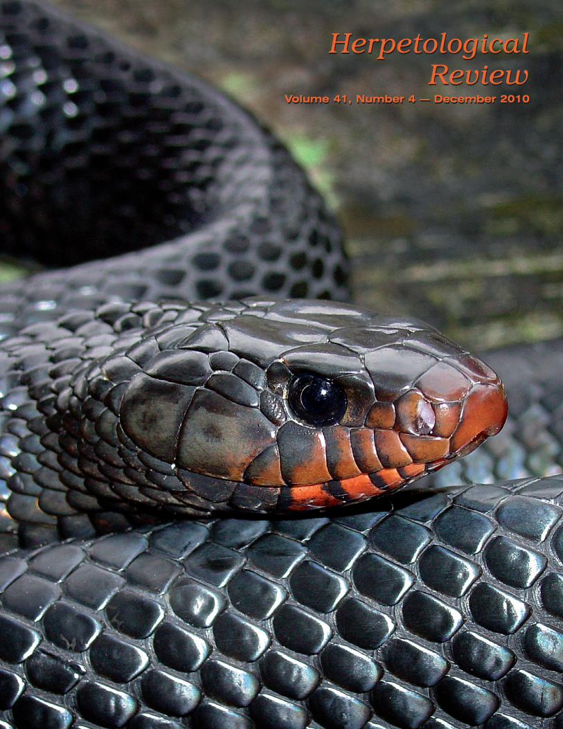
About Our Cover: Drymarchon couperi
Among the largest of colu-brid snakes—reaching adult lengths of 1.6–2.95 m—are members of the genus Dry-marchon, the indigo snakes. Collectively, their range spans some 55° degrees of latitude, extending from the southeastern United States to northern Argentina. Her-petologists have long consid-ered Drymarchon as mono-typic (D. corais) with up to eight subspecies. Currently, four species are recognized based on morphology and color patterns (Wüster et al. 2001. Herpetological Journal 11:157–165).
In sandy pineland habi-tats in Georgia/Florida, the Eastern Indigo Snake (Dry-marchon couperi) has an intimate association with the Gopher Tortoise (Go-pherus polyphemus), using the long, deep burrows of this turtle for winter dens, foraging, and nesting. Many landowners and outdoorsman revere East-ern Indigos because of their docile temperament and beauty. The colloquial-isms “blue gopher” and “gopher snake” capture the species’ predilection for tortoise burrows and the violet iridescence of the dorsal scales in sunlight. This North American icon was federally listed in 1978 as “Threatened” due to population declines attributable to habitat loss and overcollection for the pet trade. The practice of introducing gasoline into Gopher Tortoise burrows to evict Eastern Diamondback Rattlesnakes (Crotalus adamanteus) may still be employed by some snake hunters; doing so has been shown to be lethal to any Indigo Snake residing in a “gassed” burrow; currently, three rattlesnake roundups are held annually (two in Georgia, one in Alabama) at sites within or near the range of D. couperi.
Like other members of the genus, D. couperi are diurnal, active forag-ers that feed on a wide array of vertebrates, particularly snakes, including venomous species. Formidable and indiscriminate predators, they prowl wet-land margins and probe their heads into burrows when searching for prey; snake prey are typically seized by the head, chewed until immobilized, then swallowed head-first. Interestingly, a number of the forms of Drymarchon commonly consume small turtles (H. W. Greene, Cornell University, pers. comm.). (And, somewhat ironically, Eastern Indigos frequently eat hatchling Gopher Tortoises [Stevenson et al 2010. Southeastern Naturalist 9:1–18]). Mark-recapture field studies in southern Georgia have documented that adult Eastern Indigo Snakes exhibit male-biased sexual size dimorphism, require 3–4 years to reach sexual maturity, display winter den site fidelity by return-ing to the same tortoise colonies in successive years, and commonly live to be 8–12 years of age on vast protected landscapes (Stevenson et al. 2009. Herpetological Conservation and Biology 4:30–42).
Our cover features a female Drymarchon couperi found basking in Janu-ary 2009 near a Gopher Tortoise burrow in Longleaf Pine-Wiregrass sandhill habitat along the Canoochee River, Georgia. Dirk J. Ste-venson recorded this image with a Sony MVC–CD500, equipped with a Zeiss macro lens at f22, ISO 400, 1/30 sec exposure, and auto fill flash. Stevenson is Director of In-ventory and Monitoring with The Orianne Society (www.projectorianne.org), a non-profit organization dedicated to the conservation of imper-iled amphibians and reptiles. Elsewhere in this issue (pp. 437–442), Stevenson and colleagues investigate the ef-fectiveness of using wildlife detector dogs to locate East-ern Indigo Snakes.
SSAR BUSINESS
2010 Annual Meeting, Providence, Rhode Island
The 53rd Annual Meeting of SSAR took place from 7–12 July 2010 at the Weston Providence Hotel, Providence, Rhode Island, USA. The Organizing Societies were Society for the Study of Amphibians and Reptiles (in conjunction with the International Society for the History and Bibliography of Herpetology), American Elasmobranch Society (celebrating its 26th annual meeting), American Society of Ichthyologists and Herpetologists (celebrating its 90th annual meeting), and The Herpe-tologists’ League (celebrating its 68th annual meeting). The meeting was hosted by University of Rhode Island, Brown University, and University of Connecticut. The local hosts were Jacki Webb (Chair), Beth Brainerd, Eric Shultz, Kurt Schwenk, Cheryl Wilga, and Brad Wetherbee. Once again, the local hosts were ably assisted by the staff of KState Univer-sity Division of Continuing Education and by many student volunteers (from University of Rhode Island, Brown University, and University of Connecticut).
There were 1031 herpetologists and ichthyologists from around the world at the 2010 JMIH. This number was down slightly compared with that in the previous year (1170). Attendees hailed from 25 differ-ent countries (e.g., Argentina, Australia, Austria, Belgium, Canada, Ger-many, Italy, Korea, Malawi, Malaysia, The Netherlands, Poland, New Zealand, Saudi Arabia, United Kingdom, United States, Venezuela). Ap-proximately 45% of attendees were students, and around 520 papers and 260 posters were presented. Twenty-eight exhibit booths were staffed. Seven symposia, including one sponsored by SSAR and ASIH “Head-Starting Turtles—Learning from Experience” (Fig. 1), and three student workshops were scheduled. A workshop on grant writing was organized by Dawn Wilson and other members of the SSAR Graduate Student Par-ticipation Committee. The Henri Seibert Competition attracted 28 stu-dents in four categories this year.
The Annual Meeting began officially at 0900 h on Thursday, July 8th with welcomes from the Chair of the Local Host Committee, Jaqueline Webb (University of Rhode Island) and Nancy Fey-Yensen (Interim Dean, College of the Environment and Life Sciences, University of Rhode Island). Kentwood Wells, this year’s ASIH speaker, gave a pre-sentation on “The Social Behavior of Anuran Amphibians: What Have We Learned in the Last 35 years?” This was followed by the presentation of three ASIH Awards (Gibbs, Fitch, and Johnson awards) to John Lun-dberg, Tom Schoener, and Joe Nelson, respectively. The first winner of the new SSAR/ASIH/HL Meritorious Teaching Award in Herpetology, Whitfield Gibbons, was then announced by President Crother. ASIH Past-President John Lundberg spoke on “Authentic American Cryptoi-chthyology,” followed by The Herpetologists’ League’s distinguished herpetologist for 2010, Indraneil Das from University Malaysia, Sar-awak, who spoke on “Perceptions, Use and Conservation of Amphibians by Indigenous People Worldwide.” The AES speaker, Gregor Cailliett gave an address on “Ageing, Age Validation, Growth and Aging: The Life Histories of Chondrichthyan and Deep-Sea Fishes” and the Plenary Session was closed by Jacqueline Webb.
Social and ProfeSSional eventS
Robert Espinoza (California State University, Northridge) was this year’s President’s Travelogue speaker and gave his presentation (“The Herpetofauna of South America’s Southern Cone: New Discoveries from the Andean Peaks to the Peruvian Steppe”) on July 7th (Fig. 2). He spoke about the extremes of temperature and water availability and the great diversity of reptiles and amphibians in this area. Bobby acknowl-edged Richard Etheridge who introduced him to Argentina and Argen-
PHO
TO B
Y ST
ELLA
OSB
OR
N
Herpetological Review 41(4), 2010402
tinian herpetofauna, as well as the many friends and contacts he and his students have developed over the years. Every year SSAR manages to find a great Travelogue speaker and this year was no exception.
On the evening of July 8th, SSAR joined with HL to host another very successful reception for student members of both Societies and invited professionals (Fig. 3). The Joint Meeting Reception was held immedi-ately after the Student Reception at the Roger Williams Park Zoo. After a long wait for buses by most (Fig. 4a) and a detour to a casino for some, good barbeque and interesting exhibits were enjoyed by those who fi-nally made it (Fig. 4b).
In addition to the planned gatherings, many meeting attendees felt the need for some “field work” and explored local dining establishments and pubs (Fig. 5).
Twenty two students attended a pizza lunch and student workshop on grant writing organized by the Student Participation Committee on July 9th. The workshop included a panel of professional herpetologists (Robert Espinoza, Al Savitzky, Rafael de Sá, Henry Mushinsky, Karen Warkentin, and Dawn Wilson) from a variety of types of institutions offering valuable advice, including how to set a proposal in a broader context, how important it is to stress the relevance and novelty of the proposed research, and how to include preliminary data. Al and Rafael spoke of their work at NSF, as well as their own experiences writing grant proposals.
The SSAR/HL Live Auction occurred on July 11th and raised $3759.50
to support graduate student travel ($1706 of this was the result of items from the Roger Conant library). Frank Burbrink served as auctioneer and was assisted by Greg Watkins-Colwell, Samantha Wisniewski (who has helped with several of the past few auctions), Ben Jellen, Phillip Skipwith, Heather Heinz, and Taryn Cazzolli among others (Fig. 6). Greg also served as onsite coordinator before the meeting began.
Matt Venesky and Cari Hickerson (co-Chairs, Student Travel Award Committee) again worked hard to pull together a success-ful Silent Auction. $727 was raised to support the SSAR Student Travel Fund. Thanks to this year’s winners of SSAR Student Travel Awards who helped staff the exhibit desk.
A rather hastily organized evening session dealing with the ichthyological and herpeto-
logical implications of the oil spill in the Gulf of Mexico was held on July 10th. Close to standing-room-only in a large ballroom was an indica-tion of the level of interest in this topic.
The Joint Meeting Banquet was held on the last evening of the meet-ing, with Lynn Parenti serving as Master of Ceremonies. SSAR was represented at the head table by Brian Crother (President), Kirsten Nicholson (Treasurer), Marion Preest (Secretary), and SSAR member Mary White. Five past-Presidents of SSAR were in attendance. At the end of the Banquet, an invitation was issued to attend the 2011 JMIH in Minneapolis, Minnesota, July 6–11. Pat Gregory (Chair, Local Commit-tee) invited all the world’s herpetologists as well as our ichthyological colleagues from ASIH and AES to the 2012 JMIH/World Congress of Herpetology meeting in Vancouver (August 8–14). A meeting website is already up and a call by WCH for symposia has been issued.
Board Meeting and BuSineSS Meeting SuMMarieS
Society President Brian Crother called the Board Meeting to order at 0803 h on July 7th, 2010 in the Westin Providence Hotel, Providence, Rhode Island. In attendance were the following members of the Board of Directors, Editors, and Committee Chairs: Kraig Adler (Editor, Con-tributions in Herpetology), Aaron Bauer (Editor, Facsimile Reprints in Herpetology), Brian Crother (President; SSAR Rep MMPC; Chair, Stan-dard English and Scientific Names Committee), David Cundall (Board Member, Reg. 2012), Kevin de Queiroz (Board Member, Reg. 2012),
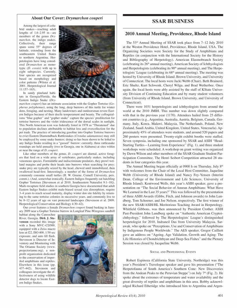
fig. 2. Beck Wehrle, Denita Weeks and Navasha Singh (L to R) students of this year’s President’s Travelogue Speaker Robert Espinoza (second from right). Denita was the 2010 Seibert Award winner in the Morphology/Physiology section. (Photo M. Preest)
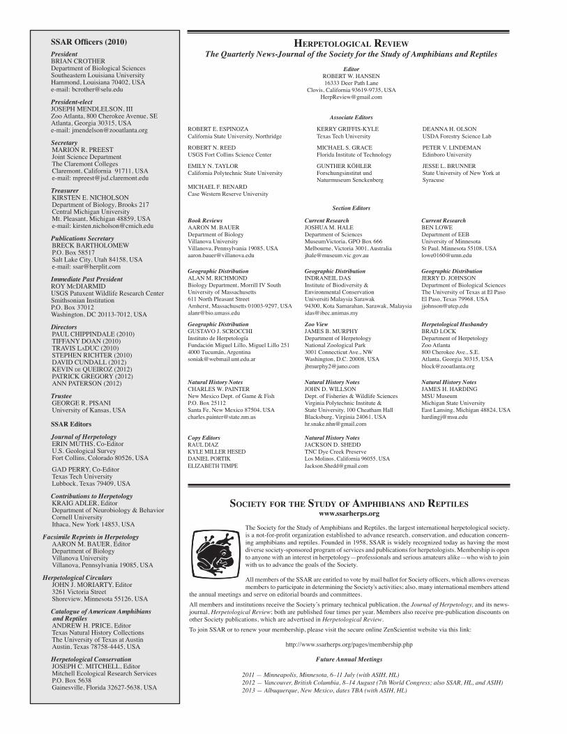
fig. 1. Participants in this year’s highly successful symposium, “Head-starting Turtles. Learning from Experience,” organized by Russell Burke and co-sponsored by SSAR and ASIH. Standing (from left to right): Willem Roosenburg, Roger Wood, Slawomir Mitrus, Tom Herman, unknown, Tom French, David Taylor, Lisa Hazard, Ken Nagy, Russ Burke, Tracey Tuberville, Brian Windmiller, Stephanie Koch, Charles Innis, Matt Hinderliter, Maria Wojakowski, Peter Warny, unknown. Seated (from left to right): Thane Wibbels, unknown, unknown, unknown, Kurt Buhlmann. (Photo M. Preest)
fig. 3. Jay Savage, Villanova student Alicia Kennedy, Mary White, and Treasurer Kirsten Nicholson enjoying the SSAR Student Reception. (Photo M. Preest)
Herpetological Review 41(4), 2010 403
Tiffany Doan (Board Member, Reg. 2010), Pat Gregory (Board Mem-ber, non-US 2012), Roy McDiarmid (Past-President), Joe Mendelson, III (President-Elect), John Moriarty (Editor, Herpetological Circulars), Erin Muths (co-Editor, Journal of Herpetology), Kirsten Nicholson (Treasurer), Pat Owen (Chair, Seibert Award Committee), Ann Paterson (Board Member, Reg. 2012), Marion Preest (Secretary), Stephen Rich-ter (Board Member, Cons. 2010), Betsie Rothermel (Chair, Conserva-tion Committee), Al Savitzky (SSAR Rep to AIBS and BioOne), Greg Watkins-Colwell (Chair, Nominations Committee; Chair, SSAR/HL
Live Auction Committee), and Dawn Wilson (Chair, Student Participa-tion Committee). Additional society members present included Robin Andrews and Henry Mushinsky. Minutes of the 2009 Board of Directors Meeting (Portland, Oregon) were approved.
Annual reports for 2009/2010 were submitted by all Officers, Editors, and Committee Chairs. President Brian Crother reported that SSAR be-came involved as a Festival Partner with the USA Science & Engineer-ing Festival to be held in Washington D.C. in October, 2010. SSAR will have a booth at the festival and Joe Mendelson, III is in charge of our par-ticipation in this national event (see event report elsewhere in this issue). The editor of the Journal of Herpetology, Matthew Parris, unexpectedly stepped down in early 2010, but Brian noted that the Society was fortu-nate to find new editors, Gad Perry and Erin Muths, fairly quickly. It is anticipated that a two-editor system will improve our manuscript turn-around time. Welcome aboard to Gad and Erin and many thanks to them for stepping up to take this key position. Thanks also to Geoff Smith, a previous editor, for stepping in and helping smooth the transition.
President Crother reported writing or signing a number of letters per-taining to SSAR business over the past year, including various letters submitted by the Conservation Committee, letters of congratulations to national and international herpetological societies acknowledging signif-icant landmarks, and letters of thanks to Maureen Donnelly (President’s Travelogue speaker, 2009) and to the participants in the 2009 JMIH SSAR Student Workshop.
Treasurer Kirsten Nicholson reported that, overall, the finances of SSAR are fairly sound and doing well. The approved 2009 budget was balanced and the Society came in under budget by around $7,000. Costs for production of our journals are increasing and we may need to review the manner in which we produce them and develop our presence online. Membership management fees assessed by Allen Press have been much higher than anticipated. Membership management was taken over by Breck Barthlomew in October 2009 and our costs are expected to be half of those charged by Allen Press. Our membership is holding steady over-all. Institutional subscriptions have fallen, however individual member-ships have increased. Although growth in membership levels would be better, holding steady is very good, given the decrease in memberships experienced by most societies. It is not clear if institutions are dropping memberships to remove duplication of effort or to reduce costs by hav-ing a BioOne (or other similar) contract which would provide Journal of Herpetology to them without having a subscription directly with us. This would explain the huge increase in royalties we are enjoying, which is expected to continue to rise. A comment was made that SSAR needs to offer an electronic-only membership option. The Society’s investments made a substantial rebound during 2009. Total market value increased
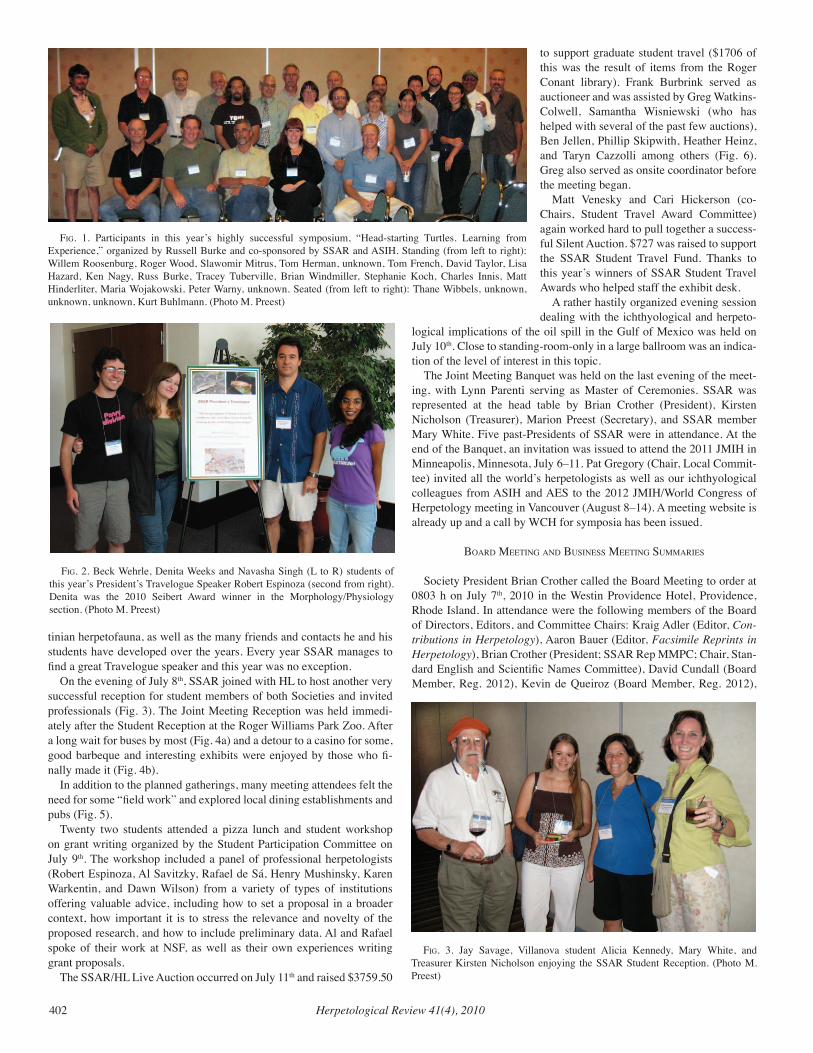
fig. 4a (top). A long wait for buses to the JMIH Picnic at the Roger Williams Park Zoo; 4b (lower) but worth it when we got there. (Photo M. Preest)
fig. 5. Matthew Morrill, Alex Pyron, Greg Watkins-Colwell, Meredith Mahoney, and Frank Burbrink (L to R) enjoying the “best beer in Rhode Island” at one of the many local pubs. (Photo M. Preest)
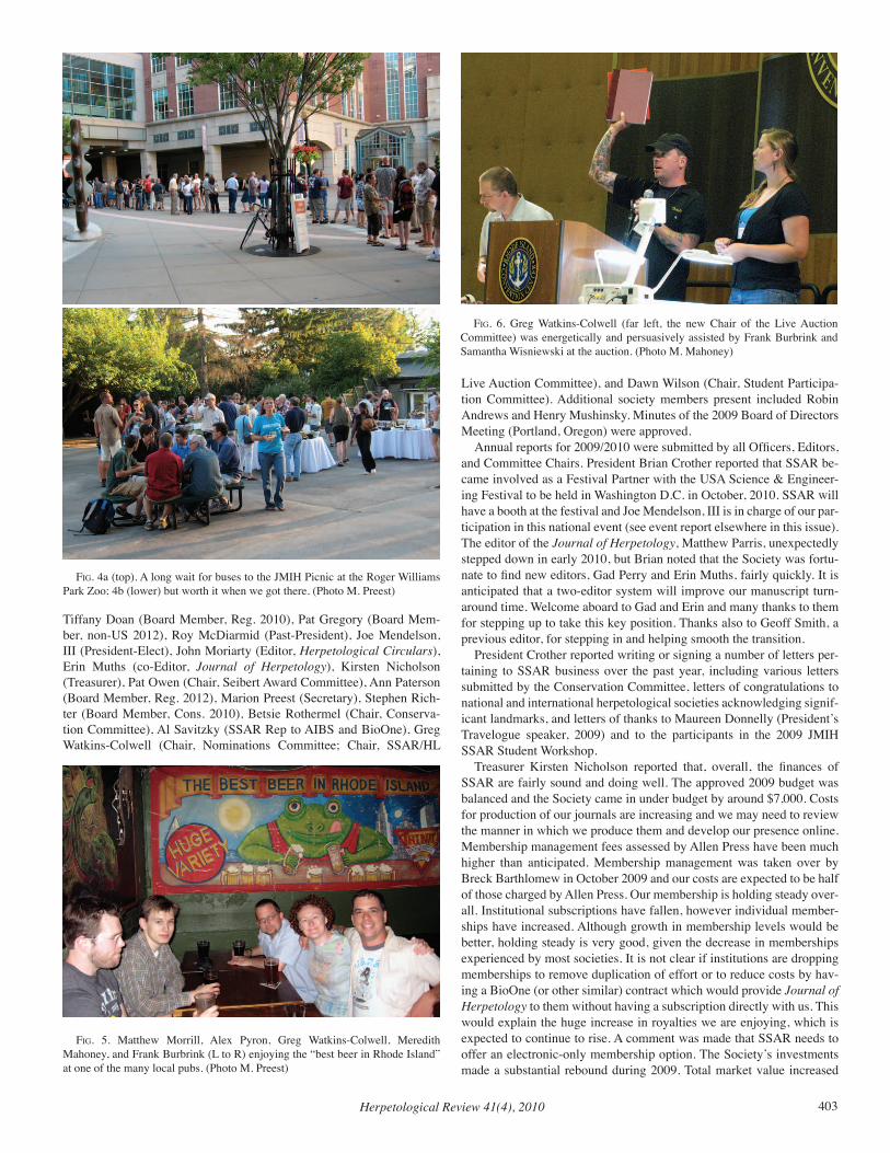
fig. 6. Greg Watkins-Colwell (far left, the new Chair of the Live Auction Committee) was energetically and persuasively assisted by Frank Burbrink and Samantha Wisniewski at the auction. (Photo M. Mahoney)
Herpetological Review 41(4), 2010404
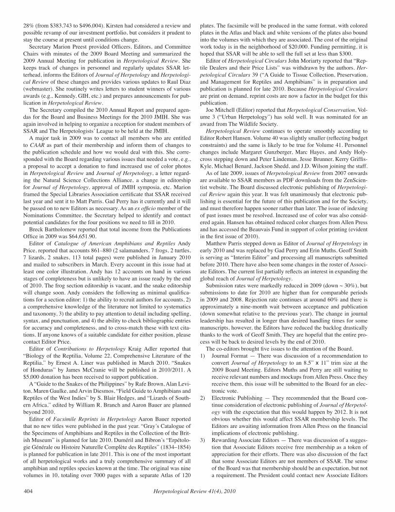
28% (from $383,743 to $496,004). Kirsten had considered a review and possible revamp of our investment portfolio, but considers it prudent to stay the course at present until conditions change.
Secretary Marion Preest provided Officers, Editors, and Committee Chairs with minutes of the 2009 Board Meeting and summarized the 2009 Annual Meeting for publication in Herpetological Review. She keeps track of changes in personnel and regularly updates SSAR let-terhead, informs the Editors of Journal of Herpetology and Herpetologi-cal Review of these changes and provides various updates to Raul Diaz (webmaster). She routinely writes letters to student winners of various awards (e.g., Kennedy, GIH, etc.) and prepares announcements for pub-lication in Herpetological Review.
The Secretary compiled the 2010 Annual Report and prepared agen-das for the Board and Business Meetings for the 2010 JMIH. She was again involved in helping to organize a reception for student members of SSAR and The Herpetologists’ League to be held at the JMIH.
A major task in 2009 was to contact all members who are entitled to CAAR as part of their membership and inform them of changes to the publication schedule and how we would deal with this. She corre-sponded with the Board regarding various issues that needed a vote, e.g., a proposal to accept a donation to fund increased use of color photos in Herpetological Review and Journal of Herpetology, a letter regard-ing the Natural Science Collections Alliance, a change in editorship for Journal of Herpetology, approval of JMIH symposia, etc. Marion framed the Special Libraries Association certificate that SSAR received last year and sent it to Matt Parris. Gad Perry has it currently and it will be passed on to new Editors as necessary. As an ex officio member of the Nominations Committee, the Secretary helped to identify and contact potential candidates for the four positions we need to fill in 2010.
Breck Bartholomew reported that total income from the Publications Office in 2009 was $64,651.90.
Editor of Catalogue of American Amphibians and Reptiles Andy Price, reported that accounts 861–880 (2 salamanders, 7 frogs, 2 turtles, 7 lizards, 2 snakes, 113 total pages) were published in January 2010 and mailed to subscribers in March. Every account in this issue had at least one color illustration. Andy has 12 accounts on hand in various stages of completeness but is unlikely to have an issue ready by the end of 2010. The frog section editorship is vacant, and the snake editorship will change soon. Andy considers the following as minimal qualifica-tions for a section editor: 1) the ability to recruit authors for accounts, 2) a comprehensive knowledge of the literature not limited to systematics and taxonomy, 3) the ability to pay attention to detail including spelling, syntax, and punctuation, and 4) the ability to check bibliographic entries for accuracy and completeness, and to cross-match these with text cita-tions. If anyone knows of a suitable candidate for either position, please contact Editor Price.
Editor of Contributions to Herpetology Kraig Adler reported that “Biology of the Reptilia, Volume 22, Comprehensive Literature of the Reptilia,” by Ernest A. Liner was published in March 2010. “Snakes of Honduras” by James McCranie will be published in 2010/2011. A $5,000 donation has been received to support publication.
A “Guide to the Snakes of the Philippines” by Rafe Brown, Alan Levi-ton, Maren Gaulke, and Arvin Diesmos, “Field Guide to Amphibians and Reptiles of the West Indies” by S. Blair Hedges, and “Lizards of South-ern Africa,” edited by William R. Branch and Aaron Bauer are planned beyond 2010.
Editor of Facsimile Reprints in Herpetology Aaron Bauer reported that no new titles were published in the past year. “Gray’s Catalogue of the Specimens of Amphibians and Reptiles in the Collection of the Brit-ish Museum” is planned for late 2010. Duméril and Bibron’s “Erpétolo-gie Générale ou Histoire Naturelle Complète des Reptiles” (1834–1854) is planned for publication in late 2011. This is one of the most important of all herpetological works and a truly comprehensive summary of all amphibian and reptiles species known at the time. The original was nine volumes in 10, totaling over 7000 pages with a separate Atlas of 120
plates. The facsimile will be produced in the same format, with colored plates in the Atlas and black and white versions of the plates also bound into the volumes with which they are associated. The cost of the original work today is in the neighborhood of $20,000. Funding permitting, it is hoped that SSAR will be able to sell the full set at less than $300.
Editor of Herpetological Circulars John Moriarty reported that “Rep-tile Dealers and their Price Lists” was withdrawn by the authors. Her-petological Circulars 39 (“A Guide to Tissue Collection, Preservation, and Management for Reptiles and Amphibians” is in preparation and publication is planned for late 2010. Because Herpetological Circulars are print on demand, reprint costs are now a factor in the budget for this publication.
Joe Mitchell (Editor) reported that Herpetological Conservation, Vol-ume 3 (“Urban Herpetology”) has sold well. It was nominated for an award from The Wildlife Society.
Herpetological Review continues to operate smoothly according to Editor Robert Hansen. Volume 40 was slightly smaller (reflecting budget constraints) and the same is likely to be true for Volume 41. Personnel changes include Margaret Gunzberger, Marc Hayes, and Andy Holy-cross stepping down and Peter Lindeman, Jesse Brunner, Kerry Griffis-Kyle, Michael Benard, Jackson Shedd, and J.D. Wilson joining the staff.
As of late 2009, issues of Herpetological Review from 2007 onwards are available to SSAR members as PDF downloads from the ZenScien-tist website. The Board discussed electronic publishing of Herpetologi-cal Review again this year. It was felt unanimously that electronic pub-lishing is essential for the future of this publication and for the Society, and must therefore happen sooner rather than later. The issue of indexing of past issues must be resolved. Increased use of color was also consid-ered again. Hansen has obtained reduced color charges from Allen Press and has accessed the Beauvais Fund in support of color printing (evident in the first issue of 2010).
Matthew Parris stepped down as Editor of Journal of Herpetology in early 2010 and was replaced by Gad Perry and Erin Muths. Geoff Smith is serving as “Interim Editor” and processing all manuscripts submitted before 2010. There have also been some changes in the roster of Associ-ate Editors. The current list partially reflects an interest in expanding the global reach of Journal of Herpetology.
Submission rates were markedly reduced in 2009 (down ~ 30%), but submissions to date for 2010 are higher than for comparable periods in 2009 and 2008. Rejection rate continues at around 60% and there is approximately a nine-month wait between acceptance and publication (down somewhat relative to the previous year). The change in journal leadership has resulted in longer than desired handling times for some manuscripts, however, the Editors have reduced the backlog drastically thanks to the work of Geoff Smith. They are hopeful that the entire pro-cess will be back to desired levels by the end of 2010.
The co-editors brought five issues to the attention of the Board. 1) Journal Format — There was discussion of a recommendation to
convert Journal of Herpetology to an 8.5” x 11” trim size at the 2009 Board Meeting. Editors Muths and Perry are still waiting to receive relevant numbers and mockups from Allen Press. Once they receive them, this issue will be submitted to the Board for an elec-tronic vote.
2) Electronic Publishing — They recommended that the Board con-tinue consideration of electronic publishing of Journal of Herpetol-ogy with the expectation that this would happen by 2012. It is not obvious whether this would affect SSAR membership levels. The Editors are awaiting information from Allen Press on the financial implications of electronic publishing.
3) Rewarding Associate Editors — There was discussion of a sugges-tion that Associate Editors receive free membership as a token of appreciation for their efforts. There was also discussion of the fact that some Associate Editors are not members of SSAR. The sense of the Board was that membership should be an expectation, but not a requirement. The President could contact new Associate Editors

Herpetological Review 41(4), 2010 405
who are not members of the Society, and encourage them to join. Editor Muths will write a draft of this letter and send it to President Crother and President-Elect Mendelson.
4) Editorial Board — The Editors questioned the role of the Editorial Board. Their function is to serve as guaranteed reviewers and the Board recommended that they be used more in this manner.
5) Page Charges — Whether a strong policy regarding page charges should be developed and enforced was again discussed. In 2009, the Board asked Editor Parris to develop such a policy. As this was not done, the Board asked the current editors to develop a policy for its consideration. This policy needs to address the issue of charges for both black and white and color pages.
When Journal of Herpetology is published electronically, increased use of color plates will be possible. Some reservations were expressed about having two versions of each issue (electronic and hard copies). One version (likely the hard copy) will need to be designated as the of-ficial version.
Three new members were added to the Conservation Committee this past year. The Committee took action on a number of items: a) they sup-ported a petition to list western U.S. populations of the Northern Leop-ard Frog as threatened, b) they sent a letter to U.S. Fish and Wildlife Service (USFWS) requesting re-initiation of an incidental take permit for a wind farm project which may affect the federally threatened Puerto Rican crested toad and contacted Puerto Rican authorities encouraging greater protection of remaining habitat for this species, c) they submitted a letter to USFWS urging federal listing and habitat designation for the coquí llanero, d) they expressed support for proactive strategies for curb-ing introduction and spread of invasive species (e.g., large constrictors) and stated their position that removal of these organisms (e.g., through controlled hunting) is unlikely to be effective, e) they wrote to the Flori-da Fish and Wildlife Conservation Commission (FFWCC) to encourage adoption of new rules regarding take and possession of native freshwater turtles.
Actions of the Committee contributed to several positive develop-ments in the past year. For example, the local Planning Board in Puerto Rico temporarily suspended their decision to approve a wind farm proj-ect and allowed time for reconsideration, and the FFWCC approved new rules that prohibit commercial harvest and impose strict daily limits on the take of wild turtles in Florida. President Crother remarked on what 10 people can accomplish and congratulated the Committee on its ef-forts.
Betsie expressed some reservations about a small Committee repre-senting the views of the entire Society and there was some discussion of soliciting greater input from the membership (e.g., via email, a web forum, or an online survey). The view of the Board was that this could be unwieldy and slow down the process and commended the Committee on its activities.
Joe Beatty (Chair, Dean Metter Award Committee) received 24 pro-posals this year and it was decided to choose two winners. They are: Matthew Niemiller (University of Tennessee, Knoxville) who is working on the maintenance of distinct cave and surface forms in the salaman-der genus Gyrinophilus, and Michael Reichert (University of Missouri, Columbia) who is studying acoustic communication in Hyla versicolor. Michael is the first student from University of Missouri in Columbia (which is where Dean Metter was a long time faculty member) to win this award. The Board encouraged Joe to advertise the Award via the SSAR website and an emailing to student members of the Society.
Erik Wild and Josh Kapfer (co-chairs, Grants-in-Herpetology Com-mittee) received 55 proposals in 2010. Applications were sent from 19 states in the US and nine countries (including the U.S.). Most applica-tions were received in the “Field Research” category. The winners each receive $500 and they are:
Conservation: James C. Cureton II, Sam Houston State UniversityField Research: Karla Moeller, Arizona State UniversityLaboratory Research: Tara A. Pelletier, Louisiana State University
Travel: Lindsey Noel Swierk, Penn. State UniversityInternational: Paulo Fernando Guedes Pereira Montenegro, Universi-
dade Federal da Paraíba (Brazil)Eric is stepping down as co-chair of the committee.The 18th annual Seibert Awards Competition was run at the 52nd Annu-
al Meeting of SSAR in Portland, Oregon. There were 35 eligible presen-tations. The Seibert Award winners for 2009 were: Systematics/Evolu-tion: Jamie Oaks, University of Kansas, “Objective partition choice and the phylogenetic systematics and biogeography of the true crocodiles.” Ecology: John Wilson, Brian Todd, and Christopher Winne, University of Georgia, Savannah River Ecology Lab, Virginia Polytechnic Insti-tute and State University, “Trap-happiness, temporary emigration and other factors affecting detectability and population estimation in aquatic snakes.” Physiology/Morphology: Victoria Arch, T. Ulmar Grafe, Mar-cos Gridi-Papp, and Peter Narins, University of California, Los Angeles, University Brunei Darussalam, “An Old World frog communicates in pure ultrasound.” Conservation: Kristine Kaiser, Menemsha Alloush, Robin Jones, Susanne Marczak, Katherine Marineau, Mark Oliva, and Peter Narins, University of California, Los Angeles, “When sounds col-lide: effects of anthropogenic noise on frog calling behavior.”
Honorable mentions were: Ecology: Javan Bauder, Holly Akenson, and Charles Peterson, Idaho State University, University of Idaho, “Over the hills and far away: movements of prairie rattlesnakes across a moun-tainous landscape in a designated wilderness.” Conservation: Anna Sav-age, Cornell University, “Experimental infection with Batrachochytrium dendrobatidis demonstrates genetic resistance to chytridiomycosis in Lithobates yavapiensis.”
All winners received a check for US $200 from SSAR and a book from University of California Press, the latter compliments of UCP Edi-tor Chuck Crumly.
The Herpetological Education Committee website has been active for a year and members respond to various enquiries submitted to the Herp Hotline. Members have also been active in developing and encouraging educational activities in herpetology. The Committee received support in 2009 from SSAR, HL, and ASIH for a proposal for a Meritorious Teach-ing Award in Herpetology. Ten nominations were received and consid-ered by a Committee consisting of professional and student members of the three participating societies.
Lynnette Sievert (Chair, Kennedy Award Committee) announced that the winning paper for 2009 is by Ermin Schadich, “Skin peptide ac-tivities against opportunistic bacterial pathogens of the African clawed frog (Xenopus laevis) and three Litoria frogs. Journal of Herpetology 43:173–183.” Ermin will receive a check for $200 or $400 equivalent in SSAR publications.
The Meeting Management and Planning Committee (MMPC) met in Providence in March 2010 to develop a meeting schedule and review conference facilities. President Crother and President-Elect Mendelson, who will replace Brian on the MMPC, attended the meeting as SSAR representatives. Almost 800 presentations (papers and posters) will be made at the 2010 meeting. Future meetings are to be held as follows:
- Minneapolis (2011)- Vancouver (2012; with the WCH)- Albuquerque (2013)
A survey conducted by the MMPC last year confirmed that many members thought the meetings were too expensive. Consequently, the MMPC invited two meeting management companies (KState and Ex-perient, Inc.) to submit competitive bids for the 2013 meeting in Albu-querque. Experient’s bids failed twice to provide costs for a number of services (e.g., web design and maintenance for registration and abstract submission, meeting announcement postcards, program book, abstract CD and book, reimbursements for MMPC, etc.). This process had two outcomes. It opened the eyes of the MMPC to the quality of service we receive from KState and it made KState aware of how serious the MMPC is in reducing meeting costs. Subsequently, KState joined with
Herpetological Review 41(4), 2010406
another meeting management company to reduce costs of site selection and hotel negotiations (e.g., reduced or eliminated room rental fees, eliminated the need for a food and beverage minimum required for free or reduced room rental fees, eliminated fees associated with not filling a certain percentage of our room block).
The MMPC presented a proposal to make the MMPC a JMIH, rather than an ASIH, Committee and a proposal to reduce expenses at future meetings. These proposals were discussed by the Board later under New Business.
The MMPC also considered, but took no action on, the issue of re-duced registration rates for accompanying and retired persons. Lightning round talks and the future and format of the banquet were discussed. A competition for a JMIH logo, separate from the individual society logos and the local meeting logo is being run. It will reflect combined interests in cartilagenous and bony fishes, amphibians, and reptiles and act as a banner for the JMIH meeting websites.
Five symposia were approved for the 2011 meetings. ASIH — Turtle Ecology on the Upper Mississippi River System, Then
and Now: A Zeitschrift in Honor of the Career in Turtle Biology of Dr. John M. Legler
ASIH — Drawing Lines in the Sand: Comparative Phylogeography of the Gulf-Atlantic Coastal Plain
ASIH — Ranaviruses: An Emerging Threat to Ectothermic VertebratesSSAR — Assisted Reproductive Technologies and Genetic Resource
Banking: Tools for Conserving Declining AmphibiansAES — Elasmobranch Telemetry
Greg Watkins-Colwell reported that no elections were held in 2009. In 2010, SSAR needs to elect a President-Elect, Secretary, Treasurer, and three Board Members (Regular, Conservation, Regional Herpetological Societies). The committee plans to submit suggestions to the Board on how to define (or re-define) some of the specialty Board positions such as “Regional Herpetological Societies.”
Stu Nielsen presented resolutions at the SSAR Business Meeting in Portland in 2009. Stu stepped down as Resolutions Chair and was re-placed by Rob Denton.
The 6th Edition of the Scientific and Standard English Names List was published two years ago and the web version is close to posting. Brian Crother, Chair of the Standard English and Scientific Names Committee, thanked Raul Diaz for his help in getting this accomplished. Kevin de Queiroz donated many photographs and has worked closely with Raul on the website version.
Dawn Wilson reported that SSAR held a pizza lunch and workshop (“How to Get a Job After Graduation: Advice from Experts”) for stu-dents at the 2009 JMIH. A workshop will be offered in 2010 on a topic decided on the basis of an exit survey given to students at the 2009 work-shop. For improvements, students suggested more specific workshops, longer workshops, holding different workshops for undergraduates and graduates, and not overlapping the workshop with other meeting activi-ties. Dawn will contact students attending the 2010 JMIH and ask for ideas for future workshops. Roy McDiarmid suggested a workshop fo-cusing on nomenclature.
New co-chairs of the Student Travel Awards Committee, Matt Venesky and Cari Hickerson, reported that the Silent Auction raised $733 at the JMIH in 2009. They hope to double this amount within two years. Pro-posed changes to the operation of the Auction include:1) Placing auction items on tables in the registration lobby of the
JMIH to increase visibility and bidding activity.2) Requiring Travel Award winners to monitor the Auction table.3) Actively soliciting donations from distinguished herpetologists.4) Creating a Facebook page to promote donations and bidding and
providing information regarding applications for Student Travel Awards.
A comment was made that although the Silent Auction has tradition-ally focused on objects of art, last year items in the Live and Silent Auc-tions were very similar. This reflected a decision made by the co-chairs
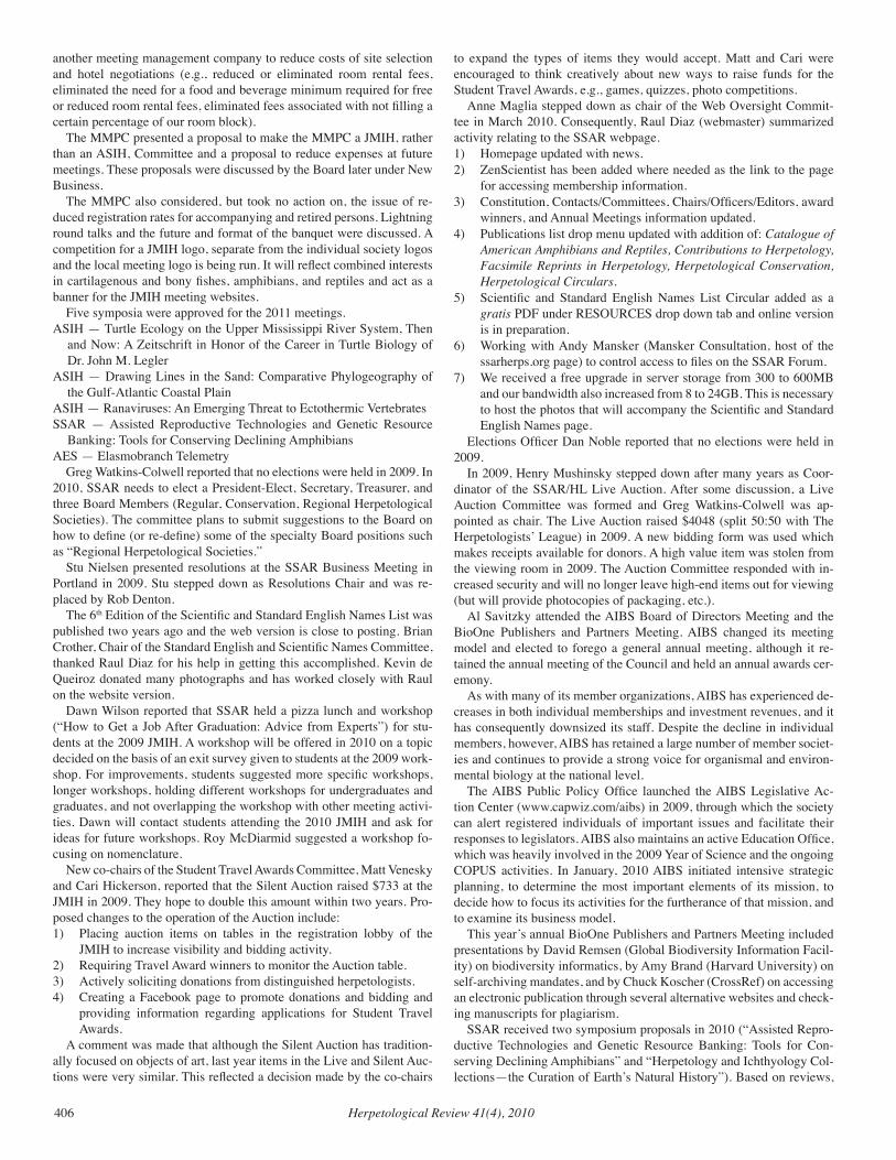
to expand the types of items they would accept. Matt and Cari were encouraged to think creatively about new ways to raise funds for the Student Travel Awards, e.g., games, quizzes, photo competitions.
Anne Maglia stepped down as chair of the Web Oversight Commit-tee in March 2010. Consequently, Raul Diaz (webmaster) summarized activity relating to the SSAR webpage.1) Homepage updated with news.2) ZenScientist has been added where needed as the link to the page
for accessing membership information.3) Constitution, Contacts/Committees, Chairs/Officers/Editors, award
winners, and Annual Meetings information updated.4) Publications list drop menu updated with addition of: Catalogue of
American Amphibians and Reptiles, Contributions to Herpetology, Facsimile Reprints in Herpetology, Herpetological Conservation, Herpetological Circulars.
5) Scientific and Standard English Names List Circular added as a gratis PDF under RESOURCES drop down tab and online version is in preparation.
6) Working with Andy Mansker (Mansker Consultation, host of the ssarherps.org page) to control access to files on the SSAR Forum.
7) We received a free upgrade in server storage from 300 to 600MB and our bandwidth also increased from 8 to 24GB. This is necessary to host the photos that will accompany the Scientific and Standard English Names page.
Elections Officer Dan Noble reported that no elections were held in 2009.
In 2009, Henry Mushinsky stepped down after many years as Coor-dinator of the SSAR/HL Live Auction. After some discussion, a Live Auction Committee was formed and Greg Watkins-Colwell was ap-pointed as chair. The Live Auction raised $4048 (split 50:50 with The Herpetologists’ League) in 2009. A new bidding form was used which makes receipts available for donors. A high value item was stolen from the viewing room in 2009. The Auction Committee responded with in-creased security and will no longer leave high-end items out for viewing (but will provide photocopies of packaging, etc.).
Al Savitzky attended the AIBS Board of Directors Meeting and the BioOne Publishers and Partners Meeting. AIBS changed its meeting model and elected to forego a general annual meeting, although it re-tained the annual meeting of the Council and held an annual awards cer-emony.
As with many of its member organizations, AIBS has experienced de-creases in both individual memberships and investment revenues, and it has consequently downsized its staff. Despite the decline in individual members, however, AIBS has retained a large number of member societ-ies and continues to provide a strong voice for organismal and environ-mental biology at the national level.
The AIBS Public Policy Office launched the AIBS Legislative Ac-tion Center (www.capwiz.com/aibs) in 2009, through which the society can alert registered individuals of important issues and facilitate their responses to legislators. AIBS also maintains an active Education Office, which was heavily involved in the 2009 Year of Science and the ongoing COPUS activities. In January, 2010 AIBS initiated intensive strategic planning, to determine the most important elements of its mission, to decide how to focus its activities for the furtherance of that mission, and to examine its business model.
This year’s annual BioOne Publishers and Partners Meeting included presentations by David Remsen (Global Biodiversity Information Facil-ity) on biodiversity informatics, by Amy Brand (Harvard University) on self-archiving mandates, and by Chuck Koscher (CrossRef) on accessing an electronic publication through several alternative websites and check-ing manuscripts for plagiarism.
SSAR received two symposium proposals in 2010 (“Assisted Repro-ductive Technologies and Genetic Resource Banking: Tools for Con-serving Declining Amphibians” and “Herpetology and Ichthyology Col-lections—the Curation of Earth’s Natural History”). Based on reviews,
Herpetological Review 41(4), 2010 407
the SSAR Board’s decision was to sponsor the first proposal by Jennifer Germano and Andy Kouba for the 2011 JMIH meetings in Minneapolis.
This concluded discussion of reports submitted, and the Board then turned to new business. The Meeting Management and Planning Com-mittee (MMPC) made the following recommendations to convert the (ASIH) MMPC into a JMIH MMPC and to reduce expenses for future Joint Meetings:1. Reconstruct the ASIH MMPC to be a JMIH Committee to make the
committee equally responsible and responsive to the four sponsoring societies.
2. The reconstructed JMIH MMPC shall have equal representation of ichthyologists and herpetologists to ensure that we have the neces-sary expertise to schedule oral presentations, posters, and symposia sessions efficiently.
3. The JMIH MMPC will be composed of six voting members and one non-voting member; one voting member each appointed by the AES, HL and SSAR, and three voting members appointed by the ASIH (two ichthyologists and one herpetologist) and the non-voting Sec-retary of the ASIH. Other than the ASIH Secretary, each member serves a four-year term and may be reappointed as determined by the appropriate society. The Chair of the MMPC is elected from within the committee and serves a two-year term. The ASIH secretary is an ex officio, non-voting member and serves on the committee for the duration of the Office. Individuals serving on the ASIH MMPC shall assume their same role on the JMIH MMPC.
4. The JMIH MMPC reports directly to the four JMIH sponsoring soci-eties.
5. All votes of the JMIH MMPC must be unanimous.6. The reconstructed JMIH MMPC must be ratified by the four JMIH
sponsoring societies. 7. Assuming that the four sponsoring societies ratify the proposed
change to the MMPC, it shall become a recognized JMIH Commit-tee.
8. Reduce the number of days of the meeting from five to four.9. Limit the number of oral presentations to fit into the new meeting
format.10. Run seven (or at most eight) concurrent sessions (most recent past
meetings have had six). Seven concurrent sessions will accommo-date 630 oral presentations (eight concurrent sessions will accom-modate 714 presentations).
11. Oral presentations are accepted on a first-come, first-served basis.12. Invited presentations for approved symposia are protected (they will
be included in the appropriate symposium).13. The number of symposia will be limited to four (one per society). If a
society fails to support a symposium, a replacement symposium can be offered by another society, if approved by the MMPC.
14. Oral presentations by students in award competitions are scheduled during meeting days one and two.
15. Winners of student awards are announced at the business meetings of each sponsoring society or another occasion as determined by each society. If the JMIH Banquet is retained, then each society should have the opportunity to work with the local committee and have in-put into the program.
16. Increase the number of poster presentations on meeting days two and three to accommodate approximately 300 poster presentations (150 per day).
17. Eliminate the end of meeting banquet (elimination of the banquet was supported strongly by the membership when polled about reduc-ing meeting length and costs).
18. Authorize the Kansas State Division of Continuing Education to en-ter into a long-term agreement with a major hotel chain (e.g., Mar-riott, Sheraton, or Hilton) to reduce meeting costs.
19. Some proposed changes cannot take effect until 2014, because we are under contract to hold five-day meetings, others may be imple-mented as they become ratified by the four societies.
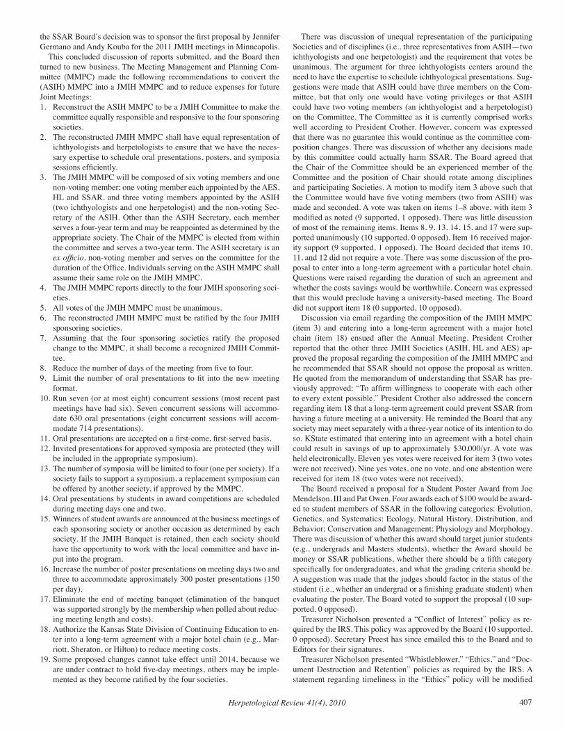
There was discussion of unequal representation of the participating Societies and of disciplines (i.e., three representatives from ASIH—two ichthyologists and one herpetologist) and the requirement that votes be unanimous. The argument for three ichthyologists centers around the need to have the expertise to schedule ichthyological presentations. Sug-gestions were made that ASIH could have three members on the Com-mittee, but that only one would have voting privileges or that ASIH could have two voting members (an ichthyologist and a herpetologist) on the Committee. The Committee as it is currently comprised works well according to President Crother. However, concern was expressed that there was no guarantee this would continue as the committee com-position changes. There was discussion of whether any decisions made by this committee could actually harm SSAR. The Board agreed that the Chair of the Committee should be an experienced member of the Committee and the position of Chair should rotate among disciplines and participating Societies. A motion to modify item 3 above such that the Committee would have five voting members (two from ASIH) was made and seconded. A vote was taken on items 1–8 above, with item 3 modified as noted (9 supported, 1 opposed). There was little discussion of most of the remaining items. Items 8, 9, 13, 14, 15, and 17 were sup-ported unanimously (10 supported, 0 opposed). Item 16 received major-ity support (9 supported, 1 opposed). The Board decided that items 10, 11, and 12 did not require a vote. There was some discussion of the pro-posal to enter into a long-term agreement with a particular hotel chain. Questions were raised regarding the duration of such an agreement and whether the costs savings would be worthwhile. Concern was expressed that this would preclude having a university-based meeting. The Board did not support item 18 (0 supported, 10 opposed).
Discussion via email regarding the composition of the JMIH MMPC (item 3) and entering into a long-term agreement with a major hotel chain (item 18) ensued after the Annual Meeting. President Crother reported that the other three JMIH Societies (ASIH, HL and AES) ap-proved the proposal regarding the composition of the JMIH MMPC and he recommended that SSAR should not oppose the proposal as written. He quoted from the memorandum of understanding that SSAR has pre-viously approved: “To affirm willingness to cooperate with each other to every extent possible.” President Crother also addressed the concern regarding item 18 that a long-term agreement could prevent SSAR from having a future meeting at a university. He reminded the Board that any society may meet separately with a three-year notice of its intention to do so. KState estimated that entering into an agreement with a hotel chain could result in savings of up to approximately $30,000/yr. A vote was held electronically. Eleven yes votes were received for item 3 (two votes were not received). Nine yes votes, one no vote, and one abstention were received for item 18 (two votes were not received).
The Board received a proposal for a Student Poster Award from Joe Mendelson, III and Pat Owen. Four awards each of $100 would be award-ed to student members of SSAR in the following categories: Evolution, Genetics, and Systematics; Ecology, Natural History, Distribution, and Behavior; Conservation and Management; Physiology and Morphology. There was discussion of whether this award should target junior students (e.g., undergrads and Masters students), whether the Award should be money or SSAR publications, whether there should be a fifth category specifically for undergraduates, and what the grading criteria should be. A suggestion was made that the judges should factor in the status of the student (i.e., whether an undergrad or a finishing graduate student) when evaluating the poster. The Board voted to support the proposal (10 sup-ported, 0 opposed).
Treasurer Nicholson presented a “Conflict of Interest” policy as re-quired by the IRS. This policy was approved by the Board (10 supported, 0 opposed). Secretary Preest has since emailed this to the Board and to Editors for their signatures.
Treasurer Nicholson presented “Whistleblower,” “Ethics,” and “Doc-ument Destruction and Retention” policies as required by the IRS. A statement regarding timeliness in the “Ethics” policy will be modified
Herpetological Review 41(4), 2010408
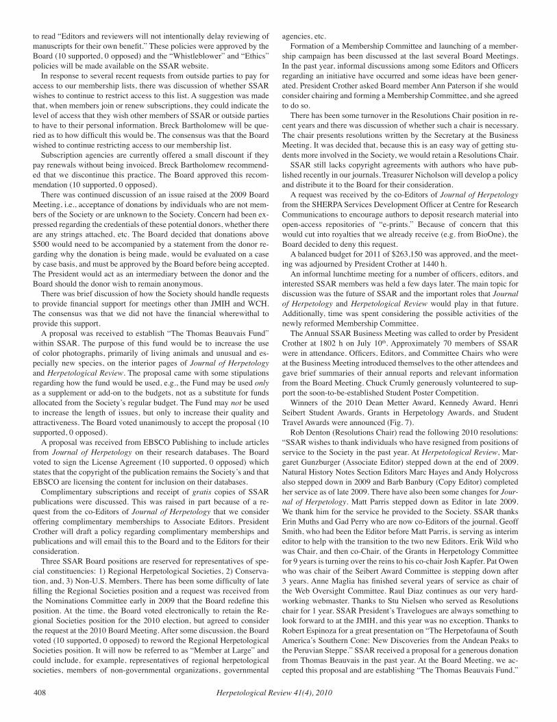
to read “Editors and reviewers will not intentionally delay reviewing of manuscripts for their own benefit.” These policies were approved by the Board (10 supported, 0 opposed) and the “Whistleblower” and “Ethics” policies will be made available on the SSAR website.
In response to several recent requests from outside parties to pay for access to our membership lists, there was discussion of whether SSAR wishes to continue to restrict access to this list. A suggestion was made that, when members join or renew subscriptions, they could indicate the level of access that they wish other members of SSAR or outside parties to have to their personal information. Breck Bartholomew will be que-ried as to how difficult this would be. The consensus was that the Board wished to continue restricting access to our membership list.
Subscription agencies are currently offered a small discount if they pay renewals without being invoiced. Breck Bartholomew recommend-ed that we discontinue this practice. The Board approved this recom-mendation (10 supported, 0 opposed).
There was continued discussion of an issue raised at the 2009 Board Meeting, i.e., acceptance of donations by individuals who are not mem-bers of the Society or are unknown to the Society. Concern had been ex-pressed regarding the credentials of these potential donors, whether there are any strings attached, etc. The Board decided that donations above $500 would need to be accompanied by a statement from the donor re-garding why the donation is being made, would be evaluated on a case by case basis, and must be approved by the Board before being accepted. The President would act as an intermediary between the donor and the Board should the donor wish to remain anonymous.
There was brief discussion of how the Society should handle requests to provide financial support for meetings other than JMIH and WCH. The consensus was that we did not have the financial wherewithal to provide this support.
A proposal was received to establish “The Thomas Beauvais Fund” within SSAR. The purpose of this fund would be to increase the use of color photographs, primarily of living animals and unusual and es-pecially new species, on the interior pages of Journal of Herpetology and Herpetological Review. The proposal came with some stipulations regarding how the fund would be used, e.g., the Fund may be used only as a supplement or add-on to the budgets, not as a substitute for funds allocated from the Society’s regular budget. The Fund may not be used to increase the length of issues, but only to increase their quality and attractiveness. The Board voted unanimously to accept the proposal (10 supported, 0 opposed).
A proposal was received from EBSCO Publishing to include articles from Journal of Herpetology on their research databases. The Board voted to sign the License Agreement (10 supported, 0 opposed) which states that the copyright of the publication remains the Society’s and that EBSCO are licensing the content for inclusion on their databases.
Complimentary subscriptions and receipt of gratis copies of SSAR publications were discussed. This was raised in part because of a re-quest from the co-Editors of Journal of Herpetology that we consider offering complimentary memberships to Associate Editors. President Crother will draft a policy regarding complimentary memberships and publications and will email this to the Board and to the Editors for their consideration.
Three SSAR Board positions are reserved for representatives of spe-cial constituencies: 1) Regional Herpetological Societies, 2) Conserva-tion, and, 3) Non-U.S. Members. There has been some difficulty of late filling the Regional Societies position and a request was received from the Nominations Committee early in 2009 that the Board redefine this position. At the time, the Board voted electronically to retain the Re-gional Societies position for the 2010 election, but agreed to consider the request at the 2010 Board Meeting. After some discussion, the Board voted (10 supported, 0 opposed) to reword the Regional Herpetological Societies position. It will now be referred to as “Member at Large” and could include, for example, representatives of regional herpetological societies, members of non-governmental organizations, governmental
agencies, etc.Formation of a Membership Committee and launching of a member-
ship campaign has been discussed at the last several Board Meetings. In the past year, informal discussions among some Editors and Officers regarding an initiative have occurred and some ideas have been gener-ated. President Crother asked Board member Ann Paterson if she would consider chairing and forming a Membership Committee, and she agreed to do so.
There has been some turnover in the Resolutions Chair position in re-cent years and there was discussion of whether such a chair is necessary. The chair presents resolutions written by the Secretary at the Business Meeting. It was decided that, because this is an easy way of getting stu-dents more involved in the Society, we would retain a Resolutions Chair.
SSAR still lacks copyright agreements with authors who have pub-lished recently in our journals. Treasurer Nicholson will develop a policy and distribute it to the Board for their consideration.
A request was received by the co-Editors of Journal of Herpetology from the SHERPA Services Development Officer at Centre for Research Communications to encourage authors to deposit research material into open-access repositories of “e-prints.” Because of concern that this would cut into royalties that we already receive (e.g. from BioOne), the Board decided to deny this request.
A balanced budget for 2011 of $263,150 was approved, and the meet-ing was adjourned by President Crother at 1440 h.
An informal lunchtime meeting for a number of officers, editors, and interested SSAR members was held a few days later. The main topic for discussion was the future of SSAR and the important roles that Journal of Herpetology and Herpetological Review would play in that future. Additionally, time was spent considering the possible activities of the newly reformed Membership Committee.
The Annual SSAR Business Meeting was called to order by President Crother at 1802 h on July 10th. Approximately 70 members of SSAR were in attendance. Officers, Editors, and Committee Chairs who were at the Business Meeting introduced themselves to the other attendees and gave brief summaries of their annual reports and relevant information from the Board Meeting. Chuck Crumly generously volunteered to sup-port the soon-to-be-established Student Poster Competition.
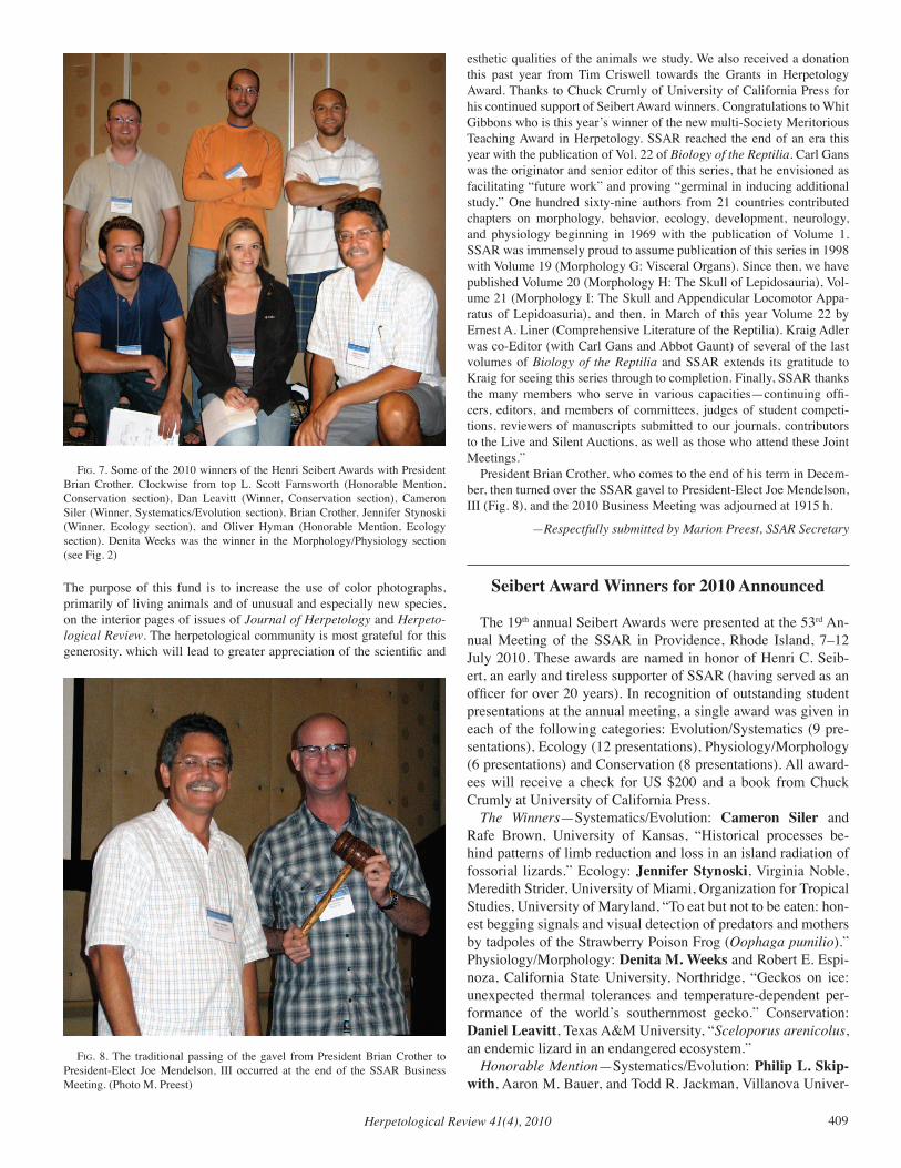
Winners of the 2010 Dean Metter Award, Kennedy Award, Henri Seibert Student Awards, Grants in Herpetology Awards, and Student Travel Awards were announced (Fig. 7).
Rob Denton (Resolutions Chair) read the following 2010 resolutions: “SSAR wishes to thank individuals who have resigned from positions of service to the Society in the past year. At Herpetological Review, Mar-garet Gunzburger (Associate Editor) stepped down at the end of 2009. Natural History Notes Section Editors Marc Hayes and Andy Holycross also stepped down in 2009 and Barb Banbury (Copy Editor) completed her service as of late 2009. There have also been some changes for Jour-nal of Herpetology. Matt Parris stepped down as Editor in late 2009. We thank him for the service he provided to the Society. SSAR thanks Erin Muths and Gad Perry who are now co-Editors of the journal. Geoff Smith, who had been the Editor before Matt Parris, is serving as interim editor to help with the transition to the two new Editors. Erik Wild who was Chair, and then co-Chair, of the Grants in Herpetology Committee for 9 years is turning over the reins to his co-chair Josh Kapfer. Pat Owen who was chair of the Seibert Award Committee is stepping down after 3 years. Anne Maglia has finished several years of service as chair of the Web Oversight Committee. Raul Diaz continues as our very hard-working webmaster. Thanks to Stu Nielsen who served as Resolutions chair for 1 year. SSAR President’s Travelogues are always something to look forward to at the JMIH, and this year was no exception. Thanks to Robert Espinoza for a great presentation on “The Herpetofauna of South America’s Southern Cone: New Discoveries from the Andean Peaks to the Peruvian Steppe.” SSAR received a proposal for a generous donation from Thomas Beauvais in the past year. At the Board Meeting, we ac-cepted this proposal and are establishing “The Thomas Beauvais Fund.”
Herpetological Review 41(4), 2010 409
The purpose of this fund is to increase the use of color photographs, primarily of living animals and of unusual and especially new species, on the interior pages of issues of Journal of Herpetology and Herpeto-logical Review. The herpetological community is most grateful for this generosity, which will lead to greater appreciation of the scientific and
fig. 7. Some of the 2010 winners of the Henri Seibert Awards with President Brian Crother. Clockwise from top L. Scott Farnsworth (Honorable Mention, Conservation section), Dan Leavitt (Winner, Conservation section), Cameron Siler (Winner, Systematics/Evolution section), Brian Crother, Jennifer Stynoski (Winner, Ecology section), and Oliver Hyman (Honorable Mention, Ecology section). Denita Weeks was the winner in the Morphology/Physiology section (see Fig. 2)
esthetic qualities of the animals we study. We also received a donation this past year from Tim Criswell towards the Grants in Herpetology Award. Thanks to Chuck Crumly of University of California Press for his continued support of Seibert Award winners. Congratulations to Whit Gibbons who is this year’s winner of the new multi-Society Meritorious Teaching Award in Herpetology. SSAR reached the end of an era this year with the publication of Vol. 22 of Biology of the Reptilia. Carl Gans was the originator and senior editor of this series, that he envisioned as facilitating “future work” and proving “germinal in inducing additional study.” One hundred sixty-nine authors from 21 countries contributed chapters on morphology, behavior, ecology, development, neurology, and physiology beginning in 1969 with the publication of Volume 1. SSAR was immensely proud to assume publication of this series in 1998 with Volume 19 (Morphology G: Visceral Organs). Since then, we have published Volume 20 (Morphology H: The Skull of Lepidosauria), Vol-ume 21 (Morphology I: The Skull and Appendicular Locomotor Appa-ratus of Lepidoasuria), and then, in March of this year Volume 22 by Ernest A. Liner (Comprehensive Literature of the Reptilia). Kraig Adler was co-Editor (with Carl Gans and Abbot Gaunt) of several of the last volumes of Biology of the Reptilia and SSAR extends its gratitude to Kraig for seeing this series through to completion. Finally, SSAR thanks the many members who serve in various capacities—continuing offi-cers, editors, and members of committees, judges of student competi-tions, reviewers of manuscripts submitted to our journals, contributors to the Live and Silent Auctions, as well as those who attend these Joint Meetings.”
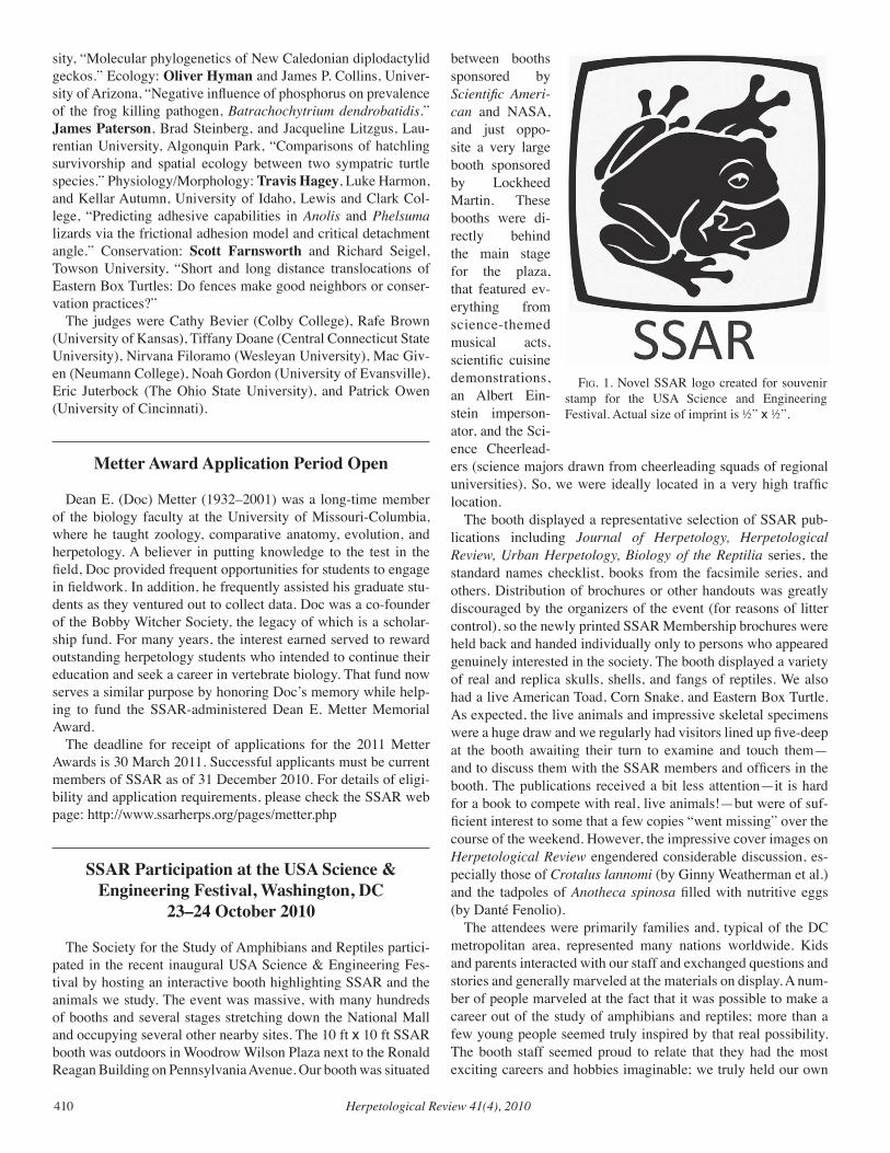
President Brian Crother, who comes to the end of his term in Decem-ber, then turned over the SSAR gavel to President-Elect Joe Mendelson, III (Fig. 8), and the 2010 Business Meeting was adjourned at 1915 h.
—Respectfully submitted by Marion Preest, SSAR Secretary
Seibert Award Winners for 2010 Announced
The 19th annual Seibert Awards were presented at the 53rd An-nual Meeting of the SSAR in Providence, Rhode Island, 7–12 July 2010. These awards are named in honor of Henri C. Seib-ert, an early and tireless supporter of SSAR (having served as an officer for over 20 years). In recognition of outstanding student presentations at the annual meeting, a single award was given in each of the following categories: Evolution/Systematics (9 pre-sentations), Ecology (12 presentations), Physiology/Morphology (6 presentations) and Conservation (8 presentations). All award-ees will receive a check for US $200 and a book from Chuck Crumly at University of California Press.
The Winners—Systematics/Evolution: Cameron Siler and Rafe Brown, University of Kansas, “Historical processes be-hind patterns of limb reduction and loss in an island radiation of fossorial lizards.” Ecology: Jennifer Stynoski, Virginia Noble, Meredith Strider, University of Miami, Organization for Tropical Studies, University of Maryland, “To eat but not to be eaten: hon-est begging signals and visual detection of predators and mothers by tadpoles of the Strawberry Poison Frog (Oophaga pumilio).” Physiology/Morphology: Denita M. Weeks and Robert E. Espi-noza, California State University, Northridge, “Geckos on ice: unexpected thermal tolerances and temperature-dependent per-formance of the world’s southernmost gecko.” Conservation: Daniel Leavitt, Texas A&M University, “Sceloporus arenicolus, an endemic lizard in an endangered ecosystem.”
Honorable Mention—Systematics/Evolution: Philip L. Skip-with, Aaron M. Bauer, and Todd R. Jackman, Villanova Univer-
fig. 8. The traditional passing of the gavel from President Brian Crother to President-Elect Joe Mendelson, III occurred at the end of the SSAR Business Meeting. (Photo M. Preest)
Herpetological Review 41(4), 2010410
sity, “Molecular phylogenetics of New Caledonian diplodactylid geckos.” Ecology: Oliver Hyman and James P. Collins, Univer-sity of Arizona, “Negative influence of phosphorus on prevalence of the frog killing pathogen, Batrachochytrium dendrobatidis.” James Paterson, Brad Steinberg, and Jacqueline Litzgus, Lau-rentian University, Algonquin Park, “Comparisons of hatchling survivorship and spatial ecology between two sympatric turtle species.” Physiology/Morphology: Travis Hagey, Luke Harmon, and Kellar Autumn, University of Idaho, Lewis and Clark Col-lege, “Predicting adhesive capabilities in Anolis and Phelsuma lizards via the frictional adhesion model and critical detachment angle.” Conservation: Scott Farnsworth and Richard Seigel, Towson University, “Short and long distance translocations of Eastern Box Turtles: Do fences make good neighbors or conser-vation practices?”
The judges were Cathy Bevier (Colby College), Rafe Brown (University of Kansas), Tiffany Doane (Central Connecticut State University), Nirvana Filoramo (Wesleyan University), Mac Giv-en (Neumann College), Noah Gordon (University of Evansville), Eric Juterbock (The Ohio State University), and Patrick Owen (University of Cincinnati).
Metter Award Application Period Open
Dean E. (Doc) Metter (1932–2001) was a long-time member of the biology faculty at the University of Missouri-Columbia, where he taught zoology, comparative anatomy, evolution, and herpetology. A believer in putting knowledge to the test in the field, Doc provided frequent opportunities for students to engage in fieldwork. In addition, he frequently assisted his graduate stu-dents as they ventured out to collect data. Doc was a co-founder of the Bobby Witcher Society, the legacy of which is a scholar-ship fund. For many years, the interest earned served to reward outstanding herpetology students who intended to continue their education and seek a career in vertebrate biology. That fund now serves a similar purpose by honoring Doc’s memory while help-ing to fund the SSAR-administered Dean E. Metter Memorial Award.
The deadline for receipt of applications for the 2011 Metter Awards is 30 March 2011. Successful applicants must be current members of SSAR as of 31 December 2010. For details of eligi-bility and application requirements, please check the SSAR web page: http://www.ssarherps.org/pages/metter.php
SSAR Participation at the USA Science & Engineering Festival, Washington, DC
23–24 October 2010
The Society for the Study of Amphibians and Reptiles partici-pated in the recent inaugural USA Science & Engineering Fes-tival by hosting an interactive booth highlighting SSAR and the animals we study. The event was massive, with many hundreds of booths and several stages stretching down the National Mall and occupying several other nearby sites. The 10 ft x 10 ft SSAR booth was outdoors in Woodrow Wilson Plaza next to the Ronald Reagan Building on Pennsylvania Avenue. Our booth was situated
between booths sponsored by Scientific Ameri-can and NASA, and just oppo-site a very large booth sponsored by Lockheed Martin. These booths were di-rectly behind the main stage for the plaza, that featured ev-erything from science-themed musical acts, scientific cuisine demonstrations, an Albert Ein-stein imperson-ator, and the Sci-ence Cheerlead-ers (science majors drawn from cheerleading squads of regional universities). So, we were ideally located in a very high traffic location.
The booth displayed a representative selection of SSAR pub-lications including Journal of Herpetology, Herpetological Review, Urban Herpetology, Biology of the Reptilia series, the standard names checklist, books from the facsimile series, and others. Distribution of brochures or other handouts was greatly discouraged by the organizers of the event (for reasons of litter control), so the newly printed SSAR Membership brochures were held back and handed individually only to persons who appeared genuinely interested in the society. The booth displayed a variety of real and replica skulls, shells, and fangs of reptiles. We also had a live American Toad, Corn Snake, and Eastern Box Turtle. As expected, the live animals and impressive skeletal specimens were a huge draw and we regularly had visitors lined up five-deep at the booth awaiting their turn to examine and touch them—and to discuss them with the SSAR members and officers in the booth. The publications received a bit less attention—it is hard for a book to compete with real, live animals!—but were of suf-ficient interest to some that a few copies “went missing” over the course of the weekend. However, the impressive cover images on Herpetological Review engendered considerable discussion, es-pecially those of Crotalus lannomi (by Ginny Weatherman et al.) and the tadpoles of Anotheca spinosa filled with nutritive eggs (by Danté Fenolio).
The attendees were primarily families and, typical of the DC metropolitan area, represented many nations worldwide. Kids and parents interacted with our staff and exchanged questions and stories and generally marveled at the materials on display. A num-ber of people marveled at the fact that it was possible to make a career out of the study of amphibians and reptiles; more than a few young people seemed truly inspired by that real possibility. The booth staff seemed proud to relate that they had the most exciting careers and hobbies imaginable; we truly held our own
fig. 1. Novel SSAR logo created for souvenir stamp for the USA Science and Engineering Festival. Actual size of imprint is ½” x ½”.
Herpetological Review 41(4), 2010 411
with the “competing” astronauts in the adjacent booth! Most of the attendees had festival-sponsored folios or “science passports” and were rabid in their efforts to collect stamps from as many booths as possible. We prepared a novel SSAR stamp for the oc-casion (Fig. 1), and many kids were thrilled to have the SSAR
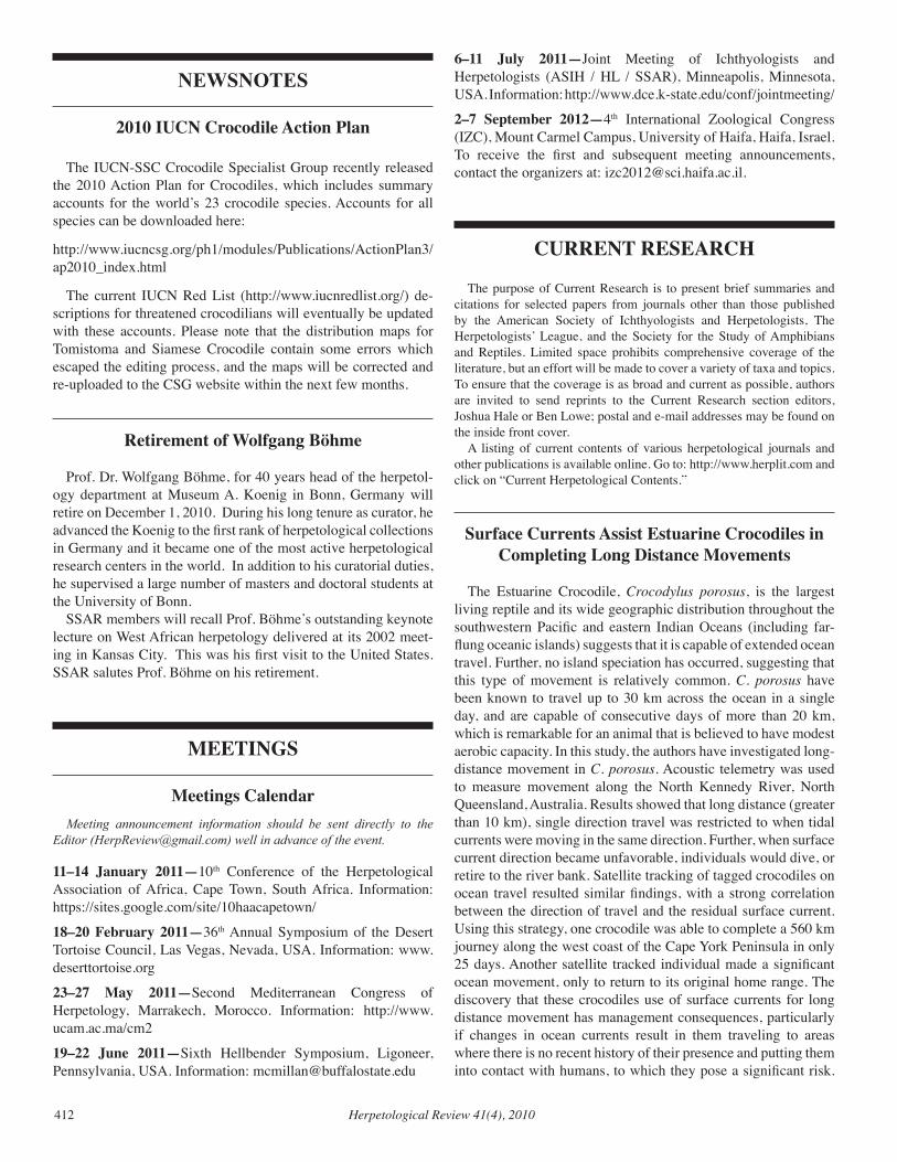
fig. 2. Kyle Miller Hesed and Steve Gorzula braving the mass attendance at the SSAR booth at the USA Science and Engineering Festival.
fig. 3. George Zug interpreting a live Corn Snake for a young woman attending the SSAR booth.
frog also applied to their hand (or even forehead!) as a “tattoo.” Next year we need special SSAR T-shirts for the booth staffers, and stickers or buttons to decorate the young people!
Given the contemporary popularity of herps as pets, and their ubiquity in classrooms and education programs at museums, na-ture centers, and zoos, it was surprising to see how many of the attendees had never touched or held a live reptile before. A great number of people touched their first snake or turtle at the SSAR booth at the Science Festival in 2010, an experience they are not likely to forget. At the other extreme, it was rewarding to meet so many people—especially young people—who were remark-ably learned and respectful towards amphibians and reptiles. We met 12-year olds who knew the difference between elapids and vipers, or who knew about the amphibian chytrid fungus.
The evident success of the weekend event was bolstered by spectacularly beautiful Fall weather and extraordinarily smooth logistics and planning on the part of the organizers. The event was promoted by President Obama and covered by major me-dia. The National Park Service has conservatively estimated an astounding attendance between 750,000–1,000,000 persons. We can be proud to truthfully say that SSAR, in keeping with its mis-sion, reached out and “brought herpetology to the people” in a booth staffed by members including hobbyists, career academics, and students (Figs. 2, 3).
Credits: Brian Crother suggested and initiated SSAR participa-tion in the event; Joe Mendelson handled pre-festival arrange-ments; Karen Lips led intra-DC logistics and volunteers for the booth; Joe Mendelson and Karen Lips managed daily set-up and break-down of the booth and staffed it for both days; use of live animals was facilitated by Jim Murphy and Robin Saun-ders, with animals courtesy of Long Branch Nature Center; viper fang displays were prepared and loaned by Jason Brock; skeletal specimens were loaned by Education Department at Zoo Atlanta; Breck Bartholomew selected and shipped representative SSAR publications. Volunteer staff at the booth were Anne Maglia, George Zug, Roy McDiarmid, Ted Kahn, Jake Li, Kyle Miller Hesed, Peter Uetz, Brian Gratwicke, Steve Gorzula, and Jim Murphy.
—Submitted by Joseph R. Mendelson, SSAR President-Elect
2011 Year of the TurtlePartners in Amphibian and Reptile Conservation (PARC), along
with many partners (IUCN Tortoise and Freshwater Turtle Special-ist Group, Turtle Survival Alliance, Chelonian Taxonomic Advisory Group, The Turtle Conservancy, and a growing list of others) are designating 2011 as “Year of the Turtle.” We invite SSAR members to join our efforts! This is an opportunity to raise awareness of ef-forts in research, conservation, and education to benefit these animals. Throughout the year there will be a number of activities including monthly newsletters featuring conservation or research efforts, local events and presentations, official reports and meeting highlights, a photo contest and release of monthly calendar pages with selected photos, and educational materials for teachers and children, including arts, humanities, and cultural values relating to turtles. Please contact Dede Olson ([email protected]) or Priya Nanjappa ([email protected]) if you’d like to assist or to contribute information toward the newsletter or related resources.
Herpetological Review 41(4), 2010412
NEWSNOTES
2010 IUCN Crocodile Action Plan
The IUCN-SSC Crocodile Specialist Group recently released the 2010 Action Plan for Crocodiles, which includes summary accounts for the world’s 23 crocodile species. Accounts for all species can be downloaded here:
http://www.iucncsg.org/ph1/modules/Publications/ActionPlan3/ap2010_index.html
The current IUCN Red List (http://www.iucnredlist.org/) de-scriptions for threatened crocodilians will eventually be updated with these accounts. Please note that the distribution maps for Tomistoma and Siamese Crocodile contain some errors which escaped the editing process, and the maps will be corrected and re-uploaded to the CSG website within the next few months.
Retirement of Wolfgang Böhme
Prof. Dr. Wolfgang Böhme, for 40 years head of the herpetol-ogy department at Museum A. Koenig in Bonn, Germany will retire on December 1, 2010. During his long tenure as curator, he advanced the Koenig to the first rank of herpetological collections in Germany and it became one of the most active herpetological research centers in the world. In addition to his curatorial duties, he supervised a large number of masters and doctoral students at the University of Bonn.
SSAR members will recall Prof. Böhme’s outstanding keynote lecture on West African herpetology delivered at its 2002 meet-ing in Kansas City. This was his first visit to the United States. SSAR salutes Prof. Böhme on his retirement.
MEETINGS
Meetings Calendar Meeting announcement information should be sent directly to the Editor ([email protected]) well in advance of the event.
11–14 January 2011—10th Conference of the Herpetological Association of Africa, Cape Town, South Africa. Information: https://sites.google.com/site/10haacapetown/
18–20 February 2011—36th Annual Symposium of the Desert Tortoise Council, Las Vegas, Nevada, USA. Information: www.deserttortoise.org
23–27 May 2011—Second Mediterranean Congress of Herpetology, Marrakech, Morocco. Information: http://www.ucam.ac.ma/cm2
19–22 June 2011—Sixth Hellbender Symposium, Ligoneer, Pennsylvania, USA. Information: [email protected]
6–11 July 2011—Joint Meeting of Ichthyologists and Herpetologists (ASIH / HL / SSAR), Minneapolis, Minnesota, USA. Information: http://www.dce.k-state.edu/conf/jointmeeting/
2–7 September 2012—4th International Zoological Congress (IZC), Mount Carmel Campus, University of Haifa, Haifa, Israel. To receive the first and subsequent meeting announcements, contact the organizers at: [email protected].
CURRENT RESEARCH
The purpose of Current Research is to present brief summaries and citations for selected papers from journals other than those published by the American Society of Ichthyologists and Herpetologists, The Herpetologists’ League, and the Society for the Study of Amphibians and Reptiles. Limited space prohibits comprehensive coverage of the literature, but an effort will be made to cover a variety of taxa and topics. To ensure that the coverage is as broad and current as possible, authors are invited to send reprints to the Current Research section editors, Joshua Hale or Ben Lowe; postal and e-mail addresses may be found on the inside front cover. A listing of current contents of various herpetological journals and other publications is available online. Go to: http://www.herplit.com and click on “Current Herpetological Contents.”
Surface Currents Assist Estuarine Crocodiles in Completing Long Distance Movements
The Estuarine Crocodile, Crocodylus porosus, is the largest living reptile and its wide geographic distribution throughout the southwestern Pacific and eastern Indian Oceans (including far-flung oceanic islands) suggests that it is capable of extended ocean travel. Further, no island speciation has occurred, suggesting that this type of movement is relatively common. C. porosus have been known to travel up to 30 km across the ocean in a single day, and are capable of consecutive days of more than 20 km, which is remarkable for an animal that is believed to have modest aerobic capacity. In this study, the authors have investigated long-distance movement in C. porosus. Acoustic telemetry was used to measure movement along the North Kennedy River, North Queensland, Australia. Results showed that long distance (greater than 10 km), single direction travel was restricted to when tidal currents were moving in the same direction. Further, when surface current direction became unfavorable, individuals would dive, or retire to the river bank. Satellite tracking of tagged crocodiles on ocean travel resulted similar findings, with a strong correlation between the direction of travel and the residual surface current. Using this strategy, one crocodile was able to complete a 560 km journey along the west coast of the Cape York Peninsula in only 25 days. Another satellite tracked individual made a significant ocean movement, only to return to its original home range. The discovery that these crocodiles use of surface currents for long distance movement has management consequences, particularly if changes in ocean currents result in them traveling to areas where there is no recent history of their presence and putting them into contact with humans, to which they pose a significant risk.
Herpetological Review 41(4), 2010 413
Furthermore, translocations of problem crocodiles, a common management strategy, need to take place where prevailing currents do not allow a rapid passage back to their original location.
caMPBell, H. a., M. e. WattS, S. Sullivan, M. a. read, S. cHoukroun, S. r. irWin and c. e. franklin. 2010. Estuarine crocodiles ride surface currents to facilitate long-distance travel. Journal of Animal Ecology 79:955–964.
Correspondence to: Hamish Campbell, School of Biological Sciences, The University of Queensland, Brisbane, Qld 4072, Australia; e-mail: [email protected].
Extreme Longevity in the Human Fish
The human fish or olm, Proteus anguinus (Proteidae), is an aquatic, paedomorphic, cave-dwelling salamander native to the European countries Slovenia and Croatia. As captive specimens have been known to live for over 70 years, this species has long been of interest to biologists studying aging and senescence. Extreme lifespan has been demonstrated in a number of taxa, including mammals, birds and reptiles, and is usually positively correlated with size. However, the Human Fish is small, generally less than 20 g. Longevity has also been associated with living in environments that are stable and/or free from predators, the possession of mechanisms that mitigate damage caused by free radicals, and slow metabolisms. It is cave dwelling and therefore lives in a stable environment, and is thought to not suffer from predation. Additionally, it has been shown not to possess advanced cellular oxidation defences; the metabolic rate of Proteus is unknown. A breeding colony, established in a cave in Saint-Girons, France in 1952, now has over 400 individuals of various age classes, allowing the authors of this paper to conduct a detailed investigation of the demographic and physiological characteristics of this species. Data on physical condition and instances of birth and death (collected weekly) reveal that the population is growing at a rate of 1.05, olms reach sexual maturity at roughly age 15, reproduce once every 12 years, and average adult lifespan is roughly 70 years. From these data, the researchers determined that the maximum lifespan for this species is 102 years. Furthermore, while salamanders exhibit reduced metabolic rates relative to other vertebrates, laboratory experiments found the metabolism of the human fish to not be markedly different from other salamanders. While the proximate mechanisms leading to longevity in the human fish remain elusive, it does represent an excellent model organism for studying longevity and senescence in vertebrates.
voituron, Y., M. de fraiPont, J. iSSartel, o. guillauMe and J. cloBert. 2010. Extreme lifespan of the human fish (Proteus anguinus): a challenge for ageing mechanisms. Biology Letters doi:10.1098/rsbl.2010.0539.
Correspondence to: Yann Voituron, Ecologie des Hydrostèmes Fluviaux, UMR CNRS 5023, Université Claude Bernard Lyon 1, Université de Lyon, 69622 Villeurbanne cedex, France; e-mail: [email protected].
Tadpoles Recognize Native but not Introduced Turtle Predators
The impact of introduced predators on prey populations is well known, but less research has been conducted on the negative effects on native species of having to compete with these alien species. On the Iberian Peninsula, the native freshwater turtles (the European pond turtle, Emys orbicularis and the Spanish terrapin, Mauremys leprosa), are being displaced by exotic species (principally the red-eared slider, Trachemys scripta elegans and the false map turtle, Graptemys pseudogeographica). Both native and introduced species feed on anuran tadpoles, which are able to recognize local predators and alter their behavior. In this study the authors have examined tadpole responses to both native and introduced species in order to determine whether they can recognize introduced species as well. Chemical cues of the four turtle species mentioned above were presented to tadpoles of four species of European anurans: Iberian green frog (Pelophylax perezi), western spadefoot (Pelobates cultripes), natterjack toad (Bufo calamita), and common gree frog (Hyla arborea). Three species (P. perezi, P. cultripes and H. arborea) reduced their swimming behavior in response to native, but not introduced species. The authors suggest that a lack of evolutionary experience with the introduced predators explains the inability of the tadpoles of these species to detect them and may confer a competitive advantage to the introduced turtle species.
Polo-cavia, n., a. gonzalo, P. lóPez and J. Martín. 2010. Predator recognition of native but not invasive turtle predators by naïve anuran tadpoles. Animal Behaviour 80:461–466.
Correspondence to: Nuria Polo-Cavia, Departamento de Biología, Universidad Autónoma de Madrid, Ciudad Universitaria de Cantoblanco, 28049 Madrid, Spain; e-mail: [email protected]
Cane Toad Eggs Toxic to Tadpoles of Native Australian Anurans
The introduced Cane Toad, Bufo (= Rhinella) marninus, is inflicting a number of profound impacts on native species as it expands across Australia. Previous studies have shown cane toad eggs to be extremely toxic to the tadpoles of native Australian anurans and in this study, the authors have conducted a number of laboratory trials to determine if there is variation among tadpoles of Australian species in propensity to consume cane toad eggs or susceptibility to toad egg toxins. In each trial, individual tadpoles were presented ten cane toad eggs either with or without an additional food source (lettuce). At least some individuals of each of the 15 anuran species studied ingested toad eggs, with mortality following rapidly in most cases. While no interspecific difference in propensity to ingest eggs was observed, the high interspecific variance in survival rate (0 > 70%) was driven by a difference in feeding behavior (e.g., small size preventing efficient consumption of toad eggs) rather than physiological tolerance. Furthermore, alternative food sources did not reduce mortality from egg ingestion as most tadpoles foraged on both the eggs and the lettuce. The influence of overall body size on
Herpetological Review 41(4), 2010414
survival was also examined in four species. While three of the species did show an effect of body size on mortality, this was not in a consistent direction, and instead was a complex interaction related to alternate food availability. Interclutch affects were examined in one species, but none were observed. This study highlights the toxicity of toad eggs to native frogs and that frequency of mortality relates to a complex interaction between body size, species and other ecological factors.
croSSland, M. r. and r. SHine. 2010. Vulnerability of an Australian anuran tadpole assemblage to the toxic eggs of the invasive cane toad (Bufo marinus). Austral Ecology 35:197–203.
Correspondence to: Richard Shine, School of Biological Sciences A08, University of Sydney, New South Wales 2006, Australia; e-mail: [email protected].
How Much UV-B Should a Captive Reptile be Exposed to?
Ultraviolet-B (UV-B) exposure is important to the production of Vitamin D in many vertebrate species, including reptiles, and plays a part in calcium-phosphorous balance and immune function. The amount of UV-B to provide is therefore an important consideration to ensure the good health of captive reptiles. In this study, the authors have investigated voluntary UV-B exposure of a number of squamate species in the wild, in the US and Jamaica. In Jamaica, three species of Anolis (A. lineotopus, A. grahami, and A. sagrei) were investigated. In the US, Agkistrodon piscivorous, Pantherophis obsoletus, Thamnophis proximus, Nerodia fasciata, N. erythrogaster, Uta stansburiana, Holbrookia maculata, Sceloporus olivaceus, S. consobrinus, S. undulatus, S. graciosus, and Anolis carolinensis were studied. Animals encountered in the field were subjected to cloacal temperature readings and UV-B readers were employed to ascertain the level of UV-B exposure the animals were experiencing. The authors found significant variation among species in UV-B exposure; more extensive observations of two species of Sceloporus (S. olivaceus and S. graciosus) revealed seasonal and diel variation as well. The authors provide a number of guidelines for providing UV-B to these species in captivity. They also highlight the importance of providing a UV-B refuge in enclosures to allow behavioral regulation of UV-B exposure.
ferguSon, g. W., a. M. Brinker, W. H. geHrMann, S. e. Bucklin, f. M. BaineS and S. J. Mackin. 2010. Voluntary exposure of some Western-Hemisphere snakes and lizard species to ultraviolet-B radiation in the field: How much ultraviolet-B should a lizard or snake receive in captivity? Zoo Biology 29:317–334.
Correspondence to: Gary Ferguson, Department of Biology, Texas Christian University, Box 298930, Fort Worth, Texas 76129, USA; e-mail: [email protected].
Behavioral Syndromes, or “Personalities” Investigated in a Lizard
Behavioral syndromes (“personalities”) are increasingly being recognized as an important component of natural systems, having
been observed in fish, invertebrates, mammals, and reptiles. In this paper, the authors have examined behavioral syndromes in the Namibian Rock Agama, Agama planiceps. To determine if individuals displayed consistent magnitudes of boldness or shyness, investigators measured flight initiation distance (the distance away a perceived threat, in this case an advancing experimenter, was from the lizard before it fled) for a number of individuals several times over a period of time. Next, male lizards were observed and their behavior (instances of head bobbing and push ups; collectively defined as “signaling”) was documented. Finally, possible fitness consequences of boldness or shyness was investigated by measuring the home ranges, rates of tail loss, and feeding rates of several males. Individuals were found to exhibit consistent degrees of boldness. Bold males spent more time basking and moving and had larger home ranges and higher feeding rates than shy males. The rate of signaling behavior however, did not differ between bold and shy individuals. Males that had a third or more of their tale missing were more likely to be bold, suggesting that there may be an increased predation risk associated with being bold. The authors suggest that long term studies are required to fully understand the fitness consequences of animal personalities such as boldness.
carter, a. J., a. W. goldizen, and S. a. troMP. 2010. Agamas exhibit behavioral syndromes: Bolder males bask and feed more but may suffer higher predation. Behavioral Ecology doi: 10.1093/beheco/arq036.
Correspondence to: Alecia Carter, The Fenner School of Environment and Society, The Australian National University, Acton, Canberra, ACT 0200, Australia; e-mail: [email protected].
Roads Act as Barriers to Seasonal Migration in Timber Rattlesnakes
Anthropogenic landscape modification, especially roads, can fragment habitats and act as dispersal barriers to wildlife. However, the degree to which a road acts as a barrier to dispersal is related to characteristics both of the road itself and the animal in question. In this study, the authors have examined the impact of recently constructed roads separating hibernacula (communal dens) of timber rattlesnakes, Crotalus horridus in four regions in New York State, USA. Microsatellites were used to examine population subdivision and gene flow across roads. It was found that hibernacula separated by roads had a higher degree genetic differentiation than hibernacula in contiguous habitat and also were less genetically diverse. This was further examined by completing genetic assignment analyses, which showed that disruption of seasonal migration was the cause of these patterns, with matings less likely to occur between snakes from hibernacula separated by roads. This study highlights the fragmenting nature of roads, even those that are relatively small, or recent in origin.
clark, r. W., W. S. BroWn, r. StecHert and k. r. zaMudio. 2010. Roads, interrupted dispersal, and genetic diversity in timber rattlesnakes. Conservation Biology 24:1059–1069.
Correspondence to: Rulon Clark, Department of Biology, San Diego State University, 5500 Campanile Drive, San Diego, California 92182, USA; e-mail: [email protected].
Herpetological Review 41(4), 2010 415
The Island of Bermuda Preserves an Ancient Skink Lineage
Paleoendemism is the preservation of ancient, formerly widespread lineages in a restricted geographic area. Islands can be good sources of paleoendemism, with the most well known example being that of the Tuatara, which was once widely-distributed, but is now restricted to islands off the shores of New Zealand. The native fauna of the island of Bermuda includes only a single extant flightless terrestrial vertebrate: the endemic scinid lizard, Plestiodon longirostris. In this study, the authors have assembled a multi-locus molecular dataset which they used to reconstruct a phylogeny and estimate divergence times, which were in turn used to determine if colonization of Bermuda was recent or ancient. They found that the lineage occuring on Bermuda is indeed ancient, having split from the lineage containing its closest extant relatives before the bulk of the diversification events that led to the divirsity of skink species occurring in North America today (about 16 million years ago). Furthermore, the extant members of the clade from which the Bermuda Skink split occur in western North America (P. skiltonianus complex), with no extant eastern North American representatives. Even more interestingly, it is found only on Bermuda currently, which is itself only 2 million years old. Fossil evidence of P. longirostris on Bermuda from 400,000 years ago suggests that colonization occurred between 2 million and 400,000 years ago, possibly by rafting the 1000 km that separates Bermuda from the North American mainland (where they once existed). Following colonization, the lineage then disappeared from the mainland. Bermuda therefore, represents a unique ‘life raft,’ preserving one of the most ancient lineages of North American Plestiodon. Currently however, habitat destruction and degradation and pressure from non-native species has severely lowered population size, resulting in this unique lizard being highly endangered.
BrandleY, M. c., Y. Wang, X. guo, a. n. MonteS de oca, M. f. ortíz, t. Hikida and H. ota. 2010. Bermuda as an evolutionary life raft for an ancient lineage of endangered lizards. PLoS ONE 5: e11375.
Correspondence to: Matthew Brandley, Department of Ecology and Evolutionary Biology, Yale University, New Haven, Connecticut, USA; e-mail: [email protected].
Newly Discovered Chameleon Species Highlights Importance of Preserving Biodiversity Hotspots
Madagascar has the dubious distinction of being among the most imperiled biodiversity hotspots on earth. However, despite a mere 17% of its original vegetation remaining, Madagascar continues to divulge species new to science. The authors of this paper describe a distinctive new species of chameleon, Calumma tarzan, from three localities on the eastern slope of the island (Anosibe An’Ala region). This species exhibits morphological traits consistent with the Calumma furcifer species group. However, it possesses a unique rostral spade, and males have a distinctive dorsal brown blotch on the head and neck. A
molecular analysis confirms the phylogenetic placement of this new taxon, follows previous analyses in failing to resolve relationships within the C. furcifer species group, corroborates the evolutionary distinctiveness of these lizards, and suggests two genetically differentiated populations (north and south). The specific epithet is derived both from the former name of a nearby village, Tarzanville (now Ambatofotsy), and the fictional forest denizen, Tarzan. As C. tarzan is known to occupy less that 10 km2, the authors posit that it should receive the IUCN’s Critically Endangered listing. They also point out the lack of protected land in this region (amounting to a “reserve gap”) and hope the patronym brings attention to this overlooked region and compels conservation efforts.
geHring, P. S., M. PaBiJan, f. M. ratSoavina, J. köHler, M. venceS, and f. glaW. 2010. A Tarzan yell for conservation: A new chameleon, Calumma tarzan sp. n., proposed as a flagship species for the creation of new nature reserves in Madagascar. Salamandra 46:167–179.
Correspondence to: Philip-Sebastian Gehring, Division of Evolutionary Biology, Zoological Institute, Technical University of Braunschweig, Spielmannstr. 8, 38106 Braunschweig, Germany; e-mail: [email protected].
Habitat Preferences and Home Range Size in a Colubrid Snake
Effective wildlife conservation plans should consider both the habitat needs and spatial requirements of the species in question. Studies that focus on the correlation between the habitat preferences and movement patterns of wildlife, particularly snakes, are uncommon. In this study, the authors attempted to determine how habitat preferences or quality influenced movement patterns of snakes. To answer this question, they used habitat preference or avoidance information obtained for Bullsnakes (Pituophis catenifer sayi) from 2003–2005 at a site in the upper-midwestern United States and compared it to minimum convex polygon estimates of home range size derived from data on radio-tracked snakes. They employed Geographical Information Systems to model the amount of preferred (open bluff faces) and avoided (agricultural fields and closed canopy forests) habitats within each estimated home range and compared them via multiple linear regression. They also tested the influence of gender, length and weight on home range size. The results of this study indicated that, among the investigated potential correlates, amount of avoided habitat was the most significant indicator of home range size. More specifically, home range size increased with amount of avoided habitat within the home range. As bullsnakes are large snakes, they are particularly susceptible to road mortality; therefore, fragmentation of suitable habitat forces snakes to wander further in the search for suitable habitat, exposing them to greater risk. In a broader context, this finding supports the hypothesis that habitat quality has an impact on wildlife movement patterns, and the relationship between habitat needs and spatial requirements should be considered when conserving or managing species.
kaPfer, J. M., c. W. Pekar, d. M. reineke, J. r. cogginS, and r. HaY. 2010. Modeling the relationship between habitat preferences and
Herpetological Review 41(4), 2010416
home-range size: A case study on a large mobile colubrid snake from North America. Journal of Zoology 282:13–20.
Correspondence to: Joshua M. Kapfer, Departments of Environmental Studies and Biology, Campus Box 2015, Elon University, Elon, North Carolina 27244, USA; e-mail: [email protected].
Multi-year Study of Toad Populations Reveals Demographic Implications of Chitrid Fungus
While many studies have implicated Batrachochytrium dendrobatidis (Bd) infection in population declines, few have studied natural populations for multiple years to elucidate the specific demographic impact of the disease. The authors of this study conducted a six-year capture-recapture study on three populations of western toad (Bufo [= Anaxyrus] boreas) in the Rocky Mountains of western US. Of these populations, two were infected with Bd and one was not. Captured animals were individually marked (if first capture), measured, swabbed to determine if the animal was infected with Bd, and released. From these data, population growth rates were obtained and analyses were conducted to determine if growth rates were correlated with various factors such as the presence or absence of Bd and climactic conditions (e.g., breading season temperature). Roughly 58% of individuals in the two Bd-infected populations were found to be infected with the disease. These infected animals were found to have an annual survival coefficient of about 0.47 (about 0.75 for uninfected individuals). Bd-infected populations were found to be declining by roughly 6% per year over the duration of the study while the Bd-free population exhibited stable population growth. Furthermore, population growth rates were not correlated with temperature. These findings reveal that in addition to causing rapid local extirpations, in some instances, Bd can also exhibit long-term persistence as a chronic disease (and that some species can persist with Bd). Furthermore, this study highlights the need for long-term monitoring of amphibian populations to determine the species-specific demographic impacts of pathogens.
Pilliod, d. S., and colleagueS. 2010. Effects of amphibian chytrid fungus on individual survival probability in wild boreal toads. Conservation Biology 24:1259–1267.
Correspondence to: David Pilliod. U.S. Geological Survey, Forest and Rangeland Ecosystem Science Center, Snake River Field Station, 970 Lusk Street, Boise, Idaho 83706, USA; e-mail: [email protected].
ZOO VIEW
Snake Versus Rabbit
This colorful excerpt was sent by Jon Coote about the Tower of London and the last director, Alfred Cops, who took over the operation in 1822.Mr. Broderip, in the second volume of the Zoological Journal,
“Mr. Cops of the Lion Office in the Tower,” writes Broderip, “sent to inform me that one of these reptiles had just cast his skin, at which period they, in common with other serpents, are most active and eager for prey. Accordingly I repaired with some friends to the Tower, where we found a spacious cage, the floor of which consisted of a tin case covered with red baize and filled with warm water, so as to produce a proper temperature. There was the snake, “ positis novus exuviis,” gracefully examining the height and extent of his prison as he raised, without any apparent effort, his towering head to the roof and upper parts of it, full of life, and brandishing his tongue.
A large buck rabbit was introduced into the cage. The snake was down and motionless in a moment. There he lay like a log without one symptom of life, save that which glared in the small bright eye twinkling in his depressed head. The rabbit appeared to take no notice of him, but presently began to walk about the cage. The snake suddenly, but almost imperceptibly, turned his head according to the rabbit’s movements, as if to keep the object within the range of his eye. At length the rabbit, totally unconscious of his situation, approached the ambushed head. The snake dashed at him like lightning. There was a blow—a scream—and instantly the victim was locked in the coils of the serpent. This was done almost too rapidly for the eye to follow: at one instant the snake was motionless : in the next he was one congeries of coils round his prey. He had seized the rabbit by the neck just under the ear, and was evidently exerting the strongest pressure round the thorax of the quadruped ; thereby preventing the expansion of the chest, and at the same time depriving the anterior extremities of motion. The rabbit never cried after the first seizure:—he lay with his hind legs stretched out, still breathing with difficulty, as could be seen by the motion of his flanks. Presently he made one desperate struggle with his hind legs; but the snake cautiously applied another coil with such dexterity as completely to manacle the lower extremities, and, in about eight minutes, the rabbit was quite dead. The snake then gradually and carefully uncoiled himself, and, finding that his victim moved not, opened his mouth, let go his hold, and placed his head opposite to the fore part of the rabbit. The boa generally, I have observed, begins with the head ; but in this instance the serpent, having begun with the fore-legs, was longer in gorging his prey than usual, and in consequence of the difficulty-presented by the awkward position of the rabbit, the dilatation and secretion of lubricating mucus were excessive. The serpent first got the fore-legs into his mouth; he then coiled himself round the rabbit, and appeared to draw out the dead body through his folds; he then began to dilate his jaws, and holding the rabbit firmly in a coil as a point of resistance, appeared to exercise at intervals the whole of his anterior muscles in protruding his stretched jaws and lubricated mouth and throat at first against, and soon after gradually upon, and over his prey. The curious mechanism in the jaws of serpents which enables them to swallow bodies so disproportioned to their apparent bulk is too well known to need description; but it may be as well to state that the symphysis of the under jaw was separated in this case, and in others which I have had an opportunity of observing. When the prey was completely ingulphed, the serpent lay for a few moments with his dislocated jaws still dropping with the mucus which had lubricated the parts, and at this time he looked quite sufficiently disgusting. He then stretched out his neck, and at the same moment the muscles seemed to push the prey further downwards. After a few efforts to replace the parts,
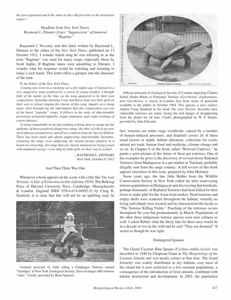
Herpetological Review 41(4), 2010 417
the jaws appeared much the same as they did previous to the monstrous repast.”
Headline from New York Times: Raymond L. Ditmars Urges “Suppression” of Immoral
“Ragtime”
Raymond J. Novotny sent this letter written by Raymond L. Ditmars to the editor of the New York Times, published on 13 October 1912. I wonder which song he was referring to as the term “Ragtime” was used for many songs, especially those by Scott Joplin. If Ragtime tunes were unsettling to Ditmars, I wonder what his response would be watching and listening to today’s rock bands. This letter offers a glimpse into the character of the man.
To the Editor of the New York Times: Coming into town in a smoking car a few nights ago, I listened to a
very suggestive song rendered by a crowd of young rowdies. I thought little of the matter at the time, as the song appeared to be their own composition. Yesterday morning I was startled to hear two little girls on their way to school singing the chorus of this song. Inquiry at a cheap music store brought me the information that this composition was one of the latest “popular” songs. It follows in the wake of objectionable portrayals of marital infidelity, risque situations, and crude twistings of coarse phrases.
It seems remarkable to me that nothing is being done to stamp out the epidemic of these positively dangerous songs, the titles of which are now stock phrases around town, and all too common from the lips of children. There has been much ado about suppressing objectionable literature, cleansing the stage, even subjecting the motion picture playlets to a board of censorship. Yet songs that are clearly immoral are being issued with unabated energy—even sung by little girls on their way to school.
—RAYMOND L. DITMARSNew York, October 9, 1912
And Then There Was One
Whenever a book appears on the scene with a title like The Last Tortoise: A Tale of Extinction in Our Lifetime (2010, The Belknap Press of Harvard University Press, Cambridge, Massachusetts & London, England ISBN 978-0-674-04992-5) by Craig B. Stanford, it is clear that this will not be an uplifting read. In
fact, tortoises are under siege worldwide, caused by a number of human-induced pressures, and Stanford covers all of these usual factors in depth: habitat alteration, collection for exotic animal pet trade, human food and medicine, climate change and so on. In Chapter 6 of the book called “Beloved Captives,” he paints a grim picture of the future of these pet tortoises. One of the examples he gives is the discovery of several dozen Radiated Tortoises from Madagascar in a pet market in Thailand, probably illegally sent from the range country. A full review of this book appears elsewhere in this issue, prepared by John Moriarty.
Some years ago, the late John Behler from the Wildlife Conservation Society in New York called me after resurveying tortoise populations in Madagascar and discovering that hundreds, perhaps thousands, of Radiated Tortoises had been killed for their livers to make pâté for the Asian food market. Dead tortoises and empty shells were scattered throughout the habitat; virtually no living individuals were located and he characterized the locale as “The Tortoise Killing Fields.” Poaching of the tortoises occurs throughout the year but predominantly in March. Populations of the other three indigenous tortoise species were near collapse as well. I asked Behler what the likely fate for these taxa would be in a decade or two in the wild and he said “They are doomed!” It seems as though he was right.
Endangered Iguanas
The Grand Cayman Blue Iguana (Cyclura nubila lewisi) was described in 1940 by Chapman Grant in The Herpetology of the Cayman Islands and was nearly extinct at that time. The lizard formerly was widely distributed in dry habitats over most of the island but is now restricted to a few remnant populations, a consequence of the introduction of feral animals, combined with habitat destruction and development. In 2002, the population
Undated postcard of child riding a Galápagos Tortoise named “Grandpa” at New York Zoological Society. Zoos no longer offer tortoise “rides.” Credit: provided by Brint Spencer.
Official postcard of Zoological Society of London depicting Charles Island [Santa María or Floreana] Tortoise (Geochelone elephantopus, now Geochelone n. nigra) at London Zoo from series of postcards available to the public in October 1904. This species is now extinct. Author Craig Stanford in his book The Last Tortoise describes how vulnerable tortoises are today, facing the real danger of disappearing from the planet for all time. Credit: photographed by W. P. Dando, provided by John Edwards.
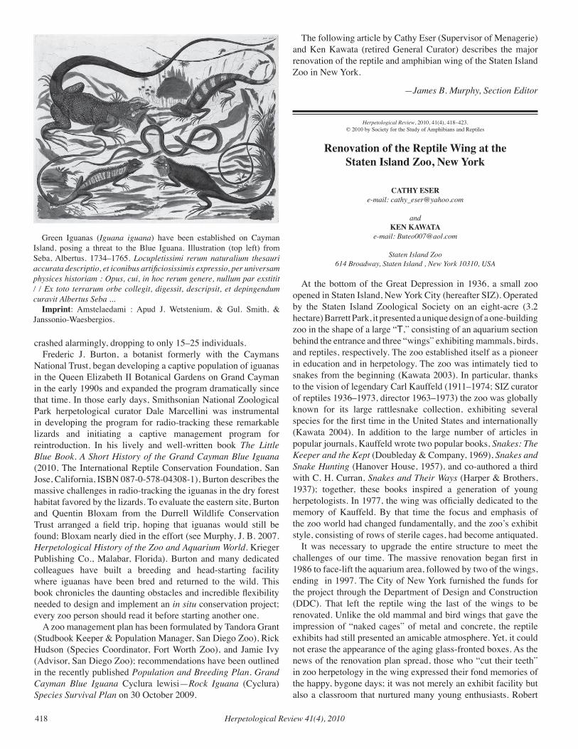
Herpetological Review 41(4), 2010418
crashed alarmingly, dropping to only 15–25 individuals.Frederic J. Burton, a botanist formerly with the Caymans
National Trust, began developing a captive population of iguanas in the Queen Elizabeth II Botanical Gardens on Grand Cayman in the early 1990s and expanded the program dramatically since that time. In those early days, Smithsonian National Zoological Park herpetological curator Dale Marcellini was instrumental in developing the program for radio-tracking these remarkable lizards and initiating a captive management program for reintroduction. In his lively and well-written book The Little Blue Book. A Short History of the Grand Cayman Blue Iguana (2010, The International Reptile Conservation Foundation, San Jose, California, ISBN 087-0-578-04308-1), Burton describes the massive challenges in radio-tracking the iguanas in the dry forest habitat favored by the lizards. To evaluate the eastern site, Burton and Quentin Bloxam from the Durrell Wildlife Conservation Trust arranged a field trip, hoping that iguanas would still be found; Bloxam nearly died in the effort (see Murphy, J. B. 2007. Herpetological History of the Zoo and Aquarium World. Krieger Publishing Co., Malabar, Florida). Burton and many dedicated colleagues have built a breeding and head-starting facility where iguanas have been bred and returned to the wild. This book chronicles the daunting obstacles and incredible flexibility needed to design and implement an in situ conservation project; every zoo person should read it before starting another one.
A zoo management plan has been formulated by Tandora Grant (Studbook Keeper & Population Manager, San Diego Zoo), Rick Hudson (Species Coordinator, Fort Worth Zoo), and Jamie Ivy (Advisor, San Diego Zoo); recommendations have been outlined in the recently published Population and Breeding Plan. Grand Cayman Blue Iguana Cyclura lewisi—Rock Iguana (Cyclura) Species Survival Plan on 30 October 2009.
The following article by Cathy Eser (Supervisor of Menagerie) and Ken Kawata (retired General Curator) describes the major renovation of the reptile and amphibian wing of the Staten Island Zoo in New York.
—James B. Murphy, Section Editor
Herpetological Review, 2010, 41(4), 418–423.© 2010 by Society for the Study of Amphibians and Reptiles
Renovation of the Reptile Wing at the Staten Island Zoo, New York
CATHY ESERe-mail: [email protected]
andKEN KAWATA
e-mail: [email protected]
Staten Island Zoo614 Broadway, Staten Island , New York 10310, USA
At the bottom of the Great Depression in 1936, a small zoo opened in Staten Island, New York City (hereafter SIZ). Operated by the Staten Island Zoological Society on an eight-acre (3.2 hectare) Barrett Park, it presented a unique design of a one-building zoo in the shape of a large “T,” consisting of an aquarium section behind the entrance and three “wings” exhibiting mammals, birds, and reptiles, respectively. The zoo established itself as a pioneer in education and in herpetology. The zoo was intimately tied to snakes from the beginning (Kawata 2003). In particular, thanks to the vision of legendary Carl Kauffeld (1911–1974; SIZ curator of reptiles 1936–1973, director 1963–1973) the zoo was globally known for its large rattlesnake collection, exhibiting several species for the first time in the United States and internationally (Kawata 2004). In addition to the large number of articles in popular journals, Kauffeld wrote two popular books, Snakes: The Keeper and the Kept (Doubleday & Company, 1969), Snakes and Snake Hunting (Hanover House, 1957), and co-authored a third with C. H. Curran, Snakes and Their Ways (Harper & Brothers, 1937); together, these books inspired a generation of young herpetologists. In 1977, the wing was officially dedicated to the memory of Kauffeld. By that time the focus and emphasis of the zoo world had changed fundamentally, and the zoo’s exhibit style, consisting of rows of sterile cages, had become antiquated.
It was necessary to upgrade the entire structure to meet the challenges of our time. The massive renovation began first in 1986 to face-lift the aquarium area, followed by two of the wings, ending in 1997. The City of New York furnished the funds for the project through the Department of Design and Construction (DDC). That left the reptile wing the last of the wings to be renovated. Unlike the old mammal and bird wings that gave the impression of “naked cages” of metal and concrete, the reptile exhibits had still presented an amicable atmosphere. Yet, it could not erase the appearance of the aging glass-fronted boxes. As the news of the renovation plan spread, those who “cut their teeth” in zoo herpetology in the wing expressed their fond memories of the happy, bygone days; it was not merely an exhibit facility but also a classroom that nurtured many young enthusiasts. Robert
Green Iguanas (Iguana iguana) have been established on Cayman Island, posing a threat to the Blue Iguana. Illustration (top left) from Seba, Albertus. 1734–1765. Locupletissimi rerum naturalium thesauri accurata descriptio, et iconibus artificiosissimis expressio, per universam physices historiam : Opus, cui, in hoc rerum genere, nullum par exstitit / / Ex toto terrarum orbe collegit, digessit, descripsit, et depingendum curavit Albertus Seba ...
Imprint: Amstelaedami : Apud J. Wetstenium, & Gul. Smith, & Janssonio-Waesbergios.
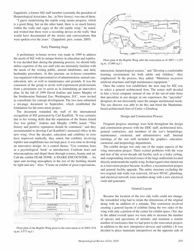
Herpetological Review 41(4), 2010 419
Zappalorti, a former SIZ staff member (currently the president of Herpetological Associates, Inc., in New Jersey), was one of them.
“I guess modernizing the reptile wing means progress, which is a good thing, but on the other hand, there is so much history within the walls and cages of the old reptile wing,” he noted, and wished that there were a recording device in the walls “that would have documented all the stories and conversations that were spoken over the years.” (Zappalorti, pers. comm. 2005)
Early Planning Stage
A preliminary in-house review was made in 1999 to address the needs of SIZ with its unique history in education and reptiles. It was decided that, during the planning process, we should fully utilize expertise of the zoo staff who are intimately familiar with the needs of the visiting public, animal inhabitants, and daily husbandry procedures. At this juncture, an in-house committee was organized with representatives of administration, animal care, education, arts, as well as maintenance and grounds. It was the consensus of the group to invite well-experienced professionals from a prominent zoo to assist us in formulating an innovative plan. In the fall of 1999 David Jenkins and James Murphy of the Smithsonian National Zoo, Washington, D.C., were invited as consultants for concept development. The two men submitted a ten-page document in September, which established the foundation for the renovation project.
The document reminded the staff of the international recognition of SIZ generated by Carl Kauffeld. “It was certainly due to his writing skills that the reputation of the Staten Island Zoo was global,” Jenkins and Murphy (1999) noted: “This history and positive reputation should be continued,” and they recommended to develop Carl Kauffeld’s memorial office in the new wing. Over the decades, education and exhibitry in zoos have improved markedly, they stated, but exhibitry involving reptiles and amphibians has not kept pace; it was time to develop an innovative design. As a central theme, “Use common fears as a psychological ‘hook’ as introduction: Confront fears and misconceptions and dispel them through science, beauty and art. Call the exhibit FEAR ZONE: A SNAKE ENCOUNTER. …An open and inviting atmosphere in the rest of the building should be light and airy.” Also, “Create an exhibit of great expectations,
interaction, herpetological science,” and “Develop a comfortable learning environment for both adults and children,” they emphasized. In the process, they added, “Minimize excessive artificial structures and high maintenance equipment.”
Once the course was established, the next step for SIZ was to select a general architectural firm. The senior staff decided to hire a local company instead of one of the out-of-state firms that specialize in zoo design; in our experience, the “specialist” designers do not necessarily meet the unique institutional needs. The zoo director was able to do this and hired the Manhattan-based architectural firm of Curtis + Ginsberg.
Design and Construction Process
Frequent progress meetings were held throughout the design and construction process with the DDC staff, architectural firm, general contractors, and members of the zoo’s herpetology, maintenance, curatorial, and administrative staff. Internal meetings of the zoo staff included the graphics, education, curatorial, and herpetology departments.
The exhibit design was only one of the major aspects of the wing renovation project. There existed problems with the wear and tear of the seven-decade old facility such as a leaky ceiling, and compounding structural issues of the large auditorium located directly underneath the reptile wing. In that regard what started out as a renovation project became, in effect, a reconstruction project and general facility upgrade. The entire structure excluding the two original side walls was removed. All new HVAC, plumbing, and electrical network were installed along with a new electrical vault and generator.
General Layout
Because the location of the two side walls could not change, the remodeled wing had to retain the dimensions of the original along with an addition of a rotunda. This restriction involved creating a general layout of exhibits lining the two sides of the wing with only a portion of the center for exhibit space. (Yet, due to the added overall space we were able to increase the number of species and specimens of animals, and maintain a similar number of enclosures that we had prior to the renovation project, in addition to the new interpretive devices and exhibits.) It was decided to place inanimate interpretives on the opposite side of
Floor plan of the Reptile Wing prior to the renovation as of 2005 (518 sq.m; 5.573 sq.f )
Floor plan of the Reptile Wing after the renovation as of 2007 ( 1,255 sq.m; 13,000 sq.f )
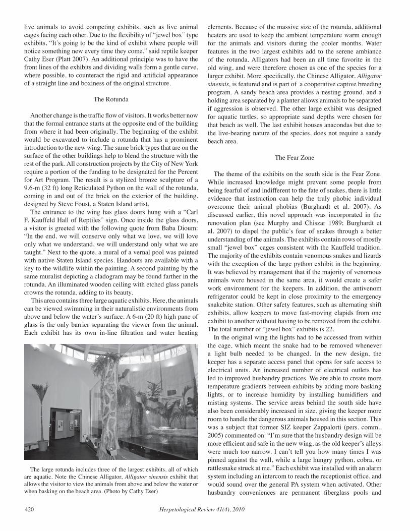
Herpetological Review 41(4), 2010420
live animals to avoid competing exhibits, such as live animal cages facing each other. Due to the flexibility of “jewel box” type exhibits, “It’s going to be the kind of exhibit where people will notice something new every time they come,” said reptile keeper Cathy Eser (Platt 2007). An additional principle was to have the front lines of the exhibits and dividing walls form a gentle curve, where possible, to counteract the rigid and artificial appearance of a straight line and boxiness of the original structure.
The Rotunda
Another change is the traffic flow of visitors. It works better now that the formal entrance starts at the opposite end of the building from where it had been originally. The beginning of the exhibit would be excavated to include a rotunda that has a prominent introduction to the new wing. The same brick types that are on the surface of the other buildings help to blend the structure with the rest of the park. All construction projects by the City of New York require a portion of the funding to be designated for the Percent for Art Program. The result is a stylized bronze sculpture of a 9.6-m (32 ft) long Reticulated Python on the wall of the rotunda, coming in and out of the brick on the exterior of the building, designed by Steve Foust, a Staten Island artist.
The entrance to the wing has glass doors hung with a “Carl F. Kauffeld Hall of Reptiles” sign. Once inside the glass doors, a visitor is greeted with the following quote from Baba Dioum: “In the end, we will conserve only what we love, we will love only what we understand, we will understand only what we are taught.” Next to the quote, a mural of a vernal pool was painted with native Staten Island species. Handouts are available with a key to the wildlife within the painting. A second painting by the same muralist depicting a cladogram may be found farther in the rotunda. An illuminated wooden ceiling with etched glass panels crowns the rotunda, adding to its beauty.
This area contains three large aquatic exhibits. Here, the animals can be viewed swimming in their naturalistic environments from above and below the water’s surface. A 6-m (20 ft) high pane of glass is the only barrier separating the viewer from the animal. Each exhibit has its own in-line filtration and water heating
elements. Because of the massive size of the rotunda, additional heaters are used to keep the ambient temperature warm enough for the animals and visitors during the cooler months. Water features in the two largest exhibits add to the serene ambiance of the rotunda. Alligators had been an all time favorite in the old wing, and were therefore chosen as one of the species for a larger exhibit. More specifically, the Chinese Alligator, Alligator sinensis, is featured and is part of a cooperative captive breeding program. A sandy beach area provides a nesting ground, and a holding area separated by a planter allows animals to be separated if aggression is observed. The other large exhibit was designed for aquatic turtles, so appropriate sand depths were chosen for that beach as well. The last exhibit houses anacondas but due to the live-bearing nature of the species, does not require a sandy beach area.
The Fear Zone
The theme of the exhibits on the south side is the Fear Zone. While increased knowledge might prevent some people from being fearful of and indifferent to the fate of snakes, there is little evidence that instruction can help the truly phobic individual overcome their animal phobias (Burghardt et al. 2007). As discussed earlier, this novel approach was incorporated in the renovation plan (see Murphy and Chiszar 1989; Burghardt et al. 2007) to dispel the public’s fear of snakes through a better understanding of the animals. The exhibits contain rows of mostly small “jewel box” cages consistent with the Kauffeld tradition. The majority of the exhibits contain venomous snakes and lizards with the exception of the large python exhibit in the beginning. It was believed by management that if the majority of venomous animals were housed in the same area, it would create a safer work environment for the keepers. In addition, the antivenom refrigerator could be kept in close proximity to the emergency snakebite station. Other safety features, such as alternating shift exhibits, allow keepers to move fast-moving elapids from one exhibit to another without having to be removed from the exhibit. The total number of “jewel box” exhibits is 22.
In the original wing the lights had to be accessed from within the cage, which meant the snake had to be removed whenever a light bulb needed to be changed. In the new design, the keeper has a separate access panel that opens for safe access to electrical units. An increased number of electrical outlets has led to improved husbandry practices. We are able to create more temperature gradients between exhibits by adding more basking lights, or to increase humidity by installing humidifiers and misting systems. The service areas behind the south side have also been considerably increased in size, giving the keeper more room to handle the dangerous animals housed in this section. This was a subject that former SIZ keeper Zappalorti (pers. comm., 2005) commented on: “I’m sure that the husbandry design will be more efficient and safe in the new wing, as the old keeper’s alleys were much too narrow. I can’t tell you how many times I was pinned against the wall, while a large hungry python, cobra, or rattlesnake struck at me.” Each exhibit was installed with an alarm system including an intercom to reach the receptionist office, and would sound over the general PA system when activated. Other husbandry conveniences are permanent fiberglass pools and
The large rotunda includes three of the largest exhibits, all of which are aquatic. Note the Chinese Alligator, Alligator sinensis exhibit that allows the visitor to view the animals from above and below the water or when basking on the beach area. (Photo by Cathy Eser)
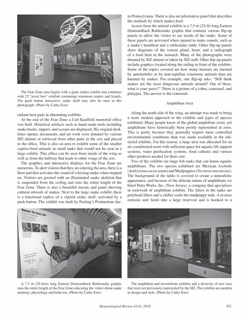
Herpetological Review 41(4), 2010 421
radiant heat pads in alternating exhibits. At the end of the Fear Zone a Carl Kauffeld memorial office
was built. Historical artifacts such as hand-made tools including snake hooks, rippers, and scoops are displayed. His original desk, letter opener, documents, and art work were donated by various SIZ alumni or retrieved from other parts in the zoo and placed in the office. This is also an area to exhibit some of the smaller captive-bred animals in small tanks that would not be seen in a large exhibit. This office can be seen from inside of the wing as well as from the hallway that leads to other wings of the zoo.
The graphics and interactive displays for the Fear Zone are numerous. To alert visitors that they are entering the area, there is a floor pad that activates the sound of a hissing snake when stepped on. Visitors are greeted with an illuminated snake skeleton that is suspended from the ceiling and runs the entire length of the Fear Zone. There is also a beautiful mosaic and panel showing cultural artwork of snakes. Next to the large snake exhibit, there is a functional replica of a viperid snake skull, activated by a push button. The exhibit was built by Peeling’s Productions Inc.
in Pennsylvania. There is also an informative panel that describes the methods by which snakes feed.
Across from the animal exhibits is a 7.5-m (25-ft) long Eastern Diamondback Rattlesnake graphic that contains various flip-up panels to allow the visitor to see inside of the snake. Some of these panels are activated when opened to make sounds, such as a snake’s heartbeat and a rattlesnake rattle. Other flip-up panels show diagrams of the venom gland, heart, and a radiograph of a food item in the stomach. Many of the photographs were donated by SIZ alumni or taken by SIZ staff. Other flip-up panels include graphics located along the railing in front of the exhibits. Some of the topics covered are how many humans are harmed by automobiles or by non-reptilian venomous animals than are harmed by snakes. For example, one flip-up asks, “Still think snakes are the most dangerous animals around? Out of these, what is your guess?” There is a picture of a cobra, conesnail, and platypus. The answer is the conesnail.
Amphibian Area
Along the north side of the wing, an attempt was made to bring a more modern approach to the exhibits and types of species exhibited. Many people know of the global amphibian crisis, yet amphibians have historically been poorly represented in zoos. This is partly because they generally require more controlled environmental conditions than was made available in the old-styled exhibits. For this reason, a large area was allocated for an air-conditioned room with sufficient space for aquatic life support systems, water purification systems, food cultures and various other products needed for their care.
Two of the exhibits are large fish tanks that can house aquatic amphibians. The two species exhibited are Mexican Axolotls (Ambystoma mexicanum) and Mudpuppies (Necturus maculosus). The background of the tanks is covered to create a naturalistic appearance, and because of the delicate nature of amphibians we hired Petra Works, Inc. (New Jersey), a company that specializes in rockwork of amphibian exhibits. The filters in the tanks are polybead filters and a chiller cools the mudpuppy tank. A reverse osmosis unit feeds into a large reservoir and is hooked to a
The Fear Zone area begins with a giant snakes exhibit and continues with 22 “jewel box” exhibits containing venomous snakes and lizards. The push button interactive snake skull may also be seen in this photograph. (Photo by Cathy Eser)
A 7.5 m (25-foot) long Eastern Diamondback Rattlesnake graphic runs the entire length of the Fear Zone educating the visitor about snake anatomy, physiology and behavior. (Photo by Cathy Eser)
The amphibian and invertebrate exhibits add a diversity of new taxa that were not previously represented by the SIZ. The exhibits are modern in design and style. (Photo by Cathy Eser)
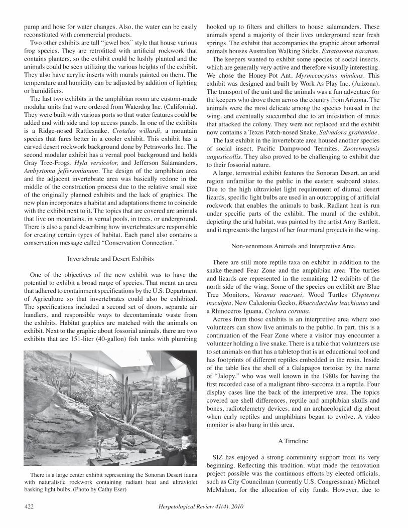
Herpetological Review 41(4), 2010422
pump and hose for water changes. Also, the water can be easily reconstituted with commercial products.
Two other exhibits are tall “jewel box” style that house various frog species. They are retrofitted with artificial rockwork that contains planters, so the exhibit could be lushly planted and the animals could be seen utilizing the various heights of the exhibit. They also have acrylic inserts with murals painted on them. The temperature and humidity can be adjusted by addition of lighting or humidifiers.
The last two exhibits in the amphibian room are custom-made modular units that were ordered from Waterdog Inc. (California). They were built with various ports so that water features could be added and with side and top access panels. In one of the exhibits is a Ridge-nosed Rattlesnake, Crotalus willardi, a mountain species that fares better in a cooler exhibit. This exhibit has a carved desert rockwork background done by Petraworks Inc. The second modular exhibit has a vernal pool background and holds Gray Tree-Frogs, Hyla versicolor, and Jefferson Salamanders, Ambystoma jeffersonianum. The design of the amphibian area and the adjacent invertebrate area was basically redone in the middle of the construction process due to the relative small size of the originally planned exhibits and the lack of graphics. The new plan incorporates a habitat and adaptations theme to coincide with the exhibit next to it. The topics that are covered are animals that live on mountains, in vernal pools, in trees, or underground. There is also a panel describing how invertebrates are responsible for creating certain types of habitat. Each panel also contains a conservation message called “Conservation Connection.”
Invertebrate and Desert Exhibits
One of the objectives of the new exhibit was to have the potential to exhibit a broad range of species. That meant an area that adhered to containment specifications by the U.S. Department of Agriculture so that invertebrates could also be exhibited. The specifications included a second set of doors, separate air handlers, and responsible ways to decontaminate waste from the exhibits. Habitat graphics are matched with the animals on exhibit. Next to the graphic about fossorial animals, there are two exhibits that are 151-liter (40-gallon) fish tanks with plumbing
hooked up to filters and chillers to house salamanders. These animals spend a majority of their lives underground near fresh springs. The exhibit that accompanies the graphic about arboreal animals houses Australian Walking Sticks, Extatasoma tiaratum.
The keepers wanted to exhibit some species of social insects, which are generally very active and therefore visually interesting. We chose the Honey-Pot Ant, Myrmecocystus mimicus. This exhibit was designed and built by Work As Play Inc. (Arizona). The transport of the unit and the animals was a fun adventure for the keepers who drove them across the country from Arizona. The animals were the most delicate among the species housed in the wing, and eventually succumbed due to an infestation of mites that attacked the colony. They were not replaced and the exhibit now contains a Texas Patch-nosed Snake, Salvadora grahamiae.
The last exhibit in the invertebrate area housed another species of social insect, Pacific Dampwood Termites, Zootermopsis angusticollis. They also proved to be challenging to exhibit due to their fossorial nature.
A large, terrestrial exhibit features the Sonoran Desert, an arid region unfamiliar to the public in the eastern seaboard states. Due to the high ultraviolet light requirement of diurnal desert lizards, specific light bulbs are used in an outcropping of artificial rockwork that enables the animals to bask. Radiant heat is run under specific parts of the exhibit. The mural of the exhibit, depicting the arid habitat, was painted by the artist Amy Bartlett, and it represents the largest of her four mural projects in the wing.
Non-venomous Animals and Interpretive Area
There are still more reptile taxa on exhibit in addition to the snake-themed Fear Zone and the amphibian area. The turtles and lizards are represented in the remaining 12 exhibits of the north side of the wing. Some of the species on exhibit are Blue Tree Monitors, Varanus macraei, Wood Turtles Glyptemys insculpta, New Caledonia Gecko, Rhacodactylus leachianus and a Rhinoceros Iguana, Cyclura cornuta.
Across from those exhibits is an interpretive area where zoo volunteers can show live animals to the public. In part, this is a continuation of the Fear Zone where a visitor may encounter a volunteer holding a live snake. There is a table that volunteers use to set animals on that has a tabletop that is an educational tool and has footprints of different reptiles embedded in the resin. Inside of the table lies the shell of a Galapagos tortoise by the name of “Jalopy,” who was well known in the 1980s for having the first recorded case of a malignant fibro-sarcoma in a reptile. Four display cases line the back of the interpretive area. The topics covered are shell differences, reptile and amphibian skulls and bones, radiotelemetry devices, and an archaeological dig about when early reptiles and amphibians began to evolve. A video monitor is also hung in this area.
A Timeline
SIZ has enjoyed a strong community support from its very beginning. Reflecting this tradition, what made the renovation project possible was the continuous efforts by elected officials, such as City Councilman (currently U.S. Congressman) Michael McMahon, for the allocation of city funds. However, due to
There is a large center exhibit representing the Sonoran Desert fauna with naturalistic rockwork containing radiant heat and ultraviolet basking light bulbs. (Photo by Cathy Eser)
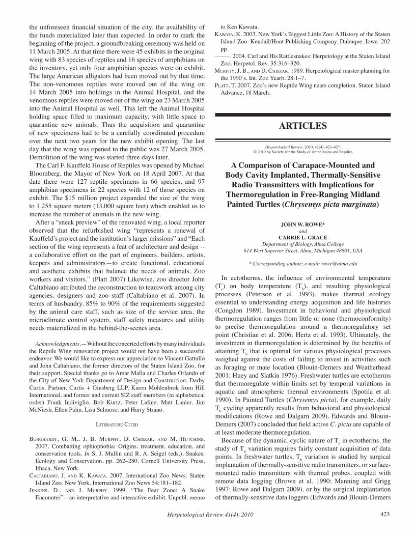
Herpetological Review 41(4), 2010 423
the unforeseen financial situation of the city, the availability of the funds materialized later than expected. In order to mark the beginning of the project, a groundbreaking ceremony was held on 11 March 2005. At that time there were 45 exhibits in the original wing with 83 species of reptiles and 16 species of amphibians on the inventory, yet only four amphibian species were on exhibit. The large American alligators had been moved out by that time. The non-venomous reptiles were moved out of the wing on 14 March 2005 into holdings in the Animal Hospital, and the venomous reptiles were moved out of the wing on 23 March 2005 into the Animal Hospital as well. This left the Animal Hospital holding space filled to maximum capacity, with little space to quarantine new animals. Thus the acquisition and quarantine of new specimens had to be a carefully coordinated procedure over the next two years for the new exhibit opening. The last day that the wing was opened to the public was 27 March 2005. Demolition of the wing was started three days later.
The Carl F. Kauffeld House of Reptiles was opened by Michael Bloomberg, the Mayor of New York on 18 April 2007. At that date there were 127 reptile specimens in 66 species, and 97 amphibian specimens in 22 species with 12 of those species on exhibit. The $15 million project expanded the size of the wing to 1,255 square meters (13,000 square feet) which enabled us to increase the number of animals in the new wing.
After a “sneak preview” of the renovated wing, a local reporter observed that the refurbished wing “represents a renewal of Kauffeld’s project and the institution’s larger missions” and “Each section of the wing represents a feat of architecture and design -- a collaborative effort on the part of engineers, builders, artists, keepers and administrators—to create functional, educational and aesthetic exhibits that balance the needs of animals, Zoo workers and visitors.” (Platt 2007) Likewise, zoo director John Caltabiano attributed the reconstruction to teamwork among city agencies, designers and zoo staff (Caltabiano et al. 2007). In terms of husbandry, 85% to 90% of the requirements suggested by the animal care staff, such as size of the service area, the microclimate control system, staff safety measures and utility needs materialized in the behind-the-scenes area.
Acknowledgments.—Without the concerted efforts by many individuals the Reptile Wing renovation project would not have been a successful endeavor. We would like to express our appreciation to Vincent Gattullo and John Caltabiano, the former directors of the Staten Island Zoo, for their support. Special thanks go to Amar Malla and Charles Orlando of the City of New York Department of Design and Construction; Darby Curtis, Partner, Curtis + Ginsberg LLP, Karen Mohlenbrok from Hill International, and former and current SIZ staff members (in alphabetical order) Frank Indiviglio, Bob Kurtz, Peter Laline, Matt Lanier, Jim McNiesh, Ellen Palm, Lisa Salmose, and Harry Strano.
literature cited
BurgHardt, g. M., J. B. MurPHY, d. cHiSzar, and M. HutcHinS. 2007. Combatting ophiophobia: Origins, treatment, education, and conservation tools. In S. J. Mullin and R. A. Seigel (eds.), Snakes: Ecology and Conservation, pp. 262–280. Cornell University Press, Ithaca, New York.
caltaBiano, J. and k. kaWata. 2007. International Zoo News: Staten Island Zoo, New York. International Zoo News 54:181–182.
JenkinS, d., and J. MurPHY. 1999. “The Fear Zone: A Snake Encounter”—an interpretative and interactive exhibit. Unpubl. memo
to Ken Kawata.kaWata, k. 2003. New York’s Biggest Little Zoo: A History of the Staten
Island Zoo. Kendall/Hunt Publishing Company. Dubuque, Iowa. 202 pp.
–––––. 2004. Carl and His Rattlesnakes: Herpetology at the Staten Island Zoo. Herpetol. Rev. 35:316–320.
MurPHY, J. B., and d. cHiSzar. 1989. Herpetological master planning for the 1990’s. Int. Zoo Yearb. 28:1–7.
Platt, t. 2007. Zoo’s new Reptile Wing nears completion. Staten Island Advance, 18 March.
ARTICLES
Herpetological Review, 2010, 41(4), 423–427.© 2010 by Society for the Study of Amphibians and Reptiles
A Comparison of Carapace-Mounted and Body Cavity Implanted, Thermally-Sensitive
Radio Transmitters with Implications for Thermoregulation in Free-Ranging Midland Painted Turtles (Chrysemys picta marginata)
JOHN W. ROWE*and
CARRIE L. GRACEDepartment of Biology, Alma College
614 West Superior Street, Alma, Michigan 48801, USA
* Corresponding author; e-mail: [email protected]
In ectotherms, the influence of environmental temperature (Te) on body temperature (Tb), and resulting physiological processes (Peterson et al. 1993), makes thermal ecology essential to understanding energy acquisition and life histories (Congdon 1989). Investment in behavioral and physiological thermoregulation ranges from little or none (thermoconformity) to precise thermoregulation around a thermoregulatory set point (Christian et al. 2006; Hertz et al. 1993). Ultimately, the investment in thermoregulation is determined by the benefits of attaining Tb that is optimal for various physiological processes weighed against the costs of failing to invest in activities such as foraging or mate location (Blouin-Demers and Weatherhead 2001; Huey and Slatkin 1976). Freshwater turtles are ectotherms that thermoregulate within limits set by temporal variations in aquatic and atmospheric thermal environments (Spotila et al. 1990). In Painted Turtles (Chrysemys picta), for example, daily Tb cycling apparently results from behavioral and physiological modifications (Rowe and Dalgarn 2009). Edwards and Blouin-Demers (2007) concluded that field active C. picta are capable of at least moderate thermoregulation.
Because of the dynamic, cyclic nature of Tb in ectotherms, the study of Tb variation requires fairly constant acquisition of data points. In freshwater turtles, Tb variation is studied by surgical implantation of thermally-sensitive radio transmitters, or surface-mounted radio transmitters with thermal probes, coupled with remote data logging (Brown et al. 1990; Manning and Grigg 1997; Rowe and Dalgarn 2009), or by the surgical implantation of thermally-sensitive data loggers (Edwards and Blouin-Demers
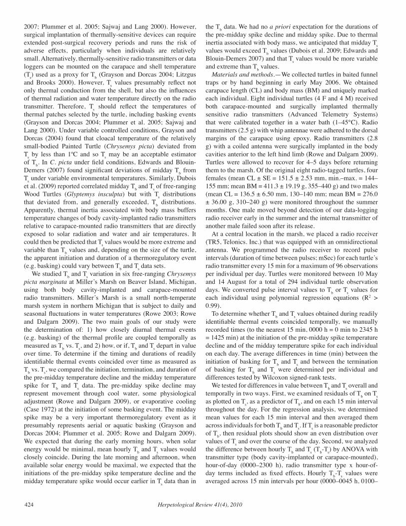
Herpetological Review 41(4), 2010424
2007; Plummer et al. 2005; Sajwaj and Lang 2000). However, surgical implantation of thermally-sensitive devices can require extended post-surgical recovery periods and runs the risk of adverse effects, particularly when individuals are relatively small. Alternatively, thermally-sensitive radio transmitters or data loggers can be mounted on the carapace and shell temperature (Ts) used as a proxy for Tb (Grayson and Dorcas 2004; Litzgus and Brooks 2000). However, Ts values presumably reflect not only thermal conduction from the shell, but also the influences of thermal radiation and water temperature directly on the radio transmitter. Therefore, Ts should reflect the temperatures of thermal patches selected by the turtle, including basking events (Grayson and Dorcas 2004; Plummer et al. 2005; Sajwaj and Lang 2000). Under variable controlled conditions, Grayson and Dorcas (2004) found that cloacal temperature of the relatively small-bodied Painted Turtle (Chrysemys picta) deviated from Ts by less than 1ºC and so Ts may be an acceptable estimator of Tb. In C. picta under field conditions, Edwards and Blouin-Demers (2007) found significant deviations of midday Tb from Ts under variable environmental temperatures. Similarly, Dubois et al. (2009) reported correlated midday Tb and Ts of free-ranging Wood Turtles (Glyptemys insculpta) but with Ts distributions that deviated from, and generally exceeded, Tb distributions. Apparently, thermal inertia associated with body mass buffers temperature changes of body cavity-implanted radio transmitters relative to carapace-mounted radio transmitters that are directly exposed to solar radiation and water and air temperatures. It could then be predicted that Ts values would be more extreme and variable than Tb values and, depending on the size of the turtle, the apparent initiation and duration of a thermoregulatory event (e.g. basking) could vary between Tb and Ts data sets.
We studied Tb and Ts variation in six free-ranging Chrysemys picta marginata at Miller’s Marsh on Beaver Island, Michigan, using both body cavity-implanted and carapace-mounted radio transmitters. Miller’s Marsh is a small north-temperate marsh system in northern Michigan that is subject to daily and seasonal fluctuations in water temperatures (Rowe 2003; Rowe and Dalgarn 2009). The two main goals of our study were the determination of: 1) how closely diurnal thermal events (e.g. basking) of the thermal profile are coupled temporally as measured as Tb vs. Ts, and 2) how, or if, Tb and Ts depart in value over time. To determine if the timing and durations of readily identifiable thermal events coincided over time as measured as Tb vs. Ts, we compared the initiation, termination, and duration of the pre-midday temperature decline and the midday temperature spike for Tb and Ts data. The pre-midday spike decline may represent movement through cool water, some physiological adjustment (Rowe and Dalgarn 2009), or evaporative cooling (Case 1972) at the initiation of some basking event. The midday spike may be a very important thermoregulatory event as it presumably represents aerial or aquatic basking (Grayson and Dorcas 2004; Plummer et al. 2005; Rowe and Dalgarn 2009). We expected that during the early morning hours, when solar energy would be minimal, mean hourly Tb and Ts values would closely coincide. During the late morning and afternoon, when available solar energy would be maximal, we expected that the initiations of the pre-midday spike temperature decline and the midday temperature spike would occur earlier in Ts data than in
the Tb data. We had no a priori expectation for the durations of the pre-midday spike decline and midday spike. Due to thermal inertia associated with body mass, we anticipated that midday Ts values would exceed Tb values (Dubois et al. 2009; Edwards and Blouin-Demers 2007) and that Ts values would be more variable and extreme than Tb values.
Materials and methods.—We collected turtles in baited funnel traps or by hand beginning in early May 2006. We obtained carapace length (CL) and body mass (BM) and uniquely marked each individual. Eight individual turtles (4 F and 4 M) received both carapace-mounted and surgically implanted thermally sensitive radio transmitters (Advanced Telemetry Systems) that were calibrated together in a water bath (1–45°C). Radio transmitters (2.5 g) with whip antennae were adhered to the dorsal margins of the carapace using epoxy. Radio transmitters (2.8 g) with a coiled antenna were surgically implanted in the body cavities anterior to the left hind limb (Rowe and Dalgarn 2009). Turtles were allowed to recover for 4–5 days before returning them to the marsh. Of the original eight radio-tagged turtles, four females (mean CL ± SE = 151.5 ± 2.53 mm, min.–max. = 144–155 mm; mean BM = 411.3 ± 19.19 g, 355–440 g) and two males (mean CL = 136.5 ± 6.50 mm, 130–140 mm; mean BM = 276.0 ± 36.00 g, 310–240 g) were monitored throughout the summer months. One male moved beyond detection of our data-logging radio receiver early in the summer and the internal transmitter of another male failed soon after its release.
At a central location in the marsh, we placed a radio receiver (TR5, Telonics. Inc.) that was equipped with an omnidirectional antenna. We programmed the radio receiver to record pulse intervals (duration of time between pulses; mSec) for each turtle’s radio transmitter every 15 min for a maximum of 96 observations per individual per day. Turtles were monitored between 10 May and 14 August for a total of 294 individual turtle observation days. We converted pulse interval values to Tb or Ts values for each individual using polynomial regression equations (R2 > 0.99).
To determine whether Tb and Ts values obtained during readily identifiable thermal events coincided temporally, we manually recorded times (to the nearest 15 min, 0000 h = 0 min to 2345 h = 1425 min) at the initiation of the pre-midday spike temperature decline and of the midday temperature spike for each individual on each day. The average differences in time (min) between the initiation of basking for Tb and Ts and between the termination of basking for Tb and Ts were determined per individual and differences tested by Wilcoxon signed-rank tests.
We tested for differences in value between Tb and Ts overall and temporally in two ways. First, we examined residuals of Tb on Ts as plotted on Ts, as a predictor of Tb, and on each 15 min interval throughout the day. For the regression analysis, we determined mean values for each 15 min interval and then averaged them across individuals for both Tb and Ts. If Ts is a reasonable predictor of Tb, then residual plots should show an even distribution over values of Ts and over the course of the day. Second, we analyzed the difference between hourly Tb and Ts (Tb-Ts) by ANOVA with transmitter type (body cavity-implanted or carapace-mounted), hour-of-day (0000–2300 h), radio transmitter type x hour-of-day terms included as fixed effects. Hourly Tb-Ts values were averaged across 15 min intervals per hour (0000–0045 h, 0100–
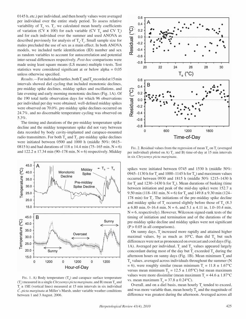
Herpetological Review 41(4), 2010 425
0145 h, etc,) per individual, and then hourly values were averaged per individual over the entire study period. To assess relative variability of Tb vs. Ts, we calculated mean hourly coefficients of variation (CV x 100) for each variable (CV Tb and CV Ts) and for each individual over the summer and used ANOVA as described previously for analysis of Tb-Ts. Small sample size for males precluded the use of sex as a main effect. In both ANOVA models, we included turtle identification (ID) number and sex as random variables to account for autocorrelation and potential inter-sexual differences respectively. Post-hoc comparisons were made using least square means (LS means) multiple t-tests. Test statistics were considered significant at or below alpha = 0.05 unless otherwise specified.
Results.—For individual turtles, both Ts and Tb recorded at 15 min intervals showed diel cycling that included monotonic declines, pre-midday spike declines, midday spikes and oscillations, and late evening and early morning monotonic declines (Fig. 1A). Of the 190 total turtle observation days for which 96 observations per individual per day were obtained, well-defined midday spikes were observed on 70.0%, pre-midday spike declines occurred on 24.7%, and no discernable temperature cycling was observed on 5.3%.
The timing and durations of the pre-midday temperature spike decline and the midday temperature spike did not vary between data recorded by body cavity-implanted and carapace-mounted radio transmitters. For both Tb and Ts, pre-midday spike declines were initiated between 0500 and 1000 h (middle 50%: 0615– 0815 h) and had durations of 118 ± 14.4 min (75–165 min, N = 6) and 122.2 ± 17.34 min (90–178 min, N = 6) respectively. Midday
spikes were initiated between 0745 and 1530 h (middle 50%: 0945–1130 h for Ts and 1000–1145 h for Tb) and maximum values occurred between 0930 and 1815 h (middle 50% 1215–1430 h for Ts and 1230–1430 h for Tb). Mean durations of basking (time between initiation and peak of the mid-day spike) were 152.7 ± 9.50 min (118–181 min, N = 6) for Tb and 149.8 ± 9.30 min (124–178 min) for Ts. The initiations of the pre-midday spike decline and midday spike of Ts occurred slightly before those of Tb (8.3 ± 6.80 min, 0–16.4 min, N = 6, and 5.1 ± 4.11 in, 1.0–10.4 min, N = 6, respectively). However, Wilcoxon signed-rank tests of the timing of initiation and termination and of the durations of the pre-midday spike decline and midday spikes were not significant (P > 0.05 in all comparisons).
On sunny days, Ts increased more rapidly and attained higher maximal values, by as much as 10°C, than did Tb but such differences were not as pronounced on overcast and cool days (Fig. 1A). Averaged per individual, Ts and Tb values appeared largely concordant during most of the day but Ts exceeded Tb during the afternoon hours on sunny days (Fig. 1B). Mean minimum Ts and Tb values, averaged across individuals throughout the summer (N = 6), were roughly similar (mean minimum Ts = 11.8 ± 1.67°C versus mean minimum Tb = 12.5 ± 1.05°C) but mean maximum values were more dissimilar (mean maximum Ts = 44.6 ± 1.87°C vs. mean maximum Tb = 37.8 ± 0.24°C).
Overall, and on a diel basis, mean hourly Ts tended to exceed, and was more variable than, mean hourly Tb and the magnitude of difference was greatest during the afternoon. Averaged across all
fig. 1. A) Body temperature (Tb) and carapace surface temperature (Ts) measured in a single Chrysemys picta marginata, and B) mean Tb and Ts ± 1SE (vertical lines) measured at 15 min intervals in six individual C. picta marginata at Miller’s Marsh, under variable weather conditions between 1 and 3 August, 2006.
fig. 2. Residual values from the regression of mean Tb on Ts (averaged per individual) plotted on A) Ts, and B) time-of-day at 15 min intervals in six Chrysemys picta marginata.
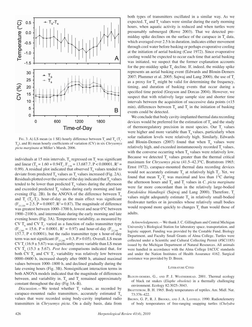
Herpetological Review 41(4), 2010426
individuals at 15 min intervals, Tb regressed on Ts was significant and linear (Tb = 1.60 + 0.94Ts, F1,94 = 13,687.7, P < 0.00001, R2 = 0.99). A residual plot indicated that observed Tb values tended to deviate from predicted Tb values as Ts values increased (Fig. 2A). Residuals plotted over the course of the day indicated that Tb values tended to be lower than predicted Tb values during the afternoon and exceeded predicted Tb values during early morning and late evening (Fig. 2B). In the ANOVA of the difference between Tb and Ts (Tb-Ts), hour-of-day as the main effect was significant (F23,120 = 2.5, P = 0.0007, R2 = 0.87). The magnitude of difference was greatest between 1000–1700 h, lowest and near 0°C between 1900–2100 h, and intermediate during the early morning and late evening hours (Fig. 3A). Temperature variability, as measured by CV Tb and CV Ts, varied as a function of radio transmitter type (F1,240 = 15.6, P = 0.0001, R2 = 0.97) and hour-of-day (F23,240 = 157.7, P < 0.0001), but the radio transmitter type x hour-of-day term was not significant (F23,240 = 0.3, P > 0.05). Overall, LS mean CV Ts (16.9 ± 5.67) was significantly more variable than LS mean CV Tb (15.3 ± 5.67). Post hoc comparisons indicated that, for both CV Tb and CV Ts, variability was relatively low between 0000–0600 h, increased sharply after 0600 h, attained maximal values between 1000–1800 h, and declined gradually during the late evening hours (Fig. 3B). Nonsignificant interaction terms in both ANOVA models indicated that the magnitude of differences between, and variability in, Tb and Ts remained approximately constant throughout the day (Fig 3A–B).
Discussion.—We tested whether Ts values, as recorded by carapace-mounted radio transmitters, accurately estimated Tb values that were recorded using body-cavity implanted radio transmitters in Chrysemys picta. On a daily basis, data from
both types of transmitters oscillated in a similar way. As we expected, Tb and Ts values were similar during the early morning hours, when aquatic activity is reduced and when turtles were presumably submerged (Rowe 2003). That we detected pre-midday spike declines on the surface of the carapace in Ts data, which averaged over 2.5 h in duration, indicates either movement through cool water before basking or perhaps evaporative cooling at the initiation of aerial basking (Case 1972). Since evaporative cooling would be expected to occur each time that aerial basking was initiated, we suspect that the former explanation accounts for the pre-midday spike Tb decline. If, indeed, the midday spike represents an aerial basking event (Edwards and Blouin-Demers 2007; Plummer et al. 2005; Sajwaj and Lang 2000), the use of Ts as a proxy for Tb might be valid for determining the frequency, timing, and duration of basking events that occur during a specified time period (Grayson and Dorcas 2004). However, we suspect that with relatively large sample size and shorter time intervals between the acquisition of successive data points (<15 min), differences between Tb and Ts in the initiation of basking events could be detected.
We conclude that body cavity-implanted thermal data recording devices would be preferred for the estimation of Tb and the study of thermoregulatory precision in most species. Our Ts values were higher and more variable than Tb values, particularly when solar radiation levels were relatively high. Similarly, Edwards and Blouin-Demers (2007) found that when Tb values were relatively high, and exceeded instantaneously recorded Ts values, with the converse occurring when Tb values were relatively low. Because we detected Ts values greater than the thermal critical maximum for Chrysemys picta (41.5–42.3°C, Brattstrom 1965; Ernst 1972), carapace-mounted thermal data recording devices would not accurately estimate Tb at relatively high Te. Yet, we found that mean Tb-Ts was maximal and less than 1°C during the afternoon hours and Tb and Ts values in C. picta marginata were far more concordant than in the relatively large-bodied Emydoidea blandingii (Sajwaj and Lang 2000). Therefore, Ts values might adequately estimate Tb in relatively small-bodied freshwater turtles or in juveniles whose relatively small bodies would respond more quickly to changes Te than would those of adults.
Acknowledgments.—We thank J. C. Gillingham and Central Michigan University’s Biological Station for laboratory space, transportation, and logistic support. Funding was provided by the Costable Fund, Biology Department, and Faculty Small Grants of Alma College. Turtles were collected under a Scientific and Cultural Collecting Permit (#SC1185) issued by the Michigan Department of Natural Resources. All animals were handled in accordance with the Alma College IACUC standards and under the Nation Institutes of Health Assurance 4162. Surgical assistance was provided by D. Breen.
literature cited
Blouin-deMerS, g., and P. J. WeatHerHead. 2001. Thermal ecology of black rat snakes (Elaphe obsoleta) in a thermally challenging environment. Ecology 82:3025–3043.
BrattStroM, B. H. 1965. Body temperatures of reptiles. Am. Midl. Nat. 73:376–422.
BroWn, g. P., r. J. BrookS, and J. a. laYfield. 1990. Radiotelemetry of body temperatures of free-ranging snapping turtles (Chelydra
fig. 3. A) LS mean (± 1 SE) hourly difference between Ts and Tb (Ts-Tb), and B) mean hourly coefficients of variation (CV) in six Chrysemys picta marginata at Miller’s Marsh, 2006.
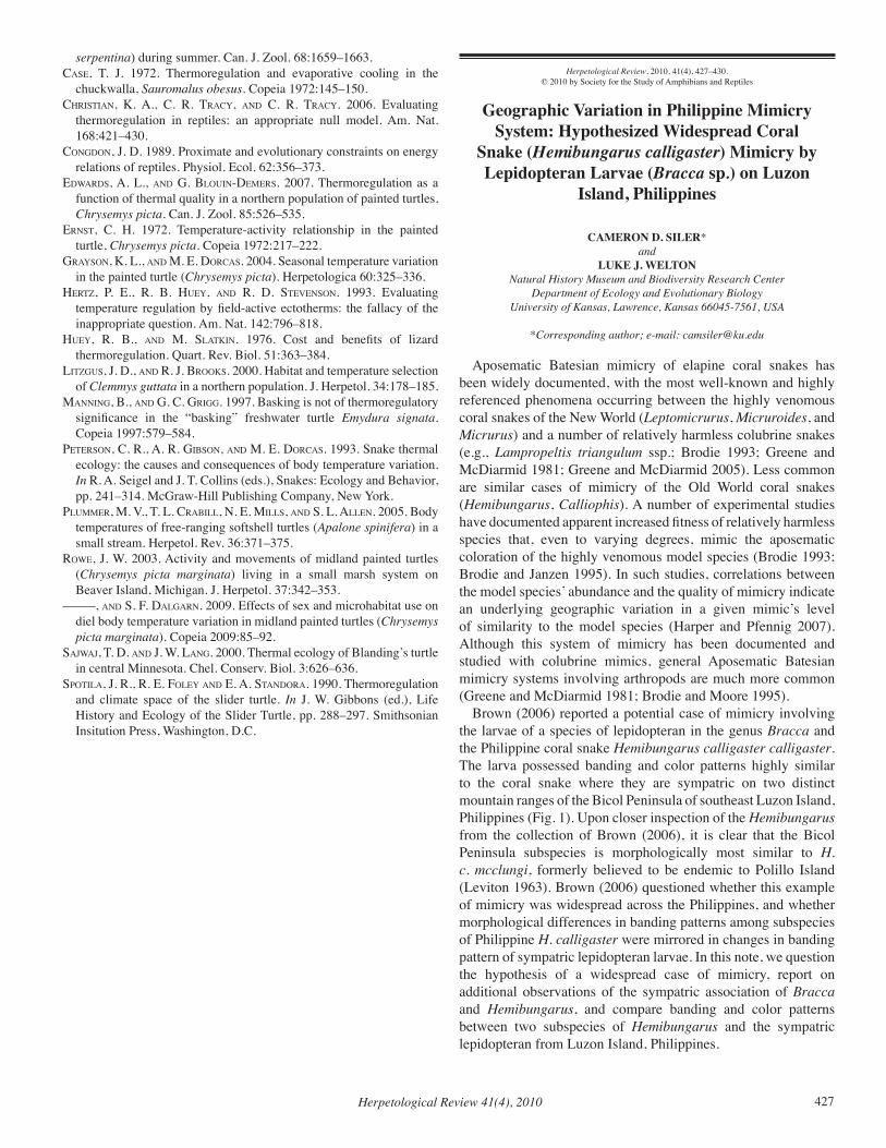
Herpetological Review 41(4), 2010 427
serpentina) during summer. Can. J. Zool. 68:1659–1663.caSe, t. J. 1972. Thermoregulation and evaporative cooling in the
chuckwalla, Sauromalus obesus. Copeia 1972:145–150. cHriStian, k. a., c. r. tracY, and c. r. tracY. 2006. Evaluating
thermoregulation in reptiles: an appropriate null model. Am. Nat. 168:421–430.
congdon, J. d. 1989. Proximate and evolutionary constraints on energy relations of reptiles. Physiol. Ecol. 62:356–373.
edWardS, a. l., and g. Blouin-deMerS. 2007. Thermoregulation as a function of thermal quality in a northern population of painted turtles, Chrysemys picta. Can. J. Zool. 85:526–535.
ernSt, c. H. 1972. Temperature-activity relationship in the painted turtle, Chrysemys picta. Copeia 1972:217–222.
graYSon, k. l., and M. e. dorcaS. 2004. Seasonal temperature variation in the painted turtle (Chrysemys picta). Herpetologica 60:325–336.
Hertz, P. e., r. B. HueY, and r. d. StevenSon. 1993. Evaluating temperature regulation by field-active ectotherms: the fallacy of the inappropriate question. Am. Nat. 142:796–818.
HueY, r. B., and M. Slatkin. 1976. Cost and benefits of lizard thermoregulation. Quart. Rev. Biol. 51:363–384.
litzguS, J. d., and r. J. BrookS. 2000. Habitat and temperature selection of Clemmys guttata in a northern population. J. Herpetol. 34:178–185.
Manning, B., and g. c. grigg. 1997. Basking is not of thermoregulatory significance in the “basking” freshwater turtle Emydura signata. Copeia 1997:579–584.
PeterSon, c. r., a. r. giBSon, and M. e. dorcaS. 1993. Snake thermal ecology: the causes and consequences of body temperature variation. In R. A. Seigel and J. T. Collins (eds.), Snakes: Ecology and Behavior, pp. 241–314. McGraw-Hill Publishing Company, New York.
PluMMer, M. v., t. l. craBill, n. e. MillS, and S. l. allen. 2005. Body temperatures of free-ranging softshell turtles (Apalone spinifera) in a small stream. Herpetol. Rev. 36:371–375.
roWe, J. W. 2003. Activity and movements of midland painted turtles (Chrysemys picta marginata) living in a small marsh system on Beaver Island, Michigan. J. Herpetol. 37:342–353.
–––––, and S. f. dalgarn. 2009. Effects of sex and microhabitat use on diel body temperature variation in midland painted turtles (Chrysemys picta marginata). Copeia 2009:85–92.
SaJWaJ, t. d. and J. W. lang. 2000. Thermal ecology of Blanding’s turtle in central Minnesota. Chel. Conserv. Biol. 3:626–636.
SPotila, J. r., r. e. foleY and e. a. Standora. 1990. Thermoregulation and climate space of the slider turtle. In J. W. Gibbons (ed.), Life History and Ecology of the Slider Turtle, pp. 288–297. Smithsonian Insitution Press, Washington, D.C.
Herpetological Review, 2010, 41(4), 427–430.© 2010 by Society for the Study of Amphibians and Reptiles
Geographic Variation in Philippine Mimicry System: Hypothesized Widespread Coral
Snake (Hemibungarus calligaster) Mimicry by Lepidopteran Larvae (Bracca sp.) on Luzon
Island, Philippines
CAMERON D. SILER*and
LUKE J. WELTONNatural History Museum and Biodiversity Research Center
Department of Ecology and Evolutionary BiologyUniversity of Kansas, Lawrence, Kansas 66045-7561, USA
*Corresponding author; e-mail: [email protected]
Aposematic Batesian mimicry of elapine coral snakes has been widely documented, with the most well-known and highly referenced phenomena occurring between the highly venomous coral snakes of the New World (Leptomicrurus, Micruroides, and Micrurus) and a number of relatively harmless colubrine snakes (e.g., Lampropeltis triangulum ssp.; Brodie 1993; Greene and McDiarmid 1981; Greene and McDiarmid 2005). Less common are similar cases of mimicry of the Old World coral snakes (Hemibungarus, Calliophis). A number of experimental studies have documented apparent increased fitness of relatively harmless species that, even to varying degrees, mimic the aposematic coloration of the highly venomous model species (Brodie 1993; Brodie and Janzen 1995). In such studies, correlations between the model species’ abundance and the quality of mimicry indicate an underlying geographic variation in a given mimic’s level of similarity to the model species (Harper and Pfennig 2007). Although this system of mimicry has been documented and studied with colubrine mimics, general Aposematic Batesian mimicry systems involving arthropods are much more common (Greene and McDiarmid 1981; Brodie and Moore 1995).
Brown (2006) reported a potential case of mimicry involving the larvae of a species of lepidopteran in the genus Bracca and the Philippine coral snake Hemibungarus calligaster calligaster. The larva possessed banding and color patterns highly similar to the coral snake where they are sympatric on two distinct mountain ranges of the Bicol Peninsula of southeast Luzon Island, Philippines (Fig. 1). Upon closer inspection of the Hemibungarus from the collection of Brown (2006), it is clear that the Bicol Peninsula subspecies is morphologically most similar to H. c. mcclungi, formerly believed to be endemic to Polillo Island (Leviton 1963). Brown (2006) questioned whether this example of mimicry was widespread across the Philippines, and whether morphological differences in banding patterns among subspecies of Philippine H. calligaster were mirrored in changes in banding pattern of sympatric lepidopteran larvae. In this note, we question the hypothesis of a widespread case of mimicry, report on additional observations of the sympatric association of Bracca and Hemibungarus, and compare banding and color patterns between two subspecies of Hemibungarus and the sympatric lepidopteran from Luzon Island, Philippines.
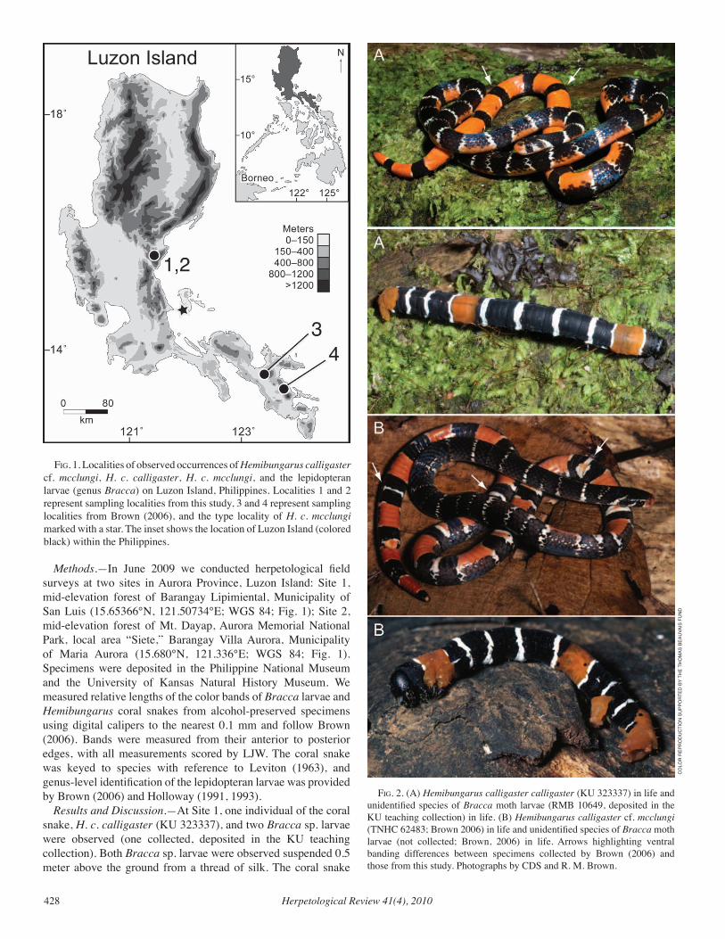
Herpetological Review 41(4), 2010428
Methods.—In June 2009 we conducted herpetological field surveys at two sites in Aurora Province, Luzon Island: Site 1, mid-elevation forest of Barangay Lipimiental, Municipality of San Luis (15.65366°N, 121.50734°E; WGS 84; Fig. 1); Site 2, mid-elevation forest of Mt. Dayap, Aurora Memorial National Park, local area “Siete,” Barangay Villa Aurora, Municipality of Maria Aurora (15.680°N, 121.336°E; WGS 84; Fig. 1). Specimens were deposited in the Philippine National Museum and the University of Kansas Natural History Museum. We measured relative lengths of the color bands of Bracca larvae and Hemibungarus coral snakes from alcohol-preserved specimens using digital calipers to the nearest 0.1 mm and follow Brown (2006). Bands were measured from their anterior to posterior edges, with all measurements scored by LJW. The coral snake was keyed to species with reference to Leviton (1963), and genus-level identification of the lepidopteran larvae was provided by Brown (2006) and Holloway (1991, 1993).
Results and Discussion.—At Site 1, one individual of the coral snake, H. c. calligaster (KU 323337), and two Bracca sp. larvae were observed (one collected, deposited in the KU teaching collection). Both Bracca sp. larvae were observed suspended 0.5 meter above the ground from a thread of silk. The coral snake
fig. 1. Localities of observed occurrences of Hemibungarus calligaster cf. mcclungi, H. c. calligaster, H. c. mcclungi, and the lepidopteran larvae (genus Bracca) on Luzon Island, Philippines. Localities 1 and 2 represent sampling localities from this study, 3 and 4 represent sampling localities from Brown (2006), and the type locality of H. c. mcclungi marked with a star. The inset shows the location of Luzon Island (colored black) within the Philippines.
fig. 2. (A) Hemibungarus calligaster calligaster (KU 323337) in life and unidentified species of Bracca moth larvae (RMB 10649, deposited in the KU teaching collection) in life. (B) Hemibungarus calligaster cf. mcclungi (TNHC 62483; Brown 2006) in life and unidentified species of Bracca moth larvae (not collected; Brown, 2006) in life. Arrows highlighting ventral banding differences between specimens collected by Brown (2006) and those from this study. Photographs by CDS and R. M. Brown.
CO
LOR
REP
RO
du
CTi
ON
Su
PPO
RTEd
BY
THE
THO
mAS
BEA
uvA
iS F
uN
d
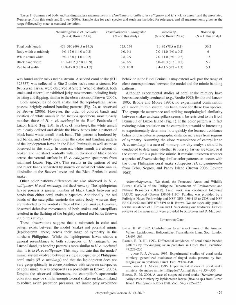
Herpetological Review 41(4), 2010 429
was found under rocks near a stream. A second coral snake (KU 323337) was collected at Site 2 under rocks near a stream. No Bracca sp. larvae were observed at Site 2. When disturbed, both snake and caterpillar exhibited jerky movements, including body twisting and flipping, similar to the observations of Brown (2006).
Both subspecies of coral snake and the lepidopteran larvae possess brightly colored banding patterns (Fig. 2), as observed by Brown (2006). However, the order of colored bands and location of white annuli in the Bracca specimens most closely matches those of H. c. cf. mcclungi in the Bicol Peninsula of Luzon Island (Fig. 2B). In H. c. cf. mcclungi, the white annuli are clearly defined and divide the black bands into a pattern of black band-white annuli-black band. This pattern is bordered by red bands, and closely resembles the color and banding pattern of the lepidopteran larvae in the Bicol Peninsula as well as those observed in this study. In contrast, white annuli are absent or broken and indistinct ventrally with no division of black bands across the ventral surface in H. c. calligaster specimens from mainland Luzon (Fig. 2A). This results in the pattern of red and black bands separated by narrow or indistinct white annuli, dissimilar to the Bracca larvae and the Bicol Peninsula coral snake.
Other color patterns differences are also observed in H. c. calligaster, H. c. cf. mcclungi, and the Bracca sp. The lepidopteran larvae possess a greater number of black bands between red bands than either coral snake subspecies. Additionally, the red bands of the caterpillar encircle the entire body, whereas they are restricted to the ventral surface of the coral snakes. However, observed defensive movements of both snakes and caterpillars resulted in the flashing of the brightly colored red bands (Brown 2006; this study).
These observations suggest that a mismatch in color and pattern exists between the model (snake) and potential mimic (lepidopteran larvae) across their range of sympatry in the northern Philippines. While the lepidopteran larvae shares a general resemblance to both subspecies of H. calligaster on Luzon Island, its banding pattern is more similar to H. c. mcclungi than it is to H. c. calligaster. This may indicate that the model/mimic system evolved between a single subspecies of Philippine coral snake (H. c. mcclungi) and that the lepidopteran does not vary geographically in correspondence with separate subspecies of coral snake as was proposed as a possibility in Brown (2006). Despite the observed differences, the caterpillar’s aposematic coloration may be similar enough to coral snakes on Luzon Island to reduce avian predation pressures. An innate prey avoidance
behavior in the Bicol Peninsula may extend well past the range of close correspondence between the model and the mimic banding patterns.
Although experimental studies of coral snake mimicry have been successfully conducted (e.g., Brodie 1993; Brodie and Janzen 1995; Brodie and Moore 1995), no experimental confirmation of a model/mimic system has been made for these two species. The sympatric occurrence and striking morphological similarity between snakes and caterpillars seems to be restricted to the Bicol Peninsula of Luzon Island (Fig. 1). If the color pattern is in fact reducing avian predation on the caterpillar, it would be interesting to experimentally determine how quickly the learned avoidance behavior dissipates as geographic distance increases from regions of sympatry. Assuming the close resemblance of caterpillar to H. c. mcclungi is a case of mimicry, toxicity analysis should be conducted to determine whether Bracca sp. larvae are toxic, or if the caterpillar is a palatable mimic. It remains unknown whether a species of Bracca sharing similar color patterns co-occurs with the other Philippine coral snake subspecies, H. c. gemiannulis from Cebu, Negros, and Panay Island (Brown 2006; Leviton 1963).
Acknowledgments.—We thank the Protected Areas and Wildlife Bureau (PAWB) of the Philippine Department of Environment and Natural Resources (DENR). Field work was conducted following IACUC approval (Brown 158-01-1110). Funding was provided by a Fulbright-Hayes Fellowship and NSF DEB 0804115 to CDS and NSF EF-0334952 and DEB 0743491 to R. Brown. We are especially grateful for the assistance of J. Brown and J. Siler during our fieldwork. Critical reviews of the manuscript were provided by R. Brown and D. McLeod.
literature cited
BateS, H. W. 1862. Contributions to an insect fauna of the Amazon Valley, Lepidoptera, Heliconiidae. Transatlantic Linn. Soc. London 23:495–566.
Brodie, e. d. iii. 1993. Differential avoidance of coral snake banded patterns by free-ranging avian predators in Costa Rica. Evolution 47:227–235.
–––––, and f. J. Janzen. 1995. Experimental studies of coral snake mimicry: generalized avoidance of ringed snake patterns by free-ranging avian predators. Funct. Ecol. 9:186–190.
–––––, and a. J. Moore. 1995. Experimental studies of coral snake mimicry: do snakes mimic millipedes? Animal Beh. 49:534–536.
BroWn, r. M. 2006. A case of suspected coral snake (Hemibungarus calligaster) mimicry by lepidopteran larvae (Bracca sp.) from Luzon Island, Philippines. Raffles Bull. Zool. 54(2):225–227.
taBle 1. Summary of body and banding pattern measurements in Hemibungarus calligaster calligaster and H. c. cf. mcclungi, and the associated Bracca sp. from this study and Brown (2006). Sample size for each species and study are included for reference, and all measurements given as the range followed by mean ± standard deviation.
Hemibungarus c. cf. mcclungi Hemibungarus c. calligaster Bracca sp. Bracca sp. (N = 4; Brown 2006) (N = 2; this study) (N = 5; Brown 2006) (N = 1; this study)
Total body length 479–510 (498.5 ± 14.5) 525, 554 71–92 (78.8 ± 8.1) 56.2Body width at midbody 9.0–17.0 (14.0 ± 0.2) 9.0, 9.1 7.0–11.0 (9.0 ± 0.2) 6White annuli width 9.0–13.0 (11.0 ± 0.2) 2.8, 2.9 7.0–11.0 (9.0 ± 0.2) 1.5Black band width 13.1–18.2 (15.8 ± 0.9) 6.6, 6.9 6.0–10.3 (7.5 ± 0.2) 5.9Red band width 13.8–17.0 (15.8 ± 1.7) 10.7, 10.8 7.4–11.5 (9.2 ± 1.3) 5.1
Herpetological Review 41(4), 2010430
greene, H. W., and r. W McdiarMid. 1981. Coral snake mimicry: Does it occur? Science 213:1207–1212.
–––––, and –––––. 2005. Wallace and Savage: heroes, theories, and venomous snake mimicry In M. A. Donnelly et al. (eds.), Ecology and Evolution in the Tropics: A Herpetological Perspective, pp. 190–208. University of Chicago Press, Chicago, Illinois.
HarPer, g. r., Jr., and d. W. Pfennig. 2007. Mimicry on the edge: Why do mimics vary in resemblance to their model in different parts of their geographic range? Proc. Royal Soc. B 274:1955–1961.
HolloWaY, J. d. 1991. Patterns of moth speciation in the Indo-Australian archipelago. In E. C. Dudley (ed.), The Unity of Evolutionary Biology, pp. 340–372. Proceedings of IVth International Congress of Systematics and Evolutionary Biology. Dioscorides Press, Portland, Oregon.
–––––. 1993 (1994). The moths of Borneo: family Geometridae, subfamily Ennominae. Malayan Nature J. 47:1–309.
leviton, a. e. 1963. Contributions to a review of Philippine snakes, III. The genera Maticora and Calliophis. Philippine J. Sci. 92:523–550.
Müller, f. 1879. Ituna and Thyridia: a remarkable case of mimicry in butterflies. Proc. Entomol. Soc. London 1879:20–29.
Herpetological Review, 2010, 41(4), 430–433.© 2010 by Society for the Study of Amphibians and Reptiles
Australian Freshwater Crocodile (Crocodylus johnstoni) Attacks on Humans
KIRSTEN N. HINES*260 Crandon Boulevard, Suite 32 #190
Key Biscayne, Florida 33149, USA
andANJA SKROBLIN
Research School of Biology, Australia National UniversityCanberra ACT 0200, Australia
*Corresponding author; e-mail: [email protected]
Both Crocodylus johnstoni (Australian Freshwater Crocodile) and C. porosus (Australian Saltwater or Estuarine Crocodile) occur in northern Australia. Crocodylus porosus is accepted as being dangerous, known to attack humans and responsible for at least 25 fatalities in Australia between 1971 and 2009 (Caldicott et al. 2005; C. Manolis, pers. comm.). Crocodylus johnstoni, on the other hand, is widely considered harmless to humans (e.g., Crocodile Specialist Group 2008; Webb and Manolis 2007). There are no recorded human fatalities from C. johnstoni (C. Manolis, pers. comm.) and locals frequently swim with this species, believing that it does not bite people (pers. obs.). Although it is acknowledged that C. johnstoni is capable of inflicting injury, both local people and experts are reluctant to believe they attack humans (Anonymous 2006; Caldicott et al. 2005; Crocodile Specialist Group 2008).
In September 2008, two Crocodylus johnstoni attacked one of the authors (KNH) in the Throssell River of the Kimberley Region of Western Australia in the presence of the second author (AS). This experience provides evidence contrary to the prevailing opinion that this species is harmless to humans. We have found additional accounts of C. johnstoni attacks on humans in northern Australia, but the difficulty we had in acquiring this information suggests that the widespread belief that C. johnstoni is harmless
may in part be perpetuated by a lack of reporting, reluctance to lend credence to such accounts, and consequently a lack of media attention. These factors impede a full understanding of this species’ behavior and jeopardize public safety. We discuss these issues further using our case study and compiled accounts of additional C. johnstoni attacks on humans in northern Australia.
Methods.—We compiled accounts of C. johnstoni attacks that were reported in northern Australia. Accounts were gathered between September 2008 and June 2010 from personal narratives brought to our attention, scientific literature, the internet (primarily online newspapers), and the Crocodile Attack Database (CAD) that was begun in 1971 and is maintained by Wildlife Management International in Darwin, Northern Territory of Australia (C. Manolis, pers. comm.). We included accounts where the attacking species was clearly identified as C. johnstoni and where there was no obvious human harassment of the animal prior to attack.
Results.—In addition to our encounter involving two C. johnstoni, we recorded ten other accounts of C. johnstoni attacks on humans in northern Australia between 1971 and 2009 (Table 1). This total is likely an underestimate given that three of the attack accounts were collected through chance personal communications (two relayed to the authors and one acquired by C. Manolis second hand [C. Manolis, pers. comm.]) and another two through personal experience, meaning that nearly half of the accounts were collected opportunistically rather than being available in the scientific literature or from news sources.
There was an average of 0.3 C. johnstoni attacks on humans reported annually in northern Australia between 1971 and 2009 (Table 1). The first reported attack was in 1988 and the annual number of reported attacks increased to 0.9 between 2000 and 2009. In all cases, the victims were engaged in water activities—swimming, treading water, or floating in an inner tube or on an inflatable mat—at the time of attack. Where gender of the victims was known, numbers were evenly divided between males and females. Crocodile size was estimated in five of the accounts and attacking crocodiles were reported as 1−2 m total length, with 2 m crocodiles being reported as responsible for three of those attacks. Attacks ranged from a quick bite and release to varying degrees of biting persistence until the victim escaped or resisted further bites. Most injuries were restricted to puncture wounds or cuts made directly by the teeth.
Case Study
The authors’ C. johnstoni attacks occurred on 17 September 2008, along a remote section of the Throssell River in the Kimberley Region of Western Australia (17.43°S, 126.05°E). September is the dry season and the river was divided into a series of water holes. There was a high density of C. johnstoni along the river, but the water hole where the attack occurred had no visible individuals on the bank or at the surface of the water. The water hole was ca. 100 m x 20 m and contained deep, murky water. The bank was steep on both sides with boulders on one end of the river bed and a shallow bank adjoining the river bed on the other end. There was dense vegetation and a steep bank along the side of the hole where the authors entered the water.
Around noon KNH entered the water with a splash and was
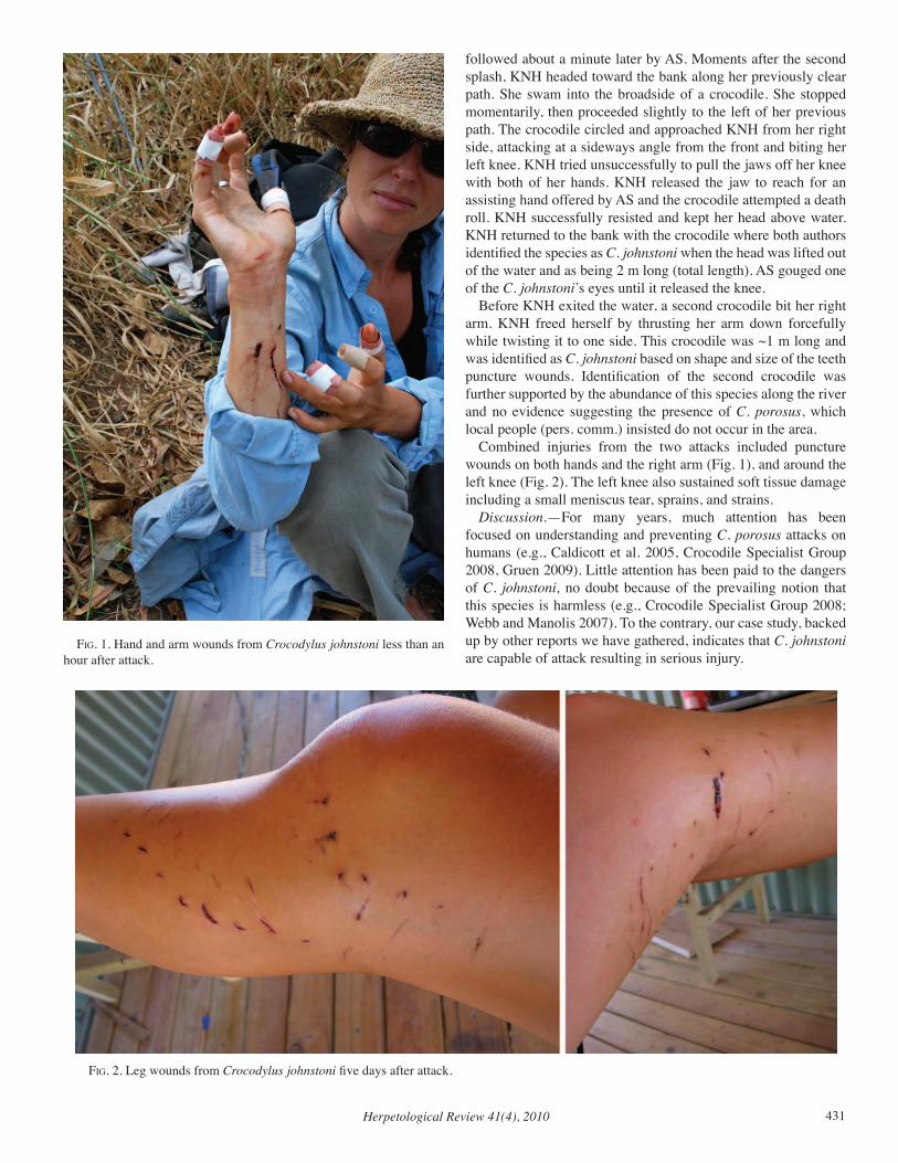
Herpetological Review 41(4), 2010 431
followed about a minute later by AS. Moments after the second splash, KNH headed toward the bank along her previously clear path. She swam into the broadside of a crocodile. She stopped momentarily, then proceeded slightly to the left of her previous path. The crocodile circled and approached KNH from her right side, attacking at a sideways angle from the front and biting her left knee. KNH tried unsuccessfully to pull the jaws off her knee with both of her hands. KNH released the jaw to reach for an assisting hand offered by AS and the crocodile attempted a death roll. KNH successfully resisted and kept her head above water. KNH returned to the bank with the crocodile where both authors identified the species as C. johnstoni when the head was lifted out of the water and as being 2 m long (total length). AS gouged one of the C. johnstoni’s eyes until it released the knee.
Before KNH exited the water, a second crocodile bit her right arm. KNH freed herself by thrusting her arm down forcefully while twisting it to one side. This crocodile was ~1 m long and was identified as C. johnstoni based on shape and size of the teeth puncture wounds. Identification of the second crocodile was further supported by the abundance of this species along the river and no evidence suggesting the presence of C. porosus, which local people (pers. comm.) insisted do not occur in the area.
Combined injuries from the two attacks included puncture wounds on both hands and the right arm (Fig. 1), and around the left knee (Fig. 2). The left knee also sustained soft tissue damage including a small meniscus tear, sprains, and strains.
Discussion.—For many years, much attention has been focused on understanding and preventing C. porosus attacks on humans (e.g., Caldicott et al. 2005, Crocodile Specialist Group 2008, Gruen 2009). Little attention has been paid to the dangers of C. johnstoni, no doubt because of the prevailing notion that this species is harmless (e.g., Crocodile Specialist Group 2008; Webb and Manolis 2007). To the contrary, our case study, backed up by other reports we have gathered, indicates that C. johnstoni are capable of attack resulting in serious injury.
fig. 1. Hand and arm wounds from Crocodylus johnstoni less than an hour after attack.
fig. 2. Leg wounds from Crocodylus johnstoni five days after attack.
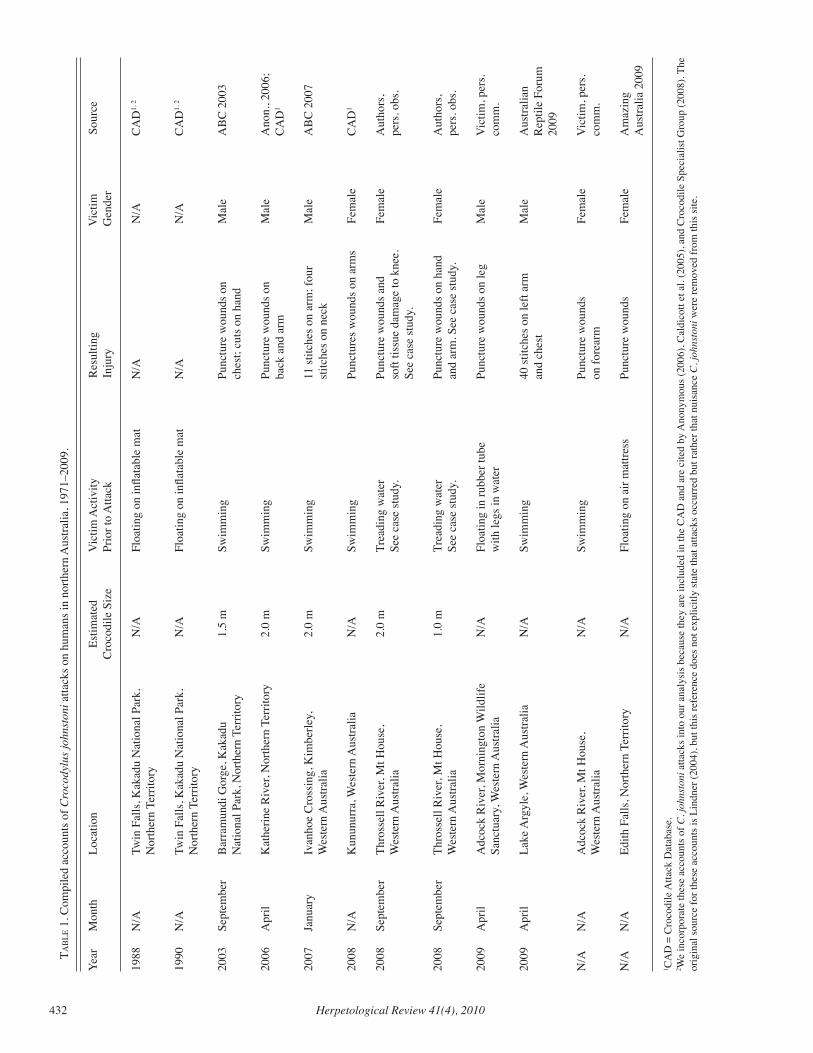
Herpetological Review 41(4), 2010432
taB
le 1
. Com
pile
d ac
coun
ts o
f Cro
cody
lus j
ohns
toni
atta
cks o
n hu
man
s in
north
ern
Aus
tralia
, 197
1–20
09.
Year
M
onth
Lo
catio
n Es
timat
ed
Vic
tim A
ctiv
ity
Res
ultin
g V
ictim
So
urce
C
roco
dile
Siz
e Pr
ior t
o A
ttack
In
jury
G
ende
r
1988
N
/A
Twin
Fal
ls, K
akad
u N
atio
nal P
ark,
N
/A
Floa
ting
on in
flata
ble
mat
N
/A
N/A
C
AD
1, 2
Nor
ther
n Te
rrito
ry
1990
N
/A
Twin
Fal
ls, K
akad
u N
atio
nal P
ark,
N
/A
Floa
ting
on in
flata
ble
mat
N
/A
N/A
C
AD
1, 2
Nor
ther
n Te
rrito
ry
2003
Se
ptem
ber
Bar
ram
undi
Gor
ge, K
akad
u 1.
5 m
Sw
imm
ing
Pu
nctu
re w
ound
s on
M
ale
AB
C 2
003
N
atio
nal P
ark,
Nor
ther
n Te
rrito
ry
ches
t; cu
ts o
n ha
nd
2006
A
pril
Kat
herin
e R
iver
, Nor
ther
n Te
rrito
ry
2.0
m
Swim
min
g Pu
nctu
re w
ound
s on
Mal
e A
non.
, 200
6;
ba
ck a
nd a
rm
C
AD
1
2007
Ja
nuar
y Iv
anho
e C
ross
ing,
Kim
berle
y,
2.0
m
Swim
min
g 11
stitc
hes o
n ar
m; f
our
Mal
e A
BC
200
7
W
este
rn A
ustra
lia
stitc
hes o
n ne
ck
2008
N
/A
Kun
unur
ra, W
este
rn A
ustra
lia
N/A
Sw
imm
ing
Pu
nctu
res w
ound
s on
arm
s Fe
mal
e C
AD
1
2008
Se
ptem
ber
Thro
ssel
l Riv
er, M
t Hou
se,
2.0
m
Trea
ding
wat
er
Punc
ture
wou
nds a
nd
Fem
ale
Aut
hors
,
W
este
rn A
ustra
lia
Se
e ca
se st
udy.
so
ft tis
sue
dam
age
to k
nee.
pers
. obs
.
See
case
stud
y.
2008
Se
ptem
ber
Thro
ssel
l Riv
er, M
t Hou
se,
1.0
m
Trea
ding
wat
er
Punc
ture
wou
nds o
n ha
nd
Fem
ale
Aut
hors
,
W
este
rn A
ustra
lia
Se
e ca
se st
udy.
an
d ar
m. S
ee c
ase
stud
y.
pe
rs. o
bs.
2009
A
pril
Adc
ock
Riv
er, M
orni
ngto
n W
ildlif
e N
/A
Floa
ting
in ru
bber
tube
Pu
nctu
re w
ound
s on
leg
Mal
e V
ictim
, per
s.
Sa
nctu
ary,
Wes
tern
Aus
tralia
with
legs
in w
ater
co
mm
.
2009
A
pril
Lake
Arg
yle,
Wes
tern
Aus
tralia
N
/A
Swim
min
g 40
stitc
hes o
n le
ft ar
m
Mal
e A
ustra
lian
an
d ch
est
R
eptil
e Fo
rum
20
09
N/A
N
/A
Adc
ock
Riv
er, M
t Hou
se,
N/A
Sw
imm
ing
Punc
ture
wou
nds
Fem
ale
Vic
tim, p
ers.
Wes
tern
Aus
tralia
on
fore
arm
com
m.
N/A
N
/A
Edith
Fal
ls, N
orth
ern
Terr
itory
N
/A
Floa
ting
on a
ir m
attre
ss
Punc
ture
wou
nds
Fem
ale
Am
azin
g
Aus
tralia
200
9
1 CA
D =
Cro
codi
le A
ttack
Dat
abas
e.
2 We
inco
rpor
ate
thes
e ac
coun
ts o
f C. j
ohns
toni
atta
cks i
nto
our a
naly
sis b
ecau
se th
ey a
re in
clud
ed in
the
CA
D a
nd a
re c
ited
by A
nony
mou
s (20
06),
Cal
dico
tt et
al.
(200
5), a
nd C
roco
dile
Spe
cial
ist G
roup
(200
8). T
he
orig
inal
sour
ce fo
r the
se a
ccou
nts i
s Lin
dner
(200
4), b
ut th
is re
fere
nce
does
not
exp
licitl
y st
ate
that
atta
cks o
ccur
red
but r
athe
r tha
t nui
sanc
e C
. joh
nsto
ni w
ere
rem
oved
from
this
site
.
Herpetological Review 41(4), 2010 433
Available data indicate that C. johnstoni attacks are not as common, nor usually as severe, as C. porosus attacks. From 1971 to 2009 there has been an average of 0.3 reported accounts of C. johnstoni attacks on humans per year compared to 1.95 average accounts reported per year for C. porosus (C. Manolis, pers. comm.). There are no known cases of human fatality from C. johnstoni compared to 25 recorded cases from C. porosus between 1971 and 2009 (C. Manolis, pers. comm.). Nevertheless, given the apparently limited reportage, acceptance, and recording of C. johnstoni attack accounts, one must wonder how many other attacks have occurred and are not known. In our data set there is a suggestion of an increase in the number of C. johnstoni attacks on humans in the last decade. This may indicate an actual rise in the number of attacks, but may also reflect easier access to more recent attack accounts.
Our analysis of available information suggests that improved data collection and access to that data is needed if information on C. johnstoni attacks are to be available and useful for both understanding this species’ biology and for informing public safety. The IUCN Crocodile Specialist Group’s recommended repository for crocodile attack information in Australia is Wildlife Management International’s Crocodile Attack Database (CAD). Given that only a third of the attacks compiled here were also in the CAD, this resource appears to be severely underutilized with respect to C. johnstoni. While not an unusual situation for data bases of this sort (Fergusson 2002), it remains troubling. One problem is that the data base is not easy to find. Google searches using reasonable search criteria are unable to find a match within the first ten pages of search results. In addition, there is no online data sheet for easy submission and data contained within the CAD are not readily available online.
Equally critical is the encouragement of reporting in the first place. Our experience and findings suggest that C. johnstoni attacks deserve more initial credibility and reportage of their details needs to be encouraged. Whether there has been an actual increase in the number of C. johnstoni attacks in the last decade or not, the threat of more attacks in the future is real. The numbers of people visiting remote areas continues to rise as improved technology and infrastructure make more challenging locations increasingly accessible. Since C. johnstoni have been observed to be less wary of humans and more prone to aggressive displays in areas where they are not regularly exposed to humans (Webb 1985), increased visitation to remote areas could increase the number of attacks. Attacks may become more frequent across this species’ range regardless of remoteness as human populations rise, increasing the probability of negative encounters. Clearly outlined safety guidelines exist for C. porosus (e.g., Queensland Government 2008). Similar guidelines should be developed and publicized for C. johnstoni. However, little is definitively understood in order to undertake such a task. Improved data will be needed to assess the triggers for C. johnstoni attack. The only clear evaluations from the data set compiled here is that all victims were in the water at the time of attack and there is no apparent gender bias. It appears that larger C. johnstoni are more prone to attack and that attacks are more likely to occur in April or September, but these conclusions are not much more than indicative. Standardized data collection is necessary to limit gaps in the data set and to ensure that the data being collected are
useful for understanding C. johnstoni attack triggers.We emphasize the importance of documenting all C. johnstoni
attacks in a standardized, easily accessible format. Acquiring such data will require an educational campaign to alter the perception that C. johnstoni are harmless, to one of recognizing their potential to attack and the need to report such incidents. Future research should pay discerning attention to the details of each recorded attack to increase our understanding of C. johnstoni attack triggers. This information is essential to understanding the biology of C. johnstoni, improving its conservation and management and, as we have shown in this paper, human safety.
Acknowledgments.—We thank Charlie Manolis for providing access to the CAD, as well as additional C. johnstoni information and a helpful review of our manuscript. We thank Matthew Brien and Grahame Webb for information; two victims who will remain anonymous for sharing their stories with us; and James Kushlan and two anonymous reviewers for constructive advice on the manuscript. Additionally, we thank all the people who facilitated our evacuation and a safe recovery, including Sarah Legge, Dan Swan, Richard Kingswood and other staff from the Australian Wildlife Conservancy’s Mornington Wildlife Sanctuary, Mt. House Station, the Royal Flying Doctor Service, and the Derby Hospital, as well as following care and treatments from various professionals, friends and family in Miami, Florida, and other areas of the USA.
literature cited
anonYMouS. 2006. Rare Australian freshwater crocodile attack. Crocodile Specialist Group Newsletter 25:13.
aMazing auStralia. 2009. http://www.amazingaustralia.com.au/animals/crocodiles.htm. Accessed June 21, 2009.
auStralian BroadcaSting corPoration (aBc). 2003. http://www.abc.net.au/news/stories/2003/09/26/954703.htm. Accessed June 21, 2009.
–––––. 2007. http://www.abc.net.au/news/stories/2007/01/09/1823853.htm. Accessed June 21, 2009.
auStralian rePtile foruM. 2009. http://www.australianreptileforum.com/arforum/forum/showthread.php?t=28696&highlight=freshwater+crocodile. Accessed 24 June 24 2010.
caldicott, d. g. e., d. c. franzcr, c. ManoliS, g. WeBB, and a. Britton. 2005. Crocodile attack in Australia: an analysis of its incidence and review of the pathology and management of crocodilian attacks in general. Wildl. Env. Med. 16:143−159.
crocodile SPecialiSt grouP. 2008. http://iucncsg.org/ph1/modules/Crocodilians/attacks.html. IUCN-SSC Crocodile Specialist Group. Accessed 7 February 2010.
ferguSSon, r. a. 2002. Living with a wild predator: managing human/crocodile conflict in Africa. Crocodile Specialist Group Newsletter 21:16−20.
gruen, r. l. 2009. Crocodile attacks in Australia: challenges for injury prevention and trauma care. World J. Surg. 33:1554−1561.
lindner, g. 2004. Crocodile management—Kakadu National Park. Proceedings of the 17th Working Meeting of the Crocodile Specialist Group of the IUCN-SSC, pp. 41–51. IUCN-SSC Crocodile Specialist Group, Darwin, Australia.
QueenSland governMent. 2008. http://www.derm.qld.gov.au/wildlife-ecosystems/wildlife/living_with_wildlife/crocodiles/crocodiles__be_croc_wise.html. Accessed 28 June 2010.
WeBB, g. J. W. 1985. Survey of a pristine population of freshwater crocodiles in the Liverpool River, Arnhem Land, Australia. Project report to the National Geographic Society.
–––––, and S. c. ManoliS. 2007. Australian Crocodiles. A Natural History. Reed New Holland, Sydney, Australia.
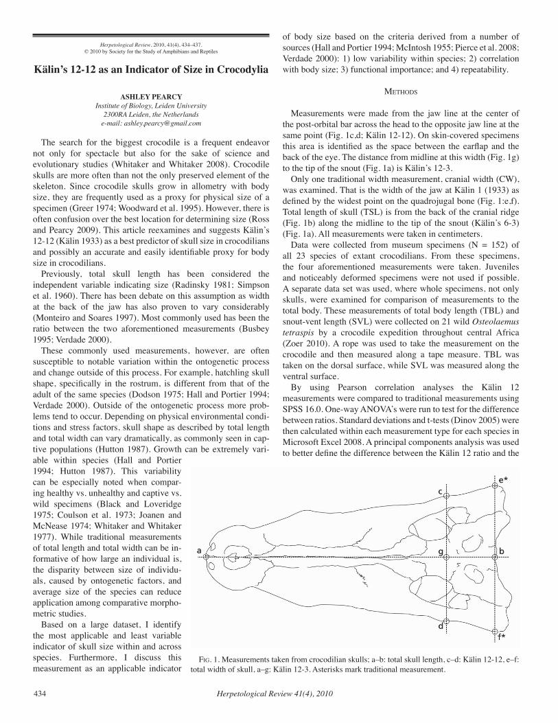
Herpetological Review 41(4), 2010434
Herpetological Review, 2010, 41(4), 434–437.© 2010 by Society for the Study of Amphibians and Reptiles
Kälin’s 12-12 as an Indicator of Size in Crocodylia
ASHLEY PEARCYInstitute of Biology, Leiden University
2300RA Leiden, the Netherlandse-mail: [email protected]
The search for the biggest crocodile is a frequent endeavor not only for spectacle but also for the sake of science and evolutionary studies (Whitaker and Whitaker 2008). Crocodile skulls are more often than not the only preserved element of the skeleton. Since crocodile skulls grow in allometry with body size, they are frequently used as a proxy for physical size of a specimen (Greer 1974; Woodward et al. 1995). However, there is often confusion over the best location for determining size (Ross and Pearcy 2009). This article reexamines and suggests Kälin’s 12-12 (Kälin 1933) as a best predictor of skull size in crocodilians and possibly an accurate and easily identifiable proxy for body size in crocodilians.
Previously, total skull length has been considered the independent variable indicating size (Radinsky 1981; Simpson et al. 1960). There has been debate on this assumption as width at the back of the jaw has also proven to vary considerably (Monteiro and Soares 1997). Most commonly used has been the ratio between the two aforementioned measurements (Busbey 1995; Verdade 2000).
These commonly used measurements, however, are often susceptible to notable variation within the ontogenetic process and change outside of this process. For example, hatchling skull shape, specifically in the rostrum, is different from that of the adult of the same species (Dodson 1975; Hall and Portier 1994; Verdade 2000). Outside of the ontogenetic process more prob-lems tend to occur. Depending on physical environmental condi-tions and stress factors, skull shape as described by total length and total width can vary dramatically, as commonly seen in cap-tive populations (Hutton 1987). Growth can be extremely vari-able within species (Hall and Portier 1994; Hutton 1987). This variability can be especially noted when compar-ing healthy vs. unhealthy and captive vs. wild specimens (Black and Loveridge 1975; Coulson et al. 1973; Joanen and McNease 1974; Whitaker and Whitaker 1977). While traditional measurements of total length and total width can be in-formative of how large an individual is, the disparity between size of individu-als, caused by ontogenetic factors, and average size of the species can reduce application among comparative morpho-metric studies.
Based on a large dataset, I identify the most applicable and least variable indicator of skull size within and across species. Furthermore, I discuss this measurement as an applicable indicator
of body size based on the criteria derived from a number of sources (Hall and Portier 1994; McIntosh 1955; Pierce et al. 2008; Verdade 2000): 1) low variability within species; 2) correlation with body size; 3) functional importance; and 4) repeatability.
MetHodS
Measurements were made from the jaw line at the center of the post-orbital bar across the head to the opposite jaw line at the same point (Fig. 1c,d; Kälin 12-12). On skin-covered specimens this area is identified as the space between the earflap and the back of the eye. The distance from midline at this width (Fig. 1g) to the tip of the snout (Fig. 1a) is Kälin’s 12-3.
Only one traditional width measurement, cranial width (CW), was examined. That is the width of the jaw at Kälin 1 (1933) as defined by the widest point on the quadrojugal bone (Fig. 1:e,f). Total length of skull (TSL) is from the back of the cranial ridge (Fig. 1b) along the midline to the tip of the snout (Kälin’s 6-3) (Fig. 1a). All measurements were taken in centimeters.
Data were collected from museum specimens (N = 152) of all 23 species of extant crocodilians. From these specimens, the four aforementioned measurements were taken. Juveniles and noticeably deformed specimens were not used if possible. A separate data set was used, where whole specimens, not only skulls, were examined for comparison of measurements to the total body. These measurements of total body length (TBL) and snout-vent length (SVL) were collected on 21 wild Osteolaemus tetraspis by a crocodile expedition throughout central Africa (Zoer 2010). A rope was used to take the measurement on the crocodile and then measured along a tape measure. TBL was taken on the dorsal surface, while SVL was measured along the ventral surface.
By using Pearson correlation analyses the Kälin 12 measurements were compared to traditional measurements using SPSS 16.0. One-way ANOVA’s were run to test for the difference between ratios. Standard deviations and t-tests (Dinov 2005) were then calculated within each measurement type for each species in Microsoft Excel 2008. A principal components analysis was used to better define the difference between the Kälin 12 ratio and the
fig. 1. Measurements taken from crocodilian skulls; a–b: total skull length, c–d: Kälin 12-12, e–f: total width of skull, a–g: Kälin 12-3. Asterisks mark traditional measurement.
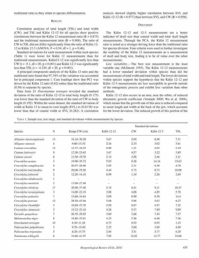
Herpetological Review 41(4), 2010 435
traditional ratio as they relate to species differentiation.
reSultS
Correlation analyses of total length (TSL) and total width (CW), and TSL and Kälin 12-12 for all species show positive correlations between the Kälin 12 measurement ratio (R = 0.875) and the traditional measurement ratio (R = 0.908). The ratio of CW to TSL did not differ significantly from the ratio of Kälin 12-12 to Kälin 12-3 (ANOVA: F = 0.54, df = 1, p = 0.46).
Standard deviations for each measurement within each species (Table 1) were lower for Kälin 12 measurements than for traditional measurements. Kälin12-12 was significantly less than CW (t = -5.1, df = 20, p < 0.001) and Kälin 12-3 was significantly less than TSL (t = -4.320, df = 20, p < 0.001).
A principal components analysis of the Kälin 12 ratio and the traditional ratio found that 97.34% of the variation was accounted for in principal component 1. Case loadings show that PC1 was driven by the Kälin 12 ratio (0.82) rather than the traditional ratio (0.58) to separate by species.
Data from 21 Osteolaemus tetraspis revealed the standard deviation of the ratio of Kälin 12-12 to total body length (0.175) was lower than the standard deviation of the ratio of CW to body length (0.192). Within the same dataset, the standard deviation of width at Kälin 12 to snout-to-vent length (SVL) to (0.0138) was lower than that of cranial width to SVL (0.282). A correlation
analysis showed slightly higher correlation between SVL and Kälin 12-12 (R = 0.977) than between SVL and CW (R = 0.958).
diScuSSion
The Kälin 12-12 and 12-3 measurements are a better indicator of skull size than cranial width and total skull length measurements. Through the PCA, the Kälin 12 measurement ratio is noted as a stronger driving force than the traditional ratio for species division. Four criteria were used to further investigate the usability of the Kälin 12 measurements as a measurement of skull and body size, finding it to be of value over the other measurements.
Low variability.—The best size measurement is the least variable one (McIntosh 1955). The Kälin 12-12 measurements had a lower standard deviation within species than did the measurements of total width and total length. The lower deviations within species support the hypothesis that the Kälin 12-12 and Kälin 12-3 measurements are less susceptible to growth outside of the ontogenetic process and exhibit less variation than other methods.
Kälin 12-12 also occurs in an area, near the orbits, of reduced allometric growth coefficient (Verdade 2000; Wu et al. 2006), which means that the growth rate of this area is reduced compared to snout length and width at the back of the jaw, which accounts for the lower deviation. The reduced growth of this portion of the
taBle 1. Sample size, size range, and standard deviations within measurements by species.
Standard deviations
Species N Range CW (cm) Kälin 12-12 CW Kälin 12-3 TSL
Alligator mississippiensis 13 16.10–30.28 3.67 5.04 6.48 7.31Alligator sinensis 4 9.60–15.10 2.24 2.33 3.02 3.61Caiman crocodilus 12 12.37–16.55 0.99 1.21 2.03 2.19Caiman latirostris 9 12.30–23.65 3.47 4.57 3.22 3.85Caiman yacare 6 13.50–19.70 2.14 2.56 2.46 3.21Crocodylus acutus 3 19.90–35.72 5.93 8.26 14.16 15.63Crocodylus cataphractus 8 20.07–26.86 2.05 2.31 4.36 4.76Crocodylus intermedius 9 20.00–35.50 4.44 5.75 8.73 10.08Crocodylus johnsoni 2 12.20–14.10 0.99 1.34 2.26 2.69Crocodylus mindorensis 1 – – – – –Crocodylus moreletii 2 13.08–27.40 – – – –Crocodylus niloticus 17 20.90–37.40 5.15 6.41 9.11 10.47Crocodylus novaeguineae 3 14.00–22.10 3.08 4.06 4.85 5.78Crocodylus palustris 7 15.60–34.84 5.09 6.99 8.39 14.4Crocodylus porosus 12 28.50–43.94 5.08 5.98 5.63 6.27Crocodylus rhombifer 3 10.69–23.70 5.50 6.87 4.97 7.52Crocodylus siamensis 2 15.22–23.10 4.26 5.57 7.89 9.89Gavialis gangeticus 7 20.35–29.55 3.69 3.66 7.54 7.57Melanosuchus niger 8 18.80–33.91 4.23 5.56 6.46 7.56Osteolaemis tetraspis 10 8.50–11.28 0.75 0.92 0.92 1.23Paleosuchus palpebrosus 3 9.70–15.49 2.25 3.00 3.65 4.69Paleosuchus trigonatus 5 8.20–15.75 2.84 3.51 5.17 6.20Tomistoma schlegelii 6 19.60–41.97 7.95 12.02 12.77 14.08
Herpetological Review 41(4), 2010436
skull at later stages of crocodile development is visible through a study of Crocodylus novaeguineae (Hall and Portier 1994). These studies do not specify the exact position of Kälin 12-12, thus inferences are made from the orbital width and postorbital cranial ridge width to snout-vent length. One study on C. niloticus used a measurement from the point anterior to the orbits to the tip of the snout, finding that it grows isometrically relative to head width (Hutton 1987). This is the closest measurement comparable to Kälin 12-3. As the area around the orbits is less prone to growth extremes, it represents a more accurate figure for skeletal size. This can be especially important in captive populations where growth responds to temperature, population density, and food (Blake and Loveridge 1975; Coulson et al. 1973; Joanen and McNease 1974; Whitaker and Whitaker 1977), thereby creating a comparable database. For skin-covered specimens, the location of Kälin 12-12 is also an area of reduced soft tissue, which could prevent discrepancies in measurements based on the thickness of soft tissue.
Correlation with body size.—When compared to body length and snout-vent length, Kälin 12-12 has a lower standard deviation than CW. This shows that Kälin 12-12 is a viable measurement of size even for total body length in O. tetraspis. As O. tetraspis is the smallest crocodile, the lower standard deviation of Kälin 12-12 may be due in part to the crocodile’s small size. However, the previous analysis shows the same positivity for Kälin 12 in skull size for all species. The direct proportions between the skull and body of crocodilians (Chentanez et al. 1983; Hutton 1987; Verdade 2000) supports the conclusion that Kälin 12-12 is a viable measurement relatable to total body size in species other than O. tetraspis.
There is a high level of integration of the differing parts of the skull meaning change in one part will result in the restructuring of the entire skull (Monteiro et al. 1997). We would, therefore, expect a similarity between the Kälin 12 measurements and CW and TSL and their respective ratios, as they are related measurements of length and width, which is a verifiable proportion within crocodilians (Chentanez et al. 1983; Hall and Portier 1994).
Functional importance.—Although functional importance is not always a necessity for determining a measurement point, it can be beneficial for application to ecology and functional morphology. The area of the skull surrounding the orbits has a reduced susceptibility to growth caused by environmental factors and it is a physically strong point on the skull. In battle, crocodilians have been known to bash heads (Webb and Manolis 1989) with the point of contact often near Kälin 12. In spite of the dangers to either side of the focal point such battles rarely seem to cause damage. Due to the closing of the secondary palate, the crocodilian rostrum is much stronger near the orbits. According to Busbey (1995) this leads to less tension from torsional loads associated with feeding behaviors.
Repeatability.—The post-orbital bar offers an area easily identifiable on skulls. On skin-covered specimens there is a minimum margin of error as the distance between the earflap and the eye is small. Although the widest part of the skull seems easier to identify than Kälin 12, in many skulls from collections, the bone is chipped or broken at the widest part. The widest part of the skull can also be open to interpretation ranging from anywhere between the back of the quadrojugal bone to the widest
part of the quadrojugal.
concluSion
Based on the aforementioned criteria, the Kälin 12 measurements prove to be slightly better than the traditional width measurement at the widest part of the skull for determining size of both skull and body. It offers an easily identifiable area which is less susceptible to growth due to environmental factors and the low amount of soft tissue in this area also reduces the bias already found in skin-covered vs. clean specimens. Kälin 12 measurements may also be better measurements within captive populations, where malnourishment and stressful conditions can lead to abnormal growth. Kälin 12-12 is also of physiological importance being one of the stronger areas of the crocodilian skull. The use of Kälin 12 could benefit comparative anatomical, evolutionary, and ecological studies of crocodilians by presenting a measurement meeting proven criteria for usability.
Acknowledgments.—I thank Franklin D. Ross for the measurement and the challenge to test a hypothesis, Zeno Wijtten for assistance in data collection and manuscript revisions, and Roland Zoer, Rogier de Bouer, and Jeroen Koorevaar for sharing data from their Africa expedition. Frans Witte and Nicole Ortega provided comments on the manuscript. I acknowledge personnel associated with Royal Belgian Institute of Natural Sciences, Forschungsinstitut und Naturmuseum Senckenberg, Naturalis, Zoological Museum Amsterdam, and Paris MNHN for access to their crocodilian skull collections.
literature cited
Black, d. k., and J. P. loveridge. 1975. The role of commercial croco-dile farming in crocodile conservation. Biol. Conserv. 8:261–272.
BuSBeY, iii, a. 1995. The structural consequences of skull flatten-ing in crocodilians. In J. Thomason (ed.), Functional Morphology in Vertebrate Paleontology, pp. 173–192. Cambridge Univ. Press, Cambridge, UK.
cHentanez, t., S. e. HugginS, and v. cHentanez. 1983. Allometric rela-tionships of the Siamese crocodile, Crocodylus siamensis. J. Sci. Soc. Thailand 9:5–26.
coulSon, t. d., r. a. coulSon, and t. Hernandez. 1973. Some observa-tions on growth of captive alligators. Zoologica 58:47–52.
dinov, i. d. 2005. T-Distribution Table. http://www.socr.uclaedu/Applets.dir/T-table.html Accessed: 29 September 2009.
dodSon, P. 1975. Functional and ecological significance of growth in Alligator. J. Zool., Lond. 175:315–355.
greer, a. e. 1974. On the maximum total length of the salt-water croco-dile (Crocodylus porosus). J. Herpetol. 8:381–384.
Hall, P. M., and k. M. Portier. 1994. Cranial morphometry of New Guinea crocodiles (Crocodylus novaeguineae): ontogenetic variation in relative growth of the skull and an assessment of its utility as a predictor of sex and size in individuals. Herpetol. Monogr. 8:203–225.
Hutton, J. M. 1987. Growth and feeding ecology of the Nile crocodile Crocodylus niloticus at Ngezi, Zimbabwe. J. Anim. Ecol. 56:25–38.
Joanen, t., and l. McneaSe. 1974. Propagation of immature American alligators in controlled environmental chambers. Proc. Southern Zoo Workshop, Monroe, Louisiana 1978:1–8.
kälin, J. a. 1933. Beiträge zur vergleichenden Osteologie des Crocodilen Schädels. Zool. Jahrb. Abt. Anat. Ont. Tiere 57:535–717 + pls. 11–16.
McintoSH, W. B. 1955. The applicability of covariance analysis for com-parison of body and skeletal measurements between two races of the deer mouse, Peromyscus maniculatus. Contr. Lab. Vertbr. Biol. Univ. Michigan 76:1–54.

Herpetological Review 41(4), 2010 437
MicroSoft. 2008. Excel 12.0. Office for Mac 2008. Monteiro, l., M. cavalcanti, and H. SoMMer, III. 1997. Comparative
ontogenetic shape change in the skull of Caiman species (Crocodylia: Alligatoridae). J. Morphol. 231:53–62.
–––––, and M. SoareS. 1997. Allometric analysis of the ontogenetic variation and evolution of the skull in Caiman Spix, 1825 (Crocodylia: Alligatoridae). Herpetologica 53:62–69.
Pierce, S. e., k. d. angielcYzk, and e. J. raYfield. 2008. Patterns of morphospace occupations and mechanical performance in extant crocodilian skulls: a combined geometric morphometric and finite element modeling approach. J. Morphol. 269:840–864.
radinSkY, l. B. 1981. Evolution of skull shape in carnivores. I. Representative modern carnivores. Biol. J. Linn. Soc. 15:369–388.
roSS, f. d., and a. PearcY. 2009. Maximum width of adult cranium. Crocodile Specialist Group Newsletter 28(4):15.
SiMPSon, g. c., a. roe, and r. c. leWontin 1960. Quantitative Zoology. Harcourt Brace, New York.
SPSS, Inc. 2008. SPSS 16 for Mac OS X. SPSS Inc, Chicago.verdade, l. M. 2000. Regression equations between body and head
measurements in the broad snouted caiman (Caiman latirostris). Rev. Brasil. Biol. 60:469–482.
WeBB, g., and c. ManoliS. 1989. Crocodiles of Australia. Reed Publishing, Frenchs Forest, NSW.
WHitaker, r., and z. WHitaker. 1977. Growth rates of Crocodylus palustris. J. Bombay Nat. Hist. Soc. 75:231–232.
–––––, and n. WHitaker. 2008. Who’s got the biggest? Crocodile Specialist Group Newsletter 27(4):26–30.
WoodWard, a. r., J. H. WHite, and S. B. linda. 1995. Maximum size of the alligator (Alligator mississippiensis). J. Herpetol. 29:507–513.
Wu X. B., H. Xue, l. S. Wu, J. l. zHu, and r. P. Wang. 2006. Regression analysis between body and head measurements of Chinese alligators (Alligator sinensis) in the captive populations. Anim. Biodivers. Conserv. 29:65–71.
zoer, r. 2010. The Osteolaemus project: the influence of bush meat trade on wild populations. Unpubl. M.S. dissertation. Centre for Wildlife Management, University of Pretoria, South Africa.
TECHNIQUES
Herpetological Review, 2010, 41(4), 437–442.© 2010 by Society for the Study of Amphibians and Reptiles
Using a Wildlife Detector Dog for Locating Eastern Indigo Snakes (Drymarchon couperi)
DIRK J. STEVENSON1*KARA R. RAVENSCROFT1, 2
ROBERT T. ZAPPALORTI3
MICHAEL D. RAVENSCROFT1, 2
STEPHEN W. WEIGLEY4
and CHRISTOPHER L. JENKINS1, 5
1The Orianne Society, The Indigo Snake Initiative Orianne Indigo Snake Preserve
P.O. Box 58, Lumber City, Georgia 31549, USA
2Present address: P.O. Box 301, Wabash, Indiana 46992, USA
3Herpetological Associates, Inc., Plant and Wildlife Consultants17961 Southwest 39th Street, Dunnellon, Florida 34432, USA
4PackLeader Dog Training, 14401 Crews Road KPNGig Harbor, Washington 98329, USA
5The Orianne Society, The Indigo Snake Initiative579 Highway 441, Clayton, Georgia 30525, USA
*Corresponding author: [email protected]
The Eastern Indigo Snake (Drymarchon couperi), a large (adults from 1.5–2.6 m total length [Stevenson et al. 2009]) diurnal species, is imperiled and federally listed as “Threatened” due to population declines attributable to habitat loss/fragmentation and declining Gopher Tortoise (Gopherus polyphemus) populations (United State Fish and Wildlife Service [USFWS] 2008). In southern Georgia and portions of its Florida range, D. couperi is closely associated with xeric sandhill habitats and Gopher Tortoise burrows, and adults often use tortoise burrows for overwintering sites (Diemer and Speake 1983; Hyslop et al. 2009a; Stevenson et al. 2003). The burrows of adult tortoises average 4.5 m long and 2 m deep (Diemer 1992), but in aeolian sand ridge habitats (see Ivester and Leigh 2003) adjacent to blackwater streams in southeastern Georgia, burrows are commonly 6.1–9.1 m in length (D. Stevenson, unpubl. data). Adult D. couperi are frequently surface-active during the winter and may bask or shed their skins near burrows, or move between burrows, during periods of mild (10.0–16.7°C) temperatures (Speake et al. 1978; Stevenson et al. 2009). Although habitat use is varied and less associated with sandy habitats in peninsular Florida, D. couperi occur in xeric uplands and regularly use tortoise burrows as far south as south central Florida (Layne and Steiner 1996). Because of the extensive time they spend in tortoise burrows and other below-ground refugia, adult D. couperi are difficult to locate during field surveys. Developing reliable survey methods for this species is an important priority for D. couperi research and monitoring efforts (USFWS 2008).
Recent studies demonstrate that detector dogs have potential Leptophis ahaetulla (UMRC 85-25). Mexico: Quintana Roo: 35 km
NE Felipe Carrillo Puerto. Illustration by Julian C. Lee.
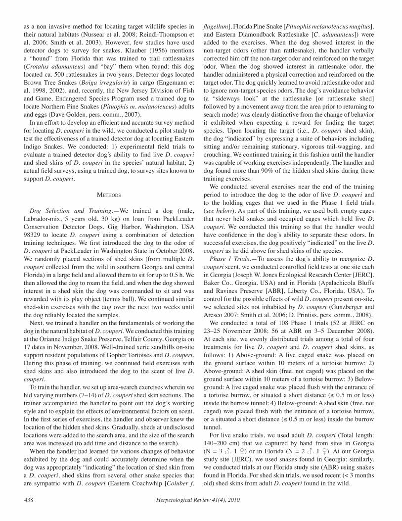
Herpetological Review 41(4), 2010438
as a non-invasive method for locating target wildlife species in their natural habitats (Nussear et al. 2008; Reindl-Thompson et al. 2006; Smith et al. 2003). However, few studies have used detector dogs to survey for snakes. Klauber (1956) mentions a “hound” from Florida that was trained to trail rattlesnakes (Crotalus adamanteus) and “bay” them when found; this dog located ca. 500 rattlesnakes in two years. Detector dogs located Brown Tree Snakes (Boiga irregularis) in cargo (Engemann et al. 1998, 2002), and, recently, the New Jersey Division of Fish and Game, Endangered Species Program used a trained dog to locate Northern Pine Snakes (Pituophis m. melanoleucus) adults and eggs (Dave Golden, pers. comm., 2007).
In an effort to develop an efficient and accurate survey method for locating D. couperi in the wild, we conducted a pilot study to test the effectiveness of a trained detector dog at locating Eastern Indigo Snakes. We conducted: 1) experimental field trials to evaluate a trained detector dog’s ability to find live D. couperi and shed skins of D. couperi in the species’ natural habitat; 2) actual field surveys, using a trained dog, to survey sites known to support D. couperi.
MetHodS
Dog Selection and Training.—We trained a dog (male, Labrador-mix, 5 years old, 30 kg) on loan from PackLeader Conservation Detector Dogs, Gig Harbor, Washington, USA 98329 to locate D. couperi using a combination of detection training techniques. We first introduced the dog to the odor of D. couperi at PackLeader in Washington State in October 2008. We randomly placed sections of shed skins (from multiple D. couperi collected from the wild in southern Georgia and central Florida) in a large field and allowed them to sit for up to 0.5 h. We then allowed the dog to roam the field, and when the dog showed interest in a shed skin the dog was commanded to sit and was rewarded with its play object (tennis ball). We continued similar shed-skin exercises with the dog over the next two weeks until the dog reliably located the samples.
Next, we trained a handler on the fundamentals of working the dog in the natural habitat of D. couperi. We conducted this training at the Orianne Indigo Snake Preserve, Telfair County, Georgia on 17 dates in November, 2008. Well-drained xeric sandhills on-site support resident populations of Gopher Tortoises and D. couperi. During this phase of training, we continued field exercises with shed skins and also introduced the dog to the scent of live D. couperi.
To train the handler, we set up area-search exercises wherein we hid varying numbers (7–14) of D. couperi shed skin sections. The trainer accompanied the handler to point out the dog’s working style and to explain the effects of environmental factors on scent. In the first series of exercises, the handler and observer knew the location of the hidden shed skins. Gradually, sheds at undisclosed locations were added to the search area, and the size of the search area was increased (to add time and distance to the search).
When the handler had learned the various changes of behavior exhibited by the dog and could accurately determine when the dog was appropriately “indicating” the location of shed skin from a D. couperi, shed skins from several other snake species that are sympatric with D. couperi (Eastern Coachwhip [Coluber f.
flagellum], Florida Pine Snake [Pituophis melanoleucus mugitus], and Eastern Diamondback Rattlesnake [C. adamanteus]) were added to the exercises. When the dog showed interest in the non-target odors (other than rattlesnake), the handler verbally corrected him off the non-target odor and reinforced on the target odor. When the dog showed interest in rattlesnake odor, the handler administered a physical correction and reinforced on the target odor. The dog quickly learned to avoid rattlesnake odor and to ignore non-target species odors. The dog’s avoidance behavior (a “sideways look” at the rattlesnake [or rattlesnake shed] followed by a movement away from the area prior to returning to search mode) was clearly distinctive from the change of behavior it exhibited when expecting a reward for finding the target species. Upon locating the target (i.e., D. couperi shed skin), the dog “indicated” by expressing a suite of behaviors including sitting and/or remaining stationary, vigorous tail-wagging, and crouching. We continued training in this fashion until the handler was capable of working exercises independently. The handler and dog found more than 90% of the hidden shed skins during these training exercises.
We conducted several exercises near the end of the training period to introduce the dog to the odor of live D. couperi and to the holding cages that we used in the Phase 1 field trials (see below). As part of this training, we used both empty cages that never held snakes and occupied cages which held live D. couperi. We conducted this training so that the handler would have confidence in the dog’s ability to separate these odors. In successful exercises, the dog positively “indicated” on the live D. couperi as he did above for shed skins of the species.
Phase 1 Trials.—To assess the dog’s ability to recognize D. couperi scent, we conducted controlled field tests at one site each in Georgia (Joseph W. Jones Ecological Research Center [JERC], Baker Co., Georgia, USA) and in Florida (Apalachicola Bluffs and Ravines Preserve [ABR], Liberty Co., Florida, USA). To control for the possible effects of wild D. couperi present on-site, we selected sites not inhabited by D. couperi (Gunzberger and Aresco 2007; Smith et al. 2006; D. Printiss, pers. comm., 2008).
We conducted a total of 108 Phase 1 trials (52 at JERC on 23–25 November 2008; 56 at ABR on 3–5 December 2008). At each site, we evenly distributed trials among a total of four treatments for live D. couperi and D. couperi shed skins, as follows: 1) Above-ground: A live caged snake was placed on the ground surface within 10 meters of a tortoise burrow; 2) Above-ground: A shed skin (free, not caged) was placed on the ground surface within 10 meters of a tortoise burrow; 3) Below-ground: A live caged snake was placed flush with the entrance of a tortoise burrow, or situated a short distance (≤ 0.5 m or less) inside the burrow tunnel; 4) Below-ground: A shed skin (free, not caged) was placed flush with the entrance of a tortoise burrow, or a situated a short distance (≤ 0.5 m or less) inside the burrow tunnel.
For live snake trials, we used adult D. couperi (Total length: 140–200 cm) that we captured by hand from sites in Georgia (N = 3 ♂, 1 ♀) or in Florida (N = 2 ♂, 1 ♀). At our Georgia study site (JERC), we used snakes found in Georgia; similarly, we conducted trials at our Florida study site (ABR) using snakes found in Florida. For shed skin trials, we used recent (< 3 months old) shed skins from adult D. couperi found in the wild.
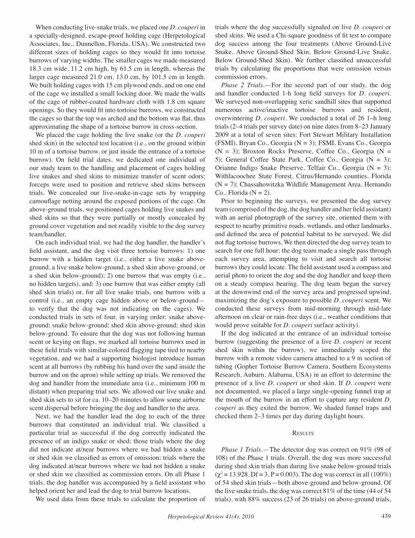
Herpetological Review 41(4), 2010 439
When conducting live-snake trials, we placed one D. couperi in a specially-designed, escape-proof holding cage (Herpetological Associates, Inc., Dunnellon, Florida, USA). We constructed two different sizes of holding cages so they would fit into tortoise burrows of varying widths. The smaller cages we made measured 18.3 cm wide, 11.2 cm high, by 61.5 cm in length, whereas the larger cage measured 21.0 cm, 13.0 cm, by 101.5 cm in length. We built holding cages with 15 cm plywood ends, and on one end of the cage we installed a small locking door. We made the walls of the cage of rubber-coated hardware cloth with 1.8 cm square openings. So they would fit into tortoise burrows, we constructed the cages so that the top was arched and the bottom was flat, thus approximating the shape of a tortoise burrow in cross-section.
We placed the cage holding the live snake (or the D. couperi shed skin) in the selected test location (i.e., on the ground within 10 m of a tortoise burrow, or just inside the entrance of a tortoise burrow). On field trial dates, we dedicated one individual of our study team to the handling and placement of cages holding live snakes and shed skins to minimize transfer of scent odors; forceps were used to position and retrieve shed skins between trials. We concealed our live-snake-in-cage sets by wrapping camouflage netting around the exposed portions of the cage. On above-ground trials, we positioned cages holding live snakes and shed skins so that they were partially or mostly concealed by ground cover vegetation and not readily visible to the dog survey team/handler.
On each individual trial, we had the dog handler, the handler’s field assistant, and the dog visit three tortoise burrows: 1) one burrow with a hidden target (i.e., either a live snake above-ground, a live snake below-ground, a shed skin above-ground, or a shed skin below-ground); 2) one burrow that was empty (i.e., no hidden targets), and; 3) one burrow that was either empty (all shed skin trials) or, for all live snake trials, one burrow with a control (i.e., an empty cage hidden above or below-ground—to verify that the dog was not indicating on the cages). We conducted trials in sets of four, in varying order: snake above-ground; snake below-ground; shed skin above-ground; shed skin below-ground. To ensure that the dog was not following human scent or keying on flags, we marked all tortoise burrows used in these field trials with similar-colored flagging tape tied to nearby vegetation, and we had a supporting biologist introduce human scent at all burrows (by rubbing his hand over the sand inside the burrow and on the apron) while setting up trials. We removed the dog and handler from the immediate area (i.e., minimum 100 m distant) when preparing trial sets. We allowed our live snake and shed skin sets to sit for ca. 10–20 minutes to allow some airborne scent dispersal before bringing the dog and handler to the area.
Next, we had the handler lead the dog to each of the three burrows that constituted an individual trial. We classified a particular trial as successful if the dog correctly indicated the presence of an indigo snake or shed; those trials where the dog did not indicate at/near burrows where we had hidden a snake or shed skin we classified as errors of omission; trials where the dog indicated at/near burrows where we had not hidden a snake or shed skin we classified as commission errors. On all Phase 1 trials, the dog handler was accompanied by a field assistant who helped orient her and lead the dog to trial burrow locations.
We used data from these trials to calculate the proportion of
trials where the dog successfully signaled on live D. couperi or shed skins. We used a Chi-square goodness of fit test to compare dog success among the four treatments (Above Ground-Live Snake, Above Ground-Shed Skin, Below Ground-Live Snake, Below Ground-Shed Skin). We further classified unsuccessful trials by calculating the proportions that were omission versus commission errors.
Phase 2 Trials.—For the second part of our study, the dog and handler conducted 1-h long field surveys for D. couperi. We surveyed non-overlapping xeric sandhill sites that supported numerous active/inactive tortoise burrows and resident, overwintering D. couperi. We conducted a total of 26 1–h long trials (2–4 trials per survey date) on nine dates from 8–23 January 2009 at a total of seven sites: Fort Stewart Military Installation (FSMI), Bryan Co., Georgia (N = 3); FSMI, Evans Co., Georgia (N = 3); Broxton Rocks Preserve, Coffee Co., Georgia (N = 5); General Coffee State Park, Coffee Co., Georgia (N = 3); Orianne Indigo Snake Preserve, Telfair Co., Georgia (N = 3); Withlacoochee State Forest, Citrus/Hernando counties, Florida (N = 7); Chassahowitzka Wildlife Management Area, Hernando Co., Florida (N = 2).
Prior to beginning the surveys, we presented the dog survey team (comprised of the dog, the dog handler and her field assistant) with an aerial photograph of the survey site, oriented them with respect to nearby primitive roads, wetlands, and other landmarks, and defined the area of potential habitat to be surveyed. We did not flag tortoise burrows. We then directed the dog survey team to search for one full hour; the dog team made a single pass through each survey area, attempting to visit and search all tortoise burrows they could locate. The field assistant used a compass and aerial photo to orient the dog and the dog handler and keep them on a steady compass bearing. The dog team began the survey at the downwind end of the survey area and progressed upwind, maximizing the dog’s exposure to possible D. couperi scent. We conducted these surveys from mid-morning through mid-late afternoon on clear or rain-free days (i.e., weather conditions that would prove suitable for D. couperi surface activity).
If the dog indicated at the entrance of an individual tortoise burrow (suggesting the presence of a live D. couperi or recent shed skin within the burrow), we immediately scoped the burrow with a remote video camera attached to a 9 m section of tubing (Gopher Tortoise Burrow Camera, Southern Ecosystems Research, Auburn, Alabama, USA) in an effort to determine the presence of a live D. couperi or shed skin. If D. couperi were not documented, we placed a large single-opening funnel trap at the mouth of the burrow in an effort to capture any resident D. couperi as they exited the burrow. We shaded funnel traps and checked them 2–3 times per day during daylight hours.
reSultS
Phase 1 Trials.—The detector dog was correct on 91% (98 of 108) of the Phase 1 trials. Overall, the dog was more successful during shed skin trials than during live snake below-ground trials (χ2 = 13.928, Df = 3, P = 0.003). The dog was correct in all (100%) of 54 shed skin trials—both above-ground and below-ground. Of the live snake trials, the dog was correct 81% of the time (44 of 54 trials), with 88% success (23 of 26 trials) on above-ground trials,
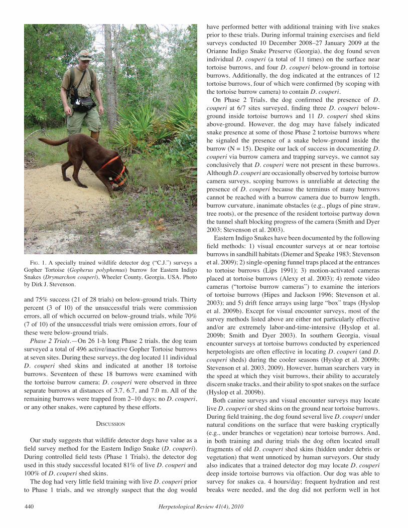
Herpetological Review 41(4), 2010440
and 75% success (21 of 28 trials) on below-ground trials. Thirty percent (3 of 10) of the unsuccessful trials were commission errors, all of which occurred on below-ground trials, while 70% (7 of 10) of the unsuccessful trials were omission errors, four of these were below-ground trials.
Phase 2 Trials.—On 26 1-h long Phase 2 trials, the dog team surveyed a total of 496 active/inactive Gopher Tortoise burrows at seven sites. During these surveys, the dog located 11 individual D. couperi shed skins and indicated at another 18 tortoise burrows. Seventeen of these 18 burrows were examined with the tortoise burrow camera; D. couperi were observed in three separate burrows at distances of 3.7, 6.7, and 7.0 m. All of the remaining burrows were trapped from 2–10 days; no D. couperi, or any other snakes, were captured by these efforts.
diScuSSion
Our study suggests that wildlife detector dogs have value as a field survey method for the Eastern Indigo Snake (D. couperi). During controlled field tests (Phase 1 Trials), the detector dog used in this study successful located 81% of live D. couperi and 100% of D. couperi shed skins.
The dog had very little field training with live D. couperi prior to Phase 1 trials, and we strongly suspect that the dog would
have performed better with additional training with live snakes prior to these trials. During informal training exercises and field surveys conducted 10 December 2008–27 January 2009 at the Orianne Indigo Snake Preserve (Georgia), the dog found seven individual D. couperi (a total of 11 times) on the surface near tortoise burrows, and four D. couperi below-ground in tortoise burrows. Additionally, the dog indicated at the entrances of 12 tortoise burrows, four of which were confirmed (by scoping with the tortoise burrow camera) to contain D. couperi.
On Phase 2 Trials, the dog confirmed the presence of D. couperi at 6/7 sites surveyed, finding three D. couperi below-ground inside tortoise burrows and 11 D. couperi shed skins above-ground. However, the dog may have falsely indicated snake presence at some of those Phase 2 tortoise burrows where he signaled the presence of a snake below-ground inside the burrow (N = 15). Despite our lack of success in documenting D. couperi via burrow camera and trapping surveys, we cannot say conclusively that D. couperi were not present in these burrows. Although D. couperi are occasionally observed by tortoise burrow camera surveys, scoping burrows is unreliable at detecting the presence of D. couperi because the terminus of many burrows cannot be reached with a burrow camera due to burrow length, burrow curvature, inanimate obstacles (e.g., plugs of pine straw, tree roots), or the presence of the resident tortoise partway down the tunnel shaft blocking progress of the camera (Smith and Dyer 2003; Stevenson et al. 2003).
Eastern Indigo Snakes have been documented by the following field methods: 1) visual encounter surveys at or near tortoise burrows in sandhill habitats (Diemer and Speake 1983; Stevenson et al. 2009); 2) single-opening funnel traps placed at the entrances to tortoise burrows (Lips 1991); 3) motion-activated cameras placed at tortoise burrows (Alexy et al. 2003); 4) remote video cameras (“tortoise burrow cameras”) to examine the interiors of tortoise burrows (Hipes and Jackson 1996; Stevenson et al. 2003); and 5) drift fence arrays using large “box” traps (Hyslop et al. 2009b). Except for visual encounter surveys, most of the survey methods listed above are either not particularly effective and/or are extremely labor-and-time-intensive (Hyslop et al. 2009b; Smith and Dyer 2003). In southern Georgia, visual encounter surveys at tortoise burrows conducted by experienced herpetologists are often effective in locating D. couperi (and D. couperi sheds) during the cooler seasons (Hyslop et al. 2009b; Stevenson et al. 2003, 2009). However, human searchers vary in the speed at which they visit burrows, their ability to accurately discern snake tracks, and their ability to spot snakes on the surface (Hyslop et al. 2009b).
Both canine surveys and visual encounter surveys may locate live D. couperi or shed skins on the ground near tortoise burrows. During field training, the dog found several live D. couperi under natural conditions on the surface that were basking cryptically (e.g., under branches or vegetation) near tortoise burrows. And, in both training and during trials the dog often located small fragments of old D. couperi shed skins (hidden under debris or vegetation) that went unnoticed by human surveyors. Our study also indicates that a trained detector dog may locate D. couperi deep inside tortoise burrows via olfaction. Our dog was able to survey for snakes ca. 4 hours/day; frequent hydration and rest breaks were needed, and the dog did not perform well in hot
fig. 1. A specially trained wildlife detector dog (“C.J.”) surveys a Gopher Tortoise (Gopherus polyphemus) burrow for Eastern Indigo Snakes (Drymarchon couperi), Wheeler County, Georgia, USA. Photo by Dirk J. Stevenson.
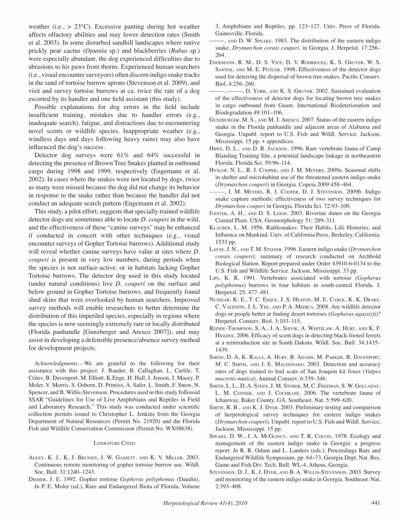
Herpetological Review 41(4), 2010 441
weather (i.e., > 23°C). Excessive panting during hot weather affects olfactory abilities and may lower detection rates (Smith et al. 2003). In some disturbed sandhill landscapes where native prickly pear cactus (Opuntia sp.) and blackberries (Rubus sp.) were especially abundant, the dog experienced difficulties due to abrasions to his paws from thorns. Experienced human searchers (i.e., visual encounter surveyors) often discern indigo snake tracks in the sand of tortoise burrow aprons (Stevenson et al. 2009), and visit and survey tortoise burrows at ca. twice the rate of a dog escorted by its handler and one field assistant (this study).
Possible explanations for dog errors in the field include insufficient training, mistakes due to handler errors (e.g., inadequate search), fatigue, and distractions due to encountering novel scents or wildlife species. Inappropriate weather (e.g., windless days and days following heavy rains) may also have influenced the dog’s success.
Detector dog surveys were 61% and 64% successful in detecting the presence of Brown Tree Snakes planted in outbound cargo during 1998 and 1999, respectively (Engemann et al. 2002). In cases where the snakes were not located by dogs, twice as many were missed because the dog did not change its behavior in response to the snake rather than because the handler did not conduct an adequate search pattern (Engemann et al. 2002).
This study, a pilot effort, suggests that specially trained wildlife detector dogs are sometimes able to locate D. couperi in the wild, and the effectiveness of these “canine surveys” may be enhanced if conducted in concert with other techniques (e.g., visual encounter surveys of Gopher Tortoise burrows). Additional study will reveal whether canine surveys have value at sites where D. couperi is present in very low numbers, during periods when the species is not surface-active, or in habitats lacking Gopher Tortoise burrows. The detector dog used in this study located (under natural conditions) live D. couperi on the surface and below ground in Gopher Tortoise burrows, and frequently found shed skins that were overlooked by human searchers. Improved survey methods will enable researchers to better determine the distribution of this imperiled species, especially in regions where the species is now seemingly extremely rare or locally distributed (Florida panhandle [Gunzberger and Aresco 2007]), and may assist in developing a defensible presence/absence survey method for development projects.
Acknowledgments.—We are grateful to the following for their assistance with this project: J. Bauder, B. Callaghan, L. Carlile, T. Crites, B. Davenport, M. Elliott, K.Enge, H. Hall, J. Jensen, J. Macey, P. Moler, V. Morris, S. Osborn, D. Printiss, A. Safer, L. Smith, F. Snow, N. Spencer, and B. Willis-Stevenson. Procedures used in this study followed SSAR “Guidelines for Use of Live Amphibians and Reptiles in Field and Laboratory Research.” This study was conducted under scientific collection permits issued to Christopher L. Jenkins from the Georgia Department of Natural Resources (Permit No. 21920) and the Florida Fish and Wildlife Conservation Commission (Permit No. WX08638).
literature cited
aleXY, k. J., k. J. BrunJeS, J. W. gaSSett, and k. v. Miller. 2003. Continuous remote monitoring of gopher tortoise burrow use. Wildl. Soc. Bull. 31:1240–1243.
dieMer, J. e. 1992. Gopher tortoise Gopherus polyphemus (Daudin). In P. E. Moler (ed.), Rare and Endangered Biota of Florida, Volume
3, Amphibians and Reptiles, pp. 123–127. Univ. Press of Florida, Gainesville, Florida.
–––––, and d. W. SPeake. 1983. The distribution of the eastern indigo snake, Drymarchon corais couperi, in Georgia. J. Herpetol. 17:256–264.
engeMann, r. M., d. S. vice, d. v. rodriguez, k. S. gruver, W. S. SantoS, and M. e. Pitzler. 1998. Effectiveness of the detector dogs used for deterring the dispersal of brown tree snakes. Pacific Conserv. Biol. 4:256–260.
–––––, –––––, d. York, and k. S. gruver. 2002. Sustained evaluation of the effectiveness of detector dogs for locating brown tree snakes in cargo outbound from Guam. International Biodeterioration and Biodegradation 49:101–106.
gunzBurger, M. S., and M. J. areSco. 2007. Status of the eastern indigo snake in the Florida panhandle and adjacent areas of Alabama and Georgia. Unpubl. report to U.S. Fish and Wildl. Service, Jackson, Mississippi. 15 pp. + appendices.
HiPeS, d. l., and d. r. JackSon. 1996. Rare vertebrate fauna of Camp Blanding Training Site, a potential landscape linkage in northeastern Florida. Florida Sci. 59:96–114.
HYSloP, n. l., r. J. cooPer, and J. M. MeYerS. 2009a. Seasonal shifts in shelter and microhabitat use of the threatened eastern indigo snake (Drymarchon couperi) in Georgia. Copeia 2009:458–464.
–––––, J. M. MeYerS, r. J. cooPer, d. J. StevenSon. 2009b. Indigo snake capture methods: effectiveness of two survey techniques for Drymarchon couperi in Georgia. Florida Sci. 72:93–100.
iveSter, a. H., and d. S. leigH. 2003. Riverine dunes on the Georgia Coastal Plain, USA. Geomorphology 51: 289–311.
klauBer, l. M. 1956. Rattlesnakes: Their Habits, Life Histories, and Influence on Mankind. Univ. of California Press, Berkeley, California. 1533 pp.
laYne, J. n., and t. M. Steiner. 1996. Eastern indigo snake (Drymarchon corais couperi): summary of research conducted on Archbold Biological Station. Report prepared under Order 43910-6-0134 to the U.S. Fish and Wildlife Service. Jackson, Mississippi. 33 pp.
liPS, k. r. 1991. Vertebrates associated with tortoise (Gopherus polyphemus) burrows in four habitats in south-central Florida. J. Herpetol. 25: 477–481.
nuSSear, k. e., t. c. eSQue, J. S. Heaton, M. e. caBlk, k. k. drake, c. valentin, J. l. Yee, and P. a. Medica. 2008. Are wildlife detector dogs or people better at finding desert tortoises (Gopherus agassizii)? Herpetol. Conserv. Biol. 3:103–115.
reindl-tHoMPSon, S. a., J. a. SHivik, a. WHitelaW, a. Hurt, and k. f. HigginS. 2006. Efficacy of scent dogs in detecting black-footed ferrets at a reintroduction site in South Dakota. Wildl. Soc. Bull. 34:1435–1439.
SMitH, d. a, k. rallS, a. Hurt, B. adaMS, M. Parker, B. davenPort, M. c. SMitH, and J. e. Maldonado. 2003. Detection and accuracy rates of dogs trained to find scats of San Joaquin kit foxes (Vulpes macrotis mutica). Animal Conserv. 6:339–346.
SMitH, l. l., d. a. Steen, J. M. StoBer, M. c. freeMan, S. W. golladaY, l. M. conner, and J. cocHrane. 2006. The vertebrate fauna of Ichauway, Baker County, GA. Southeast. Nat. 5:599–620.
SMitH, r. B., and k. J. dYer. 2003. Preliminary testing and comparison of herpetological survey techniques for eastern indigo snakes (Drymarchon couperi). Unpubl. report to U.S. Fish and Wildl. Service, Jackson, Mississippi. 15 pp.
SPeake, d. W., J. a. McglincY, and t. r. colvin. 1978. Ecology and management of the eastern indigo snake in Georgia: a progress report. In R. R. Odum and L. Landers (eds.), Proceedings Rare and Endangered Wildlife Symposium, pp. 64–73. Georgia Dept. Nat. Res. Game and Fish Div. Tech. Bull. WL-4, Athens, Georgia.
StevenSon, d. J., k. J. dYer, and B. a. WilliS-StevenSon. 2003. Survey and monitoring of the eastern indigo snake in Georgia. Southeast. Nat. 2:393–408.
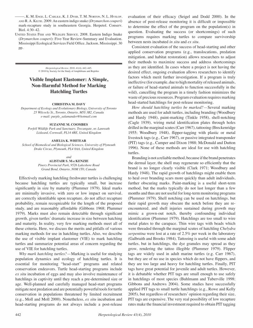
Herpetological Review 41(4), 2010442
–––––, k. M. enge, l. carlile, k. J. dYer, t. M. norton, n. l. HYSloP, and r. a. kiltie. 2009. An eastern indigo snake (Drymarchon couperi) mark-recapture study in southeastern Georgia. Herpetol. Conserv. Biol. 4:30–42.
united StateS fiSH and Wildlife Service. 2008. Eastern Indigo Snake (Drymarchon couperi): Five-Year Review-Summary and Evaluation. Mississippi Ecological Services Field Office, Jackson, Mississippi. 30 pp.
Herpetological Review, 2010, 41(4), 442–445.© 2010 by Society for the Study of Amphibians and Reptiles
Visible Implant Elastomer: A Simple, Non-Harmful Method for Marking
Hatchling Turtles
CHRISTINA M. DAVY Department of Ecology and Evolutionary Biology, University of Toronto
25 Wilcocks St., Toronto, Ontario, M5S 3B2, Canadae-mail: [email protected]
SUZANNE M. COOMBESPorfell Wildlife Park and Sanctuary, Trecangate, nr. Lanreath
Liskeard, Cornwall, PL14 4RE, United Kingdom
AMELIA K. WHITEARSchool of Biomedical and Biological Sciences, University of Plymouth
Drake Circus, Plymouth, PL4 8AA, United Kingdom
andALISTAIR S. MacKENZIE
Pinery Provincial Park, 9526 Lakeshore RoadGrand Bend, Ontario, N0M 1T0, Canada
Effectively marking hatchling freshwater turtles is challenging because hatchling turtles are typically small, but increase significantly in size by maturity (Plummer 1979). Ideal marks are minimally invasive, with zero or low impact on survival, are correctly identifiable upon recapture, do not affect recapture probability, remain recognizable for the length of the proposed study, and are reasonably affordable (Gibbons 1968; Plummer 1979). Marks must also remain detectable through significant growth, given turtles’ dramatic increase in size between hatching and maturity. In reality, very few marking methods meet all of these criteria. Here, we discuss the merits and pitfalls of various marking methods for use in hatchling turtles. Also, we describe the use of visible implant elastomer (VIE) to mark hatchling turtles and summarize potential areas of concern regarding the use of VIE for hatchling turtles.
Why mark hatchling turtles?—Marking is useful for studying population dynamics and ecology of hatchling turtles. It is essential for monitoring “head-start” programs and related conservation endeavors. Turtle head-starting programs include ex situ incubation of eggs and may also involve maintenance of hatchlings in captivity until they reach a pre-determined size or age. Well-planned and carefully managed head-start programs mitigate nest predation and are potentially powerful tools for turtle conservation in populations threatened by limited recruitment (e.g., Moll and Moll 2000). Nonetheless, ex situ incubation and head-starting programs do not always include a post-release
evaluation of their efficacy (Seigel and Dodd 2000). In the absence of post-release monitoring it is difficult or impossible to determine the effect of the program on the population(s) in question. Evaluating the success (or shortcomings) of such programs requires marking turtles to compare survivorship between nests incubated in situ and ex situ.
Consistent evaluation of the success of head-starting and other applied conservation programs (e.g., translocations, predation mitigation, and habitat restoration) allows researchers to adjust their methods to maximize success and address shortcomings as they are identified. In cases where a project is not having the desired effect, ongoing evaluation allows researchers to identify factors which merit further investigation. If a program is truly ineffective (for example, due to high mortality of released animals, or failure of head-started animals to function successfully in the wild), cancelling the program in a timely fashion minimizes the waste of precious resources. Program evaluation requires marking head-started hatchlings for post-release monitoring.
How should hatchling turtles be marked?—Several marking methods are used for adult turtles, including branding (Woodbury and Hardy 1948), paint-marking (Tinkle 1958), shell-notching (Cagle 1939), wiring metal identification plates through holes drilled in the marginal scutes (Carr 1967), tattooing (Breckenridge 1955; Woodbury 1948), flipper-tagging with plastic or metal livestock tags (e.g., Carr 1967), or passive integrated transponder (PIT) tags (e.g., Camper and Dixon 1988; McDonald and Dutton 1996). None of these methods are ideal for use with hatchling turtles.
Branding is not a reliable method, because if the brand penetrates the dermal layer, the shell may regenerate so efficiently that the brand is no longer clearly visible (Clark 1971; Woodbury and Hardy 1948). The rapid growth of hatchlings might enable them to heal over branding scars more quickly than adult individuals, further obscuring marks. Paint-marking is a useful short-term method, but the marks typically do not last longer than a few months and thus are not useful for long-term monitoring programs (Plummer 1979). Shell notching can be used on hatchlings, but their rapid growth may obscure the notch before they are re-encountered, and shell injuries sustained during growth may mimic a grown-out notch, thereby confounding individual identification (Plummer 1979). Hatchlings are too small to wire metal plates to the carapace. Thin wire tags with beads which were threaded through the marginal scutes of hatchling Chelydra serpentina were lost at a rate of 2.5% per week in the laboratory (Galbraith and Brooks 1984). Tattooing is useful with some adult turtles, but in hatchlings, the dye granules may spread as they grow, rendering the tattoo illegible (Plummer 1979). Flipper tags are widely used in adult marine turtles (e.g. Carr 1967), but they are of no use in species which do not have flippers, and they are too large and heavy for hatchling turtles. Finally, PIT tags have great potential for juvenile and adult turtles. However, it is debatable whether PIT tags are small enough to use safely in hatchlings of most species (Buhlmann and Tuberville 1998; Gibbons and Andrews 2004). Some studies have successfully applied PIT tags to small turtle hatchlings (e.g., Rowe and Kelly 2005), but regardless of researchers’ opinion regarding their size, PIT tags are expensive. The very real possibility of low recapture rates make the financial investment required to obtain PIT tagging
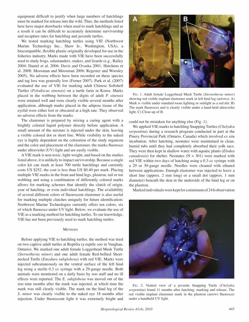
Herpetological Review 41(4), 2010 443
equipment difficult to justify when large numbers of hatchlings must be marked for release into the wild. Thus, the methods listed here have major drawbacks when used to mark hatchlings and as a result it can be difficult to accurately determine survivorship and recapture rates for hatchling and juvenile turtles.
We tested marking hatchling turtles using VIE (Northwest Marine Technology Inc., Shaw Is., Washington, USA), a biocompatible, flexible plastic originally developed for use in the fisheries industry. Marks made with VIE have been successfully used to study frogs, salamanders, snakes, and lizards (e.g., Bailey 2004; Daniel et al. 2006; Davis and Ovaska 2001; Hutchens et al. 2008; Moosman and Moosman 2006; Regester and Woosley 2005). No adverse effects have been recorded on these species and tag loss was generally low (Ferner 2007). Park et al. (2007) evaluated the use of VIE for marking adult Chinese Softshell Turtles (Pelodiscus sinensis) on a turtle farm in Korea. Marks placed in the webbing between the digits of adult P. sinensis were retained well and were clearly visible several months after application, although marks placed in the adipose tissue of the eyelid were either lost or obscured at a high rate. They observed no adverse effects from the marks.
The elastomer is prepared by mixing a curing agent with a brightly colored liquid polymer directly before application. A small amount of the mixture is injected under the skin, leaving a visible colored dot or short line. While visibility to the naked eye is highly dependent on the coloration of the study organism and the color and placement of the elastomer, the marks fluoresce under ultraviolet (UV) light and are easily visible.
A VIE mark is non-toxic, light-weight, and based on the studies listed above, it is unlikely to impact survivorship. Because a single color kit can mark at least 700 turtle hatchlings and currently costs US $252, the cost is less than US $0.40 per mark. Placing multiple VIE marks in the front and hind legs, plastron, tail or toe webbing and using a combination of differently colored marks allows for marking schemes that identify the clutch of origin, year of hatching, or even individual hatchlings. The availability of several different colors of fluorescent elastomer is also useful for marking multiple clutches uniquely for future identification: Northwest Marine Technologies currently offers ten colors, six of which fluoresce under UV light. Below, we evaluate the use of VIE as a marking method for hatchling turtles. To our knowledge, VIE has not been previously used to mark hatchling turtles.
MetHodS
Before applying VIE to hatchling turtles, the method was tried on two captive adult turtles at Reptilia (a reptile zoo in Vaughan, Ontario). We marked one adult female Loggerhead Musk Turtle (Sternotherus minor) and one adult female Red-bellied Short-necked Turtle (Emydura subglobosa) with red VIE. Marks were injected subcutaneously on the ventral surface of the left hind leg using a sterile 0.3 cc syringe with a 29-gauge needle. Both animals were monitored on a daily basis by zoo staff and no ill effects were reported. The E. subglobosa was moved out of the zoo nine months after the mark was injected, at which time the mark was still clearly visible. The mark on the hind leg of the S. minor was clearly visible to the naked eye 18 months after injection. Under fluorescent light it was extremely bright and
could not be mistaken for anything else (Fig. 1).We applied VIE marks to hatchling Snapping Turtles (Chelydra
serpentina) during a research program conducted in part at the Pinery Provincial Park (Ontario, Canada) which involved ex situ incubation. After hatching, neonates were maintained in clean, humid tubs until they had completely absorbed their yolk sacs. They were then kept in shallow water with aquatic plants (Elodea canadensis) for shelter. Neonates (N = 301) were marked with red VIE within two days of hatching using a 0.3-cc syringe with a 29 or 30-gauge needle. Needles were cleaned with ethanol between applications. Enough elastomer was injected to leave a short line (approx. 2 mm long) or a small dot (approx. 1 mm diameter) beneath the skin in the underside of the hind leg or on the plastron.
Marked individuals were kept for a minimum of 24 h observation
fig. 1. Adult female Loggerhead Musk Turtle (Sternotherus minor) showing red visible implant elastomer mark in left hind leg (arrows). A) Mark is visible under standard room lighting or sunlight as a red dot. B) The mark fluoresces and is clearly visible under a hand-held ultraviolet light. C) Close-up of B.
fig. 2. Ventral view of a juvenile Snapping Turtle (Chelydra serpentina) found 11 months after hatching, marking and release. The red visible implant elastomer mark in the plastron (arrow) fluoresces under a handheld UV light.
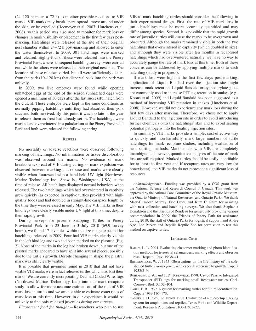
Herpetological Review 41(4), 2010444
(24–120 h; mean = 72 h) to monitor possible reactions to VIE marks. VIE marks may break apart, spread, move around under the skin, or be expelled (Heemeyer et al. 2007; Hutchens et al. 2008), so this period was also used to monitor for mark loss or changes in mark visibility or placement in the first few days post-marking. Hatchlings were released at the site of their original nest chamber within 24–72 h post-marking and allowed to enter the water themselves. In 2009, 301 hatchlings were marked and released. Eighty-four of these were released into the Pinery Provincial Park, where subsequent hatchling surveys were carried out, while the others were released at their original nest sites. The location of these releases varied, but all were sufficiently distant from the park (10–120 km) that dispersal back into the park was unlikely.
In 2009, two live embryos were found while opening unhatched eggs at the end of the season (unhatched eggs were opened a minimum of 30 days after the last successful hatch from the clutch). These embryos were kept in the same conditions as normally pipping hatchlings until they had absorbed their yolk sacs and both survived. By this point it was too late in the year to release them as frost had already set in. The hatchlings were marked and overwintered in a paludarium at the Pinery Provincial Park and both were released the following spring.
reSultS
No mortality or adverse reactions were observed following marking of hatchlings. No inflammation or tissue discoloration was observed around the marks. No evidence of mark breakdown, spread of VIE during curing, or mark expulsion was observed between marking and release and marks were clearly visible when fluoresced with a hand-held UV light (Northwest Marine Technology Inc., Shaw Is., Washington, USA) at the time of release. All hatchlings displayed normal behaviors when released. The two hatchlings which had overwintered in captivity grew quickly (as expected in captive turtles provided with high-quality food) and had doubled in straight-line carapace length by the time they were released in early May. The VIE marks in their hind legs were clearly visible under UV light at this time, despite their rapid growth.
During surveys for juvenile Snapping Turtles in Pinery Provincial Park from 23 June to 3 July 2010 (69.9 survey hours), we found 17 juveniles within the size range expected for hatchlings released in 2009. Four had VIE marks clearly visible in the left hind leg and two had been marked on the plastron (Fig. 2). None of the marks in the leg had broken down, but one of the plastral marks appeared to have split into several pieces, possibly due to the turtle’s growth. Despite changing in shape, the plastral mark was still clearly visible.
It is possible that juveniles found in 2010 that did not have visible VIE marks were in fact released turtles which had lost their marks. We are currently incorporating Decimal Coded Wire Tags (Northwest Marine Technology Inc.) into our mark-recapture study to allow for more accurate estimations of the rate of VIE mark loss in turtles and we are not able to estimate exact rates of mark loss at this time. However, in our experience it would be unlikely to find only released juveniles during our surveys.
Fluorescent food for thought.—Researchers who plan to use
VIE to mark hatchling turtles should consider the following in their experimental design. First, the rate of VIE mark loss in turtle hatchlings must be more accurately quantified and may differ among species. Second, it is possible that the rapid growth rate of juvenile turtles will cause the marks to be overgrown and obscured. Although the marks remained visible in both the two hatchlings that overwintered in captivity (which doubled in size), and although they were visible after ten months in recaptured hatchlings which had overwintered naturally, we have no way to accurately gauge the rate of mark loss at this time. Both of these concerns can be addressed by applying multiple marks to each hatchling (study in progress).
If mark loss were high in the first few days post-marking, application of Liquid Bandaid over the injection site might increase mark retention. Liquid Bandaid or cyanoacrylate glues are commonly used to increase PIT tag retention in snakes (e.g., Koons et al. 2009) and Liquid Bandaid has been suggested as a method of increasing VIE retention in snakes (Hutchens et al. 2008). However, we did not experience any mark loss during the first few days after marking. Therefore, we chose not to apply Liquid Bandaid to the injection site in order to avoid introducing further chemicals onto the hatchlings’ skin and to avoid sealing potential pathogens into the healing injection sites.
In summary, VIE marks provide a simple, cost-effective way to quickly and non-harmfully mark large numbers of turtle hatchlings for mark-recapture studies, including evaluation of head-starting methods. Marks made with VIE are completely unambiguous; however, quantitative analyses of the rate of mark loss are still required. Marked turtles should be easily identifiable for at least the first year and if recapture rates are very low (or nonexistent), the VIE marks do not represent a significant loss of resources.
Acknowledgments.—Funding was provided by a CGS grant from the National Science and Research Council of Canada. This work was approved by the Animal Care Committee of the Royal Ontario Museum, the Ontario Ministry of Natural Resources, and Ontario Parks. We thank Mary-Elizabeth Murray, Eric Davy, and Kum C. Shim for assisting with nest collection and hatchling surveys. We also thank Stephanie Donaldson and the Friends of Rondeau for generously providing various accommodations in 2009; the Friends of Pinery Park for assistance during 2010; the staff of Ontario Parks for logistical support; and Andre Ngo, Lee Parker, and Reptilia Reptile Zoo for permission to test this method on captive turtles.
literature cited
BaileY, L. L. 2004. Evaluating elastomer marking and photo identifica-tion methods for terrestrial salamanders: marking effects and observer bias. Herpetol. Rev. 35:38–41.
Breckenridge, W. J. 1955. Observations on the life-history of the soft-shelled turtle Trionyx ferox, with especial reference to growth. Copeia 1955:5–9.
BuHlMann, K. A., and T. D. TuBerville. 1998. Use of Passive Integrated Transponder (PIT) tags for marking small freshwater turtles. Chel. Conserv. Biol. 3:102–104.
Cagle, F. R. 1939. A system for marking turtles for future identification. Copeia 1939:170–173.
CaMPer, J. D., and J. R. DiXon. 1988. Evaluation of a microchip marking system for amphibians and reptiles. Texas Parks and Wildlife Depart-ment, Research Publication 7100-159:1–22.
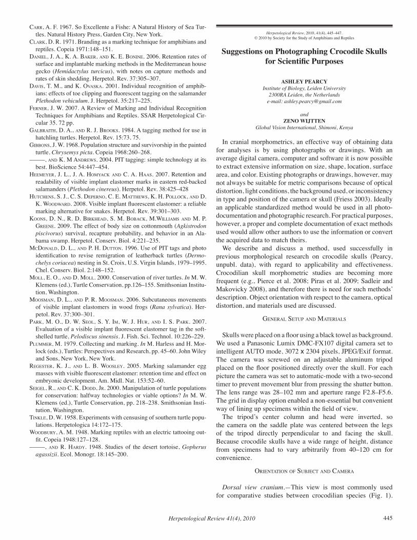
Herpetological Review 41(4), 2010 445
Carr, A. F. 1967. So Excellente a Fishe: A Natural History of Sea Tur-tles. Natural History Press, Garden City, New York.
Clark, D. R. 1971. Branding as a marking technique for amphibians and reptiles. Copeia 1971:148–151.
Daniel, J. A., K. A. Baker, and K. E. Bonine. 2006. Retention rates of surface and implantable marking methods in the Mediterranean house gecko (Hemidactylus turcicus), with notes on capture methods and rates of skin shedding. Herpetol. Rev. 37:305–307.
DaviS, T. M., and K. OvaSka. 2001. Individual recognition of amphib-ians: effects of toe clipping and fluorescent tagging on the salamander Plethodon vehiculum. J. Herpetol. 35:217–225.
Ferner, J. W. 2007. A Review of Marking and Individual Recognition Techniques for Amphibians and Reptiles. SSAR Herpetological Cir-cular 35. 72 pp.
GalBraitH, D. A., and R. J. BrookS. 1984. A tagging method for use in hatchling turtles. Herpetol. Rev. 15:73, 75.
GiBBonS, J. W. 1968. Population structure and survivorship in the painted turtle, Chrysemys picta. Copeia 1968:260–268.
–––––, and K. M AndreWS. 2004. PIT tagging: simple technology at its best. BioScience 54:447–454.
HeeMeYer, J. L., J. A. HoMYack and C. A. HaaS. 2007. Retention and readability of visible implant elastomer marks in eastern red-backed salamanders (Plethodon cinereus). Herpetol. Rev. 38:425–428
HutcHenS, S. J., C. S. DePerno, C. E. MattHeWS, K. H. Pollock, and D. K. WoodWard. 2008. Visible implant fluorescent elastomer: a reliable marking alternative for snakes. Herpetol. Rev. 39:301–303.
KoonS, D. N., R. D. BirkHead, S. M. BoBack, M.WilliaMS and M. P. Greene. 2009. The effect of body size on cottonmouth (Agkistrodon piscivorus) survival, recapture probability, and behavior in an Ala-bama swamp. Herpetol. Conserv. Biol. 4:221–235.
McDonald, D. L., and P. H. Dutton. 1996. Use of PIT tags and photo identification to revise remigration of leatherback turtles (Dermo-chelys coriacea) nesting in St. Croix, U.S. Virgin Islands, 1979–1995. Chel. Conserv. Biol. 2:148–152.
Moll, E. O., and D. Moll. 2000. Conservation of river turtles. In M. W. Klemens (ed.), Turtle Conservation, pp.126–155. Smithsonian Institu-tion, Washington.
MooSMan, D. L., and P. R. MooSMan. 2006. Subcutaneous movements of visible implant elastomers in wood frogs (Rana sylvatica). Her-petol. Rev. 37:300–301.
Park, M. O., D. W. Seol, S. Y. IM, W. J. Hur, and I. S. Park. 2007. Evaluation of a visible implant fluorescent elastomer tag in the soft-shelled turtle, Pelodiscus sinensis. J. Fish. Sci. Technol. 10:226–229.
PluMMer, M. 1979. Collecting and marking. In M. Harless and H. Mor-lock (eds.), Turtles: Perspectives and Research, pp. 45–60. John Wiley and Sons, New York, New York.
RegeSter, K. J., and L. B. WooSleY. 2005. Marking salamander egg masses with visible fluorescent elastomer: retention time and effect on embryonic development. Am. Midl. Nat. 153:52–60.
Seigel, R., and C. K. Dodd, Jr. 2000. Manipulation of turtle populations for conservation: halfway technologies or viable options? In M. W. Klemens (ed.), Turtle Conservation, pp. 218–238. Smithsonian Insti-tution, Washington.
Tinkle, D. W. 1958. Experiments with censusing of southern turtle popu-lations. Herpetologica 14:172–175.
WoodBurY, A. M. 1948. Marking reptiles with an electric tattooing out-fit. Copeia 1948:127–128.
–––––, and R. HardY. 1948. Studies of the desert tortoise, Gopherus agassizii. Ecol. Monogr. 18:145–200.
Herpetological Review, 2010, 41(4), 445–447.© 2010 by Society for the Study of Amphibians and Reptiles
Suggestions on Photographing Crocodile Skulls for Scientific Purposes
ASHLEY PEARCYInstitute of Biology, Leiden University
2300RA Leiden, the Netherlandse-mail: [email protected]
and ZENO WIJTTEN
Global Vision International, Shimoni, Kenya
In cranial morphometrics, an effective way of obtaining data for analyses is by using photographs or drawings. With an average digital camera, computer and software it is now possible to extract extensive information on size, shape, location, surface area, and color. Existing photographs or drawings, however, may not always be suitable for metric comparisons because of optical distortion, light conditions, the background used, or inconsistency in type and position of the camera or skull (Friess 2003). Ideally an applicable standardized method would be used in all photo-documentation and photographic research. For practical purposes, however, a proper and complete documentation of exact methods used would allow other authors to use the information or convert the acquired data to match theirs.
We describe and discuss a method, used successfully in previous morphological research on crocodile skulls (Pearcy, unpubl. data), with regard to applicability and effectiveness. Crocodilian skull morphometric studies are becoming more frequent (e.g., Pierce et al. 2008; Piras et al. 2009; Sadleir and Makovicky 2008), and therefore there is need for such methods’ description. Object orientation with respect to the camera, optical distortion, and materials used are discussed.
general SetuP and MaterialS
Skulls were placed on a floor using a black towel as background. We used a Panasonic Lumix DMC-FX107 digital camera set to intelligent AUTO mode, 3072 x 2304 pixels, JPEG/Exif format. The camera was screwed on an adjustable aluminum tripod placed on the floor positioned directly over the skull. For each picture the camera was set to automatic-mode with a two-second timer to prevent movement blur from pressing the shutter button. The lens range was 28–102 mm and aperture range F2.8–F5.6. The grid in display option enabled a non-essential but convenient way of lining up specimens within the field of view.
The tripod’s center column and head were inverted, so the camera on the saddle plate was centered between the legs of the tripod directly perpendicular to and facing the skull. Because crocodile skulls have a wide range of height, distance from specimens had to vary arbitrarily from 40–120 cm for convenience.
orientation of SuBJect and caMera
Dorsal view cranium.—This view is most commonly used for comparative studies between crocodilian species (Fig. 1).
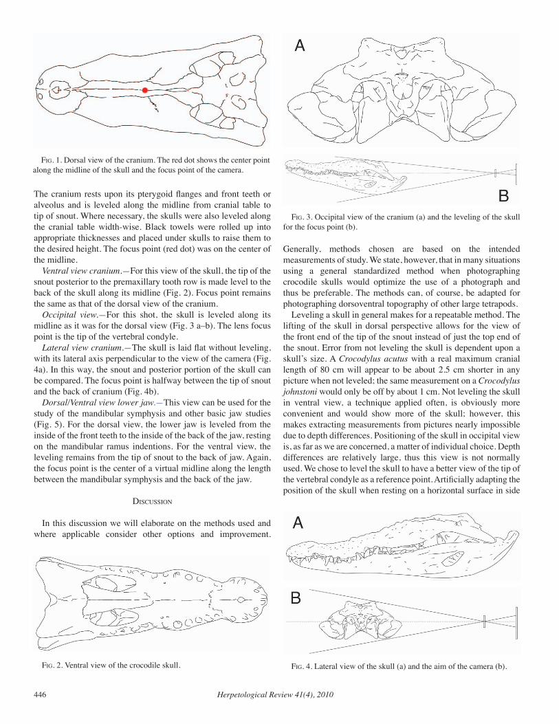
Herpetological Review 41(4), 2010446
The cranium rests upon its pterygoid flanges and front teeth or alveolus and is leveled along the midline from cranial table to tip of snout. Where necessary, the skulls were also leveled along the cranial table width-wise. Black towels were rolled up into appropriate thicknesses and placed under skulls to raise them to the desired height. The focus point (red dot) was on the center of the midline.
Ventral view cranium.—For this view of the skull, the tip of the snout posterior to the premaxillary tooth row is made level to the back of the skull along its midline (Fig. 2). Focus point remains the same as that of the dorsal view of the cranium.
Occipital view.—For this shot, the skull is leveled along its midline as it was for the dorsal view (Fig. 3 a–b). The lens focus point is the tip of the vertebral condyle.
Lateral view cranium.—The skull is laid flat without leveling, with its lateral axis perpendicular to the view of the camera (Fig. 4a). In this way, the snout and posterior portion of the skull can be compared. The focus point is halfway between the tip of snout and the back of cranium (Fig. 4b).
Dorsal/Ventral view lower jaw.—This view can be used for the study of the mandibular symphysis and other basic jaw studies (Fig. 5). For the dorsal view, the lower jaw is leveled from the inside of the front teeth to the inside of the back of the jaw, resting on the mandibular ramus indentions. For the ventral view, the leveling remains from the tip of snout to the back of jaw. Again, the focus point is the center of a virtual midline along the length between the mandibular symphysis and the back of the jaw.
diScuSSion
In this discussion we will elaborate on the methods used and where applicable consider other options and improvement.
Generally, methods chosen are based on the intended measurements of study. We state, however, that in many situations using a general standardized method when photographing crocodile skulls would optimize the use of a photograph and thus be preferable. The methods can, of course, be adapted for photographing dorsoventral topography of other large tetrapods.
Leveling a skull in general makes for a repeatable method. The lifting of the skull in dorsal perspective allows for the view of the front end of the tip of the snout instead of just the top end of the snout. Error from not leveling the skull is dependent upon a skull’s size. A Crocodylus acutus with a real maximum cranial length of 80 cm will appear to be about 2.5 cm shorter in any picture when not leveled; the same measurement on a Crocodylus johnstoni would only be off by about 1 cm. Not leveling the skull in ventral view, a technique applied often, is obviously more convenient and would show more of the skull; however, this makes extracting measurements from pictures nearly impossible due to depth differences. Positioning of the skull in occipital view is, as far as we are concerned, a matter of individual choice. Depth differences are relatively large, thus this view is not normally used. We chose to level the skull to have a better view of the tip of the vertebral condyle as a reference point. Artificially adapting the position of the skull when resting on a horizontal surface in side
fig. 1. Dorsal view of the cranium. The red dot shows the center point along the midline of the skull and the focus point of the camera.
fig. 2. Ventral view of the crocodile skull.
fig. 3. Occipital view of the cranium (a) and the leveling of the skull for the focus point (b).
fig. 4. Lateral view of the skull (a) and the aim of the camera (b).
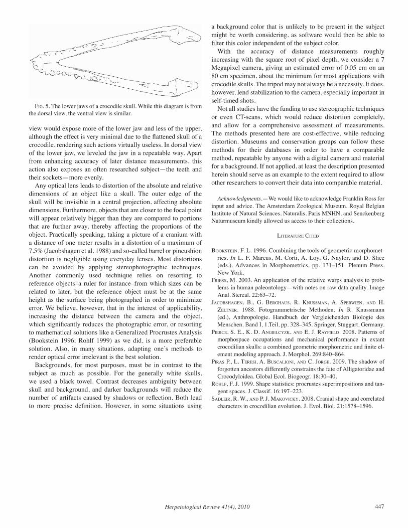
Herpetological Review 41(4), 2010 447
view would expose more of the lower jaw and less of the upper, although the effect is very minimal due to the flattened skull of a crocodile, rendering such actions virtually useless. In dorsal view of the lower jaw, we leveled the jaw in a repeatable way. Apart from enhancing accuracy of later distance measurements, this action also exposes an often researched subject—the teeth and their sockets—more evenly.
Any optical lens leads to distortion of the absolute and relative dimensions of an object like a skull. The outer edge of the skull will be invisible in a central projection, affecting absolute dimensions. Furthermore, objects that are closer to the focal point will appear relatively bigger than they are compared to portions that are further away, thereby affecting the proportions of the object. Practically speaking, taking a picture of a cranium with a distance of one meter results in a distortion of a maximum of 7.5% (Jacobshagen et al. 1988) and so-called barrel or pincushion distortion is negligible using everyday lenses. Most distortions can be avoided by applying stereophotographic techniques. Another commonly used technique relies on resorting to reference objects–a ruler for instance–from which sizes can be related to later, but the reference object must be at the same height as the surface being photographed in order to minimize error. We believe, however, that in the interest of applicability, increasing the distance between the camera and the object, which significantly reduces the photographic error, or resorting to mathematical solutions like a Generalized Procrustes Analysis (Bookstein 1996; Rohlf 1999) as we did, is a more preferable solution. Also, in many situations, adapting one’s methods to render optical error irrelevant is the best solution.
Backgrounds, for most purposes, must be in contrast to the subject as much as possible. For the generally white skulls, we used a black towel. Contrast decreases ambiguity between skull and background, and darker backgrounds will reduce the number of artifacts caused by shadows or reflection. Both lead to more precise definition. However, in some situations using
a background color that is unlikely to be present in the subject might be worth considering, as software would then be able to filter this color independent of the subject color.
With the accuracy of distance measurements roughly increasing with the square root of pixel depth, we consider a 7 Megapixel camera, giving an estimated error of 0.05 cm on an 80 cm specimen, about the minimum for most applications with crocodile skulls. The tripod may not always be a necessity. It does, however, lend stabilization to the camera, especially important in self-timed shots.
Not all studies have the funding to use stereographic techniques or even CT-scans, which would reduce distortion completely, and allow for a comprehensive assessment of measurements. The methods presented here are cost-effective, while reducing distortion. Museums and conservation groups can follow these methods for their databases in order to have a comparable method, repeatable by anyone with a digital camera and material for a background. If not applied, at least the description presented herein should serve as an example to the extent required to allow other researchers to convert their data into comparable material.
Acknowledgments.—We would like to acknowledge Franklin Ross for input and advice. The Amsterdam Zoological Museum, Royal Belgian Institute of Natural Sciences, Naturalis, Paris MNHN, and Senckenberg Naturmuseum kindly allowed us access to their collections.
literature cited
BookStein, F. L. 1996. Combining the tools of geometric morphomet-rics. In L. F. Marcus, M. Corti, A. Loy, G. Naylor, and D. Slice (eds.), Advances in Morphometrics, pp. 131–151. Plenum Press, New York.
frieSS, M. 2003. An application of the relative warps analysis to prob-lems in human paleontology—with notes on raw data quality. Image Anal. Stereal. 22:63–72.
JacoBSHagen, B., g. BergHauS, r. knuSSMan, a. SPerWien, and H. zeltner. 1988. Fotogrammetrische Methoden. In R. Knussmann (ed.), Anthropologie. Handbuch der Vergleichenden Biologie des Menschen. Band I, 1.Teil, pp. 328–345. Springer, Stuggart, Germany.
Pierce, S. e., k. d. angielcYzk, and e. J. raYfield. 2008. Patterns of morphospace occupations and mechanical performance in extant crocodilian skulls: a combined geometric morphometric and finite el-ement modeling approach. J. Morphol. 269:840–864.
PiraS P., l. tereSi, a. BuScalioni, and c. Jorge. 2009. The shadow of forgotten ancestors differently constrains the fate of Alligatoridae and Crocodyloidea. Global Ecol. Biogeogr. 18:30–40.
roHlf, f. J. 1999. Shape statistics: procrustes superimpositions and tan-gent spaces. J. Classif. 16:197–223.
Sadleir, r. W., and P. J. MakovickY. 2008. Cranial shape and correlated characters in crocodilian evolution. J. Evol. Biol. 21:1578–1596.
fig. 5. The lower jaws of a crocodile skull. While this diagram is from the dorsal view, the ventral view is similar.
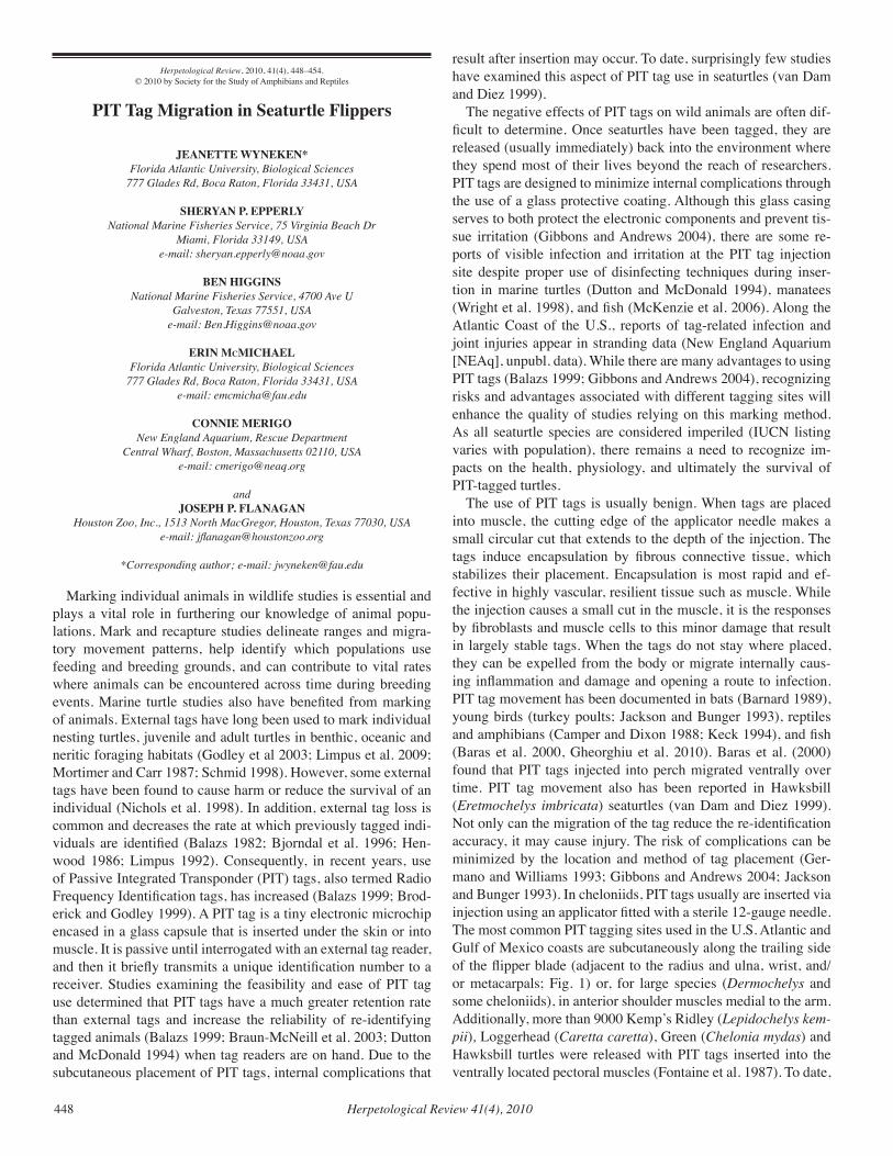
Herpetological Review 41(4), 2010448
Herpetological Review, 2010, 41(4), 448–454.© 2010 by Society for the Study of Amphibians and Reptiles
PIT Tag Migration in Seaturtle Flippers
JEANETTE WYNEKEN*Florida Atlantic University, Biological Sciences
777 Glades Rd, Boca Raton, Florida 33431, USA
SHERYAN P. EPPERLYNational Marine Fisheries Service, 75 Virginia Beach Dr
Miami, Florida 33149, USA e-mail: [email protected]
BEN HIGGINSNational Marine Fisheries Service, 4700 Ave U
Galveston, Texas 77551, USAe-mail: [email protected]
ERIN McMICHAELFlorida Atlantic University, Biological Sciences
777 Glades Rd, Boca Raton, Florida 33431, USA e-mail: [email protected]
CONNIE MERIGONew England Aquarium, Rescue Department
Central Wharf, Boston, Massachusetts 02110, USA e-mail: [email protected]
andJOSEPH P. FLANAGAN
Houston Zoo, Inc., 1513 North MacGregor, Houston, Texas 77030, USA e-mail: [email protected]
*Corresponding author; e-mail: [email protected]
Marking individual animals in wildlife studies is essential and plays a vital role in furthering our knowledge of animal popu-lations. Mark and recapture studies delineate ranges and migra-tory movement patterns, help identify which populations use feeding and breeding grounds, and can contribute to vital rates where animals can be encountered across time during breeding events. Marine turtle studies also have benefited from marking of animals. External tags have long been used to mark individual nesting turtles, juvenile and adult turtles in benthic, oceanic and neritic foraging habitats (Godley et al 2003; Limpus et al. 2009; Mortimer and Carr 1987; Schmid 1998). However, some external tags have been found to cause harm or reduce the survival of an individual (Nichols et al. 1998). In addition, external tag loss is common and decreases the rate at which previously tagged indi-viduals are identified (Balazs 1982; Bjorndal et al. 1996; Hen-wood 1986; Limpus 1992). Consequently, in recent years, use of Passive Integrated Transponder (PIT) tags, also termed Radio Frequency Identification tags, has increased (Balazs 1999; Brod-erick and Godley 1999). A PIT tag is a tiny electronic microchip encased in a glass capsule that is inserted under the skin or into muscle. It is passive until interrogated with an external tag reader, and then it briefly transmits a unique identification number to a receiver. Studies examining the feasibility and ease of PIT tag use determined that PIT tags have a much greater retention rate than external tags and increase the reliability of re-identifying tagged animals (Balazs 1999; Braun-McNeill et al. 2003; Dutton and McDonald 1994) when tag readers are on hand. Due to the subcutaneous placement of PIT tags, internal complications that
result after insertion may occur. To date, surprisingly few studies have examined this aspect of PIT tag use in seaturtles (van Dam and Diez 1999).
The negative effects of PIT tags on wild animals are often dif-ficult to determine. Once seaturtles have been tagged, they are released (usually immediately) back into the environment where they spend most of their lives beyond the reach of researchers. PIT tags are designed to minimize internal complications through the use of a glass protective coating. Although this glass casing serves to both protect the electronic components and prevent tis-sue irritation (Gibbons and Andrews 2004), there are some re-ports of visible infection and irritation at the PIT tag injection site despite proper use of disinfecting techniques during inser-tion in marine turtles (Dutton and McDonald 1994), manatees (Wright et al. 1998), and fish (McKenzie et al. 2006). Along the Atlantic Coast of the U.S., reports of tag-related infection and joint injuries appear in stranding data (New England Aquarium [NEAq], unpubl. data). While there are many advantages to using PIT tags (Balazs 1999; Gibbons and Andrews 2004), recognizing risks and advantages associated with different tagging sites will enhance the quality of studies relying on this marking method. As all seaturtle species are considered imperiled (IUCN listing varies with population), there remains a need to recognize im-pacts on the health, physiology, and ultimately the survival of PIT-tagged turtles.
The use of PIT tags is usually benign. When tags are placed into muscle, the cutting edge of the applicator needle makes a small circular cut that extends to the depth of the injection. The tags induce encapsulation by fibrous connective tissue, which stabilizes their placement. Encapsulation is most rapid and ef-fective in highly vascular, resilient tissue such as muscle. While the injection causes a small cut in the muscle, it is the responses by fibroblasts and muscle cells to this minor damage that result in largely stable tags. When the tags do not stay where placed, they can be expelled from the body or migrate internally caus-ing inflammation and damage and opening a route to infection. PIT tag movement has been documented in bats (Barnard 1989), young birds (turkey poults; Jackson and Bunger 1993), reptiles and amphibians (Camper and Dixon 1988; Keck 1994), and fish (Baras et al. 2000, Gheorghiu et al. 2010). Baras et al. (2000) found that PIT tags injected into perch migrated ventrally over time. PIT tag movement also has been reported in Hawksbill (Eretmochelys imbricata) seaturtles (van Dam and Diez 1999). Not only can the migration of the tag reduce the re-identification accuracy, it may cause injury. The risk of complications can be minimized by the location and method of tag placement (Ger-mano and Williams 1993; Gibbons and Andrews 2004; Jackson and Bunger 1993). In cheloniids, PIT tags usually are inserted via injection using an applicator fitted with a sterile 12-gauge needle. The most common PIT tagging sites used in the U.S. Atlantic and Gulf of Mexico coasts are subcutaneously along the trailing side of the flipper blade (adjacent to the radius and ulna, wrist, and/or metacarpals; Fig. 1) or, for large species (Dermochelys and some cheloniids), in anterior shoulder muscles medial to the arm. Additionally, more than 9000 Kemp’s Ridley (Lepidochelys kem-pii), Loggerhead (Caretta caretta), Green (Chelonia mydas) and Hawksbill turtles were released with PIT tags inserted into the ventrally located pectoral muscles (Fontaine et al. 1987). To date,
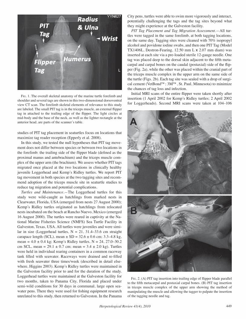
Herpetological Review 41(4), 2010 449
studies of PIT tag placement in seaturtles focus on locations that maximize tag reader reception (Epperly et al. 2008).
In this study, we tested the null hypotheses that PIT tag move-ment does not differ between species or between two locations in the forelimb: the trailing side of the flipper blade (defined as the proximal manus and antebrachium) and the triceps muscle com-plex of the upper arm (the brachium). We assess whether PIT tags migrated once placed at the two locations in clinically healthy juvenile Loggerhead and Kemp’s Ridley turtles. We report PIT tag movement in both species at the two tagging sites and recom-mend adoption of the triceps muscle site in seaturtle studies to reduce tag migration and potential complications.
Turtles and Maintenance.—The Loggerhead turtles for this study were wild-caught as hatchlings from marked nests in Clearwater, Florida, USA (emerged from nests 27 August 2000); Kemp’s Ridley turtles originated as hatchlings from relocated nests incubated on the beach at Rancho Nuevo, Mexico (emerged 16 August 2000). The turtles were reared in captivity at the Na-tional Marine Fisheries Science (NMFS) Sea Turtle Facility in Galveston, Texas, USA. All turtles were juveniles and were simi-lar in size (Loggerhead turtles, N = 21, 31.4–33.6 cm straight carapace length (SCL), mean ± SD = 32.6 ± 0.6 cm; 3.3–4.8 kg, mean = 4.0 ± 0.4 kg; Kemp’s Ridley turtles, N = 24, 27.0–30.2 cm SCL, mean = 29.1 ± 0.7 cm; mean = 3.4 ± 2.0 kg). Turtles were held in individual rearing containers in a common raceway tank filled with seawater. Raceways were drained and re-filled with fresh seawater three times/week (described in detail else-where, Higgins 2003). Kemp’s Ridley turtles were maintained in the Galveston facility prior to and for the duration of the study. Loggerhead turtles were maintained at the Galveston facility for two months, taken to Panama City, Florida and placed under semi-wild conditions for 30 days in communal, large open sea-water pens. There they were used for fishing equipment research unrelated to this study, then returned to Galveston. In the Panama
City pens, turtles were able to swim more vigorously and interact, potentially challenging the tags and the tag sites beyond what they might experience at the Galveston facility.
PIT Tag Placement and Tag Migration Assessment.—All tur-tles were tagged in the same forelimb, at both tagging locations, on the same day. Tagging sites were cleaned with 70% isopropyl alcohol and povidone iodine swabs, and then one PIT Tag (Model TX1406L, Destron-Fearing, 12.50 mm L x 2.07 mm diam) was inserted at each site via a pre-loaded sterile 12-gauge needle. One tag was placed deep to the dorsal skin adjacent to the fifth meta-carpal and carpal bones on the caudal (postaxial) side of the flip-per (Fig. 2a), while the other was placed within the cranial part of the triceps muscle complex in the upper arm on the same side of the turtle (Figs. 2b). Each tag site was sealed with a drop of surgi-cal cement (VetBond™; 3M™, St. Paul, Minnesota) to minimize the chances of tag loss and infection.
Initial MRI scans of the entire flipper were taken shortly after insertion (1 April 2002 for Kemp’s Ridley turtles; 2 April 2002 for Loggerheads). Second MRI scans were taken at 104–106
fig. 1. The overall skeletal anatomy of the marine turtle forelimb and shoulder and several tags are shown in this two-dimensional dorsoventral view CT scan. The forelimb skeletal elements of relevance to this study are labeled. The small PIT tag is in the triceps muscle, an external flipper tag in attached to the trailing edge of the flipper. The light circles at mid-body and the base of the neck, as well as the lighter rectangle at the anterior head, are parts of the scanner’s table.
fig. 2. (A) PIT tag insertion into trailing edge of flipper blade parallel to the fifth metacarpal and postaxial carpal bones. (B) PIT tag insertion in triceps muscle complex of the upper arm showing the method of manipulating the muscle and allowing the tagger to palpate the insertion of the tagging needle and tag.
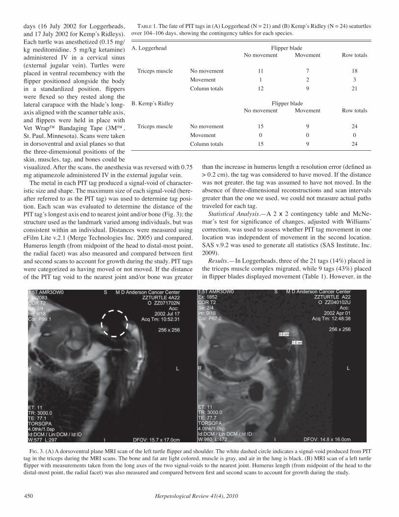
Herpetological Review 41(4), 2010450
days (16 July 2002 for Loggerheads, and 17 July 2002 for Kemp’s Ridleys). Each turtle was anesthetized (0.15 mg/kg meditomidine, 5 mg/kg ketamine) administered IV in a cervical sinus (external jugular vein). Turtles were placed in ventral recumbency with the flipper positioned alongside the body in a standardized position, flippers were flexed so they rested along the lateral carapace with the blade’s long-axis aligned with the scanner table axis, and flippers were held in place with Vet Wrap™ Bandaging Tape (3M™, St. Paul, Minnesota). Scans were taken in dorsoventral and axial planes so that the three-dimensional positions of the skin, muscles, tag, and bones could be visualized. After the scans, the anesthesia was reversed with 0.75 mg atipamezole administered IV in the external jugular vein.
The metal in each PIT tag produced a signal-void of character-istic size and shape. The maximum size of each signal-void (here-after referred to as the PIT tag) was used to determine tag posi-tion. Each scan was evaluated to determine the distance of the PIT tag’s longest axis end to nearest joint and/or bone (Fig. 3); the structure used as the landmark varied among individuals, but was consistent within an individual. Distances were measured using eFilm Lite v.2.1 (Merge Technologies Inc. 2005) and compared. Humerus length (from midpoint of the head to distal-most point, the radial facet) was also measured and compared between first and second scans to account for growth during the study. PIT tags were categorized as having moved or not moved. If the distance of the PIT tag void to the nearest joint and/or bone was greater
than the increase in humerus length ± resolution error (defined as > 0.2 cm), the tag was considered to have moved. If the distance was not greater, the tag was assumed to have not moved. In the absence of three-dimensional reconstructions and scan intervals greater than the one we used, we could not measure actual paths traveled for each tag.
Statistical Analysis.—A 2 x 2 contingency table and McNe-mar’s test for significance of changes, adjusted with Williams’ correction, was used to assess whether PIT tag movement in one location was independent of movement in the second location. SAS v.9.2 was used to generate all statistics (SAS Institute, Inc. 2009).
Results.—In Loggerheads, three of the 21 tags (14%) placed in the triceps muscle complex migrated, while 9 tags (43%) placed in flipper blades displayed movement (Table 1). However, in the
fig. 3. (A) A dorsoventral plane MRI scan of the left turtle flipper and shoulder. The white dashed circle indicates a signal-void produced from PIT tag in the triceps during the MRI scans. The bone and fat are light colored, muscle is gray, and air in the lung is black. (B) MRI scan of a left turtle flipper with measurements taken from the long axes of the two signal-voids to the nearest joint. Humerus length (from midpoint of the head to the distal-most point, the radial facet) was also measured and compared between first and second scans to account for growth during the study.
taBle 1. The fate of PIT tags in (A) Loggerhead (N = 21) and (B) Kemp’s Ridley (N = 24) seaturtles over 104–106 days, showing the contingency tables for each species.
A. Loggerhead Flipper blade No movement Movement Row totals
Triceps muscle No movement 11 7 18 Movement 1 2 3 Column totals 12 9 21
B. Kemp’s Ridley Flipper blade No movement Movement Row totals Triceps muscle No movement 15 9 24 Movement 0 0 0 Column totals 15 9 24
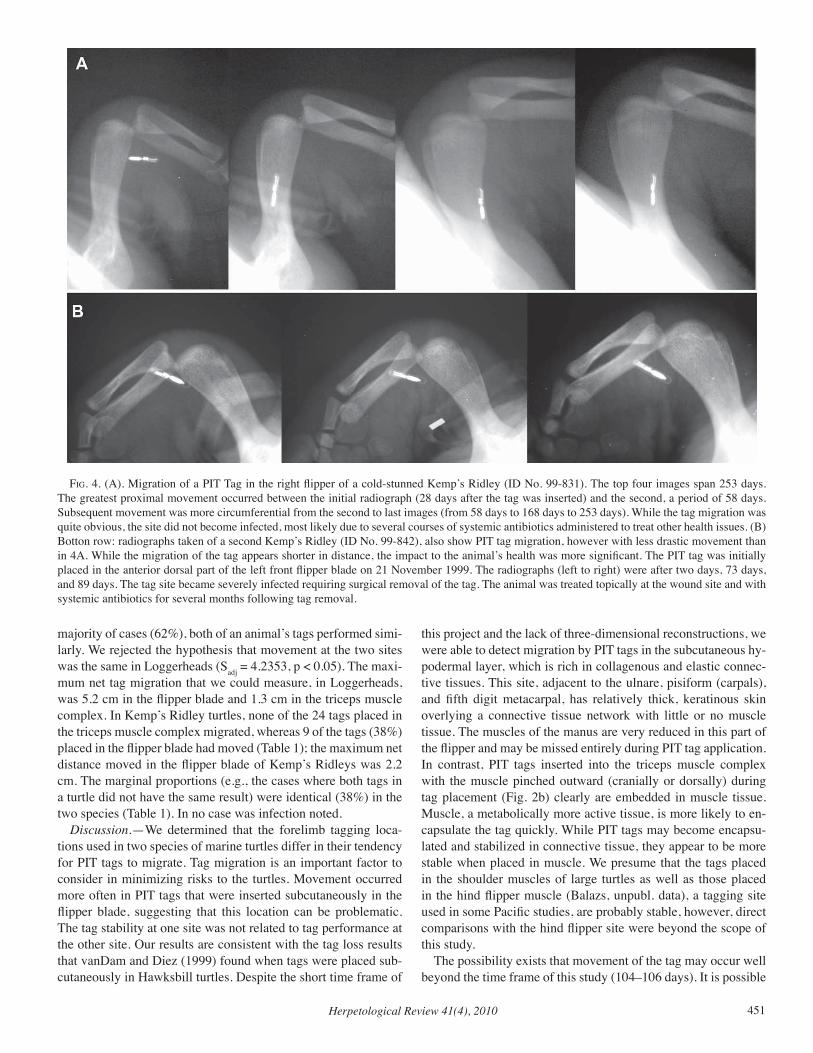
Herpetological Review 41(4), 2010 451
majority of cases (62%), both of an animal’s tags performed simi-larly. We rejected the hypothesis that movement at the two sites was the same in Loggerheads (Sadj = 4.2353, p < 0.05). The maxi-mum net tag migration that we could measure, in Loggerheads, was 5.2 cm in the flipper blade and 1.3 cm in the triceps muscle complex. In Kemp’s Ridley turtles, none of the 24 tags placed in the triceps muscle complex migrated, whereas 9 of the tags (38%) placed in the flipper blade had moved (Table 1); the maximum net distance moved in the flipper blade of Kemp’s Ridleys was 2.2 cm. The marginal proportions (e.g., the cases where both tags in a turtle did not have the same result) were identical (38%) in the two species (Table 1). In no case was infection noted.
Discussion.—We determined that the forelimb tagging loca-tions used in two species of marine turtles differ in their tendency for PIT tags to migrate. Tag migration is an important factor to consider in minimizing risks to the turtles. Movement occurred more often in PIT tags that were inserted subcutaneously in the flipper blade, suggesting that this location can be problematic. The tag stability at one site was not related to tag performance at the other site. Our results are consistent with the tag loss results that vanDam and Diez (1999) found when tags were placed sub-cutaneously in Hawksbill turtles. Despite the short time frame of
this project and the lack of three-dimensional reconstructions, we were able to detect migration by PIT tags in the subcutaneous hy-podermal layer, which is rich in collagenous and elastic connec-tive tissues. This site, adjacent to the ulnare, pisiform (carpals), and fifth digit metacarpal, has relatively thick, keratinous skin overlying a connective tissue network with little or no muscle tissue. The muscles of the manus are very reduced in this part of the flipper and may be missed entirely during PIT tag application. In contrast, PIT tags inserted into the triceps muscle complex with the muscle pinched outward (cranially or dorsally) during tag placement (Fig. 2b) clearly are embedded in muscle tissue. Muscle, a metabolically more active tissue, is more likely to en-capsulate the tag quickly. While PIT tags may become encapsu-lated and stabilized in connective tissue, they appear to be more stable when placed in muscle. We presume that the tags placed in the shoulder muscles of large turtles as well as those placed in the hind flipper muscle (Balazs, unpubl. data), a tagging site used in some Pacific studies, are probably stable, however, direct comparisons with the hind flipper site were beyond the scope of this study.
The possibility exists that movement of the tag may occur well beyond the time frame of this study (104–106 days). It is possible
fig. 4. (A). Migration of a PIT Tag in the right flipper of a cold-stunned Kemp’s Ridley (ID No. 99-831). The top four images span 253 days. The greatest proximal movement occurred between the initial radiograph (28 days after the tag was inserted) and the second, a period of 58 days. Subsequent movement was more circumferential from the second to last images (from 58 days to 168 days to 253 days). While the tag migration was quite obvious, the site did not become infected, most likely due to several courses of systemic antibiotics administered to treat other health issues. (B) Botton row: radiographs taken of a second Kemp’s Ridley (ID No. 99-842), also show PIT tag migration, however with less drastic movement than in 4A. While the migration of the tag appears shorter in distance, the impact to the animal’s health was more significant. The PIT tag was initially placed in the anterior dorsal part of the left front flipper blade on 21 November 1999. The radiographs (left to right) were after two days, 73 days, and 89 days. The tag site became severely infected requiring surgical removal of the tag. The animal was treated topically at the wound site and with systemic antibiotics for several months following tag removal.
Herpetological Review 41(4), 2010452
that tag movement or loss may be greater in wild animals than those in captivity (Thomas 2006) associated with their potential exposure to more varied physical stressors, although we did not observe such in Loggerheads that were allowed 30 days of swim-ming in open pens (compared with Kemp’s Ridleys that were maintained in tanks). PIT tag movement may also increase with the size of an animal (Gibbons and Andrews 2004), indicating that the tag movement measured in this study may be less than what might occur in larger juvenile or adult turtles.
In some cases, movement of a PIT tag may be detrimental to seaturtle health and survival. Several cases of PIT tag migration in the flippers of cold-stunned Kemp’s Ridley turtles were docu-mented by the NEAq while the animals were undergoing reha-bilitation. In November 1999, more than 277 seaturtles stranded along the shores of Cape Cod Bay (Massachusetts, USA) during a large cold-stunning event. A total of 156 were recovered alive (Still et al. 2002) and transported to the NEAq for rehabilita-tion. Due to the overwhelming volume of cases admitted into the NEAq seaturtle clinic, turtles were rapidly transported to second-ary facilities for rehabilitation. Many turtles were PIT tagged in the dorsal and cranial flipper blades prior to transport (N = 103 Kemp’s Ridleys), while others were tagged prior to release. Tag migration was noted via radiographs of several turtles (Fig. 4). At least eight of those 103 turtles developed localized infection as-sociated with the PIT tag, and surgical removal of the tag was re-quired. It is likely that this number under-represents the true num-ber of infections, due to the difficulty in following the clinical outcome of large numbers of turtles after relocation to multiple institutions. At least one of these turtles developed significant os-teomyelitis of the humerus, radium, and ulna. While osteomyeli-tis and joint mobility issues are common in cold-stunned turtles (Wyneken et al. 2006), the proximity of the PIT tag to the sites of infections suggested further assessment of PIT tagging location was warranted. Infections of the skeleton and primary locomotor structures are likely to decrease survival probability.
Because our study tested the tendency of PIT tags to migrate in clinically normal turtles (both Kemp’s Ridley and Loggerhead turtles) we were able to confirm that tags placed in the flipper blade may migrate even in the absence of infection or cold stress. Similarly, tag migration was observed in penguins both in the presence and absence of microbial growth around the tag and in-fection (Clarke and Kerry 1998).
It is likely that when physiologically stressed, such as by hy-pothermia, immune function decreases (reviewed by Jacobson 2007). Thus cold-stunned or otherwise stressed turtles may be at increased risk of infection in the PIT tag site (Baras et al. 2000). Infection and irritation have been reported in PIT tagged leather-back turtles as well (Dutton and McDonald 1994).
Studies of PIT tag placement in the skin of manatees (Tri-chechus manatus latirostris) showed that when the skin is thick, a plug of skin can be driven internally in front of the tag during the injection, increasing the risk of infection. This may, in turn, increase tag movement or rejection (Lambooij et al. 1995; Wright et al.1998).
PIT tags can also be expelled from animals, depending on where the tag is placed (Elbin and Burger 1994; Fontaine et al. 1987; Gibbons and Andrews 2004). Zimmerman and Welsh (2008) studied the placement of PIT tags in American Eels (An-
guilla rostrata) and found that tag retention varied according to tag location, with the highest retention rates in tags placed into musculature. PIT tags placed in penguins and monitored over several years showed that tag movement can be substantial (> 5 cm; Clarke and Kerry 1998). We observed less movement in PIT tags placed into seaturtle triceps muscle than the trailing as-pect of the flipper blade, thus its use may minimize the potential for tag migration, infection and loss. Risk of PIT tag movement and/or infection caused by PIT tags in seaturtles can be mitigated by combining standard skin cleaning with an antiseptic solution prior to application, the use of a sterile applicator and tag, and placement of the tag in a stable location where the keratinous skin layer is thin and the tag is most likely to be placed in muscle. In addition, the accuracy of seaturtle mark-recapture studies will increase because once placed in the triceps muscle, the tag is less likely to move; it remains within readable distance because of the location and size of the muscle, and should increase recognized recaptures.
There may be other advantages to placing the PIT tags into the triceps muscle over the flipper blade. The triceps muscle com-plex provides some soft tissue protection and so may decrease the likelihood of a tag failure due to shattering of the glass cas-ing. When the glass encasing a PIT tag’s electronics is broken, the transponder fails (Camper and Dixon 1988; Lambooij et al. 1995) and tags can migrate, breaking through skin (Germano and Williams 1993). Camper and Dixon (1988) report tag breakage and malfunction due to aggressive encounters between lizards. While we found no incidence of PIT tag failure in seaturtle flip-per blades during this study, lack of “padding” in the flipper blade does little to minimize the risk of tag breakage.
While PIT tag location, consistency, and reliability are impor-tant aspects of any mark-recapture study, there has been relatively limited consideration of the efficacy of tag placement in seaturtles except for turtle size (Fontaine et al. 1987) and accommodating the limits of tag readers (Epperly et al. 2008). Consideration of tag migration risk in seaturtles is examined for the first time here.
The use of PIT tags is an integral part of much herpetological research. PIT tag use is clearly essential in understanding verte-brate populations worldwide and has greatly increased the reli-ability and ease of re-identification of individuals. PIT tag use is bolstering our knowledge of seaturtle populations, at sea and on land. Their use in seaturtles has increased recapture rates, which has improved understanding of turtle movements, growth rates, habitat use, nesting success, internesting intervals, migration and numerous other life-history attributes. In addition, the use of PIT tags has eliminated the need to mark turtles using tattooing, drilling or carapace scarring (Affronte and Scaravelli 2002; Hen-drickson and Henrickson 1981). Our results support avoiding the flipper blade and applying tags in the triceps to minimize risks of compromising the well-being of the tagged animal.
Acknowledgments. —We thank the staff of The University of Texas M.D. Anderson Cancer Center, Houston, the Howard Hughes Medical Imaging Center, University MRI & Diagnostic Imaging Centers, D. Cro-meens, J. Hazle, C. Innis, S. LeBlanc Marco, R. Price, J. Rindfleish, F. L. Steinberg, MD, P. Sforca, D. Wilke, K. Wright, and the NOAA Fisheries Sea Turtle Facility staff at Galveston. A. Chester, C. Innis, M. Salmon, and anonymous reviewers provided valuable comments on this manuscript. This work was conducted under U.S. Fish and Wildlife Ser-
Herpetological Review 41(4), 2010 453
vice Endangered Species Act Section 10a(1)a Scientific Research Permit #TE-676379-4 and complied with all institutional animal care guide-lines. The Florida Atlantic University (FAU) IACUC determined that the study is in compliance with IACUC guidelines; administratively it does not receive an approval number because FAU personnel worked with the data files alone, not the animals. The methods used were consistent with those the IACUC would approve had the animal part of the study been done by FAU personnel. At the time, the NMFS did not issue IA-CUC approvals, but always complied with USFWS and State of Florida requirements for holding seaturtles. J. Flanagan, DVM ensured that best practices were followed. All authors participated in one or more aspects of this study and edited the manuscript.
literature cited
affronte, M., and d. Scaravelli, 2002. Results of experimental carapace tagging. Marine Turtle Newsletter 98:7.
BalazS, G. H. 1982. Factors affecting the retention of metal tags on sea turtles. Marine Turtle Newsletter 20:1–14.
–––––. 1999. Factors to consider in the tagging of sea turtles. In K. L. Eckert, K. A. Bjorndal, F. A. Abreu Grobois, and M. Donnelly (eds.), Research and Management Techniques for the Conservation of Sea Turtles. IUCN/SSC Marine Turtle Specialist Group Publication No. 4, pp. 101–109. Washington, D.C.
BaraS, e., c. MalBrouck, M. HouBart, P. keSteMont, and c. Melard. 2000. The effect of PIT tags on growth and physiology of age-0 cultured Eurasian perch Perca fluviatilis of variable size. Aquaculture 185:159–173.
Barnard, S. 1989. The use of microchip implants for identifying big brown bats (Eptesicus fuscus). Animal Keepers Forum 16:50–52.
BJorndal, k. a., a. B. Bolten, c. J. lagueuX, and a. cHaveS. 1996. Probability of tag loss in green turtles nesting at Tortuguero, Costa Rica. J. Herpetol. 30:567–571.
Braun-Mcneill, J., l. avenS, and S. P. ePPerlY. 2003. Estimated tag retention rates for PIT and inconel tags in juvenile loggerhead (Caretta caretta) sea turtles. In J. A. Seminoff (compiler), Proceedings of the Twenty-Second Annual Symposium on Sea Turtle Biology and Conservation. p. 104. NOAA Tech Memo NMFS-SEFSC-503:104. Available from http://www.sefsc.noaa.gov/seaturtletechmemos.jsp
Broderick, a. c., and B. J. godleY. 1999. Effect of tagging marine turtles on nesting behavior and reproductive success. Anim. Behav. 58:587–591.
caMPer, J. d., and J. r. diXon. 1988. Evaluation of a microchip marking system for amphibians and reptiles. Texas Parks and Wildlife Department, Austin, Texas. Available at http:// www.tpwd.state.tx.us/publications/wildlife_habitat/pdf_docs/pwd_bk_w7000_957.pdf).
clarke, J., and k. kerrY. 1998. Implanted transponders in penguins: Implantation, reliability, and long-term effects. J. Field Ornithol. 69:149–159.
dutton, P., and d. Mcdonald. 1994. Use of PIT tags to identify adult leatherbacks. Marine Turtle Newsletter 67:13–14.
elBin, S. B., and J. Burger. 1994. In my experience: implantable microchips for individual identification in wild and captive populations. Wildlife. Soc. Bull. 22:677–683.
ePPerlY, S., l. BelSkiS, and l. StokeS. 2008. Using passive integrated transponder (PIT) RFID equipment. In H. Kalb, H. A. S. Rohde, K. Gayheart, and K. Shanker (compilers), Proceedings of the Twenty Fifth Annual Symposium on Sea Turtle Biology and Conservation. p. 189. NOAA Tech Memo NMFS-SEFSC-582:189. Available from http://www.sefsc.noaa.gov/seaturtletechmemos.jsp
fontaine, c. t., t. d. WilliaMS, and J. d. caMPer. 1987. Ridleys tagged with passive integrated transponder (PIT). Marine Turtle Newsletter 41:6.
gerMano, d. J., and d. f. WilliaMS. 1993. Field evaluation of using passive integrated transponder (PIT) tags to permanently mark lizards.
Herpetol. Rev. 24:54–46. gHeorgHiu, c., J. Hanna, J. W. SMitH, d. S. SMitH, and M. P. Wilkie.
2010. Encapsulation and migration of PIT tags implanted in brown trout (Salmo trutta L.) Aquaculture 298:350–353.
giBBonS, J. W., and k. M. andreWS. 2004. PIT tagging: Simple technology at its best. BioScience 54:447–454.
godleY, B. J., e. H. S. M. liMa, S. akeSSon, a. c. Broderick, f. glen, M. H. godfreY, P. luScHi, and g. c. HaYS. 2003. Movement patterns of green turtles in Brazilian coastal waters described by satellite tracking and flipper tagging. Marine Ecology Progress Series 253: 279–288
HendrickSon, l. P., and J. r. HendrickSon. 1981. A new method for marking sea turtles? Marine Turtle Newsletter 19:6–7.
HenWood, t. a. 1986. Losses of monel flipper tags from loggerhead sea turtles, Caretta caretta. J. Herpetol. 20:276–279.
HigginS, B. M. 2003. Sea turtle husbandry. In P.L. Lutz, J. A. Musick, and J. Wyneken (eds.), Biology of Sea Turtles, Vol. II, pp. 411–440. CRC Press/Taylor & Frances Group, Boca Raton, Florida.
JackSon, d. n., and W. H. Bunger. 1993. Evaluation of passive integrated transponders as a marking technique for turkey poults. J. Iowa Acad. Sci. 100:60–61.
JacoBSon, e. r. 2007. Infectious Diseases and Pathology of Reptiles: Color Atlas and Text. CRC Press/Taylor & Frances Group, Boca Raton, Florida. 716 pp.
keck, B. 1994. Test for detrimental effects of PIT tags in neonatal snakes. Copeia 1994:226–228.
liMPuS, c. J. 1992. Estimation of tag loss in marine turtle research. Wildlife Res. 19:457–469.
–––––, i. Bell, and J. d. Miller. 2009. Mixed stocks of green turtles foraging on Clack Reef, Northern Great Barrier Reef identified from long term tagging studies. Mar. Turt. Newsl. 123:3–5.
laMBooiJ, e., g. H. langevel, g. H. laMMer, and J. H. HuiSkeS. 1995. Electronic identification with injectable transponders in pig production: results of a field trial on commercial farms and slaughterhouses concerning injectability and retrievability. Vet. Quart. 17:118–123.
Mckenzie, J., B. diggleS, l. tuBBS, c. Poortenaar, d. ParkinSon, k. WeBSter, k., and n. Miller. 2006. An evaluation of a new type of plastic coated PIT tag for tagging snapper (Pagrus auratus). New Zealand Fisheries Assessment Report 2006/8. 41 pp. Available from http://fs.fish.govt.nz/Page.aspx?pk=113&dk=10650
Merge tecHnologieS inc. 2005. eFilm Lite™ v.2.1 Milwaukee, Wisconsin.
MortiMer, J. a., and a. carr. 1987. Reproduction and migrations of the Ascension Island green turtle Chelonia mydas. Copeia 1987:103–113.
nicHolS, W. J., J. a. SeMinoff, and a. reSindiz. 1998. Plastic “Rototags” may be linked to sea turtle bycatch. Mar. Turt. Newsl. 79:20–21.
SaS inStitute. 2009. SAS. Version 9.2. SAS Institute, Cary, North Carolina.
ScHMid, J. r. 1998. Marine turtle populations on the west-central coast of Florida: results of tagging studies at the Cedar Keys, Florida, 1986–1995. Fish. Bull. 96:589–602.
Still, B., k. tuXBurY, r. PreScott, c. rYder, d. MurleY, c. Merigo, c. SMitH, and B. turnBull. 2002. A record cold stun season in Cape Cod Bay, Massachusetts, USA. In A. Mosier, A. Foley, and B. Brost (compilers), Proceedings of the Twentieth Annual Symposium on Sea Turtle Biology and Conservation, pp. 205–206. NOAA Tech. Memo. NMFS-SEFSC-477:205. Available from http://www.sefsc.noaa.gov/seaturtletechmemos.jsp
tHoMaS, J. c. 2006. American eel behavioral patterns in Silver Lake, Dover, Delaware. Unpubl. Master’s thesis, Delaware State University, Dover, Delaware.
van daM, r. P., and c. e. diez. 1999. Differential tag retention in Caribbean hawksbill turtles. Chelonian Conserv. Biol. 3:225–229.
WrigHt, i. e., S. d. WrigHt, and J. M SWeat. 1998. Use of passive
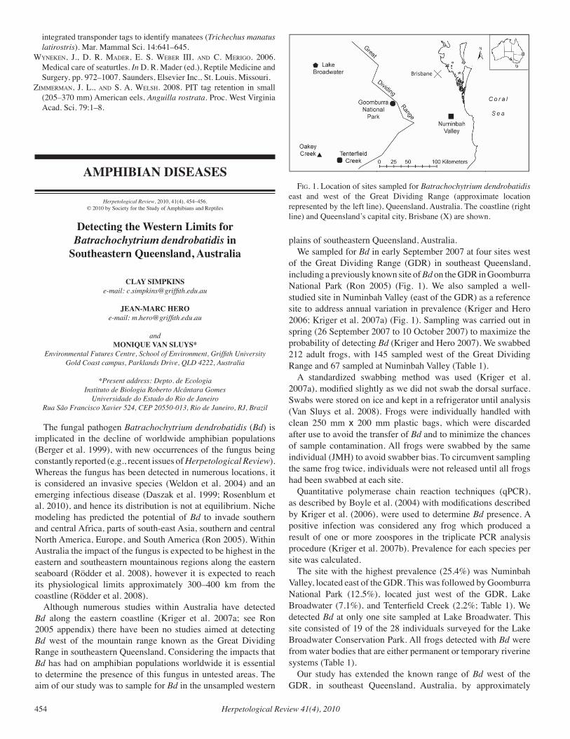
Herpetological Review 41(4), 2010454
integrated transponder tags to identify manatees (Trichechus manatus latirostris). Mar. Mammal Sci. 14:641–645.
WYneken, J., d. r. Mader, e. S. WeBer iii, and c. Merigo. 2006. Medical care of seaturtles. In D. R. Mader (ed.), Reptile Medicine and Surgery, pp. 972–1007. Saunders, Elsevier Inc., St. Louis, Missouri.
ziMMerMan, J. l., and S. a. WelSH. 2008. PIT tag retention in small (205–370 mm) American eels, Anguilla rostrata. Proc. West Virginia Acad. Sci. 79:1–8.
AMPHIBIAN DISEASES
Herpetological Review, 2010, 41(4), 454–456.© 2010 by Society for the Study of Amphibians and Reptiles
Detecting the Western Limits for Batrachochytrium dendrobatidis in
Southeastern Queensland, Australia
CLAY SIMPKINSe-mail: [email protected]
JEAN-MARC HEROe-mail: [email protected]
andMONIQUE VAN SLUYS*
Environmental Futures Centre, School of Environment, Griffith UniversityGold Coast campus, Parklands Drive, QLD 4222, Australia
*Present address: Depto. de EcologiaInstituto de Biologia Roberto Alcântara Gomes
Universidade do Estado do Rio de JaneiroRua São Francisco Xavier 524, CEP 20550-013, Rio de Janeiro, RJ, Brazil
The fungal pathogen Batrachochytrium dendrobatidis (Bd) is implicated in the decline of worldwide amphibian populations (Berger et al. 1999), with new occurrences of the fungus being constantly reported (e.g., recent issues of Herpetological Review). Whereas the fungus has been detected in numerous locations, it is considered an invasive species (Weldon et al. 2004) and an emerging infectious disease (Daszak et al. 1999; Rosenblum et al. 2010), and hence its distribution is not at equilibrium. Niche modeling has predicted the potential of Bd to invade southern and central Africa, parts of south-east Asia, southern and central North America, Europe, and South America (Ron 2005). Within Australia the impact of the fungus is expected to be highest in the eastern and southeastern mountainous regions along the eastern seaboard (Rödder et al. 2008), however it is expected to reach its physiological limits approximately 300–400 km from the coastline (Rödder et al. 2008).
Although numerous studies within Australia have detected Bd along the eastern coastline (Kriger et al. 2007a; see Ron 2005 appendix) there have been no studies aimed at detecting Bd west of the mountain range known as the Great Dividing Range in southeastern Queensland. Considering the impacts that Bd has had on amphibian populations worldwide it is essential to determine the presence of this fungus in untested areas. The aim of our study was to sample for Bd in the unsampled western
plains of southeastern Queensland, Australia. We sampled for Bd in early September 2007 at four sites west
of the Great Dividing Range (GDR) in southeast Queensland, including a previously known site of Bd on the GDR in Goomburra National Park (Ron 2005) (Fig. 1). We also sampled a well-studied site in Numinbah Valley (east of the GDR) as a reference site to address annual variation in prevalence (Kriger and Hero 2006; Kriger et al. 2007a) (Fig. 1). Sampling was carried out in spring (26 September 2007 to 10 October 2007) to maximize the probability of detecting Bd (Kriger and Hero 2007). We swabbed 212 adult frogs, with 145 sampled west of the Great Dividing Range and 67 sampled at Numinbah Valley (Table 1).
A standardized swabbing method was used (Kriger et al. 2007a), modified slightly as we did not swab the dorsal surface. Swabs were stored on ice and kept in a refrigerator until analysis (Van Sluys et al. 2008). Frogs were individually handled with clean 250 mm x 200 mm plastic bags, which were discarded after use to avoid the transfer of Bd and to minimize the chances of sample contamination. All frogs were swabbed by the same individual (JMH) to avoid swabber bias. To circumvent sampling the same frog twice, individuals were not released until all frogs had been swabbed at each site.
Quantitative polymerase chain reaction techniques (qPCR), as described by Boyle et al. (2004) with modifications described by Kriger et al. (2006), were used to determine Bd presence. A positive infection was considered any frog which produced a result of one or more zoospores in the triplicate PCR analysis procedure (Kriger et al. 2007b). Prevalence for each species per site was calculated.
The site with the highest prevalence (25.4%) was Numinbah Valley, located east of the GDR. This was followed by Goomburra National Park (12.5%), located just west of the GDR, Lake Broadwater (7.1%), and Tenterfield Creek (2.2%; Table 1). We detected Bd at only one site sampled at Lake Broadwater. This site consisted of 19 of the 28 individuals surveyed for the Lake Broadwater Conservation Park. All frogs detected with Bd were from water bodies that are either permanent or temporary riverine systems (Table 1).
Our study has extended the known range of Bd west of the GDR, in southeast Queensland, Australia, by approximately
fig. 1. Location of sites sampled for Batrachochytrium dendrobatidis east and west of the Great Dividing Range (approximate location represented by the left line), Queensland, Australia. The coastline (right line) and Queensland’s capital city, Brisbane (X) are shown.
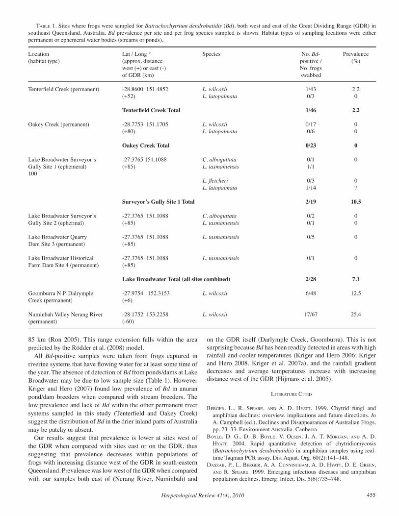
Herpetological Review 41(4), 2010 455
85 km (Ron 2005). This range extension falls within the area predicted by the Rödder et al. (2008) model.
All Bd-positive samples were taken from frogs captured in riverine systems that have flowing water for at least some time of the year. The absence of detection of Bd from ponds/dams at Lake Broadwater may be due to low sample size (Table 1). However Kriger and Hero (2007) found low prevalence of Bd in anuran pond/dam breeders when compared with stream breeders. The low prevalence and lack of Bd within the other permanent river systems sampled in this study (Tenterfield and Oakey Creek) suggest the distribution of Bd in the drier inland parts of Australia may be patchy or absent.
Our results suggest that prevalence is lower at sites west of the GDR when compared with sites east or on the GDR, thus suggesting that prevalence decreases within populations of frogs with increasing distance west of the GDR in south-eastern Queensland. Prevalence was low west of the GDR when compared with our samples both east of (Nerang River, Numinbah) and
on the GDR itself (Darlymple Creek, Goomburra). This is not surprising because Bd has been readily detected in areas with high rainfall and cooler temperatures (Kriger and Hero 2006; Kriger and Hero 2008, Kriger et al. 2007a), and the rainfall gradient decreases and average temperatures increase with increasing distance west of the GDR (Hijmans et al. 2005).
literature cited
Berger, l., r. SPeare, and a. d. HYatt. 1999. Chytrid fungi and amphibian declines: overview, implications and future directions. In A. Campbell (ed.), Declines and Disappearances of Australian Frogs, pp. 23–33. Environment Australia, Canberra.
BoYle, d. g., d. B. BoYle, v. olSen, J. a. t. Morgan, and a. d. HYatt. 2004. Rapid quantitative detection of chytridiomycosis (Batrachochytrium dendrobatidis) in amphibian samples using real-time Taqman PCR assay. Dis. Aquat. Org. 60(2):141–148.
daSzak, P., l. Berger, a. a. cunningHaM, a. d. HYatt, d. e. green, and r. SPeare. 1999. Emerging infectious diseases and amphibian population declines. Emerg. Infect. Dis. 5(6):735–748.
taBle 1. Sites where frogs were sampled for Batrachochytrium dendrobatidis (Bd), both west and east of the Great Dividing Range (GDR) in southeast Queensland, Australia. Bd prevalence per site and per frog species sampled is shown. Habitat types of sampling locations were either permanent or ephemeral water bodies (streams or ponds).
Location Lat / Long ° Species No. Bd- Prevalence(habitat type) (approx. distance positive / (%) west (+) or east (-) No. frogs of GDR (km) swabbed
Tenterfield Creek (permanent) -28.8600 151.4852 L. wilcoxii 1/43 2.2 (+52) L. latopalmata 0/3 0
Tenterfield Creek Total 1/46 2.2
Oakey Creek (permanent) -28.7753 151.1705 L. wilcoxii 0/17 0 (+80) L. latopalmata 0/6 0 Oakey Creek Total 0/23 0
Lake Broadwater Surveyor’s -27.3765 151.1088 C. alboguttata 0/1 0Gully Site 1 (ephemeral) (+85) L. tasmaniensis 1/1 100 L. fletcheri 0/3 0 L. latopalmata 1/14 7 Surveyor’s Gully Site 1 Total 2/19 10.5
Lake Broadwater Surveyor’s -27.3765 151.1088 C. alboguttata 0/2 0Gully Site 2 (ephermal) (+85) L. tasmaniensis 0/1 0
Lake Broadwater Quarry -27.3765 151.1088 L. tasmaniensis 0/5 0Dam Site 3 (permanent) (+85)
Lake Broadwater Historical -27.3765 151.1088 L. tasmaniensis 0/1 0Farm Dam Site 4 (permanent) (+85) Lake Broadwater Total (all sites combined) 2/28 7.1
Goomburra N.P. Dalrymple -27.9754 152.3153 L. wilcoxii 6/48 12.5Creek (permanent) (+6)
Numinbah Valley Nerang River -28.1752 153.2258 L. wilcoxii 17/67 25.4(permanent) (-60)
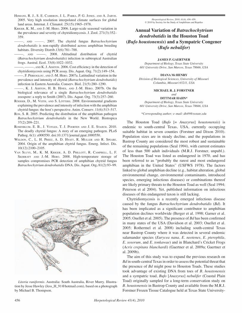
Herpetological Review 41(4), 2010456
HiJManS, r. J., S. e. caMeron, J. l. Parra, P. g. JoneS, and a. JarviS. 2005. Very high resolution interpolated climate surfaces for global land areas. Internat. J. Climatol. 25(15):1965–1978.
kriger, k. M., and J.-M. Hero. 2006. Large-scale seasonal variation in the prevalence and severity of chytridiomycosis. J. Zool. 271(3):352–359.
–––––, and –––––. 2007. The chytrid fungus Batrachochytrium dendrobatidis is non-rapidly distributed across amphibian breeding habitats. Diversity Distrib.13(6):781–788.
–––––, and –––––. 2008. Altitudinal distribution of chytrid (Batrachochytrium dendrobatidis) infection in subtropical Australian frogs. Austral. Ecol. 33(8):1022–1032.
–––––, –––––, and k. J. aSHton. 2006. Cost efficiency in the detection of chytridiomycosis using PCR assay. Dis. Aquat. Org. 71(2):149–154.
–––––, f. Pereoglou, and J.-M. Hero. 2007a. Latitudinal variation in the prevalence and intensity of chytrid (Batrachochytrium dendrobatidis) infection in Eastern Australia. Conserv. Biol. 21(5):280–1290.
–––––, k. J. aSHton, H. B. HineS, and J.-M. Hero. 2007b. On the biological relevance of a single Batrachochytrium dendrobatidis zoospore: a reply to Smith (2007). Dis. Aquat. Org. 73(3):257–260.
rödder, d., M. veitH, and S. lötterS. 2008. Environmental gradients explaining the prevalence and intensity of infection with the amphibian chytrid fungus: the host’s perspective. Anim. Conserv. 11(6):513–517.
ron, S. r. 2005. Predicting the distribution of the amphibian pathogen Batrachochytrium dendrobatidis in the New World. Biotropica 37(2):209–221.
roSenBluM, e. B., J. voYleS, t. J. Poorten and J. e. StaJicH. 2010. The deadly chytrid fungus: A story of an emerging pathogen. PLoS Pathog. 6(1): e000550. doi:10.1371/journal.ppat.1000550.
Weldon, c., l. H. Preez, a. d. HYatt, r. Muller and r. SPeare. 2004. Origin of the amphibian chytrid fungus. Emerg. Infect. Dis. 10(12):2100–2105.
van SluYS, M., k. M. kriger, a. d. PHillott, r. caMPBell, l. f. Skerratt and J.-M. Hero. 2008. High-temperature storage of samples compromises PCR detection of amphibian chytrid fungus Batrachochytrium dendrobatidis DNA. Dis. Aquat. Org. 81(2):93–99.
Herpetological Review, 2010, 41(4), 456–459.© 2010 by Society for the Study of Amphibians and Reptiles
Annual Variation of Batrachochytrium dendrobatidis in the Houston Toad
(Bufo houstonensis) and a Sympatric Congener (Bufo nebulifer)
JAMES P. GAERTNERDepartment of Biology, Texas State University
601 University Drive, San Marcos, Texas 78666, USA
DIANA McHENRYDivision of Biological Sciences, University of Missouri
Columbia, Missouri 65211, USA
MICHAEL R. J. FORSTNERand
DITTMAR HAHN*Department of Biology, Texas State University
601 University Drive, San Marcos, Texas 78666, USA
*Corresponding author; e–mail: [email protected]
The Houston Toad (Bufo [= Anaxyrus] houstonensis) is endemic to south-central Texas, USA, currently occupying suitable habitat in seven counties (Forstner and Dixon 2010). Population sizes are in steady decline, and the populations in Bastrop County are considered the most robust and sustainable of the remaining populations (Seal 1994), with current estimates of less than 500 adult individuals (M.R.J. Forstner, unpubl.). The Houston Toad was listed as endangered in 1970, and has been referred to as “probably the rarest and most endangered amphibian in the United States” (USFWS 1978). The factors linked to global amphibian decline (e.g., habitat alteration, global environmental change, environmental contaminants, introduced species, emerging infectious diseases) or combinations thereof are likely primary threats to the Houston Toad as well (Seal 1994; Peterson et al 2004). Yet, published information on infectious diseases of this endangered taxon is still lacking.
Chytridiomycosis is a recently emerged infectious disease caused by the fungus Batrachochytrium dendrobatidis (Bd). It has been implicated as a significant contributor to amphibian population declines worldwide (Berger et al. 1998; Garner et al. 2005; Ouellet et al. 2005). The presence of Bd has been confirmed in many states of the USA (Davidson et al. 2003; Ouellet et al. 2005; Rothermel et al. 2008) including south-central Texas near Bastrop County where it was detected in several endemic salamander species (Eurycea nana, E. neotenes, E. pterophila, E. sosorum, and E. tonkawae) and in Blanchard’s Cricket Frogs (Acris crepitans blanchardi) (Gaertner et al. 2009a; Gaertner et al. 2009b).
The aim of this study was to expand the previous research on Bd in south-central Texas in order to assess the potential threat that the presence of Bd might pose to Houston Toads. These studies took advantage of existing DNA from toes of B. houstonensis and a sympatric toad, Bufo [Anaxyrus] nebulifer (Coastal Plain Toad) originally sampled for a long-term conservation study on B. houstonensis in Bastrop County and available from the M.R.J. Forstner Frozen Tissue Catalogue held at Texas State University-
Litoria raniformis. Australia: South Australia, River Murry. Illustra-tion by Jesse Hawley ([email protected]), based on a photograph by Michael B. Thompson.
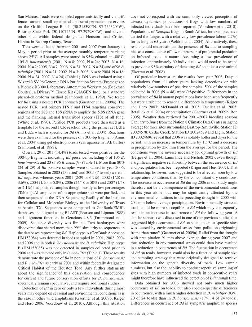
Herpetological Review 41(4), 2010 457
San Marcos. Toads were sampled opportunistically and via drift fences around small ephemeral and semi-permanent reservoirs on the Griffith League Ranch (30.215390°N, 97.255320°W), Bastrop State Park (30.110745°N, 97.292980°W), and several other sites within federal designated Houston toad Critical Habitat in Bastrop County, Texas.
Toes were collected between 2001 and 2007 from January to May, a period prior to the average monthly temperature rising above 25°C. All samples were stored in 95% ethanol at –80°C: 105 B. houstonensis (2001, N = 8; 2002, N = 24; 2003, N = 16; 2004, N = 2; 2005, N = 7; 2006, N = 24; 2007, N = 24) and of 96 B. nebulifer (2001, N = 21; 2002, N = 3; 2003, N = 6; 2004, N = 18; 2006, N = 24; 2007, N = 24) (Table 1). DNA was isolated using a Wizard® SV 96 Genomic DNA Purification System (Promega) on a Biomek® 3000 Laboratory Automation Workstation (Beckman Coulter), a DNeasy™ Tissue Kit (QIAGEN Inc.), or a standard phenol-chloroform method (Sambrook et al. 1989). We tested for Bd using a nested PCR approach (Gaertner et al. 2009a). The nested PCR used primers ITS1f and ITS4 targeting conserved regions of the 28S and 18S rRNA to amplify the 5.8S rRNA gene and the flanking internal transcribed spacer (ITS) of all fungi (White et al. 1990). Purified PCR products were then used as a template for the second PCR reaction using the primer set Bd1a and Bd2a which is specific for Bd (Annis et al. 2004). Reactions were then examined for the presence of a 300-bp fragment (Annis et al. 2004) using gel electrophoresis (2% agarose in TAE buffer) (Sambrook et al. 1989).
Overall, 29 of 201 (14.4%) toads tested were positive for the 300-bp fragment, indicating Bd presence, including 6 of 105 B. houstonensis and 23 of 96 B. nebulifer (Table 1). More than 80% (24 of 29) of Bd-positive samples were obtained in year 2006. Samples obtained in 2003 (23 tested) and 2005 (7 tested) were all Bd-negative, whereas years 2001 (2/29 or 6.9%), 2002 (1/28 or 3.6%), 2004 (1/20 or 5.0%), 2006 (24/48 or 50%) and 2007 (1/48 or 2.1%) had positive samples though mostly at low percentages (Table 1). All amplicons of the appropriate size were purified, and then sequenced at the DNA Sequencing Facility of the Institute for Cellular and Molecular Biology at the University of Texas at Austin, TX. Sequences were compared to GenBank/EMBL databases and aligned using BLAST (Pearson and Lipman 1988) and alignment functions in Geneious 4.8.3 (Drummond et al. 2009). Sequence diversity was limited. Two haplotypes were discovered that shared more than 99% similarity to sequences in the databases representing Bd. Haplotype A (GenBank Accession HM153084) was detected in toads sampled in 2001, 2002, 2004 and 2006 and in both B. houstonensis and B. nebulifer. Haplotype B (HM153085) was not detected in samples collected prior to 2006 and was detected only in B. nebulifer (Table 1). These results demonstrate the presence of Bd in populations of B. houstonensis and B. nebulifer as early as 2001 and within federally designated Critical Habitat of the Houston Toad. Any further statements about the significance of this observation and consequences for current and future conservation efforts for B. houstonensis specifically remain speculative, and require additional studies.
Detection of Bd in zero or only a few individuals during most years may depend on seasonal and environmental conditions as is the case in other wild amphibians (Gaertner et al. 2009b; Kriger and Hero 2006; Voordouw et al. 2010). Although this situation
does not correspond with the commonly viewed perception of disease dynamics, populations of frogs with low numbers of infected individuals have been reported (Voordouw et al. 2010). Populations of Xenopus frogs in South Africa, for example, have carried the fungus with a relatively low prevalence (about 2.7%) for as long as 65 years (Weldon et al. 2004). Alternatively, these results could underestimate the presence of Bd due to sampling bias as a consequence of low numbers or of preferential predation on infected toads in nature. Assuming a low prevalence of infection, approximately 60 individuals would need to be tested to provide a 95% certainty of detecting Bd on at least one animal (Skerratt et al. 2008).
Of particular interest are the results from year 2006. Despite populations from all other years lacking detections or with relatively low numbers of positive samples, 50% of the samples collected in 2006 (N = 48) were Bd-positive. Differences in the prevalence of Bd in anuran populations have been seen frequently but were attributed to seasonal differences in temperature (Kriger and Hero 2007; McDonald et al. 2005; Ouellet et al. 2005; Retallick et al. 2004) or precipitation (Gaertner et al. 2009b; Ron 2005). Weather data retrieved for 2001–2007 breeding seasons (January to June) from the National Climatic Data Center using the average for three sites surrounding Bastrop (Smithville, Station ID 20024578; Cedar Creek, Station ID 20024579 and Elgin, Station ID 20024696) reveal that 2006 was notably hotter and dryer for the period, with an increase in temperature by 1.5°C and a decrease in precipitation by 256 mm from the average for the period. The conditions were the inverse necessary for optimal growth of Bd (Berger et al. 2004; Lamirande and Nichols 2002), even though a significant negative relationship between the occurrence of Bd and precipitation has been reported (Kriger and Hero 2007). That relationship, however, was suggested to be affected more by low temperature conditions than by the concomitant dry conditions. The increase in occurrence of Bd during 2006 in our study might therefore not be a consequence of the environmental conditions in this year alone, but may be significantly affected by the environmental conditions in the preceding drought in 2005 with 204 mm below average precipitation. Environmentally stressed toads could be more susceptible to Bd which may consequently result in an increase in occurrence of Bd the following year. A similar scenario was discussed in one of our previous studies that suggested high occurrence of Bd on salamanders at specific sites was caused by environmental stress from pollution originating from urban runoff (Gaertner et al. 2009a). Relief from the drought with precipitation 91 mm above average during year 2007 and thus reduction in environmental stress could then have resulted in a reduction in occurrence of Bd. The fluctuation in occurrence of Bd on toads, however, could also be a function of sample size and sampling strategy that were originally designed to retrieve information on the genetic diversity of toads. Low sample numbers, but also the inability to conduct repetitive sampling of sites with high numbers of infected toads in consecutive years might therefore have influenced the detection of Bd through time.
Data obtained for 2006 showed not only much higher occurrence of Bd on toads, but also species-specific differences in occurrence, with a much higher detection in B. nebulifer (83%, 20 of 24 toads) than in B. houstonensis (17%, 4 of 24 toads). Differences in occurrence of Bd in sympatric amphibian species
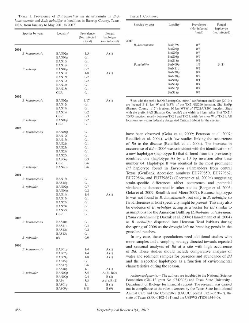
Herpetological Review 41(4), 2010458
have been observed (Goka et al. 2009; Peterson et al. 2007; Retallick et al. 2004), with few studies linking the occurrence of Bd to the disease (Retallick et al. 2004). The increase in occurrence of Bd in 2006 was coincident with the identification of a new haplotype (haplotype B) that differed from the previously identified one (haplotype A) by a 10 bp insertion after base number 64. Haplotype B was identical to the most prominent Bd haplotype found in Eurycea salamanders from central Texas (GenBank Accession numbers EU779859, EU779862, EU779864, and EU779867) (Gaertner et al. 2009a) suggesting strain-specific differences affect occurrence and potential virulence as demonstrated in other studies (Berger et al. 2005; Goka et al. 2009; Retallick and Miera 2007). Because haplotype B was not found in B. houstonensis, but only in B. nebulifer so far, differences in host specificity might be present. This may also be evidence of B. nebulifer acting as a vector for Bd similar to assumptions for the American Bullfrog (Lithobates catesbeianus [Rana catesbeiana]; Daszak et al. 2004; Hanselmann et al. 2004) as B. nebulifer dispersed into Houston Toad habitats during the spring of 2006 as the drought left no breeding ponds in the grassland patches.
In any case, these speculations need additional studies with more samples and a sampling strategy directed towards repeated and seasonal analyses of Bd at a site with high occurrence of Bd. These studies should include comparative analyses of water and sediment samples for presence and abundance of Bd and the respective haplotypes as a function of environmental characteristics during the season.
Acknowledgments.—The authors are indebted to the National Science Foundation (GK–12 grant No. 0742306) and Texas State University–Department of Biology for financial support. The research was carried out in compliance to the rules overseen by the Texas State Institutional Animal Care and Use Committee (IACUC, permit 0721–0530–7), the state of Texas (SPR–0102–191) and the USFWS (TE039544–0).
taBle 1. Prevalence of Batrachochytrium dendrobatidis in Bufo houstonensis and Bufo nebulifer at localities in Bastrop County, Texas, USA, from January to May 2001 to 2007.
Species by year Locality1 Prevalence Fungal (No. infected haplotype / total) (no. infected)
2001 B. houstonensis BAN02p 1/5 A (1) BAN04p 0/1 BAN15t 0/1 BAN18t 0/1 B. nebulifer BAN02p 0/7 BAN12t 1/8 A (1) BAN15t 0/2 BAN19t 0/2 BAN34t 0/1 BAN35t 0/1 GLR 0/1 2002 B. houstonensis BAN02p 1/17 A (1) BAN12t 0/1 BAN14t 0/1 BAN19t 0/2 GLR 0/3 B. nebulifer BAN02p 0/2 GLR 0/1 2003 B. houstonensis BAN01p 0/1 BAN12t 0/1 BAN13t 0/1 BAN21t 0/1 BAN23t 0/1 BAN24t 0/1 BAN26t 0/1 BAS06p 0/3 GLR 0/6 B. nebulifer BAN40s 0/6
2004 B. houstonensis BAN13t 0/1 BAS15p 0/1 B. nebulifer BAN02p 0/7 BAN04p 0/2 BAN14t 1/4 A (1) BAN17t 0/1 BAN23t 0/1 BAN24t 0/1 BAN36t 0/1 GLR 0/1 2005 B. houstonensis BAS10t 0/1 BAS11t 0/3 BAS12t 0/2 BAS13t 0/1 B. nebulifer n/a 0/0
2006 B. houstonensis BAS01p 1/4 A (1) BAS07p 1/4 A (1) BAS09p 1/8 A (1) BAS15p 0/1 BAS17p 0/6 BAS18p 1/1 A (1) B. nebulifer BAN02p 5/5 A (3), B(2) BAN09p 2/4 B (2) BAPp 3/3 A (1), B (2) BAS01p 1/1 B (1) BAS09p 9/11 B (9)
taBle 1. Continued
Species by year Locality1 Prevalence Fungal (No. infected haplotype / total) (no. infected)
2007 B. houstonensis BAN29s 0/3 BAS04p 0/6 BAS07p 0/6 BAS08p 0/6 BAS18p 0/3 B. nebulifer BAN09p 1/3 B (1) BAN11p 0/2 BAN28p 0/4 BAN38p 0/2 BAS01p 0/1 BAS14p 0/4 BAS15p 0/4 BAS18p 0/4
1Sites with the prefix BAN (Bastrop Co. ‘north,’ see Forstner and Dixon [2010]) are located 8–11 km W and WSW of the TX21/US290 junction. Site BAPp (Bastrop County ‘p12’) is about 10 km WSW of TX21/US290 junction. Sites with the prefix BAS (Bastrop Co. ‘south’) are within a 9 km radius E of TX21/TX95 junction, mostly between TX21 and TX71, with few sites W of TX21. All locations are within federally designated Critical Habitat for the species.
Herpetological Review 41(4), 2010 459
literature cited
anniS, S. l., f. P. daStoor, H. ziel, P. daSzak, and J. e. longcore. 2004. A DNA–based assay identifies Batrachochytrium dendrobati-dis in amphibians. J. Wildl. Dis. 40:420–428.
Berger, l., g. Marantelli, l. f. Skerratt, and r. SPeare. 2005. Viru-lence of the amphibian chytrid fungus, Batrachochytrium dendroba-tidis, varies with the strain. Dis. Aq. Org. 68:47–50
–––––, r. SPeare, P. daSzak, d. e. green, a. a. cunningHaM, c. l. goggin, r. SlocoMBe, M. a. ragan, a. H. HYatt, k. r. Mcdonald, H. B. HineS, k. r. liPS, g. Marantelli, and H. ParkeS. 1998. Chy-tridiomycosis causes amphibian mortality associated with population declines in the rain forests of Australia and Central America. Proc. Natl. Acad. Sci. USA 95:9031–9036.
–––––, –––––, H. HineS, g. Marantelli, a. d. HYatt, k. r. Mcdonald, l. f. Skerratt, v. olSen, J. M. clarke, g. gilleSPie, M. MaHonY, n. SHePPard, c. WilliaMS, and M. tYler. 2004. Effect of season and temperature on mortality in amphibians due to chytridiomycosis. Au-stral. Vet. J. 82:31–36.
daSzak, P., a. StrieBY, a. a. cunningHaM, J. e. longcore, c. c. BroWn, and d. Porter. 2004. Experimental evidence that the bullfrog (Rana catesbeiana) is a potential carrier of chytridiomycosis, an emerging fungal disease of amphibians. Herpetol. J. 14:201–207.
davidSon, e. W., M. ParriS, J. P. collinS, J. e. longcore, a. P. PeSS-ier, and J. Brunner. 2003. Pathogenicity and transmission of chy-tridiomycosis in tiger salamanders (Ambystoma tigrinum). Copeia 2003:601–607.
druMMond, a. J., B. aSHton, M. cHeung, J. Heled, M. kearSe, r. Moir, S. StoneS–HavaS, t. tHierer, and a. WilSon. 2009. Geneious v4.8.3, available from http://www.geneious.com/
forStner, M. r. J., and J. r. diXon. 2010. Houston toad (Bufo housto-nensis) 5–year review: summary and evaluation, USFWS & Austin Ecological Service, Austin, TX.
gaertner, J. P., M. r. J. forStner, l. o’donnell, and d. HaHn. 2009a. Detection of Batrachochytrium dendrobatidis in endemic salamander species from Central Texas. EcoHealth 6:20–26.
–––––, M. a. gaSton, d. SPontak, M. r. J. forStner, and d. HaHn. 2009b. Seasonal variation in the detection of Batrachochytrium den-drobatidis in a Texas population of Blanchard’s cricket frog (Acris crepitans blanchardi). Herpetol. Rev. 40:184–187.
garner, t. W. J., S. Walker, J. BoScH, a. d. HYatt, a. a. cunningHaM, and M. c. fiSHer. 2005. Chytrid fungus in Europe. Emerg. Infect. Dis. 11:1639–1641.
goka, k., J. YokoYaMa, Y. une, t. kuroki, k. Suzuki, M. nakaHara, a. koBaYaSHi, S. inaBa, t. Mizutani, and a. d. HYatt. 2009. Amphib-ian chytridiomycosis in Japan: distribution, haplotypes and possible route of entry into Japan. Molec. Ecol. 18:4757–4774.
HanSelMann, r., a. rodriguez, M. laMPo, l. faJardo–raMoS, a. a. aguirre, a. M. kilPatrick, J. rodríguez, and P. daSzak. 2004. Pres-ence of an emerging pathogen of amphibians in introduced bullfrogs Rana catesbeiana in Venezuela. Biol. Conserv. 120:115–119.
kriger, k. M., and J. M. Hero. 2006. Survivorship in wild frogs infected with chytridiomycosis. EcoHealth 3:171–177.
–––––, and ––––– 2007. Large–scale seasonal variation in the preva-lence and severity of chytridiomycosis. J. Zool. 271:352–359.
laMirande, e. W., and d. k. nicHolS. 2002. Effects of host age on sus-ceptibility to cutaneous chytridiomycosis in blue–and–yellow poison dart frogs (Dendrobates tinctorius). In R. G. McKinnell and D. L. Carlson (eds.), Proceedings of the Sixth International Symposium on the Pathology of Reptiles and Amphibians, pp. 3–13. Saint Paul, Min-nesota.
Mcdonald, k. r., d. Méndez, r. Müller, a. B. freeMan, and r. SPeare. 2005. Decline in the prevalence of chytridiomycosis in up-land frog populations in North Queensland, Australia. Pacific Con-
serv. Biol. 11:114–120.ouellet, M., i. Mikaelian, B. d. Pauli, J. rodrigue, and d. M. green.
2005. Historical evidence of widespread chytrid infection in North American amphibian populations. Conserv. Biol. 19:1431–1440
PearSon, W. r., and d. J. liPMan. 1988. Improved tools for biological sequence comparison. Proc. Natl. Acad. Sci. USA 85:2444–2448.
PeterSon, J. d., M. B. Wood, W. a. HoPkinS, J. M. unrine, and M. t. Mendonca. 2007. Prevalence of Batrachochytrium dendrobatidis in American bullfrog and southern leopard frog larvae from wetlands on the Savannah River site, South Carolina. J. Wildl. Dis. 43:450–460.
PeterSon, M. n., S. a. alliSon, M. J. PeterSon, t. r. PeterSon, and r. r. loPez. 2004. A tale of two species: Habitat conservation plans as bounded conflict. J. Wildl. Manage. 68:743–761.
retallick, r. W. r., H. MccalluM, and r. SPeare. 2004. Endemic in-fection of the amphibian chytrid fungus in a frog community post–de-cline. PLOS Biol. 2:1965–1971.
–––––, and v. Miera. 2007. Strain differences in the amphibian chytrid Batrachochytrium dendrobatidis and non-permanent, sub-lethal ef-fects of infection. Dis. Aq. Org. 75:201–207.
ron, S. 2005. Predicting the distribution of the amphibian pathogen Ba-trachochytrium dendrobatidis in the New World. Biotropica 37:209–221.
rotHerMel, B. B., S. c. WallS, J. c. MitcHell, c. k. dodd, l. k. irWin, d. e. green, v. M. vazQuez, J. W. Petranka, and d. J. StevenSon. 2008. Widespread occurrence of the amphibian chytrid fungus Batra-chochytrium dendrobatidis in the southeastern USA. Dis. Aq. Org. 82:3–18.
SaMBrook, J., e. f. fritScH, and t. ManiatiS. 1989. Molecular Cloning: A Laboratory Manual. Cold Spring Harbor Laboratory Press, Cold Spring Harbor, New York.
Seal, u. S. 1994. Population and habitat viability assessment for the Houston toad (Bufo houstonensis) IUCN Conservation Breeding Spe-cialist Group Austin, Texas.
Skerratt, l. f., l. Berger, H. B. HineS, k. r. Mcdonald, d. Mendez, and r. SPeare. 2008. Survey protocol for detecting chytridiomycosis in all Australian frog populations. Dis. Aq. Org. 80:85–94.
USFWS (U.S. Fish and Wildlife Service). 1978. Critical habitat for the Houston toad determined. News release. U.S. Fish and Wildlife Ser-vice, Albuquerque, New Mexico.
voordouW, M. J., d. adaMa, B. HouSton, P. govindaraJulu, and J. roBinSon. 2010. Prevalence of the pathogenic chytrid fungus, Batra-chochytrium dendrobatidis, in an endangered population of northern leopard frogs, Rana pipiens. BMC Ecol. 10:6.
Weldon, c., l. H. du Preez, a. d. HYatt, r. Müller, and r. SPeare. 2004. Origin of the amphibian chytrid fungus. Emerg. Infect. Dis. 10:2100–2105.
WHite, t., t. BrunS, S. lee, and J. W. taYlor. 1990. Amplification and direct sequencing of fungal ribosomal RNA genes for phylogenetics. In M. Innis, D. Gelfand, J. Sninsky, and T. White (eds.), PCR Proto-cols: A Guide to Methods and Applications, pp. 315–322. Academic Press, San Diego, California.
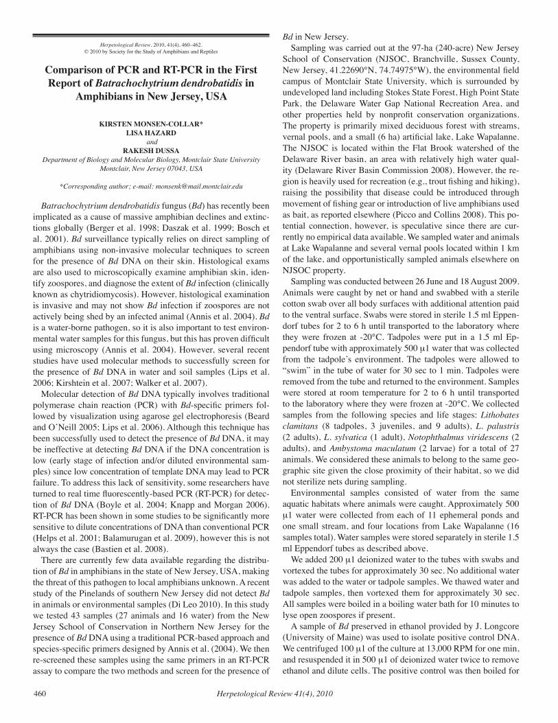
Herpetological Review 41(4), 2010460
Herpetological Review, 2010, 41(4), 460–462.© 2010 by Society for the Study of Amphibians and Reptiles
Comparison of PCR and RT-PCR in the First Report of Batrachochytrium dendrobatidis in
Amphibians in New Jersey, USA
KIRSTEN MONSEN-COLLAR*LISA HAZARD
andRAKESH DUSSA
Department of Biology and Molecular Biology, Montclair State UniversityMontclair, New Jersey 07043, USA
*Corresponding author; e-mail: [email protected]
Batrachochytrium dendrobatidis fungus (Bd) has recently been implicated as a cause of massive amphibian declines and extinc-tions globally (Berger et al. 1998; Daszak et al. 1999; Bosch et al. 2001). Bd surveillance typically relies on direct sampling of amphibians using non-invasive molecular techniques to screen for the presence of Bd DNA on their skin. Histological exams are also used to microscopically examine amphibian skin, iden-tify zoospores, and diagnose the extent of Bd infection (clinically known as chytridiomycosis). However, histological examination is invasive and may not show Bd infection if zoospores are not actively being shed by an infected animal (Annis et al. 2004). Bd is a water-borne pathogen, so it is also important to test environ-mental water samples for this fungus, but this has proven difficult using microscopy (Annis et al. 2004). However, several recent studies have used molecular methods to successfully screen for the presence of Bd DNA in water and soil samples (Lips et al. 2006; Kirshtein et al. 2007; Walker et al. 2007).
Molecular detection of Bd DNA typically involves traditional polymerase chain reaction (PCR) with Bd-specific primers fol-lowed by visualization using agarose gel electrophoresis (Beard and O’Neill 2005; Lips et al. 2006). Although this technique has been successfully used to detect the presence of Bd DNA, it may be ineffective at detecting Bd DNA if the DNA concentration is low (early stage of infection and/or diluted environmental sam-ples) since low concentration of template DNA may lead to PCR failure. To address this lack of sensitivity, some researchers have turned to real time fluorescently-based PCR (RT-PCR) for detec-tion of Bd DNA (Boyle et al. 2004; Knapp and Morgan 2006). RT-PCR has been shown in some studies to be significantly more sensitive to dilute concentrations of DNA than conventional PCR (Helps et al. 2001; Balamurugan et al. 2009), however this is not always the case (Bastien et al. 2008).
There are currently few data available regarding the distribu-tion of Bd in amphibians in the state of New Jersey, USA, making the threat of this pathogen to local amphibians unknown. A recent study of the Pinelands of southern New Jersey did not detect Bd in animals or environmental samples (Di Leo 2010). In this study we tested 43 samples (27 animals and 16 water) from the New Jersey School of Conservation in Northern New Jersey for the presence of Bd DNA using a traditional PCR-based approach and species-specific primers designed by Annis et al. (2004). We then re-screened these samples using the same primers in an RT-PCR assay to compare the two methods and screen for the presence of
Bd in New Jersey. Sampling was carried out at the 97-ha (240-acre) New Jersey
School of Conservation (NJSOC, Branchville, Sussex County, New Jersey, 41.22690°N, 74.74975°W), the environmental field campus of Montclair State University, which is surrounded by undeveloped land including Stokes State Forest, High Point State Park, the Delaware Water Gap National Recreation Area, and other properties held by nonprofit conservation organizations. The property is primarily mixed deciduous forest with streams, vernal pools, and a small (6 ha) artificial lake, Lake Wapalanne. The NJSOC is located within the Flat Brook watershed of the Delaware River basin, an area with relatively high water qual-ity (Delaware River Basin Commission 2008). However, the re-gion is heavily used for recreation (e.g., trout fishing and hiking), raising the possibility that disease could be introduced through movement of fishing gear or introduction of live amphibians used as bait, as reported elsewhere (Picco and Collins 2008). This po-tential connection, however, is speculative since there are cur-rently no empirical data available. We sampled water and animals at Lake Wapalanne and several vernal pools located within 1 km of the lake, and opportunistically sampled animals elsewhere on NJSOC property.
Sampling was conducted between 26 June and 18 August 2009. Animals were caught by net or hand and swabbed with a sterile cotton swab over all body surfaces with additional attention paid to the ventral surface. Swabs were stored in sterile 1.5 ml Eppen-dorf tubes for 2 to 6 h until transported to the laboratory where they were frozen at -20°C. Tadpoles were put in a 1.5 ml Ep-pendorf tube with approximately 500 µ1 water that was collected from the tadpole’s environment. The tadpoles were allowed to “swim” in the tube of water for 30 sec to 1 min. Tadpoles were removed from the tube and returned to the environment. Samples were stored at room temperature for 2 to 6 h until transported to the laboratory where they were frozen at -20°C. We collected samples from the following species and life stages: Lithobates clamitans (8 tadpoles, 3 juveniles, and 9 adults), L. palustris (2 adults), L. sylvatica (1 adult), Notophthalmus viridescens (2 adults), and Ambystoma maculatum (2 larvae) for a total of 27 animals. We considered these animals to belong to the same geo-graphic site given the close proximity of their habitat, so we did not sterilize nets during sampling.
Environmental samples consisted of water from the same aquatic habitats where animals were caught. Approximately 500 µ1 water were collected from each of 11 ephemeral ponds and one small stream, and four locations from Lake Wapalanne (16 samples total). Water samples were stored separately in sterile 1.5 ml Eppendorf tubes as described above.
We added 200 µ1 deionized water to the tubes with swabs and vortexed the tubes for approximately 30 sec. No additional water was added to the water or tadpole samples. We thawed water and tadpole samples, then vortexed them for approximately 30 sec. All samples were boiled in a boiling water bath for 10 minutes to lyse open zoospores if present.
A sample of Bd preserved in ethanol provided by J. Longcore (University of Maine) was used to isolate positive control DNA. We centrifuged 100 µ1 of the culture at 13,000 RPM for one min, and resuspended it in 500 µ1 of deionized water twice to remove ethanol and dilute cells. The positive control was then boiled for
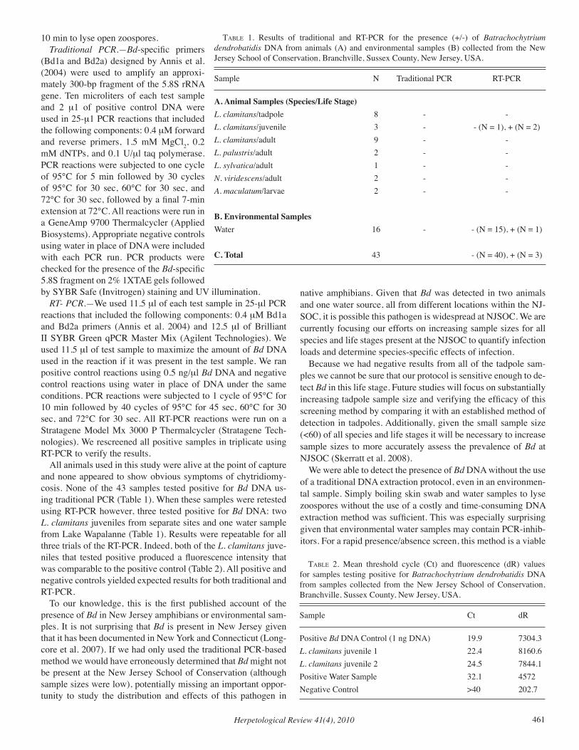
Herpetological Review 41(4), 2010 461
10 min to lyse open zoospores. Traditional PCR.—Bd-specific primers
(Bd1a and Bd2a) designed by Annis et al. (2004) were used to amplify an approxi-mately 300-bp fragment of the 5.8S rRNA gene. Ten microliters of each test sample and 2 µ1 of positive control DNA were used in 25-µ1 PCR reactions that included the following components: 0.4 µM forward and reverse primers, 1.5 mM MgCl2, 0.2 mM dNTPs, and 0.1 U/µl taq polymerase. PCR reactions were subjected to one cycle of 95°C for 5 min followed by 30 cycles of 95°C for 30 sec, 60°C for 30 sec, and 72°C for 30 sec, followed by a final 7-min extension at 72°C. All reactions were run in a GeneAmp 9700 Thermalcycler (Applied Biosystems). Appropriate negative controls using water in place of DNA were included with each PCR run. PCR products were checked for the presence of the Bd-specific 5.8S fragment on 2% 1XTAE gels followed by SYBR Safe (Invitrogen) staining and UV illumination.
RT- PCR.—We used 11.5 µl of each test sample in 25-µl PCR reactions that included the following components: 0.4 µM Bd1a and Bd2a primers (Annis et al. 2004) and 12.5 µl of Brilliant II SYBR Green qPCR Master Mix (Agilent Technologies). We used 11.5 µl of test sample to maximize the amount of Bd DNA used in the reaction if it was present in the test sample. We ran positive control reactions using 0.5 ng/µl Bd DNA and negative control reactions using water in place of DNA under the same conditions. PCR reactions were subjected to 1 cycle of 95°C for 10 min followed by 40 cycles of 95°C for 45 sec, 60°C for 30 sec, and 72°C for 30 sec. All RT-PCR reactions were run on a Stratagene Model Mx 3000 P Thermalcycler (Stratagene Tech-nologies). We rescreened all positive samples in triplicate using RT-PCR to verify the results.
All animals used in this study were alive at the point of capture and none appeared to show obvious symptoms of chytridiomy-cosis. None of the 43 samples tested positive for Bd DNA us-ing traditional PCR (Table 1). When these samples were retested using RT-PCR however, three tested positive for Bd DNA: two L. clamitans juveniles from separate sites and one water sample from Lake Wapalanne (Table 1). Results were repeatable for all three trials of the RT-PCR. Indeed, both of the L. clamitans juve-niles that tested positive produced a fluorescence intensity that was comparable to the positive control (Table 2). All positive and negative controls yielded expected results for both traditional and RT-PCR.
To our knowledge, this is the first published account of the presence of Bd in New Jersey amphibians or environmental sam-ples. It is not surprising that Bd is present in New Jersey given that it has been documented in New York and Connecticut (Long-core et al. 2007). If we had only used the traditional PCR-based method we would have erroneously determined that Bd might not be present at the New Jersey School of Conservation (although sample sizes were low), potentially missing an important oppor-tunity to study the distribution and effects of this pathogen in
native amphibians. Given that Bd was detected in two animals and one water source, all from different locations within the NJ-SOC, it is possible this pathogen is widespread at NJSOC. We are currently focusing our efforts on increasing sample sizes for all species and life stages present at the NJSOC to quantify infection loads and determine species-specific effects of infection.
Because we had negative results from all of the tadpole sam-ples we cannot be sure that our protocol is sensitive enough to de-tect Bd in this life stage. Future studies will focus on substantially increasing tadpole sample size and verifying the efficacy of this screening method by comparing it with an established method of detection in tadpoles. Additionally, given the small sample size (<60) of all species and life stages it will be necessary to increase sample sizes to more accurately assess the prevalence of Bd at NJSOC (Skerratt et al. 2008).
We were able to detect the presence of Bd DNA without the use of a traditional DNA extraction protocol, even in an environmen-tal sample. Simply boiling skin swab and water samples to lyse zoospores without the use of a costly and time-consuming DNA extraction method was sufficient. This was especially surprising given that environmental water samples may contain PCR-inhib-itors. For a rapid presence/absence screen, this method is a viable
taBle 1. Results of traditional and RT-PCR for the presence (+/-) of Batrachochytrium dendrobatidis DNA from animals (A) and environmental samples (B) collected from the New Jersey School of Conservation, Branchville, Sussex County, New Jersey, USA.
Sample N Traditional PCR RT-PCR
A. Animal Samples (Species/Life Stage) L. clamitans/tadpole 8 - -L. clamitans/juvenile 3 - - (N = 1), + (N = 2)L. clamitans/adult 9 - -L. palustris/adult 2 - -L. sylvatica/adult 1 - -N. viridescens/adult 2 - -A. maculatum/larvae 2 - -
B. Environmental Samples Water 16 - - (N = 15), + (N = 1)
C. Total 43 - (N = 40), + (N = 3)
taBle 2. Mean threshold cycle (Ct) and fluorescence (dR) values for samples testing positive for Batrachochytrium dendrobatidis DNA from samples collected from the New Jersey School of Conservation, Branchville, Sussex County, New Jersey, USA.
Sample Ct dR
Positive Bd DNA Control (1 ng DNA) 19.9 7304.3L. clamitans juvenile 1 22.4 8160.6L. clamitans juvenile 2 24.5 7844.1Positive Water Sample 32.1 4572Negative Control >40 202.7
Herpetological Review 41(4), 2010462
alternative to current methods, making it potentially important for rapid assessment or in situations where funds are very limited.
Perhaps more important however, are the results of our PCR technique comparison, which could have a potential impact on fu-ture molecular screening procedures throughout the Bd research community. In this study, we detected Bd in 3 of 43 samples us-ing RT-PCR, but none using traditional PCR. Our results strongly support the need to use the more sensitive RT-PCR screening method when using a non-invasive PCR-based detection tech-nique for Bd DNA.
Acknowledgments.—We thank Paola Dolcemascolo and Jan Sodos for assistance with field sampling. Animals were sampled under New Jersey Department of Environmental Protection Division of Fish and Wildlife Scientific Collecting Permit #29033. The study was approved by the Montclair State University Animal Care and Use Committee and adhered to SSAR guidelines for use of amphibians in field research. Sup-port was provided by a New Jersey Conserve Wildlife Matching Funds Grant.
literature cited
AnniS, S. L., F. P. DaStoor, H. Ziel, P. DaSzak, and J. E. Longcore. 2004. A DNA-based assay identifies Batrachochytrium dendrobatidis in amphibians. J. Wildl. Dis. 40:420–428.
BalaMurugan, V., K. D. JaYaPPa, M. HoSaMani, V. BHanuPrakaSH, G. VenkateSan, and R. K. SingH. 2009. Comparative efficacy of con-ventional and taqman polymerase chain reaction assays in the detec-tion of capripoxviruses from clinical samples. J. Vet. Diagn. Invest. 21:225–231.
BaStien, P., G. W. ProcoP, and U. ReiScHl. 2008. Quantitative real-time PCR is not more sensitive than “conventional” PCR. J. Clin. Micro-biol. 46:1897–1900.
Beard, K. H., and E. M. O’Neill. 2005. Infection of an invasive frog Eleutherodactylus coqui by the chytrid fungus Batrachochytrium den-drobatidis in Hawaii. Biol. Conserv. 126:591–595.
Berger, L., R. SPeare, P. DaSzak, D. E. Green, A. A. CunningHaM, C. L. Goggin, R. SlocoMBe, M. A. Ragan, A. D. HYatt, K. R. McDonald, H. B. HineS, K. R. LiPS, G. Marantelli, and H. ParkeS. 1998. Chy-tridiomycosis causes amphibian mortality associated with population declines in the rain forests of Australia and Central America. Proc. Natl. Acad. Sci. 95:9031–9036.
BoScH, J., I. Martinez-Solano, and M. Garcia-PariS. 2001. Evidence of a chytrid fungus infection involved in the decline of the common midwife toad (Alytes obstetricans) in protected areas of central Spain. Biol. Conserv. 97:331–337.
BoYle, D. G., D. B. BoYle, V. OlSen, J. A. Morgan, and A. D. HYatt. 2004. Rapid quantitative detection of chytridiomycosis (Batrachochy-trium dendrobatidis) in amphibian samples using real-time Taqman PCR assay. Dis. Aquat. Org. 60:141–148.
DaSzak, P., L. Berger, A. A. CunningHaM, A. D. HYatt, D. E. Green, and R. SPeare. 1999. Emerging infectious diseases and amphibian population declines. Emerg. Infect. Dis. 5:735–748.
DelaWare River BaSin CoMMiSSion. 2008. Delaware River State of the Basin Report. Trenton, NJ, Delaware River Basin Commission. 92 pp.
Di Leo, K. 2010. An assessment of the correlation between amphibian populations, chytridiomycete communities, and the ecological integ-rity of the habitat. Masters thesis. Ecology and Evolution. Rutgers University. 66 pp.
HelPS, C., N. ReeveS, S. TaSker, and D. HarBour. 2001. Use of real-time quantitative PCR to detect Chlamydophila felis infection. J. Clin. Microbiol. 39:2675–2676.
KirSHtein, J. D., C. W. AnderSon, J. S. Wood, J. E. Longcore, and M. A. VoYtek. 2007. Quantitative PCR detection of Batrachochytrium den-
drobatidis DNA from sediments and water. Dis. Aquat. Org. 77:11–15.KnaPP, R. A., and J. A. T. Morgan. 2006. Tadpole mouthpart depigmen-
tation as an accurate indicator of chytridiomycosis, an emerging dis-ease of amphibians. Copeia 2006:188–197.
LiPS, K. R., F. BreM, R. BreneS, J. D. Reeve, R. A. Alford, J. VoYleS, C. CareY, L. Livo, A. P. PeSSier, and J. P. CollinS. 2006. Emerging infec-tious disease and the loss of biodiversity in a Neotropical amphibian community. Proc. Natl. Acad. Sci. 103:3165–3170.
Longcore, J. R., J. E. Longcore, A. P. PeSSier, and W. A. HalteMan. 2007. Chytridiomycosis widespread in anurans of northeastern United States. J. Wildl. Manage. 71:435–444.
Picco, A. M., and J. P. CollinS. 2008. Amphibian commerce as a likely source of pathogen pollution. Conserv. Biol. 22:1582–9.
Skerratt, L. F., L. Berger, H. B. HineS, K. R. McDonald, D. Mendez, and R. SPeare. 2008. Survey protocol for detecting chytridiomycosis in all Australian frog populations. Dis. Aquat. Org. 80:85–94.
Walker, S. F., M. B. SalaS, D. JenkinS, T. W. Garner, A. A. Cunning-HaM, A. D. HYatt, J. BoScH, and M. C. FiSHer. 2007. Environmental detection of Batrachochytrium dendrobatidis in a temperate climate. Dis. Aquat. Org. 77:105–112.
Herpetological Review, 2010, 41(4), 462–465.© 2010 by Society for the Study of Amphibians and Reptiles
Batrachochytrium dendrobatidis is Present in Northwest Pennsylvania, USA, with High
Prevalence in Notophthalmus viridescens
MAYA L. GRONER*and
RICK A. RELYEADepartment of Biological Sciences, University of Pittsburgh
Pittsburgh, Pennsylvania 15260, USA
*Corresponding author; e-mail: [email protected]
Batrachochytrium dendrobatidis (Bd) has been found with varying prevalence on amphibians in the northeast, midwest, and southeast United States (Longcore et al. 2007; Zellmer et al. 2008; Green and Dodd 2007; Rothermel 2008), but surveys in Pennsylvania have failed to find infected individuals (Glenney et al. 2010). We studied between-pond variation in Bd infection lev-els in amphibians of northwestern Pennsylvania, USA. In doing so, we focused our sampling efforts on three species, Green Frogs (Rana clamitans), Red-spotted Newts (Notophthalmus virides-cens) and Wood Frogs (Rana sylvatica). These species are locally abundant and, if commonly infected, could serve as an indicator species for assessing Bd presence in ponds.
Our surveys were designed to maximize the number of ponds sampled for Bd occurrence. We swabbed up to 12 individuals per species for Bd at 10 ponds containing Green Frog tadpoles, 8 ponds containing Red-spotted Newts and 2 ponds containing Wood Frog adults between 1 April and 28 May 2008 (Table 1). This sampling scheme has low statistical power to detect true neg-ative results within a pond (e.g., Skerratt et al. 2008), but it is suf-ficient for Bd detection at ponds with moderate to high infection prevalence. For sampling efficiency, we visited each pond only once and limited the life stages that we sampled to larval Green Frogs, adult Red-spotted Newts and adult Wood Frogs. While adult frogs are more commonly sampled, we focused on larval
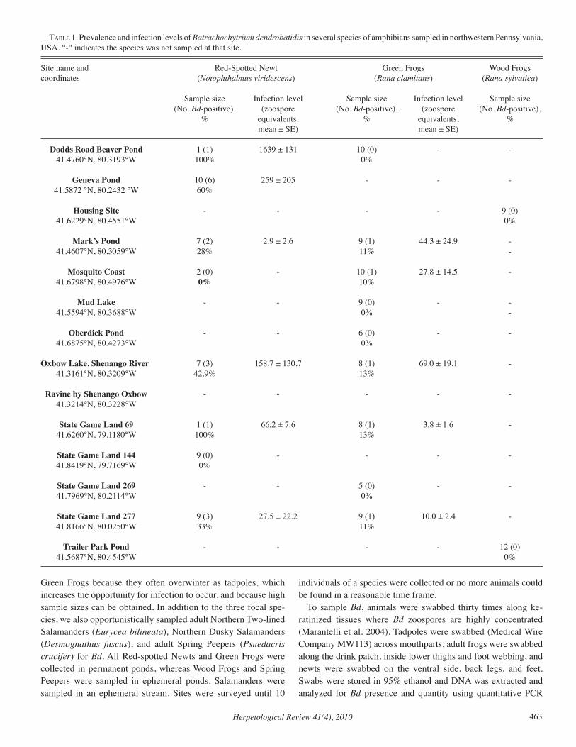
Herpetological Review 41(4), 2010 463
Green Frogs because they often overwinter as tadpoles, which increases the opportunity for infection to occur, and because high sample sizes can be obtained. In addition to the three focal spe-cies, we also opportunistically sampled adult Northern Two-lined Salamanders (Eurycea bilineata), Northern Dusky Salamanders (Desmognathus fuscus), and adult Spring Peepers (Psuedacris crucifer) for Bd. All Red-spotted Newts and Green Frogs were collected in permanent ponds, whereas Wood Frogs and Spring Peepers were sampled in ephemeral ponds. Salamanders were sampled in an ephemeral stream. Sites were surveyed until 10
individuals of a species were collected or no more animals could be found in a reasonable time frame.
To sample Bd, animals were swabbed thirty times along ke-ratinized tissues where Bd zoospores are highly concentrated (Marantelli et al. 2004). Tadpoles were swabbed (Medical Wire Company MW113) across mouthparts, adult frogs were swabbed along the drink patch, inside lower thighs and foot webbing, and newts were swabbed on the ventral side, back legs, and feet. Swabs were stored in 95% ethanol and DNA was extracted and analyzed for Bd presence and quantity using quantitative PCR
taBle 1. Prevalence and infection levels of Batrachochytrium dendrobatidis in several species of amphibians sampled in northwestern Pennsylvania, USA. “-“ indicates the species was not sampled at that site.
Site name and Red-Spotted Newt Green Frogs Wood Frogscoordinates (Notophthalmus viridescens) (Rana clamitans) (Rana sylvatica) Sample size Infection level Sample size Infection level Sample size (No. Bd-positive), (zoospore (No. Bd-positive), (zoospore (No. Bd-positive), % equivalents, % equivalents, % mean ± SE) mean ± SE)
Dodds Road Beaver Pond 1 (1) 1639 ± 131 10 (0) - - 41.4760°N, 80.3193°W 100% 0% Geneva Pond 10 (6) 259 ± 205 - - - 41.5872 °N, 80.2432 °W 60%
Housing Site - - - - 9 (0) 41.6229°N, 80.4551°W 0%
Mark’s Pond 7 (2) 2.9 ± 2.6 9 (1) 44.3 ± 24.9 - 41.4607°N, 80.3059°W 28% 11% -
Mosquito Coast 2 (0) - 10 (1) 27.8 ± 14.5 - 41.6798°N, 80.4976°W 0% 10% Mud Lake - - 9 (0) - - 41.5594°N, 80.3688°W 0% -
Oberdick Pond - - 6 (0) - - 41.6875°N, 80.4273°W 0% Oxbow Lake, Shenango River 7 (3) 158.7 ± 130.7 8 (1) 69.0 ± 19.1 - 41.3161°N, 80.3209°W 42.9% 13%
Ravine by Shenango Oxbow - - - - - 41.3214°N, 80.3228°W
State Game Land 69 1 (1) 66.2 ± 7.6 8 (1) 3.8 ± 1.6 - 41.6260°N, 79.1180°W 100% 13% State Game Land 144 9 (0) - - - - 41.8419°N, 79.7169°W 0% State Game Land 269 - - 5 (0) - - 41.7969°N, 80.2114°W 0% State Game Land 277 9 (3) 27.5 ± 22.2 9 (1) 10.0 ± 2.4 - 41.8166°N, 80.0250°W 33% 11% Trailer Park Pond - - - - 12 (0) 41.5687°N, 80.4545°W 0%
Herpetological Review 41(4), 2010464
(Boyle et al. 2004). This method is highly sensitive and can detect very low-level infections that might be missed with other meth-ods (Hyatt et al. 2007). Samples were run in triplicate and were re-run from the same extraction in cases where results yielded only one or two positive results. A re-run sample was considered positive if at least 3 of 6 wells tested were positive. Such vari-ability between wells is common in samples with low infection levels (personal observation). Quantitative estimates of infection levels were made by averaging positive results within a sample.
To minimize cross-contaminating animals, gloves and equip-ment that contacted frogs were sterilized with 70% ethanol and dried between handling of individuals and all other equipment was sterilized with a 10% bleach solution or discarded after leav-ing a pond (Johnson et al. 2003).
Our results suggest that Bd is widespread throughout north-western Pennsylvania, USA, yet infection prevalence and inten-sity varies among species (Table 1). Red-spotted Newts had the highest prevalence of the three species that could be surveyed with sufficient sample sizes. We did not find infected newts in 2 of the 9 ponds, however, the sample size was not sufficient to con-fidently demonstrate that these ponds were infection-free. Among tested individuals, infection loads (measured in terms of zoospore equivalents) varied over three orders of magnitude among sites. There are few other data examining Bd infection loads in Red-spotted Newts, but infection has been reported for museum speci-mens in eastern North America and more recently in field-collect-ed Red-spotted Newts from the southeastern USA (Ouellet et al. 2005; Rothermel et al. 2008). This suggests that Bd infection in Red-spotted Newts is not uncommon. Interestingly, the closest area to our study site that has been surveyed for Bd (Delaware Water Gap National Recreation Area) did not find infections in Red-spotted Newts or any other tested species (e.g., Rana sylvat-ica, Pseudacris crucifer, Hyla versicolor; Glenney et al. 2010). Many of the newts collected in our study also exhibited cysts indicating infection by the mesomycetozoan Amphibiocystidium viridescens (pers. obs.), and newts in the surrounding area have been shown to carry infections of other parasites, including trem-atodes, nematodes, trypanosomes, acanthocephelans, helminths and bacteria (Raffel et al. 2009). This raises the question as to whether co-infection with other pathogens alters Bd infection rates in Red-Spotted Newts.
Bd infection also was found in Green Frog tadpoles (Table 1). Although infection prevalence was lower than that found in Red-spotted Newts, infection was detected in more than 50% of ponds and sample sizes were not large enough to confidently detect low-level infections in the other ponds. On average, infection loads of Green Frog tadpoles were lower than that found in newts. This may reflect the fact that tadpoles possess less keratinized tissue than newts (Marantelli et al. 2004). Other surveys examining Bd infection in Green Frog adults and metamorphs with qPCR and histology have found higher infection levels (18–36% in Maine from 2000–2002; Longcore et al. 2007), which also may reflect less keratinized tissue in tadpoles (Marantelli et al. 2004). It is likely that Bd has been prevalent in Green Frogs for some time; the earliest evidence for Bd in North America came from a mu-seum specimen collected in 1961 in Quebec (Ouellet et al. 2005). The tadpoles sampled in our study were in their second larval year, allowing ample time for animals to be infected. Since newts
co-occur in ponds with Green Frogs, Bd may have been transmit-ted across species.
No individuals of the four other species sampled were Bd-positive: Northern Two-lined Salamanders (N = 2, ravine by Shenango Oxbow); Northern Dusky Salamanders (N = 4, ravine by Shenango Oxbow); Spring Peepers (N = 2, Trailer Park Pond); and Wood Frogs (Table 1). However, our sample sizes were small and sample sizes >59 are recommended to detect Bd when preva-lence is low (Skerratt et al. 2008). Past studies of Wood Frogs in New England and Michigan, USA show low to moderate preva-lence of Bd (>1 % to 16.7% with qPCR and histology; Longcore et al. 2007; Zellmer et al. 2008), but previous studies of Wood Frogs in eastern Pennsylvania (N = 28) did not find infected in-dividuals, despite using sensitive testing techniques (i.e., qPCR, Glenney et al. 2010). Bd also has been found in the Northern Two-Lined Salamander and the Dusky Salamander, suggest-ing that these species are susceptible to infection (Byrne et al. 2008; Grant et al. 2008). We failed to find any records of infected Spring Peepers, although they have been previously surveyed for Bd infections (Byrne et al. 2008; Longcore et al. 2007; Ouellet et al. 2005; Rothermel et al. 2008; Glenney et al. 2010).
Overall, our results correspond with other studies showing that Bd is present in areas where amphibian population declines have not been documented (Green and Dodd 2007; Longcore et al. 2007; Rothermel 2008; Zellmer et al. 2008). Though we only sampled at a single point in time, high prevalence of infection and relatively low infection intensities in both Green Frogs and Red-spotted Newts suggest that Bd is not uncommon in north-west Pennsylvania and both species might be good indicators for Bd presence in this region. Although no pathology was observed in infected individuals we suggest that further monitoring is nec-essary to test whether this observation is consistent.
Acknowledgments.—Steve Bagnull, Jenise Brown, Rickey Cothran, John Hammond, Forest Raub, Tim Schwartz, and Aaron Stoler helped in the collection of amphibians. Thanks to Aaron Stoler, Will Brogan, Rickey Cothran, Jessica Hua, and Heather Schaffery for providing com-ments on this manuscript. Funding was provided by a McKinley Grant and NSF graduate research fellowship to MLG. All work was approved by IACUC and collections were permitted by the Pennsylvania Fish and Boat Commission.
literature cited
BoYle, d. g., d. B. BoYle, v, olSen, J. a. t. Morgan, and a. d. HYatt. 2004. Rapid quantitative detection of chytridiomycosis (Batrachochytrium dendrobatidis) in amphibian samples using real-time Taqman PCR assay. Dis. Aquat. Org. 60:141–148.
BYrne, M. W., e. P. davie, and J. W. giBBonS. Batrachochytrium dendro-batidis occurrence in Eurycea cirrigera. Southeast. Nat. 7:551–555.
glenneY, g. W., J. t. Julian, and W. M. Quartz. 2010. Preliminary am-phibian health survey in the Delaware Water Gap National Recreation Area. J. Aquat. Anim. Health. 22:102–114.
grant, e. H. c., l. l. BaileY, J. l. Ware, and k. l. duncan. 2008. Prevalence of the amphibian pathogen Batrachochytrium dendro-batidis in stream and wetland amphibians in Maryland, USA. Appl. Herpetol. 9:233–241.
green, d. e., and c. k. Jr. dodd. 2007. Presence of amphibian chytrid fungus Batrachochytrium dendrobatidis and other amphibian patho-gens at warm-water fish hatcheries in southeastern North America. Herpetol. Conserv. Biol. 2:43–47.
Herpetological Review 41(4), 2010 465
HYatt, a. d., d. g. BoYle, v. olSen, d. B. BoYle, l. Berger, d. oBendorf, a. dalton, k. kriger, M. Hero, H. HineS, r. PHillot, r. caMPBell, g. Marantelli, f. gleaSon, and a. colling. 2007. Diagnostic assays and sampling protocols for the detection of Batrachochytrium dendrobatidis. Dis. Aquat. Org. 73:175–192.
JoHnSon, M. l., l. Berger, l. PHiliPS, and r. SPeare. 2003. Fungicidal effects of chemical disinfectants, UV light, desiccation and heat on the amphibian chytrid Batrachochytrium dendrobatidis. Dis. Aquat. Org. 57:255–260.
longcore, J. r., J. e. longcore, a. P. PeSSier, and W. a. HalteMan. 2007. Chytridiomycosis widespread in anurans of northeastern United States. J. Wildl. Manag. 2:435–444.
Marantelli g., l. Berger, r. SPeare, and l. keegan. 2004. Distribution of Batrachochytrium dendrobatidis and keratin during tadpole devel-opment. Pac Conserv. Biol. 10:173–179.
ouellet, M., i. Mikaelian, B. d. Pauli, J. rodrigue, and d. M. green. 2005. Historical evidence of widespread chytrid infection in North American amphibians populations. Conserv. Biol. 19:1431–1440.
raffel, t. r., r. J. le groS, B. c. love, J. r. roHr, and P. J. HudSon. 2009. Parasite age-intensity relationships in red-spotted newts: does immune memory influence salamander disease dynamics? Int. J. Parasitol. 39:231–241.
rotHerMel B. B., S. c. WallS, J. c. MitcHell, c. k. dodd, Jr., l. k. irWin, d. e. green, v. M. vazQuez, J. W. Petranka, and d. J. StevenSon. 2008. Widespread occurrence of the amphibian chytrid fungus Batrachochytrium dendrobatidis in amphibian populations in the southeastern USA. Dis. Aquat. Org. 82:3–18.
Skerratt, l. f., l. B. Berger, H. B. HineS, k. r. Mcdonald, d. Mendez, and r. SPeare. 2008. Survey protocol for detecting chytridiomycosis in all Australian frog populations. Dis. Aquat. Org. 80:85–94.
zellMer, a. J., c. l. ricHardS, and l. M. MartenS. 2008. Low preva-lence of Batrachochytrium dendrobatidis across Rana sylvatica popu-lations in southeastern Michigan, USA. Herpetol. Rev. 39:196–199.
HERPETOLOGICAL HUSBANDRY
Herpetological Review, 2010, 41(4), 465–467.© 2010 by Society for the Study of Amphibians and Reptiles
Observations on the Captive Reproduction of Gaige’s Rain Frog Pristimantis gaigeae
(Dunn 1931)
ROBERT HILL*JULIA KAYLOCK†
Atlanta Botanical Garden, 1345 Piedmont AvenueAtlanta, Georgia 30309, USA
EDGARDO GRIFFITHHEIDI ROSS
El Valle Amphibian Conservation Center, El Valle de Anton, Panama
RONALD GAGLIARDO**Atlanta Botanical Garden, 1345 Piedmont Avenue
Atlanta, Georiga 30309, USA
ELIZABETH TIMPEDepartment of Ecology and Evolutionary Biology
University of Connecticut, 75 Eagleville Road, Unit #3043Storrs, Connecticut 06269, USA
andPAUL CRUMP
Houston Zoo, 6200 Hermann Park Drive, Houston, Texas 77030, USA
*Corresponding author; e-mail: [email protected]
**Present address: Amphibian Ark, Zoo Atlanta800 Cherokee Avenue SE, Atlanta, Georgia 30315, USA
† Deceased 12 June 2009
Gaige’s Rain Frog, Pristimantis gaigeae (Strabomantidae) is a direct-developing anuran species ranging from the Atlantic lowlands in extreme southeastern Costa Rica through eastern Panama and on the Pacific versant in central Colombia (Savage 2002). The species is considered a mimic of dendrobatid species, exhibiting a pair of red, orange, or golden dorsolateral stripes on a black body, similar to the two toxic species Phyllobates aurotaenia and P. lugubris (family Dendrobatidae) with which some populations of Pristimantis gaigeae are sympatric (Fig. 1).
It is the intent of the authors to present methods that have been utilized to successfully maintain and reproduce Pristimantis gaigeae in captivity. As little information is currently available on the captive husbandry and reproduction in species of Pristimantis, we believe the information provided herein may prove useful in future attempts to maintain ex situ populations of this and other species of the genus.
SPeciMen acQuiSition and HuSBandrY MetHodS
The group of P. gaigeae at the Atlanta Botanical Garden (ABG) consisted of one male and two females collected in 2005 from the vicinity of El Valle de Antón and one male collected from the vi-cinity of El Copé, both in Provincia de Coclé, Panamá. The speci-mens were collected and exported as a part of the Amphibian
SSAR Poster Awards for 2011
This is the inaugural year for the SSAR Poster Award. The SSAR Poster Awards will provide recognition for the best student posters presented at the annual meeting of the SSAR. To be eligible, the presented poster must be the result of research con-ducted by the individual making the pre-sentation. The research must have been
conducted while the student was enrolled in either an under-graduate or graduate degree program. Students entering the competition must be members of SSAR. The presentations will be judged by the SSAR student prize committee. One SSAR Poster Award of u.S. $100 (plus a book gift from the university of California Press) may be given in each of the following four categories:• Evolution,Genetics,&Systematics• Ecology,NaturalHistory,Distribution,&Behavior• Physiology&Morphology• Conservation&Management
The SSAR Poster Award will be judged by the following criteria:• QualityofResearch• QualityoftheVisualDisplayoftheResearch• ProfessionalismandConfidenceofthePresenter
For specific recommendations on preparation of posters,please consult the SSAR website:
http://ssarherps.org/pdf/PosterRecommendations.pdf
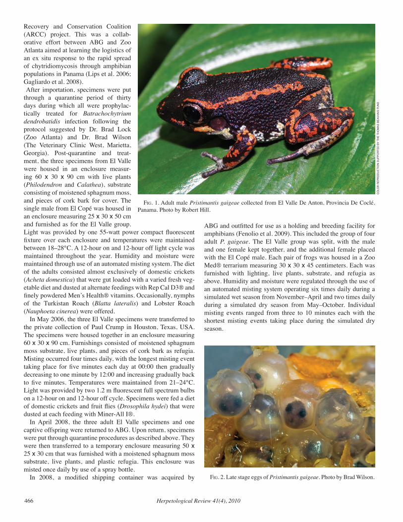
Herpetological Review 41(4), 2010466
Recovery and Conservation Coalition (ARCC) project. This was a collab-orative effort between ABG and Zoo Atlanta aimed at learning the logistics of an ex situ response to the rapid spread of chytridiomycosis through amphibian populations in Panama (Lips et al. 2006; Gagliardo et al. 2008). After importation, specimens were put through a quarantine period of thirty days during which all were prophylac-tically treated for Batrachochytrium dendrobatidis infection following the protocol suggested by Dr. Brad Lock (Zoo Atlanta) and Dr. Brad Wilson (The Veterinary Clinic West, Marietta, Georgia). Post-quarantine and treat-ment, the three specimens from El Valle were housed in an enclosure measur-ing 60 x 30 x 90 cm with live plants (Philodendron and Calathea), substrate consisting of moistened sphagnum moss, and pieces of cork bark for cover. The single male from El Copé was housed in an enclosure measuring 25 x 30 x 50 cm and furnished as for the El Valle group. Light was provided by one 55-watt power compact fluorescent fixture over each enclosure and temperatures were maintained between 18–28°C. A 12-hour on and 12-hour off light cycle was maintained throughout the year. Humidity and moisture were maintained through use of an automated misting system. The diet of the adults consisted almost exclusively of domestic crickets (Acheta domestica) that were gut loaded with a varied fresh veg-etable diet and dusted at alternate feedings with Rep Cal D3® and finely powdered Men’s Health® vitamins. Occasionally, nymphs of the Turkistan Roach (Blatta lateralis) and Lobster Roach (Nauphoeta cinerea) were offered.
In May 2006, the three El Valle specimens were transferred to the private collection of Paul Crump in Houston, Texas, USA. The specimens were housed together in an enclosure measuring 60 x 30 x 90 cm. Furnishings consisted of moistened sphagnum moss substrate, live plants, and pieces of cork bark as refugia. Misting occurred four times daily, with the longest misting event taking place for five minutes each day at 00:00 then gradually decreasing to one minute by 12:00 and increasing gradually back to five minutes. Temperatures were maintained from 21–24°C. Light was provided by two 1.2 m fluorescent full spectrum bulbs on a 12-hour on and 12-hour off cycle. Specimens were fed a diet of domestic crickets and fruit flies (Drosophila hydei) that were dusted at each feeding with Miner-All I®.
In April 2008, the three adult El Valle specimens and one captive offspring were returned to ABG. Upon return, specimens were put through quarantine procedures as described above. They were then transferred to a temporary enclosure measuring 50 x 25 x 30 cm that was furnished with a moistened sphagnum moss substrate, live plants, and plastic refugia. This enclosure was misted once daily by use of a spray bottle.
In 2008, a modified shipping container was acquired by
ABG and outfitted for use as a holding and breeding facility for amphibians (Fenolio et al. 2009). This included the group of four adult P. gaigeae. The El Valle group was split, with the male and one female kept together, and the additional female placed with the El Copé male. Each pair of frogs was housed in a Zoo Med® terrarium measuring 30 x 30 x 45 centimeters. Each was furnished with lighting, live plants, substrate, and refugia as above. Humidity and moisture were regulated through the use of an automated misting system operating six times daily during a simulated wet season from November–April and two times daily during a simulated dry season from May–October. Individual misting events ranged from three to 10 minutes each with the shortest misting events taking place during the simulated dry season.
fig. 1. Adult male Pristimantis gaigeae collected from El Valle De Anton, Provincia De Coclé, Panama. Photo by Robert Hill.
fig. 2. Late stage eggs of Pristimantis gaigeae. Photo by Brad Wilson.
CO
LOR
REP
RO
du
CTi
ON
Su
PPO
RTEd
BY
THE
THO
mAS
BEA
uvA
iS F
uN
d
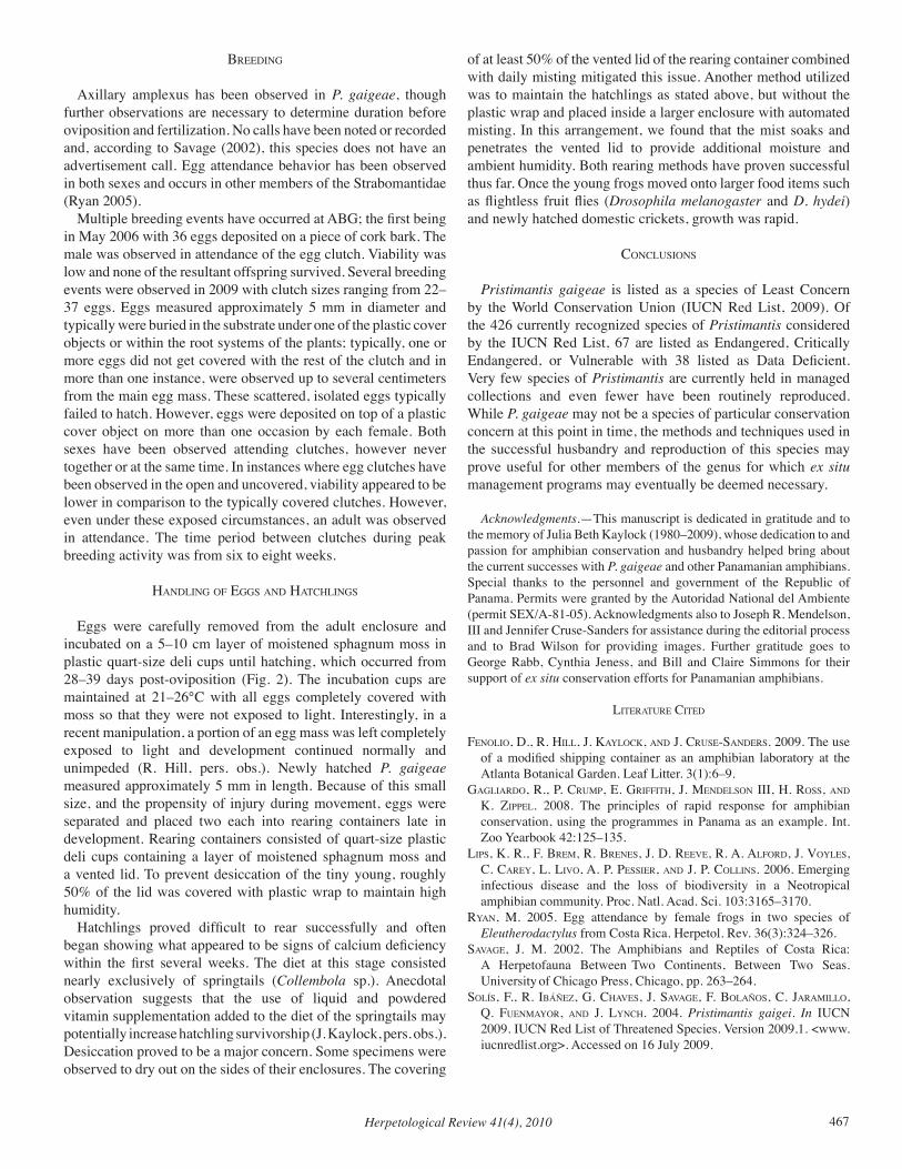
Herpetological Review 41(4), 2010 467
Breeding
Axillary amplexus has been observed in P. gaigeae, though further observations are necessary to determine duration before oviposition and fertilization. No calls have been noted or recorded and, according to Savage (2002), this species does not have an advertisement call. Egg attendance behavior has been observed in both sexes and occurs in other members of the Strabomantidae (Ryan 2005).
Multiple breeding events have occurred at ABG; the first being in May 2006 with 36 eggs deposited on a piece of cork bark. The male was observed in attendance of the egg clutch. Viability was low and none of the resultant offspring survived. Several breeding events were observed in 2009 with clutch sizes ranging from 22–37 eggs. Eggs measured approximately 5 mm in diameter and typically were buried in the substrate under one of the plastic cover objects or within the root systems of the plants; typically, one or more eggs did not get covered with the rest of the clutch and in more than one instance, were observed up to several centimeters from the main egg mass. These scattered, isolated eggs typically failed to hatch. However, eggs were deposited on top of a plastic cover object on more than one occasion by each female. Both sexes have been observed attending clutches, however never together or at the same time. In instances where egg clutches have been observed in the open and uncovered, viability appeared to be lower in comparison to the typically covered clutches. However, even under these exposed circumstances, an adult was observed in attendance. The time period between clutches during peak breeding activity was from six to eight weeks.
Handling of eggS and HatcHlingS
Eggs were carefully removed from the adult enclosure and incubated on a 5–10 cm layer of moistened sphagnum moss in plastic quart-size deli cups until hatching, which occurred from 28–39 days post-oviposition (Fig. 2). The incubation cups are maintained at 21–26°C with all eggs completely covered with moss so that they were not exposed to light. Interestingly, in a recent manipulation, a portion of an egg mass was left completely exposed to light and development continued normally and unimpeded (R. Hill, pers. obs.). Newly hatched P. gaigeae measured approximately 5 mm in length. Because of this small size, and the propensity of injury during movement, eggs were separated and placed two each into rearing containers late in development. Rearing containers consisted of quart-size plastic deli cups containing a layer of moistened sphagnum moss and a vented lid. To prevent desiccation of the tiny young, roughly 50% of the lid was covered with plastic wrap to maintain high humidity.
Hatchlings proved difficult to rear successfully and often began showing what appeared to be signs of calcium deficiency within the first several weeks. The diet at this stage consisted nearly exclusively of springtails (Collembola sp.). Anecdotal observation suggests that the use of liquid and powdered vitamin supplementation added to the diet of the springtails may potentially increase hatchling survivorship (J. Kaylock, pers. obs.). Desiccation proved to be a major concern. Some specimens were observed to dry out on the sides of their enclosures. The covering
of at least 50% of the vented lid of the rearing container combined with daily misting mitigated this issue. Another method utilized was to maintain the hatchlings as stated above, but without the plastic wrap and placed inside a larger enclosure with automated misting. In this arrangement, we found that the mist soaks and penetrates the vented lid to provide additional moisture and ambient humidity. Both rearing methods have proven successful thus far. Once the young frogs moved onto larger food items such as flightless fruit flies (Drosophila melanogaster and D. hydei) and newly hatched domestic crickets, growth was rapid.
concluSionS
Pristimantis gaigeae is listed as a species of Least Concern by the World Conservation Union (IUCN Red List, 2009). Of the 426 currently recognized species of Pristimantis considered by the IUCN Red List, 67 are listed as Endangered, Critically Endangered, or Vulnerable with 38 listed as Data Deficient. Very few species of Pristimantis are currently held in managed collections and even fewer have been routinely reproduced. While P. gaigeae may not be a species of particular conservation concern at this point in time, the methods and techniques used in the successful husbandry and reproduction of this species may prove useful for other members of the genus for which ex situ management programs may eventually be deemed necessary.
Acknowledgments.—This manuscript is dedicated in gratitude and to the memory of Julia Beth Kaylock (1980–2009), whose dedication to and passion for amphibian conservation and husbandry helped bring about the current successes with P. gaigeae and other Panamanian amphibians. Special thanks to the personnel and government of the Republic of Panama. Permits were granted by the Autoridad National del Ambiente (permit SEX/A-81-05). Acknowledgments also to Joseph R. Mendelson, III and Jennifer Cruse-Sanders for assistance during the editorial process and to Brad Wilson for providing images. Further gratitude goes to George Rabb, Cynthia Jeness, and Bill and Claire Simmons for their support of ex situ conservation efforts for Panamanian amphibians.
literature cited
fenolio, d., r. Hill, J. kaYlock, and J. cruSe-SanderS. 2009. The use of a modified shipping container as an amphibian laboratory at the Atlanta Botanical Garden. Leaf Litter. 3(1):6–9.
gagliardo, r., P. cruMP, e. griffitH, J. MendelSon iii, H. roSS, and k. ziPPel. 2008. The principles of rapid response for amphibian conservation, using the programmes in Panama as an example. Int. Zoo Yearbook 42:125–135.
liPS, k. r., f. BreM, r. BreneS, J. d. reeve, r. a. alford, J. voYleS, c. careY, l. livo, a. P. PeSSier, and J. P. collinS. 2006. Emerging infectious disease and the loss of biodiversity in a Neotropical amphibian community. Proc. Natl. Acad. Sci. 103:3165–3170.
rYan, M. 2005. Egg attendance by female frogs in two species of Eleutherodactylus from Costa Rica. Herpetol. Rev. 36(3):324–326.
Savage, J. M. 2002. The Amphibians and Reptiles of Costa Rica: A Herpetofauna Between Two Continents, Between Two Seas. University of Chicago Press, Chicago, pp. 263–264.
SolíS, f., r. iBáñez, g. cHaveS, J. Savage, f. BolañoS, c. JaraMillo, Q. fuenMaYor, and J. lYncH. 2004. Pristimantis gaigei. In IUCN 2009. IUCN Red List of Threatened Species. Version 2009.1. <www.iucnredlist.org>. Accessed on 16 July 2009.
Herpetological Review 41(4), 2010468
NATURAL HISTORY NOTES
The Natural History Notes section is analogous to Geographic Dis-tribution. Preferred notes should 1) focus on observations in the field, with little human intrusion; 2) represent more than the isolated docu-mentation of developmental aberrations; and 3) possess a natural his-tory perspective. Individual notes should, with few exceptions, concern only one species, and authors are requested to choose a keyword or short phrase which best describes the nature of their note (e.g., Reproduction, Morphology, Habitat, etc.). Use of figures to illustrate any data is encour-aged, but should replace words rather than embellish them. The section’s intent is to convey information rather than demonstrate prose. Articles submitted to this section will be re viewed and edited prior to acceptance.
Electronic submission of manuscripts is requested (as Microsoft Word or Rich Text format [rtf] files, as e-mail attachments). Figures can be submitted electronically as JPG files, although higher resolution TIFF or PDF files will be requested for publication. Please DO NOT send graphic files as imbedded figures within a text file. Additional informa-tion concerning preparation and submission of graphics files is avail-able on the SSAR web site at: http://www.ssarherps.org/HRinfo.html. Manuscripts should be sent to the appropriate section editor: Jackson D. Shedd (crocodilians, lizards, and Sphenodon; [email protected]); Charles W. Painter (amphibians; [email protected]); John D. Willson (snakes; [email protected]); and James Hard-ing (turtles; [email protected]).
Standard format for this section is as follows: SCIENTIFIC NAME, COMMON NAME (for the United States and Canada as it appears in Crother [ed.] 2008. Scientific and Standard English Names of Amphib-ians and Reptiles of North America North of Mexico. SSAR Herpetol. Circ. 37:1–84, available from SSAR Publications Secretary, [email protected]; for Mexico as it appears in Liner and Casas-Andreu 2008, Standard Spanish, English and Scientific Names of the Amphibians and Reptiles of Mexico. Herpetol. Circ. 38:1–162), KEY WORD. DATA on the animal. Place of deposition or intended deposition of specimen(s), and catalog number(s). Then skip a line and close with SUBMITTED BY (give name and address in full—spell out state names—no abbre-viations). (NCN) should be used for common name where none is rec-ognized. References may be briefly cited in text (refer to this issue for citation format).
Some further comments. The role of the “Standard Names” lists (not-ed above) is to standardize English names and comment on the current scientific names. Scientific names are hypotheses (or at least represent them) and as such their usage should not be dictated by a list, society, or journal.
Recommended citation for notes appearing in this section is: Medina, P., and R. L. Joglar. 2008. Eleutherodactylus richmondi: reproduction. Herpetol. Rev. 39:460.
CAUDATA — SALAMANDERS
PLETHODON CINEREUS (Eastern Red-backed Salamander). WETLAND HABITAT. Plethodon cinereus are noted for their ability to live and breed in terrestrial “forest litter habitats in de-ciduous, northern conifer, and mixed deciduous-conifer forests” (Petranka 1998. Salamanders of the United States and Canada. Smithsonian Institution Press, Washington, D.C. 587 pp.). How-ever, although “many individuals are found far from any visible water supply and the species is less dependent on water than many others, [P. cinereus can be found] in greatest numbers in fairly damp situations” (Bishop 1941. The Salamanders of New York. New York State Mus. Bull. 324). Scattered reports in the
literature confirm the species’ use of moist habitats, for example, two dead P. cinereus were observed in leaves of the bog-dwelling Purple Pitcher Plant (Sarracenia purpurea; Hughes et al. 1999. Herpetol. Rev. 30:160). We observed P. cinereus in moss along shorelines of wetlands and also observed the species under rocks and leaf litter in stream channels in Maine. We are aware of no previous literature documenting the occurrence and breeding of P. cinereus in wetland shoreline. Two publications document P. cinereus presence in stream channel habitat: 25 P. cinereus were detected under rocks (18–30 cm in diameter, 30% embedded in substrate) in ≥ 6 streams in Maine (Perkins and Hunter 2006. J Wildl. Manag. 70[3]:657–670) and the species was observed be-neath stones in small streams in Maryland (Cooper 1956. Her-petologica 1956:165–166). Our observations further describe the stream channel habitat used by P. cinereus. The occurrence of P. cinereus in wetland and stream habitat has relevance to managers and biologists surveying for the species. We thoroughly searched the vegetation growing in wetlands and on the shoreline of palustrine wetlands (N = 67) in Maine, USA (Acadia National Park, Massabessic Experimental Forest, U.S. Fish and Wildlife Service Sunkhaze Meadows National Wildlife Refuge, University of Maine Foundation Penobscot Experimental Forest, University of Maine Demeritt Forest) dur-ing 2001–2003. We searched by parting vegetation growing <30 cm from water with methods described by Chalmers and Loftin (2006. J. Herpetol. 40:479–486). We detected 49 P. cinereus (of which at least 12 were gravid) and one clutch of eggs in vegeta-tion in wetlands (N = 26; 39% of wetlands surveyed). We typi-cally observed the P. cinereus located directly above water and in Sphagnum or Thuidium (feather moss). Twice we observed P. cinereus with Four-toed Salamanders (Hemidactylium scutatum). We observed an adult of each species entwined at one wetland. We found eggs of P. cinereus and H. scutatum within <8 cm of each other at another wetland. The P. cinereus clutch was located in Sphagnum along the shoreline of a slow moving stream in a palustrine wetland with a Red Maple (Acer rubrum) canopy, al-der (Alnus incana) understory, and herbaceous layer of Blue-joint Reed Grass (Calamagrostis canadensis). The P. cinereus clutch (detected 20 June 2002 by J. Bertman) was on the underside of a rotten, leaning stump surrounded by Sphagnum. The clutch was attended by a female and contained six eggs. We observed the P. cinereus on 1 July attending two eggs attached to the stump and two eggs attached to Sphagnum. Twice, we observed P. cinereus in a wetland >10 m from the wetland edge on islets (<2 m2) that the salamanders may have reached during low water. The spe-cies can swim (Bishop 1941, op. cit.); however we observed P. cinereus placed in water initially sink, then surface and swim to shore. We surveyed stream salamanders in Acadia National Park, Mount Desert Island, Maine, 15 May–21 June 1999 and in Au-gust 2001–2003. The only stream salamander species currently vouchered on the island is the Northern Two-lined Salamander (Eurycea bislineata bislineata; Brotherton et al. 2004. Acadia National Park Amphibian and Reptile Inventory: March–Septem-ber 2001. Tech. Rep. NPS/NER/NRTR-2005/007. National Park Service, Woodstock, Vermont). We detected P. cinereus with two survey methods: 25-m long transects parallel to the stream with width 1 m onto shore and 1 m into water, and “belt” transects per-

Herpetological Review 41(4), 2010 469
pendicular to the stream for length spanning the stream bed plus 1 m onto each shore and 1 m width. We made 72 observations of ju-venile and adult P. cinereus (≥ 26 individuals) in transects at four (Breakneck Stream, Duck Brook, Hadlock Brook, Kebo Stream) of six streams surveyed. Plethodon cinereus were observed in stream bars and bank face. Microhabitat used by individuals on stream bars typically was under rock cover and on a moist substrate of sand, gravel, or cobble. Salamanders occupying the stream bank face typically were under wet leaf litter and on soil or bedrock substrate. Salamanders were occasionally found on substrate with pooled water. Plethodon cinereus were observed in stream channel habitat during both periods of flow and periods of low water in which intermittent flow was interspersed with pools in the stream channel. Plethodon cinereus are found in Maine along the vegetated shoreline of wetlands and the channels and banks of streams as well as in habitat farther from water. Individuals could be moving into wet habitat temporarily during dry conditions (e.g., summer drought) when the forest floor is otherwise too dry for the species. Alternately, P. cinereus may use wet habitats for a longer dura-tion if the conditions of the surrounding terrestrial habitat (shal-low, rocky, well-drained soil) are typically too dry for the species. Large numbers of Eastern Red-backed Salamanders migrate to-wards wetlands with Hemidactylium scutatum and Blue-spotted Salamanders (Ambystoma laterale) in the spring in Vermont (J. Andrews, unpubl. data). The timing of these migrations is too early for seasonal drying of the forest floor, which typically oc-curs in late summer, and Andrews suggests that at these wetlands, the salamanders may leave the wetlands only for overwintering (J. Andrews, pers. comm.) Streams in our study area annually decrease in surface water area in the summer and subsequently expose cover rocks in the stream channel. We observed E. bislin-eata moved to those cover rocks closest to the remaining surface water. Plethodon cinereus may have an increased opportunity to use stream bed habitat during summer drought because of a great-er number of cover objects available and a simultaneous decrease in interspecific competition for cover. Plethodon cinereus typi-cally are displaced from moist habitats by other plethodontid spe-cies via competitive interactions (Hairston 1949. Ecol. Monogr. 19:47–73) and depredation (Hairston 1980. Am. Nat. 115:354–366). The relatively few amphibian species (Eurycea bislineata bislineata, Desmognathus fuscus fuscus, Gyrinophilus porphy-riticus, in streams, and Hemidactylium scutatum along wetland shoreline) occupying these habitats in Maine may improve the opportunity for P. cinereus to colonize these habitats with less interspecific competition than present in more southerly regions. Further study is necessary to determine the spatial extent, dura-tion, and seasonal patterns of stream- and wetland-habitat use by P. cinereus in Maine. Plethodon cinereus also may use wetland habitats elsewhere in its range, although this had not been re-ported in published literature. Researchers and surveyors of P. cinereus should consider the possibility that this species may oc-cur in wetland habitats in northern portions of the species’ range. These data were obtained during research funded by the United States Geological Survey (USGS) Maine Cooperative Fish and Wildlife Research Unit, University of Maine, and USGS Am-phibian Research and Monitoring Initiative. We thank field assis-tants J. Bertman and A. Murphy. The manuscript was improved
by the thoughtful comments of J. Andrews and M. B. Kolozsvary.
Submitted by REBECCA J. CHALMERS, Department of Wildlife Ecology, University of Maine, Orono, Maine 04469, USA; (current address) Vermont Agency of Natural Resources, Department of Environmental Conservation, Water Quality Divi-sion, 100 Mineral Street, Suite 303, Springfield, Vermont 05156, USA (e-mail: [email protected]); and CYNTHIA S. LOFTIN, USGS Maine Cooperative Fish and Wildlife Research Unit, University of Maine, Orono, Maine 04469, USA (e-mail: [email protected]).
PLETHODON PETRAEUS (Pigeon Mountain Salamander). PREDATION. Plethodon petraeus is a large (to 84 mm SVL) scansorial plethodontid salamander endemic to karst areas of a single mountain slope in Walker and Chatooga counties in north-west Georgia, USA (Petranka 1998. Salamanders of the United States and Canada. Smithsonian Institution Press, Washington, DC. 587 pp.; Wynn et al. 1988. Herpetologica 44:135–143). Due in part to its restricted range and recent discovery, no predators have been reported for P. petraeus (Jensen and Camp 2005. In M. Lannoo [ed.], Amphibian Declines: The Conservation Status of United States Species, pp. 833–834. Univ. California Press, Berkeley). We report the first account of predation on P. petraeus, though by what is likely an insignificant and opportunistic preda-tor. At 1430 h on 17 April 2010, one of us (GJB) found a live ju-venile P. petraeus suspended in a spider web >30 cm deep in a narrow rock crevice on the Lost Wall, on Pigeon Mountain, Walker Co., Georgia, USA (34.6592°N, 85.3717°W, WGS 84). The spider was not present at the web, so the species could not be determined, but based on the shape and location of the web we tentatively identify the family as Agelenidae (North Ameri-can funnel-web spiders). Other examples of spider predation on salamanders include a wolf spider (Hogna helluo) feeding on a Blue-spotted Salamander (Ambystoma laterale; McLister and Lamond 1991. Can. Field-Nat. 105:574) and experimental data from a challenge of phyletic bias in predator-prey interactions in which adult wolf spiders (Gladicosa pulchra) were offered and preyed upon juvenile Spotted Salamanders (Ambystoma macula-tum; Rubbo et al. 2003. J. Zool. Lond. 261:1–5). Both species of wolf spiders (Lycosidae) are generalist cursorial ambush preda-tors and do not use webs to trap their prey, so this report repre-
fig. 1. Juvenile Plethodon petraeus tangled in an agelenid spiderweb. Walker Co., Georgia, USA.
Herpetological Review 41(4), 2010470
sents the first example of entrapment of a salamander in a spider web. We thank J. Perry of the North Carolina State Museum of Natu-ral Sciences for assistance with identification of the spider web.
Submitted by ANDREW M. DURSO, Department of Bio-logical Sciences, Eastern Illinois University, Charleston, Illi-nois 61920, USA (e-mail: [email protected]); GROVER J. BROWN (e-mail: [email protected]) and TODD W. PIERSON (e-mail: [email protected]), Odum School of Ecology, University of Georgia, Athens, Georgia 30602, USA.
PSEUDOEURYCEA GIGANTEA (Giant Salamander). MAXI-MUM SIZE. Pseudoeurycea gigantea is one of Mexico’s larg-est plethodontids, reaching 161 mm SVL (Parra-Olea et al. 2005. Herpetologica 61:145–158). On 20 Nov 2009, we collected an adult male Pseudoeurycea gigantea in cloud forest, 0.7 km S and 0.2 km W of San Juan de las Flores, municipality of San Bartolo Tutotepec, Hidalgo, Mexico that exceeds the total length of ear-lier records (161 mm SVL, 228 mm TL; Parra-Olea et al. 2005, op. cit.). The specimen measured 149 mm SVL and 314 mm TL which represents the largest total size of this species reported. The specimen is deposited in the Colección Herpetológica, Centro de Investigaciones Biológicas, Universidad Autónoma del Estado de Hidalgo (CIB-UAEH 2549). Pseudoeurycea gigantea is an en-demic salamander restricted to a small area of eastern Mexico, found in the states of Puebla, Hidalgo, and Veracruz (Parra-Olea et al. 2005, op. cit.). Fieldwork was funded by CONACyT- 95828.
Submitted by VICTOR D. VITE-SILVA (e-mail: [email protected]), IRENE GOYENECHEA (e-mail: [email protected]), and MELANY AGUILAR-LÓPEZ (e-mail: [email protected]), Centro de Investigaciones Biológicas (CIB), Universidad Autónoma del Estado de Hidalgo, A.P. 1-69 Plaza Juárez, C. P. 42001, Pachuca, Hidalgo, México.
SIREN SP. (Mudpuppy). PREDATION. Although wading birds are likely predators of Siren (Petranka 1998. Salamanders of the United States and Canada. Smithsonian Institution Press, Wash-ington, D.C. 587 pp.), records of specific sirenid predation events are scarce. On 15 January 2010, at approximately 1636 h a Great Egret (Ardea alba) was observed eating a Siren in the Audubon Swamp Garden in Magnolia Plantation and Gardens, Charleston County, South Carolina, USA. The egret took at least three min-utes after being seen with the siren in its beak to consume it. The siren was approximately the length of the bill of the Great Egret and could have been either a Greater Siren (S. lacertina) or Less-er Siren (S. intermedia). Although previous records are avail-able for “large, eel-like salamanders” being robbed from Great Egrets and White Ibis (Eudocimus albus) by American Crows (Corvus brachyrhynchos), these records could be for Two-toed Amphiuma (Amphiuma means) or S. lacertina (Kilham 1984. Colonial Waterbirds 7:143–145). This observation provides ad-ditional evidence of wading birds as predators of sirenids and further narrows confirmed predation by Great Egrets to members of the Siren genus.
Submitted by THOMAS M. LUHRING, Savannah River Ecology Laboratory, Aiken, South Carolina 29808, USA. Cur-rent address: University of Missouri, Division of Biological Sci-ences, 105 Tucker Hall, Columbia, Missouri 65211, USA; e-mail: [email protected].
SIREN INTERMEDIA (Lesser Siren). EXTRALIMITAL IN-TRODUCTION/REPRODUCTION. The natural range of Si-ren intermedia is confined to the Atlantic and Gulf Coastal Plain from Virginia to Mexico, and extends north in the Mississippi Valley to Illinois, Indiana, and southwestern Michigan (Conant and Collins 1991. A Field Guide to Reptiles and Amphibians of Eastern and Central North America. Houghton Mifflin Co., Bos-ton, Massachusetts). On 25 March 2005 at least one S. intermedia individual was released into a permanent man-made backyard pond (500 gal, 90 cm deep) in San Mateo Co., California via an aquatic plant ship-ment from Arkansas. The pond had previously been stocked with Fathead Minnows (Pimephales promelas), which were likely the main source of prey, and various aquatic plants which provided sufficient cover. On 10 April 2008, I discovered larval S. inter-media in the pond. I proceeded to drain the pond and collected approximately 40 larvae and two large putative adult individuals. Multiple adult Bullfrogs (Rana catesbeiana) were also recovered. All sirens were transferred to the Department of Evolution and Ecology, University of California, Davis, California and main-tained in aquaria (HBS188896–906). Although constrained mostly to the eastern United States, this event demonstrates that S. intermedia have the potential to re-produce in a California coastal climate even in the presence of aquatic predators such as the Bullfrog. Furthermore, breeding populations could potentially result from individuals released into permanent hydroperiod aquatic habitats in California. The transition of California’s aquatic resources from vernal pools to large stock ponds has previously facilitated the establishment of the non-native Tiger Salamander (Fitzpatrick and Shaffer 2004. Evolution 58:1282–1293).
Submitted by STEVEN J. MICHELETTI, Department of Evolution and Ecology, University of California, Davis, Califor-nia 95616, USA; e-mail: [email protected].
ANURA — FROGS
BUFO BOULENGERI (African Green Toad). CANNIBALISM. Bufo boulengeri (Stöck et al. 2006 Mol. Phylogenet. Evol. 41:663–689; Stöck et al. 2008 BMC Evol. Biol. 8:56) is a prolonged breeder which typically inhabits temporary and shallow water bodies (Bologna and Giacoma 2006. In Sindaco et al. [eds.], Atlante degli Anfibi e dei Rettili d’Italia, pp. 306–311. Ed. Polistampa). In arid areas, similar to those used for reproduc-tion in southern Morocco, B. boulengeri seems to be unevenly distributed, with dense populations in oases widely separated by unsuitable dry areas. Due to the coexistence of different developmental stages in the same wetland, levels of intraspecific competition are high. A wetland situated near Tiznit, Morocco (28.123164°N, 11.296131°W, 87 m. elev.) was examined on 4 April 2010. The
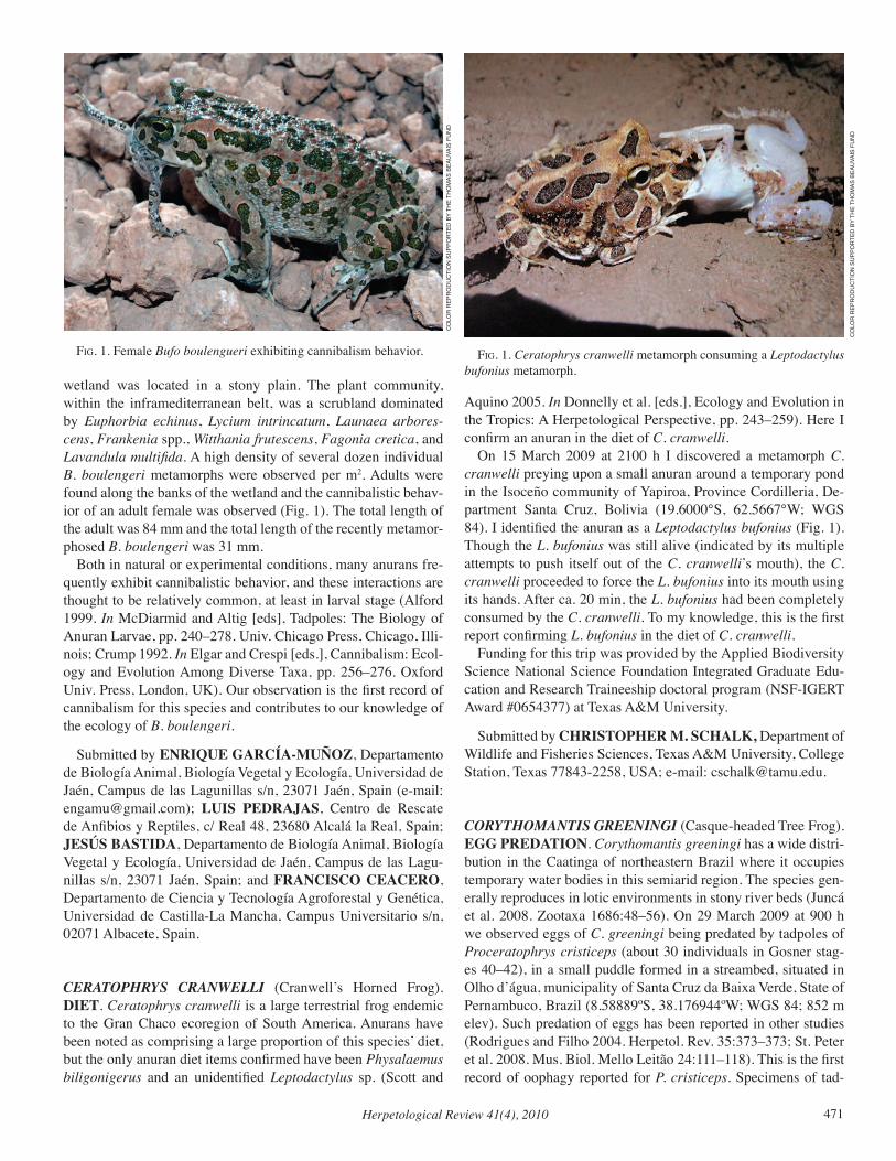
Herpetological Review 41(4), 2010 471
wetland was located in a stony plain. The plant community, within the inframediterranean belt, was a scrubland dominated by Euphorbia echinus, Lycium intrincatum, Launaea arbores-cens, Frankenia spp., Witthania frutescens, Fagonia cretica, and Lavandula multifida. A high density of several dozen individual B. boulengeri metamorphs were observed per m2. Adults were found along the banks of the wetland and the cannibalistic behav-ior of an adult female was observed (Fig. 1). The total length of the adult was 84 mm and the total length of the recently metamor-phosed B. boulengeri was 31 mm. Both in natural or experimental conditions, many anurans fre-quently exhibit cannibalistic behavior, and these interactions are thought to be relatively common, at least in larval stage (Alford 1999. In McDiarmid and Altig [eds], Tadpoles: The Biology of Anuran Larvae, pp. 240–278. Univ. Chicago Press, Chicago, Illi-nois; Crump 1992. In Elgar and Crespi [eds.], Cannibalism: Ecol-ogy and Evolution Among Diverse Taxa, pp. 256–276. Oxford Univ. Press, London, UK). Our observation is the first record of cannibalism for this species and contributes to our knowledge of the ecology of B. boulengeri.
Submitted by ENRIQUE GARCÍA-MUÑOZ, Departamento de Biología Animal, Biología Vegetal y Ecología, Universidad de Jaén, Campus de las Lagunillas s/n, 23071 Jaén, Spain (e-mail: [email protected]); LUIS PEDRAJAS, Centro de Rescate de Anfibios y Reptiles, c/ Real 48, 23680 Alcalá la Real, Spain; JESÚS BASTIDA, Departamento de Biología Animal, Biología Vegetal y Ecología, Universidad de Jaén, Campus de las Lagu-nillas s/n, 23071 Jaén, Spain; and FRANCISCO CEACERO, Departamento de Ciencia y Tecnología Agroforestal y Genética, Universidad de Castilla-La Mancha, Campus Universitario s/n, 02071 Albacete, Spain.
CERATOPHRYS CRANWELLI (Cranwell’s Horned Frog). DIET. Ceratophrys cranwelli is a large terrestrial frog endemic to the Gran Chaco ecoregion of South America. Anurans have been noted as comprising a large proportion of this species’ diet, but the only anuran diet items confirmed have been Physalaemus biligonigerus and an unidentified Leptodactylus sp. (Scott and
Aquino 2005. In Donnelly et al. [eds.], Ecology and Evolution in the Tropics: A Herpetological Perspective, pp. 243–259). Here I confirm an anuran in the diet of C. cranwelli. On 15 March 2009 at 2100 h I discovered a metamorph C. cranwelli preying upon a small anuran around a temporary pond in the Isoceño community of Yapiroa, Province Cordilleria, De-partment Santa Cruz, Bolivia (19.6000°S, 62.5667°W; WGS 84). I identified the anuran as a Leptodactylus bufonius (Fig. 1). Though the L. bufonius was still alive (indicated by its multiple attempts to push itself out of the C. cranwelli’s mouth), the C. cranwelli proceeded to force the L. bufonius into its mouth using its hands. After ca. 20 min, the L. bufonius had been completely consumed by the C. cranwelli. To my knowledge, this is the first report confirming L. bufonius in the diet of C. cranwelli. Funding for this trip was provided by the Applied Biodiversity Science National Science Foundation Integrated Graduate Edu-cation and Research Traineeship doctoral program (NSF-IGERT Award #0654377) at Texas A&M University.
Submitted by CHRISTOPHER M. SCHALK, Department of Wildlife and Fisheries Sciences, Texas A&M University, College Station, Texas 77843-2258, USA; e-mail: [email protected].
CORYTHOMANTIS GREENINGI (Casque-headed Tree Frog). EGG PREDATION. Corythomantis greeningi has a wide distri-bution in the Caatinga of northeastern Brazil where it occupies temporary water bodies in this semiarid region. The species gen-erally reproduces in lotic environments in stony river beds (Juncá et al. 2008. Zootaxa 1686:48–56). On 29 March 2009 at 900 h we observed eggs of C. greeningi being predated by tadpoles of Proceratophrys cristiceps (about 30 individuals in Gosner stag-es 40–42), in a small puddle formed in a streambed, situated in Olho d’água, municipality of Santa Cruz da Baixa Verde, State of Pernambuco, Brazil (8.58889ºS, 38.176944ºW; WGS 84; 852 m elev). Such predation of eggs has been reported in other studies (Rodrigues and Filho 2004. Herpetol. Rev. 35:373–373; St. Peter et al. 2008. Mus. Biol. Mello Leitão 24:111–118). This is the first record of oophagy reported for P. cristiceps. Specimens of tad-
fig. 1. Ceratophrys cranwelli metamorph consuming a Leptodactylus bufonius metamorph.
fig. 1. Female Bufo boulengueri exhibiting cannibalism behavior.
CO
LOR
REP
RO
du
CTi
ON
Su
PPO
RTEd
BY
THE
THO
mAS
BEA
uvA
iS F
uN
d
CO
LOR
REP
RO
du
CTi
ON
Su
PPO
RTEd
BY
THE
THO
mAS
BEA
uvA
iS F
uN
d
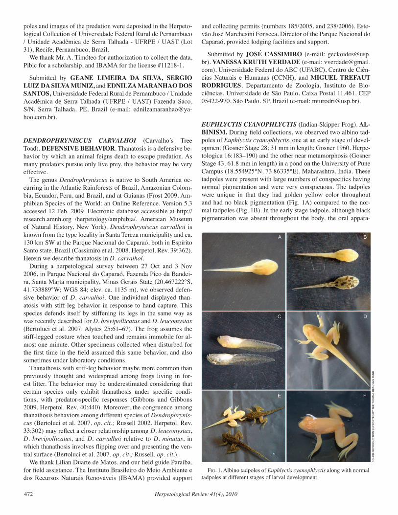
Herpetological Review 41(4), 2010472
poles and images of the predation were deposited in the Herpeto-logical Collection of Universidade Federal Rural de Pernambuco / Unidade Acadêmica de Serra Talhada - UFRPE / UAST (Lot 31), Recife, Pernambuco, Brazil. We thank Mr. A. Timóteo for authorization to collect the data, Pibic for a scholarship, and IBAMA for the license #11218-1.
Submitted by GEANE LIMEIRA DA SILVA, SERGIO LUIZ DA SILVA MUNIZ, and EDNILZA MARANHAO DOS SANTOS, Universidade Federal Rural de Pernambuco / Unidade Acadêmica de Serra Talhada (UFRPE / UAST) Fazenda Saco, S/N, Serra Talhada, PE, Brazil (e-mail: [email protected]).
DENDROPHRYNISCUS CARvALHOI (Carvalho’s Tree Toad). DEFENSIVE BEHAVIOR. Thanatosis is a defensive be-havior by which an animal feigns death to escape predation. As many predators pursue only live prey, this behavior may be very effective. The genus Dendrophryniscus is native to South America oc-curring in the Atlantic Rainforests of Brazil, Amazonian Colom-bia, Ecuador, Peru, and Brazil, and at Guianas (Frost 2009. Am-phibian Species of the World: an Online Reference. Version 5.3 accessed 12 Feb. 2009. Electronic database accessible at http://research.amnh.org /herpetology/amphibia/. American Museum of Natural History, New York). Dendrophryniscus carvalhoi is known from the type locality in Santa Tereza municipality and ca. 130 km SW at the Parque Nacional do Caparaó, both in Espírito Santo state, Brazil (Cassimiro et al. 2008. Herpetol. Rev. 39:362). Herein we describe thanatosis in D. carvalhoi. During a herpetological survey between 27 Oct and 3 Nov 2006, in Parque Nacional do Caparaó, Fazenda Pico da Bandei-ra, Santa Marta municipality, Minas Gerais State (20.467222°S, 41.733889°W; WGS 84; elev. ca. 1135 m), we observed defen-sive behavior of D. carvalhoi. One individual displayed than-atosis with stiff-leg behavior in response to hand capture. This species defends itself by stiffening its legs in the same way as was recently described for D. brevipollicatus and D. leucomystax (Bertoluci et al. 2007. Alytes 25:61–67). The frog assumes the stiff-legged posture when touched and remains immobile for al-most one minute. Other specimens collected when disturbed for the first time in the field assumed this same behavior, and also sometimes under laboratory conditions. Thanathosis with stiff-leg behavior maybe more common than previously thought and widespread among frogs living in for-est litter. The behavior may be underestimated considering that certain species only exhibit thanathosis under specific condi-tions, with predator-specific responses (Gibbons and Gibbons 2009. Herpetol. Rev. 40:440). Moreover, the congruence among thanathosis behaviors among different species of Dendrophrynis-cus (Bertoluci et al. 2007, op. cit.; Russell 2002. Herpetol. Rev. 33:302) may reflect a closer relationship among D. leucomystax, D. brevipollicatus, and D. carvalhoi relative to D. minutus, in which thanathosis involves flipping over and presenting the ven-tral surface (Bertoluci et al. 2007, op. cit.; Russell, op. cit.). We thank Lilian Duarte de Matos, and our field guide Paraíba, for field assistance. The Instituto Brasileiro do Meio Ambiente e dos Recursos Naturais Renováveis (IBAMA) provided support
and collecting permits (numbers 185/2005, and 238/2006). Este-vão José Marchesini Fonseca, Director of the Parque Nacional do Caparaó, provided lodging facilities and support.
Submitted by JOSÉ CASSIMIRO (e-mail: [email protected]), VANESSA KRUTH VERDADE (e-mail: [email protected]), Universidade Federal do ABC (UFABC), Centro de Ciên-cias Naturais e Humanas (CCNH); and MIGUEL TREFAUT RODRIGUES, Departamento de Zoologia, Instituto de Bio-ciências, Universidade de São Paulo, Caixa Postal 11.461, CEP 05422-970, São Paulo, SP, Brazil (e-mail: [email protected]).
EUPHLYCTIS CYANOPHLYCTIS (Indian Skipper Frog). AL-BINISM. During field collections, we observed two albino tad-poles of Euphlyctis cyanophlyctis, one at an early stage of devel-opment (Gosner Stage 28; 31 mm in length; Gosner 1960. Herpe-tologica 16:183–190) and the other near metamorphosis (Gosner Stage 43; 61.8 mm in length) in a pond on the University of Pune Campus (18.554925°N, 73.86335°E), Maharashtra, India. These tadpoles were present with large numbers of conspecifics having normal pigmentation and were very conspicuous. The tadpoles were unique in that they had golden yellow color throughout and had no black pigmentation (Fig. 1A) compared to the nor-mal tadpoles (Fig. 1B). In the early stage tadpole, although black pigmentation was absent throughout the body, the oral appara-
fig. 1. Albino tadpoles of Euphlyctis cyanophlyctis along with normal tadpoles at different stages of larval development.
CO
LOR
REP
RO
du
CTi
ON
Su
PPO
RTEd
BY
THE
THO
mAS
BEA
uvA
iS F
uN
d
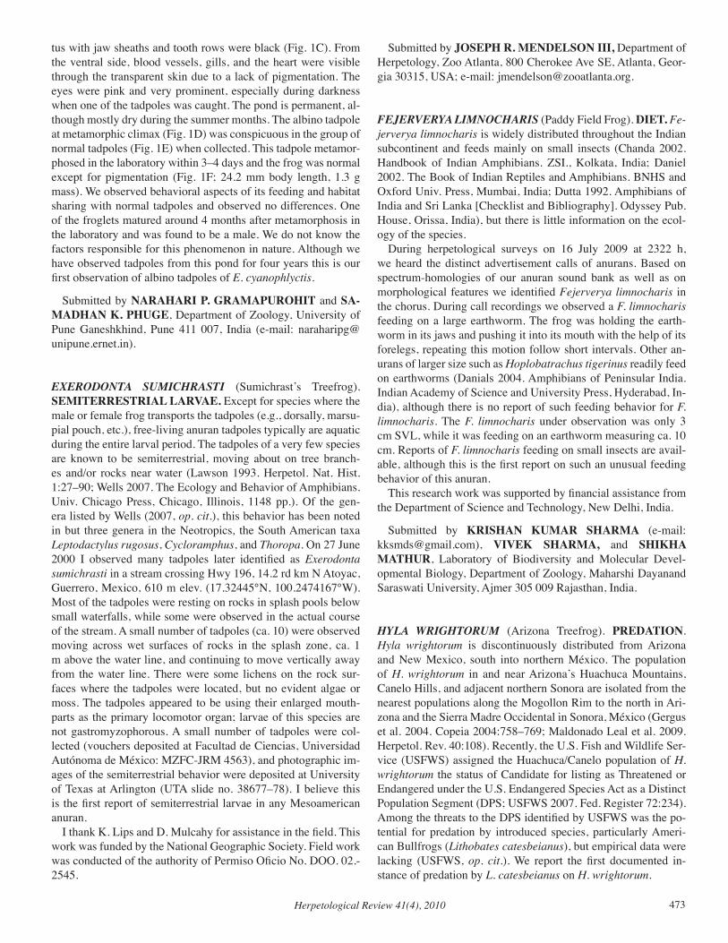
Herpetological Review 41(4), 2010 473
tus with jaw sheaths and tooth rows were black (Fig. 1C). From the ventral side, blood vessels, gills, and the heart were visible through the transparent skin due to a lack of pigmentation. The eyes were pink and very prominent, especially during darkness when one of the tadpoles was caught. The pond is permanent, al-though mostly dry during the summer months. The albino tadpole at metamorphic climax (Fig. 1D) was conspicuous in the group of normal tadpoles (Fig. 1E) when collected. This tadpole metamor-phosed in the laboratory within 3–4 days and the frog was normal except for pigmentation (Fig. 1F; 24.2 mm body length, 1.3 g mass). We observed behavioral aspects of its feeding and habitat sharing with normal tadpoles and observed no differences. One of the froglets matured around 4 months after metamorphosis in the laboratory and was found to be a male. We do not know the factors responsible for this phenomenon in nature. Although we have observed tadpoles from this pond for four years this is our first observation of albino tadpoles of E. cyanophlyctis.
Submitted by NARAHARI P. GRAMAPUROHIT and SA-MADHAN K. PHUGE, Department of Zoology, University of Pune Ganeshkhind, Pune 411 007, India (e-mail: [email protected]).
EXERODONTA SUMICHRASTI (Sumichrast’s Treefrog). SEMITERRESTRIAL LARVAE. Except for species where the male or female frog transports the tadpoles (e.g., dorsally, marsu-pial pouch, etc.), free-living anuran tadpoles typically are aquatic during the entire larval period. The tadpoles of a very few species are known to be semiterrestrial, moving about on tree branch-es and/or rocks near water (Lawson 1993. Herpetol. Nat. Hist. 1:27–90; Wells 2007. The Ecology and Behavior of Amphibians. Univ. Chicago Press, Chicago, Illinois, 1148 pp.). Of the gen-era listed by Wells (2007, op. cit.), this behavior has been noted in but three genera in the Neotropics, the South American taxa Leptodactylus rugosus, Cycloramphus, and Thoropa. On 27 June 2000 I observed many tadpoles later identified as Exerodonta sumichrasti in a stream crossing Hwy 196, 14.2 rd km N Atoyac, Guerrero, Mexico, 610 m elev. (17.32445°N, 100.2474167°W). Most of the tadpoles were resting on rocks in splash pools below small waterfalls, while some were observed in the actual course of the stream. A small number of tadpoles (ca. 10) were observed moving across wet surfaces of rocks in the splash zone, ca. 1 m above the water line, and continuing to move vertically away from the water line. There were some lichens on the rock sur-faces where the tadpoles were located, but no evident algae or moss. The tadpoles appeared to be using their enlarged mouth-parts as the primary locomotor organ; larvae of this species are not gastromyzophorous. A small number of tadpoles were col-lected (vouchers deposited at Facultad de Ciencias, Universidad Autónoma de México: MZFC-JRM 4563), and photographic im-ages of the semiterrestrial behavior were deposited at University of Texas at Arlington (UTA slide no. 38677–78). I believe this is the first report of semiterrestrial larvae in any Mesoamerican anuran. I thank K. Lips and D. Mulcahy for assistance in the field. This work was funded by the National Geographic Society. Field work was conducted of the authority of Permiso Oficio No. DOO. 02.-2545.
Submitted by JOSEPH R. MENDELSON III, Department of Herpetology, Zoo Atlanta, 800 Cherokee Ave SE, Atlanta, Geor-gia 30315, USA; e-mail: [email protected].
FEJERvERYA LIMNOCHARIS (Paddy Field Frog). DIET. Fe-jerverya limnocharis is widely distributed throughout the Indian subcontinent and feeds mainly on small insects (Chanda 2002. Handbook of Indian Amphibians. ZSI., Kolkata, India; Daniel 2002. The Book of Indian Reptiles and Amphibians. BNHS and Oxford Univ. Press, Mumbai, India; Dutta 1992. Amphibians of India and Sri Lanka [Checklist and Bibliography]. Odyssey Pub. House, Orissa, India), but there is little information on the ecol-ogy of the species. During herpetological surveys on 16 July 2009 at 2322 h, we heard the distinct advertisement calls of anurans. Based on spectrum-homologies of our anuran sound bank as well as on morphological features we identified Fejerverya limnocharis in the chorus. During call recordings we observed a F. limnocharis feeding on a large earthworm. The frog was holding the earth-worm in its jaws and pushing it into its mouth with the help of its forelegs, repeating this motion follow short intervals. Other an-urans of larger size such as Hoplobatrachus tigerinus readily feed on earthworms (Danials 2004. Amphibians of Peninsular India. Indian Academy of Science and University Press, Hyderabad, In-dia), although there is no report of such feeding behavior for F. limnocharis. The F. limnocharis under observation was only 3 cm SVL, while it was feeding on an earthworm measuring ca. 10 cm. Reports of F. limnocharis feeding on small insects are avail-able, although this is the first report on such an unusual feeding behavior of this anuran. This research work was supported by financial assistance from the Department of Science and Technology, New Delhi, India.
Submitted by KRISHAN KUMAR SHARMA (e-mail: [email protected]), VIVEK SHARMA, and SHIKHA MATHUR, Laboratory of Biodiversity and Molecular Devel-opmental Biology, Department of Zoology, Maharshi Dayanand Saraswati University, Ajmer 305 009 Rajasthan, India.
HYLA WRIGHTORUM (Arizona Treefrog). PREDATION. Hyla wrightorum is discontinuously distributed from Arizona and New Mexico, south into northern México. The population of H. wrightorum in and near Arizona’s Huachuca Mountains, Canelo Hills, and adjacent northern Sonora are isolated from the nearest populations along the Mogollon Rim to the north in Ari-zona and the Sierra Madre Occidental in Sonora, México (Gergus et al. 2004. Copeia 2004:758–769; Maldonado Leal et al. 2009. Herpetol. Rev. 40:108). Recently, the U.S. Fish and Wildlife Ser-vice (USFWS) assigned the Huachuca/Canelo population of H. wrightorum the status of Candidate for listing as Threatened or Endangered under the U.S. Endangered Species Act as a Distinct Population Segment (DPS; USFWS 2007. Fed. Register 72:234). Among the threats to the DPS identified by USFWS was the po-tential for predation by introduced species, particularly Ameri-can Bullfrogs (Lithobates catesbeianus), but empirical data were lacking (USFWS, op. cit.). We report the first documented in-stance of predation by L. catesbeianus on H. wrightorum.
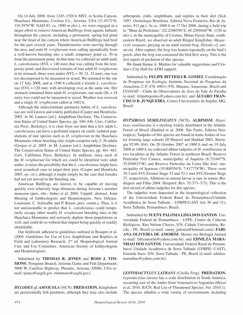
Herpetological Review 41(4), 2010474
On 14 July 2008, from 1245–1520 h MST, in Scotia Canyon, Huachuca Mountains, Cochise Co., Arizona, USA (31.4572°N, 110.3976°W, NAD 83, ca. 1890 m elev.), we were engaged in a larger effort to remove American Bullfrogs from aquatic habitats throughout the canyon, including a permanent, spring-fed pond near the head of the canyon where American Bullfrogs had bred for the past several years. Thunderstorms were moving through the area, and male H. wrightorum were calling sporadically from a well-known breeding site; a small, temporary pond ca. 50 m from the permanent pond. At that time we collected an adult male L. catesbeianus (SVL = 140 mm) that was calling from the tem-porary pond, and discovered remains of four adult H. wrightorum in its stomach; three were males (SVL = 30, 31, 32 mm), one was too decomposed to be measured or sexed. We returned to the site on 17 July 2008, and at 1540 h collected a female L. catesbeia-nus (SVL = 120 mm; with developing ova) at the same site. Her stomach contained three adult H. wrightorum, one male (SL = 34 mm); two could not be measured or sexed. Weather was overcast and a single H. wrightorum called at 1602 h. Although the indiscriminate predatory habits of L. catesbeia-nus are well known and widely published (Casper and Hendricks. 2005. In M. Lannoo [ed.], Amphibian Declines: The Conserva-tion Status of United States Species, pp. 540–546. Univ. Califor-nia Press, Berkeley), it is clear from our data that a few adult L. catesbeianus can have a profound impact on small, isolated pop-ulations of rare species such as H. wrightorum in the Huachuca Mountains where breeding sites might comprise only 2–30 adults (Gergus et al. 2005. In M. Lannoo [ed.], Amphibian Declines: The Conservation Status of United States Species, pp. 461–463. Univ. California Press, Berkeley). In addition, since each of the H. wrightorum for which sex could be identified were adult males, it raises the possibility that the L. catesbeianus might have used acoustical cues to target their prey (Casper and Hendricks 2005, op. cit.), although it might simply be the case that females had not yet moved to the breeding site. American Bullfrogs are known to be capable of moving quickly over relatively large distances during Arizona’s summer monsoon (pers. obs.; Suhre et al. 2006. Unpubl. abstract. Joint Meeting of Ichthyologists and Herpetologists, New Orleans, Louisiana; C. Schwalbe and P. Rosen, pers. comm.). Thus, it is not unreasonable to predict that L. catesbeianus could tempo-rarily occupy other nearby H. wrightorum breeding sites in the Huachuca Mountains and seriously deplete those populations as well, and could do so without that impact being quickly or readily identifiable. Our fieldwork adhered to guidelines outlined in Beaupre et al. (2004. Guidelines for Use of Live Amphibians and Reptiles in Field and Laboratory Research, 2nd ed. Herpetological Animal Care and Use Committee, American Society of Ichthyologists and Herpetologists).
Submitted by THOMAS R. JONES and ROSS J. TIM-MONS, Nongame Branch, Arizona Game and Fish Department, 5000 W. Carefree Highway, Phoenix, Arizona, 85086, USA (e-mail: [email protected], [email protected]).
HYLODES cf. AMNICOLA (NCN). PREDATION. Kingfishers are preferentially fish predators, although they may also include
arthropods, crabs, amphibians, and reptiles in their diet (Sick 1997. Ornitologia Brasileira. Editora Nova Fronteira, Rio de Ja-neiro. 912 pp.). At ca. 1000 h on 17 Oct 2008, during a field trip to “Mata da Prefeitura” (22.2180556°S, 45.2569444°W; 1150 m elev.), in the municipality of Cristina, Minas Gerais State, south-eastern Brazil, we observed an adult Ringed Kingfisher, Megac-eryle torquata, preying on an adult torrent-frog, Hylodes cf. am-nicola. After capture, the frog was beaten repeatedly on the bird’s perch; after the frog was consumed the bird flew away. This is the first report of predation of this species. We thank Itamar A. Martins for valuable suggestions and Cris-tina’s City Hall for AFBJ support.
Submitted by FELIPE BITTIOLI R. GOMES, Coordenação de Pesquisas em Ecologia, Instituto Nacional de Pesquisas da Amazônia, C.P. 478, 69011-970, Manaus, Amazonas, Brazil and COAVAP – Clube de Observadores de Aves do Vale do Paraíba (e-mail: [email protected]); and ÁLVARO FRAN-CISCO B. JUNQUEIRA, Centro Universitário de Itajubá, MG, Brazil.
HYPSIBOAS SEMILINEATUS (NCN). ALBINISM. Hypsi-boas semilineatus is a treefrog widely distributed in the Atlantic Forest of Brazil (Haddad et al. 2008. São Paulo: Editora Neo-tropica). Tadpoles of this species are found in lentic bodies of wa-ter forming large schools (D’Heursel and Haddad 2002. Iherin-gia 92:99–104). On 20 October 2007 at 1900 h and on 19 July 2008 at 1400 h we collected albino tadpoles of H. semilineatus at two localities in the Atlantic Forest in northeast Brazil: Reserva Particular Frei Caneca, municipality of Jaqueira (8.721667ºS, 35.850833ºW) and Reserva Particular da Usina São José, mu-nicipality of Igarassu (35.000556ºS, 7.817778ºW). These were 39.3 mm SVL/Gosner Stage 33 and 52.1 mm SVL/Gosner Stage 37, respectively. Albinism in anuran larvae is rare in nature (Ro-drigues and Filho 2004. Herpetol. Rev. 35:373–373). This is the first record of albino tadpoles for this species. The tadpoles were deposited in the herpetological collection of the Universidade Federal Rural de Pernambuco/Unidade Acadêmica de Serra Talhada - UFRPE/UAST (lot 30 and 31), Serra Talhada, Pernambuco, Brazil.
Submitted by SUENY PALOMA LIMA DOS SANTOS, Uni-versidade Federal de Pernambuco - UFPE, Centro de Ciências Biológicas, Rua Nelson Chaves, S/N, Cidade Universitária, Re-cife - PE, Brazil (e-mail: [email protected]); FABI-ANA OLIVEIRA DE AMORIM, Mestre em Biologia Animal (e-mail: [email protected]), and EDNILZA MARA-NHAO DOS SANTOS, Universidade Federal Rural de Pernam-buco/ Unidade Acadêmica de Serra Talhada (UFRPE/ UAST), Fazenda Saco, S/N, Serra Talhada - PE, Brazil (e-mail: [email protected]).
LEPTODACTYLUS LATRANS (Criolla Frog). PREDATION. Leptodactylus latrans has a wide distribution in South America, occurring east of the Andes from Venezuela to Argentina (Heyer et al. 2010. IUCN. Red List of Threatened Species. Ver. 2010.1). The species inhabits a wide variety of environments including
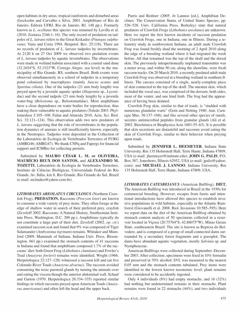
Herpetological Review 41(4), 2010 475
open habitats in dry areas, tropical rainforests and disturbed areas (Izecksohn and Carvalho e Silva. 2001. Amphibians of Rio de Janeiro, Editora UFRJ, Rio de Janeiro, RJ, 148 pp.). Formerly known as L. ocellatus this species was renamed by Lavilla et al. (2010. Zootaxa 2346:1–16). The only record of predation on tad-poles of L. latrans refers to the Great Kiskadee (Pitangus sulphu-ratus; Vaira and Coria 1994. Herpetol. Rev. 25:118). There are no records of predation of L. latrans tadpoles by invertebrates. At 2120 h on 27 Oct 2009 we observed two predation events of L. latrans tadpoles by aquatic invertebrates. The observations were made in wetland habitat associated with a coastal sand dune (32.1654°S, 52.1523°W, Corrego Alegre, sea level) in the mu-nicipality of Rio Grande, RS, southern Brazil. Both events were observed simultaneously in a school of tadpoles in a temporary pond colonized by macrophytes (mainly Juncus acutus and Spartina ciliata). One of the tadpoles (21 mm body length) was preyed upon by a juvenile aquatic spider (Diapontia sp., Lycosi-dae) and the second tadpole (23 mm body length) by a juvenile water-bug (Belostoma sp., Belostomatidae). Most amphibians have a close dependence on water bodies for reproduction, thus making them vulnerable to aquatic predators (Toledo 2003. Phyl-lomedusa 2:105–108; Fulan and Almeida 2010. Acta. Sci. Biol. Sci. 32:121–124). This observation adds two new predators of L. latrans suggesting that the role of invertebrates in the popula-tion dynamics of anurans is still insufficiently known, especially in the Neotropics. Tadpoles were deposited in the Collection of the Laboratório de Ecologia de Vertebrados Terrestres of FURG (AMRG46; AMRG47). We thank CNPq and Fapergs for financial support and ICMBio for collecting permits.
Submitted by MAURO CÉSAR L. M. de OLIVEIRA, MAURÍCIO BEUX DOS SANTOS, and ALEXANDRO M. TOZETTI, Laboratório de Ecologia de Vertebrados Terrestres, Instituto de Ciências Biológicas, Universidade Federal do Rio Grande, Av. Itália, km 8, Rio Grande, Rio Grande do Sul, Brazil (e-mail: [email protected]).
LITHOBATES AREOLATUS CIRCULOSUS (Northern Craw-fish Frog). PREDATION. Raccoons (Procyon lotor) are known to consume a wide variety of prey items. They often forage at the edge of shallow water in search of their preferred prey, crayfish (Zeveloff 2002. Raccoons: A Natural History. Smithsonian Insti-tute Press, Washington, D.C. 200 pp.). Amphibians typically do not constitute a large part of their diet. Zeveloff (2002, op. cit.) examined raccoon scat and found that 9% was composed of Tiger Salamander (Ambystoma tigrinum) remains. Whitaker and Mum-ford (2009. Mammals of Indiana. Indiana Univ. Press, Bloom-ington. 661 pp.) examined the stomach contents of 41 raccoons in Indiana and found that amphibians composed 1.7% of the rac-coons’ diet; both Green Frog (Lithobates clamitans) and Fowler’s Toad (Anaxyrus fowleri) remains were identified. Wright (1966. Herpetologica 22:127–128) witnessed a raccoon kill and eat five Colorado River Toads (Anaxyrus alvarius). The raccoon avoided consuming the toxic parotoid glands by turning the animals over and eating the viscera though the anterior abdominal wall. Schaaf and Garton (1970. Herpetologica 26:334–335) reported similar findings in which raccoons preyed upon American Toads (Anaxy-rus americanus) and often left the head and the upper back.
Parris and Redmer (2005. In Lannoo [ed.], Amphibian De-clines: The Conservation Status of United States Species, pp. 526–528. Univ. California Press, Berkeley) state that natural predators of Crawfish Frogs (Lithobates areolatus) are unknown. Here we report the first known incidents of raccoon predation on Crawfish Frogs, one in Indiana, one in Illinois. During a te-lemetry study in southwestern Indiana, an adult male Crawfish Frog was found freshly dead the morning of 2 April 2010 along the edge of a breeding wetland where it had migrated the week before. All that remained was the top of the skull and the dorsal skin. The previously intraperitoneally implanted transmitter was a meter away, and within 50 cm, along the shoreline, were fresh raccoon tracks. On 20 March 2010, a recently predated adult male Crawfish Frog was observed at a breeding wetland in southern Il-linois. The carcass consisted of a single, nearly-complete piece of skin connected to the top of the skull. The uneaten skin, which included the vocal sacs, was comprised of the dorsum, both sides, most of the venter, and one hind limb. The frog had the appear-ance of having been skinned. Crawfish Frog skin, similar to that of toads, is “studded with numerous glandular warts” (Goin and Netting 1940. Ann. Carn-egie Mus. 38:137–168); and like several other species of ranids, secretes antimicrobial peptides from granular glands (Ali et al. 2002. Biochimica et Biophysica Acta 1601:55–63). It is possible that skin secretions are distasteful and raccoons avoid eating the skin of Crawfish Frogs, similar to their behavior when preying upon toads.
Submitted by JENNIFER L. HEEMEYER, Indiana State University, Rm 135 Holmstedt Hall, Terre Haute, Indiana 47809, USA (e-mail: [email protected]); JOHN G. PALIS, P.O. Box 387, Jonesboro, Illinois 62952, USA (e-mail: [email protected]); and MICHAEL J. LANNOO, Indiana University, Rm 135 Holmstedt Hall, Terre Haute, Indiana 47809, USA.
LITHOBATES CATESBEIANUS (American Bullfrog). DIET. The American Bullfrog was introduced in Brazil in the 1930s for commercial breeding. However, escapes from farms and inten-tional introductions have allowed this species to establish inva-sive populations in wild habitats, especially in the Atlantic Rain-forest (Giovanelli et al. 2008. Biol. Invasions 10:585–595). Here we report data on the diet of the American Bullfrog obtained by stomach content analysis of 50 specimens collected in a reser-voir located in Viçosa (20.75764°S, 42.86075°W), Minas Gerais State, southeastern Brazil. The site is known as Represa do Bel-vedere, and is composed of a group of small connected dams sur-rounded by a secondary forest fragments and a grassplot. The dams have abundant aquatic vegetation, mostly Salvinia sp. and Nympheaceae. American Bullfrogs were collected during September–Decem-ber 2003. After collection, specimens were fixed in 10% formalin and preserved in 70% alcohol; SVL was measured to the nearest 0.05 mm and the stomach contents tabulated. Prey items were identified to the lowest known taxonomic level; plant remains were considered to be accidently ingested. Only 4 individuals (8%) had empty stomachs, and 16 (32%) had nothing but undetermined remains in their stomachs. Plant remains were found in 22 stomachs (44%), and two individuals
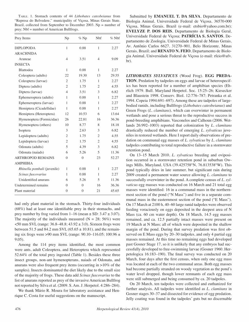
Herpetological Review 41(4), 2010476
had only plant material in the stomach. Thirty-four individuals (68%) had at least one identifiable prey in their stomachs, and prey number by frog varied from 1–16 (mean ± SD: 3.47 ± 3.07). The majority of the individuals measured (N = 28; 56%) were <50 mm SVL (range: 36.8–48.8; 43.92 ± 3.0). Sixteen frogs were between 51.5 and 84.2 mm SVL (65.65 ± 10.81), and the remain-ing six frogs were >90 mm SVL (range: 90.10–116.05; 100.96 ± 9.05). Among the 114 prey items identified, the most common were ants, adult Coleoptera, and Heteroptera which represented 52.64% of the total prey ingested (Table 1). Besides these three insect groups, non-ant hymenopterans, naiads of Odonata, and anurans were also frequent prey items (occurring in >10% of the samples). Insects dominanted the diet likely due to the small size of the majority of frogs. These data add Scinax fuscovarius to the list of anurans reported as prey of the invasive American Bullfrog not reported by Silva et al. (2009. S. Am. J. Herpetol. 4:286–284). We thank Mário R. Moura for laboratory assistance and Hen-rique C. Costa for useful suggestions on the manuscript.
Submitted by EMANUEL T. DA SILVA, Departamento de Biologia Animal, Universidade Federal de Viçosa, 36570-000 Viçosa, Minas Gerais, Brazil (e-mail: [email protected]); EVELYZE P. DOS REIS, Departamento de Biologia Geral, Universidade Federal de Viçosa; PATRÍCIA S. SANTOS, De-partamento de Zoologia, Universidade Federal de Minas Gerais, Av. Antônio Carlos 6627, 31270–901, Belo Horizonte, Minas Gerais, Brazil; and RENATO N. FEIO, Departamento de Biolo-gia Animal, Universidade Federal de Viçosa (e-mail: [email protected]).
LITHOBATES SYLvATICUS (Wood Frog). EGG PREDA-TION. Predation by tadpoles on eggs and larvae of heterospecif-ics has been reported for a number of amphibian species (Eh-rlich 1979. Bull. Maryland Herpetol. Soc. 15:25–26; Kiesecker and Blaustein 1998. Conserv. Biol. 12:776–787; Petranka et al. 1994. Copeia 1994:691–697). Among these are tadpoles of large-bodied ranids, including Bullfrogs (Lithobates catesbeianus) and Green Frogs (L. clamitans), which can overwinter in permanent wetlands and pose a serious threat to the reproductive success in pond-breeding amphibians. Vasconelos and Calhoun (2006. Wet-lands 26:992–1003) reported that colonization by L. clamitans drastically reduced the number of emerging L. sylvaticus juve-niles in restored wetlands. Here I report daily observations of pre-dation on communal egg masses of L. sylvaticus by L. clamitans tadpoles contributing to total reproductive failure in a stormwater retention pond. On 13–15 March 2010, L. sylvaticus breeding and oviposi-tion occurred in a stormwater retention pond in suburban Ow-ings Mills, Maryland, USA (39.425758°N, 76.813538°W). This pond typically dries in late summer, but significant rain during 2009 created a permanent water source allowing L. clamitans to successfully overwinter in the pond. A complete census of L. syl-vaticus egg masses was conducted on 16 March and 21 total egg masses were identified: 16 in a communal mass in the northern-most section of the pond (“N Mass”) and five in a separate com-munal mass in the easternmost section of the pond (“E Mass”). On 17 March at 2100 h, 40–60 large ranid tadpoles were observed feeding voraciously on eggs deposited in the deepest area of N Mass (ca. 60 cm water depth). On 18 March, 14.5 egg masses remained, and ca. 12.5 partially intact masses were present on 19 March at N Mass; all of which were deposited in the shallow margin of the pond. During that survey predation was first ob-served on E Mass eggs by 20–30 tadpoles, and only 4 partial egg masses remained. At this time no remaining eggs had developed past Gosner Stage 17, so it is unlikely that any embryos had suc-cessfully developed to free-swimming larvae (Gosner 1960. Her-petologica 16:183–190). The final survey was conducted on 20 March, four days after the first census, when only one egg mass was located at each of the two communal areas. Both egg masses had become partially stranded on woody vegetation as the pond’s water level dropped, though lower remnants of each egg mass were still submerged and being consumed by ca. 20 tadpoles. On 20 March, ten tadpoles were collected and euthanized for further analysis. All tadpoles were identified as L. clamitans in Gosner stages 30–37 and dissected for evidence of egg predation. Jelly coating was found in the tadpoles’ guts but no discernable
taBle. 1. Stomach contents of 44 Lithobates catesbeianus from “Represa do Belvedere,” municipality of Viçosa, Minas Gerais State, Brazil, collected from September to December 2003. Np = number of prey; Nbf = number of American Bullfrogs.
Prey Items Np % Np Nbf % Nbf
DIPLOPODA 1 0.88 1 2.27ARACHNIDA Araneae 4 3.51 4 9.09INSECTA Blattodea 1 0.88 1 2.27 Coleoptera (adults) 22 19.30 13 29.55 Coleoptera (larvae) 2 1.75 1 2.27 Diptera (adults) 2 1.75 2 4.55 Diptera (larvae) 4 3.51 3 6.82 Ephemeroptera (adults) 1 0.88 1 2.27 Ephemeroptera (larvae) 1 0.88 1 2.27 Hemiptera (Cicadellidae) 1 0.88 1 2.27 Hemiptera (Heteroptera) 12 10.53 6 13.64 Hymenoptera (Formicidae) 26 22.81 16 36.36 Hymenoptera (others) 10 8.77 8 18.18 Isoptera 3 2.63 3 6.82 Lepidoptera (adults) 2 1.75 2 4.55 Lepidoptera (larvae) 2 1.75 2 4.55 Odonata (adults) 5 4.39 3 6.82 Odonata (naiads) 6 5.26 5 11.36ARTHROPOD REMAINS 0 0 23 52.27AMPHIBIA Rhinella pombali (juvenile) 1 0.88 1 2.27 Scinax fuscovarius 1 0.88 1 2.27 Unidentified anurans 6 5.26 5 11.36Undetermined remains 0 0 16 36.36Plant material 0 0 21 45.65
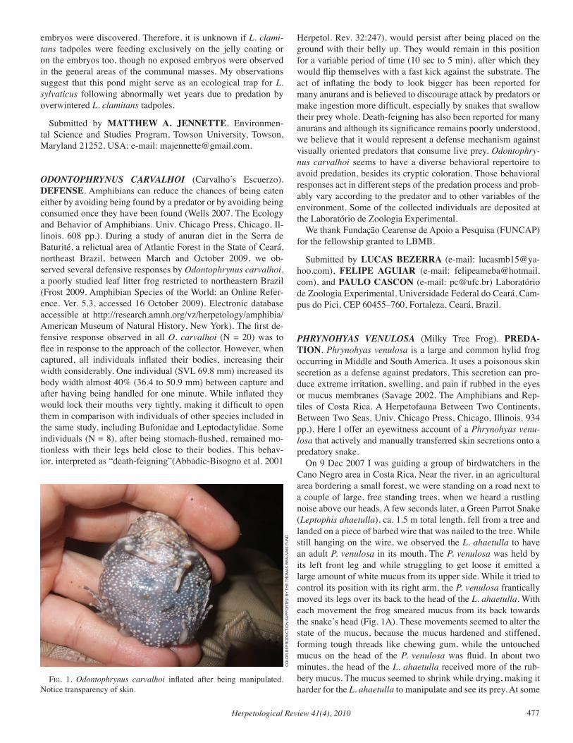
Herpetological Review 41(4), 2010 477
embryos were discovered. Therefore, it is unknown if L. clami-tans tadpoles were feeding exclusively on the jelly coating or on the embryos too, though no exposed embryos were observed in the general areas of the communal masses. My observations suggest that this pond might serve as an ecological trap for L. sylvaticus following abnormally wet years due to predation by overwintered L. clamitans tadpoles.
Submitted by MATTHEW A. JENNETTE, Environmen-tal Science and Studies Program, Towson University, Towson, Maryland 21252, USA; e-mail: [email protected].
ODONTOPHRYNUS CARvALHOI (Carvalho’s Escuerzo). DEFENSE. Amphibians can reduce the chances of being eaten either by avoiding being found by a predator or by avoiding being consumed once they have been found (Wells 2007. The Ecology and Behavior of Amphibians. Univ. Chicago Press, Chicago, Il-linois. 608 pp.). During a study of anuran diet in the Serra de Baturité, a relictual area of Atlantic Forest in the State of Ceará, northeast Brazil, between March and October 2009, we ob-served several defensive responses by Odontophrynus carvalhoi, a poorly studied leaf litter frog restricted to northeastern Brazil (Frost 2009. Amphibian Species of the World: an Online Refer-ence. Ver. 5.3, accessed 16 October 2009). Electronic database accessible at http://research.amnh.org/vz/herpetology/amphibia/ American Museum of Natural History, New York). The first de-fensive response observed in all O. carvalhoi (N = 20) was to flee in response to the approach of the collector. However, when captured, all individuals inflated their bodies, increasing their width considerably. One individual (SVL 69.8 mm) increased its body width almost 40% (36.4 to 50.9 mm) between capture and after having being handled for one minute. While inflated they would lock their mouths very tightly, making it difficult to open them in comparison with individuals of other species included in the same study, including Bufonidae and Leptodactylidae. Some individuals (N = 8), after being stomach-flushed, remained mo-tionless with their legs held close to their bodies. This behav-ior, interpreted as “death-feigning”(Abbadic-Bisogno et al. 2001
Herpetol. Rev. 32:247), would persist after being placed on the ground with their belly up. They would remain in this position for a variable period of time (10 sec to 5 min), after which they would flip themselves with a fast kick against the substrate. The act of inflating the body to look bigger has been reported for many anurans and is believed to discourage attack by predators or make ingestion more difficult, especially by snakes that swallow their prey whole. Death-feigning has also been reported for many anurans and although its significance remains poorly understood, we believe that it would represent a defense mechanism against visually oriented predators that consume live prey. Odontophry-nus carvalhoi seems to have a diverse behavioral repertoire to avoid predation, besides its cryptic coloration. Those behavioral responses act in different steps of the predation process and prob-ably vary according to the predator and to other variables of the environment. Some of the collected individuals are deposited at the Laboratório de Zoologia Experimental. We thank Fundação Cearense de Apoio a Pesquisa (FUNCAP) for the fellowship granted to LBMB.
Submitted by LUCAS BEZERRA (e-mail: [email protected]), FELIPE AGUIAR (e-mail: [email protected]), and PAULO CASCON (e-mail: [email protected]) Laboratório de Zoologia Experimental, Universidade Federal do Ceará, Cam-pus do Pici, CEP 60455–760, Fortaleza, Ceará, Brazil.
PHRYNOHYAS vENULOSA (Milky Tree Frog). PREDA-TION. Phrynohyas venulosa is a large and common hylid frog occurring in Middle and South America. It uses a poisonous skin secretion as a defense against predators. This secretion can pro-duce extreme irritation, swelling, and pain if rubbed in the eyes or mucus membranes (Savage 2002. The Amphibians and Rep-tiles of Costa Rica. A Herpetofauna Between Two Continents, Between Two Seas. Univ. Chicago Press, Chicago, Illinois. 934 pp.). Here I offer an eyewitness account of a Phrynohyas venu-losa that actively and manually transferred skin secretions onto a predatory snake. On 9 Dec 2007 I was guiding a group of birdwatchers in the Cano Negro area in Costa Rica. Near the river, in an agricultural area bordering a small forest, we were standing on a road next to a couple of large, free standing trees, when we heard a rustling noise above our heads. A few seconds later, a Green Parrot Snake (Leptophis ahaetulla), ca. 1.5 m total length, fell from a tree and landed on a piece of barbed wire that was nailed to the tree. While still hanging on the wire, we observed the L. ahaetulla to have an adult P. venulosa in its mouth. The P. venulosa was held by its left front leg and while struggling to get loose it emitted a large amount of white mucus from its upper side. While it tried to control its position with its right arm, the P. venulosa frantically moved its legs over its back to the head of the L. ahaetulla. With each movement the frog smeared mucus from its back towards the snake’s head (Fig. 1A). These movements seemed to alter the state of the mucus, because the mucus hardened and stiffened, forming tough threads like chewing gum, while the untouched mucus on the head of the P. venulosa was fluid. In about two minutes, the head of the L. ahaetulla received more of the rub-bery mucus. The mucus seemed to shrink while drying, making it harder for the L. ahaetulla to manipulate and see its prey. At some
fig. 1. Odontophrynus carvalhoi inflated after being manipulated. Notice transparency of skin.
CO
LOR
REP
RO
du
CTi
ON
Su
PPO
RTEd
BY
THE
THO
mAS
BEA
uvA
iS F
uN
d
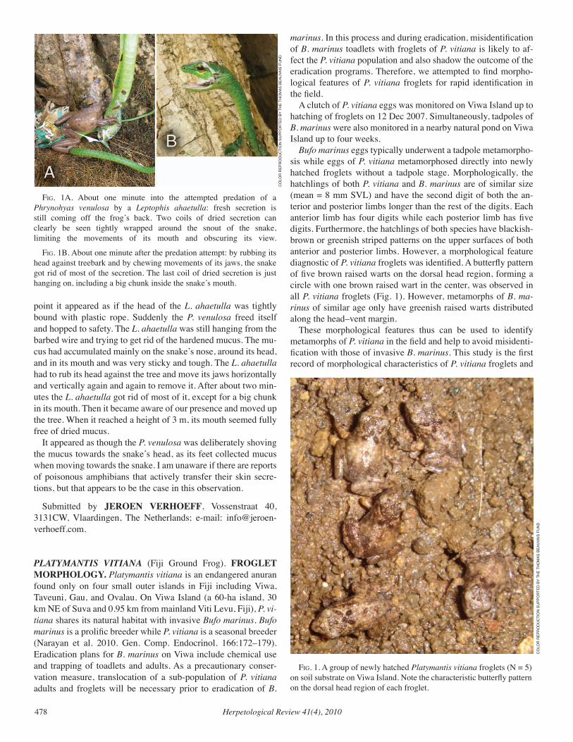
Herpetological Review 41(4), 2010478
point it appeared as if the head of the L. ahaetulla was tightly bound with plastic rope. Suddenly the P. venulosa freed itself and hopped to safety. The L. ahaetulla was still hanging from the barbed wire and trying to get rid of the hardened mucus. The mu-cus had accumulated mainly on the snake’s nose, around its head, and in its mouth and was very sticky and tough. The L. ahaetulla had to rub its head against the tree and move its jaws horizontally and vertically again and again to remove it. After about two min-utes the L. ahaetulla got rid of most of it, except for a big chunk in its mouth. Then it became aware of our presence and moved up the tree. When it reached a height of 3 m, its mouth seemed fully free of dried mucus. It appeared as though the P. venulosa was deliberately shoving the mucus towards the snake’s head, as its feet collected mucus when moving towards the snake. I am unaware if there are reports of poisonous amphibians that actively transfer their skin secre-tions, but that appears to be the case in this observation.
Submitted by JEROEN VERHOEFF, Vossenstraat 40, 3131CW, Vlaardingen, The Netherlands; e-mail: [email protected].
PLATYMANTIS vITIANA (Fiji Ground Frog). FROGLET MORPHOLOGY. Platymantis vitiana is an endangered anuran found only on four small outer islands in Fiji including Viwa, Taveuni, Gau, and Ovalau. On Viwa Island (a 60-ha island, 30 km NE of Suva and 0.95 km from mainland Viti Levu, Fiji), P. vi-tiana shares its natural habitat with invasive Bufo marinus. Bufo marinus is a prolific breeder while P. vitiana is a seasonal breeder (Narayan et al. 2010. Gen. Comp. Endocrinol. 166:172–179). Eradication plans for B. marinus on Viwa include chemical use and trapping of toadlets and adults. As a precautionary conser-vation measure, translocation of a sub-population of P. vitiana adults and froglets will be necessary prior to eradication of B.
marinus. In this process and during eradication, misidentification of B. marinus toadlets with froglets of P. vitiana is likely to af-fect the P. vitiana population and also shadow the outcome of the eradication programs. Therefore, we attempted to find morpho-logical features of P. vitiana froglets for rapid identification in the field. A clutch of P. vitiana eggs was monitored on Viwa Island up to hatching of froglets on 12 Dec 2007. Simultaneously, tadpoles of B. marinus were also monitored in a nearby natural pond on Viwa Island up to four weeks. Bufo marinus eggs typically underwent a tadpole metamorpho-sis while eggs of P. vitiana metamorphosed directly into newly hatched froglets without a tadpole stage. Morphologically, the hatchlings of both P. vitiana and B. marinus are of similar size (mean = 8 mm SVL) and have the second digit of both the an-terior and posterior limbs longer than the rest of the digits. Each anterior limb has four digits while each posterior limb has five digits. Furthermore, the hatchlings of both species have blackish-brown or greenish striped patterns on the upper surfaces of both anterior and posterior limbs. However, a morphological feature diagnostic of P. vitiana froglets was identified. A butterfly pattern of five brown raised warts on the dorsal head region, forming a circle with one brown raised wart in the center, was observed in all P. vitiana froglets (Fig. 1). However, metamorphs of B. ma-rinus of similar age only have greenish raised warts distributed along the head–vent margin. These morphological features thus can be used to identify metamorphs of P. vitiana in the field and help to avoid misidenti-fication with those of invasive B. marinus. This study is the first record of morphological characteristics of P. vitiana froglets and
fig. 1. A group of newly hatched Platymantis vitiana froglets (N = 5) on soil substrate on Viwa Island. Note the characteristic butterfly pattern on the dorsal head region of each froglet.
fig. 1A. About one minute into the attempted predation of a Phrynohyas venulosa by a Leptophis ahaetulla: fresh secretion is still coming off the frog’s back. Two coils of dried secretion can clearly be seen tightly wrapped around the snout of the snake, limiting the movements of its mouth and obscuring its view.
fig. 1B. About one minute after the predation attempt: by rubbing its head against treebark and by chewing movements of its jaws, the snake got rid of most of the secretion. The last coil of dried secretion is just hanging on, including a big chunk inside the snake’s mouth.
CO
LOR
REP
RO
du
CTi
ON
Su
PPO
RTEd
BY
THE
THO
mAS
BEA
uvA
iS F
uN
d
CO
LOR
REP
RO
du
CTi
ON
Su
PPO
RTEd
BY
THE
THO
mAS
BEA
uvA
iS F
uN
d
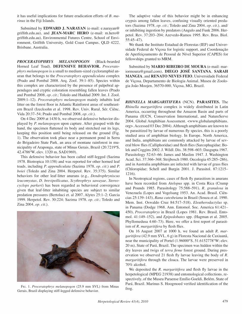
Herpetological Review 41(4), 2010 479
it has useful implications for future eradication efforts of B. ma-rinus in the Fiji Islands.
Submitted by EDWARD J. NARAYAN (e-mail: [email protected]), and JEAN-MARC HERO (e-mail: [email protected]), Environmental Futures Centre, School of Envi-ronment, Griffith University, Gold Coast Campus, QLD 4222, Brisbane, Australia.
PROCERATOPHRYS MELANOPOGON (Black-bearded Horned Leaf Toad). DEFENSIVE BEHAVIOR. Procerato-phyrs melanopogon is a small to medium-sized cycloramphid an-uran that belongs to the Proceratophyrs appendiculata complex (Prado and Pombal 2008. Arq. Zool. 39:1–85). Species within this complex are characterized by the presence of palpebral ap-pendages and cryptic coloration resembling fallen leaves (Prado and Pombal 2008, op. cit.; Toledo and Haddad 2009. Int. J. Zool. 2009:1–12). Proceratophyrs melanopogon mainly inhabits leaf litter on the forest floor in Atlantic Rainforest areas of southeast-ern Brazil (Izecksohn et al. 1998. Rev. Univ. Rural, Sér. Ciênc. Vida 20:37–54; Prado and Pombal 2008, op. cit.). On 4 Dec 2009 at 1430 h, we observed defensive behavior dis-played by P. melanopogon upon capture. After grasped with the hand, the specimen flattened its body and stretched out its legs, keeping this position until being released on the ground (Fig. 1). The observation took place near a permanent pond in Serra do Brigadeiro State Park, an area of montane rainforest in mu-nicipality of Araponga, state of Minas Gerais, Brazil (20.7219ºS, 42.4786ºW, elev. 1320 m, SAD1969). This defensive behavior has been called stiff-legged (Sazima 1978. Biotropica 10:158) and was reported for other horned leaf toads, including P. appendiculata (Sazima 1978, op. cit.) and P. boiei (Toledo and Zina 2004. Herpetol. Rev. 35:375). Similar behaviors for other leaf-litter anurans (e.g., Dendrophryniscus leucomystax, D. brevipollicatus, Scythrophrys sawayae, Stereo-cyclops parkeri) has been regarded as behavioral convergence given that leaf-litter inhabiting species are subject to similar predation pressures (Bertoluci et. al 2007. Alytes 25:1–2; Garcia 1999. Herpetol. Rev. 30:224; Sazima 1978, op. cit.; Toledo and Zina 2004, op. cit.).
The adaptive value of this behavior might be in enhancing crypsis among fallen leaves, confusing visually oriented preda-tors (Sazima 1978, op. cit.; Toledo and Zina 2004, op. cit.), and/or inhibiting ingestion by predators (Angulo and Funk 2006. Her-petol. Rev. 37:203–204; Azevedo-Ramos 1995. Rev. Bras. Biol. 55:45–47). We thank the Instituto Estadual de Florestas (IEF) and Univer-sidade Federal de Viçosa for logistic support, and Coordenação de Aperfeiçoamento de Pessoal de Nível Superior (CAPES) for fellowships granted to MRM.
Submitted by MARIO RIBEIRO DE MOURA (e-mail: [email protected]), DIEGO JOSÉ SANTANA, SARAH MANGIA, and RENATO NEVES FEIO, Universidade Federal de Viçosa, Departamento de Biologia Animal, Museu de Zoolo-gia João Moojen, 36570-000, Viçosa, MG, Brazil.
RHINELLA MARGARITIFERA (NCN). PARASITES. The Rhinella margaritifera complex is widely distributed in Latin America, occurring throughout the Amazon Basin and parts of Panama (IUCN, Conservation International, and NatureServe 2004. Global Amphibian Assessment. <www.globalamphibians.org>. Accessed 03 Dec 2004). Although amphibians are known to be parasitized by larvae of numerous fly species, this is a poorly studied area of amphibian biology. In Europe, North America, and India, amphibians are commonly attacked by larvae of sev-eral blow flies (Calliphoridae) and flesh flies (Sarcophagidae; Bo-lek and Coggins 2002. J. Wildl. Dis. 38:598–603; Dasgupta 1967. Parasitology 52:63–66; James and Maslim 1947. J. Washington Acad. Sci. 37:366–368; Strijbosch 1980. Oecologia 45:285–286), and in Australia amphibians are infected with larvae of grass flies (Chloropidae; Schell and Burgin 2001. J. Parasitol. 87:1215–1216). In Neotropical regions, cases of flesh fly parasitism in anurans have been recorded from Atelopus spp. in Costa Rica (Crump and Pounds 1985. Parasitology 75:588–591), R. granulosa in Venezuela (Lopes and Vogelsang 1953. An. Acad. Brasil. Ciên-cias 25:139–143), Rana catesbeiana in Brazil (Souza et al. 1990. Mem. Inst. Oswaldo Cruz 84:517–518), Eleutherodactylus sp. in Panama (Dodge 1968. Ann. Entomol. Soc. America 61:421–450), Proceratophrys in Brazil (Lopes 1981. Rev. Brasil. Ento-mol. 41:149–152), and Epipedobates spp. (Hagman et al. 2005. Phyllomedusa 4:60–73). Here, we offer a first report of parasit-ism of R. margaritifera by flesh flies. On 16 August 2007 at 1000 h, we found an adult R. mar-garitifera (42.9 mm SVL, 6 g) in Floresta Nacional de Caxiuanã, near the municipality of Portel (1.96000°S, 51.6152778°W; elev. 20 m), State of Pará, Brazil. The specimen was hidden within the dry leaves and twigs of terra firme forest ground. During pres-ervation we observed 21 flesh fly larvae leaving the body of R. margaritifera through the cloaca. The larvae were preserved in 70% alcohol. We deposited the R. margaritifera and flesh fly larvae in the herpetological (MPEG 21938) and entomological collections, re-spectively, of the Museu Paraense Emílio Goeldi, Belém, State of Pará, Brazil. Marinus S. Hoogmoed verified identification of the frog. fig. 1. Proceratophrys melanopogon (25.9 mm SVL) from Minas
Gerais, Brazil displaying stiff-legged defensive behavior.
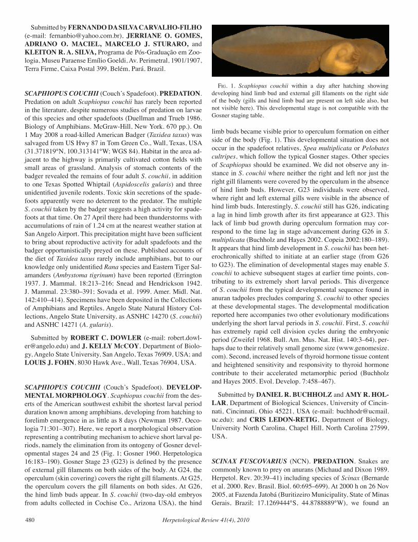
Herpetological Review 41(4), 2010480
Submitted by FERNANDO DA SILVA CARVALHO-FILHO (e-mail: [email protected]), JERRIANE O. GOMES, ADRIANO O. MACIEL, MARCELO J. STURARO, and KLEITON R. A. SILVA, Programa de Pós-Graduação em Zoo-logia, Museu Paraense Emílio Goeldi, Av. Perimetral, 1901/1907, Terra Firme, Caixa Postal 399, Belém, Pará, Brazil.
SCAPHIOPUS COUCHII (Couch’s Spadefoot). PREDATION. Predation on adult Scaphiopus couchii has rarely been reported in the literature, despite numerous studies of predation on larvae of this species and other spadefoots (Duellman and Trueb 1986. Biology of Amphibians. McGraw-Hill, New York. 670 pp.). On 1 May 2008 a road-killed American Badger (Taxidea taxus) was salvaged from US Hwy 87 in Tom Green Co., Wall, Texas, USA (31.371819°N, 100.313141°W; WGS 84). Habitat in the area ad-jacent to the highway is primarily cultivated cotton fields with small areas of grassland. Analysis of stomach contents of the badger revealed the remains of four adult S. couchii, in addition to one Texas Spotted Whiptail (Aspidoscelis gularis) and three unidentified juvenile rodents. Toxic skin secretions of the spade-foots apparently were no deterrent to the predator. The multiple S. couchii taken by the badger suggests a high activity for spade-foots at that time. On 27 April there had been thunderstorms with accumulations of rain of 1.24 cm at the nearest weather station at San Angelo Airport. This precipitation might have been sufficient to bring about reproductive activity for adult spadefoots and the badger opportunistically preyed on these. Published accounts of the diet of Taxidea taxus rarely include amphibians, but to our knowledge only unidentified Rana species and Eastern Tiger Sal-amanders (Ambystoma tigrinum) have been reported (Errington 1937. J. Mammal. 18:213–216; Snead and Hendrickson 1942. J. Mammal. 23:380–391; Sovada et al. 1999. Amer. Midl. Nat. 142:410–414). Specimens have been deposited in the Collections of Amphibians and Reptiles, Angelo State Natural History Col-lections, Angelo State University, as ASNHC 14270 (S. couchii) and ASNHC 14271 (A. gularis).
Submitted by ROBERT C. DOWLER (e-mail: [email protected]) and J. KELLY McCOY, Department of Biolo-gy, Angelo State University, San Angelo, Texas 76909, USA; and LOUIS J. FOHN, 8030 Hawk Ave., Wall, Texas 76904, USA.
SCAPHIOPUS COUCHII (Couch’s Spadefoot). DEVELOP-MENTAL MORPHOLOGY. Scaphiopus couchii from the des-erts of the American southwest exhibit the shortest larval period duration known among amphibians, developing from hatching to forelimb emergence in as little as 8 days (Newman 1987. Oeco-logia 71:301–307). Here, we report a morphological observation representing a contributing mechanism to achieve short larval pe-riods, namely the elimination from its ontogeny of Gosner devel-opmental stages 24 and 25 (Fig. 1; Gosner 1960. Herpetologica 16:183–190). Gosner Stage 23 (G23) is defined by the presence of external gill filaments on both sides of the body. At G24, the operculum (skin covering) covers the right gill filaments. At G25, the operculum covers the gill filaments on both sides. At G26, the hind limb buds appear. In S. couchii (two-day-old embryos from adults collected in Cochise Co., Arizona USA), the hind
limb buds became visible prior to operculum formation on either side of the body (Fig. 1). This developmental situation does not occur in the spadefoot relatives, Spea multiplicata or Pelobates cultripes, which follow the typical Gosner stages. Other species of Scaphiopus should be examined. We did not observe any in-stance in S. couchii where neither the right and left nor just the right gill filaments were covered by the operculum in the absence of hind limb buds. However, G23 individuals were observed, where right and left external gills were visible in the absence of hind limb buds. Interestingly, S. couchii still has G26, indicating a lag in hind limb growth after its first appearance at G23. This lack of limb bud growth during operculum formation may cor-respond to the time lag in stage advancement during G26 in S. multiplicata (Buchholz and Hayes 2002. Copeia 2002:180–189). It appears that hind limb development in S. couchii has been het-erochronically shifted to initiate at an earlier stage (from G26 to G23). The elimination of developmental stages may enable S. couchii to achieve subsequent stages at earlier time points, con-tributing to its extremely short larval periods. This divergence of S. couchii from the typical developmental sequence found in anuran tadpoles precludes comparing S. couchii to other species at these developmental stages. The developmental modification reported here accompanies two other evolutionary modifications underlying the short larval periods in S. couchii. First, S. couchii has extremely rapid cell division cycles during the embryonic period (Zweifel 1968. Bull. Am. Mus. Nat. Hist. 140:3–64), per-haps due to their relatively small genome size (www.genomesize.com). Second, increased levels of thyroid hormone tissue content and heightened sensitivity and responsivity to thyroid hormone contribute to their accelerated metamorphic period (Buchholz and Hayes 2005. Evol. Develop. 7:458–467).
Submitted by DANIEL R. BUCHHOLZ and AMY R. HOL-LAR, Department of Biological Sciences, University of Cincin-nati, Cincinnati, Ohio 45221, USA (e-mail: [email protected]); and CRIS LEDON-RETIG, Department of Biology, University North Carolina, Chapel Hill, North Carolina 27599, USA.
SCINAX FUSCOvARIUS (NCN). PREDATION. Snakes are commonly known to prey on anurans (Michaud and Dixon 1989. Herpetol. Rev. 20:39–41) including species of Scinax (Bernarde et al. 2000. Rev. Brasil. Biol. 60:695–699). At 2000 h on 26 Nov 2005, at Fazenda Jatobá (Buritizeiro Municipality, State of Minas Gerais, Brazil; 17.1269444°S, 44.8788889°W), we found an
fig. 1. Scaphiopus couchii within a day after hatching showing developing hind limb bud and external gill filaments on the right side of the body (gills and hind limb bud are present on left side also, but not visible here). This developmental stage is not compatible with the Gosner staging table.
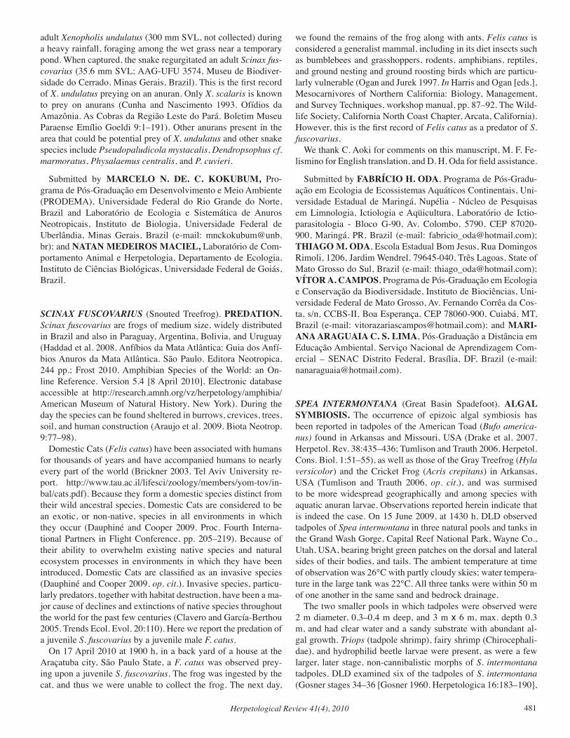
Herpetological Review 41(4), 2010 481
adult Xenopholis undulatus (300 mm SVL, not collected) during a heavy rainfall, foraging among the wet grass near a temporary pond. When captured, the snake regurgitated an adult Scinax fus-covarius (35.6 mm SVL; AAG-UFU 3574, Museu de Biodiver-sidade do Cerrado, Minas Gerais, Brazil). This is the first record of X. undulatus preying on an anuran. Only X. scalaris is known to prey on anurans (Cunha and Nascimento 1993. Ofídios da Amazônia. As Cobras da Região Leste do Pará. Boletim Museu Paraense Emílio Goeldi 9:1–191). Other anurans present in the area that could be potential prey of X. undulatus and other snake species include Pseudopaludicola mystacalis, Dendropsophus cf. marmoratus, Physalaemus centralis, and P. cuvieri.
Submitted by MARCELO N. DE. C. KOKUBUM, Pro-grama de Pós-Graduação em Desenvolvimento e Meio Ambiente (PRODEMA), Universidade Federal do Rio Grande do Norte, Brazil and Laboratório de Ecologia e Sistemática de Anuros Neotropicais, Instituto de Biologia, Universidade Federal de Uberlândia, Minas Gerais, Brazil (e-mail: [email protected]); and NATAN MEDEIROS MACIEL, Laboratório de Com-portamento Animal e Herpetologia, Departamento de Ecologia, Instituto de Ciências Biológicas, Universidade Federal de Goiás, Brazil.
SCINAX FUSCOvARIUS (Snouted Treefrog). PREDATION. Scinax fuscovarius are frogs of medium size, widely distributed in Brazil and also in Paraguay, Argentina, Bolivia, and Uruguay (Haddad et al. 2008. Anfíbios da Mata Atlântica: Guia dos Anfí-bios Anuros da Mata Atlântica. São Paulo. Editora Neotropica. 244 pp.; Frost 2010. Amphibian Species of the World: an On-line Reference. Version 5.4 [8 April 2010]. Electronic database accessible at http://research.amnh.org/vz/herpetology/amphibia/ American Museum of Natural History, New York). During the day the species can be found sheltered in burrows, crevices, trees, soil, and human construction (Araujo et al. 2009. Biota Neotrop. 9:77–98). Domestic Cats (Felis catus) have been associated with humans for thousands of years and have accompanied humans to nearly every part of the world (Brickner 2003. Tel Aviv University re-port. http://www.tau.ac.il/lifesci/zoology/members/yom-tov/in-bal/cats.pdf). Because they form a domestic species distinct from their wild ancestral species, Domestic Cats are considered to be an exotic, or non-native, species in all environments in which they occur (Dauphiné and Cooper 2009. Proc. Fourth Interna-tional Partners in Flight Conference, pp. 205–219). Because of their ability to overwhelm existing native species and natural ecosystem processes in environments in which they have been introduced, Domestic Cats are classified as an invasive species (Dauphiné and Cooper 2009, op. cit.). Invasive species, particu-larly predators, together with habitat destruction, have been a ma-jor cause of declines and extinctions of native species throughout the world for the past few centuries (Clavero and García-Berthou 2005. Trends Ecol. Evol. 20:110). Here we report the predation of a juvenile S. fuscovarius by a juvenile male F. catus. On 17 April 2010 at 1900 h, in a back yard of a house at the Araçatuba city, São Paulo State, a F. catus was observed prey-ing upon a juvenile S. fuscovarius. The frog was ingested by the cat, and thus we were unable to collect the frog. The next day,
we found the remains of the frog along with ants. Felis catus is considered a generalist mammal, including in its diet insects such as bumblebees and grasshoppers, rodents, amphibians, reptiles, and ground nesting and ground roosting birds which are particu-larly vulnerable (Ogan and Jurek 1997. In Harris and Ogan [eds.], Mesocarnivores of Northern California: Biology, Management, and Survey Techniques, workshop manual, pp. 87–92. The Wild-life Society, California North Coast Chapter, Arcata, California). However, this is the first record of Felis catus as a predator of S. fuscovarius. We thank C. Aoki for comments on this manuscript, M. F. Fe-lismino for English translation, and D. H. Oda for field assistance.
Submitted by FABRÍCIO H. ODA, Programa de Pós-Gradu-ação em Ecologia de Ecossistemas Aquáticos Continentais, Uni-versidade Estadual de Maringá, Nupélia - Núcleo de Pesquisas em Limnologia, Ictiologia e Aqüicultura, Laboratório de Ictio-parasitologia - Bloco G-90, Av. Colombo, 5790, CEP 87020-900, Maringá, PR, Brazil (e-mail: [email protected]); THIAGO M. ODA, Escola Estadual Bom Jesus, Rua Domingos Rimoli, 1206, Jardim Wendrel, 79645-040, Três Lagoas, State of Mato Grosso do Sul, Brazil (e-mail: [email protected]); VÍTOR A. CAMPOS, Programa de Pós-Graduação em Ecologia e Conservação da Biodiversidade, Instituto de Biociências, Uni-versidade Federal de Mato Grosso, Av. Fernando Corrêa da Cos-ta, s/n, CCBS-II, Boa Esperança, CEP 78060-900, Cuiabá, MT, Brazil (e-mail: [email protected]); and MARI-ANA ARAGUAIA C. S. LIMA, Pós-Graduação a Distância em Educação Ambiental. Serviço Nacional de Aprendizagem Com-ercial – SENAC Distrito Federal, Brasília, DF, Brazil (e-mail: [email protected]).
SPEA INTERMONTANA (Great Basin Spadefoot). ALGAL SYMBIOSIS. The occurrence of epizoic algal symbiosis has been reported in tadpoles of the American Toad (Bufo america-nus) found in Arkansas and Missouri, USA (Drake et al. 2007. Herpetol. Rev. 38:435–436; Tumlison and Trauth 2006. Herpetol. Cons. Biol. 1:51–55), as well as those of the Gray Treefrog (Hyla versicolor) and the Cricket Frog (Acris crepitans) in Arkansas, USA (Tumlison and Trauth 2006, op. cit.), and was surmised to be more widespread geographically and among species with aquatic anuran larvae. Observations reported herein indicate that is indeed the case. On 15 June 2009, at 1430 h, DLD observed tadpoles of Spea intermontana in three natural pools and tanks in the Grand Wash Gorge, Capital Reef National Park, Wayne Co., Utah, USA, bearing bright green patches on the dorsal and lateral sides of their bodies, and tails. The ambient temperature at time of observation was 26°C with partly cloudy skies; water tempera-ture in the large tank was 22°C. All three tanks were within 50 m of one another in the same sand and bedrock drainage. The two smaller pools in which tadpoles were observed were 2 m diameter, 0.3–0.4 m deep, and 3 m x 6 m, max. depth 0.3 m, and had clear water and a sandy substrate with abundant al-gal growth. Triops (tadpole shrimp), fairy shrimp (Chirocephali-dae), and hydrophilid beetle larvae were present, as were a few larger, later stage, non-cannibalistic morphs of S. intermontana tadpoles. DLD examined six of the tadpoles of S. intermontana (Gosner stages 34–36 [Gosner 1960. Herpetologica 16:183–190],
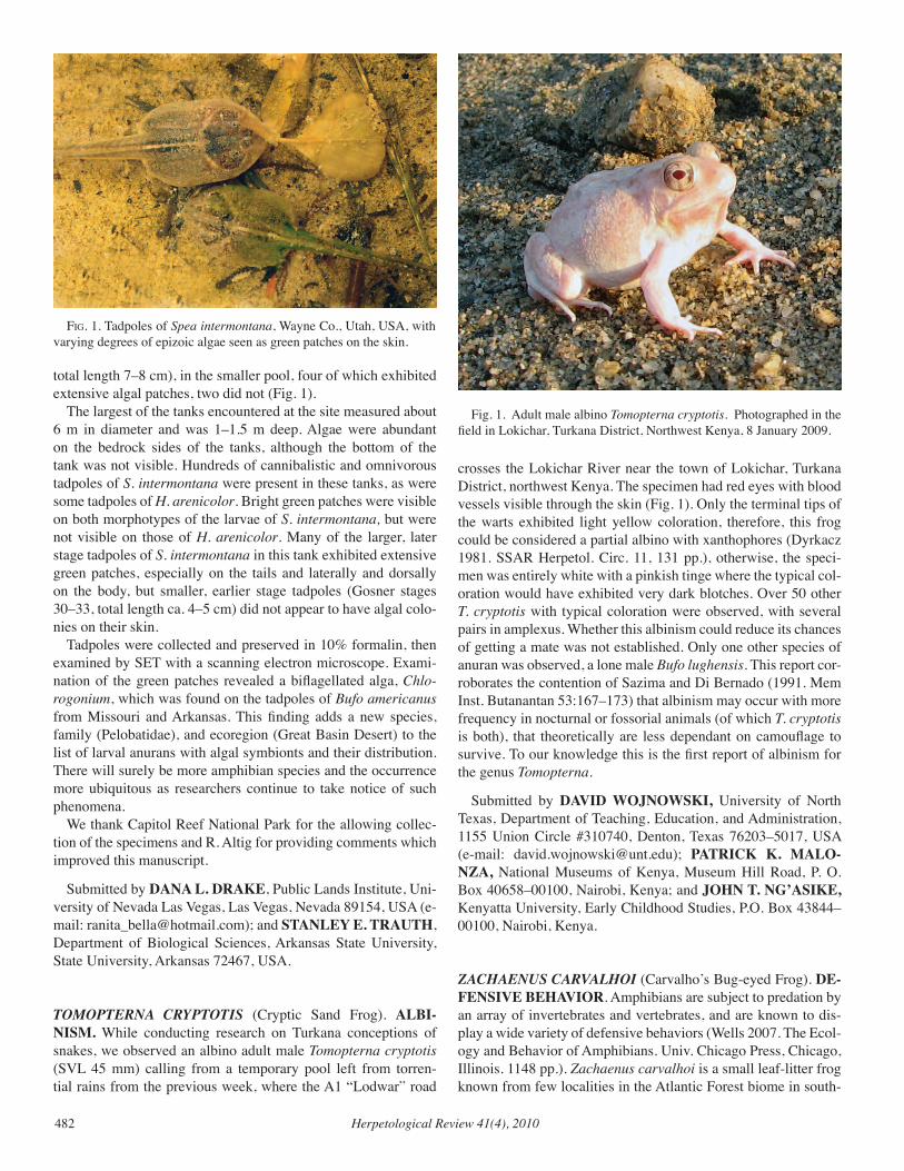
Herpetological Review 41(4), 2010482
total length 7–8 cm), in the smaller pool, four of which exhibited extensive algal patches, two did not (Fig. 1). The largest of the tanks encountered at the site measured about 6 m in diameter and was 1–1.5 m deep. Algae were abundant on the bedrock sides of the tanks, although the bottom of the tank was not visible. Hundreds of cannibalistic and omnivorous tadpoles of S. intermontana were present in these tanks, as were some tadpoles of H. arenicolor. Bright green patches were visible on both morphotypes of the larvae of S. intermontana, but were not visible on those of H. arenicolor. Many of the larger, later stage tadpoles of S. intermontana in this tank exhibited extensive green patches, especially on the tails and laterally and dorsally on the body, but smaller, earlier stage tadpoles (Gosner stages 30–33, total length ca. 4–5 cm) did not appear to have algal colo-nies on their skin. Tadpoles were collected and preserved in 10% formalin, then examined by SET with a scanning electron microscope. Exami-nation of the green patches revealed a biflagellated alga, Chlo-rogonium, which was found on the tadpoles of Bufo americanus from Missouri and Arkansas. This finding adds a new species, family (Pelobatidae), and ecoregion (Great Basin Desert) to the list of larval anurans with algal symbionts and their distribution. There will surely be more amphibian species and the occurrence more ubiquitous as researchers continue to take notice of such phenomena. We thank Capitol Reef National Park for the allowing collec-tion of the specimens and R. Altig for providing comments which improved this manuscript.
Submitted by DANA L. DRAKE, Public Lands Institute, Uni-versity of Nevada Las Vegas, Las Vegas, Nevada 89154, USA (e-mail: [email protected]); and STANLEY E. TRAUTH, Department of Biological Sciences, Arkansas State University, State University, Arkansas 72467, USA.
TOMOPTERNA CRYPTOTIS (Cryptic Sand Frog). ALBI-NISM. While conducting research on Turkana conceptions of snakes, we observed an albino adult male Tomopterna cryptotis (SVL 45 mm) calling from a temporary pool left from torren-tial rains from the previous week, where the A1 “Lodwar” road
crosses the Lokichar River near the town of Lokichar, Turkana District, northwest Kenya. The specimen had red eyes with blood vessels visible through the skin (Fig. 1). Only the terminal tips of the warts exhibited light yellow coloration, therefore, this frog could be considered a partial albino with xanthophores (Dyrkacz 1981. SSAR Herpetol. Circ. 11, 131 pp.), otherwise, the speci-men was entirely white with a pinkish tinge where the typical col-oration would have exhibited very dark blotches. Over 50 other T. cryptotis with typical coloration were observed, with several pairs in amplexus. Whether this albinism could reduce its chances of getting a mate was not established. Only one other species of anuran was observed, a lone male Bufo lughensis. This report cor-roborates the contention of Sazima and Di Bernado (1991. Mem Inst. Butanantan 53:167–173) that albinism may occur with more frequency in nocturnal or fossorial animals (of which T. cryptotis is both), that theoretically are less dependant on camouflage to survive. To our knowledge this is the first report of albinism for the genus Tomopterna.
Submitted by DAVID WOJNOWSKI, University of North Texas, Department of Teaching, Education, and Administration, 1155 Union Circle #310740, Denton, Texas 76203–5017, USA (e-mail: [email protected]); PATRICK K. MALO-NZA, National Museums of Kenya, Museum Hill Road, P. O. Box 40658–00100, Nairobi, Kenya; and JOHN T. NG’ASIKE, Kenyatta University, Early Childhood Studies, P.O. Box 43844–00100, Nairobi, Kenya.
ZACHAENUS CARvALHOI (Carvalho’s Bug-eyed Frog). DE-FENSIVE BEHAVIOR. Amphibians are subject to predation by an array of invertebrates and vertebrates, and are known to dis-play a wide variety of defensive behaviors (Wells 2007. The Ecol-ogy and Behavior of Amphibians. Univ. Chicago Press, Chicago, Illinois. 1148 pp.). Zachaenus carvalhoi is a small leaf-litter frog known from few localities in the Atlantic Forest biome in south-
Fig. 1. Adult male albino Tomopterna cryptotis. Photographed in the field in Lokichar, Turkana District, Northwest Kenya, 8 January 2009.
fig. 1. Tadpoles of Spea intermontana, Wayne Co., Utah, USA, with varying degrees of epizoic algae seen as green patches on the skin.
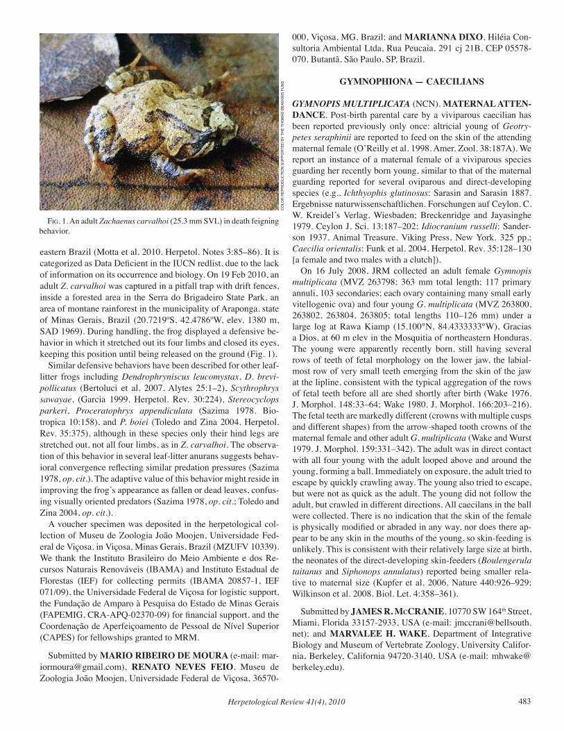
Herpetological Review 41(4), 2010 483
eastern Brazil (Motta et al. 2010. Herpetol. Notes 3:85–86). It is categorized as Data Deficient in the IUCN redlist, due to the lack of information on its occurrence and biology. On 19 Feb 2010, an adult Z. carvalhoi was captured in a pitfall trap with drift fences, inside a forested area in the Serra do Brigadeiro State Park, an area of montane rainforest in the municipality of Araponga, state of Minas Gerais, Brazil (20.7219ºS, 42.4786ºW, elev. 1380 m, SAD 1969). During handling, the frog displayed a defensive be-havior in which it stretched out its four limbs and closed its eyes, keeping this position until being released on the ground (Fig. 1). Similar defensive behaviors have been described for other leaf-litter frogs including Dendrophryniscus leucomystax, D. brevi-pollicatus (Bertoluci et al. 2007. Alytes 25:1–2), Scythrophrys sawayae, (Garcia 1999. Herpetol. Rev. 30:224), Stereocyclops parkeri, Proceratophrys appendiculata (Sazima 1978. Bio-tropica 10:158), and P. boiei (Toledo and Zina 2004. Herpetol. Rev. 35:375), although in these species only their hind legs are stretched out, not all four limbs, as in Z. carvalhoi. The observa-tion of this behavior in several leaf-litter anurans suggests behav-ioral convergence reflecting similar predation pressures (Sazima 1978, op. cit.). The adaptive value of this behavior might reside in improving the frog’s appearance as fallen or dead leaves, confus-ing visually oriented predators (Sazima 1978, op. cit.; Toledo and Zina 2004, op. cit.). A voucher specimen was deposited in the herpetological col-lection of Museu de Zoologia João Moojen, Universidade Fed-eral de Viçosa, in Viçosa, Minas Gerais, Brazil (MZUFV 10339). We thank the Instituto Brasileiro do Meio Ambiente e dos Re-cursos Naturais Renováveis (IBAMA) and Instituto Estadual de Florestas (IEF) for collecting permits (IBAMA 20857-1, IEF 071/09), the Universidade Federal de Viçosa for logistic support, the Fundação de Amparo à Pesquisa do Estado de Minas Gerais (FAPEMIG, CRA-APQ-02370-09) for financial support, and the Coordenação de Aperfeiçoamento de Pessoal de Nível Superior (CAPES) for fellowships granted to MRM.
Submitted by MARIO RIBEIRO DE MOURA (e-mail: [email protected]), RENATO NEVES FEIO, Museu de Zoologia João Moojen, Universidade Federal de Viçosa, 36570-
000, Viçosa, MG, Brazil; and MARIANNA DIXO, Hiléia Con-sultoria Ambiental Ltda, Rua Peucaia, 291 cj 21B, CEP 05578-070, Butantã, São Paulo, SP, Brazil.
GYMNOPHIONA — CAECILIANS
GYMNOPIS MULTIPLICATA (NCN). MATERNAL ATTEN-DANCE. Post-birth parental care by a viviparous caecilian has been reported previously only once: altricial young of Geotry-petes seraphinii are reported to feed on the skin of the attending maternal female (O’Reilly et al. 1998. Amer. Zool. 38:187A). We report an instance of a maternal female of a viviparous species guarding her recently born young, similar to that of the maternal guarding reported for several oviparous and direct-developing species (e.g., Ichthyophis glutinosus: Sarasin and Sarasin 1887. Ergebnisse naturwissenschaftlichen. Forschungen auf Ceylon. C. W. Kreidel’s Verlag, Wiesbaden; Breckenridge and Jayasinghe 1979. Ceylon J. Sci. 13:187–202; Idiocranium russelli: Sander-son 1937. Animal Treasure. Viking Press, New York. 325 pp.; Caecilia orientalis: Funk et al. 2004. Herpetol. Rev. 35:128–130 [a female and two males with a clutch]). On 16 July 2008, JRM collected an adult female Gymnopis multiplicata (MVZ 263798; 363 mm total length; 117 primary annuli, 103 secondaries; each ovary containing many small early vitellogenic ova) and four young G. multiplicata (MVZ 263800, 263802, 263804, 263805; total lengths 110–126 mm) under a large log at Rawa Kiamp (15.100°N, 84.4333333°W), Gracias a Dios, at 60 m elev in the Mosquitia of northeastern Honduras. The young were apparently recently born, still having several rows of teeth of fetal morphology on the lower jaw, the labial-most row of very small teeth emerging from the skin of the jaw at the lipline, consistent with the typical aggregation of the rows of fetal teeth before all are shed shortly after birth (Wake 1976. J. Morphol. 148:33–64; Wake 1980. J. Morphol. 166:203–216). The fetal teeth are markedly different (crowns with multiple cusps and different shapes) from the arrow-shaped tooth crowns of the maternal female and other adult G. multiplicata (Wake and Wurst 1979. J. Morphol. 159:331–342). The adult was in direct contact with all four young with the adult looped above and around the young, forming a ball. Immediately on exposure, the adult tried to escape by quickly crawling away. The young also tried to escape, but were not as quick as the adult. The young did not follow the adult, but crawled in different directions. All caecilans in the ball were collected. There is no indication that the skin of the female is physically modified or abraded in any way, nor does there ap-pear to be any skin in the mouths of the young, so skin-feeding is unlikely. This is consistent with their relatively large size at birth, the neonates of the direct-developing skin-feeders (Boulengerula taitanus and Siphonops annulatus) reported being smaller rela-tive to maternal size (Kupfer et al. 2006. Nature 440:926–929; Wilkinson et al. 2008. Biol. Let. 4:358–361).
Submitted by JAMES R. McCRANIE, 10770 SW 164th Street, Miami, Florida 33157-2933, USA (e-mail: [email protected]); and MARVALEE H. WAKE, Department of Integrative Biology and Museum of Vertebrate Zoology, University Califor-nia, Berkeley, California 94720-3140, USA (e-mail: [email protected]).
fig. 1. An adult Zachaenus carvalhoi (25.3 mm SVL) in death feigning behavior.
CO
LOR
REP
RO
du
CTi
ON
Su
PPO
RTEd
BY
THE
THO
mAS
BEA
uvA
iS F
uN
d
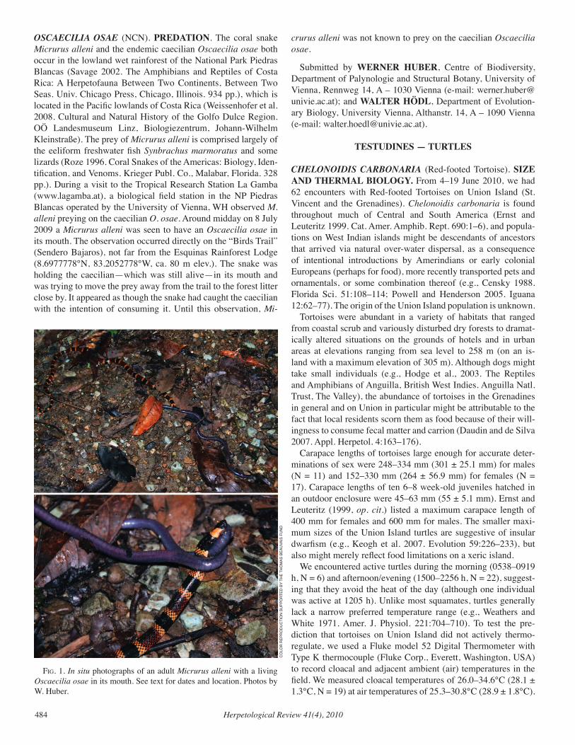
Herpetological Review 41(4), 2010484
OSCAECILIA OSAE (NCN). PREDATION. The coral snake Micrurus alleni and the endemic caecilian Oscaecilia osae both occur in the lowland wet rainforest of the National Park Piedras Blancas (Savage 2002. The Amphibians and Reptiles of Costa Rica: A Herpetofauna Between Two Continents, Between Two Seas. Univ. Chicago Press, Chicago, Illinois. 934 pp.), which is located in the Pacific lowlands of Costa Rica (Weissenhofer et al. 2008. Cultural and Natural History of the Golfo Dulce Region. OÖ Landesmuseum Linz, Biologiezentrum, Johann-Wilhelm Kleinstraße). The prey of Micrurus alleni is comprised largely of the eeliform freshwater fish Synbrachus marmoratus and some lizards (Roze 1996. Coral Snakes of the Americas: Biology, Iden-tification, and Venoms. Krieger Publ. Co., Malabar, Florida. 328 pp.). During a visit to the Tropical Research Station La Gamba (www.lagamba.at), a biological field station in the NP Piedras Blancas operated by the University of Vienna, WH observed M. alleni preying on the caecilian O. osae. Around midday on 8 July 2009 a Micrurus alleni was seen to have an Oscaecilia osae in its mouth. The observation occurred directly on the “Birds Trail” (Sendero Bajaros), not far from the Esquinas Rainforest Lodge (8.6977778°N, 83.2052778°W, ca. 80 m elev.). The snake was holding the caecilian—which was still alive—in its mouth and was trying to move the prey away from the trail to the forest litter close by. It appeared as though the snake had caught the caecilian with the intention of consuming it. Until this observation, Mi-
crurus alleni was not known to prey on the caecilian Oscaecilia osae.
Submitted by WERNER HUBER, Centre of Biodiversity, Department of Palynologie and Structural Botany, University of Vienna, Rennweg 14, A – 1030 Vienna (e-mail: [email protected]); and WALTER HÖDL, Department of Evolution-ary Biology, University Vienna, Althanstr. 14, A – 1090 Vienna (e-mail: [email protected]).
TESTUDINES — TURTLES
CHELONOIDIS CARBONARIA (Red-footed Tortoise). SIZE AND THERMAL BIOLOGY. From 4–19 June 2010, we had 62 encounters with Red-footed Tortoises on Union Island (St. Vincent and the Grenadines). Chelonoidis carbonaria is found throughout much of Central and South America (Ernst and Leuteritz 1999. Cat. Amer. Amphib. Rept. 690:1–6), and popula-tions on West Indian islands might be descendants of ancestors that arrived via natural over-water dispersal, as a consequence of intentional introductions by Amerindians or early colonial Europeans (perhaps for food), more recently transported pets and ornamentals, or some combination thereof (e.g., Censky 1988. Florida Sci. 51:108–114; Powell and Henderson 2005. Iguana 12:62–77). The origin of the Union Island population is unknown. Tortoises were abundant in a variety of habitats that ranged from coastal scrub and variously disturbed dry forests to dramat-ically altered situations on the grounds of hotels and in urban areas at elevations ranging from sea level to 258 m (on an is-land with a maximum elevation of 305 m). Although dogs might take small individuals (e.g., Hodge et al., 2003. The Reptiles and Amphibians of Anguilla, British West Indies. Anguilla Natl. Trust, The Valley), the abundance of tortoises in the Grenadines in general and on Union in particular might be attributable to the fact that local residents scorn them as food because of their will-ingness to consume fecal matter and carrion (Daudin and de Silva 2007. Appl. Herpetol. 4:163–176). Carapace lengths of tortoises large enough for accurate deter-minations of sex were 248–334 mm (301 ± 25.1 mm) for males (N = 11) and 152–330 mm (264 ± 56.9 mm) for females (N = 17). Carapace lengths of ten 6–8 week-old juveniles hatched in an outdoor enclosure were 45–63 mm (55 ± 5.1 mm). Ernst and Leuteritz (1999, op. cit.) listed a maximum carapace length of 400 mm for females and 600 mm for males. The smaller maxi-mum sizes of the Union Island turtles are suggestive of insular dwarfism (e.g., Keogh et al. 2007. Evolution 59:226–233), but also might merely reflect food limitations on a xeric island. We encountered active turtles during the morning (0538–0919 h, N = 6) and afternoon/evening (1500–2256 h, N = 22), suggest-ing that they avoid the heat of the day (although one individual was active at 1205 h). Unlike most squamates, turtles generally lack a narrow preferred temperature range (e.g., Weathers and White 1971. Amer. J. Physiol. 221:704–710). To test the pre-diction that tortoises on Union Island did not actively thermo-regulate, we used a Fluke model 52 Digital Thermometer with Type K thermocouple (Fluke Corp., Everett, Washington, USA) to record cloacal and adjacent ambient (air) temperatures in the field. We measured cloacal temperatures of 26.0–34.6°C (28.1 ± 1.3°C, N = 19) at air temperatures of 25.3–30.8°C (28.9 ± 1.8°C).
fig. 1. In situ photographs of an adult Micrurus alleni with a living Oscaecilia osae in its mouth. See text for dates and location. Photos by W. Huber.
CO
LOR
REP
RO
du
CTi
ON
Su
PPO
RTEd
BY
THE
THO
mAS
BEA
uvA
iS F
uN
d

Herpetological Review 41(4), 2010 485
Differences between ambient and cloacal temperatures were -7.0–1.5°C (-0.78 ± 1.44°C), with 30 of 41 records above and two at ambient temperatures. We also placed nine individuals (carapace length 45–172 mm, 88 ± 56 mm) in a thermal gradient (90 x 30 cm) with extreme tem-peratures of 24° and 40°C. Although most individuals spent more time near the cooler end of the gradient, possibly because they heat faster than they cool under laboratory conditions (Weathers and White, op. cit.), cloacal temperatures after 2 h were 23.9–31.6°C (26.5 ± 2.7°C). The considerable variation in individual temperatures in both the field and the gradient was suggestive of little more than an avoidance of extremely high temperatures. Permits to conduct research on Union Island were issued by Mr. Brian Johnson, Director, Department of Forestry, St. Vincent and the Grenadines. Protocols were approved by the Avila University Animal Care and Use Committee. Fieldwork was funded by a grant from the National Science Foundation (USA) to Robert Powell (DBI-0851610). All tortoises were released at original sites of capture.
Submitted by HAYDEN D. HEDMAN, Department of Ecology and Evolutionary Biology, University of Colorado, Boulder 80301, USA (e-mail: [email protected]); DEBORAH N. MUÑIZ PAGAN, Department of Biology University of Puerto Rico, Mayagüez, Puerto Rico 00681 (e-mail: [email protected]); and ROBERT POWELL, Department of Biology, Avila University, Kansas City, Missouri 64145, USA (e-mail: [email protected]).
GOPHERUS POLYPHEMUS (Gopher Tortoise). DIET. Consuming scat of conspecifics and other animals may provide species of Gopherus with symbiotic gut microbes and/or direct nutritional benefits (Bjorndal 1987. Copeia 1987:714–720; Lance and Morafka 2001. Herpetol. Monogr. 15:124–134; Walde et al. 2006. Herpetol. Rev. 37:77–78). In addition to scat of con-specifics, G. polyphemus occasionally eats fox (Vulpes vulpes or Urocyon cinereoargenteus), raccoon (Procyon lotor), and rabbit (Sylvilagus spp.) feces (Anderson and Herrington 1992. Herpetol. Rev. 23:59; Garner and Landers 1981. Proc. Ann. Conf. S.E. Assoc. Fish & Wildl. Agencies 35:120–134; MacDonald and Mushinsky 1988. Herpetologica 44:345–353). Here, we report a juvenile G. polyphemus consuming deer pellets. At approximately 1700 h (CST) on 4 October 2008, one of us (JRL) observed a 13-month-old G. polyphemus at the Camp Shelby Joint Forces Training Center (CSJFTC), Perry Co., Mississippi, USA consume the pellet of a White-tailed Deer (Odocoileus virginianus). Upon swallowing the pellet, the young tortoise ate another. Platt et al. (2009. Southeast. Nat. 8:335–346) reported a single deer pellet in the feces of a Florida Box Turtle (Terrapene carolina bauri), but to our knowledge, this is the first account of Gopherus eating deer droppings. It should be noted that the tortoise in the above observation was head-started in a large outdoor enclosure for most of its first year of life before be-ing released back to its nest site only 10 days prior to our observa-tion. We thank the Mississippi Army National Guard for their con-tinued support of tortoise conservation efforts on CSJFTC.
Submitted by THOMAS A. RADZIO, 8 Coles Cove, Hattiesburg, Mississippi 39401, USA (e-mail: [email protected]); JAMES R. LEE and MATT HINDERLITER, The Nature Conservancy, Camp Shelby Field Office, CSJFTC-ENV Building 622, Camp Shelby, Mississippi 39407, USA; and DAVID K. DELANEY, U.S. Army Construction Engineering Research Laboratory, P.O. Box 9005, Champaign, Illinois 61826, USA.
KINOSTERNON SONORIENSE (Sonoran Mud Turtle). PREDATION. In much of the arid American Southwest, stock tanks (= earthen cattle ponds) are a common landscape feature, and in the Atascosa/Pajarito Mountains, Santa Cruz Co., Arizona, stock tanks that hold water throughout the year often support breeding populations of Kinosternon sonoriense as well as popu-lations of invasive American Bullfrogs (Lithobates [Rana] cates-beianus; Jones and Akins, unpubl.). While engaged in a large-scale effort to eliminate Bullfrogs from this landscape, we rou-tinely examined stomach contents of captured frogs, and herein provide the first documented evidence of predation by Bullfrogs on K. sonoriense. On 8 May 2009, 1230–1600 h MST, we removed six large adult Bullfrogs (SVL 150–185 mm) from Yank Tank (31.42534°N, 111.18330°W; datum = NAD 83, 1252 m elev.). Two Bullfrogs, one male and one gravid female (both SVL = 180 mm) had each ingested a juvenile K. sonoriense (CL = 56 mm, PL = 47 mm, and CL = 59 mm, PL = 49 mm, respectively). Size and develop-ment of plastron annuli suggest these turtles were each in their second year (F. R. Hensley and T. R. Jones, unpubl.). On 18 and 19 August 2009, one of us (CMA) visited two other sites: Salty Tank (31.46360°N, 111.18866°W; 1298 m elev.) and Frog Tank (31.47352°N, 111.19686°W, 1317 m elev.), respectively. At Salty Tank (1525–1615 h MST) four subadult Bullfrogs (SVL 90–95) were removed, one of which (male, SVL = 91 mm, mass = 60 g) contained a hatchling K. sonoriense (CL = 23.6 mm, PL = 18.9 mm, mass = 2.11 g). At Frog Tank (1000–1130 h MST), three subadult Bullfrogs (SVL 99–115) were removed, one of which (male, SVL = 115 mm, mass = 135 g) also contained a hatch-ling K. sonoriense (CL = 25.0 mm, PL = 20.4 mm, mass = 2.75 g). The sizes and collection dates are consistent with the conclu-sion that these two turtles had hatched within a few weeks or less (Stone 2001. Southwest. Nat. 46:41–53; F. R. Hensley and T. R. Jones, unpubl.). Although such predation had been predicted to occur (Schwalbe and Rosen 1999. Sonoriensis 19:8–10), to our knowledge, this is the first documented report of Bullfrog preda-tion on K. sonoriense. In stock tanks in southeastern Arizona with large breeding populations of Bullfrogs we have observed a pattern in which K. sonoriense populations almost exclusively consist of large adults (Jones and Akins, pers. obs.), suggesting a lack of recruitment consistent with the predation reported here. This hypothesis was also suggested by Schwalbe and Rosen (1988. pp. 166–173. In Szaro et al. [eds.], Management of Amphibians, Reptiles and Small Mammals in North America, pp. 166–173. U.S. Forest Service Gen. Tech. Rep. RM-166, Fort Collins, Colorado) to ex-plain a similar demographic pattern at San Bernardino National Wildlife Refuge in extreme southeastern Arizona. Thus, although
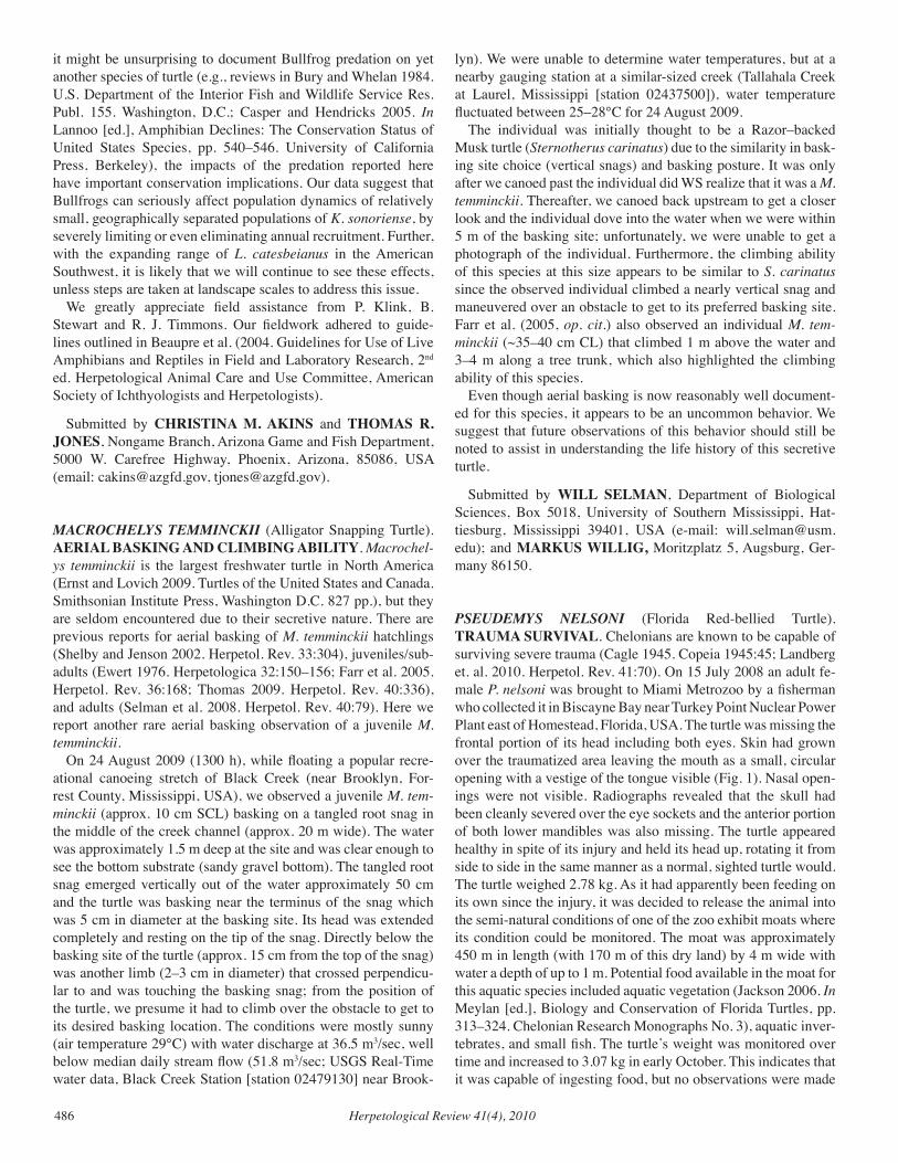
Herpetological Review 41(4), 2010486
it might be unsurprising to document Bullfrog predation on yet another species of turtle (e.g., reviews in Bury and Whelan 1984. U.S. Department of the Interior Fish and Wildlife Service Res. Publ. 155. Washington, D.C.; Casper and Hendricks 2005. In Lannoo [ed.], Amphibian Declines: The Conservation Status of United States Species, pp. 540–546. University of California Press, Berkeley), the impacts of the predation reported here have important conservation implications. Our data suggest that Bullfrogs can seriously affect population dynamics of relatively small, geographically separated populations of K. sonoriense, by severely limiting or even eliminating annual recruitment. Further, with the expanding range of L. catesbeianus in the American Southwest, it is likely that we will continue to see these effects, unless steps are taken at landscape scales to address this issue. We greatly appreciate field assistance from P. Klink, B. Stewart and R. J. Timmons. Our fieldwork adhered to guide-lines outlined in Beaupre et al. (2004. Guidelines for Use of Live Amphibians and Reptiles in Field and Laboratory Research, 2nd ed. Herpetological Animal Care and Use Committee, American Society of Ichthyologists and Herpetologists).
Submitted by CHRISTINA M. AKINS and THOMAS R. JONES, Nongame Branch, Arizona Game and Fish Department, 5000 W. Carefree Highway, Phoenix, Arizona, 85086, USA (email: [email protected], [email protected]).
MACROCHELYS TEMMINCKII (Alligator Snapping Turtle). AERIAL BASKING AND CLIMBING ABILITY. Macrochel-ys temminckii is the largest freshwater turtle in North America (Ernst and Lovich 2009. Turtles of the United States and Canada. Smithsonian Institute Press, Washington D.C. 827 pp.), but they are seldom encountered due to their secretive nature. There are previous reports for aerial basking of M. temminckii hatchlings (Shelby and Jenson 2002. Herpetol. Rev. 33:304), juveniles/sub-adults (Ewert 1976. Herpetologica 32:150–156; Farr et al. 2005. Herpetol. Rev. 36:168; Thomas 2009. Herpetol. Rev. 40:336), and adults (Selman et al. 2008. Herpetol. Rev. 40:79). Here we report another rare aerial basking observation of a juvenile M. temminckii. On 24 August 2009 (1300 h), while floating a popular recre-ational canoeing stretch of Black Creek (near Brooklyn, For-rest County, Mississippi, USA), we observed a juvenile M. tem-minckii (approx. 10 cm SCL) basking on a tangled root snag in the middle of the creek channel (approx. 20 m wide). The water was approximately 1.5 m deep at the site and was clear enough to see the bottom substrate (sandy gravel bottom). The tangled root snag emerged vertically out of the water approximately 50 cm and the turtle was basking near the terminus of the snag which was 5 cm in diameter at the basking site. Its head was extended completely and resting on the tip of the snag. Directly below the basking site of the turtle (approx. 15 cm from the top of the snag) was another limb (2–3 cm in diameter) that crossed perpendicu-lar to and was touching the basking snag; from the position of the turtle, we presume it had to climb over the obstacle to get to its desired basking location. The conditions were mostly sunny (air temperature 29°C) with water discharge at 36.5 m3/sec, well below median daily stream flow (51.8 m3/sec; USGS Real-Time water data, Black Creek Station [station 02479130] near Brook-
lyn). We were unable to determine water temperatures, but at a nearby gauging station at a similar-sized creek (Tallahala Creek at Laurel, Mississippi [station 02437500]), water temperature fluctuated between 25–28°C for 24 August 2009. The individual was initially thought to be a Razor–backed Musk turtle (Sternotherus carinatus) due to the similarity in bask-ing site choice (vertical snags) and basking posture. It was only after we canoed past the individual did WS realize that it was a M. temminckii. Thereafter, we canoed back upstream to get a closer look and the individual dove into the water when we were within 5 m of the basking site; unfortunately, we were unable to get a photograph of the individual. Furthermore, the climbing ability of this species at this size appears to be similar to S. carinatus since the observed individual climbed a nearly vertical snag and maneuvered over an obstacle to get to its preferred basking site. Farr et al. (2005, op. cit.) also observed an individual M. tem-minckii (~35–40 cm CL) that climbed 1 m above the water and 3–4 m along a tree trunk, which also highlighted the climbing ability of this species. Even though aerial basking is now reasonably well document-ed for this species, it appears to be an uncommon behavior. We suggest that future observations of this behavior should still be noted to assist in understanding the life history of this secretive turtle.
Submitted by WILL SELMAN, Department of Biological Sciences, Box 5018, University of Southern Mississippi, Hat-tiesburg, Mississippi 39401, USA (e-mail: [email protected]); and MARKUS WILLIG, Moritzplatz 5, Augsburg, Ger-many 86150.
PSEUDEMYS NELSONI (Florida Red-bellied Turtle). TRAUMA SURVIVAL. Chelonians are known to be capable of surviving severe trauma (Cagle 1945. Copeia 1945:45; Landberg et. al. 2010. Herpetol. Rev. 41:70). On 15 July 2008 an adult fe-male P. nelsoni was brought to Miami Metrozoo by a fisherman who collected it in Biscayne Bay near Turkey Point Nuclear Power Plant east of Homestead, Florida, USA. The turtle was missing the frontal portion of its head including both eyes. Skin had grown over the traumatized area leaving the mouth as a small, circular opening with a vestige of the tongue visible (Fig. 1). Nasal open-ings were not visible. Radiographs revealed that the skull had been cleanly severed over the eye sockets and the anterior portion of both lower mandibles was also missing. The turtle appeared healthy in spite of its injury and held its head up, rotating it from side to side in the same manner as a normal, sighted turtle would. The turtle weighed 2.78 kg. As it had apparently been feeding on its own since the injury, it was decided to release the animal into the semi-natural conditions of one of the zoo exhibit moats where its condition could be monitored. The moat was approximately 450 m in length (with 170 m of this dry land) by 4 m wide with water a depth of up to 1 m. Potential food available in the moat for this aquatic species included aquatic vegetation (Jackson 2006. In Meylan [ed.], Biology and Conservation of Florida Turtles, pp. 313–324. Chelonian Research Monographs No. 3), aquatic inver-tebrates, and small fish. The turtle’s weight was monitored over time and increased to 3.07 kg in early October. This indicates that it was capable of ingesting food, but no observations were made
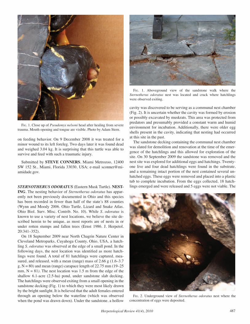
Herpetological Review 41(4), 2010 487
on feeding behavior. On 9 December 2008 it was treated for a minor wound to its left foreleg. Two days later it was found dead and weighed 3.04 kg. It is surprising that this turtle was able to survive and feed with such a traumatic injury.
Submitted by STEVE CONNERS, Miami Metrozoo, 12400 SW 152 St., Miami, Florida 33030, USA; e-mail [email protected].
STERNOTHERUS ODORATUS (Eastern Musk Turtle). NEST-ING. The nesting behavior of Sternotherus odoratus has appar-ently not been previously documented in Ohio and this species has been recorded in fewer than half of the state’s 88 counties (Wynn and Moody 2006. Ohio Turtle, Lizard and Snake Atlas. Ohio Biol. Surv. Misc. Contrib. No. 10). While S. odoratus is known to use a variety of nest locations, we believe the site de-scribed herein to be unique, as most reports are of nests in or under rotten stumps and fallen trees (Ernst 1986. J. Herpetol. 20:341–352). On 18 September 2009 near North Chagrin Nature Center in Cleveland Metroparks, Cuyahoga County, Ohio, USA, a hatch-ling S. odoratus was observed at the edge of a small pond. In the following days, the nest location was identified as more hatch-lings were found. A total of 81 hatchlings were captured, mea-sured, and released, with a mean (range) mass of 2.66 g (1.6–3.7 g, N = 80) and mean (range) carapace length of 22.75 mm (19–25 mm, N = 81). The nest location was 1.5 m from the edge of the shallow 6.1-acre (2.5-ha) pond, under sandstone slab decking. The hatchlings were observed exiting from a small opening in the sandstone decking (Fig. 1) to which they were most likely drawn by the bright sunlight. It is believed that the adult females entered through an opening below the waterline (which was observed when the pond was drawn down). Under the sandstone, a hollow
cavity was discovered to be serving as a communal nest chamber (Fig. 2). It is uncertain whether the cavity was formed by erosion or possibly excavated by muskrats. This area was protected from predators and presumably provided a constant warm and humid environment for incubation. Additionally, there were older egg shells present in the cavity, indicating that nesting had occurred at this site in the past. The sandstone decking containing the communal nest chamber was slated for demolition and renovation at the time of the emer-gence of the hatchlings and this allowed for exploration of the site. On 30 September 2009 the sandstone was removed and the nest site was explored for additional eggs and hatchings. Twenty-one live and four dead hatchlings were found in the substrate, and a remaining intact portion of the nest contained several un-hatched eggs. These eggs were removed and placed into a plastic tub to complete incubation. From the eggs collected, 18 hatch-lings emerged and were released and 5 eggs were not viable. The
fig. 1. Close up of Pseudemys nelsoni head after healing from severe trauma. Mouth opening and tongue are visible. Photo by Adam Stern.
fig. 2. Underground view of Sternotherus odoratus nest where the concentration of eggs were deposited.
fig. 1. Aboveground view of the sandstone walk where the Sternotherus odoratus nest was located and crack where hatchlings were observed exiting.
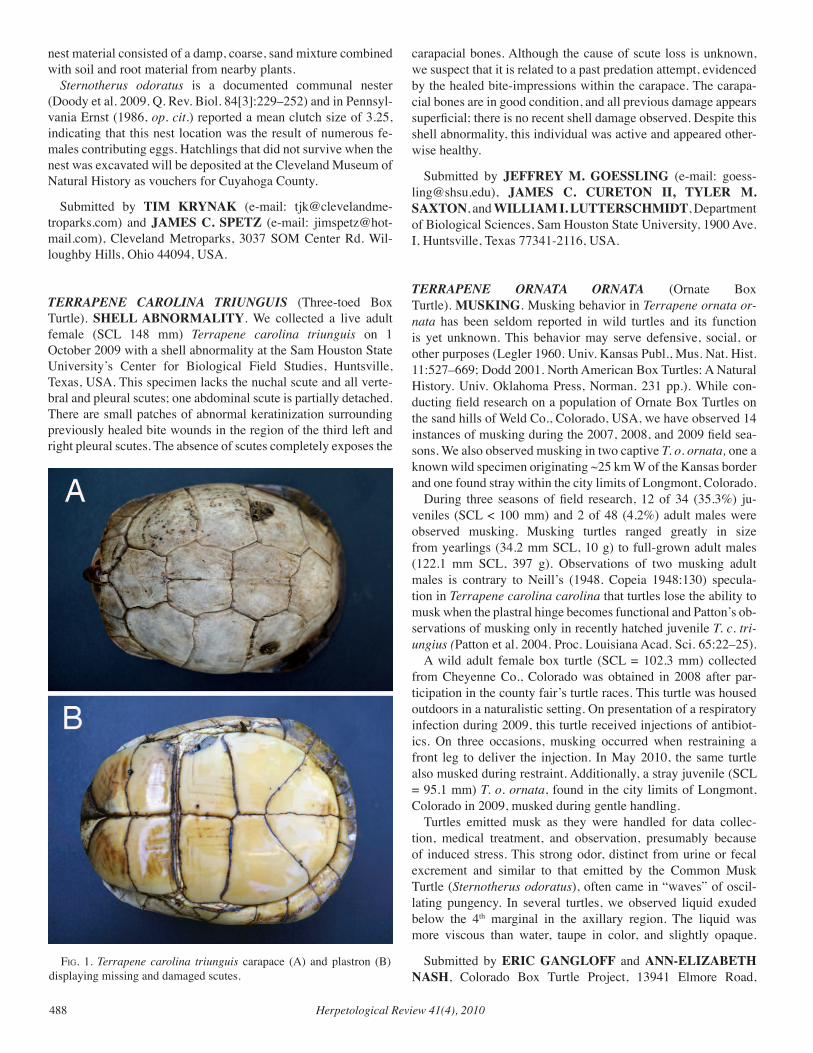
Herpetological Review 41(4), 2010488
nest material consisted of a damp, coarse, sand mixture combined with soil and root material from nearby plants. Sternotherus odoratus is a documented communal nester (Doody et al. 2009. Q. Rev. Biol. 84[3]:229–252) and in Pennsyl-vania Ernst (1986, op. cit.) reported a mean clutch size of 3.25, indicating that this nest location was the result of numerous fe-males contributing eggs. Hatchlings that did not survive when the nest was excavated will be deposited at the Cleveland Museum of Natural History as vouchers for Cuyahoga County.
Submitted by TIM KRYNAK (e-mail: [email protected]) and JAMES C. SPETZ (e-mail: [email protected]), Cleveland Metroparks, 3037 SOM Center Rd. Wil-loughby Hills, Ohio 44094, USA.
TERRAPENE CAROLINA TRIUNGUIS (Three-toed Box Turtle). SHELL ABNORMALITY. We collected a live adult female (SCL 148 mm) Terrapene carolina triunguis on 1 October 2009 with a shell abnormality at the Sam Houston State University’s Center for Biological Field Studies, Huntsville, Texas, USA. This specimen lacks the nuchal scute and all verte-bral and pleural scutes; one abdominal scute is partially detached. There are small patches of abnormal keratinization surrounding previously healed bite wounds in the region of the third left and right pleural scutes. The absence of scutes completely exposes the
carapacial bones. Although the cause of scute loss is unknown, we suspect that it is related to a past predation attempt, evidenced by the healed bite-impressions within the carapace. The carapa-cial bones are in good condition, and all previous damage appears superficial; there is no recent shell damage observed. Despite this shell abnormality, this individual was active and appeared other-wise healthy.
Submitted by JEFFREY M. GOESSLING (e-mail: [email protected]), JAMES C. CURETON II, TYLER M. SAXTON, and WILLIAM I. LUTTERSCHMIDT, Department of Biological Sciences, Sam Houston State University, 1900 Ave. I, Huntsville, Texas 77341-2116, USA.
TERRAPENE ORNATA ORNATA (Ornate Box Turtle). MUSKING. Musking behavior in Terrapene ornata or-nata has been seldom reported in wild turtles and its function is yet unknown. This behavior may serve defensive, social, or other purposes (Legler 1960. Univ. Kansas Publ., Mus. Nat. Hist. 11:527–669; Dodd 2001. North American Box Turtles: A Natural History. Univ. Oklahoma Press, Norman. 231 pp.). While con-ducting field research on a population of Ornate Box Turtles on the sand hills of Weld Co., Colorado, USA, we have observed 14 instances of musking during the 2007, 2008, and 2009 field sea-sons. We also observed musking in two captive T. o. ornata, one a known wild specimen originating ~25 km W of the Kansas border and one found stray within the city limits of Longmont, Colorado. During three seasons of field research, 12 of 34 (35.3%) ju-veniles (SCL < 100 mm) and 2 of 48 (4.2%) adult males were observed musking. Musking turtles ranged greatly in size from yearlings (34.2 mm SCL, 10 g) to full-grown adult males (122.1 mm SCL, 397 g). Observations of two musking adult males is contrary to Neill’s (1948. Copeia 1948:130) specula-tion in Terrapene carolina carolina that turtles lose the ability to musk when the plastral hinge becomes functional and Patton’s ob-servations of musking only in recently hatched juvenile T. c. tri-ungius (Patton et al. 2004. Proc. Louisiana Acad. Sci. 65:22–25). A wild adult female box turtle (SCL = 102.3 mm) collected from Cheyenne Co., Colorado was obtained in 2008 after par-ticipation in the county fair’s turtle races. This turtle was housed outdoors in a naturalistic setting. On presentation of a respiratory infection during 2009, this turtle received injections of antibiot-ics. On three occasions, musking occurred when restraining a front leg to deliver the injection. In May 2010, the same turtle also musked during restraint. Additionally, a stray juvenile (SCL = 95.1 mm) T. o. ornata, found in the city limits of Longmont, Colorado in 2009, musked during gentle handling. Turtles emitted musk as they were handled for data collec-tion, medical treatment, and observation, presumably because of induced stress. This strong odor, distinct from urine or fecal excrement and similar to that emitted by the Common Musk Turtle (Sternotherus odoratus), often came in “waves” of oscil-lating pungency. In several turtles, we observed liquid exuded below the 4th marginal in the axillary region. The liquid was more viscous than water, taupe in color, and slightly opaque.
Submitted by ERIC GANGLOFF and ANN-ELIZABETH NASH, Colorado Box Turtle Project, 13941 Elmore Road,
fig. 1. Terrapene carolina triunguis carapace (A) and plastron (B) displaying missing and damaged scutes.
Herpetological Review 41(4), 2010 489
Longmont, Colorado 80504, USA; e-mail: [email protected].
TERRAPENE ORNATA ORNATA (Ornate Box Turtle). DIET AND MICROHABITAT. Terrapene o. ornata is described as omnivorous, eating a broad range of animal and plant material and requiring access to water within its home range. However, within our study site standing water is generally not available and permanent water sources are well outside the typical home range areas of T. o. ornata. Here we provide information on one food choice, Tradescantia occidentalis (Prairie Spiderwort) from a wild population of Ornate Box Turtles in the sand hills of eastern Colorado that may also operate as an important water source. We also show the ready acceptance of this food item by two captive populations of wild-collected T. o. ornata housed in naturalistic habitats. The present study site in Weld Co., Colorado encompasses ap-proximately 50 ha of sand hills. The site is crossed with single-lane unpaved roads facilitating access to gas and oil wells. The property has been cattle ranched for more than 20 years; graz-ing is rotated with a water-pumping windmill as the focal point of each grazing section. The study area lacks any natural free-standing water and the windmill is only sporadically operated; overflow that may be used by turtles is thus unpredictable. Average yearly precipitation at the nearest available weather station, about 16.6 km W of the study site, is 360 mm. Though more than 95% of precipitation occurs from April through October, natural ephemeral water is rarer than agriculturally re-lated runoff. Precipitation from April though October 2009 was 32 mm greater than during the same period in 2008, and 100.6 mm greater than 2007. While 2009 precipitation was near aver-age for the study season, the two previous years had decreased precipitation during the field study period. In 2009, T. occiden-talis was seen during every field outing, in more areas within the study site, and in greater concentrations than in previous years. Prairie Spiderwort is a smooth, subsucculent, perennial mono-cot up to 50 cm tall. Stems are often tufted, and when pulled apart, the copious mucilaginous slime inside forms what somewhat re-sembles a spider’s web. These plants are known as “cow slobber” because of the gooey, stringy sap they produce. Given the absence of free water sources, we hypothesize that Spiderwort may be an important source of water for T. o. ornata in sand hills habitat. The study site also contains Opuntia cactus species and a variety of forbs and grasses. Ohio Spiderwort (Tradescantia ohiensi) has been listed as part of the floral community in T. o. ornata habi-tat in Iowa (Bowen at al. 2004. J. Herpetol. 38:562–568) and Wisconsin (Doroff and Keith 1990. Copeia 1990:387–399). On 13 June 2008, we observed evidence of two adult turtles eating Spiderwort. The female was observed with a purple plant in her mouth, most likely T. occidentalis petals, and the male was observed with a chewed T. occidentalis stem directly in front of him and green vegetation on his beak. Several days later we lo-cated another adult female sitting in crushed Spiderwort stems with foliage stuck to her beak. During 2009, we found increased evidence of T. o. ornata eating Spiderwort, as well as characteris-tic crushed stems that may indicate turtle feeding, with three adult turtles (two females and one male) observed eating T. occidenta-lis. All observations of Spiderwort consumption occurred in the
month of June. Several specimens of T. occidentalis were planted into two naturalistic box turtle habitats, each more than 60 m2. Wild-collected T. o. ornata readily ate this plant, often cropping it to the ground and inducing dormancy until the next growing sea-son. This was in spite of other available food items provided on a regular basis, typically a mix of grocery-store produce (greens, vegetables, and berries). Other habitat plants including mallow (Malva neglecta) and common dandelion (Taraxacum officinale) were rarely observed being consumed. In neither captive popula-tion did we make any attempt to introduce occupants to the pres-ence of T. occidentalis; turtles located and consumed this food item by free choice, often traveling 1–5 m from routine feeding locations.
Submitted by ANN-ELIZABETH NASH and ERIC GANGLOFF, Colorado Box Turtle Project, 13941 Elmore Road, Longmont, Colorado 80504, USA; e-mail: [email protected].
CROCODYLIA — CROCODILIANS
CROCODYLUS ACUTUS (American Crocodile). ADULT MASS. Few data exist on the body mass of large reptiles, due in part to the difficulties of handling and weighing these animals (Minton and Minton 1973. Giant Reptiles. Charles Scribner’s Sons, New York, New York. 345 pp.). In particular, few reliable data exist regarding body size of large crocodilians (Richardson et al. 2002. Crocodiles: Inside Out: A Guide to the Crocodil-ians and their Functional Morphology. Surrey Beatty and Sons, Ltd., Chipping Norton, NSW. 172 pp.; Webb and Manolis 1989. Crocodiles of Australia. Reed Books Pty. Ltd., Frenchs Forest, NSW. 160 pp.; Woodward et al. 1995. J. Herpetol. 29:507–513). However, crocodile research and management rely on knowledge of individual body size and size-class structure of populations (Webb and Smith 1987. In Webb et al. [eds.], Wildlife Manage-ment: Crocodiles and Alligators, pp. 199–210. Surrey Beatty & Sons, Ltd, Sydney) because demographic and reproductive vari-ables are functionally dependent on body size rather than age, and population models are generally based on the former (Nichols 1987. In Webb et al., op. cit., pp. 177–187). Here, we report the body mass of a large Crocodylus acutus from Costa Rica. On 13 September 2007, we captured an adult male C. acu-tus in the lower Tarcoles River, Costa Rica (9.79906389°N, 84.615278°W) during a study of ocular disease in this crocodile population. Total length (TL; anterior tip of the snout to the poste-rior tip of the tail, measured along the dorsal surface) and snout–vent length (SVL; anterior tip of the snout to the anterior margin of the cloacal vent, measured along the ventral surface) of the crocodile were measured with a tape as 482.6 cm and 237.5 cm, respectively. The crocodile was wrapped in an industrial cargo net and weighed using an electronic crane scale (Dynafor LLX/LLTR; Schumann GmbH, Sillerup, Germany) and manual chain hoist secured to the limb of a tree on the bank of the river. The mass of the crocodile was 537.45 kg, after subtracting the mass of the cargo net (29.54 kg). In 2003, two of us (BRB, JRBM) captured a large C. acutus (TL = 395.0 cm) for translocation during a radio-tracking project in Costa Rica and determined that it weighed 500 kg (Barr 2003.
Herpetological Review 41(4), 2010490
Crocodile Spec. Newsltr. 23:19). To our knowledge, these mea-surements of body mass are the largest yet reported for C. acutus, however they do not represent a maximum value for this species. Crocodylus acutus is known to reach lengths of 600–700 cm (Al-varez del Toro 1974. Los Crocodylia de Mexico [Estudio Com-parativo]. Recursos Naturales Renovables, Mexico City. 70 pp.; Schmidt 1924. Fieldiana 12:77–96). Using an equation derived from the relationship of TL to body mass for the morphologi-cally similar Morelet’s Crocodile (C. moreletii) (Platt et al. 2009. Carib. J. Sci. 45:80–93), the estimated body mass of these large C. acutus is predicted to range from 900 to 1283 kg.
Submitted by THOMAS R. RAINWATER, 619 Palmetto St., Mt. Pleasant, South Carolina 29464, USA (e-mail: [email protected]); LUZ DENIA BARRANTES and JUAN RAFAEL BOLAÑOS MONTERO, Asociación de Especialis-tas en Cocodrilos - Centro América (AEC-CA), Calle 31 Ave-nida 10 Casa 2959, San José, Costa Rica; STEVEN G. PLATT, Department of Biology, Box C-64, Sul Ross State University, Alpine, Texas 79832, USA; and BRADY R. BARR, National Geographic Society, 1145 17th St. NW Washington, DC 20036, USA.
SQUAMATA — LIZARDS
AMEIvA FESTIvA (Central American Whiptail Lizard). CAN-NIBALISM. Ameiva festiva is a broadly distributed, dietary gen-eralist that consumes diverse arthropod prey including crickets, spiders, roaches, and katydids (Vitt and Zani 1996. J. Herpetol. 30:110–117; Vitt and Zani 1998. J. Trop. Ecol. 14:537–559; Whitfield and Donnelly 2006. J. Trop. Ecol. 22:409–417). Liz-ards, however, have been recorded infrequently in their diet (Vitt and Zani 1998, op. cit.) and accounts identifying the lizard spe-cies taken are lacking. Hence, here we report an A. festiva prey-ing on a conspecific in the Gandoca-Manzanillo Wildlife Refuge, Costa Rica. At 1127 h on 21 July 2008, while walking the grounds at the Iguana Verde Foundation (09.63818°N, 82.70496°W, datum WGS84; elev. 3 m), we observed, photographed, and videotaped one adult female A. festiva (ca. 9 cm SVL) grasping a conspe-cific (ca. 4 cm SVL) by its head. The smaller lizard was alive but missing half its tail, which was seen ca. 7 cm away. The adult was biting the head of the smaller conspecific continually un-til a probable male (ca. 10 cm SVL) A. festiva approached and began undulating its tail. The adult female fled 2.5 m away and began manipulating her prey in a head-first orientation. The adult female Ameiva took one minute to swallow its prey, and then per-formed an undulating maneuver with its torso, and completed its consumption at 1134 h. This is the first observation of cannibalism in this frequently observed species, and augments our knowledge of trophic rela-tionships for Ameiva lizards and their behavioral ecology. This observation was made while conducting Iguana iguana research supported by the San Diego Zoo’s Institute for Conservation Re-search.
Submitted by JUAN G. ABARCA, Escuela de Ciencias Bi-ológicas, Universidad Nacional, Heredia, Costa Rica (e-mail: [email protected]); and CHARLES R. KNAPP, San Diego
Zoo’s Institute for Conservation Research., Zoological Society of San Diego, 15600 San Pasqual Valley Road, Escondido, Califor-nia 92027, USA (e-mail: [email protected]).
APHANIOTIS ACUTIROSTRIS (Indonesia Earless Agama). REPRODUCTION. Aphaniotis acutirostris is known from In-donesia (Uetz and Hallermann 2010. The Reptile Database http://www.reptile-database.org, accessed 22 July 2010). To my knowl-edge, the only information on A. acutirostris is a report of clutch sizes of two eggs deposited every 30–100 days and hatching 48–63 days later (Das 2006. A Photographic Guide to Snakes & Oth-er Reptiles of Borneo. Ralph Curtis Publishing, Sanibal Island, Florida. 144 pp.). The purpose of this note is to provide additional information on the reproductive biology of A. acutirostris. A sample of 18 A. acutirostris (nine males, mean SVL = 53.4 mm ± 2.2 SD, range = 51–56 mm; four females, mean SVL = 50.3 mm ± 2.1 SD, range = 48–53 mm; four subadults, mean SVL = 31.0 mm ± 4.8 SD, range = 28–38 mm; and one neonate, SVL = 23 mm, collected July 1975 at Bohorok, North Sumatra, Indonesia (3.516667°N, 98.133333°E; WGS84, 255–1390 m elev.) was examined from the herpetology collection of the Field Museum of Natural History (FMNH): FMNH 209533, 209534, 209536–209539, 209541–209545, 209552–209554, 209556, 209557, 209559, 209564. For histological examination, the left gonad was removed to check for yolk deposition in females and spermiogenesis (sperm formation) in males. Counts were made of enlarged ovarian follicles (> 4 mm length) or oviductal eggs. Tissues were embedded in paraffin and stained with hematoxylin followed by eosin counterstain. Histology slides are deposited in FMNH. All males examined were undergoing spermiogenesis; the lu-mina of the seminiferous tubules were lined by clusters of sperm or groups of metamorphosing spermatids. The smallest reproduc-tively active male measured 51 mm SVL. One female contained two vitellogenic follicles (2 mm length); two females each con-tained two enlarged ovarian follicles (4 mm diameter) and one female contained two oviductal eggs. The smallest reproduc-tively active female (oviductal eggs) measured 48 mm SVL. The presence of the neonate (23 mm SVL), plus three slightly larger juveniles (28, 28, 30 mm SVL) indicates they were born earlier in the year and that A. acutirostris has a prolonged period of re-production. Inger and Greenberg (1966. Ecology 47:1007–1021) reported continuous reproduction in five agamid species (Draco melanopogon, D. obscurus, D. quinquefasciatus, Gonocephalus grandis, G. liogaster) from a Bornean rain forest. Whether A. acutirostris breeds throughout the year will require examination of additional monthly samples. I thank Alan Resetar (FMNH) for permission to examine A. acutirostris.
Submitted by STEPHEN R. GOLDBERG, Department of Biology, Whittier College, Whittier, California 90608, USA; e-mail: [email protected].
ASPIDOSCELIS TIGRIS (Western Whiptail) DIET. Aspidosce-lis tigris is a common lizard of North American arid lands. Active forager, whiptails generally forage on the ground, often near veg-
Herpetological Review 41(4), 2010 491
etation, where they dig in leaf litter or probe cracks and crevices in search of prey (Pianka 1970. Ecology 51:703–720). Whiptails feed primarily on diverse ground-dwelling arthropods (Best and Gennaro 1985. Great Basin Nat. 45:527–534), but are not record-ed to take honey bee (Apis mellifera) prey. Here, I present the first record of A. tigris attacking and consuming a honey bee. At 1200 h on 29 July 2002, I observed three adult A. tigris foraging beneath a stand of saltcedar (Tamarix sp.) in a dry wash in the northwest portion of the Glen Canyon National Recreation Area, Garfield Co., Utah, USA (37.642614°N, 111.067225°W, datum WGS84; elev. 1510 m). The lizards seemed unaware of my presence as they foraged in the leaf litter. The saltcedar was in full flower, and many bee species were visiting the flowers. A honey bee flew down from a low-lying flower, landing on the ground ca. 1 m from one of the A. tigris. The lizard immediately ran over to the bee, caught it, and began chewing it rapidly. As the A. tigris began ingesting the bee, it rubbed its snout on the ground several times. It is unknown whether it was trying to avoid being stung or if the hairy body of the bee was irritating the inside of its mouth. Other whiptails have been reported to rub their snout on the ground when attacking hairy arthropods, like some caterpil-lars (Burt 1928. J. Kansas Ent. Soc. 1:50–68). The entire attack and ingestion episode lasted <1 min. I continued to watch the lizards forage under the saltcedar for ca. 15 min, and the A. tigris that had ingested the bee behaved normally, showing no adverse effects from the encounter. This record merits comment given the potentially toxic nature of this insect. Feeding experiments have revealed that the Eastern Fence Lizard (Sceloporus undulatus) always refused honey bees when offered them, and in one instance, a lizard was stung by a bee and died minutes later (Burt 1928. J. Kansas Entomol. Soc. 1:50–68). Whether the A. tigris individual was stung by the bee in this instance is unknown, but the fact that the lizard was able to attack and eat the bee without harmful consequence is notewor-thy.
Submitted by JOSEPH S. WILSON, Utah State University, Biology Department, 5305 Old Main Hill, Logan, Utah 84322, USA; e-mail: [email protected].
GONATODES HUMERALIS (Trinidad Gecko). ENDOPARA-SITES. Gonatodes humeralis is widespread in northern South America and occurs in French Guiana, Suriname, Ecuador, Guy-ana, Peru, Bolivia, NE Venezuela, Colombia, Brazil, as well as Trinidad and Tobago (Uetz and Hallermann 2010. http.reptile-database.org, accessed 20 May 2010). The purpose of this note is to add to the helminth list of G. humeralis. Fifteen G. humeralis (mean SVL = 36.9 mm ± 2.0 SD, range = 34–40 mm) collected from 1905 to 1973 from Peru, Loreto De-partment, Moropon (3.748056°S, 73.246933°W; WGS84; 107 m elev.) and deposited in the Texas Cooperative Wildlife Collection (TCWC), Texas A & M University, were examined for helminths (TCWC 36674, 36684, 36737, 36739, 38996, 41173, 41226, 41761, 41801, 41910, 41911, 41920, 41922, 42714, 42715). The body cavities were opened and the digestive tracts re-moved. Contents were examined under a dissecting microscope. Digeneans were regressively stained in hematoxylin and mount-ed on glass slides in Canada balsam. Nematodes were cleared on
glass slides in glycerol. All were cover-slipped, studied under a compound microscope and identified. Found were one species of Digenea, Mesocoelium monas (N = 173, small intestine, preva-lence = number infected lizards/number lizards examined x 100 = 7%; mean intensity, mean number helminths per infected lizard ± 1 SD = 173) and three species of Nematoda, Parapharyngodon scleratus (large intestine, N = 2, prevalence = 7%, mean intensity = 2), Physaloptera retusa (stomach, N = 6, prevalence = 20%, mean intensity = 2.0 ± 1.0, range = 1–3), Skrjabinelazia galliardi (small intestine, N = 2, large intestine, N = 2, prevalence = 20%, mean intensity = 1.3 ± 0.6, range = 1–2). Voucher helminths were deposited in the United States National Parasite Collection, US-NPC, Beltsville, Maryland: Mesocoelium monas (103203); Para-pharyngodon scleratus (103204); Physaloptera retusa (103205); Skrjabinelazia galliardi (103206). Mesocoelium monas is cosmopolitan in distribution and has been recorded from fishes, amphibians, and reptiles from the New and Old Worlds (Goldberg et al. 2009. Comp. Parasitol. 76:58–83). Parapharyngodon scleratus has been reported in a variety of lizards from Brazil and also occurs in Peru (Bursey et al. 2005. Comp. Parasitol.72:50–68). Physaloptera retusa is widely distributed in lizards and a few amphibians in the west-ern hemisphere and has been reported from Mexico, North and South America, and the West Indies (Bursey et al. 2007, op. cit.). Skrjabinelazia galliardi was originally described from G. hume-ralis collected in Brazil (Chabaud 1973. Ann. Parasitol. Hum. Comp. 48:329–334). Gonatodes humeralis represents a new host record for Mesocoelium monas, Parapharyngodon scleratus, and Physaloptera retusa. Peru is a new locality record for Skrjabine-lazia galliardi We thank Lee Fitzgerald and Toby Hibbitts (TCWC) for per-mission to examine specimens and Daisy Salguero (Whittier Col-lege) for assistance with dissections.
Submitted by STEPHEN R. GOLDBERG, Whittier College, Department of Biology, Whittier, California 90608, USA (e-mail: [email protected]); and CHARLES R. BURSEY, Penn-sylvania State University, Shenango Campus, Department of Bi-ology, Sharon, Pennsylvania 16146, USA (e-mail: [email protected]).
HELODERMA SUSPECTUM (Gila Monster). TREE-CLIMB-ING. Heloderma suspectum occupies a variety of terrestrial habitats in all deserts of the United States and northern main-land Mexico (Beck 2005. Biology of Gila Monsters and Beaded Lizards. University of California Press, Berkeley and Los Ange-les. 247 pp.; Campbell and Lamar 2004. Venomous Reptiles of the Western Hemisphere. Cornell University Press, Ithaca, New York. 870 pp.). Despite the fact these regions substantially differ with respect to environmental factors and habitats (Brown 1994. Biotic Communities of the American Southwest—United States and Mexico. University of Utah Press, Salt Lake City. 342 pp.), H. suspectum selects similar types of microhabitats and shelters (Beck 2005, op. cit.; Beck and Jennings 2003. Herpetol. Monogr. 17:111–129), which are commonly bushes and structures with subterranean components (e.g., rock formations and small mam-mal burrows). Rarely, H. suspectum exhibits arboreal activities, particularly with regard to tree climbing (Beck 2005, op. cit.;
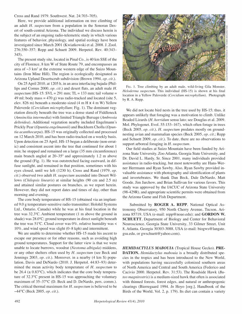
Herpetological Review 41(4), 2010492
Cross and Rand 1979. Southwest. Nat. 24:703–705). Here, we provide additional information on tree climbing of an adult H. suspectum from a population in the Sonoran Des-ert of south-central Arizona. The individual we discuss herein is the subject of an ongoing radio-telemetric study in which various features of behavior, physiology, and spatial ecology have been investigated since March 2001 (Kwiatkowski et al. 2008. J. Zool. 276:350–357; Repp and Schuett 2009. Herpetol. Rev. 40:343–345). The present study site, located in Pinal Co., is 40 km SSE of the city of Florence, 8 km W of State Route 79, and encompasses an area of ~3 km2 at the extreme western edge of the Suizo Moun-tains (Iron Mine Hill). The region is ecologically designated as Arizona Upland Desertscrub subdivision (Brown 1994, op. cit.). On 25 April 2010, at 1205 h, in an area interfacing bajada (Phil-lips and Comus 2000, op. cit.) and desert flats, an adult male H. suspectum (HS-15: SVL = 291 mm; TL = 133 mm; tail volume = 49 ml; body mass = 470 g) was radio-tracked and located (site 5, elev. 826 m) beneath a moderate-sized (4 m H x 4 m W) Yellow Paloverde (Cercidium microphyllum; Fig. 1). The dominant veg-etation directly beneath the tree was a dense stand of Fiddleneck (Amsinckia intermedia) with limited Triangle Bursage (Ambrosia deltoidea). Additional vegetation nearby included Engelmann’s Prickly Pear (Opuntia engelmannii) and Buckhorn Cholla (Opun-tia acanthocarpa). HS-15 was originally collected and processed on 12 March 2010, and has been radio-tracked on a weekly basis. Upon detection on 25 April, HS-15 began a deliberate (non-errat-ic) and consistent ascent into the tree that continued for about 1 min; he stopped and remained on a large (35 mm circumference) main branch angled at 20–35º and approximately 1.2 m above the ground (Fig. 1). He was outstretched facing eastward, in dif-fuse sunlight, and remained in that position, sometimes with his eyes closed, until we left (1230 h). Cross and Rand (1979, op. cit.) observed two adult H. suspectum ascended into Desert Wil-lows (Chilopsis linearis) at similar heights (90 cm and 2.5 m) and attained similar postures on branches, as we report herein. However, they did not report dates and times of day, other than morning and evening. The core body temperature of HS-15 (obtained via an implant-ed 9.0 g temperature-sensitive radio transmitter; Holohil Systems Ltd., Ontario, Canada) while he was at his final location in the tree was 32.3ºC. Ambient temperature (1 m above the ground in shade) was 28.0ºC; ground temperature in direct sunlight beneath the tree was 51ºC. Cloud cover was 0%, relative humidity was < 10%, and wind speed was slight (0–8 kph) and intermittent. We are unable to determine whether HS-15 made his ascent to escape our presence or for other reasons, such as avoiding high ground temperatures. Support for the latter view is that we were unable to locate burrows, woodrat (Neotoma albigula) middens, or any other shelters often used by H. suspectum (see Beck and Jennings 2003, op. cit.). Moreover, in a nearby (4 km S) popu-lation, Davis and DeNardo (2010. J. Herpetol. 44:83–93) deter-mined the mean activity body temperature of H. suspectum to be 26.4 (± 0.83°C), which indicates that the core body tempera-ture of 32.3°C present in HS-15 was approaching the voluntary maximum of 35–37°C (D. Beck and D. DeNardo, pers. comm.). The critical thermal maximum for H. suspectum is believed to be ~44°C (Beck 2005, op. cit.).
We did not locate bird nests in the tree used by HS-15; thus, it appears unlikely that foraging was a motivation to climb. Unlike Beaded Lizards (H. horridum sensu lato; see Douglas et al. 2009. Mol. Phylogenet. Evol. 55:153–167), which often forage in trees (Beck 2005, op. cit.), H. suspectum predates mostly on ground-nesting avian and mammalian species (Beck 2005, op. cit.; Repp and Schuett 2009, op. cit.). To date, there are no observations to support arboreal foraging in H. suspectum. Our field studies at Suizo Mountain have been funded by Ari-zona State University, Zoo Atlanta, Georgia State University, and Dr. David L. Hardy, Sr. Since 2001, many individuals provided assistance in radio-tracking, but most noteworthy are Hans-Wer-ner Herrmann and Ryan Sawby. Also, Ryan Sawby provided in-valuable assistance with photography and identification of plants and invertebrates. We thank Dan Beck, Dale DeNardo, Matt Goode, Jim Jarchow, and Brian Sullivan for various favors. This study was approved by the IACUC of Arizona State University (98-429R), and appropriate scientific permits were obtained from the Arizona Game and Fish Department.
Submitted by ROGER A. REPP, National Optical As-tronomy Observatory, 950 North Cherry Avenue, Tucson, Ari-zona 85719, USA (e-mail: [email protected]); and GORDON W. SCHUETT, Department of Biology and Center for Behavioral Neuroscience, Georgia State University, 33 Gilmer Street, Unit 8, Atlanta, Georgia 30303-3088, USA (e-mail: [email protected], or [email protected]).
HEMIDACTYLUS MABOUIA (Tropical House Gecko). PRE-DATION. Hemidactylus mabouia is a broadly distributed spe-cies in the tropics and has been introduced to the New World, with populations having successfully colonized southern areas of North America and Central and South America (Federico and Cacivio 2000. Herpetol. Rev. 31:53). The Roadside Hawk (Bu-teo magnirostris) is a medium-sized hawk that often is associated with thinned forests, forest edges, and natural or anthropogenic clearings (Bierregaard 1994. In Hoyo [org.], Handbook of the Birds of the World, Vol. 2, p. 179). Its diet can contain a variety
fig. 1. Tree climbing by an adult male, wild-living Gila Monster, Heloderma suspectum. This individual (HS-15) is shown at his final location in a Yellow Paloverde (Cercidium microphyllum). Photograph by R. A. Repp.
CO
LOR
REP
RO
du
CTi
ON
Su
PPO
RTEd
BY
THE
THO
mAS
BEA
uvA
iS F
uN
d
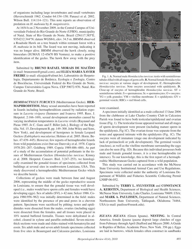
Herpetological Review 41(4), 2010 493
of organisms including large invertebrates and small vertebrates (Haverschimidt 1962. Condor 64:154–158; Panasci et al. 2002. Wilson Bull. 114:114–121). This note reports an observation of predation on H. mabouia by B. magnirostris. At 1654 h on 2 November 2009, in the Central Campus of Uni-versidade Federal do Rio Grande do Norte (UFRN), municipality of Natal, State of Rio Grande do Norte, Brazil (256117.385°E, 9354212.344°N; datum WGS84), elev. 34.5 m, BRMM observed a Roadside Hawk on a tree branch within remnant forest with a H. mabouia in its bill. The lizard was not moving, indicating it was no longer alive. BRMM observed the hawk closely, using binoculars (SUMAX 12-45x70 BS Ventura), to make a positive identification of the gecko. The hawk flew away with the prey item.
Submitted by BRUNO RAFAEL MORAIS DE MACÊDO (e-mail: [email protected]), and ELIZA MARIA XAVIER FREIRE (e-mail: [email protected]), Laboratório de Herpeto-logia, Departamento de Botânica, Ecologia e Zoologia, Centro de Biociências, Universidade Federal do Rio Grande do Norte, Campus Universitário Lagoa Nova, CEP 59072-970, Natal, Rio Grande do Norte, Brazil.
HEMIDACTYLUS TURCICUS (Mediterranean Gecko). HER-MAPHRODITISM. Many sexual anomalies have been reported in lizards including hermaphroditism caused by radiation expo-sure in Lacerta vivipara (Semenov and Ivanova 1995. Russ. J. Herpetol. 2:166–169), sexual development anomalies caused by varying incubation temperatures in Lacerta viridis (Raynaud and Pieau 1985. In C. Gans and F. Billet [eds.], Biology of the Rep-tilia, Vol. 15, Development B, pp. 149–300. John Wiley and Sons, New York), and development of hemipenes in female Leopard Geckos (Eublepharis macularius; Holmes et al. 2005. Horm. Be-hav. 47:439–445). However, few reports of hermaphrodite lizards from wild populations exist (but see Darevsky et al. 1978. Copeia 1978:201–207; Goldberg 1989. Copeia 1989:486–488). As part of a study of the accumulation of potential pollutants in the tis-sues of Mediterranean Geckos (Hemidactylus turcicus; Kinney et al. 2008. Herpetol. Conserv. Biol. 3:247–253), we histologi-cally examined the gonadal tissues of specimens collected from buildings at several sites in southwestern Louisiana, USA. This study discovered a hermaphroditic Mediterranean Gecko which we describe herein. Collections of geckos were made between June and August 2006, representing the middle of the gecko reproductive season in Louisiana, to ensure that the gonadal tissue was well devel-oped (i.e., males would have sperm cells and females would have developing eggs). Sex of adults (SVL > 42 mm; Selcer 1986. Co-peia 1986:956–962) was initially determined in the field: males were identified by the presence of pre-anal pores in a chevron pattern. Specimens were sacrificed by pithing; testes and epidi-dimides were dissected from the males, ovaries (including eggs) were dissected from the females, and all tissues were placed in 10% neutral buffered formalin. Tissues were dehydrated in al-cohol, cleared in xylene and paraffin embedded. Seven-micron- thick sections were made and slides stained with hematoxylin and eosin. Six adult male and seven adult female specimens collected from five sites in Beauregard and Calcasieu parishes, Louisiana
were examined. A specimen initially identified as a male collected 13 June 2006 from the clubhouse at Lake Charles Country Club in Calcasieu Parish was found to have both testicular/epididymal and ovarian tissue (Fig. 1). The testicular tissue appeared normal and all stages of sperm development were present (including mature sperm in the epididymis, Fig 1C). The ovarian tissue was separate from the testes and appeared intimate with the epididymis (Fig. 1C). The oocytes were all immature (stage one development indicated by lack of perinucleoli or yolk development). The germinal vesicle (nucleus), as well as the vitelline membrane surrounding the eggs can also be seen (Fig. 1D). Because this individual possesses both male and female gonadal tissues, it is a true hermaphrodite (or intersex). To our knowledge, this is the first report of a hermaph-roditic Mediterranean Gecko captured from a wild population. This study was carried out in accordance with the McNeese State University Institutional Animal Care and Use Regulations. Specimens were collected under the authority of Louisiana De-partment of Wildlife and Fisheries Scientific Collecting Permit LNHP-06-052.
Submitted by TERRY L. SYLVESTER and CONSTANCE A. KERSTEN, Department of Biological and Health Sciences, McNeese State University, Lake Charles, Louisiana 70609, USA; and MARK A. PAULISSEN, Department of Natural Sciences, Northeastern State University, Tahlequah, Oklahoma 74464, USA (e-mail: [email protected]).
IGUANA IGUANA (Green Iguana). NESTING. In Central America, female Iguana iguana deposit large clutches of eggs from February through April (Stafford and Meyers 2000. A Guide to Reptiles of Belize. Academic Press, New York. 356 pp.). Eggs are laid in burrows, which females often construct in sandbanks
Fig. 1. A. Normal male Hemidactylus turcicus: testis with seminiferous tubules filled with all stages of germ cells. B. Normal female Hemidactylus turcicus: oocytes at various stages of development. C. Hermaphrodite Hemidactylus turcicus. Note oocytes associated with epididymis. D. Close-up of oocytes of hermaphrodite Hemidactylus turcicus. ST = seminiferous tubule; S = spermatozoa; Sc = spermatocytes; O = oocytes; YG = yolk granules; VM = vitelline membrane; E = epididymis; GV = germinal vesicle; RBCs = red blood cells.
Herpetological Review 41(4), 2010494
along creeks and rivers (Stafford and Meyers, op. cit.). In Pan-ama, female I. iguana reportedly construct nesting burrows in close proximity to American Crocodile (Crocodylus acutus) nests (Rand 1968. Copeia 1968:552–561; Dugan et al. 1981. J. Herpe-tol. 15:409–414). Here we report observations of I. iguana using crocodile nests as oviposition sites in southern Belize. Crocodile embryos obtained from these nests, through genetic analysis, were determined to be C. moreletii × acutus hybrids (Hekkala 2004. Conservation Genetics at the Species Boundary: Case Stud-ies from African and Caribbean Crocodiles [Genus: Crocodylus]. PhD dissertation, Columbia University, New York. 92 pp.). On 20 April 1997, we found two crocodile nests 240 cm apart on a sandbar along Paynes Creek, Paynes Creek National Park, Toledo District (16.35830°N, 88.56604°W, datum WGS84; elev. ca. 10 m). Each crocodile nest consisted of a sand mound ca. 140 cm wide × 90 cm high. One nest contained 25 I. iguana eggs, but no crocodile eggs, and the other contained one I. iguana egg and 42 crocodile eggs. In the latter nest, the I. iguana egg was buried beneath the crocodile eggs. Remains of at least five more I. iguana eggs were scattered around the second nest, implying that the female crocodile had unearthed them during oviposition. The extent of banding visible on the crocodile eggs (Platt et al. 2008. J. Zool. 275:177–189) indicated the clutch was deposited about five days earlier. Because crocodile nests are generally con-structed 5–7 days prior to egg laying (Platt et al., op. cit.), the I. iguana clutches were likely deposited in early April. On 15 May 1997, we found two additional crocodile nests on sandbars along the Swasey Branch of Monkey River, Toledo District. The first nest (16.41888°N, 88.53466°W, datum WGS84; elev. ca. 15 m), a large sand mound measuring approximately 300 cm wide × 100 cm high, contained 38 crocodile eggs and 17 I. iguana eggs; the latter were buried about 5 cm beneath the crocodile eggs. We estimated the crocodile eggs to be about three weeks old. Hence, the I. iguana eggs were likely deposited in mid-April. A second crocodile nest (16.42833°N, 88.54361°W, datum WGS84; elev. ca. 32 m) found on a sandbar downstream from the previous nest contained 23 crocodile eggs, but no I. iguana eggs. However, abundant tracks and excavations indicated I. iguana were nesting on the sandbar adjacent to the crocodile nest. Given an incubation period of ca. 90 days (Hirth 1963. Ecology 44:613–615; Rand 1972. Herpetologica 28:252–253), I. iguana eggs deposited early-to-mid April are expected to hatch early-to-mid July, a period coinciding with the onset of the wet season (Platt et al., op. cit.). In Belize, Crocodylus acutus eggs hatch after an incubation period of 78–81 days, and hatchlings emerge from the nest during the same period as I. iguana (Platt and Thor-bjarnarson 2000. Copeia 2000:869–873). Crocodylus moreletii has a similar incubation period (ca. 75 days), though clutches are not deposited until after the wet season commences in late June and July (Platt et al., op. cit.). Although the incubation period of C. moreletii × acutus hybrids has yet to be determined, it is expected to be similar to both parental species. Thus, hatchling I. iguana and hatchling crocodiles probably emerge from the nest at about the same time. To our knowledge, these are the only reports outside of Panama of I. iguana depositing eggs in crocodile nests. Presumably, fitness advantages in the form of increased nest and hatchling survival accrue to female I. iguana from the nest de-fense behavior of female crocodiles (Dugan et al., op. cit.). How-
ever, female crocodiles occasionally unearth I. iguana eggs when excavating nests, and female I. iguana also risk being captured and consumed by female crocodiles defending nests (Dugan et al., op. cit.). We thank Eloy Quevas for bringing the crocodile nests to our attention, Travis Crabtree, Steve Lawson, and Lewis Medlock for field assistance, and Bruce Cullerton and Rainforest Mechanics, Inc. for transport. Jacob and Kelly Marlin and the Belize Founda-tion for Research and Environmental Education provided logisti-cal support. Richard Montanucci and Chris Pipes provided useful references and assistance.
Submitted by STEVEN G. PLATT, Department of Biology, P.O. Box C-64, Sul Ross State University, Alpine, Texas 79832, USA (e-mail: [email protected]); THOMAS R. RAINWA-TER, 619 Palmetto St., Mount Pleasant, South Carolina 29464 USA (e-mail: [email protected]); JOHN B. THORBJAR-NARSON, Wildlife Conservation Society, P.O. Box 37625, Gainesville, Florida 32635-7625, USA; and EVON R. HEKKA-LA, Department of Ecology and Evolutionary Biology, Tulane University, New Orleans, Louisiana 70118, USA (e-mail: [email protected]).
MABUYA ARAJARA (NCN). DEATH-FEIGNING. Death-feigning (thanatosis) is a state of immobility in response to ex-ternal stimuli that has been considered a defense mechanism against predators shared by various animals, including snakes and lizards (Gehlbach 1970. Herpetologica 24–34; Rocha 1993. Cienc. Cult. 45:116–122). This defensive behavior been observed in several different families of lizards, including Dibamidae (Torres-Cervantes et al. 2004. Herpetol. Rev. 35:384), Crota-phytidae (Gluesing 1983. Copeia 1983:835–837), Tropiduridae (Bertoluci et al. 2006. Herpetol. Rev. 37:472–473; Galdino and Pereira 2002. Herpetol. Rev. 33:54; Gomes et al. 2004; Kohls-dorf et al. 2004. Herpetol. Rev. 35:390–391), but has been less documented in Scincidae (Langkilde et al. 2003. Herpetol. J. 13:141–148). Mabuya arajara is a skink found only in high el-evation areas of northeastern Brazil (Joventino and Loebmann 2010. Herpetol. Bull. 113:4–10; Rebouças-Spieker 1981. Pap. Avul. Zool. 34:121–123; Ribeiro et al. 2008. Cad. Cult. Ciênc. 1:67–76). Little information exists on the natural history of M. arajara. Here, we report thanatosis in M. arajara during a col-lecting expedition in Araripe Mountain Plateau, municipalities of Barbalha (07.3186111°N, 39.4011111°W, WGS84) and Crato (07.2552778°N, 39.4680556°W, WGS84), Ceará. Three speci-mens were hand captured between 1500–1600 h on 11–12 May 2010. Every time the skinks were handled, they promptly exhib-ited death feigning, although their eyes remained open; these be-haviors lasted for about 3 min, even after being placed on the ground. Among lizard species, thanatosis is a behavior rarely observed in natural situations (Greene 1988. In Gans and Huey [eds.], Biology of the Reptilia, Vol. 16, Ecology and Defense, pp. 1–52. Alan R. Liss. Inc., New York) but handling may induce this behavior in many lizard species (Rocha 1993, op. cit.). Than-atosis in M. arajara may cause predators to lose interest in the potential prey and give them an additional opportunity to escape. The three skink specimens were deposited in the zoological col-lection of the Universidade Regional do Cariri/LZ-URCA, Ceará,
Herpetological Review 41(4), 2010 495
Brazil as LZ-URCA 779, 780, and 783. We thank CAPES (Co-ordenação de Aperfeiçoamento de Pessoal de Nível Superior) for the scholarship to SCR. We are also grateful to IBAMA (Brazil-ian Institute for the Environment and Natural Resources) for per-mission to collect samples from protected areas (permit 154/2007 and process 20388-1)
Submitted by SAMUEL CARDOZO RIBEIRO, Univer-sidade Federal do Pernambuco, Departamento de Zoologia, Av. Prof. Moraes Rego, 1235 - Cidade Universitária, Recife, PE, Brazil, 50670-901 (e-mail: [email protected]); JOSÉ GUILHERME GONÇALVES DE SOUSA and DIEGO ALVES TELES, Universidade Regional do Cariri - URCA, Departamento de Ciências Biológicas, Laboratório de Zoologia, Rua Cel. Antônio Luiz, n.1161, 63105-000, Crato, Ceará, Brazil; WALTÉCIO DE OLIVEIRA ALMEIDA, Universidade Re-gional do Cariri - URCA, Departamento de Química Biológica, Rua Cel. Antônio Luiz, n.1161, 63105-000, Crato, Ceará, Bra-zil; and MÍRIAM CAMARGO GUARNIERI, Universidade Federal do Pernambuco, Departamento de Zoologia, Av. Prof. Moraes Rego, 1235 - Cidade Universitária, Recife, PE, Brazil, 50670-901.
OPHISOPS ELEGANS (Snake-eyed Lizard). ENDOPARA-SITES. Ophisops elegans is a widely distributed lacertid in the Middle East occurring from the Bosphorus through Iran, south to the Sinai Peninsula and Red Sea Coast of Egypt, Jordan, Iraq, and the Transcaucasian republics where it is commonly found on stony plains and hillsides (Anderson 1999. The Lizards of Iran. SSAR Contrib. Herpetol. 15:1–442). We know of no published accounts of helminths from this lizard. The purpose of this note is to establish the initial helminth list for O. elegans. Six O. elegans (mean SVL = 45.0 ± 3.3 SD, range = 42–49 mm) from The Islamic Republic of Iran, Khuzestan Province, Shalgahi (32.416667°N, 48.866667°E, WGS84, elev. 12 m) were examined for helminths from the herpetology collection of the Museum of Comparative Zoology (MCZ), Harvard University: (MCZ R-56694, 56695, 56697, 56699, 56705, 56737). The body cavity was opened and the digestive tract was re-moved and examined under a dissecting microscope for hel-minths. Seven cestodes were found in the small intestines of four O. elegans (prevalence, number infected lizards/number exam-ined lizards × 100 = 67%; mean intensity, average number hel-minths per infected lizard = 1.8 ± 0.96 SD, range: 1–3). Cestodes were regressively stained in hematoxylin and identified under a compound microscope as Oochoristica tuberculata. They were deposited in the invertebrate collection at MCZ as IZ-95760–95763. Oochoristica tuberculata is widely distributed in the Old World and occurs in a variety of lizards and some snakes; distribution and hosts are summarized in Yildirimhan et al. (2006. Comp. Parasitol. 73:257–262). Although the life cycle of O. tuberculata is unknown, the congener O. anolis utilizes beetles as intermedi-ate hosts (Conn 1985. J. Parasitol. 71:10–16). Ophisops elegans represents a new host record for Oochoristica tuberculata. The Islamic Republic of Iran is a new locality record. We thank Jonathan B. Losos and Jose Rosado (MCZ ) for per-mission to examine O. elegans and Cecilia Nava (Whittier Col-
lege) for assistance with dissections.
Submitted by STEPHEN R. GOLDBERG, Department of Biology, Whittier College, Whittier, California 906008, USA (e-mail: [email protected]); and CHARLES R. BURSEY, Department of Biology, Pennsylvania State University, Shenango Campus, Sharon, Pennsylvania 16146, USA (e-mail: [email protected]).
POTAMITES JURUAZENSIS (NCN). ENDOPARASITES. Potamites juruazensis (Gymnopthalmidae) is known from Acre Province, Brazil (Avila-Pires and Vitt 1998. Herpetologica 54:235–2450) and Peru (Doan and Castoe 2005. Zool. J. Linn. Soc. 143:405–416). There are, to our knowledge, no reports of helminths from P. juruazensis. In this report we establish an ini-tial helminth list for P. juruazensis. Twenty P. juruazensis (9 females, 11 males), mean SVL = 40.3 mm ± 11.1 SD, range = 24–58 mm) from ca. 5 km N of Porto Walter (8.25861°S, 72.77694°W; WGS 84; elev. 198 m) Acre State, Brazil collected by LJV and Teresa C. Ávila-Pires and de-posited in the Sam Noble Oklahoma Museum of Natural History, Norman, Oklahoma as OMNH 36861–36880 were examined for endoparasites. The body cavity was opened and the intestines removed and examined under a dissecting microscope. Because these speci-mens had previously been used in an ecological study (Vitt and Avila-Pires 1998. Copeia 1998:570–582), the stomachs were not available for examination. Ten nematodes were found in the small (N = 5) and large intestines (N = 5). They were cleared in glycerol on a microscope slide, studied under a compound mi-croscope and identified as Cosmocerca vrcibradici, prevalence = number infected individuals/number individuals examined × 100 = 30%; mean intensity mean number parasites per infected lizard = 1.67 ± 0.52 SD, range = 1–2. Voucher specimens were depos-ited in the United States National Parasite Collection, Beltsville, Maryland as USNPC 103198. Cosmocerca vrcibradici was described from the gymnoph-thalmids Prionodactylus eigenmanni and P. oshaughnessyi from Brazil and Ecuador (Bursey and Goldberg 2004. J. Parasitol. 90:140–145) and has subsequently been reported from Ura-noscodon superciliosus from Brazil (Bursey et al. 2005. J. Parasi-tol. 91:1395–1398), Norops fuscoauratus from Brazil (Goldberg et al. 2006. Phyllomedusa 5:83–86), Alopoglossus angulatus and A. atriventris from Brazil and Ecuador (Goldberg et al. 2007. Herpetol. J. 17:269–272), and Arthrosaura reticulata from Ec-uador (Goldberg et al. 2010. Herpetol. Rev. 41:349–350). The report of C. vrcibradici in Chalcides ocellatus from Egypt by Ibrahim and Soliman 2005 (Parasite 12:317–323) requires veri-fication. Potamites juruazensis represents a new host record for Cosmocerca vrcibradici. We thank Cecilia Nava (Whittier College) for assistance with dissections.
Submitted by STEPHEN R. GOLDBERG, Department of Bi-ology, Whittier College, Whittier, California 90608, USA (e-mail: [email protected]); CHARLES R. BURSEY, Department of Biology, Pennsylvania State University, Shenango Campus, Sharon, Pennsylvania 16146, USA (e-mail: [email protected]);
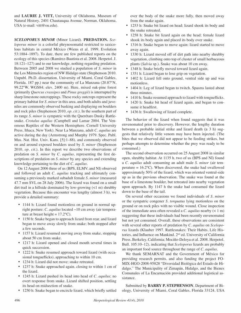
Herpetological Review 41(4), 2010496
and LAURIE J. VITT, University of Oklahoma, Museum of Natural History, 2401 Chautauqua Avenue, Norman, Oklahoma, USA (e-mail: [email protected]).
SCELOPORUS MINOR (Minor Lizard). PREDATION. Sce-loporus minor is a colorful phrynosomatid restricted to saxico-lous habitats in central México (Wiens et al. 1999. Evolution 53:1884–1897). To date, there are few published reports on the ecology of this species (Ramírez-Bautista et al. 2008. Herpetol. J. 18:121–127) and to our knowledge, nothing regarding predation. Between 2005 and 2008 we studied a population of S. minor in the Los Mármoles region of NW Hidalgo state (Stephenson 2010. Unpubl. Ph.D. dissertation, University of Miami, Coral Gables, Florida. 187 pp.) near the community of La Manzana (20.87°N, 99.22°W, WGS84; elev. 2400 m). Here, mixed oak-pine forest (primarily Quercus crassipes and Pinus greggii) is interrupted by sharp limestone outcroppings. These rocky outcrops represent the primary habitat for S. minor in this area, and both adults and juve-niles are commonly observed basking and displaying on boulders and rock piles (Stephenson 2010, op. cit.). In the southern part of its range S. minor is sympatric with the Querétaro Dusky Rattle-snake, Crotalus aquilus (Campbell and Lamar 2004. The Ven-omous Reptiles of the Western Hemisphere. Cornell University Press, Ithaca, New York). Near La Manzana, adult C. aquilus are active during the day (Armstrong and Murphy 1979. Spec. Publ. Mus. Nat. Hist. Univ. Kans. [5]:1–88), and commonly observed on and around exposed boulders used by S. minor (Stephenson 2010, op. cit.). In this report we describe two observations of predation on S. minor by C. aquilus, representing the first de-scriptions of predation on S. minor by any species and extending knowledge pertaining to the diet of C. aquilus. On 12 August 2006 three of us (BPS, ELMV, and NI) observed and followed an adult C. aquilus tracking and ultimately con-suming a previously marked subadult female S. minor (measured 47.7 mm SVL on 28 July 2006). The lizard was found on a small dirt trail in a hillside dominated by low-growing (<1 m) shrubby vegetation. Because this encounter was lengthy (almost 3 h), we provide a detailed summary:
• 1144 h: Lizard found motionless on ground in normal up-right posture; C. aquilus located ~10 cm away (air tempera-ture at breast height = 17.2°C).
• 1150 h: Snake began to approach lizard from rear, and lizard began to move away slowly from snake; both stopped after a few seconds.
• 1157 h: Lizard resumed moving away from snake, stopping about 50 cm from snake.
• 1217 h: Lizard opened and closed mouth several times in quick succession.
• 1222 h: Snake resumed approach toward lizard (with occa-sional tongueflicks), approaching to within 10 cm.
• 1234 h: Lizard did not move; snake retreated.• 1237 h: Snake approached again, closing to within 1 cm of
the lizard.• 1245 h: Lizard pushed its head into head of C. aquilus; no
overt response from snake. Lizard shifted position, settling its head on midsection of snake.
• 1250 h: Snake began to encircle lizard, which briefly settled
over the body of the snake more fully, then moved away from the snake again.
• 1253 h: Snake bit lizard on head; lizard shook its body and the snake retreated.
• 1258 h: Snake bit lizard again on the head; female lizard shook its body again and placed its body over snake.
• 1316 h: Snake began to move again; lizard started to move away again.
• 1330 h: Lizard moved off of dirt path into nearby shrubby vegetation, climbing onto top of cluster of small herbaceous plants (Salvia sp.). Snake was about 10 cm away.
• 1348 h: Snake briefly moved toward lizard again.• 1351 h: Lizard began to lose grip on vegetation.• 1402 h: Lizard fell onto ground, ventral side up and was
motionless.• 1404 h: Leg of lizard began to twitch. Spasms lasted about
three minutes.• 1410 h: Snake resumed approach to lizard with tongueflicks.• 1420 h: Snake bit head of lizard again, and began to con-
sume it headfirst.• 1436 h: Swallowing of lizard complete.
The behavior of the lizard when found suggests that it was envenomated prior to discovery. However, the lengthy duration between a probable initial strike and lizard death (≥ 3 h) sug-gests that relatively little venom may have been injected. (The bites that we observed did not appear to be feeding strikes, but perhaps attempts to determine whether the prey was ready to be consumed.) The second observation occurred on 25 August 2008 in similar open, shrubby habitat. At 1135 h, two of us (BPS and NI) found a C. aquilus adult consuming an adult male S. minor (air tem-perature = 16.2°C). When discovered, the snake had swallowed approximately 50% of the lizard, which was oriented ventral-side up as in the previous observation. The snake was found at the base of a limestone boulder, but retreated into nearby vegetation upon approach. By 1147 h the snake had consumed the lizard down to the base of the tail. On several other occasions we found individuals of S. minor or the sympatric congener S. torquatus lying motionless on the ground or on rock piles with no visible wound. Close inspection of the immediate area often revealed a C. aquilus nearby (< 1 m) suggesting that these individuals had been recently envenomated but not yet consumed. Overall, these observations are consistent with several other reports of predation by C. aquilus on Scelopo-rus lizards (Klauber 1997. Rattlesnakes: Their Habits, Life His-tories, and Influence on Mankind. 2nd ed. University of California Press, Berkeley, California; Mociño-Deloya et al. 2008. Herpetol. Bull. 105:10–12), indicating that Sceloporus lizards are probably an important food source throughout the range of C. aquilus. We thank SEMARNAT and the Government of México for providing research permits, and also funding the project FO-MIX-HGO-2008-95828 “Diversidad Biológica del Estado de Hi-dalgo.” The Municipality of Zimapán, Hidalgo, and the Bienes Comunales of La Encarnación provided additional logistical as-sistance.
Submitted by BARRY P. STEPHENSON, Department of Bi-ology, University of Miami, Coral Gables, Florida 33124, USA
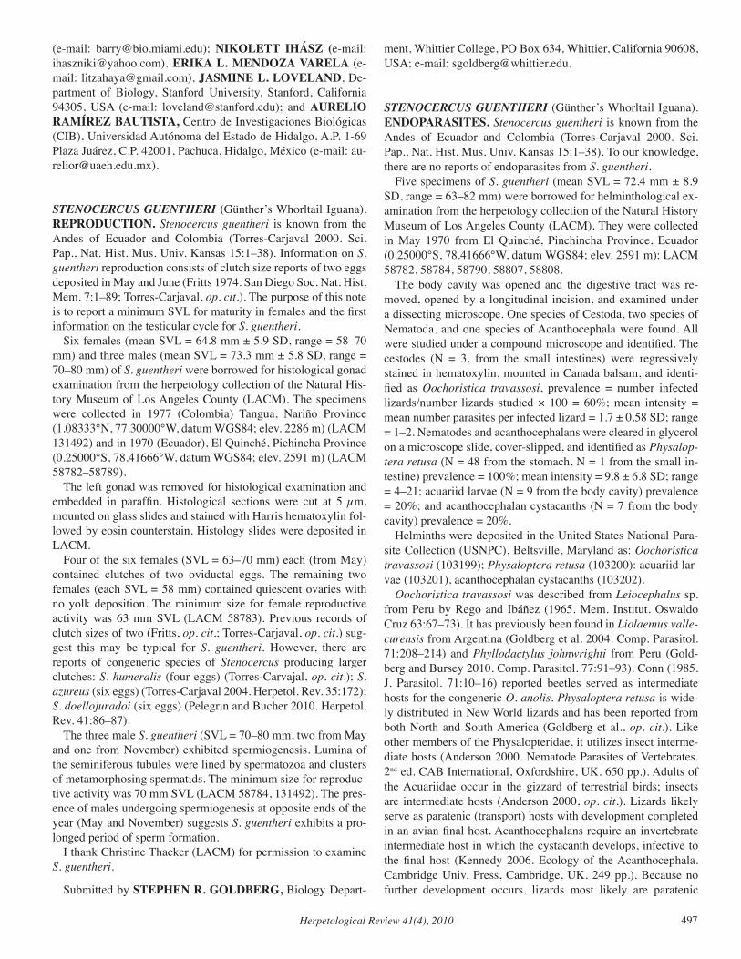
Herpetological Review 41(4), 2010 497
(e-mail: [email protected]); NIKOLETT IHÁSZ (e-mail: [email protected]), ERIKA L. MENDOZA VARELA (e-mail: [email protected]), JASMINE L. LOVELAND, De-partment of Biology, Stanford University, Stanford, California 94305, USA (e-mail: [email protected]); and AURELIO RAMÍREZ BAUTISTA, Centro de Investigaciones Biológicas (CIB), Universidad Autónoma del Estado de Hidalgo, A.P. 1-69 Plaza Juárez, C.P. 42001, Pachuca, Hidalgo, México (e-mail: [email protected]).
STENOCERCUS GUENTHERI (Günther’s Whorltail Iguana). REPRODUCTION. Stenocercus guentheri is known from the Andes of Ecuador and Colombia (Torres-Carjaval 2000. Sci. Pap., Nat. Hist. Mus. Univ. Kansas 15:1–38). Information on S. guentheri reproduction consists of clutch size reports of two eggs deposited in May and June (Fritts 1974. San Diego Soc. Nat. Hist. Mem. 7:1–89; Torres-Carjaval, op. cit.). The purpose of this note is to report a minimum SVL for maturity in females and the first information on the testicular cycle for S. guentheri. Six females (mean SVL = 64.8 mm ± 5.9 SD, range = 58–70 mm) and three males (mean SVL = 73.3 mm ± 5.8 SD, range = 70–80 mm) of S. guentheri were borrowed for histological gonad examination from the herpetology collection of the Natural His-tory Museum of Los Angeles County (LACM). The specimens were collected in 1977 (Colombia) Tangua, Nariño Province (1.08333°N, 77.30000°W, datum WGS84; elev. 2286 m) (LACM 131492) and in 1970 (Ecuador), El Quinché, Pichincha Province (0.25000°S, 78.41666°W, datum WGS84; elev. 2591 m) (LACM 58782–58789). The left gonad was removed for histological examination and embedded in paraffin. Histological sections were cut at 5 µm, mounted on glass slides and stained with Harris hematoxylin fol-lowed by eosin counterstain. Histology slides were deposited in LACM. Four of the six females (SVL = 63–70 mm) each (from May) contained clutches of two oviductal eggs. The remaining two females (each SVL = 58 mm) contained quiescent ovaries with no yolk deposition. The minimum size for female reproductive activity was 63 mm SVL (LACM 58783). Previous records of clutch sizes of two (Fritts, op. cit.; Torres-Carjaval, op. cit.) sug-gest this may be typical for S. guentheri. However, there are reports of congeneric species of Stenocercus producing larger clutches: S. humeralis (four eggs) (Torres-Carvajal, op. cit.); S. azureus (six eggs) (Torres-Carjaval 2004. Herpetol. Rev. 35:172); S. doellojuradoi (six eggs) (Pelegrin and Bucher 2010. Herpetol. Rev. 41:86–87). The three male S. guentheri (SVL = 70–80 mm, two from May and one from November) exhibited spermiogenesis. Lumina of the seminiferous tubules were lined by spermatozoa and clusters of metamorphosing spermatids. The minimum size for reproduc-tive activity was 70 mm SVL (LACM 58784, 131492). The pres-ence of males undergoing spermiogenesis at opposite ends of the year (May and November) suggests S. guentheri exhibits a pro-longed period of sperm formation. I thank Christine Thacker (LACM) for permission to examine S. guentheri.
Submitted by STEPHEN R. GOLDBERG, Biology Depart-
ment, Whittier College, PO Box 634, Whittier, California 90608, USA; e-mail: [email protected].
STENOCERCUS GUENTHERI (Günther’s Whorltail Iguana). ENDOPARASITES. Stenocercus guentheri is known from the Andes of Ecuador and Colombia (Torres-Carjaval 2000. Sci. Pap., Nat. Hist. Mus. Univ. Kansas 15:1–38). To our knowledge, there are no reports of endoparasites from S. guentheri. Five specimens of S. guentheri (mean SVL = 72.4 mm ± 8.9 SD, range = 63–82 mm) were borrowed for helminthological ex-amination from the herpetology collection of the Natural History Museum of Los Angeles County (LACM). They were collected in May 1970 from El Quinché, Pinchincha Province, Ecuador (0.25000°S, 78.41666°W, datum WGS84; elev. 2591 m): LACM 58782, 58784, 58790, 58807, 58808. The body cavity was opened and the digestive tract was re-moved, opened by a longitudinal incision, and examined under a dissecting microscope. One species of Cestoda, two species of Nematoda, and one species of Acanthocephala were found. All were studied under a compound microscope and identified. The cestodes (N = 3, from the small intestines) were regressively stained in hematoxylin, mounted in Canada balsam, and identi-fied as Oochoristica travassosi, prevalence = number infected lizards/number lizards studied × 100 = 60%; mean intensity = mean number parasites per infected lizard = 1.7 ± 0.58 SD; range = 1–2. Nematodes and acanthocephalans were cleared in glycerol on a microscope slide, cover-slipped, and identified as Physalop-tera retusa (N = 48 from the stomach, N = 1 from the small in-testine) prevalence = 100%; mean intensity = 9.8 ± 6.8 SD; range = 4–21; acuariid larvae (N = 9 from the body cavity) prevalence = 20%; and acanthocephalan cystacanths (N = 7 from the body cavity) prevalence = 20%. Helminths were deposited in the United States National Para-site Collection (USNPC), Beltsville, Maryland as: Oochoristica travassosi (103199); Physaloptera retusa (103200): acuariid lar-vae (103201), acanthocephalan cystacanths (103202). Oochoristica travassosi was described from Leiocephalus sp. from Peru by Rego and Ibáñez (1965. Mem. Institut. Oswaldo Cruz 63:67–73). It has previously been found in Liolaemus valle-curensis from Argentina (Goldberg et al. 2004. Comp. Parasitol. 71:208–214) and Phyllodactylus johnwrighti from Peru (Gold-berg and Bursey 2010. Comp. Parasitol. 77:91–93). Conn (1985. J. Parasitol. 71:10–16) reported beetles served as intermediate hosts for the congeneric O. anolis. Physaloptera retusa is wide-ly distributed in New World lizards and has been reported from both North and South America (Goldberg et al., op. cit.). Like other members of the Physalopteridae, it utilizes insect interme-diate hosts (Anderson 2000. Nematode Parasites of Vertebrates. 2nd ed. CAB International, Oxfordshire, UK. 650 pp.). Adults of the Acuariidae occur in the gizzard of terrestrial birds; insects are intermediate hosts (Anderson 2000, op. cit.). Lizards likely serve as paratenic (transport) hosts with development completed in an avian final host. Acanthocephalans require an invertebrate intermediate host in which the cystacanth develops, infective to the final host (Kennedy 2006. Ecology of the Acanthocephala. Cambridge Univ. Press, Cambridge, UK. 249 pp.). Because no further development occurs, lizards most likely are paratenic
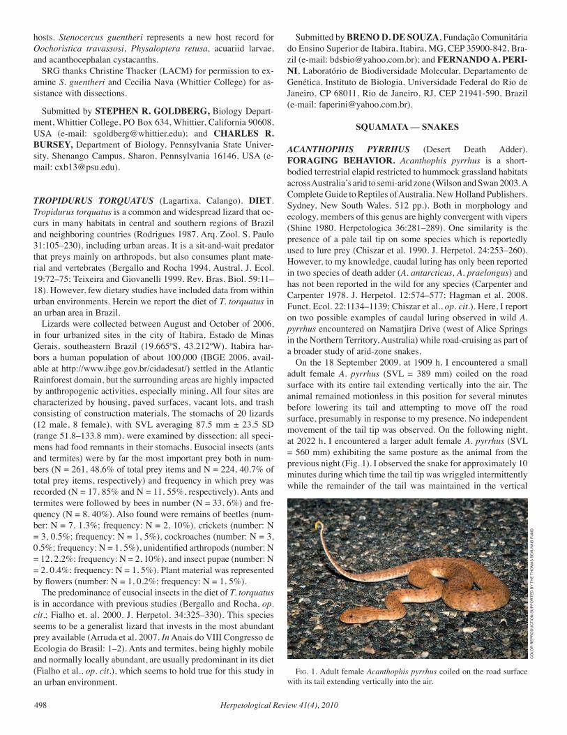
Herpetological Review 41(4), 2010498
hosts. Stenocercus guentheri represents a new host record for Oochoristica travassosi, Physaloptera retusa, acuariid larvae, and acanthocephalan cystacanths. SRG thanks Christine Thacker (LACM) for permission to ex-amine S. guentheri and Cecilia Nava (Whittier College) for as-sistance with dissections.
Submitted by STEPHEN R. GOLDBERG, Biology Depart-ment, Whittier College, PO Box 634, Whittier, California 90608, USA (e-mail: [email protected]); and CHARLES R. BURSEY, Department of Biology, Pennsylvania State Univer-sity, Shenango Campus, Sharon, Pennsylvania 16146, USA (e-mail: [email protected]).
TROPIDURUS TORQUATUS (Lagartixa, Calango). DIET. Tropidurus torquatus is a common and widespread lizard that oc-curs in many habitats in central and southern regions of Brazil and neighboring countries (Rodrigues 1987. Arq. Zool. S. Paulo 31:105–230), including urban areas. It is a sit-and-wait predator that preys mainly on arthropods, but also consumes plant mate-rial and vertebrates (Bergallo and Rocha 1994. Austral. J. Ecol. 19:72–75; Teixeira and Giovanelli 1999. Rev. Bras. Biol. 59:11–18). However, few dietary studies have included data from within urban environments. Herein we report the diet of T. torquatus in an urban area in Brazil. Lizards were collected between August and October of 2006, in four urbanized sites in the city of Itabira, Estado de Minas Gerais, southeastern Brazil (19.665ºS, 43.212ºW). Itabira har-bors a human population of about 100,000 (IBGE 2006, avail-able at http://www.ibge.gov.br/cidadesat/) settled in the Atlantic Rainforest domain, but the surrounding areas are highly impacted by anthropogenic activities, especially mining. All four sites are characterized by housing, paved surfaces, vacant lots, and trash consisting of construction materials. The stomachs of 20 lizards (12 male, 8 female), with SVL averaging 87.5 mm ± 23.5 SD (range 51.8–133.8 mm), were examined by dissection; all speci-mens had food remnants in their stomachs. Eusocial insects (ants and termites) were by far the most important prey both in num-bers (N = 261, 48.6% of total prey items and N = 224, 40.7% of total prey items, respectively) and frequency in which prey was recorded (N = 17, 85% and N = 11, 55%, respectively). Ants and termites were followed by bees in number (N = 33, 6%) and fre-quency (N = 8, 40%). Also found were remains of beetles (num-ber: N = 7, 1.3%; frequency: N = 2, 10%), crickets (number: N = 3, 0.5%; frequency: N = 1, 5%), cockroaches (number: N = 3, 0.5%; frequency: N = 1, 5%), unidentified arthropods (number: N = 12, 2.2%; frequency: N = 2, 10%), and insect pupae (number: N = 2, 0.4%; frequency: N = 1, 5%). Plant material was represented by flowers (number: N = 1, 0.2%; frequency: N = 1, 5%). The predominance of eusocial insects in the diet of T. torquatus is in accordance with previous studies (Bergallo and Rocha, op. cit.; Fialho et. al. 2000. J. Herpetol. 34:325–330). This species seems to be a generalist lizard that invests in the most abundant prey available (Arruda et al. 2007. In Anais do VIII Congresso de Ecologia do Brasil: 1–2). Ants and termites, being highly mobile and normally locally abundant, are usually predominant in its diet (Fialho et al., op. cit.), which seems to hold true for this study in an urban environment.
Submitted by BRENO D. DE SOUZA, Fundação Comunitária do Ensino Superior de Itabira, Itabira, MG, CEP 35900-842, Bra-zil (e-mail: [email protected]); and FERNANDO A. PERI-NI, Laboratório de Biodiversidade Molecular, Departamento de Genética, Instituto de Biologia, Universidade Federal do Rio de Janeiro, CP 68011, Rio de Janeiro, RJ, CEP 21941-590, Brazil (e-mail: [email protected]).
SQUAMATA — SNAKES
ACANTHOPHIS PYRRHUS (Desert Death Adder). FORAGING BEHAVIOR. Acanthophis pyrrhus is a short-bodied terrestrial elapid restricted to hummock grassland habitats across Australia’s arid to semi-arid zone (Wilson and Swan 2003. A Complete Guide to Reptiles of Australia. New Holland Publishers. Sydney, New South Wales. 512 pp.). Both in morphology and ecology, members of this genus are highly convergent with vipers (Shine 1980. Herpetologica 36:281–289). One similarity is the presence of a pale tail tip on some species which is reportedly used to lure prey (Chiszar et al. 1990. J. Herpetol. 24:253–260). However, to my knowledge, caudal luring has only been reported in two species of death adder (A. antarcticus, A. praelongus) and has not been reported in the wild for any species (Carpenter and Carpenter 1978. J. Herpetol. 12:574–577; Hagman et al. 2008. Funct. Ecol. 22:1134–1139; Chiszar et al., op. cit.). Here, I report on two possible examples of caudal luring observed in wild A. pyrrhus encountered on Namatjira Drive (west of Alice Springs in the Northern Territory, Australia) while road-cruising as part of a broader study of arid-zone snakes. On the 18 September 2009, at 1909 h, I encountered a small adult female A. pyrrhus (SVL = 389 mm) coiled on the road surface with its entire tail extending vertically into the air. The animal remained motionless in this position for several minutes before lowering its tail and attempting to move off the road surface, presumably in response to my presence. No independent movement of the tail tip was observed. On the following night, at 2022 h, I encountered a larger adult female A. pyrrhus (SVL = 560 mm) exhibiting the same posture as the animal from the previous night (Fig. 1). I observed the snake for approximately 10 minutes during which time the tail tip was wriggled intermittently while the remainder of the tail was maintained in the vertical
fig. 1. Adult female Acanthophis pyrrhus coiled on the road surface with its tail extending vertically into the air.
CO
LOR
REP
RO
du
CTi
ON
Su
PPO
RTEd
BY
THE
THO
mAS
BEA
uvA
iS F
uN
d
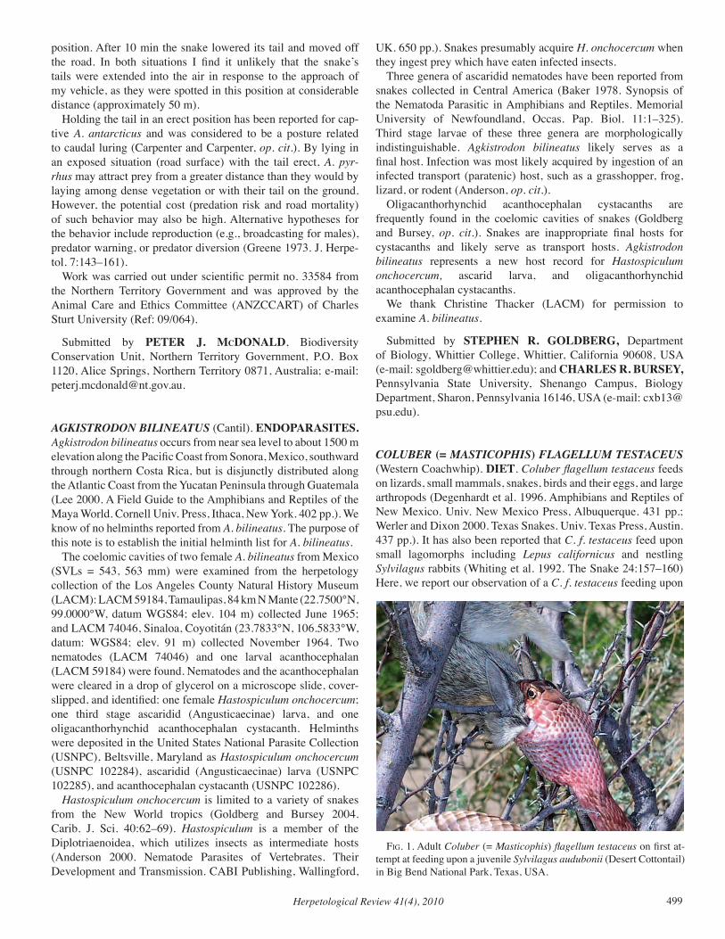
Herpetological Review 41(4), 2010 499
position. After 10 min the snake lowered its tail and moved off the road. In both situations I find it unlikely that the snake’s tails were extended into the air in response to the approach of my vehicle, as they were spotted in this position at considerable distance (approximately 50 m). Holding the tail in an erect position has been reported for cap-tive A. antarcticus and was considered to be a posture related to caudal luring (Carpenter and Carpenter, op. cit.). By lying in an exposed situation (road surface) with the tail erect, A. pyr-rhus may attract prey from a greater distance than they would by laying among dense vegetation or with their tail on the ground. However, the potential cost (predation risk and road mortality) of such behavior may also be high. Alternative hypotheses for the behavior include reproduction (e.g., broadcasting for males), predator warning, or predator diversion (Greene 1973. J. Herpe-tol. 7:143–161). Work was carried out under scientific permit no. 33584 from the Northern Territory Government and was approved by the Animal Care and Ethics Committee (ANZCCART) of Charles Sturt University (Ref: 09/064).
Submitted by PETER J. McDONALD, Biodiversity Conservation Unit, Northern Territory Government, P.O. Box 1120, Alice Springs, Northern Territory 0871, Australia; e-mail: [email protected].
AGKISTRODON BILINEATUS (Cantil). ENDOPARASITES. Agkistrodon bilineatus occurs from near sea level to about 1500 m elevation along the Pacific Coast from Sonora, Mexico, southward through northern Costa Rica, but is disjunctly distributed along the Atlantic Coast from the Yucatan Peninsula through Guatemala (Lee 2000. A Field Guide to the Amphibians and Reptiles of the Maya World. Cornell Univ. Press, Ithaca, New York. 402 pp.). We know of no helminths reported from A. bilineatus. The purpose of this note is to establish the initial helminth list for A. bilineatus. The coelomic cavities of two female A. bilineatus from Mexico (SVLs = 543, 563 mm) were examined from the herpetology collection of the Los Angeles County Natural History Museum (LACM): LACM 59184, Tamaulipas, 84 km N Mante (22.7500°N, 99.0000°W, datum WGS84; elev. 104 m) collected June 1965; and LACM 74046, Sinaloa, Coyotitán (23.7833°N, 106.5833°W, datum: WGS84; elev. 91 m) collected November 1964. Two nematodes (LACM 74046) and one larval acanthocephalan (LACM 59184) were found. Nematodes and the acanthocephalan were cleared in a drop of glycerol on a microscope slide, cover-slipped, and identified: one female Hastospiculum onchocercum; one third stage ascaridid (Angusticaecinae) larva, and one oligacanthorhynchid acanthocephalan cystacanth. Helminths were deposited in the United States National Parasite Collection (USNPC), Beltsville, Maryland as Hastospiculum onchocercum (USNPC 102284), ascaridid (Angusticaecinae) larva (USNPC 102285), and acanthocephalan cystacanth (USNPC 102286). Hastospiculum onchocercum is limited to a variety of snakes from the New World tropics (Goldberg and Bursey 2004. Carib. J. Sci. 40:62–69). Hastospiculum is a member of the Diplotriaenoidea, which utilizes insects as intermediate hosts (Anderson 2000. Nematode Parasites of Vertebrates. Their Development and Transmission. CABI Publishing, Wallingford,
UK. 650 pp.). Snakes presumably acquire H. onchocercum when they ingest prey which have eaten infected insects. Three genera of ascaridid nematodes have been reported from snakes collected in Central America (Baker 1978. Synopsis of the Nematoda Parasitic in Amphibians and Reptiles. Memorial University of Newfoundland, Occas. Pap. Biol. 11:1–325). Third stage larvae of these three genera are morphologically indistinguishable. Agkistrodon bilineatus likely serves as a final host. Infection was most likely acquired by ingestion of an infected transport (paratenic) host, such as a grasshopper, frog, lizard, or rodent (Anderson, op. cit.). Oligacanthorhynchid acanthocephalan cystacanths are frequently found in the coelomic cavities of snakes (Goldberg and Bursey, op. cit.). Snakes are inappropriate final hosts for cystacanths and likely serve as transport hosts. Agkistrodon bilineatus represents a new host record for Hastospiculum onchocercum, ascarid larva, and oligacanthorhynchid acanthocephalan cystacanths. We thank Christine Thacker (LACM) for permission to examine A. bilineatus.
Submitted by STEPHEN R. GOLDBERG, Department of Biology, Whittier College, Whittier, California 90608, USA (e-mail: [email protected]); and CHARLES R. BURSEY, Pennsylvania State University, Shenango Campus, Biology Department, Sharon, Pennsylvania 16146, USA (e-mail: [email protected]).
COLUBER (= MASTICOPHIS) FLAGELLUM TESTACEUS (Western Coachwhip). DIET. Coluber flagellum testaceus feeds on lizards, small mammals, snakes, birds and their eggs, and large arthropods (Degenhardt et al. 1996. Amphibians and Reptiles of New Mexico. Univ. New Mexico Press, Albuquerque. 431 pp.; Werler and Dixon 2000. Texas Snakes. Univ. Texas Press, Austin. 437 pp.). It has also been reported that C. f. testaceus feed upon small lagomorphs including Lepus californicus and nestling Sylvilagus rabbits (Whiting et al. 1992. The Snake 24:157–160) Here, we report our observation of a C. f. testaceus feeding upon
Fig. 1. Adult Coluber (= Masticophis) flagellum testaceus on first at-tempt at feeding upon a juvenile Sylvilagus audubonii (Desert Cottontail) in Big Bend National Park, Texas, USA.
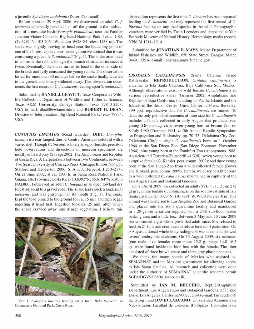
Herpetological Review 41(4), 2010500
a juvenile Sylvilagus audubonii (Desert Cottontail). Before noon on 29 April 2006, we discovered an adult C. f. testaceus apparently perched 1 m off the ground in the midsec-tion of a mesquite bush (Prosopis glandulosa) near the Panther Junction Visitor Center in Big Bend National Park, Texas, USA (29.3281°N, 103.2064°W, datum WGS 84; elev. 1138 m). The snake was slightly moving its head near the branching point of one of the limbs. Upon closer investigation we noticed that it was consuming a juvenile S. audubonii (Fig. 1). The snake attempted to consume the rabbit, though the branch obstructed its success twice. Eventually, the snake turned its head to the other side of the branch and fully consumed the young rabbit. The observation lasted for more than 30 minutes before the snake finally crawled to the ground and slowly slithered away. This observation docu-ments the first record of C. f. testaceus feeding upon S. audubonii.
Submitted by DANIEL J. LEAVITT, Texas Cooperative Wild-life Collection, Department of Wildlife and Fisheries Science, Texas A&M University, College Station, Texas 77843-2258, USA (e-mail: [email protected]); and ROBERT H. DEAN, Division of Interpretation, Big Bend National Park, Texas 79834, USA.
CONOPHIS LINEATUS (Road Guarder). DIET. Conophis lineatus is a rear-fanged, diurnal Central American colubrid with a varied diet. Though C. lineatus is likely an opportunistic predator, field observations and dissections of museum specimens are mostly of lizard prey (Savage 2002. The Amphibians and Reptiles of Costa Rica: A Herpetofauna between Two Continents, between Two Seas. University of Chicago Press, Chicago, Illinois. 954 pp.; Stafford and Henderson 2006. S. Am. J. Herpetol. 1:210–217). On 21 June 2002, at ca. 1500 h, in Santa Rosa National Park, Guanacaste Province, Costa Rica (10.8392°N, 85.6184°W, datum NAD83), I observed an adult C. lineatus in an open lowland dry forest adjacent to a gravel road. The snake had struck a toad, Bufo luetkenii, and was grasping it in its mouth (Fig. 1). The snake kept the toad pinned to the ground for ca. 15 min and then began ingesting it head first. Ingestion took ca. 25 min, after which the snake crawled away into denser vegetation. I believe this
observation represents the first time C. lineatus has been reported feeding on B. luetkenii and may represent the first record of C. lineatus feeding on any toad species in the wild. Photographic vouchers were verified by Twan Leenders and deposited at Yale Peabody Museum of Natural History, Herpetology media records YPM M 1211–1214.
Submitted by JONATHAN D. MAYS, Maine Department of Inland Fisheries and Wildlife, 650 State Street, Bangor, Maine 04401, USA; e-mail: [email protected].
CROTALUS CATALINENSIS (Santa Catalina Island Rattlesnake). REPRODUCTION. Crotalus catalinensis is endemic to Isla Santa Catalina, Baja California Sur, Mexico. Although observations exist of wild female C. catalinensis in various reproductive states (Grismer 2002. Amphibians and Reptiles of Baja California, Including its Pacific Islands and the Islands in the Sea of Cortés. Univ. California Press, Berkeley. 399 pp.), reproductive data for C. catalinensis are limited. To date, the only published accounts of litter size for C. catalinensis include: a female collected in early August that produced two young (Grismer, op cit.); seven young born at Fresno Zoo on 8 July 1980 (Tremper 1981. In 5th Annual Reptile Symposium on Propagation and Husbandry, pp. 70–75. Oklahoma City Zoo, Oklahoma City); a single C. catalinensis born on 1 October 1964 at the San Diego Zoo (San Diego Zoonooz, November 1964); nine young born at the Frankfurt Zoo (Anonymous 1988. Aquarien-und Terrarien-Zeitschrift 41:328); seven young born to a captive female (G. Keasler, pers. comm. 2009); and three young born at the San Diego Zoo from a wild collected female (Boyer and Kinkaid, pers. comm. 2009). Herein, we describe a litter born to a wild collected C. catalinensis maintained in captivity at the Los Angeles Zoo and Botanical Gardens. On 23 April 2009, we collected an adult (SVL = 71.12 cm; 272 g) gray phase female C. catalinensis on the southwest side of Isla Santa Catalina, 25.6023°N, 110.7791°W, WGS 84; elev. 0 m. This animal was transferred to Los Angeles Zoo and Botanical Garden and placed into the zoo’s quarantine facility and maintained in a 20-gallon terrarium supplied with a 24-h sub-floor heated basking area and a hide box. Between 3 May and 10 June 2009 she consumed eight whole pre-killed adult mice. She refused to feed on 21 June and continued to refuse food until parturition. On 9 August a dorsal whole body radiograph was taken and showed several embryonic skeletons. On 13 August 2009, six neonates (one male, five female; mean mass 15.2 g; range 14.8–16.3 g), were found inside the hide box with the female. The litter consisted of three brown phase and three gray phase neonates. We thank the many people of Mexico who assisted us, SEMARNAT, and the Mexican government for allowing access to Isla Santa Catalina. All research and collecting were done under the authority of SEMARNAT scientific research permit SGPA/DGVS/03804, issued to IR.
Submitted by IAN M. RECCHIO, Reptile/Amphibian Department, Los Angeles Zoo and Botanical Gardens, 5333 Zoo Drive, Los Angeles, California 90027, USA (e-mail: [email protected]); and DAVID LAZCANO, Universidad Autónoma de Nuevo León, Facultad de Ciencias Biológicas, Laboratorio de
fig. 1. Conophis lineatus feeding on a toad, Bufo luetkenii, in Guanacaste National Park, Costa Rica.
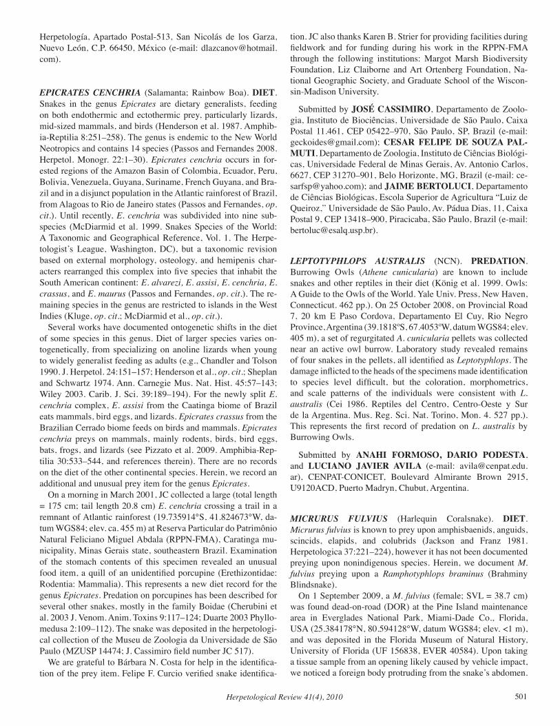
Herpetological Review 41(4), 2010 501
Herpetología, Apartado Postal-513, San Nicolás de los Garza, Nuevo León, C.P. 66450, México (e-mail: [email protected]).
EPICRATES CENCHRIA (Salamanta; Rainbow Boa). DIET. Snakes in the genus Epicrates are dietary generalists, feeding on both endothermic and ectothermic prey, particularly lizards, mid-sized mammals, and birds (Henderson et al. 1987. Amphib-ia-Reptilia 8:251–258). The genus is endemic to the New World Neotropics and contains 14 species (Passos and Fernandes 2008. Herpetol. Monogr. 22:1–30). Epicrates cenchria occurs in for-ested regions of the Amazon Basin of Colombia, Ecuador, Peru, Bolivia, Venezuela, Guyana, Suriname, French Guyana, and Bra-zil and in a disjunct population in the Atlantic rainforest of Brazil, from Alagoas to Rio de Janeiro states (Passos and Fernandes, op. cit.). Until recently, E. cenchria was subdivided into nine sub-species (McDiarmid et al. 1999. Snakes Species of the World: A Taxonomic and Geographical Reference, Vol. 1. The Herpe-tologist’s League, Washington, DC), but a taxonomic revision based on external morphology, osteology, and hemipenis char-acters rearranged this complex into five species that inhabit the South American continent: E. alvarezi, E. assisi, E. cenchria, E. crassus, and E. maurus (Passos and Fernandes, op. cit.). The re-maining species in the genus are restricted to islands in the West Indies (Kluge, op. cit.; McDiarmid et al., op. cit.). Several works have documented ontogenetic shifts in the diet of some species in this genus. Diet of larger species varies on-togenetically, from specializing on anoline lizards when young to widely generalist feeding as adults (e.g., Chandler and Tolson 1990. J. Herpetol. 24:151–157; Henderson et al., op. cit.; Sheplan and Schwartz 1974. Ann. Carnegie Mus. Nat. Hist. 45:57–143; Wiley 2003. Carib. J. Sci. 39:189–194). For the newly split E. cenchria complex, E. assisi from the Caatinga biome of Brazil eats mammals, bird eggs, and lizards. Epicrates crassus from the Brazilian Cerrado biome feeds on birds and mammals. Epicrates cenchria preys on mammals, mainly rodents, birds, bird eggs, bats, frogs, and lizards (see Pizzato et al. 2009. Amphibia-Rep-tilia 30:533–544, and references therein). There are no records on the diet of the other continental species. Herein, we record an additional and unusual prey item for the genus Epicrates. On a morning in March 2001, JC collected a large (total length = 175 cm; tail length 20.8 cm) E. cenchria crossing a trail in a remnant of Atlantic rainforest (19.735914°S, 41.824673°W, da-tum WGS84; elev. ca. 455 m) at Reserva Particular do Patrimônio Natural Feliciano Miguel Abdala (RPPN-FMA), Caratinga mu-nicipality, Minas Gerais state, southeastern Brazil. Examination of the stomach contents of this specimen revealed an unusual food item, a quill of an unidentified porcupine (Erethizontidae: Rodentia: Mammalia). This represents a new diet record for the genus Epicrates. Predation on porcupines has been described for several other snakes, mostly in the family Boidae (Cherubini et al. 2003 J. Venom. Anim. Toxins 9:117–124; Duarte 2003 Phyllo-medusa 2:109–112). The snake was deposited in the herpetologi-cal collection of the Museu de Zoologia da Universidade de São Paulo (MZUSP 14474; J. Cassimiro field number JC 517). We are grateful to Bárbara N. Costa for help in the identifica-tion of the prey item. Felipe F. Curcio verified snake identifica-
tion. JC also thanks Karen B. Strier for providing facilities during fieldwork and for funding during his work in the RPPN-FMA through the following institutions: Margot Marsh Biodiversity Foundation, Liz Claiborne and Art Ortenberg Foundation, Na-tional Geographic Society, and Graduate School of the Wiscon-sin-Madison University.
Submitted by JOSÉ CASSIMIRO, Departamento de Zoolo-gia, Instituto de Biociências, Universidade de São Paulo, Caixa Postal 11.461, CEP 05422–970, São Paulo, SP, Brazil (e-mail: [email protected]); CESAR FELIPE DE SOUZA PAL-MUTI, Departamento de Zoologia, Instituto de Ciências Biológi-cas, Universidade Federal de Minas Gerais, Av. Antonio Carlos, 6627, CEP 31270–901, Belo Horizonte, MG, Brazil (e-mail: [email protected]); and JAIME BERTOLUCI, Departamento de Ciências Biológicas, Escola Superior de Agricultura “Luiz de Queiroz,” Universidade de São Paulo, Av. Pádua Dias, 11, Caixa Postal 9, CEP 13418–900, Piracicaba, São Paulo, Brazil (e-mail: [email protected]).
LEPTOTYPHLOPS AUSTRALIS (NCN). PREDATION. Burrowing Owls (Athene cunicularia) are known to include snakes and other reptiles in their diet (König et al. 1999. Owls: A Guide to the Owls of the World. Yale Univ. Press, New Haven, Connecticut. 462 pp.). On 25 October 2008, on Provincial Road 7, 20 km E Paso Cordova, Departamento El Cuy, Rio Negro Province, Argentina (39.1818ºS, 67.4053ºW, datum WGS84; elev. 405 m), a set of regurgitated A. cunicularia pellets was collected near an active owl burrow. Laboratory study revealed remains of four snakes in the pellets, all identified as Leptotyphlops. The damage inflicted to the heads of the specimens made identification to species level difficult, but the coloration, morphometrics, and scale patterns of the individuals were consistent with L. australis (Cei 1986. Reptiles del Centro, Centro-Oeste y Sur de la Argentina. Mus. Reg. Sci. Nat. Torino, Mon. 4. 527 pp.). This represents the first record of predation on L. australis by Burrowing Owls.
Submitted by ANAHI FORMOSO, DARIO PODESTA, and LUCIANO JAVIER AVILA (e-mail: [email protected]), CENPAT-CONICET, Boulevard Almirante Brown 2915, U9120ACD, Puerto Madryn, Chubut, Argentina.
MICRURUS FULvIUS (Harlequin Coralsnake). DIET. Micrurus fulvius is known to prey upon amphisbaenids, anguids, scincids, elapids, and colubrids (Jackson and Franz 1981. Herpetologica 37:221–224), however it has not been documented preying upon nonindigenous species. Herein, we document M. fulvius preying upon a Ramphotyphlops braminus (Brahminy Blindsnake). On 1 September 2009, a M. fulvius (female; SVL = 38.7 cm) was found dead-on-road (DOR) at the Pine Island maintenance area in Everglades National Park, Miami-Dade Co., Florida, USA (25.384178°N, 80.594128°W, datum WGS84; elev. <1 m), and was deposited in the Florida Museum of Natural History, University of Florida (UF 156838, EVER 40584). Upon taking a tissue sample from an opening likely caused by vehicle impact, we noticed a foreign body protruding from the snake’s abdomen.
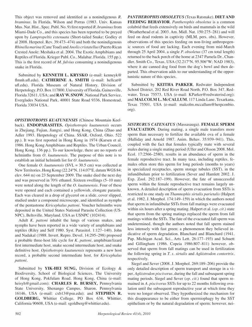
Herpetological Review 41(4), 2010502
This object was removed and identified as a nonindigenous R. braminus. In Florida, Wilson and Porras (1983. Univ. Kansas Mus. Nat. Hist., Spec. Publ. No. 9) first reported R. braminus from Miami-Dade Co., and this species has been reported to be preyed upon by Lampropeltis extenuata (Short-tailed Snake; Godley et al 2008. Herpetol. Rev. 39:473–474) and both the nonindigenous Rhinella marina (Cane Toad) and Anolis cristatellus (Puerto Rican Crested Anole; Meshaka et al. 2004. The Exotic Amphibians and Reptiles of Florida. Krieger Publ. Co., Malabar, Florida. 155 pp.). This is the first record of M. fulvius consuming a nonindigenous snake in Florida.
Submitted by KENNETH L. KRYSKO (e-mail: [email protected]), CATHERINE A. SMITH (e-mail: [email protected]), Florida Museum of Natural History, Division of Herpetology, P.O. Box 117800, University of Florida, Gainesville, Florida 32611, USA; and RAY W. SNOW, National Park Service, Everglades National Park, 40001 State Road 9336, Homestead, Florida 33034 USA.
OPISTHOTROPIS KUATUNENSIS (Chinese Mountain Keel-back). ENDOPARASITES. Opisthotropis kuatunensis occurs in Zhejiang, Fujian, Jiangxi, and Hong Kong, China (Zhao and Adler 1993. Herpetology of China. SSAR, Oxford, Ohio. 522 pp.). It was first reported in Hong Kong in 1974 (Karsen et al. 1986. Hong Kong Amphibians and Reptiles. The Urban Council, Hong Kong. 136 pp.). To our knowledge, there are no reports of helminths from O. kuatunensis. The purpose of this note is to establish an initial helminth list for O. kuatunensis. One female O. kuatunensis (SVL = 30.5 cm) was collected at New Territories, Hong Kong (22.24°N, 114.07°E, datum WGS 84; elev. 644 m) on 23 September 2009. The snake died the next day and was preserved in 70% ethanol. Sixteen swellings (5–10 mm) were noted along the length of the O. kuatunensis. Four of these were opened and each contained a yellowish, elongate parasite. Each was cleared in a drop of glycerol on a slide, cover slipped, studied under a compound microscope, and identified as nymphs of the pentastome Kiricephalus pattoni. Voucher helminths were deposited in the United States National Parasite Collection (US-NPC), Beltsville, Maryland, USA as USNPC (102414). Adult K. pattoni inhabit the lungs of various snakes, and nymphs have been reported in a wide variety of amphibians and reptiles (Riley and Self 1980. Syst. Parasitol. 1:127–140). John and Nadakal (1988. Invert. Repro. Devel. 14:295–298) proposed a probable three-host life cycle for K. pattoni, amphibian/lizard first intermediate host, snake second intermediate host, and snake definitive host. Opisthotropis kuatunensis represents a new host record, a probable second intermediate host, for Kiricephalus pattoni.
Submitted by YIK-HEI SUNG, Division of Ecology & Biodiversity, School of Biological Sciences, The University of Hong Kong, Pokfulam Road, Hong Kong, China (e-mail: [email protected]); CHARLES R. BURSEY, Pennsylvania State University, Shenango Campus, Sharon, Pennsylvania 16146, USA (e-mail: [email protected]); and STEPHEN R. GOLDBERG, Whittier College, PO Box 634, Whittier, California 90608, USA (e-mail: [email protected]).
PANTHEROPHIS OBSOLETUS (Texas Ratsnake). DIET AND FEEDING BEHAVIOR. Pantherophis obsoletus is a common colubrid that feeds extensively on birds and mammals in the wild (Weatherhead et al. 2003. Am. Midl. Nat. 150:275–281) and will feed on dead rodents in captivity (MLM, pers. obs). However, observations of this species feeding on non-living anthropogen-ic sources of food are lacking. Each evening from mid-March through 25 April 2004, a single P. obsoletus (37 cm total length) returned to the back porch of the home at 2347 Pamela Dr., Chan-dler, Smith Co., Texas, USA (32.217°N, 95.506°W; NAD 1983), where it ate canned dog food from the dog’s bowl and then de-parted. This observation adds to our understanding of the oppor-tunistic nature of this species.
Submitted by KEITHA PARKER, Redwater Independent School District, 202 Red River Road North, P.O. Box 347, Red-water, Texas 75573, USA (e-mail: [email protected]); and MALCOLM L. McCALLUM, 117 Linda Lane, Texarkana, Texas 75501, USA (e-mail: [email protected]).
SISTRURUS CATENATUS (Massasauga). FEMALE SPERM EVACUATION. During mating, a single male transfers more sperm than necessary to fertilize the available ova of a female (Halliday and Arnold 1987. Anim. Behav. 35:939–941). This, coupled with the fact that females typically mate with several males during a single mating period (Uller and Olsson 2008. Mol. Ecol. 17:2566–2580), results in an abundance of sperm in the female reproductive tract. In many taxa, including reptiles, fe-males often store this sperm for long periods (months to years) in specialized receptacles, sperm storage tubules (SST), in the infundibulum prior to fertilization (Sever and Hamlett 2002. J. Exp. Zool. 292:187–199). However, the fate of unsuccessful sperm within the female reproductive tract remains largely un-known. A detailed description of sperm evacuation from SSTs is restricted to one study on Thamnophis sirtalis parietalis (Halpert et al. 1982. J. Morphol. 174:149–159) in which the authors noted that sperm in infundibular SSTs from fall matings were evacuated within six hours after a spring mating. Halpert et al. (op. cit.) state that sperm from the spring matings replaced the sperm from fall matings within the SSTs. The fate of the evacuated fall sperm was undetermined, though the authors noted that fall sperm stained less intensely with fast green; a phenomenon they believed in-dicative of sperm degradation. Blanchard and Blanchard (1941. Pap. Michigan Acad. Sci., Arts Lett. 26:177–193) and Schuett and Gillingham (1986. Copeia 1986:807–811) however, ob-served that sperm from fall matings can be used in fertilization the following spring in T. s. sirtalis and Agkistrodon contortrix, respectively. Siegel and Sever (2008. J. Morphol. 269:189–206) provide the only detailed description of sperm transport and storage in a vi-per, Agkistrodon piscivorus, during the fall and subsequent spring mating periods. Siegel and Sever (op. cit.) found that sperm re-mained in A. piscivorus SSTs for up to 22 months following ovu-lation until the subsequent reproductive year at which time they were no longer observed. They hypothesized the mechanism for this disappearance to be either from spermiophagy by the SST epithelium or by the natural degradation of sperm; however, nei-
Herpetological Review 41(4), 2010 503
Fig. 1. Sperm in a pregnant female Sistrurus catenatus fecal sample collected 11 July 2005. Sn, sperm nucleus; St, sperm tail; Fm, fecal ma-terial.
ther of these processes were observed. Additionally, Siegel and Sever (op. cit.) failed to observe the evacuation of sperm from SSTs following the spring mating season as described by Halpert et al. (op. cit.). The following account adds to the scant literature above by providing an incident of sperm evacuation in a female Sistrurus catenatus. As part of a long-term study focusing on the conservation of S. catenatus in Butler Co., Pennsylvania, USA, free-ranging in-dividuals were implanted with radio transmitters and monitored for a suite of ecological, behavioral (including mating activities), and physiological variables three times weekly. One female was initially encountered in May 2005 at which time an ultrasound revealed the presence of enlarged vitellogenic follicles indicat-ing that she would likely give birth later in the year (birthed 19 August 2005). A fecal sample collected on 11 July 2005 to assess her general health and parasite load revealed something striking: numerous sperm (Fig. 1). Though isolated incidents of spring re-productive behavior have been reported in S. catenatus (Wright 1941. Am. Midl. Nat. 25:659–672; Jellen et al. 2007. J. Herpetol. 41:451–457), mating predominately occurs in the late summer–early fall period (Jellen et al., op. cit.; Johnson 2000. J. Herpetol. 34:186–192; Reinert 1981. Am. Midl. Nat. 105:393–395). If this sperm represents sperm from a recent mating, it would pre-date the earliest recorded date of summer reproductive behavior in S. catenatus by approximately two weeks (24 July; Jellen et al., op. cit.). Additionally, though male S. catenatus have been observed to engage in reproductive behaviors with pregnant females (Jellen et al., op. cit.), this female was never observed in the vicinity of a male nor exhibited any mating behaviors throughout the course of the year. However, because this individual was free-ranging, and consequently not under constant surveillance, an unobserved copulation during the active season prior to 11 July 2005 can-not be ruled out. A more plausible explanation, however, is that this observation represents the systematic oviductal evacuation of sperm from matings during (or prior to) the 2004 late summer–early fall mating period, which were unsuccessful in fertilizing ova in the spring of 2005. Siegel and Sever (op. cit.) report that sperm degrade in the pos-terior oviduct prior to ovulation in vipers and describe the only storage site for sperm during pregnancy as within SSTs. Although a definitive mechanism for sperm evacuation was not observed in
A. piscivorus, densities of sperm decreased in the SST over time until the subsequent mating season (Siegel and Sever, unpubl. data). A similar decrease in the presence of oviductal sperm fol-lowing oviposition was observed in Tantilla coronata (Aldridge 1992. Amphibia-Reptilia 13:219–225). We propose that after ovulation, sperm are intermittently evacuated from SSTs. This process undoubtedly takes an extended period of time due to the lack of contractile elements associated with SSTs in snakes (for review see Siegel and Sever, op. cit.), and culminates with the eventual collection of sperm in the cloaca (a structure previously ignored in histological studies on sperm transport in snakes). Normal defecation then provides a proximate outlet for unsuc-cessful sperm in the female reproductive tract. We suggest that further histological and behavioral studies on sperm transport, storage, and evacuation are needed to confirm this phenomenon. We thank M. Kowalski, B. Levine, the Pittsburgh Zoo, Game Commission, and Department of Conservation of Natural Re-sources.
Submitted by BENJAMIN C. JELLEN, Western Pennsylva-nia Conservancy, 800 Waterfront Drive, Pittsburgh, Pennsylva-nia 15222, USA; present address: Department of Biology, Saint Louis University, St. Louis, Missouri 63103, USA (e-mail: [email protected]); DUSTIN S. SIEGEL, and ROBERT D. ALDRIDGE, Department of Biology, Saint Louis University, St. Louis, Missouri 63103, USA.
THAMNOPHIS ATRATUS HYDROPHILUS (Oregon Garter-snake). MAXIMUM ELEVATION. Thamnophis atratus has been recorded at elevations up to 1920 m (6297 ft; St. John 2002. Reptiles of the Northwest. Lone Pine Publishing, Renton, Wash-ington. 272 pp.). Here we report a population of T. a. hydrophi-lus, occurring entirely above this reported elevational limit in the Klamath Mountains, California, USA. Our observations occurred in upper Deep Creek Basin, Trinity Alps Wilderness, Trinity Co., California, USA (40.9176°N, 122.8876°W, datum NAD83). Deep Creek Basin is a medium-sized glacial characterized cirque (342 ha) encompassed by steep jagged peaks reaching elevations up to 2497 m, which are among the highest in the Klamath Mountains. We captured and individually marked 56 individual T. a. hy-drophilus of various age classes during 2003–2006. We recap-tured many of these individuals, with total captures reaching 127 over the four years of the study. All snakes were found in streams, ponds, and Echo Lake, at elevations ranging from 1960 to 2215 m. These observations expand the known vertical limit of T. atra-tus by 295 m (968 ft). Much of the Klamath Mountains includ-ing the Trinity Alps, Russian, Marble Mountain, and Siskiyou Wilderness areas contain similar aquatic habitats that exceed the known previous elevational limit for this species. In lower elevation streams, native salmonid fishes are an im-portant component of T. a. hydrophilus diet (Welsh and Lind 2000. J. Herpetol. 34:67–74). Over the last century, nonnative salmonids have been introduced into most of the naturally fish-less high-elevation aquatic habitats in the Klamath Mountains (Welsh et al. 2006. Divers. Distrib. 12:298–309). Introduced sal-monid prey in the region may have allowed T. a. hydrophilus to expand into these high-elevation habitats (Pope et al. 2008 Biol. Conserv. 141:1321–1331).
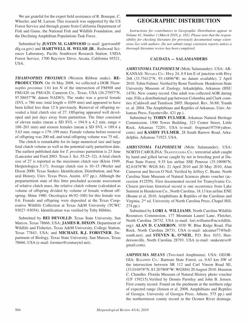
Herpetological Review 41(4), 2010504
GEOGRAPHIC DISTRIBUTION Instructions for contributors to Geographic Distribution appear in Volume 41, Number 1 (March 2010, p. 102). Please note that the respon-sibility for checking literature for previously documented range exten-sions lies with authors. Do not submit range extension reports unless a thorough literature review has been completed.
CAUDATA — SALAMANDERS
AMBYSTOMA TALPOIDEUM (Mole Salamander). USA: AR-KANSAS: nevada co.: Hwy 24, 0.9 km E of junction with Hwy 268 (33.75412°N, 93.14896°W; no datum available). 2 April 2010. Tobin Fulmer. Verified by Renn Tumlison. Henderson State University Museum of Zoology, Arkadelphia, Arkansas (HSU 1478). New county record. One adult was collected AOR during rain. Fills a distributional gap between Columbia and Clark coun-ties (Caldwell and Tumlison 2005. Herpetol. Rev. 36:88; Trauth et. al. 2004. The Amphibians and Reptiles of Arkansas. Univ. Ar-kansas Press, Fayetteville. 421 pp.). Submitted by TOBIN FULMER, Arkansas Natural Heritage Commission, 1500 Tower Building, 323 Center Street, Little Rock, Arkansas 72201, USA (e-mail: [email protected]); and KASIDY FULMER, 28 South Barrow Road, Arka-delphia, Arkansas 71923, USA.
AMBYSTOMA TALPOIDEUM (Mole Salamander). USA: NORTH CAROLINA: tranSYlvania co.: terrestrial adult caught by hand and gilled larvae caught by net in breeding pool at Du-Pont State Forest, 9.35 km airline SSE Penrose (35.19090ºN, 82.61878ºW; WGS 84). 21 April 2010 and 20 May 2010. Alan Cameron and Steven O’Neil. Verified by Jeffrey C. Beane. North Carolina State Museum of Natural Sciences photo voucher (ac-cession #12559). First documented record for Transylvania Co. Closest previous historical record is one occurrence from Lake Summit in Henderson Co., North Carolina, 18.13 km airline ENE (Beane et al. 2010. Amphibians & Reptiles of the Carolinas and Virginia, 2nd ed. University of North Carolina Press, Chapel Hill. 274 pp.) Submitted by LORI A. WILLIAMS, North Carolina Wildlife Resources Commission, 177 Mountain Laurel Lane, Fletcher, North Carolina 28732, USA (e-mail: [email protected]); ALAN D. CAMERON, 1030 W. Blue Ridge Road, Flat Rock, North Carolina 28731, USA (e-mail: [email protected]); and STEVEN K. O’NEIL, P.O. Box 1033, Hen-dersonville, North Carolina 28793, USA (e-mail: [email protected]).
AMPHIUMA MEANS (Two-toed Amphiuma). USA: GEOR-GIA: BaldWin co.: Bartram State Forest, ca. 0.63 km SW of the intersection between SR 112 and Carl Vinson Road SE, (33.016978°N, 83.207808°W; WGS84) 20 August 2010. Houston C. Chandler. Florida Museum of Natural History photo voucher (UF 159215).Verified by Dennis Parmley and John B. Jensen. First county record. Found on the piedmont at the northern edge of expected range (Jensen et al. 2008. Amphibians and Reptiles of Georgia. University of Georgia Press, Athens. 575 pp.) and the northernmost county record in the Oconee River drainage.
We are grateful for the expert field assistance of R. Bourque, C. Wheeler, and M. Larson. This research was supported by the US Forest Service and through grants from California Department of Fish and Game, the National Fish and Wildlife Foundation, and the Declining Amphibian Populations Task Force.
Submitted by JUSTIN M. GARWOOD (e-mail: [email protected]) and HARTWELL H. WELSH JR., Redwood Sci-ences Laboratory, Pacific Southwest Research Station, USDA Forest Service, 1700 Bayview Drive, Arcata, California 95521, USA.
THAMNOPHIS PROXIMUS (Western Ribbon snake). RE-PRODUCTION. On 16 May 2008, we collected a DOR Tham-nophis proximus 1.61 km N of the intersection of FM508 and FM1420 on FM1420, Cameron Co., Texas, USA (26.27957°N, 97.58657°W, datum NAD83). The snake was a gravid female (SVL = 786 mm; total length = 1059 mm) and appeared to have been killed less than 12 h previously. Removal of offspring re-vealed a fetal clutch size of 27. Offspring appeared well devel-oped and just days away from parturition. The litter consisted of eleven males (mean ± SD SVL = 194.9 ± 4.2 mm, range = 188–201 mm) and sixteen females (mean ± SD SVL = 189.4 ± 5.63 mm, range = 179–199 mm). Female volume before removal of offspring was 200 mL and total offspring volume was 75 mL. The clutch is remarkable for its large numerical size and large fetal clutch volume as well as the potential early parturition date. The earliest published date for T. proximus parturition is 23 June (Lancaster and Ford 2003. Texas J. Sci. 55:25–32). A fetal clutch size of 27 is reported as the maximum clutch size (Klein 1949. Herpetologica 5:17). Average clutch size is 10–15 (Werler and Dixon 2000. Texas Snakes: Identification, Distribution, and Nat-ural History. Univ. Texas Press, Austin. 437 pp.). Although the preparturitient state of this litter precluded accurate assessment of relative clutch mass, the relative clutch volume (calculated as volume of offspring divided by volume of female without off-spring; Shine 1980. Oecologica 46:92–100) for this female was 0.6. Female and offspring were deposited at the Texas Coop-erative Wildlife Collection at Texas A&M University (TCWC 93027–93054). Identification was verified by Toby Hibbits.
Submitted by BEI DEVOLLD, Texas State University, San Marcos, Texas 78666, USA; JAMES R. DIXON, Department of Wildlife and Fisheries, Texas A&M University, College Station, Texas 77843, USA; and MICHAEL R.J. FORSTNER, De-partment of Biology, Texas State University, San Marcos, Texas 78666, USA (e-mail: [email protected]).
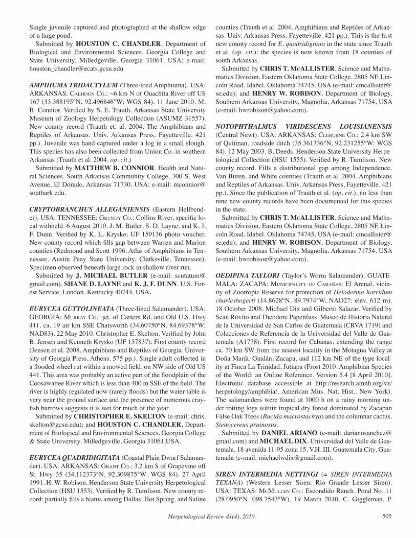
Herpetological Review 41(4), 2010 505
counties (Trauth et al. 2004. Amphibians and Reptiles of Arkan-sas. Univ. Arkansas Press, Fayetteville. 421 pp.). This is the first new county record for E. quadridigitata in the state since Trauth et al. (op. cit.); the species is now known from 18 counties of south Arkansas. Submitted by CHRIS T. McALLISTER, Science and Mathe-matics Division, Eastern Oklahoma State College, 2805 NE Lin-coln Road, Idabel, Oklahoma 74745, USA (e-mail: [email protected]); and HENRY W. ROBISON, Department of Biology, Southern Arkansas University, Magnolia, Arkansas 71754, USA (e-mail: [email protected]).
NOTOPHTHALMUS vIRIDESCENS LOUISIANENSIS (Central Newt). USA: ARKANSAS: cleBurne co.: 2.4 km SW of Quitman, roadside ditch (35.361336°N, 92.231255°W; WGS 84). 12 May 2003. B. Deeds. Henderson State University Herpe-tological Collection (HSU 1555). Verified by R. Tumlison. New county record. Fills a distributional gap among Independence, Van Buren, and White counties (Trauth et al. 2004. Amphibians and Reptiles of Arkansas. Univ. Arkansas Press, Fayetteville. 421 pp.). Since the publication of Trauth et al. (op. cit.), no less than nine new county records have been documented for this species in the state. Submitted by CHRIS T. McALLISTER, Science and Mathe-matics Division, Eastern Oklahoma State College, 2805 NE Lin-coln Road, Idabel, Oklahoma 74745, USA (e-mail: [email protected]); and HENRY W. ROBISON, Department of Biology, Southern Arkansas University, Magnolia, Arkansas 71754, USA (e-mail: [email protected]).
OEDIPINA TAYLORI (Taylor’s Worm Salamander). GUATE-MALA: ZACAPA: MuniciPalitY of caBañaS: El Arenal, vicin-ity of Zootropic Reserve for protection of Heloderma horridum charlesbogerti (14.8628°N, 89.7974°W, NAD27; elev. 612 m). 18 October 2008. Michael Dix and Gilberto Salazar. Verified by Sean Rovito and Theodore Papenfuss. Museo de Historia Natural de la Universidad de San Carlos de Guatemala (CRVA 1719) and Colecciones de Referencia de la Universidad del Valle de Gua-temala (A1778). First record for Cabañas, extending the range ca. 70 km SW from the nearest locality in the Motagua Valley at Doña María, Gualán, Zacapa, and 112 km NE of the type local-ity at Finca La Trinidad, Jutiapa (Frost 2010. Amphibian Species of the World: an Online Reference. Version 5.4 [8 April 2010]. Electronic database accessible at http://research.amnh.org/vz/herpetology/amphibia/, American Mus. Nat. Hist., New York). The salamanders were found at 1000 h on a rainy morning un-der rotting logs within tropical dry forest dominated by Zacapan False Oak Trees (Bucida macrostachya) and the columnar cactus, Stenocereus pruinosus. Submitted by DANIEL ARIANO (e-mail: [email protected]) and MICHAEL DIX, Universidad del Valle de Gua-temala, 18 avenida 11-95 zona 15. V.H. III, Guatemala City, Gua-temala (e-mail: [email protected]).
SIREN INTERMEDIA NETTINGI (= SIREN INTERMEDIA TEXANA) (Western Lesser Siren; Rio Grande Lesser Siren). USA: TEXAS: McMullen co.: Escondido Ranch, Pond No. 11 (28.0950°N, 098.7543°W). 19 March 2010. C. Giggleman, P.
Single juvenile captured and photographed at the shallow edge of a large pond. Submitted by HOUSTON C. CHANDLER, Department of Biological and Environmental Sciences, Georgia College and State University, Milledgeville, Georgia 31061, USA; e-mail: [email protected]
AMPHIUMA TRIDACTYLUM (Three-toed Amphiuma). USA: ARKANSAS: calHoun co.: ~6 km N of Ouachita River off US 167 (33.388195°N, 92.496846°W; WGS 84). 11 June 2010. M. B. Connior. Verified by S. E. Trauth. Arkansas State University Museum of Zoology Herpetology Collection (ASUMZ 31557). New county record (Trauth et. al. 2004. The Amphibians and Reptiles of Arkansas. Univ. Arkansas Press, Fayetteville. 421 pp.). Juvenile was hand captured under a log in a small slough. This species has also been collected from Union Co. in southern Arkansas (Trauth et al. 2004, op. cit.) Submitted by MATTHEW B. CONNIOR, Health and Natu-ral Sciences, South Arkansas Community College, 300 S. West Avenue, El Dorado, Arkansas 71730, USA; e-mail: [email protected].
CRYPTOBRANCHUS ALLEGANIENSIS (Eastern Hellbend-er). USA: TENNESSEE: grundY co.: Collins River, specific lo-cal withheld. 6 August 2010. J. M. Butler, S. D. Layne, and K. J. F. Dunn. Verified by K. L. Krysko. UF 159136 photo voucher. New county record which fills gap between Warren and Marion counties (Redmond and Scott 1996. Atlas of Amphibians in Ten-nessee. Austin Peay State University, Clarksville, Tennessee). Specimen observed beneath large rock in shallow river run. Submitted by J. MICHAEL BUTLER (e-mail: [email protected]), SHANE D. LAYNE and K. J. F. DUNN, U.S. For-est Service, London, Kentucky 40744, USA.
EURYCEA GUTTOLINEATA (Three-lined Salamander). USA: GEORGIA: MurraY co.: jct. of Carters Rd. and Old U.S. Hwy 411, ca. 19 air km SSE Chatsworth (34.60750°N, 84.69378°W; NAD83). 22 May 2010. Christopher E. Skelton. Verified by John B. Jensen and Kenneth Krysko (UF 157837). First county record (Jensen et al. 2008. Amphibians and Reptiles of Georgia. Univer-sity of Georgia Press, Athens. 575 pp.). Single adult collected in a flooded wheel rut within a mowed field, on NW side of Old US 441. This area was probably an active part of the floodplain of the Coosawattee River which is less than 400 m SSE of the field. The river is highly regulated now (rarely floods) but the water table is very near the ground surface and the presence of numerous cray-fish burrows suggests it is wet for much of the year. Submitted by CHRISTOPHER E. SKELTON (e-mail: [email protected]); and HOUSTON C. CHANDLER, Depart-ment of Biological and Environmental Sciences, Georgia College & State University, Milledgeville, Georgia 31061,USA.
EURYCEA QUADRIDIGITATA (Coastal Plain Dwarf Salaman-der). USA: ARKANSAS: grant co.: 3.2 km S of Grapevine off St. Hwy 35 (34.112373°N, 92.309875°W; WGS 84). 27 April 1991. H. W. Robison. Henderson State University Herpetological Collection (HSU 1553). Verified by R. Tumlison. New county re-cord; partially fills a hiatus among Dallas, Hot Spring, and Saline
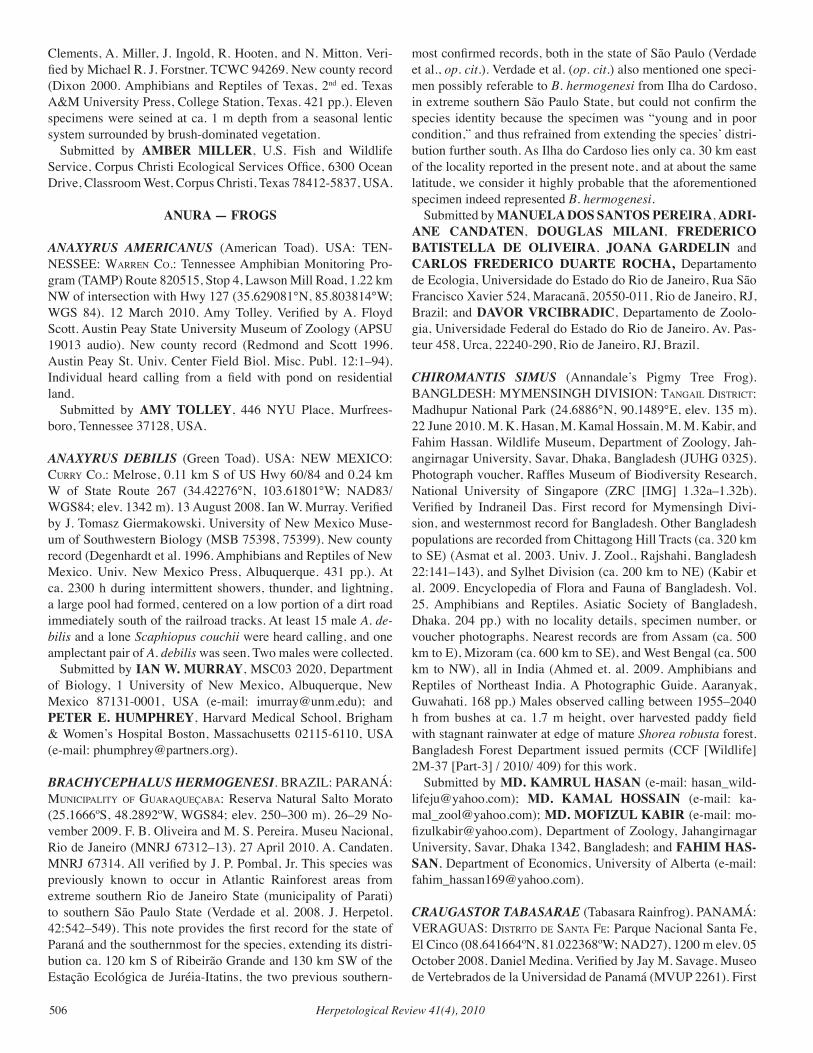
Herpetological Review 41(4), 2010506
Clements, A. Miller, J. Ingold, R. Hooten, and N. Mitton. Veri-fied by Michael R. J. Forstner. TCWC 94269. New county record (Dixon 2000. Amphibians and Reptiles of Texas, 2nd ed. Texas A&M University Press, College Station, Texas. 421 pp.). Eleven specimens were seined at ca. 1 m depth from a seasonal lentic system surrounded by brush-dominated vegetation. Submitted by AMBER MILLER, U.S. Fish and Wildlife Service, Corpus Christi Ecological Services Office, 6300 Ocean Drive, Classroom West, Corpus Christi, Texas 78412-5837, USA.
ANURA — FROGS
ANAXYRUS AMERICANUS (American Toad). USA: TEN-NESSEE: Warren co.: Tennessee Amphibian Monitoring Pro-gram (TAMP) Route 820515, Stop 4, Lawson Mill Road, 1.22 km NW of intersection with Hwy 127 (35.629081°N, 85.803814°W; WGS 84). 12 March 2010. Amy Tolley. Verified by A. Floyd Scott. Austin Peay State University Museum of Zoology (APSU 19013 audio). New county record (Redmond and Scott 1996. Austin Peay St. Univ. Center Field Biol. Misc. Publ. 12:1–94). Individual heard calling from a field with pond on residential land. Submitted by AMY TOLLEY, 446 NYU Place, Murfrees-boro, Tennessee 37128, USA.
ANAXYRUS DEBILIS (Green Toad). USA: NEW MEXICO: currY co.: Melrose, 0.11 km S of US Hwy 60/84 and 0.24 km W of State Route 267 (34.42276°N, 103.61801°W; NAD83/WGS84; elev. 1342 m). 13 August 2008. Ian W. Murray. Verified by J. Tomasz Giermakowski. University of New Mexico Muse-um of Southwestern Biology (MSB 75398, 75399). New county record (Degenhardt et al. 1996. Amphibians and Reptiles of New Mexico. Univ. New Mexico Press, Albuquerque. 431 pp.). At ca. 2300 h during intermittent showers, thunder, and lightning, a large pool had formed, centered on a low portion of a dirt road immediately south of the railroad tracks. At least 15 male A. de-bilis and a lone Scaphiopus couchii were heard calling, and one amplectant pair of A. debilis was seen. Two males were collected. Submitted by IAN W. MURRAY, MSC03 2020, Department of Biology, 1 University of New Mexico, Albuquerque, New Mexico 87131-0001, USA (e-mail: [email protected]); and PETER E. HUMPHREY, Harvard Medical School, Brigham & Women’s Hospital Boston, Massachusetts 02115-6110, USA (e-mail: [email protected]).
BRACHYCEPHALUS HERMOGENESI. BRAZIL: PARANÁ: MuniciPalitY of guaraQueçaBa: Reserva Natural Salto Morato (25.1666ºS, 48.2892ºW, WGS84; elev. 250–300 m). 26–29 No-vember 2009. F. B. Oliveira and M. S. Pereira. Museu Nacional, Rio de Janeiro (MNRJ 67312–13). 27 April 2010. A. Candaten. MNRJ 67314. All verified by J. P. Pombal, Jr. This species was previously known to occur in Atlantic Rainforest areas from extreme southern Rio de Janeiro State (municipality of Parati) to southern São Paulo State (Verdade et al. 2008. J. Herpetol. 42:542–549). This note provides the first record for the state of Paraná and the southernmost for the species, extending its distri-bution ca. 120 km S of Ribeirão Grande and 130 km SW of the Estação Ecológica de Juréia-Itatins, the two previous southern-
most confirmed records, both in the state of São Paulo (Verdade et al., op. cit.). Verdade et al. (op. cit.) also mentioned one speci-men possibly referable to B. hermogenesi from Ilha do Cardoso, in extreme southern São Paulo State, but could not confirm the species identity because the specimen was “young and in poor condition,” and thus refrained from extending the species’ distri-bution further south. As Ilha do Cardoso lies only ca. 30 km east of the locality reported in the present note, and at about the same latitude, we consider it highly probable that the aforementioned specimen indeed represented B. hermogenesi. Submitted by MANUELA DOS SANTOS PEREIRA, ADRI-ANE CANDATEN, DOUGLAS MILANI, FREDERICO BATISTELLA DE OLIVEIRA, JOANA GARDELIN and CARLOS FREDERICO DUARTE ROCHA, Departamento de Ecologia, Universidade do Estado do Rio de Janeiro, Rua São Francisco Xavier 524, Maracanã, 20550-011, Rio de Janeiro, RJ, Brazil; and DAVOR VRCIBRADIC, Departamento de Zoolo-gia, Universidade Federal do Estado do Rio de Janeiro. Av. Pas-teur 458, Urca, 22240-290, Rio de Janeiro, RJ, Brazil.
CHIROMANTIS SIMUS (Annandale’s Pigmy Tree Frog). BANGLDESH: MYMENSINGH DIVISION: tangail diStrict: Madhupur National Park (24.6886°N, 90.1489°E, elev. 135 m). 22 June 2010. M. K. Hasan, M. Kamal Hossain, M. M. Kabir, and Fahim Hassan. Wildlife Museum, Department of Zoology, Jah-angirnagar University, Savar, Dhaka, Bangladesh (JUHG 0325). Photograph voucher, Raffles Museum of Biodiversity Research, National University of Singapore (ZRC [IMG] 1.32a–1.32b). Verified by Indraneil Das. First record for Mymensingh Divi-sion, and westernmost record for Bangladesh. Other Bangladesh populations are recorded from Chittagong Hill Tracts (ca. 320 km to SE) (Asmat et al. 2003. Univ. J. Zool., Rajshahi, Bangladesh 22:141–143), and Sylhet Division (ca. 200 km to NE) (Kabir et al. 2009. Encyclopedia of Flora and Fauna of Bangladesh. Vol. 25. Amphibians and Reptiles. Asiatic Society of Bangladesh, Dhaka. 204 pp.) with no locality details, specimen number, or voucher photographs. Nearest records are from Assam (ca. 500 km to E), Mizoram (ca. 600 km to SE), and West Bengal (ca. 500 km to NW), all in India (Ahmed et. al. 2009. Amphibians and Reptiles of Northeast India. A Photographic Guide. Aaranyak, Guwahati. 168 pp.) Males observed calling between 1955–2040 h from bushes at ca. 1.7 m height, over harvested paddy field with stagnant rainwater at edge of mature Shorea robusta forest. Bangladesh Forest Department issued permits (CCF [Wildlife] 2M-37 [Part-3] / 2010/ 409) for this work. Submitted by MD. KAMRUL HASAN (e-mail: [email protected]); MD. KAMAL HOSSAIN (e-mail: [email protected]); MD. MOFIZUL KABIR (e-mail: [email protected]), Department of Zoology, Jahangirnagar University, Savar, Dhaka 1342, Bangladesh; and FAHIM HAS-SAN, Department of Economics, University of Alberta (e-mail: [email protected]).
CRAUGASTOR TABASARAE (Tabasara Rainfrog). PANAMÁ: VERAGUAS: diStrito de Santa fe: Parque Nacional Santa Fe, El Cinco (08.641664ºN, 81.022368ºW; NAD27), 1200 m elev. 05 October 2008. Daniel Medina. Verified by Jay M. Savage. Museo de Vertebrados de la Universidad de Panamá (MVUP 2261). First
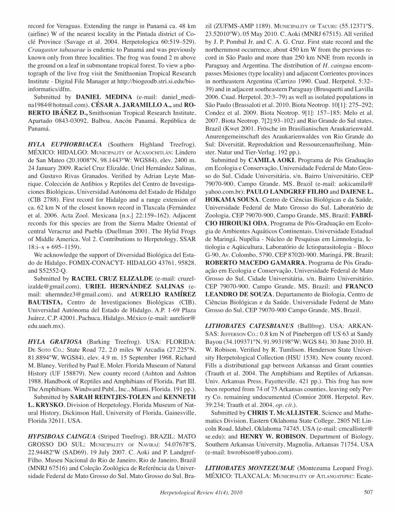
Herpetological Review 41(4), 2010 507
record for Veraguas. Extending the range in Panamá ca. 48 km (airline) W of the nearest locality in the Pintada district of Co-clé Province (Savage et al. 2004. Herpetologica 60:519–529). Craugastor tabasarae is endemic to Panamá and was previously known only from three localities. The frog was found 2 m above the ground on a leaf in submontane tropical forest. To view a pho-tograph of the live frog visit the Smithsonian Tropical Research Institute - Digital File Manager at http://biogeodb.stri.si.edu/bio-informatics/dfm. Submitted by DANIEL MEDINA (e-mail: [email protected]), CÉSAR A. JARAMILLO A., and RO-BERTO IBÁÑEZ D., Smithsonian Tropical Research Institute, Apartado 0843-03092, Balboa, Ancón Panamá, República de Panamá.
HYLA EUPHORBIACEA (Southern Highland Treefrog). MÉXICO: HIDALGO: MuniciPalitY of acaXocHitlán: Lindero de San Mateo (20.1008°N, 98.1443°W; WGS84), elev. 2400 m. 24 January 2009. Raciel Cruz Elizalde, Uriel Hernández Salinas, and Gustavo Rivas Granados. Verified by Adrian Leyte Man-rique. Colección de Anfibios y Reptiles del Centro de Investiga-ciones Biológicas, Universidad Autónoma del Estado de Hidalgo (CIB 2788). First record for Hidalgo and a range extension of ca. 62 km N of the closest known record in Tlaxcala (Fernández et al. 2006. Acta Zool. Mexicana [n.s.] 22:159–162). Adjacent records for this species are from the Sierra Madre Oriental of central Veracruz and Puebla (Duellman 2001. The Hylid Frogs of Middle America, Vol 2. Contributions to Herpetology, SSAR 18:i–x + 695–1159). We acknowledge the support of Diversidad Biológica del Esta-do de Hidalgo, FOMIX-CONACYT- HIDALGO 43761, 95828, and S52552-Q. Submitted by RACIEL CRUZ ELIZALDE (e-mail: [email protected]), URIEL HERNÁNDEZ SALINAS (e-mail: [email protected]), and AURELIO RAMÍREZ BAUTISTA, Centro de Investigaciones Biológicas (CIB), Universidad Autónoma del Estado de Hidalgo, A.P. 1-69 Plaza Juárez, C.P. 42001, Pachuca, Hidalgo, México (e-mail: [email protected]).
HYLA GRATIOSA (Barking Treefrog). USA: FLORIDA: de Soto co.: State Road 72, 2.0 miles W Arcadia (27.225°N, 81.8894°W, WGS84), elev. 4.9 m. 15 September 1968. Richard M. Blaney. Verified by Paul E. Moler. Florida Museum of Natural History (UF 158879). New county record (Ashton and Ashton 1988. Handbook of Reptiles and Amphibians of Florida. Part III. The Amphibians. Windward Publ., Inc., Miami, Florida. 191 pp.). Submitted by SARAH REINTJES-TOLEN and KENNETH L. KRYSKO, Division of Herpetology, Florida Museum of Nat-ural History, Dickinson Hall, University of Florida, Gainesville, Florida 32611, USA.
HYPSIBOAS CAINGUA (Striped Treefrog). BRAZIL: MATO GROSSO DO SUL: MuniciPalitY of naviraí: 54.07678ºS, 22.94482ºW (SAD69). 19 July 2007. C. Aoki and P. Landgref-Filho. Museu Nacional do Rio de Janeiro, Rio de Janeiro, Brazil (MNRJ 67516) and Coleção Zoológica de Referência da Univer-sidade Federal de Mato Grosso do Sul, Mato Grosso do Sul, Bra-
zil (ZUFMS-AMP 1189). MuniciPalitY of tacuru (55.12371ºS, 23.52010ºW). 05 May 2010. C. Aoki (MNRJ 67515). All verified by J. P. Pombal Jr. and C. A. G. Cruz. First state record and the northernmost occurrence, about 450 km W from the previous re-cord in São Paulo and more than 250 km NNE from records in Paraguay and Argentina. The distribution of H. caingua encom-passes Misiones (type locality) and adjacent Corrientes provinces in northeastern Argentina (Carrizo 1990. Cuad. Herpetol. 5:32–39) and in adjacent southeastern Paraguay (Brusquetti and Lavilla 2006. Cuad. Herpetol. 20:3–79) as well as isolated populations in São Paulo (Brassaloti et al. 2010. Biota Neotrop. 10[1]: 275–292; Condez et al. 2009. Biota Neotrop. 9[1]: 157–185; Melo et al. 2007. Biota Neotrop. 7[2]:93–102) and Rio Grande do Sul states, Brazil (Kwet 2001. Frösche im Brasilianischen Araukarienwald. Anurengemeinschaft des Araukarienwaldes von Rio Grande do Sul: Diversität, Reproduktion und Ressourcenaufteilung. Mün-ster. Natur und Tier-Verlag. 192 pp.). Submitted by CAMILA AOKI, Programa de Pós Graduação em Ecologia e Conservação, Universidade Federal de Mato Gros-so do Sul, Cidade Universitária, s/n, Bairro Universitário, CEP 79070-900, Campo Grande, MS, Brazil (e-mail: [email protected]); PAULO LANDGREF FILHO and DAIENE L. HOKAMA SOUSA, Centro de Ciências Biológicas e da Saúde, Universidade Federal de Mato Grosso do Sul, Laboratório de Zoologia, CEP 79070-900, Campo Grande, MS, Brazil; FABRÍ-CIO HIROIUKI ODA, Programa de Pós-Graduação em Ecolo-gia de Ambientes Aquáticos Continentais, Universidade Estadual de Maringá, Nupélia - Núcleo de Pesquisas em Limnologia, Ic-tiologia e Aqüicultura, Laboratório de Ictioparasitologia - Bloco G-90, Av. Colombo, 5790, CEP 87020-900. Maringá, PR, Brazil; ROBERTO MACEDO GAMARRA, Programa de Pós Gradu-ação em Ecologia e Conservação, Universidade Federal de Mato Grosso do Sul, Cidade Universitária, s/n, Bairro Universitário, CEP 79070-900, Campo Grande, MS, Brazil; and FRANCO LEANDRO DE SOUZA, Departamento de Biologia, Centro de Ciências Biológicas e da Saúde, Universidade Federal de Mato Grosso do Sul, CEP 79070-900 Campo Grande, MS, Brazil.
LITHOBATES CATESBIANUS (Bullfrog). USA: ARKAN-SAS: JefferSon co.: 0.8 km N of Pinebergen off US 63 at Sandy Bayou (34.109371°N, 91.993198°W; WGS 84). 30 June 2010. H. W. Robison. Verified by R. Tumlison. Henderson State Univer-sity Herpetological Collection (HSU 1538). New county record. Fills a distributional gap between Arkansas and Grant counties (Trauth et al. 2004. The Amphibians and Reptiles of Arkansas. Univ. Arkansas Press, Fayetteville. 421 pp.). This frog has now been reported from 74 of 75 Arkansas counties, leaving only Per-ry Co. remaining undocumented (Connior 2008. Herpetol. Rev. 39:234; Trauth et al. 2004, op. cit.). Submitted by CHRIS T. McALLISTER, Science and Mathe-matics Division, Eastern Oklahoma State College, 2805 NE Lin-coln Road, Idabel, Oklahoma 74745, USA (e-mail: [email protected]); and HENRY W. ROBISON, Department of Biology, Southern Arkansas University, Magnolia, Arkansas 71754, USA (e-mail: [email protected]).
LITHOBATES MONTEZUMAE (Montezuma Leopard Frog). MÉXICO: TLAXCALA: MuniciPalitY of atlangatePec: Ecate-
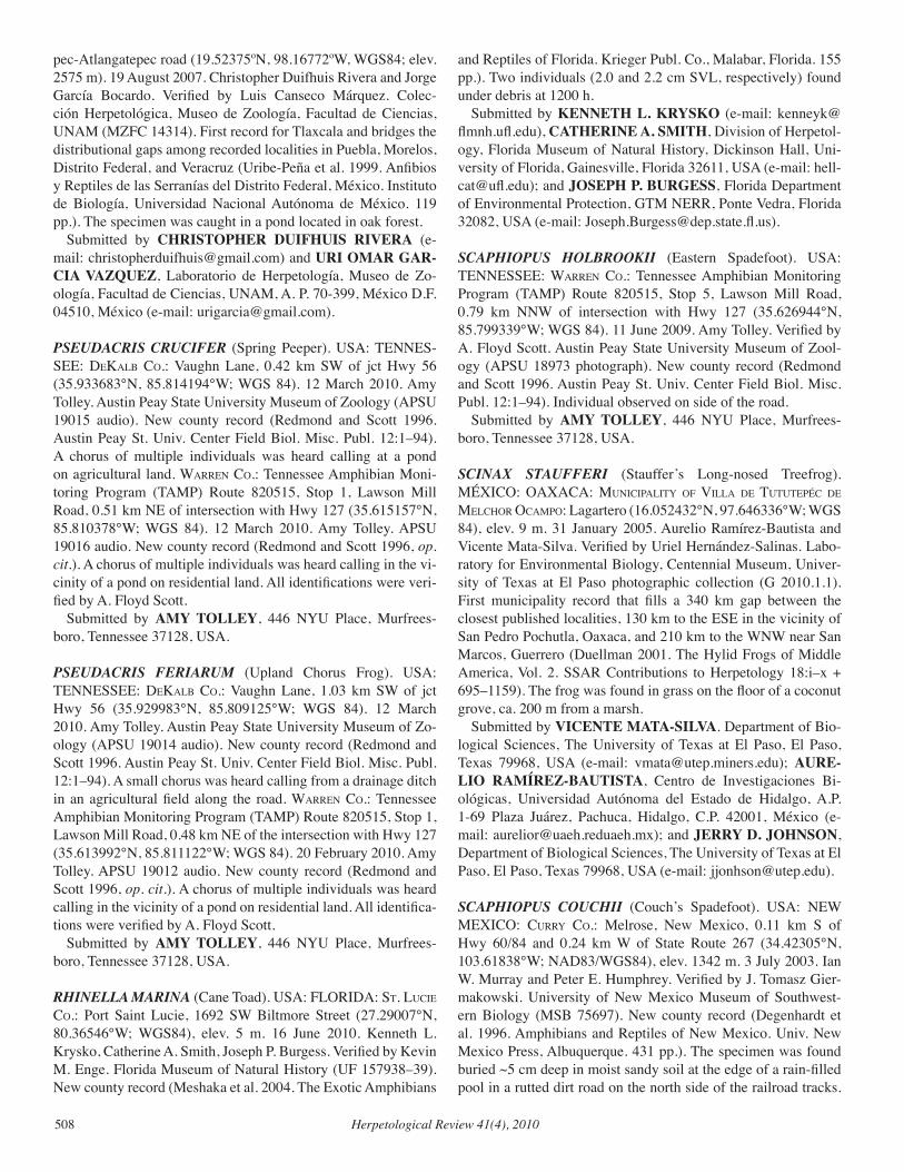
Herpetological Review 41(4), 2010508
pec-Atlangatepec road (19.52375ºN, 98.16772ºW, WGS84; elev. 2575 m). 19 August 2007. Christopher Duifhuis Rivera and Jorge García Bocardo. Verified by Luis Canseco Márquez. Colec-ción Herpetológica, Museo de Zoología, Facultad de Ciencias, UNAM (MZFC 14314). First record for Tlaxcala and bridges the distributional gaps among recorded localities in Puebla, Morelos, Distrito Federal, and Veracruz (Uribe-Peña et al. 1999. Anfibios y Reptiles de las Serranías del Distrito Federal, México. Instituto de Biología, Universidad Nacional Autónoma de México. 119 pp.). The specimen was caught in a pond located in oak forest. Submitted by CHRISTOPHER DUIFHUIS RIVERA (e-mail: [email protected]) and URI OMAR GAR-CIA VAZQUEZ, Laboratorio de Herpetología, Museo de Zo-ología, Facultad de Ciencias, UNAM, A. P. 70-399, México D.F. 04510, México (e-mail: [email protected]).
PSEUDACRIS CRUCIFER (Spring Peeper). USA: TENNES-SEE: dekalB co.: Vaughn Lane, 0.42 km SW of jct Hwy 56 (35.933683°N, 85.814194°W; WGS 84). 12 March 2010. Amy Tolley. Austin Peay State University Museum of Zoology (APSU 19015 audio). New county record (Redmond and Scott 1996. Austin Peay St. Univ. Center Field Biol. Misc. Publ. 12:1–94). A chorus of multiple individuals was heard calling at a pond on agricultural land. Warren co.: Tennessee Amphibian Moni-toring Program (TAMP) Route 820515, Stop 1, Lawson Mill Road, 0.51 km NE of intersection with Hwy 127 (35.615157°N, 85.810378°W; WGS 84). 12 March 2010. Amy Tolley. APSU 19016 audio. New county record (Redmond and Scott 1996, op. cit.). A chorus of multiple individuals was heard calling in the vi-cinity of a pond on residential land. All identifications were veri-fied by A. Floyd Scott. Submitted by AMY TOLLEY, 446 NYU Place, Murfrees-boro, Tennessee 37128, USA.
PSEUDACRIS FERIARUM (Upland Chorus Frog). USA: TENNESSEE: dekalB co.: Vaughn Lane, 1.03 km SW of jct Hwy 56 (35.929983°N, 85.809125°W; WGS 84). 12 March 2010. Amy Tolley. Austin Peay State University Museum of Zo-ology (APSU 19014 audio). New county record (Redmond and Scott 1996. Austin Peay St. Univ. Center Field Biol. Misc. Publ. 12:1–94). A small chorus was heard calling from a drainage ditch in an agricultural field along the road. Warren co.: Tennessee Amphibian Monitoring Program (TAMP) Route 820515, Stop 1, Lawson Mill Road, 0.48 km NE of the intersection with Hwy 127 (35.613992°N, 85.811122°W; WGS 84). 20 February 2010. Amy Tolley. APSU 19012 audio. New county record (Redmond and Scott 1996, op. cit.). A chorus of multiple individuals was heard calling in the vicinity of a pond on residential land. All identifica-tions were verified by A. Floyd Scott. Submitted by AMY TOLLEY, 446 NYU Place, Murfrees-boro, Tennessee 37128, USA.
RHINELLA MARINA (Cane Toad). USA: FLORIDA: St. lucie co.: Port Saint Lucie, 1692 SW Biltmore Street (27.29007°N, 80.36546°W; WGS84), elev. 5 m. 16 June 2010. Kenneth L. Krysko, Catherine A. Smith, Joseph P. Burgess. Verified by Kevin M. Enge. Florida Museum of Natural History (UF 157938–39). New county record (Meshaka et al. 2004. The Exotic Amphibians
and Reptiles of Florida. Krieger Publ. Co., Malabar, Florida. 155 pp.). Two individuals (2.0 and 2.2 cm SVL, respectively) found under debris at 1200 h. Submitted by KENNETH L. KRYSKO (e-mail: [email protected]), CATHERINE A. SMITH, Division of Herpetol-ogy, Florida Museum of Natural History, Dickinson Hall, Uni-versity of Florida, Gainesville, Florida 32611, USA (e-mail: [email protected]); and JOSEPH P. BURGESS, Florida Department of Environmental Protection, GTM NERR, Ponte Vedra, Florida 32082, USA (e-mail: [email protected]).
SCAPHIOPUS HOLBROOKII (Eastern Spadefoot). USA: TENNESSEE: Warren co.: Tennessee Amphibian Monitoring Program (TAMP) Route 820515, Stop 5, Lawson Mill Road, 0.79 km NNW of intersection with Hwy 127 (35.626944°N, 85.799339°W; WGS 84). 11 June 2009. Amy Tolley. Verified by A. Floyd Scott. Austin Peay State University Museum of Zool-ogy (APSU 18973 photograph). New county record (Redmond and Scott 1996. Austin Peay St. Univ. Center Field Biol. Misc. Publ. 12:1–94). Individual observed on side of the road. Submitted by AMY TOLLEY, 446 NYU Place, Murfrees-boro, Tennessee 37128, USA.
SCINAX STAUFFERI (Stauffer’s Long-nosed Treefrog). MÉXICO: OAXACA: MuniciPalitY of villa de tututePéc de MelcHor ocaMPo: Lagartero (16.052432°N, 97.646336°W; WGS 84), elev. 9 m. 31 January 2005. Aurelio Ramírez-Bautista and Vicente Mata-Silva. Verified by Uriel Hernández-Salinas. Labo-ratory for Environmental Biology, Centennial Museum, Univer-sity of Texas at El Paso photographic collection (G 2010.1.1). First municipality record that fills a 340 km gap between the closest published localities, 130 km to the ESE in the vicinity of San Pedro Pochutla, Oaxaca, and 210 km to the WNW near San Marcos, Guerrero (Duellman 2001. The Hylid Frogs of Middle America, Vol. 2. SSAR Contributions to Herpetology 18:i–x + 695–1159). The frog was found in grass on the floor of a coconut grove, ca. 200 m from a marsh. Submitted by VICENTE MATA-SILVA. Department of Bio-logical Sciences, The University of Texas at El Paso, El Paso, Texas 79968, USA (e-mail: [email protected]); AURE-LIO RAMÍREZ-BAUTISTA, Centro de Investigaciones Bi-ológicas, Universidad Autónoma del Estado de Hidalgo, A.P. 1-69 Plaza Juárez, Pachuca, Hidalgo, C.P. 42001, México (e-mail: [email protected]); and JERRY D. JOHNSON, Department of Biological Sciences, The University of Texas at El Paso, El Paso, Texas 79968, USA (e-mail: [email protected]).
SCAPHIOPUS COUCHII (Couch’s Spadefoot). USA: NEW MEXICO: currY co.: Melrose, New Mexico, 0.11 km S of Hwy 60/84 and 0.24 km W of State Route 267 (34.42305°N, 103.61838°W; NAD83/WGS84), elev. 1342 m. 3 July 2003. Ian W. Murray and Peter E. Humphrey. Verified by J. Tomasz Gier-makowski. University of New Mexico Museum of Southwest-ern Biology (MSB 75697). New county record (Degenhardt et al. 1996. Amphibians and Reptiles of New Mexico. Univ. New Mexico Press, Albuquerque. 431 pp.). The specimen was found buried ~5 cm deep in moist sandy soil at the edge of a rain-filled pool in a rutted dirt road on the north side of the railroad tracks.
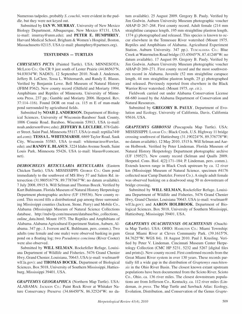
Herpetological Review 41(4), 2010 509
Numerous tadpoles, probably S. couchii, were evident in the pud-dle, but they were not keyed out. Submitted by IAN W. MURRAY, University of New Mexico Biology Department, Albuquerque, New Mexico 87131, USA (e-mail: [email protected]); and PETER E. HUMPHREY, Harvard Medical School, Brigham & Women’s Hospital, Boston, Massachusetts 02115, USA (e-mail: [email protected]).
TESTUDINES — TURTLES
CHRYSEMYS PICTA (Painted Turtle). USA: MINNESOTA: Mcleod co.: On CR 9 just south of Lester Prairie (44.86563ºN, 94.03034ºW; NAD83). 12 September 2010. Noah J. Anderson, Jeffery B. LeClere, Tessa L. Whitemarsh, and Randy E. Blasus. Verified by Benjamin Lowe. Bell Museum of Natural History (JFBM P362). New county record (Oldfield and Moriarty 1994. Amphibians and Reptiles of Minnesota. University of Minne-sota Press, 237 pp.; Gamble and Moriarty 2006. Herpetol. Rev. 37:114–116). Found DOR on road ca. 115 m E of man-made pond surrounded by agricultural fields. Submitted by NOAH J. ANDERSON, Department of Biolog-ical Sciences, University of Wisconsin-Baraboo/ Sauk County, 1006 Connie Road, Baraboo, Wisconsin 53913, USA (e-mail: [email protected]); JEFFERY B. LECLERE, 878 Galti-er Street, Saint Paul, Minnesota 55117, USA (e-mail: [email protected]); TESSA L. WHITEMARSH, 6869 Taylor Road, Sauk City, Wisconsin 53583, USA (e-mail: [email protected]); and RANDY E. BLASUS, 3224 Idaho Avenue South, Saint Louis Park, Minnesota 55426, USA (e-mail: [email protected]).
DEIROCHELYS RETICULARIA RETICULARIA (Eastern Chicken Turtle). USA: MISSISSIPPI: george co.: Gum pond immediately to the southwest of MS Hwy 57 and Salem Rd. in-tersection (31.9805167°N, 89.7787667°W; no datum available). 7 July 2008, 0915 h. Will Selman and Thomas Bocek. Verified by Kurt Buhlmann. Florida Museum of Natural History Herpetology Department photographic archive (UF 159536). New county re-cord. This record fills a distributional gap among three surround-ing Mississippi counties (Jackson, Stone, Perry) and Mobile Co., Alabama (Mississippi Museum of Natural Science Collections database, http://mdwfp.com/museum/database/bio_collections_online_data.html; Mount 1975. The Reptiles and Amphibians of Alabama. Alabama Agricultural Experiment Station, Auburn, Al-abama. 347 pp.; J. Iverson and K. Buhlmann, pers. comm.). Two adults (one female and one male) were observed basking in gum pond on a floating log; two Pseudemys concinna (River Cooter) were also observed. Submitted by WILL SELMAN, Rockefeller Refuge, Louisi-ana Department of Wildlife and Fisheries, 5476 Grand Chenier Hwy, Grand Chenier, Louisiana, 70643, USA (e-mail: [email protected]); and THOMAS BOCEK, Department of Biological Sciences, Box 5018, University of Southern Mississippi, Hatties-burg, Mississippi 39401, USA.
GRAPTEMYS GEOGRAPHICA (Northern Map Turtle). USA: ALABAMA: JackSon co.: Paint Rock River at Whitaker Na-ture Conservancy Preserve (34.66450°N, 86.32524°W; no da-
tum available). 25 August 2009. Gregory B. Pauly. Verified by Jim Godwin. Auburn University Museum photographic voucher AHAP-D 267–268. First county record. Adult female (214 mm straightline carapace length, 195 mm straightline plastron length, 1374 g) photographed and released. This species is known to oc-cur elsewhere in the Tennessee River watershed (Mount 1975. Reptiles and Amphibians of Alabama. Agricultural Experiment Station, Auburn University. 347 pp.). tuScalooSa co.: Blue Creek at Watermelon Road bridge (33.45045°N, 87.41248°W; no datum available). 17 August 09. Gregory B. Pauly. Verified by Jim Godwin. Auburn University Museum photographic voucher AHAP-D 269–271. First county record and the most southwest-ern record in Alabama. Juvenile (52 mm straightline carapace length, 44 mm straightline plastron length, 25 g) photographed and released. Previously reported from elsewhere in the Black Warrior River watershed. (Mount 1975, op. cit.). Fieldwork carried out under Alabama Conservation License 68-680 issued by the Alabama Department of Conservation and Natural Resources. Submitted by GREGORY B. PAULY, Department of Evo-lution and Ecology, University of California, Davis, California 95616, USA.
GRAPTEMYS GIBBONSI (Pascagoula Map Turtle). USA: MISSISSIPPI: laMar co.: Black Creek, U.S. Highway 11 bridge crossing southwest of Hattiesburg (31.190224°N, 89.376778°W; no datum available). 12 May 2010, 1515 h. Will Selman and Aar-on Holbrook. Verified by Peter Lindeman. Florida Museum of Natural History Herpetology Department photographic archive (UF 159527). New county record (Selman and Qualls 2009. Herpetol. Cons. Biol. 4[2]:171–184; P. Lindeman, pers. comm.). Extends known range in Black Creek upstream by ca. 12 river km (Mississippi Museum of Natural Science, specimen #4176 collected near Camp Dantzler, Forrest Co.). A single adult female was observed basking on a deadwood snag 50 m downstream of bridge crossing. Submitted by WILL SELMAN, Rockefeller Refuge, Louisi-ana Department of Wildlife and Fisheries, 5476 Grand Chenier Hwy, Grand Chenier, Louisiana 70643, USA (e-mail: [email protected]); and AARON HOLBROOK, Department of Bio-logical Sciences, Box 5018, University of Southern Mississippi, Hattiesburg, Mississippi 39401, USA.
GRAPTEMYS OUACHITENSIS OUACHITENSIS (Ouachi-ta Map Turtle). USA: OHIO: HaMilton co.: Miami Township: Great Miami River at Cleves Community Park. (39.1615°N, 84.7625°W; WGS 84). 18 August 2010. Paul J. Krusling. Veri-fied by Peter V. Lindeman. Cincinnati Museum Center Herpe-tology Collection (CMC HP 5231, 5232 and 5267 [digital files and prints]). New county record. First confirmed records from the Great Miami River system in over 130 years. These records par-tially fill a wide gap in the distribution of Graptemys ouachiten-sis in the Ohio River Basin. The closest known extant upstream populations have been documented from the Scioto River, Scioto Co., Ohio, ca. 136 river miles. The closest downstream popula-tions are from Jefferson Co., Kentucky, ca. 112 river miles (Lin-deman, in press. The Map Turtle and Sawback Atlas: Ecology, Evolution, Distribution, and Conservation of the Genus Grapte-
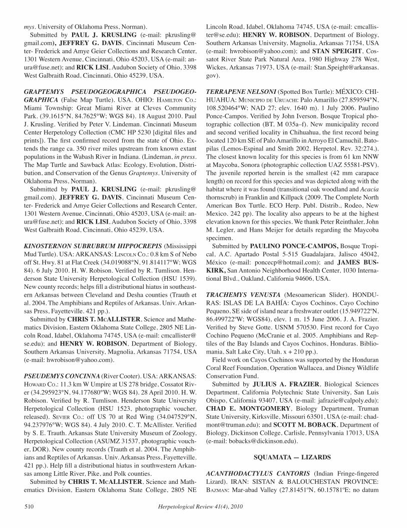
Herpetological Review 41(4), 2010510
mys. University of Oklahoma Press, Norman). Submitted by PAUL J. KRUSLING (e-mail: [email protected]), JEFFREY G. DAVIS, Cincinnati Museum Cen-ter- Frederick and Amye Geier Collections and Research Center, 1301 Western Avenue, Cincinnati, Ohio 45203, USA (e-mail: [email protected]); and RICK LISI, Audubon Society of Ohio, 3398 West Galbraith Road, Cincinnati, Ohio 45239, USA.
GRAPTEMYS PSEUDOGEOGRAPHICA PSEUDOGEO-GRAPHICA (False Map Turtle). USA. OHIO: HaMilton co.: Miami Township: Great Miami River at Cleves Community Park. (39.1615°N, 84.7625°W; WGS 84). 18 August 2010. Paul J. Krusling. Verified by Peter V. Lindeman. Cincinnati Museum Center Herpetology Collection (CMC HP 5230 [digital files and prints]). The first confirmed record from the state of Ohio. Ex-tends the range ca. 350 river miles upstream from known extant populations in the Wabash River in Indiana. (Lindeman, in press. The Map Turtle and Sawback Atlas: Ecology, Evolution, Distri-bution, and Conservation of the Genus Graptemys. University of Oklahoma Press, Norman). Submitted by PAUL J. KRUSLING (e-mail: [email protected]), JEFFREY G. DAVIS, Cincinnati Museum Cen-ter- Frederick and Amye Geier Collections and Research Center, 1301 Western Avenue, Cincinnati, Ohio 45203, USA (e-mail: [email protected]); and RICK LISI, Audubon Society of Ohio, 3398 West Galbraith Road, Cincinnati, Ohio 45239, USA.
KINOSTERNON SUBRUBRUM HIPPOCREPIS (Mississippi Mud Turtle). USA: ARKANSAS: lincoln co.: 0.8 km S of Nebo off St. Hwy. 81 at Flat Creek (34.019088°N, 91.814117°W; WGS 84). 6 July 2010. H. W. Robison. Verified by R. Tumlison. Hen-derson State University Herpetological Collection (HSU 1539). New county records; helps fill a distributional hiatus in southeast-ern Arkansas between Cleveland and Desha counties (Trauth et al. 2004. The Amphibians and Reptiles of Arkansas. Univ. Arkan-sas Press, Fayetteville. 421 pp.). Submitted by CHRIS T. McALLISTER, Science and Mathe-matics Division, Eastern Oklahoma State College, 2805 NE Lin-coln Road, Idabel, Oklahoma 74745, USA (e-mail: [email protected]); and HENRY W. ROBISON, Department of Biology, Southern Arkansas University, Magnolia, Arkansas 71754, USA (e-mail: [email protected]).
PSEUDEMYS CONCINNA (River Cooter). USA: ARKANSAS: HoWard co.: 11.3 km W Umpire at US 278 bridge, Cossatot Riv-er (34.295923°N, 94.177680°W; WGS 84). 28 April 2010. H. W. Robison. Verified by R. Tumlison. Henderson State University Herpetological Collection (HSU 1523, photographic voucher, released). Sevier co.: off US 70 at Red Wing (34.047529°N, 94.237976°W; WGS 84). 4 July 2010. C. T. McAllister. Verified by S. E. Trauth. Arkansas State University Museum of Zoology, Herpetological Collection (ASUMZ 31537, photographic vouch-er, DOR). New county records (Trauth et al. 2004. The Amphib-ians and Reptiles of Arkansas. Univ. Arkansas Press, Fayetteville. 421 pp.). Help fill a distributional hiatus in southwestern Arkan-sas among Little River, Pike, and Polk counties. Submitted by CHRIS T. McALLISTER, Science and Math-ematics Division, Eastern Oklahoma State College, 2805 NE
Lincoln Road, Idabel, Oklahoma 74745, USA (e-mail: [email protected]); HENRY W. ROBISON, Department of Biology, Southern Arkansas University, Magnolia, Arkansas 71754, USA (e-mail: [email protected]); and STAN SPEIGHT, Cos-satot River State Park Natural Area, 1980 Highway 278 West, Wickes, Arkansas 71973, USA (e-mail: [email protected]).
TERRAPENE NELSONI (Spotted Box Turtle): MÉXICO: CHI-HUAHUA: MuniciPio de uruacHi: Palo Amarillo (27.859594°N, 108.520464°W; NAD 27; elev. 1640 m). 1 July 2006. Paulino Ponce-Campos. Verified by John Iverson. Bosque Tropical pho-tographic collection (BT, M 035a–f). New municipality record and second verified locality in Chihuahua, the first record being located 120 km SE of Palo Amarillo in Arroyo El Camuchil, Bato-pilas (Lemos-Espinal and Smith 2002. Herpetol. Rev. 32:274.). The closest known locality for this species is from 61 km NNW at Maycoba, Sonora (photographic collection UAZ 55581-PSV). The juvenile reported herein is the smallest (42 mm carapace length) on record for this species and was depicted along with the habitat where it was found (transitional oak woodland and Acacia thornscrub) in Franklin and Killpack (2009. The Complete North American Box Turtle. ECO Herp. Publ. Distrib., Rodeo, New Mexico. 242 pp). The locality also appears to be at the highest elevation known for this species. We thank Peter Reinthaler, John M. Legler, and Hans Meijer for details regarding the Maycoba specimen. Submitted by PAULINO PONCE-CAMPOS, Bosque Tropi-cal, A.C. Apartado Postal 5-515 Guadalajara, Jalisco 45042, México (e-mail: [email protected]); and JAMES BUS-KIRK, San Antonio Neighborhood Health Center, 1030 Interna-tional Blvd., Oakland, California 94606, USA.
TRACHEMYS vENUSTA (Mesoamerican Slider). HONDU-RAS: ISLAS DE LA BAHÍA: Cayos Cochinos, Cayo Cochino Pequeno, SE side of island near a freshwater outlet (15.949722ºN, 86.499722ºW; WGS84), elev. 1 m. 15 June 2006. J. A. Frazier. Verified by Steve Gotte. USNM 570530. First record for Cayo Cochino Pequeno (McCranie et al. 2005. Amphibians and Rep-tiles of the Bay Islands and Cayos Cochinos, Honduras. Biblio-mania, Salt Lake City, Utah. x + 210 pp.). Field work on Cayos Cochinos was supported by the Honduran Coral Reef Foundation, Operation Wallacea, and Disney Wildlife Conservation Fund. Submitted by JULIUS A. FRAZIER, Biological Sciences Department, California Polytechnic State University, San Luis Obispo, California 93407, USA (e-mail: [email protected]); CHAD E. MONTGOMERY, Biology Department, Truman State University, Kirksville, Missouri 63501, USA (e-mail: [email protected]); and SCOTT M. BOBACK, Department of Biology, Dickinson College, Carlisle, Pennsylvania 17013, USA (e-mail: [email protected]).
SQUAMATA — LIZARDS
ACANTHODACTYLUS CANTORIS (Indian Fringe-fingered Lizard). IRAN: SISTAN & BALOUCHESTAN PROVINCE: BazMan: Mar-abad Valley (27.81451ºN, 60.15781ºE; no datum
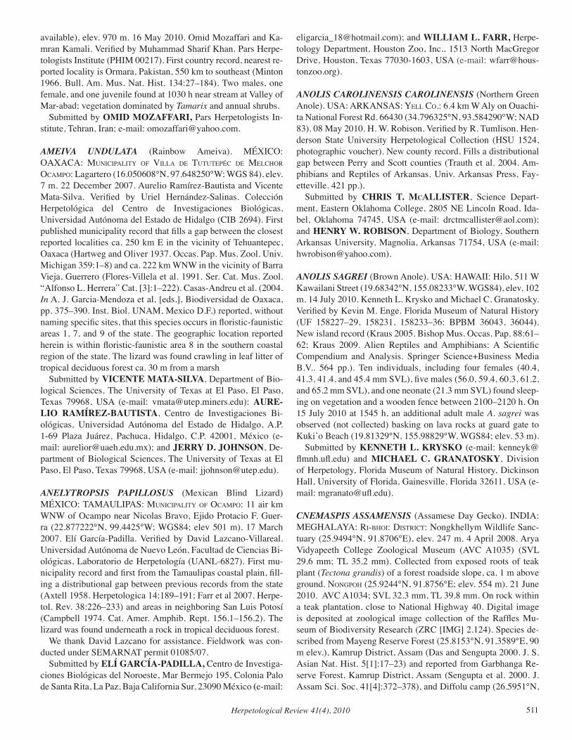
Herpetological Review 41(4), 2010 511
available), elev. 970 m. 16 May 2010. Omid Mozaffari and Ka-mran Kamali. Verified by Muhammad Sharif Khan. Pars Herpe-tologists Institute (PHIM 00217). First country record, nearest re-ported locality is Ormara, Pakistan, 550 km to southeast (Minton 1966. Bull. Am. Mus. Nat. Hist. 134:27–184). Two males, one female, and one juvenile found at 1030 h near stream at Valley of Mar-abad; vegetation dominated by Tamarix and annual shrubs. Submitted by OMID MOZAFFARI, Pars Herpetologists In-stitute, Tehran, Iran; e-mail: [email protected].
AMEIvA UNDULATA (Rainbow Ameiva). MÉXICO: OAXACA: MuniciPalitY of villa de tututePéc de MelcHor ocaMPo: Lagartero (16.050608°N, 97.648250°W; WGS 84), elev. 7 m. 22 December 2007. Aurelio Ramírez-Bautista and Vicente Mata-Silva. Verified by Uriel Hernández-Salinas. Colección Herpetológica del Centro de Investigaciones Biológicas, Universidad Autónoma del Estado de Hidalgo (CIB 2694). First published municipality record that fills a gap between the closest reported localities ca. 250 km E in the vicinity of Tehuantepec, Oaxaca (Hartweg and Oliver 1937. Occas. Pap. Mus. Zool. Univ. Michigan 359:1–8) and ca. 222 km WNW in the vicinity of Barra Vieja, Guerrero (Flores-Villela et al. 1991. Ser. Cat. Mus. Zool. “Alfonso L. Herrera” Cat. [3]:1–222). Casas-Andreu et al. (2004. In A. J. Garcia-Mendoza et al. [eds.], Biodiversidad de Oaxaca, pp. 375–390. Inst. Biol. UNAM, Mexico D.F.) reported, without naming specific sites, that this species occurs in floristic-faunistic areas 1, 7, and 9 of the state. The geographic location reported herein is within floristic-faunistic area 8 in the southern coastal region of the state. The lizard was found crawling in leaf litter of tropical deciduous forest ca. 30 m from a marsh Submitted by VICENTE MATA-SILVA, Department of Bio-logical Sciences, The University of Texas at El Paso, El Paso, Texas 79968, USA (e-mail: [email protected]); AURE-LIO RAMÍREZ-BAUTISTA, Centro de Investigaciones Bi-ológicas, Universidad Autónoma del Estado de Hidalgo, A.P. 1-69 Plaza Juárez, Pachuca, Hidalgo, C.P. 42001, México (e-mail: [email protected]); and JERRY D. JOHNSON, De-partment of Biological Sciences, The University of Texas at El Paso, El Paso, Texas 79968, USA (e-mail: [email protected]).
ANELYTROPSIS PAPILLOSUS (Mexican Blind Lizard) MÉXICO: TAMAULIPAS: MuniciPalitY of ocaMPo: 11 air km WNW of Ocampo near Nicolas Bravo, Ejido Protacio F. Guer-ra (22.877222°N, 99.4425°W; WGS84; elev 501 m). 17 March 2007. Elí García-Padilla. Verified by David Lazcano-Villareal. Universidad Autónoma de Nuevo León, Facultad de Ciencias Bi-ológicas, Laboratorio de Herpetología (UANL-6827). First mu-nicipality record and first from the Tamaulipas coastal plain, fill-ing a distributional gap between previous records from the state (Axtell 1958. Herpetologica 14:189–191; Farr et al 2007. Herpe-tol. Rev. 38:226–233) and areas in neighboring San Luis Potosí (Campbell 1974. Cat. Amer. Amphib. Rept. 156.1–156.2). The lizard was found underneath a rock in tropical deciduous forest. We thank David Lazcano for assistance. Fieldwork was con-ducted under SEMARNAT permit 01085/07. Submitted by ELÍ GARCÍA-PADILLA, Centro de Investiga-ciones Biológicas del Noroeste, Mar Bermejo 195, Colonia Palo de Santa Rita, La Paz, Baja California Sur, 23090 México (e-mail:
[email protected]); and WILLIAM L. FARR, Herpe-tology Department, Houston Zoo, Inc., 1513 North MacGregor Drive, Houston, Texas 77030-1603, USA (e-mail: [email protected]).
ANOLIS CAROLINENSIS CAROLINENSIS (Northern Green Anole). USA: ARKANSAS: Yell co.: 6.4 km W Aly on Ouachi-ta National Forest Rd. 66430 (34.796325°N, 93.584290°W; NAD 83). 08 May 2010. H. W. Robison. Verified by R. Tumlison. Hen-derson State University Herpetological Collection (HSU 1524, photographic voucher). New county record. Fills a distributional gap between Perry and Scott counties (Trauth et al. 2004. Am-phibians and Reptiles of Arkansas. Univ. Arkansas Press, Fay-etteville. 421 pp.). Submitted by CHRIS T. McALLISTER, Science Depart-ment, Eastern Oklahoma College, 2805 NE Lincoln Road, Ida-bel, Oklahoma 74745, USA (e-mail: [email protected]); and HENRY W. ROBISON, Department of Biology, Southern Arkansas University, Magnolia, Arkansas 71754, USA (e-mail: [email protected]).
ANOLIS SAGREI (Brown Anole). USA: HAWAII: Hilo, 511 W Kawailani Street (19.68342°N, 155.08233°W, WGS84), elev. 102 m. 14 July 2010. Kenneth L. Krysko and Michael C. Granatosky. Verified by Kevin M. Enge. Florida Museum of Natural History (UF 158227–29, 158231, 158233–36; BPBM 36043, 36044). New island record (Kraus 2005. Bishop Mus. Occas. Pap. 88:61–62; Kraus 2009. Alien Reptiles and Amphibians: A Scientific Compendium and Analysis. Springer Science+Business Media B.V.. 564 pp.). Ten individuals, including four females (40.4, 41.3, 41.4, and 45.4 mm SVL), five males (56.0, 59.4, 60.3, 61.2, and 65.2 mm SVL), and one neonate (21.3 mm SVL) found sleep-ing on vegetation and a wooden fence between 2100–2120 h. On 15 July 2010 at 1545 h, an additional adult male A. sagrei was observed (not collected) basking on lava rocks at guard gate to Kuki’o Beach (19.81329°N, 155.98829°W, WGS84; elev. 53 m). Submitted by KENNETH L. KRYSKO (e-mail: [email protected]) and MICHAEL C. GRANATOSKY, Division of Herpetology, Florida Museum of Natural History, Dickinson Hall, University of Florida, Gainesville, Florida 32611, USA (e-mail: [email protected]).
CNEMASPIS ASSAMENSIS (Assamese Day Gecko). INDIA: MEGHALAYA: ri-BHoi: diStrict: Nongkhellym Wildlife Sanc-tuary (25.9494°N, 91.8706°E), elev. 247 m. 4 April 2008. Arya Vidyapeeth College Zoological Museum (AVC A1035) (SVL 29.6 mm; TL 35.2 mm). Collected from exposed roots of teak plant (Tectona grandis) of a forest roadside slope, ca. 1 m above ground. nongPoH (25.9244°N, 91.8756°E; elev. 554 m). 21 June 2010. AVC A1034; SVL 32.3 mm, TL 39.8 mm. On rock within a teak plantation, close to National Highway 40. Digital image is deposited at zoological image collection of the Raffles Mu-seum of Biodiversity Research (ZRC [IMG] 2.124). Species de-scribed from Mayeng Reserve Forest (25.8153°N, 91.3589°E, 90 m elev.), Kamrup District, Assam (Das and Sengupta 2000. J. S. Asian Nat. Hist. 5[1]:17–23) and reported from Garbhanga Re-serve Forest, Kamrup District, Assam (Sengupta et al. 2000. J. Assam Sci. Soc. 41[4]:372–378), and Diffolu camp (26.5951°N,
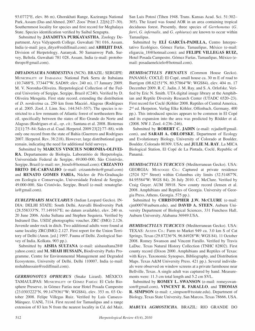
Herpetological Review 41(4), 2010512
93.0772°E, elev. 86 m), Ghorakhati Range, Kaziranga National Park, Assam (Das and Ahmed, 2007. Zoos’ Print J. 22[6]:27–30). Southernmost locality for species and first record for Meghalaya State. Species identification verified by Saibal Sengupta. Submitted by JAYADITYA PURKAYASTHA, Zoology De-partment, Arya Vidyapeeth College. Guwahati 781 016, Assam, India (e-mail: [email protected]); and ABHIJIT DAS, Division of Herpetology, Aaranyak, 50 Samanwoy Path, Sur-vey, Beltola, Guwahati 781 028, Assam, India (e-mail: [email protected]).
DRYADOSAURA NORDESTINA (NCN). BRAZIL: SERGIPE: MuniciPalitY of itaBaiana: National Park Serra de Itabaiana (10.7488ºS, 373447ºW, SAD69; elev. 240 m), 17 January 2009. M. V. Noronha-Oliveira. Herpetological Collection of the Fed-eral Univeristy of Sergipe, Sergipe, Brazil (C240). Verified by D. Oliveira Mesquita. First state record, extending the distribution of D. nordestina ca. 250 km from Maceió, Alagoas (Rodrigues et al. 2005. Zool. J. Linn. Soc. 144:543–557). The species is re-stricted to a few remnants of Atlantic forest of northeastern Bra-zil, specifically between the states of Rio Grande do Norte and Alagoas (Rodrigues et al., op. cit.; Santana et al. 2008. Biotemas 21[1]:75–84; Sales et al. Cuad. Herpetol. 2009 23[2]:77–88), with only one record from the state of Bahia (Guerrero and Rodrigues 2007. Herpetol. Rev. 38:218). However, large distributional gaps remain, indicating the need for additional field surveys. Submitted by MARCUS VINICIUS NORONHA-OLIVEI-RA, Departamento de Biologia, Laboratório de Herpetologia, Universidade Federal de Sergipe, 49.000-000, São Cristóvão, Sergipe, Brazil (e-mail: [email protected]); CRIZANTO BRITO DE-CARVALHO (e-mail: [email protected]) and RENATO GOMES FARIA, Núcleo de Pós-Graduação em Ecologia e Conservação, Universidade Federal de Sergipe, 49.000-000, São Cristóvão, Sergipe, Brazil (e-mail: [email protected]).
EUBLEPHARIS MACULARIUS (Indian Leopard Gecko). IN-DIA: DELHI STATE: South Delhi, Aravalli Biodiversity Park (28.558333ºN, 77.149167ºE; no datum available), elev. 240 m. 20 June 2006. Aisha Sultana and Stephen Sequiera. Verified by Indraneil Das. USDZ photographic voucher, ZRC (IMG) 2.126. Juvenile under rock in ditch. Two additional adults were found at same locality ZRC(IMG) 2.127. First report for the Union Terri-tory of Delhi (Anon. [ed.] 1997. Fauna of Delhi. Zoological Sur-vey of India, Kolkata. 903 pp.). Submitted by AISHA SULTANA (e-mail: [email protected]); and M. SHAH HUSSAIN, Biodiversity Parks Pro-gramme, Centre for Environmental Management and Degraded Ecosystems, University of Delhi, Delhi 110007, India (e-mail: [email protected]).
GERRHONOTUS OPHIURUS (Snake Lizard). MÉXICO: TAMAULIPAS: MuniciPalitY of góMez faríaS: El Cielo Bio-sphere Preserve, in Gómez Farías near Hotel Posada Campestre (23.0303222°N, 99.1479361°W; WGS84), elev. 353 m. 03 Oc-tober 2008. Felipe Villegas Ruiz. Verified by Luis Canseco-Márquez. UANL 7114. First record for Tamaulipas and a range extension of 83 km N from the nearest locality in Cd. del Maíz,
San Luis Potosí (Tihen 1948. Trans. Kansas Acad. Sci. 51:302–305). The lizard was found AOR in an area containing tropical deciduous forest. Presently, three species of Gerrhonotus (G. farri, G. infernalis, and G. ophiurus) are known to occur within Tamaulipas. Submitted by ELÍ GARCÍA-PADILLA, Centro Interpre-tativo Ecológico, Gómez Farías, Tamaulipas, México (e-mail: [email protected]); and FELIPE VILLEGAS RUIZ, Hotel Posada Campestre, Gómez Farías, Tamaulipas, México (e-mail: [email protected]).
HEMIDACTYLUS FRENATUS (Common House Gecko). PANAMÁ: coclé: El Copé, small house ca. 30 m E of road to Barrigon (08.62151°N, 80.57864°W; WGS84), elev. 404 m. 17 December 2009. R. C. Jadin, J. M. Ray, and S. A. Orlofske. Veri-fied by Eric N. Smith. UTA digital image library at the Amphib-ian and Reptile Diversity Research Center (UTADC 6520–23). First record for Coclé (Köhler 2008. Reptiles of Central America, 2nd ed. Herpeton, Verlag Elke Köhler, Offenbach, Germany. 400 pp.). This introduced species appears to be common in El Copé and its expansion into the area was predicted by Rödder et al. (2008. NW J. Zool. 4:236–246). Submitted by ROBERT C. JADIN (e-mail: [email protected]), and SARAH A. ORLOFSKE, Department of Ecology and Evolutionary Biology, University of Colorado at Boulder, Boulder, Colorado 80309, USA; and JULIE M. RAY, La MICA Biological Station, El Copé de La Pintada, Coclé, Republic of Panamá.
HEMIDACTYLUS TURCICUS (Mediterranean Gecko). USA: GEORGIA: MuScogee co.: Captured at private residence (2524 52nd Street) within Columbus city limits (32.51487ºN, 84.95584ºW; WGS 84). 26 July 2010. C. McClure. Verified by Craig Guyer. AUM 38918. New county record (Jensen et al. 2008. Amphibians and Reptiles of Georgia. University of Geor-gia Press, Athens, Georgia. 575 pp.). Submitted by CHRISTOPHER J.W. McCLURE (e-mail: [email protected]), and DAVID A. STEEN, Auburn Uni-versity Department of Biological Sciences, 331 Funchess Hall, Auburn University, Alabama 36949.USA.
HEMIDACTYLUS TURCICUS (Mediterranean Gecko). USA: TEXAS: auStin co.: Farm to Market 949 ca. 3.0 km S of Cat Springs, Texas (29.87236°N, 96.84928°W; WGS 84). 11 October 2008. Romey Swanson and Vincent Farallo. Verified by Travis LaDuc. Texas Natural History Collection (TNHC 82802). First county record (Dixon 2000. Amphibians and Reptiles of Texas: with Keys, Taxonomic Synopses, Bibliography, and Distribution Maps. Texas A&M University Press. 421 pp.). Several individu-als were observed on window screens at a private farmhouse near Bellville, Texas. A single adult was captured by hand. Measure-ments were: 11.3 cm total length and 5.2 cm SVL. Submitted by ROMEY L. SWANSON (e-mail: [email protected]), VINCENT R. FARALLO, and THOMAS R. SIMPSON (e-mail: [email protected]), Department of Biology, Texas State University, San Marcos, Texas 78666, USA.
MABUYA AGMOSTICHA. BRAZIL: RIO GRANDE DO
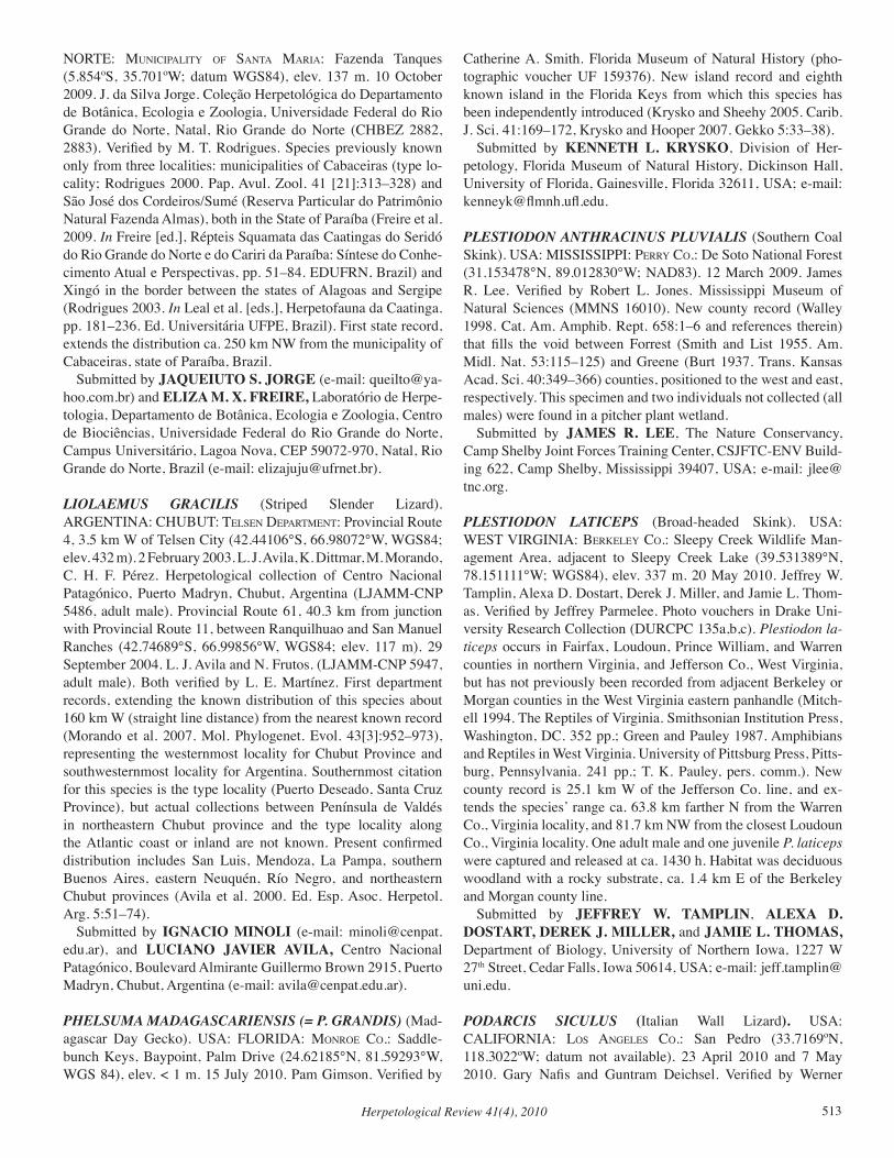
Herpetological Review 41(4), 2010 513
NORTE: MuniciPalitY of Santa Maria: Fazenda Tanques (5.854ºS, 35.701ºW; datum WGS84), elev. 137 m. 10 October 2009. J. da Silva Jorge. Coleção Herpetológica do Departamento de Botânica, Ecologia e Zoologia, Universidade Federal do Rio Grande do Norte, Natal, Rio Grande do Norte (CHBEZ 2882, 2883). Verified by M. T. Rodrigues. Species previously known only from three localities: municipalities of Cabaceiras (type lo-cality; Rodrigues 2000. Pap. Avul. Zool. 41 [21]:313–328) and São José dos Cordeiros/Sumé (Reserva Particular do Patrimônio Natural Fazenda Almas), both in the State of Paraíba (Freire et al. 2009. In Freire [ed.], Répteis Squamata das Caatingas do Seridó do Rio Grande do Norte e do Cariri da Paraíba: Síntese do Conhe-cimento Atual e Perspectivas, pp. 51–84. EDUFRN, Brazil) and Xingó in the border between the states of Alagoas and Sergipe (Rodrigues 2003. In Leal et al. [eds.], Herpetofauna da Caatinga. pp. 181–236. Ed. Universitária UFPE, Brazil). First state record, extends the distribution ca. 250 km NW from the municipality of Cabaceiras, state of Paraíba, Brazil. Submitted by JAQUEIUTO S. JORGE (e-mail: [email protected]) and ELIZA M. X. FREIRE, Laboratório de Herpe-tologia, Departamento de Botânica, Ecologia e Zoologia, Centro de Biociências, Universidade Federal do Rio Grande do Norte, Campus Universitário, Lagoa Nova, CEP 59072-970, Natal, Rio Grande do Norte, Brazil (e-mail: [email protected]).
LIOLAEMUS GRACILIS (Striped Slender Lizard). ARGENTINA: CHUBUT: telSen dePartMent: Provincial Route 4, 3.5 km W of Telsen City (42.44106°S, 66.98072°W, WGS84; elev. 432 m). 2 February 2003. L. J. Avila, K. Dittmar, M. Morando, C. H. F. Pérez. Herpetological collection of Centro Nacional Patagónico, Puerto Madryn, Chubut, Argentina (LJAMM-CNP 5486, adult male). Provincial Route 61, 40.3 km from junction with Provincial Route 11, between Ranquilhuao and San Manuel Ranches (42.74689°S, 66.99856°W, WGS84; elev. 117 m). 29 September 2004. L. J. Avila and N. Frutos. (LJAMM-CNP 5947, adult male). Both verified by L. E. Martínez. First department records, extending the known distribution of this species about 160 km W (straight line distance) from the nearest known record (Morando et al. 2007. Mol. Phylogenet. Evol. 43[3]:952–973), representing the westernmost locality for Chubut Province and southwesternmost locality for Argentina. Southernmost citation for this species is the type locality (Puerto Deseado, Santa Cruz Province), but actual collections between Península de Valdés in northeastern Chubut province and the type locality along the Atlantic coast or inland are not known. Present confirmed distribution includes San Luis, Mendoza, La Pampa, southern Buenos Aires, eastern Neuquén, Río Negro, and northeastern Chubut provinces (Avila et al. 2000. Ed. Esp. Asoc. Herpetol. Arg. 5:51–74). Submitted by IGNACIO MINOLI (e-mail: [email protected]), and LUCIANO JAVIER AVILA, Centro Nacional Patagónico, Boulevard Almirante Guillermo Brown 2915, Puerto Madryn, Chubut, Argentina (e-mail: [email protected]).
PHELSUMA MADAGASCARIENSIS (= P. GRANDIS) (Mad-agascar Day Gecko). USA: FLORIDA: Monroe co.: Saddle-bunch Keys, Baypoint, Palm Drive (24.62185°N, 81.59293°W, WGS 84), elev. < 1 m. 15 July 2010. Pam Gimson. Verified by
Catherine A. Smith. Florida Museum of Natural History (pho-tographic voucher UF 159376). New island record and eighth known island in the Florida Keys from which this species has been independently introduced (Krysko and Sheehy 2005. Carib. J. Sci. 41:169–172, Krysko and Hooper 2007. Gekko 5:33–38). Submitted by KENNETH L. KRYSKO, Division of Her-petology, Florida Museum of Natural History, Dickinson Hall, University of Florida, Gainesville, Florida 32611, USA; e-mail: [email protected].
PLESTIODON ANTHRACINUS PLUvIALIS (Southern Coal Skink). USA: MISSISSIPPI: PerrY co.: De Soto National Forest (31.153478°N, 89.012830°W; NAD83). 12 March 2009. James R. Lee. Verified by Robert L. Jones. Mississippi Museum of Natural Sciences (MMNS 16010). New county record (Walley 1998. Cat. Am. Amphib. Rept. 658:1–6 and references therein) that fills the void between Forrest (Smith and List 1955. Am. Midl. Nat. 53:115–125) and Greene (Burt 1937. Trans. Kansas Acad. Sci. 40:349–366) counties, positioned to the west and east, respectively. This specimen and two individuals not collected (all males) were found in a pitcher plant wetland. Submitted by JAMES R. LEE, The Nature Conservancy, Camp Shelby Joint Forces Training Center, CSJFTC-ENV Build-ing 622, Camp Shelby, Mississippi 39407, USA; e-mail: [email protected].
PLESTIODON LATICEPS (Broad-headed Skink). USA: WEST VIRGINIA: BerkeleY co.: Sleepy Creek Wildlife Man-agement Area, adjacent to Sleepy Creek Lake (39.531389°N, 78.151111°W; WGS84), elev. 337 m. 20 May 2010. Jeffrey W. Tamplin, Alexa D. Dostart, Derek J. Miller, and Jamie L. Thom-as. Verified by Jeffrey Parmelee. Photo vouchers in Drake Uni-versity Research Collection (DURCPC 135a,b,c). Plestiodon la-ticeps occurs in Fairfax, Loudoun, Prince William, and Warren counties in northern Virginia, and Jefferson Co., West Virginia, but has not previously been recorded from adjacent Berkeley or Morgan counties in the West Virginia eastern panhandle (Mitch-ell 1994. The Reptiles of Virginia. Smithsonian Institution Press, Washington, DC. 352 pp.; Green and Pauley 1987. Amphibians and Reptiles in West Virginia. University of Pittsburg Press, Pitts-burg, Pennsylvania. 241 pp.; T. K. Pauley, pers. comm.). New county record is 25.1 km W of the Jefferson Co. line, and ex-tends the species’ range ca. 63.8 km farther N from the Warren Co., Virginia locality, and 81.7 km NW from the closest Loudoun Co., Virginia locality. One adult male and one juvenile P. laticeps were captured and released at ca. 1430 h. Habitat was deciduous woodland with a rocky substrate, ca. 1.4 km E of the Berkeley and Morgan county line. Submitted by JEFFREY W. TAMPLIN, ALEXA D. DOSTART, DEREK J. MILLER, and JAMIE L. THOMAS, Department of Biology, University of Northern Iowa, 1227 W 27th Street, Cedar Falls, Iowa 50614, USA; e-mail: [email protected].
PODARCIS SICULUS (Italian Wall Lizard). USA: CALIFORNIA: loS angeleS co.: San Pedro (33.7169ºN, 118.3022ºW; datum not available). 23 April 2010 and 7 May 2010. Gary Nafis and Guntram Deichsel. Verified by Werner
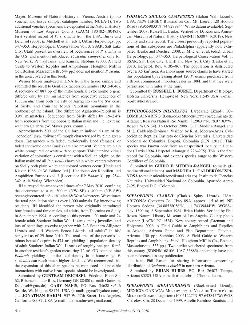
Herpetological Review 41(4), 2010514
Mayer. Museum of Natural History in Vienna, Austria (photo voucher and tissue sample; catalogue number: NULA-1). Two additional voucher specimens are deposited at the Natural History Museum of Los Angeles County (LACM 180482–180483). First verified record of P. s. siculus from the USA. Burke and Deichsel (2008. In Mitchell et al. [eds.], Urban Herpetology, pp. 347–353. Herpetological Conservation Vol. 3. SSAR, Salt Lake City, Utah) present an overview of occurrences of P. siculus in the U.S. and mention introduced P. siculus campestris only for New York, Pennsylvania, and Kansas. Stebbins (2003. A Field Guide to Western Reptiles and Amphibians. Houghton Mifflin Co., Boston, Massachusetts. 544 pp.) does not mention P. siculus in the area covered in this book. Werner Mayer analyzed mtDNA from the tissue sample and submitted the result to GenBank (accession number HQ154646). A sequence of 887 bp of the mitochondrial cytochrome b gene differed only by 1% mismatches from respective sequences of P. s. siculus from both the city of Agrigento (on the SW coast of Sicily) and from the Monti Peloritani mountains in the northeast of the island. The difference Agrigento–Peloritani is 0.9% mismatches. Sequences from Sicily differ by 1.9–2.4% from sequences from the opposite Italian mainland, i.e., extreme southern Calabria (W. Mayer, pers. comm.). Approximately 50% of the Californian individuals are of the “concolor” (syn. “olivacea”) morph characterized by plain green dorsa. Intergrades with faded, mid-dorsally lined (females) or faded checkered dorsa (males) are also present. Venters are plain white, orange, red, or white with beige spots. This inter-individual variation of coloration is consistent with a Sicilian origin: on the Italian mainland all P. s. siculus have plain white venters whereas on Sicily both plain white and colored venters occur (Henle and Klaver 1986. In W. Böhme [ed.], Handbuch der Reptilien und Amphibien Europas vol. 3 [Lacertidae III: Podarcis], pp. 254–342. Aula Verlag, Wiesbaden). JH surveyed the area several times after 7 May 2010, confining the occurrence to a ca. 300 m (NW–SE) x 400 m (NE–SW) rectangle centered at South Leland & West 34th streets. We estimate the total population size as over 1,000 animals. By interviewing residents, JH identified the person who originally introduced four females and three males, all adults, from Taormina on Sicily in September 1994. According to this person, “20 male and 24 female adult Southern Italian Wall Lizards, many juveniles, and lots of hatchlings co-exist together with 2–3 Southern Alligator Lizards and 4-5 Western Fence Lizards, all adults” in his/her yard as of 29 June 2010. The total area of the person’s lot minus house footprint is 474 m2, yielding a population density of adult Southern Italian Wall Lizards of roughly one per 10 m2. In another resident’s garden measuring 72 m2, GD counted nine Podarcis, yielding a similar local density. In its home range, P. s. siculus can reach much higher densities. We recommend that the expansion of this alien species be monitored and possible interactions with native lizard species should be investigated. Submitted by GUNTRAM DEICHSEL, Friedrich-Ebert-Str. 62, Biberach an der Riss, Germany DE-88400 (e-mail: [email protected]); GARY NAFIS, PO Box 34628-89546 Seattle, Washington 98124, USA (e-mail: [email protected]); and JONATHAN HAKIM, 937 W. 57th Street, Los Angeles, California 90037, USA (e-mail: [email protected]).
PODARCIS SICULUS CAMPESTRIS (Italian Wall Lizard). USA: NEW JERSEY Burlington co.: Mt. Laurel, 128 Hooton Road (39.9559833ºN, 74.9299944º W; no datum available). Sep-tember 2008. Russell L. Burke. Verified by D. Kizirian. Ameri-can Museum of Natural History (AMNH 163007–163019). New county and state record. The closest previously reported popula-tions of this subspecies are Philadelphia (apparently now extir-pated [Burke and Deichsel 2008. In Mitchell et al. (eds.), Urban Herpetology, pp. 347–353. Herpetological Conservation Vol. 3. SSAR, Salt Lake City, Utah]) and New York City (Burke et al. 2010. Herpetol. Rev. 41:85–86). The population is distributed over a 0.5 km2 area. An anonymous source claims to have started this population by releasing about 120 P. siculus purchased from a Bronx commercial importer/dealer in 1984, and that they were parasitized with mites at the time. Submitted by RUSSELL L. BURKE, Department of Biology, Hofstra University, Hempstead, New York 11549,USA; e-mail: [email protected].
PTYCHOGLOSSUS BILINEATUS (Largescale Lizard). CO-LOMBIA: NARIÑO: BarBacoaS MuniciPalitY: corregimiento de Altaquer, Reserva Natural Río Ñambi (1.29631°N, 78.07183°W; Bogotá WGS 84). 16 October 2009. G. F. Medina-Rangel and M. L. Calderón-Espinosa. Verified by R. A. Moreno-Arias. Col-ección de Reptiles, Instituto de Ciencias Naturales, Universidad Nacional de Colombia, Bogotá, Colombia (ICN 12011). This species was known only from an unspecified locality in Ecua-dor (Harris 1994. Herpetol. Monogr. 8:226–275). This is the first record for Colombia, and extends species range to the Western Cordillera of Colombia. Submitted by GUIDO F. MEDINA-RANGEL (e-mail: [email protected]), and MARTHA L. CALDERÓN-ESPI-NOSA (e-mail: [email protected]), Instituto de Ciencias Naturales, Universidad Nacional de Colombia, Apartado Aéreo 7495, Bogotá D.C., Colombia.
SCELOPORUS CLARKII (Clark’s Spiny Lizard). USA: ARIZONA: coconino co.: Hwy 89A, approx. 1.5 rd mi. NE Uptown Sedona (34.88538056ºN, 111.74339444ºW, WGS84; elev. 1390 m). 8 September 1984. Brian Hubbs. Verified by Phil Rosen. Natural History Museum of Los Angeles County photo voucher (LACM PC 1524). New county record (Brennan and Holycross 2006. A Field Guide to Amphibians and Reptiles in Arizona. Arizona Game and Fish Department. Phoenix, Arizona. 150 pp.; Stebbins 2003. A Field Guide to Western Reptiles and Amphibians, 3rd ed. Houghton Mifflin Co., Boston, Massachusetts. 533 pp.). Two earlier vouchered specimens from this county (SDNHM 68106, UAZ 33885) apparently have not been referenced in any publication. I thank Phil Rosen for sharing information concerning distribution of Sceloporus clarkii in northern Arizona. Submitted by BRIAN HUBBS, P.O. Box 26407, Tempe, Arizona 85285, USA; e-mail: [email protected].
SCELOPORUS MELANORHINUS (Black-nosed Lizard). MÉXICO: OAXACA: MuniciPalitY of villa de tututePéc de MelcHor ocaMPo: Lagartero (16.051227°N, 97.643845°W; WGS 84), elev. 8 m. 28 December 1999. Aurelio Ramírez-Bautista and
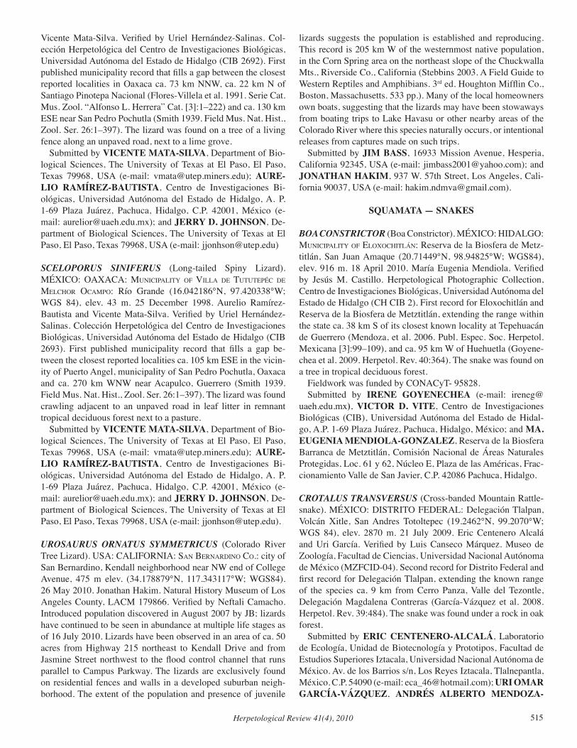
Herpetological Review 41(4), 2010 515
Vicente Mata-Silva. Verified by Uriel Hernández-Salinas. Col-ección Herpetológica del Centro de Investigaciones Biológicas, Universidad Autónoma del Estado de Hidalgo (CIB 2692). First published municipality record that fills a gap between the closest reported localities in Oaxaca ca. 73 km NNW, ca. 22 km N of Santiago Pinotepa Nacional (Flores-Villela et al. 1991. Serie Cat. Mus. Zool. “Alfonso L. Herrera” Cat. [3]:1–222) and ca. 130 km ESE near San Pedro Pochutla (Smith 1939. Field Mus. Nat. Hist., Zool. Ser. 26:1–397). The lizard was found on a tree of a living fence along an unpaved road, next to a lime grove. Submitted by VICENTE MATA-SILVA, Department of Bio-logical Sciences, The University of Texas at El Paso, El Paso, Texas 79968, USA (e-mail: [email protected]); AURE-LIO RAMÍREZ-BAUTISTA, Centro de Investigaciones Bi-ológicas, Universidad Autónoma del Estado de Hidalgo, A. P. 1-69 Plaza Juárez, Pachuca, Hidalgo, C.P. 42001, México (e-mail: [email protected]); and JERRY D. JOHNSON, De-partment of Biological Sciences, The University of Texas at El Paso, El Paso, Texas 79968, USA (e-mail: [email protected])
SCELOPORUS SINIFERUS (Long-tailed Spiny Lizard). MÉXICO: OAXACA: MuniciPalitY of villa de tututePéc de MelcHor ocaMPo: Río Grande (16.042186°N, 97.420338°W; WGS 84), elev. 43 m. 25 December 1998. Aurelio Ramírez-Bautista and Vicente Mata-Silva. Verified by Uriel Hernández-Salinas. Colección Herpetológica del Centro de Investigaciones Biológicas, Universidad Autónoma del Estado de Hidalgo (CIB 2693). First published municipality record that fills a gap be-tween the closest reported localities ca. 105 km ESE in the vicin-ity of Puerto Angel, municipality of San Pedro Pochutla, Oaxaca and ca. 270 km WNW near Acapulco, Guerrero (Smith 1939. Field Mus. Nat. Hist., Zool. Ser. 26:1–397). The lizard was found crawling adjacent to an unpaved road in leaf litter in remnant tropical deciduous forest next to a pasture. Submitted by VICENTE MATA-SILVA, Department of Bio-logical Sciences, The University of Texas at El Paso, El Paso, Texas 79968, USA (e-mail: [email protected]); AURE-LIO RAMÍREZ-BAUTISTA, Centro de Investigaciones Bi-ológicas, Universidad Autónoma del Estado de Hidalgo, A. P. 1-69 Plaza Juárez, Pachuca, Hidalgo, C.P. 42001, México (e-mail: [email protected]); and JERRY D. JOHNSON, De-partment of Biological Sciences, The University of Texas at El Paso, El Paso, Texas 79968, USA (e-mail: [email protected]).
UROSAURUS ORNATUS SYMMETRICUS (Colorado River Tree Lizard). USA: CALIFORNIA: San Bernardino co.: city of San Bernardino, Kendall neighborhood near NW end of College Avenue, 475 m elev. (34.178879°N, 117.343117°W; WGS84). 26 May 2010. Jonathan Hakim. Natural History Museum of Los Angeles County, LACM 179866. Verified by Neftali Camacho. Introduced population discovered in August 2007 by JB; lizards have continued to be seen in abundance at multiple life stages as of 16 July 2010. Lizards have been observed in an area of ca. 50 acres from Highway 215 northeast to Kendall Drive and from Jasmine Street northwest to the flood control channel that runs parallel to Campus Parkway. The lizards are exclusively found on residential fences and walls in a developed suburban neigh-borhood. The extent of the population and presence of juvenile
lizards suggests the population is established and reproducing. This record is 205 km W of the westernmost native population, in the Corn Spring area on the northeast slope of the Chuckwalla Mts., Riverside Co., California (Stebbins 2003. A Field Guide to Western Reptiles and Amphibians. 3rd ed. Houghton Mifflin Co., Boston, Massachusetts. 533 pp.). Many of the local homeowners own boats, suggesting that the lizards may have been stowaways from boating trips to Lake Havasu or other nearby areas of the Colorado River where this species naturally occurs, or intentional releases from captures made on such trips. Submitted by JIM BASS, 16933 Mission Avenue, Hesperia, California 92345, USA (e-mail: [email protected]); and JONATHAN HAKIM, 937 W. 57th Street, Los Angeles, Cali-fornia 90037, USA (e-mail: [email protected]).
SQUAMATA — SNAKES
BOA CONSTRICTOR (Boa Constrictor). MÉXICO: HIDALGO: MuniciPalitY of eloXocHitlán: Reserva de la Biosfera de Metz-titlán, San Juan Amaque (20.71449°N, 98.94825°W; WGS84), elev. 916 m. 18 April 2010. María Eugenia Mendiola. Verified by Jesús M. Castillo. Herpetological Photographic Collection, Centro de Investigaciones Biológicas, Universidad Autónoma del Estado de Hidalgo (CH CIB 2). First record for Eloxochitlán and Reserva de la Biosfera de Metztitlán, extending the range within the state ca. 38 km S of its closest known locality at Tepehuacán de Guerrero (Mendoza, et al. 2006. Publ. Espec. Soc. Herpetol. Mexicana [3]:99–109), and ca. 95 km W of Huehuetla (Goyene-chea et al. 2009. Herpetol. Rev. 40:364). The snake was found on a tree in tropical deciduous forest. Fieldwork was funded by CONACyT- 95828. Submitted by IRENE GOYENECHEA (e-mail: [email protected]), VICTOR D. VITE, Centro de Investigaciones Biológicas (CIB), Universidad Autónoma del Estado de Hidal-go, A.P. 1-69 Plaza Juárez, Pachuca, Hidalgo, México; and MA. EUGENIA MENDIOLA-GONZALEZ, Reserva de la Biosfera Barranca de Metztitlán, Comisión Nacional de Áreas Naturales Protegidas, Loc. 61 y 62, Núcleo E, Plaza de las Américas, Frac-cionamiento Valle de San Javier, C.P. 42086 Pachuca, Hidalgo.
CROTALUS TRANSvERSUS (Cross-banded Mountain Rattle-snake). MÉXICO: DISTRITO FEDERAL: Delegación Tlalpan, Volcán Xitle, San Andres Totoltepec (19.2462°N, 99.2070°W; WGS 84), elev. 2870 m. 21 July 2009. Eric Centenero Alcalá and Uri García. Verified by Luis Canseco Márquez. Museo de Zoología, Facultad de Ciencias, Universidad Nacional Autónoma de México (MZFCID-04). Second record for Distrito Federal and first record for Delegación Tlalpan, extending the known range of the species ca. 9 km from Cerro Panza, Valle del Tezontle, Delegación Magdalena Contreras (García-Vázquez et al. 2008. Herpetol. Rev. 39:484). The snake was found under a rock in oak forest. Submitted by ERIC CENTENERO-ALCALÁ, Laboratorio de Ecología, Unidad de Biotecnología y Prototipos, Facultad de Estudios Superiores Iztacala, Universidad Nacional Autónoma de México. Av. de los Barrios s/n, Los Reyes Iztacala, Tlalnepantla, México. C.P. 54090 (e-mail: [email protected]); URI OMAR GARCÍA-VÁZQUEZ, ANDRÉS ALBERTO MENDOZA-
Herpetological Review 41(4), 2010516
HERNÁNDEZ, JOAN GASTÓN ZAMORA-ABREGO, Museo de Zoología, Facultad de Ciencias, Universidad Nacio-nal Autónoma de México, A.P. 70-399, México, Distrito Federal 04510, México; and VÍCTOR HUGO JIMÉNEZ-ARCOS, Laboratorio de Herpetología, Instituto de Biología, Universidad Nacional Autónoma de México. 3er Circuito exterior s/n, Ciudad Universitaria, Coyoacán, México, D.F. C.P. 04510.
EPICTA GOUDOTII (Black Threadsnake). MÉXICO: HIDAL-GO: MuniciPalitY of Hazalingo: Tepalcahuac (20.95852°N, 98.48527°W; WGS84), elev. 315 m. 01 September 2007. Victor D. Vite Silva and Alejandro Ramírez-Pérez. Verified by Raciel Cruz Elizalde. Colección de Anfibios y Reptiles del Centro de Investigaciones Biológicas, Universidad Autónoma del Estado de Hidalgo (CIB 2181–2183). MuniciPalitY of Huautla: Tamayon (21.03457°N, 98.2886°W; WGS84), elev. 506 m. 10 November 2007. Victor D. Vite Silva and Alejandro Ramírez-Pérez. Verified by Raciel Cruz Elizalde. CIB 2184. First records for Hidalgo, extending the range ca. 147 km SW from the closest known local-ity, 2 km W of La Loma, Tamaulipas (Flores-Benabib and Flores-Villela 2008. Biol. Soc. Herpetol. Mexicana 16:13–14). We acknowledge the following for support: Diversidad Bi-ológica de Estado de Hidalgo, FOMIX-CONACYT - HIDALGO 43761; and Diversidad Biológica de Estado de Hidalgo (segunda fase), FOMIX-CONACYT - HIDALGO 95828 and S52552-Q. Submitted by URIEL HERNÁNDEZ SALINAS (e-mail: [email protected]), JUDITH PAMPA RAMÍREZ (e-mail: [email protected]), IRENE GOYENECHEA (e-mail: [email protected]), AURELIO RAMÍREZ BAU-TISTA (e-mail: [email protected]), and VÍCTOR VITE SILVA, Centro de Investigaciones Biológicas (CIB), Universidad Autónoma del Estado de Hidalgo, A.P. 1-69 Plaza Juárez, C.P. 42001, Pachuca, Hidalgo, México
FARANCIA ABACURA (Red-bellied Mudsnake). USA: GEOR-GIA: PeacH co.: DOR, State Rt. 49 at Mule Cr. (32.60278ºN; 83.79815ºW; no datum available). 8 May 2010. Digital image AHAP-D 246. First county record. taYlor co.: DOR, State Rt. 96, 1 km E of Reynolds, at Flint River floodplain (Magnolia Swamp) (32.55267ºN; 84.05382ºW; no datum available). 08 May 2010. Digital image AHAP-D 245. First county record (Jensen et al. 2008. Amphibians and Reptiles of Georgia. University of Georgia Press, Athens. 575 pp.). Both specimens collected by S. Graham and verified by Craig Guyer. Decimal degrees of locali-ties were determined using ACME Mapper 2.0 software (http://mapper.acme.com). Submitted by SEAN P. GRAHAM, Auburn University, De-partment of Biological Sciences, 331 Funchess Hall, Auburn, Alabama 36849, USA; e-mail: [email protected].
HETERODON PLATIRHINOS (Eastern Hognose Snake). USA: TEXAS: caldWell co.: Farm to Market Road 713 near the Caldwell/Bastrop Co. Line Road (29.82858°N, 97.39533°W; WGS 84). 12 April 2008. Romey Swanson. Verified by Travis La-Duc. Texas Natural History Collection (TNHC 82800). A single DOR was collected from the road shoulder. First county record (Dixon 2000. Amphibians and Reptiles of Texas: with Keys, Tax-onomic Synopses, Bibliography, and Distribution Maps. Texas
A&M University Press, College Station, Texas. 421 pp.). The in-dividual measured 48.4 cm SVL and 60.0 cm total length and had a reddish base coloration often seen in specimens collected from Bastrop Co. Submitted by ROMEY L. SWANSON (e-mail: [email protected]) and THOMAS R. SIMPSON (e-mail: [email protected]), Department of Biology, Texas State Univer-sity, San Marcos, Texas 78666, USA.
LAMPROPELTIS CALLIGASTER (Mole Kingsnake). USA: GEORGIA: PutnaM co.: Burtom Rd. near Lake Sinclair (33.224543°N, 83.419154°W; WGS84). 18 April 2010. Alfred J. Mead. GCH 5280. Verified by Christopher E. Skelton. First county record (Jensen et al. 2008. Amphibians and Reptiles of Georgia. University of Georgia Press, Athens. 575 pp.). Single adult collected DOR on road flanked by mature pine forest and recently established pasture land. Submitted by ALFRED J. MEAD (e-mail: [email protected]) and DENNIS PARMLEY, Department of Biological and Environmental Sciences, Georgia College & State University, Milledgeville, Georgia 31061, USA.
LAMPROPELTIS GETULA (Common Kingsnake). USA: GEORGIA: WilkeS co.: Tignall, Newtown Road, 1.6 km NW Elam Lunceford Road (33.87554°N, 82.83697°W, WGS84), elev. 163 m. 06 June 2009. Justin T. Oguni and Matthew A. King. Veri-fied by Kenneth L. Krysko. Florida Museum of Natural History (photographic voucher UF 158417). New county record (Jensen et al. 2008. Amphibians and Reptiles of Georgia. University of Georgia Press, Athens. 575 pp.). Found AOR at 2145 h in mixed hardwood forest with residential homes. Submitted by JUSTIN T. OGUNI, College of Veterinary Medicine, University of Georgia, Athens, Georgia 30602, USA; e-mail: [email protected]
LAMPROPELTIS GETULA NIGRA (Eastern Black King-snake). USA: GEORGIA: dade co.: field ca. 100 m NE of jct of Higdon Creek and CR 13, ca. 5.9 air km W of Trenton (34.87262°N, 85.57365°W; NAD83). 17 May 2010. Houston C. Chandler. UF 157836. Verified by John B. Jensen and Kenneth Krysko. First county record (Jensen et al. 2008. Amphibians and Reptiles of Georgia. University of Georgia Press, Athens. 575 pp.). Single adult captured and photographed on the bank of a small farm pond. Rural area with mix of forest and pasture and a ca. 5 ha clearcut within sight of capture area. Submitted by HOUSTON C. CHANDLER and CHRISTO-PHER E. SKELTON, Department of Biological and Environ-mental Sciences, Georgia College & State University, Milled-geville, Georgia 31061, USA; e-mail: [email protected].
LEPTOTYPHLOPS HUMILIS (Western Threadsnake). USA: ARIZONA: MoHave co.: Virgin River valley (36.841538°N, 113.982881°W, NAD 83), elev. 513 m. 24 July 2010. Steven Anderson. Verified by Tom Giermakowski. University of New Mexico Museum of Southwestern Biology (MSB 77882). This is the first record from the Arizona Strip and fills an important gap in the known distribution of the species. Other specimens have been recorded further south, along the lower Colorado River and
Herpetological Review 41(4), 2010 517
from the Grand Canyon, Arizona (Brennan and Holycross 2006. Amphibians and Reptiles in Arizona. Arizona Game and Fish Department, Phoenix, Arizona. 150 pp.) and to the northeast in Washington Co., Utah (A. Holycross, pers. comm.). NEVADA: clark co.: Virgin River Valley, Gold Butte recreation area (36.665935°N, 114.306052°W, NAD 83), elev. 396 m. 22 July 2010. Danny Nielsen. Verified by Andy Holycross. University of Arizona Museum of Natural History (UAZ 57242-PSV, photo voucher). This is the eighth record from southern Nevada and ca. 20 km from nearest known record in Overton, Nevada from 1939 (Grater 1981. Snakes, Lizards and Turtles of the Lake Mead Region. Southwest Parks and Monuments Association, Globe, Arizona. 48 pp.; P. Conrad, Nevada Department of Wildlife, pers. comm.), and ca. 40 km downstream from the Mohave Co., Ari-zona specimen reported above. Both specimens were collected from pitfall traps in upper riparian habitat of mesquite (Prosopis sp. and saltcedar (Tamarix sp.). Collecting permits were issued by the Arizona Game and Fish Department (No. 195230) and Nevada Department of Wildlife (No. S32027). Submitted by HEATHER L. BATEMAN (e-mail: [email protected]) and DANNY NIELSEN, Arizona State University at the Polytechnic Campus, Department of Applied Sciences and Mathematics, 6073 S. Backus Mall, Mesa, Arizona 85212, USA (e-mail: [email protected]).
NERODIA ERYTHROGASTER FLAvIGASTER (Yellow-bel-lied Watersnake). USA: ARKANSAS: calHoun co.: dor ~6 km N of Ouachita River on US 167 (33.392396°N, 92.493676°W; WGS 84). 09 September 2010. M. B. Connior. Verified by S. E. Trauth. Arkansas State University Museum of Zoology Herpe-tology Collection (photo voucher ASUMZ 31555). First coun-ty record (Trauth et al. 2004. The Amphibians and Reptiles of Arkansas. University of Arkansas Press, Fayetteville. 421 pp.). The species has also been collected from neighboring Bradley, Ouachita, and Union counties in southern Arkansas. Submitted by MATTHEW B. CONNIOR, Health and Natu-ral Sciences, South Arkansas Community College, 300 S. West Avenue, El Dorado, Arkansas 71730, USA; e-mail: [email protected].
OXYBELIS AENEUS (Mexican Vine Snake). MÉXICO: HIDALGO: MuniciPalitY of tlaHuiltePa: Boca de León (20.99139°N, 98.99193°W; WGS84), elev. 464 m. 18 November 2009. Victor D. Vite-Silva, Alejandro Ramírez-Pérez, and Oscar González-Solís. Verified by Norma Manríquez-Morán. Colec-ción Herpetológica, Centro de Investigaciones Biológicas, Uni-versidad Autónoma del Estado de Hidalgo (CIB-UAEH 2538). First record for the municipality, extending the range in Hidalgo ca. 43 km S from the closest known record at La Vega de Metzti-tlán, Municipality of Metztitlán (Gelover-Alfaro et al. 1999. Rev. Zool. [10]:6–8). The snake was found near a river in tropical de-ciduous forest. Fieldwork was funded by CONACyT- 95828. Submitted by VICTOR D. VITE-SILVA, ALEJANDRO RAMÍREZ-PÉREZ, OSCAR GONZÁLEZ-SOLÍS, JESÚS M. CASTILLO, and IRENE GOYENECHEA, Centro de In-vestigaciones Biológicas (CIB), Universidad Autónoma del Es-
tado de Hidalgo, A.P. 1-69 Plaza Juárez, Pachuca, Hidalgo, Méxi-co; e-mail: [email protected].
OXYRHOPUS CLATHRATUS (False Coral Snake). BRAZIL: BAHIA: MuniciPalitY of caMacan: Uiraçu Farm, Serra Bonita (15.39405°S, 39.56887°W; Córrego Alegre 24S), elev. 930 m. 26 February 2010. I. Dias, T. Medeiros, and M. Vila Nova. Veri-fied by M. Solé. Museu de Zoologia da Universidade Estadual de Santa Cruz, Ilhéus, Bahia, Brazil (MZUESC 8047). Known from Misiones, Argentina, and southeastern Brazil, from Rio Grande do Sul to Bahia (Argôlo 2001. Herpetol. Rev. 32:61; Peters and Orejas-Miranda 1986. Bull. U.S. Nat. Mus. 297:231). In the state of Bahia the species was known only from Barra do Choça mu-nicipality on the Planalto Sulbaiano, a plateau with elevations be-tween 700 and 900 m. The specimen reported here was collected on a mountain ca. 115 km SE, far away and completely isolated from that plateau. The species has never been detected in the low adjacent areas, despite twenty years of intensive study, suggesting that at this latitude the taxon has isolated populations in moun-tains (Argôlo 2004. As Serpentes dos Cacauais do Sudeste da Ba-hia, Ilhéus, Editus. 260 pp.; Argôlo 2009. Composição faunística e distribuição geográfica de serpentes na Mata Atlântica do sul da Bahia. Tese de Doutorado, Museu Nacional, Universidade Fed-eral do Rio de Janeiro. 274 pp.). Altough it is a terrestrial species, the specimen was found ca. 2.5 m above ground on vegetation. Submitted by TADEU TEIXEIRA MEDEIROS, IURI R. DIAS, MARCOS FERREIRA VILA NOVA, and ANTÔNIO JORGE SUZART ARGÔLO, Universidade Estadual de Santa Cruz, Departamento de Ciências Biológicas, Rodovia Ilhéus-Itabuna, km 16, Salobrinho, CEP 45662-900, Ilhéus, BA, Brazil (e-mail: [email protected]).
PANTHEROPHIS SPILOIDES (Gray Rat Snake). USA: ALABAMA: HenrY co.: County Road 57 NE of Abbeville (31.637933°N, 85.172217°W; WGS84/NAD83). 01 June 2010. R. Birkhead. Verified by Craig Guyer. AUM 38896. New county record. Found dead on road. Pantherophis spiloides has been pre-viously documented in all surrounding counties of Alabama, but the related P. alleghaniensis has not been documented in adjacent Clay Co., Georgia (Jensen et al.. 2008. Amphibians and Reptiles of Georgia. University of Georgia Press, Athens, Georgia. 575 pp.; Mount 1996. The Amphibians and Reptiles of Alabama. Uni-versity of Alabama Press. xi+347 pp.). Submitted by ROGER D. BIRKHEAD, Alabama Science In Motion, Auburn University, Alabama 36849-5414, USA; e-mail: [email protected].
PITUOPHIS CATENIFER SAYI (Bullsnake). USA: WISCON-SIN: PePin co.: Township of Lima (44.673181°N, 91.871867°W; no datum available.). Verified by Christopher Phillips. Voucher photograph, Illinois Natural History Survey( INHS 2010i). New county record. Fills a gap in the currently known range of this species in Wisconsin (Casper 1996. Geographic Distributions of the Amphibians and Reptiles of Wisconsin. Milwaukee Public Museum, Milwaukee, Wisconsin. 87 pp.). Several adults were photographed near a possible den location on 26 April 2010, and had been observed in the same general area during the spring of previous years (BB). Old egg remnants encountered near the
Herpetological Review 41(4), 2010518
individuals photographed in 2010 suggest that this may also be a nesting site for females. The property where the observations were made is an open grassland habitat parcel currently enrolled in the U.S. Department of Agriculture-Farm Service Agency’s Conservation Reserve Program. Most adjacent land parcels ap-pear to be dominated by active agriculture. This observation supplements unverified reports from near the township of Durand (Pepin Co.) taken in 2004 and 2008 by Wisconsin Department of Natural Resources employees. It also complements a specimen from adjacent Pierce Co. vouchered in 1988 by J. Moriarty, B. Oldfield, and D. Jones (James Ford Bell Museum 13235), and voucher photographs from adjacent Eau Claire Co. taken by J. Polk in 1988 (Milwaukee Public Museum photograph catalogue number 195-98), and Dunn Co. taken by J. Polk in 1995 (Mil-waukee Public Museum photograph catalogue number 561). Submitted by BRENT BAUER, W4292 State Hwy 85, Du-rand, Wisconsin 54736, USA; JOSHUA M. KAPFER, Depart-ments of Environmental Studies and Biology, Elon University, Elon, North Carolina, 27244, USA; RICHARD A. STAFFEN, Wisconsin Department of Natural Resources-Bureau of Endan-gered Resources, 101 S. Webster St., PO Box 7921, Madison, Wisconsin, 53707-7921, USA; and GARY S. CASPER, Univer-sity of Wisconsin-Milwaukee Field Station, 3095 Blue Goose Rd, Saukville, Wisconsin, 53080, USA.
RAMPHOTYPHLOPS BRAMINUS (Brahminy Blind Snake). USA: CALIFORNIA: San diego co.: City of Chula Vista, Nor-man Park, 270 F Street (32.64101ºN, 117.07806ºW; NAD 83). 07 November 2006. Marcos Dominguez, Sarmed D. Alzubaidi, and Stanley O’Gara. A second specimen was collected on 28 Septem-ber 2009 by Daniel D. Palmer, Tony E. Garcia, Trevor H. Jordan. Verified by Jens Vindum. California Academy of Sciences (CAS 244221–244222). This is the first record for California and the west coast of the USA (Kraus 2009. Alien Reptiles and Amphib-ians: A Scientific Compendium and Analysis. Invading Nature: Springer Series in Invasion Ecology 4. Springer-Verlag. 563 pp.). Apparently reproducing and established given that the first speci-men was an adult collected in 2006 and the second specimen col-lected in 2009 was a small juvenile. Both were found in an urban setting. It is not known if this species will invade native habitats in southern California or presents a risk to native species. Urban Chula Vista is dominated by invasive Argentine Ants (Linepithe-ma humile) and it is assumed that this invasive will be abundant prey for the snake. Submitted by DANIEL D. PALMER, Wildlife Research In-stitute, 18030 Highland Valley Road, Ramona, California 92065, USA; and ROBERT N. FISHER U.S. Geological Survey, San Diego Field Station, 4165 Spruance Road, Suite 200, San Diego, California 92101-0812, USA; e-mail: [email protected].
RAMPHOTYPHLOPS BRAMINUS (Brahminy Blindsnake). FEDERATED STATES OF MICRONESIA: KOSRAE: MaleM MuniciPalitY: 5.302888˚N, 163.028277˚E (WGS 84), elev. 10 m). 22 September 2009. Kenneth R Wood and Wayne Law. Veri-fied by Allen Allison. Bernice P. Bishop Museum (BPBM 36041). New island record for Kosrae. Nearest previous recording ca. 560 km west on Lenger Island, Pohnpei, Federated States of Micro-nesia (Wallach 2009. Hamadryad 34:34–61). Individual, 65 mm
long, collected near house adjacent to swamp forest with associ-ated vegetation of Horsfieldia irya, Neubergia celebica, Cocos nucifera, and Hibiscus tiliaceus. Submitted by KENNETH R. WOOD, National Tropical Bo-tanical Garden, 3530 Papalina Road, Kalāheo, Kaua‘i, Hawai‘i 96741, USA (e-mail: [email protected]); and WAYNE LAW, The New York Botanical Garden, 2900 Southern Blvd, Bronx, New York 10458, USA (e-mail: [email protected]).
RAMPHOTYPHLOPS BRAMINUS (Brahminy Blindsnake). HONDURAS: SANTA BÁRBARA: 1 km S of San José de Co-linas (15.05°N, 88.30°W; WGS84), elev. 380 m. 30 July 2008. Mario R. Espinal and Leonel Marineros. Verified by Steve W. Gotte. USNM 562754. First record for Honduran mainland and second for Honduras. The only previous Honduran record was from Isla de Utila, Islas de la Bahía (Vesely and Köhler 2009. Herpetol. Rev. 40:116). The snake was found under a rock in leaf litter on a hillside in subhumid forest. Submitted by JAMES R. McCRANIE, 10770 SW 164th Street, Miami, Florida 33157–2933, USA (e-mail: [email protected]); MARIO R. ESPINAL, Centro Zamorano de Bio-diversidad, Escuela Agrícola Panamericana, El Zamorano, Hon-duras (e-mail: [email protected]); and ADDISON WYNN, Department of Vertebrate Zoology, National Museum of Natural History, Smithsonian Institution, P.O. Box 37012, Washington, DC 20013-7012, USA.
RAMPHOTYPHLOPS BRAMINUS (Brahminy Blind Snake). KIRIBATI: GILBERT ISLANDS: taraWa iSland: Banreaba Village, South Tarawa (1.3457oN, 173.0357oE; WGS 84). July 2009. Ross Craven. AMS R174495. naBeina iSlet: North Tara-wa (1.4457oN, 173.0779oE; WGS 84). 2007. Ross Craven. AMS R1744496. Verified by R. Sadlier. First confirmed records for Kiribati (Bonin and Shea 2009. Herpetofauna 39:74–77). Only previous record for Kiribati is inclusion of Gilbert Islands in a statement of extralimital distribution in an account of Suma-tran snakes, without details, voucher, or bibliographic reference (David and Vogel 1996. The Snakes of Sumatra. An Annotated Checklist and Key with Natural History Notes. Edition Chimaira, Frankfurt am Main. 260 pp.). Nearest records are for Marshall Islands (Knight 1984. Herpetol. Rev. 15:115), and Nauru (Buden 2008. Pacific Sci. 62:499–507). Found while digging in top 15–30 cm of soil (well drained coral sands with organic matter) adjacent to houses. Dominant vegetation consists of coconut (Cocos nucifera), Guettarda spe-ciosa, Morinda citrifolia, Pandanus tectorius, and Premna ser-ratifolia in upper and middle stories, with grass Dactyloctenium aegyptium at ground level. Other plants present are Breadfruit (Artocarpus altilis), Frangipani (Plumeria obtusa), Calophyllum inophyllum, Ficus tinctoria, and Heliotropum foertherianum, and several grasses including Cenchrus echinatus, Digitaria setigera, Eragrostis amabilis, Eleusine indica, Lepturus repens, Paspalum distichum, Stenotaphrum micranthum, and Thuarea involuta. Submitted by ROSS CRAVEN, Banraeaba Village, Tarawa, Kiribati (e-mail: [email protected]); and GLENN M. SHEA, Faculty of Veterinary Science B01, University of Sydney, New South Wales 2006, Australia (e-mail: [email protected]).
Herpetological Review 41(4), 2010 519
RAMPHOTYPHLOPS BRAMINUS (Brahminy Blind Snake). MÉXICO: HIDALGO: MuniciPalitY of taSQuillo: Tzindeje (20. 56394°N, 99.31800°W; WGS84), elev. 1617 m. 12 July 2008. Luis Alberto Trejo Corona. Verified by Adrian Leyte Manrique. Colección de Anfibios y Reptiles del Centro de Investigaciones Biológicas, Universidad Autónoma del Estado de Hidalgo (CIB-2346). First record for Hidalgo, extending its known range ca. 125 km airline NW from the nearest record in Distrito Federal (Mancilla-Moreno and Ramírez-Bautista 1998. Herpetol. Rev. 29:54). The snake was found during the day among fallen logs and sand in thornscrub. We received support from Diversidad Biológica del Estado de Hidalgo and FOMIX-CONACYT- HIDALGO 43761, 95828, and funding for fieldwork by S52552-Q. Submitted by URIEL HERNÁNDEZ SALINAS (e-mail: [email protected]) and AURELIO RAMÍREZ BAU-TISTA, Centro de Investigaciones Biológicas (CIB), Univer-sidad Autónoma del Estado de Hidalgo, A.P. 1-69 Plaza Juárez, C.P. 42001, Pachuca, Hidalgo, México (e-mail: [email protected]).
SISTRURUS MILIARIUS (Pygmy Rattlesnake). USA: GEORGIA: BartoW co.: SR 293, 0.3 mi W of Todd Rd. NW (34.24231ºN, 85.01112ºW; WGS84; Coord Precision: 50; Georef Source: Google Earth ver. 4.0.2091). 10 August 2010. Grover J. Brown. Verified by Kenneth L. Krysko. UF 154935. New county record (Jensen et al. 2008. Amphibians and Reptiles of Georgia. University of Georgia Press, Athens, Georgia. 575 pp.). Submitted by GROVER J. BROWN, University of Georgia, Odum School of Ecology, Athens, Georgia 30602, USA; e-mail: [email protected]
STORERIA OCCIPITOMACULATA (Red-bellied Snake). USA: GEORGIA: BullocH co.: 0.5 km S of East Main Street along Willie McTail Trail in Statesboro (32.26604°N, 81.46927°W; datum not available). 26 June 2010. Submitted by Daniel May and Lance McBrayer. Verified by Ray Chandler. Georgia South-ern University Herpetology Collection. GSU 9731, 9732. New county record (Jensen et al. 2008. Amphibians and Reptiles of Georgia. University of Georgia Press, Athens, Georgia 404 pp.). Originally captured on 01 February 2010 and released; then re-captured in the same location (~10 m from creek) under rotten hardwood log covered with kudzu. The gravid female, (GSU 9731; 152 mm SVL, 195 mm TL) gave birth to five offspring in captivity (GSU 9732; mean SVL 57 mm, 75 mm TL). Each animal was photographed and released. Submitted by DANIEL MAY and LANCE McBRAYER, De-partment of Biology, Georgia Southern University, Statesboro, Georgia 30460, USA.
STORERIA OCCIPITOMACULATA OCCIPITOMACULATA (Northern Red-bellied Snake). USA: ARKANSAS: grant co.: 1.6 km N of Leola off St. Hwy 46 (34.184684˚N, 92.582645˚W; WGS 84). 15 June 2005. H. W. Robison. Verified by R. Tumli-son. Henderson State University Herpetological Collection (HSU 1556). New county record. Helps fill a distributional hiatus in central Arkansas between Jefferson and Hot Spring counties (Trauth et al. 2004. The Amphibians and Reptiles of Arkansas.
Univ. Arkansas Press, Fayetteville. 421 pp.). Submitted by CHRIS T. McALLISTER, Science and Mathe-matics Division, Eastern Oklahoma State College, 2805 NE Lin-coln Road, Idabel, Oklahoma 74745, USA (e-mail: [email protected]); and HENRY W. ROBISON, Department of Biology, Southern Arkansas University, Magnolia, Arkansas 71754, USA (e-mail: [email protected]).
SYMPHOLIS LIPPIENS (Mexican Short-tailed Snake). MEXICO: JALISCO: MuniciPalitY of Puerto vallarta: 1 km W Quimixto (20.50289°N, 105.37747°W; WGS84), elev. 91 m. 29 July 2008. Marko Antonio Guzmán Vargas. Verified by Luis Canseco Márquez. Colección Herpetológica del Museo de Zoología Alfonzo L. Herrera, Facultad de Ciencias, UNAM (MZFCID 03). First record from Puerto Vallarta, which bridges a distributional gap between 16.8 miles (26.88 km) E of San Blas, Nayarit (Zweifel 1959. Am. Mus. Novit. 1949:1–9) and Guadalajara, Jalisco, the type locality for the species (Smith and Taylor 1945. U.S. Nat. Mus. Bull. 187:iv + 1–239). The snake was found in tropical deciduous forest. Submitted by IVÁN T. AHUMADA CARRILLO (e-mail: [email protected].) and MARKO ANTONIO GUZMÁN VARGAS, Centro Universitario de Ciencias Biológicas y Agro-pecuarias, Universidad de Guadalajara, Carretera a Nogales Km. 15.5, Las Agujas, Nextipac, Zapopan, Jalisco, México; and URI OMAR GÁRCIA-VÁZQUEZ, Laboratorio de Herpetología, Museo de Zoología, Facultad de Ciencias, Universidad Nacional Autónoma de México, A.P. 70-399, México, D.F. 04510, México.
TANTILLA OOLITICA (Rim Rock Crowned Snake). USA: FLORIDA: Monroe co.: Big Pine Key, Ixora Drive (24.6814°N, 81.3519°W; NAD 83). 18 December 2007. Michael A. Yirka, Joseph Flowers, Michael D. Martin, Kevin R. Messenger, and Nathan A. Shepard. Verified by Kenneth L. Krysko. Florida Mu-seum of Natural History (UF photograph 152546). Island record. Extends the range 26.7 km west of the closest known locality on Vaca Key (Krysko and Decker 1996. Herpetol. Rev. 27:215). Only known Lower Keys record outside of questionable Key West specimen (Telford 1966. Bull. Florida St. Mus. Biol. Ser. 10:261–304). Submitted by MICHAEL A. YIRKA, 6016 Countryview Ln, Raleigh, North Carolina 27606, USA; JOSEPH N. FLOWERS, 4325 Inwood Road, Raleigh, North Carolina 27603, USA; MI-CHAEL D. MARTIN, University of South Carolina, Depart-ment of Biological Sciences, Coker Life Sciences Room 706, Columbia, South Carolina 29208, USA; KEVIN R. MESSEN-GER, Marshall University, Department of Biological Sciences, One John Marshall Drive, Huntington, West Virginia 25703, USA; and NATHAN A. SHEPARD, Marshall University, De-partment of Biological Sciences, One John Marshall Drive, Hun-tington, West Virginia 25703, USA (e-mail: [email protected]).
TROPIDOCLONION LINEATUM (Lined Snake). USA: NEW MEXICO: otero Co.: 1.93 km E of Mayhill on US Hwy 82 and 0.32 km N on Cherry Lane (32.90860°N, 105.46993°W; NAD83/WGS84), elev. 2016 m. 07 July 2006. Ian W. Murray and Christo-pher Newsom. Verified by J. Tomasz Giermakowski. University
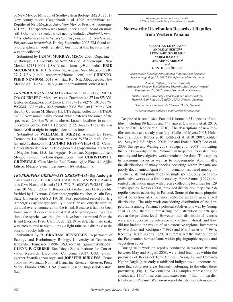
Herpetological Review 41(4), 2010520
Herpetological Review, 2010, 41(4), 520–523.© 2010 by Society for the Study of Amphibians and Reptiles
Noteworthy Distribution Records of Reptiles from Western Panamá
SEBASTIAN LOTZKAT*1, 2
ANDREAS HERTZ1, 2
LEONHARD STADLER1, 3
NADIM HAMAD1, 2
ARCADIO CARRIZO4
andGUNTHER KÖHLER1
1Senckenberg Forschungsinstitut und Naturmuseum Frankfurt Senckenberganlage 25, 60325 Frankfurt am Main, Germany
2Johann Wolfgang Goethe-UniversityInstitute for Ecology, Evolution and Diversity, BioCampus Westend
Siesmayerstr. 70, 60323 Frankfurt am Main, Germany3Justus-Liebig-University, Department of Animal Ecology, Heinrich-Buff-Ring 26-32 (IFZ), 35392 Giessen, Germany
4Universidad Autónoma de Chiriquí, Davíd, Panamá
*Corresponding author; e-mail: [email protected]
Despite of its small size, Panamá is home to 251 species of rep-tiles, including 89 lizards and 143 snakes (Jaramillo et al. 2010; Köhler 2010; Köhler et al. 2010). The descriptions of new rep-tiles continue at a steady pace (e.g., Cadle and Myers 2003; Hule-bak et al. 2007; Köhler 2010; Köhler et al. 2010, 2007; Köhler and Sunyer 2008; Myers 2003; Poe and Ibañez 2007; Poe et al. 2009; Savage and Watling 2008; Savage et al. 2008), indicating that our knowledge of the Panamanian herpetofauna is still frag-mentary and investigative work remains to be done. This applies to taxonomic issues as well as to biogeography. Additionally, the distributions of many species occurring within Panamá are poorly documented. Apart from information scattered among lo-cal checklists and publications on single species, only four com-prehensive works exist for the country. Pérez-Santos (1999) pre-sented distribution maps with precise collecting localities for 129 snake species. Köhler (2008) provided distribution maps for 238 reptile species occurring in Panamá. Some of the maps pinpoint actual collection sites, but most reflected areas of generalized distribution. The only work considering distribution of the her-petofauna among Panamá’s political subdivisions was by Young et al. (1999), therein summarizing the distribution of 228 spe-cies at the province level. However, their distributional records were not supported by reference to voucher material, and they did not include the results of two extensive regional inventories by Martínez and Rodriguez (1992) and Martínez et al. (1994). Recently, Jaramillo et al. (2010) summarized the distribution of the Panamanian herpetofauna within physiographic regions and vegetation zones.
During field work on reptiles conducted in western Panamá between May and August 2008, we visited localities within the provinces of Bocas del Toro, Chiriquí, Veraguas, and Comarca Ngöbe-Buglé (a recently established indigenous autonomous re-gion that comprises areas formerly belonging to the other three provinces) (Fig. 1). We collected 217 samples representing 72 species and 17 of those constitute extensions of their known dis-tributions in Panamá. We herein report distribution extensions of
of New Mexico Museum of Southwestern Biology (MSB 72651). New county record (Degenhardt et al. 1996. Amphibians and Reptiles of New Mexico. Univ. New Mexico Press, Albuquerque. 431 pp.). The specimen was found under a small board on moist soil. Other reptile species noted nearby included Diadophis punc-tatus, Opheodrys vernalis, Sceloporus poinsettii, S. cowlesi, and Phrynosoma hernandesi. During September 2005 EM found and photographed an adult female T. lineatum at this location, but it was not collected. Submitted by IAN W. MURRAY, MSC03 2020, Department of Biology, 1 University of New Mexico, Albuquerque, New Mexico 87131-0001, USA (e-mail: [email protected]); ERIK McCORMICK, 1011 S Yates St., Artesia, New Mexico 88210-2747, USA (e-mail: [email protected]); and CHRISTO-PHER NEWSOM, 5519 Amistad Rd. NE, Albuquerque, New Mexico 87111-1549, USA (e-mail: [email protected]).
TROPIDODIPSAS FASCIATA (Banded Snail Sucker). MÉXI-CO: GUERRERO: MuniciPalitY of ziHuataneJo: 27 km SW Val-lecitos de Zaragoza, on México Hwy 134 (17.782°N, 101.478°W; WGS84), 333 m elev. 02 September 2008. William H. Mertz. Ver-ified by Coleman M. Sheehy III. UTA digital collection (UTADC 3702). New municipality record, which extends the range of the species ca. 200 km W of its closest known localities in central Guerrero (Kofron 1987. J. Herpetol. 21:210–225). The snake was found AOR at night in tropical deciduous forest. Submitted by WILLIAM H. MERTZ, Avenida La Playa, Troncones, La Union, Guerrero, México 39270 (e-mail: [email protected]); JACOBO REYES-VELASCO, Centro Universitario de Ciencias Biológicas y Agropecuarias, Carretera a Nogales Km. 15.5. Las Agujas, Nextipac, Zapopan, Jalisco, México (e-mail: [email protected]); and CHRISTOPH I. GRÜNWALD, Casa Mexico Real Estate, Ajijic Plaza #1, Ajijic, Jalisco, México (e-mail: [email protected]).
TROPIDOPHIS GREENWAYI GREENWAYI (Big Ambergris Cay Dwarf Boa). TURKS AND CAICOS ISLANDS: Big aMBer-griS caY: N end of island (21.317°N, 71.630°W; WGS84), elev. 3 m. 20 March 2009. J. Burgess, G. Gerber, and G. Reynolds. Verified by J. Iverson. Color photographic voucher, Austin Peay State University (APSU 19018). First published record for Big Ambergris Cay, the type locality, since 1936 and only the third in-dividual ever encountered on the island. Because it had not been found since 1936, despite a great deal of herpetological investiga-tions, the species was thought to have been extirpated from the Island (Iverson 1986. Carib. J. Sci. 22:191–198). The male snake was encountered at night, during a light rain, on a dirt road at the base of a rocky hillside. Submitted by R. GRAHAM REYNOLDS, Department of Ecology and Evolutionary Biology, University of Tennessee, Knoxville, Tennessee 37996, USA (e-mail: [email protected]); GLENN P. GERBER, San Diego Zoo’s Institute for Conser-vation Research, Escondido, California 92027, USA (e-mail: [email protected]); and JOESEPH BURGESS, Guana Tolomato Matanzas National Estuarine Research Reserve, Ponte Vedra, Florida 32082, USA (e-mail: [email protected]).
Herpetological Review 41(4), 2010 521
(8.5095°N, 81.1173°W), 900 m elev. 2 August 2008. Leonhard Stadler and Nadim Hamad. SMF 89577. This record extends by 200 m, the upper confirmed elevational limit for the species previously reported by Savage (2002) and Köhler (2008). The presence of L. southi around Alto de Piedra was documented by Martínez and Rodriguez (1992). However, elevational distribu-tion for their individual records was unspecified; only a general elevational range of between 800 m and 1450 m was given.
Hoplocercidae
Enyalioides heterolepis (Bocourt’s Dwarf Iguana). VERAGUAS: Cerro Negro, ca. 6 km NNW of Santa Fé (8.5690°N, 81.0989°W), 700 m elev. 30 July 2008. Sebastian Lotzkat. SMF 89558. Cerro Negro, 6 km NNW of Santa Fé (8.5693°N, 81.1022°W), 713 m elev. 28 June 2008. Arcadio Carrizo. SMF 89955–56. First re-cords for Veraguas, extending the range more than 50 km W of the formerly westernmost record from El Copé, Coclé (Lips 1999). Köhler (2008) mentioned a record of E. heterolepis from Bocas del Toro, which as far as we can tell is not supported by a voucher specimen. A now lost individual from Cerro Narices (between Cerro Negro and Río Chilagres, see Fig. 1) that was listed as Sceloporus squamosus by Martínez et al. (1994), actu-ally represented an E. heterolepis (A. Rodriguez, pers. comm.).
Polychrotidae
Anolis auratus (Grass Anole). VERAGUAS: road between Santi-ago and Santa Fé, ca. 8 km N of Santa Fé; (8.1796°N, 80.951°W), 80 m elev. 14 May 2008. Andreas Hertz. SMF 89444. First record for Veraguas, bridging the gap between localities in Chiriquí to the west and Los Santos, Colón, and Panamá to the east (Köhler et al. 2008; Young et al. 1999).
Anolis capito (Bighead Anole). CHIRIQUÍ: Reserva Forestal La Fortuna (8.6744°N, 82.2161°W), 1460 m elev. 16 June 2008. An-dreas Hertz. SMF 89449. This record increases the known eleva-tional limit for the species by 160 m (Köhler 2008).
Anolis fortunensis (La Fortuna Anole). CHIRIQUÍ: Reserva For-estal La Fortuna (8.6776°N, 82.1981°W), 1750 m elev. 27 June 2008. Andreas Hertz and Sebastian Lotzkat. SMF 89466. This record extends the known elevational range of the species by 550 m (Köhler 2008).
Anolis humilis (Humble Anole). CHIRIQUÍ: Reserva Forestal La Fortuna (8.6761°N, 82.2006°W), 1660 m elev. 27 June 2008. Sebastian Lotzkat. SMF 89480. This record increases the known elevational range by 60 m (Köhler 2008).
Anolis pseudopachypus (La Nevera Anole). CHIRIQUÍ: Reser-va Forestal La Fortuna (8.6761°N, 82.2006°W), 1660 m elev.; (8.6793°N, 82.193°W), 1800 m elev. 25 and 27 June 2008. Andre-as Hertz and Sebastian Lotzkat. SMF 89523, 89526, respectively. First records for Chiriquí, and extends the range ca. 50 km WNW of the type locality (La Nevera, Comarca Ngöbe-Buglé; Köhler et al. 2007). COMARCA NGÖBE-BUGLÈ: Valley north of La Nevera, ca. 5.5 km N of Hato Chamí (8.5018°N, 81.7689°W), 1560 m elev. 9 August 2008. Andreas Hertz and Sebastian Lotz-kat. SMF 89519–89521. Ridgetop NE of La Nevera, ca. 5.5 km N of Hato Chami (8.4954°N, 81.7673°W), 1810 m elev. 18 August 2008. Andreas Hertz and Sebastian Lotzkat. SMF 89522. These records, near the type locality of Anolis pseudopachypus, repre-
fig. 1. Generalized localities in western Panamá visited by the authors in 2008: 1) Jurutungo, Chiriquí; 2) Reserva Forestal La Fortuna, Chiriquí; 3) road to Cerro Colorado, Comarca Ngöbe-Buglé; 4) La Nevera, Comarca Ngöbe-Buglé; 5) Cerro Mariposa (= Cerro Tute), Veraguas; 6) Cerro Negro, Veraguas; 7) Río Chilagres, Veraguas.
those 17 species, 11 of which are new provincial records; other accounts list new elevational parameters for the species.
Abbreviations for museum collections follow those of Leviton et al. (1985). Elevations were rounded to the next tenth. The map (Fig. 1) was created using DIVA-GIS and the NASA elevation datasets processed by Jarvis et al. (2006); map datum is WGS84. All vouchers were verified by Javier Sunyer. Common names came from Frank and Ramus (1995), except for Leposoma southi (Uetz 2008), Urotheca guentheri (Solórzano 2004), and by us herein for Anolis fortunensis and A. pseudopachypus, two species for which there are no published common names. We decided that for those two species, the common names should denote their type localities; Reserva Forestal La Fortuna, Panamá, and La Ne-vera, Panamá, respectively.
Squamata — Lizards
Sphaerodactylidae
Lepidoblepharis xanthostigma (Costa Rica Scaly-eyed Gecko). VERAGUAS: Cerro Mariposa, near Alto de Piedra, ca. 3.5 km W of Santa Fé (8.5161°N, 81.1185°W), 880 m elev. 12 May 2008. Andreas Hertz and Sebastian Lotzkat. SMF 89576. Cerro Negro, ca. 6 km NNW of Santa Fé (8.5690°N, 81.0989°W), 700 m elev. 28 July 2008. Andreas Hertz and Sebastian Lotzkat. SMF 89963. Martínez and Rodriguez (1992), in their inventory of “Cerro Tute” (= Cerro Mariposa) and Martínez et al. (1994) in their species list of the nearby mountains, Narices and La Anselma, both reported the presence of Lepidoblepharis sp., and consid-ered them to resemble L. xanthostigma. However, because the specific identities of these geckos were not made and the respec-tive voucher specimens were lost (V. Martínez pers. comm.), our records represent the first confirmed occurrence for the species in Veraguas. The records also fill the gap between records to the west in Chiriquí and Bocas del Toro and the east in Coclé and Colón (Young et al. 1999).
Gymnophthalmidae
Leposoma southi (Northern Spectacled Lizard). VERAGUAS: Cerro Mariposa, near Alto de Piedra, ca. 3.5 km W of Santa Fé
Herpetological Review 41(4), 2010522
sent the lowest and highest elevational records, respectively, for the species, which was previously known only from 1600 m el-evation (Köhler et al. 2007).
Anolis salvini (Salvin’s Anole). CHIRIQUÍ: Jurutungo, near bor-der with Costa Rica (8.912°N, 82.7182°W), 1980 m elev. 8 July 2008. Andreas Hertz and Sebastian Lotzkat. SMF 89527. This record extends the known elevational distribution of the species upward ca. 150 m (Köhler 2008; Savage 2002).
Anolis vittigerus (Garland Anole). VERAGUAS: Río Chilagres, ca. 9 km NE of Santa Fé (8.5872°N, 81.0363°W), 480 m elev. 3 June 2008. Andreas Hertz and Sebastian Lotzkat. SMF 89528. First record for Veraguas, extending the range ca. 50 km W of the formerly westernmost record from north of El Copé, Coclé (Poe and Ibañez 2007).
Squamata — Snakes
Colubridae
Dipsas articulata (American Snail-eater). VERAGUAS: Cerro Negro, ca. 6 km NNW of Santa Fé (8.5756°N, 81.0976°W), 1000 m elev. 29 July 2008. Arcadio Carrizo. SMF 89552. Since locali-ty data for the holotype of D. articulata is imprecise (see remarks in Peters 1960), this is the first validated record for Veraguas. The record reconfirms the species’ occurrence at 1000 m, reported as its upper elevational limit by Solórzano (2004).
Dipsas temporalis (Temporal Snail-eater). VERAGUAS: Cer-ro Mariposa, near Alto de Piedra, ca. 3.5 km W of Santa Fé (8.5107°N, 81.1207°W), 990 m elev. 10 July 2008. Leonhard Stadler and Nadim Hamad. SMF 89552. First record for Vera-guas, and a range extension of ca.130 km W of the South slope of Cerro La Campana, the formerly westernmost record in Pan-amá (Pérez-Santos 1999). Another specimen (SMF 89553) found nearby (8.5245°N, 81.1332°W), 700 m elev., on 5 August 2008 was acquired at the lowest elevation reported for this species (Pérez-Santos 1999).
Imantodes cenchoa (Blunt-headed Tree Snake). COMARCA NGÖBE-BUGLÈ: road to Cerro Colorado west of La Nevera, ca. 10 km NNW of Hato Chamí (8.5303°N, 81.7987°W), 1660 m elev. 10 May 2008. Andreas Hertz. SMF 89573. The locality is situated at an elevation 160 m above that reported for the species by Solórzano (2004).
Oxyrhopus petola (Calico False Coral Snake). VERAGUAS: Río Chilagres, ca. 9 km NE of Santa Fé (8.5872°N, 81.0363°W), 480 m elev. 3 June 2008. Andreas Hertz and Sebastian Lotzkat. SMF 89585. First record for Veraguas, which bridges a distributional gap between localities in the provinces of Chiriquí and Bocas del Toro to the west and Colón and Panamá to the east (Young et al. 1999).
Urotheca guentheri (Striped Glasstail). VERAGUAS: Cer-ro Mariposa, near Alto de Piedra, ca. 3.5 km W of Santa Fé (8.5002°N, 81.1175°W), 1250 m elev. 5 June 2008. Leonhard Stadler and Nadim Hamad. SMF 89603. First record for Vera-guas, filling a gap between localities in the provinces of Bocas del Toro (La Loma, ca.120 km NW) and Coclé (about 110 km E) (Myers 1974).
Viperidae
Bothriechis nigroviridis (Black-speckled Palm Viper). COMAR-CA NGÖBE-BUGLÈ: La Nevera, ca. 5.5 km N of Hato Chamí (8.4996°N, 81.77°W), 1650 m elev. 11 May 2008. Andreas Hertz, Leonhard Stadler, Nadim Hamad, and Sebastian Lotzkat. SMF 89534. First record for Comarca Ngöbe-Buglé, and an 80 km E range extension from the formerly easternmost record in Bo-quete, Chiriquí (Dunn 1947).
Bothriechis schlegelii (Eyelash Palm Viper). VERAGUAS: Cer-ro Mariposa, near Alto de Piedra, approx. 3.5 km W of Santa Fé (8.5157°N, 81.1197°W), 860 m elev. 13 May 2008. Gunther Köhler. SMF 89535. First record for Veraguas, which fills the gap between Isla Escudo de Veraguas, Comarca Ngöbe-Buglé (ca. 85 km NW), and El Valle de Anton, Coclé (ca. 105 km E) (Pérez-Santos 1999).
Acknowledgments.—Collecting and exportation permits SE/A-30-08 and SEX/A-108-08 were provided by A. Salazar, Y. Hidalgo, and J. Gar-cía, Autoridad Nacional del Ambiente (ANAM), Panamá City, Panamá. Q. D. Fuenmayora and V. Martínez provided valuable assistance with ac-quisition of permits. An additional permit for the Comarca Ngöbe-Buglé was issued by A. Montezuma (ANAM), San Félix, Panamá. For field assistance and logistical support, we thank C. O’Shea and S. Abrego, A. Bennett, P. Yangüez, O. Cáceres and family, M. Piepenbring, and the Peña Solís family. A. Carrizo received financial support from the DAAD. Field work funding to A. Hertz came from FAZIT-Stiftung and from Studienstiftung des deutschen Volkes to S. Lotzkat.
literature cited
cadle, J. e., and c. W. MYerS. 2003. Systematics of snakes referred to Dipsas variegata Panama and western South America, with revalida-tion of two species and notes on defensive behaviors in the Dipsadini (Colubridae). Am. Mus. Novit. [3409]:1–47.
dunn, e. r. 1947. Snakes of the Lérida Farm (Chiriqui Volcano, western Panamá). Copeia 1947:153–157.
frank, n., and e. raMuS. 1995. A Complete Guide to Scientific and Common Names of Reptiles and Amphibians of the World. N. G. Publ., Inc., Pottsville, Pennsylvania.
HuleBak, e., S. Poe, r. iBáñez, and e. e. WilliaMS. 2007. A striking new species of Anolis lizard (Squamata, Iguania) from Panama. Phyl-lomedusa 6:5–10.
JarviS, a., H. i. reuter, a. nelSon, and e. guevara. 2006. Hole-filled seamless SRTM data V3, International Centre for Tropical Agriculture (CIAT), available from http://srtm.csi.cgiar.org. Downloaded Novem-ber 2007.
köHler, g. 2008. Reptiles of Central America, 2nd ed. Herpeton, Verlag Elke Köhler, Offenbach, Germany.
–––––. 2010. A revision of the Central American species related to Ano-lis pentaprion, with the resurrection of A. beckeri and the description of a new species (Squamata Polychrotidae). Zootaxa 2354:1–18.
–––––, S. lotzkat, and a. Hertz. 2010. A new species of Sibon (Squa-mata: Colubridae) from Western Panama. Herpetologica 66:80–85.
–––––, M. Ponce, J. SunYer, and a. BatiSta. 2007. Four new species of anoles (genus Anolis) from the Serranía de Tabasará, west-central Panama (Squamata: Polychrotidae). Herpetologica 63:375–391.
–––––, and J. SunYer. 2008. Two new species of anoles formerly refer-red to as Anolis limifrons (Squamata: Polychrotidae). Herpetologica 64:92–108.
–––––, –––––, M. Ponce, and a. BatiSta. 2008. Noteworthy records of amphibians and reptiles in Panama (Amphibia: Plethodontidae, Crau-
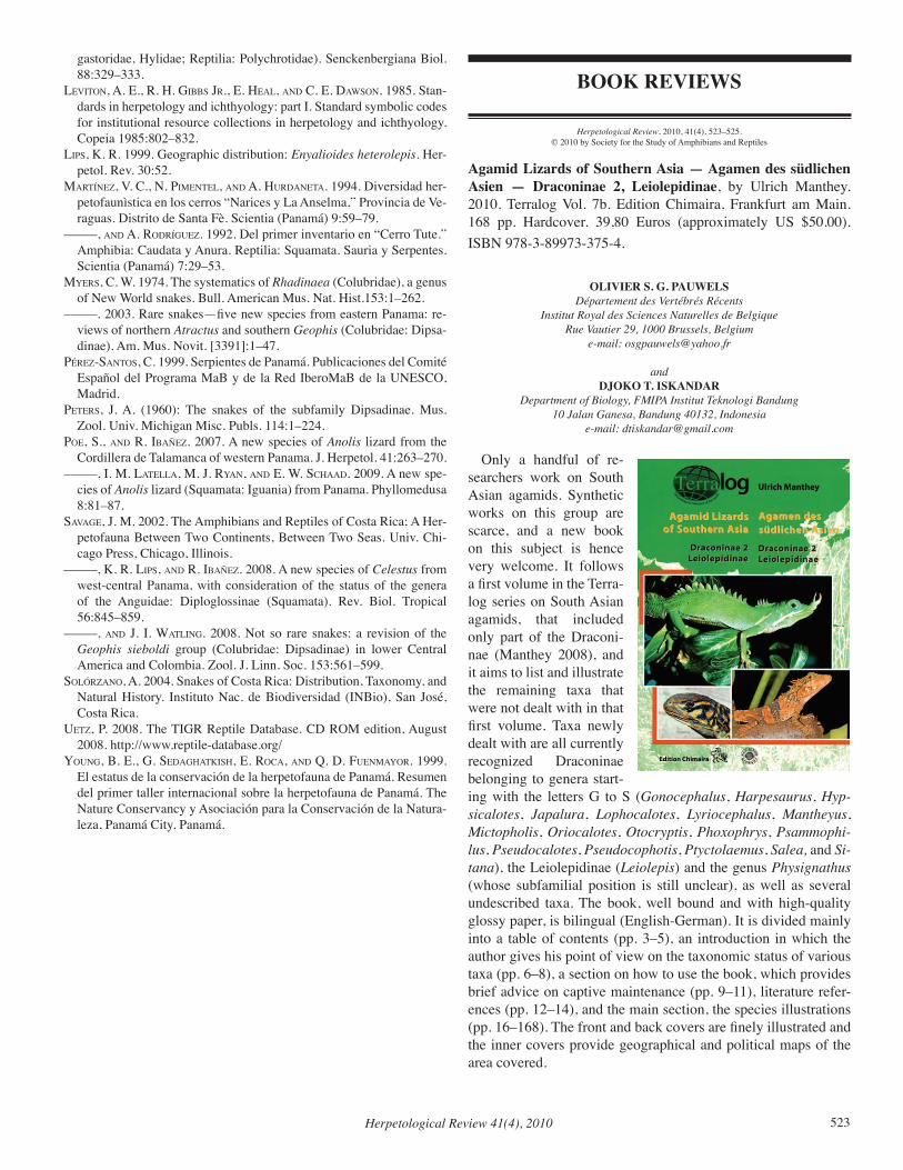
Herpetological Review 41(4), 2010 523
gastoridae, Hylidae; Reptilia: Polychrotidae). Senckenbergiana Biol. 88:329–333.
leviton, a. e., r. H. giBBS Jr., e. Heal, and c. e. daWSon. 1985. Stan-dards in herpetology and ichthyology: part I. Standard symbolic codes for institutional resource collections in herpetology and ichthyology. Copeia 1985:802–832.
liPS, k. r. 1999. Geographic distribution: Enyalioides heterolepis. Her-petol. Rev. 30:52.
Martínez, v. c., n. PiMentel, and a. Hurdaneta. 1994. Diversidad her-petofaunìstica en los cerros “Narices y La Anselma,” Provincia de Ve-raguas. Distrito de Santa Fè. Scientia (Panamá) 9:59–79.
–––––, and a. rodríguez. 1992. Del primer inventario en “Cerro Tute.” Amphibia: Caudata y Anura. Reptilia: Squamata. Sauria y Serpentes. Scientia (Panamá) 7:29–53.
MYerS, c. W. 1974. The systematics of Rhadinaea (Colubridae), a genus of New World snakes. Bull. American Mus. Nat. Hist.153:1–262.
–––––. 2003. Rare snakes—five new species from eastern Panama: re-views of northern Atractus and southern Geophis (Colubridae: Dipsa-dinae). Am. Mus. Novit. [3391]:1–47.
Pérez-SantoS, c. 1999. Serpientes de Panamá. Publicaciones del Comité Español del Programa MaB y de la Red IberoMaB de la UNESCO, Madrid.
PeterS, J. a. (1960): The snakes of the subfamily Dipsadinae. Mus. Zool. Univ. Michigan Misc. Publs. 114:1–224.
Poe, S., and r. iBañez. 2007. A new species of Anolis lizard from the Cordillera de Talamanca of western Panama. J. Herpetol. 41:263–270.
–––––, i. M. latella, M. J. rYan, and e. W. ScHaad. 2009. A new spe-cies of Anolis lizard (Squamata: Iguania) from Panama. Phyllomedusa 8:81–87.
Savage, J. M. 2002. The Amphibians and Reptiles of Costa Rica; A Her-petofauna Between Two Continents, Between Two Seas. Univ. Chi-cago Press, Chicago, Illinois.
–––––, k. r. liPS, and r. iBañez. 2008. A new species of Celestus from west-central Panama, with consideration of the status of the genera of the Anguidae: Diploglossinae (Squamata). Rev. Biol. Tropical 56:845–859.
–––––, and J. i. Watling. 2008. Not so rare snakes: a revision of the Geophis sieboldi group (Colubridae: Dipsadinae) in lower Central America and Colombia. Zool. J. Linn. Soc. 153:561–599.
Solórzano, a. 2004. Snakes of Costa Rica: Distribution, Taxonomy, and Natural History. Instituto Nac. de Biodiversidad (INBio), San José, Costa Rica.
uetz, P. 2008. The TIGR Reptile Database. CD ROM edition, August 2008. http://www.reptile-database.org/
Young, B. e., g. SedagHatkiSH, e. roca, and Q. d. fuenMaYor. 1999. El estatus de la conservación de la herpetofauna de Panamá. Resumen del primer taller internacional sobre la herpetofauna de Panamá. The Nature Conservancy y Asociación para la Conservación de la Natura-leza, Panamá City, Panamá.
BOOK REVIEWS
Herpetological Review, 2010, 41(4), 523–525.© 2010 by Society for the Study of Amphibians and Reptiles
Agamid Lizards of Southern Asia — Agamen des südlichen Asien — Draconinae 2, Leiolepidinae, by Ulrich Manthey. 2010. Terralog Vol. 7b. Edition Chimaira, Frankfurt am Main. 168 pp. Hardcover. 39,80 Euros (approximately US $50.00). ISBN 978-3-89973-375-4.
OLIVIER S. G. PAUWELSDépartement des Vertébrés Récents
Institut Royal des Sciences Naturelles de BelgiqueRue Vautier 29, 1000 Brussels, Belgium
e-mail: [email protected]
andDJOKO T. ISKANDAR
Department of Biology, FMIPA Institut Teknologi Bandung10 Jalan Ganesa, Bandung 40132, Indonesia
e-mail: [email protected]
Only a handful of re-searchers work on South Asian agamids. Synthetic works on this group are scarce, and a new book on this subject is hence very welcome. It follows a first volume in the Terra-log series on South Asian agamids, that included only part of the Draconi-nae (Manthey 2008), and it aims to list and illustrate the remaining taxa that were not dealt with in that first volume. Taxa newly dealt with are all currently recognized Draconinae belonging to genera start-ing with the letters G to S (Gonocephalus, Harpesaurus, Hyp-sicalotes, Japalura, Lophocalotes, Lyriocephalus, Mantheyus, Mictopholis, Oriocalotes, Otocryptis, Phoxophrys, Psammophi-lus, Pseudocalotes, Pseudocophotis, Ptyctolaemus, Salea, and Si-tana), the Leiolepidinae (Leiolepis) and the genus Physignathus (whose subfamilial position is still unclear), as well as several undescribed taxa. The book, well bound and with high-quality glossy paper, is bilingual (English-German). It is divided mainly into a table of contents (pp. 3–5), an introduction in which the author gives his point of view on the taxonomic status of various taxa (pp. 6–8), a section on how to use the book, which provides brief advice on captive maintenance (pp. 9–11), literature refer-ences (pp. 12–14), and the main section, the species illustrations (pp. 16–168). The front and back covers are finely illustrated and the inner covers provide geographical and political maps of the area covered.
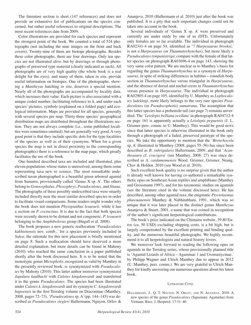
Herpetological Review 41(4), 2010524
Ananjeva, 2010 (Hallermann et al. 2010) just after the book was published. It is a pity that such important changes could not be taken into account in the book.
Several individuals of ‘Genus X sp. A’ were preserved and currently are under study by one of us (DTI). Unfortunately only females are so far available. The individual in photograph RA02341-4 on page 54, identified as ‘? Harpesaurus brooksi,’ is not a Harpesaurus (or Thaumatorhynchus), but more likely a Pseudocalotes tympanistriga; compare with the female of that lat-ter species on photograph RA03696-4 on page 143, showing the very same color pattern. We are unclear as to Manthey’s basis for regarding the genus Thaumatorhynchus as a synonym of Harpe-saurus, in spite of striking differences in habitus—roundish body section in Thaumatorhynchus versus triangular in Harpesaurus, and the absence of dorsal and nuchal crests in Thaumatorhynchus versus presence in Harpesaurus. The individual in photograph RA02905-4 on page 105, identified by U. Manthey as Lophocalo-tes ludekingi, more likely belongs to the very rare species Pseu-docalotes (or Pseudocophotis) sumatrana. The assumption that this latter species has a prehensile tail (see page 7) remains unver-ified. The ‘Leiolepis belliana ocellata’ in photograph RA04712-4 on page 161 is apparently actually a Leiolepis peguensis (J. L. Grismer pers. comm. to U. Manthey). This would be good news since that latter species is otherwise illustrated in the book only through a photograph of a faded, preserved paratype of the spe-cies. We take the opportunity to mention that the ‘Bronchocela sp. A’ illustrated in Manthey (2008, pages 55–56) has since been described as B. rubrigularis Hallermann, 2009, and that ‘Acan-thosaura cf. crucigera’ (see Manthey, 2008: 27) was since de-scribed as A. cardamomensis Wood, Grismer, Grismer, Neang, Chav & Holden, 2010 (see Wood et al. 2010).
Such excellent book quality is no surprise given that the author is already well known for having co-authored a remarkable syn-thetic opus on Southeast Asian reptiles and amphibians (Manthey and Grossmann 1997), and for his taxonomic studies on agamids (see the literature cited in the volume discussed here). He has described, among other agamid taxa, the enigmatic Ptyctolaemus phuwuanensis Manthey & Nabhitabhata, 1991, which was so unique that it was later placed in the distinct genus Mantheyus Ananjeva & Stuart, 2001, a name that was coined in recognition of the author’s significant herpetological contributions.
The book’s price indicated on the Chimaira website, 39.80 Eu-ros (ca. 50 USD) excluding shipping costs, is a bit high, but is largely compensated by the excellent printing and binding qual-ity, and the numerous beautiful photographs. We highly recom-mend it to all herpetologists and natural history lovers.
We moreover look forward to reading the following opus on agamids in the Terralog series, whose provisionally planned title is ‘Agamid Lizards of Africa – Agaminae 1 and Uromastycinae,’ by Philipp Wagner and Ulrich Manthey due to appear in 2012 (U. Manthey, pers. comm.). We are very grateful to Ulrich Man-they for kindly answering our numerous questions about his latest book.
literature cited
HallerMann, J., Q. t. nguYen, n. orlov, and n. ananJeva. 2010. A new species of the genus Pseudocalotes (Squamata: Agamidae) from Vietnam. Russ. J. Herpetol. 17:31–40.
The literature section is short (147 references) and does not provide an exhaustive list of publications on the species con-cerned, but rather useful references to original descriptions. The most recent references date from 2009.
Color illustrations are provided for each species and represent the strongest point of the book. We counted a total of 524 pho-tographs (not including the nine images on the front and back covers). Twenty-nine of them are biotope photographs. Besides these color photographs, there are four drawings. Only ten spe-cies are not illustrated alive, but by drawings or through photo-graphs of preserved type material (clearly indicated as such). All photographs are of very high quality (the whole book is a real delight for the eyes), and many of them, taken in situ, provide useful information on biotopes. One of the photographs, show-ing a Mantheyus hatching in situ, deserves a special mention. Nearly all of the photographs are accompanied by locality data, which increases their value. Each picture is also associated with a unique coded number, facilitating reference to it, and under each species’ pictures, symbols (explained on a folded page) add eco-logical information. Maps are provided for all species, generally with several species per map. Thirty-three species’ geographical distribution maps are distributed throughout the illustrations sec-tion. They are not always complete (i.e., some published locali-ties were sometimes omitted), but are generally very good. A very good point is that they include specific dots for the type localities of the species as well as of their synonyms. When for a given species the map is not in direct proximity to the corresponding photograph(s) there is a reference to the map page, which greatly facilitates the use of the book.
One hundred described taxa are included and illustrated, plus eleven populations whose status is unresolved, among them some representing taxa new to science. The most remarkable unde-scribed taxon photographed is a beautiful green arboreal agamid from Sumatra, provisionally called ‘Genus X sp. A’. The others belong to Gonocephalus, Phoxophrys, Pseudocalotes, and Sitana. The photographs of these possibly undescribed taxa were smartly included directly near the most similar described species in order to facilitate visual comparisons. Some readers might wonder why the book does not mention Physignathus lesueurii, while it has a section on P. cocincinus. It is due to the fact that both species were recently shown to be distant and not congeneric, P. lesueurii belonging to the Amphibolorus group (Hugall et al. 2008).
The book proposes a new generic reallocation ‘Pseudocalotes kakhienensis nov. comb.,’ for a species previously included in Salea; the rationale for this new placement is briefly mentioned on page 8. Such a reallocation should have deserved a more detailed explanation, but more details can be found in Mahony (2010) who reached the same conclusion in a paper published shortly after the book discussed here. It is to be noted that the monotypic genus Mictopholis, recognized as valid by Manthey in the presently reviewed book, is synonymized with Pseudocalo-tes by Mahony (2010). This latter author moreover synonymized Japalura kaulbacki with Calotes kingdonwardi and transferred it to the genus Pseudocalotes. The species had been illustrated under Calotes k. kingdonwardi and its synonym C. kingdonwardi bapoensis in the first Terralog volume on Draconinae (Manthey, 2008, pages 72–73). ‘Pseudocalotes sp. A’ (pp. 144–145) was de-scribed as Pseudocalotes ziegleri Hallermann, Nguyen, Orlov &
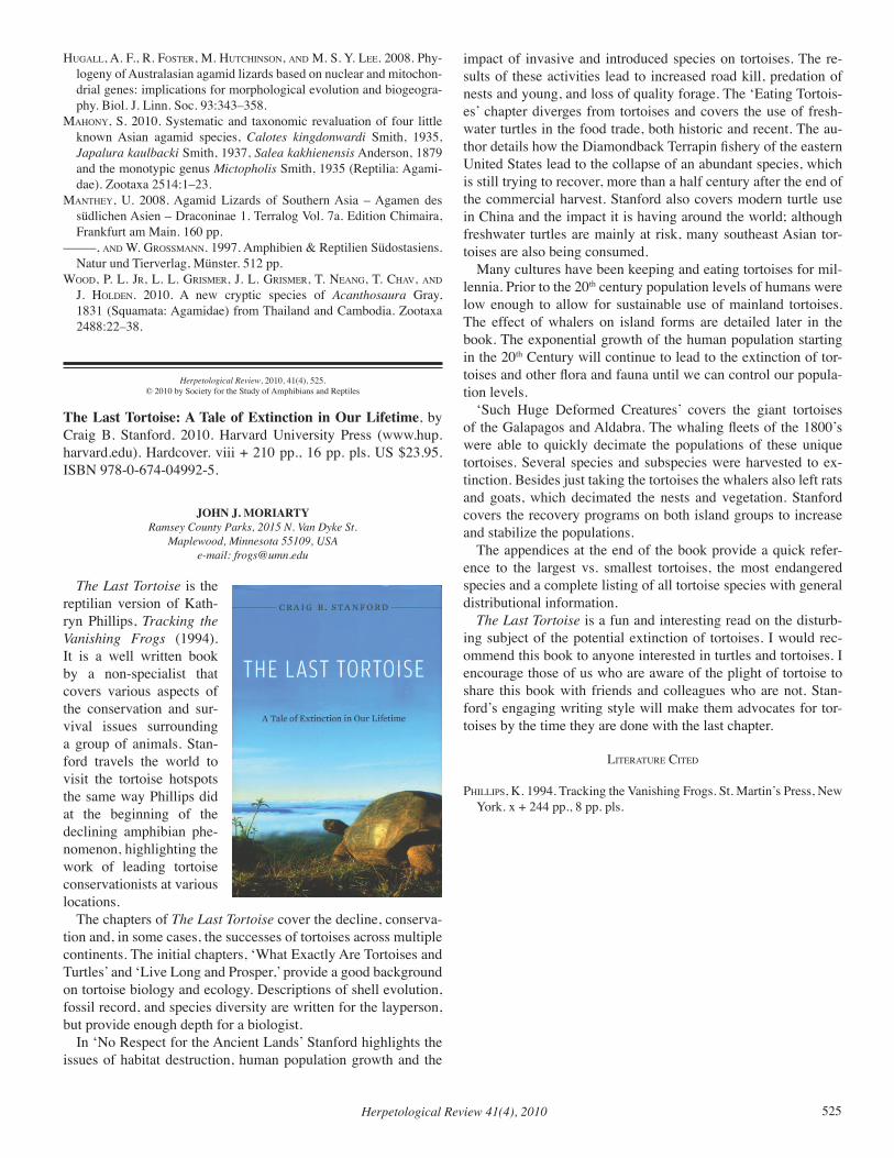
Herpetological Review 41(4), 2010 525
Hugall, a. f., r. foSter, M. HutcHinSon, and M. S. Y. lee. 2008. Phy-logeny of Australasian agamid lizards based on nuclear and mitochon-drial genes: implications for morphological evolution and biogeogra-phy. Biol. J. Linn. Soc. 93:343–358.
MaHonY, S. 2010. Systematic and taxonomic revaluation of four little known Asian agamid species, Calotes kingdonwardi Smith, 1935, Japalura kaulbacki Smith, 1937, Salea kakhienensis Anderson, 1879 and the monotypic genus Mictopholis Smith, 1935 (Reptilia: Agami-dae). Zootaxa 2514:1–23.
MantHeY, U. 2008. Agamid Lizards of Southern Asia – Agamen des südlichen Asien – Draconinae 1. Terralog Vol. 7a. Edition Chimaira, Frankfurt am Main. 160 pp.
–––––, and W. groSSMann. 1997. Amphibien & Reptilien Südostasiens. Natur und Tierverlag, Münster. 512 pp.
Wood, P. l. Jr, l. l. griSMer, J. l. griSMer, t. neang, t. cHav, and J. Holden. 2010. A new cryptic species of Acanthosaura Gray, 1831 (Squamata: Agamidae) from Thailand and Cambodia. Zootaxa 2488:22–38.
Herpetological Review, 2010, 41(4), 525.© 2010 by Society for the Study of Amphibians and Reptiles
The Last Tortoise: A Tale of Extinction in Our Lifetime, by Craig B. Stanford. 2010. Harvard University Press (www.hup.harvard.edu). Hardcover. viii + 210 pp., 16 pp. pls. US $23.95. ISBN 978-0-674-04992-5.
JOHN J. MORIARTYRamsey County Parks, 2015 N. Van Dyke St.
Maplewood, Minnesota 55109, USAe-mail: [email protected]
The Last Tortoise is the reptilian version of Kath-ryn Phillips, Tracking the Vanishing Frogs (1994). It is a well written book by a non-specialist that covers various aspects of the conservation and sur-vival issues surrounding a group of animals. Stan-ford travels the world to visit the tortoise hotspots the same way Phillips did at the beginning of the declining amphibian phe-nomenon, highlighting the work of leading tortoise conservationists at various locations.
The chapters of The Last Tortoise cover the decline, conserva-tion and, in some cases, the successes of tortoises across multiple continents. The initial chapters, ‘What Exactly Are Tortoises and Turtles’ and ‘Live Long and Prosper,’ provide a good background on tortoise biology and ecology. Descriptions of shell evolution, fossil record, and species diversity are written for the layperson, but provide enough depth for a biologist.
In ‘No Respect for the Ancient Lands’ Stanford highlights the issues of habitat destruction, human population growth and the
impact of invasive and introduced species on tortoises. The re-sults of these activities lead to increased road kill, predation of nests and young, and loss of quality forage. The ‘Eating Tortois-es’ chapter diverges from tortoises and covers the use of fresh-water turtles in the food trade, both historic and recent. The au-thor details how the Diamondback Terrapin fishery of the eastern United States lead to the collapse of an abundant species, which is still trying to recover, more than a half century after the end of the commercial harvest. Stanford also covers modern turtle use in China and the impact it is having around the world; although freshwater turtles are mainly at risk, many southeast Asian tor-toises are also being consumed.
Many cultures have been keeping and eating tortoises for mil-lennia. Prior to the 20th century population levels of humans were low enough to allow for sustainable use of mainland tortoises. The effect of whalers on island forms are detailed later in the book. The exponential growth of the human population starting in the 20th Century will continue to lead to the extinction of tor-toises and other flora and fauna until we can control our popula-tion levels.
‘Such Huge Deformed Creatures’ covers the giant tortoises of the Galapagos and Aldabra. The whaling fleets of the 1800’s were able to quickly decimate the populations of these unique tortoises. Several species and subspecies were harvested to ex-tinction. Besides just taking the tortoises the whalers also left rats and goats, which decimated the nests and vegetation. Stanford covers the recovery programs on both island groups to increase and stabilize the populations.
The appendices at the end of the book provide a quick refer-ence to the largest vs. smallest tortoises, the most endangered species and a complete listing of all tortoise species with general distributional information.
The Last Tortoise is a fun and interesting read on the disturb-ing subject of the potential extinction of tortoises. I would rec-ommend this book to anyone interested in turtles and tortoises. I encourage those of us who are aware of the plight of tortoise to share this book with friends and colleagues who are not. Stan-ford’s engaging writing style will make them advocates for tor-toises by the time they are done with the last chapter.
literature cited
PHilliPS, K. 1994. Tracking the Vanishing Frogs. St. Martin’s Press, New York. x + 244 pp., 8 pp. pls.
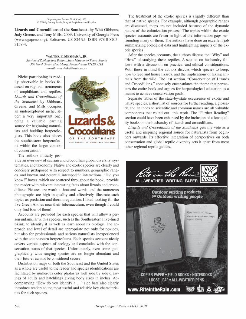
Herpetological Review 41(4), 2010526
Herpetological Review, 2010, 41(4), 526.© 2010 by Society for the Study of Amphibians and Reptiles
Lizards and Crocodilians of the Southeast, by Whit Gibbons, Judy Greene, and Tony Mills. 2009. University of Georgia Press (www.ugapress.org). Softcover. US $24.95. ISBN 978-0-8203-3158-4.
WALTER E. MESHAKA, JR.Section of Zoology and Botany, State Museum of Pennsylvania
300 North Street, Harrisburg, Pennsylvania 17120, USAe-mail: [email protected]
Niche partitioning is read-ily observable in books fo-cused on regional treatments of amphibians and reptiles. Lizards and Crocodilians of the Southeast by Gibbons, Greene, and Mills occupies an underexploited niche, al-beit a very important one, being a valuable learning source for beginning natural-ists and budding herpetolo-gists. This book also places the southeastern herpetofau-na within the larger context of conservation.
The authors initially pro-vide an overview of saurian and crocodilian global diversity, sys-tematics, and taxonomy. Native and exotic species are clearly and concisely juxtaposed with respect to numbers, geographic rang-es, and known and potential interspecific interactions. “Did you know?” boxes, which are scattered throughout the book, provide the reader with relevant interesting facts about lizards and croco-dilians. Pictures are worth a thousand words, and the numerous photographs are high in quality and effectively illustrate such topics as predation and thermoregulation. I liked looking for the five Green Anoles near their hibernaculum, even though I could only find four of them!
Accounts are provided for each species that will allow a per-son unfamiliar with a species, such as the Southeastern Five-lined Skink, to identify it as well as learn about its biology. The ap-proach and level of detail are appropriate not only for novices, but also for professionals and serious naturalists inexperienced with the southeastern herpetofauna. Each species account nicely covers various aspects of ecology and concludes with the con-servation status of that species. Unfortunately, even some geo-graphically wide-ranging species are no longer abundant and their futures cannot be considered secure.
Distribution maps of both the Southeast and the United States as a whole are useful to the reader and species identifications are facilitated by numerous color photos as well side by side draw-ings of adults and hatchlings giving body sizes in inches. Ac-companying “How do you identify a …” side bars also clearly introduce readers to the most useful and reliable key characteris-tics for each species.
The treatment of the exotic species is slightly different than that of native species. For example, although geographic ranges are discussed, maps are not included because of the dynamic nature of the colonization process. The topics within the exotic species accounts are fewer in light of the information gaps sur-rounding many of them. The authors have done an excellent job summarizing ecological data and highlighting impacts of the ex-otic species.
After the species accounts, the authors discuss the “Why” and “How” of studying these reptiles. A section on husbandry fol-lows with a discussion on practical and ethical considerations. With these in mind the authors discuss which species to keep, how to feed and house lizards, and the implications of taking ani-mals from the wild. The last section, “Conservation of Lizards and Crocodilians,” concisely encapsulates the theme that perme-ates the entire book and argues for herpetological education as a means to achieve conservation goals.
Separate tables of the state-by-state occurrence of exotic and native species, a short list of sources for further reading, a glossa-ry, and an index to scientific and common names are all valuable components that round out this work. The “Further Reading” section could have been enhanced by the inclusion of a few qual-ity books on the husbandry of lizards and crocodilians.
Lizards and Crocodilians of the Southeast gets my vote as a useful and inspiring regional source for naturalists from begin-ners onwards. Its effective integration of perspectives on both conservation and global reptile diversity sets it apart from most other regional reptile guides.
Herpetological Review 41(4), 2010 527
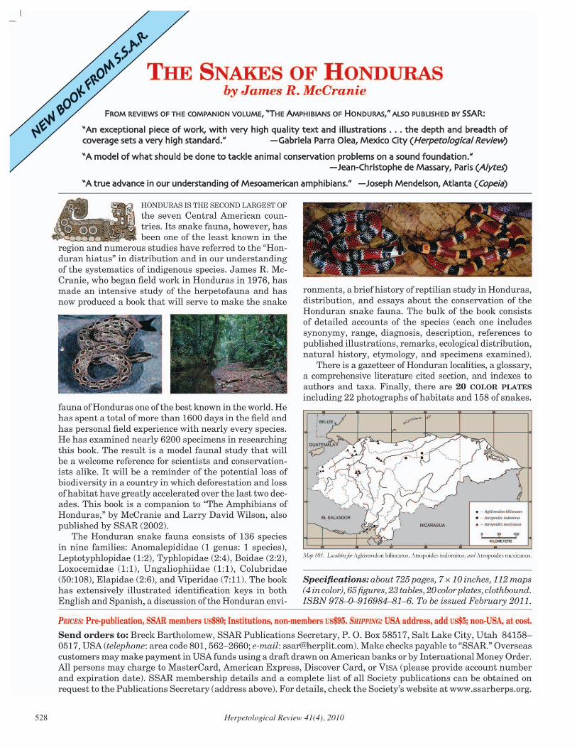
Herpetological Review 41(4), 2010528
SSAR Committee ChAiRSAnd CooRdinAtoRS
CHAIRPERSONS Standard English and Scientific Names BRIAN I. CROTHER Department of Biological Sciences Southeastern Louisiana University Hammond, Louisiana 70402, USA
Conservation BETSIE ROTHERMEL Archbold Biological Station PO Box 2057 Lake Placid, Florida 33862, USA
Grants-In-Herpetology ERIk R. WILD Department of Biology University of Wisconsin-Stevens Point Stevens Point, Wisconsin 54481-3897, USA
JOSHUA M. kAPFER Natural Resources Consulting, Inc. 119 South Main Street, PO Box 128 Cottage Gove, Wisconsin 53527, USA
Kennedy Student Award LYNNETTE SIEVERT Department of Biological Sciences Emporia State University Emporia, kansas 66801, USA
Metter Memorial Award JOSEPH J. BEATTY Department of Zoology Oregon State University Corvallis, Oregon 97331-2914, USA
Nominating GREGORY WATkINS-COLWELL Yale Peabody Museum of Natural History New Haven, Connecticut 06520-8118, USA
Resolutions STUART NIELSEN Department of Biology University of Mississippi University, Mississippi 38655, USA
Seibert Awards PATRICk OWEN Department of EEO Biology The Ohio State University at Lima Lima, Ohio 45804, USA
Student Travel Awards MATTHEW VENESkY Department of Biology The University of Memphis Memphis, Tennessee 38152, USA
CARI HICkERSON Biological, Geological & Environmental Science Cleveland State University Cleveland, Ohio 44115, USA
Webmaster RAUL E. DIAZ University of kansas Medical Center Lawrence, kansas 66160, USA e-mail: [email protected]
COORDINATORSElector DANIEL NOBLE Department of Brain, Behaviour and Evolution Macquarie University Sydney, NSW 2109, Australia
Symposium Coordinator RICHARD D. DURTSCHE Department of Biological Sciences Northern kentucky University Highland Heights, kentucky 41099, USA
infoRmAtion foR ContRibutoRS
Herpetological Review is a peer-reviewed quarterly that publishes, in English, articles and notes concerning the study of amphibians and reptiles, as well as book reviews, commentaries, regional and international herpetological society news, and letters from readers directed to the field of herpetology. Articles reporting the results of experimental research, descriptions of new taxa, or taxonomic revisions are not published in HR, but should be submitted to the Journal of Herpetology (see inside front cover for Editor’s address). To submit a manuscript to HR, please consult the SSAR webpage at:
<http://www.ssarherps.org/pages/HRinfo.php>
Reprints and Page Proofs
Reprints of notes or articles published in HR may be ordered through EZReprints ([email protected]), an online reprint ordering system. Authors will receive email notification as the issue goes to press. Page proofs are sent electronically to authors of all articles, book reviews, and obituaries, but are not sent for natural history notes or geographic distribution notes. Proofs are sent as pdf files approximately 2–4 weeks prior to publication.
Advertising
Herpetological Review accepts commercial advertising. Rates and copy information are available from the SSAR web page (http://www.ssarherps.org/pages/HRinfo.php).
Herpetological Review (ISSN: 0018-084X) is published quarterly (March, June, September, and December) by the Society for the Study of Amphibians and Reptiles at Central Michigan University, Department of Biology, 217 Brooks Hall, Mt. Pleasant, MI 48859, USA. Periodicals postage paid at Mt. Pleasant, MI 48859 and at additional mailing offices. All rights reserved. No part of this periodical may be reproduced without written permission of the Editor, except that authors and their agents are permitted to reproduce and distribute their own articles and notes. POSTMASTER: Send address changes to Breck Bartholomew, SSAR Publications Secretary, P.O. Box 58517, Salt Lake City, UT 84158. MISSING OR DAMAGED ISSUE? Please notify Breck Bartholomew, SSAR Publications Secretary (e-mail: [email protected]) for a replacement.
SSAR Publications SSAR publishes seven different series: Journal of Herpetology, Herpetological Re-view, Facsimile Reprints in Herpetology, Contributions to Herpetology, Herpetological Circulars, Herpetological Conservation, and the Catalogue of American Amphibians and Reptiles (see below for CAAR details). SSAR members receive pre-publication discounts on all Facsimiles and Circulars and on volumes in the Contributions and Herpetological Conservation series. A complete pricelist of Society publications is available at: http://www.ssarbooks.com/.
Catalogue of American Amphibians and Reptiles The Catalogue consists of loose-leaf accounts of taxa prepared by specialists, including synonymy, definition, description, distribution map, and comprehensive list of literature for each taxon. Covers amphibians and reptiles of the entire Western Hemisphere. Available for purchase from the SSAR Bookstore (http://www.ssarbooks.com/). Use the prices below to order back issues.COMPLETE SET: NUMBERS 1 – 840 US $460INDEX TO ACCOUNTS 1 – 400: Cross-referenced, 64 pages $6INDEX TO ACCOUNTS 401 – 600: Cross-referenced, 32 pages $6SYSTEMATIC TABS (Ten tabs to fit binder: “Class Amphibia,” “Order Caudata,” etc.) $6IMPRINTED POST BINDER (Note: one binder holds about 200 accounts) $35INCOMPLETE SET: NUMBERS 1 – 190 $75 191 – 410 $85 411 – 840 $320
To order: make checks payable to “SSAR” and mail to Breck Bartholomew, SSAR Publications Secretary, P.O. Box 58517, Salt Lake City, Utah 84158, USA (fax 801/453-0489). e-mail: [email protected]. Online orders at: http://www.ssarbooks.com/.
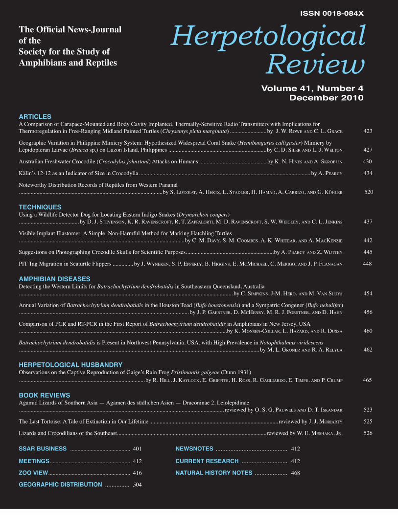
Herpetological Review
The Official News-Journalof the Society for the Study ofAmphibians and Reptiles
Volume 41, Number 4December 2010
ARTICLESA Comparison of Carapace-Mounted and Body Cavity Implanted, Thermally-Sensitive Radio Transmitters with Implications for Thermoregulation in Free-Ranging Midland Painted Turtles (Chrysemys picta marginata) ......................... by J. W. RoWe and C. L. GRaCe 423
Geographic Variation in Philippine Mimicry System: Hypothesized Widespread Coral Snake (Hemibungarus calligaster) Mimicry byLepidopteran Larvae (Bracca sp.) on Luzon Island, Philippines ...................................................................by C. d. SiLeR and L. J. WeLton 427
Australian Freshwater Crocodile (Crocodylus johnstoni) Attacks on Humans .............................................. by K. n. HineS and a. SKRobLin 430
Kälin’s 12-12 as an Indicator of Size in Crocodylia ..................................................................................................................... by a. PeaRCy434. 434
Noteworthy Distribution Records of Reptiles from Western Panamá..................................................................................................by S. LotzKat, a. HeRtz, L. StadLeR, H. Hamad, a. CaRRizo, and G. KöHLeR 520
TECHNIQUESUsing a Wildlife Detector Dog for Locating Eastern Indigo Snakes (Drymarchon couperi)......................................... by d. J. StevenSon, K. R. RavenSCRoft, R. t. zaPPaLoRti, m. d. RavenSCRoft, S. W. WeiGLey434., and C. L. JenKinS 437
Visible Implant Elastomer: A Simple, Non-Harmful Method for Marking Hatchling Turtles.................................................................................................................by C. m. davy434., S. m. CoombeS, a. K. WHiteaR, and a. maCKenzie 442
Suggestions on Photographing Crocodile Skulls for Scientific Purposes ............................................................by a. PeaRCy434. and z. WiJtten 445
PIT Tag Migration in Seaturtle Flippers .............. by J. Wy434.neKen, S. P. ePPeRLy434., b. HiGGinS, e. mCmiCHaeL, C. meRiGo, and J. P. fLanaGan 448
AMPHIBIAN DISEASESDetecting the Western Limits for Batrachochytrium dendrobatidis in Southeastern Queensland, Australia.................................................................................................................................................. by C. SimPKinS, J-m. HeRo, and m. van SLuy434.S 454
Annual Variation of Batrachochytrium dendrobatidis in the Houston Toad (Bufo houstonensis) and a Sympatric Congener (Bufo nebulifer).................................................................................................................... by J. P. GaeRtneR, d. mCHenRy434., m. R. J. foRStneR, and d. HaHn 456
Comparison of PCR and RT-PCR in the First Report of Batrachochytrium dendrobatidis in Amphibians in New Jersey, USA..............................................................................................................................................by K. monSen-CoLLaR, L. HazaRd, and R. duSSa 460
Batrachochytrium dendrobatidis is Present in Northwest Pennsylvania, USA, with High Prevalence in Notophthalmus viridescens.................................................................................................................................................................... by m. L. GRoneR and R. a. ReLy434.ea 462
HERPETOLOGICAL HUSBANDRYObservations on the Captive Reproduction of Gaige’s Rain Frog Pristimantis gaigeae (Dunn 1931)...................................................................................... by R. HiLL, J. Kay434.LoCK, e. GRiffitH, H. RoSS, R. GaGLiaRdo, e. timPe, and P. CRumP 465
BOOK REVIEWSAgamid Lizards of Southern Asia — Agamen des südlichen Asien — Draconinae 2, Leiolepidinae............................................................................................................................................ reviewed by o. S. G. PauWeLS and d. t. iSKandaR 523
The Last Tortoise: A Tale of Extinction in Our Lifetime ........................................................................................ reviewed by J. J. moRiaRty434. 525
Lizards and Crocodilians of the Southeast ......................................................................................................reviewed by W. e. meSHaKa, JR. 526
SSAR BUSINESS ......................................... 401 NEWSNOTES ............................................ 412
MEETINGS ....................................................... 412 CURRENT RESEARCH ............................ 412
ZOO VIEW........................................................ 416 NATURAL HISTORY NOTES .................... 468
GEOGRAPHIC DISTRIBUTION ............... 504
ISSN 0018-084X
Related Documents