Heritability of the Human Infectious Reservoir of Malaria Parasites Yaye Ramatoulaye Lawaly 1. , Anavaj Sakuntabhai 2,3. , Laurence Marrama 4 , Lassana Konate 5 , Waraphon Phimpraphi 2,6 , Cheikh Sokhna 7 , Adama Tall 4 , Fatoumata Die ` ne Sarr 4 , Chayanon Peerapittayamongkol 2¤ , Chalisa Louicharoen 2,8 , Bradley S. Schneider 3 , Anaı¨s Levescot 2 , Arthur Talman 9 , Isabelle Casademont 2,3 , Didier Menard 9 , Jean-Franc ¸ois Trape 7 , Christophe Rogier 10 , Jaranit Kaewkunwal 6 , Thanyachai Sura 11 , Issarang Nuchprayoon 12 , Frederic Ariey 9 , Laurence Baril 4 , Pratap Singhasivanon 6 , Odile Mercereau-Puijalon 13 , Rick Paul 1,2,3 * 1 Institut Pasteur de Dakar, Laboratoire d’Entomologie Me ´ dicale, Dakar, Senegal, 2 Institut Pasteur, Laboratoire de la Ge ´ne ´tique de la re ´ponse aux infections chez l’homme, Paris, France, 3 Institut Pasteur, Unite ´ de Pathoge ´nie Virale, Paris, France, 4 Institut Pasteur de Dakar, Unite ´ d’Epide ´ miologie, Dakar, Senegal, 5 Faculte ´ des Sciences et Techniques, UCAD, Dakar, Senegal, 6 Department of Tropical Hygiene, Faculty of Tropical Medicine, Mahidol University, Bangkok Thailand, 7 Institut de Recherche pour le De ´ veloppement, Laboratoire de Paludologie, Dakar, Senegal, 8 Inter-Department Program of Biomedical Science, Faculty of Graduate School, Chulalongkorn University, Bangkok, Thailand, 9 Unite ´ d’Epide ´ miologie Mole ´ culaire, Institut Pasteur, Phnom Penh, Cambodia, 10 Institut de Me ´decine Tropicale du Service de Sante ´ des Arme ´es, Unite ´ de Recherche en Biologie et e ´pide ´miologie parasitaires, IFR48, Le Pharo, Marseille, France, 11 Department of Medicine, Faculty of Medicine Ramathibodi Hospital, Mahidol University, Bangkok, Thailand, 12 Department of Pediatrics, Faculty of Medicine, Chulalongkorn University, Bangkok, Thailand, 13 Institut Pasteur, Unite ´ d’Immunologie Mole ´culaire des Parasites, CNRS URA 2581, Paris, France Abstract Background: Studies on human genetic factors associated with malaria have hitherto concentrated on their role in susceptibility to and protection from disease. In contrast, virtually no attention has been paid to the role of human genetics in eliciting the production of parasite transmission stages, the gametocytes, and thus enhancing the spread of disease. Methods and Findings: We analysed four longitudinal family-based cohort studies from Senegal and Thailand followed for 2–8 years and evaluated the relative impact of the human genetic and non-genetic factors on gametocyte production in infections of Plasmodium falciparum or P. vivax. Prevalence and density of gametocyte carriage were evaluated in asymptomatic and symptomatic infections by examination of Giemsa-stained blood smears and/or RT-PCR (for falciparum in one site). A significant human genetic contribution was found to be associated with gametocyte prevalence in asymptomatic P. falciparum infections. By contrast, there was no heritability associated with the production of gametocytes for P. falciparum or P. vivax symptomatic infections. Sickle cell mutation, HbS, was associated with increased gametocyte prevalence but its contribution was small. Conclusions: The existence of a significant human genetic contribution to gametocyte prevalence in asymptomatic infections suggests that candidate gene and genome wide association approaches may be usefully applied to explore the underlying human genetics. Prospective epidemiological studies will provide an opportunity to generate novel and perhaps more epidemiologically pertinent gametocyte data with which similar analyses can be performed and the role of human genetics in parasite transmission ascertained. Citation: Lawaly YR, Sakuntabhai A, Marrama L, Konate L, Phimpraphi W, et al. (2010) Heritability of the Human Infectious Reservoir of Malaria Parasites. PLoS ONE 5(6): e11358. doi:10.1371/journal.pone.0011358 Editor: Colin J. Sutherland, London School of Hygiene and Tropical Medicine, United Kingdom Received October 1, 2009; Accepted May 28, 2010; Published June 29, 2010 Copyright: ß 2010 Lawaly et al. This is an open-access article distributed under the terms of the Creative Commons Attribution License, which permits unrestricted use, distribution, and reproduction in any medium, provided the original author and source are credited. Funding: This work was funded in part by the Strategic Anopheles Horizontal Research Programme, Institut Pasteur to RELP, by grants from BIOTEC (BT-B06-MG- 14-4507), the Thailand Research Fund (BRG/16/2544), Mahidol University grant (OR-9123) and the Institut Pasteur to A.S. C.P. was supported by post-doctoral fellowships from INSERM and from the Faculty of Medicine Siriraj Hospital, Mahidol University, Thailand. C.T., W.P. and C.L. were supported by the Royal Golden Jubilee Program, the Thailand Research Fund and the French Embassy in Thailand. A. Talman was supported by ‘‘Fonds De ´die ´s’’ Sanofi-Aventis, Ministry of Research, France and Institut Pasteur research grant to F.A. The funders had no role in study design, data collection and analysis, decision to publish, or preparation of the manuscript. Competing Interests: The authors have declared that no competing interests exist. * E-mail: [email protected] . These authors contributed equally to this work. ¤ Current address: Department of Biochemistry, Faculty of Medicine Siriraj Hospital, Mahidol University, Bangkok, Thailand Introduction Transmission of malaria parasites from man to mosquito depends on the production of gametocyte sexual parasite stages in the human host that are subsequently taken up by a mosquito during a bloodmeal. For Plasmodium falciparum, the etiological agent of malignant tertian malaria, sexual stage differentiation (game- tocytogenesis) from asexual parasites occurs in the blood of the PLoS ONE | www.plosone.org 1 June 2010 | Volume 5 | Issue 6 | e11358

Welcome message from author
This document is posted to help you gain knowledge. Please leave a comment to let me know what you think about it! Share it to your friends and learn new things together.
Transcript

Heritability of the Human Infectious Reservoir of MalariaParasitesYaye Ramatoulaye Lawaly1., Anavaj Sakuntabhai2,3., Laurence Marrama4, Lassana Konate5, Waraphon
Phimpraphi2,6, Cheikh Sokhna7, Adama Tall4, Fatoumata Diene Sarr4, Chayanon
Peerapittayamongkol2¤, Chalisa Louicharoen2,8, Bradley S. Schneider3, Anaıs Levescot2, Arthur Talman9,
Isabelle Casademont2,3, Didier Menard9, Jean-Francois Trape7, Christophe Rogier10, Jaranit
Kaewkunwal6, Thanyachai Sura11, Issarang Nuchprayoon12, Frederic Ariey9, Laurence Baril4, Pratap
Singhasivanon6, Odile Mercereau-Puijalon13, Rick Paul1,2,3*
1 Institut Pasteur de Dakar, Laboratoire d’Entomologie Medicale, Dakar, Senegal, 2 Institut Pasteur, Laboratoire de la Genetique de la reponse aux infections chez
l’homme, Paris, France, 3 Institut Pasteur, Unite de Pathogenie Virale, Paris, France, 4 Institut Pasteur de Dakar, Unite d’Epidemiologie, Dakar, Senegal, 5 Faculte des
Sciences et Techniques, UCAD, Dakar, Senegal, 6 Department of Tropical Hygiene, Faculty of Tropical Medicine, Mahidol University, Bangkok Thailand, 7 Institut de
Recherche pour le Developpement, Laboratoire de Paludologie, Dakar, Senegal, 8 Inter-Department Program of Biomedical Science, Faculty of Graduate School,
Chulalongkorn University, Bangkok, Thailand, 9 Unite d’Epidemiologie Moleculaire, Institut Pasteur, Phnom Penh, Cambodia, 10 Institut de Medecine Tropicale du Service
de Sante des Armees, Unite de Recherche en Biologie et epidemiologie parasitaires, IFR48, Le Pharo, Marseille, France, 11 Department of Medicine, Faculty of Medicine
Ramathibodi Hospital, Mahidol University, Bangkok, Thailand, 12 Department of Pediatrics, Faculty of Medicine, Chulalongkorn University, Bangkok, Thailand, 13 Institut
Pasteur, Unite d’Immunologie Moleculaire des Parasites, CNRS URA 2581, Paris, France
Abstract
Background: Studies on human genetic factors associated with malaria have hitherto concentrated on their role insusceptibility to and protection from disease. In contrast, virtually no attention has been paid to the role of human geneticsin eliciting the production of parasite transmission stages, the gametocytes, and thus enhancing the spread of disease.
Methods and Findings: We analysed four longitudinal family-based cohort studies from Senegal and Thailand followed for2–8 years and evaluated the relative impact of the human genetic and non-genetic factors on gametocyte production ininfections of Plasmodium falciparum or P. vivax. Prevalence and density of gametocyte carriage were evaluated inasymptomatic and symptomatic infections by examination of Giemsa-stained blood smears and/or RT-PCR (for falciparum inone site). A significant human genetic contribution was found to be associated with gametocyte prevalence inasymptomatic P. falciparum infections. By contrast, there was no heritability associated with the production of gametocytesfor P. falciparum or P. vivax symptomatic infections. Sickle cell mutation, HbS, was associated with increased gametocyteprevalence but its contribution was small.
Conclusions: The existence of a significant human genetic contribution to gametocyte prevalence in asymptomaticinfections suggests that candidate gene and genome wide association approaches may be usefully applied to explore theunderlying human genetics. Prospective epidemiological studies will provide an opportunity to generate novel and perhapsmore epidemiologically pertinent gametocyte data with which similar analyses can be performed and the role of humangenetics in parasite transmission ascertained.
Citation: Lawaly YR, Sakuntabhai A, Marrama L, Konate L, Phimpraphi W, et al. (2010) Heritability of the Human Infectious Reservoir of Malaria Parasites. PLoSONE 5(6): e11358. doi:10.1371/journal.pone.0011358
Editor: Colin J. Sutherland, London School of Hygiene and Tropical Medicine, United Kingdom
Received October 1, 2009; Accepted May 28, 2010; Published June 29, 2010
Copyright: � 2010 Lawaly et al. This is an open-access article distributed under the terms of the Creative Commons Attribution License, which permitsunrestricted use, distribution, and reproduction in any medium, provided the original author and source are credited.
Funding: This work was funded in part by the Strategic Anopheles Horizontal Research Programme, Institut Pasteur to RELP, by grants from BIOTEC (BT-B06-MG-14-4507), the Thailand Research Fund (BRG/16/2544), Mahidol University grant (OR-9123) and the Institut Pasteur to A.S. C.P. was supported by post-doctoralfellowships from INSERM and from the Faculty of Medicine Siriraj Hospital, Mahidol University, Thailand. C.T., W.P. and C.L. were supported by the Royal GoldenJubilee Program, the Thailand Research Fund and the French Embassy in Thailand. A. Talman was supported by ‘‘Fonds Dedies’’ Sanofi-Aventis, Ministry ofResearch, France and Institut Pasteur research grant to F.A. The funders had no role in study design, data collection and analysis, decision to publish, orpreparation of the manuscript.
Competing Interests: The authors have declared that no competing interests exist.
* E-mail: [email protected]
. These authors contributed equally to this work.
¤ Current address: Department of Biochemistry, Faculty of Medicine Siriraj Hospital, Mahidol University, Bangkok, Thailand
Introduction
Transmission of malaria parasites from man to mosquito
depends on the production of gametocyte sexual parasite stages
in the human host that are subsequently taken up by a mosquito
during a bloodmeal. For Plasmodium falciparum, the etiological agent
of malignant tertian malaria, sexual stage differentiation (game-
tocytogenesis) from asexual parasites occurs in the blood of the
PLoS ONE | www.plosone.org 1 June 2010 | Volume 5 | Issue 6 | e11358

human host. Both in vitro and in vivo studies emphasise the
importance of environmental stimuli in modulating gametocyto-
genesis [1,2]. Gametocyte production may occur in response to
environmental factors that directly suppress asexual proliferation
in vitro [3], but this has not been shown in vivo [4]. Gametocyte
carriage has been associated with a worsening blood environment
for the parasite (e.g. fever responses, anaemia, and the presence of
reticulocytes) [5–7]. However, such cues are associated with
symptomatic episodes of malaria and it is now well established that
asymptomatic infections can also generate gametocytes and infect
mosquitoes [8–10]. Molecular techniques have revealed extensive
occurrence of sub-microscopic gametocytes [11,12] that can infect
mosquitoes [13] and play an important role as a reservoir of
infection especially in areas of seasonal transmission [14]. No
specific risk factors have yet been identified for gametocyte
carriage in asymptomatic infections, although as in symptomatic
infections, drug treatment of asymptomatic infections with
sulfadoxine-pyrimethamine reduces gametocyte carriage [15].
Studies on human genetic factors associated with malaria have
hitherto concentrated on their role in susceptibility to and
protection from disease [16]. The most well-known is the sickle
cell mutation (HbS) in Africa, which confers protection against
severe malaria in heterozygotes, but causes fatal sickle cell disease
in homozygotes [17,18], illustrating the powerful selective pressure
exerted by malaria on the human genome [19]. In contrast,
virtually no attention has been paid to the role of human genetics
in eliciting the production of gametocytes and thus enhancing the
spread of parasites and hence disease. Differences in the tendency
of sympatric ethnic groups to carry gametocytes have, however,
been noted since 1914 [20] and more recently in Irian Jaya [21]
and Burkina Faso [22]. The extent to which such differences are a
consequence of the impact of host genetics on parasite asexual
proliferation rather than directly on gametocytogenesis has not
been addressed. Asexual parasite density has been repeatedly
shown to be influenced by host genetics and the chromosomal
region 5q31–33, which contains a cluster of cytokine genes, has
been identified as important in the control of asexual parasite
density [23–27]. Thus, the impact of human genetics on
gametocyte production may occur via its effect on asexual parasite
proliferation. The observed association of gametocytes with
anaemia and subsequent erythropoietic response [5–7,28–29]
has yet to be explored genetically, despite a high prevalence of
inherited blood disorders that induce anaemia, such as HbS and
alpha-thalassaemia, in regions endemic for malaria [30]. Very
recently, however, increased gametocyte carriage was observed in
individuals with HbC [31].
Transmission success is crucial to the parasite and gametocyte
production has been repeatedly shown to be under strong selective
pressure [32], thus making this stage of the lifecycle propitious for
intervention. Current efforts concentrate on the development of
transmission-blocking vaccines [33] and exploration of parasite
genes implicated in sexual development [34]. The possibility of using
genome-wide association studies in humans potentially enables
identification of critical molecular pathways in humans that
influence gametocyte production, thereby potentially generating
novel strategies for treatment and prevention; for example by using
drug treatment targeting individuals genetically susceptible to carry
gametocytes or developing novel drugs that target the human factors
that induce gametocyte production. As a first step, however, it is
necessary to establish the extent to which humans exert an influence
on gametocyte production, in addition to the known intrinsic
parasite clone variability in gametocyte production [35]. Measure-
ment of heritability is central to quantitative genetic analysis and
provides an estimate of the genetic basis underlying a trait.
In this study we examine the overall contribution of human host
genetic factors (i.e. the heritability) to variation in gametocyte
production in three longitudinal cohort studies occurring in areas
of differing transmission intensity in Senegal, where P. falciparum is
endemic, and in one cohort in Thailand where both P. falciparum
and P. vivax are present. Moreover, we specifically examine the
impact on gametocyte production of two inherited blood disorders
known to cause anaemia, sickle cell trait [17] and alpha-
thalassaemia [18,36].
Materials and Methods
Ethics statementDielmo and Ndiop, Senegal. The project protocol and
objectives were carefully explained to the assembled village
population and informed consent was individually obtained from
all subjects either by signature or by thumbprint on a voluntary
consent form written in both French and in Wolof, the local
language. Consent was obtained in the presence of the school
director, an independent witness. For very young children, parents
or designated tutors signed on their behalf. The protocol was
approved by the Ethical Committee of the Pasteur Institute of
Dakar and the Ministry of Health of Senegal (ethics S1). An
agreement between Institut Pasteur de Dakar, Institut de
Recherche pour le Developpement (IRD) and the Ministere de
la Sante et de la Prevention of Senegal defines all research
activities in Dielmo and Ndiop villages. Each year, the project was
re-examined by the Conseil de Perfectionnement de l’Institut
Pasteur de Dakar and the assembled village population; informed
consent was individually renewed from all subjects.
Gouye Kouly, Senegal. The project protocol and objectives
were carefully explained to the assembled village population and
informed consent was individually obtained from all subjects by
signature on a voluntary consent form written in both French and
in Wolof. The protocol was approved by the Ethical Committee of
the Ministry of Health of Senegal (ethics S2).
Suan Phung, Thailand. The project protocol and objectives
were explained to the population and signed informed consent was
individually obtained from all study participants or their parents.
Ethical permission for the study was granted by the Ethical
Committee of the Ministry of Public Health of Thailand (ethics
S3).
Study sites and subjectsDielmo and Ndiop, Senegal. The Dielmo and Ndiop
longitudinal surveys have been described in detail elsewhere
[37–39]. Briefly, a longitudinal cohort study of malaria has been
carried out since 1990 in Dielmo and 1993 in Ndiop. For this
analysis we use data acquired from 1990–1998 in Dielmo and
1993–1998 in Ndiop. In Dielmo there were 594 individuals from
190 nuclear families and in Ndiop 653 from 208 nuclear families.
In each village, the majority of individuals were related to each
other, forming one large complex family: 1 family of 453
individuals in Dielmo and one family of 503 in Ndiop. Overall
there were 10 completely independent families in Dielmo and 21
in Ndiop. In Dielmo, the ethnic groups consisted of 79% Serere
(Niominka: 59% and Sine/Baol: 20%), 11% Mandinka and 10%
miscellaneous, whereas in Ndiop, there were 76% Wolof, 19%
Fulani and 5% miscellaneous.
Gouye Kouly, Senegal. A family-based longitudinal cohort
study was performed from June 2004–November 2005 in a third
site in Senegal, Gouye Kouly. Family structures were constructed
by using a questionnaire, interviewing each individual or key
representatives of the household to obtain both demographic
Gametocyte Heritability
PLoS ONE | www.plosone.org 2 June 2010 | Volume 5 | Issue 6 | e11358

information such as birth date, age, sex and genetic relationships
between children, their parents, and sometimes their grandparents
or non-relatives in the same household, and other households. The
population was composed of 482 individuals that belong to 9
independent families, one of which is a large complex family of
423 individuals that form 173 nuclear families. The majority of
individuals were Serere.
Suan Phung, Thailand. In Thailand, a community-based
cohort study was carried out from June 1998 to May 2005 [40].
The study was conducted in a mountainous area of Suan Phung
district, Ratchaburi province, Thailand. Suan Phung is a small
district situated near the Thai-Myanmar border. Suan Phung has
a total population of 5,368 living in 7 hamlets, of which 3,484
villagers of all ages participated in the study. This community is
made up of a group of 4 closely related ethnic groups, the majority
of which are Karen (85%), some Thai (14%) and the rest are Mon
and Burmese (1%). The total pedigrees are comprised of 2,427
individuals, including absent or deceased relatives. There were 238
independent families containing 603 nuclear families; the majority
are 2 generation-families with family size range from 3 to 958. The
recruitment procedure has been previously detailed [40].
Malaria epidemiologyMalaria transmission is perennial in Dielmo, where a river
maintains larval breeding sites for the mosquitoes even in the dry
season. The number of infective bites per person per year
(Entomological Inoculation Rate, EIR) is of the order of 200 [41].
By contrast, malaria transmission is strictly seasonal in Ndiop and
dependent upon the rainy season that occurs from July–September
and the EIR is approximately 20 [42]. Transmission is similarly
highly seasonal in Gouye Kouly with EIR measured at
approximately 2 infectious bites per person per year (unpubl.
data). Such differing transmission has marked consequences on the
epidemiology of malaria in the villages. This is most evident in the
higher P. falciparum prevalence rates of infection in Dielmo (80%)
compared to the seasonal rates in Ndiop that change from 20% in
the dry season to 70% in the rainy season [39,43] and from 8% to
15% in Gouye Kouly (unpubl. data).
The epidemiology of malaria in the Thai site has been described
elsewhere [40]. Briefly, the incidence of malaria is highly seasonal
with annual peaks in May–June and decreased over the duration
of the study from 141 per 1000 person-years in 1999 to 57 in 2004
for P. falciparum and from 79 to 28 for P. vivax. P. falciparum
prevalence rates varied from 1–7% seasonally and from 1–4% for
P. vivax. There was good concordance in the population
prevalence of fevers that were found to be positive for malaria
parasites and the fraction of fevers attributable to malaria. Thus, in
this site, virtually all infections lead to febrile episodes. Peak
incidence occurred in an earlier age group (5–9 years old) for P.
vivax than for P. falciparum (10–15 years old). Parasite densities of
either species peaked in the ,10 years old age group.
Data CollectionSymptomatic episodes (passive case detection). The
installation of health clinics in each of the study sites enabled
passive case detection of malaria episodes. We defined clinical
malaria episodes as measured fever (axillary temperature
.37.5uC) or fever-related symptoms (headache, vomiting,
subjective sensation of fever) associated with i) a P. falciparum
parasite/leukocyte ratio higher than an age-dependent pyrogenic
threshold previously identified in the patients from Dielmo [44], ii)
a P. falciparum parasite/leukocyte ratio higher than 0.3 parasite/
leukocyte in Ndiop, iii) a slide positive for blood-stage trophozoite
P. falciparum or P. vivax parasites at any density for Thailand.
Although clinical episodes were defined as a slide positive for blood-
stage trophozoite P. falciparum parasites at any density with associated
fever or fever-related symptoms for Gouye Kouly symptomatic
episodes were too few to generate sufficient gametocyte data. All
positive malaria cases were treated with appropriate antimalarial
treatment according to the recommendation of the Malaria Division,
Ministry of Public Health, as previously described [38–40], namely
quinine until 1995 and then chloroquine in Dielmo and Ndiop
and in Thailand mefloquine+primaquine for P. falciparum and
chloroquine+primaquine for P. vivax. Sulfadoxine-pyrimethamine in
conjunction with amodiaquine was used in Gouye Kouly, as this
study occurred after a national policy change in 2004.
Asymptomatic episodes (active case detection). Monthly
systematic blood slides were taken from participating individuals
from 1990–1998 and 1993–1998 in Dielmo and Ndiop
respectively. In Gouye Kouly an intensive sampling schedule
was implemented for 2005: prior to the rains in June and then
every week for 8 weeks following the onset of the rains (first week
of July). At each time point a thick blood smear was taken from all
individuals. In the June sample and every two weeks from July,
approximately 500 mL of blood were taken by finger prick from
each individual in an EDTA microtainer (Sarstedt), of which
200 mL were mixed with in 1ml TRIzolH (Invitrogen), kept on dry
ice and then frozen at 280uC for RNA extraction. Following
DNA extraction and PCR amplification, all individuals’ samples
that were found to be positive for P. falciparum were then analysed
for the presence of gametocytes by RT-PCR. The cohort was
randomly divided into two groups (by household) such that half the
cohort provided such a blood sample every week. Although there
were insufficient gametocyte positive blood smears in this cohort,
the few there were enabled us to validate the RT-PCR method.
Two cross-sectional analyses in Suan Phung, Thailand carried out
in 1995 and 2003 yielded insufficient gametocyte data from
asymptomatic infections.
In all cases parasite positivity was established as follows. Thick
and thin blood films were prepared and stained by 3% Giemsa
stain. Blood films were examined under an oil immersion objective
at 61000 magnification by the trained laboratory technicians and
200 thick film fields were examined to count the number of
asexual and gametocyte parasite stages. Parasite species were
identified on thin films and asexual parasite densities (per mL) were
calculated from thick film by establishing the ratio of parasites to
white blood cells (WBC) and then multiplying the parasite count
by 8,000, the average WBC count per mL of blood. Gametocyte
densities per microlitre were estimated by multiplying by 4 the
count per 200 microscope fields; the average number of WBCs per
field being approximately 10, thus generating 2000 WBCs per 200
fields and thus representing a quarter of a microlitre. The
minimum detectable gametocyte density is thus estimated to be 4
per mL.
Gametocyte dataBlood smears. Four gametocyte traits were considered: (i)
gametocyte positivity (i.e. prevalence), (ii) cumulative gametocyte
positivity, (iii) gametocyte density and (iv) maximum gametocyte
density for an individual. Here, ‘‘trait’’ is applied in a very loose
sense, and does not imply that any genetic influence on these
‘‘traits’’ is only resulting from the human genome. Gametocyte
positivity was defined as the proportion of parasite positive
infections that also carried gametocytes and thus addressed the
tendency to produce gametocytes during each infection. The
cumulative gametocyte positivity likewise addresses this tendency,
but sums over the number of infections an individual has
experienced (and therefore opportunity to carry gametocytes). In
Gametocyte Heritability
PLoS ONE | www.plosone.org 3 June 2010 | Volume 5 | Issue 6 | e11358
the epidemiological analyses these two traits are the same;
however in the heritability analyses they are treated differently
(see Data analyses below). In addition to considering all gametocyte
densities, we analysed the individual’s maximum gametocyte
density because transmission to mosquitoes is weakly associated
with gametocyte density in some studies [45,46], although low
gametocyte densities are well known to also permit transmission to
mosquitoes [13,47,48].
The duration of gametocyte carriage for a single infection in
endemic settings can last up to 30 days [12,49]. The longevity and
infectivity of gametocytes have been shown to persist for 3 weeks
following chloroquine treatment of clinical cases [50]. To increase
the probability that only independent symptomatic or asymptom-
atic episodes (of gametocyte production) from the same individual
are considered, consecutive samples with blood-stage malaria
parasites of the same species within 30 days were excluded. Mixed
parasite species infections were also excluded. It is likely our
sampling approach missed some episodes of gametocytaemia, and
thus underestimated prevalence.
PCR and RT-PCR for P. falciparum gametocyte
detection. DNA was extracted from all samples from Gouye
Kouly using the standard phenol-chloroform extraction method
and DNA amplified using the ssrRNA gene nested PCR method of
Snounou et al., 1993 [51]. RNA extraction was then performed
from the TRIzolH (Invitrogen) conserved sub-samples of those
found positive. RNA was extracted using TRIzolH (Invitrogen),
following the protocol recommended by the manufacturer. The
extracted RNA was directly analysed or stored at 280uC.
For the RT-PCR, ‘‘Plasmodium falciparum meiotic recombination
protein DMC1-like protein’’ gene (AF356553) was selected
because it is exclusively expressed in gametocytes [52] and
contains introns. Primers were thus selected spanning an exon-
exon junction, amplifying a 101 bp segment, in the middle of
which a probe was designed, using Primer3 software [53]. Primer
sequences were: forward primer GAM8_F 59 ATATCGGCAGC-
GAAAATGTGT 39; reverse primer GAM8_R 59 GACAAT-
TCCCCTCTTCCACTGA 39 and probe GAMPRO 59 (6-
Fam)TGCCCTTCTCGTAGTTGATTCGATTATT(BHQ1) 39.
cDNA was synthesised and the reaction primed with GAM8_R.
Briefly 8 mL of extracted RNA was mixed with buffer, dNTPs
(final concentration 1mM), RNase-free water, AMV Reverse
transcriptase (20U; Promega) and Ribonuclease inhibitor (20U;
Promega). Amplification cycle conditions were: 10 min. at 65uC,
60 min. at 42uC, 5 min. at 95uC. Quantification of cDNA was
carried out using a fluorescent probe assay. Briefly 2 mL of
synthesised cDNA was mixed with 26 mastermix (ABGene),
GAM8_R (final concentration: 400nM), GAM8_F (final concen-
tration: 400nM), GAM8_PRO (final concentration: 300nM) and
sterile water. The reaction was analysed with a Rotor GeneH real-
time PCR machine (Corbett Research). Each sample was analysed
in triplicate. A dilution series containing 1000, 100, 10, 1 and 1021
gametocytes/mL was used. This RT-PCR methodology had
previously been validated using in vitro parasite cultures, and its
specificity for detection of gametocytes in vivo, and not asexual
parasites, demonstrated in a sample of 47 individuals presenting
with clinical falciparum malaria in Madagascar (ethics S4 and
Supplementary material S1).
Genotyping of HbS and a-globin 3.7 deletion mutationsHbS – PCR-RFLP (Senegal). Following DNA extraction, a
559 bp fragment covering codon 6 of the b–globin gene (HBB)
gene was amplified by PCR using the primers HbS_F: 59-
AGGGGAAAGAAAACATCAAGGGTC-39 and HbS_R: 59-
ATAAGTCAGGGCAGAGCCATCTAT-39. The amplification
reaction was carried out using 5 mL of DNA in a reaction volume
of 15 mL composed of MgCl2 [2.5mM], dNTPs [1mM], each
primer [1mM], 1.5 mL PCR buffer (Qbiogene) and 0.04 mL Taq
(Qbiogene). Amplification cycle conditions were: 4min at 94uC,
and then 35 cycles of 30sec at 94uC, 30sec at 65uC, and 30sec at
72uC, with a final extension phase of 10 min. at 72uC. The
amplified fragment (5 mL of DNA) was then digested by restriction
enzyme Dde I (2U) in a reaction volume of 15 mL containing
1.5 mL 106 Buffer. Wildtype HBB yields 6 fragments of
201+97+89+88+50+37 base pairs, whereas HbS mutation yields
5 fragments of 298+89+88+50+37 bp.
a-globin 3.7 deletion. Following DNA extraction, we used
the PCR multiplex protocol of Chong et al. (2000) [54] to detect
the presence of the a-globin 3.7 deletion. Primers a2/3.7-F and
a2-R amplify a 1800 bp fragment covering the a2-globin gene.
The sequence corresponding to primer a2-R is lost with the a-
globin 3.7 deletion. A third primer, 3.7/20.5-R, is located 39 of the
a1-globin gene and allows amplification of fragments of 2022/
2029 bp in the presence of the a-globin 3.7 deletion. Primer
sequences are a2/3.7-F: 59-CCCCTCGCCAAGTCCACCC-39,
3.7/20.5-R: 59-AAAGCACTCTAGGGTCCAGCG-39 and a2-
R: 59-AGACCAGGAAGGGCCGGTG-39. The amplification
reaction was carried out using 5 mL of DNA in a reaction
volume of 15 mL composed of MgCl2 [1mM], dNTPs [300nM],
primer a2/3.7-F [0.4mM], primer 3.7/20.5-R [0.8mM], primer
a2-R [0.1mM], 3 mL PCR buffer HotStar (Qiagen), 3 mL PCR
buffer Q (Qiagen) and 0.04 mL HotStar Taq (Qiagen).
Amplification cycle conditions were: 15min. at 98uC for enzyme
activation and DNA denaturation, and then 50 cycles of 45sec at
98uC, 1 min. 15sec at 65uC, and 2 min. 30sec at 72uC, with a final
extension phase of 5 min at 72uC. The a-globin 3.7 deletion yields
fragments of 2022/2029 bp and the intact a2-globin gene
1800 bp.
Data analysesEpidemiological data analyses. Table 1 gives a summary
of the samples analysed. Statistical analyses and model fitting were
conducted using the statistical package Genstat 7.1 [55]. For each
site, all individuals in the study protocol were included in the
analyses, irrespective of whether their family structure was known.
Factors influencing the maximum gametocyte density of either P.
falciparum or P. vivax were analysed by fitting a Generalized Linear
Model (GLM) with a Poisson error structure (loglinear regression).
For gametocyte traits with repeated measures for the same
individual (i.e. gametocyte positivity and gametocyte density), a
Generalized Linear Mixed Model (GLMM) was fitted with
individual person as a factor in the random model. For analysis
of the gametocyte positivity rate, a binomial error structure was
implemented (thus a logistic regression). Explanatory factors
included date, which was classified annually by semester,
reflecting the transmission seasons and hereon denoted season.
Additional factors were gender and age factored initially into eight
groups (,1, 1–4, 5–9, 10–14, 15–24, 25–39, 40–59 and 60+ years
of age); if age was overall significant in the minimum adequate
model, age groups were combined when not significantly different
as ascertained by t-test and the final statistical model applied. A
dispersion parameter was estimated by the deviance method,
because the data were over-dispersed; initial model fitting with a
dispersion parameter of 1 (for binomial and poisson error
structures) yielded residual deviance much larger than the
residual degrees of freedom. F-statistics in the GLM and Wald
statistics, which approximate to a x2 distribution, in the GLMM
were established. In the analyses of maximum gametocyte density,
Gametocyte Heritability
PLoS ONE | www.plosone.org 4 June 2010 | Volume 5 | Issue 6 | e11358

the number of gametocyte density data points per individual was
used as a weight.
The residual variance not explained by these ‘‘environmental’’
factors was generated. Because a non-normal error distribution
was used, Pearson rather than standardized normal residuals were
generated. For the gametocyte trait ‘‘cumulative gametocyte
positivity rate’’, the sum of the residuals per person was then
calculated and used in the genetic analysis. For analysis of
‘‘gametocyte positivity’’ and ‘‘gametocyte density’’, all residual
values for any individual (who had repeated parasite density
measures) were then used in the genetic analysis. Only residuals
from individuals for whom family structure was available were
then analysed for heritability.
Genetic and house data analyses. To determine the
contribution of genetic factors to the ‘‘cumulative gametocyte
positivity rate’’ and ‘‘maximum gametocyte density’’, we evaluated
the heritability (h2) by using the SOLAR software package (version
2.1.4) [56]. SOLAR performs a variance components analysis of
family data that decomposes the total variance of the gametocyte
traits into components that are due to genetic (polygenic) (h2),
individual or environmental (e2) and house (c2) effects. We tested
for a heritable human component in each gametocyte trait by
comparing likelihood between the reduced model, where total
variation is due to environmental variation only, and the full
model where total variation is composed of environmental and
genetic effects estimated from the genetic relationship coefficient of
each pair of individuals. When the null hypothesis was rejected,
heritability (h2) was then estimated as the percentage of genetic
variance of the total. Although SOLAR can additionally
incorporate measured covariates (e.g. explanatory variables), a
normal distribution is assumed. For this reason we took into
account the contribution of such variables in an initial statistical
analysis (section above) and generated residual value for the
gametocyte traits. The relative contribution of genetic factors to
variation in the trait was then estimated by the heritability (h2),
defined by the ratio of genetic variance component to the residual
trait variance [56]. As several traits showed residual kurtosis of
more than 0.8, tdist option, which creates an extra parameter in
the model to describe the distribution of the trait, was applied in all
analyses. An additive model, which is a general model, making no
assumptions of the dominant or recessive nature of the gene, was
used to avoid multiplying tests. For estimation of heritability, we
used information from families that had at least 2 members with
the traits of interest.
Household can confound the estimation of the genetic
contribution to a trait, because related individuals often live in
the same house and therefore not only experience a similar level of
overall exposure to parasites, but also are potentially exposed to
more genetically related parasites. This latter may be especially
important given the known genetic variation in the parasite
gametocyte production [35]. A household or shared environment
effect can be added by an additional variance component with a
coefficient matrix (H) whose elements (house) are 1 if the relative
pair shares the same environmental exposure or 0 otherwise.
Genetic effect (i.e. heritability) is estimated using matrix of
correlation coefficients for identity by descent (IBD) allele sharing
in various types of family relative pairs, whose elements (phi2)
provide the predicted proportion of genes of the whole genome
that a pair of individuals share at least 1 allele [56]. In SOLAR we
first included the house effect in the model. If the house effect was
not significant (p value .0.05), we excluded it from the model for
estimation of heritability.
For the gametocyte traits for which there were multiple residual
values (i.e. ‘‘gametocyte positivity’’ and ‘‘gametocyte density’’), we
evaluated heritability by using the classic repeated measures model
(from the ‘‘animal model’’) [57,58], where a permanent environ-
mental effect is created for each individual. Thus the following
model is fitted: y = [Xb]+Za+Zpe+Zh+e where y is the residual
parasite density value, b is the fixed effects vector (here already
taken into account in the first statistical analysis), a is the additive
genetic effects vector, pe is the permanent environment effects
vector (of each individual), h is the common house effect vector
and e is the residual effects vector; X is the design matrix relating
observations to fixed effects and Z are design matrices relating
observations to random effects. The model was fitted using
ASREML vers. 2 [57]. The total trait variance is therefore VP,
which is partitioned into VA, additive genetic variance, VPE,
variance due to permanent environmental effects, VH, common-
Table 1. Summary of samples used in epidemiological and genetic analyses.
Site Dielmo Ndiop Gouye Kouly Suan Phung
Symptomatic Asymptomatic Symp. Asymp. Asymp. Symp. PF Symp. PV
Gametocyte positivity
Epidemiological analyses
Data points 1168 2710 1226 2063 101 1796 978
Individuals 239 343 313 379 79 949 517
Genetic analyses Individuals 236 335 310 364 77 859 470
Independent families 9 10 17 19 8 188 136
Gametocyte density
Epidemiological analyses
Data points 201 1096 180 578 84 323
Individuals 109 280 125 246 80 230
Genetic analyses Individuals 109 272 125 241 73 206
Independent families 8 10 12 13 47 78
For epidemiological analyses, presented are the number of data points analysed for each trait, the corresponding number of individuals implicated and hence residualvalues generated. For genetic analyses, presented are the number of these individuals for whom pedigree information was available and thus the number ofindependent families for each trait in the heritability analyses.doi:10.1371/journal.pone.0011358.t001
Gametocyte Heritability
PLoS ONE | www.plosone.org 5 June 2010 | Volume 5 | Issue 6 | e11358
house variance and VR, residual variance. Heritability (h2) is again
VA/VP.
Results
Table 2 presents a summary of the gametocyte data per
infection type and study cohort. From 1990–1998 in Dielmo, there
were 1,168 symptomatic P. falciparum episodes by 239 individuals;
by microscopy, 201 (17.2%) of these infections from 109
individuals had gametocytes. The mean gametocyte density
(excluding zeros) was 18.4/mL (SE 2.4, range 4–208). During the
same time frame, there were 2,710 observations of asymptomatic
P. falciparum infections in 343 individuals; 1,096 of these infections
(40.4%) from 280 individuals had gametocytes. The mean
gametocyte density was 37.2/mL (SE 5.2, range 4–3,588). From
1993–8 in Ndiop, there were 1226 symptomatic P. falciparum
episodes by 313 individuals; by microscopy, 180 (14.7%) of these
infections from 125 individuals had gametocytes. The mean
gametocyte density was 69.3/mL (SE 15.8, range 4–1,984). During
the same time frame, there were 2,063 observations of asymp-
tomatic P. falciparum infections in 379 individuals; 578 of these
infections (28%) from 246 individuals had gametocytes. The mean
gametocyte density was 22.2/mL (SE 3.1, range 4–908). From
June–August 2005 in Gouye Kouly, there were 101 independent
P. falciparum positive asymptomatic observations in 79 individuals;
there was one observation for 58 individuals, two observations for
20 and three for one individual. 79 infections (78%) had
gametocytes, as detected by RT-PCR; density was not, however,
ascertained in the RT-PCR. From 1999–2004 in Suan Phung,
there were 1,796 symptomatic P. falciparum episodes presented by
949 individuals; by microscopy, 84 (4.7%) of these infections from
80 individuals had gametocytes. The mean gametocyte density
was 284.5/mL (SE 62.8, range 1–3,480). During the same period,
there were 978 observations symptomatic P. vivax episodes
presented by 517 individuals; 323 of these infections (33%) from
230 individuals had gametocytes. The mean gametocyte density
was 648/mL (SE 63.5, range 16–11,280).
The genotype frequencies of AS (HbS heterozygote) were 9.9%
(N = 46 of 466 individuals successfully genotyped) in Dielmo,
13.6% (N = 67 of 493 individuals successfully genotyped) in Ndiop
and 7.1% (N = 21 of 295 individuals successfully genotyped) in
Gouye Kouly. There were two SS (HbS homozygote) in Dielmo
and none in either Ndiop or Gouye Kouly. The genotype
frequencies of the heterozygote alpha-globin 3.7 deletion were
18.1% (N = 75 of 415 individuals successfully genotyped) in
Dielmo, 30.2% (N = 132 of 437 individuals successfully genotyped)
in Ndiop; the alpha-deletion was not typed in Gouye Kouly. The
homozygote alpha-deletion genotype frequencies were 1.2% in
Dielmo and 1.8% in Ndiop. In Suan Phung (Thailand), the
heterozygote alpha-globin 3.7 deletion genotype frequency was
15.8% (N = 139 of 881 individuals successfully genotyped) and the
homozygote genotype frequency was 1.02% (N = 9 individuals).
Table 3 presents the genotype frequencies of alpha and beta globin
gene mutations for which there were corresponding gametocyte
data and hence used in the statistical analyses.
Table 4 presents the summary of the epidemiological analyses
showing significance level and percentage of variation in P.
falciparum (Pf) and P. vivax (Pv) gametocyte traits explained by
environmental variables and the two genetic mutations (HbS and
alpha-globin 3.7 deletion). Age, season and asexual parasite density
had a consistently significant impact on gametocytes. For
gametocyte positivity, the impact of these factors was, however,
small. The proportion of P. falciparum infections carrying gameto-
cytes decreased with increasing age and asexual parasite density. In
Ndiop, individuals of ten years and older had reduced odds of
carrying gametocytes whether in symptomatic (Odds Ratio = 0.42
[95%Confidence Intervals 0.28–0.56]) or asymptomatic infections
(OR = 0.56 [95%CI 0.43–0.68]). Similarly, in Dielmo 10+ year old
individuals similarly had lower odds of carrying gametocytes when
infected, as compared to the youngest age group (0–4 years) whether
in symptomatic (OR = 0.36 [95%CI 0.26–0.47]) or asymptomatic
infections (OR = 0.17 [95%CI 0.08–0.25]). In Suan Phung there
was also significantly lower odds of carrying P. falciparum
gametocytes for the older (.15 years) age group (OR = 0.32
[95%CI 0.22–0.42]). P. vivax gametocyte positivity increased with
asexual parasite density, but was not affected by age. Both age and
asexual parasite density were inversely correlated to gametocyte
density. Age and especially season explained a large amount of the
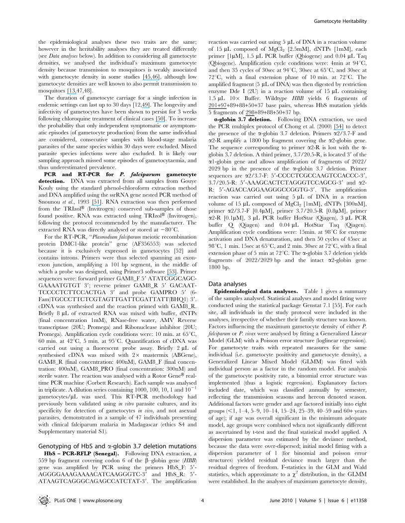
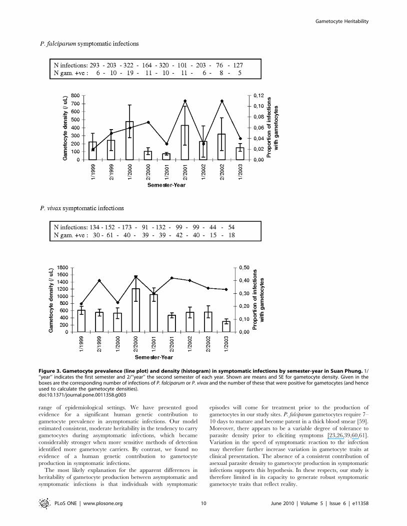
observed variation. However, as shown in Figure 1–3, variation in
gametocyte traits was as great, if not greater, across years than
between seasons, with one exception: the increase in gametocyte
density during the rainy season (season 2 of each year) in
asymptomatic infections in Ndiop, where transmission is highly
seasonal (Fig. 2).
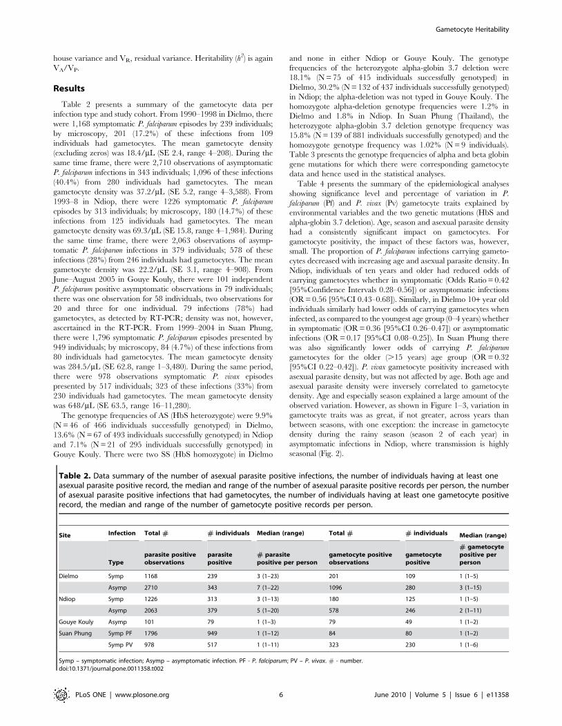
Table 2. Data summary of the number of asexual parasite positive infections, the number of individuals having at least oneasexual parasite positive record, the median and range of the number of asexual parasite positive records per person, the numberof asexual parasite positive infections that had gametocytes, the number of individuals having at least one gametocyte positiverecord, the median and range of the number of gametocyte positive records per person.
Site Infection Total # # individuals Median (range) Total # # individuals Median (range)
Typeparasite positiveobservations
parasitepositive
# parasitepositive per person
gametocyte positiveobservations
gametocytepositive
# gametocytepositive perperson
Dielmo Symp 1168 239 3 (1–23) 201 109 1 (1–5)
Asymp 2710 343 7 (1–22) 1096 280 3 (1–15)
Ndiop Symp 1226 313 3 (1–13) 180 125 1 (1–5)
Asymp 2063 379 5 (1–20) 578 246 2 (1–11)
Gouye Kouly Asymp 101 79 1 (1–3) 79 49 1 (1–2)
Suan Phung Symp PF 1796 949 1 (1–12) 84 80 1 (1–2)
Symp PV 978 517 1 (1–11) 323 230 1 (1–6)
Symp – symptomatic infection; Asymp – asymptomatic infection. PF - P. falciparum; PV – P. vivax. # - number.doi:10.1371/journal.pone.0011358.t002
Gametocyte Heritability
PLoS ONE | www.plosone.org 6 June 2010 | Volume 5 | Issue 6 | e11358

Table 3. Genotype frequencies for sickle cell mutation (HbS) and alpha-globin 3.7 deletion.
Sickle cell mutation alpha globin - 3.7deletion
Site Infection type AA AS SS Wildtype heterozygote homozygote
Dielmo Symp 272 36 1 215 49 2
Asymp 312 33 1 237 51 4
Ndiop Symp 251 36 176 69 5
Asymp 331 48 222 95 7
Gouye Kouly Asymp 73 6 ND ND ND
Suan Phung SympPF 318 63 5
SympPV 190 26 4
HbS is not present in Suan Phung (Thailand); HbE and other beta-globin mutations were found very infrequently and are not indicated. Symp – symptomatic infection;Asymp – asymptomatic infection. PF - P. falciparum; PV – P. vivax. ND – not determined.doi:10.1371/journal.pone.0011358.t003
Table 4. Summary of epidemiological analyses showing percentage of variation in P. falciparum (Pf) and P. vivax (Pv) gametocytetraits explained by environmental variables and two human genetic mutations.
Gametocyte Positivity
Site Infection type Age Date Asexual parasite density HbS a-globin 3.7 deletion
% P % P % P % P % P
Dielmo Symptomatic 0.3 0.0017 0 0.16 0.3 0.015 0.3 0.047 0 0.90
Asymptomatic 3.3 ,0.001 2.4 ,0.001 2.7 ,0.001 ,0.1 0.021 0 0.24
Ndiop Symp 2.3 ,0.001 2.3 0.007 2.7 ,0.001 1.4 ,0.001 0 0.92
Asymp 0.6 0.004 2.2 ,0.001 0 0.52 0.2 0.016 0 0.91
Gouye Kouly Asymp 0 0.45 4.5 ,0.001 10.4 ,0.001 0 0.23 ND ND
Suan Phung Symp PF 3.4 ,0.001 5.7 ,0.001 2.0 ,0.001 NA NA 0 0.47
Symp PV 0 0.37 1.5 0.002 5.3 ,0.001 NA NA 0 0.26
Gametocyte density
Site Infection type Age Date Asexual parasite density HbS a-globin 3.7 deletion
% P % P % P % P % P
Dielmo Symp 1.9 0.037 20.1 ,0.001 9.2 0.004 2.4 0.026 0 0.93
Asymp 16.3 ,0.001 20.8 ,0.001 0 0.08 0 0.56 0 0.44
Ndiop Symp 9.6 ,0.001 8.0 ,0.001 1.0 0.037 0 0.92 0 0.26
Asymp 2.5 0.003 12.6 ,0.001 3.4 ,0.001 0 0.88 0 0.29
Suan Phung Symp PF 12.6 0.002 0 0.23 6.2 0.005 NA NA 0 0.97
Symp PV 5.6 0.011 0 0.11 1.2 0.001 NA NA 0 0.64
Maximum gametocyte density
Site Infection type Age Date Asexual parasite density HbS a-globin 3.7 deletion
% P % P % P % P % P
Dielmo Symp 9.4 ,0.001 37.2 ,0.001 7.1 ,0.001 0 0.33 0 0.85
Asymp 26.3 ,0.001 35.8 ,0.001 0 0.21 0 0.37 0 0.38
Ndiop Symp 14.2 ,0.001 15.9 0.002 0 0.32 0 0.062 0 0.13
Asymp 4.5 ,0.001 28.2 ,0.001 2.2 0.006 0 0.067 0 0.19
Suan Phung Symp PF 0 0.09 0 0.13 0 0.94 NA NA 0 0.44
Symp PV 0 0.11 0 0.15 4.4 ,0.001 NA NA 0 0.22
In parentheses, p is the p-value, otherwise p,1023; ND – not done. NA – not applicable; the HbS mutation was not found in Suan Phung (Thailand). Age: 2 groups inNdiop, 0–9 & 10+ years old; 3 groups in Dielmo: 0–4, 5–9, 10+; age is a continuous variable in Gouye Kouly; 2 groups in Suan Phung 0–14 & 15+. Date: by season(semester-year) in Ndiop, Dielmo and Suan Phung, and by month (3) in Gouye Kouly. Because of low numbers of homozygote mutations in HBB (beta-globin) and HBA(alpha-globin), these groups were combined with heterozygote mutation group and compared with wildtype (See Table 3). Symp – symptomatic infection; Asymp –asymptomatic infection. PF - P. falciparum; PV – P. vivax.doi:10.1371/journal.pone.0011358.t004
Gametocyte Heritability
PLoS ONE | www.plosone.org 7 June 2010 | Volume 5 | Issue 6 | e11358

There was no impact of the alpha-globin 3.7 deletion (comparing
wildtype with heterozygote plus homozygote deletion groups) on
gametocytes in any study site (Table 4). By contrast, there was a
significant effect of HbS (heterozygote plus homozygote) on
gametocyte positivity. In both symptomatic and asymptomatic
infections in Dielmo and Ndiop, there was a greater proportion of
infections with gametocytes in individuals carrying the sickle cell
mutation (Dielmo Symptomatic OR 1.99 [95%CI 1.35–2.63];
asymptomatic OR: 1.59 [95%CI 1.12–2.05]; Ndiop Symptomatic
OR 1.53 [95%CI 1.09–1.97]; asymptomatic OR: 1.67 [95%CI
1.25–2.09]). HbS was also associated with an increase in gametocyte
density in Dielmo, explaining 2.4% of the variation in this trait.
Estimation of heritability and house effectThe estimated human genetic contribution (h2) to gametocyte
production is given in Table 5. In all three studies of P. falciparum
asymptomatic infections, there was apparent heritability in
cumulative and overall gametocyte positivity. Heritability was
moderate in Dielmo and Ndiop (15.6% SE 8.0 & 16.3% SE 8.0)
but high in Gouye Kouly (57.1% SE 24.4) for cumulative
gametocyte positivity. Similar values were obtained for per
infection gametocyte positivity (Dielmo 21.4% SE 10.1; Ndiop
19.3% SE 8.4; Gouye Kouly 48.2% SE 22.1). There was no
heritability for symptomatic infections carrying gametocytes of
either P. falciparum or P. vivax. Our estimate of heritability of
Figure 1. Gametocyte prevalence (line plot) and density (histogram) in symptomatic and/or asymptomatic infections by semester-year in Dielmo. 1/‘‘year’’ indicates the first semester and 2/‘‘year’’ the second semester of each year. Shown are means and SE for gametocytedensity. Given in the boxes are the corresponding number of infections of P. falciparum and the number of these that were positive for gametocytes(and hence used to calculate the gametocyte densities).doi:10.1371/journal.pone.0011358.g001
Gametocyte Heritability
PLoS ONE | www.plosone.org 8 June 2010 | Volume 5 | Issue 6 | e11358

(cumulative) gametocyte positivity was not significantly altered by
taking into account the effect of HbS (Table 5). There was no
human genetic contribution to gametocyte density detected in our
analysis. In our model output, there were no apparent effects of
house on any of the gametocyte traits.
We have sought to partition the total variation in the number of
infections that carry gametocytes into its genetic and environmen-
tal components (Tables 4 & 5 and Figure 4). Of particular note are
the moderate to high genetic contributions to gametocyte positivity
(both cumulative and individual) in asymptomatic infections but
lack of genetic contribution in symptomatic infections in the
estimates generated by our model. Season consistently contributed
to gametocyte positivity in the sites of seasonal transmission
irrespective of infection type. Strikingly, no single factor explained
any significant variation (i.e. .1%) in gametocyte positivity in
symptomatic infections in Dielmo (Table 4).
Discussion
This study sought to evaluate the extent of human genetic
contribution to the prevalence and density of gametocytes during
asymptomatic and symptomatic infections of P. falciparum across a
Figure 2. Gametocyte prevalence (line plot) and density (histogram) in symptomatic and/or asymptomatic infections by semester-year in Ndiop 1/‘‘year’’ indicates the first semester and 2/‘‘year’’ the second semester of each year. Shown are means and SE forgametocyte density. Given in the boxes are the corresponding number of infections of P. falciparum and the number of these that were positive forgametocytes (and hence used to calculate the gametocyte densities).doi:10.1371/journal.pone.0011358.g002
Gametocyte Heritability
PLoS ONE | www.plosone.org 9 June 2010 | Volume 5 | Issue 6 | e11358
range of epidemiological settings. We have presented good
evidence for a significant human genetic contribution to
gametocyte prevalence in asymptomatic infections. Our model
estimated consistent, moderate heritability in the tendency to carry
gametocytes during asymptomatic infections, which became
considerably stronger when more sensitive methods of detection
identified more gametocyte carriers. By contrast, we found no
evidence of a human genetic contribution to gametocyte
production in symptomatic infections.
The most likely explanation for the apparent differences in
heritability of gametocyte production between asymptomatic and
symptomatic infections is that individuals with symptomatic
episodes will come for treatment prior to the production of
gametocytes in our study sites. P. falciparum gametocytes require 7–
10 days to mature and become patent in a thick blood smear [59].
Moreover, there appears to be a variable degree of tolerance to
parasite density prior to eliciting symptoms [23,26,39,60,61].
Variation in the speed of symptomatic reaction to the infection
may therefore further increase variation in gametocyte traits at
clinical presentation. The absence of a consistent contribution of
asexual parasite density to gametocyte production in symptomatic
infections supports this hypothesis. In these respects, our study is
therefore limited in its capacity to generate robust symptomatic
gametocyte traits that reflect reality.
Figure 3. Gametocyte prevalence (line plot) and density (histogram) in symptomatic infections by semester-year in Suan Phung. 1/‘‘year’’ indicates the first semester and 2/‘‘year’’ the second semester of each year. Shown are means and SE for gametocyte density. Given in theboxes are the corresponding number of infections of P. falciparum or P. vivax and the number of these that were positive for gametocytes (and henceused to calculate the gametocyte densities).doi:10.1371/journal.pone.0011358.g003
Gametocyte Heritability
PLoS ONE | www.plosone.org 10 June 2010 | Volume 5 | Issue 6 | e11358

The absence of a human contribution to P. vivax gametocyte
traits here and in a previous study in Sri Lanka [62] can not,
unlike P. falciparum, be explained by the slow development of
gametocytes. P. vivax gametocytes develop at the same speed as
asexual stages and are produced simultaneously. Indeed, there was
a positive relationship between asexual parasite density and P. vivax
gametocyte traits. Previously a human genetic contribution to P.
vivax asexual parasite density was identified in this population [58]
and therefore P. vivax gametocyte production may be intimately
linked to asexual parasite density. Further data on gametocyte
production in asymptomatic infections is, however, required to
resolve the potential for there to be a human genetic contribution
to gametocyte positivity that is independent of asexual parasite
density.
Differences in gametocyte prevalence rates among sympatric
ethnicities have been noted previously, suggestive of human
genetic influence on gametocyte production [20–22]. A previous
study to examine heritability in gametocyte traits, however, found
no heritability [62]. That study was carried out in a population
where the transmission intensity was similar to our Thai study site
and thus most likely concern mainly symptomatic infections.
Previously identified risk factors for gametocyte carriage have
concentrated on symptomatic episodes and identified anaemia [6]
and hyperparasitaemia [7], as well as an effect of certain anti-
malarial drugs such as chloroquine [3,4]. These factors are
unlikely to be important for asymptomatic infections, although a
degree of anaemia, or more broadly haematological insult, may
occur in chronic asymptomatic infections [63]. Two candidate
Table 5. Estimated heritability of the proportion of infections that carry gametocytes (cumulative over all infections for anindividual – see Data analyses).
Site Infection type prior adjustment for environmental effectsprior adjustment for environmental and HbSeffects
N h2 (SE) P N h2 (SE) P
Dielmo Symp 301 0.06 (0.08) 0.22 - -
Asymp 335 0.156 (0.08) 0.0087 311 0.174 (0.091) 0.007
Ndiop Symp 286 0.006 (0.072) 0.47 - -
Asymp 364 0.163 (0.08) 0.006 362 0.135 (0.08) 0.018
Gouye Kouly Asymp 77 0.571 (0.244) 0.007 - -
Suan Phung Symp PF 859 0.07 (0.06) 0.099 - -
Symp PV 470 0.03 (0.10) 0.37 - -
The significant effects of environmental factors (and additionally sickle cell mutation) (Table 4) are accounted for by initial analyses and then the unexplained residualvariation is analysed for heritability. Note that HbS was not found to be significant in the initial analyses in Gouye Kouly and thus not adjusted for. HbS – sickle cellmutation. Symp – symptomatic infection; Asymp – asymptomatic infection. PF - P. falciparum; PV – P. vivax.doi:10.1371/journal.pone.0011358.t005
Figure 4. Proportion of variation explained by genetic heritability and environmental factors found to have a significant effect onP. falciparum gametocyte positivity (Table 4 & 5). (A) Asymptomatic infections, Dielmo (B) Asymptomatic infections, Ndiop (C) Asymptomaticinfections, Gouye Kouly (D) Symptomatic infections, Dielmo (E) Symptomatic infections, Ndiop (F) Symptomatic infections, Suan Phung. Colourcoding: Brown, age; Blue, date; Green, asexual parasite density; red, human genetics; beige, other.doi:10.1371/journal.pone.0011358.g004
Gametocyte Heritability
PLoS ONE | www.plosone.org 11 June 2010 | Volume 5 | Issue 6 | e11358

genes, beta-globin and alpha-globin, were chosen because of their
recognized impact on malaria parasite infection [16,17,19,64,65]
and determinant role in anaemia [18,36].
In our study, only HbS was found to have an impact in the
epidemiological analyses, being associated with increased game-
tocyte positivity and density. HbS explained 2.4% of the variation
in gametocyte density in symptomatic infections, a value similar to
the estimated protective effect afforded by HbS against clinical
disease [60]. Increased gametocyte production has been observed
in vitro using reticulocytes from anaemic patients, including those
suffering from sickle cell disease [5]. Accounting for the effect of
this gene on gametocyte positivity yielded no significant change in
the extent of heritability, however, suggesting that other co-factors
are required. The role of HbS in eliciting gametocyte production
requires further study, especially as in vivo transmission studies have
suggested that gametocytes from individuals with sickle cell
mutation are more infectious to mosquitoes, even at similar
gametocyte densities [66]. Moreover, a very recent study did
indeed observe increased gametocyte carriage in individuals with
HbC [31].
Our epidemiological analyses highlight consistent pertinent
factors, namely age, asexual parasite density and season, having an
impact, albeit slight, on P. falciparum gametocyte prevalence.
Season has previously been identified as having an impact on
gametocyte production, with notably increased gametocyte
prevalence during the transmission season [67–71]. This seasonal
increase is most clearly observed for gametocyte density in Ndiop
in asymptomatic infections, but less clear in the other studies. Such
an increase in the gametocyte reservoir in the asymptomatic
population will have significant impact on parasite transmission
and the underlying biology needs to be explored. The weakly
inverse relationship of gametocyte prevalence and asexual parasite
density is consistent with the dichotomous developmental trade-off
whereby an asexual parasite must commit to the production of
either asexual or gametocyte stages. It should be emphasized that
asexual parasite density at the time of measurement of the
gametocyte phenotype is not the same as that occurring at the time
of gametocyte developmental conversion, which occurs seven or
more days earlier. This abnegates unequivocal conclusions on the
role of asexual parasite density in gametocyte production with our
data.
The impact of age on both gametocyte prevalence and density
observed in our analyses may to some extent reflect the lower
asexual parasite densities in older age groups resulting from the
acquisition of immunity [68]. This would result in a smaller source
of asexual parasites for gametocyte production and hence a
reduced gametocyte density that makes their detection more
difficult. The effect of age may additionally be the result of anti-
gametocyte immune responses [72–75], but whose significance
remains uncertain. It has been noted that when gametocytes are
present in older age groups, their densities, relative to the asexual
parasite density whence they arise, are generally increased [74].
This is consistent with the known influence of both specific and
non-specific anti-asexual parasite immune mechanisms on the rate
of conversion from asexual to gametocyte stage parasites [76–78].
The absence of a human contribution to gametocyte density,
however, argues against genetic variation in such immune
mechanisms playing a significant role in determining gametocyte
positivity.
In all studies addressing heritability of a quantifiable trait, the
robustness of the result is dependent on the accuracy with which
the trait is defined and measured. Gametocyte traits are complex
traits that are likely to be influenced by the human, parasite and
potentially even the mosquito, all within the context of the local
actual and historical transmission intensity as well as local
environmental heterogeneity. Specific drug treatment regimens
may exert different selection pressures on the parasite populations
and contribute to site-specific differences in parasite traits.
Moreover, gametocytes are intimately linked to asexual parasites
and decoupling P. falciparum gametocyte dynamics from asexual
parasite dynamics is challenging, especially given the develop-
mental time-lag of gametocytes and the sequestration of the
asexual parasites that hinder accurate measure of density. In a first
attempt, we have used very simple gametocyte traits and it is
remarkable that, in our model, a consistent human genetic
contribution was observed in the two sites that employed a
comparable sampling protocol. Although the significantly in-
creased value obtained in Gouye Kouly might be the result of the
more sensitive gametocyte detection method, comparing herita-
bility among populations is not meaningful because the genetic
make-up of the human population and the environment (here
including the parasite population) will differ among populations
and even within the same population over time. Thus, whilst
reproducibility of genetic effects in different populations is essential
for validation, the precise heritability values given here must be
taken with caution, not least because estimating heritability is
fraught with confounding issues, most notably those associated
with economic status and sharing a household, that are to some
extent family-dependent (For a good discussion see the commen-
taries associated with [60]). In addition, there is in general some
confusion over the actual meaning of heritability. In brief,
significant heritability for gametocyte carriage suggests that there
is a human genetic contribution for variation in this trait. It does
not, however, mean that, in this study, between 16% (Dielmo and
Ndiop) and 50% (Gouye Kouly) of gametocyte carriage is caused
by human genes.
The results presented here provide sufficient evidence that more
detailed and thorough genetical and epidemiological studies are
worthwhile. Prospective epidemiological studies will provide an
opportunity to generate novel and perhaps more epidemiologically
pertinent gametocyte data. Our choice of excluding consecutive
samples with blood-stage malaria parasites of the same species
within 30 days of the first sample not only likely underestimates
gametocyte carriage, especially that at a low density, but also fails
to capture the functional true reservoir of infection. In particular,
such duration of gametocyte carriage is of evident importance
[15], as is the impact of multiplicity of infection of the same and of
other parasite species [79,80]. The existence of a significant
human genetic contribution to gametocyte prevalence suggests
that candidate gene and genome wide association approaches are
now needed to identify the underlying biological processes that
may explain this.
Supporting Information
Ethics S1 Ethics approval for Dielmo and Ndiop.
Found at: doi:10.1371/journal.pone.0011358.s001 (0.85 MB JPG)
Ethics S2 Ethical approval from Institut Pasteur Biomedical
Research Committee and Ministry of Health Ethics Committee
Senegal.
Found at: doi:10.1371/journal.pone.0011358.s002 (0.62 MB
PDF)
Ethics S3 Thai study site ethics approval.
Found at: doi:10.1371/journal.pone.0011358.s003 (0.27 MB JPG)
Ethics S4 Ethical permission to carry out RT-PCR validation in
field samples.
Gametocyte Heritability
PLoS ONE | www.plosone.org 12 June 2010 | Volume 5 | Issue 6 | e11358
Found at: doi:10.1371/journal.pone.0011358.s004 (0.21 MB
PDF)
Supplementary Material S1 Validation experiments for ga-
metocyte RT-PCR.
Found at: doi:10.1371/journal.pone.0011358.s005 (0.18 MB
DOC)
Acknowledgments
We are grateful to the villagers of Dielmo, Ndiop and Gouye Kouly for
their participation and continued collaboration and to the field workers for
their active contribution in this project. We would like to thank all study
participants and their families and also the staff of the Rajanagarindra
Tropical Diseases International Center (RTIC), Faculty of Tropical
Medicine, Bangkok, who have helped in data collection.
Author Contributions
Conceived and designed the experiments: LM LK CS AT DM PS OMP.
Performed the experiments: YRL WP FDS CP CL AL AMT IC DM TS
IN FA. Analyzed the data: AS WP CL BSS AMT CR JK LB REP.
Contributed reagents/materials/analysis tools: CS AT FDS CP JFT JK TS
IN FA PS. Wrote the paper: YRL AS LM LK BSS IC JFT CR LB OMP.
Specifically for data collection, sought grant for and executed one of the
four studies as PI: REP. Designed the experiments and the study, collected
data, did experiments for the study, and wrote the first draft of the paper:
REP.
References
1. Carter R, Miller LH (1979) Evidence for environmental modulation of
gametocytogenesis in Plasmodium falciparum in continuous culture. Bull World
Health Organ 57: 37–52.
2. Sinden RE, Butcher GA, Billker O, Fleck SL (1996) Regulation of infectivity of
Plasmodium to the mosquito vector. Adv Parasitol 38: 53–117.
3. Buckling A, Ranford-Cartwright LC, Miles A, Read AF (1999) Chloroquine
increases Plasmodium falciparum gametocytogenesis in vitro. Parasitology 118:
339–346.
4. Ali E, Mackinnon MJ, Abdel-Muhsin AM, Ahmed S, Walliker D, et al. (2006)
Increased density but not prevalence of gametocytes following drug treatment of
Plasmodium falciparum. Trans R Soc Trop Med Hyg 100: 176–183.
5. Trager W, Gill GS (1992) Enhanced gametocyte formation in young
erythrocytes by Plasmodium falciparum in vitro. J Protozoology 39: 429–432.
6. Price R, Nosten F, Simpson JA, Luxemburger C, Phaipun L, et al. (1999) Risk
factors for gametocyte carriage in uncomplicated falciparum malaria. Am J Trop
Med Hyg 60: 1019–1023.
7. Nacher M, Singhasivanon P, Silachamroon U, Treeprasertsuk S,
Tosukhowong T, et al. (2002) Decreased hemoglobin concentrations, hyperpar-
asitemia, and severe malaria are associated with increased Plasmodium falciparum
gametocyte carriage. J Parasitol 88: 97–101.
8. Githeko AK, Brandling-Bennett AD, Beier M, Atieli F, Owaga M, et al. (1992)
The reservoir of Plasmodium falciparum malaria in a holoendemic area of western
Kenya. Trans R Soc Trop. Med Hyg 86: 355–358.
9. Boudin C, Olivier M, Molez JF, Chiron JP, Ambroise-Thomas P (1993) High
human malarial infectivity to laboratory-bred Anopheles gambiae in a village in
Burkina Faso. Am J Trop Med Hyg 48: 700–706.
10. Bonnet S, Gouagna CL, Paul RE, Safeukui I, Meunier J-Y, et al. (2003)
Estimation of malaria transmission from humans to mosquitoes in two
neighbouring villages in south Cameroon: evaluation and comparison of several
indices. Trans R Soc Trop Med Hyg 97: 53–59.
11. Abdel-Wahab A, Abdel-Muhsin AM, Ali E, Suleiman S, Ahmed S, et al. (2002)
Dynamics of gametocytes among Plasmodium falciparum clones in natural
infections in an area of highly seasonal transmission. J Infect Dis 185:
1838–1842.
12. Bousema JT, Gouagna LC, Drakeley CJ, Meutstege AM, Okech BA, et al.
(2004) Plasmodium falciparum gametocyte carriage in asymptomatic children in
western Kenya. Malaria J 3: 18.
13. Schneider P, Bousema JT, Gouagna LC, Otieno S, van de Vegte-Bolmer M,
et al. (2007) Submicroscopic Plasmodium falciparum gametocyte densities
frequently result in mosquito infection. Am J Trop Med Hyg 76: 470–474.
14. Shekalaghe SA, Bousema JT, Kunei KK, Lushino P, Masokoto A, et al. (2007)
Submicroscopic Plasmodium falciparum, gametocyte carriage is common in an area
of low and seasonal transmission in Tanzania. Trop Med Int Health 12:
547–553.
15. Dunyo S, Milligan P, Edwards T, Sutherland C, Targett G, et al. (2006)
Gametocytaemia after drug treatment of asymptomatic Plasmodium falciparum.
PLoS Clin Trials 1(4): e20.
16. Kwiatkowski DP (2005) How malaria has affected the human genome and what
human genetics can teach us about malaria. Am J Hum Genet 77: 171–192.
17. Allison AC (1954) Protection afforded by sickle-cell trait against subtertian
malarial infection. Br Med J 1: 290–294.
18. Weatherall DJ, Ledingham JGG, Warrell DA (2003) Oxford Textbook of
Medicine, 4th Edition Oxford University Press. 4376 p.
19. Haldane JB (1949) The rate of mutation of human genes. Hereditas 35:
267–272.
20. Perry EL (1914) Endemic Malaria of the Jeypore Hill Tracts of the Madras
Presidency’. Indian Journal of Medical Research 2: 459.
21. Baird JK, Jones TR, Purnomo, Masbar S, Ratiwayanto S, et al. (1991) Evidence
for specific suppression of gametocytemia by Plasmodium falciparum in residents of
hyperendemic Irian Jaya. Am J Trop Med Hyg 44: 183–190.
22. Paganotti GM, Palladino C, Modiano D, Sirima BS, Raberg L, et al. (2006)
Genetic complexity and gametocyte production of Plasmodium falciparum in Fulani
and Mossi communities in Burkina Faso. Parasitology 132: 607–614.
23. Abel L, Cot M, Mulder L, Carnevale P, Feingold J (1992) Segregation analysis
detects a major gene controlling blood infection levels in human malaria.
Am J Hum Genet 50: 1308–1317.
24. Garcia A, Cot M, Chippaux JP, Ranque S, Feingold J, et al. (1998) Genetic
control of blood infection levels in human malaria: evidence for a complex
genetic model. Am J Trop Med Hyg 58: 480–488.
25. Garcia A, Marquet S, Bucheton B, Hillaire D, Cot M, et al. (1998) Linkage
analysis of blood Plasmodium falciparum levels: interest of the 5q31–q33
chromosome region. Am J Trop Med Hyg 58: 705–709.
26. Rihet P, Abel L, Traore Y, Traore-Leroux T, Aucan C, et al. (1998) Human
malaria: segregation analysis of blood infection levels in a suburban area and a
rural area in Burkina Faso. Genet Epidemiol 15: 435–450.
27. Rihet P, Traore Y, Abel L, Aucan C, Traore-Leroux T, et al. (1998) Malaria in
humans: Plasmodium falciparum blood infection levels are linked to chromosome
5q31–q33. Am J Hum Genet 63: 498–505.
28. Paul REL, Coulson TN, Raibaud A, Brey PT (2000) Sex determination in
malaria parasites. Science 287: 128–131.
29. Paul REL, Brey PT, Robert V (2002) Plasmodium sex determination and
transmission to mosquitoes. Trends Parasitol 18: 32–38.
30. Weatherall DJ (1987) Common genetic disorders of the red cell and the ‘malaria
hypothesis’. Ann Trop Med Parasitol 81: 539–548.
31. Gouagna LC, Bancone G, Yao F, Yameogo B, Dabire KR, et al. (2010) Genetic
variation in human HBB is associated with Plasmodium falciparum transmission.
Nat Genet 42: 328–331.
32. Paul REL, Ariey F, Robert V (2003) The evolutionary ecology of Plasmodium.
Ecology Letters 6: 866–880.
33. Carter R, Mendis KN, Miller LH, Molineaux L, Saul A (2000) Malaria
transmission-blocking vaccines–how can their development be supported? Nat
Med 6: 241–244.
34. Khan SM, Franke-Fayard B, Mair GR, Lasonder E, Janse CJ, et al. (2005)
Proteome analysis of separated male and female gametocytes reveals novel sex-
specific Plasmodium biology. Cell 121: 675–687.
35. Graves PM, Carter R, McNeill KM (1984) Gametocyte production in cloned
lines of Plasmodium falciparum. Am J Trop Med Hyg 33: 1045–1050.
36. Weatherall DJ (2001) Phenotype–genotype relationships in monogenic disease:
lessons from the thalassaemias. Nat Rev Genet 2: 245–255.
37. Trape JF, Rogier C, Konate L, Diagne N, Bouganali H, et al. (1994) The
Dielmo project: a longitudinal study of natural malaria infection and the
mechanisms of protective immunity in a community living in a holoendemic
area of Senegal. Am J Trop Med Hyg 51: 123–137.
38. Rogier C, Tall A, Diagne N, Fontenille D, Spiegel A, et al. (1999) Plasmodium
falciparum clinical malaria: lessons from longitudinal studies in Senegal.
Parassitologia 41: 255–259.
39. Sakuntabhai A, Ndiaye R, Casademont I, Peerapittayamonkol C, Rogier C,
et al. (2008) Genetic determination and linkage mapping of Plasmodium falciparum
malaria related traits in Senegal. PLoS ONE 3: e2000.
40. Phimpraphi W, Paul REL, Yimsamran S, Puangsa-art S, Thanyavanich N, et al.
(2008) Longitudinal study of Plasmodium falciparum and Plasmodium vivax in a
Karen population in Thailand. Malaria J 7: 99.
41. Fontenille D, Lochouarn L, Diagne N, Sokhna C, Lemasson JJ, et al. (1997)
High annual and seasonal variations in malaria transmission by anophelines and
vector species composition in Dielmo, a holoendemic area in Senegal. Am J Trop
Med Hyg 56: 247–253.
42. Fontenille D, Lochouarn L, Diatta M, Sokhna C, Dia I, et al. (1997) Four years’
entomological study of the transmission of seasonal malaria in Senegal and the
bionomics of Anopheles gambiae and A. arabiensis. Trans R Soc Trop Med Hyg 91:
647–652.
43. Cancre N, Tall A, Rogier C, Faye J, Sarr O, et al. (2000) Bayesian analysis of an
epidemiologic model of Plasmodium falciparum malaria infection in Ndiop,
Senegal. Am J Epidemiol 152: 760–770.
44. Rogier C, Commenges D, Trape JF (1996) Evidence for an age-dependent
pyrogenic threshold of Plasmodium falciparum parasitemia in highly endemic
populations. Am J Trop Med Hyg 54: 613–619.
Gametocyte Heritability
PLoS ONE | www.plosone.org 13 June 2010 | Volume 5 | Issue 6 | e11358

45. Muirhead-Thomson RC (1954) Low gametocyte thresholds of infection of
Anopheles with Plasmodium falciparum. A significant factor in malaria epidemics. BrMed J 1: 68–70.
46. Jeffery GM, Eyles DE (1955) Infectivity to mosquitoes of Plasmodium falciparum as
related to gametocyte density and duration of infection. Am J Trop Med Hyg 4:781–789.
47. Bousema JT, Schneider P, Gouagna LC, Drakeley CJ, Tostmann A, et al. (2006)Moderate effect of artemisinin-based combination therapy on transmission of
Plasmodium falciparum. J Infect Dis 193(8): 1151–1159.
48. Oesterholt MJ, Alifrangis M, Sutherland CJ, Omar SA, Sawa P, et al. (2009)Submicroscopic gametocytes and the transmission of antifolate-resistant
Plasmodium falciparum in Western Kenya. PLoS One 4(2): e4364.49. Drakeley CJ, Flobbe K, Greenwood BM, Targett GA (2000) Plasmodium
falciparum gametocytes in Gambian adults. Ann Trop Med Parasitol 94:399–401.
50. Smalley ME, Sinden RE (1977) Plasmodium falciparum gametocytes: their
longevity and infectivity. Parasitology 74: 1–8.51. Snounou G, Viriyakosol S, Jarra W, Thaithong S, Brown KN (1993)
Identification of the four human malaria parasite species in field samples bypolymerase chain reaction and detection of high prevalence of mixed infections.
Mol Biochem Parasitol 58: 283–292.
52. Le Roch KG, Zhou Y, Blair PL, Grainger M, Moch JK, et al. (2003) Discoveryof gene function by expression profiling of the malaria parasite life cycle. Science
301: 1503–1508.53. Rozen S, Skaletsky HJ (2000) Primer3 on the WWW for general users and for
biologist programmers. In: Krawetz S, Misener S, eds. Bioinformatics Methodsand Protocols: Methods in Molecular Biology. TotowaNJ: Humana Press. pp
365–386.
54. Chong SS, Boehm CD, Higgs DR, Cutting GR (2000) Single-tube multiplex-PCR screen for common deletional determinants of alpha-thalassemia. Blood
95: 360–362.55. GenStat for Windows 7th Edition. Hemel Hempstead, UK: VSN International
Ltd.
56. Almasy L, Blangero J (1998) Multipoint quantitative-trait linkage analysis ingeneral pedigrees. Am J Hum Genet 62: 1198–1211.
57. Gilmour AR, Gogel BJ, Cullis BR, Thompson R ASReml User Guide Release2.0. Hemel Hempstead, HP1 1ES, UK: VSN International Ltd.
58. Phimpraphi W, Paul R, Witoonpanich B, Turbpaiboon C, Peerapitta-yamongkol C, et al. (2008) Heritability of P. falciparum and P. vivax malaria in
a Karen population in Thailand. PLoS ONE 3: e3887.
59. Carter R, Graves PM (1988) Gametocytes. In: Wernsdorfer WH, McGregor I,eds. Malaria. Principles and practice of malariology. Edinburgh: Churchill
Livingstone. pp 1–59.60. Mackinnon MJ, Mwangi TW, Snow RW, Marsh K, Williams TN (2005)
Heritability of malaria in Africa. PLoS Med 2: e340.
61. Timmann C, Evans JA, Konig IR, Kleensang A, Ruschendorf F, et al. (2007)Genome-wide linkage analysis of malaria infection intensity and mild disease.
PLoS Genet 3: e48.62. Mackinnon MJ, Gunawardena DM, Rajakaruna J, Weerasingha S, Mendis KN,
et al. (2000) Quantifying genetic and nongenetic contributions to malarial
infection in a Sri Lankan population. Proc Natl Acad Sci U S A 97:
12661–12666.
63. Greenwood BM (1987) Asymptomatic malaria infections – do they matter?
Parasitol Today 3: 206–214.
64. Flint J, Hill AV, Bowden DK, Oppenheimer SJ, Sill PR, et al. (1986) High
frequencies of a-thalassaemia are the result of natural selection by malaria.
Nature 321: 744–750.
65. Williams TN, Maitland K, Bennett S, Ganczakowski M, Peto TE, et al. (1996)
High incidence of malaria in a-thalassaemic children. Nature 383: 522–525.
66. Robert V, Tchuinkam T, Mulder B, Bodo JM, Verhave JP, et al. (1996) Effect of
the sickle cell trait status of gametocyte carriers of Plasmodium falciparum on
infectivity to anophelines. Am J Trop Med Hyg 54: 111–113.
67. Wilson DB (1936) Rural hyper-endemic malaria in Tanganyika territory.
Trans R Soc Trop Med Hyg 29: 583–618.
68. Macdonald G (1951) Community aspects of immunity to malaria. Br Med Bull
8: 33–36.
69. Rosenberg R, Andre RG, Ketrangsee S (1990) Seasonal fluctuation of
Plasmodium falciparum gametocytaemia. Trans R Soc Trop Med Hyg 84: 29–33.
70. Paul REL, Diallo M, Brey PT (2004) Mosquitoes and transmission of malaria
parasites – not just vectors. Malaria J 3: 39.
71. Drakeley CJ, Carneiro I, Reyburn H, Malima R, Lusingu JP, et al. (2005)
Altitude-dependent and –independent variations in Plasmodium falciparum
prevalence in Northeastern Tanzania. J Infect Dis 191: 1589–1598.
72. Hayward RE, Tiwari B, Piper KP, Baruch DI, Day KP (1999) Virulence and
transmission success of the malarial parasite Plasmodium falciparum. Proc Natl
Acad Sci U S A 96: 4563–4568.
73. Piper KP, Hayward RE, Cox MJ, Day KP (1999) Malaria transmission andnaturally acquired immunity to PfEMP-1. Infect Immun 67: 6369–6374.
74. Drakeley C, Sutherland C, Bousema JT, Sauerwein R, Targett GAT (2006) The
epidemiology of Plasmodium falciparum gametocytes: weapons of mass dispersion.
Trends Parasitol 22: 424–430.
75. Saeed M, Roeffen W, Alexander N, Drakeley CJ, Targett GA, et al. (2008)
Plasmodium falciparum antigens on the surface of the gametocyte-infectederythrocyte. PLoS One 3(5): e2280.
76. Smalley ME, Brown J (1981) Plasmodium falciparum gametocytogenesis stimulated
by lymphocytes and serum from infected Gambian children. Trans R Soc Trop
Med Hyg 75: 316–317.
77. Kwiatkowski D, Sambou I, Twumasi P, Greenwood BM, Hill AVS, et al. (1990)TNF concentration in fatal cerebral, non-fatal cerebral, and uncomplicated
Plasmodium falciparum malaria. Lancet 336: 1201–1204.
78. Dyer M, Day KP (2000) Commitment to gametocytogenesis in Plasmodium
falciparum. Parasitol Today 16: 102–107.
79. Nassir E, Abdel-Muhsin AM, Suliaman S, Kenyon F, Kheir A, et al. (2005)
Impact of genetic complexity on longevity and gametocytogenesis of Plasmodium
falciparum during the dry and transmission-free season of eastern Sudan.
Int J Parasitol 35: 49–55.
80. Bousema JT, Drakeley CJ, Mens PF, Arens T, Houben R, et al. (2008) Increased
Plasmodium falciparum gametocyte production in mixed infections with P. malariae.
Am J Trop Med Hyg 78: 442–448.
Gametocyte Heritability
PLoS ONE | www.plosone.org 14 June 2010 | Volume 5 | Issue 6 | e11358
Related Documents