RESEARCH ARTICLE Open Access HER2 and ESR1 mRNA expression levels and response to neoadjuvant trastuzumab plus chemotherapy in patients with primary breast cancer Carsten Denkert 1*† , Jens Huober 2,3† , Sibylle Loibl 4 , Judith Prinzler 1 , Ralf Kronenwett 5,6,7 , Silvia Darb-Esfahani 1 , Jan C Brase 6 , Christine Solbach 8 , Keyur Mehta 4 , Peter A Fasching 9 , Bruno V Sinn 1 , Knut Engels 10 , Mattea Reinisch 4 , Martin-Leo Hansmann 10 , Hans Tesch 11 , Gunter von Minckwitz 4 and Michael Untch 12 Abstract Introduction: Recent data suggest that benefit from trastuzumab and chemotherapy might be related to expression of HER2 and estrogen receptor (ESR1). Therefore, we investigated HER2 and ESR1 mRNA levels in core biopsies of HER2-positive breast carcinomas from patients treated within the neoadjuvant GeparQuattro trial. Methods: HER2 levels were centrally analyzed by immunohistochemistry (IHC), silver in situ hybridization (SISH) and qRT-PCR in 217 pretherapeutic formalin-fixed, paraffin-embedded (FFPE) core biopsies. All tumors had been HER2- positive by local pathology and had been treated with neoadjuvant trastuzumab/ chemotherapy in GeparQuattro. Results: Only 73% of the tumors (158 of 217) were centrally HER2-positive (cHER2-positive) by IHC/SISH, with cHER2-positive tumors showing a significantly higher pCR rate (46.8% vs. 20.3%, P <0.0005). HER2 status by qRT-PCR showed a concordance of 88.5% with the central IHC/SISH status, with a low pCR rate in those tumors that were HER2-negative by mRNA analysis (21.1% vs. 49.6%, P <0.0005). The level of HER2 mRNA expression was linked to response rate in ESR1-positive tumors, but not in ESR1-negative tumors. HER2 mRNA expression was significantly associated with pCR in the HER2-positive/ESR1-positive tumors (P = 0.004), but not in HER2-positive/ESR1-negative tumors. Conclusions: Only patients with cHER2-positive tumors - irrespective of the method used - have an increased pCR rate with trastuzumab plus chemotherapy. In patients with cHER2-negative tumors the pCR rate is comparable to the pCR rate in the non-trastuzumab treated HER-negative population. Response to trastuzumab is correlated to HER2 mRNA levels only in ESR1-positive tumors. This study adds further evidence to the different biology of both subsets within the HER2-positive group. Introduction The human epidermal growth factor receptor 2 (HER2) is the prototype of a predictive biomarker for targeted treatment [1-8]. International initiatives have established the combination of immunohistochemistry (IHC) and in situ hybridization as the current gold standard [9,10]. As an additional approach determination of HER2 mRNA expression is technically feasible in formalin-fixed paraffin-embedded (FFPE) tissue [11-13]. Crosstalk between the estrogen receptor (ER) and the HER2 pathway has been suggested based on cell culture and animal models [14]. Consequently, the 2011 St Gallen panel has pointed out that HER2-positive tumors should be divided into two groups based on expression of the ER [15]. * Correspondence: [email protected] † Contributed equally 1 Institute of Pathology, Charité-Universitätsmedizin Berlin, Charitéplatz 1, D- 10117 Berlin, Germany Full list of author information is available at the end of the article Denkert et al. Breast Cancer Research 2013, 15:R11 http://breast-cancer-research.com/content/15/1/R11 © 2013 Denkert et al.; licensee BioMed Central Ltd. This is an open access article distributed under the terms of the Creative Commons Attribution License (http://creativecommons.org/licenses/by/2.0), which permits unrestricted use, distribution, and reproduction in any medium, provided the original work is properly cited.

Welcome message from author
This document is posted to help you gain knowledge. Please leave a comment to let me know what you think about it! Share it to your friends and learn new things together.
Transcript

RESEARCH ARTICLE Open Access
HER2 and ESR1 mRNA expression levels andresponse to neoadjuvant trastuzumab pluschemotherapy in patients with primary breastcancerCarsten Denkert1*†, Jens Huober2,3†, Sibylle Loibl4, Judith Prinzler1, Ralf Kronenwett5,6,7, Silvia Darb-Esfahani1,Jan C Brase6, Christine Solbach8, Keyur Mehta4, Peter A Fasching9, Bruno V Sinn1, Knut Engels10, Mattea Reinisch4,Martin-Leo Hansmann10, Hans Tesch11, Gunter von Minckwitz4 and Michael Untch12
Abstract
Introduction: Recent data suggest that benefit from trastuzumab and chemotherapy might be related toexpression of HER2 and estrogen receptor (ESR1). Therefore, we investigated HER2 and ESR1 mRNA levels in corebiopsies of HER2-positive breast carcinomas from patients treated within the neoadjuvant GeparQuattro trial.
Methods: HER2 levels were centrally analyzed by immunohistochemistry (IHC), silver in situ hybridization (SISH) andqRT-PCR in 217 pretherapeutic formalin-fixed, paraffin-embedded (FFPE) core biopsies. All tumors had been HER2-positive by local pathology and had been treated with neoadjuvant trastuzumab/ chemotherapy in GeparQuattro.
Results: Only 73% of the tumors (158 of 217) were centrally HER2-positive (cHER2-positive) by IHC/SISH, withcHER2-positive tumors showing a significantly higher pCR rate (46.8% vs. 20.3%, P <0.0005). HER2 status by qRT-PCRshowed a concordance of 88.5% with the central IHC/SISH status, with a low pCR rate in those tumors that wereHER2-negative by mRNA analysis (21.1% vs. 49.6%, P <0.0005). The level of HER2 mRNA expression was linked toresponse rate in ESR1-positive tumors, but not in ESR1-negative tumors. HER2 mRNA expression was significantlyassociated with pCR in the HER2-positive/ESR1-positive tumors (P = 0.004), but not in HER2-positive/ESR1-negativetumors.
Conclusions: Only patients with cHER2-positive tumors - irrespective of the method used - have an increased pCRrate with trastuzumab plus chemotherapy. In patients with cHER2-negative tumors the pCR rate is comparable tothe pCR rate in the non-trastuzumab treated HER-negative population. Response to trastuzumab is correlated toHER2 mRNA levels only in ESR1-positive tumors. This study adds further evidence to the different biology of bothsubsets within the HER2-positive group.Introduction The human epidermal growth factor receptor 2 (HER2) is the prototype of a predictive biomarker fortargeted treatment [1-8]. International initiatives have established the combination of immunohistochemistry (IHC)and in situ hybridization as the current gold standard [9,10]. As an additional approach determination of HER2mRNA expression is technically feasible in formalin-fixed paraffin-embedded (FFPE) tissue [11-13]. Crosstalk betweenthe estrogen receptor (ER) and the HER2 pathway has been suggested based on cell culture and animal models[14]. Consequently, the 2011 St Gallen panel has pointed out that HER2-positive tumors should be divided into twogroups based on expression of the ER [15].
* Correspondence: [email protected]† Contributed equally1Institute of Pathology, Charité-Universitätsmedizin Berlin, Charitéplatz 1, D-10117 Berlin, GermanyFull list of author information is available at the end of the article
Denkert et al. Breast Cancer Research 2013, 15:R11http://breast-cancer-research.com/content/15/1/R11
© 2013 Denkert et al.; licensee BioMed Central Ltd. This is an open access article distributed under the terms of the Creative CommonsAttribution License (http://creativecommons.org/licenses/by/2.0), which permits unrestricted use, distribution, and reproduction inany medium, provided the original work is properly cited.
A retrospective analysis of the National Surgical Adjuvant Breast and Bowel Project (NSABP) B31 study hassuggested that mRNA levels of HER2 and ESR1 might be relevant for the degree of benefit from adjuvanttrastuzumab. By subpopulation treatment effect pattern plot (STEPP) analysis in ER-positive tumors, benefit fromtrastuzumab was shown to be restricted to those with higher levels of HER2 mRNA (S Paik, personalcommunication, results summarized in [15]).In our study we evaluated this hypothesis in the neoadjuvant setting in a cohort of 217 patients from theneoadjuvant GeparQuattro trial [5]. All patients had been HER2- positive by local pathology assessment and hadreceived 24 to 36 weeks of neoadjuvant trastuzumab plus an anthracycline/taxane-based chemotherapy. Forcentral evaluation we used three different methods, HER2 IHC, and HER2 silver in situ hybridization (SISH), as wellas measurement of HER2 mRNA by quantitative real-time (qRT)-PCR [11].The primary objective of this analysis was to investigate if pathological complete response (pCR) rate in HER2-positive breast cancer would depend on the level of HER2 mRNA expression, with a separate analysis for HR-positive and -negative tumors. Central evaluation of the HER2 status showed that 27% of the tumors with HER2overexpression by local pathology were HER2-negative. This enabled us to compare response rates in patients withHER2-positive and -negative tumors as a secondary objective.
Materials and methodsStudy populationThe multicenter neoadjuvant phase III GeparQuattrotrial (NCT 00288002) recruited a total of 1,509 patientsbetween August 2005 and November 2006. Treatmentwas four cycles of epirubicin/cyclophosphamide followedby four cycles of docetaxel with or without capecitabine[16]. Patients with HER2-positive tumors (n = 445, basedon local assessment) received trastuzumab/chemotherapy[5]. FFPE pretherapeutic core biopsies were collected,after written informed consent as part of the prospec-tively planned collection of biomaterials. Ethical commit-tee approval was obtained for all centers participating inthe clinical studies and from the institutional reviewboard of the Charité hospital. A complete pathologicalresponse was defined as the pathologically confirmedabsence of residual invasive tumor in breast and lymphnodes at surgery. This definition was prospectivelydefined for this biomarker study before the start of thestatistical evaluation in concordance with previousstudies [12,17].Inclusion criteria for the use of biomaterials were the
availability of tissue samples from pretherapeutic corebiopsies with at least 30% tumor tissue and successfulRNA isolation. IHC (rabbit polyclonal anti-HER2antibody, Clone A0485, DakoCytomation, Hamburg,Germany) and SISH (Inform-SISH system, Ventana,Tucson, AZ, USA) were stained on the Ventana DiscoveryAutostainer. IHC was performed on 217 cases using largesections, while SISH was performed on a tissue-microarray(TMA) for those 156 cases with available tissue on theTMA. A centrally confirmed HER2-positive status wasdefined as either IHC 3+ or IHC 2+/SISH ratio >2.0[9,10]. The study was reported in concordance with thereporting recommendations for tumor marker prognosticstudies (REMARK) criteria [18,19].
Sample preparation and RNA extractionRNA was isolated from 10-µm FFPE sections using theVERSANT Tissue Preparation System (Siemens Health-care Diagnostics, Tarrytown, NY, USA) as describedearlier [20-22]. Samples had sufficient RNA if the meanof cycle threshold (Ct) values of three reference genesCALM2, OAZ1 and RPL37A was below 30.6. DNA con-tamination was assessed by HBB gene-specific qPCR; Ctvalues above 36 were required.
Gene expression analysis using quantitative RT-PCRPrimer and probe sequences have been described else-where [23]. qRT-PCR was performed in triplicate usingthe SuperScript III PLATINUM One-Step qRT-PCRSystem with ROX (Invitrogen, Karlsruhe, Germany) inan Agilent Mx3005 (Agilent, Böblingen, Germany) with30 minutes at 50°C, 2 minutes at 95°C followed by 40cycles of 15 sec at 95°C and 30 sec at 60°C. Relativeexpression levels of genes of interest (GOI) were calcu-lated as ΔCt values as follows:
�Ct = 20 − (CtGOI − Ct(mean of RPL37A, CALM2, OAZ1)
).
No-template controls as well as a standardized refer-ence RNA (Stratagene qPCR Human Reference TotalRNA, Agilent Technologies, Waldbronn, Germany) wereused as controls.ESR1 and HER2 cutoff values were predefined based
on previous studies [11,20,24]. Since different platforms,normalization strategies and primer/probe lots had beenused a constant target-specific shift in Ct values betweenprevious and current assay conditions occurred. Thecutoffs from the published previous studies were there-fore transformed by addition of an offset. The offsets forESR1 and HER2 were predetermined for this study byreassessment of the 167 samples from the previous
Denkert et al. Breast Cancer Research 2013, 15:R11http://breast-cancer-research.com/content/15/1/R11
Page 2 of 12
study using the new assay conditions, resulting in thecutoffs of 19.5 for HER2 mRNA and 13.8 for ESR1mRNA. These cutoffs are therefore numerically differ-ent, but identical with regard to mRNA levels to thepreviously published cutoffs. Trained laboratory person-nel performed all PCR assays with predefined protocols.All analyses (including the central pathology review)were performed blinded to the clinical data.
Statistical evaluationStatistical analysis was performed using SPSS version19.0 (IBM, Armonk, NY, USA). The probability of pCRas a function of gene expression parameters was deter-mined by univariate logistic regression analysis. The two-sided Fisher´s exact test was used for comparison of pCRrates. STEPP analysis was performed as suggested [25,26]using a sliding window approach of 40 samples with anoverlap between windows of 10 cases. pCR rates andmean HER2 mRNA levels were calculated separately foreach window for all cases, as well as the two subgroupsbased on ESR1 mRNA expression.
Results
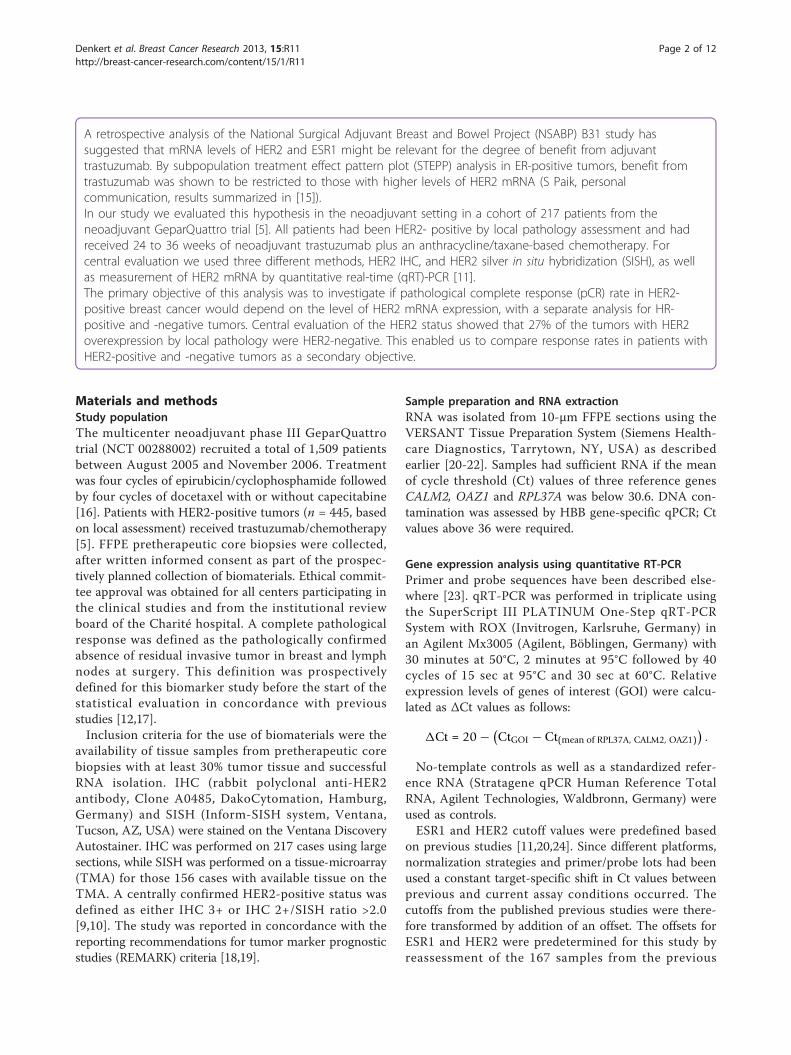
Baseline clinical data, consort statement and centralevaluation of HER2The consort statement and clinical characteristics areshown in Figure 1 and Table 1. A total of 261 sampleswere prospectively collected, of which 33 contained lessthan 30% tumor tissue. RNA isolation was possible in217 cases (95.1% of 228). For 158 tumors (72.8% of 217)the local HER2-positive status was confirmed. Theremaining 59 tumors (27.2%) were centrally HER2-nega-tive (cHER2-negative status) despite a locally positiveHER2 status. Interestingly, the discordance rate was sig-nificantly higher in ESR1-positive tumors (34.5%) thanin ESR1-negative tumors (18.4%, P = 0.009, two-sidedFisher´s test).
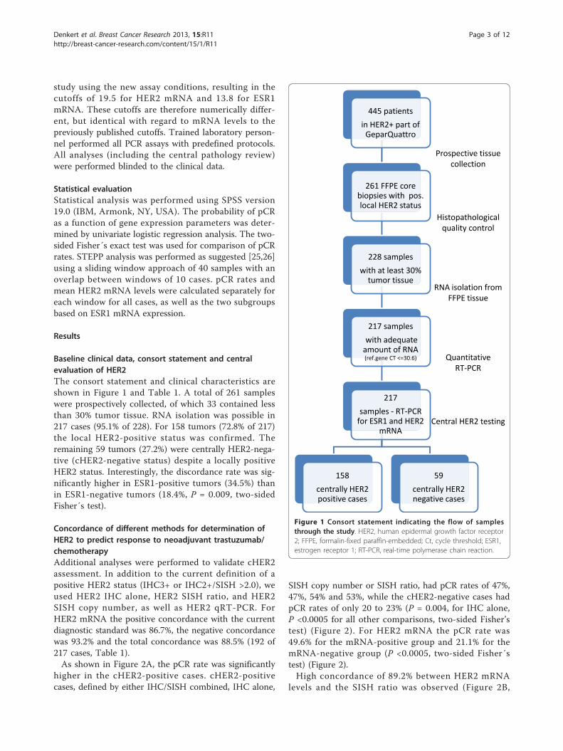
Concordance of different methods for determination ofHER2 to predict response to neoadjuvant trastuzumab/chemotherapyAdditional analyses were performed to validate cHER2assessment. In addition to the current definition of apositive HER2 status (IHC3+ or IHC2+/SISH >2.0), weused HER2 IHC alone, HER2 SISH ratio, and HER2SISH copy number, as well as HER2 qRT-PCR. ForHER2 mRNA the positive concordance with the currentdiagnostic standard was 86.7%, the negative concordancewas 93.2% and the total concordance was 88.5% (192 of217 cases, Table 1).As shown in Figure 2A, the pCR rate was significantly
higher in the cHER2-positive cases. cHER2-positivecases, defined by either IHC/SISH combined, IHC alone,
SISH copy number or SISH ratio, had pCR rates of 47%,47%, 54% and 53%, while the cHER2-negative cases hadpCR rates of only 20 to 23% (P = 0.004, for IHC alone,P <0.0005 for all other comparisons, two-sided Fisher’stest) (Figure 2). For HER2 mRNA the pCR rate was49.6% for the mRNA-positive group and 21.1% for themRNA-negative group (P <0.0005, two-sided Fisher´stest) (Figure 2).High concordance of 89.2% between HER2 mRNA
levels and the SISH ratio was observed (Figure 2B,
445 patients
in HER2+ part of GeparQuattro
261 FFPE core biopsies with pos. local HER2 status
228 samples
with at least 30% tumor tissue
217 samples
with adequate amount of RNA (ref.gene CT <=30.6)
217
samples - RT-PCR for ESR1 and HER2
mRNA
158
centrally HER2 positive cases
59
centrally HER2 negative cases
Prospective tissue collection
Histopathological quality control
RNA isolation from
FFPE tissue
Quantitative RT-PCR
Central HER2 testing
Figure 1 Consort statement indicating the flow of samplesthrough the study. HER2, human epidermal growth factor receptor2; FFPE, formalin-fixed paraffin-embedded; Ct, cycle threshold; ESR1,estrogen receptor 1; RT-PCR, real-time polymerase chain reaction.
Denkert et al. Breast Cancer Research 2013, 15:R11http://breast-cancer-research.com/content/15/1/R11
Page 3 of 12

Table 1 Clinico-pathological data of the study cohort
Characteristic All cases, number (%) Centrally evaluated HER2-positive cases, number (%)
Centrally evaluated HER2-negative cases, number (%)
Samples, number 217 158 59
Local HER2 status (IH/ISH)
positive 217 (100) 158 (100) 59 (100)
negative 0 (0) 0 (0) 0 (0)
Central HER2 status (IH/SISH)
Positive 158 (72.8) 158 (100) 0 (0)
Negative 59 (27.2) 0 (0) 59 (100)
HER2 protein expression (central IH)
3+ 144 (66.4) 144 (91.1) 0 (0)
2+ 20 (9.2) 8 (5.1) 12 (20.3)
0/1+ 48 (22.1) 2 (1.3) 46 (78.0)
missing 5 (2.3) 4 (2.5) 1 (1.7)
HER2 gene amplification (central SISH)
SISH positive (>2.2) 109 (50.2) 109 (69.0) 0 (0)
SISH negative (<1.8) 49 (22.6) 10 (6.3) 39 (66.1)
SISH equivocal (1.8-2.2) 8 (3.7) 6 (3.8) 2 (3.4)
missing 51 (23.5) 33 (20.9) 18 (30.5)
HER2 mRNA expression (central qPCR)
Positive (>19.5) 141 (65.0) 137 (86.7) 4 (6.8)
Negative (≤19.5) 76 (35.0) 21 (13.3) 55 (93.2)
ER/PR Status (local IH)
ER+ and/or PR+ 125 (57.6) 84 (53.2) 41 (69.5)
ER-/PR- 92 (42.4) 74 (46.8) 18 (30.5)
ESR1 mRNA expression (central qPCR)
Positive (>13.8) 109 (50.2) 70 (44.3) 39 (66.1)
Negative (≤13.8) 108 (49.8) 88 (55.7) 20 (33.9)
Tumor grade
G1 to G2 114 (55.1) 78 (49.4) 36 (61.0)
G3 93 (44.9) 72 (45.6) 21 (35.6)
Missing 10 (4.6) 8 (5.1) 2 (3.4)
Clinical tumor stage
cT1 to cT2 155 (71.4) 110 (69.6) 45 (76.3)
cT3 to cT4 62 (28.6) 48 (30.4) 14 (23.7)
Clinical nodal status
cN0 83 (38.2) 61 (38.6) 22 (37.3)
cN+ 134 (61.8) 97 (61.4) 37 (62.7)
Pathological response
No pCR 131 (60.4) 84 (53.2) 47 (79.7)
pCR (ypT0/is ypN0) 86 (39.6) 74 (46.8) 12 (20.3)
Age group
≤ 50 years 109 (50.2) 84 (53.2) 25 (42.4)
> 50 years 108 (49.8) 74 (46.8) 34 (57.6)
Tumor type
Ductal/other 207 (95.4) 156 (98.7) 51 (86.4)
Lobular 10 (4.6) 2 (1.3) 8 (13.6)
HER2, human epidermal growth factor receptor 2; SISH, silver in situ hybridization; IH: immunohistochemistry; ISH: in situ hybridization; qPCR, quantitativepolymerase chain reaction; ypT: tumor stage after neoadjuvant therapy; ypN: nodal status after neoadjuvant therapy.
Denkert et al. Breast Cancer Research 2013, 15:R11http://breast-cancer-research.com/content/15/1/R11
Page 4 of 12

pCR (ypT0/is ypN0)no pCR
HER2 SISH TMA ratio3
1 086420
HE
R2_
RN
A_2
0_d
CT
2 4
2 3
2 2
2 1
2 0
1 9
1 8
1 7
1 6
Figure 2 Comparison of different methods for central assessment of human epidermal growth factor receptor 2 (HER2)overexpression. (A) Five different approaches were used: IHC 3+ or IHC2+/SISH >2.0 (gold standard), HER2 IHC, HER2 SISH ratio, HER2 SISHcopy numbers as well as quantitative RT-PCR for evaluation of HER2 mRNA levels. In all approaches the pCR rate was significantly higher in thecentrally HER2-positive cases. (P = 0.004, for IHC alone, P <0.0005 for all other comparisons, two-sided Fisher’s exact test). (B) Comparison ofquantitative HER2 mRNA levels and SISH ratio; pCR cases are marked as dark spots. A total of 11 cases were negative for HER2 mRNA despite anamplification of HER2 by SISH (lower right corner). These cases showed a pCR rate of only 27.3% (three pCRs out of eleven cases). IHC,immunohistochemistry; SISH, silver in situ hybridization; RT-PCR, real-time polymerase chain reaction; pCR, pathological complete response, ypT:tumor stage after neoadjuvant therapy.
Denkert et al. Breast Cancer Research 2013, 15:R11http://breast-cancer-research.com/content/15/1/R11
Page 5 of 12
positive concordance: 90.4%, negative concordance:86.5%). Those patients who were HER2-negative by bothmethods had a pCR rate of only 20%. A total of 11 caseswere negative for HER2 mRNA despite positive SISH(lower right corner in Figure 2B). Five of those eleventumors were HER2 IHC 2+ and six were HER2 IHC 3+.These cases showed a comparably low pCR rate of only27.3% (three pCRs in eleven cases). Interestingly, thoseseven tumors that were mRNA-positive but SISH-negativehad a pCR rate of 43%. Four of those seven tumors wereHER2 3+ by IHC, while two were HER2 2+ and one wasHER2 0 to 1+.For comparison of HER2 mRNA levels and SISH copy
number, concordance of 91.1% was achieved. For hor-mone receptor (HR) status, 185 of the 217 cases wereconcordant regarding the comparison of ER IHC andESR1 mRNAs, resulting in a concordance of 85.2%.
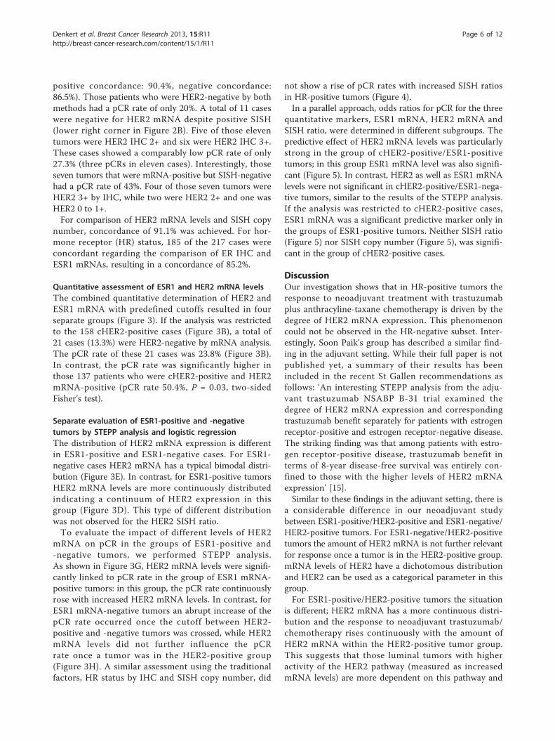
Quantitative assessment of ESR1 and HER2 mRNA levelsThe combined quantitative determination of HER2 andESR1 mRNA with predefined cutoffs resulted in fourseparate groups (Figure 3). If the analysis was restrictedto the 158 cHER2-positive cases (Figure 3B), a total of21 cases (13.3%) were HER2-negative by mRNA analysis.The pCR rate of these 21 cases was 23.8% (Figure 3B).In contrast, the pCR rate was significantly higher inthose 137 patients who were cHER2-positive and HER2mRNA-positive (pCR rate 50.4%, P = 0.03, two-sidedFisher’s test).
Separate evaluation of ESR1-positive and -negativetumors by STEPP analysis and logistic regressionThe distribution of HER2 mRNA expression is differentin ESR1-positive and ESR1-negative cases. For ESR1-negative cases HER2 mRNA has a typical bimodal distri-bution (Figure 3E). In contrast, for ESR1-positive tumorsHER2 mRNA levels are more continuously distributedindicating a continuum of HER2 expression in thisgroup (Figure 3D). This type of different distributionwas not observed for the HER2 SISH ratio.To evaluate the impact of different levels of HER2
mRNA on pCR in the groups of ESR1-positive and-negative tumors, we performed STEPP analysis.As shown in Figure 3G, HER2 mRNA levels were signifi-cantly linked to pCR rate in the group of ESR1 mRNA-positive tumors: in this group, the pCR rate continuouslyrose with increased HER2 mRNA levels. In contrast, forESR1 mRNA-negative tumors an abrupt increase of thepCR rate occurred once the cutoff between HER2-positive and -negative tumors was crossed, while HER2mRNA levels did not further influence the pCRrate once a tumor was in the HER2-positive group(Figure 3H). A similar assessment using the traditionalfactors, HR status by IHC and SISH copy number, did
not show a rise of pCR rates with increased SISH ratiosin HR-positive tumors (Figure 4).In a parallel approach, odds ratios for pCR for the three
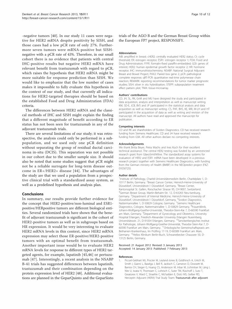
quantitative markers, ESR1 mRNA, HER2 mRNA andSISH ratio, were determined in different subgroups. Thepredictive effect of HER2 mRNA levels was particularlystrong in the group of cHER2-positive/ESR1-positivetumors; in this group ESR1 mRNA level was also signifi-cant (Figure 5). In contrast, HER2 as well as ESR1 mRNAlevels were not significant in cHER2-positive/ESR1-nega-tive tumors, similar to the results of the STEPP analysis.If the analysis was restricted to cHER2-positive cases,ESR1 mRNA was a significant predictive marker only inthe groups of ESR1-positive tumors. Neither SISH ratio(Figure 5) nor SISH copy number (Figure 5), was signifi-cant in the group of cHER2-positive cases.
DiscussionOur investigation shows that in HR-positive tumors theresponse to neoadjuvant treatment with trastuzumabplus anthracyline-taxane chemotherapy is driven by thedegree of HER2 mRNA expression. This phenomenoncould not be observed in the HR-negative subset. Inter-estingly, Soon Paik’s group has described a similar find-ing in the adjuvant setting. While their full paper is notpublished yet, a summary of their results has beenincluded in the recent St Gallen recommendations asfollows: ‘An interesting STEPP analysis from the adju-vant trastuzumab NSABP B-31 trial examined thedegree of HER2 mRNA expression and correspondingtrastuzumab benefit separately for patients with estrogenreceptor-positive and estrogen receptor-negative disease.The striking finding was that among patients with estro-gen receptor-positive disease, trastuzumab benefit interms of 8-year disease-free survival was entirely con-fined to those with the higher levels of HER2 mRNAexpression’ [15].Similar to these findings in the adjuvant setting, there is
a considerable difference in our neoadjuvant studybetween ESR1-positive/HER2-positive and ESR1-negative/HER2-positive tumors. For ESR1-negative/HER2-positivetumors the amount of HER2 mRNA is not further relevantfor response once a tumor is in the HER2-positive group.mRNA levels of HER2 have a dichotomous distributionand HER2 can be used as a categorical parameter in thisgroup.For ESR1-positive/HER2-positive tumors the situation
is different; HER2 mRNA has a more continuous distri-bution and the response to neoadjuvant trastuzumab/chemotherapy rises continuously with the amount ofHER2 mRNA within the HER2-positive tumor group.This suggests that those luminal tumors with higheractivity of the HER2 pathway (measured as increasedmRNA levels) are more dependent on this pathway and
Denkert et al. Breast Cancer Research 2013, 15:R11http://breast-cancer-research.com/content/15/1/R11
Page 6 of 12

pCR (ypT0/is ypN0)no pCR
pCR (ypT0/is ypN0)no pCR
2 01 81 61 41 21 086
2 4
2 3
2 2
2 1
2 0
1 9
1 8
1 7
1 6
2 01 81 61 41 21 08
2 4
2 3
2 2
2 1
2 0
1 9
1 8
1 7
1 6
2 62 4222 01 81 61 4
3 0
2 5
2 0
1 5
1 0
5
0
�MittStd�…
2 62 4222 01 81 61 4
2 0
1 5
1 0
5
0
�MitStdN =
2 62 42 22 01 8161 4
20
15
10
5
0
�MSN
Figure 3 mRNA-based combined quantitative determination of human epidermal growth factor receptor 2 (HER2) and estrogenreceptor 1 (ESR1) mRNA. Cases with a pCR are marked as dark spots. (A) Total of 217 cases. (B) Centrally HER2-positive cases (n = 158). In thisgroup a total of 21 cases (13.3%) were HER2-negative by mRNA analysis; the pCR rate of those 21 cases was only 23.8%. The pCR rate in those137 cases that were HER2-positive by mRNA analysis was 50.4%, (P = 0.03, two-sided Fisher’s exact test). (C, D, E) All 217 cases: differentdistribution patterns of HER2 mRNA with a continuous distribution in ESR1-positive tumors (D) and a more dichotomous distribution in ESR1-negative tumors (E). STEPP analysis for all 217 tumors (F), ESR1-positive tumors (G) and ESR1-negative tumors (H). HER2 mRNA levels were linkedto pCR rate in the group of ESR1-positive tumors with a continuously increased pCR rate. pCR, pathological complete response; STEPP,subpopulation treatment effect pattern plot.
Denkert et al. Breast Cancer Research 2013, 15:R11http://breast-cancer-research.com/content/15/1/R11
Page 7 of 12

thus more responsive to trastuzumab targeted therapy.This finding is supported by the STEPP analysis and weobserved the same effect with the classical approach oflogistic regression, which also showed a significant effectof HER2 mRNA levels (measured as a continuous vari-able) on pCR only in the ESR1-positive/HER2-positivegroup. The traditional method of HER2 SISH ratio orcopy number was not able to provide a similar result bySTEPP or logistic regression analysis, similar to the find-ing in the adjuvant HERA trial [27].The relevance of crosstalk between the ER pathway
and the HER2 pathway has been described in several invitro cell culture and animal models [14,28,29], Ampli-fied in breast (AIB)-1 [30,31] as well as Paired box gene2 (PAX2) [32] have been identified as relevant mediatorsof this crosstalk.The hypothesis derived from those investigations and
our results would be that two important growth factorpathways significantly influence ESR1-positive/HER2-positive tumors and either HER2 or ER may be thedriver of cell proliferation and survival. With sustainedHER2 inhibition ER could function as a key escape orsurvival pathway, which may result in resistance to tras-tuzumab. However when HER2 mRNA expression isvery high the primary driver of proliferation may still bethe HER2 pathway even in the presence of the activatedER pathway. These findings are consistent with two
neoadjuvant trials where significantly lower pCR rateswere observed in ER-positive/HER-positive tumors com-pared to ER-negative/HER2-positive disease [7,8]. How-ever, in a recently reported neoadjuvant trial, responserates to anti-HER2 treatment with lapatinib and trastu-zumab (without chemotherapy) were fairly high (pCR21%) when combined with endocrine treatment if HRswere present [33]. As in the adjuvant setting trastuzu-mab or lapatinib therapy (in contrast to the neoadjuvantapproach) is usually combined with endocrine therapyin the HR-positive group; the combined inhibition ofboth pathways is already used in clinical practice[34,35]. It would be interesting to further evaluate thecontribution of the endocrine therapy to outcome inESR1-positive/HER2-positive tumors.Another finding of our study is the rather high rate of
discordance of 27% between central and local evaluationof HER2. We have validated this finding by the use ofdifferent methods for central pathology. A similar rateof 20% inaccurate HER2 measurements has beenreported before [9] based on results of the NSABP [36]and the N9831 study [37,38]. Discordance has also beenobserved between different reference laboratories, inparticular for borderline cases [39]. Interestingly, in ourstudy the discordance was higher in ESR1-positivetumors, which might be partially due to the more con-tinuous distribution of HER2 mRNA in this group.
Figure 4 Subpopulation treatment effect pattern plot (STEPP) analysis using the classical parameters human epidermal growth factorreceptor 2 (HER2) silver in situ hybridization (SISH) copy number and hormone receptor immunohistochemistry. Shown are analysis forall tumors (A), hormone receptor-positive tumors (B) and hormone receptor negative tumors (C). In contrast to the data shown ins the classicalparameters were not linked to pCR rate. pCR, pathological complete response; IHC, immunohistochemistry; HR, hormone receptor.
Denkert et al. Breast Cancer Research 2013, 15:R11http://breast-cancer-research.com/content/15/1/R11
Page 8 of 12

Pinhel et al. [40] have also observed this different distri-bution of HER2 mRNA in ER-positive vs -negativetumors in a recent report.The high level of false-positive cases gave us the possi-
bility to evaluate the response to trastuzumab in HER2-negative tumors. Centrally HER2-negative tumors had apCR rate of only 20%, which is in the range of the pCRrate of HER2-negative tumors in the GeparTrio trial(17.6%). This validates previous findings that HER2 isthe crucial biomarker for trastuzumab-based therapy.However, it differs from an analysis of the NSABP-B31suggesting a benefit of adjuvant trastuzumab even incHER2-negative tumors [41]. There are two main differ-ences between NSABP-B31 and our study: we used theneoadjuvant setting, which allowed us to directly study
response in the primary tumor, but we could not evalu-ate the effects on micro-metastases as well as the contri-bution of adjuvant endocrine therapy. Furthermore, therate of cHER2-negative cases in NSABP-B31 was only9.7%, compared to 27% in our study. It would be veryinteresting to await the results of the NSABP B47 [42],which is currently evaluating the benefit of trastuzumabin low HER2-expressing tumors.As an additional method we have evaluated HER2
mRNA expression by qRT-PCR. Recently, Dabbs et al.found that HER2 mRNA levels were negative by recur-rence score in 10 (42%) of 24 HER2-positive cases [43].The same finding had already been shown in 638 sam-ples from the NSABP-B31 with an overlap of HER2mRNA expression levels between HER2-positive and
ESR1 mRNA 0.82 (0.74-0.91)
HER2 mRNA 1.33 (1.14-1.55)
HER2 SISH ratio 1.08 (0.96-1.22)
HER2 copy number 1.005 (1.002-1.008)
ESR1 mRNA 0.91 (0.80-1.03)
HER2 mRNA 1.34 (1.04-1.72)
HER2 SISH ratio 0.94 (0.81-1.09)
HER2 copy number 1.002 (1.00-1.006)
ESR1 mRNA 0.52 (0.34-0.79)
HER2 mRNA 2.01 (1.24-3.25)
HER2 SISH ratio 0.93 (0.75-1.14)
HER2 copy number 1.004 (1.00-1.01)
ESR1 mRNA 0.93 (0.64-1.35)
HER2 mRNA 1.09 (0.81-1.48)
HER2 SISH ratio 0.94 (0.76-1.17)
HER2 copy number 1.00 (0.99-1.005)
Figure 5 Odds ratios for pathological complete response (pCR) for the quantitative markers estrogen receptor 1 (ESR1) mRNA (perdCT), human epidermal growth factor receptor 2 (HER2) mRNA (per dCT), silver in situ hybridization (SISH) ratio and SISH copynumber in different subgroups. Results in the three columns (right) represent as odds ratios, 95% CI and P-values. ESR1 mRNA was significantonly in the group of all 217 cases (which also contained the centrally evaluated HER2 (cHER2)-negative cases). If the analysis was restricted tothe centrally evaluated HER2-positive cases, ESR1 mRNA was not significant. HER2 mRNA levels were only significant in the group of ESR1-positive tumors, similar to the subpopulation treatment effect pattern plot (STEPP) analysis. In contrast to the mRNA analysis, HER2 SISH ratio andcopy number were not significant in centrally evaluated HER2-positive cases. ns, not signficant.
Denkert et al. Breast Cancer Research 2013, 15:R11http://breast-cancer-research.com/content/15/1/R11
Page 9 of 12
-negative tumors [40]. In our study 11 cases were nega-tive for HER2 mRNA despite positivity by SISH, andthose cases had a low pCR rate of only 27%. Further-more seven tumors were mRNA-positive but SISH-negative with a pCR rate of 43%. Therefore, in our smallcohort there is no evidence that patients with centralIHC-positive results but negative HER2 mRNA haverelevant benefit from trastuzumab with regard to pCR,which raises the hypothesis that HER2 mRNA might bemore suitable for response prediction than SISH. Wewould like to emphasize that the low number of casesmakes it impossible to fully evaluate this hypothesis inthe context of our study, and that currently all indica-tions for HER2-targeted therapies should be based onthe established Food and Drug Administration (FDA)criteria.The differences between HER2 mRNA and the classi-
cal methods of IHC and SISH might explain the findingthat a different magnitude of benefit according to ERstatus has not been seen for trastuzumab in any of theadjuvant trastuzumab trials.There are several limitations of our study; it was retro-
spective, the analysis could only be performed in a sub-population, and we used only one pCR definitionwithout separating the group of residual ductal carci-noma in-situ (DCIS). This separation was not possiblein our cohort due to the smaller sample size. It shouldalso be noted that some studies suggest that pCR mightnot be a reliable surrogate for long-term disease out-come in ER+/HER2+ disease [44]. The advantages ofthe study are that we used a population from a prospec-tive clinical trial with a standardized assay system, aswell as a predefined hypothesis and analysis plan.
ConclusionsIn summary, our results provide further evidence forthe concept that HER2-positive/non-luminal and ESR1-positive/HERpositive tumors are different biological enti-ties. Several randomized trials have shown that the bene-fit of adjuvant trastuzumab is significant in the cohort ofHER2-positive tumors as well as in subgroups based onHR expression. It would be very interesting to evaluateHER2 mRNA levels in this context, since HER2 mRNAexpression may select those ER-positive/HER2-positivetumors with an optimal benefit from trastuzumab.Another important issue would be to evaluate HER2mRNA levels for response to different types of HER2 tar-geted agents, for example, lapatinib [45,46] or pertuzu-mab [47]. Interestingly, a recent analysis in the NSABPB-41 trials has suggested differences between lapatinib,trastuzumab and their combination depending on theprotein expression level of HER2 [48]. Additional evalua-tions are planned in the GeparQuinto and the GeparSixto
trials of the AGO B and the German Breast Group withinthe European FP7 project, RESPONSIFY.
AbbreviationsAIB: amplified in breast; cHER2: centrally evaluated HER2 status; Ct: cyclethreshold; ER: estrogen receptor; ESR1: estrogen receptor 1; FDA: Food andDrug Administration; FFPE: formalin-fixed paraffin-embedded; GOI: genes ofinterest; HER2: human epidermal growth factor receptor 2; HR: hormonereceptor; IHC: immunohistochemistry; NSABP: National Surgical AdjuvantBreast and Bowel Project; PAX2: Paired box gene 2; pCR: pathologicalcomplete response; qRT-PCR: quantitative real-time polymerase chainreaction; REMARK: reporting recommendations for tumor marker prognosticstudies; SISH: silver in situ hybridization; STEPP: subpopulation treatmenteffect pattern plot; TMA: tissue-microarray.
Authors’ contributionsCD, JH, SL, RK, GvM and MU have designed the study and participated indata acquisition, analysis and interpretation as well as manuscript writing.KM, SD-E, JCB, BVS and JP participated in the statistical analysis and dataacquisition as well as manuscript writing. CS, PAF, BVS, KE, MR, M-LH and HTparticipated in the acquisition of data as well as writing and revision of themanuscript. All authors have read and approved the manuscript forpublication.
Competing interestsCD and RK are shareholders of Sividon Diagnostics. CD has received researchfunding from Siemens Healthcare. CD and JH have received researchfunding from GSK. All other authors declare no competing interests.
AcknowledgementsWe thank Britta Beyer, Petra Wachs and Ines Koch for their excellenttechnical assistance. The central HER2 testing was funded by an unrestrictedresearch grant from GlaxoSmithKline. The standardized assay systems forevaluation of HER2 and ESR1 mRNA have been developed in a previousresearch project together with Siemens Healthcare Diagnostics, with fundingfrom the German ministry of Health (BMBF) in the NEOpredict project (FKZ:01ES0725).
Author details1Institute of Pathology, Charité-Universitätsmedizin Berlin, Charitéplatz 1, D-10117 Berlin, Germany. 2Breast Cancer Center, Heinrich-Heine-University ofDüsseldorf, Universitätsstr.1 Düsseldorf, Germany. 3Breast Center,Kantonsspital St. Gallen, Rorschacher Strasse 95, CH-9007, Switzerland.4German Breast Group, Martin-Behaim-Str. 12, D-63263 Neu-Isenburg,Germany. 5Department of Internal Medicine, Heinrich-Heine-University ofDüsseldorf, Universitätsstr.1 Düsseldorf, Germany. 6Sividon Diagnostics,Nattermannallee 1, D-50829 Cologne, Germany. 7Siemens HealthcareDiagnostics, Cologne, Nattermannallee 1, D-50829 Germany. 8Frauenklinik,Johann-Wolfgang-Goethe-Universität, Theodor-Stern-Kai 7, D-60590 Frankfurtam Main, Germany. 9Department of Gynecology and Obstetrics, UniversityHospital Erlangen, Friedrich-Alexander University Erlangen Nuremberg,Universitätssstr. 21, D-91054 Erlangen, Germany. 10Senckenbergisches Institutfür Pathologie, Johann-Wolfgang-Goethe-Universität, Theodor-Stern-Kai 7, D-60590 Frankfurt am Main, Germany. 11Onkologische Gemeinschaftspraxis amBethanien-Krankenhaus, Im Prüfling 17-19, D-60389 Frankfurt am Main,Germany. 12Helios Klinikum Berlin-Buch, Schwanebecker Chaussee 50, D-13125 Berlin, Germany.
Received: 21 August 2012 Revised: 3 January 2013Accepted: 14 January 2013 Published: 7 February 2013
References1. Piccart-Gebhart MJ, Procter M, Leyland-Jones B, Goldhirsch A, Untch M,
Smith I, Gianni L, Baselga J, Bell R, Jackisch C, Cameron D, Dowsett M,Barrios CH, Steger G, Huang CS, Andersson M, Inbar M, Lichinitser M, Láng I,Nitz U, Iwata H, Thomssen C, Lohrisch C, Suter TM, Rüschoff J, Suto T,Greatorex V, Ward C, Straehle C, McFadden E, Dolci MS, Gelber RD,Herceptin Adjuvant (HERA) Trial Study Team: Trastuzumab after adjuvant
Denkert et al. Breast Cancer Research 2013, 15:R11http://breast-cancer-research.com/content/15/1/R11
Page 10 of 12
chemotherapy in HER2-positive breast cancer. N Engl J Med 2005,353:1659-1672.
2. Slamon D, Eiermann W, Robert N, Pienkowski T, Martin M, Press M,Mackey J, Glaspy J, Chan A, Pawlicki M, Pinter T, Valero V, Liu MC, Sauter G,von Minckwitz G, Visco F, Bee V, Buyse M, Bendahmane B, Tabah-Fisch I,Lindsay MA, Riva A, Crown J, Breast Cancer International Research Group:Adjuvant trastuzumab in HER2-positive breast cancer. N Engl J Med 2011,365:1273-1283.
3. Romond EH, Perez EA, Bryant J, Suman VJ, Geyer CE Jr, Davidson NE, Tan-Chiu E, Martino S, Paik S, Kaufman PA, Swain SM, Pisansky TM,Fehrenbacher L, Kutteh LA, Vogel VG, Visscher DW, Yothers G, Jenkins RB,Brown AM, Dakhil SR, Mamounas EP, Lingle WL, Klein PM, Ingle JN,Wolmark N: Trastuzumab plus adjuvant chemotherapy for operableHER2-positive breast cancer. N Engl J Med 2005, 353:1673-1684.
4. Joensuu H, Bono P, Kataja V, Alanko T, Kokko R, Asola R, Utriainen T,Turpeenniemi-Hujanen T, Jyrkkiö S, Möykkynen K, Helle L, Ingalsuo S,Pajunen M, Huusko M, Salminen T, Auvinen P, Leinonen H, Leinonen M,Isola J, Kellokumpu-Lehtinen PL: Fluorouracil, epirubicin, andcyclophosphamide with either docetaxel orvinorelbine with or withouttrastuzumab as adjuvant treatments of breast cancer: final results of theFinHer Trial. J Clin Oncol 2009, 27:5685-5692.
5. Untch M, Rezai M, Loibl S, Fasching PA, Huober J, Tesch H, Bauerfeind I,Hilfrich J, Eidtmann H, Gerber B, Hanusch C, Kühn T, du Bois A, Blohmer JU,Thomssen C, Dan Costa S, Jackisch C, Kaufmann M, Mehta K, vonMinckwitz G: Neoadjuvant treatment with trastuzumab in HER2-positivebreast cancer: results from the GeparQuattro study. J Clin Oncol 2010,28:2024-2031.
6. Untch M, Fasching PA, Konecny GE, Hasmüller S, Lebeau A, Kreienberg R,Camara O, Müller V, du Bois A, Kühn T, Stickeler E, Harbeck N, Höss C,Kahlert S, Beck T, Fett W, Mehta KM, von Minckwitz G, Loibl S: Pathologiccomplete response after neoadjuvant chemotherapy plus trastuzumabpredicts favorable survival in human epidermal growth factor receptor2-overexpressing breast cancer: results from the TECHNO trial of theAGO and GBG study groups. J Clin Oncol 2011, 29:3351-3357.
7. Gianni L, Pienkowski T, Im YH, Roman L, Tseng LM, Liu MC, Lluch A,Staroslawska E, de la Haba-Rodriguez J, Im SA, Pedrini JL, Poirier B,Morandi P, Semiglazov V, Srimuninnimit V, Bianchi G, Szado T, Ratnayake J,Ross G, Valagussa P: Efficacy and safety of neoadjuvant pertuzumab andtrastuzumab in women with locally advanced inflammatory, or earlyHER2-positive breast cancer (NeoSphere): a randomised multicentreopen-label, phase 2 trial. Lancet Oncol 2012, 13:25-32.
8. Baselga J, Bradbury I, Eidtmann H, Di Cosimo S, de Azambuja E, Aura C,Gómez H, Dinh P, Fauria K, Van Dooren V, Aktan G, Goldhirsch A,Chang TW, Horváth Z, Coccia-Portugal M, Domont J, Tseng LM, Kunz G,Sohn JH, Semiglazov V, Lerzo G, Palacova M, Probachai V, Pusztai L,Untch M, Gelber RD, Piccart-Gebhart M, NeoALTTO Study Team: Lapatinibwith trastuzumab for HER2-positive early breast cancer (NeoALTTO):arandomised open-label, multicentre, phase 3 trial. Lancet 2012,379:633-640.
9. Wolff AC, Hammond ME, Schwartz JN, Hagerty KL, Allred DC, Cote RJ,Dowsett M, Fitzgibbons PL, Hanna WM, Langer A, McShane LM, Paik S,Pegram MD, Perez EA, Press MF, Rhodes A, Sturgeon C, Taube SE, Tubbs R,Vance GH, van de Vijver M, Wheeler TM, Hayes DF, American Society ofClinical Oncology/College of American Pathologists: American Society ofClinical Oncology/College of American Pathologists guidelinerecommendations for human epidermal growth factor receptor 2testing in breast cancer. Arch Pathol Lab Med 2007, 131:18-43.
10. Hammond ME, Hayes DF, Wolff AC: Clinical Notice for American Societyof Clinical Oncology-College of American Pathologists guidelinerecommendations on ER/PgR and HER2 testing in breast cancer. J ClinOncol 2011, 29:e458.
11. Müller BM, Kronenwett R, Hennig G, Euting H, Weber K, Bohmann K,Weichert W, Altmann G, Roth C, Winzer KJ, Kristiansen G, Petry C, Dietel M,Denkert C: Quantitative determination of estrogen receptorprogesterone receptor and HER2 mRNA in formalin-fixed paraffin-embedded tissue-a new option for predictive biomarker assessment inbreast cancer. Diagn Mol Pathol 2011, 20:1-10.
12. Noske A, Loibl S, Darb-Esfahani S, Roller M, Kronenwett R, Müller BM,Steffen J, von Toerne C, Wirtz R, Baumann I, Hoffmann G, Heinrich G,Grasshoff ST, Ulmer HU, Denkert C, von Minckwitz G: Comparison ofdifferent approaches for assessmentof HER2 expression on protein and
mRNA level: prediction of chemotherapy response in the neoadjuvantGeparTrio trial (NCT00544765). Breast Cancer Res Treat 2011, 126:109-117.
13. Baehner FL, Achacoso N, Maddala T, Shak S, Quesenberry CP Jr,Goldstein LC, Gown AM, Habel LA: Human epidermal growth factorreceptor 2 assessment in a case-control study: comparison offluorescence in situ hybridization and quantitative reverse transcriptionpolymerase chain reaction performed by central laboratories. J ClinOncol 2010, 28:4300-4306.
14. Konecny G, Pauletti G, Pegram M, Untch M, Dandekar S, Aguilar Z,Wilson C, Rong HM, Bauerfeind I, Felber M, Wang HJ, Beryt M, Seshadri R,Hepp H, Slamon DJ: Quantitative association between HER-2/neu andsteroid hormone receptors in hormone receptor-positive primary breastcancer. J Natl Cancer Inst 2003, 95:142-153.
15. Goldhirsch A, Wood WC, Coates AS, Gelber RD, Thürlimann B, Senn HJ,Panel members: Strategies for subtypes-dealing with the diversity ofbreast cancer: highlights of the St. Gallen International ExpertConsensus on the Primary Therapy of Early Breast Cancer 2011. AnnOncol 2011, 22:1736-1747.
16. von Minckwitz G, Rezai M, Loibl S, Fasching PA, Huober J, Tesch H,Bauerfeind I, Hilfrich J, Eidtmann H, Gerber B, Hanusch C, Kühn T, du Bois A,Blohmer JU, Thomssen C, Dan Costa S, Jackisch C, Kaufmann M, Mehta K,Untch M: Capecitabine in addition to anthracycline- and taxane-basedneoadjuvant treatment in patients with primary breast cancer: phase IIIGeparQuattro study. J Clin Oncol 2010, 28:2015-2023.
17. von Minckwitz G, Darb-Esfahani S, Loibl S, Huober J, Tesch H, Solbach C,Holms F, Eidtmann H, Dietrich K, Just M, Clemens MR, Hanusch C,Schrader I, Henschen S, Hoffmann G, Tiemann K, Diebold K, Untch M,Denkert C: Responsiveness of adjacent ductal carcinoma in situ andchanges in HER2 status after neoadjuvant chemotherapy/trastuzumabtreatment in early breast cancer-results from the GeparQuattro study(GBG 40). Breast Cancer Res Treat 2012, 132:863-870.
18. McShane LM, Altman DG, Sauerbrei W, Taube SE, Gion M, Clark GM,Statistics Subcommittee of the NCI-EORTC Working Group on CancerDiagnostics: Reporting recommendations for tumor marker prognosticstudies. J Clin Oncol 2005, 23:9067-9072.
19. Altman DG, McShane LM, Sauerbrei W, Taube SE: ReportingRecommendations for Tumor Marker Prognostic Studies (REMARK):Explanation and Elaboration. PLoS Med 2012, 9:e1001216.
20. Bohmann K, Hennig G, Rogel U, Poremba C, Mueller BM, Fritz P, Stoerkel S,Schaefer KL: RNA extraction from archival formalin-fixed paraffin-embedded tissue: a comparison of manual semiautomated, and fullyautomated purification methods. Clin Chem 2009, 55:1719-1727.
21. Denkert C, Loibl S, Noske A, Roller M, Müller BM, Komor M, Budczies J,Darb-Esfahani S, Kronenwett R, Hanusch C, von Törne C, Weichert W,Engels K, Solbach C, Schrader I, Dietel M, von Minckwitz G: Tumor-associated lymphocytes as an independent predictor of response toneoadjuvant chemotherapy in breast cancer. J Clin Oncol 2010,28:1146-1452.
22. Hennig G, Gehrmann M, Stropp U, Brauch H, Fritz P, Eichelbaum M,Schwab M, Schroth W: Automated extraction of DNA and RNA from asingle formalin-fixed paraffin-embedded tissue section for analysis ofboth single-nucleotide polymorphisms and mRNA expression. Clin Chem2010, 56:1845-1853.
23. Filipits M, Rudas M, Jakesz R, Dubsky P, Fitzal F, Singer CF, Dietze O, Greil R,Jelen A, Sevelda P, Freibauer C, Müller V, Jänicke F, Schmidt M, Kölbl H,Rody A, Kaufmann M, Schroth W, Brauch H, Schwab M, Fritz P, Weber KE,Feder IS, Hennig G, Kronenwett R, Gehrmann M, Gnant M, EP Investigators:A new molecular predictor of distant recurrence in ER-positive, HER2-negative breast cancer adds independent information to conventionalclinical risk factors. Clin Cancer Res 2011, 17:6012-6020.
24. Pentheroudakis G, Kalogeras KT, Wirtz RM, Grimani I, Zografos G, Gogas H,Stropp U, Pectasides D, Skarlos D, Hennig G, Samantas E, Bafaloukos D,Papakostas P, Kalofonos HP, Pavlidis N, Fountzilas G: Gene expression ofestrogen receptor progesterone receptor and microtubule-associatedprotein Tau in high-risk early breast cancer: a quest for molecularpredictors of treatment benefit in the context of a Hellenic CooperativeOncology Group trial. Breast Cancer Res Treat 2009, 116:131-143.
25. Lazar AA, Cole BF, Bonetti M, Gelber RD: Evaluation of treatment-effectheterogeneity using biomarkers measured on a continuous scale:subpopulation treatment effect pattern plot. J Clin Oncol 2010,28:4539-4544.
Denkert et al. Breast Cancer Research 2013, 15:R11http://breast-cancer-research.com/content/15/1/R11
Page 11 of 12

26. Bonetti M, Gelber RD: Patterns of treatment effects in subsets of patientsin clinical trials. Biostatistics 2004, 5:465-481.
27. Dowsett M, Procter M, McCaskill-Stevens W, de Azambuja E, Dafni U,Rueschoff J, Jordan B, Dolci S, Abramovitz M, Stoss O, Viale G, Gelber RD,Piccart-Gebhart M, Leyland-Jones B: Disease-free survival according todegree of HER2 amplification for patients treated with adjuvantchemotherapy with or without 1 year of trastuzumab: the HERA Trial. JClin Oncol 2009, 27:2962-2069.
28. Zhao W, Zhang Q, Kang X, Jin S, Lou C: AIB1 is required for theacquisition of epithelial growth factor receptor-mediated tamoxifenresistance in breast cancer cells. Biochem Biophys Res Commun 2009,380:699-704.
29. Osborne CK, Shou J, Massarweh S, Schiff R: Crosstalk between estrogenreceptor and growth factor receptor pathways as a cause for endocrinetherapy resistance in breast cancer. Clin Cancer Res 2005, 11:865s-870s.
30. Lahusen T, Fereshteh M, Oh A, Wellstein A, Riegel AT: Epidermal growthfactor receptor tyrosine phosphorylation and signaling controlled by anuclear receptor coactivator amplified in breast cancer 1. Cancer Res2007, 67:7256-7265.
31. Fereshteh MP, Tilli MT, Kim SE, Xu J, O’Malley BW, Wellstein A, Furth PA,Riegel AT: The nuclear receptor coactivator amplified in breast cancer-1is required for Neu (ErbB2/HER2) activation, signaling, and mammarytumorigenesis in mice. Cancer Res 2008, 68:3697-3706.
32. Hurtado A, Holmes KA, Geistlinger TR, Hutcheson IR, Nicholson RI, Brown M,Jiang J, Howat WJ, Ali S, Carroll JS: Regulation of ERBB2 by oestrogenreceptor-PAX2 determines response to tamoxifen. Nature 2008,456:663-666.
33. Chang JCN, Mayer IA, Forero-Torres A, Nanda R, Goetz MP, Rodriguez AA,Pavlick AC, Wang T, Hilsenbeck SG, Gutierrez C, Schiff R, Osborne CK,Rimawi MF and on behalf of the Translational Breast Cancer ResearchConsortium: TBCRC 006 A multicenter phase II study of neoadjuvantlapatinib and trastuzumab in patients with HER2-overexpressing breastcancer. J Clin Oncol Volume 29 2011 ASCO Annual Meeting Proceedings June3-7, 2011, Chicago 2011, , Suppl: 505.
34. Untch M, Gelber RD, Jackisch C, Procter M, Baselga J, Bell R, Cameron D,Bari M, Smith I, Leyland-Jones B, de Azambuja E, Wermuth P, Khasanov R,Feng-Yi F, Constantin C, Mayordomo JI, Su CH, Yu SY, Lluch A, Senkus-Konefka E, Price C, Haslbauer F, Suarez Sahui T, Srimuninnimit V, Colleoni M,Coates AS, Piccart-Gebhart MJ, Goldhirsch A, HERA Study Team: Estimatingthe magnitude of trastuzumab effects within patient subgroups in theHERA trial. Ann Oncol 2008, 19:1090-1096.
35. Johnston S, Pippen J Jr, Pivot X, Lichinitser M, Sadeghi S, Dieras V,Gomez HL, Romieu G, Manikhas A, Kennedy MJ, Press MF, Maltzman J,Florance A, O’Rourke L, Oliva C, Stein S, Pegram M: Lapatinib combinedwith letrozole versus letrozole and placebo as first-line therapy forpostmenopausal hormone receptor-positive metastatic breast cancer. JClin Oncol 2009, 27:5538-5546.
36. Paik S, Bryant J, Tan-Chiu E, Romond E, Hiller W, Park K, Brown A, Yothers G,Anderson S, Smith R, Wickerham DL, Wolmark N: Real-world performanceof HER2 testing-National Surgical Adjuvant Breast and Bowel Projectexperience. J Natl Cancer Inst 2002, 94:852-854.
37. Roche PC, Suman VJ, Jenkins RB, Davidson NE, Martino S, Kaufman PA,Addo FK, Murphy B, Ingle JN, Perez EA: Concordance between local andcentral laboratory HER2 testing in the breast intergroup trial N9831. JNatl Cancer Inst 2002, 94:855-857.
38. Perez EA, Suman VJ, Davidson NE, Martino S, Kaufman PA, Lingle WL,Flynn PJ, Ingle JN, Visscher D, Jenkins RB: HER2 testing by local central,and reference laboratories in specimens from the North Central CancerTreatment Group N9831 intergroup adjuvant trial. J Clin Oncol 2006,24:3032-3038.
39. Dowsett M, Hanna WM, Kockx M, Penault-Llorca F, Rüschoff J, Gutjahr T,Habben K, van de Vijver MJ: Standardization of HER2 testing: results of aninternational proficiency-testing ring study. Mod Pathol 2007, 20:584-591.
40. Pinhel I, Hills M, Drury S, Salter J, Sumo G, A’hern R, Bliss JM, Sestak I,Cuzick J, Barrett-Lee P, Harris A, Dowsett M, the NCRI Adjuvant BreastCancer Trial Management Group: ER and HER2 expression are positivelycorrelated in HER2 non-overexpressing breast cancer. Breast Cancer Res2012, 14:R46.
41. Paik S, Kim C, Wolmark N: HER2 status and benefit from adjuvanttrastuzumab in breast cancer. N Engl J Med 2008, 358:1409-1411.
42. ClinicalTrials.gov, identifier NCT01275677. [http://clinicaltrials.gov/ct2/show/NCT01275677?term=NCT01275677&rank=1].
43. Dabbs DJ, Klein ME, Mohsin SK, Tubbs RR, Shuai Y, Bhargava R: High false-negative rate of HER2 quantitative reverse transcription polymerasechain reaction of the Oncotype DX test: an independent qualityassurance study. J Clin Oncol 2011, 29:4279-4285.
44. von Minckwitz G, Untch M, Blohmer JU, Costa SD, Eidtmann H,Fasching PA, Gerber B, Eiermann W, Hilfrich J, Huober J, Jackisch C,Kaufmann M, Konecny GE, Denkert C, Nekljudova V, Mehta K, Loibl S:Definition and impact of pathologic complete response on prognosisafter neoadjuvant chemotherapy in various intrinsic breast cancersubtypes. J Clin Oncol 2012, 30:1796-1804.
45. Untch M, Loibl S, Bischoff J, Eidtmann H, Kaufmann M, Blohmer JU,Hilfrich J, Strumberg D, Fasching PA, Kreienberg R, Tesch H, Hanusch C,Gerber B, Rezai M, Jackisch C, Huober J, Kühn T, Nekljudova V, vonMinckwitz G, German Breast Group (GBG); ArbeitsgemeinschaftGynäkologische Onkologie-Breast (AGO-B) Study Group: Lapatinib versustrastuzumab in combination with neoadjuvant anthracycline-taxane-based chemotherapy (GeparQuinto, GBG 44): a randomised phase 3trial. Lancet Oncol 2012, 13:135-144.
46. Baselga J, Bradbury I, Eidtmann H, Di Cosimo S, de Azambuja E, Aura C,Gómez H, Dinh P, Fauria K, Van Dooren V, Aktan G, Goldhirsch A,Chang TW, Horváth Z, Coccia-Portugal M, Domont J, Tseng LM, Kunz G,Sohn JH, Semiglazov V, Lerzo G, Palacova M, Probachai V, Pusztai L,Untch M, Gelber RD, Piccart-Gebhart M, NeoALTTO Study Team: Lapatinibwith trastuzumab for HER2-positive early breast cancer (NeoALTTO): arandomised open-label, multicentre, phase 3 trial. Lancet 2012,379:633-640.
47. Gianni L, Pienkowski T, Im YH, Roman L, Tseng LM, Liu MC, Lluch A,Staroslawska E, de la Haba-Rodriguez J, Im SA, Pedrini JL, Poirier B,Morandi P, Semiglazov V, Srimuninnimit V, Bianchi G, Szado T, Ratnayake J,Ross G, Valagussa P: Efficacy and safety of neoadjuvant pertuzumab andtrastuzumab in women with locally advanced inflammatory, or earlyHER2-positive breast cancer (NeoSphere): a randomised multicentreopen-label, phase 2 trial. Lancet Oncol 2012, 13:25-32.
48. Robidoux Andre, Tang Gong, Rastogi Priya, Geyer ECharles, Azar ACatherine,Atkins James Norman, Fehrenbacher Louis, Bear Harry Douglas, Baez-DiazJ Luis, Kuebler Phillip, Margolese GRichard, Farrar William Blair,Brufsky Adam, Shibata RHenry, Bandos Hanna, Paik Soonmyung,Costantino PJoseph, Swain MSandra, Mamounas PEleftherios, NormanWolmark: Evaluation of lapatinib as a component of neoadjuvanttherapy for HER2+ operable breast cancer: NSABP protocol B-41. J ClinOncol 2012, 30(suppl), abstr LBA506.
doi:10.1186/bcr3384Cite this article as: Denkert et al.: HER2 and ESR1 mRNA expressionlevels and response to neoadjuvant trastuzumab plus chemotherapy inpatients with primary breast cancer. Breast Cancer Research 2013 15:R11.
Submit your next manuscript to BioMed Centraland take full advantage of:
• Convenient online submission
• Thorough peer review
• No space constraints or color figure charges
• Immediate publication on acceptance
• Inclusion in PubMed, CAS, Scopus and Google Scholar
• Research which is freely available for redistribution
Submit your manuscript at www.biomedcentral.com/submit
Denkert et al. Breast Cancer Research 2013, 15:R11http://breast-cancer-research.com/content/15/1/R11
Page 12 of 12
Related Documents


