IHD stone Fitriardi Sejati, dr. Pembimbing: Tjahyo Kelono Utomo, dr. Sp.B(K)BD

HEPATOLITIASIS
Dec 06, 2015
referat hepatolitiasis
residen bedah unpad stase rsud ulin banjarmasin
residen bedah unpad stase rsud ulin banjarmasin
Welcome message from author
This document is posted to help you gain knowledge. Please leave a comment to let me know what you think about it! Share it to your friends and learn new things together.
Transcript

IHD stone
Fitriardi Sejati, dr.
Pembimbing:Tjahyo Kelono Utomo, dr. Sp.B(K)BD
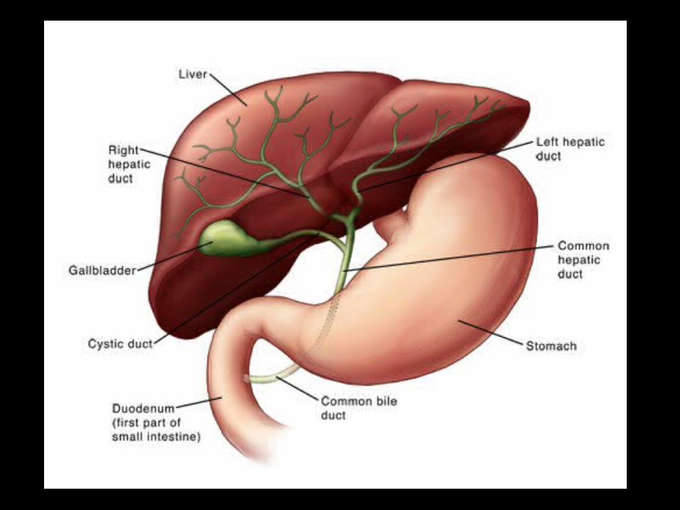

INTRAHEPATIC GALL STONES

Definition
• Any stone that proximal to confluence of right and left intrahepatic duct

Incidence
• Rare disease in Western• High prevalence in East Asia
Prakash K et al. Multidisciplinary approach in the long-term management of intrahepatic stones: Indian experience. Indian journal of gastroenterology : official journal of the Indian Society of Gastroenterology. 2004;23(6):209-13.

Classification
• By location– Right side (R)– Left side (L) Most common– Left and Right sides (LR)– Caudate lobe (C)
Blumgart's Surgery of the Liver, Pancreas and Biliary Tract (5th Edition)

INTRAHEPATIC VS EXTRAHEPATIC CALCULUS•Caroli's disease is characterized by congenital segmental dilation of the intrahepatic bile ducts producing primary intrahepatic gallstones.•Secondary intrahepatic gall stone formation occurs due to chronic obstruction of CBD and CHD
•It's believed that most patients suffering a chronic illness have excessive amounts of gallstones in the liver.• Gallstones found in gallbladder tend to be hardened and relatively large while stones found in liver tends to be soft and noncalcified•Intrahepatic Gallstones cause liver congestion and elevted liver enzymes
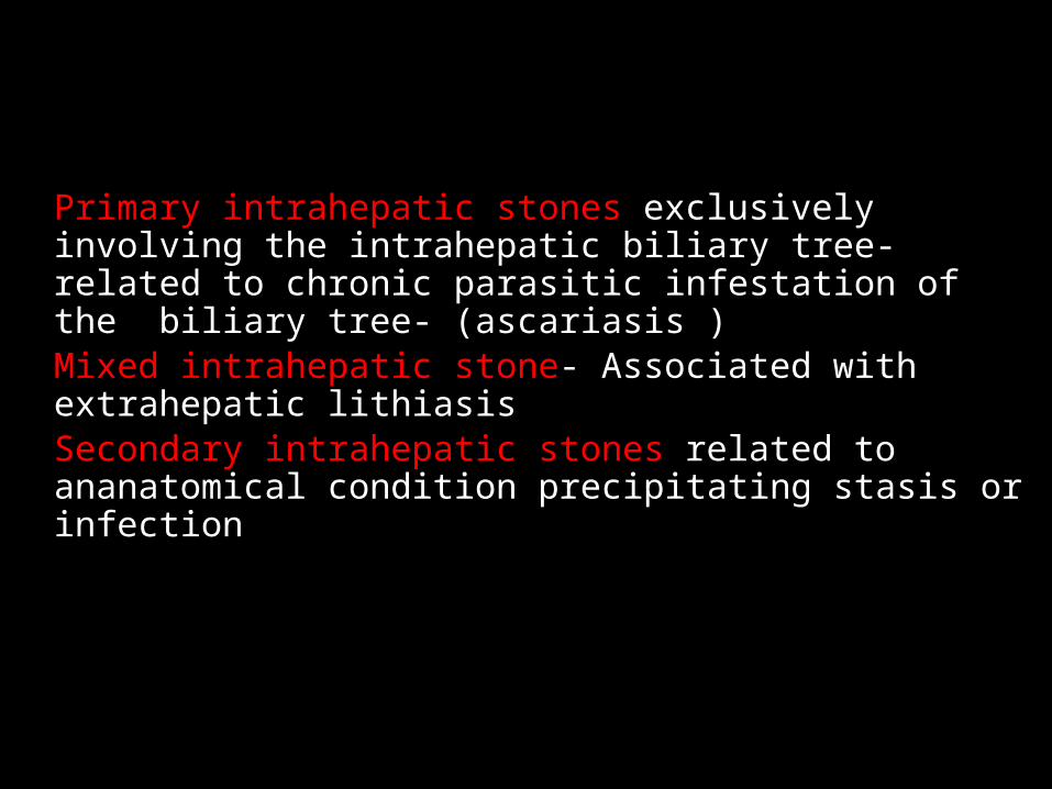
Primary intrahepatic stones exclusively involving the intrahepatic biliary tree-related to chronic parasitic infestation of the biliary tree- (ascariasis )Mixed intrahepatic stone- Associated with extrahepatic lithiasisSecondary intrahepatic stones related to ananatomical condition precipitating stasis or infection
TYPES OF INTRAHEPATIC CALCULUS

EXTRAHEPATIC CALCULUS
CALCULI IN THE DISTAL CBD

Type of stone
• Calcium bilirubinate stone (bilirubin + Chol + Fatty acid + Ca) “brown pigmented stone”
• Cholesterol stone

Gallstones


Risk Factor
• Biliary stasis eg. Stricture of biliary anastomosis
• Bacterial infection
• 3-18% progress to intrahepatic cholangiocarcinoma
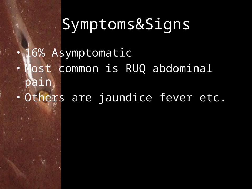
Symptoms&Signs
• 16% Asymptomatic• Most common is RUQ abdominal pain• Others are jaundice fever etc.

Imaging
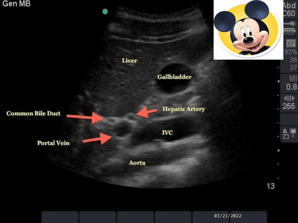
• USG : Hyperechoic lesion with posterior acoustic shadows or duct dilatation
• CT : Hyperdensity lesion at plane CT, evaluate degree of affected lobe atrophy, other complication from stone eg. Abscess, CA, PortalHT
• MRCP : Location of stones

"Mickey Mouse" view of Portal Triad
04/18/2023
04/18/2023
CT character suspected CA
• 1) presence of periductal soft-tissue density• 2) higher enhancement of the duct than
adjacent bile duct on portal venous phase images
• 3) ductal thickening• 4) portal vein obliteration• 5) lymph node enlargement
Treatment
• Main purposes– Complete removal of the stones – Preventing from further attacks of cholangitis– Controlling disease progression to biliary cirrhosis

Treatment
• Asymptomatic: intervention >> controversy• Acute obstructive suppurative cholangitis>> Emergency PTBD• Difficult bilateral intrahepatic stone – 1.PTBD to left hepatic duct– 2.Dilated tract step by step from 6-Fr to 16-Fr catheter– 3.CT post PTBD 2 weeks evaluate pathological change
of liver (atrophy or stricture >> Sx)– 4.Cholangioscopic lithotripsy in preserved sectors

Treatment

Article Informations
• Study design– Retrospective case control study
• Study group– 1 Jan 1971 – 31 Dec 2000 all patients with
cholelithiasis– 89 patients hepatolithiasis

Article Informations
• Classification • 1.Relative to liver– I type: intrahepatic – E type: extrahepatic– IE type: intrahepatic + extrahepatic
• 2.Location in liver– L type: left lobe– R type: Right lobe– LR type: Both lobes
Article Informations
• Treatment– Hepatectomy– Cholangioenterostomy• Choledochojejunostomy• Choledochoduodenostomy
– T-tube insertion– Percutanous transhepatic cholangioscopic
lithotripsy (PTCSL)

Article Informations
• Evaluate post procedure residual stone– Cholangiography– Cholangioscopy

Results

Results
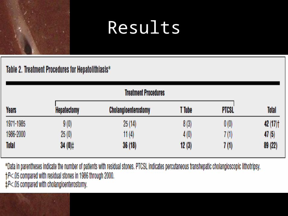
Results

Results
• 18 Lateral segmentectomies
(L type)• 13 Left lobectomy– 10 from L type– 3 from LR type
• 1 extended left lobectomy (L type)
• 2 Right lobectomy (R type)

Results

Discussion
• 4.2-9.5% recurrence stone post hepatectomy• 36.4% recurrence stone post PTCSL• Complete stone removal, but bile duct
stricture remained unchange• Ho-YAG laser lithotripsy high success rate(3
cases)• ESWL in cholesterol stone(bewared
suppurative cholangitis)
Summary
• Indication for hepatectomy– Unilateral lobe– Containing stone bile duct markedly constrict or
dilated– Combination with suspected intrahepatic bile duct
carcinoma– Complications: abscess, atrophy
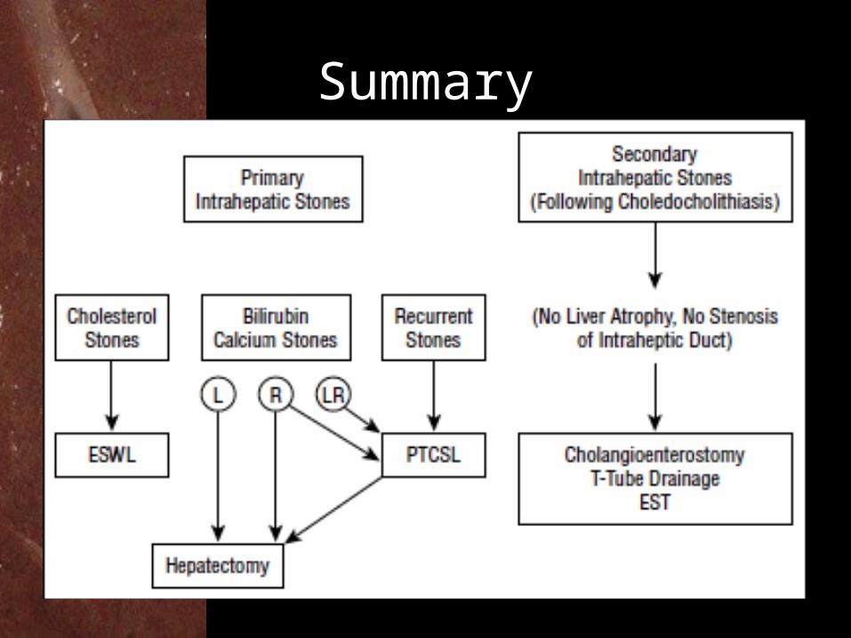
Summary

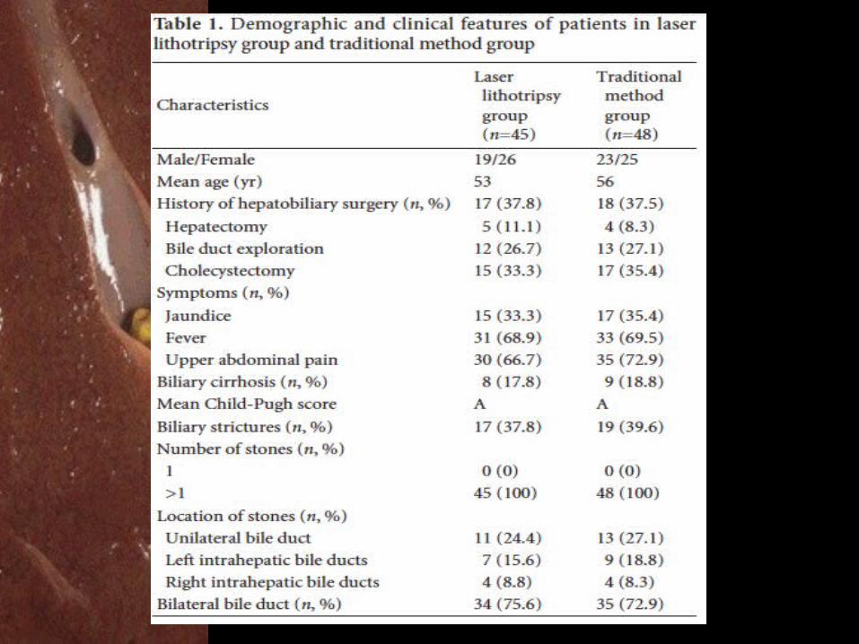
Laser Lithotripsy

Article Informations
• Study design– Retrospective review study
• Study group– July 2009 – Oct 2012 all patients with
cholelithiasis– 45 patients hepatolithiasis who had undergone
choledochoscopic FREDDY laser lithotripsy combined with or without hepatectomy in Hepatobiliary and Pancreatic Surgery Center
Methods
• 48 patients got conventional methods• 45 patients got CBD exploration who received
choledochoscopic FREDDY laser lithotripsy therapy.
• Laser pulses of 1.2 μs were applied at a repetition rate of 10-15 Hz (wavelengths of 532 nm and 1064 nm as a double pulse was applied with pulse energy of 120 mJ)

Methods
• 12 patients received hepatectomy • Indication hepatectomy – Severely narrowed intrahepatic bile duct is not
successful insert the scope– Atrophy of liver lobe– Clinically suspected cholangiocarcinoma

Methods
• Routinely T-tube placed in CBD post op • Removed 2 Mth post op when no stone left(T-tube cholangiography)• Residual stone: detected IHD stone in 3 mth
post op

Statistic Analysis
• Chi-square test and Student's t test, using SPSS software for Windows (Statistical Product and Service Solutions, version 18.0, SPSS Inc., Chicago, IL., USA).
Results
• 45 patients in laser group– 42 complete stone fragmentation with IHD
clearance (93.3%)– 3 failed (impacted stone in both lobes, bile duct
stricture and biliary cirrhosis)– 5 segmental hepatectomy of the right liver– 7 left lateral hepatectomy– 1 cholangiocarcinoma with negative margins,
Results

Results
• 48 patients in conventional group– 41 complete stone fragmentation with IHD
clearance (85.4%)– 6 segmental hepatectomy of the right liver– 12 left lateral hepatectomy– 1 cholangiocarcinoma with negative margins,

Complications
• 45 patients in laser group– 2 Hemobilia– 3 Acute cholangitis
• 48 patients in conventional group– 3 Intraop hemorrhage– 1 Leakage (placed drain)– 6 Acute cholangitis– 1 Died (liver failure from cirrhosis)

Results

Discussion
• High rate of treatment failure & recurrence in IHD stone
• Removed all stone, correct stricture to promote adequate drainage
• The percutaneous approach of Nd:YAG laser lithotripsy is difficult to resolve hepatolithiasis completely when it occurs in both liver lobes
Discussion
• Bilateral intrahepatic stones + absence of intrahepatic bile duct stricture + absence liver atrophy >> choledochoscopic FREDDY laser lithotripsy.
• In cases with stricture and atrophy >> still need Ix

Prognosis
• 6.6% High overall recurrence rate– 5.3% Hepatectomy– 8.3% Choledochojejunostomy– 6.4% Choledochotomy followed by
cholangioscopic lithotomy– 9.6% PTCS
Blumgart's Surgery of the Liver, Pancreas and Biliary Tract (5th Edition)

Thank YouFor
Your good attentions
Related Documents