HEPATITIS C CASE STUDY FROM MX A patient was treated in Mexico using a sublingual UV device and returned to Texas for testing. Here are the results. D R A F T D O C U M E N T NOT FOR DISTRIBUTION PENDING FINAL LABORATORY RESULTS -MARCH 2009 SUMMARY & CLINICAL TESTING CASE REPORT January 2, 2009 Case Report: Caucasian, female, 57yo (DOB 12-12-1951), smoker, opiate addition controlled by methadone therapy for past 18 years. Diagnosed Hepatitis C viral (HCV) (6-29-2004) and began treatment with alternative UV light therapy (9-2007). Background: HCV infection is the most common cause of chronic liver disease in North America. Approximately 2% of adults in the United States have been exposed to the virus, and of them, 75-85% are chronically infected. The CDC recommends HCV testing in the following cases: • injected illegal drugs • blood transfusion or organ transplant before July 1992 • clotting factor concentrates produced before 1987 • long-term dialysis • children born to HCV-positive women • health care, emergency medicine, and public safety workers after needle sticks, sharps, or mucosal exposure to HCV-positive blood • evidence of chronic liver disease Since 1990, the blood supply has been monitored in the U.S., and any unit of blood that tests positive for HCV is rejected. The current risk of HCV infection from transfused blood is about 1 case per two million transfused units. The anti-HCV test determines antibodies to HCV. If positive, the Qualitative HCV-RNA can determine if the infection is present. HCV viral load and HCV genotyping tests help to determine a treatment plan; the viral load and qualitative HCV RNA are used to monitor treatment response.

Welcome message from author
This document is posted to help you gain knowledge. Please leave a comment to let me know what you think about it! Share it to your friends and learn new things together.
Transcript

HEPATITIS C CASE STUDY FROM MX A patient was treated in Mexico using a sublingual UV device and returned to Texas for testing. Here are the results.
D R A F T D O C U M E N T NOT FOR DISTRIBUTION
PENDING FINAL LABORATORY RESULTS -MARCH 2009 SUMMARY & CLINICAL TESTING CASE REPORT
January 2, 2009
Case Report: Caucasian, female, 57yo (DOB 12-12-1951), smoker, opiate addition controlled by methadone therapy for past 18 years. Diagnosed Hepatitis C viral (HCV) (6-29-2004) and began treatment with alternative UV light therapy (9-2007).
Background: HCV infection is the most common cause of chronic liver disease in North America. Approximately 2% of adults in the United States have been exposed to the virus, and of them, 75-85% are chronically infected. The CDC recommends HCV testing in the following cases:
• injected illegal drugs • blood transfusion or organ transplant before July 1992 • clotting factor concentrates produced before 1987 • long-term dialysis • children born to HCV-positive women • health care, emergency medicine, and public safety workers after needle sticks, sharps, or
mucosal exposure to HCV-positive blood • evidence of chronic liver disease
Since 1990, the blood supply has been monitored in the U.S., and any unit of blood that tests positive for HCV is rejected. The current risk of HCV infection from transfused blood is about 1 case per two million transfused units.
The anti-HCV test determines antibodies to HCV. If positive, the Qualitative HCV-RNA can determine if the infection is present. HCV viral load and HCV genotyping tests help to determine a treatment plan; the viral load and qualitative HCV RNA are used to monitor treatment response.

Assessments: Qualitative HCV RNA test identify if the virus is in the blood and thus, active infection: “positive” or “detected” if any HCV viral RNA or otherwise “negative” or “not detected”. The test may be used after treatment to see if the virus has been eliminated from the body. Newer viral load tests can detect very low amounts of viral RNA, and some laboratories no longer do qualitative HCV RNA tests if they use one of these versions of viral load testing.
Viral Load or Quantitative HCV test measures the number of viral RNA particles in blood. Viral load tests are often used before and during treatment to help determine response to treatment by comparing the amount of virus before and after treatment (usually after 3 months). Successful treatment causes a decrease of 99% or more (2 logs) in viral load soon after starting treatment (as early as 4-12 weeks), and usually leads to viral load being not detected.
Viral genotyping is used to determine the kind, or genotype, of the virus present and provides an estimate of the duration of and likelihood of success for treatment. Of the 6 major types of HCV, genotype 1 is the most common and least likely to respond to treatment with interferon than genotypes 2 or 3. (Dashiki et al., 1994). Genotype 3B is a rare subtype thought to have originated in Southeast Asia. ( Kenji Ikeda et al, 1996) These differences have clinical implications for patient treatment selection, and in understanding the pathogenesis of HCV infection. (ref 1) Sustained response rates are increased for Genotype 1 with longer therapy (48 versus 24 weeks) (Labcorp report).
Enzymes The transaminases enzymes, Alanine aminotransferase (ALT) or SGPT and Aspartate aminotransferase (AST) or SGOT, are made in the liver to metabolize amino acids and make proteins. ALT and AST leak into the bloodstream when liver cells are dying or damaged due to the following:
Viral hepatitis Excessive alcohol intake and alcohol related liver disease Liver inflammation from medications and certain herbs, Auto-immune hepatitis ( immune system mistakes the liver for an invader and attacks it), Fat build -up in liver cells (steatohepatitis) causing inflammation Inherited liver diseases Liver tumors Heart failure
ALT is found in the liver only, and high levels in the bloodstream mean liver inflammation and/or damage. The test cannot predict liver damage or disease progression, but is a direct measure of the amount of ALT in the person's bloodstream at the time of the test. The normal range of ALT levels is between 5 IU/L to 60 IU/L (International Units per Liter). ALT levels in HCV infected individuals often rise and falls over time, thus, additional testing (i.e. HCV RNA, HCV genotyping and liver biopsy) may help determine the cause and extent of liver damage.

AST is found in organs other than the liver, but elevated levels in the bloodstream may indicate liver problems. The normal range is 5 IU/L to 43 IU/L. AST levels in HCV infected individuals often rise and fall over time and are not used to monitor disease progression or specifically, liver damage.
Treatments: Conventional and Alternative
UV Blood Irradiation
Summary
UV Light Treatment: September 2007: Used the non-invasive light treatment delivered under the tongue and for 2 hours daily (1 hour am/1 hour pm) in combination with covalent silver water 2/3rd cup before each treatment (mixed as 2oz/gallon of water) for the first 40 treatments sessions. December 2007: Continued light treatment in combination with nano-oxygen infused water (8oz) prior to each treatment for (had 4 gallons) (@128 oz/gallon) until completion. Spring 2008: Used light therapy with one dropper of 500ppm colloidal silver (Natural Path/Silver Wing). November 15- December 30, 2008 – Intermittent use of light therapy. December 28, 2008: Device recalibrated, and optimal dose combined with begun Branson (oxygenated water). Laboratory Results A summary of laboratory testing is presented for AST, ALT, and HCV before starting UV light treatment on October 1, 2007. Dates of CBC and HCV laboratory testing, reference/normal ranges for laboratory values. All laboratories are located in Austin, Texas unless otherwise noted.
Date Lab
HCV S/CO ratio
HCV IU/mL
(reference range)
Geno-type
AST (U/L)
(reference range U/L)
ALT (U/L)
(reference range U/L)
INR
(reference range)
7-9-2003 1 1.0 (2.5-3.5) 8-5-2003 1 70 (5-35) 98 (7-56) 4-13-2004 2 49 (0-40) 75 (0-40) 6-29-2004 3 + >=3.80 * 10-15-1004 2 925,000 12-27-2004 4 3B 1-21-2005 2 339,000 8-5-3005 2 1,480,010 1-27-2006 2 41 (0-40) 49 (0-40) 8-29-2007 2 >3,644,440 69 (0-40) 71 (0-40) 11-12-2007 2 >2,000,000 46 (0-40) 52 (0-40) 1-16-2008 1 4,570,388 + >10 63 (5-35) 85 (7-56) 6-3-2008 1 1,130,690 +>10 55 (5-35) 57 (7-56)

Laboratory: 1) Clinical Pathology Laboratories, Austin 2) LabCorp, Austin, Texas 3) Brackenridge Hospital, Austin, Texas 4) Associated Regional & University Pathology, UT * Values >=3.80 are highly predictive of true false positive 7-29-2003 Clinical Pathology Laboratories, Austin (People’s Community Clinic, Austin, Texas) INR 1.0 (normal range 2.5- 3.5) 8-05-2003: Clinical Pathology Laboratories, Austin, Texas
(People’s Community Clinic, Austin Texas) AST 70 U/L ( 5-35) ALT 98 U/L ( 7-56)
4-13-2004 LabCorp, Austin, Texas (East Austin Clinic, Austin, Texas) AST 49 IU/L (0- 40) ALT 75 IU/L (0- 40) 6-29-2004 LabCorp, Austin, Texas
(East Austin Clinic, Austin, Texas) HCV RNA 325,000 IU/mL INR 1.0 (2.0- 3.5) 8-12-2004: Brackenridge Hospital, Austin Texas
(Seton Medical Center, Austin Texas) Hep B Surface AG NonReactive Hep A viral AB IGM NonReactive Hep B Core AB IGM NonReactive Hep C AB= Positive
S/CO ratio >=3.80 (values >=3.80 are highly predictive of true anti-HCV; value <3.80 might represent false-positives)
10-15-2004 LabCorp, Houston, Texas (East Austin Clinic, Austin, Texas) HCV RNA 952,000 IU/mL 12-27-2004 Associated Regional & University Pathologist, SLC UT for
Seton Medical Center, Austin, Texas Hep C Genotype 3B
Results are based on comparison with GenBank sequences database (Maertens et al, 1997)
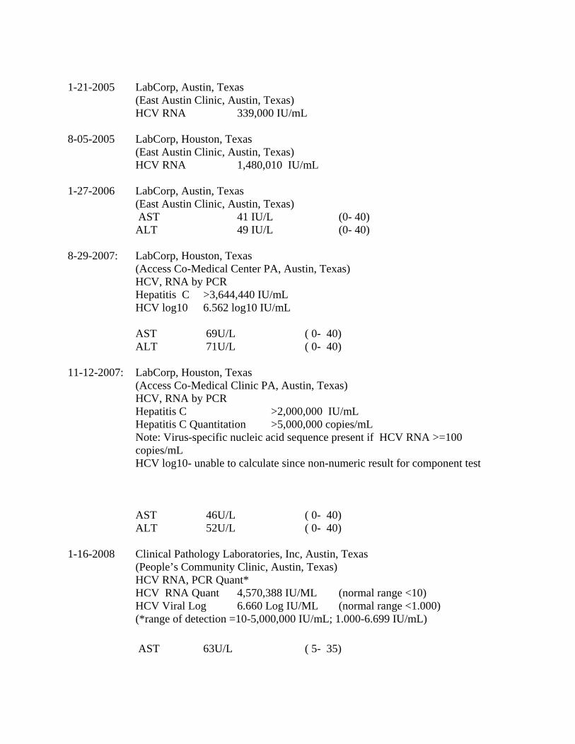
1-21-2005 LabCorp, Austin, Texas (East Austin Clinic, Austin, Texas) HCV RNA 339,000 IU/mL 8-05-2005 LabCorp, Houston, Texas (East Austin Clinic, Austin, Texas) HCV RNA 1,480,010 IU/mL 1-27-2006 LabCorp, Austin, Texas (East Austin Clinic, Austin, Texas) AST 41 IU/L (0- 40) ALT 49 IU/L (0- 40) 8-29-2007: LabCorp, Houston, Texas (Access Co-Medical Center PA, Austin, Texas) HCV, RNA by PCR Hepatitis C >3,644,440 IU/mL HCV log10 6.562 log10 IU/mL
AST 69U/L ( 0- 40) ALT 71U/L ( 0- 40) 11-12-2007: LabCorp, Houston, Texas (Access Co-Medical Clinic PA, Austin, Texas) HCV, RNA by PCR Hepatitis C >2,000,000 IU/mL Hepatitis C Quantitation >5,000,000 copies/mL
Note: Virus-specific nucleic acid sequence present if HCV RNA >=100 copies/mL HCV log10- unable to calculate since non-numeric result for component test
AST 46U/L ( 0- 40) ALT 52U/L ( 0- 40) 1-16-2008 Clinical Pathology Laboratories, Inc, Austin, Texas (People’s Community Clinic, Austin, Texas)
HCV RNA, PCR Quant* HCV RNA Quant 4,570,388 IU/ML (normal range <10) HCV Viral Log 6.660 Log IU/ML (normal range <1.000)
(*range of detection =10-5,000,000 IU/mL; 1.000-6.699 IU/mL)
AST 63U/L ( 5- 35)
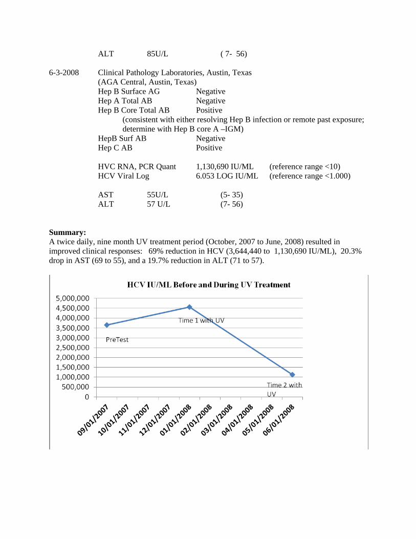
ALT 85U/L ( 7- 56) 6-3-2008 Clinical Pathology Laboratories, Austin, Texas (AGA Central, Austin, Texas)
Hep B Surface AG Negative Hep A Total AB Negative Hep B Core Total AB Positive
(consistent with either resolving Hep B infection or remote past exposure; determine with Hep B core A –IGM)
HepB Surf AB Negative Hep C AB Positive
HVC RNA, PCR Quant 1,130,690 IU/ML (reference range <10) HCV Viral Log 6.053 LOG IU/ML (reference range <1.000)
AST 55U/L (5- 35)
ALT 57 U/L (7- 56) Summary: A twice daily, nine month UV treatment period (October, 2007 to June, 2008) resulted in improved clinical responses: 69% reduction in HCV (3,644,440 to 1,130,690 IU/ML), 20.3% drop in AST (69 to 55), and a 19.7% reduction in ALT (71 to 57).

ALT & AST Before and During UV Treatment
Self Report Improvement in other symptoms: LR reported other unrelated symptoms resolved with UV light treatment:
• Two dental abscesses – presented at local dentist immediately prior to starting the UV therapy, received no treatment and prescriptions for pain medication and antibiotics. Prescriptions were not filled and instead, began UV treatment. After 2 to 4 hours of UV treatment, pain was relieved. After 6 hours (3rd day), evidence of abscess resolved as indicated by inflammation, pain, and swelling.
• Experienced pain and burning in abdomen under diaphragm, primarily on the right side over prior years. The pain was varied and constant or intermittent throughout the day and would interfere with sleep often. This pain completely resolved within the first few weeks of UV treatment.
• Experienced chronic interstitial cystitis over prior years, managed with antibiotics. The last antibiotic treatment (August 2006) resulted in extreme burning of the mucosal membranes (mouth, lips, gums, tongue) and thus, she discontinued any further antibiotic

use. Recurrent cystitis completely resolved after treatment UV treatment.
• Experienced persistent vaginal discharge, dampness in the ears, and oral thrush. These symptoms are consistent with candida yeast overgrowth. These symptoms resolved within 10-14 days of UV light treatment.
• Consistently constipated and experienced irregular bowel movements of dark and irregular shape. These conditions totally resolved within 10-14 days of UV light treatment.
• Experienced improved mental and psychological wellbeing with the UV light.
HCV 3B Genotype Hepatitis C virus subtype 3b infection in a hospital in Japan: Epidemiological study, Kenji Ikeda1, 2 , Kazuaki Chayama1, 2, Satoshi Saitoh1, 2, Isao Koida1, 2, Yoshiyuki Suzuki1, 2, Akihito Tsubota1, 2, Masahiro Kobayashi1, 2, Yasuji Arase1, 2, Naoya Murashima1, 2 and Hiromitsu Kumada Journal of Gastroenterology, Volume 31, Number 6 / November, 1996.801-805
Abstract To elucidate the epidemiology of infection with hepatitis C virus (HCV) subtype 3b (a rare subtype thought to have originated in Southeast Asia) in Japan, we examined the genotypic subtype in 1397 patients with HCV-related chronic liver diseases. Of 1330 patients with identified HCV RNA genotypes, 960 had subtype 1b, 243 had subtype 2a, 97 had subtype 2b, 14 (1.1%) had subtype 3b, and 16 had other types of HCV or mixed subtypes. The age, gender, and severity of liver disease in patients with HCV subtype 3b did not differ from these features in patients with other subtypes. Eleven of the 14 patients with the 3b subtype had once worked at Company A in Tokyo, Japan. Multivariate logistic analysis showed that working history at that company was independently associated with the incidence of the subtype; the risk ratio was 207.2 (P<0.0001). All 11 patients from Company A had received medical services, between 1953 and 1981, at Clinic C, which undertook medical care of the company staff. All 11 patients had received repeated intramuscular or intravenous injections for treatment of various diseases or for preventive vaccination for contagious diseases. The rare HCV subtype 3b, appeared to have been transmitted among the employees of a company through the performance of certain medical practices
Ref 1. Journal of General Virology (1995), 76, 2493-2507. Printed in Great Britain 2493 Investigation of the pattern of hepatitis C virus sequence diversity in different geographical regions: implications for virus classification J. Mellor,' E. C. Holmes, z L. M. Jarvis,' P. L. Yap, 3 P. Simmonds'* and The International HCV Collaborative Study Group~
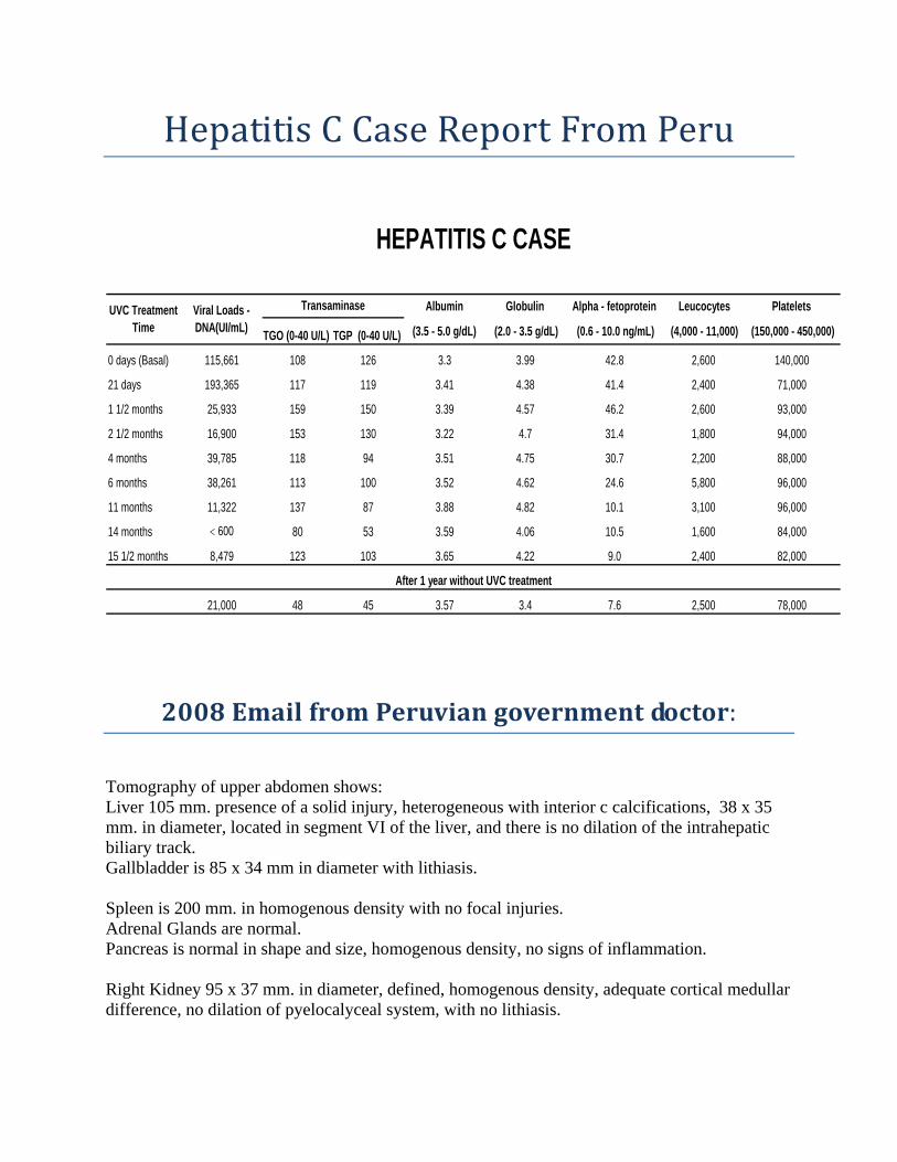
Hepatitis C Case Report From Peru
Albumin Globulin Alpha - fetoprotein Leucocytes Platelets
TGO (0-40 U/L) TGP (0-40 U/L) (3.5 - 5.0 g/dL) (2.0 - 3.5 g/dL) (0.6 - 10.0 ng/mL) (4,000 - 11,000) (150,000 - 450,000)
0 days (Basal) 115,661 108 126 3.3 3.99 42.8 2,600 140,000
21 days 193,365 117 119 3.41 4.38 41.4 2,400 71,000
1 1/2 months 25,933 159 150 3.39 4.57 46.2 2,600 93,000
2 1/2 months 16,900 153 130 3.22 4.7 31.4 1,800 94,000
4 months 39,785 118 94 3.51 4.75 30.7 2,200 88,000
6 months 38,261 113 100 3.52 4.62 24.6 5,800 96,000
11 months 11,322 137 87 3.88 4.82 10.1 3,100 96,000
14 months < 600 80 53 3.59 4.06 10.5 1,600 84,000
15 1/2 months 8,479 123 103 3.65 4.22 9.0 2,400 82,000
21,000 48 45 3.57 3.4 7.6 2,500 78,000
Viral Loads - DNA(UI/mL)
UVC Treatment Time
After 1 year without UVC treatment
HEPATITIS C CASE
Transaminase
2008 Email from Peruvian government doctor:
Tomography of upper abdomen shows: Liver 105 mm. presence of a solid injury, heterogeneous with interior c calcifications, 38 x 35 mm. in diameter, located in segment VI of the liver, and there is no dilation of the intrahepatic biliary track. Gallbladder is 85 x 34 mm in diameter with lithiasis. Spleen is 200 mm. in homogenous density with no focal injuries. Adrenal Glands are normal. Pancreas is normal in shape and size, homogenous density, no signs of inflammation. Right Kidney 95 x 37 mm. in diameter, defined, homogenous density, adequate cortical medullar difference, no dilation of pyelocalyceal system, with no lithiasis.

Left Kidney 98 x 38 mm. in diameter defined, homogenous density, adequate cortical medullar difference, no dilation of pyelocalyceal system, with lithiasis. No presence of free fluids in peritoneal cavity. CONCLUSION: LIVER CANCER DIAGNOSIS: ABSCESS VESICULAR LITHIASIS (GALLSTONES) SPLEENOMEGALY LEFT RENAL LITHAISIS

2009 Email from Peruvian government doctor:
Tomography of upper abdomen shows: -Liver size and form normal (105 mm.) heterogeneous echogenecity of irregular surroundings. There is a solid heterogeneous injury present, with no calcifications, 22 x20 mm. in diameter, it is located in the segment VI of the liver in relation with a neoplasia process. The same has been reduced in size compared to the last report of March 2008. There is no dilation of the intrahepatic biliary tract. -Gallbladder is 85 x 34 mm. in diameter; walls are very thin, with lithiasis. Pancreas is normal in form and size, echogenecity is homogeneous, no signs of inflammation. -Spleen is 200 mm., homogeneous. Right Kidney 95 x 37 mm in diameter, its echogenecity is homogeneous, adequate cortical medullar relation, no dilation of the pyelocalyceal system, no lithiasis. Left Kidney is 98 x38 mm. in diameter; echogenecity is homogeneous, adequate cortical medullar relation, no dilation of pyelocalyceal system, no lithiasis. No presence of free fluids in peritoneal cavity. CONCLUSION: LIVER CIRRHOSIS LIVER CANCER THE HAS DIMINISHED IN SIZE SPLEENOMEGALY VESICULALR LITHIASIS (GALLSTONES).
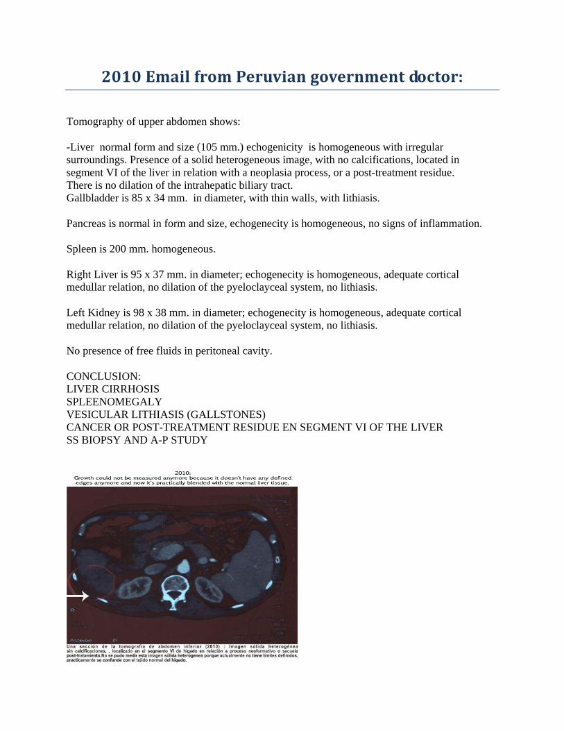
2010 Email from Peruvian government doctor:
Tomography of upper abdomen shows: -Liver normal form and size (105 mm.) echogenicity is homogeneous with irregular surroundings. Presence of a solid heterogeneous image, with no calcifications, located in segment VI of the liver in relation with a neoplasia process, or a post-treatment residue. There is no dilation of the intrahepatic biliary tract. Gallbladder is 85 x 34 mm. in diameter, with thin walls, with lithiasis. Pancreas is normal in form and size, echogenecity is homogeneous, no signs of inflammation. Spleen is 200 mm. homogeneous. Right Liver is 95 x 37 mm. in diameter; echogenecity is homogeneous, adequate cortical medullar relation, no dilation of the pyeloclayceal system, no lithiasis. Left Kidney is 98 x 38 mm. in diameter; echogenecity is homogeneous, adequate cortical medullar relation, no dilation of the pyeloclayceal system, no lithiasis. No presence of free fluids in peritoneal cavity. CONCLUSION: LIVER CIRRHOSIS SPLEENOMEGALY VESICULAR LITHIASIS (GALLSTONES) CANCER OR POST-TREATMENT RESIDUE EN SEGMENT VI OF THE LIVER SS BIOPSY AND A-P STUDY

HEPATITIS B CASE STUDY FROM PERU
Viral Loads - DNA
(UI/mL) TGO(0-40 U/L) TGP(0-40U/L)
0 days (Basal) 8,360 124 88 3.21 4.48 3,800 158,000 +
15 days 6,240 110 88 3.40 3.97 3,700 131,000 +
1 month 1/2 34,300 102 82 3.54 3.87 4,300 134,000 +
2 months 1/2 19,200 72 60 3.83 3.73 3,700 130,000 +
3 months 1/2 9,040 76 60 3.87 3.60 3,200 132,000 +
5 months 9,710 70 60 3.91 3.84 3,300 144,000 +
6 months 9,996 78 73 4.13 3.91 3,200 34,000 +
7 months 8,060 63 56 4.53 4.09 4,400 140,000 +
8 months 8,110 55 52 4.38 3.53 4,500 105,000 +
11 1/2 months 5,890 54 61 4.77 3.52 4,000 120,000 -
15 months 3,240 57 49 4.49 3.19 3,000 105,000 -
18 months 2,577 41 45 4.41 3.58 4,800 128,000 -
21 1/2 months 164 35 34 4.20 2.87 4,500 146,000 -
24 1/2 months 316 39 36 4.54 2.84 4,600 134,000 -
435 32 31 4.4 2.90 4,500 142,000 -
HEPATITIS B CASE
Albumin (3.5-5.0 g/dL)
Transaminase Globulin (2.0-3.5 g/dL)
After 7 months without UVC treatment
UVC Treatment Time
Leucocytes (4,000 - 11,000)
Platelets (150,000 - 450,000) HBe Ag Antigen
Email from Peruvian government doctor, June 8, 2008: Greetings, this is the case of a male patient, 56 years old (code JM006), diagnosed on December 4, 2007 with Active Chronic Liver Cirrhosis and Hepatitis B. On that date his blood tests were high, especially the liver functions. The patient was hospitalized and was given a scan of the liver, a test to detect liver cirrhosis. He had a viral load of 313,000 copies/ml. While in the hospital he was given pharmaceuticals, since there is no specific therapy for cirrhosis but they could not give him antiviral treatment since that would have damaged the liver more. The patient came to see me for the first time on February 18, 2008 and we took blood samples. That same day I began UVC therapy at a rate of 2 hours a day, with a 1-hour break in between. Therapy was continuous and did not stop on holidays or Sundays. The results are in the scans sent. Here’s a review. Basal: 2-19-08 At 15 days: 3-5-08 At 35 days: 3-25-08 At 70 days: 4-29-08 At 105 days: 6-3-08 (on this date we drew his blood and I hope to have the results this week. The patient is clinically stable, has not bleed from his gums and has gone up in weight, there is no tiredness or cramps (these are symptoms he had before). He has returned to his normal activities. Let’s look at his rate of coagulation. First it was 36%, then 70%, 70% and then the last control was 95%. This is an important indicator of the liver function. Another interesting

variable is that of the proteins. Here are their basal values: Total proteins: 7.69, albumin: 3.21 (3.5-5.0), globulin: 4.48 (2.0-3.5), transaminase: ALT: 88 (0-40), AST: 124 (0-40) …. And in the last control, total proteins:7.56, albumin: 3.83 (3.5-5.0), globulin: 3.73 (2.0-3.5), transaminase: ALT:60 (0-40), AST: 72 (0-40). The normalization of the albumin, the better state of consciousness of the patient, improvement of prothrombin are some of the indicators that the cirrhosis is in a state of reversal, which is not usual, better yet, it is impossible through conventional medical science. Here's a report from the Peruvian government doctor about his hepatitis B patient and his improvement after being treated with the sublingual UV device: Observe the results of the scan (sonogram): Today the Hep B patient continues with the UVC therapy, until his next control tests. Note: Observe the results of the gammagraphic or scintigraphy examinations. Analysis of the 1st report: The gammagraphic pattern is of a cirrhosis liver. In the 2nd report: This liver is no longer cirrhosis liver but we still need to work on the slight compromise of the liver parenchyma (but this is definitely not cirrhosis anymore.) We still need more follow up with this case but what I am expecting in the follow up tests is that:
1- The Australian antigen will become negative. 2- The viral load will be totally undetectable. 3- That the number of platelets will normalize completely. 4- That the TGO and the TGP will reach values equal to or lower than 40. 5- That in the next scintigraphy there will be no liver lesion at all…
If this is achieved it would be a boost to this type of technology but it cannot be denied that as it is this is a great advance and that this is a unique case in medical history. Scintigraphy Results: Test #1 (before treatment) ESSALUD Hospital H.N. Guillermo Almenara I Diagnostic Imaging Services Date: 12/20/2007 Patient ______________________ Services: Internal Medicine 3 Tests Requested: Liver and Spleen Diagnostic (CIE): B16.9 Age: 56 Sex: Male Report: Liver Scintigraphy Examination, colloidal sulfur 99MTC: An image of the liver can be observed. It is enlarged and in the shape of a globe and the concentration of the radioactive solution is diminished. There are extra hepatic traces from the reticuloendothelial system of the bone marrow (spinal cord). The spleen has also increased in size and the reticuloendothelial capture is higher than that of the liver.

Conclusion: Patter of Liver Cirrhosis in its early stage. (12/20/07) Test #2 (During treatment) ESSALUD Hospital H.N. Guillermo Almenara I Diagnostic Imaging Services Date: 1/15/2009 Patient ______________________ Services: Gastroenterology Tests Requested: Liver and Spleen Diagnostic (CIE): K74.6 Age : 56 Sex: Male Report: Liver Scintigraphy Examination, coloidal sulfur 99MTC: Image of liver can be observed and it is only slightly enlarged. Concentration of the radioactive solution is distributed in a slightly irregular and homogenous manner. The spleen is normal according to the scan. There is only light compromise of the liver parenchyma. These results show an improvement over the previous test. Update: Report from Peruvian government doctor: Tuesday, March 3, 2009 Observe the results of the scan: Today the patient continues with the UVC therapy, until his next control tests. Note: Observe the results of the scans or scintigraphy examinations. Analysis of the 1st report: The gammagraphic pattern is of a cirrhotic liver. In the 2nd report: This liver is no longer a cirrhotic liver but we still need to work on the slight compromise of the liver parenchyma (but this is definitely not cirrhosis anymore.) We still need more follow up with this case but what I am expecting in the follow up tests is that:
1- The Australian antigen will become negative. 2- The viral load will be totally undetectable. 3- That the number of platelets will normalize completely. 4- That the TGO and the TGP will reach values equal to or lower than 40. 5- That in the next scan there will be no liver lesion at all…
If this is achieved it would be a boost to this type of technology but it cannot be denied that as it is this is a great advance and that this is a unique case in medical history.
Related Documents