Hepatic progenitor cells from adult human livers for cell transplantation T S Weiss, 1,2 M Lichtenauer, 1,2 S Kirchner, 1,2 P Stock, 3 H Aurich, 3 B Christ, 3 G Brockhoff, 4 L A Kunz-Schughart, 4 K-W Jauch, 5 H-J Schlitt, 1,2 W E Thasler 5 1 Department of Surgery, University of Regensburg Hospital, Germany; 2 Center for Liver Cell Research, University of Regensburg Hospital, Germany; 3 Department of Internal Medicine, Martin Luther University of Halle–Wittenberg, Germany; 4 Department of Pathology, University of Regensburg, Germany; 5 Department of Surgery, LM University Munich, Hospital Grosshadern, Germany Correspondence to: Dr Thomas S Weiss, Center for Liver Cell Research, University of Regensburg Hospital, F.-J.-S.- Allee 11, 93053 Regensburg, Germany; thomas.weiss@ klinik.uni-regensburg.de LAK-S is now at OncoRay – Center for Radiation Research in Oncology, Medical Faculty Carl Gustav Carus, Dresden University of Technology, Fetscherstr. 74, 01307 Dresden, Germany. Revised 7 March 2008 Accepted 9 April 2008 Published Online First 16 April 2008 ABSTRACT Objective: Liver regeneration is mainly based on cellular self-renewal including progenitor cells. Efforts have been made to harness this potential for cell transplantation, but shortage of hepatocytes and premature differentiated progenitor cells from extra-hepatic organs are limiting factors. Histological studies implied that resident cells in adult liver can proliferate, have bipotential character and may be a suitable source for cell transplantation. Methods: Particular cell populations were isolated after adequate tissue dissociation. Single cell suspensions were purified by Thy-1 positivity selection, characterised in vitro and transplanted in immunodeficient Pfp/Rag2 mice. Results: Thy-1 + cells that are mainly found in the portal tract and the surrounding parenchyma, were isolated from surgical liver tissue with high yields from specimens with histological signs of regeneration. Thy-1 + cell populations were positive for progenitor (CD34, c-kit, CK14, M2PK, OV6), biliary (CK19) and hepatic (HepPar1) markers revealing their progenitor as well as hepatic and biliary nature. The potential of Thy-1 + cells for differentiation in vitro was demonstrated by increased mRNA and protein expression for hepatic (CK18, HepPar1) and biliary (CK7) markers during culture while progenitor markers CK14, chromogranin A and nestin were reduced. After transplantation of Thy-1 + cells into livers of immunodefi- cient mice, engraftment was predominantly seen in the periportal portion of the liver lobule. Analysis of in situ material revealed that transplanted cells express human hepatic markers HepPar1 and albumin, indicating func- tional engraftment. Conclusion: Bipotential progenitor cells from human adult livers can be isolated using Thy-1 and might be a potential candidate for cell treatment in liver diseases. In response to liver injury, hepatic cells, especially hepatocytes, can rapidly proliferate to repair liver damage. 1 After hepatectomy hepatocytes are able to undergo numerous cell divisions maintaining their fully differentiated state to compensate for hepatocyte loss, whereas undifferentiated liver progenitor cells play only a minor role in this process. Interestingly, under conditions such as some chronic liver diseases and after acute necrosis these hepatocyte progenitors play an important role, in particular when growth response of mature hepatocytes is impaired. 12 Mechanisms leading to progenitor cell activation have been studied in detail using rodent models of hepatocarcinogenesis and liver regeneration after toxic injury. 34 Under these conditions increasing numbers of small cells (,10 mm) were observed having a large nuclear-to-cytoplasm ratio with an oval-shaped nucleus (oval cells). They express markers of both hepatocyte lineages and the biliary epithelium that includes bile ducts as well as the canal of Hering. 56 Although histochemical studies suggest that epithelial cells residing within the canal of Hering might be resident liver stem cells, the precise origin remains ambiguous. 6 In humans these cells are termed progenitor cells/ductular reaction 7 and are activated in different diseases such as chronic viral hepatitis, alcoholic liver disease, non- alcoholic fatty liver disease and cirrhosis. 48 They are observed in the portal region of the hepatic lobule 9–11 and, furthermore, their numbers increase in direct proportion to disease severity. 12 Hepatic progenitor cells have also been shown to express haematopoietic stem cell (HSC) markers CD34, c-kit (CD117) 13 and Thy-1 (CD90). 14 Accordingly, it was hypothesised that these cells are haematopoietic stem cells transdifferentiated into the hepatic lineage. 15 However, recent reports have demonstrated that cell fusion rather than transdifferentiation is involved in this process. Also, it was shown that rodent oval cells do not originate from bone marrow, 16 but bone-marrow- derived stem cells may contribute to liver regenera- tion in an as yet unknown mechanism. 17 Isolation procedures for hepatic progenitor cells were established for rodents and in vitro studies have shown that these cells are capable of differentiating into hepatocytes and bile duct cells under appro- priate conditions. 18 Isolation of liver progenitor cells from human material has proven more difficult. Indeed, only a few reports describe an isolation of progenitor cells from fetal livers. These cells expressed various lineage specific antigens including biliary and hepatocellular markers. 19 In adult human liver, progenitor cells have been identified by CD34 and c-kit immunoreactivity, 13 but the so-called side- population was also found. 20 Recently, it was documented that Thy-1 (CD90) is expressed in human hepatoblastoma. 21 This surface antigen has also been useful for successfully isolating progenitor cells from human fetal liver. 14 However, to our knowledge, there have been no attempts so far to use this marker for the separation of progenitor cell populations from adult human liver tissues. Since such cell populations may become central determi- nants for future treatments of liver diseases requiring clinically relevant liver regeneration, we aimed to identify and isolate Thy-1 + cells from adult liver tissue for cell transplantation purposes. We analysed the phenotypic characteristics of Thy-1 + human progenitor cells after isolation, studied their differ- entiation potential in vitro and monitored their repopulation capacity in vivo in a mouse model of liver regeneration. Hepatology Gut 2008;57:1129–1138. doi:10.1136/gut.2007.143321 1129 group.bmj.com on September 3, 2015 - Published by http://gut.bmj.com/ Downloaded from

Welcome message from author
This document is posted to help you gain knowledge. Please leave a comment to let me know what you think about it! Share it to your friends and learn new things together.
Transcript

Hepatic progenitor cells from adult human livers forcell transplantation
T S Weiss,1,2 M Lichtenauer,1,2 S Kirchner,1,2 P Stock,3 H Aurich,3 B Christ,3
G Brockhoff,4 L A Kunz-Schughart,4 K-W Jauch,5 H-J Schlitt,1,2 W E Thasler5
1 Department of Surgery,University of RegensburgHospital, Germany; 2 Center forLiver Cell Research, University ofRegensburg Hospital, Germany;3 Department of InternalMedicine, Martin LutherUniversity of Halle–Wittenberg,Germany; 4 Department ofPathology, University ofRegensburg, Germany;5 Department of Surgery, LMUniversity Munich, HospitalGrosshadern, Germany
Correspondence to:Dr Thomas S Weiss, Center forLiver Cell Research, University ofRegensburg Hospital, F.-J.-S.-Allee 11, 93053 Regensburg,Germany; [email protected]
LAK-S is now at OncoRay –Center for Radiation Research inOncology, Medical Faculty CarlGustav Carus, DresdenUniversity of Technology,Fetscherstr. 74, 01307 Dresden,Germany.
Revised 7 March 2008Accepted 9 April 2008Published Online First16 April 2008
ABSTRACTObjective: Liver regeneration is mainly based on cellularself-renewal including progenitor cells. Efforts have beenmade to harness this potential for cell transplantation, butshortage of hepatocytes and premature differentiatedprogenitor cells from extra-hepatic organs are limitingfactors. Histological studies implied that resident cells inadult liver can proliferate, have bipotential character andmay be a suitable source for cell transplantation.Methods: Particular cell populations were isolated afteradequate tissue dissociation. Single cell suspensions werepurified by Thy-1 positivity selection, characterised in vitroand transplanted in immunodeficient Pfp/Rag2 mice.Results: Thy-1+ cells that are mainly found in the portaltract and the surrounding parenchyma, were isolated fromsurgical liver tissue with high yields from specimens withhistological signs of regeneration. Thy-1+ cell populationswere positive for progenitor (CD34, c-kit, CK14, M2PK,OV6), biliary (CK19) and hepatic (HepPar1) markersrevealing their progenitor as well as hepatic and biliarynature. The potential of Thy-1+ cells for differentiation invitro was demonstrated by increased mRNA and proteinexpression for hepatic (CK18, HepPar1) and biliary (CK7)markers during culture while progenitor markers CK14,chromogranin A and nestin were reduced. Aftertransplantation of Thy-1+ cells into livers of immunodefi-cient mice, engraftment was predominantly seen in theperiportal portion of the liver lobule. Analysis of in situmaterial revealed that transplanted cells express humanhepatic markers HepPar1 and albumin, indicating func-tional engraftment.Conclusion: Bipotential progenitor cells from humanadult livers can be isolated using Thy-1 and might be apotential candidate for cell treatment in liver diseases.
In response to liver injury, hepatic cells, especiallyhepatocytes, can rapidly proliferate to repair liverdamage.1 After hepatectomy hepatocytes are ableto undergo numerous cell divisions maintainingtheir fully differentiated state to compensate forhepatocyte loss, whereas undifferentiated liverprogenitor cells play only a minor role in thisprocess. Interestingly, under conditions such assome chronic liver diseases and after acute necrosisthese hepatocyte progenitors play an importantrole, in particular when growth response of maturehepatocytes is impaired.1 2
Mechanisms leading to progenitor cell activationhave been studied in detail using rodent models ofhepatocarcinogenesis and liver regeneration aftertoxic injury.3 4 Under these conditions increasingnumbers of small cells (,10 mm) were observedhaving a large nuclear-to-cytoplasm ratio with anoval-shaped nucleus (oval cells). They express
markers of both hepatocyte lineages and the biliaryepithelium that includes bile ducts as well as thecanal of Hering.5 6 Although histochemical studiessuggest that epithelial cells residing within thecanal of Hering might be resident liver stem cells,the precise origin remains ambiguous.6 In humansthese cells are termed progenitor cells/ductularreaction7 and are activated in different diseases suchas chronic viral hepatitis, alcoholic liver disease, non-alcoholic fatty liver disease and cirrhosis.4 8 They areobserved in the portal region of the hepatic lobule9–11
and, furthermore, their numbers increase in directproportion to disease severity.12
Hepatic progenitor cells have also been shown toexpress haematopoietic stem cell (HSC) markersCD34, c-kit (CD117)13 and Thy-1 (CD90).14
Accordingly, it was hypothesised that these cellsare haematopoietic stem cells transdifferentiatedinto the hepatic lineage.15 However, recent reportshave demonstrated that cell fusion rather thantransdifferentiation is involved in this process.Also, it was shown that rodent oval cells do notoriginate from bone marrow,16 but bone-marrow-derived stem cells may contribute to liver regenera-tion in an as yet unknown mechanism.17
Isolation procedures for hepatic progenitor cellswere established for rodents and in vitro studies haveshown that these cells are capable of differentiatinginto hepatocytes and bile duct cells under appro-priate conditions.18 Isolation of liver progenitor cellsfrom human material has proven more difficult.Indeed, only a few reports describe an isolation ofprogenitor cells from fetal livers. These cellsexpressed various lineage specific antigens includingbiliary and hepatocellular markers.19 In adult humanliver, progenitor cells have been identified by CD34and c-kit immunoreactivity,13 but the so-called side-population was also found.20 Recently, it wasdocumented that Thy-1 (CD90) is expressed inhuman hepatoblastoma.21 This surface antigen hasalso been useful for successfully isolating progenitorcells from human fetal liver.14 However, to ourknowledge, there have been no attempts so far to usethis marker for the separation of progenitor cellpopulations from adult human liver tissues. Sincesuch cell populations may become central determi-nants for future treatments of liver diseases requiringclinically relevant liver regeneration, we aimed toidentify and isolate Thy-1+ cells from adult livertissue for cell transplantation purposes. We analysedthe phenotypic characteristics of Thy-1+ humanprogenitor cells after isolation, studied their differ-entiation potential in vitro and monitored theirrepopulation capacity in vivo in a mouse model ofliver regeneration.
Hepatology
Gut 2008;57:1129–1138. doi:10.1136/gut.2007.143321 1129
group.bmj.com on September 3, 2015 - Published by http://gut.bmj.com/Downloaded from

MATERIALS AND METHODSReagents and materialAntibodies for this study are summarised in table 1. Mouse anti-rabbit IgG, rabbit anti-mouse IgG, and mouse APAAP wereobtained from Dako (Hamburg, Germany). Texas red-conjugateddonkey anti-rabbit antibody and fluorescein isothiocyanate(FITC)-conjugated donkey anti-mouse antibody were obtainedfrom Jackson Immunoresearch (Newmarket, UK). RNase A waspurchased from Qiagen (Hilden, Germany), DNase I and fast redsubstrate from Roche Diagnostics (Mannheim, Germany). Allfurther chemicals were purchased from Sigma (Taufkirchen,Germany) or Merck (Darmstadt, Germany).
Tissue samples from human liver resections were obtainedfrom patients undergoing partial hepatectomy. Experimentalprocedures were performed according to the guidelines of thecharitable state-controlled foundation HTCR (Human Tissueand Cell Research), with the informed patient’s consentapproved by the local ethics committee of the University ofRegensburg.22 A total of 39 liver specimens were categorised intoregenerating (n = 20; age 53.7 (SD 15.6) years; male 13, female7) and non-regenerating (n = 19; age 61.7 (SD 12.2) years; male15, female 4) tissue according to a histological and clinicalexamination by specialist. Non-regenerating liver was assignedto patients with metastatic liver tumours of colorectal cancerand regenerating liver to patients with disease as following:fibrosis/cirrhosis (n = 4; age 53.0 (SD 9.3) years; male 4),fibrosis/non-alcoholic steatohepatitis (NASH) (n = 4; age 58.0(SD 11.2) years; male 2, female 2), fibrosis/alcoholic steatohe-patitis (ASH) (n = 1; age 23; female), cirrhosis/ASH (n = 2; age64.5 (SD 4.5) years; male 2), steatosis (n = 7; age 53.7 (SD19.7) years; male 3, female 4), ASH (n = 1; age 49 years; male),NASH (n = 1; age 52 years; male).
Isolation of Thy-1 positive cells from human liver tissueIsolation of non-parenchymal cellsLiver tissue was digested using a modified two-step egtazic acid(EGTA)/collagenase perfusion procedure23 to isolate humanhepatocytes. Non-parenchymal cells (NPCs) were obtainedfrom the remaining liver tissue based on a protocol describedrecently.24 Briefly, up to 50 g of tissue was minced andincubated for up to 150 min under vigorous shaking at 37uC
in 50 ml isolation buffer (154 mmol/l NaCl, 5.6 mmol/l KCl,5 mmol/l glucose, 25 mmol/l NaHCO3, 20 mmol/l HEPES,pH 7.4) supplemented with 0.5 mg/ml collagenase type IV,0.5 mg/ml pronase, 40 mg/ml DNase I, 5 mmol/l CaCl2 and1 mmol/l MgCl2. Every 30 min undigested tissue was separated,minced and incubated again. The resulting suspension wasfiltered (meshes with 210 mm and 170 mm pore sizes) and washedthree times in phosphate-buffered saline (PBS) containing2 mmol/l ethylenediaminetetraacetic acid (EDTA). Furthermore,the cell suspension was purified using a 70% Percoll solution(800 g, 30 min, 18uC). After washing with PBS/EDTA, the NPCswere suspended in MACS buffer (PBS containing 2 mmol/lEDTA, 5 g/l bovine serum albumin (BSA)) and stored for up to20 h at 4uC. Size profile of cell fractions was monitored for qualityassessment of the dissociation procedure using a cell countersystem (Casy Scharfe System, Reutlingen, Germany). NPCfractions were isolated with an average yield of 2.7 (SD 2.6)6106
viable cells per g wet tissue (n = 39).
Purification of Thy-1 positive cellsThy-1+ cells were isolated from NPCs using the immuno-magnetic activated cell sorting (MACS) technique (MiltenyiBiotech, Bergisch Gladbach, Germany) according to the instruc-tion manual. Briefly, cells were filtered (40 mm filter, BDBiosciences Europe, Erembodegem, Belgium) and 56107 cellswere incubated with 100 ml of a mouse anti-CD90 (Thy-1)monoclonal antibody (mAb) for 30 min at 4uC. After twofoldwashing with MACS buffer, cells were incubated with 100 ml ofgoat anti-mouse microbeads for 15 min at 4uC followed by twoadditional washing steps. Labelled cell suspensions weretransferred onto MACS LS columns and washed three timeswhile being located in the magnetic field. Thy-1+ cells wereeluted with 5 ml MACS buffer following removal from themagnet. Cell number and viability were determined by thetrypan blue exclusion technique.
Characterisation by flow cytometryCells were suspended in PBS, incubated with fluorescence-labelled mAb for 30 min at 4uC, washed with PBS and analysedfollowing addition of propidium iodide (PI) (1 mg/ml PI per
Table 1 Antibodies used for immunohistochemistry (IHC) and flow cytometry
Antibody Species Manufacturer Catalogue number Dilution Application
Albumin Rabbit polyclonal IgG Abcam ab2406 1:1000 IHC
CD3-FITC Mouse monoclonal IgG2a/MEM-57 AbD Serotec MCA2184F 5 ml/106 cells Flow cytometry
CD14-FITC Mouse monoclonal IgG1/MEM-18 AbD Serotec MCA2185F 5 ml/106 cells Flow cytometry
CD34 Mouse monoclonal IgG1/Qbend10 Dako M7165 1:100 IHC
CD34-FITC Mouse monoclonal IgG1/8G12 BD 345801 20 ml/106 cells Flow cytometry
CD45-FITC Mouse monoclonal IgG1/2D1 BD 345808 10 ml/106 cells Flow cytometry
Thy-1 (CD90) Mouse monoclonal IgG1/5E10 BD Pharmingen 555593 1:250 IHC, Flow cytometry
CD90-PE Mouse monoclonal IgG1/5E10 BD Pharmingen 555596 5 ml/106 cells Flow cytometry
c-kit (CD117) Mouse monoclonal IgG2a/K45 NeoMarkers MS-289 1:100 IHC
CD117-PE Mouse monoclonal IgG1/104D2 Southern Biotech 9815-09C 10 ml/106 cells Flow cytometry
CK7 Mouse monoclonal IgG Dako M7018 1:100 IHC
CK14 Rabbit polyclonal IgG Zytomed RP113 1:50 IHC
CK18 Mouse monoclonal IgG1/CK2 Chemicon MAB3404 1:100 IHC
CK19 Mouse monoclonal IgG1/RCK108 Dako M0888 1:100 IHC
HepPar1 Mouse monoclonal IgG1/OCH1E5 Dako M7158 1:50 IHC
M2-PK Mouse monoclonal IgG1/DF4 ScheBo Biotech S-1 1:10 IHC
OV6 Mouse Gift from S. Sell* 1:100 IHC
Manufacturers: Abcam, Cambridge, UK; AbD Serotec, Dusseldorf, Germany; BD, Heidelberg, Germany; BD Pharmingen, Heidelberg, Germany; Chemicon, Schwalbach, Germany;NeoMarkers, Dreieich, Germany; ScheBo Biotech, Giessen, Germany; Southern Biotech, Birmingham, Alabama, USA; Zytomed, Berlin, Germany.*We are grateful to Dr S. Sell (Department of Pathology and Laboratory Medicine, Albany, NY, USA) for the kind gift of OV6 antibody.
Hepatology
1130 Gut 2008;57:1129–1138. doi:10.1136/gut.2007.143321
group.bmj.com on September 3, 2015 - Published by http://gut.bmj.com/Downloaded from
1 mg/ml RNase A) on a FACSCalibur flow cytometer (BD,Heidelberg, Germany) using appropriate regions of interest and/or quadrant statistics in the compensated fluorescence dot plotdiagrams. PI positive, membrane defect cells were excluded.Details concerning the antibodies are given in table 1.
Immunohistochemical stainingCryosections (6 mm) from snap frozen tissues were fixed in ice-cold acetone (10 min). For cytospots freshly isolated cells weresuspended in PBS, centrifuged (56104 cells) onto glass slides(SuperFrost Plus; Merck) and fixed in acetone. Cultured cellswere fixed using 4% paraformaldehyde for 20 min.
Alkaline phosphatase anti-alkaline phosphatase (APAAP) stainingBlocking of slides was performed with 3% fetal calf serum (FCS)in PBS followed by incubation with primary antibodies for30 min and a washing step with PBS. For CK14 staining,additional incubation with mouse anti-rabbit immunoglobulinsfor 30 min was performed, followed by incubation with rabbitanti-mouse immunoglobulins for 30 min. After incubation withmouse APAAP (1:100 in PBS/1% FCS) for 30 min immuno-signals were developed with fast red substrate (20 min) andcounterstained with haematoxylin Quick Stain.
Immunofluorescence stainingAfter blocking (3% dried milk, 0.1% Triton-X100, PBS) cellswere incubated with primary antibodies for 30 min, washedwith PBS and incubated with the secondary fluorescence-conjugated antibodies. For co-localisation, steps were repeatedwith a second set of antibodies. Nuclei were counterstainedwith 5 mg/ml Hoechst 33258 in PBS.
Determination of mRNA expression by reverse transcription-polymerase chain reactionTotal RNA was extracted using the RNeasy Mini Kit (Qiagen).First strand cDNA was synthesised using 0.1 mg of total RNAand the avian myeloblastosis virus (AMV)-reverse transcriptionreaction (Promega, Mannheim, Germany) using 15 U AMVreverse transcriptase in a prolonged incubation time of 60 min.
The polymerase chain reaction (PCR) was performed accordingto the manufacturer’s instructions (Invitrogen, Karlsruhe,Germany) using primers as listed in table 2. After an initialdenaturation step of 3 min at 94uC, amplification wasperformed for 35 cycles at 94uC for 45 s, 30 s at the optimalannealing temperature and 72uC for 30 s. The PCR wasterminated by a final elongation step at 72uC for 10 min.Amplified products were transferred on 1.5% agarose gels forelectrophoresis followed by ethidium bromide staining.
Cell cultureFreshly isolated Thy-1+ cells were resuspended in adheringmedium (William’s E medium, 5% FCS, 10 mmol/l nicotina-mide, 20 mmol/l HEPES, 17 mmol/l NaHCO3, 550 mg/lsodium pyruvate, 0.2 mmol/l ascorbic acid 2-phosphate,14 mmol/l glucose, 1027 mol/l dexamethasone, 6.25 mg/mlinsulin, 6.25 mg/ml transferrin, 6.25 ng/ml sodium selenite,1.25 mg/ml BSA and 5.35 mg/ml linoleic acid, 100 U/mlpenicillin, 100 mg/ml streptomycin) and plated onto collagencoated dishes (BD Biosciences Europe) at a density of56104/cm2. After 3–5 days the medium was replaced withculture medium (adhering medium without FCS, 20 ng/mlepidermal growth factor (EGF)). The medium was changed oncea week. Viability and cell growth during the culture period weremonitored by cell morphology.
Transplantation of hepatic progenitor cellsAll animal housing and experimentation procedures were inaccordance with German legislation on animal protection (latestversion). Pfp/Rag22/2 mice (malfunction of NK cell, T and B celldepletion) were obtained at 8 months of age from Taconic (LilleSkensved, Denmark). The cellular transplantation model wasperformed as described recently.25 26 Briefly, recipient liverregeneration was inhibited by treating the mice with mono-crotalin (30 mg/kg body weight; intraperitoneally) 6 and 4 weeksprior to transplantation. After partial hepatectomy, 16106 humanThy-1 positive liver cells were administered by intrasplenicinjection. Livers were removed after 7 weeks and fixed in 4%formaldehyde and paraffin embedded for further analysis.
Table 2 Primer sequences, annealing temperature and expected size of amplified products for polymerasechain reaction (PCR) analysis
GeneAccessionnumber Primer (forward/reverse)
Annealingtemperature
Product size(bp)
AFP NM_001134 fw TGCAGCCAAAGTGAAGAGGGAAGA 55uC 216
rv ATAGCGAGCAGCCCAAAGAAGAA
Albumin NM_000477 fw GCTGCCATGGAGATCTGCTTGA 55uC 174
rv GCAAGTCAGCAGGCATCTCATC
ChromA NM_001275 fw CGCGCCTTGTCTCCTACTC 50uC 254
rv AGGAAAGAGCCCAGAACAGAT
CK7 NM_005556 fw TCCGCGAGGTCACCATTAAC 55uC 218
rv GCTGCTCTTGGCCGACTTCT
CK14 NM_000526 fw GGGCGGCCTGTCTGTCTC 60uC 237
rv AGGCGGTCATTGAGGTTCTG
CK18 NM_000224 fw CCCGCTACGCCCTACAGA 53uC 248
rv GCGGGTGGTGGTCTTTTG
CK19 NM_002276 fw CGAGCAGAACCGGAAGGATG 53uC 318
rv AGCCGCTGGTACTCCTGATTC
Nestin NM_006617 fw GGGGGCAATGAATCCTCTAG 50uC 295
rv CACCGTATCTTCCCACCTCTG
b-Actin NM_001101 fw GCCGGGACCTGACTGACTAC 53uC 327
rv TGCGGATGTCCACGTCAC
AFP, alpha-fetoprotein; fw, forward; rv, reverse.
Hepatology
Gut 2008;57:1129–1138. doi:10.1136/gut.2007.143321 1131
group.bmj.com on September 3, 2015 - Published by http://gut.bmj.com/Downloaded from

RESULTS
Expression of progenitor cell marker in human liver sectionsExpression analysis of markers described for liver progenitorswere performed in cryo-sections of adult liver tissue from apatient with fat liver disease. Immunoreactivity for CD34 wasfound in endothelial cells lining the vascular structures of theportal tract and sparely on cells in the adjacent parenchyma(fig 1A), whereas c-kit expression was barely detected (fig 1C).Thy-1 expression was found in the portal tract area and inscattered cells in the surrounding parenchyma (fig 1B).Expression of the oval cell marker OV6 was detected onepithelial cells adjacent to the portal tract, but primarily on bileduct cells (fig 1D). Specific immunostaining of CK19 for biliaryepithelial cells (fig 1E), CK18 for bile duct epithelium andparenchymal hepatocytes (fig 1F) is also demonstrated.
Cells of adult human non-parenchymal liver cell fractionsexpress stem cell markersFractions of small liver cells separated from hepatocytes wereisolated from discarded surgical adult human liver tissue afterperforming a multi-step isolation process (fig 2A–B).Immunocytochemical analysis of small liver cell fractionsrevealed a high percentage of non-parenchymal cells (Kupffer,stellate and endothelial cells; data not shown) and are thereforetermed the non-parenchymal cell (NPC) fraction. NPC fractionswere investigated by flow cytometry for expression of surfacemarkers, which have been detected on liver progenitor cells by
immunohistochemistry. Cells were found positive for CD34(6% (SD 4%), n = 18), CD117 (8% (SD 7%), n = 18) and CD90(9% (SD 6%), n = 22). Additionally, double immunostainingrevealed, that 2% (SD 2%) (n = 11) of NPC fractions wereCD34+/CD90+, 2% (SD 2%) (n = 9) were CD34+/CD117+ and3% (SD 1%) (n = 5) were CD90+/CD117+.
Thy-1 positive cells express markers of liver progenitor cellsThy-1 (CD90), the most abundant progenitor marker on NPCfractions, was selected as a marker for immuno-magneticactivated cell sorting (MACS). Subsequently, fluorescence-activated cell sorting (FACS) analysis was performed determin-ing the quantity and characteristics of Thy-1 isolated cells.MACS sorted cells were highly positive for Thy-1 (83% (SD7%), n = 6) and co-expressed CD34 (24% (SD 9%), n = 5) andhardly CD117 (1.8% (SD 0.7%), n = 5) (fig 2C–E). Additionally,we found expression of CD45 (35% (SD 9%), n = 5), CD14 (20%(SD 7%), n = 5) and CD3 (29% (SD 9%), n = 5) on some Thy-1+
cells. The overall outcome of viable Thy-1+ cells after MACSisolation was 1.4 (SD 0.9)6104 per 106 NPCs (n = 39).Furthermore, performing a correlation study we found asignificant higher rate of Thy-1+ cells in tissues, in whichregenerative events; for example, slight histological signs ofhepatitis, fibrosis, or cirrhosis, were described (1.8% (SD 1.0%)of NPCs, n = 20) than in non-regenerating tissue (1.2% (SD0.57%) of NPCs, n = 19) (fig 2G). The rates of Thy-1+ cells werenot correlated with expression of CD45, CD14 and CD3 (data
Figure 1 Immunohistochemical staining of human liver sections (from a patient with alcoholic fatty liver disease) for markers of progenitor, hepaticand biliary cells. (A) Immunostaining for CD34 was found on endothelial cells lining the vascular structures of the portal tract (arrow head) and sparelyon some cells in adjacent liver (arrow). (B) Positive staining for Thy-1+ was found in the portal tract area and for some parenchymal cells (arrow head).(C) Only spare positive immunostaining could be detected for c-kit expressing cells. (D) OV6+ cells were mainly detected in parenchyma surrounding theportal area (arrow) and in bile duct (arrow head). (E) Bile duct cells were immunoreactive for CK19 (arrow head), whereas (F) CK18 was found onhepatocytes and bile duct cells. (A–F: original magnification, 6200).
Hepatology
1132 Gut 2008;57:1129–1138. doi:10.1136/gut.2007.143321
group.bmj.com on September 3, 2015 - Published by http://gut.bmj.com/Downloaded from

not shown). A more detailed analysis with type of disease,tissue damage and patient related data did not show anycorrelation to the rate of Thy-1+ cells (data not shown).
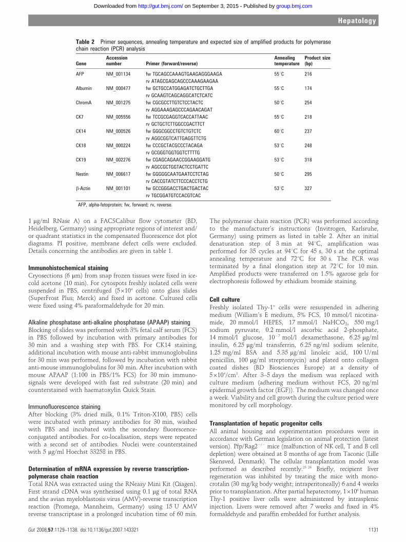
Expression of progenitor and hepatic cell markers weredetermined on freshly isolated Thy-1+ cells by immunostaining(fig 3). About 60% of the Thy-1+-sorted cells demonstratedpositivity for liver progenitor markers CK14, M2PK, OV6 andbiliary/progenitor marker CK19 (fig 3A). Double immunofluor-escence staining of Thy-1+ cells revealed co-localisation of CK14with CD117, CD34 (liver progenitor and HSC), CK19 (bileduct/progenitor cell), HepPar1 (hepatocyte) and OV6 (fig 3B).mRNA analyses of the Thy-1+ cell population from 27 donorsconfirmed the expression of CK14. Also, we detected twoadditional progenitor genes expressed in Thy-1+ cells of alldonors, chromogranin A (pancreatic) and nestin (neural) (fig 4).Positive gene expression was also found for hepatocyte markersCK18 and albumin (10 of 27), as well as biliary marker CK19,but not for CK7. alpha-fetoprotein (AFP) was not detected inany of the cell preparations (fig 4).
Thy-1 positive liver cells in culture show differentiation towardshepatocytes and bile duct cellsThy-1+ cells were seeded on collagen-coated culture dishes andmaintained in culture under conditions described for hepatocytegrowth.27 Efficiency of plating was sufficient and after 10–14 days cells started to grow into colonies (fig 5A–C). At day 28of culture an almost complete monolayer was formed (fig 5D).Furthermore, mRNA analyses by reverse transcription poly-merase chain reaction (RT-PCR) were performed throughoutculturing and differences in gene expression were observeddepending on the culture period (fig 5G). The hepatic markerCK18 strongly increased from day 14 to 28, whereas albumincould not be detected. Furthermore, CK19 expression wasalmost unchanged up to day 28, while the gene expression ofthe bile duct marker CK7 was clearly enhanced. In contrast, areduction of the progenitor markers CK14, chromogranin A andnestin was observed as a function of time in culture. AFP wasnot expressed either in fresh or in cultured cells. In addition,Thy-1+ cells cultured for 14 days were stained with antibodies
Figure 2 Isolation and characterisation of progenitor cell fractions from human liver tissue. Size profiles of Percoll purified liver cell fractions show(A) Hepatocytes (diameter, 20 (SD 5) mm) separated from (B) smaller liver cells (diameter, 8–13 mm). Fractions of dead cells are indicated by anasterisk and cell debris is found at particle sizes of below 6 mm. The Thy-1 positive cells isolated by using immuno-magnetic activated cell sorting(MACS) were further characterised by fluorescence-activated cell sorting (FACS) analysis for their expression of (C) CD90, (D) CD34, and (E) CD117.Data are from one analysis representative of five independent experiments. Unfilled curve: before MACS isolation; filled curve: after MACS isolation.(F) Thy-1+ cells were further purified by MACS and their outcome was correlated to the underlying disease. The tissues were categorised toregenerating (n = 20) and non-regenerating (n = 19) liver from patients with cirrhosis, fibrosis, alcoholic liver disease (ALD), non-alcoholic fatty liverdisease (NAFLD) and colorectal liver metastases, respectively. *p,0.05, differs from non-regenerating liver tissue. FITC, fluorescein isothiocyanate;NPCs, non-parenchymal cells; PE, phycoerythrin.
Hepatology
Gut 2008;57:1129–1138. doi:10.1136/gut.2007.143321 1133
group.bmj.com on September 3, 2015 - Published by http://gut.bmj.com/Downloaded from

Figure 3 Immunocytochemical stainingof Thy-1+ cells for liver progenitor andhepatic markers. (A) Cytospots of Thy-1-sorted cells were stained for liverprogenitor makers such as CK14, M2PK,OV6 and the biliary marker CK19 (originalmagnification, 6200). (B) Doubleimmunofluorescence staining of Thy-1-sorted cells was performed to detect co-localisation of liver progenitor markerCK14 with markers specific forhaematopoietic stem cells (HSCs) andliver progenitors (CD177, CD34), bile ductcells (CK19), hepatic cells (HepPar1) andoval cells (OV6) (original magnification,6400).
Hepatology
1134 Gut 2008;57:1129–1138. doi:10.1136/gut.2007.143321
group.bmj.com on September 3, 2015 - Published by http://gut.bmj.com/Downloaded from
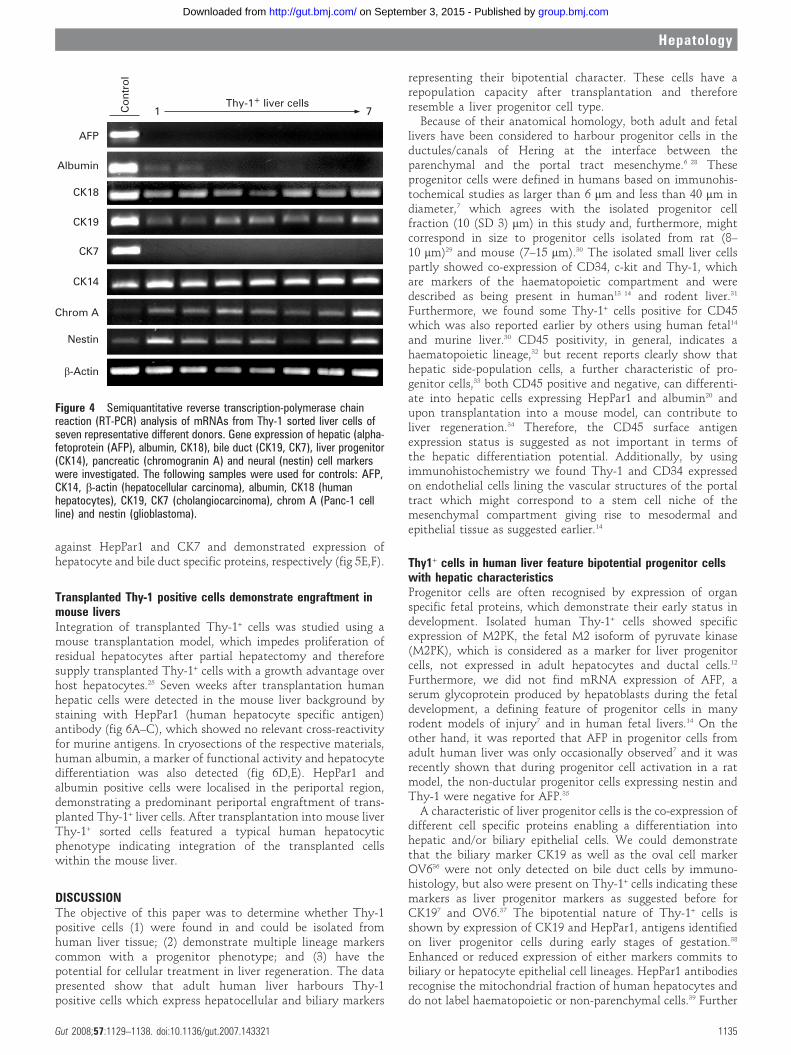
against HepPar1 and CK7 and demonstrated expression ofhepatocyte and bile duct specific proteins, respectively (fig 5E,F).
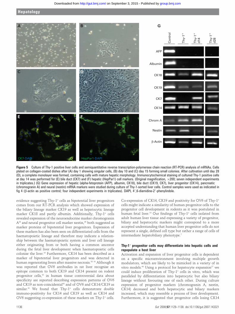
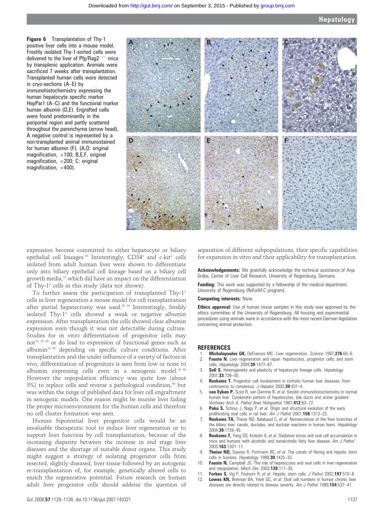
Transplanted Thy-1 positive cells demonstrate engraftment inmouse liversIntegration of transplanted Thy-1+ cells was studied using amouse transplantation model, which impedes proliferation ofresidual hepatocytes after partial hepatectomy and thereforesupply transplanted Thy-1+ cells with a growth advantage overhost hepatocytes.25 Seven weeks after transplantation humanhepatic cells were detected in the mouse liver background bystaining with HepPar1 (human hepatocyte specific antigen)antibody (fig 6A–C), which showed no relevant cross-reactivityfor murine antigens. In cryosections of the respective materials,human albumin, a marker of functional activity and hepatocytedifferentiation was also detected (fig 6D,E). HepPar1 andalbumin positive cells were localised in the periportal region,demonstrating a predominant periportal engraftment of trans-planted Thy-1+ liver cells. After transplantation into mouse liverThy-1+ sorted cells featured a typical human hepatocyticphenotype indicating integration of the transplanted cellswithin the mouse liver.
DISCUSSIONThe objective of this paper was to determine whether Thy-1positive cells (1) were found in and could be isolated fromhuman liver tissue; (2) demonstrate multiple lineage markerscommon with a progenitor phenotype; and (3) have thepotential for cellular treatment in liver regeneration. The datapresented show that adult human liver harbours Thy-1positive cells which express hepatocellular and biliary markers
representing their bipotential character. These cells have arepopulation capacity after transplantation and thereforeresemble a liver progenitor cell type.
Because of their anatomical homology, both adult and fetallivers have been considered to harbour progenitor cells in theductules/canals of Hering at the interface between theparenchymal and the portal tract mesenchyme.6 28 Theseprogenitor cells were defined in humans based on immunohis-tochemical studies as larger than 6 mm and less than 40 mm indiameter,7 which agrees with the isolated progenitor cellfraction (10 (SD 3) mm) in this study and, furthermore, mightcorrespond in size to progenitor cells isolated from rat (8–10 mm)29 and mouse (7–15 mm).30 The isolated small liver cellspartly showed co-expression of CD34, c-kit and Thy-1, whichare markers of the haematopoietic compartment and weredescribed as being present in human13 14 and rodent liver.31
Furthermore, we found some Thy-1+ cells positive for CD45which was also reported earlier by others using human fetal14
and murine liver.30 CD45 positivity, in general, indicates ahaematopoietic lineage,32 but recent reports clearly show thathepatic side-population cells, a further characteristic of pro-genitor cells,33 both CD45 positive and negative, can differenti-ate into hepatic cells expressing HepPar1 and albumin20 andupon transplantation into a mouse model, can contribute toliver regeneration.34 Therefore, the CD45 surface antigenexpression status is suggested as not important in terms ofthe hepatic differentiation potential. Additionally, by usingimmunohistochemistry we found Thy-1 and CD34 expressedon endothelial cells lining the vascular structures of the portaltract which might correspond to a stem cell niche of themesenchymal compartment giving rise to mesodermal andepithelial tissue as suggested earlier.14
Thy1+ cells in human liver feature bipotential progenitor cellswith hepatic characteristicsProgenitor cells are often recognised by expression of organspecific fetal proteins, which demonstrate their early status indevelopment. Isolated human Thy-1+ cells showed specificexpression of M2PK, the fetal M2 isoform of pyruvate kinase(M2PK), which is considered as a marker for liver progenitorcells, not expressed in adult hepatocytes and ductal cells.12
Furthermore, we did not find mRNA expression of AFP, aserum glycoprotein produced by hepatoblasts during the fetaldevelopment, a defining feature of progenitor cells in manyrodent models of injury7 and in human fetal livers.14 On theother hand, it was reported that AFP in progenitor cells fromadult human liver was only occasionally observed7 and it wasrecently shown that during progenitor cell activation in a ratmodel, the non-ductular progenitor cells expressing nestin andThy-1 were negative for AFP.35
A characteristic of liver progenitor cells is the co-expression ofdifferent cell specific proteins enabling a differentiation intohepatic and/or biliary epithelial cells. We could demonstratethat the biliary marker CK19 as well as the oval cell markerOV636 were not only detected on bile duct cells by immuno-histology, but also were present on Thy-1+ cells indicating thesemarkers as liver progenitor markers as suggested before forCK197 and OV6.37 The bipotential nature of Thy-1+ cells isshown by expression of CK19 and HepPar1, antigens identifiedon liver progenitor cells during early stages of gestation.38
Enhanced or reduced expression of either markers commits tobiliary or hepatocyte epithelial cell lineages. HepPar1 antibodiesrecognise the mitochondrial fraction of human hepatocytes anddo not label haematopoietic or non-parenchymal cells.39 Further
Figure 4 Semiquantitative reverse transcription-polymerase chainreaction (RT-PCR) analysis of mRNAs from Thy-1 sorted liver cells ofseven representative different donors. Gene expression of hepatic (alpha-fetoprotein (AFP), albumin, CK18), bile duct (CK19, CK7), liver progenitor(CK14), pancreatic (chromogranin A) and neural (nestin) cell markerswere investigated. The following samples were used for controls: AFP,CK14, b-actin (hepatocellular carcinoma), albumin, CK18 (humanhepatocytes), CK19, CK7 (cholangiocarcinoma), chrom A (Panc-1 cellline) and nestin (glioblastoma).
Hepatology
Gut 2008;57:1129–1138. doi:10.1136/gut.2007.143321 1135
group.bmj.com on September 3, 2015 - Published by http://gut.bmj.com/Downloaded from
evidence suggesting Thy-1+ cells as bipotential liver progenitorscomes from our RT-PCR analysis which showed expression ofthe biliary lineage marker CK19 as well as hepatocytic lineagemarker CK18 and partly albumin. Additionally, Thy-1+ cellsrevealed expression of the neuroendocrine marker chromograninA28 and neural progenitor cell marker nestin,40 both suggested asmarker proteins of bipotential liver progenitors. Expression ofthese markers has also been seen on differentiated cells from thehaematopoietic lineage and therefore may indicate a relation-ship between the haematopoietic system and liver cell lineageeither originating from or both having a common ancestorduring the fetal liver development when haematopoietic cellscolonise the liver.16 Furthermore, CK14 has been described as amarker of bipotential liver progenitors and was detected inhuman regenerating livers after massive necrosis.38 41 Although itwas reported that OV6 antibodies in rat liver recognise anepitope common to both CK19 and CK14 present on rodentprogenitor cells,42 in human tissue controversial data aboutspecificity are reported describing expression patterns of OV6and CK19 as non-coincidental43 and of OV6 and CK14/CK19 assimilar.41 We found that Thy-1+ cells demonstrate doubleimmuno-positivity for CK14 and CK19 as well as CK14 andOV6 suggesting co-expression of these markers on Thy-1+ cells.
Co-expression of CK14, CK19 and positivity for OV6 of Thy-1+
cells might indicate a similarity of human progenitor cells to theprogenitor cell development in rodents as it was postulated inhuman fetal liver.14 Our findings of Thy-1+ cells isolated fromadult human liver tissue and expressing a variety of progenitor,biliary and hepatocytic markers might correspond to a moreaccepted understanding that human liver progenitor cells do notrepresent a single, defined cell type but rather a range of cells ofintermediate hepatobiliary phenotype.7
Thy-1+ progenitor cells may differentiate into hepatic cells andrepopulate a host liverActivation and expansion of liver progenitor cells is dependenton a specific microenvironment involving multiple growthmodulators, which are likely to be mimicked in a variety of invitro models.44 Using a protocol for hepatocyte expansion27 wecould induce proliferation of Thy-1+ cells in vitro, which wasparalleled by differentiation into hepatocytic but also biliarylineage without favouring one of each other. During cultureexpression of progenitor markers (chromogranin A, nestin,CK14) decreased and both hepatocytic and biliary markersincreased, which may resemble a process of liver development.Furthermore, it is suggested that progenitor cells losing CK14
Figure 5 Culture of Thy-1 positive liver cells and semiquantitative reverse transcription-polymerase chain reaction (RT-PCR) analysis of mRNAs. Cellsplated on collagen-coated dishes after (A) day 1 showing singular cells, (B) day 10 and (C) day 15 forming small colonies. After cultivation until day 28(D), a complete monolayer was formed, containing cells with mature hepatic morphology. Immunocytochemical staining of cultured Thy-1 positive cellsat day 14 was performed for (E) bile duct (CK7) and (F) hepatic (HepPar1) cell markers. (Original magnification, 6200; seven independent experimentsin triplicates.) (G) Gene expression of hepatic (alpha-fetoprotein (AFP), albumin, CK18), bile duct (CK19, CK7), liver progenitor (CK14), pancreatic(chromogranin A) and neural (nestin) mRNA markers were studied during culture of Thy-1-sorted liver cells. Control samples were used as indicated infig 4 (b-actin as positive control; four independent experiments in triplicates). DAPI, 49,6-diamidine-29-phenylindole.
Hepatology
1136 Gut 2008;57:1129–1138. doi:10.1136/gut.2007.143321
group.bmj.com on September 3, 2015 - Published by http://gut.bmj.com/Downloaded from
expression become committed to either hepatocyte or biliaryepithelial cell lineages.38 Interestingly, CD34+ and c-kit+ cellsisolated from adult human liver were shown to differentiateonly into biliary epithelial cell lineage based on a biliary cellgrowth media,13 which did have an impact on the differentiationof Thy-1+ cells in this study (data not shown).
To further assess the participation of transplanted Thy-1+
cells in liver regeneration a mouse model for cell transplantationafter partial hepatectomy was used.25 26 Interestingly, freshlyisolated Thy-1+ cells showed a weak or negative albuminexpression. After transplantation the cells showed clear albuminexpression even though it was not detectable during culture.Studies for in vitro differentiation of progenitor cells maynot14 19 45 or do lead to expression of functional genes such asalbumin19 45 depending on specific culture conditions. Aftertransplantation and the under influence of a variety of factors invivo, differentiation of progenitors is seen from low or none toalbumin expressing cells even in a xenogenic model.19 45
However the repopulation efficiency was quite low (about3%) to replace cells and reverse a pathological condition,46 butwas within the range of published data for liver cell engraftmentin xenogenic models. One reason might be murine liver failingthe proper microenvironment for the human cells and thereforeno cell cluster formation was seen.
Human bipotential liver progenitor cells would be aninvaluable therapeutic tool to induce liver regeneration or tosupport liver function by cell transplantation, because of theincreasing disparity between the increase in end stage liverdiseases and the shortage of suitable donor organs. This studymight suggest a strategy of isolating progenitor cells fromresected, slightly diseased, liver tissue followed by an autogenicre-transplantation of, for example, genetically altered cells toenrich the regenerative potential. Future research on humanadult liver progenitor cells should address the question of
separation of different subpopulations, their specific capabilitiesfor expansion in vitro and their applicability for transplantation.
Acknowledgements: We gratefully acknowledge the technical assistance of AnjaGrabe, Center of Liver Cell Research, University of Regensburg, Germany.
Funding: This work was supported by a fellowship of the medical department,University of Regensburg (ReForM-C program).
Competing interests: None.
Ethics approval: Use of human tissue samples in this study was approved by theethics committee of the University of Regensburg. All housing and experimentalprocedures using animals were in accordance with the most recent German legislationconcerning animal protection.
REFERENCES1. Michalopoulos GK, DeFrances MC. Liver regeneration. Science 1997;276:60–6.2. Fausto N. Liver regeneration and repair: hepatocytes, progenitor cells, and stem
cells. Hepatology 2004;39:1477–87.3. Sell S. Heterogeneity and plasticity of hepatocyte lineage cells. Hepatology
2001;33:738–50.4. Roskams T. Progenitor cell involvement in cirrhotic human liver diseases: from
controversy to consensus. J Hepatol 2003;39:431–4.5. van Eyken P, Sciot R, van Damme B, et al. Keratin immunohistochemistry in normal
human liver. Cytokeratin pattern of hepatocytes, bile ducts and acinar gradient.Virchows Arch A, Pathol Anat Histopathol 1987;412:63–72.
6. Paku S, Schnur J, Nagy P, et al. Origin and structural evolution of the earlyproliferating oval cells in rat liver. Am J Pathol 2001;158:1313–23.
7. Roskams TA, Theise ND, Balabaud C, et al. Nomenclature of the finer branches ofthe biliary tree: canals, ductules, and ductular reactions in human livers. Hepatology2004;39:1739–45.
8. Roskams T, Yang SQ, Koteish A, et al. Oxidative stress and oval cell accumulation inmice and humans with alcoholic and nonalcoholic fatty liver disease. Am J Pathol2003;163:1301–11.
9. Theise ND, Saxena R, Portmann BC, et al. The canals of Hering and hepatic stemcells in humans. Hepatology 1999;30:1425–33.
10. Fausto N, Campbell JS. The role of hepatocytes and oval cells in liver regenerationand repopulation. Mech Dev 2003;120:117–30.
11. Forbes S, Vig P, Poulsom R, et al. Hepatic stem cells. J Pathol 2002;197:510–8.12. Lowes KN, Brennan BA, Yeoh GC, et al. Oval cell numbers in human chronic liver
diseases are directly related to disease severity. Am J Pathol 1999;154:537–41.
Figure 6 Transplantation of Thy-1positive liver cells into a mouse model.Freshly isolated Thy-1-sorted cells weredelivered to the liver of Pfp/Rag22/2 miceby transplenic application. Animals weresacrificed 7 weeks after transplantation.Transplanted human cells were detectedin cryo-sections (A–E) byimmunohistochemistry expressing thehuman hepatocyte specific markerHepPar1 (A–C) and the functional markerhuman albumin (D,E). Engrafted cellswere found predominantly in theperiportal region and partly scatteredthroughout the parenchyma (arrow head).A negative control is represented by anon-transplanted animal immunostainedfor human albumin (F). (A,D: originalmagnification, 6100; B,E,F, originalmagnification, 6200; C: originalmagnification, 6400).
Hepatology
Gut 2008;57:1129–1138. doi:10.1136/gut.2007.143321 1137
group.bmj.com on September 3, 2015 - Published by http://gut.bmj.com/Downloaded from

13. Crosby HA, Kelly DA, Strain AJ. Human hepatic stem-like cells isolated using c-kit orCD34 can differentiate into biliary epithelium. Gastroenterology 2001;120:534–44.
14. Masson NM, Currie IS, Terrace JD, et al. Hepatic progenitor cells in human fetal liverexpress the oval cell marker Thy-1. Am J Physiol Gastrointest Liver Physiol2006;291:G45–54.
15. Theise ND, Nimmakayalu M, Gardner R, et al. Liver from bone marrow in humans.Hepatology 2000;32:11–6.
16. Thorgeirsson SS, Grisham JW. Hematopoietic cells as hepatocyte stem cells: acritical review of the evidence. Hepatology 2006;43:2–8.
17. Wang X, Willenbring H, Akkari Y, et al. Cell fusion is the principal source of bone-marrow-derived hepatocytes. Nature 2003;422:897–901.
18. Walkup MH, Gerber DA. Hepatic stem cells: in search of. Stem Cells2006;24:1833–40.
19. Dan YY, Riehle KJ, Lazaro C, et al. Isolation of multipotent progenitor cells fromhuman fetal liver capable of differentiating into liver and mesenchymal lineages. ProcNatl Acad Sci U S A 2006;103:9912–7.
20. Hussain SZ, Strom SC, Kirby MR, et al. Side population cells derived from adulthuman liver generate hepatocyte-like cells in vitro. Dig Dis Sci 2005;50:1755–63.
21. Fiegel HC, Gluer S, Roth B, et al. Stem-like cells in human hepatoblastoma.J Histochem Cytochem 2004;52:1495–501.
22. Thasler WE, Weiss TS, Schillhorn K, et al. Charitable state-controlled foundationhuman tissue and cell research: ethics and legal aspects in the supply of surgicallyremoved human tissue for research in the academic and commercial sector inGermany. Cell and Tissue Banking 2003;4:49–56.
23. Weiss TS, Pahernik S, Scheruebl I, et al. Cellular damage to human hepatocytesthrough repeated application of 5-aminolevulinic acid. J Hepatol 2003;38:476–82.
24. Fabris L, Strazzabosco M, Crosby HA, et al. Characterization and isolation of ductularcells coexpressing neural cell adhesion molecule and Bcl-2 from primarycholangiopathies and ductal plate malformations. Am J Pathol 2000;156:1599–612.
25. Witek RP, Fisher SH, Petersen BE. Monocrotaline, an alternative to retrorsine-basedhepatocyte transplantation in rodents. Cell Transplant 2005;14:41–7.
26. Aurich I, Mueller LP, Aurich H, et al. Functional integration of hepatocytes derivedfrom human mesenchymal stem cells into mouse livers. Gut 2007;56:405–15.
27. Lazaro CA, Croager EJ, Mitchell C, et al. Establishment, characterization, and long-term maintenance of cultures of human fetal hepatocytes. Hepatology2003;38:1095–106.
28. Roskams T. Different types of liver progenitor cells and their niches. J Hepatol2006;45:1–4.
29. Dabeva MD, Hwang SG, Vasa SR, et al. Differentiation of pancreatic epithelialprogenitor cells into hepatocytes following transplantation into rat liver. Proc NatlAcad Sci U S A 1997;94:7356–61.
30. Wang X, Foster M, Al Dhalimy M, et al. The origin and liver repopulating capacity ofmurine oval cells. Proc Natl Acad Sci U S A 2003;100(Suppl 1):11881–8.
31. Petersen BE, Goff JP, Greenberger JS, et al. Hepatic oval cells express thehematopoietic stem cell marker Thy-1 in the rat. Hepatology 1998;27:433–45.
32. Knapp W, Dorken B, Rieber P, et al. CD antigens 1989. Blood 1989;74:1448–50.33. Shimano K, Satake M, Okaya A, et al. Hepatic oval cells have the side population
phenotype defined by expression of ATP-binding cassette transporter ABCG2/BCRP1.Am J Pathol 2003;163:3–9.
34. Wulf GG, Luo KL, Jackson KA, et al. Cells of the hepatic side population contribute toliver regeneration and can be replenished with bone marrow stem cells.Haematologica 2003;88:368–78.
35. Koenig S, Probst I, Becker H, et al. Zonal hierarchy of differentiation markers andnestin expression during oval cell mediated rat liver regeneration. Histochem Cell Biol2006;126:723–34.
36. Roskams T, De Vos R, van Eyken P, et al. Hepatic OV-6 expression in human liverdisease and rat experiments: evidence for hepatic progenitor cells in man. J Hepatol1998;29:455–63.
37. Crosby HA, Hubscher S, Fabris L, et al. Immunolocalization of putative human liverprogenitor cells in livers from patients with end-stage primary biliary cirrhosis andsclerosing cholangitis using the monoclonal antibody OV-6. Am J Pathol1998;152:771–9.
38. Haque S, Haruna Y, Saito K, et al. Identification of bipotential progenitor cells inhuman liver regeneration. Lab Invest 1996;75:699–705.
39. Minervini MI, Demetris AJ, Lee RG, et al. Utilization of hepatocyte-specific antibodyin the immunocytochemical evaluation of liver tumors. Mod Pathol 1997;10:686–92.
40. Gleiberman AS, Encinas JM, Mignone JL, et al. Expression of nestin-greenfluorescent protein transgene marks oval cells in the adult liver. Dev Dyn2005;234:413–21.
41. Seki S, Kitada T, Sakaguchi H, et al. Expression of progenitor cell markers in liverswith fulminant massive necrosis. Hepatol Res 2003;25:149–57.
42. Bisgaard HC, Parmelee DC, Dunsford HA, et al. Keratin 14 protein in culturednonparenchymal rat hepatic epithelial cells: characterization of keratin 14 and keratin19 as antigens for the commonly used mouse monoclonal antibody OV-6. MolCarcinogen 1993;7:60–6.
43. Crosby HA, Hubscher SG, Joplin RE, et al. Immunolocalization of OV-6, a putativeprogenitor cell marker in human fetal and diseased pediatric liver. Hepatology1998;28:980–5.
44. Santoni-Rugiu E, Jelnes P, Thorgeirsson SS, et al. Progenitor cells in liverregeneration: molecular responses controlling their activation and expansion. APMIS2005;113:876–902.
45. Schmelzer E, Zhang L, Bruce A, et al. Human hepatic stem cells from fetal andpostnatal donors. J Exp Med 2007;204:1973–87.
46. Fox IJ, Chowdhury JR, Kaufman SS, et al. Treatment of the Crigler–Najjar syndrometype I with hepatocyte transplantation. N Engl J Med 1998;338:1422–6.
Take advantage of BMJ Journals’ remarkable catalogue of titles with Related Collections
No busy professional has time to browse through all pertinent journals to find relevant articles, but withRelated Collections you no longer have to. Follow the ‘‘Related Collections’’ link from any article and usethe ‘‘Show Collections from other Journals’’ to expand your search across all BMJ Journals. Or simplyfollow the ‘‘Browse by topic’’ link on the home page. By setting up your own collections and receivingemail alerts every time an article is added to your chosen area, you can build up your own significantbody of knowledge.
Hepatology
1138 Gut 2008;57:1129–1138. doi:10.1136/gut.2007.143321
group.bmj.com on September 3, 2015 - Published by http://gut.bmj.com/Downloaded from

livers for cell transplantationHepatic progenitor cells from adult human
Brockhoff, L A Kunz-Schughart, K-W Jauch, H-J Schlitt and W E ThaslerT S Weiss, M Lichtenauer, S Kirchner, P Stock, H Aurich, B Christ, G
doi: 10.1136/gut.2007.1433212008 57: 1129-1138 originally published online April 16, 2008Gut
http://gut.bmj.com/content/57/8/1129Updated information and services can be found at:
These include:
References #BIBLhttp://gut.bmj.com/content/57/8/1129
This article cites 46 articles, 10 of which you can access for free at:
serviceEmail alerting
box at the top right corner of the online article. Receive free email alerts when new articles cite this article. Sign up in the
CollectionsTopic Articles on similar topics can be found in the following collections
(98)Editor's choice
Notes
http://group.bmj.com/group/rights-licensing/permissionsTo request permissions go to:
http://journals.bmj.com/cgi/reprintformTo order reprints go to:
http://group.bmj.com/subscribe/To subscribe to BMJ go to:
group.bmj.com on September 3, 2015 - Published by http://gut.bmj.com/Downloaded from
Related Documents