Heparan Sulfate Proteoglycans from Mouse Mammary Epithelial Cells: Localization on the Cell Surface with a Monoclonal Antibody MARKKU JALKANEN, HUNG NGUYEN, ALAN RAPRAEGER, NURITH KURN, MERTON BERNFIELD Department of Pediatrics, Stanford University School of Medicine, Stanford, California 94305 and ABSTRACT Mouse mammary epithelial cells, of the normal rouTine mammary gland (NMuMG) cell line, bear a heparan sulfate-rich proteoglycan (HSPG) on their surfaces. A hybridoma (281-2) secreting a monoclonal antibody that recognizes this HSPG was produced by fusion of SP-2]0 myeloma cells with spleen cells from rats immunized with NMuMG cells. The 281- 2 monoclonal antibody is directed against the core protein of the cell surface HSPG, as demonstrated by (a) recognition of the isolated proteoglycan but not its glycosaminoglycan chains, (b) co-localization of 281-2-specific antigen and radioactive cell surface HSPG on gradient polyacrylamide gel electrophoresis and on isopycnic centrifugation, and (c) abolition of immunofluorescent staining of the NMuMG cell surface by the intact, but not the protease- digested ectodomain of the cell surface HSPG. The antibody is specific for cell surface HSPG and does not recognize the HSPG that accumulates extracellularly beneath the basal cell surface. Therefore, the 281-2 antibody may be used to isolate the cell surface HSPG and to explore its distribution in tissues. Interactions of cells with extracellular materials are likely mediated by cell surface matrix receptors (44). Among these are cell surface heparan sulfate proteoglycans (HSPGs) t (25, 34). These molecules, consisting of a core protein bearing polyanionic heparan sulfate glycosaminoglycan (GAG) chains, are found at or near the surface of all adherent cell types (16). Cell surface HSPG may exist in two forms, either bound to the cell surface via the GAG chains (21) or inter- calated into the plasma membrane via the core protein (22, 27, 34). There are also extracellular matrix forms of HSPG, found in parenchymal cell basement membranes (14) or in the pericellular matrices of fibroblasts (15). We describe a monoclonal antibody directed against the core protein of an HSPG that is intercalated into the plasma membrane of mouse mammary epithelial cells of the normal murine mammary gland (NMuMG) cell line. The GAG com- position, size, buoyant density, and/or resistance to reduction JAbbreviations used in this paper: DME, Dulbecco's modified Eagle's medium; FCS, fetal calf serum; GAG, glycosaminoglycan; GdnHC1, guanidine-hydrochloride; HSPG(s), heparan sulfate proteoglycan(s); NMuMG, normal murine mammary gland; PG(s), proteoglycan(s); TBS, Tris-buffered saline. 976 appear to distinguish this HSPG from some other cellular HSPGs such as those from rat hepatocytes (29), a hepatoma cell line (30), human fibroblasts (3, 5), bovine or rabbit aortic endothelial cells (2, 31), or human colon carcinoma cells (17), and also from those isolated from basement membranes, such as from the mouse EHS tumor (10, 14) and the rat kidney glomerulus (19, 39). Indeed, the chemical characteristics of each of these HSPGs appear to differ from the others in some respect. Despite these apparent differences, the various HSPGs that have been isolated have not been compared to the extent that their relationships are conclusively established. Based on immunological reactivity (14, 17, 30, 39, 43), some HSPGs may be similar, or perhaps the antibodies detect similar determinants on distinct HSPGs. To characterize a single type of HSPG immunologically, more specific immu- noglobulins, such as monoclonal antibodies, may be required. A part of this study was presented in abstract form (1). MATERIALS AND METHODS Cell Culture and Labeling NMuMG mouse mammary epithelial cells were obtained and cultured in bicarbonate-buffered Dulbecco's modified Eagle's medium (DME" Gibco Lab- THE JOURNAL OF CELL BIOLOGY • VOLUME 101 SEPTEMBER 1985 976-984 © The Rockefeller University Press - 0021-952518510910976109 $1.00 on June 16, 2014 jcb.rupress.org Downloaded from Published September 1, 1985

Welcome message from author
This document is posted to help you gain knowledge. Please leave a comment to let me know what you think about it! Share it to your friends and learn new things together.
Transcript
Heparan Sulfate Proteoglycans from Mouse
Mammary Epithelial Cells: Localization
on the Cell Surface with a Monoclonal Antibody
MARKKU JALKANEN, HUNG NGUYEN, ALAN RAPRAEGER, NURITH KURN, MERTON BERNFIELD Department of Pediatrics, Stanford University School of Medicine, Stanford, California 94305
and
ABSTRACT Mouse mammary epithelial cells, of the normal rouTine mammary gland (NMuMG) cell line, bear a heparan sulfate-rich proteoglycan (HSPG) on their surfaces. A hybridoma (281-2) secreting a monoclonal antibody that recognizes this HSPG was produced by fusion of SP-2]0 myeloma cells with spleen cells from rats immunized with NMuMG cells. The 281- 2 monoclonal antibody is directed against the core protein of the cell surface HSPG, as demonstrated by (a) recognition of the isolated proteoglycan but not its glycosaminoglycan chains, (b) co-localization of 281-2-specific antigen and radioactive cell surface HSPG on gradient polyacrylamide gel electrophoresis and on isopycnic centrifugation, and (c) abolition of immunofluorescent staining of the NMuMG cell surface by the intact, but not the protease- digested ectodomain of the cell surface HSPG. The antibody is specific for cell surface HSPG and does not recognize the HSPG that accumulates extracellularly beneath the basal cell surface. Therefore, the 281-2 antibody may be used to isolate the cell surface HSPG and to explore its distribution in tissues.
Interactions of cells with extracellular materials are likely mediated by cell surface matrix receptors (44). Among these are cell surface heparan sulfate proteoglycans (HSPGs) t (25, 34). These molecules, consisting of a core protein bearing polyanionic heparan sulfate glycosaminoglycan (GAG) chains, are found at or near the surface of all adherent cell types (16). Cell surface HSPG may exist in two forms, either bound to the cell surface via the GAG chains (21) or inter- calated into the plasma membrane via the core protein (22, 27, 34). There are also extracellular matrix forms of HSPG, found in parenchymal cell basement membranes (14) or in the pericellular matrices of fibroblasts (15).
We describe a monoclonal antibody directed against the core protein of an HSPG that is intercalated into the plasma membrane of mouse mammary epithelial cells of the normal murine mammary gland (NMuMG) cell line. The GAG com- position, size, buoyant density, and/or resistance to reduction
J Abbreviations used in this paper: DME, Dulbecco's modified Eagle's medium; FCS, fetal calf serum; GAG, glycosaminoglycan; GdnHC1, guanidine-hydrochloride; HSPG(s), heparan sulfate proteoglycan(s); NMuMG, normal murine mammary gland; PG(s), proteoglycan(s); TBS, Tris-buffered saline.
976
appear to distinguish this HSPG from some other cellular HSPGs such as those from rat hepatocytes (29), a hepatoma cell line (30), human fibroblasts (3, 5), bovine or rabbit aortic endothelial cells (2, 31), or human colon carcinoma cells (17), and also from those isolated from basement membranes, such as from the mouse EHS tumor (10, 14) and the rat kidney glomerulus (19, 39). Indeed, the chemical characteristics of each of these HSPGs appear to differ from the others in some respect. Despite these apparent differences, the various HSPGs that have been isolated have not been compared to the extent that their relationships are conclusively established. Based on immunological reactivity (14, 17, 30, 39, 43), some HSPGs may be similar, or perhaps the antibodies detect similar determinants on distinct HSPGs. To characterize a single type of HSPG immunologically, more specific immu- noglobulins, such as monoclonal antibodies, may be required. A part of this study was presented in abstract form (1).
MATERIALS AND METHODS
Cell Culture and Labeling NMuMG mouse mammary epithelial cells were obtained and cultured in
bicarbonate-buffered Dulbecco's modified Eagle's medium (DME" Gibco Lab-
THE JOURNAL OF CELL BIOLOGY • VOLUME 101 SEPTEMBER 1985 976-984 © The Rockefeller University Press - 0021-952518510910976109 $1.00
on June 16, 2014jcb.rupress.org
Dow
nloaded from
Published September 1, 1985
oratories, Grand Island. NY) supplemented with 10% fetal calf serum (FCS; Tissue Culture Biological, Tulare, CA) as described previously (7, 34). For labeling, confluent monolayers on plastic substrata (usually 100-ram dishes; Falcon Labware, Oxnard, CA) were incubated in low sulfate medium containing 100 U/ml penicillin G, 50 ug/ml streptomycin sulfate, and 100 ttCi/ml of carrier-free H2a~SO4 (New England Nuclear, Boston, MA) for 24 h, a period sufficient for the cells to reach a steady state level of a5so4 incorporation (6).
Preparation of HSPGs TOTAL PROTEOGLYCANS: Total cellular and basal extracellular pro-
teoglycans (PGs) were isolated from cells cultured on collagen gels that were adsorbed to polyamine polyacrylamide beads (Affi Gel 731; Bio-Rad Labora- tories, Richmond, CA). Type I collagen purified from rat tail tendons (4) and solubilized in 0.1 M acetic acid was adjusted to pH 5.7 with 1 M NaOH and incubated overnight with beads (~4 mg collagen per 1 g of beads). The beads were washed with 10 mM phosphate-buffered saline (PBS), pH 7.4 to remove non-bound collagen. Cells (-1.8-2.0 × 10⁶) suspended by trypsin treatment were mixed with 1.4 g of beads in I 0 ml of DME containing 10% FCS for 30 min at room temperature. The beads and cells were plated into 175-cm 2 culture flasks (20 total) and incubated at 37"C without mixing. Medium was replaced on the first day to remove non-bound ceils and then on alternate days, On day 7, two flasks were labeled with H235SO4 (25 uCi/ml), and on day 8, the beads were harvested by settling. The beads were extracted by incubation (for 8 h at 4"C) in 4 M guanidine-hydrochloride (Gdn-HCI; Schwarz-Mann, Spring Valley, NY) containing 50 mM sodium acetate (pH 5.5), 0.5% Triton X-100, and the following proteinase inhibitors: 5 mM EDTA, 5 mM benzamidine (Aldrich Chemical Co., Milwaukee, Wl), 5 mM N-ethylmaleimide (Calbiochem-Behring Corp., La Jolla, CA), and 1 mM phenylmethylsulfonyl fluoride (Calbiochem- Behring Corp.). The 500 g supernatant was boiled and dialyzed against 8 M urea (Schwarz-Mann) containing 50 mM sodium acetate, pH 4.5 and 0.2 M NaCI, and was purified on a DEAE-Sephacel column as described below.
BASAL EXTRACELLULAR AND CELL SURFACE PGS: PGs depos- ited beneath the cell monolayer (basal extracellular PG) and the ectodomain of the cell surface PG were prepared from confluent monolayers after they were labeled with radiosulfate. The medium was discarded, and the monolayers were washed twice with cold Tris-buffered saline (TBS; l0 mM Tris, pH 7.4, 150 mM NaC1 containing 1.25 mM CaCI2 and 0.9 mM MgSO4). The monolayers were scraped with a rubber policeman into cold TBS-EDTA (0.5 mM EDTA) containing the following proteinase inhibitors: 0.5 mM phenylmethylsulfonyl fluoride, 5 mM benzamidine, and 5 mM N-ethylmaleimide. The cells were centrifuged (200 g) and the supernatant, containing the basal extracellular PG, was brought to 8 M urea or 4 M GdnHCl, buffered as above, and boiled.
The ectodomain of the cell surface PG was prepared by trypsin treatment of the cells (35). The cell pellet was washed four times in cold TBS-EDTA, and suspended by gentle agitation. Bovine pancreatic trypsin (2x crystallized, type ill: Sigma Chemical Co., St. Louis, MO) was added to 20 ug/ml and the mixture incubated at 4*C for 5 min. After 100 ug/ml soybean trypsin inhibitor (Sigma Chemical Co,) was added, the cells were centrifuged again, leaving the ectodomain of the cell surface PG in the supernatant. This supernatant was brought to 8 M urea and purified over DEAE-Sephacel as described below.
PURIFICATION AND ANALYSIS OF PGS: PGs were adsorbed on a column (1 × 5 cm) of DEAE-Sephacel (Pharmacia Inc., Piscataway, N J) in 50 mM sodium acetate (pH 4.5) containing 8 M urea and 0.2 M NaCI, washed with the same buffer, and eluted with a linear gradient of 0.2-0.8 M NaCI in this solution. Both the basal extracellular PCJs and the ectodomain eluted as a single peak of 35SO4-1abeled material at 0.5-0.6 M NaCI. The labeled material was either precipitated by mixing with 4 vol ethanol at -20*C followed by centrifugation, or stored in 8 M urea at -20"C. The 35S-ectodomain prepared and purified in this manner contains all the GAG chains, but lacks the hydrophobic domain and is slightly smaller than the parent cell surface PG (24. 35).
Cellular and basal extracellular PGs in 50 mM sodium acetate (pH 4.5) containing 4 M GdnHCI and 1% Triton X-100 were analyzed by isopycnic centrifugation. Samples were brought to 1.4 g/ml with crystalline CsCI (Schwarz-Mann Biological Grade) and centrifuged at 15"C for 72 h at 46,000 rpm in a Beckman SW 65 rotor (Beckman Instruments, Inc., Palo Alto, CA). Fractions were collected and analyzed for density by weighing. Total PG radioactivity was assessed by modification of the procedure of Stephens et al. (38). Cetylpyridinium chloride-impregnated Whatman 3MM filter discs (What- man Inc., Clifton, N J) were prepared by soaking in 2.5% cetylpyridinium chloride followed by drying. Aliquots were spotted on the dry discs which were washed five times in distilled water, soaked for 1 h in 25 mM sodium sulfate, and rinsed several times in distilled water. Finally, the discs were soaked for 30 min in 10% trichloroacetic acid (TCA) followed by washes in distilled water and 95% ethanol and were dried for scintillation counting.
PG relative mass was assessed by gradient PAGE (described below) or by
Sepharose CL-4B (Pharmacia Inc.) chromatography in 4 M GdnHCI buffered to pH 5.5 by 50 mM sodium acetate, or in 1% SDS, 100 mM NaCI buffered to pH 8.0 with 50 mM Tris-HC1. Radioactivities in solutions were assayed by scintillation counting using 6 vol of Aquamix (West Chemical Products, Inc., New York, NY) for each sample volume. Samples containing 4 M GdnHCI were diluted 1:10 with water before mixing with Aquamix,
Characteristics of the PGs were investigated by various treatments. PGs were (a) treated with 0.5 M NaOH, 0.1 M NaBH4 for 18 h at 37°C to remove GAG; (b) digested with papain ( 1 mg/ml, Worthington Biochemical Corp. Freehold, N J) in 5 mM cysteine-EDTA pH 7.5 for 24 h at 60°C to degrade the core protein; (c) treated with heparitinase (0.1 U/ml; Miles Laboratories, Inc., Naperville, IL) in 50 mM Tris, pH 7.0 and 5 mM calcium acetate for 3 h at 37"C to digest heparan sulfate or; (d) with chondroitin sulfate ABC lyase (0.5 U/ml; Miles Laboratories, Inc.) in 50 mM Tris, pH 8.0 and 30 mM sodium acetate to digest chondroitin and dermatan sulfates. Both Tris buffers also contained 0.1 mg/ml bovine serum albumin (BSA) and 1 mM phenylmethyl- sulfonyl fluoride.
Preparation of Monoclonal Antibodies IMMUNIZATION AND PRODUCTION OF HYBRIDOMAS; Confluent
NMuMG cells were washed three times with cold PBS and the cultures harvested by scraping with a rubber policeman. An aliquot of this suspension (0.25 ml in PBS) containing ~4 x l 0 7 cells was combined with an equal volume of Freund's complete adjuvant and injected into two hind foot pads of ~30-d- old Fisher rats. The rats received an identical inoculum intraperitoneally twice more at 10-d intervals. 10 d after the second intraperitoneal injection, blood samples were taken, and sera were tested for reactivity against PGs (see below). Rats with reactive serum received a third intraperitoneal injection 3 d before their spleens were harvested.
For hybridoma production, a single cell suspension of rat spleen cells (107/ spleen) (37) was mixed (1:1) with log phase mouse SP-2/0 myeloma cells (kindly provided by Dr. I. Weissman, Department of Pathology, Stanford University) in 38% polyethyleneglycol 1540 (J. T. Baker Chemical Co., Phil- lipsburg, NJ) for fusion. The cells were grown on multidish 12-ram plates at 4 × l0 s cells/cm 2 in HAT-DME containing 20% FCS as described elsewhere (1 I). Hybridoma supernatants were screened by a solid phase radioimmunoas- say as described below. Selected hybridomas were cloned, then subcloned by limiting dilution and assessed for antibody production by immunodot assay as described below.
PRODUCTION AND PURIFICATION OF MONOCLONAL ANTI- BODIES: Selected subelones of hybridoma 281-2, a hybrid producing an antibody specific for HSPG (see Results), were cultured in serum-free medium and as ascites in splenectomized nude mice. Hybridoma cells (4 x 10⁵/cm 2) were plated onto 100-ram dishes (Falcon Labware) in HB 101 medium (Hana Biologics, Berkeley, CA) without serum, and medium was harvested every 4 d. The immunoglobulins in this medium were precipitated overnight at 4"C after slowly adding ammonium sulfate to 50% saturation. The precipitate was collected at 10,000 g, as described by Hardy (13), dissolved in TBS, and dialyzed against TBS.
For the production of ascites fluid, athymic nude BALB/c mice (Harlan Sprague Dawley, Inc., Indianapolis, IN) were spleneetomized, and 2 d later received a 0.5-ml i.p. injection of 2, 6, 10, 14-tetramethyl pentadicane (Aldrich Chemical Co.). 1 wk later, l0 T hybridoma cells in 0.5 ml DME were injected intraperitoneally, and aseites fluids were harvested 7-10 d later. Immunoglob- ulins in clarified ascites fluids (25 ml) were isolated by diluting 1:1 with TBS and precipitating as described above. The precipitate was dissolved in TBS and desalted over a Sephadex G-25 column (Pharmacia Inc.) in TBS.
The immunoglobulins from both procedures were further purified by mixing with QAE-Sephadex (Pharmacia Inc.) in TBS for 3 h at 4°C, which was removed by centrifugation at 20,000 g for 30 rain, and the supernatants containing the monoclonal antibodies (13) were stored at -70*C or in liquid nitrogen. Protein concentrations were estimated using BSA as a standard (26). Concentrations and type of antibody used in each experiment are indicated in the figure legends.
Immunotechniques A S S A Y F O R R A T S E R U M A N T 1B O D I E S : Antibody with reactivity against
PG was demonstrated in rat serum by a Staphylococcus aureus-binding assay (12, 20). S. aureus (Calbiochem-Behring Corp.) were washed three times by suspension and centrifugation in 50 mM Tris-HCl, pH 8.3, 0.45 M NaCl, and 0,5% Nonidet-P40 (Calbiochem-Behring Corp.), resuspended in the same buffer containing 0.1% BSA and rabbit anti-rat lgG (final concentration 400 ug/ml; Dako Corp., Santa Barbara, CA), and incubated for 1 h at 4"C. After three more washes and resuspension, the S. aureus were incubated with rat serum (l : l0 dilution) for 2 h. After removal of serum by similar washings, 5,000 cpm
JALKANEN ETA[. Monoclonal Antibody for Heparan Sulfate Proteoglycan 977
on June 16, 2014jcb.rupress.org
Dow
nloaded from
Published September 1, 1985
of purified 35SO4-PG was added and incubated overnight in the same buffer. Nonbound radioactivity was removed by washing, the pellet was suspended in 2% SDS, boiled, and the radioactivity released was measured by scintillation counting.
SOLID-PHASE RADIOIMMUNOASSAY: Individual wells of 96-well plates (Flow Laboratories, Inc., McLean, VA) containing 0.15 ml rabbit anti- rat lgG (final concentration 2 mg/ml in TBS; Dako Corp.) were incubated overnight at 4°C. The lgG was replaced with TBS containing 0.5% BSA (BSA- TBS) for 1 h at room temperature, and the wells were filled (200 ul) with hybridoma medium and incubated overnight at 4"C. After washing the wells three times with BSA-TBS, 5,000-6,000 cpm of 35S-labeled PG in 0.15 ml BSA-TBS was added and incubated overnight at 4"C. After washing as in the previous step, the wells were removed by trimming with a hot wire, and each well was counted by liquid scintillation.
I M M U N O D O T ASSAY ON D E A L - P A P E R : Similar sized pieces of DEAL-cellulose paper (Whatman DE 81) and Whatman 3 MM paper (What- man Inc.) were wet with TBS and placed in a Microfold-dot apparatus (V & P Scientific, San Diego, CA) such that the 3 MM paper underlaid the DEAL- paper. Antigen in 8 M urea buffered to pH 4.5 by 50 mM sodium acetate (usually 0.1-4 ug PG protein in 200 ~1) was applied through the apparatus wells onto the paper, followed by washing 5-6 times with TBS under vacuum without letting the paper dry. The antibody preparation (culture medium or antibody diluted in TBS) was either similarly applied to and washed off the paper, or incubated batchwise overnight at 4"C with the DEAL-paper which had been previously incubated at room temperature for 10 rain with 1% FCS in TBS. After either antibody treatment, the DEAL-paper in a dish was washed 5-6 times in TBS and incubated with peroxidase-conjugated rabbit anti-rat IgG (final dilution 1:200 in TBS containing 1% FCS; Dako Corp.) for 30 min with gentle shaking. The paper was then washed for 20-30 min by shaking with 5-6 changes of TBS. Binding of the peroxidase conjugate was visualized using 0.05% (wt/vol) 4-chloro-l-naphthol (Sigma Chemical Co.) containing 0.03% (vol/vol) hydrogen peroxide in TBS (8).
SOS P A G E A N D W E S T E R N - B L O T T I N G ON D E A E - C E L L U -
LOS E: Proteins were fractionated in a gradient (3.8-20.0%) polyacrylamide gel using PAGE buffer composed of 40 mM Tris, 60 mM boric acid, 0.8 mM EDTA, 1 mM Na2SO4, and 0,1% SDS. The linear gradient gel was formed from 3.8 and 20% acrylamide solutions containing 7.5% bisacrylamide, 2.6 M urea, and 0.2 and I M glycerol, respectively. The gel was run overnight to completion at 125 V in a Hoefer SE 500 apparatus (Hoefer Scientific Instru- ments, San Francisco, CA) in PAGE buffer. The sample loading buffer consisted of the PAGE buffer containing 2% SDS, 0.5 M glycerol, and bromophenol blue (0.01 mg/ml). After the run, the gel was transferred onto a pad of a Hoefer TE 52 transfer apparatus and covered with DEAL-cellulose paper soaked with distilled water and supported by Whatman 3 MM paper. Transfer was per- formed according to Towbin et al. (40) at 100 V for 6 h. The antigen was localized by indirect enzyme immunostaining as described above for the immunodot assay. For autoradiography, the gel was fixed in 10% TCA-50% methanol, washed in methanol/acetic acid/water (5:7:88) for 3 h, set overnight in 50% methanol, and developed for silver staining (28). The dried gel (Bio- Rad gel dryer, Bio-Rad Laboratories) was exposed to Kodak X-Omat XAR-5 film at -70"C.
I S O E L E C T R O F O C U S I N G A N D I M M U N O D E T E C T I O N OF R A T I M M U -
N O G LO a U L l N S : The purified antibody preparations were electrofocused in a 0.5-ram polyacrylamide gel (2%) with the pH range 3.5-10.0 (ampholytes; LKB Instruments, Inc., Gaithersburg, MD) in an LKB Multiphor horizontal apparatus, as described elsewhere (18). After the run, the pH scale of the gel was measured using a surface electrode, then part of the gel was processed for Coomassie Blue staining, as described by the manufacturer, and proteins from the other part of the gel were transferred onto nitrocellulose paper (18). Rat immunoglobulins were detected as described above for the immunodot assay with the exception that the nitrocellulose paper was treated with 0. 1% Tween- 20-TBS for l h after the transfer.
I M M U N O S T A I N I N G OF C E L L S : NMuMG cells were cultured on cover- slips, washed three times with cold PBS, and incubated first with normal rabbit IgG (final concentration 100 #g/ml in PBS; Cappel Laboratories, Coch- ranville~ PA) in an ice bath for 10 min. The monolayers were then washed three times with cold PBS and incubated with 281-2 antibody for 30 rain on the ice bath. After the monolayers were washed five times with PBS, they were fixed in ice cold 30, 70, and 100% acetone each for 1 rain, rehydrated back in the same solutions, and finally for l0 min in PBS. Monolayers were exposed to fluorescein isothiocyanate-conjugated rabbit anti-rat IgG (1:50 dilution in PBS; Dako Corp.) for 30 rain, washed five times with PBS, covered with 2.4% n-propylgallate in 100% glycerol, and viewed with epifluorescence.
RESULTS
N M u M G cell c u l t u r e s c o n t a i n H S P G s w i t h i n t h e cells, a t t h e
978 T , ~ JOURNAL OF CELL BIOLOGY - VOLUME 101, 1985
cell su r face , a n d ex t r ace l lu l a r ly b e n e a t h t h e m o n o l a y e r (6,
35). T h e s e P G s a re a p p a r e n t l y h e t e r o g e n e o u s in size, b u o y a n t
dens i ty , a n d l i poph i l i c i t y b u t h a v e t h e s a m e c h a r g e d e n s i t y
a n d t y p e s o f G A G , s u g g e s t i n g t h a t t h e y m a y di f fer o n l y in
t he i r co r e p ro t e in . T o a s ses s t h i s poss ib i l i ty , we h a v e a t t e m p t e d
to p r e p a r e m o n o c l o n a l a n t i b o d i e s spec i f ic for t h e d i f f e r en t
P G types , a n d he r e r e p o r t o n t h e p r e p a r a t i o n o f a n a n t i b o d y
d i r ec t ed a g a i n s t t h e co re p r o t e i n o f t h e cell s u r f a c e P G .
Induction and Screening of Monoclonal Antibodies
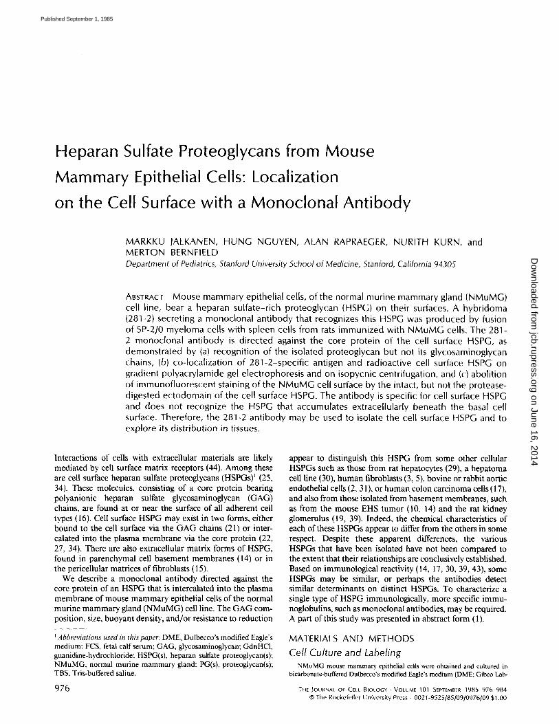
Rats were immunized with a fraction containing both whole cells and the material accumulating beneath NMuMG mon- olayers. Spleen cells from these rats were fused with SP-2/0 myeloma cells. In one fusion, resulting in 188 wells that contained successful hybrids, media from 28 were positive, as assessed by a solid phase radioimmunoassay for total PGs (see Materials and Methods). The cultures of positive clones were expanded, then screened again, and four reactive clones (209, 230, 281, and 349) were selected (Fig. 1). The most positive (281) of these was evaluated further after cloning by limiting dilution. The subclones of 281 were assayed again with the to ta l P G p r e p a r a t i o n as well as w i t h t h e pu r i f i ed e c t o d o m a i n
o f t h e cell s u r f a c e P G . U s i n g t h e s e a s says , five s u b c l o n e s o f
281 were se l ec ted t h a t s h o w e d r eac t iv i t y a g a i n s t b o t h t h e s e
p r e p a r a t i o n s ( d a t a n o t s h o w n ) . H o w e v e r , s i nce a n t i g e n i c reac-
t iv i ty m a y be los t b e c a u s e o f s u b c u l t u r i n g , t h e s e l ec t ed c l o n e s
were m a i n t a i n e d in c u l t u r e for 6 w k before r e c l o n i n g to e n s u r e
._J
uJ
[ 2
x
Q.
o
0
FIGURE 1
281
209
230 348
I I
HYBRIDOMA CLONES
Hybridoma clones recognize 35SO4-1abeled PG. Purified 35SO4-1abeled PGs were added (5,000 cpm/well) to wells previously coated first with rabbit anti-rat IgG and then with various hy bridoma media (see text). Each bar represents the average in two independent assays for the amount of 35SO4-1abeled PG bound by media from hybridoma clones. The positive clones are indicated (209, 230, 281, and 348).
on June 16, 2014jcb.rupress.org
Dow
nloaded from
Published September 1, 1985
that they were stable. A single done, 281-2, was selected for further characterization.
Characterization of Clone 281-2 Analysis of PG-specific antibodies was facilitated by an
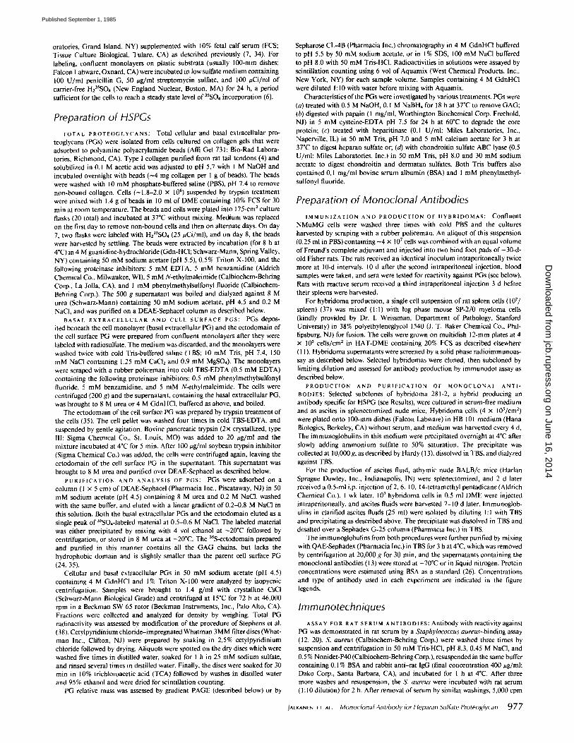
immunodot assay in which PGs were immobilized on DEAE- cellulose paper by the binding of their GAG chains. Use of DEAE-cellulose paper was necessary as the PG did not bind well to either nitrocellulose or aminophenylthioether paper, possibly due to its extensive glycosylation. Reactivity in the assay, shown using medium from clone 281-2 cultures, was dependent on both the antigen concentration and the dilution of hybridoma medium (Fig. 2). The assay was sufficiently sensitive to detect reactivity in a 1:1,000 dilution of culture medium and with 10 ng of PG protein per dot (Fig. 2). Thus, clone 281-2 is a satisfactory producer, and the PG can be detected when bound to DEAE-paper.
Clone 281-2 was recloned by limiting dilution. Using the immunodot assay, 35 out of 36 subclones were positive, suggesting that 281-2 was monoclonal. Two of the subclones (281-2-17 and 281-2-28) were selected for large scale produc- tion of antibodies by culture in serum-free medium and for the preparation of ascites fluid in nude mice. The 281-2 clone and its subclones produced an IgG2a rat immunoglobulin as shown by dot assay and indirect immunoassay on nitrocellu-
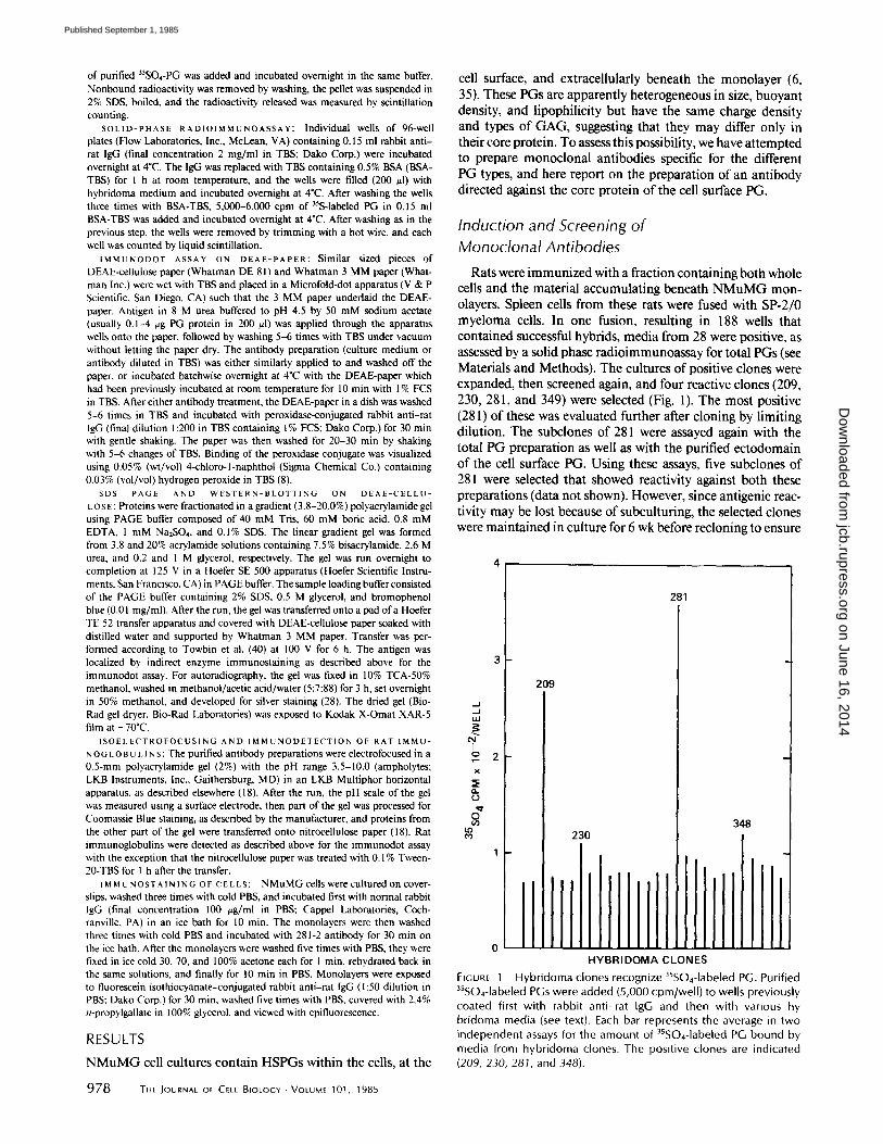
FIGURE 3 Isoelectric focusing of antibody 281-2. Ascites-derived 281-2 was loaded (5 gg/lane) onto a 2% polyacrylamide gel and fo- cused as described in the text. Identical lanes were processed for protein stain (lane A) or for immunodetection of rat immu- noglobulins (lane B) after transfer onto nitrocellulose paper (see text), pH gradient is indicated on the right.
FIGURE 2 Antibody produced by hybridoma 281-2 recognizes PG immobil ized on DEAE-cellulose paper. PG was bound to DEAD cellulose paper. (A and B) 0.001-4 /~g protein per dot; (C) 4 /~g protein per dot. A constant volume {400 /~1} of antibody (culture medium from 281-2 hybridoma cultures) either undiluted (A), di- luted up to 1:1,000 {C), or unconditioned culture medium contain- ing 20% FCS (B) were eluted through the dots. Bound IgG was detected by a peroxidase-conjugated rabbit anti-rat IgG (see text).
lose paper (not shown), using class-specific rabbit anti-rat antibodies (kindly provided by Dr. R. Coffman, DNAX, Palo Alto, CA). The subclones produced antibodies that, after purification (13), showed only a single major band on isoelec- trofocusing by both protein stain and immunostain for rat immunoglobulins (Fig. 3, lanes A and B, respectively). Thus, the 281-2 antibody appears to be monocional (33). The isoe- lectric point of 281-2 was 8.6, which is unusually high for immunoglobulins, but typical for monoclonals (33). This high isoelectric point facilitated the use of 281-2 in immunoassays on DEAE-paper because its low binding to DEAE produced low backgrounds.
Characterization of the Antigen The antigenic determinant recognized by 281-2 may reside
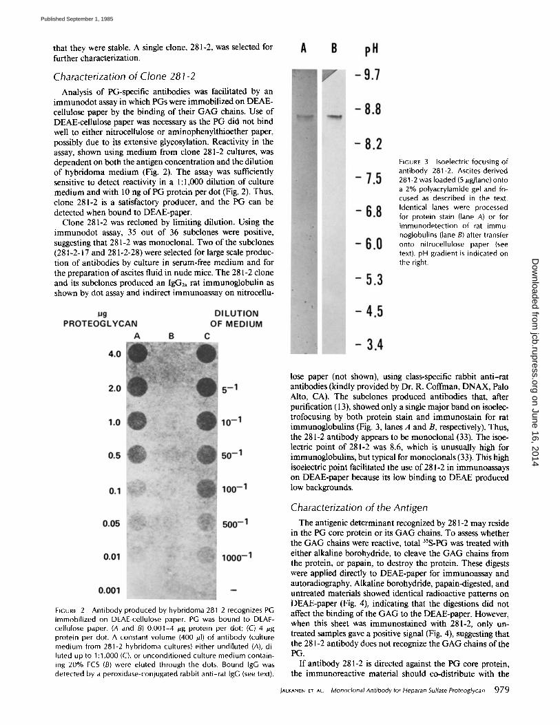
in the PG core protein or its GAG chains. To assess whether the GAG chains were reactive, total 35S-PG was treated with either alkaline borohydride, to cleave the GAG chains from the protein, or papain, to destroy the protein. These digests were applied directly to DEAE-paper for immunoassay and autoradiography. Alkaline borohydride, papain-digested, and untreated materials showed identical radioactive patterns on DEAE-paper (Fig. 4), indicating that the digestions did not affect the binding of the GAG to the DEAE-paper. However, when this sheet was immunostained with 281-2, only un- treated samples gave a positive signal (Fig. 4), suggesting that the 281-2 antibody does not recognize the GAG chains of the PG.
If antibody 281-2 is directed against the PG core protein, the immunoreactive material should co-distribute with the
IACKANEN ET AL. Monoclonal Antibody for Heparan Sulfate Proteoglycan 979
on June 16, 2014jcb.rupress.org
Dow
nloaded from
Published September 1, 1985
the cells and extracellularly. In an attempt to localize the PG that reacts with antibody 281-2, cellular and basal extracel- lular 35S-PGs were fractionated according to their buoyant densities using isopycnic centrifugation. Extracts of cells showed a single major 3~S peak with a density of 1.45-1.55 g/ ml (Fig. 6A), corresponding in density to that of the cell surface PG (35). This peak and material of lower buoyant density were immunoreactive when dotted onto DEAE-paper and stained with 281-2. The basal extracellular fraction was separated into three 35S-labeled PG peaks, of buoyant densities > 1.55, 1.35-140, and 1.25-1.28 g/ml (Fig. 6B). Despite levels of radioactivity similar to that of the cellular extracts when these fractions were stained for 281-2, immunoreactiv- ity was weak. The only stained material corresponded to the density of the immunoreactive material in the cellular frac- tion, presumably due to the presence of some residual cellular material. These data suggest that antibody 281-2 recognizes cellular PGs, including possible PG precursors, but does not recognize any of the basal extracellular PG fractions.
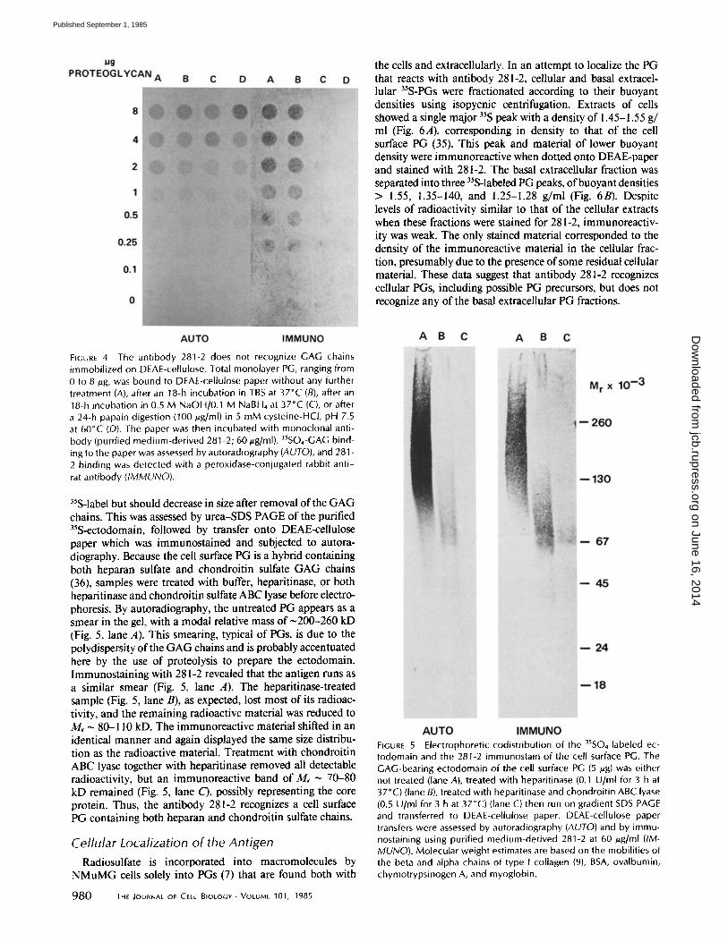
FIGURE 4 The antibody 281-2 does not recognize GAG chains immobilized on DEAE-cellulose. Total monolayer PG, ranging from 0 to 8 #g, was bound to DEAE-cellulose paper without any further treatment (A), after an 18-h incubation in TBS at 37°C (B), after an 18-h incubation in 0.5 M NaOH/0.1 M NaBH4 at 37°C (C), or after a 24-h papain digestion (100 #g/ml) in 5 mM cysteine-HCl, pH 7.5 at 60°C (D). The paper was then incubated with monoclonal anti- body (purified medium-derived 281-2; 60/~g/ml). 35SO4-GAG bind- ing to the paper was assessed by autoradiography (AUTO), and 281 - 2 binding was detected with a peroxidase-conjugated rabbit anti- rat antibody (IMMUNO).
35S-label but should decrease in size after removal of the GAG chains. This was assessed by urea-SDS PAGE of the purified 35S-ectodomain, followed by transfer onto DEAE-cellulose paper which was immunostained and subjected to autora- diography. Because the cell surface PG is a hybrid containing both heparan sulfate and chondroitin sulfate GAG chains (36), samples were treated with buffer, heparitinase, or both heparitinase and chondroitin sulfate ABC lyase before electro- phoresis. By autoradiography, the untreated PG appears as a smear in the gel, with a modal relative mass of ~200-260 kD (Fig. 5, lane A). This smearing, typical of PGs, is due to the polydispersity of the GAG chains and is probably accentuated here by the use of proteolysis to prepare the ectodomain. Immunostaining with 281-2 revealed that the antigen runs as a similar smear (Fig. 5, lane A). The heparitinase-treated sample (Fig. 5, lane B), as expected, lost most of its radioac- tivity, and the remaining radioactive material was reduced to Mr - 80-110 kD. The immunoreactive material shifted in an identical manner and again displayed the same size distribu- tion as the radioactive material. Treatment with chondroitin ABC lyase together with heparitinase removed all detectable radioactivity, but an immunoreactive band of M, ~ 70-80 kD remained (Fig. 5, lane C), possibly representing the core protein. Thus, the antibody 281-2 recognizes a cell surface PG containing both heparan and chondroitin sulfate chains.
Cellular Localization of the Antigen
Radiosulfate is incorporated into macromolecules by NMuMG cells solely into PGs (7) that are found both with
980 THE JOURNAL OF CELL BIOLOGY • VOLUME 101, 1985
FIGURE 5 Electrophoretic codistribution of the 3sso4-1abeled ec- todomain and the 281-2 immunostain of the cell surface PG. The GAG-bearing ectodomain of the cell surface PG (5 ~.g) was either not treated (lane A), treated with heparitinase (0.1 U/ml for 3 h at 37°C) (lane B), treated with heparitinase and chondroitin ABC lyase (0.5 U/ml for 3 h at 37°C) (lane C) then run on gradient SDS PAGE and transferred to DEAE-cellulose paper. DEAE-cellulose paper transfers were assessed by autoradiography (AUTO) and by immu- nostaining using purified medium-derived 281-2 at 60 ~.g/ml (IM- MUNO). Molecular weight estimates are based on the mobilities of the beta and alpha chains of type I collagen (9), BSA, ovalbumin, chymotrypsinogen A, and myoglobin.
on June 16, 2014jcb.rupress.org
Dow
nloaded from
Published September 1, 1985
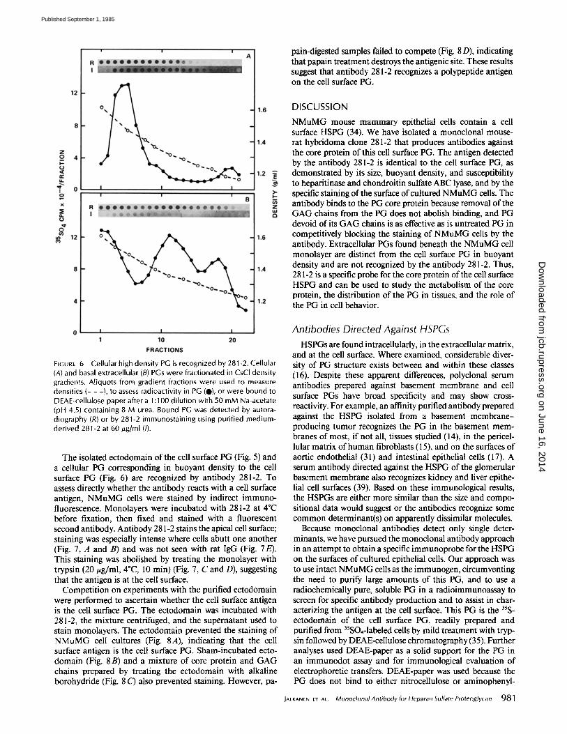
FIGURE 6 Cellular high density PG is recognized by 281-2. Cellular (A) and basal extracellular (B) PGs were fractionated in CsCl density gradients. Aliquots from gradient fractions were used to measure densities (- - -}, to assess radioactivity in PG (0), or were bound to DEAE-cellulose paper after a 1:100 dilution with 50 mM Na-acetate (pH 4.5) containing 8 M urea. Bound PG was detected by autora- diography (R) or by 281-2 immunostaining using purified medium- derived 281-2 at 60 #g/ml (/).
The isolated ectodomain of the cell surface PG (Fig. 5) and a cellular PG corresponding in buoyant density to the cell surface PG (Fig. 6) are recognized by antibody 281-2. To assess directly whether the antibody reacts with a cell surface antigen, NMuMG cells were stained by indirect immuno- fluorescence. Monolayers were incubated with 281-2 at 4"C before fixation, then fixed and stained with a fluorescent second antibody. Antibody 281-2 stains the apical cell surface; staining was especially intense where cells abutt one another (Fig. 7, A and B) and was not seen with rat IgG (Fig. 7E). This staining was abolished by treating the monolayer with trypsin (20 ~g/ml, 4"C, 10 min) (Fig. 7, C and D), suggesting that the antigen is at the cell surface.
Competition on experiments with the purified ectodomain were performed to ascertain whether the cell surface antigen is the cell surface PG. The ectodomain was incubated with 281-2, the mixture centrifuged, and the supernatant used to stain monolayers. The ectodomain prevented the staining of NMuMG cell cultures (Fig. 8A), indicating that the cell surface antigen is the cell surface PG. Sham-incubated ecto- domain (Fig. 8B) and a mixture of core protein and GAG chains prepared by treating the ectodomain with alkaline borohydride (Fig. 8 C) also prevented staining. However, pa-
pain-digested samples failed to compete (Fig. 8 D), indicating that papain treatment destroys the antigenic site. These results suggest that antibody 281-2 recognizes a polypeptide antigen on the cell surface PG.
DISCUSSION
NMuMG mouse mammary epithelial cells contain a cell surface HSPG (34). We have isolated a monoclonal mouse- rat hybridoma clone 281-2 that produces antibodies against the core protein of this cell surface PG. The antigen detected by the antibody 281-2 is identical to the cell surface PG, as demonstrated by its size, buoyant density, and susceptibility to heparitinase and chondroitin sulfate ABC lyase, and by the specific staining of the surface of cultured NMuMG cells. The antibody binds to the PG core protein because removal of the GAG chains from the PG does not abolish binding, and PG devoid of its GAG chains is as effective as is untreated PG in competitively blocking the staining of NMuMG cells by the antibody. Extracellular PGs found beneath the NMuMG cell monolayer are distinct from the cell surface PG in buoyant density and are not recognized by the antibody 281-2. Thus, 281-2 is a specific probe for the core protein of the cell surface HSPG and can be used to study the metabolism of the core protein, the distribution of the PG in tissues, and the role of the PG in cell behavior.
Antibodies Directed Against HSPGs HSPGs are found intracellularly, in the extracellular matrix,
and at the cell surface. Where examined, considerable diver- sity of PG structure exists between and within these classes (16). Despite these apparent differences, polyclonal serum antibodies prepared against basement membrane and cell surface PGs have broad specificity and may show cross- reactivity. For example, an affinity purified antibody prepared against the HSPG isolated from a basement membrane- producing tumor recognizes the PG in the basement mem- branes of most, if not all, tissues studied (l 4), in the pericel- lular matrix of human fibroblasts (l 5), and on the surfaces of aortic endothelial (3 l) and intestinal epithelial cells (17). A serum antibody directed against the HSPG of the glomerular basement membrane also recognizes kidney and liver epithe- lial cell surfaces (39). Based on these immunological results, the HSPGs are either more similar than the size and compo- sitional data would suggest or the antibodies recognize some common determinant(s) on apparently dissimilar molecules.
Because monoclonal antibodies detect only single deter- minants, we have pursued the monoclonal antibody approach in an attempt to obtain a specific immunoprobe for the HSPG on the surfaces of cultured epithelial cells. Our approach was to use intact NMuMG cells as the immunogen, circumventing the need to purify large amounts of this PG, and to use a radiochemically pure, soluble PG in a radioimmunoassay to screen for specific antibody production and to assist in char- acterizing the antigen at the cell surface. This PG is the ~sS- ectodomain of the cell surface PG, readily prepared and purified from 35SO4-1abeled cells by mild treatment with tryp- sin followed by DEAE-cellulose chromatography (35). Further analyses used DEAE-paper as a solid support for the PG in an immunodot assay and for immunological evaluation of electrophoretic transfers. DEAE-paper was used because the PG does not bind to either nitrocellulose or aminophenyl-
JALKANEN ET AL. Monoclonal Antibody for Heparan Sulfate Proteoglycan 981
on June 16, 2014jcb.rupress.org
Dow
nloaded from
Published September 1, 1985
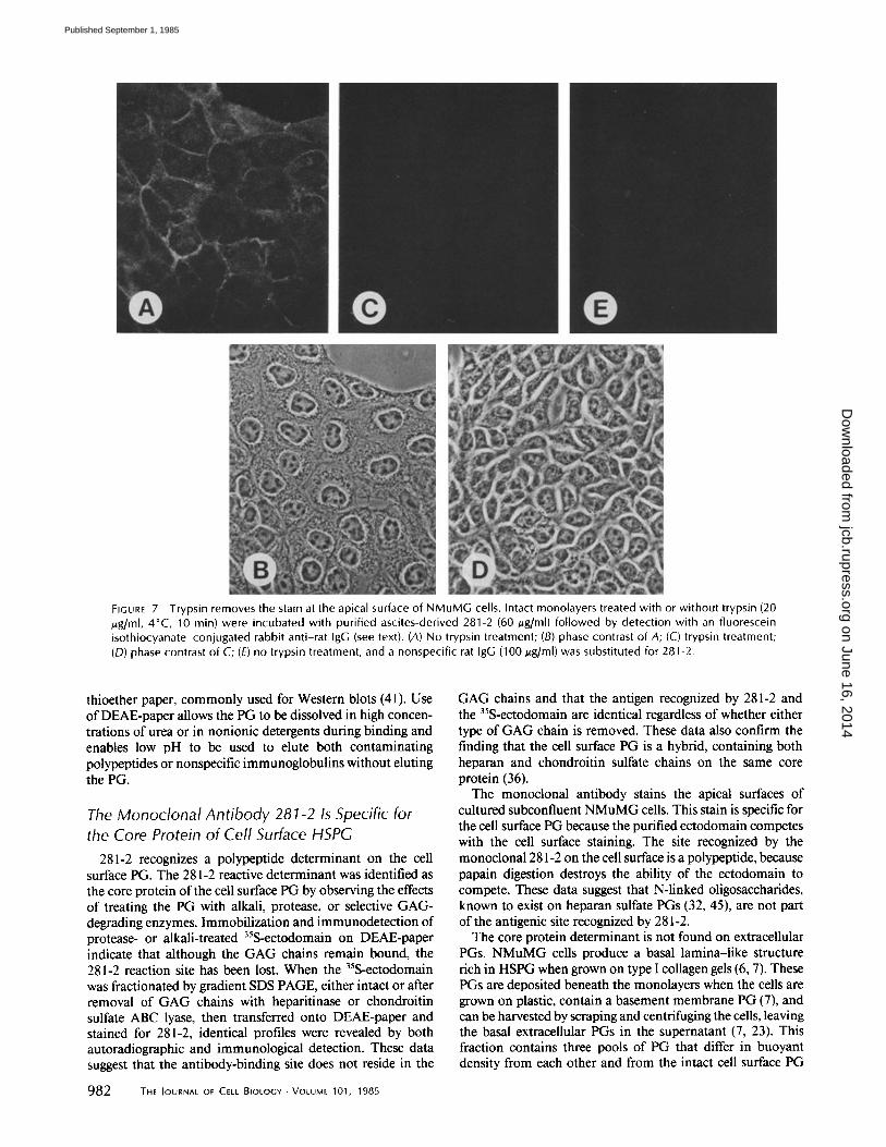
FIGURE 7 Trypsin removes the stain at the apical surface of NMuMG cells. Intact monolayers treated with or without trypsin (20 /zg/ml, 4°C, 10 min) were incubated with purified ascites-derived 281-2 (60/~g/ml) followed by detection with an fluorescein isothiocyanate-conjugated rabbit anti-rat IgG (see text). (A) No trypsin treatment; (B) phase contrast of A; (C) trypsin treatment; (D) phase contrast of C; (E) no trypsin treatment, and a nonspecific rat IgG (100/.tg/ml) was substituted for 281-2.
thioether paper, commonly used for Western blots (41). Use of DEAE-paper allows the PG to be dissolved in high concen- trations of urea or in nonionic detergents during binding and enables low pH to be used to elute both contaminating polypeptides or nonspecific immunoglobulins without eluting the PG.
The Monoclonal Antibody 281-2 Is Specific for the C o r e Protein of Cell Surface HSPG
281-2 recognizes a polypeptide determinant on the cell surface PG. The 281-2 reactive determinant was identified as the core protein of the cell surface PG by observing the effects of treating the PG with alkali, protease, or selective GAG- degrading enzymes. Immobilization and immunodetection of protease- or alkali-treated 35S-ectodomain on DEAE-paper indicate that although the GAG chains remain bound, the 281-2 reaction site has been lost. When the 35S-ectodomain was fractionated by gradient SDS PAGE, either intact or after removal of GAG chains with heparitinase or chondroitin sulfate ABC lyase, then transferred onto DEAE-paper and stained for 281-2, identical profiles were revealed by both autoradiographic and immunological detection. These data suggest that the antibody-binding site does not reside in the
982 THE JOURNAL OF CELL BIOLOGY • VOLUME 101, 1985
GAG chains and that the antigen recognized by 281-2 and the 35S-ectodomain are identical regardless of whether either type of GAG chain is removed. These data also confirm the finding that the cell surface PG is a hybrid, containing both heparan and chondroitin sulfate chains on the same core protein (36).
The monoclonal antibody stains the apical surfaces of cultured subconfluent NMuMG cells. This stain is specific for the cell surface PG because the purified ectodomain competes with the cell surface staining. The site recognized by the monoclona1281-2 on the cell surface is a polypeptide, because papain digestion destroys the ability of the ectodomain to compete. These data suggest that N-linked oligosaccharides, known to exist on heparan sulfate PGs (32, 45), are not part of the antigenic site recognized by 281-2.
The core protein determinant is not found on extracellular PGs. NMuMG cells produce a basal lamina-like structure rich in HSPG when grown on type I collagen gels (6, 7). These PGs are deposited beneath the monolayers when the cells are grown on plastic, contain a basement membrane PG (7), and can be harvested by scraping and centrifuging the cells, leaving the basal extracellular PGs in the supernatant (7, 23). This fraction contains three pools of PG that differ in buoyant density from each other and from the intact cell surface PG
on June 16, 2014jcb.rupress.org
Dow
nloaded from
Published September 1, 1985
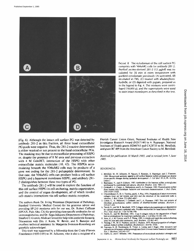
FIGURE 8 The ectodomain of the cell surface PG competes with NMuMG cells for antibody 281-2. Purified ascites-derived 281-2 (12 /~g/ml) was in, cubated for 30 rain at room temperature with purified ectodomain previously (A) untreated, (B) incubated in TBS, (C) treated with alkaline/boro- hydride, or (D) digested with papain, prepared as in the legend to Fig. 4. The mixtures were centri- fuged (10,000 g), and the supernatants were used to stain intact monolayers as described in the text.
(Fig. 6). A l t h o u g h t h e in t ac t cell su r face P G was d e t e c t e d by a n t i b o d y 281-2 in th is f rac t ion , all t h r e e basa l ex t race l lu la r P G poo l s were negat ive . Thus , t he 281-2 reac t ive d e t e r m i n a n t
is e i t he r m a s k e d o r n o t p r e s e n t in t he basa l ex t race l lu la r PGs . T h e m a s k i n g m a y be d u e to ex t r ace l lu l a r p roces s ing o f H S P G or , d e sp i t e t h e p r e s e n c e o f 8 M urea a n d p r ev ious e x t r a c t i o n wi th 4 M G d n H C I , i n t e r a c t i o n o f t h e H S P G wi th o t h e r ex t r ace l lu l a r m a t r i x m o l e c u l e s (10, 42). T h e H S P G s accu- m u l a t i n g b e n e a t h the N M u M G cells m a y be p r o d u c t s o f a gene n o t c o d i n g for t he 281-2 p o l y p e p t i d e d e t e r m i n a n t . In t h a t case, t he N M u M G cells c a n p r o d u c e b o t h a cell su r face
H S P G a n d a b a s e m e n t m e m b r a n e H S P G , a n d a n t i b o d y 281 -
2 d i s t i ngu i shes b e t w e e n these t w o types o f P G . T h e a n t i b o d y 281-2 will be u sed to exp lo re t h e f u n c t i o n o f
th is cell su r face H S P G in cell a n c h o r i n g , m a t r i x o rgan i za t i on , a n d t h e c o n t r o l o f o rgan d e v e l o p m e n t , all o f w h i c h invo lve c e l l - m a t r i x i n t e r a c t i o n s via cell su r face m a t r i x recep tors .
The authors thank Dr. Irving Weissman (Department of Pathology, Stanford University Medical Center) for his generous advice and providing SP-2/0 myeloma cells for our use, Dr. Robert Coffman (DNAX, Palo Alto, CA) for providing class-specific antibodies for rat immunoglobulins, and Dr. Sirpa Jalkanen (Department of Pathology, Stanford University Medical Center) for help with isoelectric focusing. Discussions with Drs. J. Koda, W. Benitz, and S. Banerjee and excellent technical assistance by Margareta Svensson-Rosenberg are gratefully acknowledged.
This work was supported by a fellowship from the Cystic Fibrosis Foundation (F030-5-01) to M. Jalkanen, who is also a recipient o f a
Finnish Cancer Union Grant, National Institutes of Health New Investigator Research Award (HD17146) to A. Rapraeger, National Institutes of Health grants HD06763 and CA28735 to M. Bernfield, and grant BC 409 from the American Cancer Society to M. Bernfield.
R e c e i v e d f o r pub l ica t ion 18 M a r c h 1985, a n d in revised f o r m 1 J u n e 1985.
REFERENCES
1, Bernfield, M., M. Jalkanen, H. Nguyen, S. Banerjee, A. Rapraeger, and J. Firestone. 1984. Monoclonal antibody against a cell surface heparan sulfate proteoglycan detects tissue-specific changes during epithelial development. J. Cell Biol. 99 (4, Pt. 2):163a. (Abstr.)
2. Buonassisi. V., and P, Colburn. 1983. Antibodies to the heparan sulfate proteoglycans synthesized by endothelial cell cultures. Biochim. Biophys. Acla. 760:1-12.
3, Carlstadt I., L. Coster, A. Malmstrom, and L.-A. Fransson. 1983. Protenheparan sulfate from human skin fibroblasts. Isolation and structural characterization. Z Biol. Chem. 258:11629-11635.
4. Chandrakasan, G.,D.A.Torchia, andK. A. Piez. 1976. Preparation of intact monomeric collagen from rat tail tendon and skin and the structure of the nonhelical ends in solution. J. Biol. Chem. 251:6062-6067.
5. Coster, L., A, Malstrom, I. Cadstedt, and L.-A. Fransson. 1983. The core protein of fibroblast proteoheparan sulfate consists of disulfide-bonded subunits. Biochem £ 215:417-419.
6. David, G., and M. R. Bernfield. 1979. Collagen reduces glycosaminoglycan degradation by cultured mammary epithelial cells. Possible mechanism for basal lamina formation. Proc. Natl. Acad Sci. USA 76:786-790.
7. David, G., and M. Bernfield. 1981. Type I collagen reduces the degradation of basal lamina proteoglycan by mammary epithelial cells. J. Cell Biol. 91:281-286.
8. Esen, A., J. M. Conroy, and S.-Z. Wang. 1983. A simple and rapid dot-immunobinding assay for zein and other prolamins. AnaL Biochem. 132:462-467.
9. Freytag, J. W., M. E. Noelken, and B, G. Hudson. 1979. Physical properties of collagen- sodium dodecyl sulfate complexes, Biochemistry. 18:4761-4768.
10. Fujiwara, S., H. Wiedemann, R. Timpl, A. Lustig. and J. EngeL 1984. Structure and interactions of heparan sulfate proteoglycans from a mouse tumor basement membrane. Eur J. Biochem. 143:145-157
I I. Galfre, G, and C. Milstein. 1981. Preparation of monoclonal antibodies: strategies and
JAO(ANEN ET ^t. Monoclonal Antibody for Heparan Sulfate Proteoglycan 983
on June 16, 2014jcb.rupress.org
Dow
nloaded from
Published September 1, 1985
procedures. Methods EnzymoL 73:3-46. 12. Goding, J. W. 1981. Use of staphylococcal protein A as an immunological reagent. ,L
lmmunoL Methods 20:241-253. 13. Hardy, R. R. 1985. Purification and characterization of monoclonal antibodies. In
Handbook of Experimental Immunology. 4th Edition. D. Weir and L. A. Herzenberg. Blackwell Scientific Publications Ltd., Oxford. In press.
14. Hassell. J. R., P. Gehron-Robey, H.-J. Barrach, J. Wilczek, S. I. Rennard, and G. R. Martin. Isolation of a heparan sulfate-containing proteoglycan from basement mem- brane. Proc. Natl. Acad. ScL USA, 77:4494-4498.
15. Hedman, K., S. Johansson, T. Vartio, L. Kjell6n, A. Vaheri, and M. H66k, 1982. Structure of the pericellular matrix: association of heparan and chondroitin sulfates with fibronectin-proconagen fibers. Cell. 28:663-671.
16. H66k, M., L. Kjell6n, S. Johansson, and J. Robinson. 1984. Cell surface glycosamino- glycans. Annu. Rev. Biochem. 53:847-869.
17. lozzo, R. V. 1984. Biosynthesis of heparan sulfate proteoglycan by human colon carcinoma cells and its localization at the cell surface, J. Cell Biol. 99:403-417.
18. Jalkancn, M., and S, Jalkanen. 1983. Immunological detection ofpruteins after isoelec- tric focusing in thin layer agarose gel: a specific application for the characterization of immunoglobulin diversity. Z Clin. Lab. Immunol. 10: 225-228.
19. Kanwar, Y. S., A. Veis, J. H. Kimura, and M. L. Jakubowski. 1984. Characterization of heparan sulfate-proteoglycan of glomerular basement membranes. Proc. Natl. Acad. Sci. USA. 78:5371-5375.
20. Kessler, S. W. 1975. Rapid isolation of antigens from cells with a staphylococcal protein A-antibody adsorbent: parameters on the interaction of antibody-antigen complexes with protein A. Z lmmunol. 115:1617-1624.
21. Kjell~n, L., A. Oldberg, and M. Ho6k. 1980. Cell surface heparan sulfate: mechanisms of proteoglycan-cen association. J. Biol. Chem. 255:10407-10413.
22. Kjell6n, L., I. Pettersson, and M. H66k. 1981. Cell surface heparan sulfate: an interca- lated membrane proteoglycan. Proc. Natl. Acad. ScL USA. 78:5371-5375.
23. Koda, J. E., and M. R. Berufield. 1984. Heparan sulfate proteogiycans from mouse mammary epithelial cells. Basal extracellular proteoglycan binds specifically to native type I collagen. Z Biol. Chem. 259:11763-11770.
24. Koda, J. E., A. C. Rapraeger, and M. Berufield, 1985. Heparan sulfate proteoglycans from mouse mammary epithelial cells: cell surface proteoglycan as a receptor for interstial collagens. Z Biol. Chem. 260:8157-8162.
25. Laterra, J.. J. E. Silbert, and L. A. Culp. Cell surface heparan sulfate mediates some adhesive responses to glycosaminoglycan-binding matrices, including fibronectin. ,L Cell Biol. 96:112-123.
26. Lowry, C. H., N. J. Rosebrough, A. L. Fair, and R. J. Randall. 1951. Protein measure- ment with the folin phenol reagent. ,L Biol. Chem. 193:265-272.
27. Nordling, B., B. Glimelius, and A. Wasteson. 1981. Heparan sulfate proteoglycan of cultured cells: demonstration of a lipid- and a matrix-associated form. Biochem. Biophys. Res. Commun. 103:1265-1272.
28, Oakley, B. R., D. R. Kitsch, and R, Morris. 1980. A simplified ultrasensitive silver stain for detecting proteins in polyacrylamide gels. Anal. Biochem. 105:361-363.
29. OIdberg, A., L. Kjell6n, and M. H66k. 1979. Cell surface heparan sulfate. Isolation and characterization of a proteoglycan from rat liver membranes. Z Biol. Chem. 254: 8505- 8510.
30. Oldberg, A., C. Schwartz, and E. Ruoslahti. 1982. Isolation and partial characterization of a rat hepatoma heparan sulfate proteoglycan. Arch. Biochem. Biophys. 216:400-406.
31. Oohira, A., T. N. Wight, and P. Bornstein. 1983. Sulfate proteogiycans synthesized by vascular endothelial cells in culture. J. Biol. Chem. 258:2014-202 I.
32. Partha~arathy, N., and R, G. Spiro. 1984. Isolation and characterization of the heparan sulfate proteoglycans of the bovine glomerular basement membrane. ,L Biol. Chem. 259:12749-12755.
33. Pearson, T. W., and N. L. Anderson. 1983. Use of high-t~olution two-dimensional gel electrophoresis for analysis of monoclonal antibodies and their specific antigens. Methods Enzymol. 92:196-220.
34. Rapraager, A. C., and M. R. Bernfield. 1983. Heparan sulfate proteogiycans from mouse mammary epithelial cells. A putative membrane proteoglycan associates quantitatively with lipid vesicles. J. Biol. Chem. 258:3632-3636.
35. Rapraeger, A. C., and M. R. Bernfield. 1985. Cell surface proteoglycan of mammary epithelial cells: protease releases a heparan sulfate-rich ectodomaln from a putative membrane-anchored domain. J. Biol. Chem, 260:4103--4109.
36. Rapraeger, A. C., M. Jalkanen, E. Endo, J. Koda, and M. Bernfield. 1985. The cell surface proteoglycan from mouse mammary epithelial cells bears chondroitin and heparan sulfate glycosaminoglycans. Z Biol. Chem. In press.
37. Shulman, M., C. D. Wilde, and G. Kohler. 1978. A better cell line making hybridomas secreting specific antibodies. Nature (Load.). 276:269-270.
38. Stephens, R. W., L. I. Pemberton, P. Ghosh, and T. K. F. Taylor. 1978. A radioassay for proteolytic cleavage of isolated cartilage proteoglycan. AnaL Biochem. 90:726-736.
39. Stow, J. L , and M. G. Farquhar. 1984. lmmunochemical and immunocytochemical studies of basement membrane heparan sulfate proteogiycans (HSPG). J. Cell Biol. 99 (4, Pt. 2):78a. (Abstr,)
40. Towbin, H., T. Staehelin, and J, Gordon. 1979. Electrophoretic transfer of proteins from polyacrylamide gels to nitrocellulose sheets: procedure and some applications. Proc. NatL Acad. Sci. USA. 76:4350--4354.
41. Towbin, H., and J. Gordon. 1984. lmmunoblotting and dot immunobinding-current status and outlook. J. lmmunoL Methods. 72:313-340.
42. Woodley, D., C. Rap, J. Has,sell, L. Liotta, G. Martin, and H. Kieinman. 1983. Interactions of basement membrane components. Biochem. Biophys. Acta. 761:278- 283.
43. Woods, A., M. H66k, L. Kjell~n, C, G. Smith, and D. A. Rees. 1984. Relationship of heparan sulfate proteoglycans to the cytoskeleton and extracellular matrix of cultured fibroblasts. J. Cell BioL 99:1743-1753.
44. Yamada, K. M. 1983. Cell surface interactions with extracellular materials. Annu. Rev. Biochem. 52:761-799.
45. Yanagishita, M., and V. C. Hascall. 1983. Characterization of heparan sulfate proteo- glycans synthesized by rat ovarian granulosa cells in culture. ,L BioL Chem. 258:12857- 12864.
984 THE JOURNAL OF CELL BIOLOGY • VOLUME 101, 1985
on June 16, 2014jcb.rupress.org
Dow
nloaded from
Published September 1, 1985
Related Documents


