Part 1: Disseminated Intravascular Coagulation Part 2: Peripheral T-Cell Non-Hodgkin Lymphoma Part 3: Hemoglobinopathies HEMATOLOGY Board Review Manual Volume 5, Parts 1–3 2010

Welcome message from author
This document is posted to help you gain knowledge. Please leave a comment to let me know what you think about it! Share it to your friends and learn new things together.
Transcript
Part 1: Disseminated Intravascular Coagulation
Part 2: Peripheral T-Cell Non-Hodgkin Lymphoma
Part 3: Hemoglobinopathies
HEMATOLOGYBoard Review Manual
Volume 5, Parts 1–3 2010
The distribution of this publication is made possible through the financial support of Merck
The Hospital Physician® Board Review Manuals are published by Turner White Communications, Inc., an independent medical publisher
dedicated to serving the information and education needs of clinical trainees and practicing physicians.
www.turner-white.com Hematology Volume 5, Part 1 1
HEMATOLOGY BOARD REVIEW MANUAL
STATEMENT OF
EDITORIAL PURPOSE
The Hospital Physician Hematology Board Review
Manual is a study guide for fellows and prac-
ticing physicians preparing for board exami-
nations in hematology. Each manual reviews
a topic essential to the current practice of
hematology.
PUBLISHING STAFF
PRESIDENT, GROUP PUBLISHER
Bruce M. White
SENIOR EDITOR
Robert Litchkofski
EXECUTIVE VICE PRESIDENT
Barbara T. White
EXECUTIVE DIRECTOR
OF OPERATIONS
Jean M. Gaul
PRODUCTION DIRECTOR
Jeff White
NOTE FROM THE PUBLISHER:
This publication has been developed with-
out involvement of or review by the Amer-
ican Board of Internal Medicine.
Disseminated Intravascular Coagulation
Series Editor:Eric D. Jacobsen, MDInstructor in Medicine, Harvard Medical School; Attending
Physician, Dana-Farber Cancer Institute, Boston, MA
Contributor:Thomas G. DeLoughery, MD, FACPProfessor of Medicine, Departments of Pathology and Pediatrics,
Oregon Health Sciences University, Portland, OR
Introduction . . . . . . . . . . . . . . . . . . . . . . . . . . . . .2
Pathogenesis . . . . . . . . . . . . . . . . . . . . . . . . . . . . .2
Patterns of DIC . . . . . . . . . . . . . . . . . . . . . . . . . . .3
Diagnosis . . . . . . . . . . . . . . . . . . . . . . . . . . . . . . . .3
Treatment . . . . . . . . . . . . . . . . . . . . . . . . . . . . . . .6
Specific DIC Syndromes . . . . . . . . . . . . . . . . . . . .7
Summary . . . . . . . . . . . . . . . . . . . . . . . . . . . . . . .12
References . . . . . . . . . . . . . . . . . . . . . . . . . . . . .12
Table of Contents
Cover Illustration by Kathryn K. Johnson
D i s s e m i n a t e d I n t r a v a s c u l a r C o a g u l a t i o n
2 Hospital Physician Board Review Manual www.turner-white.com
HEMATOLOGY BOARD REVIEW MANUAL
Disseminated Intravascular Coagulation
Thomas G. DeLoughery, MD, FACP
INTRODUCTION
The process of coagulation is finely controlled
at many levels to ensure the right amount of he-
mostasis at the right location. Broadly defined, dis-
seminated intravascular coagulation (DIC) refers to
any process that disrupts this fine tuning, leading
to unregulated coagulation. Defined this way, DIC
may be found in patients with a variety of diseases
and can present with a spectrum of findings rang-
ing from asymptomatic abnormal laboratory find-
ings to florid bleeding or thrombosis. It is important
to remember that DIC is always a consequence of
an underlying pathological process and not a dis-
ease in and of itself. This manual reviews concepts
common to all forms of DIC and discusses the
more common disease states that lead to DIC.
PATHOGENESIS
At the most basic level, DIC is the clinical mani-
festation of inappropriate thrombin activation.1–4
Inappropriate thrombin activation can occur due to
underlying conditions such as sepsis, obstetrical
disasters, and trauma. The activation of thrombin
leads to (1) conversion of fibrinogen to fibrin, (2)
activation of platelets (and their consumption), (3)
activation of factors V and VIII, (4) activation of
protein C (and degradation of factors Va and VIIIa),
(5) activation of endothelial cells, and (6) activation
of fibrinolysis (Table 1).
Conversion of fibrinogen to fibrin leads to for-
mation of fibrin monomers and excessive throm-
bus formation. These thrombi are rapidly dis-
solved by excessive fibrinolysis in most patients,
but in certain clinical situations, especially can-
cer, excessive thrombosis will occur. In patients
with cancer, this is most often a deep venous
thrombosis, and rarely patients may have severe
DIC with multiple arterial and venous thrombo-
ses, especially patients with pancreatic cancer.
Nonbacterial thrombotic endocarditis can also
be seen in these patients.
Because thrombin is the most potent physiologic
activator of platelets, there is increased activation
of platelets in DIC. These activated platelets are
consumed, resulting in thrombocytopenia. Platelet
dysfunction is also present. Platelets that have
been activated and have released their contents
but still circulate are known as “exhausted” plate-
lets; these patients can no longer function to sup-
port coagulation. The fibrin degradation products
(FDP) in DIC can also bind to GP IIb/IIIa and fur-
ther inhibit platelet aggregation.
Copyright 2010, Turner White Communications, Inc., Strafford Avenue, Suite 220, Wayne, PA 19087-3391, www.turner-white.com. All rights reserved. No part of this publication
may be reproduced, stored in a retrieval system, or transmitted in any form or by any means, mechanical, electronic, photocopying, recording, or otherwise, without the prior
written permission of Turner White Communications. The preparation and distribution of this publication are supported by sponsorship subject to written agreements that stipu-
late and ensure the editorial independence of Turner White Communications. Turner White Communications retains full control over the design and production of all published
materials, including selection of topics and preparation of editorial content. The authors are solely responsible for substantive content. Statements expressed reflect the views
of the authors and not necessarily the opinions or policies of Turner White Communications. Turner White Communications accepts no responsibility for statements made by
authors and will not be liable for any errors of omission or inaccuracies. Information contained within this publication should not be used as a substitute for clinical judgment.
D i s s e m i n a t e d I n t r a v a s c u l a r C o a g u l a t i o n
www.turner-white.com Hematology Volume 5, Part 1 3
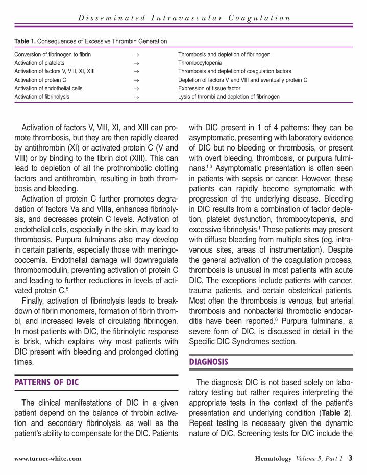
Activation of factors V, VIII, XI, and XIII can pro-
mote thrombosis, but they are then rapidly cleared
by antithrombin (XI) or activated protein C (V and
VIII) or by binding to the fibrin clot (XIII). This can
lead to depletion of all the prothrombotic clotting
factors and antithrombin, resulting in both throm-
bosis and bleeding.
Activation of protein C further promotes degra-
dation of factors Va and VIIIa, enhances fibrinoly-
sis, and decreases protein C levels. Activation of
endothelial cells, especially in the skin, may lead to
thrombosis. Purpura fulminans also may develop
in certain patients, especially those with meningo-
coccemia. Endothelial damage will downregulate
thrombomodulin, preventing activation of protein C
and leading to further reductions in levels of acti-
vated protein C.5
Finally, activation of fibrinolysis leads to break-
down of fibrin monomers, formation of fibrin throm-
bi, and increased levels of circulating fibrinogen.
In most patients with DIC, the fibrinolytic response
is brisk, which explains why most patients with
DIC present with bleeding and prolonged clotting
times.
PATTERNS OF DIC
The clinical manifestations of DIC in a given
patient depend on the balance of throbin activa-
tion and secondary fibrinolysis as well as the
patient’s ability to compensate for the DIC. Patients
with DIC present in 1 of 4 patterns: they can be
asymptomatic, presenting with laboratory evidence
of DIC but no bleeding or thrombosis, or present
with overt bleeding, thrombosis, or purpura fulmi-
nans.1,3 Asymptomatic presentation is often seen
in patients with sepsis or cancer. However, these
patients can rapidly become symptomatic with
progression of the underlying disease. Bleeding
in DIC results from a combination of factor deple-
tion, platelet dysfunction, thrombocytopenia, and
excessive fibrinolysis.1 These patients may present
with diffuse bleeding from multiple sites (eg, intra-
venous sites, areas of instrumentation). Despite
the general activation of the coagulation process,
thrombosis is unusual in most patients with acute
DIC. The exceptions include patients with cancer,
trauma patients, and certain obstetrical patients.
Most often the thrombosis is venous, but arterial
thrombosis and nonbacterial thrombotic endocar-
ditis have been reported.6 Purpura fulminans, a
severe form of DIC, is discussed in detail in the
Specific DIC Syndromes section.
DIAGNOSIS
The diagnosis DIC is not based solely on labo-
ratory testing but rather requires interpreting the
appropriate tests in the context of the patient’s
presentation and underlying condition (Table 2).
Repeat testing is necessary given the dynamic
nature of DIC. Screening tests for DIC include the
Table 1. Consequences of Excessive Thrombin Generation
Conversion of fibrinogen to fibrin → Thrombosis and depletion of fibrinogen
Activation of platelets → Thrombocytopenia
Activation of factors V, VIII, XI, XIII → Thrombosis and depletion of coagulation factors
Activation of protein C → Depletion of factors V and VIII and eventually protein C
Activation of endothelial cells → Expression of tissue factor
Activation of fibrinolysis → Lysis of thrombi and depletion of fibrinogen
D i s s e m i n a t e d I n t r a v a s c u l a r C o a g u l a t i o n
4 Hospital Physician Board Review Manual www.turner-white.com
prothrombin time (PT) activated partial thrombo-
plastin time (aPTT), platelet count, and fibrinogen
level. The PT-INR and aPPT are usually elevated
in severe DIC but may be normal or shortened in
chronic forms.7 One may also see a shortened
aPTT in severe acute DIC due to large amounts
of activated II and factor X “bypassing” the contact
pathway. APTTs as short as 10 seconds have
been seen in acute DIC. The platelet count is usu-
ally reduced but may be normal in chronic DIC.
Serum fibrinogen and platelets are decreased in
acute DIC but also may be in the “normal” range in
chronic DIC.8 The most sensitive of the screening
tests for DIC is a fall in the platelet count, with low
counts seen in 98% of patients and counts under
50,000 cells/μL in 50%.7,9 The least specific test is
fibrinogen, which tends to fall below normal only in
severe acute DIC.7
“Specific tests” for DIC allow one to deduce
that abnormally high concentrations of thrombin
are present. These include the ethanol gel and
protamine sulfate tests, measurement of fibrin deg-
radation product (FDP), and D-dimer levels. The
ethanol gel and protamine tests detect circulating
fibrin monomers. Circulating fibrin monomers are
seen when thrombin acts on fibrinogen. Usually
the monomer polymerizes with the fibrin clot, but
when there is excess thrombin these monomers
continue to circulate. Detection of circulating fibrin
monomer means there is too much IIa and there-
fore DIC is present.
FDPs are produced when plasmin acts on the
fibrin/fibrinogen molecule to cleave the molecule
in specific places. FDP levels are elevated in the
setting of increased fibrin/fibrinogen destruction,
as occurs with DIC and fibrinolysis. FDP levels are
typically mildly elevated in renal and liver disease
due to reduced clearance.
When fibrin monomers bind to form a thrombus,
factor XIII acts to bind the monomers together to
form a dense network of fibrin polymer. One of
the bonds created binds the fibrin “D” domains to-
gether, creating a bond that is resistant to plasmin.
When the thrombus is lysed, this dimer remains
and this degradation fragment is known as the
D-dimer. High levels of D-dimer indicate that IIa
has acted on fibrinogen to form a fibrin monomer
that bonded to another fibrin monomer and that
this thrombus was lysed by plasmin. Because
an elevated D-dimer level can occur due to
other causes (eg, exercise, surgery), an elevated
D-dimer must be interpreted in the context of the
clinical situation.9
Several other tests are sometimes helpful in
diagnosing DIC. The thrombin time test is per-
formed by adding thrombin to plasma. Thrombin
times are increased in DIC (FDPs interfere with
polymerization) and dysfibrinogenemia and in the
presence of low fibrinogen levels and the pres-
ence of heparin (very sensitive). Reptilase time
is the same as thrombin time but is performed
with a snake venom that is insensitive to heparin.
Reptilase time is elevated in the same conditions
as the thrombin time, with the exception of the
presence of heparin. Thrombin time and reptilase
time are most useful in evaluation of dysfibrino-
genemia. F1.2 is a small peptide cleaved off when
prothrombin is activated to thrombin. Thus, high
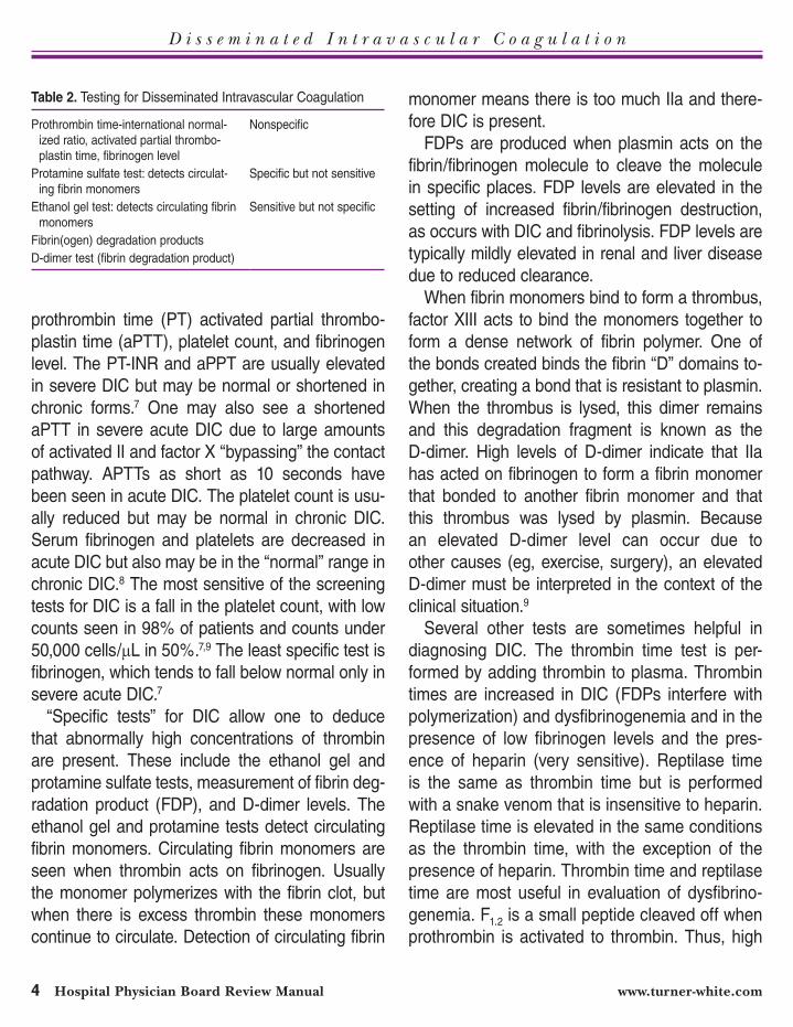
Table 2. Testing for Disseminated Intravascular Coagulation
Prothrombin time-international normal-
ized ratio, activated partial thrombo-
plastin time, fibrinogen level
Nonspecific
Protamine sulfate test: detects circulat-
ing fibrin monomers
Specific but not sensitive
Ethanol gel test: detects circulating fibrin
monomers
Sensitive but not specific
Fibrin(ogen) degradation products
D-dimer test (fibrin degradation product)
D i s s e m i n a t e d I n t r a v a s c u l a r C o a g u l a t i o n
www.turner-white.com Hematology Volume 5, Part 1 5
levels of F1.2 are found in DIC but can be seen
in other thrombotic disorders. This test’s clinical
value remains limited.
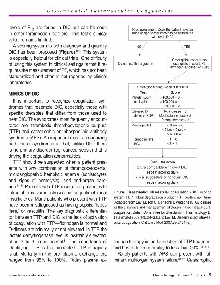
A scoring system to both diagnose and quantify
DIC has been proposed (Figure).9,10 This system
is especially helpful for clinical trials. One difficulty
of using this system in clinical settings is that it re-
quires the measurement of PT, which has not been
standardized and often is not reported by clinical
laboratories.
MIMICS OF DIC
It is important to recognize coagulation syn-
dromes that resemble DIC, especially those with
specific therapies that differ from those used to
treat DIC. The syndromes most frequently encoun-
tered are thrombotic thrombocytopenic purpura
(TTP) and catastrophic antiphospholipid antibody
syndrome (APS). An important clue to recognizing
both these syndromes is that, unlike DIC, there
is no primary disorder (eg, cancer, sepsis) that is
driving the coagulation abnormalities.
TTP should be suspected when a patient pres-
ents with any combination of thrombocytopenia,
microangiopathic hemolytic anemia (schistocytes
and signs of hemolysis), and end-organ dam-
age.11–13 Patients with TTP most often present with
intractable seizures, strokes, or sequela of renal
insufficiency. Many patients who present with TTP
have been misdiagnosed as having sepsis, “lupus
flare,” or vasculitis. The key diagnostic differentia-
tor between TTP and DIC is the lack of activation
of coagulation with TTP—fibrinogen is normal and
D-dimers are minimally or not elevated. In TTP the
lactate dehydrogenase level is invariably elevated,
often 2 to 3 times normal.14 The importance of
identifying TTP is that untreated TTP is rapidly
fatal. Mortality in the pre–plasma exchange era
ranged from 95% to 100%. Today plasma ex-
change therapy is the foundation of TTP treatment
and has reduced mortality to less than 20%.12,15–17
Rarely patients with APS can present with ful-
minant multiorgan system failure.18–21 Catastrophic
Risk assessment: Does the patient have an underlying disorder known to be associated
with overt DIC?
Do not use this algorithmOrder global coagulation tests (platelet count, PT,
fibrinogen, D-dimer, or FDP)
YESNO
Score global coagulation test results
Test Score
Platelet count
(cells/μL)
> 100,000 = 0
< 100,000 = 1
< 50,000 = 2
Elevated D-
dimer or FDP
No increase = 0
Moderate increase = 2
Strong increase = 3
Prolonged PT < 3 sec = 0
> 3 but < 6 sec = 1
> 6 sec = 2
Fibrinogen level
(g/L)
1 = 0
< 1 = 1
Figure. Disseminated intravascular coagulation (DIC) scoring
system. FDP = fibrin degradation product; PT = prothrombin time.
(Adapted from Levi M, Toh CH, Thachil J, Watson HG. Guidelines
for the diagnosis and management of disseminated intravascular
coagulation. British Committee for Standards in Haematology. Br
J Haematol 2009;145:24–33; and Levi M. Disseminated intravas-
cular coagulation. Crit Care Med 2007;35:2191–5.)
Calculate score
≥ 5 is compatible with overt DIC;
repeat scoring daily
< 5 is suggestive of nonovert DIC;
repeat scoring daily
D i s s e m i n a t e d I n t r a v a s c u l a r C o a g u l a t i o n
6 Hospital Physician Board Review Manual www.turner-white.com
APS is caused by widespread microthrombi in mul-
tiple vascular fields. These patients develop renal
failure, encephalopathy, adult respiratory distress
syndrome (often with pulmonary hemorrhage), car-
diac failure, dramatic livido reticularis, and worsen-
ing thrombocytopenia. Many of these patients have
preexisting autoimmune disorders and high-titer
anticardiolipin antibodies. It appears that the best
therapy for these patients is aggressive immuno-
suppression with plasmapheresis, followed by intra-
venous cyclophosphamide monthly.21 Early recogni-
tion of this syndrome can lead to quick therapy and
resolution of the multiorgan system failure.
TREATMENT
The main focus of treating DIC is addressing
the underlying cause that is driving the thrombin
generation.1,2,4,22,23 Fully addressing the underly-
ing cause may not be possible or may take time,
and in the meantime it is necessary to disrupt the
cycle of thrombosis and/or hemorrhage. In the past,
there was concern about using factor replacement
due to fears of “feeding the fire,” or perpetuating the
cycle of thrombosis. However, these concerns are
not supported by evidence, and one must replace
factors if depletion occurs and bleeding ensues.24
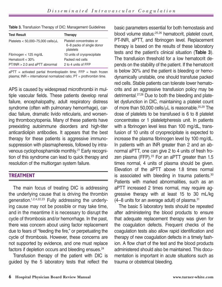
Transfusion therapy of the patient with DIC is
guided by the 5 laboratory tests that reflect the
basic parameters essential for both hemostasis and
blood volume status:25,26 hematocrit, platelet count,
PT-INR, aPTT, and fibrinogen level. Replacement
therapy is based on the results of these laboratory
tests and the patient’s clinical situation (Table 3).
The transfusion threshold for a low hematocrit de-
pends on the stability of the patient. If the hematocrit
is below 30% and the patient is bleeding or hemo-
dynamically unstable, one should transfuse packed
red cells. Stable patients can tolerate lower hemato-
crits and an aggressive transfusion policy may be
detrimental.27,28 Due to both the bleeding and plate-
let dysfunction in DIC, maintaining a platelet count
of more than 50,000 cells/μL is reasonable.25,29 The
dose of platelets to be transfused is 6 to 8 platelet
concentrates or 1 plateletpheresis unit. In patients
with a fibrinogen level less than 100 mg/dL, trans-
fusion of 10 units of cryoprecipitate is expected to
increase the plasma fibrinogen level by 100 mg/dL.
In patients with an INR greater than 2 and an ab-
normal aPTT, one can give 2 to 4 units of fresh fro-
zen plasma (FFP).23 For an aPTT greater than 1.5
times normal, 4 units of plasma should be given.
Elevation of the aPTT above 1.8 times normal
is associated with bleeding in trauma patients.30
Patients with marked abnormalities, such as an
aPTT increased 2 times normal, may require ag-
gressive therapy with at least 15 to 30 mL/kg
(4–8 units for an average adult) of plasma.31
The basic 5 laboratory tests should be repeated
after administering the blood products to ensure
that adequate replacement therapy was given for
the coagulation defects. Frequent checks of the
coagulation tests also allow rapid identification and
therapy of new coagulation defects in a timely fash-
ion. A flow chart of the test and the blood products
administered should also be maintained. This docu-
mentation is important in acute situations such as
trauma or obstetrical bleeding.
Table 3. Transfusion Therapy of DIC: Management Guidelines
Test Result Therapy
Platelets < 50,000–75,000 cells/μL Platelet concentrates or
6–8 packs of single donor
platelets
Fibrinogen < 125 mg/dL 10 units of cryoprecipitate
Hematocrit < 30% Packed red cells
PT/INR > 2.0 and aPTT abnormal 2 to 4 units of FFP
aPTT = activated partial thromboplastin time; FFP = fresh frozen
plasma; INR = international normalized ratio; PT = prothrombin time.
D i s s e m i n a t e d I n t r a v a s c u l a r C o a g u l a t i o n
www.turner-white.com Hematology Volume 5, Part 1 7
In theory since DIC is the manifestation of
exuberant thrombin production, blocking thrombin
with heparin should decrease or shut down DIC.
However, studies have shown that administration
of heparin in most patients leads to excessive
bleeding. Currently, heparin therapy is reserved for
the patient who has thrombosis as a component of
their DIC.2,24,32 Given the coagulopathy that is often
present, one should use specific heparin levels in-
stead of the aPTT to monitor anticoagulation.33,34
SPECIFIC DIC SYNDROMES
SEPSIS/INFECTIOUS DISEASE
Classically, it was believed that gram-negative
bacteria can lead to the development of DIC by
causing tissue factor exposure via their production
of endotoxin, but recent studies indicate that DIC
can be seen with any overwhelming infection.35
There are several potential avenues by which
infections can lead to DIC.36 As mentioned, gram-
negative bacteria produce endotoxin that can
directly lead to tissue factor exposure with result-
ing excess thrombin generation. In addition, any
infection can lead to expression of inflammatory
cytokines that induce tissue factor expression by
endothelium and monocytes. Some viruses and
rickettsia can directly infect the vascular endothe-
lium, converting it from an antithrombotic to a pro-
thrombotic phenotype. The hypotension produced
by sepsis leads to tissue hypoxia, which results
in more DIC. The coagulopathy can range from
subtle abnormalities of testing to purpura fulmi-
nans. Thrombocytopenia is worsened by cytokine-
induced hemophagocytic syndrome
As with all forms of DIC, empiric therapy directed
at the most likely source of infection and maintain-
ing hemodynamic stability are key to therapy. As
discussed below, heparin and other forms of coagu-
lation replacement therapy, with the controversial
exception of recombinant human activated protein
C (rhAPC), or drotrecogin alfa (activated), are of no
benefit.
PURPURA FULMINANS
DIC in association with necrosis of the skin is
seen in 2 situations, primary and secondary pur-
pura fulminans.37,38 Primary purpura fulminans is
most often seen after a viral infection.39 In these
patients, the purpura fulminans starts with a painful
red area on an extremity that rapidly progresses to
a black ischemic area. Acquired deficiency of pro-
tein S is found in many patients.37,40,41 Secondary
purpura fulminans is most often associated with
meningococcemia infections but can be seen in
any patient with overwhelming infection.42–44 Post-
splenectomy sepsis syndrome patients and those
with functional hyposplenism due to chronic liver
diseases are also at risk.45 Patients present with
signs of sepsis, and the skin lesions often involve
the extremities and may lead to amputations. As
opposed to primary purpura fulminans, those with
the secondary form will have symmetrical distal
ischemia (toes and fingers) that ascends as the
process progresses. Rarely, adrenal infarction
(Waterhouse-Friderichsen syndrome) can occur,
which leads to severe hypotension.35
Therapy for purpura fulminans is contro-
versial. Primary purpura fulminans, especial-
ly in those with post-varicella autoimmune
protein S deficiency, has responded to plas-
ma infusion titrated to keep the protein S
level above 25%.37 Intravenous immunoglobulin
has also been reported to help decrease the
anti-protein S antibodies. Heparin has been
reported to control the DIC and extent of ne-
crosis.46 The starting dose in these patients is
5 to 8 units/kg/hr.2
D i s s e m i n a t e d I n t r a v a s c u l a r C o a g u l a t i o n
8 Hospital Physician Board Review Manual www.turner-white.com
Patients with secondary purpura fulminans have
been treated with plasma drips, plasmapheresis,
and continuous plasma ultrafiltration.46–49 Hepa-
rin therapy alone has not been shown to im-
prove survival.50 Much attention has been given
to replacement of natural anticoagulants such as
protein C and antithrombin as therapy for pur-
pura fulminans, but unfortunately randomized trials
using antithrombin have shown mostly negative re-
sults.37,41,51–53 Trials using either zymogen protein C
concentrates or rhAPC have shown more promise
in controlling the coagulopathy of purpura fulmi-
nans and improving outcomes in sepsis.47,54–57 Al-
though bleeding is a concern with use of protein C,
most complications occur in patients with platelet
counts under 30,000 cells/μL or in those who have
meningitis.58 If rhAPC is used, one should also very
carefully monitor other parameters of coagulation
(Table 4). Many patients will need debridement and
amputation for their necrotic limbs, with one review
showing that approximately 66% of patients require
amputations.38
TRAUMA
Currently, the most common cause of acute DIC
is trauma. The coagulation defects that occur in
trauma patients are complex in origin.59 The most
common etiologies are dilution of hemostatic fac-
tors by fluid or blood resuscitation, hypothermia,
tissue damage from trauma, and effects of under-
lying diseases. Trauma patients are prone to hy-
pothermia, and this can be the major complicating
factor in their bleeding.60,61 Patients may be out “in
the field” for a prolonged period of time and be hy-
pothermic on arrival.62 Packed red cells are stored
at 4°C, and the infusion of 1 unit can lower the
body temperature by 0.16°C.63 Hypothermia has
profound effects on the coagulation system that
are associated with clinical bleeding.60,64,65 Even
modest hypothermia can greatly augment bleeding
and needs to be treated or prevented.
The initial management of the bleeding trauma
patient consists of obtaining the basic set of coagu-
lation tests.59,66,67 If the patient is having obvious
massive hemorrhage, red cells and plasma should
be empirically infused until the results of laboratory
tests are received. Since patients with head trauma
can develop defibrination, therapy with cryoprecipi-
tate and plasma should be considered.68 Hypother-
mia can be prevented by several measures. One
is to transfuse the blood through blood warmers.
Devices are available that can warm a unit of blood
per minute. An increasingly used technique is to
perform “damage control” surgery. Patients are
initially stabilized with control of damaged vessels
and packing of oozing sites.69 Then the patient is
taken to the intensive care unit to be warmed and
have coagulation defects corrected.
PREGNANCY-RELATED DIC SYNDROMES
Acute DIC of Pregnancy
Pregnancy can be associated with the rapid
onset of severe DIC in 2 situations, abruption and
amniotic fluid embolism.70,71 The separation of the
placenta from the uterine wall creates a space
for blood to occupy. Because the placenta is rich
in tissue factor, this separation leads to activa-
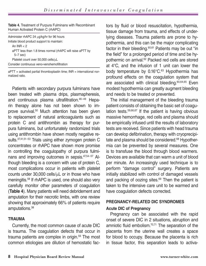
Table 4. Treatment of Purpura Fulminans with Recombinant Human Activated Protein C (rhAPC)
Administer rhAPC 24 μg/kg/hr for 96 hours
Initiate blood product support to maintain:
An INR < 2
aPTT less than 1.8 times normal (rhAPC will raise aPTT by
5–7 sec)
Platelet count over 50,000 cells/μL
Consider continuous veno-venohemofiltration
aPTT = activated partial thromboplastin time; INR = international nor-
malized ratio.
D i s s e m i n a t e d I n t r a v a s c u l a r C o a g u l a t i o n
www.turner-white.com Hematology Volume 5, Part 1 9
tion of coagulation both locally and systemically.
Release of blood when this space reaches the
vaginal opening can lead to rapid hemorrhage,
further augmenting the coagulation abnormalities.
Fetal demise due to placental insufficiency can
also worsen the DIC. Management depends on
the size of the abruption and the clinical status of
both mother and fetus.70 For severe bleeding and
DIC, blood product support is crucial to allow safe
delivery. For smaller abruption, close observation
with early delivery is indicated.
Amniotic fluid embolism occurs suddenly with
the vascular collapse of the woman soon after
delivery. Due to the presence of procoagulant rich
fluid in the circulatory system, there is often over-
whelming DIC. Therapy is directed at both sup-
porting blood volume and correcting hemostatic
defects.
HELLP Syndrome
The HELLP (hemolysis, elevated liver tests, low
platelets) syndrome is a variant of preeclampsia.72
Classically, HELLP syndrome occurs after 28
weeks of gestation in a patent suffering from pre-
eclampsia, but can occur as early as 22 weeks in
patients with APS.73–75 The preeclampsia need not
be severe. The first sign of HELLP is a decrease
in the platelet count followed by abnormal liver
function tests. Signs of hemolysis are present with
abundant schistocytes on the smear and a high
lactate dehydrogenase level. HELLP can progress
to liver failure, and deaths due to hepatic rupture
have also been reported. Unlike TTP, fetal involve-
ment is present in the HELLP syndrome, with
fetal thrombocytopenia reported in 30% of cases.
In severe cases, elevated D-dimers consistent
with DIC are also found. Delivery of the child will
most often result in cessation of the HELLP syn-
drome, but refractory cases require treatment with
dexamethasone and plasma exchange.76 Patients
should be closely observed for 1 to 2 days after
delivery as the hematologic picture can transiently
worsen before improving.77
Acute Fatty Liver of Pregnancy
Fatty liver of pregnancy also occurs late in
pregnancy and is associated with preeclampsia
in 50% of cases.78,79 Patients first present with
nonspecific symptoms of nausea and vomiting
but can progress to fulminant liver failure. Patients
develop thrombocytopenia early in the course, but
in the later stages can develop DIC and very low
fibrinogen levels. Mortality rates without therapy
can be as high as 90%. Low blood glucose and
high ammonia levels can help distinguish fatty liver
from other pregnancy complications.80 Treatment
consists of prompt delivery of the child and aggres-
sive blood product support.
Retained Dead Fetus Syndrome
This syndrome is becoming increasingly rare in
modern practices. The presence of a dead fetus
for many weeks (usually ≥ 5) can result in a chronic
DIC state with fibrinogen depletion and coagulopa-
thy. In some women, these abnormalities worsen
at delivery. In a stable patient, a short trial of hepa-
rin prior to planning delivery can control the DIC to
allow the coagulopathy to stabilize.
DRUG-INDUCED HEMOLYTIC-DIC SYNDROMES
A severe variant of the drug-induced immune
complex hemolysis associated with DIC has
been recognized. Although rare, this syndrome
has been reported in patients who receive cer-
tain second- and third-generation cephalospo-
rins (especially cefotetan and ceftriaxone).81–86
The clinical syndrome starts 7 to 10 days after the
drug is administered, and often the patient has
D i s s e m i n a t e d I n t r a v a s c u l a r C o a g u l a t i o n
10 Hospital Physician Board Review Manual www.turner-white.com
received the antibiotic only for surgical prophy-
laxis. The patient develops severe Coombs’
positive hemolysis with hypotension and DIC.
The patients are often believed to have sepsis
and often re-exposed to the cephalosporin, re-
sulting in worsening of the clinical picture. The
outcome is often fatal due to massive hemolysis
and thrombosis.83,87–89
Quinine is associated with a unique syndrome
of drug-induced DIC.90–93 Approximately 24 to
96 hours after quinine exposure, the patient
becomes acutely ill with nausea and vomiting.
The patient then develops a microangiopathic
hemolytic anemia, DIC, and renal failure. Be-
sides having antiplatelet antibodies, some pa-
tients also have antibodies binding to red cells
and neutrophils, which may lead to the more
severe syndrome. Despite therapy, patients with
quinine-induced TTP have a high incidence of
chronic renal failure.
Treatment of the drug-induced hemolytic-DIC
syndrome is anecdotal. Patients have responded
to aggressive therapy, including plasma exchange,
dialysis, and prednisone.91 Early recognition of
the hemolytic anemia and suspicion that it is drug
related is important for early diagnosis so that the
drug can be discontinued.
CANCER
Cancers, primarily adenocarcinomas, can result
in DIC. The classic Trousseau’s syndrome referred
to the association of migratory superficial thrombo-
phlebitis with cancer94 but now refers to cancer as-
sociated with thrombotic DIC.95,96 Highly vascular
tumor cells are known to express tissue factor,96,97
and some tumor cells can express a direct acti-
vator of factor X (“cancer procoagulant”). Unlike
many DIC states, DIC caused by cancer presents
with thrombosis instead of bleeding. This may
be due to the inflammatory state which accom-
panies cancer, or it may be a part of the chronic
nature of cancer DIC biology that allows time for
the body to compensate for loss of coagulation
factors. In some patients, thrombosis is the first
sign of an underlying cancer, sometimes predat-
ing the cancer diagnosis by months.97 Rarely the
DIC can result in nonthrombotic endocarditis with
microemboli leading to widespread small-vessel
thrombosis.95
Since there is no effective antineoplastic
therapy for many tumors associated with Trous-
seau’s syndrome, DIC therapy is aimed at sup-
pressing thrombosis. An exception is prostate
cancer, where hormonal therapy can markedly
decrease the DIC.98 Because the tumor directly
activates coagulation factors, inhibition of ac-
tive enzymes via heparin has been shown to
result is lower rates of recurrence than use of
warfarin.96,97 Clinical trials have demonstrated
that heparin therapy is associated with a lower
thrombosis recurrence rate than warfarin.99 In
some patients, the thrombotic process is so vig-
orous that new thrombosis can be seen within
hours of stopping heparin.94
ACUTE PROMYELOCYTIC LEUKEMIA
The hemostatic defects in patients with acute
promyelocytic leukemia (APL) are multiple.100 Most,
if not all, patients with APL have evidence of DIC
at the time of diagnosis. Patients with APL have
a higher risk of death during induction therapy as
compared with patients with other forms of leuke-
mia, with death most often due to bleeding. Once
in remission, APL patients have a higher cure rate
than most patients with leukemia. APL is also
unique among leukemias in that biological therapy
with retinoic acid or arsenic is effective in inducing
remission and cure in most patients.
D i s s e m i n a t e d I n t r a v a s c u l a r C o a g u l a t i o n
www.turner-white.com Hematology Volume 5, Part 1 11
APL patients can present with pancytopenia due
to leukemic marrow replacement or with diffuse
bleeding due to DIC and thrombocytopenia. Life-
threatening bleeding such as intracranial hemor-
rhage may occur at any time until the leukemia is
put into remission. The etiology of the hemostatic
defects in APL is complex and is thought to be the
result of DIC, fibrinolysis, and the release of other
procoagulant enzymes.100 The diagnosis of APL
can be straightforward when the leukemic cells are
promyelocytes with abundant Auer rods, although
some patients have the microgranular form without
obvious Auer rods. The precise diagnosis requires
molecular methods. Upon diagnosis of APL, one
should obtain a complete coagulation profile, in-
cluding INR, aPTT, fibrinogen, platelet count, and
D-dimers. Change in fibrinogen levels tends to be
a good marker of progress in treating the coagula-
tion defects.
Therapy of APL involves treating both the
leukemia and the coagulopathy. Currently, the
standard treatment for APL is trans-retinoic
acid (ATRA) in combination with chemothera-
py.101,102 This approach will induce remission in
over 90% of patients, and a sizable majority of
these patients will be cured of their APL. ATRA
therapy will also lead to early correction of the
coagulation defects, often within the first week
of therapy. This is in stark contrast to the che-
motherapy era when the coagulation defects
would become worse with therapy. Rare reports
of massive thrombosis complicating therapy with
ATRA exist, but the relationship to either the APL
or ATRA is unknown.
Therapy for the coagulation defects consists of
aggressive transfusion therapy support and pos-
sible use of other pharmacologic agents to control
DIC.102,103 One should try to maintain the fibrinogen
level at over 100 mg/dL and the platelet count at
over 50,000 cells/μL. Controversy still exists over
the role of heparin in therapy of APL.104 Although
attractive for its ability to quench thrombin, heparin
use can lead to profound bleeding and has fallen
out of favor.
SNAKEBITES
Snake envenomation can lead to direct activa-
tion of multiple coagulation enzymes, including
factors V, X, thrombin, and protein C as well as
lead to cleavage of fibrinogen.105 Envenomation
can also activate coagulation and damage vas-
cular endothelium. The DIC can be enhanced
by widespread tissue necrosis and hypotension.
The key to management of snake bites is admin-
istration of specific antivenom. The role of factor
replacement is controversial but indicated if there
is clinical bleeding. One confounder is that some
snake venoms, especially rattlesnake, can induce
reversible platelet aggregation that corrects with
antivenom.
LOCAL VASCULAR ABNORMALITIES
Abnormal vascular structures, including vascular
tumors, vascular malformations, and aneurysms,
can lead to localized areas of thrombin generation
that can “spill-over” into the general circulation,
leading to DIC. The diagnosis Kasabach-Merritt
phenomenon should be reserved for children with
vascular tumors such as angioma or hemangio-
endothelioma.106 Therapy depends on the lesion.
Embolization to reduce blood flow of vascular mal-
formations can either be definitive or stabilize the
patient for surgery. Aneurysms can be repaired by
surgery or stenting. Rare patients with aneurysms
with significant coagulopathy may require heparin
to increase the fibrinogen level before surgery.
Kasabach-Merritt disease can respond to steroids
or therapy with vincristine or interferon.106
D i s s e m i n a t e d I n t r a v a s c u l a r C o a g u l a t i o n
12 Hospital Physician Board Review Manual www.turner-white.com
SUMMARY
At the most basic level, DIC is the excess activ-
ity of thrombin. However, the clinical presentation
and therapy can differ greatly depending on the
primary cause. Both diagnosis and therapy involve
close coordination of laboratory data and clinical
assessment.
BOARD REVIEW QUESTIONS
Test your knowledge of this topic. Go to
www.turner-white.com and select Hematology from the drop-down
menu of specialties.
REFERENCES
1. Carey MJ, Rodgers GM. Disseminated intravascular co-agulation: clinical and laboratory aspects. Am J Hematol 1998;59:65–73.
2. De Jonge E, Levi M, Stoutenbeek CP, Van Deventer SJH. Current drug treatment strategies for disseminated intra-vascular coagulation. Drugs 1998;55:767–77.
3. Baker WF, Jr. Clinical aspects of disseminated intravascu-lar coagulation: a clinician’s point of view. Sem Thrombosis Hemostasis 1989;15:1–57.
4. Levi M, ten Cate H. Disseminated intravascular coagula-tion. N Engl J Med 1999;341:586–92.
5. Faust SN, Levin M, Harrison OB, et al. Dysfunction of endothelial protein C activation in severe meningococcal sepsis. N Engl J Med 2001;345:408–16.
6. Sharma S, Mayberry JC, DeLoughery TG, Mullins RJ. Fatal cerebroembolism from nonbacterial thrombotic en-docarditis in a trauma patient: case report and review. Military Medicine 2000;165:83–5.
7. Yu M, Nardella A, Pechet L. Screening tests of dissemi-nated intravascular coagulation: guidelines for rapid and specific laboratory diagnosis. Crit Care Med 2000;28: 1777–80.
8. Mant MJ, King EG. Severe, acute disseminated intravas-cular coagulation. A reappraisal of its pathophysiology, clinical significance, and therapy based on 47 patients. Am J Med 1979;67:557–63.
9. Levi M, Toh CH, Thachil J, Watson HG. Guidelines for the diagnosis and management of disseminated intravascular coagulation. British Committee for Standards in Haematol-ogy. Br J Haematol 2009;145:24–33.
10. Levi M. Disseminated intravascular coagulation. Crit Care Med 2007;35:2191–5.
11. George JN. Clinical practice. Thrombotic thrombocytope-nic purpura. N Engl J Med 2006;354:1927–35.
12. George JN. How I treat patients with thrombotic throm-bocytopenic purpura-hemolytic uremic syndrome. Blood 2000;96:1223–9.
13. Murrin RJ, Murray JA. Thrombotic thrombocytopenic pur-pura: aetiology, pathophysiology and treatment. Blood Rev 2006;20:51–60.
14. Patton JF, Manning KR, Case D, Owen J. Serum lactate dehydrogenase and platelet count predict survival in thrombotic thrombocytopenic purpura. Am J Hematol 1994;47:94–9.
15. Rock GA, Shumak KH, Buskard NA, et al. Comparison of plasma exchange with plasma infusion in the treatment of thrombotic thrombocytopenic purpura. N Engl J Med 1991;325:393–7.
16. Bell WR, Braine HG, Ness PM, Kickler TS. Improved sur-vival in thrombotic thrombocytopenic purpura-hemolytic uremic syndrome—clinical experience in 108 patients. N Engl J Med 1991;325:398–403.
17. Kaplan BS, Trachtman H. Improve survival with plasma exchange thrombotic thrombopenic purpura-hemolytic uremic syndrome. Am J Med 2001;110:156–7.
18. Asherson RA. The catastrophic antiphospholipid syndrome [editorial]. J Rheumatol 1992;19:508–12.
19. Asherson RA, Piette JC. The catastrophic antiphospholipid syndrome 1996: acute multi-organ failure associated with antiphospholipid antibodies: a review of 31 patients. Lupus 1996;5:414–7.
20. Asherson RA, Cervera R. Castastrophic antiphospholipid syndrome. Curr Opinion Hematol 2000;5:325–9.
21. Merrill JT, Asherson RA. Catastrophic antiphospholipid syndrome. Nat Clin Pract Rhuem 2006;2:81–9.
22. Hoffman JN, Faist E. Coagulation inhibitor replacement during sepsis: useless? Crit Care Med 2000;28(9 Suppl):S74–6.
23. Wada H, Asakura H, Okamoto K, et al. Expert con-sensus for the treatment of disseminated intravascular coagulation in Japan. Japanese Society of Thrombosis Hemostasis/DIC subcommittee. Thromb Res 2009 Sep 24. [Epub ahead of print].
24. Feinstein DI. Diagnosis and management of disseminated intravascular coagulation: the role of heparin therapy. Blood 1982;60:284–7.
25. Counts RB, Haisch C, Simon TL, et al. Hemostasis in massively transfused trauma patients. Ann Surg 1979;190: 91–9.
26. Stainsby D, MacLennan S, Hamilton PJ. Management of massive blood loss: a template guideline. Br J Anaesth
D i s s e m i n a t e d I n t r a v a s c u l a r C o a g u l a t i o n
www.turner-white.com Hematology Volume 5, Part 1 13
2000;85:487–91. 27. Hébert PC, Wells G, Blajchman MA, et al. A multicenter,
randomized, controlled clinical trial of transfusion require-ments in critical care. N Engl J Med 1999;340:409–17.
28. Blair SD, Janvrin SB, McCollum CN, Greenhalgh RM. Ef-fect of early blood transfusion on gastrointestinal haemor-rhage. Br J Surg 1986;73:783–5.
29. Miller RD, Robbins TO, Tong MJ, Barton SL. Coagulation defects associated with massive blood transfusions. Ann Surg 1971;174:794–801.
30. Ciavarella D, Reed RL, Counts RB, et al. Clotting factor levels and the risk of diffuse microvascular bleeding in the massively transfused patient. Br J Haematol 1987;67:365–8.
31. Chowdhury P, Saayman AG, Paulus U, et al. Efficacy of standard dose and 30 ml/kg fresh frozen plasma in cor-recting laboratory parameters of haemostasis in critically ill patients. Br J Haematol 2004;125:69–73.
32. Callander N, Rapaport SI. Trousseau’s syndrome. West J Med 1993;158:364–71.
33. Brill-Edwards P, Ginsberg JS, Johnston M, Hirsh J. Estab-lishing a therapeutic range for heparin therapy. Ann Intern Med 1993;119:104–9.
34. Olson JD, Arkin CF, Brandt JT, et al. College of American Pathologists Conference XXXI on laboratory monitoring of anticoagulant therapy: laboratory monitoring of unfraction-ated heparin therapy. Arch Pathol Lab Med 1998;122:782–8.
35. Yoshikawa T, Tanaka KR, Guze LB. Infection and dis-seminated intravascular coagulation. Medicine (Baltimore) 1971;50:237–58.
36. Jagneaux T, Taylor DE, Kantrow SP. Coagulation in sepsis. Am J Med Sci 2004;328:196–204.
37. Darmstadt GL. Acute infectious purpura fulminans: patho-genesis and medical management. Pediatr Dermatol 1998; 15:169–83.
38. Davis MD, Dy KM, Nelson S. Presentation and outcome of purpura fulminans associated with peripheral gan-grene in 12 patients at Mayo Clinic. J Am Acad Dermatol 2007;57:944–56.
39. Spicer TE, Rau JM. Purpura fulminans. Am J Med 1976; 61:566–71.
40. Josephson C, Nuss R, Jacobson L, et al. The varicella- autoantibody syndrome. Pediatr Res 2001;50:345–52.
41. Smith OP, White B. Infectious purpura fulminans: diagnosis and treatment. Br J Haematol 1999;104:202–7.
42. Gamper G, Oschatz E, Herkner H, et al. Sepsis-associ-ated purpura fulminans in adults. Wien Klin Wochenschr 2001;113(3-4):107–12.
43. Ward KM, Celebi JT, Gmyrek R, Grossman ME. Acute infectious purpura fulminans associated with asplenism or
hyposplenism. J Am Acad Dermatol 2002;47:493–6. 44. Childers BJ, Cobanov B. Acute infectious purpura fulmi-
nans: a 15-year retrospective review of 28 consecutive cases. Am Surg 2003;69:86–90.
45. Carpenter CT, Kaiser AB. Purpura fulminans in pneumo-coccal sepsis: case report and review. Scand J Infect Dis 1997;29:479–83.
46. Duncan A. New therapies for severe meningococcal dis-ease but better outcomes? Lancet 1997;350:1565–6.
47. Smith OP, White B, Vaughan D, et al. Use of protein-C con-centrate, heparin, and haemodiafiltration in meningococ-cus-induced purpura fulminans. Lancet 1997;350:1590–3.
48. Branson HE, Katz J. A structured approach to the manage-ment of purpura fulminans. J Natl Med Assoc 1983;75:821–5.
49. Nolan J, Sinclair R. Review of management of purpura fulminans and two case reports. Br J Anaesth 2001;86: 581–6.
50. Manios SG, Kanakoudi F, Maniati E. Fulminant menin-gococcemia. Heparin therapy and survival rate. Scand J Infect Dis 1971;3:127–33.
51. Giudici D, Baudo F, Palareti G, et al. Antithrombin replace-ment in patients with sepsis and septic shock. Haemato-logica 1999;84:452–60.
52. Fourrier F, Jourdain M, Tournoys A. Clinical trial results with antithrombin III in sepsis. Crit Care Med 2000;28 (9 Suppl):S38–43.
53. Levi M, De Jonge E, van der PT, ten Cate H. Novel ap-proaches to the management of disseminated intravascu-lar coagulation. Crit Care Med 2000;28(9 Suppl):S20–4.
54. Rivard GE, David M, Farrell C, Schwarz HP. Treatment of purpura fulminans in meningococcemia with protein C concentrate. J Pediatr 1995;126:646–52.
55. White B, Livingstone W, Murphy C, et al. An open-label study of the role of adjuvant hemostatic support with protein C replacement therapy in purpura fulminans-associated menin-gococcemia. Blood 2000;96:3719–24.
56. Aoki N, Matsuda T, Saito H, et al. A comparative double-blind randomized trial of activated protein C and unfraction-ated heparin in the treatment of disseminated intravascular coagulation. Int J Hematol 2002;75:540–7.
57. Schellongowski P, Bauer E, Holzinger U, et al. Treatment of adult patients with sepsis-induced coagulopathy and purpura fulminans using a plasma-derived protein C con-centrate (Ceprotin). Vox Sang 2006;90:294–301.
58. Taylor FB, Kinasewitz G. Activated protein C in sepsis. J Thromb Haemost 2004;2:708–17.
59. DeLoughery TG. Coagulation defects in trauma patients: etiology, recognition, and therapy. Crit Care Clin 2004; 20:13–24.
60. Eddy VA, Morris JA Jr, Cullinane DC. Hypothermia, co-agulopathy, and acidosis. Surg Clin North Am 2000;80:
D i s s e m i n a t e d I n t r a v a s c u l a r C o a g u l a t i o n
14 Hospital Physician Board Review Manual www.turner-white.com
845–54. 61. Peng RY, Bongard FS. Hypothermia in trauma patients. J
Am Coll Surg 1999;188:685–96. 62. Steinemann S, Shackford SR, Davis JW. Implications
of admission hypothermia in trauma patients. J Trauma 1990;30:200–2.
63. Rajek A, Greif R, Sessler DI, et al. Core cooling by central venous infusion of ice-cold (4 degrees C and 20 degrees C) fluid: isolation of core and peripheral thermal compart-ments. Anesthesiol 2000;93:629–37.
64. Watts DD, Trask A, Soeken K, et al. Hypothermic coagu-lopathy in trauma: effect of varying levels of hypothermia on enzyme speed, platelet function, and fibrinolytic activity. J Trauma 1998;44:846–54.
65. Ferrara A, MacArthur JD, Wright HK, et al. Hypothermia and acidosis worsen coagulopathy in the patient requiring massive transfusion. Am J Surg 1990;160:515–8.
66. Robb WJ. Massive transfusion in trauma. AACN Clinical Issues 1999;10:69–84.
67. Lynn M, Jeroukhimov I, Klein Y, Martinowitz U. Updates in the management of severe coagulopathy in trauma pa-tients. Intensive Care Med 2002;28:Suppl 2:S241–7.
68. Goodnight SH, Kenoyer G, Rapaport SI, et al. Defibrina-tion after brain-tissue destruction: a serious complication of head injury. N Engl J Med 1974;290:1043–7.
69. Stone HH, Strom PR, Mullins RJ. Management of the major coagulopathy with onset during laparotomy. Ann Surg 1983;197:532–5.
70. Hall DR. Abruptio placentae and disseminated intravascu-lar coagulopathy. Semin Perinatol 2009;33:189–95.
71. Thachil J, Toh CH. Disseminated intravascular coagulation in obstetric disorders and its acute haematological man-agement. Blood Rev 2009;23:167–76.
72. Baxter JK, Weinstein L. HELLP syndrome: the state of the art. Obstet Gynecol Surv 2004;59:838–45.
73. Egerman RS, Sibai BM. HELLP syndrome. Clin Obstetr Gynecol 1999;42:381–9.
74. Saphier CJ, Repke JT. Hemolysis, elevated liver enzymes, and low platelets (HELLP) syndrome: a review of diagnosis and management. Sem Perinatol 1998;22:118–33.
75. Le Thi TD, Tieulie N, Costedoat N, et al. The HELLP syn-drome in the antiphospholipid syndrome: retrospective study of 16 cases in 15 women. Ann Rheum Dis 2005; 64:273–8.
76. Martin JN Jr, Perry KG Jr, Blake PG, et al. Better maternal outcomes are achieved with dexamethasone therapy for postpartum HELLP (hemolysis, elevated liver enzymes, and thrombocytopenia) syndrome. Am J Obstet Gynecol 1997;177:1011–7.
77. Magann EF, Martin JN Jr. Twelve steps to optimal man-agement of HELLP syndrome. Clinical Obstet Gynecol
1999;42:532–50. 78. Jwayyed SM, Blanda M, Kubina M. Acute fatty liver of
pregnancy. J Emerg Medi 1999;17:673–7. 79. Bacq Y. Acute fatty liver of pregnancy. Sem Perinatol
1998;22:134–40. 80. Egerman RS, Sibai BM. Imitators of preeclampsia and
eclampsia. Clin Obstet Gynecol 1999;42:551–62. 81. Garratty G. Immune cytopenia associated with antibiotics.
Transfusion Medi Rev 1993;7:255–67. 82. Chenoweth CE, Judd WJ, Steiner EA, Kauffman CA.
Cefotetan-induced immune hemolytic anemia. Clin Infect Dis 1992;15:863–5.
83. Garratty G, Nance S, Lloyd M, Domen R. Fatal immune hemolytic anemia due to cefotetan. Transfusion 1992; 32:269–71.
84. Endoh T, Yagihashi A, Sasaki M, Watanabe N. Ceftizoxime-induced hemolysis due to immune complexes: case report and determination of the epitope responsible for immune complex-mediated hemolysis. Transfusion 1999;39:306–9.
85. Arndt PA, Leger RM, Garratty G. Serology of antibodies to second- and third-generation cephalosporins associated with immune hemolytic anemia and/or positive direct anti-globulin tests. Transfusion 1999;39:1239–46.
86. Martin ME, Laber DA. Cefotetan-induced hemolytic anemia after perioperative prophylaxis. Am J Hematol 2006;81:186–8.
87. Bernini JC, Mustafa MM, Sutor LJ, Buchanan GR. Fatal hemolysis induced by ceftriaxone in a child with sickle cell anemia. J Pediatr 1995;126:813–5.
88. Borgna-Pignatti C, Bezzi TM, Reverberi R. Fatal ceftriax-one-induced hemolysis in a child with acquired immunode-ficiency syndrome. Pediatr Infect Dis J 1995;14:1116–7.
89. Lascari AD, Amyot K. Fatal hemolysis caused by ceftriax-one. J Pediatr 1995;126:816–7.
90. Gottschall JL, Elliot W, Lianos E, et al. Quinine-induced im-mune thrombocytopenia associated with hemolytic uremic syndrome: a new clinical entity. Blood 1991;77:306–10.
91. Gottschall JL, Neahring B, McFarland JG, et al. Quinine-induced immune thrombocytopenia with hemolytic uremic syndrome: clinical and serological findings in nine patients and review of literature. Am J Hematol 1994;47:283–9.
92. Crum NF, Gable P. Quinine-induced hemolytic-uremic syn-drome. South Med J 2000;93:726–8.
93. Vesely T, Vesely JN, George JN. Quinine-Induced throm-botic thrombocytopenic purpura-hemolytic uremic syn-drome (TTP-HUS): frequency, clinical features, and long-term outcomes. Blood 2000;96:629 [abstract].
94. Bell WR, Starksen NF, Tong S, Porterfield JK. Trousseau’s syndrome. Devastating coagulopathy in the absence of heparin. Am J Med 1985;79:423–30.
95. Sack GH, Levin J, Bell WR. Trousseau’s syndrome and
D i s s e m i n a t e d I n t r a v a s c u l a r C o a g u l a t i o n
www.turner-white.com Hematology Volume 5, Part 1 15
other manifestations of chronic disseminated coagulopa-thy in patients with neoplasms: clinic, pathophysiologic, and therapeutic features. Medicine 1977;56:1–37.
96. Varki A. Trousseau’s syndrome: multiple definitions and multiple mechanisms. Blood 2007;110:1723–9.
97. Prandoni P, Falanga A, Piccioli A. Cancer and venous thromboembolism. Lancet Oncol 2005;6:401–10.
98. de la Fouchardiere C, Flechon A, Droz JP. Coagulopathy in prostate cancer. Neth J Med 2003;61:347–54.
99. Kearon C, Kahn SR, Agnelli G, et al. Antithrombotic thera-py for venous thromboembolic disease: American College of Chest Physicians Evidence-Based Clinical Practice Guidelines. 8th ed. Chest 2008;133(6 Suppl):454S–545S.
100. Arbuthnot C, Wilde JT. Haemostatic problems in acute promyelocytic leukaemia. Blood Rev 2006;20:289–97.
101. Wang ZY, Chen Z. Acute promyelocytic leukemia: from highly fatal to highly curable. Blood 2008;111:2505–15.
102. Tallman MS, Altman JK. How I treat acute promyelocytic leukemia. Blood 2009;114:5126–35.
103. Falanga A, Rickles FR. Management of thrombohemor-rhagic syndromes (THS) in hematologic malignancies. Hematology 2007;2007:165–71.
104. Sanz MA, Grimwade D, Tallman MS, et al. Guidelines on the management of acute promyelocytic leukemia: recom-mendations from an expert panel on behalf of the Euro-pean LeukemiaNet. Blood 2009;113:1875–91.
105. Lu Q, Clemetson JM, Clemetson KJ. Snake venoms and hemostasis. J Thromb Haemost 2005;3:1791–9.
106. Rodriguez V, Lee A, Witman PM, Anderson PA. Kasabach-merritt phenomenon: case series and ret-rospective review of the mayo clinic experience. J Pediatr Hematol Oncol 2009;31:522–6.

The distribution of this publication is made possible through the financial support of Merck
The Hospital Physician® Board Review Manuals are published by Turner White Communications, Inc., an independent medical publisher
dedicated to serving the information and education needs of clinical trainees and practicing physicians.
www.turner-white.com Hematology Volume 5, Part 2 17
HEMATOLOGY BOARD REVIEW MANUAL
STATEMENT OF
EDITORIAL PURPOSE
The Hospital Physician Hematology Board Review
Manual is a study guide for fellows and prac-
ticing physicians preparing for board exami-
nations in hematology. Each manual reviews
a topic essential to the current practice of
hematology.
PUBLISHING STAFF
PRESIDENT, GROUP PUBLISHER
Bruce M. White
SENIOR EDITOR
Robert Litchkofski
EXECUTIVE VICE PRESIDENT
Barbara T. White
EXECUTIVE DIRECTOR
OF OPERATIONS
Jean M. Gaul
PRODUCTION DIRECTOR
Jeff White
NOTE FROM THE PUBLISHER:
This publication has been developed with-
out involvement of or review by the Amer-
ican Board of Internal Medicine.
Peripheral T-Cell Non-Hodgkin Lymphoma
Series Editor and Contributor:
Eric D. Jacobsen, MD
Instructor of Medicine
Harvard Medical School;
Attending Physician
Dana-Farber Cancer Institute
Boston, MA
Introduction and Classification . . . . . . . . . . . . .18
Epidemiology . . . . . . . . . . . . . . . . . . . . . . . . . . .18
Clinical and Pathologic Features . . . . . . . . . . . .18
Predictors of Outcome . . . . . . . . . . . . . . . . . . . .20
Description of Subtypes . . . . . . . . . . . . . . . . . . .20
Treatment of Released/Refractory PTCL . . . . .29
Summary . . . . . . . . . . . . . . . . . . . . . . . . . . . . . . .29
References . . . . . . . . . . . . . . . . . . . . . . . . . . . . .30
Table of Contents
Cover Illustration by Kathryn K. Johnson
18 Hospital Physician Board Review Manual www.turner-white.com
Copyright 2010, Turner White Communications, Inc., Strafford Avenue, Suite 220, Wayne, PA 19087-3391, www.turner-white.com. All rights reserved. No part of this publication
may be reproduced, stored in a retrieval system, or transmitted in any form or by any means, mechanical, electronic, photocopying, recording, or otherwise, without the prior
written permission of Turner White Communications. The preparation and distribution of this publication are supported by sponsorship subject to written agreements that stipu-
late and ensure the editorial independence of Turner White Communications. Turner White Communications retains full control over the design and production of all published
materials, including selection of topics and preparation of editorial content. The authors are solely responsible for substantive content. Statements expressed reflect the views
of the authors and not necessarily the opinions or policies of Turner White Communications. Turner White Communications accepts no responsibility for statements made by
authors and will not be liable for any errors of omission or inaccuracies. Information contained within this publication should not be used as a substitute for clinical judgment.
HEMATOLOGY BOARD REVIEW MANUAL
Peripheral T-Cell Non-Hodgkin Lymphoma
Eric D. Jacobsen, MD
INTRODUCTION AND CLASSIFICATION
Peripheral T-cell lymphoma (PTCL) represents a
heterogeneous collection of mature T- and NK-cell
neoplasms. Most are clinically aggressive and all
are uncommon. The descriptor “peripheral” does
not refer to an anatomic location but rather the
stage of development of the T cell. PTCLs derive
from mature, post-thymic T cells as opposed to
T-cell acute lymphoblastic leukemia/lymphoma,
which derives from immature T cells.1 The most
recent World Health Organization (WHO) classifi-
cation system for PTCL is shown in Table 1.2 The
histologies are categorized by clinical behavior,
with the nodal, extranodal, and leukemic variants
grouped together; however, these distinctions are
not absolute, and there is substantial overlap in
sites of involvement. This review will not focus on
cutaneous T-cell lymphoma, which is clinically and
biologically distinct from PTCL.
EPIDEMIOLOGY
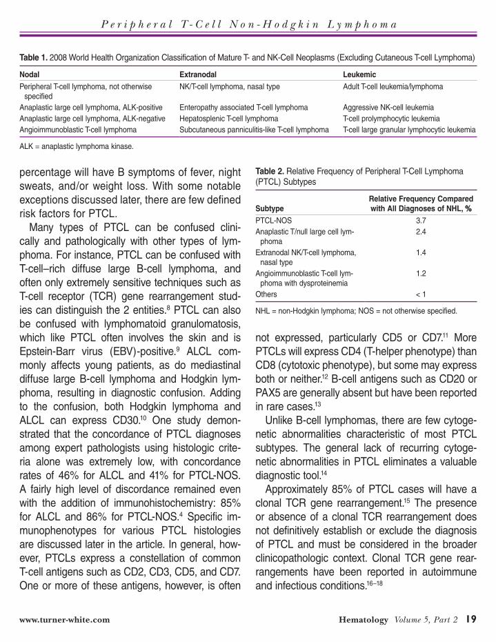
PTCL accounts for 5% to 10% of all cases
of non-Hodgkin lymphoma (NHL) diagnosed in
North America.3 Table 2 shows the relative fre-
quency of various PTCL histologies.4 In North
America and Western Europe, the most common
histologies are PTCL–not otherwise specified
(NOS); anaplastic large cell lymphoma, T/null-cell
type (ALCL); and angioimmunoblastic T-cell lym-
phoma (AILT). In parts of Asia, however, extra-
nodal NK/T-cell lymphoma, nasal type (NK/TCL)
and adult T-cell leukemia/lymphoma (ATLL) are
quite prevalent.5 The epidemiology of individual
subtypes will be discussed in more detail in later
sections.
CLINICAL AND PATHOLOGIC FEATURES
The median age at diagnosis for most his-
tologies is approximately 60 years, though his-
tologies such as ALCL and hepatosplenic T-cell
lymphoma affect adolescents and young adults.6
There is a 1.5:1 male predominance.3 Approxi-
mately 60% of patients present with stage IV dis-
ease. Fifty-six percent of patients will have nodal
and extranodal involvement, while 30% have ex-
tranodal disease only.4 Cutaneous involvement
is far more common than with B-cell NHL.7 The
majority of patients will have an elevated serum
lactate dehydrogenase (LDH), and a substantial
P e r i p h e r a l T - C e l l N o n - H o d g k i n L y m p h o m a
www.turner-white.com Hematology Volume 5, Part 2 19
percentage will have B symptoms of fever, night
sweats, and/or weight loss. With some notable
exceptions discussed later, there are few defined
risk factors for PTCL.
Many types of PTCL can be confused clini-
cally and pathologically with other types of lym-
phoma. For instance, PTCL can be confused with
T-cell–rich diffuse large B-cell lymphoma, and
often only extremely sensitive techniques such as
T-cell receptor (TCR) gene rearrangement stud-
ies can distinguish the 2 entities.8 PTCL can also
be confused with lymphomatoid granulomatosis,
which like PTCL often involves the skin and is
Epstein-Barr virus (EBV)-positive.9 ALCL com-
monly affects young patients, as do mediastinal
diffuse large B-cell lymphoma and Hodgkin lym-
phoma, resulting in diagnostic confusion. Adding
to the confusion, both Hodgkin lymphoma and
ALCL can express CD30.10 One study demon-
strated that the concordance of PTCL diagnoses
among expert pathologists using histologic crite-
ria alone was extremely low, with concordance
rates of 46% for ALCL and 41% for PTCL-NOS.
A fairly high level of discordance remained even
with the addition of immunohistochemistry: 85%
for ALCL and 86% for PTCL-NOS.4 Specific im-
munophenotypes for various PTCL histologies
are discussed later in the article. In general, how-
ever, PTCLs express a constellation of common
T-cell antigens such as CD2, CD3, CD5, and CD7.
One or more of these antigens, however, is often
not expressed, particularly CD5 or CD7.11 More
PTCLs will express CD4 (T-helper phenotype) than
CD8 (cytotoxic phenotype), but some may express
both or neither.12 B-cell antigens such as CD20 or
PAX5 are generally absent but have been reported
in rare cases.13
Unlike B-cell lymphomas, there are few cytoge-
netic abnormalities characteristic of most PTCL
subtypes. The general lack of recurring cytoge-
netic abnormalities in PTCL eliminates a valuable
diagnostic tool.14
Approximately 85% of PTCL cases will have a
clonal TCR gene rearrangement.15 The presence
or absence of a clonal TCR rearrangement does
not definitively establish or exclude the diagnosis
of PTCL and must be considered in the broader
clinicopathologic context. Clonal TCR gene rear-
rangements have been reported in autoimmune
and infectious conditions.16–18
Table 1. 2008 World Health Organization Classification of Mature T- and NK-Cell Neoplasms (Excluding Cutaneous T-cell Lymphoma)
Nodal Extranodal Leukemic
Peripheral T-cell lymphoma, not otherwise
specified
NK/T-cell lymphoma, nasal type Adult T-cell leukemia/lymphoma
Anaplastic large cell lymphoma, ALK-positive Enteropathy associated T-cell lymphoma Aggressive NK-cell leukemia
Anaplastic large cell lymphoma, ALK-negative Hepatosplenic T-cell lymphoma T-cell prolymphocytic leukemia
Angioimmunoblastic T-cell lymphoma Subcutaneous panniculitis-like T-cell lymphoma T-cell large granular lymphocytic leukemia
ALK = anaplastic lymphoma kinase.
Table 2. Relative Frequency of Peripheral T-Cell Lymphoma (PTCL) Subtypes
Subtype
Relative Frequency Compared
with All Diagnoses of NHL, %
PTCL-NOS 3.7
Anaplastic T/null large cell lym-
phoma
2.4
Extranodal NK/T-cell lymphoma,
nasal type
1.4
Angioimmunoblastic T-cell lym-
phoma with dysproteinemia
1.2
Others < 1
NHL = non-Hodgkin lymphoma; NOS = not otherwise specified.
P e r i p h e r a l T - C e l l N o n - H o d g k i n L y m p h o m a
20 Hospital Physician Board Review Manual www.turner-white.com
PREDICTORS OF OUTCOME
With the exception of anaplastic lymphoma
kinase (ALK)-positive ALCL, the treatment out-
comes for PTCL are generally inferior to those of
aggressive B-cell NHLs. The International Prog-
nostic Index (IPI) was developed to predict out-
come in diffuse large B-cell lymphoma.19 The scale
assigns 1 point to each of 5 potential risk factors:
age greater than 60 years, elevated serum LDH,
performance status greater than 2, more than
1 extranodal site of involvement, and stage III/IV
disease. The IPI has since been revised (RIPI) to
reflect outcome in the post-rituximab era.20 The IPI
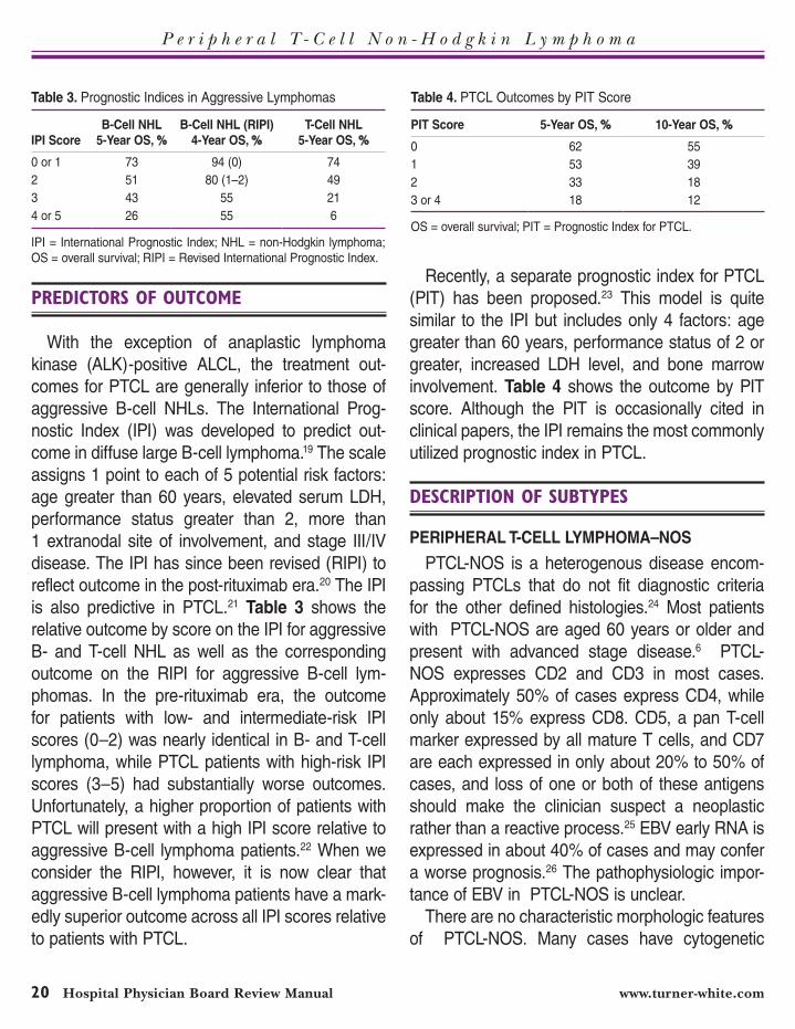
is also predictive in PTCL.21 Table 3 shows the
relative outcome by score on the IPI for aggressive
B- and T-cell NHL as well as the corresponding
outcome on the RIPI for aggressive B-cell lym-
phomas. In the pre-rituximab era, the outcome
for patients with low- and intermediate-risk IPI
scores (0–2) was nearly identical in B- and T-cell
lymphoma, while PTCL patients with high-risk IPI
scores (3–5) had substantially worse outcomes.
Unfortunately, a higher proportion of patients with
PTCL will present with a high IPI score relative to
aggressive B-cell lymphoma patients.22 When we
consider the RIPI, however, it is now clear that
aggressive B-cell lymphoma patients have a mark-
edly superior outcome across all IPI scores relative
to patients with PTCL.
Recently, a separate prognostic index for PTCL
(PIT) has been proposed.23 This model is quite
similar to the IPI but includes only 4 factors: age
greater than 60 years, performance status of 2 or
greater, increased LDH level, and bone marrow
involvement. Table 4 shows the outcome by PIT
score. Although the PIT is occasionally cited in
clinical papers, the IPI remains the most commonly
utilized prognostic index in PTCL.
DESCRIPTION OF SUBTYPES
PERIPHERAL T-CELL LYMPHOMA–NOS
PTCL-NOS is a heterogenous disease encom-
passing PTCLs that do not fit diagnostic criteria
for the other defined histologies.24 Most patients
with PTCL-NOS are aged 60 years or older and
present with advanced stage disease.6 PTCL-
NOS expresses CD2 and CD3 in most cases.
Approximately 50% of cases express CD4, while
only about 15% express CD8. CD5, a pan T-cell
marker expressed by all mature T cells, and CD7
are each expressed in only about 20% to 50% of
cases, and loss of one or both of these antigens
should make the clinician suspect a neoplastic
rather than a reactive process.25 EBV early RNA is
expressed in about 40% of cases and may confer
a worse prognosis.26 The pathophysiologic impor-
tance of EBV in PTCL-NOS is unclear.
There are no characteristic morphologic features
of PTCL-NOS. Many cases have cytogenetic
Table 3. Prognostic Indices in Aggressive Lymphomas
IPI Score
B-Cell NHL
5-Year OS, %
B-Cell NHL (RIPI)
4-Year OS, %
T-Cell NHL
5-Year OS, %
0 or 1 73 94 (0) 74
2 51 80 (1–2) 49
3 43 55 21
4 or 5 26 55 6
IPI = International Prognostic Index; NHL = non-Hodgkin lymphoma;
OS = overall survival; RIPI = Revised International Prognostic Index.
Table 4. PTCL Outcomes by PIT Score
PIT Score 5-Year OS, % 10-Year OS, %
0 62 55
1 53 39
2 33 18
3 or 4 18 12
OS = overall survival; PIT = Prognostic Index for PTCL.
P e r i p h e r a l T - C e l l N o n - H o d g k i n L y m p h o m a
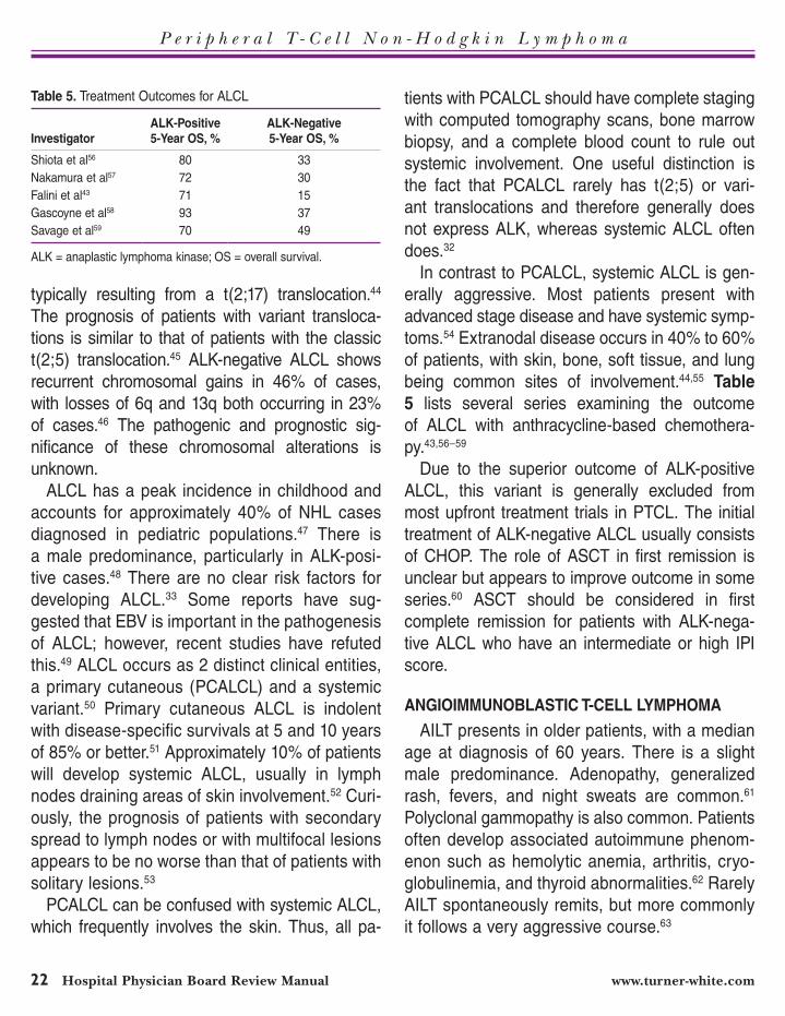
www.turner-white.com Hematology Volume 5, Part 2 21
abnormalities, but none are pathognomonic.27
Although some gene expression–profiling studies
can distinguish PTCL-NOS from ALCL and AILT,
and in some cases have stratified PTCL-NOS
into various subcategories and risk groups, these
results need to be validated before they can be
applied routinely in the clinical setting.28 There
are morphologic variants of PTCL-NOS such as
follicular and lymphoepithelioid (Lennert’s lym-
phoma), but these are of no known clinical conse-
quence.29,30
The treatment of PTCL is largely extrapolated
from aggressive B-cell malignancies. Most PTCL
treatment regimens have utilized an anthracycline
and alkylating agent backbone, with CHOP (cyclo-
phosphamide, doxorubicin, vincristine, and predni-
sone) being the most common. Overall response
rates with CHOP have typically ranged between
50% and 70%.31 In comparison, response rates
with CHOP or CHOP-rituximab in B-cell malig-
nancies are generally 80% to 90%.32 Responses
in PTCL are also less durable. The median pro-
gression-free survival (PFS) in PTCL following
CHOP chemotherapy is 12 to 14 months, with a
5-year disease-free survival (DFS) of approximate-
ly 20%.33 The PFS at 5 years in diffuse large B-cell
lymphoma is 54% and long-term DFS is 60%.32
Several studies, mostly retrospective, have
suggested a benefit from autologous stem cell
transplantation (ASCT) in first remission in PTCL-
NOS.34 The National Cancer Control Network
(NCCN) suggests that patients with a high IPI
score should be considered for ASCT in first re-
mission. Allogeneic stem cell transplant has also
been studied in PTCL in the relapsed/refractory
setting, but the role and timing of this procedure
in PTCL-NOS remains undefined.35 Ideally, trans-
plantation should occur in the context of a well-
designed clinical trial.
ANAPLASTIC T/NULL LARGE CELL LYMPHOMA
ALCL was first described as a clinical entity
in 1985 based upon its unique characteristic of
cohesive proliferation of large pleomorphic cells
with a horseshoe-shaped or embryoid nucleus
expressing CD30 (Ki-1).36 Between 40% and
60% of cases of ALCL have a translocation
between chromosome 2 and chromosome 5
[t(2;5)(p23;q35)],37 resulting in the fusion of the
nucleophosmin (NPM) gene on chromosome 5
with the cytoplasmic domain of ALK on chromo-
some 2. The subsequent NPM-ALK fusion protein
is constitutively active and results in malignant
transformation and resistance to apoptosis.38
Adult patients with ALK-positive ALCL tend to
be young men (median age 34 years) and have
a more favorable prognosis, while patients with
ALK-negative ALCL tend to be older and tend to
follow a more aggressive course.39
The majority of ALCL express one or more
T-cell associated antigens, but approximately 40%
express neither T- nor B-cell antigens (the “null”
phenotype). ALCL with the null phenotype will
often, however, have a clonal TCR gene rear-
rangement.40 CD45, which is positive on most
lymphoid tumors, is occasionally absent. ALCL
can be confused morphologically with Hodgkin
lymphoma, which is compounded by the fact
that both Hodgkin lymphoma and ALCL express
CD30.36 However, CD15, which is frequently ex-
pressed in Hodgkin lymphoma, is rarely positive in
ALCL.41 Another unusual feature of systemic (but
not cutaneous, see below) ALCL is the expression
of epithelial membrane antigen (EMA), which is not
typically seen in lymphoid tumors.42
Variant translocations other than t(2;5) occur
in up to 15% to 20% of cases.43 These include
t(1;2)(q25;p23), inv(2)(p23;q35), t(2;3), and a
CLTC (clathrin heavy chain)-ALK fusion transcript
P e r i p h e r a l T - C e l l N o n - H o d g k i n L y m p h o m a
22 Hospital Physician Board Review Manual www.turner-white.com
typically resulting from a t(2;17) translocation.44
The prognosis of patients with variant transloca-
tions is similar to that of patients with the classic
t(2;5) translocation.45 ALK-negative ALCL shows
recurrent chromosomal gains in 46% of cases,
with losses of 6q and 13q both occurring in 23%
of cases.46 The pathogenic and prognostic sig-
nificance of these chromosomal alterations is
unknown.
ALCL has a peak incidence in childhood and
accounts for approximately 40% of NHL cases
diagnosed in pediatric populations.47 There is
a male predominance, particularly in ALK-posi-
tive cases.48 There are no clear risk factors for
developing ALCL.33 Some reports have sug-
gested that EBV is important in the pathogenesis
of ALCL; however, recent studies have refuted
this.49 ALCL occurs as 2 distinct clinical entities,
a primary cutaneous (PCALCL) and a systemic
variant.50 Primary cutaneous ALCL is indolent
with disease-specific survivals at 5 and 10 years
of 85% or better.51 Approximately 10% of patients
will develop systemic ALCL, usually in lymph
nodes draining areas of skin involvement.52 Curi-
ously, the prognosis of patients with secondary
spread to lymph nodes or with multifocal lesions
appears to be no worse than that of patients with
solitary lesions.53
PCALCL can be confused with systemic ALCL,
which frequently involves the skin. Thus, all pa-
tients with PCALCL should have complete staging
with computed tomography scans, bone marrow
biopsy, and a complete blood count to rule out
systemic involvement. One useful distinction is
the fact that PCALCL rarely has t(2;5) or vari-
ant translocations and therefore generally does
not express ALK, whereas systemic ALCL often
does.32
In contrast to PCALCL, systemic ALCL is gen-
erally aggressive. Most patients present with
advanced stage disease and have systemic symp-
toms.54 Extranodal disease occurs in 40% to 60%
of patients, with skin, bone, soft tissue, and lung
being common sites of involvement.44,55 Table
5 lists several series examining the outcome
of ALCL with anthracycline-based chemothera-
py.43,56–59
Due to the superior outcome of ALK-positive
ALCL, this variant is generally excluded from
most upfront treatment trials in PTCL. The initial
treatment of ALK-negative ALCL usually consists
of CHOP. The role of ASCT in first remission is
unclear but appears to improve outcome in some
series.60 ASCT should be considered in first
complete remission for patients with ALK-nega-
tive ALCL who have an intermediate or high IPI
score.
ANGIOIMMUNOBLASTIC T-CELL LYMPHOMA
AILT presents in older patients, with a median
age at diagnosis of 60 years. There is a slight
male predominance. Adenopathy, generalized
rash, fevers, and night sweats are common.61
Polyclonal gammopathy is also common. Patients
often develop associated autoimmune phenom-
enon such as hemolytic anemia, arthritis, cryo-
globulinemia, and thyroid abnormalities.62 Rarely
AILT spontaneously remits, but more commonly
it follows a very aggressive course.63
Table 5. Treatment Outcomes for ALCL
Investigator
ALK-Positive
5-Year OS, %
ALK-Negative
5-Year OS, %
Shiota et al56 80 33
Nakamura et al57 72 30
Falini et al43 71 15
Gascoyne et al58 93 37
Savage et al59 70 49
ALK = anaplastic lymphoma kinase; OS = overall survival.
P e r i p h e r a l T - C e l l N o n - H o d g k i n L y m p h o m a
www.turner-white.com Hematology Volume 5, Part 2 23
Morphologic analysis shows effaced nodal ar-
chitecture, open peripheral sinuses, and promi-
nent arborizing high endothelial venules with
numerous follicular dendritic cells surround-
ing proliferating blood vessels.64 AILT has a
follicular T-helper lymphocyte immunophe-
notype and CXCL13, PD1 and vascular en-
dothelial growth factor expression.65 A small
proportion of AILT will have a clonal B-cell
infiltrate, and both the B cells and malignant
T cells can show involvement with human herpes
virus 6 (HHV-6) or EBV.66 The pathophysiologic
significance of EBV and HHV-6 is unknown. Oc-
casionally, patients with AILT will also develop
EBV-positive secondary B-cell lymphomas.67
The treatment of AILT is varied. Some patients
will respond to prednisone or even cyclosporine,
although most are treated with anthracycline-
based chemotherapy regimens such as CHOP.68
In a study comparing CHOP to prednisone, the
complete remission rate was 64% with CHOP
and 29% with prednisone, with a median sur-
vival of 19 months in patients receiving CHOP
compared with 11 months in those receiving
prednisone.69
Outcomes in AILT may be improved by ASCT.
In a large retrospective trial, the overall survival of
patients undergoing ASCT was 67% at 24 months
and 59% at 48 months. Patients who had achieved
a complete response prior to transplant had supe-
rior outcomes.70 Although retrospective analyses
are fraught with selection bias and other statistical
challenges, this study suggests that patients with
chemosensitive disease, and particularly patients
in complete remission, may benefit from con-
solidation with ASCT. These findings need to be
confirmed in a randomized trial. The NCCN recom-
mends consideration of ASCT in first remission for
patients with an intermediate or high IPI score.
Allogeneic stem cell transplantation has been
studied in a small number of patients with AILT,
including patients who had failed a prior autolo-
gous transplant. In one series, the PFS and overall
survival following allotransplant for AILT were 53%
and 64%, respectively.71 At present, the optimal
role and timing of allogeneic transplant remain to
be defined.
ADULT T-CELL LEUKEMIA/LYMPHOMA
ATLL is associated with HTLV-1, a retrovirus
endemic to Japan, the Caribbean, and parts of
West Africa and South America that is estimated to
infect up to 20 million individuals worldwide.72 The
virus is transmitted through exchange of bodily
fluids.73 Up to 4% of patients infected with HTLV-
1 will eventually develop ATLL.74 The mechanism
by which HTLV-1 induces oncogenesis is incom-
pletely understood.75 ATLL consists of medium-
sized lymphocytes with condensed chromatin and
hyperlobated nuclei, known as clover leaf or flower
cells. There is often a small proportion of blast-
like cells with deeply basophilic cytoplasm. The
immunophenotype is most often positive for CD2,
CD4, CD5, CD25, and CD52. CD7 and CD8 are
usually negative, while CD3 is generally dimly
expressed.76 There are no pathognomonic cytoge-
netic changes.
ATLL is rare in the United States. The me-
dian age of diagnosis is in the sixties and African-
Americans are at far higher risk than Caucasians.77
There are 4 types of ATLL: acute, lymphomatous,
chronic, and smoldering. The distinction is often
made on clinical grounds. Patients with acute
ATLL present with systemic symptoms such as
fevers and night sweats along with hypercalcemia
and a high number of circulating malignant cells.
Lymphadenopathy, skeletal involvement, cutane-
ous involvement, and hepatosplenomegaly are
P e r i p h e r a l T - C e l l N o n - H o d g k i n L y m p h o m a
24 Hospital Physician Board Review Manual www.turner-white.com
common. Patients with the lymphomatous variant
do not have a significant number of circulating cells
but otherwise have a very similar manifestation to
patients with the acute variant.78 The outcome for
both the acute and lymphomatous variants is poor,
with a median survival of 6 to 9 months; unfortu-
nately, these 2 variants account for approximately
80% of cases of ATLL.79
The chronic and smoldering variants of ATLL
are much less common, and they have a more
favorable course. Patients with the chronic vari-
ant may have a mildly to moderately elevated
lymphocyte count but rarely have significant
lymphadenopathy or organ involvement except
for cutaneous involvement.80 Patients with
the smoldering variant generally have skin le-
sions, only without significant lymphocytosis,
lymphadenopathy, or organ involvment.81 Sur-
vival of patients with these variants can range
from several to many years, and immediate
therapy is often not warranted, especially if the
patient is under age 40 years, has a normal LDH
level, has a good performance status, and has
fewer than 3 sites of involvement.70
Young and fit patients with aggressive variants
of ATLL are generally treated with aggressive
regimens modeled after those used in acute
lymphoblastic leukemia, while older patients
are generally treated with CHOP or CHOP-like
regimens. A randomized trial comparing the
vincristine, cyclophosphamide, doxorubicin, and
prednisone (VCAP); doxorubicin, ranimustine,
and prednisone (AMP); and vindesine, etopo-
side, carboplatin, and prednisone (VECP) regi-
mens to biweekly CHOP in ATLL revealed a high
complete response rate with VCAP-AMP-VECP
compared with CHOP (40% versus 25%, re-
spectively; P = 0.020). There was also a trend
in improvement in overall survival at 3 years with
VCAP-AMP-VECP (24%) compared with dose-
dense CHOP (13%), but the difference was not
statistically significant (P = 0.085). The durabil-
ity of response remained poor, with a median
duration of 13 months, and VCAP-AMP-VECP
had substantially higher toxicity than CHOP.82 Al-
logeneic transplantation may be of benefit in first
remission, but this remains unclear.83 Salvage
therapy with autologous transplant does not ap-
pear to be effective.84
NK/T-CELL LYMPHOMA, NASAL TYPE
NK/TCL typically presents in an aggressive
fashion in the upper airway or nasal cavity.85,86 The
disease can also present in other isolated sites
such as the gastrointestinal tract or skin or can
present in a disseminated fashion.87 NK/TCL is
rare in the United States but is much more com-
mon in Southeast Asia. NK/TCL afflicts younger
patients, including children, and there is a male
predominance.88 NK/TCL is almost always EBV-
positive, and it is presumed that EBV is important
in the pathogenesis.89 High levels of circulating
EBV DNA are associated with a worse progno-
sis.90
NK/TCL is characterized by a polymorphous
infiltrate composed of normal-appearing small
lymphocytes, atypical lymphoid cells of varying
size, plasma cells, and occasionally eosinophils
and histiocytes. A characteristic feature is invasion
of vascular walls.91 NK/TCL is usually positive for
CD56 and CD3 but negative for CD4 and CD8.
The tumor cells also express the cytotoxic pro-
teins TIA-1, granzyme B, and perforin.92 The TCR
gene is clonally rearranged in fewer than 20% of
cases.93 Loss of heterozygosity of chromosome 6
is common but not pathognomonic.94
In patients with localized disease the stan-
dard treatment is radiation therapy to all involved
P e r i p h e r a l T - C e l l N o n - H o d g k i n L y m p h o m a
www.turner-white.com Hematology Volume 5, Part 2 25
areas, encompassing all paranasal sinuses, the
nasopharynx, and the palate.95 The dose of ra-
diotherapy utilized to treat extranodal NK/TCL is
higher than that utilized for most other lymphomas,
with a minimum recommended dose of 50 Gy.96
The value of chemotherapy in localized disease
is unclear, but chemotherapy is generally given.97
Outcomes appear better if radiotherapy is adminis-
tered first followed by chemotherapy, although the
optimal timing of chemotherapy and radiotherapy
remains to be defined.98
Patients with disseminated disease typically re-
spond poorly to chemotherapy. The median over-
all survival for localized disease is approximately
3 years compared with 0.36 years in extranasal
cases.88 The poor outcome with chemotherapy
has generated interest in more aggressive ap-
proaches such as allogeneic and autologous stem
cell transplantation, although the role and timing of
these procedures remain to be defined.99
HEPATOSPLENIC T-CELL LYMPHOMA (HSTCL)
HSTCL is an extremely aggressive neoplasm that
tends to affect young men. Patients usually present
with splenomegaly, thrombocytopenia, and signs
and symptoms of liver insufficiency such as jaun-
dice.100 Bone marrow involvement is very common,
but lymphadenopathy generally is not prominent.101
HSTCL can occur in the setting of immunosuppres-
sion, particularly after organ transplantation or with
the use of anti-tumor necrosis factor-α therapy for
autoimmune diseases.102,103 HSCTL is comprised
of medium-sized lymphoid cells with round nuclei,
moderately condensed chromatin, and moderately
abundant, pale cytoplasm within the sinusoids of
the spleen, liver, and bone marrow. The white pulp
of the spleen is usually atrophic, and erythropha-
gocytosis is often evident in the spleen or mar-
row.104 The tumor cells are generally positive for
CD2, surface CD3, CD7, and occasionally CD56,
while CD4, CD5, and CD8 are usually negative.105
While most cases of PTCL express the alpha/beta
TCR, HSTCL generally expresses the gamma/delta
TCR.106 The most common chromosomal abnormal-
ity is isochromosome 7.107
HSTCL responds poorly to chemotherapy. In
one series, even with aggressive chemotherapy
with or without stem cell transplantation, only 50%
of patients achieved a complete response and
the median duration of complete response was 8
months. Median overall survival was 11 months.108
Autologous or allogeneic stem cell transplanta-
tion may be of benefit either in first remission or
at relapse, though most patients will not achieve
a remission, particularly in the relapse setting, to
benefit from these procedures.109 All patients with
this disease should preferentially be treated in a
well-designed clinical trial.
SUBCUTANEOUS PANNICULITIS-LIKE T-CELL
LYMPHOMA (SPTCL)
SPTCL is a very rare entity that generally af-
fects young adults.110 There seems to be a female
predominance but no other clear risk factors. Most
patients present with subcutaneous nodules mim-
icking infectious or autoimmune panniculitis.111 An
associated hemophagocytic syndrome (HPS) is
common.112 Often serial biopsies are needed to
make the diagnosis.
Previously there were 2 recognized variants of
SPTCL, an alpha/beta and a gamma/delta variant.
The gamma/delta variant is now reclassified as cu-
taneous gamma/delta T-cell lymphoma (CGDTCL)
in the 2008 WHO classification.113 SPTCL cells
have a cytotoxic phenotype and are CD8-posi-
tive but CD4-negative, while CGDTCL is usually
CD4-negative and CD8-negative.114 Morphologi-
cally, SPTCL contains a mixture of small, medium,
P e r i p h e r a l T - C e l l N o n - H o d g k i n L y m p h o m a
26 Hospital Physician Board Review Manual www.turner-white.com
and large atypical cells, often containing irregular,
hyperchromatic nuclei and pale cytoplasm sur-
rounding adipocytes. There are numerous reac-
tive histiocytes with phagocytized nuclear debris
and phagocytized lipid from necrotic adipocytes.111
The malignant cells of SPTCL generally have
complex cytogenetic changes, though none are
pathognomonic.115 A small percentage of cases
are EBV-positive, although the pathophysiologic
and clinical implications of this finding are un-
clear.116 Patients with SPTCL often have indo-
lent disease confined to the subcutis and are
less likely to have HPS. SPTCL has a favor-
able prognosis, with a 5-year overall survival of
82% (91% in the absence of HPS). In contrast,
patients with CGDTCL more commonly had
epidermal involvement and ulceration and were
more likely to have HPS. The 5-year overall
survival is 11%.117
The optimal therapy of SPTCL is unknown.
Localized disease may be successfully treated
with radiotherapy.118 Patients with more extensive
disease are often initially treated with predni-
sone,119 cyclosporine,120 oral methotrexate,121 or
oral alkylating agents.122 Nucleoside analogues
may also be active.123 Most patients will eventually
require more aggressive systemic chemotherapy,
and long-term survivors have been reported after
anthracycline-based chemotherapy124 and after
high-dose chemotherapy and ASCT or allogeneic
transplantation.125
ENTEROPATHY-TYPE T-CELL LYMPHOMA (EATL)
EATL is a rare condition that occurs most
commonly in patients with gluten-sensitive en-
teropathy (celiac sprue).126 Most patients with
EATL have the HLA DQA1*0501, DQB1*0201
genotype associated with an increased risk of
celiac disease.127 Chronic inflammation due to
sustained gluten exposure over time seems to
drive the pathogenesis of EATL, and patients
who adhere to a gluten-free diet (GFD) have
a markedly decreased risk of EATL.128 Unfor-
tunately, many patients are unaware that they
have sprue or are unable to adhere to a GFD.
Refractory celiac disease (RCD) occurs when
symptoms (eg, diarrhea) and damage to the
intestinal mucosa persist despite adherence
to a GFD.129 RCD I is defined as having poly-
clonal intraepithelial lymphocytes (IELs), while
patients with RCD II have a monoclonal, pheno-
typically aberrant intraepithelial T lymphocyte
population that expresses cytoplasmic CD3 but
lacks the surface TCR-CD3 complex, possibly
due to defective dimerization of the TCR chains
and assembly of the TCR-CD3 complex.130 As
many as 50% of RCD II patients will go on to
develop EATL, possibly as a result of the loss
of TCR gamma/delta-positive IELs that play an
important role in mucosal repair, homeostasis,
and tumor surveillance.131
The average age at diagnosis of EATL is in the
late sixth decade, with a male predominance.124
Patients with EATL often present with rapid-
onset abdominal pain, obstruction, or perfora-
tion.132 The small bowel is most commonly af-
fected.133
The tumor contains a mixture of different sized
malignant lymphocytes that are often anaplastic.
The adjacent mucosa generally contains numer-
ous intraepithelial T-cells.134 Most EATLs have a
cytotoxic immunophenotype and are CD3-positive
(cytoplasmic expression), CD4- and CD8-nega-
tive, and TIA-1-positive.135 Most EATLs have an
alpha/beta TCR gene rearrangement, although
gamma/delta cases have been described.134 Loss
of heterozygosity of chromosome 9q21 is a fre-
quent finding, with the region spanning the p14,
P e r i p h e r a l T - C e l l N o n - H o d g k i n L y m p h o m a
www.turner-white.com Hematology Volume 5, Part 2 27
p15, and p16 gene locus most frequently affected,
leading to decreased p16 protein expression and
p53 overexpression.136 Array comparative genomic
hybridization has revealed frequent complex gains
of 9q31.3 or loss of 16q12.1. Interestingly, the 2
genomic changes were mutually exclusive, sug-
gesting pathogenetically distinct types of EATL,
one which is CD56-negative and affects patients
with celiac disease (complex gains in 9q31.3),
and a rarer type that is CD56-positive and affects
patients with no history of celiac disease (loss of
16q12.1).137
EATL is a very aggressive disease. Approxi-
mately 10% of patients are long-term survivors,
with intestinal perforation and infection being the
most common causes of death.138 Surgery alone
is not adequate therapy, even if the patient has
no evidence of disease postoperatively.125 The
optimal chemotherapeutic approach is unknown
but generally consists of aggressive anthracycline-
based chemotherapy regimens such as CHOP.139
Unfortunately, the response rates with anthracy-
cline-based chemotherapy are low and treatment
is often punctuated by life-threatening complica-
tions such as infection, intestinal perforation, gas-
trointestinal bleeding, and/or malnutrition requiring
parenteral feeding.125 The poor outcomes with
standard chemotherapy have generated interest
in high-dose therapy and autologous or alloge-
neic stem cell transplantation. Long-term survivors
have been reported after stem cell transplantation,
but the optimal timing and type of transplant are
unclear.140,141 The NCCN does recommend consoli-
dative ASCT in fit patients who achieve remission
with first-line therapy.
T-CELL PROLYMPHOCYTIC LEUKEMIA (T-PLL)
T-PLL is generally an aggressive disease with an
average age of diagnosis in the mid sixties and a
male predominance. Most patients present with a
rapidly rising lymphocyte count, hepatosplenomeg-
aly, marrow infiltration, and lymphadenopathy.142 Cu-
taneous involvement and serous effusions are com-
mon.143 Rarely, T-PLL can follow an indolent course
similar to B-cell chronic lymphocytic leukemia.144
T-PLL consists of medium-sized cells with
moderately condensed chromatin and a sin-
gle, prominent nucleolus.143 The neoplastic cells
usually strongly express CD7 along with other
T-cell markers such as CD2, CD3, and CD5.
Most cases are CD4-positive and CD8-nega-
tive, though CD4-negative/CD8-positive cases
do occur.145 The majority of cases are also CD52-
positive.146 The most common chromosomal ab-
normality in T-PLL is inv(14)(q11;q32), which is
present in the majority of cases.147 This transloca-
tion juxtaposes the TCR alpha gene (14q11) to the
oncogene TCL1 (14q32).148 TCL1 can modulate
the activity of the serine-threonine kinase AKT,
a downstream effector of TCR signaling, which
can lead to cell proliferation and growth.149 T-PLL
is the most common form of leukemia found in
older children with ataxia-telangiectasia, which
implicates the ATM tumor suppressor gene in
the pathogenesis of T-PLL.150 In fact, molecular
analysis has revealed that mutations of the ATM
gene are common in sporadic T-PLL, although
chromosome 11, on which ATM is carried, is usu-
ally normal on routine cytogenetic analysis.151 A
less common translocation is t(X;14), which again
involves the TCR alpha gene, but in this instance
it is juxtaposed to the MTCP-1 gene, which is
homologous to ATM (Xq28).152
T-PLL responds poorly to chemotherapy. Nu-
cleoside analogues such as fludarabine and pen-
tostatin have been utilized with some success.153
Perhaps the most effective therapy for T-PLL
is the anti-CD52 monoclonal antibody alemtu-
P e r i p h e r a l T - C e l l N o n - H o d g k i n L y m p h o m a
28 Hospital Physician Board Review Manual www.turner-white.com
zumab. Alemtuzumab single-agent therapy has a
response rate of approximately 75% and is con-
sidered by many to be the standard initial therapy
for T-PLL.154 Unfortunately the median duration of
remission is only 7 months. Allogeneic stem cell
transplantation should be considered in patients
with T-PLL who achieve an initial remission with
either a nucleoside analogue or alemtuzumab.
Although the data are limited, long-term survivors
have been reported.155
T-CELL LARGE GRANULAR LYMPHOCYTIC
LEUKEMIA (T-LGL)
T-LGL is a very indolent disease, and a substan-
tial portion of patients are asymptomatic at diagno-
sis. The remainder generally present with recurrent
fever and infections involving the skin, sinuses,
and perirectal area.156 Systemic symptoms such
as fatigue or weight loss are occasionally pres-
ent. Most patients are over the age of 60 years at
diagnosis, and both sexes are affected equally.157
The most common peripheral blood findings are
neutropenia and a mild lymphocytosis with large
granular lymphoctyes.158 However, patients can
present with pancytopenia or even an autoimmune
hemolytic anemia.159
The etiology of T-LGL is unknown. Transient
clonal T-LGL expansions can occur in response to
viral infections such as cytomegalovirus, and care
must be taken not to overdiagnose T-LGL.160 Pa-
tients with T-LGL are more likely to have serologic
evidence of exposure to HTLV-1/2 than controls,
but whether there is a causal relationship between
T-LGL and HTLV is unclear.161 In many cases, T-
LGL is associated with another underlying condi-
tion, most commonly rheumatoid arthritis.162 Most
patients with T-LGL have a positive rheumatoid
factor, and a high percentage will also have anti-
nuclear antibodies. Polyclonal gammopathy is also
common.163 T-LGL has been associated with B-cell
lymphoproliferative disorders, multiple myeloma,
monoclonal gammopathy of undetermined signifi-
cance, and myelodysplasia.164
T-LGL consists of large lymphocytes with
abundant cytoplasm and azurophilic granules.
The nucleus is round or reniform.165 The major-
ity of T-LGL cases show a CD3-positive, CD4-
negative, CD8-positive, CD16-positive, CD57-
positive, CD56-negative phenotype, although
CD8-negative cases have been described.165
A clonal TCR gene rearrangement is generally
present and is usually alpha/beta.166 There is
no pathognomonic cytogenetic abnormality.167
Of note, there is also an NK-variant of LGL ac-
counting for about 15% of cases. These cells
are usually CD3-negative and CD56-positive
and are often EBV-positive.168 NK-LGL usually
presents in younger patients and is much more
aggressive than T-LGL.169
Many cases of T-LGL are quite indolent and
do not require immediate therapy. One study
demonstrated a median survival of greater
than 10 years.170 Treatment is indicated for
patients with progressive cytopenias, symp-
toms (eg, severe night sweats), or recurrent
infections due to neutropenia. First-line treat-
ment generally consists of oral methotrex-
ate or oral cyclophosphamide with or with-
out prednisone.171,172 Relapsed or refractory
T-LGL can be treated with cyclosporine or
alemtuzumab.173 Aggressive chemotherapy is
occasionally needed.
AGGRESSIVE NK-CELL LEUKEMIA
Aggressive NK-cell leukemia is an extremely
rare disease. The immunophenotype of this disor-
der is generally positive for CD2, CD3, and CD56
with loss of expression of CD5 and CD7. There is
P e r i p h e r a l T - C e l l N o n - H o d g k i n L y m p h o m a
www.turner-white.com Hematology Volume 5, Part 2 29
no characteristic cytogenetic abnormality.174 The
prognosis is poor, with most patients surviving only
a few months.175
TREATMENT OF RELAPSED/REFRACTORY
PTCL
A detailed discussion of the management of
relapsed/refractory PTCL is beyond the scope
of this review, but this topic has been reviewed
elsewhere.176 In short, the optimal management
of patients with relapsed/refractory PTCL is un-
known. Participation in a clinical trial should be
strongly encouraged. In lieu of clinical trials, vari-
ous agents are utilized. Conventional aggressive
lymphoma salvage regimens such as ifosfamide,
carboplatin, and etoposide (ICE) and dexametha-
sone, cytarabine, and cisplatin are commonly
used.177,178 Gemcitabine is also widely used either
as a single agent or in combination with other cyto-
toxics, most commonly platinum drugs.179,180 These
regimens can serve as a bridge to autologous or
allogeneic transplant in appropriate patients. In
contrast to large B-cell lymphoma, the benefit of
transplantation in relapsed PTCL has not been
defined by randomized trials.181 The optimal type
of transplant is unclear, although the data almost
uniformly show that only patients in remission
at the time of transplant (either autologous or
allogeneic) are likely to benefit from the proce-
dure.182
A number of single-agent therapies have also
been studied in relapsed/refractory PTCL. Prala-
trexate is an antifolate that is actively transported
into malignant cells via reduced folate carrier 1
(RFC-1), an oncofetoprotein important in embryo-
genesis and also expressed in many different
tumor types. Pralatrexate is approved by the US
Food and Drug Administration for the treatment
of relapsed/refractory PTCL.183 Other agents that
have been studied include the interleukin-2/diph-
theria toxin fusion protein denileukin diftitox,184
the anti-CD52 antibody alemtuzumab,185 and the
proteasome inhibitor bortezomib.186 The optimal
patient populations and sequencing of these drugs
have not been well studied.
SUMMARY
PTCLs are challenging diseases to diagnose
as a result of clinical presentations that mimic
inflammatory or infectious conditions and pa-
thology findings that are frequently nonspecific.
The clinician must maintain a high degree of
suspicion for these disorders and must be will-
ing to order repeat biopsies over time if the
clinical suspicion for PTCL is high in the context
of ambiguous pathology findings. Consultation
with an expert hematopathologist is often cru-
cial. The optimal treatment for most forms of
PTCL is also unclear and too often ineffective.
Given the rarity of these diseases, we must
study them in aggregate in clinical trials. In
reality, this is a very heterogeneous group of
diseases that will likely require individualized
therapy based upon histology and molecular
biology. Existing anthracycline-based chemo-
therapy has a reasonable initial response rate,
but the relapse rate in most histologies remains
unacceptably high. There are few long-term
disease-free survivors except among patients
with ALK-positive ALCL. Both allogeneic and
autologous transplantation have demonstrated
impressive results, but these have occurred
in small groups of highly selected patients.
There remains no consensus as to the optimal
role or timing of transplant or even as to the
optimal type of transplant. In addition, even
P e r i p h e r a l T - C e l l N o n - H o d g k i n L y m p h o m a
30 Hospital Physician Board Review Manual www.turner-white.com
if transplant definitively proves to be helpful,
there is tremendous need for new and more
effective regimens to achieve deep remissions
and optimize the results of transplantation. The
encouraging fact remains, however, that our
understanding of PTCL is rapidly improving,
and this will hopefully translate to more effective
therapies in the near future.
REFERENCES
1. Costello R, Sanchez C, Le Treut T, et al. Peripheral T-cell lymphoma gene expression profiling and potential thera-peutic exploitations. 2009; Nov 12. [Epub ahead of print].
2. Swerdlow SH, Campo E, Harris NL, et al; WHO Classifica-tion of Tumours of Haematopoietic and Lymphoid Tissues (4th ed). Lyon, France: International Agency for Research on Cancer; 2008.
3. Abouyabis AN, Shenoy PJ, Lechowicz MJ, Flowers CR. Incidence and outcomes of the peripheral T-cell lym-phoma subtypes in the United States. Leuk Lymphoma 2008;49:2099–107.
4. Rüdiger T, Weisenburger DD, Anderson JR, et al. Pe-ripheral T-cell lymphoma (excluding anaplastic large-cell lymphoma): results from the Non-Hodgkin’s Lymphoma Classification Project. Ann Oncol 2002;13:140–9.
5. Kwong YL, Anderson BO, Advani R, et al. Management of T-cell and natural-killer-cell neoplasms in Asia: consensus statement from the Asian Oncology Summit 2009. Lancet Oncol 2009;10:1093–101.
6. López-Guillermo A, Cid J, Salar A, et al. Peripheral T-cell lymphomas: initial features, natural history, and prognostic factors in a series of 174 patients diagnosed according to the R.E.A.L. Classification. Ann Oncol 1998;9:849–55.
7. Arrowsmith ER, Macon WR, Kinney MC, et al. Peripheral T-cell lymphomas: clinical features and prognostic factors of 92 cases defined by the revised European American lym-phoma classification. Leuk Lymphoma 2003;44:241–9.
8. El Weshi A, Akhtar S, Mourad WA, et al. T-cell/histiocyte-rich B-cell lymphoma: clinical presentation, management, and prognostic factors: report on 61 patients and review of
literature. Leuk Lymphoma 2007;48:1764–73. 9. Wil lemze R, Jaffe ES, Burg G, et al. WHO-
EORTC classification for cutaneous lymphomas. Blood 2005;105:3768–85.
10. Rosso R, Paulli M, Magrini U, et al. Anaplastic large cell lymphoma, CD30/Ki-1 positive, expressing the CD15/Leu-M1 antigen. Immunohistochemical and morphological relationships to Hodgkin’s disease. Virchows Arch A Pathol Anat Histopathol 1990;416:229–35.
11. Picker L, Weiss L, Medeiros L, et al. Immunophenotypic criteria for the diagnosis of non-Hodgkin’s lymphoma. Am J Pathol 1987;128:181–201.
12. Rüdiger T, Geissinger E, Müller-Hermelink HK. ‘Normal counterparts’ of nodal peripheral T-cell lymphoma. Hema-tol Oncol 2006;24:175–80.
13. Rahemtullah A, Longtine JA, Harris NL, et al. CD20+ T-cell lymphoma: clinicopathologic analysis of 9 cases and a review of the literature. Am J Surg Pathol 2008;32:1593–607.
14. Nelson M, Horsman DE, Weisenburger DD, et al. Cytoge-netic abnormalities and clinical correlations in peripheral T-cell lymphoma. Br J Haematol 2008;141:461–9.
15. Tan BT, Seo K, Warnke RA, Arber DA. The frequency of immunoglobulin heavy chain gene and T-cell receptor gamma-chain gene rearrangements and Epstein-Barr virus in ALK+ and ALK- anaplastic large cell lymphoma and other peripheral T-cell lymphomas. J Mol Diagn 2008;10:502–12.
16. Choi TS, Doh KS, Kim SH, et al. Clinicopathological and genotypic aspects of anticonvulsant-induced pseudolym-phoma syndrome. Br J Dermatol 2003;148:730–6.
17. Bakakos P, Pickard C, Wong WM, et al. Simultaneous analysis of T cell clonality and cytokine production in rheu-matoid arthritis using three-colour flow cytometry. Clin Exp Immunol 2002;129:370–8.
18. Du G, Qiu L, Shen L, et al. Combined megaplex TCR isola-tion and SMART-based real-time quantitation methods for quantitating antigen-specific T cell clones in mycobacterial infection. J Immunol Methods 2006;308:19–35.
19. The International Non-Hodgkin’s Lymphoma Prognostic Factors Project. A predictive model for aggressive non-Hodgkin’s lymphoma. The International Non-Hodgkin’s Lym-phoma Prognostic Factors Project, N Engl J Med 1993;329: 987–40.
20. Sehn LH, Berry B, Chhanabhai M, et al. The revised In-ternational Prognostic Index (R-IPI) is a better predictor of outcome than the standard IPI for patients with diffuse large B-cell lymphoma treated with R-CHOP. Blood 2007;109: 1857–61.
21. Sonnen R, Schmidt WP, Müller-Hermelink HK, Schmitz N. The International Prognostic Index determines the out-
BOARD REVIEW QUESTIONS
Test your knowledge of this topic. Go to
www.turner-white.com and select Hematology from the drop-down
menu of specialties.
P e r i p h e r a l T - C e l l N o n - H o d g k i n L y m p h o m a
www.turner-white.com Hematology Volume 5, Part 2 31
come of patients with nodal mature T-cell lymphomas. Br J Haematol 2005;129:366–72.
22. Ansell SM, Habermann TM, Kurtin PJ, et al. Predictive capacity of the International Prognostic Factor Index in patients with peripheral T-cell lymphoma. J Clin Oncol 1997;15:2296–301.
23. Gallamini A, Stelitano C, Calvi R, et al. Peripheral T-cell lymphoma unspecified (PTCL-U): a new prognostic model from a retrospective multicentric clinical study. Blood 2004;103:2474–9.
24. Savage KJ, Chhanabhai M, Gascoyne RD, Connors JM. Characterization of peripheral T-cell lymphomas in a single North American institution by the WHO classification. Ann Oncol 2004;15:1467–75.
25. Went P, Agostinelli C, Gallamini A, et al. Marker expres-sion in peripheral T-cell lymphoma: a proposed clini-cal-pathologic prognostic score. J Clin Oncol 2006;24: 2472–9.
26. Dupuis J, Emile JF, Mounier N, et al. Prognostic signifi-cance of Epstein-Barr virus in nodal peripheral T-cell lym-phoma, unspecified: a Groupe d’Etude des Lymphomes de l’Adulte (GELA) study. Blood 2006;108:4163–9.
27. Ong ST, Le Beau MM. Chromosomal abnormalities and molecular genetics of non-Hodgkin’s lymphoma. Semin Oncol 1998;25:447–60.
28. Ballester B, Ramuz O, Gisselbrecht C, et al. Gene expres-sion profiling identifies molecular subgroups among nodal peripheral T-cell lymphomas. Oncogene 2006;25:1560–70.
29. Rodríguez-Pinilla SM, Atienza L, Murillo C, et al. Periph-eral T-cell lymphoma with follicular T-cell markers. Am J Surg Pathol 2008;32:1787–99.
30. Patsouris E, Engelhard M, Zwingers T, Lennert K. Lympho-epithelioid cell lymphoma (Lennert’s lymphoma): clinical features derived from analysis of 108 cases. Br J Haematol 1993;84:346–8.
31. Schmitz N, Ziepert M, Nickelsen M, et al. T-cell lymphomas in studies of the German High-grade NHL study group (DSHNHL) [abstract]. Ann Oncol 2008;9 Supp 4:94a.
32. Feugier P, Van Hoof A, Sebban C, et al. Long-term re-sults of the R-CHOP study in the treatment of elderly patients with diffuse large B-cell lymphoma: a study by the Groupe d’Etude des Lymphomes de l’Adulte. J Clin Oncol 2005;23:4117–26.
33. Armitage J, Vose J, Weisenburger D. International pe-ripheral T-cell and natural killer/T-cell lymphoma study: pathology findings and clinical outcomes. J Clin Oncol 2008;26:4124–30.
34. Reimer P, Rüdiger T, Geissinger E, et al. Autologous stem-cell transplantation as first-line therapy in peripheral T-cell lymphomas: results of a prospective multicenter study. J
Clin Oncol 2009;27:106–13.35. de Lavallade H, Cassier PA, Bouabdallah R, et al. Sustained
response after reduced-intensity conditioning allogeneic stem cell transplantation for patients with relapsed peripher-al T-cell non-Hodgkin lymphoma. Br J Haematol 2008;142: 848–50.
36. Stein H, Mason DY, Gerdes J, et al. The expression of the Hodgkin disease associated antigen Ki-1 in reactive and neoplastic lymphoid tissue: evidence that Reed-Sternberg cells and histiocytic malignancies are derived from acti-vated lymphoid cells. Blood 1985;66:848–58.
37. Downing JR, Shurtleff SA, Zielenska M. Molecular detec-tion of the (2; 5) translocation of non-Hodgkin’s lymphoma by reverse transcriptase-polymerase chain reaction. Blood 1995;85:3416–22.
38. Ambrogio C, Martinengo C, Voena C, et al. NPM-ALK on-cogenic tyrosine kinase controls T-cell identity by transcrip-tional regulation and epigenetic silencing in lymphoma cells. Cancer Res 2009;69:8611–9.
39. de Bruin PC, Beljaards RC, van Heerde P, et al. Differenc-es in clinical behaviour and immunophenotype between primary cutaneous and primary nodal anaplastic large cell lymphoma of T- or null cell phenotype. Histo Pathol 1993;23:127–35.
40. Kutok JL, Aster JC. Molecular biology of anaplastic lym-phoma kinase-positive anaplastic large-cell lymphoma. J Clin Oncol 2002;20:3691–702.
41. Juco J, Holden JT, Mann KP, et al. Immunophenotypic analysis of anaplastic large cell lymphoma by flow cytom-etry. Am J Clin Pathol 2003;119:205–12.
42. Vassallo J, Lamant L, Brugieres L, et al. ALK-positive ana-plastic large cell lymphoma mimicking nodular sclerosis Hodgkin’s lymphoma: report of 10 cases. Am J Surg Pathol 2006;30:223–9.
43. Falini B, Bigerna B, Fizzotti M, et al. ALK expression de-fines a distinct group of T/null lymphomas (“ALK lympho-mas”) with a wide morphological spectrum. Am J Pathol 1998;153:875–86.
44. Wlodarska I, De Wolf-Peeters C, Falini B, et al. The cryptic inv(2) (p23q35) defines a new molecular genetic subtype of ALK-positive ALCL. Blood 1998;92:2688–95.
45. Pulford K, Lamant L, Morris S, et al. Detection of ALK and NPM-ALK protein in normal and neoplastic cells with the monoclonal antibody ALK1. Blood 1997;89:1394-404.
46. Falini B, Pulford K, Pucciarini A, et al. Lymphomas ex-pressing ALK rusion protein(s) other than NPM-ALK. Blood 1999;94:3509–15.
47. Sandlund JT, Pui CP, Santana VM, et al. Clinical fea-tures and treatment outcome for children with CD30+ large cell non-Hodgkin’s lymphoma. J Clin Oncol 1994; 12:895–8.
P e r i p h e r a l T - C e l l N o n - H o d g k i n L y m p h o m a
32 Hospital Physician Board Review Manual www.turner-white.com
48. Falini B, Pileri S, Zinzani PL, et al. ALK+ lymphoma: clinico-pathological findings and outcome. Blood 1999;93:2697–706.
49. Herling M, Rassidakis GZ, Jones D, et al. Absence of Ep-stein-Barr virus in anaplastic large cell lymphoma: a study of 64 cases classified according to World Health Organiza-tion criteria. Human Pathol 2004;35:455–9.
50. Harris NL, Jaffe ES, Stein H, et al. A revised European-American classification of lymphoid neoplasms: a pro-posal from the international lymphoma study group. Blood 1994;84:1361–92.
51. Liu HL, Hoppe RT, Kohler S, et al. CD30+ cutaneous lym-phoproliferative disorders: the Stanford experience in lym-phomatoid papulosis and primary cutaneous anaplastic large cell lymphoma. J Acad Am Dermatol 2003;49:1049–58.
52. Bekkenk M, Geelen FAMJ, van Voorst Vader PC, et al. Primary and secondary cutaneous CD30-positive lym-phoproliferative disorders: long term follow-up data of 219 patients and guidelines for diagnosis and treatment: a report from the Dutch Cutaneous Lymphoma Group. Blood 2000;95:3653–61.
53. Cabanillas F, Armitage J, Pugh WC, et al. Lymphomatoid papulosis: a T-cell dyscrasia with a propensity to transform into malignant lymphoma. Ann Intern Med 1995;122:210–7.
54. Kadin ME, Carpenter C. Systemic and primary cutane-ous anaplastic large cell lymphomas. Semin Hematol 2003;40:244–56.
55. Noorduyn LA, van Heerde P, Meyer CJ. Cytology of Ki-1 (CD-30) positive large cell lymphoma. Cytopathology 1990;1:297–304.
56. Shiota M, Nakamura S, Ichinohasama R, et al. Anaplastic large cell lymphomas expressing the novel chimeric pro-tein p80NPM/ALK: a distinct clinicopathologic entity. Blood 1995;86:1954–60.
57. Nakamura S, Shiota M, Nakagawa A, et al. Anaplastic large cell lymphoma: a distinct molecular pathologic entity: a reappraisal with special reference to p80(NPM/ALK) expression. Am J Surg Pathol 1997;21:1420–32.
58. Gascoyne RD, Aoun P, Wu D, et al. Prognostic significance of anaplastic lymphoma kinase (ALK) protein expres-sion in adults with anaplastic large cell lymphoma. Blood 1999;93:3913–21.
59. Savage KJ, Harris NL, Vose JM. ALK-anaplastic large-cell lymphoma is clinically and immunophenotypically different from both ALK+ ALCL and peripheral T-cell lymphoma, not otherwise specified: report from the International Periph-eral T-Cell Lymphoma Project. Blood 2008;111:5496–504.
60. Chen AI, McMillan A, Negrin RS, et al. Long-term results of autologous hematopoietic cell transplantation for periph-
eral T cell lymphoma: the Stanford experience. Biol Blood Marrow Transplant 2008;14:741–7.
61. Alizadeh AA, Advani RH. Evaluation and management of angioimmunoblastic T-cell lymphoma: a review of current approaches and future strategies. Clin Adv Hematol Oncol 2008;6:899–909.
62. Siegert W, Nerl C, Agthe A, et al. Angioimmunoblastic lymphadenopathy (AILD)-type T-cell lymphoma: prognostic impact of clinical observations and laboratory findings at presentation. The Kiel Lymphoma Study Group. Ann Oncol 1995;6:659–64.
63. Lachenal F, Berger F, Ghesquières H, et al. Angioim-munoblastic T-cell lymphoma: clinical and laboratory fea-tures at diagnosis in 77 patients. Medicine (Baltimore) 2007;86:282–92.
64. Khokhar FA, Payne WD, Talwalkar SS, et al. Angioimmuno-blastic T-cell lymphoma in bone marrow: a morphologic and immunophenotypic study. Hum Pathol 2010;41:79–87.
65. Iqbal J, Weisenburger DD, Greiner TC, et al. Molecular signatures to improve diagnosis in peripheral T-cell lym-phoma and prognostication in angioimmunoblastic T-cell lymphoma. Blood 2009; Nov 18. [Epub ahead of print].
66. Zhou Y, Attygalle AD, Chuang SS, et al. Angioimmuno-blastic T-cell lymphoma: histological progression associ-ates with EBV and HHV6B viral load. Br J Haematol 2007;138:44–53.
67. Willenbrock K, Bräuninger A, Hansmann ML. Frequent oc-currence of B-cell lymphomas in angioimmunoblastic T-cell lymphoma and proliferation of Epstein-Barr virus-infected cells in early cases. Br J Haematol 2007;138:733–9.
68. Advani R, Horwitz S, Zelenetz A, Horning SJ. Angioim-munoblastic T cell lymphoma: treatment experience with cyclosporine. Leuk Lymphoma 2007;48:521–5.
69. Siegert W, Agthe A, Griesser H, et al. Treatment of angio-immunoblastic lymphadenopathy (AILD)-type T-cell lym-phoma using prednisone with or without the COPBLAM/IMVP-16 regimen. A multicenter study. Kiel Lymphoma Study Group. Ann Intern Med 1992;117:364–70.
70. Kyriakou C, Canals C, Goldstone A, et al. High-dose thera-py and autologous stem-cell transplantation in angioimmu-noblastic lymphoma: complete remission at transplantation is the major determinant of Outcome-Lymphoma Working Party of the European Group for Blood and Marrow Trans-plantation. J Clin Oncol 2008;26:218–24.
71. Kyriakou C, Canals C, Finke J, et al. Allogeneic stem cell transplantation is able to induce long-term remissions in angioimmunoblastic T-cell lymphoma: a retrospective study from the lymphoma working party of the European Group for Blood and Marrow Transplantation. J Clin Oncol 2009;27:3951–8.
72. Ragin C, Edwards R, Heron DE, et al. Prevalence of
P e r i p h e r a l T - C e l l N o n - H o d g k i n L y m p h o m a
www.turner-white.com Hematology Volume 5, Part 2 33
cancer-associated viral infections in healthy afro-Carib-bean populations: a review of the literature. Cancer Invest 2008;26:936–47.
73. Eshima N, Iwata O, Iwata S, et al. Age and gender specific prevalence of HTLV-1. J Clin Virol 2009;45:135–8.
74. Proietti FA, Carneiro-Proiettia B, Catalan-Soares BC, Murphy EL. Global epidemiology of HTLV-I infection and associated diseases. Oncogene 2005;24:6058–68.
75. Yoshida M. Multiple viral strategies of HTLV-1 for dys-regulation of cell growth control. Annu Rev Immunol 2001;19:475–96.
76. Matutes E. Adult T-cell leukaemia/lymphoma. J Clin Pathol 2007;60:1373–7.
77. Yamamoto JF, Goodman MT. Patterns of leukemia inci-dence in the United States by subtype and demograph-ic characteristics, 1997–2002. Cancer Causes Control 2008;19:379–90.
78. Tsukasaki K, Hermine O, Bazarbachi A, et al. Definition, prognostic factors, treatment, and response criteria of adult T-cell leukemia-lymphoma: a proposal from an inter-national consensus meeting. J Clin Oncol 2009;27:453–9.
79. Suzumiya J, Ohshima K, Tamura K, et al. The International Prognostic Index predicts outcome in aggressive adult T-cell leukemia/lymphoma: analysis of 126 patients from the International Peripheral T-cell Lymphoma Project. Ann Oncol 2009 Jan 15. [Epub ahead of print].
80. Bittencourt AL, Barbosa HS, Vieira MD, Farré L. Adult T-cell leukemia/lymphoma (ATL) presenting in the skin: clinical, histological, and immunohistochemical features of 52 cases. Acta Oncol 2009;48:598–604.
81. Shirono K, Hattori T, Hata H, et al. Profiles of expression of activated cell antigens on peripheral blood and lymph node cells from different clinical stages of adult T-cell leukemia. Blood 1989;73:1664–71.
82. Tsukasaki K, Utsunomiya A, Fukuda H, et al. VCAP-AMP-VECP compared with biweekly CHOP for adult T-cell leu-kemia-lymphoma: Japan Clinical Oncology Group Study JCOG9801. J Clin Oncol 2007;25:5458–64.
83. Shiratori S, Yasumoto A, Tanaka J, et al. A retrospective analysis of allogeneic hematopoietic stem cell transplanta-tion for adult T cell leukemia/lymphoma (ATL): clinical im-pact of graft-versus-leukemia/lymphoma effect. Biol Blood Marrow Transplant 2008;14:817–23.
84. Phillips AA, Willim RD, Savage DG, et al. A multi-institu-tional experience of autologous stem cell transplantation in North American patients with human T-cell lymphotropic virus type-1 adult T-cell leukemia/lymphoma suggests ineffective salvage of relapsed patients. Leuk Lymphoma 2009;50:1039–42.
85. Pagano L, Gallamini A, Trape G, et al. NK/T-cell lympho-mas ‘nasal type’: an Italian multicentric retrospective sur-
vey. Ann Oncol 2006;17:794–800.86. Oshimi K, Kawa K, Nakamura S, et al. NK-cell neoplasms
in Japan. Hematology 2005;10:237–45.87. Chan JK, Sin VC, Wong KF, et al. Nonnasal lymphoma
expressing the natural killer cell marker CD56: a clinico-pathologic study of 49 cases of an uncommon aggressive neoplasm. Blood 1997;89:4501–13.
88. Au WY, Weisenburger DD, Intragumtornchai T, et al. Clinical differences between nasal and extranasal natu-ral killer/T-cell lymphoma: a study of 136 cases from the International Peripheral T-Cell Lymphoma Project. Blood 2009;113:3931–7.
89. Wu X, Li P, Zhao J, et al. A clinical study of 115 patients with extranodal natural killer/T-cell lymphoma, nasal type. Clin Oncol (R Coll Radiol) 2008;20:619–25.
90. Lei KI, Chan LY, Chan WY, et al. Diagnostic and prognos-tic implications of circulating cell-free Epstein-Barr virus DNA in natural killer/T-cell lymphoma. Clin Cancer Res 2002;1:29–34.
91. Kuo TT, Shih LY, Tsang NM. Nasal NK/T-cell lymphoma in Taiwan: a clinicopathologic study of 22 cases, with analy-sis of histologic subtypes, Epstein-Barr virus LMP-1 gene association, and treatment modalities. Int J Surg Pathol 2004;12:375–87.
92. Ko YH, Ree HJ, Kim WS, et al. Clinicopathologic and ge-notypic study of extranodal nasal-type natural killer/T-cell lymphoma and natural killer precursor lymphoma among Koreans. Cancer 2000;89:2106–16.
93. Ko YH, Cho EY, Kim JE, et al. NK and NK-like T-cell lym-phoma in extranasal sites: a comparative clinicopathologi-cal study according to site and EBV status. Histopathology 2004;44:480–9.
94. Sun HS, Su IJ, Lin YC, et al. A 2.6 Mb interval on chro-mosome 6q25.2-q25.3 is commonly deleted in human nasal natural killer/T-cell lymphoma. Br J Haematol 2003; 122:590–9.
95. Isobe K, Uno T, Tamaru J, et al. Extranodal natural killer/T-cell lymphoma, nasal type: the significance of radiothera-peutic parameters. Cancer 2006;106:609–15.
96. Sakata K, Fuwa N, Kodaira T, et al. Analyses of dose-re-sponse in radiotherapy for patients with mature T/NK-cell lymphomas according to the WHO classification. Radio-ther Oncol 2006;79:179–84.
97. Li YX, Yao B, Jin J, et al. Radiotherapy as primary treatment for stage IE and IIE nasal natural killer/T-cell lymphoma. J Clin Oncol 2006;24:181–9.
98. Huang MJ, Jiang Y, Liu WP, et al. Early or up-front radio-therapy improved survival of localized extranodal NK/T-cell lymphoma, nasal-type in the upper aerodigestive tract. Int J Radiat Oncol Biol Phys 2008;70:166–74.
99. Lee J, Au WY, Park MJ, et al. Autologous hematopoietic
P e r i p h e r a l T - C e l l N o n - H o d g k i n L y m p h o m a
34 Hospital Physician Board Review Manual www.turner-white.com
stem cell transplantation in extranodal natural killer/T cell lymphoma: a multinational, multicenter, matched controlled study. Biol Blood Marrow Transplant 2008;14:1356–64.
100. Gumbs AA, Zain J, Neylon E, et al. Importance of early splenectomy in patients with hepatosplenic T-cell lym-phoma and severe thrombocytopenia. Ann Surg Oncol 2009;16:2014–7.
101. Belhadj K, Reyes F, Farcet JP, et al. Hepatosplenic gam-madelta T-cell lymphoma is a rare clinicopathologic entity with poor outcome: report on a series of 21 patients. Blood 2003;102:4261–9.
102. Mackey AC, Green L, Leptak C, Avigan M. Hepatosplenic T cell lymphoma associated with infliximab use in young patients treated for inflammatory bowel disease: update. J Pediatr Gastroenterol Nutr 2009;48:386–8.
103. Roelandt PR, Maertens J, Vandenberghe P, et al. Hepato-splenic gammadelta T-cell lymphoma after liver transplan-tation: report of the first 2 cases and review of the literature. Liver Transpl 2009;15:686–92.
104. Vega F, Medeiros LJ, Gaulard P. Hepatosplenic and other gammadelta T-cell lymphomas. Am J Clin Pathol 2007;127:869–80.
105. Krenacs L, Smyth MJ, Bagdi E, et al. The serine protease granzyme M is preferentially expressed in NK-cell, gamma delta T-cell, and intestinal T-cell lymphomas: evidence of origin from lymphocytes involved in innate immunity. Blood 2003;101:3590–3.
106. Macon WR, Levy NB, Kurtin PJ, et al. Hepatosplenic al-phabeta T-cell lymphomas: a report of 14 cases and com-parison with hepatosplenic gammadelta T-cell lymphomas. Am J Surg Pathol 2001;25:285–96.
107. Wlodarska I, Martin-Garcia N, Achten R, et al. Fluores-cence in situ hybridization study of chromosome 7 aber-rations in hepatosplenic T-cell lymphoma: isochromosome 7q as a common abnormality accumulating in forms with features of cytologic progression. Genes Chromosomes Cancer 2002;33:243–51.
108. Falchook GS, Vega F, Dang NH, et al. Hepatosplenic gamma-delta T-cell lymphoma: clinicopathological features and treatment. Ann Oncol 2009;20:1080–-5.
109. Konuma T, Ooi J, Takahashi S, et al. Allogeneic stem cell transplantation for hepatosplenic gammadelta T-cell lym-phoma. Leuk Lymphoma 2007;48:630–2.
110. Gallardo F, Pujol RM. Subcutaneous panniculitic-like T-cell lymphoma and other primary cutaneous lymphomas with prominent subcutaneous tissue involvement. Dermatol Clin 2008;26:529–40, viii.
111. Parveen Z, Thompson K. Subcutaneous panniculitis- like T-cell lymphoma: redefinition of diagnostic criteria in the recent World Health Organization-European Organization for Research and Treatment of Cancer classification for
cutaneous lymphomas. Arch Pathol Lab Med 2009;133: 303–8.
112. Koh MJ, Sadarangani SP, Chan YC, et al. Aggressive subcutaneous panniculitis-like T-cell lymphoma with hemo-phagocytosis in two children (subcutaneous panniculitis-like T-cell lymphoma). J Am Acad Dermatol 2009;61:875–81.
113. Kao GF, Resh B, McMahon C, et al. Fatal subcutaneous panniculitis-like T-cell lymphoma gamma/delta subtype (cutaneous gamma/delta T-cell lymphoma): report of a case and review of the literature. Am J Dermatopathol 2008;30:593–9.
114. Kong YY, Dai B, Kong JC, et al. Subcutaneous panniculi-tis-like T-cell lymphoma: a clinicopathologic, immunophe-notypic, and molecular study of 22 Asian cases accord-ing to WHO-EORTC classification. Am J Surg Pathol 2008;32:1495–502.
115. Hahtola S, Burghart E, Jeskanen L, et al. Clinicopathologi-cal characterization and genomic aberrations in subcuta-neous panniculitis-like T-cell lymphoma. J Invest Dermatol 2008;128:2304–9.
116. Harada H, Iwatsuki K, Kaneko F. Detection of Epstein-Barr virus genes in malignant lymphoma with clinical and his-tologic features of cytophagic histiocytic panniculitis. J Am Acad Dermatol 1994;31:379–83.
117. Willemze R, Jansen PM, Cerroni L, et al. Subcutaneous panniculitis-like T-cell lymphoma: definition, classification, and prognostic factors: an EORTC Cutaneous Lymphoma Group Study of 83 cases. Blood 2008;111:838–45.
118. Salhany KE, Macon WR, Choi JK, et al. Subcutaneous panniculitis-like T-cell lymphoma: clinicopathologic, immu-nophenotypic, and genotypic analysis of alpha/beta and gamma/delta subtypes. Am J Surg Pathol 1998;22:881–93.
119. Weenig RH, Ng CS, Perniciaro C. Subcutaneous panniculi-tis-like T-cell lymphoma. Am J Dermatopathol 2001;23:206–15.
120. Rojnuckarin P, Nakorn TN, Assanasen T, et al. Cyclosporin in subcutaneous panniculitis-like T-cell lymphoma. Leuk Lymphoma 2007;48:560–3.
121. Go RS, Wester SM. Immunophenotypic and molecular fea-tures, clinical outcomes, treatments, and prognostic fac-tors associated with subcutaneous panniculitis-like T cell lymphoma: a systematic analysis of 156 patients reported in the literature. Cancer 2004;101:1404–13.
122. Santucci M, Pimpinelli N, Massi D, et al. Cytotoxic/natu-ral killer cell cutaneous lymphomas. Report of EORTC Cutaneous Lymphoma Task Force Workshop. Cancer 2003;97:610–27.
123. Chim CS, Loong F, Ng WK, Kwong YL. Use of fluda-rabine-containing chemotherapeutic regimen results in
P e r i p h e r a l T - C e l l N o n - H o d g k i n L y m p h o m a
www.turner-white.com Hematology Volume 5, Part 2 35
durable complete remission of subcutaneous panniculitis- like T-cell lymphoma. Am J Clin Dermatol 2008;9:396–8.
124. Perniciaro C, Zalla MJ, White JW Jr, Menke DM. Subcuta-neous T-cell lymphoma. Report of two additional cases and further observations. Arch Dermatol 1993;129:1171–6.
125. Ghobrial IM, Weenig RH, Pittlekow MR, et al. Clinical out-come of patients with subcutaneous panniculitis-like T-cell lymphoma. Leuk Lymphoma 2005;46:703–8.
126. Ekström Smedby K, Vajdic CM, Falster M, et al. Autoim-mune disorders and risk of non-Hodgkin lymphoma sub-types: a pooled analysis within the InterLymph Consortium. Blood 2008;111:4029–38.
127. Howell WM, Leung ST, Jones DB, et al. HLA-DRB, -DQA, and -DQB polymorphism in celiac disease and enter-opathy-associated T-cell lymphoma. Common features and additional risk factors for malignancy. Hum Immunol 1995;43:29–37.
128. Silano M, Volta U, Vincenzi AD, et al. Effect of a glu-ten-free diet on the risk of enteropathy-associated T-cell lymphoma in celiac disease. Dig Dis Sci 2008;53: 972–6.
129. Rubio-Tapia A, Kelly DG, Lahr BD, et al. Clinical staging and survival in refractory celiac disease: a single center experience. Gastroenterology 2009;136:99–107.
130. Tjon JM, Verbeek WH, Kooy-Winkelaar YM, et al. Defective synthesis or association of T-cell receptor chains underlies loss of surface T-cell receptor-CD3 expression in enteropa-thy-associated T-cell lymphoma. Blood 2008;112:5103–10.
131. Verbeek WH, Van De Water JM, Al-Toma A, et al. Incidence of enteropathy--associated T-cell lymphoma: a nation-wide study of a population-based registry in the Netherlands. Scand J Gastroenterol 2008;43:1322–8.
132. Gale J, Simmonds PD, Mead GM, et al. Enteropathy-type intestinal T-cell lymphoma: clinical features and treatment of 31 patients in a single center. J Clin Oncol 2000;18:795–803.
133. Zettl A, deLeeuw R, Haralambieva E, Mueller-Hermelink HK. Enteropathy-type T-cell lymphoma. Am J Clin Pathol 2007;127:701–6.
134. Murray A, Cuevas EC, Jones DB, Wright DH. Study of the immunohistochemistry and T cell clonality of enter-opathy-associated T cell lymphoma. Am J Pathol 1995; 14:509–19.
135. De Bruin PC, Connolly CE, Oudejans JJ, et al. Enteropathy associated T-cell lymphomas have a cytotoxic T-cell phe-notype. Histopathology 1997;31:313–7.
136. Obermann EC, Diss TC, Hamoudi RA, et al. Loss of hetero-zygosity at chromosome 9p21 is a frequent finding in enter-opathy-type T-cell lymphoma. J Pathol 2004;202:252–62.
137. Deleeuw RJ, Zettl A, Klinker E, et al. Whole-genome
analysis and HLA genotyping of enteropathy-type T-cell lymphoma reveals 2 distinct lymphoma subtypes. Gastro-enterology 2007;132:1902–11.
138. Al-Toma A, Verbeek WH, Hadithi M, et al. Survival in re-fractory coeliac disease and enteropathy-associated T-cell lymphoma: retrospective evaluation of single-centre expe-rience. Gut 2007;56:1373–8.
139. Wohrer S, Chott A, Drach J, et al. Chemotherapy with cyclophosphamide, doxorubicin, etoposide, vincristine, and prednisone (CHOEP) is not effective in patients with enteropathy-type intestinal T-cell lymphoma. Ann Oncol 2004;15:1680–3.
140. Al-Toma A, Verbeek WH, Visser OJ, et al. Disappoint-ing outcome of autologous stem cell transplantation for enteropathy-associated T-cell lymphoma. Dig Liver Dis 2007;39:634–41.
141. Bishton MJ, Haynes AP. Combination chemotherapy fol-lowed by autologous stem cell transplant for enteropa-thy-associated T cell lymphoma. Br J Haematol 2007; 136:111–3.
142. Matutes E, Brito-Babapulle V, Swansbury J, et al. Clinical and laboratory features of 78 cases of T-prolymphocytic leukemia. Blood 1991;78:3269–74.
143. Magro CM, Morrison CD, Heerema N, et al. T-cell pro-lymphocytic leukemia: an aggressive T cell malignancy with frequent cutaneous tropism. J Am Acad Dermatol 2006;55:467–77.
144. Soma L, Cornfield DB, Prager D, et al. Unusually indolent T-cell prolymphocytic leukemia associated with a complex karyotype: is this T-cell chronic lymphocytic leukemia? Am J Hematol 2002;71:224–6.
145. Dungarwalla M, Matutes E, Dearden CE. Prolympho-cytic leukaemia of B- and T-cell subtype: a state-of-the-art paper. Eur J Haematol 2008;80:469–76.
146. Jiang L, Yuan CM, Hubacheck J, et al. Variable CD52 expression in mature T cell and NK cell malignancies: implications for alemtuzumab therapy. Br J Haematol 2009;145:173–9.
147. Bug S, Dürig J, Oyen F, et al. Recurrent loss, but lack of mutations, of the SMARCB1 tumor suppressor gene in T-cell prolymphocytic leukemia with TCL1A-TCRAD juxta-position. Cancer Genet Cytogenet 2009;192:44–7.
148. Narducci MG, Stoppacciaro A, Imada K, et al. TCL1 is overexpressed in patients affected by adult T-cell leuke-mias. Cancer Res 1997;57:5452–6.
149. Herling M, Patel KA, Teitell MA, et al. High TCL1 expres-sion and intact T-cell receptor signaling define a hyperpro-liferative subset of T-cell prolymphocytic leukemia. Blood 2008;111:328–37.
150. Chun HH, Castellví-Bel S, Wang Z, et al. TCL-1, MTCP-1, and TML-1 gene expression profile in non-leukemic clonal
P e r i p h e r a l T - C e l l N o n - H o d g k i n L y m p h o m a
36 Hospital Physician Board Review Manual www.turner-white.com
proliferations associated with ataxia-telangiectasia. Int J Cancer 2002;97:726–31.
151. Stoppa-Lyonnet D, Soulier J, Laugé A, et al. Inactivation of the ATM gene in T-cell prolymphocytic leukemias. Blood 1998;91:3920–6.
152. Madani A, Choukroun V, Soulier J, et al. Expression of p13MTCP1 is restricted to mature T-cell proliferations with t(X;14) translocations. Blood 1996;87:1923–7.
153. Mercieca J, Matutes E, Dearden C, et al. The role of pen-tostatin in the treatment of T-cell malignancies: analysis of response rate in 145 patients according to disease sub-type. J Clin Oncol 1994;12:2588–93.
154. Dearden CE, Matutes E, Cazin B, et al. High remission rate in T-cell prolymphocytic leukemia with Campath-1H. Blood 2001;98:1721–6.
155. de Lavallade H, Faucher C, Fürst S, et al. Allogeneic stem cell transplantation after reduced-intensity conditioning in a patient with T-cell prolymphocytic leukemia: graft-ver-sus-tumor effect and long-term remission. Bone Marrow Transplant 2006;37:709–10.
156. Loughran TP Jr, Starkebaum G. Large granular lymphocyte leukemia. Report of 38 cases and review of the literature. Medicine (Baltimore). 1987;66:397–405.
157. Lamy T, Loughran TP Jr. Clinical features of large granular lymphocyte leukemia. Semin Hematol 2003;40:185–95.
158. Mohan SR, Maciejewski JP. Diagnosis and therapy of neu-tropenia in large granular lymphocyte leukemia. Curr Opin Hematol 2009;16:27–34.
159. Loughran TP Jr, Kadin ME, Starkebaum G, et al. Leuke-mia of large granular lymphocytes: association with clonal chromosomal abnormalities and autoimmune neutropenia, thrombocytopenia, and hemolytic anemia. Ann Intern Med 1985;102:169–75.
160. Rossi D, Franceschetti S, Capello D, et al. Transient mono-clonal expansion of CD8+/CD57+ T-cell large granular lymphocytes after primary cytomegalovirus infection. Am J Hematol 2007;82:1103–5.
161. Duong YT, Jia H, Lust JA, et al. Short communication: ab-sence of evidence of HTLV-3 and HTLV-4 in patients with large granular lymphocyte (LGL) leukemia. AIDS Res Hum Retroviruses 2008;24:1503–5.
162. Shah A, Diehl LF, St Clair EW. T cell large granular lym-phocyte leukemia associated with rheumatoid arthritis and neutropenia. Clin Immunol 2009;132:145–52.
163. Bassan R, Pronesti M, Buzzetti M, et al. Autoimmunity and B-cell dysfunction in chronic proliferative disorders of large granular lymphocytes/natural killer cells. Cancer 1989;63:90–5.
164. Huh YO, Medeiros LJ, Ravandi F, et al. T-cell large granular lymphocyte leukemia associated with myelodysplastic syn-drome: a clinicopathologic study of nine cases. Am J Clin
Pathol 2009;131:347–56.165. O’Malley DP. T-cell large granular leukemia and related
proliferations. Am J Clin Pathol 2007;127:850–9.166. Bourgault-Rouxel AS, Loughran TP Jr, Zambello R, et al.
Clinical spectrum of gammadelta+ T cell LGL leukemia: analysis of 20 cases. Leuk Res 2008;32:45–8.
167. Salido M, Solé F, Espinet B, et al. New t(11;12)(q12;q11) characterized by RxFISH in a patient with T-cell large granular lymphocyte leukemia. Cancer Genet Cytogenet 2001;125:70–3.
168. Loughran TP. Clonal diseases of large granular lympho-cytes. Blood 1993;8:1–14.
169. Lamy T, Loughran TP. Large granular lymphocyte leuke-mia. Cancer Control 1998;5:25–33.
170. Dhodapkar MV, Li CY, Lust JA, et al. Clinical spectrum of clonal proliferations of T-large granular lymphocytes: a T-cell clonopathy of undetermined significance? Blood 1994;84:1620–7.
171. Loughran TP Jr, Kidd PG, Starkebaum G. Treatment of large granular lymphocyte leukemia with oral low-dose methotrexate. Blood 1994;84:2164–70.
172. Kouides PA, Rowe JM. Large granular lymphocyte leuke-mia presenting with both amegakaryocytic thrombocytope-nic purpura and pure red cell aplasia: clinical course and response to immunosuppressive therapy. Am J Hematol 1995;49:232–6.
173. Osuji N, Matutes E, Tjonnfjord G, et al. T-cell large granular lymphocyte leukemia: a report on the treatment of 29 pa-tients and a review of the literature. Cancer 2006;107:570–8.
174. Yoo EH, Kim HJ, Lee ST, et al. Frequent CD7 antigen loss in aggressive natural killer-cell leukemia: a useful diagnos-tic marker. Korean J Lab Med 2009;29:491–6.
175. Suzuki R, Suzumiya J, Yamaguchi M, et al. Prognostic fac-tors for mature natural killer (NK) cell neoplasms: aggres-sive NK cell leukemia and extranodal NK cell lymphoma, nasal type. Ann Oncol 2009; Oct 22. [Epub ahead of print].
176. Prichard M, Harris T, Williams ME, Densmore JJ. Treat-ment strategies for relapsed and refractory aggressive non-Hodgkin’s lymphoma. Expert Opin Pharmacother 2009;10:983–95.
177. Zelenetz AD, Hamlin P, Kewalramani T, et al. Ifosfamide, carboplatin, etoposide (ICE)-based second-line chemo-therapy for the management of relapsed and refractory aggressive non-Hodgkin’s lymphoma. Ann Oncol 2003;14 Suppl 1:i5–10.
178. Tarella C, Cuttica A, Caracciolo D, et al. High-dose se-quential (HDS) chemotherapy for high-risk non-Hodgkin’s lymphoma: long-term analysis and future developments. Ann Hematol 2001;80 Suppl 3:B123–6.
P e r i p h e r a l T - C e l l N o n - H o d g k i n L y m p h o m a
www.turner-white.com Hematology Volume 5, Part 2 37
179. Zinzani PL, Venturini F, Stefoni V, et al. Gemcitabine as single agent in pretreated T-cell lymphoma patients: evalu-ation of the long-term outcome. Ann Oncol 2009; Nov 3. [Epub ahead of print].
180. Arkenau HT, Chong G, Cunningham D, et al. Gemcitabine, cisplatin and methylprednisolone for the treatment of pa-tients with peripheral T-cell lymphoma: the Royal Marsden Hospital experience. Haematologica 2007;92:271–2.
181. Moskowitz AJ, Moskowitz CH. Controversies in the treat-ment of lymphoma with autologous transplantation. On-cologist 2009;14:921–9.
182. Dreger P, Laport GG. Controversies in lymphoma: the role of hematopoietic cell transplantation for mantle cell lym-phoma and peripheral T cell lymphoma. Biol Blood Marrow Transplant 2008;14(1 Suppl 1):100–7.
183. Thompson CA. FDA approves pralatrexate for treatment of rare lymphoma. Am J Health Syst Pharm 2009;66:1890.
184. Dang NH, Pro B, Hagemeister FB, et al. Phase II trial of denileukin diftitox for relapsed/refractory T-cell non-Hodg-kin lymphoma. Br J Haematol 2007;136:439–47.
185. Zinzani PL, Alinari L, Tani M, et al. Preliminary observa-tions of a phase II study, reduced-dose alemtuzumab treatment in patients with pretreated T-cell lymphoma. Haematologica 2005;90:702–3.
186. Zinzani PL, Musuraca G, Tani M, et al. Phase II trial of proteasome inhibitor bortezomib in patients with relapsed or refractory cutaneous T-cell lymphoma. J Clin Oncol 2007;25:4293–7.

The distribution of this publication is made possible through the financial support of Merck
The Hospital Physician® Board Review Manuals are published by Turner White Communications, Inc., an independent medical publisher
dedicated to serving the information and education needs of clinical trainees and practicing physicians.
www.turner-white.com Hematology Volume 5, Part 3 39
HEMATOLOGY BOARD REVIEW MANUAL
STATEMENT OF
EDITORIAL PURPOSE
The Hospital Physician Hematology Board Review
Manual is a study guide for fellows and prac-
ticing physicians preparing for board exami-
nations in hematology. Each manual reviews
a topic essential to the current practice of
hematology.
PUBLISHING STAFF
PRESIDENT, GROUP PUBLISHER
Bruce M. White
SENIOR EDITOR
Robert Litchkofski
EXECUTIVE VICE PRESIDENT
Barbara T. White
EXECUTIVE DIRECTOR
OF OPERATIONS
Jean M. Gaul
PRODUCTION DIRECTOR
Jeff White
NOTE FROM THE PUBLISHER:
This publication has been developed with-
out involvement of or review by the Amer-
ican Board of Internal Medicine.
Hemoglobinopathies
Series Editor:
Eric D. Jacobsen, MD
Instructor in Medicine, Harvard Medical School;
Attending Physician, Dana-Farber Cancer Institute,
Boston, MA
Contributors:
Katharine Batt, MD, MSc
Fellow in Hematology/Oncology, Mount Sinai Hospital
New York, NY
Thomas Reske, MD
Fellow in Hematology/Oncology, Boston University Medical
Center, Boston, MA
Introduction . . . . . . . . . . . . . . . . . . . . . . . . . . . .40
Classification . . . . . . . . . . . . . . . . . . . . . . . . . . . .41
Epidemiology . . . . . . . . . . . . . . . . . . . . . . . . . . .43
Case Presentation . . . . . . . . . . . . . . . . . . . . . . . .44
Conclusion . . . . . . . . . . . . . . . . . . . . . . . . . . . . .53
References . . . . . . . . . . . . . . . . . . . . . . . . . . . . .54
Table of Contents
Cover Illustration by Kathryn K. Johnson
Copyright 2010, Turner White Communications, Inc., Strafford Avenue, Suite 220, Wayne, PA 19087-3391, www.turner-white.com. All rights reserved. No part of this publication
may be reproduced, stored in a retrieval system, or transmitted in any form or by any means, mechanical, electronic, photocopying, recording, or otherwise, without the prior
written permission of Turner White Communications. The preparation and distribution of this publication are supported by sponsorship subject to written agreements that stipu-
late and ensure the editorial independence of Turner White Communications. Turner White Communications retains full control over the design and production of all published
materials, including selection of topics and preparation of editorial content. The authors are solely responsible for substantive content. Statements expressed reflect the views
of the authors and not necessarily the opinions or policies of Turner White Communications. Turner White Communications accepts no responsibility for statements made by
authors and will not be liable for any errors of omission or inaccuracies. Information contained within this publication should not be used as a substitute for clinical judgment.
40 Hospital Physician Board Review Manual www.turner-white.com
INTRODUCTION
Hemoglobin is a tetrameric protein composed
of 2 pairs of globin chains (4 globin polypeptides)
complexed with 4 heme groups. Each globin chain,
or subunit, is associated with a heme group in its
center. Globin chains are designated as α, β, γ, and
δ and are classified as α chain or non–α chain. The
dominant form of adult hemoglobin is hemoglobin
A (HbA), which is made up of 2 α chains and 2 β
chains (Figure).
α-Globin genes are encoded on chromosome
16, and the γ-, δ- and β-globin genes are en-
coded on chromosome 11. Each individual car-
ries a linked pair of α-globin genes: 2 from the
paternal chromosome and 2 from the mater-
nal chromosome. The synthesis and structure
of the different globin chains is under tight ge-
netic control, resulting in a 1.00 (± 0.05) ratio of
α to non–α chains. Defects in these genes can
cause the abnormal production of hemoglobin and
anemias, disorders called hemoglobinopathies.
These genetic defects can result in structural de-
fects in the hemoglobin molecule, diminished pro-
duction of the hemoglobin subunits, or abnormal
association of subunits. Hemoglobinopathies can
be qualitative (abnormal hemoglobin as in sickle
cell disease), quantitative (anemia as in thalas-
semia), or both (sickle cell disease with concurrent
thalassemia). Most hemoglobinopathies are not
clinically apparent, while others produce abnormal
laboratory findings and a few cause serious dis-
ease.
Structural defects in the hemoglobin molecule
often occur because of mutations in either the α or
β subunit chains, but mutations can also appear in
the δ and γ chains. The most common clinically en-
countered qualitative mutation in the United States
is hemoglobin S (HbS), a hemoglobinopathy char-
acterized by an amino acid substitution at position
6 on the β chain, resulting in structurally abnormal
sickle-shaped hemoglobin.
Mutations that cause diminished production of
1 of the 2 subunits of hemoglobin result in disor-
ders called “thalassemias.” Mutations can affect
any step in the pathway of globin gene expression,
including transcription, pre-mRNA splicing, mRNA
translation, mRNA stability, post-translational as-
sembly, and stability of globin polypeptides. The
1.00 ratio of α to non–α chains is not maintained,
and there is decreased production of total he-
moglobin. Those hemoglobin molecules that are
produced are structurally normal. Thalassemias
are referred to by the deficient subunit: α-thalas-
HEMATOLOGY BOARD REVIEW MANUAL
Hemoglobinopathies
Katharine Batt, MD, MSc, and Thomas Reske, MD
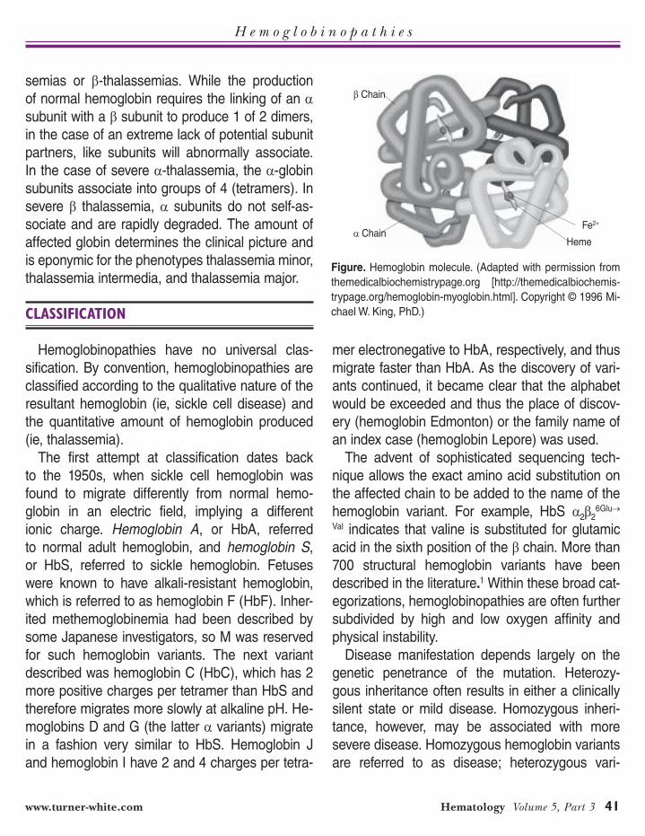
H e m o g l o b i n o p a t h i e s
www.turner-white.com Hematology Volume 5, Part 3 41
semias or β-thalassemias. While the production
of normal hemoglobin requires the linking of an α
subunit with a β subunit to produce 1 of 2 dimers,
in the case of an extreme lack of potential subunit
partners, like subunits will abnormally associate.
In the case of severe α-thalassemia, the α-globin
subunits associate into groups of 4 (tetramers). In
severe β thalassemia, α subunits do not self-as-
sociate and are rapidly degraded. The amount of
affected globin determines the clinical picture and
is eponymic for the phenotypes thalassemia minor,
thalassemia intermedia, and thalassemia major.
CLASSIFICATION
Hemoglobinopathies have no universal clas-
sification. By convention, hemoglobinopathies are
classified according to the qualitative nature of the
resultant hemoglobin (ie, sickle cell disease) and
the quantitative amount of hemoglobin produced
(ie, thalassemia).
The first attempt at classification dates back
to the 1950s, when sickle cell hemoglobin was
found to migrate differently from normal hemo-
globin in an electric field, implying a different
ionic charge. Hemoglobin A, or HbA, referred
to normal adult hemoglobin, and hemoglobin S,
or HbS, referred to sickle hemoglobin. Fetuses
were known to have alkali-resistant hemoglobin,
which is referred to as hemoglobin F (HbF). Inher-
ited methemoglobinemia had been described by
some Japanese investigators, so M was reserved
for such hemoglobin variants. The next variant
described was hemoglobin C (HbC), which has 2
more positive charges per tetramer than HbS and
therefore migrates more slowly at alkaline pH. He-
moglobins D and G (the latter α variants) migrate
in a fashion very similar to HbS. Hemoglobin J
and hemoglobin I have 2 and 4 charges per tetra-
mer electronegative to HbA, respectively, and thus
migrate faster than HbA. As the discovery of vari-
ants continued, it became clear that the alphabet
would be exceeded and thus the place of discov-
ery (hemoglobin Edmonton) or the family name of
an index case (hemoglobin Lepore) was used.
The advent of sophisticated sequencing tech-
nique allows the exact amino acid substitution on
the affected chain to be added to the name of the
hemoglobin variant. For example, HbS α2β26Glu→
Val indicates that valine is substituted for glutamic
acid in the sixth position of the β chain. More than
700 structural hemoglobin variants have been
described in the literature.1 Within these broad cat-
egorizations, hemoglobinopathies are often further
subdivided by high and low oxygen affinity and
physical instability.
Disease manifestation depends largely on the
genetic penetrance of the mutation. Heterozy-
gous inheritance often results in either a clinically
silent state or mild disease. Homozygous inheri-
tance, however, may be associated with more
severe disease. Homozygous hemoglobin variants
are referred to as disease; heterozygous vari-
Figure. Hemoglobin molecule. (Adapted with permission from
themedicalbiochemistrypage.org [http://themedicalbiochemis-
trypage.org/hemoglobin-myoglobin.html]. Copyright © 1996 Mi-
chael W. King, PhD.)
β Chain
α ChainHeme
Fe2+
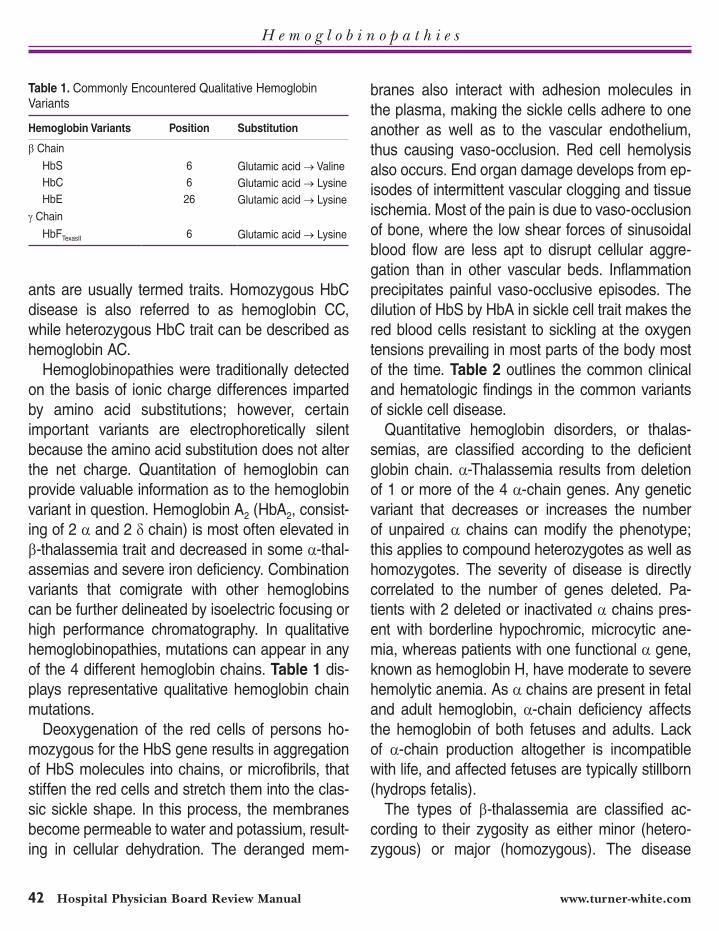
H e m o g l o b i n o p a t h i e s
42 Hospital Physician Board Review Manual www.turner-white.com
ants are usually termed traits. Homozygous HbC
disease is also referred to as hemoglobin CC,
while heterozygous HbC trait can be described as
hemoglobin AC.
Hemoglobinopathies were traditionally detected
on the basis of ionic charge differences imparted
by amino acid substitutions; however, certain
important variants are electrophoretically silent
because the amino acid substitution does not alter
the net charge. Quantitation of hemoglobin can
provide valuable information as to the hemoglobin
variant in question. Hemoglobin A2 (HbA2, consist-
ing of 2 α and 2 δ chain) is most often elevated in
β-thalassemia trait and decreased in some α-thal-
assemias and severe iron deficiency. Combination
variants that comigrate with other hemoglobins
can be further delineated by isoelectric focusing or
high performance chromatography. In qualitative
hemoglobinopathies, mutations can appear in any
of the 4 different hemoglobin chains. Table 1 dis-
plays representative qualitative hemoglobin chain
mutations.
Deoxygenation of the red cells of persons ho-
mozygous for the HbS gene results in aggregation
of HbS molecules into chains, or microfibrils, that
stiffen the red cells and stretch them into the clas-
sic sickle shape. In this process, the membranes
become permeable to water and potassium, result-
ing in cellular dehydration. The deranged mem-
branes also interact with adhesion molecules in
the plasma, making the sickle cells adhere to one
another as well as to the vascular endothelium,
thus causing vaso-occlusion. Red cell hemolysis
also occurs. End organ damage develops from ep-
isodes of intermittent vascular clogging and tissue
ischemia. Most of the pain is due to vaso-occlusion
of bone, where the low shear forces of sinusoidal
blood flow are less apt to disrupt cellular aggre-
gation than in other vascular beds. Inflammation
precipitates painful vaso-occlusive episodes. The
dilution of HbS by HbA in sickle cell trait makes the
red blood cells resistant to sickling at the oxygen
tensions prevailing in most parts of the body most
of the time. Table 2 outlines the common clinical
and hematologic findings in the common variants
of sickle cell disease.
Quantitative hemoglobin disorders, or thalas-
semias, are classified according to the deficient
globin chain. α-Thalassemia results from deletion
of 1 or more of the 4 α-chain genes. Any genetic
variant that decreases or increases the number
of unpaired α chains can modify the phenotype;
this applies to compound heterozygotes as well as
homozygotes. The severity of disease is directly
correlated to the number of genes deleted. Pa-
tients with 2 deleted or inactivated α chains pres-
ent with borderline hypochromic, microcytic ane-
mia, whereas patients with one functional α gene,
known as hemoglobin H, have moderate to severe
hemolytic anemia. As α chains are present in fetal
and adult hemoglobin, α-chain deficiency affects
the hemoglobin of both fetuses and adults. Lack
of α-chain production altogether is incompatible
with life, and affected fetuses are typically stillborn
(hydrops fetalis).
The types of β-thalassemia are classified ac-
cording to their zygosity as either minor (hetero-
zygous) or major (homozygous). The disease
Table 1. Commonly Encountered Qualitative Hemoglobin Variants
Hemoglobin Variants Position Substitution
β Chain
HbS 6 Glutamic acid → Valine
HbC 6 Glutamic acid → Lysine
HbE 26 Glutamic acid → Lysine
γ Chain
HbFTexasII 6 Glutamic acid → Lysine
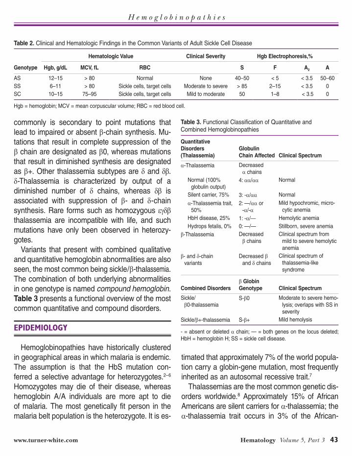
H e m o g l o b i n o p a t h i e s
www.turner-white.com Hematology Volume 5, Part 3 43
commonly is secondary to point mutations that
lead to impaired or absent β-chain synthesis. Mu-
tations that result in complete suppression of the
β chain are designated as β0, whereas mutations
that result in diminished synthesis are designated
as β+. Other thalassemia subtypes are δ and δβ.
δ-Thalassemia is characterized by output of a
diminished number of δ chains, whereas δβ is
associated with suppression of β- and δ-chain
synthesis. Rare forms such as homozygous εγδβ
thalassemia are incompatible with life, and such
mutations have only been observed in heterozy-
gotes.
Variants that present with combined qualitative
and quantitative hemoglobin abnormalities are also
seen, the most common being sickle/β-thalassemia.
The combination of both underlying abnormalities
in one genotype is named compound hemoglobin.
Table 3 presents a functional overview of the most
common quantitative and compound disorders.
EPIDEMIOLOGY
Hemoglobinopathies have historically clustered
in geographical areas in which malaria is endemic.
The assumption is that the HbS mutation con-
ferred a selective advantage for heterozygotes.2–6
Homozygotes may die of their disease, whereas
hemoglobin A/A individuals are more apt to die
of malaria. The most genetically fit person in the
malaria belt population is the heterozygote. It is es-
timated that approximately 7% of the world popula-
tion carry a globin-gene mutation, most frequently
inherited as an autosomal recessive trait.7
Thalassemias are the most common genetic dis-
orders worldwide.8 Approximately 15% of African
Americans are silent carriers for α-thalassemia; the
α-thalassemia trait occurs in 3% of the African-
Table 2. Clinical and Hematologic Findings in the Common Variants of Adult Sickle Cell Disease
Hematologic Value Clinical Severity Hgb Electrophoresis,%
Genotype Hgb, g/dL MCV, fL RBC S F A2 A
AS 12–15 > 80 Normal None 40–50 < 5 < 3.5 50–60
SS 6–11 > 80 Sickle cells, target cells Moderate to severe > 85 2–15 < 3.5 0
SC 10–15 75–95 Sickle cells, target cells Mild to moderate 50 1–8 < 3.5 0
Hgb = hemoglobin; MCV = mean corpuscular volume; RBC = red blood cell.
Table 3. Functional Classification of Quantitative and Combined Hemoglobinopathies
Quantitative
Disorders
(Thalassemia)
Globulin
Chain Affected Clinical Spectrum
α-Thalassemia Decreased
α chains
Normal (100%
globulin output)4: αα/αα Normal
Silent carrier, 75% 3: -α/αα Normal
α-Thalassemia trait,
50%
2: —/αα or
-α/-αMild hypochromic, micro-
cytic anemia
HbH disease, 25% 1: -α/— Hemolytic anemia
Hydrops fetalis, 0% 0: —/— Stillborn, severe anemia
β-Thalassemia Decreased
β chains
Clinical spectrum from
mild to severe hemolytic
anemia
β- and δ-chain
variants
Decreased β
and δ chains
Clinical spectrum of
thalassemia-like
syndrome
Combined Disorders
β Globin
Genotype Clinical Spectrum
Sickle/
β0-thalassemiaS-β0 Moderate to severe hemo-
lysis; overlaps with SS in
severity
Sickle/β+-thalassemia S-β+ Mild hemolysis
- = absent or deleted α chain; — = both genes on the locus deleted;
HbH = hemoglobin H; SS = sickle cell disease.
H e m o g l o b i n o p a t h i e s
44 Hospital Physician Board Review Manual www.turner-white.com
American population and in 1% to 15% of persons
of Mediterranean origin.3 β-Thalassemia is preva-
lent in Mediterranean populations (10%–-15%
incidence), as well as those from Southeast Asia,
West Africa, and the Middle East. It occurs in less
than 1% of African Americans.
HbS, HbC, and hemoglobin E (HbE) are the
most frequently encountered qualitative hemoglo-
binopathies. HbS has the greatest prevalence in
tropical Africa, with a heterozygous frequency up to
20%. The sickle cell gene has also been reported
in the Middle East, Greece, and India, although it
occurs in these countries at a markedly lower rate.
In the United States, HbS has been reported in 9%
of the African-American population.9 HbC is found
in more than 30% of the West African population10
and has been reported in approximately 3% of
African Americans. HbE is found predominantly in
Southeast Asia, most commonly in Thailand and
Cambodia and less commonly in Malaysia.3,11
Through migration, hemoglobinopathies have
spread from their native areas and are now en-
demic throughout Europe, the Americas, and Aus-
tralia.12 Although rare, thalassemias can occur in all
racial groups due to sporadic mutations; thus, ra-
cial background does not preclude the diagnosis.
CASE PRESENTATION
A 21-year-old man presents to the emergen-
cy department with chest pain that started
12 hours ago. He has a diagnosis of HbS/β0 thalas-
semia. His outpatient medications are hydroxyurea,
folate, and oxycodone as needed for pain.
His past medical history is significant for a sple-
nectomy at the age of 5 years, a cholecystectomy
at age 8 years, and avascular necrosis of the left
femoral head.He also has mild cardiac dysfunction
with global left ventricular hypokinesis and an ejec-
tion fraction of 50% by recent echocardiography.
His last hospitalization was for a pain crisis that
occurred 2 years ago.
The patient has no history of smoking, alcohol
use, or drug abuse; he is currently enrolled in
college and he describes himself as single. His
mother is originally from Iran; his father was born
in West Africa.
On further questioning, the patient complains of
diffuse throbbing chest pain that he ranks as 8/10 on
a pain scale. He reports the pain is mostly anterior
chest pain and states that his usual sickle cell pain is
lower back and joint pain and is relieved by nonsteroidal
anti-inflammatory agents. He is afebrile with a pulse of
108 bpm, blood pressure of 135/67 mm Hg, respira-
tory rate of 21 breaths/min, and an oxygen saturation
of 93% on room air.
It is apparent that the patient is in some degree
of physical distress, using accessory muscles
to breath. His pulmonary exam is significant for
crackles at the base of his right lobe. On cardiac
auscultation he has a grade III systolic murmur
over his right upper sternal border. The remainder
of his physical examination is unremarkable.
Results of initial laboratory tests show a white
blood cell (WBC) count of 18,100/μL, with a neu-
trophilia; a hemoglobin level of 7.3 g/dL, down from
his baseline of 9 g/dL; a mean corpuscular volume
(MCV) of 77 fL, (normal, 80–96 fL); and a platelet
count of 424,000/μL (normal, 150,000–400,000/
μL). A peripheral blood smear shows microcytosis
and polychromatophilia. The patient’s urinalysis is
normal; blood cultures are negative for bacteria
after 48 hours. A chest radiograph shows right
basilar opacities.
CLINICAL PRESENTATION
How a patient presents depends on the char-
acteristics of the underlying hemoglobinopathy.
H e m o g l o b i n o p a t h i e s
www.turner-white.com Hematology Volume 5, Part 3 45
Mutations or alterations of the globin protein
produce pronounced changes in the functional
property of hemoglobin, including oxygen affin-
ity and solubility, and impair the structural integ-
rity of the erythrocyte.13 Heterozygous disorders
usually have a benign presentation, whereas ho-
mozygous disorders can lead to significant ane-
mia and hemolytic and/or vaso-occlusive crises.
Qualitative hemoglobinopathies (eg, homozy-
gous HbS) are characterized by rigid red blood
cells that do not pass through capillaries and
cause microinfarction or vaso-occlusion, both of
which can lead to acute and chronic organ dam-
age. The amount of sickle cells is directly related
to the severity of the hemolytic process.14 That
said, sickle cell disease remains a highly phe-
notypically variable disease. In the steady state,
individuals with a qualitative hemoglobinopathy
usually present with a normochromic, normo-
cytic anemia in the range of 5 to 11 g/dL. The
anemia is usually accompanied by an elevated
reticulocyte count and a reduced erythropoietin
level relative to the anemia. Laboratory workup
is indicative of hemolysis, as indirect serum
bilirubin and lactate dehydrogenase (LDH) are
elevated.
Current risk stratification for common complica-
tions remains incomplete, but certain findings are
predictive of outcomes. For example, a low HbF
concentration and leukocytosis are associated
with increased risk of early death, acute chest
syndrome, and painful crises.15 Higher steady
state hemoglobin concentrations are associated
with avascular necrosis and sickle cell retinopa-
thy.16 Compound disease, such as sickle cell/β-
thalassemia, presents with a spectrum of clinical
manifestations that reflect the underlying chain
defect. The severity of disease is an inverse func-
tion of the quantity of HbA. Patients with sickle
cell/β0 thalassemia have more irreversible sickled
cells in the peripheral smear than patients with
sickle cell/β+. Both compound sickle cell variants
present with clinical manifestations, although they
are less severe than those seen with homozy-
gous HbS.
Quantitative and qualitative hemoglobinopa-
thies can present with a similar range of anemia.
The majority of patients with α- and β-thal-
assemia minor are diagnosed because of an
asymptomatic microcytic, hypochromic anemia.
Anemia can be more pronounced in thalasse-
mias of intermediate degree, while in thalassemia
major patients present with life-long transfusion-
dependent anemia and iron overload syndromes,
which untreated can lead to end organ damage.
Organ-Specific Findings
The function of blood and its role in oxygen
delivery means that hemoglobinopathies can af-
fect any organ system. Organ findings in hemo-
globinopathy reflect the effects of compensatory
hemoglobin production, distribution and disposal
of hemolyzed red blood cells, and iron deposi-
tion, particularly from recurrent transfusions.
The most commonly affected organ systems
are the cardiopulmonary, renal, and central ner-
vous systems, skin, bone, and the genitourinary,
endocrine and the reticuloendothelial systems
(Table 4).
Cardiopulmonary symptoms of shortness
of breath and tachycardia secondary to anemia
are the most common presenting symptoms of
sickle cell disease.17 Chronic tachycardias can
result in ventricular remodeling. In HbS disease,
recurrent occlusive crises of the cardiac and
pulmonary vasculature result in micro-infarcts
that eventually alter blood supply, cardiac work-
load, and cardiac contractility.18–20 Fat embolus
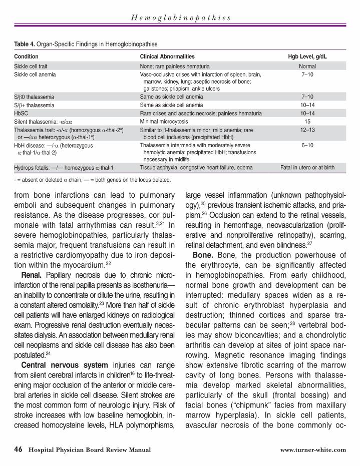
H e m o g l o b i n o p a t h i e s
46 Hospital Physician Board Review Manual www.turner-white.com
from bone infarctions can lead to pulmonary
emboli and subsequent changes in pulmonary
resistance. As the disease progresses, cor pul-
monale with fatal arrhythmias can result.3,21 In
severe hemoglobinopathies, particularly thalas-
semia major, frequent transfusions can result in
a restrictive cardiomyopathy due to iron deposi-
tion within the myocardium.22
Renal. Papillary necrosis due to chronic micro-
infarction of the renal papilla presents as isosthenuria—
an inability to concentrate or dilute the urine, resulting in
a constant altered osmolality.23 More than half of sickle
cell patients will have enlarged kidneys on radiological
exam. Progressive renal destruction eventually neces-
sitates dialysis. An association between medullary renal
cell neoplasms and sickle cell disease has also been
postulated.24
Central nervous system injuries can range
from silent cerebral infarcts in children16 to life-threat-
ening major occlusion of the anterior or middle cere-
bral arteries in sickle cell disease. Silent strokes are
the most common form of neurologic injury. Risk of
stroke increases with low baseline hemoglobin, in-
creased homocysteine levels, HLA polymorphisms,
large vessel inflammation (unknown pathophysiol-
ogy),25 previous transient ischemic attacks, and pria-
pism.26 Occlusion can extend to the retinal vessels,
resulting in hemorrhage, neovascularization (prolif-
erative and nonproliferative retinopathy), scarring,
retinal detachment, and even blindness.27
Bone. Bone, the production powerhouse of
the erythrocyte, can be significantly affected
in hemoglobinopathies. From early childhood,
normal bone growth and development can be
interrupted: medullary spaces widen as a re-
sult of chronic erythroblast hyperplasia and
destruction; thinned cortices and sparse tra-
becular patterns can be seen;28 vertebral bod-
ies may show biconcavities; and a chondrolytic
arthritis can develop at sites of joint space nar-
rowing. Magnetic resonance imaging findings
show extensive fibrotic scarring of the marrow
cavity of long bones. Persons with thalasse-
mia develop marked skeletal abnormalities,
particularly of the skull (frontal bossing) and
facial bones (“chipmunk” facies from maxillary
marrow hyperplasia). In sickle cell patients,
avascular necrosis of the bone commonly oc-
Table 4. Organ-Specific Findings in Hemoglobinopathies
Condition Clinical Abnormalities Hgb Level, g/dL
Sickle cell trait None; rare painless hematuria Normal
Sickle cell anemia Vaso-occlusive crises with infarction of spleen, brain,
marrow, kidney, lung; aseptic necrosis of bone;
gallstones; priapism; ankle ulcers
7–10
S/β0 thalassemia Same as sickle cell anemia 7–10
S/β+ thalassemia Same as sickle cell anemia 10–14
HbSC Rare crises and aseptic necrosis; painless hematuria 10–14
Silent thalassemia: -α/αα Minimal microcytosis 15
Thalassemia trait: -α/-α (homozygous α-thal-2a)
or —/αα heterozygous (α-thal-1a)
Similar to β-thalassemia minor; mild anemia; rare
blood cell inclusions (precipitated HbH)
12–13
HbH disease: —/-α (heterozygous
α-thal-1/α-thal-2)
Thalassemia intermedia with moderately severe
hemolytic anemia; precipitated HbH; transfusions
necessary in midlife
6–10
Hydrops fetalis: —/— homozygous α-thal-1 Tissue asphyxia, congestive heart failure, edema Fatal in utero or at birth
- = absent or deleted α chain; — = both genes on the locus deleted.
H e m o g l o b i n o p a t h i e s
www.turner-white.com Hematology Volume 5, Part 3 47
curs in the femoral/humeral heads; in infants
under the age of 9 months, avascular necrosis
can manifest as dactylitis. However, the entire
skeleton is at risk of infarction; in the most
dramatic presentation of bone involvement, the
anterior tibia can become swollen, tender, and
erythematous. Necrotic marrow presents risks of
superinfection from encapsulated organisms (ie,
Salmonella and Staphylococcus) and embolus
to the lung, causing acute chest syndrome or
sudden death.
Reticuloendothelial system. Increased red cell
destruction in childhood leads to alterations in the
reticuloendothelial system that manifest initially as
splenomegaly, resultant extramedullary hematopoi-
esis, and eventual autosplenectomy, often between
18 and 36 months of age, with subsequent immuno-
compromise. Patients are particularly vulnerable to
infections with encapsulated organisms.29 Spleno-
megaly presents with symptoms of early satiety and
laboratory values consistent with hypersplenism. In
thalassemic disease, constant destruction of globin
chains can lead to spleen “work hypertrophy” and a
resultant hypersplenism, plasma volume expansion,
and erythroid marrow expansion with worsening
anemia.3
The destruction of dysfunctional cells in the
spleen and liver can present with hepatospleno-
megaly and jaundice. Between 50% and 60% of
patients develop pigment gallstones, secondary to
a hyperbilirubinemia; there is a low overall incidence
of primary choledocholithiasis.30 The need for re-
current transfusions in many hemoglobinopathies
leads to iron overload in the liver, fibrosis, and end-
stage liver disease.31
Acute splenic sequestration (ASSC) is a life-
threatening event in the sickle cell patient. Intra-
splenic trapping of red blood cells can cause a
precipitous fall in hemoglobin and resultant hypo-
volemia. ASSC can be defined by a decrease of at
least 2 g/dL from a patient’s steady-state hemoglo-
bin level with evidence of increased erythropoiesis
(ie, increased reticulocyte level, enlarging spleen).
Clinically, ASSC manifests with sudden weakness,
pallor, tachycardia, tachypnea, and abdominal full-
ness.32
Endocrine abnormalities can result from hor-
monal and structural disruptions due to disor-
dered hematopoiesis as well as from recurrent
transfusions and subsequent iron overload. Growth
retardation, growth failure, dysfunctional sexual de-
velopment, diabetes and hypothyroidism are often
seen.33
Skin. Ulcerations, particularly around the ankles,
are common problems in sickle cell patients.34 The
general immunocompromised state of many of
these patients, often exacerbated by the use of the
myelosuppressive medication hydroxyurea, pre-
disposes ulcerations to infection. In addition, lower
levels of hemoglobin seen in patients with skin
ulcerations (and concomitant elevations in LDH, bili-
rubin, and aspartate aminotransferase) suggest that
hemolysis occurs at greater intensity in this patient
population; transfusion provides effective therapy.35
CASE CONTINUED
The patient is diagnosed with acute chest
syndrome and is admitted to the hospital
for further management. A comprehensive meta-
bolic panel reveals an elevated total bilirubin of 1.7
mg/dL (normal range, 0.3–1.2 mg/dL), a direct bili-
rubin of 0.7 mg/dL (normal range, 0.0–0.4 mg/dL),
and an LDH of 404 U/L (normal range, 94–250
U/L). The remainder of the results, including liver
and renal function, are normal; coagulation param-
eters are within normal limits. Iron studies are not
sent due to the acuity of the event; blood is sent for
typing and crossmatching.
H e m o g l o b i n o p a t h i e s
48 Hospital Physician Board Review Manual www.turner-white.com
The patient receives 5 mg of morphine in-
travenously in the ED and is then started on a
morphine patient-controlled anesthesia pump
(PCA) with settings of 1 mg/hr basal rate and
an as-needed bolus. A bowel regimen with
docusate and senna is started to prevent nar-
cotic-induced constipation. He also receives an
intravenous (IV) infusion of ketorolac, IV fluids
at 125 mL/hour, and 2 units of packed red blood
cells. Empiric IV antibiotic therapy is started for
possible community-acquired pneumonia. He
is continued on his outpatient hydroxyurea and
folate.
MEDICAL EMERGENCIES ASSOCIATED WITH
HEMOGLOBINOPATHIES
The diagnosis of a hemoglobinopathy is never
an emergency. However, complications of hemo-
globinopathies such as sepsis, thrombotic stroke
(children), cerebral hemorrhage in adults with sick-
le cell anemia, rib infarction, acute chest syndrome
(ACS)/acute respiratory distress syndrome, ASSC,
severe aplasia, and fat embolism syndrome can all
be considered emergencies.
Pain Crises
Pain and pain crises are the most common rea-
sons for patients with hemoglobinopathies to be
hospitalized; these crises can be potent indicators
of serious organ dysfunction. Four different vari-
ants of crises are differentiated: vaso-occlusive,
aplastic, sequestration, and hemolytic. Vaso-
occlusive crises occur most frequently; the impli-
cated pathophysiology of such episodes includes
complex interactions between endothelium, acti-
vated plasma factors, leukocytes and, in the case
of sickle cell disease, rigid, inflexible red blood
cells. Obstruction of the microvasculature com-
promises oxygen delivery to the organ. The type
of vascular supply as well as the affected organ
dramatically changes the acuteness of care.
Vaso-occlusive Crises
Vaso-occlusive crises that affect the central ner-
vous system can have devastating complications.
Cerebrovascular accidents in patients with a hemo-
globinopathy are thought to occur due to existent
inflammatory lesions in the major vessels (ie, the
internal carotid arteries and the anterior and middle
cerebral arteries), with most patients have no fore-
warning of an imminent stroke. The highest inci-
dence of central nervous system crises is observed
in children and adults older than 29 years of age.36
In approximately 25% of patients, prior painful or
aplastic crises, transient ischemic attacks, human
leukocyte antigen loci polymorphisms, low baseline
hemoglobin, and an elevated diastolic blood pres-
sure can signal a predisposition to stroke. Screen-
ing methods to identify disease before it causes
extreme devastation are being investigated.36 In par-
ticular, the use of transcranial Doppler ultrasonogra-
phy in high-risk patient populations to evaluate for
flow-velocity changes is showing promise. Between
46% to 90% of patients who go untreated following
an initial stroke will suffer a repeat stroke; the high-
est percentage of repeat strokes will occur within 36
months of the initial stroke.36 Exchange transfusion
has been shown superior to simple transfusion both
as acute treatment and in the prevention of a sec-
ond stroke.37 If the use of hydroxyurea in patients
with prior stroke leads to a significant increase in
HbF, transfusions can be discontinued.38 In general,
maintaining a HbS fraction less than 30% has also
been shown to reduce the likelihood of stroke recur-
rence.39
Acute Chest Syndrome
Acute chest syndrome is a general term for
H e m o g l o b i n o p a t h i e s
www.turner-white.com Hematology Volume 5, Part 3 49
any condition that results in a new pulmonary
infiltrate. The differential diagnosis is pneumonia,
pulmonary embolism, and primary pulmonaryth-
rombosis. ACS clinically presents as a com-
bination of fever, chest pain, elevated white blood
count, infection, and new pulmonary infiltrates. It
is thought to occur secondary to the interplay of
infection, infarction, and pulmonary embolus. In
a study of 538 patients with ACS, only 38% of
episodes had a clear defining pathophysiologic
event.40 The incidence of ACS increases in the
winter months and in children aged 2 to 4 years.41
The concentration of HbF and degree of anemia
are inversely proportional to the incidence of ACS
and directly proportional to the white blood cell
count.42 Diagnostic criteria for ACS are as follows:
new pulmonary infiltrate detected by chest radio-
graph involving at least one complete lung seg-
ment that is not consistent with the appearance of
atelectasis and one or more of the following signs
or symptoms:
• Chest pain
• Temperature > 38.5°C
• Tachypnea, wheezing, cough, or appearance of
increased work of breathing
• Hypoxemia relative to baseline
In addition to general measures of hydration,
pain control, oxygenation, and antibiotic treatment,
if indicated, simple transfusion should be started
and advanced to exchange transfusion or erythro-
cytapheresis if there is clinical progression, severe
hypoxemia, multilobar disease, or previous history
of severe ACS or cardiopulmonary disease. The
goal of therapy is to decrease the HbS to less than
30% of total hemoglobin while not exceeding a
hemoglobin level of 10 g/dL.43
Rib infarction can also present with a form of
acute chest pain—specifically pleuritis and splint-
ing. If not treated promptly, it can result in acute
respiratory distress syndrome requiring mechani-
cal ventilation. Aggressive analgesia and use of
incentive spirometry (10 puffs every 2 hours during
daytime hours) can prevent 85% of the infiltrates
that develop in patients having chest pain in the
hospital.44
Priapism
Low-flow priapism is a serious complication that
occurs in approximately 35% of patients, usually
before the age of 20 years.45 Sickling within the
venous sinusoids during erection can lead to criti-
cal stasis, hypoxia, and acidemia. If left untreated,
a patient can be rendered permanently impotent.
Risk factors include prolonged sexual activity;
fever; dehydration; and use of alcohol, mari-
juana, cocaine, psychotropic agents, phospho-
diesterase 5 enzyme inhibitors, or exogenous
testosterone. Diagnosis is made with color du-
plex Doppler ultrasonography or cavernosal blood
gas measurement. Neither simple nor exchange
transfusion has been found beneficial in treatment
of acute priapism. In erections lasting longer than
2 hours, aspiration of blood from the corpus caver-
nosum followed by a saline or adrenergic agonist
infusion is standard treatment.46 In severe cases,
surgical procedures, such as Winter’s procedure,
shunt blood away from the corpus cavernosum to
the more pliable corpus spongiosum.
Thalassemia-Specific Emergencies
Emergencies in thalassemias largely correlate
with the acuity of the anemia. In β-thalassemia
major, critical changes are seen in infants after
6 months of age, when hemoglobin production
changes from fetal to adult hemoglobin. Infants
develop chronic anemia, with stigmata of profound
H e m o g l o b i n o p a t h i e s
50 Hospital Physician Board Review Manual www.turner-white.com
hemolysis. Developmental delays, growth retarda-
tion, and abdominal swelling with enlargement of
the liver and spleen, as well as consequent jaun-
dice reflect the onset of severe hemolytic anemia.
Around 80% of untreated children die within the
first years of life, due to consequences of severe
anemia, high-output heart failure, and susceptibil-
ity to infection.
CASE RESOLUTION
Initially, the patient’s pain does not im-
prove, so the basal rate of his morphine
PCA is increased to 2 mg/hr. Repeat blood
work after 16 hours reveals a hemoglobin level
of 9.2 mg/dL, with a WBC of 16,2000/μL and
platelet count of 414,000/μL. The HbF level on
admission is 4.7% (normal range, 0.0%–1.5%).
The peripheral smear reveals an average of
2 sickled cells per high-power field, with micro-
cytic cells and polychromasia. After 48 hours of
PCA treatment, the pain medication is switched to
oral oxycodone. He has no desaturation or fevers
over 72 hours; his vital signs normalize and he is
switched to an oral antibiotic regimen. A repeat
chest radiograph reveals a decrease in the right
basal opacity. After 120 hours, the patient’s vital
signs remain stable, he is asymptomatic, and is
discharged on as-needed pain regimen and 5
more days of oral antibiotics. He is scheduled for a
follow-up appointment at the Sickle Cell Hematol-
ogy clinic.
DIAGNOSIS
Diagnostic recommendations regarding the lab-
oratory investigation of abnormal hemoglobins
were first made in 1975 by the International Com-
mittee for Standardization in Hematology expert
panel. The recommend initial testing included a
complete blood count, electrophoresis at pH 9.2,
tests for solubility, and quantification of HbA2 and
HbF. The identification of an abnormal hemo-
globin required further testing, using additional
techniques such as electrophoresis at pH 6.0 to
6.2, globin chain separation, and isoelectric fo-
cusing. Heat and isopropanol stability tests were
recommended for detection of unstable hemo-
globin or hemoglobin with altered oxygen affinity.
Although electrophoresis at alkaline and acid pH
has been widely used for many years, cation-ex-
change high-performance liquid chromatography,
or HPLC, has become the method of choice for
the quantitation of HbA2 and HbF and identifica-
tion of hemoglobin variants. HPLC has stream-
lined the recommended preliminary and follow-up
tests for the identification of hemoglobinopathies,
providing a rapid and complete diagnostic work-
up in a majority of cases. Although not usually
indicated, bone marrow biopsy will demonstrate
marrow erythroid hyperplasia and a prominent
increase in iron. Flow cytometry is used to detect
and quantify HbF. Definite diagnosis of a hemo-
globin variant may require mutational analysis of
a specific globin gene by polymerase chain reac-
tion or electrophoresis gene analysis by Southern
blot. Detailed structural analysis of the globin
chains is done by fingerprinting of cryptic digests
by electrophoresis, amino acid sequencing, and
nucleic acid mutation analysis. Genetic testing
is recommended in infants, as hemoglobin elec-
trophoresis will be altered by a predominance of
HbF. Genetic counseling is being used in couples
with significant history to prevent severe forms
of thalassemia. Extraction of fetal DNA either by
amniotic fluid aspiration or chorionic villus sam-
pling enables diagnosis of hemoglobin disorders
in utero.47 Polymerase chain reaction combined
with the use of oligonucleotide probes aids in fast
and reliable diagnosis of mutations.48
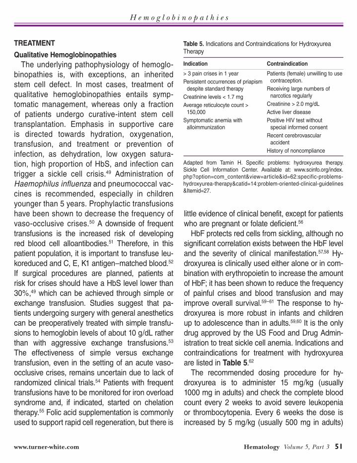
H e m o g l o b i n o p a t h i e s
www.turner-white.com Hematology Volume 5, Part 3 51
TREATMENT
Qualitative Hemoglobinopathies
The underlying pathophysiology of hemoglo-
binopathies is, with exceptions, an inherited
stem cell defect. In most cases, treatment of
qualitative hemoglobinopathies entails symp-
tomatic management, whereas only a fraction
of patients undergo curative-intent stem cell
transplantation. Emphasis in supportive care
is directed towards hydration, oxygenation,
transfusion, and treatment or prevention of
infection, as dehydration, low oxygen satura-
tion, high proportion of HbS, and infection can
trigger a sickle cell crisis.49 Administration of
Haemophilus influenza and pneumococcal vac-
cines is recommended, especially in children
younger than 5 years. Prophylactic transfusions
have been shown to decrease the frequency of
vaso-occlusive crises.50 A downside of frequent
transfusions is the increased risk of developing
red blood cell alloantibodies.51 Therefore, in this
patient population, it is important to transfuse leu-
koreduced and C, E, K1 antigen–matched blood.52
If surgical procedures are planned, patients at
risk for crises should have a HbS level lower than
30%,49 which can be achieved through simple or
exchange transfusion. Studies suggest that pa-
tients undergoing surgery with general anesthetics
can be preoperatively treated with simple transfu-
sions to hemoglobin levels of about 10 g/dL rather
than with aggressive exchange transfusions.53
The effectiveness of simple versus exchange
transfusion, even in the setting of an acute vaso-
occlusive crises, remains uncertain due to lack of
randomized clinical trials.54 Patients with frequent
transfusions have to be monitored for iron overload
syndrome and, if indicated, started on chelation
therapy.55 Folic acid supplementation is commonly
used to support rapid cell regeneration, but there is
little evidence of clinical benefit, except for patients
who are pregnant or folate deficient.56
HbF protects red cells from sickling, although no
significant correlation exists between the HbF level
and the severity of clinical manifestation.57,58 Hy-
droxyurea is clinically used either alone or in com-
bination with erythropoietin to increase the amount
of HbF; it has been shown to reduce the frequency
of painful crises and blood transfusion and may
improve overall survival.59–61 The response to hy-
droxyurea is more robust in infants and children
up to adolescence than in adults.59,60 It is the only
drug approved by the US Food and Drug Admin-
istration to treat sickle cell anemia. Indications and
contraindications for treatment with hydroxyurea
are listed in Table 5.62
The recommended dosing procedure for hy-
droxyurea is to administer 15 mg/kg (usually
1000 mg in adults) and check the complete blood
count every 2 weeks to avoid severe leukopenia
or thrombocytopenia. Every 6 weeks the dose is
increased by 5 mg/kg (usually 500 mg in adults)
Table 5. Indications and Contraindications for Hydroxyurea Therapy
Indication Contraindication
> 3 pain crises in 1 year
Persistent occurrences of priapism
despite standard therapy
Creatinine levels < 1.7 mg
Average reticulocyte count >
150,000
Symptomatic anemia with
alloimmunization
Patients (female) unwilling to use
contraception.
Receiving large numbers of
narcotics regularly
Creatinine > 2.0 mg/dL
Active liver disease
Positive HIV test without
special informed consent
Recent cerebrovascular
accident
History of noncompliance
Adapted from Tamin H. Specific problems: hydroxyurea therapy.
Sickle Cell Information Center. Available at: www.scinfo.org/index.
php?option=com_content&view=article&id=62:specific-problems-
hydroxyurea-therapy&catid=14:problem-oriented-clinical-guidelines
&Itemid=27.
H e m o g l o b i n o p a t h i e s
52 Hospital Physician Board Review Manual www.turner-white.com
until the absolute neutrophil count rather than the
total WBC count is approximately 1000/μL. When
the patient is titered to a neutrophil count of 1000/
μL, then the complete blood count can be checked
every 3 months. Toxicity develops below 500/μL.
Discussion of contraceptive precautions is impor-
tant in patients taking hydroxyurea.
5-Azacytidine has also been found to elevate
HbF levels, but it has never achieved widespread
use due to concerns about carcinogenesis and
toxicity. Sickle cell trait, HbC, and hemoglobin D
usually have an excellent prognosis and need no
specific treatment.
Thalassemia
Treatment for thalassemia is curative only with
bone marrow transplantation. Symptomatic man-
agement for nontransplanted individuals entails
blood transfusion, management of iron stores,
and generalized medical care.63,64 As in sickle cell
disease, patients are at risk for infections, espe-
cially after developing skull deformities in the ENT
area. Infections due to the compromised immune
system should be treated empirically.3 The skull
deformities also lead to an increase in structural
dental problems. Surveillance for alloimmuniza-
tion and hepatitis C, hepatitis B and HIV infection
should be done routinely in recipients of frequent
blood transfusion. Splenectomy should only be
performed in patients with sudden increased trans-
fusion requirements or pain secondary to spleno-
megaly. The risk of splenectomy is susceptibility
for overwhelming pneumococcal infections and
thromboembolic events. Other unstable hemoglo-
bin variants exist, and these are usually treated
symptomatically with transfusion, hydration, and
oxygenation. All patients with thalassemia variants
that require frequent transfusion need surveillance
of the iron stores and chelation therapy, if indi-
cated. Indications for start of chelation therapy in
chronic transfusion-dependent thalassemias are
ferritin levels greater than 1000 mg and/or signs of
iron overload.65
Bone Marrow Transplantation
Hematopoietic stem cell transplantation (HSCT)
remains the only curative option for hemoglobinop-
athies available.66,67 Use of HSCT in thalassemia
was first described in 1982.68 Candidates consid-
ered for transplants are usually children with poor
prognosis.52 The best results are obtained in pa-
tients with HLA-matched siblings. Hepatomegaly,
hepatic fibrosis, and quality of chelation therapy
have been identified as significant outcome vari-
ables in β-thalassemia transplant candidates.69
Long-term survival after transplantation averages
approximately 80%, and 85% to 90% of patients
are cured.70,71 Data on HSCT for sickle cell disease
is not as extensive due to the variable course of
disease and prognostic factors predicting severity
of symptoms. Eligibility for transplant is limited be-
cause of advanced stage disease or missing HLA
matches. The role for early transplantation in pre-
symptomatic young children has yet to be defined.
Nonmyeloablative regimens have been tried to
reduce toxicity, although graft rejection or disease
recurrence was seen.72
Investigational Therapies
Cell receptors and ion pump channels have
been targeted to control hemolysis in sickle cell
disease. Oral magnesium has been studied as an
inhibitor of the KCL co-transporter, with insufficient
data supporting a benefit. Anti-adherence therapy
targeting erythrocyte-endothelial-leukocytes and
platelets has been studied without any current
clinical approved therapies. Nitric oxide, a potent
vasodilator, has been used in the treatment of
H e m o g l o b i n o p a t h i e s
www.turner-white.com Hematology Volume 5, Part 3 53
acute sickle cell disease and found to reduce the
pain score and pain medication use in children.73
In thalassemia, peripheral stem cell transplant
as opposed to HSCT has been studied. Com-
pared to bone marrow transplantation, it has a
shorter engraftment time but a higher incidence
of graft-versus-host disease.74 In view of the low
incidence of graft-versus-host disease associated
with allogenic cord blood transplantation (CBT),
this procedure is particularly appealing. Available
evidence indicates that related donor CBT is a safe
and effective option for patients with hemoglobin-
opathies, offering results at least as good as those
reported using bone marrow cells.75 Hematopoietic
stem cell–targeted gene transfer is currently being
investigated as a treatment option for hemoglobin-
opathies caused by single gene defects.76
OUTCOMES AND PROGNOSIS
Transfusion and chelation treatment have im-
proved outcomes in severe forms of β-thalas-
semia. Patients with an estimated serum ferritin
level below 500 ng/mL over a period of 12 years
were found to have a disease-free survival rate of
91%.77 Transplantation is able to cure patients and
has become a standard procedure. In milder forms
of thalassemia, judicious use of splenectomy in
patients with hypersplenism, vaccination, and a
good standard of general care have an impact on
survival. Prevention through screening and genetic
counseling remains essential to prevent severe
forms of thalassemia.
Survival in sickle cell disease patients is overall
reduced but has been steadily improving. With
good medical care, patients with sickle cell disease
survive to middle age.77 Over the last few decades,
mortality has especially dropped in children. Sur-
vival has improved due to newborn screening pro-
grams, penicillin prophylaxis of disease caused by
Streptococcus pneumoniae, and perhaps pneu-
mococcal vaccine. The most common cause of
death in sickle cell disease is infection, and others
are pulmonary emboli, stroke, and splenic seques-
tration. Neither sickle cell trait nor HbC appear to
impact survival. Genetic counseling also is impor-
tant to prevent severe disease and disease side
effects. Patients at high risk for sickle cell disease
have the option of transplantation.
CONCLUSION
Hemoglobinopathies are hemopoietic stem cell
disorders with qualitative, quantitative, and com-
bined globin chain abnormalities. The range of
newly diagnosed genotypes with resulting pheno-
type has been steadily increasing due to improved
laboratory diagnostic procedures. Treatment re-
mains supportive in the majority of encountered
diseases. Curative treatment in high-risk patients
is limited to HSCT. Transplantation has significant
risks but has become standard procedure, more
so in thalassemias than in sickle cell disease, due
to improved peri- and posttransplantation care.
Genetic counseling and screening are relevant
in predicting and diagnosing clinical significant
genotypes. Further studies are needed to expedite
curative treatment options and prevent recurrent
crises and long-term side effects.
BOARD REVIEW QUESTIONS
Test your knowledge of this topic. Go to
www.turner-white.com and select Hematology from the drop-down
menu of specialties.
H e m o g l o b i n o p a t h i e s
54 Hospital Physician Board Review Manual www.turner-white.com
REFERENCES
1. Clarke GM, Higgins TN. Laboratory investigation of hemo-globinopathies and thalassemias: review and update. Clin Chem 2000;46(8 Pt 2):1284–90.
2. Chotivanich K, Udomsangpetch R, Pattanapanyasat K, et al. Hemoglobin E: a balanced polymorphism protective against high parasitemias and thus severe P falciparum malaria. Blood 2002;100:1172–6.
3. Lichtman M, Beutler E, Kaushansky E, et al. Williams hema-tology. 7th ed. New York: McGraw-Hill Companies; 2005.
4. Oo M, Tin-Shwe, Marlar-Than, O’Sullivan WJ. Genetic red cell disorders and severity of falciparum malaria in Myan-mar. Bull World Health Organ 1995;73:659–65.
5. Fairhurst RM, Fujioka H, Hayton K, et al. Aberrant devel-opment of Plasmodium falciparum in hemoglobin CC red cells: implications for the malaria protective effect of the homozygous state. Blood 2003;101:3309–15.
6. Allen SJ, O’Donnell A, Alexander ND, et al. Alpha+-Thalas-semia protects children against disease caused by other infections as well as malaria. Proc Natl Acad Sci USA 1997;94:14736–41.
7. Weatherall DJ, Clegg JB. Inherited haemoglobin disorders: an increasing global health problem. Bull World Health Organ 2001;79:704.
8. Benz EJ. Disorders of hemoglobin. In: Fauci AS, Kasper DL, Hauser SL, et al, editors. Harrison’s principles of inter-nal medicine. New York: McGraw-Hill Companies; 2007.
9. Kjeldsberg C. Practical diagnosis of hematologic disorders. Chicago: ASCP Press; 2006.
10. Pagnier J, Mears JG, Dunda-Belkhodja O, et al. Evidence for the multicentric origin of the sickle cell hemoglobin gene in Africa. Proc Natl Acad Sci USA 1984;81:1771–3.
11. Flatz G. Hemoglobin E: distribution and population dynam-ics. Humangenetik 1967;3:189–234.
12. Angastiniotis M, Modell B. Global epidemiology of hemo-globin disorders. Ann N Y Acad Sci 1988;850:251–69.
13. Alexy T, Sangkatumvong S, Connes P, et al. Sickle cell disease: selected aspects of pathophysiology. Clin Hemorheol Microcirc 2010;44:155–66.
14. Serjeant GR, Serjeant BE, Milner PF. The irreversibly sick-led cell; a determinant of haemolysis in sickle cell anaemia. Br J Haematol 1969;17:527–33.
15. Platt OS, Brambilla DJ, Rosse WF, et al. Mortality in sickle cell disease. Life expectancy and risk factors for early death. N Engl J Med 1994;330:1639–44.
16. Buchanan GR, DeBaun MR, Quinn CT, Steinberg MH. Sickle cell disease. Hematology Am Soc Hematol Educ Program 2004:35–47.
17. Miller GJ, Serjeant GR, Sivapragasam S, Petch MC.
Cardio-pulmonary responses and gas exchange during exercise in adults with homozygous sickle-cell disease (sickle-cell anaemia). Clin Sci 1973;44:113–28.
18. Gladwin MT, Sachdev V, Jison ML, et al. Pulmonary hyper-tension as a risk factor for death in patients with sickle cell disease. N Engl J Med 2004;350:886–95.
19. Vichinsky EP. Pulmonary hypertension in sickle cell dis-ease. N Engl J Med 2004;350:857–9.
20. Ardiles T, Dark D. Pulmonary complications of sickle cell disease in adults. Mo Med 2007;104:250–4.
21. Castro O. Systemic fat embolism and pulmonary hyperten-sion in sickle cell disease. Hematol Oncol Clin North Am 1996;10:1289–303.
22. Tsironi M, Polonifi K, Deftereos S, et al. Transfusional hemosiderosis and combined chelation therapy in sickle thalassemia. Eur J Haematol 2005;75:355–8.
23. Maigne G, Ferlicot S, Galacteros F, et al. Glomerular le-sions in patients with sickle cell disease. Medicine (Balti-more) 2010;89:18–27.
24. Baron BW, Mick R, Baron JM. Hematuria in sickle cell ane-mia—not always benign: evidence for excess frequency of sickle cell anemia in African Americans with renal cell carcinoma. Acta Haematol 1994;92:119–22.
25. Verduzco LA, Nathan DG. Sickle cell disease and stroke. Blood 2009;114:5117–25.
26. Adams RJ, Ohene-Frempong K, Wang W. Sickle cell and the brain. Hematology Am Soc Hematol Educ Program 2001:31–46.
27. Fadugbagbe AO, Gurgel RQ, Mendonca CQ, et al. Ocular manifestations of sickle cell disease. Ann Trop Paediatr 2010;30:19–26.
28. Vichinsky EP. The morbidity of bone disease in thalasse-mia. Ann N Y Acad Sci 1998;850:344–8.
29. Boghossian SH, Wright G, Webster AD, Segal AW. Investi-gations of host defence in patients with sickle cell disease. Br J Haematol 1985;59:523–31.
30. Vicari P, Gil MV, Cavalheiro Rde C, Figueiredo MS. Multiple primary choledocholithiasis in sickle cell disease. Intern Med 2008;47:2169–70.
31. Ebert EC, Nagar M, Hagspiel KD. Gastrointestinal and hepatic complications of sickle cell disease. Clin Gastro-enterol Hepatol 2010;8:483–9.
32. Wang WC. Splenic sequestration and transient aplastic crises. 2000. Available at: http://sickle.bwh.harvard.edu/spleen.html.
33. Fung EB, Harmatz P, Milet M, et al. Morbidity and mortal-ity in chronically transfused subjects with thalassemia and sickle cell disease: a report from the multi-center study of iron overload. Am J Hematol 2007;82:255–65.
34. Serjeant GR, Serjeant BE, Mohan JS, Clare A. Leg ulcer-ation in sickle cell disease: medieval medicine in a modern
H e m o g l o b i n o p a t h i e s
www.turner-white.com Hematology Volume 5, Part 3 55
world. Hematol Oncol Clin North Am 2005;19:943–56, viii-ix.
35. Nolan VG, Adewoye A, Baldwin C, et al. Sickle cell leg ulcers: associations with haemolysis and SNPs in Klotho, TEK and genes of the TGF-beta/BMP pathway. Br J Hae-matol 2006;133:570–8.
36. Ohene-Frempong K, Weiner SJ, Sleeper LA, et al. Cere-brovascular accidents in sickle cell disease: rates and risk factors. Blood 1998;91:288–94.
37. Hulbert ML, Scothorn DJ, Panepinto J, et al. Exchange blood transfusion compared with simple transfusion for first overt stroke is associated with a lower risk of subsequent stroke: a retrospective cohort study of 137 children with sickle cell anemia. J Pediatr 2006;149:710–2.
38. Ware RE, Zimmerman SA, Schultz WH. Hydroxyurea as an alternative to blood transfusions for the prevention of recurrent stroke in children with sickle cell disease. Blood 1999;94:3022–6.
39. Williams J, Goff JR. Efficacy of transfusion therapy for one to two years in patients with sickle cell disease and cere-brovascular accidents. J Pediatr 1980;96:205–8.
40. Vichinsky EP, Neumayr LD, Earles AN, et al. Causes and outcomes of the acute chest syndrome in sickle cell disease. National Acute Chest Syndrome Study Group. N Engl J Med 2000;342:1855–65.
41. Castro O, Brambilla DJ, Thorington B, et al. The acute chest syndrome in sickle cell disease: incidence and risk factors. The Cooperative Study of Sickle Cell Disease. Blood 1994;84:643–9.
42. Graham LM. Sickle cell disease: pulmonary management options. Pediatr Pulmonol Suppl 2004;26:191–3.
43. Telen MJ. Principles and problems of transfusion in sickle cell disease. Semin Hematol 2001;38:315–23.
44. Bellet PS, Kalinyak KA, Shukla R, et al. Incentive spirom-etry to prevent acute pulmonary complications in sickle cell diseases. N Engl J Med 1995;333:699–703.
45. Mantadakis E, Cavender JD, Rogers ZR, et al. Prevalence of priapism in children and adolescents with sickle cell anemia. J Pediatr Hematol Oncol 1999;21:518–22.
46. Rogers ZR. Priapism in sickle cell disease. Hematol Oncol Clin North Am 2005;19:917–28, viii.
47. Old JM, Fitches A, Heath C, et al. First-trimester fetal diagnosis for haemoglobinopathies: report on 200 cases. Lancet 1986;2:763–7.
48. Saiki RK, Chang CA, Levenson CH, et al. Diagnosis of sickle cell anemia and beta-thalassemia with enzymati-cally amplified DNA and nonradioactive allele-specific oli-gonucleotide probes. N Engl J Med 1988;319:537–41.
49. Shah A. Hemoglobinopathies and other congenital hemo-lytic anemia. Indian J Med Sci 2004;58:490–3.
50. Laulan S, Bernard JF, Boivin P. Systematic blood trans-
fusions in adult homozygote sickle-cell anemia. Study of 11 cases followed for 5 to 10 years. Presse Med 1990;19:785–9.
51. Rosse WF, Gallagher D, Kinney TR, et al. Transfusion and alloimmunization in sickle cell disease. The Cooperative Study of Sickle Cell Disease. Blood 1990;76:1431–7.
52. Roseff SD. Sickle cell disease: a review. Immunohematol-ogy 2009;25:67–74.
53. Vichinsky EP, Haberkern CM, Neumayr L, et al. A compari-son of conservative and aggressive transfusion regimens in the perioperative management of sickle cell disease. The Preoperative Transfusion in Sickle Cell Disease Study Group. N Engl J Med 1995;333:206–13.
54. Alhashimi D, Fedorowicz Z, Alhashimi F, Dastgiri S. Blood transfusions for treating acute chest syndrome in people with sickle cell disease. Cochrane Database Syst Rev 2010(1):CD007843.
55. Silliman CC, Peterson VM, Mellman DL, et al. Iron chela-tion by deferoxamine in sickle cell patients with severe transfusion-induced hemosiderosis: a randomized, double-blind study of the dose-response relationship. J Lab Clin Med 1993;122:48–54.
56. Dhar M, Bellevue R, Carmel R. Pernicious anemia with neuropsychiatric dysfunction in a patient with sickle cell anemia treated with folate supplementation. N Engl J Med 2003;348:2204–7.
57. Noguchi CT, Rodgers GP, Serjeant G, Schechter AN. Lev-els of fetal hemoglobin necessary for treatment of sickle cell disease. N Engl J Med 1988;318:96–9.
58. Powars DR, Schroeder WA, Weiss JN, et al. Lack of influence of fetal hemoglobin levels or erythrocyte indi-ces on the severity of sickle cell anemia. J Clin Invest 1980;65:732–40.
59. Rodgers GP, Dover GJ, Uyesaka N, et al. Augmenta-tion by erythropoietin of the fetal-hemoglobin response to hydroxyurea in sickle cell disease. N Engl J Med 1993;328:73–80.
60. Davies SC, Gilmore A. The role of hydroxyurea in the man-agement of sickle cell disease. Blood Rev 2003;17:99–109.
61. Zimmerman SA, Schultz WH, Davis JS, et al. Sustained long-term hematologic efficacy of hydroxyurea at maxi-mum tolerated dose in children with sickle cell disease. Blood 2004;103:2039–45.
62. Steinberg MH. Management of sickle cell disease. N Engl J Med 1999;340:1021–30.
63. Cao A, Galanello R. Beta-thalassemia. Genet Med 2010;12:61–76.
64. Weatherall DJ, Clegg JB. The thalassemia syndromes. 4th ed. Oxford (UK): Blackwell Science; 2001.
65. Taher A, Hershko C, Cappellini MD. Iron overload in
H e m o g l o b i n o p a t h i e s
56 Hospital Physician Board Review Manual www.turner-white.com
thalassaemia intermedia: reassessment of iron chelation strategies. Br J Haematol 2009;147:634–40.
66. Smiers FJ, Krishnamurti L, Lucarelli G. Hematopoietic stem cell transplantation for hemoglobinopathies: cur-rent practice and emerging trends. Pediatr Clin North Am 2010;57:181–205.
67. Hsieh MM, Kang EM, Fitzhugh CD, et al. Allogeneic hema-topoietic stem-cell transplantation for sickle cell disease. N Engl J Med 2009;361:2309–17.
68. Thomas ED, Buckner CD, Sanders JE, et al. Marrow trans-plantation for thalassaemia. Lancet 1982;2:227–9.
69. Lucarelli G, Galimberti M, Polchi P, et al. Bone marrow transplantation in thalassemia. Hematol Oncol Clin North Am 1991;5:549–56.
70. Boulad F, Giardina P, Gillio A, et al. Bone marrow trans-plantation for homozygous beta-thalassemia. The Memo-rial Sloan-Kettering Cancer Center experience. Ann N Y Acad Sci 1998;850:498–502.
71. Lawson SE, Roberts IA, Amrolia P, et al. Bone mar-row transplantation for beta-thalassaemia major: the UK experience in two paediatric centres. Br J Haematol 2003;120:289–95.
72. Michlitsch JG, Walters MC. Recent advances in bone mar-row transplantation in hemoglobinopathies. Curr Mol Med 2008;8:675–89.
73. Sullivan KJ, Goodwin SR, Evangelist J, et al. Nitric oxide successfully used to treat acute chest syndrome of sickle cell disease in a young adolescent. Crit Care Med 1999;27:2563–8.
74. Iravani M, Tavakoli E, Babaie MH, et al. Comparison of peripheral blood stem cell transplant with bone marrow transplant in class 3 thalassemic patients. Exp Clin Trans-plant 2010;8:66–73.
75. Boncimino A, Bertaina A, Locatelli F. Cord blood transplan-tation in patients with hemoglobinopathies. Transfus Apher Sci 2010;42:277–81.
76. Persons DA. Hematopoietic stem cell gene transfer for the treatment of hemoglobin disorders. Hematology Am Soc Hematol Educ Program 2009:690–7.
77. Olivieri NF, Nathan DG, MacMillan JH, et al. Survival in medically treated patients with homozygous beta-thalas-semia. N Engl J Med 1994;331:574–8.
Related Documents