HELLP ME! Maternal Emergencies that Exist Beyond the Laboring Pregnant Patient Presented By: Theresa Bowden CFRN, CCRN, C-NPT Life Flight Network

Welcome message from author
This document is posted to help you gain knowledge. Please leave a comment to let me know what you think about it! Share it to your friends and learn new things together.
Transcript

HELLP ME! Maternal Emergencies that Exist Beyond the
Laboring Pregnant Patient
Presented By:
Theresa Bowden CFRN, CCRN, C-NPT
Life Flight Network
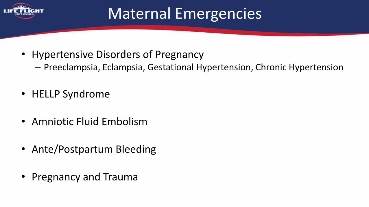
Maternal Emergencies
• Hypertensive Disorders of Pregnancy – Preeclampsia, Eclampsia, Gestational Hypertension, Chronic Hypertension
• HELLP Syndrome • Amniotic Fluid Embolism
• Ante/Postpartum Bleeding • Pregnancy and Trauma
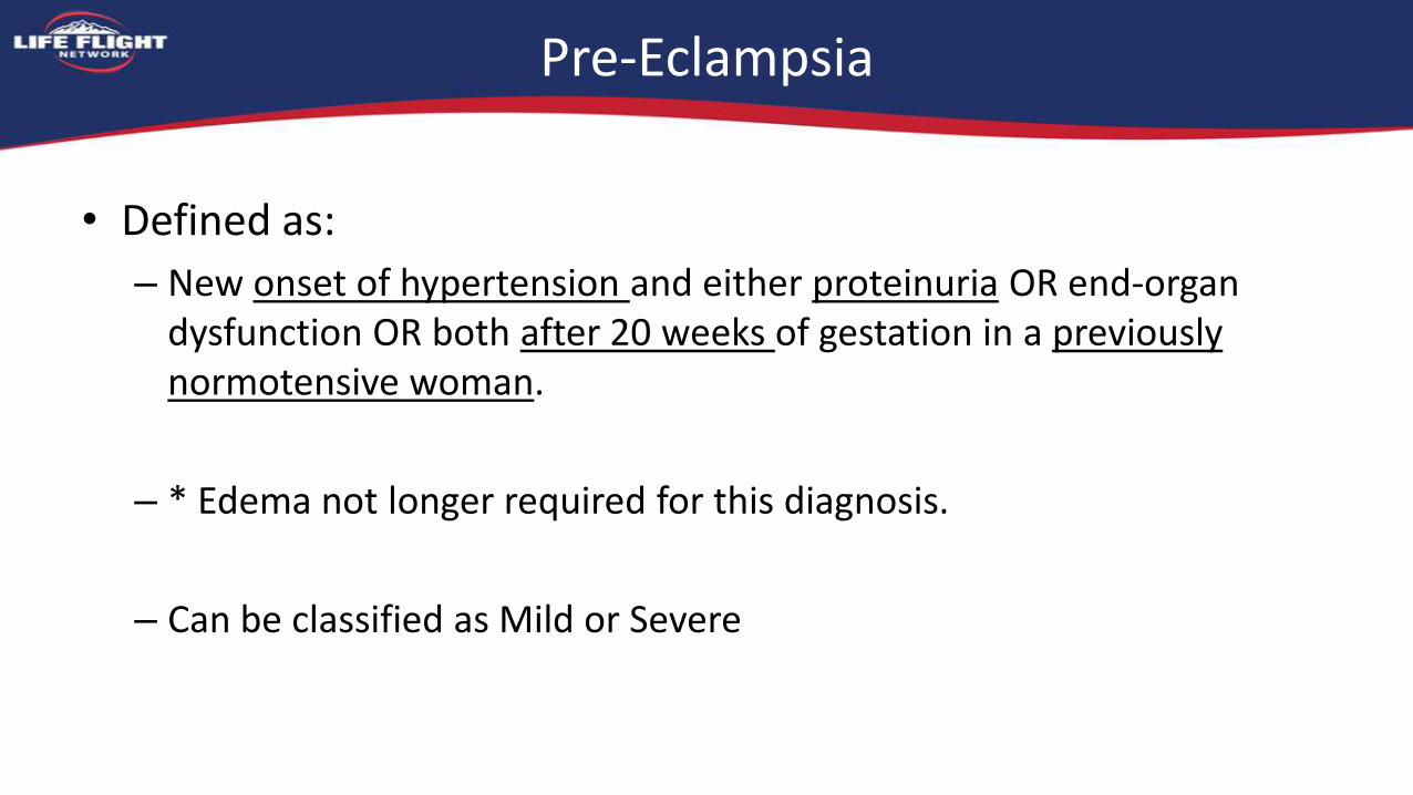
Pre-Eclampsia
• Defined as:
– New onset of hypertension and either proteinuria OR end-organ dysfunction OR both after 20 weeks of gestation in a previously normotensive woman.
– * Edema not longer required for this diagnosis.
– Can be classified as Mild or Severe

Mild Pre-Eclampsia
• Blood pressure >140/90
• >300 mg/dL Protein in a 24 hour urine collection

Severe Pre-Eclampsia
• SBP > 160 or DBP >110 (? recorded on 2 occasions, 6 hours apart)
• Proteinuria
• ? Oliguria
• Visual disturbances
• Epigastric pain; Nausea & vomiting
• Pulmonary edema
• HELLP syndrome
• ? Fetal growth restriction

Eclampsia
• Defined as:
– The development of grand mal seizures in a woman with preeclampsia (in the absence of other neurologic conditions that could account for the seizure).
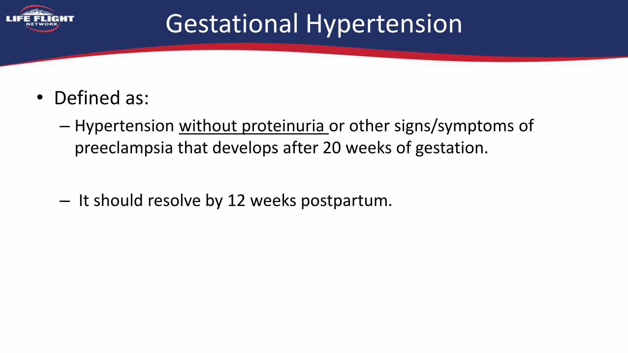
Gestational Hypertension
• Defined as:
– Hypertension without proteinuria or other signs/symptoms of preeclampsia that develops after 20 weeks of gestation.
– It should resolve by 12 weeks postpartum.

Chronic Hypertension
• Defined as:
– Chronic/preexisting hypertension is defined as systolic pressure ≥ 140 mmHg AND/OR diastolic pressure ≥ 90 mmHg that antedates pregnancy
– OR is present before the 20th week of pregnancy (on 2 occasions)
– OR persists longer than 12 weeks postpartum.
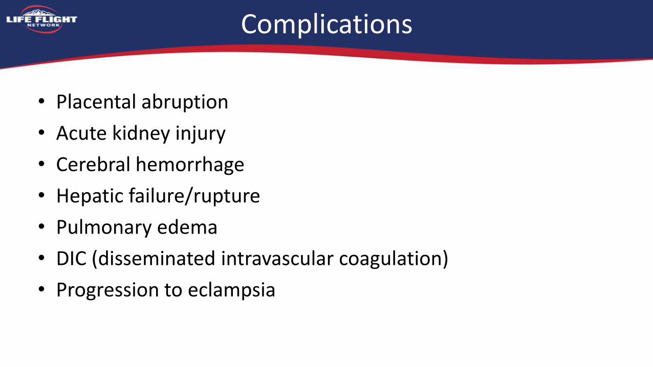
Complications
• Placental abruption
• Acute kidney injury
• Cerebral hemorrhage
• Hepatic failure/rupture
• Pulmonary edema
• DIC (disseminated intravascular coagulation)
• Progression to eclampsia

Mortality
• In the United States, preeclampsia/eclampsia is one of four leading causes of maternal death
– Hemorrhage
– Cardiovascular conditions
– Thromboembolism
– Preeclampsia/eclampsia
1:100,000 live births results in maternal death due to preeclampsia.
Risk Factors
• Past history of preeclampsia
• First pregnancy
• Family history of preeclampsia
• Obesity
• Preexisting medical conditions – Pregestational diabetes, BP ≥130/80 mmHg at first visit, antiphospholipid
antibodies, BMI >26.1, chronic kidney disease
• Twin pregnancies
• Advanced maternal age

General Signs & Symptoms
• Severe hypertension: ≥160/90
• Persistent/severe headache
• Visual abnormalities (blurred vision, sensitive to light)
• Upper abdominal/epigastric pain
• Nausea/vomiting
• Dyspnea or retrosternal chest pain
• Altered mental status

What about the baby?
• The consequences from chronic low perfusion to the placenta:
– Fetal growth restriction
– Low amniotic fluid
– Indirectly, this leads to probable preterm delivery, which in itself puts the baby at risk for many complications.

Assessment
• Vital signs- place on cardiac monitor
• Respiratory status
• Neurological status (LOC, HA, blurred vision)
• Epigastric pain
• Deep Tendon Reflexes and Presence/absence of Clonus
• Assess for edema
• Fetal HR by Doppler if able
• Assess uterine activity

Management
• Minimize IV fluid
• Oxygen for saturations <94%
• Place the patient in a left lateral tilt position
• Observe the patient for active labor and for possible placental abruption
• Assess for central nervous system involvement
– Frontal headache, blurred vision, epigastric pain, changes in reflexes
Medication Management
• Magnesium Sulfate – Example: 6 gram bolus over 30 minutes followed by a continuous infusion of 2
grams/hour – Used more as an anticonvulsant/neuroprotection
• Labetalol – Example: 20mg IV. After 10 min may adm. 40mg IV; after another 10 min may adm.
80mg IV. Can also be a continuous infusion. – Onset 5-10 minutes & duration 3-6 hours. If BP not controlled may administer
Hydralazine
• Hydralazine – Example: 10mg IV over 2 minutes; may repeat q 20 min PRN – Onset 5-10 minutes & duration 2-4 hours
Seizures
• With the progression from pre-eclampsia to eclampsia, the patient has neurologic irritability with the onset of seizures.
– During transport, decrease stimuli and be prepared with suction and airway management
– If the patient seizes:
• Protect the airway
• Administer medications per protocol (Magnesium and Ativan)
HELLP Syndrome
• Defined as:
– Hemolysis, Elevated Liver Enzymes, Low Platelets
– Probably represents a severe form of preeclampsia, but this relationship remains controversial. HELLP may be an independent disorder. (Many women do not have concurrent hypertension or proteinuria).

Signs & Symptoms: HELLP
• *Epigastric or RUQ tenderness
• Nausea and/or vomiting
• Headache
• General malaise
• Jaundice
• Bleeding if coagulopathy present: hematuria, GI bleeding, DIC
• Hypertension (85% of cases)
• Severe pre-eclampsia: SBP >160, DBP >110, proteinuria, edema, seizures
Management: HELLP
• Similar to hypertensive disorder management
• Transport in left lateral tilt
• Oxygen if saturations <94%
• Treat hypertension per protocol
• Assess patient for uterine activity
• Assess fetal heart tones with Doppler (if available)
• Patient may need platelet, RBC or Fresh Frozen Plasma transfusion(s) prior to delivery

Amniotic Fluid Embolism Syndrome
• Obstetric emergency in which the amniotic fluid, fetal cells, hair or other debris enters the mother’s blood stream via the placental bed of the uterus and triggers an allergic-like reaction.

AFES
• Originally thought that fetal cells and debris occluded the pulmonary vasculature.
• Currently, it is considered that the fetal antigens enter the maternal circulation triggering a response similar to SIRS: Systemic Inflammatory Response Syndrome: activating the coagulation cascade which leads to DIC and inflammatory suppression of myocardial function.


AFES
• Risk Factors
– Multiparity: >5 live births
– Advanced maternal age: > 35 years
– Precipitous labor
– Trauma
– Rupture of membranes, rupture of uterine veins
– Possibly: C/S, instrument vaginal delivery, abruption, previa, cervical lacerations or uterine rupture

AFES Presentation
• Rare: between 1-12 per 100,000 deliveries
• When the fluid and fetal cells enter the maternal pulmonary circulation, there is profound respiratory failure with cyanosis and cardiovascular shock.
• Usually then followed by seizures and coma
AFES Management
• There is no specific treatment for amniotic fluid embolism
• Prognosis:
– Maternal mortality is high
– Patients with AFE die within the first hour of onset of symptoms
– Of those who survive this first time period, 50% develop a coagulopathy and 7% are neurologically impaired
• Neonatal survival can be quite good if delivered quickly. The neurological status of the infant is directly related to the time elapsed between maternal arrest and delivery.
Antepartum/Postpartum Bleeding
• Placental Abruption
• Placenta Previa
• Uterine Rupture
• Post Partum Hemorrhage
• Trauma

Goals of Treatment
• The goal is to prevent maternal and/or fetal demise by maintaining hemodynamic stability and blood flow to the uterus as evidenced by normal vital signs, normal fetal heart rate.

Placental Abruption
• Bleeding that causes partial or total placental detachment prior to the delivery of the fetus.
Placental Abruption
• Sign and Symptoms:
– Vaginal bleeding, abdominal pain, hypertonic uterine contractions, non-reassuring fetal heart rate
– Bleeding is usually dark red
• Contributing causes:
– Uterine abnormalities, smoking, drug use (cocaine), hypertension, multiparity, premature rupture of membranes >24hrs, history of previous abruption.

Placenta Previa
• The presence of placental tissue that extends over the internal os, after 20 weeks gestation.

Placenta Previa
• Signs and symptoms:
– Painless vaginal bleeding; can be serial
– Usually bright red blood
• Contributing causes:
– History of previous previa, history of previous cesarean sections, multiple gestation pregnancy, previous uterine surgical procedures

Uterine Rupture
• Can be defined as related to a previously scarred uterus (i.e. previous surgery)
• OR incidence with a unscarred uterus
Uterine Rupture
• Signs and Symptoms:
– Fetal heart rate abnormalities, possible vaginal bleeding, sudden or worsening abdominal pain, changes in uterine contractions, or cessation of labor, maternal hypotension/tachycardia
• Contributing Causes:
– trauma, congenital weakness of myometrium, >40 weeks gestation, history of prior uterine surgeries, infant > 4000 grams estimated fetal weight
***Obstetric Emergency!

Post Partum Hemorrhage
• Obstetrical emergency
• A major cause of maternal morbidity and mortality
• If managed properly, it is one of the most preventable causes
• Can be Primary (early) or Secondary (late)
• Defined by the volume of blood loss:
– >500 ml after vaginal birth
– >1000 ml after cesarean section

Post Partum Hemorrhage
• Causes: 4 T’s:
– Tone: * uterine atony most common cause –Tissue: retained placenta – Trauma: lacerations – Thrombin: clotting disorders
• The potential is high after a delivery related to the fact that late in pregnancy the uterine artery blood flow is 500-700ml/min (approx. 15% of cardiac output)

Types of Post Partum Hemorrhage
• Primary (Early PPH)
– Occurs in the first 24 hours after delivery
– Related to atony, trauma, coagulopathies
• Secondary (Late PPH)
– Occurs >24 hours after delivery and up to 12 weeks post partum; peaking at 2 weeks post partum
– Most common cause is uterine atony due to retained placenta; or history of primary PPH or possibly arteriovenous malformation

Post Partum Hemorrhage
• Signs and Symptoms
– Bleeding that is greater than expected
– Hypovolemia symptoms: hypotension, tachycardia, pallor, confusion, hypoxemia
PPH Management
• Monitor rate and volume of bleeding
• Supportive interventions- based on suspected etiology
– IV access: 16g in anticipation for fluids/blood administration
– Treat for shock
• Fluids, oxygen

Trauma & Pregnancy
• Motor vehicle accidents and domestic/intimate partner violence account for most cases of maternal major trauma.
• Challenges with the pregnant trauma patient are unique in the sense that with the presence of a fetus, there are now two patients whom are potentially at risk.

Physical Changes in Pregnancy
• Cardiovascular: – Cardiac output increases by 20% at 8 weeks and continues to rise until approx. 30 weeks
where it plateaus at 50% above baseline – Supine position at term can lower CO by 25-30%
• Pulmonary: – Oxygen consumption increases, and by >20 weeks there is a displacement of the
diaphragm – Normal ETCO2 is 25-35 for her
• Hematological: – Pregnancy is a procoagulant state
• Gastrointestinal: – Increased risks of aspiration due to decreased esophageal tone and increased
intraabdominal pressure

Physical Changes in Pregnancy
• Uterine:
– Less than 12 weeks gestation, the uterus is protected by the bony pelvis
– Pelvic vasculature is dilated in pregnancy
– Uterine blood flow is approx. 600ml/min in the third trimester
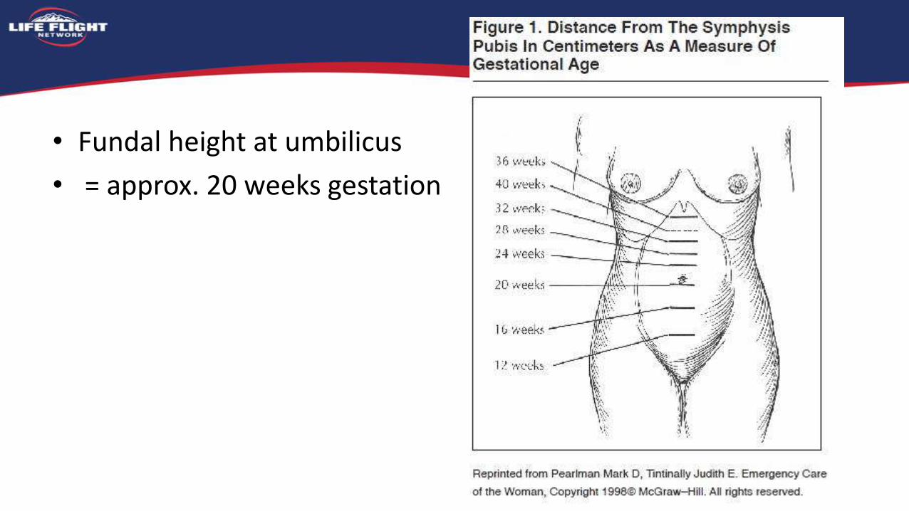
• Fundal height at umbilicus
• = approx. 20 weeks gestation

Maternal Risks with Trauma
• Preterm labor
• Placental abruption
• Uterine rupture
• Hemorrhage
• Shock
• DIC
• Death

Fetal Risks
• Preterm birth
• PROM (premature rupture of membranes)
• Meconium at delivery
• Cesarean birth
• Neonatal respiratory distress syndrome
• Death

Maternal/Fetal Assessment: Trauma
• Uterine palpation • Bleeding • Monitor frequency/intensity of uterine contractions • Bleeding? • Gestational age • Fetal heart rate: 110-160 bpm (Doppler)
Placental Trauma
• Placental abruption
• Ruptured uterus
• PROM
• Cord prolapse

Goals/Management
• Initial goals are to assess maternal airway, breathing and circulation; establish maternal cardio/pulmonary stability
• Treat the pregnant trauma patient as a trauma patient first! • If you need to intubate
– Pre-oxygenate, assume a difficult airway and note increased risk of aspiration
• Cervical Spine Immobilization – Spinal precautions for all suspected spinal cord injury patients. If patient is on a
board, utilized left lateral tilt of the board, and remove as soon as possible.

Management
• Vital Signs q 5 minutes
• Left lateral tilt position
• O2 by NC or NRM
• 2 large bore IVs (16g)
• Fluids to keep SBP >90-100
• Consider blood administration if 2L fluids and no response in BP (caution with vasopressors)
• Be sure to let the receiving facility know the patient is pregnant

Case Study
• Dispatched to transport a patient from a Free-Standing Emergency Room to be admitted at a near by hospital
• 33 year old Hispanic multiparous female presented with complaints of back and abdominal pain. She also has had nausea and vomiting.
• She states she is 7 months pregnant. G3P2
• No allergies, no medications.

Case Study
• BP 223/123
• HR 86
• T 97.6
• RR 20
• Alert, oriented. DTRs normal, no clonus, no headache or blurred vision, no edema, PERRL.
• Breath sounds clear and equal bilaterally

Case Study
• Lab work:
– WBCs and LFTs slightly elevated
– 1+ proteinuria and small amount of blood –UA
– Everything else reported as WNL

Case Study
• Abdominal Ultrasound : normal (noting viable pregnancy)
• Scanned and was negative for Aortic Dissection
• Received 2 liters of LR
• Morphine and Zofran: brief relief of symptoms
• Dilaudid and Ativan: brief relief of symptoms
• Doppler check of baby: FHT 142
• BP 155/107

Case Study
• Magnesium bolus 2gm
• Labetalol 20mg
– 164/93 FHR 154
• Pt is now sleepy, but awakens and is able to tell you she still is having the abdominal pain especially right upper quadrant.
• You package her and transport her to the hospital
• Her BP now is 100/60……..

Hallmarks
• HELLP syndrome develops in < 1% of pregnancies, but in 10-20% of pregnancies with PIH or severe PIH
• Abdominal pain: epigastric , RUQ pain
• Hypertension in 85% of cases
• Most are between 28-36 weeks gestation, but it can present up to 7 days post-partum
• Treating the maternal symptoms and assessing the fetal status are priorities; as is the decision to deliver the infant.

HELLP
• The outcome can be good, but serious complications can also occur:
– Abruption, acute renal failure, sub-capsular liver hematoma/rupture, pulmonary edema, retinal detachment

Questions?
Related Documents


