Developmental Cell Article Hedgehog Signaling Is a Principal Inducer of Myosin-II-Driven Cell Ingression in Drosophila Epithelia Douglas Corrigall, 1 Rhian F. Walther, 1 Lilia Rodriguez, 1 Pierre Fichelson, 1 and Franck Pichaud 1, * 1 MRC Laboratory for Molecular Cell Biology and Cell Biology Unit, Department of Anatomy and Developmental Biology, University College London, Gower Street, WC1E 6BT, London, United Kingdom *Correspondence: [email protected] DOI 10.1016/j.devcel.2007.09.015 SUMMARY Cell constriction promotes epithelial sheet in- vagination during embryogenesis across phyla. However, how this cell response is linked to global patterning information during organo- genesis remains unclear. To address this issue, we have used the Drosophila eye and studied the formation of the morphogenetic furrow (MF), which is characterized by cells undergoing a synchronous apical constriction and apicobasal contraction. We show that this cell response relies on microtubules and F-actin enrichment within the apical domain of the constricting cell as well as on the activation of nonmuscle myo- sin. In the MF, Hedgehog (Hh) signaling is re- quired to promote cell constriction downstream of cubitus interruptus (ci), and, in this context, Ci155 functions redundantly with mad, the main effector of dpp/BMP signaling. Further- more, ectopically activating Hh signaling in fly epithelia reveals a direct relationship between the duration of exposure to this signaling path- way, the accumulation of activated Myosin II, and the degree of tissue invagination. INTRODUCTION Organogenesis requires the precise regulation of cell proliferation, movement, and apoptosis to achieve proper organ shape and size. In addition, the folding of epithelial cell sheets plays a crucial role in shaping organs such as the heart, lung, and kidney. In many organisms, epithe- lial-cell sheet invagination is promoted when a few epithe- lial cells adopt a bottle- or wedge-like shape resulting from a striking constriction of their apical domain. Such cell- shape changes create a local mechanical stress within the epithelium that is instrumental for promoting tissue invagination (Kimberly and Hardin, 1998). This is likely to be a conserved feature in tissue patterning because the formation of bottle-shaped cells associated with tissue invagination has been identified in the sea urchin during gastrulation (Davidson et al., 1995), during neural tube clo- sure in the vertebrate notochord (Schroeder, 1970), and in the developing fly embryo (Kiehart et al., 1990), to name but a few examples. Apical cell constriction is crucial for mesoderm invagina- tion during Drosophila development, and, in the past de- cade, key molecular players involved in this process have been identified. In the fly embryo, cell constriction is characterized by an accumulation of apical F-actin and activation of hexameric, nonmuscle MyoII through the RhoA-Rok signaling pathway (Dawes-Hoang et al., 2005; Hacker and Perrimon, 1998; Nikolaidou and Barrett, 2004; Seher et al., 2006). Cell constriction is achieved through the assembly of MyoII into bipolar filaments and the assembly of short bundles of unbranched F-actin that act as a substrate for the motile activity of MyoII. Rok ap- pears to be the principle kinase that activates MyoII via phosphorylation of a conserved serine at position 19 of the myosin regulatory light chain (MRLC) (encoded by spa- ghetti squash [sqh]) (Amano et al., 1996). Activated MyoII is then thought to be involved in pulling the adherens junc- tions (AJs) toward the apical pole of the cell, thereby ‘‘eat- ing-up’’ the apical surface (Dawes-Hoang et al., 2005; Kolsch et al., 2007). In addition, a recent report revealed that transient AJ disassembly also takes place during cell constriction (Kolsch et al., 2007). In the fly embryo, ventral furrow formation involves the transcription factor Twist upstream of the G protein Concertina pathway (Parks and Wieschaus, 1991; Seher et al., 2006) as well as the recently identified transmembrane protein T48 (Kolsch et al., 2007). In this context, the putative secreted protein Folded gastrulation induces the cytoskeletal changes required to promote tissue invagination (Costa et al., 1994; Morize et al., 1998). However, knowledge about the up- stream signaling pathways that provide the patterning information that governs cell constriction and eventually cell ingression during organogenesis remains scant. A situation strongly reminiscent of cell constriction is cell cytokinesis. Specifically, the assembly of the contrac- tile ring necessary for the function of the cleavage furrow and cell division also requires the assembly of parallel F-actin and motile forces provided by MyoII (Dean et al., 2005; Hickson et al., 2006). Additionally, the MyoII-driven motile force required to achieve cell cleavage involves the RhoA-Rok signaling pathway (Glotzer, 2001). In this 730 Developmental Cell 13, 730–742, November 2007 ª2007 Elsevier Inc.

Welcome message from author
This document is posted to help you gain knowledge. Please leave a comment to let me know what you think about it! Share it to your friends and learn new things together.
Transcript
Developmental Cell
Article
Hedgehog Signaling Is a Principal Inducerof Myosin-II-Driven Cell Ingressionin Drosophila EpitheliaDouglas Corrigall,1 Rhian F. Walther,1 Lilia Rodriguez,1 Pierre Fichelson,1 and Franck Pichaud1,*1MRC Laboratory for Molecular Cell Biology and Cell Biology Unit, Department of Anatomy and Developmental Biology,
University College London, Gower Street, WC1E 6BT, London, United Kingdom
*Correspondence: [email protected] 10.1016/j.devcel.2007.09.015
SUMMARY
Cell constriction promotes epithelial sheet in-vagination during embryogenesis across phyla.However, how this cell response is linked toglobal patterning information during organo-genesis remains unclear. To address this issue,we have used the Drosophila eye and studiedthe formation of the morphogenetic furrow (MF),which is characterized by cells undergoing asynchronous apical constriction and apicobasalcontraction. We show that this cell responserelies on microtubules and F-actin enrichmentwithin the apical domain of the constricting cellas well as on the activation of nonmuscle myo-sin. In the MF, Hedgehog (Hh) signaling is re-quired to promote cell constriction downstreamof cubitus interruptus (ci), and, in this context,Ci155 functions redundantly with mad, themain effector of dpp/BMP signaling. Further-more, ectopically activating Hh signaling in flyepithelia reveals a direct relationship betweenthe duration of exposure to this signaling path-way, the accumulation of activated Myosin II,and the degree of tissue invagination.
INTRODUCTION
Organogenesis requires the precise regulation of cell
proliferation, movement, and apoptosis to achieve proper
organ shape and size. In addition, the folding of epithelial
cell sheets plays a crucial role in shaping organs such as
the heart, lung, and kidney. In many organisms, epithe-
lial-cell sheet invagination is promoted when a few epithe-
lial cells adopt a bottle- or wedge-like shape resulting from
a striking constriction of their apical domain. Such cell-
shape changes create a local mechanical stress within
the epithelium that is instrumental for promoting tissue
invagination (Kimberly and Hardin, 1998). This is likely to
be a conserved feature in tissue patterning because the
formation of bottle-shaped cells associated with tissue
invagination has been identified in the sea urchin during
730 Developmental Cell 13, 730–742, November 2007 ª2007 E
gastrulation (Davidson et al., 1995), during neural tube clo-
sure in the vertebrate notochord (Schroeder, 1970), and in
the developing fly embryo (Kiehart et al., 1990), to name
but a few examples.
Apical cell constriction is crucial for mesoderm invagina-
tion during Drosophila development, and, in the past de-
cade, key molecular players involved in this process
have been identified. In the fly embryo, cell constriction is
characterized by an accumulation of apical F-actin and
activation of hexameric, nonmuscle MyoII through the
RhoA-Rok signaling pathway (Dawes-Hoang et al., 2005;
Hacker and Perrimon, 1998; Nikolaidou and Barrett,
2004; Seher et al., 2006). Cell constriction is achieved
through the assembly of MyoII into bipolar filaments and
the assembly of short bundles of unbranched F-actin that
act as a substrate for the motile activity of MyoII. Rok ap-
pears to be the principle kinase that activates MyoII via
phosphorylation of a conserved serine at position 19 of
the myosin regulatory light chain (MRLC) (encoded by spa-
ghetti squash [sqh]) (Amano et al., 1996). Activated MyoII is
then thought to be involved in pulling the adherens junc-
tions (AJs) toward the apical pole of the cell, thereby ‘‘eat-
ing-up’’ the apical surface (Dawes-Hoang et al., 2005;
Kolsch et al., 2007). In addition, a recent report revealed
that transient AJ disassembly also takes place during cell
constriction (Kolsch et al., 2007). In the fly embryo, ventral
furrow formation involves the transcription factor Twist
upstream of the G protein Concertina pathway (Parks
and Wieschaus, 1991; Seher et al., 2006) as well as the
recently identified transmembrane protein T48 (Kolsch
et al., 2007). In this context, the putative secreted protein
Folded gastrulation induces the cytoskeletal changes
required to promote tissue invagination (Costa et al., 1994;
Morize et al., 1998). However, knowledge about the up-
stream signaling pathways that provide the patterning
information that governs cell constriction and eventually
cell ingression during organogenesis remains scant.
A situation strongly reminiscent of cell constriction is
cell cytokinesis. Specifically, the assembly of the contrac-
tile ring necessary for the function of the cleavage furrow
and cell division also requires the assembly of parallel
F-actin and motile forces provided by MyoII (Dean et al.,
2005; Hickson et al., 2006). Additionally, the MyoII-driven
motile force required to achieve cell cleavage involves
the RhoA-Rok signaling pathway (Glotzer, 2001). In this
lsevier Inc.
Developmental Cell
Hh Signaling and Cell Constriction
context, the microtubule-associated and F-actin-nucleat-
ing formin Diaphanous (Dia) is required for stabilizing
MyoII at the cleavage furrow. This is achieved through the
actin-nucleating activity of the formin homology (FH) 2
domain present in Dia and is presumably combined with
the ability of this protein to directly interact with Profilin,
another F-actin effector (Chang et al., 1997). However,
a role for dia in cell constriction remains to be examined.
To gain insight into how cell constriction and tissue
invagination are regulated during development, we turned
to the genetically amenable Drosophila eye imaginal disc.
Patterning of this columnar pseudostratified epithelium
depends on the formation and movement of the morpho-
genetic furrow (MF) along the anterior-posterior axis. The
MF is characterized by cells undergoing a synchronous
apical constriction and apicobasal contraction together
with a cell-cycle arrest in G1 (Ready et al., 1976; Wolff
and Ready, 1991). Hh and Dpp signaling are instrumental
for propagating the MF; Hh is produced by the developing
neurons and then diffuses more anteriorly (Figure 1A). The
two signaling pathways rely on transcriptional regulation
of genes downstream of Ci and Mad, respectively (Lum
and Beachy, 2004).
We show that the cell constriction and mild ingression
observed in the MF are transcriptionally controlled down-
stream of Hh. This signaling pathway requires the small
GTPase RhoA and its effector, Rok, to control the activa-
tion of nonmuscle MyoII, whereas Dia, coupled with the
regulated activity of Cofilin and Profilin, is required for
proper F-actin enrichment within the apical cortex of the
constricting cells. Our data are consistent with a model
in which the stabilization of apical MTs in the MF is cou-
pled to the assembly of parallel F-actin within the apical
cell cortex and in which the motile force necessary for
apical constriction is provided by MyoII. Importantly, our
work suggests a dose-dependent relationship between
the level of phosphorylated MyoII within the apical mem-
brane and the types of structures produced in a columnar
epithelium. Short-term exposure to Hh signaling leads to
cell constriction and mild ingression, whereas longer
exposure to this pathway is accompanied by higher levels
of activated MyoII and leads to major tissue invagination.
RESULTS
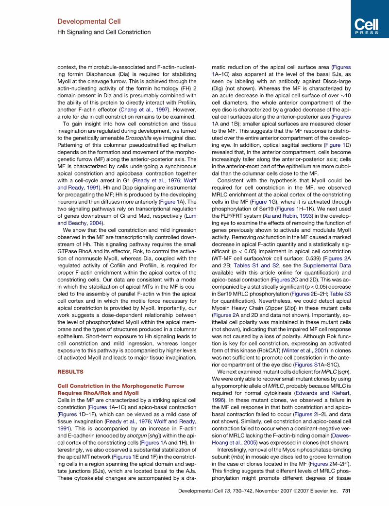
Cell Constriction in the Morphogenetic FurrowRequires RhoA/Rok and MyoIICells in the MF are characterized by a striking apical cell
constriction (Figures 1A–1C) and apico-basal contraction
(Figures 1D–1F), which can be viewed as a mild case of
tissue invagination (Ready et al., 1976; Wolff and Ready,
1991). This is accompanied by an increase in F-actin
and E-cadherin (encoded by shotgun [shg]) within the api-
cal cortex of the constricting cells (Figures 1A and 1H). In-
terestingly, we also observed a substantial stabilization of
the apical MT network (Figures 1E and 1F) in the constrict-
ing cells in a region spanning the apical domain and sep-
tate junctions (SJs), which are located basal to the AJs.
These cytoskeletal changes are accompanied by a dra-
Developmen
matic reduction of the apical cell surface area (Figures
1A–1C) also apparent at the level of the basal SJs, as
seen by labeling with an antibody against Discs-large
(Dlg) (not shown). Whereas the MF is characterized by
an acute decrease in the apical cell surface of over �10
cell diameters, the whole anterior compartment of the
eye disc is characterized by a graded decrease of the api-
cal cell surfaces along the anterior-posterior axis (Figures
1A and 1B); smaller apical surfaces are measured closer
to the MF. This suggests that the MF response is distrib-
uted over the entire anterior compartment of the develop-
ing eye. In addition, optical sagittal sections (Figure 1D)
revealed that, in the anterior compartment, cells become
increasingly taller along the anterior-posterior axis; cells
in the anterior-most part of the epithelium are more cuboi-
dal than the columnar cells close to the MF.
Consistent with the hypothesis that MyoII could be
required for cell constriction in the MF, we observed
MRLC enrichment at the apical cortex of the constricting
cells in the MF (Figure 1G), where it is activated through
phosphorylation of Ser19 (Figures 1H–1K). We next used
the FLP/FRT system (Xu and Rubin, 1993) in the develop-
ing eye to examine the effects of removing the function of
genes previously shown to activate and modulate MyoII
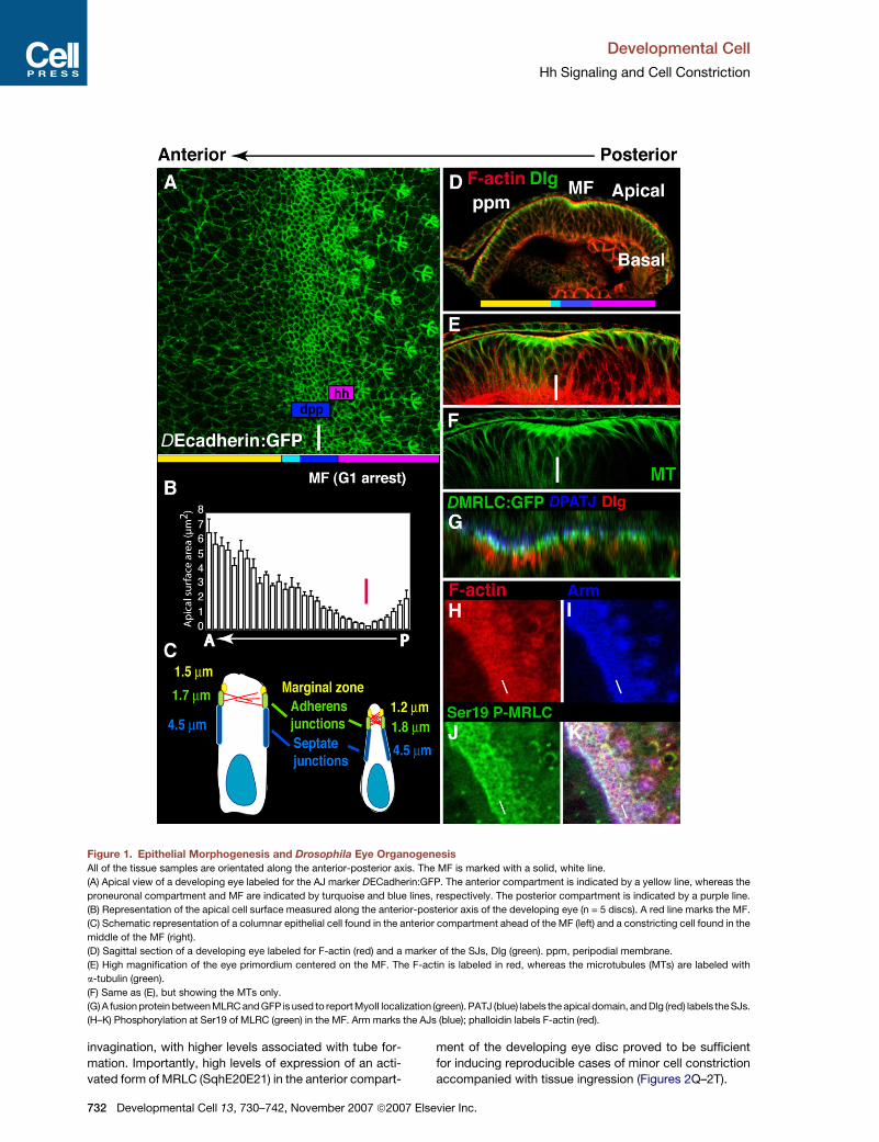
activity. Removing rok function in the MF caused a marked
decrease in apical F-actin quantity and a statistically sig-
nificant (p < 0.05) impairment in apical cell constriction
(WT-MF cell surface/rok cell surface: 0.539) (Figures 2A
and 2B; Tables S1 and S2, see the Supplemental Data
available with this article online for quantification) and
apico-basal contraction (Figures 2C and 2D). This was ac-
companied by a statistically significant (p < 0.05) decrease
in Ser19 MRLC phosphorylation (Figures 2E–2H; Table S3
for quantification). Nevertheless, we could detect apical
Myosin Heavy Chain (Zipper [Zip]) in these mutant cells
(Figures 2A and 2D and data not shown). Importantly, ep-
ithelial cell polarity was maintained in these mutant cells
(not shown), indicating that the impaired MF cell response
was not caused by a loss of polarity. Although Rok func-
tion is key for cell constriction, expressing an activated
form of this kinase (RokCAT) (Winter et al., 2001) in clones
was not sufficient to promote cell constriction in the ante-
rior compartment of the eye disc (Figures S1A–S1C).
We next examined mutant cells deficient for MRLC (sqh).
We were only able to recover small mutant clones by using
a hypomorphic allele of MRLC, probably because MRLC is
required for normal cytokinesis (Edwards and Kiehart,
1996). In these mutant clones, we observed a failure in
the MF cell response in that both constriction and apico-
basal contraction failed to occur (Figures 2I–2L and data
not shown). Similarly, cell constriction and apico-basal cell
contraction failed to occur when a dominant-negative ver-
sion of MRLC lacking the F-actin-binding domain (Dawes-
Hoang et al., 2005) was expressed in clones (not shown).
Interestingly, removal of the Myosin phosphatase-binding
subunit (mbs) in mosaic eye discs led to groove formation
in the case of clones located in the MF (Figures 2M–2P0).
This finding suggests that different levels of MRLC phos-
phorylation might promote different degrees of tissue
tal Cell 13, 730–742, November 2007 ª2007 Elsevier Inc. 731
Developmental Cell
Hh Signaling and Cell Constriction
Figure 1. Epithelial Morphogenesis and Drosophila Eye Organogenesis
All of the tissue samples are orientated along the anterior-posterior axis. The MF is marked with a solid, white line.
(A) Apical view of a developing eye labeled for the AJ marker DECadherin:GFP. The anterior compartment is indicated by a yellow line, whereas the
proneuronal compartment and MF are indicated by turquoise and blue lines, respectively. The posterior compartment is indicated by a purple line.
(B) Representation of the apical cell surface measured along the anterior-posterior axis of the developing eye (n = 5 discs). A red line marks the MF.
(C) Schematic representation of a columnar epithelial cell found in the anterior compartment ahead of the MF (left) and a constricting cell found in the
middle of the MF (right).
(D) Sagittal section of a developing eye labeled for F-actin (red) and a marker of the SJs, Dlg (green). ppm, peripodial membrane.
(E) High magnification of the eye primordium centered on the MF. The F-actin is labeled in red, whereas the microtubules (MTs) are labeled with
a-tubulin (green).
(F) Same as (E), but showing the MTs only.
(G) A fusion protein between MLRC and GFP is used to report MyoII localization (green). PATJ (blue) labels the apical domain, and Dlg (red) labels the SJs.
(H–K) Phosphorylation at Ser19 of MLRC (green) in the MF. Arm marks the AJs (blue); phalloidin labels F-actin (red).
invagination, with higher levels associated with tube for-
mation. Importantly, high levels of expression of an acti-
vated form of MRLC (SqhE20E21) in the anterior compart-
732 Developmental Cell 13, 730–742, November 2007 ª2007
ment of the developing eye disc proved to be sufficient
for inducing reproducible cases of minor cell constriction
accompanied with tissue ingression (Figures 2Q–2T).
Elsevier Inc.
Developmental Cell
Hh Signaling and Cell Constriction
Figure 2. Patterning of the Eye Imaginal
Disc: MF and MyoII-Driven Cell Constric-
tion
In all panels, the MF is indicated by a white line,
and the clones, marked by the absence of blue
(b-galactosidase or GFP staining), are circled
with a dashed, white line.
(A and B) Cells mutant for the null allele rok2
showing a strong reduction in apical F-actin
staining (red) correlated with a failure to prop-
erly achieve cell constriction. The dashed lines
labeled (C) and (D) mark the position corre-
sponding to the sagittal sections seen in (C)
and (D), respectively.
(C and D) Sagittal sections showing a failure of
the (D) rok2 mutant cells to undergo apicobasal
contraction, compared to the (C) wild-type tis-
sue shown in the same preparation.
(E–H) Cells mutant for the null allele rok2. F-
actin staining is shown in red, and Phospho-
Ser19 MRLC staining is shown in green. The
cell nuclei are labeled with DAPI (white) in (H).
(I–L) Cells mutant for MRLC (sqh1). F-actin
staining is shown in red, and Dlg staining is
shown in green.
(M–P0) Eye disc presenting a clone mutant for
mbs791. (M)–(P) show an optical section at the
level of the apical membrane of the constricting
cells in the MF, and the position of the mutant
clone is indicated with the dashed line. (M0–
P0) show a basal view of the same region of
the eye disc. This reveals that the apical mem-
branes of the mbs791 mutant cells are located
at the level of the basal nuclei (F-actin staining
is shown in red; the AJ marker Arm staining is
shown in green).
(Q–T) Activated MRLC (SqhE20E21) induces
mild cell ingression (lack of green staining).
F-actin staining is shown in red; Nuclei are
stained blue. An arrowhead points to the ec-
topic cell ingression.
(U–X) Eye disc mutant for RhoA72M1 induced by
using the Minute technique. MRLC phosphory-
lation at Ser19 is shown in green.
Finally, in the absence of RhoA function, we observed
a dramatic loss of cell constriction and apicobasal con-
traction in the MF, associated with weak F-actin staining
and a diffuse pattern of MRLC phosphorylation at Ser19
when compared to wild-type (Figures 2U–2X). Taken to-
gether, our findings demonstrate that cell constriction
and ingression in the MF depend upon the action of
RhoA, Rok, and MyoII. Our observation of residual phos-
phorylation of MRLC at Ser19 in rok mutant cells (Table
Developme
S3) confirms a previous report (Lee and Treisman, 2004)
and suggests that another kinase acts in parallel with
Rok to phosphorylate this residue.
The Formin Dia Is Required for Apical CellConstrictionOne major effect of removing RhoA and rok function in the
MF is a significant reduction in F-actin accumulation in the
apical domain of the cell (Figures 2B and 2V; Table S2 for
ntal Cell 13, 730–742, November 2007 ª2007 Elsevier Inc. 733
Developmental Cell
Hh Signaling and Cell Constriction
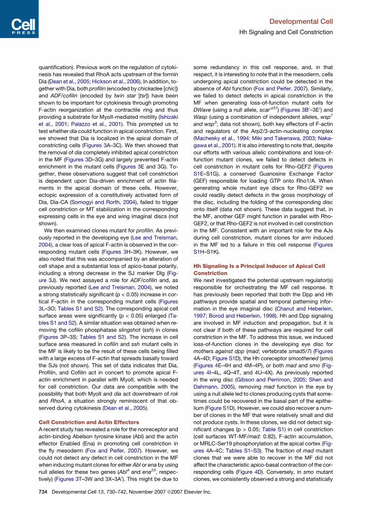
quantification). Previous work on the regulation of cytoki-
nesis has revealed that RhoA acts upstream of the formin
Dia (Dean et al., 2005; Hickson et al., 2006). In addition, to-
gether with Dia, both profilin (encoded by chickadee [chic])
and ADF/cofilin (encoded by twin star [tsr]) have been
shown to be important for cytokinesis through promoting
F-actin reorganization at the contractile ring and thus
providing a substrate for MyoII-mediated motility (Ishizaki
et al., 2001; Palazzo et al., 2001). This prompted us to
test whether dia could function in apical constriction. First,
we showed that Dia is localized in the apical domain of
constricting cells (Figures 3A–3C). We then showed that
the removal of dia completely inhibited apical constriction
in the MF (Figures 3D–3G) and largely prevented F-actin
enrichment in the mutant cells (Figures 3E and 3G). To-
gether, these observations suggest that cell constriction
is dependent upon Dia-driven enrichment of actin fila-
ments in the apical domain of these cells. However,
ectopic expression of a constitutively activated form of
Dia, Dia-CA (Somogyi and Rorth, 2004), failed to trigger
cell constriction or MT stabilization in the corresponding
expressing cells in the eye and wing imaginal discs (not
shown).
We then examined clones mutant for profilin. As previ-
ously reported in the developing eye (Lee and Treisman,
2004), a clear loss of apical F-actin is observed in the cor-
responding mutant cells (Figures 3H–3K). However, we
also noted that this was accompanied by an alteration of
cell shape and a substantial loss of apico-basal polarity,
including a strong decrease in the SJ marker Dlg (Fig-
ure 3J). We next assayed a role for ADF/cofilin and, as
previously reported (Lee and Treisman, 2004), we noted
a strong statistically significant (p < 0.05) increase in cor-
tical F-actin in the corresponding mutant cells (Figures
3L–3O; Tables S1 and S2). The corresponding apical cell
surface areas were significantly (p < 0.05) enlarged (Ta-
bles S1 and S2). A similar situation was obtained when re-
moving the cofilin phosphatase slingshot (ssh) in clones
(Figures 3P–3S; Tables S1 and S2). The increase in cell
surface area measured in cofilin and ssh mutant cells in
the MF is likely to be the result of these cells being filled
with a large excess of F-actin that spreads basally toward
the SJs (not shown). This set of data indicates that Dia,
Profilin, and Cofilin act in concert to promote apical F-
actin enrichment in parallel with MyoII, which is needed
for cell constriction. Our data are compatible with the
possibility that both MyoII and dia act downstream of rok
and RhoA, a situation strongly reminiscent of that ob-
served during cytokinesis (Dean et al., 2005).
Cell Constriction and Actin EffectorsA recent study has revealed a role for the nonreceptor and
actin-binding Abelson tyrosine kinase (Abl) and the actin
effector Enabled (Ena) in promoting cell constriction in
the fly mesoderm (Fox and Peifer, 2007). However, we
could not detect any defect in cell constriction in the MF
when inducing mutant clones for either Abl or ena by using
null alleles for these two genes (Abl4 and ena23, respec-
tively) (Figures 3T–3W and 3X–3A0). This might be due to
734 Developmental Cell 13, 730–742, November 2007 ª2007 E
some redundancy in this cell response, and, in that
respect, it is interesting to note that in the mesoderm, cells
undergoing apical constriction could be detected in the
absence of Abl function (Fox and Peifer, 2007). Similarly,
we failed to detect defects in apical constriction in the
MF when generating loss-of-function mutant cells for
DWave (using a null allele, scarD37) (Figures 3B0–3E0) and
Wasp (using a combination of independent alleles, wsp1
and wsp3; data not shown), both key effectors of F-actin
and regulators of the Arp2/3-actin-nucleating complex
(Machesky et al., 1994; Miki and Takenawa, 2003; Naka-
gawa et al., 2001). It is also interesting to note that, despite
our efforts with various allelic combinations and loss-of-
function mutant clones, we failed to detect defects in
cell constriction in mutant cells for Rho-GEF2 (Figures
S1E–S1G), a conserved Guanosine Exchange Factor
(GEF) responsible for loading GTP onto Rho1/A. When
generating whole mutant eye discs for Rho-GEF2 we
could readily detect defects in the gross morphology of
the disc, including the folding of the corresponding disc
onto itself (data not shown). These data suggest that, in
the MF, another GEF might function in parallel with Rho-
GEF2, or that Rho-GEF2 is not involved in cell constriction
in the MF. Consistent with an important role for the AJs
during cell constriction, mutant clones for arm induced
in the MF led to a failure in this cell response (Figures
S1H–S1K).
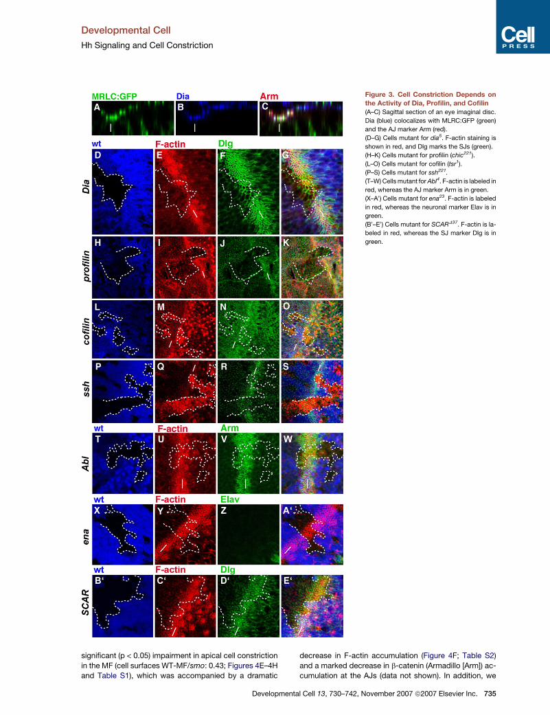
Hh Signaling Is a Principal Inducer of Apical CellConstrictionWe next investigated the potential upstream regulator(s)
responsible for orchestrating the MF cell response. It
has previously been reported that both the Dpp and Hh
pathways provide spatial and temporal patterning infor-
mation in the eye imaginal disc (Chanut and Heberlein,
1997; Borod and Heberlein, 1998). Hh and Dpp signaling
are involved in MF induction and propagation, but it is
not clear if both of these pathways are required for cell
constriction in the MF. To address this issue, we induced
loss-of-function clones in the developing eye disc for
mothers against dpp (mad; vertebrate smad5/7) (Figures
4A–4D; Figure S1D), the Hh coreceptor smoothened (smo)
(Figures 4E–4H and 4M–4P), or both mad and smo (Fig-
ures 4I–4L, 4Q–4T, and 4U–4X). As previously reported
in the wing disc (Gibson and Perrimon, 2005; Shen and
Dahmann, 2005), removing mad function in the eye by
using a null allele led to clones producing cysts that some-
times could be recovered in the basal part of the epithe-
lium (Figure S1D). However, we could also recover a num-
ber of clones in the MF that were relatively small and did
not produce cysts. In these clones, we did not detect sig-
nificant changes (p > 0.05; Table S1) in cell constriction
(cell surfaces WT-MF/mad: 0.82), F-actin accumulation,
or MRLC-Ser19 phosphorylation at the apical cortex (Fig-
ures 4A–4C; Tables S1–S3). The fraction of mad mutant
clones that we were able to recover in the MF did not
affect the characteristic apico-basal contraction of the cor-
responding cells (Figure 4D). Conversely, in smo mutant
clones, we consistently observed a strong and statistically
lsevier Inc.
Developmental Cell
Hh Signaling and Cell Constriction
Figure 3. Cell Constriction Depends on
the Activity of Dia, Profilin, and Cofilin
(A–C) Sagittal section of an eye imaginal disc.
Dia (blue) colocalizes with MLRC:GFP (green)
and the AJ marker Arm (red).
(D–G) Cells mutant for dia5. F-actin staining is
shown in red, and Dlg marks the SJs (green).
(H–K) Cells mutant for profilin (chic221).
(L–O) Cells mutant for cofilin (tsr1).
(P–S) Cells mutant for ssh221.
(T–W) Cells mutant for Abl4. F-actin is labeled in
red, whereas the AJ marker Arm is in green.
(X–A0) Cells mutant for ena23. F-actin is labeled
in red, whereas the neuronal marker Elav is in
green.
(B0–E0) Cells mutant for SCARD37. F-actin is la-
beled in red, whereas the SJ marker Dlg is in
green.
significant (p < 0.05) impairment in apical cell constriction
in the MF (cell surfaces WT-MF/smo: 0.43; Figures 4E–4H
and Table S1), which was accompanied by a dramatic
Developme
decrease in F-actin accumulation (Figure 4F; Table S2)
and a marked decrease in b-catenin (Armadillo [Arm]) ac-
cumulation at the AJs (data not shown). In addition, we
ntal Cell 13, 730–742, November 2007 ª2007 Elsevier Inc. 735
Developmental Cell
Hh Signaling and Cell Constriction
Figure 4. Hh Signaling Regulates MyoII-
Driven Cell Constriction
(A–C) Cells mutant for mad12. F-actin is labeled
in red.
(D) Sagittal section across a mad12 mutant
clone showing that cells still achieve proper
apicobasal contraction (marked with a white
line).
(E–H) Cells mutant for smoD16 exhibiting a fail-
ure in apical F-actin enrichment (red). Dlg
marks the SJs (green).
(I–L) Double mutant clone for mad1-2 and smo3
exhibiting a failure in apical F-actin enrichment
(red). Dlg marks the SJs (green).
(M–P) smoD16 mutant cells showing a total loss
of MRLC phosphorylation at Ser19 (green).
(Q–T) Double smo3, mad1-2 mutant cells exhib-
iting failure to achieve apical constriction
(F-actin, red) and a total loss of MRLC phos-
phorylation at Ser19 (green).
(U–X) Loss of Dia staining (green) at the apical
cortex in the MF in the absence of smo3 and
mad1-2 function. F-actin is labeled in red, and
the mutant cells are indicated with a white
asterisk.
measured a significant decrease (p < 0.05) in the levels of
MRLC phosphorylation at Ser19 at the apical cortex of
these mutant cells (Figure 4Q; Table S3), implying that
Hh signaling plays a major role in inducing cell constriction
and apico-basal contraction in the MF. However, by using
Dlg to label the SJs, we could still observe a remnant of the
MF (Figure 4G), suggesting that Dpp and Hh might act in
a partially redundant manner in the control of the MF cell
response. To test this hypothesis, we removed both of
these two signaling pathways in clones mutant for both
mad and smo. The mutant cells completely failed to
undergo apical constriction (cell surfaces WT-MF/mad,
smo: 0.30; Figures 4I–4L and Table S1) and apico-basal
contraction. Furthermore, there was no F-actin accumula-
tion at the apical cortex (Table S2) or MRLC phosphoryla-
736 Developmental Cell 13, 730–742, November 2007 ª2007 E
tion at Ser19 (Figures 4Q–4T; Table S3). In these mutant
cells, we also observed a substantial decrease in apical
Dia staining (Figures 4U–4X). Nonetheless, cell polarity
was preserved, as assayed by using marginal zone, AJ,
and SJ markers (Figure 4K and data not shown).
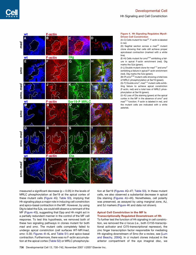
Apical Cell Constriction in the MF IsTranscriptionally Regulated Downstream of HhTo further test the function of Hh signaling in cell constric-
tion, we removed the ci locus (i.e., both Ci155-transcrip-
tional activator and Ci75-transcriptional repressor), the
zinc finger transcription factor responsible for mediating
Hh signaling downstream of Smo (for a review, see [Lum
and Beachy, 2004]). In ci mutant clones induced in the
anterior compartment of the eye imaginal disc, we
lsevier Inc.
Developmental Cell
Hh Signaling and Cell Constriction
Figure 5. Hh Signaling Transcriptionally
Regulates Cell Constriction and Ingres-
sion
(A–D) Cells mutant for ci showing ectopic api-
cal constriction immediately anterior to the
endogenous MF. F-actin (red) and Dlg labeling
the SJs (green).
(E) Sagittal section of a cullin116E clone.
(F–H) Double mutant clone for the transcription
factor ci and mad1-2 fail to show ectopic apical
constriction.
(I–M) Sagittal section across a clone mutant
for ci (loss of green staining, dashed lines).
(I) A phospho-specific antibody labels P-Mad
(blue); (K) F-actin staining is shown in red,
and the (I) cell nuclei are labeled with DAPI
(white).
(N–R) (N) Sagittal section across an eye disc
expressing spastin:GFP. (O) P-Ser19 staining
(blue). (P) MT staining in red. (R) Nuclei are
stained with DAPI (white).
consistently observed ectopic MF-like cell responses,
including apical MT stabilization and MyoII activation
(Figures 5A–5D and not shown). Similarly, we prevented
Ci75-transcriptional repressor formation by removing
cullin1, a ubiquitin-ligase required to generate the tran-
scriptional repressor Ci75 from the transcriptional activa-
tor Ci155 (Ou et al., 2002). cullin1 loss-of-function clones
led to ectopic MF cell responses in the anterior compart-
ment immediately ahead of the MF (Figures 5E). These
experiments suggest that alleviating Ci75-mediated tran-
scriptional repression is sufficient to induce cell constric-
tion. Surprisingly, while mad is not absolutely required
for a cell to constrict in the MF, loss of mad function in ci
mutant cells (in double ci and mad mutant cells) led to
a lack of ectopic cell constriction in the corresponding
cells (Figures 5F–5H). This indicates that, in the absence
of Ci155-mediated transcriptional activation, ectopic cell
constriction requires Dpp signaling to proceed. Although,
all of the cells located in the anterior compartment of the
eye disc receive the Dpp signal, as reported by a phos-
pho-specific antibody raised against active P-Mad (Fig-
ures 5I–5M), we did not observe increased P-Mad staining
in ci mutant clones. Altogether, this suggests that mad
function is required for cell constriction to occur in ci
mutant cells, whereas in the MF, Ci-155 and Mad might
act redundantly to promote this cell response.
In ci mutant cells and in the endogenous MF, cell con-
striction is accompanied by MT stabilization within the
apical cell domain. To assay their contribution during cell
Developmen
constriction, we used the MT-severing factor Spastin
(Sherwood et al., 2004) to depolymerize MTs in vivo (Fig-
ures 5N–5R). When expressed in the MF (Figure 5N),
Spastin led to a near total depletion of the MT network,
accompanied by a loss of apical F-actin accumulation
and a loss of Ser19-P MyoII staining. In addition, the
basally located nuclei normally found in the MF appeared
randomly distributed and had a tendency to be close to
the apical cell surfaces (Figure 5Q).
Hh Signaling Induces a Range of TissueInvaginationWe next tested whether Hh signaling was sufficient to trig-
ger cell constriction in fly epithelia. To this end, we ectop-
ically activated the Hh signaling pathway in the eye,
antenna, and wing imaginal discs by generating mutant
clones in which the inhibitory coreceptor patched (ptc)
was removed (Hooper and Scott, 1989). In ptc mutant
clones, Hh signaling is constitutively activated at its max-
imal strength. Interestingly, these clones led to a range of
responses, depending on the levels of activation of MyoII
measured through quantifying the Ser19-P MRLC staining
(Table S3). In the case of ptc clones dissected 24 hr
after clone induction, we invariably observed MF-like
responses (Figures 6A–6D; Tables S1 and S2) together
with levels of Ser19-P MRLC comparable to those mea-
sured in the endogenous MF cells (p > 0.005; Table S3).
This was accompanied by a pronounced bundling of
the apical MTs (Figures 6E–6H) and a striking apical
tal Cell 13, 730–742, November 2007 ª2007 Elsevier Inc. 737
Developmental Cell
Hh Signaling and Cell Constriction
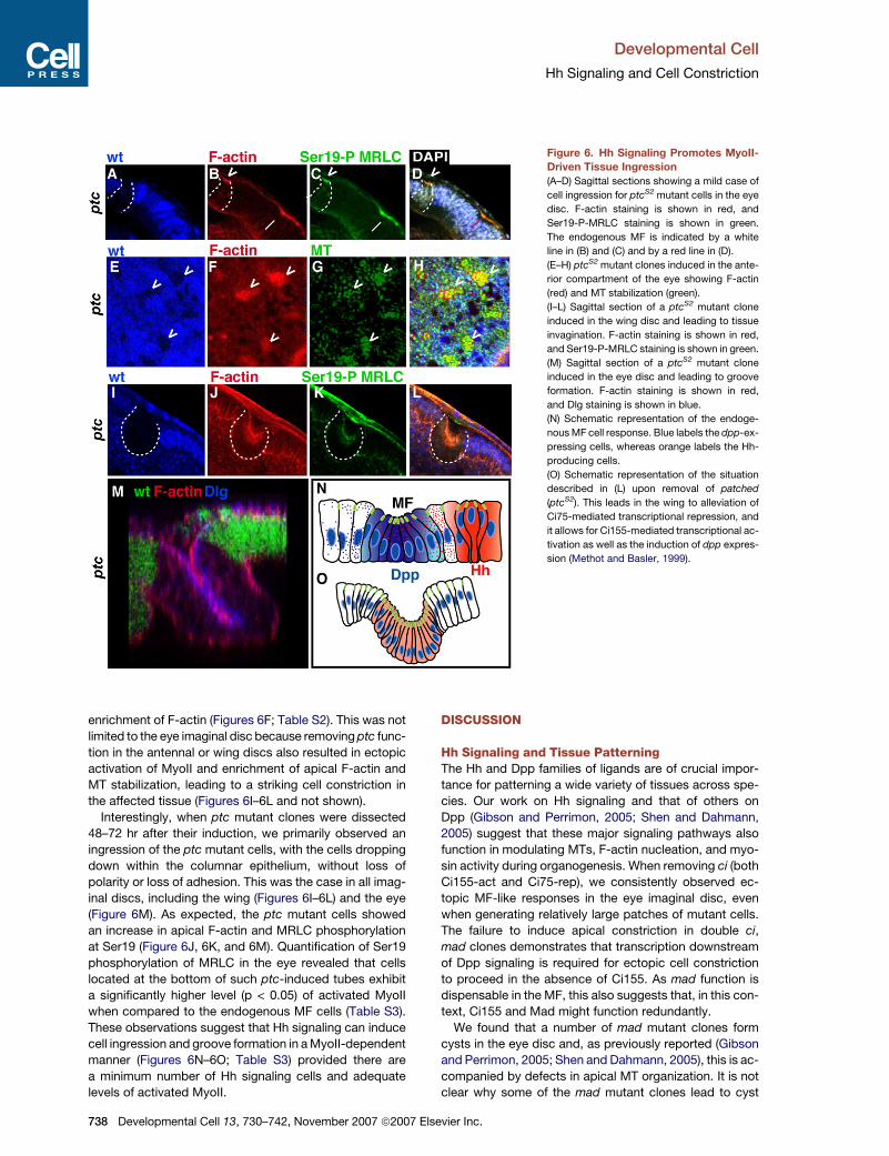
Figure 6. Hh Signaling Promotes MyoII-
Driven Tissue Ingression
(A–D) Sagittal sections showing a mild case of
cell ingression for ptcS2 mutant cells in the eye
disc. F-actin staining is shown in red, and
Ser19-P-MRLC staining is shown in green.
The endogenous MF is indicated by a white
line in (B) and (C) and by a red line in (D).
(E–H) ptcS2 mutant clones induced in the ante-
rior compartment of the eye showing F-actin
(red) and MT stabilization (green).
(I–L) Sagittal section of a ptcS2 mutant clone
induced in the wing disc and leading to tissue
invagination. F-actin staining is shown in red,
and Ser19-P-MRLC staining is shown in green.
(M) Sagittal section of a ptcS2 mutant clone
induced in the eye disc and leading to groove
formation. F-actin staining is shown in red,
and Dlg staining is shown in blue.
(N) Schematic representation of the endoge-
nous MF cell response. Blue labels the dpp-ex-
pressing cells, whereas orange labels the Hh-
producing cells.
(O) Schematic representation of the situation
described in (L) upon removal of patched
(ptcS2). This leads in the wing to alleviation of
Ci75-mediated transcriptional repression, and
it allows for Ci155-mediated transcriptional ac-
tivation as well as the induction of dpp expres-
sion (Methot and Basler, 1999).
enrichment of F-actin (Figures 6F; Table S2). This was not
limited to the eye imaginal disc because removing ptc func-
tion in the antennal or wing discs also resulted in ectopic
activation of MyoII and enrichment of apical F-actin and
MT stabilization, leading to a striking cell constriction in
the affected tissue (Figures 6I–6L and not shown).
Interestingly, when ptc mutant clones were dissected
48–72 hr after their induction, we primarily observed an
ingression of the ptc mutant cells, with the cells dropping
down within the columnar epithelium, without loss of
polarity or loss of adhesion. This was the case in all imag-
inal discs, including the wing (Figures 6I–6L) and the eye
(Figure 6M). As expected, the ptc mutant cells showed
an increase in apical F-actin and MRLC phosphorylation
at Ser19 (Figure 6J, 6K, and 6M). Quantification of Ser19
phosphorylation of MRLC in the eye revealed that cells
located at the bottom of such ptc-induced tubes exhibit
a significantly higher level (p < 0.05) of activated MyoII
when compared to the endogenous MF cells (Table S3).
These observations suggest that Hh signaling can induce
cell ingression and groove formation in a MyoII-dependent
manner (Figures 6N–6O; Table S3) provided there are
a minimum number of Hh signaling cells and adequate
levels of activated MyoII.
738 Developmental Cell 13, 730–742, November 2007 ª2007 E
DISCUSSION
Hh Signaling and Tissue PatterningThe Hh and Dpp families of ligands are of crucial impor-
tance for patterning a wide variety of tissues across spe-
cies. Our work on Hh signaling and that of others on
Dpp (Gibson and Perrimon, 2005; Shen and Dahmann,
2005) suggest that these major signaling pathways also
function in modulating MTs, F-actin nucleation, and myo-
sin activity during organogenesis. When removing ci (both
Ci155-act and Ci75-rep), we consistently observed ec-
topic MF-like responses in the eye imaginal disc, even
when generating relatively large patches of mutant cells.
The failure to induce apical constriction in double ci,
mad clones demonstrates that transcription downstream
of Dpp signaling is required for ectopic cell constriction
to proceed in the absence of Ci155. As mad function is
dispensable in the MF, this also suggests that, in this con-
text, Ci155 and Mad might function redundantly.
We found that a number of mad mutant clones form
cysts in the eye disc and, as previously reported (Gibson
and Perrimon, 2005; Shen and Dahmann, 2005), this is ac-
companied by defects in apical MT organization. It is not
clear why some of the mad mutant clones lead to cyst
lsevier Inc.
Developmental Cell
Hh Signaling and Cell Constriction
formation while others remain within the epithelium, and it
is possible that the timing of mutant-clone induction is
a key parameter. Interestingly, cells in the MF present
the highest levels of P-Mad, and this correlates with our
observation that, in these cells, the apical MTs appear
more bundled and abundant.
Linking MT and F-Actin during Cell ConstrictionThe Formin-Homology (FH) protein family has been impli-
cated in a number of actin- and MT-dependent cellular
processes, including the alignment of the MTs and F-actin
during stress-fiber formation (Tsuji et al., 2002), cell polar-
ity, and cytokinesis (Wasserman, 1998). In the fly embryo,
dia loss of function leads to defects in pseudocleavage-
furrow invagination during cellularization (Afshar et al.,
2000) and contractile-ring morphogensis (Giansanti et al.,
1998). Consistent with a role for dia during cytokinesis,
we observed that a high proportion of dia mutant cells
were located at the apical surface of the epithelium and
arrested in telophase. Nevertheless, we could recover
small dia mutant clones in the MF that retained their overall
apico-basal polarity, thus enabling us to reveal a function
for this factor for F-actin enrichment during cell constric-
tion in the MF. However, we were not able to induce
ectopic cell constriction by using an activated form of
dia (Somogyi and Rorth, 2004).
FH proteins such as Dia contain a central proline-rich
FH1 domain that, in yeast, has been shown to mediate
a direct interaction with the actin effector Profilin (Chang
et al., 1997), whereas the amino-terminal part of several
FH proteins is able to interact with the GTP-bound Rho-
GTPases such as Rho1 (Watanabe et al., 1997, 1999).
Our data suggest that a RhoA-Dia-Profilin biochemical
module might be evolutionarily conserved (Chang et al.,
1997). Interestingly, mdia has been shown to be trans-
ported on MT and act as a MT (+) end stabilizer in 3T3
fibroblasts (Palazzo et al., 2001). Whereas MTs remain
largely unaffected in the absence of dia in the MF (not
shown), depolymerizing MTs (Sherwood et al., 2004) led
to the absence of cell constriction in the MF, accompanied
by a loss of apical F-actin and Ser19-P MyoII staining. In
this context, we favor a model in which dia is involved in
maintaining F-actin at the apical cell cortex in the MF,
but is not required for the apparent tight bundling and sta-
bilization of the apical MTs in these cells. Finally, it is inter-
esting to note that, in the fly syncytial blastoderm embryo,
F-actin and MyoII are transported toward the MT (+) end
(Foe et al., 2000). The failure in apical F-actin and activated
MyoII enrichment when MTs are depolymerized in the MF
is consistent with a role for MTs in targeting MyoII to the
apical cell surface.
MyoII Activation and Cell ConstrictionIn the MF, we note that loss of rok function does not lead to
a total loss of phosphorylation of MRLC at the conserved
Ser19. This is consistent with a previous report in the
developing eye disc (Lee and Treisman, 2004) and sug-
gests that other kinases act in parallel with Rok in the MF.
Potential candidates are the P21-activated kinases (PAK),
Developme
encoded by three loci in Drosophila (PAK1/PAK, DPAK2/
mbt, and PAK3). We used the null allele mbtP1 as well as
various PAK1 alleles (including PAK16, which encodes
a truncated protein) to examine the role of these kinases
in apical cell constriction in the MF, and, in both cases,
we failed to detect defects in apical cell constriction (not
shown). In addition, expressing the auto-inhibitory domain
(PAK-AID) found in the group-I PAKs (including PAK1 and
PAK3) in clones, and thus interfering with these two
kinases’ function (Conder et al., 2004), did not lead to
loss of cell constriction in the MF (not shown). It is possible
that residual DPAK2/mbt activity could compensate for
the lack of PAK1 and PAK3 function, and further work will
be required to analyze the effects of removing all three of
these loci in the MF. Another kinase potentially responsible
for phosphorylating MRLC in the MF is myosin light chain
kinase (MLCK), a large, complex locus for which loss-
of-function mutations have not been identified.
MyoII Activation and Tissue InvaginationIn ptc mutant clones, we are ectopically inducing maxi-
mum levels of Hh signaling. In this context, MF-like ingres-
sion (or indentation) is associated with lower levels of
Ser19-P-MyoII than those we measured in cases of
groove formation. Interestingly, the situation in the endo-
genous MF involves a very transient stimulation of the Hh
pathway due to the activity of the ubiquitin ligases Cullin1
and Cullin3, which process Ci155 in the anterior and pos-
terior edge of the MF, respectively (Ou et al., 2002). Our
data are consistent with a model in which, upon Hh signal-
ing, activated MyoII accumulates at the apical cortex of
the constricting cell over time. The degree of activated
MyoII accumulation is linked to the duration of Hh signal-
ing perceived by the corresponding constricting cells and
appears to be the main parameter involved in groove
formation in columnar epithelia.
It is likely that the Hh-induced MyoII-driven pathway we
report here also plays a role in tissue patterning in higher
organisms. For example, cell constriction and apico-basal
contraction occur during the invagination of the neural
plate to form the neural tube in vertebrates. In this case,
the notochord and the floor plate are a potent source of
sonic hedgehog (shh) (Jessell, 2000), and shh knockout
mice exhibit a failure in neural tube closure. In these
mice, the neural plate lacks characteristic bottle cells,
pointing to the possibility that neural tube closure is driven
by MyoII following a similar pathway to the one defined in
the present study.
EXPERIMENTAL PROCEDURES
Genotypes Used in This Study
The functional Ubi-Sqh:GFP was expressed in a sqhAX3 null mutant
background (Royou et al., 2002). w, hsflp122; Tub>w[+]>Gal4/UAS-
mYFP:myosinDN (Dawes-Hoang et al., 2005). y,w,FRT101 sqh1/UbiGFP
FRT101; eyflp/+ (Karess et al., 1991). sqh1 is a hypomorphic mutation in
the MRLC. rok2 FRT19A/FRT19A arm-LacZ; eyflp/+ (Winter et al.,
2001). arm8 FRT19A/FRT19A arm-LacZ; eyflp/+ (Uemura et al.,
1996). w, eyflp; madB1 FRT40A/FRT40A arm-LacZ and w, eyflp;
mad12 FRT40A/FRT40A arm-LacZ (Wiersdorff et al., 1996). mad12 is
ntal Cell 13, 730–742, November 2007 ª2007 Elsevier Inc. 739
Developmental Cell
Hh Signaling and Cell Constriction
an insertion in the mad locus that prevents most Dpp signaling. w,
eyflp; smoD16 FRT40A/FRT40A arm-LacZ (van den Heuvel and Ing-
ham, 1996). smoD16 is a null allele (Chen and Struhl, 1996). w, eyflp;
smo3, mad1-2 FRT40A/FRT40A arm-LacZ (Curtiss and Mlodzik,
2000). mad1-2 is a hypomorphic allele, and smo3 is an amorphic allele.
y, w, eyflp; [Ci+] FRT40A/FRT40A arm-LacZ;;Ci94/Ci94 (Zhang and
Kalderon, 2001). y, w, eyflp; [Ci+], mad1-2 FRT40A/FRT40A arm-
LacZ;;Ci94/Ci94 (Fu and Baker, 2003). w, eyflp; dia5 FRT40A/FRT40A
arm-LacZ (Afshar et al., 2000). w, eyflp; chic221 FRT40A/FRT40A
arm-LacZ (Verheyen and Cooley, 1994). The chic221 allele is an amor-
phic allele. w, eyflp; RhoA720M1 FRT40A/FRT40A arm-LacZ, Minutes
(Strutt et al., 1997). w, eyflp; scarD37 FRT40A/FRT40A arm-LacZ (Zallen
et al., 2002). w, hsflp122; ptcS2 FRT42D/FRT42D arm-LacZ (Jiang and
Struhl, 1995). ptcS2 is an amorphic allele. w, hsflp122; tsr1 FRT42D/
FRT42D arm-LacZ (Gunsalus et al., 1995). w, hsflp122; ena23
FRT42D/FRT42D arm-LacZ (Ahern-Djamali et al., 1998). w, eyflp;
FRT80A mbsT541/FRT80A arm-LacZ and w, eyflp; FRT80 mbsT791/
FRT80 arm-LacZ (Lee and Treisman, 2004). w, eyflp; FRT80A
cul116E/FRT80A arm-LacZ and w, eyflp; FRT80A cul1107D/FRT80A
arm-LacZ (Ou et al., 2002). w, eyflp; FRT80A Abl4/FRT80A arm-LacZ
(Bennett and Hoffmann, 1992). w, eyflp; FRT82B ssh26-1/FRT82B
arm-LacZ as well as w, eyflp; FRT82B ssh1-11/FRT82B arm-LacZ and
w, eyflp; FRT82B sshP01207/FRT82B arm-LacZ (Niwa et al., 2002). w,
eyflp; FRT82B wsp1/FRT82B arm-LacZ and w, eyflp; FRT82B wsp3/
FRT82B arm-LacZ (Ben-Yaacov et al., 2001). w, eyflp; FRT82B
pak16/FRT82B arm-LacZ (Hing et al., 1999). pak16 is an amorphic
allele. Homozygous RhoGEF24.1/RhoGEF2PX6 animals (Barrett et al.,
1997). RhoGEF24.1 is a null allele, and RhoGEF2PX6 is a hypomorph.
w, UAS-GFP, eyflp; TubGal4; FRT82B, TubGal80/FRT82B UAS-Pak-
AID (Conder et al., 2004). w, UAS-GFP, eyflp; TubGal4; FRT82B, Tub-
Gal80/FRT82B, UAS-ROKCAT (Winter et al., 2001).
y,w, hflp122; Tub>GFP,y+>Gal4; UAS-sqh-E20E21. UAS-sqh-E20E21
contains the sqh transgene with two activating phosphomimetic
mutations (Winter et al., 2001) under the control of the UAS sequence.
y,w, hflp122; Tub>GFP,y+>Gal4; UAS-spastin:GFP (Sherwood et al.,
2004).
Fly cultures and crosses were carried out at 25�C. Overexpression of
SqhE20E21 was performed at 29�C.
Immunostaining
Immunostainings were performed with the following antibodies: anti-
Armadillo (Riggleman et al., 1990) (mouse, 1/50); anti-MyoII (Kiehart
and Feghali, 1986) (rabbit, 1/500); anti-PSer19-MRLC (rabbit, 1/10;
Cell Signaling Technology, Beverly, MA); anti-Diaphanous (Afshar
et al., 2000) (rabbit, 1/200); anti-P-Mad (Gift from G. Morata) (rabbit,
1/500); anti-b-galactosidase (rabbit, 1/5000; Cappel Laboratories,
Durham, NC); anti-b-galactosidase (goat, 1/500; Cappel Laboratories);
anti-a-tubulin (Sigma, mouse, 1/2000); anti-Discs large (mouse, 1/50);
phalloidin-Texas red (Sigma, 20 mM); anti-mouse, -rabbit, and -goat
secondary antibodies (Jackson) (used at 1/200).
Spastin:GFP-expressing cells were generated by heat shocking
third-instar larvae for 30 minutes at 37�C, and the corresponding ani-
mals were dissected 5 hr later. ptc-mutant clones were induced in
early second-instar larvae by performing a 1 hr heat shock at 37�C.
The corresponding animals were dissected 24, 48, 72, or 96 hr after
heat shock.
Imaginal discs were dissected in 13 PBS and fixed in 4% formalde-
hyde for 20 min at room temperature. Samples were permeabilized in
PBS and 0.3% Triton and then blocked in 5% FCS. Antibodies were
incubated in PBS, Triton (0.3%). Imaging was performed by using a
Leica SP2, SP5 or Biorad Radiance 1040 confocal microscope.
Quantification
Images were analyzed by using IMAGEJ v1.33. To avoid complications
with variations from preparation to preparation, we chose to compare
wild-type and mutant tissue from the same disc. Surface areas of wild-
type and mutant patches of cells in the MF (or anterior to the MF in the
case of ptc-mutant clones) were measured. For F-actin quantity, aver-
740 Developmental Cell 13, 730–742, November 2007 ª2007 E
age pixel intensity over a transect in the MF (or in clones in the anterior
compartment for ptc) was calculated. The data were compared by
using a 2-tailed Student’s paired-t test to compare the mutant and
wild-type cell responses.
Supplemental Data
Supplemental Data include statistical analyses and Figure S1 and are
available at http://www.developmentalcell.com/cgi/content/full/13/5/
730/DC1/.
ACKNOWLEDGMENTS
This work was funded by the Medical Research Council (MRC). We
particularly acknowledge L. Alphey for the kind gift of the UAS-
sqhE20E21 flies and Matthew Freeman for sharing unpublished data
concerning this transgene. The authors would also like to acknowl-
edge Buzz Baum, Nick Baker, Kathy Barrett, Andrea Brand, Roger
Karess, Gines Morata, and Jessica Treisman for kindly providing us
with fly stocks and reagents; the Bloomington Stock Center for fly
stocks; and the Developmental Study Hybridoma Bank (DSHB) for
antibodies. We are grateful to the Pichaud lab for their input during
this work. R.F.W. is a recipient of an MRC career development fellow-
ship. P.F. is a recipient of a European Molecular Biology Organization
long-term fellowship.
Received: February 23, 2007
Revised: July 17, 2007
Accepted: September 25, 2007
Published: November 5, 2007
REFERENCES
Afshar, K., Stuart, B., and Wasserman, S.A. (2000). Functional analysis
of the Drosophila diaphanous FH protein in early embryonic develop-
ment. Development 127, 1887–1897.
Ahern-Djamali, S.M., Comer, A.R., Bachmann, C., Kastenmeier, A.S.,
Reddy, S.K., Beckerle, M.C., Walter, U., and Hoffmann, F.M. (1998).
Mutations in Drosophila enabled and rescue by human vasodilator-
stimulated phosphoprotein (VASP) indicate important functional roles
for Ena/VASP homology domain 1 (EVH1) and EVH2 domains. Mol.
Biol. Cell 9, 2157–2171.
Amano, M., Ito, M., Kimura, K., Fukata, Y., Chihara, K., Nakano, T.,
Matsuura, Y., and Kaibuchi, K. (1996). Phosphorylation and activation
of myosin by Rho-associated kinase (Rho-kinase). J. Biol. Chem. 271,
20246–20249.
Barrett, K., Leptin, M., and Settleman, J. (1997). The Rho GTPase and
a putative RhoGEF mediate a signalling pathway for the cell shape
changes in Drosophila gastrulation. Cell 91, 905–915.
Ben-Yaacov, S., Le Borgne, R., Abramson, I., Schweisguth, F., and
Schejter, E.D. (2001). Wasp, the Drosophila Wiskott-Aldrich syndrome
gene homologue, is required for cell fate decisions mediated by Notch
signalling. J. Cell Biol. 152, 1–13.
Bennett, R.L., and Hoffmann, F.M. (1992). Increased levels of the
Drosophila Abelson tyrosine kinase in nerves and muscles: subcellular
localization and mutant phenotypes imply a role in cell-cell interac-
tions. Development 116, 953–966.
Borod, E.R., and Heberlein, U. (1998). Mutual regulation of decapenta-
plegic and hedgehog during the initiation of differentiation in the
Drosophila retina. Dev. Biol. 197, 187–197.
Chang, F., Drubin, D., and Nurse, P. (1997). cdc12p, a protein required
for cytokinesis in fission yeast, is a component of the cell division ring
and interacts with profilin. J. Cell Biol. 137, 169–182.
Chanut, F., and Heberlein, U. (1997). Role of decapentaplegic in initia-
tion and progression of the morphogenetic furrow in the developing
Drosophila retina. Development 124, 559–567.
lsevier Inc.
Developmental Cell
Hh Signaling and Cell Constriction
Chen, Y., and Struhl, G. (1996). Dual roles for patched in sequestering
and transducing Hedgehog. Cell 87, 553–563.
Conder, R., Yu, H., Ricos, M., Hing, H., Chia, W., Lim, L., and Harden,
N. (2004). dPak is required for integrity of the leading edge cytoskele-
ton during Drosophila dorsal closure but does not signal through the
JNK cascade. Dev. Biol. 276, 378–390.
Costa, M., Wilson, E.T., and Wieschaus, E. (1994). A putative cell signal
encoded by the folded gastrulation gene coordinates cell shape
changes during Drosophila gastrulation. Cell 76, 1075–1089.
Curtiss, J., and Mlodzik, M. (2000). Morphogenetic furrow initiation and
progression during eye development in Drosophila: the roles of decap-
entaplegic, hedgehog and eyes absent. Development 127, 1325–
1336.
Davidson, L.A., Koehl, M.A., Keller, R., and Oster, G.F. (1995). How do
sea urchins invaginate? Using biomechanics to distinguish between
mechanisms of primary invagination. Development 121, 2005–2018.
Dawes-Hoang, R.E., Parmar, K.M., Christiansen, A.E., Phelps, C.B.,
Brand, A.H., and Wieschaus, E.F. (2005). folded gastrulation, cell
shape change and the control of myosin localization. Development
132, 4165–4178.
Dean, S.O., Rogers, S.L., Stuurman, N., Vale, R.D., and Spudich, J.A.
(2005). Distinct pathways control recruitment and maintenance of
myosin II at the cleavage furrow during cytokinesis. Proc. Natl. Acad.
Sci. USA 102, 13473–13478.
Edwards, K.A., and Kiehart, D.P. (1996). Drosophila nonmuscle myosin
II has multiple essential roles in imaginal disc and egg chamber mor-
phogenesis. Development 122, 1499–1511.
Foe, V.E., Field, C.M., and Odell, G.M. (2000). Microtubules and mitotic
cycle phase modulate spatiotemporal distributions of F-actin and
myosin II in Drosophila syncytial blastoderm embryos. Development
127, 1767–1787.
Fox, D.T., and Peifer, M. (2007). Abelson kinase (Abl) and RhoGEF2
regulate actin organization during cell constriction in Drosophila.
Development 134, 567–578.
Fu, W., and Baker, N.E. (2003). Deciphering synergistic and redundant
roles of Hedgehog, Decapentaplegic and Delta that drive the wave of
differentiation in Drosophila eye development. Development 130,
5229–5239.
Giansanti, M.G., Bonaccorsi, S., Williams, B., Williams, E.V., Santola-
mazza, C., Goldberg, M.L., and Gatti, M. (1998). Cooperative interac-
tions between the central spindle and the contractile ring during Dro-
sophila cytokinesis. Genes Dev. 12, 396–410.
Gibson, M.C., and Perrimon, N. (2005). Extrusion and death of DPP/
BMP-compromised epithelial cells in the developing Drosophila
wing. Science 307, 1785–1789.
Glotzer, M. (2001). Animal cell cytokinesis. Annu. Rev. Cell Dev. Biol.
17, 351–386.
Gunsalus, K.C., Bonaccorsi, S., Williams, E., Verni, F., Gatti, M., and
Goldberg, M.L. (1995). Mutations in twinstar, a Drosophila gene
encoding a cofilin/ADF homologue, result in defects in centrosome
migration and cytokinesis. J. Cell Biol. 131, 1243–1259.
Hacker, U., and Perrimon, N. (1998). DRhoGEF2 encodes a member of
the Dbl family of oncogenes and controls cell shape changes during
gastrulation in Drosophila. Genes Dev. 12, 274–284.
Hickson, G.R., Echard, A., and O’Farrell, P.H. (2006). Rho-kinase con-
trols cell shape changes during cytokinesis. Curr. Biol. 16, 359–370.
Hing, H., Xiao, J., Harden, N., Lim, L., and Zipursky, S.L. (1999). Pak
functions downstream of Dock to regulate photoreceptor axon guid-
ance in Drosophila. Cell 97, 853–863.
Hooper, J.E., and Scott, M.P. (1989). The Drosophila patched gene
encodes a putative membrane protein required for segmental pattern-
ing. Cell 59, 751–765.
Developme
Ishizaki, T., Morishima, Y., Okamoto, M., Furuyashiki, T., Kato, T., and
Narumiya, S. (2001). Coordination of microtubules and the actin cyto-
skeleton by the Rho effector mDia1. Nat. Cell Biol. 3, 8–14.
Jessell, T.M. (2000). Neuronal specification in the spinal cord: inductive
signals and transcriptional codes. Nat. Rev. Genet. 1, 20–29.
Jiang, J., and Struhl, G. (1995). Protein kinase A and hedgehog signal-
ling in Drosophila limb development. Cell 80, 563–572.
Karess, R.E., Chang, X.J., Edwards, K.A., Kulkarni, S., Aguilera, I., and
Kiehart, D.P. (1991). The regulatory light chain of nonmuscle myosin is
encoded by spaghetti-squash, a gene required for cytokinesis in
Drosophila. Cell 65, 1177–1189.
Kiehart, D.P., and Feghali, R. (1986). Cytoplasmic myosin from
Drosophila melanogaster. J. Cell Biol. 103, 1517–1525.
Kiehart, D.P., Ketchum, A., Young, P., Lutz, D., Alfenito, M.R., Chang,
X.J., Awobuluyi, M., Pesacreta, T.C., Inoue, S., Stewart, C.T., et al.
(1990). Contractile proteins in Drosophila development. Ann. N Y
Acad. Sci. 582, 233–251.
Kimberly, E.L., and Hardin, J. (1998). Bottle cells are required for the
initiation of primary invagination in the sea urchin embryo. Dev. Biol.
204, 235–250.
Kolsch, V., Seher, T., Fernandez-Ballester, G.J., Serrano, L., and Lep-
tin, M. (2007). Control of Drosophila gastrulation by apical localization
of adherens junctions and RhoGEF2. Science 315, 384–386.
Lee, A., and Treisman, J.E. (2004). Excessive Myosin activity in mbs
mutants causes photoreceptor movement out of the Drosophila eye
disc epithelium. Mol. Biol. Cell 15, 3285–3295.
Lum, L., and Beachy, P.A. (2004). The Hedgehog response network:
sensors, switches, and routers. Science 304, 1755–1759.
Machesky, L.M., Atkinson, S.J., Ampe, C., Vandekerckhove, J., and
Pollard, T.D. (1994). Purification of a cortical complex containing two
unconventional actins from Acanthamoeba by affinity chromatography
on profilin-agarose. J. Cell Biol. 127, 107–115.
Methot, N., and Basler, K. (1999). Hedgehog controls limb develop-
ment by regulating the activities of distinct transcriptional activator
and repressor forms of Cubitus interruptus. Cell 96, 819–831.
Miki, H., and Takenawa, T. (2003). Regulation of actin dynamics by
WASP family proteins. J. Biochem. (Tokyo) 134, 309–313.
Morize, P., Christiansen, A.E., Costa, M., Parks, S., and Wieschaus, E.
(1998). Hyperactivation of the folded gastrulation pathway induces
specific cell shape changes. Development 125, 589–597.
Nakagawa, H., Miki, H., Ito, M., Ohashi, K., Takenawa, T., and Miya-
moto, S. (2001). N-WASP, WAVE and Mena play different roles in the
organization of actin cytoskeleton in lamellipodia. J. Cell Sci. 114,
1555–1565.
Nikolaidou, K.K., and Barrett, K. (2004). A Rho GTPase signalling path-
way is used reiteratively in epithelial folding and potentially selects the
outcome of Rho activation. Curr. Biol. 14, 1822–1826.
Niwa, R., Nagata-Ohashi, K., Takeichi, M., Mizuno, K., and Uemura, T.
(2002). Control of actin reorganization by Slingshot, a family of phos-
phatases that dephosphorylate ADF/cofilin. Cell 108, 233–246.
Ou, C.Y., Lin, Y.F., Chen, Y.J., and Chien, C.T. (2002). Distinct protein
degradation mechanisms mediated by Cul1 and Cul3 controlling Ci
stability in Drosophila eye development. Genes Dev. 16, 2403–2414.
Palazzo, A.F., Cook, T.A., Alberts, A.S., and Gundersen, G.G. (2001).
mDia mediates Rho-regulated formation and orientation of stable
microtubules. Nat. Cell Biol. 3, 723–729.
Parks, S., and Wieschaus, E. (1991). The Drosophila gastrulation gene
concertina encodes a G a-like protein. Cell 64, 447–458.
Ready, D.F., Hanson, T.E., and Benzer, S. (1976). Development of the
Drosophila retina, a neurocrystalline lattice. Dev. Biol. 53, 217–240.
Riggleman, B., Schedl, P., and Wieschaus, E. (1990). Spatial expres-
sion of the Drosophila segment polarity gene armadillo is posttran-
scriptionally regulated by wingless. Cell 63, 549–560.
ntal Cell 13, 730–742, November 2007 ª2007 Elsevier Inc. 741
Developmental Cell
Hh Signaling and Cell Constriction
Royou, A., Sullivan, W., and Karess, R. (2002). Cortical recruitment of
nonmuscle myosin II in early syncytial Drosophila embryos: its role in
nuclear axial expansion and its regulation by Cdc2 activity. J. Cell
Biol. 158, 127–137.
Schroeder, T.E. (1970). Neurulation in Xenopus laevis. An analysis and
model based upon light and electron microscopy. J. Embryol. Exp.
Morphol. 23, 427–462.
Seher, T.C., Narasimha, M., Vogelsang, E., and Leptin, M. (2006). Anal-
ysis and reconstitution of the genetic cascade controlling early meso-
derm morphogenesis in the Drosophila embryo. Mech. Dev. 124, 167–
179.
Shen, J., and Dahmann, C. (2005). Extrusion of cells with inappropriate
Dpp signalling from Drosophila wing disc epithelia. Science 307, 1789–
1790.
Sherwood, N.T., Sun, Q., Xue, M., Zhang, B., and Zinn, K. (2004). Dro-
sophila spastin regulates synaptic microtubule networks and is re-
quired for normal motor function. PLoS Biol. 2, e429.
Somogyi, K., and Rorth, P. (2004). Evidence for tension-based regula-
tion of Drosophila MAL and SRF during invasive cell migration. Dev.
Cell 7, 85–93.
Strutt, D.I., Weber, U., and Mlodzik, M. (1997). The role of RhoA in tis-
sue polarity and Frizzled signalling. Nature 387, 292–295.
Tsuji, T., Ishizaki, T., Okamoto, M., Higashida, C., Kimura, K., Furuya-
shiki, T., Arakawa, Y., Birge, R.B., Nakamoto, T., Hirai, H., and Naru-
miya, S. (2002). ROCK and mDia1 antagonize in Rho-dependent Rac
activation in Swiss 3T3 fibroblasts. J. Cell Biol. 157, 819–830.
Uemura, T., Oda, H., Kraut, R., Hayashi, S., Kotaoka, Y., and Takeichi,
M. (1996). Zygotic Drosophila E-cadherin expression is required for
processes of dynamic epithelial cell rearrangement in the Drosophila
embryo. Genes Dev. 10, 659–671.
van den Heuvel, M., and Ingham, P.W. (1996). smoothened encodes
a receptor-like serpentine protein required for hedgehog signalling.
Nature 382, 547–551.
742 Developmental Cell 13, 730–742, November 2007 ª2007 El
Verheyen, E.M., and Cooley, L. (1994). Profilin mutations disrupt mul-
tiple actin-dependent processes during Drosophila development.
Development 120, 717–728.
Wasserman, S. (1998). FH proteins as cytoskeletal organizers. Trends
Cell Biol. 8, 111–115.
Watanabe, N., Madaule, P., Reid, T., Ishizaki, T., Watanabe, G., Kaki-
zuka, A., Saito, Y., Nakao, K., Jockusch, B.M., and Narumiya, S.
(1997). p140mDia, a mammalian homolog of Drosophila diaphanous,
is a target protein for Rho small GTPase and is a ligand for profilin.
EMBO J. 16, 3044–3056.
Watanabe, N., Kato, T., Fujita, A., Ishizaki, T., and Narumiya, S. (1999).
Cooperation between mDia1 and ROCK in Rho-induced actin reorga-
nization. Nat. Cell Biol. 1, 136–143.
Wiersdorff, V., Lecuit, T., Cohen, S.M., and Mlodzik, M. (1996). Mad
acts downstream of Dpp receptors, revealing a differential requirement
for dpp signalling in initiation and propagation of morphogenesis in the
Drosophila eye. Development 122, 2153–2162.
Winter, C.G., Wang, B., Ballew, A., Royou, A., Karess, R., Axelrod,
J.D., and Luo, L. (2001). Drosophila Rho-associated kinase (Drok) links
Frizzled-mediated planar cell polarity signalling to the actin cytoskele-
ton. Cell 105, 81–91.
Wolff, T., and Ready, D.F. (1991). The beginning of pattern formation in
the Drosophila compound eye: the morphogenetic furrow and the sec-
ond mitotic wave. Development 113, 841–850.
Xu, T., and Rubin, G.M. (1993). Analysis of genetic mosaics in develop-
ing and adult Drosophila tissues. Development 117, 1223–1237.
Zallen, J.A., Cohen, Y., Hudson, A.M., Cooley, L., Wieschaus, E., and
Schejter, E.D. (2002). SCAR is a primary regulator of Arp2/3-depen-
dent morphological events in Drosophila. J. Cell Biol. 156, 689–701.
Zhang, Y., and Kalderon, D. (2001). Hedgehog acts as a somatic stem
cell factor in the Drosophila ovary. Nature 410, 599–604.
sevier Inc.
Related Documents


