1 for the heart of Australia .... Heart Development and Congenital Heart Disease Sally Dunwoodie [email protected] Developmental and Stem Cell Biology Division Victor Chang Cardiac Research Institute Faculty of Medicine, UNSW four chambered heart right ventricle left ventricle right atrium right atrium right ventricle left ventricle pulmonary artery pulmonary artery ascending aorta ascending aorta left atrium vena cava vena cava

Heart Development and Congenital Heart Disease · 1 for the heart of Australia .... Heart Development and Congenital Heart Disease Sally Dunwoodie [email protected] Developmental
Sep 03, 2019
Welcome message from author
This document is posted to help you gain knowledge. Please leave a comment to let me know what you think about it! Share it to your friends and learn new things together.
Transcript

1
for the heart of Australia ....
Heart Development and Congenital Heart Disease
Sally Dunwoodie [email protected]
Developmental and Stem Cell Biology Division Victor Chang Cardiac Research Institute
Faculty of Medicine, UNSW
four chambered heart
right ventricle
left ventricle
right atrium
right atrium
right ventricle
left ventricle
pulmonary artery
pulmonary artery
ascending aorta
ascending aorta
left atrium
vena cava
vena cava
2
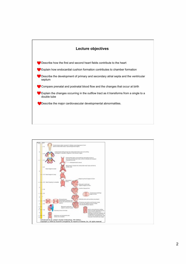
Lecture objectives
Describe how the first and second heart fields contribute to the heart Explain how endocardial cushion formation contributes to chamber formation Describe the development of primary and secondary atrial septa and the ventricular septum Compare prenatal and postnatal blood flow and the changes that occur at birth Explain the changes occurring in the outflow tract as it transforms from a single to a double tube Describe the major cardiovascular developmental abnormalities.

3
cardiac crescent and linear heart tube- 20 to 21 days
cardiac crescent and linear heart tube
inner endocardium outer myocardium
4
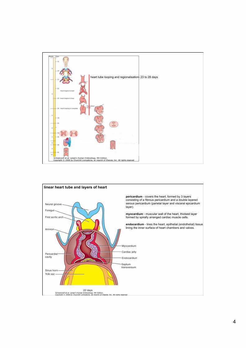
heart tube looping and regionalisation- 23 to 28 days
linear heart tube and layers of heart
pericardium - covers the heart, formed by 3 layers consisting of a fibrous pericardium and a double layered serous pericardium (parietal layer and visceral epicardium layer). myocardium - muscular wall of the heart, thickest layer formed by spirally arranged cardiac muscle cells. endocardium - lines the heart, epithelial (endothelial) tissue lining the inner surface of heart chambers and valves.
5
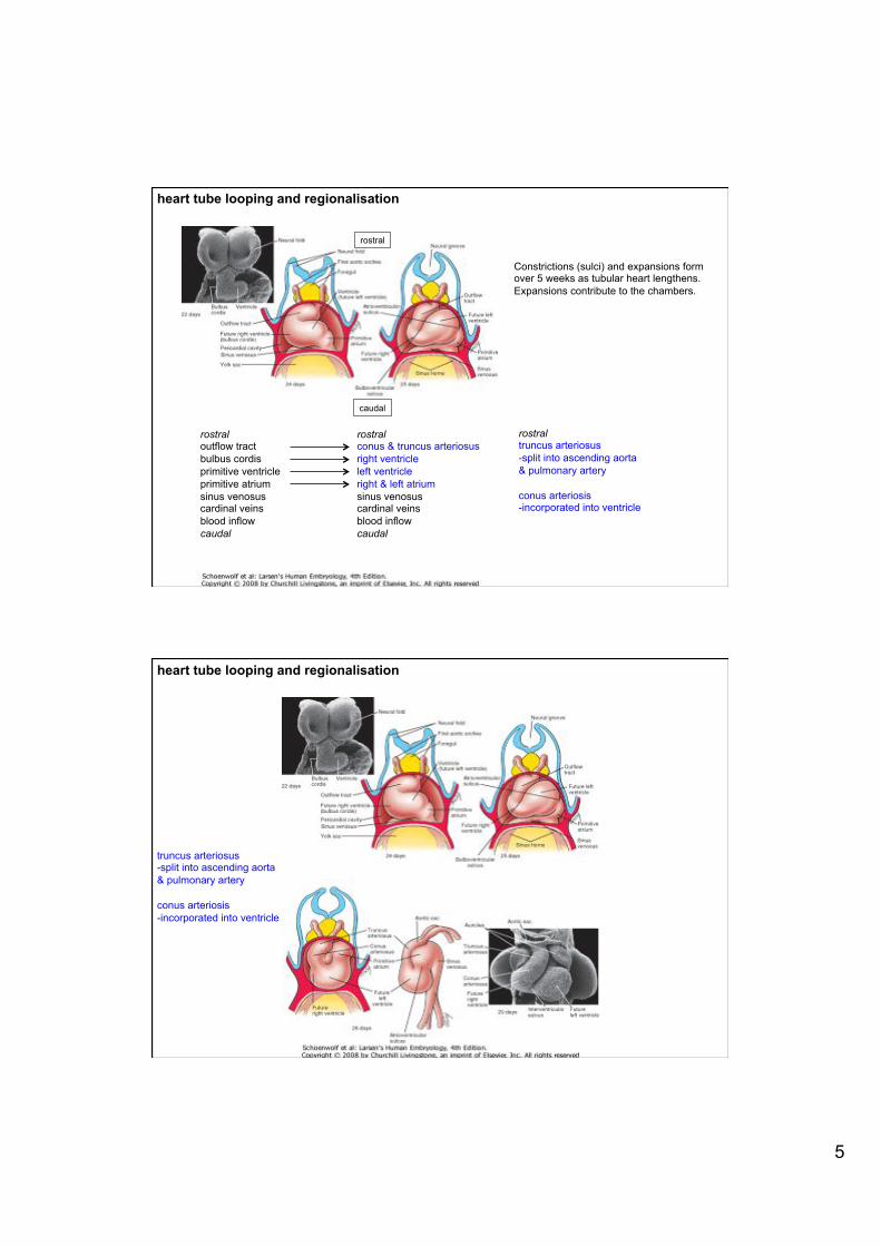
heart tube looping and regionalisation
Constrictions (sulci) and expansions form over 5 weeks as tubular heart lengthens. Expansions contribute to the chambers.
rostral
caudal
rostral outflow tract bulbus cordis primitive ventricle primitive atrium sinus venosus cardinal veins blood inflow caudal
rostral conus & truncus arteriosus right ventricle left ventricle right & left atrium sinus venosus cardinal veins blood inflow caudal
rostral truncus arteriosus -split into ascending aorta & pulmonary artery conus arteriosis -incorporated into ventricle
heart tube looping and regionalisation
truncus arteriosus -split into ascending aorta & pulmonary artery conus arteriosis -incorporated into ventricle

6
first heart field (primary) = linear heart tube second heart field (secondary) =dorsal to heart tube
first and second heart fields- cardiac progenitor populations
Buckingham et al (2005) Nat Rev Genet
first heart field second heart field
first and second heart fields
How was this worked out?

7
label cells with lipophilic dye culture embryo
see where these labelled cells and their progeny end up
Waldo et al Dev 2001
first and second heart fields
examine transcript localisation by RNA in situ hybridisation
Cai et al Dev Cell 2003
SHF FHF
SHF FHF
first and second heart fields
8
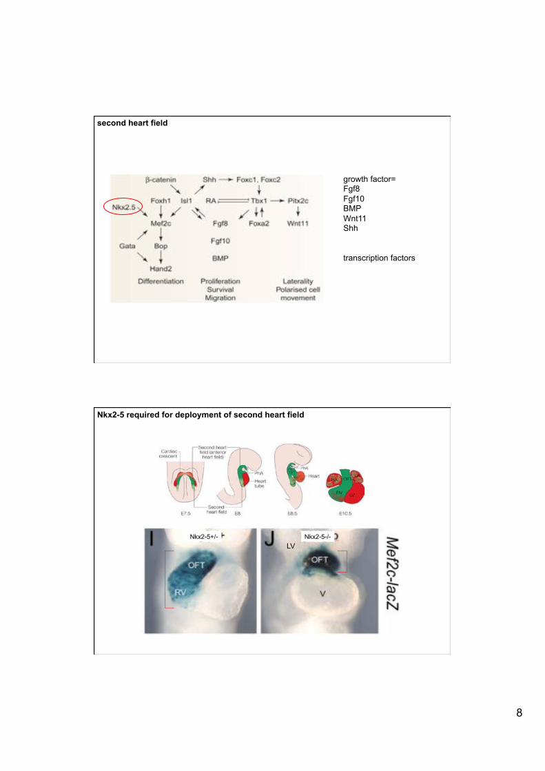
second heart field
growth factor= Fgf8 Fgf10 BMP Wnt11 Shh transcription factors
LV Nkx2-5+/- Nkx2-5-/-
Nkx2-5 required for deployment of second heart field

9
septation- 28 to 50 days
septation
M� Septation is necessary to separate the systemic and pulmonary circulations
M� Partial separation of definitive atria, ventricles and division of the atrioventricular canal into right and left canals
M� Endocardial cushions and muscular septum
10
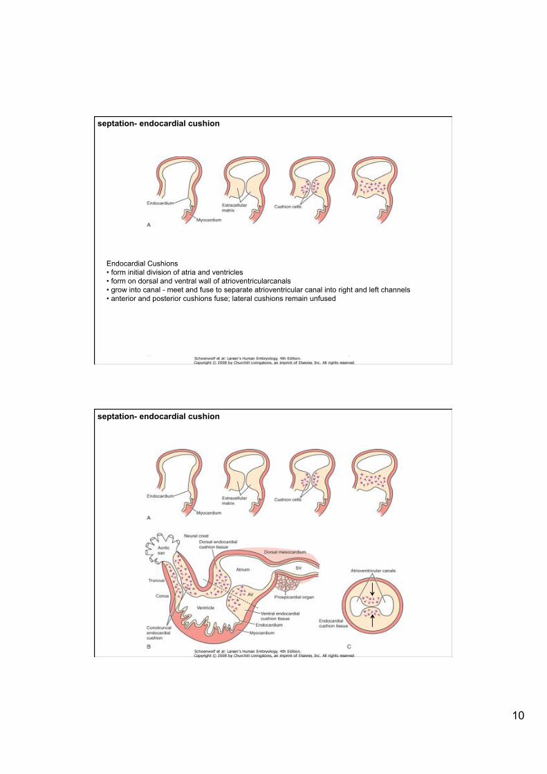
septation- endocardial cushion
Endocardial Cushions M� form initial division of atria and ventricles M� form on dorsal and ventral wall of atrioventricularcanals M� grow into canal - meet and fuse to separate atrioventricular canal into right and left channels M� anterior and posterior cushions fuse; lateral cushions remain unfused
septation- endocardial cushion
11
signaling network model for heart valve development and remodeling
Armstrong and Bischoff (2004) Circulation Research
growth factors
transcription factors
myocardium
endocardium
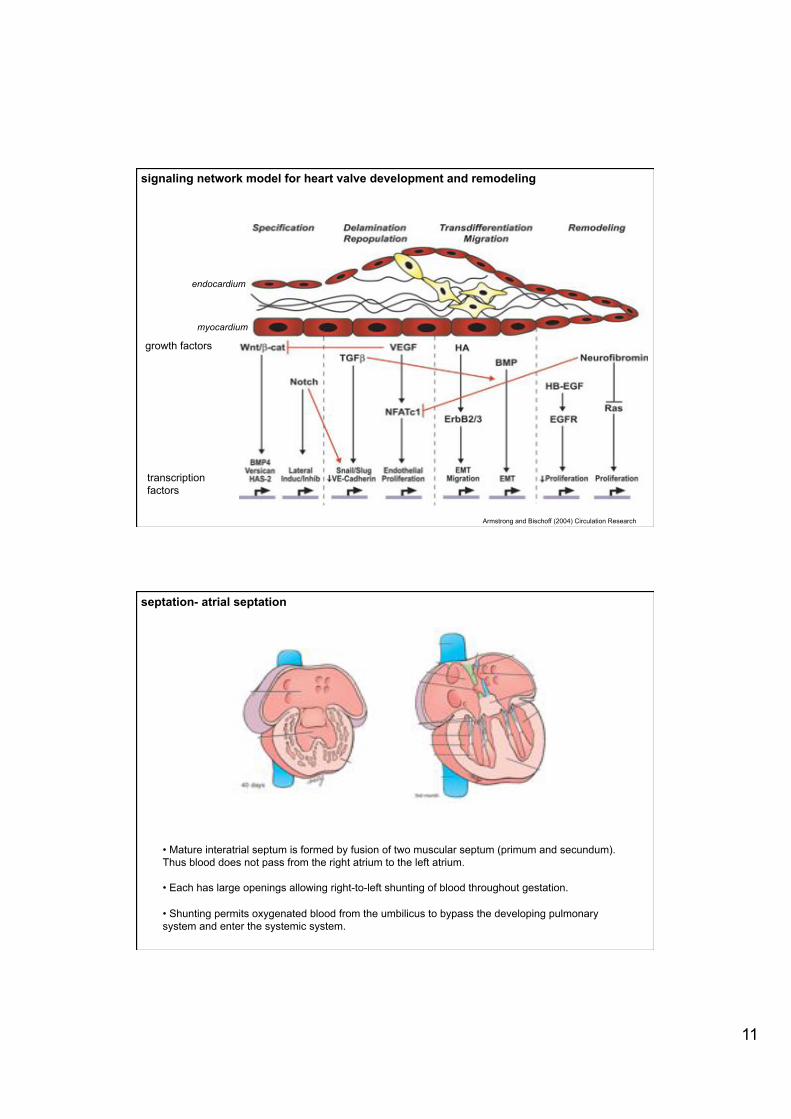
M� Mature interatrial septum is formed by fusion of two muscular septum (primum and secundum). Thus blood does not pass from the right atrium to the left atrium.
M� Each has large openings allowing right-to-left shunting of blood throughout gestation.
M� Shunting permits oxygenated blood from the umbilicus to bypass the developing pulmonary system and enter the systemic system.
septation- atrial septation

12
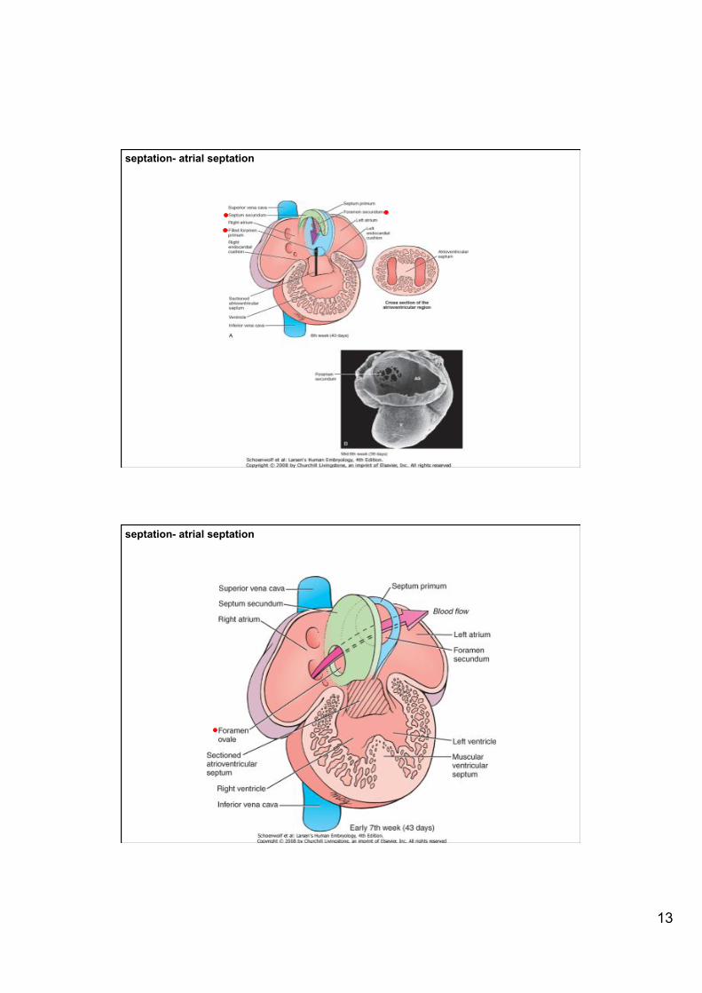
septation- atrial septation
septum primum
septum secundum
right left
poor understanding of genes required for atrial septation
septation- atrial septation
13
septation- atrial septation
septation- atrial septation
14
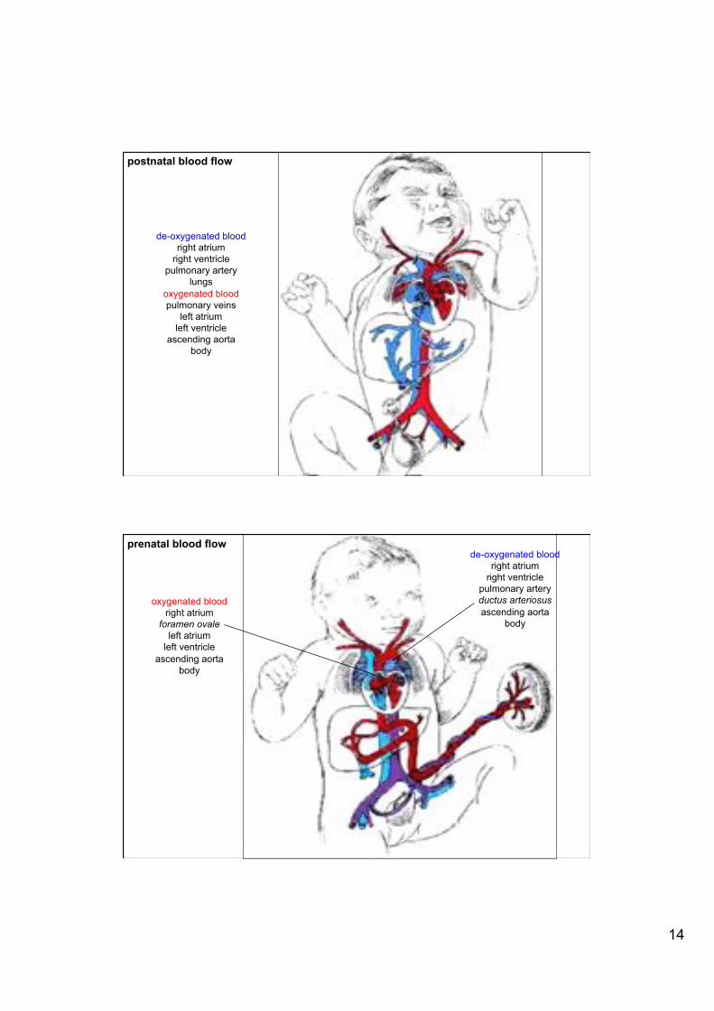
de-oxygenated blood right atrium
right ventricle pulmonary artery
lungs oxygenated blood pulmonary veins
left atrium left ventricle
ascending aorta body
postnatal blood flow
oxygenated blood right atrium
foramen ovale left atrium
left ventricle ascending aorta
body
de-oxygenated blood right atrium
right ventricle pulmonary artery ductus arteriosus ascending aorta
body
prenatal blood flow

15
M� at birth, cutting the umbilical cord and changes in the lungs after the first breaths trigger major functional adaptations in the fetal circulatory system
M� blood flow through ductus venosus is eliminated M� pulmonary circulation bed expands - reducing blood flow through ductus arteriosus M� physiological closure of interatrial shunt M� closure of ductus venosus in liver is prolonged
changes at birth
ventricular septation four chambered heart
poor understanding of genes required for ventricular
septation

16
outflow tract septation
M� initially outflow tract is a single tube, the bulbus cordis M� elongates to form proximal conus arteriosus and distal truncus arteriosus M� 2 growths (endocardial cushion) from wall in spiral pattern, inferior upwards - separate tract into 2 channels M� mesenchyme and neural crest contribute to this septation process M� fusion of outgrowths separate aortic and pulmonary outflow
http://php.med.unsw.edu.au/embryology/index.php?title=Development_Animation_-_Heart_Outflow_Septation
outflow tract septation
17
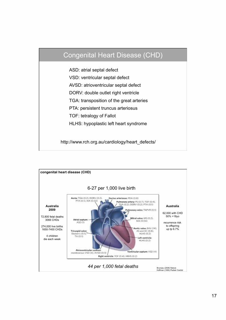
ASD: atrial septal defect
VSD: ventricular septal defect
AVSD: atrioventricular septal defect
DORV: double outlet right ventricle
TGA: transposition of the great arteries
PTA: persistent truncus arteriosus
TOF: tetralogy of Fallot
HLHS: hypoplastic left heart syndrome
Congenital Heart Disease (CHD)
http://www.rch.org.au/cardiology/heart_defects/
6-27 per 1,000 live birth
Bruneau (2008) Nature Hoffman (1995) Pediatr Cardiol
44 per 1,000 fetal deaths
Australia 2009
72,800 fetal deaths
3066 CHDs
274,000 live births 1650-7400 CHDs
4 children
die each week
Australia
62,000 with CHD 50% >18yo
recurrence risk
to offspring up tp 6.7%
congenital heart disease (CHD)

18
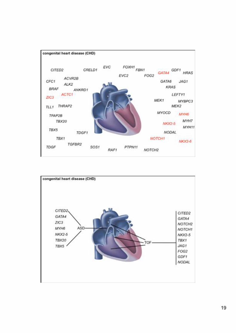
M� Chromosomal (11.9%) and Mendelian syndromes (7.4%) account for CHD
M� Non-syndromic large families with Mendelian inheritance patterns have identified CHD genes: ZIC3 (heterotaxy), NOTCH1 (aortic stenosis and bicuspid aortic valve), NKX2.5 (ASD), NKX2.6 (PTA/CAT), MYH6 (ASD), MYH11 (PDA), JAG1 (TOF), ACTC1 (ASD) and GATA4 (ASD)
M� Non-Mendelian/non-chromosomal �sporadic� CHD account for the remaining 80%, the increased risk of CHD recurrence in siblings and offspring indicates a genetic component
genetic causes of CHD
Bruneau Nature 2008
How do we identify the genes associated with these defects? M� familial: gene mapping
M� non-familial: candidate gene - 316 genes associated with heart defects in mice - 276 genes associated with ASD in mice - 143 genes associated with VSD in mice
M� understand developmental processes eg. SHF – OFT – aorta + pulmonary artery
congenital heart disease (CHD)
19
NKX2-5
GATA4
ZIC3
MYH6
NOTCH1 NKX2-6
ACTC1
TBX20
CITED2 GDF1
NODAL
CFC1
TBX1
THRAP2
CRELD1 FOG2
LEFTY1
FOXH1
TDGF
MYOCD TLL1
ANKRD1
TBX5
TFAP2B
PTPN11
KRAS
SOS1 RAF1 NOTCH2
JAG1
EVC
MEK1 MEK2
EVC2 FBN1
TGFBR2
HRAS
BRAF
ACVR2B ALK2
TDGF1
GATA6
MYH11
MYH7
MYBPC3
congenital heart disease (CHD)
ASD
NKX2-5
TBX5
TBX20
CITED2
GATA4
ZIC3
MYH6
TOF
NKX2-5
JAG1 TBX1
CITED2 GATA4 NOTCH2 NOTCH1
FOG2 GDF1 NODAL
congenital heart disease (CHD)

20
ASD
TOF
VSD
Ebstein’s anomaly tricuspid atresia
DORV TGA PTA
aortic stenosis
NKX2-5
interrupted aortic arch coarctation of the aorta
HLHS
congenital heart disease (CHD)
ASD
TOF
VSD
Ebstein’s anomaly tricuspid atresia
DORV TGA PTA
aortic stenosis
NKX2-5 R25C
interrupted aortic arch coarctation of the aorta
HLHS no defect
congenital heart disease (CHD)
21
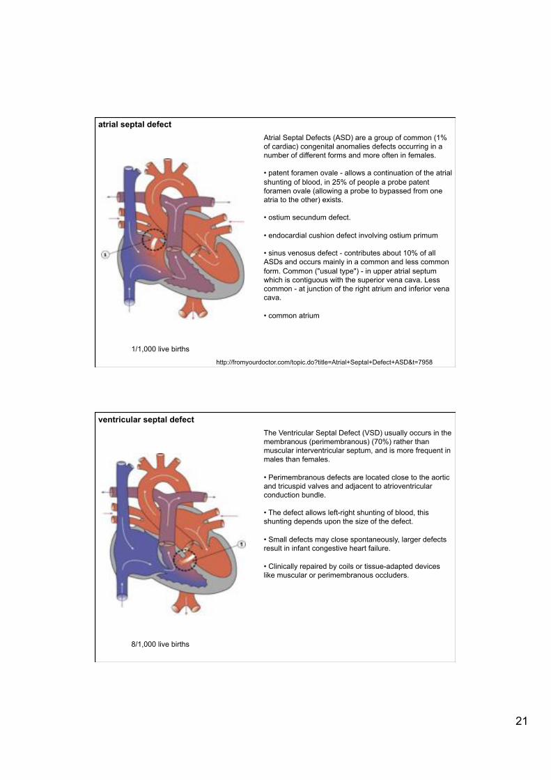
atrial septal defect
1/1,000 live births
Atrial Septal Defects (ASD) are a group of common (1% of cardiac) congenital anomalies defects occurring in a number of different forms and more often in females. M� patent foramen ovale - allows a continuation of the atrial shunting of blood, in 25% of people a probe patent foramen ovale (allowing a probe to bypassed from one atria to the other) exists.
M� ostium secundum defect. M� endocardial cushion defect involving ostium primum M� sinus venosus defect - contributes about 10% of all ASDs and occurs mainly in a common and less common form. Common ("usual type") - in upper atrial septum which is contiguous with the superior vena cava. Less common - at junction of the right atrium and inferior vena cava. M� common atrium
http://fromyourdoctor.com/topic.do?title=Atrial+Septal+Defect+ASD&t=7958
ventricular septal defect The Ventricular Septal Defect (VSD) usually occurs in the membranous (perimembranous) (70%) rather than muscular interventricular septum, and is more frequent in males than females. M� Perimembranous defects are located close to the aortic and tricuspid valves and adjacent to atrioventricular conduction bundle.
M� The defect allows left-right shunting of blood, this shunting depends upon the size of the defect.
M� Small defects may close spontaneously, larger defects result in infant congestive heart failure.
M� Clinically repaired by coils or tissue-adapted devices like muscular or perimembranous occluders.
8/1,000 live births
22
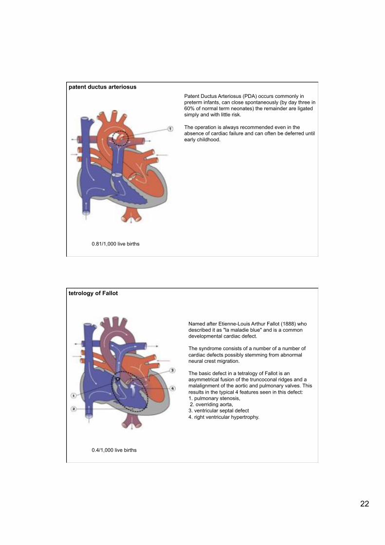
patent ductus arteriosus
0.81/1,000 live births
Patent Ductus Arteriosus (PDA) occurs commonly in preterm infants, can close spontaneously (by day three in 60% of normal term neonates) the remainder are ligated simply and with little risk. The operation is always recommended even in the absence of cardiac failure and can often be deferred until early childhood.
tetrology of Fallot
Named after Etienne-Louis Arthur Fallot (1888) who described it as "la maladie blue" and is a common developmental cardiac defect. The syndrome consists of a number of a number of cardiac defects possibly stemming from abnormal neural crest migration. The basic defect in a tetralogy of Fallot is an asymmetrical fusion of the truncoconal ridges and a malalignment of the aortic and pulmonary valves. This results in the typical 4 features seen in this defect: 1. pulmonary stenosis, 2. overriding aorta, 3. ventricular septal defect 4. right ventricular hypertrophy.
0.4/1,000 live births

23
Victor Chang Cardiac Research Institute
Sally L. Dunwoodie Developmental Biology Division Victor Chang Cardiac Research Institute Darlinghurst [email protected]
Related Documents