The Cardiovascular System A circulation consisting of … – A pump (the HEART) – A conducting system (blood vessels) – A fluid medium (blood) • Is specialized fluid CT • Contains cells suspended in a fluid matrix

heart cardiac cycle
Jul 24, 2015
Welcome message from author
This document is posted to help you gain knowledge. Please leave a comment to let me know what you think about it! Share it to your friends and learn new things together.
Transcript
The Cardiovascular System
A circulation consisting of …
– A pump (the HEART)
– A conducting system (blood vessels)
– A fluid medium (blood) • Is specialized fluid CT
• Contains cells suspended in a fluid matrix
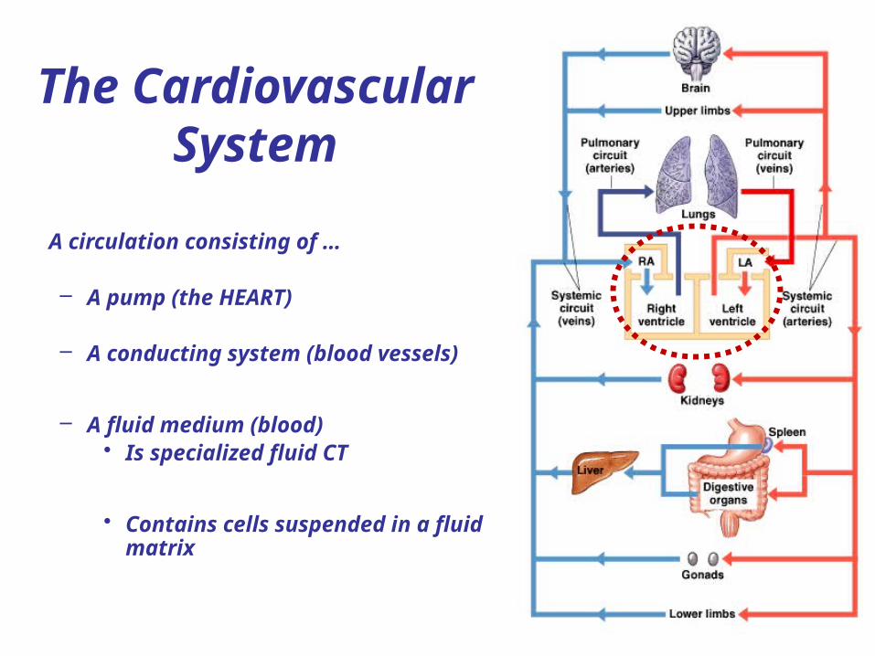
Important BASIC “RULES” of the CV system:
This is independent of the OXYGEN content of the blood i.e the amount of HbO2.
1. By convention, arteries carry blood AWAY from the heart and VEINS towards it.
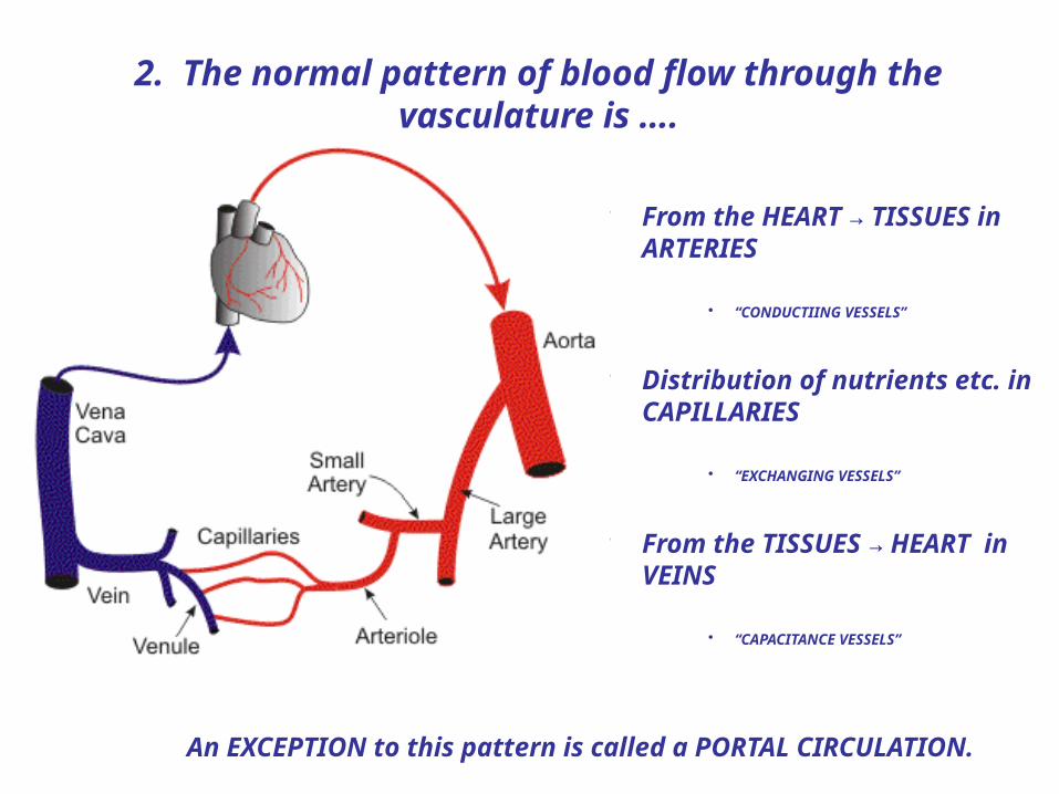
2. The normal pattern of blood flow through the vasculature is ….
• From the HEART → TISSUES in ARTERIES
• “CONDUCTIING VESSELS”
• Distribution of nutrients etc. in CAPILLARIES
• “EXCHANGING VESSELS”
• From the TISSUES → HEART in VEINS
• “CAPACITANCE VESSELS”
An EXCEPTION to this pattern is called a PORTAL CIRCULATION.
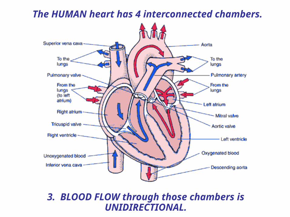
The HUMAN heart has 4 interconnected chambers.
3. BLOOD FLOW through those chambers is UNIDIRECTIONAL.
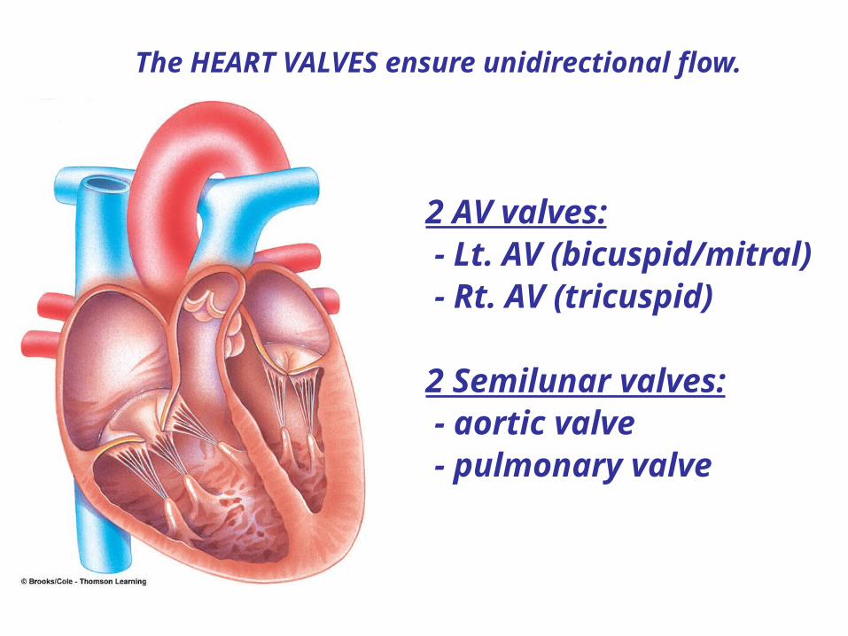
The HEART VALVES ensure unidirectional flow.
2 AV valves: - Lt. AV (bicuspid/mitral) - Rt. AV (tricuspid)
2 Semilunar valves: - aortic valve - pulmonary valve

Thus, the normal adult pattern of blood flow would be …
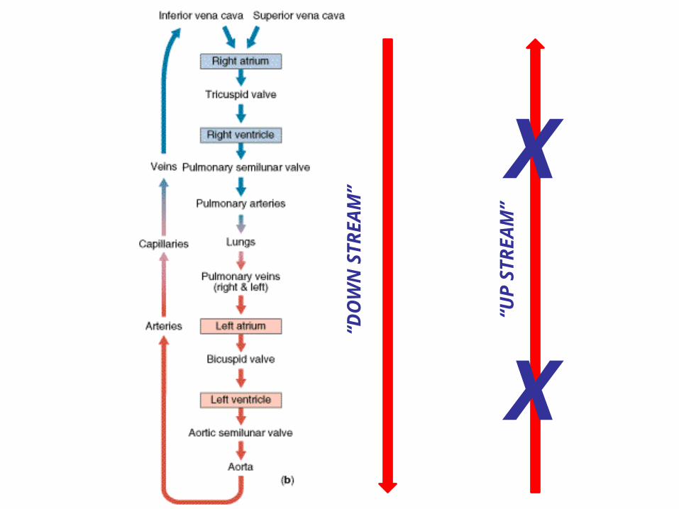
“DO
WN
ST
RE
AM
”
“UP
ST
RE
AM
”
X
X

Right atrium
Right ventricle
Papillary muscle
Chordae tendineae
Septum
AV valve structure
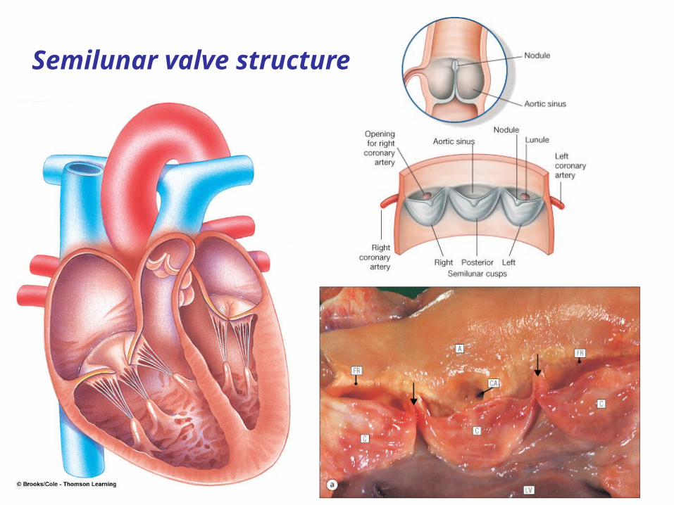
Semilunar valve structure
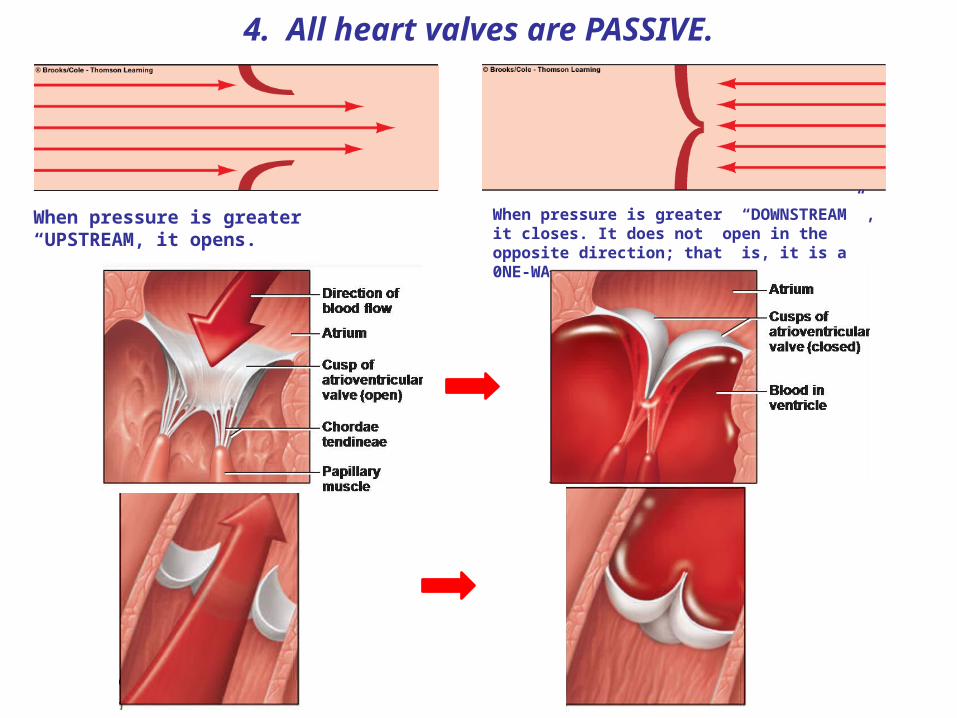
When pressure is greater “UPSTREAM, it opens.
When pressure is greater “DOWNSTREAM” , it closes. It does not open in the opposite direction; that is, it is a 0NE-WAY VALVE.
4. All heart valves are PASSIVE.
“DO
WN
ST
RE
AM
”
“UP
ST
RE
AM
”
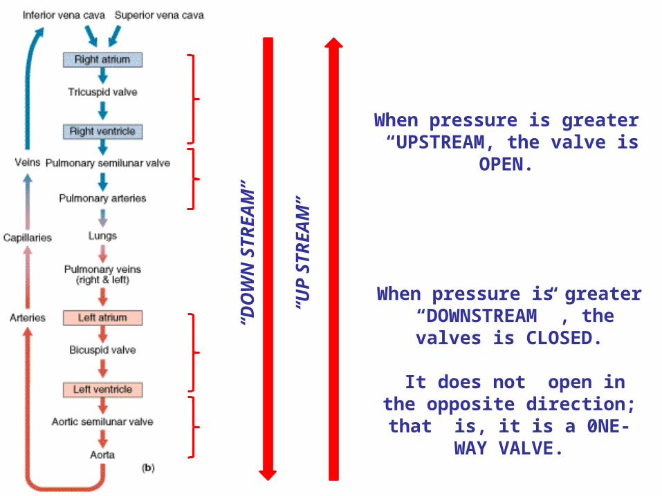
When pressure is greater “UPSTREAM, the valve is
OPEN.
When pressure is greater “DOWNSTREAM” , the valves
is CLOSED.
It does not open in the opposite direction; that is, it
is a 0NE-WAY VALVE.

What must the pressure relationships between chambers or between chambers and draining vessels be, in each of the following diagrams?
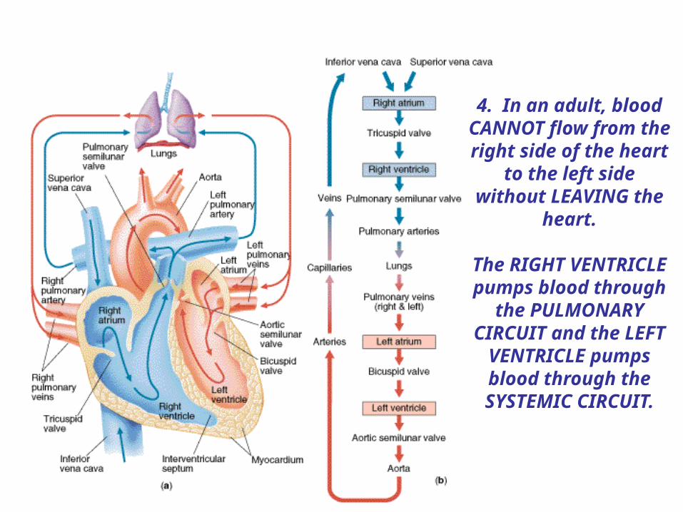
4. In an adult, blood CANNOT flow from the right side of the heart to the left side without LEAVING the
heart.
The RIGHT VENTRICLE pumps blood through the
PULMONARY CIRCUIT and the LEFT VENTRICLE
pumps blood through the SYSTEMIC
CIRCUIT.
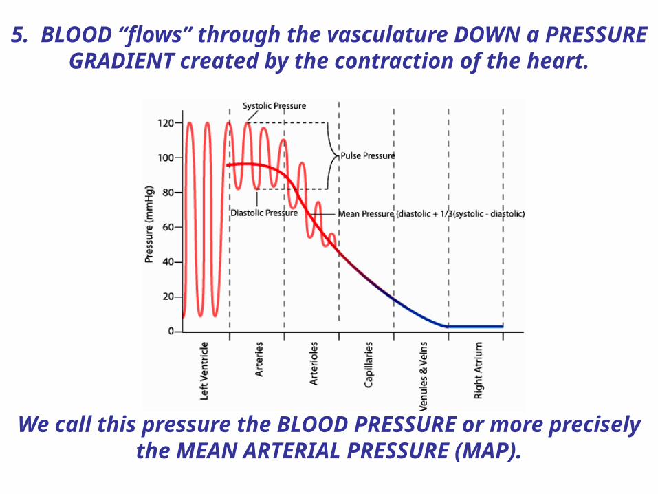
5. BLOOD “flows” through the vasculature DOWN a PRESSURE GRADIENT created by the contraction of the
heart.
We call this pressure the BLOOD PRESSURE or more precisely the MEAN ARTERIAL PRESSURE (MAP).
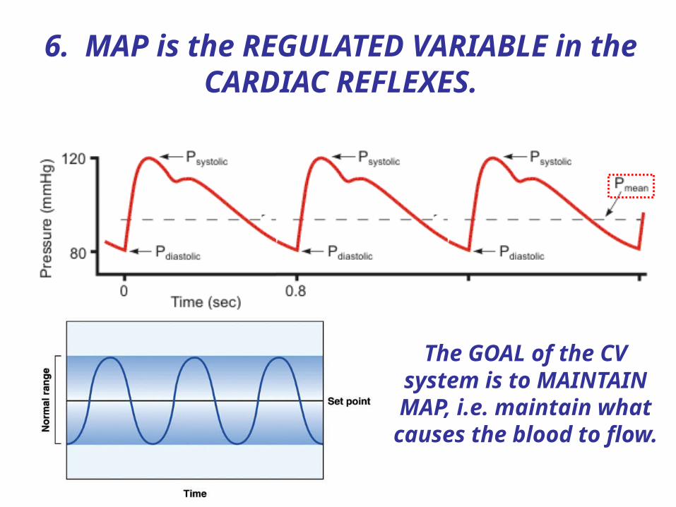
6. MAP is the REGULATED VARIABLE in the CARDIAC REFLEXES.
The GOAL of the CV system is to MAINTAIN MAP, i.e. maintain what
causes the blood to flow.


The HEART is located in the THORACIC CAVITY.
It is found in a non-delineated space MEDIAL to the LUNGS called the MEDIASTINUM.

Its BASE is located SUPERIORLY and its APEX, INFERIORLY.
It is ROTATED counterclock-wise so as to lie on its RIGHT SIDE.
Base
Apex
Apex
Base

The BASE of the heart lies at the level of the 2nd
intercostal space at midline. The APEX lies at the level of the 5th intercostal
space about 2.5-3” to the LEFT of
midline.
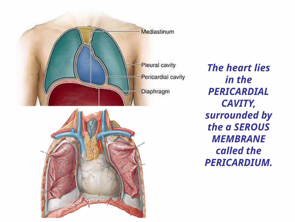
The heart lies in the
PERICARDIAL CAVITY,
surrounded by the a SEROUS MEMBRANE
called the PERICARDIUM.
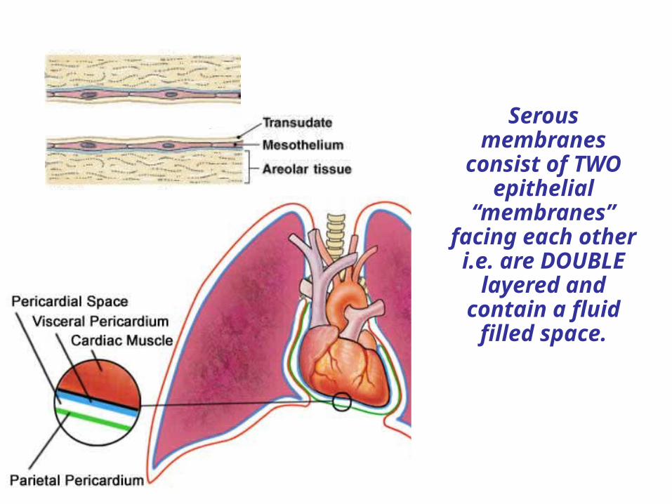
Serous membranes
consist of TWO epithelial
“membranes” facing each other i.e. are DOUBLE
layered and contain a fluid filled space.
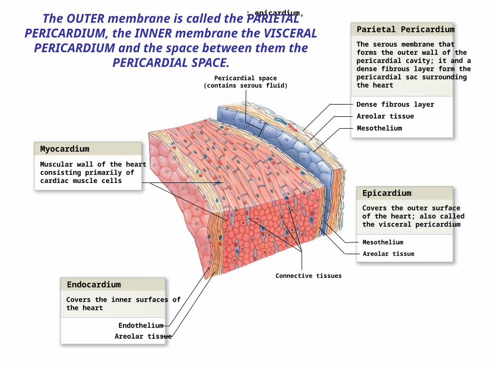
A section of the heart showing its three layers: epicardium,myocardium, and endocardium
Myocardium
Endocardium
Epicardium
Parietal Pericardium
Dense fibrous layer
Areolar tissue
Mesothelium
Mesothelium
Areolar tissue
Connective tissues
Pericardial space(contains serous fluid)
Muscular wall of the heartconsisting primarily ofcardiac muscle cells
Areolar tissue
Covers the inner surfaces ofthe heart
Endothelium
Covers the outer surface of the heart; also calledthe visceral pericardium
The serous membrane thatforms the outer wall of thepericardial cavity; it and adense fibrous layer form thepericardial sac surroundingthe heart
The OUTER membrane is called the PARIETAL PERICARDIUM, the INNER membrane the VISCERAL
PERICARDIUM and the space between them the PERICARDIAL SPACE.
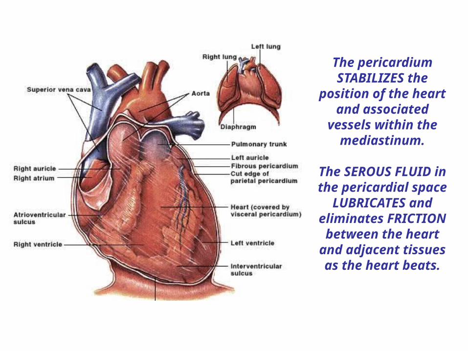
The pericardium STABILIZES the
position of the heart and associated
vessels within the mediastinum.
The SEROUS FLUID in the pericardial space
LUBRICATES and eliminates FRICTION
between the heart and adjacent tissues as
the heart beats.

Externally, the CORONARY SULCUS marks the separation between the atria
and ventricles.
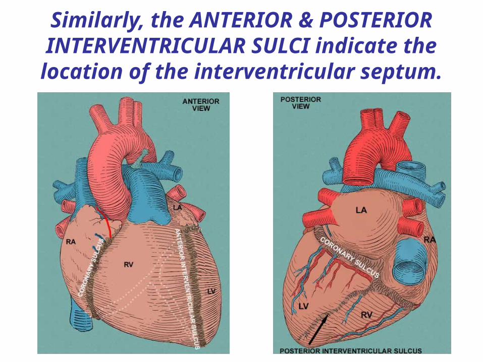
Similarly, the ANTERIOR & POSTERIOR INTERVENTRICULAR SULCI indicate the location of the interventricular septum.

Commonly, significant accumulations of WAT
(structural fat) are found in these sulci, …
Mesothelium(visceral pericardium)
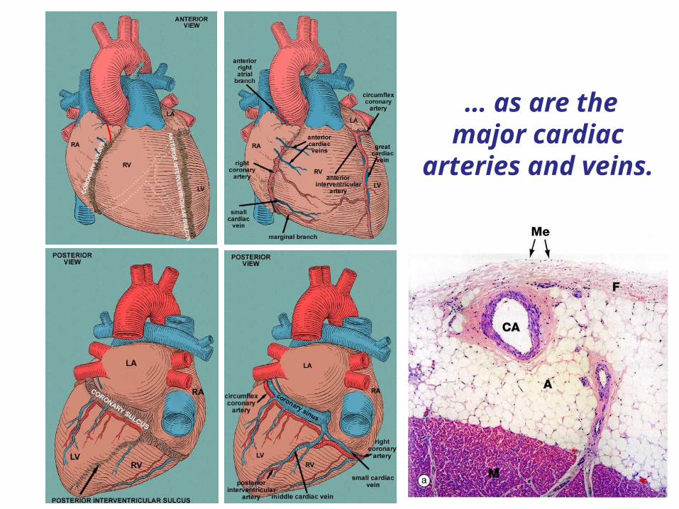
… as are the major cardiac
arteries and veins.
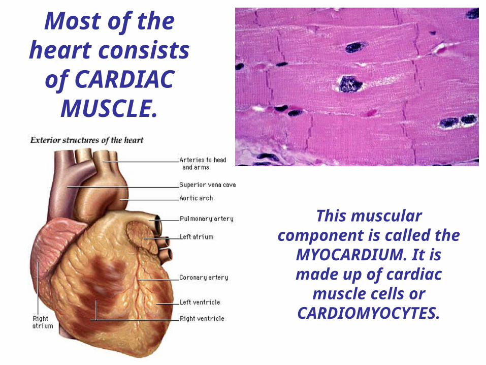
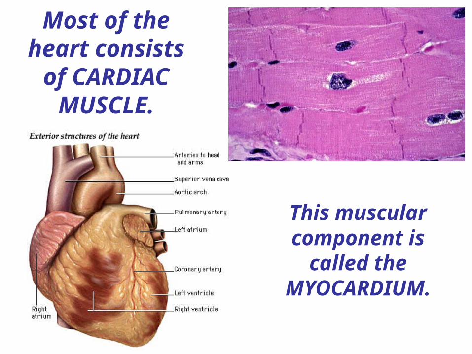
Most of the heart consists of CARDIAC
MUSCLE.
This muscular component is called the
MYOCARDIUM. It is made up of cardiac
muscle cells or CARDIOMYOCYTES.

The ORIENTATION of the cardiac muscle cells
means that the contracts with a “wringing” motion from
APEX to BASE.
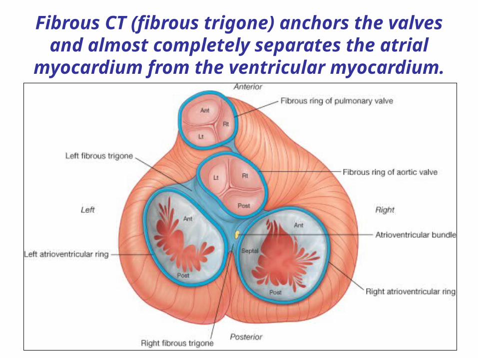
Fibrous CT (fibrous trigone) anchors the valves and almost completely separates the atrial
myocardium from the ventricular myocardium.

The internal surfaces of the heart are lined by a simple
squamous epithelium called the ENDOCARDIUM.
This is continuous with the ENDOTHELIUM that lines
the blood vessels.

A section of the heart showing its three layers: epicardium,myocardium, and endocardium
Myocardium
Endocardium
Epicardium
Parietal Pericardium
Dense fibrous layer
Areolar tissue
Mesothelium
Mesothelium
Areolar tissue
Connective tissues
Pericardial cavity(contains serous fluid)
Muscular wall of the heartconsisting primarily ofcardiac muscle cells
Areolar tissue
Covers the inner surfaces ofthe heart
Endothelium
Covers the outer surface of the heart; also calledthe visceral pericardium
The serous membrane thatforms the outer wall of thepericardial cavity; it and adense fibrous layer form thepericardial sac surroundingthe heart
Thus, the point of a pin entering the
pericardial cavity from the outside ….


4. The heart relies completely on AEROBIC RESPIRATION for its energy, and is unable to pump sufficiently in an ISCHEMIC (oxygen-deprived) environment.
The heart, as a muscle, pumps continuously throughout life and is
adapted to be highly resistant to fatigue.
2. Cardiomyocytes contain large numbers of mitochondria, enabling continuous aerobic respiration and production of ATP.
3. Cardiac muscle also contains MYOGLOBIN, an oxygen-storing protein.
1. Cardiac muscle has an extensive supply from the CORONARY ARTERIES that provide nutrients and oxygen.
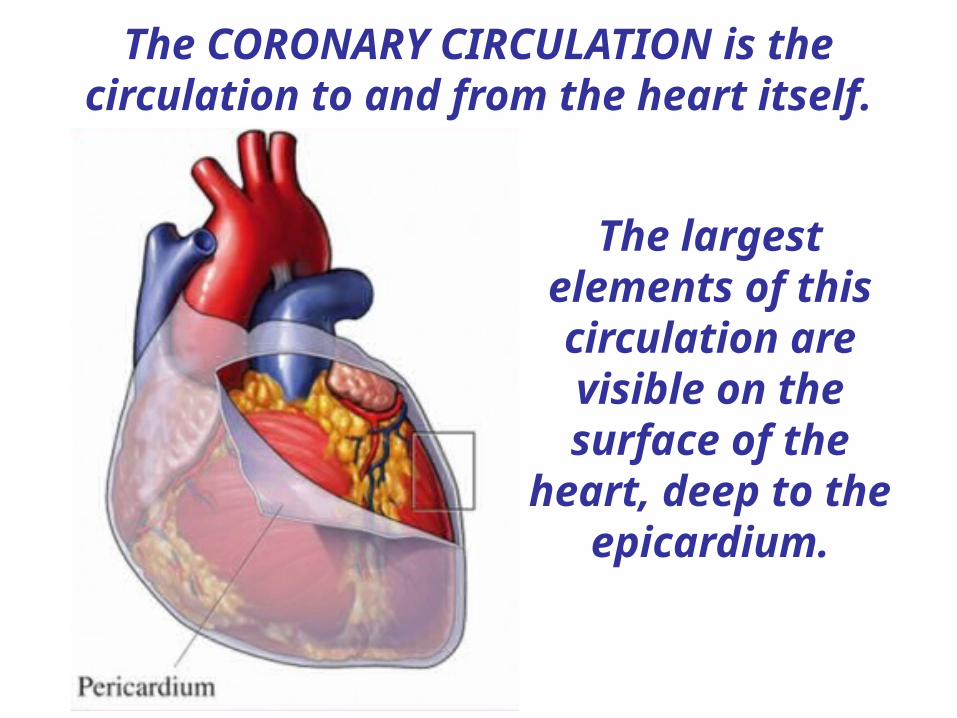
The CORONARY CIRCULATION is the circulation to and from the heart itself.
The largest elements of this circulation are visible on the surface of the
heart, deep to the epicardium.
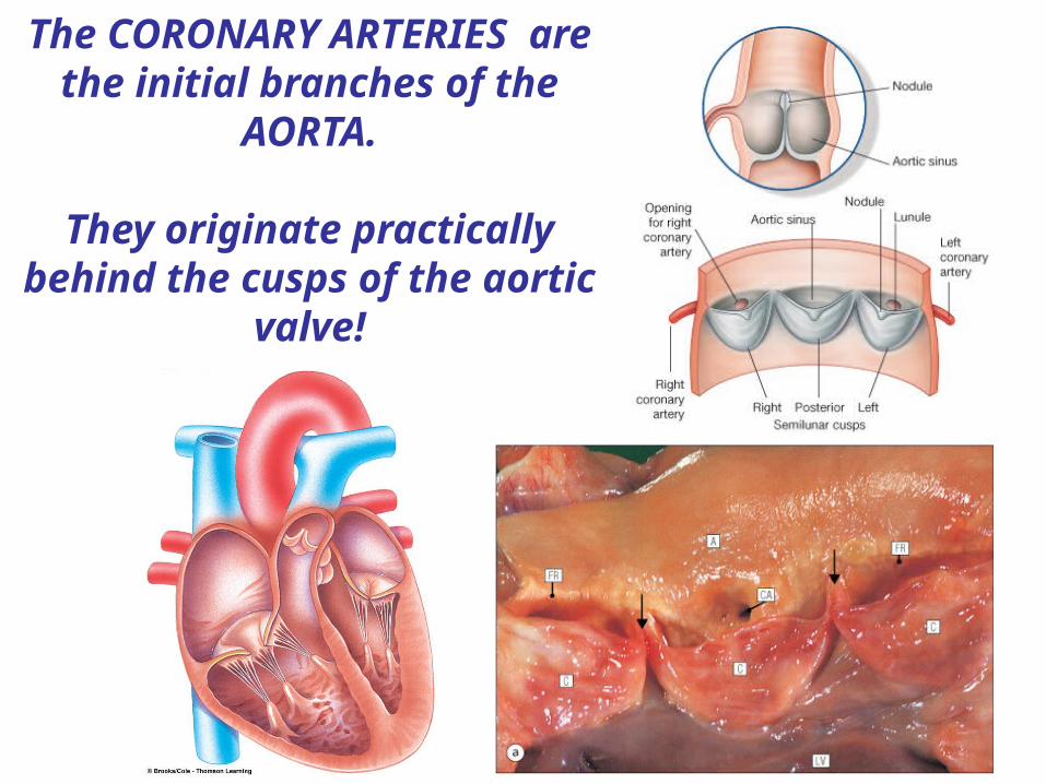
The CORONARY ARTERIES are the initial branches of the
AORTA.
They originate practically behind the cusps of the aortic
valve!

The heart is supplied with blood from the LEFT & RIGHT CORONARY ARTERIES.
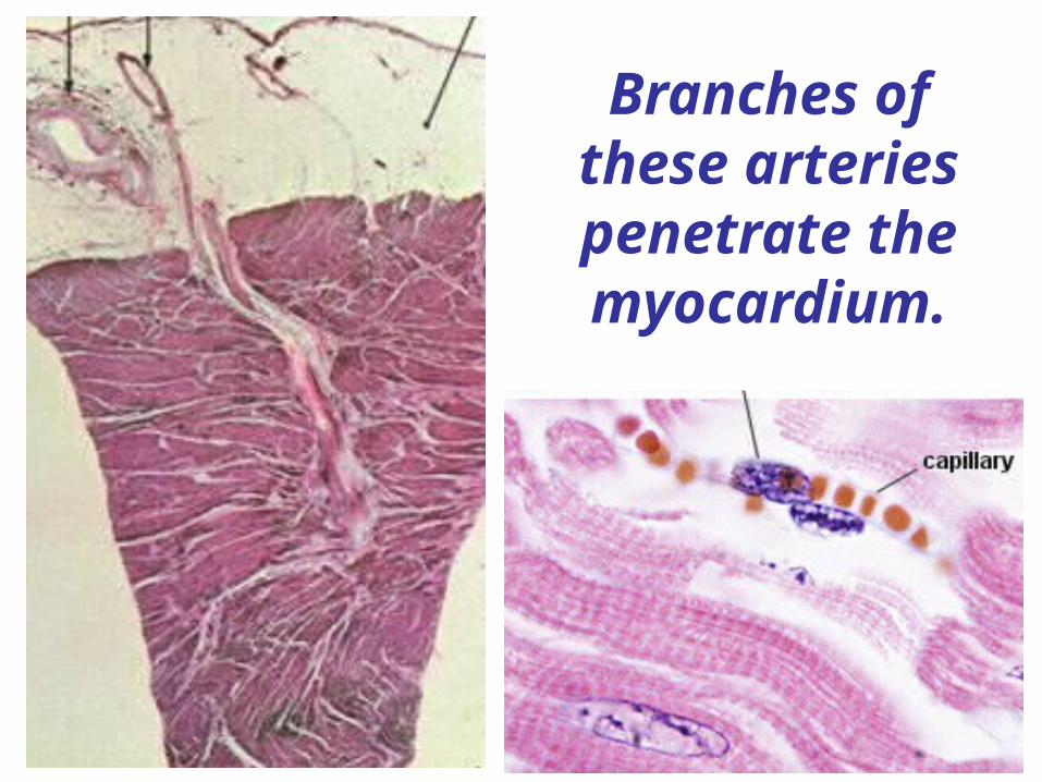
Branches of these arteries penetrate the myocardium.
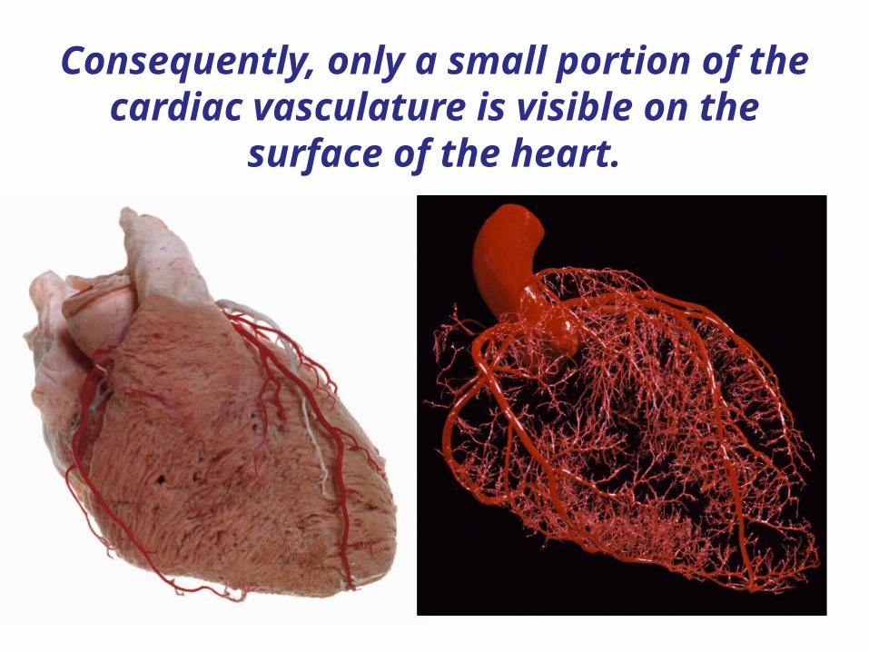
Consequently, only a small portion of the cardiac vasculature is visible on the
surface of the heart.

Generally speaking, the left CA supplies the LEFT side of the heart and the right
CA the RIGHT.
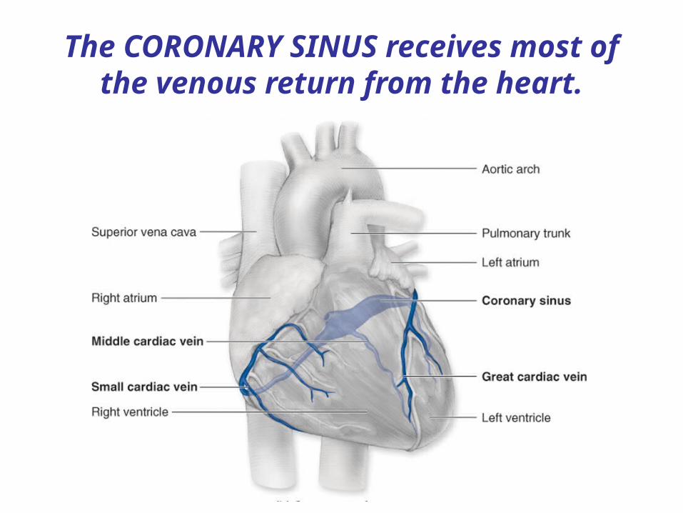
The CORONARY SINUS receives most of the venous return from the heart.
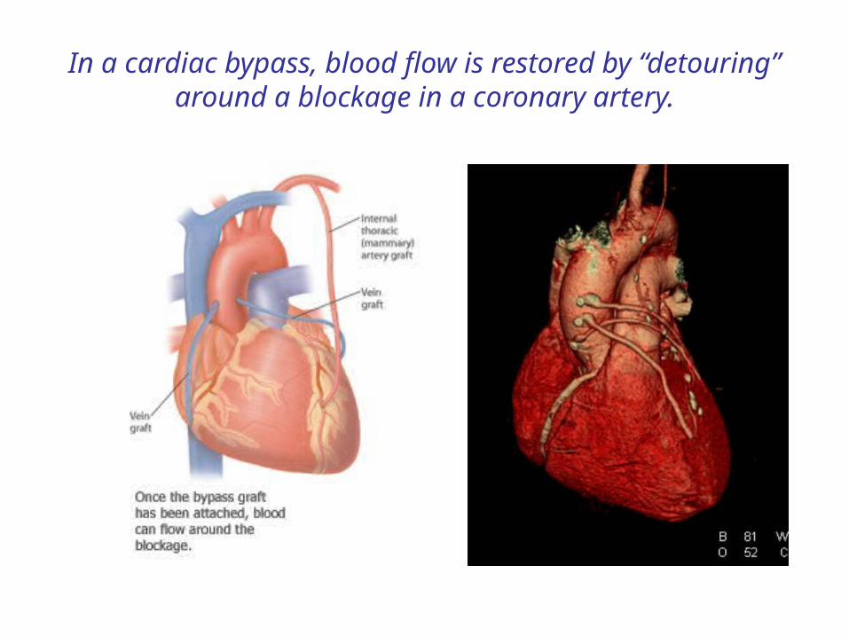
In a cardiac bypass, blood flow is restored by “detouring” around a blockage in a coronary artery.
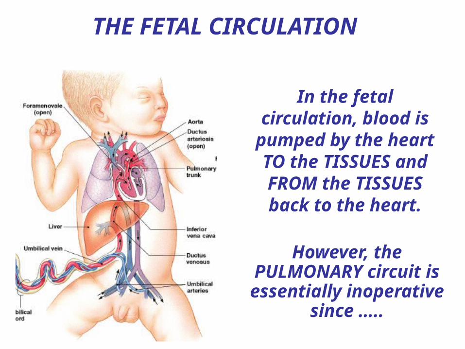
In the fetal circulation, blood is pumped by
the heart TO the TISSUES and FROM the TISSUES back to
the heart.
However, the PULMONARY circuit is essentially inoperative
since …..
THE FETAL CIRCULATION
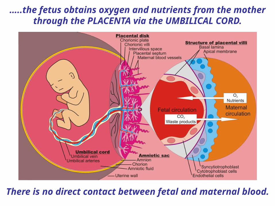
…..the fetus obtains oxygen and nutrients from the mother through the PLACENTA via the UMBILICAL CORD.
There is no direct contact between fetal and maternal blood.

Umbilicalcord
An UMBILICAL/PLACENTAL VEIN delivers oxygenated blood TO the fetus and 2
UMBILICAL ARTERIES return venous blood FROM the fetus
to the placenta.
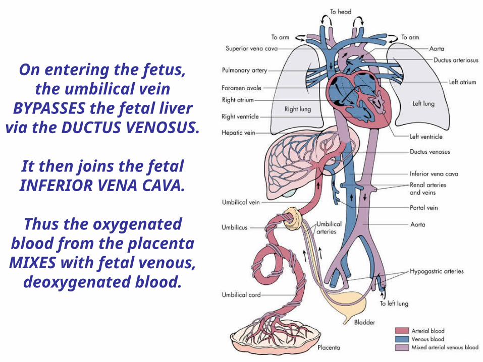
On entering the fetus, the umbilical vein
BYPASSES the fetal liver via the DUCTUS
VENOSUS.
It then joins the fetal INFERIOR VENA CAVA.
Thus the oxygenated blood from the placenta MIXES with fetal venous,
deoxygenated blood.
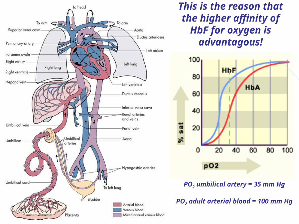
This is the reason that the higher affinity of
HbF for oxygen is advantagous!
PO2 umbilical artery = 35 mm Hg
PO2 adult arterial blood = 100 mm Hg
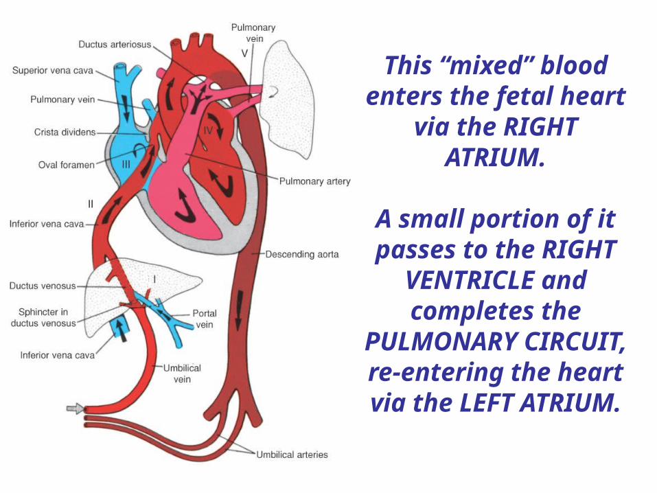
This “mixed” blood enters the fetal heart
via the RIGHT ATRIUM.
A small portion of it passes to the RIGHT
VENTRICLE and completes the PULMONARY
CIRCUIT, re-entering the heart via the LEFT ATRIUM.

Alternatively, rather than flowing to the LUNGS via
the PULMONARY TRUNK, blood may flow DIRECTLY into the fetal AORTA via the DUCTUS
ARTERIOSUS.
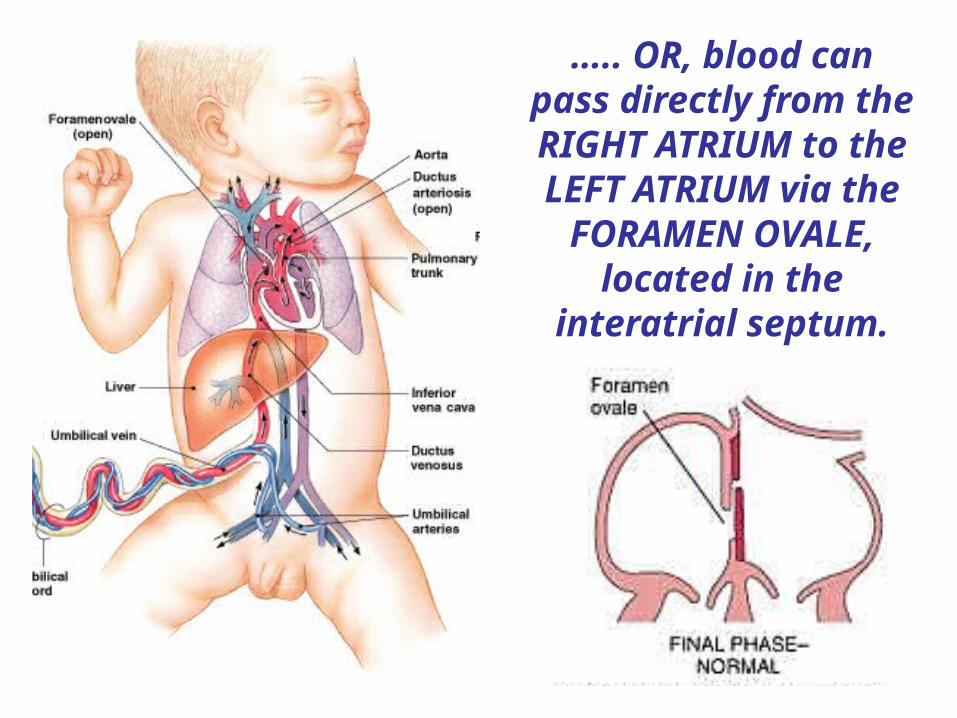
….. OR, blood can pass directly from the RIGHT ATRIUM to the LEFT ATRIUM via the FORAMEN OVALE,
located in the interatrial septum.
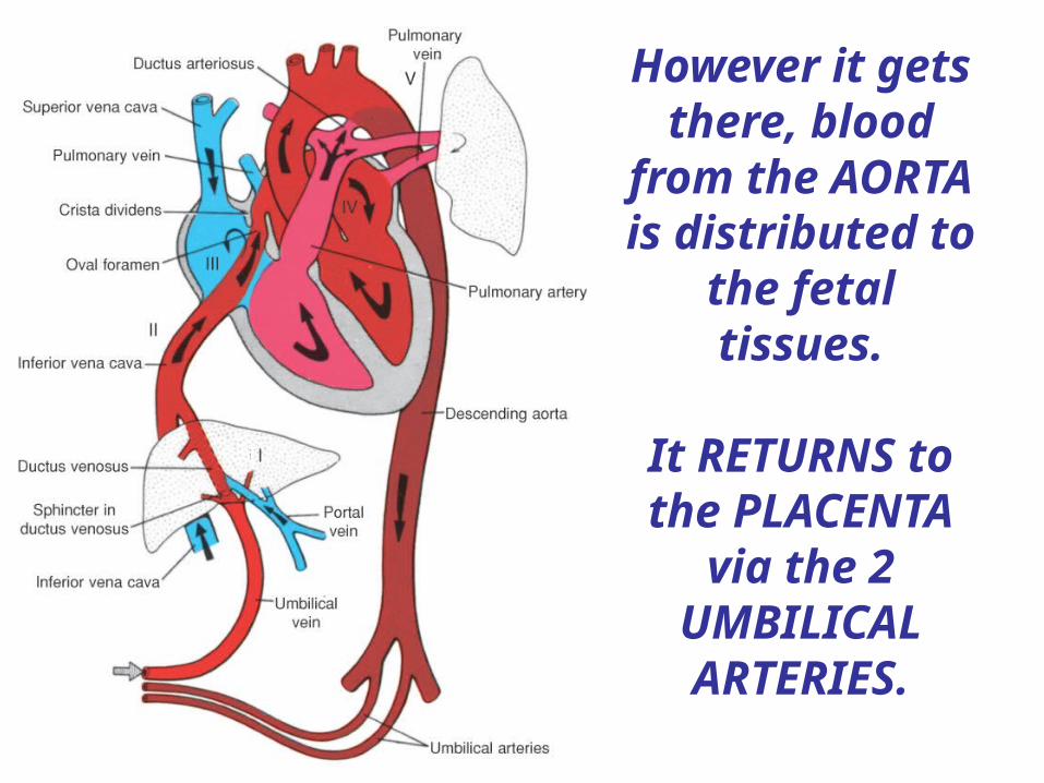
However it gets there, blood
from the AORTA is distributed to
the fetal tissues.
It RETURNS to the PLACENTA
via the 2 UMBILICAL ARTERIES.
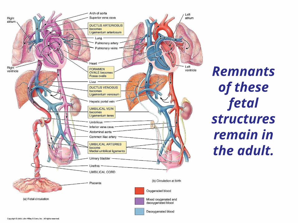
Remnants of these
fetal structures remain in the adult.

Most of the heart consists of CARDIAC
MUSCLE.
This muscular component is
called the MYOCARDIUM.

Striatedmuscle
Unstriatedmuscle
Skeletalmuscle
Cardiacmuscle
Smoothmuscle
Voluntarymuscle
Involuntarymuscle
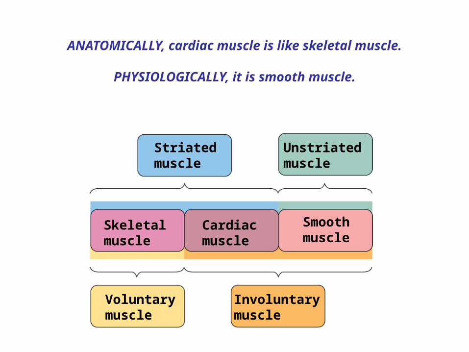
ANATOMICALLY, cardiac muscle is like skeletal muscle.
PHYSIOLOGICALLY, it is smooth muscle.

Cardiac muscle is STRIATED.
• Contains SARCOMERES
• Contains ACTIN, MYOSIN and TROPONIN
• Contraction explained by the SLIDING FILAMENT MODEL
• Excitation/contraction coupling via Ca++

Cardiac muscle cells are interconnected via INTERCALATED DISCS.
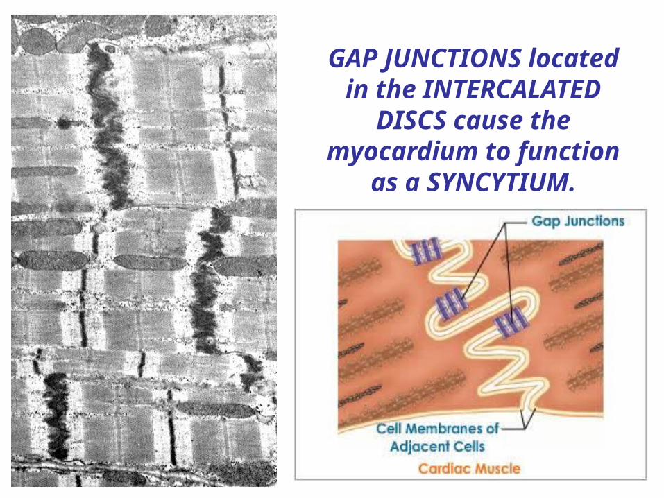
GAP JUNCTIONS located in the INTERCALATED
DISCS cause the myocardium to function
as a SYNCYTIUM.
Striatedmuscle
Unstriatedmuscle
Skeletalmuscle
Cardiacmuscle
Smoothmuscle
Voluntarymuscle
Involuntarymuscle
ANATOMICALLY, cardiac muscle is like skeletal muscle.
PHYSIOLOGICALLY, it is smooth muscle.

Cardiac muscle cells are INVOLUNTARY, i.e. their CONTRACTION is regulated ….
• INTRINSICALLY by PACEMAKER activity

Cardiac muscle cells are INVOLUNTARY, i.e. their CONTRACTION is regulated ….
• EXTRINSICALLY by the ANS and ENDOCRINE SYSTEMS

Cardiac muscle is an EXCITABLE tissue!!!
It consists of 2 populations of
excitable cells –
-Contractile cardiomyocytes
-Pacemaker cardiomyocytes
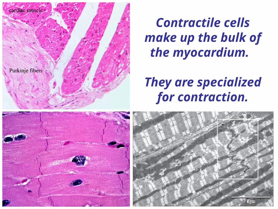
Contractile cells make up the bulk of the myocardium.
They are specialized for contraction.
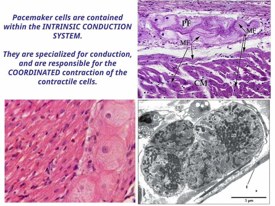
Pacemaker cells are contained within the INTRINSIC
CONDUCTION SYSTEM.
They are specialized for conduction, and are responsible
for the COORDINATED contraction of the contractile cells.
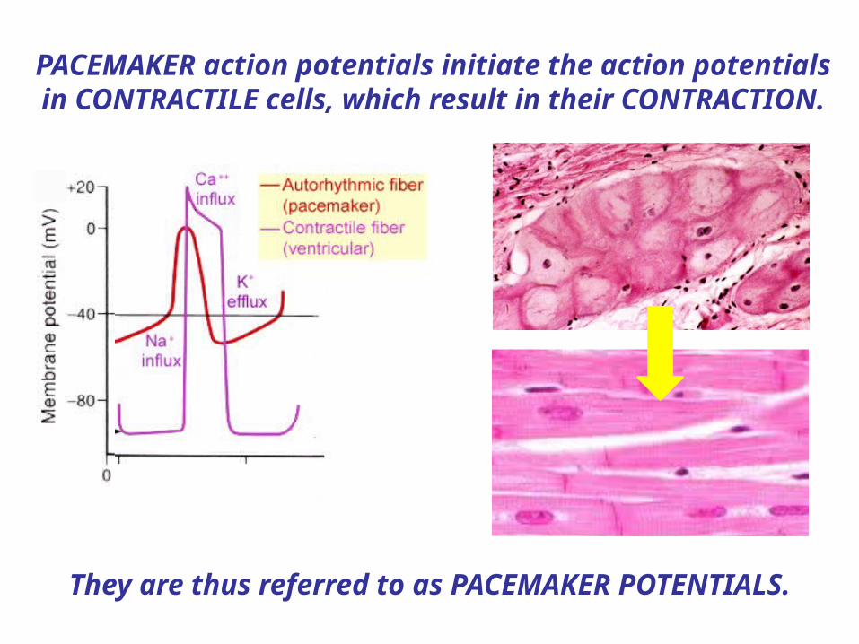
PACEMAKER action potentials initiate the action potentials in CONTRACTILE cells, which result in their
CONTRACTION.
They are thus referred to as PACEMAKER POTENTIALS.

Since the pacemaker cardiomyocytes are linked by GAP JUNCTIONS to the
contractile cardiomyocytes …
…when the pacemaker cells depolarize, so do the contractile cells, AT THE SAME TIME AND
RATE!
Thus the INHERENT pacemaker activity of these cardiomyocytes determines the INTRINSIC CONTRACTILE
RATE OF THE HEART!

2. AT ANY MOMENT IN TIME the membrane potential is produced by the WEIGHTED AVERAGE of all the ions in DISEQUILIBRIUM to which it is PERMEABLE.
1. PACEMAKER CELLS have an inherently UNSTABLE membrane potential that DEPOLARIZES over time.
This is called a PACEMAKER or PREPOTENTIAL.
The pacemaker potential gradually becomes less negative until it reaches THRESHOLD and triggers an ACTION
POTENTIAL.
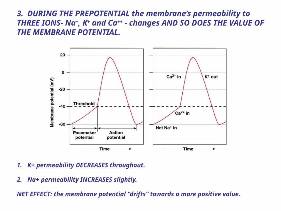
3. DURING THE PREPOTENTIAL the membrane’s permeability to THREE IONS- Na+, K+ and Ca++ - changes AND SO DOES THE VALUE OF THE MEMBRANE POTENTIAL.
1. K+ permeability DECREASES throughout.
2. Na+ permeability INCREASES slightly.
NET EFFECT: the membrane potential “drifts” towards a more positive value.
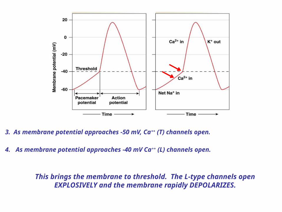
3. As membrane potential approaches -50 mV, Ca++ (T) channels open.
4. As membrane potential approaches -40 mV Ca++ (L) channels open.
This brings the membrane to threshold. The L-type channels open EXPLOSIVELY and the membrane rapidly DEPOLARIZES.
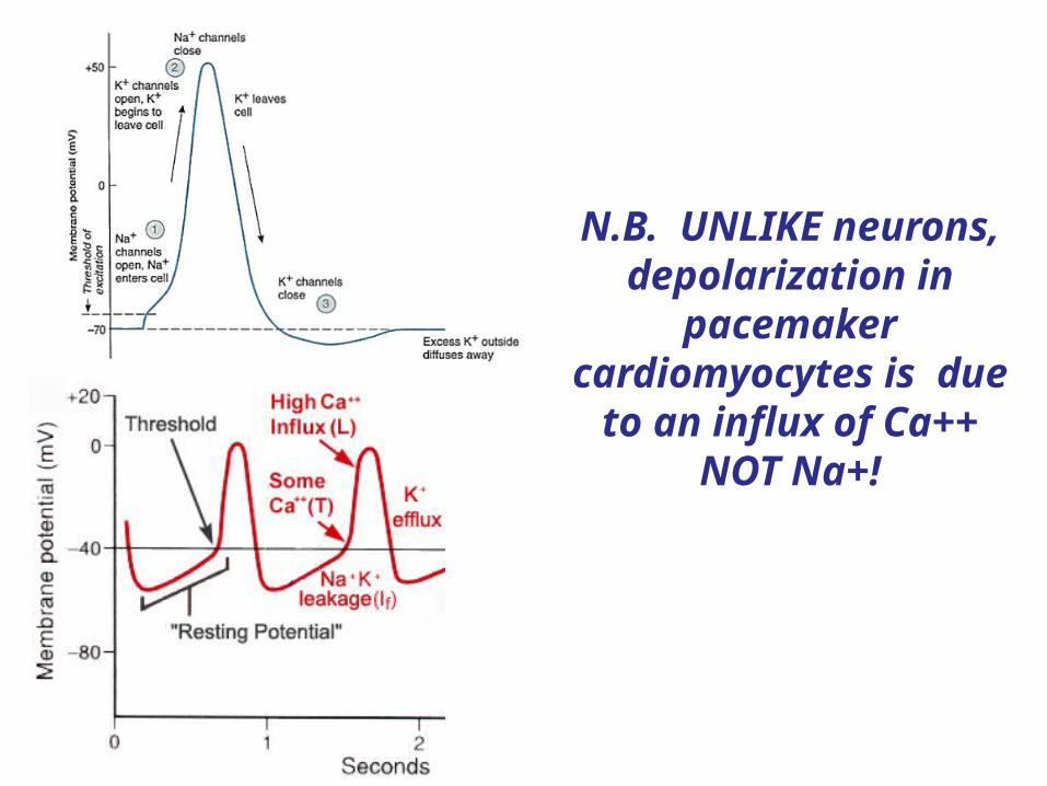
N.B. UNLIKE neurons, depolarization in
pacemaker cardiomyocytes is
due to an influx of Ca++ NOT Na+!

4. At maximum depolarization, the L-type channels CLOSE and the K+ channels OPEN, initiating
REPOLARIZATION.

The pacemaker cells are arranged in an interconnected pathway called the INTRINSIC
CONDUCTION SYSTEM.
SA node
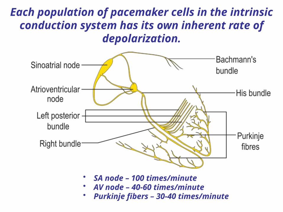
Each population of pacemaker cells in the intrinsic conduction system has its own inherent rate of
depolarization.
• SA node – 100 times/minute• AV node – 40-60 times/minute• Purkinje fibers – 30-40 times/minute
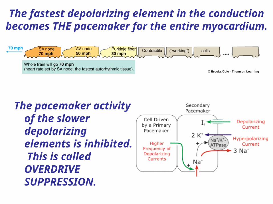
The fastest depolarizing element in the conduction becomes THE pacemaker for the entire myocardium.
The pacemaker activity of the slower depolarizing elements is inhibited. This is called OVERDRIVE SUPPRESSION.
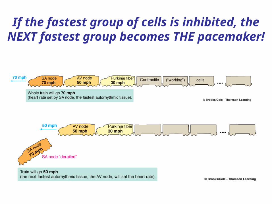
If the fastest group of cells is inhibited, the NEXT fastest group becomes THE pacemaker!
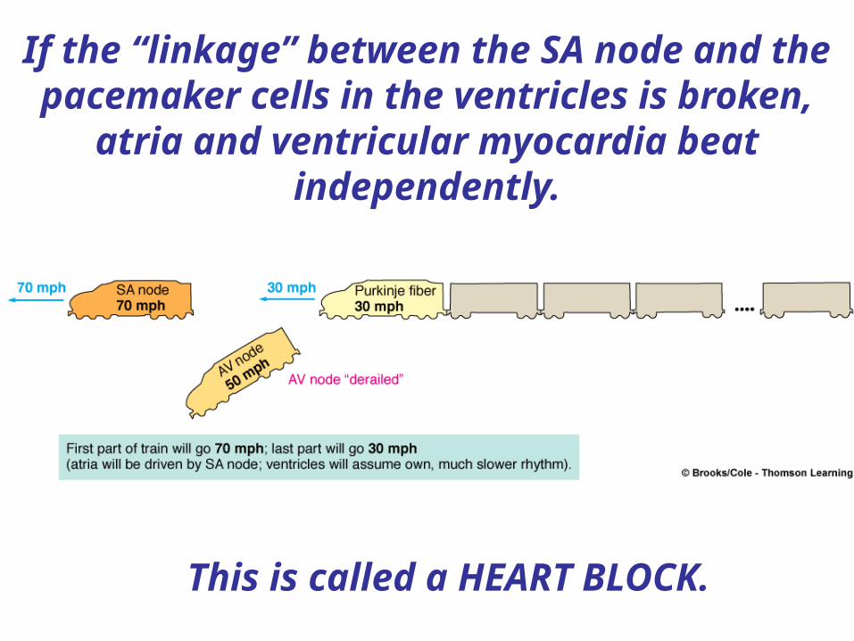
If the “linkage” between the SA node and the pacemaker cells in the ventricles is broken,
atria and ventricular myocardia beat independently.
This is called a HEART BLOCK.
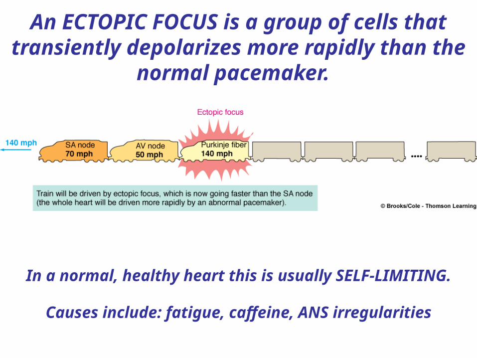
An ECTOPIC FOCUS is a group of cells that transiently depolarizes more rapidly than the
normal pacemaker.
In a normal, healthy heart this is usually SELF-LIMITING.
Causes include: fatigue, caffeine, ANS irregularities

Transmission of the cardiac impulse through the heart, showing the time of appearance (in fractions of a second
after initial appearance at the sinoatrial node) in different parts of the
heart.
© 2005 Elsevier
Although all regions of the myocardium are functionally united
by this system….
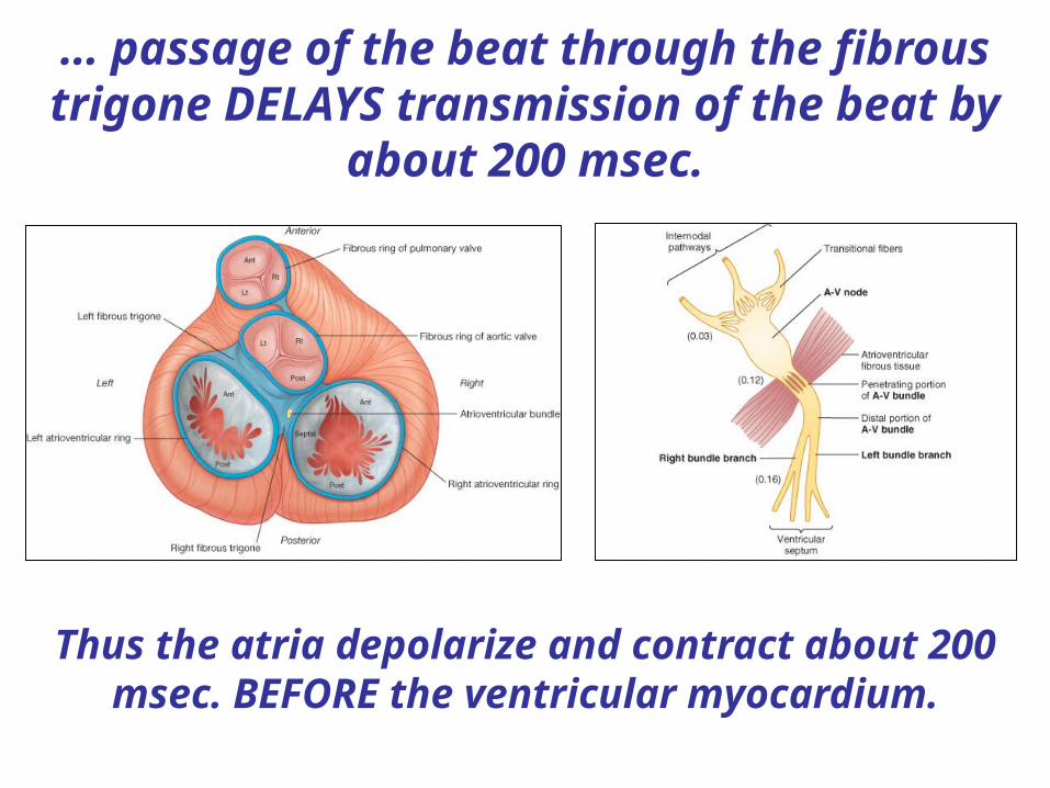
… passage of the beat through the fibrous trigone DELAYS transmission of the beat by
about 200 msec.
Thus the atria depolarize and contract about 200 msec. BEFORE the ventricular myocardium.
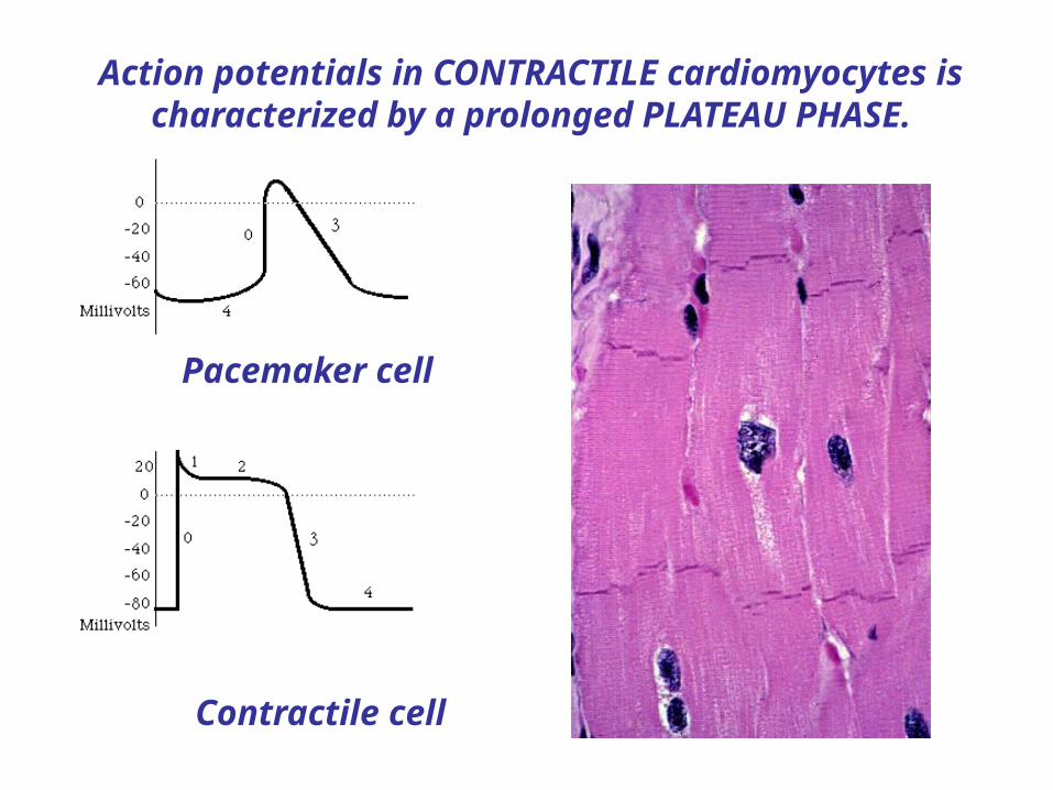
Pacemaker cell
Contractile cell
Action potentials in CONTRACTILE cardiomyocytes is characterized by a prolonged PLATEAU PHASE.
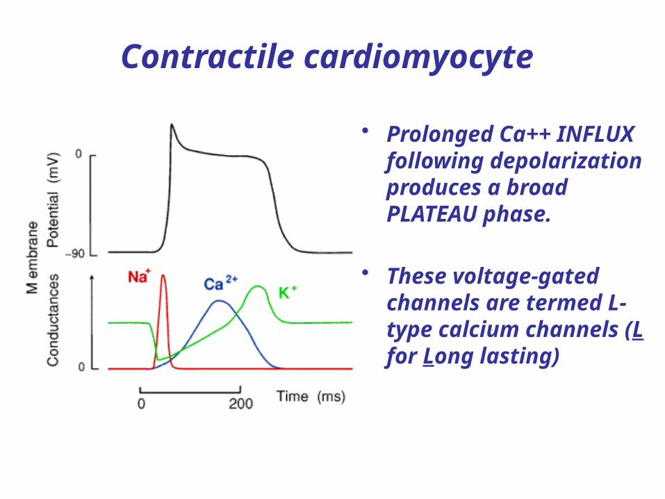
Contractile cardiomyocyte
• Prolonged Ca++ INFLUX following depolarization produces a broad PLATEAU phase.
• These voltage-gated channels are termed L-type calcium channels (L for Long lasting)

Action potentials & contraction in skeletal muscle
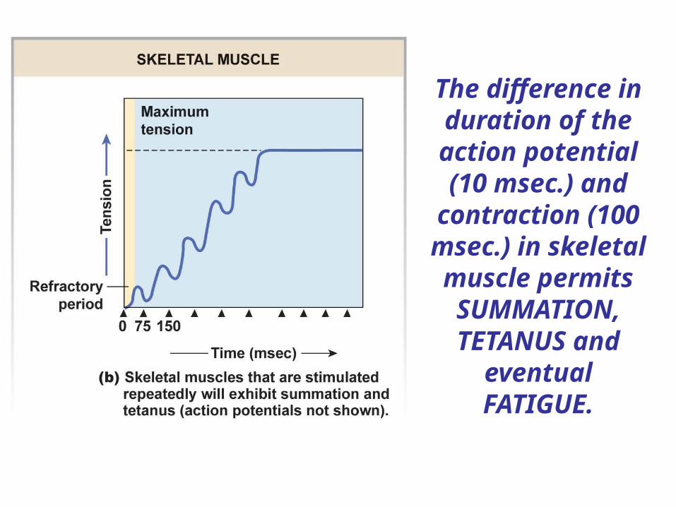
The difference in duration of the action potential (10 msec.) and
contraction (100 msec.) in
skeletal muscle permits
SUMMATION, TETANUS and
eventual FATIGUE.
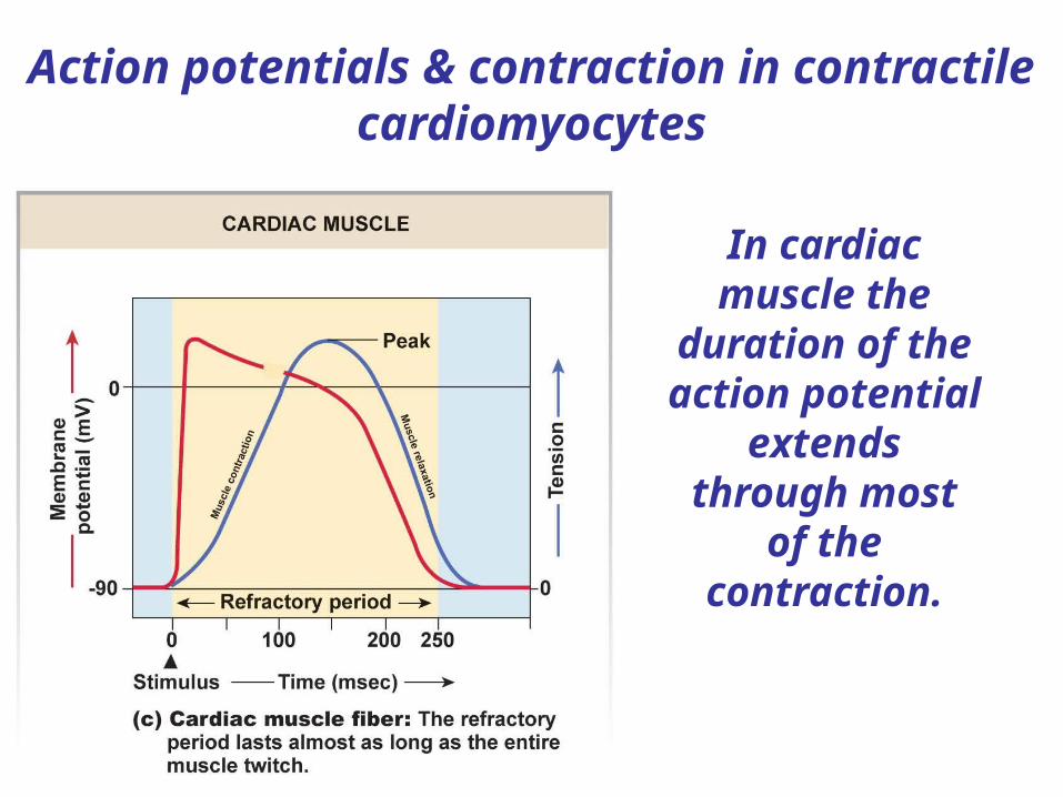
Action potentials & contraction in contractile cardiomyocytes
In cardiac muscle the
duration of the action potential
extends through most
of the contraction.
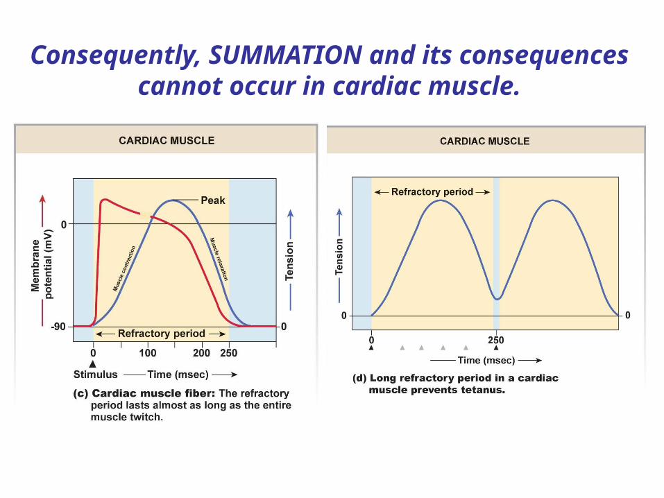
Consequently, SUMMATION and its consequences cannot occur in cardiac muscle.
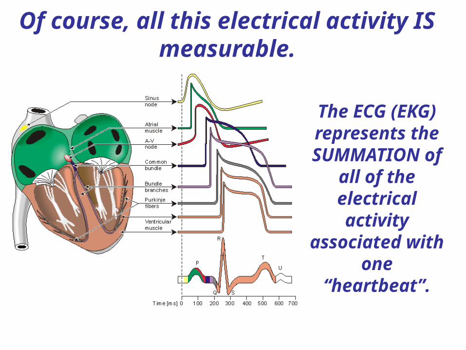
Of course, all this electrical activity IS measurable.
The ECG (EKG) represents the SUMMATION of
all of the electrical activity
associated with one
“heartbeat”.
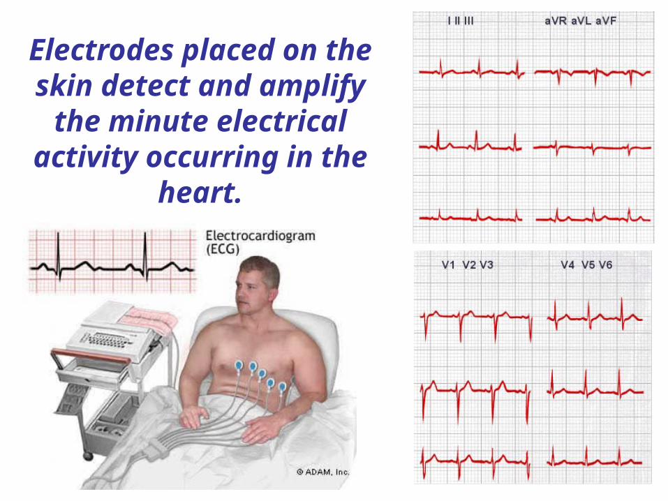
Electrodes placed on the skin detect and amplify
the minute electrical activity occurring in the
heart.
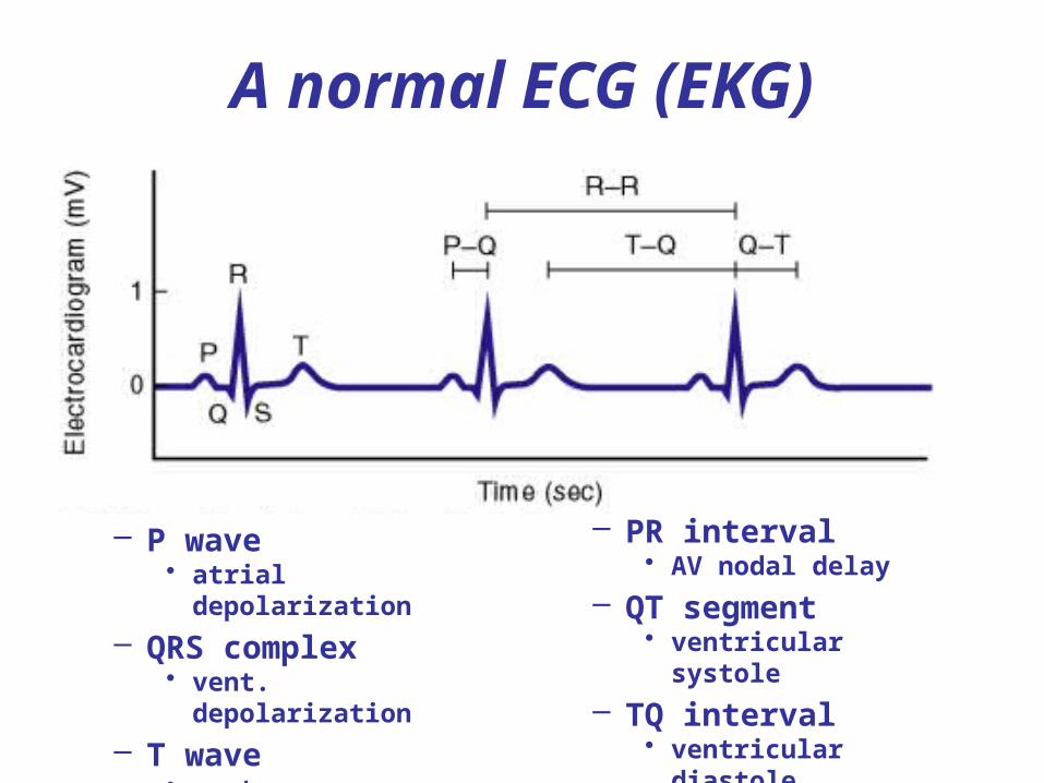
– P wave • atrial depolarization
– QRS complex• vent. depolarization
– T wave • vent. repolarization
– PR interval• AV nodal delay
– QT segment• ventricular systole
– TQ interval• ventricular diastole
A normal ECG (EKG)
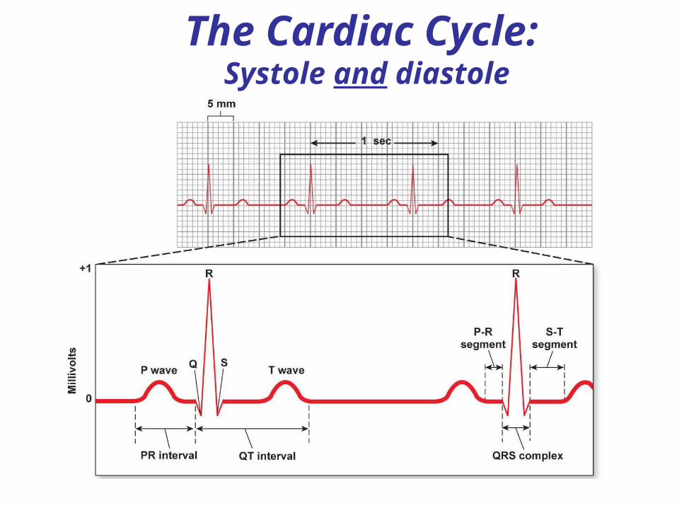
The Cardiac Cycle: Systole and diastole
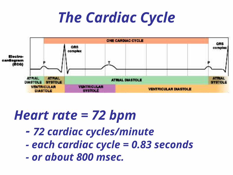
The Cardiac Cycle
Heart rate = 72 bpm- 72 cardiac cycles/minute- each cardiac cycle = 0.83 seconds- or about 800 msec.
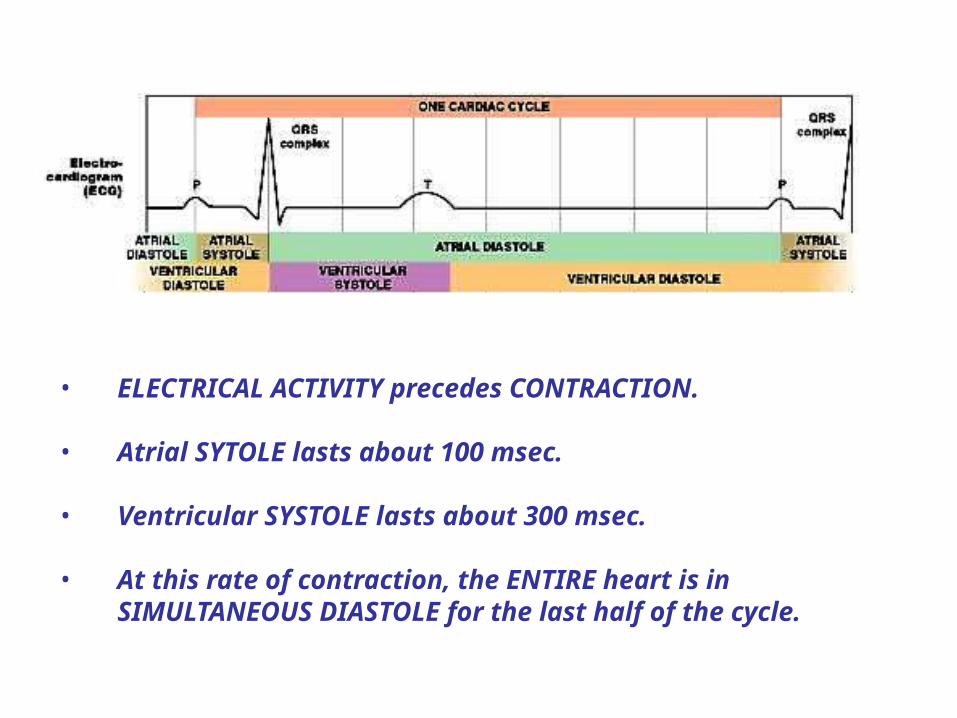
• ELECTRICAL ACTIVITY precedes CONTRACTION.
• Atrial SYTOLE lasts about 100 msec.
• Ventricular SYSTOLE lasts about 300 msec.
• At this rate of contraction, the ENTIRE heart is in SIMULTANEOUS DIASTOLE for the last half of the cycle.

A CARDIAC CYCLE consists of one set of atrial and ventricular systoles and diastoles.
Since both “sides” of the heart and doing the same thing at the same time, we can deal with a “half heart”.
By convention, this is the LEFT side or SYSTEMIC PUMP.
By convention, one assumes a normal heart “at rest”.
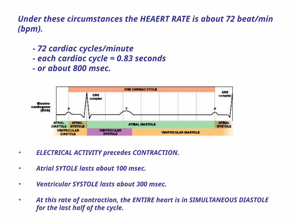
Under these circumstances the HEAERT RATE is about 72 beat/min (bpm).
- 72 cardiac cycles/minute- each cardiac cycle = 0.83 seconds- or about 800 msec.
• ELECTRICAL ACTIVITY precedes CONTRACTION.
• Atrial SYTOLE lasts about 100 msec.
• Ventricular SYSTOLE lasts about 300 msec.
• At this rate of contraction, the ENTIRE heart is in SIMULTANEOUS DIASTOLE for the last half of the cycle.

Wiggers diagram

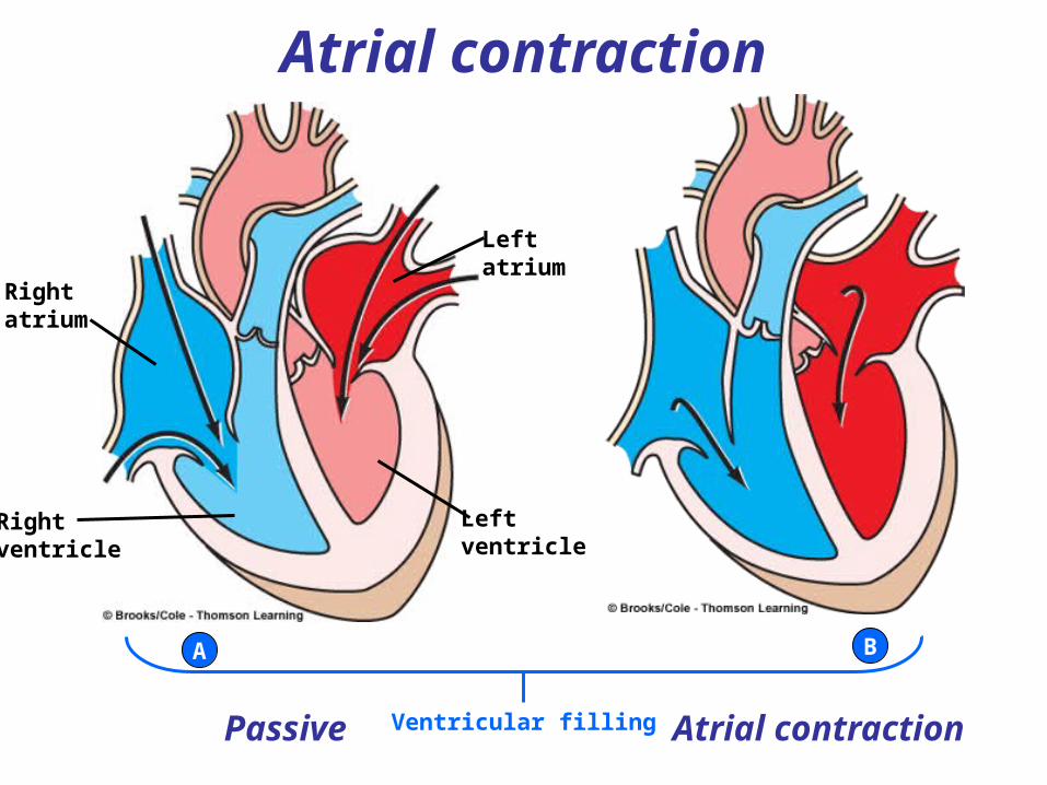
The cardiac cycle begins with VENTRICULAR FILLING.
What is the valvular configuration?

Because the pressure in the atrium is greater than the pressure in the ventricle, the AV valves are OPEN.
Because the pressure in the aorta is greater than the pressure in the
ventricle, the aortic valve is CLOSED.
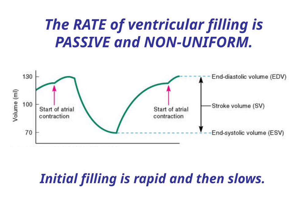
The RATE of ventricular filling is PASSIVE and NON-UNIFORM.
Initial filling is rapid and then slows.

At the end of ATRIAL
DIASTOLE, the SA node fires and the
atria depolarize and CONTRACT.
Rightatrium
Rightventricle
Passive
Leftventricle
Leftatrium
Atrial contractionVentricular filling
A B
Atrial contraction

This adds a small, additional amount of blood to the ventricle (~ 15 mls).
This is the END DIASTOLIC VOLUME (EDV)
EDV = volume of blood in ventricle at end of
(ATRIAL) diastole (~ 130 mls)

About 100 msec. later, the
VENTRICLESdepolarize and
CONTRACT.
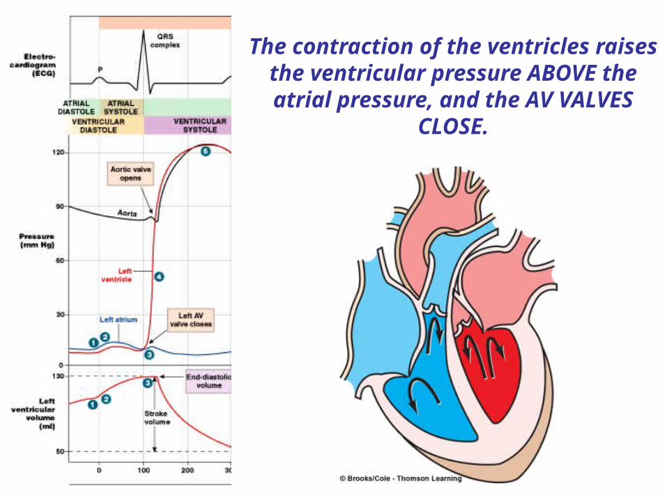
The contraction of the ventricles raises the ventricular pressure ABOVE the atrial pressure, and the AV VALVES
CLOSE.
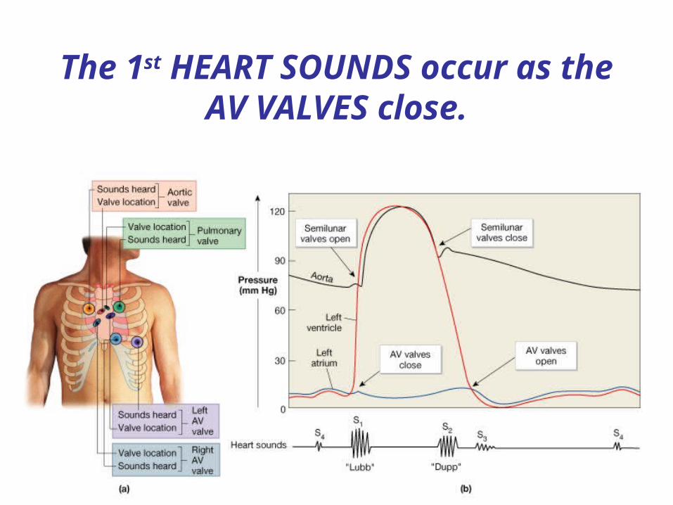
The 1st HEART SOUNDS occur as the AV VALVES close.

However, the pressure in the ventricles is still LOWER than that in the aorta, so the aortic
valves remain closed.
ISOVOLUMETRIC VENTRICULAR CONTRACTION
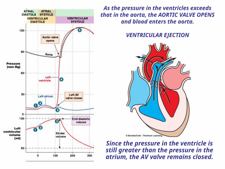
As the pressure in the ventricles exceeds that in the aorta, the AORTIC VALVE OPENS
and blood enters the aorta.
VENTRICULAR EJECTION
Since the pressure in the ventricle is still greater than the pressure in the atrium, the AV valve remains closed.

The volume of blood remaining in the ventricle AFTER ejection i.e. after ventricular systole is called the
END SYSTOLIC VOLUME.
The volume of blood ejected into the aorta is the STROKE VOLUME.
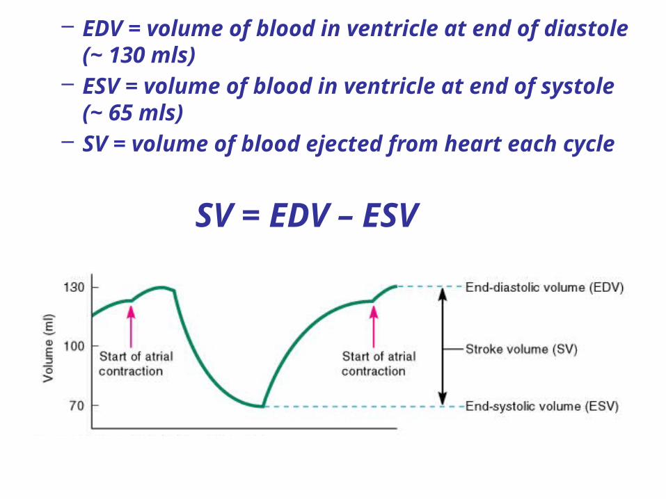
– EDV = volume of blood in ventricle at end of diastole (~ 130 mls)
– ESV = volume of blood in ventricle at end of systole (~ 65 mls)
– SV = volume of blood ejected from heart each cycle
SV = EDV – ESV
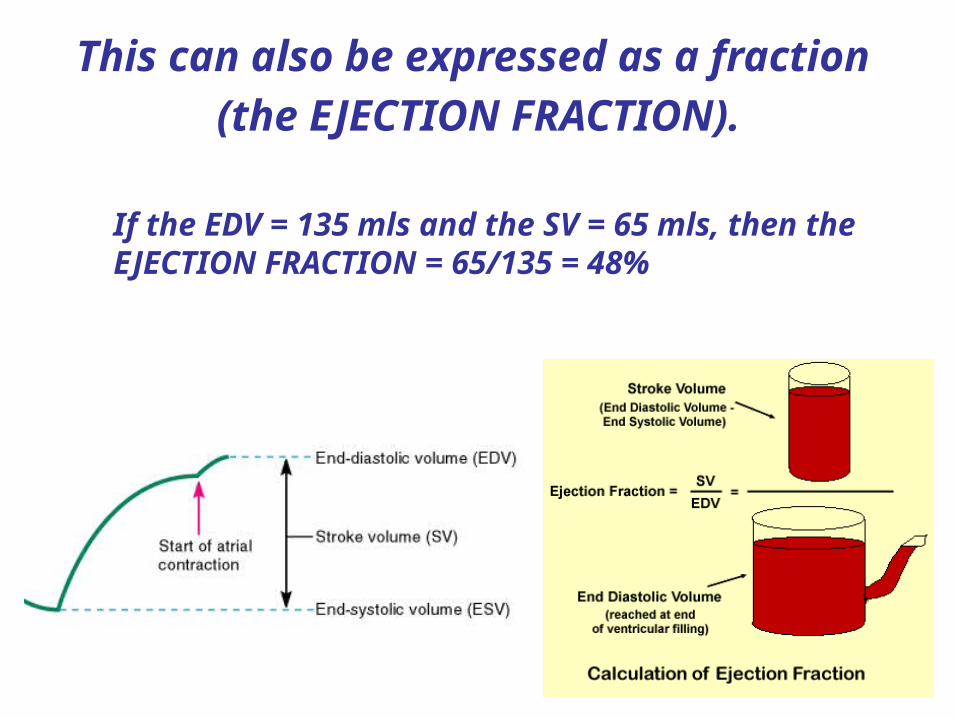
If the EDV = 135 mls and the SV = 65 mls, then the EJECTION FRACTION = 65/135 = 48%
This can also be expressed as a fraction
(the EJECTION FRACTION).

As the ventricles begin to relax into
diastole, the pressure falls BELOW the
pressure in the aorta and the
AORTIC VALVES CLOSE.

The 2nd HEART SOUNDS occur as the SEMILUNAR VALVES close.

The pressure in the ventricles is still greater than the
pressure in the atria, so the AV valves still remain
CLOSED.
The heart is thus in simultaneous diastole with ALL
VALVES CLOSED.
ISOVOLUMETRIC VENTRICULAR RELAXATION

As the pressure in the ventricles rapidly declines
during diastole, it falls BELOW that in the ATRIA and the AV
VALVES OPEN.
Since the pressure in the atria is BELOW that in the aorta, the aortic
valve remains closed.
VENTRICULAR FILLING begins as the CARDIAC CYCLE repeats itself.

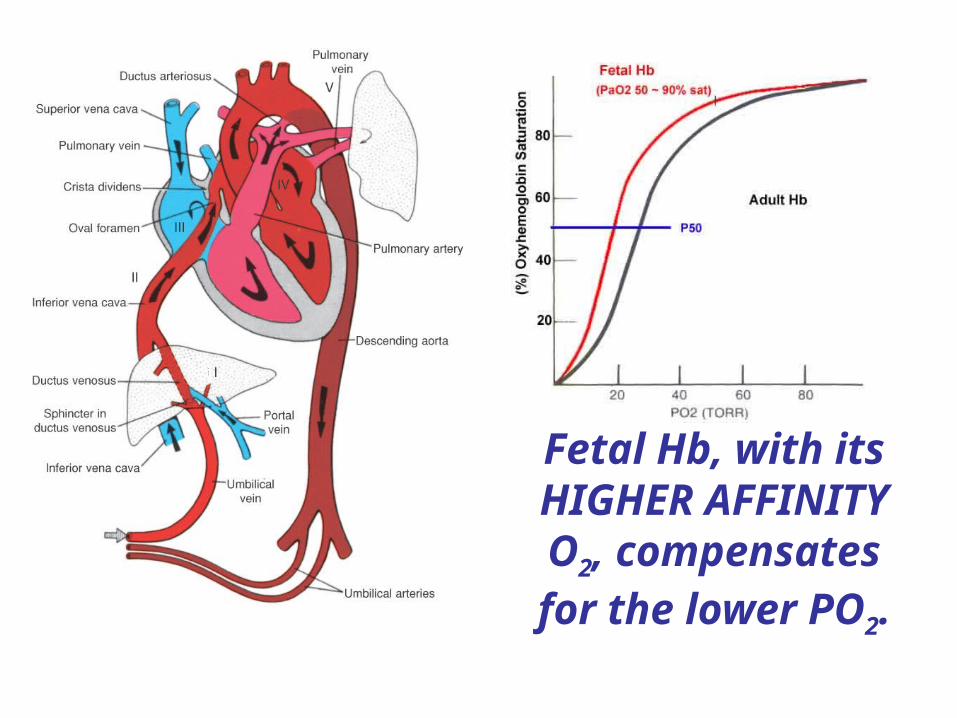
Fetal Hb, with its HIGHER AFFINITY O2, compensates for the lower PO2.

Cardiac Muscle Cells/cardiomyocytes
• Characteristics of Cardiac Muscle Cells
– Small size
– Single, central nucleus
– Branching interconnections between cells
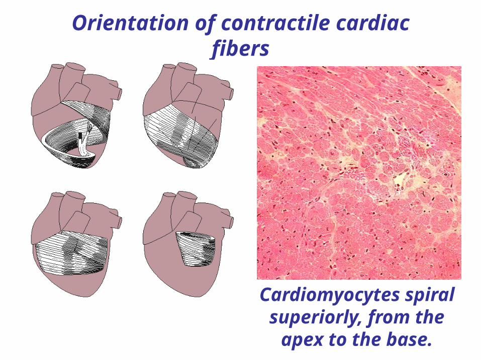
Orientation of contractile cardiac fibers
Cardiomyocytes spiral superiorly, from the
apex to the base.
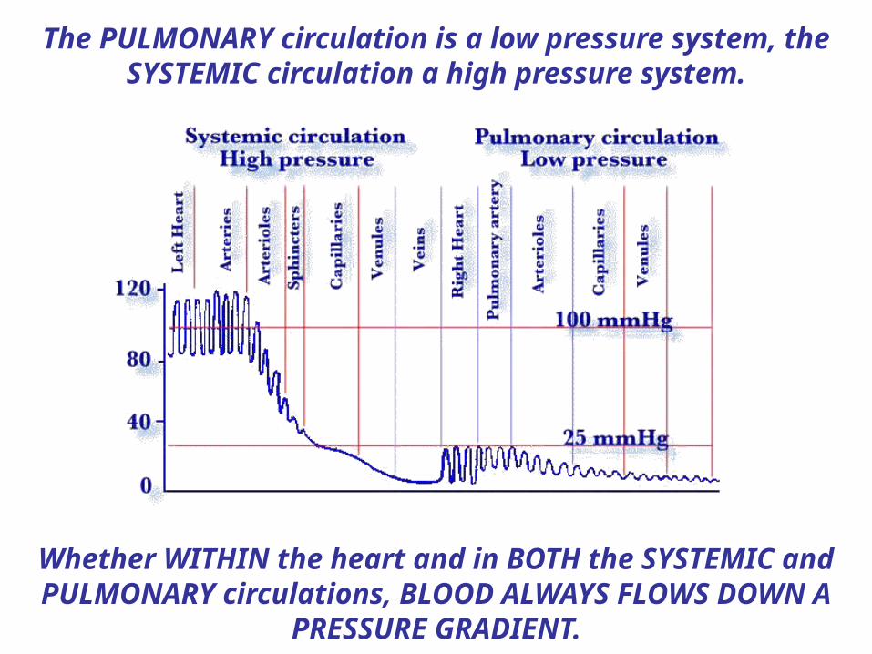
The PULMONARY circulation is a low pressure system, the SYSTEMIC circulation a high pressure system.
Whether WITHIN the heart and in BOTH the SYSTEMIC and PULMONARY circulations, BLOOD ALWAYS FLOWS DOWN A
PRESSURE GRADIENT.
Related Documents