U.S. GOVERNMENT PRINTING OFFICE WASHINGTON : For sale by the Superintendent of Documents, U.S. Government Printing Office Internet: bookstore.gpo.gov Phone: toll free (866) 512–1800; DC area (202) 512–1800 Fax: (202) 512–2250 Mail: Stop SSOP, Washington, DC 20402–0001 23–927 2005 HEARING ON MANAGING THE USE OF IMAGING SERVICES HEARING BEFORE THE SUBCOMMITTEE ON HEALTH OF THE COMMITTEE ON WAYS AND MEANS U.S. HOUSE OF REPRESENTATIVES ONE HUNDRED NINTH CONGRESS FIRST SESSION MARCH 17, 2005 Serial No. 109–29 Printed for the use of the Committee on Ways and Means ( VerDate Aug 31 2005 03:54 Apr 13, 2006 Jkt 023927 PO 00000 Frm 00001 Fmt 5011 Sfmt 5011 E:\HR\OC\23927.XXX 23927 jcorcoran on PROD1PC62 with HEARING

Welcome message from author
This document is posted to help you gain knowledge. Please leave a comment to let me know what you think about it! Share it to your friends and learn new things together.
Transcript
U.S. GOVERNMENT PRINTING OFFICE
WASHINGTON :
For sale by the Superintendent of Documents, U.S. Government Printing OfficeInternet: bookstore.gpo.gov Phone: toll free (866) 512–1800; DC area (202) 512–1800
Fax: (202) 512–2250 Mail: Stop SSOP, Washington, DC 20402–0001
23–927 2005
HEARING ON MANAGING THE USE OF IMAGING SERVICES
HEARING BEFORE THE
SUBCOMMITTEE ON HEALTH OF THE
COMMITTEE ON WAYS AND MEANS
U.S. HOUSE OF REPRESENTATIVES
ONE HUNDRED NINTH CONGRESS
FIRST SESSION
MARCH 17, 2005
Serial No. 109–29
Printed for the use of the Committee on Ways and Means
(
VerDate Aug 31 2005 03:54 Apr 13, 2006 Jkt 023927 PO 00000 Frm 00001 Fmt 5011 Sfmt 5011 E:\HR\OC\23927.XXX 23927jcor
cora
n on
PR
OD
1PC
62 w
ith H
EA
RIN
G
ii
COMMITTEE ON WAYS AND MEANS BILL THOMAS, California, Chairman
E. CLAY SHAW, JR., Florida NANCY L. JOHNSON, Connecticut WALLY HERGER, California JIM MCCRERY, Louisiana DAVE CAMP, Michigan JIM RAMSTAD, Minnesota JIM NUSSLE, Iowa SAM JOHNSON, Texas ROB PORTMAN, Ohio PHIL ENGLISH, Pennsylvania J.D. HAYWORTH, Arizona JERRY WELLER, Illinois KENNY C. HULSHOF, Missouri SCOTT MCINNIS, Colorado RON LEWIS, Kentucky MARK FOLEY, Florida KEVIN BRADY, Texas THOMAS M. REYNOLDS, New York PAUL RYAN, Wisconsin ERIC CANTOR, Virginia JOHN LINDER, Georgia BOB BEAUPREZ, Colorado MELISSA A. HART, Pennsylvania CHRIS CHOCOLA, Indiana
CHARLES B. RANGEL, New York FORTNEY PETE STARK, California SANDER M. LEVIN, Michigan BENJAMIN L. CARDIN, Maryland JIM MCDERMOTT, Washington JOHN LEWIS, Georgia RICHARD E. NEAL, Massachusetts MICHAEL R. MCNULTY, New York WILLIAM J. JEFFERSON, Louisiana JOHN S. TANNER, Tennessee XAVIER BECERRA, California LLOYD DOGGETT, Texas EARL POMEROY, North Dakota STEPHANIE TUBBS JONES, Ohio MIKE THOMPSON, California JOHN B. LARSON, Connecticut RAHM EMANUEL, Illinois
ALLISON H. GILES, Chief of Staff JANICE MAYS, Minority Chief Counsel
SUBCOMMITTEE ON HEALTH NANCY L. JOHNSON, Connecticut, Chairman
JIM MCCRERY, Louisiana SAM JOHNSON, Texas DAVE CAMP, Michigan JIM RAMSTAD, Minnesota PHIL ENGLISH, Pennsylvania J.D. HAYWORTH, Arizona KENNY C. HULSHOF, Missouri
FORTNEY PETE STARK, California JOHN LEWIS, Georgia LLOYD DOGGETT, Texas MIKE THOMPSON, California RAHM EMANUEL, Illinois
Pursuant to clause 2(e)(4) of Rule XI of the Rules of the House, public hearing records of the Committee on Ways and Means are also published in electronic form. The printed hearing record remains the official version. Because electronic submissions are used to prepare both printed and electronic versions of the hearing record, the process of converting between various electronic formats may introduce unintentional errors or omissions. Such occur-rences are inherent in the current publication process and should diminish as the process is further refined.
VerDate Aug 31 2005 03:54 Apr 13, 2006 Jkt 023927 PO 00000 Frm 00002 Fmt 0486 Sfmt 0486 E:\HR\OC\23927.XXX 23927jcor
cora
n on
PR
OD
1PC
62 w
ith H
EA
RIN
G

iii
C O N T E N T S
Page
Advisory of March 10, 2005, announcing the hearing .......................................... 2
WITNESSES
Medicare Payment Advisory Commission, Mark Miller ....................................... 5
American College of Radiology, James Borgstede, M.D. ...................................... 23 University of Chicago, Chicago, Illinois, Kim Allen Williams, M.D. ................... 30 National Coalition for Diagnostic Imaging Services, Houston, Texas, Cherrill
Farnsworth ........................................................................................................... 41 Philips Medical Systems, Milpitas, California, David Rollo, M.D. ...................... 46
SUBMISSIONS FOR THE RECORD
American Association of Orthopaedic Surgeons, Kathryn Pontzer, statement .. 71 American College of Obstetricians and Gynecologists, Vivian M. Dickerson,
M.D., statement .................................................................................................... 74 American Society for Therapeutic Radiology and Oncology, Fairfax, VA, Lisa
Shuger Hublitz, statement .................................................................................. 75 Buchholz, Marilyn K., Omaha, NE, letter ............................................................. 76 Cardiovascular Computed Tomography, Stephan Achenbach, Damascus, MD,
statement .............................................................................................................. 77 Coalition for Quality Ultrasound, Columbia, MD, letter ...................................... 81 National Imaging Associates, Inc., Thomas G. Dehn, Hackensack, NJ, letter ... 84 Petrikin, Timothy M., Nashville, TN, letter .......................................................... 86 Society for Maternal-Fetal Medicine, statement ................................................... 87 Wagner, Judith A., Mequon, WI, letter ................................................................. 89
VerDate Aug 31 2005 03:54 Apr 13, 2006 Jkt 023927 PO 00000 Frm 00003 Fmt 0486 Sfmt 0486 E:\HR\OC\23927.XXX 23927jcor
cora
n on
PR
OD
1PC
62 w
ith H
EA
RIN
G

VerDate Aug 31 2005 03:54 Apr 13, 2006 Jkt 023927 PO 00000 Frm 00004 Fmt 0486 Sfmt 0486 E:\HR\OC\23927.XXX 23927jcor
cora
n on
PR
OD
1PC
62 w
ith H
EA
RIN
G
(1)
HEARING ON MANAGING THE USE OF IMAGING SERVICES
THURSDAY, MARCH 17, 2005
U.S. HOUSE OF REPRESENTATIVES, COMMITTEE ON WAYS AND MEANS,
Washington, DC. The Subcommittee met, pursuant to notice, at 10:11 a.m., in
room 1100, Longworth House Office Building, Hon. Nancy L. John-son (Chairman of the Subcommittee) presiding.
[The advisory announcing the hearing follows:]
VerDate Aug 31 2005 03:54 Apr 13, 2006 Jkt 023927 PO 00000 Frm 00005 Fmt 6633 Sfmt 6633 E:\HR\OC\23927.XXX 23927jcor
cora
n on
PR
OD
1PC
62 w
ith H
EA
RIN
G
2
ADVISORY FROM THE COMMITTEE ON WAYS AND MEANS
SUBCOMMITTEE ON HEALTH
CONTACT: (202) 225–3943 FOR IMMEDIATE RELEASE March 10, 2005 HL–4
Johnson Announces Hearing on Managing the Use of Imaging Services
Congresswoman Nancy L. Johnson (R–CT), Chairman, Subcommittee on Health of the Committee on Ways and Means, today announced that the Subcommittee will hold a hearing on managing the use of imaging services. The hearing will take place on Thursday, March 17, 2005, in the main Committee hearing room, 1100 Longworth House Office Building, beginning at 10:00 a.m.
In view of the limited time available to hear witnesses, oral testimony at this hearing will be from invited witnesses only. Witnesses will include a representative from the Medicare Payment Advisory Commission (MedPAC) and representatives from physician groups that perform medical imaging. However, any individual or or-ganization not scheduled for an oral appearance may submit a written statement for consideration by the Committee and for inclusion in the printed record of the hearing.
BACKGROUND:
According to MedPAC, between 1999 and 2002, per-beneficiary growth in the vol-ume and complexity of imaging services was twice as high as the growth for all services paid under the Medicare physician fee schedule. MedPAC attributes the in-crease in imaging services to technological innovations that have improved diag-nostic services and allowed for service delivery in physicians’ offices. Other factors contributing to the growth of imaging services include a possible misalignment of Medicare’s payment rates and costs of services, physician interest in capturing reve-nues from the provision of ancillary services, and patient desire to receive diagnostic tests in convenient settings.
As the delivery of imaging services has migrated from hospitals to physician of-fices, these services are subject to less stringent oversight. MedPAC recommended that Medicare develop quality standards for all providers that receive payment for performing and interpreting imaging studies. It believes that these standards will improve the accuracy of diagnostic tests, which will increase quality of care and help control spending. In addition, MedPAC recommended that Secretary Leavitt, of the U.S. Department of Health and Human Services, expand coding edits on billing for imaging services, measure and compare physician use of imaging services, and strengthen rules that restrict physician investment in imaging centers to which they refer.
In announcing the hearing, Chairman Johnson stated, ‘‘Given the significant growth in imaging services, we need to carefully examine the existing quality and safety of these services provided in physicians’ offices before requiring providers to meet new quality standards. Is there a problem, and if so, how widespread is it? What types of services are involved? I want to ensure that seniors have access to appropriate, safe, and high quality imaging services.’’
FOCUS OF THE HEARING:
The hearing will focus on MedPAC’s recommendations for managing the use of imaging services, especially the need to require physicians to meet quality stand-ards as a condition of payment. Witnesses will present evidence supporting or op-posing MedPAC’s recommendations.
VerDate Aug 31 2005 03:54 Apr 13, 2006 Jkt 023927 PO 00000 Frm 00006 Fmt 6633 Sfmt 6621 E:\HR\OC\23927.XXX 23927jcor
cora
n on
PR
OD
1PC
62 w
ith H
EA
RIN
G
3
DETAILS FOR SUBMISSION OF WRITTEN COMMENTS:
Please Note: Any person(s) and/or organization(s) wishing to submit for the hear-ing record must follow the appropriate link on the hearing page of the Committee website and complete the informational forms. From the Committee homepage, http://waysandmeans.house.gov, select ‘‘109th Congress’’ from the menu entitled, ‘‘Hearing Archives’’ (http://waysandmeans.house.gov/Hearings.asp?congress=17). Se-lect the hearing for which you would like to submit, and click on the link entitled, ‘‘Click here to provide a submission for the record.’’ Once you have followed the on-line instructions, completing all informational forms and clicking ‘‘submit’’ on the final page, an email will be sent to the address which you supply confirming your interest in providing a submission for the record. You MUST REPLY to the email and ATTACH your submission as a Word or WordPerfect document, in compliance with the formatting requirements listed below, by close of business Thursday, March 31, 2005. Finally, please note that due to the change in House mail policy, the U.S. Capitol Police will refuse sealed-package deliveries to all House Office Buildings. For questions, or if you encounter technical problems, please call (202) 225–1721.
FORMATTING REQUIREMENTS:
The Committee relies on electronic submissions for printing the official hearing record. As al-ways, submissions will be included in the record according to the discretion of the Committee. The Committee will not alter the content of your submission, but we reserve the right to format it according to our guidelines. Any submission provided to the Committee by a witness, any sup-plementary materials submitted for the printed record, and any written comments in response to a request for written comments must conform to the guidelines listed below. Any submission or supplementary item not in compliance with these guidelines will not be printed, but will be maintained in the Committee files for review and use by the Committee.
1. All submissions and supplementary materials must be provided in Word or WordPerfect format and MUST NOT exceed a total of 10 pages, including attachments. Witnesses and sub-mitters are advised that the Committee relies on electronic submissions for printing the official hearing record.
2. Copies of whole documents submitted as exhibit material will not be accepted for printing. Instead, exhibit material should be referenced and quoted or paraphrased. All exhibit material not meeting these specifications will be maintained in the Committee files for review and use by the Committee.
3. All submissions must include a list of all clients, persons, and/or organizations on whose behalf the witness appears. A supplemental sheet must accompany each submission listing the name, company, address, telephone and fax numbers of each witness.
Note: All Committee advisories and news releases are available on the World Wide Web at http://waysandmeans.house.gov.
The Committee seeks to make its facilities accessible to persons with disabilities. If you are in need of special accommodations, please call 202–225–1721 or 202–226– 3411 TTD/TTY in advance of the event (four business days notice is requested). Questions with regard to special accommodation needs in general (including avail-ability of Committee materials in alternative formats) may be directed to the Com-mittee as noted above.
f
Chairman JOHNSON. Good morning, everyone. Today the Subcommittee focuses our attention on diagnostic im-
aging services in Medicare. These are services such as X-rays, PET scans, MRIs, which provide the opportunity for better quality care, often at reduced costs. Fortunately, the day is gone when physi-cians routinely used exploratory surgery to make a diagnosis. Today, instead of an open biopsy of the breast, women may receive a needle biopsy guided by ultrasound to evaluate lesions. Instead of placing a catheter into the bladder, many urologists use a small
VerDate Aug 31 2005 03:54 Apr 13, 2006 Jkt 023927 PO 00000 Frm 00007 Fmt 6633 Sfmt 6602 E:\HR\OC\23927.XXX 23927jcor
cora
n on
PR
OD
1PC
62 w
ith H
EA
RIN
G
4
ultrasound machine to measure the extent of problems associated with prostate enlargement.
As a result of these advances and other factors, the Medicare Payment Advisory Commission (MedPAC) reports that spending on imaging services has skyrocketed. Between 1999 and 2002, imaging services per beneficiary grew twice as fast as all physician services. More recent data from 2003 shows that growth in imaging services has moderated but continues to exceed overall growth.
MedPAC offers several reasons for this, including technological advances that allow physicians to use imaging for diagnosis more often and allow them to provide imaging services in their offices. Second, patients want to receive diagnostic tests in more conven-ient settings by physician’s offices. Third, Medicare may not be paying appropriately for these services. Finally, physicians may be increasing in office imaging services to increase their Medicare re-imbursements.
MedPAC concludes that the growth in imaging services is dis-proportionate and problematic and makes several recommendations to Congress to restrain this growth. Specifically, MedPAC rec-ommends that Medicare develop quality standards for all providers who receive payment for performing and interpreting imaging stud-ies; expand coding edits on billing for imaging services; measure and compare physician use of imaging services; and strengthen rules that restrict physician investment in imaging center to which they refer.
The medical community is not united behind these recommenda-tions. Some agree with all of MedPAC’s recommendations; others would limit their application to certain types of imaging services; and still others would make no changes to current Medicare prac-tices. Our witnesses will help us evaluate the existing quality and safety of imaging services provided in physicians’ offices and the extent of overuse of services. We want seniors to have access to ap-propriate, safe, and high-quality imaging services. So, we need to understand if there are problems across all imaging services or if problems are limited to certain types of services. Are these prob-lems widespread or, in fact, are they nonexistent?
Our witnesses will help us evaluate these questions. We will hear first from Mark Miller, the Executive Director of MedPAC. He will provide us with more details about the MedPAC recommenda-tions and the evidence supporting these recommendations.
Our second panel includes witnesses from the imaging commu-nity. Dr. Borgstede represents the American College of Radiology. Radiologists provide the bulk of imaging services. Dr. Williams rep-resents the American College of Cardiology and the Coalition for Patient-Centered Imaging. Dr. Williams will provide us with a view from the nonradiologist community. Ms. Farnsworth represents the National Coalition for Quality Diagnostic Imaging Services, a coali-tion of outpatient imaging centers which advocates for public and private sector standards for quality and safety in imaging. Our final witness, Dr. Rollo, who represents the National Electrical Manufacturers Association, which develops standards for medical imaging equipment, will conclude the second panel.
We look forward to your testimony and to an opportunity to dia-log with the witnesses.
VerDate Aug 31 2005 03:54 Apr 13, 2006 Jkt 023927 PO 00000 Frm 00008 Fmt 6633 Sfmt 6602 E:\HR\OC\23927.XXX 23927jcor
cora
n on
PR
OD
1PC
62 w
ith H
EA
RIN
G
5
Let me recognize now Mr. Stark. Mr. STARK. Thank you, Madam Chair, for holding what prom-
ises to be a fascinating hearing, and I am so, curious to hear all about it that, as I said, I was going to skip over how we could save billions of dollars by getting to the important things like cutting out overpaying managed care plans. I will not talk about that today.
We are treading some exciting new ground here, and I suspect that we are going to hear from a series of specialists, and I guess somebody—Russell Long used to say, ‘‘Don’t tax you, don’t tax me, tax the fellow behind the tree.’’ I am surprised that the chiroprac-tors are not here protecting their rights to take X-rays, and we are going to have to sort out a lot of interests and, I suppose, decide at some point who is going to be in charge of setting standards and enforcing them, something that Medicare has basically never done. It has been left to the States. If a doctor is licensed, they pretty much can do whatever they want.
So, I am interested and I am sure we will have a lot of time to discuss this, and I would be interested in Dr. Miller’s recommenda-tions. Thank you very much for calling us together.
Chairman JOHNSON. Thank you very much. Mr. Miller, please proceed.
STATEMENT OF MARK E. MILLER, PH.D., EXECUTIVE DIRECTOR, MEDICARE PAYMENT ADVISORY COMMISSION
Mr. MILLER. Chairman Johnson, Congressman Stark, and dis-tinguished Members of the Subcommittee, I am Mark Miller, Exec-utive Director of the Medicare Payment Advisory Commission, and I appreciate being asked here to talk about the Commission’s rec-ommendations on diagnostic imaging.
Improvements in imaging technology have lowered the cost of purchasing these machines for physicians and reduced the size of these technologies. This has allowed this technology to diffuse from institutional settings to office settings.
Imaging technology has an important role in medicine. It im-proves diagnosis, it leads to better treatments, and it certainly im-proves convenience for the patients. But several issues arise in this context.
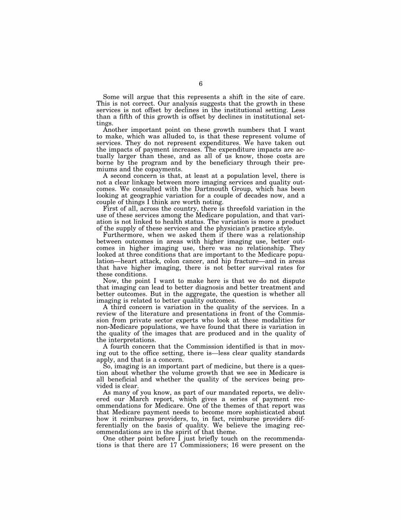
The first is that there has been substantial growth—and this first slide is an illustration of this—substantial growth in the vol-ume of imaging services per beneficiary. What this figure shows is the horizontal line is the average increase in volume of services for all physician services under the fee schedule, and you can see from the far right bar that imaging services are growing at twice the rate of all physician services. The time period is 1999 to 2003. The growth rate for physician is 22 percent for all services, and for im-aging it is 45 percent.
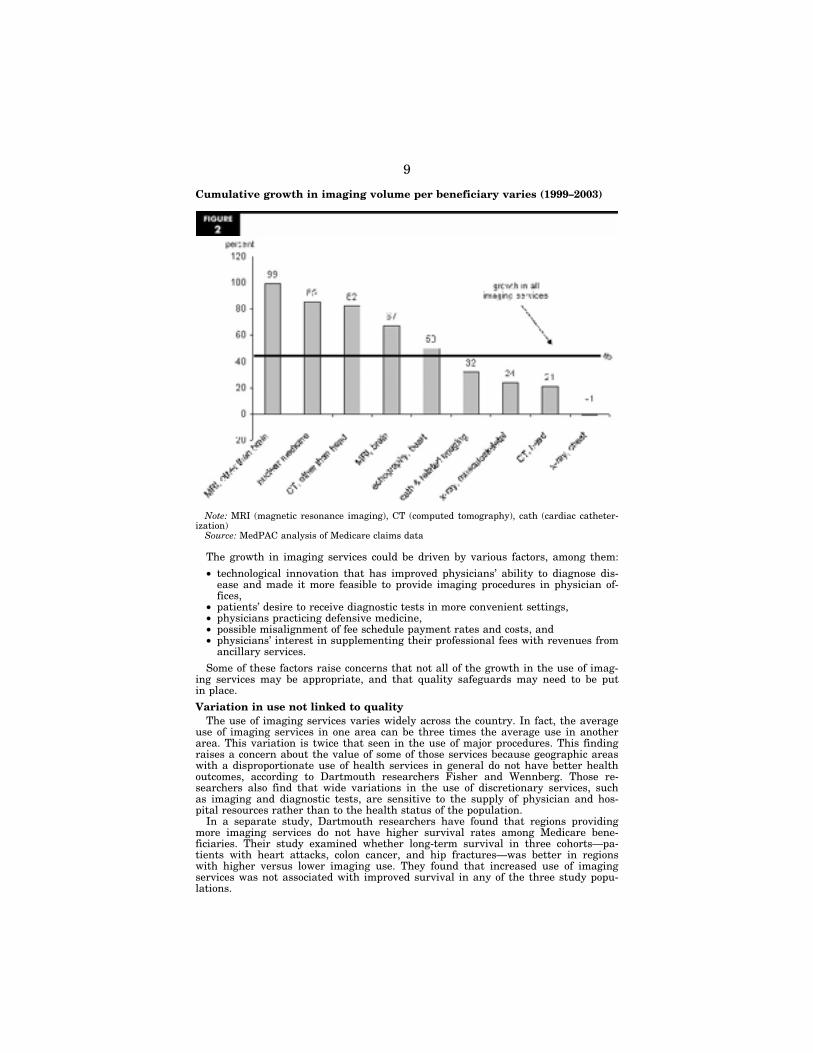
This chart also shows that certain imaging modalities are grow-ing even more aggressively. Here the horizontal line is the average growth for all imaging, 45 percent over the same time period, and certain modalities, like MRI, excluding the brain, nuclear medicine, and CT scans, excluding the head, are growing between 82 percent and 99 percent.
VerDate Aug 31 2005 03:54 Apr 13, 2006 Jkt 023927 PO 00000 Frm 00009 Fmt 6633 Sfmt 6602 E:\HR\OC\23927.XXX 23927jcor
cora
n on
PR
OD
1PC
62 w
ith H
EA
RIN
G
6
Some will argue that this represents a shift in the site of care. This is not correct. Our analysis suggests that the growth in these services is not offset by declines in the institutional setting. Less than a fifth of this growth is offset by declines in institutional set-tings.
Another important point on these growth numbers that I want to make, which was alluded to, is that these represent volume of services. They do not represent expenditures. We have taken out the impacts of payment increases. The expenditure impacts are ac-tually larger than these, and as all of us know, those costs are borne by the program and by the beneficiary through their pre-miums and the copayments.
A second concern is that, at least at a population level, there is not a clear linkage between more imaging services and quality out-comes. We consulted with the Dartmouth Group, which has been looking at geographic variation for a couple of decades now, and a couple of things I think are worth noting.
First of all, across the country, there is threefold variation in the use of these services among the Medicare population, and that vari-ation is not linked to health status. The variation is more a product of the supply of these services and the physician’s practice style.
Furthermore, when we asked them if there was a relationship between outcomes in areas with higher imaging use, better out-comes in higher imaging use, there was no relationship. They looked at three conditions that are important to the Medicare popu-lation—heart attack, colon cancer, and hip fracture—and in areas that have higher imaging, there is not better survival rates for these conditions.
Now, the point I want to make here is that we do not dispute that imaging can lead to better diagnosis and better treatment and better outcomes. But in the aggregate, the question is whether all imaging is related to better quality outcomes.
A third concern is variation in the quality of the services. In a review of the literature and presentations in front of the Commis-sion from private sector experts who look at these modalities for non-Medicare populations, we have found that there is variation in the quality of the images that are produced and in the quality of the interpretations.
A fourth concern that the Commission identified is that in mov-ing out to the office setting, there is—less clear quality standards apply, and that is a concern.
So, imaging is an important part of medicine, but there is a ques-tion about whether the volume growth that we see in Medicare is all beneficial and whether the quality of the services being pro-vided is clear.
As many of you know, as part of our mandated reports, we deliv-ered our March report, which gives a series of payment rec-ommendations for Medicare. One of the themes of that report was that Medicare payment needs to become more sophisticated about how it reimburses providers, to, in fact, reimburse providers dif-ferentially on the basis of quality. We believe the imaging rec-ommendations are in the spirit of that theme.
One other point before I just briefly touch on the recommenda-tions is that there are 17 Commissioners; 16 were present on the
VerDate Aug 31 2005 03:54 Apr 13, 2006 Jkt 023927 PO 00000 Frm 00010 Fmt 6633 Sfmt 6602 E:\HR\OC\23927.XXX 23927jcor
cora
n on
PR
OD
1PC
62 w
ith H
EA
RIN
G
7
day that we considered these recommendations, and the votes on these were unanimous.
So, very quickly, with the remaining time I will just make these points.
The first recommendation is that Congress direct the Secretary to set quality standards for all providers who bill Medicare for per-forming and interpreting diagnostic imaging services.
Two points I would like to make about this. Some people charac-terize this recommendation as directed toward limiting imaging to radiologists only, billing for imaging to radiologists only. That is not correct. We believe that the standards should apply to all phy-sicians, and if physicians meet those standards, they should be able to bill.
A second thing, that while this is new ground, it is not without precedent. The Mammography Quality Standards Act passed in 1992 makes these types of recommendations for mammography services.
Just to finish up, the Secretary should measure physician use of imaging services So, that physicians can confidentially compare their practice patterns with those of their peers; that the Secretary improve Medicare’s coding edits for imaging; and, finally, that the Secretary strengthen rules that govern physician investment in im-aging centers.
I appreciate being asked here and look forward to your questions. [The prepared statement of Mr. Miller follows:]
Statement of Mark Miller, Executive Director, Medicare Payment Advisory Commission
Chairman Johnson, Congressman Stark, distinguished Subcommittee members. I am Mark Miller, Executive Director of the Medicare Payment Advisory Commission (MedPAC). I appreciate the opportunity to be here with you this morning to discuss ways to improve imaging services for Medicare beneficiaries.
The Commission has concluded that it is time for the Medicare program to start to differentiate among providers when making payments. Currently, Medicare pays providers the same regardless of their quality. In its March report to the Congress MedPAC discusses several important steps towards differentiation which, taken to-gether, will improve the quality of care for beneficiaries and lay the groundwork for obtaining better value in the Medicare program. For example, MedPAC recommends pay for performance linked to quality. As requested, this testimony focuses on the Commission’s recommendations for imaging services contained in the March report.
Technological progress in imaging over the past years, and its promise for improv-ing diagnosis, treatments, and health outcomes are impressive. In addition, im-provements in technology have made those services available outside the hospital in settings such as imaging centers and doctors’ offices—with concomitant improve-ments in convenience for patients. However, at the same time there has been rapid and sustained growth in the volume of imaging services for Medicare beneficiaries; and there are concerns about potential overuse of imaging services, possible poor quality, and that Medicare payment policy has not kept up with technological changes. As an example of the rapid growth in imaging, according to the Wall Street Journal, there are now more magnetic resonance imaging (MRI) scanners in the Pittsburgh area than in all of Canada and, in 2003, there were over 13 computed tomography (CT) scans provided for every 100 members of the largest health plan in the area.
The Commission has investigated these issues through data analysis, consulta-tions with private sector experts in management of imaging services, discussions with specialty medical societies, and a review of the available literature. After public discussion and deliberation the Commission, by a unanimous vote among those present, has recommended that:
• the Secretary of HHS improve Medicare’s coding edits for imaging studies,
VerDate Aug 31 2005 03:54 Apr 13, 2006 Jkt 023927 PO 00000 Frm 00011 Fmt 6633 Sfmt 6621 E:\HR\OC\23927.XXX 23927jcor
cora
n on
PR
OD
1PC
62 w
ith H
EA
RIN
G
8
• the Congress direct the Secretary to set standards for all providers who bill Medicare for performing and interpreting diagnostic imaging studies,
• the Secretary measure physicians’ use of imaging services so that physicians can compare their practice patterns with those of their peers, and
• the Secretary strengthen the rules that govern physician investment in imaging centers to which they refer patients.
Taken together, these actions should help add value to the imaging services Medi-care buys.
Growth has been dramatic Diagnostic imaging services paid under Medicare’s physician fee schedule grew
more rapidly than any other type of physician service between 1999 and 2003. While the sum of all physician services grew 22 percent in those years, imaging services grew twice as fast, by 45 percent (see figure 1). This measure is the growth in the volume and intensity of services per beneficiary; we have removed changes resulting from increases in the number of beneficiaries and changes in prices during those years. Not all imaging services grew at this rate; some grew even faster. Advanced imaging services and nuclear medicine led the way: MRI of parts of the body other than the brain grew by 99 percent; nuclear medicine grew 85 percent; and CT of parts of the body other than the head grew 82 percent (see figure 2).
In dollar terms, Medicare spending for imaging services paid under the physician fee schedule grew over 60 percent, from $5.7 billion in 1999 to $9.3 billion in 2003. Beneficiaries’ spending on these services has also increased, both directly through copayments and indirectly through increased Part B premiums.
Imaging shows highest cumulative growth in services per beneficiary (1999–2003)
Some argue that much of this increase was attributable to the movement of imag-ing from the outpatient setting to settings where the technical charge is included in the physician fee schedule. However, of the $1.6 billion increase in fee schedule imaging spending from 2001 to 2003, only $300 million was offset by the decrease in imaging provided in hospital outpatient departments. In addition, the movement of imaging from outpatient departments to physician offices raises another concern: the institutional standards that govern the performance and interpretation of stud-ies in hospitals are usually absent in physician offices.
VerDate Aug 31 2005 03:54 Apr 13, 2006 Jkt 023927 PO 00000 Frm 00012 Fmt 6633 Sfmt 6621 E:\HR\OC\23927.XXX 23927 Inse
rt 2
3927
A.0
01In
sert
239
27A
.002
jcor
cora
n on
PR
OD
1PC
62 w
ith H
EA
RIN
G
9
Cumulative growth in imaging volume per beneficiary varies (1999–2003)
Note: MRI (magnetic resonance imaging), CT (computed tomography), cath (cardiac catheter-ization)
Source: MedPAC analysis of Medicare claims data
The growth in imaging services could be driven by various factors, among them:
• technological innovation that has improved physicians’ ability to diagnose dis-ease and made it more feasible to provide imaging procedures in physician of-fices,
• patients’ desire to receive diagnostic tests in more convenient settings, • physicians practicing defensive medicine, • possible misalignment of fee schedule payment rates and costs, and • physicians’ interest in supplementing their professional fees with revenues from
ancillary services.
Some of these factors raise concerns that not all of the growth in the use of imag-ing services may be appropriate, and that quality safeguards may need to be put in place.
Variation in use not linked to quality The use of imaging services varies widely across the country. In fact, the average
use of imaging services in one area can be three times the average use in another area. This variation is twice that seen in the use of major procedures. This finding raises a concern about the value of some of those services because geographic areas with a disproportionate use of health services in general do not have better health outcomes, according to Dartmouth researchers Fisher and Wennberg. Those re-searchers also find that wide variations in the use of discretionary services, such as imaging and diagnostic tests, are sensitive to the supply of physician and hos-pital resources rather than to the health status of the population.
In a separate study, Dartmouth researchers have found that regions providing more imaging services do not have higher survival rates among Medicare bene-ficiaries. Their study examined whether long-term survival in three cohorts—pa-tients with heart attacks, colon cancer, and hip fractures—was better in regions with higher versus lower imaging use. They found that increased use of imaging services was not associated with improved survival in any of the three study popu-lations.
VerDate Aug 31 2005 03:54 Apr 13, 2006 Jkt 023927 PO 00000 Frm 00013 Fmt 6633 Sfmt 6621 E:\HR\OC\23927.XXX 23927 Inse
rt 2
3927
A.0
03In
sert
239
27A
.004
jcor
cora
n on
PR
OD
1PC
62 w
ith H
EA
RIN
G
10
Quality varies According to published studies, health plans, and experts we consulted, providers
vary in their ability to perform quality imaging procedures. In one study, published in Radiology, BlueCross BlueShield of Massachusetts inspected 1,000 imaging pro-viders to evaluate the quality of their equipment, technical staff, and other features. Nearly one-third of the providers had at least one serious deficiency, such as film processing problems, failure to monitor radiation exposure, poor image quality, or lack of an equipment calibration report. Eleven percent of the providers had severe problems that could not be easily remedied, while 20 percent had deficiencies that could be remedied. Chiropractic and podiatric offices were the most likely to have deficiencies; cardiology, radiology, and surgical specialty offices were the least likely. According to a study in the American Journal of Roentgenology, another health plan that inspected almost 100 nonradiologist offices that provided radiography services identified serious problems in 78 percent of the offices. These problems included lack of proper image identification (e.g., noting left or right) and use of equipment that had not been inspected during the previous year.
In our March 2004 public meeting a panel of health plans and imaging benefit managers informed us that some providers fail to meet standards because their im-aging equipment is old or not working properly. Physician offices sometimes acquire used equipment from a hospital and continue to use that equipment beyond its use-ful life.
Problems identified by purchasers may lead to inaccurate studies, missed or inac-curate diagnoses, and inappropriate treatment. A recent study published in the Journal of Vascular Surgery found that vascular ultrasound providers that were not accredited often produced inaccurate carotid ultrasound examinations. In that study, carotid ultrasound tests performed by nonaccredited labs were repeated by an accredited lab that follows standards for diagnostic criteria, testing protocols, and technician training. For 61 percent of the patients, findings by this lab contradicted findings by the nonaccredited providers in a clinically significant way.
There may also be problems with the quality of interpretation of imaging. For ex-ample, in one study published in the Annals of Emergency Medicine, over 500 CT scans that were interpreted by emergency physicians were also read by radiologists. Radiologists disagreed with the emergency physicians’ interpretations in 39 percent of the cases, most of which were potentially clinically significant misinterpretations (e.g., major false negatives or positives). Another study by an imaging benefit com-pany found interpretation reports, which are an integral part of a diagnostic exam-ination, to be incomplete. The study found half of the reports examined lacked infor-mation on the indication for the study and many lacked information on the views taken. Setting standards for imaging providers and interpreters
The lack of quality oversight for imaging tests provided in physician offices, con-cerns about use of imaging studies, and rapid volume growth lead to our first rec-ommendation: The Congress should direct the Secretary to set standards for pro-viders who bill Medicare for performing and/or interpreting diagnostic imaging stud-ies. The Secretary should select private sector organizations to administer the standards. As many physicians integrate imaging services into their office practices, ensuring that these studies are done by skilled technicians using appropriate equip-ment and interpreted by qualified physicians should improve the accuracy of diag-nostic tests and reduce the need to repeat studies, thus enhancing quality of care and helping to control spending.
Requiring physicians to meet quality standards as a condition of payment for im-aging services provided in their offices represents a major change in Medicare’s pay-ment policy. Traditionally, Medicare has paid for all medically necessary services provided by physicians operating within the scope of practice for the state in which they are licensed. We believe that this policy change is warranted by the growth of imaging studies provided in physician offices and the lack of comprehensive standards for this setting. There are some limited precedents for this policy in imag-ing, but they are not comprehensive. Current standards
Aside from a physician supervision requirement, no national Medicare standards for imaging apply to physician offices, and many imaging modalities, such as MRI, are not covered by any government standards. CMS has developed national stand-ards for imaging provided in hospitals and independent diagnostic testing facilities. For example, hospitals that treat Medicare beneficiaries must comply with Medi-care’s conditions of participation, which include standards for radiology services. In addition, several Medicare carriers have minimum standards for the technical qual-
VerDate Aug 31 2005 03:54 Apr 13, 2006 Jkt 023927 PO 00000 Frm 00014 Fmt 6633 Sfmt 6621 E:\HR\OC\23927.XXX 23927jcor
cora
n on
PR
OD
1PC
62 w
ith H
EA
RIN
G
11
ity of some types of ultrasound studies performed in physician offices, but these standards have not been adopted nationally. Even when standards exist for an im-aging modality, they may not be comprehensive or well enforced.
There are also two limited cases where standards are set for imaging interpreta-tion. First, the Medicare carrier for New York (Empire) sets standards for physi-cians who wish to bill for interpreting an echocardiography study. Another exception is contained in CMS’s recent decision to cover positron emission tomography (PET) scans for the diagnosis of patients with mild cognitive impairment and early demen-tia. The coverage decision specifies that tests be interpreted by physicians only in certain specialties, such as nuclear medicine and radiology, who have expertise in reading these scans.
There is a national standard for mammography. Under the Mammography Qual-ity Standards Act, the Food and Drug Administration (FDA) develops and enforces quality assurance standards for mammography equipment, technical staff, and the physicians who interpret mammograms. The GAO has credited the FDA standards with improving the quality of mammograms without decreasing access. Failure rates for image quality decreased from 11 percent before the act to 2 percent after.
State radiation control boards license facilities that use radiation-producing equip-ment, but their primary mission is to ensure patient safety rather than the quality of images, and the standards are not always comprehensive or rigorously enforced.
Several of the private insurers we interviewed require that hospital outpatient de-partments, freestanding facilities, and physician offices that provide imaging serv-ices meet basic standards. These standards relate to the quality of imaging equip-ment, the qualifications of radiology technicians, the resulting quality of the images, the procedures for ensuring patient safety, and qualifications of interpreting physi-cians. Plans and their vendors often require that providers become accredited by a private organization, such as the American Institute for Ultrasound in Medicine (AIUM), American College of Radiology (ACR), or the Intersocietal Accreditation Commission (IAC). Developing standards
The Congress should grant the Secretary authority to develop standards. The Sec-retary could review the criteria used by private plans and accreditation organiza-tions, and consult with imaging accreditation organizations, physician specialty groups, and manufacturers when developing these requirements. CMS should strongly consider setting standards for at least the following areas: the imaging equipment, qualifications of technicians, qualifications and responsibilities of the su-pervising physician, technical quality of the images produced, procedures for ensur-ing patient safety, and the professional training, experience, and education of the physicians who interpret studies.
Although private plans sometimes base permission to bill for imaging procedures on the physician’s specialty, the Commission has not recommended this approach. The practice of medicine is evolving quickly, and specialty training may change over time. Thus, CMS should develop criteria that allow physicians of different special-ties to receive payment for interpreting imaging studies. Similar to the require-ments set by private accreditation organizations for interpreting physicians, Medi-care’s standards should be based on some combination of physician training, experi-ence, and continuing education. Standards will vary for each major imaging modal-ity.
Several private accreditation programs and one government agency have already developed standards for physicians who interpret certain types of imaging studies and prepare the reports. Accreditation organizations, such as the AIUM, ACR, or IAC, generally set minimum standards for some combination of professional train-ing, experience, and education of the physicians who interpret studies at accredited providers. The IAC has forged agreement among different specialties on common standards. The IAC has had representatives of several specialty groups jointly de-velop facility and physician standards for: echocardiography, nuclear medicine, and vascular ultrasound.
To reduce CMS’s administrative burden, the agency should authorize private ac-creditation organizations to verify that providers meet the quality standards set by the Secretary. CMS should also have the authority to change the roster of organiza-tions that verify compliance. Private insurers often rely on accreditation programs to certify that their providers meet quality standards.
To allow CMS to implement national standards in all settings, the Congress should provide the Secretary with specific statutory authority to do so. Although CMS has set quality standards for various types of providers (such as hospitals and skilled nursing facilities), there are very few examples of federal standards for phy-
VerDate Aug 31 2005 03:54 Apr 13, 2006 Jkt 023927 PO 00000 Frm 00015 Fmt 6633 Sfmt 6621 E:\HR\OC\23927.XXX 23927jcor
cora
n on
PR
OD
1PC
62 w
ith H
EA
RIN
G
12
sician offices (the primary exceptions are mammography and clinical laboratory services, which are authorized by statute). Measuring physicians’ use of imaging services
The Commission also recommends: The Secretary should use Medicare claims data to measure fee-for-service physicians’ resource use and share results with phy-sicians confidentially to educate them about how they compare with aggregated peer performance. The Congress should direct the Secretary to perform this function. Educating physicians about their resource use should encourage those who practice significantly differently than their peers to reconsider their practice patterns. This initiative applies to all physicians. In regard to imaging, it should focus on the phy-sicians who order imaging studies, because under Medicare, radiologists (with few exceptions) may only perform studies with an order from the treating physician. CMS would develop measures of imaging volume per beneficiary for patients seen by the ordering physician. Because radiologists sometimes suggest modifications to the original order, their resource use could also be measured. Expanding coding edits
The Commission’s third recommendation is: The Secretary should improve Medi-care’s coding edits that detect unbundled diagnostic imaging services and reduce the technical component payment for multiple imaging services performed on contiguous body parts. This action would improve Medicare’s ability to detect improper claims and help the program pay more accurately for multiple imaging services. Currently, Medicare uses edits to determine whether a claim meets the program’s payment rules.
Some private insurers have developed their own set of coding edits that go beyond Medicare’s current edits. First, some plans have implemented more rigorous policies to address unbundling of services—that is, separately billing for two procedures when one is a component of the other—and billing for mutually exclusive proce-dures. For example, one imaging benefit manager does not pay for both a CT of the head and CT of the maxillofacial region at the same time because the head includes the maxillofacial area.
Second, a number of plans use coding edits to adjust payments when providers bill for multiple imaging services performed on contiguous body parts. Medicare al-ready has a similar policy for surgical services: it pays the full rate for the most expensive surgical services and a discounted rate for other services. For imaging, private insurers usually pay the full amount for the first service but a reduced amount (usually half) for the technical component of an additional study that is of the same modality (e.g., MRI or CT). This strategy is based on the premise that sav-ings in clerical time, technical preparation, and supplies occur when multiple stud-ies of the same modality are performed on contiguous body parts during one patient encounter. For example, according to a panel of experts that reported at our March 2004 public meeting, a CT of the pelvis, performed immediately after a CT of the abdomen, takes much less time than if performed separately because the patient has already been prepared for the procedure.
In developing more extensive coding edits for imaging services, CMS should con-sult with private plans and imaging benefit managers that have developed such edits, encourage physicians to review and comment on the edits, and communicate them in advance to physicians so they can bill correctly. Strengthening the rules that restrict physician investment in imaging cen-
ters The Commission also recommends strengthening the rules restricting physician
investment in imaging centers to which they refer Medicare or Medicaid patients. Specifically, it recommends the Secretary should:
• include nuclear medicine and PET procedures as designated health services under the Ethics in Patient Referrals Act, and
• expand the definition of physician ownership in the Ethics in Patient Referrals Act to include interests in an entity that derives a substantial proportion of its revenue from a provider of designated health services.
These changes should reduce physicians’ financial incentives to refer patients for additional imaging services, which should help control Medicare spending on these services.
Physician ownership of health care facilities may create a financial incentive to order additional services. In addition, some argue that rather than referring pa-tients to the facility providing the best care, physician investors might refer patients to the facilities they own. Studies by the GAO and others have found that physi-cians who invest in diagnostic imaging centers or who have imaging equipment in
VerDate Aug 31 2005 03:54 Apr 13, 2006 Jkt 023927 PO 00000 Frm 00016 Fmt 6633 Sfmt 6621 E:\HR\OC\23927.XXX 23927jcor
cora
n on
PR
OD
1PC
62 w
ith H
EA
RIN
G

13
their offices refer their patients more frequently for MRI, CT, nuclear medicine, and ultrasound.
The Ethics in Patient Referrals Act (also known as the Stark law) prohibits physi-cians from referring Medicare or Medicaid patients for certain services to providers with which the physician has a financial relationship. It also prohibits those entities from submitting claims for services provided to patients referred by the physician- investor. The law applies to a set of ‘‘designated health services’’ (DHS), which in-cludes radiology and certain other imaging services (MRI, CT, and ultrasound).
In a final rule, CMS excluded nuclear medicine from the Stark law’s prohibitions. This decision allowed physicians to invest in freestanding centers that provide nu-clear medicine procedures and refer Medicare or Medicaid patients to these facili-ties. The Commission recommends CMS add nuclear medicine to the list of des-ignated health services because of the recent rapid growth of these services and its similarity to other designated health services. Prohibiting physicians from referring Medicare or Medicaid patients to nuclear medicine facilities they own should reduce their financial incentives to refer patients for these services.
CMS curently permits physicians to own entities that provide services and equip-ment to imaging centers and other DHS providers, as long as the physicians do not own the actual entity submitting claims to Medicare or Medicaid. The rule imple-menting the Stark law defines ‘‘ownership’’ of an entity only as an interest in the entity that submits claims to Medicare or Medicaid. However, this definition allows arrangements that may be inconsistent with the intent of the law. For example, physicians can buy a MRI machine from a manufacturer, lease it to an imaging cen-ter, and be reimbursed a fixed amount per use (figure 3). This arrangement creates a financial incentive for the physicians who lease the MRI to the center to refer pa-tients to that center.
Impacts Setting standards should increase the quality of imaging services provided to
Medicare beneficiaries, not decrease access, and potentially decrease spending by re-ducing duplication of images and eliminating unnecessary services. Physician re-source measurement should educate physicians who have higher use, and has the potential to decrease spending in the long run. Improved edits should reduce inap-propriate billing and thus decrease spending. Strengthening restrictions on owner-ship will reduce financial incentives to provide additional services. Beneficiaries will not only experience higher quality imaging services if these recommendations are implemented, but will also benefit from reduced cost sharing and Part B premiums.
f
Chairman JOHNSON. Thank you very much, Mr. Miller. The issue of the significance of the increase in volume is really
a difficult one. When imaging is being so integrated into both diag-nostic procedures and treatment procedures, it is very hard to rely on gross figures of increased use to draw any conclusions about
VerDate Aug 31 2005 03:54 Apr 13, 2006 Jkt 023927 PO 00000 Frm 00017 Fmt 6633 Sfmt 6602 E:\HR\OC\23927.XXX 23927 Inse
rt 2
3927
A.0
05In
sert
239
27A
.006
jcor
cora
n on
PR
OD
1PC
62 w
ith H
EA
RIN
G
14
overuse. That is separate from the issue that you raise of the qual-ity of the work.
What specific evidence did you base—was most significant in your mind and the Commission’s work to indicate that the rise in volume represented some degree of overuse? How would you dif-ferentiate the overuse from the natural explosion of imaging in its appropriate use?
Mr. MILLER. I think there are probably a couple of things, if I understand the question. I want to be clear here that I don’t think in this growth in volume you can very well distinguish between ap-propriate and inappropriate. I think that is what in part drives us to the quality standards, because I think measuring—I think there is absolutely—there is absolutely that you could find specific clin-ical evidence that linkage between given procedures and given out-comes, but the Dartmouth evidence suggests that when you look at it in the aggregate, there is not a clear linkage.
I think another piece of evidence was that the variation in the error rates and in the quality of the imaging across the physicians suggests that perhaps not all of this is directly linked to quality.
Actually, if I could just say, our attempt here is we acknowledge that you cannot, when you look at these gross figures, differentiate between appropriate and inappropriate growth, but that is what some of the hope of setting the quality standards is, to have physi-cians and machines—machines calibrated and physicians educated to move more toward the types of modalities that will produce bet-ter outcomes.
I will say one last thing on this. Also, in looking at some of this— I think this is related to your question. I might be a little off track here. Also, when you look at the growth, some of this you might expect if it is not—if it is all necessary, you might expect some of a reduction to occur in the institutional settings, and we are not seeing that one-to-one tradeoff. Some of these modalities, like MRI, CT, you are seeing very rapid growth both in the office setting and in the institution.
Chairman JOHNSON. It does seem to me, though, in doing the research that it would have been wise to look at, for example, the use of imaging in oncology or in various specialties and sort of evaluate whether that is now common practice, whether that is the standard of care that you would be held to, and to what extent that imaging is a rise in volume that is to be expected and recognized. Then the question of whether physicians are doing that well or not, that is a different question. I have not asked you this question in terms of evidence for the quality, and we will get to that. But I am not asking that because I want to give my colleagues time here, but in doing your research, to just look at volume without sort of sev-ering out from the volume those areas that have seen a lot of growth in a sense for good reason, because this is a new tool that improves accuracy and precision and quality and so on and so forth, leaves us weaker in terms of trying to understand that vol-ume and how to deal with it. You certainly would not want to just respond to volume. As you have said yourself, you cannot tell what it means.
VerDate Aug 31 2005 03:54 Apr 13, 2006 Jkt 023927 PO 00000 Frm 00018 Fmt 6633 Sfmt 6602 E:\HR\OC\23927.XXX 23927jcor
cora
n on
PR
OD
1PC
62 w
ith H
EA
RIN
G
15
Mr. MILLER. Right, but also I think it is fair to say that we did not look just at volume. As I said—in 5 minutes it is hard to glide through this.
Chairman JOHNSON. Yes. Mr. MILLER. But we also looked at the literature, and the lit-
erature suggests that there are significant differences, again, in the quality of the images being produced and in the interpretation of those images. So, volume was certainly a starting point, and when you see something growing this fast, it is going to catch your atten-tion. Then we looked at the literature. We had people come in front of the Commission and talk about this phenomenon in the non- Medicare population. That is what drove us, rather than saying, for Medicare to go in and say this is the right condition in this proce-dure to do, what we are trying to say is can you set minimum standards for the facility and the interpreting physician before they start billing to Medicare.
Chairman JOHNSON. Thank you. Mr. Stark? Mr. STARK. Thank you. Just a couple of questions. Perhaps we
will get a second round here. But I guess you are saying that while we have not previously required physicians to meet quality stand-ards or competence standards, whatever you want to call it, do you feel that imaging services uniquely require those standards? Or should we begin requiring standards for other aspects of medical care?
Mr. MILLER. I think that is a fair question, and I think my an-swer to that—I am always here representing the Commission, which is 17 people, and I do not want to get too far out in front of them.
Mr. STARK. Oh, go ahead. [Laughter.] Mr. MILLER. Okay. Thanks. I would not close the door that we could be back talking to you
about other services. For example, on one of the slides that I showed you, while imaging is growing very aggressively, so is diag-nostic testing. Diagnostic testing has some of the same kind of sup-ply-driven characteristics that imaging has. I do not think it is out of the question that we could come back and talk to you about that. I cannot say for sure. I think there is something about imaging and its recent movement from the institutional to the office setting that drove some of these discussions. But I do not know that it is unique in that sense.
Mr. STARK. You may not have done a study on this, but I am sure that you might have a suspicion or an opinion. Is the marginal cost in an imaging procedure minuscule as it is in, say, punching out pills? Once you have got the formula and you are cranking out pills, to punch out another couple of pills costs next to nothing. Once you have cranked up the MRI and the staff has got there for the day and they have made their coffee and opened the store and turned on the power, is the difference between whether they do 10 or 11 scans in a day insignificant, or is it pretty level? I am just trying to get some sense.
Mr. MILLER. I am going to give you two pieces, I think, in re-sponse to that.
VerDate Aug 31 2005 03:54 Apr 13, 2006 Jkt 023927 PO 00000 Frm 00019 Fmt 6633 Sfmt 6602 E:\HR\OC\23927.XXX 23927jcor
cora
n on
PR
OD
1PC
62 w
ith H
EA
RIN
G
16
First of all, yes, I do think that the marginal cost goes down as you run your imaging. You have a big up-front investment, and these machines can be expensive, and then the marginal cost goes down as you run a patient through it. Frankly, in some of the ad-vertisements you see in magazines, some of the economic analysis is laid right out there. If you pass 1.5 patients per week through this, you have this or you break even; if you pass 10 per week, you start to make these kinds of returns. That is one thought.
A second thought inside our recommendations, which, again, is hard to detail with an opening statement, is the notion of the cod-ing edits, and there is a precedent in Medicare where you pay less for the second surgical procedure that is done. The private sector does this for imaging; Medicare does it for surgical procedures. One of our coding edit recommendations is that there is also a marginal difference between the first and second image that you take on a patient in terms of positioning the patient, having the machine and the technician all there. So, in both of those senses, yes, I think there is a marginal cost issue.
Mr. STARK. Is there an issue—well, I guess I am about out of time here. Let me skip over that, and maybe I will get you on the second round.
You recommend that the Secretary have a private sector organi-zation administer any new standards for imaging providers. Do you have a specific private sector organization in mind? Why would you pick that, say, over FDA or Health and Human Services, Surgeon General, a public sector person?
Mr. MILLER. Completely fair question, and I want to parse a couple of pieces of this answer. The Commission’s view on this is that the Secretary’s standards would be administered. So, I want to be very clear on this. This is not that the standards are shipped out to private organizations. The Secretary will pull together a range of actors, associations, physician societies, benefit managers, manufacturers, bring all them together, set a standard, arrive at standards for the facility and the physician. I could give you some more sense of that. But then purely for efficiency reasons, we would see that the Secretary would in a sense contract this out——
Mr. STARK. As they do for the intermediaries. Mr. MILLER. Exactly. Mr. STARK. Okay. Mr. MILLER. The notion here is we don’t have a specific organi-
zation. It could be societies; it could be benefit managers; it could be any range of things.
Mr. STARK. Thank you. Thank you, Madam Chair. Chairman JOHNSON. Mr. Ramstad? Mr. RAMSTAD. Thank you, Madam Chairman, for your leader-
ship on this issue and for convening this hearing. Dr. Miller, thank you for your testimony.
Is it a fair and accurate restatement of your testimony that over- utilization of imaging services will simply not end if we transfer which doctor does the test, if we simply transfer which doctor does the test? Is that a fair statement of your testimony?
Mr. MILLER. Transfer the—I am not sure I follow.
VerDate Aug 31 2005 03:54 Apr 13, 2006 Jkt 023927 PO 00000 Frm 00020 Fmt 6633 Sfmt 6602 E:\HR\OC\23927.XXX 23927jcor
cora
n on
PR
OD
1PC
62 w
ith H
EA
RIN
G
17
Mr. RAMSTAD. Well, it seems to me it is a very complex issue that we are talking about here and that over-utilization of imaging services will not end if we simply, for example, with respect to car-diovascular imaging, take that function away from the cardiologist and transfer it to the radiologist.
Mr. MILLER. I think I now get your question. I am sorry. I missed it the first time through.
A couple of things I want to make sure that I get across to you. One is our recommendations do not say that a cardiologist cannot perform these tests. That is not—and we have been characterized in the press and some other places as saying that. That is not what we are saying. We would say any physician can bill Medicare as long as there is minimum quality standard met there. So, we are not saying this can only be done by a radiologist. First point.
Second point, the quality standard part of our recommendation, I think it is sort of indirectly aimed at, controlling volume in the following way. What you see when there are no standards is tests having to be redone because the image was improper, because the interpretation was not quite complete. We would hope for effi-ciencies through that kind of phenomenon.
Mr. RAMSTAD. I am happy to hear your response. I would hate to see this become nothing more than a turf battle. It seems to me it is a very—over-utilization is a very complex issue involved fac-tors like defensive medicine, provider preference care, supply-sen-sitive care. Consumer demand for the best test certainly would be a factor here.
I would also like in my remaining time, Mr. Miller, to ask you certainly you and the Commission have reviewed empirical data, I am sure, as far as utilization of imaging services. I would cite a Wall Street Journal article from January that discussed one such survey in northern California, eight imaging centers doing a simple head CT scan, and the costs ranged from $250 for the same test to $8,000—$250 to $8,000.
Now, I would get mine at the $250 center, and I would hope that our Medicare dollars would encourage similar frugality.
Isn’t this in essence what you are trying to do with your rec-ommendations?
Mr. MILLER. Not quite, at least on the payment side of things. I am actually glad that you asked this question.
Mr. RAMSTAD. Thank you. Mr. MILLER. I am glad everyone asked their questions. I don’t
mean to be parsing here. [Laughter.] Mr. MILLER. I think this is where I get in trouble. Okay. To your payment issue, actually I think there is another
issue here which I could see MedPAC talking about in the future that would be part of our agenda. We think that there may actually be some pricing problems—pricing here now. The unit price of pay-ing for a diagnostic image may not be properly calibrated here. It gets technical. I will not get into it. There are some parts of the practice expense, of the physician fee schedule on these services that I think needs to be re-examined. So, I could see us actually moving to looking at that.
VerDate Aug 31 2005 03:54 Apr 13, 2006 Jkt 023927 PO 00000 Frm 00021 Fmt 6633 Sfmt 6602 E:\HR\OC\23927.XXX 23927jcor
cora
n on
PR
OD
1PC
62 w
ith H
EA
RIN
G
18
Another issue that I was going to get to is there are differences in how we pay between the outpatient hospital setting and in the physician setting. In some instances, that technical component to the physician is higher than the outpatient hospital setting. I think there is some calibration there that probably creates some incen-tives here.
Mr. RAMSTAD. Let me just conclude—and thank you for your responses—by saying there is nothing better than the Executive Director of the Medicare Payment Advisory Commission having a sense of humor.
I yield back. [Laughter.] Mr. MILLER. Thank you. I appreciate that. Chairman JOHNSON. Thank you very much. Mr. Lewis? Mr. LEWIS. Thank you very much, Madam Chair, for holding
this hearing. Thank you very much, Mr. Miller, for being here today. The only thing I would say, don’t be afraid to get in trouble. Sometimes trouble can be very good trouble.
Mr. MILLER. I understand what you are saying. Mr. LEWIS. So, don’t be afraid. In response—I guess maybe you responded to Mr. Stark’s ques-
tion, but let me try to ask you in a different way. Who should be responsible for setting quality standards for facilities and for physi-cians?
Mr. MILLER. In our recommendations, for the purposes of billing Medicare, it is the Secretary of the Department of Health and Human Services (HHS). That is what we are saying in our rec-ommendations.
Mr. LEWIS. Now, Dr. Williams in his written testimony—we are going to hear from him—later said Medicare imaging is increasing in part because doctors are using images instead of more costly invasive procedures. Have you found evidence that this is the case? Is Medicare spending less on surgical or other invasive procedures as a result of imaging?
Mr. MILLER. That is a really good question, and it is a fair point. I saw that testimony about 24 hours ago and noticed that point in there.
We did not directly measure whether there was an offset on invasive surgery as a result of increases in imaging. We are aware that there are clinical studies that say there is such a relationship, that if you do imaging, you can reduce the kinds of invasive stud-ies.
There is one thing I would say about that. This is not unlike the evidence that you see where you can find specific cases where this is true. But when you look at it in the aggregate—for example, the Dartmouth research, which looks at geographic areas, tends to find that when an area is high, it is high on everything: high imaging, high testing high admissions to the hospital, high surgery—every-thing.
So, I am absolutely sure that you can find studies that say there was a substitution effect in a narrow case, specific modality type of sense. But when you look at this in the aggregate in Medicare data, it does not always show up.
VerDate Aug 31 2005 03:54 Apr 13, 2006 Jkt 023927 PO 00000 Frm 00022 Fmt 6633 Sfmt 6602 E:\HR\OC\23927.XXX 23927jcor
cora
n on
PR
OD
1PC
62 w
ith H
EA
RIN
G
19
Mr. LEWIS. Thank you, Mr. Miller. Do you think that a one-size-fits-all standard is advisable? Mr. MILLER. No. The standards in our recommendations would
be specific to modality, so that there would be different standards for different imaging modalities. But I think your question is more directed at is this one set of standards for all physicians, and I think my answer to that would be yes, we would be looking for a set of minimum standards for all physicians to meet.
Mr. LEWIS. If you have a heart specialist and maybe a urologist, what type of standards—would they be similar?
Mr. MILLER. This is the way we see this working. For example, for a given modality, you would say that—you would be basing a physician’s qualifications, whether they meet the standards, on their training, the numbers of times they have done a particular procedure. There might be requirements for continuing education, that type of thing. For a given modality, for an imaging modality, you would set a set of standards. And one physician might meet those standards because of their training and education. Another physician might be a mix of education and their experience.
The set of standards that I just ticked through for you, those are the same standards that the Mammography Quality Standard Act used in 1992.
Mr. LEWIS. Did MedPAC consider the value of imaging and medical technology to patients?
Mr. MILLER. Consider the value? I have two things to say about that. One is we certainly acknowledged throughout all of our work the notion of improved diagnosis, better treatment, convenience to the patient, absolutely. But I think your question is also directed at a different point, which is many people cite David Cutler’s re-search—okay, that’s where—all right. Many people cite David Cut-ler’s research, and what that research said is that when you look at technology, it has a net economic benefit to society. When you look at the growth of technology—and he has very sophisticated methodologies that he uses to get at that. We will stipulate to that research.
The second step in that research which David Cutler talks about—and we had him actually into the Commission to discuss this, and he does it in other of his writings and presentations. He always goes to the second step, which many people do not speak about, which is he believes that that cost-benefit ratio can be im-proved. He talks about specific incentives for physicians to do qual-ity—procedures that are related to quality and avoid those that are wasteful.
So, even within the context of that argument, he argues that the cost/benefit ratio can be improved.
Mr. LEWIS. Thank you very much. Mr. MILLER. Yes, sir. Mr. LEWIS. Thank you, Madam Chair. Chairman JOHNSON. Thank you very much. Mr. Emanuel? Well, you were here before—is that all right?
Okay. Mr. Thompson, the gentleman is yielding. Mr. THOMPSON. Thank you, Madam Chair. Thank you, Mr.
Miller.
VerDate Aug 31 2005 03:54 Apr 13, 2006 Jkt 023927 PO 00000 Frm 00023 Fmt 6633 Sfmt 6602 E:\HR\OC\23927.XXX 23927jcor
cora
n on
PR
OD
1PC
62 w
ith H
EA
RIN
G
20
Do you have any comparison data on capitated programs such as Kaiser and as to how these issues play with them?
Mr. MILLER. I think my answer is going to be—and I was just told no.
[Laughter.] Mr. THOMPSON. What is the short answer? Mr. MILLER. I think it would still be no. We did in our March
2004 meeting have a medical director from Blue Cross/Blue Shield in Michigan and a woman who was Tufts Medical Plan, and I be-lieve that may have a capitation component to it. They all spoke of some variation across physicians in these standards, but I do not have a specific comparison.
Mr. THOMPSON. Will there be any followup on that? Mr. MILLER. To be very directly about it, I had not had that on
an agenda, my agenda. Mr. THOMPSON. Okay. The specialty societies argue that they
are kind of lumped in with the limited service providers and asso-ciate them with lower-quality outcomes. And they would argue, the specialty folks would argue that the standards should be estab-lished differently.
Did MedPAC decide to recommend that all physicians meet the standards instead of just the limited service providers?
Mr. MILLER. Correct, we recommended all physicians should meet the standards.
Mr. THOMPSON. And why not differentiate between the limited service providers?
Mr. MILLER. A couple of—I think a couple of reasons, to answer that question. We think that—first of all, I do want to say I don’t think in our work we lump people together. To the extent that the literature parses those effects, to the extent that the presentations in front of the Commission parse those effects, we reported them and say that in the report. And there are differences in the rates, and we try and address those, or at least point them out to the community.
I think the reason that we did this, I think are two things. First of all, we think that medicine can change, is changing radically, and that the notion of coming in and saying, okay, this specialty can and this specialty cannot is short-sighted; that a training pro-gram for a given specialty may change and encompass a new imag-ing modality, and then they might be perfectly qualified and trained to do that.
A second thing is the way we think about it is not by specialty; we think about it by modality, that you would set standards for the specific imaging modality.
Mr. THOMPSON. Have you found anything that—Dr. Wil-liams—and he will speak later—makes the case that the growth in medical imaging has substituted for more costly and more invasive procedures. Have you found anything, is Medicare spending less on surgical or other invasive procedures as a result of the growth in imaging?
Mr. MILLER. We did not have a direct analysis of the relation-ship between the increases in imaging and the reductions in invasive surgery. I absolutely believe that in the clinical literature you can find specific examples, and the last thing I will say is that
VerDate Aug 31 2005 03:54 Apr 13, 2006 Jkt 023927 PO 00000 Frm 00024 Fmt 6633 Sfmt 6602 E:\HR\OC\23927.XXX 23927jcor
cora
n on
PR
OD
1PC
62 w
ith H
EA
RIN
G
21
the Dartmouth group, when you look at that geographic variation, you tend to find in areas of high utilization, they are high utilizers on a range of services, including surgery, imaging, that type of thing.
So, to the extent that they have looked at it, at a population level you do not find that. But I do not want to dispute it. I absolutely believe that you can find specific clinical cases where substitution occurs.
Mr. THOMPSON. Thank you. I yield back, Madam Chairman. Chairman JOHNSON. Mr. Emanuel? Mr. EMANUEL. Thank you. Madam Chair, I appreciate this
hearing because it relates to the hearing last week on the commu-nity hospitals versus specialty hospitals. And although you do not look at the same issues, they are dealing with the same kind of topic.
One of the questions I had—and I know you don’t look at the mo-tivation and the pricing as it relates to the referral. You have doc-tors who are not radiologists doing tests, the imaging here. We are dealing, again, with physician hospitals, investor hospitals, wheth-er they were referring cases to their own hospitals last week.
Mr. MILLER. Right. Mr. EMANUEL. And one of the issues I have and one of the
issues I would like to—their conclusion was it did not seem like ec-onomics was a motivating factor, one’s self-interest was not a moti-vating factor here.
Now, you do not look at it specifically from that standpoint. Did you have any concern as in relation to whether physicians are re-ferring or doing some of the imaging that should be referred out to radiologists and that economics or one’s own self-interest would be a motivating factor here?
Mr. MILLER. I think we do have some of these concerns. And, again, it was very brief in my opening statement. We have a rec-ommendation that strengthens the Stark regulations on self-refer-ral. And those specific recommendations refer to two things.
In the drafting—so the answer is yes, we have this concern. We don’t think that this is necessarily the only thing that is driving this. We say in the report the things that the Chairman said at the beginning, that there are many reasons that this may be hap-pening. But we make some specific recommendations.
We say that nuclear medicine, PET scans should be included in the definition of designated health services which you are not al-lowed to self-refer to. That was left out of the definition when the regulation was implemented.
A second thing that we do—and I have a picture of this, and I have to have the picture to do this because I am going to get this wrong. But what happens here is right now on the right-hand side of that picture, a physician is not allowed to refer to an imaging center in which they have ownership. However, what you can do is purchase a machine, lease it to that imaging center, and get re-imbursed on a per click basis. And what we are saying is that the regulation should be revised to prevent that left-hand box, and it is kind of a complicated definition that you can have an interest in an entity that derives a significant proportion of its revenue from the other entity.
VerDate Aug 31 2005 03:54 Apr 13, 2006 Jkt 023927 PO 00000 Frm 00025 Fmt 6633 Sfmt 6602 E:\HR\OC\23927.XXX 23927jcor
cora
n on
PR
OD
1PC
62 w
ith H
EA
RIN
G
22
Mr. EMANUEL. But what concerns me, last week—first of all, we have a point here, imaging, if I am not mistaken—I am going off a report here—has gone up by 45 percent, and that by 22 per-cent—the question is: If it is not—I don’t want to get into a philo-sophical discussion, but the whole notion is that people are moti-vated by their own self-interest. Forget the medical field. Maybe what we should be studying then is why people in the medical field are motivated by their own self-interest, which would be self-refer-ral or having an economic interest in an imaging machine doing that. There is clearly a dramatic increase in imaging that is going on. There is clearly a dramatic increase also in purchasing of these machines. And I appreciate that maybe one way to deal with it is by setting a standard, and that may de-incentivize, if there is a term there for that, doctors’ self-referral in this case.
Again, it bleeds over to the subject we talked about last week as it dealt with community hospitals versus physician-invested hos-pitals. And I know you only have a brief comment on it, but it seems to me—do you really think that setting regulations is the right way to do it rather than some other type of standard to en-sure that we are not having—I do not want to say ‘‘self-dealing’’ be-cause I am not willing to make that judgment yet. But we are not putting economic interests ahead of proper medical care.
Mr. MILLER. Your concerns are shared by the Commission, both on this issue and on the specialty hospital issue, which is—we issued that report. And the Commission is trying to—and it is pre-cisely the balancing act between the notion of being sure that one is not stopping innovation and efficiency in a center, because there is something to the notion that if you are in the physician’s office and you can get a test——
Mr. EMANUEL. No problem. Mr. MILLER. So, you want to be careful—— Mr. EMANUEL. As the son of a pediatrician, no problem. Mr. MILLER. Right, you do not want to blow that away. But, on
the other hand, there is this concern and there are Commissioners on the Commission who regularly articulate the kind of concern that you are articulating here. And the Commission has not closed the door on any of these issues, but this—I can only tell you that this is as far as we have come on this issue.
Mr. EMANUEL. Can I ask one other question? I know my time is up. Chairman Johnson, can I ask one question?
Chairman JOHNSON. If it is brief. Mr. EMANUEL. Very brief. Since you came at this based on a
certain set of regulations, what were some of the things that did not come out of your recommendation but you looked at as the al-ternative way to do it?
Mr. MILLER. Fair enough, and here is the way I will answer it. The other way people are dealing with this in the private sector, for example, are prior authorization; setting networks and saying if a physician does not meet standards, they cannot be in the net-work; privileging, and privileging on the basis of specialty or the quality of the physicians.
I do not know if other people go at it through the ‘‘you cannot have self-referral.’’ There may be. I just do not know.
VerDate Aug 31 2005 03:54 Apr 13, 2006 Jkt 023927 PO 00000 Frm 00026 Fmt 6633 Sfmt 6602 E:\HR\OC\23927.XXX 23927jcor
cora
n on
PR
OD
1PC
62 w
ith H
EA
RIN
G

23
Mr. EMANUEL. The only thing I would ask—and then I am going to stop—is that if there was ever a time that we can look at why those were rejected or not pushed forward, that would be great.
Thank you. Chairman JOHNSON. My intent is, we are not going to go with
a second round of questions right now with Dr. Miller, because I believe after we hear the other panel, we will need him to come to one of our seminars where we can toss these things back and forth in rather greater depth.
But I know how hard it is for people to get back after the votes. We are going to have a series of three votes in a few minutes, and I would like to give you a chance to hear as many of the people in the next panel as possible before we go. Then those of us who come back will hear the rest.
Mr. Stark? Mr. STARK. I would just ask, Mark, when you come back, if you
could get some comparative information, say with the VA and a capitated plan as to the same type of illness and what is the vol-ume utilization for a variety of these diagnostics tests, how it com-pared with other plans that are privately paid, I think that would be interesting.
Chairman JOHNSON. Thank you very much, Mark, for your help this morning.
Mr. MILLER. Thanks. Chairman JOHNSON. We look forward to talking about this
with you further. Chairman JOHNSON. I would like to ask the next panel to come
forward. We are going to have votes soon. You never quite know what they mean by ‘‘soon,’’ so if we can get through everyone’s opening statement, that would be to the advantage of the Com-mittee Members since everyone’s schedule crowds out coming back when there has been a 20-minute hiatus. So, we will start imme-diately with Dr. James Borgstede, the Chair of the Board of Chancellors of the American College of Radiology, and then go to Dr. Kim Williams, Professor of Medicine and Director of Nuclear Cardiology at the University of Chicago hospitals; Cherrill Farnsworth, Chairperson of the National Coalition for Diagnostic Imaging Services from Texas; and Dr. David Rollo, Chief Medical Officer of Philips Medical Systems from California.
Dr. Borgstede? Would you turn your microphone on, please. Thank you.
STATEMENT OF JAMES P. BORGSTEDE, M.D., CHAIR, BOARD OF CHANCELLORS, AMERICAN COLLEGE OF RADIOLOGY (ACR)
Dr. BORGSTEDE. Thank you, Madam Chairman. I am Dr. James Borgstede, Chair of the American College of Radiology Board of Chancellors and a private practice radiologist from Colo-rado. As the physician specialist who received 4 to 6 years of unique post-medical school education in medical imaging, we en-courage and support the advances in diagnostic imaging that have improved the quality of health care while producing savings through less invasive diagnostic techniques. However, we are deep-ly concerned with the exponential growth in office-based imaging
VerDate Aug 31 2005 03:54 Apr 13, 2006 Jkt 023927 PO 00000 Frm 00027 Fmt 6633 Sfmt 6602 E:\HR\OC\23927.XXX 23927jcor
cora
n on
PR
OD
1PC
62 w
ith H
EA
RIN
G
24
by those who may lack the education, training, equipment, and clinical personnel to safely and effectively use these studies to bet-ter their patients’ health.
The real harm is excessive examinations and unnecessary expo-sure to radiation leading to misdiagnoses, which can result in addi-tional patient injury or even patient death. For this reason, the ACR supports many of the MedPAC recommendations that link Medicare reimbursement to quality, safety, and training standards for physicians and facilities which provide medical imaging serv-ices. Private payers such as United Healthcare, Anthem, and Blue Cross and Blue Shield have already enacted similar guidelines with little additional burden on physicians to comply with these quality measures and no decline in patient access to care. Our Medicare patients deserve no less.
The MedPAC recommendations also have government precedent. In 1992, Congress enacted the Mammography Quality Standards Act. Since then earlier detection through quality imaging has saved thousands of women’s lives. Medicare has always required accredi-tation of hospitals. In addition, the New York Medicare contractor requires not only facility accreditation but also sets physician standards regarding an important cardiology procedure— transthoracic echocardiography—at the request of cardiologists.
The recent MedPAC recommendations on imaging standards are merely an evolution of existing policy. At present, many office im-aging facilities lack accreditation, may use equipment that is not maintained properly, not subject to quality assurance programs, and may not employ certified technologists. Poorly maintained equipment with poorly trained operators can lead to poor images, misdiagnoses, duplicate tests, and more invasive procedures, as well as unnecessary radiation exposure, although the adverse ef-fects of this exposure may not show up in the form of increased cancer risk for decades. A recent Blue Cross and Blue Shield study showed that nearly a third of imaging procedures performed by nonradiologists are unwarranted.
In their MedPAC report, MedPAC also identified drastic fluctua-tions in imaging growth with regards to medical specialty and ge-ography as factors in their decision to make quality and safety rec-ommendations. Let me give you some vivid examples of this fluc-tuation.
A study published in the Journal of the American Medical Asso-ciation pointed out that physicians who own imaging equipment are up to 7 times more likely to order tests than those who refer patients to facilities in which they have no financial interest.
More striking are the Medicare numbers that show from 1998 to 2003, in Alabama and Ohio, in-office MRI and CT utilization among nonradiologists was up more than 3,000 percent, 30 times the national average by all providers during that span.
Office-based expansions have been inaccurately justified by claims of patient convenience, yet convenient access to poor-quality or unnecessary services is not a patient benefit. Also, Medicare bill-ing data shows that only 3 percent of imaging procedures done by nonradiologists are billed on the same claim as the office visit. Medicare data further shows that this trend is not merely a shift
VerDate Aug 31 2005 03:54 Apr 13, 2006 Jkt 023927 PO 00000 Frm 00028 Fmt 6633 Sfmt 6602 E:\HR\OC\23927.XXX 23927jcor
cora
n on
PR
OD
1PC
62 w
ith H
EA
RIN
G
25
in site of service. Imaging has increased significantly, both in hos-pitals and office settings, since 2000.
The ACR urges Congress to act now with a quality-based ap-proach to this problem that will improve care and produce needed Medicare savings. Over the next 10 years, lesser-quality imaging mills will represent an increasing share of Medicare spending un-less quality standards are enacted to deter these operators from en-tering the marketplace.
Given the likelihood that Medicare spending on the highest-cost modalities may approach $100 billion over the next decade, deter-ring just 5 percent of projected spending would represent a sub-stantial savings to the Medicare program and improve patient care.
The ACR is pleased to work with this Committee in its efforts to ensure appropriate utilization of diagnostic imaging services.
Thank you very much. [The prepared statement of Dr. Borgstede follows:]
Statement of James Borgstede, M.D., Chair, Board of Chancellors, American College of Radiology
Chairwoman Johnson and Distinguished Members of the Subcommittee, My name is James Borgstede, M.D. and I am the Chair of the Board of
Chancellors for the American College of Radiology. I am a general radiologist from Colorado Springs, Colorado. It is a pleasure and an honor to represent the 32,000 members of the American College of Radiology before this distinguished body. The College is the nation’s largest radiology specialty organization representing diag-nostic radiologists, radiation oncologists, interventional radiologists, nuclear medi-cine physicians and medical physicists. Today, I will be addressing the increased utilization of imaging services and ACR support of the Medicare Payment Advisory Commission’s (MedPAC) recommendations to reduce those increases while pro-moting quality and safety in the Medicare program. Increased Utilization of Imaging Services
The ACR encourages and supports the technological innovations and advances in diagnostic medical imaging, which have unequivocally improved the quality of health care while producing cost savings through less invasive diagnostic tech-niques. As medical physicians who have been devoted for over 75 years to studying, researching, understanding and practicing the science and clinical application be-hind this medical technology, the ACR and its members’ experience, expertise and passion is focused on medical imaging. The College appreciates and supports the tremendous developments imaging has brought to patient care. However we have concerns regarding the quality, safety and costs associated with the dramatic rise in the volume of procedures and forward growth trends in high-cost diagnostic imag-ing modalities. We suspect some of this growth is economically driven by physicians who own and operate imaging equipment in their office, and would like to address these concerns in our testimony.
MedPAC’s June 2004 and March 2005 reports to Congress share the College’s con-cerns, stating that diagnostic medical imaging is the fastest growing type of medical expenditure within the category of physician services in the United States, boasting an annual growth rate that is more than two times that of general medical proce-dures. The ACR, as well as lawmakers, federal regulators and private payer insur-ers, recognize that this trend line, which is growing exponentially every year, is unsustainable and that the growth of imaging utilization, some of which may be in-appropriate, must be controlled. As troubling as the rising costs associated with over-utilization of imaging services is, MedPAC also has expressed a growing con-cern that both the quality and safety necessary for effective diagnosis may be de-creasing.
The ACR shares MedPAC’s concerns regarding the quality, safety and costs asso-ciated with the dramatic rise and variation in the volume of procedures utilizing high-cost diagnostic imaging modalities. In response to MedPAC’s and the private payer insurers’ desire to address this alarming imaging utilization trend, the Col-lege has worked closely with these groups to establish a Medicare physician pay-ment policy focused on quality of care, patient safety and expertise of the physician interpreter to obtain needed cost savings and quality assurances in the area of diag-
VerDate Aug 31 2005 03:54 Apr 13, 2006 Jkt 023927 PO 00000 Frm 00029 Fmt 6633 Sfmt 6621 E:\HR\OC\23927.XXX 23927jcor
cora
n on
PR
OD
1PC
62 w
ith H
EA
RIN
G
26
nostic medical imaging services. More importantly, implementation of these rec-ommendations will help assure that America’s over 65 population receives high quality, safe and effective medical imaging.
MedPAC believes this policy is appropriate as evidenced by its unanimous ap-proval of recommendations to establish quality standards for the provision and in-terpretation of imaging services. The MedPAC recommendations, many of which the College fully supports, were published in its March 2005 report to Congress. The ACR urges the Subcommittee to seriously consider and follow the advice of its Advi-sory Commission.
In short, MedPAC’s recommendations, which are not specialty specific, call for all diagnostic imaging providers to meet quality standards for imaging equipment, non- physician staff, images produced, patient safety protocols, and increased training for physicians who bill Medicare for interpreting diagnostic imaging procedures. As sug-gested by MedPAC in its March 2005 report, these standards would be determined by the Secretary of Health and Human Services (HHS) in consultation with physi-cian specialty associations and nationally recognized accreditation organizations. Therefore, those physicians that are conscientious and committed to obtaining the training, education, personnel and equipment necessary to meet these standards will continue to be able to provide services to their patients. Those physicians, who may have unethical reasons for performing imaging studies, may see the quality and safety requirements as a deterrent and no longer provide them.
The College’s policy (see ACR Principles of Proposed Legislation attachment) par-allels MedPAC’s quite closely. However, the ACR proposes that HHS set standards only for Magnetic Resonance Imaging (MRI), Computed Tomography (CT), Positron Emission Tomography (PET) and any ‘‘fusion’’ technologies that may be developed in the future. MedPAC acknowledges this option in its March 2005 report and states, ‘‘. . . the agency (CMS) might want to first focus on modalities that receive higher payment rates and are growing fastest. MRI, CT and nuclear medicine (in-cluding PET) fall within this high priority category.’’
According to data compiled for the ACR, congressional implementation of the MedPAC recommendations, designed in part to stem the financial incentive associ-ated with some of the growth in imaging utilization, could save the Medicare pro-gram a minimum of $4–6 billion over ten years (the analysis behind this cost sav-ings has been previously provided to Committee staff). Given the likelihood that Medicare spending on the highest-cost modalities may approach $100 billion over the next ten years, deterring just 5 percent of projected spending would represent a substantial savings to Medicare. Moreover, the quality of care Medicare bene-ficiaries receive, with fewer duplicative studies and better image quality, should sig-nificantly improve with the implementation of quality and safety requirements for medical imaging. Quality and Imaging Services
Probably the best example of the effectiveness of a quality-based imaging program is the Mammography Quality Standards Act or MQSA. This congressionally estab-lished program sought to increase the quality of mammographic images by setting standards for the facility, technicians and physicians involved in the mammography process, thus improving breast cancer diagnosis and ultimately breast cancer sur-vival. Both MedPAC and the ACR have used MQSA’s accreditation and physician qualification policies to model their current proposals.
As evidence of the success of this image-quality initiative, a 1998 report published by the United States Government Accountability Office (GAO) stated that ‘‘MQSA’s quality standards and the related accreditation process have had a substantial effect on improving the quality of services.’’ GAO further writes, ‘‘We believe it is reasonable to attribute a large part of the quality improvement to MQSA processes that enforced accreditation standards that were not previously met by many facili-ties. . . .’’
ACR believes that if Congress thought it was important to ensure quality for the x-ray procedures involved in mammography, then it is logical that Congress would want to enact similar standards for other imaging procedures that are more com-plex, such as CT, in which the radiation dose is 100 times that of a conventional chest film. Clearly the Centers for Medicare and Medicaid Services (CMS) has begun to move in this direction as is evidenced by its October 1, 2004 transmittal number 24 which incorporated into Medicare regulation a national coverage deter-mination for PET scans that includes facility accreditation and demonstrated physi-cian interpreter expertise as a requirement of coverage. Specifically, the CMS lan-guages states ‘‘The FDG–PET scan is performed in a facility that has all the accred-itation necessary to operate nuclear medicine equipment. The reading of the scan
VerDate Aug 31 2005 03:54 Apr 13, 2006 Jkt 023927 PO 00000 Frm 00030 Fmt 6633 Sfmt 6621 E:\HR\OC\23927.XXX 23927jcor
cora
n on
PR
OD
1PC
62 w
ith H
EA
RIN
G
27
should be done by an expert in nuclear medicine, radiology, neurology, or psychi-atry, with experience interpreting such scans in the presence of dementia.’’
While the value of quality and safety standards are innate, their need is also sup-ported by evidence. MedPAC has clearly articulated an objective review of many im-portant studies that illustrate the basis for their recommendations. MedPAC specifi-cally cites studies by Moskowitz and Verrilli, both of which were published in peer reviewed journals, thus assuring their quality. While some criticize these studies, it is significant to note that they were conducted in separate states and resulted in very similar findings. In both studies, different private health insurers implemented imaging guidelines for equipment and health care providers performing and inter-preting the study. In each instance, the private health insurer saw a quality im-provement and cost savings. Concerns
Many medical specialty organizations do not share the ACR’s and MedPAC’s con-cerns regarding the growth in diagnostic imaging utilization. While those that op-pose implementation of the MedPAC recommendations for MRI, CT and PET sug-gest that these modalities are dominated by radiologists, they neglect to mention the exponential growth rates of non radiologists providing these services.
Analysis of Medicare claims data reveals that between 2000 and 2003, imaging by nonradiologist physicians grew at a rate of 12% per year, while imaging by radi-ologists grew at a rate of 10% per year. However, the differences in utilization be-tween radiologists and nonradiologist physicians are more pronounced when one fo-cuses the analysis on the inordinate volume of CT and MRI emanating from the of-fice setting. For example, in-office CT per 1,000 beneficiaries by radiologists grew at a rate of 16% per year in number of procedures and 14% per year in dollars, whereas in-office CT per 1,000 beneficiaries by nonradiologist physicians grew at a rate of 20% per year in number of procedures and 27% per year in dollars. Addition-ally, in-office MRI per 1,000 beneficiaries by radiologists grew at a rate of 14% per year in procedures and 13% per year in dollars, whereas in-office MRI per 1,000 beneficiaries by nonradiologist physicians grew at a rate of 25% per year in number of procedures and 28% per year in dollars.
Furthermore, some opponents suggest that the shift in the site of service from in-patient hospital to physician offices has inflated the increase in imaging utilization. However, analysis of imaging procedures in Part B Medicare (measured in terms of number of procedures as well as professional component RVUs per 1,000 bene-ficiaries), showed increases in both inpatient and office settings. As per the Physi-cian Supplier Procedures Summary (PSPS) Masterfile, the three-year growth in im-aging per 1,000 beneficiaries for the period 2000–2003, in all sites of service com-bined, was 17% (5.3% per year) in number of procedures and 26% (7.9% per year) in professional component RVUs. In other words, since there are increases in both the inpatient and office settings, it cannot be suggested that all of the growth in office imaging is replacing imaging performed in other settings.
Other medical specialty organizations cite patient convenience and ‘‘one stop shop-ping’’ as a reason not to pursue quality and safety standards for imaging proce-dures. This concern is unwarranted. Patient convenience will not go away with the implementation of quality and safety standards and it seems reasonable to point out that convenient access to poor quality or unnecessary services is hardly a patient benefit. Rather, patients will be assured that their health care provider, regardless of specialty, is qualified to perform and interpret these expensive, highly technical and advanced imaging studies when they receive them in the offices of their physi-cian. Established Diagnostic Imaging Quality and Safety Programs
The use of accreditation standards is one mechanism to help attain the goal of increasing quality and safety, while at the same time reducing utilization costs to Medicare. MedPAC’s imaging standards recommendations are based on the concept of accreditation and are similar to the standards facilities and physicians who per-form mammograms must meet under the federally established Mammography Qual-ity Standards Act of 1992. Accreditation programs evaluate the imaging equipment specifications and calibration, dose (where appropriate), patient image quality, phy-sician and non-physician personnel qualifications, and quality control among other items.
The ACR’s history of developing and administering accreditation programs that assess the quality of imaging facilities dates back to 1963 and is a testimony to the College’s dedication to quality patient care in imaging and radiation therapy. While there may be some who believe that the important requirements associated with ac-creditation may be covered by state radiation protection programs, it must be under-
VerDate Aug 31 2005 03:54 Apr 13, 2006 Jkt 023927 PO 00000 Frm 00031 Fmt 6633 Sfmt 6621 E:\HR\OC\23927.XXX 23927jcor
cora
n on
PR
OD
1PC
62 w
ith H
EA
RIN
G
28
stood that these programs vary by state and typically only address imaging equip-ment utilizing ionizing radiation (i.e., x-rays) and not MRI or ultrasound. Further, state radiation protection programs typically only inspect facilities for radiation safety compliance, not necessarily image quality performance. State radiation pro-tection programs do not evaluate the entire imaging system (including patient image quality) the way accreditation does.
Currently, the ACR has established and maintains nine different accreditation programs, all with pathways for radiology and nonradiology practices to receive ac-credited status. I want to emphasize the ability of both radiologists and nonradiolo-gists to receive accreditation through the ACR programs. For example, approxi-mately 15% of the facilities accredited by the ACR in nuclear medicine are cardi-ology practices. Also, 144 nonradiology specialty or multispecialty clinics have re-ceived ACR accreditation for CT or MRI. The College is also ready and willing to collaborate with other specialty organizations in the development of our quality and safety resources. For example, the ACR Stereotactic Breast Biopsy Accreditation Program was developed in collaboration with the American College of Surgeons.
Today, many private insurers have recognized the need for quality improvement in medical imaging, as well as cost savings associated with implementing higher standards. To improve image quality and reduce costs, some insurers are following accreditation models similar to those proposed by MedPAC and ACR. One local Medicare contractor has even implemented facility accreditation requirements and standards for physicians in the area of transthoracic echocardiography, a service typically provide by cardiologists. A list of some insurers and states utilizing ACR Accreditation is attached to this testimony and include Aetna, Blue Cross of Cali-fornia, Highmark Blue Cross of Pennsylvania, Blue Cross Blue Shield of Alabama, United Health Group, Cigna of Connecticut and Oxford to name a few. Conclusion
The ACR is the premier organization with unmatched breadth, depth and exper-tise in radiological sciences, medical imaging, radiation safety, radiation protection, dose delivery and image interpretation programs. The College has demonstrated its commitment to evidence based decision making in healthcare and dedication to high quality, safe and effective patient care through all of its available resources.
The American College of Radiology recognizes that the unbridled growth of high cost diagnostic imaging services within the Medicare program is unsustainable and that the costs associated with inappropriate volume must be contained. The policy recommendations developed by MedPAC currently being reviewed by Congress can significantly help accomplish this goal.
With rapidly increasing healthcare costs, patient co-pays and deductibles, the phy-sician community and Congress have an obligation to the American public to pro-mote quality based initiatives and guard against the unnecessary use of healthcare services. If Congress fails to remedy this issue, the added cost of unnecessary and lower quality imaging procedures threatens the solvency of the Medicare program and many state Medicaid programs, which could ultimately result in across-the- board cuts in physician reimbursement rates. A decline in reimbursement rates in conjunction with skyrocketing malpractice insurance rates would drive qualified im-aging providers from the field, restrict patient access to quality care and discourage investment in new diagnostic imaging modalities. Clearly, the patients have the most to lose.
The American College of Radiology appreciates this opportunity to testify and looks forward to working with the Subcommittee, MedPAC, CMS and other medical specialties to establish quality standards in diagnostic imaging services that will benefit our patients and the health care system overall.
—————
ACR PRINCIPLES OF PROPOSED LEGISLATION
I. FACILITY CERTIFICATION. Certification by the Secretary of HHS is required for lawful operation of all facilities under the regulatory jurisdiction of the United States that provide computed tomography (‘‘CT’’), magnetic resonance imaging (‘‘MRI’’), and positron emission tomography (‘‘PET’’), except when such service is provided for the purpose of radiation therapy treatment planning or image guided therapy. These certificates shall be issued or renewed for three-year periods. The Secretary has discretion to include new, emerging technologies in the certification process.
II. FACILITY ACCREDITATION. In order to be certified, a facility must be ac-credited by a private, non-profit organization with experience in diagnostic medical
VerDate Aug 31 2005 03:54 Apr 13, 2006 Jkt 023927 PO 00000 Frm 00032 Fmt 6633 Sfmt 6621 E:\HR\OC\23927.XXX 23927jcor
cora
n on
PR
OD
1PC
62 w
ith H
EA
RIN
G
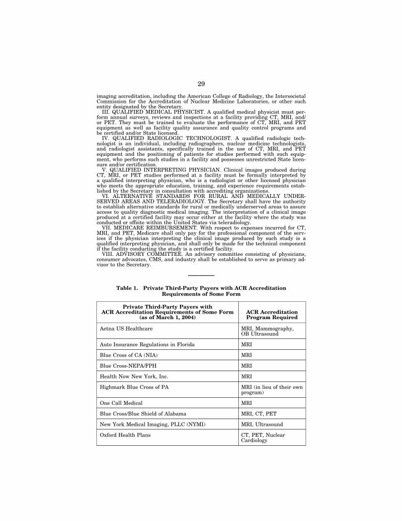
29
imaging accreditation, including the American College of Radiology, the Intersocietal Commission for the Accreditation of Nuclear Medicine Laboratories, or other such entity designated by the Secretary.
III. QUALIFIED MEDICAL PHYSICIST. A qualified medical physicist must per-form annual surveys, reviews and inspections at a facility providing CT, MRI, and/ or PET. They must be trained to evaluate the performance of CT, MRI, and PET equipment as well as facility quality assurance and quality control programs and be certified and/or State licensed.
IV. QUALIFIED RADIOLOGIC TECHNOLOGIST. A qualified radiologic tech-nologist is an individual, including radiographers, nuclear medicine technologists, and radiologist assistants, specifically trained in the use of CT, MRI, and PET equipment and the positioning of patients for studies performed with such equip-ment, who performs such studies in a facility and possesses unrestricted State licen-sure and/or certification.
V. QUALIFIED INTERPRETING PHYSICIAN. Clinical images produced during CT, MRI, or PET studies performed at a facility must be formally interpreted by a qualified interpreting physician, who is a radiologist or other licensed physician who meets the appropriate education, training, and experience requirements estab-lished by the Secretary in consultation with accrediting organizations.
VI. ALTERNATIVE STANDARDS FOR RURAL AND MEDICALLY UNDER-SERVED AREAS AND TELERADIOLOGY. The Secretary shall have the authority to establish alternative standards for rural or medically underserved areas to assure access to quality diagnostic medical imaging. The interpretation of a clinical image produced at a certified facility may occur either at the facility where the study was conducted or offsite within the United States via teleradiology.
VII. MEDICARE REIMBURSEMENT. With respect to expenses incurred for CT, MRI, and PET, Medicare shall only pay for the professional component of the serv-ices if the physician interpreting the clinical image produced by such study is a qualified interpreting physician, and shall only be made for the technical component if the facility conducting the study is a certified facility.
VIII. ADVISORY COMMITTEE. An advisory committee consisting of physicians, consumer advocates, CMS, and industry shall be established to serve as primary ad-visor to the Secretary.
—————
Table 1. Private Third-Party Payers with ACR Accreditation Requirements of Some Form
Private Third-Party Payers with ACR Accreditation Requirements of Some Form
(as of March 1, 2004) ACR Accreditation Program Required
Aetna US Healthcare MRI, Mammography, OB Ultrasound
Auto Insurance Regulations in Florida MRI
Blue Cross of CA (NIA) MRI
Blue Cross-NEPA/FPH MRI
Health Now New York, Inc. MRI
Highmark Blue Cross of PA MRI (in lieu of their own program)
One Call Medical MRI
Blue Cross/Blue Shield of Alabama MRI, CT, PET
New York Medical Imaging, PLLC (NYMI) MRI, Ultrasound
Oxford Health Plans CT, PET, Nuclear Cardiology
VerDate Aug 31 2005 03:54 Apr 13, 2006 Jkt 023927 PO 00000 Frm 00033 Fmt 6633 Sfmt 6621 E:\HR\OC\23927.XXX 23927jcor
cora
n on
PR
OD
1PC
62 w
ith H
EA
RIN
G
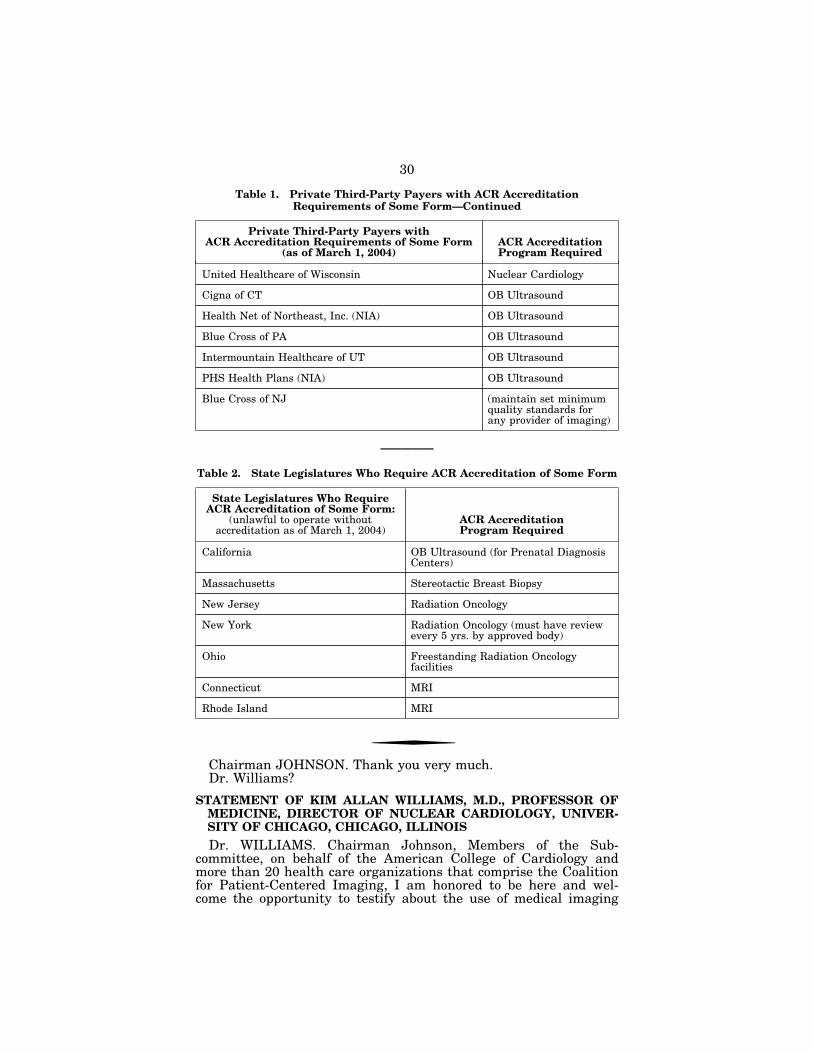
30
Table 1. Private Third-Party Payers with ACR Accreditation Requirements of Some Form—Continued
Private Third-Party Payers with ACR Accreditation Requirements of Some Form
(as of March 1, 2004) ACR Accreditation Program Required
United Healthcare of Wisconsin Nuclear Cardiology
Cigna of CT OB Ultrasound
Health Net of Northeast, Inc. (NIA) OB Ultrasound
Blue Cross of PA OB Ultrasound
Intermountain Healthcare of UT OB Ultrasound
PHS Health Plans (NIA) OB Ultrasound
Blue Cross of NJ (maintain set minimum quality standards for any provider of imaging)
—————
Table 2. State Legislatures Who Require ACR Accreditation of Some Form
State Legislatures Who Require ACR Accreditation of Some Form:
(unlawful to operate without accreditation as of March 1, 2004)
ACR Accreditation Program Required
California OB Ultrasound (for Prenatal Diagnosis Centers)
Massachusetts Stereotactic Breast Biopsy
New Jersey Radiation Oncology
New York Radiation Oncology (must have review every 5 yrs. by approved body)
Ohio Freestanding Radiation Oncology facilities
Connecticut MRI
Rhode Island MRI
f
Chairman JOHNSON. Thank you very much. Dr. Williams?
STATEMENT OF KIM ALLAN WILLIAMS, M.D., PROFESSOR OF MEDICINE, DIRECTOR OF NUCLEAR CARDIOLOGY, UNIVER-SITY OF CHICAGO, CHICAGO, ILLINOIS
Dr. WILLIAMS. Chairman Johnson, Members of the Sub-committee, on behalf of the American College of Cardiology and more than 20 health care organizations that comprise the Coalition for Patient-Centered Imaging, I am honored to be here and wel-come the opportunity to testify about the use of medical imaging
VerDate Aug 31 2005 03:54 Apr 13, 2006 Jkt 023927 PO 00000 Frm 00034 Fmt 6633 Sfmt 6602 E:\HR\OC\23927.XXX 23927jcor
cora
n on
PR
OD
1PC
62 w
ith H
EA
RIN
G
31
to deliver health care, particularly its irreplaceable contribution to the best practices of patient care. As you know, an extension of my remarks is provided for the record.
First I have to give my conflict of interest. As you noted, I am an academician at the University of Chicago, board-certified in in-ternal medicine, cardiology, nuclear medicine, and nuclear cardi-ology, and so none of the things that I am going to discuss today are actually going to benefit me or my institution. But I am here today to speak about the fact that office-based medical imaging performed by well-trained physician specialists, like the ones that I train, is good medical care and is in the best interest of patients.
Let me begin by saying that we do take exception to the concept implied by MedPAC that physician specialists are providing sub-standard care for their patients by providing poor-quality medical imaging in their offices. There is really no creditable evidence to support that notion. We agree with MedPAC that safety and pa-tient quality of imaging are of utmost importance, and we agree with the fact that there needs to be careful study of the growth in medical imaging services. But let’s keep safety and quality in the in-office medical imaging as the focus, not the agenda of one physi-cian group or another. Patients are really the issue, not the turf war that is frequently discussed in some of the literature of my col-league in the American College of Radiology, of which I am a mem-ber, by the way.
This group and MedPAC cite the same studies, recommending an agenda that to some of us appears to be aimed at restricting imag-ing to non-specialists. That is a concern because the people that I train actually do very well because they know the organ to begin with and then the imaging comes on top of that. With proper train-ing, they do extremely well and they become the experts at this.
Now, MedPAC looked at a number of studies in developing its recommendations, but these studies did not specifically examine the quality of imaging interpretations done by both the physician specialists and radiologists in MRI, CT, and nuclear, particularly PET. So, this is a problem because MedPAC has recommended that if these modalities become the high priority, we do not want to see overgeneralization of these studies to other modalities because that would be erroneous and inappropriate.
With regard to medical imaging utilization, our data says that imaging is not really growing twice as fast as all the fee schedule services. It depends on how you do the analysis. We had an anal-ysis done by the Lewin Group, which is soon to be published. When all Part B services are included and we account for the shift in de-livery of imaging services from hospitals to physician offices, imag-ing actually grew at an annual rate of about 8 percent from 2001 to 2003, and all Part B actually grew at a rate of 10 percent. So, in fact, our data is showing that the rate of growth actually may be stabilizing or even slowing.
With regard to my own field, nuclear cardiology, cardiologists are actually performing 73 percent of these tests now, and as Congress-men Lewis and Thompson have pointed out, nuclear cardiology is starting to supplant the invasive diagnostic cardiac catheterization. It can be done outside the hospital, is less risky, less expensive, and often predicts outcomes of the patient better than the invasive
VerDate Aug 31 2005 03:54 Apr 13, 2006 Jkt 023927 PO 00000 Frm 00035 Fmt 6633 Sfmt 6602 E:\HR\OC\23927.XXX 23927jcor
cora
n on
PR
OD
1PC
62 w
ith H
EA
RIN
G
32
tests. So, this growth is really highly beneficial to patients, trans-lates to saving lives, and there is no evidence that anyone has that this is not appropriate, time-efficient, or cost-effective.
So, quality in imaging can be from my point of view optimized for patients by ordering the right test at the right time, performed well, and interpreting it well. And the American College of Cardi-ology is partnering with other organizations to develop appropriate-ness criteria for imaging in an effort to define the right test at the right time.
Now, we believe that MedPAC’s recommendations requiring phy-sician privileging as stated runs counter to the Subcommittee’s goal of improving quality care by tying physician payment process to outcomes. You have to do sort of one or the other. Simply directing CMS to determine the competency of individual physicians as a condition of payment does not ensure that those people who are deemed competent are actually applying the technology in the way that benefits patients.
Now, accreditation is one step physicians may consider when it comes to ensuring the safety and quality of medical imaging. In the absence of a system wide quality measurement, though, accredita-tion has served as a proxy for quality, we admit. Now, more evi-dence is needed to prove that it produces measurable and signifi-cant improvements in quality of care. In addition, sufficient time has to be given to providers to meet any new requirements.
So, in closing, I respectfully urge the Committee to carefully weigh the complexity of the medical imaging issue before acting on any of MedPAC’s recommendations. Remember that cardiologists, urologists, orthopedic surgeons, obstetricians, neurologists, and other specialty physicians are uniquely qualified to provide imaging services specific to their specialty because they are trained in both the diagnostic imaging technique and the structure and function and clinical manifestation of disease in that organ system that they are imaging.
So, we are asking Congress, therefore, to, one, mandate the solid, credible studies be commissioned to examine the quality and safety of imaging services among all modalities among all physicians dis-ciplines; two, we ask that you support the development of peer-re-viewed evidence that accreditation results in measurable and sig-nificant improvements in quality of care, and convene a multi-spe-cialty panel to discuss the results and subsequent next steps; and lastly, refrain from removing or limiting the in-office ancillary ex-ception to the physician ownership referral laws. We want sound policy that is based on sound evidence.
Thank you again. [The prepared statement of Dr. Williams follows:]
Statement of Kim Allen Williams, M.D., Professor of Medicine, Director of Nuclear Cardiology, University of Chicago, Chicago, Illinois
Chairman Johnson and members of the Subcommittee, on behalf of the American College of Cardiology (ACC) and more than 20 physician and other healthcare orga-nizations representing a broad range of specialists and practices that comprise the Coalition for Patient-Centered Imaging (CPCI), I welcome the opportunity to testify about the use of medical imaging in the delivery of health care, and particularly its irreplaceable contribution to best practices for patient care. I am a Professor of Med-icine and Radiology and Director of Nuclear Cardiology at The University of Chicago
VerDate Aug 31 2005 03:54 Apr 13, 2006 Jkt 023927 PO 00000 Frm 00036 Fmt 6633 Sfmt 6621 E:\HR\OC\23927.XXX 23927jcor
cora
n on
PR
OD
1PC
62 w
ith H
EA
RIN
G
33
School of Medicine. I am board-certified in Internal Medicine, Cardiology, Nuclear Medicine, and Nuclear Cardiology.
Today I would also like to speak to you about the growth in utilization of medical imaging, specifically why growth is occurring, and why physician specialists depend on medical imaging. I will also address some of the myths surrounding accusations of inappropriate use of medical imaging. I will articulate why the imaging that I and other specialists perform and interpret for our patients is in the best interest of the patient, is timely, and is cost-effective. We believe imaging kept in the hands of experienced and qualified specialists is safe, appropriate, of the highest quality, and critical to the emerging demand for outcomes-based disease state management, both in the medical and public policymaking arenas.
I remember being in cardiology training just a few short decades ago when invasive diagnostic testing was ‘‘standard’’ practice in cardiology. I was told by my mentors that if we didn’t have ‘‘30 percent normal angiograms,’’ we were being too selective and would miss disease in a large segment of our patients. Well, those days are gone due to highly accurate, noninvasive imaging tests that more accu-rately predict outcomes for cardiac patients than the old-fashioned invasive ‘‘stand-ard.’’
Medical Imaging Is Good Medicine
Advancements in medical imaging have changed the way cardiologists, oncologists, obstetricians and gynecologists, urologists, family practitioners, neurolo-gists, orthopaedic surgeons and other surgeons and many other physicians deliver patient care on a daily basis. By integrating medical technology into care plans, pa-tients are receiving more prompt, efficient, effective and cost-effective care. In addi-tion to traditional diagnostics employing medical imaging, we now use imaging to guide minimally invasive treatments and to track ongoing treatment protocols through judicious use of medical imaging. We are enabled as physicians to adjust patient care plans mid-therapy to achieve the best possible outcomes. Several spe-cialist groups intimately integrate medical imaging in the most delicate and intri-cate aspects of their care. The prudent use of medical imaging in the actual treat-ment regimen is not only excellent medicine: it also manages short- and long-term costs by minimizing wasteful and ineffective treatments. Patient Value
We cannot overestimate the patient values of choice, comfort, convenience and peace of mind when it comes to the provision of in-office imaging. The in-office set-ting affords patients greater choice about who they want to perform and interpret their test results. Patients can now have imaging tests performed by physicians who know their medical history and who will ultimately make treatment decisions and provide ongoing continuity of care. Patients also can have their imaging tests per-formed in a setting that is comfortable and convenient to them, oftentimes resulting in one office visit instead of three—or more. This is efficient not only from a pa-tient’s point of view, but for society as a whole. Physician Value
In my field of cardiovascular medicine, advancements in noninvasive medical im-aging have significantly changed the way we diagnose and treat patients with ac-quired and congenital cardiovascular disease. Today, cardiologists can do for their patients what was not possible even 10 years ago. There is unquestionable value for physicians being empowered to integrate imaging into their practices. With the aid of medical imaging, physicians make more precise diagnoses in a time-efficient manner, leading to earlier detection of disease. As Medicare and private payers begin to focus on disease state management, we are learning one of the greatest ob-stacles to effectively managing the care of patients with chronic conditions is compli-ance. Physicians know when they have to order a test for a patient outside their office, there is a question of when, or even whether, that patient will follow through. Better compliance means maintaining a continuum of care that is critical in treating and managing disease.
Most often in medicine, timeliness in imaging improves outcomes. A diagnosis de-layed is essentially a treatment denied. This is true whether we are speaking of di-agnosing a critically narrowed and life-threatening coronary artery which is soon to close completely, or a stress fracture in a runner’s foot that could become a complete bone break with the next training session. Value to Payers and Society
Advances in technology can decrease healthcare spending, if they lead to less ex-pensive treatments, or they can increase spending by, for example, rendering pre-
VerDate Aug 31 2005 03:54 Apr 13, 2006 Jkt 023927 PO 00000 Frm 00037 Fmt 6633 Sfmt 6621 E:\HR\OC\23927.XXX 23927jcor
cora
n on
PR
OD
1PC
62 w
ith H
EA
RIN
G
34
1 The value of investment in health care. Bethesda, MD: MEDTAP International. http:// bcbshealthissues.com/relatives/20846.pdf.
2 David M. Cutler, Mark McClellan, ‘‘Productivity Change in Health Care,’’ American Eco-nomic Review 2001;91(2):281–286.
3 David M. Cutler, Mark McClellan, Joseph P. Newhouse, and Dahlia Remler, ‘‘Are Medical Prices Declining? Evidence from Heart Attack Treatments.’’ Quarterly Journal of Economics 1998;113(4):991–1024.
viously untreatable or partially treatable conditions treatable. Technological ad-vancements can also lead to significant improvements in health outcomes that can clearly offset the direct costs of new technology.
Last year, a study by MEDTAP International on the value of healthcare spending revealed that the value of the health improvements in the U.S. population over the past 20 years has significantly outweighed the additional healthcare expenditures that have accompanied these improvements. According to the report, every addi-tional dollar spent on overall treatment for heart attacks has resulted in health gains of $1.10.1
A study by David M. Cutler, Ph.D., and Mark McClellan, M.D., Ph.D., published in 2001 2 found that benefits of improvement in heart attack treatment over a 10- year period exceeded treatment costs by $87,000 per person who had a heart attack. Another study 3 found that more than half of the cost growth for heart attacks from 1984 to 1998 was attributable to technological developments, most often in the ex-pansion of existing technologies within new patient populations. In general, there is a notable body of literature that finds that new medical technologies create de-sired value and health care improvements that far outweigh their costs.
The Medicare Payment Advisory Commission’s (MedPAC) recent report to Con-gress made little mention of the value that medical imaging has brought to patients, physicians or the healthcare system as a whole. This is a notable omission.
Before Congress considers policies that could ultimately discourage or restrict certain specialists from providing appropriate imaging services to their patients, cost-effectiveness studies are needed to better determine the incremental costs of procedures in relation to their incremental health benefits in order to determine whether imaging technologies provide rea-sonable value for their cost.
While there is high value for patients receiving medical imaging services by their physician specialists in an office setting, current costs to Medicare for imaging per-formed in hospital or non-hospital settings remain neutral. The migration, however, to office-based noninvasive diagnostic imaging and image-guided therapy, has the potential to create significant cost savings.
When physicians must refer their patients to a hospital or imaging facility for needed tests, the process of a referral to a radiologist can itself increase costs to both Medicare and to patients. The referral can result in as many as three or more appointments and visits—one to see the physician and learn an image is needed, a second to have the image taken, and then a followup appointment and visit to the referring physician to receive the treatment plan based on the image. Although difficult to quantify, by reducing the number of visits, in-office imaging should di-rectly reduce costs to both patients and Medicare, while increasing convenience and improving the timeliness of subsequent diagnosis and treatment. In addition, with fewer visits and a shorter delay between the initial visit and treatment, both patient compliance and health outcomes are improved.
Analyzing Utilization of Medical Imaging
Shift in Site of Service In its just-released report to Congress, MedPAC found that between 1999 and
2002 the growth rate in the use of imaging services was twice as high as the growth rate for all fee schedule services (10.1 percent vs. 5.2 percent). MedPAC also found that growth in imaging moderated from 2002 to 2003, but imaging services were still higher (8.6 percent) than all services (4.9 percent).
While MedPAC acknowledged about 20 percent of the growth in imaging services paid under the physician fee schedule between 1999 and 2002 was due to a shift of site of services out of hospitals and into physician offices, MedPAC does not di-rectly account for this shift in its growth comparisons. Failure to appropriately ac-count for site of service changes presents a misleading interpretation of growth in imaging services. In addition, MedPAC does not include all Part B services in its comparison. In particular, MedPAC omits durable medical equipment, chemo-therapy drugs and other drugs covered under Part B, and ambulance services. When we include all Part B services and account for changes in site of service for imaging,
VerDate Aug 31 2005 03:54 Apr 13, 2006 Jkt 023927 PO 00000 Frm 00038 Fmt 6633 Sfmt 6621 E:\HR\OC\23927.XXX 23927jcor
cora
n on
PR
OD
1PC
62 w
ith H
EA
RIN
G
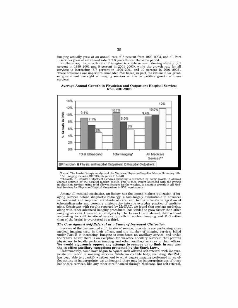
35
imaging actually grew at an annual rate of 8 percent from 1999–2003, and all Part B services grew at an annual rate of 7.8 percent over the same period.
Furthermore, the growth rate of imaging is stable or even slowing slightly (8.1 percent in 1999–2001 and 8 percent in 2001–2003), while the growth rate for all services is increasing (5.7 percent in 1999–2001 and 10 percent in 2001–2003). These omissions are important since MedPAC bases, in part, its rationale for great-er government oversight of imaging services on the competitive growth of these services.
Average Annual Growth in Physician and Outpatient Hospital Services from 2001–2003
Source: The Lewin Group’s analysis of the Medicare Physician/Supplier Master Summary File. * All Imaging includes BETOS categories I1A–I4B ** Growth in Hospital Outpatient Services spending is estimated by using growth in allowed
charges deflated by the hospital market basket. This is then weight averaged with the growth in physician services, using total allowed charges for the weights, to estimate growth in All Med-ical Services for Physician/Hospital Outpatient in RVU equivalents.
Among all medical specialties, cardiology has the second highest utilization of im-aging services behind diagnostic radiology, a fact largely attributable to advances in treatment and improved standards of care, and to the ultimate integration of echocardiography and coronary angiography into the everyday practice of cardiolo-gists. Consistent with results reported by MedPAC, we found that nuclear medicine, along with other advanced imaging procedures, has tended to grow faster than other imaging services. However, an analysis by The Lewin Group showed that, without accounting for shift in site of service, growth in nuclear imaging and MRI (other than of the brain) is overstated by a third.
The Case Against Self-Referral as a Cause of Increased Utilization Because of the documented shift in site of service, physicians are performing more
medical imaging tests in their offices, and the number of imaging services billed under Part B is increasing. Imaging is considered an ancillary service, and under the ‘‘Stark Laws’’ there is an exception for ‘‘in-office ancillary services’’ that permits physicians to legally perform imaging and other ancillary services in their offices. We would vigorously oppose any attempt to remove or to limit in any way the in-office ancillary exceptions protected by the Stark Laws.
Unfortunately, some have begun to equate such allowed self-referral with inappro-priate utilization of imaging services. While no credible body, including MedPAC, has been able to quantify whether and to what degree imaging performed in an of-fice setting is inappropriate, we understand there may be inappropriate use of these healthcare services, like any other care financed through Medicare. But self-referral,
VerDate Aug 31 2005 03:54 Apr 13, 2006 Jkt 023927 PO 00000 Frm 00039 Fmt 6633 Sfmt 6621 E:\HR\OC\23927.XXX 23927 Inse
rt 2
3927
A.0
07
jcor
cora
n on
PR
OD
1PC
62 w
ith H
EA
RIN
G
36
4 R.P. Strasser, M.J. Bass, M. Brennan, ‘‘The effect of an on-site radiology facility on radiologic utilization in family practice,’’ Journal of Family Practice 1987;24:619–623.
5 K.K. Oguz, D.M. Yousen, T. Deluca, E.H. Herskovitz, N.J. Beauchamp, ‘‘Effect of emergency department CT on neuroimaging case volume and positive scan rates.’’ Academic Radiology 2002;9:1018–1024.
6 Iskandrian, et al. Journal of the American College of Cardiology 1993;22:665–670.
as it is labeled by self-interested groups outside the physician specialty community, is not the primary driver in growth in imaging services.
Important findings of the Lewin analysis, not examined by MedPAC, undermine the claim that the primary driver of growth in imaging services is self-referral. First, utilization of MRI and CT, which have experienced relatively high growth rates compared with other types of services, was examined. The average growth rate for CT from 2001–2003 was 16 percent, with radiology dominating 84 percent of CT scans performed. Similarly, the average growth rate for MRI during this same time period was 19 percent, with radiology dominating 65 percent of use. Because MRI and CT are dominated by radiologists, these results suggest that eliminating the ability of specialty physicians to perform and interpret imaging tests in their offices is no protection against the growth in utilization.
Second, central to the argument that self-referral is a significant cost-driver is the idea, supported by MedPAC, that physicians will automatically over-utilize imaging services to increase their practices’ revenue and income. However, two studies 4,5 demonstrate that physicians order more images when they have access to on-site imaging equipment, even when they do not own it and have no financial incentives to do so. Clinical Substitution as a Result of More Effective Technology
In its March 2003 report, MedPAC acknowledged new indications for existing technologies may contribute to imaging growth rates, and decreases in some serv-ices, and in some cases more costly services, may result of substituting one service for another. In nuclear cardiology, my particular area of training and expertise, the data indicate that nuclear cardiology is supplanting hospital-based invasive cardiac catheterization in many cases.
The average growth rate from 2001–2003 for cardiac nuclear imaging by radiolo-gists and cardiologists combined was 18 percent, down from an average of 19 per-cent during 1999–2001. Comparatively, the average annual growth rate for invasive cardiac catheterization, a procedure not performed by radiologists, dropped from 8 percent in 1999–2001 to negative-4 percent in 2001–2003. This shows a direct and positive correlation between increases in cardiac nuclear imaging and decreases in invasive diagnostic cardiac catheterization. The significance of this substitution is that nuclear cardiology and other noninvasive imaging can be done outside of hos-pitals, is less expensive, is less risky in terms of complications, requires no patient recuperation, and often predicts the outcome of the patient better than invasive tests.6 Advancements in Technology
As we eliminate the theory that self-referral is a primary driver of medical imag-ing utilization, we can begin to examine the true reasons why utilization has in-creased in this area of medicine.
While MedPAC has acknowledged the role of technological innovation in the growth of imaging services, it does not adequately include this factor in its analysis, especially the issue of substitution of one treatment or diag-nosis method for another.
Growth in the use of imaging services is, in part, reflective of growing applications for these technologies. In fact, in its March 2003 report, MedPAC states, ‘‘. . . it appears that use of well-established technologies is increasing. CAT, for example, was introduced in the 1970s. MRI began to diffuse as a new technology in the 1980s. Thus, the indications for use of these technologies may be changing.’’
As I have mentioned, technology improvements have resulted in new imaging techniques that replace more invasive and generally more risky diagnostic proce-dures, and this trend of substitution isn’t just occurring in cardiology. For example, ultrasound guidance can allow needle biopsies to replace open biopsies of the breast, and it also can enable more accurate biopsies of prostate legions with fewer tissue samples.
In breast surgery, ultrasound-guided breast biopsies can reduce performance of potentially unnecessary and invasive procedures and surgeries. Ultrasound-guided breast biopsy allows for less-invasive evaluation of mammographic lesions, with more reliable tissue differentiation, more streamlined patient care and characteriza-tion, and improved staging of disease.
VerDate Aug 31 2005 03:54 Apr 13, 2006 Jkt 023927 PO 00000 Frm 00040 Fmt 6633 Sfmt 6621 E:\HR\OC\23927.XXX 23927jcor
cora
n on
PR
OD
1PC
62 w
ith H
EA
RIN
G
37
Urology offers another example where advancements in medical imaging have led to less-invasive and less-painful procedures. Older men often experience difficulty urinating because of prostate enlargement. To evaluate this problem, physicians must learn how much urine is retained in the bladder after voiding, known as ‘‘re-sidual urine.’’ For many years this was determined by passing a catheter through the urethra and into the bladder, the amount of urine drained from the bladder was then measured. Introducing a catheter into the bladder, in addition to being uncom-fortable, also may introduce infection. Today, many urologists employ a small ultrasound machine designed specifically for this task. This test can be done in the urologist’s office and eliminates the use of a catheter and the danger of infection. In addition, 15 percent of Americans will have a kidney stone in their lifetime, and during acute episodes there is much pain and disability, often resulting in hos-pitalization and loss of work. The CT stone protocol provides for a rapid, accurate diagnosis of the vast majority of kidney stones. Using a CT scanner, a patient does not have to be given intravenous contrast, which can be toxic, as would be needed with the traditional intravenous pyleogram (IVP). The CT scan also allows for the diagnosis of non-calcium stones that may not be the case with IVP. This provides more efficient health care for the economy, for providers and far better patient care.
Today in cardiology, we routinely use an arsenal of high-tech equipment to combat and treat disease. With the use of CT, we can see the heart beating in three dimen-sions which allows us to define the adequacy of coronary perfusion as well as plaque within a vessel wall. We can track heart disease at every state, visualizing what we could only imagine in the past. The expectation of society, and of our patients, is that we will employ all of these marvels to achieve best practice outcomes for every care interlude. That means medical imaging as part of the treatment plan de-livered in a physician’s office is here to stay. Changing Demographics
Managing heart disease is one of the most significant success stories in modern health care. Over the past 30 years, there has been a substantial increase in the life expectancy of Americans that directly correlates with downward trends in heart disease mortality and disability. We know that prevention efforts are important con-tributors to the reduced mortality rates, but many of the benefits are attributed to better and earlier detection and improved treatment.
We anticipate that as the population ages, because medical imaging will continue to be an essential tool for treating heart disease, growth in utilization is bound to continue. Consider this: Based on the National Heart, Lung, and Blood Institute’s Framingham Heart Study, the average annual rates of first major cardiovascular events rise from seven per 1,000 men at ages 35–44 to 68 per 1,000 at ages 85– 94. For women, comparable rates occur as men, but about 10 years later in life. At the same time, the prevalence of diseases associated with heart disease, such as obe-sity, hypertension, diabetes, and high cholesterol also continue to grow.
Advancements in technology and changing demographics are factors that can be expected to continue to fuel growth in Medicare and overall health expenditures. Furthermore, while technological advances sometimes increase expenditures, they can also decrease costs by allowing less invasive and less costly treatments to re-place older, more invasive treatments. In addition, new imaging technologies can improve early detection, which can allow us to treat diseases at a lower level of in-tensity, and thus at a lower cost than if they were detected later. It is important, therefore, that Congress move cautiously when considering policies to limit the use of imaging services, because imprudent limits on the use of diagnostic imaging could increase total Medicare expenditures for non-imaging services.
Quality Medical Imaging is Safe and Appropriate
Quality Quality is a top priority in health care as providers, payers, and regulators strive
for a system that reduces errors and rewards physicians for exemplary clinical prac-tice. CMS and private payers have begun to implement programs that tie physician reimbursement to patient care outcomes—a growing trend called ‘‘pay for perform-ance.’’ Emphasizing and rewarding quality is good for patients. However, this proc-ess must recognize that, by necessity, physicians will need to use advanced tech-nology to meet high quality standards and ensure optimal care. Newer imaging mo-dalities provide more accurate and precise images with lower patient risk, helping physicians to diagnose and treat diseases more effectively. To make objective eval-uations of patient progress tied to payment, medical imaging will undoubtedly ex-pand to provide clear, unequivocal valuations of care protocols and procedures. Who
VerDate Aug 31 2005 03:54 Apr 13, 2006 Jkt 023927 PO 00000 Frm 00041 Fmt 6633 Sfmt 6621 E:\HR\OC\23927.XXX 23927jcor
cora
n on
PR
OD
1PC
62 w
ith H
EA
RIN
G
38
7 Levin DC, Rao VM. Turf wars in radiology: the quality of imaging facilities operated by non-radiologist physicians and of the images they produce. J Am Coll Radiol 2004;1:649–51.
8 MedPAC Report to Congress: Medicare Payment Policy, March 2005, pp. 166. 9 Moskowitz, H., J. Sunshine, D. Grossman, et al. 2000. The effect of imaging guidelines on
the number and quality of outpatient radiographic examinations. American Journal of Roentgen-ology 175, no. 1 (July): 9–15.
10 MedPAC Report to Congress: Medicare Payment Policy, March 2005, pp. 160.
owns these technologies and how widely they are utilized have become critical ques-tions for our patients and physicians.
The American College of Cardiology and the Coalition for Patient-Centered Imag-ing support the delivery of the highest quality care. But the debate over medical imaging, one that is playing out in the press, in state legislatures and here in Con-gress, stands to pit one physician group against another. The College has sat at the table with the leadership of the American College of Radiology, and has sought to reach agreement on what defines quality in imaging. To date, our results have been mixed. Not working together as a physician community is the ultimate disservice to our patients and, ultimately, to the payer community.
The literature comparing the quality of interpretations of imaging studies con-ducted by ‘‘nonradiologists,’’ and radiologists remains limited.7 Of the studies that do exist, the findings do not suggest that physician specialists (limited license pro-viders excluded) have lower quality of performance in diagnostic imaging than radi-ologists. In an article published last year in the Journal of the American College of Radiology, and disconcertingly titled, ‘‘Turf Wars in Radiology: The Quality of Inter-pretations of Imaging Studies by Nonradiologist Physicians—A Patient Safety Issue,’’ the authors David Levin and Vijay Rao cite studies that compared the per-formance on plain radiographs (x-rays), an imaging modality dominated by radiolo-gists, rather than more specialized modalities such as CT and MRI—which is where much of the increase in utilization is occurring. In particular, modalities primarily used by other specialties (such as echocardiography, which is used primarily by car-diologists, and transrectal prostate ultrasound, which used primarily by urologists) were excluded. Furthermore, the studies quoted by Levin and Rao actually found that cardiologists perform and interpret chest X-rays as well as radiologists, but that fact was not brought out in the article. Our point here is, specialists who learn a medical imaging technique will perform that technique with quality and will ac-cept nothing less for patients for whom they care.
None of the studies cited in the MedPAC report examined the quality of imaging interpretations by nonradiologists and radiologists in the modalities that MedPAC actually recommended that CMS consider a ‘‘high priority’’—MRI, CT, and nuclear medicine (including PET).8 One study by Harold Moskowitz, published in the Amer-ican Journal of Roentgenology, specifically excluded CT, MR, sonography and nu-clear studies from the results.9 The only modality analyzed in the study results was X-ray. Another study cited in the report by CareCore National also only examined X-ray studies.10 These studies are not relevant to the issues at hand. The inability of those practitioners, who rarely read X-rays, to read them better than radiologists who read X-rays frequently, has nothing to do with the ability of cardiologists, for example, who read echocardiographs almost daily to read them better than radiolo-gists, who see echocardiographs as one modality among many.
MedPAC has recommended to Congress that it consider privileging as a means to improve the quality of imaging interpretations and as a condition of Medicare payment for physicians who want to interpret imaging tests.
Before Congress directs CMS to become involved in determining com-petencies of individual physicians in this country, we suggest that solid, credible studies be commissioned that examine the quality of imaging serv-ices among all modalities and among all physician disciplines.
We believe MedPAC’s recommendation requiring physician privileging leads us down a road that runs counter to this Subcommittee’s goal of im-proving the quality of patient care by tying physician payment to process and outcomes measures. By simply directing CMS to determine the com-petency of physicians as a condition of payment, it does not ensure that those deemed ‘‘competent’’ are in fact appropriately applying medical tech-nology in a way that best benefits patients. Appropriateness
Physicians must carefully incorporate new technologies in patient care plans only after their effectiveness has been proven. Congress and CMS should consider the dialogue occurring between physician societies and private sector payers about how to ensure that imaging being delivered to patients is indeed appropriate.
VerDate Aug 31 2005 03:54 Apr 13, 2006 Jkt 023927 PO 00000 Frm 00042 Fmt 6633 Sfmt 6621 E:\HR\OC\23927.XXX 23927jcor
cora
n on
PR
OD
1PC
62 w
ith H
EA
RIN
G
39
11 David C. Levin, Vijay M. Rao, ‘‘Turf wars in radiology: the overutilization of imaging result-ing from self-referral,’’ Journal of the American College of Radiology, 2004;1(3):169–172.
12 MedPAC Report to Congress: Medicare Payment Policy, March 2005, pp. 167. 13 American Medical Association House of Delegates Resolution 108. 14 American Medical Association House of Delegates Resolution 802.
At the American College of Cardiology, we are developing specific appropriateness criteria for imaging in an effort to define ‘‘what to do,’’ ‘‘when to do it’’ and ‘‘how often’’ in the context of local care environments when combined with patient and family preferences. Ideally, appropriateness criteria would encompass ‘‘cost-effec-tive’’ and ‘‘benefit vs. risk’’ analysis of available care alternatives. The criteria will be simple, reliable, valid, and transparent. They will provide cardiovascular special-ists with meaningful feedback on their care practices relative to national standards. The College’s first focus will be on appropriateness criteria for nuclear cardiology. The process, expected to be complete by early summer, is underway as an abstrac-tion, distillation, and in some cases, expansion of the scientifically sound, evidence- based ACC/American Heart Association Clinical Practice Guidelines and Expert Consensus Documents. Developing appropriateness criteria is a complex and often very divisive issue, but it will allow cardiologists, payers and patients to quantify quality.
There are some who claim if CMS simply ‘‘weeds out’’ all inappropriate utilization, the result would be significant cost savings to Medicare. Before arriving at this con-clusion, policymakers must quantify appropriate and inappropriate utilization in order to effectively estimate cost savings. Again, I point to the efforts underway be-tween the American College of Cardiology and the private payers to develop appro-priateness criteria.
We are aware that the American College of Radiology is projecting estimates of Medicare cost savings to drive their quest for ‘‘designated imaging provider’’ legisla-tion in the states and in Congress. In articles published in the Journal of the Amer-ican College of Radiology, hypothetical cost savings are based arbitrarily on the as-sumption that the level of referrals to radiologists by physicians who do not have access to on-site imaging equipment ‘‘delineate the necessary and appropriate utili-zation rate of imaging.’’ 11 It is no less valid to assume that patients whose physi-cians do not have access to on-site imaging equipment are underserved, and thus receive substandard care. Even MedPAC noted in its report that a 1994 GAO study examining referrals to specialty physician-owned imaging facilities did not address whether the additional services were appropriate or not.12
Training and Safety Training of Physicians
Radiology was first introduced into medical practice in the early 1900s with the discovery of X-rays. Since then, medical imaging has significantly progressed and gained widespread clinical use. Some of the first diagnostic imaging technologies used by cardiologists to assess cardiac conditions were chest X-rays, electrocardiog-raphy, coronary angiography, and cardiac catheterization. Cardiologists’ use of new technologies such a nuclear cardiology, cardiac MR and CT are a natural extension of the strong and appropriate dominance that cardiologists have held in cardiac im-aging for the last three decades. Today, cardiologists dominate the use of chest X- rays, nuclear medicine, echocardiography, cardiac catheterization, and electro-cardiography, in part because cardiologists have generally obtained better training in these areas, and have the clinical expertise to evaluate the procedures.
Cardiologists, urologists, orthopaedic surgeons, obstetricians and gynecologists, neurologists and other specialist physicians are uniquely qualified to provide imag-ing services specific to their specialty because they are trained in both diagnostic imaging techniques, and in the structure and function of the organs and systems they are imaging.
In ultrasound, the need to codify training and education for the performance and interpretation of examinations has already been thoroughly addressed by the med-ical community. There is no evidence to support additional standards being placed upon physicians and their practices. The American Medical Association (AMA) has concluded that ultrasound is integral to the practice of appropriately trained physi-cians 13 and that office-based ultrasound is cost effective and essential to patient care.14 The AMA further recognized that it is inappropriate to apply educational standards developed for one medical specialty group to that of another. Because of the different and unique applications of ultrasound within the various physician specialties, the AMA called for each specialty to define ultrasound standards for its
VerDate Aug 31 2005 03:54 Apr 13, 2006 Jkt 023927 PO 00000 Frm 00043 Fmt 6633 Sfmt 6621 E:\HR\OC\23927.XXX 23927jcor
cora
n on
PR
OD
1PC
62 w
ith H
EA
RIN
G
40
15 ibid. 16 Luhmann SJ, Schootman M, Gordon JE, Wright R, ‘‘Magnetic Resonance Imaging of the
Knee in Children and Adolescents. Its Role in Clinical Decision-Making.’’ J. Bone Join Surg. Am., Mar 2005; 87:497–502.
17 National Coalition for Quality Diagnostic Imaging Services. Written testimony to the House Ways and Means Subcommittee on Health; Feb. 10, 2005.
18 MedPAC Report: Purchasing Strategies. June 2004, pp. 108.
members to fit their clinical environment,15 which is what has occurred in the spe-cialties of breast surgery and emergency medicine.
To practice clinical nuclear cardiology, for example, a physician must receive two years of general cardiology training and up to six months dedicated to nuclear cardi-ology training. This training is critical to accurate interpretation of imaging studies. By contrast, the American Board of Radiology has no specific requirements for train-ing time, mentoring, or case load in nuclear cardiology prior to certifying a radiolo-gist as competent in this area.
The American College of Cardiology and other cardiovascular health organizations have been leaders in the development of training program standards, clinical com-petency statements, and clinical practice guidelines that contain recommendations regarding the necessary knowledge and skills, as well as the appropriate use of im-aging procedures.
Therefore, we believe all physicians who meet the appropriate level of training ought to be able to provide imaging services to patients. Specialty designation alone is not an appropriate basis on which to determine physi-cian qualifications.
The critical piece of data for the referring physician is not just to have the dicta-tion of the findings from the images, but to receive clinical input for the patient’s care management based on those findings from the image interpreter—something the specialist routinely does but the general imager does not necessarily do. The ra-diologist’s comment ‘‘please correlate clinically’’ is often appended to image reports, while the specialist has completed this ‘‘clinical correlation’’ automatically and com-municates this to the referring physician.
A study published in a recent issue of The Journal of Bone & Joint Surgery fur-ther illustrates this point. The study compared the findings of a radiologist’s report and an orthopaedic surgeon’s preoperative diagnosis, following an examination and reading of any images with actual surgical findings. Not surprisingly, the surgeon’s preoperative diagnosis was significantly more accurate than the radiologist’s report. The study concluded that ‘‘the formal interpretation of the MRI scan by the radiolo-gist commonly had inaccuracies and may not correlate with the initial diagnoses . . . reliance on the radiologist’s formal interpretation can lead to diagnostic inac-curacies and to delay in appropriate treatment.’’ 16 Accreditation of Facilities
Accreditation is a step payers and physicians may consider when it comes to en-suring the safety and quality of medical imaging. Let me begin by saying that I do not agree, nor do any credible studies suggest, that the imaging we are performing outside the hospital is sub-optimal or unsafe. The costs to physicians, in both time and money, to complete accreditation processes are significant. If mandatory, ac-creditation should be accompanied by clinical evidence that accreditation results in measurable and significant improvement in quality of care and compliance man-dates should include sufficient time to allow providers to achieve accreditation.
Furthermore, and most importantly, accreditation should not be limited to the standards of just one accrediting body. For example, for cardiovascular laboratories, accreditation bodies include the Intersocietal Accreditation Commission (IAC) and the American College of Radiology (ACR), and the differences between these pro-grams must be recognized. In a February 10, 2005, statement to this Subcommittee, that National Coalition for Quality Diagnostic Imaging Services (NCQDIS) stated that ACR has full accreditation programs for many diagnostic procedures and im-plied that only offices accredited by ACR perform the highest quality imaging serv-ices.
We want to set the record straight on this point and on a study cited by both MedPAC and by NCQDIS. This study stated an imaging site inspection program ‘‘revealed that over 1⁄3 of imaging facilities operated by nonradiologist physicians had one or more significant quality deficiencies, while only 1 percent of facilities op-erated by radiologists had such deficiencies.’’ 17 What NCQDIS does not tell the reader is that 92 of the 150 sites that failed inspection (62%) were operated by lim-ited license providers (chiropractic and podiatry).18 Cardiologists, in fact, had a lower failure rate than radiologists.
VerDate Aug 31 2005 03:54 Apr 13, 2006 Jkt 023927 PO 00000 Frm 00044 Fmt 6633 Sfmt 6621 E:\HR\OC\23927.XXX 23927jcor
cora
n on
PR
OD
1PC
62 w
ith H
EA
RIN
G
41
Conclusion In closing, I want to emphasize that medical imaging performed and interpreted
in physicians’ offices is an integral and cost-effective part of the specialists’ patient care continuum. To prevent or limit the practice of in-office imaging would move the field of medicine in the wrong direction, just as clinical advancements are improving patient care and outcomes. Furthermore, attempting to restrict physician eligibility to perform and interpret tests for their patients in the most efficient and effective setting is antithetical to the aggressive disease state management paradigm now being promoted by CMS through multiple pilot projects.
We also have seen a clear connection between better and faster diagnosis and im-proved quality of life, enhanced productivity and contained costs compared with more invasive treatments at later-stage disease arrest. Medical imaging, while not the sole factor in gaining ground in the fight against disease, certainly is a leading cause for this remarkable turnaround, particularly in heart disease.
We respectfully urge the Subcommittee to carefully weigh the complexity of the medical imaging debate before enacting any governmental restraints against spe-cialty physicians from performing and interpreting medical images. Our patients de-serve better, and they are accustomed to receiving care which incorporates medical imaging in their personal physician’s office. Turning back the clock at this stage will surely retract many of the gains we have achieved in the last 20 years through the use of medical imaging in our patient care protocols.
Thank you.
f
Chairman JOHNSON. Thank you very much, Dr. Williams. Ms. Farnsworth?
STATEMENT OF CHERRILL FARNSWORTH, EXECUTIVE DIREC-TOR, NATIONAL COALITION FOR DIAGNOSTIC IMAGING SERVICES, HOUSTON, TEXAS
Ms. FARNSWORTH. Thank you, Madam Chairman, Congress-man Stark, and Members of the Committee, for the opportunity to testify today and for your personal involvement and interest in this important health care issue. I am Cherrill Farnsworth, the Presi-dent and CEO of a company, Health Help. I also serve as Executive Director of the National Coalition for Quality Diagnostic Imaging Services.
Health Help is a company that manages radiology benefits for the private sector companies throughout the country who also face rising imaging costs and fears about quality and safety.
NCQDIS represents more than 2,400 outpatient imaging centers and hospital outpatient departments, promoting strategies for health care cost savings, and advocating for public and private sec-tor standards for diagnostic imaging services. It is my privilege to testify before the Committee today as CEO and to share valuable practical experience of what we see bringing these very solutions in the private sector.
Both organizations I am representing today support the MedPAC recommendations. We have been there, we have seen it, and we know that it works. We have all heard testimony citing the increas-ing costs in the use of diagnostic imaging service as well as the growing concern about quality of these medically necessary imag-ing procedures. But there is good news. The private sector has faced these issues, has been forced to take a closer look and take a stand on the problem.
The issues described by MedPAC are not unique to Medicare. They have been experienced by the commercial plans. The only dif-
VerDate Aug 31 2005 03:54 Apr 13, 2006 Jkt 023927 PO 00000 Frm 00045 Fmt 6633 Sfmt 6602 E:\HR\OC\23927.XXX 23927jcor
cora
n on
PR
OD
1PC
62 w
ith H
EA
RIN
G
42
ference is that in the private sector they have taken action to ad-dress the costs, the overuses, and to promote quality and safety.
The MedPAC Commission’s recommendations surround three areas: First, privileging. Privileging in the private sector has gen-erally taken a stance that in those areas where a specialty or med-ical provider, health care provider, is trained in his training to do the test, he should be allowed to do the test, to bill for it, and be paid for it. Where he has not had the training, it is not safe, it is not practical, and it is not reasonable that he would be performing, billing, or be paid for those services.
An example I would like to give you is a very recent one from the private sector where a health care plan that we work for in the State of Texas introduced privileging standards almost imme-diately after we did the analysis. An example out of the analysis is that they had spent more than $15 million in the State of Texas for psychiatrists to perform and evaluate PET scans. Psychiatrists are not trained in the very complex imaging procedures behind PET. They have not been trained in the physics behind PET in order to keep their patients safe. This is a frightening thing that is happening as far as costs of the Medicare program and safety to the Medicare beneficiary.
Also, the second point that Mr. Miller talked about was correct billing initiatives. When the CPT codes were developed for imaging many years ago, the technology was very different. Today, it is time that we modernize those CPT codes to reflect the advances in technology and how they perform today.
Quality standards, I believe they are an imperative. Imaging is a $100 billion business. I don’t think there are very many people running a $100 billion business without any quality standards.
In the private sector, for our insurance companies, we are finding used equipment, equipment without maintenance contracts, equip-ment where you cannot even buy parts for this equipment today. Certainly the standards are there and they are enforced by the manufacturers who manufacture this equipment. We are talking about enforcing that the maintenance contracts and the quality be maintained through the use of that equipment.
We also see things that point to the lack of training of those peo-ple performing the tests in the center or the department, things like the use of collimation. Collimation was developed a hundred years ago. This is not new. It is like shutters on a camera where we make sure that the radiation is only going to that area of the body that we are looking at. This is important that we not get scat-ter radiation throughout the patients’ body. In our company, when we are looking at imaging centers without trained technologists that do not have radiology oversight or oversight by a physician trained in that type of imaging, more than 92 percent of the time no collimation is used.
Our chief medical officer in Health Help uses an example where we see a film with the entire baby. This baby was there to have his lower abdomen examined, so we should have seen only that part of his lower abdomen. But the baby has radiation in his brain, throughout his body, and we know from the evidence that the ear-lier a patient has radiation and maintains the effects of that radi-ation throughout his life, the more likely the cancer.
VerDate Aug 31 2005 03:54 Apr 13, 2006 Jkt 023927 PO 00000 Frm 00046 Fmt 6633 Sfmt 6602 E:\HR\OC\23927.XXX 23927jcor
cora
n on
PR
OD
1PC
62 w
ith H
EA
RIN
G
43
In closing, this issue is not about costs. I do not believe this issue is about a turf battle. I believe it is about the Medicare beneficiary and the viability of the Medicare program in light of these kinds of exponential cost growth. It is about quality patient care and the importance of maintaining safety for the Medicare enrollee. Medi-care has the opportunity to take advantage of efforts that are seen in the private sector. And the MedPAC recommendations are an important first step in addressing imaging issues.
Medicare beneficiaries deserve the same rights and should have the same protection as other patients, and NCQDIS stands ready to assist the work of the Committee as this issue proceeds. I thank you again for the opportunity to testify and would be glad to an-swer questions the Committee may have.
[The prepared statement of Ms. Farnsworth follows:]
Statement of Cherrill Farnsworth, Chairperson, National Coalition for Diagnostic Imaging Services, Houston, Texas
Chairwoman Johnson, I am pleased to have this opportunity to testify before the House Ways and Means Subcommittee on Health and commend you for being so personally involved in addressing Medicare quality of care issues and the wise use of Medicare funds. My name is Cherrill Farnsworth, and I am the Executive Direc-tor of the National Coalition for Quality Diagnostic Imaging Services (NCQDIS), as well as the President and CEO of a company called Health Help, Inc. NCQDIS is comprised of more than 2,400 outpatient imaging centers and departments in the United States. The coalition promotes ‘‘best industry practices,’’ strategies for healthcare cost savings, and advocates for public and private sector standards for quality and safety in diagnostic imaging services.
Advances in diagnostic imaging have led to great strides in patient care: from re-ducing the need for invasive surgical procedures to early detection of life-threat-ening diseases. NCQDIS and its members are at the forefront of medical technology, providing physicians and patients with the most state-of-the-art innovations, tech-niques and procedures available in diagnostic imaging.
I am pleased to have this opportunity to testify to the House Ways and Means Subcommittee on Health on the opportunities that exist to increase quality of care to Medicare patients, while addressing the Committee’s cost concerns about diag-nostic imaging services. We share the concerns expressed by the Medicare Payment Advisory Commission (MedPAC) regarding utilization of diagnostic imaging services in Medicare. The time is right to address the significant costs associated with in-creased utilization, as well as quality concerns regarding the use of this constantly evolving technology.
Fortunately, Congress can address these cost concerns while increasing the qual-ity and safety of services provided to Medicare patients. Today, many of the policies and standards supported by NCQDIS have been implemented by private payers to successfully reduce costs and improve patient safety and quality. The coalition be-lieves that the same policies and programs that are working in the private sector should be available to protect Medicare beneficiaries and safeguard the Medicare Trust Fund.
Medicare Should Incorporate the Innovations of the Private Sector On a day to day basis, I run a company that does in the private sector what Medi-
care should be doing with regard to the management of diagnostic imaging benefits. Empirical evidence demonstrates that private sector privileging strategies promote high quality care. Tufts Health Plan uses an Imaging Privileging Program to ad-dress quality and utilization issues for non-emergency, outpatient diagnostic imag-ing provided by nonradiologists. Privileging to perform specialty-appropriate imag-ing procedures is granted based on a provider’s specialty designation, and otherwise must be provided by a radiologist or imaging facility. Miriam Sullivan, representing Tufts Health Plan, has testified to MedPAC that by expanding the use of free-standing imaging facilities and increasing competition, physician groups have less
VerDate Aug 31 2005 03:54 Apr 13, 2006 Jkt 023927 PO 00000 Frm 00047 Fmt 6633 Sfmt 6621 E:\HR\OC\23927.XXX 23927jcor
cora
n on
PR
OD
1PC
62 w
ith H
EA
RIN
G
44
1 Medicare Payment Advisory Commission, Meeting Transcript, March 18–19, 2004, page 53. 2 http://icael.org/icael/reimbursement/highmark_press.htm. 3 Report to the Congress: Medicare Payment Policy, MedPAC, March 2003, page 77. 4 Levin DC, Intenzo CM, Rao VM, Frangos AJ, Parker L, Sunshine JH. Comparison of recent
utilization trends in radionuclide myocardial perfusion imaging among radiologists and cardiolo-gists. J Am Coll Radiol, in press.
5 Hillman BJ, Olson GT, Griffith PE, et al. Physicians’ utilization and charges for outpatient diagnostic imaging in a Medicare population. JAMA 1992; 268:2050–2054.
6 Medicare: Referrals to Physician-Owned Imaging Facilities Warrant HCFA’s Scrutiny; GAO, HEHS–95–2, October 20, 1994.
desire to purchase equipment and more incentives to use Tufts’ quality and evi-dence-based guidelines.1
We firmly believe that private sector quality standards should also be available to Medicare beneficiaries. Highmark uses privileging guidelines where imaging fa-cilities must have a documented Quality Control Program, Radiation Safety Pro-gram, and As Low As Reasonably Achievable (ALARA) Program. Highmark pro-viders must be appropriately licensed and meet the physician specialty criteria in the plan’s privileging guidelines.2
States have also become concerned payers of diagnostic imaging services and are increasingly taking action at the state level to limit physician self-referral of serv-ices. The State of Maryland passed legislation in 2000 that is similar to the federal Stark ban on physician self-referral, except that § 1–301(k)(2) of the law specifically excludes magnetic resonance imaging services, radiation therapy services, and com-puter tomography scan services from the in-office ancillary services exception. The Maryland Attorney General released a legal opinion on January 5, 2004, stating that this law bars a nonradiologist physician from referring patients for tests on an MRI machine or CT scanner owned by that practice.
Other states are considering action to limit physician self-referral of services. The State of Texas introduced legislation (HB 3281) on March 11, 2005 relating to finan-cial arrangements between referring health care providers and providers of des-ignated health services and supplies. Under this bill, a health care provider may not refer a patient for the provision of designated health services, including diagnostic imaging services, to a person in which the health care provider is directly or indi-rectly an investor or has an investment interest. In addition, a health care provider may not present a claim for payment to any person or third-party payer for a re-ferred health service prohibited under this bill. This bill will soon go into hearing. Medicare should have the same opportunities to increase quality, contain unneces-sary utilization and limit conflicts of interest that are being addressed at the state level. Protecting Beneficiaries and the Trust Fund Requires Medicare Take a Clos-
er Look At Use of Imaging As you know, data from MedPAC and the GAO have raised concerns about the
growth of diagnostic imaging performed by nonradiologists. Nevertheless, research shows that services performed by radiologists account for a small portion of the growth of diagnostic imaging. MedPAC found that imaging services increased by 9% between 1999 and 2002.3 Other research has defined the growth in imaging services between 1993–2002 as 49% by nonradiologists. Interestingly, services provided by radiologists who typically have no incentive to self-refer have grown only by 7%. In addition, the growth in Medicare payments for radiology services grew by 72% for radiologists and by 119% for nonradiologists.4
Nonradiologist physicians owning their own equipment use diagnostic imaging tests more frequently than physicians who refer their patients to radiologists. One study found physicians owning equipment used imaging 2–8 times more often than physicians who refer their patients to radiologists.5 A similar 1994 GAO study re-vealed physicians owning their equipment use imaging 2–5 times more often than referring physicians.6
Based on this evidence, we believe that radiologists and independent diagnostic testing facilities (IDTFs) can provide the most cost-effective care. In addition, there is no differential in Medicare payment if services shift from nonradiologist physi-cians to radiologists and independent diagnostic testing facilities, where identical payments are made under the physician fee schedule. Updated statistics show that there are sufficient radiologists in the U.S. to meet patients’ needs. Medicare Beneficiaries Should Be Assured of Access to the Highest Quality
Imaging Services Like private payors, Medicare should only pay for imaging services that meet
quality standards. Medical literature shows that imaging equipment and facilities
VerDate Aug 31 2005 03:54 Apr 13, 2006 Jkt 023927 PO 00000 Frm 00048 Fmt 6633 Sfmt 6621 E:\HR\OC\23927.XXX 23927jcor
cora
n on
PR
OD
1PC
62 w
ith H
EA
RIN
G
45
7 Orrison & Levin, Radiology 2002; 225(P):550. 8 Medicare Payment Advisory Commission, Meeting Transcript, March 18–19, 2004, page 34. 9 Potchen, RADIOLOGY 2000; 217:456.
operated by nonradiologists is often sub-optimal. One private sector imaging site in-spection program revealed that over 1⁄3 of imaging facilities operated by nonradiolo-gist physicians had one or more significant quality deficiency, while only 1% of fa-cilities operated by radiologists had such deficiencies.7 Quality standards for equip-ment and facilities would reduce the need for duplicate scans or expensive therapy from incomplete images or misdiagnosis.
In addition, the recycling of obsolete diagnostic imaging equipment should be cur-tailed by implementing strong equipment standards. Dr. Thomas Ruane, BC/BS of Michigan, testified to MedPAC that, ‘‘The diagnostic equipment that becomes some-what obsolete in our tertiary medical centers often does not go to the Third World. It often goes down the street to another doctor’s office where it lives another life.’’ 8 NCQDIS believes that Medicare patients deserve better. NCQDIS Promotes the Appropriate Use of Diagnostic Imaging By Trained
Specialists Radiologists spend 4–6 years in residency training to learn imaging techniques
and interpretation. Most nonradiologist physicians have limited or no formal train-ing in image interpretation. Although some physicians in other specialties get lim-ited amounts of training in certain areas of imaging, the training is often informal and does not meet defined standards. To protect patient safety and reduce medical errors, physicians billing Medicare for imaging services should meet certain training and education standards.
Radiologists working with other clinicians provide an important second opinion in clinical diagnosis, helping to minimize medical errors. As is being discussed in the hearing today, the best clinical outcomes are achieved when a team approach is used to manage patient care. Radiologists serve as an important second opinion in clinical diagnosis, treatment, and management of patients needing diagnostic imag-ing services.
It is important to note that imaging centers owned by radiologists and IDTFs do not create a demand for imaging services. Business is independently referred to im-aging centers from third party physicians who determine that a patient needs a di-agnostic imaging test. Therefore, radiologists and IDTFs are limited in their ability to generate business outside of that which is referred.
Evidence also demonstrates that quality of care is improved if appropriately trained physicians read diagnostic images. In 2000, one research group used a standardized set of chest radiographs to compare the accuracy of interpretation of radiologists and nonradiologists. The composite group of board-certified radiologists demonstrated performance far superior to that of nonradiologist physicians. Even radiology residents in training out-performed nonradiologist physicians.9 NCQDIS Recommends That Medicare Take Steps Now to Protect Medicare
Beneficiaries Medicare has a unique opportunity now to address the issues related to diagnostic
imaging services. NCQDIS is pleased to submit its recommendations to the House Ways and Means Subcommittee on Health on the best way to promote quality of care in diagnostic imaging. Congress has the opportunity to act now to address this important issue. The private sector has already taken many of these steps.
1. Congress should enact a privileging policy for high cost high technology imag-ing, i.e. procedures where high level training is required to interpret and perform the test. We propose a privileging policy for MRI, CT, and PET that would require that physicians meet certain professional standards in order to directly bill Medi-care for the technical and professional components of these procedures. This policy would allow current billing practices to continue for cardiac ultrasound procedures and plain X-rays. Medicare should promote quality of care and patient safety by re-imbursing only those doctors who are certified and have the appropriate training in diagnostic imaging services. This approach would avoid the provision of low-qual-ity images, interpreted by inadequately trained nonradiologists using sub-standard technology. NCQDIS supports privileging policies that address the professional and technical components of diagnostic imaging services. CMS conditions of coverage could require that a physician become certified by CMS as a qualified ‘‘designated physician imager’’ in order to bill Medicare for diagnostic imaging tests.
2. NCQDIS also suggests that CMS address the technical component of diagnostic imaging services by implementing standards for equipment quality. An image pro-
VerDate Aug 31 2005 03:54 Apr 13, 2006 Jkt 023927 PO 00000 Frm 00049 Fmt 6633 Sfmt 6621 E:\HR\OC\23927.XXX 23927jcor
cora
n on
PR
OD
1PC
62 w
ith H
EA
RIN
G
46
duced by a poor quality piece of equipment will inevitably lead to errors, misdiagnoses, and the need for repeat testing.
3. NCQDIS supports coding edits to allow financial intermediaries to detect im-proper billing.
NCQDIS understands that more expansive privileging policies targeting other procedures and specialties take time to develop and test. Therefore, NCQDIS rec-ommends that Medicare be authorized to implement a broader privileging policy based on private sector privileging policies, to be implemented within one year from the date of enactment using a panel of experts. This policy should detail by medical specialty those imaging tests permitted by the specialty.
f
Chairman JOHNSON. Thank you very much. We are going to have to suspend, Dr. Rollo, before your testimony. We have about 4 minutes or 3 minutes left before the vote concludes. Then there will be two 5-minute votes. There is always a little time between, so we will resume at 25 of.
Thank you very much. [Recess.] Chairman JOHNSON. Welcome back. I apologize for the longer
interlude. There was one more vote than I had expected. Dr. Rollo will—— Mr. STARK. May I introduce him? Chairman JOHNSON. Oh, sorry, yes, absolutely. I yield to my
colleague. Mr. STARK. Dr. Rollo has been trying to ignore this, but I used
to represent Milpitas, and he has jumped clear, but welcome, doc-tor. But for the reapportionment, you would still be my constituent. We are happy to have you here.
Chairman JOHNSON. Thank you, Dr. Rollo.
STATEMENT OF F. DAVID ROLLO, M.D., CHIEF MEDICAL OFFI-CER, PHILIPS MEDICAL SYSTEMS, MILPITAS, CALIFORNIA, ON BEHALF OF THE NATIONAL ELECTRICAL MANUFACTUR-ERS ASSOCIATION (NEMA) Dr. ROLLO. Madam Chairman and Members of the Sub-
committee, my name is David Rollo. I am a physician and Chief Medical Officer at Philips Medical Systems. I am speaking on be-half of the National Electrical Manufacturers Association, known as NEMA, the largest association representing medical imaging manufacturers in the world. It is also the world’s primary standard setting organization for medical imaging equipment.
Madam Chairman, I want to discuss several key points in my testimony today. First, consumers are safer, healthier and live longer today because of medical imaging. Frankly, it is hard to overstate the clinical significance of medical imaging. By providing clinicians with a remarkable vision of the body structure and func-tion, medical imaging has redefined virtually all aspects of medical diagnosis and treatment. It has played a central role in our modern day approach to heart disease, stroke and cancer, and has been in-strumental in the dramatic mortality reductions in each of these diseases. It is not surprising that the New England Journal of Medicine called imaging one of the top 11 innovations of the past 1,000 years, ranking it alongside such milestones as the discovery of anesthesia and also antibiotics. In fact, it is this remarkable ca-pacity to better and less invasively provide more careful informa-
VerDate Aug 31 2005 03:54 Apr 13, 2006 Jkt 023927 PO 00000 Frm 00050 Fmt 6633 Sfmt 6602 E:\HR\OC\23927.XXX 23927jcor
cora
n on
PR
OD
1PC
62 w
ith H
EA
RIN
G
47
tion related to the care of individuals, and this is one of the reasons for the heart of the dramatic growth in the use of medical imaging.
My second point. Medical imaging is rigorously tested, reviewed and regulated to ensure safety. Before imaging products reach the market, they undergo careful testing within companies. The prod-ucts are then reviewed by the Food and Drug Administration to make sure they comply with its regulations and requirements as well as the safety and efficacy defined by the manufacturers. This includes inspections of all manufacturers plants, review of all label-ing and post-marketing surveillance and reporting. Once products clear FDA the manufacturer must install imaging equipment in compliance with all referenced standards and quality control re-quirements.
At that point the States get into the act, requiring routine in-spections of hospitals and physicians offices.
On the question of safety, let me add one more point. Medical im-aging comprises a wide range of modalities, not all of which use x- rays or ionizing radiation to generate images. Ultrasound uses sound waves. Magnetic resonance imaging uses radio frequency pulses. Nevertheless, the FDA regulates all imaging devices to en-sure their safety. For those that use x-rays, FDA also insists that they meet a special FDA safety standard for x-ray equipment. In addition, both the Nuclear Regulatory Commission and OSHA im-pose additional requirements.
The third point that I want to emphasize, Madam Chairman, is that if Congress requires additional standards for imaging or imag-ing quality, NEMA should play a central role. This is a key point. As I said earlier, NEMA is not only an association of imaging man-ufacturers, it is also a standard setting organization. For the past 75 years NEMA had developed hundreds of standards for product quality, safety and performance. That has included standards in-volving x-ray, magnetic resonance imaging, ultrasound, nuclear im-aging and more. Even now, NEMA diagnostic imaging equipment standards are in use by the American College of Radiology, by the U.S. Food and Drug Administration, and by the Institute of Elec-trical and Electronics Engineers, to mention a few.
My fourth point, and perhaps the most important, is the con-troversy that we are discussing today is at its core a battle over reimbursement. Payers are seeking to pass the blame for rising costs and specialists are fighting over what they perceive as an in-creasingly limited reimbursement pool. In many ways this is busi-ness as usual. But what is troubling in this particular case is that charges about safety and quality have become weapons in this bat-tle. We find such charges unfortunate and in many cases incorrect. Conclusions about the use and value of medical imaging should be based upon a full and accurate reflection of the peer-reviewed evi-dence in the literature, not upon a narrow slice of the literature or information that is anecdotally reported.
To be sure, Madam Chairman, no technology can be perfect, nor can every provider, and we would welcome intelligent evidence- based discussions of the appropriate utilization of medical equip-ment, but to suggest a quality problem of the scale that MedPAC has suggested is inappropriate and inaccurate. In fact, the sugges-tion contradicts MedPAC’s own report which in recent years have
VerDate Aug 31 2005 03:54 Apr 13, 2006 Jkt 023927 PO 00000 Frm 00051 Fmt 6633 Sfmt 6602 E:\HR\OC\23927.XXX 23927jcor
cora
n on
PR
OD
1PC
62 w
ith H
EA
RIN
G
48
lauded the clinical value of these very technologies, and stated more than once that they represented no serious threat to Medi-care.
Let me close with a short note from Dr. Tom Ruane, Medical Di-rector of PPO and Care Management Programs for Blue Cross Blue Shield of Michigan. He testified before MedPAC in March 2004. This is what Dr. Ruane said when he addressed the question of why there has been such significant growth in the use of medical imaging among physicians throughout the United States?
He stated: ‘‘I think the number one driver of the trend is techno-logical advancement. These are wonderful tests that are available that really have improved the care of patients. Our fundamental business is making these tests available to people.’’
Our industry stands ready to work with Congress, MedPAC, pri-vate payers and others to address the cost concerns facing Medi-care in the United States health care system. But in so doing we believe that one thing is critical, the kind of honesty and candor about the value of medical imaging that was expressed by Dr. Ruane.
Thank you. [The prepared statement of Dr. Rollo follows:]
Statement of David Rollo, M.D., Chief Medical Officer, Philips Medical Systems, Milpitas, California
Madam Chairman and Members of the Subcommittee: My name is David Rollo. I am a physician and Chief Medical Officer of Philips
Medical Systems. I am speaking on behalf of the National Electrical Manufacturers Association.
NEMA is the largest association representing medical imaging manufacturers in the world. It represents companies whose sales comprise more than 90 percent of the global market for x-ray imaging, computed tomography (CT), radiation therapy, diagnostic ultrasound, nuclear medicine imaging, magnetic resonance (MRI), and medical imaging informatics equipment. NEMA is also the world’s primary stand-ards-development organization for medical imaging equipment. Such standards are voluntary guidelines that establish commonly-accepted methods of design, produc-tion, and distribution for products destined for national and international commerce. Sound technical standards benefit the user, as well as the manufacturer, by improv-ing safety, fostering efficiencies, eliminating misunderstandings between manufac-turer and purchaser, and assisting the purchaser in selecting and obtaining the ap-propriate product. We have been setting standards for 75 years. Medical Imaging Means Better Safety, Quality, and Value
Madam Chairman, one of the most important points I would like to stress in my testimony is that consumers are safer, healthier, and live longer today because of medical imaging.
Frankly, it is hard to overstate the clinical significance of medical imaging. By providing clinicians with a remarkable vision of the body’s structure and function, medical imaging has redefined virtually all aspects of medical diagnosis and treat-ment. It has played a central, defining role in our modern-day approach to heart disease, stroke, and cancer—to name only a few—and has been instrumental in the dramatic mortality reductions in each of those diseases. It is not surprising that the New England Journal of Medicine called medical imaging one of the top 11 innova-tions of the past 1,000 years—ranking it alongside such milestones as the discovery of anesthesia and antibiotics. In fact, it is this remarkable capacity to provide bet-ter, less invasive, and more successful care that lies at the heart of the dramatic growth in use of medical imaging in virtually all parts of healthcare delivery.
NEMA has assembled a selection of evidence about the clinical and economic value of medical imaging in a special website at www.medicalimaging.org. The site includes peer-reviewed literature from such publications as The New England Jour-nal of Medicine, Lancet, Radiology, Stroke, and Journal of the American Medical As-sociation. It is excellent reading for anyone who doubts either the value of imaging
VerDate Aug 31 2005 03:54 Apr 13, 2006 Jkt 023927 PO 00000 Frm 00052 Fmt 6633 Sfmt 6621 E:\HR\OC\23927.XXX 23927jcor
cora
n on
PR
OD
1PC
62 w
ith H
EA
RIN
G
49
1 ‘‘Immediate Computed Tomographic Scanning of Acute Stroke is Cost-Effective and Improves Quality-of-Life,’’ Wardlaw, JM; Seymour JC; Keir S; Lewis S, and Sandercock P, Stroke, Novem-ber 2004, pp. 2477–2483.
2 Trends in the Use of CT and Radiography in the Evaluation of Facial Trauma, 1992–2002: Implications for Current Costs,’’ Turner BG, Rhea JT, Thrall JH, Small AB, Novelline RA; American Journal of Roentgenology, 2004; 183:751–754.
3 ‘‘Screening for Abdominal Aortic Aneurysm: A Best-Evidence Systematic Review for the U.S. Preventive Services Task Force,’’ Fleming C, Whitlock EP, Beil TL, Lederle FA; Annals of Inter-nal Medicine, Feb. 2005, Vol. 142, No. 3; 203–211. Also, ‘‘Screening for Abdominal Aortic Aneu-
Continued
or the depth of the evidence—sound, reliable, independent, peer-reviewed evidence— supporting that value. Addressing Overutilization the Right Way
In light of the fact that the MedPAC Report to the Congress: Medicare Payment Policy, March 2005, raises doubts about the value of imaging and, in particular, bases much of that concern upon the rapid growth in imaging utilization, we believe it is important to cite a few recent examples from the peer-reviewed literature that shed additional light on this critical point.
At the outset, let me say that the medical imaging industry is committed to ap-propriate utilization. We believe the best way to achieve that is to develop practice guidelines and protocols based upon sound evidence. Such guidelines are issued by a wide variety of organizations such as the American College of Radiology and the American College of Cardiology.
We do not, however, believe that growth in utilization is per se proof that such utilization is inappropriate or excessive.
• How much of the growth in utilization of medical imaging is a result of growth in the percentage of women receiving mammograms, which have been shown to save lives and catch cancer at its least costly stage?
• How much of the growth in utilization of medical imaging is a result of greater use of MRI or CT in identifying the nature and location of strokes so that doc-tors can undertake drug therapy or other treatments that have been shown to save money?
• How much of the growth in utilization of medical imaging comes from expanded use of minimally invasive therapies, such as coronary angioplasty or fibroid embolization, which have been shown to reduce costs when compared to more costly treatments?
The fact is, many of the criticisms of growth in the utilization of imaging—per-haps most, in fact—do not look behind the numbers to see whether the underlying use was, in reality, cost-increasing or, in fact, cost-decreasing. This is unfortunate because much medical imaging substitutes for more costly and invasive treatment. Or it creates productivity gains that far outweigh the costs—whether such gains are measured in the number of patients who can recover in half the time and avoid going onto disability or the reduced costs brought about by fewer complications, shorter hospital stays, or fewer exploratory surgeries.
Let me cite a few recent examples of such technologies from the peer-reviewed lit-erature:
• The American Heart Association journal Stroke reported last November that a ‘‘scan-all’’ strategy for stroke patients ultimately saved money—when compared to later or lesser use of CT scans for such patients—because the information from the scans led to better diagnoses that led to better outcomes and shorter hospital stays.1
• Physicians at Massachusetts General Hospital reported in the American Jour-nal of Roentgenology in 2004 that increased use of state-of-the-art CT imaging in treating facial trauma patients led to a reduction in overall imaging costs of 22 percent per patient between 1992 and 2002. The primary explanation for the findings, according to the researchers, is that CT costs less than it did 10 years ago, does more, and increasingly substitutes for X-ray examinations, which dropped by 50 percent over the period.2
• The U.S. Preventive Services Task Force recommended in the Annals of Inter-nal Medicine in February 2005, that elderly males who are at high risk of ab-dominal aortic aneurysms—which are often fatal—should be screened by ultrasound. The recommendation was based on an analysis of four studies, in-volving 125,000 patients, that found that screening reduced the chances of death by 43 percent. Separately, the USPSTF also concluded that the ultrasound exam was cost-effective.3
VerDate Aug 31 2005 03:54 Apr 13, 2006 Jkt 023927 PO 00000 Frm 00053 Fmt 6633 Sfmt 6621 E:\HR\OC\23927.XXX 23927jcor
cora
n on
PR
OD
1PC
62 w
ith H
EA
RIN
G
50
rysm: Recommendation Statement, U.S. Preventive Services Task Force, Annals of Internal Medicine, Feb. 2005, Vol. 142, No. 3; 198–202.
Madam Chairman, in each of these cases, more utilization of imaging is better, not worse. Rather than increasing costs, greater utilization could actually reduce costs. In our view, policymakers should applaud such utilization, not criticize it. And they should be wary of concluding that if utilization goes up beyond an expected level that it is, by definition, wrong, inappropriate, or harmful. Medical Imaging is Thoroughly Tested and Regulated to Ensure Safety
The second major point that I want to emphasize, Madam Chairman, is that med-ical imaging technologies are rigorously tested, reviewed, and regulated to ensure safety. A quick reading of the MedPAC report could suggest otherwise.
Before imaging products reach the market, they undergo testing within compa-nies, often in conjunction with academic medical centers. Once this is complete, the product is then reviewed by the Food and Drug Administration to make sure it com-plies with its regulations and requirements. This includes inspections of all manu-facturer plants, systems, and operations; review of all labeling and marketing; and post-marketing surveillance and reporting. In addition, all imaging devices must meet international standards.
It is important to recognize that medical imaging comprises a wide range of mo-dalities—not all of which use X-rays, or ionizing radiation, to generate images. Ultrasound uses sound waves; MRI uses radiofrequency pulses. Nevertheless, FDA regulates all imaging devices to ensure their safety.
For those that use X-rays, FDA also insists that they meet a special FDA safety standard for X-ray equipment. In addition, both the Nuclear Regulatory Commission and OSHA impose additional requirements on imaging equipment using ionizing ra-diation.
After FDA review, then the States get into the act. Although the FDA does not inspect imaging equipment once the purchaser takes possession, most states require routine inspections of hospitals and physicians offices. In some cases, these are an-nual inspections. These regulations are uniform throughout the U.S. and are a high priority for states. State regulators hold authority over the owners of the imaging equipment and can require that it be in full compliance with their regulations.
Attachment 1 to my testimony provides more detail on the state, federal, and international regulations that apply to medical imaging. NEMA Role in Standards Development
Madam Chairman, the third point I want to emphasize is that if Congress re-quires additional standards for medical imaging or imaging quality to be developed, NEMA should play a central role.
As I said earlier, NEMA is not only an association of imaging manufacturers, it is also a standards-setting organization. For the past 75 years, NEMA has devel-oped hundreds of standards for product quality, safety, and performance. That has included numerous standards involving medical imaging.
• NEMA developed a standard for ultrasound equipment enabling the physician or sonographer to monitor the acoustic output display in real time, during an ultrasound examination. This helps minimize the ultrasound exposure, while maximizing the diagnostic information that can be acquired from the exam.
• NEMA developed a performance standard and quality control guidelines for sin-gle photon emission computed tomography (SPECT) devices used in nuclear medicine, to inform clinical users whether their SPECT devices are performing properly, and therefore suitable for use with patients.
• NEMA successfully developed, and continues to update, one of the most signifi-cant standards in improving efficiency and communications in healthcare deliv-ery. The Digital Information and Communications in Medicine (DICOM) stand-ard established a common digital ‘‘language’’ to facilitate the interchange of in-formation between digital imaging computer systems in medical environments.
Despite NEMA’s experience and background, we were troubled by the failure of the recent MedPAC report to include NEMA, or an organization like it, in the devel-opment of the standards the Commission calls for. MedPAC Proposal for Setting Quality Standards
The March 2005 MedPAC Report to Congress recommends that Congress direct the Secretary of Health and Human Services to set standards for all providers who bill Medicare for performing diagnostic imaging studies. The report provides addi-tional guidance in implementing this recommendation, saying that ‘‘. . . CMS
VerDate Aug 31 2005 03:54 Apr 13, 2006 Jkt 023927 PO 00000 Frm 00054 Fmt 6633 Sfmt 6621 E:\HR\OC\23927.XXX 23927jcor
cora
n on
PR
OD
1PC
62 w
ith H
EA
RIN
G
51
4 March 2005 MedPAC Report to Congress on Medicare Payment Policy, p. 166. 5 Ibid.
should strongly consider setting standards for at least the following areas: the imag-ing equipment, qualifications of technicians . . . technical quality of the imaging and procedures for ensuring patient safety.’’ 4
Further, the March report recommends that for this new process the Secretary should select private organizations to administer the standards. However, when the report talks about developing the standards, we were disappointed to read that, ac-cording to MedPAC, ‘‘. . . CMS should consult with imaging accreditation organiza-tions and physician specialty groups when developing these requirements.’’ 5
That leaves out the people and companies who develop the technology that is used to produce the images that help save lives and costs. As I have indicated, NEMA not only represents this industry, but has a long and successful track record as a standards-setting organization. This is true in the field of medical imaging, but also in many other parts of the electro industry. Even now NEMA diagnostic imaging equipment standards are in use by the American College of Radiology, by the U.S. Food and Drug Administration, and by the Institute of Electrical and Electronics Engineers to mention only a few. NEMA and its member companies know equip-ment standards, and we believe that if equipment and equipment-related standards are under discussion that we can add value to that discussion.
Thus, if Congress decides to require development of the kinds of standards that MedPAC calls for, we strongly recommend that NEMA is made a central part of that effort. Processes for Standards Development
I would like to add one more word about the process that Congress might use in setting such standards.
We believe that any Medicare program to promote the safety, quality and medical effectiveness of diagnostic imaging services must adhere to a set of basic principles and processes:
• An open and collaborative standards development process that involves all af-fected parties, including diagnostic imaging manufacturers;
• Minimal administrative and financial burden on participating providers and the Medicare program;
• Mechanisms for timely updating of the standards program to keep pace with advances in technology and medical practice;
• Program administration by multiple, objective entities that are fully accessible to all parties; and
• Timely process and transition mechanisms to ensure no interruption in Medi-care beneficiary access to necessary care.
If the Committee does decide to move forward on the MedPAC recommendations, we would encourage you to include these principles in any legislation toward that end. Key Issues Involving Standards Development
In your invitation to testify, Madam Chairman, you also asked about our experi-ence in the development and implementation of standards. Attachment 2 provides information in response to your inquiry. In summary, we believe that the most suc-cessful standards are those that adhere to the principles I just outlined—openness, inclusiveness, timely updating, and minimal administrative or operational burdens. The Real Debate is Reimbursement, Not Safety
My final point Madam Chairman is, in many ways, the most important: That is, we believe that this entire controversy raised by the MedPAC report is, at its core, a battle over reimbursement—nothing else.
Payers are seeking to pass the blame for rising costs; and specialists are fighting over what they perceive as an increasingly limited reimbursement pool. In many ways, this is business as usual. But what is troubling in this particular case is that charges about safety and quality have become weapons in this battle. We find such charges unfortunate. It is time we based conclusions about the use, role, and value of medical imaging upon a full and accurate reflection of the peer-reviewed evidence, not upon a narrow slice of it or upon narrowly focused studies from payers or other self-interested groups.
To be sure, Madam Chairman, no technology can be perfect; nor can every pro-vider. And we would welcome intelligent, evidence-based discussions about the ap-propriate utilization of medical imaging. But to suggest a quality problem of the
VerDate Aug 31 2005 03:54 Apr 13, 2006 Jkt 023927 PO 00000 Frm 00055 Fmt 6633 Sfmt 6621 E:\HR\OC\23927.XXX 23927jcor
cora
n on
PR
OD
1PC
62 w
ith H
EA
RIN
G
52
6 ‘‘Report to the Congress: Medicare Payment Policy,’’ March 2001, Appendix A, p. 165. 7 ‘‘Report to the Congress: Medicare Payment Policy,’’ March 1999, Appendix C, p. 152. 8 Ibid. 9 Ibid. 10 Ibid., p. 153. 11 MedPAC Public Meeting, March 18, 2004, p. 25.
scale that MedPAC has suggested is inappropriate. In fact, the suggestion con-tradicts MedPAC’s own reports which have lauded the clinical value of these very technologies—and which have concluded that they represented no serious cost threat to Medicare.
For example, it is interesting to note how MedPAC described medical imaging in its overview of new hospital technologies for fiscal year 2002—right in the middle of the period of dramatic growth in imaging that MedPAC now criticizes:
‘‘Over the past several decades, tremendous quality-of-care enhancements have been achieved in the fields of radiology, imaging, and nuclear medi-cine. In the next year, new imaging technology and additional applications of existing technologies including magnetic resonance imaging, positron emission tomography, ultrasound and computed tomography, will continue to increase costs for hospitals. MedPAC believes that diffusion of advances in these areas will have a small impact on total hospital costs. . . .’’ 6
Two years earlier, in 1999—at the beginning of the period of rapid increases in the use of medical imaging that MedPAC now criticizes—MedPAC came to virtually the same conclusion. Only the Commission was even more complimentary of medical imaging innovations. MedPAC pointed out that, among other things:
• ‘‘A new generation of computed tomography scanners will soon be available that are faster than previous devices and provide improved clarity. This newer tech-nology may result in expanding the use of computed tomography scanners in trauma, vascular, and cardiac scanning.’’ 7
• ‘‘Technical developments have increased the speed and versatility of magnetic resonance imaging, including imaging for many cardiac conditions.’’ 8
• ‘‘High-speed magnetic resonance methods are capable of imaging the entire brain with a temporal resolution of a few seconds.’’ 9
• ‘‘Recent developments and advances in contrast echocardiography have im-proved the diagnosis and evaluation of cardiac structures and function. The new developments in acoustic instrumentation with new contrast agents have im-proved studies previously obtained by standard two-dimensional echocardiog-raphy.’’ 10
None of this was presented with criticism, worry, or doubt; nor was there concern expressed over appropriateness, clinical value, safety, or overutilization. To be sure, things can change in a few years. But we believe these quotations support our con-tention that there is no safety or quality issue truly in question here.
Conclusion: ‘‘These are wonderful tests.’’ Madam Chairman, I would like to close my comments today with a short quote
from Dr. Tom Ruane, Medical Director of PPO and Care Management Programs for Blue Cross Blue Shield of Michigan. He testified before MedPAC in March 2004, when the Commission began exploring utilization of imaging. This is what Dr. Ruane said when he addressed the question of why there has been such significant growth in the use of medical imaging among physicians throughout the U.S.:
‘‘I think the number one driver of the trend is technological advancement. These are wonderful tests that are available that really have improved the care of patients. Our fundamental business is making these tests available to people.’’ 11
Our industry stands ready to work with Congress, MedPAC, private payers, and others to address the cost concerns facing Medicare and the U.S. healthcare system. But in so doing, we believe that one thing is critical: The kind of honesty and candor about the value of medical imaging that was expressed by Dr. Ruane.
—————
VerDate Aug 31 2005 03:54 Apr 13, 2006 Jkt 023927 PO 00000 Frm 00056 Fmt 6633 Sfmt 6621 E:\HR\OC\23927.XXX 23927jcor
cora
n on
PR
OD
1PC
62 w
ith H
EA
RIN
G
53
Attachment 1
Medical Imaging Safety
Addressed by Legislation, Regulations, and Enforcement
Medical imaging is regulated by a wide range of federal, state, and international requirements. This document highlights some of those laws and requirements. Food and Drug Administration
• FDA regulates manufacturers of medical imaging technologies through both premarket and post-approval requirements. The focus is to ensure that products are safe and effective. Products must clear FDA through one of two review tracks—a premarket approval or a premarket notification.
• The premarket approval application (PMA) requires clinical trials before the ap-plication is submitted, reviewed and approved by FDA. The PMA would include reference to standards the equipment should meet, e.g. the FDA Diagnostic X- ray standard, the International Electrotechnical Committee (IEC) medical imag-ing standards, and the NEMA medical imaging standards. Referencing these standards means the manufacturer has committed to comply with them.
• The second FDA premarket method of regulating medical imaging equipment is through a Premarket Notification Application or 510K. In general a full scale clinical trial is not required, however, in some cases smaller studies are re-quired. The same standards mentioned earlier for a PMA, when referenced in a premarket notification application, become obligatory.
• The FDA inspects company manufacturing facilities where imaging equipment is made to examine the manufacturing process against the FDA Quality System regulation. The FDA also reacts to Medical Device Reports (MDR) and can re-call devices if they are found to be unsafe or ineffective.
• The manufacturer is required to install medical imaging equipment in accord-ance with installation instructions leaving the device in compliance with all ref-erenced standards and quality control requirements. The manufacturers also provide a maintenance procedure and schedule with the customer designed to keep the imaging equipment to original specifications. Most NEMA member companies offer service contracts to their customers in order to keep equipment in top shape.
• FDA requires reviews and takes action on reports received from clinicians and other providers when problems arise with a medical device.
• FDA can bring legal action against manufacturers to stop sale, production, mar-keting of a medical device.
• Three major laws define FDA regulation. They are the Medical Device Amend-ments (1976–2002) to the Federal Food Drug and Cosmetic Act; the Radiation Control for Health & Safety Act of 1968; and the Mammography Quality Stand-ards Act 1992–2004.
• FDA regulations implementing these laws can be found at: 21 CFR Chapter 1, Food and Drug; Subchapter H—Medical Devices; Subchapter I—Mammography Quality Standards Act; Subchapter J—Radiological Health.
U.S. Nuclear Regulatory Commission • NRC regulates the use, manufacture, and transportation of nuclear by-product
material used with or in medical imaging devices. • NRC provides licenses to users based on approval of satisfactory applications
and the inspection of user facilities. • NRC provides certification to authorize applicants to manufacturer and ship nu-
clear materials. • NRC inspects licensees and can issue legal sanctions to violators.
U.S. Department of Labor Occupational Safety & Health Administration (OSHA)
• OSHA requires employers to provide a safe environment to workers in medical imaging facilities through standards and inspections.
• OSHA prepares standards or references published standards/guidelines for workplace safety.
State Regulatory Health Agencies • States regulate imaging facilities and equipment. • States adopted regulations from Council of State Governments Recommended
regulations.
VerDate Aug 31 2005 03:54 Apr 13, 2006 Jkt 023927 PO 00000 Frm 00057 Fmt 6633 Sfmt 6621 E:\HR\OC\23927.XXX 23927jcor
cora
n on
PR
OD
1PC
62 w
ith H
EA
RIN
G
54
• Inspection of medical imaging facilities and equipment (old and new) is a high priority.
• State equipment safety regulations are consistent with the FDA regulation. 21CFR 1020.30; Diagnostic X-ray systems.
—————
Attachment 2
Standards for Imaging Overview
NEMA has been engaged in the development of industry standards for over 75 years, since its founding in 1926. This document outlines some of the key elements of standards setting, implementation, and cost. What is a NEMA Standard?
• A NEMA standard defines a product, process, or procedure with reference to one or more of the following elements: nomenclature, composition, construction, dimensions, tolerances, safety, operating characteristics, performance, rating, testing and the service for which it is designed. A NEMA Standard pertains to engineering, technical, or safety considerations in terms of characteristics of the products.
• Standards enable customers to more easily select from a range of safe, effective and compatible electrical products.
• NEMA standards exist for a wide variety of products including lighting systems, motors and generators, transformers, industrial controls and power and build-ing equipment. In the medical area, standards have been developed for mag-netic resonance devices, ultrasound imaging equipment, nuclear medicine imag-ing systems, x-ray imaging systems and for digital imaging and communications in medicine (DICOM)
• NEMA standards are voluntary but are used extensively throughout the world. How are NEMA Standards Developed?
• A subcommittee or work group initiates a project to develop a new standard or revise an existing standard:
• Subcommittee develops draft standard. • Subcommittee approves standard and gives recommendation for approval to re-
spective NEMA product section. • Section approves standard by ballot. (Up to this point, the ‘‘standard’’ is consid-
ered a ‘‘draft’’ and is not an official NEMA standard). • A NEMA draft standard must be reviewed and approved by the NEMA Codes
and Standards Committee. It then becomes an official NEMA standard, and is made available to standards buyers around the globe.
• NEMA Product Sections conduct regularly scheduled, mandatory reviews of all industry standards. Every NEMA standard must be either affirmed, rescinded or revised every 5 years. When the need for a new standard is identified, or when an existing standard is being reviewed for revision or withdrawal, the product section seeks guidance from both users and manufacturers. FDA input is also considered. By incorporating the views of these groups, the association ensures the design of effective and safe products.
Participation in International Standards Activities In addition to NEMA developing its own standards, NEMA members actively par-
ticipate in international standards activities. Frequently, a NEMA standard will be-come the basis of an international standard.
NEMA is involved with such international standards organizations as the Inter-national Electrotechnical Commission (IEC) and International Organization for Standardization (ISO). In these organizations, NEMA representatives participate with representatives from other countries to develop performance and safety stand-ards which can be used by the international community. Examples of NEMA Standards on Safety and Quality
• One NEMA ultrasound standard enables the physician or sonographer to mon-itor the acoustic output display of an ultrasound system in real time, during an ultrasound examination, so that clinical operators may minimize ultrasound ex-posure, while at the same time maximizing the diagnostic information which may be acquired from the exam.
VerDate Aug 31 2005 03:54 Apr 13, 2006 Jkt 023927 PO 00000 Frm 00058 Fmt 6633 Sfmt 6621 E:\HR\OC\23927.XXX 23927jcor
cora
n on
PR
OD
1PC
62 w
ith H
EA
RIN
G
55
• NEMA developed a performance standard and quality control guidelines for sin-gle photon emission computed tomography (SPECT) devices used in nuclear medicine, to inform clinical users whether their SPECT devices are performing properly, and therefore suitable for use with patients.
• NEMA developed quality control guidelines for intraoperative gamma probes. Gamma probes are used by the surgeon to detect whether breast cancer has me-tastasized to the lymph nodes. Similarly as with the SPECT guidelines, these guidelines set forth measurement methods to help the user determine whether the probe is fit for use, prior to being employed on patients.
How are Standards Administered? • NEMA standards are used to enable manufacturers to develop products which
meet agreed upon criteria for performance or safety. • NEMA standards are employed by users of NEMA products to assess the safety
and performance of these products both prior to accepting these products into their facilities, and for continuing to measure their performance after accept-ance on an ongoing basis.
• NEMA standards are frequently incorporated into the regulatory requirements for marketing products. For example, x-ray manufacturers must comply with x- ray performance standards in order to comply with FDA 510(k) requirements.
Example—Ultrasound 510(k) Applications • For ultrasound 510(k) applications, FDA requires conformity to the ultrasound
acoustic output measurement standard and the ultrasound output display standard.
• International standards as well are often incorporated into regulatory system requirements, e.g. international EMC standards.
• For compliance with FDA regulations for ultrasound 510(k) applications, a dec-laration of conformity is needed to a standard recognized by FDA Center for De-vices and Radiological Health to demonstrate thermal, mechanical or electrical safety, such as IEC 60601–1, UL 2601 UL 544, CSA C22.2 No. 125 and BSI 5724. Standards such as IEC 60601–1 apply to all diagnostic imaging devices.
• Patient biocompatibility for patient contact materials must satisfactorily meet tests according to ISO–10993–1.
How Much Does it Cost to Develop and Apply Standards? • Taking the Digital Information and Communications in Medicine (DICOM)
standard as an example, the cost to develop and update this standard is esti-mated to be over one million dollars per year.
• Costs for development and application of NEMA standards includes expendi-tures for convening meetings of experts, including travel and associated ex-penses, association staff personnel time, and time of experts developing the standards. Application of standards requires expenditures of staff and equip-ment to assess the performance of medical devices by manufacturers and users.
f
Chairman JOHNSON. Thank you very much. And I thank the panel, and I appreciate that Members have come back after the votes to conclude our discussion.
Let me just ask you all what you think the impact of MedPAC’s coding changes will be? I mean clearly there are issues of reim-bursement here and I am interested in whether the issues of reim-bursement kind of parallel the issues of reimbursement that we are seeing in other areas. We don’t have a logical continuum of reim-bursement policy in the rehabilitation area from long term care, rehab hospitals to rehab in other settings. We don’t have an honest continuum of care in the hospital area in cardiology and other areas, and that is part of the reason we are getting boutique hos-pitals.
Will the coding changes more accurately reimburse for imaging services, and will they be less likely to drive site issues? Dr. Borgstede? I am looking for every one of you to respond to this.
VerDate Aug 31 2005 03:54 Apr 13, 2006 Jkt 023927 PO 00000 Frm 00059 Fmt 6633 Sfmt 6602 E:\HR\OC\23927.XXX 23927jcor
cora
n on
PR
OD
1PC
62 w
ith H
EA
RIN
G
56
Dr. BORGSTEDE. As I answer that question, I want to make this clear that my answer is prefaced by the comment that this is not a turf battle. If this were a turf battle, we would envision that the imaging would come to radiologists that is not done by radiolo-gists right now. What we are saying is that there is inappropriate imaging and that that imaging should go away, and if it is inappro-priate it shouldn’t be done by a radiologist either.
The other thing that I would say in answer to your question is that if we merely do studies at this point in time and the Medicare program is hemorrhaging and we need to stop the hemorrhage and the study won’t solve that problem. Now I will answer your ques-tion as the colleges represented to the Relative Value Update Com-mittee, that value is CPT codes. What we have done recently, the Practice Expense Advisory Committee that has addressed the tech-nical component reimbursement for imaging studies, along with the other 7,000 CPT codes. The valuation that the Practice Expense Advisory Committee came up with will take effect January 1st, 2006. So, there will be an opportunity there to see if some of the coding changes or the changes that the PEAC recommended will also control cost.
But if those issues don’t, the way the PEAC addresses this doesn’t control the problem, then certainly we can understand where MedPAC is coming from with their coding——
Chairman JOHNSON. What is the difference between what the CPT Coding Committee is doing and what MedPAC is recom-mending on coding?
Dr. BORGSTEDE. What MedPAC is recommending would be when you do multiple examinations, for example, on the same pa-tient, let us say a CT scan chest, abdomen, pelvis, that you are doing multiple body parts, there would be some reduction in pay-ment for the second body part that you do and the third body part, similar to what is done in the surgical coding, and that is certainly one approach that could be done. The only comment I would make is that the PEAC has already looked at these codes, CT scan chest, abdomen, pelvis, all the 7,000 CT codes and has recommended de-valuations for the technical component reimbursement, but those recommendations will not take effect until January 1st, 2006. When they take effect you will see some changes as well.
So, you might want to consider that. Chairman JOHNSON. Are they reducing the technical compo-
nent for body part 2 and body part 3? Dr. BORGSTEDE. Yes, ma’am. Chairman JOHNSON. So, that has the effect of doing what
MedPAC is recommending, does it not? Dr. BORGSTEDE. But they are doing it in a different way. What
they have done is not only body part 2 and 3, but they have de-valued body part 1 on the technical component as well. They de-valued all of the technical components, so even when you start out with one body part, there will be less valuation.
Chairman JOHNSON. That is the difference between MedPAC and new CPT codes on this issue.
Dr. BORGSTEDE. Yes. Chairman JOHNSON. So, your comment is that the changes that
have just been that will soon go into effect realize the coding goals,
VerDate Aug 31 2005 03:54 Apr 13, 2006 Jkt 023927 PO 00000 Frm 00060 Fmt 6633 Sfmt 6602 E:\HR\OC\23927.XXX 23927jcor
cora
n on
PR
OD
1PC
62 w
ith H
EA
RIN
G
57
roughly, that MedPAC is recommending and as have some effect on the overall cost.
Dr. BORGSTEDE. What they realize is that there are reimburse-ment issues that need to be addressed, and they are addressing them in a slightly different manner, but, yes, they are addressing the reimbursement as a global concept.
Chairman JOHNSON. Dr. Williams? Dr. WILLIAMS. Thank you. I would just like to comment that
as I understand the MedPAC—I don’t really want to speak for them—but as I understand their points, one of them is that they would like to see coding edits in place that look at appropriateness, and we agree with that, that there needs to be a filter or a sentinel effect, if you will, of someone saying, ‘‘Is this test appropriate for this particular patient based on the disease state that they have?’’
So, my point would be that they need to make sure that MedPAC or whoever sets up the Commission to develop it looks at the spe-cialists who are experts in that organ system to help derive what is appropriate and what is not. As long as that is done, the appro-priateness criteria will help the edits and reject the claims of those who are doing tests for reasons that——
Chairman JOHNSON. Do you think that we know enough for those appropriateness edits to be accurate?
Dr. WILLIAMS. Well, certainly in cardiology. We have been doing this for a while, and we have had people look at appropriate-ness and even OIG looked at it and noticed, and noted that in nu-clear cardiology the tests seemed to be doing exactly what they were supposed to be, and we would like to see more of that and we indeed at the American College of Cardiology are developing test by test for the heart appropriateness criteria.
Chairman JOHNSON. Thank you. Ms. Farnsworth. Ms. FARNSWORTH. I would like to say that I think what
MedPAC is recommending goes a step further than what is being stated here and has a lot of the same thinking that the CCI, the correct coding initiatives had initially. They just did not have the imaging expertise to really take it to that level. What we are find-ing in the private sector, my company, a part of what we do is cod-ing in MedPAC study out company and others. We save over and above the correct coding initiatives and what our plans are doing with their fraud departments and other things. We save between 2 and 4 percent more of their entire imaging spend, and it has to do with appropriate reimbursement. What we do is strictly on the technical component, has to do with the technology because hon-estly, the professional component, when the physician reads it there’s more information than there ever was before, so he’s doing more work. But the technology is so fast today. CT of the abdomen, CT of the pelvis that Dr. Borgstede is referring to. To do that sec-ond body part takes, depending on the equipment, maybe 5 sec-onds. It wouldn’t be expected to be paid twice for that extra 5 sec-onds. So, there’s very little pushback from the physician commu-nity because it is expected that these will be aligned at some point.
So, to quantify, to give some quantification to the answer, that is what we are seeing, depending on the plan, 2 to 4 percent more than the CCI or the fraud filters they have in place.
VerDate Aug 31 2005 03:54 Apr 13, 2006 Jkt 023927 PO 00000 Frm 00061 Fmt 6633 Sfmt 6602 E:\HR\OC\23927.XXX 23927jcor
cora
n on
PR
OD
1PC
62 w
ith H
EA
RIN
G
58
Chairman JOHNSON. So, in other words, they can reduce the technical component 2 to 4 percent more?
Ms. FARNSWORTH. They are saving 2 to 4 percent of their en-tire imaging spend. It is a substantial amount of dollars.
Chairman JOHNSON. And they are private sector what, payers? Ms. FARNSWORTH. Payers, self-insured employers and FIs. Chairman JOHNSON. Thank you. Dr. Rollo? Dr. ROLLO. I think my main point would be that as we look at
any kind of a reimbursement change and coding change, as has been mentioned before, it is really important to have experts who are representatives from the various modalities involved in those discussions. I think it is very easy to make a comment about how long it takes to do a particular study, the body parts that are in-volved. But I think that there is something beyond simply col-lecting the images. The interpretation of those studies does in fact require a select number of images, images that are obtained for certain periods of time, and information content that allows the ac-curacy of the diagnosis to be obtained. All of these things should be included in the recommendations of, number one, the coding, but also in recommendations in terms of the reimbursement.
All of the societies, whether it be magnetic resonance imaging, ultrasound, nuclear, both PET and SPECT as well as cardiology have reimbursement committees that continue to look at the value of the individual procedures that are performed, and I think they would all welcome the opportunity to participate in discussions, in decisions that are made regarding the final coding, but also the re-imbursement that would be selected.
Chairman JOHNSON. Are they aware of the recent changes made to the CPT codes by that review committee that will go into effect January 1st, 2006?
Dr. ROLLO. Yes. Chairman JOHNSON. And are they satisfied with those? Dr. ROLLO. Yes. Chairman JOHNSON. Thank you. Mr. Stark? I am going to come back some, but I am going to pursue a single
subject and give everybody a chance. Mr. STARK. I want to thank all of you for your testimony. I am
afraid that I suspect I see a little bit of a turf war here. Dr. Williams, if I am correct, you represent, in addition to cardi-
ologist, oncologists, obstetricians, gynecologists, urologists, family practitioners, neurologists and orthopedic surgeons.
Dr. WILLIAMS. Indeed. Mr. STARK. How about chiropractors and podiatrists? Dr. WILLIAMS. No. Mr. STARK. Stride-Rite Shoe Stores? [Laughter.] Dr. WILLIAMS. Not part of the CPCI, no. Mr. STARK. All right. So, they are on their own. Dr. Rollo, is there anybody to whom you would not sell any of
the equipment you make? There is a list of six things that Philips makes——
Dr. ROLLO. Yes, for Philips, correct.
VerDate Aug 31 2005 03:54 Apr 13, 2006 Jkt 023927 PO 00000 Frm 00062 Fmt 6633 Sfmt 6602 E:\HR\OC\23927.XXX 23927jcor
cora
n on
PR
OD
1PC
62 w
ith H
EA
RIN
G
59
Mr. STARK. Would you sell them to chiropractors? Dr. ROLLO. I think that most of the manufacturers will sell to
those who are interested. Mr. STARK. Okay. I am a little—and I guess this is between Dr.
Borgstede and Dr. Williams. Both of you say that you want to pro-tect the patients, and I suspect you both agree that there ought to be standards. That makes some sense. Is that fair to say in terms of how the imaging is performed? Is that fair?
Dr. WILLIAMS. Yes. Dr. BORGSTEDE. That is fair, and I think that the quality
metrics that we are proposing, MedPAC is proposing, should be equal for everyone. We are not trying to favor one specialty over the other. If the imaging is inappropriate it should go away. So, we think the standard should be the same for everyone. We are not asking for any special standards for one specialty versus another?
Mr. STARK. Are you comfortable with that, doc? Dr. WILLIAMS. I would like to refine that a little bit, please. It
sounds like some of this is turf war, but I think we are saying a lot of the same things, and that is there needs to be three things in place: appropriateness; we need to have some sort of credentialing for physicians; and some way of making sure that the images themselves are good. And we are all in agreement about that. The question is how do you do it?
Mr. STARK. Let me make a statement and see if I can get you guys to disagree.
Dr. WILLIAMS. Okay. Mr. STARK. I have had some experience with ultrasound and I
guess it would be obstetricians. And I think it is very handy for the obstetrician when you are wondering is it a boy or a girl or what-ever. It is there. You do not have to go across town and make an appointment for next week. I have watched ultrasounds and it looks to me like I could do it now, I have seen it so often. You don’t want to come to me, but I could.
[Laughter.] Mr. STARK. So, that sounds like it is okay, but here is the rub.
Who are you willing to allow to do this? Medicare has basically— with the exception of mammography where we got into it, we have never set practice procedures or standards. Congress isn’t com-petent to do that. As wonderful staff as we all have, you don’t want us to. I doubt if CMS has got a big—now maybe FDA can do it, maybe MedPAC can hire it out.
But I remember some years ago suggesting to one of your spe-cialties that specialists, to qualify for Medicare reimbursement, should be required to pass a test every 7 years, and the specialty societies came off the wall screaming that government was inter-fering. I said, all right. They said, ‘‘We do it anyway.’’
Well, okay, if you require your members to be in the college of whatever college you are running to take the test, are you willing to see government require those tests? If you guys write the tests, let us say. I am not suggesting this is ‘‘Leave No Doctor Behind,’’ but I am just saying you write the test, but there then has to be, somebody has to oversee it, have oversight. Are you willing to see the government, in this case CMS, enforce that? Either or both. Go ahead, Dr. Borgstede, you go first.
VerDate Aug 31 2005 03:54 Apr 13, 2006 Jkt 023927 PO 00000 Frm 00063 Fmt 6633 Sfmt 6602 E:\HR\OC\23927.XXX 23927jcor
cora
n on
PR
OD
1PC
62 w
ith H
EA
RIN
G
60
Dr. BORGSTEDE. The answer is yes. That is the short answer. I used to be President of the Colorado State Board of Medical Ex-aminers, and so I had to deal with physician credentialing from that perspective as well. What we are looking at here is a more comprehensive approach to this problem.
Mr. STARK. That is fine. I just want to get through this thing about we don’t want government interference, because it sounds to me like that is where we are heading and it is new turf for us.
How about Dr. Williams, how about your group? Dr. WILLIAMS. That is a difficult question. If you ask most phy-
sicians, they don’t want government interference. Mr. STARK. You are right, they don’t. Dr. WILLIAMS. And I agree with your point on that. The fact
of the matter is, the professional societies have done in a large de-gree a good job coming up with training and credentialing guide-lines, and to the extent that they are followed, we agree that that should be the standard, and so I am not sure it is really necessary to put another level of——
Mr. STARK. Let me tell you where it isn’t. JCAHO, they are lousy. They do a poor job. They miss serious problems in hospitals and they’re not responsible to anybody except themselves, not even to the hospital. I mean they are out there as a loose cannon on the deck and they have just done a miserable job of inspecting hos-pitals. There are some of us who would say, wait a minute, they ought to be responsible to CMS if they are going to deem, because somebody has to answer to this sooner than later. All I am think-ing is, prospectively here, if you guys are going to have to agree on how it has to be regulated. I do not think we are going to get into that fight.
Dr. WILLIAMS. Well, having said that, I would like to add that, for example, nuclear cardiology, we helped set up the certifying board for nuclear cardiology, and we actually apply to the Nuclear Regulatory Commission to be one of the tests that actually is used for licensure for the safeness of radioactive materials. I don’t have a problem, and I don’t think most people would, of having a society come up with criteria, come up with an exam and then submit it for review, and say, ‘‘Yes, this is a good process. Look at what is in the exam. Does it really test what we want, which is quality?’’ As long as that is the process, that should go well.
Mr. STARK. Dr. Borgstede? Dr. BORGSTEDE. Yes. I think what we need to focus on are two
things here. First off, this needs to a comprehensive approach to accreditation and that is what MedPAC is really talking about. It is not just the physician, it is the physician, it is the technologist who performs the examination, and it is the equipment and it is physics inspections to make sure the equipment continues.
With all due respect to NEMA, you know the equipment may be perfect when it comes out of the box, but the equipment isn’t nec-essarily going to be perfect on and on and on.
One other comment I would like to make, and that is, while car-diology does have training requirements in imaging, it is the only other specialty that has that. There are no other mandatory train-ing requirements for physicians other than the cardiologist. So, you
VerDate Aug 31 2005 03:54 Apr 13, 2006 Jkt 023927 PO 00000 Frm 00064 Fmt 6633 Sfmt 6602 E:\HR\OC\23927.XXX 23927jcor
cora
n on
PR
OD
1PC
62 w
ith H
EA
RIN
G
61
have a lot of other people out here doing imaging with no qualifica-tions.
Mr. STARK. If I could just finish up. What I see out there is in fact, I suspect in California—Dr. Rollo would probably know—I don’t suppose that there is any procedure that a licensed physician in California can’t perform. You may not want him or her to do it to you, but I think they could be a psychiatrist or a brain surgeon, whatever, and all they got to do is have a ticket to practice medi-cine in California. The hospital might——
So, it has always been an assumption that you all, in your pro-fession, would rate each other, but if we are now going to get to a point where we are going to restrict how we pay and who we pay, you are getting into an area that you may or may—I just say that, but you have to come up with kind of a uniform agreement among the various specialties because for us to get into that—like the SHAL study, you guys got together on the SHAL study, for better or worse, and agreed. We did not have to make that decision, and I would urge you to get to that.
Dr. BORGSTEDE. I would concur. I am pleased at what I am hearing today, is that there is an agreement that quality metrics are important and that we need to have those as requirements. I would also point out that, currently, CMS is involved in a pay-for- performance approach on dialysis patients and providing dialysis care. So, again, this is not necessarily plowing completely new ground.
Mr. STARK. Thank you, Madam Chair, for indulging me. Chairman JOHNSON. Mr. Lewis. Mr. LEWIS. Thank you very much, Madam Chair. Let me thank each Member of the panel for being here. Dr. Williams, let me just ask you, who paid for the study com-
pleted by Lewin and has their study been published by any peer review journal? As an interested party how can you assure Mem-bers of the Committee that the study funded by your collected in-terests is not biased in some way?
Dr. WILLIAMS. Thank you for answering the question. [Laughter.] Mr. LEWIS. I didn’t mean to answer, Dr. Williams. I am just try-
ing to get some information here, sir. Dr. WILLIAMS. No, seriously, no, I wasn’t being flippant. You
are absolutely right that it was our responsibility to look at the data in a responsible way, and so we hired a credible organization to try to go back and look at it. So, the answer to the middle por-tion of the question is that I have seen a draft of it. They have been working on it, and there is a lot of areas other than the data that we presented that need refinement.
I understand that that brings a lot of questions because you have a credible MedPAC and a credible organization that give you dif-ferent answers. So, I would like to say that we would be more than happy to have all of the parties get together and talk through what the differences are so that the methodologies are comparable and come up with what answers would actually satisfy the Committee.
But I don’t really want to say that this is all about imaging or about image volume. I think that sort of detracts from the message. It really is about whether or not that increase in volume has value
VerDate Aug 31 2005 03:54 Apr 13, 2006 Jkt 023927 PO 00000 Frm 00065 Fmt 6633 Sfmt 6602 E:\HR\OC\23927.XXX 23927jcor
cora
n on
PR
OD
1PC
62 w
ith H
EA
RIN
G
62
clinically. Is it clinically useful? Does it save lives? And as long as that is the case, we need to look at that before we start talking about ways of restricting it.
Mr. LEWIS. Thank you very much, Dr. Williams. Dr. Rollo, you in your written and prepared statement you made
a very strong and definitive statement that consumers are safer, healthier and live longer because of medical imaging. Would you like to elaborate more? You have proof and evidence and data that would demonstrate what you stated?
Dr. ROLLO. I am sorry. I missed the first part. Is this related to the——
Mr. LEWIS. Yes. In the very beginning of your written statement you mention that consumers are safer, healthier and live longer today because of medical images.
Dr. ROLLO. Those comments were based on information that we have posted on the website, examples where consumers are safer as a result of the special regulations that have been imposed by the FDA but also by the various organizations that regulate the place-ment of the technology in the hospital settings, the safety regula-tions. It was mentioned, for example, that the joint commission may not do a good job, but one of the things they do, including in their assessment, is when technology is installed there are require-ments that technology be continuously maintained, and that there are records that the quality control measures and quality manage-ment programs for those technologies are in fact completed.
When the joint commission comes in for their evaluation, if they show that the records have not in fact been kept and there’s not evidence that the system continues to perform as was intended by the original manufacturer, they can be cited, and in fact can lose, in the extreme case, their Medicare capabilities of performing with-in that system.
So, I think there is evidence as we look at the literature on the results of evaluation of hospitals and facilities that the equipment is safer, which is what I am relating to here, because of the special requirement for shielding, the insulation requirements and the ever-ongoing maintenance of that technology.
Mr. LEWIS. Not any of you are suggesting today that we should look to the specialty societies rather than the Secretary of Health and Human Services. Could you sort of clarify that for me because I think Mr. Miller said you should stay with the Commission, with the Secretary, and maybe not look to the specialty societies?
Dr. ROLLO. Let me address that. This goes back to a question before because I think that one of the—we keep throwing out the word ‘‘quality’’ without defining what we mean by quality.
Mr. LEWIS. What is quality? Dr. ROLLO. That is the point. I think that quality, when you
talk about quality in health care, it isn’t necessarily living and dying; as people keep saying, well, there is quality so we have im-provements in life or we have improvements in the necessary, the length of an individual’s survival. It is truly how you get there. The appropriateness of the procedures that are performed. In certain cases no therapy should be applied because of the end stage of the patient. It is the curing and the understanding of the way that cure is administered to that individual patient, and it is the quality of
VerDate Aug 31 2005 03:54 Apr 13, 2006 Jkt 023927 PO 00000 Frm 00066 Fmt 6633 Sfmt 6602 E:\HR\OC\23927.XXX 23927jcor
cora
n on
PR
OD
1PC
62 w
ith H
EA
RIN
G
63
life that individual may enjoy. If people have end stage disease we don’t always do surgery, we don’t always provide the latest in terms of therapeutic agents. We provide them with agents which we think are consistent with the quality of life that they are capa-ble of sustaining.
So, I think quality, when we talk about imaging, it has to do with not only the quality of the images in terms of the diagnostic content, but also the clarify with which a physician can have con-fidence in the interpretation of that image. What is really impor-tant to understand is doctors don’t perform procedures except for the therapeutics, cardiologists, for example, performing a proce-dure. The images are obtained by a technologist who acquires the images, does the preparation of the images and does the display of those images for the physician. If you have a quality image, mean-ing that it has diagnostic information which can be easily diag-nosed, doctors can in fact do that interpretation.
Where we get into the unnecessary, inappropriate procedures, more often than not, it is a case where the image does not clearly define the presence of the abnormality that a physician is looking for, and they therefore recommend another procedure to clarify and confirm that diagnosis.
Mr. LEWIS. Madam Chair, could you be patient with me just for a second?
Chairman JOHNSON. Yes. The others members wanted to com-ment.
Mr. LEWIS. Yes, I would love for the others to comment. Dr. WILLIAMS. I would really like to respond to the issue of spe-
cialty involvement. We put together collaborations. For example, the American Society of Nuclear Cardiology has guidelines about the performance of tests. It involves the radiologist, the nuclear medicine doctors and the cardiologists in our organization. When we do this with the American College of Cardiology, American Heart Association, American Society of Nuclear Cardiology to make guidelines for radionuclide imaging, we call upon the expertise of the people who actually do this, the ones who are publishing. We use peer-reviewed literature and use that as the arbiter of making guidelines, saying this is Class I–A, meaning that this is something that is definitely going to help your patients for this particular dis-ease disorder, and there is good evidence behind it.
That is the process that we should go through, and I think it would be very difficult for the government or some third party, as MedPAC was saying, to do that. You need to rely on the specialists who know the patients, who know the diseases.
Dr. BORGSTEDE. Yes. I would envision that the Secretary would go about setting the standards, and then choose among a va-riety of physician specialties to put forth their standard, just like what my colleague talked about. The American College of Radi-ology has appropriateness criteria that were developed with other specialties. So, we would envision that that would be one way that you would approach this, the appropriateness criteria. We have ac-creditation programs. College has been involved in accreditation since 1965. So, we have multiple accreditation, not only in cardiac imaging, but in nuclear medicine, in MR, in CT scan, in mammog-raphy. We are very comprehensive that way.
VerDate Aug 31 2005 03:54 Apr 13, 2006 Jkt 023927 PO 00000 Frm 00067 Fmt 6633 Sfmt 6602 E:\HR\OC\23927.XXX 23927jcor
cora
n on
PR
OD
1PC
62 w
ith H
EA
RIN
G
64
But we don’t envision that this would be exclusive, that the American College of Radiology would be the accrediting body. It would probably, we would hope, would be one of them. We would hope that our standards would be a baseline standard, but there are other organizations who are very credible out there who can also be involved in accreditation, and we would welcome any orga-nization that has credibility in that regard to be involved in this.
Ms. FARNSWORTH. I think in today’s world and certainly what we are seeing in the private sector, and I feel certain that MedPAC was alluding to this, we are at a different level as far as looking at the definition of quality today in health care. The Institute of Medicine’s report on medical mistakes—and I am sure you have all taken a look at that—talks about we have to get away from opin-ion-based medicine in our country and get to evidence-based medi-cine. I think pulling together the societies to debate the issues and to bring their peer-reviewed to the table is an important piece.
What we are looking at is the data, and the standardized code and data sets that Congress enacted are really assisting the sciences to look at the data and to see—I think quality is defined by the outcome. I think that defines quality. For the first time we can begin to see it. We are studying it. The data that we pull in from the insurance companies, we are looking at it financially, we are looking at the outcome. There are many pieces of peer-reviewed literature that say that more tests do not produce a better outcome; it is the right test. And also we have to take patient safety into consideration.
So, I would encourage Congress to not only listen to that dialog of the societies, but take it to another level which MedPAC and others can do. Look at the data. Look at the outcome.
Mr. LEWIS. Thank you very much. Chairman JOHNSON. I thank the panel. Are there other ques-
tions? Mr. LEWIS. I think Dr. Rollo—— Chairman JOHNSON. One more, Dr. Rollo? Dr. ROLLO. Yes. I just want to make a comment in endorsing
comments that are made, that I think that the individual medical specialties have a better understanding of the types of procedures and the type of information they require in order to do the inter-pretation of the images in their area. Each of the individual groups, individual professional organizations, would have the re-sponsibility for the education. But one of the single biggest prob-lems I have with the way we set up within the professional organi-zations, we have something called the guidelines. The guidelines are an indication of when to do a study, but a typical guideline, for example, may say, if you feel that this patient has signs, symptoms and history consistent with congestive heart failure, one of the ways to determine that is to do an injection fraction either using nuclear medicine or echocardiography.
But they don’t tell you how to do the procedure. In other words, there is not a protocol. And each of the equipment manufacturers have different features associated with their equipment, so to sim-ply make a statement that says ‘‘do a particular procedure,’’ you re-quire a protocol in order to optimize the images and the informa-tion that is available.
VerDate Aug 31 2005 03:54 Apr 13, 2006 Jkt 023927 PO 00000 Frm 00068 Fmt 6633 Sfmt 6602 E:\HR\OC\23927.XXX 23927jcor
cora
n on
PR
OD
1PC
62 w
ith H
EA
RIN
G
65
One of the things I would like to see if we were to go to an ac-crediting body, for example, American College of Cardiology, Amer-ican College of Radiology and others, is to have them also take the next step which would be to define the protocols so we would have consistency in the information which is obtained on patients and go back to the manufacturers where the manufacturers would have the responsibility to certify that their technology does in fact pro-vide the information, the optimized images which are required in the system.
Chairman JOHNSON. Interesting. Mr. Borgstede. Dr. BORGSTEDE. The American College of Radiology has done
that. Chairman JOHNSON. Mr. Stark. Mr. STARK. I just want to go back again to this idea of govern-
ment, and I guess I would almost say I don’t much care, but it seems to me, Dr. Williams, that the leading professors of medicine will cease to be Moliere and Shaw, and pretty soon be people who deal with not opinion based, as Ms. Farnsworth suggested, but out-comes, research and standards of practice which I think it is fair to say there has been a tremendous amount of resistance in the physician community to having that imposed from afar. I don’t care whether it is government or where, but it seems to me the more technology becomes sophisticated and complicated, the more we are going to be pushed, you are going to be pushed to dealing with pro-tocols that are more precise and more based on—and one of the things that I think that government will have to do to see that come about in a reasonable amount of time. I am saying the next couple of decades the government is going to have to say ‘‘This is it guys. Here is the way you are going to keep records, at least for Medicare.’’ Where we are paying we can say that. Because other-wise it is going to be different. It is going to be different in North Dakota and California and New York.
I would hope that we would get tremendous help in doing that from organized medicine, if you would, and then it would work bet-ter. All these things we are talking about now, the Chair has talked about, technology and IT, and we are talking today about quality, and all of these things, and we are heading in that direc-tion with nothing to guide us. If we can’t work together in that, it could be a mess. If we can work together, I sense that it is a major change, a quantum change in how your students and members will practice in the future, and it is fascinating to watch. I hope you don’t just resist it. Go ahead.
Dr. WILLIAMS. It is interesting that you bring that up. We actu-ally would take credit for it. If you look at the guidelines I just quoted and the fact that we make levels of evidence. Those levels of evidence are based on objective outcomes-related research. And it is interesting that nuclear cardiology has grown. Part of that is the fact that we actually showed that the gold standard in cardi-ology for so many years, invasive testing, was not the gold stand-ard in terms of outcomes. In fact, the patient’s outcome, as I testi-fied, is better related to their nuclear perfusion result than to the invasive images. Once that happened that is when you saw the up-swing in the number of nuclear cardiology tests because people
VerDate Aug 31 2005 03:54 Apr 13, 2006 Jkt 023927 PO 00000 Frm 00069 Fmt 6633 Sfmt 6602 E:\HR\OC\23927.XXX 23927jcor
cora
n on
PR
OD
1PC
62 w
ith H
EA
RIN
G
66
knew that they could have a test that they could rely on to actually say what is going to happen to the patient.
I would say that it actually started with the medical societies, and we agree that outcomes is the best way to manage this.
Chairman JOHNSON. Mr. Borgstede. Dr. BORGSTEDE. I think that we need to look beyond cardiology
though. We need to look more comprehensively at all of medicine, and clearly, from the government’s point of view, you are the big-gest insurer, single insurer in the United States, and we need to make sure that that money is being spent appropriately. Imaging is oftentimes done very early in a patient’s disease process, and if the imaging is not done correctly, you head off on the wrong path-way, and then you end up with imaging being repeated, wrong ex-aminations, wrong surgery and everything else.
So, I don’t envision that there is going to be a choice as to wheth-er we are going to participate in this or not. I mean there is going to have to be a change. The pay-for-performance concept, by what-ever you want to call it, is out there, and we are going to have to step up to the plate or someone will do it to us, and that is why we would rather be proactive, the College, and be out there work-ing with you, rather than resist, resist, resist.
Chairman JOHNSON. Ms. Farnsworth. Ms. FARNSWORTH. I think it is important, in all due respect
to the fact that the societies have looked at outcomes, that you as fiduciaries for the Medicare program, it is not just about the out-come, the medical outcome, it is about the financial outcome and how those two things tie together. When medical economists look at an outcome, we are looking at what dollars were spent produced a better outcome, which didn’t. So, it is more complex than just pulling the societies together to look at that medical outcome. I just want to remind you of that, that we are now at a level where we look at the financials and the dollars, as well as the science.
Chairman JOHNSON. Let me ask a micro question and conclude with that, and a macro comment.
How does all this stuff about setting standards and so on pertain to the other specialties in which imaging has now become integral? For instance, oncologists use imaging constantly to get the treat-ment to precisely the right site. Obstetricians use imaging con-stantly. It has become a tool in the process of care. Now, you cer-tainly don’t want to—I would rather have an obstetrician who is looking at these images of fetal development constantly than some-body who is doing all kinds of work and only occasionally looks at that kind of material. So, do you think that there is a deficit in the training of physicians who are specialists in other areas in the use of imaging? It seems to me they get that training as they learn to use the equipment. Since their whole diagnosis and treatment proc-ess depends on it, they get very good at it. So, do we want to im-pose a whole new superstructure that addresses standards of prac-tice in each of these areas? I find that daunting.
Dr. BORGSTEDE. Just a comment I would make. I would refer you to the American College of Graduate Medical Education Green Book which talks about imaging training, and I would be happy to provide you a copy of that. But the only two specialties that have training requirements that I am aware of are the American College
VerDate Aug 31 2005 03:54 Apr 13, 2006 Jkt 023927 PO 00000 Frm 00070 Fmt 6633 Sfmt 6602 E:\HR\OC\23927.XXX 23927jcor
cora
n on
PR
OD
1PC
62 w
ith H
EA
RIN
G
67
of Radiology and the American College of Cardiology. So, there are no other specialties that have those requirements.
Chairman JOHNSON. Correct, but do we have any evidence that the training of physicians that has developed as part of the process of using this new—think of the difference between doing laser sur-gery and non-laser surgery, so there is lots of new technology com-ing into medicine that you had better learn to use right.
Mr. STARK. Would the gentlewoman yield? Chairman JOHNSON. Yes, in just a minute. So, I heard what you said earlier about you are the only two dis-
ciplines where there is formal training, so that is why I am raising this question. I mean do we now impose formal training on every other specialty that uses imaging, or do we look more carefully and see where they are getting the training and what the outcome of their training is and whether their training appears to be generally good or appears to be generally inadequate.
Let me yield to Pete and then we will go to that one. Mr. STARK. I just wonder, like orthopedic guys who set bones
and stuff, they are not trained in how to read? Dr. BORGSTEDE. There are no formal imaging training require-
ments. I am not saying that they don’t look at images, but if—— Mr. STARK. But they don’t ask to go to Dr. Williams’ class. Dr. BORGSTEDE. The other thing, getting back to the MedPAC
report, I think they were really addressing the three high-end im-aging modalities, PET, CT and MR. So, if we stay focused on those and you look at baseline requirements that are again comprehen-sive, that not only deal with physician training but deal with the equipment, deal with the technologist who is performing that ex-amination, I think it would be across the board a level playing field. Whoever wants to do imaging, particularly in those three areas where we are focused right now, could do imaging as long as you could meet those requirements. That would be I think a very fair and very appropriate way to do this.
Chairman JOHNSON. Any other comments on that? Ms. Farnsworth.
Mr. STARK. Cherrill, yes, do you notice a difference—you go around and consult—between radiologists and cardiologists and people who didn’t get trained? What do you see?
Ms. FARNSWORTH. What we see is that we believe—and there is certainly evidence—that OBs are well trained in reading OB ultrasound. I think what Dr. Borgstede is talking about is they might not have all of the training on the physics behind the ma-chine and all that sort of thing, but they look at films, they learn this in their medical training, and they are good at it. Orthopedic surgeons are good at looking at x-rays.
What we are seeing happen in the private sector is if you are not formally trained, then we won’t pay you to do it. OBs are paid to do OB ultrasound. But let us say the patient is in the office and is having a little pain, and the OB says, ‘‘Well, I have this ultrasound; let me take a look at her gall bladder.’’ He is not trained at looking at gall bladders, so we don’t pay an OB to do gall bladder ultrasound. I use the Mark Twain quote, ‘‘When you’ve got a hammer, everything looks like a nail.’’ So, just because you have it in your office we don’t want you to utilize it on our enrollee
VerDate Aug 31 2005 03:54 Apr 13, 2006 Jkt 023927 PO 00000 Frm 00071 Fmt 6633 Sfmt 6602 E:\HR\OC\23927.XXX 23927jcor
cora
n on
PR
OD
1PC
62 w
ith H
EA
RIN
G
68
necessarily. So, it is a lot about—and certainly cardiologists are trained in echo and nuclear cardiology. They are doing it. They should do it. They are the right people to do it, and we are advo-cates of that.
Chairman JOHNSON. Do you find in general that specialists who use imaging in the line of their specialty work are well trained in the use of imaging in their specialty?
Ms. FARNSWORTH. In their specialty. It is when they wander out of that area because maybe sort of——
Chairman JOHNSON. So, what you are saying is we just need to make sure that the coding pays you for specifically the work that we think you are doing, and doesn’t pay you for work in other areas.
Ms. FARNSWORTH. Correct. Dr. BORGSTEDE. But I think it is important to remember that
patients don’t come in with saying that ‘‘My problem is my heart.’’ Patients come in with a symptom, and they come in with chest pain.
Chairman JOHNSON. I appreciate that. But there are some spe-cialties where it is well integrated, where this is definable and ex-cludable from the overall issue of the kind of training that radiolo-gists have had, and I would say OB, orthopedics, oncology, are cer-tainly three of those.
Dr. Williams? Dr. WILLIAMS. Yes. I really just wanted to echo, if I could use
a cardiac term, what you are saying, Chairman Johnson. The spe-cialists have the criteria. They have the training, and they are doing a good job, and it would be good if everyone had written guidelines. I can’t speak for the OBs or the ophthalmologists who are now checking for cataract maturity with ultrasound devices. I can’t speak for them, but I think first of all our principles are that you have to give the people the time to develop, as we have.
And then our real reason for being here is the fact that a lot of the literature that was going around with some of my colleagues here was talking about things like the Manhattan Project, which seemed to be exclusive of other specialties. Now, at the table here we haven’t heard any of that, so I am very uplifted——
Chairman JOHNSON. What is the Manhattan Project? Dr. BORGSTEDE. The Manhattan Project is an educational
project that is not restricted to any specialty and it is to educate people in cardiac imaging.
Dr. WILLIAMS. Let me speak of that. That is good that he says it that way.
Chairman JOHNSON. You mean it would educate people who are not cardiologists to do cardiac imaging?
Dr. BORGSTEDE. It would educate radiologists, cardiologists or any other individual who wanted to enroll in the program. It is run by a number of academic institutions.
Chairman JOHNSON. Do we have similar programs that would educate radiologists to read orthopedic films and——
Dr. BORGSTEDE. We have a residency program. We do that during our residency. But the reason this program was started is because cardiac imaging was becoming very sophisticated with MR and CT, for example, much different than what had been done in
VerDate Aug 31 2005 03:54 Apr 13, 2006 Jkt 023927 PO 00000 Frm 00072 Fmt 6633 Sfmt 6602 E:\HR\OC\23927.XXX 23927jcor
cora
n on
PR
OD
1PC
62 w
ith H
EA
RIN
G
69
nuclear cardiology and echocardiography. The project was initiated to look at educating individuals who want to learn particularly those two imaging modalities and it is not restricted to any spe-cialty.
Chairman JOHNSON. Dr. Williams, do you think that a radiolo-gist doing that kind of work in cardiology is not as good for the pa-tient as the cardiologist being able to do that for himself?
Dr. WILLIAMS. That is indeed what our experience is. I applaud the American College of Radiology for coming up with the edu-cational aspect of the Manhattan Project, but a lot of the hoopla that went along with it was talking about excluding other people, and I am very glad to hear that they are moving away from that. What we had heard is that the applicants to the programs were 10 to 1 cardiologists, and that they were being rejected unless they came with a radiologist.
Our data that we have looked at says that the people who under-stand imaging in cardiology the best are the cardiologists who train, and they spend 4 years doing this typically, as well as their internal medicine training, and to compare that with a five-day course which was being proposed by the Manhattan Project really isn’t a fair comparison. So, the performance of cardiac tests is, yes, indeed much better at the level of a cardiologist.
Dr. BORGSTEDE. I would like to come back to a comment that I started earlier, and that is the fact that we have to remember that patients come in with a symptom. They have chest pain, and their chest pain may be cardiac in origin, it might be a blood clot in their lung, it might be a musculoskeletal problem, they might have a collapsed lung. And if all you have trained in is looking at the heart, we are sort of back to the Mark Twain analogy. You begin to look at that organ, but you don’t look at the rest, and I think that is what the radiologists bring to the table, is that we can look at the comprehensive exam.
Chairman JOHNSON. Okay. I think what you need to think about is, and what I certainly will be thinking about is, how do we look at the problems that we face from the perspective of reforms to the payment system that are going to integrate quality consider-ations with pay? If we integrate quality considerations with pay, then you will attend to your training, you will attend to your tech-nicians’ training. Having been the lead sponsor on the mammo-gram bill, it did make a big difference years ago that we set stand-ards for the mammogram machinery and equipment, and put in place a better oversight capability of is that equipment being main-tained.
But I will read your testimony in more detail to see how many structures we already have in place, how much could they be doing better than they are doing? But I think, I am very concerned with putting in a whole group of new structures because the system is structure heavy, and if there is anything the Institute of Medicine Quality Chasm Report demonstrated is that we know a lot more than we are able to deliver. So, I am not sure putting more delivery oversight is really what we need. I don’t know what the answer is. But I do think more accurate coding is an obvious part. Coding needs to change as medicine modernizes itself. I am very pleased
VerDate Aug 31 2005 03:54 Apr 13, 2006 Jkt 023927 PO 00000 Frm 00073 Fmt 6633 Sfmt 6602 E:\HR\OC\23927.XXX 23927jcor
cora
n on
PR
OD
1PC
62 w
ith H
EA
RIN
G
70
to hear that the CPT system is working, and we will certainly see how that works.
I need you to think through from what you know, both about the tensions between various people who use imaging in their work, and the ability of the specialty societies and of the payers to look at quality and outcomes. What is it we would require of you? Be-cause what is going to happen really is the specialty organizations are going to be involved in setting quality standards, something this year, something next year, and each year we are going to have to progress up a quality ladder that is going to have multi compo-nents. Some will be do you have the computer capability to get the information to us? How much of it will be process? How much of it will be outcomes, what the mix will be, what the staging will be?
Those are all things that you in the private sector can have a voice in and must have a voice in if this is going to work because this is a collaborative effort. I don’t know how many of you are fa-miliar with what went on in the New Jersey gain sharing example, but that was phenomenal. The only reason it made a difference, the doctors earn more, the hospitals earn more and Medicare was saved 2 percent.
Now, it all rested on an integrated relationship between doctors and hospitals. It was deeply collaborative. What we have built is a deeply uncollaborative system. In fact, it punishes collaboration. In fact, that very promising demo stopped because it abrogated Federal law in certain ways that the courts decided that the Ad-ministration couldn’t waive. In other words, we have been thinking illness treatment for so long; we have been thinking we pay you for incidents and actions for so long, that we are going to have a hard time rethinking. How do we think about the whole patient? How do we think now about diagnosis and treatment capabilities that cross the old lines?
So, think about the recommendations of MedPAC, but think about how we can foster the level of collaborative action out there on which quality is really going to depend in the long run, rather than our setting a whole bunch of new standards, some of which will be appropriate.
I am just having a lot of my lab technicians—I don’t know whether you have had this or not—but they are outraged. They have been in the work a long time. There are lots of checks on their work. And they are in hospitals, so by gum, if they start doing it wrong, it comes out pretty quick out there in the patient manage-ment arena. They are not only having to take this test, but if they can’t take it on exactly the day, they have to go to Chicago. They have to pay $500 to take the test. Now, for people doing lab tech work in our society, they are not affluent, and 500 bucks to take this test when you are doing a good job and all of the oversight in-struments in place say you are doing a good job, it is just sort of dumb. But we did it and we did it on purpose. We thought we were improving the system.
So, be careful what you ask for. Help us right now when we are at a stage where we have to rethink how we pay physicians. See this as an opportunity. So, take all the controversies that are wrapped up in this imaging issue—and I was very glad that MedPAC kept stressing that we do not know from volume whether
VerDate Aug 31 2005 03:54 Apr 13, 2006 Jkt 023927 PO 00000 Frm 00074 Fmt 6633 Sfmt 6602 E:\HR\OC\23927.XXX 23927jcor
cora
n on
PR
OD
1PC
62 w
ith H
EA
RIN
G
71
this is good or bad. You can’t tell much about that. We don’t want to impede the integrated use of a plethora of technologies because this is going to get worse, not better, because of the complexity of the body and the extraordinary mechanisms now we have devel-oped to get at that complexity.
Think about it and then get back to us about what you would like to see in terms of how much could be done through steps on the quality ladder in the payment system, and what needs to be done through the old mechanism of somebody setting new stand-ards and what standards are already in place that just need to be enforced.
I thank you for your testimony and your discussion and your preparation for this hearing. Thank you.
[Whereupon, at 12:47 p.m., the hearing was adjourned.] [Submissions for the record follow:]
Statement of Kathy Pontzer, American Association of Orthopaedic Surgeons
Chairman Johnson, Ranking Member Stark, and Members of the Subcommittee, the American Association of Orthopaedic Surgeons, representing 18,000 U.S. board- certified orthopaedic surgeons, thanks you for the opportunity to comment. AAOS, along with the vast majority of the physician community, have serious concerns with the Medicare Payment Advisory Commission’s (MedPAC) March 2005 Report to Congress on imaging, which includes recommendations that, if implemented, would directly impact an orthopaedic surgeon’s ability to provide diagnostic imaging services to his or her patients. Our comments focus particularly on MedPAC’s rec-ommendations regarding the creation of standards for physicians performing and reading images and for increased restrictions on physician ownership. Additionally, we would like to address some of the arguments surrounding imaging utilization and discuss the important role imaging plays in the provision of quality orthopaedic care. MedPAC Recommendations Standards MedPAC recommends the creation and implementation of standards for physicians who interpret images and provide imaging services under Medicare.
This recommendation equates to credentialing, which has never been done for physicians in the Medicare program and sets a dangerous precedent that may have severe repercussions on Medicare beneficiaries’ access to imaging services. The AAOS has serious concerns about how CMS would set the standards recommended by MedPAC as there is little to no consensus among the physician community as to what standards for training and experience are appropriate for each modality. It is absolutely imperative that orthopaedic surgeons be involved in the creation of any standards which orthopaedic surgeons will have to meet.
Once standards have been created, MedPAC recommends that ‘‘private organiza-tions’’ be used to verify and enforce the standards. However, MedPAC’s report is un-clear about what this will entail. There is justifiable apprehension among nonradi-ologist physicians that the standard setting process will be used as a means to re-strict the provision of imaging services to only radiologists. To prevent this, it is es-sential that CMS recognize multiple accreditation organizations.
Orthopaedic Training and Testing: AAOS takes exception to the notion that the imaging services provided by nonradiologist physicians are of inferior quality com-pared to radiologists. There is no evidence in MedPAC’s report to support this. In addition, AAOS strongly disagrees with the argument that orthopaedic surgeons are not as qualified as radiologists to provide imaging services to patients with orthopaedic conditions. Because imaging is so integrated into the provision of qual-ity orthopaedic care, orthopaedic surgeons receive extensive training in the reading and interpretation of radiological images. During residency, orthopaedic residents are required to interpret images, including x-rays, computed tomography (CT) scans and magnetic resonance images (MRI), on almost every patient seen. In addition, a majority of residency programs require orthopaedic residents to perform a full ra-diology rotation. The nature of orthopaedics, with the treatment of bones, joints and
VerDate Aug 31 2005 03:54 Apr 13, 2006 Jkt 023927 PO 00000 Frm 00075 Fmt 6633 Sfmt 6621 E:\HR\OC\23927.XXX 23927jcor
cora
n on
PR
OD
1PC
62 w
ith H
EA
RIN
G
72
surrounding structures, requires some type of imaging in nearly every case. Even in the treatment of acute low back pain, where orthopaedic surgeons advise that im-ages are not necessary in the early stages of most cases, by the time a patient has been referred to an orthopaedic surgeon, images most likely have already been taken and the orthopaedic surgeon must interpret them.
The American Board of Orthopaedic Surgery (ABOS), which administers the orthopaedic surgery board examination, requires lengthy training in performing and interpreting diagnostic imaging studies. As a result, the orthopaedic surgery board examination, which is in two parts, contains extensive testing on the reading and performance of imaging services. Part One is written and is taken immediately after finishing residency. Fifty percent of the questions require the interpretation of im-ages. Part Two is oral and is taken after an orthopaedic surgeon has practiced in the same location for two years following residency. In Part Two, the candidate brings ten cases from his or her practice, including images, to be discussed in detail. As a result, one hundred-percent of cases comprising Part Two of the examination involve the interpretation of images. Physician Ownership MedPAC recommends that nuclear medicine and PET procedures be added to the list of designated health services. In addition, MedPAC recommends expansion of the Stark law to prohibit financial relationships between physicians and entities that provide imaging services.
AAOS would adamantly oppose any effort to eliminate the in-office ancillary ex-ception in the Stark law, which allows patients the option of receiving imaging serv-ices in the convenience of their physicians’ offices. Advancements in medical imag-ing technology greatly benefit the quality of care available to patients, and efforts to restrict in-office imaging would negate the results of decades of technological im-provements and application of best practices. Added convenience and high quality care are, however, not the only positive results of in-office imaging—patients are also able to receive care in a time-sensitive, efficient, and cost-effective manner, which benefits patients, physicians, and payers. Utilization
AAOS objects to the contention that the increased use of imaging services is whol-ly inappropriate and, therefore, action needs to be taken to restrict the provision of such services. Advances in technology, the shift in site of service from hospitals to the in-office setting, and higher standards of patient care have all contributed to the increased use of imaging, though few, if any, would argue that these are negative changes in the direction of health care.
The purpose of MedPAC’s report was initially to examine increases in utilization and determine whether the growth is appropriate. In its final report, MedPAC rec-ommends credentialing as a way to control volume. However, AAOS believes that credentialing does nothing to address volume control and could be used to severely restrict who is able to provide imaging services instead of ensuring quality.
In its report, MedPAC refers to inappropriate utilization of imaging services, but no evidence is cited to support this allegation. While there is likely some degree of inappropriate usage, just as there is with all health services, it is important to note that the vast majority of imaging services, including MRIs and CT scans, are per-formed and read by radiologists, even on patients that present with orthopaedic con-ditions. For example, in 2003, there were a total of 34,557 CT scans of upper ex-tremities billed under Medicare—32,228 (93.3%) were performed by radiologists; nonradiologists billed for only 2,329 (6.7%) scans. During the same year, there were 553,352 MRIs on lower extremities billed under Medicare—radiologists performed 464,150 (83.9%) of these MRIs. Since radiologists clearly control most of the imaging done in the United States, if there is indeed concern about inappropriate utilization, it makes sense to also examine the utilization practices of radiologists.
It is true that there has been an increase in the usage of imaging services, such as MRIs. However, AAOS takes exception to the argument that increased usage is linked to greed on the part of physicians trying to make up for decreasing reim-bursements. For orthopaedic surgeons, the increased usage of MRI is due in large part to the fact that orthopaedic surgeons are becoming increasingly aware of the benefit of MRI in the diagnosis of various orthopaedic conditions, in particular with the foot and hand. Only a short time ago, it was rare to obtain MRIs on the extrem-ities, but now it is much more routine as orthopaedic surgeons have learned how to use that information to help with the diagnosis. Clearly this is a positive medical advancement for patients. For instance, x-rays demonstrate changes in bone, but do not show ligaments meaning that, in the past, there were many ligamentious inju-ries in and about the wrist that were unable to be properly diagnosed. MRIs are
VerDate Aug 31 2005 03:54 Apr 13, 2006 Jkt 023927 PO 00000 Frm 00076 Fmt 6633 Sfmt 6621 E:\HR\OC\23927.XXX 23927jcor
cora
n on
PR
OD
1PC
62 w
ith H
EA
RIN
G
73
much more accurate at showing ligaments; therefore, orthopaedic surgeons can pre-vent things such as scapholunate disassociation, which previously was not diag-nosed until there was instability and required a rather sizable operation on the wrist. With a MRI, orthopaedic surgeons can diagnose this accurately and perform a less invasive procedure preventing degeneration in the wrist. Quality Orthopaedic Care
Diagnostic imaging services are an integral component of quality orthopaedic care. When a patient is able to receive imaging services in an orthopaedic surgeon’s office, important judgments about the course of care can be made without delay. Forcing patients to leave an orthopaedic surgeon’s office in order to receive a diagnostic im-aging service can cause additional harm and injury to a patient, especially with el-derly patients. It is untenable for an orthopaedic surgeon to remove a cast and then send a patient across town to obtain an x-ray in order to decide if the cast needs to be replaced. In addition to being inconvenient to a patient, it is dangerous. The patient must make two appointments at the orthopaedic surgeon’s office—the initial appointment and then the followup after the imaging service is performed. In addi-tion, since the extremity would be unsupported during transfer, or at least during x-ray, it is dangerous if the fracture is not completely healed.
Most orthopaedic surgeons have some type of imaging equipment in their offices, which provides patients with the convenience of receiving a full range of care in one location. Excluding those who rent office space from a hospital or those employed by a university or hospital, almost all orthopaedic surgeons own x-ray machines, and approximately 25-percent own MRI machines.
It is critical that the Subcommittee understand that an orthopaedic surgeon never makes a surgical decision, such as whether surgery is necessary or what type of pro-cedure is appropriate, strictly from a radiologist’s report. An orthopaedic surgeon never makes a diagnosis strictly on an x-ray, a MRI, or a piece of lab work. It must be correlated with history and physical findings. An orthopaedic surgeon’s read of a patient’s images is a crucial part of the evaluation and treatment process. The orthopaedic surgeon is able to add functional, anatomical and clinical assessments— resulting in patient-specific information—to the interpretation of an image. For ex-ample, with a fracture of a distal radius, which is the most common fracture in adults, it cannot be ascertained from a radiologist report whether the fracture needs an operative approach versus a closed approach, and what type of operation must be done. It must depend on the orthopaedic surgeon’s interpretation of the actual image. In addition, unlike radiologists, orthopaedic surgeons have the advantage of examining patients and being intimately familiar with the specifics of a patient’s case, outside of just what is seen in a diagnostic image. For example, the orthopaedic surgeon who has performed knee replacement surgery on a patient is the most qualified to interpret the results of a followup imaging study.
The fact that the orthopaedic surgeon plays a vital role in the interpretation of images is supported by a recent study done at the St. Louis Children’s Hospital and the Shriners Hospital for Children in St. Louis, Missouri (Scott J. Luhmann, Mario Schootman, J. Eric Gordon, and Rick W. Wright, ‘‘Magnetic Resonance Imaging of the Knee in Children and Adolescents. Its Role in Clinical Decision-Making.’’ Jour-nal of Bone and Joint Surgery. Am., Mar. 2005; 87:497–502). The study examined ninety-six pediatric patients with abnormal knees who underwent arthroscopy all performed by the same orthopaedic surgeon. The radiologist’s report and the orthopaedic surgeon’s preoperative diagnosis for each patient were compared with the actual post-surgical findings to determine the level of initial accuracy in the di-agnosis.
The results of the study are telling: ‘‘The preoperative diagnoses by the surgeon (physical examination and review of x-rays and MRIs) were significantly better than the formal interpretation of the magnetic resonance imaging scans by the radiologist . . . the formal interpretation of the magnetic resonance imaging scan by the radi-ologist commonly has inaccuracies and may not correlate with the initial clinical di-agnoses.’’ For example, patients with discoid lateral meniscus were correctly diag-nosed by the radiologist 46-percent of the time, versus 100-percent for the orthopaedic surgeon. Orthopaedic surgeons correctly diagnosed an anterior cruciate ligament tear 100-percent of the time, versus 78-percent for radiologists. The radi-ologist correctly diagnosed an osteochondral fracture only 65-percent of the time, versus orthopaedic surgeons who were correct in 90-percent of these cases.
The authors conclude that ‘‘by combining the history and the findings on physical examination, plain radiographs, and magnetic resonance imaging scans, we were able to achieve the highest overall diagnostic accuracy yet reported.’’ The study vali-dates AAOS’ belief that orthopaedic surgeons are able to provide patients with a more accurate and thorough diagnosis of orthopaedic conditions versus a radiolo-
VerDate Aug 31 2005 03:54 Apr 13, 2006 Jkt 023927 PO 00000 Frm 00077 Fmt 6633 Sfmt 6621 E:\HR\OC\23927.XXX 23927jcor
cora
n on
PR
OD
1PC
62 w
ith H
EA
RIN
G
74
gist’s report alone because the orthopaedic surgeon is more intimately involved in the care of the patient. Conclusion
The AAOS is committed to promoting quality orthopaedic care, and we hope that this information provides the Subcommittee with a look at the vital role diagnostic imaging plays in the practice of orthopaedics. Orthopaedic surgeons receive exten-sive training and testing in imaging services and are well-qualified to provide such services. In addition, a review of a patient’s diagnostic image, whether the surgeon provided the service or not, is critical to an orthopaedic surgeon’s decisionmaking on the proper course of treatment. Finally, AAOS wishes to emphasize the impor-tance of physician involvement from all specialties in the setting of standards for physicians performing and interpreting images.
Advances in medical technology have benefited patients through the ability to de-liver better care. One of the greatest advances has been in the field of medical imag-ing. Orthopaedic surgeons rely on medical imaging every day as they strive to pro-vide the highest level of orthopaedic care to their patients. We urge Congress to move cautiously and deliberately through this debate, examining all of the facts and weighing all decisions against how patients will be affected if undue restrictions are put on the provision of imaging services.
Again, thank you for the opportunity to provide comment.
f
Statement of Vivian M. Dickerson, M.D., American College of Obstetricians and Gynecologists
On behalf of 46,000 physicians dedicated to women’s health, the American College of Obstetricians and Gynecologists (ACOG) is pleased to have an opportunity to sub-mit testimony on this important issue.
ACOG is concerned with MedPAC’s recent recommendations and any similar fed-eral legislation that may be introduced to restrict physicians’ ability to perform needed imaging services to patients. We firmly believe, and data support, that ob- gyn imaging use is clinically appropriate, high-quality and done in the best interest of patients. We urge the Congress to recognize the value and quality of imaging by obstetrician-gynecologists.
Radiologists actually perform the vast majority of Medicare imaging procedures. In fact, radiologists perform more than two-thirds of the two ob-gyn related ultrasounds that make up 83% of Medicare ob-gyn imaging.
Ob-gyns, on the other hand, perform a small but critical portion of in-office imag-ing for Medicare recipients. These include obstetric ultrasonography for pregnant women with disabilities—many of whom have higher rates of pregnancy complica-tions than the average privately insured pregnant patient—and gynecologic ultrasonography.
Patients need access to timely imaging in ob-gyn offices and labor and delivery rooms. Waiting for a radiologist to arrive, or sending a pregnant patient to another office for ultrasound, can delay urgent care. Many exams need to be performed im-mediately, such as when a woman experiences unexplained bleeding, pelvic pain, or discovery of a mass. Many emergencies, like ectopic pregnancies or complications during active labor, can be life-threatening and require immediate imaging so the patient, and her baby, can be cared for quickly. Ob-gyns are most often the appro-priate physician to provide these services. Radiologists often are not on call through-out the night and on weekends when many emergencies occur. It is critical that ob- gyn patients have access to diagnosis and treatment when they need it.
The June 2004 Commission report discusses the use of private sector purchasing strategies to contain imaging service growth. Ob-gyn imaging utilization is given high marks by private insurers for appropriateness. In particular, a study by Highmark Blue Cross Blue Shield and National Imaging Associates, a radiology benefit management firm, found that ob-gyns rate the best among specialists in ap-propriate utilization. In addition, the much-cited Maryland law—which creates one of the country’s most restrictive state limits on self-referral—exempts ultrasound from these limits because legislators did not detect any abuse of these self-referral privileges.
Ob-gyns are trained, experienced and qualified to perform and interpret diag-nostic imaging procedures, and failure rates in ob-gyn imaging are very low. Diag-nostic ultrasonography is part of ob-gyn residencies and a part of the certifying ex-aminations of the American Board of Obstetrics and Gynecology. Procedures per-formed by ob-gyns include sonography to assist in the diagnosis of certain pregnancy
VerDate Aug 31 2005 03:54 Apr 13, 2006 Jkt 023927 PO 00000 Frm 00078 Fmt 6633 Sfmt 6621 E:\HR\OC\23927.XXX 23927jcor
cora
n on
PR
OD
1PC
62 w
ith H
EA
RIN
G
75
1 Claudia J. Kasales, et al. Training in Obstetric Sonography for Radiology Residents and Fel-lows in the United States. American Journal of Roentgenology 2001 Oct.; 177:763–767.
complications, gynecological cancers, or sources of pelvic pain or postmenopausal bleeding. Maternal-fetal medicine specialists, who work with high-risk pregnant women, also are trained in use of fetal echocardiography to investigate fetal heart problems. Patients requiring advanced imaging procedures—such as magnetic reso-nance imaging (MRI), computed tomography (CT), and positron emission tomog-raphy (PET)—are generally referred to radiologists.
Ob-gyns are the most appropriate specialists to provide these services. Ob-gyn residents are trained in, and use obstetric and gynecologic ultrasound throughout their four-year residency, while radiology residents often have fewer than 4 weeks of obstetric sonography training per year. An October 2001 study in the American Journal of Radiology found that ‘‘current levels of experience in obstetric sonography may not be providing sufficient experience to allow [radiology] residents to appro-priately manage call cases.’’ 1
The growth of Medicare ob-gyn imaging is controlled and consistent with advances in medical technology. Part of this increase reflects a shift in the site of service from hospitals to physician offices, which helps hold down Medicare costs and is better for patients. With advances in ultrasound equipment, patients can now have proce-dures done in an ob-gyn’s office that used to require a hospital MRI or CT. And di-agnostic imaging has led to a drastic shift from invasive to non-invasive procedures and has virtually eliminated the need for exploratory surgery for some conditions. For instance, in-office ultrasound can now rule out masses that previously required invasive procedures in patients with abnormal uterine bleeding.
Thank you for considering our views.
f
Statement of American Society for Therapeutic Radiology and Oncology (ASTRO), Fairfax, VA
On behalf of the American Society of Therapeutic Radiology and Oncology, we ap-preciate this opportunity to submit written testimony to the House Ways and Means Subcommittee on Health on managing the use of medical imaging services.
ASTRO is the largest radiation oncology society in the world, with more than 8,000 members who specialize in treating patients with radiation therapies. As a leading organization in radiation oncology, biology and physics, the Society is dedi-cated to the advancement of the practice of radiation oncology by promoting excel-lence in patient care, providing opportunities for educational and professional devel-opment, promoting research and disseminating research results and representing radiation oncology in a rapidly evolving socioeconomic healthcare environment.
Cancer is the leading cause of death in the United States and the number one health concern for Americans. One in three people will develop cancer in their life-time. This year, more than 1.3 million Americans will be diagnosed with cancer and more than 560,000 will die of this disease. Nearly two-thirds of all cancer patients will receive radiation therapy during their illness. In light of these realities, curing patients of cancer requires a constant commitment to progress at many levels in the healthcare system. To ensure excellence in patient treatment and improved out-comes, we must invest in the latest technology available. The practice of radiation oncology depends heavily upon these state-of-the-art technologies as we seek to offer our patients an improved opportunity for cure or more durable palliation. Medical imaging and radiotherapy are inextricably linked with an eye clearly on the future.
Radiation oncologists are an integral part of the multidisciplinary management of the cancer patient, collaborating closely with physicians in related disciplines in the overall management of patient care. The practice of radiation oncology is an ever growing and evolving area of medicine. Every day, researchers and physicians are looking for new and innovative ways to treat cancer and benign diseases with radi-ation. As the practice continues to advance technologically, the use of medical imag-ing devices has become an integral component of the overall treatment plan.
Insofar as radiation oncology is deeply rooted in the evolution of the overall field of radiology, medical imaging is an indispensable and fundamental element in the practice of radiation oncology and is given focused attention during the residency program. Therefore, we believe that radiation oncologists are adequately qualified to perform medical imaging procedures as a component of the cancer treatment plan.
Over the past decade, improvements in medical imaging modalities, such as CT, ultrasound, MRI and PET, have been incorporated into the practice of radiation on-
VerDate Aug 31 2005 03:54 Apr 13, 2006 Jkt 023927 PO 00000 Frm 00079 Fmt 6633 Sfmt 6621 E:\HR\OC\23927.XXX 23927jcor
cora
n on
PR
OD
1PC
62 w
ith H
EA
RIN
G
76
cology. This has led to the development of a new treatment paradigm called ‘‘image- guided radiotherapy’’ or IGRT. IGRT is radiotherapy that uses cross-sectional im-ages of the patient’s internal anatomy to better target the radiation dose in the tumor while reducing the radiation exposure to healthy organs. Image guidance leads to improved control of the tumor while simultaneously reducing the potential for acute side effects due to irradiation of healthy tissue surrounding the tumor. Further, studies show that by combining an X-ray volume imaging system and ra-diotherapy equipment to provide ‘‘real time’’ images of the tumor during treatment, the accuracy of the radiation can be significantly improved.
Another new technology on the horizon in the area of image-guided guided radio-therapy is four-dimensional (time varying) computed tomography (4–CT). This new technology may even surpass the use of three-dimensional imaging that is currently being used to treat lung cancer. This new form of treatment will enable the radi-ation oncologist to take into account the phases of inhalation and exhalation to min-imize damage to healthy lung tissue. One of the major challenges in treating lung tumors with radiation is precisely targeting a moving tumor while simultaneously decreasing the amount of healthy tissue that may be exposed. This technology is promising because it may improve the ability to develop more precise treatment plans for the delivery of radiation therapy to lung cancer patients.
The most important and relevant aspect of the use of medical imaging in radi-ation oncology is the benefit that it lends to millions of patients around the world. A number of reports have attributed the rising costs in healthcare to the prolifera-tion of medical imaging. Clearly, imaging should be done by well trained physicians. Obviously, ASTRO would be concerned if steps taken to reduce availability of imag-ing would impair radiation oncologists’ use of imaging as they treat cancer patients.
In the case of radiation oncology, a patient is usually treated over a series of weeks. The use of image-guided radiotherapy permits the radiation oncologist to precisely target the treatment to a localized area, minimizing the number of visits a patient may require for treatment of their cancer. In many cases, that means avoiding unneeded surgery or hospitalization. The use of imaging technology saves money by reducing or eliminating nonessential or inappropriate treatment. For ex-ample, PET scans can eliminate as much as half the futile surgeries for lung cancer patients.
Medical imaging is an essential tool in the practice of radiation oncology. Patient safety and quality of care would be severely compromised without imaging tech-nology in our specialty. Researchers stand on the brink of life-saving discoveries in the area of image-guided radiotherapy treatments. The use of medical imaging in the modern radiation therapy treatment plan is revealing exciting possibilities with the potential to revolutionize the way we treat cancer.
Therefore, we respectfully request, that as the Subcommittee explores options for managing the use of diagnostic imaging, the vital and integral role that therapeutic imaging plays in the practice of radiation oncology is recognized and ensures that the radiation oncologist’s ability to use all appropriate medical imaging modalities be protected now and in the future.
f
Omaha, Nebraska 68131 March 22, 2005
To Whom It May Concern: I have worked in healthcare for over 20 years; I have been associated with Radi-
ology all of those 20 years. I have worked in Nebraska, Ohio, Wisconsin and Indiana as the Director of Radiology in each of these locations. I have always worked in a hospital setting, most were not-for-profit but my current employer is a for-profit en-tity. I have thoroughly enjoyed all of my time spent in healthcare but recent trends have me concerned about the future of healthcare, Medicare and the economy as a whole.
When I first began working as a radiological technologist there were very few independent Imaging Centers and the Radiologists worked for the hospital. Over the years the Radiologists became private contractors and Imaging Centers began to ap-pear; Radiologists’ groups owned most of these independent Imaging Centers. Since Radiologists cannot order tests this did not appear to be too much of a problem but the Radiologists could influence where referrers sent their patients. The inequality that began many years ago has bloomed into true favoritism for outpatient (OP) Im-aging Centers. Radiologists and any number of other physicians now cooperatively own many OP Imaging Centers. They are permitted to self refer and are even re-warded, monetarily, for doing so. The reimbursement at OP Imaging Centers can
VerDate Aug 31 2005 03:54 Apr 13, 2006 Jkt 023927 PO 00000 Frm 00080 Fmt 6633 Sfmt 6621 E:\HR\OC\23927.XXX 23927jcor
cora
n on
PR
OD
1PC
62 w
ith H
EA
RIN
G
77
be much higher and many of the limitations that apply in the hospital setting are not relevant in the OP setting. Global billing allowances gives OP centers certain advantages and no penalties exist to ordering physicians who refer their patients to an Imaging Center that they are part owner of. Nor is there any means of com-municating their ownership of this entity to their patients, they refer them to the Imaging Center saying that the patient will receive better and more timely care. Many tests are ordered that would not be ‘medically necessary’ if the physician had to send them for this test where the ordering physician did not receive a portion of the profits.
In many cities, such as Green Bay, Wisconsin and Marion, Indiana to name just a couple, competition from privately owned Imaging Centers has driven up the cost of healthcare. It has also created a hardship on the hospitals that have been in ex-istence for many years that have routinely provide charity care. ‘Cherry picking’ is practiced in many places; the practice of sending self-pay or charity care patients to the hospital for their imaging tests while insured patients are referred to these privately owned OP centers.
With the advantages given by Medicare and other governmental programs, it pays for physicians to open these OP Imaging Centers and refer to them almost exclu-sively. These centers are not inspected by governmental bodies as frequently, if ever, as hospitals are. The stringent documentation regulations, indications for examina-tions and medical necessity guidelines, are not imposed on them as strictly as for hospital radiology departments. Does a pregnant woman really need an OB ultrasound every other month? Does a patient need a MRI of the spine before they even have plain films taken? What is the incidence of diagnostic mammograms needed at an OP center versus a hospital setting? Why would a woman have wire localization on her breast at an OP Imaging Center and then drive a couple of miles down the road to a surgery center for her biopsy? How does a physician talk this woman into this when she knows that she can have this same procedure done in the same building as her surgery without having to get into a vehicle and drive with a needle in her breast? He owns a portion of the Imaging Center! Most people still abide by the direction of their physician and since they do not have to disclose their ownership in this other entity the patient assumes that their physician is looking out for their best interest and not his/her own.
With the advent of the MQSA regulations many OP Mammography Centers closed because the quality of their work was substandard and bringing these services up to par would have been too expensive. Since patient safety should be our highest priority the amount of radiation received should be a great concern. Trained tech-nologists and board certified Radiologists provide a level of care second to none where quality examinations and radiation safety is concerned. The MQSA centered on mammography but the radiation received from a mammogram is much less than that received during a CT scan. Even though OP Imaging Centers owned by sur-geons, family practice and orthopedic physicians, continue to perform many unnec-essary tests because they can get reimbursed for these tests. There have been many studies done over the past 10 years that point out the over usage of testing when imaging is owned by referring physicians. The Stark laws have never touched the imaging section of healthcare. Imaging is a very lucrative business and therefore has attracted many people who have quality as a side note instead of their focus. Although imaging is not the only area where abuses are seen it is the one area I am familiar with. I am hoping that by passing legislation of this kind that maybe some type of Medicare system might still be around when my children are old and in need; if no legal action is taken healthcare will continue to be more and more costly to all.
Marilyn K. Buchholz
f
Statement of Society of Stephan Achenbach, Cardiovascular Computed Tomography, Damascus, Maryland
The Society of Cardiovascular Computed Tomography (SCCT) is a professional so-ciety that was formed earlier this year by the leading authorities in the field of non- invasive cardiology to promote research and to advance the appropriate use of CT in the diagnosis of cardiovascular disease. SCCT serves as an independent organiza-tion committed solely to the further development of cardiovascular CT through edu-cation, training, accreditation, advocacy, quality control, and research. SCCT is de-lighted to have this opportunity to submit testimony in conjunction with the Ways and Means Health Subcommittee’s hearing on medical imaging.
VerDate Aug 31 2005 03:54 Apr 13, 2006 Jkt 023927 PO 00000 Frm 00081 Fmt 6633 Sfmt 6621 E:\HR\OC\23927.XXX 23927jcor
cora
n on
PR
OD
1PC
62 w
ith H
EA
RIN
G
78
SCCT is deeply committed to incorporating cardiac CT into the diagnostic workup of patients with known or suspected cardiovascular diseases in a way that results in the provision of the highest possible quality of care. We believe that CT tech-nology has the potential to significantly improve the safety and quality of cardio-vascular care by substantially reducing the need for more invasive diagnostic car-diac catheterization and by substituting for potentially more costly and sometimes less accurate diagnostic tests. Among SCCT’s objectives are the establishment of comprehensive practice and training guidelines for cardiovascular CT.
SCCT has reviewed the MedPAC recommendations regarding diagnostic imaging that were the subject of the hearing held by the House Ways and Means Committee Health Subcommittee’s hearing of March 17, which are set forth in detail below. I. Background
Due to technological developments that have increased the speed of CT scanning, now for the first time, CT scanners can consistently image lesions of the native coro-nary arteries. The new CT scanners have the technological capability to rule out cor-onary artery disease in patients complaining of chest pain, thus frequently elimi-nating the need for more invasive diagnostic cardiac catheterizations.
Moreover, CT coronary artery examinations are not just single two-dimensional images. They consist of hundreds of slices that can be manipulated on a workstation in a number of ways to produce two or three-dimensional reconstructions, as nec-essary for the diagnosis of a particular patient.
In addition to evaluating the presence or absence of coronary artery disease in pa-tients with chest pain, cardiac CT may have useful clinical application in a number of other areas:
• Evaluating patients with equivocal results from stress tests. • Assessment of suspected patients with congenital coronary anomalies of coro-
nary circulation or great vessels. • Evaluating ventricular function. CT can image the heart in both the systolic
and diastolic phases and can accurately measure ventricular volumes or ejection fractions.
• Assessment of cardiac chamber anatomy and pathology, especially the left atri-um and the pulmonary veins before and after radiofrequency ablation of atrial fibrillation. Cardiac CT may facilitate the planning and followup of patients un-dergoing radiofrequency ablation. Post-ablation pulmonary vein thrombosis and stenosis are two rare but serious complications associated with radiofrequency ablation.
• Presurgical evaluations of the heart (especially with patients who have had cor-onary artery bypass graft surgery and need followup).
• Followup of patients after coronary artery bypass graft surgery to assess the pa-tency and function of bypass grafts.
• CT has the ability to image the heart as part of a larger chest scan, so the coro-nary CTA also may be used as a generalized chest scan in which aortic aneu-rysm and dissection, tumors, pulmonary embolisms, and other anomalies may be identified.
SCCT is committed to exploring these and other potential applications of this enormously powerful diagnostic tool offered by today’s CT technology.
CT, as a diagnostic imaging modality, is already highly regulated by both federal and state authorities, since the technology uses ionizing radiation and nephrotoxic contrast agent. In fact, the FDA, the OSHA, and state regulatory authorities all have jurisdiction over the safety of CT equipment and services. II. Diagnostic Imaging Quality
In reviewing MedPAC’s recommendations on diagnostic imaging, we are particu-larly concerned about the implication that the performance of diagnostic imaging services outside the hospital setting somehow impugns the quality of the services and thus should require more stringent federal regulatory oversight.
In this regard, we note that the studies that MedPAC cited in support of the prop-osition that additional federal regulation is required to assure the quality of non- hospital based services are quite meager. Specifically, MedPAC makes two rec-ommendations with respect to quality standards. First, MedPAC recommends the establishment of federal standards for MRI, CT and PET facilities, including the es-tablishment of standards in the areas of diagnostic imaging equipment, proper credentialing of non-physician personnel, and image quality. There are only two studies, to our knowledge, that focus on the quality of diagnostic imaging facilities. Both studies were conducted by private payers and involved on-site surveys of diag-nostic imaging facilities. Neither survey indicated any significant quality concern
VerDate Aug 31 2005 03:54 Apr 13, 2006 Jkt 023927 PO 00000 Frm 00082 Fmt 6633 Sfmt 6621 E:\HR\OC\23927.XXX 23927jcor
cora
n on
PR
OD
1PC
62 w
ith H
EA
RIN
G
79
1 Interestingly, cardiologists’ excellent quality record is not set forth in the data table included in the study, which is an unpublished study authored by a radiologist. The data was obtained informally from the study sponsor.
with facilities operated by cardiologists; in fact, the track record of cardiologists in one survey was superior to that of radiologists.1 The quality issues that did arise focused primarily on facilities operated by podiatrists and chiropractors, who have limited billing rights for diagnostic imaging services under Medicare.
Second, MedPAC recommends the establishment of federal standards for physi-cians who interpret MRI, CT, and PET studies. The only studies cited by MedPAC justifying the need for quality standards with respect to interpreting physicians focus on vascular ultrasound and x-rays, and not on CT (or CTA), MRI, or PET serv-ices. We are not aware of any studies establishing that physician interpretations of MRI, CT/CTA, PET studies overall are substandard.
While neither of these two recommendations pertaining to quality is well-sup-ported by the current medical literature, these two recommendations are otherwise quite distinguishable and should be considered separately. While many types of service providers (including hospitals, home health agencies, nursing facilities, am-bulatory surgical centers, and others) are subject to facility standards (in many cases enforced by accreditation organizations such as the JCAHO), the Medicare program historically has been reluctant to establish professional standards for indi-vidual physicians, with a few notable exceptions, such as mammography. A. Facility Standards
While the need for federal facility standards has not been demonstrated by objec-tive data, SCCT does not object to legislation that would direct the Secretary to de-velop such standards so long as any such standards are developed with the input of all affected specialty groups and are implemented in a manner that does not effectively limit approval to radiologists or any other par-ticular specialty group. We also believe that any such legislation should in-clude at least 2 years for facilities to come into compliance.
However, we are troubled by MedPAC’s suggestion that such standards should be implemented exclusively through national accreditation organizations. Today, a fa-cility that is subject to Medicare conditions of participation (under Part A) or condi-tions of coverage (under Part B) may do so either by being surveyed by a state health agency (which conducts the survey under contract with CMS) or by becoming accredited by an accreditation organization that has been approved by CMS (i.e. that have obtained ‘‘deemed status’’), in which case the facility is deemed to be in compliance with the Medicare conditions. MedPAC appears to be recommending that any new standards for CT, MRI, and PET facilities be implemented exclusively through accreditation organizations that have obtained ‘‘deemed status.’’
In the case of CT, however, the only nationally recognized accreditation organiza-tion at this time is operated by the American College of Radiology, which has an acknowledged interest in ensuring that cardiac CT remains in the hands of radiolo-gists. While SCCT hopes to work with the Intersociety Accreditation Commission to establish an accreditation organization for CT that is not governed by a single med-ical specialty group, this accreditation program is not yet operational. Recommendation: IF Congress decides to require the establishment of facility standards despite the lack of data, it should provide a route for facilities to be ap-proved other than through an ACR-sponsored accreditation program. It should also require the standards to be developed with input from all affected medical specialties and should provide a reasonable transition period (of at least 4 years) to enable exist-ing facilities to qualify. B. Standards for Interpreting Physicians
While we are not completely convinced that federal facility standards for CT/CTA are necessary, we understand the perceived need for such standards. However, we believe that federal standards for interpreting physicians would be far more trouble-some. Federal physician credentialing—especially in an area that is constantly changing, such as diagnostic imaging—has the potential to exclude highly qualified physicians from performing diagnostic-imaging services that are integral to the care of their patients, and to thereby adversely affect access to high quality care.
Without a doubt, special training is necessary to properly perform and interpret these tests. However, since cardiac CTA is an emerging technology, there is simply no consensus regarding the training, experience, and other requirements that are necessary to interpret these studies, and there is no established body of literature that even attempts to answer this question.
VerDate Aug 31 2005 03:54 Apr 13, 2006 Jkt 023927 PO 00000 Frm 00083 Fmt 6633 Sfmt 6621 E:\HR\OC\23927.XXX 23927jcor
cora
n on
PR
OD
1PC
62 w
ith H
EA
RIN
G
80
What is clear is that standards in this complex and evolving area cannot and should not be based solely on a physician’s specialty, which is the basis that histori-cally has been used by those private third party payers that have implemented phy-sician credentialing programs. Optimal interpretation of coronary CTA will always take into account detailed knowledge of the patient’s condition, history, and the pos-sible treatment options. Cardiologists are more familiar with clinical indications and contraindications—as well as diagnostic and therapeutic options and alternatives to such procedures—than radiologists. It would be poor medical care to exclude cardi-ologists—particularly for those cardiologists who are well-trained and properly credentialed—from performing and interpreting coronary CTA. And, in light of the current acute shortage of radiologists, it is unclear whether there would be enough radiologists willing to undergo the necessary education and training to become pro-ficient in coronary CTA.
MedPAC appears to agree that specialty-based credentialing would be inappro-priate. How, then, could a CMS official determine the appropriate training, experi-ence, and other requirements for physicians interpreting cardiac CTA, when the pro-fessional societies themselves have not done so and approval based on specialty des-ignation, as such, is inappropriate? Recommendation: SCCT recommends that Congress reject MedPAC’s recommenda-tion to establish federal standards for interpreting physicians. We respectfully submit that determinations of this nature do not naturally fall within the expertise of CMS and are not properly the role of the federal government. This is especially the case when cost savings to the Medicare program are unlikely, implementation costs could be substantial, and the agency is already struggling to meet the demanding deadlines of the Medicare Modernization Act. III. Utilization and Physician Ownership
The MedPAC report on diagnostic imaging notes that diagnostic-imaging services on the whole appear to be growing at a significantly faster rate than all services reimbursed under the Physician Fee Schedule from the period from 1999 to 2002, and that CT services are among those diagnostic imaging services that are growing the fastest.
While SCCT firmly believes that CT ultimately will become a powerful and cost- efficient tool in the diagnosis of cardiovascular disease, at this stage, Medicare cov-erage of cardiac CT is somewhat limited, and is determined on a locality by locality basis by the Medicare carriers. Currently, according to the American College of Ra-diology, only approximately 9% of the claims for all CT services are submitted by nonradiologists. It seems clear from these statistics alone that the rapid growth in CT/CTA over the past 5 years has not been fueled primarily by physician ownership of CT facilities but by the clinical utility of CT technology.
The benefits of in-office diagnostic imaging cannot be disputed. In the field of car-diology, two diagnostic imaging technologies are frequently performed in the office setting—cardiac ultrasound and nuclear cardiology. Both have become invaluable tools in the timely diagnosis of heart disease and have contributed substantially to the significant reduction in mortality and morbidity of heart disease over the past several years. We would hope and anticipate that cardiac CT will follow a similar path.
In light of the advantages of in-office diagnostic testing in the field of cardiology, we are quite concerned about the underlying assumption that in-office testing is the primary cause of the increase in diagnostic imaging utilization over the past several years. In fact, Medicare spending for the services of independent diagnostic testing facilities (IDTFs)—which are already precluded by federal self-referral laws from having financial relationships with referring physicians except under specific nar-rowly defined circumstances—nearly doubled between 2000 and 2002, from $385 million to $741 million, while Medicare spending for all imaging services paid under the physician fee schedule grew at half that rate during the same period. This data, along with data indicating that the growth in diagnostic imaging in hospital settings has likewise increased, strongly suggests that it is the clinical utility of diagnostic imaging rather than physicians’ financial incentive to perform in-office testing, that has been the primary fuel driving the utilization increases noted by MedPAC. Fur-thermore, there is a national shortage of radiologists in the U.S., and by limiting specialists, like cardiologists, from performing imaging examination on the organ that is of main interest to the specialty is counterproductive to the general move-ment to provide quality overall healthcare to the population at large.
Significantly, too, one of the most significant private sector experiments in lim-iting ‘‘physician self-referral’’ of diagnostic imaging services ended in abject failure. In January 1993, the United Mineworkers of America Health and Retirement Funds (the Funds) implemented a policy to limit the incentives for self-referral for imaging
VerDate Aug 31 2005 03:54 Apr 13, 2006 Jkt 023927 PO 00000 Frm 00084 Fmt 6633 Sfmt 6621 E:\HR\OC\23927.XXX 23927jcor
cora
n on
PR
OD
1PC
62 w
ith H
EA
RIN
G
81
2 University of Virginia researchers also did a retrospective economic evaluation of claims and expenditures for diagnostic imaging studies filed by physicians practicing in the 20 U.S. counties that had the greatest number of Funds’ beneficiaries, both in 1992, and in 1993 after the new policy went into effect. It found that the high-concentration areas experienced a significant in-crease in utilization after the reimbursement change. These results were published in the Jour-nal of the American Medical Association (9/20/95). While offering a number of hypotheses, the authors of this additional study admit that their ‘‘data do not permit investigation of why this occurred.’’
services among nonradiologist physicians. While still paying physicians the standard fee for taking the images, the new policy eliminated payments to self-referring, non-radiologists for reading the images, effectively reducing payments to these nonradi-ologists by about 30%. The Funds assumed that its costs would be reduced without reducing the quality of care for its beneficiaries.
Teams of researchers from the University of Virginia and the New England Med-ical Center, under subcontract with the Funds, assessed whether this policy reduced the Funds’ costs and utilization without also reducing quality of care and patient satisfaction. Fifty-seven physicians serving Funds’ and non-Funds’ patients and 1,433 patients participated in the two parallel studies. The first study found no con-sistent impact on utilization or costs, and the second study found that there were no apparent differences in outcomes or satisfaction when Funds’ and non-Funds’ pa-tients and self- and radiologist-referring physicians were compared.2 IV. Conclusion
SCCT is ready and willing to work with Congress in studying the appropriate and inappropriate uses of cardiac CT over the coming years. However, we caution that measures aimed at regulating nonradiologist involvement in cardiac imaging may have a myriad of unintended consequences, and are unlikely to resolve the complex underlying clinical issues involved in distinguishing between necessary and unnec-essary utilization increases. We would hope that issues of quality and issues of utili-zation will each be evaluated on their own merits and on the basis of objective and published peer-reviewed data, and that additional regulation will be imposed only when and to the extent necessary to protect Medicare beneficiaries and the financial integrity of the Medicare program.
Thank you for your time and consideration of our views. If you have any questions regarding this testimony, please do not hesitate to contact SCCT through Bill Nelligan, the Society’s Executive Director, at 301–253–5208.
Sincerely yours, Stephan Achenbach, MD
President
f
Columbia, Maryland 21045 March 24, 2005
Rep. Nancy Johnson House Ways and Means Health Subcommittee Washington, DC Dear Representative Johnson:
The Coalition for Quality in Ultrasound (‘‘CQU’’) is pleased to hear of your inter-est and shares the Subcommittee’s objective in appropriately controlling the increas-ing utilization of diagnostic medical ultrasound imaging procedures and improving the quality of those services. CQU also supports those recommendations from the Medicare Payment Advisory Committee (MedPAC) suggesting that the Medicare program should adopt quality standards for imaging procedures. We write this letter to explain the steps that we recommend that the Medicare program take in this im-portant area.
The CQU is an alliance of twelve (12) of the leading diagnostic medical ultrasound societies and organizations, with combined membership of over 100,000 ultrasound professionals, that is committed to the implementation of nationwide accreditation and/or credentialing requirements for Medicare reimbursement of all diagnostic medical ultrasound services. The research, experience and expertise of CQU’s di-verse constituents have demonstrated that appropriate utilization of ultrasound services, as well as the protection of the Medicare program and of Medicare bene-ficiaries, can best be achieved and maintained by the accreditation of the labora-tories in which the services are provided and by requiring the private credentialing of the personnel performing the technical component of the services. Thus, the CQU
VerDate Aug 31 2005 03:54 Apr 13, 2006 Jkt 023927 PO 00000 Frm 00085 Fmt 6633 Sfmt 6621 E:\HR\OC\23927.XXX 23927jcor
cora
n on
PR
OD
1PC
62 w
ith H
EA
RIN
G
82
1 One hundred SVU and SDMS members were surveyed. There was a thirty percent return rate, which is comparable to the return rate that CMS uses in surveys.
represents a clear consensus of ultrasound providers in the medical community in support of these important recommendations. We commend the Subcommittee for examining this issue, and we are pleased to offer our comments.
CQU believes the growth in diagnostic medical imaging services is of concern. Al-though there are differing opinions as to why diagnostic medical imaging services are outpacing some other components of the Medicare program, CQU believes the solution lies not in the creation or perpetuation of ‘‘turf’’ issues, but in consensus- driven quality standards that qualified providers and suppliers will provide appro-priate services. To that end, CQU recommends the implementation of a Medicare policy requiring facility accreditation and technical component personnel credentialing in diagnostic medical ultrasound services. The implementation of such a policy would allow Congress to be assured that existing resources are directed only to those providers who are willing to commit reasonable and necessary services uti-lizing facility and personnel requirements. Studies have suggested accredited ultrasound facilities are cost effective and help to eliminate unproductive and inac-curate diagnostic ultrasound services, reducing false positive and false negative di-agnostic results and their attendant costs. At present, at least thirty-nine (39) states have implemented or recommended directives that incorporate this standard for Medicare reimbursement of various types of ultrasound studies. Thus, these require-ments are well-established means to control costs and to ensure quality for patients. These policies now need to be adopted more widely within the Medicare program.
A peer-reviewed study conducted by David Stanley, M.D., a vascular surgeon af-filiated with a facility in Tennessee that is accredited by the Intersocietal Commis-sion for the Accreditation of Vascular Laboratories (‘‘ICAVL’’), reinforces the need for accreditation and credentialing requirements. This study used standard criteria to compare the results of a repeat vascular study performed by Dr. Stanley’s accred-ited laboratory to the results of studies that were initially performed by accredited and unaccredited laboratories. The findings of the study indicated an 83% correla-tion rate to studies that were initially performed at accredited laboratories. How-ever, when the initial study was performed by a laboratory that was not accredited, the correlation rate fell to 45%. This study suggests that services performed by unaccredited providers are more-often-than-not, inaccurate. Published in the June 2004 issue of the Society for Vascular Ultrasound’s (‘‘SVU’s’’) Journal for Vascular Ultrasound (‘‘JVU’’), this study provides powerful evidence for the commonsense point that we advocate—there can be no cost-effective quality control when unaccredited and uncredentialed providers are permitted to bill the Medicare pro-gram.
We have found similar results in other areas of ultrasound. For example, the American Institute of Ultrasound in Medicine (‘‘AIUM’’) compared the scores of case studies of accreditation applications with their respective scores at the time of re-accreditation three years later. The study concludes that practices that seek and re-ceive ultrasound accreditation are able to improve the scores of case studies and to achieve compliance with AIUM minimum standards and guidelines for the perform-ance of gynecologic and obstetric ultrasound examinations. This study was pub-lished in the Journal of Ultrasound in Medicine in August of 2004 in an article enti-tled, ‘‘The Accreditation of Ultrasound Practices: Impact on Compliance with Min-imum Performance Guidelines.’’
Another study, which was conducted by SVU and the Society of Diagnostic Med-ical Sonography (‘‘SDMS’’) in 2003, resulted in similar findings. This study involved a survey of vascular technologists and sonographers in Indiana and Kentucky.1 The study found that an average of 12% of all carotid duplex exams (a total of 4,872 in this study of only two states) were repeated annually due to errors that were at-tributable to the absence of a credentialing or accreditation requirement. The inves-tigators commented that the 12% estimate represents the ‘‘lucky’’ patients, because that figure does not include all of the ‘‘disturbingly high’’ instances in which defec-tive studies were performed and relied upon in determining the course of treatment. The study documented other adverse effects of poor studies, including instances of false positive, missed diagnoses, and false negatives. This small sample alone rep-resents a huge expenditure for poor quality imaging studies, a situation that could have been mitigated by implementing quality control policies such as accreditation and credentialing.
As you know, the Medicare Payment Advisory Commission (MedPAC), the inde-pendent federal advisory body that advises Congress on issues affecting the Medi-care program, has also expressed concern about the growth and overutilization of diagnostic medical imaging procedures over the past several years. Consequently,
VerDate Aug 31 2005 03:54 Apr 13, 2006 Jkt 023927 PO 00000 Frm 00086 Fmt 6633 Sfmt 6621 E:\HR\OC\23927.XXX 23927jcor
cora
n on
PR
OD
1PC
62 w
ith H
EA
RIN
G
83
MedPAC has begun to address the complex and multifactorial reasons for the in-crease in utilization. After considering various strategies to manage the use of diag-nostic imaging services, MedPAC recently issued draft recommendations on this topic, which were unanimously approved by the MedPAC Commissioners on Janu-ary 12, 2005 and were released in MedPAC’s March 2005 Report to the Congress: Medicare Payment Policy on March 1, 2005. One of these recommendations urges Congress to direct the Secretary to adopt private standards for all providers who bill Medicare for performing diagnostic imaging services. Significantly, much of MedPAC’s work in this area focused on the credentialing of technical component personnel and the accreditation of laboratories.
The CQU fully supports MedPAC’s recommendations to ‘‘set standards for all pro-viders of diagnostic imaging studies and that the Secretary should select private or-ganizations to administer the standards.’’ The CQU believes that accreditation and/ or credentialing standards for diagnostic ultrasound imaging studies meets the ‘‘rea-sonable and necessary’’ standard under the Medicare program. It is because we see and acknowledge the serious value and necessity of ultrasound services accredita-tion and credentialing in this context that we have committed our organizations to successfully implement policies that require just that.
For illustrative purposes, we suggest the following language for your consideration as part of a Medicare reform package: ‘‘Notwithstanding any other provision under this title, the Secretary, or the carriers and fiscal intermediaries, as delegated by the Secretary, shall institute diagnostic laboratory accreditation and non-physician technical component certification requirements for diagnostic ultrasound services no later than January 1, 2008. Only a range of nationally-recognized medical ultrasound laboratory accreditation and/or technical component personnel standards may be imposed under this section.’’
We do not believe that it would be appropriate for Congress to cut reimbursement for diagnostic ultrasound services. Such cost cuts would actually impede the adop-tion of appropriate standards. Congress can best realize cost savings in this area by ensuring the standards set for the delivery of these services are set appropriately and ensure that the tremendous costs associated with poor quality services are not borne by the Medicare program. In our view, this area of Medicare services presents an excellent opportunity for a pay for performance initiative.
In light of MedPAC’s quality standard recommendation, and given our coalition’s extensive research and experience in the area of diagnostic ultrasound, we urge you to consider the adoption of consensus-based quality ultrasound standards such as laboratory accreditation and technical component personnel credentialing. In our view, there is nothing more important Congress can do to control imaging costs, while improving patient-care related quality than to ensure that only appropriate diagnostic ultrasound services are received by Medicare beneficiaries and are paid for by the Medicare program. Thank you again for this opportunity to provide our input to the House Subcommittee. Please let us know if we may provide additional information or assistance.
Sincerely, On behalf of the Coalition for Quality in Ultrasound:
American Institute of Ultrasound in Medicine American Registry for Diagnostic Medical Sonography
Cardiovascular Credentialing International Intersocietal Commission for the Accreditation of Vascular Laboratories
Joint Review Committee on Education in Diagnostic Medical Sonography Society of Diagnostic Medical Sonography
Society of Interventional Radiology Society for Vascular Medicine and Biology
Society for Vascular Surgery Society for Vascular Ultrasound
f
VerDate Aug 31 2005 03:54 Apr 13, 2006 Jkt 023927 PO 00000 Frm 00087 Fmt 6633 Sfmt 6602 E:\HR\OC\23927.XXX 23927jcor
cora
n on
PR
OD
1PC
62 w
ith H
EA
RIN
G
84
1 NCQA, URAC and the JCAHO.
National Imaging Associates, Inc. Hackensack, New Jersey
March 29, 2005
Committee on Ways and Means—Subcommittee on Health U.S. House of Representatives 1102 Longworth House Office Building Washington, D.C. 20515
Mr. Chairman,
Thank you for the opportunity to submit this comment in conjunction with your recent March 17 ‘‘Subcommittee on Health of the Committee on Ways and Means’’ hearing on managing the use of diagnostic imaging services. I would like to take this opportunity to introduce to you and the members of this committee, National Imaging Associates, the nation’s largest single manager of radiology benefits. Allow me to further elucidate a number of the key issues facing the radiology industry and how NIA is working with many of the nation’s leading health plans to solve these problems through clinically appropriate and economical use of diagnostic imaging.
National Imaging Associates, Inc. (NIA) is the nation’s leader in Radiology Bene-fits Management (RBM) and serves more than 14 million health plan members in 30 states, touches more than 185,000 physicians, and manages $2.5 billion in radi-ology expenditures annually. We are a utilization and quality management company performing pre-procedure evaluation and consultation services (outpatient Diag-nostic Imaging) for a number of health plans nationwide, including large plans such as Highmark of Pennsylvania, Anthem of Colorado, Harvard Pilgrim Healthcare as well as regional plans such as QualChoice of Ohio and Network Health of Wis-consin. I am Doctor Thomas G. Dehn, M.D., the Executive Vice President and Chief Medical Officer of National Imaging Associates (NIA). I am a Board Certified radi-ologist and Fellow of the American College of Radiology.
With radiology costs reaching over $100 billion annually, diagnostic imaging is one of the fastest growing cost areas in American healthcare. Unmanaged radiology spending is expected to continue growing at a rate of 20% annually for some time, driven by the advances in imaging technology, advertising directed at patients and an aging population. For example, Highmark, one of NIA’s larger clients notes pay-ments for advanced imaging services for its members, including magnetic resonance imaging (MRI) and computerized axial tomography (CAT), to have increased more than 20 percent annually in each of the last three years. Much of the recent growth in diagnostic imaging is the appropriate result of technological advances that allow physicians and other healthcare professionals to more accurately identify a patient’s condition or avoid more invasive treatments but a significant driver is, simply, inap-propriate or non-contributory utilization often delivered by marginally competent providers.
NIA, holder of both URAC and NCQA certification as well as URAC Privacy Ac-creditation (for taking the necessary steps to safeguard Protected Health Informa-tion in accordance with the HIPAA Privacy Rules), is working to ensure patients who are members of health plans served by NIA will receive clinically appropriate care, thereby reducing radiation exposure and mitigating insurance co-pays. NIA’s pre-procedure review and related quality review processes help patients receive the most appropriate care while helping health plans realize reductions in imaging ex-penses as well as greater control over the quality of their imaging infrastructure (networks).
The challenge of improving the quality of utilization of Diagnostic Imaging serv-ices is one of national proportion. Today, National Imaging Associates reviews ‘‘ap-propriateness’’ in the use of Diagnostic Imaging on behalf of more than fourteen mil-lion citizens of all ages, in 32 states, including commercial, Medicare and Medicaid enrollees of both public and private health plans. As a company, we are certified/ accredited by all three of the nationally recognized oversight bodies in the industry.1
While the ‘‘management’’ of the use of Diagnostic Imaging is commonly thought of as spurred by economic impetus, it is of far greater importance as a quality and safety initiative. Nearly all Diagnostic Imaging involves the use of ‘‘ionizing radi-ation’’ the risks and benefits of which must always be considered when contem-plating its use. For example, the common comparison is that the radiation received from a total body CT scan is equivalent to that received by an individual positioned
VerDate Aug 31 2005 03:54 Apr 13, 2006 Jkt 023927 PO 00000 Frm 00088 Fmt 6633 Sfmt 6621 E:\HR\OC\23927.XXX 23927jcor
cora
n on
PR
OD
1PC
62 w
ith H
EA
RIN
G
85
2 David J. Brenner, PhD, DSc and Carl D. Elliston, MA, Estimated Radiation Risks Potentially Associated with Full-Body CT Screening Radiology 2004; 232:735–738.
3 American Journal of Roentgenology 2001:176;303–306. 4 Medical Technology as a Driver of Healthcare Costs: Diagnostic Imaging, BCBS Association,
working draft, fall 2003. 5 DIAGNOSTIC IMAGING: A NEW COST DRIVER, Health Point, Division of Health Care Fi-
nance and Policy, State of Massachusetts, Two Boylston Street, Boston, MA, 02116, fall 2003. 6 Borenstein G, Boden SD, Wiesel SW, et al. ‘‘The value of magnetic resonance imaging of the
lumbar spine to predict low-back pain in asymptomatic individuals: A 7-year followup study. J Bone Joint [am] 2001; 83:320–34.
7 ‘‘A Stark II Primer: The Impact of the Federal Physician Self-Referral Ban on Health Care Providers’ Business Relationships,’’ by Andrew B. Wachler, Phyllis A. Avery, and Tracy Silver-man Weissman, Laches, January 2002.
just one and a half miles from ‘‘ground zero’’ in the Hiroshima Nuclear Explosion.2 To put it in today’s terms, the amount of radiation exposure to the breast from a chest CT scan is equivalent to twenty mammograms! 3 NIA data, indicates that, typically, more than one-thousand examinations will be delivered to every one-thou-sand individuals in the 0–65 year old age group and three times that amount to the Medicare population. This means that, on an actuarial basis, each member of this committee is likely to receive at least one imaging examination in the next year. Recently, the National Blue Cross Association prepared a ‘‘white-paper’’ on just this issue and noted a year-over-year increase of twenty-plus percent in the use of diagnostic imaging.4 Several State Commissions have conducted similar studies, including Massachusetts, which revealed a staggering increase in utilization with no evidence of improved health to the population! 5 Clearly, given the magnitude of the everyday use of this technology, the Medical Profession has an ethical mandate to use it judiciously. The facts, however, demonstrate that without a formal program of management the public is at risk. These, however, are percentages, where physi-cians treat people.
The following vignettes are taken directly from recent NIA files and are rep-resentative of a nationwide trend and experience:
• Requests for Nuclear Cardiology studies by a Family Practitioner for women who are pregnant or breastfeeding. The examinations were deferred because of the radiation exposure to the fetus/infant.
• NIA processes frequent requests for CT of head on children. This involves sig-nificant radiation to the child’s eyes and thyroid gland, if any study is necessary NIA will recommend a MRI which does not involve ionizing radiation.
• Frequent requests for CT of the abdomen on children with suspected appendi-citis. The diagnosis of acute appendicitis should be made on a clinical (examina-tion) basis and not by the use of X-ray. Again a very large and unacceptable radiation dose to a child’s gonads and developing organs.
• The ‘‘knee-jerk’’ request for MRI of the back on patients presenting with ‘‘back pain.’’ Nearly all should have a trial of conservative therapy before diagnostic imaging because of the astounding number of false positive findings. Nearly sixty percent of asymptomatic people (committee included) will likely have an abnormal back MRI! 6
• The three year doubling of the performance of Nuclear Cardiac procedures in Connecticut, coincident with the widespread purchase of Nuclear Medicine equipment by cardiologists. This raises the serious question of inappropriate self-referral and potential violations of, at least the spirit of, the Stark II laws.7
The solution, educate the ordering physician at the time of decision by imple-mentation of a pre-procedure review/consultation program. In such a program the physician provides accountability beyond the office doors and learns of the most up- to-date use of this rapidly changing technology. In the most extreme condition, when a study is denied, the committee should know that the requesting physician will speak to a reviewing physician in a peer-to-peer discussion and is clearly advised that this only means denial-of-payment; the physician should always exercise best clinical judgment and in all cases they are informed of their appeal rights.
Nationwide, of each one hundred requests for advanced imaging studies submitted for prior-approval, approximately ten will either be disapproved or voluntarily with-drawn. Of these less than one percent will be appealed or .1% of all decisions will be contested.
For example, in Highmark’s situation, to help improve the quality and ensure the appropriate use of advanced imaging services, Highmark chose NIA to assist them in introducing a new radiology management program for all its health programs in the 29 counties of Western Pennsylvania. The program NIA/Highmark program in-cludes two vital components:
VerDate Aug 31 2005 03:54 Apr 13, 2006 Jkt 023927 PO 00000 Frm 00089 Fmt 6633 Sfmt 6621 E:\HR\OC\23927.XXX 23927jcor
cora
n on
PR
OD
1PC
62 w
ith H
EA
RIN
G
86
1 This proposal is included as Item 1046 in CMS’s latest semi-annual regulatory agenda, pub-lished December 13, 2004 in the Federal Register.
2 42 U.S.C. § 1395nn(h)(6)(D). 3 66 Fed. Reg. 856 at 924 (Jan. 4, 2001).
• Privileging: To receive Highmark reimbursement, all providers who perform imaging services—radiologists, physicians, etc.—must become privileged. To be-come privileged, an imaging provider must meet certain minimal requirements to deliver imaging services. The privileging process for Highmark’s imaging net-work began in July 2004.
• Notification: Started in late 2004, all privileged imaging providers will be asked to notify Highmark when they perform certain MRIs, CT scans and positron emission tomography (PET) studies. In late 2005, Highmark will ini-tiate a pre-authorization requirement, focusing on selected imaging services that have wide variation in how they are used.
This program, as it exists in many states, may cause a minor inconvenience to the requesting physician; it is never applied to an emergency condition. I trust that the Committee will concur that solutions are possible that benefit the quality and safety of the citizens of America and that the ‘‘processes’’ far outweigh the perceived inconvenience.
Speaking on behalf of the dedicated staff of NIA, we would like to offer our good offices to your Committee and staff as it considers the scope of, and solution to the Diagnostic Imaging challenge.
Respectfully submitted, Thomas G. Dehn, M.D., FACR
Executive Vice President, Chief Medical Officer
f
Nashville, Tennessee 37205 March 16, 2005
Dear Congressmen: Our company partners with cancer specialists and local hospitals to offer positron
emission tomography (‘‘PET’’) scanning services to cancer patients. A PET scan is the most advanced imaging technology for the detection and evaluation of cancer. The whole-body scan generates high-resolution images of abnormal metabolic activ-ity. Abnormal metabolic activity often takes place before physical changes are iden-tifiable by other imaging exams. Cancer specialists use PET images to determine the extent of disease, the effectiveness of chemotherapy, radiation therapy or sur-gery and possible recurrence.
I am writing in connection with CMS’s proposal to change the definition of ‘‘radi-ology and certain other imaging services’’ and ‘‘radiation therapy services and sup-plies’’ under the Stark law to include diagnostic and therapeutic nuclear medicine services and supplies, respectively.1 The practical effect of this change would be to include nuclear medicine procedures as ‘‘designated health services’’ under the Stark law, thus prohibiting a physician with a financial relationship with an entity from referring a patient to that entity for the furnishing of nuclear medicine services such as PET scans. I would like to express my opinion that the proposed change may not be implemented by regulation. Rather, only Congress can amend the Stark law to include PET scans as a ‘‘designated health service.’’ Please recall that Con-gress recently amended the Stark law to implement another CMS policy objective— the imposition of a moratorium on physician-owned specialty hospitals—that CMS itself did not have the authority to implement by regulation.
As you are aware, under the Stark law, a physician with an ownership interest in a facility providing ‘‘designated health services’’ may not make a referral to that facility. Congress has set forth eleven categories of designated health services. One of these categories is radiology services. The actual language of the statute defines the category as ‘‘radiology, including magnetic resonance imaging, computerized axial tomography scans, and ultrasound services.’’ 2
In the preamble to final Stark rules published on January 4, 2001, CMS indicated that it was ‘‘excluding [from the definition of designated health services] nuclear medicine services since those services are not commonly considered to be radi-ology.’’ 3 Since the Stark law does not list PET scans as a discrete category of des-ignated health services, CMS can only categorize PET as a designated health service
VerDate Aug 31 2005 03:54 Apr 13, 2006 Jkt 023927 PO 00000 Frm 00090 Fmt 6633 Sfmt 6621 E:\HR\OC\23927.XXX 23927jcor
cora
n on
PR
OD
1PC
62 w
ith H
EA
RIN
G
87
4 66 Fed. Reg. 856 at 927 (January 4, 2001).
if it fits within one of the eleven categories of designated health services found in the statute. The only designated health service listed in the statute that might in-clude PET scans is ‘‘radiology, including magnetic resonance imaging, computerized axial tomography scans, and ultrasound services.’’ Although the agenda item refers to a possible change in the definition of ‘‘radiation therapy services and supplies,’’ this definition cannot have any relevance to diagnostic PET scans. PET is consid-ered by no one to be therapeutic. Since PET scans can only be included under the statute if they constitute radiology services, CMS can only expend the definition of ‘‘designated health services’’ to include PET if PET is considered a radiology service.
CMS itself has concluded that nuclear medicine services (and thus PET services) are not radiology services. In the preamble to the final regulations published in Jan-uary of 2001, CMS indicated that it was excluding nuclear medicine services from the radiology category ‘‘since those services are not commonly considered to be radi-ology.’’ 4 To reverse its published, considered position now would suggest that the agency is giving in to pressure from interest groups (such as the American College of Radiology) who are interested in limiting access to PET services to those situa-tions where radiologists have control over the service.
I would urge Congress not to expand the list of designated health services to in-clude PET. There is no historical evidence of over-utilization of PET resulting from physician ownership, and PET imaging technology is unlikely to be over utilized. PET is used primarily to confirm a diagnosis of cancer, stage cancer for treatment planning, or monitor the effectiveness of treatment. PET scans are performed after injecting the patient with a radioactive tracer. The exposure to a radioactive sub-stance combined with the fact that patients in need of a PET scan are often very ill diminish the chance of abuse and eliminates any need for regulatory action. In addition, the Medicare coverage rules already contain tight utilization controls.
Expanding the list of designated health services to nuclear medicine—which in-cludes PET imaging technology—would limit the access of desperately ill cancer pa-tients to potentially life saving technology. The growth of PET in the United States has largely been through freestanding centers. Hospitals have been slow to adopt the technology, perhaps due to capital restraints. A state-of-the-art PET scanner costs approximately $2,500,000. Limiting the ability of physicians to participate in the application of PET imaging technology will stop the development of PET imag-ing centers in markets where non-physicians are unwilling to risk the substantial capital needed to develop a PET imaging center.
Although cancer patients residing in the largest of cities with major medical cen-ters or teaching institutions may continue to have access to the new technologies like PET, cancer knows no geographic boundaries and when you are that ill travel is difficult. If the local cancer specialists in Tulsa and Oklahoma City, Oklahoma as well as Sugar Land and Garland, Texas had not been able to personally invest in PET scanning technology, taking significant financial risk, their patients may still be required to travel to Dallas, Kansas City or the Texas Medical Center in Houston for state-of-the-art PET imaging. Often the local hospitals and/or radiolo-gists simply lacked the capital and professional resources to provide the latest can-cer tools. A policy reversal by CMS at this point, with no evidence of inappropriate utilization, will unnecessarily impede access to this vital new technology as well as others.
If Congress is, however, intent on prohibiting physician investment in PET cen-ters, I would urge Congress to at least grandfather existing facilities as it did when it placed a moratorium on the development of further physician-owned specialty hospitals. The failure to grandfather any existing facilities would cripple those busi-nesses that made significant capital investments in reliance on CMS’s historical po-sition and the actual language of the Stark law and its regulations.
I would be happy to discuss these issues with you at any time. Sincerely,
Timothy M. Petrikin f
Statement of Society for Maternal-Fetal Medicine
The Society for Maternal-Fetal Medicine (SMFM) appreciates the opportunity to provide our views regarding the use of imaging services. SMFM is concerned with the Medicare Payment Advisory Commission’s recent deliberations regarding the growth of in-office diagnostic imaging and efforts to eliminate or restrict the in-office ancillary exception to the Stark law. While we are aware of the growth of imaging
VerDate Aug 31 2005 03:54 Apr 13, 2006 Jkt 023927 PO 00000 Frm 00091 Fmt 6633 Sfmt 6621 E:\HR\OC\23927.XXX 23927jcor
cora
n on
PR
OD
1PC
62 w
ith H
EA
RIN
G
88
volume, we firmly believe, and data supports, that Ob-Gyn generalists and Maternal Fetal Medicine subspecialists’ use of self-referral is clinically appropriate, of high- quality and done in the best interest of patients. We respectfully request that the in-office access to ultrasonography be maintained.
There are approximately 2,000 board certified Maternal Fetal Medicine (MFM) subspecialists in the United States. A major portion of their clinical activity when caring for high risk mothers and their unborn high risk fetuses is the performance of complex ultrasound diagnostic and therapeutic procedures, most commonly for women referred by generalist Obstetrician-Gynecologists. MFM subspecialists have developed a number of diagnostic procedures to determine fetal risk as well as diag-nostic and treatment strategies to diminish the likelihood of later disability or death.
MFM subspecialists must complete 3 additional years of training following com-pletion of their Ob-Gyn training. Much of the three years is devoted to diagnostic ultrasound procedures in referred high-risk patients. MFMs perform a small but critical portion of in-office imaging for Medicare recipients. These include obstetric ultrasonography for pregnant women with disabilities—many of whom have higher rates of pregnancy complications than the average pregnant patient—and many be-cause of their ultrasound skills also perform gynecologic ultrasonography. Radiolo-gists actually perform the vast majority of Medicare imaging procedures. In fact, ra-diologists perform more than two-thirds of the two Ob-Gyn related ultrasounds that make up 83% of Medicare Ob-Gyn imaging.
Patients need access to timely imaging in Ob-Gyn offices and labor and delivery rooms. Waiting for a radiologist to arrive, or sending a pregnant patient to another office for ultrasound, can delay urgent care and cause significant delay and incon-venience to patients, many of whom do not have adequate transportation. Many exams need to be performed urgently, such as when a woman experiences unex-plained bleeding, pelvic pain, or discovery of a mass. Many emergencies, like ectopic pregnancies or complications during active labor, can be life-threatening and require immediate imaging so the patient can be cared for quickly. Ob-Gyns are most often the appropriate physician to provide these services. Radiologists often are not on call throughout the night and on weekends when many emergencies occur. It is crit-ical that Ob-Gyn patients have access to diagnostic ultrasound in a timely and con-venient manner that does not cause undue hardship relative to time and transpor-tation.
The June 2004 Commission report discusses the use of private sector purchasing strategies to contain imaging service growth. Ob-Gyn and MFM imaging utilization is given high marks by private insurers for appropriateness. In particular, a study by Highmark Blue Cross Blue Shield and National Imaging Associates, a radiology benefit management firm, found that Ob-Gyn physicians’ rates of appropriate utili-zation are the best among specialists. In addition, the much-cited Maryland law— which creates one of the country’s most restrictive state limits on self-referral—fails to limit ultrasound because legislators did not detect any abuse of self-referral privi-leges in that modality.
Ob-Gyns are trained, experienced and qualified to perform and interpret diag-nostic sonographic imaging procedures. Training in diagnostic ultrasonography is part of Ob-Gyn residencies and a part of the certifying examinations of the Amer-ican Board of Obstetrics and Gynecology. Procedures performed by Ob-Gyns include sonography to assist in the diagnosis of certain pregnancy complications, gyneco-logical cancers, or sources of pelvic pain or post-menopausal bleeding. Maternal- Fetal Medicine subspecialists, who take care of high-risk pregnant women, are also trained in the use of fetal echocardiography to investigate fetal heart problems in addition to other screening procedures for developmental defects.
Ob-Gyn generalists and their MFM subspecialty colleagues are the most appro-priate specialists and subspecialists to provide certain ultrasound imaging services. Ob-Gyn residents often use obstetric ultrasound throughout their four-year resi-dency, while radiology residents often have fewer than four weeks of obstetric sonography training per year. An October 2001 study in the American Journal of Radiology found that ‘‘current levels of experience in obstetric sonography may not be providing sufficient experience to allow [radiology] residents to appropriately manage call cases.’’
The growth of Medicare Ob-Gyn imaging is controlled and consistent with ad-vances in medical technology. Part of this increase reflects a shift in the site of serv-ice from hospitals to physician offices, which helps hold down Medicare costs and is better for patients. With advances in ultrasound equipment, patients can now have procedures that formerly required scheduling of a hospital MRI or CT at a later time, followed by yet another visit back to the Obstetrician, done during one visit to an Ob-Gyn’s or MFM physician’s office. Further, diagnostic imaging has led
VerDate Aug 31 2005 03:54 Apr 13, 2006 Jkt 023927 PO 00000 Frm 00092 Fmt 6633 Sfmt 6621 E:\HR\OC\23927.XXX 23927jcor
cora
n on
PR
OD
1PC
62 w
ith H
EA
RIN
G
89
to a drastic shift from invasive to non-invasive procedures and has virtually elimi-nated the need for exploratory surgery for some conditions. For instance, in-office ultrasound can now rule out masses that previously required invasive procedures in patients with abnormal uterine bleeding.
Thank you for the opportunity to present these comments regarding the use of imaging services.
f
Mequon, Wisconsin 53092 March 15, 2005
Congresswoman Nancy L. Johnson, Subcommittee on Health 1136 Longworth House Office Building Washington, D.C. 20515 Dear Congresswoman Johnson and Subcommittee on Health,
I am submitting the following statement to the Subcommittee hearing ‘‘Managing the Use of Imaging Services’’ on March 17, 2005.
I am a nurse, breast cancer survivor, and patient advocate, who has been working with great determination for the past 2 years to inform women about quality breast imaging in an effort to help improve the quality and outcome of their breast care, as it relates to screening mammography, diagnostic evaluation, and breast biopsy procedures.
I am including a brief background as to what lead up to my advocacy. Two years ago I had a yearly screening mammogram in the facility where I initially had my very first screening mammogram and never questioned the level of care provided. After a suspicious lesion was found on my screening mammogram, a diagnostic mammogram was performed by a dedicated radiologist and he recommended an image-guided needle breast biopsy. But at this facility I was referred to a surgeon, who attempted to perform a stereotactic-guided needle biopsy and indicated he could not visualize the lesion, so the biopsy was not performed. In this facility, only sur-geons perform the stereotactic breast biopsies. I then went to another breast center for a second opinion, where a dedicated radiologist, accredited in stereotactic-guided needle biopsy identified my lesion, performed a biopsy and established the diagnosis of DCIS (an early, non-invasive form of breast cancer).
Puzzled by the fact that one physician could and the other physician could not perform my stereotactic-guided needle biopsy, I began my research to learn more about the standards for physicians who perform image-guided breast biopsy proce-dures.
Mammography Quality Standards Act (MQSA), which was introduced in 1992, re-quires all institutions performing screening mammography to achieve a set level of performance with annual certification or face possible fines and/or closure of its fa-cility. But physicians performing image-guided needle breast biopsy procedures to include ultrasound-guided biopsy and stereotactic-guided breast biopsies are not re-quired to be accredited at the present time. Only a voluntary accreditation process is available through the American College of Radiology (ACR).
I believe this is another issue of grave importance to women as it relates to the Management of Imaging Services and the related costs to the payer and patient not only financially, but also in the quality of the service provided which in the end leads to quality outcomes.
I have attended the most recent Institute of Medicine study, ‘‘Improving Mam-mography Quality Standards,’’ in September 2004. I was able to make a statement and submitted a written statement to the committee of experts, who were gathering information to complete the study.
I want to see change in a positive direction for the performance of the image guid-ed breast biopsy procedures so that the following will be accomplished:
• All physicians who perform these procedures must meet the exact same stand-ards of practice with mandated accreditation to quality.
• There cannot be multiple standards of care as the consumer does not know where to find the experts who practice in accredited settings. I continually hear the following question when I speak to women’s groups, ‘‘How do we know one when we see one?’’
• The cost of medical care is on the increase. We need to set standards of practice for the imaging procedures that remains in a continual mode of improvement and are delivered by those physicians who are committed to quality and excel-lence.
VerDate Aug 31 2005 03:54 Apr 13, 2006 Jkt 023927 PO 00000 Frm 00093 Fmt 6633 Sfmt 6621 E:\HR\OC\23927.XXX 23927jcor
cora
n on
PR
OD
1PC
62 w
ith H
EA
RIN
G
90
I am enclosing excerpts from articles that I have read that have caused me great concern as a nurse and patient advocate and more especially as a woman, who has been a recipient of different qualities of breast biopsy imaging procedures performed by physicians.
• The first excerpt is from an article titled, Strict attention to detail enhances sensitivity of breast US 4/17/03, By: Deborah R. Dakins.
‘‘When high-quality equipment is paired with meticulous attention to scanning de-tails, breast ultrasound yields increasingly definitive diagnostic information.’’
‘‘By applying a strict algorithm for differentiating benign from malignant lesions, sonographers can make more specific diagnoses, prevent biopsy of benign lesions, and find palpable malignant tumors missed by mammography,’’ said Dr. A. Thomas Stavros, chief of ultrasound at Radiology Imaging Associates in Greenwood Village, CO.
Moreover, such findings can be correlated with the Breast Imaging Reporting and Data System (BI–RADS) developed for mammography by the American College of Radiology.
Based on as-yet-unpublished work, Stavros reported achieving better than 98% sensitivity with ultrasound when adhering to a strict algorithm, one that demands identification of multiple characteristics of malignant or benign lesions.
‘‘Breast cancer is too heterogeneous to be solved by a single finding,’’ Stavros said. ‘‘The average cancer that we see has five or six positive findings.’’
It is this strict attention to detail that all physicians who perform imaging serv-ices must reach in order that all imaging procedures are performed by a committed physician with the greatest imaging ability, not just because the physician has equipment in his office.
• The next excerpt is from an article titled, Miami Breast Cancer Conference.
Breast Surgeons Performing Own Ultrasound, By Robert H. Carlson
Palpable Lesions ‘‘Open biopsy is no longer an appropriate way to establish a diagnosis of a solid
breast mass,’’ Dr. Dixon said, ‘‘when core biopsy is done for small lesions it should be done with image guidance.’’
‘‘A surgeon can do this as easily as a radiologist, because most solid lesions are easy to see,’’ said Dr. Dixon, who advised that a picture of the needle within the lesion be obtained for the records. ‘‘This technique is particularly easy to learn by surgeons, who already perform many other image-guided procedures.’’ Not Expensive
He pointed out that ultrasound machines are not expensive. The machine he uses in his clinic, made by Honda—which made for good automobile analogies from this entertaining speaker—cost under US$10,000 ‘‘And it’s very easy to use, even for a surgeon,’’ Dr. Dixon continued.
Dr. Dixon concluded by saying he knows many surgeons who would like to use ultrasound but they need to be trained: ‘‘Accredited courses in breast ultrasound for surgeons are needed urgently. And anyway, why should radiologists have all the fun?’’
I find this article very disturbing. This is like saying we should have ‘‘a car in every garage and a chicken in every pot.’’ It is about having an ultrasound machine in every office, so the surgeon can perform the procedure and it trivializes the sig-nificance of the imaging ability that is required of the physician performing the pro-cedure.
• Remember that for ultrasound to be performed with the highest of standards there needs to be ‘‘Strict Attention to Detail to Enhance Sensitivity.’’ This re-quires a dedicated professional.
• The last excerpt that I am enclosing is from the following article:
The Role of Ultrasound in Treating Breast Disease
The following is an excerpt from an interview with Dr. Richard A. Lopchinsky, a board certified surgeon.
Dr. Richard A. Lopchinsky is Clinical Assistant Professor, Mt. Sinai School of Medicine, and is in private practice at Mt. Sinai and Lenox Hill Hospital. He is also Treasurer of the New York Head and Neck Society. Dr. Lopchinsky teaches an ultrasound course available on the Internet.
VerDate Aug 31 2005 03:54 Apr 13, 2006 Jkt 023927 PO 00000 Frm 00094 Fmt 6633 Sfmt 6621 E:\HR\OC\23927.XXX 23927jcor
cora
n on
PR
OD
1PC
62 w
ith H
EA
RIN
G
91
‘‘Ten years ago ultrasound was used primarily for determining whether or not a lump was a cyst. It was a very crude, coarse evaluation of a radiologic abnormality. Today, ultrasound aids identification of structures down to the 1–2mm size. Physi-cians have also learned to characterize ultrasound abnormalities with far greater precision, allowing them to label a finding absolutely or almost absolutely benign, indeterminate or highly suspicious. This technology change has allowed physicians to fine-tune their diagnostic capabilities. By increasing the resolution of the machin-ery, the ultrasound can guide a biopsy. Real-time needle placement during ultrasound allows the surgeon to exactly place the needle and see it penetrate the abnormality. With ultrasound-guided biopsy a benign diagnosis is 99.9% accurate.’’
‘‘When learning ultrasound, it is very important to take several courses—either the same course more than once or several different courses. The first course teach-es a certain level of basic knowledge. Once surgeons actually begin practicing ultrasound they are able to assimilate information that initially they may have been unable to grasp. It is basically the same concept as continuing education—a base needs to be built before getting started. Once a surgeon starts doing the procedures, he/she needs to increase that knowledge base by reviewing the material. Having the course on the Internet enables the surgeon to re-visit the course as often as needed’’
I find this article the most disturbing when I read the statement, ‘‘once the sur-geon begins practicing ultrasound.’’ This surgeon advocates buying his course on the Internet, buying an ultrasound machine, taking a few courses to assure competency. Then the surgeon can order the procedure and perform it with ease. This is why I believe that the standards of practice need to be the same for every single physi-cian who performs imaging procedures. I believe that the final burden of this issue is on this Subcommittee. The increase in medical costs can be significantly reduced when procedures are performed by physicians who adhere to ‘‘one set of standards’’ regardless of their medical specialty. This will lead to quality outcomes and the pa-tient will be protected and receive the best care possible.
I thank you for your time and consideration as to this statement. Respectfully,
Judith A. Wagner, R.N.
Æ
VerDate Aug 31 2005 03:54 Apr 13, 2006 Jkt 023927 PO 00000 Frm 00095 Fmt 6633 Sfmt 6611 E:\HR\OC\23927.XXX 23927jcor
cora
n on
PR
OD
1PC
62 w
ith H
EA
RIN
G
Related Documents