Citation: Tartaglione, I.; Carfora, R.; Brotto, D.; Barillari, M.R.; Costa, G.; Perrotta, S.; Manara, R. Hearing Loss in Beta-Thalassemia: Systematic Review. J. Clin. Med. 2022, 11, 102. https://doi.org/10.3390/ jcm11010102 Academic Editors: Paolo Ricchi, Gian Luca Forni and Giancarlo Castaman Received: 23 November 2021 Accepted: 21 December 2021 Published: 25 December 2021 Publisher’s Note: MDPI stays neutral with regard to jurisdictional claims in published maps and institutional affil- iations. Copyright: © 2021 by the authors. Licensee MDPI, Basel, Switzerland. This article is an open access article distributed under the terms and conditions of the Creative Commons Attribution (CC BY) license (https:// creativecommons.org/licenses/by/ 4.0/). Journal of Clinical Medicine Review Hearing Loss in Beta-Thalassemia: Systematic Review Immacolata Tartaglione 1 , Roberta Carfora 1 , Davide Brotto 2 , Maria Rosaria Barillari 3, * , Giuseppe Costa 3 , Silverio Perrotta 1 and Renzo Manara 4 1 Department of General and Specialized Surgery for Women and Children, Università degli Studi della Campania “Luigi Vanvitelli”, 80131 Napoli, Italy; [email protected] (I.T.); [email protected] (R.C.); [email protected] (S.P.) 2 Otorhinolaryngology—Head and Neck Section, Department of Neurosciences, University of Padova, 35122 Padova, Italy; [email protected] 3 Division of Phoniatrics and Audiology, Department of Mental and Physical Health and Preventive Medicine, University of Campania “Luigi Vanvitelli”, 80138 Naples, Italy; [email protected] 4 Neuroradiology, Department of Neuroscience, University of Padova, 35128 Padova, Italy; [email protected] * Correspondence: [email protected] Abstract: In the last half century, the life expectancy of beta-thalassemia patients has strikingly increased mostly due to regular blood transfusions and chelation treatments. The improved survival, however, has allowed for the emergence of comorbidities, such as hearing loss, with a non-negligible impact on the patients’ quality of life. This thorough review analyzes the acquired knowledge regarding hearing impairment in this hereditary hemoglobinopathy, aiming at defining its prevalence, features, course, and possible disease- or treatment-related pathogenic factors. Following PRISMA criteria, we retrieved 60 studies published between 1979 and 2021. Diagnostic tools and criteria, forms of hearing impairment, correlations with beta-thalassemia phenotypes, age and sex, chelation treatment and laboratory findings including iron overload, were carefully searched, analyzed and summarized. In spite of the relatively high number of studies in the last 40 years, our knowledge is rather limited, and large prospective studies with homogeneous diagnostic tools and criteria are required to define all the aforementioned issues. According to the literature, the overall prevalence rate of hearing impairment is 32.3%; age, sex, and laboratory findings do not seem to correlate with hearing deficits, while the weak relationship with clinical phenotype and chelation treatment seems to highlight the presence of further yet to be identified pathogenic factors. Keywords: hearing loss; thalassemia; iron-chelation 1. Introduction Beta-thalassemia is a common inherited congenital disorder of hemoglobin production, resulting in hemolytic anemia and multiorgan involvement [1]. Each year, nearly 60,000 beta-thalassemia children are born worldwide, while carriers are estimated to be around 90 million people (1.5% of the global population) [2]. In the past centuries, the most severe forms led to early death due to severe anemia. Regular red cell transfusions 60 years ago changed transfusion-dependent β-thalassemia (TDT) from a fatal childhood disorder into a chronic illness [3]. However, while increas- ing life expectancy, blood transfusions exacerbated multisystem iron overload. In the last decades, the introduction of chelation therapy has further improved life expectancy, allowing for the emergence of late disease- and treatment-related complications with a non-negligible impact on the patients’ quality of life. Indeed, the first chelator (deferoxamine) provided evidence of dose-related ototoxicity causing sensorineural hearing loss [4] that led to routine auditory assessments, especially in TDT patients, and to a careful dosage adjustment and/or a reasoned pharmacological shift. Nevertheless, in spite of a more tailored treatment [5], a recent meta-analysis still J. Clin. Med. 2022, 11, 102. https://doi.org/10.3390/jcm11010102 https://www.mdpi.com/journal/jcm

Welcome message from author
This document is posted to help you gain knowledge. Please leave a comment to let me know what you think about it! Share it to your friends and learn new things together.
Transcript
�����������������
Citation: Tartaglione, I.; Carfora, R.;
Brotto, D.; Barillari, M.R.; Costa, G.;
Perrotta, S.; Manara, R. Hearing Loss
in Beta-Thalassemia: Systematic
Review. J. Clin. Med. 2022, 11, 102.
https://doi.org/10.3390/
jcm11010102
Academic Editors: Paolo Ricchi, Gian
Luca Forni and Giancarlo Castaman
Received: 23 November 2021
Accepted: 21 December 2021
Published: 25 December 2021
Publisher’s Note: MDPI stays neutral
with regard to jurisdictional claims in
published maps and institutional affil-
iations.
Copyright: © 2021 by the authors.
Licensee MDPI, Basel, Switzerland.
This article is an open access article
distributed under the terms and
conditions of the Creative Commons
Attribution (CC BY) license (https://
creativecommons.org/licenses/by/
4.0/).
Journal of
Clinical Medicine
Review
Hearing Loss in Beta-Thalassemia: Systematic Review
Immacolata Tartaglione 1 , Roberta Carfora 1, Davide Brotto 2 , Maria Rosaria Barillari 3,* , Giuseppe Costa 3 ,Silverio Perrotta 1 and Renzo Manara 4
1 Department of General and Specialized Surgery for Women and Children, Università degli Studi dellaCampania “Luigi Vanvitelli”, 80131 Napoli, Italy; [email protected] (I.T.);[email protected] (R.C.); [email protected] (S.P.)
2 Otorhinolaryngology—Head and Neck Section, Department of Neurosciences, University of Padova,35122 Padova, Italy; [email protected]
3 Division of Phoniatrics and Audiology, Department of Mental and Physical Health and Preventive Medicine,University of Campania “Luigi Vanvitelli”, 80138 Naples, Italy; [email protected]
4 Neuroradiology, Department of Neuroscience, University of Padova, 35128 Padova, Italy;[email protected]
* Correspondence: [email protected]
Abstract: In the last half century, the life expectancy of beta-thalassemia patients has strikinglyincreased mostly due to regular blood transfusions and chelation treatments. The improved survival,however, has allowed for the emergence of comorbidities, such as hearing loss, with a non-negligibleimpact on the patients’ quality of life. This thorough review analyzes the acquired knowledgeregarding hearing impairment in this hereditary hemoglobinopathy, aiming at defining its prevalence,features, course, and possible disease- or treatment-related pathogenic factors. Following PRISMAcriteria, we retrieved 60 studies published between 1979 and 2021. Diagnostic tools and criteria,forms of hearing impairment, correlations with beta-thalassemia phenotypes, age and sex, chelationtreatment and laboratory findings including iron overload, were carefully searched, analyzed andsummarized. In spite of the relatively high number of studies in the last 40 years, our knowledgeis rather limited, and large prospective studies with homogeneous diagnostic tools and criteria arerequired to define all the aforementioned issues. According to the literature, the overall prevalencerate of hearing impairment is 32.3%; age, sex, and laboratory findings do not seem to correlate withhearing deficits, while the weak relationship with clinical phenotype and chelation treatment seemsto highlight the presence of further yet to be identified pathogenic factors.
Keywords: hearing loss; thalassemia; iron-chelation
1. Introduction
Beta-thalassemia is a common inherited congenital disorder of hemoglobin production,resulting in hemolytic anemia and multiorgan involvement [1]. Each year, nearly 60,000beta-thalassemia children are born worldwide, while carriers are estimated to be around90 million people (1.5% of the global population) [2].
In the past centuries, the most severe forms led to early death due to severe anemia.Regular red cell transfusions 60 years ago changed transfusion-dependent β-thalassemia(TDT) from a fatal childhood disorder into a chronic illness [3]. However, while increas-ing life expectancy, blood transfusions exacerbated multisystem iron overload. In thelast decades, the introduction of chelation therapy has further improved life expectancy,allowing for the emergence of late disease- and treatment-related complications with anon-negligible impact on the patients’ quality of life.
Indeed, the first chelator (deferoxamine) provided evidence of dose-related ototoxicitycausing sensorineural hearing loss [4] that led to routine auditory assessments, especiallyin TDT patients, and to a careful dosage adjustment and/or a reasoned pharmacologicalshift. Nevertheless, in spite of a more tailored treatment [5], a recent meta-analysis still
J. Clin. Med. 2022, 11, 102. https://doi.org/10.3390/jcm11010102 https://www.mdpi.com/journal/jcm
J. Clin. Med. 2022, 11, 102 2 of 14
showed a prevalence of hypoacusia in nearly one third of young transfusion-dependentbeta-thalassemia patients (up to 63% in one study) [6]. Even though a large proportion ofhearing loss was not sensorineural (up to two thirds [6]) and transient conductive hearingloss is common in the pediatric population, hearing impairment still represents an issue. Infact, hearing loss is known to favor, especially in adulthood, social isolation, depression, andcognitive decline [7], that has repeatedly been described in beta-thalassemia patients [8–11].
Despite its high prevalence and the several studies available in the literature, the truescenario of hearing loss among beta thalassemia remains rather nebulous. Most studiesdo not consider incorporating a healthy control group, so that relative prevalence ratesare not adjusted with the regional expected prevalence. Besides, findings are strikinglyheterogeneous and conflicting with prevalence rates ranging from no hearing problems atall to a hearing deficit in most patients. Discordances also encompass the type of hearingdeficit, its severity, and course, with (1) conductive and mixed forms often being neglected,excluded, or not differentiated; (2) variable inclusion criteria from simple high frequencysensorineural dip to severe hypoacusia; (3) scarce information regarding the evolution.In addition, correlation with beta-thalassemia phenotype, chelation treatment, laboratoryfindings, age, and sex, are poorly defined and the proper management of hearing functionvaries from yearly investigations to no investigation at all without agreed guidelines aboutthe preferable diagnostic tool.
This comprehensive review aims to make a point about the incidence, etiology, andevolution of this disease- or treatment-related complication highlighting the issues thatneed further investigations.
2. Methods for Literature Review
Following PRISMA guidelines for systematic reviews, the PubMed database, GoogleScholar, and www.google.com (accessed on 31 August 2021) were screened up to Au-gust 2021, using the following keywords and meshes: “beta-thalassemia” and “hearing”or ‘acoustic‘ or ‘auditory’. All retrieved publications were evaluated, individuating therelevant ones. Duplications of pre-existing data were excluded; only articles in English,German, Spanish, Italian, or French language were included. The reference lists of theselected articles were also screened to identify additional studies. The articles were subdi-vided among all authors, analyzed, and summarized, excluding non-pertinent papers; alldata were thereafter discussed collegially. Appropriate descriptive statistics were applied,when possible, on parametric and non-parametric variables.
3. Results
Eighty-five articles were found: 24 were excluded as the full text was not availableonline nor in the main Italian medical libraries; one was excluded due to Chinese language.Sixty studies were therefore considered; they included 10 case reports and two reviews.The publication years ranged from 1979 to 2021. Seven studies on mixed patients (beta-thalassemia and other anemias) were not considered for our review on the prevalence ofhearing loss, as disaggregate data on beta-thalassemia were not retrievable. In the followingparagraphs, we present and discuss in detail the literature findings.
3.1. Diagnostic Tools and Criteria for Hearing Deficit
In the literature regarding beta-thalassemia, the detection of hearing impairment hasbeen pursued with different, sometimes combined, methods, with likely implications interms of the observed prevalence rates. Before entering the issue of results, a short overviewabout diagnostic tools and criteria is necessary.
Diagnostic Tools
Clinical interviews widely underestimate hearing loss, as only a minority of beta-thalassemia patients with proven hearing abnormalities complain of hearingimpairment [4,12–15]; in addition, clinical interview is not feasible in young children.
J. Clin. Med. 2022, 11, 102 3 of 14
The gold standard to detecting hearing loss in clinical practice is the pure-tone au-diometry (PTA). This tool can detect and categorize the degree of hearing loss according tothe hearing threshold (mild, moderate, severe, or profound), its type (sensorineural, con-ductive, and mixed forms), and the audiometric pattern (i.e., flat, sloping, and rising); PTAis even able to detect hearing deficits on a single, generally acute, tone (sensorineural dip).
Even though sensorineural dip does not have a clinical impact, its presence (uni-or bilateral) might represent a warning light of an ongoing pathological process, thusprompting for an audiological follow up to evaluate any progression over time of hearingdamage or assess its span on contiguous frequencies. PTA relies on patients’ responsesto stimuli and requires cooperative children and adults. Therefore, pure-tone audiometryrepresents a valid and reliable option for monitoring auditory function in all patients overthe age of six. Below this age, and in children who are partially cooperative, the test can beperformed in the age-adapted version of behavioral pure-tone audiometry that is feasiblethough less informative than the pure-tone audiometry.
Otoacoustic emissions, both transiently evoked (TEOAEs) or distortion product(DPOAEs) otoacoustic emissions, are an accurate method for assessing cochlear func-tion, especially the outer hair cell function, and they present several advantages as theyare a non-invasive, objective, rapid, easy to use, and sensitive tool. They can be appliedeven in non-cooperative subjects and seem to be useful for the objective early detection ofsubclinical hearing loss, even before PTA abnormalities. However, the information given byaudiometry is more comprehensive. In fact, DPOAEs are tested at specific intensities (e.g.,35 dB), thus not providing the whole picture of the hearing loss severity. DPOAEs’ absencemight be an early sign of hearing loss, but they can also be not detectable for a naturaldecline of cochlear function. For this reason, DPOAEs should be considered a screeningtest in non-collaborative patients (e.g., aged less than six) and their findings should alwaysbe confirmed with other diagnostic tools (e.g., auditory evoked potentials, PTA).
Auditory evoked potentials are also objective and can be more informative thanotoacoustic emissions, as they could potentially provide a hearing threshold. However,in clinical practice, their use is limited to specific frequencies (2000–4000 Hz) to keep thelength of the examination feasible to patients’ compliance. Its use is mostly limited toinfants, to non-collaborative patients, or in a research setting.
3.2. Diagnostic Criteria in the Beta-Thalassemia Literature
According to the current guidelines, the diagnosis of hearing loss is based on anaverage hearing threshold increase above 25 dB at 500, 1000, and 2000 Hz (World HealthOrganization—Grades of Hearing Impairment in different levels of severity). However,ototoxicity primarily affects high frequency domains (e.g., 6000–8000 Hz) and its earlydetection is improved by considering the decreased performance on single or contiguoushigh frequencies tested by PTA in one or both ears. For this reason, studies have variablyconsidered hearing impairment above 10 [16], 15 [17], 20 [4,12,13,15,18–28], 25 [14,29–33],30 [34–36], or 40 dB [15] on single, consecutive or multiple frequencies, uni- or bilaterally. Inaddition, both otoacoustic emissions and auditory evoked potentials might detect objectiveabnormalities in the auditory pathway that are not strictly related to hearing thresholds.These approaches have further widened the criteria for hearing impairment detection inbeta-thalassemia, sometimes without concomitant PTA data [37]. Finally, some studieshave reported mixed and conductive deficits or data on single ear, therefore includingpatients with unilateral hearing abnormalities while other studies have intentionally ex-cluded patients with unilateral hearing impairment or those with conductive or mixedforms [14,25,34]. The lack of uniform diagnostic criteria has obviously greatly conditionedthe hearing impairment prevalence in the literature making any comparison among studiesvery hard and hampering any attempt to define the rate of hearing loss in beta-thalassemia.In addition, most studies lacked a healthy control group that could help understand therole of beta-thalassemia (or its treatments) in the occurrence of hearing deficits.
J. Clin. Med. 2022, 11, 102 4 of 14
3.3. Beta-Thalassemia Phenotypes and Hearing Loss
Beta-thalassemia has a wide phenotypic spectrum that is classically dichotomized intotransfusion-dependent (TDT) and non-transfusion-dependent (NTDT) patients accordingto hemoglobin levels. This terminology has substituted the previous classification into beta-thalassemia major and intermedia, and the terms are almost interchangeable. However,the two classifications present some minor discrepancies as some studies considered inthe “thalassemia intermedia” subgroup also included patients under regular transfusionregimen (e.g., Chen et al. [35]).
Regarding the hearing loss issue, most studies focused on TDT patients (29 studies,2766 patients), two of which reported disaggregate data on mixed phenotypes (27 TDT vs.7 NTDT patients and 51 TDT vs. 20 NTDT) [22,32] and two papers referred only to NTDT(one study with 24 patients that did not report hearing loss prevalence data and a caseseries including three patients) [38,39]. Several studies had aggregate data regarding mixedthalassemia phenotypes [4,27] or different conditions (e.g., sickle cell disease, BlackfanDiamond anemia, HbE etc.) and the rate of hearing loss was therefore not ascertainable.One TDT study reported data on right and left ear but the number of patients with bilateralor unilateral hearing loss was not retrievable [28].
The mean rate of hearing loss was 29.9 ± 20% among TDT and 31.4 ± 27.9% amongNTDT, but across studies both the diagnostic criteria (e.g., pathologic dB threshold, sin-gle or average frequency impairment, pure sensorineural or conductive/mixed hearingloss) and the diagnostic tools for hearing loss ascertainment were highly heterogeneous,making the comparison highly arduous. In the three sole studies that included both phe-notypes [4,22,32], hearing deficit was less common in NTDT (16/44 vs. 62/108, p = 0.02),though the difference was not significant considering only sensorineural hearing deficit(10/44 vs. 36/108, p = 0.20).
3.4. Types and Prevalence of Auditory Impairment
A recent large review on 1422 Iranian TDT patients [6] found a hearing deficit in 27.3%of TDT patients and reported sensorineural, conductive, and mixed hearing loss in 10.6%,14.6%, and 9.1%, respectively. Our review confirmed the rate of hearing deficit (1004/3104patients; 32.3%), although the prevalence rate varied hugely among studies (0–88.2%).
Most studies focused on sensorineural hearing loss (SNHL), in some cases excludingpatients with conductive, mixed, or unilateral SNHL cases, as they were not attributable tochelation therapy-related ototoxicity. On the other hand, some studies included all types ofhearing loss without distinguishing among subtypes of hearing loss and without specifyingtheir prevalence rates. The latter studies were excluded from sub-analyses.
SNHL prevalence rate was investigated and reported in 25 studies (mean 24.8 ± 19.0%;range 0–71.3%), while conductive hearing loss (CHL) was reported in 15 studies (mean10.7 ± 15.9%; range 0–61.8%) and 12 studies reported the mixed form of hearing loss (mean3.8 ± 5.8%; range 0–14.7%). However, the data are often difficult to be compared as thecriteria for hearing deficit varied vastly among studies. In addition, in some studies SNHLshowed improvement with chelation therapy adjustment (especially among the oldeststudies when the risk of ototoxicity was initially underrecognized) and in other studiesCHL was transient or reversible with appropriate treatment. For example, in the studyby Albera et al., 22/23 TDT patients with CHL did not present hearing deficits at followup [20]. The reversibility of CHL is common among children as it is often due to momentarymiddle ear disease. Extramedullary erythropoiesis involving the ossicular chain has beenalso repeatedly reported [40–43].
3.5. Age and Hearing
Hearing impairment is commonly associated with aging in the general population,so that a higher prevalence could be expected in adulthood in beta-thalassemia as well.However, other factors such as the disease itself or concomitant therapies and comorbidities
J. Clin. Med. 2022, 11, 102 5 of 14
might play a pathogenic role in beta-thalassemia, with a possible increased vulnerability inthe pediatric age, as well.
Most studies on hearing function in thalassemia were conducted on a mixed popu-lation, from pediatric to adult age, and results are presented as a whole group, thus notallowing comparisons between the hearing deficit in children and in adults. Among the40 studies reporting data on prevalence of HL, 25 described a mixed group (adults andchildren); to note, these studies recruited young patients, with the mean ages ranging from9 [23] to 28 years [25]. Five studies did not report the age of studied subjects.
Three studies enrolled exclusively adults (total 137 subjects, 117/137 TDT) and fiveonly pediatric subjects (total 650 children, all TDT); among the latter, Alzaree et al. did notreport data on patients, but on ears [28].
The hearing loss prevalence rate was slightly higher in adults (59/137, 43.1%; 52/11744.4% considering TDT patients) than among children (230/602, 38.2%) though the differ-ence was not significant (p = 0.29).
The prevalence ranged in the pediatric population from 15.5% [12] to 73.3% [12,19] inthe different studies analyzed, while in adults from 16.6% [31] to 58.8% [32] (see Table 1).
A comparison between adults and children was done with regards to the differenthearing loss phenotypes among the TDT patients; an increased prevalence of SNHL wasfound in adults (40/108, 37.0%, versus 109/602 children, 18.1%, p < 0.0001); in contrast, nodifference was found in CHL prevalence (8/108 adults, 7.4% and 13/140 children, 9.3%,p: 0.59) consistently with a higher rate of middle ear disease among children; mixed HLwas noted only in 6/60 adults, not reported in the pediatric studies.
The relationship between age and hearing loss was specifically investigated by someauthors, with heterogeneous and inconclusive results; most studies reported no correlationbetween hearing loss and the age of patients [4,13–15,18,24,27,29,30], while some othersreported an increased prevalence with age [6,27,28,44], and finally in some studies therewas rather a reduction in the prevalence of hearing deficits with increasing age [31,36].Olivieri et al. and Albera et al. confirmed the results that younger patients had a greatersensorineural hearing loss, and thus they suggested that the ototoxic effect seems to be dueto a greater cochlear sensitivity in younger subjects [20,45].
Table 1. Literature overview of hearing impairment in beta-thalassemia.
Author andYear # Age Phenotype ICT HL SNHL CHL MHL Method HL
Definition
De Virgiliiset al., 1979 [18] 75 ped. TDT DFO 73.3% 57.3% 16% n.r. PTA >20 dB in any
frequency
Marsh et al.,1981 [46] 1 adult NTDT DFO 0 0 0 0 clinical
interviewNot
Applicable
Orton et al.,1985 [47] 2 n.r. TDT DFO 100% 100% 0 0 n.r. n.r.
Barrat et al.,1987 [19] 25 mixed TDT DFO 36% 36% 0 0 PTA >20 dB in any
frequency
Albera et al.,1988 [20] 153 ped. TDT DFO 71% 71% 15% n.r. PTA >20 dB in any
frequency
Masala et al.,1988 [16] 100 mixed TDT DFO 24% 12% 12% n.r. PTA >10 dB in any
frequency
Cohen et al.,1990 [21] 27 mixed TDT DFO 0 0 0 0 PTA >20 dB in any
frequency
Porter et al.,1989 [4] *ˆ 30 mixed TDT DFO 23.3% 23.3% 6.7% n.r. PTA >20 dB in any
frequency
Porter et al.,1989 [4] *ˆ 17 mixed NTDT DFO,
none 23.5% 11.8% 17.6% n.r. PTA >20 dB in anyfrequency
Wonke et al.,1989 [34] 50 mixed TDT DFO,
Ca-DTPA 26% 26% n.r. n.r. PTA >30 dB in anyfrequency
J. Clin. Med. 2022, 11, 102 6 of 14
Table 1. Cont.
Author andYear # Age Phenotype ICT HL SNHL CHL MHL Method HL
Definition
Triantafyllouet al., 1990 [48] 120 mixed TDT DFO 36.6% 10% 15% 12.2% n.r. n.r.
Argiolu et al.,1991 [12] 309 ped. TDT DFO 15.5% 15.5% n.r. n.r. PTA
>20 dB in anyfrequency
above2000 Hz
Cuda et al.,1991 [35] 50 mixed TDT DFO 26% n.r. n.r. n.r. PTA
>30 dB in anyfrequency or>25 dB in at
least 2frequencies
Sheikha et al.,1992 [39] 3 mixed NTDT n.r. 100% 66.6% 33.3% 0 n.r. n.r.
Wong et al.,1993 [49] 34 mixed TDT DFO 11.8% n.r. n.r. n.r. BAEP n.r.
Onerci et al.,1994 [22] * 27 mixed TDT DFO 92.6% 14.8% 63% 14.8% PTA >20 dB in any
frequency
Onerci et al.,1994 [22] * 7 mixed NTDT none 71.4% 0 57.1% 14.2% PTA >20 dB in any
frequency
Sacco et al.,1994 [36] 36 mixed TDT DFO 19.4 19.4% n.r. n.r. PTA >30 dB in any
frequency
Kontzoglouet al., 1996 [23] 88 mixed TDT DFO 27.3% 27.3% 0 0 PTA >20 dB in any
frequency
Levine et al.,1997 [50] 2 adult TDT DFO 50% 50% 0 0 n.r. n.r.
Meara et al.,1998 [41] 1 adult NTDT n.r. 100% 0 100% 0 n.r. n.r.
Ambrosettiet al., 2000 [29] 57 adult TDT DFO 33.3% 26.3% 7% 0 PTA >25 dB in any
frequency
Passat et al.,2001 [37] 65 ped. TDT nr 29.2% 27.7% 1.5% 0 BAEP >30 dB
Karimi et al.,2002 [24] 128 n.r. TDT DFO 56% 11.7 n.r. n.r. PTA >20 dB in any
frequency
Chen et al.,2005 [30] 25 mixed TDT DFO 20% 20% n.r. n.r. PTA >25 dB in any
frequency
Sonbolestanet al., 2005 [14] 160 n.r. TDT DFO 48.7% n.r. n.r. n.r. PTA
>25 dB in anyfrequency or
>10 dB in twosequential
frequencies
Berjis et al., 2007[15] 160 mixed TDT DFO 50% 50% n.r. n.r. PTA
>20 dB in twoconsecutivefrequencies,or > 40 dB in
one frequency
Budak et al.,2008 [31] 9 adult TDT DFO 16.6% n.r. 5.5% 11.1% PTA >25 dB in any
frequency
Delehaye et al.,2008 [25] 60 mixed TDT DFO 50% 50% 0 0 PTA,
DPOAE
>20 dB atPTAv or
increase of atleast 20 dB inany frequencyat follow up
Shamsian et al.,2008 [17] 67 mixed TDT DFO 7.4% 7.4% 0 0 PTA >15 dB in any
frequency
J. Clin. Med. 2022, 11, 102 7 of 14
Table 1. Cont.
Author andYear # Age Phenotype ICT HL SNHL CHL MHL Method HL
Definition
Thio et al.,2008 [42] 1 ped. TDT DFO 100% 0 100% 0 n.r. n.r.
Faramarzi et al.,2010 [13] 293 mixed TDT DFO 3.5% 3.5% 0 0 PTA >20 dB in any
frequency
Vir et al.,2010 [51] 26 mixed TDT DFO, DFP n.r. n.r. n.r. n.r. PTA >= 26 dB in
any frequency
Chao et al.,2013 [26] 37 mixed TDT DFO, DFP 35.1% n.r. n.r. n.r. PTA >20 dB in any
frequency
Uygun et al.,2013 [52] 169 mixed TDT mixed 14% n.r. n.r. n.r. n.r. n.r.
Pereira da Silvaet al., 2015 [53] 2 ped. mixed none 100% 0 100% 0 PTA n.r.
Osma et al.,2015 [27] 159 mixed mixed mixed 39% 39% n.r. n.r. PTA >20 dB in any
frequency
Bhardwaj et al.,2016 [33] 30 n.r. TDT mixed 23.3% n.r. n.r. n.r. PTA,
DPOAE
Average of 2,4 and 8 kHz
>25 dB
Badfar et al.,2017 [6] 1422 n.r. TDT DFO 27.3% 10.6% 14.6% 9.1% n.r. n.r.
Lanigan et al.,2017 [40] 1 ped. TDT DFX +
DFO 100% 0 100% 0 PTA,OAE n.r.
Hasan et al.,2018 [54] 23 mixed TDT DFO,
none 26.1% 4.3% 21,7% n.r. n.r. n.r.
Sirisena et al.,2018 [43] 1 ped. TDT DFX 100% 0 100% 0 n.r. n.r.
Alzaree et al.,2019 [28] 48 ped. TDT DFO 23/29%§ 23/29%§ 0 0 PTA,
DPOAE>20 dB in any
frequency
Khan et al.,2019 [55] 198 mixed TDT DFX 45.5% 45.5% n.r. n.r. PTA n.r.
Khalaf et al.,2020 [56] 100 mixed TDT nr 8% 8% n.r. n.r. n.r. n.r.
Manara et al.,2021 [32] * 51 adult TDT mixed 58.8% 46% 5.9% 7.8% PTA >25 dB in any
frequency
Manara et al.,2021 [32] * 20 adult NTDT mixed,
none 35% 25% 5% 5% PTA >25 dB in anyfrequency
Abbreviations: TDT: Transfusion dependent Thalassemia; NTDT: Non Transfusion dependent Thalassemia;HL: hearing loss; SNHL: Sensorineural Hearing Loss; CHL: Conductive Hearing Loss; MHL: Mixed hearingLoss; PTA: pure-tone audiometry; PTAv: pure-tone average; ICT: iron chelation therapy; DFO: deferoxamine;DFX: deferasirox; DFP: deferiprone; Ca-DTPA: calcium-diethylene triamine penta-acetic acid; BAEP: BrainstemAuditory Evoked Potentials; DPOAE: Distortion Product Otoacustic Emissions; OAE: Otoacustic Emissions.* Disaggregate data according to beta-thalassemia phenotype are presented for Porter, Onerci, and Manara. ˆ onepatient on deferoxamine with conductive hearing loss due to otosclerosis is mentioned, without reporting whetherTDT or NTDT. This patient was not counted in the prevalence rate. § data refer to right and left ear; data onpatients were not available.
In general, the poor or absent correlation between hearing loss and age seems to sug-gest that within the beta-thalassemia population, some subjects are more vulnerable, evenin the early phase of the disease, while others are almost refractory to auditory impairment.
4. Hearing Loss Course
Hearing loss in beta-thalassemia is expected to have a progressive course over time,mostly due to the presumed cumulative toxic effect of chelation therapy. However, veryfew longitudinal studies are available so far [23,33–36,57,58] and the scarce data are partlyconflicting. Studies investigating hearing loss course suggested that a slow worseningis common [23,33]; however, some authors found no progression during the follow up
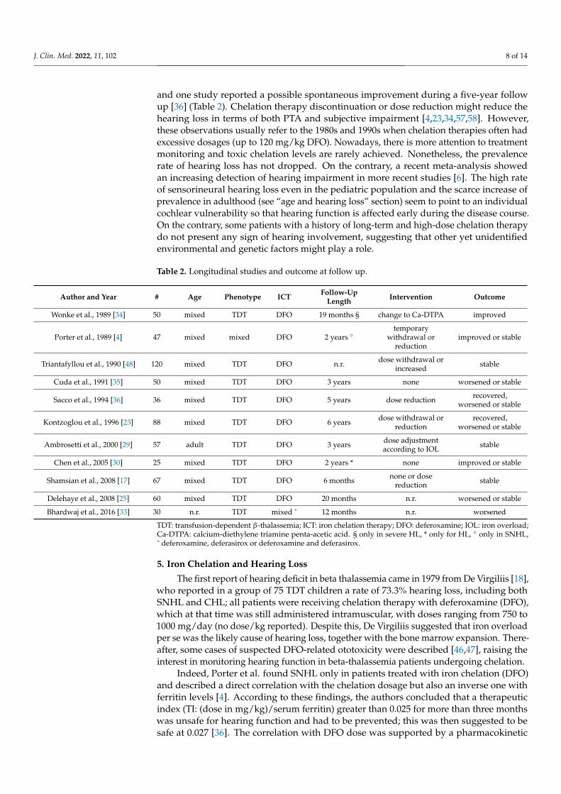
J. Clin. Med. 2022, 11, 102 8 of 14
and one study reported a possible spontaneous improvement during a five-year followup [36] (Table 2). Chelation therapy discontinuation or dose reduction might reduce thehearing loss in terms of both PTA and subjective impairment [4,23,34,57,58]. However,these observations usually refer to the 1980s and 1990s when chelation therapies often hadexcessive dosages (up to 120 mg/kg DFO). Nowadays, there is more attention to treatmentmonitoring and toxic chelation levels are rarely achieved. Nonetheless, the prevalencerate of hearing loss has not dropped. On the contrary, a recent meta-analysis showedan increasing detection of hearing impairment in more recent studies [6]. The high rateof sensorineural hearing loss even in the pediatric population and the scarce increase ofprevalence in adulthood (see “age and hearing loss” section) seem to point to an individualcochlear vulnerability so that hearing function is affected early during the disease course.On the contrary, some patients with a history of long-term and high-dose chelation therapydo not present any sign of hearing involvement, suggesting that other yet unidentifiedenvironmental and genetic factors might play a role.
Table 2. Longitudinal studies and outcome at follow up.
Author and Year # Age Phenotype ICT Follow-UpLength Intervention Outcome
Wonke et al., 1989 [34] 50 mixed TDT DFO 19 months § change to Ca-DTPA improved
Porter et al., 1989 [4] 47 mixed mixed DFO 2 years ◦temporary
withdrawal orreduction
improved or stable
Triantafyllou et al., 1990 [48] 120 mixed TDT DFO n.r. dose withdrawal orincreased stable
Cuda et al., 1991 [35] 50 mixed TDT DFO 3 years none worsened or stable
Sacco et al., 1994 [36] 36 mixed TDT DFO 5 years dose reduction recovered,worsened or stable
Kontzoglou et al., 1996 [23] 88 mixed TDT DFO 6 years dose withdrawal orreduction
recovered,worsened or stable
Ambrosetti et al., 2000 [29] 57 adult TDT DFO 3 years dose adjustmentaccording to IOL stable
Chen et al., 2005 [30] 25 mixed TDT DFO 2 years * none improved or stable
Shamsian et al., 2008 [17] 67 mixed TDT DFO 6 months none or dosereduction stable
Delehaye et al., 2008 [25] 60 mixed TDT DFO 20 months n.r. worsened or stable
Bhardwaj et al., 2016 [33] 30 n.r. TDT mixed ˆ 12 months n.r. worsened
TDT: transfusion-dependent β-thalassemia; ICT: iron chelation therapy; DFO: deferoxamine; IOL: iron overload;Ca-DTPA: calcium-diethylene triamine penta-acetic acid. § only in severe HL, * only for HL, ◦ only in SNHL,ˆ deferoxamine, deferasirox or deferoxamine and deferasirox.
5. Iron Chelation and Hearing Loss
The first report of hearing deficit in beta thalassemia came in 1979 from De Virgiliis [18],who reported in a group of 75 TDT children a rate of 73.3% hearing loss, including bothSNHL and CHL; all patients were receiving chelation therapy with deferoxamine (DFO),which at that time was still administered intramuscular, with doses ranging from 750 to1000 mg/day (no dose/kg reported). Despite this, De Virgiliis suggested that iron overloadper se was the likely cause of hearing loss, together with the bone marrow expansion. There-after, some cases of suspected DFO-related ototoxicity were described [46,47], raising theinterest in monitoring hearing function in beta-thalassemia patients undergoing chelation.
Indeed, Porter et al. found SNHL only in patients treated with iron chelation (DFO)and described a direct correlation with the chelation dosage but also an inverse one withferritin levels [4]. According to these findings, the authors concluded that a therapeuticindex (TI: (dose in mg/kg)/serum ferritin) greater than 0.025 for more than three monthswas unsafe for hearing function and had to be prevented; this was then suggested to besafe at 0.027 [36]. The correlation with DFO dose was supported by a pharmacokinetic
J. Clin. Med. 2022, 11, 102 9 of 14
study in TDT children with and without neurotoxicity (hearing/vision loss), showing thatthe administered dose was higher in “neurotoxic” subjects [59].
The available literature however is not in agreement with regards to iron chelation’srole in causing hearing loss; our analysis shows that a correlation with the dose of the ironchelators is reported mostly when doses are higher than the recommended (i.e., greaterthan 40 mg/kg/d for DFO).
Most studies were conducted on patients receiving DFO, so that data are scarce for theother two iron chelators; only in two studies deferasirox was the sole chelator used, and nostudy was found on patients on deferiprone monotherapy; six studies included patientson different iron chelation drugs, but they did not report separate data for each chelator(Table 1). In addition, it is not clear if patients treated with deferasirox or deferiprone werepreviously treated with DFO.
Lastly, no study compared the rate of hearing loss among the three different chelatorsor analyzed the prevalence rate among patients naive for iron chelation. Interestingly,abnormal BAEPs were registered in a group of NTDT subjects, not on iron chelation [38,50],and recently, SNHL was reported even among some NTDT patients who were not onchelation treatment [32], highlighting that SNHL could also be a complication of beta-thalassemia itself. Indeed, beta-thalassemia patients (regardless of disease severity andchelation therapy) showed brain perfusion changes at the level of the primary auditorycortex suggesting a more complex pathogenesis of hearing dysfunction [32].
Further investigations are therefore warranted also in not-chelated and not-transfusedbeta thalassemia patients to achieve more information about the role of chelation treatmentin the SNHL onset.
6. Iron Overload and Hearing Loss
One of the main complications in TDT patients is the secondary hemochromatosis dueto tissue iron deposition occurring during the disease course and the prolonged transfusionregimen. Current guidelines recommend that therapy must be started after serum ferritinlevels are above 1000 µg/L. The long-term control of serum ferritin has been linked toprotection from heart disease and to improved survival if levels are consistently less than2500 µg/L [60], with even better outcomes at levels <1000 µg/L [61]. The excessive irondeposition in the heart, liver and endocrine glands is responsible for heart failure, liverfibrosis and cirrhosis, diabetes mellitus, hypogonadism, growth failure, sexual immaturity,and immunological alterations.
Serum ferritin levels have been also implicated with hearing impairment development, butthe relationship is still controversial. Most authors found no significant ferritin level differencesbetween patients with and without hearing loss [12,14,17,20,23–25,29,30,35,44,48,58,62].
In the study by Porter et al. in 1989, low serum ferritin appeared as a risk factor forSNHL, since all patients with hearing impairment had serum ferritin below 2000 µg/L [4].This association was confirmed in a few subsequent studies [26–28,45] in patients withthalassemia undergoing long-term transfusion therapy, and it was attributed to the invasivetreatment of DFO to reduce iron overload in patients with ferritin levels above 3000 µg/L [6],or to the excessive depletion of metals in overtreated patients [49]. However, two smallsample studies had opposite results suggesting that iron overload could also be associatedwith auditory deficiency at high frequencies [18,31].
Thus, according to the available literature, both iron overload and excessive iron-chelating therapy should be prevented, and a tailored chelation treatment should be adopted.
7. Anemia and Hearing Loss
The possible relationship between hearing impairment and the severity of anemia(hemoglobin levels or number of transfusions) was investigated in eight papers. More than1200 patients undergoing regular transfusion regimen were analyzed with the followingresults: abnormal PTA results and hearing loss were non-related with pre-transfusionhemoglobin [26,44], duration of transfusion therapy [26], age at the first blood trans-
J. Clin. Med. 2022, 11, 102 10 of 14
fusion [13], mean hemoglobin in the last three months [24], mean annual hemoglobinvalues [18,35] or unit of blood received per year [35].
In a small sample of TDT patients, a hearing threshold decrease correlated withhemoglobin levels, transfusions per year, and duration since the last transfusion. Theseresults were explained by a lower formation of insoluble alpha chains tetramers whenhemoglobin increases, but no other study supported this hypothesis. Furthermore, as totaltransfusion numbers increased, hearing thresholds at PTA and high frequency audiometrydecreased [31].
8. Sex and Hearing Loss
Several studies have investigated the relationship between sex and hearing loss inorder to identify additional risk factors for ototoxicity. In nine articles (757 beta-thalassemicpatients), the correlation between sex and hearing impairment was analyzed: most ofthese studies failed to detect a significant correlation [6,17,23,24,26–28]. Only two papersshowed a possible association between sex and hearing loss and found respectively anincreased [14] and a decreased [31] severity in males. Therefore, there seems to be norelationship between sex and hearing loss in beta thalassemia.
9. Tinnitus
Tinnitus is one of the most common hearing disorders, with wide-ranging risk factorsincluding age, hearing loss, noise exposure, inflammatory diseases, psychosocial distress,and ototoxic drugs. The latter may induce both reversible and irreversible damage ofthe inner ear structures causing tinnitus, with or without hearing loss and balance prob-lems [63]. These ototoxic effects seem to be related to the duration of therapy, route ofadministration, dosage, individual sensitivity, genetic predisposition, and altered renal andhepatic functions [64].
Data on tinnitus in thalassemic patients are rather scanty and often dispersed in theresults sections. They were acquired only by means of an anamnestic interview; no studyapplied specific instrumental examinations or standardized questionnaires. For these rea-sons tinnitus is often poorly characterized in terms of its severity, disability, correlation withage, or association with hearing loss. In addition, no study included a healthy control groupor mentioned the prevalence of tinnitus in the general population. In beta-thalassemia,tinnitus prevalence rate ranged widely from 3.3% to 38% [4,15,30,35,44,46,57,62,65], so thattinnitus was defined as either rare or common, up to being the most common hearingsymptom in thalassemic patients, both in subjects with normal audiogram and in thosewith hearing loss.
As tinnitus is often underreported by patients and it is frequently associated withototoxicity, even in the absence of hearing loss, dedicated investigations by means ofstandardized questionnaires or acufenometry (frequency measure; intensity measure andthe minimum level of tinnitus masking) are warranted to improve the follow up of beta-thalassemic patients.
10. Audiological Monitoring and Management
Audiological testing has become routinary practice in beta-thalassemia for multiplereasons, at least in the transfusion-dependent form. First, the need for potentially ototoxiclong-lasting therapies and the ageing due to an improved life expectancy are expected todetermine an increasing prevalence of hearing loss in beta-thalassemia patients. Second,hearing loss might be a precious warning light of toxicity of the ongoing therapy, thusallowing for prompt dosage adjustment; this approach was proven to often revert or reducethe perceived hearing loss [23,57]. Third, untreated hearing loss might cause a wide rangeof consequences, from social isolation to depression, and might predispose to an earlieronset of dementia [66].
Few longitudinal studies are available (see “hearing loss course” section). Currently,guidelines on audiological assessment and management are missing. As during the clin-
J. Clin. Med. 2022, 11, 102 11 of 14
ical trials of iron chelation, audiological assessment was done yearly, this approach hasbeen automatically translated in the clinical practice, even though there is no supportingevidence. Notably, even audiological data obtained during trials have been not or veryscarcely published so that the reason for such a strict follow up remains obscure [67].
Since the outset of the hearing loss is still unpredictable, audiological testing is sug-gested before the start of treatment to assess the hearing threshold. Audiological evaluationshould be considered mandatory in the case of the onset of hearing disorders (subjectivehearing loss, fullness, tinnitus). Integrating previous suggestions [33], in the case of pa-tients with normal hearing function, it seems to be reasonable to plan following tests eachyear in the pediatric age and every two years in adolescence (mostly due to the dramaticimpact of undiagnosed hearing loss on scholastic/learning performance). In adulthood,audiological testing seems to be reasonable every 3–5 years even in patients not reportinghearing impairment. The detection of a rapidly progressive hearing loss should lead to astricter follow up. Hearing aids should be promptly considered whenever hearing loss isimpairing daily social life [4].
11. Conclusions
Despite the large number of studies addressing the topic of hearing loss in beta-thalassemia, the discrepancies in recruitment and diagnostic criteria do not allow theobtainment of a reliable and precise picture of this problem. Future longitudinal studieswith a detailed description of sample, treatment, and hearing deficit will help understandthe pathogenesis, the prevalence, and the best management of hearing impairment inbeta-thalassemia.
Author Contributions: Conceptualization R.M. and I.T.; methodology, R.M. and I.T.; formal analysis,all authors; data curation R.M., I.T., R.C.: writing—original draft preparation, all authors.; writing—review and editing, all authors; supervision, R.M. and I.T.; funding acquisition, S.P. All authors haveread and agreed to the published version of the manuscript.
Funding: Financial support was provided by Programma VALERE (VAnviteLli pEr la RicErca),Università della Campania “L. Vanvitelli”, Naples, Italy (PI Silverio Perrotta), grant numbers:B68D19001880005, project MOXPRES.
Institutional Review Board Statement: Not Applicable.
Informed Consent Statement: Not Applicable.
Conflicts of Interest: The authors declare no conflict of interest.
References1. McGann, P.T.; Nero, A.C.; Ware, R.E. Clinical Features of β-Thalassemia and Sickle Cell Disease. Adv. Exp. Med. Biol. 2017, 1013,
1–26. [PubMed]2. Origa, R. β-Thalassemia. Genet. Med. 2017, 19, 609–619. [CrossRef] [PubMed]3. Lal, A. Challenges in Chronic Transfusion for Patients with Thalassemia. Hematol. Am. Soc. Hematol. Educ. Program 2020, 1,
160–166. [CrossRef]4. Porter, J.B.; Jaswon, M.S.; Huehns, E.R.; East, C.A.; Hazell, J.W.P. Desferrioxamine Ototoxicity: Evaluation of Risk Factors in
Thalassaemic Patients and Guidelines for Safe Dosage. Br. J. Haematol. 1989, 73, 403–409. [CrossRef] [PubMed]5. Chonat, S.; Quinn, C.T. Current Standards of Care and Long Term Outcomes for Thalassemia and Sickle Cell Disease. Adv. Exp.
Med. Biol. 2017, 1013, 59–87.6. Badfar, G.; Mansouri, A.; Shohan, M.; Karimi, H.; Khalighi, Z.; Rahmati, S.; Delpisheh, A.; Veisani, Y.; Soleymani, A.; Azami, M.
Hearing Loss in Iranian Thalassemia Major Patients Treated with Deferoxamine: A Systematic Review and Meta-Analysis. Casp.J. Intern. Med. 2017, 8, 239–249.
7. Gates, G.A.; Mills, J.H. Presbycusis. Lancet 2005, 366, 1111–1120. [CrossRef]8. Mihailescu, A.M.; Musallam, K.M.; Cappellini, M.D.; Taher, A.T. Less ‘Reds’ More ‘Blues’: Hemoglobin Level and Depression in
Non-Transfusion-Dependent Thalassemia. Ann. Hematol. 2020, 99, 903–904. [CrossRef]9. Monastero, R.; Monastero, G.; Ciaccio, C.; Padovani, A.; Camarda, R. Cognitive Deficits in Beta-Thalassemia Major. Acta Neurol.
Scand. 2000, 102, 162–168. [CrossRef] [PubMed]10. Raz, S.; Koren, A.; Dan, O.; Levin, C. Cognitive Functions in Adults with β-Thalassemia Major: Before and after Blood Transfusion
and Comparison with Healthy Controls. Ann. N. Y. Acad. Sci. 2016, 1375, 19–27. [CrossRef]
J. Clin. Med. 2022, 11, 102 12 of 14
11. Tartaglione, I.; Manara, R.; Caiazza, M.; Carafa, P.A.; Caserta, V.; Ferrantino, T.; Granato, I.; Ippolito, N.; Maietta, C.; Oliveto, T.Brain Functional Impairment in Beta-Thalassaemia: The Cognitive Profile in Italian Neurologically Asymptomatic Adult Patientsin Comparison to the Reported Literature. Br. J. Haematol. 2019, 186, 592–607. [CrossRef] [PubMed]
12. Argiolu, F.; Diana, G.; Avignone, A.; Cao, A.; Di Ninni, S. Hearing Impairment during Deferoxamine Therapy for ThalassemiaMajor. J. Pediatr. 1991, 118, 826–827. [CrossRef]
13. Faramarzi, A.; Karimi, M.; Heydari, S.; Shishegar, M.; Kaviani, M. Frequency of Sensory Neural Hearing Loss in Major Beta-Thalassemias in Southern Iran. Iran. J. Pediatr. 2010, 20, 308–312. [PubMed]
14. Sonbolestan, M.; Mokhtarinejad, F.; Omrani, M. An evaluation of sensory neural hearing loss in thalassaemic patients treatedwith desferrioxamine and its risk factors. J. Res. Med. Sci. 2005, 10, 210–216.
15. Berjis, N.; Sonbolestan, S.M.; Nemati, S.; Mokhtarinejad, F.; Danesh, Z.; Abdeyazdan, Z. Otorhinolaryngologic Manifestations inThalassemia Major Patients. Iran. J. Ped. 2007, 17, 15–18.
16. Masala, W.; Meloni, F.; Gallisai, D.; Careddu, M.; Secchi, G.; Cuccuru, G.B.; Loriga, V.; Salvo, G. Can deferoxamine be consideredan ototoxic drug? Scand. Audiol. Suppl. 1988, 30, 237–238. [PubMed]
17. Shamsian, B.S.; Aminasnafi, A.; Moghadassian, H.; Latif, G.M.; Arzanian, T.; Alavi, S.; Esfehani, H.; Garallahi, F.; Amini, R.Sensorineural hearing loss in β-thalassemia major patients treated with deferoxamine. J. Pediatr. Hematol. Oncol. 2008, 25, 502–508.[CrossRef] [PubMed]
18. De Virgiliis, S.; Argiolu, F.; Sanna, G.; Cornacchia, G.; Cossu, P.; Cao, A.; Mallardi, V.; Puxeddu, P. Auditory Involvement inThalassemia Major. Acta Haematol. 1979, 61, 209–215. [CrossRef]
19. Barratt, P.S.; Toogood, I.R. Hearing loss attributed to desferrioxamine in patients with beta-thalassaemia major. Med. J. Aust. 1987,147, 177–179. [CrossRef]
20. Albera, R.; Pia, F.; Morra, B.; Lacilla, M.; Bianco, L.; Gabutti, V.; Piga, A. Hearing Loss and Desferrioxamine in HomozygousBeta-Thalassemia. Audiology 1988, 27, 207–214. [CrossRef] [PubMed]
21. Cohen, A.; Martin, M.; Mizanin, J.; Konkle, D.F.; Schwartz, E. Vision and Hearing during Deferoxamine Therapy. J. Pediatr. 1990,117 Pt 1, 326–330. [CrossRef]
22. Onerci, M.; Aslan, S.; Gümrük, F.; Aksoy, S.; Belgin, E.; Ozçelik, T.; Altay, C. Audiologic and Impedancemetric Findings withinThalassaemic Patients. Int. J. Pediatr. Otorhinolaryngol. 1994, 28, 167–172. [CrossRef]
23. Kontzoglou, G.; Koussi, A.; Tsatra, J.; Noussios, G.; Vital, V.; Sagarakis, G.; Athanassiou, M. Sensorineural Hearing Loss inChildren with Thalassemia Major in Northern Greece. Int. J. Pediatr. Otorhinolaryngol. 1996, 35, 223–230. [CrossRef]
24. Karimi, M.; Asadi-Pooya, A.A.; Khademi, B.; Asadi-Pooya, K.; Yarmohammadi, H. Evaluation of the Incidence of SensorineuralHearing Loss in Beta-Thalassemia Major Patients under Regular Chelation Therapy with Desferrioxamine. Acta Haematol. 2002,108, 79–83. [CrossRef] [PubMed]
25. Delehaye, E.; Capobianco, S.; Bertetto, B.I.; Meloni, F. Distortion-Product Otoacoustic Emission: Early Detection in DeferoxamineInduced Ototoxicity. Auris Nasus Larynx 2008, 35, 198–202. [CrossRef] [PubMed]
26. Chao, Y.; Wu, K.; Lin, C.; Tsai, M.; Peng, C.; Wu, H.; Lin, C. Audiologic and Vestibular Assessment in Patients with β-ThalassemiaMajor Receiving Long-Term Transfusion Therapy. Pediatr. Blood Cancer 2013, 60, 1963–1966. [CrossRef]
27. Osma, U.; Kurtoglu, E.; Eyigor, H.; Yilmaz, M.D.; Aygener, N. Sensorineural Hearing Loss in β-Thalassemia Patients Treated withIron Chelation. Ear Nose Throat J. 2015, 94, 481–485. [CrossRef]
28. Alzaree, F.A.; Shehata, M.A.; Atti, M.A.; Elzaree, G.A.; El-Kassas, G.M. New Advances in Evaluation of Hearing in a Sample ofEgyptian Children with β-Thalassemia Major. Open Access Maced. J. Med. Sci. 2019, 7, 1494–1498. [CrossRef]
29. Ambrosetti, U.; Dondè, E.; Piatti, G.; Cappellini, M.D. Audiological Evaluation in Adult Beta-Thalassemia Major Patients underRegular Chelation Treatment. Pharmacol. Res. 2000, 42, 485–487. [CrossRef]
30. Chen, S.H.; Liang, D.C.; Lin, H.C.; Cheng, S.Y.; Chen, L.J.; Liu, H.C. Auditory and Visual Toxicity during Deferoxamine Therapyin Transfusion-Dependent Patients. J. Pediatr. Hematol. Oncol. 2005, 27, 651–653. [CrossRef] [PubMed]
31. Budak, B.; Bayar, M.N.; Gumruk, F.; Budak, G. Pure Tone and High Frequency Audiometries in Beta Thalassemia Major. KBB veBBC Dergisi 2008, 16, 10–15.
32. Manara, R.; Ponticorvo, S.; Perrotta, S.; Barillari, M.R.; Costa, G.; Brotto, D.; Di Concilio, R.; Ciancio, A.; de Michele, E.; Carafa,P.A.; et al. Auditory Cortex Hypoperfusion: A Metabolic Hallmark in Beta Thalassemia. Orphanet. J. Rare Dis. 2021, 16, 349.[CrossRef]
33. Bhardwaj, V.; Verma, R.; Chopra, H.; Sobti, P. Chelation-induced ototoxicity in thalassemic patients: Role of distortion-productotoacoustic emissions and various management parameters. Indian J. Otol. 2016, 22, 193–198.
34. Wonke, B.; Hoffbrand, A.V.; Aldouri, M.; Wickens, D.; Flynn, D.; Stearns, M.; Warner, P. Reversal of Desferrioxamine InducedAuditory Neurotoxicity during Treatment with Ca-DTPA. Arch. Dis. Child. 1989, 64, 77–82. [CrossRef] [PubMed]
35. Cuda, D.; De Benedetto, M.; Leante, M.; Corvaglia, E. The prevalence and evolution of hypoacusis in Cooley’s disease. ActaOtorhinolaryngol. Ital. 1991, 11, 471–481.
36. Sacco, M.; Meleleo, D.; Tricarico, N.; Greco Miani, A.; Serra, E.; Parlatore, L. Evaluation of desferrioxamine ototoxicity inthalassemic patients. Follow-up over a 5-year period and results. Minerva Pediatr. 1994, 46, 225–230.
37. Passat, J.; Bulan Ginting, M.; Fauzi, M.; Tambunan, T. Brainstem Auditory Evoked Potentials Features in Thalassemia Major.Paediatr. Indones. 2001, 41, 166–170. [CrossRef]
J. Clin. Med. 2022, 11, 102 13 of 14
38. Teli, A.; Economou, M.; Rudolf, J.; Tzovaras, F.; Gourtsa, V.; Kondou, A.; Kontopoulos, E.; Gombakis, N.; Athanassiou-Metaxa,M.; Zafeiriou, D. Subclinical Central Nervous System Involvement and Thrombophilic Status in Young Thalassemia IntermediaPatients of Greek Origin. Blood Coagul. Fibrinolysis 2012, 23, 195–2012. [CrossRef]
39. Sheikha, A.; Kameswaran, M.; Okafor, B.C.; Al-Saigh, A.-A. Otological Manifestations of Thalassaemia Intermedia: Evidence ofTemporal Bone Involvement and Report of a Unique Cholesteatoma-like Lesion. J. Laryngol. Otol. 1992, 106, 316–321. [CrossRef]
40. Lanigan, A.; Taylor Fordham, M. Temporal Bone Extramedullary Hematopoiesis as a Causeof Pediatric Bilateral ConductiveHearing loss:Case Report and Review of the Literature. Int. J. Pediatr. Otorhinolaryngol. 2017, 97, 135–138. [CrossRef] [PubMed]
41. Meara, J.G.; Potter, C.; Goodman, M.; Vernick, D. Extramedullary Hematopoiesis of the Middle Ear in a Patient with Thalassemia.Am. J. Otolaryngol. 1998, 19, 287–289. [CrossRef]
42. Thio, D.; Prasad, V.; Anslow, P.; Lennox, P. Marrow Proliferation as a Cause of Hearing Loss in Beta-Thalassaemia Major. J.Laryngol. Otol. 2008, 122, 1253–1256. [CrossRef] [PubMed]
43. Sirisena, M.; Birman, C.; McKibbin, A.; O’Brien, K. Bilateral Auditory Ossicular Expansions in a Child with Beta-ThalassemiaMajor: Case Report and Literature Review. Int. J. Pediatr. Otorhinolaryngol. 2018, 112, 126–131. [CrossRef] [PubMed]
44. Derin, S.; Azık, F.M.; Topal, Y.; Topal, H.; Karakus, V.; Çetinkaya, P.U.; Sahan, M.; Azık, T.E.; Kocabas, C.N. The Incidence ofOtotoxicity in Patients Using Iron Chelators. J. Int Adv. Otol. 2017, 13, 136–139. [CrossRef]
45. Olivieri, N.F.; Buncic, J.R.; Chew, E.; Gallant, T.; Harrison, R.V.; Keenan, N.; Logan, W.; Mitchell, D.; Ricci, G.; Skarf, B. Visualand Auditory Neurotoxicity in Patients Receiving Subcutaneous Deferoxamine Infusions. N. Engl. J. Med. 1986, 314, 869–873.[CrossRef]
46. Marsh, M.N.; Holbrook, I.B.; Clark, C.; Shaffer, J.L. Tinnitus in a Patient with Beta-Thalassaemia Intermedia on Long-TermTreatment with Desferrioxamine. Postgrad. Med. J. 1981, 57, 582–584. [CrossRef]
47. Orton, R.B.; Veber, L.L.; Sulh, H.M. Ocular and Auditory Toxicity of Long-Term, High-Dose Subcutaneous Deferoxamine Therapy.Can. J. Ophthalmol. 1985, 20, 153–156. [PubMed]
48. Triantafyllou, N.; Fisfis, M.; Sideris, G.; Triantafyllou, D.; Rombos, A.; Vrettou, H.; Mantouvalos, V.; Politi, C.; Malliara,S.; Papageorgiou, C. Neurophysiological and Neuro-Otological Study of Homozygous Beta-Thalassemia under Long-TermDesferrioxamine (DFO) Treatment. Acta Neurol. Scand. 1991, 83, 306–308. [CrossRef] [PubMed]
49. Wong, V.; Li, A.; Lee, A.C. Neurophysiologic Study of Beta-Thalassemia Patients. J. Child. Neurol. 1993, 8, 330–335. [CrossRef]50. Levine, J.E.; Cohen, A.; MacQueen, M.; Martin, M.; Giardina, P.J. Sensorimotor Neurotoxicity Associated with High-Dose
Deferoxamine Treatment. Pediatr. Hematol. Oncol. J. 1997, 19, 139–141. [CrossRef]51. Vir, D.; Panda, N.K.; Marwaha, R.K. Desferioximine induced Ototoxicity in Thalassemic patients. Ann. Neurosci. 2010, 17, 182–184.
[CrossRef]52. Uygun, V.; Kurtoglu, E. Iron-chelation therapy with oral chelators in patients with thalassemia major. Hematology 2013, 18, 50–55.
[CrossRef]53. Pereira da Silva, A.; Bebiano Coutinho, M.; Sena Esteves, S.; Telma Feliciano, T.; Almeida e Sousa, C. Conductive hearing loss in
Beta Thalassemia. Acta Otorrinolaringol. Gallega 2015, 8, 128–132.54. Hasan, A.F.; Salman, H.H.; Khalaf, J.M. Evaluation of hearing in patients with Beta Thalassemia Major. Basrah J. Surg. 2018, 24,
47–51.55. Khan, M.A.; Khan, M.A.; Seedat, A.M.; Khan, M.; Khuwaja, S.F.; Kumar, R.; Usama, S.M.; Fareed, S. Sensorineural Hearing
Loss and Its Relationship with Duration of Chelation Among Major β-Thalassemia Patients. Cureus 2019, 11, e5465. [CrossRef][PubMed]
56. Khalaf, Q.; Bargas, O. Otorhinological Manifestation in Patients with Thalassemia Major. Egypt. J. Ear Nose Throat Allied Sci. 2020,21, 111–115. [CrossRef]
57. Chiodo, A.A.; Alberti, P.W.; Sher, G.D.; Francombe, W.H.; Tyler, B. Desferrioxamine Ototoxicity in an Adult Transfusion-DependentPopulation. J. Otolaryngol. 1997, 26, 116–122. [PubMed]
58. Gallant, T.M.; Boyden, H.; Gallant, L.A.; Carley, H.; Freedman, M.H. Serial Studies of Auditory Neurotoxicity in Patients ReceivingDeferoxamine Therapy. Am. J. Med. 1987, 83, 83,1085–90. [CrossRef]
59. Bentur, Y.; Koren, G.; Tesoro, A.; Carley, H.; Olivieri, N.; Freedman, M.H. Comparison of deferoxamine pharmacokineticsbetween asymptomatic thalassemic children and those exhibiting severe neurotoxicity. Clin. Pharm. 1990, 47, 478–482. [CrossRef][PubMed]
60. Olivieri, N.F.; Nathan, D.G.; MacMillan, J.H.; Wayne, A.S.; Liu, P.P.; McGee, A.; Martin, M.; Koren, G.; Cohen, A.R. Survival inMedically Treated Patients with Homozygous Beta-Thalassemia. N. Engl. J. Med. 1994, 331, 574–578. [CrossRef]
61. Borgna-Pignatti, C.; Rugolotto, S.; De Stefano, P.; Zhao, H.; Cappellini, M.D.; Del Vecchio, G.C.; Romeo, M.A.; Forni, G.L.;Gamberini, M.R.; Ghilardi, R.; et al. Survival and Complications in Patients with Thalassemia Major Treated with Transfusionand Deferoxamine. Haematologica 2004, 89, 1187–1193.
62. Kong, M.H.; Goh, B.S.; Hamidah, A.; Zarina, A.L. The Prevalence of Sensorineural Hearing Loss in β-Thalassaemia PatientTreated with Desferrioxamine. Med. J. Malays. 2014, 69, 9–12.
63. Lord, S.G. Monitoring Protocols for Cochlear Toxicity. In Seminars in Hearing; Thieme Medical Publishers: New York, NY, USA,2019; Volume 40, pp. 122–143.
64. Lanvers-Kaminsky, C.; Zehnhoff-Dinnesen, A.; Parfitt, R.; Ciarimboli, G. Drug-Induced Ototoxicity: Mechanisms, Pharmacoge-netics, and Protective Strategies. Clin. Pharmacol. 2017, 101, 491–500. [CrossRef]

J. Clin. Med. 2022, 11, 102 14 of 14
65. Styles, L.A.; Vichinsky, E.P. Ototoxicity in Hemoglobinopathy Patients Chelated with Desferrioxamine. Pediatr. Hematol. Oncol. J.1996, 18, 42–45. [CrossRef]
66. Castiglione, A.; Casa, M.; Gallo, S.; Sorrentino, F.; Dhima, S.; Cilia, D.; Lovo, E.; Gambin, M.; Previato, M.; Colombo, S.; et al.Correspondence Between Cognitive and Audiological Evaluations Among the Elderly: A Preliminary Report of an AudiologicalScreening Model of Subjects at Risk of Cognitive Decline with Slight to Moderate Hearing Loss. Front. Neurosci. 2019, 13, 1279.[CrossRef] [PubMed]
67. Cappellini, M.D.; Cohen, A.; Piga, A.; Bejaoui, B.; Perrotta, S.; Agaoglu, L.; Aydinok, Y.; Aydinok, Y.; Kattamis, A.; Kilinc, Y.; et al.A Phase 3 Study of Deferasirox (ICL670), a Once-Daily Oral Iron Chelator, in Patients with Beta-Thalassemia. Blood 2006, 107,3455–3462. [CrossRef] [PubMed]
Related Documents