ARTICLE Healthy cloned offspring derived from freeze-dried somatic cells Sayaka Wakayama 1,2 ✉ , Daiyu Ito 1 , Erika Hayashi 1 , Takashi Ishiuchi 1 & Teruhiko Wakayama 1,2 ✉ Maintaining biodiversity is an essential task, but storing germ cells as genetic resources using liquid nitrogen is difficult, expensive, and easily disrupted during disasters. Our aim is to generate cloned mice from freeze-dried somatic cell nuclei, preserved at -30 °C for up to 9 months after freeze drying treatment. All somatic cells died after freeze drying, and nucleic DNA damage significantly increased. However, after nuclear transfer, we produced cloned blastocysts from freeze-dried somatic cells, and established nuclear transfer embryonic stem cell lines. Using these cells as nuclear donors for re-cloning, we obtained healthy cloned female and male mice with a success rate of 0.2–5.4%. Here, we show that freeze-dried somatic cells can produce healthy, fertile clones, suggesting that this technique may be important for the establishment of alternative, cheaper, and safer liquid nitrogen-free bio- banking solutions. https://doi.org/10.1038/s41467-022-31216-4 OPEN 1 Faculty of Life and Environmental Science, University of Yamanashi, Kofu 400-8510, Japan. 2 Advanced Biotechnology Center, University of Yamanashi, Kofu 400-8510, Japan. ✉ email: [email protected]; [email protected] NATURE COMMUNICATIONS | (2022)13:3666 | https://doi.org/10.1038/s41467-022-31216-4 | www.nature.com/naturecommunications 1 1234567890():,;

Welcome message from author
This document is posted to help you gain knowledge. Please leave a comment to let me know what you think about it! Share it to your friends and learn new things together.
Transcript
ARTICLE
Healthy cloned offspring derived from freeze-driedsomatic cellsSayaka Wakayama 1,2✉, Daiyu Ito 1, Erika Hayashi 1, Takashi Ishiuchi1 & Teruhiko Wakayama 1,2✉
Maintaining biodiversity is an essential task, but storing germ cells as genetic resources using
liquid nitrogen is difficult, expensive, and easily disrupted during disasters. Our aim is to
generate cloned mice from freeze-dried somatic cell nuclei, preserved at −30 °C for up to
9 months after freeze drying treatment. All somatic cells died after freeze drying, and nucleic
DNA damage significantly increased. However, after nuclear transfer, we produced cloned
blastocysts from freeze-dried somatic cells, and established nuclear transfer embryonic stem
cell lines. Using these cells as nuclear donors for re-cloning, we obtained healthy cloned
female and male mice with a success rate of 0.2–5.4%. Here, we show that freeze-dried
somatic cells can produce healthy, fertile clones, suggesting that this technique may be
important for the establishment of alternative, cheaper, and safer liquid nitrogen-free bio-
banking solutions.
https://doi.org/10.1038/s41467-022-31216-4 OPEN
1 Faculty of Life and Environmental Science, University of Yamanashi, Kofu 400-8510, Japan. 2 Advanced Biotechnology Center, University of Yamanashi,Kofu 400-8510, Japan. ✉email: [email protected]; [email protected]
NATURE COMMUNICATIONS | (2022) 13:3666 | https://doi.org/10.1038/s41467-022-31216-4 |www.nature.com/naturecommunications 1
1234
5678
90():,;
The preservation of genetic resources is an important tool inpromoting species survival. Although not all genetic traitsare required for survival, they must be preserved so that
species can survive if unknown diseases are spread or environ-mental changes, such as global warming, occur1. Spermatozoaand embryos of mammals are cryopreserved in liquid nitrogen(LN2)2,3. However, the use of LN2 has several drawbacks, such ashigh maintenance cost, and during a disaster, LN2 supply may bestopped or supplies may be destroyed. To solve this problem, wedeveloped a freeze-drying technique for mouse spermatozoa4.Although all spermatozoa died after the freeze-drying process,their DNA remained intact, and healthy offspring were obtainedwhen spermatozoa were injected into oocytes. This technique hasalso been applied to other species, such as rats, hamsters, rabbits,and horses5–8. The nuclei of freeze-dried (FD) mouse sperma-tozoa have a strong tolerance against environmental changes9;healthy offspring were obtained from FD sperm stored for morethan one year in a desk drawer without controlling roomtemperature10 and in the International Space Station for morethan 5 years11,12. Thus, freeze drying could be the best way topreserve genetic resources for a long period in a safe, low-cost,and location-independent manner13,14. However, to date, theonly cells that have produced offspring after freeze drying aremature spermatozoa. Collecting spermatozoa from infertile malesand oocytes/embryos from fertile females is difficult. Conversely,after the success of the first animal (frog) clone15,16 and the firstmammalian (sheep) clone17, it is now possible to generate clonedoffspring from live somatic cells18; this indicates that somatic cellscan also be used as a genetic resource. It is noteworthy thatsomatic cells can be collected from almost anywhere in the bodyor even from body waste19 or cadavers20,21.
In this study, we aimed to generate cloned mice from FDsomatic cell nuclei by adapting the nuclear transfer procedure.Our data reveal that although some DNA abnormalities areobserved in the process, FD somatic cell nuclei can be used togenerate blastocysts by nuclear transfer, and embryonic stem celllines derived from these blastocysts yield donor nuclei that arecapable of producing healthy, fertile cloned mice. The resultsobtained in this study may provide a viable method for preservingthe genetic resources of any animal in a safe and low-cost man-ner, even if power and LN2 supplies are interrupted during adisaster.
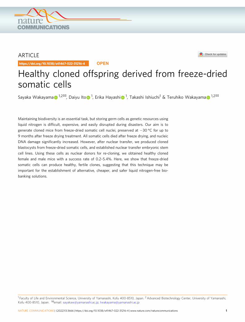
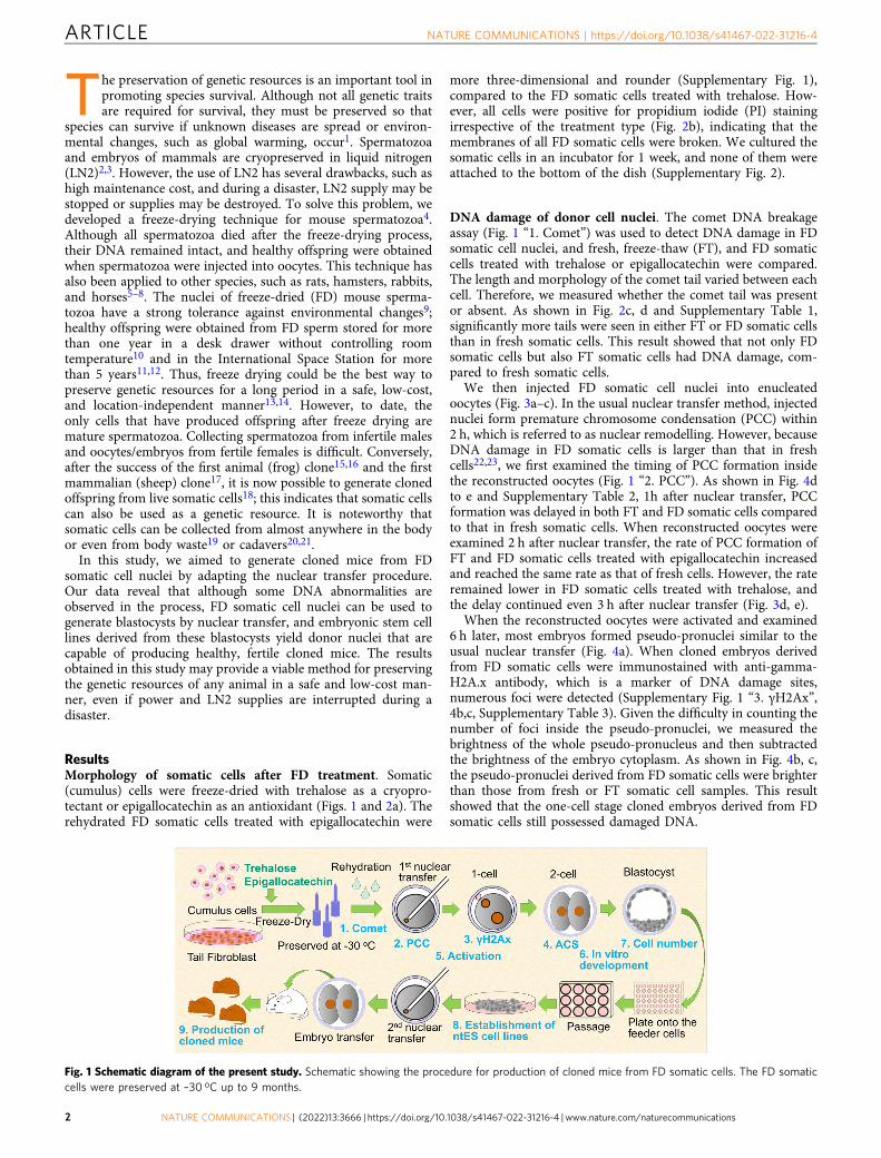
ResultsMorphology of somatic cells after FD treatment. Somatic(cumulus) cells were freeze-dried with trehalose as a cryopro-tectant or epigallocatechin as an antioxidant (Figs. 1 and 2a). Therehydrated FD somatic cells treated with epigallocatechin were
more three-dimensional and rounder (Supplementary Fig. 1),compared to the FD somatic cells treated with trehalose. How-ever, all cells were positive for propidium iodide (PI) stainingirrespective of the treatment type (Fig. 2b), indicating that themembranes of all FD somatic cells were broken. We cultured thesomatic cells in an incubator for 1 week, and none of them wereattached to the bottom of the dish (Supplementary Fig. 2).
DNA damage of donor cell nuclei. The comet DNA breakageassay (Fig. 1 “1. Comet”) was used to detect DNA damage in FDsomatic cell nuclei, and fresh, freeze-thaw (FT), and FD somaticcells treated with trehalose or epigallocatechin were compared.The length and morphology of the comet tail varied between eachcell. Therefore, we measured whether the comet tail was presentor absent. As shown in Fig. 2c, d and Supplementary Table 1,significantly more tails were seen in either FT or FD somatic cellsthan in fresh somatic cells. This result showed that not only FDsomatic cells but also FT somatic cells had DNA damage, com-pared to fresh somatic cells.
We then injected FD somatic cell nuclei into enucleatedoocytes (Fig. 3a–c). In the usual nuclear transfer method, injectednuclei form premature chromosome condensation (PCC) within2 h, which is referred to as nuclear remodelling. However, becauseDNA damage in FD somatic cells is larger than that in freshcells22,23, we first examined the timing of PCC formation insidethe reconstructed oocytes (Fig. 1 “2. PCC”). As shown in Fig. 4dto e and Supplementary Table 2, 1h after nuclear transfer, PCCformation was delayed in both FT and FD somatic cells comparedto that in fresh somatic cells. When reconstructed oocytes wereexamined 2 h after nuclear transfer, the rate of PCC formation ofFT and FD somatic cells treated with epigallocatechin increasedand reached the same rate as that of fresh cells. However, the rateremained lower in FD somatic cells treated with trehalose, andthe delay continued even 3 h after nuclear transfer (Fig. 3d, e).
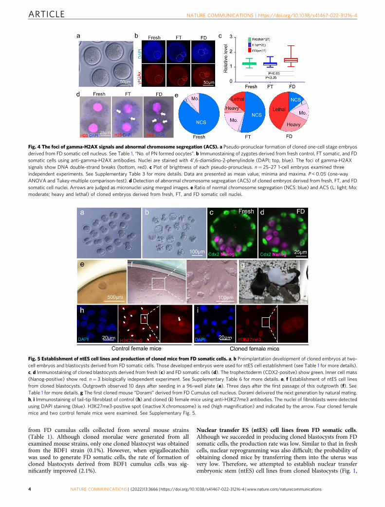
When the reconstructed oocytes were activated and examined6 h later, most embryos formed pseudo-pronuclei similar to theusual nuclear transfer (Fig. 4a). When cloned embryos derivedfrom FD somatic cells were immunostained with anti-gamma-H2A.x antibody, which is a marker of DNA damage sites,numerous foci were detected (Supplementary Fig. 1 “3. γH2Ax”,4b,c, Supplementary Table 3). Given the difficulty in counting thenumber of foci inside the pseudo-pronuclei, we measured thebrightness of the whole pseudo-pronucleus and then subtractedthe brightness of the embryo cytoplasm. As shown in Fig. 4b, c,the pseudo-pronuclei derived from FD somatic cells were brighterthan those from fresh or FT somatic cell samples. This resultshowed that the one-cell stage cloned embryos derived from FDsomatic cells still possessed damaged DNA.
Fig. 1 Schematic diagram of the present study. Schematic showing the procedure for production of cloned mice from FD somatic cells. The FD somaticcells were preserved at –30 oC up to 9 months.
ARTICLE NATURE COMMUNICATIONS | https://doi.org/10.1038/s41467-022-31216-4
2 NATURE COMMUNICATIONS | (2022) 13:3666 | https://doi.org/10.1038/s41467-022-31216-4 | www.nature.com/naturecommunications
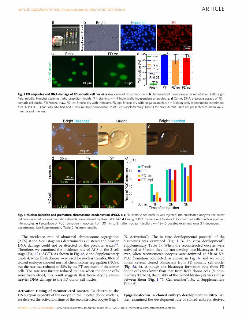
The incidence rate of abnormal chromosome segregation(ACS) at the 2-cell stage was determined as clustered and heavierDNA damage could not be detected by the previous assays24.Therefore, we examined the incidence rate of ACS at the 2-cellstage (Fig. 1 “4. ACS”). As shown in Fig. 4d, e and SupplementaryTable 4, when fresh donors were used for nuclear transfer, 86% ofcloned embryos showed normal chromosome segregation (NCS),but the rate was reduced to 53% by the FT treatment of the donorcells. The rate was further reduced to 14% when the donor cellswere freeze-dried; this result suggests that freeze drying causesheavier DNA damage to the FD donor cell nuclei.
Activation timing of reconstructed oocytes. To determine theDNA repair capacity of the oocyte in the injected donor nucleus,we delayed the activation time of the reconstructed oocyte (Fig. 1
“5. Activation”). The in vitro developmental potential of theblastocysts was examined (Fig. 1 “6. In vitro development”,Supplementary Table 5). When the reconstructed oocytes wereactivated at 30 min, they did not develop into blastocysts. How-ever, when reconstructed oocytes were activated at 3 h or 5 h,PCC formation completed, as shown in Fig. 3e and we couldobtain several cloned blastocysts from FD somatic cell nuclei(Fig. 5a, b). Although the blastocyst formation rate from FDdonor cells was lower than that from fresh donor cells (Supple-mentary Table 5), the quality of the cloned blastocysts was similarbetween them (Fig. 1 “7. Cell number”, 5c, d, SupplementaryTable 6).
Epigallocatechin in cloned embryo development in vitro. Wethen examined the development rate of cloned embryos derived
Fig. 2 FD ampoules and DNA damage of FD somatic cell nuclei. a Ampoules of FD somatic cells. b Damaged cell membrane after rehydration. Left: brightfield; middle: Hoechst staining; right: propidium iodide (PI) staining. n= 4 biologically independent ampoules. c, d Comet DNA breakage assays of FDsomatic cell nuclei. FT: Freeze-thaw. FD-tre: Freeze-dry with trehalose. FD-epi: Freeze-dry with epigallocatechin. n= 3 biologically independent experiment.a vs. b: P < 0.05 (one-way ANOVA and Tukey-multiple comparison-test). See Supplementary Table 1 for more details. Data are presented as mean value,minima and maxima.
Fig. 3 Nuclear injection and premature chromosome condensation (PCC). a–c FD somatic cell nucleus was injected into enucleated oocytes; the arrowindicates injected nucleus. Somatic cell nuclei were stained by Hoechst33342. d Timing of PCC formation of fresh or FD somatic cells after nuclear injectioninto oocytes. e Percentage of PCC formation in oocytes from 30min to 3 h after nuclear injection. n= 18–40 oocytes examined over 3 independentexperiments. See Supplementary Table 2 for more details.
NATURE COMMUNICATIONS | https://doi.org/10.1038/s41467-022-31216-4 ARTICLE
NATURE COMMUNICATIONS | (2022) 13:3666 | https://doi.org/10.1038/s41467-022-31216-4 |www.nature.com/naturecommunications 3
from FD cumulus cells collected from several mouse strains(Table 1). Although cloned morulae were generated from allexamined mouse strains, only one cloned blastocyst was obtainedfrom the BDF1 strain (0.1%). However, when epigallocatechinwas used to generate FD somatic cells, the rate of formation ofcloned blastocysts derived from BDF1 cumulus cells was sig-nificantly improved (2.1%).
Nuclear transfer ES (ntES) cell lines from FD somatic cells.Although we succeeded in producing cloned blastocysts from FDsomatic cells, the production rate was low. Similar to that in freshcells, nuclear reprogramming was also difficult; the probability ofobtaining cloned mice by transferring them into the uterus wasvery low. Therefore, we attempted to establish nuclear transferembryonic stem (ntES) cell lines from cloned blastocysts (Fig. 1,
Fig. 4 The foci of gamma-H2AX signals and abnormal chromosome segregation (ACS). a Pseudo-pronuclear formation of cloned one-cell stage embryosderived from FD somatic cell nucleus. See Table 1, “No. of PN formed oocytes”. b Immunostaining of zygotes derived from fresh control, FT somatic, and FDsomatic cells using anti-gamma-H2AX antibodies. Nuclei are stained with 4’,6-diamidino-2-phenylindole (DAPI; top, blue). The foci of gamma-H2AXsignals show DNA double-strand breaks (bottom, red). c Plot of brightness of each pseudo-pronucleus. n= 25–27 1-cell embryos examined threeindependent experiments. See Supplementary Table 3 for more details. Data are presented as mean value, minima and maxima. P < 0.05 (one-wayANOVA and Tukey-multiple comparison-test). d Detection of abnormal chromosome segregation (ACS) of cloned embryos derived from fresh, FT, and FDsomatic cell nuclei. Arrows are judged as micronuclei using merged images. e Ratio of normal chromosome segregation (NCS: blue) and ACS (L: light; Mo:moderate; heavy and lethal) of cloned embryos derived from fresh, FT, and FD somatic cell nuclei.
Fig. 5 Establishment of ntES cell lines and production of cloned mice from FD somatic cells. a, b Preimplantation development of cloned embryos at two-cell embryos and blastocysts derived from FD somatic cells. Those developed embryos were used for ntES cell establishment (see Table 1 for more details).c, d Immunostaining of cloned blastocysts derived from fresh (c) and FD somatic cells (d). The trophectoderm (CDX2-positve) show green. Inner cell mass(Nanog-positive) show red. n= 3 biologically independent experiment. See Supplementary Table 6 for more details. e, f Establishment of ntES cell linesfrom cloned blastocysts. Outgrowth observed 10 days after seeding in a 96-well plate (e). Three days after the first passage of this outgrowth (f). SeeTable 1 for more details. g The first cloned mouse “Dorami” derived from FD Cumulus cell nucleus. Dorami delivered the next generation by natural mating.h, i Immunostaining of tail-tip fibroblast of control (h) and cloned (i) female mice using anti-H3K27me3 antibodies. The nuclei of fibroblasts were detectedusing DAPI staining (blue). H3K27me3-positive spot (inactive X chromosome) is red (high magnification) and indicated by the arrow. Four cloned femalemice and two control female mice were examined. See Supplementary Fig. 5.
ARTICLE NATURE COMMUNICATIONS | https://doi.org/10.1038/s41467-022-31216-4
4 NATURE COMMUNICATIONS | (2022) 13:3666 | https://doi.org/10.1038/s41467-022-31216-4 | www.nature.com/naturecommunications
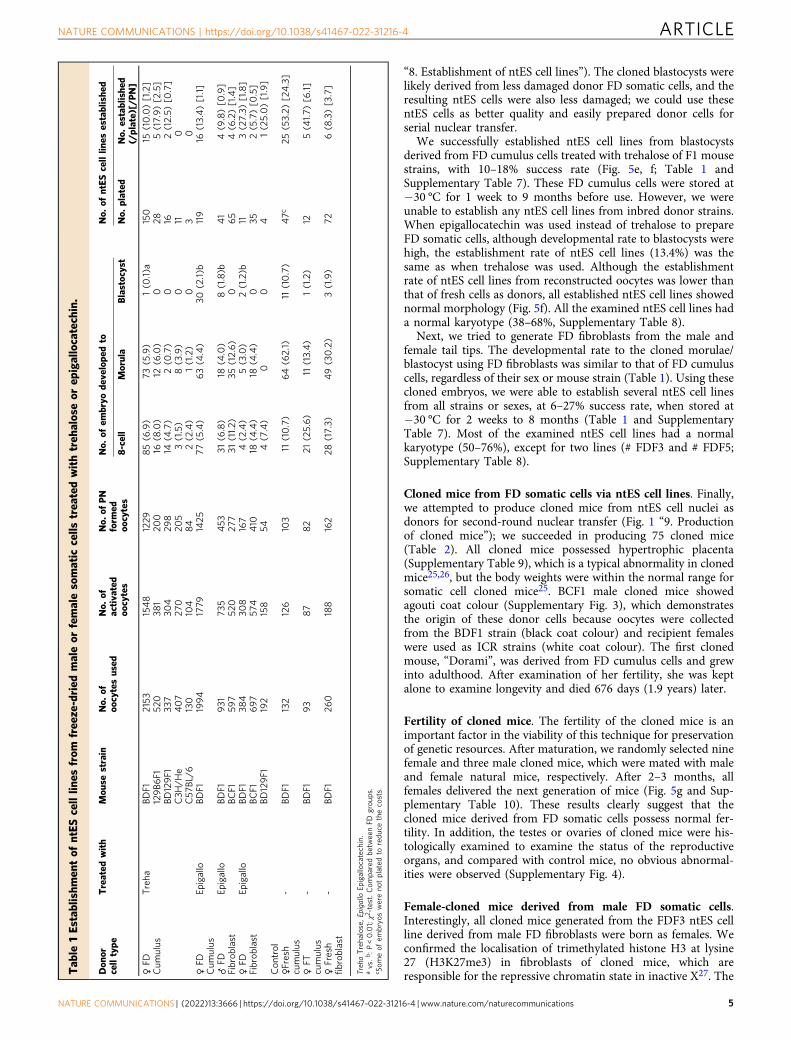
“8. Establishment of ntES cell lines”). The cloned blastocysts werelikely derived from less damaged donor FD somatic cells, and theresulting ntES cells were also less damaged; we could use thesentES cells as better quality and easily prepared donor cells forserial nuclear transfer.
We successfully established ntES cell lines from blastocystsderived from FD cumulus cells treated with trehalose of F1 mousestrains, with 10–18% success rate (Fig. 5e, f; Table 1 andSupplementary Table 7). These FD cumulus cells were stored at−30 °C for 1 week to 9 months before use. However, we wereunable to establish any ntES cell lines from inbred donor strains.When epigallocatechin was used instead of trehalose to prepareFD somatic cells, although developmental rate to blastocysts werehigh, the establishment rate of ntES cell lines (13.4%) was thesame as when trehalose was used. Although the establishmentrate of ntES cell lines from reconstructed oocytes was lower thanthat of fresh cells as donors, all established ntES cell lines showednormal morphology (Fig. 5f). All the examined ntES cell lines hada normal karyotype (38–68%, Supplementary Table 8).
Next, we tried to generate FD fibroblasts from the male andfemale tail tips. The developmental rate to the cloned morulae/blastocyst using FD fibroblasts was similar to that of FD cumuluscells, regardless of their sex or mouse strain (Table 1). Using thesecloned embryos, we were able to establish several ntES cell linesfrom all strains or sexes, at 6–27% success rate, when stored at−30 °C for 2 weeks to 8 months (Table 1 and SupplementaryTable 7). Most of the examined ntES cell lines had a normalkaryotype (50–76%), except for two lines (# FDF3 and # FDF5;Supplementary Table 8).
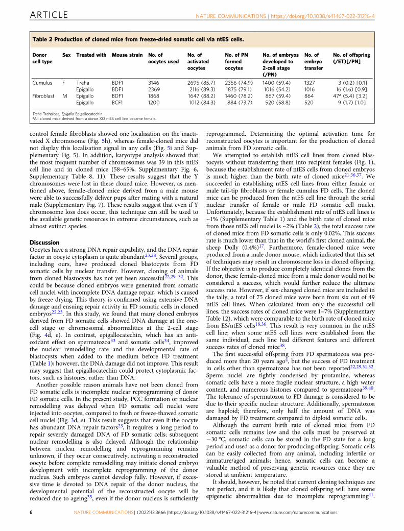
Cloned mice from FD somatic cells via ntES cell lines. Finally,we attempted to produce cloned mice from ntES cell nuclei asdonors for second-round nuclear transfer (Fig. 1 “9. Productionof cloned mice”); we succeeded in producing 75 cloned mice(Table 2). All cloned mice possessed hypertrophic placenta(Supplementary Table 9), which is a typical abnormality in clonedmice25,26, but the body weights were within the normal range forsomatic cell cloned mice25. BCF1 male cloned mice showedagouti coat colour (Supplementary Fig. 3), which demonstratesthe origin of these donor cells because oocytes were collectedfrom the BDF1 strain (black coat colour) and recipient femaleswere used as ICR strains (white coat colour). The first clonedmouse, “Dorami”, was derived from FD cumulus cells and grewinto adulthood. After examination of her fertility, she was keptalone to examine longevity and died 676 days (1.9 years) later.
Fertility of cloned mice. The fertility of the cloned mice is animportant factor in the viability of this technique for preservationof genetic resources. After maturation, we randomly selected ninefemale and three male cloned mice, which were mated with maleand female natural mice, respectively. After 2–3 months, allfemales delivered the next generation of mice (Fig. 5g and Sup-plementary Table 10). These results clearly suggest that thecloned mice derived from FD somatic cells possess normal fer-tility. In addition, the testes or ovaries of cloned mice were his-tologically examined to examine the status of the reproductiveorgans, and compared with control mice, no obvious abnormal-ities were observed (Supplementary Fig. 4).
Female-cloned mice derived from male FD somatic cells.Interestingly, all cloned mice generated from the FDF3 ntES cellline derived from male FD fibroblasts were born as females. Weconfirmed the localisation of trimethylated histone H3 at lysine27 (H3K27me3) in fibroblasts of cloned mice, which areresponsible for the repressive chromatin state in inactive X27. TheT
able
1Establishm
entof
ntES
celllin
esfrom
free
ze-dried
maleor
femalesomatic
cells
trea
tedwithtreh
aloseor
epigallocatechin.
Don
orcelltype
Treated
with
Mou
sestrain
No.
ofoo
cytesused
No.
ofactivated
oocytes
No.
ofPN
form
edoo
cytes
No.
ofem
bryo
develope
dto
No.
ofntES
celllin
esestablishe
d
8-cell
Morula
Blastocyst
No.
plated
No.
establishe
d(/plate)[/PN]
♀FD
Cum
ulus
Treha
BDF1
2153
1548
1229
85(6.9)
73(5.9)
1(0
.1)a
150
15(10.0)[1.2]
129B6
F152
038
120
016
(8.0)
12(6.0)
028
5(17.9)[2.5]
BD129F1
337
304
298
14(4.7)
2(0
.7)
016
2(12.5)
[0.7]
C3H
/He
407
270
205
3(1.5)
8(3.9)
011
0C57
BL/6
130
104
84
2(2.4)
1(1.2)
03
0♀FD
Cum
ulus
Epigallo
BDF1
1994
1779
1425
77(5.4)
63(4.4)
30(2.1)b
119
16(13.4)[1.1]
♂FD
Fibrob
last
Epigallo
BDF1
931
735
453
31(6.8)
18(4.0)
8(1.8)b
41
4(9.8)[0
.9]
BCF1
597
520
277
31(11.2)
35(12.6)
065
4(6.2)[1.4]
♀FD
Fibrob
last
Epigallo
BDF1
384
308
167
4(2.4)
5(3.0)
2(1.2)b
113(27.3)
[1.8]
BCF1
697
574
410
18(4.4)
18(4.4)
035
2(5.7)[0
.5]
BD129F1
192
158
544(7.4)
00
41(25.0)[1.9]
Con
trol
♀Fresh
cumulus
-BD
F1132
126
103
11(10.7)
64(62.1)
11(10.7)
47c
25(53.2)
[24.3]
♀FT
cumulus
-BD
F193
87
82
21(25.6)
11(13.4)
1(1.2)
125(41.7)
[6.1]
♀Fresh
fibrob
last
-BD
F126
0188
162
28(17.3)
49(30.2)
3(1.9)
726(8.3)[3.7]
TrehaTrehalose,E
pigallo
Epigallocatechin.
avs.b:P
<0.01;χ2-test.Com
paredbe
tweenFD
grou
ps.
c Som
eof
embryoswereno
tplated
toredu
cethecosts.
NATURE COMMUNICATIONS | https://doi.org/10.1038/s41467-022-31216-4 ARTICLE
NATURE COMMUNICATIONS | (2022) 13:3666 | https://doi.org/10.1038/s41467-022-31216-4 |www.nature.com/naturecommunications 5
control female fibroblasts showed one localisation on the inacti-vated X chromosome (Fig. 5h), whereas female-cloned mice didnot display this localisation signal in any cells (Fig. 5i and Sup-plementary Fig. 5). In addition, karyotype analysis showed thatthe most frequent number of chromosomes was 39 in this ntEScell line and in cloned mice (58–65%, Supplementary Fig. 6,Supplementary Table 8, 11). These results suggest that the Ychromosomes were lost in these cloned mice. However, as men-tioned above, female-cloned mice derived from a male mousewere able to successfully deliver pups after mating with a naturalmale (Supplementary Fig. 7). These results suggest that even if Ychromosome loss does occur, this technique can still be used tothe available genetic resources in extreme circumstances, such asalmost extinct species.
DiscussionOocytes have a strong DNA repair capability, and the DNA repairfactor in oocyte cytoplasm is quite abundant23,28. Several groups,including ours, have produced cloned blastocysts from FDsomatic cells by nuclear transfer. However, cloning of animalsfrom cloned blastocysts has not yet been successful22,29–32. Thiscould be because cloned embryos were generated from somaticcell nuclei with incomplete DNA damage repair, which is causedby freeze drying. This theory is confirmed using extensive DNAdamage and ensuing repair activity in FD somatic cells in clonedembryos22,23. In this study, we found that many cloned embryosderived from FD somatic cells showed DNA damage at the one-cell stage or chromosomal abnormalities at the 2-cell stage(Fig. 4d, e). In contrast, epigallocatechin, which has an anti-oxidant effect on spermatozoa33 and somatic cells34, improvedthe nuclear remodelling rate and the developmental rate ofblastocysts when added to the medium before FD treatment(Table 1); however, the DNA damage did not improve. This resultmay suggest that epigallocatechin could protect cytoplasmic fac-tors, such as histones, rather than DNA.
Another possible reason animals have not been cloned fromFD somatic cells is incomplete nuclear reprogramming of donorFD somatic cells. In the present study, PCC formation or nuclearremodelling was delayed when FD somatic cell nuclei wereinjected into oocytes, compared to fresh or freeze-thawed somaticcell nuclei (Fig. 3d, e). This result suggests that even if the oocytehas abundant DNA repair factors23, it requires a long period torepair severely damaged DNA of FD somatic cells; subsequentnuclear remodelling is also delayed. Although the relationshipbetween nuclear remodelling and reprogramming remainsunknown, if they occur consecutively, activating a reconstructedoocyte before complete remodelling may initiate cloned embryodevelopment with incomplete reprogramming of the donornucleus. Such embryos cannot develop fully. However, if exces-sive time is devoted to DNA repair of the donor nucleus, thedevelopmental potential of the reconstructed oocyte will bereduced due to ageing35, even if the donor nucleus is sufficiently
reprogrammed. Determining the optimal activation time forreconstructed oocytes is important for the production of clonedanimals from FD somatic cells.
We attempted to establish ntES cell lines from cloned blas-tocysts without transferring them into recipient females (Fig. 1),because the establishment rate of ntES cells from cloned embryosis much higher than the birth rate of cloned mice21,36,37. Wesucceeded in establishing ntES cell lines from either female ormale tail-tip fibroblasts or female cumulus FD cells. The clonedmice can be produced from the ntES cell line through the serialnuclear transfer of female or male FD somatic cell nuclei.Unfortunately, because the establishment rate of ntES cell lines is~1% (Supplementary Table 1) and the birth rate of cloned micefrom those ntES cell nuclei is ~2% (Table 2), the total success rateof cloned mice from FD somatic cells is only 0.02%. This successrate is much lower than that in the world’s first cloned animal, thesheep Dolly (0.4%)17. Furthermore, female-cloned mice wereproduced from a male donor mouse, which indicated that this setof techniques may result in chromosome loss in cloned offspring.If the objective is to produce completely identical clones from thedonor, these female-cloned mice from a male donor would not beconsidered a success, which would further reduce the ultimatesuccess rate. However, if sex-changed cloned mice are included inthe tally, a total of 75 cloned mice were born from six out of 49ntES cell lines. When calculated from only the successful celllines, the success rates of cloned mice were 1–7% (SupplementaryTable 12), which were comparable to the birth rate of cloned micefrom ES/ntES cells18,36. This result is very common in the ntEScell line; when some ntES cell lines were established from thesame individual, each line had different features and differentsuccess rates of cloned mice38.
The first successful offspring from FD spermatozoa was pro-duced more than 20 years ago4, but the success of FD treatmentin cells other than spermatozoa has not been reported22,29,31,32.Sperm nuclei are tightly condensed by protamine, whereassomatic cells have a more fragile nuclear structure, a high watercontent, and numerous histones compared to spermatozoa39,40.The tolerance of spermatozoa to FD damage is considered to bedue to their specific nuclear structure. Additionally, spermatozoaare haploid; therefore, only half the amount of DNA wasdamaged by FD treatment compared to diploid somatic cells.
Although the current birth rate of cloned mice from FDsomatic cells remains low and the cells must be preserved at−30 °C, somatic cells can be stored in the FD state for a longperiod and used as a donor for producing offspring. Somatic cellscan be easily collected from any animal, including infertile orimmature/aged animals; hence, somatic cells can become avaluable method of preserving genetic resources once they arestored at ambient temperature.
It should, however, be noted that current cloning techniques arenot perfect, and it is likely that cloned offspring will have someepigenetic abnormalities due to incomplete reprogramming41.
Table 2 Production of cloned mice from freeze-dried somatic cell via ntES cells.
Donorcell type
Sex Treated with Mouse strain No. ofoocytes used
No. ofactivatedoocytes
No. of PNformedoocytes
No. of embryosdeveloped to2-cell stage(/PN)
No. ofembryotransfer
No. of offspring(/ET)[/PN]
Cumulus F Treha BDF1 3146 2695 (85.7) 2356 (74.9) 1400 (59.4) 1327 3 (0.2) [0.1]Epigallo BDF1 2369 2116 (89.3) 1875 (79.1) 1016 (54.2) 1016 16 (1.6) [0.9]
Fibroblast M Epigallo BDF1 1868 1647 (88.2) 1460 (78.2) 867 (59.4) 864 47a (5.4) [3.2]Epigallo BCF1 1200 1012 (84.3) 884 (73.7) 520 (58.8) 520 9 (1.7) [1.0]
Treha Trehalose, Epigallo Epigallocatechin.aAll cloned mice derived from a donor XO ntES cell line became female.
ARTICLE NATURE COMMUNICATIONS | https://doi.org/10.1038/s41467-022-31216-4
6 NATURE COMMUNICATIONS | (2022) 13:3666 | https://doi.org/10.1038/s41467-022-31216-4 | www.nature.com/naturecommunications
Nevertheless, we have previously demonstrated that the next gen-eration of cloned mice is normal even after nuclear transfer wasrepeated 25 times42. In this study, although cloned mice wereproduced by repeating nuclear transfer twice, these clones werefertile and therefore did not detract from the objective of preservinggenetic resources.
Interestingly, one of the ntES cell lines derived from male FDfibroblasts lost its Y chromosome and became an XO cell line; allthe cloned mice produced from that cell line became female. Sexchange occurs rarely in cloned animals43, and Y chromosomedeletion has been reported during ES cell line culture44. In thisstudy, we did not determine the precise point at which the Ychromosome was lost, such as during FD treatment, nucleartransplantation, or the establishment of ntES cells, and this resultdoes represent a failure to completely preserve the geneticresources of the donor mouse. However, if the same treatmentcould be performed in endangered species where only malessurvived, it would be possible to produce females and naturallypreserve the species. Ultimately, the preservation of somatic cellsby FD treatment will be an important method supporting theestablishment of alternative, cheaper, and safer bio-bankingsolutions.
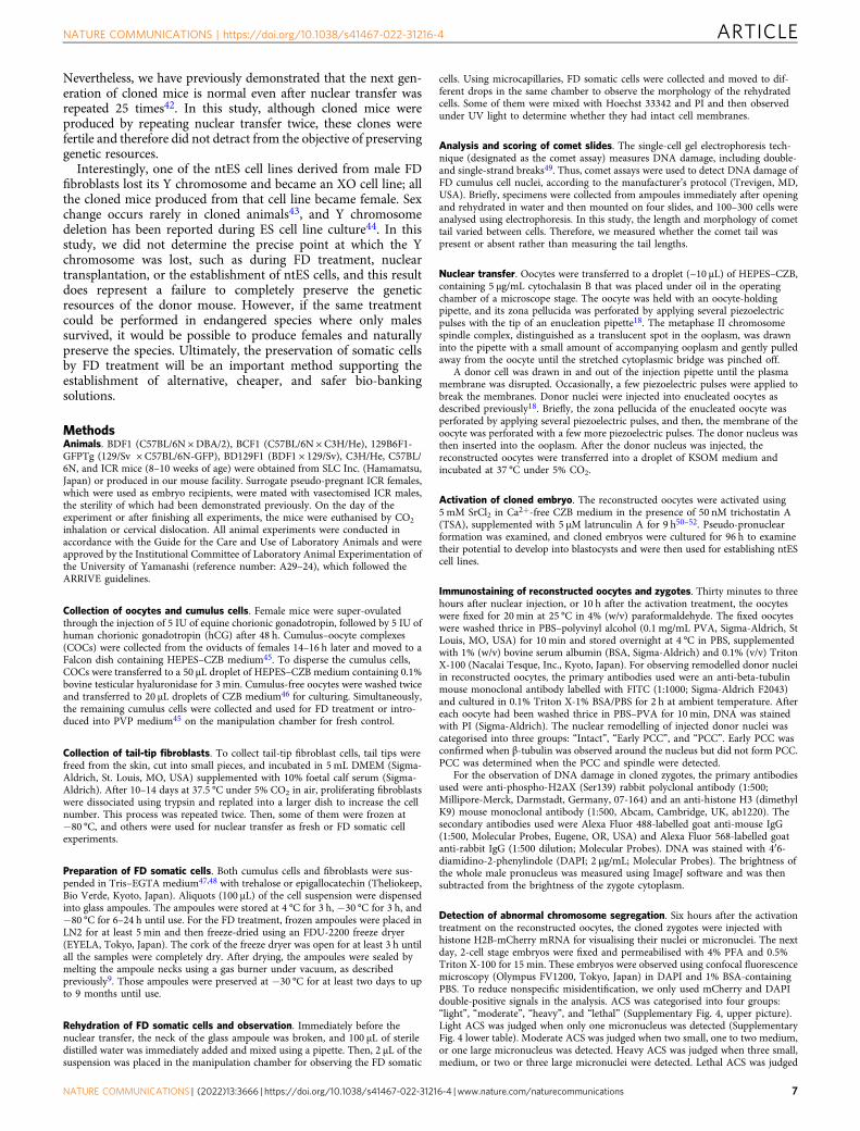
MethodsAnimals. BDF1 (C57BL/6N ×DBA/2), BCF1 (C57BL/6N × C3H/He), 129B6F1-GFPTg (129/Sv × C57BL/6N-GFP), BD129F1 (BDF1 × 129/Sv), C3H/He, C57BL/6N, and ICR mice (8–10 weeks of age) were obtained from SLC Inc. (Hamamatsu,Japan) or produced in our mouse facility. Surrogate pseudo-pregnant ICR females,which were used as embryo recipients, were mated with vasectomised ICR males,the sterility of which had been demonstrated previously. On the day of theexperiment or after finishing all experiments, the mice were euthanised by CO2
inhalation or cervical dislocation. All animal experiments were conducted inaccordance with the Guide for the Care and Use of Laboratory Animals and wereapproved by the Institutional Committee of Laboratory Animal Experimentation ofthe University of Yamanashi (reference number: A29–24), which followed theARRIVE guidelines.
Collection of oocytes and cumulus cells. Female mice were super-ovulatedthrough the injection of 5 IU of equine chorionic gonadotropin, followed by 5 IU ofhuman chorionic gonadotropin (hCG) after 48 h. Cumulus–oocyte complexes(COCs) were collected from the oviducts of females 14–16 h later and moved to aFalcon dish containing HEPES–CZB medium45. To disperse the cumulus cells,COCs were transferred to a 50 μL droplet of HEPES–CZB medium containing 0.1%bovine testicular hyaluronidase for 3 min. Cumulus-free oocytes were washed twiceand transferred to 20 μL droplets of CZB medium46 for culturing. Simultaneously,the remaining cumulus cells were collected and used for FD treatment or intro-duced into PVP medium45 on the manipulation chamber for fresh control.
Collection of tail-tip fibroblasts. To collect tail-tip fibroblast cells, tail tips werefreed from the skin, cut into small pieces, and incubated in 5 mL DMEM (Sigma-Aldrich, St. Louis, MO, USA) supplemented with 10% foetal calf serum (Sigma-Aldrich). After 10–14 days at 37.5 °C under 5% CO2 in air, proliferating fibroblastswere dissociated using trypsin and replated into a larger dish to increase the cellnumber. This process was repeated twice. Then, some of them were frozen at−80 °C, and others were used for nuclear transfer as fresh or FD somatic cellexperiments.
Preparation of FD somatic cells. Both cumulus cells and fibroblasts were sus-pended in Tris–EGTA medium47,48 with trehalose or epigallocatechin (Theliokeep,Bio Verde, Kyoto, Japan). Aliquots (100 μL) of the cell suspension were dispensedinto glass ampoules. The ampoules were stored at 4 °C for 3 h, −30 °C for 3 h, and−80 °C for 6–24 h until use. For the FD treatment, frozen ampoules were placed inLN2 for at least 5 min and then freeze-dried using an FDU-2200 freeze dryer(EYELA, Tokyo, Japan). The cork of the freeze dryer was open for at least 3 h untilall the samples were completely dry. After drying, the ampoules were sealed bymelting the ampoule necks using a gas burner under vacuum, as describedpreviously9. Those ampoules were preserved at −30 °C for at least two days to upto 9 months until use.
Rehydration of FD somatic cells and observation. Immediately before thenuclear transfer, the neck of the glass ampoule was broken, and 100 µL of steriledistilled water was immediately added and mixed using a pipette. Then, 2 µL of thesuspension was placed in the manipulation chamber for observing the FD somatic
cells. Using microcapillaries, FD somatic cells were collected and moved to dif-ferent drops in the same chamber to observe the morphology of the rehydratedcells. Some of them were mixed with Hoechst 33342 and PI and then observedunder UV light to determine whether they had intact cell membranes.
Analysis and scoring of comet slides. The single-cell gel electrophoresis tech-nique (designated as the comet assay) measures DNA damage, including double-and single-strand breaks49. Thus, comet assays were used to detect DNA damage ofFD cumulus cell nuclei, according to the manufacturer’s protocol (Trevigen, MD,USA). Briefly, specimens were collected from ampoules immediately after openingand rehydrated in water and then mounted on four slides, and 100–300 cells wereanalysed using electrophoresis. In this study, the length and morphology of comettail varied between cells. Therefore, we measured whether the comet tail waspresent or absent rather than measuring the tail lengths.
Nuclear transfer. Oocytes were transferred to a droplet (~10 μL) of HEPES–CZB,containing 5 μg/mL cytochalasin B that was placed under oil in the operatingchamber of a microscope stage. The oocyte was held with an oocyte-holdingpipette, and its zona pellucida was perforated by applying several piezoelectricpulses with the tip of an enucleation pipette18. The metaphase II chromosomespindle complex, distinguished as a translucent spot in the ooplasm, was drawninto the pipette with a small amount of accompanying ooplasm and gently pulledaway from the oocyte until the stretched cytoplasmic bridge was pinched off.
A donor cell was drawn in and out of the injection pipette until the plasmamembrane was disrupted. Occasionally, a few piezoelectric pulses were applied tobreak the membranes. Donor nuclei were injected into enucleated oocytes asdescribed previously18. Briefly, the zona pellucida of the enucleated oocyte wasperforated by applying several piezoelectric pulses, and then, the membrane of theoocyte was perforated with a few more piezoelectric pulses. The donor nucleus wasthen inserted into the ooplasm. After the donor nucleus was injected, thereconstructed oocytes were transferred into a droplet of KSOM medium andincubated at 37 °C under 5% CO2.
Activation of cloned embryo. The reconstructed oocytes were activated using5 mM SrCl2 in Ca2+-free CZB medium in the presence of 50 nM trichostatin A(TSA), supplemented with 5 μM latrunculin A for 9 h50–52. Pseudo-pronuclearformation was examined, and cloned embryos were cultured for 96 h to examinetheir potential to develop into blastocysts and were then used for establishing ntEScell lines.
Immunostaining of reconstructed oocytes and zygotes. Thirty minutes to threehours after nuclear injection, or 10 h after the activation treatment, the oocyteswere fixed for 20 min at 25 °C in 4% (w/v) paraformaldehyde. The fixed oocyteswere washed thrice in PBS–polyvinyl alcohol (0.1 mg/mL PVA, Sigma-Aldrich, StLouis, MO, USA) for 10 min and stored overnight at 4 °C in PBS, supplementedwith 1% (w/v) bovine serum albumin (BSA, Sigma-Aldrich) and 0.1% (v/v) TritonX-100 (Nacalai Tesque, Inc., Kyoto, Japan). For observing remodelled donor nucleiin reconstructed oocytes, the primary antibodies used were an anti-beta-tubulinmouse monoclonal antibody labelled with FITC (1:1000; Sigma-Aldrich F2043)and cultured in 0.1% Triton X-1% BSA/PBS for 2 h at ambient temperature. Aftereach oocyte had been washed thrice in PBS–PVA for 10 min, DNA was stainedwith PI (Sigma-Aldrich). The nuclear remodelling of injected donor nuclei wascategorised into three groups: “Intact”, “Early PCC”, and “PCC”. Early PCC wasconfirmed when β-tubulin was observed around the nucleus but did not form PCC.PCC was determined when the PCC and spindle were detected.
For the observation of DNA damage in cloned zygotes, the primary antibodiesused were anti-phospho-H2AX (Ser139) rabbit polyclonal antibody (1:500;Millipore-Merck, Darmstadt, Germany, 07-164) and an anti-histone H3 (dimethylK9) mouse monoclonal antibody (1:500, Abcam, Cambridge, UK, ab1220). Thesecondary antibodies used were Alexa Fluor 488-labelled goat anti-mouse IgG(1:500, Molecular Probes, Eugene, OR, USA) and Alexa Fluor 568-labelled goatanti-rabbit IgG (1:500 dilution; Molecular Probes). DNA was stained with 4′6-diamidino-2-phenylindole (DAPI; 2 μg/mL; Molecular Probes). The brightness ofthe whole male pronucleus was measured using ImageJ software and was thensubtracted from the brightness of the zygote cytoplasm.
Detection of abnormal chromosome segregation. Six hours after the activationtreatment on the reconstructed oocytes, the cloned zygotes were injected withhistone H2B-mCherry mRNA for visualising their nuclei or micronuclei. The nextday, 2-cell stage embryos were fixed and permeabilised with 4% PFA and 0.5%Triton X-100 for 15 min. These embryos were observed using confocal fluorescencemicroscopy (Olympus FV1200, Tokyo, Japan) in DAPI and 1% BSA-containingPBS. To reduce nonspecific misidentification, we only used mCherry and DAPIdouble-positive signals in the analysis. ACS was categorised into four groups:“light”, “moderate”, “heavy”, and “lethal” (Supplementary Fig. 4, upper picture).Light ACS was judged when only one micronucleus was detected (SupplementaryFig. 4 lower table). Moderate ACS was judged when two small, one to two medium,or one large micronucleus was detected. Heavy ACS was judged when three small,medium, or two or three large micronuclei were detected. Lethal ACS was judged
NATURE COMMUNICATIONS | https://doi.org/10.1038/s41467-022-31216-4 ARTICLE
NATURE COMMUNICATIONS | (2022) 13:3666 | https://doi.org/10.1038/s41467-022-31216-4 |www.nature.com/naturecommunications 7
when the embryos had multiple micronuclei. Importantly, when two conditions co-occurred, the evaluation became more severe. For instance, when one moderateand two small micronuclei were observed in the embryo, it was considered as“heavy”.
Immunostaining of blastocysts. To evaluate the quality of the cloned blastocystsfrom FD somatic cells, cell numbers were examined using immunofluorescencestaining as previously described12. The primary antibodies used were an anti-CDX2mouse monoclonal antibody (1:500; BioGenex, San Ramon, CA, USA, MU392A-UC) for detecting the TE cells and an anti-Nanog rabbit polyclonal antibody(1:500; Abcam, Cambridge, UK, ab80892) for detecting the ICM cells. The sec-ondary antibodies used were Alexa Fluor 488-labelled goat anti-mouse IgG (1:500;Molecular Probes Inc., Oregon, USA, A11029) and Alexa Fluor 568 goat anti-rabbitIgG (1:500; Abcam, Cambridge, UK, A11036). DNA was stained with DAPI (2 μg/mL; Molecular Probes).
Establishment of ntES cell lines. When cloned embryos developed to the morula/blastocyst stage, they were treated with acid Tyrode solution to remove the zonaepellucidae and used to establish ntES cell lines as described previously36 with aslight modification. Embryos were placed in 96-multi-well dishes precoated withmouse embryonic fibroblasts in 20% Knock-out Serum Replacement (Invitrogen,Company, CA) and 0.1 mg/mL ACTH (fragments 1–24; American Peptide Com-pany, Sunnyvale, CA, USA). Proliferating outgrowths were dissociated usingtrypsin and replated to fibroblasts until stable cell lines grew out. The establishedntES cell lines were also used as donor cells for nuclear transfer.
Karyotyping of cells and detection of inactive X chromosome. To increase themetaphase stage of ntES cells or fibroblasts, 10 µL/mL of demecolcine (Wako,Japan, 045-18761) was added to the medium and cultured for 2 h. Those cells weredetached by trypsin, exposed to 0.075 M KCl solution for 20 min and fixed withCarnoy’s solution, and then applied onto the glass slides. To count the number ofchromosomes, the glass slides were stained with Giemsa or DAPI and observedunder a microscope. To detect inactive X chromosomes, fibroblasts were culturedon the glass, fixed, and observed after immunofluorescence staining. Primarily, weused anti-H3K27me3 rabbit polyclonal antibodies (1:500; Millipore-Merck,Darmstadt, Germany 07-449). The secondary antibodies used were Alexa Fluor 568goat anti-rabbit IgG (1:500; Abcam, Cambridge, UK, A11036). DNA was stainedwith DAPI (2 μg/mL; Molecular Probes).
Production of cloned offspring using ntES cell nuclei. Enucleated B6D2F1oocytes were injected individually with the ntES cell nuclei derived from FDcumulus cells or FD fibroblasts and then activated as described above. On the dayafter the night when the mice had been mated with a vasectomised male, clonedembryos that had reached the 2-cell stage were transferred into the oviducts ofpseudo-pregnant ICR female mice at 0.5 days post coitum (dpc) before embryotransfer. On the day of embryo transfer, the recipients were anaesthetised using anintraperitoneal injection of medetomidine, midazolam, and butorphanol. Five toeight embryos were transferred into each uterine horn, and an equal amount ofatipamezole was injected. At 19.5 dpc, the offspring were delivered by caesareansection. Randomly selected offspring were transferred to the cage of a foster motherwho had delivered pups naturally. Three weeks later, the offspring were mated, andtheir fertility was examined.
Histological analysis. The recovered testis or ovary from cloned mice and controlmice was fixed in 4% paraformaldehyde in PBS for 48 h and embedded in OCTcompound (Sakura Finetek, Japan). The OCT blocks were sectioned at a thicknessof 10 μm with a cryostat (Thermo Fisher Scientific, USA) and mounted onSuperfrost Micro Slides (Matsunami Glass, Japan). Serial cross sections werestained with haematoxylin and eosin for light microscopy.
Statistical analysis. All experiments were repeated at least three times; thesestudies were performed independently by two to three, and similar results wereobtained irrespective of the experimentalists. The rates of embryo development,birth of offspring, and the body and placental weights were evaluated using chi-squared tests. Fluorescence levels were evaluated using a one-way ANOVA andTukey-multiple comparison-test (Prism, GraphPad Software, USA), and P < 0.05was considered to represent a statistically significant difference.
Reporting summary. Further information on research design is available in the NatureResearch Reporting Summary linked to this article.
Data availabilityAll data generated or analysed during this study are included in this publishedarticle. Source data are provided with this paper.
Received: 24 February 2022; Accepted: 8 June 2022;
References1. Loi, P., Iuso, D., Czernik, M., Zacchini, F. & Ptak, G. Towards storage of cells
and gametes in dry form. Trends Biotechnol. 31, 688–695 (2013).2. Benson, J. D., Woods, E. J., Walters, E. M. & Critser, J. K. The cryobiology of
spermatozoa. Theriogenology 78, 1682–1699 (2012).3. Sztein, J. M., Takeo, T. & Nakagata, N. History of cryobiology, with special
emphasis in evolution of mouse sperm cryopreservation. Cryobiology 82,57–63 (2018).
4. Wakayama, T. & Yanagimachi, R. Development of normal mice from oocytesinjected with freeze-dried spermatozoa. Nat. Biotechnol. 16, 639–641 (1998).
5. Gil, L. et al. Current status of freeze-drying technology to preserve domesticanimals sperm. Reprod. Domest. Anim. Zuchthyg. 49(Suppl 4), 72–81 (2014).
6. Liu, J. L. et al. Freeze-dried sperm fertilization leads to full-term developmentin rabbits. Biol. Reprod. 70, 1776–1781 (2004).
7. Choi, Y. H., Varner, D. D., Love, C. C., Hartman, D. L. & Hinrichs, K.Production of live foals via intracytoplasmic injection of lyophilized spermand sperm extract in the horse. Reproduction 142, 529–538 (2011).
8. Palazzese, L. et al. Whole genome integrity and enhanced developmentalpotential in ram freeze-dried spermatozoa at mild sub-zero temperature. Sci.Rep. 10, 18873 (2020).
9. Wakayama, S. et al. Tolerance of the freeze-dried mouse sperm nucleus totemperatures ranging from −196 degrees C to 150 degrees C. Sci. Rep. 9, 5719(2019).
10. Kamada, Y. et al. Assessing the tolerance to room temperature and viability offreeze-dried mice spermatozoa over long-term storage at room temperatureunder vacuum. Sci. Rep. 8, 10602 (2018).
11. Wakayama, S. et al. Evaluating the long-term effect of space radiation on thereproductive normality of mammalian sperm preserved on the InternationalSpace Station. Sci. Adv. 7, https://doi.org/10.1126/sciadv.abg5554 (2021).
12. Wakayama, S. et al. Healthy offspring from freeze-dried mouse spermatozoaheld on the International Space Station for 9 months. Proc. Natl Acad. Sci.USA 114, 5988–5993 (2017).
13. Saragusty, J. et al. Dry biobanking as a conservation tool in the Anthropocene.Theriogenology 150, 130–138 (2020).
14. Saragusty, J. & Loi, P. Exploring dry storage as an alternative biobankingstrategy inspired by Nature. Theriogenology 126, 17–27 (2019).
15. Gurdon, J. B. The developmental capacity of nuclei taken from intestinalepithelium cells of feeding tadpoles. J. Embryol. Exp. Morphol. 10, 622–640(1962).
16. Gurdon, J. B., Elsdale, T. R. & Fischberg, M. Sexually mature individuals ofXenopus laevis from the transplantation of single somatic nuclei. Nature 182,64–65 (1958).
17. Wilmut, I., Schnieke, A. E., McWhir, J., Kind, A. J. & Campbell, K. H. Viableoffspring derived from fetal and adult mammalian cells. Nature 385, 810–813(1997).
18. Wakayama, T., Perry, A. C., Zuccotti, M., Johnson, K. R. & Yanagimachi, R.Full-term development of mice from enucleated oocytes injected withcumulus cell nuclei. Nature 394, 369–374 (1998).
19. Mizutani, E. et al. Generation of cloned mice and nuclear transfer embryonicstem cell lines from urine-derived cells. Sci. Rep. 6, 23808 (2016).
20. Loi, P. et al. Genetic rescue of an endangered mammal by cross-speciesnuclear transfer using post-mortem somatic cells. Nat. Biotechnol. 19,962–964 (2001).
21. Wakayama, S. et al. Production of healthy cloned mice from bodies frozen at-20 degrees C for 16 years. Proc. Natl Acad. Sci. USA 105, 17318–17322(2008).
22. Loi, P. et al. Freeze-dried somatic cells direct embryonic development afternuclear transfer. PloS one 3, e2978 (2008).
23. Iuso, D. et al. Genomic stability of lyophilized sheep somatic cells before andafter nuclear transfer. PLoS ONE 8, e51317 (2013).
24. Yamagata, K., Suetsugu, R. & Wakayama, T. Assessment of chromosomalintegrity using a novel live-cell imaging technique in mouse embryosproduced by intracytoplasmic sperm injection. Hum. Reprod. (Oxf., Engl.) 24,2490–2499 (2009).
25. Wakayama, T. & Yanagimachi, R. Cloning of male mice from adult tail-tipcells. Nat. Genet. 22, 127–128 (1999).
26. Tanaka, S. et al. Placentomegaly in cloned mouse concepti caused byexpansion of the spongiotrophoblast layer. Biol. Reprod. 65, 1813–1821(2001).
27. Huynh, K. D. & Lee, J. T. X-chromosome inactivation: a hypothesis linkingontogeny and phylogeny. Nat. Rev. Genet. 6, 410–418 (2005).
ARTICLE NATURE COMMUNICATIONS | https://doi.org/10.1038/s41467-022-31216-4
8 NATURE COMMUNICATIONS | (2022) 13:3666 | https://doi.org/10.1038/s41467-022-31216-4 | www.nature.com/naturecommunications
28. Derijck, A., van der Heijden, G., Giele, M., Philippens, M. & de Boer, P. DNAdouble-strand break repair in parental chromatin of mouse zygotes, the firstcell cycle as an origin of de novo mutation. Hum. Mol. Genet. 17, 1922–1937(2008).
29. Loi, P. et al. Nuclear transfer of freeze-dried somatic cells into enucleatedsheep oocytes. Reprod. Domest. Anim. 43(Suppl 2), 417–422 (2008).
30. Das, Z. C., Gupta, M. K., Uhm, S. J. & Lee, H. T. Lyophilized somatic cellsdirect embryonic development after whole cell intracytoplasmic injection intopig oocytes. Cryobiology 61, 220–224 (2010).
31. Dang-Nguyen, T. Q. et al. Doubling of the cytoplasm volume improves thedevelopmental competence of porcine oocytes injected with freeze-driedsomatic cells. Cryobiology 97, 131–137 (2020).
32. Ono, T., Mizutani, E., Li, C. & Wakayama, T. Nuclear transfer preserves thenuclear genome of freeze-dried mouse cells. J. Reprod. Dev. 54, 486–491(2008).
33. Chen, M., Liu, W., Li, Z. & Xiao, W. Effect of epigallocatechin-3-gallate(EGCG) on embryos inseminated with oxidative stress-induced DNA damagesperm. Syst. Biol. Reprod. Med. 66, 244–254 (2020).
34. Natan, D., Nagler, A. & Arav, A. Freeze-drying of mononuclear cells derivedfrom umbilical cord blood followed by colony formation. PLoS ONE 4, e5240(2009).
35. Wakayama, S. et al. Effect of long-term exposure of donor nuclei to the oocytecytoplasm on production of cloned mice using serial nuclear transfer. CellReprogram 18, 382–389 (2016).
36. Wakayama, T. et al. Differentiation of embryonic stem cell lines generatedfrom adult somatic cells by nuclear transfer. Science 292, 740–743 (2001).
37. Wakayama, S. et al. Equivalency of nuclear transfer-derived embryonic stemcells to those derived from fertilized mouse blastocysts. Stem Cells 24,2023–2033 (2006).
38. Wakayama, S. et al. Mice cloned by nuclear transfer from somatic and ntEScells derived from the same individuals. J. Reprod. Dev. 51, 765–772 (2005).
39. Bao, J. & Bedford, M. T. Epigenetic regulation of the histone-to-protaminetransition during spermiogenesis. Reproduction 151, R55–R70 (2016).
40. Hao, S. L., Ni, F. D. & Yang, W. X. The dynamics and regulation of chromatinremodeling during spermiogenesis. Gene 706, 201–210 (2019).
41. Matoba, S. & Zhang, Y. Somatic cell nuclear transfer reprogramming:mechanisms and applications. Cell Stem Cell 23, 471–485 (2018).
42. Wakayama, S. et al. Successful serial recloning in the mouse over multiplegenerations. Cell Stem Cell 12, 293–297 (2013).
43. Inoue, K. et al. Sex-reversed somatic cell cloning in the mouse. J. Reprod. Dev.55, 566–569 (2009).
44. Eggan, K. et al. Male and female mice derived from the same embryonic stemcell clone by tetraploid embryo complementation. Nat. Biotechnol. 20,455–459 (2002).
45. Kimura, Y. & Yanagimachi, R. Intracytoplasmic sperm injection in the mouse.Biol. Reprod. 52, 709–720 (1995).
46. Chatot, C. L., Ziomek, C. A., Bavister, B. D., Lewis, J. L. & Torres, I. Animproved culture medium supports development of random-bred 1-cellmouse embryos in vitro. J. Reprod. Fertil. 86, 679–688 (1989).
47. Kaneko, T. & Nakagata, N. Improvement in the long-term stability of freeze-dried mouse spermatozoa by adding of a chelating agent. Cryobiology 53,279–282 (2006).
48. Kusakabe, H. & Kamiguchi, Y. Ability to activate oocytes and chromosomeintegrity of mouse spermatozoa preserved in EGTA Tris-HCl bufferedsolution supplemented with antioxidants. Theriogenology 62, 897–905 (2004).
49. Haines, G., Marples, B., Daniel, P. & Morris, I. DNA damage in human andmouse spermatozoa after in vitro-irradiation assessed by the comet assay. Adv.Exp. Med. Biol. 444, 79–91 discussion 92-73 (1998).
50. Kishigami, S. et al. Significant improvement of mouse cloning technique bytreatment with trichostatin A after somatic nuclear transfer. Biochem. Biophys.Res. Commun. 340, 183–189 (2006).
51. Terashita, Y. et al. Latrunculin a can improve the birth rate of cloned mice andsimplify the nuclear transfer protocol by gently inhibiting actinpolymerization. Biol. Reprod. 86, 180 (2012).
52. Terashita, Y. et al. Latrunculin a treatment prevents abnormal chromosomesegregation for successful development of cloned embryos. PLoS ONE 8,e78380 (2013).
AcknowledgementsWe thank Dr. T. Kohda, Dr. S. Kishigami, Dr. M. Ooga, Dr. Y. Fujimoto, Mr. M.Nakamura, Mr. M. Sakamoto, Mr. Y. Sato, Mrs. C. Yamaguchi, and Y. Kanda forassistance in preparing this manuscript. This work was partially funded by ResearchFellowships from the Japan Society for the Promotion of Science for Young Scientists(JP20J23364) to D.I.; grants from MEXT Grant-in-Aid for Scientific Research onInnovative Areas (JP19H05756) to T.I.; the Naito Foundation and Takahashi Industrialand Economic Research Foundation (189) to S.W.; and from the Asada Science Foun-dation and the Canon Foundation (M20-0008) to T.W. The authors would like to thankEditage for English language editing.
Author contributionsS.W. and T.W. conceived and designed the study. S.W., D.I., E.H., T.I., and T.W. per-formed experiments, analysed the data, and interpreted the results. S.W. and T.W. wrotethe manuscript. All authors read and edited the manuscript.
Competing interestsThe authors declare no competing interests.
Additional informationSupplementary information The online version contains supplementary materialavailable at https://doi.org/10.1038/s41467-022-31216-4.
Correspondence and requests for materials should be addressed to Sayaka Wakayama orTeruhiko Wakayama.
Peer review information Nature Communications thanks John Gurdon, Pasqualino Loiand the other, anonymous, reviewer(s) for their contribution to the peer review of thiswork. Peer reviewer reports are available.
Reprints and permission information is available at http://www.nature.com/reprints
Publisher’s note Springer Nature remains neutral with regard to jurisdictional claims inpublished maps and institutional affiliations.
Open Access This article is licensed under a Creative CommonsAttribution 4.0 International License, which permits use, sharing,
adaptation, distribution and reproduction in any medium or format, as long as you giveappropriate credit to the original author(s) and the source, provide a link to the CreativeCommons license, and indicate if changes were made. The images or other third partymaterial in this article are included in the article’s Creative Commons license, unlessindicated otherwise in a credit line to the material. If material is not included in thearticle’s Creative Commons license and your intended use is not permitted by statutoryregulation or exceeds the permitted use, you will need to obtain permission directly fromthe copyright holder. To view a copy of this license, visit http://creativecommons.org/licenses/by/4.0/.
© The Author(s) 2022
NATURE COMMUNICATIONS | https://doi.org/10.1038/s41467-022-31216-4 ARTICLE
NATURE COMMUNICATIONS | (2022) 13:3666 | https://doi.org/10.1038/s41467-022-31216-4 |www.nature.com/naturecommunications 9
Related Documents