Head & Neck Tumours Head & Neck Tumours Part II Part II Dr. Khalid AL-Qahtani MD,MSc,FRCS(c) Assistant Professor Consultant of Otolaryngology Advance Head and Neck Oncology , Thyroid and Parathyroid,Microvascular Reconstruction, Skull Base Surgery

Head & Neck Tumours Part II Dr. Khalid AL-Qahtani MD,MSc,FRCS(c) Assistant Professor Consultant of Otolaryngology Advance Head and Neck Oncology, Thyroid.
Dec 24, 2015
Welcome message from author
This document is posted to help you gain knowledge. Please leave a comment to let me know what you think about it! Share it to your friends and learn new things together.
Transcript
Head & Neck TumoursHead & Neck TumoursPart IIPart II
Dr. Khalid AL-QahtaniMD,MSc,FRCS(c)
Assistant Professor
Consultant of Otolaryngology
Advance Head and Neck Oncology , Thyroid and Parathyroid,Microvascular Reconstruction,
Skull Base Surgery
ContentContent• Tumours of the Ears
• Tumours of the Nose
• Tumours of the Mouth
• Tumours of the Pharynx
• Tumours of the Larynx
Neoplasms of the Ear and Lateral Neoplasms of the Ear and Lateral Skull BaseSkull Base
• Lesions of the Pinna and EAC
• Lesions of the Middle Ear and Mastoid
• Lesions of the Petrous Apex and Clivus
• Lesions of the IAC, CPA, and Skull Base

IntroductionIntroduction
• Generally classified by location, and occasionally by cell-type
• Causes of these neoplasms are largely unknown?

Neoplasms of the pinna and external Neoplasms of the pinna and external auditory canalauditory canalCutaneous carcinoma
Squamous cell carcinoma Basal cell carcinoma
Malignant melanoma
Glandular neoplasm Ceruminous adenoma Ceruminous adenocarcinoma Pleomorphic adenoma Adenoid cystic carcinoma
Osteoma and exostosis
Miscellaneous neoplasm Merkel cell carcinoma Squamous papilloma Pilomatrixoma Myxoma Auricular endochondrial pseudocyst Chondrodermatitis nodularis chronica helicis (Winkler disease)

Lesions of the Petrous Apex and ClivusLesions of the Petrous Apex and Clivus
• Adenomatous neoplasm Benign middle ear adenoma Endolymphatic sac tumor
Chordoma
Congenital neoplasm Dermoid Teratoma Choristoma
Cholesterol granuloma
• Langerhans cell histiocytosis Eosinophilic granuloma Hand-Schüller-Christian disease Letterer-Siwe disease Sarcoma Rhabdomyosarcoma Chondrosarcoma Ewing sarcoma Osteogenic sarcoma Fibrosarcoma

Neoplasms of the internal auditory canal and Neoplasms of the internal auditory canal and cerebellopontine anglecerebellopontine angle
Schwannoma Vestibular schwannoma Facial nerve schwannoma Trigeminal schwannoma Jugular foramen schwannomaMeningiomaLipomaMetastases

Neoplasms of the Pinna and EACNeoplasms of the Pinna and EAC
Cutaneous Carcinoma BCC
• BCC (20% of ear/TB neoplasms)
• Most on pinna
• Sun exposure is initiator
• Locally infiltrative, rolled border central crusting ulcer
• May invade TB if left untreated

Cutaneous CarcinomaCutaneous CarcinomaSCCASCCA
• Pinna and EAC are common• Sun, cold, radiation are all factors• Scaly irregular indurated maculopapular lesion, often
ulcerated with sero-sang d/c• Can be confused with OE• Other symptoms VII, CHL, SNHL (with invasion of
TB)• Met. To LN more common than BCC

Cutaneous CarcinomaCutaneous CarcinomaTreatmentTreatment
• Moh’s micro surgery for most scc and bcc pinna lesions
• TB lesions require TB resection and RT
• Address LN in SCC

Osteomata and ExostosesOsteomata and Exostoses
• Benign bony growths in EAC
• Osteoma’s – solitary, pedunculated, smooth, round lesions arising from tympanomastoid and squamous suture
• Exostose’s – broad, more medial, multiple, often bilateral
• Related to cold water exposure

Lesions of the Middle Ear and MastoidLesions of the Middle Ear and Mastoid
• Paragangliomas– Most common neoplasm of middle ear but still rare– Glomus tympanicum
• Originate on promontory of cochlea (jacobson or Arnold’s nerve)• Fill ME space and ossicles involved• May extend to hypotympanum and expose jugular or petrous carotid• Present with HL and pulsatile tinnitus and ME mass
– Glomus jugulare• Arise in jugular fossa• Become large before symptomatic (multiple CN)
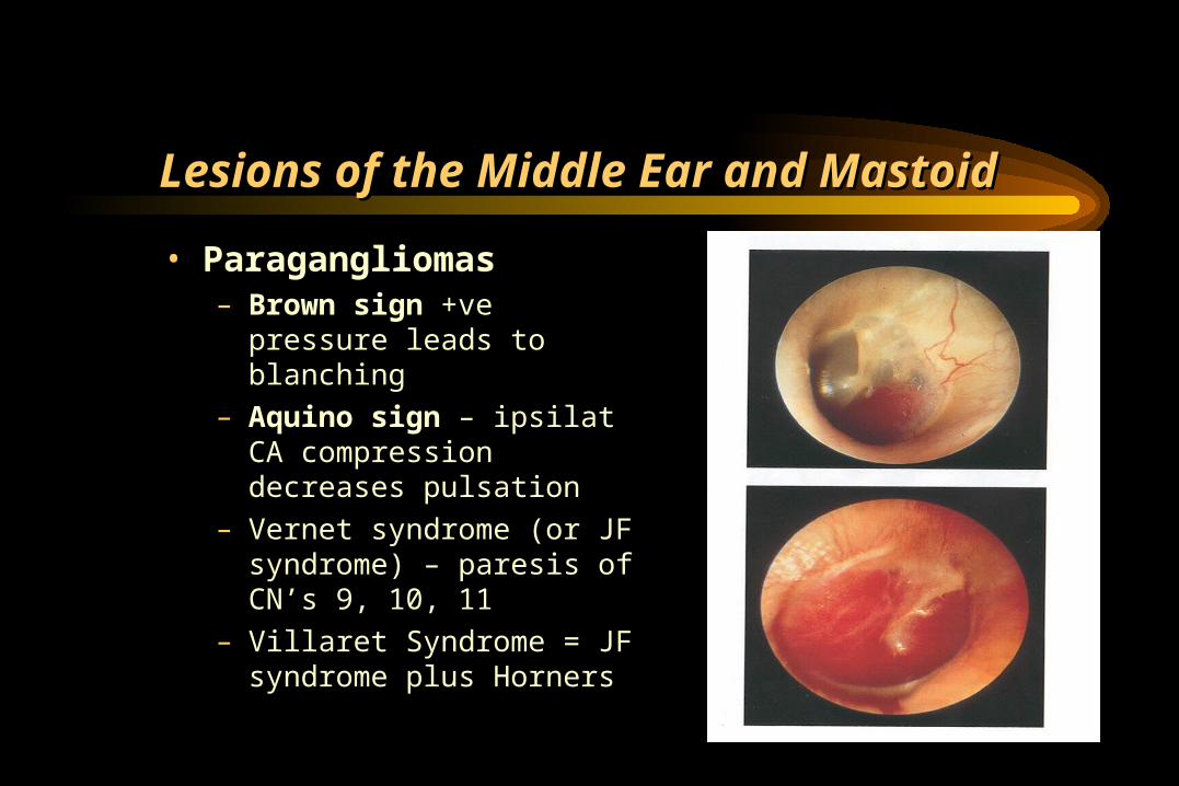
Lesions of the Middle Ear and MastoidLesions of the Middle Ear and Mastoid
• Paragangliomas– Brown sign +ve pressure leads
to blanching
– Aquino sign – ipsilat CA compression decreases pulsation
– Vernet syndrome (or JF syndrome) – paresis of CN’s 9, 10, 11
– Villaret Syndrome = JF syndrome plus Horners

ParagangliomasParagangliomas
• Rx is complete surgical excision• If secretory must address this (alpha or beta
blockade)• Trans canal, trans mastoid-lab, trans cervical, infra
temporal, intra cranial• Pre-op embolization is a neccessity• If you think it invades the ICA, balloon occlusion
studies must be done• RT or stereotactic radiosurgery can halt disease in
up to 90%

Lesions of the Petrous Apex and ClivusLesions of the Petrous Apex and Clivus
• Cholesterol granulomas– Most common lesion of the
petrous apex– Negative pressure in lumen
causes hemorrhage– Expansile lesion– Hearing loss, tinnitus, vertigo,
facial twitching– HRCT– MRI diagnostic
• T1 and T2 hyperintense

Lesions of the Petrous Apex and ClivusLesions of the Petrous Apex and Clivus

Cholesterol GranulomaCholesterol Granuloma
• Causes: poor drainage of ME, hemmorhage, obstruction of ventilation, FB reaction to cholesterol crystals from HB catabolism
• Rx is surgical drainage

Lesions of the IAC, CPA, and Skull BaseLesions of the IAC, CPA, and Skull Base
• Schwannomas (no longer acoustic)– Arise from sheaths of cranial
nerves– Vestibular, facial, trigeminal,
jugular– Varied presentation– HRCT
• Inhomogeneous enhancement• Smooth mass effect
– MRI – definitive diagnosis• T1- low intensity• Marked enhancement with
gadolinium on T1
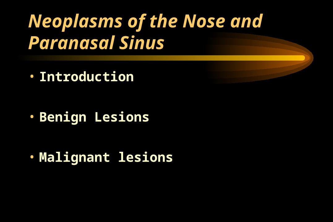
Neoplasms of the Nose and Neoplasms of the Nose and Paranasal SinusParanasal Sinus
• Introduction
• Benign Lesions
• Malignant lesions
20
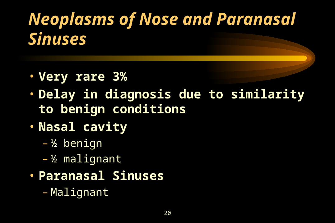
Neoplasms of Nose and Paranasal Neoplasms of Nose and Paranasal SinusesSinuses
• Very rare 3%• Delay in diagnosis due to similarity to benign
conditions• Nasal cavity
– ½ benign– ½ malignant
• Paranasal Sinuses– Malignant

21
Neoplasms of Nose and Paranasal Neoplasms of Nose and Paranasal SinusesSinuses
• Multimodality treatment
• Orbital Preservation
• Minimally invasive surgical techniques

22
EpidemiologyEpidemiology
• Predominately of older males
• Exposure:– Wood, nickel-refining processes– Industrial fumes, leather tanning
• Cigarette and Alcohol consumption– No significant association has been shown
23
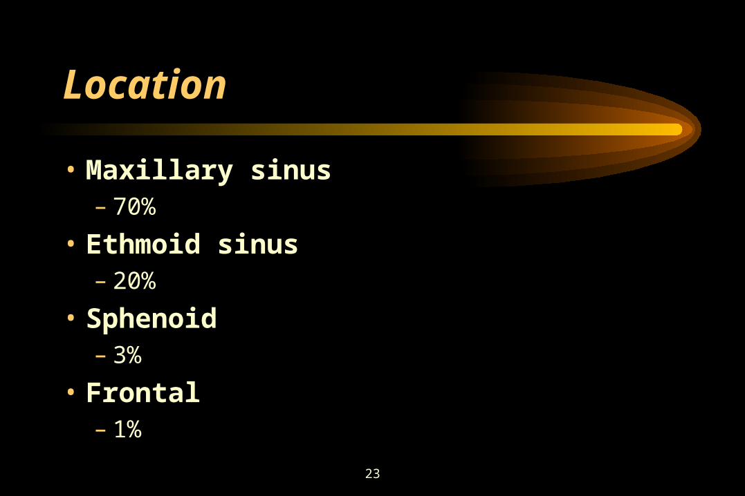
LocationLocation
• Maxillary sinus– 70%
• Ethmoid sinus– 20%
• Sphenoid– 3%
• Frontal– 1%

24
PresentationPresentation
• Oral symptoms: 25-35%– Pain, trismus, alveolar ridge fullness, erosion
• Nasal findings: 50%– Obstruction, epistaxis, rhinorrhea
• Ocular findings: 25%– Epiphora, diplopia, proptosis
• Facial signs– Paresthesias, asymmetry

25
Benign LesionsBenign Lesions
• Papillomas
• Osteomas
• Fibrous Dysplasia
26
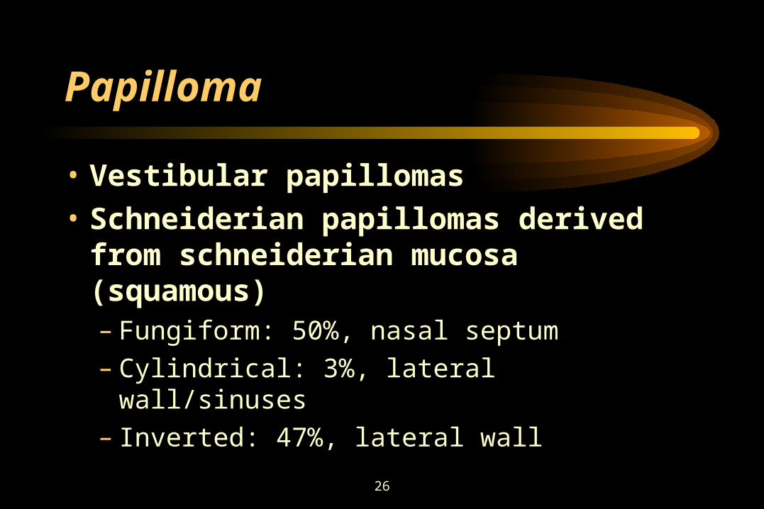
PapillomaPapilloma
• Vestibular papillomas
• Schneiderian papillomas derived from schneiderian mucosa (squamous)– Fungiform: 50%, nasal septum– Cylindrical: 3%, lateral wall/sinuses– Inverted: 47%, lateral wall
27
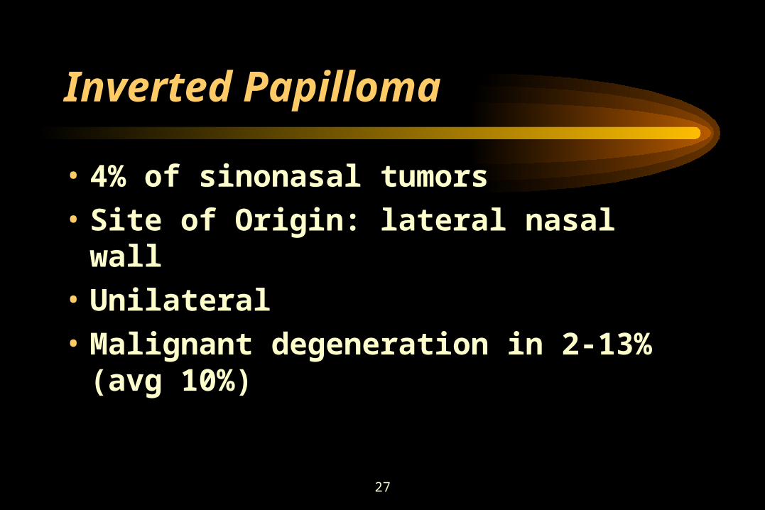
Inverted PapillomaInverted Papilloma
• 4% of sinonasal tumors
• Site of Origin: lateral nasal wall
• Unilateral
• Malignant degeneration in 2-13% (avg 10%)

28
Inverted PapillomaInverted PapillomaResectionResection
• Initially via transnasal resection:– 50-80% recurrence
• Medial Maxillectomy via lateral rhinotomy:– Gold Standard– 10-20%
• Endoscopic medial maxillectomy:– Key concepts:
• Identify the origin of the papilloma• Bony removal of this region
• Recurrent lesions:– Via medial maxillectomy vs. Endoscopic resection– 22%

29
OsteomasOsteomas
• Benign slow growing tumors of mature bone
• Location:– Frontal, ethmoids, maxillary sinuses
• When obstructing mucosal flow can lead to mucocele formation
• Treatment is local excision

30
Fibrous dysplasiaFibrous dysplasia
• Dysplastic transformation of normal bone with collagen, fibroblasts, and osteoid material
• Monostotic vs Polyostotic
• Surgical excision for obstructing lesions
• Malignant transformation to rhabdomyosarcoma has been seen with radiation
31
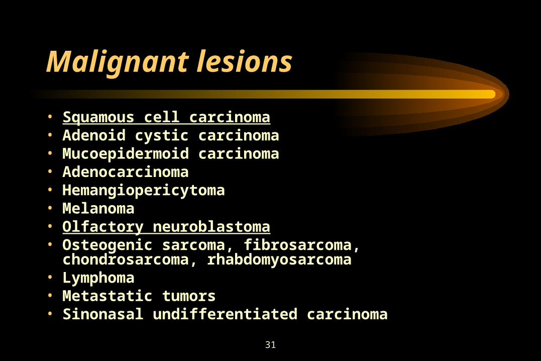
Malignant lesionsMalignant lesions
• Squamous cell carcinoma• Adenoid cystic carcinoma• Mucoepidermoid carcinoma• Adenocarcinoma• Hemangiopericytoma• Melanoma• Olfactory neuroblastoma• Osteogenic sarcoma, fibrosarcoma, chondrosarcoma,
rhabdomyosarcoma• Lymphoma• Metastatic tumors• Sinonasal undifferentiated carcinoma
32
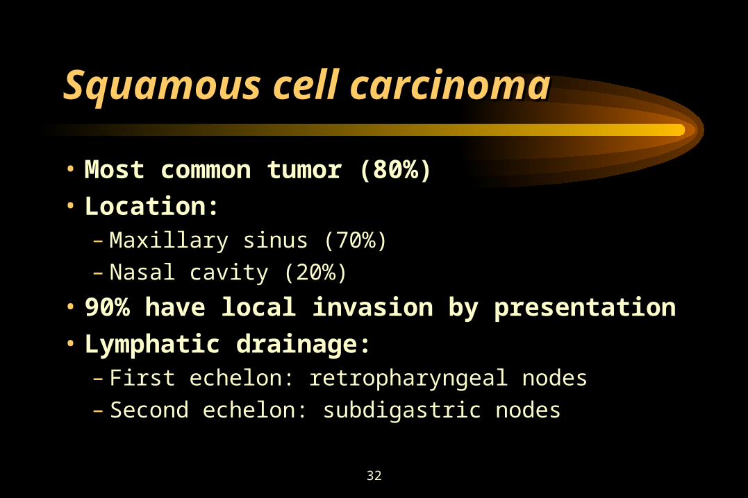
Squamous cell carcinomaSquamous cell carcinoma
• Most common tumor (80%)• Location:
– Maxillary sinus (70%)– Nasal cavity (20%)
• 90% have local invasion by presentation• Lymphatic drainage:
– First echelon: retropharyngeal nodes– Second echelon: subdigastric nodes

33
Staging of Maxillary Sinus TumorsStaging of Maxillary Sinus Tumors

34
Staging of Maxillary Sinus TumorsStaging of Maxillary Sinus Tumors
• T1: limited to antral mucosa without bony erosion• T2: erosion or destruction of the infrastructure,
including the hard palate and/or middle meatus• T3: Tumor invades: skin of cheek, posterior wall of
sinus, inferior or medial wall of orbit, anterior ethmoid sinus
• T4: tumor invades orbital contents and/or: cribriform plate, post ethmoids or sphenoid, nasopharynx, soft palate, pterygopalatine or infratemporal fossa or base of skull

35
TreatmentTreatment
• 88% present in advanced stages (T3/T4)
• Surgical resection with postoperative radiation– Complex 3-D anatomy makes margins difficult

36
Olfactory NeuroblastomaOlfactory NeuroblastomaEsthesioneuroblastomaEsthesioneuroblastoma
• Originate from stem cells of neural crest origin that differentiate into olfactory sensory cells.
• Kadish Classification– A: confined to nasal cavity– B: involving the paranasal cavity– C: extending beyond these limits

37
Olfactory NeuroblastomaOlfactory NeuroblastomaEsthesioneuroblastomaEsthesioneuroblastoma
• Aggressive behavior
• Local failure: 50-75%
• Metastatic disease develops in 20-30%
• Treatment:– En bloc surgical resection with postoperative XRT

Oral Cavity CancerOral Cavity Cancer
• Introduction
• Premalignant Lesions
• Malignant Lesions

EpidemiologyEpidemiology• 95% are squamous cell carcinoma• Risk factors
– Smoking (depends on dosage and type)– Alcohol– Snuff dipping / tobacco chewing– HPV (subtype 16)– Reverse cigar smoking (India)– Betel-nut chewing (Asia)– ?Poor dentition / mechanical irritation (dentures)

EpidemiologyEpidemiology
• 75% of cases occur on 10% of mucosal surface area– Area from ant FOM along gingivobuccal sulcus and lat
border tongue to retromolar trigone and ant tonsil pillar– Flow and pooling of carcinogen-contaminated saliva
here
• Incidence 4% cancers in males, 2% in females (increasing in females)

Evaluation and DiagnosisEvaluation and Diagnosis
• Lesions generally easy to see• Simple biopsy under local anesthesia• Important goals:
– Stage full extent of disease– Rule out synchronous primary– Evaluate for possible metastatic disease
• CT or MRI for T2 or greater• Staging endoscopy
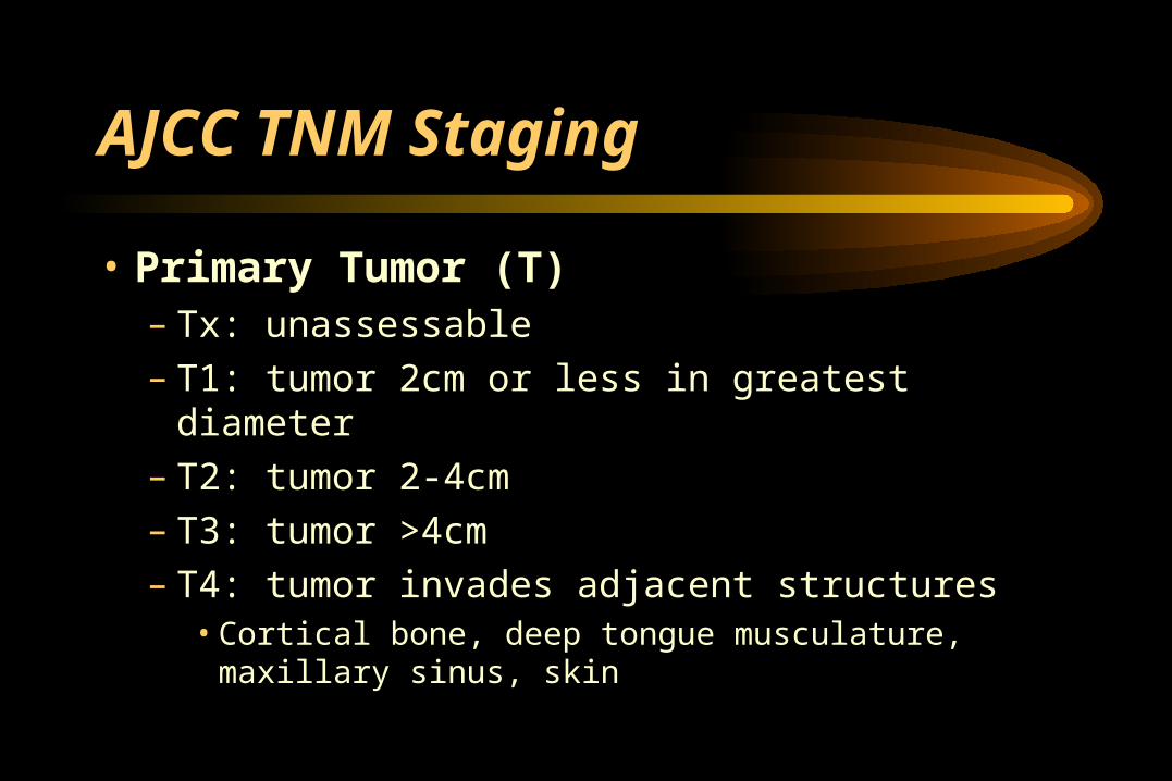
AJCC TNM StagingAJCC TNM Staging
• Primary Tumor (T)– Tx: unassessable– T1: tumor 2cm or less in greatest diameter– T2: tumor 2-4cm– T3: tumor >4cm– T4: tumor invades adjacent structures
• Cortical bone, deep tongue musculature, maxillary sinus, skin
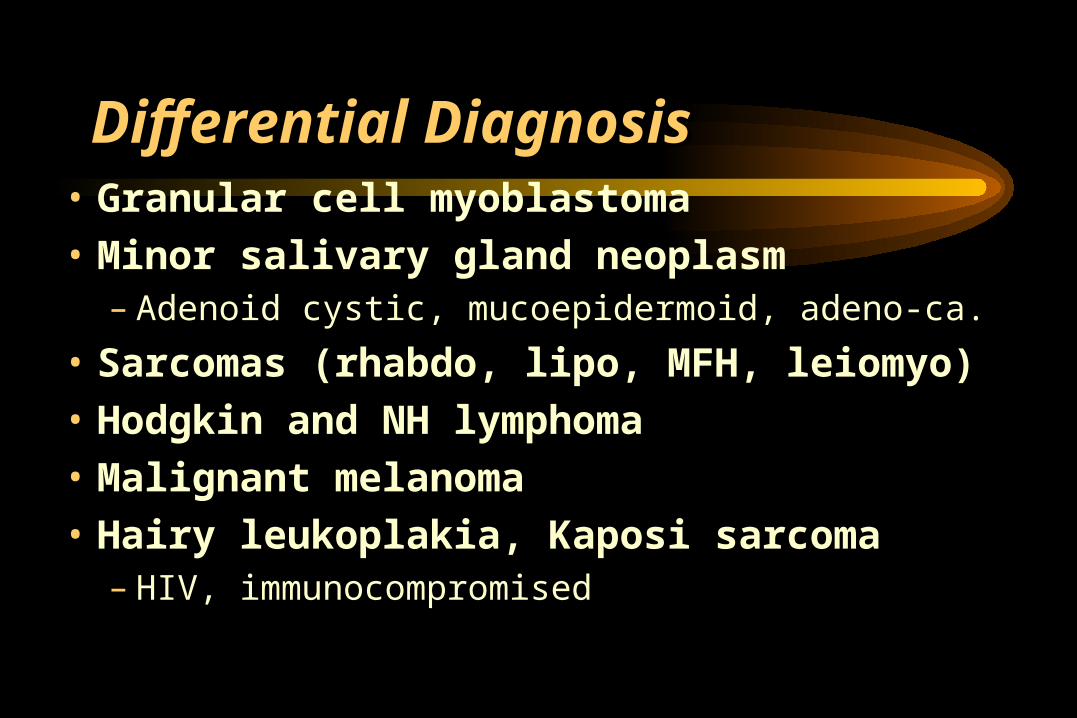
Differential DiagnosisDifferential Diagnosis• Granular cell myoblastoma
• Minor salivary gland neoplasm– Adenoid cystic, mucoepidermoid, adeno-ca.
• Sarcomas (rhabdo, lipo, MFH, leiomyo)
• Hodgkin and NH lymphoma
• Malignant melanoma
• Hairy leukoplakia, Kaposi sarcoma– HIV, immunocompromised

Premalignant LesionsPremalignant Lesions• Leukoplakia
– Hyperkeratosis, dysplasia– Malignant transformation greater in non-smokers– Treatment:
• Surgical or laser excision
• Topical bleomycin, retinoids,
• Erythroplasia– Greater risk of malignancy

Prognostic FactorsPrognostic Factors• Poor prognostic tumor factors include
– Tumor thickness (3mm FOM, 5mm tongue)– Stage– Perineural invasion– Lymphatic invasion– Vascular invasion– Neck/distant mets– DNA ploidy– Pathology
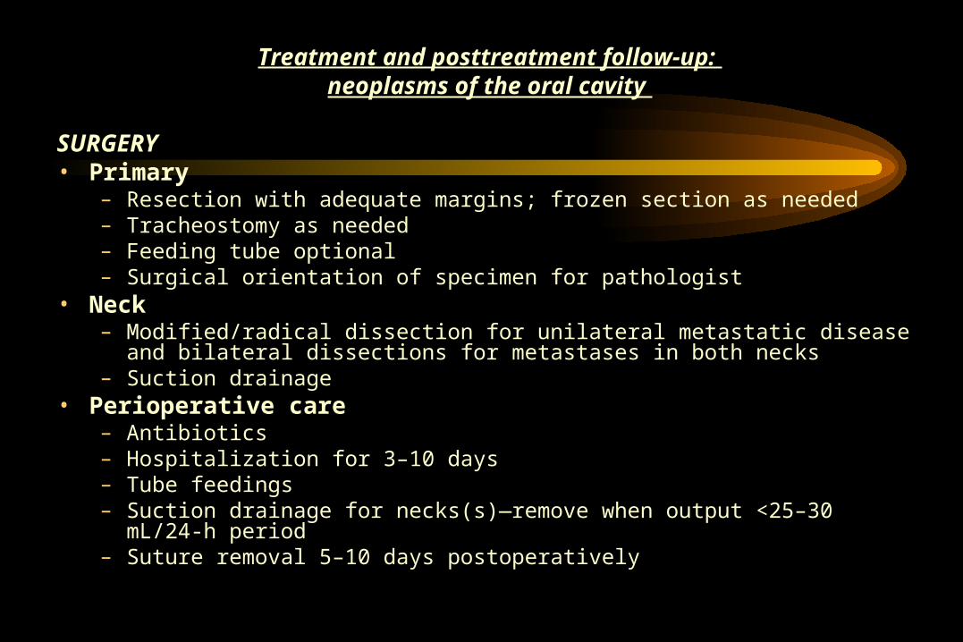
Treatment and posttreatment follow-up: neoplasms of the oral cavity
SURGERY• Primary
– Resection with adequate margins; frozen section as needed – Tracheostomy as needed– Feeding tube optional – Surgical orientation of specimen for pathologist
• Neck– Modified/radical dissection for unilateral metastatic disease and bilateral dissections for
metastases in both necks– Suction drainage
• Perioperative care – Antibiotics– Hospitalization for 3–10 days– Tube feedings – Suction drainage for necks(s)—remove when output <25–30 mL/24-h period– Suture removal 5–10 days postoperatively
Tumours of PharynxTumours of Pharynx
• Nasopharyngeal Carcinoma
• Oropharyngeal Carcinoma
• Hypopharyngeal Carcinoma

Nasopharyngeal CarcinomaNasopharyngeal CarcinomaIntroductionIntroduction
• Rare in the US, more common in Asia
• High index of suspicion required for early diagnosis
• Nasopharyngeal malignancies– SCCA (“nasopharyngeal carcinoma”)– Lymphoma– Salivary gland tumors– Sarcomas

ClassificationClassification
• WHO classes– Based on light microscopy findings– All SCCA by EM
• Type I - “SCCA”– 25 % of NPC (in North Amer population)– 1-2 % NPC of endemic populations– moderate to well differentiated cells similar to other
SCCA ( keratin, intercellular bridges)

ClassificationClassification
• Type II - “non-keratinizing” carcinoma– 12 % of NPC– variable differentiation of cells (mature to
anaplastic)– minimal if any keratin production– may resemble transitional cell carcinoma of the
bladder– Lumped with Type III in 1991 WHO revision

ClassificationClassification
• Type III - “undifferentiated” carcinoma– 60 % of NPC in North Amer population, majority of
NPC in young patients, and 95% of endemic cases– Difficult to differentiate from lymphoma by light
microscopy requiring special stains & markers– Diverse group
• Lymphoepitheliomas, spindle cell, clear cell and anaplastic variants

EpidemiologyEpidemiology
• Chinese native (esp Guangdong province) > Chinese immigrant > North American caucasian– Both genetic and environmental factors
• Genetic– HLA histocompatibility loci possible markers
• HLA-A2, B17 and Bw46

EpidemiologyEpidemiology
• Environmental– Viruses
• EBV- well documented viral “fingerprints” in tumor cells and also anti-EBV serologies with WHO type II and III NPC
• HPV - possible factor in WHO type I lesions
– Nitrosamines - salted fish– Others - polycyclic hydrocarbons, chronic nasal
infection, poor hygiene, poor ventilation

Clinical PresentationClinical Presentation
• Often subtle initial symptoms– unilateral HL (SOM)
– painless, slowly enlarging neck mass (70%)• Lymphatic channels cross midline in NP, bilateral disease common
• Larger lesions– nasal obstruction
– epistaxis
– cranial nerve involvement
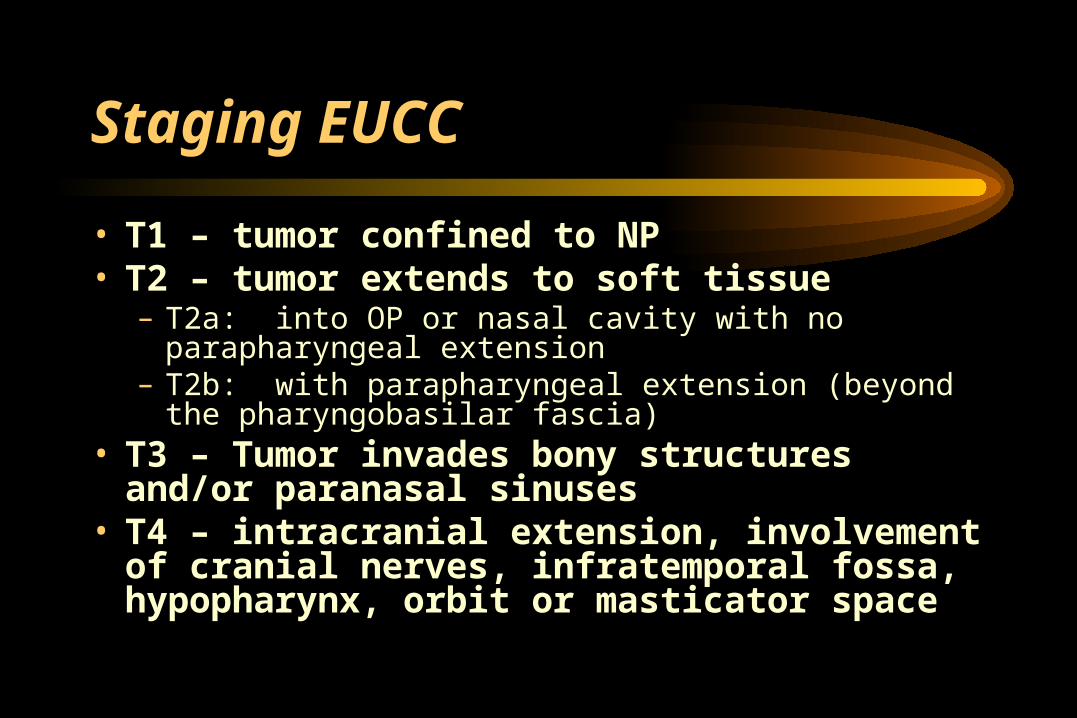
Staging EUCCStaging EUCC
• T1 – tumor confined to NP• T2 – tumor extends to soft tissue
– T2a: into OP or nasal cavity with no parapharyngeal extension– T2b: with parapharyngeal extension (beyond the
pharyngobasilar fascia)• T3 – Tumor invades bony structures and/or
paranasal sinuses• T4 – intracranial extension, involvement of cranial
nerves, infratemporal fossa, hypopharynx, orbit or masticator space

TreatmentTreatment
• External beam radiation– Dose: 6500-7000 cGy– Primary, upper cervical nodes– Consider 5000 cGy prophylactic tx of clinically
negative lower neck

TreatmentTreatment
• Adjuvant Chemotherapy– Stardard of care– Cisplatnium (hematologic sideeffects therefore not
overlapping toxicity)– 5-FU
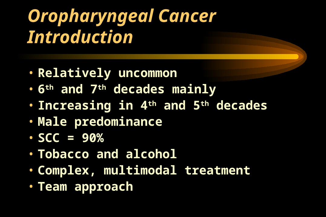
Oropharyngeal CancerOropharyngeal CancerIntroductionIntroduction
• Relatively uncommon• 6th and 7th decades mainly• Increasing in 4th and 5th decades• Male predominance• SCC = 90%• Tobacco and alcohol• Complex, multimodal treatment• Team approach

AnatomyAnatomy• Connects nasopharynx to
hypopharynx• Ant
– Circumvallate papillae
– Anterior tonsillar pillars
– Junction of hard and soft palates

AnatomyAnatomy
• Pharyngeal walls– Mucosa, submucosa, pharyngobasilar fascia, constrictor
muscles, buccopharyngeal fascia• Tonsils sit in tonsillar fossa• Soft Palate
– Palatine aponeurosis– Tensor veli palatini– Levator veli palatini– Uvular muscle– Palatoglossus– palatopharyngeus

EtiologyEtiology
• SCC arise from the accumulation of multiple genetic alterations to genes important to the regulation of cell growth and death
• Cells have selective growth advantage– Genetic– Environmental
• Tobacco and alcohol• Dose related• Synergistic
– HPV and EBV– Dietary factors– Immunosuppression
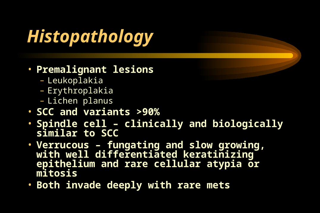
HistopathologyHistopathology
• Premalignant lesions– Leukoplakia– Erythroplakia– Lichen planus
• SCC and variants >90%• Spindle cell – clinically and biologically similar to
SCC• Verrucous – fungating and slow growing, with well
differentiated keratinizing epithelium and rare cellular atypia or mitosis
• Both invade deeply with rare mets

HistopathologyHistopathology
• Lymphoepitheliomas– Grow rapidly and readily mets– Tonsillar region– Younger patients without risk factors
• Adenoid squamous, adenosquamous, and basaloid SCC are rare and highly aggressive (latter two have early mets)

TreatmentTreatment
• Team approach– Surgeons and Radiation Oncologists– SLP– Oral Surgeon
• T1 and T2 – surgery or radiation• T3 and T4 – combined modality• Neck
– N0 and N1 – surgery or XRT– N2 and N3 - combined modality– Both necks treated with central lesions– Retropharyngeal nodes are always treated
Hypopharyngeal CancerHypopharyngeal Cancer
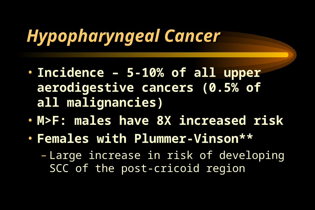
• Incidence – 5-10% of all upper aerodigestive cancers (0.5% of all malignancies)
• M>F: males have 8X increased risk
• Females with Plummer-Vinson**– Large increase in risk of developing SCC of the post-
cricoid region

Hypopharyngeal CancersHypopharyngeal Cancers
• Risk Factors– Smoking
– EtOH
– Chronic reflux disease
• Treatment Challenge:– Patients often present with advanced disease
– May be complicated by severe malnutrition

Hypopharynx - AnatomyHypopharynx - Anatomy• Abuts the oropharynx at
the level of the hyoid, extends to the level of the inferior border of the cricoid
• 3 sub-sites: piriform fossa(e), post-cricoid region, posterior pharyngeal wall

Hypopharynx - AnatomyHypopharynx - Anatomy
• Piriform apex – junction between the post-cricoid area and the inferior aspect of the piriform fossae

Staging EndoscopyStaging Endoscopy• Most important component
of procedure (secondary to obtaining Bx samples for diagnosis) is determining the inferior limit of the tumour
• Common site: piriform fossae, post pharyngeal wall, post-cricoid region

PathologyPathology
• ~95% of cancers of the hypopharynx are SCC• Lymphomas
– Angiocentric T-cell lymphoma– MALT (mucosa associated lymphoid tissue)– Non-hodgkins lymphoma
• Adenocarcinomas– May originate in the minor salivary glands of the hypopharynx
• Benign lesions– Limpoma: < 1%, usually resected due to risk of airway obstruction
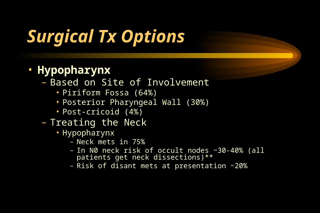
Surgical Tx OptionsSurgical Tx Options
• Hypopharynx– Based on Site of Involvement
• Piriform Fossa (64%)• Posterior Pharyngeal Wall (30%)• Post-cricoid (4%)
– Treating the Neck• Hypopharynx
– Neck mets in 75%– In N0 neck risk of occult nodes ~30-40% (all patients get neck
dissections)**– Risk of disant mets at presentation ~20%

Hypopharynx Tx Surgical OptionsHypopharynx Tx Surgical Options
ProcedureProcedure T stageT stage ReconstructioReconstructionn
Partial PharyngectomyPartial Pharyngectomy T1, T2T1, T2 Primary closurePrimary closure
Partial Partial LaryngopharyngectomyLaryngopharyngectomy
T1, T2, T3T1, T2, T3 Regional or free flapRegional or free flap
Supracricoid Supracricoid hemilaryngectomyhemilaryngectomy
T1, T2, T3T1, T2, T3 Primary closurePrimary closure
Endoscopic CO2 laser Endoscopic CO2 laser resectionresection
T1, T2 (possibly T3, T1, T2 (possibly T3, T4)T4)
Secondary intentionSecondary intention
Total Laryngectomy with Total Laryngectomy with partial-total partial-total pharyngectomypharyngectomy
T3T3 Primary closure vs. Primary closure vs. regional or free flapregional or free flap
Total Laryngo-Pharyngo-Total Laryngo-Pharyngo-esophagectomyesophagectomy
T4T4 Gastric pull-upGastric pull-up
Laryngeal TumoursLaryngeal Tumours
• Introduction
• Benign Lesions
• Malignant Lesions
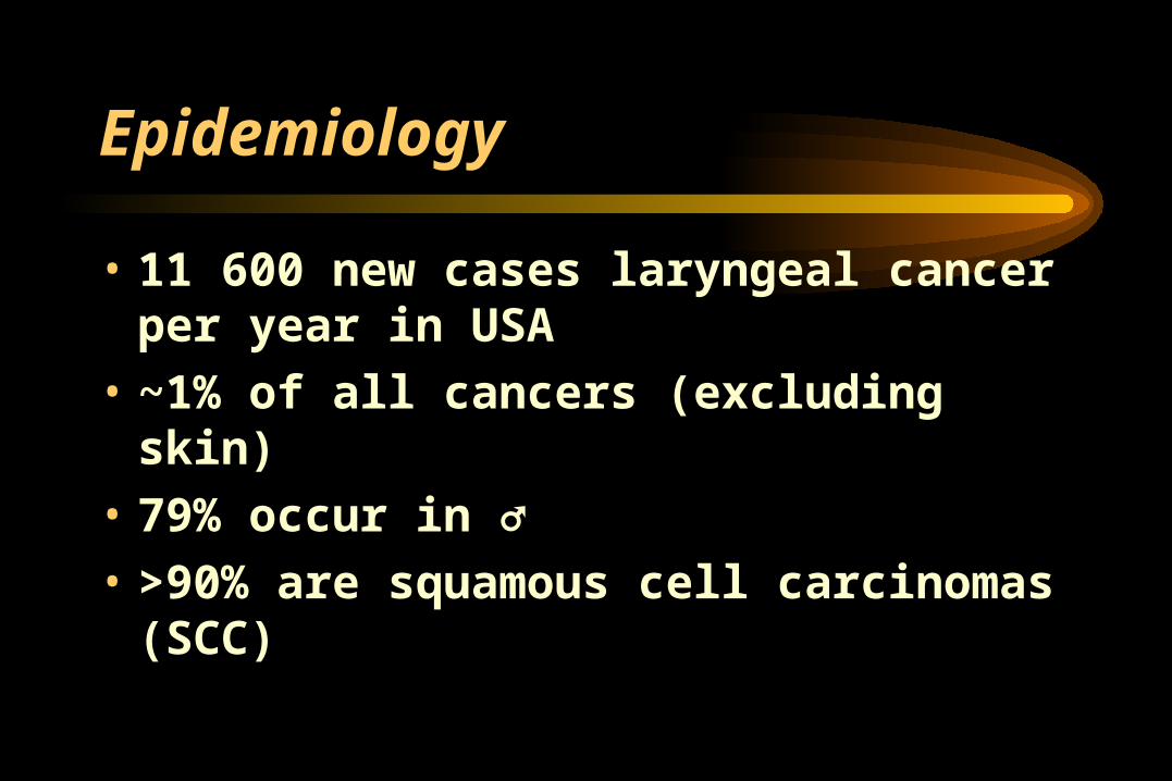
EpidemiologyEpidemiology
• 11 600 new cases laryngeal cancer per year in USA
• ~1% of all cancers (excluding skin)
• 79% occur in ♂
• >90% are squamous cell carcinomas (SCC)

EtiologyEtiology
• EtOH – supraglottic• Tobacco – glottic• GERD – chronic laryngeal irritation• Viral infection• Asbestos• Nickel• Wood• Isopropyl alcohol• Radiation

Laryngeal PapillomatosisLaryngeal Papillomatosis
• Most common benign laryngeal tumor, HPV etiology
• Vocal folds and subglottis most common laryngeal sites
Laryngeal PapillomatosisLaryngeal Papillomatosis
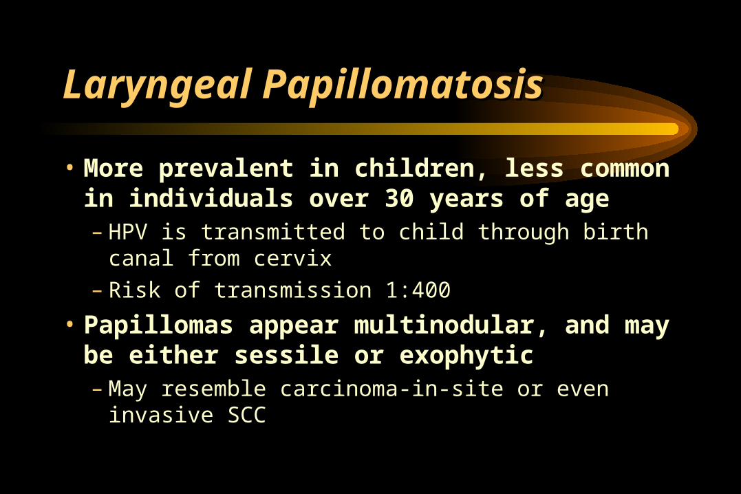
• More prevalent in children, less common in individuals over 30 years of age– HPV is transmitted to child through birth canal from
cervix– Risk of transmission 1:400
• Papillomas appear multinodular, and may be either sessile or exophytic– May resemble carcinoma-in-site or even invasive SCC
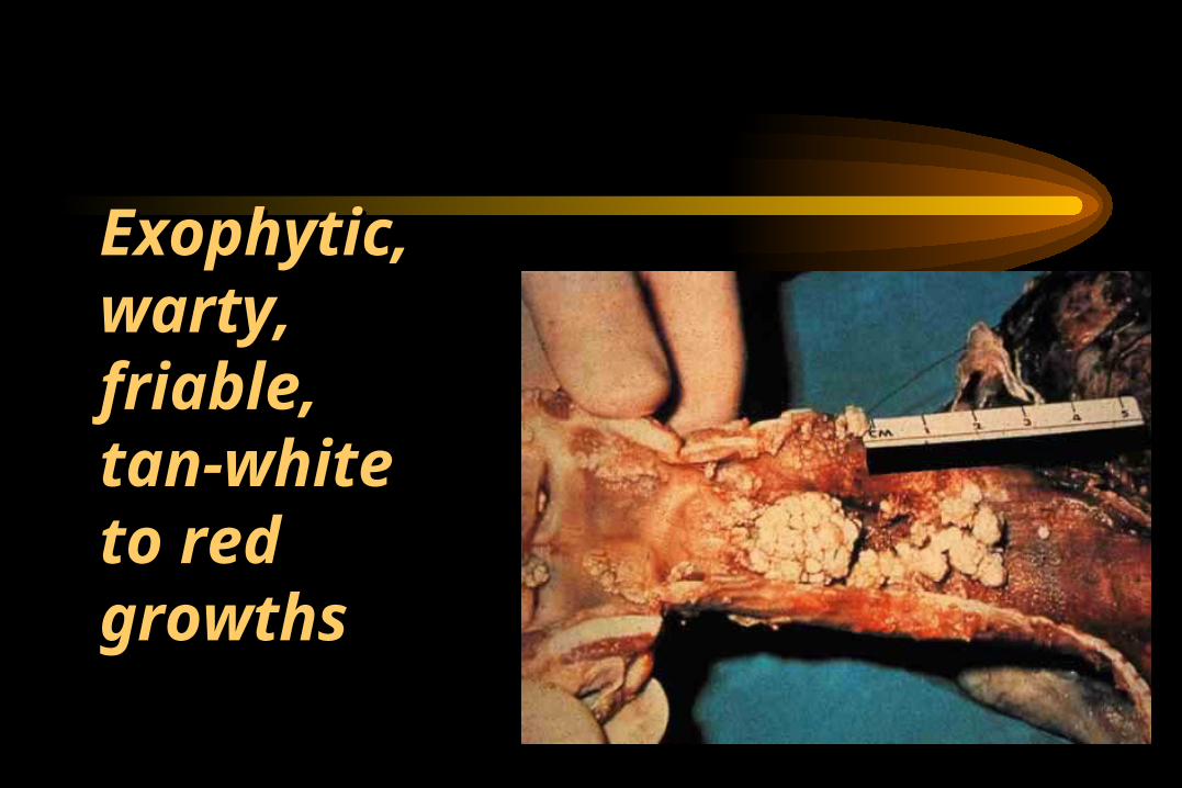
Exophytic, Exophytic, warty, warty, friable, tan-friable, tan-white to red white to red growthsgrowths
Laryngeal PapillomatosisLaryngeal Papillomatosis
• Most common viral subtypes are 6 or 11, but 16 or 18 have higher potential for malignant change
• Hoarseness is common early symptom followed by airway obstruction and respiratory difficulty

Laryngeal PapillomatosisLaryngeal Papillomatosis
• Laryngeal papillomas presenting in adults seem to be less aggressive than juvenile form but remission rate unpredictable– In adults, growth may be rapid during periods of
hormone change such as during pregnancy
• Malignant degeneration of laryngeal papillomas rare and usually associated with history of radiotherapy, tobacco abuse or both

TreatmentTreatment
– Surgery
• Laser microlaryngoscopy (most commonly CO2 [10.6um] or Nd:YAG [1.06um]) at power setting of 2-8W pulse or continuous
• Powered microdebrider
• Always biopsy before remainder of case proceeds

Cont TreatmentCont Treatment
– Interferon• Bad chronic side-effects (myalgias, flu-like symptoms)
• Lesions tend to return after interferon finished
– Intralesional cidofovir (acyclic nucleoside analogue)– Indole-3-carbinol (found in cruciferous vegetables,
works via inhibition of estrogen metabolism)– Acyclovir– Photodynamic therapy

Supraglottic vs Glottic DiseaseSupraglottic vs Glottic Disease
• North America glottic cancer > supraglottic (2:1)
• France supraglottic > glottic (2:1)

Anatomy - GlottisAnatomy - Glottis• True vocal cords
• Anterior and posterior commissures
• Superior limit – apex of ventricle
• Inferior limit – 1 cm inferior to line through apex

Staging – Early GlotticStaging – Early Glottic
• Tis – no invasion beyond basement membrane
• T1 – confined to glottis with normal mobility
• T1a – tumor limited to one vocal cord
• T1b – tumor involves both cords, no limitation in mobility
Staging – Early GlotticStaging – Early Glottic
• T2 – extend into supra- or subglottis without complete vocal cord fixation• T2a – involve supra- or subglottis but do not impair
movement
• T2b – impair movement of vocal cords, but not complete fixation

Staging – Advanced GlotticStaging – Advanced Glottic
• T3 – complete vocal cord fixation, ± paraglottic space, ± minor thyroid cartilage erosion (inner cortex)
• T4 – extends beyond larynx, into thyroid cartilage

SymptomsSymptoms
• Hoarseness >4 weeks – investigate
• Occasionally may present without hoarseness
• Dysphagia
• Hemoptysis
Glottic CarcinomaGlottic Carcinoma
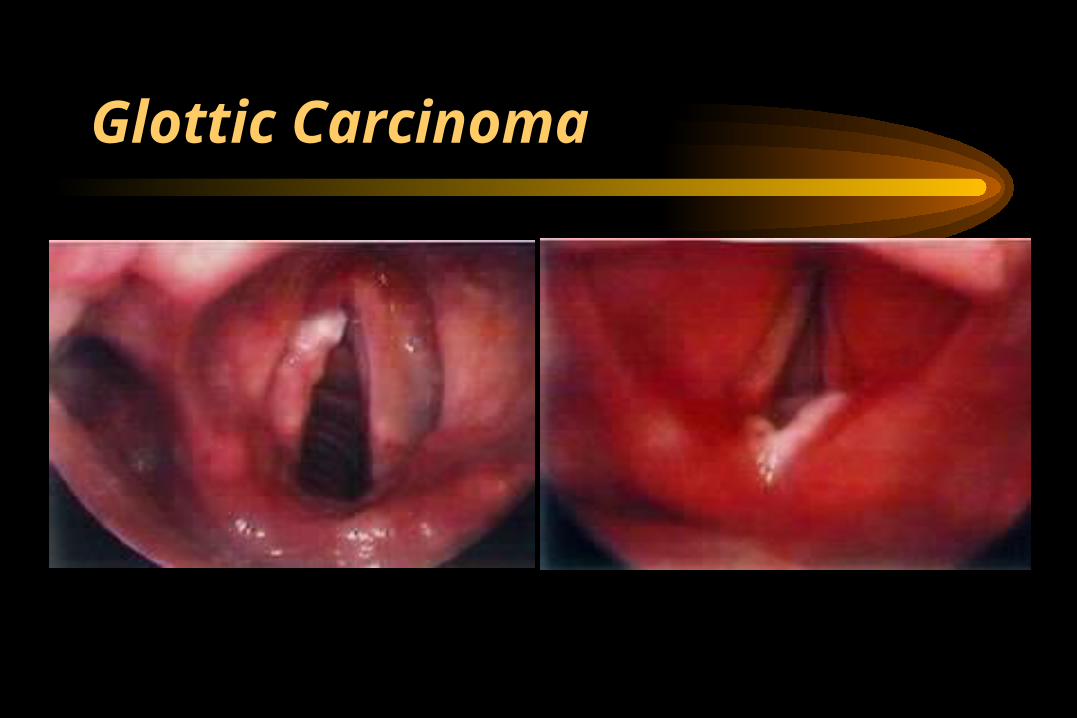
• Early: irregular area of mucosal thickening
• Advanced: exophytic, fungating, endophytic, ulcerated mass
• More commonly keratinizing, well to moderately differentiated
• In situ component
• Invasive component predominantly infiltrative

Glottic CarcinomaGlottic Carcinoma
• Up to 20% of T1 cancers have some degree of vocal cord ligament invasion

Glottic CarcinomaGlottic Carcinoma• Most tumors originate
on free surface of vocal cord
• Anterior 2/3
Glottic CarcinomaGlottic Carcinoma

TreatmentTreatment
• Early Stage:
Laser or Radiation
• Advance Stage:
Chemo+Radiation
Surgery+Radiation

Thank You
Related Documents