YIL/YEAR: 2020 CİLT/VOLUME: 9 SAYI/ISSUE: 2

Welcome message from author
This document is posted to help you gain knowledge. Please leave a comment to let me know what you think about it! Share it to your friends and learn new things together.
Transcript

YIL/YEAR: 2020 CİLT/VOLUME: 9 SAYI/ISSUE: 2

E-ISSN:2146-7188
HARRAN ÜNİVERSİTESİ VETERİNER FAKÜLTESİ
DERGİSİHarran University
Journal of The Faculty of Veterinary Medicine
Harran Üniversitesi Veteriner Fakültesi Yayınıdır Published by Harran University Faculty Of Veterinary Medicine
YIL/YEAR:2020 CİLT/VOLUME:9 SAYI/ISSUE: 2

HARRAN ÜNİVERSİTESİ VETERİNER FAKÜLTESİ DERGİSİ
Harran University Journal of the Faculty of Veterinary Medicine
Harran Üniversitesi
Veteriner Fakültesi Adına Sahibi/Owner
Prof. Dr. Nihat DENEK Dekan/Dean
Baş Editör/Editor in Chief
Prof. Dr. Oktay KESKİN
Editörler Kurulu/Editorial board Prof. Dr. Füsun TEMAMOĞULLARI
Doç. Dr. Şükrü GÜRLER Doç. Dr. Rahşan YILMAZ Doç. Dr. Deniz KORKMAZ
Doç. Dr. Serap KILIÇ ALTUN Doç. Dr. Birten EMRE
İngilizce Editörü/English Editor Prof. Dr. Ayşe Menteş GÜRLER
Mizanpaj Editörü/Layout Editor Dr. Öğr. Üyesi İrfan ÖZGÜNLÜK
Dergi Sekreteri/Journal Secretary Arş. Gör. Ayfer GÜLLÜ YÜCETEPE
Yazışma /Correspondence
Harran Üniversitesi Veteriner Fakültesi Dergisi Editörlüğü Eyyübiye Kampüsü, 63200 -
Şanlıurfa/TÜRKİYE Tel: +90 414 318 38 59
+90 414 318 38 55 Faks: +90 414 318 39 22
e-mail: [email protected]
Harran Üniversitesi Veteriner Fakültesi Dergisi Hakemli Bir Dergi Olup, Yılda 2(iki) Sayı Olarak Yayınlanır. ULAKBİM
(Yaşam Bilimleri Veri Tabanı), Türkiye Atıf Dizini, Cab abstracts, İndex Copernicus, Zoological Record, SOBIAD,
Google Scholar gibi ulusal ve uluslararası indeksler tarafından taranmaktadır.
Yıl/Year: 2020 - Cilt/Volume: 9 Sayı/Issue 2
Danışma Kurulu/Advisory Board
Prof. Dr. Ergun AKÇAY, Ankara Üniv. Vet. Fak. Ankara, Türkiye.
Prof. Dr. Mustafa ATASEVER, Atatürk Üniv. Vet. Fak. Erzurum,
Türkiye.
Prof. Dr. Halil Selçuk BİRİCİK, Aksaray Üniv. Vet. Fak. Aksaray, Türkiye
Prof. Dr. Ali BUMİN, Ankara Üniv. Vet. Fak. Ankara, Türkiye.
Prof. Dr. Ahmet ÇAKIR, Ankara Üniv. Vet. Fak. Ankara, Türkiye.
Prof. Dr. Hayrettin ÇETİN, Adnan Menderes Üniv. Vet. Fak. Aydın
Türkiye.
Prof. Dr. Hasan EREN, Adnan Menderes Üniv. Vet. Fak. Aydın,
Türkiye.
Prof. Dr. Okan ERTUĞRUL, Ankara Üniv. Vet. Fak. Ankara, Türkiye.
Prof. Dr. Anila HODA, Agric. Uni. of Tirana, Fac. of Agric.& Environ.
Tirana, Albania.
Prof. Dr. Osman KUTSAL, Ankara Üniv. Vet. Fak. Ankara, Türkiye.
Prof. Dr. Narin LİMAN, Erciyes Üniv. Vet. Fak. Kayseri, Türkiye.
Prof. Dr. Manzoor Ur Rahman MIR, SKUAST Kashmir Fac. of Vet. Sci.
& Anim. Husbandry. Kashmir, India.
Prof. Dr. Sema TEMİZER OZAN, Fırat Üniv. Vet. Fak. Elazığ, Türkiye.
Prof. Dr. Ceyhan ÖZBEYAZ, Ankara Üniv. Vet. Fak. Ankara, Türkiye.
Prof. Dr. Gerald REINER, Justus-Liebig Uni. Fac. of Vet. Med. Giessen,
Germany.
Prof. Dr. Kazım ŞAHİN, Fırat Üniv. Vet. Fak. Elazığ, Türkiye.
Prof. Dr. Atilla ŞİMŞEK, Selçuk Üniv. Vet. Fak. Konya, Türkiye.
Prof. Dr. Mehmet Emin TEKİN, Selçuk Üniv. Vet. Fak. Konya, Türkiye.
Prof. Dr. Ender YARSAN, Ankara Üniv. Vet. Fak. Ankara, Türkiye.
Prof. Dr. Halis YERLİKAYA, Fırat Üniv. Vet. Fak. Elazığ, Türkiye.
Prof. Dr. Murat YILDIRIM, Kırıkkale Üniv. Vet. Fak. Kırıkkale, Türkiye.

Harran Üniversitesi Veteriner Fakültesi Dergisi
2020 Yılı 9. Cilt 2. Sayı Hakem Listesi (alfabetik sıra) The Referees List of This Issue (in alphabetical order)
Prof. Dr. Ahmet ÇAKIR Ankara Üniversitesi Veteriner Fakültesi Prof. Dr. Ahmet KOLUMAN Pamukkale Üniversitesi Teknoloji Fakültesi Prof. Dr. Ayhan FİLAZİ Ankara Üniversitesi Veteriner Fakültesi Prof. Dr. Ayşegül BİLDİK Aydın Adnan Menderes Üniversitesi Veteriner Fakültesi Prof. Dr. Barış SAREYYÜPOĞLU Ankara Üniversitesi Veteriner Fakültesi Prof. Dr. Filiz AKDAĞ Ondokuz Mayıs Üniversitesi Veteriner Fakültesi Prof. Dr. Filiz KÖK Aydın Adnan Menderes Üniversitesi Veteriner Fakültesi Prof. Dr. İ. Safa GÜRCAN Ankara Üniversitesi Veteriner Fakültesi Prof. Dr. Mehmet ÇİFTÇİ Fırat Üniversitesi Veteriner Fakültesi Prof. Dr. Murat KİBAR Artvin Çoruh Üniversitesi Artvin Meslek Yüksekokulu Prof.Dr. Nihal Yaşar GÜL SATAR Uludağ Üniversitesi Veteriner Fakültesi Prof. Dr. Sinan İNCE Afyon Kocatepe Üniversitesi Veteriner Fakültesi Prof. Dr. Uçkun Sait UÇAN Selçuk Üniversitesi Veteriner Fakültesi Prof. Dr. Yücel MERAL Ondokuz Mayıs Üniversitesi Veteriner Fakültesi Prof. Dr. Zehra BOZKURT Afyon Kocatepe Üniversitesi Veteriner Fakültesi Doç. Dr. Ali Doğan ÖMÜR Atatürk Üniversitesi Veteriner Fakültesi Doç. Dr. Bülent TEKE Ondokuz Mayıs Üniversitesi Veteriner Fakültesi Doç. Dr. Çiğdem ÇEBİ ŞEN Harran Üniversitesi Veteriner Fakültesi Doç. Dr. Doğukan ÖZEN Ankara Üniversitesi Veteriner Fakültesi Doç. Dr. Hüsamettin EKİCİ Kırıkkale Üniversitesi Veteriner Fakültesi Doç. Dr. İbrahim AKIN Aydın Adnan Menderes Üniversitesi Veteriner Fakültesi Doç. Dr. İrem ERGİN Ankara Üniversitesi Veteriner Fakültesi Doç.Dr. Mustafa KÖM Fırat Üniversitesi Veteriner Fakültesi Doç. Dr. Mustafa Orhun DAYAN Selçuk Üniversitesi Veteriner Fakültesi Doç. Dr. Nazan GEZER İNCE İstanbul Üniversitesi Cerrahpaşa Üniversitesi Veteriner Fakültesi Doç. Dr. Pınar PEKER AKALIN Hatay Mustafa Kemal Üniversitesi Veteriner Fakültesi Doç. Dr. Serdar KOÇAK Afyon Kocatepe Üniversitesi Veteriner Fakültesi Doç. Dr. Tugay AYAŞAN Osmaniye Korkut Ata Üniversitesi Kadirli Uygulamalı Bilimler Yüksekokulu
Doç. Dr. Yasin DEMİRASLAN Burdur Mehmet Akif Ersoy Üniversitesi Veteriner Fakültesi Doç. Dr. Zafer DOĞU Harran Üniversitesi Bozova Meslek Yüksekokulu Doç. Dr. Zeki ERİŞİR Fırat Üniversitesi Veteriner Fakültesi Dr. Öğr. Üyesi Fatma Tülin ÖZBAŞER Tekirdağ Namık Kemal Üniversitesi Veteriner Fakültesi Dr. Öğr. Üyesi Gamze EVKURAN DAL İstanbul Üniversitesi Cerrahpaşa Üniversitesi Veteriner Fakültesi Dr. Öğr. Üyesi İbrahim Mert POLAT Kırıkkale Üniversitesi Veteriner Fakültesi Dr. Öğr. Üyesi İftar GÜRBÜZ Burdur Mehmet Akif Ersoy Üniversitesi Veteriner Fakültesi Dr. Öğr. Üyesi İmdat ORHAN Erciyes Üniversitesi Veteriner Fakültesi Dr. Öğr. Üyesi Kadri KULUALP Dokuz Eylül Üniversitesi Veteriner Fakültesi Dr. Öğr. Üyesi Kemal AKSOY Muğla Sıtkı Koçman Üniversitesi Milas Veteriner Fakültesi Dr. Öğr. Üyesi Mehmet BOZKURT Afyon Kocatepe Üniversitesi Veteriner Fakültesi Dr. Öğr. Üyesi Mert METİN Muğla Sıtkı Koçman Üniversitesi Milas Veteriner Fakültesi Dr. Öğr. Üyesi Muhammet DUMAN Bursa Uludağ Üniversitesi Veteriner Fakültesi Dr. Öğr. Üyesi Mustafa Barış AKGÜL Siirt Üniversitesi Veteriner Fakültesi Dr. Öğr. Üyesi Nilgün PAKSOY Harran Üniversitesi Veteriner Fakültesi Dr. Öğr. Üyesi Osman BULUT Muğla Sıtkı Koçman Üniversitesi Milas Veteriner Fakültesi Dr. Öğr. Üyesi Osman YILMAZ Van Yüzüncü Yıl Üniversitesi Veteriner Fakültesi

Harran Üniversitesi Veteriner Fakültesi Dergisi
Harran University Journal of the Faculty of Veterinary Medicine 2020 Yılı 9. Cilt 2. Sayı
İçindekiler / Contents
Araştırma Makaleleri / Research Articles
Sayfa /Page 1. In Vitro Et’e Karşı Olan Tutumun Araştırılması: Erzurum Meslek Yüksekokulu Öğrencileri Örneği
A Survey on the Investigation of Attitude Against In Vitro Meat: An Example of Erzurum Vocational School Students Alper BARAN
98-106
2. Koroner Arter Hastalığı olan Köpeklerde Hipertansiyonun İnflamatuar Etkisinin Ekokardiyografi ile Değerlendirilmesi Evaluation of Inflammatory Effect of Hypertension by Echocardiography in Dogs with Coronary Artery Disease Mehmet İRİADAM, Melahat TOKER
107-111
3. The Effects of Diplotaenia Turcica Root Extract on Sperm Parameters and Reproductive Hormones in Streptozotocin Induced Diabetic Rats Streptozotosin Kaynaklı Diyabetik Sıçanlarda Diplotaenia Turcica Kök Ekstraktının Sperm Parametreleri ve Üreme Hormonları Üzerine Etkileri Saadet BELHAN, Yeter DEĞER, Zübeyir HUYUT, Süleyman Mesut PINAR
112-117
4. Serbest Dolaşımlı (Free Range) ve Derin Altlık Barındırma Sisteminin Yavaş ve Hızlı Gelişen Etlik Piliçlerde Büyüme Performansı ve Ekonomik Verimlilik Üzerine Etkisi Effects of Free-Range and Deep Litter Housing System on Growth Performance and Production Economics of Slow and Fast-Growing Broiler Metin PETEK , İbrahima Mahamane ABDOURHAMANE
118-125
5. Bazı Yerli ve Melez Koyun Genotiplerinde Süt Yağ Asidi Profillerinin Araştırılması Investigation of Milk Fatty Acid Profiles in Some Native and Crossbred Sheep Genotypes Mücahit KAHRAMAN, Banu YÜCEER ÖZKUL
126-132
6. The Effect of Perch Cooling and Perch Height on Some Bone Strength Parameters in Broilers Reared in Summer Yaz Mevsiminde Yetiştirilen Etlik Piliçlerde Tünek Soğutma ve Tünek Yüksekliğinin Bazı Kemik Dayanıklılık Parametreleri Üzerine Etkisi M. Kenan TÜRKYILMAZ, Ahmet NAZLIGÜL, Evrim DERELI FIDAN, Solmaz KARAARSLAN, Mehmet KAYA, Figen Sevil KILIMCI
133-138
7. Güneydoğu Anadolu Bölgesi’ndeki Pet Kliniklerinde Kullanılan Beşerî Müstahzarların Kullanım Durumunun Karşılaştırılması Comparison of the Use of Human Preparations in Pet Clinics in Southeastern Anatolia Region Nida KAHYAOĞLU, Füsun TEMAMOĞULLARI
139-143
8. Türkiye’de Isparta İlinin Eğirdir Bölgesinden Toplanan Koyun Sütlerinde Aflatoksin M1 Kalıntı Varlığının ELISA ile Değerlendirilmesi Evaluation of Aflatoxin M1 Residues by ELISA in Sheep Milk Obtained from Eğirdir in Isparta Province in Turkey Sinan İNCE, Serkan KARATEKELİ
144-147
9. A Distinction of Some Cyprinid Species from Tigris River Basin According to Scales by Geometric Morphometric Methods Dicle Nehir Sistemindeki Bazı Cyprinid Türlerinin Pullarından Geometrik Morfometrik Yöntemle Ayırt Edilmesi Serbest BİLİCİ
148-153
10. Investigation of Vector-Borne Diseases in Dogs Köpeklerde Vektör Kaynaklı Hastalıkların Araştırılması Özkan ASLANTAŞ, Bekir ÇELEBI, Selma USLUCA
154-160
11. Van Kedilerinde Humerus ve Antebrachium’un Bilgisayarlı Tomografi ile Anatomik, Morfometrik ve Volümetrik Olarak İncelenmesi Anatomical, Morphometric, and Volumetric Examination of The Humerus and Antebrachium With Computed Tomography in Van Cats Osman YILMAZ, Zafer SOYGÜDER, Alpaslan YAVUZ
161-169
12. Investigation of Os Coxae in Horses Using Geometric Morphometry Method Geometrik Morfometri Yöntemi Kullanılarak Atlarda Os Coxae’larının İncelenmesi Ozan GÜNDEMİR, Ermiş ÖZKAN, Nazan GEZER İNCE, Gülsün PAZVANT, İsmail DEMİRCİOĞLU, Sedat AYDOĞDU, Ali KOÇYİĞİT, Mustafa Orhun DAYAN
170-176
13. Bıldırcın Rasyonlarına Farklı Oranlarda Sumak (Rhus Coriaria L.) İlavesinin Besi Performansı, Oksidatif Stres Parametreleri ve Et Kalitesi Üzerine Etkisi Effects of Dietary Addition of Different Rates Sumac (Rhus Coriaria L.) Powder on Growth Performance, Meat Quality, Carcass Composition and Oxidative Stress Markers of Japanese Quail Nurcan KIRAR, Osman BİLAL, Aydın DAŞ, İsmail KOYUNCU, Mehmet AVCI, Faruk BOZKAYA, Gülçün BAYTUR, Tuncay TUFAN
177-182
14. Evaluation of Fundus Examination of Hunting Dogs' Eyes Using a Smartphone-Based Camera Akıllı Telefon Tabanlı Kamera Kullanarak Av Köpeklerinin Gözlerinin Fundus Muayenesinin Değerlendirilmesi Özlem ŞENGÖZ ŞİRİN
183-188
15. İvesi Koyunlarında (Ovis aries) Mandibula’nın Morfometrik İncelemesi Morphometric Evaluation of the Mandible in the Awassi Sheep (Ovis aries) Bestami YILMAZ
189-193
16. Investigation of the Clinical Effectiveness of Polyhexanid/Polyhexamethylene Biguanide Wound Dressing in Chronic Cases Kronik Olgularda Poliheksanid/Polyheksametilen Biguanid Yara Örtüsünün Klinik Etkinliğinin Araştırılması Nuriza ZAMİRBEKOVA, Fatma ÇUHADAR ERDAL, Mustafa ARICAN
194-199
17. Serum Triiodothyronine, Thyroxine, and Thyroid Stimulating Hormone Concentrations of Domestic Female Cats at Different Reproductive Stages Farklı Üreme Dönemindeki Dişi Kedilerin Serum Triiyodotironin, Tiroksin ve Tiroit Uyarıcı Hormon Konsantrasyonları Ruhi KABAKÇI, Taha Burak ELİFOĞLU
200-205
18. Şanlıurfa İli Süt Sığırı İşletmelerindeki Süt Verim Kaybına Isı Stresinin Etkisi The Effect of Heat Stress on Milk Yield Loss in Şanlıurfa Province Dairy Cattle Farms Romedi ÇELİK
206-210
Olgu Sunumu/Case Report 19. Bir Kara Kaplumbağasının Karapaksındaki Kırık Olgusu
A Case of Fracture in the Carapace of a Turtle Eren POLAT, Sinem COŞKUN, Koray CAN, Dilan KAYAPINAR, Onur YILDIRIM
211-213
Derleme/Review 20. The effects of taurine on central nervous system
Taurinin merkezi sinir sistemi üzerindeki etkileri Ayris GÖKÇEOĞLU, Gül Fatma YARIM, Murat YARIM
214-219

Harran Üniv Vet Fak Derg, 2020; 9 (2): 98-106 Araştırma Makalesi
Harran Üniversitesi Veteriner Fakültesi Dergisi, 2020; Cilt 9, Sayı 2 98
İn Vitro Et’e Karşı Olan Tutumun Araştırılması: Erzurum Meslek Yüksekokulu Öğrencileri Örneği
Alper BARAN1,a,*
1Atatürk Üniversitesi, Teknik Bilimler Meslek Yüksekokulu, Gıda Kalite Kontrol ve Analizi Programı, Erzurum, Türkiye. aORCID: 0000-0002-3089-6624
Geliş Tarihi: 09.04.2020 Kabul Tarihi: 20.11.2020 Özet: Artan dünya nüfusu nedeniyle ete olan talep günümüzde sürekli olarak artmaktadır. Mevcut kaynakların yetersiz oluşu, etik kaygılar, hayvan refahı gibi faktörler alternatif et kaynaklarının keşfi konusunda yeni çalışmalar yapmaya teşvik etmektedir. Bu alternatiflerden biri de temelde bir laboratuar ortamında üretilen in vitro ettir (IVE). IVE oldukça yeni ve bilinmeyen bir alternatif olduğundan, tüketicilerin ürüne karşı algılarının ve algılarını etkileyen parametrelerin ortaya konulmasıyla ilgili çalışmalara ihtiyaç duyulmaktadır. Bu bağlamda, mevcut araştırmamız, IVE’ye karşı genel tutumları araştırmak ve tüketicilerin tutumları üzerinde bazı öngörücü faktörlerin bir etkisi olup olmadığını incelemek için kurulmuştur. Çalışma Atatürk Üniversitesi Erzurum Meslek Yüksekokulunda okuyan 80 erkek ve 81 kadın öğrenci katılımcı kullanılarak kurulmuştur. Katılımcılara sorulan tüm sorular 1 ± 5 likert tipi ölçekte sunulmuştur. Katılımcılar IVE'yi denemeyeceklerini belirttiyse, diğer takip soruları sunulmamıştır. Daha sonra tüm katılımcılara, hangi koşullarda IVE'yi denemek istemeyeceklerini araştıran çok yönlü bir seçenek verilmiştir. İstatistiksel analizler, öğrencilerin bir şekilde IVE tüketmeye istekli olduklarını göstermektedir. Diğer taraftan IVE tüketimi konusunda tat/görünüşün öncelikli engel olduğu belirlenmiştir. Bununla birlikte, cinsiyet ve et türünün, IVE’ye yönelik tutumda en önemli değişkenler olduğu tespit edilmiştir. Ancak elde edilen tüm sonuçlar IVE algısı konusunda derin bir karmaşının olduğunu göstermektedir. Çalışma küçük ölçekli olsa da, IVE’ye yönelik algıların ölçülmesinde önemli bir adım olduğu ve IVE konusu hakkında daha fazla araştırmanın gerekliliğini ortaya koymuştur. Anahtar Kelimeler: In vitro et, Öğrenciler, Tutum. A Survey on the Investigation of Attitude Against In Vitro Meat: An Example of Erzurum Vocational
School Students
Abstract: The demand for meat is constantly rising due to the increasing world population. Factors such as insufficient resources, ethical concerns, and animal welfare encourage the new studies to discover alternative meat sources. One of these alternatives is in vitro meat (IVM), which is produced in a laboratory condition. Since IVM is a fairly new and unknown alternative, studies are needed to find out consumers' perceptions and parameters that affect their perception. In this context, our current research was established to investigate general attitudes towards IVM and to examine whether some predictive factors have an impact on consumers' attitudes. The study was established using 80 male and 81 female students attending Atatürk University Erzurum Vocational School. All questions asked to the participants are presented on a 1 ± 5 likert type scale. If the participants stated that they would not try IVM, other follow-up questions were not submitted. Then, all participants were given a versatile option, which investigated under what conditions they would not want to try IVM. Statistical analysis shows that students are somehow willing to consume IVM. On the other hand, it has been determined that taste/appearance is the primary obstacle in terms of IVM consumption. At the same time, it has been determined that gender and meat type are the most important variables in attitude towards IVM. However, all the results show that there is a deep confusion about the perception of IVM. Although the study is small-scale, it has revealed that it is an important step in measuring the perceptions of IVM and the need for further research on the subject of IVM. Keywords: In vitro meat, Attitude, Students. Giriş
Gelişmekte olan ülkelerde, nüfus ve gelir büyümesinin kişi başına daha fazla hayvansal kaynaklı protein tüketimine neden olduğu ve bu şekilde et tüketiminin 2050 yılına kadar yaklaşık %73 oranında artacağı tahmin edilmektedir (McLeod, 2011). Ancak et üretiminin gerçekleştiril-diği mevcut tesisler, yeraltı suyu kirliliği ve sera gazı emisyonları gibi çevresel etkilerin yanı sıra hastalıkların kaynağı olma potansiyeli ve hayvan
refahı gibi sebeplerden ötürü önemli bir endişe kaynağıdır (Bryant ve Barnett, 2018). Bu endişeler, protein kaynağı olan besinlere olan talebin önümüzdeki on yıllar boyunca artmaya devam edeceği tahminleriyle birleştiğinde, daha sürdürülebilir, besleyici ve hayvan refahına duyarlı üretim yöntemlerine acil ihtiyaç olduğu görüşünü ortaya çıkarmıştır. Bu noktada çözüm için son yıllarda ortaya çıkmış olan terimlerden biri de
DOI:10.31196/huvfd.717035

Harran Üniv Vet Fak Derg, 2020; 9 (2): 98-106 Araştırma Makalesi
Harran Üniversitesi Veteriner Fakültesi Dergisi, 2020; Cilt 9, Sayı 2 99
hücresel tarımdır. Bu yöntemle laboratuvar koşullarında kültürleme teknikleri kullanılarak tipik olarak hayvancılıktan elde edilen ürünler üretilebilmektedir. Doku mühendisliğine dayalı hücresel tarım, canlı hayvanlardan alınan hücre veya hücre hatları kullanılarak in vitro et (IVE) üretimine de imkân tanımaktadır. Bu yöntemle geleneksel hayvancılık yöntemlerine kıyasla minimum miktarda hayvansal doku ile yüksek miktarda tüketilebilir niteliğe sahip doku üretmek mümkün olabilmektedir. Başlangıç materyali olan hücreler, bir biyopsi prosedürü kullanılarak bir hayvandan alınabilmektedir (Post ve ark., 2014; Stephens ve ark., 2018).
Günümüzde bu amaca yönelik olarak ABD, Hollanda ve İsrail’de faaliyet gösteren sırasıyla Memphis Meats, MosaMeat ve The Kitchen Foodtech Hub adlı firmalar tarafından prototip IVE üretimi yapılmış ve henüz ticari olarak mevcut olmasa da beş yıl içinde IVE’nin ticari olarak satışa sunulacağı tahmin edilmektedir. Bazı araştırmacılar tüketici kabulünün IVE’nin önündeki en büyük engel olabileceğini ileri sürmüşseler de IVE’nin ticari bir boyut kazanması ve GDO'lar gibi benzer gıda teknolojilerinin tüketici tarafından yaygın bir şekilde reddedilmesi göz önüne alındığında, artık IVE’nin tüketici tarafından kabul görmesine yönelik giderek artan önemli bir ilgi mevcuttur (Stephens ve ark., 2018; Sharma ve ark., 2015).
IVE’nin tüketici tarafından kabul görüp görmeyeceği son yıllarda çeşitli çalışmalara konu olmuştur. Stephens ve ark. (2018) yakın bir zamanda bunu sistematik bir incelemenin parçası olarak araştırmışlardır. Bununla birlikte IVE’ye olan ilgiye yönelik bu türden birçok çalışma yayınlanmıştır. Bu bağlamda ülkemizde henüz bu konuyu irdeleyecek anket düzeyinde toplumun algılarını ölçmeye yönelik yapılmış bir çalışma bulunmamaktadır. Bu açıdan mevcut araştırmamızda küçük ölçekli olarak Erzurum Meslek Yüksek Okulu’nda IVE’e yönelik algıların ortaya konulması hedeflenmiştir. Elde edilen bulguların, yeni gıda teknolojilerini kamuoyunda anlamaya çalışan araştırmacılar ve endüstride IVE geliştiren araştırmacılar için yararlı olacağı umulmaktadır.
Materyal ve Metot Deneysel tasarım: Eylül ve Kasım 2019 arasında
yapılan yüzyüze bir anketle IVE olan algılar incelenmiştir. Bu ankete katılım tamamen anonim ve gönüllü olarak belirlenmiş ve anket başlangıcında bu durum katılımcılar tarafından onaylanmıştır. Veri toplamak için hazır bir potansiyelinin bulunması ve güncel teknolojik gelişmelerden yaygın medya aracılığıyla bilgi sahibi olma potansiyeli yüksek olan
genç popülasyon yoğunlukta olduğu için Erzurum Meslek Yüksekokulu bu anketin yeri olarak seçilmiştir.
İlk olarak katılımcılara yaşları, cinsiyetleri, eğitim düzeyleri, yıllık gelirleri ve et tüketimleri hakkında sorular sorulmuştur. Bunu takiben katılımcılardan IVE’ye olan algıları sorulmuştur. Tüm katılımcılara katılımdan önce tasarlanan deneme hakkında kısa bir bilgilendirici metin verilmiştir. Anket soruları Wilks ve Phillips (2017)’in bildirdiği şekilde düzenlenmiştir (Ek 1).
Katılımcılara, IVE'yi normal ete göre nasıl algıladıkları hakkında bir dizi soru sorulmuştur. Bu sorular 1 (çok daha fazla) ile 5 (çok daha az) arasında değişen Likert tipi bir ölçekte sunulmuştur. Bunu takiben katılımcılardan, IVE'nin ticari olarak mevcut olduğunu ve tekrar Likert tipi bir ölçekte (1 (kesinlikle evet)- 5 (kesinlikle hayır)) denemek isteyip istemediklerini konusunda cevap istenmiştir. 1 ± 4 arasında bir seçenek belirlediyse katılımcılar-dan, bu ürünü satın alma, deneme ve tüketme isteklerini ve normal ete göre ne kadar para ödemek istediklerini inceleyen bir dizi soru sorulmuştur. Tüm sorular 1 ± 5 likert tipi ölçekte sunulmuştur. Katılımcılar IVE'yi denemeyeceklerini belirttiyse, diğer takip soruları sunulmamıştır. Daha sonra tüm katılımcılara, hangi koşullarda IVM'yi denemek istemeyeceklerini araştıran çok yönlü bir seçenek verilmiştir. Katılımcılara daha sonra şu anda hangi et türlerini yediklerini ve in vitro yöntemlerle üretildikleri takdirde hangi et türlerini yemek isteyeceklerini irdeleyen iki çok cevaplı soru daha verilmiştir.
İstatiksel analizler: Veriler, SPSS istatistik programı kullanılarak analiz edilmiştir. Analizler, hem ikili hem de çok değişkenli lojistik regresyon ile gerçekleştirilmiştir. Lojistik regresyon modelleri için logit link fonksiyonu kullanılmıştır. Normal et ve her bir et çeşidinin IVE'sini yiyeceğini bildiren katılımcıların oranındaki farklılıklar, çiftler halinde Fisher's Exact testi ile analiz edilmiştir.
Bulgular Bu çalışmada, Türkiye’de ilk defa IVE'ye yönelik
tutumlar küçük ölçekli olarak Erzurum Meslek Yüksekokulu öğrencilerinde incelenmiştir. Ankete yaşları 18 ile 37 arasında (Ort=21,7, SS=2,9) değişen toplam 161 katılımcı (80 erkek ve 81 kadın) dahil edilmiştir. Katılımcılara ait istatistiki bilgiler Tablo 1’de verilmiştir. Ankete katılım gösterenlerin IVE ile ilgili algılarının ölçülmesi için sorulan sorular ve alınan cevaplara ilişkili oranlar Tablo 2’de verilmiştir. Anket katılımcılarının doğal yollarla elde edilen ete göre IVE algılarına dair sonuçlar Tablo 3’te gösterilmiştir. IVE’ye yönelik bireylerin mevcut sahip olduğu tutumlarını açıklayan ifadelere karşılık

Harran Üniv Vet Fak Derg, 2020; 9 (2): 98-106 Araştırma Makalesi
Harran Üniversitesi Veteriner Fakültesi Dergisi, 2020; Cilt 9, Sayı 2 100
alınan yanıtlar Tablo 4’te gösterilmiştir. Ankete katılanların önemli bir öngörücü faktör olan cinsiyetlerine göre IVE’ye yönelik algılarının yer aldığı istatiksel analizler Tablo 5’te gösterilmiştir.
Tablo 1. Araştırmada kullanılan demografik yanıtlar
Kategorik Değişkenler Soru/cevap seçenekleri Cevap sayısı Oran (%) Cinsiyet Erkek 80 49,7 Kadın 81 50,3 Gelir > 10000 TL 18 11,2 2000-3000 TL 103 64,0 3000-4000 TL 6 3,7 4000-5000 TL 23 14,3 6000-7000 TL 10 6,2 8000-9000 TL 1 0,6 Gıda Tüketim Alışkanlıkları Kırmızı Et 127 78,9 Sadece balıketi 1 0,6 Sadece tavuk eti 27 16,8 Vegan 2 1,2 Vejeteryan 4 2,5 Diğer Değişkenler Ortalama (O) SS Yaş 21,7 2,9 Diyeti oluşturan et yüzdesi 39,6 23,5
SS: Standart sapma
Tablo 2. Katılımcıların IVE ile ilgili algıları.
IVE denemek ister misiniz? Cevap sayısı Oran (%) Emin değilim 34 21,1 Evet muhtemelen 32 19,9 Evet kesinlikle 30 18,6 Hayır kesinlikle olmaz 45 28,0 Hayır muhtemelen değil 20 12,4 Düzenli olarak IVE yer misiniz?* Emin değilim 39 40,6 Evet muhtemelen 17 17,7 Evet kesinlikle 3 3,1 Hayır kesinlikle olmaz 12 12,5 Hayır muhtemelen değil 25 26,0 Normal et yerine IVE yemeye istekli misiniz? * Emin değilim 41 42,7 Evet kesinlikle 6 6,3 Evet muhtemelen 14 14,6 Hayır kesinlikle olmaz 9 9,4 Hayır muhtemelen değil 26 27,1 Eğer IVE satın alacak olsaydınız, normal ete kıyasla ne kadar ödemek isterdiniz? Biraz daha 6 6,3 Biraz daha az 42 43,8 Daha az 38 39,6 Ne fazla ne eksik 10 10,4
*IVE denemeye istekli olmadığını (kesinlikle ve muhtemelen hayır) bildiren katılımcılar sonraki soruların dışında tutulmuştur.
Tartışma ve Sonuç Elde edilen sonuçlar, öğrencilerin büyük bir
çoğunluğunun IVE tüketmeye istekli olduklarını göstermektedir. Katılımcıların yaklaşık üçte biri IVE'yi (muhtemelen ya da kesinlikle) deneyebileceğini, yine yaklaşık üçte biri ise bunu deneyemeyeceğini (muhtemelen ya da kesinlikle) belirtmiştir. Bununla birlikte, düzenli olarak IVE yiyebileceğini belirten katılımcı sayısı düşüktür. Bu bağlamda katılımcıların yaklaşık beşte biri düzenli olarak IVE yiyebileceğini belirtmiştir (muhtemelen veya kesinlikle). Bu durum, katılımcıların IVE denemeye istekli olmasına rağmen, düzenli olarak tüketime dair daha güçlü çekincelere sahip olduğunu göstermektedir. Yapılan benzer çalışmalarda (Wilks ve Phillips, 2017; Slade, 2018; Hocquette ve ark., 2015) da genel olarak, çoğu tüketicinin IVE’yi denemek istediği, ancak nispeten küçük bir oranın normal et veya diğer et alternatiflerine göre seçeceği gösterilmiştir. Pratikte bu tercihin tat, fiyat ve popülerlik gibi bir dizi faktöre dayalı olduğu düşünülmüştür. Ancak IVE üretimi şu anda ticari olarak mevcut olmadığından, bunları net olarak açıklamak için henüz erken olduğu düşünülmüştür.
IVE tüketimine katılım için potansiyel engeller 4 kategoride toplanmıştır. Bu nedenleri belirten katılımcılardan % 35'i (n = 82) ürünün tat/görünüşü, % 27’si (n = 63) etik kaygı, % 14’ü dini gerekçeler (n = 33) ve % 19’u (n=45) diğer sebeplerden ötürü IVE tüketimine mesafeli yaklaşmaktadır. Tüm sonuçlar göz önünde bulundurulduğunda tat/görünüşün öncelikli engel olduğu ve ardından etik kaygı olduğu görülmektedir. Bu bulgular tat/görünüşün IVE tüketim uygulamalarına katılmanın önündeki en önemli engel olduğunu öne süren önceki araştırmalarla uyumludur (Bryant ve ark., 2019; Stephens ve ark., 2018; Tucker, 2014; Wilks ve Phillips, 2017).
Mevcut araştırmamızda katılımcılara ön bilgilendirme yapılarak konu hakkında kısa bir bilgi verilmiştir. Yapılan konu ile ilgili diğer çalışmalarda kabul ölçümlerinin önceden yapılan bilgilendirmeye duyarlı olduğunu göstermektedir. Örneğin Verbeke ve ark. (2015a), katılımcılara çevre ve halk sağlığı için faydaları hakkında ek bilgi verildiğinde kendilerine bildirilen IVE için daha fazla deneme, satın alma ve daha fazla ödeme yapma istekliliğinin arttığını tespit etmişlerdir. Bu bulgu, Bekker ve ark. (2017) ve Marcu ve ark. (2015) tarafından da bildirilmiştir. Ancak mevcut araştırmamızda bu durumun değerlendirilmesine yönelik bir ölçüm yapılmamıştır.
Hem normal ete kıyasla hem de IVE'ye yönelik genel algılar incelenmiştir. Katılımcılar, IVE'nin daha az sağlıklı, daha az doğal, daha az çekici, normal ete

Harran Üniv Vet Fak Derg, 2020; 9 (2): 98-106 Araştırma Makalesi
Harran Üniversitesi Veteriner Fakültesi Dergisi, 2020; Cilt 9, Sayı 2 101
Tablo 3. Normal ete kıyasla ortalama IVE algıları (1 çok daha fazla - 5 çok daha az).
Sorular Ortalama SS Sizce IVE’nin, normal etle karşılaştırıldığında ne kadar sağlıklı olduğunu düşünüyorsunuz? 3,93 1,20 Sizce IVE’nin normal etle karşılaştırıldığında ne kadar doğal olduğunu düşünüyorsunuz? 4,28 1,03 Sizce IVE’nin, normal ete kıyasla ne kadar çevre dostu olduğunu düşünüyorsunuz? 3,13 1,47 Sizce IVE’nin normal et ile karşılaştırıldığında ne kadar etik olduğunu düşünüyorsunuz? 3,60 1,22 Sizce IVE’nin normal et ile karşılaştırıldığında ne kadar çekici olduğunu düşünüyorsunuz? 3,89 1,22 Sizce IVE’nin, normal et ile karşılaştırıldığında ne kadar lezzetli olacağını düşünüyorsunuz? 4,11 1,09 Hayvanlardan insana bulaşıcı hastalık transferi için IVE ile normal et karşılaştırıldığında riskinin ne kadar olacağını düşünüyorsunuz? 3,49 1,37 Küresel düzeyde, et talebinin karşılanmasında laboratuvarda üretilen etlerin kullanılması, normal etten daha ucuz veya daha pahalı olacağını düşünüyor musunuz? 2,79 1,42
Tablo 4. IVE'ye yönelik tutumlarla ilgili ifadeler (1 kesinlikle katılıyorum - 5 kesinlikle katılmıyorum)
Soru Ortalama SS IVE, doğal değildir 1,78 1,07 IVE, doğaya saygısızlıktır 2,49 1,34 IVE, dünyadaki mutlu hayvan sayısını azaltacaktır 2,57 1,54 IVE, insanların yenilme ihtimalini artıracaktır, yani yamyamlık meydana gelebilir 3,04 1,47 IVE etiktir 3,14 1,31 IVE, hayvan refahı koşullarını iyileştirir 2,81 1,43 IVE dünyadaki kıtlık sorunlarını çözebilecektir 2,58 1,33 Gelecekte, IVE, normal etlere uygulanabilir bir alternatif olacaktır 2,70 1,31 IVE, küresel ısınmanın çiftçilikle ilgili etkisini azaltacaktır 2,63 1,37 IVE üretiminin geleneksel çiftçiler üzerinde olumsuz etkisi olacaktır 1,94 1,26
Tablo 5. Regresyon katsayıları, P değerleri, Olasılıklar oranı ve güven aralıkları ile cinsiyetten önemli ölçüde etkilenen IVE anketine ortalama erkek ve kadın yanıtları (1: çok daha fazla/kesinlikle katılıyorum/kesinlikle evet; 5: çok daha az/kesinlikle katılmıyorum/ kesinlikle hayır)
Soru Erkek Kadın Regresyon Katsayısı (β)
P değeri OO %95 GA
Daha önce IVE terimini uydunuz mu? (1=Evet, 2=Hayır) 1,69 1,65 0,79 >0,05 1,082 0,50-2,33 Sizce IVE’nin, normal etle karşılaştırıldığında ne kadar sağlıklı olduğunu düşünüyorsunuz 4,15 3,70 -,642 <0,05 0,53 0,33-0,83 Sizce IVE’nin, normal ete kıyasla ne kadar çevre dostu olduğunu düşünüyorsunuz? 3,00 3,26 ,519 <0,05 1,68 1,18-2,38 Sizce IVE’nin normal etle karşılaştırıldığında ne kadar etik olduğunu düşünüyorsunuz? 3,56 3,63 ,452 <0,05 1,57 1,03-2,40 Sizce IVE’nin normal et ile karşılaştırıldığında ne kadar çekici olduğunu düşünüyorsunuz? 3,98 3,81 -,277 >0,05 0,76 0,50-1,16 Sizce IVE’nin, normal etle karşılaştırıldığında ne kadar lezzetli olacağını düşünüyorsunuz 4,15 4,06 ,171 >0,05 1,19 0,76-1,85 IVE denemek ister misiniz? 3,28 2,95 -,188 >0,05 0,83 0,58-1,18 IVE, doğal değildir 1,81 1,74 ,310 >0,05 1,36 0,88-2,11 IVE etiktir 3,26 3,01 -,269 >0,05 0,76 0,52-1,12 Gelecekte, IVE, normal etlere uygulanabilir bir alternatif olacaktır 2,73 2,68 -,055 >0,05 0,95 0,72-1,25 IVE, küresel ısınmanın çiftçilikle ilgili etkisini azaltacaktır 2,78 2,49 -,198 >0,05 0,82 0,60-1,12 IVE üretiminin geleneksel çiftçiler üzerinde olumsuz etkisi olacaktır 1,98 1,91 -,331 >0,05 0,72 0,48-1,08 IVE, doğaya saygısızlıktır 2,34 2,64 ,373 <0,05 1,45 1,02-2,06 IVE, insanların yenilme ihtimalini artıracaktır, yani yamyamlık meydana gelebilir 2,98 3,10 ,146 >0,05 1,16 0,87-1,54
GA: Güven aralığı; OO: Olasılıklar Oranı
göre daha az lezzetli, daha az çevre dostu olduğunu buna karşın normal ete kıyasla daha ucuz olacağını düşünmüştür. Burada ilginç olan değerlendirme ise IVE’nin gerçekte zoonoz hastalıklar yönüyle risk taşıma olasılığının düşük olmasına rağmen katılımcıların bu konuda ikna olmamasıdır.
Tablo 4’te görüleceği üzere katılımcılar, daha önce yapılmış çalışmalarda (Laestadius ve ark.,
2015; Mancini ve Antonioli, 2019; Tiberius ve ark., 2019; Wilks ve Phillips, 2017) elde edilen sonuçlarla uyumlu bir şekilde, IVE'nin doğallık ile ilgili endişelerinin olduğunu belirtmişlerdir. Ayrıca katılımcılar, IVE'nin normal ete bir alternatif olabileceği konusuna katılmazken dünyadaki kıtlık sorunlarını çözme potansiyeline sahip olduğu konusunda nispeten katılmışlardır. Katılımcılar

Harran Üniv Vet Fak Derg, 2020; 9 (2): 98-106 Araştırma Makalesi
Harran Üniversitesi Veteriner Fakültesi Dergisi, 2020; Cilt 9, Sayı 2 102
IVE'nin doğaya saygısızlık olduğu veya dünyadaki mutlu hayvan sayısını azaltacağı konusunda aynı fikirdedir. Bununla birlikte, katılımcılar, ortalama olarak, IVE’nin geleneksel çiftçiler üzerinde olumsuz etkileri olacağını da belirtmişlerdir. Tüm bu sonuçlar göz önünde bulundurulduğunda IVE ile hem bağımsız hem de normal ete göre bazı genel kaygılar olmasına rağmen, katılımcıların bazı başlıklarda olumlu sonuçları tespit edebildiklerini görülmektedir. Bununla birlikte, önceki araştırmalarda tanımlandığı gibi (Wilks ve Phillips, 2017; Verbekeve ark., 2015b), bu olumlu sonuçlar daha düşük iken, endişeler daha fazladır.
Cinsiyet, soruların yaklaşık %30’unda (4/13) farklılıklar göstererek en önemli demografik öngörücü olarak bulunmuştur. Genel olarak bakıldığında, erkeklere kıyasla, kadınların IVE'ye yönelik tutumlarının daha pozitif olduğu belirlenmiştir. Kadınların bir ürün olarak IVE tüketme konusunda daha istekli oldukları ve 4 soru dışında ürün hakkında daha olumlu görüşlere sahip oldukları bulunmuştur. Bulunan bu sonuç ABD’deki erkek bireylerin IVE konusunda daha pozitif bir görüşe sahip olduğunu bildiren Wilks ve Phillips’in (2017) yapmış olduğu çalışmayla farklıdır. Bu durum, et tüketme algısının cinsiyete bağımlı bir şekilde değişebileceğini ifade eden Ruby ve Heine (2011)’nin bildirdiği sonuçla uyumludur. Nitekim normal et, erkek bireyler tarafından IVE’ye karşı savunularak geleneksel alışkanlıklar ön planda tutulmuştur. Tüm tablo göz önünde bulundurulduğunda her ne kadar farklılıklar gözlemlense de cinsiyetin IVE’ye yönelik algıdaki etkilerinin düşük düzeyde olduğu saptanmıştır.
Yapılan anket çalışmamızda verilen cevaplara göre bir diğer öngörücü faktör bireylerin beslenme alışkanlıklarıydı. Yapılan istatiksel analizlerde beslenme alışkanlığı yönünden katılımcıların düşük sayıda sadece balık eti, vegan ve vejeteryan tercih göstermesi sebebiyle sadece kırmızı et ve tavuk eti göz önünde bulundurularak IVE’ye yönelik tutumları dikkate alınmıştır. Elde edilen sonuçlara göre tavuk eti tüketenler kırmızı et tüketenlere nazaran IVE’nin normal ete kıyasla daha çevre dostu olduğunu belirtmişlerdir (β=-,612, p= 0,02, OO= 0,64, (%95 GA, 0,33-1,24)). Buna karşın kırmızı et tüketenlerin IVE tüketme konusunda tavuk eti tüketenlere nazaran daha istekli oldukları görülmüştür (β= ,866, p= 0,01, OO= 2,38, (%95 GA, 1,41-3,99). Diğer taraftan tavuk eti tüketenler kırmızı eti tüketenlere nazaran küresel ısınmanın çiftçilikle ilgili etkisini azaltacağını belirtmişlerdir (β=-1,025, p= 0,01, OO= 0,36, (%95 GA, 0,20-0,64). Diğer sorulan sorulara alınan yanıtlar beslenme alışkanlıkları yönüyle istatiksel olarak önemsiz bulunmuştur.
Tavuk eti tüketenler IVE tüketme noktasında daha az istekli görünmektedir. Buna karşılık, et
yiyenler, etik/sağlık bakış açılarına bakılmaksızın ürünle daha ilgili görünüyorlardı. Dahası, diyetlerinde bildirilen et yüzdesi daha yüksek olan katılımcılar, daha düşük bir yüzdeye sahip olanlardan daha az pozitif IVE görüşlerini ifade etme eğilimindeydi, ancak ürünle gerçekten ilgilenme istekliliğini bu durum etkilememiştir. Bu, daha fazla et yiyen insanların, IVE gibi normal ete ilişkin daha olumlu bir görüşle ilişkili olabilecek alternatif bir et kaynağına girme konusunda istekli olabileceğini düşündürmektedir. Yine belirgin bir şekilde IVE’nin daha çevreci ve küresel ısınma üzerine etkilerinin daha iyi olacağı konusunda görüş bildiren tavuk eti tüketenlerin IVE tüketimi konusunda daha az istekli olmaları ilginç bir paradoks olarak karşımıza çıkmaktadır. Benzer bir sonuç Wilks ve Phillips (2017) tarafından da bildirilmiştir.
Ankete katılanların gelir düzeyi ile IVE’ye yönelik algılar arasında anlamlı bir ilişki kurulamamıştır.
Tüm bu sonuçlar göz önüne alındığında bireylerin IVE’ye dönük algılarında kompleks bir ilişki olduğu görülmektedir. Bu durum henüz daha yeni bir kavram olan IVE’ye yönelik bireylerin nasıl ve ne şekilde davranacağıyla ilgili olduğundan ileri geldiği düşünülmüştür. IVE’nin gündelik hayatta kullanımının yokluğu ve/veya azlığının yanısıra lezzet, görünüş vb. gibi özelliklerinin bilinmemesi IVE’ye yönelik algılarda daha kuşkulu davranış olarak karşımıza çıkmaktadır. Yine bu çalışmanın ilginç sonuçlarından biri de ankete katılım gösteren kadınların erkeklere göre IVE’ye dönük nispeten daha pozitif algılarıdır. Bu çalışmada IVE’nin kabülünde engel olarak ahlâkî mutlakiyetçiliğin önemli rolü olduğu düşünülmüştür. Bireylerin sahip oldukları gıda tüketimi konusundaki alışkanlıklarının sorgulanması yeni ürünlerin üretimi konusunda önemli bir bariyer olarak görülmektedir. Bu bakımdan toplumsal kabulü teşvik etmek için bu endişeleri nasıl ele alabileceğimiz çözülmesi gereken bir problem olarak görülmüştür. İlerleyen yıllarda bu alanda gerçekleştirilebilecek adımların nasıl karşılanacağı ile ilgili Türkiye çapında yapılması gerekli daha fazla araştırmaya ihtiyaç duyulduğu düşünülmüştür. Mevcut araştırmamız her ne kadar küçük ölçekli olsa da IVE’ye yönelik algıların araştırıldığı ilk çalışma olması açısından önemlidir.
Kaynaklar
Bekker GA, Fischer AR, Tobi H, van Trijp HC, 2017: Explicit
and implicit attitude toward an emerging food technology: The case of cultured meat. Appetite, 108, 245-254.
Bryant C, Barnett J, 2018: Consumer acceptance of cultured meat: A systematic review. Meat Sci, 143, 8-17.

Harran Üniv Vet Fak Derg, 2020; 9 (2): 98-106 Araştırma Makalesi
Harran Üniversitesi Veteriner Fakültesi Dergisi, 2020; Cilt 9, Sayı 2 103
Bryant CJ, Szejda K, Deshpande V, Parekh N, Tse B, 2019: A Survey of Consumer Perceptions of Plant-Based and Clean Meat in the USA, India, and China. Front Sustain Food Syst, 3, 11.
Hocquette A, Lambert C, Sinquin C, Peterolff L, Wagner Z, Lebert A, Hocquette J-F, 2015: Educated consumers don’t believe artificial meat is the solution to the problems with the meat industry. J Integr Agric, 14, 273-284.
Laestadius LI. Caldwell MA, 2015: Is the future of meat palatable? Perceptions of in vitro meat as evidenced by online news comments. Public Health Nutr, 18, 2457-2467.
Mancini MC. Antonioli F, 2019: Exploring consumers' attitude towards cultured meat in Italy. Meat Sci, 150, 101-110.
Marcu A, Gaspar R, Rutsaert P, Seibt B, Fletcher D, Verbeke W, Barnett J, 2015: Analogies, metaphors, and wondering about the future: Lay sense-making around synthetic meat. Public Underst Sci, 24, 547-562.
McLeod A, 2011: World livestock 2011-livestock in food security: Food and Agriculture Organization of the United Nations (FAO).
Post MJ, 2014: Cultured beef: medical technology to produce food. J Sci Food Agric, 94, 1039-1041.
Ruby MB, Heine SJ, 2011: Meat, morals, and masculinity. Appetite, 56, 447-450.
Sharma S, Thind SS, Kaur A, 2015: In vitro meat production system: why and how? J Food Sci Technol, 52, 7599-7607.
Slade P, 2018: If you build it, will they eat it? Consumer preferences for plant-based and cultured meat burgers. Appetite, 125, 428-437.
Stephens N, Di Silvio L, Dunsford I, Ellis M, Glencross A, Sexton A, 2018: Bringing cultured meat to market: Technical, socio-political, and regulatory challenges in cellular agriculture. Trends Food Sci Technol, 78:155-166.
Tiberius V, Borning J, Seeler S, 2019: Setting the table for meat consumers: an international Delphi study on in vitro meat. npj Sci Food, 3, 10.
Tucker CA, 2014: The significance of sensory appeal for reduced meat consumption. Appetite, 81, 168-179.
Verbeke W, Sans P, Van Loo EJ, 2015a: Challenges and prospects for consumer acceptance of cultured meat. J Integr Agric, 14, 285-294.
Verbeke W, Marcu A, Rutsaert P, Gaspar R, Seibt B, Fletcher D, Barnett J, 2015b: ‘Would you eat cultured meat?’: Consumers' reactions and attitude formation in Belgium, Portugal and the United Kingdom. Meat sci, 102, 49-58.
Wilks M, Phillips CJ, 2017: Attitudes to in vitro meat: A survey of potential consumers in the United States. PloS One, 12, e0171904.
*Yazışma Adresi: Alper BARAN Atatürk Üniversitesi, Teknik Bilimler Meslek Yüksekokulu,
Gıda Kalite Kontrol ve Analizi Programı, Erzurum, Türkiye.
e-mail: [email protected]

Harran Üniv Vet Fak Derg, 2020; 9 (2): 98-106 Araştırma Makalesi
Harran Üniversitesi Veteriner Fakültesi Dergisi, 2020; Cilt 9, Sayı 2 104
Ankette sorulan soruların listesi
Katılımcılara ankete katılımdan önce aşağıdaki metini okuması sağlanmıştır. “Bu ankette İn vitro et üretimine karşı olan tutumlarının incelenmesi amaçlanmaktadır. Sorulan sorularda kişisel önem arz eden
bilgiler yer almamakta olup elde edilecek veriler sadece akademik ortamda paylaşılacaktır.”
Sorular S1 Çalışmanın koşullarını okudum ve anladım. Katılımın tamamen gönüllü ve isimsiz olduğunu onaylıyorum.
Evet (2) Hayır (1) Hayır’ı seçen katılımcıların değerlendirmeleri dikkate alınmamıştır.
S2 Lütfen yaşınızı belirtin (yıl olarak). S3 Cinsiyetiniz nedir?
Male (1) Female (2)
S4 Yıllık geliriniz nedir?
2000-3000 TL (1) 3000-4000 TL (2) 4000-5000 TL (3) 6000-7000 TL (4) 8000-9000 TL (5) 10000 TL ve üzeri (6)
S5 Gıda tüketme alışkanlıklarınız nelerdir?
Kırmızı Et (1) Sadece tavuk eti (2) Sadece balık eti (3) Vejeteryan (Sağlık açısından bazen et tüketir) (4) Vegan (Hiçbir şekilde et tüketmez) (5)
S6 Diyetinizin yaklaşık yüzde kaçı etten oluşuyor?
1 (1) 2 (2) 3 (3) 4 (4) 5 (5) 6 (6) 7 (7) 8 (8) 9 (9) 10 (10)
%10 S7 Daha önce "İnvitro et (İn vitro et)" terimini duydunuz mu??
Evet (1) Hayır (2)
Bir sonraki soruları cevaplayabilmek için bu kısmı mutlaka okumalısınız. İn vitro et (İnvitro et), hiçbir zaman canlı bir hayvanın bir
parçası olmayan, ancak bunun yerine hayvanların kas kök hücreleri kullanan bir laboratuarda yetiştirilen bir hayvan eti ürünüdür. Bu kök hücreler, hayvana zarar vermeden çıkarılarak elde edilir. İn vitro et aynı zamanda kültüre edilmiş et, schmeat veya sentetik et olarak da adlandırılır. Ağustos 2013'te, bilim adamları dünyanın ilk Laboratuvar ortamında üretilerek elde edilen etten hazırlanmış hamburger köftesini tanıtmıştır. Halen ticari olarak temin edilememektedir, ancak gelecekteki potansiyel et üretim tekniği olarak tanıtılması için araştırmalar yapılmaktadır. Dünyanın ilk İn vitro et hamburgeri Londra’da tanıtılmıştır.
S8 Sizce İn vitro etin, çiftlik etiyle karşılaştırıldığında ne kadar sağlıklı olduğunu düşünüyorsunuz?
Çok daha fazla sağlıklı (1) Biraz daha sağlıklı (2) Ne daha sağlıklı ne de daha az sağlıklı (3) Biraz daha az sağlıklı (4) Çok daha az sağlıklı (5)
S9 Sizce İn vitro etin çiftlik etiyle karşılaştırıldığında ne kadar doğal olduğunu düşünüyorsunuz?
Çok daha fazla doğal (1) Biraz daha doğal (2) Ne daha doğal ne de daha az doğal (3) Biraz daha az doğal (4) Çok daha az doğal (5)
EK 1

Harran Üniv Vet Fak Derg, 2020; 9 (2): 98-106 Araştırma Makalesi
Harran Üniversitesi Veteriner Fakültesi Dergisi, 2020; Cilt 9, Sayı 2 105
S10 Sizce İn vitro etin, çiftçilik ete kıyasla ne kadar çevre dostu olduğunu düşünüyorsunuz? Çok daha fazla çevre dostu (1) Biraz daha çevre dostu (2) Ne daha çevre dostu ne de daha az çevre dostu (3) Biraz daha az çevre dostu (4) Çok daha az çevre dostu (5)
S11 Sizce İn vitro etin çiftlik etiyle karşılaştırıldığında ne kadar etik olduğunu düşünüyorsunuz?
Çok daha fazla etik (1) Biraz daha etik (2) Ne daha etik ne de daha az etik (3) Biraz daha az etik (4) Çok daha az etik (5)
S12 Sizce İn vitro etin çiftlik eti ile karşılaştırıldığında ne kadar çekici olduğunu düşünüyorsunuz?
Çok daha fazla çekici (1) Biraz daha çekici (2) Ne daha çekici ne de daha az çekici (3) Biraz daha az çekici (4) Çok daha az çekici (5)
S13 Sizce İn vitro etin, çiftlik etiyle karşılaştırıldığında ne kadar lezzetli olacağını düşünüyorsunuz?
Çok daha fazla lezzetli (1) Biraz daha lezzetli (2) Ne daha lezzetli ne de daha az lezzetli (3) Biraz daha az lezzetli (4) Çok daha az lezzetli (5)
S14 Hayvanlardan insana bulaşıcı hastalık transferi için İn vitro et, çiftlik eti karşılaştırıldığında riskinin ne kadar olacağını
düşünüyorsunuz? Çok daha fazla riskli (1) Biraz daha riskli (2) Ne daha riskli ne de daha az riskli (3) Biraz daha az riskli (4) Çok daha az riskli (5)
S15 Küresel düzeyde, et talebinin karşılanmasında laboratuvarda üretilen etlerin kullanılması, çiftlik etinden daha ucuz veya daha
pahalı olacağını düşünüyor musunuz? Çiftlik etinden çok daha ucuz (1) Çiftlik etinden biraz daha ucuz (2) Çiftlik etinden ne daha az ne de daha ucuz (3) Çiftlik etinden biraz daha pahalı (4) Çiftlik etinden çok daha pahalı (5)
S16 İn vitro et denemek ister misiniz?
Evet kesinlikle (1) Evet belki (2) Emin değilim (3) Hayır muhtemelen değil (4) Hayır kesinlikle olmaz (5)
“İn vitro et denemek ister misiniz” sorusunun cevabı eğer Hayır ise anket bitirildi. Evet ise sonraki sorular soruldu. S17 Düzenli olarak İn vitro et yer misiniz?
Evet kesinlikle (1) Evet belki (2) Emin değilim (3) Hayır muhtemelen değil (4) Hayır kesinlikle olmaz (5)

Harran Üniv Vet Fak Derg, 2020; 9 (2): 98-106 Araştırma Makalesi
Harran Üniversitesi Veteriner Fakültesi Dergisi, 2020; Cilt 9, Sayı 2 106
S18 Çiftlik etinin yerine İn vitro et yemeye istekli misiniz? Evet kesinlikle (1) Evet belki (2) Emin değilim (3) Hayır muhtemelen değil (4) Hayır kesinlikle olmaz (5)
S19 Eğer İn vitro et satın alacak olsaydınız, çiftçilik etine kıyasla ne kadar ödemek isterdiniz?
Daha fazla (1) Biraz daha (2) Ne fazla ne eksik (3) Biraz daha az (4) Daha az (5)
Bu kısımdaki sorular tüm katılımcılar için sorulmuştur. S20 Neden İn vitro et denemek istemiyor olabilirsiniz? Uygun olanları seçin.
Etik kaygılar (1) Dini sebepler (2) Ürünün tadı/görünüşü (3) Diğer (4)
S21 Şu anda ne tür et yiyorsunuz?
Balık ve deniz ürünleri (1) Kümes hayvanları (2) Pastırma, salam, sosis, kavurma (3) Sığır eti (4)
S22 Laboratuvar yöntemiyle üretilmiş olsaydı ne tür et yerdiniz?
Balık ve deniz ürünleri (1) Kümes hayvanları (2) Pastırma, salam, sosis, kavurma (3) Sığır eti (4)
S23 Lütfen aşağıdaki ifadelere ne derece katıldığınızı değerlendirin. Kesinlikle
katılıyorum (1) Biraz
katılıyorum (2) Ne
katılıyorum ne katılmıyorum (3)
Biraz katılmıyorum (4)
Kesinlikle katılmıyorum (5)
İn vitro et, doğal değildir
İn vitro et, doğaya saygısızlıktır
İn vitro et, dünyadaki mutlu hayvan sayısını azaltacaktır
İn vitro et, insanların yenilme ihtimalini artıracaktır, yani yamyamlık meydana gelebilir
İn vitro et etiktir İn vitro et, hayvan
refahı koşullarını iyileştirir
İn vitro et dünyadaki kıtlık sorunlarını çözebilecektir
Gelecekte, İn vitro et, çiftlik etlerine uygulanabilir bir alternatif olacaktır
İn vitro et, küresel ısınmanın çiftçilikle ilgili etkisini azaltacaktır
İn vitro et üretiminin geleneksel çiftçiler üzerinde olumsuz etkisi olacaktır

Harran Üniv Vet Fak Derg, 2020; 9 (2): 107-111 Araştırma Makalesi
Harran Üniversitesi Veteriner Fakültesi Dergisi, 2020; Cilt 9, Sayı 2 107
Koroner Arter Hastalığı Olan Köpeklerde Hipertansiyonun İnflamatuar Etkisinin Ekokardiyografi ile Değerlendirilmesi
Mehmet İRİADAM 1,a,*, Melahat TOKER2,b
1Harran Üniversitesi Veteriner Fakültesi Fizyoloji Anabilim Dalı Eyyübiye Yerleşkesi, Şanlıurfa. 2Ankara Eğitim ve Araştırma Hastanesi Kardiyoloji Kliniği. Ankara.
aORCID: ORCİD 0000-0002-9694-6014, bORCID: 0000-0001-5442-4309
Geliş Tarihi: 05.05.2020 Kabul Tarihi: 10.09.2020 Özet: Bu çalışma; koroner arter hastalığı (KAH) bulunan köpeklerde hipertansiyonun oluşturduğu (HT) inflamatuvar süreci üzerine olası etkilerini belirlemek üzere yapılmıştır. Ankara ilindeki özel pet kliniklerine gelen, farklı yaş, kilo, cins ve ırklardaki toplam 25 köpek üzerinde yürütüldü. Hayvanlara elektrokardiyografi, tansiyon, telegrafi, tam kan ve C- reaktif protein (CRP) ölçümleri yapıldı. Ekokardiyografilerinde ise 2D, M-mod, ve renkli Doppler ekokardiyografi yöntemleri kullanılarak, sol ventrikül sistol ve diyastol çapları, ventrikül duvar kalınlıkları, ventrikül duvar hareketlerindeki anormallikler ile kapak patolojileri değerlendirildi. Hayvanların ekokardiyografilerinde sol ventrikül konsantrik hipertrofi (LVH) ve diastolik diskfonksiyonu tespit edildi. Ayrıca; Ekokardiyografide Mitral kapak tutulumları ile kapakçıklar üzerinde anüler kalsifikasyon izlendi. İnterventriküler septum kalınlığında önemli ölçüdeki artışlarla beraber sol ventrikül arka duvar kalınlığında bir artış belirlendi. Bununla birlikte; yüksek olan CRP ve kolestrol değerlerinde ise anlamlı bir artış belirlendi. Yapılan rutin ekokardiyografilerde parasternal uzun eksende M mode tekniği ile sol ventrikül diastolik ve sistolik çapları (cm), interventriküler septum çapı (cm), posterior duvar kalınlığı (cm), ejeksiyon fraksiyon (%), fraksiyon kısalma (%), aort çapı (cm) ve sol atriyum(cm) ölçüldü. Parasternal kısa eksende mitral kapağın yapısı, segmenter duvar hareket bozukluğu, pulmoner arter çapı ve pulmoner kapak değerlendirildi. Apikal boşluklarda 2,3,4 ve 5 boşlukları atriyoventriküler kapakları (mitral-triküspit kapak) ve semilunar kapak (aort kapak) değerlendirildi. İnflamasyonun sürecinin artışı, damarlarda oluşan kolestrol plaklarını da tetikleyebileceği ya/ya da daha fazla pıhtı yaratabileceği şeklindedir. Hipertansiyonun kardiyovasküler hastalıklar için önemli risk faktörlerinden hem morbidite hem de mortaliteyi önemli ölçüde arttırdığı şeklinde düşünülmektedir. Bütün bu değerlendirmelerimiz de ekokardiyografi, sol ventrikül hipertansiyonun tanısında elektrokardiyografinin yanı sıra vazgeçilemez bir tanı yöntemi olarak karşımıza çıkmaktadır Anahtar Kelime: Ekokardiyografi, Köpek, İnflamation, Sol ventrikül.
Evaluation of Inflammatory Effect of Hypertension by Echocardiography in Dogs with Coronary Artery Disease
Abstract: The present investigation was performed to determine the possible effects of hypertension-induced (HT) inflammatory process in dogs with coronary artery disease (CAD). It was carried out on 25 dogs of different ages, weights, and breeds coming to private clinics in Ankara. Electrocardiography, Blood Pressure, Telegraphy, whole blood, and C-Reactive protein (CRP) measurements were performed on the animals. In the echocardiography, 2D, M-Mode, and color doppler methods were used to evaluate left ventricular systole and diastole diameters, ventricular wall thickness abnormalities in ventricular wall movement, and valve pathologies. Left ventricular concentric hypertrophy (LVH) and diastolic dysfunction were detected in echocardiographies of animals. In echocardiography anular calcification was observed on the valves and mitral valve involvement also. However, an increase in left ventricular posterior wall thickness was determined and significant increases in ventricular septum thickness. Although high increase CRP and cholesterol values were determined. The routine echocardiography, left ventricular diastolic and systolic diameters (cm), interventricular septum diameter (cm), posterior wall thickness (cm), ejection fraction(%), fraction shortening(%), aortic diameter (cm), and the left atrium (cm) with M mode technique on long axis were measured. The structure of the mitral valve, segmental wall motion disorder, pulmonary artery diameter, and pulmonary valve were evaluated in the parasternal short axis. Atrioventricular valves (mitral-tricuspid valve) and half-moon valve (aortic valve) in the apical 2,3,4, and 5 spaces were evaluated. The increase of the inflammation process is that it may also trigger cholesterol plaques formed in the vessels and / or create more clots. It is thought that hypertension significantly increases both morbidity and mortality, which are important risk factors for cardiovascular diseases. In addition to the defined electrocardiography of left ventricular hypertension, echocardiography appears as an indispensable diagnostic method. Keyword: Dog, Echocardiography, Left ventricule, Inflammation.
DOI:10.31196/huvfd.732399

Harran Üniv Vet Fak Derg, 2020; 9 (2): 107-111 Araştırma Makalesi
Harran Üniversitesi Veteriner Fakültesi Dergisi, 2020; Cilt 9, Sayı 2 108
Giriş Bugün ülkemizde ve dünyada ateroskleroza bağlı ölümlerin nedenler arasında ilk sırada yer aldığı bildirilmektedir (Gordon ve Kannel, 1971). Aterosklerotik plak, birçok yönüyle kronik inflamasyona benzemektedir. İnflamasyonun aterosklerotik olayının ilerlemesi ve trombotik komplikasyonların gelişmesine kadar her evrede rolü belirlenmiştir. Koroner arter hastalığı (KAH) kalp kasını besleyen ve koroner arterler olarak adlandırılan atardamarların daralma veya tıkanması sonucu kan akımının kısmen ya/ yada tamamen kesilmesine bağlı olarak ortaya çıkan bozukluklar olarak tanımlanmaktadır. Sebebi olarak ta damar sertliği olarak bilinen "ateroskleroz" olduğu görüşü hakimdir. Bu hastalığın en önemli özelliği ileri dönmelerde hayatı tehdit edebilen kalp krizine yol açabilmesidir (Felmeden ve ark., 2003; Gordon ve Kannel, 1971; Martinez ve ark.,1985). Hipertansiyon, koroner arter hastalığı için büyük bir risk faktörü olduğundan çoğunlukla KAH ile birlikte görülebilmektedir. Gerek sistolik gerekse diyastolik kan basınçlarındaki artışların KAH riskini arttırdığını gösteren ve epidemiyolojik çalışmalara dayanan çok sayıda bildirimler bulunmaktadır. Hipertansiyon koroner aterosklerozun gelişimini hızlandırmakta, koroner aterosklerozu gelişmiş hastalarda ise miyokardın oksijen ihtiyacı daha da arttırmaktadır. Hipertansiyona bağlı olarak sol ventrikül hipertrofisinin gelişmesi durumunda koroner dolaşımda bazı bozukluklar meydana gelmektedir (Sieg-Dobrescu ve ark., 2001). Miyokard dokusunun artmasına bağlı olarak ekstravasküler koroner dirençte artmakta olup, bunun sonucunda da Koroner Mikrodolaşımın gelişmesi de sol ventrikül kitle artışının oldukça gerisinde kalmaktadır. Koroner direnç damarlarının tonusun da fonksiyonel bir artışa yol açarak koroner rezervi azaltmaktadır. Bütün bu nedenlere bağlı olarak hipertansif hastalarda özellikle sol ventrikül hipertrofisi de varsa iskeminin daha da arttığı görülmektedir. Sol ventrikül hipertrofisi olan bir hasta miyokard infarktüsü (MI) geçirecek olursa, MI daha yaygın olmakta ve infarktın ilerleme hızı daha da artmaktadır (Hingorani ve ark.,2000; Maat ve ark.,2002). Hasta infarktüs geçirirken antihipertansif tedavi alırsa bu akselerdeki değişiklikler kısmen de olsa önlenebilmektedir. Yine hipertansif hastalarda iskemiye yanıt olarak elektrofizyolojik bozukluklarda daha fazla artış ve ani ölüm görülmektedir. Koroner arter hastalığının hipertansiyonla birlikte bulunması durumunda bağlı morbidite ve mortalite oranları belirgin ölçüde yükselmektedir. Kan basıncı ile KAH arasındaki ilişki pozitif bir ilişki göstermekte hipertansif sayılmayacak değerlerde başlamaktadır.
Bu araştırmadaki temel amacımız stabil KAH'da hipertansiyonun inflamatuvar sürece üzerine ilave katkısının olup olmadığını belirlemektir.
Materyal ve Metot Çalışmamız 2004 -2019 yılları arasında Ankara
ilinde özel pet kliniklerine gelen, farklı yaş, kilo, cins ve ırklarda olan toplam 25 köpek üzerinde yürütüldü. Çalışmamızda etik kurul belgesine gerek bulunmamaktadır. Kliniğe gelen hasta hayvanlarda solunum güçlüğü, kalp atım sayısında artış, efor kapasitelerinde azalma, öksürük ve halsizlik gibi semptomlar belirlendi. Köpeklerin yaş ortalamaları (7+/-2) arasında değişmekteydi. Hasta hayvanlara fiziksel muayeneden sonra Elektrokardiyografi, tansiyon, telegrafi, kan ve biyokimyasal parametreleri belirlemenin yanı sıra kardiak enzimlere bakıldı. Kan örneklerinin alınması 12 saat aç olarak bekletilen hayvanlardan beş dakika dinlendirildikten sonra gerçekleştirildi.
Tüm hayvanların transtorasik ekokardiyografileri AU5, HP Sonos 5500 cihazı ile yapıldı. Ayıca; bu hayvanlarda Mitsubishi high-performance digital color Printer sistem ile fotoğrafları çekildi. Ekokardiyografi'de ise 2D, M-mod, Doppler ve renkli Doppler ekokardiyografi yöntemleri kullanılarak, rutin olarak yapılan parasternal uzun eksen, parasternal kısa eksen, modifiye kesit ve apikal kesitlerle sol ventrikül sistol ve diyastol çapları, ventrikül duvar kalınlıkları, ventrikül duvar hareketlerindeki anomaliler ile kapak patolojileri değerlendirildi (Fu, 1995; Martinez ve ark., 1985; Renaudin ve ark., 1995).
Bulgular Hayvanlarda iki boyutlu, M mode ve Doppler
ekokardiyografide sol ventrikül konsantrik hipertrofi (LVH) ve diastolik diskfonksiyonu tespit edildi. Mitral kapakta CRP 'lerin yüksek olmasına bağlı olarak kapakçık tutulumları ve kapacıklar üzerinde anüler kalsifikasyon izlendi. Ayrıca; interventriküler septumda anlamlı artışlarla beraber sol ventrikül arka duvarda da artış izlendi.
Özellikle köpeklerde elektrokardiyografisi anlamlı olan her hastaya tansiyon bakıldı.
Hipertansiyon hastalarında özellikle kilolu, ileri yaş ve bazı ırklarda diyastolik disfonkiyon çoğunlukla izlendi.
Sistolik kan basıncı değerleri 150-180/mm Hg, diastolik kan basınç değerleri ise 90-110/mm Hg arasında değişen aralıklarda belirlenerek hipertansiyon tespit edildi. Alınan kan örneklerinde

Harran Üniv Vet Fak Derg, 2020; 9 (2): 107-111 Araştırma Makalesi
Harran Üniversitesi Veteriner Fakültesi Dergisi, 2020; Cilt 9, Sayı 2 109
total kolestorel, trigliserid ve glukoz değerlerinde önemli ölçüde arış olduğu tespit edildi. Ayrıca; KAH+HT olan köpeklerde C-reaktif protein değerinde de önemli ölçüde artmış olduğu
belirlendi. Koroner arter hastalığı olan hastalarda özellikle anterior, inferior segmentlerde ve/ veya diğer duvar hareket bozukluğu izlendi.
Şekil 1. Köpeklerde, parasternal uzun eksende hipertansiyona bağlı olarak hipertrofik görüntü
Şekil 2. Apikal dört boşluk görüntüsünde, sol ventrikül konsantrik hipertrofi (LVH)
Şekil 3. Koroner arter hastalığında elektrokardiyografi

Harran Üniv Vet Fak Derg, 2020; 9 (2): 107-111 Araştırma Makalesi
Harran Üniversitesi Veteriner Fakültesi Dergisi, 2020; Cilt 9, Sayı 2 110
Tartışma ve Sonuç Hipertansiyon kardiyovasküler hastalıklar için
en önemli risk faktörlerinden birisi olup, kardiyovasküler morbidite ve mortaliteyi belirgin oranda arttırmaktadır. Bu çalışmada, sadece koroner arter hastalığı olanların yanı sıra koroner arter hastalığı+HT olanlarda da inflamasyonun göstergesi olan CRP değerleri daha yüksek olduğu tespit edilmiştir. Fakat; KAH ve KAH+ HT olanlar kendi aralarında karşılaştırıldığında, KAH+HT olanlarda inflamatuvar markerlarda anlamlı olarak farklılık bulunamadığı bunun da, (Renaudin ve ark. 1995; Mc Eniery ve Wilkinson 2005) bildirimleri ile paralellik göstermektedir.
Aterosklerotik olayın başlangıcından ilerlemesi ve trombotik komplikasyonların gelişmesine kadar kronik inflamasyon varlığı arasında bir ilişki gösterilmiş olup, bu konuda ki bulgularımızla uyum göstermektedir (Bhagat ve ark., 1996; Chrysohoou ve ark., 2004; Higashi ve ark.,2002). İnflamasyonun, HT’nin sebebi mi yoksa sonucu mu olduğu kesin olarak bilinememektedir. Ancak; şu iyi bilinmektedir ki, belli endotelyal fonksiyon anormallikleri HT’e yol açabilmekte ve HT’nin kendisi de endotelyal fonksiyonları negatif yönde değiştirebilmektedir (Chung ve ark., 2004; Felmeden ve ark., 2003). Daha önceleri yumuşak fiziksel bir bariyer olarak bilinen adventisya, artık HT’in fizyopatolojisine önemli role sahip serbest oksijen radikalleri için önemli kaynak olduğu bilinmektedir (Sieg ve ark., 2001). Hipertansif farelerin adventisyalarında arttmış sayıda inflamasyon hücreleri belirlendiği (Renaudin ve ark., 1995), aynı zamanda hipertansif fare modellerinde artarak aktive olan lenfosit ve monositlerin endotelyal hücrelere yapıştığı kanıtlanmıştır. Son yıllarda ateroskleroz ile inflamasyon arasındaki ilişkinin net olarak aydınlatılması, dolaşımdaki bazı inflamatuvar göstergelerinin kardiyovasküler olay gelişme riskini belirlemede yol gösterici olarak kullanılabileceği düşüncesini doğurmuştur. Bu belirteçler arasında en yoğun kanıtlar CRP ile ilişkili olanıdır. Yüksek ve orta derecedeki CRP değerleri, diğer risk faktörlerinin varlığından bağımsız olarak artmış kardiyovasküler olay riskine eşlik etmektedir (Chang ve ark.,1994; Fu, 1995). CRP’nin inflamatuar reaksiyonda birçok olayı aktive ettiği in vitro çalışmalar ile gösterilmiştir. CRP’nin kardiyovasküler hastalık riskini saptamadaki yeri diğer inflamatuvar göstergelere göre daha belirgindir (Gage ve ark., 1986; Gordon ve ark., 1971). Akut Miyokard infarktüs sonrasında ve unstabil angina pektoris esnasındaki yüksek CRP değerlerinin daha kötü sonuçları yansıttığı bilinmektedir. Esansiyel HT’lu hastalarda sağlıklı kontrollere kıyasla CRP’nin yüksek
olduğu gösterilmiştir (Okamura ve ark., 2004). Ayrıca; son zamanlarda ki birçok bildirimde pre-HT evresindeki hastaların, normotansif deneklerle karşılaştırıldığında, daha yüksek CRP seviyelerinin varlığı gösterilmiştir. Bu sonuçlar inflamasyon/HT ilişkisinde daha fazla destek sağlamıştır.
Sonuç olarak; ekokardiyografide hipertansiyon hastalarında KAH hastalığı artışı saptanmasına karşın, stabil KAH hastalarında HT’nin inflamatuar süreç üzerine additif etkisi gösterilemedi. Antihipertansif amaçlı ACE inhibitörleri ve ARB’lerinin kan basıncını düşürmede uygun ve etkili olduğu pek çok çalışmada gösterilmiştir. Ayrıca; bu grup ilaçların hipertansiyonlu hastalarda morbidite ve mortaliteyi konvansiyonel tedaviye (beta bloker ve diüretic benzer olarak azalttığı iki prospektif çalışmada (Captopril Prevention Project CAPPP, Swedish Trial in old Patients-2 with Hypertension-STOP-Hypertension II) kanıtlanmıştır (The CAPPP Group, 1990). Bunun yanı sıra kullanımının stabil KAH’da da inflamatuvar cevabı azaltabileceği şeklinde düşünülmektedir.
Teşekkür Çalışmamızda bize emeği geçen klinik sahibine
ve çalışma arkadaşlarıma, hayvan sahiplerine çok teşekkür ederim.
Kaynaklar
Bhagat K, Moss R, Collier J, Vallance P, 1996: Endothelial "stunning" following a brief exposure to endotoxin: a mechanism to link infection and infarction? Cardiovasc Res ;32 (5), 822-9.
Chang JA, Froelicher VF, 1994: Clinical and exercise test markers of prognosis in patients with stable coronary artery disease. Curr Probl Cardiol, 8 (3), 533-87.
Chrysohoou C, Pitsavos C, Panagiotakos DB, Skoumas J, Stefanadis C, 2004: Association between prehypertension status and inflammatory markers related to atherosclerotic
disease: The ATTICA Study. Am J Hypertens ;17(7):568-73. Chung NA, Beevers DG, Lip G, 2004: Effects of losartan
versus hydrochlorothiazide on indices of endothelial damage/dysfunction, angiogenesis and tissue factor in essential hypertension. Blood Pres ;13(3):183-9.
Felmeden DC, Blann AD, Spencer CG, Beevers DG, Lip GY, 2003: A comparison of flow-mediated dilatation and von Willebrand factoras markers of endothelial cell function in health and in hypertension: relationship to cardiovascular risk and effects of treatment: a substudy of the Anglo Scandinavian Cardiac Outcomes Trial. Coagul Fibrinolysis ;14(5):425-31.
Fu ML, 1995: Abnormal immune system and hypertension: where arewe? Ann Med ;27(6):671-4.

Harran Üniv Vet Fak Derg, 2020; 9 (2): 107-111 Araştırma Makalesi
Harran Üniversitesi Veteriner Fakültesi Dergisi, 2020; Cilt 9, Sayı 2 111
Gage JE, Hess OM, Murakami T, Ritter M, Grimm J, Krayenbuehl HP, 1986: Vasoconstriction of stenotic coronary arteries during dynamic exercise in patients with classic angina pectoris: reversibility by nitroglycerin. Circulation ;73(5):865-76.
Gordon T, Kannel WB, 1971: Premature mortality from coronary heart disease. The framingham study. JAMA ;215(10):1617-25.
Higashi Y, Sasaki S, Nakagawa K, Matsuura H, Oshima T, Chayama K, 2002: Endothelial function and oxidative stress in renovascular hypertension. N Engl J Med ;346(25):1954-62.
Hingorani AD, Cross J, Kharbanda RK, et al, 2000: Acute systemic inflammation impairs endothelium dependent dilatation in humans. Circulation ;102(9):994-9.
Maat MP, Haverkate F, Kluft C, 2000: Relationship between CRP and clinical course of unstable angina depends on assay method. Vascul Pharmacol ;39(3):113-5.
Martinez Amenós A, Buendia E, Carreras L, 1985: Humoral and cellular immunological abnormalities in hypertensive patients. J Clin Hypertens ;1(2):153-60.
Mc Carron RM, Wang L, Siren AL, Spatz M, Hallenbeck JM, 1994: Monocyte adhesion to cerebromicrovascular endothelial cells derived from hypertensive and normotensive rats. Am J Physiol ;267(6 Pt 2):2491-7.
Mc Eniery CM, Wilkinson IB, 2005: Large artery stiffness and inflammation. J Hum Hypertens ;19(7)507-9.
Nurnberger J, Keflioglu-Scheiber A, Opazo Saez AM,
Wenzel RR, Philipp T, Schafers RF, 2002: Augmentation index is associated with cardiovascularrisk. J Hypertens ;20(12):2407-14.
Okamura T, Moriyama Y, Kadowaki T, Kanda H, Ueshima H, 2004: Non-invasive measurement of brachial-ankle pulse wave velocity is associated with serum C-reactive protein but notwith alphatocopherolin Japanese middle-aged male workers. Hypertens Res ;27(3):173-80.
Renaudin C, Bataillard A, Sassard J 1995. Partial transfer of genetic hypertension by lymphoid cells in Lyon rats. J Hypertens. 13(12 Pt 2):1589-92
Sieg-Dobrescu D, Burnier M, Hayoz D, Brunner HR,Waeber B, 2001: The return of increased blood pressure after discontinuation of antihypertensive treatment is associated with an impaired postischemic skin blood flow response. J Hypertens;19(8):1387-92
The CAPPP Group, 1990: The Captopril Prevention Project: a prospective intervention trial of angiotensin enzyme inhibition in the treatment of hypertension. J Hypertens, 8:985-990.
*Yazışma adresi: Mehmet İriadam Harran Üniversitesi Veteriner Fakültesi Fizyoloji AD
Eyyübiye Yerleşkesi, Şanlıurfa, Türkiye. e-mail: [email protected].

Harran Üniv Vet Fak Derg, 2020; 9 (2): 112-117 Research Article
Harran Üniversitesi Veteriner Fakültesi Dergisi, 2020; Cilt 9, Sayı 2 112
The Effects of Diplotaenia Turcica Root Extract on Sperm Parameters and Reproductive Hormones in Streptozotocin Induced Diabetic Rats
Saadet BELHAN1,a,*, Yeter DEĞER2,b, Zübeyir HUYUT3,c, Süleyman Mesut PINAR4,d
1Van Yuzuncu Yıl University, Faculty of Veterinary Medicine, Department of Reproduction and Artificial Insemination, Van, Turkey.
2Van Yuzuncu Yıl University, Faculty of Veterinary Medicine, Department of Biochemistry, Van, Turkey, 65080 3 Van Yuzuncu Yıl University, Faculty of Veterinary Medicine, Department of Biochemistry, Van, Turkey, 65080.
4Van Yuzuncu Yıl University, Van School of Health, Van, Turkey, 65080. aORCID: 0000-0002-8115-2051 , bORCID: 0000-0002-9023-0699 , cORCID: 0000-0002-7623-1492 ,
dORCID: 0000-0002-1774-7704
Geliş Tarihi: 21.05.2020 Kabul Tarihi: 04.12.2020 Abstract: This study was conducted to determine the effects of Diplotaenia turcica root extract on sperm parameters and reproductive hormones in streptozotocin (STZ) induced diabetic rats. The study was performed on a total of 42 male rats divided into 6 groups of equal numbers. Single-dose physiological saline was administrated intraperitoneally to Group 1. STZ 45 mg/kg was administered intraperitoneally to Group 2. Diplotaenia turcica root extract 100 mg/kg was administrated through gastric gavage for 28 days to Group 3. Diplotaenia turcica root extract 200 mg/kg was administrated through gastric gavage for 28 days to Group 4. STZ 45 mg/kg administrated intraperitoneally to Group 5. In addition, Diplotaenia turcica root extract 100 mg/kg was administrated through gastric gavage for 28 days. STZ 45 mg/kg administrated intraperitoneally to Group 6. Also, Diplotaenia turcica root extract 200 mg/kg was administrated through gastric gavage for 28 days. Reproductive hormones and sperm parameters were analyzed. In Group 2, it was determined that sperm motility and density and reproductive hormone values were significantly lower when compared to group 1, and the abnormal sperm rate was significantly higher when compared to Group 1. Testosterone levels in Groups 3 and 4 were significantly higher than Groups 2, 5, and 6. It was determined that the 100 mg/kg dose of Diplotaenia turcica root extract, which is given also to the diabetic group, creates an improvement in sperm parameters and the hormone testosterone, but the 200 mg/kg dose does not have the same effect. As a result, we can recommend the 100 mg/kg dose of Diplotaenia turcica root extract in patients with diabetes. Keywords: Diabetes, Diplotaenia turcica root extract, Male rat, Reproductive hormones, Sperm parameters.
Streptozotosin Kaynaklı Diyabetik Sıçanlarda Diplotaenia Turcica Kök Ekstraktının Sperm
Parametreleri ve Üreme Hormonları Üzerine Etkileri Özet: Bu çalışma, Streptozotosin (STZ) kaynaklı diyabetik sıçanlarda Diplotaenia turcica kök ekstraktının sperm parametreleri ve üreme hormonları üzerindeki etkilerini belirlemek amacıyla yapıldı. Çalışma eşit sayıda 6 gruba ayrılan toplam 42 erkek rat üzerinde yapıldı. Grup 1'e intraperitoneal olarak tek doz serum fizyolojik uygulandı. Grup 2'ye intraperitonal yolla STZ 45 mg/kg uygulandı. Grup 3’e 100 mg/kg Diplotaenia turcica kök ekstraktı 28 gün boyunca gastrik gavajla uygulandı. Grup 4’e 200 mg/kg Diplotaenia turcica kök ekstraktı 28 gün boyunca gastrik gavajla uygulandı. STZ 45 mg/kg, Grup 5'e intraperitonal olarak uygulandı. Ek olarak Diplotaenia turcica kök ekstraktı 100 mg/kg, 28 gün boyunca gastrik gavaj yoluyla uygulandı. STZ 45 mg/kg, Grup 6'ya intraperitonal olarak uygulandı. Ek olarak Diplotaenia turcica kök ekstraktı 200 mg/kg, 28 gün boyunca gastrik gavaj yoluyla uygulandı. Üreme hormonları ve sperm parametreleri analiz edildi. Grup 2'de sperm motilitesi ve yoğunluğu ile üreme hormonu değerlerinin Grup 1'e göre anlamlı derecede düşük olduğu ve anormal sperm oranının Grup 1'e göre anlamlı derecede yüksek olduğu belirlendi. Grup 3 ve 4’ün testosteron düzeyleri, Grup 2, 5 ve 6’ya göre oldukça yüksekti. Diyabetli gruba ilave olarak verilen Diplotaenia turcica kök ekstraktının 100 mg/kg’lık dozunun sperm parametrelerinde ve testosteron hormonunda bir iyileştirme oluşturduğu, ancak 200 mg/kg’lık dozunun aynı etkiyi oluşturmadığı tespit edildi. Sonuç olarak, Diplotaenia turcica kök ekstraktının 100 mg/kg dozunu, diyabetli hastalarda önerebiliriz. Anahtar Kelimeler: Diplotaenia turcica kök ekstraktı, Diyabet, Erkek Sıçan, Sperm Parametreleri, Üreme hormonları. Introduction Since ancient times, people have benefited from wild plants as a source of health and nutrition. Identical plants were named differently across regions, and these local discrepancies can render it difficult to work with these plants. For instance,
giant fennel is the general name given to the Ferula (Umbelliferae) species mentioned in regions such as Helige, Helis, Helizan, Kerkur, Siyabo, and Siyabu. There exist approximately 20 Ferula species in Turkey. The two types, Ferula orientalis and Ferula
DOI:10.31196/huvfd.745738

Harran Üniv Vet Fak Derg, 2020; 9 (2): 112-117 Research Article
Harran Üniversitesi Veteriner Fakültesi Dergisi, 2020; Cilt 9, Sayı 2 113
rigidula, are generally called Siyabo, Siyabu, or Çakşır in Van and the surrounding provinces. Ferula rigidula Diplotaenia cachrydifolia is a species that grows in eastern Anatolia (Baytop, 1984). Once the plant, known as Diplotaenia cachrydifolia and growing in high, humid ground, was examined taxonomically it was understood as Diplotaenia turcica (Pimenov et al., 2011).
Diplotaenia turcica grows in the east of Turkey, in Hakkari, Sirnak, Van, and Bitlis, and is widely used in traditional therapy. Diplotaenia turcica is a genus belonging to the family Umbelliferae (Apiacea) (Pimenov et al., 2011). In the Van region Diplotaenia turcica leaves are add in cheese products and several local dishes for smell and flavor. It was reported Diplotaenia turcica is used to protect against poisonous animal bites and is also good for rheumatism, blood sugar, blood pressure, labored breathing, and heart disease (Kaval et al., 2014).
Apart from surgical methods, different chemical agents used in pancreatic damage are used to create experimental diabetes. The most commonly used chemicals to create experimental diabetes today are streptozotocin (STZ) and alloxan (Kurcer et al., 2012; Gushiken et al., 2016). STZ was obtained from streptomyces achromagenes, a fungus species in 1950. It is an antibiotic with neoplastic, antineoplastic and diabetogenic properties (Pari et al., 2017). Due to the presence of glucose in the structure of STZ, the pancreas is taken into beta cells. STZ affects pancreatic beta cell DNA. After this effect, necrosis occurs as cell energy stores are consumed as a result of consumption of nicotinamide adenine dinucleotides and ATP from beta cells. In addition, due to the oxidant properties of STZ, it activates the xanthine oxidase system and increases the formation of hydrogenperoxide and hydroxyl radicals (Pabbidi et al., 2008).
It is known that Diplotaenia turcica is used especially by local people in patients with diabetes. People who use it say that it is good for diabetes. However, this situation is based on subjective opinions. It has been reported in previous studies that diabetes causes problems in sperm parameters, especially lowers sperm count and sperm motility, increases anarmol sperm count, and also decreases testosterone hormone, which directly affects sexual power (Artimani et al., 2018; Bal et al., 2011; Nelli et al., 2013; Rashid et al., 2015; Soliman et al., 2018). Problems in sperm parameters can be attributed to the polyunsaturated fatty acids found in the plasma membranes of the sperm because this leaves them vulnerable to reactive oxygen species (Rashid and Sil, 2015). Özdek et al. (2017) determined that the dose of 250 mg / kg Diplotaenia turcica root extract
significantly reduced glucose levels compared to other treatment groups and the control group. Özdek et al. (2020) reported in another study that Diplotaenia turcica root extract significantly reduced pancreatic damage and decreased blood insulin levels in diabetic rats. The situation that lays the ground for conducting this study is to determine whether the above subjective opinion has a scientific basis and to reveal what effects the mentioned plant will have on sperm parameters and reproductive hormones that deteriorate after diabetes.
For this purpose, experimental diabetes was created with streptozotocin in the study., spermatogenic parameters were analyzed. In addition, follicle stimulating hormone (FSH), luteinizing hormone (LH), testosterone hormone which directly affect spermatogenesis, were evaluated.
Materials and Methods Plant material and extraction: The Diplotaenia
turcica plant, which is the primary focus of the present study, was collected from the natural environment between May and June through a taxonomic examination. The removed root was first washed with water, then cut into small pieces and dried in a shaded medium, then pulverized using a grinder. 100 g was taken from the powdered plant root. It was kept in 96% 100 ml ethyl alcohol for 24 hours and then filtered. In the second step, the remaining filtrate was kept in 70% ethyl alcohol for 24 hours and filtered. Then both filtrates were combined and dried at 50 °C and 70 rpm on the evaporator. The remaining part was kept in a 40 °C water bath until it was completely dry (Farkhad et al., 2012).
Chemicals: The streptozotocin (STZ) was purchased from Sigma (Sigma-Aldrich Corp., St. Louis, MO, USA). The LH reagent kit (Ref No: 2P40-35), FSH reagent kit (Ref No: 7K 75-25), testosterone reagent kit (Ref No: 2P 13-23), Architect system concentrated wash buffer, Architect reaction vessels, Architect trigger solution and pre-triyer solution was purchased from the Abbott Laboratories Import Export and Trade Company (Abbott Lab. Distributor, Istanbul, Turkey).
Animals: In the present study 42 Wistar albino male rats (200-250 g in weight and 10-12 weeks old) were used. The animals were obtained from the Medical Faculty Research Laboratory at Van Yuzuncu Yıl University. The rats were kept under standard laboratory conditions during the study period. The rooms were regularly ventilated and the

Harran Üniv Vet Fak Derg, 2020; 9 (2): 112-117 Research Article
Harran Üniversitesi Veteriner Fakültesi Dergisi, 2020; Cilt 9, Sayı 2 114
cages were cleaned daily. Fresh feed and water were provided at all times.
Induction of diabetes in experimental animals: Firstly, the basal glucose levels of the rats were measured with a glucometer. Then, the STZ was dissolved in a cold citrate buffer (0.1 M, pH 4.5) and a single dose of 45 mg/kg was administered intraperitoneally. Diabetic rats having glucose levels >200 mg dL-1, 72 h after administration of STZ were included in the study (Kumar et al., 2016).
Experimental design: Permission was obtained from the Ethics Committee of Yuzuncu Yıl University before initiation of the present study (Authorization number: 2020/01).
The 42 male Wistar albino rats were randomly divided into six experimental groups.
1.Control group (n = 7): a single dose physiological saline was administered intraperitoneally.
2.Diabetes group (n = 7): STZ 45 mg/kg was administered intraperitoneally.
3.Diplotaenia turcica root extract (100 mg/kg) group (n = 7): Diplotaenia turcica root extract 100 mg/kg was administrated through gastric gavage for 28 days.
4.Diplotaenia turcica root extract (200 mg/kg) group (n= 7): Diplotaenia turcica root extract 200 mg/kg was administrated through gastric gavage for 28 days.
5.Diabetes + Diplotaenia turcica root extract (100 mg/kg) group (n=7): STZ 45 mg/kg was administrated intraperitoneally. In addition, Diplotaenia turcica root extract 100 mg/kg was administered through gastric gavage for 28 days.
6.Diabetes + Diplotaenia turcica root extract (200 mg/kg) group (n = 7): STZ 45 mg/kg was administrated intraperitoneally. In addition, Diplotaenia turcica root extract 200 mg/kg was administered through gastric gavage for 28 days. At the end of the study the rats were fasted for
12 hours. Later, each rat was anesthetized using intraperitoneally 75 mg/kg ketamine. The intracardiac blood samples were retrieved after the anesthesia had taken effect. The blood samples were centrifuged at 3000 rpm for five minutes to remove the serums. These serums were used in hormonal evaluations. Furthermore, the cauda epididymis of rats was cut to retrieve semen and spermatological examinations were performed.
Measurement of serum testosterone, FSH, and LH levels: Testosterone measurements were performed on the Abbott Architect I4000 SR with the chemiluminescence microparticle immunological method using the appropriate calibrator, control, and kit. The obtained serum testosterone levels were given as nmol/L. Measurements of the serum FSH and LH levels were performed on the Abbott Architect I6200 SR with chemiluminescence microparticle immunological method using the appropriate calibrator, control, and kit. The FSH and LH values were given as mlU/L.
Obtaining semen and evaluation of spermatological features: After anesthesia was administrated to the rats, one of the testicles was
removed from the scrotum through an incision. Efforts were made to remove the testicle before the body was cooled. For the motility examination, a microscope with a heating table set at 37 °C was used. In this examination, very dense semen was diluted in physiological saline at 37 °C and care was taken not to waste time in the motility examination. In the motility assessment, three different field evaluations were made under the microscope. Motility score was determined by taking the arithmetric averages of the motility percentages detected in these areas.
In the same cauda, the epididymis was sliced in 2 ml of physiological saline. Sperm density and abnormal sperm ratios were determined using a mixture obtained by slicing the cauda epididymis in 2 ml of physiological saline. To determine the abnormal sperm rate, eosin dye and semen were mixed in equal amounts, and froti was prepared from this mixture and allowed to dry in a short time. In these preparations, 200 sperms were counted, respectively. A thoma slide was used to determine sperm density. After the thoma slide was prepared, approximately 5-10 microliters of sperm suspension was left in both counting chambers. It was waited for 5 minutes for the sperm to collapse. Then 5 large squares were counted in both counting chambers (Aksu et al., 2015; Sonmez et al., 2005; Turk et al., 2008).
Statistical analysis: Descriptive statistics of the groups are given as mean and standard deviation. In reproductive hormones, the significance of the difference between the groups within the same parameter was evaluated with the Kruskal-wallis non-parametric test. Tukey post-hoc test (multiple comparison test) was used to determine which group caused the significant difference (p <0.05). In sperm parameters, differences between groups were determined by post hoc Tukey’s HSD test and one ‐ way analysis of variance (ANOVA). All statistical data were evaluated using the SPSS program (SPSS for Windows, version 20.0).
Results The FSH, LH, and testosterone levels of all
groups are presented in detail in Table 1. It was determined the testosterone, LH, and FSH levels of the diabetes group were lower than the control group (p < 0.012 and p < 0.001). Furthermore, FSH and LH levels of the Diplotaenia turcica root extract (100 mg/kg) group did not present significant changes compared to those of the control group (p > 0.05). In addition, with the group given 200 mg/kg Diplotaenia turcica, the FSH and LH levels of the groups given diabetes + 100 mg / kg Diplotaenia turcica and Diabetes + 200 mg/kg Diplotaenia

Harran Üniv Vet Fak Derg, 2020; 9 (2): 112-117 Research Article
Harran Üniversitesi Veteriner Fakültesi Dergisi, 2020; Cilt 9, Sayı 2 115
turcica were lower than the control group and were similar to the diabetes group (p> 0.05).
When testosterone values were analyzed in detail, there was a significant decrease especially in diabetes and diabetes + Diplotaenia turcica groups (p <0.001). Although the testosterone values of the 100 mg/kg Diplotaenia turcica group increased partially compared to the control group, it was not
significant (p> 0.05). In addition, the testosterone levels of the Diabetes + Diplotaenia turcica (100 and 200 mg/kg) groups were significantly higher than the diabetes group (p <0.001). Testosterone levels of Diplotaenia turcica (100 and 200 mg/kg) groups were quite high compared to diabetes and diabetes + Diplotaenia turcica groups (p <0.001).
Table 1. Effect of Diplotaenia turcica root extract administrate on reproductive hormone values in health and STZ induced diabetic rats.
Groups FSH (mlU/L) LH (mlU/L) Testosterone (nmol/L) Control 0.41±0.04 0.43±0.03 2.93±0.31 Diabetic 0.33±0.02 * 0.33±0.02* 0.28±0.07* Diplotaenia turcica root extract 100 0.41±0.04ɸ 0.42±0.03ɸ 3.13±0.36*,≠ Diplotaenia turcica root extract 200 0.31±0.02* 0.37±0.02 * 2.01±0.15*,≠ Diabetic+ Diplotaenia turcica root extract 100 0.32±0.02 * 0.37±0.03* 0.68±0.09*,# Diabetic+ Diplotaenia turcica root extract 200 0.28±0.03 * 0.35±0.02 * 0.61±0.05*,# P values 0.001 0.012 0.001
*p: Significant compared to the Control Group (p <0.05). ɸp: Significant compared to the Diabetes Group (p <0.05). ≠p: Significant compared to the Diabetes and Diabetes+ Diplotaenia turcica root extract Groups (p<0.05), #p: Significant compared to the Diabetes Group (p<0.05). FSH: Follicle stimulating hormone, LH: Luteinizing hormone.
The motility, density, and abnormal sperm
ratios of all groups are presented in detail in Table 2. The motility and density values of the diabetes group were extremely low compared to the control and Diplotaenia turcica root extract groups (p < 0.001). In the Diplotaenia turcica root extract (100 mg/kg) group, sperm density and sperm motility were increased compared to the control group, however this increase was not statistically significant (p > 0.05). In the Diplotaenia turcica root
extract (200 mg/kg) group, sperm density and sperm motility were decreased compared to the control group (p < 0.001). It was determined that Diplotaenia turcica, which was added to the diabetes group at a dose of 100 mg/kg, made an improvement in sperm motility and density values towards the control group values. However, it was found that Diplotaenia turcica given to the Diabetes group at a dose of 200 mg/kg did not perform the improvement provided by the dose of 100 mg/kg.
Table 2. Effect of Diplotaenia turcica root extract administrate on sperm parameters in health and STZ induced diabetic rats.
Groups Motility Rate (%)
Density (x106)
Abnormal Sperm Rate (%) Head Tail Total
Control (n=7) 76.42±2.43a 115.14±3.53a 3.28±0.48e 6.42±0.78e 9.42±0.53e Diabetic (n=7) 27.85±2.67e 45.00±3.31e 16.85±0.37a 22.14±0.69a 39.14±0.89a Diplotaenia turcica root extract 100 (n=7) 77.14±2.67a 119.28±3.63a 2.85±0.37e 5.42±0.53f 8.14±0.69f Diplotaenia turcica root extract 200 (n=7) 60.71±1.88b 105.14±3.53b 6.14±0.69d 9.14±0.37d 15.14±0.69d Diabetic+ Diplotaenia turcica root extract 100 (n=7) 48.57±2.43c 85.14±3.62c 9.28±0.48c 12.14±0.37c 21.57±0.53c Diabetic+ Diplotaenia turcica root extract 200 (n=7) 40.71±3.45d 67.71±4.46d 12.57±0.78b 17.14±0.37b 29.28±0.75b Significance (p <0.001) (p <0.001) (p <0.001) (p <0.001) (p <0.001)
Note: The different subscript letters (a, b, c, d, e) in the same column indicate significant differences between groups (p <0.001). When the abnormal sperm ratios are analyzed,
it was found that the values in the diabetes group were very high compared to the control and other groups. It was determined that 100 mg/kg Diplotaenia turcica given in addition to the diabetes group caused a significant decrease in abnormal sperm rates, but the healing effect of Diplotaenia turcica given at 200 mg/kg dose was not up to 100 mg/kg (p <0.001).
Discussion and Conclusion Diabetes causes problems in the male
reproductive system by damaging the
hypothalamic-pituitary-gonadal axis, disrupting spermatogenesis, creating problems in ejaculation, creating undesirable effects in all of the listed conditions (Sexton ve Jarow, 1997). In addition, diabetes is known to progress with hyperglycemia. Reactive oxygen species, which occur in large amounts in hyperglycemia, cause damage to the mitochondria of germ and leydig cells and cause disruption of spermatogenesis (Li et al., 2013; Long et al., 2015).
Sperm count, motility and morphology are very important markers for testicular functions. Based on the data in Table 1, sperm morphology, epididimal sperm count, and motility are seen to be

Harran Üniv Vet Fak Derg, 2020; 9 (2): 112-117 Research Article
Harran Üniversitesi Veteriner Fakültesi Dergisi, 2020; Cilt 9, Sayı 2 116
adversely affected in diabetic rats compared to the control group, which is in agreement with the results of study showing deterioration in spermatogenesis under hyperglycemia (Artimani et al., 2018; Bal et al., 2011; Nelli et al., 2013; Rashid et al., 2015; Soliman et al., 2018). These negativities detected in sperm parameters can be explained by inhibition of spermatogenesis due to diabetes (Dawson et al., 1992). In this study, Diplotaenia turcica (100 mg / kg) supplementation resulted in remarkable recovery in diabetic male rats, epididimal sperm count, sperm motility, and sperm morphology. These findings clearly show us the spermatogenic activity of the extract and support the use of the Diplotaenia turcica plant by the local people.
Serum testosterone levels and gonadotropin levels were significantly lower in the diabetic group compared to the control group (p < 0.012 and p < 0.001, respectively). This outcome was consistent with previous reports on the changes in reproductive hormone parameters in diabetic rats (Ballester et al., 2004; Fedail et al., 2016). Based on the data in Table 1, after a 100 mg/kg dose of Diplotaenia turcica root extract the testosterone level increased compared to the control and diabetes groups, however a higher dose decreased the testosterone levels compared to the control group. This could be due to the androgen release activity of a 100 mg/kg Diplotaenia turcica root extract dose.
Sexual dysfunction occupies an important place among the complications of diabetes in men (Isidro, 2012). Looking at the testosterone values in Table 1, it is seen that there is a significant decrease (p <0.001) in the diabetes group. This may have been due to a decrease in the number or impairment of the Leydig cells, where testosterone is secreted. As a matter of fact, leydig cells both decrease in number due to diabetes and decrease their testosterone synthesis, which is their main function (Ballester et al., 2004). Testosterone is a very important hormone for the structural and functional development of male reproductive organs (O’Hara & Smith, 2015). Our findings related to the decrease of testosterone, which has a very positive effect especially on libido, in diabetic situations are in line with the previous study results (Artimani et al., 2018; Bal et al., 2011; Nelli et al., 2013; Rashid et al., 2015; Soliman al., 2018). As a matter of fact, serum testosterone concentration has been reported to show a negative correlation with blood sugar level (Kim et al., 2014). Testosterone values of the Diplotaenia turcica root extract (100 and 200 mg/kg) groups were significantly higher than those of the diabetes and diabetes + Diplotaenia turcica root extract groups (p
< 0.001). This outcome presented parallels with studies indicating an increase in testosterone levels in diabetic groups due to the administration of a number of extracts such as Mucuna pruriens, Cocculus hirsutus, and Kaempferia parviflora, which were acknowledged as antidiabetics (Lert-Amornpat et al., 2017; Patil et al., 2014; Suresh and Prakash, 2012). The results of this study suggest this supplement could provide an androgenic effect, as Diplotaenia turcica root extract particularly and significantly increases serum testosterone levels in STZ-induced diabetic rats.The literature review did not indicate a similar study that focuses on the use of Diplotaenia turcica root extract in diabetic rats. The present study is the first to indicate the application of Diplotaenia turcica root extract with diabetic rats results in significant recovery rates on testicular dysfunction and impaired spermatogenesis. In conclusion, it is possible to assert the Diplotaenia turcica root extract is spermatogenic and androgenic in STZ-induced diabetic rats. It is essential that further studies focus on assessing the effects of Diplotaenia turcica root extract on reproductive function and other complications related to diabetes and the effects of different doses.
In the literature review, no similar study was found on the use of Diplotaenia turcica on sperm parameters and reproductive hormones in diabetic rats. With this study, it was demonstrated that the application of Diplotaenia turcica to diabetic rats for the first time restores testicular dysfunction and impaired spermatogenesis. As a result, we can say that the root extract of Diplotaenia turcica has spermatogenic and androgenic effects in STZ-induced diabetic rats. It is our recommendation to undertake additional studies to evaluate the effects of the root extract of Diplotaenia turcica on the reproductive function and other complications related to diabetes, and to determine what effects different doses will have.
References
Aksu EH, Akman O, Özkaraca M, Ömür AD, Uçar Ö, 2015:
Effect of Maclura pomifera extract against cisplatin‐induced damage in reproductive system of male rats. Kafkas Univ Vet Fak Derg, 21(3), 397-403.
Artimani T, Amiri I, Soleimani Asl S, Saidijam M et al., 2018: Amelioration of diabetesinduced testicular and sperm damage in rats by cerium oxide nanoparticle treatment. Andrologia, 50(9), e13089.
Bal R, Türk G, Tuzcu M, Yilmaz O, Ozercan I et al. 2011: Protective effects of nanostructures of hydrate C(60)fullerene on reproductive function in streptozotocin-diabetic male rats. Toxicology, 282(3), 69-81.

Harran Üniv Vet Fak Derg, 2020; 9 (2): 112-117 Research Article
Harran Üniversitesi Veteriner Fakültesi Dergisi, 2020; Cilt 9, Sayı 2 117
Ballester J, Muñoz MC, Domínguez J, Rigau T, et al., 2004: Insulin-dependent diabetes affects testicular function by FSH and LH-linked mechanisms. J Androl, 25(5), 706-719.
Baytop T, 1984: Treatment with Plants in Turkey. İstanbul, Istanbul University Publications. 3255.
Dawson EB, Harris WA, Teter MC, Powell LC, 1992: Effect of ascorbic acid supplementation on the sperm quality of smokers. Fertil Steril, 58(5): 1034-1039.
Farkhad NK, Farokhi F, Tukmacki A, 2012: Hydro-alcoholic extract of the root of Prangos ferulacea (L.) Lindl can improve serum glucose and lipids in alloxan-induced diabetic rats. Avicenna J Phytomed, 2(4), 179-187.
Fedail JS, Ahmed AA, Musa HH, Ismail E, Sifaldin AZ et al. 2016: Gum arabic improves semen quality and oxidative stress capacity in alloxan induced diabetes rats. APJR, 5(5), 434-441.
Gushiken LF, Beserra FP, Rozza AL, Bérgamo PL, Bérgamo DA, Pellizzon CH, 2016: Chemical and biological aspects of extracts from medicinal plants with antidiabetic effects. Rev Diabet Stud, 13(2-3), 96-102.
Isidro ML, 2011: Sexual dysfunction in men with type 2 diabetes. Postgrad Med J, 88(1037), 152-159.
Kaval I, Behçet L, Cakilcioglu U, 2014: Ethnobotanical study on medicinal plants in Geçitli and its surrounding (Hakkari-Turkey). J Ethnopharmacol, 55(1), 171-184.
Kim KS, Kang SH, Kim MJ, Kim SK, Kim YL et al. 2014: Low serum testosterone concentrations in hospitalized men with poorly controlled type 2 diabetes. Endocrinol Metab (Seoul), 29(4): 574-578.
Kurcer Z, Karaoglu D, 2012: Deneysel diyabet modellerinde alloksan ve streptozotosin kullanimi. Turk JEM, 16(2), 34-41.
Kumar V, Jain P, Rathore K, Ahmed Z, 2016: Biological evaluation of Pupalia lappacea for antidiabetic, antiadipogenic, and hypolipidemic activity both in vitro and in vivo. Scientifica, 2016(9), 1-10.
Lert-Amornpat T, Maketon C, Fungfuang W, 2017: Effect of Kaempferia parviflora on sexual performance in streptozotocin-induced diabetic malerats. Andrologia, 49, e12770.
Li M, Liu Z, Zhuan L, Wang,T. Guo S et al, 2013: Effects of apocynin on oxidative stress and expression of apoptosis-related genes in testes of diabetic rats. Mol Med Rep, 7(1), 47-52.
Long L, Wang J, Lu X, Xu Y, Zheng S et al, 2015: Protective effects of scutellarin on type II diabetes mellitus-induced testicular damages related to reactive oxygen species/Bcl-2/Bax and reactive oxygen species/microcirculation/staving pathway in diabetic rat. J Diabetes Res, 2015, 252530.
Nelli GB, K AS, Kilari EK, 2013: Antidiabetic effect of α-mangostin and its protective role in sexual dysfunction of streptozotocin induced diabetic male rats. Syst Biol Reprod Med, 59(6), 319-328.
O’Hara L, Smith LB, 2015: Androgen receptor roles in spermatogenesis and infertility. Best practice &
research. Best Pract Res Clin Endocrinol Metab, 29(4), 595-605.
Özdek U, Başbuğan Y, Yıldırım S, Boğa M, Fırat M et al., 2018: Activity, acute and Sub-acute toxicity and safety assesment of the hydroalcholic root extract of Diplotaenia turcica. IJAR, 52(12), 1688-1694.
Özdek U, Değer Y, 2020: The effect of Diplotaenia turcica root extract in streptozotocin-induced diabetic rats. Turk J Bıochem, 45(4).
Pabbidi RM, Cao DS, Parihar A, Pauza ME, Premkumar LS, 2008: Direct role of streptozotocin in inducing thermal hyperalgesia by enhanced expression of transient receptor potential vanilloid 1 in sensory neurons. Mol Pharmacol, 73(3), 995-1004.
Pari L, Majeed M, Rathinam A, Chandramohan R 2018: Molecular action of inflammation and oxidative stress in hyperglycemic rats: Effect of different concentrations of Pterocarpus marsupiums extract. J Diet Suppl, 15(4), 452-470
Patil SA, Sujaya M, Patil SB, 2014: Aphrodisiac and phytochemical studies of Cocculus hirsutus extracts in albino rats. APJR, 3(1), 23-29.
Pimenov M, Kljuykov E, Degtjareva G, 2011: Survey of the genus Diplotaenia (Umbelliferae), with description of two new species from Turkey. Willdenowia, 41(1), 67-74.
Rashid K, Sil PC, 2015: Curcumin ameliorates testicular damage in diabetic rats by suppressing cellular stress-mediated mitochondria and endoplasmic reticulum-dependent apoptotic death. Biochim Biophys Acta, 1852(1), 70-82.
Sexton WJ, Jarow JP, 1997: Effect of diabetes mellitus upon male reproductive function. Urology, 49(4), 508-513.
Soliman GA, Saeedan AS, Abdel-Rahman RF, Ogaly HA, Abd-Elsalam RM et al., 2019: Olive leaves extract attenuates type II diabetes mellitus-induced testicular damage in rats: Molecular and biochemical study. Saudi Pharm J, 27(3), 326-340.
Sonmez M, Turk G, Yuce A, 2005: The effect of ascorbic acid supplementation on sperm quality, lipid peroxidation and testosterone levels of male Wistar rats. Theriogenology, 63, 2063-2072.
Suresh S, Prakash S, 2012: Effect of Mucuna pruriens (Linn.) on sexual behavior and sperm parameters in streptozotocin-induced diabetic male rat. J Sex Med, 9, 3066-307.
Turk G, Atessahin A, Sonmez M, Ceribasi AO, Yuce A, 2008: Improvement of cisplatin‐induced injuries to sperm quality, the oxidant– antioxidant system, and the histologic structure of the rat testis by ellagic acid. Fertil Steril, 89(5), 1474-1481.
*Correspondence: Saadet BELHAN Van Yuzuncu Yıl University, Faculty of Veterinary
Medicine, Department of Reproduction and Artificial Insemination, Van, Turkey.
e-mail: [email protected]

Harran Üniv Vet Fak Derg, 2020; 9 (2): 118-125 Araştırma Makalesi
Harran Üniversitesi Veteriner Fakültesi Dergisi, 2020; Cilt 9, Sayı 2 118
Serbest Dolaşımlı (Free Range) ve Derin Altlık Barındırma Sisteminin Yavaş ve Hızlı Gelişen Etlik Piliçlerde Büyüme Performansı ve Ekonomik Verimlilik
Üzerine Etkisi*
İbrahima Mahamane ABDOURHAMANE1, a, Metin PETEK1, b, **
1Bursa Uludağ Üniversitesi Veteriner Fakültesi Zootekni Anabilim Dalı, Bursa, Turkiye. aORCID: 0000-0003-4560-2438, bORCID: 0000-0001-8789-3101
Geliş Tarihi: 16.06.2020 Kabul Tarihi: 02.12.2020 Özet: Bu çalışma, serbest dolaşımlı (free-range) barındırma sisteminin yavaş ve hızlı gelişen etlik piliçlerde büyüme performansı ve ekonomik verimlilik üzerine etkisini incelemek amacıyla yapılmıştır. Çalışmada yavaş ve hızlı gelişen iki ayrı etlik piliç genotipi ile serbest dolaşımlı (free range) ve geleneksel derin altlık zemin sistemi olmak üzere dört ana grup yer almış (2x2) ve her ana grup 5 tekrarlı gruptan oluşmuştur. Her tekrarlı grupta 10 adet civciv olmak üzere çalışmada toplamda 200 adet günlük yaşta erkek civciv kullanılmış, gruplarda canlı ağırlık, yem tüketimi ve ölenler kaydedilmiş, değişken giderler ve bu giderlerin toplam içindeki payları belirlenmiş, brüt kar ve karlılık oranı hesaplanmıştır. Araştırma sonu (56 günlük yaş) canlı ağırlık (P<0.009) ve yemden yararlanma oranı (P<0.01) bakımından barındırma sisteminin etkisi önemli bulunmuş, hızlı gelişen genotiplerin deneme sonu canlı ağırlık (P<0.001) ve yemden yararlanma yeteneği (P<0.006) yavaş gelişenlere göre daha yüksek bulunmuştur. Genel olarak, serbest dolaşımlı (free-range) barındırmada yavaş ve hızlı gelişen piliçlerin her ikisinin de büyüme ve ekonomik performansı derin altlık sistemde yetiştirilenlere göre daha yüksek bulunmuştur. Elde edilen sonuçlara göre, hızlı gelişen piliçlerin serbest dolaşımlı free-range piliç eti üretiminde, yavaş gelişen piliçlerin konvansiyonel piliç eti üretiminde kullanımının giderek yaygınlaşacağı beklenebilir. Anahtar Kelimeler: Etlik piliç, Barınak, Genotip, Büyüme performansı, Ekonomik verimlilik.
Effects of Free-Range and Deep Litter Housing System on Growth Performance and Production Economics of Slow and Fast-Growing Broiler
This study aimed to evaluate the effects of free-range housing system on growth performance and production economics of slow and fast-growing broiler. In the study, slow-growing and fast-growing broilers with slatted floor and deep litter were used, so there were four main groups (2x2), and each main group was consisted of 5 replicates. Each replicates was consisted of 10 male day-old ages chicks so 200 birds were used in total. The experiment lasted in 56 days. In the study, live weight gain, feed consumption, and death of birds were recorded throughout the experiment. Variable costs of each group and their percentages within total costs were calculated, gross profit and profitability were calculated to analyze economic performance. As expected, the body weight and feed conversion ratio of fast-growing broiler was found to be better than slower growings (P<0.001, P<0.006), and the effect of the housing system on live body weight and feed conversion ratio was found to be significantly important (P<0.009, P<0.01). In general, the growth and economic performance of both slow and fast-growing broilers in free-range housing were higher than those raised in conventional deep litter system. According to the results, the use of fast-growing broilers in free-range production and the use of slow-growing broilers in conventional production can be expected to become widespread. Keywords: Broiler, Economics, Free range housing, Genotype, Growth performance. Giriş Piliç eti üretimi dünya genelinde derin altlıklı kümeslerde gerçekleştirilmektedir (North ve Bell, 1990). Derin altlıklı kümeslerde hayvanların hareket alanının sınırlı olması ve doğal gün ışığından yararlanamamaları, doğal ve organik ürünlere olan tüketici talebinin giderek artması gibi nedenlerden piliç eti üretiminde serbest dolaşımlı free range gibi değişik barındırma ve yetiştirme sistemlerine olan ilgi giderek artmaktadır (Anonim, 2007). İngiltere’ de toplam piliç eti üretiminin %3,5’ i serbest dolaşımlı free-range sistemde gerçekleştirilmektedir
(British Poultry Council, 2017). Türkiye’ de 2019 verilerine göre 7.807 ticari etlik piliç işletmesi ve 12.725 etlik piliç kümesi bulunmakta olup (Anonim, 2020) tamamına yakınının geleneksel derin altlıklı kümes olduğu düşünülmektedir. Serbest dolaşımlı (free-range) barındırma sistemi, doğal piliç etine olan talebin ve hayvan refahı konusunda tüketici duyarlılığının artmasıyla yaygınlaşan alternatif bir sistemdir (Jones ve Berk., 2012). Bu yetiştirme sistemi kanatlı hayvanların kapalı barınakta barındırılması ve açık alanda serbest olarak
DOI:10.31196/huvfd.753460

Harran Üniv Vet Fak Derg, 2020; 9 (2): 118-125 Araştırma Makalesi
Harran Üniversitesi Veteriner Fakültesi Dergisi, 2020; Cilt 9, Sayı 2 119
otlatılması esasına dayanmakta olup, barınak derin altlıklı kapalı bir bölüm ile gezinti alanı olmak üzere iki bölümden oluşmaktadır. Gezinti alanı yeşillendirilmiş çayır mera alanı ve yalnızca toprak veya kumla kaplanmış gezinti alanı olarak planlanabilmektedir (Appleby ve ark., 1992; Yenilmez ve Uruk, 2016). Serbest dolaşmlı free range barındırma sisteminde kapalı alan ve gezinti alanında hayvan başına olması gereken zemin alanı konusunda tam bir benzerlik olmayıp, ülkelere göre farklı uygulamalar olabilmektedir (Taylor ve ark., 2018). Serbest dolaşımlı alternatif sistemlerde besleme programı üretim yönüne göre farklı olabildiği gibi, bu barındırma sisteminin sera gazı salınımı, toprak ve çevreye olan etkileri de farklıdır (Da Silva ve ark., 2014). Serbest dolaşımlı free range kümesler sabit olabildiği gibi hareketli de olabilmekte (Grandin, 2015), hayvanlar sıcak ve soğuk stresi, aşırı parlak ışık, yağmur gibi hayvan sağlığı ve refahını etkileyen faktörlerin daha fazla etkisi altındadır (Ponte, 2008). Bu sistemde yetiştirilen kanatlılar merada otlanma, böcek ve solucan avlama ile yoğun fiziksel aktivite imkânlarına sahip olup (Van Horne ve Achterbosch, 2008), hayvanlara geleneksel sistemlerde yaşayamadıkları doğal davranışlarını yaşama, güneş ışınından yararlanma fırsatı da sağlanmış olur. Serbest dolaşımlı sistemde kümes içi alanda hayvanlara daha geniş ve rahat bir ortam sağlanmaktadır. Ancak bu sistemde daha fazla araziye ihtiyaç vardır ve piliç eti üretim maliyetleri daha yüksek olabilmektedir. Bu barındırmada gezinti alanında yer alan zararlı ot ve bitkilerden tavuklar olumsuz etkilenebilmekte (Miao ve ark., 2005), yabani hayvan saldırılarına daha açık olmakta, hayvanlarda korku davranışına daha fazla rastlanılmaktadır. Serbest dolaşımlı free-range barındırmada hayvanların enfeksiyöz hastalıklara yakalanma ihtimali daha yüksektir. Beslenme yetersizlikleri ile daha sık karşılaşılmakta olup, hayvanların merada tükettiği ot miktarının ve besleyici değerinin hesaplanması daha güçtür (Yenilmez ve Uruk, 2016).
Piliç eti üretiminde uygun koşullarda barındırma yanında hayvanın genetik yapısı verimliliği etkileyen en önemli faktörlerden birisi olup, ticari üretimde yaygın olarak hızlı gelişen etlik piliç genotipleri kullanılmaktadır. Genetik olarak yavaş ya da orta düzeyde gelişme kabiliyeti olan etlik piliç genotipler ise daha çok organik tarım gibi sertifikalı ve alternatif üretimlerde kullanılmaktadır (Moyle ve ark., 2014). Bu piliçlerde daha yavaş gelişme yanında; hızlı gelişen piliçlere göre ani ölüm sendromu ve ayak problemleri görülme oranı daha düşüktür (Karaarslan, 2015; Çavuşoğlu ve Petek, 2019). Türkiye’ de serbest ya da gezen piliç adı altında free-range barındırma sisteminde piliç eti
üretimi az da olsa yapılmakta olup, organik ya da organik olmayan koşullarda yavaş gelişen etlik piliç genotipleri ticari olarak kullanılmaya başlanmıştır (Ceylan, 2018). Yavaş gelişen etlik piliç genotiplerinin hem serbest dolaşımlı free-range üretimde hem de kapalı barındırma koşullarında verim performanslarının belirlenmesinin, özellikle ticari entansif üretimde hızlı gelişen genotipler ile performanslarının eşdeğer koşullarda ortaya konulmasının faydalı olacağı düşünülmektedir. Hollanda, İngiltere ve Amerika Birleşik Devletleri gibi bazı ülkelerde üreticiler free range ve organik sistemlerde hızlı gelişen etlik piliç genotiplerini tercih etse de (Poltowicz ve Doktor 2011) yavaş gelişen etlik piliçlerin uygun olmayan çevresel koşullara dayanıklı ve yüksek otlama davranışı nedeniyle alternatif sistemlerde kullanılmasının daha uygun olacağı bildirilmiştir (Moyle ve ark., 2014; Legrand ve ark., 2017). Etlik piliç genotiplerinin mera kullanımı farklı olup, yavaş gelişen genotiplerin daha yüksek mera kullanımı ve ot tüketim oranına sahip oldukları tespit edilmiştir (Fanatico, 2007). Serbest dolaşımlı free range üretim sisteminde yetiştirilen hayvanların büyüme performansının araştırılarak bu üretim sistemine en uygun piliç genotipinin belirlenmesine dönük çalışmalara büyük ihtiyaç olduğu düşünülmektedir. Bu çalışma serbest dolaşımlı free-range ve geleneksel derin altlıklı kümeslerde yetiştirilen yavaş ve hızlı gelişen genotiplerin karşılaştırmalı olarak büyüme performansları ile üretim ekonomisi üzerine etkilerini araştırmak amacı ile planlanmıştır.
Materyal ve Metot Yönetim: Çalışmada 4 ana grup oluşturulmuş
(2 genotip x 2 barındırma sistemi) ve her ana grupta 5’ er tekrarlı alt grup olmak üzere denemede toplamda 20 grup yer almıştır. Denemede yavaş gelişen genotip olarak; Hubbard JA57 ve hızlı gelişen genotip olarak Ross 308 kullanılmıştır. Serbest dolaşımlı free-range gruplar kapalı bölüm ve gezinti/otlama bölümünden oluşurken, derin altlık gruplar sadece kapalı bölümden oluşmuştur. Her iki grupta da kapalı bölümler derin altlık olarak düzenlenmiştir. Gruplarda kapalı bölümler 1x1 m, serbest dolaşımlı free range grubun gezinti/otlama alanlarının her biri ise 1x5 m ölçülerinde planlanmıştır. Her tekrarlı alt grupta 10, her ana gurupta 50 adet günlük yaşta civciv olacak şekilde; çalışmada toplam olarak 100 adet yavaş gelişen ve 100 adet hızlı gelişen erkek etlik civciv kullanılmıştır.
Derin altlıklı ve serbest dolaşımlı sistemin kapalı bölümünde altlık olarak bir metrekare alanda 7 kg olacak şekilde yaklaşık 10 cm kalınlığında pirinç kavuzu kullanılmıştır. Deneme öncesi, deneme alanı

Harran Üniv Vet Fak Derg, 2020; 9 (2): 118-125 Araştırma Makalesi
Harran Üniversitesi Veteriner Fakültesi Dergisi, 2020; Cilt 9, Sayı 2 120
ile deneme süresince kullanılacak bütün ekipmanlar temizlenip dezenfekte edilmiş ve kuruması sonrası altlık materyali ve ekipmanlar deneme bölümlerine yerleştirilmiştir. Denemenin ilk haftasında bütün gruplarda civciv yemlik ve suluğu, sonrasında 10 kg yem kapasitesine sahip olan askılı plastik yemlik ve askılı yuvarlak suluklar kullanılmıştır. Deneme süresince gruplarda bulunan hayvanlar; etlik piliçler için standart koşullarda büyütülmüşler (North ve Bell, 1990). Deneme başlangıcında gruplarda yer alan hayvanlara civciv seviyesinde 33-35 oC sıcaklık sağlanmış, sonrasında haftalık 3.5-4.0 oC azaltılarak 4. haftadan sonra 18-21 oC’ de sabit tutulmuştur. Gezinti/otlama alanı dış ortam sıcaklığı ise gün içinde 20-30 oC arasında değişmiştir. Denemenin ilk 4 haftasında %40-60, sonraki haftalarda %30-40 düzeyinde iç ortam rutubeti sağlanmış, hayvanlar özel bir yem fabrikasından temin edilen dört farklı ticari konsantre yem ile ad-libitum beslenmişlerdir. Gruplarda yer alan hayvanlara 10 günlük yaşa kadar küçük taneli pelet formda etlik civciv başlangıç (%23 protein, 2950 kcal/kg ME), 11-23. günler arası pelet formda etlik civciv büyütme (%21 protein, 3000 kcal/kg ME), 24-36. günler arası pelet formda etlik piliç büyütme (%20 protein, 3050 kcal/kg ME), 37. günden kesime kadar pelet formda kesim öncesi yem (%19 protein, 3100 kcal/kg ME) verilmiştir. Denemede kullanılan serbest dolaşımlı free range sistemde doğal gezinti/otlama alanının botanik kompozisyonu Los Pasos metodu ile belirlenmiş (Anonim, 1980), gezinti/otlama alanının %40’ dan daha azı yonca gibi yem bitkilerilerinden, çoğunluğu pıtrak gibi doğal bitkilerden oluştuğundan ”zayıf mera” kategorisinde değerlendirilmiştir (Çelik 2015). Deneme ünitelerinin tünel havalandırma sistemi (0.080-2.444 m3/saat minimum havalandırma oranı, Aviagen 2018) ve petek-fan serinletme sistemi ile havalandırma ve serinletme ihtiyaçları karşılanmış, ünitenin kuzey tarafında yer alan gezinti bölümünün üzeri özel bir file ile kapatılarak gölgelik sağlanmış, aynı zamanda kuşların girmesi de engellenmiştir.
Veri Toplama: Deneme başlangıcında civcivler bireysel olarak tartılarak kuluçka çıkım ağırlıkları belirlenmiş ve guruplara rastgele dağıtılmışlar, deneme sonu canlı ağırlığı bireysel tartımlar ile belirlenmiştir. Deneme başında her gruba 5’ er kg yem ayrılmış, sonrasında gerektikçe 5’ er kg yem ilave edilmiştir. Deneme sonu gruplarda kalan yemler tartılarak verilen toplam yem miktarından çıkarılmış ve toplam yem tüketimi dönem başı hayvan sayısına bölünerek (tavuk-kümes) piliç başına yem tüketimi bulunmuştur. Piliç başına ortalama yem tüketimi ortalama canlı ağırlığa bölünerek yemden yararlanma hesaplanmıştır. Gruplarda ölümler olduğunda kaydedilmiş, yaşayan hayvan sayısının başlangıçtaki hayvan sayısına
bölünmesiyle yaşama gücü hesaplanmıştır. Gruplarda aşağıdaki formül ile dönem sonu verim indeksi hesaplanmıştır (Kryezeu ve ark., 2018).
[Yaşama Gücü (%) X Canlı Ağırlık (kg) X 100] / (Yaş (gün) X Yemden Yararlanma Oranı)
Gruplarda yer alan bütün hayvanlar deneme sonu Veteriner Fakültesi Deneysel Kesim ve Karkas Değerlendirme Laboratuvarında hayvan refahı ilkeleri dikkate alınarak boyundan kanatma yöntemi ile 56 günlük yaşta kesilmiştir. Deneme gruplarında piliç başına değişken giderler hesaplanarak bir kg pilicin üretim maliyeti bulunmuş, piliç satış geliri ulusal bir marketler zincirinden alınan perakande piliç satış fiyatına göre hesaplanmıştır. Değişken giderler olarak yem gideri, civciv maliyeti ve altlık gideri dikkate alınmış (Çobanoğlu, 2014), giderler toplamı yapılan gerçek harcamalardan hesaplanmıştır. Aşağıdaki formüller ile gruplarda brüt kar ve rantabilite hesaplanmıştır (Aydın ve Unakıtan, 2016).
Brüt kar=perakende karkas satış geliri-değişken giderler toplamı
Rantabilite (%) = Kar/Maliyet x 100 İstatistikî analizler: Denemede incelenen
büyüme performansı parametreleri yönünden gruplar arası karşılaştırmalar için tesadüf parseller deneme planı (General Linear Model) kullanılmış, gruplar arası farklılıkların önemli bulunması halinde çoklu karşılaştırmalar için Duncan testi uygulanmıştır (Snedecor ve Cochran, 1989). Canlı ağırlık için karşılaştırmalar bireysel değerlerden, yem tüketimi, yemden yararlanma ile yaşama gücü için tekrarlı grup ortalamaları üzerinden yapılmıştır. Yüzde oranlar önce ArcSin dönüşüme tabi tutulmuş, sonra test uygulanmıştır (Kutsal ve ark., 1990). Analizler SPSS bilgisayar programında yapılmıştır (IBM SPSS, 2009).
Bulgular
Bu çalışmada yer alan gruplarda büyüme
performansı değerleri tablo 1 de gösterilmiştir. Canlı ağırlık (P<0.001), yem tüketimi (P<0.001) ve yemden yararlanma (P<0.006) üzerine genotipin etkisi önemli bulunurken, barındırma sistemi ise yemden yararlanma (P<0.019) üzerine önemli bir etki göstermiştir. Serbest dolaşımlı free range sistemde yetiştirilen hızlı gelişen piliçlerin yemden yararlanma oranı derin altlık gruba göre belirgin şekilde daha düşük hesaplanmıştır. Yaşama gücü yönünden hem genotipin hem de barındırma sisteminin etkisi önemsiz bulunmuştur. Yavaş ve hızlı gelişen genotiplerde deneme sonu canlı ağırlıklar; 4.588 ve 2.177 g bulunmuş, verim indeksi değerleri hızlı ve yavaş gelişenlerde 375 ve 221, free range ve derin altlık sistemde 320 ve 276 olarak hesaplanmıştır. En yüksek verim indeksi değeri free

Harran Üniv Vet Fak Derg, 2020; 9 (2): 118-125 Araştırma Makalesi
Harran Üniversitesi Veteriner Fakültesi Dergisi, 2020; Cilt 9, Sayı 2 121
Tablo 1. Deneme gruplarında büyüme performansı değerleri ( x ± S x ). Gruplar Canlı Ağırlık g Yem Tüketimi g Yemden Yararlanma Yaşama Gücü % Verim indeksi Genotip
HG 4,588±98 7,856±105 1,725±0,042 96,00 375
YG 2,177±97 4,081±105 1,914±0,042 98,00 221
Barındırma
FR 3,525±98 5,954±105 1,741±0,042 97,00 320
DA 3,240±96 5,984±105 1,897±0,042 97,00 276
Genotipx Barındırma Sistemi
HGxFR 4,862±139 7,859±149 1,601±0,060 96,00 416
HGxDA 4,314±140 7,854±148 1,849±0,060 96,00 335
YGxFR 2,187±138 4,048±148 1,882±0,060 98,00 225
YGxDA 2,167±139 4,114±150 1,946±0,060 98,00 217
ANOVA
Genotip 0,001 0,001 0,006 ö.d
Barındırma Sistemi 0,057 0,840 0,019 ö.d
Genotip x Barındırma Sistemi 0,076 0,815 0,143 ö.d
HG: Hızlı Gelişen, YG:Yavaş gelişen, FG: Free-range, DA: Derin altlık, ö.d, önemli değil
range sistemde yetiştirilen hızlı gelişen piliçlerde (483), en düşük derin altlık barındırmada yetiştirilen yavaş gelişen piliçlerde (217) hesaplanmıştır.
Çalışmada deneme sonu hesaplanan karkas geliri, karkas maliyeti, brüt kar ve karlılık oranları tablo 2’de gösterilmiştir. Serbest dolaşımlı free range sistemde yetiştirilen yavaş gelişen piliçlerde brüt kar ve karlılık düzeyi en yüksek bulunurken, derin altlık sistemde yetiştirilen hızlı gelişen piliçlerde bu değerler en düşük hesaplanmıştır.
Tablo 2. Gruplarda karkas satış geliri ve maliyetler ile brüt kar ve karlılık düzeyleri
Gruplar Karkas geliri/kg
(TL)
Karkas maliyeti/kg
(TL)
Brüt Kar (TL)
Karlılık (%)
HG x FR 10,35 5,89 4,46 75,85 HG x DA 10,35 6,63 3,72 56,12 YG x FR 19,29 7,06 12,23 173,35 YG x DA 16,97 7,23 9,75 134,87
HG:Hızlı Gelişen, YG:Yavaş gelişen, FG:Free-range, DA:Derin altlık Deneme gruplarında maliyetler değişen
giderler üzerinden hesaplanmış, karkas üretim maliyetlerinde yem giderinin toplam içindeki payı hızlı gelişenlerde yaklaşık %95 yavaş gelişenlerde %91 düzeyinde olarak hesaplanmıştır (Tablo 3).
Tablo 3. Gruplarda yem, civciv ve altlık giderlerinin toplam içindeki dağılımı (%).
Gruplar Yem gideri Civciv gideri
Altlık gideri
HG x FR 95,02 4,19 0,78 HG x DA 95,02 4,20 0,78 YG x FR 90,77 7,78 1,45 YG x DA 90,96 7,66 1,43
HG: Hızlı Gelişen, YG: Yavaş gelişen, FG: Free-range, DA: Derin altlık
Tartışma ve Sonuç Piliç eti üretiminde büyüme performansının
karşılaştırılmasında canlı ağırlık, yemden yararlanma, yaşama gücü ve bunların tamamının tek bir rakamla ifade edildiği büyüme performans indeksi ya da verim indeksi en önemli parametrelerdir (Bird 1955; Kryeziu ve ark. 2018). Kuluçka çıkış ağırlığı 38-40 g kadar oldukça düşük olan etçi genotipler 42 günlük kesim yaşında 2,5-2,7 kg canlı ağırlığa ulaşmaktadırlar (Branciari ve ark., 2009). Kesim yaşı tüketici talebi ve pazar koşullarına göre değişmekle birlikte ticari koşullarda yapılan geleneksel piliç eti üretiminde genelde 39 gün, free range piliç eti üretiminde 59 gün, organik piliç eti üretiminde ise 70 gündür (British Poultry Council, 2017).
Bu çalışmada beklenildiği gibi; hızlı gelişen etlik piliçlerde deneme sonu canlı ağırlık kazancı yavaş gelişen genotiplere göre önemli düzeyde daha yüksek bulunmuştur. Hızlı gelişen piliçlerin canlı ağırlık kazancı yönünden yüksek bir genetik potansiyele sahip olmaları bu farklılığın başlıca nedenidir (Fanatico, 2007; Harlander ve Hausler, 2009; Sarıca ve Yamak, 2010). Bu çalışmada elde edilen bu bulgular Branciari ve ark. (2009) ve Wang ve ark. (2009)’ nın araştırma sonuçları ile benzerlik göstermektedir. Serbest dolaşmlı free-range sistemde yetiştirilen hızlı gelişen piliçlerin canlı ağırlıkları derin altlık sistemde yetiştirilenlere göre önemli düzeyde daha yüksek bulunmuştur. Bu sistemde hayvanlar gezinti/otlama esnasında bir miktar enerji harcasa da gezinti alanındaki yeşil bitkiler, tohumlar, solucanlar ve böceklerden elde ettiği ilave besin maddelerinden dolayı derin altlık sistemde yetiştirilen piliçlere göre daha yüksek canlı ağırlık kazanmıştır. Ponte ve ark. (2008) mera

Harran Üniv Vet Fak Derg, 2020; 9 (2): 118-125 Araştırma Makalesi
Harran Üniversitesi Veteriner Fakültesi Dergisi, 2020; Cilt 9, Sayı 2 122
kullanımının broyler büyüme performansı ve et kalitesi üzerine etkisini belirlemek amacıyla yaptıkları bir araştırmada free-range barındırma grubunda canlı ağırlığın derin altlıklı sistemde büyütülen piliçlere göre daha yüksek olduğunu bildirmişlerdir. Bu çalışmada elde edilen sonuçtan farklı olarak bazı araştırmalarda (Almasi ve ark., 2015; Stadig ve ark., 2016) canlı ağırlığın derin altlık sistemde yetiştirilen piliçlerde free-range piliçlere göre daha yüksek bulunduğu bildirilirken, bazı araştırmalarda (Dal Bosco ve ark., 2014; Moyle ve ark., 2014) barındırma sisteminin canlı ağırlık üzerine etkisinin istatistiksel olarak önemli olmadığı bildirilmiştir. Bu çalışmada piliç başına toplam yem tüketimi bakımından genotipin etkisi önemli bulunurken, barındırma sisteminin etkisi önemsiz bulunmuştur. Bununla uyumlu olarak yapılan bazı çalışmalarda, hızlı gelişen piliçlerin yavaş gelişen piliçlere göre daha fazla yem tüketimine sahip oldukları bildirilmiştir (Branciari ve ark., 2009; Quentin ve ark., 2003). Bu çalışmada hem hızlı gelişen hem de yavaş gelişen piliçlerin derin altlık ve serbest dolaşımlı free range sistemde deneme sonu toplam yem tüketimleri birbirine yakın bulunmuştur. Free range sistemde hayvanlar dışarı çıkıp gezinme ve otlama imkânı bulsa da gezinti otlama alanının mera kalitesi hayvanların yem tüketimi üzerine önemli bir katkı sağlamamıştır. Bu çalışmada barındırma sisteminin piliç başına yem tüketimi üzerine etkisi ile elde edilen değerler, Ponte ve ark. (2008), Dal Bosco ve ark. (2014) ile Tong ve ark. (2014) ve Stadig ve ark. (2016)’nın bildirişleri ile benzerlik göstermektedir. Bu çalışmada yemden yararlanma üzerine genotip ve barındırma sisteminin etkisi önemli bulunmuştur (P<0.006 ve P<0.009). Deneme sonu yemden yararlanma değeri free-range sistemde yetiştirilen hızlı gelişenlerde en düşük bulunurken (1.601), derin altlık sistemde yetiştirilen yavaş gelişenlerde en yüksek bulunmuştur (1.946). Bu çalışmada elde edilen bulguların aksine Mikulski ve ark. (2011) tarafından yemden yararlanma açısından genotipler arasındaki farklılıkların istatistiksel olarak önemli olmadığı bildirilmiştir. Bu çalışma ile uyumlu olarak Fanatico ve ark. (2006) tarafından hızlı gelişen piliçlerde yavaş gelişen piliçlere göre daha düşük bir yemden yararlanma oranı elde edildiği bildirilmiştir. Genetik etki yanında yavaş gelişen piliçlerin daha hareketli olmalarından dolayı bu hayvanlarda kilogram canlı ağırlık kazancı için yem tüketiminin daha yüksek olduğu söylenebilir. Bu çalışmada deneme sonu yemden yararlanma için hesaplanan değer Pavlovski ve ark. (2009)’nın Arbor Acres ve RedBro genotipleri için bildirdiği değerlerden daha yüksektir. Hızlı gelişen piliçler için derin altlık sistemde hesaplanan deneme sonu yemden yararlanma oranı Fouad ve ark. (2008), Almedia ve
ark. (2017) ile Li ve ark. (2017)’ nın derin altlık sistemde hızlı gelişen piliçler için bildirdiği değerden daha düşüktür. Yavaş gelişen piliçler için serbest dolaşımlı free range ve derin altlık sistemde hesaplanan yemden yararlanma oranı Fanatico ve ark. (2008) ile Diktaş ve ark. (2015)’nın aynı barındırma sistemlerinde yavaş gelişenler için bildirdiği yemden yararlanma oranından çok daha iyidir. Yavaş ve hızlı gelişen genotipler için deneme sonu hesaplanan yemden yararlanma oranı (1.725 ve 1.914) Koçer ve ark. (2018)’ nın bildirdiği değerlerden daha iyidir (2.03 ve 3.16). Aynı şekilde hızlı gelişen piliçler için serbest dolaşımlı free range sistemde hesaplanan yemden yararlanma oranı (1.601) Poltowicz ve Doktor (2011) tarafından bu barındırma sisteminde benzer genotipler için bildirilen değerden daha iyi bulunmuştur.
Bu çalışmada derin altlık ve free-range sistemde yetiştirilen etlik piliçlerde yaşama gücü %95’ in üzerinde hesaplanmış, yaşama gücü üzerine barındırma sistemi ve genotipin etkisi önemsiz bulunmuştur. Etlik piliç yetiştiriciliğinde bir üretim döneminde %5’ e kadar ölümlerin kabul edilebilir olduğu dikkate alınırsa guruplardaki yaşama gücü değerleri ticari koşullar için kabul edilebilir sınırlardadır (Çelik ve ark., 2007). Çalışmada, deneme sonu yaşama gücü oranı, hızlı ve yavaş gelişen piliç gruplarında sırasıyla %96 ve %98 olup, free-range ve derin altlıklı barındırma gruplarında aynı oranda %97 olarak belirlenmiştir. Bu araştırmada, hızlı ve yavaş gelişen piliç gruplarında elde edilen yaşama gücü oranları, Quentin ve ark. (2003) ile Branciari ve ark. (2009) ve Mikulski ve ark. (2011)’nın bildirişleriyle benzerlik göstermektedir. Free-range ve derin altlıklı barındırma gruplarında elde edilen yaşama gücü oranları, Pavlovski ve ark. (2009), Mikulski ve ark. (2011), Dal Bosco ve ark. (2014) ile Tong ve ark. (2014) ve Ohara ve ark. (2015)’ nın bulgularıyla genelde benzerdir. Bu çalışmada elde edilen yaşama gücü değerleri hızlı ve yavaş gelişen piliçlerin kullanıldığı geleneksel ve organik piliç eti üretiminde elde edilen değerlerden biraz daha düşük olsa da genelde benzerdir (Bokkers ve De Boer 2009; Gocsik ve ark., 2016). Free range ve derin altlık sistemde yetiştirilen yavaş gelişenler için bulunan yaşama gücü değeri Diktaş ve ark. (2015)’ nın bildirdiğinden daha yüksektir. Hızlı gelişenler için derin altlık sistemde tespit edilen yaşama gücü değerleri Li ve ark. (2017) ile Almedia ve ark. (2017) ‘in bildirdiği değerlerden daha yüksek bulunmuştur. Ticari piliç eti üretiminde üretim dönemi ya da genotiplerin belirli standartlara göre performanslarını değerlendirmek için Avrupa Verimlilik İndeksi olarak tanımlanan genel bir verim indeksi geliştirilmiştir. Bu performans indeksinin sınıflandırılmasında 140-150 arasındaki değer zayıf, 150-170 arasındaki değerler orta, 170-185

Harran Üniv Vet Fak Derg, 2020; 9 (2): 118-125 Araştırma Makalesi
Harran Üniversitesi Veteriner Fakültesi Dergisi, 2020; Cilt 9, Sayı 2 123
arasındaki değerler iyi ve 190’dan daha büyük değerler de mükemmel olarak belirlenmiştir (Şenköylü, 1991). Günümüz ticari koşullarında ise bu değerin 300-350 arasında değiştiği bildirilmiştir (Poultry Performance Plus, 2019). Buna göre bu çalışmada yer alan gruplarda hesaplanan verim indeksi değerlerinin tamamı Şenköylü (1991)’ nün bildirdiğine göre mükemmel sınıfında yer almaktadır. Günümüz ticari koşulları için bildirilen değerler ile karşılaştırıldığında ise free-range sistemde yetiştirilen hızlı gelişen grubun verim indeksi üst sınırdan daha yüksek derin altlık sistemde yetiştirilen hızlı gelişenler için hesaplanan değer ise bildirilen sınırlar arasındadır. Yavaş gelişenler için bu çalışmada hem free-range hem derin altlık sistemde hesaplanan indeks değerleri ticari koşullar için bildirilen değerden daha düşüktür. Ticari koşullar için hesaplanan bu değerin hızlı gelişen genotiplere göre hesaplanmış bir değer olduğunu da dikkate almak gerekir. Free range barındırma sisteminde hesaplanan indeks değeri ticari koşullar için bildirilen değere benzer iken, derin altlık için hesaplanan değer bildirilenin altında bulunmuştur.
Piliç eti üretiminde en büyük maliyet unsuru yem olup, sabit giderlerin eşit olduğu koşullarda işletmeler ya da gruplar arası karşılaştırmalarda faaliyetlerin başarısı genelde brüt karlılık düzeyine göre, değişken ve sabit giderlerin tamamı dikkate alınarak yapılan karşılaştırmalar ise genelde net kar ve karlılık düzeyine göre yapılmaktadır (Yılmaz ve ark., 2010). Sarıca ve ark. (2014) tarafından farklı etlik piliç genotipleri ile yapılan bir çalışmada toplam masrafların %92’sini değişken giderler, %8’ini ise sabit giderlerin oluşturduğu, değişken masraflar içinde en önemli payı ise %75 ile yem maliyetinin oluşturduğu bildirilmiştir. Bu çalışmada yem, civciv ve altlık giderinden ibaret değişken giderlerde en büyük pay %90-95 ile yem giderinde olmuştur. Hızlı gelişenlerde yemin toplam içindeki payı yavaş gelişenlere göre daha yüksek bulunmuştur. Yavaş gelişenlerde karkas satış gelirinin daha yüksek olmasından dolayı brüt, kar ve karlılık düzeyi yavaş gelişenlerde hızlı gelişenlere göre çok daha yüksek hesaplanmıştur. Serbest dolaşımlı free-range sistemde yetiştirilen hızlı gelişenler derin altlık sistemde yetiştirilen hızlı gelişenlerden, yine serbest dolaşmılı free-range sistemde yetiştirilen yavaş gelişenler, derin altlıkta yetiştirilen yavaş gelişenlerden daha yüksek brüt kar ve karlılık göstermişlerdir. Çiçekgil (2018) Türkiye’de kanatlı hayvan sektöründe üretim maliyetinin en önemli kısmını %68 oran ile yem giderlerinin oluşturduğunu bildirmiştir. Petek (1999) Bursa ili yakın çevrede faaliyet gösteren ticari ve sözleşmeli piliç eti üretim işletmelerinde brüt kar oranını %25 ve %68, net kar oranını %17 ve %26 bulmuş, bu
işletmelerde değişken giderlerin toplam maliyet içindeki payını ise %91 ve %56 olarak bildirmiştir. Hızlı gelişen piliçlerde karkas maliyetinin daha düşük olmasının bu hayvanlarda tüketilen yemi ete dönüştürme kabiliyetlerinin çok daha yüksek olmasından dolayı olduğu düşünülmektedir. Avrupa genelinde organik ya da free-range sistemlerden üretilmiş piliçlerin satış fiyatının konvansiyonel sistemdeki piliçlere göre iki kat daha fazla olduğu bildirilmiştir (Westgren, 1999). Bu durum Türkiye’de de hemen hemen aynı olup, yavaş gelişen piliçlerin perakende karkas gelirinin hızlı gelişen piliçlere göre daha yüksek olduğu tespit edilmiştir. Bu çalışma bulgusu, Husak ve ark. (2008)’nın çalışmasındaki bulgu ile benzerlik göstermektedir. Daha yüksek satış fiyatı yavaş gelişen piliçlerde daha yüksek brüt kar ve karlılık oranı elde edilmesini sağlamıştır.
Bu çalışmada hızlı gelişen piliçlerin büyüme performansı daha iyi bulunsa da daha yüksek satış fiyatına bağlı olarak yavaş gelişen piliçlerin ekonomik performansının daha iyi olduğu tespit edilmiştir. Serbest dolaşımlı free-range barındırmada yavaş ve hızlı gelişen piliçlerin her ikisinin de büyüme ve ekonomik performansı derin altlık sistemde yetiştirilenlere göre daha yüksek bulunmuştur. Yakın gelecekte ticari üretimde; serbest dolaşımlı free-range üretimde hızlı gelişen piliçlerin, konvansiyonel derin altlık kapalı barınaklarda yavaş gelişen piliçlerin kullanımının giderek yaygınlaşacağı düşünülmektedir. Bu gelişimde daha doğal ve hayvan refahına daha uygun sistemlerde üretilmiş piliç etine olan tüketici ilgisi önemli bir rol oynayacaktır.
Kaynaklar
Almasi A, Andrassyne BG, Milisits G, Kustosne PO, Suto Z, 2015: Effects of different rearing systems on muscle and meat quality traits of slow- and medium-growing male chickens. Br Poult Sci, 56, 320–324.
Almeida EA, Souza LFA, Anna ACS, Bahiense RN, Macari M, Furlan RL, 2017: Poultry rearing on perforated plastic floors and the effect on air quality, growth performance, and carcass injuries-Experiment 1: Thermal Comfort. Poult Sci, 96, 3155-3162.
Anonim, 1980: Muestreo de pastos. In EEPF “Indio Hatuey, (ed.) Taller del IV Seminario Científico de la EEPF Indio Hatuey”, Matanzas, Cuba.
Anonim, 2007: Council Regulation (EC) No 834/2007 of 28 June 2007 on organic production and labelling of organic products and repealing Regulation (EEC) No 2092/91 https://eur-lex.europa.eu/legal-content/EN/TXT/PDF/?uri=CELEX:32007R0834&from=EN, Erişim tarihi 10.12.2020.
Anonim, 2018. Minimum Ventilation Rates for Todays Broiler. http://tr.aviagen.com/assets/Tech_Center/Broiler_Breeder_Tech_Articles/English/AviagenBrief-

Harran Üniv Vet Fak Derg, 2020; 9 (2): 118-125 Araştırma Makalesi
Harran Üniversitesi Veteriner Fakültesi Dergisi, 2020; Cilt 9, Sayı 2 124
VentilationRates-2018-EN.pdf, Erişim tarihi 30.11. 2020.
Anonim, 2020: Kanatlı Verileri. Tarım ve Orman Bakanlığı Gıda Kontrol Genel Müdürlüğü. https://www.tarimorman.gov.tr/sgb/belgeler/sagmenuveriler/haygem.pdf, Erişim tarihi; 23.10.2020.
Appleby MC, Hughes BO, Elson HA, 1992: Poultry Production Systems. CABI Publishing. UK.
Aydın B, Unakıtan G, 2016: Trakya Bölgesinde faaliyet gösteren tarım işletmelerinin karşılaştırmalı ekonomik analizi. Anadolu Tarım Bilimleri Dergisi. doi: 10.7161/omuanajas.260978
Bird HR, 1955: “Performance index” of growing chickens. Poult Sci, 34, 1163–1164.
Bokkers EAM, De Boer IJM, 2009: Economic, ecological, and social performance of conventional and organic broiler production in the Netherlands. Br Poult Sci, 50, 546-557.
Branciari R, Mugnai C, Mammoli R, Miraglia D, Ranucci D, Dal Bosco A, Castellini C, 2009: Effect of genotype and rearing system on chicken behavior and muscle fiber characteristics1. J Anim Sci, 87, 4109-4117.
British Poultry Council, 2017: In praise of free range. British Poultry Council. https://www.britishpoultry.org.uk/in-praise-of-free-range/, Erişim tarihi; 15.06.2020.
Ceylan N, 2018: Organik ve Geleneksel (Konvansiyonel) Üretilmiş Tavuk Eti (Yanlış Bilinenler ve Gerçekler) https://acikders.ankara.edu.tr/ pluginfile.php/ 69120/mod_resource/content/0/KB10__10Hafta_Organik_Modelde_Kanatl%C4%B1_besleme_2018.pdf, Erişim tarihi; 19 Mart 2019.
Çelik A, (015: Ankara’da otlanan ve otlanmayan iki meranın botanik kompozisyonu ile ot veriminin Karşılaştırılması. Yüksek lisans tezi, Ankara Üniversitesi Fen Bilimleri Enstitüsü, Ankara.
Çelik S, Özmelioğlu K, Kararali A, Özdemir V, 2007: Etlik Piliç Yetiştiriciliği. https://www.tarimorman.gov.tr/HAYGEM/Belgeler/Hayvanc%C4%B1l%C4%B1k/Kanatl%C4%B1%20Yeti%C5%9Ftiricili%C4%9Fi/Etlik%20Pili%C3%A7%20Yetistiriciligi.pdf, Erişim tarihi; 20.06.2020.
Çiçekgil Z, 2019: Tarım Ürünleri Piyasaları Tavuk Eti. Tarımsal Ekonomi ve Politika Geliştirme Enstitüsü (TEPGE). rastirma.tarimorman.gov.tr/tepge/Belgeler/PDF%20Tarım%20Ürünleri%20Piyasaları/2019-Ocak%20Tarım%20Ürünleri%20Raporu/2019-Ocak%20Tavuk%20Eti.pdf, Erişim tarihi; 20.06.2020.
Çobanoğlu F, Kucukyimaz K, Cinar M, Bozkurt M, Catlı AU, Bintaş E, 2014: Comparing the Profitability of Organic and Conventional Broiler Production. Rev Bras Cienc Avic, 16, 89-96.
Da Silva PV, Van der Werf HM, Soares SR, Corson MS, 2014: Environmental impacts of French and Brazilian broiler chicken production scenarios: an LCA approach. J Environ Manage, 133, 222-231.
Dal Bosco A, Mugnai C, Rosati A, Paoletti A, Caporali S, Castellini C, 2014: Effect of range enrichment on performance, behavior, and forage intake of free-range chickens. The J App Poult Res, 23, 137–145.
Diktaş M, Şekeroğlu A, Duman M, Yıldırım A, 2015: Effect of Different Housing Systems on Production and
Blood Profile of Slow-Growing Broilers. Kafkas Üniv Vet Fak Derg, 4, 521-526.Fanatico A, Pillai PB, Cavitt LC, Emmert JL, Meulllent JF, Owens CM, 2006: Evaluation of Slower-Growing Broiler Genotypes Grown with and Without Outdoor Access: Sensory Attributes. Poult Sci, 85, 337–343.
Fanatico AC, Pillai PB, Hester PY, Falcobne C, Mench JA, Owens CM, Emmert JL, 2008: Performance, Livability, and Carcass Yield of Slow- and Fast-Growing Chicken Genotypes Fed Low-Nutrient or Standard Diets and Raised Indoors or with Outdoor Access. Poult Sci 87, 1012–1021.
Fouad MA, Abdelrazek AH, Badaway ESM, 2008: Bird welfare and economics under two management alternatives on commercial scales. Int J Poult Sci, 7, 1167-1173.
Gocsik É, Brooshooft SD, De Jong IC, Saatkamp HW, 2016: Cost-efficiency of animal welfare in broiler production systems: A pilot study using the Welfare Quality® assessment protocol. Agric Sys, 146, 55-69.
Grandin T, 2015: Improving Animal Welfare: A Practical Approach. 2nd Edition, pp: 255, CABI, Colorado State University, USA,
Harlander-MA, Häusler K, 2009: Understanding feather eating behaviour in laying hens. Appl. Animal Behav Sci, 117, 35–41.
Husak R, Sebranek J, Bregendahl K, 2008: A survey of commercially available broilers marketed as organic, free-range, and conventional broilers for cooked meat yields, meat composition, and relative value. Poult Sci, 87, 2367-2376.
Jones Ta, Berk J, 2012: Alternative systems for meat chcikens and turkeys: Production, health and welfare, chapter 14: CAB International (Ed. V.Sand.lans, PM Hocking). Poultry Science Symposium Series 3, 250-272.
Karaarslan S, 2015: Etlik piliçlerde refah kriteri olarak bacak sağlığı, korku ve stres parametreleri üzerine aydınlatma, yerleşim sıklığı ve tünek kullanımının etkileri. Doktora tezi, Adnan Menderes Üniversitesi Sağlık Bilimleri Enstitüsü Zootekni Anabilim Dalı, Aydın.
Koçer B, Bozkurt M, Ege, Tüzün AE, Konak R, Olgun O, 2018: Effects of a meal feeding regimen and the availability of fresh alfalfa on growth performance and meat and bone quality of broiler genotypes. Br Poult Sci, 59, 318-329.
Kryeziu AJ, Mestani N, Berisha Sh, Kamberi MA, 2018: The European performance indicators of broiler chickens as influenced by stocking density and sex. Agron Res, 16, 483 491.
Kutsal A, Alpan O, Arpacık R, 1990: İstatistik Uygulamalar. Bizim Büro Basımevi, Ankara.
Legrand A, Ajuda I, Locqueville J, Pencalet E, 2017: Developing higher welfare broiler production in France: a case study from a French cooperative. Xth European Symposium on Poultry Welfare. 19-22 June 2017, Ploufragan, France.
Li Y, Luo C, Wang J, Guo F, 2017: Effects of different raising systems on growth performance, carcass, and meat quality of medium-growing chickens. J App Anim Res, 45, 326-330.

Harran Üniv Vet Fak Derg, 2020; 9 (2): 118-125 Araştırma Makalesi
Harran Üniversitesi Veteriner Fakültesi Dergisi, 2020; Cilt 9, Sayı 2 125
Li H, Wen X, Alphin R, Zhou Z, 2017: Effects of two different broiler flooring systems on production performances, welfare, and environment under commercial production conditions. Poult Sci, 96, 1108-1119.
Miao ZH, Glatz PC, Ru YJ, 2005: “Free-range Poultry Production - A Review.” Asian-Australasian J Anim Sci, 18, 113-132.
Mikulski D, Celej J, Jankowski J, Majewska T, Mikulska M, 2011: Growth Performance, Carcass Traits and Meat Quality of Slower-growing and Fast-growing Chickens Raised with and without Outdoor Access. Asian-Australasian J Anim Sci, 24, 1407-1416.
Moyle JR, Arsi K, Woo-Ming, Arambel H, Fanatico A, Blore PJ, Clark FD, Donoghue DJ, Donoghue AM, 2014: Growth performance of fast-growing broilers reared under different types of production systems with outdoor access: Implications for organic and alternative production systems. The J App Poult Res, 23, 1-9.
Ohara A, Oyakawa C, Yoshihara Y, Ninomiya S, Sato S, 2015: Effect of Environmental Enrichment on the Behavior and Welfare of Japanese Broilers at a Commercial Farm. The J Poult Sci, 52, 323-330.
Pavlovski Z, Skrbic Z, Lukic M, 2009: The effect of genotype and housing system on production results of fattening chickens. Biotech In Anim Husband, 25, 221-229.
Petek M, 1999: Bursa il merkezine yakın çevre broyler işletmelerinde farklı genotiplerin üretim parametreleri ve ekonomik verimlilik. Lalahan Hayvancılık Araş Enst Derg, 39, 61- 72.
Poltowicz K, Doktor J, 2011: The effect of free-range raising on performance, carcass attributes and meat quality of broiler chickens. Anim Sci Papers and Rep, 29, 139-149.
Ponte PIP, 2008: Effect of pasture biomass intake on growth performance and meat quality of free-range broilers. PhD thesis, Tese de Doutoramento em Ciência e Tecnologia Animal, Lisboa.
Ponte PIP, Alves SP, Bessa RJB et al., 2008: Influence of Pasture Intake on the Fatty Acid Composition, and Cholesterol, Tocopherols, and Tocotrienols Content in Meat from Free-Range Broilers. Poult Sci, 87, 80–88.
Poultry Performance Plus, 2019: Performance Index. https://poultryperformanceplus.com/information-database/broilers/157-production-index, Erişim tarihi; 09.04.2019.
Quentin M, Bouvarel I, Berri C, Le Bihan-Duval E, Baeza E, Jego Y, Picard M, 2003: Growth, carcass composition and meat quality response to dietary concentrations in fast-, medium- and slow-growing commercial broilers. Anim Res, 52, 65-77.
Sarıca M, Ceyhan V, Yamak US, 2014: Yavaş Gelişen Sentetik Etlik Piliç Genotipleri ile Ticari Etlik Piliçlerin
Büyüme, Karkas Özellikleri ve Bazı Ekonomik Parametreler Bakımından Karşılaştırılması. J Agric Sci, 22, 20-31.
Sarıca M, Yamak US, 2010: Yavaş gelişen etlik piliçlerin özellikleri ve geliştirilmesi. Anadolu J Agric Sci, 25, 61-67.
Snedecor GW, Cochran, WG, 1989: Statistical Methods. Eighth Edition, Iowa State University Press, USA.
SPSS Inc. Released, 2009: PASW Statistics for Windows, Version 18.0. Chicago: SPSS Inc.
Stadig LM, Rodenburg TB, Reubens B, Aerts J, Duquane B, Tuyttens FAM, 2016: Effects of free-range access on production parameters and meat quality, composition and taste in slow-growing broiler chickens. Poult Sci, 5, 2971–2978.
Şenköylü N, 1991: Modern Tavuk Üretimi. Anadolu Matbaa ve Ticaret Koll. Sti. İstanbul.
Taylor PS, Hemsworth PH, Groves PJ, Gebhardt-Henrich SG, Loup Rault J, 2018: Ranging behavior relates to welfare indicators pre- and post-range access in commercial free-range broilers. Poult Sci, 97, 1861–1871.
Tong HB, Wang Q, Lu J, Zou JM, Chang LL, Fu SY, 2014: Effect of free-range days on a local chicken breed: Growth performance, carcass yield, meat quality, and lymphoid organ index1. Poult Sci, 93, 1883–1889.
Wang KH, Shi SR, Dou TC, Sun HJ, 2009: Effect of a free-range raising system on growth performance, carcass yield, and meat quality of slow-growing chicken. Poult Sci, 88, 2219–2223.
Westgren RE, 1999: Delivering Food Safety, Food Quality, and Sustainable Production Practices: The Label Rouge Poultry System in France. Am J Agric Evon, 81, 1107.
Van Horne PLM, Achterbosch TJ, 2008: Animal Welfare in Poultry Production Systems: Impact of EU Standards on World Trade. World’s Poult Sci J, 64, 40-52.
Yenilmez F, Uruk E, 2016: Free-Range Sistemi, Avantaj ve Dezavantajları. Nevşehir Bilim ve Teknoloji Dergisi TARGİD DOI: 10.17100/nevbiltek.62680.
Yılmaz F, İnan İH, Hurma H, 2010: Tarım İşletmelerinder Ekonomik Analiz İçin Bir Bilgisayar Yazılımı Geliştirilmesi. Türkiye IX. Tarım Ekonomisi Kongresi, Şanlıurfa, 80-87.
*Bu makale “Serbest Dolaşımlı (Free-range) Barındırma
Sistemi ve Yavaş Gelişen Etlik Piliç Genotiplerinin Büyüme Performansı, Hayvan Refahı ve Davranışları ile Ayak Sağlığı ve Ekonomik Verimlilik Üzerine Etkileri” konu başlıklı doktora tezinden özetlenmiştir.
**Yazışma Adresi: Metin Petek Bursa Uludağ Universitesi Veteriner Fakultesi Zootekni
Anabilim Dalı 16059 Gorukle / Bursa, Turkey. e-mail: [email protected]

Harran Üniv Vet Fak Derg, 2020; 9 (2): 126-132 Araştırma Makalesi
Harran Üniversitesi Veteriner Fakültesi Dergisi, 2020; Cilt 9, Sayı 2 126
Bazı Yerli ve Melez Koyun Genotiplerinde Süt Yağ Asidi Profillerinin Araştırılması*
Mücahit KAHRAMAN1,a,**, Banu YÜCEER ÖZKUL2,b
1Harran Üniversitesi, Veteriner Fakültesi, Zootekni Anabilim Dalı, Şanlıurfa. 2Ankara Üniversitesi, Veteriner Fakültesi, Zootekni Anabilim Dalı, Ankara.
aORCID: 0000-0002-7757-2483, bORCID: 0000-0002-7036-6230
Geliş Tarihi: 30.06.2020 Kabul Tarihi: 23.10.2020 Özet: Yağ asitlerinin insan sağlığı açısından hastalıkları önleyici ve tedavi edici özelliği vardır. Ayrıca süt ve süt ürünlerinde kaliteyi etkileyen önemli bir parametredir. Dolayısıyla hayvansal ürünlerde yağ asidi kompozisyonun belirlenmesi büyük önem taşımaktadır. Bu araştırma Akkaraman (Akk), Bafra (Baf) ve Bafra x Akkaraman (F1) koyunlarda süt yağ asidi kompozisyonunun belirlenmesi amacıyla yapılmıştır. Yağ asidi kompozisyonu, her genotipten rastgele seçilen 12 koyundan laktasyonun başlangıç, orta ve son dönemlerinde alınan süt örnekleri ile Gaz Kromatografi Kütle Spektrofotometrisi yöntemi kullanılarak belirlenmiştir. Laktasyon boyunca Akk, Baf ve F1 genotipi koyunlarda belirlenen genel ortalama toplam doymuş yağ asidi (∑DYA) oranı sırasıyla %64,66±0,40; 60,61±0,97 ve 58,23±0,81; toplam doymamış yağ asidi oranı (∑DŞYA) %35,06±0,40; 38,97±1,00 ve 41,29±0,78; Σω6/Σω3 oranı 4,16±0,16; 4,44±0,17 ve 4,36±0,26; Besleyici Değer (BD) 1,43±0,03; 1,44±0,05 ve 1,38±0,04; Aterojenik Indeks (AI) değeri 1,69±0,04; 1,41±0,08 ve 1,25±0,04; Trombojenik Indeks (TI) değeri ise 1,50±0,02; 1,33±0,09 ve 1,13±0,04 olarak tespit edilmiştir. Sonuç olarak, belirlenen yağ asidi kompozisyonun koyun sütü için bildirilen değerlere uyumlu olduğu tespit edilirken; genotip grupları arasında bazı yağ asidi profilleri bakımından farklılık olduğu söylenebilir. Anahtar Kelimeler: Akkaraman, Bafra, Süt, Koyun, Yağ asidi profili
Investigation of Milk Fatty Acid Profiles in Some Native and Crossbred Sheep Genotypes
Abstract: Fatty acids have preventive and therapeutic properties in terms of human health. It is also important parameter affecting the milk quality and dairy products. Therefore, determining the fatty acid composition in animal products is of great importance. This research was carried out to determine milk fatty acid composition in Akkaraman (Akk), Bafra (Baf) and Bafra x Akkaraman (F1) sheep. Fatty acid composition was determined by using Gas Chromatography Mass Spectrophotometry method with milk samples taken from 12 sheep selected randomly from each genotype at the beginning, middle and end of lactation. The overall means in sheep from Akkaraman, Bafra ve Bafra x Akkaraman F1 were found as %64,66±0,40; 60,61±0,97 and 58,23±0,81 for total saturated fatty acids rate (∑SFA); %35,06±0,40; 38,97±1,00 and 41,29±0,78 for total unsaturated fatty acids rate (∑UFA); 4,16±0,16; 4,44±0,17 and 4,36±0,26 for Σω6/Σω3 rate; 1,43±0,03; 1,44±0,05 and 1,38±0,04 for Nutritive Value; 1,69±0,04; 1,41±0,08 and 1,25±0,04 for Atherogenic Index (AI); 1,50±0,02; 1,33±0,09 and 1,13±0,04 for Thrombogenic Index (TI), respectively. As a result, it is determined that the determined fatty acid composition is compatible with the values reported for sheep milk; It may be said that there are differences between genotype groups in terms of some fatty acid profiles. Keywords: Akkaraman, Bafra, Milk, Sheep, Fatty acid profile, Giriş İnsanların mental ve fiziksel yönden sağlıklı olmaları, beslenme durumuyla yakından ilgilidir. Sağlıklı yaşama, büyüme, gelişme, zihinsel ve bedensel fonksiyonların sürekliliği ancak yeterli ve dengeli beslenme ile sağlanabilir (Çelebi ve Karaca, 2006). Temel fonksiyonları, organizmanın metabolik aktivitesi için gerekli olan protein, enerji, vitamin ve mineraller gibi mikro ve makro besin unsurlarını sağlamak olan gıdalar, sağlık açısından olumlu etkileri olan birçok bileşiği de içermektedir (Çelebi ve Kaya, 2008). Özellikle son yıllarda yapılan araştırmalar insanların beslenme alışkanlıkları ile hastalıklar arasında bir ilişki olduğunu ortaya
koymaktadır. Bazı hastalık faktörleri ile beslenme arasındaki ilişkiler araştırılırken sıklıkla irdelenen gıda bileşeni yağlardır. Yağlar, yüksek enerji kaynağı olmasının yanında yağda çözünen vitaminleri içermeleri ve kan lipit düzeyindeki rolleri nedeniyle insan beslenmesinde önemli bir yere sahiptir (Kayahan, 2009).
Süt, insanların beslenmesinde kullanılan en önemli hayvansal kökenli gıda maddelerinden biridir. Süt üretiminde sütün besleyici değeri kadar sütün hijyenik şartlarda üretilip tüketiciye sunulması da önem taşımaktadır (Özbeyaz, 2012). Süt; protein, yağ, karbonhidrat, vitamin ve mineral madde
DOI:10.31196/huvfd.760912

Harran Üniv Vet Fak Derg, 2020; 9 (2): 126-132 Araştırma Makalesi
Harran Üniversitesi Veteriner Fakültesi Dergisi, 2020; Cilt 9, Sayı 2 127
yönünden zengin bir besindir. Süt içeriği türlere göre farklılık göstermektedir. Koyun sütünde kuru madde miktarı, özellikle kazein ve yağ oranı yüksektir. (Karaca ve ark., 2003; Ocak ve ark., 2009; Özbeyaz, 2012; Tekelioğlu ve Çimen, 2011; Uğur ve ark., 2003). Süt ve süt ürünlerinin kalite yönünden değerlendirilmesinde besin madde içeriği, hijyen ve duyusal özellikler her zaman göz önünde bulundurulmuştur. Ancak günümüzde süt kalitesi ile ilgili yapılan çalışmalar, bu kriterlere ek olarak ürün kalitesi ile ilişki süt kompozisyonu üzerine yoğunlaşmaktadır (Boyazoglu ve Morand-Fehr, 2001). İnsan sağlığı açısından hastalıkları önleyici ve tedavi edici özelliği olan yağ asitleri, süt ve süt ürünlerinde kaliteyi etkileyen önemli bir parametredir. Dolayısıyla hayvansal ürünlerde yağ asidi kompozisyonun belirlenmesi büyük önem taşımaktadır. Sütteki yağ asidi kompozisyonu; beslenme, ırk, laktasyon dönemi, kondüsyon ve çevresel faktörlere bağlıdır (Pulina ve ark., 2006).
Yağların fiziksel, kimyasal ve fizyolojik özellikleri, birinci derecede yapısındaki yağ asitlerinin cins ve miktarına bağlıdır. Doymamış yağ asitlerinin tüketiminin, hastalıkların oluşumunda etkili risk faktörlerini azaltıcı etkisi olduğu bildirilmektedir. Gıdalarda yaygın olarak bulunan tekli doymamış yağ asidi; oleik asit, çoklu doymamış yağ asidi ise linoleik asittir. Çoklu doymamış yağ asitleri insan vücudunda sentezlenemezler bu nedenle gıdalarla alınması gerekmektedir (Çakmakçı ve Kahyaoğlu, 2012).
Koyun sütünün karakteristik özelliği, inek ve keçi sütüne göre daha yüksek oranda bütirik asit (C4:0) ve konjugelinolek asit (KLA) içermesidir (Parodi, 1999). KLA izomerleri ruminant hayvanların rumenlerinde linoleik ve linolenik gibi çoklu doymamış yağ asitlerinin rumen bakterileri tarafından biyohidrojenasyonu esnasında meydana gelen ara ürünlerdir. KLA’nın kanser, kalp-damar hastalıkları, şeker hastalığı, immun sistem, kemik mineralizasyonu ve vücut kompozisyonu üzerinde çok önemli pozitif etkileri bulunmaktadır (Çelebi ve Kaya, 2008). KLA’nın esas kaynağını ruminant hayvanların etleri, süt ve peynir gibi süt ürünleri oluşturmaktadır. KLA ihtiyacının %60’ının süt ürünleri, %37’sinin ise et ürünlerinden karşılandığı bildirilmektedir (Çakmakçı ve Kahyaoğlu, 2012). Koyun sütündeki yağ asitlerinin %75’inden fazlasını C10:0, C14:0, C16:0, C18:0 ve C18:1 oluşturmaktadır. Koyun sütünde bulunan ve metabolik yönden değerli kısa ve orta zincirli yağ asitlerinin toplam yağ asitleri içindeki payı sırasıyla kaproik asit (C6:0) %2,9; kaprilik asit (C8:0) %2,6; kaprik asit (C10:0) %7,8 ve laurik asit (C12:0) %4,4’tür (Goudjil ve ark., 2004). Bu yağ asitlerinin peynirlerde karakteristik lezzetin oluşumunda rol oynadığı ve diğer türlere ait sütlerin içine katılan
koyun sütünün tespit edilmesinde kullanılabileceği bildirilmektedir (Park ve ark. 2007).
Bu araştırma Akkaraman (Akk), Bafra (Baf) ve Bafra x Akkaraman F1 (F1) genotipi koyunlarda süt yağ asidi profillerinin belirlenmesi amacıyla yapılmıştır.
Materyal ve Metot Araştırma materyali olarak Tarım İşletmeleri
Genel Müdürlüğü’ne (TİGEM) bağlı Gözlü Tarım İşletmesi’nde yetiştirilen Akkaraman, Bafra ve Bafra x Akkaraman F1 genotipi koyunlardan elde edilen sütler kullanılmıştır. Koyunların bakım ve beslenmesi mera ve iklim şartları dikkate alınarak, işletmenin rutin uyguladığı şekilde yapılmıştır.
Yağ asiti kompozisyonunu belirlemek amacıyla her genotipten rastgele seçilen 12 koyundan laktasyonun başlangıç (55. gün), orta (85. gün) ve son (115. gün) dönemlerinde, sabah ve akşam sağımlarında elde edilen süt numuneleri kullanılmıştır. Kontrol günlerinde elde edilen sütler 4000 dv/dk hızla 30 dk süre ile santrüfüj edilmiştir. Ekstrakte edilen yağ örnekleri analiz edilinceye kadar -18 °C’de dondurulmuştur. Yağ asiti kompozisyonu analizi için sabah ve akşam sağımlarında elde edilen yağlar eşit miktarda karıştırılarak kullanılmıştır. Analizler Mustafa Kemal Üniversitesi Teknoloji ve Ar-Ge Uygulama ve Araştırma Merkezi Laboratuvarlarında Gaz Kromatografi Kütle Spektrofotometrisi (GC-MS) yöntemi kullanılarak belirlenmiştir (Luna ve ark., 2005). Verilerin analizinde Tek Yönlü Variyans Analizi kullanılmış ve anlamlı fark olan grupların ikili karşılaştırılmasında ise Duncan testinden yararlanılmıştır. Hesaplamalarda SPSS paket programı kullanılmıştır (Anonim, 2001; Park ve ark. 2007). Araştırmanın etik kurul izni, Ankara Üniversitesi Hayvan Deneyleri Yerel Etik Kurulu (2015-21-228 nolu karar) tarafından verilmiştir.
Bulgular Koyunlarda laktasyonun farklı dönemlerinde
genotiplere göre tespit edilen yağ asidi oranları Tablo 1’de verilmiştir. Laktasyonun başlangıç döneminde C4:0 (P<0,01); C18:0 ve C18:2n6 (P<0,05) yağ asitleri bakımından genotip grupları arasında gözlenen farklılıklar istatistik olarak önemli bulunmuştur. Laktasyonun orta döneminde C18:0; C18:1 (P<0,001); C20:0 (P<0,01); C20:4n6 ve C24:0 (P<0,05) yağ asitleri düzeylerinde genotiplerin etkisi önemli olarak saptanmıştır. Laktasyonun son döneminde ise C4:0 (P<0,001); C8:0 (P<0,05); C10:0 (P<0,01); C12:0 (P<0,001); C14:0 (P<0,05); C18:0 (P<0,001); ve C18:1 (P<0,01) yağ asitleri bakımından

Harran Üniv Vet Fak Derg, 2020; 9 (2): 126-132 Araştırma Makalesi
Harran Üniversitesi Veteriner Fakültesi Dergisi, 2020; Cilt 9, Sayı 2 128
Tablo 2. Laktasyonun farklı dönemlerinde genotiplere göre sütte yağ asitleri ile ilgili indeks değerleri (X�± SX�) (%) Başlangıç Dönemi Orta Dönem Son Dönem Genel Özellik Akk Baf F1 P Akk Baf F1 P Akk Baf F1 P Akk Baf F1 P (n=12) (n=12) (n=12) (n=12) (n=12) (n=12) (n=12) (n=12) (n=12) (n=36) (n=36) (n=36) ∑DYA 63,08±0,89 62,63±2,09 58,21±1,97 - 67,72±0,67a 60,65±1,26b 59,55±0,62b *** 63,20±0,54a 58,55±0,77b 56,92±1,37b *** 64,66±0,40a 60,61±0,97b 58,23±0,81c *** ∑TDŞYA 28,05±0,77 26,44±2,25 31,23±2,03 - 22,44±0,70b 28,84±0,96a 29,19±0,53a *** 27,72±0,62b 31,90±0,79a 32,82±1,34a ** 26,07±0,39b 29,06±0,97a 31,08±0,87a *** ∑ÇDŞYA 8,70±0,42 10,64±0,41 10,15±1,12 - 9,47±0,42 9,88±0,62 10,71±0,36 - 8,79±0,34 9,23±0,40 9,78±0,61 - 8,99±0,25b 9,92±0,28a 10,21±0,38a * ∑DŞYA 36,76±0,90 37,07±2,13 41,37±1,90 - 31,91±0,67b 38,72±1,24a 39,90±0,64a *** 36,51±0,54b 41,13±0,79a 42,6±1,38a *** 35,06±0,40c 38,97±1,00b 41,29±0,78a *** ∑ÇDŞYA/∑DYA 0,14±0,01 0,17±0,01 0,18±0,02 - 0,14±0,01b 0,17±0,01ab 0,18±0,01a * 0,14±0,01b 0,16±0,01ab 0,17±0,01a * 0,14±0,00b 0,17±0,01a 0,18±0,01a *** ∑DŞYA/∑DYA 0,59±0,02 0,61±0,05 0,74±0,07 - 0,47±0,01b 0,65±0,03a 0,67±0,02a *** 0,58±0,01b 0,71±0,02a 0,76±0,04a *** 0,55±0,01c 0,65±0,02b 0,72±0,03a *** Σω6 7,00±0,30 8,78±0,40 8,18±0,81 - 7,20±0,30 7,71±0,52 8,22±0,35 - 7,23±0,30 7,50±0,31 7,91±0,51 - 7,14±0,20b 8,00±0,21a 8,10±0,25a ** Σω3 1,71±0,17 1,85±0,13 1,97±0,34 - 2,26±0,15 2,18±0,23 2,49±0,15 - 1,56±0,09 1,73±0,12 1,87±0,20 - 1,84±0,07 1,92±0,10 2,11±0,16 - Σω6/Σω3 4,43±0,42 5,01±0,42 4,78±0,50 - 3,27±0,15 3,86±0,40 3,47±0,33 - 4,77±0,26 4,45±0,21 4,81±0,61 - 4,16±0,16 4,44±0,17 4,36±0,26 - BD 1,49±0,04 1,28±0,10 1,43±0,07 - 1,30±0,04ab 1,40±0,04b 1,23±0,04a * 1,50±0,04 1,64±0,08 1,47±0,07 - 1,43±0,03 1,44±0,05 1,38±0,04 - AI 1,55±0,08 1,61±0,19 1,22±0,10 - 2,02±0,08a 1,47±0,08b 1,38±0,05b *** 1,50±0,05a 1,16±0,05b 1,14±0,08b *** 1,69±0,04a 1,41±0,08b 1,25±0,04c *** TI 1,37±0,05 1,53±0,21 1,15±0,09 - 1,65±0,05a 1,23±0,07b 1,12±0,04b *** 1,48±0,03a 1,23±0,04b 1,12±0,07b *** 1,50±0,02a 1,33±0,09b 1,13±0,04b *** -: P>0,05; *: P<0,05; **: P<0,01; ***: P<0,001 a,b,c: Her sağım döneminde aynı satırda farklı harfle gösterilen ortalamalar arasındaki farklılık istatistik olarak önemlidir (P<0,05). Akk: Akkaraman BD: Besleyici Değer ΣDYA: Toplam Doymuş Yağ Asidi Σω6: Toplam Omega 6 Yağ Asidi Baf: Bafra AI: Aterojenik Indeks ΣTDŞYA: Toplam Tekli Doymamış Yağ Asidi Σω3: Toplam Omega 3 Yağ Asidi F1: Bafra x Akkaraman F1 TI: Trombojenik Indeks ΣÇDŞYA: Toplam Çoklu Doymamış Yağ Asidi
Tablo 1. Laktasyonun farklı dönemlerinde genotiplere göre sütte yağ asitleri ile ilgili oranlar (X� ± SX�) (%) Başlangıç Dönemi Orta Dönem Son Dönem Genel Yağ Asidi Akk Baf F1 P Akk Baf F1 P Akk Baf F1 P Akk Baf F1 P (n=12) (n=12) (n=12) (n=12) (n=12) (n=12) (n=12) (n=12) (n=12) (n=36) (n=36) (n=36) C 4:0 Bütirik 0,36±0,10a 0,15±0,06b 0,05±0,03b ** 0,07±0,02 0,09±0,02 0,04±0,04 - 0,15±0,03a 0,14±0,02a 0,00±0,00b *** 0,20±0,04a 0,13±0,02a 0,03±0,02b ** C 6:0 Kaproik 0,16±0,04 0,17±0,04 0,16±0,05 - 0,23±0,07 0,25±0,06 0,32±0,09 - 0,16±0,03a 0,12±0,03ab 0,10±0,03b * 0,18±0,03 0,18±0,03 0,19±0,04 - C 8:0 Kaprilik 0,62±0,09 0,52±0,10 0,57±0,09 - 0,72±0,10 0,63±0,13 0,75±0,13 - 0,43±0,04 0,28±0,04 0,39±0,04 - 0,59±0,05 0,48±0,06 0,57±0,05 - C 10:0 Kaprik 3,76±0,47 3,67±0,47 3,64±0,41 - 3,93±0,27 3,74±0,41 3,84±0,40 - 2,54±0,21a 1,59±0,14b 2,38±0,16a ** 3,41±0,24 3,00±0,22 3,29±0,19 - C 12:0 Laurik 4,22±0,68 3,75±0,24 3,21±0,27 - 3,56±0,22 3,23±0,40 3,39±0,25 - 2,34±0,16a 1,57±0,08b 2,21±0,14a *** 3,37±0,26 2,85±0,13 2,94±0,14 - C 14:0 Miristik 9,88±0,36 10,24±0,44 9,10±0,43 - 11,56±0,29 10,68±0,42 11,23±0,30 - 9,26±0,32a 8,01±0,43b 9,01±0,28ab * 10,23±0,22 9,64±0,26 9,78±0,18 - C14:1 Miristoleik 0,08±0,04 0,20±0,10 0,05±0,03 - 0,24±0,02 0,26±0,03 0,23±0,04 - 0,38±0,20 0,13±0,03 0,16±0,04 - 0,23±0,07 0,19±0,03 0,15±0,02 - C 15:0 Pentadekanoik 1,74±0,11 1,49±0,17 1,59±0,15 - 2,64±0,21 2,26±0,11 2,32±0,16 - 1,97±0,11 1,88±0,12 2,00±0,15 - 2,12±0,07 1,88±0,07 1,97±0,09 - C 16:0 Palmitik 25,92±0,43 28,00±1,05 26,50±0,48 - 26,13±0,77 25,57±0,44 26,60±0,81 - 26,74±0,50 26,42±0,68 27,02±0,65 - 26,26±0,37 26,66±0,51 26,71±0,41 - C 16:1 Palmitoleik 1,32±0,15 1,54±0,23 1,31±0,20 - 2,04±0,15 2,00±0,10 1,94±0,11 - 1,93±0,08 1,93±0,09 1,93±0,16 - 1,76±0,06 1,83±0,11 1,73±0,11 - C 17:0 Margarik 2,19±0,14 2,16±0,16 2,07±0,15 - 2,78±0,21 2,70±0,10 2,81±0,10 - 2,58±0,12 2,76±0,10 2,48±0,18 - 2,52±0,09 2,54±0,07 2,45±0,11 - C 17:1 Heptadekanoik 0,36±0,04 0,40±0,06 0,44±0,05 - 0,48±0,05 0,47±0,02 0,50±0,03 - 0,43±0,02 0,54±0,04 0,51±0,06 - 0,42±0,01 0,47±0,03 0,49±0,03 - C 18:0 Stearik 12,69±0,82a 10,80±1,01ab 8,85±1,07b * 14,22±0,87a 9,88±1,34b 6,34±0,58c *** 15,14±0,51a 13,77±0,99a 9,26±1,17b *** 14,02±0,58a 11,48±0,75b 8,15±0,69c *** C18:1 Oleik 25,80±0,92 23,97±2,38 28,81±1,92 - 19,54±0,74b 25,97±0,92a 26,29±0,58a *** 24,77±0,74b 28,98±0,80a 29,97±1,25a ** 23,37±0,44b 26,30±1,00a 28,35±0,82a *** C 18:2 n6 Linoleik 4,81±0,16b 6,05±0,35a 4,66±0,42b * 5,26±0,20 5,67±0,52 6,22±0,24 - 5,16±0,21 5,28±0,28 5,91±0,38 - 5,08±0,13 5,67±0,20 5,60±0,26 - C 18:3 n3 α-Linolenik 1,63±0,18 1,74±0,11 1,79±0,35 - 2,12±0,16 2,00±0,20 2,26±0,17 - 1,37±0,09 1,54±0,07 1,70±0,21 - 1,71±0,08 1,76±0,07 1,92±0,17 - C 18:3 n6 γ-Linolenik 1,96±0,22 2,02±0,16 2,52±0,68 - 1,45±0,10 1,63±0,08 1,69±0,22 - 1,59±0,14 1,70±0,10 1,34±0,17 - 1,67±0,09 1,78±0,08 1,85±0,22 - C 20:0 Araşidik 0,86±0,17 1,16±0,33 1,12±0,44 - 0,84±0,03b 0,87±0,04b 1,00±0,04a ** 0,96±0,05 1,15±0,05 1,10±0,09 - 0,88±0,06 1,06±0,11 1,07±0,13 - C 20:1 Eikosenoik 0,50±0,20 0,34±0,09 0,62±0,16 - 0,15±0,02 0,15±0,04 0,23±0,04 - 0,21±0,04 0,31±0,09 0,25±0,05 - 0,29±0,07 0,26±0,04 0,37±0,05 - C 20:2 n6 Eikosadienoik 0,10±0,05 0,40±0,14 0,44±0,23 - 0,12±0,01 0,12±0,01 0,12±0,01 - 0,12±0,03 0,15±0,04 0,15±0,03 - 0,12±0,02 0,22±0,05 0,23±0,07 - C 21:0 Henikosanoik 0,17±0,06 0,18±0,05 0,83±0,37 - 0,25±0,03 0,23±0,02 0,20±0,05 - 0,22±0,03 0,23±0,03 0,32±0,07 - 0,21±0,02 0,21±0,02 0,45±0,13 - C 20:4 n6 Araşidonik 0,13±0,06 0,32±0,07 0,55±0,34 - 0,37±0,04a 0,29±0,05ab 0,19±0,06b * 0,36±0,05 0,37±0,05 0,52±0,12 - 0,28±0,02 0,33±0,04 0,42±0,11 - C 20:5 n3 Eikosapentanoik 0,08±0,03 0,12±0,03 0,18±0,04 - 0,14±0,02 0,18±0,04 0,23±0,05 - 0,18±0,05 0,19±0,09 0,17±0,05 - 0,13±0,02 0,16±0,04 0,19±0,03 - C 22:0 Behenik 0,28±0,15 0,14±0,03 0,36±0,10 - 0,36±0,05 0,32±0,04 0,44±0,04 - 0,39±0,04 0,34±0,06 0,38±0,05 - 0,34±0,06 0,27±0,02 0,40±0,03 - C 23:0 Trikosilik 0,07±0,02 0,09±0,02 0,07±0,02 - 0,25±0,09 0,09±0,01 0,11±0,02 - 0,14±0,03 0,17±0,05 0,10±0,02 - 0,16±0,03 0,12±0,02 0,09±0,01 - C 24:0 Lignoserik 0,16±0,09 0,12±0,03 0,09±0,02 - 0,19±0,02a 0,12±0,01b 0,16±0,02ab * 0,17±0,01 0,13±0,02 0,17±0,02 - 0,17±0,04 0,12±0,01 0,14±0,01 - Akk: Akkaraman a,b,c: Her sağım döneminde aynı satırda farklı harfle gösterilen ortalamalar arasındaki farklılık istatistik olarak önemlidir (P<0,05). Baf: Bafra F1: Bafra x Akkaraman F1
-: P>0,05; *: P<0,05; **: P<0,01; ***: P<0,001

Harran Üniv Vet Fak Derg, 2020; 9 (2): 126-132 Araştırma Makalesi
Harran Üniversitesi Veteriner Fakültesi Dergisi, 2020; Cilt 9, Sayı 2 129
gözlenen farklılıklar önemli olarak tespit edilmiştir. Laktasyon genelinde ise genotipler arasında C4:0 (P<0,01); C18:0 ve C18:1 (P<0,001) yağ asitleri açısından tespit edilen farklılıklar istatistik olarak anlamlı bulunmuştur.
Koyunlarda farklı laktasyon dönemlerinde genotiplere göre yağ asitlerinden hesaplanan indeks değerleri Tablo 2’de görülmektedir. Laktasyonun orta ve son dönemleri ile laktasyona ait genel ortalamada toplam doymuş, toplam tekli doymamış, toplam çoklu doymamış, toplam doymamış yağ asitleri, toplam çoklu doymamış yağ asitleri/toplam doymuş yağ asitleri, toplam çoklu doymamış yağ asitleri/toplam doymuş yağ asitleri, ω6 yağ asitleri ile aterojenik ve trombojenik indeks değerleri bakımından genotipler arasındaki farklılıklar çeşitli düzeylerde önemli bulunmuştur (P≤0,05).
Tartışma ve Sonuç Bu araştırmada, laktasyonun başlangıç
döneminde C4:0 (P<0,01); C18:0 ve C18:2n6 (P<0,05) yağ asitleri bakımından genotip grupları arasında gözlenen farklılıklar istatistik olarak önemli bulunmuştur. Laktasyonun orta döneminde C18:0; C18:1 (P<0,001); C20:0 (P<0,01); C20:4n6 ve C24:0 (P<0,05) yağ asitleri düzeylerinde genotiplerin etkisi önemli olarak saptanmıştır. Laktasyonun son döneminde ise C4:0 (P<0,001); C6:0 (P<0,05); C10:0 (P<0,01); C12:0 (P<0,001); C14:0 (P<0,05); C18:0 (P<0,001); ve C18:1 (P<0,01) yağ asitleri bakımından gözlenen farklıklar önemli olarak tespit edilmiştir. Koyunlarda yağ asidi profilleri üzerinde ırk özelliğinin etkisi hakkında farklı bildirimler bulunmaktadır. Bazı araştırmalarda yağ asitleri bakımdan ırklar arasında farklılık bulunmadığı bildirilirken (Tsiplakou ve ark., 2008), bazı çalışmalarda ise yağ asitleri üzerinde genotipin etkili olduğu bildirilmiştir (Kondyli ve ark., 2012; Mierlita ve ark., 2011a; Mierlita ve ark., 2011b; Payandeh ve ark., 2016; Rozbicka-Wieczorek ve ark., 2013; Rozbicka-Wieczorek ve ark., 2015; Sinanoglou ve ark., 2015; Talpur ve ark., 2009). Bu araştırmada kullanılan hayvan grupları benzer bakım ve besleme şartlarında bulundurulmuştur. Dolayısıyla yağ asitleri bakımından gruplar arasında gözlenen farklılıklar genotip veya genotip x laktasyon dönemi arasındaki interaksiyonlardan kaynaklanmış olabilir.
Bu araştırmada incelenen tüm gruplarda kısa ve orta zincirli yağ asitleri (C4:0 ve C6:0, C8:0, C10:0) laktasyonun başlangıç döneminden orta döneme doğru hafif düzeyde artmış, son dönemde ise başlangıç seviyesinin de altına inmiştir. Koyun sütüne özgü karakteristik lezzetin oluşumunda rol
oynayan (Park ve ark., 2007) bu yağ asitlerinin laktasyonun sonuna doğru azalması nedeniyle laktasyonun son döneminde koyun sütünün kendine özgü kokusunda azalma olacağı söylenebilir. Bu çalışmada melez genotipte belirlenen kısa ve uzun zincirli yağ asitleri ya en düşük değer almış (C4:0) ya da Akk ve Baf genotipleri arasında (C8:0 ve C10:0) değer almıştır. Bu durum melez genotipte koyun sütünde istenmeyen koku oluşumuna neden olan kısa ve orta uzunluktaki yağ asitlerinde azalmaya neden olarak Bafra x Akkaraman melezleme çalışmasının süt kalitesinin artmasına olumlu yönde katkı sağladığını düşündürmektedir. Bu araştırmada laktasyon genelinde genotipler arasında bütirik (C4:0) (P<0,01); stearik (C18:0) ve oleik (C18:1) (P<0,001) asit açısından tespit edilen farklılıklar istatistik olarak anlamlı bulunmuştur. Bütirik asit bakımdan belirlenen bu farklılık Boutsiko ve Karamaniko koyunları ile (Kondyli ve ark., 2012), Karagouniko ve Sakız ırkı koyunlarda bildirilen (Sinanoglou ve ark., 2015) duruma benzer olurken, Kachi ve Kooka koyunlarında tespit edilen (Talpur ve ark., 2009), Mehraban ve Sanjabi koyunlarında bildirilen (Payandeh ve ark., 2016), Spanca ve Turkana koyunlarında bildirilen (Mierlita ve ark., 2011a) durumdan ise farklı olarak gerçekleşmiştir. Stearik (C18:0) asit açısından tespit edilen farklılıklar Kachi ve Kooka koyunlarında tespit edilen (Talpur ve ark., 2009), Boutsiko ve Karamaniko koyunlarında bildirilen (Kondyli ve ark., 2012), Karagouniko ve Sakız ırkı koyunlarda bildirilen (Sinanoglou ve ark., 2015) duruma benzer, Spanca ve Turkana koyunlarında bildirilen (Mierlita ve ark., 2011a) durumdan ise farklı olarak gerçekleşmiştir. Oleik (C18:1) asit açısından tespit edilen farklılıklar Kachi ve Kooka koyunlarında tespit edilen (Talpur ve ark., 2009), Spanca ve Turkana koyunlarında bildirilen (Mierlita ve ark., 2011a) duruma benzer olurken, Boutsiko ve Karamaniko koyunlarında bildirilen (Kondyli ve ark., 2012) durumdan farklı olarak gerçekleşmiştir. Bu çalışmada C10:0, C14:0, C16:0, C18:0 ve C18:1 yağ asitleri, genel yağ asitlerinin %75’inden fazlasını (Akk, Baf ve F1 koyunlarda sırasıyla %77,31; 77,08 ve 76,28) oluşturması, koyun sütlerinde yoğun olarak bildirilen (Chiofalo ve ark., 2004; Goudjil ve ark., 2004; Mierlita ve ark., 2011a; Kondyli ve ark., 2012; Payandeh ve ark., 2016; Rozbicka-Wieczorek ve ark., 2015) yağ asidi ağırlıklarına benzer oluğunu göstermektedir.
Laktasyonun orta ve son dönemleri ile laktasyona ait genel ortalamada toplam doymuş, toplam tekli doymamış, toplam çoklu doymamış, toplam doymamış yağ asitleri, toplam çoklu doymamış yağ asitleri/ toplam doymuş yağ asitleri, toplam çoklu doymamış yağ asitleri/toplam

Harran Üniv Vet Fak Derg, 2020; 9 (2): 126-132 Araştırma Makalesi
Harran Üniversitesi Veteriner Fakültesi Dergisi, 2020; Cilt 9, Sayı 2 130
doymuş yağ asitleri, ω6 yağ asitleri ile aterojenik ve trombojenik indeks değerleri bakımından genotipler arasındaki farklılıklar çeşitli düzeylerde önemli bulunmuştur (P<0,05; P<0,01 ve P<0,001). Laktasyonun farklı dönemlerinde ırklar arasında gözlenen bu farklılıklar Karagouniko ve Sakız ırkı koyunlarda bildirilen (Sinanoglou ve ark., 2015) duruma kısmen benzerlik göstermiştir.
Araştırmada Akk, Baf ve F1 genotipi koyunlarda belirlenen genel ortalama ΣDYA oranı (%64,66±0,40; 60,61±0,97; 58,23±0,81), Comisana koyunlarında yapılan bir çalışmada (Chiofalo ve ark., 2004) tespit edilen değerden (%71,09) ve Romanya’da yetiştirilen Spanca ve Turkana koyunlarında bildirilen (Mierlita ve ark., 2011a) değerlerden (%73,27 ve 60,88) düşük, İran’da yetiştirilen Mehraban ve Sanjabi koyunlarında bildirilen (Payandeh ve ark., 2016), (%69,47 ve 66,56) ve Pakistan’da yetiştirilen Kachi ve Kooka koyunlarında bildirilen (Talpur ve ark., 2009) değerlere (%66,96 ve 59,00) benzer, Polonya’da yetiştirilen Wrozosowka ve Lowland koyunlarında bildirilen (Rozbicka-Wieczorek ve ark., 2015) değerlerden (%55,61 ve 56,60) ise yüksek olarak saptanmıştır. Çalışmada Akk, Baf ve F1 genotipi koyunlarda belirlenen genel ortalama ΣTDŞYA (%26,07±0,39; 29,06±0,97; 31,08±0,87), Friesian koyunlarında bildirilen (Tsiplakou ve Zervas 2008) değerden (%32,70) ve Polonya’da yetiştirilen Wrozosowka ve Lowland koyunlarında bildirilen (Rozbicka-Wieczorek ve ark., 2015) değerlerden (%36,37 ve 33,56) düşük, İran’da yetiştirilen Mehraban ve Sanjabi koyunlarında bildirilen (Payandeh ve ark., 2016) değerlere (%25,83 ve 28,86) ve Pakistan’da yetiştirilen Kachi ve Kooka koyunlarında bildirilen (Talpur ve ark., 2009) değerlere (%25,35 ve 30,33) benzer, Comisana koyunlarında yapılan bir çalışmada (Chiofalo ve ark., 2004) bildirilen değerden (%20,48) ve Romanya’da yetiştirilen Spanca ve Turkana koyunlarında bildirilen (Mierlita ve ark., 2011a) değerlerden ( %21,04 ve 31,01) ise yüksek olarak tespit edilmiştir. Bu çalışmada Akk, Baf ve F1
genotipi koyunlarda belirlenen genel ortalama ΣÇDŞYA (%8,99±0,25; 9,92±0,28; 10,21±0,38), Comisana koyunlarında yapılan bir çalışmada (Chiofalo ve ark., 2004) değere (%8,47) benzer, İran’da yetiştirilen Mehraban ve Sanjabi koyunlarında bildirilen (Payandeh ve ark., 2016) değerlerden (%4,55 ve 4,39), Friesian koyunlarında bildirilen (Tsiplakou ve Zervas, 2008) değerden (%6,80), Pakistan’da yetiştirilen Kachi ve Kooka koyunlarında bildirilen (Talpur ve ark., 2009) değerlerden (%2,96 ve 3,07), Polonya’da yetiştirilen Wrozosowka ve Lowland koyunlarında bildirilen (Rozbicka-Wieczorek ve ark., 2015) değerlerden (%4,70 ve 4,57) ve Romanya’da
yetiştirilen Spanca ve Turkana koyunlarında bildirilen (Mierlita ve ark., 2011a) değerlerden (5,61 ve 8,06) ise yüksek olarak tespit edilmiştir. Bu araştırmada Akk, Baf ve F1 genotipi koyunlarda belirlenen genel ortalama ΣDŞYA (%35,06±0,40; 38,97±1,00; 41,29±0,78), İran’da yetiştirilen Mehraban ve Sanjabi koyunlarında bildirilen (Payandeh ve ark., 2016) değerlerden (%30,38 ve 33,25), Pakistan’da yetiştirilen Kachi ve Kooka koyunlarında bildirilen (Talpur ve ark., 2009) değerlerden (%28,31 ve 33,40) ve Romanya’da yetiştirilen Spanca ve Turkana koyunlarında bildirilen (Mierlita ve ark., 2011a) değerlerden (%26,65 ve 39,07) yüksek ya da benzer olarak saptanmıştır. Bu çalışmada Akkaraman, Bafra ve Bafra x Akkaraman F1 genotipi koyunlarda belirlenen genel ortalama ΣDŞYA/ΣDYA (0,55±0,01; 0,65±0,02; 0,72±0,03), Comisana koyunlarında yapılan bir çalışmada bildirilen (Chiofalo ve ark., 2004) değer (0,41) ile Friesian koyunlarında bildirilen (Tsiplakou ve Zervas 2008) değer (1,70) arasında yer almıştır. Çalışmada Akk, Baf ve F1
genotipi koyunlarda belirlenen genel ortalama Σω6/Σω3 (4,16±0,16; 4,44±0,17; 4,36±0,26), Romanya’da yetiştirilen Spanca ve Turkana koyunlarında bildirilen (Mierlita ve ark., 2011a) değerlerden (4,36 ve 4,39) ve İran’da yetiştirilen Mehraban ve Sanjabi koyunlarında bildirilen (Payandeh ve ark., 2016) değerlere (4,22 ve 3,5) benzer veya yüksek olarak bulunmuştur. Gıdalarda bulunan Σω6/Σω3 oranının 4:1’e yakın olması önerilmektedir (Simopoulos, 2008). Melez genotipte hesaplanan Σω6/Σω3 değerinin Bafra genotipine göre azalması kalite açısından olumlu bir sonuç olarak değerlendirilmiştir.
Bu araştırmada koyunlarda hesaplanan genel ortalama besleyici değer Akk, Baf ve F1
genotiplerinde sırasıyla 1,43±0,03; 1,44±0,05; 1,38±0,04 olarak belirlenmiştir. Besleyici değer bakımından en düşük ortalamanın melez genotipte belirlenmesi kalite açısından olumsuz bir durum olarak değerlendirilmiştir. Çalışmada Akkaraman, Bafra ve Bafra x Akkaraman F1 genotipi koyunlarda belirlenen genel ortalama AI (1,69±0,04; 1,41±0,08; 1,25±0,04) değeri, Romanya’da yetiştirilen Spanca ve Turkana koyunlarında bildirilen (Mierlita ve ark., 2011a) değerlerden (3,11 ve 1,67) ve İran’da yetiştirilen Mehraban ve Sanjabi koyunlarında bildirilen (Payandeh ve ark., 2016) değerlere (2,28 ve 1,91) benzer ya da düşük olarak belirlenirken; genel ortalama TI (1,50±0,02:1,33±0,09; 1,13±0,04) değeri, Polonya’da yetiştirilen Wrozosowka ve Lowland koyunlarında bildirilen (Rozbicka-Wieczorek ve ark., 2015) değerlerden (1,63 ve 1,67) düşük olarak saptanmıştır.
Koyun sütünde yağ asitleri ile ilgili literatür bildirimleri karşılaştırıldığında oldukça farklı

Harran Üniv Vet Fak Derg, 2020; 9 (2): 126-132 Araştırma Makalesi
Harran Üniversitesi Veteriner Fakültesi Dergisi, 2020; Cilt 9, Sayı 2 131
sonuçlar elde edildiği görülmektedir. Bu durum, yağ asitlerinin kompozisyonunun, hayvanların beslenmesinde kullanılan rasyonlar ve genotipik farklılıklar ile rasyon x ırk interaksiyonlarından ileri gelmektedir (Chilliard ve Ferlay, 2004; Chiofalo ve ark., 2004; Payandeh ve ark., 2016; Sinanoglou ve ark., 2015; Tsiplakou ve ark., 2008).
Sonuç olarak, sunulan bu çalışmada, laktasyon genelinde C4:0; C18:0 ve C18:1 yağ asitleri bakımından genotip grupları arasında farklılık olduğu tespit edilmiştir. Yağ asitlerinden hesaplanan indeks değerleri bakımından genotipler arasında belirlenen farklılıklar ise genel olarak önemli bulunmuştur. Araştırmada elde edilen sonuçlar değerlendirildiğinde, belirlenen yağ asidi kompozisyonun koyun sütü için bildirilen değerlere uyumlu olduğu tespit edilirken; genotip grupları arasında bazı yağ asidi profilleri bakımından farklılık olduğu söylenebilir.
Teşekkür Bu araştırma Ankara Üniversitesi BAP
Koordinatörlüğü tarafından (Proje no:16L0239013) desteklenmiştir.
Kaynaklar
Anonim, 2001: Statistical package for the social science for Windows. Statistical innovations INC (Serial Number: 9024147), USA.
Boyazoglu J, Morand-Fehr P, 2001: Mediterranean dairy sheep and goat products and their quality. Small Rumin Res, 40, 1-11
Chilliard Y, Ferlay A, 2004: Dietary lipids and forages interactions on cow and goat milk fatty acid composition and sensory properties. Reprod Nutr Dev, 44, 467-492.
Chiofalo B, Liotta L, Zumbo A, Chıofalo V, 2004: Administration of olive cake for ewe feeding: effect on milk yield and composition. Small Rumin Res, 55, 169-176.
Çakmakçı S, Kahyaoğlu DT, 2012: Yağ asitlerinin sağlık ve beslenme üzerine etkileri. Türk Bilimsel Derlemeler Dergisi, 5 (2), 133-137.
Çelebi Ş, Karaca H, 2006: Yumurtanın besin değeri kolesterol içeriği ve yumurtayı n-3 yağ asitlerince zenginleştirmeye yönelik çalışmalar. Atatürk Univ. J of Agricultural Faculty, 37(2), 257-265.
Çelebi Ş, Kaya A, 2008: Konjuge linoleik asitin biyolojik özellikleri ve hayvansal ürünlerde miktarını artırmaya yönelik bazı çalışmalar. J Anim Prod, 49(1), 62-68.
Goudjil H, Fontecha J, Luna P, De La Fuente MA, Alonso L, Juárez M, 2004: Quantitative characterization of unsaturated and trans fatty acids in ewe’s milk fat. Lait, 84, 473-482.
Karaca O, Akyüz N, Andiç S, Altın T, 2003: Karakaş koyunlarının süt verim özellikleri. Turk J Vet Anim Sci, 27, 589-594.
Kayahan M, 2009: Sağlıklı beslenme açısından trans yağ asitleri. s. 7-11. II. Geleneksel Gıdalar Sempozyumu.Van, Türkiye.
Kondyli E, Svarnas C, Samelis J, Katsiari MC, 2012: Chemical composition and microbiological quality of ewe and goat milk of native Greek breeds. Small Rumin Res, 103(2), 194-199.
Luna P, Juárez M, De La Fuente, MA, 2005: Validation of a rapid milk fat separation method to determine the fatty acid profile by Gas Chromatography. J Dairy Sci, 88, 3377-3381.
Mierlita D, Daraban S, Lup F, 2011a: Effects of breed on milk fatty acid profile in dairy ewes, with particular reference to cis-9, trans-11 conjugated linoleic acid. South S Afr J Anim Sci, 41, 223-231.
Mierlita D, Padeanu I, Maerescu C, Chereji I, Halma E, Lup F, 2011b: Comparative study regarding the fatty acids profile in sheep milk related to the breed and parity. Analele Universitatii din Oradea, 10, 221-232.
Ocak E, Bingöl M, Gökdal Ö, 2009: Van Yöresinde yetiştirilen Norduz koyunlarının süt bileşimi ve süt verim özellikleri. YYU J AGR SCI, 19(2), 85-89.
Özbeyaz C, 2012: Sığır yetiştiriciliği ders notları. Ankara Üniversitesi Veteriner Fakültesi Zootekni Anabilim Dalı, Ankara.
Park YW, Ju´Arez M. Ramosc M, Haenlein GFW, 2007: Physico-chemical characteristics of goat and sheep milk. Small Rumin Res, 68, 88–113.
Parodi PW, 2006: Conjugated linoleic acid and other anticarcinogenic agents of bovine milk fat. J Dairy Sci, 82(6), 1339-49.
Payandeh S, Kafilzadeh F, Juárez M, De La Fuente, MA Ghadimi D, Marín AM, 2016: Extensive analysis of milk fatty acids in two fat-tailed sheep breeds during lactation. Trop Anim Health Pro, 48(8), 1613-1620.
Pulina G, Nudda A, Battacone G, Cannas A, 2006: Effects of nutrition on the contents of fat, protein, somatic cells, aromatic compounds, and undesirable substances in sheep milk. Anim Feed Sci Technol, 131, 255–291.
Rozbicka-Wieczorek A, Radzik-Rant A, Rant W, Czauderna M, 2013: The content of conjugated linoleic acid (CLA) isomer groups in milk of two Polish sheep breeds determined by silver ion liquid chromatography (Ag+-HPLC). Folia Biol, 61(1-2), 107-111.
Rozbicka-Wieczorek AJ, Radzik-Rant A, Rant W, Kuczynska B, Czauderna M, 2015: Characterization of the milk lipid fraction in non-dairy sheep breeds. Arch Anim Breed, 58, 395-401.
Simopoulos AP, 2008: The importance of the omega-6/omega-3 fatty acid ratio in cardiovascular disease and other chronic diseases. Exp Biol and Med, 233, 674-688.
Sinanoglou VJ, Koutsouli P, Fotakis C, Sotiropoulou G, Cavouras D, Bizelıs I, 2015: Assessment of lactation stage and breed effect on sheep milk fatty acid profile and lipid quality indices. Dairy Sci. & Technol., 95, 509.
Talpur FN, Bhanger MI, Memon NN, 2009: Milk fatty acid composition of indigenous goat and ewe breeds

Harran Üniv Vet Fak Derg, 2020; 9 (2): 126-132 Araştırma Makalesi
Harran Üniversitesi Veteriner Fakültesi Dergisi, 2020; Cilt 9, Sayı 2 132
from Sindh, Pakistan. J Food Composit Anal, 22, 59-64.
Tekelioğlu O, Çimen M, 2011: Yaz mevsimi başlangıcında makineli sağımla elde edilen sütlerde asitlik analizi. GTED, 6(3), 23-26.
Tsiplakou E, Kominakis A, Zervas G, 2008: The interaction between breed and diet on CLA and fatty acids content of milk fat of four sheep breeds kept indoors or at grass. Small Rumin Res, 74, 179-187.
Tsiplakou E, Zervas G, 2008: The effect of dietary inclusion of olive tree leaves and grape marc on the content of conjugated linoleic acid and vaccenic
acid in the milk of dairy sheep and goats. J Dairy Res, 75, 270-278.
Uğur M, Nazlı B, Bostan K, 2003: Gıda hijyeni. Teknik Yayınevi. İstanbul, Türkiye.
*Bu makale 1. yazarın doktora tezinin bir bölümünden
özetlenerek hazırlanmıştır. **Yazışma Adresi: Mücahit KAHRAMAN Harran Üniversitesi, Veteriner Fakültesi, Zootekni AD,
Şanlıurfa, Türkiye. e-mail: [email protected]

Harran Üniv Vet Fak Derg, 2020; 9 (2): 133-138 Research Article
Harran Üniversitesi Veteriner Fakültesi Dergisi, 2020; Cilt 9, Sayı 2 133
The Effect of Perch Cooling and Perch Height on Some Bone Strength Parameters in Broilers Reared in Summer
M. Kenan TÜRKYILMAZ1,a,*, Ahmet NAZLIGÜL1,b, Evrim DERELİ FİDAN1,c, Solmaz KARAARSLAN1,d,
Mehmet KAYA1,e, Figen Sevil KİLİMCİ2,f
1Department of Animal Science, Faculty of Veterinary Medicine, Aydin Adnan Menderes University, Aydin, Turkey.
2Department of Anatomy, Faculty of Veterinary Medicine, Aydin Adnan Menderes University, Aydin, Turkey. aORCID: 0000-0002-7600-2390, bORCID: 0000-0303-1476-4039, cORCID: 0000-0002-9805-6162, dORCID: 0000-0002-6239-2439, eORCID: 0000-0003-2377-4474, fORCID: 0000-0002-2291-0545
Geliş Tarihi: 16.07.2020 Kabul Tarihi: 26.11.2020 Abstract: This study was carried out to investigate the effects of perch cooling and two different perch heights on physical (the weight, length, diameter, strength index, and weight-length index) and strength (strength, stiffness, elastic modulus, ultimate force) properties of tibiotarsus of broilers reared in summer conditions. A total of 450 one-d-old Ross-308 broiler chicks were used as the material. Chicks were allocated into 10 pens which has 15 chicks in each as to a 3x2x2 experimental design with three replication. Perch temperature was set to 10oC for cooled perches while perches were attached 7.5 and 15 cm off the floor. The position of both cooled and non-cooled perches were set to make animals pass over the perch for feeding and drinking. Results revealed that tibia weights in both 7.5 (19.39 g) and 15 cm (19.01 g) perch height groups were higher than the no-perch (17.24 g) group (P<0.001). Bones from the cooled perch group were found heavier (19.58 g-109.48 mm) and longer than the no-cooled (18.29 g-105.55 mm) group (P<0.001). As to ultimate force (Fmax), the greatest force (264.2 N) was required for bones in a group with 7.5 cm perch height (P<0.05). There were also similar significant differences in perch cooling and sex parameters while both perch application and perch cooling do not affect bone strength and elastic modulus. Key words: Broiler, Bone, Cooling, Perch, Strength.
Yaz Mevsiminde Yetiştirilen Etlik Piliçlerde Tünek Soğutma ve Tünek Yüksekliğinin Bazı Kemik Dayanıklılık Parametreleri Üzerine Etkisi
Özet: Bu çalışma, yaz koşullarında yetiştirilen etlik piliçlerde soğutulmuş tünek kullanımı ve tünek yüksekliğinin tibia kemiği fiziksel (ağırlık, uzunluk, çap, dayanıklılık ve ağırlık-uzunluk endeksi) ve dayanıklılık (dayanıklılık, katılık, esneme katsayısı ve kırılma gücü) özellikleri üzerine etkilerini incelemek üzere yapılmıştır. Bir günlük yaşta 450 Ross-308 civciv çalışmanın materyalini oluşturmuştur. Civcivler 10 ayrı bölmeye her bir grupta 15 civciv olacak şekilde 3x2x2 deneme düzeni çerçevesinde yerleştirilmiştir. Tünek sıcaklığı 10oC’ye ayarlanmış olup, tünekler tabandan 7.5 ve 15 cm yüksekte olacak şekilde konumlandırılmıştır. Soğutulmuş tünek ve yükseklik gruplarının tamamında tünekler hayvanların yem ve su gibi ihtiyaçlarını gidermek için üzerlerinden geçecekleri şekilde yerleştirilmiştir. Tibia ağırlığı hem 7.5 cm (19.39 g) hem 15 cm (19.01 g) tünek yüksekliklerinde tünek olmayan (17.24 g) gruptan daha yüksek çıkmıştır (P<0.001). Soğutmalı grup (19.58 g-109.48 mm) tibiaları soğutmasız (18.29 g-105.55 mm) olanlara göre hem daha ağır hem daha uzun olarak bulunmuştur (P<0,001). Kemiklerin kırılması için gereken güce gelince (Fmax); en yüksek güç değeri 264.2 N ile 7.5 cm tünek yüksekliği olan grupta gözlenmiştir (P<0.05). Benzeri önemlilik durumu tünek soğutma ve cinsiyet parametrelerinde bulunamamıştır. Aksine, hem tünek kullanımı hem tünek soğutmasının kemik dayanıklılığı ve dayanıklılık katsayısı üzerine herhangi bir etkisi olmadığı tespit edilmiştir. Anahtar Kelimeler: Broyler, Dayanım, Kemik, Soğutma, Tünek. Introduction Intense genetic selection and strict management practices led to a fast gain of muscle weight and tibial bones are loaded by heavy muscles and more prone to various disorders and even fractures (Charuta et al., 2011). But, the strength of leg bones is not only genetically determined but it also depends on the sex, age, health, nutrition, and environment. Hu et al. (2016) reported that thermally cooled perches reduced
thermoregulatory behaviors during acute heat stress, but did not affect their performance and physiological parameters under the ambient temperature imposed during the study. Kiyma et al. (2016) reported that perch use did not have any effect on growth performance but had a positive effect on footpad lesions. Enrichment of the environment such as perch usage can help broilers to overcome the negative effects of inactivity.
DOI:10.31196/huvfd.770462

Harran Üniv Vet Fak Derg, 2020; 9 (2): 133-138 Research Article
134
However, although broilers exhibited higher perching rates in the past (up to 27% of birds perching at 8 wk of age) (Hughes and Elson, 1977), later studies demonstrated much lower perching frequency (between 1.0 and 2.6%), (Su et al., 2000; Pettit-Riley and Estevez, 2001). It’s thought that broilers failed to use perches in these studies because the effort required to jump up to the perch may have exceeded their ability or motivation to do so, especially as the increased weights of the birds exceeded what their legs could handle (Le Van et al., 2000). For this purpose, cooling the perches may generate the required motivation to broilers for perching.
In general, there is a need for a better understanding of some bone parameters in broilers under the influence of perch cooling and perch height in the same study. This study aimed to investigate the effects of perch cooling and two different perch heights on physical (the weight, length, diameter, strength index, and weight-length index) and strength (strength, stiffness, elastic modulus, Fmax) properties of tibiotarsus of broilers reared in summer.
Material and Methods Material Before experimenting, ethical clearance was
obtained from the Institutional Animal Ethics Committee (ADU-HADYEK) of Adnan Menderes University, Aydin, Turkey (File No: 64583101/2016/121).
This study was carried out on 450 one-d-old Ross 308 broiler chicken in the Poultry Research Unit of Faculty of Veterinary Medicine, University of Aydin Adnan Menderes. A 3x2x2 (perch application x perch cooling x sex) factorial arrangement in a completely randomised design with three replication was used. All chicks were weighted and wing tagged at the arrival before their allocation into 10 different pens having 10 cm wood shavings. Animal density was set to 12 chicks/m2 and ambient temperature and humidity were kept as to normal rearing conditions and recorded daily. All chicks were fed with commercial diets (3060 kcal/kg ME, 23% crude protein) and watered ad libitum. A standard lighting program (18L:6D) was applied after the first three days.
Methods A total of 200 right side tibia bones (20 from
each pen) were used for physical bone properties. However, 5 bones were discarded from the measurements because of their problems. In terms of perch application, 8 steel perches (200 mm length and 30 mm diameter) were horizontally
attached 7.5 and 15 cm off the pen floor as to experimental design. The position of both cooled and non-cooled perches were set to make animals pass over the perch for feeding and drinking. Perch allowance per chicken was 15 cm. Out of eight perches, four perches were connected to the water cooler (Cihso 2000, 2.5 hp) which has capable of cool and circulate 5000 ml/min water at 10oC through the pipes. Water temperature in cooled perches was controlled twice daily.
Three-point bending test: On the 42nd day, 20 tibia bones from the right side of each chicken from each pen were cleaned of residual meat and other tissues. Each bone was weighed and measured for length and diameters with calipers. For the analysis of bone characteristics, these samples were wrapped with sterilised gauze dressing immersed in physiological saline solution and frozen at -20oC. Three-point bending test device (Zwick/Roell Z 0.5) in Adnan Menderes University Research Center (TARBİYOMER) was used to measure bone strength using advanced fulcra (ANSI/ASAE S459) for this purpose. Before the test, all bones were tawed. After the installation of the tibia into the device properly (total length/2), running speed was set to 10 mm/min and pre-load force to 2 N (Anonymous, 1993; Turner and Burr, 1993).
Bone deformation data till the breakage under the ultimate force were recorded. Force-deformation graphs for each bone were obtained from these data on these parameters: Ultimate force (Fmax): An amount of force at which the bone breaks (Newton-N). Stiffness: The slope of the elastic region of the force-deformation curve represents (Newton/meter-N/m). Bone strength: The maximum stress the bone can sustain is called the ultimate strength (GigaPascal-GPa) (Turner and Burr, 1993). Elastic modulus: Constant of proportionality between stress and strain (GPa). Moment of inertia: It is a measure of distribution of material around a given axis and is necessary to calculate the bending stress in a cross-section of the bone (lx m4) (Turner and Burr, 1993). The stiffness value for each bone was also calculated by using these graphs. The moment of inertia (I) which gives one to calculate the bending stress in the stressed cross-section of the tibia was calculated by measuring the inner and outer diameters of bone. Elastic modulus and strength values of bones were calculated by using force-deformation graphs and moment of inertia data (Anonymous, 1992; Harner and Wilson, 1986). About physical properties of bones; tibia weight, tibia length, diameter and strength index, and weight-length index were calculated as follows: Robusticity index: bone length/cubic root of bone weight. Weight-length

Harran Üniv Vet Fak Derg, 2020; 9 (2): 133-138 Research Article
135
index: bone weight/bone length (mg/mm) (Karaarslan and Nazligul, 2015).
Statistical analysis: The data were analysed by the least-squares technique and the following mathematical model was designed to determine the effect of factors such as perch application, perch cooling, and sex affecting the traits under consideration:
Yijkl=μ+ai+bj+ck+eijkl
Where, Yijkl= Tibia strength, stiffness, elastic modulus, Fmax, and moment of inertia, tibia weight, tibia length, tibia diameter, robusticity, and weight-length indexes. μ=Overall mean, ai=effect of perch application (no perch, 7.5 cm and 15 cm off the floor), bj= effect of perch cooling (no cooled and cooled at 10oC), ck: effect of sex (Male and female), eijkl= random error. Comparisons among subclass means were carried out by Duncan test available in SPSS (Version 22) (Duncan, 1955).
Results The results of the physical analysis of the
tibiotarsus were given in Table 1. Tibia weight and length were measured higher in the group with 7.5 cm perch height than 15 cm and no perch groups (P<0.001). It should be noted from the table that perch cooling has significant effects on both tibia weight and length (P<0.05). As expected before, the weight and length of tibia for males were measured higher (21.53 and 109.72 mm, respectively) than female (16.12 and 104.52 mm, respectively) counterparts (P<0.001). Tibiatarsus diameters in the no-perch group were found lower (7.87 mm) than the other two groups (8.14 and 8.17 mm for groups with 7.5 and 15 cm perch height, respectively), (P<0.05). Different from perch height and sex, it’s understood that perch cooling has no statistically significant effect on tibial diameter.
As to robusticity index calculations of these bones, there were no differences among perch height groups while this index in cooled perch(4.08) and female (4.15) groups was higher than non-cooled (4.03) and male (3.95) groups, respectively (P<0.01 and P<0.001). Weight-length index was calculated as the lowest in no perch group as parallel to weight and length values. Similar to tibia diameter, weight-length index has also no differences between cooled and non-cooled groups.
Ultimate force for the bones (Fmax) in groups with 7.5 and 15 cm perches was found higher (264.2 and 259.3 N, respectively) than no perch (226.7 N) group (P<0.05), (Table 2). It should be noted that perch cooling has a statistically significant effect on Fmax (P<0.05) and there was a similar effect on sex. The effects and significances of stiffness and moment of inertia were found as similar to the
ultimate force for perch application, cooling, and sex. In contrast, it was determined that except sex, perch height and cooling have no effects on bone strength.
Discussion In general, the physical inspection of tibia
bones revealed that broilers in perched groups (both 7.5 and 15 cm heights) have heavier, longer, and wider tibia bones in diameter than non-used counterparts while perch cooling has a significant effect on these parameters but diameter. In a study on the effect of lithium chloride on bone strength, Harvey et al. (2015) reported that average tibia length at the 42nd days was 120.0 mm for the control group. In another study, Gonzales et al. (2015) were found the tibia weight, length, and diameter as 13.14 g, 100.26 mm, and 7.49 mm for Arkansas random-bred chicken population, respectively. As parallel to this study, in a different study carried on the effects of dietary P restriction on tibia weight, length, and diameter revealed that birds given low P have shorter tibia (110.5 mm) than control counterparts (112.0 mm) (Moran and Todd, 1994). Kwiatkowska et al. (2018) also reported the range of tibia length and weight as 110.5 mm to 112.1 mm and 21.91 g to 23.45 g, respectively in a study on the effect of Fe-Glycinate chelate in diet for broiler chickens. But the same situation was not determined for tibia weight (18.0 g) and diameter (10.9 mm).
The results also showed that sex has an important effect on tibia weight, length, and diameter. The males had heavier, longer, and wider tibia bones in diameter compared to their female counterparts. As parallel to this study, Mabelebele et al. (2017) reported that tibia weight, length, and diameter were 36.91-25.58 g, 144.90-126.06 mm, and 8.79 -10.19 mm for male and female Ross-308 chickens at the age of 90 days, respectively. Charuta et al. (2011) who indicated that bone length was related to sex support these results.
The results indicated that perch cooling and sex have significant effects on the robusticity index. However, it’s understood that weight-length index was only affected by perch height and sex. Although there was no statistical significance in the perch height group for robusticity index, it’s seen that birds in no perch group have lower index than those in 7.5 and 15 cm perch height groups. A similar situation was valid for the weight-length index for perch cooling that birds in the cooled group have a higher index than those in the non-cooled group. The robusticity and weight-length index of tibia were found as 3.95-195.5 and 4.15-153.8 for males

Harran Üniv Vet Fak Derg, 2020; 9 (2): 133-138 Research Article
136
Table 1. Some physical properties of tibiotarsus
Factors n Tibia weight
(g)
Tibia length
(mm)
Diameter
(mm) Robusticity index
Weight-length
index
Exp. Mean(µ) 18.81 107.12 8.09 4.05 174.7
Perch height
No perch 40 17.24b 103.60b 7.87b 4.03 165.7b
7.5 cm 78 19.39a 108.31a 8.14a 4.05 178.1a
15 cm 77 19.01a 107.69a 8.17a 4.06 175.8a
Perch cooling
No cooling 116 18.29b 105.55b 8.11 4.03b 172.4
10oC 79 19.58a 109.48a 8.07 4.08a 178.0
Sex
Male 98 21.53a 109.72a 8.60a 3.95b 195.5a
Female 97 16.12b 104.52b 7.60b 4.15a 153.8b
Sx 0.162 0.320 0.049 0.008 1.17
P Values
Perch height 0.001 0.003 0.024 0.976 0.005
Perch cooling 0.037 0.001 0.147 0.003 0.404
Sex 0.001 0.001 0.001 0.001 0.001
Table 2. Three point bending test results of tibiotarsus
Factors n Fmax
(N)
Stiffness
(N/m)
Bone Strength
(GPa)
Elastic modulus
(GPa) I (m4)
Exp. Mean(µ) 254.8 77500.3 0.082 1.500 0.162
Perch height
No perch 40 226.7b 66742.8b 0.088 1.643 0.128b
7.5 cm 78 264.2a 80717.2a 0.082 1.484 0.171a
15 cm 77 259.3a 79662.2a 0.081 1.445 0.170a
Perch cooling
No cooling 116 243.6b 72955.8b 0.084 1.578 0.150b
10oC 79 271.5a 84317.1a 0.080 1.384 0.180a
Sex
Male 98 291.3a 81484.8a 0.078b 1.180b 0.200a
Female 97 218.2b 73515.8b 0.087a 1.821a 0.124b
Sx 3.71 779.8 0.001 0.037 0.004
P Values
Perch height 0.039 0.001 0.408 0.567 0.013
Perch cooling 0.020 0.001 0.404 0.055 0.024
Sex 0.001 0.001 0.007 0.001 0.001
and females, respectively. Mabalebele et al. (2017) reported the robusticity index and weight-length index as 4.35-254.84 and 4.28-207.81 for males and females, respectively. The difference between the two studies should be raised from the age of animals. As similar to the results of this study, Karaarslan and Nazligul (2015) reported that perch
usage has no significant effects on robusticity index (4.10 for perch, 4.11 for non-perch) and weight-length index (189.0 for perch, 186.89 for non-perch) while there was a statistically significant difference about sex for strength index (4.00 for males, 4.22 for females) and weight-length index (210.54 for males and 165.34 for females). It’s well known that

Harran Üniv Vet Fak Derg, 2020; 9 (2): 133-138 Research Article
137
the lower robusticity index means the stronger bone structure and the higher weight-length index means the denser bones. In terms of the physical properties of the tibia, it can be said that if the bone length continues to grow without the increase in bone width, this could make bones prone to breakage.
Bone tissue is composed of inorganic and organic matrices that provide support and mechanical strength. The inorganic matrix provides stiffness and compressional strength while the organic matrix (collagen) provides tensile strength and plastic property (Einhorne, 1996). As to three-point bending results, bones in the group with 7.5 cm perch height and cooled group have higher Fmax (264.2 and 271.5 N, respectively) and stiffness (80717.2 and 84317.1 N/m). And sex has a significant effect on both ultimate force (291.3 and 218.2 N for males and females, respectively) and stiffness (81484.8 and 73515.8 N/m for males and females, respectively). Kwiatkowska et al. (2018) indicated that the ultimate force was ranged from 263.6 to 282.1 N. Harvey et al. (2015) reported the Fmax and stiffness as 343.4 Nand 253400 N/m for the control group. In that LiCI study, reduced stiffness could suggest a potential positive effect by reduced risk of fracture, and less energy was needed to bend the tibia bone of LiCl broilers as compared with controls implying a reduction in mineralization.
Higher moment of inertia may have arisen from the most commonly used perches were the ones cooled and with 7.5 cm height. Many studies reported that broilers have some problems with the transmission of the heat produced within their body beyond 25oC (Arad and Marder, 1982). Studies on the heat transfer ways of broilers showed that nearly 25% of total heat was transferred via their feet (Hilman and Scott, 1989). It’s determined that when the ambient temperature rose, blood flow to feet also rose. In this respect, cooled perch would be very helpful for decreasing the body temperature using both perching and direct contact with wings or body. Also, Rath et al. (1999) reported that unlike cage layers, the birds reared in floor pens presumably had adequate movement and exercise, which are essential for the maintenance of bone strength and integrity. On the other hand, sex has also significant effects on Fmax. The sex effect on Fmax can be explained by the bigger diameters of the male bones. Apart from these, Martrenchar et al. (2000) reported that Fmax was highly correlated to the bone weight (r=0.43, P<0.01) and to the live weight (r=0.45, P<0.01). However, it was unrelated to the presence of perches in the pen (F=299.7±10.2 N in birds that used perches frequently vs 316.8±9.1 N in controls, P>0.05). In another study about the effects of calcium and magnesium supplementation
on bone strength, there was no statistical difference among treatment groups and Fmax was found as 247.90 N for the control group (Karasek et al., 2017).
In conclusion, it can be said that if it’s height was set properly, perch application and perch cooling have great advantages for the motivation of fast-growing broilers which are prone to bone disorders.
Acknowledgements This manuscript was compiled from the project
supported by Aydin Adnan Menderes University Scientific Research Projects Unit (Project Number: VTF-17005).
References
Anonymous, 1992: American Society of Agricultural and Biological Engineers, St. Joseph, Michigan, “Shear and Three-Point Bending Test of Animal Bone” ASAEANSI/ASAE.
Arad Z, Marder J, 1982: Comparison of the productive performances of the Sinai Beduin fowl, the White Leghorn and their crossbreds: Study under natural desert conditions. Br Poult Sci, 23:333-338.
Charuta A, Dzierzecka M, Majchrzak T, Czerwinskie E, Cooper G, 2011: Computer-generated radiological imagery of the structure of the spongious substance in the postnatal development of the tibio-tarsal bones of the Peking domestic duck (Anas platyrhynchos var. domestica). Poult Sci, 90:830-835.
Duncan DB, 1955: Multiple range test and multiple F-tests. Biometrics, 11: 1-42.
Einhorne TA, 1996: Biomechanics of bone. In, Principles of Bone Biology. Raisz LG, Rodan GA, 25-27, Academic Press, USA.
Gonz´alez-Cer´on F, Rekaya R, Aggrey SE, 2015: Genetic analysis of bone quality traits and growth in a random mating broiler population. Poult Sci, 94: 883–889.
Harner JP, Wilson JH, 1986: Testing techniques for determination of poultry bone strength. ASAE. Am Soc Agr Biol Eng, 29:642-644.
Harvey BM, Eschbach M, Glynn EA, Kotha S, Darre M, Adams DJ, Ramanathan R, Mancini R, Govoni KE, 2015: Effect of daily lithium chloride administration on bone mass and strength in growing broiler chickens. Poult Sci, 94: 296–301.
Hillman PE, Scott NR, 1989: Energy budget of the chicken foot. Ther Biol, 4:205-217.
Hu JY, Hester PY, Makagon MM, Vezzoli G, Gates RS, Xiong YJ, Cheng HW, 2016: Cooled perch effects on performance and well-being traits in caged White Leghorn hens. Poult Sci, 95: 2737-2746.
Hughes BO, Elson HA, 1977: The use of perches by broilers in floor pens. Br Poult Sci, 18: 715–722.
Karaarslan S, Nazligul A, 2015: Effects of lighting, stocking density, and access to perches on leg health

Harran Üniv Vet Fak Derg, 2020; 9 (2): 133-138 Research Article
138
variables as welfare indicators in broiler chickens. Livest Sci, 218:31-36.
Karasek F, Stenclova H, St’astnik O, Mrkwicova E, Pavlata L, Nedemova S, Zeman L, 2017: The effect of calcium and magnesium supplementation on performance and bone strength of broiler chickens. Slov Food Sci, 1:120-125.
Kiyma Z, Küçükyılmaz K, Orojpour A, 2016: Effects of perch availability on performance, carcass characteristics, and footpad lesions in broilers. Arch Anim Breed, 59: 19-25.
Kwiatkowska K, Winiarska-Mieczan A, Kwiecień M, 2018: Effect of application of Fe-glycinate chelate in diet for broiler chickens in an amount covering 50 or 25% of the requirement on physical, morphometric and strength parameters of tibia bones. Biol Trace Element Res, 184: 483–490.
Le Van NF, Estevez I, Stricklin WR, 2000: Use of horizontal and angled perches by broiler chickens. Appl Anim Behav Sci, 65:349-365.
Mabelebele M, Norris D, Siwendu NA, Ng’ambi JW, Alabi OJ, Mbajiorgu CA, 2017: Bone morphometric parameters of the tibia and femur of indigenous and broiler chickens reared intensively. Appl Eco Env Res, 15:1387-1398.
Martrenchar A, Huonnic D, Cotte JP, Boilletot E, Morisse JP, 2000: Influence of stocking density, artificial dusk and group size on the perching behaviour of broilers. Br Poult Sci, 41:125–130.
Moran ET, Todd MC, 1994: Continuous submarginal phosphorus with broilers and the effect of preslaughter transportation: Carcass defects, further processing yields, and tibia-femur integrity. Poult Sci, 73: 1448-1457.
Pettit-Riley RL, Estevez I, 2001: Effects of density on perching behaviour of broiler chickens. Appl Anim Behav Sci, 71:127-140, 2001.
Su G, Sorensen P, Kestin SC, 2000: A note on the effects of perches and litter substrate on leg weakness in broiler chickens. Poult Sci, 79:1259-1263.
Turner CH, Burr DB, 1993: Basic biomechanical measurements of bone: A tutorial. Bone, 14:595-608.
*Correspondence: Prof. Dr. M.K. Turkyilmaz Department of Animal Science, Faculty of Veterinary
Medicine, Aydin Adnan Menderes University, Isikli, Aydin-Turkey
e-mail: [email protected]

Harran Üniv Vet Fak Derg, 2020; 9 (2): 139-143 Araştırma Makalesi
Harran Üniversitesi Veteriner Fakültesi Dergisi, 2020; Cilt 9, Sayı 2 139
Güneydoğu Anadolu Bölgesi’ndeki Pet Kliniklerinde Kullanılan Beşerî Müstahzarların Kullanım Durumunun Karşılaştırılması*
Nida KAHYAOĞLU1,a,**, Füsun TEMAMOĞULLARI1,b
1Harran Üniversitesi, Veteriner Fakültesi, Farmakoloji ve Toksikoloji Anabilim Dalı, Şanlıurfa, Türkiye. aORCID: 0000-0002-9567-5742, bORCID: 0000-0001-7738-1145
Geliş Tarihi: 17.08.2020 Kabul Tarihi: 09.12.2020 Özet: Son dönemlerde sosyo-kültürel yaşamdaki ilerleme ve gelişme ile evcil hayvan besleme oranlarında da artış söz konusudur. Dolayısıyla pet hayvanlar için özel faaliyet gösteren kliniklerin sayısı da artmaktadır. Hastalıkların tedavisinde ise küçük hayvanlar için mevcut olan Veteriner müstahzarlar yetersiz gelmektedir. Bundan dolayı Veteriner Hekimler beşeri ilaçları kullanmak zorunda kalmaktadırlar. Yapılan araştırmada Güneydoğu Anadolu Bölgesi’ni temsil edecek olan (Şanlıurfa, Gaziantep, Diyarbakır, Mardin) illerdeki toplam 42 pet klinisyen Veteriner Hekime anket uygulanmıştır. Anket sonuçlarına göre Veteriner Hekimlerin %45,2’si Veteriner müstahzarların yetersiz, %50’si kısmen yetersiz, %4,8’i ise yeterli olduğunu düşünmektedir. %31,7’nin beşeri ilaçları kolay temin edebildiği, %13,3’ün daha güvenli bulduğu, %21,7’nin istenen farmasötik formda bulabildiği ve %3,3’ün ise daha ucuz olduğu için beşeri ilaçları tercih ettiği tespit edilmiştir. %30’luk kısım ise Veteriner müstahzarı olmadığı için beşeri ilaç tercih ettiklerini belirtmişlerdir. Anket verilerine göre anlaşılmaktadır ki genel düşünce Veteriner müstahzarların eksik olduğu yönündedir. Ancak, bazı Veteriner Hekimlerin alışkanlıkları veya bilgi eksiklikleri nedeniyle beşeri müstahzarları tercih ettikleri de gözlenmiştir. Sonuç olarak, çalışma yapılan bölgede küçük hayvanlar için yeterli farmakolojik çeşitlilikte ilaçların bulunmadığı tespit edilmiştir. Halkın pet hayvanlara ilgisinin ve kliniklere başvurulan hasta sayısının daha da artacağı öngörülmektedir. Bundan dolayı Veteriner müstahzar eksikliklerinin giderilmesi ve Veteriner Hekimlerin her hayvan türüne özgü ilaç kullanmaları yönünde bilinçlendirilmesi gerekmektedir. Anahtar Kelimeler: Beşeri müstahzar, Güneydoğu, Kedi, Köpek, Veteriner ilaç.
Comparison of the Use of Human Preparations in Pet Clinics in Southeastern Anatolia Region
Abstract: In recent years, there has been an increase in socio-cultural life and in pet feeding rates. Therefore, the number of clinics that are active for pet animals is increasing. Veterinary preparations in small animals are insufficient for the treatment of some diseases. Therefore, veterinarians must use human medicines. In this research, 42 pet clinician veterinarians in the provinces that will represent the Southeastern Anatolia Region (Şanlıurfa, Gaziantep, Diyarbakır, Mardin) were surveyed. According to the survey results, 45.2% of veterinarians think veterinary preparations are inadequate, 50% think they are partially inadequate, and 4.8% think they are enough. It was found that 31.7% could easily buy human medicines, 13.3% found human medicines safer, 21.7% could find them in the desired pharmaceutical form, and 3.3% used them because they were cheap. 30% stated that they preferred human medicines because they did not have a veterinary preparation. According to the survey data, it is understood that the general opinion is that veterinary preparations are lacking. However, it has been observed that some veterinarians prefer human preparations because of their habits or lack of knowledge. As a result, it was determined that there is not enough pharmacological variety for small animals in the study area. It is predicted that public interest in pets and the number of patients applied to clinics will increase. Therefore, deficiencies in veterinary preparations should be eliminated and veterinarians should be made aware of the use of drugs specific to each animal species. Keywords: Cat, Dog, Human preparation, Southeastern, Veterinary medicine Giriş
Son zamanlarda hızla gelişen klinisyen Veteriner Hekimliğinin, hayvan sağlığını koruması, hastalıkların tanı ve tedavisi, cerrahi ve koruyucu hekimlik uygulamaları, laboratuvar hizmetleri gibi birçok önemli görevleri vardır (Brown ve Silverman, 1999). Günümüzde insanların sosyo-kültürel yaşantılarındaki gelişme ve ilerleme ile evcil hayvan sahiplenme oranında da artış söz konusudur. Türkiye’de son 30-35 yıllık dönemde, özellikle Ankara, İstanbul, İzmir gibi büyükşehirler başta olmak üzere pet hayvanlara (kedi, köpek, kafes
kuşları vb.) yönelik faaliyet gösteren özel kliniklerin sayısı hızla artmakta ve yaygınlaşmaktadır (Kaygısız ve Akdağ, 2004).
Veteriner Hekimlikte kullanılan ilaçlar; hastalıkların sağaltımı ve önlenmesi, davranışların değiştirilmesi, gelişmenin hızlandırılması, verimin artırılması ve gıda kalitesinin iyileştirilmesi gibi birçok amaç için kullanılır. Klinikte ilaç kullanan veya reçeteyi düzenleyen Veteriner Hekimlerin en önemli görevleri etkin tedavi ve gıda güvenliğini sağlamaktır (Yarsan, 2015). Hayvanlarda insanlardan farklı
DOI:10.31196/huvfd.781581
Harran Üniv Vet Fak Derg, 2020; 9 (2): 139-143 Araştırma Makalesi
Harran Üniversitesi Veteriner Fakültesi Dergisi, 2020; Cilt 9, Sayı 2 140
olarak ilacın etkisini değiştirebilen bazı etkenler söz konusudur. Bunlar; hayvanlardaki değişken vücut ağırlıkları ve yüzey alanları, türler arası yapısal farklılıklar, beslenme farklılıkları, deri geçirgenliklerinin farklı olması (tüylü-tüysüz) gibi etkenlerdir. Hayvanlar arasında görülen mide anatomi ve fizyolojisindeki farklılıklar ilacın absorpsiyonunu, dağılımını ve dolayısıyla ilacın farmakokinetiğini büyük ölçüde değiştirmektedir (Şenel, 2016).
Pet hayvanlarında ilaç kullanımı son derece yaygındır. Çeşitli hastalıkların tedavisi ve hastalıklardan korunma amacıyla, cerrahi girişimler öncesi ve sonrasında ilaç kullanımı söz konusudur. Son yıllarda Veteriner Hekimlik alanındaki gelişmelere bağlı olarak pet hayvanlarının daha sağlıklı ve kaliteli bir yaşam sürdürebilmeleri için birçok yeni ilaç (kalp-damar sistemi, hormonlar, psikotrop ve endokrin sistem üzerine etkili ilaçlar) kullanıma girmiştir (Yarsan, 2015). Ancak, Türkiye’de Veteriner müstahzarı üreten veya ithal eden firmaların ürünlerine bakıldığında büyük bir kısmının çiftlik hayvanlarına yönelik olduğu, pet hayvanları için ise sınırlı sayıda müstahzar olduğu görülmektedir (Anonim, 2019). Veteriner Hekimlik alanında ilaç çeşitliliğindeki yetersizliğin nedenlerini talep azlığı ve ilaç firmalarının olaya ekonomik yaklaşımları oluşturmaktadır. Bu nedenle de Veteriner Hekimler beşeri ilaçları tercih etmek zorunda kalmaktadırlar (Traş, 2009). Kolay ulaşılabilirlik, potansiyel tasarruflar, bileşik düzenlemelerdeki değişiklikler ve çapraz ilaçlar kısacası insanlar için geliştirilmiş ilaçların Veteriner Hekimler tarafından tercih edilme nedenleridir (Young ve ark., 2017).
Çalışmanın amacı, Güneydoğu Anadolu Bölgesi’nde (Şanlıurfa, Gaziantep, Mardin ve Diyarbakır) bulunan pet kliniklerinde kullanılan beşeri ilaçların kullanım amacı, tercih edilme nedenleri, beşeri ilaç kullanımı ile elde edilen olumlu veya olumsuz etkiler ve doz ayarlarken dikkat edilen hususlar gibi bazı parametreler araştırılarak Pet kliniklerinde çalışan Veteriner Hekimlerin çalışmalarına ışık tutmaktır.
Materyal ve Metot Çalışmada Güneydoğu Anadolu Bölgesi’ndeki
pet kliniklerinde beşeri ilaçların kullanım durumunun araştırılması ile ilgili olarak bölgeyi temsil edebilecek olan iller tespit edilmiştir. Şanlıurfa, Gaziantep, Mardin ve Diyarbakır illerindeki pet kliniklerinde çalışan Veteriner Hekimlere ulaşılmıştır (Tablo 1).
Araştırma kesitsel araştırma tipindedir. Şanlıurfa, Gaziantep, Mardin ve Diyarbakır illerinde Haziran-Kasım 2019 tarihleri arasında
Tablo 1. Anket yapılan Pet kliniklerin şehirlere göre dağılımı
Şehir Sayı %
Gaziantep 13 31,0 Diyarbakır 11 26,2 Şanlıurfa 11 26,2 Mardin 7 16,6 Toplam 42 100,0
yürütülmüştür. Seçilen illerdeki özellikle pet kliniklerinde çalışan veya hem pet hem büyükbaş hayvan çalışan Veteriner Hekimlere anket hakkında kısa bir bilgi verildikten sonra ankete katılmayı kabul etmeleri neticesinde anket uygulanmıştır. Toplamda 42 Veteriner Hekim araştırmaya katılmıştır. Araştırmaya Harran Üniversitesi Hayvan Deneyleri Yerel Etik Kurulu tarafından 03.07.2019 tarih ve 2019/004 nolu oturumun 01-14 nolu kararınca onay alınmıştır.
Ankette beşeri ilacın kullanıldığı hayvan türü, hangi hastalık için kullanıldığı, ilaç-doz ayarlamasında dikkat edilen parametreler, beşeri ilaç tercih etme nedenleri gibi önemli bilgilere ulaşılması hedeflenmiştir. Şanlıurfa’daki Veteriner Hekimlere anket yüz yüze yapılmıştır. Diğer illerdeki hekimlerden bir kısmına, online anket hazırlanarak, e-posta yoluyla gönderilmiş, bir kısmına da telefon yoluyla sözlü olarak anket uygulanmıştır.
Çalışmada e-posta ile anket gönderdiğimiz bazı hekimlerin geri dönüş yapmaması veya geç dönüş yapmaları, telefonda anket çalışmasını anlattığımız bazı hekimlerin ise yoğun oldukları gerekçesiyle ankete katılmayı reddetmeleri çalışmanın zorluklarını oluşturmuştur.
Hazırlanan anket toplam 10 sorudan oluşmaktadır. 2. soru açık uçlu, geri kalanlar ise çoktan seçmeli olarak hazırlanmıştır. Çoktan seçmeli sorularda birden fazla cevap işaretlenebileceği belirtilmiştir. Ankete başlamadan önce katılımcıların kimlik bilgilerinin gizli tutulacağı belirtilerek, gizlilik esasına göre hareket edilmiştir.
İstatistik Analiz: Ankette sorulan her soru için frekans analizi yapılmıştır. Analizler SPSS 20.0 paket programı ile yapılmıştır.
Bulgular Ankete katılan Veteriner Hekimlerin verdikleri
cevaplar SPSS 20.0 programında analiz edilerek, soruların sıralamasına göre veriler aşağıda sunulmuştur.

Harran Üniv Vet Fak Derg, 2020; 9 (2): 139-143 Araştırma Makalesi
Harran Üniversitesi Veteriner Fakültesi Dergisi, 2020; Cilt 9, Sayı 2 141
1. Beşeri İlaç Kullanılan Hayvan Türü: Beşeri ilaç kullanılan küçük hayvanlardan kedi, köpek ve kafes kuşlarından hangisi olduğunun belirtilmesi istenmiştir. Birden fazla seçenek işaretlenebileceği söylenmiştir. Verilen cevaplara göre beşeri ilacın en fazla köpekte (%51,4) kullanıldığı tespit edilmiştir.
2. Pet Kliniklerinde Beşeri İlaç Kullanımı Tercih Edilen Hastalıklar: Veteriner Hekimlere hangi hastalıklar için beşeri ilaç kullanmayı tercih ettikleri sorulduğunda %28,6 ile en çok üst veya alt solunum yolu enfeksiyonlarında tercih ettikleri görülmüştür. Daha sonrasında %19 ile gastrit ve %11,9 ile de paraziter hastalıkların tedavisinde en çok kullanıldığı tespit edilmiştir. Diğer cevapların ise birbirlerine yakın değerlerde olduğu görülmüştür (Tablo 2).
Tablo 2. Pet kliniklerinde beşeri ilaç kullanımı tercih edilen hastalıklar
Hastalık Sayı % ÜSYEa/ASYEb 12 28,6 Gastrit 8 19 Paraziter Hastalıklar 5 11,9 Emezis 2 4,8 Pnömoni 2 4,8 Parvoviral enteritis 2 4,8 Yaralanma 2 4,8 Otitis media 1 2,4 Kennel-Kaugh 1 2,4 Egzema 1 2,4 Sarılık 1 2,4 Köpek distemper 1 2,4 Kuruma hastalığı 1 2,4 Konjonktivit 1 2,4 Koronavirüs enfeksiyonu 1 2,4 Göz enfeksiyonu 1 2,4 Toplam 42 100,0
a: Üst solunum yolu enfeksiyonu, b: Alt solunum yolu enfeksiyonu 3. İlaç-Doz Ayarlamasında Dikkat Edilen
Parametreler: Veteriner Hekimlere küçük hayvanlarda doz ayarlamasını hangi kriterlere göre yaptıkları sorulduğunda en fazla verilen cevabın ‘’hayvanın canlı ağırlığı (kg)’’ olduğu tespit edilmiştir. Birden fazla seçenek işaretlenebilen bu soruda ikinci dikkat edilen parametrenin ise ‘’hastalığın şiddeti’’ olduğu tespit edilmiştir.
4. Kullanılan Beşeri İlacın Veteriner Müstahzarının Olup Olmadığı Bilgisinin Değerlendirilmesi: Beşeri ilaç kullanımını tercih eden hekimlere aynı etkiye sahip Veteriner müstahzarın olup olmadığı bilgisi sorulmuştur. Verilen cevaplara göre hekimlerin %42,9’u Veteriner müstahzarının olduğunu, %38,1’i kullandığı beşeri ilacın Veteriner müstahzarının olmadığını belirtmiştir (Tablo 3).
Tablo 3. Kullanılan beşeri ilacın Veteriner müstahzarının olup olmadığı bilgisinin değerlendirilmesi
Sayı % Evet 18 42,9 Hayır 16 38,1 Bilmiyorum 8 19,0 Toplam 42 100,0 5. Pet Klinisyenlerin Beşeri İlaç Tercih Etme
Nedenleri: Veteriner Hekimlere beşeri ilaçları neden tercih ettikleri sorusu yöneltildiğinde birden fazla seçenek işaretlenerek cevaplar verilmiştir. Verilen cevapların sonuçları değerlendirildiğinde ‘’kolay temin edebildiğim için’’ ve ‘’Veteriner müstahzarı olmadığı için’’ şıkları en çok verilen cevaplar olduğu görülmüştür. Ayrıca %3,3’lük kısım da farmakoekonomik yönden tercih ettiğini belirtmiştir.
6. Tedavide Kullanılan Beşeri İlaçtan İstenen Etkinin Alınıp-Alınmadığının Değerlendirilmesi: Veteriner Hekimlerin pet hayvanların hastalıklarında kullandıkları beşeri ilaçlardan istenen etkiyi alıp-almadıklarının değerlendirmesi yapılmıştır. Sonuçlara göre %76,2’sinin istenen etkiyi aldığı, %4,8’inin istenen etkiyi alamadıkları tespit edilmiştir. %19’u ise bir etkinin söz konusu olduğunu; ancak tam verimle istenen etkinin olmadığını belirtmiştir.
7. Tedavide Kullanılan Beşeri İlacın İstenmeyen-Yan Etkisinin Değerlendirilmesi: Veteriner Hekimlere tedavide kullanılan beşeri ilacın herhangi bir yan etkisinin hayvan üzerinde gözlenip gözlenmediği sorusu sorulmuştur. Anketteki bu soru %50 açık uçlu %50 kapalı uçlu bir sorudur. Eğer herhangi bir yan etki ile karşılaşılmışsa bunun ne olduğunun belirtilmesi istenmiştir. Verilen cevaplara göre yan etki ile karşılaşanlar %7,1’lik bir kısımken, Veteriner Hekimlerin büyük çoğunluğu (%92,9) yan etki ile karşılaşmadığını belirtmiştir.
8. Beşeri İlaç İle İlgili Yan Etkilerin Değerlendirilmesi: Anketteki 7.sorunun içinde olan açık uçlu bu soruya sadece 3 veteriner hekim (%7,1) cevap vermiştir. Karşılaştıkları yan etkiyi tanımlamaları istendiğinde ise zehirlenme, bulantı-kusma ve alerjik reaksiyonlar gözlemlediklerini belirtmişlerdir.
9. Pet Hayvanları İçin Yeterli Veteriner Müstahzarın Olup Olmadığı Bilgisinin Değerlendirilmesi: Veteriner Hekimlere pet hayvanları için yeterince Veteriner müstahzarın olup-olmadığı hakkındaki fikirleri sorulmuştur. Alınan cevaplara göre hekimlerin %50’si ‘’kısmen’’, %4,8’i ‘’evet’’ cevabını verirken %45,2’si ‘’hayır’’ cevabını vermiştir. Sonuçlardan da anlaşıldığı gibi Veteriner Hekimlerin büyük bir kısmı (kısmen+hayır)

Harran Üniv Vet Fak Derg, 2020; 9 (2): 139-143 Araştırma Makalesi
Harran Üniversitesi Veteriner Fakültesi Dergisi, 2020; Cilt 9, Sayı 2 142
pet hayvanları için Veteriner müstahzarların yetersiz olduğunu düşünmektedir.
10. Veteriner Hekimler Tarafından Piyasada Eksik Olduğu Belirtilen Veteriner Müstahzarların Değerlendirilmesi: Pet klinisyen hekimlere özellikle hangi alanlarda Veteriner müstahzarların eksik olduğunu düşündükleri sorulmuştur. Birden fazla seçenek işaretleyebilecekleri belirtilmiştir. Sonuçlara göre %21,2 oranıyla en fazla kalp-damar ilaçlarında, %19,2 ile endokrin sistem ilaçlarında ve sonrasında %14,4 ile psikotrop ve hormon ilaçlarında Veteriner Hekimlik alanında eksiklik olduğu belirtilmiştir (Tablo 4).
Tablo 4. Pet klinisyenler tarafından Veteriner müstahzarlarının eksik olduğu belirtilen farmakolojik gruplar
Farmakolojik Gruplar Sayı % Anestezikler 4 3,8 Kalp-Damar İlaçları 22 21,2 Psikotrop İlaçlar 15 14,4 Hormonlar 15 14,4 Ağrı Kesiciler 7 6,7 Antihistaminikler 7 6,7 Endokrin Sistem İlaçları 20 19,2 Antibiyotikler 13 12,5 Diğer (Sindirim Sistem İlaçları) 1 1,0 Toplam 104 100,0
11. Pet Klinisyenlerin İlaç Tedavisine
Başlamadan Önce Dikkat Ettikleri Parametrelerin Değerlendirilmesi: Veteriner Hekimlere küçük hayvanlarda ilaç tedavisine başlamadan önce hangi parametrelere dikkat ettikleri sorulmuştur. Birden fazla seçenek işaretlenebileceği belirtilmiştir. Verilen cevaplara göre hekimlerin %31,1’ini oluşturan büyük bir kısmı ilk olarak hastanın gebelik durumunu sorguladıkları, %28,2’lik bir kısmı hayvanın yenidoğan veya yaşlılık durumuna göre karar verdikleri ve %23,3’lük kısmının ise hayvanın karaciğer-böbrek fonksiyonlarının durumunu değerlendirdikleri tespit edilmiştir. En az dikkat edilen parametrenin %2,9 oranıyla cinsiyet olduğu görülmüştür.
Tartışma ve Sonuç Son zamanlarda insanların sosyo-kültürel
yaşantılarındaki gelişme ve ilerleme ile evcil hayvan besleme oranında da artış söz konusudur (Kaygısız ve Akdağ, 2004). Stull ve ark. (2013) yaptıkları bir çalışmada Kanada’da incelenen evlerin %56’sında en az bir köpek veya kedi bulunduğunu göstermiştir. Bunun sonucunda pet hayvanları için özel klinik sayısı da orantılı olarak artmaktadır. Klinik sayısı gün geçtikçe artmakta; ancak pet hayvanları için özel üretilen ilaç sayısı yetersiz gelmektedir. Bundan dolayı Veteriner Hekimler insanlar için
ruhsatlandırılmış beşeri ilaçları tercih etmek zorunda kalmaktadırlar.
Shmalberg ve Memon (2015) tarafından yapılan çalışmada, Amerika’da bulunan bir kliniğe getirilen hastalar incelendiğinde küçük hayvanlarda en fazla nörolojik ve ortopedik hastalıklar için beşeri ilaç kullanıldığı gözlenmiştir. İngiltere’de ise cilt hastalıkları, Cushing sendromu, diyabet, lenfosarkom gibi hastalıklarda beşeri ilaç kullanıldığı belirtilmiştir (Miert, 1985). Güneydoğu Anadolu Bölgesi’nde ise en fazla solunum sistemi, daha sonrasında sindirim sistemi ile ilgili hastalıklar için beşeri ilaç kullanıldığı tespit edilmiştir.
Güneydoğu Anadolu Bölgesi’nde beşeri ilaçların aynı etkide Veteriner müstahzarları olup-olmadığı sorusuna %42,9 ‘’evet’’ cevabı verilirken; Ankara, İstanbul, İzmir, Bursa ve Elazığ illerinde yapılan başka bir çalışmada aynı soruya ‘’evet’’ cevabı %65,7 olarak belirlenmiştir (Eter, 2015). Güneydoğu Anadolu Bölgesi’ndeki Veteriner Hekimlerin tedavide kullanılan beşeri ilaçtan elde ettikleri olumlu etki %76,2 olarak belirlenirken; Ankara, İstanbul, İzmir, Bursa ve Elazığ illerinde yapılan çalışmada ise %77,9 olarak belirtilmiştir (Eter, 2015) ve sonuçların birbirine benzer olduğu görülmüştür.
Veteriner Hekimlerin büyük çoğunluğu (%76,2) beşeri ilaçtan istenen etkiyi aldıklarını ve herhangi bir yan etki ile karşılaşmadıklarını (%92,9) belirtmişlerdir; ancak henüz bir yan etki veya istenmeyen etki ile karşılaşmamış olmak bundan sonra da karşılaşılmayacağı anlamına gelmemektedir. Özellikle şüpheli istenmeyen etkilerin raporlanması ve ilgili yerlere bildirilmesi gerekmektedir. Taylor (1985) tarafından küçük hayvan tıbbında analjeziklerin kullanımıyla ilgili yapılan incelemede, indometazin ve ibuprofen gibi ilaçların daha yaygın bir şekilde kullanıldığı; ancak nadiren kan diskrazileri, kusma ve ishal ile mide irritasyon sorunları gibi advers reaksiyon potansiyeli olduğu gösterilmektedir. Yapılan bir diğer çalışmada sulfasalazin ile kolit için tedavi edilen köpeklerde keratokonjonktivit bildirilmiştir (Taylor, 1985). Keen ve Livingston (1983) tarafından yapılan çalışmada ise, köpeklerde pyometranın tedavisinde kullanılan prostaglandin dinoprostun bronkokonstriksiyona neden olduğu rapor edilmiştir. Bu çalışmalardan da yola çıkarak söylenebilir ki, şüpheli görülen bütün yan etkiler ciddiyetle takip edilip rapor edilmesi zorunludur.
Çalışmanın sonucunda görülmüştür ki, Güneydoğu Anadolu Bölgesi’nde bulunan pet kliniklerinde genel olarak beşeri ilaç kullanımı son derece yaygındır. Bunun nedenleri arasında Veteriner müstahzar eksikliği en önemli nedendir. Sonrasında ise hekimlerin beşeri ilaçları eczanelerden veya ilaç depolarından kolaylıkla

Harran Üniv Vet Fak Derg, 2020; 9 (2): 139-143 Araştırma Makalesi
Harran Üniversitesi Veteriner Fakültesi Dergisi, 2020; Cilt 9, Sayı 2 143
temin edebilmeleri neticesinde Veteriner müstahzarı olan bir ilaç bile olsa alışkanlıkların tercih edildiği gözlenmiştir. Bilgi yetersizliğinin önüne geçmek ve Veteriner müstahzar kullanımının artırılmasını teşvik etmek için mümkün olduğunca her yıl İlaç Kullanım Kılavuzları oluşturulmalı ve güncellenerek Veteriner Hekimlere eğitimler verilmelidir. Kedi ve köpek gibi küçük hayvanlara özgü Veteriner müstahzarların geliştirilmesi veya ruhsatlandırılması için çalışmalar hızlandırılmalıdır. Hızla gelişen toplumda hastalıkların da giderek arttığı unutulmamalıdır. Bunun için Veteriner müstahzarların özellikle kalp-damar, endokrin, psikotrop, hormonlar gibi farmakolojik gruplardaki eksiklikleri giderilmelidir.
Kaynaklar
Anonim, 2019: Ruhsatlı Veteriner Ürünler.
https://vtu.tarim.gov.tr/RListe.aspx, Erişim tarihi; 16.09.2019.
Brown JP, Silverman JD, 1999: The current and future market for veterinarians and veterinary medical services in the United States. J Am Vet Med Assoc, 215, 161-183.
Eter M, 2015: Kedi ve Köpek Kliniklerinde Beşeri İlaçların Kullanım Durumu. Yüksek Lisans Tezi, Fırat Üniversitesi Sağlık Bilimleri Enstitüsü, Elazığ.
Kaygısız FH, Akdağ F, 2004: İstanbul ilindeki veteriner klinik işletmelerinin teknik ve mali yapıları ile işletmecilik sorunları üzerine bir araştırma. İstanbul Üniv Vet Fak Derg, 30, 63-78.
Keen P, Livingston A, 1983: Adverse reactions to drugs. Practise, 5, 174-180.
Miert V, 1985: The use in animals of drugs licensed for human use only In: Comparative Veterinary Pharmacology, Toxicology and Therapy, Miert V,
Bogaert MG, Debackere M (Eds.), 489-510, Ghent, Belgium.
Shmalberg J, Memon MA, 2015: A Retrospective Analysis of 5,195 Patient Treatment Sessions in an Integrative Veterinary Medicine Service: Patient Characteristics, Presenting Complaints, and Therapeutic Interventions. Vet Med Int, 2015 (983621), 1-11.
Stull JW, Peregrine AS, Sargeant JM ve ark. 2013: Pet husbandry and infection control practices related to zoonotic disease risks in Ontario, Canada. BMC Public Health, 13, 520.
Şenel S, 2016: Veteriner İlaçlar ve Veteriner Biyolojik Ürünler In: Eczacının Başvuru Rehberi, Yakıncı C, Aktay G (Eds), 165-170, Türk Eczacıları Birliği Eczacılık Akademisi, Ankara.
Taylor P, 1985: Analgesia in the dog and cat. Practise, 7, 5-13.
Traş B, 2009: Farmakoterapi ve Doğru İlaç Kullanımı In: Veteriner İlaç, Traş B, Yazar E, Elmas M (Eds), 1-16, İstanbul.
Yarsan E, 2015: Kedi ve Köpek Hekimliği. Güneş Tıp Kitabevleri, Ankara.
Young NW, Royal KD, Davidson GS, 2017: Baseline knowledge of potential pet toxins: a survey of pharmacists. Pharmacy Practice, 15, 1058.
*Bu araştırma makalesi, “Güneydoğu Anadolu
Bölgesi’ndeki Pet Kliniklerinde Kullanılan Beşeri Müstahzarların Kullanım Durumunun Karşılaştırılması” isimli yüksek lisans tezinden özetlenmiştir. Çalışma için izin belgesi Harran Üniversitesi Hayvan Deneyleri Yerel Etik Kurulu’ndan alınmıştır.
**Yazışma Adresi: Nida KAHYAOĞLU Mehmet Akif İnan Eğitim ve Araştırma Hastanesi Eczane
Birimi, Esentepe Mah., Karaköprü/Şanlıurfa, Türkiye. e-mail: [email protected]

Harran Üniv Vet Fak Derg, 2020; 9 (2): 144-147 Araştırma Makalesi
Harran Üniversitesi Veteriner Fakültesi Dergisi, 2020; Cilt 9, Sayı 2 144
Türkiye’de Isparta İlinin Eğirdir Bölgesinden Toplanan Koyun Sütlerinde Aflatoksin M1 Kalıntı Varlığının ELISA ile Değerlendirilmesi
Sinan İNCE1,a,*, Serkan KARATEKELİ2,b
1Afyon Kocatepe Üniversitesi Veteriner Fakültesi, Farmakoloji ve Toksikoloji Anabilim Dalı, Afyonkarahisar, Türkiye. 2Eğirdir İlçe Tarım ve Orman Müdürlüğü, Isparta, Türkiye.
aORCID: 0000-0002-1915-9797, bORCID: 0000-0003-2491-7350
Geliş Tarihi: 27.08.2020 Kabul Tarihi: 07.12.2020 Özet: Aflatoksinler yem ve gıda ürünlerinde yaygın bir şekilde görülebilmekte ve kalıntısı ihtiva eden ürünlerin tüketiciler tarafından alınması ise önemli sağlık sorunları meydana getirmektedir. Bu çalışmada; Eğirdir’de koyun yetiştiriciliği yapılan işletmelerden toplanan 86 çiğ koyun sütü örneği aflatoksin M1 kalıntı varlığı açısından ELISA yöntemiyle analiz edildi. Analiz neticesinde 83 örneğin aflatoksin M1 kalıntısı içermediği ve 3 örneğin ise maksimum kalıntı limitinin altında kalıntı içerdiği belirlendi. Sonuç olarak, araştırılan süt örneklerinde düşük düzeyde aflatoksin M1 kalıntısının olması tüketime sunulan sütlerin tüketici sağlığı açısından bir sakınca oluşturmadığını göstermiştir. Anahtar Kelimeler: Aflatoksin M1, ELISA, Süt, Koyun.
Evaluation of Aflatoxin M1 Residues by ELISA in Sheep Milk Obtained from Eğirdir in Isparta Province in Turkey
Abstract: Aflatoxins can be seen widely in feed and food products, and consumers' intake of products containing residue causes serious health problems. In this study, 86 raw sheep milk samples were collected from Eğirdir and analyzed for aflatoxin M1 by ELISA. The results of the study showed that 83 samples did not contain residues and 3 samples contained a low level of residue which is less than acceptable maximum residue limits. As a result, a low level of aflatoxin M1 residue in the milk samples investigated showed that the milk offered for consumption does not pose a drawback in terms of consumer health. Keywords: Aflatoxin M1, ELISA, Milk, Sheep. Giriş
Süt ve süt ürünleri içerdikleri protein, vitamin ve temel mineraller bakımından önemli bir besin kaynağıdır. İnsanlar tarafından en çok tüketilen süt büyükbaş hayvanlardan alınan sütler olup bunlar su (%87), protein (%3), laktoz (%4-5), yağ (%3-4), mineraller (%0.8) ve vitaminleri (%0.1) içermektedir. Bununla birlikte, çevresel koşullar, emzirme durumu, hayvan türleri ve hayvanların beslenme durumu gibi çeşitli faktörler sütün kimyasal bileşimini etkileyebilir. Örneğin, koyun sütü yüksek yağ ve protein içeriğine sahip iken, keçi sütü yüksek miktarda vitaminler (A, B1 ve B12), kalsiyum ve fosfor içermektedir (Acaroz ve ark., 2020; Balthazar ve ark., 2017).
Aflatoksinler Aspergillus, Penisilium ve Rhizopus türleri mantarlar tarafından üretilen mikotoksinlerdir. Aflatoksin B (1 ve 2) ve G (1 ve 2) yem ve hammaddeleri ile gıdalarda bulanan aflatoksin türleridir. Aflatoksin M (1 ve 2)’lerin günümüzde sadece sütte değil ayrıca mısır, soya, fıstık gibi ürünlerde de aflatoksin B’lerden oluştukları görülmektedir. Süt ve ürünlerinde bulunabilen aflatoksin M1 ve M2 varlığı tüketiciler
açısından önemli bir sorun olmaktadır. Ülkemizde bugüne kadar yapılan araştırmalarla inek (Eker ve ark., 2019; Oruç ve ark., 2011), koyun (Karadal ve ark., 2018), keçi (Bilgin, 2014) sütü ve/veya bunların ürünlerinde aflatoksin M1 varlığı araştırılmıştır.
Yapılan bu çalışmada, Eğirdir’de koyun yetiştiriciliği yapılan işletmelerden toplanan 86 adet koyun süt örneğinde aflatoksin M1 kalıntı varlığı ELISA yöntemi kullanılarak araştırıldı. Ayrıca elde edilen bulguların tüketici sağlığı açısından önemi ile yasal mevzuat açısından kalıntı durumu değerlendirilerek yapılan kalıntı izleme programları için ilave bilgilerin eldesi gerçekleştirildi.
Materyal ve Metot Süt numunelerinin toplanması ve analize
hazırlanması:Bu çalışmada; koyun yetiştiriciliğinin yoğun olarak yapıldığı Isparta ilinin Eğirdir ilçesi Barla, Bademli, Sorkuncak ve Yuvalı köylerinde koyun yetiştiriciliği yapılan işletmelerden 2020 yılı Ocak-Nisan ayları arasında toplamda 86 adet koyun sütü numunesi sağım sonrası hayvanlardan
DOI:10.31196/huvfd.786343

Harran Üniv Vet Fak Derg, 2020; 9 (2): 144-147 Araştırma Makalesi
Harran Üniversitesi Veteriner Fakültesi Dergisi, 2020; Cilt 9, Sayı 2 145
toplandı. Alınan sütler soğuk zincirde hemen laboratuvara getirildi ve analizler yapılıncaya kadar derin dondurucuda (-20 °C) muhafaza edildi.
Analize başlanmadan önce numunelerin ön işlemi için; deney tüpüne 5 ml alınan süt örneğinin 2000 devirde 15 dakika santrifüjü yapıldı. Sonrasında tüp içerisinde üstte toplanan süt serumunun yağı uzaklaştırıldı ve elde edilen temiz süt serum örneği analiz işleminde kullanıldı (Kara ve Ince, 2014).
ELISA yöntemi ile numunelerin analizi: Süt numunelerinde aflatoksin M1 kalıntı varlığını tespit etmek amacıyla ticari ELISA kiti (AflaM1, Celer, İtalya, Lot No: 03069) kullanıldı ve örneklerin okuma işlemi ELISA (Thermo MultiSkan) okuyucusunda gerçekleştirildi. Örneklerin değerlendirilmesi amacıyla konsantrasyonları belirli standartlara (0, 25, 50, 200 ve 500 ng/l) karşı okunan absorbanslar temel alınarak verilerin değerlendirmeleri RidaSoft Win Net programı ile yapıldı. Aflatoksin M1 analizi için kullanılan kitin tespit limiti (çiğ süt için) ise 25 ng/l’dir.
BULGULAR Analiz sonunda standartların absorbans
değerlerine karşılık gelen miktarlar ve Şekil 1’de görülen konsantrasyon-absorbans eğrisi ile Tablo
1’de görülen standart konsantrasyonlarına ait veriler elde edildi.
Şekil 1. Aflatoksin M1 standart-konsantrasyon eğrisi
Seksen altı süt örneğinde belirlenen aflatoksin
M1 kalıntı derişim aralıkları ve sonuç veren örnek sayısı Tablo 2’de verilmiştir. Bu sonuçlara göre 86 numunenin kalıntı içermediği, 3 örneğin ise maksimum kalıntı limitinin (50 ng/kg) altında aflatoksin M1 kalıntısı içerdiği tespit edildi.
Tablo 1. Aflatoksin M1 standart konsantrasyon verileri
Konsantrasyon
(ng/l)
Absorbans
(Ortalama)
B/B0
(%)
Hesaplama
(ng/l)
Sapma
(%)
0 1523 100.0 - -
25 1274 83.7 24.96 0.2
50 0.899 59.0 50.06 0.1
200 0.424 27.8 200.84 0.4
500 0.375 24.6 488.12 2.4
Tablo 2. Süt numunelerindeki aflatoksin M1 kalıntı düzeyleri
Konsantrasyon (ng/l) Pozitif numune sayısı (n: 86)
<25 83
25-30 3
Tartışma ve Sonuç Süt ve ürünlerinde aflatoksin M1 kalıntısının
belirlenmesi için yapılan araştırmalar, gıda üretimi
ve tüketici sağlığı açısından önem arz etmektedir. Süt ve ürünlerindeki aflatoksin M1 kalıntısı tüketici sağlığı açısından gelişme geriliği, organ hasarı ve kanser gibi istenmeyen etkiler meydana

Harran Üniv Vet Fak Derg, 2020; 9 (2): 144-147 Araştırma Makalesi
Harran Üniversitesi Veteriner Fakültesi Dergisi, 2020; Cilt 9, Sayı 2 146
getirdiğinden bunun kalıntı analizinin yapılması giderek önem kazanmıştır. (Filazi ve ark., 2010; Kara ve Ince, 2014). Bundan dolayı yapılan bu çalışmada da koyun sütlerinde aflatoksin M1 kalıntısı diğer araştırmalardaki benzer yöntemler ve sonuçlarla karşılaştırılarak araştırıldı ve sağlık açısından önemi değerlendirildi. Yapılan çalışmada, Eğirdir’den alınan koyun sütlerinde aflatoksin M1 kalıntı varlığı ELISA yöntemiyle araştırıldı ve 86 süt örneği içerisinden 83 numunenin kalıntı içermediği, buna karşın 3 örnekte ise maksimum kalıntı limitlerinin altında aflatoksin M1 kalıntı varlığı tespit edildi.
Ülkemizde süt ve/veya ürünlerinde aflatoksin M1 kalıntısının araştırıldığı bazı çalışmalar incelenecek olunursa; Yeşil ve ark. (2019) ELISA ile aflatoksin M1 kalıntı varlığını belirlemek amacıyla Diyarbakır ilinde tüketime sunulan süt ve ürünlerinde (beyaz peynir, krem, çökelek, çeçil, Van otlu, lor, Van otlu lavaş peyniri ve tereyağı) yaptıkları bir çalışmada, inceledikleri 248 örnekten 7 süt örneği ile 15 tereyağı örneğindeki kalıntı miktarının hem Türk kodeks limitini hem de Avrupa komisyonu limitlerini aştığını tespit etmişlerdir.
Bursa ilinde süt sığırcılığı yapılan işletmeler ile satışa sunulan UHT sütlerinde aflatoksin M1 kalıntısının ELISA ile araştırıldığı bir çalışmada, 34 işletme sütü ve 54 UHT süt örneklerinin toplandığı ve yapılan analizler neticesinde tüm örneklerde aflatoksin M1 kalıntısının tespit edildiği, buna karşın tespit edilen miktarların Türk Gıda Kodeksinde belirtilen limitlerin çok altında olduğu bildirilmiştir (Oruç ve ark., 2011).
Aydın ve Denizli illerinden elde edilmiş olan sütlerde aflatoksin M1 prevalansı ve miktarlarının ELISA ile araştırıldığı çalışmada çiftliklerden toplanan 81 inek süt örneğinin 20 tanesinin yasal limitleri aştığı ve halk sağlığı açısından tüketime sunulmasının tehlikeli olacağı vurgulanmıştır (Hazer, 2011).
Çanakkale ilinden 2015 ve 16 yıllarında toplanan 360 çiğ süt ve peynir örneğinde aflatoksin M1 kalıntısının ELISA ile araştırıldığı çalışmada çiğ sütlerde bulunan değerlerin (%3,3) Türk gıda kodeksi ve Avrupa Komisyonu limitlerini aştığı fakat peynirlerdeki miktarların ise belirtilen limitlerin altında kaldığı belirtilmiştir (Eker ve ark., 2019).
Aydın ili Çine ilçesine bağlı köylerden kış ve yaz mevsimlerinde alınan inek (30), koyun (30) ve keçi (30) olmak üzere toplamda 180 süt örneği aflatoksin M1 bakımından ELISA metodu ile analiz edilmiştir. Analizler neticesinde inek sütlerinin yazın 3’ünde, kışın 15’inde, koyun sütlerinin yazın 1’inde, kışın ise 5’inde aflatoksin M1 saptanmış iken, keçi sütlerinin hiçbirinde aflatoksin M1 tespit edilmemiştir. Bununla birlikte bir inek ve bir koyun sütünde saptanan AFM1 düzeylerinin, Türk Gıda Kodeksi limit değerinin üzerinde olduğu bildirilmiştir. Çalışmada
kış mevsiminde elde edilen sütlerin yaz sütlerine göre aflatoksin M1 bakımından daha yüksek olduğu belirtilmiştir (Bilgin, 2014).
Niğde ilinde tüketime sunulan çiğ koyun, keçi ve inek sütlerinde aflatoksin M1 kalıntısının araştırıldığı çalışmada 90 çiğ süt örneği ELISA yöntemi ile analiz edilmiştir. İncelenen süt örneklerinin hepsinin aflatoksin M1 içerdiği, keçi ve koyun sütlerindeki kalıntı miktarlarının Türk Gıda Kodeksi yasal limitlerinin altında, buna karşın inek sütlerinin 3'ünde ise bu limitlerin üzerinde aflatoksin M1 olduğu belirtilmiştir (Karadal ve ark., 2018).
Dünya genelinde süt ve ürünlerinde aflatoksin M1 varlığının araştırılmasına yönelik yapılan bazı çalışmalar incelendiğinde; Pakistan'ın Pencap eyaletinin Faisalabad bölgesinden beş farklı memeli türünün süt örneklerindeki aflatoksin M1, floresan saptama ile yüksek performanslı sıvı kromatografisi kullanılarak belirlenmiştir. Toplam 169 süt örneği analiz edilmiş ve bunlar manda (55), inek (40), keçi (30), koyun (24) ve deveden (20) oluşan beş türden alınmıştır. Analizler neticesinde deve sütünde aflatoksin M1 kalıntısı tespit edilmemişken, manda, inek, keçi ve koyun sütlerinde aflatoksin M1 kontaminasyon yüzdeleri sırasıyla% 34.5, % 37.5, %20 ve% 16.7 olarak bulunmuştur (Hussain ve ark., 2010).
Hırvatistan'ın farklı bölgelerinden 2016 ilkbahar ve sonbahar aylarında toplamda 1618 çiğ inek, keçi ve koyun sütü örnekleri toplanmış, ELISA ile analiz sonucunda aflatoksin M1 kalıntı miktarlarının inek sütlerinde 0.93–85.4 ng/kg, keçi sütlerinde 2.0–18.6 ng/kg ve koyun sütlerinde 2.27–11.2 ng/kg olarak bulunmuştur. Çalışmada ilkbaharda 2 inek sütü numunesinde ve sonbaharda 10 inek sütü örneğinde Avrupa Birliği'nin maksimum (50 ng/kg) kalıntı seviyesini aşan aflatoksin M1 seviyeleri bulunduğu rapor edilmiştir (Bilandžić ve ark., 2017).
Batı Sicilya'da 2013-16 yılları arasında inek (170) koyun (133) ve eşeklerden (84) toplanan toplam 394 çiğ ve ısıl işlem görmüş 91 süt örneği ELISA ile aflatoksin M1 yönünden analiz edilmiştir. Analiz sonucunda inek ve koyun sütlerinde sırasıyla %12,9 ve %5 pozitif sonuç alınmış buna karşın eşek sütü örneklerinde kalıntı varlığı görülmemiştir. Pozitif örneklerin sadece çiftliklerden toplanan örneklere ait olduğu ve buna bağlı olarak tüketicilerin korunması amacıyla süt çiftliklerinde gerekli olan beslenme tekniklerinin önemli olduğu vurgulanmıştır (Camilleri ve ark., 2019).
Elde edilen veriler ışığında 86 numunenin 25 ng/l düzeyinin altında ve 3 numunenin ise 25-30 ng/l aflatoksin M1 ihtiva ettiği ve bu miktarların da yönetmelikte belirtilen maksimum kalıntı limitinin (TGK 2011) altında olduğu görülmüştür. Bu durum

Harran Üniv Vet Fak Derg, 2020; 9 (2): 144-147 Araştırma Makalesi
Harran Üniversitesi Veteriner Fakültesi Dergisi, 2020; Cilt 9, Sayı 2 147
Eğirdir’in köylerinde hayvanlara verilen yemlerin aflatoksin yönünden temiz olduğunu ve sütlerin güvenli bir şekilde tüketime verilebileceği şeklinde değerlendirilebilir.
Sonuç olarak tüketici sağlığı açısından hayvanlara verilecek yemlerin mikotoksin kalıntıları ihtiva edebileceğinden verilirken dikkat edilmesi gerekliliği ve bölgedeki yetiştiricilerin bu durum hakkında bilinçlendirilmesi gerekmektedir. Ayrıca, rutin bir şekilde yem ve yem maddelerinden örnekler alınarak yetkili kurumlar tarafından kalıntı tarama ve tespit işlemlerinin önemle yapılması gerektiği kanaatine varılmıştır.
Kaynaklar
Acaroz U, Ince S, Arslan-Acaroz D, Kucukkurt I, Eryavuz A, 2020: Determination of kanamycin residue in Anatolian buffalo milk by LC-MS/MS. Kafkas Vet. Fak. Derg, 26 (1), 97-102
Balthazar CF, Pimentel TC, Ferrão LL, Almada CN, Santillo A, Albenzio M, Mollakhalili N, Mortazavian AM, Nascimento JS, Silva MC, Freitas MQ, Sant’ana AS, Granato D, Cruz AG, 2017: Sheep milk: physicochemical characteristics and relevance for functional food development. Comp. Rev. Food Sci. Food Saf, 16 (2), 247–262.
Bilandžić N, Varenina I, Solomun Kolanović B, Božić Luburić Đ, Varga I, Želježić B, Cvetnić L, Benić M, Tanković ., Cvetnić Ž, 2016: Occurrence of aflatoxin M1 in raw cow, goat and sheep milk during spring and autumn in Croatia during 2016. Toxin Rev, 36 (4), 290–296.
Bilgin Ö, 2014: İnek, koyun ve keçi sütlerinde yaz ve kış mevsimlerinde aflatoksin M1 düzeyinin belirlenmesi. Yüksek Lisans Tezi, Adnan Menderes Üniversitesi, Sağlık Bilimleri Enstitüsü, Aydın.
Cammilleri G, Graci S, Collura R, Buscemi MD, Vella A, Macaluso A, Giaccone V, Giangrosso G, Cicero A, Lo Dico GM, Pulvirenti A, Cicero N, Ferrantelli V, 2019:
Aflatoxin M1 in cow, sheep, and donkey milk produced in Sicily, Southern Italy. Mycotoxin Res, 35: 47–53.
Eker FY, Muratoglu K, Eser AG, 2019: Detection of aflatoxin M1 in milk and milk products in Turkey. Environ Monit Asses, 191 (8), 523.
Filazi A, Ince S, Temamogulları F, 2010: Türkiye’nin Urfa İli’ndeki koyun sütlerinden üretilen peynirlerde aflatoksin M1 düzeylerinin belirlenmesi. Ankara Üniv Vet Fak Derg, 57 (3), 197-199.
Hazer A, 2011: Denizli ve Aydın illerinden elde edilen çiğ sütlerde aflatoksin M1 prevelansı ve miktarının aranması. Doktora tezi, Adnan Menderes Üniversitesi Sağlık Bilimleri Enstitüsü, Aydın.
Hussain I, Anwar J, Asi MR, Munawar MA, Kashif M, 2010: Aflatoxin M1 contamination in milk from five dairy species in Pakistan. Food Control, 21, 122–124.
Kara R, Ince S, 2014: Aflatoxin M1 in buffalo and cow milk in Afyonkarahisar. Turkey, Food Addit Contam B, 7(1), 7-10.
Karadal F, Onmaz NE, Hızlısoy H, Yıldırım Y, Serhat AL, Gönülalan Z, 2018: Niğde ilindeki çiğ koyun, keçi ve inek sütlerinde aflatoksin M1 düzeyleri. Kocatepe Vet Derg,11 (2), 119-125.
Oruç HH, Temelli S, Sorucu A, 2011: Bursa'da çiğ süt ve UHT sütlerde aflatoksin M1 düzeyleri. Uludag Univ J Fac Vet Med, 2, 1-4.
TGK, 2011: Türk Gıda Kodeksi Bulaşanlar Yönetmeliği, 29.12.2011 Tarih ve 28157 (3. Mükerrer) Sayılı Resmi Gazete.
Yeşil ÖF, Hatipoğlu A, Yıldız, A, Vural A, Erkan ME, 2019: A research on the determination of aflatoxin M1 levels in milk and dairy products for sale in Diyarbakir by ELISA. Erzincan Üniv Fen Bilim Derg, 12 (1), 479-488.
*Yazışma Adresi: Sinan İNCE Afyon Kocatepe Üniversitesi Veteriner Fakültesi,
Farmakoloji ve Toksikoloji Anabilim Dalı, 03200, Afyonkarahisar, Türkiye
e-mail: [email protected]

Harran Üniv Vet Fak Derg, 2020; 9 (2): 148-153 Research Article
Harran Üniversitesi Veteriner Fakültesi Dergisi, 2020; Cilt 9, Sayı 2 148
A Distinction of Some Cyprinid Species from Tigris River Basin According to Scales by Geometric Morphometric Methods
Serbest BİLİCİ1,a,*
1Istanbul Aydin University Bil College Diyarbakır, Turkey aORCID: 0000-0002-3787-4452
Geliş Tarihi: 28.08.2020 Kabul Tarihi: 09.12.2020 Abstract: In this study, a total of 86 (31♀♀, 55♂♂) cyprinid specimens belonging to the species, Luciobarbus mystaceus, Arabibarbus grypus, Luciobarbus esocinus and Carasobarbus luteus, were collected from the Tigris River near Cizre town. The size (as centroid size) and shape of scale separately were analyzed by 2D geometric morphometric methods. The size and shape of species were different but not for sex, according to ANOVA. Mahalonobis length of CVA results shows that only the difference between Luciobarbus esocinus and Carasobarbus luteus was not significant. DFA results based on T2 all species scale shape differences were significant except Luciobarbus esocinus and Carasobarbus luteus. Keywords: Cyprinidae, Geometric, Landmark, Morphometric, Scale, Shape, Turkey.
Dicle Nehir Sistemindeki Bazı Cyprinid Türlerinin Pullarından Geometrik Morfometrik Yöntemle Ayırt Edilmesi
Özet: Bu çalışmada, Cizre ilçesi yakınlarındaki Dicle Nehri'nden Luciobarbus mystaceus, Arabibarbus grypus, Luciobarbus esocinus ve Carasobarbus luteus türlerine ait toplam 86 (31♀♀, 55♂♂) cyprinid örneği toplanmış ve büyüklüğü (centroid olarak) boyut ve pul şekli ayrı ayrı 2D geometrik morfometrik yöntemlerle analiz edilmiştir. Türlerin boyutu ve şekli farklıydı, ancak ANOVA'ya göre cinsiyet için farklı değildi. CVA sonuçlarının Mahalonobis mesafesine göre, sadece Luciobarbus esocinus ve Carasobarbus luteus arasındaki farkın anlamlı olmadığını göstermektedir. Luciobarbus esocinus ve Carasobarbus luteus dışında T2'ye dayalı DFA sonuçları tüm türlerin pul şekil farklılıkları önemliydi. Anahtar Kelimeler: Balık pulu, Cyprinidae, Geometrik, Belirteç, Morfometrik, Şekil, Türkiye. Introduction Cyprinidae is the largest family of freshwater fishes and shows an extensive geographic distribution from North America (northern Canada to southern Mexico) to Africa and Eurasia (Nelson, 2006). Approximately 15% of freshwater fishes in Turkey belong to the Cyprinidae (59 species) (Çiçek et al., 2020; Kuru et al. 2014). The subject of this study, Luciobarbus mystaceus, Luciobarbus esocinus, Carasobarbus luteus and Arabibarbus grypus are species belonging to the Cyprinidae and are distributed in the Tigris and Euphrates water systems (Beckman, 1962; Coad, 1996; Karaman, 1971; Kuru, 1979).
Fish scale is a useful tool for defining fish in genus or species levels and also for identifying fish in studies of fish phylogeny, sexual dimorphism, age determination, and habitats affecting development (Esmaeili et al., 2007; Esmaeili and Gholami, 2011; Ibáñez et al., 2007, 2016; Jawad, 2005; Jawad and Al-Jufaili, 2007; Miranda and Escala, 2000; Poulet et al., 2005). Although fish scales were considered to be an essential value in the classification of fish, the aspect of the scale has proven to be inefficient at
least at the species level, the use of fish scales as the age index besides the use of fish for the life history has been determined (Van Oosten, 1957). It was stated that the external structure of the fish and the models of fish scales were useful in establishing phylogenetic relations (Van Oosten, 1957). Recently, scanning electron microscopic studies have revealed a detailed shape design and the shape of Teleostei scales, renewing the interest in using the scales' surface structure for taxonomic purposes (De Lamater and Courtenay, 1974). It has been suggested that ecological characteristics could be of great importance for identifying groups within the genus Barbus (Economidis, 1989; Tsigenopoulos and Berrebi, 2000). Different growth characteristics of fish populations concerning various external factors, seasonal or habitat variability, availability of nutritional resources and therefore scales with the initial contact with the environment indicate an important phenotypic feature about all these factors (Şerban and Grigoraş, 2018).
Geometric morphometrics (GMMs) is a strong taxonomy tool and its systematic has notable
DOI:10.31196/huvfd.786577

Harran Üniv Vet Fak Derg, 2020; 9 (2): 148-153 Research Article
Harran Üniversitesi Veteriner Fakültesi Dergisi, 2020; Cilt 9, Sayı 2 149
statistical power and deals directly with the Cartesian coordinates of landmarks, rather than with traditional distance, angle, or ratio measurements (Bookstein 1999; Klingenberg, 2011). It is useful to reveal even small morphological variations, which often are invisible by traditional morphometric (Zelditsch et al., 2004).
Analysis on scales by geometric morphometric methods has been reported to be a handy and reliable tool to distinguish between challenging to distinguish genus, species, geographic variants, local populations, effects of habitat on scale morphology, and showing age in addition to seasonal variation. Moreover, contrary to other methods, it has been stated that this method is more economical and easier, harmless, and allowing samples to be inspected and monitored because the samples can be released again, and it is possible to obtain many samples from the populations. Fish scales are extremely suitable materials to be used for 2D geometric morphometric methods, the scales may vary depending on age, gender, and season. Also,
scales can be used to determine the source of differences and variations in fish size and shape (Avigliano et al., 2017; Bilici et al., 2016; Poulet et al., 2005; Çicek et al., 2016; Ibáñez et al., 2007, 2009 and 2012; Staszyn et al., 2012)
This study aims to evaluate whether the landmark-based, geometric, morphometric approach to define fish scale morphology is useful in distinguishing species belonging to the same family such as Luciobarbus mystaceus, Arabibarbus grypus, Luciobarbus esocinus and Carasobarbus luteus.
Material and Methods In this study, specimens of Luciobarbus
myctaceus (n=58), Arabibarbus grypus (n=18), Luciobarbus esocinus (n=5) and Carasobarbus luteus (n=5) belonging to the Cyprinidae were obtained by local fisheries from the Tigris River (Figure 1).
Figure 1. Sample localities (1-Dirsekli Pond (İdil), 2-Tigris River (Güçlükonak), 3-Tigris River (Güçlükonak), 4-Tigris River (Akdizgin),
5-Tigris River (Damlarca), 6-Kasrik Stream, 7-Tigris River (Cizre).
Since the fish included in this study are commercial fish caught by fishermen, ethics committee approval is not required. The scales were taken from the front and upper sections of the lateral lines of dorsal fins of fishes and age. They were determined and photographed by an Olympus digital camera with Canon SX 7 model binocular under the same conditions. Then, six landmarks (Figure 2) were collected by tpsDig ver. 2.32 (Rohlf, 2016) software and Procrustes analysis were performed. After separating the shape and size of the samples, ANOVA, PCA, CVA/MANOVA, and DFA analyses were performed by using Morpho J1. 06 d (Klingenberg, 2011) and PAST 3.11 (Hammer et al., 2001) programs.
Figure 2. Landmark definitions used in the fish scales.

Harran Üniv Vet Fak Derg, 2020; 9 (2): 148-153 Research Article
Harran Üniversitesi Veteriner Fakültesi Dergisi, 2020; Cilt 9, Sayı 2 150
Results Arabibarbus grypus and Carasobarbus luteus
have scales that are very large and close to each other in width and length. Since the length of the Luciobarbus esocinus and Luciobarbus mystaceus scales are greater than their width, they don’t have a circular shape.
In all species, significant differences were found between species in terms of both size (CS) and shape, but differences between gender were found to be insignificant (Table 1, Figure 3).
Table 1. Procurstes ANOVA results for species and gender
F p(parm) Species CS 64,14 <.0001
Shape 3,90 <.0001 Gender CS 4,67 0,0336
Shape 0,57 0,8046
(p(parm): parametric p value, Pillai tr.: Pillai trece, (F: Gooddal F value).
Figure 3. The Box plot of CS of species (The short horizontal lines: Min. and Max., the box down and up bounder: 25 and 75 percentile, the long horizontal line: Average).
In the Basic Component Analysis (PCA)
according to species, the first three components account for 65.4% of the total variation. The first three components in PCA explain 65.3% of the total variation according to gender (Figure 4).
Figure 4. Principal component analysis (PCA) scatter plot.
In the Canonical Variance Analysis (CVA) for species, the first two canonical variances explained 87.5% of the total variance (Figure 4). According to Mahalanobis's length (Mah), the difference between Luciobarbus esocinus and Carasobarbus luteus is insufficient, but differences in other comparisons are significant (Table 2). According to Procrustes (Proc) distance, Luciobarbus esocinus - Carasobarbus luteus and Luciobarbus esocinus – Luciobarbus mystaceus, the difference between the two is not sufficient. Still, the difference for other comparisons is important (Table 2). We did not find any difference between the gender of all species
Table 2. CVA results.
Groups Luciobarbus esocinus Barbus mystaceus Mah. D Proc. D Mah. D Proc. D Barbus mytaceus 1,9405*** 0,0394ns Carasobarbus luteus 2,6918ns 0,0818*** 2,3769** 0,0773** Arabibarbus grypus 3,0002** 0,0526 ns 2,1918* 0,0497*
Mah. D.: Mahalanobis length, Proc. D.: Procrustes length, *: Permutation p value, *p<0.0001, **p<0.01, ***p<0.05, ns: not significant.
according to Mahalonobis Mahalanobis and Procrustes' distance.
In Discriminant Function Analysis (DFA), the difference according to the parametric (Parm.) and Permutations (Perm.) P values for all species are given in Table 3.
Table 3. DFA results.
Groups Luciobarbus esocinus Barbus mystaceus
T2/ Parm. p Perm.p (Proc./ T2) T2/ Parm. P
Perm.p (Proc./ T2)
Luciobarbus mystaceus 19,1381/*** ns/*** Carasobarbus luteus 46,3131/ns **/ns 30,2377/** **/* Arabibarbus grypus 40,4185/*** ns/*** 75,3029/* */*
T2: T-square, Parm. p: Parametric p values, Perm. p: Permutation p value, *p<0.0001, **p<0.01, ***p<0.05, ns: not significant.
The difference according to the parametric
(Parm.) and Permutations (Perm.) P values for Luciobarbus esocinus-Luciobarbus mystaceus and Luciobarbus esocinus-Carasobarbus luteus; Luciobarbus esocinus-Arabibarbus grypus are significant, whereas, those are insignificant for Luciobarbus esocinus-Carasobarbus luteus (Table 3 and Figure 5). The difference of parametric (Parm.) and Permutations (Perm.) P values showed no signification between genders for all species.

Harran Üniv Vet Fak Derg, 2020; 9 (2): 148-153 Research Article
Harran Üniversitesi Veteriner Fakültesi Dergisi, 2020; Cilt 9, Sayı 2 151
Figure 5. Scatter plot of CVA for species
The difference of shape given by DF analysis of Arabibarbus grypus, Carasobarbus luteus, Luciobarbus esocinus and Luciobarbus mystaceus were given in Figure 6.
In the difference of shape given by DF analysis, in comparison of Carasobarbus luteus and Arabibarbus grypus; Carasobarbus luteus is wider in ventral and dorsal and narrower in anterio-ventral and posterio- dorsal. In comparison of Luciobarbus esocinus and Carasobarbus luteus; Luciobarbus esocinus is narrower in ventral and dorsal and a larger scale structure in anterio-ventral. In comparison of Luciobarbus esocinus and Arabibarbus grypus; Luciobarbus esocinus is wider in anterior and anterio-dosal, narrower in posterio-dorsal. In comparison of Luciobarbus mystaceus and Carasobarbus luteus; Luciobarbus mystaceus is wider in anterior and posterior, narrower scale structure in dorsal and ventral.
Figure 6. The shape differences between species scale.
Discussion Scales are a useful taxonomic character used in
fish classification. In addition, it is an important tool in revealing the growth, reproduction, and feeding characteristics of fish population dynamics, determining the diet of aquatic predators, or in paleontological analysis (Gupta, 2017). Although the scales used in this study were sampled from the same anatomical region (shoulder area) of the fish, there are some differences in this region in each fish. Also, there is a significant variation throughout the body. A similar situation has been noted in other fish species. In their study on mugilids, Ibanez
et al (2007) determined intra-species variations in scales taken from the same region of the fish.
The results show that in the present study, significant differences were found between species in both size (CS) and shape, but the difference between the genders was not found significant in all the species under study. The variation formation seen in elasmoid fish scales provides important information in terms of swimming mode as well as a taxonomic character (Ibanez et al., 2009).
These results show over again that GMMs is so strong tool for analyses size and shape separately to determine the differences and similarity (Bookstein, 1999; Klingenberg, 2011; Zelditsch et al., 2004). As

Harran Üniv Vet Fak Derg, 2020; 9 (2): 148-153 Research Article
Harran Üniversitesi Veteriner Fakültesi Dergisi, 2020; Cilt 9, Sayı 2 152
mentioned before fish scales are cut out for 2D geometric morphometric analysis to identify fish in a wide variety of studies (Ibáñez et al. 2007, 2009 and 2012; Poulet et al. 2005; Staszyn et al. 2012).
As a result, as stated by Richard and Esteves (1997), Poulet et al. (2005), Ibanez et al. (2007, 2009 and 2012), Staszyn et al. (2012), and Teimori (2016), it is seen that geometric morphometric studies made with scales are a very safe and useful method to identify and distinguish morphologically similar taxons that are close to each other. However, the researchers should be aware that the scales may vary depending on age, gender, and seasons.
References
Avigliano E, Domanico, A Sánchez, S Volpedo AV, 2017: Otolith elemental fingerprint and scale and otolith morphometry in Prochilodus lineatus provide identification of natal nurseries. Fish. Res. 2017, 186 (1), 1-10.
Beckman WC, 1962: The Freshwater Fishes of Syria and their General Biology and Management, First Edition. FAO Fish. Bio. Tec., Roma, Italy.
Bilici S, Ünlü E, Cicek T, Satici Ö, 2016: The reproductive biology of Carasobarbus luteus and Capoeta trutta in the Tigris River, Turkey. Cybium, 40(2), 147-153.
Bookstein FL, 1999: Linear methods for nonlinear maps: Procrustes fits, thin-plate splines, and the biometric analysis of shape variability. In: Brain Warping (AW. Toga, Ed.), pp. 157–181. Academic Press, San Diego.
Cicek T, Kaya A, Bilici S, Ünlü E, 2016: Size and shape analysis of two close Cyprinidae species (Garra variabilis-Garra rufa) by geometric morphometric methods. Survey in Fish Res, 2(2), 35-44.
Coad BW, 1996: Zoogeography of the fishes of the Tigris-Euphrates basin. Zool. Middle East, 13, 71–83.
Çiçek E, Sungur S, Fricke R, 2020: Freshwater lampreys and fishes of Turkey; a revised and updated annotated checklist 2020. Zootaxa, 4809 (2), 241-270.
Coad BW, 1996: Zoogeography of the fishes of the Tigris-Euphrates basin. Zool. Middle East, 13, 71–83.
De Lamater ED, Courtanay WR, 1973: Studies on scale structure of flatfishes. I. The genus Trinectes, with notes on related forms. Proceedings of the 27th Annual conference of the Southeast Association. Game and fish communication, pp. 592-608.
Economidis PS, 1989: Distribution pattern of the genus Barbus (Pisces, Cyprinidae) in the freshwaters of Greece. Extrait des “Travaux du Museum d’Historie naturelle Grigore Antipa, 30, 223-229.
Esmaeili HR, Gholami Z, 2011: Scanning Electron Microscopy of the scale morphology in Cyprinid fish, Rutilus frisii kutum Kamenskii, 1901 (Actinopterygii: Cyprinidae). Iranian Journal of Fish Res, 2011. 10(1), 155-166.
Esmaeili HR, Hojat AT, Teymouri A, 2007: Scale structure of a cyprinid fish, Capoeta damascina (Valenciennes in Cuvier and Valenciennes, 1842) using scanning
electron microscope (SEM). Iran J Sci Technol Trans A Sci, (31), 255-262.
Gupta, N, 2017: Use of fish scales as a tool for research-A Review. G- Env Sci and Tech, 4(6), 46-48.
Hammer Ø, Harper DAT, Ryan PD, 2001: PAST-palaeontological statistics, ver. 1.89. Palaeontol Electron, 4(1), 1-9. Ibañez AL, Cowx, IG, O'Higgins P, 2007: Geometric morphometric analysis of fish scales for identifying genera, species, and local populations within the Mugilidae. Can J Fish Aquat, 64(8), 1091-1100.
Ibanez, A.L., Cowx, I.G. and O’Higgins, P., 2007: Geometric morphometric analysis of fish scales for identifying genera, species, and local populations within the Mugilidae. Can J Fish Aquat, 64, 1091–1100.
Ibáñez AL, Cowx IG, O'Higgins P, 2009: Variation in elasmoid fish scale patterns is informative with regard to taxon and swimming mode. Zool Linn Soc, 155(4), 834-844.
Ibáñez AL, Pacheco-Almanzar E, Cowx IG 2012: Does compensatory growth modify fish scale shape? Environmental Biology of Fishes, 94(2), 477-482.
Ibáñez AL, Jawad LA, Sadighzadeh Z, 2016: Morphometric variation of fish scales among some species of the family Lutjanidae from Iranian waters. Cah. Biol. Mar, 57, 289-295.
Jawad L, Al Jufaili SM, 2007: Scale morphology of greater lizardfish Saurida tumbil (Bloch, 1795)(Pisces: Synodontidae). J. Fish Biol, 70(4), 1185-1212.
Jawad LA, 2005. Comparative morphology of scales of four teleost fishes from Sudan and Yemen. J. Nat. Hist., 39(28), 2643-2660.
Karaman M, 1971: Süβwasserfische der Türkei. 8.Teil. Revision der Barben Europas, Vorderasiens und Nordafrikas. Mitteilungen aus dem Hamburgischen Zoologischen Museum und Institut, 67, 175–254.
Klingenberg CP, 2011. MorphoJ: an integrated software package for geometric morphometrics. Mol. Ecol. Resour., 11(2), 353-357.
Kuru M, Yerli S, Mangıt F, Ünlü E, Alp A, 2014: Fish biodiversity in inland waters of Turkey. Acad Doc for Fish Aquacul, 1(3), 93-120.
Kuru M, 1979: The fresh water fish of South-Eastern Turkey-2 (Euphrates-Tigris Sisteme). Hac Bull Nat Sci Eng, 7–8, 105–114.
Miranda R, Escala M, 2000: Morphological and biometric comparison of the scales of the barbels (Barbus Cuvier) of Spain.J Morphol, 245 (3), 196-205.
Nelson JS, 2006: Fishes of the world. Fourth Ed. John Wiley and Sons. Inc., Hoboken, New Jersey. 601 p.
Oosten JV, 1957: The skin and scales. The Physiology of Fishes, Vol. 1, 207-244.
Poulet N, Reyjol Y, Collier, H, Lek S, 2005: Does fish scale morphology allow the identification of populations at a local scale? A case study for rostrum dace Leuciscus leuciscus burdigalensis in River Viaur (SW France). Aquat Sci, 67 (1), 122-127.
Richards, R.A., and Esteves, C. 1997. Use of scale morphology for discriminating wild stocks of Atlantic striped bass. Trans Am Fish Soc, 126: 919–925.
Rohlf FJ, 2016: tps Dig, version 2.26. See http://life. bio. sunysb. edu/morph/soft-dataacq. html. Structural and morphometric study of scales in Petzea rudd

Harran Üniv Vet Fak Derg, 2020; 9 (2): 148-153 Research Article
Harran Üniversitesi Veteriner Fakültesi Dergisi, 2020; Cilt 9, Sayı 2 153
(Scardinuz racovitzai MÜLLER 1958). Applied Eco. and Env. Res.16(5), 6063-6076.
Staszny A, Ferincz A, Weiperth A, Havas E, Urbanyi B, Paulovits G, 2012: Scalemorphometry study to discriminate gibel carp (Carassius gibelio) populations in the balaton-catchment (Hungary). Acta Zool Acad Sci Hung, 58, 19–27.
Șerban C, Grigoraş G, 2018: Structural and morphometric study of scales in Petzea rudd (Scardinuz racovitzai MÜLLER 1958). Appl Ecol and Eviron Res, 16 (5), 6063-6076.
Teimori, A, 2016. "Scanning electron microscopy of scale and body morphology as taxonomic characteristics of two closely related cyprinid species of genus Capoeta Valenciennes, 1842 in southern Iran. Current Science, (00113891) 111.7.
Tsigenopoulos CS, Berrebi P, 2000: Molecular Phylogeny of North Mediterranean Freshwater Barbus (Genus Barbus: Cyprinidae) Inferred from Cytochrome b Sequences: Biogeographic and Systematic Implications. Molecular Phylo and Evol, 14, 165–179.
The skin and scales. The Physiology of Fishes, Vol. 1, 207-244.
Zelditsch ML, Swiderski DL, Sheets HD, Fink WL, 2004: Geometric morphometrics for biologists: A primer. San-Diego etc.: Elsevier Academic Press,. 443 p.
*Correspondence: Serbest BİLİCİ Istanbul Aydin University Bil College Diyarbakır,
Kayapınar-Diyarbakır, Turkey. e-mail: [email protected]

Harran Üniv Vet Fak Derg, 2020; 9 (2): 154-160 Research Article
Harran Üniversitesi Veteriner Fakültesi Dergisi, 2020; Cilt 9, Sayı 2 154
Investigation of Vector-Borne Diseases in Dogs
Özkan ASLANTAŞ1,a,*, Bekir ÇELEBI2,b, Selma USLUCA3,c
1Hatay Mustafa Kemal University, Faculty of Veterinary Medicine, Department of Microbiology, Hatay, Turkey 2Ministry of Health, General Directorate of Public Health, Zoonotic and Vector-Borne Diseases Department, Ankara, Turkey 3Ministry of Health, General Directorate of Public Health, Microbiology Reference Laboratory Department, Ankara, Turkey
aORCID: 0000-0003-0407-8633 , bORCID: 0000-0002-4545-5573, cORCID: 0000-0002-8934-439X
Geliş Tarihi: 08.10.20200 Kabul Tarihi: 16.11.2020 Abstract: In this study, a total of 186 blood samples were collected from kennel dogs consisting of 104 male and 82 female in five provinces (Mersin, Adana, Hatay, Gaziantep and Batman) of Turkey, and evaluated using molecular methods for the presence of canine vector-borne diseases (CVBDs). Overall, 10.8% of the sampled dogs were found to be infected with one or more CVBD pathogens investigated. Ehrlichia canis (17/186; 9.1%) was the most common CVBD pathogen, followed by Babesia canis vogeli (5/186; 2.7%) and Hepatozoon canis (1/186; 0.5%), respectively. Co-infection of E. canis with B. canis was detected in 3 (1.6%) dogs. Infection with Rickettsia spp., Coxiella burnetii, Borrelia burgdorferi s.l., Francisella tularensis, Bartonella spp., Leishmania spp., Diroflaria immitis, Diroflaria repens, and Acanthocheilonema reconditum were not detected. No sex association with CVBDs was determined (p>0.05). The result of the study indicates the presence of three CVB pathogens, including the first report of B. canis and H. canis in the studied provinces. Keywords: Canine vector-borne pathogen, Dog, Molecular characterization.
Köpeklerde Vektör Kaynaklı Hastalıkların Araştırılması
Özet: Bu çalışmada, Türkiye'nin beş farklı ilindeki (Mersin, Adana, Hatay, Gaziantep ve Batman) köpek barınaklarından alınan 186 (104'ü erkek ve 82'si dişi) kan örneği vektör kaynaklı nakledilen patojenler yönünden moleküler yöntemlerle araştırıldı. İncelenen örneklerin %10.8'inin en az bir veya birden fazla patojen ile enfekte olduğu tespit edildi. Ehrlichia canis (17/186; %9.1) en yaygın vektör aracılı nakledilen patojen olup, bunu sırasıyla Babesia canis vogeli (5/186; %2.7) ve Hepatozoon canis (1/186; %0.5) izledi. E. canis ve B. canis ortak enfeksiyonu 3 (%1.6) köpekte tespit edildi. Rickettsia spp., Coxiella burnetii, Borrelia burgdorferi s.l., Francisella tularensis, Bartonella spp., Leishmania spp., Diroflaria immitis, Diroflaria repens ve Acanthocheilonema reconditum enfeksiyonu saptanmadı. Vektör aracılı nakledilen patojenler yönünden pozitif bulunan köpeklerde yaş ve cinsiyet yönünden istatistiksel olarak önemli bir fark belirlenmedi (p> 0.05). Çalışılan illerde köpeklerde vektör aracılı nakledilen patojenlerden üçünün varlığı gösterilmiş ve çalışılan illerde ilk kez B. canis ve H. canis varlığı tespit edilmiştir. Anahtar Kelimeler: Köpek, Moleküler karakterizasyon, Vektör kaynaklı patojen. Introduction Canine vector-borne diseases (CVBDs) constitute a large group of diseases that are of great importance on canine health status. CVBDs are caused by a variety of pathogens of bacteria, viruses, protozoa, and helminths, transmitted by arthropods (e.g. ticks, fleas, lice, mosquitoes, phlebotomine sandflies) (Otranto et al., 2009a). Besides their importance for canine health, CVBDs have an impact on public health due to their zoonotic character (Maggi and Krämer, 2019). CVBDs have a wide range of clinical manifestations, changing from asymptomatic cases to serious health implications, depending on the pathogenicity of the causative agent, the susceptibility of the host, the presence of single or co-infections, which makes diagnosis, control and treatment of CVBDs more challenging for veterinarian practitioners (Otranto et al., 2009b).
Distribution and incidence of many CVBDs have been attributed to a plethora of anthropogenic factors, including climate change, globalization, a significant increase in international trade, tourism, travel, and the rapid growth of human, expansion of canine and wildlife reservoir populations (Duscher et al., 2014; Maggi and Krämer, 2019). Among these factors, climate changes are the main factors involved in the density and life cycles of vectors as well as their habitats (Fouque and Reeder, 2019). Apart from the life cycles of vectors, environmental temperature also affects the survival rates of microorganisms carried by vectors and definitive hosts (Semenza and Menne, 2009). Due to the dynamic nature of the abovementioned factors, continuous surveillance for the determination of the prevalence, incidence, and spatial distribution
DOI:10.31196/huvfd.792147

Harran Üniv Vet Fak Derg, 2020; 9 (2): 154-160 Research Article
Harran Üniversitesi Veteriner Fakültesi Dergisi, 2020; Cilt 9, Sayı 2 155
of CVBDs is an integral part of the prevention, and control programs (Self et al., 2019).
In previous studies involving dog populations in Turkey, the presence of many CVBDs has been reported by molecular methods (Aktaş et al., 2015; Düzlü et al., 2014; Guo et al., 2017; Güven et al., 2017; Karagenç et al., 2005; Orkun et al., 2018). However, the majority of these studies focused on either a particular pathogen(s) or in a restricted area. Therefore, this study aimed to determine the current situation of vector-borne pathogens causing babesiosis, hepatozoonosis, leishmaniasis, toxoplasmosis, anaplasmosis, filariasis, rickettsiosis, bartonellosis, ehrlichiosis, Q-fever, borreliosis, and tularemia using molecular methods in shelter dogs in five different cities in Turkey.
Materials and Methods Ethical approval: The study was conducted in
compliance with the Animal Ethical Committee of Hatay Mustafa Kemal University with the decision number of 2020/02-12.
Study area and sample collection: The study was conducted on shelter dogs in five provinces (Hatay, Adana, Mersin, Gaziantep, and Batman) of Turkey. The blood samples (2-3 ml) were collected into EDTA-coated vacutainer tubes from 186 dogs between May 2020 and August 2020. During sampling time, data regarding sex and age were also recorded, as presented in Table 1. All the dogs
included in the study were clinically healthy and not infested with ectoparasites.
Table 1. Sample distribution according to sex, age and locations
Location Sex Age (year) Total 1< 1-3 3-6 >6
Hatay Female 1 8 6 4 19 Male 4 8 9 1 22 Total 5 16 15 5 41
Mersin Female 3 5 4 3 15 Male 5 6 6 4 21 Total 8 11 10 7 36
Adana Female 3 11 3 5 22 Male 5 7 7 4 23 Total 8 18 10 9 45
Batman Female 3 10 5 3 21 Male 2 11 10 6 29 Total 5 21 15 9 50
Gaziantep Female 0 1 2 2 5 Male 0 5 3 1 9 Total 0 6 5 3 14
Total Female 10 35 20 17 82 Male 16 37 35 16 104 Total 26 72 55 36 186
DNA isolation and PCR analysis: Genomic DNA
was extracted from blood samples using PureLink® Genomic DNA Mini Kit (Invitrogen, Carlsbad, California, USA) by the manufacturer's recommendations. Extracted DNA samples were stored at -20 oC until molecular analysis. Conventional and real-time PCR was performed to detect bacterial and protozoal pathogens, and DNase-RNase-free sterile water was used as a negative control, and positive control DNA extracted from the pathogens were included in each reaction. The PCR methods, target genes, and primer sequences used in the study are given in Table 2.
Table 2. Primers used in this study
Pathogen Methods Target gene Primer sequences Product size (bp)
Reference
Anaplasma spp., Ehrlichia spp.
Real time-PCR/ PCR
groEL ESpF- TACTCAGAGTGCTTCTCAATGT ESpR- GCATACCATCAGTTTTTTCAAC
362 Bell and Patel (2005)
Rickettsia spp. Real-time-PCR Taqman prob
23S rRNA PanR8F- AGCTTGCTTTTGGATCATTTG G PanR8R- TTCCTTGCCTTTTCATACATCTAGT PanR8-P- Fl-CCTGCTTCTATTTGTCTTGCAGTAACACGCCA-BHQ1
111 Kato et al. (2013)
Coxiella burnetii Real-time-PCR/ Sybr-green ompA CoxF- CAGAGCCGGGAGTCAAGCT CoxR- CTGAGTAGGAGATTTGAATCGC
82 Jaton et al. (2013)
Francisella tularensis Real-time-PCR Taqman prob tul4 Tul4F-ATTACAATGGCAGGCTCCAGA Tul4R-TGCCCAAGTTTTATCGTTCTTCT Tul4P-TCTAAGTGCCATGATACAAGCTTCCCAATTACTAAG BHQ1)
91 Versage et al. (2003)
Bartonella spp. Real-time-PCR/Sybr-green ssrA ssrA F-GCTATGGTAATAAATGGACAATGAAATAA ssrA R-GCTTCTGTTGCCAGGTG
301 Diaz et al. (2012)
Babesia spp. Hepatozoon spp, Theileria spp., Hemolivia mauritanica
PCR 18S rRNA BJ1- GTCTTGTAATTGGAATGATGG BN2- TAGTTTATGGTTAGGACTACG
411–452 Casati et al. (2006)
Leishmania spp. Real-time-PCR /Evagreen ITS1 LSGITS1-F1-CATTTTCCGATGATTACAC LSGITS1-R1-CGTTATGTGAGCCGTTATC
220 to 275 De Almeida et al. (2017)
Pan-filarial PCR 5.8 S-ITS2-228S
DIDR-F1-AGTGCGAATTGCAGACGCATTGAG 484-578 Rishniw et al. (2006) DIDR-R1-AGCGGGTAATCACGACTGAGTTGA
Diroflaria immitis PCR COI DICOI-F1-AGTGTAGAGGGTCAGCCTGAGTTA 203 DICOI-R1-ACAGGCACTGACAATACCAAT
Acanthocheilonema reconditum PCR COI ARCOI-F1AGTGTTGAGGGACAGCCAGAATTG 208 ARCOI-R1-CCAAAACTGGAACAGACAAAACAAGC
Diroflaria repens PCR COI DRCOI-F1AGTGTTGATGGTCAACCTGAATTA 209 DRCOI-R1GCCAAAACAGGAACAGATAAAACT
Sequencing and phylogenetic analysis: The successfully amplified PCR products were purified and bidirectionally sequenced at a commercial sequencing service provider (Macrogen, Netherlands). Obtained nucleotide sequences were compared with registered GenBank sequences using BLAST analysis (www.ncbi.nlmn.nih.gov/BLAST). The sequences were edited and aligned using BioEdit software (Hall, 1999). The nucleotide
sequences obtained in this study were deposited in GenBank under the accession numbers MN250296, MN364708-MN364722 for E. canis, MT908962-MT908966 for B. canis, and MT909554 for H. canis.
Phylogenetic analysis: Phylogenetic relationships between the sequences were inferred using the maximum likelihood method (ML) with the MEGAX.0 software (Kumar et al. 2018).

Harran Üniv Vet Fak Derg, 2020; 9 (2): 154-160 Research Article
Harran Üniversitesi Veteriner Fakültesi Dergisi, 2020; Cilt 9, Sayı 2 156
Statistical analysis: Statistical differences between vector-borne pathogens and variables including sex and age for significance were assessed through Pearson’s Chi-square using SPSS v.14·0 software. A P-value less than 0·05 was regarded as statistically significant.
Results The overall infection rate of CVBD pathogens
was 10.8% (20/186). Frequency of positivity of E. canis, B. canis and H. canis was 9.1% (17/186), 2.7% (5/186), and 0.5% (1/186), respectively. No positivity for other CVBD pathogens was detected (Table 3). Simultaneous infection by two CVBD pathogens were only observed in 3 (15%) of the infected dogs (Table 4). No difference between positivity to CVBD pathogens, sex was determined (p>0.05). While the highest number of positivity was determined from the province of Mersin with 30.6%, the lowest was Hatay (4.9%). No CVBD
pathogen was detected in the province of Batman. Phylogenetic trees were illustrated in Figure 1-3.
Table 3. The frequency of CVBD pathogens detected by PCR and DNA sequencing according to provinces
Province No of dogs tested
Pathogens identified Total E. canis B. canis H. canis
Mersin 36 11 4 0 15 Adana 45 4 0 0 4 Hatay 41 0 1 1 2 Batman 50 0 0 0 0 Gaziantep 14 2 0 0 2 Total 186 17 5 1 23
Table 4. Distribution and frequency of CVBD pathogens in sampled dogs, detected by DNA amplification and DNA sequencing
Infection status
Identified pathogen n %
Single infection E. canis 14 7.5 B. canis 2 1.1 H. canis 1 0.5 Mixed infection E. canis + B. canis 3 1.6 Negative 166 89.2 Total 186 100
Figure 1. Phylogenetic tree based on aligned sequences 16S rRNA of E. canis isolates using the Maximum Likelihood method and Hasegawa-Kishino-Yano model (Hasegawa et al. 1985) with 1000 bootstrap. The E. canis sequences generated in this study are indicated in bold. GenBank accession numbers of sequences and names of lineages are given before species names.

Harran Üniv Vet Fak Derg, 2020; 9 (2): 154-160 Research Article
Harran Üniversitesi Veteriner Fakültesi Dergisi, 2020; Cilt 9, Sayı 2 157
Figure 2. Phylogenetic tree based on aligned sequences 18S rRNA of B. canis isolates constructed by using Maximum Likelihood method and Hasegawa-Kishino-Yano model (Hasegawa et al. 1985) with 1000 bootstrap. The B. canis sequences generated in this study are indicated in bold. GenBank accession numbers of sequences and names of lineages are given before species names.
Figure 3. Phylogenetic tree based on aligned sequences 18S rRNA of H. canis isolate constructed by using Maximum Likelihood method and Tamura 3 Model model (Tamura, 1992) with 1000 bootstrap. The H. canis sequence generated in this study are indicate in bold. GenBank accession numbers of sequences and names of lineages are given before species names.

Harran Üniv Vet Fak Derg, 2020; 9 (2): 154-160 Research Article
Harran Üniversitesi Veteriner Fakültesi Dergisi, 2020; Cilt 9, Sayı 2 158
Discussion
CVBDs constitute a varied and complex group of diseases posing an important threat for both animal and human health, and the geographic distribution and incidence of CVBDs are on the rise worldwide (Baneth et al., 2012). E. canis (9.1%), the etiological agent of canine monocytic ehrlichiosis (CME), was the most common CVBD pathogen detected in the study. Apart from being an important veterinary pathogen, human infections with E. canis have been also reported (Perez et al., 1996; Perez et al., 2006; Bouza-Mora et al., 2017). The main vector of the agent is the brown dog tick Rhipicephalus sanguineus (s.l.), which is also dominant species in dog populations in Turkey (Aktaş et al., 2013). In previous studies, various rates of positivity have been reported depending on the regions of Turkey. In the Aegean region of Turkey, the prevalence of E. canis was reported as 41.5% (Karagenç et al., 2005). Düzlü et al. (2014) reported a prevalence rate of 14.5% in Kayseri; Guo et al. (2017) no positivity for E. canis was reported in Konya. Güven et al., (2017) reported a prevalence rate of 9.77% in Erzurum. Aktaş et al. (2015) investigated tick-borne bacterial and protozoal diseases in dog blood sample collected from 10 provinces located in different regions of Turkey using reverse line blotting (RLB) and sequencing, and found positivity for E. canis in only four provinces, percentage of which were ranging between cities as 8.1-% and 28%. The spatial distribution of E. canis observed in dogs in different provinces of Turkey could be attributed to different dog populations sampled and climatic conditions affecting the vector dynamics.
Canine babesiosis is an important CVBD infection with a worldwide spread. Dogs can be infected by different Babesia species including large Babesia species (B. canis, B. rossi, and B. vogeli) and small Babesia species (B. gibsoni, B. conradae, and Babesia vulpes). In previous studies, low prevalence rates have been reported in Turkey. Aysul (2006) investigated Babesia species in dogs and reported a prevalence rate of 3.8% for B. canis vogeli by RLB In İstanbul. In a comprehensive study, Aktaş et al. (2015) tested a total of 757 dog blood samples from different provinces and found only one (0.1%) dog to be infected with B. canis in Eastern Anatolia of Turkey. In another study carried out in the same province, Güven et al. (2017) reported a prevalence rate of 5.3% (7/133). Guo et al. (2017) reported a prevalence rate of 2.1% in Konya in Central Anatolia. In the present study, for the first time, the presence of B. canis was detected in two cities (Mersin and Adana) located in Southern Turkey. These findings are important to show the presence of a vector carrying the agent in the region.
Canine hepatozoonosis (CH) is currently known to be caused by two hepatozoon species (H. canis and H. americanum). H. canis infections are widespread in Europe, Asia, Africa, and South America with a prevalence rate varying 7.5% and 52% (Baneth, 2011). Although the brown dog tick Rhipicephalus sanguineus (s.l.) is known as the main vector of H. canis, Haemaphysalis sulcata, Dermacentor marginatus and Ixodes ricinus were also reported as other possible vectors (Aktaş et al. 2013; Aktaş 2014). H. americanum infections are restricted to North America since the vector of the Gulf Coast tick Amblyomma maculatum is found only in the southeastern states of America (Little at al. 2009). In this study, only one H. canis positive sample was detected, resulting in a positive rate of 0,5% (1/186), which is similar to that of Bölükbaş et al. (2016) findings (0.5%, 1/200) in Samsun. In contrast to the results of the current study, high or higher prevalence rates of H. canis infection in Turkey have been reported in previous studies. Karagenç et al. (2006) reported the prevalence of the infection in the Aegean coast of Turkey as 10.6% by microscopy and 25.8% by PCR. Also, the authors found that 36.8% of serum samples were positive for antibodies against Hepatozoon spp. by IFAT. In a study covering 10 Turkish provinces, Aktaş et al. (2015) examined a total of 694 dog blood samples by PCR and found 22.3% of the dogs to be positive for H. canis, ranging from 3.9 to 42.8% according to provinces sampled. In Erzurum, out of 133 dog blood samples, 43 (32.3%) were found to be positive for Hepatozoon spp., and seven of the positive samples were confirmed as H. canis based on DNA sequencing (Güven et al. 2017). H. canis positivity was reported as 4.2% in Konya (Guo et al., 2017), 5.3% in Kayseri (Düzlü et al. 2013), 4% in Ankara (Aktaş et al., 2015b). In contrast, Orkun et al. (2018) reported higher positivity (49.5%) for H. canis infection in 103 stray dogs living in a shelter in Ankara. The differences observed in H. canis prevalence rates abovementioned studies could be attributed to the fact that the distribution of the vector and population density (Otranto et al., 2011), sampling methodology, and characteristics of the targeted dog population (Gomes et al., 2010).
Co-infections are a common event in vector-borne infections in endemic areas, especially for those dogs living mostly outdoors (Otranto et al., 2009). Moreover, co-infections with CVBD pathogens are reported to be associated with severe clinical manifestations and hematological abnormalities (De Tommasi et al., 2013). Co-infections with other vector-borne pathogens have been reported in previous studies in low rates in Turkey (Aktaş et al., 2015; Düzlü et al., 2014; Guo et al., 2017; Güven et al., 2017). Similarly, a low rate of

Harran Üniv Vet Fak Derg, 2020; 9 (2): 154-160 Research Article
Harran Üniversitesi Veteriner Fakültesi Dergisi, 2020; Cilt 9, Sayı 2 159
co-infection with E. canis and B. canis (3/186, 1.6%) was detected in the current study. Infections with multiple vector-borne pathogens may be attributed to the dogs' simultaneous exposure to different vectors or multiple pathogen carrying vector species (e.g. ticks) (Fang et al., 2015; Kordick et al., 1999).
The results of the current study indicated that E. canis, B. canis vogeli and H. canis species were present in dogs in different provinces of Turkey, with E. canis being the most common species among CVBD pathogens. To our knowledge, for the first time, B. canis and H. canis were detected in some of the studied provinces. Regarding the changing vector dynamics all over the world, continuous and detailed studies are needed to detect emerging and re-emerging vector-borne pathogens and to develop the necessary control strategies for these diseases.
Acknowledgments The authors would like to thank Prof. Dr.
Alpaslan YILDIRIM (Erciyes University, Faculty of Veterinary Medicine, Department of Parasitology) for obtaining D. immitis positive control.
Funding This work was supported by the Hatay Mustafa
Kemal University Scientific Research Fund under Grant number of 20.M.028.
References
Aktaş M, Özübek S, Sayın Ipek DN, 2013: Molecular
investigations of Hepatozoon species in dogs and developmental stages of Rhipicephalus sanguineus. Parasitol Res, 112, 2381-2385.
Aktaş M, Özübek S, Altay K, Sayın İpek ND, Balkaya İ, Utuk AE, Kırbaş A, Şimsek S, Dumanlı N, 2015a: Molecular detection of tick-borne rickettsial and protozoan pathogens in domestic dogs from Turkey. Parasites & Vectors, 8, 157.
Aktaş M, Özübek S, Altay K , Balkaya İ, Utuk AE, Kırbaş A, Şimsek S, Dumanlı N, 2015b: A molecular and parasitological survey of Hepatozoon canis in domestic dogs in Turkey. Vet Parasitol, 209, 264-267.
Aktaş M, 2014: A survey of ixodid tick species and molecular identification of tick-borne pathogens. Vet Parasitol, 200, 276–283.
Aktaş M, Özübek S, İpek DN, 2013: Molecular investigations of Hepatozoon species in dogs and developmental stages of Rhipicephalus sanguineus. Parasitol Res, 112, 2381–2385.
Aysul N, 2006: Comparison of microscopic and PCR-RLB findings in detection of Babesia species of dogs in Istanbul. PhD Thesis, Istanbul University, Institute of
Health Science, Parasitology Department, Istanbul, Turkey.
Baneth G, 2011: Perspectives on canine and feline hepatozoonosis. Vet Parasitol, 181, 3–11.
Baneth G, Bourdeau P, Bourdoiseau G, Bowman D, Breitschwerdt E, Capelli G, Cardoso L, Dantas-Torres F, Day M, Dedet JP, Dobler G, Ferrer L, Irwin P, et al., 2012: Vector-Borne Diseases-constant challenge for practicing veterinarians: recommendations from the CVBD World Forum. Parasit & Vectors, 5, 55.
Bell CA, Patel R, 2005: A real-time combined polymerase chain reaction assay for the rapid detection and differentiation of Anaplasma phagocytophilum, Ehrlichia chaffeensis and Ehrlichia ewingii. Diagn Microbiol Infect Dis, 53, 301–306.
Bouza-Mora L, Dolz G, Solórzano-Morales A, Romero-Zuñiga JJ, Salazar-Sánchez L, Labruna MB, Aguiar DM, 2017: Novel genotype of Ehrlichia canis detected in samples of human blood bank donors in Costa Rica. Ticks Tick Borne Dis, 8, 36-40.
Bölükbaş CS, Pekmezci D, Gürler AT, Pekmezci GZ, Güzel M, Açıcı M, Umur Ş, 2016: Molecular survey of Hepatozoon canis in dogs from Samsun Province of Northern part of Turkey. Etlik Vet Mikrobiyol Derg, 27, 104-107.
De Almeida ME, Koru O, Steurer F, Herwaldt BL, da Silva AJ, 2017: Detection and differentiation of Leishmania spp. in clinical specimens by use of a SYBR Green-Based Real-Time PCR assay. J Clin Microbiol, 55, 281-290.
De Tommasi AS, Otranto D, Dantas-Torres F, Capelli G, Breitschwerdt EB, de Caprariis D, 2013: Are vector-borne pathogen co-infections complicating the clinical presentation in dogs? Parasites & Vectors, 6, 97.
Diaz MH, Bai Y, Malania L, Winchell JM, Kosoy MY, 2012: Development of a novel genus-specific real-time PCR assay for detection and differentiation of Bartonella species and genotypes. J Clin Microbiol. 50, 1645–1649.
Duscher GG, Leschnik M, Fuehrer HP, Joachim A, 2014: Wildlife reservoirs for vector-borne canine, feline and zoonotic infections in Austria. Int J Parasitol Parasites Wildl, 4, 88-96.
Düzlü Ö, İnci A, Yıldırım A, Önder Z, Çiloğlu A, 2014: The investigation of some tick-borne protozoon and rickettsial infections in dogs by Real Time PCR and the molecular characterizations of the detected isolates. Ankara Üniv Vet Fak Derg, 61, 275-282.
Fang LQ, Liu K, Li XL, Liang S, Yang Y, Yao HW, Sun RX, Sun Y, Chen WJ, Zuo SQ, Ma MJ, Li H, Jiang JF, Liu W, Yang XF, Gray GC, Krause PJ, Cao WC, 2015: Emerging tick-borne infections in mainland China: an increasing public health threat. Lancet Infect Dis, 15, 1467-1479.
Fouque F, Reeder JC, 2019: Impact of past and on-going changes on climate and weather on vector-borne diseases transmission: a look at the evidence. Infect Dis Poverty, 8, 51.
Gomes PV, Mundim MJS, Mundim AV, Ávila DF, Guimarães EC, Cury MC, 2010: Occurrence of Hepatozoon sp. In dogs in the urban area originating

Harran Üniv Vet Fak Derg, 2020; 9 (2): 154-160 Research Article
Harran Üniversitesi Veteriner Fakültesi Dergisi, 2020; Cilt 9, Sayı 2 160
from a municipality in southeastern Brazil Vet Parasitol, 174, 155–161.
Guo H, Sevinç F, Ceylan O, Sevinç M, İnce E, Gao Y, Moumouni PFA, Liu M, Efstratiou A, Wang G, Cao S, Zhou M, Jirapattharasate C, Ringo AE, Zheng W, Xuan X, 2017: A PCR survey of vector-borne pathogens in different dog populations from Turkey. Acta Parasitologica, 62, 533–540.
Güven E, Avcıoğlu H, Cengiz S, Hayırlı A, 2017: Vector-borne pathogens in stray dogs in Northeastern Turkey. Vector Borne Zoonotic Dis, 17, 610-617.
Hall TA, 1999: BioEdit: A user-friendly biological sequence alignment editor and analysis program for Windows 95/98/NT. Nucleic Acid Symp, 47, 95- 98.
Hasegawa M, Kishino H, Yano T, 1985: Dating the human-ape split by a molecular clock of mitochondrial DNA. J Mol Evol, 22, 160-174.
Jaton K, Peter O, Raoult D, Tissot JD, Greub G, 2013: Development of a high throughput PCR to detect Coxiella burnetii and its application in a diagnostic laboratory over a 7-year period. New Microbes New Infect, 1, 6–12.
Karagenç T, Hoşgör M, Bilgiç HB, Paşa S, Kirli G, Eren H, 2005: The determination of the prevalence of E. canis, A. phagocytophila, A. platys with nested PCR in dogs in the Aegean Region. İzmir, Turkey: 14th National Parasitology Congress. p. 18–25.
Kato CY, Chung IH, Robinson LK, Austin AL, Dasch GA, Massung RF, 2013: Assessment of Real-Time PCR assay for detection of Rickettsia spp. and Rickettsia rickettsii in banked clinical samples. J Clin Microbiol, 51, 314–317.
Kordick SK, Breitschwerdt EB, Hegarty BC, Southwick KL, Colitz CM, Hancock SI, Bradley JM, Rumbough R, Mcpherson JT, MacCormack JN, 1999: Coinfection with multiple tick-borne pathogens in a Walker Hound kennel in North Carolina. J Clin Microbiol, 37, 2631-2638.
Kumar S, Stecher G, Li M, Knyaz C, Tamura K, 2018: MEGA X: Molecular Evolutionary Genetics Analysis across computing platforms. Mol Biol Evol, 35, 1547-1549.
Little SE, Allen KE, Johnson EM, Panciera RJ, Reichard MV, Ewing SA, 2009: New developments in canine hepatozoonosis in North America: a review. Parasites & Vectors, Suppl 1, S5.
Maggi RG, Krämer F, 2019: A review on the occurrence of companion vector-borne diseases in pet animals in Latin America. Review. Parasites & Vectors, 12, 145.
Orkun Ö, Koç N, Nalbantoğlu S, Sürsal N, Çakmak A, Karaer Z, 2018: Molecular Characterization of tick-borne blood protozoa in stray dogs from Central Anatolia Region of Turkey with a high-rate hepatozoon infection. Kafkas Univ Vet Fak Derg, 24, 227-232.
Otranto, Dantas-Torres F, Breitschwerdt EB, 2009a: Managing canine vector-borne diseases of zoonotic concern: part one. Trends Parasitol. 25, 157–163.
Otranto D, Dantas-Torres F, Breitschwerdt EB, 2009: Managing canine vector-borne diseases of zoonotic concern: Part two. Trends Parasitol, 25, 228–235.
Otranto D, Dantas-Torres F, Weigl S, Latrofa MS, Stanneck D, de Caprariis D, Capelli G, Baneth G, 2011: Diagnosis of Hepatozoon canis in young dogs by cytology and PCR. Parasites & Vectors, 4, 55.
Perez M, Bodor M, Zhang C, Xiong Q, Rikihisa Y, 2006: Human infection with Ehrlichia canis accompanied by clinical signs in Venezuela. Ann N Y Acad Sci, 1078, 110-117.
Perez M, Rikihisa Y, Wen B, 1996: Ehrlichia canis-like agent isolated from a man in Venezuela: antigenic and genetic characterization. J Clin Microbiol, 34, 2133–2139.
Rishniw M, Barr SC, Simpson KW, Frongillo MF, Franz M, Dominguez Alpizar, JL, 2006: Discrimination between six species of canine microfilariae by a single polymerase chain reaction. Vet Parasitol, 135, 303-314.
Self SCW, Liu Y, Nordone SK, Yabsley MJ, Walden HS, Lund RB, Dwight D, Bowman D, Carpenter C, McMahan CS, Gettings JR, 2019: Canine vector-borne disease: mapping and the accuracy of forecasting using big data from the veterinary community. Anim Health Res Rev, 20, 47-60.
Semenza JC, Menne B, 2009: Climate change and infectious diseases in Europe. Lancet Infect Dis, 9, 365–375.
Tamura K, 1992: Estimation of the number of nucleotide substitutions when there are strong transition-transversion and G+C-content biases. Mol Biol Evol, 9, 678-687.
Versage JL, Severin DD, Chu MC, Petersen JM, 2003: Development of a multitarget real-time TaqMan PCR assay for inhanced detection of Francisella tularensis in complex specimens. J Clin Microbiol, 41, 5492–5499.
Yu P, Liu Z, Niu Q, Yang J, Abdallah MO, Chen Z, Yin H, 2017: Molecular evidence of tick-borne pathogens in Hyalomma anatolicum ticks infesting cattle in Xinjiang Uygur autonomous region, northwestern China. Exp Appl Acarol, 73 (2), 269–281.
*Correspondence: Özkan ASLANTAŞ Hatay Mustafa Kemal University, Faculty of Veterinary
Medicine, Department of Microbiology, Hatay, Turkey.
e-mail: [email protected]

Harran Üniv Vet Fak Derg, 2020; 9 (2): 161-169 Araştırma Makalesi
Harran Üniversitesi Veteriner Fakültesi Dergisi, 2020; Cilt 9, Sayı 2 161
Van Kedilerinde Humerus ve Antebrachium’un Bilgisayarlı Tomografi ile Anatomik, Morfometrik ve Volümetrik Olarak İncelenmesi*
Osman YILMAZ1,a,**, Zafer SOYGÜDER1,b, Alpaslan YAVUZ2,c
1 Van Yüzüncü Yıl Üniversitesi, Veteriner Fakültesi, Anatomi Anabilim Dalı, Van 2Van Yüzüncü Yıl Üniversitesi, Tıp Fakültesi, Radyoloji Anabilim Dalı, Van
aORCID: 0000-0003-2013-9213, bORCID: 0000-0002-0489-5062, cORCID: 0000-0002-8796-988X
Geliş Tarihi: 10.10.2020 Kabul Tarihi: 16.11.2020 Özet: Bu çalışma, Van kedilerinde humerus, radius ve ulna’nın bilgisayarlı tomografi (BT) aracılığıyla üç boyutlu (3B) rekonstrüksiyonu yapmak, anatomik özelliklerinin belirlenmesini sağlamak, morfometrik ve volümetrik ölçülerini elde etmek ve bu ölçüm değerlerinin seksüel dimorfizm bakımından biyometrik farklılıklarının belirlenmesi amacıyla yapıldı. Çalışmada 8 dişi, 8 erkek olmak üzere 16 adet Van kedisi kullanıldı. Kediler dissosiyatif ajanlar (ketamine ve xylazine kombinasyonu) kullanılarak anesteziye alındı. Anestezi altındaki hayvanlar BT ile taranarak görüntüleri elde edildi. Elde edilen imajlar DICOM formatında işlenmek üzere iş istasyonuna aktarıldı ve 3B modelleme programı olan Syngo CT kullanılarak rekonstrüksiyon işlemi yapıldı. Daha sonra bu kemiklerin anatomik yapıları incelenerek, morfometrik ve volümetrik ölçümleri hesaplandı ve istatistiki analizi yapıldı. Yapılan çalışmadaki 3B rekonstrüksiyon görüntülerinde, humerus’un distal’inde hem foramen (for.) supracondylare hem de for. supratrochleare’ye rastlanıldı. Morfometrik analiz sonuçlarına bakıldığında, humerus, radius ve ulna’nın ölçüm değerleri bakımından erkek ve dişi Van kedileri arasında istatistiksel olarak önemli farklılıklar olduğu görüldü (P<0.05). Erkek ve dişi kedilere ait humerus, radius ve ulna’nın volüm ölçüm değerleri sırasıyla 11.22±0.86 cm3, 8.01±1.16 cm3; 3.85±0.57 cm3, 2.37±0.20 cm3; 26±0.66 cm3, 2.99±0.26 cm3 olarak tespit edildi. Erkek ve dişi kedilerin volümetrik ölçüm değerleri arasında görülen bu farklılıkların istatistiksel olarak anlamlı seviyede olduğu bulundu (P<0.05). Sonuç olarak, Van kedilerinde humerus, radius ve ulna’ya ait ölçüm parametrelerinin istatistiksel olarak cinsiyetler arasındaki farklılıkları BT ve 3B modelleme programı kullanılarak tespit edildi. Ayrıca çalışmadan elde edilen morfolojik bilgilerin ve osteometrik ölçüm değerlerinin bu kemiklerle ilgili anatomi eğitimi başta olmak üzere, patoloji, cerrahi, klinik uygulama ve zooarkeoloji gibi birçok alandaki çalışmalara faydalı olacağı düşünülmektedir. Anahtar Kelimeler: Bilgisayarlı tomografi, Humerus, Radius, Ulna, Üç boyutlu rekonstrüksiyon, Van kedisi.
Anatomical, Morphometric, and Volumetric Examination of the Humerus and Antebrachium With Computed Tomography in Van Cats
Abstract: This study was carried out to make a three-dimensional (3D) reconstruction of the humerus, radius, and ulna by using computed tomography (CT) in Van cats, to detect their anatomical features, to obtain morphometric and volumetric measurements, and to determine the biometric differences of these measurement values in terms of sexual dimorphism. In the study, 16 Van cats, 8 females and 8 males were used. The cats were anesthetized using dissociative agents (ketamine and xylazine combination). The images of the animals under anesthesia were obtained by CT scanning. The images obtained were transferred to the workstation to be processed in DICOM format and reconstructed using the 3D modeling program Syngo CT. Then, the anatomical structures of these bones were examined, morphometric and volumetric measurements were calculated and statistical analysis was made. In the 3D reconstruction images in the study, both foramen (for.) supracondylare and supratrochleare were present in the distal humerus. In the morphometric analysis results, it was observed that there were statistically significant differences in male and female Van cats in terms of measurement values of the humerus, radius, and ulna (P<0.05). Volume measurement values of the humerus, radius, and ulna of the male and female cats were determined as 11.22±0.86 cm3, 8.01±1.16 cm3; 3.85±0.57 cm3, 2.37±0.20 cm3; 26±0.66 cm3, 2.99±0.26 cm3, respectively. These differences between male and female cats' volumetric measurement values were found to be statistically significant (P<0.05). In conclusion, the statistical differences between the genders of the measurement parameters of the humerus, radius, and ulna in Van cats were determined using CT and 3D modeling program. Also, it is thought that the morphological information and osteometric measurement values obtained from this study will be useful for studies in many areas such as pathology, surgery, clinical practice, and zooarchaeology, especially in anatomy education related to these bones. Keywords: Computed tomography, Humerus, Radius, Three-dimensional reconstruction, Ulna, Van cat. Giriş
Türkiye’de Van gölü yöresinde yaşayan ve ismini buradan alan Van kedileri, farklı göz rengi, üçgen
şeklinde kafa yapısı, uzun ve yumuşak tüylü vücut yapısı, yüzmeyi sevmesi, zekiliği ve üstün öğrenme
DOI:10.31196/huvfd.792943

Harran Üniv Vet Fak Derg, 2020; 9 (2): 161-169 Araştırma Makalesi
Harran Üniversitesi Veteriner Fakültesi Dergisi, 2020; Cilt 9, Sayı 2 162
yeteneği gibi özelliklere sahiptirler. Van kedileri bu fiziksel özelliklerinden dolayı tarih boyunca insanların beğenisini ve dikkatini çekmişlerdir (Odabaşıoğlu ve Ateş, 2000).
Humerus, ön bacağın temelini oluşturan uzun ve kalın bir kemiktir (Dursun, 2002). Articulatio humeri aracılığıyla scapula’ya, articulatio cubiti aracılığıyla ise de kendisinden sonra gelen kemik olan antebrachium’a eklemleşir (Dyce ve ark., 2002). Antebrachium ise, radius ve ulna olmak üzere iki kemikten oluşur (Liebich ve ark., 2007). Genellikle evcil memeli hayvanlarda radius, ulna’dan daha kalındır (domuzlar hariç). Domuzlarda ise ulna, radius’tan daha kalındır. Bu iki kemik carnivora’da birbirleriyle hareket edecek şekilde eklemleşmişken; equidae, ruminantia ve sus’da birbirleriyle hareket etmeyecek şekilde bağlanmışlardır (Bahadır ve Yıldız, 2008).
Son yıllarda medikal görüntüleme alanlarındaki teknolojik gelişmeler ve geliştirilen çeşitli üç boyutlu rekonstrüksiyon programları sayesinde özellikle kedi, köpek gibi küçük pet hayvanlarının iskelet sistemindeki anatomik yapıların morfolojik özelliklerinin belirlenmesinde, morfometrik ve volümetrik ölçüm değerlerinin elde edilmesinde, ilgili yapıdaki travma, çıkık, neoplasia, kırık, dejeneratif bozukluklar, metabolik ve yangısal değişiklikler gibi çeşitli patolojik durumların teşhisinde ve tedavi etkinliklerinin değerlendirilmesinde önemli değişiklikler meydana gelmiştir (Ohlerth ve Scharf, 2007; Wisner and Zwingenberger, 2015; Yılmaz ve ark., 2020). Ayrıca bu görüntüleme yöntemleri ve üç boyutlu modellemeler uzun kemiklerin morfometrik özelliklerinin ve ölçüm değerlerinin ortaya konulmasını amaçlayan çeşitli antropolojik çalışmalarda da sıklıkla kullanılmaktadır (Carew ve ark., 2019; Lee ve ark., 2015).
Bu çalışma, Van kedilerinde humerus ve antebrachium’un bilgisayarlı tomografi aracılığıyla üç boyutlu rekonstrüksiyonunu yapmak, anatomik özelliklerinin belirlenmesini sağlamak, morfometrik ve volümetrik ölçülerini elde etmek ve bu ölçüm değerlerinin seksüel dimorfizm bakımından biyometrik farklılıklarını ortaya koymak amacıyla yapılmıştır.
Materyal ve Metot Yapılan çalışmada Van Yüzüncü Yıl Üniversitesi
(YYÜ) Van Kedisi Araştırma ve Uygulama Merkezi’nden temin edilen 3100-6000 gr ağırlığında, 4 ile 6 yaşları arasında toplam 16 adet Van kedisi (8 dişi, 8 erkek) kullanıldı. Sunulan bu araştırma, Van YYÜ Hayvan Deneyleri Yerel Etik Kurulu (VAN YUHADYEK)’nun 2017/01 sayılı kararı ile gerçekleştirildi. Kedilerin anestezisi için, dissosiyatif
ajanlardan 15 mg/kg dozunda Ketamine ile 1-2 mg/kg dozunda Xylazine kombinasyonu kullanıldı.
Humerus ve antebrahium’a ait BT görüntülerini almak için Van kedileri prone pozisyonunda ve simetrik olarak 16 dedektörlü çok kesitli BT cihazına (Siemens, Somatom Sensation 16, Erlangen, Germany) yerleştirildi. Çekim sırasında BT cihazı parametreleri; kesit kalınlığı, 0.75 mm; fiziksel dedektör collimation, 16 × 0.6 mm; kernel, U90u; rotation time (sec) değerleri / effective mAs / KV, 0.75 / 120 / 120; resolution, 512 × 512 pixel; gantry rotation period, 420 ms; final section colimation, 32 × 0.63 mm; increment, 0.5 mm; feed/rotation, 6 mm olacak şekilde belirlendi. BT uygulama dozlarının parametreleri literatürler esas alınarak gerçekleştirildi (Kalra ve ark., 2004; Prokop, 2003). Elde edilen görüntüler DICOM formatında kaydedildi ve iş istasyonuna aktarıldı.
İş istasyonunda bulunan Syngo CT software kapsamındaki yazılım kullanılarak BT görüntülerindeki kemiklerin üç boyutlu rekonstrüksiyonu yapıldı. Üç boyutlu modellemeleri yapılan humerus, radius ve ulna üzerindeki yapılar öncellikle anatomik özellikleri bakımından incelendi. Daha sonra iş istasyonu üzerindeki görüntülerden ve oluşturulmuş 3B rekonstrüksiyon imajlarından elektronik caliperler kullanılarak morfometrik ve volümetrik ölçüm değerleri belirlendi. Morfometrik ölçümlerin yapılmasında literatürler (Von Den Driesch, 1976) esas alınarak, aşağıdaki parametreler kullanıldı:
a. Humerus’un morfometrik ölçümleri için kullanılan referans noktaları (cm): o Humerus’un Uzunluğu (HU): Humerus’un
proximal ucu ile distal ucu arasındaki mesafe o Caput humeri’nin çapı (CaHÇ): Caput
humeri’nin craniocaudal uzaklığı o Sulcus intertubercularis’in genişliği (SİG):
Tuberculum majus ve minus arasındaki uzaklık o Collum humeri’nin çapı (CoHÇ): Humerus’un
boyun kısmının craniocaudal uzaklığı o Corpus humeri’nin C-C çapı (CHCC):
Humerus’un uzunluğunun orta kısmının craniocaudal uzaklığı
o Corpus humeri’nin L-M çapı (CHLM): Humerus’un uzunluğunun orta kısmının lateromedial uzaklığı
o For. supracondylare ile humerus’un proximal’i arasındaki uzunluk (FSPU): Humerus’un en uç noktası ile for. supracondylare’nin başlangıcı arasındaki uzaklık
o For. supracondylare’nin uzunluğu (FSU): For. supracondylare’nin proximal ucu ile distal ucu arasındaki uzaklık
o For. supracondylare’nin genişliği (FSG): For. supracondylare’nin lateromedial uzaklığı

Harran Üniv Vet Fak Derg, 2020; 9 (2): 161-169 Araştırma Makalesi
Harran Üniversitesi Veteriner Fakültesi Dergisi, 2020; Cilt 9, Sayı 2 163
o Fossa radialis’in genişliği (FRG): Fossa radialis’in lateromedial uzaklığı
o Fossa olecrani’nin genişliği (FOG): Fossa olecrani’nin lateromedial uzaklığı
o Condylus humeri’nin genişliği (CHG): Condylus humeri’nin lateromedial uzaklığı
o Humerus’un proximal C-C medulla çapı (HPCC): Cavum medullare’nin proximal craniocaudal uzaklığı
o Humerus’un proximal L-M medulla çapı (HPLM): Cavum medullare’nin proximal lateromedial uzaklığı
o Humerus’un medial C-C medulla çapı (HMCC): Cavum medullare’nin medial craniocaudal uzaklığı
o Humerus’un medial L-M medulla çapı (HMLM): Cavum medullare’nin medial lateromedial uzaklığı
o Humerus’un distal C-C medulla çapı (HDCC): Cavum medullare’nin distal craniocaudal uzaklığı
o Humerus’un distal L-M medulla çapı (HDLM): Cavum medullare’nin distal lateromedial uzaklığı
b. Radius’un morfometrik ölçümleri için kullanılan referans noktaları: o Radius’un uzunluğu (RU): Radius’un proximal
ucu ile distal ucu arasındaki mesafe o Radius’un proximal L-M çapı (RPLM): Radius’un
proximal ucunun lateromedial uzaklığı o Radius’un proximal C-C çapı (RPCC): Radius’un
proximal ucunun craniocaudal uzaklığı o Radius’un medial L-M çapı (RMLM): Radius’un
orta noktasının lateromedial uzaklığı o Radius’un medial C-C çapı (RMCC): Radius’un
orta noktasının craniocaudal uzaklığı o Radius’un distal L-M çapı (RDLM): Radius’un
distal ucunun craniocaudal uzaklığı o Radius’un distal C-C çapı (RDCC): Radius’un
distal ucunun lateromedial uzaklığı o Trochlea radii’nin genişliği (TRG): Trochlea
radii’nin lateromedial uzaklığı o Radius’un proximal C-C medulla çapı (RPCCM):
Cavum medullare’nin proximal craniocaudal uzaklığı
o Radius’un proximal L-M medulla çapı (RPLMM): Cavum medullare’nin proximal lateromedial uzaklığı
o Radius’un medial C-C medulla çapı (RMCCM): Cavum medullare’nin orta noktasının craniocaudal uzaklığı
o Radius’un medial L-M medulla çapı (RMLMM): Cavum medullare’nin orta noktasının lateromedial uzaklığı
o Radius’un distal C-C medulla çapı (RDCCM): Cavum medullare’nin distal craniocaudal uzaklığı
o Radius’un distal L-M medulla çapı (RDLMM): Cavum medullare’nin distal lateromedial uzaklığı
c. Ulna’nın morfometrik ölçümleri için kullanılan referans noktaları: o Ulna’nın uzunluğu (UU): Ulna’nın proximal ucu
ile distal ucu arasındaki mesafe o Olecranon’un uzunluğu (OU): Tuber
olecrani’nin proximal ucu ile radius’un caudal taraftaki proximal ucu arasındaki mesafe
o Ulna’nın proximal C-C çapı (UPCC): Ulna’nın proximal ucunun craniocaudal uzaklığı
o Ulna’nın proximal L-M çapı (UPLM): Ulna’nın proximal ucunun lateromedial uzaklığı
o Ulna’nın medial C-C çapı (UMCC): Ulna’nın orta kısmının craniocaudal uzaklığı
o Ulna’nın medial L-M çapı (UMLM): Ulna’nın orta kısmının lateromedial uzaklığı
o Ulna’nın distal C-C çapı (UDCC): Ulna’nın distal ucunun craniocaudal uzaklığı
o Ulna’nın distal L-M çapı (UDLM): Ulna’nın distal ucunun lateromedial uzaklığı
o Ulna’nın proximal C-C medulla çapı (UPCCM): Cavum medullare’nin proximal craniocaudal uzaklığı
o Ulna’nın proximal L-M medulla çapı (UPLMM): Cavum medullare’nin proximal lateromedial uzaklığı
o Ulna’nın medial C-C medulla çapı (UMCCM): Cavum medullare’nin orta noktasının craniocaudal uzaklığı
o Ulna’nın medial L-M medulla çapı (UMLMM): Cavum medullare’nin orta noktasının lateromedial uzaklığı
o Ulna’nın distal C-C medulla çapı (UDCCM): Cavum medullare’nin distal craniocaudal uzaklığı
o Ulna’nın distal L-M medulla çapı (UDLMM): Cavum medullare’nin distal lateromedial uzaklığı
o Spatium interosseum antebrachii’nin uzunluğu (SİAU): Spatium interosseum antebrachii’nin proximal ucu ile distal ucu arasındaki mesafe
Çalışmada, Van kedilerinde humerus ve antebrachium’un BT aracılığıyla osteometrik ve volümetrik olarak incelenmesi sonucunda elde edilen veriler için tanımlayıcı istatistik bilgileri; ortalama (ort.), standart sapma (std. sap.), minimum (min.) ve maksimum (max.) değerler olarak ifade edildi. Elde edilen bu ölçüm parametreleri açısından cinsiyetler arasındaki farklılıkları belirlemede Mann-Whitney U testi kullanıldı. Hesaplamalarda SPSS (IBM SPSS for Windows, Ver. 23) istatistik paket programı kullanıldı ve istatistik anlamlılık düzeyi (α) %5 olarak alındı.

Harran Üniv Vet Fak Derg, 2020; 9 (2): 161-169 Araştırma Makalesi
Harran Üniversitesi Veteriner Fakültesi Dergisi, 2020; Cilt 9, Sayı 2 164
Ayrıca, çalışmada kullanılan terminolojik ifadelerde Nomina Anatomica Veterinaria (NAV) (Nomina Anatomica Veterinaria, 2017) esas alındı.
Bulgular Humerus’un anatomik, morfometrik ve
volümetrik değerlendirilmesi: Humerus, extremitas proximalis (üst uç), corpus (gövde) ve extremitas distalis (alt uç) olmak üzere üç kısımda incelendi. Humerus’un üzerindeki anatomik yapılar Şekil 1’de gösterildi. Humerus’un extremitas proximalis bölümünde oldukça belirgin caput humeri, tuberculum majus, tuberculum minus, sulcus intertubercularis ve collum humeri yapıları görüldü. Tuberculum majus’un caput humeri seviyesini biraz aştığı gözlendi. Ayrıca tuberculum majus’un caudodorsal’inde, Nomina’da ismi bulunmayan sığ bir çukurluk görüldü. Humerus’un corpus kısmının yaklaşık üst ⅓’lük kısmında ve dış yan yüzeyinde proximal’den distal’e doğru crista gibi yükselen bir tuberositas deltoidea ve buradan aşağıya doğru inildikçe belirgin bir oluk olan sulcus m. brahialis ile
bu oluğu distolateral taraftan sınırlayan crista supracondylaris lateralis yapıları tespit edildi. Extremitas distalis bölümünde ise, condylus humeri’nin hemen üzerinde fossa olecrani ve fossa radialis’in şekillenmiş olduğu görüldü. Ayrıca çalışmadaki 3B görüntülerinde, fossa radialis ve fossa olecrani’nin bir delik vasıtasıyla iştirakte olduğu görüldü. Bu delik köpekteki adlandırmaya uygun olarak (NAV, 2017), for. supratrochleare olarak adlandırıldı. Bununla birlikte epicondylus medialis kısmının hemen üst tarafında for. supracondylare’nin de bulunduğu tespit edildi.
Cinsiyete göre humerus ölçüm değerleri Tablo 1’de verilmiştir. Buna göre kedilerdeki humerus’a ait HU, SİG, FSPU, FOG, CHG, HPCC, HMCC, HMLM ve Total Volüm değerlerinin erkek kedilerde dişi kedilere göre daha yüksek olduğu gözlendi. Erkek ve dişi kediler arasındaki bu farkların istatistik olarak önemli düzeyde olduğu görüldü (P<0.05). Ayrıca humerus’un HDLM ölçüm değerinin dişi kedilerde erkeklere göre daha yüksek olduğu saptandı. Fakat bu durumun istatistiksel olarak anlamlı olmadığı belirlendi (P>0.05).
Tablo 1. Cinsiyete göre humerus değerlerinin tanımlayıcı istatistikleri ve karşılaştırılması.
Erkek Dişi Parametre Ort. Std. Sap Min. Max. Ort Std. Sap Min. Max. *P. HU 10.03 0.66 8.60 10.73 9.13 0.55 8.27 9.97 0.010 CaHÇ 1.44 0.19 1.19 1.70 1.37 0.14 1.16 1.52 0.387 SİG 0.89 0.12 0.71 1.06 0.68 0.08 0.53 0.76 0.001 CoHÇ 1.37 0.13 1.19 1.55 1.38 0.06 1.24 1.45 0.870 CHCC 1.09 0.10 0.96 1.22 1.02 0.025 0.98 1.06 0.065 CHLM 0.94 0.10 0.80 1.13 0.87 0.05 0.80 0.92 0.113 FSPU 7.75 0.43 7.15 8.54 7.09 0.31 6.70 7.50 0.004 FSU 0.54 0.10 0.44 0.69 0.48 0.10 0.31 0.61 0.322 FSG 0.12 0.02 0.10 0.15 0.20 0.32 0.08 1.00 0.470 FRG 0.95 0.11 0.81 1.14 0.88 .079 0.78 1.00 0.150 FOG 0.94 0.15 0.79 1.26 0.78 0.12 0.63 0.92 0.029 CHG 1.96 0.10 1.85 2.15 1.68 1.10 1.51 1.79 0.001 HPCC 8.45 0.19 8.16 8.73 7.76 0.46 7.21 8.78 0.002 HPLM 6.30 0.70 5.60 7.61 5.58 0.77 4.38 6.34 0.071 HMCC 5.26 0.39 4.61 5.71 4.59 0.38 4.11 5.09 0.004 HMLM 4.34 0.29 3.96 4.73 3.64 0.19 3.36 3.92 0.001 HDCC 4.93 0.48 4.48 5.76 4.52 0.28 4.18 5.04 0.058 HDLM 3.52 0.19 3.27 3.84 3.58 0.26 3.30 3.98 0.629 Total Volüm (cm3) 11.22 0.86 10.18 12.32 8.01 1.16 6.65 9.99 0.001 * Mann-Whitney U testi sonuçları, P<0.05 olanlar istatistik olarak anlamlı (önemli) bulunmuştur. Antebrachium’un anatomik, morfometrik ve
volümetrik değerlendirilmesi: Antebrachium’un radius ve ulna olmak üzere iki ayrı kemikten oluştuğu tespit edildi. Radius’un ulna’ya göre daha kısa ve kalın bir kemik olduğu gözlendi. Bu iki kemiğin proximal ve distal kısımlarının eklemleştiği ve aralarında Spatium interosseum antebrachii’yi şekillendirdiği görüldü. Antebrachium’un üzerindeki anatomik yapılar Şekil 2’de gösterildi.
a- Radius’un anatomik, morfometrik ve volümetrik değerlendirilmesi: Radius’un exremitas proximalis kısmının kalınlaşarak caput radii’yi şekillendirdiği gözlendi. Ayrıca radius’un proximal
kısmında fovea capitis radii, collum radii ve az belirgin olarak da tuberositas radii tespit edildi. Yine bu bölgede radius ile ulna’nın inc. radialis’inin eklemleşmesine mahsus oldukça belirgin bir circumferentia articularis gözlendi. Radius’un exremitas proximalis bölümünün, corpus bölümüne göre daha geniş olduğu ve corpus’tan sonra distal’e doğru inildikçe kemiğin tekrar genişlediği görüldü. Exremitas distalis bölümünde şekillenen trochlea radii’nin caput radii’den daha geniş olduğu saptandı. Trochlea radii’nin medial tarafında proc. styloideus medialis yapısı gözlendi.

Harran Üniv Vet Fak Derg, 2020; 9 (2): 161-169 Araştırma Makalesi
Harran Üniversitesi Veteriner Fakültesi Dergisi, 2020; Cilt 9, Sayı 2 165
Tablo 2. Cinsiyete göre radius değerlerinin tanımlayıcı istatistikleri ve karşılaştırılması
Erkek Dişi Parametre Ort. Std. Sap Min. Max. Ort Std. Sap Min. Max. *P. RU 8.93 0.85 7.13 9.68 8.32 0.50 7.80 9.38 0.102 RPLM 0.99 0.05 0.90 1.04 0.79 0.05 0.71 0.89 0.001 RPCC 0.80 0.11 0.62 0.94 0.64 0.06 0.54 0.75 0.003 RMLM 0.73 0.08 0.65 0.90 0.61 0.10 0.51 0.74 0.021 RMCC 0.72 0.03 0.68 0.75 0.68 0.05 0.60 0.75 0.080 RDLM 0.77 0.08 0.68 0.89 0.68 0.14 0.45 0.84 0.124 RDCC 0.78 0.10 0.64 0.96 0.68 0.08 0.54 0.78 0.048 TRG 1.20 0.08 1.11 1.37 1.14 0.12 0.97 1.30 0.230 RPCCM 2.64 0.48 2.15 3.41 1.51 0.15 1.27 1.69 0.001 RPLMM 1.53 0.15 1.33 1.80 1.08 0.13 0.89 1.32 0.001 RMCCM 2.72 0.33 2.11 3.24 1.47 0.22 1.17 1.76 0.001 RMLMM 1.80 0.33 1.21 2.30 1.32 0.07 1.18 1.44 0.001 RDCCM 3.03 0.63 2.12 3.86 1.52 0.39 1.16 2.11 0.001 RDLMM 1.60 0.22 1.26 1.92 0.75 0.12 0.67 1.02 0.001 Total Volüm (cm3) 3.85 0.57 3.21 4.75 2.37 0.20 2.01 2.58 0.001
* Mann-Whitney U testi sonuçları, P<0.05 olanlar istatistik olarak anlamlı (önemli) bulunmuştur.
Tablo 3. Cinsiyete göre ulna değerlerinin tanımlayıcı istatistikleri ve karşılaştırılması.
Erkek Dişi Parametre Ort. Std. Sap Min. Max. Ort Std. Sap Min. Max. *P. UU 10.66 1.23 8.47 12.13 9.77 0.30 9.40 10.08 0.066 OU 1.99 0.14 1.82 2.20 1.65 0.15 1.45 1.92 0.001 UPCC 1.11 0.06 1.00 1.18 1.07 0.07 0.96 1.21 0.231 UPLM 0.66 0.05 0.60 0.76 0.60 0.04 0.55 0.66 0.029 UMCC 0.67 0.09 0.59 0.84 0.62 0.03 0.57 0.68 0.199 UMLM 0.58 0.08 0.45 0.70 0.47 0.06 0.40 0.58 0.013 UDCC 0.61 0.09 0.51 0.76 0.53 0.09 0.39 0.62 0.076 UDLM 0.50 0.08 0.39 0.65 0.52 0.07 0.41 0.59 0.647 SİAU 7.54 0.87 6.61 8.93 7.22 0.35 6.72 7.79 0.363 UPCCM 2.85 0.32 2.13 3.17 2.26 0.14 2.11 2.42 0.001 UPLMM 1.78 0.16 1.62 2.07 1.03 0.04 0.99 1.09 0.001 UMCCM 2.12 0.64 1.33 2.87 1.77 0.11 1.63 1.91 0.150 UMLMM 1.43 0.18 1.19 1.71 1.42 0.13 1.25 1.68 0.866 UDCCM 1.92 0.23 1.47 2.17 1.26 0.36 0.94 2.11 0.001 UDLLM 1.35 0.24 0.96 1.72 0.91 0.20 0.67 1.34 0.001 Total Volüm (cm3) 5.26 0.66 4.51 6.10 2.99 0.26 2.40 3.20 0.001
* Mann-Whitney U testi sonuçları, P<0.05 olanlar istatistik olarak anlamlı (önemli) bulunmuştur. Cinsiyete göre radius’un ölçüm değerleri Tablo
2’de verildi. Buna göre kedilerdeki radius’a ait RPLM, RPCC, RMLM, RDCC, RPCCM, RPLMM, RMCCM, RMLMM, RDCCM, RDLMM ve Total Volüm değerlerinin erkek kedilerde dişilere oranla daha yüksek olduğu tespit edildi. Bu ölçüm değerleri arasındaki farkların istatistiksel olarak anlamlı olduğu görüldü (P<0.05).
b- Ulna’nın anatomik, morfometrik ve volümetrik değerlendirilmesi: Ulna’nın exremitas proximalis kısmında, radius’u aşan bir olecranon çıkıntısı ve bu çıkıntının üzerinde ise tuber olecrani yapısı görüldü. Tuber olecrani’nin üç çıkıntılı olduğu tespit edildi. Bu çıkıntılardan ikisinin cranial’de, birinin caudal’de yer aldığı belirlendi. Bununla birlikte proximal kısımda proc. anconeus ve inc. trochlearis yapıları da belirgin olarak gözlendi. Ulna’nın gövde kısmının hafif iç bükey olduğu göze çarpmaktaydı. Ulna’nın exremitas distalis kısmında, radius’un inc. ulnaris’i ile ekleşmeye mahsus oldukça belirgin bir circumferentia articularis
gözlendi. Ayrıca distal kısmın en uç tarafında gayet belirgin bir proc. styloideus lateralis yapısı görüldü.
Cinsiyete göre ulna ölçüm değerleri Tablo 3’te verildi. Buna göre kedilerdeki ulna’ya ait OU, UPLM, UMLM, UPCCM, UPLMM, UDCCM, UDLMM ve Total Volüm değerlerinin erkek kedilerde dişilere oranla daha yüksek olduğu saptandı. Bu farkların istatistiksel olarak anlamlı olduğu tespit edildi (P<0.05). Dişi kedilerde erkeklere göre UDLM ölçüm değerinin daha yüksek olduğu gözlendi. Fakat bu ölçüm değeri farkının istatistiksel olarak anlamlı olmadığı tespit edildi (P>0.05).
Tartışma ve Sonuç Özkan (2004), kirpi humerus’unda tuberositas
deltoidea’nın iyi gelişmediğini ancak tuberculum majus, tuberculum minus, crista supracondylaris lateralis’in belirgin olduğunu, Yılmaz ve ark. (1998), oklu kirpilerde tuberositas deltoidea’nın iyi geliştiğini, tuberculum majus’un caput humeri

Harran Üniv Vet Fak Derg, 2020; 9 (2): 161-169 Araştırma Makalesi
Harran Üniversitesi Veteriner Fakültesi Dergisi, 2020; Cilt 9, Sayı 2 166
Şekil 1. Van kedilerinde sağ humerus’un rekonstrüksiyon görüntüsü. A- Humerus’un caudal’den görünüşü, B- Humerus’un lateral’den görünüşü, C- Humerus’un medial’den görünüşü, 1- Caput humeri, 2- Tuberculum majus, 3- Tuberculum minus, 4- Collum humeri, 5- Sulcus intertubercularis, 6- Corpus humeri, 7- Tuberositas deltoidea, 8- Crista supracondylaris lateralis, 9- Sulcus m. brachialis, 10- Condylus humeri, 11- For. supracondylare, 12- Epicondylus medialis, 13- Epicondylus lateralis, 14- For. supratrochleare, 15- Fossa olecrani, 16- Fossa radialis.
Şekil 2. Van kedilerinde sağ antebrachium’un rekonstrüksiyon görüntüsü. A- Antebrachium’un cranial’den görünüşü, B- Antebrachium’un lateral’den görünüşü, C- Antebrachium’un medial’den görünüşü, 1- Tuber olecrani, 2- Proc. anconeus, 3- Inc. trochlearis, 4- Fovea capitis radii, 5- Collum radii, 6- Corpus radii, 7- Corpus ulna, 8- Trochlea radii, 9- Spatium interosseum antebrachii, 10- Proc. styloideus lateralis, 11- Proc. styloideus medialis.
seviyesini biraz aştığını, Karan ve Atalar (2003), sincaplarda tuberculum majus, caput humeri ve tuberculum minus’un yaklaşık olarak aynı hizada olduğunu, tuberositas deltoidea’nın çok belirgin bir crista şeklinde olduğunu, crista supracondylaris lateralis’in belirgin ve keskin olduğunu, bu cristadan dolayı sulcus m. brachialis’in belirgin olduğunu, Atalar ve Karan (2002), sansarlarda tıpkı sincaplarda olduğu gibi tuberculum majus, caput humeri ve tuberculum minus’un yaklaşık olarak aynı hizada olduğunu, tuberositas deltoidea’nın crista halinde olduğunu, facies m. infraspinati’nin hemen altında humerus’un caudal yüzünde NAV’da ismine rastlanılmayan dikensi bir çıkıntının gözlendiğini, crista supracondylaris lateralis’in çok belirgin olduğunu, yine bu crista nedeniyle sulcus m. brachialis’in belirgin olduğunu, Özkan (2002), mole-ratlarında tuberculum majus’un oklu kirpilerde
olduğu gibi caput humeri seviyesinden daha büyük olduğunu, sulcus intertubercularis yapısının derin olduğunu, tuberositas deltoidea’nın belirgin bir şekilde görüldüğünü, crista supracondylaris lateralis’in humerus’un lateral’i boyunca uzandığını, Saber (2013), wombatlarda, kirpi, okli kirpi, sansar, sincap ve mole-ratlarından farklı olarak tuberculum majus’un cranial ve caudal olarak iki parçaya bölündüğünü, tuberculum minus’un daha küçük olduğunu, tuberositas deltoidea’nın çok belirgin olduğunu, sulcus m. brachialis’in derin bir şekilde olduğunu, Girgin ve ark. (1988), tuberculum majus’un tilkilerde, kurt ve köpeğe oranla daha yüksek olduğunu, kurtta ve tilkide crista humeri üzerinde tuberculum majus'un hemen distal’inde belirgin bir çıkıntının yer aldığını, köpeklerde ise crista humeri’nin üzerinde böyle bir çıkıntının yer almadığını bildirmişlerdir. Yapılan çalışmada ise

Harran Üniv Vet Fak Derg, 2020; 9 (2): 161-169 Araştırma Makalesi
Harran Üniversitesi Veteriner Fakültesi Dergisi, 2020; Cilt 9, Sayı 2 167
humerus’un extremitas proximalis bölümünde oldukça belirgin caput humeri, tuberculum majus, tuberculum minus, sulcus intertubercularis ve collum humeri yapıları tespit edildi. Tuberculum majus’un caput humeri seviyesini biraz aştığı gözlendi. Ayrıca tuberculum majus’un caudodorsal’inde, NAV’da ismi bulunmayan sığ bir çukurluk saptandı. Humerus’un corpus kısmının yaklaşık üst ⅓’lük kısmında ve dış yan yüzeyinde proximal’den distal’e doğru crista gibi yükselen bir tuberositas deltoidea ve buradan aşağıya doğru inildikçe belirgin bir oluk olan sulcus m. brahialis ile bu oluğu distolateral taraftan sınırlayan crista supracondylaris lateralis yapıları tespit edildi.
Humerus’un extremitas distalis bölümünde condylus humeri’nin hemen üzerinde bulunan for. supratrochleare’nin ratlarda, kobaylarda, tavşanlarda (Özkan ve ark., 1997), grasscutterlerde (Tobechukwu ve ark., 2015), oklu kirpilerde (Yılmaz ve ark, 1998), köpeklerde (Bahadır ve Yıldız, 2008; Dursun, 2002; Liebich ve ark., 2007) ve yaban domuzlarında (Karan, 2012); epicondylus medialis kısmının hemen üst tarafında yer alan for. supracondylare’nin sincaplarda (Karan ve Atalar, 2003), sansarlarda (Atalar ve Karan, 2002), kedilerde (Dursun, 2002), wombatlarda (Saber, 2013) bulunduğu bildirilmesine rağmen, su samurlarında (Yılmaz ve ark., 1999) for. supracondylare’nin epicondylus lateralis’in hemen üzerinde şekillendiği tespit edilmiştir. Bununla birlikte kirpilerde (Özkan, 2004) ve mole-ratlarda (Özkan, 2002) söz konusu her iki anatomik oluşuma da rastlanmamıştır. Çalışmadaki materyallerde ise mirketlerde (Staden, 2014) olduğu gibi hem for. supracondylare hem de for. supratrochleare’ye rastlanıldı. Literatürde (Bahadır ve Yıldız, 2008; Dursun, 2002; Liebich ve ark., 2007) kedilerde sadece for. supracondylare’nin varlığından bahsedilmektedir. Bu çalışmadaki 3B görüntülerinde ise humerus’un distal’inde, condylus humeri’nin üzerinde ikinci bir deliğin bulunduğu saptandı (Şekil 1). Bu delik, köpekteki adlandırmaya uygun olarak (NAV, 2017) for. supratrochleare olarak adlandırıldı.
Humerus, kendisini hareket ettiren kaslar için terminatio, ön bacak ve skeleton manus’u hareket ettiren kaslar için origo noktası oluşturmaktadır. Üzerindeki anatomik yapılar ve bu kaslarla birlikte bacağın extension, flexion, supinasyon ve pronasyon hareketinin sağlanmasına önemli ölçüde yardım etmektedir (Polly, 2007). Humerus’un Van kedilerinde de aynı görevlerin yerine getirilmesine katkı sağladığı düşünülmektedir. Ayrıca humerus’un distal kısmında bulunan condylus humeri’nin her iki tarafında yer alan kemiksel kabartılardan (epicondylus lateralis ve medialis) ön bacağın distal’indeki kaslar orijin alır (Liebich ve ark., 2007). Bu kaslar carnivora’da kazma aktivitesinin
sağlanmasında oldukça önemli bir yer tutmaktadır (Sesoko ve ark., 2015). Yapılan çalışmada condylus humeri’nin her iki tarafında yer alan epicondylus lateralis ve medialis’lerin belirgin olması, hem for. supracondylare’nin hem de for. supratrochleare’nin bulunması gibi sebepler bu bölgenin genişlemesine ve kazma aktivitesi bakımından önemli olan kasların orijin yüzeylerinin artmasına neden olabilmektedir. Humerus’un distal’indeki bu anatomik oluşumların, Van kedilerinde kazma aktivitesinin gerçekleşme-sinde oldukça önemli olduğu düşünülmektedir.
Yapılan çalışmada antebrachium’un radius ve ulna olmak üzere iki ayrı kemikten oluştuğu, radius’un ulna’ya göre daha kısa ve kalın bir kemik olduğu, bu iki kemiğin proximal ve distal kısımlarının eklemleştiği ve aralarında spatium interosseum antebrachii’yi şekillendirdiği görülmüştür. Çalışma sonuçlarına paralel olarak Gültekin ve Uçar (1980) yerli köpek, kurt ve tilkilerde; Bahadır ve Yıldız (2008) , Liebich ve ark. (2007) ise kedilerde radius ve ulna’nın birbirleriyle kaynaşmadığını, bu iki kemiğin aralarında rotasyon hareketini sağlayacak şekilde eklemleştiğini bildirmişlerdir. Bununla birlikte Dursun (2002), ruminantia’da ve equidae’de bu iki kemiğin hareket etmeyecek şekilde birleştiğini bildirmiştir. Ayrıca oklu kirpilerde (Yılmaz ve ark., 1998), sincaplarda (Karan ve Atalar, 2003), sansarlarda (Atalar ve Karan, 2002), kanatlılarda (Dursun, 2002), domuzlarda (Liebich ve ark., 2007) ve yaban domuzlarında (Karan, 2012) ulna’nın radius’tan daha kalın olduğunu bildirmişlerdir. Antebrachium kemikleri arasındaki bu farklılığın araştırma bulgularıyla uyum içinde olmadığı gözlendi.
Tuber olecrani’nin equidae (Dursun, 2002; Liebich ve ark., 2007) ve yaban domuzlarında (Karan, 2012) 1; ruminanti’larda (Bahadır ve Yıldız, 2008) ve sincaplarda (Karan ve Atalar, 2003) 2; yerli köpek, kurt ve tilkilerde (Gültekin ve Uçar, 1980), sansarlarda (Atalar ve Karan, 2002), oklu kirpilerde (Yılmaz ve ark., 1998) ve ratlarda (Kahraman, 2012) ise 3 çıkıntıya sahip olduğu bildirilmiştir. Van kedilerinde ise Tuber olecrani’nin üç çıkıntılı olduğu tespit edilmiştir. Ayrıca Gültekin ve Uçar (1980), yerli tilki, çakal ve yerli köpekte tuber olecrani’deki çıkıntıların ikisinin cranial’de, birisinin de caudal’de olduğunu bildirmiştir. Yapılan çalışmada ise bu çıkıntıların yerleşimlerinin yerli tilki, çakal ve yerli köpeklerle aynı olduğu gözlendi.
Radius’un anterior vücut kitlesini desteklemek, ulna’nın ise dirsek eklemini stabilize etmek, dirsek extensor’leri için terminatio noktası sağlamak gibi fonksiyonları bulunmaktadır. Bununla birlikte radius ve ulna’nın birleşme derecesi, tuberositas radii’nin şekli ve ulna ile karşılıklı olarak gelen eklem yüzeyi, olecrani’nin uzunluğu, baş kısımlarının konumu gibi faktörler ön bacağın extension, flexion, süpinasyon

Harran Üniv Vet Fak Derg, 2020; 9 (2): 161-169 Araştırma Makalesi
Harran Üniversitesi Veteriner Fakültesi Dergisi, 2020; Cilt 9, Sayı 2 168
ve pronasyon hareketinin sağlanmasına yardımcı olmaktadır (Polly, 2007). Van kedilerindeki antebrachium’un da bu fonksiyonların yerine getirilmesi açısından oldukça önemli olduğu düşünülmektedir. Ayrıca, Sesoko ve ark. (2015), radius ve ulna’nın tüm uzunluğu boyunca yer alan düzensiz kemik yüzeyleri ve çıkıntıları, buradaki kasların yapışma yüzeylerini artırdığını bildirmektedir. Yapılan çalışmada literatür verisiyle uyumlu olarak, radius ve ulna yüzeylerinde yer alan düzensiz kemik çıkıntılarının, genişleme ve daralmaların bu bölgedeki kasların yapışma yüzeyinin artmasında oldukça önemli olduğu düşünülmektedir.
Hayvan kemiği üzerine yapılan morfolojik ve osteometrik çalışmalar, evrimsel, gelişimsel ve adli bilimler gibi çok çeşitli bilimsel alanlar için oldukça önemli veriler sunmaktadır. Özellikle uzun kemiklerden alınan çeşitli morfometrik, volümetrik analizler ve morfolojik değerlendirmeler hayvan türleri arasındaki yem arama, avlanma, yırtıcılardan korunma, göç etme ve duruş şekilleri gibi birçok önemli davranışsal etkinliklerin belirlenmesine yardımcı olmaktadır. Bununla birlikte, alınan bu ölçüm değerleri hayvan cinsleri içerisindeki türler arasındaki morfolojik varyasyonların ve taksonomik sınıflandırmasının ortaya çıkarılmasında, cinsiyetler arasındaki farklılıkların belirlenmesinde önemli bir yer tutmaktadır (Boonsri ve ark., 2019; Martin-Serra ve ark., 2014; Polly, 2007). Bu amaçla, ön bacak uzun kemiklerinden alınan osteometrik ölçümlerin cinsiyetler arasındaki farklılıklarını ortaya koymak için birçok çalışma yapılmıştır (Boonsri ve ark., 2019; Demircioglu ve Gezer Ince, 2020; Lee ve ark., 2015; Pazvant ve Kahvecioğlu, 2009; Yılmaz, 2018). Yaptığımız bu çalışmada ise erişkin Van kedilerinde BT ve üç boyutlu modelleme programı kullanılarak, humerus, radius ve ulna’ya ait morfometrik ve volümetrik ölçüm değerlerinin belirlenmesi ve bu değerlerin cinsiyetler arasındaki farklılıklarının ortaya çıkarılması sağlandı. Buna göre humerus’a ait HU, SİG, FSPU, FOG, CHG, HPCC, HMCC ve HMLM ölçüm değerlerinin; radius’a ait RPLM, RPCC, RMLM, RDCC, RPCCM, RPLMM, RMCCM, RMLMM, RDCCM ve RDLMM ölçüm değerlerinin; ulna’ya ait OU, UPLM, UMLM, UPCCM, UPLMM, UDCCM ve UDLMM ölçüm değerlerinin erkek kedilerde dişi kedilere göre istatistik olarak anlamlı seviyede yüksek olduğu gözlendi (P<0.05). Bununla birlikte, BT görüntüleri kullanılarak 3B modelleme programı (Syngo CT 20.1-The Materialise Group, Leuven, Belgium) aracılığıyla humerus, radius ve ulna’ya ait hacim değerleri de hesaplandı. Humerus’un hacim değeri erkeklerde ortalama 11.22±0.86 cm3, dişilerde ortalama 8.01±1.16 cm3; radius’nın hacim değeri erkeklerde ortalama 3.85±0.57 cm3, dişilerde ortalama 2.37±0.20 cm3; ulna’nın hacim değeri
erkeklerde ortalama 5.26±0.66 cm3, dişilerde ortalama 2.99±0.26 cm3 olarak tespit edildi. Buradan Van kedilerinde humerus, radius ve ulna’ların erkeklerde dişilere göre daha büyük olduğu tespit edilmiştir.
Sonuç olarak, Van kedilerinde humerus, radius ve ulna’ya ait ölçüm değerlerinin istatistiksel olarak cinsiyetler arasındaki farklılıkları bilgisayarlı tomografi ve üç boyutlu modelleme programı kullanılarak tespit edildi. Ayrıca çalışmadan elde edilen morfolojik bilgilerin ve osteometrik ölçüm değerlerinin bu kemiklerle ilgili anatomi eğitimi başta olmak üzere, patoloji, klinik uygulama, cerrahi ve zooarkeoloji gibi birçok alandaki çalışmalara faydalı olacağı düşünülmektedir.
Teşekkür Bu çalışma, birinci yazarın “Van Kedilerinde Ön
Bacak İskeletinin Bilgisayarlı Tomografi ile Üç Boyutlu Olarak İncelenmesi” isimli doktora tezinin bir bölümünden oluşmaktadır ve Van Yüzüncü Yıl Üniversitesi Bilimsel Araştırma Projeleri Koordinasyon Birimi tarafından TDK-2017-5905 proje numarası ile desteklenmiştir.
Kaynaklar
Atalar Ö, Karan M, 2002: Sansar (Martes foina) iskelet sistemi üzerinde makro-anatomik araştırmalar. I. Ossa membri thoracici. FÜ Sağlık Bil Dergisi, 16, 2, 229-232.
Bahadır A, Yıldız H, 2008: Veteriner Anatomi: Hareket Sistemi & İç Organlar. Ezgi Kitabevi, Bursa.
Boonsri B, Pitakarnnop T, Buddhachat K, Changtor P, Nganvongpanit K, 2019: Can feline (Felis catus) flat and long bone morphometry predict sex or skull shape? Anat Sci Int, 94, 3, 245‐256.
Carew RM, Morgan RM, Rando C, 2019: A preliminary investigation into the accuracy of 3D modeling and 3D printing in forensic anthropology evidence reconstruction. J Forensic Sci, 64, 342-352.
Demircioglu I, Gezer Ince N, 2020: Three-dimensional modelling of computed tomography images of limb bones in gazelles (Gazella subgutturosa). Anat Histol Embryol, 49, 6, 695-707.
Dursun N, 2002: Veteriner Anatomi I. Medisan Yayınevi, Ankara.
Dyce KM, Sack WO, Wensing CJG, 2002: Textbook of Veterinary Anatomy. 3rd ed., Saunders, Philadelphia, United States.
Girgin A, Karadag H, Bilgiç S, Temizer A, 1988: Kurt (Canis lupus) ve tilki (Canis vulpes) iskelet kemiklerinin yerli köpeğinkilerine (Canis familiaris) göre gösterdikleri makro-anatomik ayırımlar üzerine araştırmalar. SÜ Vet Fak Dergisi, 4, 1, 169-182.
Gültekin M, Uçar Y, 1980: Yerli tilki (Canis vulpes) ve çakal (Canis sureus) iskelet kemiklerinin yerli köpeğinkilerine (Canis familiaris) göre gösterdikleri makro-anatomik ayrımlar üzerinde araştırmalar.

Harran Üniv Vet Fak Derg, 2020; 9 (2): 161-169 Araştırma Makalesi
Harran Üniversitesi Veteriner Fakültesi Dergisi, 2020; Cilt 9, Sayı 2 169
Bölüm 1: Truncus ve membra. AÜ Vet Fak Dergisi, 27, 1-2, 201-214.
Kahraman S, 2012: Ratlarda ossa membri thoracici’nin bilgisayarlı tomografi görüntülerinin üç boyutlu modellenmesi. Yüksek lisans tezi, SÜ Sağlık Bilimleri Enstitüsü, Yüksek Lisans Tezi, Konya.
Kalra MK, Maher MM, Toth TL, Hamberg LM, Blake MA, Shepard J, Saini S, 2004: Strategies for CT radiation dose optimization. Radiology, 230, 619-28.
Karan M, 2012: Yaban domuzlarında (Sus scrofa) ön bacak kemiklerinin makro-anatomik olarak incelenmesi. FÜ Sağ Bil Vet Derg, 26, 1, 17- 20.
Karan M, Atalar Ö, 2003: Sincap (Sciurus vulgaris) iskelet sistemi üzerinde makroanatomik araştırmalar I. Ossa membri thoracici. FÜ Sağlık Bil Dergisi, 17, 1, 35-38.
Lee UY, Kim IB, Kwak DS, 2015: Sex determination using discriminant analysis of upper and lower extremity bones: new approach using the volume and surface area of digital model. Forensic Sci Int, 253, 1- 4.
Liebich HG, König HE, Maierl J, 2007: Forelimb or Thoracic Limb (Membra thoracica). In: Veterinary Anatomy of Domestic Mammals: Text Book and Colour Atlas, König HE, Liebich HG (Ed), 145-214, Schattauer, Germany.
Martín-Serra A, Figueirido B, Palmqvist P, 2014: A three-dimensional analysis of morphological evolution and locomotor performance of the carnivoran forelimb. PLoS One, 9, 1, e85574.
Nomina Anatomica Veterinaria, 2017: Prepared by the international committes on veterinary gross anatomical nomenclature and authorized by the general assambly of the world association of veterinary anatomists (6th ed.). The Editorial Committee Hannover (Germany), Ghent (Belgium), Columbia, MO (U.S.A.), Rio de Janeiro (Brazil).
Odabaşıoğlu F, Ateş CT, 2000: Van Kedisi. 1. Baskı, Selçuk Üniversitesi Basımevi, Konya.
Ohlerth S, Scharf G, 2007: Computed tomography in small animals-basic principles and state of the art applications. Vet J, 173, 254-271.
Özkan ZE, 2002: Macro-anatomical investigations on the forelimb skeleton of Mole-Rat (Spalax leucodon nordmann). Vet Arhiv, 72, 2, 91-99.
Özkan ZE, 2004: Kirpi (Erinaceus europaeus) iskelet sistemi üzerinde makro-anatomik araştırmalar I. Ossa membri thoracici. Turk J Vet Anim Sci, 28, 271-274.
Özkan ZE, Dinç G, Aydın A, 1997: Tavşan (Oryctolagus cuniculus), kobay (Cavia porcellus) ve ratlarda (Rattus norvegicus) scapula, Skeleton brachii ve Skeleton antebrachii’nin karşılaştırmalı gross anatomisi üzerinde incelemeler. FÜ Sağlık Bil Derg, 11, 171-175.
Pazvant G, Kahvecioğlu KO, 2009: Studies on homotypic variation of forelimb and hindlimb long bones of rabbits. J Fac Vet Med Istanbul Univ, 35, 23-39.
Polly PD, 2007: Limbs in Mammalian Evolution. In: Fins into Limbs. Evolution, development and transformation, Chapter 15, Hall BK (Ed.), 245-268, University of Chicago Press, Chicago.
Prokop M, 2003: General principles of MDCT. Eur J Radiol, 45, S4-S10.
Saber AS, 2013: Some morphological observations on the thoracic limb bones of the Hairy-Nosed Wombat (Lasiorhinus latifornis, Owen). J Vet Anat, 6, 2, 93-109.
Sesoko NF, Rahal SC, Bortolini Z, Pasini de Souza L, Vulcano LZ, Monteiro FOB, Teixeira CR, 2015: Skeletal morphology of the forelimb of Myrmecophaga tridactyla. J Zoo Wildl Med, 46, 4, 713–722.
Staden SL, 2014: The thoracic limb of the suricate (Suricata suricatta): osteology, radiologic anatomy, and functional morphologic changes. J Zoo Wildl Med, 45, 3, 476-486.
Tobechukwu OK, Adeniyi OS, Olajide HJ, Tavershima D, Sulaiman SO, 2015: Macro–anatomical and morphometric studies of the Grasscutter (Thryonomyss winderianus) forelimb skeleton. Int J Vet Sci Anim Husb, 2, 1, 006-012.
Von Den Driesch A, 1976: A guide to the measurement of animal bones from archaeological sites. Peabody Museum Bulletins, Harvard University, The United States of America.
Wisner ER, Zwingenberger AL, 2015: Atlas of Small Animal CT and MRI. 40-65, Willey-Blackwell Publishing, USA.
Yılmaz O, 2018: Three-dimensional investigation by computed tomography of the forelimb skeleton in van cats. Doktora tezi, Van YYÜ Sağlık Bilimleri Enstitüsü, Van.
Yılmaz O, Soyguder Z, Yavuz A, 2020: Three-dimensional investigation by computed tomography of the clavicle and scapula in Van cats. Van Vet J, 31, 1, 34-41.
Yılmaz S, Dinç G, Özdemir D, 1999: Su samuru (Lutra lutra) iskelet sistemi üzerinde makro-anatomik araştırmalar. I. Ossa membri thoracici. FÜ Sağlık Bil Derg, 13, 3, 225-228.
Yılmaz S, Özkan ZE, Özdemir D, 1998: Oklu kirpi (Hystrix cristata) iskelet sistemi üzerine makro-anatomik araştırmalar I. Ossa membri thoracici. Turk J Vet Anim Sci, 22, 389–392.
*Bu çalışma, birinci yazarın “Van Kedilerinde Ön
Bacak İskeletinin Bilgisayarlı Tomografi ile Üç Boyutlu Olarak İncelenmesi” isimli doktora tezinin bir bölümünden özetlenmiştir.
**Yazışma Adresi: Van Yüzüncü Yıl Üniversitesi, Veteriner Fakültesi, Anatomi Anabilim Dalı, 65080, Zeve Kampüsü, Van, Türkiye.
e-mail: [email protected]

Harran Üniv Vet Fak Derg, 2020; 9 (2): 170-176 Research Article
Harran Üniversitesi Veteriner Fakültesi Dergisi, 2020; Cilt 9, Sayı 2 170
Investigation of Os Coxae in Horses Using Geometric Morphometry Method
Ozan GÜNDEMİR1, a, Ermiş ÖZKAN1, b, Nazan GEZER İNCE1, c, Gülsün PAZVANT1, d, İsmail DEMİRCİOĞLU2, e,*, Sedat AYDOĞDU3, f, Ali KOÇYİĞİT3, g, Mustafa Orhun DAYAN3, h
1Department of Anatomy, Faculty of Veterinary Medicine, İstanbul University-Cerrahpaşa, İstanbul, Turkey 2 Department of Anatomy, Faculty of Veterinary Medicine, Harran University, Şanliurfa, TURKEY
3Department of Anatomy, Faculty of Veterinary Medicine, Selçuk University, Konya, Turkey aORCID:0000-0002-3637-8166; bORCID: 0000-0002-5000-5075; cORCID: 000-0003-1627-5757; dORCID: 0000-0001-5986-3992; eORCID: 0000-0002-0724-3019; fORCID: 0000-0002-9354-3519; gORCID: 0000-0002-9354-7480;
hORCID: 0000-0003-0368-4607
Geliş Tarihi: 29.09.2020 Kabul Tarihi: 07.12.2020 Abstract: In contrast to normal morphometric measurements, geometric morphometry is a method that can explain the differences and variations on the x-y coordinates of the specified points on a 2D plane. In this method, two-dimensional samples, such as pictures or x-rays, are used. The study aims to investigate the right os coxae of 23 adults (12 males, 11 females) horses geometrically. Photographs were taken from each sample at 120 centimeters distance and landmarks were made on these samples. 13 determining landmarks were used on os coxae. Medial endpoint of the narrowest section of the body of ilium and medial endpoint of ischial spine points were found to be statistically significant in terms of female and male distinction (P < 0.001). It was seen that the remaining points in the ventrolateral section did not have a statistically significant difference in gender discrimination. However, the difference between the genders in the ventral endpoint of coxal tuber and midpoint of coxal tuber in the dorsolateral section was statistically significant (P < 0.001). The difference between females and males is visually indicated using the morphology program. In females, it was observed that the points in the inner region are more lateral than males. Gender analysis studies can be conducted on materials such as bone using the morphometric results obtained as a result of such modeling studies. Key words: Geometric analysis, Horse anatomy, Os coxae,Veterinary anatomy.
Geometrik Morfometri Yöntemi Kullanılarak Atlarda Os Coxae’larının İncelenmesi Özet: Geometrik morfometri, normal morfometrik ölçümlerin aksine 2D düzlemde belirtilen noktaların x-y koordinatlarındaki farklılıkları ve varyasyonları açıklayan bir yöntemdir. Resim veya röntgen gibi iki boyutlu örnekler kullanılır. Bu çalışmada erişkin 23 (12 erkek, 11 dişi) tane atın sağ os coxae’sı geometrik olarak incelenmesi amaçlanmıştır. Her örnekten 120 santimetre uzaklıktan fotoğraflar alınmış ve bu örnekler üzerinden işaretlemeler yapılmıştır. Os coxae üzerinde 13 adet belirleyici nokta kullanılmıştır. Cinsiyet ayırımı bakımından, spina ischiadica’nın orta noktası ve ilium gövdesinin en dar bölümünün medial uç noktası arasında istatistiksel olarak anlamlı bir fark bulunmuştur (P < 0.001). Ventrolateral bölümde kalan noktaların cinsiyet ayrımında istatistiksel olarak anlamlı bir fark göstermediği görülmedi. Bununla birlikte, cinsiyetler arasında tuber coxae’nın ventral son noktası ve dorsolateral bölümünün orta noktası istatistiksel olarak anlamlıydı (P < 0.001). Dişi ve erkekler arasındaki fark, morfoloji programı kullanılarak gösterildi. Dişilerde iç bölgedeki noktalar erkeklere göre daha yanal olduğu gözlemlendi. Bu modelleme çalışmaları sonucunda elde edilen morfometrik sonuçlar kullanılarak kemik gibi materyaller üzerinde cinsiyet analizi çalışmaları yapılabilmektedir. Anahtar Kelimeler: Geometrik analiz, At anatomisi, Os coxae, Veteriner anatomi.
Introduction
Hip bone (os coxae) is the largest flat bone. It combines with the sacrum and the first caudal vertebrae and generates the pelvis (Dursun, 2006). Hip bone has important functions such as protects the reproductive organs and transmits movement from the hindlimb to the trunk (Ashdown and Done, 2001; Konig and Liebich, 2004). Also, the pelvis protects the caudal organs of the abdomen, particularly those of the urinary and intestinal tracts; nerves and vessels pass from the obturator foramen nutrify tissues and organs (Rebsamen et al., 2014; Colville and Bassert, 2015; Dyce et al.,
2016). Os coxae initially consist of three separate bones. Then they merge at the joint cavity called the acetabulum. Although the bones that fuse at a common point are called os coxae. They are called os ilium, os ischium, and os pubis to define their region (Bahadır and Yıldız, 2010; Colville and Bassert, 2015). The two os coxae join in the ventral and form the joint called symphysis pelvis. It forms two strong sacroiliac joints with the sacrum. Thus it is connected with columna vetebralis. Os ilium is the largest bone located in the dorsal, os ischium forms most of the pelvic cavity and the caudo-
DOI:10.31196/huvfd.801569

Harran Üniv Vet Fak Derg, 2020; 9 (2): 170-176 Research Article
Harran Üniversitesi Veteriner Fakültesi Dergisi, 2020; Cilt 9, Sayı 2 171
ventral of this cavity. Os pubis is the smallest bone of os coxae and fuses with other os pubis (Konig and Liebich, 2004; Frandson et al., 2009; Bahadır and Yıldız, 2010).
Even though male and female pelvises are very similar to each other in the horses, there are some differences used to differentiate them. Pelvis diameters in females are larger than males. Ischial arch wider in females (Dursun, 2006). The reference information states that while the terminal line is egg-shaped in males, it is circular in females (Klingenberg, 2011). The floor of the pelvis section on the pubic bone is concave in females and flat in males (Bahadır and Yıldız, 2010).
Equine bone structure and function can be evaluated for clinical or research investigation of pathologic, pharmacologic, and physiologic processes. Noninvasive diagnostic techniques are reported but provide insufficient samples for assessment of bone architecture or measurements of remodeling and mineral apposition. Invasive bone biopsies remain the gold standard for quantitative assessment of bone remodeling by Allen (2013) and efforts have previously been made to find the least invasive method to obtain adequate bone samples from horses (Savage et al., 1991a; Savage et al., 1991b; Delguste et al., 2011; Allen, 2013). Samples must be large enough for the architecture to be undamaged by the biopsy technique, must provide enough tissue for multiple histologic sections to be examined, and be of sufficient quality to assess trabecular and cortical bone (Steiger et al., 1999).
In contrast to normal morphometric measurements, geometric morphometry is a method that can explain the differences and variations on the x-y coordinates of the specified points on a 2D plane. In this method, two-dimensional samples, such as pictures or x-rays, are used. It provides more information and results than the morphometric method (Aytek, 2017). Reference points are determined on the pictures and statistical comments are made on these points. This method is used in many fields of science, primarily in anthropology (Bernal, 2007; Slice, 2007; Ozdemir et al., 2010). Anthropologists use some bones such as neurocranium and mandible with this method for the determination of gender, ancestry classification, and age variation in the human (Franklin et al., 2007; Maass and Friedling, 2019; Noble el al., 2019). In addition to these bones, os coxae is frequently used especially in gender determination studies. Besides gender determination, this method has examined the variations of os coxae belonging to individuals of the same gender, thus revealing the change in the pelvic cavity (Bytheway and Ross, 2010; Gonzales et al., 2017).
There are also studies of geometric morphometry in the science of veterinary anatomy. In a study conducted on the crania of the Anatolian Wild Sheep breed and the Akkaraman Sheep breed, the differences between these two breeds were examined (Yalçın and Kaya, 2009). In a study, the differences between wolf and German Shepherd's mandible bone and skull shapes were determined using geometric analysis (Gürbüz et al., 2020). Gender analysis was performed using photographs of turkey skulls and statistical differences were revealed (Gündemir et al., 2020). In another study, mandibular bones of the same breeds were examined via this method (Yalçın et al., 2010). In a different study, geometric morphometric studies were performed on three different rodent species (Yalçın et al., 2007). With this method, pelvis, hindlimb bones, and lumbar vertebrae of some mammal species have been analyzed (Álvarez et al., 2013). In the gray fox, the pelvis and cranium bones have been examined in terms of size and shape. Besides, the cranium has been analyzed with geometric morphometry in marmots and domestic dogs (Cardini et al., 2006; Schutz et al., 2009; Drake and Klingenberg, 2010). In a study conducted in E. lutescens, cranium and mandible have been examined with this method. The relationship between dogs and wolves using cranium by the geometric morphometric method has been examined (Geiger et al., 2017; Kaya et al., 2018).
The shape and morphological differences were examined with geometric morphometry in horses. Especially posture positions that give information about behavior, the phylogenetic, the population of, inter- or intra- individual levels in horses were investigated by using geometric morphometry (Cervantes et al., 2009; Fureix et al., 2011). With this method, using metacarpals and metatarsals of the horses living in ancient populations, mobility and migration of horses were revealed between the regions where the horses live (Bignon et al., 2005). Again in the fossil record of horses, dental morphology is the primary importance of taxonomic research was examined. Variations in tooth size and enamel patterns are considered to be too greatly influenced by age and continuous tooth wear. Thus, dental morphology has been a useful marker for horse domestication and is used in geometric morphometry. Generally, digital photographs are used in many geometric morphometric studies such as fossil record and phylogenetic. But recent years measurements are made using three-dimensional models in parallel with the developments in medical imaging and the increase in the use in veterinary medicine (Seetah et., 2014; Danilo et al., 2015)
As a result of the study using geometric morphometry in Catalan Pyrenean horses; It is

Harran Üniv Vet Fak Derg, 2020; 9 (2): 170-176 Research Article
Harran Üniversitesi Veteriner Fakültesi Dergisi, 2020; Cilt 9, Sayı 2 172
stated that males were misclassified more than females and the iliac landmarks were contributed to the total variance of shape with a higher degree. According to the shape, the ischiopubic complex was contributed less to gender differentiation (Parés-Casanova, 2014)
The motivation of this study is that studies and knowledge on gender discrimination using hip bone in horses are limited. The study aimed to investigate the hip bones of the horses by using the geometric analysis method and to contribute to the literature knowledge. The variations between the specified points and individuals were revealed and the differences between the females and males were statistically examined.
Materials and methods
In the study, the right hip bones of 23 adults (12 males, 11 females) horses that were brought as dead to the Faculty of Veterinary Medicine, Istanbul University-Cerrahpasa were used. The photographs of the bones were taken from a distance of 120 centimeters from the cranial by using Canon 650D machine. All the photographs were taken from the same perspective. The photographs obtained were transferred to the computer. To use the photographs in the marking program, tpsUtil32 (Version 1.74) program was used to make them suitable. Then, 13 landmarks were determined on the hip bone (Figure 1A). For the study, ethical approval was obtained from the Ethics Committee of Istanbul University-Cerrahpaşa Faculty of Veterinary Science (No: 2019/40).
The tpsDig232 (Version 2.30) program was used to make these markings (Rohlf, 2015). The photographs of each individual were marked separately. After these processes, the files were converted to a “text” extension for the statistical analysis of the data. Past (Version 2.17c) program was used to show the variations occurring between the points and the main components that were determinative of the variations between the individuals were obtained (Hammer et al., 2001) Also, the differences between the groups (male and female) were examined by this program. Then, the points causing the change are shown in Figure 1B by using the MorphoJ (Version 1.07a) program (Klingenberg, 2011). Via the MorphoJ program, the variations between the individuals were shown by symbols. Procrustes analysis was done using MorphoJ. Generate Covariance Matrix was applied to create a covariance matrix for essential components. "Principal Component Analysis" was done for the basic component analysis. Discriminant analysis was applied using MorphoJ to visualize gender differences.
SPSS program (SPSS for Windows, version 21.0) was used for statistical analysis. Differences between genders were tried to be statistically revealed by using multi-directional variance analysis (MANOVA) for ‘’x’’ and ‘’y’’ values located in the coordinate plane of each landmark. Whether there is a statistical difference is explained by the results of Wiks'Lambda. Bonferroni test was used because a different number of samples were used in the study. Covariance Matrices results are added to the study data to check that the preliminary assumption has been fulfilled.
Results Twenty-two main components were obtained
by using the Past program and they were shown in Table 1. The first main component alone accounted for 22.22% of the variation between the individuals. The first five main components accounted for 72.39% of the total variation. Variations between males and females were also examined by using these main components. In a 95% confidence interval, the difference between the genders by using the main components 1 and 2 was shown via
Table 1. Principal component analysis (Past).
Main Component
Eigenvalues % Variation
1 0.000722933 22.22 2 0.000540385 16.609 3 0.000440521 13.54 4 0.000361551 11.113 5 0.000289715 8.9046 6 0.000202508 6.2243 7 0.000174036 5.3491 8 0.000125743 3.8648 9 0.000107864 3.3153 10 8,21909E-05 2.5262 11 5,93202E-05 1.8233 12 4,75675E-05 1.462 13 3.55616E-05 1.093 14 2,00077E-05 0.61495 15 1,82972E-05 0.56238 16 1,05775E-05 0.32511 17 7,54557E-05 0.23192 18 3,72153E-05 0.11438 19 2,48869E-05 0.076492 20 9,37875E-05 0.028826 21 5,81609E-05 0.0017876 22 1,62428E-05 4,9924E-29
geometric morphometric analysis in Figure 2. In the coordinate system, only one female was found to be in the negative region on x coordinates. It was observed that all of the male individuals were in the negative region according to the x coordinates. As in the confidence interval ellipse, it was observed that 7 females were exactly separated from the males with these values. According to the first two

Harran Üniv Vet Fak Derg, 2020; 9 (2): 170-176 Research Article
Harran Üniversitesi Veteriner Fakültesi Dergisi, 2020; Cilt 9, Sayı 2 173
Table 2. Statistical differences among landmarks in the coordinate plane between gender (MANOVA).
Landmarks Covariance Matrices
Sex F Significant
Ventral endpoint of coxal tuber 0.258 11.177 0.001 Midpoint of coxal tuber 0.852 7.565 0.004 Dorsal endpoint of coxal tuber 0.185 0.051 0.950 Midpoint of iliac crest 0.657 7.267 0.004 Dorsal endpoint of sacral tuber 0.059 5.796 0.010 Medial endpoint of the narrowest section of body of ilium 0.666 12.926 0.000 Medial endpoint of ischial spine 0.089 16.737 0.000 Caudo-dorsal endpoint of pelvic symphysis 0.009 7.253 0.004 Cranio-ventral endpoint of pelvic symphysis 0.70 1.521 0.243 Extreme point of ischial tuberosity 0.403 0.736 0.492 Ventral endpoint of acetabulum 0.136 0.287 0.753 Dorsal endpoint of acetabulum 0.975 0.281 0.758 Lateral endpoint of the narrowest section of body of ilium 0.764 0.276 0.761
Figure 1. A: Marking points taken on hip bone; (1) Ventral endpoint of coxal tuber; (2) Mid end of coxal tuber; (3) Dorsal endpoint of coxal tuber; (4) Midpoint of iliac crest; (5) Dorsal endpoint of sacral tuber; (6) Medial endpoint of the narrowest section of body of ilium; (7) Medial endpoint of ischial spine; (8) Caudo-dorsal endpoint of pelvic symphysis; (9) Cranio-ventral endpoint of pelvic symphysis; (10) Extreme point of ischial tuberosity; (11) Ventral endpoint of acetabulum; (12) Dorsal endpoint of acetabulum; (13) Lateral endpoint of the narrowest section of body of ilium. B: Linear presentation of the shape differences for the first principal component (MorphoJ).
Figure 2. 95% confidence ellipse plot of the 13 marked analyses of hip bone. Red points represent female horses and green points represent male horses.

Harran Üniv Vet Fak Derg, 2020; 9 (2): 170-176 Research Article
Harran Üniversitesi Veteriner Fakültesi Dergisi, 2020; Cilt 9, Sayı 2 174
components, 5 male individuals were completely separated from the females. The remaining 11 individuals were not exactly separated by this basic component and were situated in the joint intersection area.
Without gender discrimination according to the first main component, the variation between the ventral endpoint of the coxal tuber, the midpoint of the iliac crest, the dorsal endpoint of the sacral tuber, and the medial endpoint of the ischial spine was found to be higher than other points.
In Figure 3, the difference between female and male is visually indicated using the morphology program. In females, it was observed that the points in the inner region are more lateral than males (the midpoint of the iliac crest, the dorsal endpoint of the sacral tuber, the medial endpoint of the narrowest section of the body of the ilium, the medial endpoint of the ischial spine and the caudo-dorsal endpoint of the pelvic symphysis). The change in the ventral endpoint of the coxal tuber point was visually higher than the other points. It was observed that this point was closer to the medial in females. The least change between females and males was observed in the dorsal endpoint of coxal tuber, the ventral endpoint of acetabulum, and the dorsal endpoint of acetabulum points.
Figure 3. The dots show the average of the female samples. The length of the lines extending from the points represents male samples. The length of the lines shows the amount of change between the genders and in which direction this change is directed (MorphoJ).
The statistical differences of the Landmarks used as a result of geometrical analysis between males and females are given in Table 2. Results similar to the visual data obtained with the MorphoJ program were obtained. The medial endpoint of the narrowest section of body of ilium and medial endpoint of ischial spine points were found to be statistically significant in terms of female and male distinction. It was seen that the remaining points in the ventrolateral section did not have a statistically significant difference in gender discrimination. However, the difference between the genders in the ventral endpoint of coxal tuber and midpoint of coxal tuber in the dorsolateral section was statistically significant.
Discussion In the present study, the geometric
morphometry of right hip bone belonging to 23 adult horses was examined and the differences between the male and female specimens used in the study were tried to be revealed. In a study conducted previously on the hip bone of 28 non-adult horses (mean 271 days), 16 determinative points were used and it was reported that 12 main components were obtained as a result of statistical analyses (Parés-Casanova, 2014). It was specified that the first main component accounted for 46.21% of the difference between the individuals. In the present study, 11 determinative points were used and 22 main components were obtained, and the first main component alone accounted for 22.22% of the variation among individuals. The variation difference between the two studies is considered to be caused by the use of the determinative points with different amounts and at different points. It is also known that the results of the geometric morphometric analysis of the photographs taken from different perspectives will be different.
In normal morphometric analyses, the distance between two points is measured and the differences between these distances are statistically analyzed. However, the geometric analysis makes a different comment. For example, in the present study, when the female and male hip bones were compared, the variations of the change between specific points on the coordinate plane were taken as a basis. And the deviation of the change to which direction on which points can be determined. In the present study, when the gender differences were considered, it was found that the midpoint of iliac crest on the medial margin, the dorsal endpoint of sacral tuber, the medial endpoint of the narrowest section of body of ilium, the medial endpoint of ischial spine, and the caudo-dorsal endpoint of

Harran Üniv Vet Fak Derg, 2020; 9 (2): 170-176 Research Article
Harran Üniversitesi Veteriner Fakültesi Dergisi, 2020; Cilt 9, Sayı 2 175
pelvic symphysis showed variation towards the outer points. However, in a normal morphometric study performed on the pelvis bone, it was stated that the difference between iliopubic eminence and sacroiliac joint was higher in females than males in female and male cats (Boonsri et al., 2019) By using these two methods together, it is predicted that more comprehensive studies would be possible where different comments can be made.
It is thought that such studies can be developed in the field of anatomy via the different interpretation of the geometric morphometric analysis. Additionally, it is predicted that the studies in which normal morphometric analyses could not performed with this method would be possible. For example, in a study, the geometric analysis of facial expressions of the cats before and after the operation was prepared and the morphometric analysis of the difference between the facial expressions was carried out (Finka et al., 2019).
In the reference information, it is asserted that males have a more angular and narrower pelvis when viewed from the front (Dyce et al., 2016). It is also stated that the distance between the symmetrical parts of the pelvis is always greater in females (Dursun, 2006). It is said that the pelvic canal and caudal pelvic aperture are narrower in males than females and the floor of the pelvis section is flat in males and concave in females (Bahadır and Yıldız, 2010). When the differences between males and females were considered in the present study, it was seen that the determinative points shifted from the center to outside, which supports the literature information.
In the present study, geometric analysis of the photograph samples that were taken only from one direction was performed. It was determined that more determinative points can be used in the photographs taken from the front side and it was considered that there can be more variations among the individuals. Also, it was foreseen that the photographs taken from the front side would be more deterministic in gender discrimination. In the present study, the variations between the individuals using the geometric morphometric analysis on horse hip bone were tried to be revealed. The deterministic points were determined by the statistical data obtained between the genders. As there is no comprehensive literature source similar to this study in the field of veterinary anatomy, a comparison with other studies could not be made to a large extent. However, it is thought that such studies would increase further in the future. It is predicted that the parameters such as race, gender, and age can be evaluated in the field of veterinary anatomy via geometric morphometric studies. Also, it is thought that individual studies
may also be present via facial geometric analysis in animals.
References
Allen MJ, 2013: Biochemical markers of bone metabolism in animals: uses and limitations. Vet Clin Pathol, 32 (3), 101‐113.
Álvarez A, Ercoli MD, Prevosti FJ, 2013: Locomotion in some small to medium-sized mammals: a geometric morphometric analysis of the penultimate lumbar vertebra, pelvis and hindlimbs. Zool, 116, 356-371.
Ashdown RR, Done SH, 2001: Color Atlas of Veterinary Anatomy, Volume 2: The Horse, 2nd ed. Mosby Elsevier, Missouri, USA.
Aytek AI, 2017: Geometrik Morfometri. MASROP E-Dergi, 11 (17), 1-7.
Bahadır A, Yıldız H, 2010: Veteriner Anatomi: Hareket Sistemi ve İç Organlar. 2nd ed. Ezgi Kitapevi, Bursa, Turkey.
Bernal V, 2007: Size and Shape Analysis of Human Molars: Comparing Traditional and Geometric Morphometric Techniques. Homo, 58 (4), 279-296.
Bignon O, Baylac M, Vigne JD, Eisenmann V, 2005: Geometric morphometrics and the population diversity of Late Glacial horses in Western Europe (Equus caballus arcelini): phylogeographic and anthropological implications. J Archaeol Sci, 32, 375-391.
Boonsri B, Pitakarnnop T, Buddhachat K, Changtor P, Nganvongpanit K, 2019: Can Feline (Felis catus) Flat and Long Bone Morphometry Predict Sex or Skull Shape. Anat Sci Int, 94, 245-256.
Bytheway JA, Ross AH, 2010: A geometric morphometric approach to sex determination of the human adult os coxa. J Forensic Sci, 55 (4), 859-864.
Cardini A, Thorington Jr RW, 2006: Postnatal ontogeny of marmot (Rodentia, Sciuridae) crania: allometric trajectories and species divergence. J Mammal, 87 (2), 201-215.
Cervantes I, Baumung R, Molina A, Druml T, Gutierrez JP, Sölkner J, Valera M, 2009: Size and shape analysis of morphofunctional traits in the Spanish Arab horse. Livest Sci, 125, 43-49.
Colville TP, Bassert JM, 2015: Clinical anatomy and physiology for veterinary technicians. 3rd ed. Mosby Elsevier, Missouri, USA.
Danilo L, Remy J, Vianey-Liaud M, Merigeaud S, Lihoreau F, 2015: Intraspecific variation of endocranial structures in extant Equus: a prelüde to endocranial studies in fossil equoids. J Mammal Evol, 22, 561-582.
Delguste C, Doucet M, Gabriel A, Guyonnet J, Lepage O, Amory H, 2011: Assessment of a bone biopsy technique for measuring tiludronate in horses: a preliminary study. Can J Vet Res, 75 (2), 128‐133.
Drake AG, Klingenberg CP, 2010: Large-scale diversification of skull shape in domestic dogs: disparity and modularity. Am Nat, 175 (3), 289-301.
Dursun N, 2006: Veteriner Anatomi 1. 10th ed., Medisan Yayınevi, Ankara, Turkey.

Harran Üniv Vet Fak Derg, 2020; 9 (2): 170-176 Research Article
Harran Üniversitesi Veteriner Fakültesi Dergisi, 2020; Cilt 9, Sayı 2 176
Dyce KM, Sack WO, Wensing CJG, 2016: Textbook of Veterinary Anatomy. 3rd ed., Saunders Elsevier, Pennsylvania, USA.
Finka LR, Luna SP, Brondani JT, Tzimiropoulos Y, McDonagh J, Fanworth MJ, Ruta M, Mills DS, 2019: Geometric Morphometrics for the Study of Facial Expressions in Non-Human Animals, Using the Domestic Cat as an Exemplar. Sci Rep, 9 (9883), 1-12.
Frandson RD, Wilke WL, Fails AD, 2009: Anatomy and physiology of farm animals. 7th ed., Wiley-Blackwell, Iowa, USA.
Franklin D, Oxnard CE, O'Higgins P, Dadour I, 2007: Sexual dimorphism in the subadult mandible: quantification using geometric morphometrics. J Forensic Sci, 52 (1), 6-10.
Fureix C, Hausberger M, Seneque E, Morisset S, Baylac S, Cornette R, Biquand V, Deleporte P, 2011: Geometric morphometrics as a tool for improving the comparative study of behavioural postures. Naturwissenschaften, 98, 583-592.
Geiger M, Evin A, Sánchez-Villagra MR, Gascho D, Mainini C, Zollikofer CP, 2017: Neomorphosis and heterochrony of skull shape in dog domestication. Sci. Rep, 7 (1), 1-9.
Gonzalez NC, RascónPérez J, Chamero B, Cambra-Moo O, Martin AG, 2017: Geometric morphometrics reveals restrictions on the shape of the female os coxae. J. Anat, 230 (1), 66-74.
Gündemir O, Özkan E, Dayan MO, Aydoğdu S, 2020: Sexual analysis in turkey (Meleagris gallopavo) neurocranium using geometric morphometric methods. Turk J Vet Anim Sci, 44(3), 681-687.
Gürbüz İ, Aytek AI, Demiraslan Y, Onar V, Özgel Ö, 2020. Geometric Morphometric Analysis of Cranium of Wolf (Canis lupus) and German Shepherd Dog (Canis lupus familiaris). Kafkas Üniv Vet Fak Derg, 26(4), 525-532.
Hammer Ø, Harper DAT, Ryan PD, 2011: PAST: Paleontological Statistics Software Package for Education and Data Analysis. Palaeontol Electron, 4 (1), 1-9.
Kaya A, Gharakhloo MM, Coşkun Y, 2018: Geographic variation in the skull morphology of Ellobius lutescens Thomas, 1897 (Mammalia: Rodentia) by geometric morphometrics. Vertebr. Zool, 68 (2), 157-164.
Klingenberg CP, 2011: Morphoj: An Integrated Software Package for Geometric Morphometrics. Mol. Ecol. Resour, 11 (2), 353-357.
Konig HE, Liebich HG, 2004: Veterinary anatomy of domestic animals: textbook and color atlas. 6th ed., Schattauer GmbH, Stuttgart, Germany.
Maass P, Friedling LJ, 2019: Morphometric Analysis of the Neurocranium in an Adult South African Cadaveric Sample. J. Forensic Sci, 64 (2), 367-374.
Misheff MM, Stover SM, Pool RR, 1992: Corticocancellous Bone Biopsy from The 12th Rib of Standing Horses. Vet Surg, 21 (2), 133‐138.
Noble J, Cardini A, Flavel A, Franklin D, 2019: Geometric morphometrics on juvenile crania: Exploring age and
sex variation in an Australian population. Forensic Sci. Int, 294, 57-68.
Ozdemir ST, Ercan I, Ozkaya G, Cankurt NS, Erdal YS, 2010: Geometric Morphometric Study and Cluster Analysis of Late Byzantine and Modern Human Crania. Coll. Antropol, 34 (2), 493-499.
Parés-Casanova PM, 2014: Geometric Morphometrics for the Study of Hemicoxae Sexual Dimorphism in a Local Domestic Equine Breed. J Morphol Sci, 31 (4), 214-218.
Rebsamen E, Geyer H, Fürst A, Eser MW, Koch C, Kummer M, 2014: Haematuria in two geldings caused by osteochondroma of the os pubis: Case reports and anatomic study of the os pubis in 41 cadaveric pelvises. Equine Vet Educ, 24 (1), 30-37.
Rohlf FJ, 2015: tpsDig2 Version 2.18., Department of Ecology and Evolution, State University of New York at Stony Brook. New York, USA.
Savage CJ, Jeffcott LB, Melsen F, ØStblom LC, 1991a: Bone biopsy in the horse: 1. Method using the wing of the ilium. J Vet Med, 38 (1-10), 776‐783.
Savage CJ, Tidd LC, Østblom LC, Melsen F, Jeffcott LB, 1991b: Bone biopsy in the horse: 3. Normal histomorphometric data according to age and sex. J Vet Med, 38 (1-10), 793‐797.
Schutz H, Polly PD, Krieger JD, Guralnick RP, 2009: Differential sexual dimorphism: size and shape in the cranium and pelvis of grey foxes (Urocyon). Biol J Linn Soc Lond, 96, 339-353.
Seetah K, Cucchi T, Dobney K, Barker G. 2014: A geometric morphometric re-evaluation of the use of dental form to explore differences in horse (Equuscaballus) populations and its potential zooarchaeological application. J Archaeol Sci, 41, 904-910.
Slice DE, 2007: Geometric Morphometrics. Annu Rev Anthropol, 36, 261–281.
Steiger R, Geyer H, Provencher A, Perron-Lepage MF, Von Salis B, Lepage OM, 1999: Equine bone core biopsy: evaluation of collection sites using a new electric drilling machine. Equine Practice, 21, 14‐21.
Yalçın H, Kaya MA, Arslan A, 2010: Comparative geometrical morphometries on the mandibles of anatolian wild sheep (Ovis gmelini anatolica) and akkaraman sheep (Ovis aries). Kafkas Univ Vet Fak Derg, 16 (1), 55-61.
Yalçın H, Kaya MA, 2009: Comparative geometrical morphometry on the skull bones of anatolian wild sheep and akkaraman sheep. Atatürk Üniversitesi Vet Bil Derg, 4 (2), 105-116.
Yalçın H, Kayış SA, Arslan A, 2007: A Comparative Macro-Anatomic, Mechanical and Geometric Morphometrics Study on Tree and Ground Squirrel and Rat. Vet Bil Derg, 23 (1), 83-95.
*Correspondence: İsmail DEMİRCİOĞLU Harran University Faculty of Veterinary, Department of
Anatomy, Şanlıurfa, Turkey e-mail: [email protected]

Harran Üniv Vet Fak Derg, 2020; 9 (2): 177-182 Araştırma Makalesi
Harran Üniversitesi Veteriner Fakültesi Dergisi, 2020; Cilt 9, Sayı 2 177
Bıldırcın Rasyonlarına Farklı Oranlarda Sumak (Rhus Coriaria L.) İlavesinin Besi Performansı, Oksidatif Stres Parametreleri ve Et Kalitesi Üzerine Etkisi
Nurcan KIRAR1,a,* Osman BİLAL1,b Aydın DAŞ2,c İsmail KOYUNCU3,d Mehmet AVCI4,e Faruk BOZKAYA5,f
Gülçün BAYTUR1,g Tuncay TUFAN6,h
1Harran Üniversitesi Sağlık Bilimler Enstitüsü, Şanlıurfa, Türkiye. 2Harran Üniversitesi, Veteriner Fakültesi, Zootekni Anabilim Dalı, Şanlıurfa, Türkiye.
3Harran Üniversitesi Tıp Fakültesi Temel Tıp Bilimleri Bölümü Tıbbi Biyokimya Anabilim Dalı, Şanlıurfa, Türkiye. 4Harran Üniversitesi, Veteriner Fakültesi, Hayvan Besleme ve Beslenme Hastalıkları Anabilim Dalı, Şanlıurfa, Türkiye.
5Harran Üniversitesi Veteriner Fakültesi Genetik Anabilim Dalı. Şanlıurfa, Türkiye. 6Siirt Üniversitesi Veteriner Fakültesi Zootekni ve Hayvan Besleme Bölümü Anabilim Dalı, Siirt, Türkiye.
aORCID: 0000-0002-2778-1789, bORCID:0000-0002-4404-6516, cORCID: 0000-0003-0371-5434, dORCID: 0000-0002-9469-4757, eORCID: 0000-0002-2523-2137, fORCID: 0000-0001-6423-8067, gORCID: 0000-0002-1878-3862,
hORCID: 0000-0001-8420-4235
Geliş Tarihi: 03.10.2020 Kabul Tarihi: 30.11.2020 Özet: Bu çalışma; Japon bıldırcınlarının yemlerine farklı seviyelerde ilave edilen sumağın canlı ağırlık kazancı (CAK), yem tüketimi (YT), yemden yararlanma oranı (YYO), oksidatif stres parametreleri ve et kalitesi üzerine etkilerini belirlemek amacıyla yapılmıştır. Çalışmada 10 günlük yaşta 120 adet karışık cinsiyette Japon bıldırcın biri kontrol grubu olmak üzere rastgele 4 gruba ayrılmıştır. Her gruptaki hayvanlar ikişerli olarak 15 tekerrüre ayrılmıştır. Bıldırcın yemlerine sırayla %0, %1, %2 ve %3 sumak tozu ilave edilmiş olup rasyonlar izokalorik ve izonitrojenik olarak hazırlanmıştır. Çalışmada sumak ilavesinin CAK, YT ve YYO’ na etkisinin olmadığı belirlenmiştir. Sumak ilavesi CAK, YT ve YYO’ nı etkilemezken, gruplar arasında sırt ve but ağırlıkları açısından önemli farklılıklar gözlemlenmiştir (P <0.05). En düşük sırt ağırlığının 27,12 g ile %3 sumak grubunda, en yüksek ağırlığın ise 32,70 g ile kontrol grubunda olduğu belirlenmiştir. En düşük but ağırlığı 26,22 g ile %3 sumak grubunda ve en yüksek 29,14 g ile kontrol grubunda tespit edilmiştir (P˂0,05). Ayrıca çalışmada sumağın 1. ve 24. saatlerde göğüs eti pH’sı ile göğüs eti L*, a* ve b* değerleri bakımından gruplar arasında fark oluşturmadığı belirlenmiştir. Kanda toplam antioksidan durum (TAS) değeri en yüksek %3 sumakta en düşük ise kontrol grubunda görülmüştür (P˂0,05). Oksidatif stres indeksi (OSİ) değeri en yüksek kontrol grubunda en düşük ise %3 sumak grubunda olduğu tespit edilmiştir (P˂0.05). Sonuç olarak bıldırcın rasyonlarına sumak ilavesinin besi performansı, karkas özellikleri ve et kalitesini değiştirmediği ancak kandaki TAS değerlerini yükselttiği ve OSİ değerlerini düşürdüğü için rasyona sumak ilavesinin faydalı olacağı görülmüştür. Anahtar Kelimeler: Japon bıldırcını, Performans, Sumak, Oksidatif stres indeksi, Toplam antioksidan durum, Toplam oksidatif durum.
Effects of Dietary Addition of Different Rates Sumac (Rhus Coriaria L.) Powder on Growth Performance, Meat Quality, Carcass Composition and Oxidative Stress Markers of Japanese Quail
Abstract: This study was conducted to determine the effects of sumac (Rhus Coriaria L.) on body weight gain (BWG), feed consumption (FC), feed conversion rate (FCR), oxidative stress parameters, and meat quality added to the feed of Japanese quails. In the study, 120 10-day-old mixed-sex Japanese quail were randomly divided into 4 groups of 15 birds each. Into the feed of the quails 1%, 2%, and 3% sumac powder were added. The control group was given a diet without sumac. The diets were prepared as isocaloric and isonitrogenic. In the study, it was determined that the addition of sumac had no effect on BWG, FC, FCR, while significant differences were observed among groups with respect to back and thigh weights (P<0.05). It was determined that the lowest back weight was in the 3% sumac group with 27.12 g while the highest was in the control group with 32.70 g. The lowest thigh weight was in the 3% sumac group with 26.22 g and the highest in the control group with 29.14 g (P˂0, 05). In addition, it was determined that sumac did not cause any difference between the groups in terms of breast meat pH and breast meat L *, a * and b * values at first and 24th hours. Total antioxidant status (TAS) value in blood was highest in 3% the sumac and lowest in the control group (P˂0.05). The oxidative stress index (OSI) was the highest in the control group and the lowest in the 3% sumac group (P˂0.05). As a result, it has been observed that the addition of sumac to quail rations did not change the fattening performance, carcass characteristics, and meat quality, but increased the TAS values in the blood and decreased the OSI values. Keywords: Japanese quail, Oxidative stress index, Performance, Rhus Coriaria L, Total antioxidant status, Total oxidative status.
DOI:10.31196/huvfd.804867

Harran Üniv Vet Fak Derg, 2020; 9 (2): 177-182 Araştırma Makalesi
Harran Üniversitesi Veteriner Fakültesi Dergisi, 2020; Cilt 9, Sayı 2 178
Giriş
Ülkemiz hayvancılığının gelişmesi için hayvanların besin madde gereksinimlerini yeterli ve dengeli bir şekilde karşılayacak rasyonlarla beslenmeleri gerekmektedir. Kanatlı üretiminde ve ürünlerinde yaşanan kayda değer artış, genetik ilerleme yanında besleme, yem ve yem teknolojisindeki gelişmeler ile yakından ilişkili olup disiplinler arası yoğun çalışmalarının ortak sonucudur (Kutlu ve Şahin, 2017).
Bıldırcınlar düşük kolesterollü et üretimi, yüksek yumurta verimi ve büyüme hızı, erken cinsel olgunluğa erişme ve düşük yetiştirme masraflarıyla son yıllarda dikkatleri üzerinde toplayan bir kümes hayvanıdır. Ülkemizde bıldırcın yumurtası ve etine olan talebin artması bıldırcın üretiminin gelişmesini ve günden güne büyüyen bir sektör haline gelmesini sağlamıştır. Ancak diğer kanatlı hayvanlarda olduğu gibi bıldırcınlarda da toplam giderlerin yaklaşık %75-80’ni yem giderleri oluşturmaktadır. Bu nedenle araştırıcılar tüm giderler içerisinde en büyük payı oluşturan yem giderlerini azaltabilmek için alternatif yem kaynakları ya da yem katkı maddeleri bulmayı amaçlamışlardır (Çimrin, 2012).
Sumak (Rhus coriaria L.) Anacardiaceous familyasına ait Asya ülkelerinde yaygın olarak yetişen bir bitkidir (Shidfar ve ark., 2014). Geleneksel olarak sumak ilaç endüstrisinde kullanılmaktadır. Gastrit mide kanseri ve aterosklerozis tedavisinde kullanılan sumağın ana bileşikleri tanenlerdir. Bununla birlikte sumak, B vitaminleri ve ayrıca gallik asit bakımından da zengindir. Sumak tohumları çok iyi bir gallotannis, uçucu yağ ve antosiyanin kaynağıdır (Ghasemi ve ark., 2014). Sumak potasyum, kalsiyum, magnezyum ve fosfor gibi birçok mineral bakımından zengindir (Gümüş ve ark., 2018). Bazı araştırmacılar, diyete sumak ilavesinin bıldırcınlarda büyüme performansını iyileştirdiğini bildirmiştir (Zavaragh, 2011).
Bu çalışmanın amacı, Japon bıldırcınlarının (Coturnix Coturnix Japonica) diyetine farklı oranlarda sumak ilavesinin besi performansı, oksidatif stres parametreleri ve et kalitesi üzerine etkisini araştırmaktır.
Materyal ve Metot Denemede 10 günlük yaşta toplam 120 adet
Japon bıldırcını (dişi ve erkek) kullanılmıştır. Deneme, 5 katlı her katında birbirinden bağımsız raydan ve ışıklandırma amacıyla ampul bulunan ana makinalarında yürütülmüştür. İlk 10 gün yem ve su özel kaplarla bölme içindeki ızgara üzerinde verilmiş ve daha sonra ana makinasında mevcut yemlik ve
suluklar kullanılmıştır. Toplam 120 bıldırcın, her bir grupta 15 tekerrürden her tekerrürde de 2 hayvan bulunacak şekilde tesadüfi parseller deneme desenine göre 60 kafes bölmesine rastgele dağıtılmıştır. Böylece araştırma 1 kontrol ve 3 deneme grubu olmak üzere toplam 4 grup halinde yürütülmüştür. Deneme gruplarının karma yemleri kontrol grubu %0 (I. Grup), %1 (II. Grup) %2 (III. Grup) ve %3 (IV. Grup) sumak ilave edilerek izokalorik ve izonitrojenik olarak hazırlanmıştır ve araştırma 4 hafta sürdürülmüştür. Deneme grubu yemleri hazırlanırken yeme katılacak sumak önce kendi miktarlarının on katı kadar yemle iyice karıştırılmış, daha sonra geniş bir kova içerisinde üzerine azar azar yem ilave edilerek homojen bir karışım elde edilmiştir.
Araştırmada kullanılan yem maddeleri ve karma yemlerin ham besin madde içerikleri NRC (1994)’ye göre yapılmıştır. Denemede kullanılan karma yemlerin hammadde bileşimi ve besin madde içerikleri Tablo 1'de verilmiştir. Çalışmada katkı maddesi olarak kullanılan sumağın besin madde içeriği KM %98, HP %3, HY %12, HK %2.8, ME 2000 kcal/kg olarak belirlenmiştir.
Araştırmada, hayvanların canlı ağırlıkları (CA) ve canlı ağırlık kazançları (CAK) 4 hafta süresince haftada bir kez olmak üzere 0.01 g hassasiyette elektronik terazi ile tartılarak belirlenmiştir. Ayrıca yemler haftalık olarak tartılmış yem tüketimi (YT) ve yemden yararlanma oranı (YYO) hesaplanmıştır.
Çalışma sonunda kesim ve karkas parametrelerinin belirlenmesi için her grupta 10 adet olmak üzere toplam 40 bıldırcın kesilmiştir. Bıldırcınlarının göğüs eti örneklenerek pH ve renk ölçümleri için değerlendirilmiştir. Karaciğer, kalp, taşlık ve dalak ağırlıkları aynı terazi ile tartılarak belirlendi. İç organlar çıkarıldıktan sonra karkas tartılarak sıcak karkas ağırlığı belirlenmiştir. Daha sonra sonra Genchev ve Mihaylov (2008)’a göre karkaslar parçalandı. Karkasta göğüs, sırt+boyun, but ve kanat ağırlıkları belirlendi.
Çalışma sonunda her gruptan kesilen hayvanların göğüs etinde (derisiz) parlaklık (L*), kırmızı renk (a*), sarı renk (b*) ölçümleri 3 ayrı noktadan 1. ve 24. saatte belirlendi. Et renginin ölçümünde Lovibond (RT SERIES for MODEL SP60) cihaz kullanıldı. pH ölçümleri Testo 205 cihazı kullanılarak 1. ve 24. saatte yapıldı.
Oksidatif stres ve antioksidanların durumunu belirlemek üzere kesim sırasında her grupta 10 hayvandan ETDA’lı tüplere alınan kanlar 3000 rpm’de 10 dk santrifüj edildikten sonra plazmaları alındı ve -80 oC’de analiz edilinceye kadar bekletildi. Toplam oksidatif stres (TOS) ve toplam antioksidan kapasite (TAS) ticari kit (Rel assay, Türkiye)

Harran Üniv Vet Fak Derg, 2020; 9 (2): 177-182 Araştırma Makalesi
Harran Üniversitesi Veteriner Fakültesi Dergisi, 2020; Cilt 9, Sayı 2 179
Tablo 1. Denemede kullanılan karma yemlerin bileşimi ve besin madde değerleri (%) ile metabolize olabilir enerji içerikleri (Kcal/kg).
Ham madde Kontrol Sumak %1 Sumak %2 Sumak %3 Buğday 17.49 13.80 13.50 10.61 Mısır 43.45 45.44 44.74 46.00 Balık unu 0.20 0.20 0.20 0.20 Soya fasulyesi küspesi (44 % CP) 35.30 36 36 36.62 Bitkisel yağ 0.50 0.50 0.50 0.50 Sumak - 1.00 2.00 3.00 Mermer tozu 0.97 0.97 0.97 0.96 Dikalsiyum fosfat 1.26 1.26 1.26 1.28 DL.Metiyonin 0.10 0.10 0.10 0.10 L-Lizin 0.06 0.06 0.06 0.06 Sodyum bikarbonat 0.12 0.12 0.12 0.12 Tuz 0.25 0.25 0.25 0.25 Vitamin-Mineral 0.30 0.30 0.30 0.30 Toplam 100 100 100 100 Analiz Değerleri (%) Kuru madde 86.3 86.2 86.3 86.2 Ham protein 24.0 24.0 24.0 24.0 Ham yağ 2.69 2.76 2.84 2.91 Ham selüloz 2.68 2.78 2.89 2.99 Ham kül 5.73 5.76 5.77 5.82 Hesaplanmış Değer (%) ME 2903 2906 2898 2897 Ca 0.80 0.80 0.80 0.80 Kullanılabilir fosfor 0.30 0.30 0.30 0.30 Met+Sistin 0.86 0.85 0.85 0.85 Lizin 1.31 1.33 1.32 1.33
Bir kg karma yemdeki vitamin-mineral değerleri ; A vitamini 12.000. IU; D3 vitamini 5.000. IU; E vitamini 50 mg; K3 vitamini 4. mg; B1 vitamini 3 mg; B2 vitamini 6 mg; Niasin 40 mg; Kalsiyum D-pantotenat 15mg; B6 vitamini 5 mg; B12 vitamini 0.0 3 mg; Folik Asit 1 mg; Biotin 0.075 mg; Kolin Klorid 400 mg. C vitamini 50 mg ve antioksidan 10 mg . Manganez 120 mg Demir 40mg; Çinko 110 mg; Bakır 16 mg; Kobalt 0.005 mg; İyot 0.125 mg; Selenyum 0.003mg.
protokolüne göre yapıldıktan sonra oksidatif stres indeksi (OSI) kitte belirtilen protokole göre hesaplandı. Protein oksidasyonu (AOPP) Başkol ve ark., (2014)’nın belirttiği Witko yöntemi ile, total thiol seviyesi ise 2,2-ditiobis nitrobenzoik (DTNB) yöntemi ile spektrofotometrik olarak tespit edildi.
Elde edilen sonuçlar SPSS (1999) paket programında tek yönlü varyans analizi ile değerlendirilmiştir. Gruplar arası farkın önemi Duncan testine göre değerlendirilmiştir.
Bulgular Bıldırcın rasyonlarına farklı oranlarda sumak
ilavesinin canlı ağırlıkları üzerine etkileri Tablo 2’de verilmiştir. Canlı ağırlık ortalamaları açısından tüm gruplar arasında fark bulunmamıştır (P˃0,05).
Sumak ilavesinin bıldırcınların performansı üzerine etkisi Tablo 3’te de görüldüğü üzere haftalık canlı ağırlık kazancı (CAK), yem tüketimi (YT) ve yemden yararlanma oranı (YYO) açısından gruplar arasında istatistiki olarak fark bulunmamıştır (P˃0,05).
Araştırmada sumağın kesim özellikleri ve iç organ ağırlıkları üzerine etkisinin verildiği Tablo 4. incelendiğinde sırt ve but ağırlıkları dışında incelenen kesim ve karkas ağırlıkları açısından gruplar arasında fark bulunmamıştır. Sırt ağırlığı en düşük %3 sumak grubunda, en yüksek ise kontrol grubunda olduğu tespit edilmiştir (P=0.026). Ayrıca but ağırlığı da en düşük %3 sumak grubunda en
yüksek ise kontrol grubunda olduğu belirlenmiştir (P=0.049).
Japon bıldırcınlarında sumağın göğüs et kalitesi özelliklerinden olan pH ve göğüs eti renk özellikleri (L*, a* ve b*) üzerine 1. ve 24. saatlerdeki etkisi, tablo 5 verilmiştir. Tablo irdelendiğinde gruplar arasında istatistiki olarak fark bulunmamıştır (P˃0,05).
Japon bıldırcınlarında sumak ilavesinin oksidatif stres ve antioksidan kapasiteleri bakımından gruplar arasında farklılıklar önemli bulunmuştur (Tablo 6). TAS değeri en yüksek %3 sumakta en düşük ise kontrol grubunda görülmüştür (P=0.000). OSİ değeri en yüksek kontrol grubunda en düşük ise %3 sumak grubunda olduğu tespit edilmiştir (P=0,000).
Tartışma ve Sonuç Avrupa Birliği tarafından, antibiyotiklerin yem
katkı maddesi olarak kullanımının yasaklanması, araştırıcıları antibiyotiklere alternatif yem katkı maddeleri bulmaya yönlendirmiştir. Kanatlı hayvanların verim düzeylerini yükseltmek için doğal ve şifalı bitkilerin yem katkı maddesi olarak kullanılmasına ilgi artmaktadır.
Kanatlı hayvanlarının performansını, sağlığını ve ürünlerin kalitesini artırmak için yem katkı maddesi olarak bitkiler ve ekstraktları antibiyotiklere alternatifler olarak kullanılmıştır
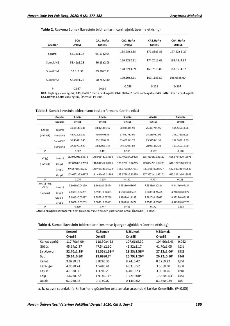
Harran Üniv Vet Fak Derg, 2020; 9 (2): 177-182 Araştırma Makalesi
Harran Üniversitesi Veteriner Fakültesi Dergisi, 2020; Cilt 9, Sayı 2 180
Tablo 2. Rasyona Sumak İlavesinin bıldırcınların canlı ağırlık üzerine etkisi (g)
BCA
CA1. Hafta CA2. Hafta CA3.Hafta CA4. Hafta
Gruplar Ort±SE Ort±SE Ort±SE Ort±SE Ort±SE
Kontrol
53.12±1.17 95.11±2.04 135.98±2.35 171.88±3.86 197.22± 5.27
Sumak %1
53.41±1.28 96.13±2.03 136.22±2.21 174.20±3.62 198.48±4.97
Sumak %2
52.8±1.31 89.20±2.71 128.32±3.09 163.78±3.88 187.35±4.32
Sumak %3
53.01±1.26 90.78±2.30 129.59±2.61 169.11±3.52 198.03±5.00
P
0.987 0.099 0.058 0.222 0.307
BCA: Başlangıç canlı ağırlık, CA1. Hafta:1.hafta canlı ağırlık, CA2. Hafta: 2.hafta canlı ağırlık, CA3.Hafta: 3.hafta canlı ağırlık, CA4.Hafta: 4.hafta canlı ağırlık, Önemsiz: P> 0.05
Tablo 3. Sumak ilavesinin bıldırcınların besi performansı üzerine etkisi
Gruplar 1.hafta 2.hafta 3.hafta 4.hafta 1-4hafta
Gruplar Ort±SE Ort±SE Ort±SE Ort±SE Ort±SE
CAK (g) Kontrol 41.9914±1.36 40.8714±1.12 38.0414±1.98 25.3377±1.96 144.4250±4.56
(Haftalık) Sumak%1 42.7220±1.59 40.0940±.78 37.9827±2.09 24.2807±1.92 145.0713±3.95
Sumak%2 36.4147±2.40 39.1280±.88 35.4573±1.79 23.5753±1.31 134.5687±2.89
Sumak%3 37.8079±2.23 38.8200±1.14 39.5229±1.82 28.9214±2.23 145.0657±3.00
P 0.067 0.461 0.515 0.197 0.120
YT (g) Kontrol 112.9429±3.83372 149.0664±2.93603 169.4093±7.09408 195.4636±11.33132 626.8764±20.12075
(Haftalık) Grup1 112.0280±3.27391 148.0373±2.70206 176.9787±8.26785 179.0847±11.64231 616.1227±18.36714
Grup 2 97.0873±5.82252 140.4033±3.36923 158.0793±8.47971 187.3667±9.88777 582.9293±14.69580
Grup3 103.6671±5.66875 141.4914±3.11764 182.6736±6.13829 207.3971±11.49201 635.2221±16.28982
P 0.070 0.108 0.130 0.327 0.168 YYO (g YT/g
CAK) Kontrol 2.6929±0.04290 3.6621±0.05494 4.4921±0.08697 7.6583±0.20314 4.3414±0.04124
(Haftalık) Grup 1 2.6387±0.04701 3.6993±0.04893 4.6980±0.08410 7.5280±0.21466 4.2480±0.06077
Grup 2 2.6913±0.05087 3.5973±0.07766 4.4947±0.14183 7.9820±0.12095 4.3327±0.05157
Grup 3 2.7600±0.03263 3.6686±0.08491 4.6764±0.12474 7.3586±0.26063 4.3793±0.06373
P 0.299 0.747 0.401 0.172 0.392
CAK: Canlı ağırlık kazancı, YT: Yem tüketimi, YYO: Yemden yararlanma oranı, Önemsiz (P > 0.05)
Tablo 4. Sumak ilavesinin bıldırcınların kesim ve iç organ ağırlıkları üzerine etkisi (g).
Kontrol %1Sumak %2Sumak %3Sumak Ort±SE Ort±SE Ort±SE Ort±SE p Karkas ağırlığı 117,70±4,09 118,50±4,52 107,66±5,30 104,66±2,43 0.062 Göğüs 45.14±2.37 47.54±2.60 43.32±2.17 41.70±1.05 0,231 Sırt+boyun 32.70±1.18a 31.35±1.38ab 28.23±1.58bc 27.12±1.06c 0.026 But 29.14±0.80a 29.09±0.7a 26.79±1.26ab 26.22±0.50b 0.049 Kanat 9.02±0.32 8.82±0.36 8.24±0.42 8.17±0.22 0,259 Karaciğer 4.96±0.74 4.54±0.41 4.03±0.52 3.56±0.30 0.239 Taşlık 4.15±0.30 4.37±0.23 4.40±0.23 3.98±0.26 0.589 Kalp 1.62±0.09b 1.91±0.11a 1.72±0.08ab 1.58±0.063b 0.050 Dalak 0.12±0.02 0.11±0.02 0.13±0.02 0.13±0.024 0871
a. b. c: aynı satırdaki farklı harflerle gösterilen ortalamalar arasındaki farklar önemlidir. (P<0.05)

Harran Üniv Vet Fak Derg, 2020; 9 (2): 177-182 Araştırma Makalesi
Harran Üniversitesi Veteriner Fakültesi Dergisi, 2020; Cilt 9, Sayı 2 181
Tablo 5. Sumak ilavesinin bıldırcınların göğüs eti renk özellikleri üzerine etkisi.
Gruplar Kontrol %1 Sumak %2 Sumak %3 Sumak p Ort±SE Ort±SE Ort±SE Ort±SE pH1 6,11±0,04 6,22±0,08 6,16±0,02 6,15±0,07 0,723 pH24 5,91±0,05 6,05±0,05 5,91±0,03 5,86±0,05 0,841 L*1 44,54±1,72 45,21±0,75 45,69±0,71 45,84±1,06 0,057 L*24 56,55±1,26 55,03±0,93 56,87±0,65 57,44±1,03 0,314 a*1 9,60±0,71 10,73±0,61 9,98±0,50 8,48±0,50 0,051 a*24 11,87±0,77 12,57±0,47 12,33±0,55 10,36±0,75 0,306 b*1 9,22±0,46 10,29±0,45 9,90±0,16 9,62±0,46 0,069 b*24 12,66±0,44 13,41±0,44 13,32±0,25 13,66±0,52 0,471
L*: Parlaklık, a*: Kırmızı renk koordinatı, b*: Sarı renk koordinatı Tablo 6. Sumak ilavesinin bıldırcınların oksidatif stres parametreleri üzerine etkisi.
Gruplar Kontrol %1 Sumak %2 Sumak %3 Sumak p Ort±SE Ort±SE Ort±SE Ort±SE TAS 1,24±0,03c 1,41±0,05b 1,52±0,05b 1,70±0,06a 0.000 TOS 15,02±0,82 14,45±0,71 13,48±0,,45 12,95±0,42 0.089 OSİ 1,20±0,04a 1,03±0,06b 0,89±0,05bc 0.76±0,02c 0.000 TOTAL THİOL 0,22±0,02 0,32±0,04 0.26±0,06 0.32±0,03 0.414 AOPP 0.63±0.09 0.55±0.06 0.50±0.10 0.38±0.06 0.233
TAS: Toplam antioksidan durum, TOS: Toplam oksidatif durum, OSİ: oksidatif stres indeksi, AOPP: Gelişmiş oksidasyon protein ürünleri, THİOL: Toplam thiol grupları, SE: Standart hata, a. b.c: aynı satırdaki farklı harflerle gösterilen ortalamalar arasındaki farklar önemlidir. (P<0.05)
Ghasemi ve ark. (2014), broyler civcivlerinin performans ve bağırsak özellikleri üzerine etkisini değerlendirdiği çalışmada rasyona %0,1 % 0.2 ve % 0.3 sumak ekstraktı ilavesinin yem tüketimini kontrol grubuna kıyasla anlamlı derecede arttırdığını (P <0.05) ve en yüksek canlı ağırlık artışının %0.2 sumak ekstraktı ilave edilen grupta elde edildiğini bildirmişlerdir. Valiollahi ve ark. (2014), sumak ve karabiber ilavesinin broyler civcivlerinin performansı üzerindeki etkisi isimli çalışmada, rasyonlarına sumak ve karabiber ilave edilen gruplarda yem tüketimi, canlı ağırlık artışı ve toplam canlı ağırlığının kontrol grubundan daha yüksek olduğunu bildirmişlerdir. Kheiri ve ark. (2015), broyler rasyonuna sumak ve kurutulmuş peynir altı suyu tozunun büyüme performansı, karkas özellikleri, bağırsak morfolojisi, mikrobiyal popülasyon, bazı biyokimyasal parametreleri ve Newcastle hastalığına karşı antikor titresi üzerindeki etkilerini araştırdıkları çalışmada, sumak ve peyniraltı suyunun birlikte ilave edilmesinin broyler civcivlerin yem tüketimi, canlı ağırlık artışını kontrol grubuna kıyasla arttırdığını belirtmişlerdir (P <0.05). Mansoob (2011), broyler rasyonunda farklı seviyelerde sumak kullanımının yem tüketimi canlı ağırlık artışı ve yemden yararlanma oranında önemli etkiye sahip olduğunu göstermiştir (P <0.05). Canlı
ağırlık artışının ve yemden yararlanma oranının iyileşmesinin sumakta bulunan aktif maddeden (sinnamaldehit ve ögenol) kaynaklandığını belirtilmiştir (Mansoob, 2011). Yapılan çalışmalara göre besi performansıyla ilgili sonuçların mevcut çalışmamızla uyuşmamasının muhtemel sebebi yüksek yaz sıcaklıklarına denk gelmiş olması olabilir. Yüksek sıcaklığa bağlı olarak yem tüketimi ve yemden yararlanma azalmakta, gelişme gerilemekte ve ölüm oranı artmaktadır (Lott, 1991; May ve Cott, 1992).
Çalışmada sumak ilavesinin kesim özellikleri ve iç organ ağırlıkları üzerine etkisi incelendiğinde sırt ve but ağırlıkları dışında incelenen kesim ve karkas ağırlıkları açısından gruplar arasında fark bulunmamıştır (P>0.05). Buna karşın Zavaragh (2011) sarımsak ve sumak tozunun Japon bıldırcınlarının performans ve serum kompozisyonu üzerindeki etkilerini araştırmışlardır. En yüksek karkas ve karaciğer yüzdesini sırasıyla %2 sarımsak ve sumak tozu ile %2 sumak tozu içeren grupta bulmuşlardır (P <0.05).
Japon bıldırcınlarında sumağın göğüs et kalitesi özelliklerinden olan pH ve bıldırcınların göğüs eti renk özellikleri (L*, a* ve b*) üzerinde etkisi önemli bulunmamıştır (P>0.05). Bunun sebebini hava sıcaklığının et kalitesi üzerindeki etkisinden

Harran Üniv Vet Fak Derg, 2020; 9 (2): 177-182 Araştırma Makalesi
Harran Üniversitesi Veteriner Fakültesi Dergisi, 2020; Cilt 9, Sayı 2 182
kaynaklandığına bağlayabiliriz (Altan ve ark., 2001). Ayrıca kesim öncesi stresininde et kalitesi üzerinde etkisi olmaktadır (Babj ve ark., 1982).
Japon bıldırcınlarında sumak ilavesinin oksidatif stres ve antioksidan kapasitesi değerlerini önemli düzeyde etkilediği tespit edilmiştir (P<0.05) (Tablo 6). Sumak düzeyinin artışına bağlı olarak TAS değeri kontrol grubuna göre yükseltmiştir (P=000). Bu bulguyla uyumlu olarak en yüksek OSİ değeri kontrol grubunda en düşük ise %3 sumak grubunda tespit edilmiştir (P=000). Alishah ve ark. (2012), yaptıkları çalışmalarında broyler yemlerine %0, %0.25, %0.50 ve %1 seviyelerinde sumak ilavesinin performans ve kan antioksidan düzeyi üzerine etkisini incelemişler ve sumak ilavesinin kandaki toplam antioksidan kapasitesi (TAC), malondialdehit (MDA) içeriği ve süperoksit dismutaz (SOD) ve glutatyon peroksidaz (GPX) enzim aktivitelerini etkilemediğini bildirmişlerdir (P>0,05). Ayrıca diyete sumak ilavesinin ısı stresindeki başlangıç döneminde broyler tavuklarının performansını artırabilmesine rağmen, büyüme döneminde kan antioksidan değerlerini ve performansı etkilemediği sonucuna varmışlardır.
Çalışma sonucuna göre bıldırcın rasyonlarına sumak ilavesinin herhangi bir olumsuz etkisinin olmadığı, sıcak yaz şartlarında TAS ve OSİ değerleri üzerine olumlu etkisinden dolayı yem katkı maddesi olarak kullanılabileceği belirlenmiştir.
Kaynaklar
Alishah AS, Daneshyar M, Aghazadeh A, 2013: The effect
of dietary sumac fruit powder (Rhus coriaria L) on performance and blood antioxidant status of broiler chickens under continuous heat stress condition. Ital J Anim Sci., 12 (6), 39-43.
Altan A, Bayraktar H, Önenç A, 2001: Etlik Piliçlerde Sıcak Stresinin Et Rengi ve pH’ı Üzerine Etkileri. Hayvansal Üretim Derg, 42 (2), 1-8.
Babj AS, Froning GW Nkoga DA, 1982. The effects of preslaughter environmental temperature in the presence of electrolyte treatment on turkey meat quality. Poult Sc, 61 (12), 2385–2389.
Başkol M, Dolbun Seckin K, Başkol G, 2014: Advanced oxidation protein products, total thiol levels and total oxidant/antioxidant status in patients with nash. Turk J Gastroenterol, 25 (1),32-37.
Çimrin T, Tunca Rİ, 2012: Bıldırcın beslemede alternatif yem ve katkıların kullanımı. Iğdır Üni Fen Bilimleri Enst Derg, 2 (3), 109-116.
Genchev A, Mihaylov R, 2008: Slaughter analysis protocol in experiments using Japanese quails (Coturnix Japonica). Trakia J. Sci, 6, 66-71.
Ghasemi R, Faghani M, Reza JP, Khonmirzaie N, Rahimian Y, 2014: Using Sumac (Rhus coriaria L.) extract affect performance and intestinal characteristics of broiler chicks. Scholarly J. Agric. Sci., 4 (8), 442-445.
Golzadeh MP, Farhoomand and Daneshyar M. 2012: Dietary Rhus coriaria L. Powder Reduces the Blood Cholesterol, VLDL-c and Glucose, but Increases Abdominal Fat in Broilers. S. Afr. J. Anim. Sci., 42 (4), 399-405.
Gümüş H, Oguz NM, Bugdayci KE, Oğuz FK, 2018: Effects of sumac and turmeric as feed additives on performance egg quality traits and blood parameters of laying hens. R. Bras. Zootec., 47, 1-7.
Kutlu HR, Şahin A, 2017: Kanatlı Beslemede Güncel Çalışmalar ve Gelecek için Öneriler. Hayvansal Üretim Derg, 58 (2),66-79.
Kheiri F, Rahimian Y, Nasır J, 2015: Application of sumac and dried whey in female broiler feedArch. Anim Breed, 58, 205-210.
Lott BD, 1991: The effect of feed intake on body temperature and water consumption of male
broilers during heat exposure. Poult Sci, 70, 410-417. Mansoob HN, 2012: Effect of different levels of Sumac
Powder (Rhus Coriaria L.) on performance, carcass and blood parameters of broiler Chickens. Annals of Biol. Res, 2 (5), 647-652.
May JD, Cott BD, 1992: Feed consumption patterns of broilers at high environmental temperatures. Poult Sci, 71, 331-336.
NRC, U. 1994: Science and judgment in risk assessment. Washington: National Academy of Sciences, Whashington, USA.
Salih YG, Gürbüz Y, 2015: Sumac (Rhus coriaria L.) and Ginger (Zingiber officinale) as Feed additive in Poultry Nutrition., KSU J Nat Sci, 18 (3), 44-48.
SPSS I. 1999: SPSS for Windows. Chicago, Illinois. Shidfar F, Rahideh ST, Rajab A, 2014: The Effect of Sumac
(Rhus coriaria L.) Powder on Serum Glycemic Status. ApoB. ApoA-I and Total Antioxidant Capacity in Type 2 Diabetic Patients. Iran. J. Pharm. Res., 13 (4), 1249-1255.
Valiollahi MR, Rahimian Y, Miri Y, Asgarian F, Rafiee A, 2014: Effect of ginger (Zingiber officinale) and black pepper (Piper nigrum L.) powder on performance, haematological parameters and antibody titre in broiler chicks. Res. Opinions Anim. Vet. Sci., 4 (3), 128-132.
Zavaragh FM, 2011: Influence of Garlic and Sumac Powder (Rhus coriaria L.) on Performance, Carcass and Blood Biochemical of Japanese Quails Scholars Research Library. Annals of Biological Research, 2 (6), 542-545.
*Yazışma Adresi: Nurcan KIRAR Harran Üniversitesi Sağlık Bilimler Enstitüsü, Şanlıurfa,
Türkiye. e-mail: [email protected]

Harran Üniv Vet Fak Derg, 2020; 9 (2): 183-188 Research Article
Harran Üniversitesi Veteriner Fakültesi Dergisi, 2020; Cilt 9, Sayı 2 183
Evaluation of Fundus Examination of Hunting Dogs' Eyes Using a Smartphone-Based Camera
Özlem ŞENGÖZ ŞİRİN1,a,*
1Burdur Mehmet Akif Ersoy Üniversitesi, Veteriner Fakültesi, Cerrahi Anabilim Dalı, Burdur TÜRKİYE. aORCID: 0000-0001-2345-6789
Geliş Tarihi: 08.10.2020 Kabul Tarihi: 27.10.2020 Abstract: Fundus imaging with a smartphone-based camera has been reported in a limited number of literature, particularly in patient groups (pediatric/geriatric ie.) considered to be incompatible. In this study, by examining both eyes of 20 hunting dogs, multiple shooting series (20 sequential shooting automatic series with 1-second interval) and video sequence (a video that started shooting at 30-second and/or 60-second intervals) were recorded. The patients were first examined without any intervention. Afterwards, mydriatic drops were applied and the examination was repeated within the effective period. During the examination, optic disc nerve head, tapetum lucidum, non-tepatal region, retinal vessels, and choroid vessels were visualized in the posterior segment. Focal light artifacts were common when photographing the tapetum lucidum. The minimum light intensity was chosen to display the tapetum lucidum. No significant difference was observed between the examinations performed before the mydriatic drop and the examinations performed after the mydriatic drop. Further studies are recommended to formally assess clinical benefit. Keywords: D-EYE, Dog, Retinal examination, Smartphone-based camera.
Akıllı Telefon Tabanlı Kamera Kullanarak Av Köpeklerinin Gözlerinin Fundus Muayenesinin Değerlendirilmesi
Özet: Akıllı telefon tabanlı telefon ile fundus görüntülenmesi özellikle uyumsuz olarak kabul edilen hasta gruplarında (pediatrik/geriatrik vb.) pratik bir şekilde görüntülerin elde edildiği sınırlı sayıdaki literatürde bildirilmiştir. Bu çalışmada 20 av köpeğinin her iki gözü incelenerek çoklu fotoğraf serisi (1 saniye aralıklarla 20 ardışık otomatik çekim serisi) ve video görüntüsü (30 saniye ve/veya 60 saniye aralıklarla kaydedilen video) kaydedilmiştir. Hastalar önce herhangi bir müdahale olmaksızın muayene edildi. Daha sonra midriyatik damlalar uygulandı ve etkili süre içinde muayene tekrarlandı. Muayenede optik sinir başı, tapetum lucidum, nontepatal bölge, retina damarları ve arka segmentte koroid damarlar görüntülendi. Tapetum lucidum'un fotoğrafını çekerken fokal ışık yapaylıkları yaygındı. Tapetum lucidum'u görüntülemek için minimum ışık yoğunluğu seçildi. Midriyatik damla öncesi yapılan muayeneler ile midriyatik damla sonrası yapılan muayeneler arasında anlamlı bir farklılık gözlenmedi. Klinik faydayı resmi olarak değerlendirmek için daha ileri çalışmalar önerilir. Anahtar Kelimeler: Akıllı telefon tabanlı kamera, D-EYE, Köpek, Retinal Muayene. Introduction
Photographing the fundus is a standard method for documenting ocular fundus findings, and fundus photography is traditionally performed in a clinical setting using a fundus camera (Haddock and Quin, 2015; Khanamari et al., 2017). The biggest limitation of current imaging systems is the equipment is not portable and expensive (Maamari et al., 2017; Games and Ledbetter, 2019). Recently, the growing popularity of smartphones, high-resolution cameras, large data storage capacities, ease of image capture and sharing have led to the widespread use of smartphones in ophthalmology (Haddock and Quin, 2015; Ryan et al., 2015). In smartphone-based fundus imaging, the coaxial flashlight of the smartphone camera and a strong, high-resolution hand lens form an ophthalmoscopy-like system capable of recording digital fundus images (Khanamari et al., 2017). Although it is a
complementary diagnostic tool by veterinary ophthalmology specialists, it is a unique, simple, and affordable application that provides photo-video documentation of retinal changes and allows consultation sharing in many clinical settings where retina imaging was previously not possible (Kanemaki et al., 2017; Russo et al., 2015).
Eye examinations of animals with systemic disease help narrow the differential diagnosis list. Diseases affecting the vascular and nervous systems are partially prone to ocular manifestations. In cases where ocular blood flow is very high, the probability of affecting the uveal and retinal vascular system increases, and hematogenous neoplastic cells and/or infectious organisms may arise in this area (Ofri, 2008). Ocular pathologies can be identified by a smartphone-based fundus imaging system. With clinical examination,
DOI:10.31196/huvfd.807349

Harran Üniv Vet Fak Derg, 2020; 9 (2): 183-188 Research Article
Harran Üniversitesi Veteriner Fakültesi Dergisi, 2020; Cilt 9, Sayı 2 184
cataracts, glaucoma, age-related maculopathy, chorioretinal atrophy, scar, systemic disorders (hypertension or diabetic retinopathy), retinitis pigmentosa, posterior vitreous detachment, and retinal detachment can be detected (Russo et al., 2014). When a fundus is displayed, a mydriatic agent should be used for comfortable viewing of the peripheral retina (Russo et al., 2015). Dynamic events such as venous circulation and uveal cysts floating in the vitreous cavity are visualized by video recording, which is an advantage of smartphone-based fundus imaging systems (Balland et al., 2017). The D-EYE digital ophthalmoscope is a fundus camera that can be attached to smartphones and used in conjunction with a Health Insurance Portability and Accountability Act (HIPAA) compliant application (Russo et al, 2014). D-EYE is an adapter that attaches magnetically to the smartphone and captures high-resolution fundus images with an approximate 20º field of view (Anonym, 2020). Through the D-EYE system, clinicians can share images stored digitally via an existing cloud system to get expert opinions without organizing a clinical visit (Mamtora et al., 2018).
This study aims to increase the limited literature knowledge about smartphone-based fundus imaging in the veterinary field, to confirm the availability of D-EYE devices in the incompatible patient group in the veterinary field, and the use of a mydriatic agent in examinations with D-EYE device.
Material and Method Animal care and use: All procedures were
approved by Burdur Mehmet Akif Ersoy University Animal Ethics Committee.
Equipment: Nowadays, the use of mobile devices to display the fundus is especially common in human medicine. Given the convenience and accessibility it provided during fundus examination, D-EYE® was selected for imaging, consisting of a meta cell shell and original optical systems provided by Galileo Diagnostics Corporation (Galileo Genclis, Nancy, France), and was used for evaluation of the fundus. In this study, a prototype D-EYE® was used on the iPhone 5 model. D-EYE was introduced to the market with a metal frame compatible with Apple (iPhone 5, iPhone 5s and iPhone 6; Apple Inc., Cupertino, CA, USA) and Samsung (Galaxy S4 and Galaxy S5; Samsung, Taegu, South Korea) brand phones. This metal frame is a bumper designed to fit the outer mold of the phone, allowing the smartphone to be fully aligned with the optics of the D-EYE. The metal frame allows easy installation of the D-EYE, via two neodymium magnets. D-EYE consists of special lenses, polarizing filters, a beam
splitter, a diaphragm, and mirrors in a configuration that reduces light reflections, aligns the LED light beam, and compensates for corneal glare. The optical path of the D-EYE allows fundus imaging with the camera lens of the smartphone. There are two polarization filter combinations that significantly reduce corneal reflection with cross-polarization. To reduce the intensity of light emitted by the flash, an aperture is added to the device.
Photo and video recording: In order to create a photo and video recording with D-EYE, an application called “D-EYE” is required, which can be used on both iOS and Android platforms. "Retina scan" is selected after login to the application. The patient's descriptive information is entered, and then the operator chooses Oculus Dexter (OD) (right eye) or Oculus Sinister (OS) (left eye). Image recording and video recording called “multishot” can be taken with the application. By the multishot feature, the number of shots and intervals can be adjusted, an automatic serial photo recording consisting of 20 consecutive shots can be taken at 1-second intervals. When the video recording will be created, the duration of the video recording can be adjusted from the settings. After the start button is pressed, recording starts when the image becomes clear thanks to the autofocus and focus locking features via the autofocus system (AF) button. In addition, if the iPhone is examined with a smartphone, the light intensity of the phone LED can be checked with a scale under the application screen.
Examination protocol: Twenty hunting dogs brought to Burdur Mehmet Akif Ersoy University Veterinary Faculty Animal Hospital Surgery Clinic due to minor operative interventions were included in this study. Informed consent was obtained from the animal owners during the study. Before creating mydriasis of all dogs, fundus examinations of the right and left eyes were done with D-EYE and the multishot series and video series were recorded. During the examination, the next step is to create a full 5-minute break three times mydriasis with tropicamide 0.5% (0.5% Tropamid; Science Ilac San. Tic. A.Ş., Istanbul, Turkey) was dropped. After the animals were kept in a dimly lit environment for 20 minutes, fundus examinations of their right and left eyes were done with D-EYE and the multishot series and video series were recorded. All animals were examined in a dimly lit hall and records were created. The animals were conscious during the examination and an assistant was holding the animals. The light intensity was adjusted by moving from left (minimum level) to right (maximum level) with the slider available in the application. Minimum light intensity was preferred to display tapetum lucidum. During the imaging of the non-

Harran Üniv Vet Fak Derg, 2020; 9 (2): 183-188 Research Article
Harran Üniversitesi Veteriner Fakültesi Dergisi, 2020; Cilt 9, Sayı 2 185
tapetal region, medium and high light intensities were selected. In animals where the autofocus algorithm failed to achieve a sharp image; the focus was locked to infinity before aiming at a distant object (several meters). When the subject appears clearly on the screen, AF is locked by pressing the AF button. Records were created by following the same method for each animal. Images were checked at every stage and repeated if not satisfactory.
Results Of the hunting dogs included in the study, 18
were female and 2 were male. The average age of dogs is 1.97±0.21 years. In the study, fundus pathologies were not found in dogs.
Throughout the study, the smartphone was held by the left hand while performing the right eye examination, and by the right hand while performing the left eye examination. In cases where autofocus could not lock the fundus, it was locked to infinity for the first time as described earlier and the examination was repeated. The smartphone was positioned at 1-2 cm from the patient's eye. A safe distance has been established between the clinician and the patient being examined. With this distance, the clinician was able to easily observe the movements of the animal under examination. The fundus of the examined eye was examined in real-time on the phone screen before recording. When the fundus image was captured, the record button was pressed. Animals are expected to stand still during the fundus examination. Although it is difficult to perform fundus examination in moving temperament-hunting dogs, fundus images that can be interpreted with D-EYE were recorded within 1-3 minutes.
As shown a Figure 1 the optic nerve head, tapetum lucidum, non-tapetal region, retinal vessels, and choroid vessels were seen in the posterior segment structures. In the tapetum lucidum examination, the best image was obtained with minimum light intensity and in the cassette-free region examination, the best image was obtained with maximum light intensity. Focal light artifacts were common in tapetum lucidum photographs. As shown a Figure 2 these light artifacts are usually placed dorsally on the image. During the examination, the fundus was artificially hyperreflective. Video recordings and artificial hyper-reflective findings were easily distinguishable from true hyper-reflective findings. In fundus examination, we thought that we could detect a localized inactive chorioretinitis adjacent to the optic nerve in 1 case. However, as shown a Figure 3 through the video recording of the case, we
Figure 1. The anatomical structures of the posterior segment were observed during the examination: optic disc nerve head, tapetum lucidum, non-tapetal region, retinal vessels, and choroid vessels.
Figure 2. Focal light artifacts are common when tapetum lucidum is photographed (Shown with white arrow).

Harran Üniv Vet Fak Derg, 2020; 9 (2): 183-188 Research Article
Harran Üniversitesi Veteriner Fakültesi Dergisi, 2020; Cilt 9, Sayı 2 186
Figure 3. Hyporeflective light artifact that acts as a pathological finding when tapetum lucidum is photographed.
Figure 4. No significant difference was observed between the examinations performed before the mydriatic instillation and the examinations performed after the mydriatic instillation. However, in fundus photographing, the field of view is wider with a mydriatic drip. A-C: fundus photograph by examination without dripping mydriatic, B-D: fundus photograph by examination dripping mydriatic. While the field of view is expanded in B, no significant expansion is observed in D.
determined that the area with this hyporeflexivity was a camera-induced study. Video recordings allowed the display of dynamic events. With the videos recorded during the examination, hyper-reflective events and artificial hyper-reflective pathologies, as well as mild artifacts and pathologies can be distinguished.
Non-tapetal fundus was examined, no pathogenicity was encountered. The color of the non-tapetal fundus, which did not feature reflexivity, varies between black (65%) in 13 dogs and brown (35%) in 7 dogs. In some cases, the non-tapetal region adjacent to the tapetum lucidum had a pale and browner appearance than the ventral non-tapetal region. This was not a sign of pathogenicity. When examining the bulbar part of the optic nerve head, the color varied from pinkish white to dark pink. No pathogenicity could be determined. Hyaloid vessels displayed a healthy appearance in all fundus images.
No significant difference was observed between the examinations performed before the mydriatic vaccination and the examinations performed after the mydriatic vaccination. Peripheral visualization of the tapetal fundus was easier during examinations with a mydriatic drip. As shown a Figure 4, it was observed that the part of the fundus displayed in the studies with dripping mydriatic was relatively larger.
Discussion With the advances in technology, the use of
mobile devices as a medical diagnostic tool is becoming increasingly common. Taking advantage of the easy portability of mobile phones, the size of its data storage capacity, and wireless connectivity, it is thought that smartphone-based fundus imaging will play an important role in clinics soon (Maamari et al., 2013; Shen and Mukai, 2017). Khanamiri et al. (2017) compared the quality of the fundus images taken with a smartphone and the quality of the images taken using traditional fundus cameras, and there was no significant difference between the images. Fundus photography can be done in a clinical setting using fundus cameras, but the expensive and immovable equipment is the major disadvantage of this method (Khanamiri et al., 2017). Miniaturization and improvement of internal photographic equipment (etc. lenses, sensors, lighting systems, and autofocus systems) have made smartphone-based fundus imaging very powerful tools. An example of a small optical device that can be connected to a smartphone is the D-EYE module (D-EYE Srl, Padova, Italy). Smartphone-based fundus imaging systems have been described in both humans and animals (Balland et al., 2014).
Although the ophthalmoscopic technique used by the D-EYE for imaging the fundus is not replaced, it is very similar to that of direct ophthalmoscopy (Balland et al., 2014). The absence of a safe distance between the patient and the clinician during direct ophthalmoscopy is a disadvantage of direct ophthalmoscopy (Gelatt et al., 2013). Besides being easy to use in the field conditions, D-EYE provided easy visualization of the fundus especially in children and the elderly, which are considered incompatible patient groups (Anonym, 2020). In our study, we used puppy dogs as an incompatible patient group to test the effectiveness of D-EYE in the veterinary field. The time to record fundus images was determined as 2-3 minutes as a result of the examination we performed with D-EYE in puppies with a highly mobile temperament. We think that D-EYE may be the reason for preference in ophthalmological examinations in the veterinary field, especially inpatient groups that are

Harran Üniv Vet Fak Derg, 2020; 9 (2): 183-188 Research Article
Harran Üniversitesi Veteriner Fakültesi Dergisi, 2020; Cilt 9, Sayı 2 187
incompatible and cannot be transferred to the clinic. There is also an arms distance between the clinician and the patient, which can be considered safe during the examination. In this way, the clinician can easily observe the animal's movements.
Light safety limits for ophthalmic instruments are determined in humans by the International Standards Organization (ISO 15004-2.2). In this respect, smartphone fundus photography is a safe technique (Khanamiri et al., 2017). Kim et al. (2012) compared the light levels produced by standard indirect ophthalmoscopes and used in smartphone fundus photography (Kim et al., 2012). In this study, the light produced by the standard indirect ophthalmoscope was shown to be 10 times brighter than the light used by the iPhone 4. In the D-EYE application, the iPhone LED light, which is darkened by polarizing filters and diaphragm, and carried through the diverging lens, is 15 and 24 times less for thermal and photochemical hazards, respectively, than the light used in indirect ophthalmoscopy. As a result, the retinal exposure of smartphones is less than 1 degree from an indirect ophthalmoscope. Both are within the safety limits of the thermal and photochemical hazards defined by ISO when tested under conditions simulating routine fundoscopy (Kim et al., 2012). Haddock and Qian (2015) although the light intensity and energy levels of the iPhone 5 are higher than that of the iPhone 4, they think that it is far below the danger limits.
In this study, it was observed that focal light artifacts were common in the tapetum lucidum and that the fundus exhibited hyper-reflective properties during the examination. Similarly, Balland et al. (2017) showed that the tapetum area is overexposed and the fundus is artificially hyper-reflective. They reported that artifacts did not cover the entire tapetal region, and their size varied and appeared as smooth areas with no visible details. Also, it was stated that light artifacts were not observed in the examination of the subalbinotic fundus and non-tapetal region. The iPhone's focus and exposure automation algorithms were sometimes overwhelmed by the tapetum lucidum. Exposure problems have manifested themselves as light artifacts in images. This problem can be solved by turning off autofocus and manually focusing to infinity through the application. The dynamic range of the fundus of the carnivores (the difference between the brightest and darkest areas) is quite evident. Therefore, a wide and high-definition sensor is required, which is suitable for the dynamic range of the fundus of carnivores. In contrast, the relatively small size of the iPhone sensor may be responsible for overexposure in the tapetal region
(Balland et al., 2017). Kanemaki et al. (2016) suggested that in fundus photographs, 2 small bright spots reflected from the front and back surfaces of indirect lenses can affect the image. For this reason, to prevent overexposure while recording images, the light intensity should be decreased when photographing the tapetal region, and they stated that the light intensity should be increased when photographing the non-tapetal region. In contrast, although high image resolution is set during video recording, it has been suggested that image resolution may be low due to low light (Kanemaki et al., 2017). Gomes and Ledbetter (2019) reported that enlightenment artifacts may be present in all images due to the physics of light passing through an indirect lens, but this affects less than 1% of the photographs collected (Gomes and Ledbetter, 2019). With our study, we determined that mild artifacts resulting from this overexposure can be confused with retinal pathologies. In videos, hyper-reflective areas can be displaced and usually placed dorsally. This suggests that areas that appear to have hyper-reflectivity can be caused by the phone's LED light. Also, it was detected a camera artifact that can cause an incorrect assessment of the images of a case thanks to the video recording feature. As a result of the displacement of the area that appears to be hyporeflective in fundus images, we observed that this is a product of the camera. For this reason, it was argued that the video recording feature offers a dynamic examination opportunity during the examinations with D-EYE and is extremely important in distinguishing pathologies.
Examinations in the smartphone-based fundus imaging system can be performed with or without using mydriatic (Anonym, 2020). Ryan et al. (2015) reported that in both methods, pathogenicity that threatens vision can be detected. However, there is a lower sensitivity in detecting diseases such as diabetic retinopathy in smartphone systems, especially when mydriatic is not used (Ryan et al., 2015). Russo et al. (2015), a limitation of the D-EYE system is inadequate in visualizing the peripheral retina (Russo et al., 2015). Baeza et al. (2009) argue that there is a linear ratio between pupil patency and fundus photo quality (Baeza et al., 2009). In contrast, Shen et al. (2017) stated that physiological dilation, which is a part of standard ophthalmic practice, has significant disadvantages. First, ophthalmologists are not accustomed to using dilatation drops. In addition, regardless of expertise, pharmacological dilation tends to be inconvenient for both the medical doctor and the patient, dilatation drops take about twenty minutes to take effect, and the patient experiences blurred vision and light sensitivity for up to several hours after

Harran Üniv Vet Fak Derg, 2020; 9 (2): 183-188 Research Article
Harran Üniversitesi Veteriner Fakültesi Dergisi, 2020; Cilt 9, Sayı 2 188
dilatation. Also, pharmacological dilation prevents physical examination of the pupil over the next few hours, which is an undesirable effect when monitoring patients with critical neurological disease. Finally, the risk of provoking acute angle-closure glaucoma insensitive eyes with the use of expanding eye drops is a small but real risk. Given these drawbacks, it is more useful for pediatric ophthalmologists, although the comfort of examining with a prototype camera without using a mydriatic is difficult to align (Shen and Mukai, 2017). Ryan et al. (2015) think that the direction to be developed for this technique is to improve the imaging of the peripheral fundus (Ryan et al., 2015).
It was claimed that the D-EYE system offers some practical advantages over desktop fundus photography and other portable ophthalmic imaging devices. The first of these; is a lightweight, compact and inexpensive device that facilitates ophthalmoscopic examination. It provides the opportunity to examine patients who cannot be brought to the clinical environment around them with their portability. It allows a safe distance between the patient and the clinician. Last time; it offers the opportunity to work more harmoniously with the group of patients who have difficulties during the examination. Light intensity and quiet operation, which can be easily adjusted with the application, give the patient minimum discomfort. Third; data can be stored clinically and shared for consulting purposes via wireless connection and cloud storage. Although the quality of the photos is lower than the quality of the photos obtained with fundus cameras, the veterinarian will be very useful as a complementary diagnostic tool on the field. It is among the advantages of allowing ophthalmoscopic scanning without the need for mydriatic use. A limitation of the device is that the peripheral retina cannot be fully visualized without using the mydriatic during examinations. The device needs to be developed to show the peripheral retina more comfortably. Finally, the D-EYE system is a complementary and useful screening method because of its ease of use, data storage, portability, allowing mydriatic examination in routine scans, and allowing patients to examine without stress even in patients not compatible with the veterinarian.
References
Anonym 2020. https://www.deyecare.com/ Accession date; 01.05.2020. Russo A, Morescalchi F, Costagliola C, Romano MR, Marino IG, 2014: Comparison of smartphone-based ophthalmoscopy
versus dilated ophthalmic examination to detect ocular pathologic features.
Baeza M, Orozco-Beltrán D, Gil-Guillen VF, Pedrera V, Ribera MC, Pertusa S, Merino J, 2009: Screening for sight threatening diabetic retinopathy using non-mydriatic retinal camera in a primary care setting: to dilate or not to dilate? Int. J Clin Pract, 63 (3), 433-8.
Balland O, Russo A, Isard PF, Mathieson I, Semeraro F, Dulaurent T, 2017: Assessment of a smartphone-based camera for fundus imaging in animals. Vet Ophthalmol, 20 (1), 89-94.
Gelatt KN, Gilger BJ, Kern TJ, 2013: Veterinary Ophthalmology. 5th ed, Wiley Blackwell, New Jersey, USA.
Gomes FE, Ledbetter E, 2019: Canine and feline fundus photography and videography using a nonpatented 3D printed lens adapter for a smartphone. Vet Ophthalmol, 22 (1), 82-92.
Haddock LJ, Qian MD, 2015: Smartphone technology for fundus photography greater portability could mean greater versatility. Retin Physician, 12 (6), 51-58.
Kanemaki N, Inaniwa M, Terakado K, Kawarai S, Ichikawa Y, 2017: Fundus photography with a smartphone in indirect ophthalmoscopy in dogs and cats. Vet Ophthalmol, 20 (3), 280-4.
Khanamiri HN, Nakatsuka A, El-Annan J, 2017: Smart phone fundus photography. J Vis Exp, 125, 1-5.
Kim DY, Delori F, Mukai S, 2012: Smartphone photography safety. J Ophthalmol,119 (10),2200-01.
Maamari RD, Keenan JD, Fletcher D.A, Margolis TP, 2014: A mobile phone-based retinal camera for portable wide field imaging. Br J Ophthalmol, 98 (4), 438-441.
Mamtora S, Sandinha MT, Ajith A, Song A, Steel DHW, 2018: Smartphone ophthalmoscopy: a potential replacement for the direct ophthalmoscope. Eye, 32 (11), 1766-1771.
Ofri R, 2008: Retina. In “Slatter’s Fundamentals of Veterinary Ophthalmology”, Ed; Maggs DJ, Miller PE, Ofri R, Saunder Elsevier, Missouri USA.
Russo A, Mapham W, Turano R, Costagliola C, Morescalchi F, Semeraro F, 2014: Comparison of smartphone ophthalmoscopy with slit-lamp biomicroscopy for grading vertical cup-to-disc ratio. J Glaucoma, 25 (9), 777-781.
Russo A, Morescalchi F, Costagliola C, Delcassi L, Semeraro F, 2015: Comparison of smartphone ophthalmoscopy with slit-lamp biomicroscopy for grading diabetic retinopathy. Am J Ophthalmol, 159 (2), 360-364.
Ryan ME, Rajalakshmi R, Prathiba V, Anjana RM, Ranjani H, Narayan KMV, Olsen TW, Mohan V, Ward LA, Lynn MJ, Hendrick A, 2015: Comparison Among Methods of Retinopathy Assessment (CAMRA) study. J Ophthalmol, 122 (10), 2038-2043.
Shen BY, Mukai SA, 2017: Portable, Inexpensive, Nonmydriatic Fundus Camera Based on the Raspberry Pi® Computer. J Ophthalmol, 14, 1-5.
*Correspondence: Özlem ŞENGÖZ ŞİRİN Burdur Mehmet Akif Ersoy Üniversitesi, Veteriner
Fakültesi, Cerrahi Anabilim Dalı, Burdur, Türkiye. e-mail: [email protected]

Harran Üniv Vet Fak Derg, 2020; 9 (2): 189-193 Araştırma Makalesi
Harran Üniversitesi Veteriner Fakültesi Dergisi, 2020; Cilt 9, Sayı 2 189
İvesi Koyunlarında (Ovis aries) Mandibula’nın Morfometrik İncelemesi
Bestami YILMAZ1,a,*
1Harran Üniversitesi, Veteriner Fakültesi, Anatomi Anabilim Dalı, Şanlıurfa, Türkiye. aORCID: 0000-0002-0901-3129
Geliş Tarihi: 20.10.2020 Kabul Tarihi: 15.12.2020 Özet: Bu çalışma İvesi koyunlarında mandibula’nın morfometrik özelliklerini belirlemek amacıyla yapıldı. Bu amaçla 20 adet (10 erkek, 10 dişi) erişkin İvesi koyunu (Ovis aries) mandibulası kullanıldı. Bir bütün halinde temin edilen kafataslarının maserasyonu yapıldı, kas ve deri diseksiyonları tamamlandı ve temporomandibular bölgenin dezartikülasyonu ile mandibula’ lar çıkarıldı. Mandibula’ lar üzerinde 18 adet doğrusal ölçüm analizleri yapıldı ve tüm veriler ortalama ± standart sapma (SD) olarak ifade edildi. Yapılan incelemelerde dişi ve erkekler arasında GC-MAK (P<0.05) ve CR-PC (P<0.01) parametrelerinde istatistiksel farklılıklar gözlendi. Osteometrik ölçümler arasındaki korelasyon analizleri incelendiğinde veriler arasında P<0.05 ve P<0.01 değerinde istatistiksel önem tespit edildi. Elde edilen biyometrik verilerin hayvan modeli olarak koyun mandibala modellerinin kullanıldığı anatomik, klinik ve taksonomik çalışmalarda referans bilgi kaynağı niteliğinde biyometrik ölçüm ve sınırlar sunacağı düşüncesindeyiz. Anahtar Kelimeler: İvesi koyunu, mandibula, morfometri.
Morphometric Evaluation of the Mandible in the Awassi Sheep (Ovis aries)
Abstract: In this study, a morphometric test of mandible was performed in Awassi sheep. In the study, twenty adult (10 female and 10 male) Awassi sheep (Ovis aries) mandibles were used. The skulls were macerated, muscle and skin dissections were completed and the mandibles were removed by disarticulation of the temporomandibular region, respectively. On the mandible samples, 18 linear measurements were analyzed and all data were expressed as mean ± standard deviation (SD). Statistical differences were observed in GC-MAK (P<0.05) and CR-PC (P<0.01) parameters between males and females in the examinations. The correlation analysis on osteometric measurements indicated a statistical significance between P <0.05 and P <0.01. We believe that the obtained biometric data will provide a reference information source for biometric measurements and edges in anatomical, clinical, and taxonomic studies using sheep mandibles as animal models. Keywords: Awassi sheep, mandible, morphometry. Giriş
Mandibula, embriolojik olarak brankial kemerden köken alan, symphsis mandibula aracılığıyla sağ ve sol tarafı birleşen, kafatasının en büyük tamamlayıcı kemiğidir. Mandibula, cranium’a os temporale ile articulatio (art.) temporomandibularis eklemi aracılığıyla hareketli olarak bağlanır (Dyce ve ark., 2010; Nickel ve ark., 1986). Geçmişten günümüze gelen canlıların, genetik ve çevre şartlarına bağlı olarak evrimsel değişimlerini yorumlamak kemik morfolojisi ve morfometrisi kullanılarak yapılabilir (Rohlf ve Marcus 1993). Mandibula kemiğinin morfometrisi, cinsiyet (Akbulut ve ark., 2014), ırk (Gürbüz ve ark., 2016), beslenme (Kiliaridis ve ark., 1999) ve genetik (Rohlf ve Marcus, 1993) faktörlerden kaynaklı değişiklik gösterir.
Koyun ırkları kendi içerisinde oldukça çeşitlilik gösterir (Kaymakçı, 2001). Bu ırkların karakteristik yapısını belirlemede morfometrik yapısının ortaya konulması önemlidir. İvesi koyunu yoğunluk olarak Türkiye’nin Güneydoğu Anadolu bölgesinde yetiştiriciliği yapılan yerli koyun ırklarındandır. Yerli
koyun ırklarının %2,2’sini oluşturur (Garip, 2013). Bu koyun ırkı üzerinde birçok anatomik ve morfometrik (Demircioğlu ve Yılmaz, 2019; Yılmaz ve Demircioğlu, 2020) çalışma yapılmıştır. Bu çalışmada ise, İvesi koyunu mandibulası’nın morfometrik özelliklerini belirleyerek, sexuel dimorphizmini ortaya koymak, filogenetik, zooarkeoloji ve klinik bilimler üzerine yapılacak çalışmalarda kullanılabilir veriler sunmak amaçlandı.
Materyal ve Metot Etik kurul izni: Çalışmada uyulacak prosedürler
için Harran Üniversitesi Hayvan Deneyleri Yerel Etik Kurulu’ndan (Karar numarası: 2019/005/05) gerekli izinler alındı.
Çalışma materyali: Çalışmada 20 adet (10 erkek, 10 dişi) erişkin İvesi koyunu mandibula’ sı kullanıldı. Bu mandibula örnekleri Şanlıurfa’daki yerel bir kesimhaneden temin edildi. Bu örneklerin ait oldukları hayvanlarda iskelet deformasyonu,
DOI:10.31196/huvfd.813490

Harran Üniv Vet Fak Derg, 2020; 9 (2): 189-193 Araştırma Makalesi
Harran Üniversitesi Veteriner Fakültesi Dergisi, 2020; Cilt 9, Sayı 2 190
patolojik ve nörolojik bozukluk olmamasına dikkat edildi. Tüm kafatası örnekleri Taşbaş ve Teciroğlu (1965)’na göre yapılan maserasyon tekniği sonrasında morfometrik incelenmelere hazır hale getirildi. Kafatası üzerindeki deri ve kasların diseke edilmesinin ardından temporomandibular bölgenin dezartikülasyonu ile mandibula’ lar çıkarıldı. Ardından çeneler biyolojik maserasyona tabi tutuldu ve hidrojen peroksit solüsyonunda bekletilerek ağartıldı.
İstatistiksel analiz: Çalışmada elde edilen parametrelerin ortalama ve standart sapma değerleri SPSS 22 programında belirlendi. Ayrıca elde edilen veriler cinsiyete göre student t-testi ile karşılaştırıldı ve verilerin korelasyon analizi pearson korelasyon testi ile yapıldı.
Terminolojide Nomina Anatomica Veterinaria (2017) terimleri esas alındı. Çalışma fotoğrafları Canon (EOS-700D, Japonya) dijital fotoğraf makinesi ile alındı.
Osteometrik Ölçümler: Mandibula’nın osteometrik ölçümleri için aşağıdaki ölçüm noktaları kullanılmıştır (Yilmaz ve Demircioglu, 2019; Demiraslan ve ark., 2014).
Coronion (CR): Processus coronoideus’un en yüksek noktası.
Condylon (PC): Processus condylaris’in caudal noktası.
Gonion caudale (GC): Angulus mandibula açısının en aboral noktası.
Gonion ventrale (GV): Angulus mandibula açısının en uç ventral noktası.
Infradentale (ID): Mandibular insiciv diş alveol’lerinin oral yüzündeki en belirgin median noktası.
Ölçülen osteometrik verilerin ölçüm noktaları ve kısaltmaları Tablo 1’de, mandibula üzerindeki bölgeleri ise Şekil 1 ve Şekil 2’ de verildi.
Tablo 1. Mandibular ölçüm noktaları ve kısaltmalar.
Parametre Kısaltma Açıklama 1. GC-ID Gonion caudale- Infradentale arası uzunluk 2. CR-ID Coronion- Infradentale arası uzunluk 3. GC-MAK Gonion caudale- M3’ün aboral alveolar kenarı arası uzunluk 4. MAK-ID M3’ün aboral alveolar kenarı- Infradentale arası uzunluk 5. GC-PIK Gonion caudale- P2’nin oral alveolar kenarı arası uzunluk 6. GC-FMN Gonion caudale- Foramen (for.) mentale’nin aboral ucu arası uzunluk 7. MDU Molar diş sırası uzunluğu 8. PDU Premolar diş sırası uzunluğu 9. DU Diastema uzunluğu 10. GV-CR Gonion ventrale- Coronion arası uzunluk 11. MAK-MH M3’ün aboral alveolar kenarı düzleminde mandibula yüksekliği 12. MO-MH M1’in ön alveolar kenarı düzleminde mandibula yüksekliği 13. FMN-ID For. mentale’nin oral ucu - Infradentale arası uzunluk 14. CR-PC Coronion- Processus (proc.) condylaris’in en yüksek noktası arası uzunluk 15. SI Son incisiv diş seviyesinde mandibula genişliği 16. BM Birinci molar diş seviyesinde mandibular boşluğun genişliği 17. CG Proc. condylaris genişliği 18. MG Proc. coronoideus seviyesinde mandibular boşluğun genişliği
Tablo 2. Mandibula ölçümlerinin ortalama ve standart sapma değerleri (*P< 0.05, **P<0.01, NS: P>0.05, ÖD: Önemli değil).
Parametreler Tanımlayıcı istatistik Erkek Dişi
P Ort. + SD Min. Max. Ort. + SD Ort. + SD
1. GC-ID (mm) 180.22 ± 11.91 158.74 199.92 176.14 ± 12.57 184.30 ± 10.23 ÖD 2. CR-ID (mm) 191.55 ± 22.35 109.16 210.15 183.31 ± 27.89 199.80 ± 11.20 ÖD 3. GC-MAK (mm) 57.63 ± 5.62 47.18 64.24 55.03 ± 4.95 60.22 ± 5.21 * 4. MAK-ID (mm) 124.84 ± 6.74 115.44 138.44 123.69 ± 6.34 125. 99 ± 7.27 ÖD 5. GC-PIK (mm) 120.27 ± 9.07 101.23 133.07 117.41 ± 10.19 123.13 ± 7.20 ÖD 6. GC-FMN (mm) 145.28 ± 28.92 30.02 166.51 135.35 ± 38.39 155.22 ± 8.51 ÖD 7. MDU (mm) 41.28 ± 8.48 27.14 54.28 38.69 ± 9.92 43.86 ± 6.20 ÖD 8. PDU (mm) 29.60 ± 4.86 19.06 34.66 29.83 ± 5.18 29.36 ± 4.80 ÖD 9. DU (mm) 47.62 ± 4.80 39.44 58.11 46.27 ± 3.29 48.97 ± 5.82 ÖD 10. GV-CR (mm) 99.72 ± 18.45 34.39 121.79 95.61 ± 21.89 103.82 ± 14.23 ÖD 11. MAK-MH (mm) 38.86 ± 4.12 29.14 45.52 40.18 ± 3.71 37.54 ± 4.26 ÖD 12. MO-MH (mm) 24.84 ± 2.59 18.19 27.97 25.47 ± 1.95 24. 20 ± 3.08 ÖD 13. FMN-ID (mm) 25.03 ± 2.66 19.63 28.96 24.14 ± 2.82 25.92 ± 2.28 ÖD 14. CR-PC (mm) 24.71 ± 3.75 18.51 31.88 22.52 ± 3.05 26.91 ± 3.12 ** 15. SI (mm) 24.59 ± 3.40 18.14 31.27 24.63 ± 3.07 24.55 ± 3.87 ÖD 16. BM (mm) 25.98 ± 3.04 20.53 32.83 25.57 ± 2.98 26.39 ± 3.20 ÖD 17. CG (mm) 24.03 ± 1.39 21.94 26.68 23.56 ± 1.30 24.50 ± 1.38 ÖD 18. MG (mm) 72.90 ± 13.62 29.16 102.08 72.01 ± 6.72 73.80 ± 18.57 ÖD

Harran Üniv Vet Fak Derg, 2020; 9 (2): 189-193 Araştırma Makalesi
Harran Üniversitesi Veteriner Fakültesi Dergisi, 2020; Cilt 9, Sayı 2 191
Şekil 1. Mandibula’ nın morfometrik ölçüm noktaları (lateral görünüm)
Şekil 2. Mandibula’ nın morfometrik ölçüm noktaları (dorsal görünüm)
Tablo 3. Mandibula’ ya ait alınan ölçümler arası korelasyon ilişkisi (*P<0.05, **P <0.01) GC-ID CR-ID GC-MAK MAK-ID GC-PIK GC-FMN MDU PDU DU GV-CR MAK-MH MO-MH FMN-ID CR-PC SI BM CG MG GC-ID 1 CR-ID 0.357 1 GC-MAK 0.774** 0.384 1 MAK-ID 0.814** 0.049 0.557* 1 GC-PIK 0.797** 0.361 0.775** 0.505* 1 GC-FMN 0.611** 0.188 0.411 0.454* 0.698** 1 MDU 0.661** 0.216 0.436 0.605** 0.746** 0.579** 1 PDU -0.352 -0.374 -0.285 -0.164 -0.462* -0.306 -0.605** 1 DU 0.731** 0.314 0.534* 0.680** 0.303 0.404 0.197 -0.175 1 GV-CR 0.085 0.816** 0.163 -0.104 0.201 0.034 0.186 -0.384 0.143 1 MAK-MH 0.200 -0.207 0.287 0.276 0.320 -0.035 0.183 0.009 -0.006 -0.132 1 MO-MH 0.020 -0.285 0.186 0.093 0.105 -0.211 -0.143 0.270 -0.184 -0.304 0.699** 1 FMN-ID 0.489* 0.307 0.610** 0.325 0.517* 0.360 0.280 -0.055 0.261 0.200 0.237 -0.014 1 CR-PC 0.359 0.400 0.547* 0.094 0.595** 0.332 0.505* -0.325 -0.062 0.196 -0.014 -0.048 0.394 1 SI 0.285 -0.069 0.250 0.186 0.428 0.123 0.186 0.268 -0.042 -0.115 0.541* 0.554* 0.192 0.047 1 BM -0.116 0.227 -0.033 -0.263 0.298 0.149 0.338 -0.286 -0.435 0.256 0.057 0.127 -0.142 0.448* 0.373 1 CG 0.711** 0.260 0.447* 0.714** 0.513* 0.521* 0.622** -0.272 0.481* -0.006 0.278 0.157 0.339 0.427 0.239 0.084 1 MG 0.036 0.243 0.158 -0.306 0.329 0.085 0.143 -0.346 -0.246 0.090 -0.244 -0.256 -0.205 0.358 -0.056 0.234 -0.273 1

Harran Üniv Vet Fak Derg, 2020; 9 (2): 189-193 Araştırma Makalesi
Harran Üniversitesi Veteriner Fakültesi Dergisi, 2020; Cilt 9, Sayı 2 192
Bulgular Mandibula’ya ait osteometrik ölçümlerin
ortalama değer ve standart sapmaları Tablo 2’de, verilerin birbirleriyle olan korelason analizleri Tablo 3’ de gösterildi. Student t-testi ile yapılan istatistiksel analizde dişi ve erkekler arasında GC-MAK (P<0.05) ve CR-PC (P<0.01) parametresinde istatistiksel farklılıklar gözlendi. Diğer verilerde cinsiyetler arası istatistiksel olarak anlamlı fark gözlenmedi (P>0.05). Osteometrik ölçümler arasında korelasyon analizleri incelendiğinde veriler arasında P<0.05 ve P<0.01 değerinde istatistiksel önem tespit edildi.
Tartışma ve Sonuç Koyunlarda tür içinde oldukça fazla sayıda ve
farklı morfolojik ve verim özellikleri gösteren ırklar bulunur (Kaymakçı, 2001). Yaklaşık onbir bin yıl önce evcilleştirilmeye başlayan koyunların (Vaughan ve ark., 2015) günümüze kadar geldiği süreçte gerek çevresel gerekse genetik etmenlerden dolayı morfolojilerinde birtakım değişiklikler meydana gelmiştir. Bu bağlamda koyunlara ait osteometrik ölçümlerin belirlenmesi, ırka özgü özelliklerin ortaya konulmasını, adli, zooarkeolojik ve gelişimsel bilimler adına oldukça önemli veriler sunar (Pitakarnnop ve ark., 2017). Bu amaçla, mandibula’dan alınan ölçümlerin cinsiyetler arası ve tür içi farklılıkları ortaya koyması için, veteriner anatomi alanında (Dalga ve ark., 2017; Dermiraslan ve ark., 2014; Özkan ve ark., 2020; Özüdoğru ve ark., 2019a; Özüdoğru ve ark., 2019b; Yilmaz ve Demircioglu, 2019) birçok çalışma yapılmıştır.
İnsanlarda ve farklı tür hayvanlarda mandibula’ya ait osteometrik ölçüm değerlerine bakıldığında erkeklerde dişilere oranla bazı parametrelerin daha yüksek olduğu ve ölçümler arasında anlamlı istatistiki önemler olduğu bildirilmiştir (Gezer Ince ve Pazvant, 2010; Onar ve ark., 1997; Pitakarnnop ve ark., 2017; Rooppakhun ve ark. 2010;). Yilmaz ve Demircioglu (2019) ceylan (Gazella subgutturosa) mandibula’sı üzerine yaptıkları çalışmada GV-CR, SI ve BM verilerinde cinsiyetler arası istatistiki önem olduğunu, Özkan ve ark. (2020), Kosova bardhoka ırkı koyunlarında GV-CR değerinin cinsiyetler arasında istatistiki önem olmadığını bildirmişlerdir. Yapılan çalışmada İvesi koyunlarında GC-MAK ve CR-PC verilerinin cinsiyetler arası istatistiki olarak anlamlı bir fark gösterdiği diğer verilerin cinsiyetler üzerine anlamlı bir etkisi olmadığı belirlendi.
Çalışmada mandibula’nın uzunluğu (GC-ID) 180.22 ± 11.91 mm olarak belirlendi. Yapılan diğer çalışmalarda mandibula uzunluğunun; Hasmer
koyununda (Özüdoğru ve ark., 2019a) 186.30 ± 9.30 mm, tuj ve morkaraman koyunlarında (Demiraslan ve ark. 2014) sırasıyla; 146.76 ± 5.40 mm, 152.43 ± 7.47 mm, Konya merinosunda (Özüdoğru ve ark. 2019b) 203.44±3.14 mm, Hemşin koyununda (Dalga ve ark., 2017) 167.87 ± 17.11 mm, Bangladeş siyah bengal keçisinde (Uddin ve ark., 2009); 142.1 ± 0.98 mm olduğu bildirilmiştir.
Regio mentalis’ in anestezi seçim yerinin özellikle alt çene operasyonlarında oldukça önemli olduğu bildirilmiştir (Karimi ve ark. 2012). Karimi ve ark. (2012) DU ve FMN-ID uzunluğunu Mehraban koyunlarında sırasıyla; 39.7 ± 4.8 mm ve 20.7 ± 4.5 mm, Yilmaz ve Demircioglu (2019) DU ve FMN-ID uzunluğunu ceylanlarda (Gazella subgutturosa) sırasıyla; 31.27 ± 3.13 mm ve 15.01 ± 2.48 mm, Özüdoğru ve ark. (2019a) DU’ yu hasmer koyununda 43.54±3.48 mm, Dalga ve ark. (2017) Hemşin koyununda DU’ yu 43.74±3.32 mm, Demiraslan ve ark. (2014) Tuj ve Morkaraman koyunlarında DU’yu sırasıyla; 36.44 ± 2.5 mm ve 37.16 ± 1.88 mm olarak bildirmiştir. Çalışmada İvesi koyunu mandibula’sının DU ve FMN-ID uzunluğunun ortalama değerleri sırasıyla; 47.62 ± 4.80 mm ve 25.03 ± 2.66 mm olarak tespit edildi.
Çalışmada mandibula yüksekliğinin (GV-CR) ortalama değeri 99.72 ± 18.45 mm olarak belirlendi. Farklı koyun ırkları üzerinde yapılan çalışmalarda mandibula yüksekliği incelendiğinde, Mehraban koyununda (Karimi ve ark., 2012) 95.7 ± 27.1 mm, Hasmer koyununda (Özüdoğru ve ark. 2019a) 108.68 ± 2.36 mm, Tuj ve Morkaraman koyunlarında (Demiraslan ve ark., 2014) sırasıyla; 85.37 ± 2.74 mm ve 86.97 ± 6.08 mm, Konya merinosunda (Özüdoğru ve ark., 2019b) 110.97 ± 2.95 mm, Hemşin koyununda (Dalga ve ark., 2017) 94.20 ± 7.65 mm, Kosova bardhoka ırkı koyununda (Özkan ve ark., 2020) 109.86 ± 5.56 mm olarak bildirilmiştir. Buna göre, çalışma verilerinin literatür ile uyumlu olduğu görülmektedir
Çalışmada verilerin korelasyon ilişkileri incelendiğinde MDU-PDU arasında kuvvetli negatif korelasyon (P<0.01) olduğu tespit edildi. Bu tespit Morkaraman koyununda (Demiraslan ve ark., 2014) yapılan çalışmadaki analiz ile uyumludur ancak Tuj (Demiraslan ve ark. 2014) ve Hemşin koyunlarında (Dalga ve ark., 2017) bu değerler arasındaki korelasyon değerinin istatistiksel olarak önemli olmadığı görülmektedir. Dalga ve ark., (2017) Hemşin koyunlarında MAK-MH ve GC-MAK arasında P<0.05 oranında bir korelasyon ilişki olduğunu bildirmiştir. Ancak, yapılan çalışmada İvesi koyunlarında MAK-MH ve GC-MAK parametreleri arasındaki korelasyon değerinin istatistiksel olarak önemli olmadığı tespit edilmiştir (P>0.05).
Yapılan bu araştırmada, sexuel dimorphizm açısından osteometrik değerlerdeki farklılığın az

Harran Üniv Vet Fak Derg, 2020; 9 (2): 189-193 Araştırma Makalesi
Harran Üniversitesi Veteriner Fakültesi Dergisi, 2020; Cilt 9, Sayı 2 193
olması nedeniyle, İvesi koyun mandibula’sının cinsiyet ayrımında kullanılamayacağı düşünülmektedir. Fakat çalışmada, verilerin ırk üzerinde yapılacak cerrahi, zoo-arkeolojik ve taksonomik araştırmalara fayda sağlayacak temel anatomik veriler elde edilmiştir. Ayrıca veriler dikkate alındığında özellikle Türkiye’ de yetiştirciliği yapılan yerli ve kültür ırkı koyunu mandibula’sının morfometrik değerlerinin birbirlerine yakın olduğu sonucuna varılmıştır.
Kaynaklar
Akbulut Y, Demiraslan Y, Gürbüz İ, Aslan K, 2014: Yeni Zelanda tavşanı (Oryctolaguscuniculus L.)’ında cinsiyet faktörünün mandibula morfometrisine etkisi. Fırat Üniv Sağl Bil Vet Derg, 28 (1): 15-18.
Dyce KM, Sack WO, Wensing CJG, 2010: Textbook of veterinary anatomy, 4. Edition, Saunders, Elsevier Inc, Missouri: United States,32-99.
Dalga S, Aslan K, Kırbaş G, 2017: Hemşin koyunu mandibulası üzerinde morfometrik bir çalışma. Atatürk Üniv Vet Bil Derg, 12 (1): 22-27.
Demiraslan Y, Gülbaz F, Özcan S, Dayan MO, Akbulut Y, 2014: Morphometric analysis of the mandible of Tuj and Morkaraman sheep. JVA, 7 (2): 75-86.
Demircioğlu İ, Yılmaz B, 2019: İvesi Koyunlarda (Ovis aries) bulbus oculinin makroanatomik ve morfometrik yapısı. Dicle Üniv Vet Fak Derg, 12 (2), 108-111.
Garip M, 2013: Yetiştiricilik. Ed. Elmas, M. Koyun-Keçi El Kitabı, ISBN: 978-605-5194-01-7. Billur Yayınevi, Konya.
Gezer Ince N, Pazvant G, 2010: Morphometry of the mandible in rats (Wistar Albino). J Fac Vet Med Istanbul Üniv, 36 (1): 51-56.
Gürbüz İ, Demiraslan Y, Gülbaz F, Aslan K, 2016: Malakan Atı mandibula’sının cinsiyete göre morfometrik özellikleri. Eurasian J Vet Sci, 32 (3): 136-140.
Karimi I, Hadipour M, Nikbakht P, Motamedi S, 2012: The Lower Jawbone of Mehraban Sheep: A descriptive morphometric approach. World’s Vet J, 2 (4): 57-60.
Kaymakçı M, Oğuz I, Ün C, Bilgen G, Taşkın T, 2001: Basic characteristics of some Turkish indigenous sheep breeds. Pakistan J Biol Sci, 4: 916-919.
Kiliaridis S, Thilander B, Kjellberg H, Topouzelis N, Zafiriadis A, 1999: Effect of low masticatory function on condylar growth: a morphometric study in the rat. Am J Orthod Dentofacial Orthop, 116 (2): 121-5.
Nickel R, Schummer A, Seiferle E, Frewein J, Wilkens H, Wille KH, 1986: The locomotor system of the
domestic mammals. Vol:1, Verlag Paul Parey, Berlin-Hamburg, 122.
Nomina Anatomica Veterinaria, 2017: International committee on veterinary gross anatomical nomenclature (6th ed. (Revised version)). Published by the Editorial Committee Hannover (Germany), Columbia, MO (USA), Ghent (Belgium), Sapporo (Japan).
Onar V, Kahvecioğlu O, Mutuş R, Alpak H, 1997: Morphometric Analysis of the Mandible in German Shepherd Dogs. Turkish J Vet Anim Sci, 23: 329-334.
Özkan E, Jashari T, Gündemir O, Gezer İnce N, 2020: Morphometric analysis of the mandible of Bardhoka autochthonous sheep in Kosovo. Anat Histol Embryol, 1–5, DOI: 10.1111/ahe.12568.
Özüdoğru Z, İlgün R, Özdemir D, 2019a: Hasmer Koyunu Mandibula'sı Üzerinde Makroanatomik ve Morfometrik İncelemeler. Erciyes Üniv Vet Fak Derg, 16 (3): 218-223. Özüdoğru Z, İlgün R, Teke BE, 2019b: Konya Merinosunun Mandibula’sı Üzerine Morfometrik Bir Çalışma. Uluslararası Tarım ve Yaban Hayatı Bilimleri Dergisi, 5 (2): 392-395.
Pitakarnnop T, Buddhacha K, Euppayo T, Kriangwanich W, Nganvongpanit K, 2017: Feline (Felis catus) skull and pelvic morphology and morphometry: gender-related difference? Anat Histol Embryol, 46, 3: 294–303.
Rooppakhun S, Surasith P, Vatanapatimakul N, Kaewprom Y, Sitthiseripratip K, 2010: Craniometric study of Thai skull based on three-dimensional computed tomography (CT) data.J Med Assoc Thai, 93:(1),90-8.
Taşbaş M, Tecirlioğlu S, 1965: Maserasyon tekniği üzerinde araştırmalar. Ankara Üniv Vet Fak Derg, 12: 324-330.
Uddin MM, Ahmed SU, Islam KN, Islam MM, 2009: Clinical anatomy of the head region of the Black Bengal goat in Bangladesh. Int J Morphol, 27 (4): 1269-73.
Vaughan TA, Ryan JM, Czaplewski NJ, 2015: Mammalian Domestication. In: Mammalogy. 6th edition. Chapter 27. Jones and Bartlett Publishers, 611-624.
Yılmaz B, Demircioglu İ, 2019: Ceylanlarda (Gazella subgutturosa) mandibulanın üç boyutlu rekonstrüksiyonu ve morfometrik yapısı: Bir BT Çalışması. J Res Vet Med, 38 (2): 65-71.
Yılmaz B, Demircioglu İ, 2020: İvesi koyunlarda (Ovis aries) kafatasının morfometrik analizi. Fırat Üniv Sağlık Bil Vet Derg, 34 (1), 1-6.
*Correspondence: Bestami YILMAZ Harran University, Faculty of Veterinary Medicine,
Department of Anatomy, Şanlıurfa, Turkey. e-mail: [email protected]

Harran Üniv Vet Fak Derg, 2020; 9 (2): 194-199 Research Article
Harran Üniversitesi Veteriner Fakültesi Dergisi, 2020; Cilt 9, Sayı 2 194
Investigation of the Clinical Effectiveness of Polyhexanid/Polyhexamethylene Biguanide Wound Dressing in Chronic Cases
Nuriza ZAMİRBEKOVA1,a,*, Fatma ÇUHADAR ERDAL1,b, Mustafa ARICAN1,c
1Selçuk Üniversitesi Veteriner Fakültesi Cerrahi Anabilim Dalı, 42250, Konya, Türkiye, aORCID: 0000-0003-4465-5511, bORCID: 0000-0001-5827-8046, cORCID: 0000-0001-8180-135X.
Geliş Tarihi: 28.08.2020 Kabul Tarihi: 02.11.2020 Abstract: Polyhexanid/polyhexamethylene biguanide is one of the most used wound antiseptics, with a high tissue compatibility and low cytotoxicity. In the present study, it was aimed to investigate the effectiveness of antimicrobial gel and spray that containing 0.1% PHMB, on wound healing in the treatment of patients who come to our clinic with chronic infected wound formation due to different reasons. The materials of the study consisted of 12 cats and 8 dogs with chronic, infected, ulcerative and suppurative wounds in various parts of the body, which were treated at the Surgery Clinic of SU Faculty of Veterinary Medicine. In the evaluation of the wound bed, the color, depth, shape, size of the wound, type and amount of the exudate, localization of the wound, and wound care conditions were taken into consideration. Surgical or mechanical superficial debridements of the wound were performed initially. Following this procedure, irrigation of the wound area was achieved with 0.9% saline. Protective dressing was applied to the wound area of all patients by applying antimicrobial (Actolind® w Gel, ACTO, Germany) spray containing 0.1% PHMB twice a day, and gel of the same product after 5 minutes. When the wound bed of the cases were evaluated on the 15th day, (100%) granulation and epithelization occurred in all the wounds of 19 patients, and only in one patient epithelization was observed. At the end of the 30th day, it was noted that granulation was completed in all cases except one case and passed to the epithelization stage. In all clinical cases, no complications were encountered during the treatment process. Clinical studies of polyhexanide/polyhexamethylene biguanide are promising. In this study, positive response was obtained in a short time. However, more experimentally controlled clinical and laboratory studies are required in order to improve 0.1% PHMB applications in chronic wound treatments. Keywords: Antiseptic, chronic wound, infected wound, polyhexanide/polyhexamethylene biguanide.
Kronik Olgularda Poliheksanid/Polyheksametilen Biguanid Yara Örtüsünün Klinik Etkinliğinin Araştırılması
Özet: Poliheksanid/poliheksametilen biguanid (PHMB), doku uyumluluğu son derece yüksek, "sitotoksisite" derecesi düşük ve günümüzde en sık kullanılan yara antiseptiklerinden biridir. Sunulan çalışmada; farklı sebeplere bağlı olarak kronik enfekte yara oluşumu ile kliniğimize gelen olguların tedavisinde % 0,1 PHMB içeren antimikrobiyal jel ve spreyin yara iyileşmesi üzerine etkinliğini araştırmak amaçlanmıştır. Çalışmanın materyalini; S.Ü. Veteriner Fakültesi Cerrahi Kliniği’ne vücudun çeşitli bölgelerinde kronik, enfekte ve ülseratif yara şikayeti ile getirilen 12 kedi ve 8 köpek oluşturdu. Yara yatağının değerlendirilmesinde; yaranın rengi, derinliği, şekli, büyüklüğü, eksudat tipi ve miktarı, yaranın lokalizasyonu ve yara bakım şartları göz önünde bulunduruldu. Yaranın şirurjikal ya da mekanik yüzeysel debritmanı yapıldı. Bu işlemin ardından %0,9’luk serum fizyolojik ile yara bölgesinin irrigasyonu sağlandı. Bütün hastaların yara bölgesine günde iki kez olmak üzere önce %0,1 PHMB içeren antimikrobiyal (Actolind® w Solution, ACTO Pharma, Germany) sprey, 5 dakika sonra aynı ürünün jeli uygulanarak koruyucu pansuman yapıldı. Yara yatağı 15. günde değerlendirildiğinde 19 hastanın yaralarında (%100) granülasyon ve epitelizasyonun oluştuğu, bu süre içerisinde sadece 1 hastada epitelizasyon gözlendi. 30. gün sonunda 1 olgu dışında bütün olgularda granulasyonun tamamlanıp epitelizasyon aşamasına geçtiği kaydedildi. Klinik olguların tamamında tedavi sürecinde herhangi bir komplikasyonla karşılaşılmadı. Poliheksanid/poliheksametilen biguanid’in klinik çalışmaları umut vericidir. Bu çalışmaya dahil olan olgularda da kısa süre içinde olumlu geri dönüş alınmıştır. Fakat, kronik yara tedavilerinde %0,1’lik PHMB uygulamalarının geliştirilebilmesi için daha fazla kontrollü deneysel ve klinik çalışmalar ile detaylı laboratuvar değerlendirmelerine ihtiyaç vardır. Anahtar Kelimeler: Antiseptik, enfekte yara, kronik yara, poliheksanid/poliheksametilen biguanid. Introduction
The wound is the loss of anatomical and functional continuity of living tissue with impaired skin integrity. In veterinary medicine, the wound generally occurs due to traffic accidents, falls, sharp and piercing tools, gun injuries and bite
(Murphree, 2017). Normal wound healing usually follows a series of well-managed repair processes. Complex biochemical pathways and cellular interactions allow hemostasis, inflammation, proliferation, and maturation phase to progress in
DOI:10.31196/huvfd.817720

Harran Üniv Vet Fak Derg, 2020; 9 (2): 194-199 Research Article
Harran Üniversitesi Veteriner Fakültesi Dergisi, 2020; Cilt 9, Sayı 2 195
order (Bayındır, 2007). Many local and systemic factors that affect wound healing. Local factors are infection in the wound area, insufficient blood circulation, hypoxia, tissue necrosis, the presence of foreign particles, recurrent traumas, and mobility of the wound area. Systemic factors are diseases such as nutritional deficiency, diabetes, chronic renal failure, immunodeficiency, and corticosteroid use, age, and genetic structure of the patient (Hanna and Giacopelli, 1997; Robson et al., 2001). While many wounds heal without problems, with the prolongation of the tissue repair period, healing problems occur in many chronic wounds and open wounds are observed. The loss of the ability of a chronic wound to heal indicates a disruption somewhere in the natural stages of wound repair (Moore et al., 2006). The healing of open wounds often stops during the inflammatory phase or proliferative stage. The wound area that does not heal for a long time creates an excellent culture medium for microorganisms (Woo et al., 2007).
It appears that infection of the wound can cause pain and discomfort to the patient by prolongation of the inflammatory phase of wound healing and can lead to serious and potentially fatal systemic sepsis unless properly treated (Consensus Panel, 2010). The use of only systemic antibiotics in wound treatment is often insufficient for wound healing. Topical antimicrobial agents are generally alcohol-based, iodine-based, biguanides: chlorhexidine gluconate and polyhexanide/polyhexamethylene biguanide (PHMB), halophenols (chloroxilenol), bisphenols (triclosan), silver compounds, and oxygenated water. The use of topical antimicrobials in the treatment of wound infections has been considered as an alternative to systemic antibiotics to minimize parenteral antibiotic use (O'Dwyer and Demetriou, 2016). PHMB is an antiseptic that has been used for more than 60 years in various industries and relatively new to use in veterinary medicine. PHMB is highly effective as antimicrobial by destroying the outer and cytoplasmic membranes of bacteria (O'Dwyer and Demetriou, 2016). PHMB is one of the most used wound antiseptics, with high tissue compatibility and low cytotoxicity (Wehner et al., 2009). PHMB has a broad spectrum of activity against bacteria, viruses, and fungi (Moore and Gray, 2007) and is recommended as therapeutic alternatives to antibiotics (Hancock and Sahl, 2006).
This study aimed to investigate the effectiveness of antimicrobial gel and spray containing 0.1% PHMB on wound healing in the treatment of patients who were brought to our
clinic with chronic infected wound formation due to different reasons.
Materials and Methods The materials of the study consisted of 12 cats
and 8 dogs with chronic, exudative, and suppurative wounds in various parts of the body, which were treated at the Surgical Clinic of SU Faculty of Veterinary Medicine. There was no age, race, and gender limit in patients. After clinical examination and blood analysis of all patients, wound beds were evaluated. In the evaluation of the wound bed, the color, depth, shape, size of the wound, type and amount of the exudate, localization of the wound, and wound care conditions were taken into consideration. Firstly, the periwound area was shaved. Then, according to the condition of the wound, surgical or mechanical superficial debridements were performed. Following this procedure, irrigation of the wound area was applied with 0.9% saline. After irrigation, the wound was dried with a sterile gauze and an antimicrobial (Actolind® w Solution, ACTO Pharma, Germany) spray containing 0.1% PHMB was applied to the wound area twice a day, and protective dressing was applied by gel of the same product after 5 minutes. Four cases were monitored in the hospital. Some of the patients with ownership were brought to the hospital daily and followed up by dressing and bandaging.
The ways of using drugs were described to other clients, and they were called for control periodically. Also, cefazolin was injected to the cases (30 mg/kg, i.m., Iespor®, İbrahim Etem Ulagay İlaç Sanayi Türk A.Ş., Istanbul, Turkey), a parenteral broad-spectrum 1st generation cephalosporin group antibiotic for 7-14 days for antimicrobial therapy. During the treatment period, corticosteroids were not injected. The cases were kept under control until the wound healed. Clinical observations were performed on days 0, 15, and 30 day the type of the wound and its underlying etiology, localization of the wound, the type and color of the tissue in the wound bed, the state of infection/inflammation, the amount and color of the exudate, the wound edges and the condition and epithelization of the surrounding skin were recorded (Table 1). These evaluations were made as described by Gray et al., (2006).
Tissue type in the wound bed: Necrotic (dead tissue), fibrin, exudative (purulent/purulent discharge), granulation tissue, epithelization tissue.
Infection/inflammation status of the wound: Purulent and green exudation was considered as infective; when the exudate was stopped and the
Harran Üniv Vet Fak Derg, 2020; 9 (2): 194-199 Research Article
Harran Üniversitesi Veteriner Fakültesi Dergisi, 2020; Cilt 9, Sayı 2 196
wound began to turn red it was considered as inflammatory (Gray et al., 2006).
Tissue color in the wound bed: Necrotic tissues were evaluated as black, fibrinous tissues yellow-yellowish, suppurative tissues with purulent discharge green, granulation tissues red, epithelization tissues pink and mixed color.
Evaluation of exudate type and amount: İt was evaluated as; dry/less; no exudate (-), moderate/moist; excess exudate discharge (++) and excessive/wet; excessive exudate discharge (+++). The exudate type was assessed and recorded as serous, seromucous, serosanguineous, hemorrhagic, fibrinous, and purulent.
Measuring wound surface and depth: The percentage of wound contraction for wounds surface were calculated and noted on days 0, 15, and 30 by the following formula, as described by Kumar et al. (2006): % wound contraction on day X = (area on day 0 - open area on day X/area on day 0) × 100. Although this method is the simplest, it should be known that it is not the exact result (Table 1).
Results Tissue type in the wound bed: When tissue
types in the wound beds were evaluated, 19 of 20 cases (95%); necrotic, fibrinous, and suppurative appearance on day 0, were detected as necrotic and fibrinous in one case (5%). When the wound bed was evaluated on day 15 granulation and epithelization occurred in all of the wounds of 19 patients (100%), and granulation was observed only in one patient (Table 1). At the end of the 30th day, epithelization (keratinocyte) was recorded in all cases (100%).
Infection findings: When the status of the lesions was evaluated on day 0, 95% of the cases (19 cases) were infected, 5% (one case) were inflammatory; on day 15, 100% were inflammatory (Table 1) and on day 30 both findings were disappeared 100%.
Discoloration in the wound: On day 0, the color of the wound was detected as mixed in 35% of cases (in 7 cases), yellow-yellowish and green in 30% (in 6 cases), yellow-yellowish, green and red in 10% (2 cases), red in 5% (1 case), yellow-yellowish and red in 5% (1 case) and black, yellow, and red in 15% (3 cases). On day 15 the wound color was determined as red-pink in 95% (19 cases) and pink in 5% of the cases (1 case) (Table 1).
Exudate type and amount findings: The exudate type and amount are compared between days, on day 0, excessive exudate with fibrinous and purulent character in 35% (7 cases) of the cases, excess exudate with fibrinous and
serosanguinous character in 10% (2 cases), in 35% of the cases (in 7 cases) excessive exudate with fibrinous purulent serosanguinous character, in 10% of cases excessive exudate with fibrinous purulent and hemorrhagic character, in 5% of the cases (1 case) excessive exudate with fibrinous, purulent, serous and hemorrhagic character, and 5% of the cases less exudate with hemorrhagic serosanguinous character was determined. While the exudate type was hemorrhagic serosanguineous in 90% of the cases on the 15th day, it was evaluated as hemorrhagic in 10% of the cases. During this period, excess exudate was detected in 25% of the cases (Table 1). On the 30th day of the evaluation, only 5% of cases had exudation and 95% of the cases did not have any exudation.
Results of measuring wound surface and depth: The total size of the wound was 592.05 cm2 on day 0, 197.8 cm2 on day 15, and 3.5 cm2 on day 30. The reduction in wound size was 67% on day 15 and 98.2% on day 30. While the decrease in the size of the postoperative lesions (7 cases) was 58.88% on day 15 98.49% on day 30 the decrease in the wound size of 3 patients with suspected osteomyelitis was recorded as 55.84% on day 15 (Table 1) and 100% on day 30. In all clinical cases, no complications were encountered during the treatment process. All the cases recovered for a certain period; 19 cases in 30 days, 1 case in 45 days.
Discussion Non-healing wounds and wounds that have
secondary infections, mostly caused by drug-resistant bacteria, are common problems for veterinarians. Normal wound healing occurs in three interrelated stages, such as hemostasis, proliferation, and maturation. However, ulcerative wounds and burns that develop on the skin for various reasons often delay the healing process. Therefore, both topical antimicrobials and systemic antibiotics are used to accelerate the healing of infected ulcerative wounds (O’Dwyer and Demetriou, 2016).
PHMB, which we discussed in this study, is a topical agent that its antiseptic/antimicrobial activity on wound care has been proven by experimental and clinical studies. The mechanism of action of PHMB is, initially to attach to the positively charged surface of the bacteria, and then penetrate to the internal cytoplasm of the bacteria through the cytoplasm membrane, leading to cell death by disrupting the integrity and permeability of the phospholipid structure (Lee et al., 2004).

Harran Üniv Vet Fak Derg, 2020; 9 (2): 194-199 Research Article
Harran Üniversitesi Veteriner Fakültesi Dergisi, 2020; Cilt 9, Sayı 2 197
Table 1. The condition of the tissues in the wound bed on the 0th, 15th and 30th day.
Table 1. N: necrotic, F: fibrinous, S: suppurative, G: granulation tissue, E: epithelialization tissue. The color of the tissues in the wound bed is; R: red, epithelialization tissues P: pink Mix: mixed color. Exudate type and amount was evaluated as dry/less; no exudate (-), moderate / moist; excess exudate discharge (++) and excessive/wet; excessive exudate discharge (+++). The exudate types. S: serous, SM: sero-mucous, SS: serosanguineous H: hemorrhagic, F: fibrinous and P: purulent.
Figure 1. (Case 5) Postoperatively infected, necrotic and exudative wound in this case of fragmented, closed fracture at radio-ulna diaphysis of a cat, a) debridement of the wound with sharp tool and wet-dry dressing on day 0, b) Medial and lateral views of the wound healing by wet-dry dressing with PHMB on day 15; c) Medial and lateral views of the wound on the 30th day of treatment.
0th Day 15th Day 30th Day
Case
Tiss
ue
Infe
ctio
n/In
flam
mat
ion
Exud
ate
type
an
d am
ount
The
colo
r of
the
tissu
es
The
size
of t
he
wou
nd
Epite
lizat
ion
Case
Tiss
ue
Infe
ctio
n/In
flam
mat
ion
Exud
ate
type
an
d am
ount
The
colo
r of
the
tissu
es
The
size
of t
he
wou
nd
Epite
lizat
ion
Case
Tiss
ue
Infe
ctio
n/In
fla Ex
udat
e ty
pe
and
amou
nt
The
colo
r of
the
tissu
es
The
size
of t
he
wou
nd
Epite
lizat
ion
1 N, F, S Infected ++ F,P Mix 12.5 cm2 - 1 G,E Inflammatory + H,SS R,P 5.75cm2 +++ 1 E P +++ 2 N,F,S Infected +++ F,P Y, G 10 cm2 - 2 G,E Inflammatory + H,SS R,P 2.25cm2 ++ 2 E P +++ 3 N,F,S Infected ++ F,P Y, G, R 18 cm2 - 3 G,E Inflammatory + P - +++ 3 E P +++ 4 N,F,S Infected +++ F,P B, Y,R 21 cm2 - 4 G,E Inflammatory ++ H,SS R,P 12 cm2 + 4 E P +++ 5 N,F Inflammatory ++ F,SS B,Y, R 23.5cm2 - 5 G,E Inflammatory ++ H,SS R,P 13.25cm2 + 5 E P +++ 6 N,F,S Infected + H,SS R 2 cm2 - 6 E Inflammatory + R,P - +++ 6 E P +++ 7 N,F,S Infected +++ F, P, H Mix 12 cm2 - 7 G,E Inflammatory ++ H,SS R,P 8 cm2 + 7 E K,P 3.5 cm2 +++ 8 N,F,S Infected +++ F, P, SS B,Y,G 12.5 cm2 - 8 G,E Inflammatory ++ H,SS R,P 5.5 cm2 + 8 E P +++ 9 N,F,S Infected +++ F,P,SS Y,G 8 cm2 - 9 G,E Inflammatory + H R,P - ++ 9 E P +++
10 N,F,S Infected ++ + F,P,SS Mix 15 cm2 - 10 G,E Inflammatory + H,SS R,P 3.5 cm2 ++ 10 E P +++ 11 N,F,S Infected ++ F, SS Y, R 6 cm2 - 11 G,E Inflammatory + H,SS R,P - +++ 11 E P +++ 12 N,F,S Infected +++ F,P,S,H Mix 24.8 cm2 - 12 G,E Inflammatory + H,SS R,P 8.25cm2 ++ 12 E P +++ 13 N,F,S Infected +++ F,P Y, G 15 cm2 - 13 G,E Inflammatory ++ H,SS R,P 3.75cm2 + 13 E P +++ 14 N,F,S Infected +++ F,P,SS Mix 139 cm2 - 14 G,E Inflammatory + H,SS R,P 52.5 cm ++ 14 E P +++ 15 N,F,S Infected +++ F,P,SS Y,G 13 cm2 - 15 G,E Inflammatory + H,SS R,P - +++ 15 E P +++ 16 N,F,S Infected + ++ F,P,SS Y,G 42 cm2 - 16 G,E Inflammatory + H,SS R,P 12cm ++ 16 E P +++ 17 N,F,S Infected +++ F,P,SS Mix 96 cm2 - 17 G,E Inflammatory + H,SS R,P 32 cm2 ++ 17 E P +++ 18 N,F,S Infected ++ F, P Y, G 65 cm2 - 18 G,E Inflammatory + H,SS R,P 26.05cm2 ++ 18 E P +++ 19 N,F,S Infected +++ F, P, H Y, G, R 18 cm2 - 19 G,E Inflammatory + H,SS R,P 3 cm2 ++ 19 E K,P +++ 20 N,F,S Infected ++ F, P, Mix 38.5 cm2 - 20 G,E Inflammatory + H,SS R,P 5cm2 ++ 20 E P +++

Harran Üniv Vet Fak Derg, 2020; 9 (2): 194-199 Research Article
Harran Üniversitesi Veteriner Fakültesi Dergisi, 2020; Cilt 9, Sayı 2 198
Cazzaniga et al. (2002) used a 0.2% PHMB impregnated gauze in the wound created by Pseudomonas aeruginosa in a pig model. In treated wounds with 0.2% PHMB-gauze, bacterial colonization is significantly reduced or eliminated after 72 hours. In the study, they note that the biguanide impregnated dressing can be used to prevent the proliferation of bacterial pathogens and can be particularly useful in preventing bacteria from entering open, effusive wounds. In this clinical study, showing the same results as the study mentioned above, no symptoms of infection and exudate were observed in all wounds on day 15 of 0.1% PHMB treatment in 95% of the patients who were healed. In addition, cessation of exudation in the wounds on day 15 was evaluated as evidence that the microbial environment in the wound disappeared and PHMB treatment showed close to 100% success in chronic wound healing.
As is known from previous studies, the presence of necrotic or devitalized tissues on the wound surface prevents the evaluation of the destructed tissue area and reduces the penetration of local antiseptic agents (Hendrickson, 2012; Weir et al., 2018). For this reason, topical PHMB antiseptic was applied after the superficial mechanical debridements of the necrotic tissues were provided in the lesions of all cases included in the study. However, unlike other antiseptics, PHMB's antimicrobial activity is not impaired in wound fluid, or with a high amount of blood or albumin. This feature increases the importance of PHMB in clinical use. Also, after the PHMB is applied to the area, it binds to the cellular surfaces in about 5 minutes for the full antiseptic effect to occur and can last for hours (Harbarth, 2006). Considering this feature of PHMB, in this clinical study, a protective bandage was applied 5 minutes after applying the antimicrobial spray containing 0.1% PHMB (Actolind® w Gel, ACTO, Germany).
Lee et al. (2004), to eliminate bacterial pathogens in the laboratory environment, determined that PHMB impregnated gauzes were more effective in inhibiting the growth of all gram-positive bacteria in agar plates than the control group. In the present study, although no antibiogram was performed, the clinical results obtained in a short time were interpreted as the antimicrobial gel containing 0.1% PHMB and spray prevented the colonization of the pathogens in the wound area.
In another clinical human study conducted by Mulder et al. (2007), PHMB was applied in 26 wound treatments in total. They detected a decrease in the wound size of eight patients from an average of 6.79 cm2 to 4.57 cm2 on average of day 25. In the first week of our study, although the
wounds were quite exudative, necrotic, fibrinous, and suppurative, these findings were seen to disappear in all wounds on day 15 of PHMB treatment. Also, the size of the wound shrank from 592.05 cm2 on day 0 to 3.5 cm2 on day 30 is an indication that PHMB provides effective recovery in chronic wounds due to various reasons. In 1 of 20 cases in total, the wound healing process of the wound due to plate osteosynthesis was delayed.
In wounds with tissue loss, the closure of wound areas begins with contraction. This happens as the wound edges surrounding the wound gradually cover the wound as a result of the movement towards the center. The myofibroblasts which are originating from fibroblastic cells in granulation tissue drag the collagen fibers along the wound tension line to allow the wound edges to move towards the wound center and the wound area to shrink. In wounds with tissue loss, it may be seen that the contraction begins in the wound approximately 5–9 days later (Swaimve et al., 2001; Hendrickson, 2006). The red color in the wound is an indication that the new granulation tissue has developed and epithelialization has begun on it (Gray et al., 2006).
As a result of using PHMB impregnated gauze in ulcer treatments, the ulcer was completely covered with healthy granulation tissue on day 15 (Fumarola et al., 2010). In another study, the completion of the granulation stage of the ulcer on day 18 as a result of topically use of the PHMB (Glover and Wicks, 2009) has led many researchers to think that low concentrations of PHMB can have a positive effect on the proliferation of human keratinocytes.
However, there are no proven studies related to this feature of PHMB. According to our observations in this study, incompatible with other studies; becoming red in color of the lesions on day 15 of the 0.1% PHMB application, shrinking of the wound from the periphery to the center, and decreasing its depth, were interpreted as occurring of the granulation tissue. On day 15 of our study, wound epithelization was found to be advanced in 15 cases. On day 30, it was interpreted that wound healing was completed in 19 of 20 cases, where all wounds were covered with epithelialization tissue. During this period, the epithelization process was prolonged in only one case. In all cases, the pink color of the wound was accepted as an indication that the wound has reached the final stage of wound healing when it began to be covered with new epithelium (Boateng et al., 2008). The fact that PHMB is effective against Staphylococcus aureus, which is the biggest cause of non-healing wound contamination in hospitals, will provide faster results on wound therapy.

Harran Üniv Vet Fak Derg, 2020; 9 (2): 194-199 Research Article
Harran Üniversitesi Veteriner Fakültesi Dergisi, 2020; Cilt 9, Sayı 2 199
Clinical studies of PHMB are promising. In this study also, positive response feedback was obtained in a short time. However, more controlled experimental clinical and laboratory studies are needed to improve the 0.1% PHMB applications in chronic wound treatments and to determine their superiority over other antimicrobial dressings.
References
Bayındır Y, 2007: Yara bakımı: Yaraya genel yaklaşım. Hastane İnfeks Derg, 11(1), 13-18.
Boateng JS, Matthews KH, Stevens HNE, Eccleston GM, 2008: Wound healing dressings and drug delivery systems: A review. J Pharm Sci, 97(8), 2892-2923.
Cazzaniga A, Serralta V, Davis S, Orr R, Eaglstein W, Mertz PM, 2002: The effect of an antimicrobial gauze dressing impregnated with 0.2-percent polyhexamethylene biguanide as a barrier to prevent Pseudomonas aeruginosa wound invasion. Wounds, 14(5), 169-176.
Consensus Panel, 2010: Consensus document: PHMB and its potential contribution to wound management. Wounds UK, Schofield Media Company, 1-15, Abeerdeen.
Fumarola S, Butcher M, Cooper P, Gray D, Russell F, Stringfellow S, Bertram M, Duguid K, Pirie G, Shand S, 2010: A clinical audit of Suprasorb® X+PHMB. Wounds UK, 6(3), 78-87.
Glover DP, Wicks G, 2009: Suprasorb® X+PHMB: the clinical evidence. J Wound Care, 18(11), 1-24.
Gray D, Acton C, Chadwick P, Fumarola S, Leaper D, Morris C, Stang D, Vowden K, Vowden P Young T, 2011: Consensus guidance for the use of debridement techniques in the UK. Wounds UK, 7(1), 77-84.
Hancock REW, Sahl HG, 2006: Antimicrobial and host defence peptides as new antimicrobial therapeutic strategies. Nat Biotechnol, 24(12), 1551-1557.
Hanna JR, Giacopelli JA, 1997: A review of wound healing and wound dressing products. J Foot Ankle Surg, 36(1), 2-14.
Harbarth S, 2006: Pilot Study on the MRSA Decolonisation with PHMB-Based Products. Personal communication.
Hendrickson DA, 2006: Management of superficial wounds. In: Equine Surgery, Hend rickson DA, McIlwraith CW, Turner AS, (Eds), 306-316, Lippincott Williams and Wilkins, Philadelphia.
Hendrickson DA, 2012: Management of superficial wounds. İn: Equine Surgery, Auer J, Stick J. (Eds), Elsevier Sounders, St Louis. Lee WR, Tobias KM, Bemis DA, Rohrbach BW, 2004: Invitro efficacy of a polyhexamethylene biguanide-impregnated gauze dressing against bacteria found in veterinary patients. Vet Surg, 33(4), 404-11.
Moore K, Gray D, 2007: Using PHMB antimicrobial to prevent wound infections. Wounds UK, 3(2), 96 -102.
Moore K, McCallion R, Searle RJ, Stacey MC, Harding KG, 2006: Prediction and monitoring the therapeutic response of chronic dermal wounds. Int Wound J, 3(2), 89-98.
Murphree RW, 2017: Impairments in skin integrity. Nurs Clin North Am, 52(3), 405-417.
O’Dwyer L, Demetriou J, 2016. Effective wound care – the use of Pioneer’s dressings to optimise healing. Vet Nurs, 7(2).
Robson MC, Steed DL, Franz MG, 2001: Wound healing: biologic features and approaches to maximize healing trajectories. Curr Probl Surg, 38(2), 71-140.
Wehner F, Wehner HD, Schulz MM, 2009: Lethal intravenous infusion of a wound antiseptic containing polyhexanide. Arch Kriminol, 223(3-4), 108-116.
Weir D, Scarborough P, Niezgoda JA, 2018. Wound debridement. In: Chronic Wound Care: The Essentials e-Book, Krasner DL, van Rijswijk L (Eds), 63-78, PA: HMP, Malvern.
Woo K, Ayello EA, Sibbald RG, 2007: The edge effect: current therapeutic options to advance the wound edge. Adv Skin Wound Care, 20(2), 99-117.
*Correspondence: Nuriza Zamirbekova Selçuk Üniversitesi Veteriner Fakültesi Cerrahi Anabilim
Dalı, 42250, Konya, Türkiye. e-mail: [email protected]

Harran Üniv Vet Fak Derg, 2020; 9 (2): 200-205 Research Article
Harran Üniversitesi Veteriner Fakültesi Dergisi, 2020; Cilt 9, Sayı 2 200
Serum Triiodothyronine, Thyroxine, and Thyroid Stimulating Hormone Concentrations of Domestic Female Cats at Different Reproductive Stages
Ruhi KABAKÇI1,a,*, Taha Burak ELİFOĞLU2,b
1Faculty of Veterinary Medicine, Department of Physiology, Kırıkkale University, Kırıkkale, Turkey. 2Faculty of Veterinary Medicine, Department of Obstetrics and Gynecology, Kırıkkale University, Kırıkkale, Turkey.
aORCID: 0000-0001-9131-0933, bORCID: 0000-0002-2302-6321
Geliş Tarihi: 02.10.2020 Kabul Tarihi: 25.11.2020 Abstract: Thyroid hormones have various roles in different physiological systems in the body. Thus, thyroid dysfunction is common in cats, in this study, we aimed to determine the normal values of triiodothyronine (T3), thyroxine (T4), and thyroid-stimulating hormone (TSH) in the blood of female cats at different phases of the estrous cycle. Queens were divided into three groups as estrous (n=14), diestrus (n=12), and interestrus (n=10) according to findings of vaginal cytology, ovarian inspection, and serum estradiol (E2) and progesterone (P4) concentrations. Blood samples were collected before the ovariohysterectomy, and the obtained sera were analyzed for free T3, free T4, TSH, E2, and P4. The results showed that the highest T3 and T4 concentrations were found in cats at interestrus while the lowest T3 and T4 concentrations were found at diestrus and estrus, respectively. The level of TSH in all cats was below the detection limit of the assay. The mean concentrations of thyroid-related hormones in each group showed non-significant variations (P>0.05). The correlation between ovarian and thyroidal hormones was not statistically significant (P>0.05). In conclusion, it is required to further comprehensive/experimental studies to exhibit the interaction between ovarium and thyroid gland because of its importance. Keywords: Estrous stages, Feline, Reproductive cycle, Thyroid hormones, TSH.
Farklı Üreme Dönemindeki Dişi Kedilerin Serum Triiyodotironin, Tiroksin ve Tiroit Uyarıcı Hormon Konsantrasyonları
Özet: Tiroit hormonları vücuttaki birçok fizyolojik sistemin gelişim ve fonksiyonlarına dahil olmaktadır. Kedilerde tiroit bozuklukları sık görüldüğünden, bu çalışmada östrus siklusunun farklı evrelerindeki dişi kedilerin kanında bulunan tiriyodotironin (T3), tiroksin (T4) ve tiroit uyarıcı hormonun (TSH) normal değerlerinin belirlemesi amaçlandı. Kediler vajinal sitoloji, ovaryum muayenesi ve serum östrojen (E2) ve progesteron (P4) konsantrasyonlarının bulgularına göre östrus (n=14), diöstrus (n=12) ve interestrus (n=10) olmak üzere üç gruba ayrıldı. Ovariohisterektomiden önce kan örnekleri toplandı ve elde edilen serumlardan E2 ve P4'ün yanı sıra serbest T3, serbest T4 ve TSH analizi yapıldı. Bu çalışmanın sonuçları, en yüksek T3 ve T4 konsantrasyonlarının interöstrusdaki kedilerde olduğunu, en düşük T3 ve T4 konsantrasyonlarının sırasıyla diöstrus ve östrusdaki kedilerde olduğunu gösterdi. Tüm kedilerdeki serum TSH seviyeleri ise test kitinin tespit sınırının altındaydı. Gruplardaki tiroitle ilgili hormonların ortalama konsantrasyonları anlamlı olmayan farklılıklar gösterdi (P> 0,05). Ayrıca ovaryum ve tiroit hormonları arasındaki korelasyon istatistiksel olarak anlamlı bulunmadı (P> 0,05). Sonuç olarak, önemi nedeniyle ovaryum ve tiroit bezi arasındaki etkileşimi ortaya koymak için daha kapsamlı/deneysel çalışmalara ihtiyaç olduğu düşünüldü. Anahtar Kelimeler: Kedi, Östrus siklusu, Tiroit hormonları, TSH, Üreme döngüsü Introduction
Triiodothyronine (T3) and thyroxine (T4) which also called thyroid hormones have a critical role in the development of the brain and body of a fetus and large number of metabolic functions in cardiovascular, nervous, immune, and reproductive systems of adults (Feldman et al., 2014). These hormones derived from tyrosine amino acid are mainly circulated by thyroid-binding proteins in the blood but, the small amount (less than 1%) of them are transported from the thyroid gland to target tissues as unbound, which are called free T3 and T4. Although the biologically active form of thyroid
hormones is T3, there is less production of it in the thyroid gland compared to T4. Thyroxine is the major form of the thyroid hormones secreted by thyroid gland and in blood circulation. However, it is finally metabolized to T3 in target tissues/cells since it is synthesized as a prohormone of T3 (Barrett et al., 2016). The thyroid-stimulating hormone (TSH) is produced and secreted by the anterior pituitary gland to induce the thyroid gland for the biosynthesis of thyroid hormones. If blood concentrations of thyroid hormone are decreased, the pituitary gland is stimulated to release TSH, if
DOI:10.31196/huvfd.819930

Harran Üniv Vet Fak Derg, 2020; 9 (2): 200-205 Research Article
Harran Üniversitesi Veteriner Fakültesi Dergisi, 2020; Cilt 9, Sayı 2 201
thyroid hormone levels are increased, TSH secretion is suppressed. Therefore, it has also a quite importance in terms of the assessment of the activity of thyroid gland (Feldman et al., 2014).
Many variants can influence serum thyroid hormone concentrations such as species, breed, sex, age, and reproductive status like other biochemical parameters, or diseases unrelated to thyroid gland (Peterson et al., 1983; Yokus et al., 2006). Skinner (1998) showed that serum T3 and T4 levels in cats were gradually reduced depending on aging, while others remained within the normal ranges. The author also reported a gender-related difference in T4 decline, such that the age-related decrease in T4 was significant in females but not significant in males. In some previous studies, it has been shown that the physiological status of the ovary may lead to differences in blood thyroid hormone levels in cows (Ashkar et al., 2010; Soliman et al., 1963), buffaloes (Ghani et al., 2017), and bitches (Reimers et al., 1984).
Thyroid dysfunctions, especially hyperthyroidism, are commonly diagnosed diseases of the endocrine system in cats. However, the physiopathology of feline hyperthyroidism is still unclear. Therefore, veterinary practitioners and researchers are interested in this endocrinopathy (Peterson et al., 1987; Vaske et al., 2014). For the diagnosis of many diseases, it requires the comparison of clinical data obtained from potential patients with the reference values of healthy ones (Yokus et al., 2006). Thyroid and thyrotropic hormones are commonly measured to evaluate thyroidal activity. In some studies, thyroid-related hormones (total T3, total T4, free T3, free T4, and TSH) of cats have been assessed in different states such as in thyroidal (Peterson et al., 2015; Peterson et al., 1987) and non-thyroidal diseases (Mooney et al., 1996), and various environment and age (Skinner, 1998). It has been also reported that ovarian activity may affect the blood level of thyroid-related hormones in humans and animals (Ashkar et al., 2010; Krassas et al., 2010). However, to the best of our knowledge, no study on the variation of thyroidal activity during the estrous cycle has been reported in female cats.
The present study investigated the normal values of free T3, free T4, T4:T3 ratio and TSH levels of domestic female cats at different reproductive stages.
Materials and Methods Animals: This study was conducted on 36 mix
breed, mature (2.02 ± 1.30 years old) and healthy domestic female cats free from reproductive and metabolic diseases, which were brought to Small
Animal Hospital of Kirikkale University/TURKEY for routine ovariohysterectomy. All cats which had no administration of any steroid or thyroid hormone treatments included to study and they underwent clinical examination and complete blood count. All of them were owned, fed with commercial dry food, and located in Kirikkale region. The whole procedures of this research were approved by the Local Ethical Committee of Kirikkale University, Turkey (2020/06-34).
Detection of estrous’ stage: The estrous’ stage was determined by findings of vaginal cytology that stained with Giemsa, serum estradiol and progesterone levels, and macroscopic examination of the existence of follicles or corpus luteum on ovaries as described previously (Kabakci et al., 2019; Mills et al., 1979). The percent distribution of three types of cells (parabasal, intermediate and superficial) were assessed in the vaginal cytology of cats. According to that, superficial (60-70%) and intermediate (40-60%) cells were in highest percentage during estrus and diestrus, respectively, while both were in average distribution (30-50%) during interestrus (Mills et al., 1979; Shille and Sojka, 1965).
Blood collection: Blood samples were collected into tubes with/without anticoagulant (EDTA) from the vena cephalica antebrachia of cats before each ovariohysterectomy at 10 am. Hematological analysis was performed, and cats have any metabolic diseases were not included to the study. The other blood samples allowed to clot at 4 ºC for approximately 20 min and then centrifuged at 1,000 g for 10 min. The separated sera were stored at -20oC for analysis.
Hormone measurement: The E2 and P4 concentrations of sera were measured by using electrochemiluminescence immunoassay kits (Elecsys Estradiol III and Elecsys Progesterone III, Roche Diagnostic, USA) on the Roche Cobas E800 analyzer with the recommendations of the manufacturer. The findings of vaginal cytology and macroscopic examination were confirmed with the serum E2 and P4 concentrations. According to that, cats were classified as in estrous when E2 levels were higher than 20 pg/ml. The queens having higher P4 levels than 1.5 ng/ml were classified as in diestrus and, E2 and P4 levels lower than 20 pg/ml and 1.5 ng/ml respectively were classified as in interestrus (Hamouzova et al., 2017; Kabakci et al., 2020). The range limits of assays for E2 and P4 were 5 - 3000 pg/mL and 0.05 – 60 ng/mL, respectively.
The concentrations of free T3, T4, and TSH of the sera were also assessed by electrochemiluminescence immunoassay kits (FT3, Elecsys FT4 III, and Elecsys TSH, Roche Diagnostic, USA) on the Roche Cobas E800 analyzer with the

Harran Üniv Vet Fak Derg, 2020; 9 (2): 200-205 Research Article
Harran Üniversitesi Veteriner Fakültesi Dergisi, 2020; Cilt 9, Sayı 2 202
recommendations of the manufacturer. The range limits of assays for free T3, free T4, and TSH were 0.4‑50 pmol/L, 0.5‑100 pmol/L, and 0.005-100 μIU/mL, respectively.
Statistical analysis: Before the significance analysis of the parametric data, normality test and homogeneity of variance were performed with Shapiro Wilk and Levene test, respectively. Statistical analysis of the data expressed the mean ±
standard error mean (SEM) for each group was done by using one-way analysis of variance (ANOVA). The relationship between E2, P4, T3, T4, and T4:T3 (except TSH due to not detection) parameters was assessed by using the Pearson correlation coefficient. All statistical analysis was performed by using SPSS 14.01 package program and assessed 5% margin of error (P<0.05).
Figure 1. Macroscopic examination of feline ovaries during ovariohysterectomy. (a) Cats having one or more follicle on the ovary or ovarian tissue were classified in estrus. (b) Cats having one or more corpus luteum on the ovary or ovarian tissue were classified in diestrus.
Results Estrous stages of queens: According to
findings of vaginal cytology, serum E2 and P4 concentrations, and macroscopic examinations, it was determined that 12 cats were in estrus, 14 in diestrus, and 10 in interestrus. It was observed one or more follicles in estrous, and one or more corpus luteum in diestrus as shown in Figure 1 while neither follicle nor corpus luteum was observed in interestrus. Serum E2 and P4 concentrations were in the expected ranges by the classification of reproductive cycle carried out by using vaginal cytology and macroscopic examination. Such that, serum E2 concentrations were 29.18 ± 3.95, 6.32 ± 0.57, and 7.25 ± 1.25 pg/mL while P4 concentrations were 1.70 ± 0.36, 15.14 ± 2.84, and 0.70 ± 0.13 ng/mL in estrus, diestrus, and interestrus, respectively.
Serum thyroid-related hormones concentrations: As shown in Table 1, although the level of the T3 varied depending on the estrous stages, it was not significant according to statistical analysis (P>0.05). The highest concentration of thyroxine (16.32 pg/mL) was in cats with interestrus, while the lowest concentration was measured in cats with estrus. Also, slight non-significant change in the blood levels of T4 (P>0.0.5). The T4:T3 ratio changed within a narrow range during the estrous
Table 1. Changes of hormone levels related thyroidal activity of cats at different reproductive stages.
Parameters Estrous stages
n Mean S.E.M Min. Max. P
value
T3 pg/mL Estrus 12 2.44 0.24 1.29 4.43
0.193 Diestrus 14 2.09 013 1.28 2.99 Interestrus 10 2.58 0.23 1.33 3.51
T4 pg/mL Estrus 12 14.53 1.00 10.90 20.40
0.552 Diestrus 14 15.73 1.00 6.90 22.90 Interestrus 10 16.32 1.49 9.40 23.50
T4:T3 ratio Estrus 12 6.39 0.57 3.37 9.65
0.221 Diestrus 14 7.77 0.59 3.71 12.11 Interestrus 10 6.64 0.67 3.77 9.11
TSH uIU/mL
Estrus 12 ND ND ND ND - Diestrus 14 ND ND ND ND
Interestrus 10 ND ND ND ND
ND: Not detected
Table 2. Correlations between all variables. Pearson
Correlation
E2
pg/mL
P4
ng/mL
T3
pg/mL
T4
pg/mL T4:T3
E2 pg/mL 1
P4 ng/mL -0.341* 1
T3 pg/mL 0.132 -0.168 1
T4 pg/mL -0.144 -0.121 0.320 1
T4:T3 -0.234 -0.033 -0.629** 0.481** 1
*: Correlation is significant at the 0.05 level (2-tailed). **: Correlation is significant at the 0.01 level (2-tailed). Correlation coefficient could not be analyzed on TSH due to the inadequate hormone levels in serum samples of each group

Harran Üniv Vet Fak Derg, 2020; 9 (2): 200-205 Research Article
Harran Üniversitesi Veteriner Fakültesi Dergisi, 2020; Cilt 9, Sayı 2 203
cycle was observed, but not statistically significant (P>0.05). Serum TSH levels of all cats in each phase of estrous were below the detection limit of the assay kit, which could not be compared according to different stages.
Ovarian and thyroid hormones correlations: Correlations coefficient of all measured hormones are shown in Table 2. The thyroid-stimulating hormone could not be included in the analysis of correlation coefficient due to inadequate concentrations in serum samples to detection. A negative correlation was found (r=-0.341, P< 0.05) between the concentration of E2 and P4 in all cats’ blood. It was also observed that T4:T3 ratio had a negative relation with T3 (r= -0.629, P< 0.01), and a positive relation with T4 (r= 0.481, P< 0.01).
Discussion and Conclusion The hormones of the hypothalamic–pituitary–
thyroid axis are generally measured to assess the activity of thyroid gland, which can be affected by various physiological and pathological factors. Furthermore, the determination of normal values of these hormones in any physiological status of living beings is critical for the diagnosis of the thyroidal activity. Measurement of free T3 and free T4 show more reliable results than total T3 and total T4 measurements for the evaluation of thyroid dysfunctions (both bound and free) due to the possible changes in plasma level of binding proteins (Feldman et al., 2014). Also, it was noted that the test sensitivity of serum-free T4 is higher than the serum total T4 in terms of hyperthyroidism. Many cats (more than 95%) with hyperthyroidism have increased levels of free T4 while they have levels of total T4 within the reference ranges (Peterson et al., 2001). In this study, serum concentrations of free T3, free T4, T4:T3 ratio, and the relationship between reproductive steroids and thyroid hormones in healthy queens at different reproductive stages have been reported for the first time. Because thyroid-related diseases are more commonly observed in females compared to males, female cats were included the study (Ghani et al., 2017).
Although radioimmunoassay (RIA) is the most suitable method for the measurement of thyroid hormones, different techniques were required because of various methodological limitations of RIA such as radioactive contamination (Vaske et al., 2014). Because serum free T3 and T4 of cats were measured by using a chemiluminescent technique validated for thyroid hormones in sheep (Eshratkhah et al., 2011), horses (Suárez-Esquivel and Castro-Ramírez, 2016), dogs, and cats (Higgs et al., 2014), hormonal analysis in this study was
carried out with electrochemiluminescence technique. Results of the present study showed that the variations in free T3 and free T4, and T4:T3 ratio were not statistically significant between different phases of the estrous cycle. Similarly, Dalvi et al. (2013) reported that serum T3 and T4 levels were higher in estrous stages than that of diestrus, however not significant. The authors were also showed a non-significant variation on T4:T3 ratio during the estrous cycle of buffaloes, even if it was higher in diestrus compare to estrus. Following these reports, the fluctuation of T3 and T4 concentrations of goat (Zarei et al., 2009) and mare (Johnson, 1986) during the reproductive cycle were not remarkable. Moreover, Ghani et al. (2017) showed that serum T3 and T4 concentrations in cyclic and acyclic buffaloes were not significantly different. On the other hand, it was reported that ovarian status could affect thyroid activity; significant changes in thyroid hormones in cows were noted during the estrous cycle (Ashkar et al., 2010; Rastogi and Agarwal, 1990; Soliman et al., 1963), and serum T3 and T4 concentrations of bitches were found higher in diestrus than that of proestrus (Reimers et al., 1984).
The connection between the hypothalamus, pituitary, and thyroid glands by the feedback mechanisms controls the thyroidal activity. Therefore, it could be useful to measure blood TSH levels which can influence by ovarian activity. Soliman et al. (1963) reported that thyroid hormone levels in cows were high and thyrotropic hormone level was low during estrous, while thyroid hormone levels were low and thyrotropic hormone level was high during diestrus. However, a higher concentration of circulating T4 results in TSH level reduction. Particularly, the blood level of TSH could be very low in most cats with hyperthyroidism. In a 14-months study, it was reported that hyperthyroidism was detected in only approximately half of the 104 cats with undetectable TSH levels (Wakeling et al., 2011). On the other hand, Peterson et al. (2015) reported that serum TSH levels of 33.6 % of clinically normal and 15.6 % of euthyroid cats in a previous study were below the detectable value, which extend to zero as reference range in normal cats. In the present study, it was assessed the serum TSH concentrations of cats at various reproductive stages. However, the undetectably lower TSH levels found in all cats agree with previous studies mentioned above. Therefore, the measurement of TSH may not be useful for the assessment of thyroidal activity in cats.
According to the results of the present study, there was a negative correlation between E2 and P4 concentrations under the physiological process, in

Harran Üniv Vet Fak Derg, 2020; 9 (2): 200-205 Research Article
Harran Üniversitesi Veteriner Fakültesi Dergisi, 2020; Cilt 9, Sayı 2 204
which increased P4 levels suppresses follicular development results in decreased E2 level (Shille and Sojka, 1965). Estrogen deficiency promotes metabolic dysfunction predisposing to obesity, metabolic syndrome, and type 2 diabetes (Mauvais-Jarvis et al., 2013). Estradiol regulates energy homeostasis in the brain in both sexes (Xu and López, 2018). On the other hand, thyroid hormones have various effects on follicular functions and development (Krassas et al., 2010; Wei et al., 2018). It was noted that hypothyroidism decreases the conception rate indirectly due to estrous cycle irregularities or the cessation of estrous cycle (Ghani et al., 2017). Also, Pamela and Richard (2009), reported a close relationship between low thyroid hormones and delayed puberty. It has been also previously reported that hormonal stimulation of ovaries leads to an increase in free T4 levels in cows (Ashkar et al., 2010). In contrast to these reports, it was not observed any correlations between the hormones of the ovary and thyroid gland. This may be most likely related to the type of hormones measured in this study which were free form rather than total form of T3 and T4 since protein-bounded thyroid hormones are more susceptible to physiological and pathological variations compare to free form (Feldman et al., 2014).
The findings of correlation coefficient analysis were showed that the T4:T3 ratio had a negative relation with T3, but a positive relation with T4. This ratio gives information about the conversation of T4 to T3, and it was approximately 15:1 in normal cats (Peterson, 2016). A decrease in this ratio could be related to iodine deficiency while an increase in it could be related to cellular deiodinase type I and II (Maunder et al., 2018). This ratio was found approximately 7:1 in this study. This value was lower compared to the report of Peterson (2016) since only free from of thyroid hormones were assessed which were little amount of total T3 and T4. Free T4 is the only T4 form that can pass across cell membranes to be used as a prohormone for T3 (Vaske et al., 2014). Therefore, it was suggested to be not surprising the close relationship between thyroid hormones and the T4:T3 ratio.
In conclusion, it was observed that non-significant variations on blood concentrations of free T3 and free T4 in domestic female cats at different reproductive stages. Even though It was also measured sera TSH concentrations, it was not within the range limit of assay kits. In addition, although some of the above-mentioned results have been a close relation between ovarium and thyroid gland, correlations between E2, P4, T3, T4, and TSH concentrations of cats were not statistically significant. The thyroidal activity of cats is still
taking the attention of veterinary practitioners and researchers. Therefore, further in vivo and/or in vitro comprehensive studies are required to exhibit the interaction between both glands.
Acknowledgement We would like to thank Dear Dr. Ufuk Kaya for
his valuable contribution to statistical analysis. However, this study did not receive any kind of financial support.
References
Ashkar FA, Bartlewski PM, Singh J, Malhi PS, Yates KM, Singh T, King WA, 2010: Thyroid hormone concentrations in systemic circulation and ovarian follicular fluid of cows. Exp Biol Med, 235 (2), 215-221.
Barrett KE, Barman SM, Boitano S, Brooks HL, 2016: Ganong's review of medical physiology, 25 (ed.), McGraw-Hill Education, New York.
Dalvi SH, Deshmukh BT, Kumarasamy J, 2013: Thyroid hormone levels during oestrous cycle in pandharpuri buffalo. Buffalo Bull, 32 (Special Issue 2), 380-384.
Eshratkhah B, Namvar D, Rajabian H, Mohammad A, 2011: Agreement between electrochemılumınescence and radıoımmunoassay methods for determınatıon of plasma thyroıd hormone concentratıons in sheep. Bulg J Vet Med, 14 (2), 87-93.
Feldman EC, Nelson RW, Reusch C, Scott-Moncrieff JC, 2014: Thyroid gland In Canine and feline endocrinology e-book, Feldman, EC, Nelson, RW (Eds.), 3rd (ed.), 45-60, Elsevier Health Sciences, Philadelphia.
Ghani MU, Ahmad I, Ahmad N, Ijaz N, Mehfooz A, 2017: Hematology, serum total cholesterol and thyroid hormone concentrations in cyclic and acyclic nili-ravi buffaloes. Pak Vet J, 37 (1), 31-34.
Hamouzova P, Cizek P, Novotny R, Bartoskova A, Tichy F, 2017: Distribution of mast cells in the feline ovary in various phases of the oestrous cycle. Reprod Domest Anim, 52 (3), 483-486.
Higgs P, Costa M, Freke A, Papasouliotis K, 2014: Measurement of thyroxine and cortisol in canine and feline blood samples using two immunoassay analysers. J Small Anim Pract, 55 (3), 153-159.
Johnson AL, 1986: Serum concentrations of prolactin, thyroxine and triiodothyronine relative to season and the estrous cycle in the mare. J Anim Sci, 62 (4), 1012-1020.
Kabakci R, Macun HC, Polat IM, Yildirim E, 2019: Inhibitory effect of bisphenol a on in vitro feline uterine contractions. Anim Reprod Sci, 205, 27-33.
Kabakci R, Macun HC, Yildirim E, Polat IM, Elifoglu TB, 2020: In vitro exposure to di-(2-ethylhexyl) phthalate (dehp) stimulates spontaneous feline uterine contractions. Turk J Vet Anim Sci, 44 (4), 919-927.
Harran Üniv Vet Fak Derg, 2020; 9 (2): 200-205 Research Article
Harran Üniversitesi Veteriner Fakültesi Dergisi, 2020; Cilt 9, Sayı 2 205
Krassas G, Poppe K, Glinoer D, 2010: Thyroid function and human reproductive health. Endocr Rev, 31 (5), 702-755.
Maunder C, Friend EJ, Day M, Warren-Smith C, 2018: Juvenile hyperthyroidism in a dog. Vet Rec Case Rep, 6 (e000653), 1-4.
Mauvais-Jarvis F, Clegg DJ, Hevener AL, 2013: The role of estrogens in control of energy balance and glucose homeostasis. Endocr Rev, 34 (3), 309-338.
Mills JN, Valli V, Lumsden J, 1979: Cyclical changes of vaginal cytology in the cat. Can Vet J, 20 (4), 95.
Mooney C, Little C, Macrae A, 1996: Effect of illness not associated with the thyroid gland on serum total and free thyroxine concentrations in cats. J Am Vet Med Assoc, 208 (12), 2004-2008.
Pamela C, Richard A, 2009: Lippincott’s illustrated review of biochemistry, 4 th (ed.), Wolter and Kluwer Pvt. Ltd., New York.
Peterson M, Guterl J, Nichols R, Rishniw M, 2015: Evaluation of serum thyroid-stimulating hormone concentration as a diagnostic test for hyperthyroidism in cats. J Vet Intern Med, 29 (5), 1327-1334.
Peterson M, Kintzer P, Cavanagh P, Fox P, Ferguson DC, Johnson G, Becker D, 1983: Feline hyperthyroidism: Pretreatment clinical and laboratory evaluation of 131 cases. J Am Vet Med Assoc, 183 (1), 103-110.
Peterson ME, 2016: Diagnosis and management of iatrogenic hypothyroidism In August's consultations in feline internal medicine, Little, SE (Ed), (ed.), 260-269, Elseevier, Ontario, Canada.
Peterson ME, Graves TK, Cavanagh I, 1987: Serum thyroid hormone concentrations fluctuate in cats with hyperthyroidism. J Vet Intern Med, 1 (3), 142-146.
Peterson ME, Melián C, Nichols R, 2001: Measurement of serum concentrations of free thyroxine, total thyroxine, and total triiodothyronine in cats with hyperthyroidism and cats with nonthyroidal disease. J Am Vet Med Assoc, 218 (4), 529-536.
Rastogi S, Agarwal S, 1990: Breed differences in serum thyroid hormone levels of cows during oestrous cycle. Indian J Anim Sci, 60 (9), 1047-1052.
Reimers TJ, Mummery LK, Mc Cann JP, Cowan RG, Concannon PW, 1984: Effects of reproductive state
on concentrations of thyroxine, 3, 5, 3′-triiodothyronine and cortisol in serum of dogs. Biol Reprod, 31 (1), 148-154.
Shille V, Sojka N, 1965: Feline reproduction In Textbook of veterinary internal medicine., Ettinger Sj, Feldman Ec (Eds.), (ed.), 1690, WB Saunders Co, Philadelphia.
Skinner ND, 1998: Thyroid hormone levels in cats: Colony average and the decrease with age. J Nutr, 128 (12), 2636S-2638S.
Soliman F, Nasr H, Zaki K, 1963: Levels of thyroid and thyrotrophic hormones in the blood of friesian cows at various reproductive stages. Reproduction, 6 (3), 335-340.
Suárez-Esquivel M, Castro-Ramírez L, 2016: Measurement of thyroid hormones and cortisol in horses with an automated immunoassay analyzer. Rev Cien Vet, 34 (1), 39-49.
Vaske HH, Schermerhorn T, Armbrust L, Grauer GF, 2014: Diagnosis and management of feline hyperthyroidism: Current perspectives. Vet Med (Auckl), 5, 85-96.
Wakeling J, Elliott J, Syme H, 2011: Evaluation of predictors for the diagnosis of hyperthyroidism in cats. J Vet Intern Med, 25 (5), 1057-1065.
Wei Q, Fedail JS, Kong L, Zheng K, Meng C, Fadlalla MB, Shi F, 2018: Thyroid hormones alter estrous cyclicity and antioxidative status in the ovaries of rats. Anim Sci J, 89 (3), 513-526.
Xu Y, López M, 2018: Central regulation of energy metabolism by estrogens. Mol Metab, 15, 104-115.
Yokus B, Cakir D, Kanay Z, Gulten T, Uysal E, 2006: Effects of seasonal and physiological variations on the serum chemistry, vitamins and thyroid hormone concentrations in sheep. J Vet Med A, 53 (6), 271-6.
Zarei M, Farshad A, Akhondzadeh S, 2009: Variations in thyroidal activity during estrous cycle and natural breeding season in markhoz goat breeds. Pak J Biol Sci, 12 (21), 1420-1424.
*Correspondence: Ruhi KABAKÇI Faculty of Veterinary Medicine, Department of
Physiology, Kırıkkale University, Kırıkkale, Turkey. e-mail: [email protected]

Harran Üniv Vet Fak Derg, 2020; 9 (2): 206-210 Araştırma Makalesi
Harran Üniversitesi Veteriner Fakültesi Dergisi, 2020; Cilt 9, Sayı 2 206
Şanlıurfa İli Süt Sığırı İşletmelerindeki Süt Verim Kaybına Isı Stresinin Etkisi*
Romedi ÇELİK1,a,**
1Harran Üniversitesi Veteriner Fakültesi Zootekni AD. Kampus, 63000, Şanlıurfa, Türkiye. aORCID: 0000-0002-6517-3688
Geliş Tarihi: 20.11.2020 Kabul Tarihi: 07.12.2020 Özet: Bu çalışma, Şanlıurfa ilinin ilçelerine ait sıcaklık nem indekslerini (SNİ) hesaplayarak ısı stresinin süt kayıpları üzerindeki etkisini belirlemek amacıyla yapılmıştır. Araştırmanın veri setini Diyarbakır Meteoroloji Bölge Müdürlüğünden temin edilen Şanlıurfa ilinin 10 ilçesine ait 1999-2019 yılları arasındaki meteorolojik veriler ile Türkiye İstatistik Kurumu’ndan (TUİK) alınan 2019 yılı sağmal inek sayıları oluşturmuştur. Araştırmada, ilin tüm ilçelerine ait Mayıs-Eylül ayları arasındaki SNİ değerleri, SNİ değerinin alansal dağılımı, inek başına günlük süt kayıpları ile günlük toplam süt kayıpları belirlenmiştir. Araştırmada, SNİ değerlerinin kritik değerleri aştığı ay ve ilçeler dikkate alındığında yüksek SNİ değerlerine bağlı olarak oluşan inek başına günlük süt kaybının en yüksek olduğu ilçe Ağustos ayında 10.828 g ile Şanlıurfa merkez, en düşük olduğu ilçe ise Haziran ayında 163 g ile Hilvan olarak tespit edilmiştir. Böylece SNİ kaynaklı süt kayıplarının en fazla Temmuz ve Ağustos aylarında şekillendiği ve en çok etkilenen ilçelerin ise Şanlıurfa Merkez ve Akçakale olduğu belirlenmiştir. Ayrıca TUİK’ten alınan Şanlıurfa il ve ilçelerinin 2019 yılına ait 2 yaşın üstündeki toplam süt inek sayıları kullanılarak elde edilen toplam ortalama günlük süt kayıplarının en fazla yaşandığı ilçe Ağustos ayında 27283 kg ile Siverek, en az yaşandığı ilçe ise Haziran ayıda 71 kg ile Bozova olarak tespit edilmiştir. Söz konusu aylarda en fazla kaybın yaşandığı ilçeler süt ineği popülasyonuna bağlı olarak Şanlıurfa merkez, Akçakale ve Siverek olmuştur. Sonuç olarak, Şanlıurfa süt işletmelerinde, özellikle Temmuz ve Ağustos aylarında, ısı stresi nedeniyle önemli süt verim kayıplarının yaşandığı belirlenmiştir. Isı stresi ve süt kaybını önlemek için yüksek çevre sıcaklığının olduğu dönemlerde bakım-yönetim uygulamalarının iyileştirilmesi faydalı olacaktır. Anahtar Kelimeler: Isı stresi, Sıcaklık-nem indeksi, Süt sığırcılığı, Süt verimi.
The Effect of Heat Stress on Milk Yield Loss in Şanlıurfa Province Dairy Cattle Farms Abstract: This study aimed to determine the effect of heat stress on milk losses by calculating the temperature-humidity index (THI) of the districts of Şanlıurfa. The data set of the research comprised meteorological data belonging to 10 districts of Şanlıurfa province between 1999-2019, which were got from Diyarbakır Regional Directorate of Meteorology and the numerical values of the dairy cows in the year 2019 were provided by the Turkey Statistical Institute (TUİK). In the study, THI values between May and September, geographic areal distribution of the THI value, daily milk losses per cow, and daily total milk losses in all districts of the province were determined. In the study, when the months and districts where THI values exceed the critical values were considered, the district with the highest daily milk loss per cow because of high THI values was determined as Şanlıurfa center with 10.828 g in August and the lowest was determined as Hilvan with 163 g in June. Thus, it was determined that milk losses because of THI were mostly in July and August and the most affected districts were Şanlıurfa Center and Akçakale. Also, the district with the highest average daily milk losses obtained by using the total number of dairy cows over 2 years old in Şanlıurfa provinces and districts got from TUIK was determined as Siverek in August with 27283 kg, and the district with the lowest was determined as Bozova with 71 kg in June. The districts with the highest losses during the months in question were Şanlıurfa Center, Akçakale and Siverek, depending on the dairy cow population. As a result, it was determined that Şanlıurfa dairy enterprises experienced significant milk yield losses due to heat stress, especially in July and August. It will be beneficial to improve maintenance-management practices during periods of high environmental temperature to prevent heat stress and milk loss. Keywords: Dairy cattle, Heat stress, Milk yield, Temperature-Humidity index. Giriş
İklim, sıcaklık, nem, yağış, hava hareketi, radyasyon, barometrik basınç ve iyonlaşmayı içeren unsurların birleşimidir (West, 2003). Günümüzde iklim modelleri, önümüzdeki 100 yıl içinde küresel ortalama yüzey sıcaklığındaki artışın, küresel atmosferik bileşimin bozulması nedeniyle 1,8°C ile 4,0°C arasında olabileceğini tahmin etmektedir (Koç ve Uzmay, 2016; Koç ve Uğurlu, 2019). Kibar ve ark.
(2018), Türkiye’nin bulunduğu coğrafi konum itibariyle dört mevsimin birlikte yaşandığı, ılıman iklime sahip bir ülke olduğu ve yaz aylarındaki (Haziran, Temmuz, Ağustos) günlük ortalama sıcaklık değerlerinin 25oC’nin üzerine çıkabildiğini özellikle sıcaklığın çok yüksek olduğu Güneydoğu Anadolu Bölgesi’nde bütün canlıların ısı stresinden etkilenebileceği bildirilmiştir.
DOI:10.31196/huvfd.828885

Harran Üniv Vet Fak Derg, 2020; 9 (2): 206-210 Araştırma Makalesi
Harran Üniversitesi Veteriner Fakültesi Dergisi, 2020; Cilt 9, Sayı 2 207
Çevre sıcaklığındaki değişim hayvanların termal konforunu bozarak strese girmelerine sebep olmaktadır. Isı stresi, hayvandan çevreye akan net enerji miktarı ile hayvan tarafından üretilen ısı enerjisi miktarı arasındaki dengenin bozulmasından kaynaklanmaktadır. Bu durum, güneş ışığı, termal radyasyon, hava sıcaklığı gibi çevresel faktörlerin, metabolizma hızı ve nem kaybı gibi hayvana ait özelliklerin radyasyon, konveksiyon ve buharlaşma gibi termoregülasyon mekanizmalarının kombinasyonundaki değişikliklerle tetiklenmektedir (Çenet ve Korkmaz, 2020; Koç ve Uğurlu, 2019; West, 1994).
Hayvanların vücut sıcaklıklarını strese girmeden koruyacağı, metabolik ve fizyolojik faaliyetlerini yürütebildikleri optimum çevre sıcaklığı sığırlar için 5°C ile 15°C arasında, verimlerini azaltmadan yaşam faaliyetlerini devam ettirebildikleri konfor çevre sıcaklığı ise -5 ile 25°C arasında değişmektedir. Çevre sıcaklığı ile birlikte bağıl nem oranı, solar radyasyon ve rüzgâr hızı gibi meteorolojik özellikler çevre sıcaklığının hissedilme derecesini etkilemektedir. Bu nedenle verimler üzerinde yüksek çevre sıcaklığının yarattığı ısı stresinin etkisini belirlemek için sıcaklık-nem indeksi değerinden (Koç ve Uğurlu, 2019) yararlanılmakta ve çiftlik hayvanlarındaki ısı stresi ile ilgili çalışmalarda genellikle hava sıcaklığı ve bağıl neme odaklanmaktadır (Joksimović-Todorović, 2011). Isı stresine maruz kalan süt sığırlarında süt ve döl verimi gibi verim özelliklerde düşüşler görülmekte ve bu durum entansif süt sığırı işletmelerinde büyük ekonomik kayıplara neden olmaktadır (Alkoyak ve Çetin, 2016). Çiftlik hayvanlarında yüksek çevre sıcaklığının etkisiyle meydana gelen ısı stresinin düzeyinin belirlenmesinde kullanılan sıcaklık-nem indeksi tüm dünyada (Kibar ve ark., 2018; Yaslıoğlu ve İlhan, 2016) özellikle sıcak bölgeler için yaygın olarak kullanılmaktadır (Dikmen ve Hansen, 2009). Dünyada ve ülkemizde sıcaklık ve bağıl nem kaynaklı ısı stresinin süt sığırları üzerine etkilerini içeren birçok araştırmada ısı stresinin başlangıcı olarak 70 değerinin kritik değer olduğu kabul edilmiştir (Bouraoui ve ark., 2002; Kibar ve ark., 2018; Ravagnolo ve Misztal, 2000; St-Pierre ve ark., 2003; West, 2003).
Bu araştırma, Şanlıurfa ilinin tüm ilçelerine ait çevre sıcaklığının yüksek olduğu dönemler için sıcaklık-nem indekslerini hesaplayarak, ısı stresinin günlük süt verim kayıplarına etkisini belirlemek amacıyla yapılmıştır.
Materyal ve Metot Araştırmada Diyarbakır Meteoroloji Bölge
Müdürlüğünden 1999-2019 yılları arasında Mayıs ayından Eylül ayına kadar olan dönemdeki Şanlıurfa
ilinin Akçakale, Birecik, Bozova, Halfeti, Hilvan, Suruç, Siverek, Şanlıurfa merkez, Harran ve Viranşehir ilçelerine ait günlük ortalama kuru termometre sıcaklığı ve bağıl nem oranları temin edilerek SNİ değerleri hesaplanmıştır. Sıcaklık-Nem İndeksinin hesaplanmasında Yaşlıoğlu ve İlhan’nın (2016) bildirdiği eşitlikten yararlanılmıştır.
SNİ = (1.8 × Tdb + 32) − [(0.55 − 0.0055 × RH) × (1.8 × Tdb − 26)]
Bu eşitlikte SNİ: Sıcaklık-Nem İndeksi, Tdb: Kuru termometre sıcaklığı, RH: Bağıl nem oranını ifade etmektedir.
SNİ değerlerinin alansal dağılımının belirlenmesinde ArcGIS 10.1 paket programının Spatial Analyst modülü kullanılarak Kriging enterpolasyon yöntemi uygulanmıştır. Kriging enterpolasyon yöntemi, bilinen yakın noktalardan alınan verileri kullanarak diğer noktalardaki verilerin optimum değerlerini kestiren bir metottur (Işık ve ark., 2016; İnal ve ark., 2002; İlhan, 2018),
Tüm ilçelerdeki ısı stresi nedeniyle süt verimindeki azalmayı hesaplamak için aşağıda belirtilen eşitlikten yararlanılmıştır (St-Pierre ve ark., 2003; Yaslıoğlu ve İlhan, 2016).
SÜT kaybı = 0,0695 (SNİmaks - SNİeşik)2 X D Bu eşitlikte; SÜTkaybı: Süt veriminde azalma
(kg), D: Gün içinde toplam stres süresinin 24 saate oranını ifade etmektedir (SNİmaks > SNİeşik durumunda). Bu eşitlikteki süt sığırları için SNİ eşik değeri ise 70 olarak alınmıştır.
Ayrıca, günler içinde sıcaklık ve bağıl nemin ters döngüsel olduğu varsayımından minimum SNİ (SNİ min) minimum sıcaklık ve maksimum nem kullanılarak hesaplanırken; maksimum SNİ (SNİ max) standart SNİ denklemi kullanılarak maksimum sıcaklık ve minimum nem (Ravagnolo ve ark., 2000; St-Pierre ve ark., 2003) belirtiği şekilde kullanılarak hesaplanmıştır.
Araştırmada, TUİK’den Şanlıurfa Merkez ve diğer İlçelerinin 2019 yılına ait 2 yaşın üstündeki toplam sütçü inek sayıları temin edilerek, toplam ortalama günlük süt kayıpları hesaplanmıştır. Ayrıca, Şanlıurfa ilinin ve ilçelerine ait ortalama günlük kuru termometre sıcaklığı, bağıl nem oranı ve SNİ değerlerinin tanımlayıcı istatistikleri için SPSS paket programından yararlanılmıştır.
Bulgular Araştırmada Şanlıurfa ili merkez ve ilçelerinin
1999-2019 yılları arasındaki Mayıs ayından Eylül ayına kadar olan tüm dönemlerdeki ortalama çevre sıcaklığı, bağıl nem oranı ve SNİ değerleri Tablo 1’de verilmiştir. Araştırmada, en düşük SNİ değerlerinin Mayıs ayında Şanlıurfa ilinin kuzeybatı tarafına düşen Hilvan ve Bozova ilçelerinde (SNİ=63,4 ve

Harran Üniv Vet Fak Derg, 2020; 9 (2): 206-210 Araştırma Makalesi
Harran Üniversitesi Veteriner Fakültesi Dergisi, 2020; Cilt 9, Sayı 2 208
64,0) olduğu belirlenmiştir. Buna karşın en yüksek SNİ değerinin ise Temmuz ve Ağustos aylarında
Şanlıurfa merkezinde olduğu (SNİ = 77,5 ve 77,6) belirlenmiştir (Tablo 1).
Tablo 1. Şanlıurfa merkez ve ilçelerinin 1999-2019 yılları arası Mayıs, Haziran, Temmuz, Ağustos ve Eylül ayları; sıcaklık (°C), nem ve Sıcaklık-Nem İndeksi (SNİ) değerleri.
Sıcaklık Nem SNİ Sıcaklık Nem SNİ Sıcaklık Nem SNİ Sıcaklık Nem SNİ Sıcaklık Nem SNİAKÇAKALE 22,8 44,6 66,2 28,7 34,4 72,3 31,8 35,9 76,2 30,9 41,1 76 26,1 44,3 70,6
BİRECİK 22,2 49,1 65,7 28,1 41,1 72,2 31,5 40,5 76,2 30,6 44,1 75,7 25,5 47 69,8BOZOVA 20,6 48,1 64 26,9 36 70,7 30,8 33,2 75 30,6 35,6 75,2 25,6 38 69,6HALFETİ 21,9 45,6 66,2 27,1 36,9 72,2 31 31,5 75,6 31,1 34,6 76,1 26,6 35,1 71,4HARRAN 23,7 50,1 66,6 28,6 41,7 72,8 29,3 48,7 76,4 29 57,1 76,6 25,1 57,3 71,7HİLVAH 20,2 51,9 63,4 26,7 37,3 70,3 30,8 32,7 74,9 30,5 35,6 75,2 25,2 38,4 69,3MERKEZ 23 44 67,6 29,1 33,3 73,7 32,8 30,8 77,5 32,2 36,4 77,6 27,5 38,8 72,7SİVEREK 20,3 51,7 64,3 26,8 36,6 71,3 31 30,8 75,5 30,6 34 75,7 25,4 38,8 70,2VİRANŞEHİR 22,5 45,1 66,4 29,1 28,3 72,3 32,7 23,9 75,6 32,2 26,6 75,6 27 30,9 70,8
İLÇEMayıs Haziran Temmuz Ağustos Eylül
Şekil 1. Şanlıurfa ili Mayıs, Haziran, Temmuz ve Ağustos ayı Sıcaklık-Nem İndeksi (SNİ) değerleri dağılımı.
Şekil 2. Şanlıurfa ili Mayıs, Haziran, Temmuz, Ağustos ve Eylül ayları Hayvan Başına Ortalama Günlük Süt Kayıpları (kg) dağılımı.
Şekil 3. Şanlıurfa ili Mayıs, Haziran, Temmuz, Ağustos ve Eylül ayları Toplam Ortalama Günlük Süt Kayıpları (kg) dağılımı.

Harran Üniv Vet Fak Derg, 2020; 9 (2): 206-210 Araştırma Makalesi
Harran Üniversitesi Veteriner Fakültesi Dergisi, 2020; Cilt 9, Sayı 2 209
Sıcaklık-nem indeksinin alansal dağılımı ise Şekil 1’de verilmiştir. Temmuz ayı ortalama SNİ haritası incelendiğinde, değerlerin en düşük olduğu ilçeler Hilvan, Bozova ve Suruç iken en yüksek olduğu ilçeler ise Merkez, Harran, Akçakale (SNİ>77) ile Halfeti (SNİ>75) ilçesidir. Ağustos ayı ortalama SNİ haritasında ise Temmuz ayına yakın değerlere sahip olduğu belirlenmiştir (Şekil 1).
Şanlıurfa ilinin ilçelerindeki süt sığırı işletmelerinin Mayıs ayından Eylül ayına kadar olan dönemlerdeki hayvan başına ortalama günlük süt kayıpları Şekil 2’de verilmiştir. Araştırmada inek başına günlük süt kaybının Temmuz ve Ağustos ayında en yüksek Şanlıurfa Merkez ilçesinde, en düşük ise Temmuz ayında Hilvan (5.836 kg), Ağustos ayında ise 3.665 kg süt kaybı ile Suruç ilçesinde olduğu tespit edilmiştir. Elde edilen sonuçlara göre sıcaklık-nem indeksi kaynaklı süt kayıplarının en fazla Temmuz ve Ağustos aylarında gerçekleştiği ve en çok etkilenen ilçelerin ise Şanlıurfa Merkez ve Akçakale olduğu belirlenmiştir. Özellikle Merkez ilçe ve çevresinde kayıplar sığır başına günlük 10 kg’ın üzerine çıkmaktadır (Şekil 2).
Toplam ortalama günlük süt kayıpları ise Şekil 3’te verilmiştir. Toplam ortalama günlük süt kayıpları değerlendirildiğinde ise Temmuz ve Ağustos aylarında en fazla süt kaybının Siverek ilçesinde (25020 kg ile 27283 kg) olduğu, buna karşın en düşük süt kaybının Bozova ilçesinde görüldüğü belirlenmiştir (Şekil 3).
Tartışma ve Sonuç Bu araştırmada, sıcaklık-nem indeksinin
Şanlıurfa ilçelerindeki Mayıs-Eylül ayları arasındaki değerleri incelendiğinde en yüksek değerler bakımından aylara göre sırasıyla 67,6, 73,7; 77,5; 77,6 ve 72,6 olduğu belirlenmiştir. Sıcaklık -nem indeksi eşik değerinin 70 olarak kabul edilmesi durumunda Haziran ve Eylül ayları için belirlenen SNİ değerlerinin eşik değerin biraz üstünde, Temmuz ve Ağustos aylarında ise oldukça yüksek değerlere ulaştığı saptanmıştır. Kibar ve ark. (2018), Siirt merkezi ve ilçelerinde Haziran, Temmuz, Ağustos ve Eylül aylarında SNİ değerlerinin eşik değerin üzerine çıktığını bildirmiştir. Bu çalışmada Şanlıurfa ilinde aynı şekilde Temmuz ve Ağustos aylarında ortalama SNİ değerinin kritik eşik değerinin üstünde seyrettiği, özellikle Merkez ve Harran ilçelerinde 78 değerine ulaştığı ve bu değerler Çenet ve Korkmaz (2020)’ın Şanlıurfa ili için tespit ettikleri değerlerle uyum içinde olduğu görülmektedir. Şanlıurfa ili için Temmuz ve Ağustos aylarında ölçülen yüksek SNİ değerlerini, Çağlak ve ark. (2016), Şanlıurfa ilinin jeomorfolojik özelliklerinin iklim şartlarını etkilediği, özellikle yaz döneminde, Basra Alçak Basınç merkezinin etkisi
altında gelişen sıcak hava kütleleri ilin tamamında kuraklık etkisi oluşturduğu öte yandan Atatürk Baraj Gölü’nün etkisiyle sulamalı tarım yapılan güneydeki Harran Ovası ve çevresinde nem oranının yüksek değerlere ulaştığı tarzındaki belirlemeleri, açıklayıcı niteliktedir.
Bu araştırmada, SNİ değerinin eşik değerin altında belirlendiği Mayıs ayında süt kaybının en düşük olduğu ve süt kayıplarının SNİ değerinin değişimine paralel olarak seyrettiği saptanmıştır. Sıcaklık-nem indeksinin eşik değerin altında veya yakın değerlerde tespit edildiği aylarda minimum olsa da süt kaybının barınak koşullarından kaynaklı ısı stresine bağlı gerçekleşmesi mümkündür. Nitekim, Kibar ve ark. (2018), hayvanlar tarafından veya başka barınak elemanlarından ortama verilen ısıdan kaynaklı olarak ısı stresi oluşabileceğini bildirilmiştir. Ayrıca aynı çalışmada iklim koşulları ve coğrafik konum bakımından Şanlıurfa’ya yakın olan Siirt ili ve ilçeleri için belirlenen bulgular bu araştırma sonucuyla benzerdir.
Sıcaklık-nem indeksi ile süt verimi arasındaki ilişkiyi belirlemek amacıyla yapılan bazı araştırmalarda verim düşüklüğü için SNİ eşik değerinin 50, 60, 65 ve 69 gibi düşük sınır değerler olduğu bildirilmiştir (Bernabucci ve ark., 2014; Bouraouia ve ark., 2002; Koç ve Uğurlu, 2019; Zimbelman ve ark., 2009). Duru’nun (2018) yapmış olduğu çalışmada ise süt veriminin özellikle 70 üzerindeki SNİ değerinden sonra geri dönüşü olmayan bir şekilde azaldığını bildirmiştir. Bu araştırmada belirlenen değerlerle uyumlu olarak SNİ değerinin 70’ten sonraki değerlerinde hayvanların ısı stresine girdiği söylenebilir.
Bu araştırmada sıcaklık-nem indeksi kaynaklı hayvan başına günlük süt kayıpları değerlendirildiğinde, en fazla kayıpların Temmuz ve Ağustos aylarında Şanlıurfa Merkez, Akçakale ve Harran ilçelerinde ısı stresi kaynaklı süt kaybının 10 kg’ı aştığı belirlenmiştir. Ayrıca, toplam günlük süt kayıplarının (ilçelerdeki süt ineği popülasyonuna göre) SNI değerlerinin en yüksek olduğu Temmuz ve Ağustos aylarında Siverek, Şanlıurfa merkez ve Akçakale ilçelerinde olduğu belirlenmiştir. Bu araştırma sonucuyla benzer olarak Yaslıoğlu ve İlhan (2016) Güney Marmara Bölgesinde en fazla süt kaybının Temmuz ve Ağustos aylarında olduğunu bildirmiştir. İlhan (2018)’nın, ülkemiz süt sığırı yetiştiriciliğinde önemli bir yere sahip olan Marmara Bölgesi’ndeki süt sığırları için ısı stresinin sebep olduğu olası ekonomik kayıpların belirlenmesi amaçlı yaptığı çalışmasında Temmuz ayında bazı illerde sığır başına süt verimi kayıplarının günlük 10 kg’ı geçtiğini ve Ağustos ayındaki durumun Temmuz ayından farklı olmadığı bildirilmiştir.
Sonuç olarak, başta Merkez ve Akçakale ilçeleri olmak üzere, Şanlıurfa süt işletmelerinde, özellikle

Harran Üniv Vet Fak Derg, 2020; 9 (2): 206-210 Araştırma Makalesi
Harran Üniversitesi Veteriner Fakültesi Dergisi, 2020; Cilt 9, Sayı 2 210
Temmuz ve Ağustos aylarında, sıcaklık stresine bağlı olarak önemli süt verim kayıplarının yaşandığı belirlenmiştir. Bu doğrultuda, çevre sıcaklığının yüksek olduğu dönemlerde süt sığırlarının artan su ihtiyacını karşılama, yeterli havalandırmayı sağlama, gölge alanları temin etme ve varsa soğutma sistemlerinin devreye sokma gibi bakım-idare uygulamalarının iyileştirilmesinin ısı stresini engellemek ve süt kaybının önüne geçmek için yararlı olacaktır.
Kaynaklar
Alkoyak A, Çetin O, 2016: Süt sığırlarında sıcaklık stresi ve korunma yolları. Bahri Dağdaş Hayvancılık Araştırma Dergisi, 5 (1), 40-55.
Bernabucci U, Biffani S, Buggiotti L, Vitali A, Lacetera N, Nardone A, 2014: The effects of heat stress in Italian Holstein dairy cattle. J Dairy Sci, 97, 471–486.
Bouraoui R, Lahmar M, Majdoub A, Djemali M, Belyea R, 2002: The relationship of temperature-humidity index with milk production of dairy cow in a Mediterranean climate. Anim. Res. 51, 479-491.
Çağlak S, Özlü T, Gündüz S, 2016: Şanlıurfa ilinin iklim özelliklerinin enterpolasyon teknikleri ile analizi. JISR, 9 (45), 360-372.
Çenet Z, Korkmaz Ö, 2020: Şanlıurfa ilinde ısı stresindeki ineklerde bazı ovulasyon senkronizasyon yöntemlerinin gebelik oranlarına etkisi. Harran Üniv Vet Fak Derg, 9 (1), 059-063.
Dikmen S, Hansen PJ, 2009: Is the temperature-humidity index the best indicator of heat stress in lactating dairy cows in a subtropical environment. J Dairy Sci, 92, 109-116.
Duru S, 2018: Determination of starting level of heat stress on daily milk yield in Holstein cows in Bursa city of Turkey. Ankara Üniv Vet Fak Derg, 65, 193-8.
Işık M, Aydinşakir K, Dinç N, Büyüktaş K, Tezcan A, 2016: Antalya koşullarında sıcaklık-nem indeks değer-lerinin süt sığırcılığı açısından değerlendirilmesi. Mediterr Agric Sci, 29 (1), 27-31.
İlhan H, 2018: Marmara bölgesi süt sığırcılığı işletmelerinin sıcaklık-nem göstergesi kullanılarak değerlendirilmesi ve yapısal önlemler. Doktora Tezi, Uludağ Üniversitesi Fen Bilimleri Enstitüsü, Bursa.
Joksimović-Todorović M, Davidović V, Hristov S, Stanković B, 2011: Effect of heat stress on milk production in dairy cows. Biotech Anim Husbandry, 27,1017–1023.
Kibar M, Yılmaz A, Bakır G, 2018: Sıcaklık nem indeks değerlerinin süt sığırcılığı açısından değerlendirilmesi: Siirt ili örneği. Turk J Agric Res, 5(1), 45-50.
Koç HU, Uğurlu M, 2019: Süt sığırlarında ısı stresinin verim özellikleri üzerine etkisi. Lalahan Hay Araşt Enst Derg, 59(1), 30-35.
Koç G, Uzmay A, 2016: İklim değişikliğinin süt sığırcılığı üzerindeki etkilerinin gıda güvencesi ve ekonomik açıdan değerlendirilmesi. Tarım Ekonomisi Dergisi, 22 (2), 29-35.
Ravagnolo O, Misztal I, 2000: Genetic component of heat stress in dairy cattle, parameter estimation. J. Dairy Sci, 83, 2126–2130.
St-Pierre NR, Cobanov B, Schnitkey G, 2003: Economic losses from heat stressby US livestock ındustries. J. DairySci, 86 (E Suppl.), E52–E77.
Teke B, Akdağ F, 2012: The effect of heat stress on some reproductivetraits in Jersey cows under semi-humid conditions in Turkey. Bulg J Agric Sci, 18 (4), 506–10.
TÜİK, 2020: https://tuikweb.tuik.gov.tr/Start.do, Erişim tarihi; 06.08.2020.
West JW, 1994: Interactions of energy and bovine somatotropin with heat stress. J Dairy Sci, 77, 2091–2102.
West JW, 2003: Effects of heat-stress on production in dairy cattle. J Dairy Sci, 86, 2131–2144.
Yaslıoğlu E, İlhan H, 2016: Güney Marmara süt sığırı yetiştiriciliğinin ısı stresi yönünden değerlendirilmesi. JOTAF, 13(4), 12-19.
Zimbelman RB, Rhoads RP, Rhoads ML, Duff GC, Baumgard LH, Collier RJ, 2009: A re-evaluation of the impact of temperature humidity index (THI) and black globe humidity index (BGHI) on milk production in high producing dairy cows. Proceedings of the South-west Nutrition Conference, University of Arizona, USA, 158-169.
*Bu çalışma, 4th International Health Sciences Conference (IHSC 2020) (November 05-06, 2020/ Diyarbakir- Turkey) adlı konferansta bildiri olarak sunulmuştur.
**Yazışma Adresi: Romedi ÇELİK Harran Üniversitesi Veteriner Fakültesi Zootekni AD.
Kampus, 63000, Şanlıurfa, Türkiye e-mail: [email protected]

Harran Üniv Vet Fak Derg, 2020; 9 (2): 211-213 Olgu Sunumu
Harran Üniversitesi Veteriner Fakültesi Dergisi, 2020; Cilt 9, Sayı 2 211
Bir Kara Kaplumbağasının Karapaksındaki Kırık Olgusu
Eren POLAT1, a, * Sinem COŞKUN1, b Koray CAN1, c Dilan KAYAPINAR1, d Onur YILDIRIM2, e
1Fırat Üniversitesi, Veteriner Fakültesi, Cerrahi Anabilim Dalı, Elazığ, Türkiye. 2Siirt Üniversitesi, Veteriner Fakültesi, Cerrahi Anabilim Dalı, Türkiye.
a ORCİD: 0000-0002-3999-1310, bORCİD: 0000-0003-0411-8666, cORCİD: 0000-0002-3272-6311, dORCİD: 0000-0002-7652-173X, eORCİD: 0000-0002-5462-6100
Geliş Tarihi: 06.04.2020 Kabul Tarihi: 24.07.2020 Özet: Bu olgu sunumunda, bir kara kaplumbağasının üst kabuğundaki kırık olgusu konu edildi. Kaplumbağanın yapılan muayeneleri sonucunda, nöral ve kostal plakları arasında longitudinal yönde bir kırık olduğu tespit edildi. Bununla birlikte, kostal ve marjinal plaklar üzerinde transversal doğrultuda ikinci bir kırık hattının daha olduğu görüldü. Radyografik muayenede, ikinci kırık hattının nöral plaklar üzerinden geçerek kaplumbağanın sağ tarafına doğru devam ettiği tespit edildi. Bu iki kırık hattı ortopedik pin ve teller kullanılarak operasyon ile sabitlendi. Yapılan bu operasyon ve postoperatif bakımın ardından sağlığına kavuşan kaplumbağa doğal ortamına bırakıldı. Sonuç olarak, nadiren karşılaşılan karapaks kırıklarının tanı ve tedavileri hakkında meslektaşlarımıza bilgi vermek amaçlandı. Anahtar Kelimeler: Kabuk, kaplumbağa, karapaks, kırık, tedavi.
A Case of Fracture in the Carapace of a Turtle
Abstract: A fracture on the upper Shell of a turtle was evaluated in this case report. After the examination of the turtle shell, a fracture between the neural and costal plaques was found in the longitudinal direction. However, a second fracture line was observed in the transversal direction on the costal and marginal plates. In the radiographic examination, it was determined that the second fracture line passed over neural plaques and continued to the right side of the turtle. These two fracture lines were fixed by operation using orthopedic pins and wires. The turtle, which regained its health, was left to its natural environment after the postoperative care. As a result, it was aimed to inform our colleagues about the diagnosis and treatment of rarely encountered carapace fractures. Keywords: Carapace, fracture, shell, treatment, turtle. Giriş
Kaplumbağalar, dinozorlar çağı olarak bilinen mezozoik dönemden günümüze kadar gelen ve çok büyük morfolojik değişikliklere uğramayan sürüngenler sınıfı içerisindeki canlılardır. Türlere göre farklılık göstermekle birlikte 200 yıla kadar yaşayabilirler (Budak ve Göçmen, 2008). Kaplumbağalar diğer sürüngenlere göre farklı morfolojik yapıları ile dikkat çeken özel canlılardır. Bu farklılıkların en önemlisi ve belirgin olanı vücutlarının bir kabuk içerisinde olmasıdır. Kemikten oluşan ve sert bir yapıya sahip olan kabuklarının üzerinde keratin plaklar bulunmaktadır. Kabuklarının üst kısmına karapaks (üst kabuk), alt kısmına ise plastron (alt kabuk) adı verilir. Gövde omurları ve kaburgalar üst kabuk ile kaynamış durumdadır (Şekil 1) (Anonim, 2020; Budak ve Göçmen, 2008). Üst kabuk (karapaks) üzerinde bulundukları yere göre isimlendirilen plaklar mevcuttur. Kabuğun orta hattında omurlar ile kaynaşan plaklara nöral plak; kabuğun yan tarafında kaburgalar ile kaynaşan plaklara kostal plak ve kostal plakların dış tarafını çevreleyen
plaklara marjinal plak adı verilir. Nöral plakların ön tarafında omurlar ile kaynaşmamış nuchal plak ve kuyruk üzerine gelen suprakaudal plaklar bulunmaktadır (Anonim, 2020; Budak ve Göçmen, 2008).
Şekil 1. Kaplumbağaların kabuklarındaki karapaks (kırmızı ok) ve plastron (mavi ok) yapıları
Kaplumbağaların kabukları ile ilgili karşılaşılan en önemli tıbbi sorunlar, metabolik sebeplere bağlı olarak oluşan kabuk yumuşamaları ve travmatik
DOI:10.31196/huvfd.715430

Harran Üniv Vet Fak Derg, 2020; 9 (2): 211-213 Olgu Sunumu
Harran Üniversitesi Veteriner Fakültesi Dergisi, 2020; Cilt 9, Sayı 2 212
sebeplere bağlı olarak oluşan kabuk kırıklarıdır (Anonim, 2020; Fleming, 2008; Kurtdede ve ark., 2006). Kabuk kırıklarına motorlu taşıt çarpması, köpek saldırısı ve yüksekten düşme gibi çeşitli travmalar sebep olmaktadır (Altuğ ve ark., 2017; Anonim, 2020; Fleming, 2008). Kaplumbağaların kabuk kırıklarında radyografik görüntüleme önemli diyagnostik yöntemler arasında yer almaktadır. Özellikle kraniokaudal, laterolateral ve dorsoventral/ventrodorsal pozisyonlarda radyografi alınması gerekir. Kraniokaudal pozisyondaki radyografiler akciğer kapasitesinin değerlendirilmesi sebebiyle önem arz etmektedir (Altuğ ve ark., 2017; Fleming, 2008). Kaplumbağalardaki kabuk kırıklarının fiksasyonu için birçok farklı teknik kullanılmaktadır. Vida, plak ve tel gibi ortopedik materyaller kullanılabileceği gibi; epoksi ve akrilat gibi dolgu maddeleri de kullanılabilmektedir. Epoksi ve akrilat maddeleri travmatik ve kontamine yaralarda kullanıldığında septisemi ve kontaminasyon riski oluşturmaktadır. Aynı zamanda bu maddelerin yumuşak doku ve kemiklerle teması iyileşmenin gecikmesine sebep olmaktadır. Tüm bu sebeplerden dolayı kabuk kırıklarının tedavilerinde çoğunlukla ortopedik materyallerin kullanılması tercih edilmektedir (Altuğ ve ark., 2017; Fleming, 2008; Vella, 2009).
Bu olgu sunumunda üzerinden iş makinası geçmesi sonucu karapaksında kırık tespit edilen bir kaplumbağanın muayene ve tedavi süreçleri paylaşılarak literatüre katkı sağlanması amaçlandı.
Olgu Tanımı Bu olgu sunumunda, karapaksında kırık tespit
edilen bir kaplumbağanın tanı ve tedavi süreci hakkında bilgi verildi.
Şekil 2. Kaplumbağanın üst kabuğundaki kırıklar. Nöral plaklar ile sol kostal plaklar arasındaki kırık (kırmızı ok), kostal plaktan marjinal plağa doğru olan kırık (sarı ok)
Klinik muayenede, kaplumbağanın nöral plakları ile sol kostal plaklarının birleşme yerleri arasında bir kırık tespit edildi. Yine ilk kırık hattı ile kesişen üçüncü kostal plağın ortasından sekizinci marjinal plağın sonuna kadar uzanan ikinci bir kırık
hattının olduğu görüldü (Şekil 2). Radyografik muayene amacıyla kaplumbağanın ventrodorsal ve laterolateral pozisyonda radyografileri alındı. Yapılan radyografik muayenede, ikinci kırık hattının nöral plaklar üzerinden geçerek kaplumbağanın sağ tarafına doğru devam ettiği tespit edildi. İç organlar, diğer yumuşak dokular ve kemiklerde ise herhangi bir anormallik tespit edilemedi (Şekil 3, 4).
Şekil 3. Kaplumbağanın laterolateral radyografisi, kırık hattı (kırmızı ok).
Şekil 4. Kaplumbağanın ventrodorsal radyografisi, tranversal yönde ilerleyen kırık hattı (kırmızı oklar), longitudinal yönde ilerleyen kırık hattı (yeşil ok)
Yapılan klinik ve radyolojik muayene sonrasında üst kabuğun (karapaks) fiksasyonu için operasyon yapılmasına karar verildi. Hastanın genel anestezisi için, 0.1 mg/kg dozunda medetomidin hidroklorür (Domitor, Pfizer, 1 mg/ml) ve 5 mg/kg dozunda ketamin hidroklorür (Ketasol, İnterhas, 100 mg/ml) intramuskuler yolla uygulandı. Genel anesteziye alınan hastaya önce povidine iode kullanılarak antisepsi işlemi uygulandı. Kırık hattı üzerinde bulunan yabancı cisimler temizlenerek dokunun debridmanı yapıldıktan sonra, nöral plakları ile kostal plakları arasındaki kırığa Steinmann çivisi (2mm Ø) yerleştirildi. Daha sonra sol tarafındaki üçüncü kostal plak ve sekizinci marjinal plak üzerine ortopedik matkap yardımıyla birer delik açıldı. Açılan bu deliklerden serklaj teli (0.4 mm Ø) geçirilip sıkıştırılarak kabuğun fiksasyonu sağlandı. Kırık bölgesinin stabilizasyonunu arttırmak ve kabuğun biraz daha yaklaşmasını sağlamak için sekiz ve dokuzuncu marjinal plaklar birbirine serklaj teli yardımıyla sabitlendi (Şekil 5). Postoperatif olarak

Harran Üniv Vet Fak Derg, 2020; 9 (2): 211-213 Olgu Sunumu
Harran Üniversitesi Veteriner Fakültesi Dergisi, 2020; Cilt 9, Sayı 2 213
hastaya 72 saatte bir kez olmak üzere, 20 mg/kg dozunda seftazidim (İesetum, I.E. Ulugay, 500 mg/flk), toplam üç doz olmak üzere intramuskuler yolla uygulandı. Operasyondan bir ay sonra genel durumunun iyi olması ve herhangi bir komplikasyon görülmemesi nedeniyle hasta doğal yaşam ortamına bırakıldı.
Şekil 5. Kaplumbağanın postoperatif görüntüsü (0. Gün): Nöral ve kostal plak arasındaki pin uygulaması (beyaz ok), costal ve marjinal plak arasındaki serklaj teli uygulaması (kırmızı ok), marjinal plaklar arasındaki serklaj teli uygulaması (mavi ok)
Tartışma Yaşam ortamlarından dolayı kara
kaplumbağalarındaki hastalıklar ve tedavileri literatürlerde nadiren karşılaşılan olgulardır. Kaplumbağa hastalıkları hakkında bilgi veren literatürler çoğunlukla su kaplumbağaları ile ilgilidir. Bu durum su kaplumbağalarının evcil hayvan olarak beslenmesinden kaynaklanmaktadır.
Kaplumbağalar, vücutlarının kabuk içerisinde olması ile diğer sürüngenlerden farklı morfolojik yapıya sahiptirler. Karapaks ve plastron kırıkları, kaplumbağaların en çok karşılaşılan cerrahi problemleri arasındadır (Altuğ ve ark., 2017; Anonim, 2020; Budak ve Göçmen, 2008; Fleming, 2008). Bu olgu sunumunda da karapaks kırığı olan bir kara kaplumbağasının muayene ve tedavisi paylaşılarak meslektaşlarımıza ve literatüre katkı sağlanması amaçlandı.
Kaplumbağaların kabuk kırıklarında radyografik görüntüleme önemli diyagnostik yöntemler arasın-da yer almaktadır (Altuğ ve ark., 2017; Fleming, 2008). Özellikle kraniokaudal pozisyonda alınan radyografiler akciğer kapasitesinin değerlendirilmesi amacıyla önemlidir (Fleming, 2008). Yapılan bu olgu sunumunda, kaplumbağanın ventrodorsal ve latero-lateral pozisyonda radyografileri alınarak kabuktaki hasar belirlenmesine rağmen; kraniokaudal radyografinin alınmaması bir eksikliktir.
Kaplumbağaların kabuk kırıklarının tedavilerinde epoksi ve akrilat gibi dolgu maddeleri kullanılabilmektedir. Yumuşak doku ile temas
ettiklerinde kontaminasyon ve septisemi riskine sebep olan bu maddeler iyileşmenin gecikmesine de sebep olabilir. Bu yüzden kabuk kırıklarında çoğunlukla ortopedik materyaller ile fragmentlerin fiksasyonu sağlanmaktadır (Altuğ ve ark., 2017; Fleming, 2008; Vella, 2009). Bu olgu sunumunda da karapaksında kırık olan kaplumbağanın kırık fragmentleri serklaj teli ve steinmann ortopedik pin yardımıyla bir araya getirilerek stabilize edildi.
Kaplumbağaların kabuk kırıklarının iyileşmeleri genellikle 6 ile 30 ay arasında sürmektedir (Altuğ ve ark, 2017; O’Malley,2005; Vella, 2009). Bu çalış-mada bir ay boyunca gözetim altında tutulan kaplumbağanın kırık fragmentlerinin henüz tama-men iyileşmemesinden dolayı ortopedik materyaller hayvanın üzerindeyken doğaya bırakılmış ve stabilizasyonun bozulması engellenmiştir.
Sonuç olarak, bu olgu sunumu ile hem litera-türe hem de meslektaşlarımıza katkı sağlanmıştır. Bu çalışma ile kaplumbağaların karapaks kırıklarında ortopedik materyaller kullanılarak tedavilerinin mümkün olduğu ve dolgu maddelerinden kaynaklanabilecek septisemi ve kontaminasyon risklerininde ortadan kalktığı düşünülmektedir.
Kaynakça
Altug ME, Yurtal Z, İşler CT, Deveci MZY, Alakus H, 2017: Deniz kaplumbağalarında cerrahi yöntemler. Türkiye Klinikleri J Vet Sci, 8(1-2), 14-24.
Anonim, 2020: Kaplumbağalarda kabuk kırığının plastik köprü kullanımı ile tedavisi. https://adaveteriner.com/kaplumbaga-kabuk-kirigi-tedavi/, Erişim Tarihi; 30.03.2020
Budak A, Göçmen B, 2008: Herpetoloji., 2. Baskı, Ege Üniversitesi Basımevi., Bornova, İzmir, Türkiye.
Fleming GJ, 2008: Clinical technique: Chelonian Shell repair. J Exot Pet Med, 17(4), 246-258.
Kurtdede A, Cıngı CC, Ural K, Şahin F, 2006: Kaplumbağalarda kabuk yumuşaması ve kabuk çürüğü. Vet Bil Derg, 22(3-4), 109-111.
O’Malley B, 2005: Clinical anatomy and physiology of exotic species: Structure and function of mammals, birds, reptiles and amphibians. Elsevier Saunders., Philadelphia, USA.
Vella D, 2009: Management of aquatic turtle shell fractures. Lab Anim, 38 (2), 52-53.
*Yazışma Adresi: Eren POLAT Fırat Üniversitesi, Veteriner Fakültesi, Cerrahi Anabilim
Dalı, Elazığ, Türkiye. e-mail: [email protected]

Harran Üniv Vet Fak Derg, 2020; 9 (2): 214-219 Review
Harran Üniversitesi Veteriner Fakültesi Dergisi, 2020; Cilt 9, Sayı 2 214
The Effects of Taurine on Central Nervous System
Ayris GÖKÇEOĞLU1,a,*, Gül Fatma YARIM1,b, Murat YARIM2,c
1Department of Biochemistry, Faculty of Veterinary Medicine, Ondokuz Mayis University, Samsun, Turkey. 2Department of Pathology, Faculty of Veterinary Medicine, Ondokuz Mayis University, Samsun, Turkey.
aORCID: 0000-0002-2944-9122; bORCID: 0000-0003-4050-429X; cORCID: 0000-0002-0636-4214.
Tarihi: 11.06.2020 Kabul Tarihi: 26.10.2020 Abstract: Taurine is a neuroprotective amino acid which regulates gene expression of neural stem and precursor cells, modulating inflammatory pathways in the central nervous system, suppressing apoptosis, antioxidant effect and controlling cell volume and water content of neurons. Taurine suppresses endoplasmic stress-mediated apoptosis through the ionotropic taurine receptor and the metabotropic taurine receptor. Due to its neuroprotective effect, taurine is successfully used in the prophylaxis and treatment of neurodegenerative disorders. This review aims to present current scientific information of the effects of taurine on the central nervous system and its use in alleviating central nervous system disorders. Keywords: Central nervous system, Neuroprotective, Taurine.
Taurinin Merkezi Sinir Sistemi Üzerindeki Etkileri
Özet: Taurin, nöral kök ve prekürsör hücrelerin gen ekspresyonunu düzenleyen, merkezi sinir sistemindeki enflamatuar yolları modüle eden, apoptozu baskılayan, antioksidan etkili ve nöronların hacmini ve su içeriğini kontrol eden nöroprotektif bir amino asittir. Taurin, iyonotropik taurin reseptörü ve metabotropik taurin reseptörü aracılığıyla endoplazmik stres aracılı apoptozu baskılamaktadır. Nöroprotektif etkisi nedeniyle taurin nörodejeneratif bozuklukların profilaksisinde ve tedavisinde başarıyla kullanılmaktadır. Bu derlemenin amacı, taurinin merkezi sinir sistemi üzerindeki etkilerini ve taurinin merkezi sinir sistemi hasarlarını hafifletmede kullanımını konusunda güncel bilimsel bilgiyi sunmaktır. Anahtar Kelimeler: Merkezi sinir sistemi, Nöroprotektif, Taurin. Introduction
Taurine is synthesized from the cysteine in the organism with molecular formula C2H7N03S and molecular weight 125.15 g mol−1 (Ripps and Shen, 2012). Taurine was first isolated from ox bile in 1827 by German scientists Friedrich Tiedemann and Leopold Gmelin (Tiedemann and Gmelin, 1827). The first step in the synthesis of taurine is the conversion of
methionine to cysteine through transsulfuration. Cysteine is oxidized to cysteine sulfinic acid, and also converted to cysteamine, and cysteamine is oxidized to hypotaurine. Taurine is synthesized as a final product by the oxidation of hypotaurine (Menzie et al., 2013; Ripps and Shen, 2012). The major steps of taurine biosynthesis were presented in Figure 1.
Figure 1. The major steps of taurine biosynthesis. ( Adapted from Menzie et al., 2013).
DOI:10.31196/huvfd.751318

Harran Üniv Vet Fak Derg, 2020; 9 (2): 214-219 Review
Harran Üniversitesi Veteriner Fakültesi Dergisi, 2020; Cilt 9, Sayı 2 215
Figure 2. The effects of taurine in various tissues and organs (Adapted from De Luca et al., 2015)
General effects of the taurine on organism:
Taurine controls the membrane excitability of the skeletal muscle and phenotypic properties (De Luca et al., 2015). Taurine has important effects such as neuromodulator, antioxidant, anti-inflammatory, antiarrhythmic, and anti-cholestatic (Chen et al., 2020; Jangra et al., 2020; Liu et
al., 2017). Taurine is an effect on glycine receptors and reduces glycine affinity in postsynaptic neurons (Chan et al., 2013). Taurine inhibits hypothalamic leptin resistance (Camargo et al., 2015). Taurine stimulates GABAA receptor-mediated action potentials in GABAergic neurons (Jia et al., 2008). The effects of taurine in various tissues and organs were presented in Figure 2.
The effects of taurine on the central nervous system: Taurine is found in the central nervous system and the neuroprotective effect of taurine has been reported by many studies (Ananchaipatana-Auitragoon et al., 2015; Foos and Wu, 2002; Reeta et al., 2017; Vitvitsky et al., 2011; Wang et al., 2007; Wu et al., 2005; Wu and Prentice, 2010; Zhang et al., 2017). Taurine shows these effects by promoting proliferation and survival of neural progenitor cells, acting as neuro-osmolyte, protecting against endoplasmic stress and neurotoxicity, and providing the cellular integrity of auditory neurons. (Hernández-Benítez et al., 2010; Pan et al., 2012; Rak et al., 2014; Hackett et al., 2016). A schematic view of the neuroprotective effect of taurine is presented in Figure 3.
The effects of taurine on proliferation and survival of neural cells: Taurine, which contributes to proliferation and survival of neural progenitor cells, is suggested to be a trophic factor for these cells. It has been reported
Figure 3. A schematic view of the neuroprotective effect of taurine via taurine receptors (1) Activated ionotropic taurine receptor (iTauR) and/or (2) metabotropic taurine receptor (mTauR) inhibits, sodium/calcium exchanger (3); (4) The inhibition of voltage-dependent calcium channels (VGCC) due to taurine-induced hyperpolarization causes intracellular calcium deprivation (5). Decreased intracellular calcium inhibits calpain and the calpain- induced breakdown of Bcl-2 and Bax is shaped (6). (7) Inhibition of the Bax homodimer leads to the inhibition of the mitochondrion-mediated death cascade (8). (9) Phospholipase C (PLC) is inhibited by the active mTauR (mTauR: bound to the inhibitor G protein) and results in reduced production of IP3 which attenuates calcium release from the endoplasmic reticulum (ER) (10) and causes ER stress and ER stress-mediated apoptosis (Menzie et al., 2013).
that the increasing effect of taurine in neural precursor cells from the subventricular zone of the adult mouse brain (Hernandez et al., 2012) is 5 times greater than in embryonic cells (Hernández-Benítez et al., 2010). The researchers linked this
finding to the fact that embryonic cells are growing in an environment rich in taurine. Taurine in developing brain tissue plays a role in neural stem/progenitor cell proliferation via ERK1/2 pathways and affects protein levels associated with
Harran Üniv Vet Fak Derg, 2020; 9 (2): 214-219 Review
Harran Üniversitesi Veteriner Fakültesi Dergisi, 2020; Cilt 9, Sayı 2 216
synapse development (Shivaraj et al., 2012). This study is stated to be the first evidence showing the effect of taurine on early postnatal neuronal development using a combination of in vitro, ex-vivo, and in vivo systems. Similarly, taurine has been reported to stimulate the proliferation and neurite outgrowth of neural stem cells, which was completely abolished by sonic hedgehog inhibitor cyclopamine, into spiral ganglia by activating the sonic hedgehog signaling pathway. It has been emphasized that and the important role of sonic hedgehog pathway underlying the protective effect of taurine on the auditory neural system (Huang et al., 2018).
The effects of taurine as neuro-osmolyte: Increased extracellular taurine levels during perfusions with Krebs-ringer bicarbonate in the rat dentate gyrus have been determined. This result indicates the possible involvement of taurine in osmoregulatory processes in the brain (Solís et al., 1988). Taurine functions as an osmolyte by controlling membrane content and water content of neurons during ion entry and exit by membrane depolarization of neuronal transmission in brain tissue (Olson and Martinho, 2006). Taurine, which is found intensely in the granular and molecular layers of the cerebellum, is defined as neuro-osmolyte (Hackett et al., 2016). During the local osmotic alteration, increased cellular hydration was accompanied by a marked increase in extracellular taurine levels in the rat brain has been demonstrated. The specificity, sensitivity, and reversibility of this increase in extracellular taurine strongly suggest a functional role in osmoregulation in the brain under normal as well as pathological conditions (Wade et al., 1988).
The effect of taurine against endoplasmic stress: Taurine has been reported to have a protective role against activation of endoplasmic stress pathways in rat primary cortical neuronal culture. It has been suggested that taurine exerts this effect by inhibiting the upregulation of caspase-12 and GADD153 / CHOP caused by hypoxia/reoxygenation (Pan et al., 2012). Taurine release in mouse hippocampal slices has been shown that regulated by ionotropic glutamate and the adenosine receptors and may counteract any excitotoxic effects of glutamate, particularly in the developing hippocampus (Oja and Saransaari, 2013). The levels of the endoplasmic reticulum stress protein markers GRP78, caspase-12, CHOP, and p-IRE-1 which has been markedly increased in vitro and in vivo model of rat focal middle cerebral artery occlusion significantly has been declined after taurine administration (Gharibani et al., 2013).
The effects of taurine on cellular integrity of neurons: Recently, taurine has been linked to
neurite outgrowth, synaptogenesis, and synaptic transmission during the early stages of brain development in both vertebrate and invertebrate species (Mersman et al., 2020). Taurine protects the cellular integrity of auditory neurons and also promotes cellular survival (Rak et al., 2014). Hypoxic pulses given to the substantia nigra by the microdialysis probe have been shown to increase the extrasynaptic taurine level and decrease the taurine level as the osmolarity increases and it has been suggested that the non-synaptic taurine pool which is in substantia nigra plays a role in the defense of nigral cells in Parkinson’s disease (Morales et al., 2007). Glial cells secrete taurine to prevent the harmful effects of extracellular hypotonicity on cell volume (Cardin et al., 2003; Deleuze et al., 2000).
Preventive effects of taurine against neurotoxicity: Taurine has a neuroprotective effect against glutamate-induced neurotoxicity (Wu et al., 2005). Taurine inhibits not only the electrical activity of vasopressin neurons but also acts as an inhibitor of both central and peripheral vasopressin secretion during different physiological states (Engelmann et al., 2001). Taurine pretreatment has been shown a protective effect against unconjugated bilirubin-induced damage via reversal of the increased intracellular free calcium ion levels in primary neuronal cultures in a concentration-dependent manner (Zhang et al., 2010). The protective role of taurine treatment at 20 and 1 mM concentrations through activation of GABAA receptors in 1-methyl-4-phenylpyridinium (MPP+) induced neurotoxicity rat model has been determined (O'Byrne and Tipton, 2000). Vohra et al. (2001) have reported that taurine protects neurons from the oxidative stress induced by carbon tetrachloride toxicity in a dose-dependent manner.
The use of taurine in central nervous system disorders: Increasing evidence has been indicated that the important roles of taurine administration in alleviating and treating central nervous system damage. Taurine has been reported to enhance the viability and proliferation of mouse cochlear neural stem cell cultures (Wang et al., 2015). Taurine reduces white matter damage and hippocampal neuronal death by suppressing calpain activation and protecting against gray and white matter damage in closed head-trauma rats (Gu et al., 2015). Taurine reinforcement in neural stem and precursor cells derived from mesencephalon of mouse embryos regulates cell proliferation and survival, gene expression of neural stem and precursor cells involved in adhesion and mitochondrial functions (Pasantes-Morales et al., 2015). It has been reported that taurine promote axonal regeneration in the spinal cord injury using

Harran Üniv Vet Fak Derg, 2020; 9 (2): 214-219 Review
Harran Üniversitesi Veteriner Fakültesi Dergisi, 2020; Cilt 9, Sayı 2 217
lampreys as an animal model (Sobrido-Cameán et al., 2020).
Taurine supplementation alleviates white matter damage by reducing brain edema, hemorrhagic lesion volume, and neuronal damage (Zhao et al., 2018; Seki et al. (2005) have suggested that taurine administration alleviates neuronal damage, inflammation, and white matter damage by upregulating the content of hydrogen sulfide in tissues around the hematoma and by suppressing P2X7R receptor expression. In this mentioned work, it has been determined that cerebrospinal fluid taurine concentration increased 1.8-fold in severe traumatic brain injury and declined to a control level of 67 hours after injury.
Taurine has been suggested to have an important potential in primary cortical neuron culture to reducing nickel-induced lactate dehydrogenase release, production of oxygen derivatives and mitochondrial superoxide levels, alleviation of superoxide dismutase and glutathione peroxidase activities reduction, and elimination of adverse effects of nickel in the nervous system (Xu et al., 2015). Astrocytes, which capable of producing taurine under proinflammatory signals, are responsible for changes in taurine levels after brain damage (Junyent et al., 2011). Taurine has been postulated that to reduce neurotoxicity and may be a promising target in the treatment of neurodegenerative diseases (Louzada et al., 2004; Paula-Lima et al., 2005). Cerebellum taurine content has been reported to decrease during the aging process in rats (Suárez et al., 2016). Taurine relieves streptozotocin-induced cognitive impairment via suppressing oxidative stress and inflammatory cytokines (Reeta et al., 2017). Caletti et al. (2018) set forth that long-term taurine treatment decreased oxidative stress, protects against DNA damage, and reduced inflammation in the diabetic rat brain.
Taurine has a potential neuroprotective effect via exhibiting antioxidative and mitochondria protective effects against manganese neurotoxicity (Ommati et al., 2019). In rats exposed to drinking water and sodium fluoride, taurine administration with oral gavage decreased the oxidative stress index in brain tissue, suppressed elevation in inflammatory biological markers, and reversed caspase-3 activity (Adedara et al., 2017). Taurine exhibit neuroprotective effects to dopaminergic neurons via inhibition of neuroinflammation which caused by microglia (Che et al., 2018). In a transgenic mouse model of Alzheimer's disease, administration of taurine via drinking water for 6 weeks has rescued cognitive deficits. Researchers have suggested that taurine can aid cognitive impairment and may inhibit Aβ-related damages
(Kim et al., 2014). Taurine has neuroprotective effects against glutamatergic antagonist-induced memory deficit and hyperlocomotion in zebrafish (Franscescon et al., 2020).
Conclusion Both in vivo and in vitro researches have
indicated that taurine has important effects on the central nervous system. Taurine has neurotrophic and neuroprotective effects by suppressing calpain activation through taurine receptors, coordinating gene expressions of neural stem and precursor cells, inhibiting sodium/calcium modifier, and endoplasmic reticulum stress-mediated apoptosis suppression. Taurine, which contributes to the proliferation and survival of neural progenitor cells, is a trophic factor for these cells. Taurine is involved in neural stem/progenitor cell proliferation in developing brain tissue and controls cell content and water content of neurons during ion entry and exit through membrane depolarization in neuronal transmissions. Taking into account the results of scientific studies based on the administration of taurine due to its neuroprotective effects, it is understood that taurine is an alternative option in the treatment of neurodegenerative diseases of both humans and animals
References
Adedara IA, Olabiyi BF, Ojuade TD, Idris UF, Onibiyo EM, Farombi EO, 2017: Taurine reverses sodium fluoride-mediated increase in inflammation, caspase-3 activity, and oxidative damage along the brain-pituitary-gonadal axis in male rats. Can J Physiol Pharmacol, 95, 1019-1029.
Ananchaipatana-Auitragoon P, Ananchaipatana-Auitragoon Y, Siripornpanich V, Kotchabhakdi N, 2015: Protective role of taurine in developing offspring affected by maternal alcohol consumption. EXCLI J, 14, 660-671.
Caletti G, Herrmann AP, Pulcinelli RR, Steffens L, Morás AM, Vianna P, Chies J, Moura DJ, Barros H, Gomez R, 2018: Taurine counteracts the neurotoxic effects of streptozotocin- induced diabetes in rats. Amino Acids, 50, 95-104.
Camargo RL, Batista TM, Ribeiro RA, Branco RC, Da Silva PM, Izumi C, Araujo TR, Greene LJ, Boschero AC, Carneiro EM, 2015: Taurine supplementation preserves hypothalamic leptin action in normal and protein-restricted mice fed on a high-fat diet. Amino Acids, 47, 2419-2435.
Cardin V, Lezama R, Torres-Márquez ME, Pasantes-Morales H, 2003: Potentiation of the osmosensitive taurine release and cell volume regulation by cytosolic Ca2+ rise in cultured cerebellar astrocytes. Glia, 44, 119-128.

Harran Üniv Vet Fak Derg, 2020; 9 (2): 214-219 Review
Harran Üniversitesi Veteriner Fakültesi Dergisi, 2020; Cilt 9, Sayı 2 218
Chan CY, Sun HS, Shah SM, Agovic MS, Ho I, Friedman E, Banerjee SP, 2013: Direct interaction of taurine with the NMDA glutamate receptor subtype via multiple mechanisms. Adv Exp Med Biol, 775, 45-52.
Che Y, Hou L, Sun F, Zhang C, Liu X, Piao F, Zhang D, Li H, Wang Q, 2018: Taurine protects dopaminergic neurons in a mouse Parkinson's disease model through inhibition of microglial M1 polarization. Cell Death Dis, 9, 435.
Chen C, Yang Q, Ma X, 2020: Synergistic effect of ascorbic acid and taurine in the treatment of a spinal cord injury- induced model in rats. 3 Biotech, 10, 50.
De Luca A, Pierno S, Camerino DC, 2015: Taurine: The appeal of a safe amino acid for skeletal muscle disorders. J Transl Med, 13, 243.
Deleuze C, Duvoid A, Moos FC, Hussy N, 2000: Tyrosine phosphorylation modulates the osmosensitivity of volume-dependent taurine efflux from glial cells in the rat supraoptic nucleus. J Physiol, 2, 291-299.
Engelmann M, Ludwig M, Singewald N, Ebner K, Sabatier N, Lubec G, Landgraf R, Wotjak CT, 2001: Taurine selectively modulates the secretory activity of vasopressin neurons in conscious rats. Eur J Neurosci, 14, 1047-1055.
Foos TM, Wu JY, 2002: The role of taurine in the central nervous system and the modulation of intracellular calcium homeostasis. Neurochem Res, 27, 21-26.
Franscescon F, Müller TE, Bertoncello KT, Rosemberg DB, 2020: Neuroprotective role of taurine on MK-801-induced memory impairment and hyperlocomotion in zebrafish. Neurochem Int, 135, 104710.
Gharibani PM, Modi J, Pan C, Menzie J, Ma Z, Chen PC, Tao R, Prentice H, Wu JY, 2013: The mechanism of taurine protection against endoplasmic reticulum stress in an animal stroke model of cerebral artery occlusion and stroke-related conditions in primary neuronal cell culture. Adv Exp Med Biol, 776, 241-258.
Gu Y, Zhao Y, Qian K, Sun M, 2015: Taurine attenuates hippocampal and corpus callosum damage, and enhances neurological recovery after closed head injury in rats. Neuroscience, 291, 331-340.
Hackett MJ, Paterson PG, Pickering IJ, George GN, 2016: Imaging Taurine in the Central Nervous System Using Chemically Specific X-ray Fluorescence Imaging at the Sulfur K-Edge. Anal Chem, 88, 10916-10924.
Hernández-Benítez R, Pasantes-Morales H, Saldaña IT, Ramos-Mandujano G, 2010: Taurine stimulates proliferation of mice embryonic cultured neural progenitor cells. J Neurosci Res, 88, 1673-1681.
Hernández-Benítez R, Ramos-Mandujano G, Pasantes-Morales H, 2012: Taurine stimulates proliferation and promotes neurogenesis of mouse adult cultured neural stem/progenitor cells. Stem Cell Res, 9, 24-34.
Huang X, Wu W, Hu P, Wang Q, 2018: Taurine enhances mouse cochlear neural stem cells proliferation and differentiation to sprial gangli through activating sonic hedgehog signaling pathway. Organogenesis, 4, 147-157.
Jangra A, Rajput P, Dwivedi DK, Lahkar M, 2020: Amelioration of repeated restraint stress- induced behavioral deficits and hippocampal anomalies with
taurine treatment in mice. Neurochem Res, 45, 731-740.
Jia F, Yue M, Chandra D, Keramidas A, Goldstein PA, Homanics GE, Harrison NL, 2008: Taurine is a potent activator of extrasynaptic GABA(A) receptors in the thalamus. J Neurosci, 28, 106-115.
Junyent F, De Lemos L, Utrera J, Paco S, Aguado F, Camins A, Pallàs M, Romero R, Auladell C, 2011: Content and traffic of taurine in hippocampal reactive astrocytes. Hippocampus, 21, 185-197.
Kim HY, Kim HV, Yoon JH, Kang BR, Cho SM, Lee S, Kim JY, Kim JW, Cho Y, Woo J, Kim Y, 2014: Taurine in drinking water recovers learning and memory in the adult APP/PS1 mouse model of Alzheimer's disease. Sci Rep, 4, 7467.
Liu Y, Li F, Zhang L, Wu J, Wang Y, Yu H, 2017: Taurine alleviates lipopolysaccharide‑ induced liver injury by anti-inflammation and antioxidants in rats. Mol Med Rep, 16, 6512-6517.
Louzada PR, Paula Lima AC, Mendonca-Silva DL, Noël F, De Mello FG, Ferreira ST, 2004: Taurine prevents the neurotoxicity of beta-amyloid and glutamate receptor agonists: activation of GABA receptors and possible implications for Alzheimer's disease and other neurological disorders. FASEB J, 18, 511-518.
Menzie J, Prentice H, Wu JY, 2013: Neuroprotective mechanisms of taurine against ischemic stroke. Brain Sci, 3, 877-907.
Mersman B, Zaidi W, Syed NI, Xu F, 2020: Taurine promotes neurite outgrowth and synapse development of both vertebrate and invertebrate central neurons. Front Synaptic Neurosci, 12, 29.
Morales I, Dopico JG, Sabate M, Gonzalez-Hernandez T, Rodriguez M, 2007: Substantia nigra osmoregulation: taurine and ATP involvement. Am J Physiol Cell Physiol, 292, C1934-1941.
O'Byrne MB, Tipton KF, 2000: Taurine-induced attenuation of MPP+ neurotoxicity in vitro: a possible role for the GABA(A) subclass of GABA receptors. J Neurochem, 74, 2087-2093.
Oja SS, Saransaari P, 2013: Regulation of taurine release in the hippocampus of developing and adult mice. Adv Exp Med Biol, 775, 135-143.
Olson JE, Martinho E Jr, 2006: Regulation of taurine transport in rat hippocampal neurons by hypo-osmotic swelling. J Neurochem, 96, 1375-1389.
Ommati MM, Heidari R, Ghanbarinejad V, Abdoli N, Niknahad H, 2019: Taurine Treatment Provides Neuroprotection in a Mouse Model of Manganism. Biol Trace Elem Res, 190, 384-395.
Pan C, Prentice H, Price AL, Wu JY, 2012: Beneficial effect of taurine on hypoxia- and glutamate-induced endoplasmic reticulum stress pathways in primary neuronal culture. Amino Acids, 43, 845-855.
Pasantes-Morales H, Ramos-Mandujano G, Hernández-Benítez R, 2015: Taurine enhances proliferation and promotes neuronal specification of murine and human neural stem/progenitor cells. Adv Exp Med Biol, 803, 457-472.
Paula-Lima AC, De Felice FG, Brito-Moreira J, Ferreira ST, 2005: Activation of GABA(A) receptors by taurine and muscimol blocks the neurotoxicity of beta-

Harran Üniv Vet Fak Derg, 2020; 9 (2): 214-219 Review
Harran Üniversitesi Veteriner Fakültesi Dergisi, 2020; Cilt 9, Sayı 2 219
amyloid in rat hippocampal and cortical neurons. Neuropharmacology, 49, 1140-1148.
Rak K, Völker J, Jürgens L, Scherzad A, Schendzielorz P, Radeloff A, Jablonka S, Mlynski R, Hagen R, 2014: Neurotrophic effects of taurine on spiral ganglion neurons in vitro. Neuroreport, 25, 1250-1254.
Reeta KH, Singh D, Gupta YK, 2017: Chronic treatment with taurine after intracerebroventricular streptozotocin injection improves cognitive dysfunction in rats by modulating oxidative stress, cholinergic functions and neuroinflammation. Neurochem Int, 108, 146-156.
Ripps H, Shen W, 2012: Review: taurine: a "very essential" amino acid. Mol Vis, 18, 2673-2686.
Seki Y, Kimura M, Mizutani N, Fujita M, Aimi Y, Suzuki Y, 2005: Cerebrospinal fluid taurine after traumatic brain injury. Neurochem Res, 30, 123-128.
Shivaraj MC, Marcy G, Low G, Ryu JR, Zhao X, Rosales FJ, Goh EL, 2012: Taurine induces proliferation of neural stem cells and synapse development in the developing mouse brain. PLoS One, 7(8), e42935.
Sobrido-Cameán D, Fernández-López B, Pereiro N, Lafuente A, Rodicio MC, Barreiro- Iglesias A, 2020: Taurine promotes axonal regeneration after a complete spinal cord injury in lampreys. J Neurotrauma, 37, 899-903.
Solís JM, Herranz AS, Herreras O, Lerma J, Martín del Río R, 1988: Does taurine act as an osmoregulatory substance in the rat brain? Neurosci Lett, 91, 53-58.
Suárez LM, Muñoz MD, Martín Del Río R, Solís JM, 2016: Taurine content in different brain structures during ageing: effect on hippocampal synaptic plasticity. Amino Acids, 48, 1199-1208.
Tiedemann F, Gmelin L, 1827: Einige neue Bestandtheile der Galle des Ochsen". Annalen der Physik, 85, 326-337.
Vitvitsky V, Garg SK, Banerjee R, 2011: Taurine biosynthesis by neurons and astrocytes. J Biol Chem, 286, 32002-32010.
Vohra BP, Hui X, 2001: Taurine protects against carbon tetrachloride toxicity in the cultured neurons and in vivo. Arch Physiol Biochem, 109, 90-94.
Wade JV, Olson JP, Samson FE, Nelson SR, Pazdernik TL, 1988: A possible role for taurine in osmoregulation within the brain. J Neurochem, 51, 740-745.
Wang GH, Jiang ZL, Fan XJ, Zhang L, Li X, Ke KF, 2007: Neuroprotective effect of taurine against focal cerebral ischemia in rats possibly mediated by activation of both GABAA and glycine receptors. Neuropharmacology, 52, 1199-1209.
Wang Q, Zhu GH, Xie DH, Wu WJ, Hu P, 2015: Taurine enhances excitability of mouse cochlear neural stem cells by selectively promoting differentiation of glutamatergic neurons over GABAergic neurons. Neurochem Res, 40, 924-931.
Wu H, Jin Y, Wei J, Jin H, Sha D, Wu JY, 2005: Mode of action of taurine as a neuroprotector. Brain Res, 1038, 123-131.
Wu JY, Prentice H, 2010: Role of taurine in the central nervous system. J Biomed Sci, 24, 17 Suppl 1:S1.
Xu S, He M, Zhong M, Li L, Lu Y, Zhang Y, Zhang L, Yu Z, Zhou Z, 2015: The neuroprotective effects of taurine against nickel by reducing oxidative stress and maintaining mitochondrial function in cortical neurons. Neurosci Lett, 590, 52-57.
Zhang B, Yang X, Gao X, 2010: Taurine protects against bilirubin-induced neurotoxicity in vitro. Brain Res, 1320, 159-167.
Zhang X, Wang X, Zhang J, Pan X, Jiang J, Li Y, 2017: Effects of taurine on alterations of neurobehavior and neurodevelopment key proteins expression in infant rats by exposure to hexabromocyclododecane. Ad Exp Med Biol, 975, 119-130.
Zhao H, Qu J, Li Q, Cui M, Wang J, Zhang K, Liu X, Feng H, Chen Y, 2018: Taurine supplementation reduces neuroinflammation and protects against white matter injury after intracerebral hemorrhage in rats. Amino Acids, 50, 439-451.
*Correspondens: Ayris GÖKÇEOĞLU Department of Biochemistry, Faculty of Veterinary
Medicine, Ondokuz Mayis University, Samsun, Turkey.
e-mail: [email protected]

HARRAN ÜNİVERSİTESİ VETERİNER FAKÜLTESİ DERGİSİ YAYIN KURALLARI *
1- Harran Üniversitesi Veteriner Fakültesi Dergisi (Harran Üniv Vet Fak Derg), özellikle Veteriner Hekimliği bilim alanı ile ilgili olmak üzere insan ve hayvan sağlığını kapsayan Türkçe ve İngilizce olarak hazırlanmış orijinal klinik ve deneysel araştırmalar, olgu sunumları, derlemeler (çağrılı veya sorumlu yazara ait derleme konusu ile ilgili en az 3 araştırma makalesinin referans listesinde olması gereklidir), kısa bilimsel makale ve editöre mektuplar yayınlayan hakemli bir dergidir. Dergide İngilizce hazırlanmış makalelerin yayımlanmasına öncelik verilir. Dergi 6 ayda bir, yılda 2 sayı olarak yayınlanır. Yayınlanan makalelerden ücret alınmamaktadır. 2- Dergiye kabul edilen yayınlar başka bir yerde yayınlanmamış olmalıdır. Eş zamanlı olarak incelenmek üzere başka dergilere gönderilmiş olmamalıdır. Yayınlanan makalelerden doğacak her türlü hukuki ve cezai sorumluluk yazarlara aittir. Yazarlara yayın hakkı bedeli ödenmez. Gönderilen makaleler ve ekleri makale yayınlansın veya yayınlanmasın geri iade edilmez. 3- Daha önce kongrelerde tebliğ edilmiş ve özeti yayımlanmış çalışmalar, bu durum kapak sayfasında belirtilmek üzere kabul edilir. Bununla birlikte yayın, tezden üretilmiş ise ve destekleyen kuruluş var ise yayında belirtilmelidir. 4- Dergi Editörlüğüne ulaşan makale, dergi editörlüğünce ön değerlendirmeye tabi tutulur. Editörlük, ön değerlendirme sonucuna göre makaleyi reddetme veya hakem değerlendirmesine tabi tutmadan önce düzeltme isteme hakkına sahiptir. 5- Makaleler değerlendirme için en az iki hakeme gönderilir. Makale kabul sürecinde, iki hakemin görüşlerinin farklı olması durumunda editör, üçüncü bir hakemin veya danışma kurulunun görüşünü alarak karar verir. 6- Harran Üniversitesi Veteriner Fakültesi Dergisi, etik ilkelere saygı çerçevesinde, TÜBİTAK ULAKBİM tarafından Türkiye'de tüm üniversitelerin kullanımına açmış olduğu "ithenticate" intihal tespit programı aracılığıyla gönderilen tüm makale, olgu sunumu ve derlemelerin ön değerlendirmesinin yapılması ve sonuçların gönderilmesi gerekmektedir. Bu ön değerlendirme sonuçlarına göre, makale, olgu sunumu veya derlemelerin başka kaynaklarla benzerlik oranının %15’i (özet, abstract ve kaynaklar hariç) aşmaması gerekmektedir. "ithenticate" programı aracılığı ile yapılacak öndeğerlendirmede benzerlik oranının %15 değerini aşması durumunda yayımlanmak üzere dergimize gönderilen makale, olgu sunumu veya derlemeler değerlendirilmeye alınmayacaktır. 7- Gönderilen herhangi bir makalenin (tüm makale kategorileri için) referanslarının en az % 20'sinin son beş yılda yayınlanan referansları içermesi gerekir. Anonim kaynaklar asgari düzeyde tutulmalıdır. 8- Makale yayına kabul edildiği takdirde her türlü yayın hakkının devredildiğine dair beyanları kapsayan Telif Hakkı Devir Sözleşmesinin tüm yazarlar tarafından imzalanarak basımdan önce elektronik olarak dergi editörlüğüne gönderilmesi gerekmektedir. Telif Hakkı Devir Sözleşmesi gönderilmeyen makaleler yayımlamaya kabul edilmiş olsalar bile basılmazlar. 9- Harran Üniversitesi Veteriner Fakültesi Dergisi'ne gönderilecek makale, olgu sunumu, derleme vb. çalışmalar, https://dergipark.org.tr/tr/pub/huvfd adresinden gönderildiğinde değerlendirme sürecine alınmaktadır. 10- Harran Üniversitesi Veteriner Fakültesi Dergisi'ne gönderilecek makale, olgu sunumu, derleme vb. çalışmalar MS Word formatında, tüm fotoğraflar (resimler) en az 300 dpi çözünürlükte, TIFF veya JPEG formatında kaydedilmiş olmalıdır.

YAZIM KURALLARI
Yazılar, MS Word formatında, Times New Roman yazı tipinde, 12 punto, çift satır aralıklı ve her kenardan 2.5 cm boşluk bırakılarak hazırlanmalıdır. Makaleye satır numaraları (makalenin 2. sayfasından başlamak üzere sürekli olacak şekilde) eklenmelidir. Bu şekildeki yazılar, şekil ve tablolar dâhil olmak üzere orijinal bilimsel araştırmalar ve derlemelerde 15, kısa bilimsel makale ve olgu sunumlarında 5 sayfayı geçmemelidir. Birimler ve ölçüler için Uluslararası Standart birimleri (SI-sistem) kullanılmalıdır. Araştırma Makaleleri: Orijinal araştırma makaleleri aşağıdaki ana konu sıralamasına göre dizilmelidir: Başlık, Yazar adları (Sorumlu yazar (*) ile işaretlenmeli), Yazar adresleri, Yazar ORCID numaraları, Özet ve Anahtar kelimeler (3 - 6 kelime), İngilizce başlık, Abstract ve Keywords ile Giriş, Materyal ve Metot, Bulgular, Tartışma ve Sonuç, Teşekkür veya Bilgilendirme ile Kaynaklar. Her bir Tablo ve Şekil ayrı sayfalarda yer almalıdır. YAZIM DÜZENİ
Özet: Orijinal araştırma makalelerinde 250, diğer makale türlerinde 200 kelimeyi geçmeyecek şekilde hazırlanmalıdır. Anahtar Kelimeler: En fazla 6 tane olmak üzere her iki dildeki özetin altında alfabetik sırayla verilmelidir. Anahtar kelimeler, Türkiye Bilim Terimleri arasından seçilmelidir. Anahtar kelimelerin seçiminde Türkiye Bilim Terimleri internet adresinden (http://www.bilimterimleri.com) yararlanılmalıdır.
Giriş: Sonuçların anlaşılabilmesi ve yorumlanabilmesi için o konu ile ilgili yapılmış olan çalışmalar hakkında bilgilere yer verilmelidir. Giriş'te çalışmanın hipotezi belirtilmelidir. Çalışmanın amacı bu bölümün en sonunda açık olarak yazılmalıdır.
Materyal ve Metot: Bu bölümde deneysel çalışmalar diğer araştırmacılar tarafından tekrarlanabilecek yeterlilikteki detayı ile verilmelidir. Uluslararası indeksli dergilerde yayınlanmış bir makalede açıklanan bir teknik kullanıldığında, metodun çok kısa açıklanması ve ilgili orijinal makaleye atıf yapılması gereklidir. Makalede etik kurul izini ve/veya yasal/özel izin alınmasının gerekip gerekmediği bu bölümde belirtilmelidir. Materyal olarak hayvan kullanılan orijinal araştırma makalelerinde (klinik, deneysel, saha çalışmaları vb.); etik kurul onayı alınmış olmalıdır. Etik kurul onay/izin belgesinin “alındığı etik kurulun ismini, sayısını ve tarihini” içeren açıklayıcı bilgiler materyal ve metot bölümüne yazılmalıdır. Yayın kurulu etik kurul onay belgesini isteme hakkına sahiptir.
Bulgular: Araştırma bulguları açık ve anlaşılabilir şekilde verilmelidir. Bulgular, gerektiğinde tablo ve şekillerle desteklenmeli ve kısa olarak sunulmalıdır.
Tartışma ve Sonuç: Bulgular gereksiz ayrıntıya girmeden literatürler ışığında tartışılmalı ve bulguların önemi vurgulanmalıdır. Sonuç ya da öneri cümlesi ile bitirilmelidir. Teşekkür: Çalışma veya makaleye kişisel katkı ve parasal destek burada belirtilmelidir.

Derleme: Derginin yayın alanlarındaki konularda yenilikleri içeren, güncel kaynaklardan yararlanılarak hazırlanmış makaleler olup, yazarların konu ile doğrudan ilişkili en az 3 adet çalışmalarının olması ve bunların derleme içinde kullanılması durumunda yayınlanmak üzere kabul edilebilecektir. Sorumlu yazar, derlemesini gönderirken konu ile ilgili makalelerinin de künye bilgilerini dergi editörlüğüne göndermelidir (makale künyeleri, makale metninin en son sayfasında sunulmalıdır). Harran Üniversitesi Veteriner Fakültesi Dergisi’nde değerlendirmeye alınan ve yayınlanan derlemeler çağrılı derlemelerden oluşmaktadır. Derlemelerde; Özet, Giriş, Sonuç ve Kaynaklar bölümleri bulunmalıdır.
Olgu Sunumu: Yazarların, karşılaştıkları yeni veya ender gözlemlenen olguların ele alındığı, bilimsel değere sahip bilgileri içeren eserlerdir. En fazla 15 kaynak kullanılmalı ve bu kaynakların güncel olmasına özen gösterilmelidir. Olgu sunumları; Özet, Giriş, Olgu tanımı, Tartışma ve Sonuç ile Kaynaklar bölümlerinden oluşmalıdır.
Kısa Bilimsel Makale: Kısa bilimsel makalelerde dar kapsamlı olarak ele alınmış, yeni bilgi ve bulgular sunulmalıdır. Araştırma makalesi formatında hazırlanmalı ve en fazla 5 sayfa olmalıdır. En fazla 2 tablo veya şekil içermelidir. Kaynaklar Metin içinde atıf yapılırken;
1. Yazar veya yazarların soyadından sonra parantez içinde kaynağın yayın yılı belirtilmelidir; Adams (1998) tarafından; Wilkie ve Whittaker (1997) tarafından; Doyle ve ark. (2007) tarafından….
2. Cümlenin sonunda atıf yapıldığında ise yazar ismi ve yayın yılı parantez içinde belirtilmelidir; ... bildirilmiştir (Adams, 1998); .... bildirilmiştir (Wilkie ve Whittaker, 1997); ..... bildirilmiştir (Doyle ve ark., 2007).
3. Birden çok kaynağa atıf yapılması durumunda önce alfabetik sonra kronolojik sıralama yapılmalıdır; …. bildirilmiştir (Adams, 1998; Adams, 2008; Doyle ve ark., 2007; Wilkie ve Whittaker, 2006).
4. Aynı yazarın aynı yıl yayınları söz konusu ise her biri “a” harfinden başlayarak küçük harflerle işaretlenmelidir; …. (Adams, 2005a; Adams, 2005b;…).
Kaynak listesi aşağıdaki şekilde hazırlanmalıdır: 1. Kaynak listesi yazar soyadına göre alfabetik olarak sıralanmalıdır. 2. Kaynaklarda yer alacak dergi adları ISI web of Science’a göre kısaltılmalı ve italik yazılmalıdır. 3. Kaynakların yazın şekli aşağıdaki şekilde olmalıdır.
Makale; Sullivan JC, Sasser JM, Pollock JS, 2007: Sexual dimorphism in oxidant status in spontaneously hypertensive rats. Am J Phsiol Integr Comp Physiol, 292 (1), 64-68.
Kitap; Cadenas E, Packer L, 2001: Handbook of Antioxidants. 2nd ed., Marcel Dekker Inc., New York, USA.
Kitaptan bir bölüm: Bahk J, Marth EH (1990). Listeriosis and Listeria monocytogenes In: Foodborne Diseases, Cliver DO (Ed), 248-256, Academic Press, San Diego. Web sayfası: Anonim (1) http://www.emea.europa.eu, Erişim tarihi; 01.04.2010.

Tez: Er A, 2009: Makrolid grubu antibiyotiklerin endotoksemide sitokin düzeylerine etkisi. Doktora tezi, SÜ Sağlık Bilimleri Enstitüsü, Konya. Bilimsel toplantıda sunulan bildiri: Allen WR, Wilsher S, Morris L, Crowhurst JS, Hillyer MH, Neal HN, 2006: Re-establishment of oviducal patency and fertility in infertile mares. In: Proceedings of the Ninth International Symposium on Equine Reproduction, Kerkrade, Holland, pp. 27-28.
Tablo ve Şekiller: Her bir tablo ve şekil ayrı sayfalara yerleştirilmelidir. Kullanım sırasına göre numaralandırılmalı, kısa başlıklarla ifade edilmeli ve metin içinde tablo numarası verilerek atıfta bulunulmalıdır. Tablo başlıkları makalenin yazım dilinde tablonun üst bölümüne yazılmalıdır. Tabloda kullanılan kısaltmalar ve gerekli açıklamalar tablo altında verilmelidir. Şekil başlıkları makalenin yazım dilinde şeklin alt bölümüne yazılmalıdır.

HARRAN UNIVERSITY VETERINARY FACULTY JOURNAL PUBLICATION RULES *
1- Journal of Harran University Veterinary Faculty is a refereed journal that publishes original clinical and experimental research in Turkish and English, covering human and animal health, especially in the field of Veterinary Science, case reports, reviews (at least 3 on the subject of compilation of the invited or responsible author) the research paper must be on the reference list), short scientific articles and letters to the editor. Publishing articles in English is given priority in the journal. The journal is published electronically every 6 months and 2 issues a year. There are no fees for published articles.
2- Publications accepted to the journal should not have been published elsewhere. It should not have been submitted to other journals for review simultaneously. All legal and criminal liability arising from the published articles belong to the authors. Authors are not paid the right to publish. Submitted articles and their attachments are not returned, whether the article is published or not.
3- The works that have been previously communicated in congresses and published with a summary are accepted to be stated on the cover page. However, if the publication is produced from the thesis and there is a supporting organization, it should be stated in the publication.
4- The article that reaches the Journal Editor is subjected to preliminary evaluation by the journal editor. Editing has the right to reject the article according to the preliminary evaluation result or to request correction before subjection to the reviewer.
5- Articles are sent to at least two referees for evaluation. In the article acceptance process, if the opinions of the two referees differ, the editor decides by taking the opinion of a third referee or advisory board.
6- TUBITAK ULAKBIM respect for ethical principles, which opened to the use of all universities in Turkey "iThenticate" sent through plagiarism detection program, the whole article, must be made of the case report and review of the preliminary assessment and the delivery of results. According to these preliminary evaluation results, the similarity rate of articles, case reports or reviews with other references should not exceed 15% (excluding abstract, abstract and references). Articles, case reports or reviews sent to our journal for publication will not be evaluated if the similarity rate exceeds 15% in the evaluation made through the "ithenticate" program.
7- At least 20% of references to any submitted article (for all article categories) must include references published in the last five years. Anonymous references should be kept to a minimum.
8- If the article is accepted for publication, the Copyright Transfer Agreement, which includes the declaration that any right of publication has been transferred, must be signed by all authors and sent to the journal editor before printing. Articles without Copyright Transfer Agreement are not published even if they are accepted to publish.
9- An article, case report, review etc. to be sent to Harran University Veterinary Faculty Journal. When the works are sent to https://dergipark.org.tr/tr/pub/huvfd, they are taken into the evaluation process.
10- An article, case report, review etc. to be sent to Harran University Veterinary Faculty Journal. Works must be saved in MS Word format, all photographs (pictures) at least 300 dpi resolution, in TIFF or JPEG format.

WRITING RULES
Manuscripts should be prepared in MS Word format, Times New Roman font, with 12 font size, double line spacing and 2.5 cm space on each side. Line numbers (continually starting from page 2 of the article) should be added to the article. Articles of this type should not exceed 15 pages in original scientific research and reviews, including figures and tables, and 5 pages in short scientific articles and case reports.
International Standard Units (SI-system) should be used for units and dimensions.
Research Articles: Original research articles should be arranged in the order of the following main topics: Title, Author names (must be marked with the responsible author (*)), Author addresses, Author ORCID numbers, Abstract and Keywords (3 - 6 words), English title, Abstract and Introduction to Keywords, Material and Method, Results, Discussion and Conclusion, Thanks or Information and References. Each Table and Figure should be on separate pages.
STYLE AND FORMAT
Abstract: It should be prepared not to exceed 250 words in original research articles and 200 words in other types of articles.
Keywords: It should be given in alphabetical order below the summary in both languages, maximum 6. Keywords should be selected from Turkey Science Terms. Turkey Science Terms in the selection of keywords from the internet address (http://www.bilimterimleri.com) should be utilized.
Introduction: In order for the results to be understood and interpreted, information about the studies done on that subject should be included. In the introduction, the hypothesis of the study should be specified. The purpose of the study should be clearly written at the end of this section.
Material and Method: Experimental studies should be given in this section with sufficient detail that can be repeated by other researchers. When using a technique described in an article published in international indexed journals, it is necessary to describe the method very briefly and to cite the relevant original article. In the article, it should be stated in this section whether the ethical committee permission and / or legal / special permission should be obtained. In original research articles using animals as materials (clinical, experimental, field studies, etc.); ethics committee approval must have been obtained. Explanatory information including the name, number and date of the ethics committee's ethics committee approval / permit document should be written in the material and method section. The editorial board has the right to request the ethics committee approval document.
Results: Research findings should be given clearly and understandably. Findings should be supported with tables and figures when necessary and presented briefly.
Discussion and Conclusion: Findings should be discussed in the light of the literature before going into unnecessary detail and the importance of the findings should be emphasized. It should be finished with a conclusion or suggestion sentence.
Acknowledgment: Personal contribution and monetary support to the study or article should be stated here.

Compilation: These are articles that contain innovations on the subjects of the journal's publications and are prepared by using current references. If the authors have at least 3 works directly related to the subject and they can be accepted for publication. When submitting his review, the responsible author should send the imprint information of the articles related to the subject to the editor of the journal (article tags must be presented on the last page of the article text). Reviews compiled and published in Harran University Veterinary Faculty Journal are invited reviews. in the compilation; Summary, Introduction, Conclusion and References sections should be available.
Case Report: These are the works that contain information of scientific value that the authors discuss the new or rare cases that they encounter. Maximum 15 references should be used and care should be taken to keep these references up to date. Case reports; It should consist of Summary, Introduction, Case description, Discussion and Conclusion and References sections.
Short Scientific Article: In short scientific articles, it should be handled narrowly and new information and findings should be presented. It should be prepared in the form of a research paper and should not exceed 5 pages. Must contain no more than 2 tables or figures.
References:
While citing in the text;
1. The publication year of the reference should be specified in parentheses after the surname of the author or authors; By Adams (1998); By Wilkie and Whittaker (1997); Doyle et al. (2007) by….
2. When cited at the end of the sentence, the name of the author and the year of publication must be indicated in parentheses; ... have been reported (Adams, 1998); .... has been reported (Wilkie and Whittaker, 1997); ..... has been reported (Doyle et al., 2007).
3. In case of reference to more than one reference, first alphabetical and chronological order should be done;
.... reported (Adams, 1998; Adams, 2008; Doyle et al., 2007; Wilkie & Whittaker, 2006).
4. If the same author has publications in the same year, each should be marked in lowercase letters, starting with the letter "a";
.... (Adams, 2005a; Adams, 2005b;…).
The list of references should be prepared as follows:
1. Reference list should be listed alphabetically by author surname.
2. The names of the journals in the references should be shortened according to the ISI web of Science and should be written in italics.
3. Type of references should be as follows.
Journal article; Sullivan JC, Sasser JM, Pollock JS, 2007: Sexual dimorphism in oxidant status in spontaneously hypertensive rats. Am J Phsiol Integr Comp Physiol, 292 (1), 64-68.
Book; Cadenas E, Packer L, 2001: Handbook of Antioxidants. 2nd ed., Marcel Dekker Inc., New York, USA.
Chapter in a book: Bahk J, Marth EH (1990). Listeriosis and Listeria monocytogenes In: Foodborne Diseases, Cliver DO (Ed), 248-256, Academic Press, San Diego. Web page: Anonymous (1) http://www.emea.europa.eu, Access date; 01.04.2010.
Thesis: Er A, 2009: Effect of macrolide antibiotics on cytokine levels in endotoxemia. PhD thesis, SU Health Sciences Institute, Konya.
Paper presented at the scientific meeting: Allen WR, Wilsher S, Morris L, Crowhurst JS, Hillyer MH, Neal HN, 2006: Re-establishment of oviducal patency and fertility in infertile mares. In: Proceedings of the Ninth International Symposium on Equine Reproduction, Kerkrade, Holland, pp. 27-28.
Tables and Figures: Each table and figure should be placed on separate pages. It should be numbered according to the order of use, expressed in short titles, and should be cited by giving the table number in the text. Table titles should be written in the writing language of the article in the upper part of the table. Abbreviations and necessary explanations used in the table should be given under the table. Figure titles should be written at the bottom of the figure in the writing language of the article.
Harran Üniversitesi Veteriner Fakültesi Dergisi Yayın Kurulu 63200 Şanlıurfa Tel: 0414 318 39 24, E-mail: [email protected]
Harran Üniversitesi
Veteriner Fakültesi Dergisi Editörlüğüne
Biz aşağıda adı, soyadı ve imzaları bulunan yazarlar, tarafımızdan yazılmış,
...................................................................................................................................................................
.......................................................................................................................................
İsimli makalenin içeriği, sonuçları ve yorumları konusunda, Harran Üniversitesi Veteriner Fakültesi
Dergisi’nin hiçbir sorumluluk taşımadığını kabul ederiz. Sunduğumuz makalenin orijinal olduğunu,
herhangi bir başka dergiye yayınlanmak üzere gönderilmediğini, daha önce yayınlanmadığını beyan
ederiz. Makalenin telif hakkından feragat etmeyi kabul ederek sorumluluğu üstlenir ve imza ederiz.
Makalenin telif hakkı Harran Üniversitesi Veteriner Fakültesi Dergisi’ne devredilerek yayınlanması
konusunda yetkili kılınmıştır.
Bununla birlikte yazarların aşağıdaki hakları saklıdır:
1. Telif Hakkı dışında kalan patent v.b. bütün tescil edilmiş haklar.
2. Yazarın gelecekteki kitaplar ve dersler gibi çalışmalarında; makalenin tümü ya da bir bölümünü
ücret ödemeksizin kullanma hakkı.
3. Makaleyi ticari amaçlarla kullanmamak koşulu ile çoğaltma hakkı.
Yazarın Adı ve Soyadı Tarih İmza
…………………………………………… ………………… ………………….
…………………………………………… ………………… ………………….
…………………………………………… ………………… ………………….
…………………………………………… ………………… ………………….
…………………………………………… ………………… ………………….
…………………………………………… ………………… ………………….
Sorumlu yazarın adı/yazışma adresi: ................................................................................................................................................................. ................................................................................................................................................................. Telefon: .................................... Fax: ...................................... E-mail: .................................................... (Makale başvurusu ile bu formu bütün yazarlar tarafından imzalanmış olarak “http://dergipark.gov.tr/huvfd” adresinden yükleyiniz.)
HARRAN ÜNİVERSİTESİ VETERİNER FAKÜLTESİ DERGİSİ
TELİF HAKKI DEVİR FORMU

Harran Üniversitesi Veteriner Fakültesi Dergisi Yayın Kurulu 63200 Şanlıurfa Tel: 0414 318 39 24, E-mail: [email protected]
We grant that Harran University Journal of the Faculty of Veterinary Medicine has no responsibility
fort he content, results and conclusions of the manuscript entitled,
...................................................................................................................................................................
.......................................................................................................................................
We state that the submitted manuscript is original, has not been published or is not being considered
for publishing elsewhere. We grant to disclaim the copyright and sign this form by undertaking all
responsibility. Hereby Harran University Journal of the Faculty of Veterinary Medicine has been
authorized for publishing by transferring the copyright of the manuscript.
However the following rights of the author(s) are reserved:
1. All other rights such as patent right.
2. The rights for using the manuscript as a whole or a part in their future Works such as boks or
lecturers without paying any charge.
3. The rights for reproducing the manuscript for purpose other than commercial use.
Name and Surname of the Author Date Signature
…………………………………………… ………………… ………………….
…………………………………………… ………………… ………………….
…………………………………………… ………………… ………………….
…………………………………………… ………………… ………………….
…………………………………………… ………………… ………………….
…………………………………………… ………………… ………………….
Address of the Corresponding Author:
.................................................................................................................................................................
.................................................................................................................................................................
Phone: .................................... Fax: ...................................... E-mail: ......................................................
(Upload the form to “http://dergipark.gov.tr/huvfd” after filling it.)
HARRAN UNIVERSTY JOURNAL of the FACULTY of VETERINARY MEDICINE
COPYRIGHT TRANSFER FORM

Related Documents