DOI: 10.2478/s11535-006-0023-5 Research article CEJB 1(3) 2006 376–385 Harmful or Not: Trichostatin A treatment of embryos generated by ICSI or ROSI Satoshi Kishigami ∗ , Hiroshi Ohta, Eiji Mizutani, Sayaka Wakayama, Hong-Thuy Bui, Nguyen Van Thuan, Takafusa Hikichi, Rinako Suetsugu, Teruhiko Wakayama Laboratory for Genomic Reprogramming, Center for Developmental Biology RIKEN Kobe, Kobe 650-0047, Japan Received 15 May 2006; accepted 27 June 2006 Abstract: Trichostatin A (TSA), a histone deacetylase inhibitor, is a known teratogen causing malformations such as vertebral fusions when applied during the postimplantation period; TSA also causes developmental arrest when applied during the preimplantation period. Regardless of these hindrances, we have succeeded in the establishment of an efficient somatic cloning method for the mouse where reconstructed embryos are treated with TSA. To elucidate this apparent discrepancy, we treated fertilized mouse embryos generated either by intracytoplasmic sperm injection (ICSI) or round spermatid injection (ROSI) with 50 nM TSA for 20 h after fertilization as well as parthenogenetic embryos and found that TSA treatment inhibited the preimplantation development of ICSI embryos but not ROSI or parthenogenetic embryos. And, although we often observed hypomorphism following TSA treatment in embryos grown to full term produced by both ICSI (av. of body weight: 1.7 g vs. 1.5 g) and ROSI (1.6 g vs. 1.2 g), TSA treatment reduced the offspring production rate for ICSI from 57% to 34% but not for ROSI from 30% to 36%. Thus, these data indicate that the effects, harmful or not, of TSA treatment on embryonic development depend on their nuclear derivations. Also, the resulting hypomorphism after TSA treatment is a caveat for this procedure in current Assisted Reproductive Technologies. c Versita Warsaw and Springer-Verlag Berlin Heidelberg. All rights reserved. Keywords: Mouse, teratogenicity, trichostatin A, embryonic development * E-mail: [email protected] - 10.2478/s11535-006-0023-5 Downloaded from PubFactory at 07/25/2016 06:57:03PM via free access

Welcome message from author
This document is posted to help you gain knowledge. Please leave a comment to let me know what you think about it! Share it to your friends and learn new things together.
Transcript
DOI: 10.2478/s11535-006-0023-5
Research article
CEJB 1(3) 2006 376–385
Harmful or Not: Trichostatin A treatment
of embryos generated by ICSI or ROSI
Satoshi Kishigami∗, Hiroshi Ohta, Eiji Mizutani, Sayaka Wakayama,
Hong-Thuy Bui, Nguyen Van Thuan, Takafusa Hikichi,Rinako Suetsugu, Teruhiko Wakayama
Laboratory for Genomic Reprogramming,Center for Developmental Biology RIKEN Kobe,
Kobe 650-0047, Japan
Received 15 May 2006; accepted 27 June 2006
Abstract: Trichostatin A (TSA), a histone deacetylase inhibitor, is a known teratogen causing
malformations such as vertebral fusions when applied during the postimplantation period; TSA also
causes developmental arrest when applied during the preimplantation period. Regardless of these
hindrances, we have succeeded in the establishment of an efficient somatic cloning method for the mouse
where reconstructed embryos are treated with TSA. To elucidate this apparent discrepancy, we treated
fertilized mouse embryos generated either by intracytoplasmic sperm injection (ICSI) or round spermatid
injection (ROSI) with 50 nM TSA for 20 h after fertilization as well as parthenogenetic embryos and
found that TSA treatment inhibited the preimplantation development of ICSI embryos but not ROSI
or parthenogenetic embryos. And, although we often observed hypomorphism following TSA treatment
in embryos grown to full term produced by both ICSI (av. of body weight: 1.7 g vs. 1.5 g) and ROSI
(1.6 g vs. 1.2 g), TSA treatment reduced the offspring production rate for ICSI from 57% to 34% but not
for ROSI from 30% to 36%. Thus, these data indicate that the effects, harmful or not, of TSA treatment
on embryonic development depend on their nuclear derivations. Also, the resulting hypomorphism after
TSA treatment is a caveat for this procedure in current Assisted Reproductive Technologies.
c© Versita Warsaw and Springer-Verlag Berlin Heidelberg. All rights reserved.
Keywords: Mouse, teratogenicity, trichostatin A, embryonic development
∗ E-mail: [email protected]
- 10.2478/s11535-006-0023-5Downloaded from PubFactory at 07/25/2016 06:57:03PM
via free access
S. Kishigami et al. / Central European Journal of Biology 1(3) 2006 376–385 377
Abbreviations
TSA – trichostatin A
ICSI – intracytoplasmic sperm injection
ROSI – round spermatid injection
1 Introduction
As well as other animals, mouse cloning by somatic cell nuclear transfer (SCNT) has been
inefficient since the first cloned mouse, “Cumulina,” was born in 1997 [1]. Although we
have tried several new methods including the methods of oocyte activation [2, 3], inhibi-
tion of cytokinesis [3], recloning via nuclear transfer-derived embryonic stem cells (NT-ES
cells) [4] and timing of enucleation or injection of nucleus [5], improvement was minimal.
We and others have published a new cloning method where embryos are treated with
trichostatin A (TSA), a histone deacetylase (HDAC) inhibitor, following nuclear transfer
[6–8], which leads to 2-5 times higher success rates for both reproductive and therapeutic
cloning [6], suggesting that TSA enhances reprogramming of transferred somatic nuclei
in oocytes.
The idea for the improvement of embryonic development by TSA treatment after
SCNT originally came from our recent findings that abnormal DNA hypermethylation
of spermatid-derived paternal genomes in zygotes following ROSI occurs before the end
of the first mitosis and that treatment of ROSI zygotes with TSA results in a significant
reduction of DNA methylation level, specifically on spermatid-derived paternal genomes
– not maternal ones [9]. This hypermethylation of spermatid-derived genome after ROSI
may explain the fact that the offspring production rate of ROSI is significantly lower
than that of ICSI [10–14]. The similar phenomenon of abnormal DNA hypermethylation
of genomes during preimplantation has been also observed in cloned embryos, which has
been believed to be associated with the low developmental potentials of cloned embryos
[15–18]. Therefore, if TSA treatment of cloned embryos could similarly remove abnor-
mal DNA methylation, then TSA treatment of cloned embryos was expected to improve
further embryonic development. Although the effect of TSA treatment on DNA hyperme-
thylation of cloned genomes has not been demonstrated yet, TSA treatment practically
improves mouse cloning technology. This success also opens up great possibilities for
application of the TSA method for fertility treatments such as ROSI in a similar way.
However, regardless of the success, TSA treatment should be carefully applied because
of its teratogenicity.
To date, HDAC inhibitors are widely used both in basic research and medications such
as anticonvulsants and anticancer [19]. However, HDAC inhibitors such as valproic acid
and TSA are well known potent teratogens using Xenopus, zebrafish and mice [20, 21].
TSA, belonging to the group of hydroxamic acids, is the most potent discovered and is
a fermentation product of Streptomyces [22]. In addition to its teratogenic effects during
postimplantation development of mice, TSA treatment at the one- and two-cell stages
- 10.2478/s11535-006-0023-5Downloaded from PubFactory at 07/25/2016 06:57:03PM
via free access
378 S. Kishigami et al. / Central European Journal of Biology 1(3) 2006 376–385
causes inhibition of subsequent embryonic cleavage [23]. Understandably, the current
belief is that TSA functions as a teratogen, interfering with embryonic development.
The apparent discrepancy between the improvement of clone-embryonic development
by TSA and the teratogenicity of TSA prompted us to examine how TSA treatment
of fertilized embryos created by ICSI or ROSI affects embryonic development.
2 Materials and methods
2.1 Animals
B6D2F1 mice (C57BL/6 × DBA/2) were used to prepare spermatogenic cells and as
oocyte donors. Surrogate females were ICR females mated with vasectomized males
of the same strain. All animals were obtained from SLC (Shizuoka, Japan) and were
maintained in accordance with the Animal Experiment Handbook at the Riken Centre
for Developmental Biology.
2.2 Collection and culture of oocytes
Mature oocytes were collected from the oviducts of 8–12-week-old females that had been
induced to superovulate with 5 IU PMSG, followed by 5 IU hCG 48 h later. Oocytes
were collected from oviducts approximately 16 h after hCG injection, placed in HEPES-
buffered CZB medium, and treated with 0.1% hyaluronidase until the cumulus cells dis-
persed. The oocytes were then placed in synthetic oviductal medium enriched with potas-
sium (KSOM) containing NEAA and EAA (KSOM AA; Specialty Media, Phillipsburg,
NJ, USA) supplemented with 1 mg/ml BSA, covered with paraffin oil (Nacalai Tesque,
Kyoto, Japan) and stored at 37 ◦C (5% CO2/air).
2.3 Microinsemination with spermatozoa (ICSI) and round spermatids
(ROSI)
Collection of spermatogenic cells and injections were performed according to previously
described methods [10–13]. For ICSI, the head of each spermatozoon was separated from
the tail by applying pulses to the head–tail junction by means of a Piezo-driven pipette
(PrimeTech; Ibaraki, Japan). Only the sperm head was injected into each oocyte. For
ROSI, oocytes were activated by incubation in Ca2+-free CZB medium containing 5 mM
SrCl2 for 20 min. Forty to 80 minutes after activation, the oocytes were injected with the
nuclear region of a round spermatid.
2.4 Production of parthenogenetic diploid embryos
Mature oocytes were activated by 5 mM SrCl2 in Ca2+-free CZB medium in the presence
of 5 µg/ml cytochalasin B for 6 h and cultured for 4 days in KSOM medium [1, 24].
- 10.2478/s11535-006-0023-5Downloaded from PubFactory at 07/25/2016 06:57:03PM
via free access
S. Kishigami et al. / Central European Journal of Biology 1(3) 2006 376–385 379
2.5 TSA treatment and embryo transfer
The oocytes microinjected with sperm or spermatids were cultured in KSOM medium
containing 50 nM TSA (Sigma) within 30 min after ICSI or ROSI and then rinsed in
KSOM without TSA 20 h after ICSI or ROSI [6, 9]. These embryos were cultured in
KSOM until either transfer into surrogate mother (2nd day) or examination of blastocyst
formation (4th day). For embryo transfer, approximately 10 two-cell embryos after ICSI
or ROSI were transferred to each oviduct of 0.5-day pseudopregnant females (ICR).
2.6 Statistical analysis
The data of the offspring production rates were compared with chi-square test analysis.
The data of the offspring weights were analyzed by one-way ANOVA followed by a Tukey
multiple comparison test (PRISM Graph Pad version 4; Graph Pad Software, San Diego,
CA, USA). Differences at P < 0.05 were considered significant.
3 Results
3.1 Effect of TSA treatment on embryos during preimplantation deve-
lopment
To examine the effect of TSA treatment on embryonic development of fertilized embryos,
fertilized embryos produced by ICSI or ROSI as well as parthenogenetic embryos were
treated with 50 nM TSA for 20 h after injections. These parameters are the maximum
allowable while still showing improved cloning rates as we previously reported [6]. As
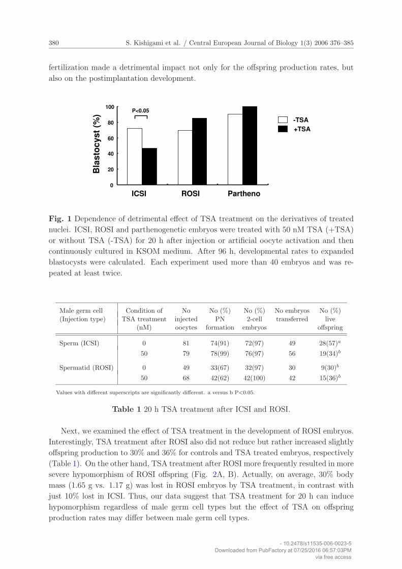
expected from the previous report [23], TSA treatment of ICSI embryos resulted in a
significant reduction from 72% to 46% in the developmental rates to expanded blastocyst
after 96 h culture (Fig. 1). Surprisingly, neither ROSI nor parthenogenetic embryos
showed significant reduction in their developmental rates, but rather an increase from 69%
to 85% and 90% to 100%, respectively, even with no statistical significance (Fig. 1). Thus,
our data suggest that harmfulness of TSA treatment to preimplantation development is
determined by nuclear derivations of embryos.
3.2 TSA treatment of fertilized embryos leads to hypomorphism
Regardless of the reported teratogenicity and toxicity of TSA to embryos, TSA-treated
cloned embryos unexpectedly show significantly better development and no apparent
abnormalities [6]. In contrast, as expected based on their in vitro development, TSA
treatment of embryos after ICSI led to significant reduction of the offspring production
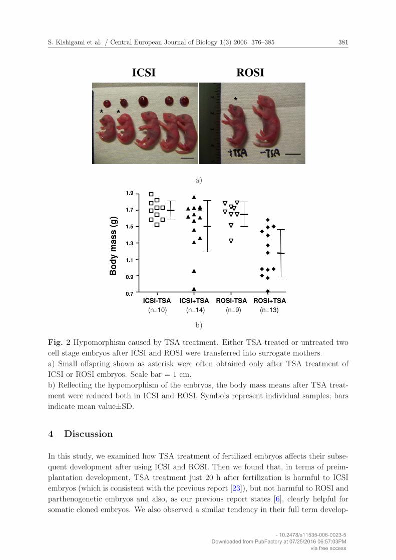
rates from 57% to 34% (Table 1). Further, when offspring were delivered, hypomorphism
was frequently observed among them (Fig. 2A, B). These results indicate that, in contrast
to mouse cloning, the TSA treatment of normal fertilized embryos for the first 20 h after
- 10.2478/s11535-006-0023-5Downloaded from PubFactory at 07/25/2016 06:57:03PM
via free access
380 S. Kishigami et al. / Central European Journal of Biology 1(3) 2006 376–385
fertilization made a detrimental impact not only for the offspring production rates, but
also on the postimplantation development.
Bla
sto
cys
t (%
)
0
20
40
60
80
100
ICSI ROSI Partheno
P<0.05
-TSA
+TSA
Fig. 1 Dependence of detrimental effect of TSA treatment on the derivatives of treated
nuclei. ICSI, ROSI and parthenogenetic embryos were treated with 50 nM TSA (+TSA)
or without TSA (-TSA) for 20 h after injection or artificial oocyte activation and then
continuously cultured in KSOM medium. After 96 h, developmental rates to expanded
blastocysts were calculated. Each experiment used more than 40 embryos and was re-
peated at least twice.
Male germ cell Condition of No No (%) No (%) No embryos No (%)(Injection type) TSA treatment injected PN 2-cell transferred live
(nM) oocytes formation embryos offspring
Sperm (ICSI) 0 81 74(91) 72(97) 49 28(57)a
50 79 78(99) 76(97) 56 19(34)b
Spermatid (ROSI) 0 49 33(67) 32(97) 30 9(30)b
50 68 42(62) 42(100) 42 15(36)b
Values with different superscripts are significantly different. a versus b P<0.05.
Table 1 20 h TSA treatment after ICSI and ROSI.
Next, we examined the effect of TSA treatment in the development of ROSI embryos.
Interestingly, TSA treatment after ROSI also did not reduce but rather increased slightly
offspring production to 30% and 36% for controls and TSA treated embryos, respectively
(Table 1). On the other hand, TSA treatment after ROSI more frequently resulted in more
severe hypomorphism of ROSI offspring (Fig. 2A, B). Actually, on average, 30% body
mass (1.65 g vs. 1.17 g) was lost in ROSI embryos by TSA treatment, in contrast with
just 10% lost in ICSI. Thus, our data suggest that TSA treatment for 20 h can induce
hypomorphism regardless of male germ cell types but the effect of TSA on offspring
production rates may differ between male germ cell types.
- 10.2478/s11535-006-0023-5Downloaded from PubFactory at 07/25/2016 06:57:03PM
via free access
S. Kishigami et al. / Central European Journal of Biology 1(3) 2006 376–385 381
ROSIICSI
* *
*
a)
ICSI-TSA ICSI+TSA ROSI-TSA ROSI+TSA0.7
0.9
1.1
1.3
1.5
1.7
1.9
Bo
dy m
ass
(g)
(n=10) (n=14) (n=9) (n=13)
b)
Fig. 2 Hypomorphism caused by TSA treatment. Either TSA-treated or untreated two
cell stage embryos after ICSI and ROSI were transferred into surrogate mothers.
a) Small offspring shown as asterisk were often obtained only after TSA treatment of
ICSI or ROSI embryos. Scale bar = 1 cm.
b) Reflecting the hypomorphism of the embryos, the body mass means after TSA treat-
ment were reduced both in ICSI and ROSI. Symbols represent individual samples; bars
indicate mean value±SD.
4 Discussion
In this study, we examined how TSA treatment of fertilized embryos affects their subse-
quent development after using ICSI and ROSI. Then we found that, in terms of preim-
plantation development, TSA treatment just 20 h after fertilization is harmful to ICSI
embryos (which is consistent with the previous report [23]), but not harmful to ROSI and
parthenogenetic embryos and also, as our previous report states [6], clearly helpful for
somatic cloned embryos. We also observed a similar tendency in their full term develop-
- 10.2478/s11535-006-0023-5Downloaded from PubFactory at 07/25/2016 06:57:03PM
via free access
382 S. Kishigami et al. / Central European Journal of Biology 1(3) 2006 376–385
mental rates, that is, the different effect of TSA treatment on the offspring production
rates between ICSI and ROSI even if some of the offspring showed hypomorphism. Thus,
the detrimental effect of TSA treatment of zygotes on their embryonic development re-
ally depends on the nuclear derivations, that is, which of oocyte-, sperm-, spermatid or
somatic-derived pronuclei exist in treated zygotes. Therefore, the effect of TSA treatment
could be defined by the epigenetic states of the treated nuclei.
What is the mechanism underlying the different effects of TSA treatment among nu-
clear types on embryonic development? TSA treatment of zygotic embryos after nuclear
transfer, ICSI or ROSI leads to hyperacetylated histone H3 and H4 [7, 9, 23, 25], as
seen in somatic cells [22, 26]. It currently remains unknown how the hyperacetylation of
histones defines the different embryonic developmental rates among nuclear types after
TSA treatment. However, one of the possibilities is that acetylation levels of histones or
some specific amino-acid residues of them in TSA-treated nuclei may be different and con-
tribute to the varying developmental potentials of TSA-treated embryos carrying unique
nuclear types, On the other hand, it has been shown that TSA also triggers selective DNA
demethylation depending on the cell type and genomic region in mammalian cells and
Neurospora [27]. At the same time, it is also reported that the DNA methylation levels
of genomes overall are not significantly changed following TSA treatment in somatic cells
despite the hyperacetylation of histones [26]. In these points of view, we previously found
that ROSI zygotes show DNA hypermethylation of spermatid-derived paternal genomes
in zygotes similar to maternal genomes before the end of the first mitosis, which is evi-
dently in contrast with hypomethylation of sperm-derived genomes [9]. The treatment of
both ICSI and ROSI zygotes with TSA results in a significant reduction of DNA methy-
lation level of paternal genomes [9] but not maternal genomes [9, 25]. Further, the final
DNA methylation levels in paternal genomes after TSA treatment are different even be-
tween sperm- and spermatid-derived genomes [9]. Thus, TSA treatment of zygotes can
remarkably reduce DNA methylation depending on the origins of transferred nuclei and
their genomic regions, most likely according to their epigenetic statuses. Therefore, one
of the possibilities is that the different DNA methylation levels in zygotic genomes in-
duced by TSA treatment underlie the different effects of TSA treatment between nuclear
derivations on embryonic development.
As already described, the TSA treatment of embryos often caused hypomorphism
regardless of types of male germ cells even if the frequency and degree were different be-
tween ICSI and ROSI, substantiating the teratogenicity of TSA treatment. These results
warn that although TSA treatment significantly improves the mouse cloning technique
[6, 7], the safety of this method should be carefully assessed for reproductive technologies
including ICSI, ROSI and animal cloning. Regardless, we found that TSA treatment does
not harm the blastocyst formation rate nor offspring production rate of ROSI embryos.
It is important to point out that the conditions of TSA treatment used in this study were
not optimized, that is, TSA 20 h for TSA treatment may be too long or 50 nM TSA may
be excessive. Therefore, it may be possible to safely improve the current ROSI technology
after careful optimization of conditions of TSA treatment.
- 10.2478/s11535-006-0023-5Downloaded from PubFactory at 07/25/2016 06:57:03PM
via free access
S. Kishigami et al. / Central European Journal of Biology 1(3) 2006 376–385 383
In conclusion, our findings elucidate the apparent discrepancy between the improve-
ment of clone-embryonic development by TSA and the teratogenicity of TSA and found a
dependency of TSA toxicity on derivatives of treated nuclei. Further, our results provide
insight into TSA teratogenicity and will also serve for future improvements of current
assisted reproductive technologies (ART).
Acknowledgment
We acknowledge discussions with Dr. T. Castranio. We also thank T. Oyanagi and Y.
Sakaide for their technical assistance. We are grateful to the Laboratory for Animal
Resources and Genetic Engineering for housing of mice. These studies were supported
by RIKEN (Strategic Program for Research and Development (FY2005) to S.K.) and
MEXT (17780213 to S.K. and 15080211 and a project for the realization of regenerative
medicine to T.W.).
References
[1] T. Wakayama, A.C.F. Perry, M. Zuccotti, K.R. Johnson and R. Yanagimachi: “Full-
term development of mice from enucleated oocytes injected with cumulus cell nuclei”,
Nature, Vol. 394, (1998), pp. 369–374.
[2] H. Kishikawa, T. Wakayama and R. Yanagimachi: “Comparison of oocyte-activating
agents for mouse cloning”, Cloning, Vol. 1, (1999), pp. 153–159.
[3] T. Wakayama and R. Yanagimachi: “Effect of cytokinesis inhibitor, DMSO and the
timing of oocyte activation on mouse cloning using cumulus cell nuclei”, Reproduc-
tion, Vol. 122, (2001), pp. 49–60.
[4] S. Wakayama, E. Mizutani, S. Kishigami, N.V. Thuan, H. Ohta, T. Hikichi, H.T.
Bui, M. Miyake and T. Wakayama: “Mice cloned by nuclear transfer from somatic
and ntES cells derived from the same individuals”, J. Reprod. Dev., Vol. 51, (2005),
pp. 765–772.
[5] S. Wakayama, J.B. Cibelli and T. Wakayama: “Effect of timing of the removal of
oocyte chromosomes before or after injection of somatic nucleus on development of
NT embryos”, Cloning Stem Cells, Vol. 5, (2003), pp. 181–189.
[6] S. Kishigami, E. Mizutani, H. Ohta, T. Hikichi, N. Van Thuan, S. Wakayama, H.T.
Bui and T. Wakayama: “Significant improvement of mouse cloning technique by
treatment with trichostatin A after somatic nuclear transfer”, Biochem. Biophys.
Res. Commun., Vol. 340, (2006), pp. 183–189.
[7] A. Rybouchkin, Y. Kato and Y. Tsunoda: “Role of Histone Acetylation in Repro-
gramming of Somatic Nuclei Following Nuclear Transfer”, Biol. Reprod., Vol. 74,
(2006), pp. 1083–1089.
[8] S. Kishigami, S. Wakayama, N. Van Thuan , H. Ohta, E. Mizutani, T. Hikichi, H.T.
Bui, S. Balbach, A Ogura, M. Boiani and T. Wakayama: “Production of Cloned Mice
by Somatic Cell Nuclear Transfer”, Nat. Protocols, Vol. 1, (2006), pp. 125-–138.
- 10.2478/s11535-006-0023-5Downloaded from PubFactory at 07/25/2016 06:57:03PM
via free access
384 S. Kishigami et al. / Central European Journal of Biology 1(3) 2006 376–385
[9] S. Kishigami, N. Van Thuan, T. Hikichi, H. Ohta, S. Wakayama, E. Mizutani and
T. Wakayama: “Epigenetic abnormalities of the mouse paternal zygotic genome as-
sociated with microinsemination of round spermatids”, Dev. Biol., Vol. 289, (2006),
pp. 195–205.
[10] A. Ogura, J. Matsuda and R. Yanagimachi: “Birth of normal young after electrofu-
sion of mouse oocytes with round spermatids”, Proc. Natl. Acad. Sci. U.S.A., Vol.
91, (1994), pp. 7460–7462.
[11] Y. Kimura and R. Yanagimachi: “Mouse oocytes injected with testicular spermato-
zoa or round spermatids can develop into normal offspring”, Development, Vol. 121,
(1995), pp. 2397–2405.
[12] S. Kishigami, S. Wakayama, N. Van Thuan and T Wakayama: “Similar time re-
striction for intracytoplasmic sperm injection and round spermatid injection into
activated oocytes for efficient offspring production”, Biol. Reprod., Vol. 70, (2004),
pp. 1863–1869.
[13] S. Kishigami, N. Van Thuan, S. Wakayama, T. Hikichi and T. Wakayama: “A novel
method for isolating spermatid nuclei from cytoplasm prior to ROSI in the mouse”,
Zygote, Vol. 12, (2004), pp. 321–327.
[14] R. Yanagimachi: “Intracytoplasmic injection of spermatozoa and spermatogenic cells:
its biology and applications in human and animals”, Reprod. Biomed. Online, Vol.
10, (2005), pp. 247–286.
[15] J. Ohgane, T. Wakayama, S. Senda, Y. Yamazaki, K. Inoue, A. Ogura, J. Marh, S.
Tanaka, R. Yanagimachi and K. Shiota: “The Sall3 locus is an epigenetic hotspot of
aberrant DNA methylation associated with placentomegaly of cloned mice”, Genes
Cells, Vol. 9, (2004), pp. 253–260.
[16] Y.K. Kang, D.B. Koo, J.S. Park, Y.H. Choi, A.S. Chung, K.K. Lee and Y.M. Han:
“Aberrant methylation of donor genome in cloned bovine embryos”, Nat. Genet., Vol.
28, (2001), pp. 173–177.
[17] W. Dean, F. Santos, M. Stojkovic, V. Zakhartchenko, J. Walter, E. Wolf, and W.
Reik: “Conservation of methylation reprogramming in mammalian development:
Aberrant reprogramming in cloned embryos”, Proc. Natl. Acad. Sci. U.S.A., Vol.
98, (2001), pp. 13734–13738.
[18] F. Santos, V. Zakhartchenko, M. Stojkovic, A. Peters, T. Jenuwein, E. Wolf, W.
Reik and W. Dean: “Epigenetic marking correlates with developmental potential in
cloned bovine preimplantation embryos”, Curr. Biol., Vol. 13, (2003), pp. 1116–1121.
[19] M. Dokmanovic and P.A. Marks: “Prospects: histone deacetylase inhibitors”, J. Cell
Biochem., Vol. 96, (2005), pp. 293–304.
[20] N. Gurvich, M.G. Berman, B.S. Wittner, R.C. Gentleman, P.S. Klein and J.B. Green:
“Association of valproate-induced teratogenesis with histone deacetylase inhibition
in vivo”, FASEB J., Vol. 19, (2005), pp. 1166–1168.
[21] K. Svensson, R. Mattsson, T.C. James, P. Wentzel, M. Pilartz, J. MacLaughlin,
S.J. Miller, T. Olsson, U.J. Eriksson and R. Ohlsson: “The paternal allele of the H19
gene is progressively silenced during early mouse development: the acetylation status
- 10.2478/s11535-006-0023-5Downloaded from PubFactory at 07/25/2016 06:57:03PM
via free access
S. Kishigami et al. / Central European Journal of Biology 1(3) 2006 376–385 385
of histones may be involved in the generation of variegated expression patterns”,
Development, Vol. 125, (1998), pp. 61–69.
[22] M. Yoshida, M. Kijima, M. Akita and T. Beppu: “Potent and specific inhibition of
mammalian histone deacetylase both in vivo and in vitro by trichostatin A”, J. Biol.
Chem., Vol. 265, (1990), pp. 17174–17179.
[23] J. Ma, P. Svoboda, R.M. Schultz and P. Stein: “Regulation of zygotic gene activa-
tion in the preimplantation mouse embryo: global activation and repression of gene
expression”, Biol. Reprod., Vol. 64, (2001), pp. 1713–1721.
[24] G.T. O’Neill, L.R. Rolfe and M.H. Kaufman: “Developmental potential and chro-
mosome constitution of strontium-induced mouse parthenogenones”, Mol. Reprod.
Dev., Vol. 30, (1991), pp. 214–219.
[25] M. Spinaci, E. Seren and M. Mattioli: “Maternal chromatin remodeling during mat-
uration and after fertilization in mouse oocytes”, Mol. Reprod. Dev., Vol. 69, (2004),
pp. 215–221.
[26] B.P. Enright, C. Kubota, X. Yang, and X.C. Tian: “Epigenetic characteristics and
development of embryos cloned from donor cells treated by trichostatin A or 5-aza-
2’-deoxycytidine”, Biol. Reprod., Vol. 69, (2003), pp. 896–901.
[27] M. Szyf: “DNA methylation and demethylation as targets for anticancer therapy”,
Biochemistry (Mosc), Vol. 70, (2005), pp. 533–549.
- 10.2478/s11535-006-0023-5Downloaded from PubFactory at 07/25/2016 06:57:03PM
via free access
Related Documents