Harmful effects of metal(loid) oxide nanoparticles Eduardo V. Soares 1,2 & Helena M. V. M. Soares 3 Abstract The incorporation of nanomaterials (NMs), including metal(loid) oxide (MOx) nanoparticles (NPs), in the most diversified consumer products, has grown enormously in recent decades. Consequently, the contact between humans and these materials increased, as well as their presence in the environment. This fact has raised concerns and uncertainties about the possible risks of NMs to human health and the adverse effects on the environment. These concerns underline the need and importance of assessing its nanosecurity. The present review focuses on the main mechanisms underlying the MOx NPs toxicity, illustrated with different biological models: release of toxic ions, cellular uptake of NPs, oxidative stress, shading effect on photosynthetic microorgan- isms, physical restrain and damage of cell wall. Additionally, the biological models used to evaluate the potential hazardous of nanomaterials are briefly presented, with particular emphasis on the yeast Saccharomyces cerevisiae, as an alternative model in nanotoxicology. An overview containing recent scientific advances on cellular responses (toxic symptoms exhibited by yeasts) resulting from the interaction with MOx NPs (inhibition of cell proliferation, cell wall damage, alteration of function and morphology of organelles, presence of oxidative stress bio-indicators, gene expression changes, genotoxicity and cell dead) is critically presented. The elucidation of the toxic modes of action of MOx NPs in yeast cells can be very useful in providing additional clues about the impact of NPs on the physiology and metabolism of the eukaryotic cell. Current and future trends of MOx NPs toxicity, regarding their possible impacts on the environment and human health, are discussed. Key points • The potential hazardous effects of MOx NPs are critically reviewed. • An overview of the main mechanisms associated with MOx NPs toxicity is presented. • Scientific advances about yeast cell responses to MOx NPs are updated and discussed. Keywords Aquatic organisms . Hazard/risk assessment . Metal(loid) oxide nanoparticles . Nanosafety . Toxic modes of action . Yeast Introduction Nanomaterials (NMs) are defined as “chemical substances or materials with particle sizes between 1 to 100 nm in at least one dimension” (ECHA 2020). Due to their nanometer size, they present huge surface-to-volume ratios, exhibiting unique physical and chemical properties (such as catalytic, optical, magnetic, electronic and mechanical) that are different from those of materials on a larger or “bulk” scale (Klaine et al. 2013). The exceptional properties exhibited by NMs have led to their incorporation in many products in various sectors such as agriculture, automotive, construction, cosmetics, electron- ics, environment, food, home appliance, medicine, petroleum and printing (NPD 2020). * Eduardo V. Soares [email protected] 1 Bioengineering Laboratory-CIETI, ISEP-School of Engineering, Polytechnic Institute of Porto, rua Dr António Bernardino de Almeida, 431, 4249-015 Porto, Portugal 2 CEB-Centre of Biological Engineering, University of Minho, Campus de Gualtar, 4710-057 Braga, Portugal 3 REQUIMTE/LAQV, Departamento de Engenharia Química, Faculdade de Engenharia, Universidade do Porto, rua Dr Roberto Frias, s/n, 4200-465 Porto, Portugal

Welcome message from author
This document is posted to help you gain knowledge. Please leave a comment to let me know what you think about it! Share it to your friends and learn new things together.
Transcript
Harmful effects of metal(loid) oxide nanoparticles
Eduardo V. Soares1,2 & Helena M. V. M. Soares3
AbstractThe incorporation of nanomaterials (NMs), including metal(loid) oxide (MOx) nanoparticles (NPs), in the most diversifiedconsumer products, has grown enormously in recent decades. Consequently, the contact between humans and these materialsincreased, as well as their presence in the environment. This fact has raised concerns and uncertainties about the possible risks ofNMs to human health and the adverse effects on the environment. These concerns underline the need and importance of assessingits nanosecurity. The present review focuses on the main mechanisms underlying theMOx NPs toxicity, illustrated with differentbiological models: release of toxic ions, cellular uptake of NPs, oxidative stress, shading effect on photosynthetic microorgan-isms, physical restrain and damage of cell wall. Additionally, the biological models used to evaluate the potential hazardous ofnanomaterials are briefly presented, with particular emphasis on the yeast Saccharomyces cerevisiae, as an alternative model innanotoxicology. An overview containing recent scientific advances on cellular responses (toxic symptoms exhibited by yeasts)resulting from the interaction with MOx NPs (inhibition of cell proliferation, cell wall damage, alteration of function andmorphology of organelles, presence of oxidative stress bio-indicators, gene expression changes, genotoxicity and cell dead) iscritically presented. The elucidation of the toxic modes of action of MOx NPs in yeast cells can be very useful in providingadditional clues about the impact of NPs on the physiology and metabolism of the eukaryotic cell. Current and future trends ofMOx NPs toxicity, regarding their possible impacts on the environment and human health, are discussed.
Key points• The potential hazardous effects of MOx NPs are critically reviewed.• An overview of the main mechanisms associated with MOx NPs toxicity is presented.• Scientific advances about yeast cell responses to MOx NPs are updated and discussed.
Keywords Aquatic organisms . Hazard/risk assessment . Metal(loid) oxide nanoparticles . Nanosafety . Toxic modes of action .
Yeast
Introduction
Nanomaterials (NMs) are defined as “chemical substances ormaterials with particle sizes between 1 to 100 nm in at leastone dimension” (ECHA 2020). Due to their nanometer size,they present huge surface-to-volume ratios, exhibiting uniquephysical and chemical properties (such as catalytic, optical,magnetic, electronic and mechanical) that are different fromthose of materials on a larger or “bulk” scale (Klaine et al.2013). The exceptional properties exhibited by NMs have ledto their incorporation in many products in various sectors suchas agriculture, automotive, construction, cosmetics, electron-ics, environment, food, home appliance, medicine, petroleumand printing (NPD 2020).
* Eduardo V. [email protected]
1 Bioengineering Laboratory-CIETI, ISEP-School of Engineering,Polytechnic Institute of Porto, rua Dr António Bernardino deAlmeida, 431, 4249-015 Porto, Portugal
2 CEB-Centre of Biological Engineering, University of Minho,Campus de Gualtar, 4710-057 Braga, Portugal
3 REQUIMTE/LAQV, Departamento de Engenharia Química,Faculdade de Engenharia, Universidade do Porto, rua Dr RobertoFrias, s/n, 4200-465 Porto, Portugal

The rapid expansion of production and use of NMs inevita-bly raised concerns about their safety for human health and theenvironment. The physical form and the chemical reactivity thatmakes NMs distinctive also provide them the potential to inter-fere with biological processes and produce hazardous effects.Humans can be intentional (through nanomedicine or personalhealthcare products) or unintentionally exposed to NMs (re-leased from food packaging); additionally, occupational expo-sure (as consequence of industrial processes) should also beconsidered (Lombi et al. 2019; Klaper 2020). Examples of in-tentional application of NMs in the environment include theiruse in environmental remediation (Guerra et al. 2018; Qianet al. 2020) or in agricultural practices (Servin et al. 2015;Usman et al. 2020). Unintentionally release of NMs in theenvironment includes the following: (i) the release due to thelife cycle of products incorporating NPs, such as paints, cos-metics, sunscreens (Sun et al. 2016; Wu et al. 2020); and, (ii)accidental spills or industrial liquid effluents, such as thoseemitted by textile industries during the washing of nanotextiles(Yetisen et al. 2016). It was estimated that of the global NMsproduced, 63–91% reach landfills, 8–28% are released intosoils, 0.4–0.7% in natural water bodies, and 0.1–1.5% are re-leased into the atmosphere (Keller et al. 2013).
NMs can be divided into five categories: carbon-based(single and multi-walled carbon nanotubes, graphene and ful-lerenes); metal-based (metal(loid) oxides; zerovalent metalssuch as iron, silver and gold); dendrimers (hyperbranchedpolymers, dendrigraft polymers and dendrons); semiconduc-tor nanocrystals, known as quantum dots; and composites(constituted by two different NMs or NMs combined withlarger, bulk-type materials; and NMs combined with syntheticpolymers or resins) (EPA 2017).
Among NMs, metal(loid) oxide (MOx) nanoparticles(NPs) have received considerable attention largely due to theirvariety of uses namely in optics and electronics, healthcare,construction, automotive and personal care products (Laurentet al. 2018), and it will be the subject of the present review.The market for MOx NPs is expected to growth at a com-pound annual growth over 7% globally during the period of2020–2025. However the current framework of uncertaintyarising from the COVID-19 pandemic it may hinder thegrowth of this market sector (Mordor Intelegence 2020).
In the last five years, several review papers have been pub-lished about NMs, NPs or more specifically on MOx NPs.Some reviews refer more broadly to NMs, namely about theirbehaviour, fate, bioavailability and effects on the environment(Pulido-Reyes et al. 2017; Lead et al. 2018; Spurgeon et al.2020; Zhao et al. 2020) or the toxicity mechanisms associatedwith NMs over algal cells (Chen et al. 2019). Within NMs,reviews on NPs, namely about the influence of their physico-chemical properties on ecotoxicology, in terrestrial and aquat-ic systems (Bundschuh et al. 2018; Nguyen et al. 2020; Romaet al. 2020), the effects on freshwater organisms (Deniel et al.
2019), genotoxicity (Mortezaee et al. 2019) or the mecha-nisms associated with cell dead by necrosis, apoptosis andautophagy (Mohammadinejad et al. 2019; Paunovic et al.2020) have been published. A more specific review aboutthe toxic effects of NiO in aquatic organisms (Meyer et al.2020) was recently published.
The present work summarises the mainmechanisms under-lying MOx NPs toxicity. The biological models used to assessnanotoxicity are briefly presented, with particular emphasis onthe yeast Saccharomyces cerevisiae as a valuable and alterna-tive model in nanotoxicology. An updated overview of yeastcell responses to stress induced by MOx NPs is criticallyreviewed. Finally, current and future trends in the assessmentof MOx NPs toxicity, regarding their possible impact on theenvironment and human health, are discussed.
Biological models used in nanotoxicology
Brief overview of the models used in nanotoxicityassessment
An array of biological models have been used in ecotoxicitystudies, which include (in parentheses it can be found typicalexamples employed): bacteria (Escherichia coli and Vibriofischeri), yeasts (S. cerevisiae), microalgae (Pseudokirchneriellasubcapitata and Chlorella vulgaris), protozoa (Tetrahymenathermophila), rotifers (Brachionus plicatilis), crustaceans(Daphnia magna), annelids (Eisenia fetida), nematodes(Caenorhabditis elegans), cnidarians (Hydra attenuata), molluscs(Potamopyrgus antipodarum), echinoderms (Lytechinus pictus),amphibians (Xenopus laevis) and fishes (Danio rerio) (Jugansonet al. 2015; Minetto et al. 2016; Libralato et al. 2017).
Although animal testing is still the predominant modeluse for the risk assessment of chemicals (Hartung andRovida 2009), due to the pressure from public opinionand legal demand, supported by ethical reasons, the re-placement of animals for cheaper and more human-relevant alternatives have been proposed based on the useof cell lines. Thus, different mammalian cell lines havebeen used in toxicity assays with MOx NPs (Al2O3, CuO,NiO, TiO2 and ZnO), comprising models of different hu-man systems, such as respiratory, digestive, renal, immuneand skin (Ivask et al. 2014; Naseer et al. 2018; Czyzowskaand Barbasz 2020).
The yeast S. cerevisiae as an important tool innanotoxicology
S. cerevisiae is the most commonly used yeast in industrialapplications, receiving the status of Generally Recognized AsSafe (GRAS) microorganism by the United States Food andDrug Administration (FDA 2018). This yeast is easy to
manipulate and cultivate, does not require expensive ingredi-ents in the formulation of the culture media and presents ashort generation time. It was the first eukaryotic organismwith the genome completely sequenced (Goffeau et al. 1996).
The yeast S. cerevisiae presents a cellular structure andorganization related to animal cells. About 30% of genes as-sociated with human diseases have a yeast orthologue (Foury1997), whichmakes this yeast an attractive model organism tostudy diseases in humans. Mitochondrial respiration can bemanipulated by the loss of mitochondrial DNA or by chang-ing the growth conditions, making this yeast an appropriatemodel for elucidating the role of mitochondria in ROS gener-ation, as well as mitochondrial diseases associated with oxi-dative phosphorylation (Malina et al. 2018); this informationcan be readily transported to higher eukaryotes via the GeneOntology (Howe et al. 2018).
This yeast features a set of important tools that include thecomplete gene deletion collection (Giaever and Nislow 2014)and the possibility of achieving high-throughput data, such asobtained from transcriptomics, proteomics and metabolomicsanalysis (Braconi et al. 2016). The use of yeasts in the assessmentof toxicity of environmental pollutants (including NMs) does notraise ethical issues and is well suited in a first toxicity screening,because reduces costs and toxic wastes and replaces/limits theuse of animal models (dos Santos and Sa-Correia 2015).
However, this model also has limitations. The unicellularnature of this organism does not make possible to provide spe-cific toxicological data about tissues or organs. In addition, itpresents a cell wall (in contrast to animal cells), which can act asa barrier to toxicants, many efflux pumps and detoxificationmechanisms, which can be the cause of the greater toleranceof yeasts to toxics, compared with eukaryotic cells of higherorganisms (dos Santos et al. 2012; Braconi et al. 2016).
Global mechanisms underlying to MOx NPstoxicity
MOxNPs can present a toxic effect by several mechanisms, insome cases even by more than one. The main mechanisms aresummarised below and depicted in Fig. 1.
NPs solubilisation: release of toxic ions
MOx NPs dissolution, to a greater or lesser extent, is a com-mon transformation process, which is dependent on theirphysico-chemical properties (chemical composition and size),presence of stabilizing agents and chemical composition of themedium, namely, pH, ionic strength (IS), presence of anions(phosphate and sulphate) and natural organic matter (Quigget al. 2013; Amde et al. 2017).
Once in solution, the ions diffuse in the medium and reachthe cells, where they produce a deleterious effect after
intracellular accumulation (Fig. 1A). Metal ions, as chargedchemical species, do not diffuse freely across the plasma mem-brane. Thus, different membrane transport proteins (pumps andchannels) are involved in their influx (Argueello et al. 2012).
In certain MOx NPs, the ions dissolved seem to be themajor factor in their ecotoxicity. This is the case of the toxicimpact of ZnO NPs over bacteria (Heinlaan et al. 2008; Liet al. 2011; Wang et al. 2016), yeasts (Kasemets et al. 2009;Bayat et al. 2014), crustaceans (Heinlaan et al. 2008; Wienchet al. 2009; Vimercati et al. 2020), microalgae (Franklin et al.2007; Miller et al. 2010; Lee and An 2013; Aravantinou et al.2015; Schiavo et al. 2016) or mammalian cell lines (Brunneret al. 2006; Zhang et al. 2012), which is totally or mainlycaused by solubilized Zn ions. A Multi-omics approach (tran-scriptomics, metabolomics and lipidomics) confirmed thatmetal ions mediated the main toxicological pathways ofZnO NPs in lung epithelial A549 cells (Dekkers et al. 2018).In the same line, CuONPs toxicity over bacteria (Bondarenkoet al. 2012), yeasts (Kasemets et al. 2009; Kasemets et al.2013; Bayat et al. 2014; Bao et al. 2015), green algae(Aruoja et al. 2009; von Moos et al. 2015), crustaceans(Heinlaan et al. 2008; Jo et al. 2012) or human cell lines(Cohen et al. 2013; He et al. 2020) was partially or completelyexplained by dissolved Cu ions. The toxicity of NiO and SnO2
over P. subcapitata (Sousa et al. 2018b; Sousa et al. 2019b) orMn3O4 and Y3O3 over S. cerevisiae (Moriyama et al. 2019;Sousa et al. 2019a) can also be mainly attributed to the respec-tive ions leached from the NPs.
NPs passage through wall pores versus release ofmetal ions on NPs-cell surface interface
The cell wall, present in plants, in most bacteria, yeasts and inmany microalgae, is the primary site of interaction and the firstbarrier to the passage of NPs from the extracellular medium tothe cytoplasm. This cellular structure is absent in animal cellsand protozoa. The second barrier is the plasma membrane,common to all cells. Chemical composition and architectureof the cell wall vary according to the organism, being theirthickness an important factor in the determination of the NPsinternalisation (Chen et al. 2019). This cell structure is generallyseen as a porous matrix. For instance, the yeast S. cerevisiaepresents cell wall pores of 200 nm, which can be enlarged up to400 nm under stress conditions (Pereira and Geibel 1999). Cellwall pore diameter of 7–8 nm in marine macro algae (Zemke-White et al. 2000) and pore channels with an average diameterof 20–200 nm in unicellular green microalgae were also de-scribed (Anissimova and Staer 2018). It is conceivable that, inorganisms with cell wall, NPs and NPs homoagglomeratessmaller than wall pores can pass through this cellular structureand reach the plasma membrane (Fig. 1B, top). Conversely,NPs and NPs homoagglomerates larger than pore size are veryunlikely to pass through cell wall (sieve effect).

Itwas described thatCeO2NPs, coatedwith polyvinylpyrrolidoneand presenting a size of 4–5 nm, could cross the cell wallof the microalga Chlamydomonas reinhardtii beinginternalised into intracellular vesicles (Taylor et al. 2016).Using different microscopy techniques, the internalisation ofCuO NPs in S. cerevisiae (Vasco et al. 2017) and in theC. reinhardtii (Yin et al. 2020) was also described. In the caseof C. reinhardtii, CuO NPs were largely accumulated in thevacuoles (Yin et al. 2020). Conversely, it has been suggestedthat the internalisation of CeO2 NPs (with a nominal size of25 nm and presenting in solution agglomerates with an aver-age size of 146 nm) in C. reinhardtii was rather unlikely (neeRohder et al. 2018). A similar conclusion was achieved inother studies with algae (Rodea-Palomares et al. 2011;Pulido-Reyes et al. 2015; Angel et al. 2015) and cyanobacte-rium (Rodea-Palomares et al. 2011). In this line, the examina-tion by transmission electron microscopy (TEM)-energy-dis-persive X-ray spectroscopy of yeast cells treated with CuO(Bao et al. 2015), NiO (Sousa et al. 2018a), or ZnO NPs
(Zhang et al. 2016) did not detect MOx NPs inside cells,suggesting that these NPs could not be taken by yeast cells.In resume, although examples of NPs internalisation in organ-isms with cell wall can be found in the literature, this is adebatable issue, especially the passage through the cell wallof agglomerated NPs.
However, different studies have attributed the toxic ef-fects of MOx NPs to the NPs themselves rather than tothe ions coming from them. This is the case of NiO(Sousa et al. 2018a), SiO2 (Sousa et al. 2019a) and TiO2
(Bayat et al. 2014) over yeasts as well as Al2O3, CeO2,Fe3O4, Mn3O4 and WO3 NPs on microalgae (Aruoja et al.2015; Angel et al. 2015; Sousa et al. 2019b). In this con-text, MOx NPs can present a toxic effect by an indirectmechanism: particles adhere tightly to the cell wall of themicroorganisms and enhance the release of metals at theNP-cell wall interface, leading to the activation of toxicresponses (Fig. 1B, bottom). This indirect mechanism wassuggested for to explain the antibacterial activity of
Photosynthetic microorganism
NPs homoagglomerated
Wall damage
Toxicity
NPs homoagglomerated
ToxicityCW PM
Cell
CW
ATP
Toxicity
Toxicity
ROSROS
ROS
Light
ROSROS
ROS
Abiotic ROS
Biotic ROS
Mt
ETC
Cl
HO.H2O2O2
-e-
SOD CAT H2OROS
Passage through wall pores
ToxicityCW Release of ions on NPs-cell
surface interface
Block of wall pores
Toxicity
Nutrients
CW
CWPM
Toxicity
Endocytic vesicle
Lysosome
Metal ions release
CW
a b c
e f g
d
Fig. 1 Outline of the main toxic mechanisms associated with metal(loid)oxide nanoparticles. Please see text for details. a NPs solubilisation:release of toxic ions. b NPs passage through wall pores versus releaseof metal ions on NPs-cell surface interface. c Direct effect on the cellsurface. d Cellular uptake of NPs. e Oxidative stress. f Shading effect
(on photosynthetic microorganisms): homoagglomeration of NPs. gPhysical restraint: heteroagglomeration. CAT, catalase; Cl, chloroplast;CW, cell wall; ETC, electron transport chain; Mt, mitochondrion; NPs,nanoparticles; PM, plasma membrane; ROS, reactive oxygen species;SOD, superoxide dismutase

Fe2O3, Co3O4 and NiO NPs (Wang et al. 2016) and thetoxicity of CuO and NiO on yeasts (Kasemets et al. 2013;Bao et al. 2015; Sousa et al. 2018a) and CeO2 NPs onmicroalgae (Angel et al. 2015).
Direct effect on the cell surface
After contact and adsorption to the cell wall, NPs can clog thepores of the wall, limiting the exchange of chemical species(including nutrients) between the surrounding medium andthe cell (Fig. 1C, top) or induce physical damage to the cellwall (Fig. 1C, bottom).
In agreement with the first possibility, it was described thatC. reinhardtii incubated with TiO2 presented the cell surfacecoated with NPs, which can hinder the exchange of substancesbetween the cell and the surrounding milieu (Chen et al. 2012).The functionalization and the type of functionalization of CuOand ZnO can influence the level of NPs adsorption toS. cerevisiae and C. reinhardtii cell wall and the respectiveanti-fungi and anti-algal activity (Halbus et al. 2019; Halbuset al. 2020).
The damage of the cell wall of algae and yeast cells incu-bated with ZnONPs was described (Suman et al. 2015; Zhanget al. 2016; Babele et al. 2018). In the same way, it has beenreported that TiO2NPs or TiO2NPs surface-bound humic acidadhered to algal cells (Chlorella spp., Karenia brevis,Nitzschia closterium and Skeletonema costatum) and coulddestroy the cell wall and enter the cells, inducing plasmolysis(Lin et al. 2012; Xia et al. 2015; Li et al. 2015). In the samemode, it has been proposed that the toxic effect of CeO2 NPson P. subcapitata can be mediated, mainly, by a physicaleffect due to a close adsorption of the NPs on the cell surface(Manier et al. 2013). This possibility is in line with otherobservations that describe that CeO2 NPs are stronglyadsorbed to Anabaena sp. and completely disrupt cyanobac-terium cell wall andmembrane (Rodea-Palomares et al. 2011).It is important to note that the adhesion ofMOxNPs to the cellwall does not necessarily imply the triggering of a toxic effect.For instance, the attachment of La2O3 NPs to the cell wall didnot produce morphological changes on Chlorella spp.(Balusamy et al. 2015).
Cellular uptake of NPs
After crossing the cell wall (in organisms with a cell wall andwhen such passage is possible), MOx NPs meet the cell mem-brane and two types of events can occur: damage of the mem-brane (due to physical disruption) (Chen et al. 2019) or passagethrough the membrane by endocytosis (Fig. 1D), a process wellknown in mammalian cells (Oh and Park 2014). Endocytosiswas also suggested in microalgae and bacteria but their predom-inance and mechanisms are unknown (von Moos et al. 2014).
Once inside the cells, MOx NPs can undergo several alter-ations, such as redox transformations and complexation (Chenet al. 2019) or can be solubilized inside of acidic lysosomes(Fig. 1D) and exerts their toxicity by a Trojan horse-type mech-anism (Oh and Park 2014). The intracellular dissolution mech-anism affords the trafficking of toxic metal ions into the cells.Thus, CuO and ZnONPs intracellular dissolution and release ofCu and Zn ions, respectively, were described in different mam-malian (including human) cell lines (Xia et al. 2008; Cronholmet al. 2013; Condello et al. 2016; He et al. 2020).
Oxidative stress
Oxidative stress (OS) occurs when it is observed an imbalancebetween the generation of reactive species (RS) and the levelof antioxidant defences, either enzymatic or non-enzymatic(Halliwell and Gutteridge 2015). MOx NPs themselves, orthe metals released from them, can present a pro-oxidant po-tential, i.e. the capacity to generate the production of RS orhindering/consuming antioxidant defences (Nel et al. 2006).Reactive oxygen species (ROS) production (such as superox-ide radical (O2·
−), hydroxyl radical (HO.) and hydrogen per-oxide (H2O2)) can occur under a cell free milieu, i.e. extracel-lularly (abiotic ROS) or via interaction of NPs (or the releasedions) with biologicals systems (biotic ROS) (Fig. 1E).
ROS can be generated at the NP surface (Fig. 1E, top). Thebioavailability and valence state of redox-active elements in-fluences strongly the level of ROS generation by NPs. In ageneral way, the capacity of MOx NPs to generate ROS isdependent on their chemical composition, purity, (particle)size, shape and surface reactivity (von Moos andSlaveykova 2014). In certain NPs, such as TiO2, the abilityto produce ROSmay also require light or an ultraviolet sourceto excite the NPs surface (Xia et al. 2008; Guo et al. 2011)(Fig. 1E, top). ROS generation, in abiotic conditions, byCeO2, Co3O4, CuO and Sb2O3 NPs were described (Xiaet al. 2008; Bayat et al. 2014; Aruoja et al. 2015).
Intracellular (biotic) ROS can have origin in the endoplas-mic reticulum, peroxisomes and in electron transport process-es in mitochondria and chloroplasts (in eukaryotic photosyn-thetic organisms) (Lesser 2006; del Rio and Lopez-Huertas2016) (Fig. 1E, bottom). MOx NPs, even containing redox-inactive metals, such as NiO (Siddiqui et al. 2012; Ahamedet al. 2013; Oukarroum et al. 2017; Sousa et al. 2018c; Sousaet al. 2018b) and ZnO NPs (De Berardis et al. 2010; Kumaret al. 2011; Alarifi et al. 2013; Lu et al. 2015; Ng et al. 2017)can induce intracellular ROS and oxidative stress. It wasshown that P. subcapitata algal cells exposed to NiO NPspresented a reduced activity of the photosystem II (ɸPSII)and a decreased electron flow in the electron transport chain(ETC). The electrons deflected from photosynthetic ETCprobably are used to generate ROS (Sousa et al. 2018b)(Fig. 1E, bottom). Compatible with this possibility,

intracellular ROS accumulation and decrease of ɸPSII werealso observed in microalgae (P. subcapitata or C. reinhardtii)exposed to Al2O3, CeO2, Cr2O3, Mn3O4, SiO2 or SnO2 NPs(Rodea-Palomares et al. 2012; da Costa et al. 2016; Sousaet al. 2019b) and in the cyanobacterium Anabaena CPB4337exposed to CeO2 NPs (Rodea-Palomares et al. 2012).
It was proposed that in P. subcapitata cells exposed to NiONPs, the disturbance of photosynthetic performance and theincrease of intracellular ROS, combined with a reduction ofmetabolic (esterasic) activity may cause the arrest of algal cellcycle which, in turn, may origin the increase of cell volumeand the appearance of aberrant morphology and, ultimately,the arrest of algal growth (Sousa et al. 2018b). Similarly, itwas observed that Al2O3, Mn3O4 and SiO2 NPs induced thegrowth inhibition of P. subcapitata as a consequence of thecumulative effect of adverse outcomes, such as intracellularROS accumulation, loss of metabolic activity and reduction ofɸPSII (Sousa et al. 2019b). A reduction of chlorophyll a con-tent and increase of ROS was also observed in C. minutissimaexposed to Co3O4 NPs (Sharan and Nara 2020).
OS is associated with the damage of biological moleculessuch as cellular lipids (via lipid peroxidation, LPO), carbohy-drates, proteins and DNA (Halliwell and Gutteridge 2015) beingconsidered a major mechanism of NPs toxicity (Nel et al. 2006;Xia et al. 2006). Accordingly, ROS production by various MOxNPs, namely, Al2O3, Ce2O, CuO, Mn3O4, NiO, SiO2, SnO2,TiO2 and ZnO with the consequent cell oxidative disturbances,which include, LPO and cell membrane damage (loss of integri-ty), overwhelmed antioxidant defence system, reduced mito-chondrial function, chromatin condensation, DNA damage andcell death via apoptotic pathway, over different biologicalmodelshave been described; examples are the following: bacteria(Kumar et al. 2011; Rodea-Palomares et al. 2012), yeasts(Zhang et al. 2016; Babele et al. 2018; Sousa et al. 2018c;Sousa et al. 2019a), freshwater and marine microalgae (Rodea-Palomares et al. 2012; von Moos et al. 2015; Xia et al. 2015;Suman et al. 2015; vonMoos et al. 2016; Oukarroum et al. 2017;Dauda et al. 2017; Sendra et al. 2018; Sousa et al. 2018b; Sousaet al. 2019b), carp (Cyprinus carpio) larva (Naeemi et al. 2020)and human cell lines (Karlsson et al. 2008; Park et al. 2008; Luet al. 2015; Duan et al. 2015; Rajiv et al. 2016; Subramaniamet al. 2020).
Shading effect (on photosynthetic microorganisms):homoagglomeration of NPs
MOxNPs, in aqueousmedium, can interact with each other andform clusters of NPs (homoagglomerates), which can have animportant impact on their bioavailability, fate in the environ-ment and toxicity (Vale et al. 2016). The homoagglomerationprocess (as well as heteroagglomeration—please see below)depends on the concentration and characteristics of the NPs(chemical composition, morphology and charge), the NPs
surface functionalisation, the physico-chemical properties ofthe medium where they are suspended, namely, IS (influencedby water salinity and hardness), pH of the solution (which af-fects the NPs surface charge) and the existence of natural or-ganic matter (NOM). NOM can adsorb to the NPs surface andorigin their steric stabilisation or act as bridges promoting theagglomeration of NPs (Amde et al. 2017; Yu et al. 2018). Theadsorption of NOM to NPs, with the formation of coated NPs(corona-coated NPs), can be associated with the transfer ofthese NMs from algae to higher trophic levels consumers (crus-taceans and fish) presenting unknown environmental and hu-man health risks (Xu et al. 2020).
Aqueous suspensions of dispersed or homoagglomeratedMOx NPs are, sometimes, opaque. Due to light absorption orscattering by NPs or NPs homoagglomerates, a reduction ofthe light availability can occur (shading effect, Fig. 1F). Thiseffect can influence the photosynthetic efficiency of organ-isms like cyanobacteria and algae (Navarro et al. 2008). Inagreement with this possibility, a significant decrease in lightabsorbance of C. reinhardtii algal suspensions, in comparisonwith control (NPs free), due to CuO NPs at concentrationshigher than 1 mg L−1 was described (Cheloni et al. 2016).Sadiq et al. (2011) suggested that the growth inhibition andchlorophyll content reduction observed in algal cells incubat-ed with Al2O3 could be attributed to the decrease of lightavailability owing to the attachment of the NPs onto cell wallof Chlorella spp. A shading effect was also attributed to CoNPs in the inhibition of S. costatum growth (Chen et al. 2018).
However, other authors did not observe any significanteffect on the 72 h growth of the alga P. subcapitata, regardlessof the concentrations of CeO2, CuO and ZnO NPs tested; inthe case of TiO2, even for the relative opaque suspensions,containing easily settled NPs homoagglomerates, a growthreduction was not observed (Aruoja et al. 2009; Rogers et al.2010; Hartmann et al. 2010). A shading effect was also ex-cluded, as the main mechanism of ZnO nanotoxicity onChlorella spp. (Ji et al. 2011) and TiO2 on K. brevis andS. costatum, although the algae were almost entirely coveredby TiO2NPs agglomerates (Li et al. 2015). This means that, atleast, for the organisms and NPs reported above, the shadingeffect does not appear to be the main mechanism of toxicity.
Physical restraint: heteroagglomeration
Another possibility of NPs inducing a toxic effect is through theco-agglomeration of NPs (or NPs homoagglomerates) withcells—formation of heteroagglomerates. These micro or evenmacroscopic heteroagglomerates can lead to the reduction oflight, nutrients, or oxygen availability, due to the trapping ofcells inside the agglomerates (Fig. 1G). In this context, Aruojaet al. (2009) described the co-agglomeration of TiO2 NPs withalgal cells of P. subcapitata; the formation of large clustersentrapped almost all algae. Thus, it was suggested that the
observed growth inhibition could be attributed to the reducedavailability of light in entrapped cells. According to the authors,the shading effect may contribute (or play a major role) to thetoxicity of TiO2NPs on algae. A similar mechanism (limitationof essential nutrients due to physical restriction caused byheteroagglomeration) has been proposed for cyanobacteriumcells exposed to CeO2 NPs; bacteria incubated with CeO2
NPs were found completely entrapped inside theheteroagglomerates, which leads the authors not to excludethe possibility that the nutrients transport into the cells mayhave been severely impaired (Rodea-Palomares et al. 2011).However, phosphate or micronutrients depletion, due to theadsorption on NPs surface, alone, did not allow to explainCeO2 toxicity to P. subcapitata (Rogers et al. 2010).
The formation of heteroagglomerates and the respectivealgal entrapment, may, by itself, not induce a toxic effect. Infact, it was reported that Al2O3, Mn3O4 and SnO2 NPs formheteroagglomerates with algal cells. The observation of cellsinside, at the periphery of the structures and in the surroundingmedium, together with the easy dispersibility of the agglom-erates makes it hardly plausible that the toxicity induced bythese NPs may be due to nutritional limitations induced byhetero agglomeration (Sousa et al. 2019b).
Yeast responses to MOx NPs stress
Although different yeasts have been used in the assessment ofantifungal properties of MOx NPs, the main workhorse inecotoxicity studies with these NMs is the yeast S. cerevisiae.Thus, unless stated otherwise, when in the text below it ismentioned the word “yeast”, it means S. cerevisiae.
The knowledge of the cellular responses (toxic symptomsexhibited by yeasts) to MOx NPs (described below anddepicted in Fig. 2) is important in the identification of poten-tial targets and biomarkers of the toxic action of NPs.Additionally, these information can be useful in the elucida-tion of the specific modes of action by which MOx NPs inter-act with the eukaryotic cells and affect their physiology andmetabolism.
Inhibition of cell proliferation
The impact of MOx NPs on the ability of a cell to divide (yeastproliferation) has been evaluated either by a clonogenic assay(viability assay) or in a liquid culture medium (growth inhibi-tion assay). A reduction of the % of viability, in a dose-dependent way, was observed when yeast cells were exposedto different NPs: Al2O3, NiO, Mn3O4, SiO2 and SnO2 (Sousaet al. 2018a; Sousa et al. 2019a). 24 h-IC50 values of 4.8 mg/LCuO (Kasemets et al. 2013) and 5–20 mg/L ZnO (Babele et al.2018) were described. Higher NPs concentrations were re-quired to inhibit growth. Thus, yeast growth inhibition, in rich
medium, was described for CuO (8 h-EC50 of 20.7 mg/L) andfor ZnO NPs (8 h-EC50 121–134 mg/L) in malt extract (ME)medium (Kasemets et al. 2009). However, no growth inhibitionin yeast peptone dextrose (YPD) broth was observed whenyeast cells were exposed to 100 mg/L Al2O3, NiO, Mn3O4,SiO2, and SnO2 NPs (Sousa et al. 2018a; Sousa et al. 2019a).
The incubation of yeast cells in water or a buffer solutionrevealed to be a more sensitive method for the assessment ofMOx NPs toxicity rather than protein-rich liquid culture me-dium (ME or YPD) (Kasemets et al. 2013; Suppi et al. 2015;Sousa et al. 2018a; Sousa et al. 2019a). This difference ofsensitivity can be partially explained by the presence of pro-teins in the culture media which can be adsorbed to NPs, andform a protein layer, which is called protein corona(Kharazian et al. 2016); the “coating” of MOx NPs with pro-teins can reduce their toxicity (Nguyen and Lee 2017).Additionally, the organic ligands, in rich medium, complexthe toxic ions making them less bioavailable and thus lesstoxic (Hughes and Poole 1991).
Cell wall damage
The exposure of S. cerevisiae to ZnO NPs induced cell walldamage (Babele et al. 2018). Yeasts with morphologychanged from elliptical to irregular shape and with cell walldeformed, with sunken areas or deficiencies or even broken orpartially broken were described after being exposed to ZnONPs (Zhang et al. 2016). Yeast cells treated with TiO2 andCuO NPs presented the wall with an undulating appearance.In cells exposed to CuO NPs, a cell wall with a thicker andfolded appearance was also described (Bayat et al. 2014).
Mutant strains with deficient genes associated with cellwall organization and biogenesis (such as KRE6, HOC1 andBCK1) were more sensitive to ZnO NPs than the respectivewild-type strain; the increased susceptibility of ZnO treatedcells to sonication confirmed that ZnO NPs affected cell wallfunction and integrity (Márquez et al. 2018).
An increase in the chitin content, a marker of cell wall stressand an upregulation in the expression of chitin synthesis (CHS1,CHS3 and CHS5) genes were described in S. cerevisiae treatedwith ZnO NPs (Babele et al. 2018). Similarly, the yeast Pichiapastoris incubated with TiO2 NPs presented an increased chitincontent in the cell wall (Liu et al. 2016).
Modification of metabolic activity
Another option used to assess the toxicity of MOx NPs inyeasts is to evaluate its impact on the general metabolic statusof the cells. For this purpose, different fluorescent probes havebeen used such as fluorescein diacetate (FDA), 2-chloro-4-(2,3-dihydro-3-methyl-(benzo-1,3-thiazol-2-yl)-methylidene)-1-phenylquinolinium iodide (FUN-1), andresazurin (Alamar Blue reagent). Yeast cells exposed to
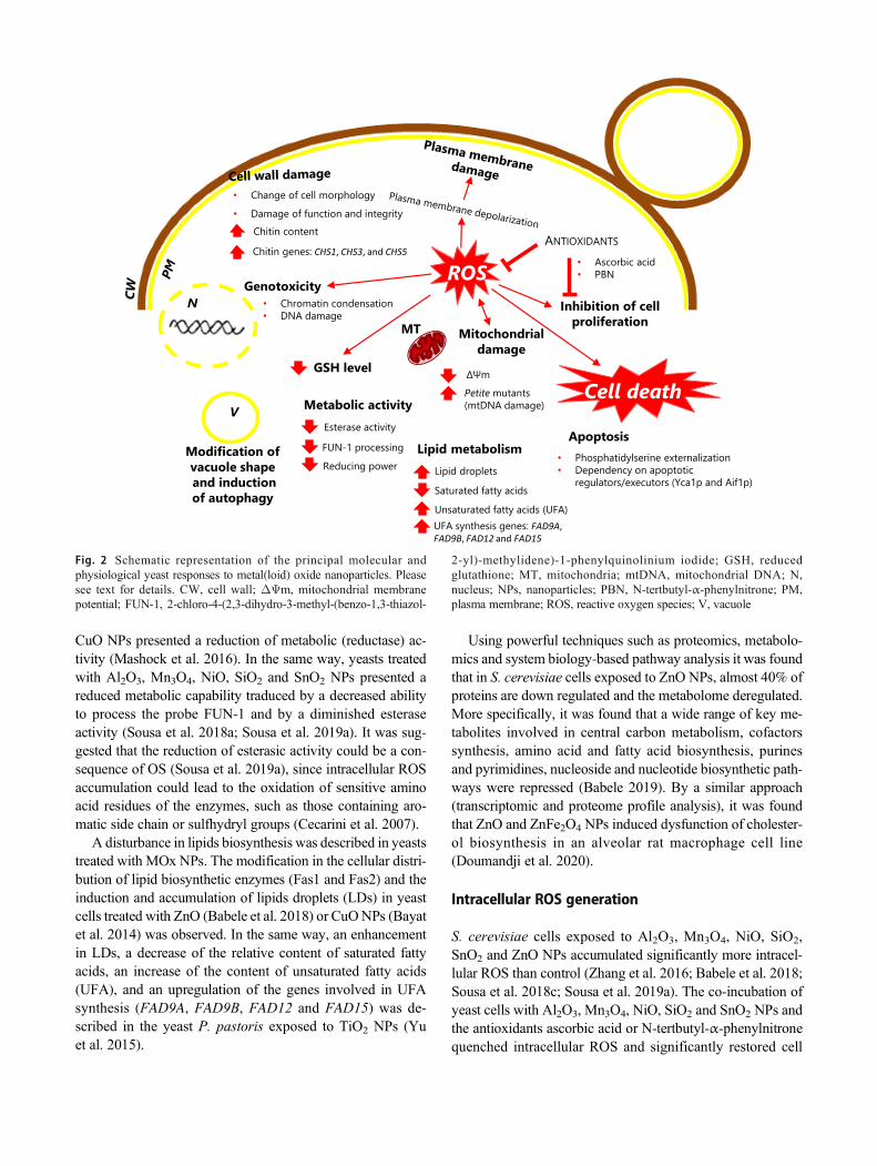
CuO NPs presented a reduction of metabolic (reductase) ac-tivity (Mashock et al. 2016). In the same way, yeasts treatedwith Al2O3, Mn3O4, NiO, SiO2 and SnO2 NPs presented areduced metabolic capability traduced by a decreased abilityto process the probe FUN-1 and by a diminished esteraseactivity (Sousa et al. 2018a; Sousa et al. 2019a). It was sug-gested that the reduction of esterasic activity could be a con-sequence of OS (Sousa et al. 2019a), since intracellular ROSaccumulation could lead to the oxidation of sensitive aminoacid residues of the enzymes, such as those containing aro-matic side chain or sulfhydryl groups (Cecarini et al. 2007).
A disturbance in lipids biosynthesis was described in yeaststreated with MOx NPs. The modification in the cellular distri-bution of lipid biosynthetic enzymes (Fas1 and Fas2) and theinduction and accumulation of lipids droplets (LDs) in yeastcells treated with ZnO (Babele et al. 2018) or CuONPs (Bayatet al. 2014) was observed. In the same way, an enhancementin LDs, a decrease of the relative content of saturated fattyacids, an increase of the content of unsaturated fatty acids(UFA), and an upregulation of the genes involved in UFAsynthesis (FAD9A, FAD9B, FAD12 and FAD15) was de-scribed in the yeast P. pastoris exposed to TiO2 NPs (Yuet al. 2015).
Using powerful techniques such as proteomics, metabolo-mics and system biology-based pathway analysis it was foundthat in S. cerevisiae cells exposed to ZnO NPs, almost 40% ofproteins are down regulated and the metabolome deregulated.More specifically, it was found that a wide range of key me-tabolites involved in central carbon metabolism, cofactorssynthesis, amino acid and fatty acid biosynthesis, purinesand pyrimidines, nucleoside and nucleotide biosynthetic path-ways were repressed (Babele 2019). By a similar approach(transcriptomic and proteome profile analysis), it was foundthat ZnO and ZnFe2O4 NPs induced dysfunction of cholester-ol biosynthesis in an alveolar rat macrophage cell line(Doumandji et al. 2020).
Intracellular ROS generation
S. cerevisiae cells exposed to Al2O3, Mn3O4, NiO, SiO2,SnO2 and ZnO NPs accumulated significantly more intracel-lular ROS than control (Zhang et al. 2016; Babele et al. 2018;Sousa et al. 2018c; Sousa et al. 2019a). The co-incubation ofyeast cells with Al2O3, Mn3O4, NiO, SiO2 and SnO2 NPs andthe antioxidants ascorbic acid or N-tertbutyl-α-phenylnitronequenched intracellular ROS and significantly restored cell
ROS
V
Modification of
vacuole shape
and induction
of autophagy
GSH level
Esterase activity
FUN-1 processing
Reducing power
Metabolic activityCell death
Apoptosis
• Phosphatidylserine externalization• Dependency on apoptotic
regulators/executors (Yca1p and Aif1p)
• Ascorbic acid• PBN
ANTIOXIDANTS
Inhibition of cell
proliferation
• Change of cell morphology
• Damage of function and integrityChitin content
Chitin genes: CHS1, CHS3, and CHS5
Lipid metabolism
Lipid droplets
Saturated fatty acids
Unsaturated fatty acids (UFA)UFA synthesis genes: FAD9A, FAD9B, FAD12 and FAD15
N • Chromatin condensation• DNA damage
Genotoxicity
Petite mutants (mtDNA damage)
ΔΨm
MTMitochondrial
damage
Fig. 2 Schematic representation of the principal molecular andphysiological yeast responses to metal(loid) oxide nanoparticles. Pleasesee text for details. CW, cell wall; ΔΨm, mitochondrial membranepotential; FUN-1, 2-chloro-4-(2,3-dihydro-3-methyl-(benzo-1,3-thiazol-
2-yl)-methylidene)-1-phenylquinolinium iodide; GSH, reducedglutathione; MT, mitochondria; mtDNA, mitochondrial DNA; N,nucleus; NPs, nanoparticles; PBN, N-tertbutyl-α-phenylnitrone; PM,plasma membrane; ROS, reactive oxygen species; V, vacuole
viability and metabolic activity, suggesting a ROS-mediatedmechanism in cytotoxicity induced by these NPs over yeastcells (Sousa et al. 2018c; Sousa et al. 2019a). TiO2 NPs alsoinduced a dose-dependent accumulation of intracellular ROSin the yeast P. pastoris (Chen et al. 2019).
Mitochondrial respiratory chain seems to be an importantsource of ROS since wild-type yeast cells under nitrogen at-mosphere as well as mutants lacking respiratory chain (with-out mitochondrial DNA, ρ0) presented decreased intracellularlevels of ROS and augmented resistance to NiO NPs (Sousaet al. 2018c). It was hypothesised that the Ni ions releasedfrom NPs can disturb the electron transport at mitochondriaby displacing iron from the ETC of the inner mitochondrialmembrane. Probably, the leakage of electrons from the ETC tooxygen lead to the production of O2·
− which, in turn, is mostlikely the main origin of H2O2 (Sousa et al. 2018c). In fact,both ROS types (O2·
− and H2O2) were detected inS. cerevisiae cells treated with NiO NPs (Sousa et al. 2018c).
Transcriptomic analysis revealed that Y3O3 NPs, at highconcentration (1–4 g L−1), induced the upregulation of oxida-tive stress genes in S. cerevisiae (Moriyama et al. 2019).
Reduction of non-enzymatic and enzymatic antioxi-dant defences
Yeast cells present non-enzymatic (which the most abundantis reduced glutathione, GSH) and enzymatic defence mecha-nisms such as superoxide dismutases (SOD1 and SOD2), cat-alases (CatT and CatA), glutathione peroxidases (such asGpx3 and Grx1) and thioredoxin peroxidases (like, Tsa1 andPrx1) to preserve intracellular redox equilibrium and survive(Jamieson 1998; Herrero et al. 2008).
Reduced glutathione seems to be involved in the fightagainst OS induced by NiO as revealed by the decrease ofcellular GSH level in yeasts incubated with these NPs.Supporting this observation, it was shown that mutant strainswithout (gsh1Δ) or with a reduced level of GSH (gsh2Δ) pre-sented augmented levels of ROS and susceptibility to NiO NPs(Sousa et al. 2018c). The depletion of the GSH levels can be aconsequence of the increased consumption in the scavenging offree oxygen radicals induced by NPs or due to the affinity ofmetal ions (such as Ni2+) to cysteine residue of GSH, leading toa reduction of cellular antioxidant defences (Sousa et al. 2018c).Similarly, TiO2 NPs induced an accentuated reduction in GSHconcentration in the yeast P. pastoris (Liu et al. 2016).
Single-gene mutant strains devoid of the main antioxidantenzymatic defences (Sod1p, Sod2p, Ctt1p, Cta1p, Gpx3p,Grx1p, Tsa1p and Pprx1p) did not present augmented vulnera-bility to NiO NPs comparatively to wild-type strain (Sousa et al.2018c); the absence of a sensitive phenotype, in these deletedstrains, can be attributed to gene redundancy or the presence ofcompensatory parallel pathways (Dawes 2004). A similar effectwas observed with sod1Δ and sod2Δmutant strains exposed to
CuONPs (Kasemets et al. 2013). However, the yeast P. pastoristreated with TiO2 NPs presented a downregulation of the genes(cSOD, GLR1, GPX1 and TRR1) encoding to enzymes associ-ated with ROS scavenging system (Liu et al. 2016).
Loss of cell membrane integrity
One of the outcomes of high ROS levels is the lipid per-oxidation. Large-scale lipid peroxidation leads to in-creased membrane fluidity, efflux of cytosolic compo-nents and, ultimately, loss of plasma membrane integrityand cell death (Avery 2011). Consistent with this scenar-io, it was shown that the exposure of yeasts to NiO NPsleads to a progressive depolarization (reduction of themembrane potential) and an increase of permeability ofthe yeast plasma membrane, in cells under oxidative stress(Sousa et al. 2019c). Similarly, yeasts incubated with ZnONPs displayed intracellular ROS and an augmented cellmembrane permeability (Babele et al. 2018). It was ob-served that strains with deletion of genes involved in thebiosynthesis of ergosterol (ERG2 and ERG28), a sterolthat affects membrane fluidity, and in transmembranetransport (PKR1), displayed enhanced susceptibility toZnO NPs, which suggested that these NPs disrupt cellmembrane integrity and impair their proper function(transport) (Márquez et al. 2018).
The impact of MOx NPs on yeast cell membrane can bedose-dependent. Thus, the incubation of yeasts for 24 h with100 mg L−1 Al2O3, In2O3, Mn3O4, SiO2 and SnO2 did notinduce the permeabilization of the cell membrane (Sousaet al. 2019a). However, the exposure of the same yeasts tosome of these NPs (Al2O3, Mn2O3 and SiO2), but at higherconcentration (1000mg L−1), during 10 h, caused a significantloss of membrane integrity (Garcia-Saucedo et al. 2011;Otero-Gonzalez et al. 2013).
Alteration of function and morphology ofmitochondria and endoplasmic reticulum
Mitochondrial membrane potential (ΔΨm) is an essentialcomponent for energy-producing and non-producing mito-chondrial functions (Zorova et al. 2018). The depolarizationof the mitochondrial membrane, i.e. the dissipation of mito-chondrial membrane potential in yeast cells treated with NiONPs (Sousa et al. 2019c) and the alteration of the architectureof mitochondria in cells incubated with ZnO NPs were de-scribed (Babele et al. 2018).
The exposure to ZnO NPs also severely affected the archi-tecture and function of the endoplasmic reticulum, in yeasts,through modulation of unfolded protein response (Babeleet al. 2018).

Modification of vacuole architecture and induction ofautophagy
Yeast cells treated with ZnO NPs presented a drastic modifi-cation of vacuoles morphology (Bayat et al. 2014; Babeleet al. 2018) and a redistribution of Atg8-GFP to vacuoles,indicating the induction of autophagy (Babele et al. 2018). Itwas observed (by TEM) the modification of vacuole shapeand its disruption, in yeasts incubated with TiO2 and CuONPs, respectively. Dark deposits in vacuoles (TiO2 treatedcells) or in vesicles (ZnO or CuO treated cells) were alsodescribed (Bayat et al. 2014).
Mitochondrial and genomic DNA damage
DNA damage is commonly found during OS, being mitochon-drial DNA (mtDNA) a very sensitive target (Richter et al.1988). Exposure of S. cerevisiae cells to NiO NPs led to themtDNA damage with the consequent abolition of respiration(incapacity to grow on non-fermentable carbon sources) and theformation of typical respiratory-deficient colonies, commonlyknown as petite mutants (Sousa et al. 2019c).
Using the canavanine assay, it was shown the damage ofnuclear DNA in yeasts incubated with NiO NPs (Sousa et al.2019c). Nuclear DNA damage, measured using the cometassay, in yeast cells treated with CuO, TiO2 and ZnO NPswas also described (Bayat et al. 2014).
Apoptotic cell death
The exposure of yeast cells to NiO NPs induced regulated celldeath, with typical apoptotic hallmarks such as damage of cellmembrane, loss of cell viability, phosphatidylserine exposureat the outer cytoplasmic membrane leaflet, and nuclear chro-matin condensation, in a process dependent on de novo pro-tein synthesis and apoptotic regulators/executors (Yca1p andAif1p) (Sousa et al. 2019c). The sequence of events associatedwith the induction of cell death in S. cerevisiae by NiO NPswas described (Sousa et al. 2019c). Other studies also indicat-ed a cell dead apoptotic pathway in human cell lines exposedto CuO (Siddiqui et al. 2013), NiO (Siddiqui et al. 2012), ZnO(Keerthana and Kumar 2020), or binary mixtures of Al2O3
and ZnO NPs (Koerich et al. 2020).
Concluding remarks
Products containing NMs grown enormously in the last de-cades. Nanotechnology Consumer Products Inventory, up-dated in 2013, listed 1814 consumer products containingNMs, from 622 companies, in which products containingmetals and metal oxides correspond to the largest group, con-stituting 37% of products (Vance et al. 2015). Concomitant,
and understandably, concerns about environmental, healthand safety implications of NMs have also raised.
Over the last decade, and thanks to a substantial researcheffort, important progress concerning the impact of NPs interrestrial and aquatic systems as well as about their mecha-nisms of toxicity has been observed. However, substantialgaps still exist that require further attention, namely regardingto MOx NPs concentrations used in the assays, the time andthe type of exposure.
Although it is difficult to accurately detect NPs in aquaticenvironments, it is estimated that their concentration in sur-face waters varies from ng L−1 to μg L−1 (Gottschalk et al.2013). However, it is common to find studies that use NPsconcentrations greater than 100 mg L−1, reaching 1000 mgL−1 or even more. Another challenge is to study the impactof sub-lethal concentrations of NPs during a long-term expo-sure (covering multiple generations of the organism), in arepeated way, to get more information on chronic exposureto MOx NPs, in order to adopt the necessary protective mea-sures regarding the use of products containing MOx NPs.
A more systematic approach is needed in nanotoxicologyresearch. Thus, future studies should combine high-throughputmolecular profiling technologies (transcriptomics, proteomicsand metabolomics) with more traditional approaches (physiolog-ical studies), to give a holistic understanding of cellular responsesto MOx NPs (and NMs in a general way) and allow the elucida-tion of the mechanisms associated with its toxicity.
Author contribution ES and HS conceived the review. ES and HS wrotethe manuscript. ES conceived and designed the figures. All authors readand approved the manuscript.
Funding This work was supported by National funds through FCT -Foundation for Science and Technology under the scope of the projectsUIDB/50006/2020, UID/BIO/04469/2020 unit and BioTecNorte opera-tion (NORTE-01-0145-FEDER-000004) funded by the EuropeanRegional Development Fund under the scope of Norte2020 - ProgramaOperacional Regional do Norte.
Declarations
This article does not contain any studies with human participants or an-imals performed by any of the authors.
Conflict of interest The authors declare no competing interests.
References
Ahamed M, Ali D, Alhadlaq HA, Akhtar MJ (2013) Nickel oxide nano-particles exert cytotoxicity via oxidative stress and induce apoptoticresponse in human liver cells (HepG2). Chemosphere 93:2514–2522. https://doi.org/10.1016/j.chemosphere.2013.09.047
Alarifi S, Ali D, Alkahtani S, Verma A, AhamedM, AhmedM, AlhadlaqHA (2013) Induction of oxidative stress, DNA damage, and apopto-sis in a malignant human skin melanoma cell line after exposure to
zinc oxide nanoparticles. Int J Nanomedicine 8:983–993. https://doi.org/10.2147/IJN.S42028
Amde M, Liu J-F, Tan Z-Q, Bekana D (2017) Transformation and bio-availability of metal oxide nanoparticles in aquatic and terrestrialenvironments. A review. Environ Pollut 230:250–267. https://doi.org/10.1016/j.envpol.2017.06.064
Angel BM, Vallotton P, Apte SC (2015) On the mechanism ofnanoparticulate CeO2 toxicity to freshwater algae. Aquat Toxicol168:90–97. https://doi.org/10.1016/j.aquatox.2015.09.015
Anissimova OV, Staer OV (2018) Morphology of cell wall pore channelsin the genus Euastrum ralfs (Desmidiales). Moscow Univ Biol SciBull 73:28–31. https://doi.org/10.3103/S0096392518010029
Aravantinou AF, Tsarpali V, Dailianis S, Manariotis ID (2015) Effect ofcultivation media on the toxicity of ZnO nanoparticles to freshwaterand marine microalgae. Ecotoxicol Environ Saf 114:109–116.https://doi.org/10.1016/j.ecoenv.2015.01.016
Argueello JM, Raimunda D, Gonzalez-Guerrero M (2012) Metal trans-port across biomembranes: emerging models for a distinct chemis-try. J Biol Chem 287:13510–13517. https://doi.org/10.1074/jbc.R111.319343
Aruoja V, Dubourguier H-C, Kasemets K, Kahru A (2009) Toxicity ofnanopar t ic les of CuO, ZnO and TiO2 to microalgaePseudokirchneriella subcapitata. Sci Total Environ 407:1461–1468. https://doi.org/10.1016/j.scitotenv.2008.10.053
Aruoja V, Pokhrel S, Sihtmaae M, Mortimer M, Maedler L, Kahru A(2015) Toxicity of 12 metal-based nanoparticles to algae, bacteriaand protozoa. Environ Sci 2:630–644. https://doi.org/10.1039/c5en00057b
Avery SV (2011) Molecular targets of oxidative stress. Biochem J 434:201–210. https://doi.org/10.1042/BJ20101695
Babele PK (2019) Zinc oxide nanoparticles impose metabolic toxicity byde-regulating proteome and metabolome in Saccharomycescerevisiae. Toxicol REPORTS 6:64–73. https://doi.org/10.1016/j.toxrep.2018.12.001
Babele PK, Thakre PK, Kumawat R, Tomar RS (2018) Zinc oxide nano-particles induce toxicity by affecting cell wall integrity it pathway,mitochondrial function and lipid homeostasis in Saccharomycescerevisiae. Chemosphere 213:65–75. https://doi.org/10.1016/j.chemosphere.2018.09.028
Balusamy B, Tastan BE, Ergen SF, Uyar T, Tekina T (2015) Toxicity oflanthanum oxide (La2O3) nanoparticles in aquatic environments.Environ Sci IMPACTS 17:1265–1270. https://doi.org/10.1039/c5em00035a
Bao S, Lu Q, Fang T, Dai H, Zhang C (2015) Assessment of the toxicityof CuO nanoparticles by using Saccharomyces cerevisiae mutantswith multiple genes deleted. Appl Environ Microbiol 81:8098–8107. https://doi.org/10.1128/AEM.02035-15
Bayat N, Rajapakse K, Marinsek-Logar R, Drobne D, Cristobal S (2014)The effects of engineered nanoparticles on the cellular structure andgrowth of Saccharomyces cerevisiae. Nanotoxicology 8:363–373.https://doi.org/10.3109/17435390.2013.788748
Bondarenko O, Ivask A, Kaekinen A, Kahru A (2012) Sub-toxic effectsof CuO nanoparticles on bacteria: kinetics, role of Cu ions andpossible mechanisms of action. Environ Pollut 169:81–89. https://doi.org/10.1016/j.envpol.2012.05.009
Braconi D, Bernardini G, Santucci A (2016) Saccharomyces cerevisiae asa model in ecotoxicological studies: a post-genomics perspective. JProteomics 137:19–34. https://doi.org/10.1016/j.jprot.2015.09.001
Brunner TJ, Wick P, Manser P, Spohn P, Grass RN, Limbach LK,Bruinink A, Stark WJ (2006) In vitro cytotoxicity of oxide nanopar-ticles: comparison to asbestos, silica, and the effect of particle solu-bility. Environ Sci Technol 40:4374–4381. https://doi.org/10.1021/es052069i
Bundschuh M, Filser J, Luderwald S, Mckee MS, Metreveli G,Schaumann GE, Schulz R, Wagner S (2018) Nanoparticles in the
environment: where do we come from, where do we go to? EnvironSci Eur 30. https://doi.org/10.1186/s12302-018-0132-6
Cecarini V, Gee J, Fioretti E, Amici M, Angeletti M, Eleuteri AM, KellerJN (2007) Protein oxidation and cellular homeostasis: emphasis onmetabolism. Biochim Biophys Acta-Mol Cell Res 1773:93–104.https://doi.org/10.1016/j.bbamcr.2006.08.039
Cheloni G, Marti E, Slaveykova VI (2016) Interactive effects of copperoxide nanoparticles and light to green alga Chlamydomonasreinhardtii. Aquat Toxicol 170:120–128. https://doi.org/10.1016/j.aquatox.2015.11.018
Chen L, Zhou L, Liu Y, Deng S, Wu H, Wang G (2012) Toxicologicaleffects of nanometer titanium dioxide (nano-TiO2) onChlamydomonas reinhardtii. Ecotoxicol Environ Saf 84:155–162.https://doi.org/10.1016/j.ecoenv.2012.07.019
Chen X, Zhang C, Tan L, Wang J (2018) Toxicity of Co nanoparticles onthree species of marine microalgae. Environ Pollut 236:454–461.https://doi.org/10.1016/j.envpol.2018.01.081
Chen F, Xiao Z, Yue L, Wang J, Feng Y, Zhu X,Wang Z, Xing B (2019)Algae response to engineered nanoparticles: current understanding,mechanisms and implications. Environ Sci 6:1026–1042. https://doi.org/10.1039/c8en01368c
Cohen D, Soroka Y, Ma’or Z, Oron M, Portugal-Cohen M, BregegereFM, Berhanu D, Valsami-Jones E, Hai N, Milner Y (2013)Evaluation of topically applied copper(II) oxide nanoparticle cyto-toxicity in human skin organ culture. Toxicol Vitr 27:292–298.https://doi.org/10.1016/j.tiv.2012.08.026
Condello M, De Berardis B, Ammendolia MG, Barone F, Condello G,Degan P, Meschini S (2016) ZnO nanoparticle tracking from uptaketo genotoxic damage in human colon carcinoma cells. Toxicol Vitr35:169–179. https://doi.org/10.1016/j.tiv.2016.06.005
Cronholm P, Karlsson HL, Hedberg J, Lowe TA, Winnberg L, Elihn K,Wallinder IO, Moeller L (2013) Intracellular uptake and toxicity ofAg and CuO nanoparticles: a comparison between nanoparticles andtheir corresponding metal ions. SMALL 9:970–982. https://doi.org/10.1002/smll.201201069
Czyzowska A, Barbasz A (2020) A review: zinc oxide nanoparticles-friends or enemies? Int J Environ Health Res. https://doi.org/10.1080/09603123.2020.1805415
da Costa CH, Perreault F, OukarroumA,Melegari SP, Popovic R,MatiasWG (2016) Effect of chromium oxide (III) nanoparticles on theproduction of reactive oxygen species and photosystem II activityin the green alga Chlamydomonas reinhardtii. Sci Total Environ565:951–960. https://doi.org/10.1016/j.scitotenv.2016.01.028
Dauda S, Chia MA, Bako SP (2017) Toxicity of titanium dioxide nano-particles to Chlorella vulgaris Beyerinck (Beijerinck) 1890(Trebouxiophyceae, Chlorophyta) under changing nitrogen condi-tions. Aquat Toxicol 187:108–114. https://doi.org/10.1016/j.aquatox.2017.03.020
Dawes IW (2004) Stress responses. In: Dickinson JR, Schweizer M (eds)The metabolism and molecular physiology of Saccharomycescerevisiae, 2nd edn. Taylor and Francis, Ltd, London, pp 376–439
De Berardis B, Civitelli G, Condello M, Lista P, Pozzi R, Arancia G,Meschini S (2010) Exposure to ZnO nanoparticles induces oxidativestress and cytotoxicity in human colon carcinoma cells. ToxicolAppl Pharmacol 246:116–127. https://doi.org/10.1016/j.taap.2010.04.012
Dekkers S, Williams TD, Zhang J, Zhou J, Vandebriel RJ, la FonteyneLJJ, Gremmer ER, He S, Guggenheim EJ, Lynch I, Cassee FR, DeJong WH, Viant MR (2018) Multi-omics approaches confirm metalions mediate the main toxicological pathways of metal-bearingnanoparticles in lung epithelial A549 cells. Environ Sci 5:1506–1517. https://doi.org/10.1039/c8en00071a
del Rio LA, Lopez-Huertas E (2016) ROS generation in peroxisomes andits role in cell signaling. PLANT CELL Physiol 57:1364–1376.https://doi.org/10.1093/pcp/pcw076

Deniel M, Errien N, Daniel P, Caruso A, Lagarde F (2019) Currentmethods to monitor microalgae-nanoparticle interaction and associ-ated effects. Aquat Toxicol 217:217. https://doi.org/10.1016/j.aquatox.2019.105311
dos Santos SC, Sa-Correia I (2015) Yeast toxicogenomics: lessons from aeukaryotic cell model and cell factory. Curr Opin Biotechnol 33:183–191. https://doi.org/10.1016/j.copbio.2015.03.001
dos Santos S, Teixeira M, Cabrito T, Sá-Correia I (2012) Yeasttoxicogenomics: genome-wide responses to chemical stresses withimpact in environmental health, pharmacology, and biotechnology.Front Genet 3:63. https://doi.org/10.3389/fgene.2012.00063
Doumandji Z, Safar R, Lovera-Leroux M, Nahle S, Cassidy H,Matallanas D, Rihn B, Ferrari L, Joubert O (2020) Protein and lipidhomeostasis altered in rat macrophages after exposure to metallicoxide nanoparticles. Cell Biol Toxicol 36:65–82. https://doi.org/10.1007/s10565-019-09484-6
Duan W-X, HeM-D, Mao L, Qian F-H, Li Y-M, Pi H-F, Liu C, Chen C-H, Lu Y-H, Cao Z-W, Zhang L, Yu Z-P, Zhou Z (2015) NiO nano-particles induce apoptosis through repressing SIRT1 in human bron-chial epithelial cells. Toxicol Appl Pharmacol 286:80–91. https://doi.org/10.1016/j.taap.2015.03.024
ECHA - European Chemicals Agency 2020 Nanomaterials. https://echa.europa.eu/regulations/nanomaterials. Accessed 7 Aug 2020
EPA - United States Environmental Protection Agency (2017)Nanomaterials - technical fact sheet. EPA 505-F-17-002
FDA – Food and Drug Administration (2018) Microorganisms µbial-derived ingredients used in food (partial list). In: FDA.https://www.fda.gov/food/generally-recognized-safe-gras/microorganisms-microbial-derived-ingredients-used-food-partial-list. Accessed 7 Aug 2020
Foury F (1997) Human genetic diseases: a cross-talk between man andyeast. Gene 195:1–10. https://doi.org/10.1016/S0378-1119(97)00140-6
Franklin NM, Rogers NJ, Apte SC, Batley GE, Gadd GE, Casey PS(2007) Comparative toxicity of nanoparticulate ZnO, bulk ZnO,and ZnCl2 to a freshwater microalga (Pseudokirchneriellasubcapitata): the importance of particle solubility. Environ SciTechnol 41:8484–8490. https://doi.org/10.1021/es071445r
Garcia-Saucedo C, Field JA, Otero-Gonzalez L, Sierra-Alvarez R (2011)Low toxicity of HfO2, SiO2, Al2O3 and CeO2 nanoparticles to theyeast, Saccharomyces cerevisiae. J Hazard Mater 192:1572–1579.https://doi.org/10.1016/j.jhazmat.2011.06.081
Giaever G, Nislow C (2014) The yeast deletion collection: a decade offunctional genomics. Genetics 197:451–465. https://doi.org/10.1534/genetics.114.161620
Goffeau A, Barrell BG, Bussey H, Davis RW, Dujon B, Feldmann H,Galibert F, Hoheisel JD, Jacq C, JohnstonM, Louis EJ, Mewes HW,Murakami Y, Philippsen P, Tettelin H, Oliver SG (1996) Life with6000 genes. Science (80- ) 274:546. https://doi.org/10.1126/science.274.5287.546
Gottschalk F, Sun T, Nowack B (2013) Environmental concentrations ofengineered nanomaterials: review of modeling and analytical stud-ies. Environ Pollut 181:287–300. https://doi.org/10.1016/j.envpol.2013.06.003
Guerra FD, Attia MF, Whitehead DC, Alexis F (2018) Nanotechnologyfor environmental remediation: materials and applications.MOLECULES 23. https://doi.org/10.3390/molecules23071760
Guo Y, Cheng C, Wang J, Wang Z, Jin X, Li K, Kang P, Gao J (2011)Detection of reactive oxygen species (ROS) generated by TiO2(R),TiO2(R/A) and TiO2(A) under ultrasonic and solar light irradiationand application in degradation of organic dyes. J Hazard Mater 192:786–793. https://doi.org/10.1016/j.jhazmat.2011.05.084
Halbus AF, Horozov TS, Paunov VN (2019) Self-grafting copper oxidenanoparticles show a strong enhancement of their anti-algal andanti-yeast action. NANOSCALE Adv 1:2323–2336. https://doi.org/10.1039/c9na00099b
HalbusAF, Horozov TS, PaunovVN (2020) Surface-modified zinc oxidenanoparticles for antialgal and antiyeast applications. ACS ApplNANO Mater 3:440–451. https://doi.org/10.1021/acsanm.9b02045
Halliwell B, Gutteridge JMC (2015) Oxidative stress and redox regula-tion: adaptation, damage, repair, senescence, and death. In: Freeradicals in biology and medicine, 5th edn. Oxford UniversityPress, pp 199–283
Hartmann NB, der Kammer F, Hofmann T, BaaloushaM, Ottofuelling S,Baun A (2010) Algal testing of titanium dioxide nanoparticles-testing considerations, inhibitory effects and modification of cadmi-um bioavailability. Toxicology 269:190–197. https://doi.org/10.1016/j.tox.2009.08.008
Hartung T, Rovida C (2009) Chemical regulators have overreached.Nature 460:1080–1081. https://doi.org/10.1038/4601080a
He H, Zou Z, Wang B, Xu G, Chen C, Qin X, Yu C, Zhang J (2020)Copper oxide nanoparticles induce oxidative DNA damage and celldeath via copper ion-mediated p38 MAPK activation in vascularendothelial cells. Int J Nanomedicine 15:3291–3302. https://doi.org/10.2147/IJN.S241157
Heinlaan M, Ivask A, Blinova I, Dubourguier H-C, Kahru A (2008)Toxicity of nanosized and bulk ZnO, CuO and TiO2 to bacteriaVibrio f ischeri and crustaceans Daphnia magna andThamnocephalus platyurus. Chemosphere 71:1308–1316. https://doi.org/10.1016/j.chemosphere.2007.11.047
Herrero E, Ros J, Belli G, Cabiscol E (2008) Redox control and oxidativestress in yeast cells. Biochim Biophys Acta-Gen Subj 1780:1217–1235. https://doi.org/10.1016/j.bbagen.2007.12.004
Howe DG, Blake JA, Bradford YM, Bult CJ, Calvi BR, Enge SR, KadinJA, Kaufman TC, Kishores R, Laulederkind SF, Lewis SE, MoxonSAT, Richardson JE, Smith C (2018)Model organism data evolvingin support of translational medicine. Lab Anim (NY) 47:277–289.https://doi.org/10.1038/s41684-018-0150-4
Hughes MN, Poole RK (1991) Metal speciation and microbial growth:the hard (and soft) facts. J Gen Microbiol 137:725–734. https://doi.org/10.1099/00221287-137-4-725
Ivask A, Juganson K, Bondarenko O, Mortimer M, Aruoja V, KasemetsK, Blinova I, Heinlaan M, Slaveykova V, Kahru A (2014)Mechanisms of toxic action of Ag, ZnO and CuO nanoparticles toselected ecotoxicological test organisms and mammalian cellsin vitro: a comparative review. Nanotoxicology 8:57–71. https://doi.org/10.3109/17435390.2013.855831
Jamieson DJ (1998) Oxidative stress responses of the yeastSaccharomyces cerevisiae. YEAST 14:1511–1527.
Ji J, Long Z, Lin D (2011) Toxicity of oxide nanoparticles to the greenalgae Chlorella sp. Chem Eng J 170:525–530. https://doi.org/10.1016/j.cej.2010.11.026
Jo HJ, Choi JW, Lee SH, Hong SW (2012) Acute toxicity of Ag and CuOnanoparticle suspensions againstDaphnia magna: the importance oftheir dissolved fraction varying with preparation methods. J HazardMater 227:301–308. https://doi.org/10.1016/j.jhazmat.2012.05.066
Juganson K, Ivask A, Blinova I, Mortimer M, Kahru A (2015) NanoE-Tox: new and in-depth database concerning ecotoxicity ofnanomaterials. BEILSTEIN J Nanotechnol 6:1788–1804. https://doi.org/10.3762/bjnano.6.183
Karlsson HL, Cronholm P, Gustafsson J, Moeller L (2008) Copper oxidenanoparticles are highly toxic: a comparison between metal oxidenanoparticles and carbon nanotubes. Chem Res Toxicol 21:1726–1732. https://doi.org/10.1021/tx800064j
Kasemets K, Ivask A, Dubourguier H-C, Kahru A (2009) Toxicity ofnanoparticles of ZnO, CuO and TiO2 to yeast Saccharomycescerevisiae. Toxicol Vitr 23:1116–1122. https://doi.org/10.1016/j.tiv.2009.05.015
Kasemets K, Suppi S, Kuennis-Beres K, Kahru A (2013) Toxicity ofCuO nanoparticles to yeast Saccharomyces cerevisiae BY4741wild-type and its nine isogenic single-gene deletion mutants.Chem Res Toxicol 26:356–367. https://doi.org/10.1021/tx300467d

Keerthana S, Kumar A (2020) Potential risks and benefits of zinc oxidenanoparticles: a systematic review. Crit Rev Toxicol 50:47–71.https://doi.org/10.1080/10408444.2020.1726282
Keller AA, McFerran S, Lazareva A, Suh S (2013) Global life cyclereleases of engineered nanomaterials. J Nanoparticle Res:15.https://doi.org/10.1007/s11051-013-1692-4
Kharazian B, Hadipour NL, Ejtehadi MR (2016) Understanding thenanoparticle-protein corona complexes using computational and ex-perimental methods. Int J Biochem Cell Biol 75:162–174. https://doi.org/10.1016/j.biocel.2016.02.008
Klaine SJ, Edgington A, Seda B (2013) Nanomaterials in the environ-ment. In: Férard J-F, Blaise C (eds) Encyclopedia of aquatic ecotox-icology. Springer Science, Dordrecht, pp 767–779
Klaper RD (2020) The known and unknown about the environmentalsafety of nanomaterials in commerce. Small 2000690:2000690.https://doi.org/10.1002/smll.202000690
Koerich JS, Nogueira DJ, Vaz VP, Simioni C, Da Silva MLN, OuriquesLC, Vicentini DS,MatiasWG (2020) Toxicity of binary mixtures ofAl2O3 and ZnO nanoparticles toward fibroblast and bronchial epi-thelium cells. J Toxicol Environ Heal A-CURRENT ISSUES 83:363–377. https://doi.org/10.1080/15287394.2020.1761496
Kumar A, Pandey AK, Singh SS, Shanker R, Dhawan A (2011)Engineered ZnO and TiO2 nanoparticles induce oxidative stressand DNA damage leading to reduced viability of Escherichia coli.Free Radic Biol Med 51:1872–1881. https://doi.org/10.1016/j.freeradbiomed.2011.08.025
Laurent S, Boutry S, Muller RN (2018) Metal oxide particles and theirprospects for applications. In: Mahmoudi M, Laurent S (eds) Ironoxide nanoparticles for biomedical applications. Elsevier, pp 3–42
Lead JR, Batley GE, Alvarez PJJ, Croteau M-N, Handy RD,McLaughlinMJ, Judy JD, Schirmer K (2018) Nanomaterials in the environment:behavior, fate, bioavailability, and effects-an updated review.Environ Toxicol Chem 37:2029–2063. https://doi.org/10.1002/etc.4147
Lee W-M, An Y-J (2013) Effects of zinc oxide and titanium dioxidenanoparticles on green algae under visible, UVA, and UVB irradi-ations: no evidence of enhanced algal toxicity under UV pre-irradi-ation. Chemosphere 91:536–544. https://doi.org/10.1016/j.chemosphere.2012.12.033
Lesser MP (2006) Oxidative stress in marine environments: biochemistryand physiological ecology. Annu Rev Physiol 68:253–278. https://doi.org/10.1146/annurev.physiol.68.040104.110001
Li M, Zhu L, Lin D (2011) Toxicity of ZnO nanoparticles to Escherichiacoli: mechanism and the influence of medium components. EnvironSci Technol 45:1977–1983. https://doi.org/10.1021/es102624t
Li F, Liang Z, Zheng X, Zhao W, Wu M, Wang Z (2015) Toxicity ofnano-TiO2 on algae and the site of reactive oxygen species produc-tion. Aquat Toxicol 158:1–13. https://doi.org/10.1016/j.aquatox.2014.10.014
Libralato G, Galdiero E, Falanga A, Carotenuto R, de Alteriis E, GuidaM(2017) Toxicity effects of ufnctionalized quantum dots, gold andpolystyrene nanoparticles on target aquatic biological models: ae rv i ew . MOLECULES 22 . h t tp s : / / do i . o rg /10 .3390 /molecules22091439
Lin D, Ji J, Long Z, Yang K,Wu F (2012) The influence of dissolved andsurface-bound humic acid on the toxicity of TiO2 nanoparticles toChlorella sp. WATER Res 46:4477–4487. https://doi.org/10.1016/j.watres.2012.05.035
Liu Z, Zhang M, Han X, Xu H, Zhang B, Yu Q, Li M (2016) TiO2
nanoparticles cause cell damage independent of apoptosis and au-tophagy by impairing the ROS-scavenging system in Pichiapastoris. Chem Biol Interact 252:9–18. https://doi.org/10.1016/j.cbi.2016.03.029
Lombi E, Donner E, Dusinska M, Wickson F (2019) A one health ap-proach to managing the applications and implications of
nanotechnologies in agriculture. Nat Nanotechnol 14:523–531.https://doi.org/10.1038/s41565-019-0460-8
Lu S, Zhang W, Zhang R, Liu P, Wang Q, Shang Y, Wu M, DonaldsonK, Wang Q (2015) Comparison of cellular toxicity caused by ambi-ent ultrafine particles and engineered metal oxide nanoparticles. PartFibre Toxicol 12. https://doi.org/10.1186/s12989-015-0082-8
Malina C, Larsson C, Nielsen J (2018) Yeast mitochondria: an overviewof mitochondrial biology and the potential of mitochondrial systemsbiology. FEMS Yeast Res 18. https://doi.org/10.1093/femsyr/foy040
Manier N, Bado-Nilles A, Delalain P, Aguerre-Chariol O, Pandard P(2013) Ecotoxicity of non-aged and aged CeO2 nanomaterials to-wards freshwater microalgae. Environ Pollut 180:63–70. https://doi.org/10.1016/j.envpol.2013.04.040
Márquez IG, Ghiyasvand M, Massarsky A, Babu M, Samanfar B, OmidiK, Moon TW, Smith ML, Golshani A (2018) Zinc oxide and silvernanoparticles toxicity in the baker’s yeast, Saccharomycescerevisiae. PLoS One 13:e0193111. https://doi.org/10.1371/journal.pone.0193111
Mashock MJ, Kappell AD, Hallaj N, Hristova KR (2016) Copper oxidenanoparticles inhibit the metabolic activity of Saccharomycescerevisiae. Environ Toxicol Chem 35:134–143. https://doi.org/10.1002/etc.3159
Meyer JS, Lyons-Darden T, Garman ER, Middleton ET, Schlekat CE(2020) Toxicity of nanoparticulate nickel to aquatic organisms: re-view and recommendations for improvement of toxicity tests.Environ Toxicol Chem. https://doi.org/10.1002/etc.4812
Miller RJ, Lenihan HS, Muller EB, Tseng N, Hanna SK, Keller AA(2010) Impacts of metal oxide nanoparticles on marine phytoplank-ton. Environ Sci Technol 44:7329–7334. https://doi.org/10.1021/es100247x
Minetto D, Ghirardini AV, Libralato G (2016) Saltwater ecotoxicology ofAg, Au, CuO, TiO2, ZnO and C-60 engineered nanoparticles: anoverview. Environ Int 92–93:189–201. https://doi.org/10.1016/j.envint.2016.03.041
Mohammadinejad R, Moosavi MA, Tavakol S, Vardar DO, Hosseini A,Rahmati M, Dini L, Hussain S, Mandegary A, Klionsky DJ (2019)Necrotic, apoptotic and autophagic cell fates triggered by nanopar-ticles. Autophagy 15:4–33. https://doi.org/10.1080/15548627.2018.1509171
Mordor Intelegence (2020) Metal oxide nanoparticles market - growth,trends, and forecast (2020 - 2025). https://www.mordorintelligence.com/industry-reports/metal-oxide-nanoparticles-market. Accessed19 May 2020
MoriyamaA, Takahashi U,Mizuno Y, Takahashi J, HorieM, Iwahashi H(2019) The truth of toxicity caused by yttrium oxide nanoparticles toyeast Cells. J Nanosci Nanotechnol 19:5418–5425. https://doi.org/10.1166/jnn.2019.16544
Mortezaee K, Najafi M, Samadian H, Barabadi H, Azarnezhad A,Ahmadi A (2019) Redox interactions and genotoxicity of metal-based nanoparticles: a comprehensive review. Chem Biol Interact312:312. https://doi.org/10.1016/j.cbi.2019.108814
Naeemi AS, Elmi F, Vaezi G, Ghorbankhah M (2020) Copper oxidenanoparticles induce oxidative stress mediated apoptosis in carp(Cyprinus carpio) larva. GENE REPORTS 19. https://doi.org/10.1016/j.genrep.2020.100676
Naseer B, Srivastava G, Qadri OS, Faridi SA, Islam RU, Younis K (2018)Importance and health hazards of nanoparticles used in the foodindustry. Nanotechnol Rev 7:623–641. https://doi.org/10.1515/ntrev-2018-0076
Navarro E, BaunA, Behra R, Hartmann NB, Filser J,Miao A-J, QuiggA,Santschi PH, Sigg L (2008) Environmental behavior and ecotoxicityof engineered nanoparticles to algae, plants, and fungi.ECOTOXICOLOGY 17:372–386. https://doi.org/10.1007/s10646-008-0214-0

nee Rohder LA, Brandt T, Sigg L, Behra R (2018) Uptake and effects ofcerium(III) and cerium oxide nanoparticles to Chlamydomonasreinhardtii. Aquat Toxicol 197:41–46. https://doi.org/10.1016/j.aquatox.2018.02.004
Nel A, Xia T, Madler L, Li N (2006) Toxic potential of materials at thenanolevel. Science 311:622–627. https://doi.org/10.1126/science.1114397
Ng CT, Yong LQ, Hande MP, Ong CN, Yu LE, Bay BH, Baeg GH(2017) Zinc oxide nanoparticles exhibit cytotoxicity andgenotoxicity through oxidative stress responses in human lung fi-broblasts and Drosophila melanogaster. Int J Nanomedicine 12:1621–1637. https://doi.org/10.2147/IJN.S124403
Nguyen VH, Lee B-J (2017) Protein corona: a new approach fornanomedicine design. Int J Nanomedicine 12:3137–3151. https://doi.org/10.2147/IJN.S129300
NguyenMK,Moon J-Y, Lee Y-C (2020)Microalgal ecotoxicity of nano-particles: an updated review. Ecotoxicol Environ Saf:201. https://doi.org/10.1016/j.ecoenv.2020.110781
NPD - Nanotechnology Products Database (2020) Introduction. https://product.statnano.com/. Accessed 7 Aug 2020
Oh N, Park J-H (2014) Endocytosis and exocytosis of nanoparticles inmammalian cells. Int J Nanomedicine 9:51–63. https://doi.org/10.2147/IJN.S26592
Otero-Gonzalez L, Garcia-Saucedo C, Field JA, Sierra-Alvarez R (2013)Toxicity of TiO2, ZrO2, Fe-0, Fe2O3, and Mn2O3 nanoparticles tothe yeast, Saccharomyces cerevisiae. Chemosphere 93:1201–1206.https://doi.org/10.1016/j.chemosphere.2013.06.075
OukarroumA, ZaidiW, SamadaniM, Dewez D (2017) Toxicity of nickeloxide nanoparticles on a freshwater green algal strain of Chlorellavulgaris. Biomed Res Int 2017:1–8. https://doi.org/10.1155/2017/9528180
Park E-J, Choi J, Park Y-K, Park K (2008) Oxidative stress induced bycerium oxide nanoparticles in cultured BEAS-2B cells. Toxicology245:90–100. https://doi.org/10.1016/j.tox.2007.12.022
Paunovic J, Vucevic D, Radosavljevic T, Mandic-Rajcevic S, Pantic I(2020) Iron-based nanoparticles and their potential toxicity: focus onoxidative stress and apoptosis. Chem Biol Interact:316. https://doi.org/10.1016/j.cbi.2019.108935
Pereira RD, Geibel J (1999) Direct observation of oxidative stress on thecell wall of Saccharomyces cerevisiae strains with atomic forcemicroscopy. Mol Cell Biochem 201:17–24. https://doi.org/10.1023/A:1007007704657
Pulido-Reyes G, Rodea-Palomares I, Das S, Sakthivel TS, Leganes F,Rosal R, Seal S, Fernandez-Pinas F (2015) Untangling the biologi-cal effects of cerium oxide nanoparticles: the role of surface valencestates. Sci Rep 5. https://doi.org/10.1038/srep15613
Pulido-Reyes G, Leganes F, Fernandez-Pinas F, Rosal R (2017) Bio-nanointerface and environment: a critical review. Environ Toxicol Chem36:3181–3193. https://doi.org/10.1002/etc.3924
Qian Y, Qin C, Chen M, Lin S (2020) Nanotechnology in soilremediation-applications vs. implications. Ecotoxicol Environ Saf201. https://doi.org/10.1016/j.ecoenv.2020.110815
Quigg A, Chin W-C, Chen C-S, Zhang S, Jiang Y, Miao A-J, SchwehrKA, Xu C, Santschi PH (2013) Direct and indirect toxic effects ofengineered nanoparticles on algae: role of natural organic matter.ACS Sustain Chem Eng 1:686–702. https://doi.org/10.1021/sc400103x
Rajiv S, Jerobin J, Saranya V, Nainawat M, Sharma A, Makwana P,Gayathri C, Bharath L, Singh M, Kumar M, Mukherjee A,Chandrasekaran N (2016) Comparative cytotoxicity andgenotoxicity of cobalt (II, III) oxide, iron (III) oxide, silicon dioxide,and aluminum oxide nanoparticles on human lymphocytes in vitro.
Hum Exp Toxicol 35:170–183. https://doi.org/10.1177/0960327115579208
Richter C, Park JW, Ames BN (1988) Normal oxidative damage to mi-tochondrial and nuclear-DNA is extensive. Proc Natl Acad Sci U SA 85:6465–6467. https://doi.org/10.1073/pnas.85.17.6465
Rodea-Palomares I, Boltes K, Fernandez-Pinas F, Leganes F, Garcia-Calvo E, Santiago J, Rosal R (2011) Physicochemical characteriza-tion and ecotoxicological assessment of CeO2 nanoparticles usingtwo aquatic microorganisms. Toxicol Sci 119:135–145. https://doi.org/10.1093/toxsci/kfq311
Rodea-Palomares I, Gonzalo S, Santiago-Morales J, Leganes F, Garcia-Calvo E, Rosal R, Fernandez-Pinas F (2012) An insight into themechanisms of nanoceria toxicity in aquatic photosynthetic organ-isms. Aquat Toxicol 122:133–143. https://doi.org/10.1016/j.aquatox.2012.06.005
Rogers NJ, Franklin NM, Apte SC, Batley GE, Angel BM, Lead JR,Baalousha M (2010) Physico-chemical behaviour and algal toxicityof nanoparticulate CeO2 in freshwater. Environ Chem 7:50–60.https://doi.org/10.1071/EN09123
Roma J, Matos AR, Vinagre C, Duarte B (2020) Engineered metal nano-particles in the marine environment: a review of the effects on ma-rine fauna. Mar Environ Res:161. https://doi.org/10.1016/j.marenveres.2020.105110
Sadiq IM, Pakrashi S, ChandrasekaranN,Mukherjee A (2011) Studies ontoxicity of aluminum oxide (Al2O3) nanoparticles to microalgaespecies: Scenedesmus sp and Chlorella sp. J Nanoparticle Res 13:3287–3299. https://doi.org/10.1007/s11051-011-0243-0
Schiavo S, Oliviero M, Miglietta M, Rametta G, Manzo S (2016)Genotoxic and cytotoxic effects of ZnO nanoparticles forDunaliella tertiolecta and comparison with SiO2 and TiO2 effectsat population growth inhibition levels. Sci Total Environ 550:619–627. https://doi.org/10.1016/j.scitotenv.2016.01.135
Sendra M, Blasco J, Araujo CVM (2018) Is the cell wall of marinephytoplankton a protective barrier or a nanoparticle interaction site?Toxicological responses of Chlorella autotrophica and Dunaliellasalina to Ag and CeO2 nanoparticles. Ecol Indic 95:1053–1067.https://doi.org/10.1016/j.ecolind.2017.08.050
Servin A, ElmerW,Mukherjee A, la Torre-Roche R,HamdiH,White JC,Bindraban P, Dimkpa C (2015) A review of the use of engineerednanomaterials to suppress plant disease and enhance crop yield. JNANOPARTICLE Res 17. https://doi.org/10.1007/s11051-015-2907-7
Sharan A, Nara S (2020) Exposure of synthesized Co3O4 nanoparticles toChlorella minutissima: an ecotoxic evaluation in freshwatermicroalgae. Aquat Toxicol 224. https://doi.org/10.1016/j.aquatox.2020.105498
Siddiqui MA, Ahamed M, Ahmad J, Khan MAM, Musarrat J, Al-Khedhairy AA, Alrokayan SA (2012) Nickel oxide nanoparticlesinduce cytotoxicity, oxidative stress and apoptosis in cultured hu-man cells that is abrogated by the dietary antioxidant curcumin.FOOD Chem Toxicol 50:641–647. https://doi.org/10.1016/j.fct.2012.01.017
Siddiqui MA, Alhadlaq HA, Ahmad J, Al-Khedhairy AA, Musarrat J,Ahamed M (2013) Copper oxide nanoparticles induced mitochon-dria mediated apoptosis in human hepatocarcinoma cells. PLoS One8:8. https://doi.org/10.1371/journal.pone.0069534
Sousa CA, Soares HMVM, Soares EV (2018a) Nickel oxide (NiO) nano-particles disturb physiology and induce cell death in the yeastSaccharomyces cerevisiae. Appl Microbiol Biotechnol 102:2827–2838. https://doi.org/10.1007/s00253-018-8802-2
Sousa CA, Soares HMVM, Soares EV (2018b) Toxic effects of nickelox ide (N iO) nanopa r t i c l e s on t he f r e shwa t e r a l ga

Pseudokirchneriella subcapitata. Aquat Toxicol 204:80–90. https://doi.org/10.1016/j.aquatox.2018.08.022
Sousa CA, Soares HMVM, Soares EV (2018c) Nickel Oxide (NiO) nano-particles induce loss of cell viability in yeast mediated by oxidativestress. Chem Res Toxicol 31:658–665. https://doi.org/10.1021/acs.chemrestox.8b00022
Sousa CA, Soares HMVM, Soares EV (2019a) Metal(loid) oxide (Al2O3,Mn3O4, SiO2 and SnO2) nanoparticles cause cytotoxicity in yeastvia intracellular generation of reactive oxygen species. ApplMicrobiol Biotechnol 103:6257–6269. https://doi.org/10.1007/s00253-019-09903-y
Sousa CA, Soares HMVM, Soares EV (2019b) Chronic exposure of thefreshwater alga Pseudokirchneriella subcapitata to five oxide nano-particles: hazard assessment and cytotoxicity mechanisms. AquatToxicol 214:105265. https://doi.org/10.1016/j.aquatox.2019.105265
Sousa CA, Soares HMVM, Soares EV (2019c) Nickel oxide nanoparti-cles trigger caspase- and mitochondria-dependent apoptosis in theyeast Saccharomyces cerevisiae. Chem Res Toxicol 32:245–254.https://doi.org/10.1021/acs.chemrestox.8b00265
SpurgeonDJ, Lahive E, Schultz CL (2020) Nanomaterial transformationsin the environment: effects of changing exposure forms on bioaccu-mulation and toxicity. SMALL 16:2000618. https://doi.org/10.1002/smll.202000618
Subramaniam VD, Murugesan R, Pathak S (2020) Assessment of thecytotoxicity of cerium, tin, aluminum, and zinc oxide nanoparticleson human cells. J NANOPARTICLE Res 22. https://doi.org/10.1007/s11051-020-05102-3
Suman TY, Rajasree SRR, Kirubagaran R (2015) Evaluation of zincoxide nanoparticles toxicity on marine algae chlorella vulgaristhrough flow cytometric, cytotoxicity and oxidative stress analysis.Ecotoxicol Environ Saf 113:23–30. https://doi.org/10.1016/j.ecoenv.2014.11.015
Sun TY, Bornhoft NA, Hungerbuhler K, Nowack B (2016) Dynamicprobabilistic modeling of environmental emissions of engineerednanomaterials. Environ Sci Technol 50:4701–4711. https://doi.org/10.1021/acs.est.5b05828
Suppi S, Kasemets K, Ivask A, Kuennis-Beres K, Sihtmaee M, Kurvet I,Aruoja V, Kahru A (2015) A novel method for comparison of bio-cidal properties of nanomaterials to bacteria, yeasts and algae. JHazard Mater 286:75–84. https://doi.org/10.1016/j.jhazmat.2014.12.027
Taylor NS, Merrifield R, Williams TD, Chipman JK, Lead JR, Viant MR(2016)Molecular toxicity of cerium oxide nanoparticles to the fresh-water alga Chlamydomonas reinhardtii is associated with supra-environmental exposure concentrations. Nanotoxicology 10:32–41. https://doi.org/10.3109/17435390.2014.1002868
Usman M, Farooq M, Wakeel A, Nawaz A, Cheema SA, Rehman HU,Ashraf I, Sanaullah M (2020) Nanotechnology in agriculture: cur-rent status, challenges and future opportunities. Sci Total Environ721:137778. https://doi.org/10.1016/j.scitotenv.2020.137778
Vale G,Mehennaoui K, Cambier S, Libralato G, Jomini S, Domingos RF(2016) Manufactured nanoparticles in the aquatic environment-biochemical responses on freshwater organisms: a critical overview.Aquat Toxicol 170:162–174. https://doi.org/10.1016/j.aquatox.2015.11.019
Vance ME, Kuiken T, Vejerano EP, McGinnis SP, Hochella MF Jr,Rejeski D, Hull MS (2015) Nanotechnology in the real world:redeveloping the nanomaterial consumer products inventory.BEILSTEIN J Nanotechnol 6:1769–1780. https://doi.org/10.3762/bjnano.6.181
Vasco MS, Alves LC, Corregidor V, Correia D, Godinho CP, Sa-CorreiaI, Bettiol A, Watt F, Pinheiro T (2017) 3D map distribution of
metallic nanoparticles in whole cells using MeV ion microscopy. JMicrosc 267:227–236. https://doi.org/10.1111/jmi.12561
Vimercati L, CavoneD, Caputi A, DeMaria L, TriaM, Prato E, Ferri GM(2020) Nanoparticles: an experimental study of zinc nanoparticlestoxicity on marine crustaceans. General overview on the health im-plications in humans. Front Public Health 8. https://doi.org/10.3389/fpubh.2020.00192
vonMoos N, Slaveykova VI (2014) Oxidative stress induced by inorgan-ic nanoparticles in bacteria and aquatic microalgae-state of the artand knowledge gaps. Nanotoxicology 8:605–630. https://doi.org/10.3109/17435390.2013.809810
von Moos N, Bowen P, Slaveykova VI (2014) Bioavailability of inor-ganic nanoparticles to planktonic bacteria and aquatic microalgae infreshwater. Environ Sci 1:214–232. https://doi.org/10.1039/c3en00054k
von Moos N, Maillard L, Slaveykova VI (2015) Dynamics of sub-lethaleffects of nano-CuO on the microalga Chlamydomonas reinhardtiiduring short-term exposure. Aquat Toxicol 161:267–275. https://doi.org/10.1016/j.aquatox.2015.02.010
von Moos N, Koman VB, Santschi C, Martin OJF, Maurizi L,Jayaprakash A, Bowen P, Slaveykova VI (2016) Pro-oxidant effectsof nano-TiO2 on Chlamydomonas reinhardtii during short-term ex-posure. RSC Adv 6:115271–115283. https://doi.org/10.1039/c6ra16639c
Wang D, Lin Z, Wang T, Yao Z, Qin M, Zheng S, Lu W (2016) Wheredoes the toxicity of metal oxide nanoparticles come from: the nano-particles, the ions, or a combination of both? J Hazard Mater 308:328–334. https://doi.org/10.1016/j.jhazmat.2016.01.066
Wiench K, Wohlleben W, Hisgen V, Radke K, Salinas E, Zok S,Landsiedel R (2009) Acute and chronic effects of nano- and non-nano-scale TiO2 and ZnO particles on mobility and reproduction ofthe freshwater invertebrate Daphnia magna. Chemosphere 76:1356–1365. https://doi.org/10.1016/j.chemosphere.2009.06.025
Wu F, Seib M, Mauel S, Klinzing S, Hicks AL (2020) A citizen scienceapproach estimating titanium dioxide released from personal careproducts. PLoS One:15. https://doi.org/10.1371/journal.pone.0235988
Xia T, Kovochich M, Brant J, Hotze M, Sempf J, Oberley T, Sioutas C,Yeh JI, Wiesner MR, Nel AE (2006) Comparison of the abilities ofambient and manufactured nanoparticles to induce cellular toxicityaccording to an oxidative stress paradigm. NANO Lett 6:1794–1807. https://doi.org/10.1021/nl061025k
Xia T, KovochichM, LiongM,Maedler L, Gilbert B, Shi H, Yeh JI, ZinkJI, Nel AE (2008) Comparison of the mechanism of toxicity of zincoxide and cerium oxide nanoparticles based on dissolution and ox-idative stress properties. ACS Nano 2:2121–2134. https://doi.org/10.1021/nn800511k
Xia B, Chen B, Sun X, Qu K, Ma F, Du M (2015) Interaction of TiO2
nanoparticles with the marine microalga Nitzschia closterium:growth inhibition, oxidative stress and internalization. Sci TotalEnviron 508:525–533. https://doi.org/10.1016/j.scitotenv.2014.11.066
Xu L, Xu M, Wang R, Yin Y, Lynch I, Liu S (2020) The crucial role ofenvironmental coronas in determining the biological effects ofengineered nanomaterials. SMALL 16. https://doi.org/10.1002/smll.202003691
Yetisen AK, Qu H, Manbachi A, Butt H, Dokmeci MR, Hinestroza JP,Skorobogatiy M, Khademhosseini A, Yun SH (2016)Nanotechnology in textiles. ACS Nano 10:3042–3068. https://doi.org/10.1021/acsnano.5b08176
Yin E, Zhao Z, Chi Z, Zhang Z, Jiang R, Gao L, Cao J, Li X (2020) Effectof Chlamydomonas reinhardtii on the fate of CuO nanoparticles in

aquatic environment. Chemosphere 247:125935. https://doi.org/10.1016/j.chemosphere.2020.125935
Yu Q, Liu Z, Xu H, Zhang B, Zhang M, Li M (2015) TiO2 nanoparticlespromote the production of unsaturated fatty acids (UFAs) fightingagainst oxidative stress in Pichia pastoris. RSC Adv 5:41033–41040. https://doi.org/10.1039/c5ra02366a
Yu S, Liu J, Yin Y, Shen M (2018) Interactions between engineerednanoparticles and dissolved organic matter: a review onmechanismsand environmental effects. J Environ Sci 63:198–217. https://doi.org/10.1016/j.jes.2017.06.021
Zemke-White WL, Clements KD, Harris PJ (2000) Acid lysis ofmacroalgae by marine herbivorous fishes: effects of acid pH on cellwall porosity. J Exp Mar Bio Ecol 245:57–68. https://doi.org/10.1016/S0022-0981(99)00151-3
Zhang H, Ji Z, Xia T, Meng H, Low-Kam C, Liu R, Pokhrel S, Lin S,Wang X, Liao Y-P,WangM, Li L, Rallo R, Damoiseaux R, TelescaD, Maedler L, Cohen Y, Zink JI, Nel AE (2012) Use of metal oxidenanoparticle band gap to develop a predictive paradigm for
oxidative stress and acute pulmonary inflammation. ACS Nano 6:4349–4368. https://doi.org/10.1021/nn3010087
Zhang W, Bao S, Fang T (2016) The neglected nano-specific toxicity ofZnO nanoparticles in the yeast Saccharomyces cerevisiae. Sci Rep6. https://doi.org/10.1038/srep24839
Zhao J, LinM,Wang Z, Cao X, Xing B (2020) Engineered nanomaterialsin the environment: are they safe? Crit Rev Environ Sci Technol.https://doi.org/10.1080/10643389.2020.1764279
Zorova LD, Popkov VA, Plotnikov EY, Silachev DN, Pevzner IB,Jankauskas SS, Babenko VA, Zorov SD, Balakireva AV,Juhaszova M, Sollott SJ, Zorov DB (2018) Mitochondrial mem-brane potential. Anal Biochem 552:50–59. https://doi.org/10.1016/j.ab.2017.07.009
Related Documents