Calling and filtering complex small variants (indels, MNPs, and haplotypes) Erik Garrison Wellcome Trust Sanger Institute @ SeqShop, University of Michigan May 20, 2015

Welcome message from author
This document is posted to help you gain knowledge. Please leave a comment to let me know what you think about it! Share it to your friends and learn new things together.
Transcript

Calling and filtering complex small variants
(indels, MNPs, and haplotypes)
Erik GarrisonWellcome Trust Sanger Institute
@ SeqShop, University of MichiganMay 20, 2015

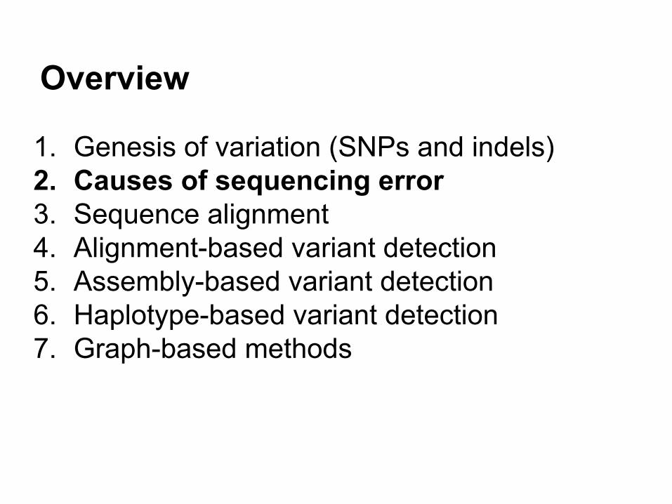
Overview
1. Genesis of variation (SNPs and indels)2. Causes of sequencing error3. Sequence alignment4. Alignment-based variant detection5. Assembly-based variant detection6. Haplotype-based variant detection7. Graph-based methods

DNA
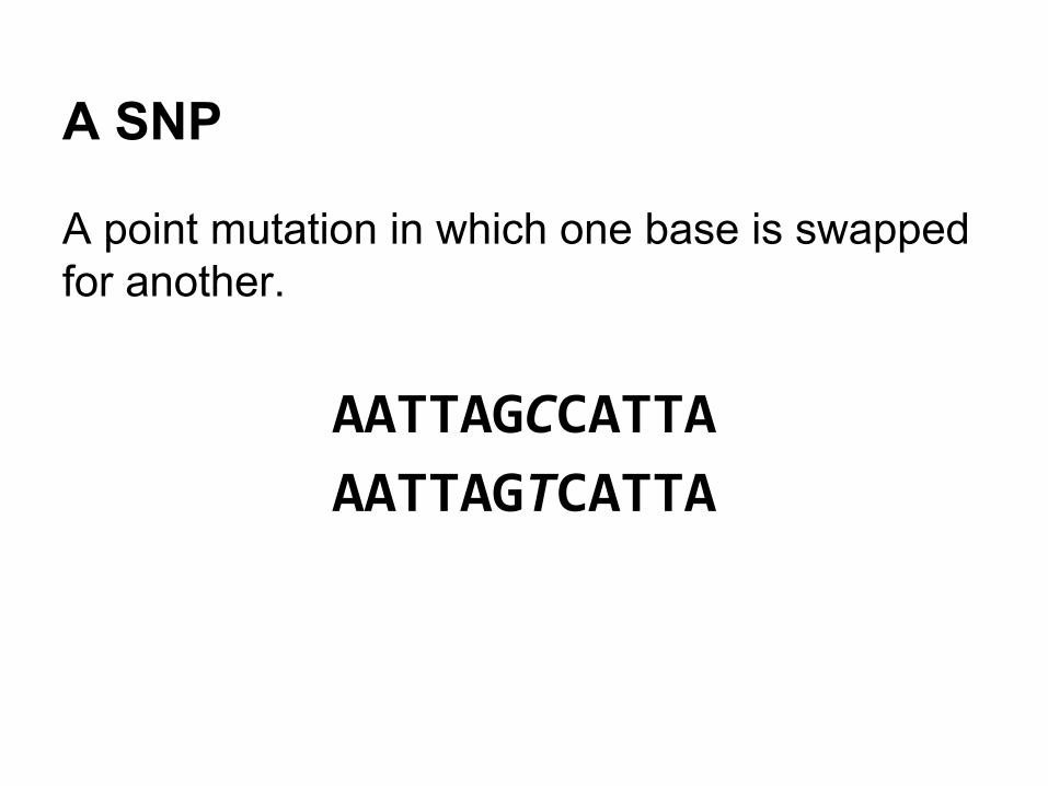
A SNP
A point mutation in which one base is swapped for another.
AATTAGCCATTA
AATTAGTCATTA

(some) causes of SNPs
● Deamination○ cytosine → uracil○ 5-methylcytosine → thymine○ guanine → xanthine (mispairs to A-T bp)○ adenine → hypoxanthine (mispairs to G-C bp)
Deamination of Cytosine to Uracilhttp://en.wikipedia.org/wiki/Deamination

(some) causes of SNPs
● Depurination○ purines are cleaved from DNA sugar backbone
(5000/cell/day, pyrimidines at much lower rate)○ Base excision repair (BEP) can fail → mutation

Multi-base events (MNPs)
● MNPs○ thymine dimerization (UV induced)○ other (e.g. oxidative stress induced)
http://www.rcsb.org/pdb/101/motm.do?momID=91

Transitions and transversionsIn general transitions are 2-3 times more common than transversions. (But this depends on biological context.)
https://upload.wikimedia.org/wikipedia/commons/3/35/Transitions-transversions-v3.png

An INDEL
A mutation that results from the gain or loss of sequence.
AATTAGCCATTA
AATTA--CATTA

INDEL genesis
A number of processes are known to generate insertions and deletions in the process of DNA replication:
● Replication slippage● Double-stranded break repair● Structural variation (e.g. mobile element
insertions, CNVs)

DNA replication
http://www.stanford.edu/group/hopes/cgi-bin/wordpress/2011/02/all-about-mutations/

Polymerase slippage
http://www.stanford.edu/group/hopes/cgi-bin/wordpress/2011/02/all-about-mutations/

NHEJ-derived indels
DNA Slippage Occurs at Microsatellite Loci without Minimal Threshold Length in Humans: A Comparative Genomic Approach. Leclercq S, Rivals E, Jarne P - Genome Biol Evol (2010)

Structural variation (SV)
Transposable elements (in this case, an Alu) are sequences that can copy and paste themselves into genomic DNA, causing insertions.
Deletions can also be mediated by these sequences via other processes.
http://www.nature.com/nrg/journal/v3/n5/full/nrg798.html
Overview
1. Genesis of variation (SNPs and indels)2. Causes of sequencing error3. Sequence alignment4. Alignment-based variant detection5. Assembly-based variant detection6. Haplotype-based variant detection7. Graph-based methods

Sequencingby synthesis (Illumina)
http://bitesizebio.com/13546/sequencing-by-synthesis-explaining-the-illumina-sequencing-technology/

(1)Fluorescently labeled dNTPs with reversible terminators are incorporated by polymerase into growing double-stranded DNA attached to the flowcell surface.
Illumina sequencing process
http://www.ebi.ac.uk/training/online/course/ebi-next-generation-sequencing-practical-course/what-next-generation-dna-sequencing/illumina-

Illumina sequencing process
(2) A round of imaging determines the predominant base in each cluster at the given position.(3) “reverse” terminator by washing, goto (1)(...)Build up images, process to determine sequence
http://www.ebi.ac.uk/training/online/course/ebi-next-generation-sequencing-practical-course/what-next-generation-dna-sequencing/illumina-

http://www.ebi.ac.uk/training/online/course/ebi-next-generation-sequencing-practical-course/what-next-generation-dna-sequencing/illumina-

What can go wrong?
1. input artifacts, problems with library prepa. replication in PCR has no error-correction (→ SNPs)b. no quaternary structures (e.g. clamp) to prevent
slippage (→ indels)c. chimeras…d. duplicates (worse if they are errors)
2. Sequencing-by-synthesisa. phasing of step
i. synthesis reaction efficiency is not 100%ii. particularly bad in A/T homopolymers
b. certain context specific errorsi. vary by sequencing protocol, deviceii. often strand-specific

Context specific errors
Show up as strand-specific errors:
http://www.ncbi.nlm.nih.gov/pmc/articles/PMC3622629/

Context specific errors (motifs)
← forward and reverse error rates for the ten most-common CSEs on a variety of illumina systems
(often GC-rich)
http://www.ncbi.nlm.nih.gov/pmc/articles/PMC3622629/

Overview
1. Genesis of variation (SNPs and indels)2. Causes of sequencing error3. Sequence alignment4. Alignment-based variant detection5. Assembly-based variant detection6. Haplotype-based variant detection7. Graph-based methods

From reads to variants
Genome (FASTA) Variation (VCF)
alignment andvariant calling
Reads (FASTQ)

Global alignment
Idea: localize reads against a large reference system. Requires some cleverness.
Main approaches:1. compressed suffix arrays2. k-mer based seed+extend

Compressed suffix arrays← suffix tree ~ suffix arraysuffix array ~ Burrows-Wheeler transform
BWTs are very compressible: bwa, bowtie, use FM-index (sampled BWT)whole-genome index in 3GB of memory!

k-mer based seed+extend
Used in novoalign, MOSAIK.

Local alignment
Schematic of Needleman and Wunsch algorithm. Smith and Waterman added the 0. http://www.hiv.lanl.gov/content/sequence/HIV/REVIEWS/2006_7/ABECASIS/abecasis.html

Overview
1. Genesis of variation (SNPs and indels)2. Causes of sequencing error3. Sequence alignment4. Alignment-based variant detection5. Assembly-based variant detection6. Haplotype-based variant detection7. Graph-based methods

Alignments to candidatesReference
Reads
Variant observations

The data exposed to the callerReference
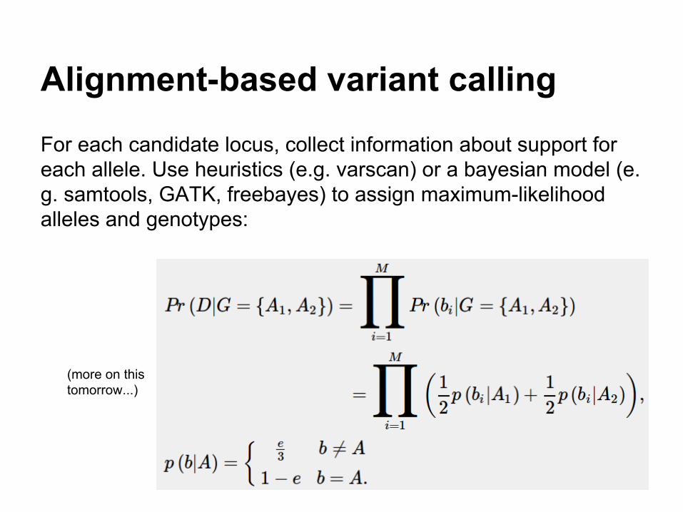
Alignment-based variant calling
For each candidate locus, collect information about support for each allele. Use heuristics (e.g. varscan) or a bayesian model (e.g. samtools, GATK, freebayes) to assign maximum-likelihood alleles and genotypes:
(more on this tomorrow...)

But wait!
If we work directly from our alignments, we may assign too much significance to the particular way our reads were aligned.

Example: calling INDEL variation
Can we quickly design a process to detect indels from alignment data?
What are the steps you’d do to find the indel between these two sequences?

Indel finder
We could start by finding the long matches in both sequences at the start and end:

Indel finder
We can see this more easily like this:
CAAATAAGGTTTGGAGTTCTATTATACAAATAAGGTTTGGAAATTTTCTGGAGTTCTATTATA

Indel finder
The match structure implies that the sequence that doesn’t match was inserted in one sequence, or lost from the other.
CAAATAAGGTTT-----------GGAGTTCTATTATACAAATAAGGTTTGGAAATTTTCTGGAGTTCTATTATA
So that’s easy enough….

Something more complicated
These sequences are similar to the previous ones, but with different mutations between them.
They are still (kinda) homologous but it’s not easy to see.

Pairwise alignment
One solution, assuming a particular set of alignment parameters, has 3 indels and a SNP:
But if we use a higher gap-open penalty, things look different:

Alignment as interpretation
Different parameterizations can yield different results.
Different results suggest “different” variation.
What kind of problems can this cause? (And how can we mitigate these issues?)

INDELs have multiple representations and require normalization for standard calling
Left alignment allows us to ensure that our representation is consistent across alignments and also variant calls.
https://www.biostars.org/p/66843/ user sa9

example: 1000G PhaseI low coveragechr15:81551110, ref:CTCTC alt:ATATA
ref: TGTCACTCGCTCTCTCTCTCTCTCTCTATATATATATATTTGTGCATalt: TGTCACTCGCTCTCTCTCTCTATATATATATATATATATTTGTGCAT
ref: TGTCACTCGCTCTCTCTCTCTCTCTCT------ATATATATATATTTGTGCATalt: TGTCACTCGCTCTCTCTCTCT------ATATATATATATATATATTTGTGCAT
Interpreted as 3 SNPs
Interpreted as microsatellite expansion/contraction
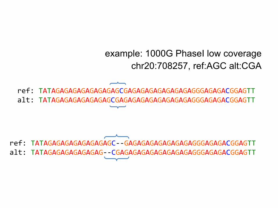
example: 1000G PhaseI low coveragechr20:708257, ref:AGC alt:CGA
ref: TATAGAGAGAGAGAGAGAGCGAGAGAGAGAGAGAGAGGGAGAGACGGAGTTalt: TATAGAGAGAGAGAGAGCGAGAGAGAGAGAGAGAGAGGGAGAGACGGAGTT
ref: TATAGAGAGAGAGAGAGAGC--GAGAGAGAGAGAGAGAGGGAGAGACGGAGTTalt: TATAGAGAGAGAGAGAG--CGAGAGAGAGAGAGAGAGAGGGAGAGACGGAGTT

Overview
1. Genesis of variation (SNPs and indels)2. Causes of sequencing error3. Sequence alignment4. Alignment-based variant detection5. Assembly-based variant detection6. Haplotype-based variant detection7. Graph-based methods

Problem: inconsistent variant representation makes alignment-based variant calling difficult
If alleles are represented in multiple ways, then to detect them correctly with a single-position based approach we need:1. An awesome normalization method2. Perfectly consistent filtering (so we represent
our entire context correctly in the calls)3. Highly-accurate reads

Solution: assembly and haplotype-driven detection
We can shift our focus from the specific interpretation in the alignments:- this is a SNP- whereas this is a series of indels… and instead focus on the underlying sequences.
Basically, we use the alignments to localize reads, then process them again with assembly approaches to determine candidate alleles.

Variant detection by assembly
Multiple methods have been developed by members of the 1000G analysis group:● Global joint assembly
○ cortex○ SGA (localized to 5 megabase chunks)
● Local assembly○ Platypus (+cortex)○ GATK HaplotypeCaller
● k-mer based detection○ FreeBayes (anchored reference-free windows)

de Bruijn Assembly
http://www.nature.com/nrmicro/journal/v7/n4/full/nrmicro2088.html

Using colored graphs (Cortex)
Variants can be called using bubbles in deBruijn graphs.
Method is completely reference-free, except for reporting of variants. The reference is threaded through the colored graph.
Many samples can be called at the same time.
from Iqbal et. al., "De novo assembly and genotyping of variants using colored de Bruijn graphs." (2012)

String graphs (SGA)
http://www.homolog.us/blogs/2012/02/13/string-graph-of-a-genome/
A string graph has the advantage of using less memory to represent an assembly than a de Bruijn graph. In the 1000G, SGA is run on alignments localized to ~5mb chunks.

Discovering alleles using graphs (GATK HaplotypeCaller)

Why don't we just assemble?
Assembly-based calls tend to have high specificity, but sensitivity suffers.
from Iqbal et. al., "De novo assembly and genotyping of variants using colored de Bruijn graphs." (2012)
The requirement of exact kmer matches means that errors disrupt coverage of alleles.
Existing assembly methods don't just detect point mutations--- they detect haplotypes.

Indel validation, 191 AFR samples
High-depth miSeq sequencing-based validation on 4 samples.Local assembly methods (BI2, BC, SI1)* have higher specificity than baseline mapping-based calls (BI1), but lower sensitivity. Global assembly (OX2) yielded very low error, but also low sensitivity.*The local assembly-based method Platypus (OX1) had a genotyping bug which caused poor performance.

Site-frequency spectrum, SNPs
phase3-like setchr20
*RepeatSeq is a microsatellite caller

Site-frequency spectrum, indels
phase3-like set chr20

Overview
1. Genesis of variation (SNPs and indels)2. Causes of sequencing error3. Sequence alignment4. Alignment-based variant detection5. Assembly-based variant detection6. Haplotype-based variant detection7. Graph-based methods

Finding haplotype polymorphisms
AGAACCCAGTGCTCTTTCTGCT
AGAACCCAGTGGTCTTTCTGCT
AGAACCCAGTG TCTTTCTGCTCG
AGAACCCAGTGCTCTATCTGCT
AGAACCCAGTG TCTGCTCTCTAGTCTT
Two reads
Their alignment
a SNP
Another readshowing a SNP on the same haplotype as the first
A variant locus implied by alignments

Direct detection of haplotypes
Detection window
Reference
Reads
Direct detection of haplotypes from reads resolves differentially-represented alleles (as the sequence is compared, not the alignment).
Allele detection is still alignment-driven.


Why haplotypes?
- Variants cluster.- This has functional significance.- Observing haplotypes lets us be more
certain of the local structure of the genome.- We can improve the detection process itself
by using haplotypes rather than point mutations.
- We get the sensitivity of alignment-based approaches with the specificity of assembly-based ones.

Sequence variants cluster
In ~1000 individuals, ½ of variants are within ~22bp of another variant.
Variance to mean ratio (VMR) = 1.4.

The functional effect of variants depends on other nearby variants on the same haplotype
AGG GAG CTGArg Glu Leu
reference:
AGG TAG CTGArg Ter ---
apparent:
AGG TTG CTGArg Leu Leu
actual:
OTOF gene – mutations cause profound recessive deafness
Apparent nonsense variant, one YRI homozygote
Actually a block substitution that results in a missense substitution
(Daniel MacArthur)

Importance of haplotype effects: frame-restoring indels
● Two apparent frameshift deletions in the CASP8AP2 gene (one 17 bp, one 1 bp) on the same haplotype
● Overall effect is in-frame deletion of six amino acids
(Daniel MacArthur)
(in NA12878)

Frame-restoring indels in1000 Genomes Phase I exomes
chr6:117113761, GPRC6A (~10% AF in 1000G)
chr6:32551935, HLA-DRB1 (~11% AF in 1000G)
ref: ATTGTAATTCTCA--TA--TT--TGCCTTTGAAAGCalt: ATTGTAATTCTCAGGTAATTTCCTGCCTTTGAAAGC
ref: CCACCGCGGCCCGCGCCTG-C-TCCAGGATGTCCAlt: CCACCGCGG--CGCGCCTGTCTTCCAGGAGGTCC

Impact on genotyping chip design
monomorphic loci
● Biallelic SNPs detected during the 1000 Genomes Pilot project were used to design a genotyping microarray (Omni 2.5).
● When the 1000 Genomes samples were genotyped using the chip, 100k of the 2.5 million loci showed no polymorphism (monomorphs).


Measuring haplotypes improves specificity
Indels from AFR191 sample set, 1000G phase2 testing.
Excess of 1bp insertions is driven by bubble artifacts in sequencing.

2bp MNPs and dinucleotide intermediates
reference
alte
rnat
e

Direct detection of haplotypes can remove directional bias associated with
alignment-based detection
CAGT
CGGC
TGAC
TGAC
CGGC
CAGT
CA/TG TG/CA
A→G transition to CpG intermediate
Deamination of methyl-C to T
Same process on opposite strand

Overview
1. Genesis of variation (SNPs and indels)2. Causes of sequencing error3. Sequence alignment4. Alignment-based variant detection5. Assembly-based variant detection6. Haplotype-based variant detection7. Graph-based methods

Graph-based methods
1. Variation + sequence = variation graphs2. Local realignment to a variation graph3. Constructing a whole genome variation
graph4. Aligning to a whole genome variation graph

(1) Variation + sequence
= variation graph

Most SNPs (>99%) are now known

Current variant detection is de novo
Genome (FASTA) Variation (VCF)
alignment andvariant calling
Reads (FASTQ)
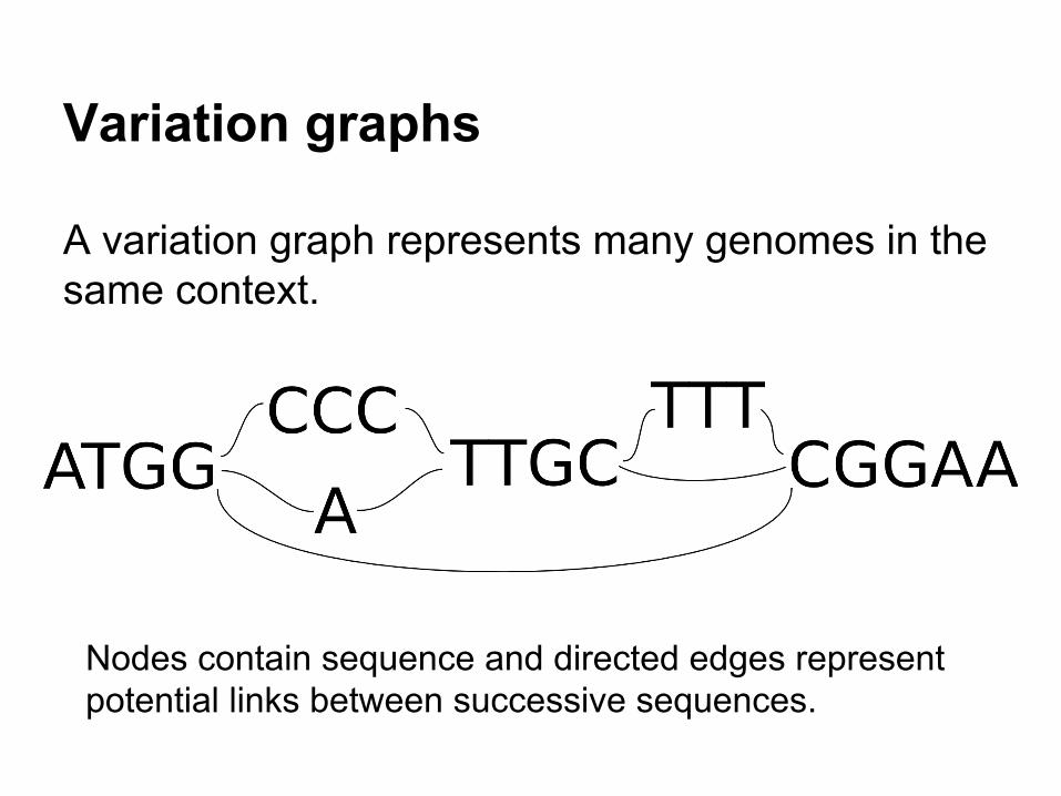
Variation graphs
A variation graph represents many genomes in the same context.
Nodes contain sequence and directed edges represent potential links between successive sequences.

A multiple sequence alignmentis a variation graph
Christopher Lee, Catherine Grasso, Mark F. Sharlow. Multiple sequence alignment using partial order graphs. Bioinformatics, 2002.
traditional MSA
consensus sequence
positionally-matching regions aligned

Assembly graphsare variation graphs
Eugene Myers. The fragment assembly string graph. Bioinformatics, 2005.
http://plus.maths.org/content/os/issue55/features/sequencing/index, credit Daniel Zerbino
A de Bruijn graph can be converted into a variation graph (as previous) by setting node sequences to the first letter in the kmer and compressing non-branching runs into single nodes.
A string graph follows the same format as the variation graph.

(2) Local realignment toa variation graph

Local alignment against a graph
Christopher Lee, Catherine Grasso, Mark F. Sharlow. Multiple sequence alignment using partial order graphs. Bioinformatics, 2002.

Seeding graph-based alignments
linear reference x x x
? ?
graph reference
x x x
Test imperfectly-mapped reads against graph.

Detecting variation on the variant graph
G
A
AGCCTA
AGTACGTAGCT CCTATG GGCCAG
read supporting variant allele
agtacg ggccag
read supporting reference alleleagtacg cctatg ggtcag
Detecting variation on the graph

realigned to variation graph
raw alignments

realignment to variation graph reduces reference bias
Standard alignment is frustrated even by small variants
Ratio between observations with and without realignment to graph of union variants
Improvement in observation support
dens
ity
(tested against 1mb segment on chr20 using 1000Gp3 union allele list)

(3) Constructing a whole genome variation graph

Objective
Construct a whole genome variation graph to serve as our reference for sequence analysis.
We’ll need:(1) a data model(2) a serialization format(3) an API (construct, align, view, …)

Conceptual model
We have sequences (nodes), and allowed linkages between them (edges).
Paths (walks through the graph) are also useful.

Data modelUse protocol buffers to define objects
Graph, Node, Edge, Path, Alignment, Edit
message Node { optional string sequence = 1; optional string name = 2; required int64 id = 3; optional double quality = 4; optional int32 coverage = 5; optional bytes data = 6;}
message Graph { repeated Node node = 1; repeated Edge edge = 2; repeated Path path = 3;}
message Edge { required int64 from = 1; required int64 to = 2; optional bytes data = 3;}
message Path { optional int32 target_position = 1; optional string name = 2; repeated Mapping mapping = 3;}
message Mapping { required int64 node_id = 1; repeated Edit edit = 2;}

Format
Graph
Edges Nodes
Paths
MappingsMappingsMappingsMappingsMappings
Basic entity is a Graph, which is composed of nodes, edges, and paths.

Serialization
To serialize the graph, we generate a stream of sub-graphs that can be reassembled into the whole.
Graph
Edges Nodes
Paths
Mappings
Subgraph n
Edges Nodes
Paths
Mappings
in memory serialized stream
output
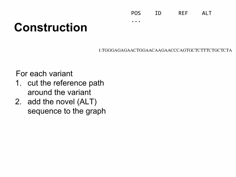
ConstructionPOS ID REF ALT...
For each variant1. cut the reference path
around the variant2. add the novel (ALT)
sequence to the graph

ConstructionPOS ID REF ALT10 . A T...
For each variant1. cut the reference path
around the variant2. add the novel (ALT)
sequence to the graph

ConstructionPOS ID REF ALT10 . A T21 . A ATTAAGA...
For each variant1. cut the reference path
around the variant2. add the novel (ALT)
sequence to the graph

ConstructionPOS ID REF ALT10 . A T21 . A ATTAAGA31 . TCTTT T
For each variant1. cut the reference path
around the variant2. add the novel (ALT)
sequence to the graph

Indexing
Instead of indexing the serialized graph representation (e.g. BAM, VCF), write the graph into a disk-backed key/value store:
{"key":"+g+26+n", "value":{"id": 26, "sequence": "TATTTGAAGT"}}{"key":"+g+26+p+1+103", "value":{"node_id": 26, "edit": []}}{"key":"+g+26+t+24", "value":{"from": 24, "to": 26}}{"key":"+g+26+t+25", "value":{"from": 25, "to": 26}}{"key":"+g+27+f+29", "value":{"from": 27, "to": 29}}…{"key":"+k+TCATACTACTG+69", "value":-2}{"key":"+k+TCATACTACTG+70", "value":-4}{"key":"+k+TCATACTACTG+72", "value":-5}{"key":"+k+TCATATGTCCA+167", "value":29}{"key":"+k+TCATATGTCCA+169", "value":-1}{"key":"+k+TCATATGTCCA+171", "value":-2}...
This allows our graphs and kmer indexes to have > main memory size. (Uses rocksdb.)

Command-line APIusage: vg <command> [options]
commands:
-- construct graph construction
-- view format conversions for graphs and alignments
-- index index features of the graph in a disk-backed key/value store
-- find use an index to find nodes, edges, kmers, or positions
-- paths traverse paths in the graph
-- align local alignment
-- map global alignment
-- stats metrics describing graph properties
-- join combine graphs via a new head
-- ids manipulate node ids
-- concat concatenate graphs tail-to-head
-- kmers enumerate kmers of the graph
-- sim simulate reads from the graph
-- mod filter and transform the graph
-- surject map alignments onto specific paths

(4) Aligning to a whole genome variation graph

Constructing a whole human genome variation graph
Constructed 1000G phase3 + GRCh37 variation graph using vg construct.
5h20m on 32-core system @Sanger3.07G on disk3.181 Gbp of sequence in graph286 million nodes (11 bp/node)376 million edges (8.5 bp/edge)

Indexing 1000G+GRCh37
for each node:for each k-path extending no more than n edges or k bp away from the node:
→ index the k-mers of the k-path
(Edge bounding limits the k-mer space, which can be very important in real data. See →)
for 27-mers crossing no more than 11 edges:52h on a 32-core host @SangerIndex is 175G and on LustreCan align 70 read/s/CPU with index on disk

Aligning to 1000G+GRCh37
For each k-mer in the readIf the k-mer is informative
→ sort hits by node idFor each cluster of hits : max(id)-min(id) < M
Get local region of graph from indexJoin the subgraphs togetherAlign to the joined graph using POA → alignmentIf alignment failed, decrease k-mer length or stride
8000 read/s on 32-core machine @Sanger (cached)~ 40 hours for a deep 150x2 Illumina X10 run

read: AGCTCTCCTTGTCCCTCCTACGATCTCTTCACTGGCCTCTTATCTTTACTGTTACCAAATCTTTCCGGAAGCTGCTCTTTCCCTCAAT
find k-mer subgraphs
read
k-mers
node ids
hit clusterscluster ids
target subgraphpartial order
alignment
Alignment
k-mer based alignment of short reads to a variation graph

Thanks!Gabor MarthDeniz KuralWan-Ping LeeAlistair WardRichard DurbinJosh RandallBenedict PattonZamin IqbalJerome KelleherJared SimpsonDavid HausslerDaniel ZerbinoJouni Siren
Related Documents


![Inexpensive Multiplexed Library Preparation for Megabase-Sized … · 2015-01-16 · [8,9]. With current technology, hundreds of full megabase-size genomes can be sequenced in a single](https://static.cupdf.com/doc/110x72/5fb486a471023d674e295320/inexpensive-multiplexed-library-preparation-for-megabase-sized-2015-01-16-89.jpg)


