Original Research Articles Hantaviruses Induce Antiviral and Pro-Inflammatory Innate Immune Responses in Astrocytic Cells and the Brain Ok Sarah Shin, 1,2 Gabriella Shinyoung Song, 2 Mukesh Kumar, 3 Richard Yanagihara, 3 Ho-Wang Lee, 2 and Jin-Won Song 2 Abstract Although hantaviruses are not generally considered neurotropic, neurological complications have been reported occasionally in patients with hemorrhagic fever renal syndrome (HFRS). In this study, we analyzed innate immune responses to hantavirus infection in vitro in human astrocytic cells (A172) and in vivo in suckling ICR mice. Infection of A172 cells with pathogenic Hantaan virus (HTNV) or a novel shrew-borne hantavirus, known as Imjin virus (MJNV), induced activation of antiviral genes and pro-inflammatory cytokines/chemokines. MicroRNA expression profiles of HTNV- and MJNV-infected A172 cells showed distinct changes in a set of miRNAs. Following intraperitoneal inoculation with HTNV or MJNV, suckling ICR mice developed rapidly progressive, fatal central nervous system-associated disease. Immunohistochemical staining of virus-infected mouse brains confirmed the detection of viral antigens within astrocytes. Taken together, these findings suggest that the neurological findings in HFRS patients may be associated with hantavirus-directed modulation of innate immune responses in the brain. Introduction T he seminal discovery of Hantaan virus (HTNV), as the prototype virus of hemorrhagic fever with renal syn- drome (HFRS), in the striped-field mouse (Apodemus agrarius) in Korea in 1976 (37), led to the detection of several other HFRS-causing hantavirus species. These include Seoul virus (SEOV) (12,23,32,36,45), Puumala virus (PUUV) (11,42), Dobrava/Belgrade virus (DOBV) (7,8,33,34,44), and Amur/ Soochong virus (AMRV/SOOV) (9,26). The discovery of HTNV also made possible the identification of hantaviruses such as Sin Nombre virus (SNV), Black Creek Canal virus (BCCV), and Andes virus (ANDV), harbored by sigmodontine and neotomine rodent species, which cause hantavirus cardio- pulmonary syndrome (HCPS) in the Americas (39,43,51). Recently, the reservoir host range has been expanded with the discovery of highly divergent lineages of hantaviruses in shrews (2,3,5,28,30,52–54), moles (4,27–29) and bats (57,58), but their pathogenic potential in humans is not known. HFRS-associated hantaviruses cause vascular leakage and renal dysfunction, whereas HCPS-associated hantaviruses cause acute pulmonary edema (15,47). The mechanisms of hantavirus-induced pathogenesis are not fully understood, but both HFRS and HCPS share several clinical and path- ologic features, including high fever, thrombocytopenia, increased capillary permeability and upregulation of tumor necrosis factor-a (TNF-a). Recent reports indicate less well- studied complications of HFRS and HCPS, such as cen- tral nervous system (CNS) dysfunction (10,19–22,24,55). However, hantaviral antigens have not been detected in the brains of patients, so the mechanisms by which hantavirus infection might lead to neurological and neuropsychological complications are almost entirely unknown. Both antiviral and pro-inflammatory responses to hanta- virus infection play an important role in the modulation of host defense and disease manifestation (1,6,18,25,38,46,48). Pathogenic rodent-borne hantaviruses, such as HTNV, evade early innate immune responses by downregulating antiviral responsive gene expression immediately after infection. By contrast, upregulation of antiviral gene expression is observed in nonpathogenic Prospect Hill virus (PHV) infection (35). The pathogenesis of HFRS and HCPS is hypothesized to be mediated by increased pro-inflammatory responses (46). TNF-a, interleukin-1b (IL-1b), interleukin-6 (IL-6), chemo- kine (C-C motif ) ligand-5 (CCL-5), C-X-C motif ligand-8 (CXCL-8), and C-X-C motif ligand-10 (CXCL-10) are ele- vated in the kidneys and lungs of HFRS and HCPS patients, respectively, and in hantavirus-infected endothelial cells, macrophages, or epithelial cells in vitro (17,40,41,50). Al- though the major target organs of HFRS and HCPS are the 1 Department of Biomedical Sciences, College of Medicine, Korea University, Gurodong, Gurogu, Seoul, Republic of Korea. 2 Department of Microbiology, College of Medicine, Korea University, Seoul, Republic of Korea. 3 Pacific Center for Emerging Infectious Diseases Research, John A. Burns School of Medicine, University of Hawaii at Manoa, Honolulu, Hawaii. VIRAL IMMUNOLOGY Volume 27, Number 6, 2014 ª Mary Ann Liebert, Inc. Pp. 256–266 DOI: 10.1089/vim.2014.0019 256

Welcome message from author
This document is posted to help you gain knowledge. Please leave a comment to let me know what you think about it! Share it to your friends and learn new things together.
Transcript
Original Research Articles
Hantaviruses Induce Antiviral and Pro-Inflammatory InnateImmune Responses in Astrocytic Cells and the Brain
Ok Sarah Shin,1,2 Gabriella Shinyoung Song,2 Mukesh Kumar,3
Richard Yanagihara,3 Ho-Wang Lee,2 and Jin-Won Song2
Abstract
Although hantaviruses are not generally considered neurotropic, neurological complications have been reportedoccasionally in patients with hemorrhagic fever renal syndrome (HFRS). In this study, we analyzed innate immuneresponses to hantavirus infection in vitro in human astrocytic cells (A172) and in vivo in suckling ICR mice.Infection of A172 cells with pathogenic Hantaan virus (HTNV) or a novel shrew-borne hantavirus, known as Imjinvirus (MJNV), induced activation of antiviral genes and pro-inflammatory cytokines/chemokines. MicroRNAexpression profiles of HTNV- and MJNV-infected A172 cells showed distinct changes in a set of miRNAs.Following intraperitoneal inoculation with HTNV or MJNV, suckling ICR mice developed rapidly progressive, fatalcentral nervous system-associated disease. Immunohistochemical staining of virus-infected mouse brains confirmedthe detection of viral antigens within astrocytes. Taken together, these findings suggest that the neurological findingsin HFRS patients may be associated with hantavirus-directed modulation of innate immune responses in the brain.
Introduction
The seminal discovery of Hantaan virus (HTNV), asthe prototype virus of hemorrhagic fever with renal syn-
drome (HFRS), in the striped-field mouse (Apodemus agrarius)in Korea in 1976 (37), led to the detection of several otherHFRS-causing hantavirus species. These include Seoul virus(SEOV) (12,23,32,36,45), Puumala virus (PUUV) (11,42),Dobrava/Belgrade virus (DOBV) (7,8,33,34,44), and Amur/Soochong virus (AMRV/SOOV) (9,26). The discovery ofHTNV also made possible the identification of hantavirusessuch as Sin Nombre virus (SNV), Black Creek Canal virus(BCCV), and Andes virus (ANDV), harbored by sigmodontineand neotomine rodent species, which cause hantavirus cardio-pulmonary syndrome (HCPS) in the Americas (39,43,51).Recently, the reservoir host range has been expanded with thediscovery of highly divergent lineages of hantaviruses inshrews (2,3,5,28,30,52–54), moles (4,27–29) and bats (57,58),but their pathogenic potential in humans is not known.
HFRS-associated hantaviruses cause vascular leakage andrenal dysfunction, whereas HCPS-associated hantavirusescause acute pulmonary edema (15,47). The mechanisms ofhantavirus-induced pathogenesis are not fully understood,but both HFRS and HCPS share several clinical and path-ologic features, including high fever, thrombocytopenia,
increased capillary permeability and upregulation of tumornecrosis factor-a (TNF-a). Recent reports indicate less well-studied complications of HFRS and HCPS, such as cen-tral nervous system (CNS) dysfunction (10,19–22,24,55).However, hantaviral antigens have not been detected in thebrains of patients, so the mechanisms by which hantavirusinfection might lead to neurological and neuropsychologicalcomplications are almost entirely unknown.
Both antiviral and pro-inflammatory responses to hanta-virus infection play an important role in the modulation ofhost defense and disease manifestation (1,6,18,25,38,46,48).Pathogenic rodent-borne hantaviruses, such as HTNV, evadeearly innate immune responses by downregulating antiviralresponsive gene expression immediately after infection. Bycontrast, upregulation of antiviral gene expression is observedin nonpathogenic Prospect Hill virus (PHV) infection (35).The pathogenesis of HFRS and HCPS is hypothesized to bemediated by increased pro-inflammatory responses (46).TNF-a, interleukin-1b (IL-1b), interleukin-6 (IL-6), chemo-kine (C-C motif ) ligand-5 (CCL-5), C-X-C motif ligand-8(CXCL-8), and C-X-C motif ligand-10 (CXCL-10) are ele-vated in the kidneys and lungs of HFRS and HCPS patients,respectively, and in hantavirus-infected endothelial cells,macrophages, or epithelial cells in vitro (17,40,41,50). Al-though the major target organs of HFRS and HCPS are the
1Department of Biomedical Sciences, College of Medicine, Korea University, Gurodong, Gurogu, Seoul, Republic of Korea.2Department of Microbiology, College of Medicine, Korea University, Seoul, Republic of Korea.3Pacific Center for Emerging Infectious Diseases Research, John A. Burns School of Medicine, University of Hawaii at Manoa,
Honolulu, Hawaii.
VIRAL IMMUNOLOGYVolume 27, Number 6, 2014ª Mary Ann Liebert, Inc.Pp. 256–266DOI: 10.1089/vim.2014.0019
256
kidney and lungs, respectively, the possibility that other or-gans, such as the brain, serve as target sites remains to bedetermined.
We recently determined that a novel shrew-borne hantavi-rus, Imjin virus (MJNV) (23), causes rapid and intense innateimmune responses in human endothelial cells and macro-phages, similar to that of the pathogenic rodent-borne HTNV(50). In this report, we demonstrate that both HTNV andMJNV show similar replication kinetics and efficient viralprotein synthesis in human astrocytic cells. Distinct patterns ofmicroRNA (miRNA) expression were also associated withHTNV and MJNV infection, and the top-predicted gene targetswere found to play a role in inflammatory cytokine and che-mokine pathways and integrin signaling. Moreover, HTNVand MJNV produced profound increases in cytokine andchemokine expression in brains of experimentally infectedmice. Therefore, our data suggest that profound innate immunestimulation may be a potential underlying mechanism for CNSdysfunction in HFRS patients.
Materials and Methods
Cells lines
A human astrocytoma cell line (A172), derived from thebrain tissue of a 53-year-old man with glioblastoma, wasobtained from the Korean Cell Line Bank (Seoul, Korea) andpropagated in RPMI-1640 (Lonza) supplemented with 5%heat-inactivated fetal bovine serum (FBS; Lonza), 10 mMHEPES, 2 mM l-glutamine, and antibiotics (penicillin-streptomycin) at 37�C at 5% CO2. Vero E6 cells (Africangreen monkey kidney epithelial cell line, CRL1586; ATCC)were maintained in Dulbecco’s modified Eagle’s medium(DMEM; Lonza) with 5% FBS, 2 mM l-glutamine, andantibiotics (penicillin-streptomycin) at 37�C at 5% CO2.
Viruses
HTNV strain 76–118 and MJNV strain 04–55 were used.Virus stocks and cells were determined to be free ofMycoplasma contamination by polymerase chain reaction(PCR) analysis. Infectivity titers of virus stocks were mea-sured by plaque assay on Vero E6 cell monolayers, as pre-viously described (48).
Animals and virus inoculation
Pregnant outbred ICR mice (Dbl Laboratory, Korea) weremaintained in the vivarium at Korea University. NewbornICR mice ( < 24 h after birth; 13–15 per group) were inoc-ulated intraperitoneally with 1,000 plaque forming units(PFU) of HTNV or MJNV by the route. Mice were moni-tored daily for clinical signs, and were euthanized if mori-bund or at prescribed time points post-inoculation. TheInstitutional Animal Care and Use Committee approved allexperimental animal protocols.
Virus replication kinetics
A172 cells were inoculated with HTNV and MJNV at amultiplicity of infection (MOI) of 0.5. After adsorption for90 min at 37�C, cell monolayers were washed with phos-phate buffered saline (PBS) and maintained in completemedium. Virus infectivity titers of supernatants were de-termined on each post-infection day by plaque assay (48).
Immunofluorescence microscopy
A172 cells, seeded on coverslips in 24-well plates, wereinoculated with HTNV or MJNV at a MOI of 0.5 for 90 minat 37�C. Thereafter, fresh cell culture medium was addedand incubated for 72 h. Slides were prepared as describedpreviously (50) and examined by fluorescence microscopy,using a Zeiss Axioplan 2 microscope. Images were capturedwith a digital CCD camera (Hamamatsu).
Western blot analysis
Protein lysates were prepared as described previously(50). Briefly, HTNV- and MJNV-infected A172 cell lysateswere separated on 10% SDS-PAGE and blotted onto PVDFmembranes. HTNV and MJNV N proteins were detectedwith mouse monoclonal N protein-specific antibodies followedby anti-mouse horseradish peroxidase (HRP)-conjugatedsecondary antibody.
Quantitative real time reverse transcription PCR
RNA isolation and cDNA synthesis were described previ-ously (50). Briefly, total RNA (1 lg), isolated with Trizol re-agent (Invitrogen), was reverse transcribed using the RT system(Promega) for 1 h at 42�C. The resulting cDNA was used astemplate for real-time PCR quantification. Using the powerSYBR Green supermix (Invitrogen), mRNA expression ofhuman and mouse genes was measured. Human primers werepreviously published (14,50,60), and mouse primers are pro-vided (Supplementary Table S1; Supplementary Data areavailable online at www.liebertpub.com/vim). Cycling pa-rameters were 95�C for 15 min, followed by 40 cycles, with onecycle consisting of 30 sec at 95�C and 1 min at 60�C. Glycer-aldehyde 3-phosphate dehydrogenase (GAPDH) mRNA wasused as an endogenous reference to normalize the quantities oftarget mRNAs and presented as the change in induction relativeto that of untreated control cells. Calculations of expressionwere normalized by using the DCt method, where the amountof target, normalized to an endogenous reference and relative toa calibrator, is given by 2DDCt, where Ct is the cycle number ofthe detection threshold.
Cytokine secretion measurement
Supernatants of HTNV- and MJNV-infected A172 cellswere harvested at indicated times post-infection and storedat - 80�C until testing. Cytokine levels were measured on aMagpix using a human cytokine Milliplex Kit (Millipore).Data analysis was performed with Bio-Plex Manager soft-ware (Bio-Rad Laboratories). The Magpix experiments wereperformed in duplicate.
miRNA microarray analysis
Total RNA was isolated followed by quality checks of bothtotal RNA and small RNA using a 2100 Bioanalyzer andsoftware capable of determining 28S and 18S ribosomal RNAratio, total RNA Integrity Number, and small RNA andmiRNA concentrations. Only samples with adequate totalRNA and miRNA were used in the study. miRNA expressionprofiling was performed using the Agilent Human miRNA 8X 60K (V16). The microarray was designed based on SangermiRBase (release 16.0) and contained probes for 1,205 human
HANTAVIRUS INNATE IMMUNE RESPONSES 257
and 144 human viral miRNAs. One hundred ng of total RNAwas labeled using the Agilent miRNA Complete Labeling andHybridization Kit, according to the manufacturer’s instruc-tions. For target gene functional annotation, we chose to useGO categories, canonical Kyoto Encyclopedia of Genes andGenomes (KEGG) pathways maps, and Panther protein clas-sification tools. Ingenuity Pathways Analysis (IPA; IngenuitySystems, Inc.) was used to analyze miRNA expression data inthe context of known biological response, regulatory net-works, and specific pathways.
Immunohistochemistry
Brain tissues were fixed in 4% paraformaldehyde andembedded in paraffin. Paraffin-embedded, 5 lm thick sectionswere deparaffinized with xylol, rehydrated using decreasingconcentrations of ethanol (100%, 95%, 80%, and 70% for5 min each). Following antigen retrieval in 10 mM sodiumcitrate buffer, pH 6.0 at 95–100�C for 20 min, peroxide-blocking solution was applied to slides for 20 min at roomtemperature. Antigen detection was performed using anti-HTNV and anti-MJNV monoclonal mouse IgG antibodiesfollowed by antibody enhancer and polymer AP (GBI Polink-2 Plus kits; GBI Lab); subsequent color development wasperformed with AP + red (GBI Labs). For double staining ofglial fibrillary acidic protein (GFAP), preblocking solution(10% normal goat serum) was applied to the slides and in-
cubated for 10 min at room temperature. Anti-GFAP primaryantibody, at the recommended concentration (1:500; Milli-pore), was incubated for 2 h at room temperature. Slides werewashed in 1 · TBST four times for 5 min and antibody en-hancer and polymer HRP detection kit was applied (GBIPolink-2 Plus kits). After washing the slides four times, colordevelopment was performed with Emerald Green (GBI Labs).The slides were mounted with aqueous mounting media.
Statistical analysis
The paired differences of the experimental groups werecompared using the nonparametric Mann–Whitney U-test.A p-value of < 0.05 was considered statistically significant(Graph-Pad; Prism software).
Results
Replication kinetics of HTNV and MJNV in A172human astrocytic cells
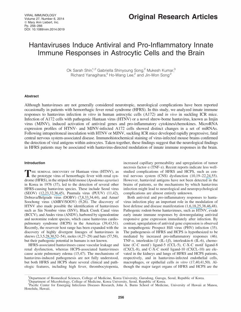
To study the immunological responses in vitro, we firstdetermined if the A172 human astrocytic cell line could beinfected with hantaviruses. Virus-specific fluorescence wasdetected in A172 cells at 72 h following HTNV or MJNVinfection at a MOI of 0.5 (Fig. 1A). Infectivity titers of HTNVand MJNV in A172 cells, at a MOI of 0.5, attained 103–104
PFU/mL at day 2 and persisted until day 5 post-infection
FIG. 1. Hantavirus infection in human astrocytic cells. (A) A172 cells, infected with HTNV or MJNV at a multiplicity ofinfection (MOI) of 0.5 and incubated for 72 h, were stained by the IFA technique, using virus-specific mouse monoclonalantibodies followed by FITC-conjugated anti-mouse IgG antibodies, to detect virus antigen (green). Cell nuclei were stainedwith 4,6-diamidino-2-phenylindole (DAPI, blue). Original magnification, 400 · . (B) Infectivity titers at 1–5 days post-infection were determined by plaque assay in supernatants of A172 cells infected with HTNV or MJNV at a MOI of 0.5.Days post-infection are indicated on the horizontal axis, and titers are represented on a log scale (PFU/mL). Titrations wererepeated twice. (C) Viral proteins at 1–5 days post-infection were determined by Western blot analysis, using mousemonoclonal antibodies against HTNV or MJNV N protein, followed by HRP-conjugated anti-mouse IgG antibodies, inlysates of A172 cells infected with HTNV or MJNV at a MOI of 0.5. Comparable loading of proteins was verified by re-probing the blots with an antibody specific for the housekeeping gene product, b-actin. The Western blots are representativeof three independent experiments. HTNV, Hantaan virus; MJNV, Imjin virus.
258 SHIN ET AL.
(Fig. 1B). Nucleocapsid (N) protein synthesis in HTNV-and MJNV-infected A172 cells, as measured by Westernblot analysis, was elevated early and persisted until day 5post-infection (Fig. 1C).
Antiviral and pro-inflammatory responsesin HTNV- and MJNV-infected A172 cells
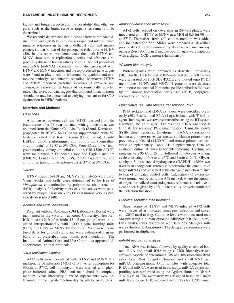
To determine if astrocytes can induce antiviral responsesto hantavirus infection, transcripts for myxovirus resistance
gene A (MxA), IFN-b, IL-28 (IFN-k2), and IL-29 (IFN-k1)were quantitated by real-time reverse transcription PCR(qRT-PCR) at 1, 3, and 5 days post-infection. MxA andIFN-b gene expression peaked at day 3 post-infection, anddecreased at day 5 post-infection in cells infected withHTNV or MJNV (Fig. 2A and B). In contrast to the lowexpression levels of MxA and IFN-b at day 1 post-infection,the induction of type III IFN (both IL-28 and IL-29) pre-ceded induction of MxA and IFN-b by 2 days (Fig. 2C andD). Thus, these data indicate that type III IFN (IFN-k)
FIG. 2. Kinetics of antiviral and pro-inflammatory gene expression during hantavirus infection. A172 cells were infectedwith HTNV (black) or MJNV (light grey) (MOI of 0.5) or mock infected. RNA was extracted at days 1, 3, and 5 post-infection.The mRNA expression of (A) MxA, (B) IFN-b, (C) IL-28, (D) IL-29, (E) IP-10, (F) RANTES, (G) VEGF, and (H) IL-8 inA172 cells treated with media was arbitrarily set to 1 and relative expression is shown in the graph. Expression of target geneswas normalized to that of b-actin. The quantitative real-time reverse transcription polymerase chain reaction (qRT-PCR)experiments were performed in duplicate, and the results are representative of at least three independent experiments.
HANTAVIRUS INNATE IMMUNE RESPONSES 259
expression is induced prior to type I IFN (MxA and IFN-b)during HTNV and MJNV infection.
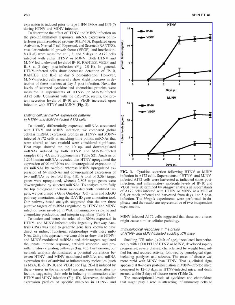
To determine the effect of HTNV and MJNV infection onthe pro-inflammatory responses, mRNA expression of in-terferon gamma-induced protein-10 (IP-10), Regulated uponActivation, Normal T-cell Expressed, and Secreted (RANTES),vascular endothelial growth factor (VEGF), and interleukin-8 (IL-8) were measured at 1, 3, and 5 days in A172 cellsinfected with either HTNV or MJNV. Both HTNV andMJNV led to elevated levels of IP-10, RANTES, VEGF, andIL-8 at 3 days post-infection (Fig. 2E–H). In general,HTNV-infected cells show decreased detection of IP-10,RANTES, and IL-8 at day 5 post-infection. However,MJNV-infected cells generally show slight increases in de-tection of these markers at day 5 post-infection. Next, thelevels of secreted cytokine and chemokine proteins weremeasured in supernatants of HTNV- or MJNV-infectedA172 cells. Consistent with the qRT-PCR results, the pro-tein secretion levels of IP-10 and VEGF increased uponinfection with HTNV and MJNV (Fig. 3).
Distinct cellular miRNA expression patternsin HTNV- and MJNV-infected A172 cells
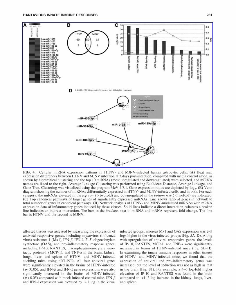
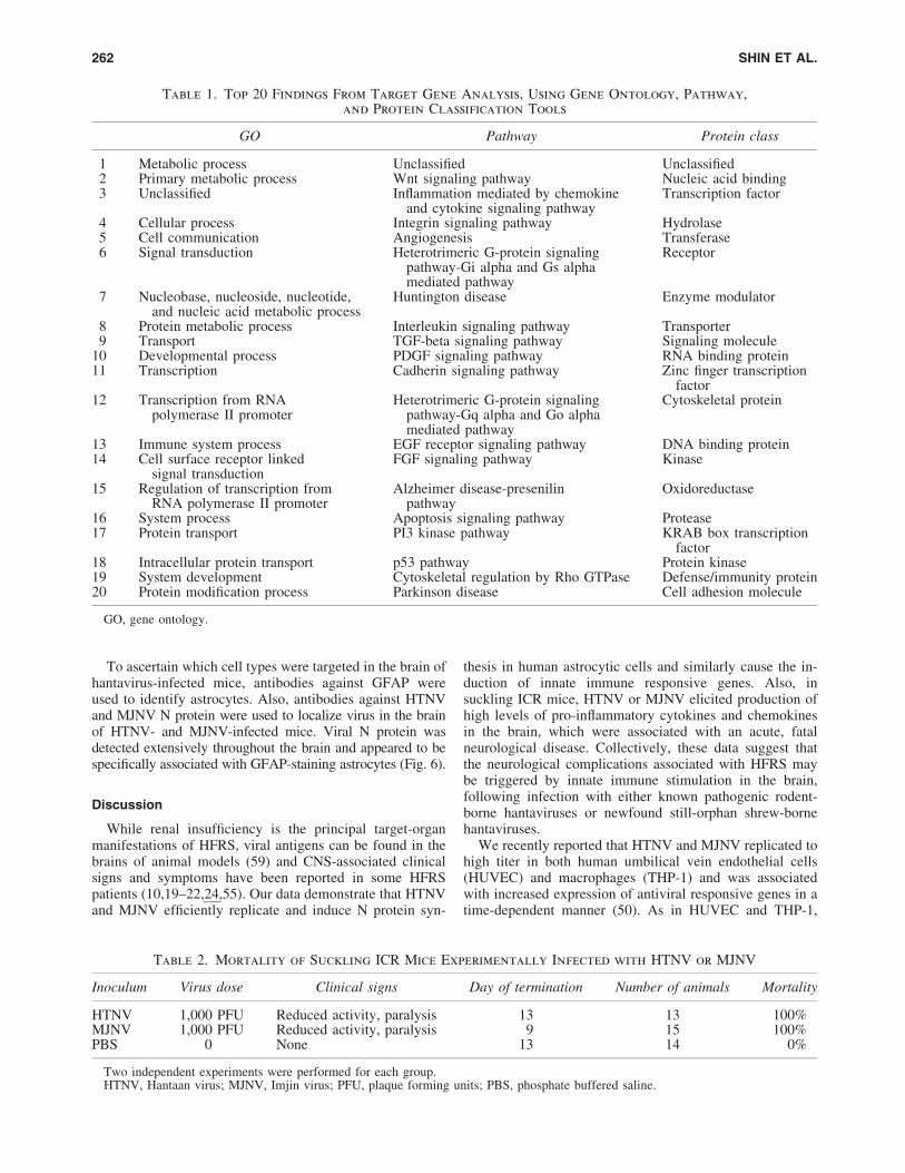
To identify differentially expressed miRNAs associatedwith HTNV and MJNV infection, we compared globalcellular miRNA expression profiles in HTNV- and MJNV-infected A172 cells at matching time points. miRNAs thatwere altered at least twofold were considered significant.Heat maps showed the top 10 up- and downregulatedmiRNAs induced by both HTNV and MJNV-infectedsamples (Fig. 4A and Supplementary Table S2). Analysis of1,205 human miRNAs revealed that HTNV upregulated theexpression of 90 miRNAs and downregulated expression ofsix miRNAs by twofold, whereas MJNV upregulated ex-pression of 64 miRNAs and downregulated expression oftwo miRNAs by twofold (Fig. 4B). A total of 3,364 targetgenes were upregulated, whereas 2,803 target genes weredownregulated by selected miRNAs. To analyze more fullythe top biological functions associated with identified tar-gets, we performed a Gene Ontology (GO) term and KEGGpathway annotation, using the DAVID gene annotation tool.Our pathway-based analysis suggested that the top threeputative targets of miRNAs regulated by HTNV and MJNVinfection were involved in Wnt, inflammatory cytokine andchemokine production, and integrin signaling (Table 1).
To understand better the roles of miRNAs expressed inHTNV- and MJNV-infected cells, Ingenuity Pathway Ana-lysis (IPA) was used to generate gene lists known to havedirect or indirect functional relationships with these miR-NAs. Using this approach, we were able to show that HTNV-and MJNV-modulated miRNAs and their targets regulatedthe innate immune response, antiviral response, and pro-inflammatory signaling pathways (Fig. 4C). Furthermore, ournetwork analysis demonstrated a potential correlation be-tween HTNV- and MJNV-modulated miRNAs and mRNAexpression data of antiviral or inflammatory molecules (suchas MxA, IL-8, IP-10, and VEGF; see Fig. 2E–H) induced bythese viruses in the same cell type and same time after in-fection, suggesting their role in inducing inflammation afterHTNV and MJNV infection (Fig. 4D). Moreover, the similarexpression profiles of specific miRNAs in HTNV- and
MJNV-infected A172 cells suggested that these two virusesmight cause similar cellular pathology.
Immunological responses in the brainsof HTNV- and MJNV-infected suckling ICR mice
Suckling ICR mice ( < 24 h of age), inoculated intraperito-neally with 1,000 PFU of HTNV or MJNV, developed rapidlyprogressive, severe disease, characterized by weight loss, ruf-fled fur, and reduced activity, followed by neurological signs,including paralyses and seizures. The onset of disease wasmore rapid with MJNV than HTNV. That is, clinical signsappeared at 8–9 days post-inoculation in MJNV-infected mice,compared to 12–13 days in HTNV-infected mice, and deathensued within 2 days of disease onset (Table 2).
The transcriptional levels of cytokines and chemokinesthat might play a role in attracting inflammatory cells to
FIG. 3. Cytokine secretion following HTNV or MJNVinfection in A172 cells. Supernatants of HTNV- and MJNV-infected A172 cells were harvested at indicated times post-infection, and inflammatory molecule levels of IP-10 andVEGF were determined by Magpix analysis in supernatantsof A172 cells infected with HTNV or MJNV at a MOI of0.5, or mock infected and harvested from days 1 to 5 post-infection. The Magpix experiments were performed in du-plicate, and the results are representative of two independentexperiments.
260 SHIN ET AL.
affected tissues was assessed by measuring the expression ofantiviral responsive genes, including myxovirus (influenzavirus) resistance-1 (Mx1), IFN-b, IFN-k, 2¢-5¢-oligoadenylatesynthetase (OAS), and pro-inflammatory response genes,including IP-10, RANTES, macrophage/monocyte chemo-tactic protein-1 (MCP-1), and TNF-a in the brain, kidney,lungs, liver, and spleen of HTNV- and MJNV-infectedsuckling mice, using qRT-PCR. All four antiviral geneswere significantly elevated in the brains of HTNV-infected( p < 0.05), and IFN-b and IFN-k gene expressions were alsosignificantly increased in the brains of MJNV-infected( p < 0.05) compared with mock-infected control mice. IFN-band IFN-k expression was elevated by *1 log in the virus-
infected groups, whereas Mx1 and OAS expression was 2–3logs higher in the virus-infected groups (Fig. 5A–D). Alongwith upregulation of antiviral responsive genes, the levelsof IP-10, RANTES, MCP-1, and TNF-a were significantlyincreased in brains of HTNV-infected mice (Fig. 5E–H).In examining the innate immune responses in other tissuesof HTNV- and MJNV-infected mice, we found that theexpression of antiviral and pro-inflammatory genes wasincreased, but the level of induction was not as high as thatin the brain (Fig. S1). For example, a 4–6 log-fold higherelevation of IP-10 and RANTES was found in the braincompared to <1–2 log increase in the kidney, lungs, liver,and spleen.
FIG. 4. Cellular miRNA expression patterns in HTNV- and MJNV-infected human astrocytic cells. (A) Heat mapexpression differences between HTNV and MJNV infection at 3 days post-infection, compared with media control alone, asshown by hierarchical clustering and the top 10 miRNAs (most upregulated and downregulated) were selected, and miRNAnames are listed to the right. Average Linkage Clustering was performed using Euclidean Distance, Average Linkage, andGene Tree. Clustering was visualized using the program MeV 4.7.1. Gene expression ratios are depicted by log2. (B) Venndiagram showing the number of miRNAs differentially expressed in HTNV- and MJNV-infected cells, and in both. For eachcategory, the miRNAs elevated in the top row ( > twofold) and downregulated in the bottom row ( < twofold) are indicated.(C) Top canonical pathways of target genes of significantly expressed miRNAs. Line shows ratio of genes in network tototal number of genes in canonical pathways. (D) Network analysis of HTNV- and MJNV-modulated miRNAs with mRNAexpression data of inflammatory genes induced by these viruses. Solid lines indicate a direct interaction, whereas a brokenline indicates an indirect interaction. The bars in the brackets next to miRNA and mRNA represent fold-change. The firstbar is HTNV and the second is MJNV.
HANTAVIRUS INNATE IMMUNE RESPONSES 261
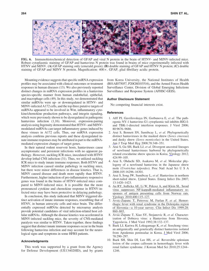
To ascertain which cell types were targeted in the brain ofhantavirus-infected mice, antibodies against GFAP wereused to identify astrocytes. Also, antibodies against HTNVand MJNV N protein were used to localize virus in the brainof HTNV- and MJNV-infected mice. Viral N protein wasdetected extensively throughout the brain and appeared to bespecifically associated with GFAP-staining astrocytes (Fig. 6).
Discussion
While renal insufficiency is the principal target-organmanifestations of HFRS, viral antigens can be found in thebrains of animal models (59) and CNS-associated clinicalsigns and symptoms have been reported in some HFRSpatients (10,19–22,24,55). Our data demonstrate that HTNVand MJNV efficiently replicate and induce N protein syn-
thesis in human astrocytic cells and similarly cause the in-duction of innate immune responsive genes. Also, insuckling ICR mice, HTNV or MJNV elicited production ofhigh levels of pro-inflammatory cytokines and chemokinesin the brain, which were associated with an acute, fatalneurological disease. Collectively, these data suggest thatthe neurological complications associated with HFRS maybe triggered by innate immune stimulation in the brain,following infection with either known pathogenic rodent-borne hantaviruses or newfound still-orphan shrew-bornehantaviruses.
We recently reported that HTNV and MJNV replicated tohigh titer in both human umbilical vein endothelial cells(HUVEC) and macrophages (THP-1) and was associatedwith increased expression of antiviral responsive genes in atime-dependent manner (50). As in HUVEC and THP-1,
Table 1. Top 20 Findings From Target Gene Analysis, Using Gene Ontology, Pathway,
and Protein Classification Tools
GO Pathway Protein class
1 Metabolic process Unclassified Unclassified2 Primary metabolic process Wnt signaling pathway Nucleic acid binding3 Unclassified Inflammation mediated by chemokine
and cytokine signaling pathwayTranscription factor
4 Cellular process Integrin signaling pathway Hydrolase5 Cell communication Angiogenesis Transferase6 Signal transduction Heterotrimeric G-protein signaling
pathway-Gi alpha and Gs alphamediated pathway
Receptor
7 Nucleobase, nucleoside, nucleotide,and nucleic acid metabolic process
Huntington disease Enzyme modulator
8 Protein metabolic process Interleukin signaling pathway Transporter9 Transport TGF-beta signaling pathway Signaling molecule
10 Developmental process PDGF signaling pathway RNA binding protein11 Transcription Cadherin signaling pathway Zinc finger transcription
factor12 Transcription from RNA
polymerase II promoterHeterotrimeric G-protein signaling
pathway-Gq alpha and Go alphamediated pathway
Cytoskeletal protein
13 Immune system process EGF receptor signaling pathway DNA binding protein14 Cell surface receptor linked
signal transductionFGF signaling pathway Kinase
15 Regulation of transcription fromRNA polymerase II promoter
Alzheimer disease-presenilinpathway
Oxidoreductase
16 System process Apoptosis signaling pathway Protease17 Protein transport PI3 kinase pathway KRAB box transcription
factor18 Intracellular protein transport p53 pathway Protein kinase19 System development Cytoskeletal regulation by Rho GTPase Defense/immunity protein20 Protein modification process Parkinson disease Cell adhesion molecule
GO, gene ontology.
Table 2. Mortality of Suckling ICR Mice Experimentally Infected with HTNV or MJNV
Inoculum Virus dose Clinical signs Day of termination Number of animals Mortality
HTNV 1,000 PFU Reduced activity, paralysis 13 13 100%MJNV 1,000 PFU Reduced activity, paralysis 9 15 100%PBS 0 None 13 14 0%
Two independent experiments were performed for each group.HTNV, Hantaan virus; MJNV, Imjin virus; PFU, plaque forming units; PBS, phosphate buffered saline.
262 SHIN ET AL.
HTNV and MJNV replicated in A172 cells with similarkinetics and efficiency. Hantavirus infection in A172 cellswas accompanied by type I and type III IFN responses andincreased production of pro-inflammatory mediators, suchas IP-10, RANTES, and VEGF. It has been previouslydemonstrated that hantaviruses induces the expression oftype I IFN (a/b), type III IFN-k, and interleukin-28 and -29
(IL-28 and IL-29) (56). Type I IFN has been demonstratedto play a central role in the regulation of innate immuneresponses and in the control of hantavirus infection in vitro(1,35). Interestingly, type III IFN was upregulated morerapidly at 1 day post-infection than type I IFN in A172 cells,although this was not sufficient to control viral replication atearly time points.
FIG. 5. Hantavirus-induced antiviral and pro-inflammatory gene expression patterns in vivo. Suckling ICR mice wereinjected intraperitoneally with HTNV or MJNV and sacrificed 9 (MJNV) or 13 (HTNV) days after infection. Total RNAwas extracted from the brain, and the expression of (A) Mx1, (B) IFN-b, (C) IFN-k, (D) OAS, (E) IP-10, (F) RANTES, (G)MCP-1, and (H) TNF-a was examined by qRT-PCR. p-Values at the top of individual graphs are comparisons between mock-infected and virus-infected mice. Expression of target genes was normalized to that of the housekeeping gene, GAPDH. TheqRT-PCR experiments were performed in duplicate and each symbol represents the value for an individual mouse, and the barindicates the mean for the group. An asterisk indicates that the values for the HTNV-infected or MJNV-infected samples weresignificantly different from those in the buffer control samples ( p < 0.05). Kruskal–Wallis test (one way analysis of variance)and Dunn’s Multiple Comparison Test (post-test) were performed. GAPDH, glyceraldehyde 3-phosphate dehydrogenase.
HANTAVIRUS INNATE IMMUNE RESPONSES 263
Mounting evidence suggests that specific miRNA expressionprofiles may be associated with clinical outcomes or treatmentresponses in human diseases (13). We also previously reporteddistinct changes in miRNA expression profiles in a hantavirusspecies-specific manner from human endothelial, epithelial,and macrophage cells (49). In this study, we demonstrated thatsimilar miRNAs were up- or downregulated in HTNV- andMJNV-infected A172 cells, and the top three putative targets ofmiRNAs appeared to be involved in Wnt, inflammatory cyto-kine/chemokine production pathways, and integrin signaling,which were previously shown to be dysregulated in pathogenichantavirus infection (1,16). Moreover, expression-pairinganalysis using Ingenuity demonstrated that HTNV- and MJNV-modulated miRNAs can target inflammatory genes induced bythese viruses in A172 cells. Thus, our miRNA expressionanalysis confirms previous reports and these dysregulated in-nate immune responses may be attributed in part to the miRNA-mediated expression changes of target genes.
In their natural rodent reservoir hosts, hantaviruses causeasymptomatic and persistent infections with no apparent pa-thology. However, suckling ICR mice, inoculated with HTNVdevelop lethal CNS infection (31). Thus, we utilized sucklingICR mice to study innate immune responses. Both HTNV andMJNV infection caused similar pathology in suckling mice,but there were minor differences in disease kinetics. That is,MJNV caused disease and death more rapidly than HTNV.Furthermore, higher induction of pro-inflammatory responsivegenes was found in the brains of HTNV-infected mice com-pared to MJNV-infected mice. It is possible that the morepronounced cytokine and chemokine response in HTNV-in-fected mice may have been protective in delaying pathology.
In conclusion, this study indicated that MJNV triggered dis-tinct activation of innate immune responses, resembling that ofHTNV, in human astrocytic cells and mice brain. The differ-entially expressed miRNAs detected by microarray analysisprovide potential candidates of viral infection-related host cel-lular miRNAs. Although the disease kinetics was accelerated inMJNV-infected suckling mice, the severity of CNS-mediatedparalysis was similar to HTNV. Taken together, these findingssuggest that distinct innate immune responses occur in the brainfollowing hantavirus infection and may account for the neuro-logical signs and symptoms in some HFRS patients.
Acknowledgments
This work was supported by a grant from the Agencyfor Defense Development (UE134020ID), and by grants
from Korea University, the National Institutes of Health(R01AI075057, P20GM103516), and the Armed Forces HealthSurveillance Center, Division of Global Emerging InfectionsSurveillance and Response System (AFHSC-GEIS).
Author Disclosure Statement
No competing financial interests exist.
References
1. Alff PJ, Gavrilovskaya IN, Gorbunova E, et al. The path-ogenic NY-1 hantavirus G1 cytoplasmic tail inhibits RIG-I-and TBK-1-directed interferon responses. J Virol 2006;80:9676–9686.
2. Arai S, Bennett SN, Sumibcay L, et al. Phylogeneticallydistinct hantaviruses in the masked shrew (Sorex cinereus)and dusky shrew (Sorex monticolus) in the United States.Am J Trop Med Hyg 2008;78:348–351.
3. Arai S, Gu SH, Baek LJ, et al. Divergent ancestral lineagesof newfound hantaviruses harbored by phylogeneticallyrelated crocidurine shrew species in Korea. Virology 2012;424:99–105.
4. Arai S, Ohdachi SD, Asakawa M, et al. Molecular phy-logeny of a newfound hantavirus in the Japanese shrewmole (Urotrichus talpoides). Proc Natl Acad Sci U S A2008;105:16296–16301.
5. Arai S, Song JW, Sumibcay L, et al. Hantavirus in northernshort-tailed shrew, United States. Emerg Infect Dis 2007;13:1420–1423.
6. Au RY, Jedlicka AE, Li W, Pekosz A, and Klein SL. Seoulvirus suppresses NF-kappaB-mediated inflammatory re-sponses of antigen presenting cells from Norway rats.Virology 2010;400:115–127.
7. Avsic-Zupanc T, Petrovec M, Furlan P, et al. Hemor-rhagic fever with renal syndrome in the Dolenjska regionof Slovenia—a 10-year survey. Clin Infect Dis 1999;28:860–865.
8. Avsic-Zupanc T, Xiao SY, Stojanovic R, et al. Character-ization of Dobrava virus: a Hantavirus from Slovenia,Yugoslavia. J Med Virol 1992;38:132–137.
9. Baek LJ, Kariwa H, Lokugamage K, et al. Soochong virus:an antigenically and genetically distinct hantavirus isolatedfrom Apodemus peninsulae in Korea. J Med Virol 2006;78:290–297.
10. Baek SH, Shin DI, Lee HS, et al. Reversible spleniumlesion of the corpus callosum in hemorrhagic fever withrenal failure syndrome. J Korean Med Sci 2010;25:1244–1246.
FIG. 6. Immunohistochemical detection of GFAP and viral N protein in the brain of HTNV- and MJNV-infected mice.Robust cytoplasmic staining of GFAP and hantavirus N protein was found in brains of mice experimentally infected withHTNV and MJNV. (A) GFAP staining only (emerald green); (B) double staining of GFAP and HTNV N protein; (C) doublestaining of GFAP and MJNV N protein. Magnification: 400 · . GFAP, glial fibrillary acidic protein.
264 SHIN ET AL.
11. Brummer-Korvenkontio M, Vaheri A, Hovi T, et al. Ne-phropathia epidemica: detection of antigen in bank volesand serologic diagnosis of human infection. J Infect Dis1980;141:131–134.
12. Cueto GR, Cavia R, Bellomo C, Padula PJ, and Suarez OV.Prevalence of hantavirus infection in wild Rattus norvegi-cus and R. rattus populations of Buenos Aires City, Ar-gentina. Trop Med Int Health 2008;13:46–51.
13. Dai R, and Ahmed SA. MicroRNA, a new paradigm forunderstanding immunoregulation, inflammation, and auto-immune diseases. Translat Res 2011;157:163–179.
14. Diegelmann J, Beigel F, Zitzmann K, et al. Comparativeanalysis of the lambda-interferons IL-28A and IL-29 re-garding their transcriptome and their antiviral propertiesagainst hepatitis C virus. PLoS One 2010;5:e15200.
15. Easterbrook JD, and Klein SL. Immunological mechanismsmediating hantavirus persistence in rodent reservoirs. PLoSPathog 2008;4:e1000172.
16. Gavrilovskaya IN, Peresleni T, Geimonen E, andMackow ER. Pathogenic hantaviruses selectively inhibitbeta3 integrin directed endothelial cell migration. ArchVirol 2002;147:1913–1931.
17. Geimonen E, Neff S, Raymond T, et al. Pathogenic andnonpathogenic hantaviruses differentially regulate endo-thelial cell responses. Proc Natl Acad Sci U S A 2002;99:13837–13842.
18. Handke W, Oelschlegel R, Franke R, Kruger DH, and RangA. Hantaan virus triggers TLR3-dependent innate immuneresponses. J Immunol 2009;182:2849–2858.
19. Hautala N, Kauma H, Rajaniemi SM, et al. Signs of generalinflammation and kidney function are associated with theocular features of acute Puumala hantavirus infection.Scand J Infect Dis 2012;44:955–62.
20. Hautala T, Hautala N, Mahonen SM, et al. Young malepatients are at elevated risk of developing serious centralnervous system complications during acute Puumala han-tavirus infection. BMC Infect Dis 2011;11:217.
21. Hautala T, Mahonen SM, Sironen T, et al. Central nervoussystem-related symptoms and findings are common inacute Puumala hantavirus infection. Ann Med 2010;42:344–351.
22. Hautala T, Partanen T, Sironen T, et al. Elevated cerebro-spinal fluid neopterin concentration is associated with dis-ease severity in acute Puumala hantavirus infection. ClinDevel Immunol 2013;2013:634632.
23. Heyman,P, Plyusnina A, Berny P, et al. Seoul hantavirus inEurope: first demonstration of the virus genome in wildRattus norvegicus captured in France. Eur J Clin MicrobiolInfect Dis 2004;23:711–717.
24. Hopkins RO, Larson-Lohr V, Weaver LK, and Bigler ED.Neuropsychological impairments following hantaviruspulmonary syndrome. J Int Neuropsychol Soc 1998;4:190–196.
25. Jiang H, Wang PZ, Zhang Y, et al. Hantaan virus inducestoll-like receptor 4 expression, leading to enhanced pro-duction of beta interferon, interleukin-6 and tumor necrosisfactor-alpha. Virology 2008;380:52–59.
26. Jiang JF, Zhang WY, Wu XM, Zhang PH, and Cao WC.Soochong virus and Amur virus might be the same entitiesof hantavirus. J Med Virol 2007;79:1792–1795.
27. Kang HJ, Bennett SN, Dizney L, et al. Host switch duringevolution of a genetically distinct hantavirus in the Ameri-can shrew mole (Neurotrichus gibbsii). Virology 2009;388:8–14.
28. Kang HJ, Bennett SN, Hope AG, Cook JA, and YanagiharaR. Shared ancestry between a newfound mole-borne han-tavirus and hantaviruses harbored by cricetid rodents. JVirol 2011;85:7496–7503.
29. Kang HJ, Bennett SN, Sumibcay L, et al. Evolutionaryinsights from a genetically divergent hantavirus harboredby the European common mole (Talpa europaea). PLoSOne 2009;4:e6149.
30. Kang HJ, Kadjo B, Dubey S, Jacquet F, and YanagiharaR. Molecular evolution of Azagny virus, a newfoundhantavirus harbored by the West African pygmy shrew(Crocidura obscurior) in Cote d’Ivoire. Virol J 2011;8:373.
31. Kim GR, and McKee KT, Jr. Pathogenesis of Hantaanvirus infection in suckling mice: clinical, virologic, andserologic observations. Am J Trop Med Hyg 1985;34:388–395.
32. Kim YS, Ahn C, Han JS, et al. Hemorrhagic fever withrenal syndrome caused by the Seoul virus. Nephron 1995;71:419–427.
33. Klempa B, Stanko M, Labuda M, et al. Central EuropeanDobrava Hantavirus isolate from a striped field mouse(Apodemus agrarius). J Clin Microbiol 2005;43:2756–2763.
34. Klempa B, Tkachenko EA, Dzagurova TK, et al. Hemor-rhagic fever with renal syndrome caused by 2 lineages ofDobrava hantavirus, Russia. Emerg Infect Dis 2008;14:617–625.
35. Kraus AA, Raftery MJ, Giese T, et al. Differential antiviralresponse of endothelial cells after infection with pathogenicand nonpathogenic hantaviruses. J Virol 2004;78:6143–6150.
36. Lee HW, Baek LJ, and Johnson KM. Isolation of Hantaanvirus, the etiologic agent of Korean hemorrhagic fever,from wild urban rats. J Infect Dis 1982;146:638–644.
37. Lee HW, Lee PW, and Johnson KM. Isolation of the eti-ologic agent of Korean hemorrhagic fever. 1978. J InfectDis 2004;190:1711–1721.
38. Lee MH, Lalwani P, Raftery MJ, et al. RNA helicase re-tinoic acid-inducible gene I as a sensor of Hantaan virusreplication. J Gen Virol 2011;92:2191–2200.
39. Levis S, Morzunov SP, Rowe JE, et al. Genetic diversityand epidemiology of hantaviruses in Argentina. J Infect Dis1998;177:529–538.
40. Markotic A. [Immunopathogenesis of hemorrhagic feverwith renal syndrome and hantavirus pulmonary syndrome].Acta Med Croatica 2003;57:407–414.
41. Mori M, Rothman AL, Kurane I, et al. High levels ofcytokine-producing cells in the lung tissues of patients withfatal hantavirus pulmonary syndrome. J Infect Dis 1999;179:295–302.
42. Mustonen J, Vapalahti O, Henttonen H, Pasternack A, andVaheri A. Epidemiology of hantavirus infections in Europe.Nephrol Dial Transplant 1998;13:2729–2731.
43. Nichol ST, Spiropoulou CF, Morzunov S, et al. Geneticidentification of a hantavirus associated with an outbreak ofacute respiratory illness. Science 1993;262:914–917.
44. Papa A, Johnson AM, Stockton PC, et al. Retrospectiveserological and genetic study of the distribution of hanta-viruses in Greece. J Med Virol 1998;55:321–327.
45. Pilaski J, Ellerich C, Kreutzer T, et al. Haemorrhagic feverwith renal syndrome in Germany. Lancet 1991;337:111.
46. Rang A. Modulation of innate immune responses by han-taviruses. Crit Rev Immunol 2010;30:515–527.
HANTAVIRUS INNATE IMMUNE RESPONSES 265
47. Schonrich G, Rang A, Lutteke N, Raftery MJ, CharbonnelN, and Ulrich RG. Hantavirus-induced immunity in rodentreservoirs and humans. Immunol Rev 2008;225:163–189.
48. Shim SH, Park MS, Moon S, et al. Comparison of innateimmune responses to pathogenic and putative non-pathogenichantaviruses in vitro. Virus Res 2011;160:367–373.
49. Shin OS, Kumar M, Yanagihara R, and Song JW. Hanta-viruses induce cell type- and viral species-specific host mi-croRNA expression signatures. Virology 2013;446:217–224.
50. Shin OS, Yanagihara R, and Song JW. Distinct innateimmune responses in human macrophages and endothelialcells infected with shrew-borne hantaviruses. Virology2012;434:43–49.
51. Song JW, Baek LJ, Gajdusek DC, et al. Isolation of path-ogenic hantavirus from white-footed mouse (Peromyscusleucopus). Lancet 1994;344:1637.
52. Song JW, Gu SH, Bennett SN, et al. Seewis virus, a ge-netically distinct hantavirus in the Eurasian common shrew(Sorex araneus). Virol J 2007;4:114.
53. Song JW, Kang HJ, Gu SH, et al. Characterization of Imjinvirus, a newly isolated hantavirus from the Ussuri white-toothed shrew (Crocidura lasiura). J Virol 2009;83:6184–6191.
54. Song JW, Kang HJ, Song KJ, et al. Newfound hantavirus inChinese mole shrew, Vietnam. Emerg Infect Dis 2007;13:1784–1787.
55. Steiner T. Ettinger J, Peng Z, et al. Hyperintense lesion inthe corpus callosum associated with Puumala hantavirusinfection. J Neurol 2012;259:1742–1745.
56. Stoltz M, and Klingstrom J. Alpha/beta interferon (IFN-alpha/beta)-independent induction of IFN-lambda1 (interleukin-29) in response to Hantaan virus infection. J Virol 2010;84:9140–9148.
57. Sumibcay L, Kadjo B, Gu SH, et al. 2012. Divergent lineageof a novel hantavirus in the banana pipistrelle (Neoromiciananus) in Cote d’Ivoire. Virol J 9:34.
58. Weiss S, Witkowski PT, Auste B, et al. Hantavirus in bat,Sierra Leone. Emerg Infect Dis 2012;18:159–161.
59. Wichmann D, Grone HJ, Frese M, et al. Hantaan virusinfection causes an acute neurological disease that isfatal in adult laboratory mice. J Virol 2002;76:8890–8899.
60. Yin Z, Dai J, Deng J, et al. Type III IFNs Are Produced byand Stimulate Human Plasmacytoid Dendritic Cells. J Im-munol 2012;189:2735–2745.
Address correspondence to:Dr. Jin-Won Song
Department of MicrobiologyCollege of Medicine
Korea University5-ga, Anam-dong
Seongbuk-guSeoul 136-705
Republic of Korea
E-mail: [email protected]
266 SHIN ET AL.
Related Documents