Welcome message from author
This document is posted to help you gain knowledge. Please leave a comment to let me know what you think about it! Share it to your friends and learn new things together.
Transcript

Handbook of Marine Model
Organisms in Experimental Biology

Handbook of Marine Model
Organisms in Experimental Biology Established and Emerging
Edited by
Agnès Boutet and Bernd Schierwater
First edition published 2022
by CRC Press
6000 Broken Sound Parkway NW, Suite 300, Boca Raton, FL 33487–2742
and by CRC Press
2 Park Square, Milton Park, Abingdon, Oxon, OX14 4RN
© 2022 Taylor & Francis Group, LLC
CRC Press is an imprint of Taylor & Francis Group, LLC
This book contains information obtained from authentic and highly regarded sources. While all reasonable efforts have been made to publish
reliable data and information, neither the author[s] nor the publisher can accept any legal responsibility or liability for any errors or omissions
that may be made. The publishers wish to make clear that any views or opinions expressed in this book by individual editors, authors or
contributors are personal to them and do not necessarily reflect the views/opinions of the publishers. The information or guidance contained
in this book is intended for use by medical, scientific or health-care professionals and is provided strictly as a supplement to the medical or
other professional’s own judgement, their knowledge of the patient’s medical history, relevant manufacturer’s instructions and the appropriate
best practice guidelines. Because of the rapid advances in medical science, any information or advice on dosages, procedures or diagnoses
should be independently verified. The reader is strongly urged to consult the relevant national drug formulary and the drug companies’
and device or material manufacturers’ printed instructions, and their websites, before administering or utilizing any of the drugs, devices
or materials mentioned in this book. This book does not indicate whether a particular treatment is appropriate or suitable for a particular
individual. Ultimately it is the sole responsibility of the medical professional to make his or her own professional judgements, so as to advise
and treat patients appropriately. The authors and publishers have also attempted to trace the copyright holders of all material reproduced in this
publication and apologize to copyright holders if permission to publish in this form has not been obtained. If any copyright material has not been
acknowledged please write and let us know so we may rectify in any future reprint.
Except as permitted under U.S. Copyright Law, no part of this book may be reprinted, reproduced, transmitted, or utilized in any form by any
electronic, mechanical, or other means, now known or hereafter invented, including photocopying, microfilming, and recording, or in any
information storage or retrieval system, without written permission from the publishers.
For permission to photocopy or use material electronically from this work, access www.copyright.com or contact the Copyright Clearance
Center, Inc. (CCC), 222 Rosewood Drive, Danvers, MA 01923, 978–750–8400. For works that are not available on CCC please contact
The Erasmus+ Digital Marine project has been funded with support from the European Commission. This publication reflects the views only of
the authors, and the Commission cannot be held responsible for any use which may be made of the information contained therein.
Trademark notice: Product or corporate names may be trademarks or registered trademarks and are used only for identification and explanation
without intent to infringe.
Library of Congress Cataloging - in - Publication Data [Insert LoC Data here when available]
ISBN: 978-0-367-44447-1 (hbk)
ISBN: 978-1-032-10883-4 (pbk)
ISBN: 978-1-003-21750-3 (ebk)
DOI: 10.1201/9781003217503
Typeset in Times
by Apex CoVantage, LLC
Cover artwork description
Picture 1: An illustration of the cosmopolitan marine invertebrate Botryllus schlosseri, a model species in the field of developmental biology, aging and allorecognition (illustrated by Oshrat Ben-Hamo).
Photos on the right (pictures 2 to 5): Courtesy of © Station Biologique de Roscoff, Wilfried THOMAS.
Photo on the right (picture 6): Courtesy of Barry Piekos & Bernd Schierwater (Yale and Hannover).
Contents
Preface.................................................................................................................................................................................... vii
About the Editors .................................................................................................................................................................... ix
List of Contributors .................................................................................................................................................................. x
Chapter 1 Marine Bacterial Models for Experimental Biology .......................................................................................... 1
Raphaël Lami, Régis Grimaud, Sophie Sanchez-Brosseau, Christophe Six, François Thomas, Nyree J West, Fabien Joux and Laurent Urios
Chapter 2 Brown Algae: Ectocarpus and Saccharina as Experimental Models for Developmental Biology.................. 27
Ioannis Theodorou and Bénédicte Charrier
Chapter 3 Unicellular Relatives of Animals ..................................................................................................................... 49
Aleksandra Kożyczkowska, Iñaki Ruiz-Trillo and Elena Casacuberta
Chapter 4 Porifera ............................................................................................................................................................. 67
Maja Adamska
Chapter 5 The Homoscleromorph Sponge, Oscarella lobularis ...................................................................................... 79
Emmanuelle Renard, Caroline Rocher, Alexander Ereskovsky and Carole Borchiellini
Chapter 6 Placozoa ...........................................................................................................................................................101
Bernd Schierwater and Hans-Jürgen Osigus
Chapter 7 Nematostella vectensis as a Model System .....................................................................................................107
Layla Al-Shaer, Jamie Havrilak and Michael J. Layden
Chapter 8 The Marine Jellyfsh Model, Clytia hemisphaerica ...................................................................................... 129
Sophie Peron, Evelyn Houliston and Lucas Leclère
Chapter 9 The Upside-Down Jellyf sh Cassiopea xamachana as an Emerging Model System to Study
Cnidarian–Algal Symbiosis ............................................................................................................................149
Mónica Medina, Victoria Sharp, Aki Ohdera, Anthony Bellantuono, Justin Dalrymple, Edgar Gamero-Mora, Bailey Steinworth, Dietrich K. Hofmann, Mark Q. Martindale, André C. Morandini, Matthew DeGennaro and William K. Fitt
Chapter 10 Acropora—The Most-Studied Coral Genus ....................................................................................................173
Eldon E. Ball, David C. Hayward, Tom C.L. Bridge and David J. Miller
Chapter 11 Stylophora pistillata—A Model Colonial Species in Basic and Applied Studies ..........................................195
Dor Shefy and Baruch Rinkevich
v
vi Contents
Chapter 12 Symsagittifera roscoffensis as a Model in Biology .........................................................................................217
Pedro Martinez, Volker Hartenstein, Brenda Gavilán, Simon G. Sprecher and Xavier Bailly
Chapter 13 The Annelid Platynereis dumerilii as an Experimental Model for Evo-Devo
and Regeneration Studies ............................................................................................................................... 235
Quentin Schenkelaars and Eve Gazave
Chapter 14 Cycliophora—An Emergent Model Organism for Life Cycle Studies .......................................................... 259
Peter Funch
Chapter 15 Crustaceans .....................................................................................................................................................271
Nicolas Rabet
Chapter 16 Parhyale hawaiensis , Crustacea .................................................................................................................... 289
John Rallis, Gentian Kapai and Anastasios Pavlopoulos
Chapter 17 Echinoderms: Focus on the Sea Urchin Model in Cellular and Developmental Biology............................. 307
Florian Pontheaux, Fernando Roch, Julia Morales and Patrick Cormier
Chapter 18 Echinoderms: Temnopleurus reevesii .............................................................................................................335
Shunsuke Yaguchi
Chapter 19 Cephalochordates ............................................................................................................................................341
Salvatore D’Aniello and Stéphanie Bertrand
Chapter 20 Solitary Ascidians ...........................................................................................................................................357
Gabriel Krasovec, Kilian Biasuz, Lisa M. Thomann and Jean-Philippe Chambon
Chapter 21 Botryllus schlosseri—A Model Colonial Species in Basic and Applied Studies .......................................... 385
Oshrat Ben-Hamo and Baruch Rinkevich
Chapter 22 Cyclostomes (Lamprey and Hagfi sh) ............................................................................................................. 403
Fumiaki Sugahara
Chapter 23 Current Trends in Chondrichthyes Experimental Biology .............................................................................419
Yasmine Lund-Ricard and Agnès Boutet
Chapter 24 Anemonefi shes ............................................................................................................................................... 443
Marleen Klann, Manon Mercader, Pauline Salis, Mathieu Reynaud, Natacha Roux, Vincent Laudet and Laurence Besseau
Index .................................................................................................................................................................................... 465
PrefaceBringing a rich diversity of living beings to the workbench
is a conditio sine qua non to explore and understand the
magical mechanisms underlying organism development
and diversity. This explains why academic researchers have
never ceased—and should never cease—to bring new model
systems into the laboratories. In the present book, we pres
ent both the traditional and iconic marine model organisms
and also some new organisms recently brought to the bench.
Marine organisms have always fertilized and nourished
traditional disciplines such as neurobiology, physiology,
anatomy, ontogeny or comparative zoology; they now also
feed important modern fields from genomics to quantitative
and computational biology.
The main purpose of this book is to provide an update
on marine model organisms from two different perspec
tives. The first perspective focuses on the general knowl
edge we have so far collected from the model system; the
second perspective is on the present and future importance
of the organism for a given research area. To meet the goals,
we have compiled 24 chapters covering some of the most
important marine model organisms, from bacteria to verte
brates. All chapters are written by experts with longstand
ing expertise and address the following topics: history of the
model, geographical distribution, life cycle, embryogenesis,
anatomy, genomic data, functional approaches and chal
lenging research questions. This layout is intended to help
the reader compare marine organisms at a glance and assess
to which extent they share common features or, in contrast,
display specifi c peculiarities. Of note, several chapters con
tain substantial descriptive sections relating to anatomy.
This is intended as a reminder that fundamental research
has been emphasizing morphological descriptions as a pre
requisite for pursuing molecular and functional studies.
The work of Ramón y Cajal at the end of the 19th century
is a good example in this respect; his drawings are still used
today to illustrate cellular and tissue morphology in review
dealing with neurosciences or cancer research (L linás 2003;
López-Novoa and Nieto 2009). Remarkably, after count
less tissue and cell observations and the careful restitutions
with material as simple as ink and paper, Ramón y Cajal
(Histologia Del Sistema Nervioso Del Hombre Y De Los Vertebrados, 1897–1904) was able to sketch the cellular the
ory of the brain parenchyma at a time when biologists were
unaware of gene expression.
We hope the reader will discover or rediscover the fas
cination of comparing some very special marine organ
isms which excite biologists across disciplines. A fi rst
example is the capacity of regeneration, both at the body
level (as illustrated in Chapter 4 [porifera], Chapters 7 and 8
[Nematostella and Clytia], Chapter 12 [acoela], Chapter 13
[annelids], Chapter 21 [colonial ascidians]) and organ level
(such as the kidney of cartilaginous fish, Chapter 23). The
organisms presented offer excellent study systems that help
us understand why and how certain tissues and structures
are able to renew.
Some other marine organisms are intriguing because they
display particular processes that are not well understood,
such as gamete formation through transdifferentiation of
somatic cells (Chapter 5, Oscarella), the metabolic state of
cryptobiosis (Chapter 15, crustaceans) or chromosome elim
ination during embryogenesis (Chapter 22, cyclostomes).
Although seemingly paradoxical, some marine organisms
are also attractive because events as basic as embryogen
esis or gametogenesis could not be described yet (example:
Chapter 6, Placozoa) or because only less than a handful of
species have been indexed in an entire phylum (examples:
Chapter 6, Placozoa, and Chapter 14, cycliophora).
Genomic or transcriptomic data are now available for
almost all marine organisms presented in this handbook. This
information is crucial to develop molecular tools but also
to revisit the evolution of gene families and the evolution of
physiological traits. For example, the unexpected presence of
endogenous glycoside hydrolase (GH) genes in the genome
of the crustacean Parhyale hawaiensis (Chapter 16) confi rms
that cellulose digestion in metazoans is not necessarily ful
filled by a symbiotioc association with gut-associated bacteria
and Protozoa.
Other central research questions put forward in this book
include the origin of the mesoderm (Chapter 7, Nematostella)
and of metazoan body plans (Chapter 4, Porifera; Chapter
6, Placozoa), gastrulation outside bilaterians (Chapter 8,
Clytia), aging and longevity mechanisms, anthropogenic
impact on the environment (Chapter 10 and 11, coral; Chapter
17, echinoderms), how color patterns are set up (Chapter 24,
anemone fi sh) and which biomolecules are being considered
for therapeutic or industrial applications (Chapter 1, bac
teria; Chapter 5, Oscarella; Chapter 20, solitary ascidians;
Chapter 23, cartilaginous fish). In addition, Chapter 17 gives
a full measure of the complexity of biochemical mechanisms
brought into play during gamete encounters.
The reader will also be able to appreciate why some
marine species have served pioneering studies related to
genome-wide chromatin accessibility (Chapter 19, cepha
lochordates) or quantitative single-cell morphology and
mechanical morphogenesis modeling (Chapter 20, solitary
ascidians).
The vast majority of models presented in this book are
metazoans, which is not surprising considering the afore
mentioned biological questions. We have added some non-
metazoan model systems in which similar (analogous or
homologous) topics have been studied. Brown algae are the
first example, as these can serve to investigate size and shape
vii
viii
acquisition at the cellular level (Chapter 2). Unicellular holo
zoans and choanoflagellates are the second example, as they
help us to understand how metazoans evolved (Chapter 3).
The third example is marine bacteria, as they are essential to
study symbiotic organisms, in our example (Chapter 1), they
produce metabolites that constitute compulsory signals for
jellyfish physiology and metamorphosis (Chapter 8, Clytia and 9, Cassiopea). These examples are also good illustra
tions of how all chapters are interconnected.
Importantly, developing new model species for experi
mental biology can become necessary to overcome specifi c
disadvantages of an existing model organism and to open
additional technical perspectives. For instance, until recently,
producing stable genetically modified strains has not been
feasible in echinoderms, because the traditional model spe
cies take several years to reach sexual maturity. Introducing
a new species with a short life cycle (Temnopleurus reevesii) has allowed researchers to produce the fi rst homozygous
knock-out sea urchin strain (Chapter 18).
While bringing new species into the lab has always been
an exciting challenge, we now face an additional question
associated with our Anthropocene epoch: the conservation
status of the organism we want to study. The best example
for this might be the chapter dedicated to cartilaginous fi sh
(Chapter 23), in which the reader will find a list of different
species that have been used for experimental studies in this
group along with their degree of vulnerability.
Having the main features of all marine model organisms
presented side by side in one book will clearly be benefi cial
for researchers across disciplines. The reader can assess to
which extent it is possible to use a specific tool and answer
a specifi c question with one model species but not (or not as
easily) with another. We thank all authors for their state-of
the-art reviews allowing the reader of this book to quickly
and reliably judge the advantages and drawbacks of different
model systems and pick the most appropriate one to answer
his/her question.
Finally, because many disciplines within the life sciences
are at crossroads between two (or more) topics (for example,
Preface
mathematical modeling and biology or biophysics and cell
morphogenesis), this handbook should captivate a highly
diverse scientific community. Not only researchers work
ing in developmental biology or evo-devo but also students
and scientists eager to go beyond a traditional view of life
sciences will find food here. We hope this handbook will
find its way into all marine stations and institutes across the
globe and help strengthen the network of scientists using
marine organisms for their research.
This handbook was created within the Erasmus+-funded
strategic partnership project DigitalMarine (2018–2021)
set up to support research training on marine organisms
in biology. An online distance learning platform intended
for master’s students is the other deliverable of this project.
The combination of this platform with the Schmid Training
Course, a marine biology practical course taking place in
Roscoff, has been enabling the deployment of innovative
teaching methods such as flipped classrooms and blended
learning.
We deeply thank all the contributors for their eagerness
to review and highlight the most cutting-edge research on
their favorite organisms. We are also grateful to Haley Flom
and David Wahnoun, respectively, educational engineer and
graphic designer in the DigitalMarine project, for the help in
editing and illustrations.
Agnès Boutet Roscoff, France
Bernd Schierwater Hannover, Germany
BIBLIOGRAPHY
Llinás RR. 2003. The contribution of Santiago Ramón y Cajal to
functional neuroscience. Nat Rev Neurosci . 4:77–80.
López -Novoa JM, Nieto MA. 2009. Inflammation and EMT: An
alliance towards organ fibrosis and cancer progression.
EMBO Mol Med . 1:303–314.
Ramón y Cajal S. 1897–1904. Histologia Del Sistema Nervioso Del Hombre Y De Los Vertebrados . CSIC-Madrid. Contributors
About the EditorsAgnès Boutet has a doctorate in neurosciences from
Université Paris XI (now Université Paris-Saclay). During
her post-doctoral work in Spain (Angela Nieto’s lab) and in
France (Andreas Schedl’s lab) she was interested in the role
of developmental genes in the triggering of renal diseases and
more generally in processes linking embryogenesis to human
pathologies. In 2011, she got an academic position as a lec
turer at Sorbonne Université to work at the Station Biologique
de Roscoff, in France. There, she used marine organisms to
conduct work in evolutionary biology to track the origin of
brain asymmetries in vertebrates (in Sylvie Mazan’s lab). In
Roscoff, she also had the chance to continue the organiza
tion of the iconic Schmid Training Course, an international
practical course on the use of marine models in biology. Her
current research is still involving marine organisms, more
precisely sharks as they have the property to regenerate their
kidney. Her question is to decipher the molecular mechanisms
underlying this incredible regenerative property. She is cur
rently the chair of the Erasmus+-funded strategic partnership,
DigitalMarine (2018 – 2021). This project aims to develop
a hybrid training (combining self-learning through a digital
platform and intense practical lab work in marine station)
dedicated to the use of marine organisms in life sciences.
Bernd Schierwater is a Director ITZ and Professor of
Zoology, TiHo University Hannover, Germany. He received
his Ph.D. (special honors degree’summa cum laude’) from
Technical University Braunschweig (TUB), Germany
in 1989. He was a Distinguished Sabbatical Scholar at
NESCent, Duke University. He was awarded with Senior
Ecologist of the Ecological Society of America (2009). His
training in evolutionary and ecological genetics has arisen
from running laboratories at Frankfurt University (Assistant
Professor), Freiberg University (Associate Professor) and
Hannover TiHo University (Full Professor) and from work
ing as a Research Associate in different departments at Yale
University and also at the AMNH New York (Rob DeSalle
lab). He has developed the most primitive metazoan ani
mals, the placozoans, into an emerging model system for
next generation biodiversity and cancer research. Hans-
Jürgen Osigus is at the University of Veterinary Medicine
Hannover, Foundation, Institute of Animal Ecology.
ix
Contributors
Maja Adamska Research School of Biology
Australian National University
Canberra, Australia
Pavlopoulos Anastasios Foundation for Research and Technology Hellas
Institute of Molecular Biology and Biotechnology
Heraklion, Greece
Xavier Bailly Multicellular Marine Models (M3) Team
Sorbonne Université
Roscoff, France
Eldon Ball Division of Ecology and Evolution
Australian National University
Acton, ACT, Australia
Anthony Bellantuono Department of Biology
Florida International University
Miami, Florida, USA
Stéphanie Bertrand Observatoire Océanologique de Banyuls sur Mer- BIOM
UMR7232 CNRS/SU
Sorbonne Université
Banyuls Sur Mer, France
Laurence Besseau CNRS – BIOM
Sorbonne University
Banyuls-Sur-Mer, France
Kilian Biasuz Centre de Recherche de Biologie Cellulaire de
Montpellier, CBRM, CNRS
Université De Montpellier
Montpellier, France
Agnès Boutet Centre National de la Recherche Scientifi que (CNRS)
Sorbonne Université
Roscoff, France
Tom Bridge Biodiversity and Geosciences Program
Queensland Museum
Townsville, QLD, Australia
Carole Borchiellini Aix Marseille Université
Avignon Université, CNRS, IRD, IMBE
Marseille, France
Elena Casacuberta Functional Genomics Dept.
CSIC-University Pompeu Fabra
Barcelona, Spain
Jean-Philippe Chambon Sorbonne Université, Paris
Paris, France
Bénédicte Charrier Station Biologique, CNRS
Sorbonne Université
Roscoff, France
Patrick Cormier Centre National de la Recherche Scientifi que (CNRS)
Sorbonne Université
Roscoff, France
Salvatore D’aniello Department of Biology and Evolution of Marine Organisms
Stazione Zoologica Anton Dohrn
Napoli, Italy
Justin Dalrymple Department of Biology
Florida International University
Miami, Florida, USA
Matthew Degennaro Department of Biology
Florida International University
Miami, Florida, USA
Renard Emmanuelle Aix Marseille Université, Avignon Université, CNRS
Ird, Imbe
Marseille, France
Alexander Ereskovsky Aix Marseille Université, Avignon Université, CNRS
Ird, Imbe
Marseille, France
William K. Fitt Odum School of Ecology
University of Georgia
Athens, Georgia, USA x
xi Contributors
Peter Funch Department of Biology
Aarhus University
Aarhus, Denmark
Edgar Gamero-Mora Departamento de Zoologia
Universidade de São Paulo
São Paulo, Brasil
Brenda Gavilán Departament de Genètica
Universitat de Barcelona,
Barcelona, Spain
Eve Gazave Université de Paris, CNRS
Institut Jacques Monod
Paris, France
Kapai Gentian Institute of Molecular Biology and Biotechnology -
Foundation for Research and Technology Hellas
Heraklion, Greece
Régis Grimaud Université de Pau et Des Pays de L’adour, E2S UPPA,
CNRS, IPREM
Pau, France
Volker Hartenstein Department of Molecular Cell and Developmental Biology
University of California, Los Angeles (UCLA)
Los Angeles, California, USA
Jamie Havrilak Department of Biological Sciences
Lehigh University
Bethlehem, Pennsylvania, USA
David Hayward Division of Biomedical Science and Biochemistry
Australian National University
Acton, ACT, Australia
Dietrich K. Hofmann Department of Zoology & Neurobiology
Ruhr-University Bochum
Bochum, Germany
Evelyn Houliston Laboratoire de Biologie du Développement de
Villefranche-sur-Mer (LBDV)
Sorbonne Université
Villefranche-sur-Mer, France
Fabien Joux Laboratoire D’Océanographie Microbienne (LOMIC)
Sorbonne Université
Banyuls-Sur-Mer, France
Marleen Klann Marine Eco-Evo-Devo Unit
Okinawa Institute of Science and Technology
Japan, Okinawa
Gabriel Krasovec National University of Ireland, Galway
Galway, Ireland
Aleksandra Kożyczkowska Functional Genomics Department
CSIC-University Pompeu Fabra
Barcelona, Spain
Raphaël Lami Laboratoire de Biodiversité et Biotechnologies
Microbiennes (LBBM)
Sorbonne Université
Banyuls-sur-Mer, France
Michael Layden Department of Biological Sciences
Lehigh University
Bethlehem, Pennsylvania, USA
Nicolas Rabet UMR BOREA 7208 MNHN/UPMC/CNRS/IRD
Université Sorbonne Universités
Paris, France
Lucas Leclère Laboratoire de Biologie du Développement de
Villefranche-sur-Mer (LBDV)
Sorbonne Université
Villefranche-sur-Mer, France
Yasmine Lund-Ricard Centre National de la Recherche Scientif que (CNRS)
Sorbonne Université
Roscoff, France
Mercader Manon Marine Eco-Evo-Devo Unit
Okinawa Institute of Science and Technology
Japan, Okinawa
Mark Q. Martindale Whitney Laboratory For Marine Bioscience
University of Florida
St. Augustine, Florida, USA
xii Contributors
Pedro Martinez Departament de Genètica, Microbiologia I Estadística,
Universitat de Barcelona,
Institut Català de Recerca I Estudis Avançats (Icrea)
Barcelona, Spain
Reynaud Mathieu Marine Eco-Evo-Devo Unit
Okinawa Institute of Science and Technology
Japan, Okinawa
Mónica Medina Department of Biology
Pennsylvania State University
University Park, Pennsylvania, USA
David Miller Molecular & Cell Biology
James Cook University
Townsville, Qld, Australia
Julia Morales Centre National de la Recherche Scientif que (CNRS)
Sorbonne Université
Roscoff, France
André C. Morandini Departamento de Zoologia
Universidade de São Paulo
São Paulo, Brasil
Roux Natacha CNRS – BIOM
Sorbonne University
Banyuls-sur-Mer, France
Aki Ohdera Division of Biology and Biological Engineering
California Institute of Technology
Pasadena, California, USA
Hans-Jürgen Osigus Institute of Animal Ecology
University of Veterinary Medicine Hannover, Foundation
Hannover, Germany
Salis Pauline CNRS – BIOM
Sorbonne University
Banyuls-sur-Mer, France
Sophie Peron Laboratoire de Biologie du Développement de
Villefranche-sur-Mer (Lbdv)
Sorbonne Université
Villefranche-sur-Mer, France
Florian Pontheaux Centre National de la Recherche Scientif que (CNRS)
Sorbonne Université
Roscoff, France
John Rallis Institute of Molecular Biology and Biotechnology
Foundation for Research and Technology Hellas
Heraklion, Greece
Baruch Rinkevich Israel Oceanography and Limnological Research
National Institute of Cceanography
Haifa, Israel
Ben Hamo Rinkevich The Department of Evolutionary and Environmental
Biology
University of Haifa
Haifa, Israel
Fernando Roch Centre National de la Recherche Scientif que (CNRS)
Sorbonne Université
Roscoff, France
Caroline Rocher Aix Marseille Université
Avignon Université, CNRS, IRD, IMBE
Marseille, France
Iñaki Ruiz-Trillo Functional Genomics Department
Departament de Genetica, Microbiologia I Estadistica
Universitat de Barcelona Barcelona, Spain
Sophie Sanchez-Brosseau CNRS, Laboratoire de Biologie Intégrative des
Organismes Marins (BIOM)
Sorbonne Université
Banyuls-sur-Mer, France
Quentin Schenkelaars Institut Jacques Monod
Université de Paris, CNRS
Paris, France
Bernd Schierwater Institute of Animal Ecology
University of Veterinary Medicine Hannover, Foundation
Hannover, Germany
Layla Al-Shaer Department of Biological Sciences
Lehigh University
Bethlehem, Pennsylvania, USA
xiii Contributors
Lisa Thomann Centre de Recherche de Biologie Cellulaire de
Montpellier, CRBM, CNRS
Université de Montpellier
Montpellier, France
Victoria Sharp Department of Biology
Pennsylvania State University
University Park, Pennsylvania, USA
Dor Shefy Department of Life Sciences, Ben-Gurion
University of The Negev Eilat
Haifa, Israel
Christophe Six Equipe Ecologie du Plancton Marin
Sorbonne Université
Roscoff, France
Simon G. Sprecher Department of Biology
University of Fribourg
Fribourg, Switzerland
B. Steinworth Whitney Laboratory for Marine Bioscience
University of Florida
St. Augustine, Florida, USA
Fumiaki Sugahara Division of Biology
Hyogo College of Medicine
Riken Nishinomiya, Japan
and
Kobe, Japan
Ioannis Theodorou Station Biologique, CNRS
Sorbonne Université
Roscoff, France
François Thomas Station Biologique, CNRS
Sorbonne Université
Roscoff, France
Laurent Urios Université de Pau et des Pays de L’adour, E2S UPPA,
CNRS, Iprem
Pau, France
Laudet Vincent Marine Eco-Evo-Devo Unit
Okinawa Institute of Science and Technology
Japan, Okinawa
Institute of Cellular and Organismic Biology - Lab of
Marine Eco-Evo-Devo - Academia Sinica
Taipei, Taiwan
Nyree J West Laboratoire de Biodiversité et Biotechnologies
Microbiennes (LBBM)
Sorbonne Université
Banyuls-sur-Mer, France
Shunsuke Yaguchi Shimoda Marine Research Center University of Tsukuba Shimoda, Shizuoka, Japan

1 Marine Bacterial Models for Experimental Biology
Raphaël Lami, Régis Grimaud, Sophie Sanchez-Brosseau, Christophe Six, François Thomas, Nyree J West, Fabien Joux and Laurent Urios
CONTENTS
1.1 Introduction..................................................................................................................................................................... 2
1.1.1 Early Bacterial Models in Experimental Biology ................................................................................................. 2
1.1.2 A Vast Diversity of Bacteria in the Seawater, a Reservoir of Potential Prokaryotic Models .............................. 2
1.1.3 The Need for New Marine Bacterial Models ...................................................................................................... 2
1.2 Examples of Marine Bacterial Models ........................................................................................................................... 3
1.2.1 Vibrio fi scheri, a Well-Known and Historic Marine Bacterial Model ................................................................ 3
1.2.1.1 Key Features of V. fi scheri ................................................................................................................... 3
1.2.1.2 Bioluminescence Mechanisms in Marine Environments and Organisms ............................................ 3
1.2.1.3 Quorum Sensing, a Cell-to-Cell Communication System................................................................... 4
1.2.1.4 The Molecular Mechanisms of Symbiotic Associations...................................................................... 4
1.2.1.5 V. fi scheri: Conclusions........................................................................................................................ 6
1.2.2 Picocyanobacteria as Models to Explore Photosynthetic Adaptations in the Oceans ........................................ 6
1.2.2.1 Key Features of Prochlorococcus and Synechococcus ........................................................................ 6
1.2.2.2 Different Adaptive Strategies of Prochlorococcus and Synechococcus to Light .................................. 7
1.2.2.3 Adaptation of the Photosynthetic Apparatus of Prochlorococcus ....................................................... 7
1.2.2.4 Adaptation of the Photosynthetic Apparatus of Synechococcus .......................................................... 7
1.2.2.5 Picocyanobacterial Models: Conclusions ............................................................................................ 9
1.2.3 Zobellia galactanivorans, a Model for Bacterial Degradation of Macroalgal Biomass ..................................... 9
1.2.3.1 Key Features of Zobellia galactanivorans ..........................................................................................10
1.2.3.2 An Extraordinary Set of Enzymes Made Z. galactanivorans a Bacterial Model
for the Use of Algal Sugars ................................................................................................................ 10
1.2.3.3 A Model to Study Bacterial Colonization of Algal Surfaces ............................................................. 12
1.2.3.4 Z. galactanivorans: Conclusions ....................................................................................................... 12
1.2.4 Marinobacter hydrocarbonoclasticus, a Model Bacterium for Biofi lm Formation,
Lipid Biodegradation and Iron Acquisition ...................................................................................................... 12
1.2.4.1 Key Features of Marinobacter hydrocarbonoclasticus ..................................................................... 12
1.2.4.2 Biofilm Formation on Nutritive Surface and Alkane Degradation .................................................... 12
1.2.4.3 Biosynthesis and Accumulation of Wax Esters ................................................................................. 13
1.2.4.4 Iron Acquisition ................................................................................................................................. 13
1.2.4.5 Genomics and Genetics of M. hydrocarbonoclasticus ...................................................................... 13
1.2.4.6 Marinobacter hydrocarbonoclasticus: Conclusions...........................................................................14
1.3 The Bacterial Model Organism Toolkit .........................................................................................................................14
1.3.1 Innovative Techniques for the Isolation of New Bacterial Models: Culturing the Unculturable ......................14
1.3.2 Genetic Manipulation of Marine Bacteria .........................................................................................................16
1.3.3 The Future of Gene Editing in Bacterial Models: The CRISPR-Cas Approaches ............................................17
1.3.4 Phenotyping and Acquiring Knowledge on Model Strains ...............................................................................18
1.4 Conclusions................................................................................................................................................................... 20
Acknowledgements ................................................................................................................................................................ 20
Bibliography .......................................................................................................................................................................... 20
DOI: 10.1201/9781003217503-1 1
2
1.1 INTRODUCTION
Bacteria are ubiquitous and abundant in the marine envi
ronment (105 –106 cells.mL−1), playing a multiplicity of roles
in marine ecosystems that is a product of their long evolu
tion and subsequent genetic diversification. Certain species
play key roles in biogeochemical cycles, notably by contri
bution to primary production in the case of phototrophic
Cyanobacteria or by the remineralization of this production
by heterotrophic bacteria. Other bacterial species impact
human health and the economy adversely by causing dis
ease in humans and aquaculture facilities, whereas oth
ers interact in a coordinated fashion to form biofi lms that
can lead to biofouling and corrosion of marine structures.
Conversely, by virtue of their wide genetic diversity, the
bacterial kingdom offers a chemical and enzymatic diver
sity that can be exploited in many fields, for example, in the
bioremediation of marine pollution or for the discovery of
novel natural products for the food and medical industries.
To further understanding in these diverse research domains,
simple tractable bacterial model organisms are needed. In
this chapter, we will briefly touch on the well-known non
marine bacterial model organisms and the criteria for a good
model organism and explain some of the reasons few marine
models are available despite the extraordinary reservoir of
the marine environment. We will then present four different
marine bacterial models applied to very different research
domains, each with their own specific questions and appli
cations but all dependent on a similar toolkit that we will
develop at the end of this chapter.
1.1.1 EARLY BACTERIAL MODELS IN EXPERIMENTAL
BIOLOGY
One of the most famous model organisms is undoubtedly
the intestinal bacterium Escherichia coli belonging to the
Proteobacteria phylum that was discovered in 1885 by
Theodor Escherich. With its fast growth rate in a range
of inexpensive media, simple cell structure and ease of
manipulation and storage, E. coli became the workhorse of
the microbiology laboratory. With advances in molecular
biology, research on E. coli led to a number of signifi cant
discoveries that were instrumental in developing the fi eld
of molecular genetics. A few examples of these discover
ies, some of which were awarded Nobel prizes, include gene
exchange between bacteria by conjugation, the elucidation
of the genetic code, the mechanism of DNA replication, the
organization of genes into operons and restriction enzymes
( Blount 2015 ).
Other bacteria are also well-known models in biology,
although less commonly used, and not so famous as E. coli. Bacillus subtilis is a member of the Firmicutes phylum
and can be found in a diverse number of aquatic and ter
restrial habitats and even in animal guts (Earl et al. 2008).
On account of its fast growth, natural transformation, pro
tein secretion, production of endospores and formation of
biofilms, it has become an important model, notably for the
Emerging Marine Model Organisms
food and biotechnology industries (Errington and van der
Aart 2020). Despite being non-pathogenic, this bacterium
has also been used to study the mechanisms of pathogen
esis, as it presents some interesting features in common with
pathogenic cells, including biofilm formation and sporula
tion. Other medically important model bacteria include
Staphylococcus for the study of the skin microbiota and
antibiotic resistance; Bifi dobacterium for research on gut
microbiota; and Pseudomonas aeruginosa for biofi lm for
mation, chemotaxis and antibiotic resistance.
1.1.2 A VAST DIVERSITY OF BACTERIA IN THE SEAWATER, A RESERVOIR OF POTENTIAL PROKARYOTIC MODELS
Understandably, the best-known models mentioned pre
viously are those organisms living in close contact with
humans, as commensals or present in their immediate envi
ronment. The exploration of the oceans, combined with the
molecular biology revolution, revealed a vast diversity of bac
teria. Prokaryotes are incredibly abundant in seawater: their
average abundance is about 5 × 10⁵ cells per mL, and their
total number in the world ocean is estimated to be about 1029
cells (Whitman et al. 1998). Since the 1990s, continuous
and massive 16S rRNA gene sequencing of planktonic DNA
has revealed the extraordinary diversity of marine prokary
otes, both for Bacteria and Archaea. An analysis of samples
collected during the Tara research vessel’s marine expedi
tions (https://oceans.taraexpeditions.org) has revealed 37,470
species of Bacteria and Archaea (Sunagawa et al. 2015).
Analysis of sequence datasets has also revealed that we are
still far from capturing the whole picture of the total pro
karyotic diversity in the oceans. A considerable fraction of
this diversity belongs to the “rare biosphere”, an immense
reservoir of species with low abundances (Overmann and
Lepleux 2016 ). Moreover, recent studies revealed that some
marine niches, like marine biofilms, are even more diverse
than the pelagic waters and still constitute a substantial
source of hidden diversity ( Zhang et al. 2019). The recovery
of large metagenomic datasets from oceanic samples has also
provided evidence for the extraordinary functional diver
sity of marine prokaryotes; in their 193 samples, the Tara
Ocean datasets revealed the presence of 39,246 different
orthologous groups. The oceanic metagenomic datasets were
enriched in functional categories related to transport of sol
utes (coenzymes, lipids, amino acids, secondary metabolites)
and energy production (including photosynthesis) (Sunagawa
et al. 2015). Marine bacteria are also known to produce many
types of bioactive compounds that are of interest for indus
trial applications, including active enzymes and molecules
with anticancer, antimicrobial and anti-infl ammatory prop
erties ( Zeaiter et al. 2018).
1.1.3 THE NEED FOR NEW MARINE BACTERIAL MODELS
The marine environment is potentially a very important
reservoir of prokaryotic models to explore many types of
biological mechanisms, either to investigate their diversity
3 Marine Bacterial Models for Experimental Biology
or to assess some of the particular features linked to their
adaptation to marine life. We will emphasize in this chapter
the diversity of biological questions that can be addressed
using marine models and for which the current ‘traditional’
models cannot provide enough answers. Indeed, many major
questions in biology and evolutionary studies cannot be
fully addressed using famous bacterial models like E. coli or B. subtilis. Many of them are connected to environmental
questions, and they include, for example, the ones related
to molecular adaptations to environmental changes (includ
ing in ecotoxicology) or to the identification of organisms
suited to develop innovative ‘green’ or ‘blue’ biotechnologi
cal applications.
1.2 EXAMPLES OF MARINE BACTERIAL MODELS
Only a few marine bacterial models currently exist, a para
dox when considering the huge taxonomic and functional
diversity of marine waters. In this chapter, we present a non-
exhaustive collection of relevant marine models and give
a snapshot of the diversity of biological mechanisms they
can help us explore. We will show how Vibrio fischeri is a
common model to examine host–symbiont interactions, bio
luminescence mechanisms and cell–cell interactions; how
marine cyanobacteria Prochlorococcus and Synechococcus are models to examine the mechanisms of photosynthesis
and their adaptation to life in the oceans; how Zobellia galactanivorans allows us to study the bacterial degrada
tion of algal biomass; and how Marinobacter hydrocarbonoclasticus provides us with key information on biofi lm
development, iron acquisition and hydrocarbon and lipid
metabolism.
1.2.1 VIBRIO FISCHERI, A WELL-KNOWN AND
HISTORIC MARINE BACTERIAL MODEL
Allivibrio fischeri (but the historical name V. fischeri is still
widely used) is a widely known bacterial model isolated from
the marine environment. We will see in this section that this
bacterium serves as a model for the study of biolumines
cence mechanisms, cell-to-cell communication systems and
host–symbiont relationships. This first example will reveal
how a marine bacterial model also serves to explore relevant
mechanisms in medical sciences, biotechnology, pharma
cology and many others.
1.2.1.1 Key Features of V. fi scheri This bacterium is a common marine Gammaproteobacteria that belongs to the Vibrionaceae. This bacterium is motile
thanks to a tuft of polar flagella, which is formed by one
to fi ve fl agellar filaments. The genome of V. fischeri has
been fully sequenced and is of 4.2 Mb. It is organized in
two chromosomes and usually some additional plasmids.
This bacterium colonizes various marine niches, including
the seawater column and marine sediments. One exceptional
feature of this bacterium is its ability to colonize hosts, like
the small squid Euprymna scolopes: when associated with
its host, V. fischeri produces light, which makes the animal
luminescent.
1.2.1.2 Bioluminescence Mechanisms in Marine Environments and Organisms
Bioluminescent marine bacteria interact with a high diver
sity of metazoan hosts, including squids and fi shes. Like
some other marine bioluminescent bacteria, V. fischeri exhibits a dual lifestyle, either freely floating in the water
column or as a symbiont inside its host. V. fischeri is typi
cally involved in symbiosis species from two families of
squids as well as different families of fishes (Dunlap and
Kita-Tsukamoto 2006), thus demonstrating the ubiquitous
capacity of the bacterium to colonize different host types.
Among the family Sepiolidae, the symbioses involving
Mediterranean (Sepiola) and Pacifi c (Euprymna) squid spe
cies probably evolved independently, as they involve differ
ent Vibrio species (Fidopiastis et al. 1998). It is known that
the light organ of Sepiola sp. contains a mixed population
of V. logei and V. fischeri species (Fidopiastis et al. 1998),
while only V. fischeri is strictly observed in the light organ
of Euprymna scolopes. It appears that most of the time, the
bacterial population is monospecific in a light organ (Dunlap
and Urbanczyk 2013).
As for all bioluminescent organisms, the chemical reac
tion of bioluminescence in bacteria relies on the oxidation of
a substrate (luciferin) by an enzyme (luciferase). Bacterial
luciferin consists of a reduced fl avin (FMNH2) and an ali
phatic aldehyde chain (4 to 8 carbon atoms), which serves as
a cofactor. Bacterial luciferase is a fl avin mono-oxygenase
formed of two α (40 kDa or 355 aa) and β (37 kDa or 324 aa)
subunits. The catalytic site of the enzyme consists of a TIM-
type barrel (Campbell et al. 2010) located in the α subunit,
while the β subunit is necessary for the stability and activity
of the enzyme. In V. fischeri, luminescence is produced when
luciferase (composed of α and β subunits) converts reduced
flavin to flavin. During the dioxygen-dependent reaction,
FMNH2 and the aliphatic aldehyde are oxidized to fl avin
(FMN) and fatty acid, respectively, as follows: FMNH2 +
O2 + R-CHO → FMN + R-COOH + H2O + h (λmax = 490
nm). Early studies evidenced that a V. fischeri strain (previ
ously also known as Photobacterium fischeri) was also able
to emit yellow light (Ruby and Nealson 1977). This was one
of the first descriptions of a bioluminescent bacterial strain
emitting light in a different color than blue-green, which is
the more common emission in the ocean water column. In
this particular case of fluorescence associated with biolumi
nescence phenomenon, a yellow fluorescent protein, YFP,
binds FMN and shifts the light emission from around 490
nm to 545 nm. In luminous bacteria, all products involved
in the bioluminescent reaction are encoded in a lux operon.
In V. fischeri, the lux operon comprises genes coding for
different subunits of either luciferase (luxA and luxB),
fatty acid reductase complex of the luminescence system
(luxC, luxD, and luxE) or flavin reductase (luxG ) ( Dunlap
and Kita-Tsukamoto 2006). In V. fischeri, the lux genes are
4
cotranscribed with luxI (which will be defi ned hereafter),
according to the lux ICDABEG order, the most frequent
order found in luminous bacteria.
1.2.1.3 Quorum Sensing, a Cell-to-Cell Communication System
The existence of communication between microorgan
isms was first suspected in Streptococcus pneumoniae by
Alexander Tomasz in 1965. The researcher demonstrated the
emission of a hormone-like based communication that con
trols the competent state. However, most of the observations
that led to the study of communication between microor
ganisms and thus to the concept of “quorum sensing” were
acquired from experiments conducted by marine scientists
during the 1970s. During this decade, and as described,
in-depth studies were conducted on V. fischeri strains that
can colonize the light organ of the Hawaiian bobtail squid
Euprymna scolopes, where they produce bioluminescence
(Greenberg et al. 1979; Nealson et al. 1970). In particular, it
has been noticed that the capacity for bioluminescence is a
density-dependent phenotype. In seawater, V. fischeri cells
are free living and scarce and do not produce light most of
the time. However, in particular conditions, they can emit
light when they reach high cell densities, like in laboratory
cultures or when they colonize the light organ of the small
squid. Since these initial studies, the concept of quorum
sensing was defined in the 1990s and refers to a popula
tion density-based physiological response of bacterial cells
(Fuqua et al. 1994).
After these first observations, this original system of
bioluminescence regulation was fully chemically and genet
ically described. The diffusible signal, also named autoin
ducer (AI), was identified in 1981 as an acyl-homoserine
lactone (AHL) and described as 3-oxo-hexanoyl-homoserine
lactone (3-oxo-C6-HSL) (Eberhard et al. 1981). The genetic
cluster involved in this phenomenon was then characterized
as a bi-directionally transcribed operon with eight genes,
named luxA-E, luxG, luxI and luxR. This genetic system has
been mentioned in this chapter, except for the roles of LuxI
and LuxR, which are of particular interest when focusing on
quorum sensing mechanisms. LuxI is the AI synthase, while
LuxR is the receptor of this diffusible signal. When the AIs
reach a threshold concentration in the nearby environment
of bacterial cells (refl ecting the increase in cell abundance),
they bind to the LuxR receptors, which act as transcription
factors and activate the expression of all lux genes. The dif
fusible signal is designated as AI because it promotes its own
production through the autoinduction of luxI ( Engebrecht
et al. 1983; Swartzman et al. 1990) (Figure 1.1).
After these initial discoveries and the subsequent identi
fication of the genetic system of quorum sensing in V. fi scheri, the study of this mechanism garnered little interest from
the scientific community for more than a decade. Likely,
quorum sensing appeared then to be a kind of regulation
specialized for bioluminescence expressed in the Vibrio bacteria colonizing a small Hawaiian squid. This interest
was renewed in the 1990s with the development of DNA
Emerging Marine Model Organisms
sequencing methods and the discovery of a broad diversity
of luxI and luxR homologs in many different types of bacte
ria: Vibrio fi sheri has thus been little by little established as
a universal model for the study of quorum sensing circuits.
Most of the scientific effort in the field of quorum sensing
in the 1990s focused on strains with a medical or agronomic
interest. An important reason for this interest in the medical
field, among others, is that an increasing number of links
were established between virulence and quorum sensing in
model pathogenic bacteria, such as in Staphylococcus strains
(Ji et al. 1995) and Pseudomonas aeruginosa (Pearson et al.
2000). It was only in the following decade that work began
to be published about bacteria in the field of environmen
tal sciences, including those isolated from marine waters.
In 1998, one of the first reports of AIs present in the natu
ral environment was published under the title “Quorum
Sensing Autoinducers: Do They Play a Role in Natural
Microbial Habitats?”, which revealed some early interest in
quorum sensing from the aquatic AIs in naturally occurring
biofi lms (Bachofen and Schenk 1998). In 2002, Gram et al.
reported for the first time the production of AHLs within
Roseobacter strains isolated from marine snow (Gram et al.
2002). Since then, a growing number of reports have focused
on the nature and role of quorum sensing in marine bacteria,
and large sets of culture-dependent and culture-independent
studies have highlighted the importance of quorum sensing
mechanisms in marine biofilms and environments (Lami
2019 ).
1.2.1.4 The Molecular Mechanisms of Symbiotic Associations
Nowadays, the symbiosis between V. fischeri and the Hawaiian
bobtail squid Euprymna scolopes is well characterized ( McFall-
Ngai and Ruby 1991) and constitutes a perfect model to
understand bacteria–animal interactions (McFall-Ngai
2014). The luminescence produced by the V. fischeri symbi
onts would help camouflage their host at night by eliminating
its shadow within the water column (“counter-illumination”).
Although this symbiosis is obligatory for the host, symbionts
are horizontally transmitted as the squid host E. scolopes acquires its V. fischeri luminescent symbionts from the sur
rounding seawater (Wei and Young 1989). This association
shows a strong species specificity initiated within hours after
the juvenile squid hatches, provided that symbiotically com
petent V. fischeri cells are present in the ambient seawater
(Ruby and Asato 1993; Wei and Young 1989).
Interestingly, the E. scolopes–V. fischeri model provided
the first direct evidence of an animal host controlling the
number and activity of its extracellular bacterial population
as part of a circadian biological rhythm. E. scolopes and
Sepiola atlantica mechanically control the emission of lumi
nescence by periodically expelling excess V. fischeri symbi
onts, thereby adjusting bacterial density inside the light organ
(Ruby and Asato 1993). As a result, the cell abundance of V. fi scheri within the squid follow a circadian pattern. At night,
V. fischeri cells are present at high concentrations in the
crypts of the light organ (1010 –1011 cells mL−1) and produce
5 Marine Bacterial Models for Experimental Biology
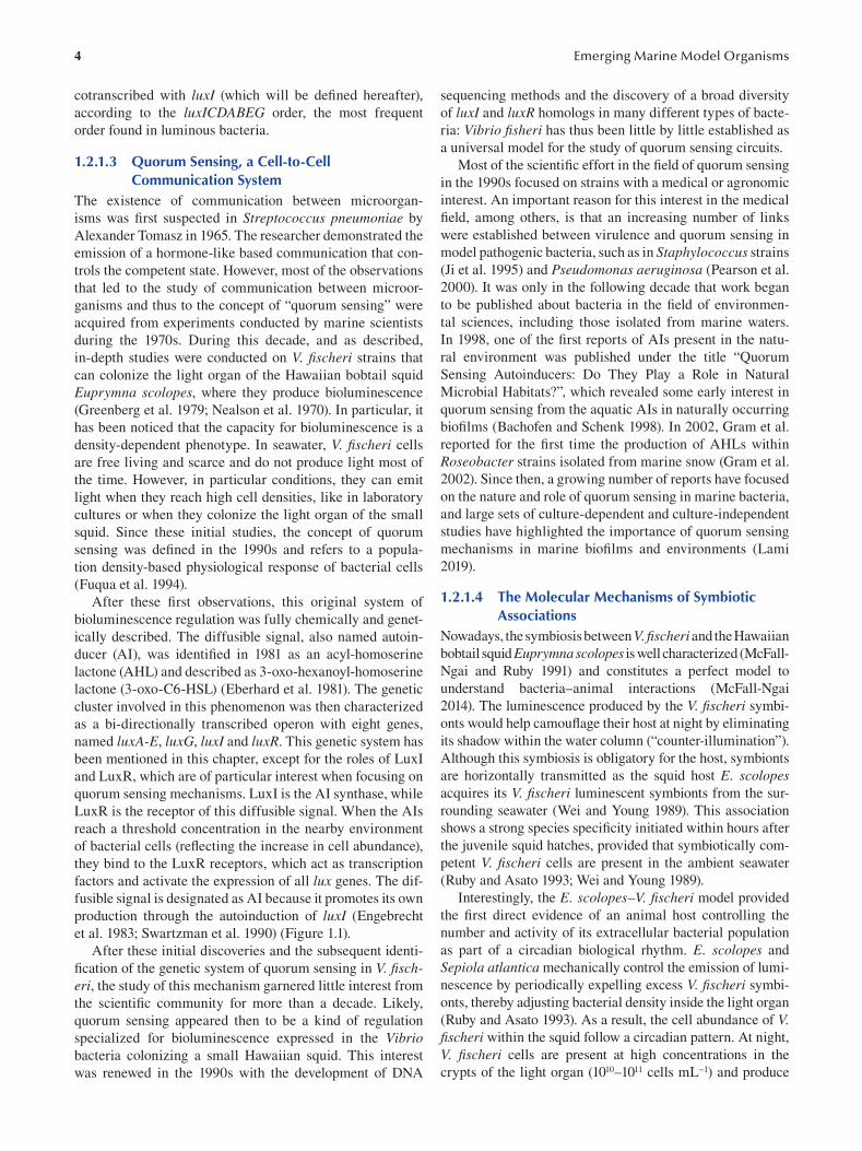
FIGURE 1.1 A schematic representation of the first discovered luxI/luxR-based quorum sensing system in the model species Vibrio fi scheri, producing 3-oxo-C6-HSL as an autoinducer. Since then, a second quorum sensing system has been discovered. Based on the
AinS AI synthase, it permits the liberation of C8-HSL. At low cell density, autoinducer concentration is low, while at high cell densities,
autoinducers induce cytoplasmic cascades that lead to drastic genetic modifications, including transcription of genes responsible for
bacterial bioluminescence.
AIs, which induce light emission (see previous paragraph). At bioluminescence is observed just before dawn to early after-
the end of the night, most of the bacterial cells are expulsed noon. This coincides with the onset of environmental light
from the light organ, leading to a dramatic reduction in bac- (Lee and Ruby 1994). During the day, the concentrations of
terial concentration and of this diffusible factor. Thus, in the V. fischeri cells that have not been expulsed are very low, the
V. fischeri–E. scolopes symbiosis, the lowest production of diffusible factor is not produced and the squid does not glow.
6
However, this remaining population of V. fischeri grows
steadily under favorable conditions within the squid through
out the day and at night again reaches a cell abundance that is
sufficient to produce bioluminescence (Boettcher et al. 1996;
Heath-Heckman et al. 2013).
A complex and specific dialog occurs between V. fischeri cells and the E. scolopes host, given that first, the V. fischeri cells are typically present at a concentration of less than
0.1% of the total bacterial population in the Hawaiian waters
(Lee and Ruby 1994), and second, the motility of these bac
terial cells is required to bring the symbionts toward the
pores, the entrance of the luminescent organ in formation.
Two main mechanisms were found to initiate the interaction
(Visick and McFall-Ngai 2000): (i) close contact between
the surfaces of the host and symbiont cells through receptor–
ligand interactions and (ii) the creation of an environment
in which only V. fischeri is viable. Receptor–ligand dynam
ics, often more generally referenced as microbe-associated
molecular patterns (MAMPs) (Koropatnick 2004), can also
be essential elements underlying the onset, maturation and
persistence of mutualistic animal–microbe partnerships.
Different data provided evidence that at least a portion of
the host response is mediated by lipopolysaccharide-binding
proteins from the LBP/BPI protein family (Chun et al. 2008;
Krasity et al. 2011) and peptidoglycan-recognition proteins
(PGRPs) (Troll et al. 2010). Also, studies were published
concerning the complete annotated genome of V. fischeri (Ruby et al. 2005) and the cDNA expression libraries for
colonized and uncolonized E. scolopes light organs (Chun
et al. 2006 ). Numerous gene-encoding proteins known to be
essential for both development and symbiosis were identi
fied, such as reflectin, actin, myeloperoxidase, aldehyde
dehydrogenase and nitric oxide synthase (Chun et al. 2008).
These fi ndings confirm the molecular dialogue between host
squid and bacterial symbionts at cell surfaces. Comparison
of host and symbiont population transcriptomes at four
times over the day–night cycle revealed maximum expres
sion of cytoskeleton related genes just before dawn, concor
dant with the daily effacement of the host epithelium and a
cyclic change in the anaerobic metabolism of the symbionts
(Wier et al. 2010). These host epithelium effacement and
change in symbiont metabolism are clearly synchronized
with the daily expulsion of most of the bacterial popula
tion (Boettcher et al. 1996; Ruby and Asato 1993). It is well
known that during the colonization of the host tissue, the
expression of sets of bacterial genes can be under the con
trol of specific transcriptional regulators (Cotter and DiRita
2000), mainly described in bacteria that initiate pathogenic
or benign infections (van Rhijn and Vanderleyden 1995).
Interestingly, a mutant study showed that the gene litR,
essential for the induction of luminescence, also plays a role
as a transcriptional regulator in modulating the ability of V. fi scheri to colonize juvenile squid (Fidopiastis et al. 2002).
1.2.1.5 V. fi scheri: Conclusions V. fischeri is now a well-known marine model in experi
mental biology. This first example clearly reveals how a
Emerging Marine Model Organisms
marine bacterial strain, which at first sight appears to have
a very particular mode of life (a bacterium associated with
Hawaiian species), is in fact a universal model to explore
mechanisms relevant to many diverse scientifi c fields and is
at the origin of major discoveries in biology.
1.2.2 PICOCYANOBACTERIA AS MODELS TO EXPLORE
PHOTOSYNTHETIC ADAPTATIONS IN THE OCEANS
Cyanobacteria are, evolutionarily speaking, very old organ
isms capable of producing oxygen that have signifi cantly
contributed to shape the current composition of the atmo
sphere. Their bioenergetic mechanisms are unique, as com
plex electron transfers (photosynthesis and respiration)
occur in the same cell compartment. Among these organ
isms, the marine picocyanobacteria Prochlorococcus and
Synechococcus genera provide detailed examples of pho
tosynthetic adaptations to light conditions in the oceans.
Beyond the description of unique photosynthetic mecha
nisms, the study of these marine cyanobacteria is key to bet
ter understanding the evolutionary origins of photosynthesis.
1.2.2.1 Key Features of Prochlorococcus and Synechococcus
The global chlorophyll biomass of oceanic ecosystems
is dominated by tiny unicellular cyanobacteria of the
Prochlorococcus and Synechococcus genera (1 and 0.6
μm diameter, respectively), which are thought to account for
25% of the global marine primary productivity (Flombaum
et al. 2013). They are considered the smallest but also the
numerically most abundant photosynthetic organisms on
Earth, with estimations of 1.7 × 1027 cells in the World
Ocean. Prochlorococcus and the marine Synechococcus diverged from a common ancestor 150 million years ago,
and the Prochlorococcus radiation delineates a monophyletic
lineage within the complex Synechococcus group. Marine
Synechococcus strains are indeed a more ancient and diverse
radiation, which is usually divided into three subclusters,
the major one (5.1) being subdivided into 15 other impor
tant clades that include 35 subclades (Farrant et al. 2016;
Mazard et al. 2012). Despite their close relatedness, these two
cyanobacteria have quite different ecophysiological features,
as they occupy complementary though overlapping ecologi
cal niches in the ocean. Prochlorococcus strains are con
fined to the warm 45°N to 40°S latitudinal band and are very
abundant in the subtropical gyres and the Mediterranean
Sea but are absent from the high-latitude, colder waters.
Prochlorococcus cell concentrations are often less important
in coastal areas than offshore. By contrast, Synechococcus cells are detected in almost all marine environments outside
of the polar circles and can be considered as the most wide
spread cyanobacterial genus on Earth.
Since the discovery of marine Prochlorococcus and
Synechococcus only some decades ago, much progress
has been made in the study of their biology. Marine pico
cyanobacteria have been prime targets for whole-genome
sequencing projects, and more than 100 complete genomes
7 Marine Bacterial Models for Experimental Biology
are now available, spanning a large range of ecological
niches and physiological and genetic diversity. These stud
ies have revealed that Prochlorococcus is a striking example
of an organism that has undergone genome “streamlining”
(Dufresne et al. 2005), an evolutionary process thought to
have rapidly followed the divergence from the common
ancestor with Synechococcus and which resulted in an rapid
specialization in oligotrophic marine niches. Thus, some
Prochlorococcus isolates have a genome as small as 1.65 Mb
(1700 genes), and this cyanobacterium is often considered
as approaching the near-minimal set of genes necessary for
an oxygenic phototroph. The study of the Synechococcus genomes is more complex because of the large microdiver
sity of the radiation. They are on the whole bigger (2–3 Mb;
2500–3200 genes) than Prochlorococcus ones and, by con
trast, show a relatively small range of variation in their char
acteristics among strains (Dufresne et al. 2008). Interestingly,
the number of “unique genes”, that is, the genes that are
found only in one genome, is well correlated with the whole
genome size. Like in Prochlorococcus, most of these unique
genes are located in variable regions called genomic islands,
whose size, position and predicted age are highly variable
among genomes. This suggests that horizontal transfer of
genetic material is an important process in these picocyano
bacteria. Overall, the Synechococcus core genome includes
70 gene families that are not present in Prochlorococcus, suggesting a higher diversity of metabolic processes, in line
with the greater diversity of marine niches colonized by
Synechococcus (Scanlan et al. 2009).
1.2.2.2 Different Adaptive Strategies of Prochlorococcus and Synechococcus to Light
The accumulation of (meta)genomic information has trig
gered the beginning of a thorough analysis of the rela
tionships between the picocyanobacterial genotypes,
phenotypes and different marine environments. In particu
lar, the study of Prochlorococcus and Synechococcus has
allowed much progress in the understanding of the selective
pressures that drive the evolution of the oxygenic photosyn
thetic process at all scales of organization, from genes to
the global ocean. Light quantity and quality are among the
main drivers of photosynthesis, both showing great variabil
ity in the oceans. In tropical oligotrophic areas, the sunray
angle and water transparency lead the photic zone to extend
much deeper compared to higher latitudes and in turbid
coastal waters. Moreover, seawater absorbs and scatters
wavelengths in a selective way. Long wavelengths such as
red light are absorbed within the first meters, whereas blue-
green light can penetrate more deeply. In shallow coastal
areas, water often carries large amounts of particulate mat
ter that further alter the underwater light quality, inducing
the presence of a green-yellow light. Successful adaptation
of phototrophs to the multifaceted behavior of light in the
aquatic systems notably relies on the nature and composition
of the light-harvesting systems, and Prochlorococcus and
Synechococcus have adopted drastically different strategies.
1.2.2.3 Adaptation of the Photosynthetic Apparatus of Prochlorococcus
The most-reviewed example is probably the manner by
which Prochlorococcus modified its photosynthetic appara
tus during evolution (Ting et al. 2002). Most cyanobacteria
on Earth have a photosynthetic antenna consisting of a giant
pigmented protein complex, called the phycobilisome. By
contrast, Prochlorococcus is one of the rare cyanobacteria
that uses membrane-intrinsic chlorophyll-binding pro
teins, termed Prochlorophyte-chl-binding (Pcb) proteins.
Thus, most genes encoding phycobilisome components have
been lost during the Prochlorococcus genome streamlining.
As Prochlorococcus uses chlorophyll b as an accessory pig
ment in its atypical antenna complex, it effi ciently harvests
blue light, the dominant wavelength in oligotrophic and deep
waters. As a result, Prochlorococcus populations extend
deeper in the water column than almost any other photo
trophs, basically defining the deepest limit of photosynthetic
life in the World Ocean. The ability of Prochlorococcus to
thrive in the entire euphotic zone also largely relies on its
microdiversity, as this cyanobacteria features genetically and
photophysiologically distinct populations (Biller et al. 2014).
These so-called high-light and low-light ecotypes partition
themselves down the water column along the light irradiance
decreasing gradient. One of the main known physiological
differences between Prochlorococcus light ecotypes is their
major light-harvesting complexes, which comprise different
sets of the Pcb proteins associated either with photosystem I
or II, resulting in higher chl b to chl a ratio in the low-light
ecotypes (Partensky and Garczarek 2010). Nevertheless, we
still know very little about the differential pigmentation and
function of the different Pcb proteins, especially regarding
the photoprotective processes. More physiological and bio
chemical work is needed on this topic (Figure 1.2).
1.2.2.4 Adaptation of the Photosynthetic Apparatus of Synechococcus
A second interesting example is the way picocyanobacte
ria deal with the large variations in light spectral quality
that occur along the horizontal (i.e. coastal-oceanic) gradi
ents in the oceans. In contrast to Prochlorococcus , marine
Synechococcus use phycobilisomes to harvest light, which
consist of three classes of stacked phycobiliproteins. The
phycobilisome core, made of allophycocyanin (APC) and
connected to the photosystems, is surrounded by rods consti
tuted of phycocyanin (PC) and/or phycoerythrin (PE). Each
phycobiliprotein has a much-conserved hexameric cylindri
cal structure, binding one or several tetrapyrrolic chromo
phore (phycobilin) types: the blue phycocyanobilin (PCB),
the red phycoerythrobilin (PEB), and the orange phycouro
bilin (PUB).
During their evolution, marine Synechococcus have
developed an amazing variety of pigmentations by exploiting
the modular nature of phycobilisomes, elaborating rods with
variable pigment composition. Thus, three main pigment
types can be distinguished based on the phycobiliprotein
8 Emerging Marine Model Organisms
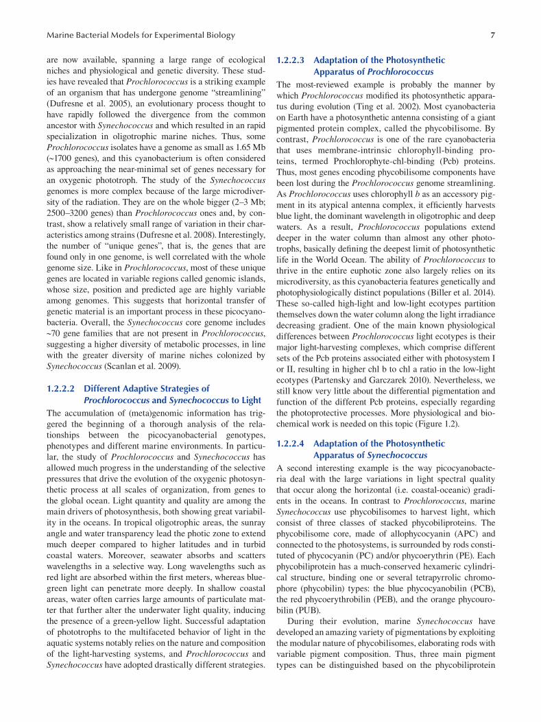
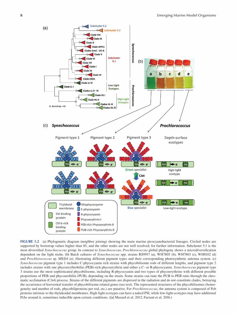
FIGURE 1.2 (a) Phylogenetic diagram (neighbor joining) showing the main marine picocyanobacterial lineages. Circled nodes are
supported by bootstrap values higher than 85, and the other nodes are not well resolved; for further information. Subcluster 5.1 is the
most diversifi ed Synechococcus group. In contrast to Synechococcus, Prochlorococcus global phylogeny shows a microdiversifi cation
dependent on the light niche. (b) Batch cultures of Synechococcus spp. strains RS9917 (a), WH7805 (b), WH7803 (c), WH8102 (d)
and Prochlorococcus sp. MED4 (e), illustrating different pigment types and their corresponding photosynthetic antenna system. (c)
Synechococcus pigment type 1 includes C-phycocyanin rich strains with phycobilisome rods of different lengths, and pigment type 2
includes strains with one phycoerythrobilin (PEB)-rich phycoerythrin and either a C- or R-phycocyanin. Synechococcus pigment type
3 strains use the most sophisticated phycobilisome, including R-phycocyanin and two types of phycoerythrins with different possible
proportions of PEB and phycourobilin (PUB), depending on the strain. Some strains can tune the PUB to PEB ratio through the chro
matic acclimation (CA4) process. Strains of the different pigments are dispersed in the radiation and do not constitute clades, betraying
the occurrence of horizontal transfer of phycobilisome related genes (see text). The represented structures of the phycobilisomes (homo
geneity and number of rods, phycobiliproteins per rod, etc.) are putative. For Prochlorococcus, the antenna system is composed of Pcb
proteins intrinsic to the thylakoidal membranes. High-light ecotypes can have a naked PSI, while low-light ecotypes may have additional
Pcbs around it, sometimes inducible upon certain conditions. ([a] Mazard et al. 2012; Farrant et al. 2016.)
9 Marine Bacterial Models for Experimental Biology
and phycobilin content of the phycobilisome rods. Pigment
type 1 contains only phycocyanin, binding solely the orange
light-absorbing phycocyanobilin (AMAX = 620 nm), and is
restricted to coastal, low-salinity surface waters, character
ized by a high turbidity, inducing the dominance of orange
wavelengths in the water. Pigment type 2 strains use PC
and one type of PE binding PEB, the green-light absorb
ing pigment (AMAX = 550 nm), and inhabit transition zones
between brackish and oceanic environments with intermedi
ate optical properties. Finally, pigment type 3 strains pos
sess PC and two types of PEs (PE-I and PE-II), a feature
specific of marine Synechococcus cyanobacteria. The PEs
of pigment type 3 strains bind both PEB and the blue light-
absorbing PUB (AMAX = 495 nm) in various ratios depend
ing on the strain, thus defining “green light specialists” (low
PUB:PEB) and “blue light specialists” (high PUB:PEB)
strains. Accordingly, these strains are found over large
gradients from onshore mesotrophic waters, rich in green
wavelengths, to offshore oligotrophic systems, where blue
light is dominant. Overall, at least a dozen of optically dif
ferent phycobiliproteins have been elaborated by marine
Synechococcus during their evolution (Six et al. 2007),
and there is no doubt that this is partly responsible for their
global ecological success.
The genomic comparison of strains representative of
these pigment types revealed that most genes involved in
the biosynthesis of phycobilisome rods are located in a large
(up to 30 kb) specialized region of the genome, generally
predicted to be a genomic island. The gene content and orga
nization of this region is specific to each pigment type, inde
pendently from the strain phylogenetic position, and shows
a tremendous increase in phycobilisome gene complexity
from pigment types 1 to 3, the latter type being a more recent
structure and the most sophisticated phycobilisome known
so far. Together with the presence of phycobilisome genes
in metaviriome datasets, this suggests that genes related to
the phycobilisome rod region can be laterally transferred
between Synechococcus lineages and that this might be a
key mechanism facilitating adaptation of these lineages to
new light niches.
Finally, there exists another particularly interesting
Synechococcus pigment type that consists of strains capable
of a unique type of chromatic acclimation (CA4), a revers
ible process that modifies the composition of the phycobili
somes. The strains capable of CA4 are pigment type 3 strains
able to dynamically tune the PUB to PEB ratio of their phy
cobilisome, which becomes low under green light and high
under blue light to precisely match the ambient light quality.
CA is therefore predicted to increase fitness in conditions of
changing light colors, allowing the harvesting of more pho
tons than for strains with fixed pigmentation. Comparative
genomic analyses of marine Synechococcus strains showed
that the CA4 process is possible thanks to a specifi c small
genomic island that exists in two slightly different versions,
named CA4-A and CA4-B (Humily et al. 2013). The recent
implementation of methods for plating and genetic manipu
lations such as the disruption and/or overexpression of CA4
genes in marine Synechococcus has allowed us to start deci
phering the regulation of the CA4-A process. Thus, in the
model strain Synechococcus sp. RS9916, isolated in the Red
Sea, the CA-4 process involves chromophore switch systems
at three phycoerythrin cysteines, which are regulated by the
two transcription factors FciA and FciB (Sanfilippo et al.
2019). Thanks to the setup of genetic transformation meth
ods, CA is one of the physiological processes that has been
more closely studied in the laboratory in picocyanobacteria.
Using phycobiliprotein and CA4 genetic markers, the
study of the extensive metagenomic Tara Oceans dataset
allowed us to determine that, globally, CA4-A and CA4-B
strains account for 23% and 19% of all Synechococcus, respectively (Grébert et al. 2018). Interestingly, CA4-A cells
predominated in the nutrient-rich, temperate or cold waters
found at high latitudes and in upwelling areas, while CA4-B
cells were most abundant in warm, nutrient-poor waters.
The reason there exist two types of CA4 genomic islands is,
however, still not clear, and the functioning of the CA4-B
genomic island is under investigation.
1.2.2.5 Picocyanobacterial Models: Conclusions Picocyanobacteria (meta)genomics has greatly increased
our understanding of the genomic and phenotype variations
existing among these organisms, which is tightly linked to
processes of niche specialization. In particular, these studies
have unveiled unprecedented information on how photosyn
thetic complexes may drastically evolve in the oceans to fi t
different light niches. In this context, it is worth noting that
the strength of the picocyanobacterial model is not restricted
to one model organism but rather consists in a large panel of
many strains that allow the understanding of the evolution
of major processes like photosynthesis in the oceans. To bet
ter understand the relationships between picocyanobacterial
genotypes and phenotypes, further progress requires a sig
nificant development of experimental work on the numer
ous picocyanobacteria strains available in culture. In this
context, the development of culture axenization methods
adapted to picocyanobacteria is a real necessity. Compared
to other microbial models, thorough and advanced physi
ological studies are still scarce, and today, targeted studies
of gene function should be prioritized over the overaccumu
lation of non-characterized genetic information. The recent
development of genetic manipulation techniques on the
Synechococcus sp. RS9916 strain gives much hope, but this
will be particularly challenging for Prochlorococcus.
1.2.3 ZOBELLIA GALACTANIVORANS, A MODEL
FOR BACTERIAL DEGRADATION OF
MACROALGAL BIOMASS
Macroalgae and their associated microbiota provide a large
diversity of enzymes, in particular involved in the degra
dation of many diverse types of sugars, which are also of
major interest for industry. Numerous economic sectors
rely on the production of efficient enzymes and are continu
ously searching for innovative ones. For example, alginate
10
lyases have many applications for food and pharmaceutical
companies. We will see in this section that the bacterium Z. galactanivorans, associated with macroalgae, is an excel
lent model to study the diversity and the functioning of these
enzymes. Working on this bacterial model also provides
interesting insights into the mechanisms of colonization of
algal surfaces and degradation of macroalgal organic matter.
1.2.3.1 Key Features of Zobellia galactanivorans Green, red and brown macroalgae (also known as seaweeds)
are dominant primary producers in coastal regions, often
locally exceeding phytoplankton and other benthic car
bon fi xers (Duarte et al. 2005). Seaweeds thus represent an
important reservoir of organic matter and are considered a
global carbon sink. The composition of macroalgal biomass
is unique and consists of >50% of polysaccharides that differ
from those known in terrestrial plants by the nature of their
monosaccharide units and the presence of sulfated motifs
and other substituents (Ficko-Blean et al. 2014). Turnover
of this biomass is mostly mediated by marine heterotrophic
bacteria that can colonize macroalgae and access, degrade
and remineralize the algal compounds. Studies of the mech
anisms underlying the interactions of these bacteria with
macroalgae and their degradation pathways are therefore
crucial to understanding coastal ecosystems’ nutrient cycles
and discovering novel enzymatic functions.
Members of the class Flavobacteriia (phylum Bacteroidetes) are recognized as key players in the degradation of marine
algal polysaccharides (Thomas et al. 2011b). Among them,
the cultivated species Z. galactanivorans has become over
the past 20 years an environmentally relevant model organ
ism to investigate macroalgal biomass degradation. Both
cultivation and metagenomic approaches frequently detect
members of the genus Zobellia in algae-dominated habi
tats and directly on the surface of seaweeds from different
oceanic basins (Hollants et al. 2013; Nedashkovskaya et al.
2004). In particular, Z. galactanivorans DsijT was fi rst iso
lated in November 1988 in Roscoff (France) from a live
specimen of the red macroalga Delesseria sanguinea ( Potin
et al. 1991) and later described as the type strain of the genus
Zobellia (Barbeyron et al. 2001). Cells are Gram-negative
and rod-shaped with rounded ends (0.3–0.5 × 1.2–8.0 μm). Z. galactanivorans is chemoorganotroph with a strictly aerobic
respiratory metabolism. Colonies on agar plates are yellow-
orange due to the biosynthesis of non-diffusible fl exirubin
type pigments. Cells do not possess flagella and cannot swim
in liquid medium. On solid surfaces, they exhibit gliding
motility at ca. 1–4 μm.s −1.
1.2.3.2 An Extraordinary Set of Enzymes Made Z. galactanivorans a Bacterial Model for the Use of Algal Sugars
Z. galactanivorans DsijT has been extensively studied for its
ability to use a wide array of macroalgal compounds as sole
carbon and energy sources, including agars and carrageen
ans from red algae, as well as alginate, laminarin, mannitol
and fucose-containing sulfated polysaccharides from brown
Emerging Marine Model Organisms
algae. Recently, it was also shown to directly degrade fresh
tissues of the kelp Laminaria digitata, corroborating its effi
ciency for macroalgal biomass turnover ( Zhu et al. 2017).
Annotation of its 5.5-Mb genome revealed that up to 9% of
its gene content could be dedicated to polysaccharide utili
zation (Barbeyron et al. 2016). This includes genes encod
ing an impressive number of 142 glycoside hydrolases (GHs)
and 17 polysaccharide lyases (PLs), representing 56 different
functional carbohydrate active enzyme families (CAZymes),
together with 37 carbohydrate-binding modules as described
in the CAZy database (Lombard et al. 2014). These enzymes
are accompanied by 18 carbohydrate esterases and 71 sul
fatases of the S1 family, which can remove substituents
from polysaccharides. These genes are often clustered in
regions of the Z. galactanivorans genome termed polysac
charide utilization loci (PULs). PULs are frequently found
in Bacteroidetes. They encode a suite of proteins dedicated
to the utilization of a given polysaccharide, generally com
prising (i) CAZYmes responsible for the breakdown of the
substrate, (ii) substituent-removing enzymes, (iii) substrate-
binding membrane proteins, (iv) transporters for oligosaccha
rides and (v) transcriptional regulators that control the PUL
expression, depending on substrate availability. In particular,
Z. galactanivorans DsijT harbors 71 tandems of SusC-like
TonB-dependent transporter (TBDT) and SusD-like surface
glycan-binding protein (SGBP) that are considered hallmarks
of PUL genomic organization (Grondin et al. 2017).
Over the years, numerous biochemical and structural
studies have focused on the in-depth characterization of
Z. galactanivorans proteins dedicated to polysaccharide
utilization. In September 2020, the function of 42 of these
proteins was experimentally validated, and for half of them,
the crystallographic 3D structure was solved. This nota
bly includes enzymes targeting agars (Naretto et al. 2019),
porphyrans (Hehemann et al. 2010), carrageenans (Matard-
Mann et al. 2017), laminarin (Labourel et al. 2015), algi
nate (Thomas et al. 2013), mannitol (Groisillier et al. 2015)
and hemicellulose (Dorival et al. 2018). In several instances,
studies of Z. galactanivorans proteins led to the discovery
of novel CAZY families [e.g. iota-carrageenases GH82 and
α-1,3-L-(3,6-anhydro)-galactosidase GH117 (Rebuffet et al.
2011)] or to novel activities in existing families [e.g. exolytic
α-1,3-(3,6-anhydro)-D-galactosidases in GH127 and GH129
(Ficko-Blean et al. 2017 )].
Furthermore, genome-wide transcriptomes of Z. galactanivorans DsijT cells grown with different carbohydrates
are publicly available, either based on microarrays (Thomas
et al. 2017) or RNA-seq (Ficko-Blean et al. 2017). This is
complemented by a validated reverse transcription real-
time quantitative PCR (RT-qPCR) protocol to specifi cally
target genes of interest (Thomas et al. 2011a). These tran
scriptomic data revealed both substrate-specifi c and shared
responses between co-occurring polysaccharides and helped
define 192 operon-like transcription units. The upregulation
of 35 predicted transcriptional regulators in the presence
of algal polysaccharides compared to glucose gave further
insights into the regulation strategies at play to fi ne-tune
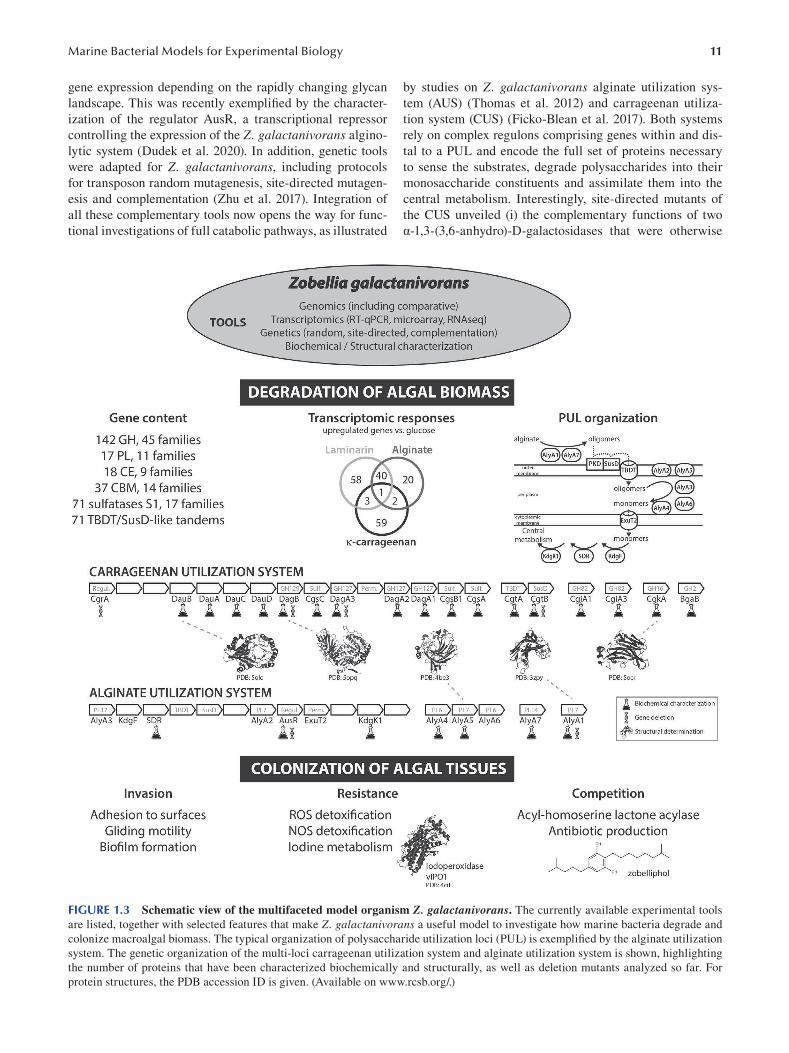
11 Marine Bacterial Models for Experimental Biology
gene expression depending on the rapidly changing glycan
landscape. This was recently exemplified by the character
ization of the regulator AusR, a transcriptional repressor
controlling the expression of the Z. galactanivorans algino
lytic system (Dudek et al. 2020). In addition, genetic tools
were adapted for Z. galactanivorans, including protocols
for transposon random mutagenesis, site-directed mutagen
esis and complementation ( Zhu et al. 2017). Integration of
all these complementary tools now opens the way for func
tional investigations of full catabolic pathways, as illustrated
by studies on Z. galactanivorans alginate utilization sys
tem (AUS) (Thomas et al. 2012) and carrageenan utiliza
tion system (CUS) (Ficko-Blean et al. 2017). Both systems
rely on complex regulons comprising genes within and dis
tal to a PUL and encode the full set of proteins necessary
to sense the substrates, degrade polysaccharides into their
monosaccharide constituents and assimilate them into the
central metabolism. Interestingly, site-directed mutants of
the CUS unveiled (i) the complementary functions of two
α-1,3-(3,6-anhydro)-D-galactosidases that were otherwise
FIGURE 1.3 Schematic view of the multifaceted model organism Z. galactanivorans. The currently available experimental tools
are listed, together with selected features that make Z. galactanivorans a useful model to investigate how marine bacteria degrade and
colonize macroalgal biomass. The typical organization of polysaccharide utilization loci (PUL) is exemplified by the alginate utilization
system. The genetic organization of the multi-loci carrageenan utilization system and alginate utilization system is shown, highlighting
the number of proteins that have been characterized biochemically and structurally, as well as deletion mutants analyzed so far. For
protein structures, the PDB accession ID is given. (Available on www.rcsb.org/.)
12
indistinguishable based on in vitro biochemical assays and
(ii) the role of a distal TBDT/SusD-like tandem that was
absent from the main carrageenolytic locus. These results
highlight the benefi t of genetic tools in a bacterial model to
assess gene functions in vivo. Studies on Z. galactanivorans also provided insights into the genomic exchange of poly
saccharide degradation pathways between closely and dis
tantly related bacteria by horizontal gene transfers (HGTs).
This includes acquisitions by Z. galactanivorans of specifi c
genes (e.g. alginate lyase AlyA1, endoglucanase EngA) from
marine Actinobacteria and Firmicutes ( Zhu et al. 2017;
Dorival et al. 2018) and transfers of flavobacterial PULs to
marine Proteobacteria, as well as several iconic examples
of diet-mediated HGT into gut bacteria of Asian populations
(Hehemann et al. 2012) (Figure 1.3).
1.2.3.3 A Model to Study Bacterial Colonization of Algal Surfaces
Besides polysaccharide degradation, Z. galactanivorans is
a relevant model to study other adaptations to macroalgae
associated lifestyle, such as surface colonization and resis
tance against algal defenses. First, its gliding motility and
rapid spread on surfaces might aid in colonizing the algal
thallus. Flow-cell chamber experiments showed that Z. galactanivorans can grow as thick biofilms (up to 90 μm),
a capacity which is maintained or even increased in the
presence of algal exudates (Salaün et al. 2012). Second, Z. galactanivorans possesses multiple enzymes predicted to
cope with the reactive oxygen and nitrogen species pro
duced by macroalgae as defense mechanisms. This includes
superoxide dismutases, peroxidases, glutathione reductases,
thioredoxins, thioredoxin reductases, peroxiredoxins and
NO/N2O reductases. Third, Z. galactanivorans features an
iodotyrosine dehalogenase and biochemically active iodo
peroxidases (Fournier et al. 2014) and accumulates up to
50 μM of iodine, two orders of magnitude higher than typi
cal oceanic concentrations. This distinct iodine metabolism
likely participates in the resistance against the high iodine
concentration in algal cell walls and the stress-induced
release of halogenated compounds. Finally, Z. galactanivorans strain OII3 produces a novel secondary metabolite
of the dialkylresorcinol (DAR) family, named zobelliphol,
with anti-microbial activity against Gram-positive bacteria
(Harms et al. 2018). This compound could therefore help Z. galactanivorans compete with other epiphytic bacteria. It is
also possible that zobelliphol acts as an antioxidant and/or
signaling molecule, similar to other DAR derivatives. In line
with this, Z. galactanivorans encodes a putative acyl-homo
serine lactone acylase that might degrade communication
molecules produced by competing bacteria and interfere
with their quorum sensing.
1.2.3.4 Z. galactanivorans: Conclusions Collectively, all these features reveal that Z. galactanivorans is a multifaceted model organism to investigate how
marine bacteria colonize and degrade macroalgal biomass.
Such studies can improve our understanding of nutrient cycles
Emerging Marine Model Organisms
in coastal areas but also uncover novel activities with prom
ising biotechnological applications. Considering that marine
organisms represent an immense potential reservoir of bio
active compounds, such bacterial models are essential to
characterize innovative molecules of interest for biotechnol
ogy but also to understand their ecological roles.
1.2.4 MARINOBACTER HYDROCARBONOCLASTICUS, A MODEL BACTERIUM FOR BIOFILM FORMATION, LIPID BIODEGRADATION AND IRON ACQUISITION
The degradation of hydrocarbons is a bacterial activity of
major industrial and environmental interest. Few micro
organisms, one of which is Marinobacter hydrocarbonoclasticus, are able to efficiently degrade such compounds.
Interestingly, and above the primary interest focused on
hydrocarbon degradation, we will see in this section that this
bacterium is also an excellent model to investigate the mech
anisms of biofilm formation and iron acquisition, which are
two universal and key features of microbial physiology.
1.2.4.1 Key Features of Marinobacter hydrocarbonoclasticus
Bacteria of the genus Marinobacter, to date composed of 57
species, are widespread in marine environments. They have
been detected in the deep ocean, coastal seawater, marine
sediment, hydrothermal settings, oceanic basalt, sea ice,
solar salterns and oilfields, as well as in association with ani
mal or algal hosts. These bacteria are Gram-stain-negative,
rod-shaped, motile, mesophilic, halotolerant, heterotrophic
and aerobic. The genus was first described with the type
strain M. hydrocarbonoclasticus SP17 (hereafter MhSP17),
which was isolated from sediments of the Mediterranean
Sea near a petroleum refinery (Gauthier et al. 1992). Later,
the strain M. aquaeolei VT8 (MhVT8) was isolated from the
produced water of an offshore oil well and was recognized
as a heterotypic synonym of M. hydrocarbonoclasticus (Huu et al. 1999; Márquez and Ventosa 2005). Since then,
M. hydrocarbonoclasticus strains became models for study
ing biofi lm formation on lipids and alkanes as a strategy to
assimilate these insoluble substrates, production and storage
of wax esters and iron acquisition through the synthesis of
the siderophore petrobactin.
1.2.4.2 Biofilm Formation on Nutritive Surface and Alkane Degradation
MhSP17 exhibits a remarkable ability to grow on nearly
water-insoluble compounds like long-chain alkanes (up to
32 carbons atoms), triglycerides, fatty acids and wax esters
(Klein et al. 2008; Mounier et al. 2014). The water-insolubility
of these substrates impairs their assimilation by bacterial
cells. Growth on water-insoluble compounds can only be
achieved by way of physiological and/or behavioral adap
tations enabling rapid mass transfer of the substrate from
the non-water-dissolved state to the cell. Biofi lm formation
is a widespread strategy to assimilate non-dissolved sub
strates, as observed, for instance, on cellulose, chitin and
13 Marine Bacterial Models for Experimental Biology
hydrocarbons (Sivadon et al. 2019). These biofi lms develop
on so-called nutritive interfaces since they play both the role
of substrate and substratum. This feature distinguishes them
from conventional biofilms growing on inert supports, such
as minerals, metals or plastics. MhSP17 forms a biofi lm at
the interface between the aqueous phase and substrates that
can be solid (saturated triacylglycerol, long-chain alkane,
fatty acids, fatty alcohol and wax esters) or liquid (medium
chain alkane and unsaturated triacylglycerol). MhSP17 sub
strates also differ by the localization of their metabolism.
Triglycerides must be hydrolyzed by a secreted lipase before
entering the cell, whereas alkane metabolism is purely
intracellular. The ability of MhSP17 to form biofilms on a
variety of substrates exhibiting different physical properties
or involving different metabolisms makes this bacterium a
valuable model for studying biofilms on nutritive surfaces.
During biofilm formation on alkanes or triglyceride,
MhSP17 cells undergo profound changes in gene expression,
indicating a reshaping of the physiology of biofi lm cells
(Mounier et al. 2014; Vaysse et al. 2011, 2009). Interestingly,
a great part of the genes modulated during biofi lm formation
was of unknown function, leading to potential for the dis
covery of new cellular functions. The role of some of these
genes, like the alkane transport system AupA-AupB, has
been elucidated by constructing mutants deleted of genes
detected in omics analyses (Mounier et al. 2018). An extra
cellular matrix of biofilm developing on a nutritive surface
is viewed as an external digester improving the solubiliza
tion of the substrate (Sivadon et al. 2019). This matrix func
tion was documented in MhSP17 with the demonstration
that the matrix contained extracellular factors involved in
triglycerides and alkanes assimilation (Ennouri et al. 2017).
A random mutational analysis led to the identification of a
di-guanylate cyclase that is important for biofi lm formation
on alkane.
1.2.4.3 Biosynthesis and Accumulation of Wax Esters The strains MhVT8 and, to a lesser extent, MhSP17 are also
used as models for the biosynthesis of wax esters. Production
and storage of neutral lipids such as wax esters and triacylg
lycerols are encountered in few marine bacterial genera like
Alcanivorax and Marinobacter. This process is believed to
be a survival strategy that allows bacteria to store energy and
carbon to thrive in natural environments where nutrient avail
ability fluctuates (Alvarez 2016; Manilla-Pérez et al. 2010).
Wax esters are formed by the esterification of a fatty alco
hol and an activated fatty acid. The length and desaturation
degree of the fatty acid and the fatty alcohol moieties of wax
esters confer on them diverse physicochemical properties that
are of great interest in the industries of cosmetics, high-grade
lubricants, wood coatings, antifoaming agents, printing inks,
varnishes and food additives (Miklaszewska et al. 2018). Wax
esters are nowadays mostly industrially produced from fos
sil fuels. The more sustainable production of wax esters by
microbial cells from wastes is currently the object of inten
sive research and requires the utilization of model systems
like M. hydrocarbonoclasticus. Strains MhVT8 and MhSP17
naturally accumulate high yields of wax esters. The two key
enzymes of the biosynthesis of wax esters are the fatty acyl
reductase (FAR) and the wax synthase (WS), which produce
wax esters from coenzyme A (CoA) or acyl carrier protein
(ACP) activated fatty acids. MhSP17 and MhVT8 possess
four and fi ve WS genes, respectively ( Lenneman et al. 2013;
Petronikolou and Nair 2018). Enzymatic properties of FAR
and WS from Marinobacter strains have been extensively
studied, leading to engineering efforts to alter their substrate
specificity. The heterologous expression of these enzymes in
hosts like Arabidopsis thaliana or yeasts led to the success
ful production of wax esters (Wenning et al. 2017; Vollheyde
et al. 2020).
1.2.4.4 Iron Acquisition In oceans, remineralization into CO2 of the organic carbon
released by marine phototrophs occurs mostly through the
respiration of heterotrophic bacteria (Buchan et al. 2014). A
great part of the heterotrophic activity resides in the particu
late fraction of the organic carbon consisting of aggregated
compounds (mostly proteins, polysaccharides and lipids)
that are colonized by biofilm-forming bacteria (Benner
and Amon 2015). Metal availability, particularly iron, is
expected to have a strong impact on organic carbon remin
eralization since respiration is a highly iron-demanding pro
cess, the respiratory chain alone containing approximately
94% of the cellular iron (Tortell et al. 1999). Iron acquisition
by marine heterotrophic bacteria is thus a fundamental mat
ter to understand the recycling of organic carbon in marine
environments.
MhSP17 and MhVT8 have been used as models to study
iron acquisition in marine environments. MhVT8 was shown
to produce three siderophores: the petrobactin and its sulfo
nated and disulfonated forms, while in MhSP17 culture, only
petrobactin and the monosulfonated derivative were detected.
The role of these sulfonations and the pathways leading to
their formation are unknown. Moreover, petrobactin exhib
its a typical property of marine siderophores, the photore
activity of the ferric-complex, which causes the release of
soluble Fe(II) and results in a petrobactin photoproduct that
retains the capacity to complex Fe(III) (Barbeau et al. 2003).
The biological significance of this photoreactivity is still not
understood. Nevertheless, it might influence the iron uptake
mechanism and consequently the biogeochemical cycling of
iron in marine environments. It is without any doubt that the
use of models that are genetically trackable will be an asset
for elucidating the various mechanism facets of petrobactin
and its derivatives.
1.2.4.5 Genomics and Genetics of M. hydrocarbonoclasticus
The genomes of MhSP17 and MhVT8 encode for 3803
and 4272 proteins, respectively. As expected for two
strains from the same species, genomes of MhVT8 and
MhSP17 have a great number of genes in common, their
core genome consisting of 3041 genes (80% identity, 80%
coverage). However, due to different sites of isolation and
14
likely different evolutionary history, the genomes of these
strains are not identical, MhVT8 and MhSP17 having 1348
and 742 strain-specifi c genes (80% identity, 80% coverage),
respectively. In addition, MhVT8 harbors two plasmids,
pMAQU01 and pMAQU02, encoding for 213 and 201 pro
teins, respectively, while MhSP17 does not carry any plas
mid (Singer et al. 2011).
The genomic potential of M. hydrocarbonoclasticus strains suggests the utilization of a large variety of substrates
as terminal electron acceptors, which is consistent with their
occurrence in diverse environments and the multiple life
styles, planktonic and biofilm-forming, of this species (Singer
et al. 2011). One striking feature of the M. hydrocarbonoclasticus genomes is their high content in genes involved in
the metabolism of the second messenger: bis (3’-5’) cyclic
dimeric guanosine monophosphate (c-di-GMP). MhSP17
and MhVT8 harbor 83 and 80 genes, respectively, encod
ing either diguanylate cyclases with GGDEF domains that
synthesize c-di-GMP or phosphodiesterases with EAL or
HD-GYP domains that hydrolyze c-di-GMP. The c-di-GMP
signaling pathway controls, in particular, the switch between
the sessile and biofi lm mode of life. The presence of a large
number of c-di-GMP-related genes suggests that M. hydrocarbonoclasticus lifestyles are under the control of different
c-di-GMP regulatory circuits that are activated in response to
multiple environmental conditions.
More details about genetic tools are provided in Section
1.3 of this chapter, but specific details about Marinobacter strains are provided here. In MhSP17, gene transfer has
been proved successful only by conjugation, using the trans
fer system based on the conjugating plasmid RP4 with an
Escherichia coli donor strain expressing the transfer func
tions. The M. hydrocarbonoclasticus receiving strain was
JM1, a streptomycin-resistant derivative of MhSP17 that
enables counter selection in conjugation experiments. The
introduction of suicide plasmids that are unable to replicate
in JM1 enables random mutagenesis using mini-Tn5 transpo
son and site-directed mutagenesis by allele exchange. Gene
addition in MhSP17 has been achieved using the transposon
vector min-Tn7 that has an integration site in the MhSP17
chromosome. This has been used to express green fl uores
cent protein constitutively to follow fluorescent cells under
fluorescence microscopy. Plasmids with the replication ori
gin of pBBR1 are stably maintained in JM1, and the PBAD
promoter was shown to be functional. This offers the pos
sibility to introduce, maintain and express genes in MhSP17
for complementation tests or any physiological studies
requiring the controlled expression of a gene (Ennouri et al.
2017; Mounier et al. 2018).
1.2.4.6 Marinobacter hydrocarbonoclasticus: Conclusions
The specific features of MhSP17 and MhVT8, such as bio
film formation on lipids and alkanes, accumulation of wax
esters and production of siderophores, together with the
availability of genetic tools, make them valuable models
to study carbon and iron cycles in the ocean as well as to
Emerging Marine Model Organisms
implement biotechnological processes for the production of
lipids of industrial interest. In this sense, this example of a
bacterial model has many common features with Zobellia galactanivorans, previously developed, and demonstrates
that marine bacterial models are extremely valuable for the
exploration of fundamental biological mechanisms but also
for the rapidly expanding field of blue biotechnology.
1.3 THE BACTERIAL MODEL ORGANISM TOOLKIT
Although the four bacterial models presented have emerged
from different labs, with the aim to answer diverse scientifi c
questions concerning different biological mechanisms, they
were developed thanks to a common toolkit consisting of
optimized protocols for isolation and culture, genetic manip
ulation and phenotypic characterization. These key tools are
under constant evolution and will be presented in this next
section, beginning with the development of novel isolation
methodologies essential for the discovery of original mod
els and the establishment of strain collections. Then we will
describe how classical genetic manipulation protocols allow
the production of mutants to directly target key mechanisms
of interest in bacterial models and present the state of the art
genome editing CRISPR-Cas technology. Finally, we will
see how recent omics approaches complement the character
ization of bacterial models and pave the way for innovative
phenotyping methods.
1.3.1 INNOVATIVE TECHNIQUES FOR THE
ISOLATION OF NEW BACTERIAL MODELS: CULTURING THE UNCULTURABLE
The decision to develop a new bacterial model may be moti
vated by a lack of current models that are representative of
the target species and/or a particular function they carry out
in the environment. The selection of this model necessarily
goes through a stage of isolation and culture in the labora
tory in order to fully study its phenotype and genotype or to
construct mutants. However, it is well known that isolated
bacteria represent only a small fraction of the total bacterial
diversity and that the culturability of environmental bacte
ria, is very low, ranging from less than 0.001% in seawater to
about 0.3% in soils (Rappé and Giovannoni 2003). Even in
the era of ‘meta-omic’ techniques, the objective of isolating
and cultivating uncultivated bacteria remains a high prior
ity in microbiology. This phenomenon is referred to as “the
great plate count anomaly”, and there are many hypotheses
that could explain it: (i) some bacteria do not tolerate high
concentration of nutrients; (ii) organic substrates present in
culture media are inappropriate for growth; (iii) important
specific vitamins or growth factors are missing in the culture
media; (iv) a nutritional shock is induced by an uncontrolled
production of oxygen reactive species (substrate-accelerated
death); (v) growth inhibition by antagonistic interaction
of other species (antibiosis); (vi) some species dependent
on cell–cell communication cannot grow in the absence
15 Marine Bacterial Models for Experimental Biology
of chemical signals from other cells; (vii) growth of some
bacteria is too slow to be detected; and (viii) unadapted
pressure, O 2 concentration or inappropriate culture method
(solid vs. liquid). Based on these hypotheses, different strat
egies can be tested to improve the isolation and cultivation
of more bacterial species, especially those most abundant in
the natural environment, as they could constitute interesting
laboratory models.
The first strategy is to modify the culture environment
and the conditions for growth. Conventional growth media
are very rich in nutrients because they were originally
designed for human pathogens well adapted to this type of
environment. A first step is to reduce organic matter con
centrations in order to favor oligotrophic species. In particu
lar, members of Alphaproteobacteria have been shown to
grow preferentially on nutrient-poor media (Senechkin et al.
2010). The reduction of the organic carbon concentration is,
however, constrained: if growth is detected by observing
colonies on solid media or a visible cloud in liquid media
with the naked eye, a sufficient concentration of organic
carbon is necessary, which would remain much higher than
that of natural environments. Other studies have proposed to
add peroxidase (an enzyme catalyzing the decomposition of
hydrogen peroxide), to replace agar with gellan gum in solid
culture media (Gelrite or Phytagel) (Tamaki et al. 2005) or
to autoclave phosphate and agar in culture media separately
(Kato et al. 2020, 2018). These changes could reduce the
generation of hydrogen peroxide compared to conventionally
prepared agar media and significantly increase the diversity
of cultivable bacteria. It is also possible to complement the
culture medium with components that stimulate growth,
such as trace elements similar to those found in the environ
ment, siderophores (e.g. pyoverdines-Fe, desferricoprogen),
quorum sensing molecules (e.g. acylhomoserine lactone) or
the supernatant of cultures of other species that stimulate
the growth of others (Bruns et al. 2002; Tanaka et al. 2004).
Metagenomic analysis of environmental samples can even
unveil specifi c metabolic properties used by target non-cul
tivated bacteria or, inversely, the absence of genes indicating
auxotrophy for certain elements that will be added to the
culture medium to improve their isolation.
A second strategy is based on microculture and microma
nipulation techniques. The first step consists of depositing
cells from the environment on a polycarbonate membrane
and then setting the membrane on a pad impregnated with
nutrients or sterilized sediment. Nutrients can diffuse
through the polycarbonate membrane and allow cell growth
with the formation of microcolonies after a few days of incu
bation (Ferrari et al. 2008). Microcolonies can be observed
by inverted microscopy and removed from the membrane
by microdissection using ultrasound waves generated by a
piezoelectric probe (Ericsson et al. 2000). Microcolonies can
then be sampled using a glass capillary and transferred in
tubes or microplate wells for cultivation separately from other
microcolonies. This stage of microculture can then facilitate
cell culture in a richer environment. This technique, however,
remains tedious and requires specialized instruments.
A third strategy is to isolate single cells and try to grow
them individually in order to obtain microcolonies formed
of a pure culture. The separation of single cells could favour
the growth of rare species, as it prevents direct competition.
Obtaining microcolonies can be a first step to larger growth.
Individualized cells can be grown in hundreds of diffusion
chambers (called iChips) that are placed in situ in natural
(e.g. sediments, soils) or simulated natural environments
for the influx of natural compounds (Bollmann et al. 2010;
Sizova et al. 2012; Van Pham and Kim 2014). These culture
chambers are separated from the outside environment by
semipermeable membranes of 0.03 μm, allowing fl uxes of
nutrients and signal molecules but preventing contamination
by other microorganisms (Berdy et al. 2017; Nichols et al.
2010). This approach has been used to isolate a bacterial spe
cies producing a new antibiotic of interest (Ling et al. 2015).
Another, more sophisticated approach is to encapsulate envi
ronmental bacteria into gel microdroplets (GMDs) (Liu et al.
2009), which are then placed in a chemostat fed by the nutri
ents extracted from the sampling environment ( Zengler et al.
2002 ). This system also allows the transfer of communication
molecules between GMDs. The GMDs in which a microcol
ony has developed, a priori consisting of a clonal culture,
can then be separated by cell sorting using fl ow cytometry,
followed by cultivation attempts. This device is attractive but
expensive and complicated to implement and does not guar
antee the long-term culturability of the selected cells.
A final strategy to cultivate environmental bacteria is the
dilution-to-extinction technique. This approach emerged in
the mid-1990s (Button et al. 1993) and was further developed
in the 2000s (Connon and Giovannoni 2002; Stingl et al.
2007). It consists of performing serial dilutions of the samples
using sterile natural sampling water or media on microplates
or tubes to isolate one or a few cells in a single microcham
ber. The main benefit of this technique is to allow a slow and
gradual adaptation (incubation for several weeks) of the bac
terial cells in conditions that mimic the natural environment
studied. Cell density is monitored by epifl uorescence micros
copy or flow cytometry counts, allowing even weak growth to
be detected. In addition, the very low number of cells reduces
the possibility of target uncultivated strains being overgrown
and inhibited by opportunistic bacteria that may overgrow
and inhibit slow growers of interest. The main drawback of
this approach is the lack of interactions between cells of dif
ferent species which could inhibit growth, as mentioned pre
viously. While time consuming, this technique enabled the
first-time isolation of many previously uncultured bacteria
such as SAR11 or the oligotrophic marine gammaproteo
bacteria (OMG) that dominate marine ecosystems (Cho and
Giovannoni 2004; Rappé et al. 2002; Stingl et al. 2007 ). The
isolated species are mainly oligotrophic, and most of them
fail to grow in a richer culture medium. Nevertheless, adapta
tions in the composition of the growth medium can allow for
cultures to attain a fairly high biomass, as was the case for the
model oligotrophic marine bacterium Pelagibacter ubique (Carini et al. 2013). Furthermore, additional improvements
to artificial media allowed the cultivation of more than 80
16
new isolates belonging to abundant marine clades SAR116,
OM60/NOR5, SAR92, Roseobacter and SAR11 (Henson
et al. 2016). These authors recently expanded their collection
to include members of the SAR11 LD12 and Actinobacteria
acIV clades and other novel SAR11 and SAR116 strains by
combining a large-scale three-year dilution to extinction
campaign and modelling of taxon-specific viability varia
tion to further refine their experimental cultivation strategy
(Henson et al. 2020).
1.3.2 GENETIC MANIPULATION OF MARINE BACTERIA
To fully exploit a model organism, it is important to develop
molecular genetics tools to be able to elucidate the functions
of genes, study and modify gene expression and engineer
modified organisms for biotechnological applications. The
manipulation of the strain of interest may be approached
using forward or reverse genetics, depending on the research
question.
Forward genetics is used when researchers are interested
in a particular phenotype and seek to understand the genetic
basis for this phenotype and is particularly useful for geneti
cally intractable organisms. Either natural mutants can be
studied or mutations can be induced by random mutagene
sis, using chemicals or UV radiation, and then the mutations
are subsequently mapped to determine the genes affected.
This method was used to study the process of magnetosome
formation in the magnetotactic bacterium Desulfovibrio magneticus for which genetic tools were not available
(Rahn-Lee et al. 2015). The random mutagenesis toolkit
was enhanced with the discovery, in the 1940s–1950s, of
mobile DNA elements known as transposons, or “jumping
genes”, that can insert randomly into genomes, thus creat
ing mutations. Transposons were used for the mutagenesis
of a marine archaeon (Guschinskaya et al. 2016 ) and marine
bacteria (Ebert et al. 2013; McCarren and Brahamsha 2005;
Zhu et al. 2017 ). This method is particularly suited to large-
scale studies of genes of unknown function, as demonstrated
by (Price et al. 2018), who generated thousands of mutant
phenotypes from 32 species of bacteria.
In contrast to forward genetics, reverse genetics is based
on modification of a target gene by deletion or insertion,
for example, followed by the characterization of the mutant
phenotype. Reverse genetics usually requires a priori knowl
edge of the genomic context and has been facilitated in the
past 10 years owing to the increasing number of full genome
sequences available ( Zeaiter et al. 2018) and with the wealth
of information provided by oceanic metagenomic datasets
(Rusch et al. 2007; Sunagawa et al. 2015; Biller et al. 2018).
Reverse genetics requires first a method to transfer foreign
DNA into the target cells and then strategies for genome
editing, shuttle vector and promoter design and the choice of
selectable and counter-selectable markers. The toolkit can be
expanded to include reporter system design to allow selec
tion of mutated organisms or to follow gene expression. Gene
inactivation is achieved primarily by homologous recombi
nation either mediated by plasmids using the endogenous
Emerging Marine Model Organisms
recombination machinery of the host or, more recently, by
using phage recombination systems, also known as recom
bineering (Fels et al. 2020). Plasmid-mediated homolo
gous recombination requires the use of traditional cloning
approaches to incorporate into the plasmid vector the modi
fied target gene with relatively long (1–2 kb) fl anking homol
ogous sequences (homology arms) that will be the site for
allelic exchange for the first cross-over event. Use of a non-
replicating plasmid, under antibiotic selection, forces integra
tion of the plasmid into the host genome via a fi rst cross-over
event. However, to achieve gene replacement, a second cross
over event must occur, and these rare double-recombination
events must be selected for out of the vast majority of single
recombination clones that would be extremely time consum
ing. A strategy to promote a second cross-over event was fi rst
established with a temperature-sensitive replicon (Hamilton
et al. 1989) and was later improved with the development of
suicide plasmids with counter-selectable markers encoding
conditional lethal genes. One of the most widely used coun
ter-selectable markers is sacB, which confers sensitivity to
sucrose (Gay et al. 1985) and is lethal for cells that have not
undergone a second recombination to eliminate the plasmid.
This strategy was used to study the role of a specifi c enzyme
thought to be involved in alginate digestion in the model
Zobellia galactanivorans by creating a deletion mutant of an
alginase lyase gene ( Zhu et al. 2017).
The more recently developed methods known as recom
bineering, for recombination-mediated genetic engineering,
integrate linear single-stranded DNA, oligonucleotides or
double-stranded DNA fragments into the target genome in
cells expressing the bacteriophage -encoded recombination
proteins (see Fels et al. 2020 for a review). Recombineering
offers significant advantages over plasmid-mediated meth
ods, since it avoids laborious in vitro cloning techniques,
only short homology arms are required and the recom
bination efficiency is high. Although this method is com
monly used to engineer model organisms such as E. coli, it has been challenging to adapt to other bacteria outside
of closely related enterobacteria, since the existing phage
recombination systems are not efficient in all species (Fels
et al. 2020). Current research is aimed at discovering new
single-stranded annealing proteins that will be able to pro
mote recombination of ssDNA in a wider range of bacteria
(Wannier et al. 2020).
For all the gene editing approaches mentioned, the fi nal
hurdle for successful genome editing is the transfer of the
recombinant DNA into the target strain. DNA transfer is
known to occur naturally in bacteria through transforma
tion and conjugation (Paul et al. 1991; Chen et al. 2005) and
transduction (Jiang and Paul 1998) and is the mechanism for
horizontal gene transfer in bacteria. Natural competence is
mediated by proteins that enable the penetration of extracel
lular DNA, such as type IV pili or type 2 secretion systems.
For example, some cyanobacterial strains (Synechococcus sp. PCC 7002) and many Vibrio strains (including isolates
related to V. parahaemolyticus, V. vulnificus, V. fischeri ) are
naturally competent (Frigaard et al. 2004; Simpson et al.
17 Marine Bacterial Models for Experimental Biology
2019). In such cases, transformation protocols appear rela
tively simple and rely on incubation of the targeted strain
with the exogenous DNA. Various factors can affect the effi
ciency of natural transformation, such as plasmid concentra
tion, cell density, light conditions and pre-treatment of cells
( Zang et al. 2007). For example, the natural competence
of some Vibrio strains is induced by chitin, a biopolymer
abundant in aquatic habitats, originating, for example, from
crustacean exoskeletons (Meibom et al. 2005; Zeaiter et al.
2018). Tools have been developed to transform cells that
are not naturally competent by artificially creating pores in
the bacterial cell wall. The fi rst artificial method to induce
competency is chemical transformation, whereby treatments
with salt solutions create pores in the cell membranes that
allow DNA penetration into the cytoplasm. Calcium chlo
ride, diméthylsulfoxyde, polyéthylene glycol and lysozyme
are among the chemical compounds used to prepare com
petent cells or to improve the efficiency of other types of
transformation protocols. A few positive reports of chemi
cal transformation of marine bacteria were published. This
includes transformation of Rhodobacter sphaeroides and
Vibrio natriegens (Fornari and Kaplan 1982). In the latter
case, it was necessary to use a V. natriegenes strain mutated
for the chromosomal Dns endonuclease to avoid the expres
sion of a resistance mechanism (Weinstock et al. 2016).
However, several failures of chemical transformation pro
tocols applied to marine strains were reported. For exam
ple, no transformants were obtained after testing chemical
transformation protocols on 12 different Roseobacter strains
( Piekarski et al. 2009). In general, chemical transformation
does not appear to be a very efficient approach to transform
marine bacteria ( Zeaiter et al. 2018). The second method to
induce competency is by electroporation, one of the most
efficient tools to introduce DNA, particularly plasmid
DNA, into a bacterial strain. This technique consists of the
application of a brief electrical current to facilitate DNA
uptake by a bacterial cell. Indeed, a brief pulse of 5–10 kV/
cm increases cell membrane permeability and allows the
production of transformants. Marine strains belonging to
diverse taxonomic groups were successfully transformed
using these protocols, such as strains of Roseobacter, Vibrio,
Pseudoalteromonas, Caulobacter, Halomonas and some
cyanobacteria ( Zeaiter et al. 2018). However, the electric
treatment applied to the cells is harsh and induces large cell
mortality and many transformation failures. Indeed, many
factors can influence the success of an electroporation pro
tocol, including cell concentration, the composition of the
growth medium and buffer composition, temperature, volt
age of electroporation systems, plasmid size and topology
( Zeaiter et al. 2018). In particular, the presence of salts is one
of the most influential factors on electroporation effi ciency.
Therefore, careful development is needed to find the best
medium for electroporation of marine strains, which require
high concentrations of salts for growth.
Conjugation of bacterial strains is, together with elec
troporation, used more often to manipulate marine strains.
Conjugation is the only method of transfer that requires
cell-to-cell contact, whereby a donor (usually E. coli ) trans
fers various types of mobile elements, including plasmids,
transposons and integrons. One of the advantages, compared
to other methods, is the capacity to transfer large amounts
of genetic material. Another advantage is that conjugation
involves single-strand DNA, which avoids bacterial resis
tance mechanisms (restriction systems) of the receptor strain.
Conjugative transfer is a complex process that requires the
concerted action of many gene products. The mobile ele
ment to be transferred needs to contain an origin of trans
fer oriT, and the conjugative process in itself is mediated
by the transfer regions tra. If the donor strain possesses tra regions, it can directly transfer the mobile element into the
recipient strain via bi-parental conjugation. When the donor
strain lacks these regions, a third helper strain is needed
to provide conjugative ability via tri-parental conjugation.
After the conjugative transfer, donor and recipient cells will
both carry the mobile element. Therefore, the selection of
transconjugants is a critical step after conjugation to ensure
complete removal of donor (and helper) strains. This can be
achieved by using selective growth conditions (e.g. salin
ity, temperature) favoring the growth of marine strains over
donors (usually E. coli). Alternatively, the mobile element
can encode antibiotic resistance genes controlled by promot
ers that function in the recipient strain but not in the donor
strain. In addition, donor strains auxotrophic for a specifi c
compound can be used. In this case, selection occurs on a
culture medium devoid of the compound.
Transduction is an efficient method of transfer of DNA
from a bacteriophage to a bacterium and was successfully
used to transfer genes into cultivated marine isolates and
natural bacterial communities (Jiang et al. 1998). However,
it is not used as widely as conjugation and transformation as
a DNA delivery method, since phages generally have a lim
ited host range, and therefore requires the careful selection
of suitable phages for the target bacteria strain.
1.3.3 THE FUTURE OF GENE EDITING IN BACTERIAL
MODELS: THE CRISPR-CAS APPROACHES
One of the most recent additions to the genetic engineer
ing toolbox is the CRISPR-Cas technology, also known as
“molecular scissors”, that allows the precise cutting of DNA
at specific target sites by a Cas endonuclease, guided by
a short RNA sequence known as a guide RNA (sg-RNA).
The CRISPR (clustered regularly interspaced short pal
indromic repeats)-Cas system is an adaptive immune sys
tem in prokaryotes, defending the cell against invasion by
bacteriophages or extrachromosomal elements (Barrangou
et al. 2007; Bolotin et al. 2005). The CRISPR loci, pres
ent in prokaryote genomes but not those of eukaryotes or
viruses (Mojica et al. 2000; Jansen et al. 2002), contain short
DNA repeats separated by spacer sequences, known as pro
tospacer sequences, that correspond to fragments of the for
eign DNA that are stored as a record in the CRISPR array.
Although many different CRISPR-Cas systems have been
discovered (Koonin et al. 2017), the most commonly used
18
system for genome editing is based on the CRISPR-Cas9
from Streptococcus pyogenes and belongs to the CRISPR
type II family. It functions by transcription of the repeat-
spacer element to precursor CRISPR RNA (pre-crRNA),
which, following base-pairing with a trans-activating cr-
RNA (tracr-RNA), triggers processing of the structure to
mature crRNA by RNAse III in the presence of Cas9 (Jinek
et al. 2012). Site-specific cleavage of the foreign DNA by
Cas9 only occurs (i) if there is complementary base-pairing
between the cr-RNA and the protospacer and (ii) if the pro
tospacer is adjacent to a short, sequence-specifi c region
known as the protospacer adjacent motif (PAM) (Jinek et al.
2012). The sequence-specific cutting of the target DNA to
create a double-stranded break (DSB) led the authors to
realize the immediate potential of this mechanism for repur
posing into a genome engineering tool, optimized further
with the creation of single chimeric targeting RNA, a sin
gle guide RNA(sgRNA) to replace the cr-RNA:tracr-RNA
duplex (Jinek et al. 2012). CRISPR technology revolution
ized genome engineering in eukaryotes due to the ease of
designing sgRNAs to guide the nuclease to the genome
editing site, the efficiency of the Cas endonucleases and
the possibility to scale up to multiple gene edits (see Hsu et
al. 2014 and Pickar-Oliver and Gersbach 2019 for reviews).
Whereas eukaryotes can use the error-prone non-homolo
gous end-joining (NHEJ) system to repair DSBs, leading to
small insertions or deletions, the majority of bacteria lack
this pathway, making DSBs lethal. Although there are a
number of hurdles to employing CRISPR in bacteria (Vento
et al. 2019), CRISPR-Cas9 editing was successful in E. coli (Bassalo et al. 2016) and industrially important bacteria such
as Lactobacillus reuteri (Oh and van Pijkeren 2014), Bacillus subtilis (Westbrook et al. 2016 ) and Streptomyces species
(Alberti and Corre 2019). Considering the importance of
streptomycete bacteria for the production of antimicrobi
als, several CRISPR plasmid toolkits have been developed
for genome editing of Streptomyces (Alberti and Corre
2019). Examples of the application of these tools include
the activation of novel transcriptionally silent biosynthetic
gene clusters (BGCs) by knocking out known, preferentially
or constitutively expressed BGCs (Culp et al. 2019) or the
increase of their expression by “knocking in” constitutive
promoters ( Zhang et al. 2017). CRISPR is not limited to
gene editing but can also be used to study gene repression
or “knockdown” with CRISPR interference (CRISPRi).
CRISPRi uses an engineered catalytically inactive (or dead)
Cas9 protein (dCas9), which, instead of cutting the DNA,
represses transcription of the target gene by steric interfer
ence. This approach presents several advantages, including
the ease to knock down multiple genes and induction and
tuning of gene repression, and requires less effort than the
creation of multiple gene deletions. It has been employed
for gene repression in diverse bacteria such as Streptomyces (Tong et al. 2015), Synechococcus (Knoot et al. 2020) and
B. subtilis (Westbrook et al. 2016 ). More recently, the type
V-A Cas protein, Cas12a (Cpf1) (Koonin et al. 2017), is
showing promise for CRISPR editing (Yan et al. 2017) or
Emerging Marine Model Organisms
interference in bacteria (Li et al. 2018) and can be a useful
alternative for when Cas9 toxicity is observed, as was the
case in Streptomyces (Li et al. 2018). Cas12a presents some
advantages over Cas9, since it can enable multiplex genome
editing, and the production of staggered cuts instead of blunt
ends by this endonuclease promotes homology-directed
repair via the provision of a repair template (Paul and
Montoya 2020). And, last, an alternative CRISPR system
which circumvents the difficulties of repairing DSBs carries
out DSB-free single-base editing using a fusion protein of a
Cas9 variant, Cas9 nickase (Cas9n). This strategy allowed
efficient multiplex editing in Streptomyces strains that was
not possible with the standard CRISPR-Cas9 system (Tong
et al. 2019) and single-base editing in Clostridium ( Li et
al. 2019 ).
1.3.4 PHENOTYPING AND ACQUIRING
KNOWLEDGE ON MODEL STRAINS
When the bacterial model has been isolated and preserved
in appropriate conditions, and when collections of mutants
have been prepared (see Section 1.3.2) to explore the role of
various targeted genes and functions, the following step is
to characterize in depth the model strain. Traditional pheno
typing methods are still widely used in microbiology labo
ratories, including catabolic profiling on different nutrient
sources, evaluation of growth parameters in various condi
tions (i.e. biofilm vs. liquid) and determination of cell shape
or movements via microscopic techniques. This is especially
relevant when comparing wild-type strains with mutants to
evidence the role of the knocked-out genes. These traditional
techniques are now complemented by the recent develop
ment of “Omics” tools providing an immense potential in
model strain characterization.
First, whole-genome analysis of individual strains pro
vides a comprehensive view of cell physiology capacities,
which is an essential step when establishing a new bacterial
model. Additionally, the development of genetic tools relies
on thorough and precise information about gene organization
and regulation in the target strain raised as a model. Accurate
lists of genes, gene annotations and transcriptomic and pro
teomic datasets, as well as the existence of computational
platforms for data integration and systems-levels analysis,
are among the essential criteria to establish bacterial mod
els (Liu and Deutschbauer 2018). An increasing quantity of
genomic data for isolated strains are now available. These
genomes are available in various types of databases (not
specifically marine), such as that maintained by the Joint
Genome Institute (JGI) Genome Portal (https://genome.jgi.
doe.gov/portal/) or the one maintained by the Genoscope in
France (https://mage.genoscope.cns.fr/microscope/home/
index.php). For cyanobacteria, especially marine picocyano
bacteria, specific databases that include genome exploration
tools are available, such as Cyanobase (http://genome.kazusa.
or.jp/cyanobase) and Cyanorak (http://application.sb-roscoff.
fr/cyanorak/). In some databanks, one important diffi culty
is that many genomes are still incomplete and published as
19 Marine Bacterial Models for Experimental Biology
“draft genomes”, which can limit their utilization in genetic
approaches.
The availability of numerous complete and annotated
bacterial genomes in databanks facilitates the choice of
the genes to knock out when starting targeted mutagen
esis approaches, which is an essential step when building
an isolated strain as a model of interest. Also, the existence
of many genome sequences provides potential insights into
bacterial metabolic pathways: genome mining of marine
strains allows the putative identification and characteriza
tion of novel biosynthetic pathways (which will have then to
be confirmed by other types of experimental approaches, i.e.
the preparation of collections of mutants) that are respon
sible for the production of bioactive compounds and the
identification of physiological traits that were not suspected
before. Then, comparative genomics approaches may allow
the comparison of specific characteristics, even in phyloge
netically closed strains. For example, comparative genom
ics revealed that choline metabolism is widespread among
marine Roseobacter. Choline is an abundant organic com
pound in the ocean and, through its conversion to glycine
betain, serves as an osmoprotectant in many marine bac
teria. This molecule is also an important component of
membranes (phosphatidylcholine). However, the genetic and
molecular mechanisms regulating intracellular choline and
glycine betaine concentrations are poorly known in marine
bacteria. Following comparative genomic analysis, a tar
geted mutagenesis of genes involved in choline metabolism
was conducted in the model bacteria Ruegeria pomeroyi DSS-3. The authors of this study demonstrated the key role
of the betG gene, encoding an organic solute transporter
(essential in the uptake of choline) of the betB gene convert
ing choline in glycine betaine and of the fhs gene encoding
the formyl tetrahydrofolate synthetase, essential in the oxi
dization of the choline methyl groups and the catabolism of
glycine betaine (Lidbury et al. 2015).
While genomic analysis provides a snapshot of the physi
ological potential of a model strain, transcriptomics gives
insights into the functions that are expressed in a given exper
imental condition. In the cyanobacterial Prochlorococcus strain AS9601, transcriptomics approaches revealed some
of the mechanisms responsible for adaptation to salt stress.
Under hypersaline conditions (5% w/v), 1/3 of the genome
is differentially expressed compared to lower salt condi
tions (3.8% w/v). In hypersaline conditions, higher tran
script abundance was observed for the genes involved in
respiratory electron transfer, carbon fixation, osmolyte sol
ute biosynthesis and inorganic ion transport. By contrast, a
reduction of transcript abundance was noticed for the genes
involved in iron transportation, heme production and pho
tosynthesis electron transport. Such analysis thus suggests
interesting mechanisms linking light utilization and salt
stress in this strain of Prochlorococcus (Al-Hosani et al.
2015 ).
Proteomics is the characterization of the protein con
tent in a cell using mass spectrometry and nuclear mag
netic resonance approaches. Following the central dogma
of molecular biology (DNA→RNA→proteins), focusing on
protein expression allows an overall characterization of the
organism’s physiology in a defined experimental condition.
Indeed, the function of many proteins has been described,
and proteomics now provides to researchers in-depth charac
terization of the microbial cell physiology. Proteomics stud
ies were conducted on various marine prokaryotes, including
different cyanobacteria (Prochlorococcus, Synechococcus), Pseudoalteromona s, Planctomycete s, Vibrios and others
(Schweder et al. 2008). For example, the planktonic/biofi lm
transition was investigated using proteomics in the bacte
rial model Pseudoalteromonas lipolytica TC8. This study
revealed that peptidases, oxidases, transcription factors,
membrane proteins and enzymes involved in histidine bio
synthesis were over-expressed in biofilms. In contrast, pro
teins involved in heme production, nutrient assimilation,
cell division and arginine/ornithine biosynthesis were over-
expressed in planktonic cells (Favre et al. 2019). Collectively,
all these data provide insights into the mechanisms that are
expressed in bacterial cells and responsible for their adapta
tion to a biofilm or a planktonic way of life.
Metabolomics is now another essential approach to
explore the physiology of prokaryotic models and their
interactions with the environment. This approach provides
global metabolite profiles under a given set of experimen
tal conditions and a snapshot of the physiological response
of prokaryotic cells. One important difficulty and technical
challenge in metabolomics is the identification and dosage of
thousands of molecular compounds, sometimes at very low
concentrations, for which no standard is available for rapid
identification. Untargeted metabolomic approaches compare
the whole metabolomes in a qualitative or semi-quantitative
manner and without a priori knowledge about the type of
metabolites produced, while targeted metabolomics focuses
on a particular compound. During the last decade, important
improvements in the sensitivity and resolution of the analyti
cal tools required for metabolomic analysis were achieved,
including in mass spectrometry and nuclear magnetic reso
nance approaches (Ribeiro et al. 2019). These improvements
allowed for significant progress in the characterization and
identification of various compounds, including carbohy
drates, alcohols, ketones, amino acids and also several types
of secondary metabolites like antibiotics, pigments and info-
chemicals. Metabolomics is still a science in its infancy but
has begun to be used to characterize the response of marine
bacterial models to environmental variations. The authors
of the previously mentioned study on Pseudoalteromonas lipolytica TC8 also used metabolomics to characterize the
planktonic/biofilm transition. Interestingly, they revealed
drastic modifications in the lipid composition of the mem
branes (Favre et al. 2019). Phosphatidylethanolamine deriv
atives were abundant in biofilm cells, while ornithine lipids
were more present in planktonic bacteria. Thus, this study,
with others, highlights the need to focus on membrane plas
ticity mechanisms in the planktonic-to-biofi lm transition
when bacteria attach to surfaces, which remains an underex
plored research question in marine bacterial models.
20
1.4 CONCLUSIONS
This chapter reveals, through the very different selected
examples (Vibrio, Prochlorococcus and Synechococcus, Zobellia and Marinobacter), the interest and potential as
well as the difficulties to establish marine bacterial strains as
models for experimental biology. The isolation of bacterial
strains of interest; their full characterization; the development
of genetic tools and the maintenance of strain collections; the
investment in genome sequencing, including accurate gene
annotation; the phenotyping of mutants relying on OMIC
approaches: all these steps are crucial in the establishment
of new models. Clearly, it appears from this non-exhaustive
list of technical approaches as well as from the collection of
examples presented in this chapter that no universal exper
imental approach can be applied to develop a new marine
bacterial model. However, unprecedented progress has been
made this last decade in synthetic biology, molecular genetic
tool development, the application of omics data techniques
and computational tools, which undoubtedly paves the way
to the development of new bacterial models of major interest
to characterize many types of biological mechanisms. The
potential and the outcomes of such work are immense, and
applications are found in several fields. For example, recom
binant marine Synechococcus allowed the production of
polyunsaturated acids of medical interest (Yu et al. 2000),
and recombinant strains of the marine Vibrio natriegens spe
cies contributed to the production of melanin (Wang et al.
2020). Bacterial models can also serve as tools for biology,
like the model Vibrio fischeri, which serves as a biosensor to
detect pollutants in diverse environmental samples (Farré et
al. 2002; Parvez et al. 2006; Dalzell et al. 2002) and is often
reported as one of the most sensitive assays compared to oth
ers across a wide range of chemicals. Overall, new marine
bacterial models have the potential to address questions
which cannot be assessed by ‘traditional’ bacterial models.
Thus, many fundamental and applied research fi elds would
greatly benefit from investing massively in the development
of new bacterial models, including research in marine sci
ences, marine ecology, ecotoxicology and evolutionary stud
ies but also ‘blue’ biotechnology.
ACKNOWLEDGEMENTS
François Thomas acknowledges support from CNRS and the
French ANR project ALGAVOR (grant agreement ANR-18
CE02–0001–01). All authors thanks Haley Flom for English
grammar and spelling.
BIBLIOGRAPHY
Alberti, F., and C. Corre. 2019. Editing Streptomycete genomes in
the CRISPR/Cas9 age. Natural Product Reports 36(9):1237–
1248.
Al-Hosani, S., M. M. Oudah, A. Henschel, and L. F. Yousef. 2015.
Global transcriptome analysis of salt acclimatedProchlorococcus AS9601. Microbiological Research 176:21–28.
Alvarez, H. M. 2016. Triacylglycerol and wax ester-accumulating
machinery in Prokaryotes. Biochimie 120:28–39.
Emerging Marine Model Organisms
Bachofen, R., and A. Schenk. 1998. Quorum sensing autoin
ducers: Do they play a rôle in natural microbial habitats?
Microbiology Research 153(1):61–63.
Barbeau, K., E. L. Rue, C. G. Trick, K. W. Bruland, and A. Butler.
2003. Photochemical reactivity of siderophores produced by
marine heterotrophic bacteria and cyanobacteria based on
characteristic Fe(III) binding groups. Limnology and Oceano graphy 48(3):1069–1078.
Barbeyron, T., S. L’Haridon, E. Corre, B. Kloareg, and P. Potin.
2001. Zobellia galactanovorans gen. nov., sp. nov., a marine
species of Flavobacteriaceae isolated from a red alga,
and classification of [ Cytophaga] uliginosa (ZoBell and
Upham 1944) Reichenbach 1989 as Zobellia uliginosa gen.
nov., comb. nov. International Journal of Systematic and Evolutionary Microbiology 51(Pt 3):985–997.
Barbeyron, T., F. Thomas, V. Barbe, H. Teeling, C. Schenowitz,
C. Dossat, A. Goesmann, C. Leblanc, F.-O. Glöckner, M.
Czjzek, R. Amann, and G. Michel. 2016. Habitat and taxon
as driving forces of carbohydrate catabolism in marine het
erotrophic bacteria: Example of the model algae-associated
bacterium Zobellia galactanivorans DsijT. Environmental Microbiology 18:4610–4627.
Barrangou, R., C. Fremaux, H. Deveau, M. Richards, P. Boyaval,
S. Moineau, D. A. Romero, and P. Horvath. 2007. CRISPR
provides acquired resistance against viruses in Prokaryotes.
Science 315(5819):1709–1712.
Bassalo, M. C., A. D. Garst, A. A. L Halweg-Edwards, W. C. Grau,
D. W. Domaille, V. K. Mutalik, A. P. Arkin, and R. T. Gill.
2016. Rapid and efficient one-step metabolic pathway inte
gration in E. Coli. ACS Synthetic Biology 5(7):561–568.
Benner, R., and R. M. W. Amon. 2015. The size-reactivity con
tinuum of major bioelements in the ocean. Annual Review of Marine Science 7(1):185–205.
Berdy, B., A. L. Spoering, L. L. Ling, and S. S. Epstein. 2017. In
situ cultivation of previously uncultivable microorganisms
using the ichip. Nature Protocols 12(10):2232–2242.
Biller, S. J., P. M. Berube, K. Dooley, M. Williams, B. M. Satinsky,
T. Hackl, S. L. Hogle, A. Coe, K. Bergauer, and H. A.
Bouman. 2018. Marine microbial metagenomes sampled
across space and time. Scientifi c Data 5:180176.
Biller, S. J., P. M. Berube, D. Lindell, and S. W. Chisholm. 2014.
Prochlorococcus: The structure and function of collective
diversity. Nature Reviews Microbiology 13(1):13–27.
Blount, Z. D. 2015. The natural history of model organisms: The
unexhausted potential of E. Coli. Elife 4:e05826.
Boettcher, K. J., E. G. Ruby, and M. J. McFall-Ngai. 1996.
Bioluminescence in the symbiotic squid Euprymna scolopes is controlled by a daily biological rhythm. Journal of Comparative Physiology A (179):65–73.
Bollmann, A., A. V. Palumbo, K. Lewis, and S. S. Epstein. 2010.
Isolation and physiology of bacteria from contaminated sub
surface sediments. Applied and Environmental Microbiology 76(22):7413–7419.
Bolotin, A., B. Quinquis, A. Sorokin, and S. D. Ehrlich. 2005. Clustered
regularly interspaced short palindrome repeats (CRISPRs) have
spacers of extrachromosomal origin. Microbiology 151(8):
2551–2561.
Bruns, A., H. Cypionka, and J. Overmann. 2002. Cyclic AMP and
acyl homoserine lactones increase the cultivation effi ciency
of heterotrophic bacteria from the central Baltic Sea. Applied and Environmental Microbiology 68(8):3978–3987.
Buchan, A., G. R. LeCleir, C. A. Gulvik, and J. M. Gonzalez.
2014. Master recyclers: Features and functions of bacte
ria associated with phytoplankton blooms. Nature Reviews Microbiology 12(10):686–698.
21 Marine Bacterial Models for Experimental Biology
Button, D. K., F. Schut, P. Quang, R. Martin, and B. R. Robertson.
1993. Viability and isolation of marine bacteria by dilution
culture: Theory, procedures, and initial results. Applied and Environmental Microbiology 59(3):881–891.
Campbell, Z. T., T. O. Baldwin, and O. Miyashita. 2010. Analysis
of the bacterial luciferase mobile loop by replica-exchange
molecular dynamics. Biophysical Journal 99(12):4012–4019.
Carini, P., L. Steindler, S. Beszteri, and S. J. Giovannoni. 2013.
Nutrient requirements for growth of the extreme oligotroph
‘Candidatus Pelagibacter ubique’ HTCC1062 on a defi ned
medium. The ISME Journal 7(3):592–602.
Chen, I., P. J. Christie, and D. Dubnau. 2005. The ins and outs of
DNA transfer in bacteria. Science 310(5753):1456–1460.
Cho, J.-C., and S. J. Giovannoni. 2004. Cultivation and growth char
acteristics of a diverse group of oligotrophic marine Gamma
proteobacteria. Applied and Environmental Microbiology 70(1):
432–440.
Chun, C.-K., T. E. Scheetz, MdF. Bonaldo, B. Brown, A. Clemens,
W. J. Crookes-Goodson, K. Crouch, T. DeMartini, M.
Eyestone, M. S. Goodson, B. Janssens, J. L. Kimbell, T. A.
Koropatnick, T. Kucaba, C. Smith, J. J. Stewart, D. Tong, J.
V. Troll, S. Webster, J. Winhall-Rice, C. Yap, T. L. Casavant,
M. J. McFall-Ngai, and M. Bento Soares. 2006. An anno
tated cDNA library of juvenile Euprymna scolopes with and
without colonization by the symbiont Vibrio fischeri. BMC Genomics 7(1):154.
Chun, C.-K., J. V. Troll, I. Koroleva, B. Brown, L. Manzella, E. Snir,
H. Almabrazi, T. E. Scheetz, MdF. Bonaldo, T. L. Casavant,
M. Bento Soares, E. G. Ruby, and M. J. McFall-Ngai.
2008. Effects of colonization, luminescence, and autoinducer
on host transcription during development of the squid-vibrio
association. Proceedings of the National Academy of Sciences USA 105(32):11323–11328.
Connon, S. A., and S. J. Giovannoni. 2002. High-throughput meth
ods for culturing microorganisms in very-low-nutrient media
yield diverse new marine isolates. Applied and Environmental Microbiology 68(8):3878–3885.
Cotter, P. A., and V. J. DiRita. 2000. Bacterial virulence gene
regulation: An evolutionary perspective. Annual Review of Microbiology 54(1):519–565.
Culp, E. J., G. Yim, N. Waglechner, W. Wang, A. C. Pawlowski,
and G. D. Wright. 2019. Hidden antibiotics in Actinomycetes can be identified by inactivation of gene clusters for common
antibiotics. Nature Biotechnology 37(10):149–154.
Dalzell, D. J. B., S. Alte, E. Aspichueta, A. De la Sota, J. Etxebarria,
M. Gutierrez, C. C. Hoffmann, D. Sales, U. Obst, and N.
Christofi. 2002. A comparison of five rapid direct toxicity
assessment methods to determine toxicity of pollutants to
activated sludge. Chemosphere 47(5):535–545.
Dorival, J., S. Ruppert, M. Gunnoo, A. Orłowski, M. Chapelais-
Baron, J. Dabin, A. Labourel, D. Thompson, G. Michel,
M. Czjzek, and S. Genicot. 2018. The laterally acquired
GH5 Zg EngA GH5_4 from the marine bacterium Zobellia galactanivorans is dedicated to hemicellulose hydrolysis.
Biochemical Journal 475(22):3609–3628.
Duarte, C. M., J. J. Middelburg, and N. Caraco. 2005. Major
role of marine vegetation on the oceanic carbon cycle.
Biogeosciences Discussions 2:1–8.
Dudek, M., A. Dieudonné, D. Jouanneau, T. Rochat, G. Michel, B.
Sarels, and F. Thomas. 2020. Regulation of alginate catabolism
involves a GntR family repressor in the marine Flavobacterium
Zobellia galactanivorans DsijT. Nucleic Acids Research 48(14):
7786–7800.
Dufresne, A., L. Garczarek, and F. Partensky. 2005. Accelerated
evolution associated with genome reduction in a free-living
prokaryote. Genome Biology 6(2):R14.
Dufresne, A., M. Ostrowski, D. J. Scanlan, L. Garczarek, S. Mazard,
B. Palenik, I. T. Paulsen, N. Tandeau de Marsac, P. Wincker,
C. Dossat, S. Ferriera, J. Johnson, A. F. Post, W. R. Hess,
and F. Partensky. 2008. Unraveling the genomic mosaic of a
ubiquitous genus of marine Cyanobacteria. Genome Biology 9(5):R90.
Dunlap, P. V., and K. Kita-Tsukamoto. 2006. Luminous bacteria.
In The Prokaryotes, edited by M. Dworkin, S. Falkow, E.
Rosenberg, K.-H. Schleifer, and E. Stackebrandt, 863–892.
New York, NY: Springer New York.
Dunlap, P. V., and H. Urbanczyk. 2013. Luminous bacteria. The Prokaryotes:495–521.
Earl, A. M., R. Losick, and R. Kolter. 2008. Ecology and genomics
of Bacillus subtilis. Trends in Microbiology 16(6):269–275.
Eberhard, A., A. L. Burlingame, C. Eberhard, G. L. Kenyon, K. H.
Nealson, and N. J. Oppenheimer. 1981. Structural identifi
cation of autoinducer of Photobacterium fischeri luciferase.
Biochemistry 20(9):2444–2449.
Ebert, M., S. Laaß, M. Burghartz, J. Petersen, S. Koßmehl, L.
Wöhlbrand, R. Rabus, C. Wittmann, P. Tielen, and D. Jahn.
2013. Transposon mutagenesis identified chromosomal and
plasmid genes essential for adaptation of the marine bac
terium Dinoroseobacter shibae to anaerobic conditions.
Journal of Bacteriology 195(20):4769–4777.
Engebrecht, J., K. Nealson, and M. Silverman. 1983. Bacterial
bioluminescence: Isolation and genetic analysis of functions
from Vibrio fi scheri. Cell 32(3):773–781.
Ennouri, H. P. d’Abzac, F. Hakil, P. Branchu, M. Naïtali, A.-M.
Lomenech, R. Oueslati, J. Desbrières, P. Sivadon, and R.
Grimaud. 2017. The extracellular matrix of the oleolytic
biofilms of Marinobacter hydrocarbonoclasticus comprises
cytoplasmic proteins and T2SS effectors that promote growth
on hydrocarbons and lipids. Environmental Microbiology 19(1):159–173.
Ericsson, M., D. Hanstorp, P. Hagberg, J. Enger, and T. Nyström.
2000. Sorting out bacterial viability with optical tweezers.
Journal of Bacteriology 182(19):5551–5555.
Errington, J., and L. T. van der Aart. 2020. Microbe profi le:
Bacillus subtilis: Model organism for cellular development,
and industrial workhorse. Microbiology 166(5):425–427.
Farrant, G. K., H. Doré, F. M. Cornejo-Castillo, F. Partensky, M.
Ratin, M. Ostrowski, F. D. Pitt, P. Winckler, D. J. Scanlan, D.
Ludicone, S. G. Acinas, and L. Garczarek. 2016. Delineating
ecologically significant taxonomic units from global patterns
of marine Picocyanobacteria. Proceedings of the National Academy of Sciences USA 113(24):E3365–E3374.
Farré, M., C. Gonçalves, S. Lacorte, D. Barcelo, and M. Alpendurada.
2002. Pesticide toxicity assessment using an electrochemi
cal biosensor with Pseudomonas putida and a biolumines
cence inhibition assay with Vibrio fischeri. Analytical and Bioanalytical Chemistry 373(8):696–703.
Favre, L., A. Ortalo-Magné, L. Kerloch, C. Pichereaux, B. Misson,
J.-F Briand, C. Garnier, and G. Culioli. 2019. Metabolomic and
proteomic changes induced by growth inhibitory concen
trations of copper in the biofilm-forming marine bacterium
Pseudoalteromonas lipolytica. Metallomics 11(11):1887–1899.
Fels U., K. Gevaert, and P. Van Damme. 2020. Bacterial genetic
engineering by means of recombineering for reverse genet
ics. Frontiers in Microbiology 11:548410.
Ferrari, B. C., T. Winsley, M. Gillings, and S. Binnerup. 2008. Cultivat
ing previously uncultured soil bacteria using a soil substrate
membrane system. Nature Protocols 3(8):1261–1269.
Ficko-Blean, E., C. Hervé, and G. Michel. 2014. Sweet and sour
sugars from the sea: The biosynthesis and remodeling of
sulfated cell wall polysaccharides from marine macroalgae.
Perspectives in Phycology 2(1):51–64.
22
Ficko-Blean, E., A. Préchoux, F. Thomas, T. Rochat, R. Larocque,
Y. Zhu, M. Stam, S. Génicot, M. Jam, A. Calteau, B. Viart, D.
Ropartz, D. Pérez-Pascual, G. Correc, M. Matard-Mann, K.
A. Stubbs, H. Rogniaux, A. Jeudy, T. Barbeyron, C. Médigue,
M. Czjzek, D. Vallenet, M. J. McBride, E. Duchaud, and
G. Michel. 2017. Carrageenan catabolism is encoded by a
complex regulon in marine heterotrophic bacteria. Nature Communications 8:1685.
Fidopiastis, P. M., S. von Boletzky, and E. G. Ruby. 1998. A new
niche for Vibrio logei, the predominant light organ symbi
ont of squids in the genus Sepiola. Journal of Bacteriology 180(1):59–64.
Fidopiastis, P. M., C. M. Miyamoto, M. G. Jobling, E. A. Meighen,
and E. G. Ruby. 2002. LitR, a new transcriptional activator
in Vibrio fischeri, regulates luminescence and symbiotic light
organ colonization. Molecular Microbiology 45(1):131–143.
Flombaum, P., J. L. Gallegos, R. A Gordillo, J. Rincon, L. L Zabala,
N. Jiao, D. M. Karl, W. K. Li, M. W. Lomas, D. Veneziano,
C. S. Vera, J. A. Vrugt, and A. C. Martiny. 2013. Present and
future global distributions of the marine cyanobacteria
Prochlorococcus and Synechococcus. Proceedings of the National Academy of Sciences USA 110(24):9824–9829.
Fornari, C. S., and S. Kaplan. 1982. Genetic transformation of
Rhodopseudomonas sphaeroides by plasmid DNA. Journal of Bacteriology 152(1):89–97.
Fournier, J. B., E. Rebuffet, L. Delage, R. Grijol, L. Meslet-
Cladière, J. Rzonca, P. Potin, G. Michel, M. Czjzek, and
C. Leblanc. 2014. The vanadium iodoperoxidase from the
marine Flavobacteriaceae species Zobellia galactanivorans reveals novel molecular and evolutionary features of halide
specificity in the vanadium haloperoxidase enzyme family.
Applied and Environmental Microbiology 80(24):7561–7573.
Frigaard, N.-U., S. Yumiko, and D. A. Bryant. 2004. Gene inacti
vation in the cyanobacterium Synechococcus sp. PCC7002
and the green sulfur bacterium Chlorobium tepidum using
in vitro-made DNA constructs and natural transformation. In
Photosynthesis Research Protocols, 325–340. Totowa, NJ:
Humana Press Inc.
Fuqua, W. C., S. C Winans, and E. P. Greenberg. 1994. Quorum
sensing in bacteria: The LuxR-LuxI family of cell density-
responsive transcriptional regulators. Journal of Bacteriology 176(2):269–275.
Gauthier, M. J., B. Lafay, R. Christen, L. Fernandez, M. Acquaviva,
P. Bonin, and J.-C. Bertrand. 1992. Marinobacter hydrocarbonoclasticus gen. nov., sp. nov., a new, extremely halotoler
ant, hydrocarbon-degrading marine bacterium. International Journal of Systematic Bacteriology 42(4):568–576.
Gay, P., D. Le Coq, M. Steinmetz, T. Berkelman, and C. I. Kado.
1985. Positive selection procedure for entrapment of inser
tion sequence elements in Gram-negative bacteria. Journal of Bacteriology 164(2):918–921.
Gram, L., H. P. Grossart, A. Schlingloff, and T. Kiorboe. 2002.
Possible quorum sensing in marine snow bacteria: Production
of acylated homoserine lactones by Roseobacter strains
isolated from marine snow. Applied and Environmental Microbiology 68(8):4111–4116.
Grébert, T., H. Doré, F. Partensky, G. K. Farrant, E. S. Boss, and
M. Picheral. 2018. Light color acclimation is a key process
in the global ocean distribution of Synechococcus cyanobac
teria. Proceedings of the National Academy of Sciences USA 115(9):E2010–E2019.
Greenberg, E. P., J. W. Hastings, and S. Ulitzur. 1979. Induction
of luciferase synthesis in Beneckea harveyi by other marine
bacteria. Archives of Microbiology 120(2):87–91.
Emerging Marine Model Organisms
Groisillier, A., A. Labourel, G. Michel, and T. Tonon. 2015. The
mannitol utilization system of the marine bacterium Zobellia galactanivorans. Applied and Environmental Microbiology 81(5):1799–1812.
Grondin, J. M., K. Tamura, G. Déjean, D. W. Abbott, and H. Brumer.
2017. Polysaccharide utilization loci: Fuelling microbial
communities. Journal of Bacteriology 199(15):e00860–16.
Guschinskaya, N., R. Brunel, M. Tourte, G. L. Lipscomb, M. W. W.
Adams, P. Oger, and X. Charpentier. 2016. Random mutagen
esis of the hyperthermophilic archaeon Pyrococcus furiosus using in vitro mariner transposition and natural transforma
tion. Scientifi c Reports 6:36711.
Hamilton, C. M., M. Aldea, B. K. Washburn, P. Babitzke, and
S. R. Kushner. 1989. New method for generating dele
tions and gene replacements in Escherichia coli. Journal of Bacteriology 171(9):4617–4622.
Harms, H., A. Klöckner, J. Schrör, M. Josten, S. Kehraus, M.
Crüsemann, W. Hanke, T. Schneider, T. F. Schäberle, and
G. M. König. 2018. Antimicrobial dialkylresorcins from
marine-derived microorganisms: Insights into their mode
of action and putative ecological relevance. Planta Medica 84(18):1363–1371.
Heath-Heckman, E. A. C., Suzanne M. Peyer, C. A. Whistler, M. A.
Apicella, W. E. Goldman, and M. J. McFall-Ngai. 2013.
Bacterial bioluminescence regulates expression of a host
cryptochrome gene in the squid-vibrio symbiosis. MBio 4(2)
e00167–13.
Hehemann, J.-H., G. Correc, T. Barbeyron, W. Helbert, M. Czjzek,
and G. Michel. 2010. Transfer of carbohydrate-active
enzymes from marine bacteria to japanese gut microbiota.
Nature 464(7290):908–912.
Hehemann, J.-H., G. Correc, F. Thomas, T. Bernard, T. Barbeyron,
M. Jam, W. Helbert, G. Michel, and M. Czjzek. 2012.
Biochemical and structural characterization of the com
plex agarolytic enzyme system from the marine bacte
rium Zobellia galactanivorans. The Journal of Biological Chemistry 287(36):30571–30584.
Henson, M. W., D. M. Pitre, J. L. Weckhorst, V. C. Lanclos, A.
T. Webber, and J. C. Thrash. 2016. Artificial seawater media
facilitate cultivating members of the microbial majority from
the Gulf of Mexico. Msphere 1(2):e00028–16.
Henson, M. W., V. C. Lanclos, D. M. Pitre, J. L. Weckhorst, A. M.
Lucchesi, C. Cheng, . . . and J. C. Thrash. 2020. Expanding the
diversity of bacterioplankton isolates and modeling isolation
efficacy with large-scale dilution-to-extinction cultivation.
Applied and Environmental Microbiology 86(17):e00943–20.
Hollants, J., F. Leliaert, O. De Clerck, and A. Willems. 2013. What
we can learn from sushi: A review on seaweed-bacterial asso
ciations. FEMS Microbiology Ecology 83(1):1–16.
Hsu, P. D., E. S. Lander, and F. Zhang. 2014. Development and
applications of CRISPR-Cas9 for genome engineering. Cell 157(6):1262–1278.
Humily, F., F. Partensky, C. Six, G. K. Farrant, M. Ratin, D. Marie,
and L. Garczarek. 2013. A gene island with two possible con
figurations is involved in chromatic acclimation in marine
Synechococcus. PLoS One 8(12):e84459.
Huu, N. B., E. B. M. Denner, D. T. C. Ha, G. Wanner, and H. Stan-
Lotter. 1999. Marinobacter aquaeolei sp. nov., a halophilic
bacterium isolated from a vietnamese oil-producing well.
Inter national Journal of Systematic and Evolutionary Microbiology 49(2):367–375.
Jansen, R., J. D. A. van Embden, W. Gaastra, and L. M. Schouls. 2002.
Identifi cation of genes that are associated with DNA repeats
in prokaryotes. Molecular Microbiology 43(6):1565–1575.
23 Marine Bacterial Models for Experimental Biology
Ji, G., R. C. Beavis, and R. P. Novick. 1995. Cell density control of
Staphylococcal virulence mediated by an octapeptide phero
mone. Proceedings of the National Academy of Sciences USA 92(26):12055–12059.
Jiang, S. C., and J. H. Paul. 1998. Gene transfer by transduction
in the marine environment. Applied and Environmental Microbiology 64(8):2780–2787.
Jinek, M., K. Chylinski, I. Fonfara, M. Hauer, J. A. Doudna, and E.
Charpentier. 2012. A programmable dual-RNA-guided DNA
endonuclease in adaptive bacterial immunity. Science337(6096):
816–821.
Kato, S., M. Terashima, A. Yama, M. Sato, W. Kitagawa, K.
Kawasaki, and Y. Kamagata. 2020. Improved isolation of
uncultured anaerobic bacteria using medium prepared with
separate sterilization of agar and phosphate. Microbes and Environments 35(1):ME19060.
Kato, S., A. Yamagishi, S. Daimon, K. Kawasaki, H. Tamaki,
W. Kitagawa, A. Abe, M. Tanaka, T. sone, K. Asano, and
Y. Kamagata. 2018. Isolation of previously uncultured
slow-growing bacteria by using a simple modifi cation in
the preparation of agar media. Applied and Environmental Microbiology 84(19):e00807–18.
Klein, B., V. Grossi, P. Bouriat, P. Goulas, and R. Grimaud. 2008.
Cytoplasmic wax ester accumulation during biofi lm-driven
substrate assimilation at the alkane-water interface by
Marinobacter hydrocarbonoclasticus SP17. Research in Microbiology 159(2):137–144.
Knoot, C. J., S. Biswas, and H. B. Pakrasi. 2020. Tunable repression
of key photosynthetic processes using Cas12a CRISPR inter
ference in the fast-growing cyanobacterium Synechococcus sp. UTEX 2973. ACS Synthetic Biology 9(1):132–143.
Koonin, E. V., K. S. Makarova, and F. Zhang. 2017. Diversity, clas
sification and evolution of CRISPR-Cas systems. Current Opinion in Microbiology 37:67–78.
Koropatnick, T. A. 2004. Microbial factor-mediated development in
a host-bacterial mutualism. Science 306(5699):1186–1188.
Krasity, B. C., J. V. Troll, J. P. Weiss, and M. J. McFall-Ngai.
2011. LBP/BPI proteins and their relatives: Conservation
over evolution and roles in mutualism. Biochemical Society Transactions 39(4):1039–1044.
Labourel, A., M. Jam, L. Legentil, B. Sylla, J.-H. Hehemann, V.
Ferrières, M. Czjzek, and G. Michel. 2015. Structural and bio
chemical characterization of the laminarinase ZgLamCGH16
from Zobellia galactanivorans suggests preferred recogni
tion of branched laminarin. Acta Crystallographica Section D Biological Crystallography 71:173–184.
Lami, R. 2019. Chapter 3: Quorum sensing in marine biofi lms and
environments. In Quorum Sensing, edited by Giuseppina
Tommonaro, 55–96. New York, NY: Academic Press.
Lee, K.-H., and E. G. Ruby. 1994. Effect of the squid host on
the abundance and distribution of symbiotic Vibrio fischeri in nature. Applied and Environmental Microbiology 60(5):1565–1571.
Lenneman, E. M., J. M. Ohlert, N. P. Palani, and B. M. Barney.
2013. Fatty alcohols for wax esters in Marinobacter aquaeolei VT8: Two optional routes in the wax biosyn
thesis pathway. Applied and Environmental Microbiology 79(22):7055–7062.
Li, L., K. Wei, G. Zheng, X. Liu, S. Chen, W. Jiang, and Y. Lu. 2018.
CRISPR-Cpf1-assisted multiplex genome editing and tran
scriptional repression in Streptomyces. Applied and Environmental Microbiology 84(18):e00827–18.
Li, Q., F. M. Seys, N. P. Minton, J. Yang, Y. Jiang, W. Jiang, and
S. Yang. 2019. CRISPR-Cas9D10A nickase-assisted base
editing in the solvent producer Clostridium beijerinckii. Biotechnology and Bioengineering 116(6):1475–1483.
Lidbury, I., G. Kimberley, D. J. Scanlan, J. C. Murrell, and Y.
Chen. 2015. Comparative genomics and mutagenesis analy
ses of choline metabolism in the marine Roseobacter clade.
Environmental Microbiology 17(12):5048–5062.
Ling, L. L., T. Schneider, A. J. Peoples, A. L. Spoering, I. Engels, B.
P. Conlon, A. Mueller, T. F. Shäberle, D. E. Hughes, S. Epstein,
M. Jones, L. Lazarides, V. A. Steadman, D. R. Cohen, C. R.
Felix, K. A. Fetterman, W. P. Millett, A. G. Nitti, A. M. Zullo,
C. Chen, and K. Lewis. 2015. A new antibiotic kills pathogens
without detectable resistance. Nature 517(7535):455–459.
Liu, H., and A. M. Deutschbauer. 2018. Rapidly moving new
bacteria to model-organism status. Current Opinion in Biotechnology 51:116–122.
Liu, W., H. J. Kim, E. M. Lucchetta, W. Du, and R. F. Ismagilov.
2009. Isolation, incubation, and parallel functional testing
and identification by FISH of rare microbial single-copy
cells from multi-species mixtures using the combination
of chemistrode and stochastic confi nement. Lab on a Chip 9(15):2153.
Lombard, V., H. G. Ramulu, E. Drula, P. M. Coutinho, and B.
Henrissat. 2014. The carbohydrate-active enzymes database
(CAZy) in 2013. Nucleic Acids Research 42:490–495.
Manilla-Pérez, E., A. B. Lange, S. Hetzler, and A. Steinbüchel.
2010. Occurrence, production, and export of lipophilic com
pounds by hydrocarbonoclastic marine bacteria and their
potential use to produce bulk chemicals from hydrocarbons.
Applied Microbiology and Biotechnology 86(6):1693–1706.
Márquez, M. C., and A. Ventosa. 2005. Marinobacter hydrocarbonoclasticus Gauthier et al. 1992 and Marinobacter aquaeolei Nguyen et al. 1999 are heterotypic synonyms. International Journal of Systematic and Evolutionary Microbiology 55(3):
1349–1351.
Matard-Mann, M., T. Bernard, C. Leroux, T. Barbeyron, R.
Larocque, A. Préchoux, A. Jeudy, M. Jam, P. N. Collén, G.
Michel, and M. Czjzek. 2017. Structural insights into marine
carbohydrate degradation by family GH16-carrageenases.
Journal of Biological Chemistry 292(48):19919–19934.
Mazard, S., M. Ostrowski, F. Partensky, and D. J. Scanlan. 2012.
Multi-locus sequence analysis, taxonomic resolution and
biogeography of marine Synechococcus. Environmental Microbiology 14(2):372–386.
McCarren, J., and B. Brahamsha. 2005. Transposon mutagenesis in
a marine Synechococcus strain: Isolation of swimming motil
ity mutants. Journal of Bacteriology 187(13):4457–4462.
McFall-Ngai, M. J. 2014. The importance of microbes in animal
development: Lessons from the squid-vibrio symbiosis.
Annual Review of Microbiology 68:177–194.
McFall-Ngai, M. J., and E. Ruby. 1991. Symbiont recognition and
subsequent morphogenesis as early events in an animal-bac
terial mutualism. Science 254(5037):1491–1494.
Meibom, K. L., M. Blokesch, N. A. Dolganov, C.-Y. Wu, and G.
K. Schoolnik. 2005. Chitin induces natural competence in
Vibrio cholerae. Science 310(5755):1824–1827.
Miklaszewska, M., F. Dittrich-Domergue, A. Banaś, and F.
Domergue. 2018. Wax synthase MhWS2 from Marinobacter hydrocarbonoclasticus: Substrate specificity and bio
technological potential for wax ester production. Applied Microbiology and Biotechnology 102(9):4063–4074.
Mojica, F. J. M., C. Díez-Villaseñor, E. Soria, and G. Juez. 2000.
Biological significance of a family of regularly spaced
repeats in the genomes of Archaea, Bacteria and mitochon
dria. Mole cular Microbiology 36(1):244–246.
24
Mounier, J., A. Camus, I. Mitteau, P.-J. Vaysse, P. Goulas, R.
Grimaud, and P. Sivadon. 2014. The marine bacterium
Marinobacter hydrocarbonoclasticus SP17 degrades a wide
range of lipids and hydrocarbons through the formation of
oleolytic biofilms with distinct gene expression profi les.
FEMS Microbiology Ecology 90(3):816–831.
Mounier, J., F. Hakil, P. Branchu, M. Naïtali, P. Goulas, P. Sivadon,
and R. Grimaud. 2018. AupA and AupB are outer and inner
membrane proteins involved in alkane uptake in Marinobacter hydrocarbonoclasticus SP17. MBio 9(3):e00520–18.
Naretto, A., M. Fanuel, D. Ropartz, H. Rogniaux, R. Larocque, M.
Czjzek, C. Tellier, and G. Michel. 2019. The agar-specifi c
hydrolase ZgAgaC from the marine bacterium Zobellia galactanivorans defines a new GH16 protein subfamily.
Journal of Biological Chemistry 294(17):6923–6939.
Nealson, K. H., T. Platt, and J. W. Hastings. 1970. Cellular control
of the synthesis and activity of the bacterial luminescent sys
tem. Journal of Bacteriology 104(1):313–322.
Nedashkovskaya, O. I., M. Suzuki, M. Vancanneyt, I. Cleenwerck,
A. M. Lysenko, V. V. Mikhailov, and J. Swings. 2004. Zobellia amurskyensis sp. nov., Zobellia laminariae sp. nov. and
Zobellia russellii sp. nov., novel marine bacteria of the family
Flavobacteriaceae. International Journal of Systematic and Evolutionary Microbiology 54(Pt5):1643–1648.
Nichols, D., N. Cahoon, E. M. Trakhtenberg, L. Pham, A. Mehta,
A. Belanger, T. Kanigan, K. Lewis, and S. S. Epstein.
2010. Use of ichip for high-throughput in situ cultivation of
uncultivable microbial species. Applied and Environmental Microbiology 76(8):2445–2450.
Oh, J.-H., and J.-P. van Pijkeren. 2014. CRISPR-Cas9-assisted
recombineering in Lactobacillus reuteri. Nucleic Acids Research 42(17):e131.
Overmann, J., and C. Lepleux. 2016. Marine bacteria and archaea:
Diversity, adaptations, and culturability. In The Marine Microbiome, 21–55. Switzerland: Springer.
Partensky, F., and L. Garczarek. 2010. Prochlorococcus : Advantages
and limits of minimalism. Annual Review of Marine Science 2:305–331.
Parvez, S., C. Venkataraman, and S. Mukherji. 2006. A review on
advantages of implementing luminescence inhibition test
(Vibrio fischeri) for acute toxicity prediction of chemicals.
Environment International 32(2):265–268.
Paul, B., and G. Montoya. 2020. CRISPR-Cas12a: Functional over
view and applications. Biomedical Journal 43(1):8–17.
Paul, J. H., M. E. Frischer, and J. M. Thurmond. 1991. Gene trans
fer in marine water column and sediment microcosms by
natural plasmid transformation. Applied and Environmental Microbiology 57(5):1509–1515.
Pearson, J. P., M. Feldman, B. H. Iglewski, and A. Prince. 2000.
Pseudomonas aeruginosa cell-to-cell signaling is required
for virulence in a model of acute pulmonary infection.
Infection and Immunity 68(7):4331–4334.
Petronikolou, N., and S. K. Nair. 2018. Structural and biochemical
studies of a biocatalyst for the enzymatic production of wax
esters. ACS Catalysis 8(7):6334–6344.
Pickar-Oliver, A., and C. A. Gersbach. 2019. The next generation of
CRISPR-Cas technologies and applications. Nature Reviews Molecular Cell Biology 20(8):490–507.
Piekarski, T., I. Buchholz, T. Drepper, M. Schobert, I. Wagner-Döbler,
P. Tielen, and D. Jahn. 2009. Genetic tools for the investigation
of Roseobacter clade bacteria. BMC Microbiology 9(1):265.
Potin, P., A. Sanseau, Y. Le Gall, C. Rochas, and B. Kloareg. 1991.
Purification and characterization of a new kappa-carrageen
ase from a marine cytophaga-like bacterium. European Journal of Biochemistry 201(1):241–247.
Emerging Marine Model Organisms
Price, M. N., K. M. Wetmore, R. J. Waters, M. Callaghan, J. Ray,
H. Liu, J. V. Kuehl, R. A. Melnyk, J. S. Lamson, Y. Suh, H.
K. Carlson, Z. Esquivel, H. Sadeeshkumar, R. Chakraborty,
G. M. Zane, B. E. Rubin, J. D. Wall, A. Visel, J. Bristow, M.
J. Blow, A. P. Arkin, and A. M. Deutschbauer. 2018. Mutant
phenotypes for thousands of bacterial genes of unknown
function. Nature 557(7706):503–509.
Rahn-Lee, L., M. E. Byrne, M. Zhang, D. Le Sage, D. R. Glenn, T.
Milbourne, R. L. Walsworth, H. Vali, and A. Komeili. 2015.
A genetic strategy for probing the functional diversity of
magnetosome formation. PLoS Genetics 11(1):e1004811.
Rappé, M. S., S. A. Connon, K. L. Vergin, and S. J. Giovannoni.
2002. Cultivation of the ubiquitous SAR11 marine bacterio
plankton clade. Nature 418(6898):630–633.
Rappé, M. S., and S. J. Giovannoni. 2003. The uncultured microbial
majority. Annual Review of Microbiology 57(1):369–394.
Rebuffet, E., A. Groisillier, A. Thompson, A. Jeudy, T. Barbeyron,
M. Czjzek, and G. Michel. 2011. Discovery and structural
characterization of a novel glycosidase family of marine ori
gin. Environmental Microbiology 13(5):1253–1270.
Ribeiro, P. R., R. R. Barbosa, and C. P. de Almeida. 2019.
Metabolomics approaches in microbial research: Current
knowledge and perspective toward the understanding of
microbe plasticity. In Microbial Interventions in Agriculture and Environment, 29–50. Singapore: Springer Nature.
Ruby, E. G., and L. M. Asato. 1993. Growth and fl agellation of
Vibrio fischeri during initiation of the sepiolid squid light
organ symbiosis. Archives of Microbiology 159(2):160–167.
Ruby, E. G., and K. H. Nealson. 1977. A luminous bacterium that
emits yellow light. Science 196(4288):432–434.
Ruby, E. G., M. Urbanowski, J. Campbell, A. Dunn, M. Faini, R.
Gunsalus, P. Lostroh, C. Lupp, J. McCann, D. Millikan, A.
Schaefer, E. Stabb, A. Stevens, K. Visick, C. Whistler, and
E. P. Greenberg. 2005. Complete genome sequence of Vibrio fi scheri: A symbiotic bacterium with pathogenic conge
ners. Proceedings of the National Academy of Sciences USA 102(8):3004–3009.
Rusch, D. B., A. L. Halpern, G. Sutton, K. B. Heidelberg, S.
Williamson, S. Yooseph, D. Wu, J. A. Eisen, J. M. Hoffman,
and K. Remington. 2007. The Sorcerer II global ocean sam
pling expedition: Northwest Atlantic through eastern tropical
Pacifi c. PLoS Biology 5(3):e77.
Salaün, S., S. La Barre, M. Dos Santos-Goncalvez, P. Potin, D. Haras,
and A. Bazire. 2012. Influence of exudates of the kelp Laminaria digitata on biofilm formation of associated and exogenous bac
terial epiphytes. Microbial Ecology 64(2):359–369.
Sanfilippo, J. E., L. Garczarek, F. Partensky, and D. M. Kehoe.
2019. Chromatic acclimation in Cyanobacteria: A diverse
and widespread process for optimizing photosynthesis.
Annual Review of Microbiology 73(1):407–433.
Scanlan, D. J., M. Ostrowski, S. Mazard, A. Dufresne, L.
Garczarek, W. R. Hess, A. F. Post, M. Hagemann, I. Paulsen,
and F. Partensky. 2009. Ecological genomics of marine
Picocyanobacteria. Microbiology and Molecular Biology Reviews 73(2):249–299.
Schweder, T., S. Markert, and M. Hecker. 2008. Proteomics of
marine bacteria. Electrophoresis 29(12):2603–2616.
Senechkin, I. V., A. G. C. L. Speksnijder, A. M. Semenov, A. H. C.
van Bruggen, and L. S. van Overbeek. 2010. Isolation and
partial characterization of bacterial strains on low organic
carbon medium from soils fertilized with different organic
amendments. Microbial Ecology 60(4):829–839.
Simpson, C. A., R. Podicheti, D. B. Rusch, A. B. Dalia, and J. C. van
Kessel. 2019. Diversity in natural transformation frequencies
and regulation across Vibrio species. MBio 10(6):e02788–19.
25 Marine Bacterial Models for Experimental Biology
Singer, E., E. A. Webb, W. C. Nelson, J. F. Heidelberg, N. Ivanova,
A. Pati, and K. J. Edwards. 2011. Genomic potential of
Marinobacter aquaeolei, a biogeochemical ‘opportunitroph’.
Applied and Environmental Microbiology 77(8):2763–2771.
Sivadon, P., C. Barnier, L. Urios, and R. Grimaud. 2019. Biofi lm
formation as a microbial strategy to assimilate particulate sub
strates. Environmental Microbiology Reports 11(6):749–764.
Six, C., J.-C. Thomas, L. Garczarek, M. Ostrowski, A. Dufresne,
N. Blot, D. J. Scanlan, and F. Partensky. 2007. Diversity and
evolution of phycobilisomes in marine Synechococcus spp.:
A comparative genomics study. Genome Biology 8(12):R259.
Sizova, M. V., T. Hohmann, A. Hazen, B. J. Paster, S. R. Halem,
C. M. Murphy, N. S. Panikov, and S. S. Epstein. 2012.
New approaches for isolation of previously uncultivated
oral bacteria. Applied and Environmental Microbiology 78(1):194–203.
Stingl, U., H. J. Tripp, and S. J. Giovannoni. 2007. Improvements
of high-throughput culturing yielded novel SAR11 strains
and other abundant marine bacteria from the oregon coast
and the Bermuda Atlantic Time Series study site. The ISME Journal 1(4):361–371.
Sunagawa, S., L. P. Coelho, S. Chaffron, J. R. Kultima, K. Labadie,
G. Salazar, B. Djahanschiri, G. Zeller, D. R. Mende, and A.
Alberti. 2015. Structure and function of the global ocean
microbiome. Science 348(6237):1261359.
Swartzman, A., S. Kapoor, A. F. Graham, and E. A. Meighen. 1990.
A new Vibrio fischeri lux gene precedes a bidirectional ter
mination site for the lux operon. Journal of Bacteriology 172(12):6797–6802.
Tamaki, H., Y. Sekiguchi, S. Hanada, K. Nakamura, N. Nomura,
M. Matsumura, and Y. Kamagata. 2005. Comparative analy
sis of bacterial diversity in freshwater sediment of a shallow
eutrophic lake by molecular and improved cultivation-based
techniques. Applied and Environmental Microbiology 71(4):
2162–2169.
Tanaka, Y., S. Hanada, A. Manome, T. Tsuchida, R. Kurane, K.
Nakamura, and Y. Kamagata. 2004. Catellibacterium nectariphilum gen. nov., sp. nov., which requires a diffusible
compound from a strain related to the genus Sphingomonas for vigorous growth. International Journal of Systematic and Evolutionary Microbiology 54(3):955–959.
Thomas, F., T. Barbeyron, T. Tonon, S. Génicot, M. Czjzek, and G.
Michel. 2012. Characterization of the first alginolytic oper
ons in a marine bacterium: From their emergence in marine
Flavobacteriia to their independent transfers to marine
Proteobacteria and human gut Bacteroides. Environmental Microbiology 14(9):2379–2394.
Thomas, F., P. Bordron, D. Eveillard, and G. Michel. 2017. Gene
expression analysis of Zobellia galactanivorans during the
degradation of algal polysaccharides reveals both substrate-
specific and shared transcriptome-wide responses. Frontiers in Microbiology 8:1808.
Thomas, F., T. Barbeyron, and G. Michel. 2011a. Evaluation of
reference genes for real time quantitative PCR in the marine
fl avobacterium Zobellia galactanivorans. Journal of Microbiological Methods 84:61–66.
Thomas, F., J.-H. Hehemann, E. Rebuffet, M. Czjzek, and G.
Michel. 2011b. Environmental and gut Bacteroidetes : The
food connection. Frontiers in Microbiology 2:93.
Thomas, F., L. C. E. Lundqvist, M. Jam, A. Jeudy, T. Barbeyron,
C. Sandström, G. Michel, and M. Czjzek. 2013. Comparative
characterization of two marine alginate lyases from Zobellia galactanivorans reveals distinct modes of action and exqui
site adaptation to their natural substrate. The Journal of Biological Chemistry 288(32):23021–23037.
Ting, C. S., G. Rocap, J. King, and S. W. Chisholm. 2002.
Cyanobacterial photosynthesis in the oceans: The origins and
significance of divergent light-harvesting strategies. Trends in Microbiology 10(3):134–142.
Tong, Y., P. Charusanti, L. Zhang, T. Weber, and S. Y. Lee. 2015.
CRISPR-Cas9 based engineering of Actinomycetal genomes.
ACS Synthetic Biology 4(9):1020–1029.
Tong, Y., C. M. Whitford, H. L. Robertsen, K. Blin, T. S. Jørgensen,
A. K. Klitgaard, T. Gren, X. Jiang, T. Weber, and S. Yup
Lee. 2019. Highly efficient DSB-free base editing for
Streptomycetes with CRISPR-BEST. Proceedings of the National Academy of Sciences USA 116(41):20366–20375.
Tortell, P. D., M. T. Maldonado, J. Granger, and N. M. Price. 1999.
Marine bacteria and biogeochemical cycling of iron in the
oceans. FEMS Microbiology Ecology 29(1):1–11.
Troll, J. V., E. H. Bent, N. Pacquette, A. M. Wier, W. E. Goldman,
N. Silverman, and M. J. McFall-Ngai. 2010. Taming the sym
biont for coexistence: A host PGRP neutralizes a bacterial
symbiont toxin. Environmental Microbiology 12(8):2190–
2203.
Van Pham, H. T., and J. Kim. 2014. Bacillus thaonhiensis sp. nov.,
a new species, was isolated from the forest soil of Kyonggi
University by using a modified culture method. Current Microbiology 68(1):88–95.
van Rhijn, P., and J. Vanderleyden. 1995. The rhizobium-plant sym
biosis. Microbiological Reviews 59(1):124–142.
Vaysse, P.-J., L. Prat, S. Mangenot, S. Cruveiller, P. Goulas, and R.
Grimaud. 2009. Proteomic analysis of Marinobacter hydrocarbonoclasticus SP17 biofilm formation at the alkane-water
interface reveals novel proteins and cellular processes involved
in hexadecane assimilation. Research in Microbiology 160(10):
829–837.
Vaysse, P.-J., P. Sivadon, P. Goulas, and R. Grimaud. 2011. Cells
dispersed from Marinobacter hydrocarbonoclasticus SP17
biofilm exhibit a specific protein profile associated with a
higher ability to reinitiate biofilm development at the hexa
decane-water interface: M. hydrocarbonoclasticus biofi lm
dispersed cells. Environmental Microbiology 13(3):737–746.
Vento, J. M., N. Crook, and C. L. Beisel. 2019. Barriers to genome
editing with CRISPR in Bacteria. Journal of Industrial Microbiology & Biotechnology 46(9):1327–1341.
Visick, K. L., and M. J. McFall-Ngai. 2000. An exclusive contract:
Specificity in the Vibrio fischeri-Euprymna scolopes partner
ship. Journal of Bacteriology 182(7):1779–1787.
Vollheyde, K., D. Yu, E. Hornung, C. Herrfurth, and I. Feussner.
2020. The fifth WS/DGAT enzyme of the bacterium
Marinobacter aquaeolei VT8. Lipids 55(5):479–494.
Wang, Z., T. Tschirhart, Z. Schultzhaus, E. E. Kelly, A. Chen, E. Oh,
O. Nag, E. R. Glaser, E. Kim, and P. F Lloyd. 2020. Melanin
produced by the fast-growing marine bacterium Vibrio natriegens through heterologous biosynthesis: Characterization and
application. Applied and Environmental Microbiology 86(5):
e02749–19.
Wannier, T. M., A. Nyerges, H. M. Kuchwara, M. Czikkely, D.
Balogh, G. T. Filsinger, N. C. Borders, C. J. Gregg, M. J.
Lajoie, X. Rios, C. Pal, and G. M. Church. 2020. Improved
bacterial recombineering by parallelized protein discov
ery. Proceedings of the National Academy of Sciences USA 117(24):13689–13698.
Wei, S. L., and R. E. Young. 1989. Development of symbiotic bac
terial bioluminescence in a nearshore cephalopod, Euprymna scolopes. Marine Biology 103(4):541–546.
Weinstock, M. T., E. D. Hesek, C. M. Wilson, and D. G. Gibson.
2016. Vibrio natriegens as a fast-growing host for molecular
biology. Nature Methods 13(10):849.
26
Wenning, L., T. Yu, F. David, J. Nielsen, and V. Siewers. 2017.
Establishing very long-chain fatty alcohol and wax ester bio
synthesis in Saccharomyces cerevisiae. Biotechnology and Bioengineering 114(5):1025–1035.
Westbrook, A. W., M. Moo-Young, and C. Perry Chou. 2016.
Development of a CRISPR-Cas9 tool kit for comprehensive
engineering of Bacillus subtilis. Applied and Environmental Microbiology 82(16):4876–4895.
Whitman, W. B., D. C. Coleman, and W. J. Wiebe. 1998. Prokaryotes:
The unseen majority. Proceedings of the National Academy of Sciences USA 95(12):6578–6583.
Wier, A. M., S. V. Nyholm, M. J. Mandel, R. P. Massengo-Tiassé,
A. L. Schaefer, I. Koroleva, S. Splinter-Bondurant, B. Brown,
L. Manzella, E. Snir, H. Almabrazi, T. E. Scheetz, MdF.
Bonaldo, T. L. Casavant, M. Bento Soares, J. E. Cronan,
J. L. Reed, E. G. Ruby, and M. J. McFall-Ngai. 2010.
Transcriptional patterns in both host and bacterium underlie
a daily rhythm of anatomical and metabolic change in a ben
eficial symbiosis. Proceedings of the National Academy of Sciences USA 107(5):2259–2264.
Yan, M.-Y., H.-Q. Yan, G.-X. Ren, J.-P. Zhao, X.-P. Guo, and Y.-C.
Sun. 2017. CRISPR-Cas12a-assisted recombineering in
bacteria. Applied and Environmental Microbiology 83(17):
e00947–17.
Yu, R., A. Yamada, K. Watanabe, K. Yazawa, H. Takeyama, T.
Matsunaga, and R. Kurane. 2000. Production of eicosa
pentaenoic acid by a recombinant marine Cyanobacterium,
Synechococcus sp. Lipids 35(10):1061–1064.
Emerging Marine Model Organisms
Zang, X., B. Liu, S. Liu, K. K. I. U. Arunakumara, and X. Zhang.
2007. Optimum conditions for transformation of Synechocystis sp. PCC 6803. The Journal of Microbiology 45(3):241–245.
Zeaiter, Z., F. Mapelli, E. Crotti, and S. Borin. 2018. Methods
for the genetic manipulation of marine bacteria. Electronic Journal of Biotechnology 33:17–28.
Zengler, K., G. Toledo, M. Rappe, J. Elkins, E. J. Mathur, J. M.
Short, and M. Keller. 2002. Nonlinear partial differen
tial equations and applications: Cultivating the uncultured.
Proceedings of the National Academy of Sciences USA 99(24):15681–15686.
Zhang, M. M., F. T. Wong, Y. Wang, S. Luo, Y. H. Lim, E. Heng,
W. L. Yeo, R. E. Cobb, B. Enghiad, E. Lui Ang, and H.
Zhao. 2017. CRISPR-Cas9 strategy for activation of silent
Streptomyces biosynthetic gene clusters. Nature Chemical Biology 13(6):607–609.
Zhang, W., W. Ding, Y.-X. Li, C. Tam, S. Bougouffa, R. Wang, B.
Pei, H. Chiang, P. Leung, and Y. Lu. 2019. Marine biofi lms
constitute a bank of hidden microbial diversity and functional
potential. Nature Communications 10(1):1–10.
Zhu, Y., F. Thomas, R. Larocque, N. Li, D. Duffieux, L. Cladière, F.
Souchaud, G. Michel, and M. J. McBride. 2017. Genetic anal
yses unravel the crucial role of a horizontally acquired alginate
lyase for brown algal biomass degradation by Zobellia galactanivorans. Environmental Microbiology 19(6):2164–2181.
2 Brown Algae Ectocarpus and Saccharina as Experimental Models for Developmental Biology
Ioannis Theodorou and Bénédicte Charrier
CONTENTS
2.1 Introduction................................................................................................................................................................... 27
2.2 Ectocarpus sp................................................................................................................................................................ 29 2.2.1 History of the Model and Geographical Location ............................................................................................ 29
2.2.2 Life Cycle ......................................................................................................................................................... 29
2.2.3 Embryogenesis and Early Development ........................................................................................................... 29
2.2.4 Anatomy—Later Development ......................................................................................................................... 32
2.2.4.1 Meiosis and the Gametophytic Phase ................................................................................................ 32
2.2.4.2 Sex Determination ............................................................................................................................. 32
2.2.5 Genomic Data ................................................................................................................................................... 33
2.2.6 Functional Approaches: Tools for Molecular and Cellular Analyses ............................................................... 33
2.2.6.1 Cultivation in the Laboratory ............................................................................................................. 33
2.2.6.2 Cell Biology and Biophysical Techniques ......................................................................................... 33
2.2.6.3 Modifcation of Gene Expression ...................................................................................................... 34
2.3 Saccharina latissimi ...................................................................................................................................................... 34 2.3.1 Nomenclature History, Evolution, Geographical Distribution and Uses .......................................................... 34
2.3.1.1 History of Its Nomenclature .............................................................................................................. 34
2.3.1.2 Evolution and Diversif cation ............................................................................................................ 34
2.3.1.3 Geographical Distribution.................................................................................................................. 34
2.3.1.4 Uses.................................................................................................................................................... 35
2.3.2 Life Cycle ......................................................................................................................................................... 35
2.3.3 Embryogenesis.................................................................................................................................................. 37
2.3.4 Anatomy............................................................................................................................................................ 37
2.3.5 Genomics .......................................................................................................................................................... 39
2.3.6 Functional Approaches: Tools for Molecular and Cellular Analyses ............................................................... 40
2.3.6.1 Culture Methods ................................................................................................................................ 40
2.3.6.2 Immunochemistry and Ultrastructure Protocols................................................................................ 40
2.3.6.3 Modifcation of Gene Expression ...................................................................................................... 40
2.4 Challenging Questions in Basic and Applied Research .................................................................................................41
2.4.1 Why Study Brown Algae? .................................................................................................................................41
2.4.1.1 Advancing Knowledge on Their Developmental Mechanisms ...........................................................41
2.4.1.2 Improving Aquaculture .......................................................................................................................41
2.4.2 Biological Models: Ectocarpus sp., S. Latissima or Another Brown Alga? ......................................................41 Bibliography .......................................................................................................................................................................... 42
2.1 INTRODUCTION phylogeny and cytological characters position brown algae
Brown algae (also named Phaeophyceae) are a group of within the division of Stramenopiles (Heterokonta), diverg-
ing from the last common Stramenopile ancestor ~250 mil-eukaryotic multicellular organisms comprising ~2000 spe-
lion years ago (Mya) (Kawai et al. 2015) (Figure 2.1a). cies. They are autotrophic organisms using photosynthesis
The Stramenopiles are characterized by reproductive cells to transform light into chemical energy (ATP through NADP
that possess two fagella (“konta”) of different size and struc-reduction). Their evolutionary history is distinct from that
ture (Derelle et al. 2016). Other photosynthetic stramenopiles of animals, fungi and plants. In the tree of life, molecular
DOI: 10.1201/9781003217503-2 27
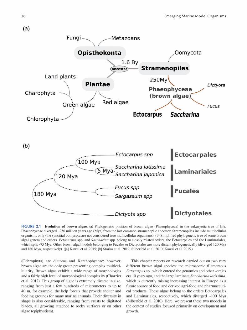
28 Emerging Marine Model Organisms
FIGURE 2.1 Evolution of brown algae. (a) Phylogenetic position of brown algae (Phaeophyceae) in the eukaryotic tree of life.
Phaeophyceae diverged ~250 million years ago (Mya) from the last common stramenopile ancestor. Stramenopiles include multicellular
organisms only (the syncitial oomycota are not considered true multicellular organisms). (b) Simplified phylogenetic tree of some brown
algal genera and orders. Ectocarpus spp. and Saccharina spp. belong to closely related orders, the Ectocarpales and the Laminariales,
which split ~75 Mya. Other brown algal models belonging to Fucales or Dictyotales are more distant phylogenetically (diverged 120 Mya
and 180 Mya, respectively). ([a] Kawai et al. 2015; [b] Starko et al. 2019; Silberfeld et al. 2010; Kawai et al. 2015.)
(Ochrophyta) are diatoms and Xanthophyceae; however, This chapter reports on research carried out on two very
brown algae are the only group presenting complex multicel- different brown algal species: the microscopic fi lamentous
lularity. Brown algae exhibit a wide range of morphologies Ectocarpus sp., which entered the genomics and other -omics
and a fairly high level of morphological complexity (Charrier era 10 years ago, and the large laminate Saccharina latissima,
et al. 2012). This group of algae is extremely diverse in size, which is currently raising increasing interest in Europe as a
ranging from just a few hundreds of micrometers to up to future source of food and derived agri-food and pharmaceuti
40 m, for example, the kelp forests that provide shelter and cal products. These algae belong to the orders Ectocarpales
feeding grounds for many marine animals. Their diversity in and Laminariales, respectively, which diverged ~100 Mya
shape is also considerable, ranging from crusts to digitated (Silberfeld et al. 2010). Here, we present these two models in
blades, all growing attached to rocky surfaces or on other the context of studies focused primarily on development and
algae (epiphystism). growth.
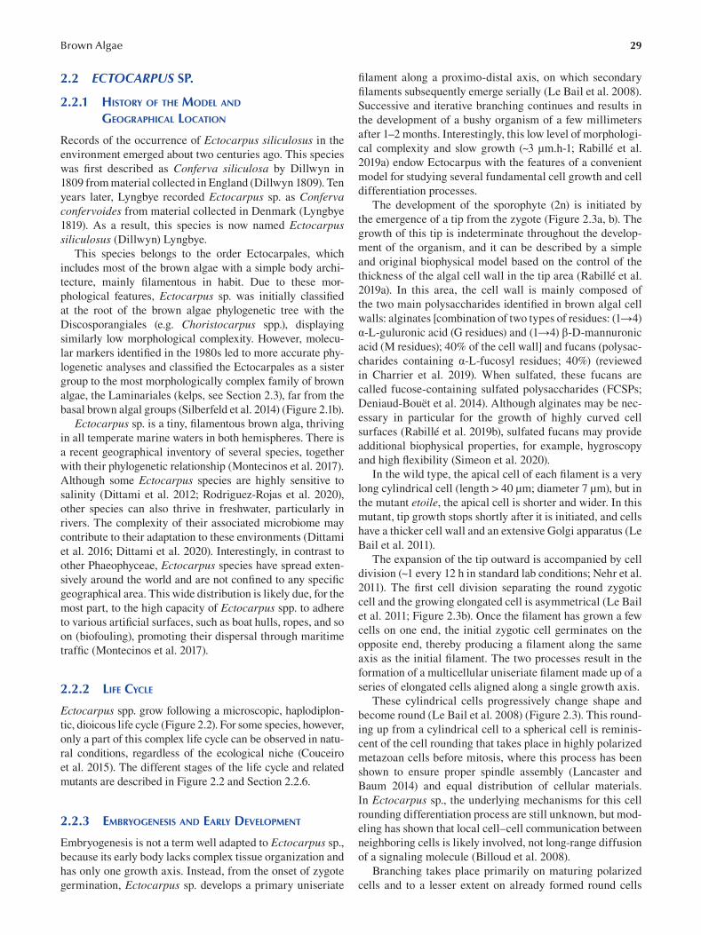
29 Brown Algae
2.2 ECTOCARPUS SP.
2.2.1 HISTORY OF THE MODEL AND
GEOGRAPHICAL LOCATION
Records of the occurrence of Ectocarpus siliculosus in the
environment emerged about two centuries ago. This species
was first described as Conferva siliculosa by Dillwyn in
1809 from material collected in England (Dillwyn 1809). Ten
years later, Lyngbye recorded Ectocarpus sp. as Conferva confervoides from material collected in Denmark (Lyngbye
1819). As a result, this species is now named Ectocarpus siliculosus (Dillwyn) Lyngbye.
This species belongs to the order Ectocarpales, which
includes most of the brown algae with a simple body archi
tecture, mainly filamentous in habit. Due to these mor
phological features, Ectocarpus sp. was initially classifi ed
at the root of the brown algae phylogenetic tree with the
Discosporangiales (e.g. Choristocarpus spp.), displaying
similarly low morphological complexity. However, molecu
lar markers identified in the 1980s led to more accurate phy
logenetic analyses and classified the Ectocarpales as a sister
group to the most morphologically complex family of brown
algae, the Laminariales (kelps, see Section 2.3), far from the
basal brown algal groups (Silberfeld et al. 2014) (Figure 2.1b).
Ectocarpus sp. is a tiny, filamentous brown alga, thriving
in all temperate marine waters in both hemispheres. There is
a recent geographical inventory of several species, together
with their phylogenetic relationship (Montecinos et al. 2017).
Although some Ectocarpus species are highly sensitive to
salinity (Dittami et al. 2012; Rodriguez-Rojas et al. 2020),
other species can also thrive in freshwater, particularly in
rivers. The complexity of their associated microbiome may
contribute to their adaptation to these environments (Dittami
et al. 2016; Dittami et al. 2020). Interestingly, in contrast to
other Phaeophyceae, Ectocarpus species have spread exten
sively around the world and are not confi ned to any specifi c
geographical area. This wide distribution is likely due, for the
most part, to the high capacity of Ectocarpus spp. to adhere
to various artificial surfaces, such as boat hulls, ropes, and so
on (biofouling), promoting their dispersal through maritime
traffic (Montecinos et al. 2017).
2.2.2 LIFE CYCLE
Ectocarpus spp. grow following a microscopic, haplodiplon
tic, dioicous life cycle (Figure 2.2). For some species, however,
only a part of this complex life cycle can be observed in natu
ral conditions, regardless of the ecological niche (Couceiro
et al. 2015). The different stages of the life cycle and related
mutants are described in Figure 2.2 and Section 2.2.6.
2.2.3 EMBRYOGENESIS AND EARLY DEVELOPMENT
Embryogenesis is not a term well adapted to Ectocarpus sp.,
because its early body lacks complex tissue organization and
has only one growth axis. Instead, from the onset of zygote
germination, Ectocarpus sp. develops a primary uniseriate
filament along a proximo-distal axis, on which secondary
filaments subsequently emerge serially (Le Bail et al. 2008).
Successive and iterative branching continues and results in
the development of a bushy organism of a few millimeters
after 1–2 months. Interestingly, this low level of morphologi
cal complexity and slow growth (~3 μm.h-1; Rabillé et al.
2019a) endow Ectocarpus with the features of a convenient
model for studying several fundamental cell growth and cell
differentiation processes.
The development of the sporophyte (2n) is initiated by
the emergence of a tip from the zygote (Figure 2.3a, b). The
growth of this tip is indeterminate throughout the develop
ment of the organism, and it can be described by a simple
and original biophysical model based on the control of the
thickness of the algal cell wall in the tip area (Rabillé et al.
2019a). In this area, the cell wall is mainly composed of
the two main polysaccharides identified in brown algal cell
walls: alginates [combination of two types of residues: (1→4)
α-L-guluronic acid (G residues) and (1→4) β-D-mannuronic
acid (M residues); 40% of the cell wall] and fucans (polysac
charides containing α-L-fucosyl residues; 40%) (reviewed
in Charrier et al. 2019). When sulfated, these fucans are
called fucose-containing sulfated polysaccharides (FCSPs;
Deniaud-Bouët et al. 2014). Although alginates may be nec
essary in particular for the growth of highly curved cell
surfaces (Rabillé et al. 2019b), sulfated fucans may provide
additional biophysical properties, for example, hygroscopy
and high flexibility (Simeon et al. 2020).
In the wild type, the apical cell of each filament is a very
long cylindrical cell (length > 40 μm; diameter 7 μm), but in
the mutant etoile, the apical cell is shorter and wider. In this
mutant, tip growth stops shortly after it is initiated, and cells
have a thicker cell wall and an extensive Golgi apparatus (Le
Bail et al. 2011).
The expansion of the tip outward is accompanied by cell
division (~1 every 12 h in standard lab conditions; Nehr et al.
2011). The first cell division separating the round zygotic
cell and the growing elongated cell is asymmetrical (Le Bail
et al. 2011; Figure 2.3b). Once the filament has grown a few
cells on one end, the initial zygotic cell germinates on the
opposite end, thereby producing a filament along the same
axis as the initial filament. The two processes result in the
formation of a multicellular uniseriate filament made up of a
series of elongated cells aligned along a single growth axis.
These cylindrical cells progressively change shape and
become round (Le Bail et al. 2008) (Figure 2.3). This round
ing up from a cylindrical cell to a spherical cell is reminis
cent of the cell rounding that takes place in highly polarized
metazoan cells before mitosis, where this process has been
shown to ensure proper spindle assembly (Lancaster and
Baum 2014) and equal distribution of cellular materials.
In Ectocarpus sp., the underlying mechanisms for this cell
rounding differentiation process are still unknown, but mod
eling has shown that local cell–cell communication between
neighboring cells is likely involved, not long-range diffusion
of a signaling molecule (Billoud et al. 2008).
Branching takes place primarily on maturing polarized
cells and to a lesser extent on already formed round cells
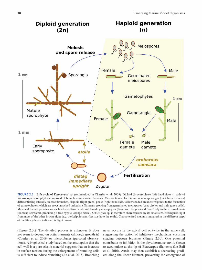
30 Emerging Marine Model Organisms
FIGURE 2.2 Life cycle of Ectocarpus sp. (summarized in Charrier et al. 2008). Diploid (brown) phase (left-hand side) is made of
microscopic sporophytes composed of branched uniseriate filaments. Meiosis takes place in unilocular sporangia (dark brown circles)
differentiating laterally on erect branches. Haploid (light green) phase (right-hand side, yellow shaded area) corresponds to the formation
of gametophytes, which are erect branched uniseriate filaments growing from germinated meiospores (gray circles and light green cells).
Male and female gametes are each released from male and female gametophytes (dioicous life cycle) and fuse freely in the external envi
ronment (seawater), producing a free zygote (orange circle). Ectocarpus sp. is therefore characterized by its small size, distinguishing it
from most of the other brown algae (e.g. the kelp Saccharina sp.) (note the scale). Characterized mutants impaired in the different steps
of the life cycle are indicated in light brown.
(Figure 2.3c). The detailed process is unknown. It does never occurs in the apical cell or twice in the same cell,
not seem to depend on actin filaments (although growth is) suggesting the action of inhibitory mechanisms ensuring
(Coudert et al. 2019) or microtubules (personal observa- spacing between branches (Figure 2.3d). One potential
tions). A biophysical study based on the assumption that the contributor to inhibition is the phytohormone auxin, shown
cell wall is a poro-elastic material suggests that an increase to accumulate at the tip of Ectocarpus filaments (Le Bail
in surface tension during the enlargement of rounding cells et al. 2010). Auxin may then establish a decreasing gradi
is sufficient to induce branching (Jia et al. 2017). Branching ent along the linear filament, preventing the emergence of
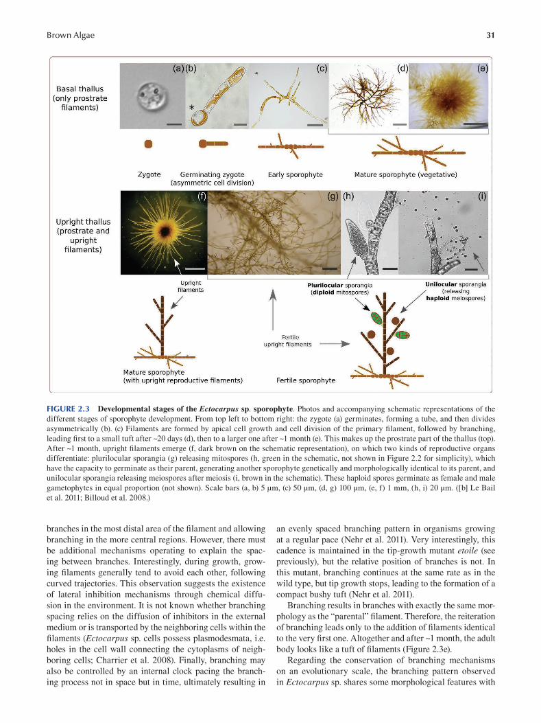
31 Brown Algae
FIGURE 2.3 Developmental stages of the Ectocarpus sp. sporophyte. Photos and accompanying schematic representations of the
different stages of sporophyte development. From top left to bottom right: the zygote (a) germinates, forming a tube, and then divides
asymmetrically (b). (c) Filaments are formed by apical cell growth and cell division of the primary filament, followed by branching,
leading first to a small tuft after ~20 days (d), then to a larger one after ~1 month (e). This makes up the prostrate part of the thallus (top).
After ~1 month, upright filaments emerge (f, dark brown on the schematic representation), on which two kinds of reproductive organs
differentiate: plurilocular sporangia (g) releasing mitospores (h, green in the schematic, not shown in Figure 2.2 for simplicity), which
have the capacity to germinate as their parent, generating another sporophyte genetically and morphologically identical to its parent, and
unilocular sporangia releasing meiospores after meiosis (i, brown in the schematic). These haploid spores germinate as female and male
gametophytes in equal proportion (not shown). Scale bars (a, b) 5 μm, (c) 50 μm, (d, g) 100 μm, (e, f) 1 mm, (h, i) 20 μm. ([b] Le Bail
et al. 2011; Billoud et al. 2008.)
branches in the most distal area of the filament and allowing an evenly spaced branching pattern in organisms growing
branching in the more central regions. However, there must at a regular pace (Nehr et al. 2011). Very interestingly, this
be additional mechanisms operating to explain the spac- cadence is maintained in the tip-growth mutant etoile (see
ing between branches. Interestingly, during growth, grow- previously), but the relative position of branches is not. In
ing filaments generally tend to avoid each other, following this mutant, branching continues at the same rate as in the
curved trajectories. This observation suggests the existence wild type, but tip growth stops, leading to the formation of a
of lateral inhibition mechanisms through chemical diffu- compact bushy tuft (Nehr et al. 2011).
sion in the environment. It is not known whether branching Branching results in branches with exactly the same mor-
spacing relies on the diffusion of inhibitors in the external phology as the “parental” filament. Therefore, the reiteration
medium or is transported by the neighboring cells within the of branching leads only to the addition of fi laments identical
fi laments (Ectocarpus sp. cells possess plasmodesmata, i.e. to the very first one. Altogether and after ~1 month, the adult
holes in the cell wall connecting the cytoplasms of neigh- body looks like a tuft of filaments (Figure 2.3e).
boring cells; Charrier et al. 2008). Finally, branching may Regarding the conservation of branching mechanisms
also be controlled by an internal clock pacing the branch- on an evolutionary scale, the branching pattern observed
ing process not in space but in time, ultimately resulting in in Ectocarpus sp. shares some morphological features with
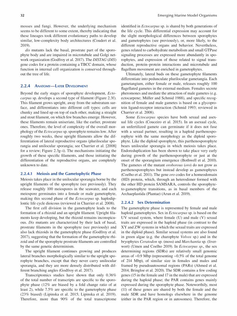
32
mosses and fungi. However, the underlying mechanism
seems to be different to some extent, thereby indicating that
these lineages took different evolutionary paths to develop
similar, low-complexity body architectures (Coudert et al.
2019 ).
dis mutants lack the basal, prostrate part of the sporo
phyte body and are impaired in microtubule and Golgi net
work organization (Godfroy et al. 2017). The DISTAG (DIS)
gene codes for a protein containing a TBCC domain, whose
function in internal cell organization is conserved through
out the tree of life.
2.2.4 ANATOMY—LATER DEVELOPMENT
Beyond the early stages of sporophyte development, Ectocarpus sp. develops a second type of filament (Figure 2.3f).
This filament grows upright, away from the substratum sur
face, and differentiates into different cell types: cells are
chunky and lined up on top of each other, making a straight
and stout filament, on which few branches emerge. However,
these filaments remain uniseriate, like the earlier, prostrate
ones. Therefore, the level of complexity of the overall mor
phology of the Ectocarpus sp. sporophyte remains low. After
roughly two weeks, these upright filaments allow the dif
ferentiation of lateral reproductive organs (plurilocular spo
rangia and unilocular sporangia; see Charrier et al. [2008]
for a review; Figure 2.3g–i). The mechanisms initiating the
growth of these specifi c filaments, and those initiating the
differentiation of the reproductive organs, are completely
unknown to date.
2.2.4.1 Meiosis and the Gametophytic Phase Meiosis takes place in the unilocular sporangia borne by the
upright filaments of the sporophyte (see previously). They
release roughly 100 meiospores in the seawater, and each
meiospore germinates into a female or male gametophyte,
making this second phase of the Ectocarpus sp. haplodip
lontic life cycle dioicous (reviewed in Charrier et al. 2008).
The first cell division in the gametophyte leads to the
formation of a rhizoid and an upright filament. Upright fi la
ments keep developing, but the rhizoid remains inconspicu
ous. Dis mutants are characterized by their lack of basal,
prostrate filaments in the sporophyte (see previously) and
also lack rhizoids in the gametophyte phase (Godfroy et al.
2017), suggesting that the formation of the gametophyte rhi
zoid and of the sporophyte prostrate filaments are controlled
by the same genetic determinism.
The upright filament continues growing and produces
lateral branches morphologically similar to the upright spo
rophyte branches, except that they never carry unilocular
sporangia, and they are more densely distributed with dif
ferent branching angles (Godfroy et al. 2017 ).
Transcriptomics studies have shown that only 0.36%
of the total number of transcripts are specific to the sporo
phyte phase (12% are biased by a fold change ratio of at
least 2), while 7.5% are specific to the gametophyte phase
(23% biased) ( Lipinska et al. 2015 ; Lipinska et al. 2019 ).
Therefore, more than 90% of the total transcriptome
Emerging Marine Model Organisms
identified in Ectocarpus sp. is shared by both generations of
the life cycle. This differential expression may account for
the slight morphological differences between sporophytes
and gametophytes (see previously), or, more likely, to the
different reproductive organs and behavior. Nevertheless,
genes related to carbohydrate metabolism and small GTPase
signaling processes are expressed more abundantly in spo
rophytes, and expression of those related to signal trans
duction, protein–protein interactions and microtubule and
flagellum movement are enriched in gametophytes.
Ultimately, lateral buds on these gametophyte fi laments
differentiate into pedunculate plurilocular gametangia. Each
gametangium, either female or male, releases roughly 100
flagellated gametes in the external medium. Females secrete
pheromones and mediate the attraction of male gametes (e.g.
ectocarpene; Müller and Schmid 1988 ), and specifi c recog
nition of female and male gametes is based on a glycopro
tein ligand-receptor interaction ( Schmid 1993 ; reviewed in
Charrier et al. 2008 ).
Some Ectocarpus species have both sexual and asex
ual life cycles (Couceiro et al. 2015). In an asexual cycle,
an unfertilized gamete can germinate if it does not fuse
with a sexual partner, resulting in a haploid parthenospo
rophyte with the same morphology as the diploid sporo
phyte. Like the diploid sporophyte, this parthenosporophyte
bears unilocular sporangia in which meiosis takes place.
Endoreduplication has been shown to take place very early
during growth of the parthenosporophyte or just at the
onset of the sporangium emergence (Bothwell et al. 2010).
The gametes of the mutant oroborous (oro) do not grow as
parthenosporophytes but instead develop as gametophytes
(Coelho et al. 2011). The gene oro codes for a homeodomain
(HD) protein, which, through an heterodimer formed with
the other HD protein SAMSARA, controls the sporophyte-
to-gametophyte transitions, as in basal members of the
Archaeplastida (Plantae) (Arun et al. 2019).
2.2.4.2 Sex Determination The gametophyte phase is represented by female and male
haploid gametophytes. Sex in Ectocarpus sp. is based on the
UV sexual system, where female (U) and male (V) sexual
traits are expressed in the haploid phase (in contrast to the
XY and ZW systems in which the sexual traits are expressed
in the diploid phase). Similar sexual systems are also found
in green algae (e.g. the charophyte Volvox sp.) and in the
bryophytes Ceratodon sp. (moss) and Marchantia sp. (liver
wort) (Umen and Coelho 2019). In Ectocarpus sp., the sex
determining regions (SDRs) are relatively small genomic
areas of ~0.9 Mbp (representing ~0.5% of the total genome
of 214 Mbp), of similar size in females and males and
framed by pseudoautosomal regions (PARs) (Ahmed et al.
2014; Bringloe et al. 2020). The SDR contains a few coding
genes (15 in the female and 17 in the male) that are expressed
during the haploid phase; the PAR contains genes mainly
expressed during the sporophyte phase. Noteworthily, most
(11) of these genes are shared by both the female and the
male SDR and have homologs elsewhere in the genome
(either in the PAR region or in autosomes). Therefore, the
33 Brown Algae
identity of the Ectocarpus sp. sex locus is weak compared
with other species, both in the number and in the specifi city
of its genes. Nevertheless, these SDR loci control the expres
sion of 753 female genes (with a -fold change [FC] > 2),
representing 4.3% of the total transcripts (5.5% of the tran
scripts expressed in the female gametophyte), located in the
rest of the genome during the haploid phase (Lipinska et al.
2015). In the male gametophyte, 1391 genes (7.9% total tran
scripts, 10% male-gametophyte-expressed genes) are specif
ically expressed with a FC > 2.
However, the role of these gametophyte genes in sex
determination remains unclear, because the sexual dimor
phism observed in this genus is nonexistent in vegetative
gametophytes and subtle during the reproductive phase, dur
ing which male gametophytes produce more gametangia and
slightly smaller gametes than female gametophytes (Lipinska
et al. 2015; Luthringer et al. 2014). This slight dimorphism
is reflected by the weak differential expression of sex-biased
genes at these two stages of gametophyte development.
In summary, Ectocarpus sp. is characterized by a low
level of morphological complexity: cells are aligned, and
growth is one dimensional, followed by reiterated branching
events producing filaments similar to the “mother” fi lament.
The life cycle is virtually isomorphic: sporophyte and game
tophyte are both filamentous, mainly made up of upright
filaments, and gender traits are absent.
2.2.5 GENOMIC DATA
The nuclear genome of Ectocarpus sp. (accession CCAP
1310/4) has been estimated to contain 214 Mbp, and genome
sequence annotation identified 17,418 genes (Cormier et al.
2017). As the first sequence known for a brown alga at that
time, it revealed unusual features. With a high GC content,
genes are composed of, on average, 8 × 300 bp exons, sepa
rated by seven introns of 740 bp. Alternative splicing takes
place with a frequency leading to 1.6 transcript per gene
(Cormier et al. 2017), comparable to alternative splicing in
metazoans and plants. Promoters have not been characterized
to date, and 3’-UTR regions are particularly long (~900 bp), in
contrast to most other organisms of similar genome size but
similar to mammalian genomes. From this genome, several
families of transposable elements, of which retrotransposons
and retroposons are the most abundant, cover ~20% of the
genome (Cock et al. 2010), as well as 23 microRNAs identi
fied from a genome-based approach and whose expression
has been quantifi ed by q-RT-PCR (Billoud et al. 2014). This
inventory also includes a set of 63 miR candidates identifi ed
from an RNA-seq-based approach, limited by the extent of
range and level of gene expression (Tarver et al. 2015).
Interestingly, a significant proportion of Ectocarpus sp.
genes are organized on alternating DNA strands along the
chromosome, a feature specific to compact genomes.
A preliminary genetic map built with microsatellite
markers was proposed in 2010 (Heesch et al. 2010), since
supplemented with single nucleotide polymorphism (SNP)
markers, facilitating the identification of mutated loci
(Billoud et al. 2015; Cormier et al. 2017). All together, based
on genetic linkage and flow cytometry data (although from
another species), Ectocarpus sp. does not appear to have
more than 28 chromosomes (Cormier et al. 2017).
2.2.6 FUNCTIONAL APPROACHES: TOOLS FOR
MOLECULAR AND CELLULAR ANALYSES
Based on a solid knowledge of its biology and life cycle
(reviewed in Charrier et al. 2008), Ectocarpus sp. was cho
sen as a genetic model for brown algae in the 2000s (Peters
et al. 2004). Its genome was sequenced in 2010, which was
a major breakthrough as the first genomic sequence for a
brown alga and, what’s more, the first multicellular mac
roalga (Cock et al. 2010). This breakthrough was accompa
nied by the development of a full palette of technical tools.
Only techniques related to cell biology, cultivation and
genetics are considered in the following.
2.2.6.1 Cultivation in the Laboratory Ectocarpus sp. is easily grown in laboratory conditions (Le
Bail and Charrier 2013). Growth speed, morphology and
fertility induction depend on (white) light intensity (usually
dim, <30 μE.s −1.m−2), photoperiod (long day or equal day:
night cycle) and temperature (13–14°C). Due to its small size,
optical microscopes and stereo microscopes are required to
follow the different stages of the life cycle. Micromanipulation
(using tweezers) is often necessary to separate the different
organs of the Ectocarpus sp. body, such as sporangia. The
adult organism is a few centimeters long, meaning that the
whole life cycle can be carried out in a small recipient such
as a Petri dish. Altogether, the cultivation of Ectocarpus sp. is amenable to rudimentary laboratory conditions and
equipment. To avoid contamination with either bacteria or
protozoa, Ectocarpus sp. is preferably handled under a ster
ile laminar hood.
2.2.6.2 Cell Biology and Biophysical Techniques Transmission and scanning electronic microscopy tech
niques have both been used to observe Ectocarpus sp. cells
and filaments (e.g. Le Bail et al. 2011; Tsirigoti et al. 2015),
facilitated by the filamentous shape of this organism, expos
ing all cells to observation. However, because the cells are
small (filament cell diameter, 7 μm), observation of a spe
cific cell orientation may be difficult to handle. However,
exploiting the fact that Ectocarpus sp. grows on surface, it
is possible to make serial sections of apical filament cells in
longitudinal and transversal axes, as illustrated in Rabillé
et al. (2019), who measured the thickness of the cell wall
along the meridional axis of the cell.
Protocols for immunocytochemistry (ICC, or immunolo
calization) of cytoskeleton components have been developed
in the past 20 years, inspired by protocols developed on other
brown algae (reviewed in Katsaros et al. 2006 ). Microtubules
(Coelho et al. 2012; Katsaros et al. 1992), actin fi laments
(Rabillé et al. 2018b) and centrin (Katsaros et al. 1991; Godfroy
et al. 2017) can now be visualized in Ectocarpus sp. cells.
These ICC protocols rely on the high conservation of these
molecules, allowing the use of commercial primary antibodies
34
raised against animal homologs. ICC using antibodies specifi c
to Ectocarpus sp. has not been reported yet. However, mono
clonal antibodies raised against polysaccharide components of
the brown algal cell wall have been produced (Torode et al.
2016, 2015) and are now used to map specific blocks of algi
nates (Rabillé et al. 2019b) and fucans (Simeon et al. 2020).
A recent study on Ectocarpus sp. using mRNA in situ hybridization after an attack by a pathogen (Badstöber
et al. 2020) showed mRNA in subcellular locations within
the infected cell. The development of fi lament-wide in situ mRNA labeling is needed to monitor responses or cell-fate
programs at the level of the whole organism.
Additional techniques, previously developed in other
organisms, have been transferred to Ectocarpus sp . Growth
of the cell surface can be monitored by loading sticky fl uo
rescent beads on the filament surface. Recording the posi
tion of the beads as the cell expands (either during growth or
in response to a stimulus) makes it possible to measure the
propensity for deformation of specific cell areas. This mea
surement provides information on cell mechanical proper
ties (Rabillé et al. 2018a). Mechanical properties can also be
studied using atomic force microscopy, a biophysical tech
nique that records how deep a cantilever can plunge into a
cell surface and retract, according to the cell wall stiffness
and adhesion (Gaboriaud and Dufrêne 2007). Ectocarpus sp. is particularly amenable to such approaches, because
its cells are directly exposed to the cantilever (Tesson and
Charrier 2014). This technique helped show that the cells
along the sporophyte filament display different degrees of
surface stiffness (Rabillé et al. 2019b).
2.2.6.3 Modification of Gene Expression Attempts to genetically transform Ectocarpus sp. have been
numerous and so far unsuccessful. Agrotransformation,
electroporation, PEG-mediated protoplast or gamete trans
formation and micro-injection have all been tested and
shown to be inefficient. A major issue is that there is little to
no information on Ectocarpus sp. gene promoters, and het
erologous promoters tested so far (e.g. diatom, Ulva, Maize
or Plant virus CaMV35S) have not been shown to be func
tional (personal communication).
Therefore, “ready-to-use” molecules that can alter the
expression of host genes without relying on the host tran
scription and translation machinery currently appear to be a
more promising approach. Morpholinos and RNA interfer
ence have not proven to be efficient enough for routine tran
sient knock-down experiments (personal communication;
Macaisne et al. 2017).
Efforts are currently being put into the development of
the CRISPR-Cas9 technology (Lino et al. 2018), shown to
be a powerful tool to stably modify the genome of several
marine organisms, including echinoderms (sea urchins; Lin
et al. 2019) and tunicates (Phallusia sp.; McDougall et al.
2021). Because the expression of the guided RNA and the
Cas9 protein from the host genome remains challenging, the
use of pre-assembled guide RNA-Cas9 protein complex, as
illustrated in Brassicaceae plants (Murovec et al. 2018), is
currently considered the most promising strategy.
Emerging Marine Model Organisms
Several morphogenetic mutants have been generated by
UV irradiation, among which some have been genetically
characterized. These mutants are impaired in tip growth
(Le Bail et al. 2011), cell differentiation (Godfroy et al. 2017;
Macaisne et al. 2017; Le Bail et al. 2010), branching and repro
ductive phase change (Le Bail et al. 2010). In most mutants,
several morphogenetic processes are affected, refl ecting the
low level of complexity of Ectocarpus sp. morphogenesis and
suggesting an overlap in genetic functions (see transcriptomic
results previously). Others are impaired in the alternation
of the sporophyte and gametophyte generations (life cycle
mutants: Coelho et al. 2011; Arun et al. 2019) (Figure 2.2).
2.3 SACCHARINA LATISSIMI
2.3.1 NOMENCLATURE HISTORY, EVOLUTION, GEOGRAPHICAL DISTRIBUTION AND USES
2.3.1.1 History of Its Nomenclature Saccharina latissima (Linnaeus) C.E. Lane, C. Mayes,
Druehl & G.W. Saunders 2006 is a marine photosynthetic
eukaryotic organism with many different common names,
including sugar kelp, sea-belt, kombu, sugar tang, poor man’s
weather glass and so on. Originally, in 1753, Linnaeus con
sidered it an Ulva species, Ulva latissima, due to its sheet
like blade, common in the genus Ulva (Linnaeus and Salvius
1753 ). In 1813, Lamouroux reclassified it as Laminaria saccharina (Lamouroux 1813), despite its original genus name
Saccharina given by the botanist J. Stackhouse in 1809. This
genus name was resurrected in 2006 when molecular phylo
genetics made it apparent that the order Laminariales should
be split into two clades or families (Lane et al. 2006), which
diverged ~25 Mya (Starko et al. 2019). Now, Laminaria spp.
are assigned to the Laminariaceae family, and Saccharina spp. are part of the Arthrothamnaceae family (Jackson et al.
2017 ).
2.3.1.2 Evolution and Diversifi cation Classic taxonomy using morphological or physiological
characteristics is useful for identifying species in the fi eld;
however, in the absence of a genetic approach, they can lead
to long-lasting species confusions.
Among the brown algae, kelps are thought to have
emerged ~75 Mya (Starko et al. 2019). Within the kelps
(order Laminariales), S. latissima belongs to the so-called
“complex kelps” (Starko et al. 2019) and thus shows close
genetic similarity with various genera, allegedly result
ing from an important upsurge in speciation beginning 31
Mya, concomitant to a massive marine species extinction
due to the cooling of the Pacifi c Ocean during the Eocene–
Oligocene boundary.
2.3.1.3 Geographical Distribution Kelps are now almost cosmopolitan species, their pres
ence ranging from temperate to cold waters on both sides
of the Atlantic and Pacific Oceans (Bartsch et al. 2008).
Saccharina genus appears to have initially emerged in
the Northwest Pacific (North Japan, Russia) (Bolton 2010;
35 Brown Algae
Luttikhuizen et al. 2018; Starko et al. 2019) and then spread
further to three or four distinct regions of the globe where
different lineages of S. latissima can be traced: in temper
ate to cold-temperate (sub-Arctic) waters of the Northeast
Pacific, where the early diversifi cation of Laminariales
ancestors took place, and in the Northeast and Northwest
Atlantic (Neiva et al. 2018; Starko et al. 2019). S. latissima is
absent from the southern hemisphere (Bolton 2010).
Even though these populations seem to be considered as
a single species (assumption supported by crosses), barcod
ing studies (based on the cytochrome c oxidase gene, used
for) indicate high divergence between regions (Neiva et al.
2018). In combination with their morphological divergence
and history of glacial vicariance (Neiva et al. 2018), these
regional groups of S. latissima are clearly differentiating
into separate species.
2.3.1.4 Uses Individual kelp can become enormous: S. latissima blades
can grow up to 45 m in length (Kanda 1936). As such, kelps
constitute the largest coastal biomass and one of the main
primary producers of the oceans. According to the FAO
(2018), kelps in general, and S. latissima in particular, are
cultivated and consumed mainly in Asia for human suste
nance as well as for their alginate and iodine contents. In
comparison, European consumption and production are
considerably lower, and wild populations are used for vari
ous applications, mainly food and feed (Rebours et al. 2014;
Barbier et al. 2019). Recent innovations aim to combine S. latissima cultivation with salmon aquaculture (integrated
multi-trophic aquaculture) to reduce the impact of fi sh farms
in Norway (Fossberg et al. 2018). S. latissima has been pro
posed as a source of bioethanol (Adams et al. 2008; Kraan
2016), and substances such as the sulfated polysaccharides
fucoidans, laminarin and other extracts have demonstrated
antitumoral effects along with anti-inflammatory and anti
coagulant pharmacological properties (Cumashi et al. 2007;
Mohibbullah et al. 2019; Han et al. 2019; Long et al. 2019).
The number of clinical studies on mice for testing the posi
tive effects of kelp extracts keeps increasing, as attested by
a simple search in the PubMed scientific literature search
engine.
2.3.2 LIFE CYCLE
S. latissima is characterized by a highly heteromorphic
haplodiplontic life cycle (Figure 2.4). Meiosis leads to the
haploid gametophytic stage (or generation) of the life cycle,
which, upon fertilization, gives rise to a diploid sporophyte
stage. In S. latissima, and generally in Laminariales, the
sporophyte generation is considerably different morphologi
cally from the gametophyte generation (Kanda 1936; Fritsch
1945), in contrast to the isomorphic haplodiplontic life cycle
of Ectocarpales, as seen in the previous section (Figure 2.2).
The gametophyte (haploid) is microscopic and slowly grows
into a prostrate filamentous thallus, and the sporophyte (dip
loid) is large and conspicuous. Upon favorable conditions,
reproduction is initiated by a gradual differentiation of the
cells of the gametophytic filaments into reproductive cells,
the gametangia—antheridia (male) or oogonia (female)—
a process induced by blue light (Lüning and Dring 1972).
Interestingly, this induction is accompanied by changes in
gene expression that are by and large common to female and
male gametophytes, suggesting that the initiation of germ-
line differentiation follows similar general mechanisms
independently of gender (Pearson et al. 2019). That is, tran
scriptomics studies have revealed enhanced transcriptional
and translational activities as well as metabolic activities
(carbohydrate biosynthesis and nitrogen uptake), suggesting
that gametogenesis is accompanied by an intensifi cation of
primary cellular and metabolic functions. This intensifi ca
tion is surprising when weighed against the fact that only
one single gamete is produced by each gametangium. Yet
there are differences between female and male gametogen
esis proper. A small set of genes display gender-dependent
induction of their expression, seemingly faster in females
than in males (Pearson et al. 2019). Genes involved in basic
cellular function (protein modification, nucleoplasmic trans
port, intron splicing), energy production and metabolic path
ways and more specifically in oogenesis (reactive oxidative
species metabolism) are overexpressed during female game
togenesis, in addition to prostaglandin-biosynthesis genes
(Pearson et al. 2019; Monteiro et al. 2019). In turn, and as
expected, male gametogenesis is accompanied mainly by
the over-expression of “high mobility group” (HMG) genes,
a conserved marker of male gender determination in ani
mals, fungi and brown algae (Ahmed et al. 2014), which
suppresses the development of female gender, hereby con
sidered as set by default (Pearson et al. 2019).
In relatively high temperature conditions (20°C), the
male and female gametophytes show more similar transcrip
tomic patterns, probably indicating a change of focus from
gametogenesis-related genes to resistance to heat stress,
amplified in females (Monteiro et al. 2019).
The oogonium releases an egg, leaving behind an empty
apoplast, a process that is subject to the circadian rhythm
and, in contrast to the formation of gametangia, is inhib
ited by blue light (Lüning 1981). Male gametes swim to
the egg in response to female pheromones (e.g. lamoxiren;
Hertweck and Boland 1997 ), demonstrating conspicuous
chemotaxis (Maier and Müller 1986; Maier and Muller
1990; Boland 1995; Maier et al. 2001; Kinoshita et al. 2017).
Upon fertilization, the early sporophyte develops as a pla
nar embryo. In Saccharina japonica, the ratio of genes spe
cifi c to sporophytes or gametophytes is more balanced than
in Ectocarpus sp., with ~4% (about 700 genes) of the total
number of transcripts being specifically expressed in both
phase organisms (Lipinska et al. 2019). This difference in
transcripts can be interpreted as a reflection of the conspicu
ous morphological differences between these two life cycle
generations in S. latissima, contrasting with the near iso
morphy in Ectocarpus sp.
The developing diploid sporophyte requires several
months before reaching sexual maturity (Andersen et al.
2011; Forbord et al. 2018 , 2019). Then, sori, groups of sacs
(sporangia) of meiospores (swimming spores that are the
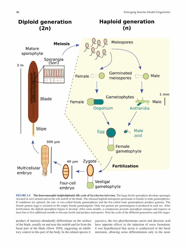
36 Emerging Marine Model Organisms
FIGURE 2.4 The heteromorphic halplodiplontic life cycle of Saccharina latissima. The large fertile sporophyte develops sporangia
(located in sori) around and on the soft midrib of the blade. The released haploid meiospores germinate to female or male gametophytes.
If conditions are optimal, the one- to two-celled female gametophytes and the few-celled male gametophytes produce gametes. The
female gamete (egg) is retained on the empty female gametangium. Only one gamete per gametangium is produced in each sex. After
fertilization, the diploid sporophyte begins to develop. After some months, a conspicuous juvenile sporophyte emerges and requires at
least four to five additional months to become fertile and produce meiospores. Note the scale of the different generations and life stages.
product of meiosis) abundantly differentiate on the surface japonica, the two phytohormones auxin and abscissic acid
of the blade, usually on and near the midrib and far from the have opposite effects in the induction of sorus formation;
basal part of the blade (Drew 1910), suggesting an inhibi- it was hypothesized that auxin is synthesized in the basal
tory control in this part of the body. In the related species S. meristem, allowing sorus differentiation only in the more
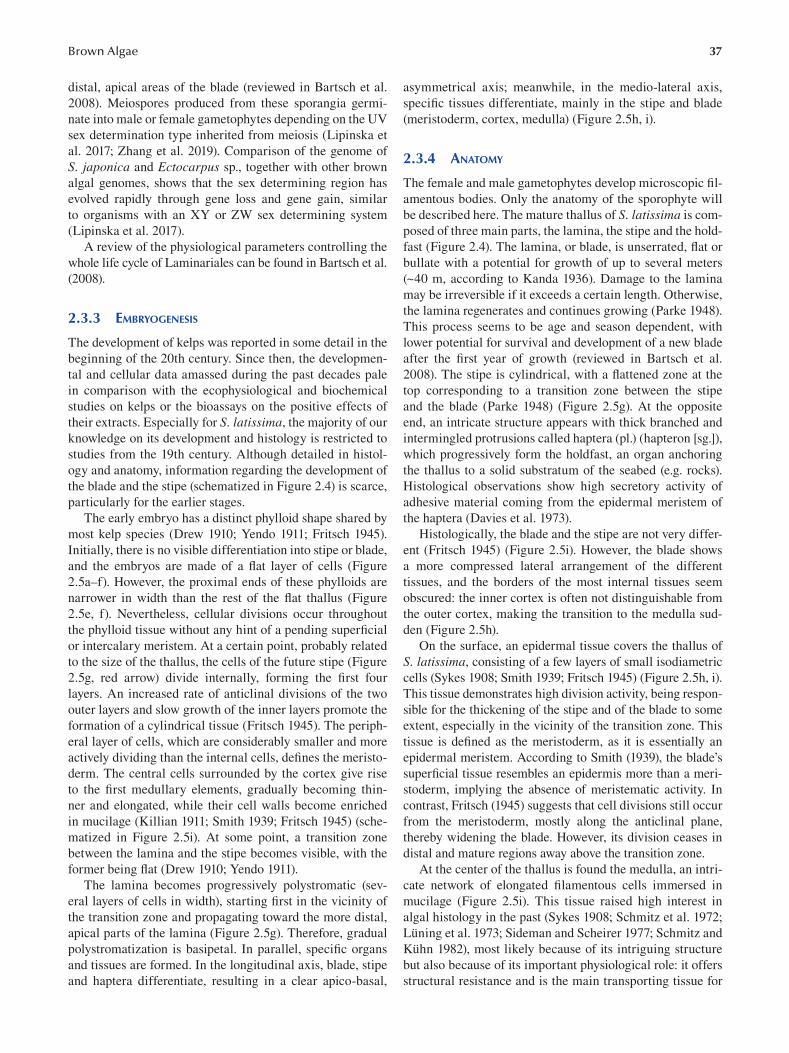
37 Brown Algae
distal, apical areas of the blade (reviewed in Bartsch et al.
2008). Meiospores produced from these sporangia germi
nate into male or female gametophytes depending on the UV
sex determination type inherited from meiosis (Lipinska et
al. 2017; Zhang et al. 2019). Comparison of the genome of
S. japonica and Ectocarpus sp., together with other brown
algal genomes, shows that the sex determining region has
evolved rapidly through gene loss and gene gain, similar
to organisms with an XY or ZW sex determining system
(Lipinska et al. 2017).
A review of the physiological parameters controlling the
whole life cycle of Laminariales can be found in Bartsch et al.
(2008 ).
2.3.3 EMBRYOGENESIS
The development of kelps was reported in some detail in the
beginning of the 20th century. Since then, the developmen
tal and cellular data amassed during the past decades pale
in comparison with the ecophysiological and biochemical
studies on kelps or the bioassays on the positive effects of
their extracts. Especially for S. latissima, the majority of our
knowledge on its development and histology is restricted to
studies from the 19th century. Although detailed in histol
ogy and anatomy, information regarding the development of
the blade and the stipe (schematized in Figure 2.4) is scarce,
particularly for the earlier stages.
The early embryo has a distinct phylloid shape shared by
most kelp species (Drew 1910; Yendo 1911; Fritsch 1945).
Initially, there is no visible differentiation into stipe or blade,
and the embryos are made of a flat layer of cells (Figure
2.5a– f). However, the proximal ends of these phylloids are
narrower in width than the rest of the flat thallus (Figure
2.5e, f). Nevertheless, cellular divisions occur throughout
the phylloid tissue without any hint of a pending superfi cial
or intercalary meristem. At a certain point, probably related
to the size of the thallus, the cells of the future stipe (Figure
2.5g, red arrow) divide internally, forming the fi rst four
layers. An increased rate of anticlinal divisions of the two
outer layers and slow growth of the inner layers promote the
formation of a cylindrical tissue (Fritsch 1945). The periph
eral layer of cells, which are considerably smaller and more
actively dividing than the internal cells, defines the meristo
derm. The central cells surrounded by the cortex give rise
to the first medullary elements, gradually becoming thin
ner and elongated, while their cell walls become enriched
in mucilage (Killian 1911; Smith 1939; Fritsch 1945) (sche
matized in Figure 2.5i). At some point, a transition zone
between the lamina and the stipe becomes visible, with the
former being flat (Drew 1910; Yendo 1911).
The lamina becomes progressively polystromatic (sev
eral layers of cells in width), starting first in the vicinity of
the transition zone and propagating toward the more distal,
apical parts of the lamina (Figure 2.5g). Therefore, gradual
polystromatization is basipetal. In parallel, specifi c organs
and tissues are formed. In the longitudinal axis, blade, stipe
and haptera differentiate, resulting in a clear apico-basal,
asymmetrical axis; meanwhile, in the medio-lateral axis,
specific tissues differentiate, mainly in the stipe and blade
(meristoderm, cortex, medulla) (Figure 2.5h , i).
2.3.4 ANATOMY
The female and male gametophytes develop microscopic fi l
amentous bodies. Only the anatomy of the sporophyte will
be described here. The mature thallus of S. latissima is com
posed of three main parts, the lamina, the stipe and the hold
fast (Figure 2.4). The lamina, or blade, is unserrated, fl at or
bullate with a potential for growth of up to several meters
(~40 m, according to Kanda 1936). Damage to the lamina
may be irreversible if it exceeds a certain length. Otherwise,
the lamina regenerates and continues growing (Parke 1948).
This process seems to be age and season dependent, with
lower potential for survival and development of a new blade
after the first year of growth (reviewed in Bartsch et al.
2008). The stipe is cylindrical, with a flattened zone at the
top corresponding to a transition zone between the stipe
and the blade (Parke 1948) (Figure 2.5g). At the opposite
end, an intricate structure appears with thick branched and
intermingled protrusions called haptera (pl.) (hapteron [sg.]),
which progressively form the holdfast, an organ anchoring
the thallus to a solid substratum of the seabed (e.g. rocks).
Histological observations show high secretory activity of
adhesive material coming from the epidermal meristem of
the haptera (Davies et al. 1973).
Histologically, the blade and the stipe are not very differ
ent (Fritsch 1945) (Figure 2.5i). However, the blade shows
a more compressed lateral arrangement of the different
tissues, and the borders of the most internal tissues seem
obscured: the inner cortex is often not distinguishable from
the outer cortex, making the transition to the medulla sud
den (Figure 2.5h).
On the surface, an epidermal tissue covers the thallus of
S. latissima, consisting of a few layers of small isodiametric
cells ( Sykes 1908 ; Smith 1939 ; Fritsch 1945 ) ( Figure 2.5h , i ).
This tissue demonstrates high division activity, being respon
sible for the thickening of the stipe and of the blade to some
extent, especially in the vicinity of the transition zone. This
tissue is defined as the meristoderm, as it is essentially an
epidermal meristem. According to Smith (1939), the blade’s
superficial tissue resembles an epidermis more than a meri
stoderm, implying the absence of meristematic activity. In
contrast, Fritsch (1945) suggests that cell divisions still occur
from the meristoderm, mostly along the anticlinal plane,
thereby widening the blade. However, its division ceases in
distal and mature regions away above the transition zone.
At the center of the thallus is found the medulla, an intri
cate network of elongated filamentous cells immersed in
mucilage (Figure 2.5i). This tissue raised high interest in
algal histology in the past (Sykes 1908; Schmitz et al. 1972;
Lüning et al. 1973; Sideman and Scheirer 1977; Schmitz and
Kühn 1982), most likely because of its intriguing structure
but also because of its important physiological role: it offers
structural resistance and is the main transporting tissue for
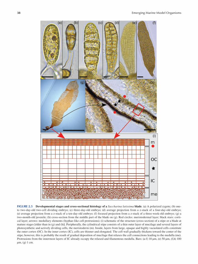
38 Emerging Marine Model Organisms
FIGURE 2.5 Developmental stages and cross-sectional histology of a Saccharina latissima blade. (a) A polarized zygote; (b) one-
to two-day-old two-cell dividing embryo; (c) three-day-old embryo; (d) average projection from a z-stack of a four-day-old embryo;
(e) average projection from a z-stack of a ten-day-old embryo; (f) focused projection from a z-stack of a three-week-old embryo; (g) a
two-month-old juvenile; (h) cross-section from the middle part of the blade on (g). Red circles: meristodermal layer; black stars: corti
cal layer; arrows: medullary elements (hyphae-like cell protrusions); (i) schematic of the structure (cross-section) of a stipe or a blade at
mature stages [older than in (g) and (h)]. Peripherally, the cylindrical stipe consists of a thin outer layer of mucilage and several layers of
photosynthetic and actively dividing cells, the meristoderm (m). Inside, layers from large, opaque and highly vacuolated cells constitute
the outer cortex (OC). In the inner cortex (IC), cells are thinner and elongated. The cell wall gradually thickens toward the center of the
stipe; however, this is probably the result of gradual deposition of mucilage that relaxes the cell connections leading to the medulla (me).
Protrusions from the innermost layers of IC already occupy the relaxed and filamentous medulla. Bars: (a-f) 10 μm, (e) 50 μm, (f,h) 100
μm, (g) 1 cm.
39 Brown Algae
photoassimilates and nutrients. In recent studies on other
kelp species, the medullary cells seem to have the capacity
to generate turgor through the elastic properties of their cell
walls (as illustrated in the kelp Nereocystis by Knoblauch
et al. 2016a), possibly controlling the flow of the transported
solutions. This and the alginate-rich extracellular matrix of
the medulla make the sieve elements of kelps a study model
for fluid mechanics in transport systems of plant organisms,
since they are easily manipulable (Knoblauch et al. 2016b).
Between the medulla and the meristoderm resides the
cortex (Figure 2.5h , i). It is divided into two parts: the outer
cortex and the inner cortex. The outer cortex is easily distin
guished from the meristoderm due to its sizable isodiametric
opaque cells with pointy corners. The inner cortex is closer
to the medulla and has elongated, thick-walled cells (closer
to the medulla) with straight edges. At the transition zone
and young parts of the stipe, the outer cortex cells widen and
lengthen following the enlargement of the organ. A gradual
change toward the more elongated cells of the inner cortex is
visible. The innermost cells close to the medulla have protru
sions on their most internal (proximal) longitudinal cell walls
that may overlap each other, gradually resembling the shape
and size of the medulla cells, as they progressively occupy
this intricate mesh. At the transition zone, both the abun
dant mucilage deposits and the elongation of the innermost
cells in combination with their growing septate protrusions
“relax” the inner cortex tissue, which gradually differentiates
into medullary cells (Killian 1911; Fritsch 1945). The inner
cortex is supplied with cells from the outer cortex, which
themselves originate from the actively dividing meristoderm.
In summary, growth of the blade and the stipe in the lon
gitudinal axis is ensured by the transition zone, which fur
nishes the blade and the stipe with new tissues (Smith 1939;
Fritsch 1945; Parke 1948; Steinbiss and Schmitz 1974).
Therefore, the transition zone is characterized by both cell
division activity in the longitudinal axis, which provides the
cells for the lamina and stipe tissues, and active cell divi
sion in the peripheral meristoderm, whose role is to renew
and keep providing cells to the transition zone. In this area,
cell division and cell differentiation take place centripetally.
Recently, transcriptomics studies confirmed an increasing
meristematic activity in this location through the upregula
tion of ribosomal proteins and immediate upright genes a
in the basal part of the blade (Ye et al. 2015), as in juvenile
sporophytes (Shao et al. 2019).
As soon as the blade and the stipe can be identifi ed,
haptera start differentiating in the very basal part of the S. latissima thallus. These are outgrowths that originate from
the lower end of the stipe, where a disc-like structure ini
tially forms on top of the rhizoids (Drew 1910; Yendo 1911).
Above this structure, the first haptera start developing.
While it shares the cortex and meristoderm with the stipe,
the medulla of the stipe does not extend into the haptera
(Yendo 1911; Smith 1939; Fritsch 1945; Davies et al. 1973).
Haptera growth seems to be apical, but there is no exten
sive research on that matter. Haptera cells contribute to car
bon fixation through photosynthesis, except when sheltered
from light, resulting in cells of the haptera meristoderm
displaying underdeveloped plastids with a rudimentary thy
lakoid membrane system (Davies et al. 1973). In addition,
their endomembrane system is very well developed, with
hypertrophied dictyosomes containing cell wall polysaccha
rides and alginate acid.
2.3.5 GENOMICS
The S. latissima genome sequence is expected to be released
in 2021 (Project “Phaeoexplorer”, led by FranceGenomics
and the Roscoff Marine Station, www.france-genomique.
org/projet/phaeoexplorer/). In the meantime, a draft genome
sequence was published in 2015 for the close relative S. japonica (Ye et al. 2015), which diverged from S. latissima only ~5 Mya (Starko et al. 2019). It was enhanced by recent
genome assembly work (Liu et al. 2019), leading to a genome
of 580 Mbp for >35,000 genes.
The S. japonica genome is 2.7 times bigger than that of
Ectocarpus sp. (Cock et al. 2010), and it contains twice as
many genes; as expected, gene length is similar in the two
species (Liu et al. 2019). Average exon lengths (~250 bp)
are similar, but introns are less abundant (only 4.6 per gene
on average in S. japonica vs. 7 in Ectocarpus sp.). Oddly,
because introns are longer (1200 bp vs. 700 in Ectocarpus sp.), the overall exon:intron ratio per gene remains similar in
Saccharina sp. and Ectocarpus sp. However, a signifi cant dif
ference lies in the presence of repeated sequences (46% in
S. japonica vs. 22% in Ectocarpus sp.), mainly composed of
class I and class II transposons and microsatellite sequences
(Liu et al. 2019).
A large proportion of the gene content (85%) is distrib
uted in gene families found in Ectocarpus sp. Nevertheless,
detailed analysis shows interesting differences, in line with
the biology of the organisms. In particular, the high capac
ity of S. japonica to accumulate iodine is reflected in the
composition of its genome, which displays a very rich group
of vanadium-dependent haloperoxidases (vHPOs), most
likely resulting from gene expansion (Ye et al. 2015; Liu et
al. 2019). Gene expansion may also have led to a signifi cant
increase in cell wall biosynthesis proteins (especially those
involved in the synthesis of alginates), protein kinases and
membrane-spanning receptor kinases. All together, in com
parison with the Ectocarpus sp. genome, gene expansion
would have been the genetic basis for the diversifi cation of
body plans and more generally of the complex multicellular
ity of Laminariales (Liu et al. 2019), which, together with
the increased bioaccumulation of iodine, are the main char
acteristics differentiating Laminariales from Ectocarpales.
Interestingly, compared with other genomes, Ectocarpus sp. and S. japonica genomes display a signifi cant increase
in gene families (~1200) counterbalanced with a limited
loss (~300), whose functions involve enzyme hydrolysis and
cupin-like proteins (Ye et al. 2015). Although the functions
of the gained gene families are largely unknown due to the
lack of sequence conservation with other organisms, protein
kinase and helix-extended-loop-helix super family domains
have been identified as enriched domains in this group, sug
gesting a role in cell signaling and cell differentiation.
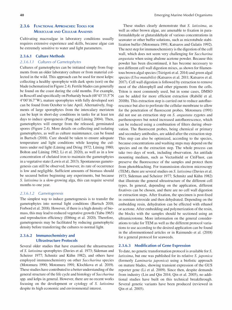
40
2.3.6 FUNCTIONAL APPROACHES: TOOLS FOR
MOLECULAR AND CELLULAR ANALYSES
Cultivating macroalgae in laboratory conditions usually
requires extensive experience and skills, because algae can
be extremely sensitive to water and light parameters.
2.3.6.1 Culture Methods 2.3.6.1.1 Cultures of Gametophytes Cultures of gametophytes can be initiated simply from frag
ments from an older laboratory culture or from material col
lected in the wild. This approach can be used for most kelps:
collecting a healthy sporophyte with dark spots (sori) on the
blade (schematized in Figure 2.4). Fertile blades can generally
be found on the coast during the cold months. For example,
in Roscoff and specifically on Perharidy beach (48°43’33.5”N
4°00’16.7”W), mature sporophytes with fully developed sori
can be found from October to late April. Alternatively, frag
ments of large sporophytes from the intercalary meristem
can be kept in short-day conditions in tanks for at least ten
days to induce sporogenesis (Pang and Lüning 2004). Then,
gametophytes will emerge from the released, germinated
spores (Figure 2.4). More details on collecting and isolating
gametophytes, as well as culture maintenance, can be found
in Bartsch (2018). Care should be taken to ensure adequate
temperature and light conditions while keeping the cul
tures under red light (Lüning and Dring 1972; Lüning 1980;
Bolton and Lüning 1982; Li et al. 2020), as well as in a low
concentration of chelated iron to maintain the gametophytes
in a vegetative state (Lewis et al. 2013). Spontaneous gameto
genesis can still be observed; however, its rate of occurrence
is low and negligible. Sufficient amounts of biomass should
be secured before beginning any experiments, but because
S. latissima is a slow-growing alga, this can require several
months to one year.
2.3.6.1.2 Gametogenesis The simplest way to induce gametogenesis is to transfer the
gametophytes into normal light conditions (Bartsch 2018;
Forbord et al. 2018). However, if there is a high density of bio
mass, this may lead to reduced vegetative growth (Yabu 1965)
and reproduction efficiency (Ebbing et al. 2020). Therefore,
gametogenesis may be facilitated by reducing gametophytic
density before transferring the cultures to normal light.
2.3.6.2 Immunochemistry and Ultrastructure Protocols
Several older studies that have examined the ultrastructure
of S. latissima sporophytes (Davies et al. 1973; Sideman and
Scheirer 1977; Schmitz and Kühn 1982), and others have
employed immunochemistry on other Saccharina species
( Motomura 1990 ; Motomura 1991 ; Klochkova et al. 2019 ).
These studies have contributed to a better understanding of the
general structure of the life cycle and histology of Saccharina spp. and kelps in general. However, there are no recent works
focusing on the development or cytology of S. latissima despite its high economic and environmental interest.
Emerging Marine Model Organisms
These studies clearly demonstrate that S. latissima , as
well as other brown algae, are amenable to fixation in para
formaldehyde or glutaraldehyde of various concentrations in
seawater or other buffer solutions, such as microtubule stabi
lization buffer (Motomura 1991; Katsaros and Galatis 1992).
The next step for immunochemistry is the digestion of the cell
wall, which does not seem very challenging for Saccharina angustata when using abalone acetone powder. Because this
powder has been discontinued, it has become necessary to
test different cell wall digestion mixes, as shown for fi lamen
tous brown algal species (Tsirigoti et al. 2014) and green algal
species (Ulva mutabilis) (Katsaros et al. 2011; Katsaros et al.
2017). Cell wall digestion is followed by extraction to remove
most of the chlorophyll and other pigments from the cells.
Triton is most commonly used, but in some cases, DMSO
can be added for more efficient extraction (Rabillé et al.
2018b). This extraction step is carried out to reduce autofl uo
rescence but also to perforate the cellular membrane to allow
for the penetration of fluorescent probes. Motomura (1991)
did not use an extraction step on S. angustata zygotes and
parthenospores but noted increased autofl uorescence, which
can be reduced using a combination of filters during obser
vation. The fluorescent probes, being chemical or primary
and secondary antibodies, are added after the extraction step.
This step can also be optimized, according to the species,
because concentrations and washing steps may depend on the
species and on the extraction step. The whole process can
take two days of work, including observation. An antifade
mounting medium, such as Vectashield or CitiFluor, can
preserve the fluorescence of the samples and protect them
from photobleaching. For transmission electron microscopy
(TEM), there are several studies on S. latissima (Davies et al.
1973; Sideman and Scheirer 1977; Schmitz and Kühn 1982)
that illustrate the general ultrastructure of the different cell
types. In general, depending on the application, different
fixatives can be chosen, and there are no cell wall digestion
or extraction steps. After fixation, the specimen is post-fi xed
in osmium tetroxide and then dehydrated. Depending on the
embedding resin, dehydration can be effected with ethanol
or acetone. After embedding and polymerization of the resin,
the blocks with the samples should be sectioned using an
ultramicrotome. More information on the general consider
ations to take for TEM as well as the different protocol varia
tions to use according to the desired application can be found
in the aforementioned articles or in Raimundo et al. (2018)
for a general protocol for seaweeds.
2.3.6.3 Modification of Gene Expression To date, no genetic transformation protocol is available for S. latissima, but one was published for its relative S. japonica (formerly Laminaria japonica) using a biolistic approach
on mature blades, showing transient expression of the GUS
reporter gene (Li et al. 2009). Since then, despite demands
from industry (Lin and Qin 2014; Qin et al. 2005), no addi
tional studies have built on this technical breakthrough.
Several genetic variants have been produced (reviewed in
Qin et al. 2005).
41 Brown Algae
2.4 CHALLENGING QUESTIONS IN BASIC AND APPLIED RESEARCH
2.4.1 WHY STUDY BROWN ALGAE?
2.4.1.1 Advancing Knowledge on Their Developmental Mechanisms
Brown algae make up a specific phylum of multicellular
organisms. Their phylogenetic position in the eukaryotic
tree (Baldauf 2008), distant from other multicellular organ
isms, makes them a key taxon for understanding the evo
lution of complex multicellularity and specifi c metabolic
pathways. The literature abounds with biological questions
and research topics positioning these organisms as essen
tial ones to consider in future studies, and, more specifi cally
related to this chapter, brown algae offer a wealth of candi
date species to study the evolution of the formation of dif
ferent body shapes. Furthermore, in contrast to the red and
green algae, there is no representative unicellular species for
brown algae, making the evolutionary scenario of the emer
gence of their diverse shapes even more intriguing.
However, the knowledge in the fields of evolution and
development is very scarce compared with that on metazo
ans and land plants. In the following, two examples pertain
ing to kelp features illustrate the potential brown algae hold
for leading to knowledge breakthroughs in developmental
biology.
First, despite the similarities between brown algal tissues
and complex histological structures in land plants, brown
algal body architecture and shape remain fairly simple. Even
kelps—the most complex brown algae at the morphological
level—develop only a few different organs (blade, stipe and
holdfast), with a limited number of specific tissues and cell
types (i.e. epidermis, cortex, medulla, meristoderm, sorus
[this chapter] and pneumatocysts, receptacles and concepta
cles in other brown algae [reviewed in Charrier et al. 2012]).
This relative simplicity provides a useful opportunity to
study basic developmental mechanisms based on simple
geometrical rules or morphogen gradients. Although auxin,
the long-standing leading morphogen for land plants, is
present in brown algae and affects morphogenesis of several
morphologically simple brown algae, such as Ectocarpus sp.
and Dictyota sp. (Dictyotales) (Le Bail et al. 2010; Bogaert
et al. 2019), it has no conspicuous effect, nor is it specifi
cally localized in the apex of Sargassum sp. (Fucales), a
brown alga with relatively high morphological complexity,
including the presence of an apical meristem (stem cell tis
sue) (Linardić and Braybrook 2017). This result casts doubt
on the consistency of morphogen-mediated control mecha
nisms in brown algae and presages the identification of new,
alternative growth control mechanisms.
The second example relates to one of the stunning char
acteristics of some brown algae: their size. How do cells
communicate with each other over such a long distance,
when it comes to organisms among the tallest on earth:
kelps? The transport system in kelps is reminiscent of the
vascular systems of land plants, except that the extracellular
matrix (alginates) has a specific organization and distribu
tion and contributes to the flow of photoassimilated prod
ucts (Knoblauch et al. 2016a, 2016b). Cells connect with
each other through pit structures where the plasmodesmata
(channels or pore connecting two adjacent cells) are con
centrated. These plasmodesmata are structurally similar to
those in land plants (Terauchi et al. 2015), except for the
absence of desmotubules and the lack of the ability to con
trol the size of molecules transferred symplastically (Bouget
et al. 1998; Terauchi et al. 2015). Although some kelps (e.g.
Macrocystis spp.) adjust the size of their vascular tissues to
the needs for photoassimilate distribution to “sink” organs
(i.e. meristerm, storage tissues, sori) as land plants do, oth
ers do not, suggesting again different control mechanisms in
the management of this important function (Drobnitch et al.
2015). One explanation is that larger kelp rely more heavily
on an efficient transport system, especially when source and
sink tissues are physically distant. Relying on a transport
system would call for a regulated developmental process, as
in land plants (Drobnitch et al. 2015).
2.4.1.2 Improving Aquaculture Over the past several decades, S. japonica (known as
“kombu”) aquaculture in Asia has undergone many improve
ments at many different levels, because this alga has been
cultivated for human consumption for several centuries.
One improvement lever is breeding, and—beyond empirical
approaches used in the past—genomics can now assist and
speed up breeding programs (Wang et al. 2020), along with
new knowledge on the control of the life cycle, reproduction
and early growth steps (e.g. substrate adhesion, sensitivity to
high density) (reviewed in Charrier et al. 2017). Regarding
more specifi cally S. latissima cultivated in Europe, its
genome has not yet been sequenced and, other than concerns
on the ecological impact of seaweed aquaculture, the cur
rent bottlenecks are mainly technical and focused on scaling
up production and reducing cultivation costs (reviewed in
Barbier et al. 2019).
2.4.2 BIOLOGICAL MODELS: ECTOCARPUS SP., S. LATISSIMA OR ANOTHER BROWN ALGA?
Because Ectocarpus sp. is a morphologically and sexually
simple organism, it is a convenient model for cellular and
molecular studies requiring microscopy, and this asset is
enhanced by the availability of many additional cell biology
tools (e.g. protocols for immunolocalization of the cell wall
and the cytoskeleton, laser capture microdissection, in situ hybridization, etc.). Therefore, as illustrated in this chapter,
its amenability to laboratory experimentation and its short
life cycle have made it a convenient organism to explore.
However, its low biomass is an impediment for biochemical
research, in addition to its simple morphology, which pre
cludes the study of complex multicellular mechanisms.
This is how S. latissima landed on the roadmap: based
on the wealth of cultivation practice-based knowledge from
applied phycology and aquaculture R&D laboratories,
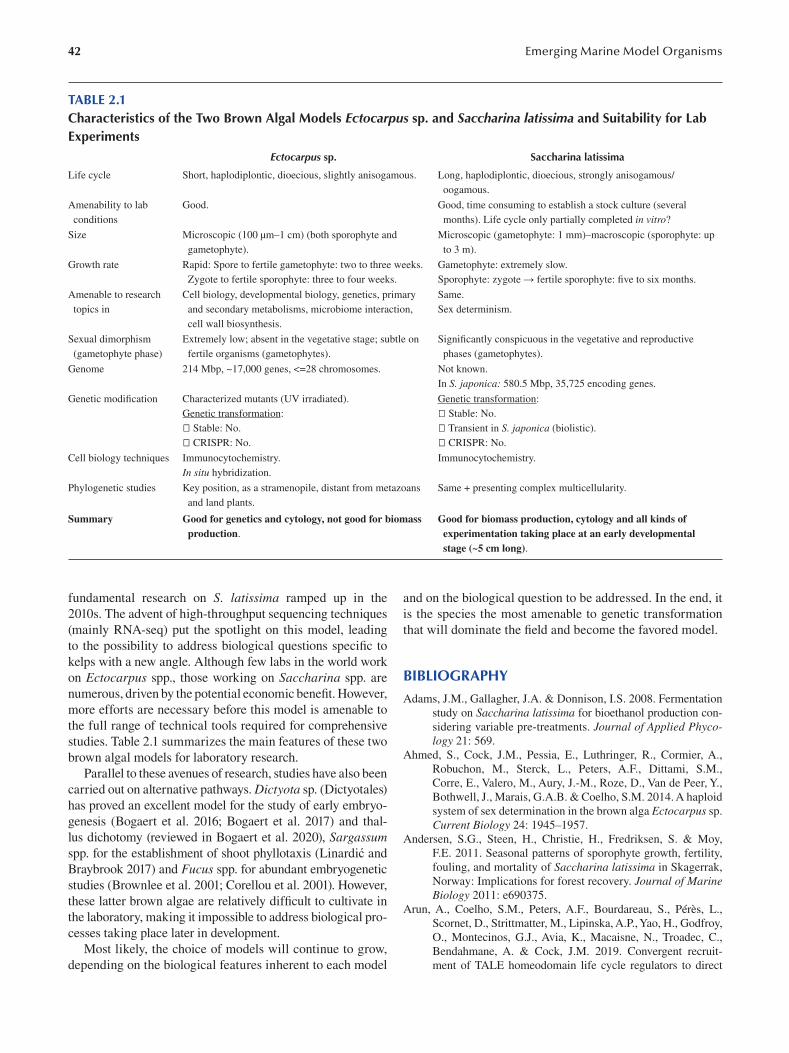
42 Emerging Marine Model Organisms
TABLE 2.1 Characteristics of the Two Brown Algal Models Ectocarpus sp. and Saccharina latissima and Suitability for Lab Experiments
Ectocarpus sp . Saccharina latissima
Life cycle Short, haplodiplontic, dioecious, slightly anisogamous. Long, haplodiplontic, dioecious, strongly anisogamous/
oogamous.
Amenability to lab Good. Good, time consuming to establish a stock culture (several
conditions months). Life cycle only partially completed in vitro?
Size Microscopic (100 μm–1 cm) (both sporophyte and Microscopic (gametophyte: 1 mm)–macroscopic (sporophyte: up
gametophyte). to 3 m).
Growth rate Rapid: Spore to fertile gametophyte: two to three weeks. Gametophyte: extremely slow.
Zygote to fertile sporophyte: three to four weeks. Sporophyte: zygote → fertile sporophyte: five to six months.
Amenable to research Cell biology, developmental biology, genetics, primary Same.
topics in and secondary metabolisms, microbiome interaction, Sex determinism.
cell wall biosynthesis.
Sexual dimorphism Extremely low; absent in the vegetative stage; subtle on Significantly conspicuous in the vegetative and reproductive
(gametophyte phase) fertile organisms (gametophytes). phases (gametophytes).
Genome 214 Mbp, ~17,000 genes, <=28 chromosomes. Not known.
In S. japonica: 580.5 Mbp, 35,725 encoding genes.
Genetic modifi cation Characterized mutants (UV irradiated). Genetic transformation :
Genetic transformation : Stable: No.
Stable: No. Transient in S. japonica (biolistic).
CRISPR: No. CRISPR: No.
Cell biology techniques Immunocytochemistry. Immunocytochemistry.
In situ hybridization.
Phylogenetic studies Key position, as a stramenopile, distant from metazoans Same + presenting complex multicellularity.
and land plants.
Summary Good for genetics and cytology, not good for biomass Good for biomass production, cytology and all kinds of production. experimentation taking place at an early developmental
stage (~5 cm long).
fundamental research on S. latissima ramped up in the
2010s. The advent of high-throughput sequencing techniques
(mainly RNA-seq) put the spotlight on this model, leading
to the possibility to address biological questions specifi c to
kelps with a new angle. Although few labs in the world work
on Ectocarpus spp., those working on Saccharina spp. are
numerous, driven by the potential economic benefi t. However,
more efforts are necessary before this model is amenable to
the full range of technical tools required for comprehensive
studies. Table 2.1 summarizes the main features of these two
brown algal models for laboratory research.
Parallel to these avenues of research, studies have also been
carried out on alternative pathways. Dictyota sp. (Dictyotales)
has proved an excellent model for the study of early embryo
genesis (Bogaert et al. 2016; Bogaert et al. 2017) and thal
lus dichotomy (reviewed in Bogaert et al. 2020), Sargassum spp. for the establishment of shoot phyllotaxis (Linardić and
Braybrook 2017 ) and Fucus spp. for abundant embryogenetic
studies (Brownlee et al. 2001; Corellou et al. 2001). However,
these latter brown algae are relatively difficult to cultivate in
the laboratory, making it impossible to address biological pro
cesses taking place later in development.
Most likely, the choice of models will continue to grow,
depending on the biological features inherent to each model
and on the biological question to be addressed. In the end, it
is the species the most amenable to genetic transformation
that will dominate the field and become the favored model.
BIBLIOGRAPHY
Adams, J.M., Gallagher, J.A. & Donnison, I.S. 2008. Fermentation
study on Saccharina latissima for bioethanol production con
sidering variable pre-treatments. Journal of Applied Phycology 21: 569.
Ahmed, S., Cock, J.M., Pessia, E., Luthringer, R., Cormier, A.,
Robuchon, M., Sterck, L., Peters, A.F., Dittami, S.M.,
Corre, E., Valero, M., Aury, J.-M., Roze, D., Van de Peer, Y.,
Bothwell, J., Marais, G.A.B. & Coelho, S.M. 2014. A haploid
system of sex determination in the brown alga Ectocarpus sp.
Current Biology 24: 1945–1957.
Andersen, S.G., Steen, H., Christie, H., Fredriksen, S. & Moy,
F.E. 2011. Seasonal patterns of sporophyte growth, fertility,
fouling, and mortality of Saccharina latissima in Skagerrak,
Norway: Implications for forest recovery. Journal of Marine Biology 2011: e690375.
Arun, A., Coelho, S.M., Peters, A.F., Bourdareau, S., Pérès, L.,
Scornet, D., Strittmatter, M., Lipinska, A.P., Yao, H., Godfroy,
O., Montecinos, G.J., Avia, K., Macaisne, N., Troadec, C.,
Bendahmane, A. & Cock, J.M. 2019. Convergent recruit
ment of TALE homeodomain life cycle regulators to direct
43 Brown Algae
sporophyte development in land plants and brown algae. eLife 8: e43101.
Badstöber, J., Gachon, C.M.M., Ludwig-Müller, J., Sandbichler,
A.M. & Neuhauser, S. 2020. Demystifying biotrophs: FISHing
for mRNAs to decipher plant and algal pathogen-host interac
tion at the single cell level. Scientic Reports 10: 14269.
Baldauf, S.L. 2008. An overview of the phylogeny and diversity of
eukaryotes. Journal of Systematics and Evolution 46: 263–273.
Barbier, M., Charrier, B., Araujo, R., Holdt, S.L., Jacquemin, B. &
Rebours, C. 2019. PEGASUS: PHYCOMORPH European Guidelines for a Sustainable Aquaculture of Seaweeds , COST
Action FA1406 (M. Barbier & B. Charrier, eds). Roscoff,
France.
Bartsch, I. 2018. Derivation of clonal stock cultures and hybridiza
tion of kelps: A tool for strain preservation and breeding pro
grams. In: Protocols for Macroalgae Research , pp. 61–78.
Francis & Taylor Group, CRC Press, Boca Raton.
Bartsch, I., Wiencke, C., Bischof, K., Buchholz, C.M., Buck, B.H.,
Eggert, A., Feuerpfeil, P., Hanelt, D., Jacobsen, S., Karez, R.,
Karsten, U., Molis, M., Roleda, M.Y., Schubert, H., Schumann,
R., Valentin, K., Weinberger, F. & Wiese, J. 2008. The genus
Laminaria sensu lato: Recent insights and developments.
European Journal of Phycology 43: 1–86.
Billoud, B., Bail, A.L. & Charrier, B. 2008. A stochastic 1D near
est-neighbour automaton models early development of the
brown alga Ectocarpus siliculosus. Functional Plant Biology 35: 1014–1024.
Billoud, B., Jouanno, É., Nehr, Z., Carton, B., Rolland, É.,
Chenivesse, S. & Charrier, B. 2015. Localization of causal
locus in the genome of the brown macroalga Ectocarpus: NGS-
based mapping and positional cloning approaches. Frontiers in Plant Sciences 6: 68.
Billoud, B., Nehr, Z., Le Bail, A. & Charrier, B. 2014. Computational
prediction and experimental validation of microRNAs in the
brown alga Ectocarpus siliculosus. Nucleic Acids Research 42: 417–429.
Bogaert, K.A., Beeckman, T. & De Clerck, O. 2016. Abiotic regula
tion of growth and fertility in the sporophyte of Dictyota dichotoma (Hudson) J.V. Lamouroux (Dictyotales, Phaeophyceae).
Journal of Applied Phycology 28: 2915–2924.
Bogaert, K.A., Beeckman, T. & De Clerck, O. 2017. Two-step cell
polarization in algal zygotes. Nature Plants 3: 16221.
Bogaert, K.A., Blommaert, L., Ljung, K., Beeckman, T. & De
Clerck, O. 2019. Auxin function in the brown alga Dictyota dichotoma. Plant Physiology 179: 280–299.
Bogaert, K.A., Delva, S. & De Clerck, O. 2020. Concise review of the
genus Dictyota J.V. Lamouroux. Journal of Applied Phycology 32: 1521–1543. https://doi.org/10.1007/s10811-020-02121-4
Boland, W. 1995. The chemistry of gamete attraction: Chemical
structures, biosynthesis, and (a)biotic degradation of algal
pheromones. Proceedings of the National Academy of Sciences U.S.A. 92: 37–43.
Bolton, J.J. 2010. The biogeography of kelps (Laminariales,
Phaeophyceae): A global analysis with new insights from
recent advances in molecular phylogenetics. Helgoland Marine Research 64: 263–279.
Bolton, J.J. & Lüning, K. 1982. Optimal growth and maxi
mal survival temperatures of Atlantic Laminaria species
(Phaeophyta) in culture. Marine Biology 66: 89–94.
Bothwell, J.H., Marie, D., Peters, A.F., Cock, J.M. & Coelho,
S.M. 2010. Role of endoreduplication and apomeiosis dur
ing parthenogenetic reproduction in the model brown alga
Ectocarpus. New Phytologist 188: 111–121.
Bouget, F.Y., Berger, F. & Brownlee, C. 1998. Position dependent
control of cell fate in the Fucus embryo: Role of intercellular
communication. Development 125: 1999–2008.
Bringloe, T.T., Starko, S., Wade, R.M., Vieira, C., Kawai, H., De
Clerck, O., Cock, J.M., Coelho, S.M., Destombe, C., Valero,
M., Neiva, J., Pearson, G.A., Faugeron, S., Serrao, E.A. &
Verbruggen, H. 2020. Phylogeny and evolution of the brown
algae. Critical Review of Plant Sciences 39: 281–321.
Brownlee, C., Bouget, F.Y. & Corellou, F. 2001. Choosing sides:
establishment of polarity in zygotes of fucoid algae. Seminars in Cell & Developmental Biology 12: 345–351. https://doi.
org/10.1006/scdb.2001.0262
Charrier, B., Abreu, M.H., Araujo, R., Bruhn, A., Coates, J.C., De
Clerck, O., Katsaros, C., Robaina, R.R. & Wichard, T. 2017.
Furthering knowledge of seaweed growth and development
to facilitate sustainable aquaculture. New Phytologist 216:
967–975.
Charrier, B., Coelho, S.M., Le Bail, A., Tonon, T., Michel, G., Potin,
P., Kloareg, B., Boyen, C., Peters, A.F. & Cock, J.M. 2008.
Development and physiology of the brown alga Ectocarpus siliculosus: Two centuries of research. New Phytologist 177:
319–332.
Charrier, B., Le Bail, A. & de Reviers, B. 2012. Plant Proteus:
Brown algal morphological plasticity and underlying devel
opmental mechanisms. Trends in Plant Science 17: 468–477.
Charrier, B., Rabillé, H. & Billoud, B. 2019. Gazing at cell wall
expansion under a golden light. Trends in Plant Science 24:
130–141.
Cock, J.M., Sterck, L., Rouzé, P., Scornet, D., Allen, A.E., Amoutzias,
G., Anthouard, V., Artiguenave, F., Aury, J.-M., Badger, J.H.,
Beszteri, B., Billiau, K., Bonnet, E., Bothwell, J.H., Bowler,
C., Boyen, C., Brownlee, C., Carrano, C.J., Charrier, B.,
Cho, G.Y., Coelho, S.M., Collén, J., Corre, E., Da Silva, C.,
Delage, L., Delaroque, N., Dittami, S.M., Doulbeau, S., Elias,
M., Farnham, G., Gachon, C.M.M., Gschloessl, B., Heesch,
S., Jabbari, K., Jubin, C., Kawai, H., Kimura, K., Kloareg, B.,
Küpper, F.C., Lang, D., Le Bail, A., Leblanc, C., Lerouge, P.,
Lohr, M., Lopez, P.J., Martens, C., Maumus, F., Michel, G.,
Miranda-Saavedra, D., Morales, J., Moreau, H., Motomura, T.,
Nagasato, C., Napoli, C.A., Nelson, D.R., Nyvall-Collén, P.,
Peters, A.F., Pommier, C., Potin, P., Poulain, J., Quesneville,
H., Read, B., Rensing, S.A., Ritter, A., Rousvoal, S., Samanta,
M., Samson, G., Schroeder, D.C., Ségurens, B., Strittmatter,
M., Tonon, T., Tregear, J.W., Valentin, K., von Dassow, P.,
Yamagishi, T., Van de Peer, Y. & Wincker, P. 2010. The
Ectocarpus genome and the independent evolution of multi
cellularity in brown algae. Nature 465: 617–621.
Coelho, S.M., Godfroy, O., Arun, A., Le Corguillé, G., Peters,
A.F. & Cock, J.M. 2011. OUROBOROS is a master regu
lator of the gametophyte to sporophyte life cycle transition
in the brown alga Ectocarpus. Proceedings of the National Academy of Sciences U.S.A. 108: 11518–11523.
Coelho, S.M., Scornet, D., Rousvoal, S., Peters, N., Dartevelle, L.,
Peters, A.F. & Cock, J.M. 2012. Immunostaining of Ectocarpus
cells. Cold Spring Harbour Protocols 2012: 369–372.
Corellou F, Brownlee C, Detivaud L, et al. 2001. Cell cycle in the
fucus zygote parallels a somatic cell cycle but displays a
unique translational regulation of cyclin-dependent kinases.
Plant Cell 13: 585–598.
Cormier, A., Avia, K., Sterck, L., Derrien, T., Wucher, V., Andres,
G., Monsoor, M., Godfroy, O., Lipinska, A., Perrineau,
M.-M., Peer, Y.V.D., Hitte, C., Corre, E., Coelho, S.M. &
Cock, J.M. 2017. Re-annotation, improved large-scale
44
assembly and establishment of a catalogue of noncoding loci
for the genome of the model brown alga Ectocarpus. New Phytologist 214: 219–232.
Couceiro, L., Le Gac, M., Hunsperger, H.M., Mauger, S.,
Destombe, C., Cock, J.M., Ahmed, S., Coelho, S.M., Valero,
M. & Peters, A.F. 2015. Evolution and maintenance of hap
loid-diploid life cycles in natural populations: The case of the
marine brown alga Ectocarpus. Evolution 69: 1808–1822.
Coudert, Y., Harris, S. & Charrier, B. 2019. Design principles of
branching morphogenesis in filamentous organisms. Current Biology 29: R1149–R1162.
Cumashi, A., Ushakova, N.A., Preobrazhenskaya, M.E., D’Incecco,
A., Piccoli, A., Totani, L., Tinari, N., Morozevich, G.E.,
Berman, A.E., Bilan, M.I., Usov, A.I., Ustyuzhanina, N.E.,
Grachev, A.A., Sanderson, C.J., Kelly, M., Rabinovich,
G.A., Iacobelli, S., Nifantiev, N.E. & Consorzio Interu
niversitario Nazionale per la Bio-Oncologia, Italy. 2007.
A comparative study of the anti-infl ammatory, anticoagu
lant, antiangiogenic, and antiadhesive activities of nine dif
ferent fucoidans from brown seaweeds. Glycobiology 17:
541–552.
Davies, J.M., Ferrier, N.C. & Johnston, C.S. 1973. The ultrastructure
of the meristoderm cells of the hapteron of Laminaria. Journal of the Marine Biological Association U-K 53: 237–246.
Deniaud-Bouët, E., Kervarec, N., Michel, G., Tonon, T., Kloareg,
B. & Hervé, C. 2014. Chemical and enzymatic fractionation
of cell walls from Fucales: Insights into the structure of the
extracellular matrix of brown algae. Annals of Botany 114:
1203–1216.
Derelle, R., López-García, P., Timpano, H. & Moreira, D. 2016. A
phylogenomic framework to study the diversity and evolu
tion of stramenopiles (=heterokonts). Molecular Biology and Evolution 33: 2890–2898.
Dillwyn, L.W. 1809. British Confervae: Or Colored Figures and Descriptions of the British Plants Referred by Botanists to the Genus Conferva. W. Phillips, London, UK.
Dittami, S.M., Duboscq-Bidot, L., Perennou, M., Gobet, A., Corre,
E., Boyen, C. & Tonon, T. 2016. Host-microbe interactions
as a driver of acclimation to salinity gradients in brown algal
cultures. The ISME Journal 10: 51–63.
Dittami, S.M., Gravot, A., Goulitquer, S., Rousvoal, S., Peters,
A.F., Bouchereau, A., Boyen, C. & Tonon, T. 2012. Towards
deciphering dynamic changes and evolutionary mechanisms
involved in the adaptation to low salinities in Ectocarpus
(brown algae). The Plant Journal 71: 366–377.
Dittami, S.M., Peters, A.F., West, J.A., Cariou, T., KleinJan, H.,
Burgunter-Delamare, B., Prechoux, A., Egan, S. & Boyen,
C. 2020. Revisiting Australian Ectocarpus subulatus (Phaeophyceae) from the Hopkins river: Distribution, abi
otic environment, and associated microbiota. Journal of Phycology 56: 719–729.
Drew, G.H. 1910. The reproduction and early development of
Laminaria digitata and Laminaria saccharina. Annals of Botany 24: 177–189.
Drobnitch, S.T., Jensen, K.H., Prentice, P. & Pittermann, J. 2015.
Convergent evolution of vascular optimization in kelp
(Laminariales). Proceedings of Royal Society B: Biological Sciences 282: 20151667.
Ebbing, A., Pierik, R., Bouma, T., Kromkamp, J.C. & Timmermans,
K. 2020. How light and biomass density influence the repro
duction of delayed Saccharina latissima gametophytes
(Phaeophyceae). Journal of Phycology 56: 709–718.
FAO. 2018. The global status of seaweed production, trade and uti
lization. Globefish Research Programme. Vol. 124, p. 120.
Rome.
Emerging Marine Model Organisms
Forbord, S., Steinhovden, K.B., Rød, K.K., Handå, A., Skjermo,
J., Steinhovden, K.B., Rød, K.K., Handå, A. & Skjermo,
J. 2018. Cultivation protocol for Saccharina latissima . In:
Protocols for Macroalgae Research, pp. 37–60. Francis &
Taylor Groups, CRC Press, Boca Raton.
Forbord, S., Steinhovden, K.B., Solvang, T., Handå, A. & Skjermo,
J. 2019. Effect of seeding methods and hatchery periods on
sea cultivation of Saccharina latissima (Phaeophyceae): A
Norwegian case study. Journal of Applied Phycology 32:
2201–2212.
Fossberg, J., Forbord, S., Broch, O.J., Malzahn, A.M., Jansen, H.,
Handå, A., Førde, H., Bergvik, M., Fleddum, A.L., Skjermo, J. &
Olsen, Y. 2018. The potential for upscaling Kelp (Saccharina
latissima) cultivation in salmon-driven integrated multi-trophic
aquaculture (IMTA). Frontiers in Marine Science 5: 418.
Fritsch, F.E. 1945. The Structure and Reproduction of the Algae.
Cambridge University Press, Cambridge.
Gaboriaud, F. & Dufrêne, Y.F. 2007. Atomic force microscopy of
microbial cells: Application to nanomechanical properties,
surface forces and molecular recognition forces. Colloids and Surfaces B Biointerfaces 54: 10–19.
Godfroy, O., Uji, T., Nagasato, C., Lipinska, A.P., Scornet, D.,
Peters, A.F., Avia, K., Colin, S., Mignerot, L., Motomura,
T., Cock, J.M. & Coelho, S.M. 2017. DISTAG/TBCCd1 is
required for basal cell fate determination in Ectocarpus. The Plant Cell 29: 3102–3122.
Han, H., Wang, L., Liu, Y., Shi, X., Zhang, X., Li, M. & Wang,
T. 2019. Combination of curcuma zedoary and kelp inhib
its growth and metastasis of liver cancer in vivo and in vitro
via reducing endogenous H2S levels. Food & Function 10:
224–234.
Heesch, S., Cho, G.Y., Peters, A.F., Le Corguillé, G., Falentin, C.,
Boutet, G., Coëdel, S., Jubin, C., Samson, G., Corre, E., Coelho,
S.M. & Cock, J.M. 2010. A sequence-tagged genetic map for
the brown alga Ectocarpus siliculosus provides large-scale
assembly of the genome sequence. New Phytologist 188: 42–51.
Hertweck, C. & Boland, W. 1997. Highly efficient synthesis of
(±)-lamoxirene, the gamete-releasing and gamete-attracting
pheromone of the Laminariales (Phaeophyta). Tetrahedron 53: 14651–14654.
Jackson, C., Salomaki, E.D., Lane, C.E. & Saunders, G.W. 2017. Kelp
transcriptomes provide robust support for interfamilial rela
tionships and revision of the little known Arthrothamnaceae
(Laminariales). Journal of Phycology 53:1–6. https://doi.
org/10.1111/jpy.12465
Jia, F., Ben Amar, M., Billoud, B. & Charrier, B. 2017. Morphoe
lasticity in the development of brown alga Ectocarpus siliculosus: From cell rounding to branching. Journal of the Royal Society Interface 14: 20160596.
Kanda, T. 1936. On the gametophytes of some Japanese spe
cies of Laminariales I. Scientific Papers of the Institute of Algological Research, Faculty of Science, Hokkaido Imperial University 1: 221–260.
Katsaros, C.I. & Galatis, B. 1992. Immunofluorescence and electron
microscopic studies of microtubule organization during the
cell cycle of Dictyota dichotoma (Phaeophyta, Dictyotales).
Protoplasma 169: 75–84.
Katsaros, C.I., Karyophyllis, D. & Galatis, B. 2006. Cytoskeleton
and morphogenesis in brown algae. Annals of Botany 97:
679–693.
Katsaros, C.I., Kreimer, G. & Melkonian, M. 1991. Localization
of tubulin and a centrin-homologue in vegetative cells and
developing gametangia of Ectocarpus siliculosus (Dillw.)
Lyngb. (Phaeophyceae, Ectocarpales). Botanica Acta 104:
87–92.
45 Brown Algae
Katsaros, C.I., Varvarigos, V., Gachon, C.M.M., Brand, J.,
Motomura, T., Nagasato, C. & Küpper, F.C. 2011. Comparative
immunofluorescence and ultrastructural analysis of microtu
bule organization in Uronema sp., Klebsormidium flaccidum,
K. subtilissimum, Stichococcus bacillaris and S. chloranthus (Chlorophyta). Protist 162: 315–331.
Katsaros, C.I., Weiss, A., Llangos, I., Theodorou, I. & Wichard,
T. 2017. Cell structure and microtubule organisation dur
ing gametogenesis of Ulva mutabilis Føyn (Chlorophyta).
Botanica Marina 60: 123–135.
Kawai, H., Hanyuda, T., Draisma, S.G.A., Wilce, R.T. & Andersen,
R.A. 2015. Molecular phylogeny of two unusual brown algae,
Phaeostrophion irregulare and Platysiphon glacialis , proposal
of the Stschapoviales ord. nov. and Platysiphonaceae fam. nov.,
and a re-examination of divergence times for brown algal
orders. Journal of Phycology 51: 918–928.
Killian, K. 1911. Beiträge zur Kenntnis der Laminarien. Zeitschr. Bot. 3: 433–494.
Kinoshita, N., Nagasato, C. & Motomura, T. 2017. Chemotactic
movement in sperm of the oogamous brown algae, Saccharina japonica and Fucus distichus. Protoplasma 254: 547–555.
Klochkova, T.A., Motomura, T., Nagasato, C., Klimova, A.V. &
Kim, G.H. 2019. The role of egg flagella in the settlement
and development of zygotes in two Saccharina species.
Phycologia 58: 145–153.
Knoblauch, J., Drobnitch, S.T., Peters, W.S. & Knoblauch, M.
2016a. In situ microscopy reveals reversible cell wall swell
ing in kelp sieve tubes: One mechanism for turgor generation
and flow control? Plant, Cell & Environment 39: 1727–1736.
Knoblauch, J., Peters, W.S. & Knoblauch, M. 2016b. The gelati
nous extracellular matrix facilitates transport studies in kelp:
Visualization of pressure-induced flow reversal across sieve
plates. Annals of Botany 117: 599–606.
Kraan, S. 2016. Seaweed and alcohol. In: Seaweed in Health and Disease Prevention, pp. 169–184. Elsevier.
Lamouroux, J.V.F. 1813. Essai sur les genres de la famille des
Thalassio phytes non articulées. Annales du Muséum d’Histoire Naturelle 20: 21–47, 115–139, 267–293, pls 7–13.
Lancaster, O.M. & Baum, B. 2014. Shaping up to divide: Coordi nating
actin and microtubule cytoskeletal remodelling during mitosis.
Seminars in Cell & Developmental Biology 34: 109–115.
Lane, C.E., Mayes, C., Druehl, L.D. & Saunders, G.W. 2006. A
multi-gene molecular investigation of the kelp (Laminariales,
Phaeophyceae) supports substantial taxonomic re-organiza
tion. Journal of Phycology 42: 493–512.
Le Bail, A., Billoud, B., Kowalczyk, N., Kowalczyk, M., Gicquel,
M., Le Panse, S., Stewart, S., Scornet, D., Cock, J.M., Ljung,
K. & Charrier, B. 2010. Auxin metabolism and function in
the multicellular brown alga Ectocarpus siliculosus. Plant Physiology 153: 128–144.
Le Bail, A., Billoud, B., Le Panse, S., Chenivesse, S. & Charrier,
B. 2011. ETOILE regulates developmental patterning in the
filamentous brown alga Ectocarpus siliculosus. The Plant Cell 23: 1666–1678.
Le Bail, A., Billoud, B., Maisonneuve, C., Peters, A., Cock, J.M. &
Charrier, B. 2008. Initial pattern of development of the brown
alga Ectocarpus siliculosus (Ectocarpales, Phaeophyceae)
sporophyte. Journal of Phycology 44: 1269–1281.
Le Bail, A. & Charrier, B. 2013. Culture methods and mutant gen
eration in the filamentous brown algae Ectocarpus siliculosus. In: Plant Organogenesis (I. De Smet, ed.), pp. 323–332.
Humana Press.
Lewis, R.J., Green, M.K. & Afzal, M.E. 2013. Effects of chelated
iron on oogenesis and vegetative growth of kelp gameto
phytes (Phaeophyceae). Phycological Research 61: 46–51.
Li, F., Qin, S., Jiang, P., Wu, Y. & Zhang, W. 2009. The integrative
expression of GUS gene driven by FCP promoter in the sea
weed Laminaria japonica (Phaeophyta). Journal of Applied Phycology 21: 287–293.
Li, H., Monteiro, C., Heinrich, S., Bartsch, I., Valentin, K., Harms,
L., Glöckner, G., Corre, E. & Bischof, K. 2020. Responses of
the kelp Saccharina latissima (Phaeophyceae) to the warm
ing Arctic: From physiology to transcriptomics. Physiologia Plantarum 168: 5–26.
Lin, C.-Y., Oulhen, N., Wessel, G. & Su, Y.-H. 2019. CRISPR/
Cas9-mediated genome editing in sea urchins. Methods in Cell Biology 151: 305–321.
Lin, H. & Qin, S. 2014. Tipping points in seaweed genetic engi
neering: Scaling up opportunities in the next decade. Marine Drugs 12: 3025–3045.
Linardić, M. & Braybrook, S.A. 2017. Towards an understanding
of spiral patterning in the Sargassum muticum shoot apex.
Scientifi c Reports 7: 13887.
Linne, C. von & Salvius, L. 1753. Caroli Linnaei . . . Species plantarum: exhibentes plantas rite cognitas, ad genera relatas, cum differentiis specificis, nominibus trivialibus, synonymis selectis, locis natalibus, secundum systema sexuale digestas . . . Impensis Laurentii Salvii, Holmiae.
Lino, C.A., Harper, J.C., Carney, J.P. & Timlin, J.A. 2018. Delivering
CRISPR: A review of the challenges and approaches. Drug Delivery 25: 1234–1257.
Lipinska, A.P., Cormier, A., Luthringer, R., Peters, A.F., Corre, E.,
Gachon, C.M.M., Cock, J.M. & Coelho, S.M. 2015. Sexual
dimorphism and the evolution of sex-biased gene expression
in the brown alga Ectocarpus. Molecular Biology & Evolution 32: 1581–1597.
Lipinska, A.P., Serrano-Serrano, M.L., Cormier, A., Peters, A.F.,
Kogame, K., Cock, J.M. & Coelho, S.M. 2019. Rapid turn
over of life-cycle-related genes in the brown algae. Genome Biology 20: 35.
Lipinska, A.P., Toda, N.R.T., Heesch, S., Peters, A.F., Cock, J.M. &
Coelho, S.M. 2017. Multiple gene movements into and out of
haploid sex chromosomes. Genome Biology 18: 104.
Liu, T., Wang, X., Wang, G., Jia, S., Liu, G., Shan, G., Chi, S.,
Zhang, J., Yu, Y., Xue, T. & Yu, J. 2019. Evolution of com
plex Thallus alga: Genome sequencing of Saccharina japonica. Frontiers in Genetics 10: 378.
Long, M., Li, Q.-M., Fang, Q., Pan, L.-H., Zha, X.-Q. & Luo, J.-P.
2019. Renoprotective effect of Laminaria japonica polysac
charide in adenine-induced chronic renal failure. Molecules 24: 1491.
Lüning, K. 1980. Critical levels of light and temperature regu
lating the gametogenesis of three Laminaria species
(Phaeophyceae). Journal of Phycology 16: 1–15.
Lüning, K. 1981. Egg release in gametophytes of Laminaria saccharina: Induction by darkness and inhibition by blue light
and u.v. British Phycological Journal 16: 379–393.
Lüning, K. & Dring, M.J. 1972. Reproduction induced by blue light
in female gametophytes of Laminaria saccharina. Planta 104: 252–256.
Lüning, K., Schmitz, K. & Willenbrink, J. 1973. CO 2 fi xation and
translocation in benthic marine algae. III. Rates and ecologi
cal significance of translocation in Laminaria hyperborea and Laminaria saccharina. Marine Biology 23: 275–281.
Luthringer, R., Cormier, A., Ahmed, S., Peters, A.F., Cock, J.M. &
Coelho, S.M. 2014. Sexual dimorphism in the brown algae.
Perspectives in Phycology 1: 11–25.
Luttikhuizen, P.C., Heuvel, F.H.M. van den, Rebours, C., Witte,
H.J., Bleijswijk, J.D.L. van & Timmermans, K. 2018. Str
ong population structure but no equilibrium yet: Genetic
46
connectivity and phylogeography in the kelp Saccharina latissima (Laminariales, Phaeophyta). Ecology and Evolution 8: 4265–4277.
Lyngbye, H.C. 1819. Tentamen hydrophytologiae danicae continens omnia hydrophyta cryptogamma Daniae, Holsatiae, Faeroae, Islandiae, Groendlandiae hucusque cognita, systematice disposita, descripta et iconibus illustrata, adjectis simul speciebus norvegicis. Opus, praemio. Schultz.
Copenhagen, Denmark.
Macaisne, N., Liu, F., Scornet, D., Peters, A.F., Lipinska, A.,
Perrineau, M.-M., Henry, A., Strittmatter, M., Coelho, S.M. &
Cock, J.M. 2017. The Ectocarpus IMMEDIATE UPRIGHT
gene encodes a member of a novel family of cysteine-rich
proteins with an unusual distribution across the eukaryotes.
Development 144: 409–418.
Maier, I., Hertweck, C. & Boland, W. 2001. Stereochemical speci
ficity of lamoxirene, the sperm-releasing pheromone in kelp
(Laminariales, Phaeophyceae). The Biological Bulletin 201:
121–125.
Maier, I. & Müller, D.G. 1986. Sexual pheromones in algae. The Biological Bulletin 170: 145–175.
Maier, I. & Muller, D.G. 1990. Chemotaxis in Laminaria digitata (Phaeophyceae): I. Analysis of spermatozoid movement.
Journal of Experimental Botany 41: 869–876.
McDougall, A., Hebras, C., Gomes, I. & Dumollard, R. 2021. Gene
editing in the ascidian phallusia mammillata and tail nerve cord
formation. Methods in Molecular Biology 2219: 217–230.
Mohibbullah, M., Bashir, K.M.I., Kim, S.-K., Hong, Y.-K., Kim,
A., Ku, S.-K. & Choi, J.-S. 2019. Protective effects of a
mixed plant extracts derived from Astragalus membranaceus and Laminaria japonica on PTU-induced hypothyroidism
and liver damages. Journal of Food Biochemistry 43: e12853.
Montecinos, A.E., Couceiro, L., Peters, A.F., Desrut, A., Valero,
M. & Guillemin, M.-L. 2017. Species delimitation and phy
logeographic analyses in the Ectocarpus subgroup siliculosi
(Ectocarpales, Phaeophyceae). Journal of Phycology 53: 17–31.
Monteiro, C., Heinrich, S., Bartsch, I., Valentin, K., Corre, E.,
Collén, J., Harms, L., Glöckner, G. & Bischof, K. 2019.
Temperature modulates sex-biased gene expression in the
gametophytes of the kelp Saccharina latissima. Frontiers in Marine Science 6: 769.
Motomura, T. 1990. Ultrastructure of fertilization in Laminaria angustata (Phaeophyta, Laminariales) with emphasis on the
behavior of centrioles, mitochondria and chloroplasts of the
sperm. Journal of Phycology 26: 80–89.
Motomura, T. 1991. Immunofluorescence microscopy of fer
tilization and parthenogenesis in Laminaria angustata (Phaeophyta). Journal of Phycology 27: 248–257.
Müller, D.G. & Schmid, C.E. 1988. Qualitative and quantita
tive determination of pheromone secretion in female gam
etes of Ectocarpus siliculosus (Phaeophyceae). Biological Chemistry 369: 647–654.
Murovec, J., Guček, K., Bohanec, B., Avbelj, M. & Jerala, R. 2018.
DNA-free genome editing of Brassica oleracea and B. rapa protoplasts using CRISPR-Cas9 Ribonucleoprotein com
plexes. Frontiers in Plant Science 9: 1594.
Nehr, Z., Billoud, B., Le Bail, A. & Charrier, B. 2011. Space-time
decoupling in the branching process in the mutant étoile of
the filamentous brown alga Ectocarpus siliculosus. Plant Signaling & Behaviour 6: 1889–1892.
Neiva, J., Paulino, C., Nielsen, M.M., Krause-Jensen, D., Saunders,
G.W., Assis, J., Bárbara, I., Tamigneaux, É., Gouveia, L.,
Aires, T., Marbà, N., Bruhn, A., Pearson, G.A. & Serrão,
E.A. 2018. Glacial vicariance drives phylogeographic diver
sification in the amphi-boreal kelp Saccharina latissima.
Scientifi c Reports 8: 1112.
Emerging Marine Model Organisms
Pang, S. & Lüning, K. 2004. Photoperiodic long-day control of
sporophyll and hair formation in the brown alga Undaria pinnatifi da. Journal of Applied Phycology 16: 83–92.
Parke, M. 1948. Studies on British Laminariaceae. I. Growth in
Laminaria saccharina (L.) Lamour. Journal of the Marine Biological Association U-K 27: 651–709.
Pearson, G.A., Martins, N., Madeira, P., Serrão, E.A. & Bartsch,
I. 2019. Sex-dependent and -independent transcriptional
changes during haploid phase gametogenesis in the sugar
kelp Saccharina latissima. PLoS One 14: e0219723.
Peters, A.F., Marie, D., Scornet, D., Kloareg, B. & Cock, J.M. 2004.
Proposal of Ectocarpus siliculosus (ectocarpales, Phae ophy
ceae) as a model organism for brown algal genetics and
genomics1, 2. Journal of Phycology 40: 1079–1088.
Qin, S., Jiang, P. & Tseng, C. 2005. Transforming kelp into a
marine bioreactor. Trends in Biotechnology 23: 264–268.
Rabillé, H., Billoud, B., Rolland, E. & Charrier, B. 2018a. Dynamic
and microscale mapping of cell growth case of Ectocarpus
filament cells. In: Protocols for Macroalgae Research,
pp. 349–364. Taylor & Francis group, CRC Press, Boca
Raton.
Rabillé, H., Billoud, B., Tesson, B., Le Panse, S., Rolland, E. &
Charrier, B. 2019a. The brown algal mode of tip growth:
Keeping stress under control. PLoS Biology 17: e2005258
Rabillé, H., Koutalianou, M., Charrier, B. & Katsaros, C. 2018b.
Actin fluorescent staining in the filamentous brown alga
Ectocarpus siliculosus. In: Protocols for Macroalgae Research (B. Charrier, T. Wichard, & C. Reddy, eds.), pp. 365–379.
Taylor & Francis group, CRC Press, Boca Raton.
Rabillé, H., Torode, T.A., Tesson, B., Le Bail, A., Billoud, B.,
Rolland, E., Le Panse, S., Jam, M. & Charrier, B. 2019b.
Alginates along the filament of the brown alga Ectocarpus
help cells cope with stress. Scientifi c Reports 9: 12956.
Raimundo, S.C., Domozych, D.S. & Domozych, D.S. 2018. Probing
the subcellular topography of seaweeds: Transmission elec
tron microscopy, immunocytochemistry, and correlative
light microscopy. In: Protocols for Macroalgae Research,
pp. 391–410. Francis & Taylor Group, CRC Press, Boca
Raton.
Rebours, C., Marinho-Soriano, E., Zertuche-González, J.A., Haya
shi, L., Vásquez, J.A., Kradolfer, P., Soriano, G., Ugarte, R.,
Abreu, M.H., Bay-Larsen, I., Hovelsrud, G., Rødven, R. &
Robledo, D. 2014. Seaweeds: An opportunity for wealth and
sustainable livelihood for coastal communities. Journal of Applied Phycology 26: 1939–1951.
Rodriguez-Rojas, F., Lopez-Marras, A., Celis-Pla, P.S.M., Munoz,
P., Garcia-Bartolomei, E., Valenzuela, F., Orrego, R.,
Carratala, A., Luis Sanchez-Lizaso, J. & Saez, C.A. 2020.
Ecophysiological and cellular stress responses in the cosmo
politan brown macroalga Ectocarpus as biomonitoring tools
for assessing desalination brine impacts. Desalination 489:
114527.
Schmid, C.E. 1993. Cell-cell-recognition during fertilization in
Ectocarpus siliculosus (Phaeophyceae). In: Fourteenth International Seaweed Symposium (A.R.O. Chapman, M.T. Brown, &
M. Lahaye, eds.), pp. 437–443. Springer Netherlands, Dordrecht.
Schmitz, K. & Kühn, R. 1982. Fine structure, distribution and fre
quency of plasmodesmata and pits in the cortex of Laminaria hyperborea and L. saccharina. Planta 154: 385–392.
Schmitz, K., Lüning, K. & Willenbrink, J. 1972. CO 2 -fi xierung
und stofftransport in benthischen marinen algen. II. zum
ferntransport 14C-markierter assimilate bei Laminaria hyperborea und Laminaria saccharina. Zeitschrift für Pfl anzenphysiologie 67: 418–429.
Shao, Z., Zhang, P., Lu, C., Li, S., Chen, Z., Wang, X. & Duan,
D. 2019. Transcriptome sequencing of Saccharina japonica
47 Brown Algae
sporophytes during whole developmental periods reveals
regulatory networks underlying alginate and mannitol bio
synthesis. BMC Genomics 20: 975.
Sideman, E.J. & Scheirer, D.C. 1977. Some fine structural obser
vations on developing and mature sieve elements in the
brown alga Laminaria saccharina. American Journal of Botany 64: 649–657.
Silberfeld, T., Leigh, J.W., Verbruggen, H., Cruaud, C., de Reviers,
B. & Rousseau, F. 2010. A multi-locus time-calibrated
phylogeny of the brown algae (Heterokonta, Ochrophyta,
Phaeophyceae): Investigating the evolutionary nature of the
“brown algal crown radiation”. Molecular Phylogenetics and Evolution 56: 659–674.
Silberfeld, T., Rousseau, F. & Reviers, B. de. 2014. An updated
classification of brown algae (Ochrophyta, Phaeophyceae).
Cryptogamie Algologie 35: 117–156.
Simeon, A., Kridi, S., Kloareg, B. & Herve, C. 2020. Presence of
exogenous sulfate is mandatory for tip growth in the brown
alga Ectocarpus subulatus. Frontiers in Plant Science 11:
1277.
Smith, A.I. 1939. The comparative histology of some of the
Laminariales. American Journal of Botany 26: 571–585.
Starko, S., Soto Gomez, M., Darby, H., Demes, K.W., Kawai,
H., Yotsukura, N., Lindstrom, S.C., Keeling, P.J., Graham,
S.W. & Martone, P.T. 2019. A comprehensive kelp phylog
eny sheds light on the evolution of an ecosystem. Molecular Phylogenetics and Evolution 136: 138–150.
Steinbiss, H.-H. & Schmitz, K. 1974. Zur Entwicklung und funk
tionellen Anatomie des Phylloids von Laminaria hyperborea.
Helgolander Wiss. Meeresunters 26: 134–152.
Sykes, M.G. 1908. Anatomy and Histology of Macrocystis pyrifera and Laminaria saccharina. Annals of Botany 22:
291–325.
Tarver, J.E., Cormier, A., Pinzón, N., Taylor, R.S., Carré, W.,
Strittmatter, M., Seitz, H., Coelho, S.M. & Cock, J.M. 2015.
microRNAs and the evolution of complex multicellularity:
Identifi cation of a large, diverse complement of microRNAs
in the brown alga Ectocarpus. Nucleic Acids Research 43:
6384–6398.
Terauchi, M., Nagasato, C. & Motomura, T. 2015. Plasmodesmata
of brown algae. Journal of Plant Research 128: 7–15.
Tesson, B. & Charrier, B. 2014. Brown algal morphogenesis:
Atomic force microscopy as a tool to study the role of
mechanical forces. Frontiers in Plant Science 5: 471.
Torode, T.A., Marcus, S.E., Jam, M., Tonon, T., Blackburn, R.S.,
Hervé, C. & Knox, J.P. 2015. Monoclonal antibodies directed
to fucoidan preparations from brown algae. PLoS One 10:
e0118366.
Torode, T.A., Siméon, A., Marcus, S.E., Jam, M., Le Moigne,
M.-A., Duffieux, D., Knox, J.P. & Hervé, C. 2016. Dynamics
of cell wall assembly during early embryogenesis in the
brown alga Fucus. Journal of Experimental Botany 67:
6089–6100.
Tsirigoti, A., Beakes, G.W., Hervé, C., Gachon, C.M.M. & Katsaros,
C. 2015. Attachment, penetration and early host defense
mechanisms during the infection of filamentous brown algae
by Eurychasma dicksonii. Protoplasma 252: 845–856.
Tsirigoti, A., Küpper, F.C., Gachon, C.M.M. & Katsaros, C. 2014.
Cytoskeleton organisation during the infection of three
brown algal species, Ectocarpus siliculosus, Ectocarpus crouaniorum and Pylaiella littoralis, by the intracellular
marine oomycete Eurychasma dicksonii. Plant Biology 16:
272–281.
Umen, J. & Coelho, S. 2019. Algal sex determination and the evolution
of anisogamy. Annual Review of Microbiology 73: 267–291.
Wang, X., Yao, J., Zhang, J. & Duan, D. 2020. Status of genetic
studies and breeding of Saccharina japonica in China.
Journal of Oceanology & Limnology 38: 1064–1079.
Yabu, H. 1965. Early development of several species of Laminariales
in Hokkaido. Memoirs of the Faculty of Fisheries Hokkaido University 12: 1–72.
Ye, N., Zhang, X., Miao, M., Fan, X., Zheng, Y., Xu, D., Wang,
J., Zhou, L., Wang, D., Gao, Y., Wang, Y., Shi, W., Ji, P., Li,
D., Guan, Z., Shao, C., Zhuang, Z., Gao, Z., Qi, J. & Zhao,
F. 2015. Saccharina genomes provide novel insight into kelp
biology. Nature Communications 6: 6986.
Yendo, K.R. 1911. The development of Costaria, Undaria , and
Laminaria. Annals of Botany 25: 691–716.
Zhang, L., Li, J., Wu, H. & Li, Y. 2019. Isolation and expression
analysis of a candidate gametophyte sex determination gene
(sjhmg) of kelp ( Saccharina japonica). Journal of Phycology 55: 343–351.

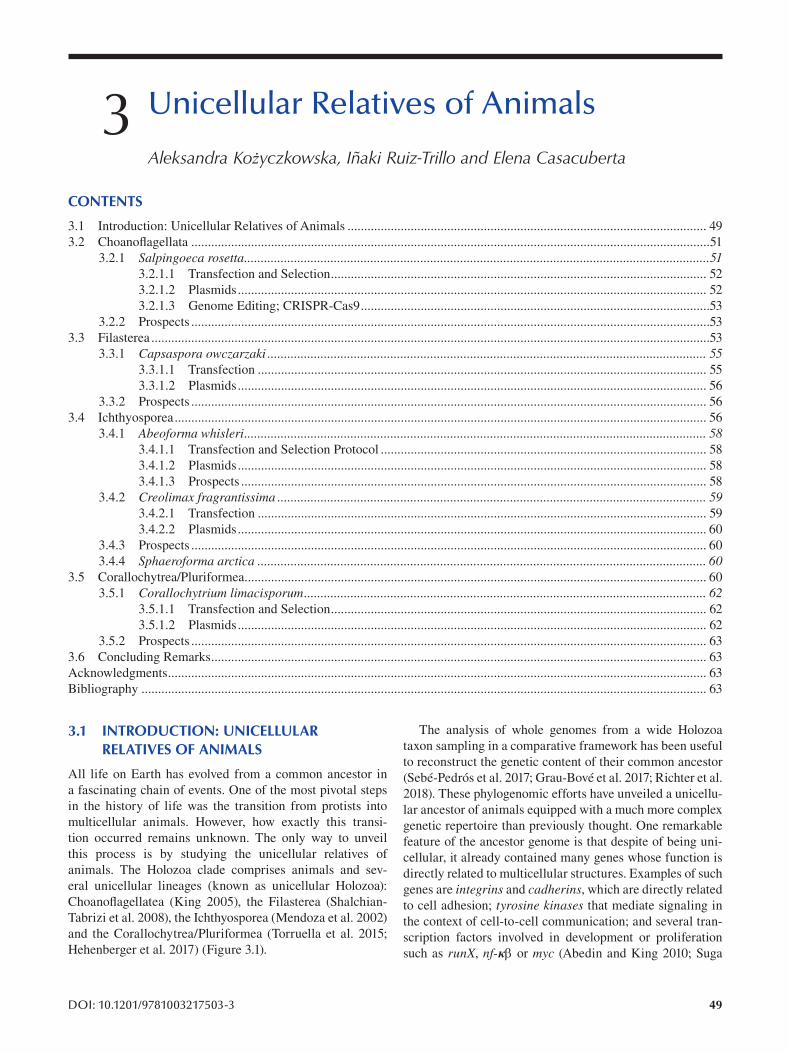
3 Unicellular Relatives of Animals
Aleksandra Kożyczkowska, Iñaki Ruiz-Trillo and Elena Casacuberta
CONTENTS
3.1 Introduction: Unicellular Relatives of Animals ............................................................................................................ 49
3.2 Choanofl agellata ............................................................................................................................................................51
3.2.1 Salpingoeca rosetta ............................................................................................................................................513.2.1.1 Transfection and Selection ................................................................................................................. 52
3.2.1.2 Plasmids............................................................................................................................................. 52
3.2.1.3 Genome Editing; CRISPR-Cas9.........................................................................................................53
3.2.2 Prospects ............................................................................................................................................................53
3.3 Filasterea ........................................................................................................................................................................53
3.3.1 Capsaspora owczarzaki .................................................................................................................................... 553.3.1.1 Transfection ....................................................................................................................................... 55
3.3.1.2 Plasmids............................................................................................................................................. 56
3.3.2 Prospects ........................................................................................................................................................... 56
3.4 Ichthyosporea................................................................................................................................................................ 56
3.4.1 Abeoforma whisleri........................................................................................................................................... 583.4.1.1 Transfection and Selection Protocol .................................................................................................. 58
3.4.1.2 Plasmids............................................................................................................................................. 58
3.4.1.3 Prospects ............................................................................................................................................ 58
3.4.2 Creolimax fragrantissima ................................................................................................................................. 593.4.2.1 Transfection ....................................................................................................................................... 59
3.4.2.2 Plasmids............................................................................................................................................. 60
3.4.3 Prospects ........................................................................................................................................................... 60
3.4.4 Sphaeroforma arctica ....................................................................................................................................... 603.5 Corallochytrea/Pluriformea........................................................................................................................................... 60
3.5.1 Corallochytrium limacisporum ......................................................................................................................... 623.5.1.1 Transfection and Selection ................................................................................................................. 62
3.5.1.2 Plasmids............................................................................................................................................. 62
3.5.2 Prospects ........................................................................................................................................................... 63
3.6 Concluding Remarks ..................................................................................................................................................... 63
Acknowledgments .................................................................................................................................................................. 63
Bibliography .......................................................................................................................................................................... 63
3.1 INTRODUCTION: UNICELLULAR The analysis of whole genomes from a wide Holozoa
RELATIVES OF ANIMALS taxon sampling in a comparative framework has been useful
to reconstruct the genetic content of their common ancestor All life on Earth has evolved from a common ancestor in (Sebé-Pedrós et al. 2017; Grau-Bové et al. 2017; Richter et al. a fascinating chain of events. One of the most pivotal steps 2018). These phylogenomic efforts have unveiled a unicelluin the history of life was the transition from protists into lar ancestor of animals equipped with a much more complex multicellular animals. However, how exactly this transi- genetic repertoire than previously thought. One remarkable tion occurred remains unknown. The only way to unveil feature of the ancestor genome is that despite of being unithis process is by studying the unicellular relatives of cellular, it already contained many genes whose function is
eral unicellular lineages (known as unicellular Holozoa): genes are integrins and cadherins, which are directly related Choanoflagellatea (King 2005), the Filasterea (Shalchian- to cell adhesion; tyrosine kinases that mediate signaling in Tabrizi et al. 2008), the Ichthyosporea (Mendoza et al. 2002) the context of cell-to-cell communication; and several tranand the Corallochytrea/Pluriformea (Torruella et al. 2015; scription factors involved in development or proliferation Hehenberger et al. 2017) (Figure 3.1). such as runX, nf-κ or myc (Abedin and King 2010; Suga
animals. The Holozoa clade comprises animals and sev- directly related to multicellular structures. Examples of such
DOI: 10.1201/9781003217503-3 49
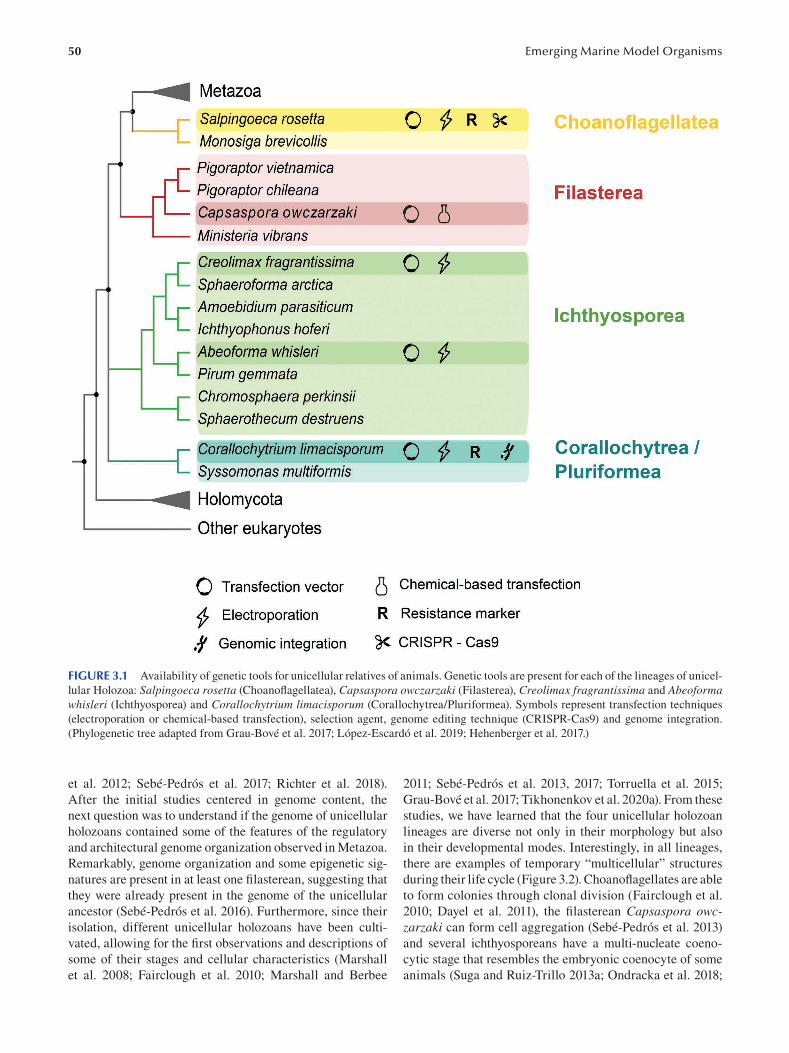
50 Emerging Marine Model Organisms
FIGURE 3.1 Availability of genetic tools for unicellular relatives of animals. Genetic tools are present for each of the lineages of unicel
lular Holozoa: Salpingoeca rosetta (Choanofl agellatea), Capsaspora owczarzaki (Filasterea), Creolimax fragrantissima and Abeoforma whisleri (Ichthyosporea) and Corallochytrium limacisporum (Corallochytrea/Pluriformea). Symbols represent transfection techniques
(electroporation or chemical-based transfection), selection agent, genome editing technique (CRISPR-Cas9) and genome integration.
(Phylogenetic tree adapted from Grau-Bové et al. 2017; López-Escardó et al. 2019; Hehenberger et al. 2017.)
et al. 2012; Sebé-Pedrós et al. 2017; Richter et al. 2018).
After the initial studies centered in genome content, the
next question was to understand if the genome of unicellular
holozoans contained some of the features of the regulatory
and architectural genome organization observed in Metazoa.
Remarkably, genome organization and some epigenetic sig
natures are present in at least one filasterean, suggesting that
they were already present in the genome of the unicellular
ancestor (Sebé-Pedrós et al. 2016). Furthermore, since their
isolation, different unicellular holozoans have been culti
vated, allowing for the first observations and descriptions of
some of their stages and cellular characteristics (Marshall
et al. 2008; Fairclough et al. 2010; Marshall and Berbee
2011; Sebé-Pedrós et al. 2013, 2017; Torruella et al. 2015;
Grau-Bové et al. 2017; Tikhonenkov et al. 2020a). From these
studies, we have learned that the four unicellular holozoan
lineages are diverse not only in their morphology but also
in their developmental modes. Interestingly, in all lineages,
there are examples of temporary “multicellular” structures
during their life cycle (Figure 3.2). Choanoflagellates are able
to form colonies through clonal division (Fairclough et al.
2010; Dayel et al. 2011), the fi lasterean Capsaspora owczarzaki can form cell aggregation (Sebé-Pedrós et al. 2013)
and several ichthyosporeans have a multi-nucleate coeno
cytic stage that resembles the embryonic coenocyte of some
animals (Suga and Ruiz-Trillo 2013a ; Ondracka et al. 2018;
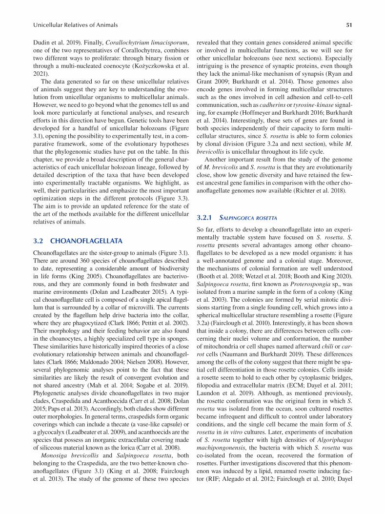
51 Unicellular Relatives of Animals
Dudin et al. 2019). Finally, Corallochytrium limacisporum,
one of the two representatives of Corallochytrea, combines
two different ways to proliferate: through binary fi ssion or
through a multi-nucleated coenocyte (Kożyczkowska et al.
2021).
The data generated so far on these unicellular relatives
of animals suggest they are key to understanding the evo
lution from unicellular organisms to multicellular animals.
However, we need to go beyond what the genomes tell us and
look more particularly at functional analyses, and research
efforts in this direction have begun. Genetic tools have been
developed for a handful of unicellular holozoans (Figure
3.1), opening the possibility to experimentally test, in a com
parative framework, some of the evolutionary hypotheses
that the phylogenomic studies have put on the table. In this
chapter, we provide a broad description of the general char
acteristics of each unicellular holozoan lineage, followed by
detailed description of the taxa that have been developed
into experimentally tractable organisms. We highlight, as
well, their particularities and emphasize the most important
optimization steps in the different protocols (Figure 3.3).
The aim is to provide an updated reference for the state of
the art of the methods available for the different unicellular
relatives of animals.
3.2 CHOANOFLAGELLATA
Choanoflagellates are the sister-group to animals (Figure 3.1).
There are around 360 species of choanofl agellates described
to date, representing a considerable amount of biodiversity
in life forms (King 2005). Choanoflagellates are bacterivo
rous, and they are commonly found in both freshwater and
marine environments (Dolan and Leadbeater 2015). A typi
cal choanoflagellate cell is composed of a single apical fl agel
lum that is surrounded by a collar of microvilli. The currents
created by the flagellum help drive bacteria into the collar,
where they are phagocytized (Clark 1866; Pettitt et al. 2002).
Their morphology and their feeding behavior are also found
in the choanocytes, a highly specialized cell type in sponges.
These similarities have historically inspired theories of a close
evolutionary relationship between animals and choanofl agel
lates (Clark 1866; Maldonado 2004; Nielsen 2008). However,
several phylogenomic analyses point to the fact that these
similarities are likely the result of convergent evolution and
not shared ancestry (Mah et al. 2014; Sogabe et al. 2019).
Phylogenetic analyses divide choanoflagellates in two major
clades, Craspedida and Acanthoecida (Carr et al. 2008; Dolan
2015; Paps et al. 2013). Accordingly, both clades show different
outer morphologies. In general terms, craspedids form organic
coverings which can include a thecate (a vase-like capsule) or
a glycocalyx (Leadbeater et al. 2009), and acanthoecids are the
species that possess an inorganic extracellular covering made
of siliceous material known as the lorica (Carr et al. 2008).
Monosiga brevicollis and Salpingoeca rosetta, both
belonging to the Craspedida, are the two better-known cho
anoflagellates (Figure 3.1) (King et al. 2008; Fairclough
et al. 2013). The study of the genome of these two species
revealed that they contain genes considered animal specifi c
or involved in multicellular functions, as we will see for
other unicellular holozoans (see next sections). Especially
intriguing is the presence of synaptic proteins, even though
they lack the animal-like mechanism of synapsis ( Ryan and
Grant 2009; Burkhardt et al. 2014). Those genomes also
encode genes involved in forming multicellular structures
such as the ones involved in cell adhesion and cell-to-cell
communication, such as cadherins or tyrosine-kinase signal
ing, for example (Hoffmeyer and Burkhardt 2016; Burkhardt
et al. 2014). Interestingly, these sets of genes are found in
both species independently of their capacity to form multi
cellular structures, since S. rosetta is able to form colonies
by clonal division (Figure 3.2a and next section), while M. brevicollis is unicellular throughout its life cycle.
Another important result from the study of the genome
of M. brevicolis and S. rosetta is that they are evolutionarily
close, show low genetic diversity and have retained the few
est ancestral gene families in comparison with the other cho
anoflagellate genomes now available (Richter et al. 2018).
3.2.1 SALPINGOECA ROSETTA
So far, efforts to develop a choanoflagellate into an experi
mentally tractable system have focused on S. rosetta. S. rosetta presents several advantages among other choano
flagellates to be developed as a new model organism: it has
a well-annotated genome and a colonial stage. Moreover,
the mechanisms of colonial formation are well understood
(Booth et al. 2018; Wetzel et al. 2018; Booth and King 2020).
Salpingoeca rosetta , first known as Proterospongia sp., was
isolated from a marine sample in the form of a colony ( King
et al. 2003). The colonies are formed by serial mitotic divi
sions starting from a single founding cell, which grows into a
spherical multicellular structure resembling a rosette (Figure
3.2a) (Fairclough et al. 2010). Interestingly, it has been shown
that inside a colony, there are differences between cells con
cerning their nuclei volume and conformation, the number
of mitochondria or cell shapes named afterward chili or carrot cells (Naumann and Burkhardt 2019). These differences
among the cells of the colony suggest that there might be spa
tial cell differentiation in those rosette colonies. Cells inside
a rosette seem to hold to each other by cytoplasmic bridges,
filopodia and extracellular matrix (ECM; Dayel et al. 2011;
Laundon et al. 2019). Although, as mentioned previously,
the rosette conformation was the original form in which S. rosetta was isolated from the ocean, soon cultured rosettes
became infrequent and difficult to control under laboratory
conditions, and the single cell became the main form of S. rosetta in in vitro cultures. Later, experiments of incubation
of S. rosetta together with high densities of Algoriphagus machipongonensis, the bacteria with which S. rosetta was
co-isolated from the ocean, recovered the formation of
rosettes. Further investigations discovered that this phenom
enon was induced by a lipid, renamed rosette inducing fac
tor (RIF; Alegado et al. 2012; Fairclough et al. 2010; Dayel
52
et al. 2011; Woznica et al. 2016 ). In parallel, a forward genetic
screen for mutants unable to form rosettes allowed for the
identification of a genetic factor in S. rosetta, which could
be linked to the rosette phenotype. The recovered rosetteless mutant encoded a C-type lectin and was not able to develop
rosettes in spite of being exposed to RIFs (Levin et al. 2014).
Although it is not yet fully understood by which molecular
mechanism the C-type lectin establishes the relevant interac
tions, it has been hypothesized that the function of the C-type
lectin is related to an interaction with the ECM (Levin et al.
2014). Interestingly, colony formation is not the only stage
in S. rosetta’s life cycle governed by bacteria. For instance,
Woznica, Gerdt and collaborators discovered that the bac
teria Vibrio fischeri was able to induce sexual behavior in
S. rosetta through a secreted product that was conveniently
labeled EroS (Woznica et al. 2017 ). Interestingly, EroS was
biochemically identified as a chondroitin lyase. This enzyme
is able to digest chondroitin sulfate and initiate mating, bear
ing some similarities to sperm digestion of the egg cover in
animal reproduction (Miller and Ax 1990).
Under conditions promoting fast growth, S. rosetta is able
to form yet another multicellular form different from the
rosettes. Linear colonies consist of a chain of cells attached
to each other and connected by intercellular bridges and
ECM (Figure 3.2a) (Dayel et al. 2011). In the case of single
cells, S. rosetta can acquire three different forms, which
besides its morphology also present a specific behavior: fast
swimmers, slow swimmers and thecate cells. The main dif
ference between the different forms of single cell types is
the presence of the theca in thecate cells, which consists of a
vase-like capsule composed of ECM. All forms of S. rosetta have a flagellum that is used for swimming and orienting the
colony, and fast swimmers and rosette colonies also have
thin filopodia (Dayel et al. 2011).
Regardless of the availability of genetic tools, S. rosettacould
already be considered an emerging model system because sub
stantial information on its biology had already been obtained.
The rosetteless mutant had been isolated by a forward genetic
screen aiming to isolate defective mutants in rosette develop
ment (Levin and King 2013; Levin et al. 2014). Moreover, spe
cific culture conditions were developed to obtain and enrich for
each of the different life forms of S. rosetta (Dayel et al. 2011),
and, finally, by the co-cultivation with specific bacteria, mating
could be induced (Woznica et al. 2016; Woznica et al. 2017).
Nevertheless, tools for direct genetic manipulation, which
would allow us for example to fluorescently tag specifi c pro
teins to study their localization and dynamics or to knock out
target genes, were missing. In recent years, Dr. Nicole King’s
research group has successfully developed transfection, selec
tion and genome editing for S. rosetta, overcoming these limi
tations. In the following sections, we will briefly summarize the
main steps of these achievements.
3.2.1.1 Transfection and Selection The transfection protocol for S. rosetta is based on the
Nucleofection technology, developed by Amaxa (Lonza
Cologne AG group) (Figure 3.3a). Nucleofection is a
Emerging Marine Model Organisms
specialized electroporation-based transfection technol
ogy engineered to transfer the DNA into the nucleus. This
technique proved successful in S. rosetta, which can now
be transiently transfected with an average efficiency of 1%,
similar to what has been achieved in other protists (Janse
et al. 2006; Caro et al. 2012).
In order to understand the significance of each optimiza
tion step, Booth et al. sequentially eliminated them one at a
time and monitored the change in efficiency (Figure 3.3a).
For example, the addition of pure and highly concentrated
carrier DNA (empty plasmid, such as pUC19), in combi
nation with the plasmid of interest, was key to optimize S. rosetta transfection, as observed in other unicellular holo
zoans (Faktorová et al. 2020; Kożyczkowska et al. 2021).
A second key step to boost transfection in S. rosetta was
priming the cells with a buffer that contains a combination
of a protease, a reducing agent, a chelator and a chaotrope
(Booth et al. 2018). This specific buffer was key in break
ing down the extracellular coat and signifi cantly improved
the uptake of transfected DNA into the cell. Even though
the extracellular coat is specific for this choanofl agellate,
it could be of inspiration for those working on organisms
that also possess an extracellular coat or wall, which usually
hampers transfection effi ciency.
One of the first applications of the developed transfection
in S. rosetta by Dr. Booth and collaborators was the study
of the localization of two septin orthologues, SrSeptin2 and
SrSeptin6 (Booth et al. 2018). Septins are a multigenic fam
ily involved in highly conserved functions such as cell divi
sion (Neufeld and Rubin 1994) but also more specialized
functions in multicellular organisms at the level of intracel
lular junctions and the maintenance of polarity in an epithe
lium (Spiliotis et al. 2008; Kim et al. 2010). The study of the
involvement of septin orthologues of S. rosetta in these lat
ter roles can help us understand the contribution of Septins
in the evolution of the epithelia before the onset of animals.
Finally, at the same time as the study of Septins in S. rosetta, the newly developed transfection technique also
proved significant for the characterization of additional
rosette defective mutations (Wetzel et al. 2018). In addition,
in this study, researchers went one step further by applying
selection with the antibiotic puromycin. Selection is very
useful in order to enrich the population in a greater propor
tion of transfected cells Figure 3.3a) (Wetzel et al. 2018). A
public protocol for transfection and selection of S. rosetta is available at Protocols.io; dx.doi.org/10.17504/protocols.
io.h68b9hw
3.2.1.2 Plasmids As a first step to develop transient transfection, researchers
cloned putative endogenous promoters from the elongation factor 1, ef1, -actin, act, -tubulin, tub and histone H3 genes from S. rosetta. Two different reporter genes, nanoluc (monitored through a luciferase assay) and mwassabi (moni
tored through expression of green fluorescence), were cho
sen to test the newly cloned promoters and used to fi ne-tune
the transfection protocol (Booth et al. 2018).
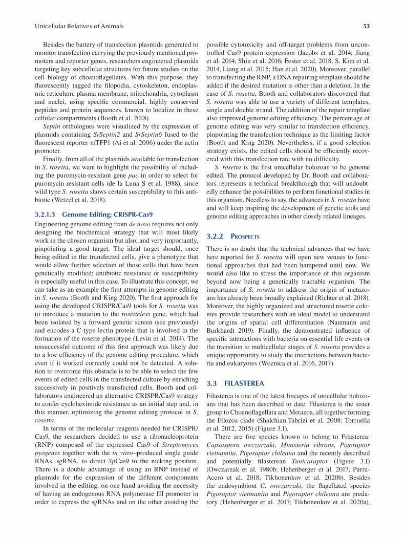
53 Unicellular Relatives of Animals
Besides the battery of transfection plasmids generated to
monitor transfection carrying the previously mentioned pro
moters and reporter genes, researchers engineered plasmids
targeting key subcellular structures for future studies on the
cell biology of choanoflagellates. With this purpose, they
fluorescently tagged the filopodia, cytoskeleton, endoplas
mic reticulum, plasma membrane, mitochondria, cytoplasm
and nuclei, using specific commercial, highly conserved
peptides and protein sequences, known to localize in these
cellular compartments (Booth et al. 2018).
Septin orthologues were visualized by the expression of
plasmids containing SrSeptin2 and SrSeptin6 fused to the
fluorescent reporter mTFP1 (Ai et al. 2006 ) under the actin
promoter.
Finally, from all of the plasmids available for transfection
in S. rosetta, we want to highlight the possibility of includ
ing the puromycin-resistant gene pac in order to select for
puromycin-resistant cells (de la Luna S et al. 1988), since
wild type S. rosetta shows certain susceptibility to this anti
biotic (Wetzel et al. 2018).
3.2.1.3 Genome Editing; CRISPR-Cas9 Engineering genome editing from de novo requires not only
designing the biochemical strategy that will most likely
work in the chosen organism but also, and very importantly,
pinpointing a good target. The ideal target should, once
being edited in the transfected cells, give a phenotype that
would allow further selection of those cells that have been
genetically modified; antibiotic resistance or susceptibility
is especially useful in this case. To illustrate this concept, we
can take as an example the first attempts in genome editing
in S. rosetta (Booth and King 2020). The fi rst approach for
using the developed CRISPR/Cas9 tools for S. rosetta was
to introduce a mutation to the rosetteless gene, which had
been isolated by a forward genetic screen (see previously)
and encodes a C-type lectin protein that is involved in the
formation of the rosette phenotype (Levin et al. 2014). The
unsuccessful outcome of this first approach was likely due
to a low efficiency of the genome editing procedure, which
even if it worked correctly could not be detected. A solu
tion to overcome this obstacle is to be able to select the few
events of edited cells in the transfected culture by enriching
successively in positively transfected cells. Booth and col
laborators engineered an alternative CRISPR/Cas9 strategy
to confer cycloheximide resistance as an initial step and, in
this manner, optimizing the genome editing protocol in S. rosetta.
In terms of the molecular reagents needed for CRISPR/
Cas9, the researchers decided to use a ribonucleoprotein
(RNP) composed of the expressed Cas9 of Streptomyces pyogenes together with the in vitro–produced single guide
RNAs, sgRNA, to direct SpCas9 to the nicking position.
There is a double advantage of using an RNP instead of
plasmids for the expression of the different components
involved in the editing: on one hand avoiding the necessity
of having an endogenous RNA polymerase III promoter in
order to express the sgRNAs and on the other avoiding the
possible cytotoxicity and off-target problems from uncon
trolled Cas9 protein expression (Jacobs et al. 2014; Jiang
et al. 2014; Shin et al. 2016; Foster et al. 2018; S. Kim et al.
2014; Liang et al. 2015; Han et al. 2020). Moreover, parallel
to transfecting the RNP, a DNA repairing template should be
added if the desired mutation is other than a deletion. In the
case of S. rosetta, Booth and collaborators discovered that
S. rosetta was able to use a variety of different templates,
single and double strand. The addition of the repair template
also improved genome editing efficiency. The percentage of
genome editing was very similar to transfection effi ciency,
pinpointing the transfection technique as the limiting factor
(Booth and King 2020). Nevertheless, if a good selection
strategy exists, the edited cells should be effi ciently recov
ered with this transfection rate with no diffi culty.
S. rosetta is the first unicellular holozoan to be genome
edited. The protocol developed by Dr. Booth and collabora
tors represents a technical breakthrough that will undoubt
edly enhance the possibilities to perform functional studies in
this organism. Needless to say, the advances in S. rosetta have
and will keep inspiring the development of genetic tools and
genome editing approaches in other closely related lineages.
3.2.2 PROSPECTS
There is no doubt that the technical advances that we have
here reported for S. rosetta will open new venues to func
tional approaches that had been hampered until now. We
would also like to stress the importance of this organism
beyond now being a genetically tractable organism. The
importance of S. rosetta to address the origin of metazo
ans has already been broadly explained (Richter et al. 2018).
Moreover, the highly organized and structured rosette colo
nies provide researchers with an ideal model to understand
the origins of spatial cell differentiation (Naumann and
Burkhardt 2019). Finally, the demonstrated infl uence of
specific interactions with bacteria on essential life events or
the transition to multicellular stages of S. rosetta provides a
unique opportunity to study the interactions between bacte
ria and eukaryotes (Woznica et al. 2016, 2017).
3.3 FILASTEREA
Filasterea is one of the latest lineages of unicellular holozo
ans that has been described to date. Filasterea is the sister
group to Choanoflagellata and Metazoa, all together forming
the Filozoa clade (Shalchian-Tabrizi et al. 2008; Torruella
et al. 2012, 2015) (Figure 3.1).
There are five species known to belong to Filasterea:
Capsaspora owczarzaki, Ministeria vibrans, Pigoraptor vietnamita, Pigoraptor chileana and the recently described
and potentially fi lasterean Tunicaraptor (Figure 3.1 )
(Owczarzak et al. 1980b; Hehenberger et al. 2017; Parra-
Acero et al. 2018; Tikhonenkov et al. 2020b). Besides
the endosymbiont C. owczarzaki, the fl agellated species
Pigoraptor vietmanita and Pigoraptor chileana are preda
tory (Hehenberger et al. 2017; Tikhonenkov et al. 2020a),
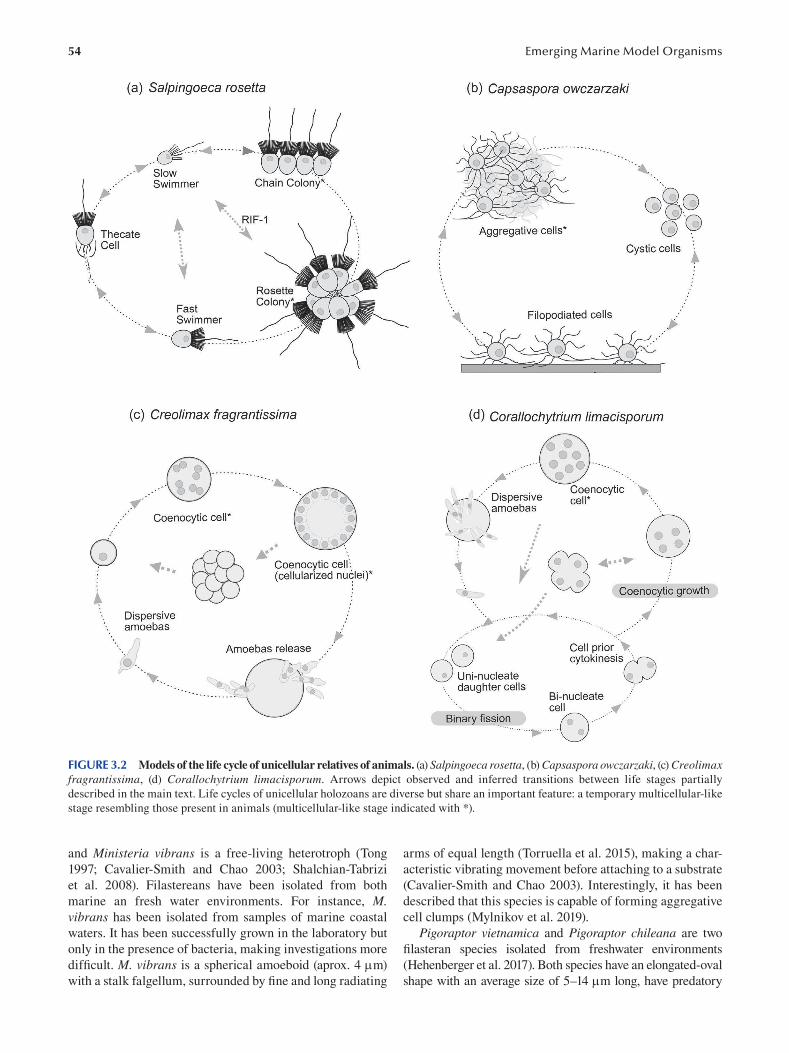
54 Emerging Marine Model Organisms
FIGURE 3.2 Models of the life cycle of unicellular relatives of animals. (a) Salpingoeca rosetta, (b) Capsaspora owczarzaki, (c) Creolimax fragrantissima, (d) Corallochytrium limacisporum. Arrows depict observed and inferred transitions between life stages partially
described in the main text. Life cycles of unicellular holozoans are diverse but share an important feature: a temporary multicellular-like
stage resembling those present in animals (multicellular-like stage indicated with *).
and Ministeria vibrans is a free-living heterotroph (Tong
1997; Cavalier-Smith and Chao 2003; Shalchian-Tabrizi
et al. 2008). Filastereans have been isolated from both
marine an fresh water environments. For instance, M. vibrans has been isolated from samples of marine coastal
waters. It has been successfully grown in the laboratory but
only in the presence of bacteria, making investigations more
diffi cult. M. vibrans is a spherical amoeboid (aprox. 4 m)
with a stalk falgellum, surrounded by fine and long radiating
arms of equal length (Torruella et al. 2015), making a char
acteristic vibrating movement before attaching to a substrate
(Cavalier-Smith and Chao 2003). Interestingly, it has been
described that this species is capable of forming aggregative
cell clumps (Mylnikov et al. 2019).
Pigoraptor vietnamica and Pigoraptor chileana are two
filasteran species isolated from freshwater environments
(Hehenberger et al. 2017). Both species have an elongated-oval
shape with an average size of 5–14 m long, have predatory
55 Unicellular Relatives of Animals
behavior and display a very similar life cycle. A detailed
description of their complex life cycle can be found in the work
done by Tikonhenko et al. (2020a). We would like to highlight
that both Pigoraptor species can aggregate during their life
cycle, as has been described for M. vibrans as well as for the
best-studied fi lasterean, C. owczarzaki (see the following).
3.3.1 CAPSASPORA OWCZARZAKI
First reports of C. owczarzaki appeared from investigations
on the susceptibility of the fresh-water snail Biomphalaria glabrata to be infected by the parasite Schistosoma mansoni. Studying the possible factors underneath the resis
tance to infection, Stibbs and collaborators isolated a small
amoeba of 3–5 m in diameter from pericardium and mantle
explants from three different strains of B. glabrata, two of
them resistant to Schistosoma infection (Stibbs et al. 1979).
The ability to grow C. owczarzaki in axenic cultures allowed
researchers to test the interaction between the amoeba and
the parasite. These works demonstrated that C. owczarzaki amoebas were able to adhere to and kill the sporocists of S. mansoni, resulting in a high proliferation of C. owczarzaki. H. Stibbs and A. Owczarzaki were the first ones to describe
C. owczarzaki and set the initial culture conditions.
The initial stage of the life cycle of C. owczarzaki consists
of crawling filopodiated amoebas that grow exponentially.
Once the culture is saturated and nutrients become limit
ing, amoebas retract their filopodia and encyst in a round
and compact cell, and their growth stabilizes. At this point,
encysted cells can attach to each other, forming compact
cell aggregates of different sizes (Figure 3.2b). C. owczarzaki cell aggregates can happen spontaneously or can also be
induced by agitation with specific parameters (Sebé-Pedrós
et al. 2013). Most importantly, electron microscopy analyses
revealed that cells in the aggregates are glued together by
cohesive extracellular material, which provides the aggre
gate with consistency but keeps cells individually separated.
RNA-seq analyses demonstrated an upregulation of the
expression of key genes involved in cell-to-cell communica
tion and cell adhesion, such as the tyrosine kinase signaling
pathway and the integrin adhesome (Sebé-Pedrós et al. 2013).
The study of C. owczarzaki has not only provided knowl
edge about its biology but also about the wider question of
animal origins. For example, analysis of its genome revealed
several genomic features previously thought to be animal spe
cific (Suga et al. 2013; Sebé-Pedrós et al. 2017). C. owczarzaki contains a complete integrin adhesome necessary to mediate
the interaction between the cell and the ECM (Suga et al. 2013;
Parra-Acero et al. 2020). Moreover, C. owczarzaki also con
tains a set of proteins, including transcription factors (TFs),
known to be involved in developmental pathways in animals;
NF-κb, Runx and T-box; and others involved in cell motility
and proliferation such as Brachyury and MYC (Mendoza and
Sebé-Pedrós 2019). Additionally, components of different sig
nal transduction pathways have an unexpected conservation,
with examples such as JAK-STAT, Notch, TGFβ or tyrosine
kinases in general (RTKs) (Suga et al. 2012).
It is clear that C. owczarzaki was an ideal species to be
developed into a genetically tractable organism in order to
further investigate the different hypotheses drawn from the
genomic content and signatures, as well as to plunge into
the terrain of cell biology to enrich the investigations of the
evolutionary path shared among holozoans.
3.3.1.1 Transfection The fi rst attempts to transfect a new organism fail the vast
majority of times. For C. owczarzaki, the fi rst protocols
to be tested were based on different technologies such as
electroporation, magnetofection and lipid-based transfec
tion methods. However, these tests yielded either no posi
tive cells or very low transfection effi ciencies, hampering
reproducibility (Suga and Ruiz-Trillo 2013; Ensenauer et al.
2011; Parra-Acero et al. 2018). The technology that ended
up being efficient enough to be further optimized into a reli
able transfection protocol was the classical calcium phos
phate precipitation method (Figure 3.2b) (Graham and van
der Eb 1973). Here we highlight the steps that turned out
to be crucial to improve the efficiency of the transfection
protocol (Parra-Acero et al. 2018). One of the factors that
is important to maximize efficiency is to use cells at the
exponential growth phase. The stage in which C. owczarzaki is growing exponentially is the adherent stage. Cells
from a fresh culture at 90/95% confluence from the adher
ent stage were the ones with higher transfection effi ciency.
The size of the crystals from the DNA and the precipitates
of calcium phosphate also proved important to improv
ing the efficiency of transfection. The authors determined
that the smaller the crystals, the better, as shown for other
organisms such as D. discoideum (Jordan and Wurm 2004;
Gaudet et al. 2007). In order to achieve a smaller crystal
size, it is important to keep the same ratio for DNA/calcium
and phosphate when preparing the DNA mix to transfect.
The stability of the DNA/calcium ratio once the DNA mix
was added to the media also depended on the amount of
phosphate in the transfection media, which also needed to
be taken into account. Similarly, the pH of the fi nal solution
should be controlled to avoid changes in the solubility of the
precipitates. The last touch to further improve transfection
efficiency was to expose cells to an osmotic shock, which
would permeate the cell membrane for a short period of
time. This technique is also used in a variety of eukaryotic
cells with the application of glycerol or DMSO (10–20%)
(Grosjean et al. 2006; Gaudet et al. 2007; Guo et al. 2017).
In the case of C. owczarzaki, a 10% glycerol shock dur
ing one minute was good enough (Figure 3.3b). Finally, as
in any transfection protocol, it is important to be able to
identify those cells where the DNA has successfully entered
the nucleus and is being expressed. The identifi cation of
transfected cells can be done by enriching the transfected
population using an antibiotic or a specific drug to which
wild type cells (non-transfected cells) are susceptible or
by inspecting the expression of a fluorescent protein using
fluorescence microscopy. Because C. owczarzaki seems to
be resistant to different antibiotics, pesticides or cytostatic
56
drugs that are commonly used for selection, the initial plas
mids that were designed and transfected into C. owczarzaki contained genes encoding small fl uorescent proteins.
These fluorescent proteins, such as mVenus and mCherry,
were expressed in the cytosol of transfected cells. Besides
the microscopy observations, efficiency of transfection was
also analyzed using flow cytometry by comparing the popu
lation of transfected cells with cells from a negative control
population. Note that it is important to take into account the
possible phenomenon of auto-fluorescence for some types
of cells. Efficiency of transfection was on average around
1.132% ± 0.529 (mean ± s.d.), which might seem low for
researchers working with transfection in other eukaryotic
systems, but it is sufficient to efficiently further select trans
fected cells and proceed with downstream experiments
(Parra-Acero et al. 2018).
Co-transfection is known to increase efficiency of the
transfection per se, and it is also very useful in order to
deliver two different constructs simultaneously. Dr. Parra-
Acero and collaborators tested in which proportion two
different plasmids were uptaken by the cells when co-trans
fected in order to use co-transfection to visualize simultane
ously more than one subcellular structure. Co-transfection
resulted, with a rate of incorporation of both constructs
almost equally (72.909% ± 5.468) in C. owczarzaki ( Parra-
Acero et al. 2018).
Although stable transfection has not yet been developed
in Capsaspora, plasmids delivered by transient transfection
were shown to be expressed inside the cells for up to ten
days. The life cycle of Capsaspora is much shorter than ten
days, and therefore this protocol allows for the interrogation
of the reporter expression at the different life stages of the
organism.
3.3.1.2 Plasmids The reporter plasmids (pONSY-mVenus and pONSY-mCherry)
for optimizing transfection and calculating effi ciency were
already designed using the endogenous promoter and ter
minator sequences of the elongation factor 1-α gene (EF1-α)
of Capsaspora (Parra-Acero et al. 2018). Besides the engi
neered plasmids to visualize the cytosol, the researchers went
one step further in order to get insights into the cell biology
of this species. For this reason, they designed plasmids to
fluorescently label the different subcellular structures. For
example, the endogenous histone 2B (H2B) gene was fused
to mVenus to highlight the nucleus (pONSY-CoH2B:Venus),
and the plasma membrane was visualized by cloning the
N-myristoylation motif (NMM) of the endogenous Src2
tyrosine kinase gene, which is known to localize at mem
branes and filopodia (pONSY-CoNMM:mCherry) (Sigal et
al. 1994; Parra-Acero et al. 2018). Finally, in order to visual
ize the cytoskeleton, a small peptide (17 amino acid) named
lifeAct known to bind filamentous actin (Riedl et al. 2008)
was fused to mCherry (pONSY-Lifeact:mCherry) to visual
ize the actin cytoskeleton and filopodia of transfected cells.
Detailed observations using confocal microscopy of single
and co-transfected C. owczarzaki cells with these plasmids
Emerging Marine Model Organisms
revealed the targeted structures explaining, among others,
the hollow basket structure from the actin bundles around
the cell body or the dynamics of the filopodia along the dif
ferent life stages (Parra-Acero et al. 2018).
3.3.2 PROSPECTS
C. owczarzaki, in addition to its key phylogenetic position,
its well-annotated genome and the number of “multicel
lular” genes its genome encodes, is also able to form cell
aggregates during its life cycle (Figure 3.2b), making it an
ideal organism to analyze the origin of animals.
Finally, the fact that this organism is able to attack and
feed on S. mansoni sporocysts (Stibbs et al. 1979; Owczarzak
et al. 1980a) also makes it a potential candidate for disease-
control strategies, even though the specific interaction of
C. owczarzaki with the snail B. glabrata remains unclear.
Interestingly, C. owczarzaki exhibits high resistance to
antibiotics and harsh mediums, suggesting its potential in
medical applications in the case that was fi nally selected to
control schistomiasis (Parra-Acero et al. 2018).
3.4 ICHTHYOSPOREA
Ichthyosporea is the sister-group to Corallochytrea, as well
as to the Filozoa (Choanoflagellata, Filasterea and Metazoa)
(Mendoza et al. 2002). All described ichthyosporeans are
osmotrophs and have multiple life stages that vary greatly
in shape and motility and in most cases contain a cell wall
of variable composition. The developmental mode of ichthyosporeans is complex and contains multinucleated stages
such as a coenocyte (Figures 3.1 and 3.2c).
Ichthyosporeans received this name because the early
identified representatives were all parasites of fi sh (Cavalier-
Smith 1998). Later phylogenomic analyses of rDNA with
newer representatives expanded the group in two internal
classes, the Dermocystida, which are exclusively parasites
of vertebrate hosts, and the Ichthyophonida, which can
parasitize a variety of host species (Mendoza et al. 2002;
Marshall et al. 2008). In accordance with their habitat, only
representatives of Ichthyophonida can be cultured in labora
tory conditions (Jøstensen et al. 2002; Marshall et al. 2008).
Interestingly, the motile representatives of Dermocystida are equipped with a flagellum, while the ichthyophonids are
motile amoebas. Maybe related, it has been shown by electron
microscopy studies that representatives of Ichthyophonida have a spindle pole body (Marshall et al. 2008), which
would nicely correlate with the disappearance of centrioles
and the flagellum as a consequence (Marshall and Berbee
2011). On the other hand, centrioles have been described for
members of Dermocystida such as Dermocystidum percae (Pekkarinen 2003). In the coming years, further investiga
tions on other key biological questions will be possible once
experimentally tractable organisms will be developed for
both subclasses. For instance, investigations on the micro
tubule organizing centers and the nature of the mitosis
(whether it is open, closed or somewhere in between) would
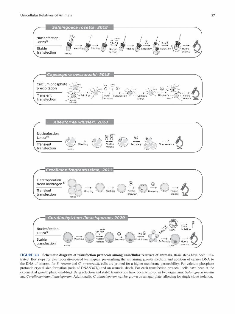
57 Unicellular Relatives of Animals
FIGURE 3.3 Schematic diagram of transfection protocols among unicellular relatives of animals. Basic steps have been illus
trated. Key steps for electroporation-based techniques: pre-washing the remaining growth medium and addition of carrier DNA to
the DNA of interest; for S. rosetta and C. owczarzaki, cells are primed for a higher membrane permeability. For calcium phosphate
protocol: crystal size formation (ratio of DNA/CaCl2) and an osmotic shock. For each transfection protocol, cells have been at the
exponential growth phase (mid-log). Drug selection and stable transfection have been achieved in two organisms: Salpingoeca rosetta and Corallochytrium limacisporum. Additionally, C. limacisporum can be grown on an agar plate, allowing for single clone isolation.
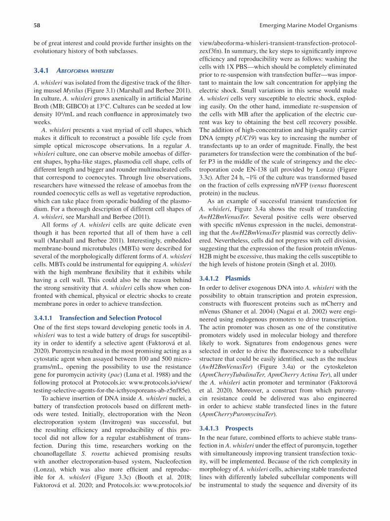
58
be of great interest and could provide further insights on the
evolutionary history of both subclasses.
3.4.1 ABEOFORMA WHISLERI
A. whisleri was isolated from the digestive track of the fi lter
ing mussel Mytilus (Figure 3.1) (Marshall and Berbee 2011).
In culture, A. whisleri grows axenically in artifi cial Marine
Broth (MB; GIBCO) at 13C. Cultures can be seeded at low
density 104/mL and reach confluence in approximately two
weeks.
A. whisleri presents a vast myriad of cell shapes, which
makes it difficult to reconstruct a possible life cycle from
simple optical microscope observations. In a regular A. whisleri culture, one can observe mobile amoebas of differ
ent shapes, hypha-like stages, plasmodia cell shape, cells of
different length and bigger and rounder multinucleated cells
that correspond to coenocytes. Through live observations,
researchers have witnessed the release of amoebas from the
rounded coenocytic cells as well as vegetative reproduction,
which can take place from sporadic budding of the plasmo
dium. For a thorough description of different cell shapes of
A. whisleri, see Marshall and Berbee (2011).
All forms of A. whisleri cells are quite delicate even
though it has been reported that all of them have a cell
wall (Marshall and Berbee 2011). Interestingly, embedded
membrane-bound microtubules (MBTs) were described for
several of the morphologically different forms of A. whisleri cells. MBTs could be instrumental for equipping A. whisleri with the high membrane flexibility that it exhibits while
having a cell wall. This could also be the reason behind
the strong sensitivity that A. whisleri cells show when con
fronted with chemical, physical or electric shocks to create
membrane pores in order to achieve transfection.
3.4.1.1 Transfection and Selection Protocol One of the first steps toward developing genetic tools in A. whisleri was to test a wide battery of drugs for susceptibil
ity in order to identify a selective agent (Faktorová et al.
2020). Puromycin resulted in the most promising acting as a
cytostatic agent when assayed between 100 and 500 micro
grams/mL, opening the possibility to use the resistance
gene for puromycin activity (pac) (Luna et al. 1988) and the
following protocol at Protocols.io: www.protocols.io/view/
testing-selective-agents-for-the-icthyosporeans-ab-z5nf85e ).
To achieve insertion of DNA inside A. whisleri nuclei, a
battery of transfection protocols based on different meth
ods were tested. Initially, electroporation with the Neon
electroporation system (Invitrogen) was successful, but
the resulting efficiency and reproducibility of this pro
tocol did not allow for a regular establishment of trans
fection. During this time, researchers working on the
choanofl agellate S. rosetta achieved promising results
with another electroporation-based system, Nucleofection
(Lonza), which was also more efficient and reproduc
ible for A. whisleri (Figure 3.3c) (Booth et al. 2018;
Faktorová et al. 2020; and Protocols.io: www.protocols.io/
Emerging Marine Model Organisms
view/abeoforma-whisleri-transient-transfection-protocol
zexf3fn). In summary, the key steps to signifi cantly improve
efficiency and reproducibility were as follows: washing the
cells with 1X PBS—which should be completely eliminated
prior to re-suspension with transfection buffer—was impor
tant to maintain the low salt concentration for applying the
electric shock. Small variations in this sense would make
A. whisleri cells very susceptible to electric shock, explod
ing easily. On the other hand, immediate re-suspension of
the cells with MB after the application of the electric cur
rent was key to obtaining the best cell recovery possible.
The addition of high-concentration and high-quality carrier
DNA (empty pUC19) was key to increasing the number of
transfectants up to an order of magnitude. Finally, the best
parameters for transfection were the combination of the buf
fer P3 in the middle of the scale of stringency and the elec
troporation code EN-138 (all provided by Lonza) (Figure
3.3c). After 24 h, ~1% of the culture was transformed based
on the fraction of cells expressing mVFP (venus fl uorescent
protein) in the nucleus.
As an example of successful transient transfection for
A. whisleri, Figure 3.4a shows the result of transfecting
AwH2BmVenusTer. Several positive cells were observed
with specific mVenus expression in the nuclei, demonstrat
ing that the AwH2BmVenusTer plasmid was correctly deliv
ered. Nevertheless, cells did not progress with cell division,
suggesting that the expression of the fusion protein mVenus
H2B might be excessive, thus making the cells susceptible to
the high levels of histone protein (Singh et al. 2010).
3.4.1.2 Plasmids In order to deliver exogenous DNA into A. whisleri with the
possibility to obtain transcription and protein expression,
constructs with fluorescent proteins such as mCherry and
mVenus (Shaner et al. 2004) (Nagai et al. 2002) were engi
neered using endogenous promoters to drive transcription.
The actin promoter was chosen as one of the constitutive
promoters widely used in molecular biology and therefore
likely to work. Signatures from endogenous genes were
selected in order to drive the fluorescence to a subcellular
structure that could be easily identifi ed, such as the nucleus
(AwH2BmVenusTer) (Figure 3.4a) or the cytoskeleton
(ApmCherryTubulinaTer, ApmCherry Actina Ter), all under
the A. whisleri actin promoter and terminator (Faktorová
et al. 2020). Moreover, a construct from which puromy
cin resistance could be delivered was also engineered
in order to achieve stable transfected lines in the future
(ApmCherryPuromycinaTer).
3.4.1.3 Prospects In the near future, combined efforts to achieve stable trans
fection in A. whisleri under the effect of puromycin, together
with simultaneously improving transient transfection toxic
ity, will be implemented. Because of the rich complexity in
morphology of A. whisleri cells, achieving stable transfected
lines with differently labeled subcellular components will
be instrumental to study the sequence and diversity of its
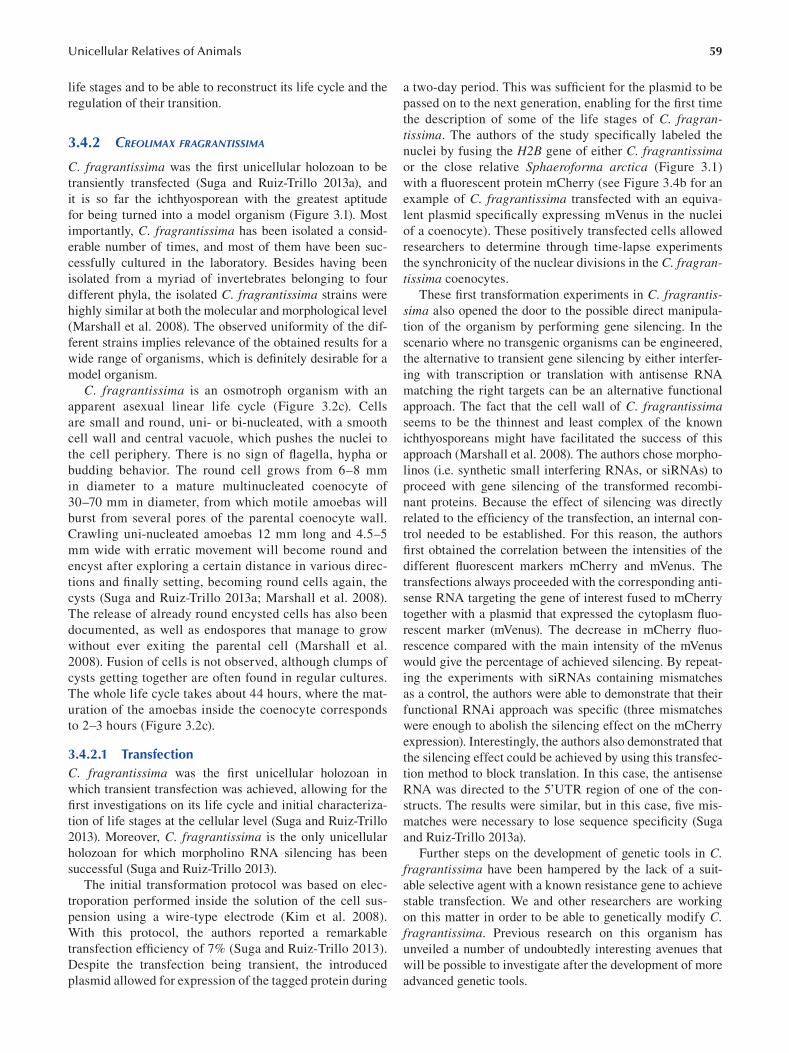
59 Unicellular Relatives of Animals
life stages and to be able to reconstruct its life cycle and the
regulation of their transition.
3.4.2 CREOLIMAX FRAGRANTISSIMA
C. fragrantissima was the first unicellular holozoan to be
transiently transfected (Suga and Ruiz-Trillo 2013a), and
it is so far the ichthyosporean with the greatest aptitude
for being turned into a model organism (Figure 3.1). Most
importantly, C. fragrantissima has been isolated a consid
erable number of times, and most of them have been suc
cessfully cultured in the laboratory. Besides having been
isolated from a myriad of invertebrates belonging to four
different phyla, the isolated C. fragrantissima strains were
highly similar at both the molecular and morphological level
(Marshall et al. 2008). The observed uniformity of the dif
ferent strains implies relevance of the obtained results for a
wide range of organisms, which is defi nitely desirable for a
model organism.
C. fragrantissima is an osmotroph organism with an
apparent asexual linear life cycle ( Figure 3.2c). Cells
are small and round, uni- or bi-nucleated, with a smooth
cell wall and central vacuole, which pushes the nuclei to
the cell periphery. There is no sign of flagella, hypha or
budding behavior. The round cell grows from 6–8 mm
in diameter to a mature multinucleated coenocyte of
30–70 mm in diameter, from which motile amoebas will
burst from several pores of the parental coenocyte wall.
Crawling uni-nucleated amoebas 12 mm long and 4.5–5
mm wide with erratic movement will become round and
encyst after exploring a certain distance in various direc
tions and finally setting, becoming round cells again, the
cysts (Suga and Ruiz-Trillo 2013a ; Marshall et al. 2008).
The release of already round encysted cells has also been
documented, as well as endospores that manage to grow
without ever exiting the parental cell (Marshall et al.
2008). Fusion of cells is not observed, although clumps of
cysts getting together are often found in regular cultures.
The whole life cycle takes about 44 hours, where the mat
uration of the amoebas inside the coenocyte corresponds
to 2–3 hours (Figure 3.2c).
3.4.2.1 Transfection C. fragrantissima was the first unicellular holozoan in
which transient transfection was achieved, allowing for the
first investigations on its life cycle and initial characteriza
tion of life stages at the cellular level (Suga and Ruiz-Trillo
2013). Moreover, C. fragrantissima is the only unicellular
holozoan for which morpholino RNA silencing has been
successful (Suga and Ruiz-Trillo 2013).
The initial transformation protocol was based on elec
troporation performed inside the solution of the cell sus
pension using a wire-type electrode ( Kim et al. 2008 ).
With this protocol, the authors reported a remarkable
transfection efficiency of 7% ( Suga and Ruiz-Trillo 2013 ).
Despite the transfection being transient, the introduced
plasmid allowed for expression of the tagged protein during
a two-day period. This was sufficient for the plasmid to be
passed on to the next generation, enabling for the fi rst time
the description of some of the life stages of C. fragrantissima. The authors of the study specifically labeled the
nuclei by fusing the H2B gene of either C. fragrantissima or the close relative Sphaeroforma arctica ( Figure 3.1 )
with a fluorescent protein mCherry (see Figure 3.4b for an
example of C. fragrantissima transfected with an equiva
lent plasmid specifically expressing mVenus in the nuclei
of a coenocyte). These positively transfected cells allowed
researchers to determine through time-lapse experiments
the synchronicity of the nuclear divisions in the C. fragrantissima coenocytes.
These first transformation experiments in C. fragrantissima also opened the door to the possible direct manipula
tion of the organism by performing gene silencing. In the
scenario where no transgenic organisms can be engineered,
the alternative to transient gene silencing by either interfer
ing with transcription or translation with antisense RNA
matching the right targets can be an alternative functional
approach. The fact that the cell wall of C. fragrantissima seems to be the thinnest and least complex of the known
ichthyosporeans might have facilitated the success of this
approach (Marshall et al. 2008). The authors chose morpho
linos (i.e. synthetic small interfering RNAs, or siRNAs) to
proceed with gene silencing of the transformed recombi
nant proteins. Because the effect of silencing was directly
related to the effi ciency of the transfection, an internal con
trol needed to be established. For this reason, the authors
first obtained the correlation between the intensities of the
different fluorescent markers mCherry and mVenus. The
transfections always proceeded with the corresponding anti
sense RNA targeting the gene of interest fused to mCherry
together with a plasmid that expressed the cytoplasm fl uo
rescent marker (mVenus). The decrease in mCherry fl uo
rescence compared with the main intensity of the mVenus
would give the percentage of achieved silencing. By repeat
ing the experiments with siRNAs containing mismatches
as a control, the authors were able to demonstrate that their
functional RNAi approach was specific (three mismatches
were enough to abolish the silencing effect on the mCherry
expression). Interestingly, the authors also demonstrated that
the silencing effect could be achieved by using this transfec
tion method to block translation. In this case, the antisense
RNA was directed to the 5’UTR region of one of the con
structs. The results were similar, but in this case, fi ve mis
matches were necessary to lose sequence specifi city (Suga
and Ruiz-Trillo 2013a).
Further steps on the development of genetic tools in C. fragrantissima have been hampered by the lack of a suit
able selective agent with a known resistance gene to achieve
stable transfection. We and other researchers are working
on this matter in order to be able to genetically modify C. fragrantissima. Previous research on this organism has
unveiled a number of undoubtedly interesting avenues that
will be possible to investigate after the development of more
advanced genetic tools.
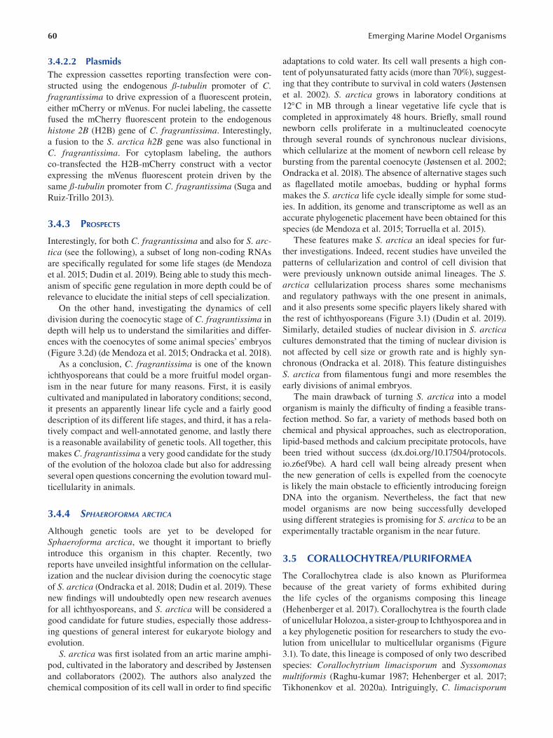
60
3.4.2.2 Plasmids The expression cassettes reporting transfection were con
structed using the endogenous ß-tubulin promoter of C. fragrantissima to drive expression of a fl uorescent protein,
either mCherry or mVenus. For nuclei labeling, the cassette
fused the mCherry fluorescent protein to the endogenous
histone 2B (H2B) gene of C. fragrantissima . Interestingly,
a fusion to the S. arctica h2B gene was also functional in
C. fragrantissima. For cytoplasm labeling, the authors
co-transfected the H2B-mCherry construct with a vector
expressing the mVenus fluorescent protein driven by the
same ß-tubulin promoter from C. fragrantissima (Suga and
Ruiz-Trillo 2013).
3.4.3 PROSPECTS
Interestingly, for both C. fragrantissima and also for S. arctica (see the following), a subset of long non-coding RNAs
are specifically regulated for some life stages (de Mendoza
et al. 2015; Dudin et al. 2019). Being able to study this mech
anism of specifi c gene regulation in more depth could be of
relevance to elucidate the initial steps of cell specialization.
On the other hand, investigating the dynamics of cell
division during the coenocytic stage of C. fragrantissima in
depth will help us to understand the similarities and differ
ences with the coenocytes of some animal species’ embryos
(Figure 3.2d) (de Mendoza et al. 2015; Ondracka et al. 2018).
As a conclusion, C. fragrantissima is one of the known
ichthyosporeans that could be a more fruitful model organ
ism in the near future for many reasons. First, it is easily
cultivated and manipulated in laboratory conditions; second,
it presents an apparently linear life cycle and a fairly good
description of its different life stages, and third, it has a rela
tively compact and well-annotated genome, and lastly there
is a reasonable availability of genetic tools. All together, this
makes C. fragrantissima a very good candidate for the study
of the evolution of the holozoa clade but also for addressing
several open questions concerning the evolution toward mul
ticellularity in animals.
3.4.4 SPHAEROFORMA ARCTICA
Although genetic tools are yet to be developed for
Sphaeroforma arctica, we thought it important to briefl y
introduce this organism in this chapter. Recently, two
reports have unveiled insightful information on the cellular
ization and the nuclear division during the coenocytic stage
of S. arctica (Ondracka et al. 2018; Dudin et al. 2019). These
new findings will undoubtedly open new research avenues
for all ichthyosporeans, and S. arctica will be considered a
good candidate for future studies, especially those address
ing questions of general interest for eukaryote biology and
evolution.
S. arctica was first isolated from an artic marine amphi
pod, cultivated in the laboratory and described by Jøstensen
and collaborators (2002). The authors also analyzed the
chemical composition of its cell wall in order to fi nd specifi c
Emerging Marine Model Organisms
adaptations to cold water. Its cell wall presents a high con
tent of polyunsaturated fatty acids (more than 70%), suggest
ing that they contribute to survival in cold waters ( Jøstensen
et al. 2002). S. arctica grows in laboratory conditions at
12C in MB through a linear vegetative life cycle that is
completed in approximately 48 hours. Briefly, small round
newborn cells proliferate in a multinucleated coenocyte
through several rounds of synchronous nuclear divisions,
which cellularize at the moment of newborn cell release by
bursting from the parental coenocyte (Jøstensen et al. 2002;
Ondracka et al. 2018). The absence of alternative stages such
as flagellated motile amoebas, budding or hyphal forms
makes the S. arctica life cycle ideally simple for some stud
ies. In addition, its genome and transcriptome as well as an
accurate phylogenetic placement have been obtained for this
species (de Mendoza et al. 2015; Torruella et al. 2015).
These features make S. arctica an ideal species for fur
ther investigations. Indeed, recent studies have unveiled the
patterns of cellularization and control of cell division that
were previously unknown outside animal lineages. The S. arctica cellularization process shares some mechanisms
and regulatory pathways with the one present in animals,
and it also presents some specific players likely shared with
the rest of ichthyosporeans (Figure 3.1) (Dudin et al. 2019).
Similarly, detailed studies of nuclear division in S. arctica cultures demonstrated that the timing of nuclear division is
not affected by cell size or growth rate and is highly syn
chronous (Ondracka et al. 2018). This feature distinguishes
S. arctica from filamentous fungi and more resembles the
early divisions of animal embryos.
The main drawback of turning S. arctica into a model
organism is mainly the difficulty of finding a feasible trans
fection method. So far, a variety of methods based both on
chemical and physical approaches, such as electroporation,
lipid-based methods and calcium precipitate protocols, have
been tried without success (dx.doi.org/10.17504/protocols.
io.z6ef9be). A hard cell wall being already present when
the new generation of cells is expelled from the coenocyte
is likely the main obstacle to efficiently introducing foreign
DNA into the organism. Nevertheless, the fact that new
model organisms are now being successfully developed
using different strategies is promising for S. arctica to be an
experimentally tractable organism in the near future.
3.5 CORALLOCHYTREA/PLURIFORMEA
The Corallochytrea clade is also known as Pluriformea
because of the great variety of forms exhibited during
the life cycles of the organisms composing this lineage
(Hehenberger et al. 2017). Corallochytrea is the fourth clade
of unicellular Holozoa, a sister-group to Ichthyosporea and in
a key phylogenetic position for researchers to study the evo
lution from unicellular to multicellular organisms (Figure
3.1). To date, this lineage is composed of only two described
species: Corallochytrium limacisporum and Syssomonas multiformis (Raghu-kumar 1987; Hehenberger et al. 2017;
Tikhonenkov et al. 2020a). Intriguingly, C. limacisporum
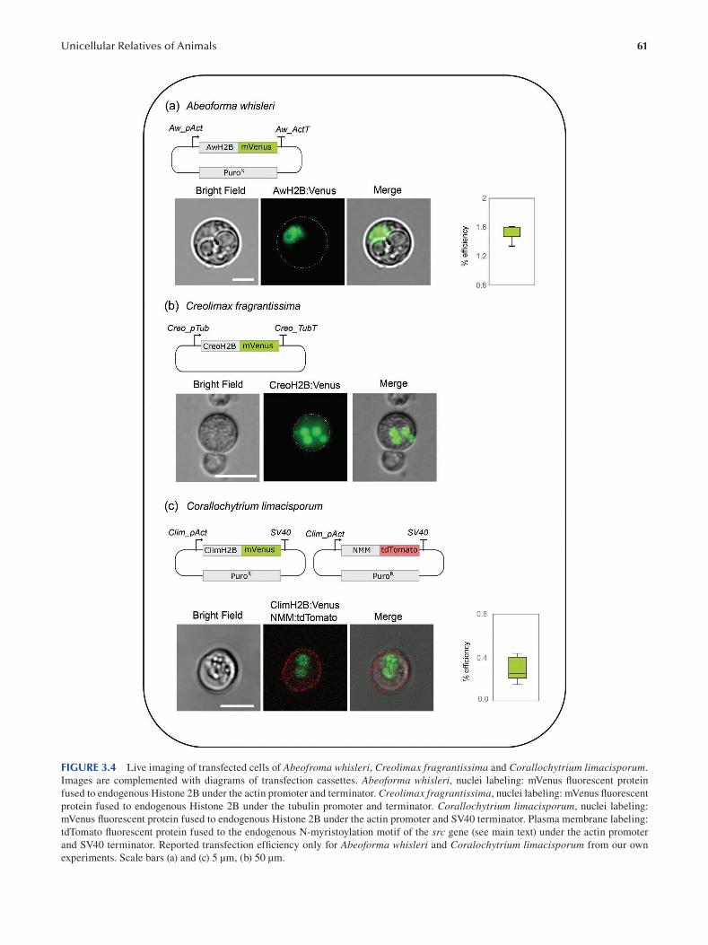
61 Unicellular Relatives of Animals
FIGURE 3.4 Live imaging of transfected cells of Abeofroma whisleri, Creolimax fragrantissima and Corallochytrium limacisporum. Images are complemented with diagrams of transfection cassettes. Abeoforma whisleri, nuclei labeling: mVenus fl uorescent protein
fused to endogenous Histone 2B under the actin promoter and terminator. Creolimax fragrantissima, nuclei labeling: mVenus fl uorescent
protein fused to endogenous Histone 2B under the tubulin promoter and terminator. Corallochytrium limacisporum, nuclei labeling:
mVenus fluorescent protein fused to endogenous Histone 2B under the actin promoter and SV40 terminator. Plasma membrane labeling:
tdTomato fluorescent protein fused to the endogenous N-myristoylation motif of the src gene (see main text) under the actin promoter
and SV40 terminator. Reported transfection efficiency only for Abeoforma whisleri and Coralochytrium limacisporum from our own
experiments. Scale bars (a) and (c) 5 μm, (b) 50 μm.
62
contains a complete flagellar toolkit (Torruella et al. 2015),
but its flagellated forms occur sporadically in our culture
conditions, whereas in contrast, the most commonly occur
ring stage of S. multiformis consists of fl agellated forms
(Tikhonenkov et al. 2020a). Both representatives of this
clade show some morphological resemblance in their life
cycle, S. multiformis being the one with a greater variety of
forms. As an example, both organisms have active amoe
boid forms and also present complex multicellular stages
(Figure 3.2d) (Tikhonenkov et al. 2020a ; Kożyczkowska
et al. 2021).
In addition to its key phylogenetic position, C. limacisporum has many of the desirable features for an organism to be
developed as genetically tractable (see next section). On the
other hand, unfortunately, cultures of S. multiformis are no
longer available, and therefore it is difficult to speculate on
the possibility of this organism becoming an experimentally
treatable organism.
3.5.1 CORALLOCHYTRIUM LIMACISPORUM
C. limacisporum is a small, marine, free-living corallochy
trean isolated from coral reefs of India and Hawaii (Raghu
kumar 1987). This taxa possesses numerous features that
make it an attractive candidate for further functional anal
ysis. It grows very fast and under axenic conditions, and
most importantly, it is able to grow in both liquid and agar
media, allowing for easy screenings and selection of indi
vidual transformed clones. Moreover, it is the only coral
lochytrean with a completely sequenced and well-annotated
genome (Grau-Bové et al. 2017). Finally, besides these
technical advantages, C. limacisporum has a peculiar and
understudied biology, with a complex life cycle and, as we
mentioned before, some fungal-like features. For all these
reasons, developing genetic tools in this fascinating unicel
lular organism will for sure be useful for several scientifi c
questions/fi elds.
3.5.1.1 Transfection and Selection Different antibiotics, antifungals and herbicides had been
tested in C. limacisporum, and the antibiotic puromycin was
selected as the most adequate for its efficiency and apparent
low toxicity (Kożyczkowska et al. 2021). In addition to selec
tion by antibiotics, it would be ideal to have a double selection
system that would also allow us to screen transfected cells by
fluorescence microscopy. Therefore, a dual selection system
based on resistance to puromycin and mCherry expression was
set up. Two recombinant plasmids, CAMP (Corallochytrium A ctin M cherry Pac) and CTMP (Corallochytrium T ubulin
M cherry Pac), were used for optimizing the transfection
parameters (see also “Plasmids” section).
Different methods of transfection that had worked for
other protists, yeast or eukaryote cells in general based on
chemical or physical methods were tested, but only electro
poration was successful. Initially, positive results using an
in electrode apparatus from Invitrogen, the Neon system,
which allows modifying the electric pulse and the duration
Emerging Marine Model Organisms
of the pulse (dx.doi.org/10.17504/protocols.io.hmwb47e),
were obtained. Nevertheless, this protocol did not have
enough reproducibility to carry out downstream applica
tions, and we selected the electroporator 4D-Nucleofector
from Lonza, which was being used with greater effi
ciency in other protists (Figure 3.3e) (Kożyczkowska et
al. 2021 and Protcols.io: dx.doi.org/10.17504/protocols.
io.r5ud86w; see sections for S. rosetta, A. whisleri and C. fragrantissima).
One of the important factors was the cell density and age
of the starting culture to maximize efficiency. Similarly to A. whisleri, the cells should be washed with 1X PBS to remove
the culture media. Co-transfection of highly pure and highly
concentrated carrier plasmid DNA (empty pUC19) was
another key factor that significantly increased effi ciency.
In general, some fluorescent cells could be observed after
24 hours post-transfection, although there was always a
significant increase in positive cells after 48 hours, after
which puromycin was added. In the case of C. limacisporum, the combination of buffer P3 and code EN-138 from
the 4D-Nucleofector (Lonza) proved the most optimal
for successful transfection (Figure 3.3e) (Kożyczkowska
et al. 2021). Clonal lines can be obtained by plating a dilu
tion of the cells in MB agar plates containing puromycin
(Kożyczkowska et al. 2021).
As an immediate contribution from these developed
genetic tools, the description of the life cycle of C. limacisporum and the unraveling of some unexpected traits,
was possible. It has been discovered that C. limacisporum has two different paths for cell division, binary fi ssion and
coenocytic growth ( Figure 3.2d), demonstrating that the C. limacisporum life cycle is non-linear and more complex than
previously thought (Raghu-kumar 1987). Additionally, some
particular features of C. limacisporum not commonly found
in eukaryotes were described: first the decoupling of cyto
kinesis and karyokinesis in binary fission and second the
observation of some examples of asynchronous nuclei divi
sions during coenocytic growth. The possibility to expand
functional studies of these features in C. limacisporum will
undoubtedly contribute to a better characterization of this
unicellular holozoan.
3.5.1.2 Plasmids As mentioned, a double selection system was engineered. The
CAMP plasmid contained the pac gene to provide drug resis
tance (Luna et al. 1988) and the mCherry gene to produce
fluorescence in the positively transfected cells. In order to
drive transcription with endogenous promoters, the upstream
non-coding sequence of the actin and tubulin genes from C. limacisporum and the 3’UTR terminator of the actin gene
from the ichthyosporean A. whisleri were cloned in order
to avoid homologous recombination at the actin locus. The
CAMP and CTMP plasmids were indistinguishable in their
phenotype, fl uorescent labeling of the cytoplasm in C. limacisporum revealing a “crescent moon-like” shape produced
by the presence of a large vacuole that occupies the 65% of
the cell’s volume (Kożyczkowska et al. 2021).
63 Unicellular Relatives of Animals
Progress into understanding the cell biology of C. limacisporum (see transfection section) was possible through the
generation of constructs tagging sub-cellular components,
such as the plasma membrane, cytoskeleton, cytoplasm and
nucleus (Kożyczkowska et al. 2021). To construct the pactNMN-tdTomato plasmid, the predicted N-myristoylation
motif (NMM) from the Src tyrosine kinase orthologue
(Gene ID Clim_evm93s153) was used. This motif has been
successfully used in C. owczarzaki to direct the fusion pro
tein to the plasma membrane (Parra-Acero et al. 2018). Our
results show that this motif was also plasma membrane spe
cific in C. limacisporum and therefore might also be useful
in other organisms (Figure 3.4c) (Kożyczkowska et al. 2021).
To visualize the cytoskeleton, the 17-amino acid peptide
LifeAct that binds specifi cally to filamentous actin (ibidi)
was fused to the mCherry protein pact-LifeAct. Finally, the
construct pact-H2B-mVenus contains the endogenous gene
of C. limacisporum (Gene ID Clim_evm20s1) fused to the
mVenus fluorescent protein. In addition, the construct con
tains the actin promoter, with the dual system of puromy
cin resistance as well as fl uorescence (Kożyczkowska et al.
2021).
3.5.2 PROSPECTS
The development of specific recombinant plasmids together
with stable transfection in C. limacisporum has provided
insightful information about the biology of this organism
while also providing the initial tools to set up functional
experiments. Importantly, now C. limacisporum provides
the opportunity to further investigate which are the fac
tors behind different developmental routes (binary fi ssion
or coenocytic growth), as well as a promising model to
study the mechanisms behind the decoupling of karyokine
sis from cytokinesis and the basis of asynchronous nuclear
division.
Besides the previously mentioned advances, developing
CRISPR/Cas9 genome editing in C. limacisporum is cur
rently ongoing. The establishment of genome editing in the
future will allow us to understand, among others, the pos
sible ancestral role of some genes related to multicellular
functions in Metazoa.
3.6 CONCLUDING REMARKS
We have here described the most recent advances in the
handful of model organisms available among unicellular
holozoans (Figure 3.1). These model organisms belong to all
four clades of unicellular relatives of animals, constituting a
functional platform to experimentally address many of the
hypotheses regarding the evolution of genes and cellular fea
tures along the Holozoa tree. We are eager to see how evo
lutionary cell biology will take advantage of all those new
emerging model systems to address the function of ancestral
genes and protein domains, as well as for the conservation or
innovation of cell biological processes.
ACKNOWLEDGMENTS
We would like to thank Núria Ros-Rocher for critical read
ing of the manuscript and the Multicellgenome laboratory
for ideas and discussions. We would also like to thank
the European Research Council (ERC) and the Betty and
Gordon Moore foundation for their strategic vision in fund
ing the development of these organisms into experimen
tally tractable organisms. This work was supported by a
European Research Council Consolidator Grant (ERC
2012-Co-616960) grant to I.R-T. and a Betty and Gordon
Moore “New Genetic tools for Marine Protists”, Grant num
ber 4973.01 to E.C and I.R-T.
BIBLIOGRAPHY
Abedin, M., and N. King. 2010. Diverse Evolutionary Paths to Cell
Adhesion. Trends in Cell Biology 20: 734–742.
Ai, H., Henderson, J. N., Remington, S. J., and R. E. Campbell.
2006. Directed Evolution of a Monomeric, Bright and
Photostable Version of Clavularia Cyan Fluorescent Protein:
Structural Characterization and Applications in Fluorescence
Imaging. The Biochemical Journal 400: 531–540.
Alegado, R. A., Brown L. W., Cao S., Dermenjian, R. K., Zuzow,
R., Fairclough, S. R., Clardy J., and Nicole King. 2012. A
Bacterial Sulfonolipid Triggers Multicellular Development
in the Closest Living Relatives of Animals. ELife 1: e00013.
Booth, D. S., and N. King. 2020. Genome Editing Enables
Reverse Genetics of Multicellular Development in the
Choanoflagellate Salpingoeca Rosetta. Edited by Alejandro
Sánchez Alvarado, Patricia J Wittkopp, Margaret A Titus,
Iñaki Ruiz-Trillo, and Matthew C Gibson. ELife 9: e56193.
Booth, D. S., Szmidt-Middleton, H., and N. King. 2018. Transfection
of Choanoflagellates Illuminates Their Cell Biology and the
Ancestry of Animal Septins. Molecular Biology of the Cell 29: 3026–3038.
Burkhardt, P., Grønborg, M., McDonald, K., Sulur, T., Wang, Q.,
and N. King. 2014. Evolutionary Insights into Premetazoan
Functions of the Neuronal Protein Homer. Molecular Biology and Evolution 31: 2342–2355.
Caro, F., Miller, M. G., and J. L DeRisi. 2012. Plate-Based
Transfection and Culturing Technique for Genetic
Manipulation of Plasmodium falciparum. Malaria Journal 11: 22.
Carr, M., Leadbeater, B. S. C., Hassan, R., Nelson, M., and S. L.
Baldauf. 2008. Molecular Phylogeny of Choanofl agellates,
the Sister Group to Metazoa. Proceedings of the National Academy of Sciences of the United States of America 105:
16641–16646.
Cavalier-Smith, T. 1998. A Revised Six-Kingdom System of Life.
Biological Reviews of the Cambridge Philosophical Society 73: 203–266.
Cavalier-Smith, T., and E. E.-Y. Chao. 2003. Phylogeny of
Choanozoa, Apusozoa, and Other Protozoa and Early
Eukaryote Megaevolution. Journal of Molecular Evolution 56: 540–563.
Clark, H. J. 1866. Conclusive Proofs of the Animality of the Ciliate
Sponges, and of Their Affinities with the Infusoria Flagellata.
American Journal of Science 42 (2): 320–324.
Dayel, M. J., Alegado, R. A., Fairclough, S. R., Levin, T. C., Nichols,
S. A., McDonald, K., and N. King. 2011. Cell Differentiation
and Morphogenesis in the Colony-Forming Choanofl agellate
Salpingoeca rosetta. Developmental Biology 357: 73–82.
64
Dolan, J., and B. S. Leadbeater. 2015. The Choanoflagellates: Evolution, Biology, and Ecology. Cambridge University
Press, Cambridge, UK. 315 pp. Hardcover, ISBN978-0-521
88444-0, $125. Journal of Eukaryotic Microbiology , June.
Dudin, O., Ondracka, A., Grau-Bové, X., Haraldsen, A. A. B.,
Toyoda, A., Suga, H., Bråte, J., and I. Ruiz-Trillo. 2019.
A Unicellular Relative of Animals Generates a Layer of
Polarized Cells by Actomyosin-Dependent Cellularization.
ELife 8: e49801. https://doi.org/10.7554/eLife.49801 .
Ensenauer, R., Hartl, D., Vockley, J., Roscher, A. A., and U.
Fuchs. 2011. Efficient and Gentle SiRNA Delivery by
Magnetofection. Biotechnic & Histochemistry: Official Publication of the Biological Stain Commission 86: 226–231.
Fairclough, S. R., Chen, Z., Kramer, E., Zeng, Q., Young, S.,
Robertson, H. M., E. Begovic, et al. 2013. Premetazoan
Genome Evolution and the Regulation of Cell Differentiation
in the Choanofl agellate Salpingoeca rosetta. Genome Biology 14: R15.
Fairclough, S. R., Dayel, M. J., and N. King. 2010. Multicellular
Development in a Choanofl agellate. Current Biology: CB 20:
R875–R876.
Faktorová, D., Nisbet, E. R., Fernández Robledo, J. A., Casacuberta,
E., Sudek L., Allen A. E., M. Ares, et al. 2020. Genetic
Tool Development in Marine Protists: Emerging Model
Organisms for Experimental Cell Biology. Nature Methods 17: 481–494.
Foster, A. J., Martin-Urdiroz, M., Yan, X., Wright, H. S., Soanes, D.
M., and N. J. Talbot. 2018. CRISPR-Cas9 Ribonucleoprotein-
Mediated Co-Editing and Counterselection in the Rice Blast
Fungus. Scientifi c Reports 8: 14355.
Gaudet, P., Pilcher, K. E., Fey, P., and R. L. Chisholm. 2007.
Transformation of Dictyostelium Discoideum with Plasmid
DNA. Nature Protocols 2: 1317–1324.
Graham, F. L., and A. J. van der Eb. 1973. A New Technique for the
Assay of Infectivity of Human Adenovirus 5 DNA. Virology 52: 456–467.
Grau-Bové, X., Torruella, G., Donachie, S., Suga, H., Leonard,
G., Richards, T. A., and I. Ruiz-Trillo. 2017. Dynamics of
Genomic Innovation in the Unicellular Ancestry of Animals.
Edited by Diethard Tautz. ELife 6 (July): e26036.
Grosjean, F., Bertschinger, M., Hacker, D. L., and F. M. Wurm.
2006. Multiple Glycerol Shocks Increase the Calcium
Phosphate Transfection of Non-Synchronized CHO Cells.
Biotechnology Letters 28: 1827–1833.
Guo, L., Wang, L., Yang, R., Feng, R., Li, Z., Zhou, X., Z. Dong,
et al. 2017. Optimizing Conditions for Calcium Phosphate
Mediated Transient Transfection. Saudi Journal of Biological Sciences, Computational Intelligence Research & Approaches
in Bioinformatics and Biocomputing 24: 622–629.
Han, H. A., Sheng Pang, J. K., and B.-S. Soh. 2020. Mitigating
Off-Target Effects in CRISPR/Cas9-Mediated In Vivo Gene
Editing. Journal of Molecular Medicine 98: 615–632.
Hehenberger, E., Tikhonenkov, D. V., Kolisko, M., del Campo,
J., Esaulov, A. S., Mylnikov, A. P., and P. J. Keeling. 2017.
Novel Predators Reshape Holozoan Phylogeny and Reveal
the Presence of a Two-Component Signaling System in the
Ancestor of Animals. Current Biology 27: 2043–2050.
Hoffmeyer, T. T., and P. Burkhardt. 2016. Choanofl agellate
Models: Monosiga brevicollis and Salpingoeca rosetta.
Current Opinion in Genetics & Development , Developmental
Mechanisms, Patterning and Evolution 39: 42–47.
Jacobs, J. Z., Ciccaglione, K. M., Tournier, V., and M. Zaratiegui.
2014. Implementation of the CRISPR-Cas9 System in
Fission Yeast. Nature Communications 5: 5344.
Emerging Marine Model Organisms
Janse, C. J., Franke-Fayard, B., Mair, G. R., Ramesar, J., Thiel,
C., Engelmann, S., Matuschewski, K., van Gemert,
G. J., Sauerwein, R. W., and A. P. Waters. 2006. High
Efficiency Transfection of Plasmodium Berghei Facilitates
Novel Selection Procedures. Molecular and Biochemical Parasitology 145: 60–70.
Jiang, W., Brueggeman, A., Horken, K., Plucinak, T., and D.
Weeks. 2014. Successful Transient Expression of Cas9 and
Single Guide RNA Genes in Chlamydomonas reinhardtii. Eukaryotic Cell 13.
Jordan, M., and F. Wurm. 2004. Transfection of Adherent and
Suspended Cells by Calcium Phosphate. Methods (San Diego, Calif.) 33: 136–143.
Jøstensen, J.-P., Sperstad, S., Johansen, S., and B. Landfald.
2002. Molecular-Phylogenetic, Structural and Biochemical
Features of a Cold-Adapted, Marine Ichthyosporean Near
the Animal-Fungal Divergence, Described from In Vitro
Cultures. European Journal of Protistology 38: 93–104.
Kim, J. A., Cho, K., Shin, M. S., Lee, W. G., Jung, N., Chung, C.,
and J. Keun Chang. 2008. A Novel Electroporation Method
Using a Capillary and Wire-Type Electrode. Biosensors & Bioelectronics 23: 1353–1360.
Kim, S. K., Kim, D., Woo Cho, S., Kim, J., and J.-S. Kim. 2014.
Highly Efficient RNA-Guided Genome Editing in Human
Cells via Delivery of Purified Cas9 Ribonucleoproteins.
Genome Research 24.
Kim, S. K., Shindo, A., Park, T. J., Oh, E. C., Ghosh, S., Gray,
R. S., R. A. Lewis, et al. 2010. Planar Cell Polarity Acts
through Septins to Control Collective Cell Movement and
Ciliogenesis. Science (New York, N.Y.) 329: 1337–1340.
King, N. 2005. Choanofl agellates. Current Biology: CB 15:
R113–R114.
King, N., Hittinger, C. T., and S. B. Carroll. 2003. Evolution of
Key Cell Signaling and Adhesion Protein Families Predates
Animal Origins. Science (New York, N.Y.) 301: 361–363.
King, N., Westbrook, M. J., Young, S. L., Kuo, A., Abedin, M.,
Chapman, J., S. Fairclough, et al. 2008. The Genome of the
Choanofl agellate Monosiga brevicollis and the Origin of
Metazoans. Nature 451: 783–788.
Kożyczkowska, A., Najle, S. R., Ocaña-Pallarès, E., Aresté, C.,
Shabardina, V., Ara, P. S., Ruiz-Trillo, I., and E. Casacuberta.
2021. Stable Transfection in the Protist Corallochytrium limacisporum Allows Identification of Novel Cellular
Features Among Unicellular Relatives of Animals.
Current Biology 31: 1–7. https://doi.org/10.1016/j.cub.2021.
06.061
Laundon, D., Larson, B. T., McDonald, K., King, N., and P.
Burkhardt. 2019. The Architecture of Cell Differentiation in
Choanoflagellates and Sponge Choanocytes. PLoS Biology 17 (4): e3000226.
Leadbeater, B. S. C., Yu, Q., Kent, J., and D. J. Stekel. 2009.
Three-Dimensional Images of Choanofl agellate Loricae.
Proceedings of the Royal Society B: Biological Sciences 276:
3–11.
Levin, T. C., Greaney A. J., Wetzel, L., and N. King. 2014.
The Rosetteless Gene Controls Development in the
Choanofl agellate S. rosetta. ELife 3 (October).
Levin, T. C., and N. King. 2013. Evidence for Sex and
Recombination in the Choanofl agellate Salpingoeca rosetta.
Current Biology 23: 2176–2180.
Liang, X., Potter, J., Kumar, S., Zou, Y., Quintanilla, R., Sridharan,
M., J. Carte, et al. 2015. Rapid and Highly Effi cient
Mammalian Cell Engineering via Cas9 Protein Transfection.
Journal of Biotechnology 208 (August): 44–53.
65 Unicellular Relatives of Animals
López-Escardó, D., Grau-Bové, X., Guillaumet-Adkins, A., Gut,
M., Sieracki, M. E., and I. Ruiz-Trillo. 2019. Reconstruction
of Protein Domain Evolution Using Single-Cell Amplifi ed
Genomes of Uncultured Choanoflagellates Sheds Light on
the Origin of Animals. Philosophical Transactions of the Royal Society B: Biological Sciences 374 (1786): 20190088.
Luna, S. de la, Soria, I., Pulido, D., Ortín, J., and A. Jiménez.
1988. Efficient Transformation of Mammalian Cells with
Constructs Containing a Puromycin-Resistance Marker.
Gene 62: 121–126.
Mah, J. L., Christensen-Dalsgaard, K. K., and S. P. Leys. 2014.
Choanoflagellate and Choanocyte Collar-Flagellar Systems
and the Assumption of Homology. Evolution & Development 16: 25–37.
Maldonado, M. 2004. Choanoflagellates, Choanocytes, and Animal
Multicellularity. Invertebrate Biology 123: 1–22.
Marshall, W. L., and M. L. Berbee. 2011. Facing Unknowns:
Living Cultures ( Pirum gemmata Gen. Nov., Sp. Nov., and
Abeoforma whisleri, Gen. Nov., Sp. Nov.) from Invertebrate
Digestive Tracts Represent an Undescribed Clade within
the Unicellular Opisthokont Lineage Ichthyosporea
(Mesomycetozoea). Protist 162: 33–57.
Marshall, W. L., Celio, G., McLaughlin, D. J., and M. L.
Berbee. 2008. Multiple Isolations of a Culturable, Motile
Ichthyosporean (Mesomycetozoa, Opisthokonta), Creolimax fragrantissima n. Gen., n. Sp., from Marine Invertebrate
Digestive Tracts. Protist 159: 415–433.
Mendoza, A. de, and A. Sebé-Pedrós. 2019. Origin and Evolution
of Eukaryotic Transcription Factors. Current Opinion in Genetics & Development 58–59: 25–32.
Mendoza, A. de, Suga, H., Permanyer, J., Irimia, M., and I. Ruiz-Trillo.
2015. Complex Transcriptional Regulation and Independent
Evolution of Fungal-Like Traits in a Relative of Animals.
Edited by Alejandro Sánchez Alvarado. ELife 4: e08904.
Mendoza, L., Taylor, J. W., and L. Ajello. 2002. The
Class Mesomycetozoea: A Heterogeneous Group of
Microorganisms at the Animal-Fungal Boundary. Annual Review of Microbiology 56: 315–344.
Miller, D. J., and R. L. Ax. 1990. Carbohydrates and Fertilization
in Animals. Molecular Reproduction and Development 26:
184–198.
Mylnikov, A. P., Tikhonenkov, D. V., Karpov, S. A., and C. Wyle
zich. 2019. Microscopical Studies on Ministeria vibrans Tong, 1997 (Filasterea) Highlight the Cytoskeletal Structure
of the Common Ancestor of Filasterea, Metazoa and
Choanofl agellata. Protist 170: 385–396.
Nagai, T., Ibata, K., Sun Park, E., Kubota, M., Mikoshiba, K., and
A. Miyawaki. 2002. A Variant of Yellow Fluorescent Protein
with Fast and Efficient Maturation for Cell-Biological
Applications. Nature Biotechnology 20: 87–90.
Naumann, B., and P. Burkhardt. 2019. Spatial Cell Disparity in the
Colonial Choanofl agellate Salpingoeca rosetta. Frontiers in Cell and Developmental Biology 7.
Neufeld, T. P., and G. M. Rubin. 1994. The Drosophila Peanut
Gene Is Required for Cytokinesis and Encodes a Protein
Similar to Yeast Putative Bud Neck Filament Proteins. Cell 77: 371–379.
Nielsen, C. 2008. Six Major Steps in Animal Evolution: Are We
Derived Sponge Larvae? Evolution & Development 10:
241–257.
Ondracka, A., Dudin, O., and I. Ruiz-Trillo. 2018. Decoupling of
Nuclear Division Cycles and Cell Size during the Coenocytic
Growth of the Ichthyosporean Sphaeroforma arctica. Current Biology 28: 1964–1969.
Owczarzak, A., Stibbs, H. H., and C. J. Bayne. 1980a. The
Destruction of Schistosoma mansoni Mother Sporocysts In
Vitro by Amoebae Isolated from Biomphalaria glabrata : An
Ultrastructural Study. Journal of Invertebrate Pathology 35:
26–33.
Owczarzak, A., Stibbs, H. H., and C. J. Bayne. 1980b. The
Destruction of Schistosoma mansoni Mother Sporocysts In
Vitro by Amoebae Isolated from Biomphalaria glabrata : An
Ultrastructural Study. Journal of Invertebrate Pathology 35:
26–33.
Paps, J., Medina-Chacón, L. A., Marshall, W., Suga, H., and I. Ruiz-
Trillo. 2013. Molecular Phylogeny of Unikonts: New Insights
into the Position of Apusomonads and Ancyromonads and the
Internal Relationships of Opisthokonts. Protist 164: 2–12.
Parra-Acero, H., Harcet, M., Sánchez-Pons, N., Casacuberta, E.,
Brown, N. H., Dudin, O., and I. Ruiz-Trillo. 2020. Integrin-
Mediated Adhesion in the Unicellular Holozoan Capsaspora owczarzaki. Current Biology 30: 4270–4275.
Parra-Acero, H., Ros-Rocher, N., Perez-Posada, A., Kożyczkowska,
A., Sánchez-Pons, N., Nakata, A., Suga, H., Najle, S. R., and
I. Ruiz-Trillo. 2018. Transfection of Capsaspora owczarzaki, a Close Unicellular Relative of Animals. Development (Cambridge, England) 145 (10).
Pekkarinen, M. 2003. Phylogenetic Position and Ultrastructure
of Two Dermocystidium Species (Ichthyosporea) from the
Common Perch (Perca fl uviatilis). Acta Protozoologica 42
(4): 287–307.
Pettitt, M. E., Orme, B. A. A., Blake, J. R., and B. S. C.
Leadbeater. 2002. The Hydrodynamics of Filter Feeding in
Choanofl agellates. European Journal of Protistology 38:
313–332.
Raghu-kumar, S. 1987. Occurrence of the Thraustochytrid,
Corallochytrium Limacisporum Gen. et Sp. Nov. in the Coral
Reef Lagoons of the Lakshadweep Islands in the Arabian
Sea. Botanica Marina 30 (1).
Richter, D. J., Fozouni, P., Eisen, M. B., and N. King. 2018. Gene
Family Innovation, Conservation and Loss on the Animal
Stem Lineage. Edited by Maximilian J Telford. ELife 7:
e34226.
Riedl, J., Crevenna, A. H., Kessenbrock, K., Haochen Yu, J.,
Neukirchen, D., Bista, M., F. Bradke, et al. 2008. Lifeact:
A Versatile Marker to Visualize F-Actin. Nature Methods 5:
605.
Ryan, T. J., and S. G. N. Grant. 2009. The Origin and Evolution of
Synapses. Nature Reviews Neuroscience 10: 701–712.
Sebé-Pedrós, A., Ballaré, C., Parra-Acero, H., Chiva, C., Tena, J.
J., Sabidó, E., Gómez-Skarmeta, J. L., Di Croce, L., and I.
Ruiz-Trillo. 2016. The Dynamic Regulatory Genome of
Capsaspora and the Origin of Animal Multicellularity. Cell 165: 1224–1237.
Sebé-Pedrós, A., Degnan, B. M., and I. Ruiz-Trillo. 2017. The
Origin of Metazoa: A Unicellular Perspective. Nature Reviews Genetics 18: 498–512.
Sebé-Pedrós, A., Irimia, M., Del Campo, J., Parra-Acero, H., Russ,
C., Nusbaum, C., Blencowe, B. J., and I. Ruiz-Trillo. 2013.
Regulated Aggregative Multicellularity in a Close Unicellular
Relative of Metazoa. ELife 2 (December): e01287.
Shalchian-Tabrizi, K., Minge, M. A., Espelund, M., Orr, R., Ruden,
T., Jakobsen, K. S., and T. Cavalier-Smith. 2008. Multigene
Phylogeny of Choanozoa and the Origin of Animals. PLoS One 3 (5).
Shaner, N. C., Campbell, R. E., Steinbach, P. A., Giepmans,
B. N. G., Palmer, A. E., and R. Y. Tsien. 2004. Improved
Monomeric Red, Orange and Yellow Fluorescent Proteins
66
Derived from Discosoma Sp. Red Fluorescent Protein.
Nature Biotechnology 22: 1567–1572.
Shin, S.-E., Lim, J.-M., Koh, H. G., Kim, E. K., Kang, N. K., Jeon,
S., S. Kwon, et al. 2016. CRISPR/Cas9-Induced Knockout
and Knock-in Mutations in Chlamydomonas reinhardtii. Scientifi c Reports 6: 27810.
Sigal, C. T., Zhou, W., Buser, C. A., McLaughlin, S., and M. D.
Resh. 1994. Amino-Terminal Basic Residues of Src Mediate
Membrane Binding through Electrostatic Interaction with
Acidic Phospholipids. Proceedings of the National Academy of Sciences of the United States of America 91: 12253–12257.
Singh, R. K., Liang, D., Gajjalaiahvari, U. R., Miquel Kabbaj,
M.-H., Paik, J., and A. Gunjan. 2010. Excess Histone Levels
Mediate Cytotoxicity via Multiple Mechanisms. Cell Cycle 9: 4236–4244.
Sogabe, S., Hatleberg, W. L., Kocot, K. M., Say, T. E., Stoupin, D.,
Roper, K. E., Fernandez-Valverde, S. L., Degnan, S. M., and
B. M. Degnan. 2019. Pluripotency and the Origin of Animal
Multicellularity. Nature 570: 519–522.
Spiliotis, E. T., Hunt, S. J., Hu, Q., Kinoshita, M., and W. J. Nelson.
2008. Epithelial Polarity Requires Septin Coupling of Vesicle
Transport to Polyglutamylated Microtubules. The Journal of Cell Biology 180: 295–303.
Stibbs, H. H., Owczarzak, A., Bayne, C. J., and P. DeWan. 1979.
Schistosome Sporocyst-Killing Amoebae Isolated from
Biomphalaria glabrata. Journal of Invertebrate Pathology 33: 159–170.
Suga, H., Dacre, M., de Mendoza, A., Shalchian-Tabrizi, K.,
Manning, G., and I. Ruiz-Trillo. 2012. Genomic Survey of
Premetazoans Shows Deep Conservation of Cytoplasmic
Tyrosine Kinases and Multiple Radiations of Receptor
Tyrosine Kinases. Science Signaling 5 (222): ra35.
Suga, H., and I. Ruiz-Trillo. 2013. Development of Ichthyosporeans
Sheds Light on the Origin of Metazoan Multicellularity.
Developmental Biology 377: 284–292.
Suga, H., Chen, Z., de Mendoza, A., Sebé-Pedrós, A., Brown, M.
W., Kramer, E., M. Carr, et al. 2013. The Capsaspora Genome
Emerging Marine Model Organisms
Reveals a Complex Unicellular Prehistory of Animals.
Nature Communications 4 (1): 2325. https://doi.org/10.1038/
ncomms3325 .
Tikhonenkov, D. V., Hehenberger, E., Esaulov, A. S., Belyakova, O. I.,
Mazei, Y. A., Mylnikov, A.P., and P. J. Keeling. 2020a. Insights
into the Origin of Metazoan Multicellularity from Predatory
Unicellular Relatives of Animals. BMC Biology 18: 39.
Tikhonenkov, D. V., Mikhailov, K. V., Hehenberger, E., Karpov, S.
A., Prokina, K. I., Esaulov, A. S., O. I. Belyakova, et al. 2020b.
New Lineage of Microbial Predators Adds Complexity to
Reconstructing the Evolutionary Origin of Animals. Current Biology 30: 4500–4509.
Tong, S. M. 1997. Heterotrophic Flagellates and Other Protists
from Southampton Water, U.K. Ophelia 47: 71–131.
Torruella, G., de Mendoza, A., Grau-Bové, X., Antó, M., Chaplin,
M. A., del Campo, J., L. Eme, et al. 2015. Phylogenomics
Reveals Convergent Evolution of Lifestyles in Close Relatives
of Animals and Fungi. Current Biology 25: 2404–2410.
Torruella, G., Derelle, R., Paps, J., Lang, B. F., Roger, A. J.,
Shalchian-Tabrizi, K., and I. Ruiz-Trillo. 2012. Phylogenetic
Relationships within the Opisthokonta Based on Phylogenomic
Analyses of Conserved Single-Copy Protein Domains.
Molecular Biology and Evolution 29: 531–544.
Wetzel, L. A., Levin, T. C., Hulett, R. E., Chan, D., King, G. A.,
Aldayafleh, R., Booth, D. S., Abedin Sigg, M., and N. King.
2018. Predicted Glycosyltransferases Promote Development
and Prevent Spurious Cell Clumping in the Choanofl agellate
S. rosetta. Edited by Alejandro Sánchez Alvarado and
Marianne E Bronner. ELife 7: e41482.
Woznica, A., Cantley, A. M., Beemelmanns, C., Freinkman, E., Clardy,
J., and N. King. 2016. Bacterial Lipids Activate, Synergize,
and Inhibit a Developmental Switch in Choa nofl agellates.
Proceedings of the National Academy of Sciences of the United States of America 113: 7894–7899.
Woznica, A., Gerdt, J. P., Hulett, R. E., Clardy, J., and N. King.
2017. Mating in the Closest Living Relatives of Animals Is
Induced by a Bacterial Chondroitinase. Cell 170: 1175–1183.
4 Porifera
Maja Adamska
CONTENTS
4.1 History of the Model ..................................................................................................................................................... 67
4.2 Geographical Location .................................................................................................................................................. 68
4.3 Life Cycle ...................................................................................................................................................................... 69
4.4 Embryogenesis .............................................................................................................................................................. 69
4.5 Anatomy .........................................................................................................................................................................71
4.6 Genomic Data ............................................................................................................................................................... 72
4.7 Functional Approaches: Tools for Molecular and Cellular Analyses............................................................................ 73
4.8 Challenging Questions Both in Academic and Applied Research ................................................................................ 73
Bibliography ...........................................................................................................................................................................75
4.1 HISTORY OF THE MODEL
Sponges (Porifera) have fascinated scientists for at least
150 years, with two key subjects of investigation remain
ing vibrant until today and additional areas of research
emerging recently. The first of the original subjects is the
relationship between sponges, other animals and protists,
both in terms of their relative phylogenetic positions and the
homology between body plans and cell types. The second
stems from the remarkable ability of sponges to regenerate:
not only by restoring lost body parts but also by completely
rebuilding bodies from dissociated cells. Why can sponges
do that and we cannot? While 19th- and early 20th-century
biologists were equipped only with microscopes, current
scientists have harnessed the power of modern genomics
and gene expression analysis to address these fundamen
tally interesting questions. This section of the chapter sets
the stage for sponges as models for biological research by
(briefly) reviewing findings and opinions of 19th-century
scientists on the position of sponges in the tree of life and the
discoveries of sponge regenerative capacity in the early 20th
century. The following sections cover modern approaches to
both subjects, concluding with discussion of the most recent
advances and forecasting future directions of research uti
lizing sponges as models.
But what are sponges, actually? Perhaps surprisingly, this
simple question continues to generate heated arguments,
with various answers offered (but never universally agreed
on) throughout the past centuries. Are they animals of cel
lular grade of organization (Parazoa), with a unique body
plan and independently evolved cell types? Or are they true
animals, with germ layers homologous to our endoderm and
ectoderm? Are they living fossils, retaining features of our
distant ancestors?
When Robert Grant gave sponges the name “Porifera”
(= pore bearing), he referred to the numerus tiny openings
(called pores of ostia) which are present on the surface of adult
sponges and which lead to (more or less complex, depend
ing on the body plan; see Section 4.5) system of canals and
chambers (Grant 1825, 1836) (Figure 4.1). The innermost
surface of sponges, an epithelial layer called choanoderm, is
composed of choanocytes (collar cells), which are equipped
with flagella propelling water through the body. Choanocyte
collars capture food particles—often bacteria—and the fi l
tered water is then expelled through a larger opening (or
openings) called osculum (plural oscula). All other surfaces
of sponges (the outer, the basal and lining of the canals) are
composed of flat cells called pinacocytes. In between those
two epithelial layers lies the non-epithelial mesohyl layer,
containing motile amoeboid cells, cells producing skeletal
elements, gametes and—in viviparous sponges—embryos.
With these basic building blocks, sponges form a variety
of body plans, which are discussed further in Section 4.5
(Figure 4.1). Although Linnaeus listed sponges as “vegeta
bles”, Grant considered them animals.
Few decades later, the striking similarity between cho
anocytes and choanoflagellates, which are single-cell
and colonial protists, noticed by James-Clark in 1868 and
Saville-Kent in 1880, was interpreted to indicate strong
affinity between sponges and protists, in effect relegating
sponges from the animal kingdom. Intriguingly, all mod
ern phylogenies place choanoflagellates as the nearest rela
tives (the sister group) of animals, and the majority of the
genome-based phylogenies place sponges as the earliest
branching animal lineage (Figure 4.1), consistent with the
position of sponges as the link between protists and “true
animals” (Eumetazoans).
Ernst Haeckel, considered by many the father of evolu
tionary developmental biology, noted similarities between
body plans of sponges, in particular calcareous sponges,
and cnidarians, especially coral polyps. According to his
views, the sponge choanoderm was homologous to the coral
DOI: 10.1201/9781003217503-4 67
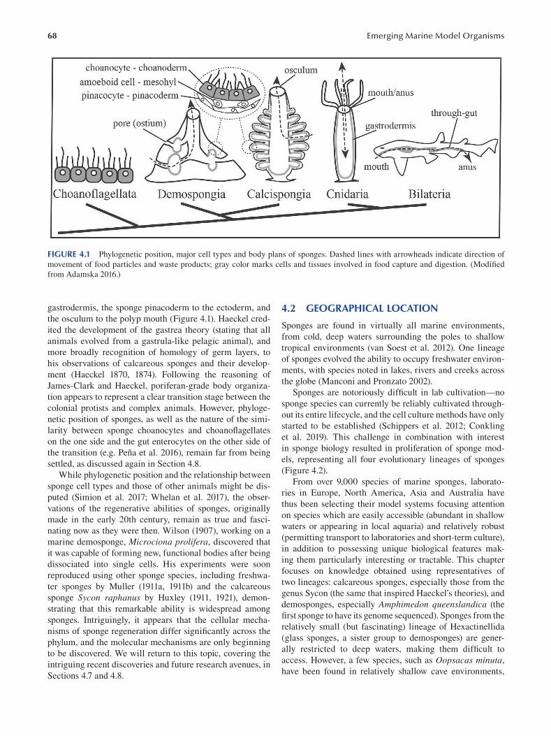
68 Emerging Marine Model Organisms
FIGURE 4.1 Phylogenetic position, major cell types and body plans of sponges. Dashed lines with arrowheads indicate direction of
movement of food particles and waste products; gray color marks cells and tissues involved in food capture and digestion. (Modifi ed
from Adamska 2016.)
gastrodermis, the sponge pinacoderm to the ectoderm, and
the osculum to the polyp mouth (Figure 4.1). Haeckel cred
ited the development of the gastrea theory (stating that all
animals evolved from a gastrula-like pelagic animal), and
more broadly recognition of homology of germ layers, to
his observations of calcareous sponges and their develop
ment (Haeckel 1870, 1874). Following the reasoning of
James-Clark and Haeckel, poriferan-grade body organiza
tion appears to represent a clear transition stage between the
colonial protists and complex animals. However, phyloge
netic position of sponges, as well as the nature of the simi
larity between sponge choanocytes and choanofl agellates
on the one side and the gut enterocytes on the other side of
the transition (e.g. Peña et al. 2016), remain far from being
settled, as discussed again in Section 4.8.
While phylogenetic position and the relationship between
sponge cell types and those of other animals might be dis
puted (Simion et al. 2017; Whelan et al. 2017), the obser
vations of the regenerative abilities of sponges, originally
made in the early 20th century, remain as true and fasci
nating now as they were then. Wilson (1907 ), working on a
marine demosponge, Microciona prolifera, discovered that
it was capable of forming new, functional bodies after being
dissociated into single cells. His experiments were soon
reproduced using other sponge species, including freshwa
ter sponges by Muller (1911a, 1911b) and the calcareous
sponge Sycon raphanus by Huxley (1911, 1921 ), demon
strating that this remarkable ability is widespread among
sponges. Intriguingly, it appears that the cellular mecha
nisms of sponge regeneration differ significantly across the
phylum, and the molecular mechanisms are only beginning
to be discovered. We will return to this topic, covering the
intriguing recent discoveries and future research avenues, in
Sections 4.7 and 4.8.
4.2 GEOGRAPHICAL LOCATION
Sponges are found in virtually all marine environments,
from cold, deep waters surrounding the poles to shallow
tropical environments (van Soest et al. 2012). One lineage
of sponges evolved the ability to occupy freshwater environ
ments, with species noted in lakes, rivers and creeks across
the globe (Manconi and Pronzato 2002).
Sponges are notoriously difficult in lab cultivation—no
sponge species can currently be reliably cultivated through
out its entire lifecycle, and the cell culture methods have only
started to be established (Schippers et al. 2012; Conkling
et al. 2019). This challenge in combination with interest
in sponge biology resulted in proliferation of sponge mod
els, representing all four evolutionary lineages of sponges
( Figure 4.2 ).
From over 9,000 species of marine sponges, laborato
ries in Europe, North America, Asia and Australia have
thus been selecting their model systems focusing attention
on species which are easily accessible (abundant in shallow
waters or appearing in local aquaria) and relatively robust
(permitting transport to laboratories and short-term culture),
in addition to possessing unique biological features mak
ing them particularly interesting or tractable. This chapter
focuses on knowledge obtained using representatives of
two lineages: calcareous sponges, especially those from the
genus Sycon (the same that inspired Haeckel’s theories), and
demosponges, especially Amphimedon queenslandica (the
first sponge to have its genome sequenced). Sponges from the
relatively small (but fascinating) lineage of Hexactinellida
(glass sponges, a sister group to demosponges) are gener
ally restricted to deep waters, making them diffi cult to
access. However, a few species, such as Oopsacas minuta,
have been found in relatively shallow cave environments,
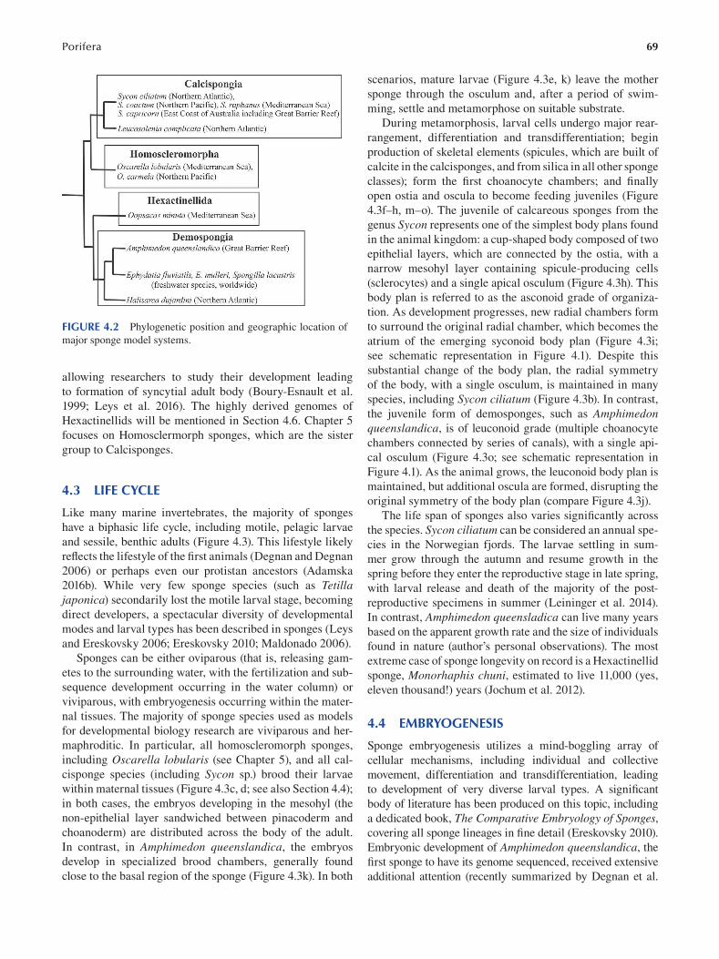
69 Porifera
FIGURE 4.2 Phylogenetic position and geographic location of
major sponge model systems.
allowing researchers to study their development leading
to formation of syncytial adult body (Boury-Esnault et al.
1999; Leys et al. 2016 ). The highly derived genomes of
Hexactinellids will be mentioned in Section 4.6. Chapter 5
focuses on Homosclermorph sponges, which are the sister
group to Calcisponges.
4.3 LIFE CYCLE
Like many marine invertebrates, the majority of sponges
have a biphasic life cycle, including motile, pelagic larvae
and sessile, benthic adults (Figure 4.3). This lifestyle likely
reflects the lifestyle of the first animals ( Degnan and Degnan
2006) or perhaps even our protistan ancestors (Adamska
2016b). While very few sponge species (such as Tetilla japonica) secondarily lost the motile larval stage, becoming
direct developers, a spectacular diversity of developmental
modes and larval types has been described in sponges (Leys
and Ereskovsky 2006; Ereskovsky 2010; Maldonado 2006).
Sponges can be either oviparous (that is, releasing gam
etes to the surrounding water, with the fertilization and sub
sequence development occurring in the water column) or
viviparous, with embryogenesis occurring within the mater
nal tissues. The majority of sponge species used as models
for developmental biology research are viviparous and her
maphroditic. In particular, all homoscleromorph sponges,
including Oscarella lobularis (see Chapter 5), and all cal
cisponge species (including Sycon sp.) brood their larvae
within maternal tissues (Figure 4.3c, d; see also Section 4.4);
in both cases, the embryos developing in the mesohyl (the
non-epithelial layer sandwiched between pinacoderm and
choanoderm) are distributed across the body of the adult.
In contrast, in Amphimedon queenslandica, the embryos
develop in specialized brood chambers, generally found
close to the basal region of the sponge (Figure 4.3k). In both
scenarios, mature larvae (Figure 4.3e, k) leave the mother
sponge through the osculum and, after a period of swim
ming, settle and metamorphose on suitable substrate.
During metamorphosis, larval cells undergo major rear
rangement, differentiation and transdifferentiation; begin
production of skeletal elements (spicules, which are built of
calcite in the calcisponges, and from silica in all other sponge
classes); form the first choanocyte chambers; and fi nally
open ostia and oscula to become feeding juveniles (Figure
4.3f–h , m–o). The juvenile of calcareous sponges from the
genus Sycon represents one of the simplest body plans found
in the animal kingdom: a cup-shaped body composed of two
epithelial layers, which are connected by the ostia, with a
narrow mesohyl layer containing spicule-producing cells
(sclerocytes) and a single apical osculum (Figure 4.3h). This
body plan is referred to as the asconoid grade of organiza
tion. As development progresses, new radial chambers form
to surround the original radial chamber, which becomes the
atrium of the emerging syconoid body plan (Figure 4.3i;
see schematic representation in Figure 4.1). Despite this
substantial change of the body plan, the radial symmetry
of the body, with a single osculum, is maintained in many
species, including Sycon ciliatum ( Figure 4.3b ). In contrast,
the juvenile form of demosponges, such as Amphimedon queenslandica, is of leuconoid grade (multiple choanocyte
chambers connected by series of canals), with a single api
cal osculum (Figure 4.3o; see schematic representation in
Figure 4.1). As the animal grows, the leuconoid body plan is
maintained, but additional oscula are formed, disrupting the
original symmetry of the body plan (compare Figure 4.3j).
The life span of sponges also varies signifi cantly across
the species. Sycon ciliatum can be considered an annual spe
cies in the Norwegian fjords. The larvae settling in sum
mer grow through the autumn and resume growth in the
spring before they enter the reproductive stage in late spring,
with larval release and death of the majority of the post-
reproductive specimens in summer (Leininger et al. 2014).
In contrast, Amphimedon queensladica can live many years
based on the apparent growth rate and the size of individuals
found in nature (author’s personal observations). The most
extreme case of sponge longevity on record is a Hexactinellid
sponge, Monorhaphis chuni, estimated to live 11,000 (yes,
eleven thousand!) years (Jochum et al. 2012).
4.4 EMBRYOGENESIS
Sponge embryogenesis utilizes a mind-boggling array of
cellular mechanisms, including individual and collective
movement, differentiation and transdifferentiation, leading
to development of very diverse larval types. A signifi cant
body of literature has been produced on this topic, including
a dedicated book, The Comparative Embryology of Sponges, covering all sponge lineages in fine detail (Ereskovsky 2010).
Embryonic development of Amphimedon queenslandica , the
first sponge to have its genome sequenced, received extensive
additional attention (recently summarized by Degnan et al.
70 Emerging Marine Model Organisms
FIGURE 4.3 Sponge life cycle. Adults (a, b, j), embryos within maternal tissue (c, d, k), larvae (e, l), postlarvae (f, g, m, n) and juveniles
(h, i) of two sponge model systems: the calcareous sponge Sycon ciliatum (a–i) and the demosponge Amphimedon queenslandica (j–o).
(a) Multiple sponge specimens growing together on Laminaria sp.; (j) individual sponge on coral rubble. (d) Fixed slice of tissue with
spicules removed to reveal embryos; the remaining samples are live specimens or their fragments. See text for description of embry
onic development and metamorphosis. Scale bars: (b, j): 5 mm; (e, l): 50 μm. ([a–i] Reproduced from Leininger et al. 2014, [j–o] from
Adamska et al. 2007.)
2015). In this species, embryonic development occurs in a
brood chamber, containing a mix of embryos of all stages,
from eggs to ready-to-release larvae, with the younger stages
close to the edge of the chamber and more mature ones at
the center (Figure 4.3k). The embryos are approximately 0.5
mm in diameter and yolky, with a cell division pattern best
described as asynchronous and anarchic, leading to forma
tion of a solid, spherical morula composed of cells of differ
ent sizes and differing by pigmentation level. Extensive cell
movements result in development of a bi-layered, polarized
embryo (referred to as gastrula in the original publication
describing development of this species; Leys and Degnan
2002, but see Nakanishi et al. 2014 for a different view on
the same process). Pigmented cells coalesce at one pole of the
embryo to first form a spot and then a ring (Figure 4.3k). This
ring, known to be a photosensory steering organ positioned
at the posterior pole of the Amphimedon larva (Leys and
Degnan 2001), is characteristic of parenchymella-type larvae
of many other demosponges (Maldonado et al. 2006). There
can be an extensive number of cell types present in mature
parenchymella type larvae, including sclerocytes (cells pro
ducing spicules), archaeocytes (stem cells) and, in some cases,
fully differentiated choanocytes and pinacocytes (e.g. Saller
1988 ).
One of the best studied of the larval types among sponges
are the amphiblastula larvae of Calcaronean sponges, the
lineage of calcisponges that includes Sycon ciliatum and
related species (Franzen 1988). The other lineage of calcare
ous sponges, the Calcineans, has calciblastula larvae very
similar to cinctoblastula found in Homoscleromorph sponges
(Chapter 5), although it is not clear whether this similarity
reflects shared ancestry (as Homoscleromorpha and sister
group to the Calcispongiae) or is a result of convergence.
The amphiblastula larva forms through a highly stereo
typic series of division followed by differentiation of only
three cell types, which further undergo clear differentiation
pathways upon metamorphosis. The oocytes are found uni
formly distributed across the mesohyl of mature specimens.
In the case of Sycon ciliatum in the Norwegian fjords, the
development is synchronous through the local populations,
71 Porifera
with the first round of oocyte growth and fertilization occur
ring in the late spring (Leininger et al. 2014). Cleavage is
complete, with the first two planes of division perpendicular
to each other and the plane of the pinacoderm, thus divid
ing the zygote into four equal blastomeres. The subsequent
divisions are oblique, resulting in formation of a cup-shaped
embryo, with larger cells (macromeres) closer to the cho
anocytes and smaller cells (micromeres) facing pinacocytes
(Figure 4.1). The embryonic cavity communicates with the
lumen of the radial chamber, and through this opening, the
embryo inverts itself so that the flagella of the micromeres
(which originally form on the inner surface of the embryo)
point outward.
In addition to the flagellated micromeres and larger,
non-flagellated macromeres, the larva contains two other
cell types: cross cells and maternal cells (Figure 4.1b). The
cross cells (four in each larva) are of embryonic origin and
differentiate from the outer “corners” of the four original
blastomeres, with their final positions forming a cross at the
equator of the larvae, conveying tetra-radial symmetry to
the larva (Figure 4.1a). The function of these cells remains
enigmatic, but they have been proposed to have sensory role
and, consistent with this notion, express a number of genes
known from other animals to be involved in specifi cation
of sensory cells and neurons (Tuzet 1973; Fortunato et al.
2014). Intriguingly, cross cells, along with maternal cells,
which migrate inside of the embryo after inversion, degener
ate during metamorphosis and do not contribute to forma
tion of the juvenile body (Amano and Hori 1993).
As the larva settles on its anterior pole, the macromeres
envelop the micromeres without losing epithelial character
and differentiate directly to pinacocytes. The micromeres
FIGURE 4.4 Schematic representation of embryonic development (a) and metamorphosis (b) in calcaronean sponges . In
(a), the top row shows cross-sections of embryos surrounded by
maternal tissues (pinacoderm and choanoderm); the bottom row is
a top view of isolated embryos. Thick lines indicate macromeres
and pinacocytes; thin lines indicate micromeres and choanocytes.
Embryonic/larval cross cells and the cytoplasm of cleavage stage
embryo destined to become cross cell are shaded gray.
undergo epithelial-to-mesenchymal transition and become
amoeboid cells. After a period of movement (hours to
days, depending on species), the micromeres differentiate
into choanocytes and other juvenile cell types, including
sclerocytes (spicule producing cells) (Figure 4.4b). Finally,
the osculum opens at the apical pole and ostia form across
the surface, resulting in formation of a functional, juvenile
sponge of asconoid grade of organization. The source of
porocytes is unclear, but it is likely that they differentiate
from pinacocytes.
4.5 ANATOMY
All sponges (with the notable exception of carnivorous
sponges, which secondarily lost choanocytes; Vacelet and
Boury-Esnault 1995 ; Riesgo et al. 2007 ) are built of the
same basic building blocks: choanocytes forming cho
anoderm of the radial chambers, the pinacoderm lining all
remaining surfaces, with varying types and numbers of cells
inhabiting the mesohyl. The mesohyl can be very cell poor
and narrow (for example, in the Homoscleromorph sponges;
see Chapter 5 ) or constitute most of the body of the sponge,
as in many Demosponges. Traditionally, the body plans are
divided into three major types. The simplest is asconoid,
as described for Calcaronean juveniles ( Figure 4.3h , 4.4b ),
with many calcisponge species retaining this body organi
zation, with branching and anastomosing tubes forming as
the body enlarges. The second type is syconoid, as in cal
cisponges from the genus Sycon ( Figure 4.1 , Figure 4.3b ,
c , i ), with radial choanocyte chambers surrounding endop
inacocyte-lined atrial cavity. The most complex, and the
most common among sponges (being the typical body plan
of Demosponges, the most speciose of the sponge lineages),
is the leuconoid body plan composed of choanocyte cham
bers linked by an intricate network of endopinacyte-lined
canals ( Figure 4.1 and 4.3j , o ). Two lesser-known sponge
body plans should also be mentioned. One is the syllei
bid body plan found in Homoscleromorph sponges, which
can be considered a link between the syconoid and leuco
noid body plans, with multiple syconoid-level units con
nected to the atrium. The most recently described sponge
body plan, solenoid, is found in some Calcinean species and
can be best described as a complex system of anastomosing
tubes of the asconoid grade embedded in a thick mesohyl
layer ( Cavalcanti and Klautau 2011 ).
In the majority of sponges, the epithelial and mesen
chymal layers are supported by organic and/or inorganic
skeletons. The spongin-based organic skeletons of the
genus Spongia and related species are well known as bath
sponges—although, after the natural populations have been
virtually exterminated by combination of harvest and pol
lution of the habitat, natural bath sponges have been all but
replaced by artificial ones (Pronzato and Manconi 2008).
The majority of sponges produce inorganic skeletal ele
ments, called spicules, which were traditionally the key to
sponge taxonomy, given the paucity of other characters avail
able until the advent of molecular phylogenies (Uriz 2006;
72
FIGURE 4.5 Spicule formation in calcareous sponges.
Thickener cells (t) are dark gray, founder cells (f) are light gray.
(Modified from Voigt et al. 2017, with the schematic representa
tions re-drawn from Minchin 1908.)
van Soest et al. 2012). The spicules are of two types—built
of calcite in the calcisponges and of silica in the remaining
three lineages. Not only the material but also the cellular
mechanism of spicule synthesis and subsequent positioning
differs. The demosponge spicules are produced intracellu
larly, within vacuoles, and are subsequently moved to their
final position by a concerted action of carrier cells (Mohri et
al. 2008; Nakayama et al. 2015). In contrast, calcareous spic
ules are produced by groups of cells, the numbers of which
depend on the type of the spicule and tend to remain in situ,
without subsequent movement. For example, single-rayed
spicules (diactines) are secreted by two cells, one known as
the founder cell and the other as the thickener cell. On the
other hand, the tri-radial triactines are produce by sextets of
cells, with three founder cells and three thickener cells work
ing together to produce one spicule (Minchin 1908; Voigt et
al. 2017 ) (Figure 4.5). Different types of spicules form sup
porting structures along the body, with the long, slender di
actines often found forming a crown or a collar around the
osculum (Figure 4.3b).
4.6 GENOMIC DATA
The first insight into gene content of sponges was provided
by transcriptome rather than genome analyses. Most signifi
cantly, the analysis of developmental regulatory genes in the
transcriptome of the homoscleromorph sponge Oscarella carmela revealed that sponges possess multiple components
of developmental signaling pathways used by animals to
regulate their development (Nichols et al. 2006 ). However,
the complete developmental regulatory gene repertoire of a
sponge could only be fully appreciated by whole genome
sequencing. The first sponge for which this was achieved
was the demosponge Amphimedon queenslandica, a spe
cies inhabiting reefs fringing the Heron Island of the Great
Barrier Reef (Srivastava et al. 2010). This was not only the
first but also likely the last sponge genome sequenced using
Emerging Marine Model Organisms
the traditional Sanger method. Amphimedon genome analysis
revealed that for the overwhelming majority of developmen
tal regulatory gene families, whether signaling molecules or
transcription factors, Amphimedon possesses fewer family
members than the more complex animals (Cnidarians and
Bilaterians). This pattern, perhaps expected, was consistent
with the notion that a simple animal would have a simpler
regulatory gene repertoire.
It was therefore surprising when analysis of the second
sponge species to be sequenced—the calcareous sponge
Sycon ciliatum—revealed developmental gene family
sizes on a par with those found in bilaterians. For example,
while humans have 19 Wnt ligands and Amphimedon has 3
(Adamska et al. 2007), Sycon has 21 (Leininger et al. 2014).
Even more strikingly—and controversially—the Sycon genome appears to possess a ParaHox gene, Cdx, which is
clearly absent from the Amphimedon genome (Larroux et
al. 2007; Fortunato et al. 2014). A systematic comparison
of transcription factors present in Amphimedon and Sycon demonstrated that genomes of calcisponges and demo-
sponges underwent independent events of gene loss and
family expansions (Fortunato et al. 2015).
Gene content analysis of two Hexactinellids (glass
sponges) revealed a different kind of surprise—it appears
that neither Oopsacas minuta nor Aphrocallistes vastus possesses key components of the Wnt signaling pathway
(Schenkelaars et al. 2017). As this pathway is used across
the animal kingdom (including other sponges; See section
4.7) to pattern the major body axis, this finding is another
key indication that insights from one lineage of sponges
cannot be assumed to reflect the genome composition of
all sponges—and of the last common ancestor of all ani
mals. Instead, it thus appears that, since the divergence
approximately 600 million years ago, sponge gene reper
toires underwent dramatic changes, in contrast to the body
plans which remained apparently stable throughout this
time.
But sponge genomes can provide insight into more than
just gene content: a gateway to understand evolution of
genome function in animals. One of the mechanisms known
to regulate gene expression in vertebrates (but not in the
majority of invertebrates) is DNA methylation. However,
the evolutionary history of this mechanism is not well
understood. A recent study revealed that—in parallel to the
differences found in gene content—sponge genomes are
methylated to very different levels. While the Amphimedon genome is highly methylated (in striking similarly to ver
tebrate genomes), methylation in Sycon is more moderate,
consistent with independent acquisition of genome methyla
tion in sponges (de Mendoza et al. 2019).
Gaiti and colleagues (2017 ) used the Amphimedon genome find out whether two other regulatory features of
animal genomes are found in sponges: the posttranslational
modifi cations of histone H3 (linked to precise regulation of
gene expression in animals) and micro-systenic units har
boring distal enhancers of developmental regulatory genes.
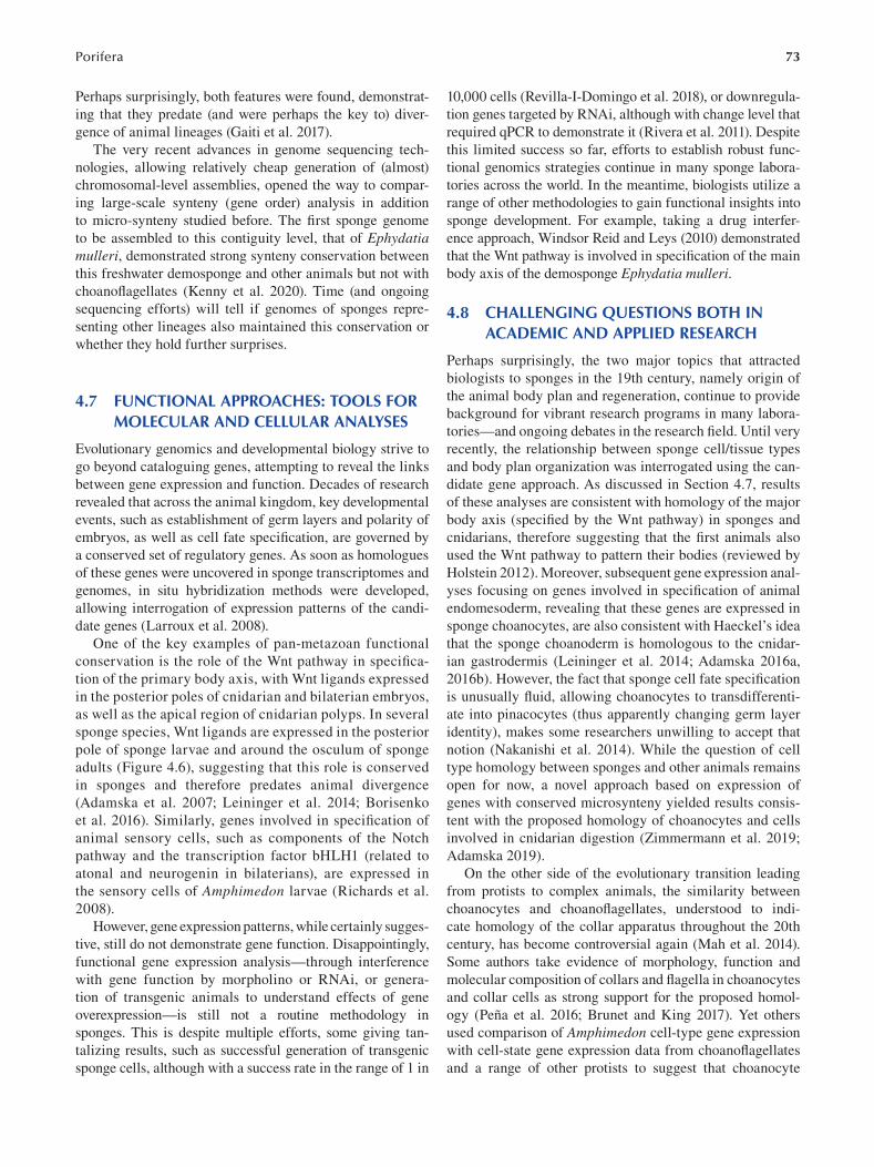
73 Porifera
Perhaps surprisingly, both features were found, demonstrat
ing that they predate (and were perhaps the key to) diver
gence of animal lineages (Gaiti et al. 2017).
The very recent advances in genome sequencing tech
nologies, allowing relatively cheap generation of (almost)
chromosomal-level assemblies, opened the way to compar
ing large-scale synteny (gene order) analysis in addition
to micro-synteny studied before. The first sponge genome
to be assembled to this contiguity level, that of Ephydatia mulleri, demonstrated strong synteny conservation between
this freshwater demosponge and other animals but not with
choanoflagellates (Kenny et al. 2020). Time (and ongoing
sequencing efforts) will tell if genomes of sponges repre
senting other lineages also maintained this conservation or
whether they hold further surprises.
4.7 FUNCTIONAL APPROACHES: TOOLS FOR MOLECULAR AND CELLULAR ANALYSES
Evolutionary genomics and developmental biology strive to
go beyond cataloguing genes, attempting to reveal the links
between gene expression and function. Decades of research
revealed that across the animal kingdom, key developmental
events, such as establishment of germ layers and polarity of
embryos, as well as cell fate specification, are governed by
a conserved set of regulatory genes. As soon as homologues
of these genes were uncovered in sponge transcriptomes and
genomes, in situ hybridization methods were developed,
allowing interrogation of expression patterns of the candi
date genes (Larroux et al. 2008).
One of the key examples of pan-metazoan functional
conservation is the role of the Wnt pathway in specifi ca
tion of the primary body axis, with Wnt ligands expressed
in the posterior poles of cnidarian and bilaterian embryos,
as well as the apical region of cnidarian polyps. In several
sponge species, Wnt ligands are expressed in the posterior
pole of sponge larvae and around the osculum of sponge
adults ( Figure 4.6), suggesting that this role is conserved
in sponges and therefore predates animal divergence
(Adamska et al. 2007; Leininger et al. 2014; Borisenko
et al. 2016). Similarly, genes involved in specifi cation of
animal sensory cells, such as components of the Notch
pathway and the transcription factor bHLH1 (related to
atonal and neurogenin in bilaterians), are expressed in
the sensory cells of Amphimedon larvae (Richards et al.
2008 ).
However, gene expression patterns, while certainly sugges
tive, still do not demonstrate gene function. Disappointingly,
functional gene expression analysis—through interference
with gene function by morpholino or RNAi, or genera
tion of transgenic animals to understand effects of gene
overexpression—is still not a routine methodology in
sponges. This is despite multiple efforts, some giving tan
talizing results, such as successful generation of transgenic
sponge cells, although with a success rate in the range of 1 in
10,000 cells (Revilla-I-Domingo et al. 2018), or downregula
tion genes targeted by RNAi, although with change level that
required qPCR to demonstrate it (Rivera et al. 2011). Despite
this limited success so far, efforts to establish robust func
tional genomics strategies continue in many sponge labora
tories across the world. In the meantime, biologists utilize a
range of other methodologies to gain functional insights into
sponge development. For example, taking a drug interfer
ence approach, Windsor Reid and Leys (2010) demonstrated
that the Wnt pathway is involved in specification of the main
body axis of the demosponge Ephydatia mulleri.
4.8 CHALLENGING QUESTIONS BOTH IN ACADEMIC AND APPLIED RESEARCH
Perhaps surprisingly, the two major topics that attracted
biologists to sponges in the 19th century, namely origin of
the animal body plan and regeneration, continue to provide
background for vibrant research programs in many labora
tories—and ongoing debates in the research field. Until very
recently, the relationship between sponge cell/tissue types
and body plan organization was interrogated using the can
didate gene approach. As discussed in Section 4.7, results
of these analyses are consistent with homology of the major
body axis (specified by the Wnt pathway) in sponges and
cnidarians, therefore suggesting that the first animals also
used the Wnt pathway to pattern their bodies (reviewed by
Holstein 2012 ). Moreover, subsequent gene expression anal
yses focusing on genes involved in specification of animal
endomesoderm, revealing that these genes are expressed in
sponge choanocytes, are also consistent with Haeckel’s idea
that the sponge choanoderm is homologous to the cnidar
ian gastrodermis ( Leininger et al. 2014 ; Adamska 2016a,
2016b). However, the fact that sponge cell fate specifi cation
is unusually fluid, allowing choanocytes to transdifferenti
ate into pinacocytes (thus apparently changing germ layer
identity), makes some researchers unwilling to accept that
notion ( Nakanishi et al. 2014 ). While the question of cell
type homology between sponges and other animals remains
open for now, a novel approach based on expression of
genes with conserved microsynteny yielded results consis
tent with the proposed homology of choanocytes and cells
involved in cnidarian digestion ( Zimmermann et al. 2019 ;
Adamska 2019 ).
On the other side of the evolutionary transition leading
from protists to complex animals, the similarity between
choanocytes and choanoflagellates, understood to indi
cate homology of the collar apparatus throughout the 20th
century, has become controversial again (Mah et al. 2014).
Some authors take evidence of morphology, function and
molecular composition of collars and flagella in choanocytes
and collar cells as strong support for the proposed homol
ogy (Peña et al. 2016; Brunet and King 2017). Yet others
used comparison of Amphimedon cell-type gene expression
with cell-state gene expression data from choanofl agellates
and a range of other protists to suggest that choanocyte
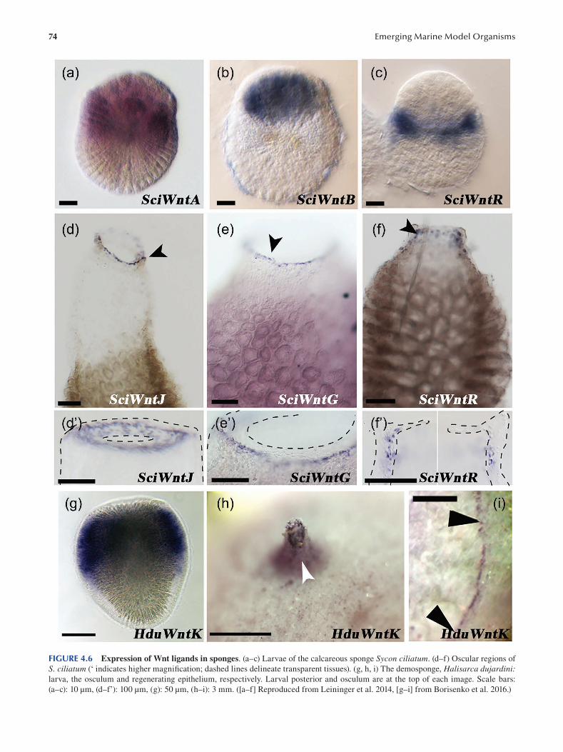
74 Emerging Marine Model Organisms
FIGURE 4.6 Expression of Wnt ligands in sponges. (a–c) Larvae of the calcareous sponge Sycon ciliatum. (d–f) Oscular regions of
S. ciliatum (‘ indicates higher magnification; dashed lines delineate transparent tissues). (g, h, i) The demosponge, Halisarca dujardini: larva, the osculum and regenerating epithelium, respectively. Larval posterior and osculum are at the top of each image. Scale bars:
(a–c): 10 μm, (d–f’): 100 μm, (g): 50 μm, (h–i): 3 mm. ([a–f] Reproduced from Leininger et al. 2014, [g–i] from Borisenko et al. 2016.)
75 Porifera
morphology evolved independently from choanofl agel
lates (Sogabe et al. 2019). That these seemingly academic
questions are also exciting to the general audience is evi
denced by popular science magazines covering this debate
( Cepelewicz 2019 ).
Less “academic”, as understanding of sponge regeneration
capacity might potentially be applicable to human regenera
tive medicine, is the question of how sponges regulate their
spectacular regenerative capacities. Recent research reveals
that some of the regeneration mechanisms might indeed be
shared between sponges and other animals, as many of the
developmental signaling pathways known to be involved in
mammalian regenerations are also activated during regener
ation of sponges, including re-building of bodies from disso
ciated cells (Soubigou et al. 2020). The most exciting aspect
of sponge regeneration appears to be the capacity of sponge
cells to directly transdifferentiate upon injury (Ereskovsky
et al. 2015; Ereskovsky et al. 2017; reviewed by Adamska
2018). Would it be possible to utilize mechanisms involved
in transdifferentiation of sponge cells to reprogram mam
malian cells for therapeutic purposes?
The pharmaceutical industry has been investigating
sponges as potential sources of bioactive compounds, with
great success, for over 50 years. In 1969, the fi rst sponge-
derived anti-cancer drug, cytarabine (also known as Ara-
C, Cytosar-U or Depocyst), originally extracted from the
Caribbean demosponge Tectitethya crypta, was approved
by the Food and Drug Administration (FDA). In 1976, the
FDA also approved vidarabine (Ara-A, Vira-A) as an anti
viral drug derived from the same sponge species (reviewed
by Brinkmann et al. 2017). More recently, eribulin mesylate
(E389, Halaven), an analog of halichondrin B isolated from
Japanese demosponge Halichondria okadai, was approved
as treatment for metastatic breast cancer (reviewed by
Gerwick and Fenner 2013).
In addition to being useful, the secondary metabolites
found in sponges are all the more fascinating as they are in
fact produced by microbes living in close symbiosis with their
poriferan hosts. The study of sponge microbiomes revealed
essential roles in nutrient cycling and production of vitamins
in addition to the secondary metabolites likely responsible
for protection of sponges from potential predators and foul
ing organisms (see Reiswig 1981; Maldonado et al. 2012).
It appears that the complex, species-specifi c assemblages
of bacteria can be transmitted both horizontally (from the
surrounding water) and vertically (from mother to larvae)
(e.g. Schmitt et al. 2008; Webster et al. 2010). However, the
molecular mechanisms involved in establishment and main
tenance of these symbioses are not understood and remain
an area of open and exciting investigations.
BIBLIOGRAPHY
Adamska M. 2016a. Sponges as models to study emergence of
complex animals. Curr Opin Genet Dev 39: 21–28.
Adamska M. 2016b. Sponges as the Rosetta Stone of colonial-to
multicellular transition. In: Multicellularity: Origins and evolution. KJ Niklas, SA Newman (Eds.). The MIT Press,
Cambridge, MA; London, England. ISBN: 978-0-262-03
415-9.
Adamska M. 2018. Differentiation and transdifferentiation of
sponge cells. In: Marine Organisms as Model Systems in Biology and Medicine. M Kloc, JZ Kubiak (Eds.). Springer-
Nature Series. Results and Problems in Cell Differentiation.
Springer International Publishing. eBook ISBN: 978-3-319
92486-1. Hardcover ISBN: 978-3-319-92485-4.
Adamska M. 2019. Animal cell type diversity. Nat Ecol Evol 3:
1277–1278.
Adamska M, Degnan SM, Green KM, Adamski M, Craigie A,
Larroux C, Degnan BM. 2007. Wnt and Tgf expression
in the sponge Amphimedon queenslandica and the origin of
metazoan embryonic patterning. PLoS One 2(10): e1031.
Amano S, Hori I. 1993. Metamorphosis of calcareous sponges. 2:
Cell rearrangement and differentiation in metamorphosis.
Invert Reprod Dev 24: 13–26.
Borisenko I, Adamski M, Ereskovsky A, Adamska M. 2016.
Surprisingly rich repertoire of Wnt genes in the demosponge
Halisarca dujardini. BMC Evol Biol 16: 123.
Boury-Esnault N, Efremova S, Bézac C, Vacelet J. 1999.
Reproduction of a hexactinellid sponge: First description
of gastrulation by cellular delamination in the Porifera.
Invertebr Reprod Dev 35 (3): 187–201.
Brinkmann CM, Marker A, Kurtböke DI. 2017. An overview on
marine sponge-symbiotic bacteria as unexhausted sources
for natural product discovery diversity. Diversity 9: 40.
Brunet T, King N. 2017. The origin of animal multicellularity and
cell differentiation. Dev Cell 43(2): 124–140.
Cavalcanti FF, Klautau M. 2011. Solenoid: A new aquiferous sys
tem to Porifera. Zoomorphology 130: 255–260.
Cepelewicz J. 2019. Scientists debate the origin of cell types in the
fi rst animals. www.quantamagazine.org/scientists-debate
the-origin-of-cell-types-in-the-fi rst-animals-20190717/
Conkling M, Hesp K, Munroe S, et al. 2019. Breakthrough in
marine invertebrate cell culture: Sponge cells divide rapidly
in improved nutrient medium. Sci Rep 9: 17321.
Degnan BM, Adamska M, Richards GR, Larroux C, Leininger S,
Bergum B, Calcino A, Maritz K, Nakanishi N, Degnan SM.
2015. Porifera. In: Evolutionary Developmental Biology of Invertebrates. Vol. 1. A. Wanninger (Ed.), pp. 65–106.
Springer Verlag, Vien. ISBN: 978-3-7091-1861-0.
Degnan SM, Degnan BM. 2006. The origin of the pelagobenthic
metazoan life cycle: What’s sex got to do with it? Integr Comp Biol 46(6): 683–690.
de Mendoza A, Hatleberg WL, Pang K, et al. 2019. Convergent
evolution of a vertebrate-like methylome in a marine sponge.
Nat Ecol Evol 3(10): 1464–1473.
Ereskovsky AV. 2010. The Comparative Embryology of Sponges.
Springer, New York.
Ereskovsky AV, Borisenko IE, Lapébie P, Gazave E, Tokina DB,
et al. 2015. Oscarella lobularis (Homoscleromorpha, Por
ifera) regeneration: Epithelial morphogenesis and metapla
sia. PLoS One 10(8): e0134566.
Ereskovsky AV, Lavrov AI, Bolshakov FV, Tokina DB. 2017.
Regeneration in White Sea sponge Leucosolenia complicata (Porifera, Calcarea). Invertebr Zool 14(2): 108–113.
Fortunato SAV, Adamski M, Adamska M. 2015. Comparative
analyses of developmental transcription factor repertoires
in sponges reveal unexpected complexity of the earliest ani
mals. Mar Genomics 2: 121–129.
Fortunato SAV, Adamski M, Mendivil O, Leininger S, Liu J, Ferrier
DEK, and Adamska M. 2014. Calcisponges have a ParaHox
gene and dynamic expression of dispersed NK homeobox
genes. Nature 514(7524): 620–623.
76
Franzen W. 1988. Oogenesis and larval development of Scypha
ciliata (Porifera, Calcarea). Zoomorphology 107: 349.
Gaiti F, Jindrich K, Fernandez-Valverde SL, Roper KE, Degnan
BM, Tanurdžić M. 2017. Landscape of histone modifi cations
in a sponge reveals the origin of animal cis-regulatory com
plexity. eLife 6: e22194.
Gerwick WH, Fenner AM. 2013. Drug discovery from marine
microbes. Microb Ecol 65(4): 800–806.
Grant RE. 1825. Observations and experiments on the structure and
functions of the sponge. Edinburgh Phil Journ 13: 94, 343;
14: 113–124.
Grant RE. 1836. Animal Kingdom. In: The Cyclopaedia of Anatomy and Physiology. Vol. 1. RB Todd (Ed.), pp. 107–118.
Sherwood, Gilbert, and Piper, London.
Haeckel E. 1870. On the organization of sponges and their relation
ship to the corals. Ann Mag Nat Hist 5: 1–13, 107–120.
Haeckel E. 1874. Die Gastrae Theorie, die phylogenetische
Classification des Thierreichs und die Homologie der
Keimblatter. Jena Zeitschr Naturwiss 8: 1–55.
Holstein TW. 2012. The evolution of the Wnt pathway. Cold Spring Harb Perspect Biol 4: a007922.
Huxley JS. 1911. Some phenomena of regeneration in Sycon; with
a note on the structure of its collar-cells. Phil Trans R Soc B 202: 165–189.
Huxley JS. 1921. Further studies on restitution-bodies and free tis
sue culture in Sycon. Quart J Micr Sci 65: 293–322.
James-Clark H. 1868. On the spongiae ciliatae as infusoria fl ag
ellata; or observations on the structure, animality, and rela
tionship of Leucosolenia botryoides. Ann Mag Nat Hist 1:
133–142, 188–215, 250–264.
Jochum KP, Wang X, Ennemannc TW, Sinhaa B, Müller WEG.
2012. Siliceous deep-sea sponge Monorhaphis chuni : A
potential paleoclimate archive in ancient animals. Chemical Geology : 300–330.
Kenny NJ, Francis WR, Rivera-Vicéns RE, et al. 2020. Tracing
animal genomic evolution with the chromosomal-level
assembly of the freshwater sponge Ephydatia muelleri. Nat Commun 11: 3676.
Knobloch S, Jóhannsson R, Marteinsson V. 2019. Co-cultivation of
the marine sponge Halichondria panicea and its associated
microorganisms. Sci Rep 9: 10403.
Larroux C, Fahey B, Adamska M, Richards GS, Gauthier M, Green
K, Lovas E, Degnan BM. 2008. Whole-mount in situ hybrid
ization in amphimedon. CSH Protoc . pdb.prot5096.
Larroux C, Fahey B, Degnan SM, Adamski M, Rokhsar DS,
Degnan BM. 2007. The NK homeobox gene cluster predates
the origin of Hox genes. Curr Biol Apr 17, 17(8): 706–710.
Leininger S, Adamski M, Bergum B, Guder C, Liu J, Laplante
M, Bråte J, Hoffmann F, Fortunato S, Jordal S, Rapp HT,
Adamska M. 2014. Developmental gene expression provides
clues to relationships between sponge and eumetazoan body
plans. Nature Comm 5: 3905.
Leys SP, Degnan BM. 2001. Cytological basis of photoresponsive
behavior in a sponge larva. Biol Bull 201(3): 323–338.
Leys SP, Degnan BM. 2002. Embryogenesis and metamorphosis in
a haplosclerid demosponge: Gastrulation and transdifferen
tiation of larval ciliated cells to choanocytes. Invertebr Biol 121: 171–189.
Leys SP, Ereskovsky AV. 2006. Embryogenesis and larval differen
tiation in sponges. Can J Zool 84(2): 262–287.
Leys SP, Kamarul Zaman A, Boury-Esnault N. 2016. Three-
dimensional fate mapping of larval tissues through metamor
phosis in the glass sponge Oopsacas minuta. Invertebr Biol 135: 259–272.
Emerging Marine Model Organisms
Mah JL, Christensen-Dalsgaard KK, Leys SP. 2014. Choanofl agellate
and choanocyte collar-flagellar systems and the assumption of
homology. Evol Dev 16(1): 25–37.
Maldonado M. 2006. The ecology of the sponge larva. Can J Zool 84(2): 175–194.
Maldonado M, Ribes M, van Duyl FC. 2012. Nutrient fl uxes through
sponges: Biology, budgets, and ecological implications. Adv Mar Biol 62: 113–182. Elsevier Ltd ISSN 0065-2881.
Manconi M, Pronzato R. 2002. Suborder Spongillina subord. nov.:
Freshwater sponges. In: Systema Porifera: A Guide to the Classification of Sponges. NA John, WM Rob, Van Soest
(Eds.), pp. 921–1021. Kluwer Academic/Plenum Publishers,
New York.
Minchin EA. 1908. Materials for a monograph of the Ascons. II:
The formation of spicules in the genus Leucosolenia, with
some notes on the histology of the sponges. Q J Microsc Sci 52: 301–355.
Mohri K, Nakatsukasa M, Masuda Y, Agata K, Funayama N. 2008.
Toward understanding the morphogenesis of siliceous spic
ules in freshwater sponge: Differential mRNA expression of
spicule-type-specific silicatein genes in Ephydatia fluviatilis.
Dev Dyn 237: 3024–3039.
Muller K. 1911a. Beobachtungen liber Reduktionsvorgange bei
Spongilliden, nebst Bemerkungen zu deren ausserer Morphologie
und Biologie. Zool Anz 37: 114–121.
Muller K. 1911b. Versuche liber die Regenerationsfahigkeit der
Susswasserschwamme. Zool Anz 37: 83.
Nakanishi N, Sogabe S, Degnan BM. 2014. Evolutionary origin of
gastrulation: Insights from sponge development. BMC Biol 12: 26.
Nakayama S, Arima K, Kawai K, Mohri K, Inui C, Sugano W,
Koba H, Tamada K, Nakata YJ, Kishimoto K, Arai-Shindo
M, Kojima C, Matsumoto T, Fujimori T, Agata K, Funayama
N. 2015. Dynamic transport and cementation of skeletal ele
ments build up the pole-and-beam structured skeleton of
sponges. Curr Biol 25(19): 2549–2554.
Nichols SA, Dirks W, Pearse JS, King N. 2006. Early evolution of
animal cell signaling and adhesion genes. Proc Natl Acad Sci USA 103(33): 12451–12456.
Peña JF, Alié A, Richter DJ, et al. 2016. Conserved expression
of vertebrate microvillar gene homologs in choanocytes of
freshwater sponges. Evo Devo 7: 13.
Pronzato R, Manconi R. 2008. Mediterranean commercial sponges:
Over 5000 years of natural history and cultural heritage. Mar Ecol 29: 146–166.
Reiswig HM. 1981. Partial carbon and energy budgets of the bac
teriosponge Verongia fistularis (Porifera: Demospongiae) in
Barbados West-Indies. Mar Biol 2: 273–294.
Revilla-I-Domingo R, Schmidt C, Zifko C, Raible F. 2018.
Establishment of transgenesis in the demosponge Suberites domuncula. Genetics 210(2): 435–443.
Richards GS, Simionato E, Perron M, Adamska M, Vervoort M,
Degnan BM. 2008. Sponge genes provide new insight into
the evolutionary origin of the neurogenic circuit. Curr Biol 18: 1156–1161.
Riesgo A, Taylor C, Leys SP. 2007. Reproduction in a carnivorous
sponge: The significance of the absence of an aquiferous sys
tem to the sponge body plan. Evol Dev 9(6): 618–631.
Rivera AS, Hammel JU, Haen KM, Danka ES, Cieniewicz B,
Winters IP, Posfai D, Wörheide G, Lavrov DV, Knight
SW, Hill MS, Hill AL, Nickel M. 2011. RNA interference
in marine and freshwater sponges: Actin knockdown in
Tethya wilhelma and Ephydatia muelleri by ingested dsRNA
expressing bacteria. BMC Biotechnol 11: 67.
77 Porifera
Saller U. 1988. Oogenesis and larval development of Ephydatia fluviatilis (Porifera, Spongillidae). Zoomorphology 108: 23–28.
Saville-Kent W. 1880. A Manual of the Infusoria: Including a Description of All Known Flagellate, Ciliate, and Tentaculiferous Protozoa, British and Foreign, and an Account of the Organization and the Affi nities of the Sponges. David Bogue, London.
Schenkelaars Q, Pratlong M, Kodjabachian L, et al. 2017. Animal
multicellularity and polarity without Wnt signaling. Sci Rep 7: 15383.
Schippers K, Sipkema D, Osinga R, Smidt H, Pomponi S, Martens
D, Wijffels R. 2012. Cultivation of sponges, sponge cells and
symbionts. Adv Mar Biol 62: 273–337.
Schmitt S, Angermeier H, Schiller R, Lindquist N, Hentschel U.
2008. Molecular microbial diversity survey of sponge repro
ductive stages and mechanistic insights into vertical trans
mission of microbial symbionts. Appl Environ Microbiol 74(24): 7694–7708.
Simion P, Philippe H, Baurain D, Jager M, Richter DJ, Di Franco
A, Roure B, Satoh N, Quéinnec É, Ereskovsky A, Lapébie P,
Corre E, Delsuc F, King N, Wörheide G, Manuel M. 2017. A
large and consistent phylogenomic dataset supports sponges as
the sister group to all other animals. Curr Biol 27(7): 958–967.
Sogabe S, Hatleberg WL, Kocot KM, Say TE, Stoupin D, Roper
KE, Fernandez-Valverde SL, Degnan SM, Degnan BM.
2019. Pluripotency and the origin of animal multicellularity.
Nature 570(7762): 519–522.
Soubigou A, Ross EG, Touhami Y, Chrismas N, Modepalli V. 2020.
Regeneration in the sponge Sycon ciliatum partly mimics
postlarval development. Development 147(22): dev193714.
Srivastava M, Simakov O, Chapman J, Fahey B, Gauthier MEA,
Mitros T, Richards GS, Conaco C, Dacre M, Hellsten U,
Larroux C, Putnam NH, Stanke M, Adamska M, Darling A,
Degnan SM, Oakley TH, Plachetzki DC, Zhai Y, Adamski
M, Calcino A, Cummins SF, Goodstein DM, Harris C,
Jackson DJ, Leys SP, Shu S, Woodcroft BJ, Vervoort M,
Kosik KS, Manning G, Degnan BM, Rokhsar DS. 2010. The
Amphimedon queenslandica genome and the evolution of
animal complexity. Nature 466(7307): 720–726.
Tuzet O. 1973. Éponges calcaires. In: Traité de Zoologie Anatomie, Systématique, Biologie Spongiaires. PP Grassé (Ed.),
pp. 27–132. Masson et Cie, Paris.
Uriz MJ. 2006. Mineral skeletogenesis in sponges. Can J Zool 84:
322–356.
Vacelet J, Boury-Esnault N. 1995. Carnivorous sponges. Nature 373: 333–335.
Van Soest RWM, Boury-Esnault N, Vacelet J, Dohrmann M,
Erpenbeck D, et al. 2012. Global diversity of sponges
(Porifera). PLoS One 7(4): e35105.
Voigt O, Adamska M, Adamski M, Kittelmann A, Wencker L,
Wörheide G. 2017. Spicule formation in calcareous sponges:
Coordinated expression of biomineralization genes and
spicule-type specific genes. Sci Rep 7: 45658.
Webster NS, Taylor MW, Behnam F, Lücker S, Rattei T, Whalan
S, Horn M, Wagner M. 2010. Deep sequencing reveals
exceptional diversity and modes of transmission for bacte
rial sponge symbionts. Environ Microbiol 12(8): 2070–2082.
Whelan NV, Kocot KM, Moroz TP, Mukherjee K, Williams P,
Paulay G, Moroz LL, Halanych KM 2017. Ctenophore rela
tionships and their placement as the sister group to all other
animals. Nat Ecol Evol 1(11): 1737–1746.
Wilson HV. 1907. On some phenomena of coalescence and regen
eration in sponges. J Exp Zool 5: 245–258.
Windsor Reid P, Leys S. 2010. Wnt signaling and induction in the
sponge aquiferous system: Evidence for an ancient origin of
the organizer. Evol Dev 12: 484–493.
Zimmermann B, Robert NSM, Technau U, et al. 2019. Ancient
animal genome architecture reflects cell type identities. Nat Ecol Evol 3: 1289–1293.

5 The HomoscleromorphSponge, Oscarella lobularis
Emmanuelle Renard, Caroline Rocher, Alexander Ereskovsky and Carole Borchiellini
CONTENTS
5.1 History of the Model..................................................................................................................................................... 79
5.2 Geographical Location ...................................................................................................................................................81
5.3 Life Cycle ...................................................................................................................................................................... 82
5.3.1 Asexual Reproduction: Fragmentation and Budding ........................................................................................ 82
5.3.2 Sexual Reproduction, Gametogenesis and Indirect Development .................................................................... 82
5.4 Embryogenesis.............................................................................................................................................................. 84
5.4.1 Cleavage and Formation of Coeloblastula ........................................................................................................ 84
5.4.2 Morphogenesis of the Cinctoblastula Larva and Larval Metamorphosis ......................................................... 85
5.4.3 Molecular Control of Development .................................................................................................................. 85
5.5 Anatomy........................................................................................................................................................................ 85
5.5.1 The Pinacoderm ................................................................................................................................................ 87
5.5.2 The Choanoderm............................................................................................................................................... 87
5.5.3 The Mesohyl ..................................................................................................................................................... 87
5.6 Transcriptomic and Genomic Data ............................................................................................................................... 88
5.7 Functional Approaches: Tools for Molecular and Cellular Analyses............................................................................ 89
5.7.1 Developmental and Non-Developmental Morphogenetic Contexts Accessible ............................................... 89
5.7.2 Polymerase Chain Reaction and Relatives ....................................................................................................... 89
5.7.3 In Situ Hybridization..........................................................................................................................................91
5.7.4 Fluorescent Immunolocalization .......................................................................................................................91
5.7.5 Cell Viability, Cell Apoptosis and Cell Proliferation Assays .............................................................................91
5.7.6 Cell Staining and Tracking ............................................................................................................................... 92
5.7.7 Loss-of-Function Approaches ........................................................................................................................... 92
5.8 Challenging Questions Both in Academic and Applied Research ................................................................................ 92
5.8.1 Finding New Bioactive Secondary Metabolites ............................................................................................... 92
5.8.2 Understanding Host–Symbiont Interactions..................................................................................................... 93
5.8.3 Deciphering the Origin and Evolution of Metazoan Epithelia ......................................................................... 93
5.8.4 Sponge Gastrulation and the Origin of Germ Layers ....................................................................................... 93
Acknowledgments .................................................................................................................................................................. 94
Bibliography .......................................................................................................................................................................... 94
5.1 HISTORY OF THE MODEL O. lobularis was long considered the only species of the genus
Oscarella. Accordingly, all species of the Oscarella genus Oscarella lobularis (Schmidt 1862) was first described as
reported between 1930 and 1990 were probably wrongly Halisarca lobularis Schmidt 1862 ( Schmidt 1862 ). Later
assigned to O. lobularis (Lage et al. 2018; Pérez and Ruiz Oscarella lobularis became the type species of the genus
2018 ). Oscarella Vosmaer, 1884 (Vosmaer 1884), genus, classifi ed
The cosmopolitan status of Oscarella lobularis began until 2012 (Gazave et al. 2012) within the class Demospongiae,
to be questioned in 1992. Several color morphs assigned subclass Tetractinellida, due to the shared presence of sili-
to the species O. lobularis (Schmidt 1862) living in symceous tetractinal-like calthrops spicules (Levi 1956). Despite
patry in the west Mediterranean area were compared for the its reported cosmopolitan distribution (uncommon in sponges
fi rst time using a combination of characters: morphological because of the low dispersal capacity of most sponge larvae)
characters, cytological characters and electric mobility of 12and the observation of a large variety of colors (Figure 5.1c),
protein markers. This study evidenced the presence of two
DOI: 10.1201/9781003217503-5 79
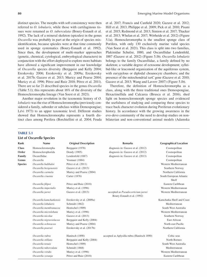
80
distinct species. The morphs with soft consistency were then
referred to O. lobularis, while those with cartilaginous tis
sues were renamed as O. tuberculata (Boury-Esnault et al.
1992). The lack of a mineral skeleton (spicules) in the genus
Oscarella was probably in part at the origin of species mis
identification, because spicules were at that time commonly
used in sponge systematics (Boury-Esnault et al. 1992).
Since then, the development of multi-marker approaches
(genetic, chemical, cytological, embryological characters) in
conjunction with the effort deployed to explore more habitats
have allowed a significant improvement in our knowledge
of Oscarella species diversity (Bergquist and Kelly 2004;
Ereskovsky 2006; Ereskovsky et al. 2009a ; Ereskovsky
et al. 2017b; Gazave et al. 2013; Muricy and Pearse 2004;
Muricy et al. 1996; Pérez and Ruiz 2018; Pérez et al. 2011).
There are so far 21 described species in the genus Oscarella (Table 5.1); this represents about 16% of the diversity of the
Homoscleromorpha lineage (Van Soest et al. 2021).
Another major revolution in the taxonomic history of O. lobularis was the rise of Homoscleromorpha (previously con
sidered a family, suborder or subclass within Demospongiae;
Lévi 1973) to an upper taxonomic level. Different studies
showed that Homoscleromorpha represents a fourth dis
tinct class among Porifera (Borchiellini et al. 2004; Feuda
Emerging Marine Model Organisms
et al. 2017; Francis and Canfield 2020; Gazave et al. 2012;
Hill et al. 2013; Philippe et al. 2009; Pick et al. 2010; Pisani
et al. 2015; Redmond et al. 2013; Simion et al. 2017; Thacker
et al. 2013; Whelan et al. 2017; Wörheide et al. 2012) (Figure
5.1a). Homoscleromorpha is the smallest sponge class of
Porifera, with only 130 exclusively marine valid species
(Van Soest et al. 2021). This class is split into two families,
Plakinidae Schulze, 1880, and Oscarellidae Lendenfeld,
1887 (Gazave et al. 2012) (Figure 5.1b). Oscarella lobularis belongs to the family Oscarellidae, a family defined by no
skeleton; a variable degree of ectosome development; syllei
bid-like or leuconoid organization of the aquiferous system,
with eurypylous or diplodal choanocyte chambers; and the
presence of the mitochondrial tatC gene (Gazave et al. 2010;
Gazave et al. 2013; Wang and Lavrov 2007 ) (Figure 5.1b).
Therefore, the definition of Homoscleromorpha as a
class, along with the three traditional ones Demospongiae,
Hexactinellida and Calcarea (Brusca et al. 2016), shed
light on homoscleromorph sponge species and evidenced
the usefulness of studying and comparing these species to
trace back character evolution during Poriferan evolutionary
history. In accordance with the growing awareness in the
evo-devo community of the need to develop studies on non
bilaterian and non-conventional animal models (Adamska
TABLE 5.1 List of Oscarella Species Rank Name Original Description Remarks Geographical Location
Class Homoscleromorpha Bergquist (1978) diagnosis in: Gazave et al. (2012 ) Cosmopolitan
Order Homosclerophorida Dendy (1905) diagnosis in: Gazave et al. (2012 ) Cosmopolitan
Family Oscarellidae Lendenfeld (1887) diagnosis in: Gazave et al. (2013 ) Cosmopolitan
Genus Oscarella Vosmaer (1884 ) Cosmopolitan
Species Oscarella balibaloi Pérez et al. (2011 ) Western Mediterranean
Oscarella bergenensis Gazave et al. (2013 ) Southern Norway
Oscarella carmela Muricy and Pearse (2004 ) Northern California
Oscarella cruenta Carter (1876) South European Atlantic
Shelf
Oscarella fi lipoi Pérez and Ruiz (2018) Eastern Caribbean
Oscarella imperialis Muricy et al. (1996 ) Western Mediterranean
Oscarella jarrei Gazave et al. (2013 ) accepted as Pseudocorticium jarrei Western Mediterranean
Boury-Esnault et al. (1992)
Oscarella kamchatkensis Ereskovsky et al. (2009a) Kamchatka Shelf and Coast
Oscarella lobularis Schmidt (1862 ) Mediterranean
Oscarella membranacea Hentschel (1909) South West Australia
Oscarella microlobata Muricy et al. (1996 ) Western Mediterranean
Oscarella nicolae Gazave et al. (2013 ) Southern Norway
Oscarella nigraviolacea Bergquist and Kelly (2004 ) East African
Oscarella ochreacea Muricy and Pearse (2004 ) North east Pacifi c
Oscarella pearsei Ereskovsky et al. (2017b) Northern California
Oscarella rubra Hanitsch (1890) accepted as Aplysilla rubra (Hanitsch 1890) Celtic seas
Oscarella stillans Bergquist and Kelly (2004 ) North Borneo
Oscarella tenuis Hentschel (1909) South West Australia
Oscarella tuberculata Schmidt (1868) Mediterranean
Oscarella viridis Muricy et al. (1996 ) Western Mediterranean
Oscarella zoranja Pérez and Ruiz (2018) Eastern Caribbean
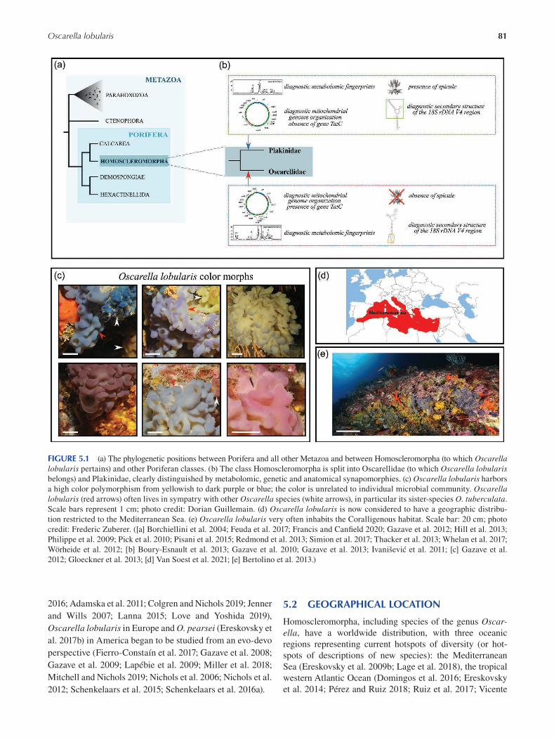
81 Oscarella lobularis
FIGURE 5.1 (a) The phylogenetic positions between Porifera and all other Metazoa and between Homoscleromorpha (to which Oscarella lobularis pertains) and other Poriferan classes. (b) The class Homoscleromorpha is split into Oscarellidae (to which Oscarella lobularis belongs) and Plakinidae, clearly distinguished by metabolomic, genetic and anatomical synapomorphies. (c) Oscarella lobularis harbors
a high color polymorphism from yellowish to dark purple or blue; the color is unrelated to individual microbial community. Oscarella lobularis (red arrows) often lives in sympatry with other Oscarella species (white arrows), in particular its sister-species O. tuberculata.
Scale bars represent 1 cm; photo credit: Dorian Guillemain. (d) Oscarella lobularis is now considered to have a geographic distribu
tion restricted to the Mediterranean Sea. (e) Oscarella lobularis very often inhabits the Coralligenous habitat. Scale bar: 20 cm; photo
credit: Frederic Zuberer. ([a] Borchiellini et al. 2004; Feuda et al. 2017; Francis and Canfield 2020; Gazave et al. 2012; Hill et al. 2013;
Philippe et al. 2009; Pick et al. 2010; Pisani et al. 2015; Redmond et al. 2013; Simion et al. 2017; Thacker et al. 2013; Whelan et al. 2017;
Wörheide et al. 2012; [b] Boury-Esnault et al. 2013; Gazave et al. 2010; Gazave et al. 2013; Ivanišević et al. 2011; [c] Gazave et al.
2012; Gloeckner et al. 2013; [d] Van Soest et al. 2021; [e] Bertolino et al. 2013.)
2016; Adamska et al. 2011; Colgren and Nichols 2019; Jenner 5.2 GEOGRAPHICAL LOCATION and Wills 2007; Lanna 2015; Love and Yoshida 2019),
Homoscleromorpha, including species of the genus Oscar-Oscarella lobularis in Europe and O. pearsei ( Ereskovsky et ella, have a worldwide distribution, with three oceanic al. 2017b) in America began to be studied from an evo-devo regions representing current hotspots of diversity (or hot-perspective (Fierro-Constaín et al. 2017; Gazave et al. 2008; spots of descriptions of new species): the Mediterranean Gazave et al. 2009; Lapébie et al. 2009; Miller et al. 2018; Sea ( Ereskovsky et al. 2009b ; Lage et al. 2018 ), the tropical
Mitchell and Nichols 2019; Nichols et al. 2006; Nichols et al. western Atlantic Ocean ( Domingos et al. 2016 ; Ereskovsky
2012; Schenkelaars et al. 2015; Schenkelaars et al. 2016a). et al. 2014 ; P érez and Ruiz 2018; Ruiz et al. 2017 ; Vicente
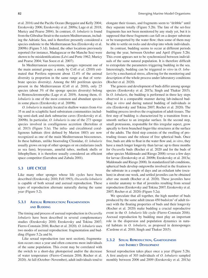
82
et al. 2016 ) and the Pacific Ocean ( Bergquist and Kelly 2004 ;
Ereskovsky 2006 ; Ereskovsky et al. 2009a ; Lage et al. 2018 ;
Muricy and Pearse 2004 ). In contrast, O. lobularis is found
from the Gibraltar Strait to the eastern Mediterranean, includ
ing the Adriatic Sea, and is therefore presently considered a
species endemic to the Mediterranean Sea ( Ereskovsky et al.
2009b ) ( Figure 5.1d ). Indeed, the other locations previously
reported (for instance, Madagascar or the Manche Sea) were
shown to be misidentifications ( Lévi and Porte 1962 ; Muricy
and Pearse 2004 ; Van Soest et al. 2007 ).
In Mediterranean ecosystems, sponges represent one of
the main animal groups: a study by Coll et al . (2010 ) esti
mated that Porifera represent about 12.4% of the animal
diversity (a proportion in the same range as that of verte
brate species diversity). Among the 681 poriferan species
present in the Mediterranean (Coll et al. 2010), only 25
species (about 3% of the sponge species diversity) belong
to Homoscleromorpha (Lage et al. 2019). Among them, O. lobularis is one of the most common and abundant species
in some places (Ereskovsky et al. 2009b).
O. lobularis is mainly located in shallow waters from 4 to
35 m and in sciaphilic hard substratum communities includ
ing semi-dark and dark submarine caves (Ereskovsky et al.
2009b). In particular, O. lobularis is one of the 273 sponge
species involved in coralligenous accretion (Bertolino et
al. 2013) (Figure 5.1e). The infra- and circalittoral coral
ligenous habitats (fi rst defined by Marion 1883) are now
recognized as one of the main Mediterranean biocoenoses.
In these habitats, unlike bioeroding Clionidae, O. lobularis usually grows on top of other sponges or on cnidarians (such
as sea fans), bryozoans, annelid tubes, mollusk shells or
lithophyllum; it is therefore usually considered an effi cient
space competitor (Garrabou and Zabala 2001).
5.3 LIFE CYCLE
Like many other sponges whose life cycles have been
described (Ereskovsky 2010; Fell 1993), Oscarella lobularis is capable of both sexual and asexual reproduction. These
types of reproduction alternate naturally during the same
year (Figure 5.2).
5.3.1 ASEXUAL REPRODUCTION: FRAGMENTATION
AND BUDDING
The timing and process of asexual reproduction in Oscarella lobularis have been described in several complementary
studies (Ereskovsky 2010; Ereskovsky and Tokina 2007;
Fierro-Constain 2016; Rocher et al. 2020). O. lobularis uses
two modes of asexual reproduction: fragmentation and bud
ding (Figure 5.2a and b).
Like sexual reproduction (see next section), fragmenta
tion occurs once a year and often concerns most individuals
of the same population. This event may be correlated with
the switch to a short-day photoperiod and/or the decrease
of water temperature (Fierro-Constain 2016; Rocher et al.
2020). At fall (October–November), adult individuals tend to
Emerging Marine Model Organisms
elongate their tissues, and fragments seem to “dribble” until
they separate totally (Figure 5.2b). The fate of the set-free
fragments has not been monitored by any study yet, but it is
supposed that these fragments can fall on a deeper substrate
or be transported by the water flow; then some of them may
be able to settle on rocks and develop into whole individuals.
In contrast, budding seems to occur at different periods
during the year, between October and April (Figure 5.2b).
This event appears not to be synchronized between individ
uals of the same natural population. It is therefore diffi cult
to extrapolate the parameters triggering budding in the sea.
Interestingly, budding can be triggered in vitro in O. lobularis by a mechanical stress, allowing for the monitoring and
description of the whole process under laboratory conditions
(Rocher et al. 2020).
The genesis and development of buds differ among sponge
species (Ereskovsky et al. 2017a ; Singh and Thakur 2015).
In O. lobularis, the budding is performed in three key steps
observed in a comparable manner during lab-induced bud
ding in vitro and during natural budding of individuals in situ (Ereskovsky and Tokina 2007; Rocher et al. 2020). The
budding process involves the evagination of adult tissues. The
first step of budding is characterized by a transition from a
smooth surface to an irregular surface. In the second step,
small protrusions, responsible for this irregular aspect, grow
apically to form branched finger-like structures at the surface
of the adults. The third step consists of the swelling of pro
truding tissues and the release of free spherical buds. Once
free, buds are able to float in the water flow and, in vitro , they
have a much longer longevity than larvae: up to three months
for Oscarella buds (Rocher et al. 2020 and for the buds of
other species Maldonado and Riesgo 2008) versus a few days
for larvae (Ereskovsky et al. 2009b; Ereskovsky et al. 2013a;
Maldonado and Riesgo 2008). In standardized lab conditions,
spherical buds develop outgrowths involved in the fi xation to
the substrate in a couple of days and an exhalant tube (oscu
lum) in about one week, and settled juveniles can be obtained
after one month (Rocher et al. 2020). These juveniles have
a similar anatomy to that of juveniles resulting from sexual
reproduction (Ereskovsky and Tokina 2007; Ereskovsky et al.
2007; Rocher et al. 2020) (Figure 5.2a).
We speculate that all together, the high number of buds
produced by the same adult (mean 450 buds/cm3 of adult tis
sue) with the floating properties of buds and their longevity
(Rocher et al. 2020) make budding a crucial reproductive
event in the O. lobularis life cycle (Fierro-Constain 2016).
Asexual reproduction by budding must play an important
role in the dispersion and population dynamics in natu
ral habitats in O. lobularis, as proposed in demosponges
(Cardone et al. 2010; Singh and Thakur 2015).
5.3.2 SEXUAL REPRODUCTION, GAMETOGENESIS
AND INDIRECT DEVELOPMENT
Sexual reproduction takes place once a year (Figure 5.2b).
A first analysis of 303 individuals of O. lobularis sampled
monthly between 2006 and 2009 (Ereskovsky et al. 2013a)
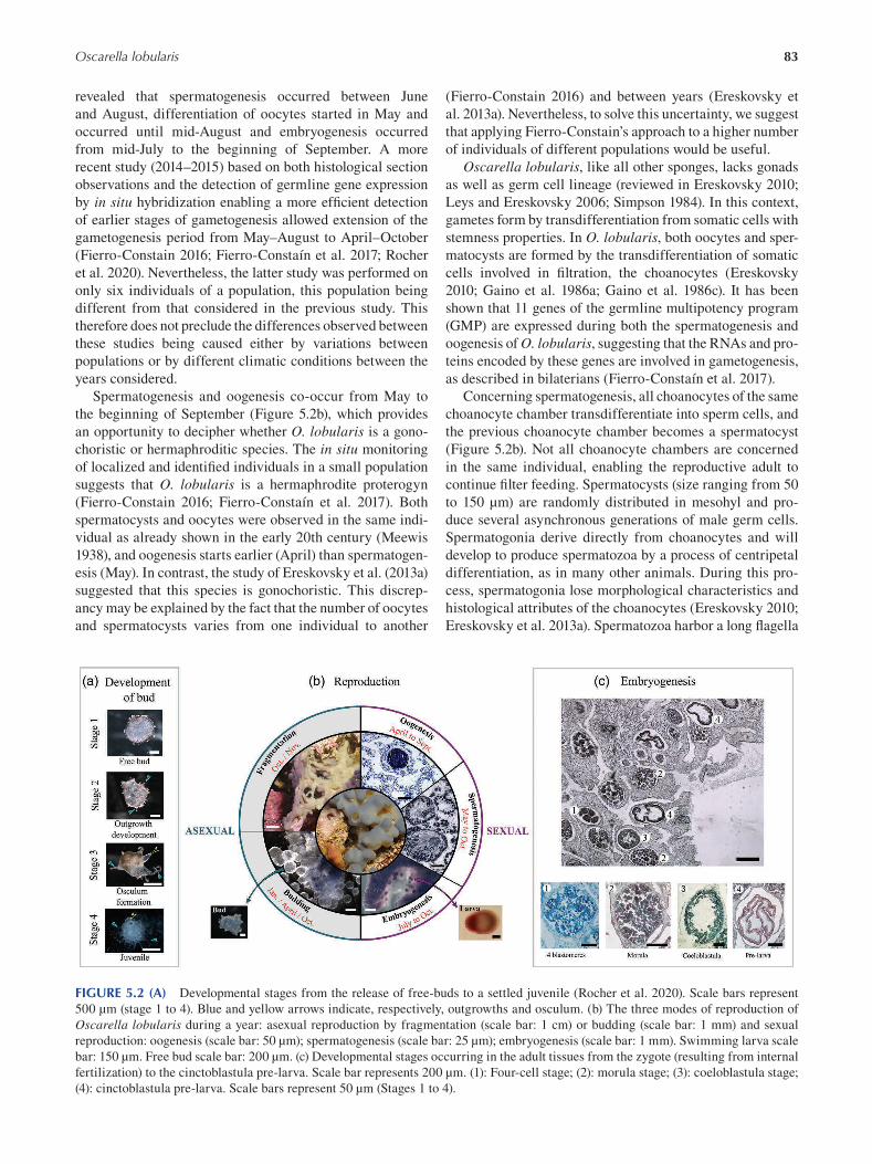
83 Oscarella lobularis
revealed that spermatogenesis occurred between June
and August, differentiation of oocytes started in May and
occurred until mid-August and embryogenesis occurred
from mid-July to the beginning of September. A more
recent study (2014–2015) based on both histological section
observations and the detection of germline gene expression
by in situ hybridization enabling a more effi cient detection
of earlier stages of gametogenesis allowed extension of the
gametogenesis period from May–August to April–October
(Fierro-Constain 2016; Fierro-Constaín et al. 2017; Rocher
et al. 2020). Nevertheless, the latter study was performed on
only six individuals of a population, this population being
different from that considered in the previous study. This
therefore does not preclude the differences observed between
these studies being caused either by variations between
populations or by different climatic conditions between the
years considered.
Spermatogenesis and oogenesis co-occur from May to
the beginning of September (Figure 5.2b), which provides
an opportunity to decipher whether O. lobularis is a gono
choristic or hermaphroditic species. The in situ monitoring
of localized and identified individuals in a small population
suggests that O. lobularis is a hermaphrodite proterogyn
(Fierro-Constain 2016; Fierro-Constaín et al. 2017 ). Both
spermatocysts and oocytes were observed in the same indi
vidual as already shown in the early 20th century (Meewis
1938), and oogenesis starts earlier (April) than spermatogen
esis (May). In contrast, the study of Ereskovsky et al. (2013a)
suggested that this species is gonochoristic. This discrep
ancy may be explained by the fact that the number of oocytes
and spermatocysts varies from one individual to another
(Fierro-Constain 2016 ) and between years (Ereskovsky et
al. 2013a). Nevertheless, to solve this uncertainty, we suggest
that applying Fierro-Constain’s approach to a higher number
of individuals of different populations would be useful.
Oscarella lobularis, like all other sponges, lacks gonads
as well as germ cell lineage (reviewed in Ereskovsky 2010;
Leys and Ereskovsky 2006; Simpson 1984). In this context,
gametes form by transdifferentiation from somatic cells with
stemness properties. In O. lobularis, both oocytes and sper
matocysts are formed by the transdifferentiation of somatic
cells involved in filtration, the choanocytes (Ereskovsky
2010; Gaino et al. 1986a ; Gaino et al. 1986c). It has been
shown that 11 genes of the germline multipotency program
(GMP) are expressed during both the spermatogenesis and
oogenesis of O. lobularis, suggesting that the RNAs and pro
teins encoded by these genes are involved in gametogenesis,
as described in bilaterians (Fierro-Constaín et al. 2017).
Concerning spermatogenesis, all choanocytes of the same
choanocyte chamber transdifferentiate into sperm cells, and
the previous choanocyte chamber becomes a spermatocyst
(Figure 5.2b). Not all choanocyte chambers are concerned
in the same individual, enabling the reproductive adult to
continue filter feeding. Spermatocysts (size ranging from 50
to 150 μm) are randomly distributed in mesohyl and pro
duce several asynchronous generations of male germ cells.
Spermatogonia derive directly from choanocytes and will
develop to produce spermatozoa by a process of centripetal
differentiation, as in many other animals. During this pro
cess, spermatogonia lose morphological characteristics and
histological attributes of the choanocytes (Ereskovsky 2010;
Ereskovsky et al. 2013a). Spermatozoa harbor a long fl agella
FIGURE 5.2 (A) Developmental stages from the release of free-buds to a settled juvenile (Rocher et al. 2020). Scale bars represent
500 μm (stage 1 to 4). Blue and yellow arrows indicate, respectively, outgrowths and osculum. (b) The three modes of reproduction of
Oscarella lobularis during a year: asexual reproduction by fragmentation (scale bar: 1 cm) or budding (scale bar: 1 mm) and sexual
reproduction: oogenesis (scale bar: 50 μm); spermatogenesis (scale bar: 25 μm); embryogenesis (scale bar: 1 mm). Swimming larva scale
bar: 150 μm. Free bud scale bar: 200 μm. (c) Developmental stages occurring in the adult tissues from the zygote (resulting from internal
fertilization) to the cinctoblastula pre-larva. Scale bar represents 200 μm. (1): Four-cell stage; (2): morula stage; (3): coeloblastula stage;
(4): cinctoblastula pre-larva. Scale bars represent 50 μm (Stages 1 to 4).
84
and a slightly elongated head with an acrosome and a large
mitochondrion (Ereskovsky 2010; Ereskovsky et al. 2013a ;
Gaino et al. 1986a). Spermatozoa are released into the sur
rounding water by the oscula via the exhalant canals.
Concerning oogenesis, a few choanocytes migrate into
the mesohyl and transdifferentiate into oocytes (Figure
5.2b). The size of the young spherical oocyte corresponds
to the size of one choanocyte (7–10 μm) without fl agel
lum, microvilli and basal filopodia. This size increases
significantly during vitellogenesis, although the fi nal size
of a mature oocyte is different, according to the authors
(Ereskovsky 2010; Ereskovsky et al. 2013a ; Fierro-Constain
2016). In this species, the great amount of vitellus (poly
lecithal eggs), uniformly distributed in the ooplasm (iso
lecithal), is produced by endogenous synthesis (Ereskovsky
2010; Ereskovsky et al. 2009b; Gaino et al. 1986b), unlike
the other sponges with polylecithal oocytes in which vitel
logenesis occurs by phagocytosis of somatic cells and/or
bacteria (Maldonado and Riesgo 2008). Mature oocytes,
located in the basal zone of the choanosome, are enclosed by
endopinacoderm to form a so-called follicle. Before the clo
sure of the follicle, maternal symbiotic bacteria and several
maternal cells penetrate in the space between the oocyte and
the follicle (Ereskovsky and Boury-Esnault 2002). Vertical
transmission of symbionts from embryo to juvenile has been
well documented in sponges (Boury-Esnault et al. 2003;
Ereskovsky 2010; Ereskovsky and Boury-Esnault 2002;
Ereskovsky et al. 2007; Ereskovsky et al. 2009b). Moreover,
the penetration of maternal vacuolar cells inside of follicles
was described in many investigated sponge species from the
classes Demospongiae, Calcarea and Homoscleromorpha
(Ereskovsky 2010). The oocytes remain in the adult tissue,
meaning that O. lobularis performs internal fertilization. As
fertilization per se has never been observed in this species,
it is unknown whether it relies upon a carrier-cell system, as
described in Calcaronea species (first described by Gatenby
in 1920; reviewed in Ereskovsky 2010).
5.4 EMBRYOGENESIS
5.4.1 CLEAVAGE AND FORMATION OF COELOBLASTULA
Like many sponge species described so far, Oscarella lobularis undergoes indirect development (Ereskovsky 2010).
Additionally, as a direct consequence of internal fertiliza
tion, O. lobularis is a “brooding” sponge. This means that
the development from a zygote to a fully developed larva
(cinctoblastula) occurs within the adult tissue (Figure 5.2c):
swimming larvae are then released in the surrounding water
( Figure 5.2b ).
The embryonic development of O. lobularis is similar
to other species of the genus Oscarella. The main steps
of this embryonic development have been described so
far only by classical histological approaches on fi xed indi
viduals (Ereskovsky 2010; Ereskovsky and Boury-Esnault
2002; Ereskovsky et al. 2009b; Ereskovsky et al. 2013a ;
Emerging Marine Model Organisms
Ereskovsky et al. 2013b; Leys and Ereskovsky 2006). As
in all Metazoa, the first developmental step consists of the
cleavage of the zygote. The zygote being isolecithal (see
previous section on oogenesis), this cleavage is holoblastic.
The first two divisions (until the four-cell stage; Figure 5.2c)
are equal and synchronous. Then the cleavage becomes
irregular and asynchronous from the third division. After
six divisions, the morula stage is reached: the morula is
composed of 64 undifferentiated blastomeres (Ereskovsky
and Boury-Esnault 2002; Ereskovsky et al. 2013a ; Leys and
Ereskovsky 2006) (Figure 5.2c). As cleavage progresses, the
blastomeres reduce in size, and the volume of the embryo
remains unchanged.
From the 64-cell morula stage, the blastomeres at the
surface of the morula divide more actively, while inter
nal blastomeres migrate to the periphery of the embryo
through a process of multipolar egression (Ereskovsky 2010;
Ereskovsky and Boury-Esnault 2002; Ereskovsky et al.
2013a ; Leys and Ereskovsky 2006 ) to form a monolayered
coeloblastula with a central cavity (Figure 5.2c). This cen
tral cavity has been described as containing the maternal
symbiotic bacteria and maternal vacuolar cells (see previ
ous section on oogenesis). The role and fate of these latter
have not been explored and will have to be with modern
molecular and cellular tools (Boury-Esnault et al. 2003;
Ereskovsky and Boury-Esnault 2002; Ereskovsky et al.
2007; Ereskovsky et al. 2013a), but they seem to degenerate
during metamorphosis of the larvae (personal observations).
Unlike in the three other sponge classes, the coeloblas
tula of Oscarella exhibits a monolayer columnar epithelium.
This epithelium fits all classical criteria of the defi nition
of epithelia in Bilaterians (Ereskovsky et al. 2009b; Leys
and Riesgo 2012; Leys et al. 2009; Tyler 2003; Renard et al.
2021). i) Cells are highly polarized: cilia develop at the api
cal cell pole; ii) cells are tightened by specialized intercel
lular junctions, similar to adherens junctions, in the apical
domain; and iii) cells are lined at their basal pole by a base
ment membrane consisting of collagen IV (Boute et al. 1996;
Ereskovsky and Boury-Esnault 2002; Boury-Esnault et al.
2003). The establishment of this columnar epithelium at the
coeloblastula stage is the first sign of cellular differentia
tion processes. Note that, even if the term “coeloblastula”
was used in the literature because of the presence of a cen
tral cavity, this organization is not the result of the same
processes (cleavage only) as in other metazoans (Boury-
Esnault et al. 2003; Brusca and Brusca 2003; Ereskovsky
2010; Ereskovsky and Boury-Esnault 2002; Ereskovsky
and Dondua 2006; Leys 2004; Leys and Ereskovsky 2006;
Maldonado and Riesgo 2008; Wörheide et al. 2012). For this
reason, some authors prefer the use of the term “prelarva” or
“cinctoblastula prelarva” (Ereskovsky 2010). Unfortunately,
this complex terminology makes comparison with other
metazoans very difficult, and none of the embryological
descriptions of embryological development available so far
in sponges are based on live observations and cell tracking
experiments.
85 Oscarella lobularis
5.4.2 MORPHOGENESIS OF THE CINCTOBLASTULA
LARVA AND LARVAL METAMORPHOSIS
Cells continue to divide, thus increasing the cell surface
area. Because of the limited space in the follicle, the external
epithelium becomes folded (Figure 5.2c). The central cavity
is progressively filled by collagen fibrils, and a pronounced
antero-posterior polarity is acquired: the ciliated cells
contain various cytoplasmic inclusions and present a vari
able nucleus position according to their position along the
anterior–posterior axis, unlike in coeloblastula larva of other
sponges (Boury-Esnault et al. 2003; Ereskovsky 2010; Leys
and Ereskovsky 2006 ). The cellular mechanisms by which
pre-cinctoblastula larvae are transferred from the mesohyl
to the exhalant canal was described in Boury-Esnault et al.
(2003) and involves a fusion between the endopinacoderm
forming the follicles and the endopinacoderm lining the
canals. Finally, a free-swimming cinctoblastula larva is
released from the adult sponge through the exhalant canals
and the osculum. Larvae are uniformly fl agellated (despite
the presence of few scattered non-ciliated cells) and pres
ent a polarity: the anterior pole is larger than the posterior
one, and the posterior pole is pigmented (pink pigments in
O. lobularis) and rich in symbiotic bacteria and maternal
vacuolar cells in the central cavity. The pigments are prob
ably involved in the observed larval phototaxis behavior, as
evidenced in the demosponge Amphimedon queenslandica (Degnan et al. 2015; Leys and Degnan 2001; Rivera et al.
2012 ).
The larva can swim in the water column for several days
before settlement. The larva attaches to the substrate by the
anterior pole thanks to mucus secretion, then undergoes
metamorphosis (Figure 5.3a). Therefore, the A/P axis of
the larva corresponds to the baso-apical axis of the juvenile
sponge.
During metamorphosis, the larva undergoes radical mor
phological and physiological changes. The metamorphosis
of the larva represents a second phase of reorganization of
cell layers and corresponds to the acquisition of the typi
cal sponge bauplan with a functional aquiferous system.
The formation of the two main epithelial layers, namely the
pinacoderm and the choanoderm, occurs through the trans-
differentiation of the larval epithelium (fully detailed in
Ereskovsky 2010 ; Ereskovsky et al. 2007 ; Ereskovsky et al.
2010 ).
The steps of larval metamorphosis have been described
as variable and independent of environmental factors
(Ereskovsky et al. 2007). However, the origin of this poly
phenism is unknown. In most cases, the metamorphosis of
O. lobularis larvae begins by a basal invagination (Figure
5.3a). In parallel to this invagination, several lateral cells
ingress into the cavity. The lateral then sides fold up with the
subsequent involution of marginal sides. At this stage, the
future juvenile is composed of two cell layers, an external
layer, from which the future exopinacoderm will originate,
and an internal layer. The cells of this internal layer become
flat, thereby increasing the tissue surface, which itself results
in folding. This inner folded epithelium gives rise to the
aquiferous system: the endopinacoderm is derived from the
proximal parts of the internal cell layer, while the choano
cyte chambers develop from distal parts of the internal folds
(Figure 5.3a). The inhalant pores, ostia, and the exhalant
pores, osculum, are formed secondarily. A settled fi ltering
juvenile is finally formed, usually called a “rhagon”.
5.4.3 MOLECULAR CONTROL OF DEVELOPMENT
The molecular mechanisms controlling the previously
described developmental events are still unknown. As
sexual reproduction occurs only once a year and embryos
are not observed every year in sampled adults, and further
more the embryos are intimately embedded in the adult tis
sues, their dissection and manipulation are rather tricky.
Therefore, only two studies so far report gene expression
patterns during embryogenesis. Due to the key role of the
WNT pathway in axial patterning across the animal king
dom, several studies have investigated the pattern of Wnt gene expression during sponge development or during other
morphogenetic processes (Adamska 2016; Adamska et al.
2007; Adamska et al. 2010; Adamska et al. 2011; Borisenko
et al. 2016; Degnan et al. 2015; Lanna 2015; Leininger et al.
2014; Richards and Degnan 2009). In Oscarella lobularis, nine Wnt genes were found, as well as their target genes
(Lapébie et al. 2009; Schenkelaars 2015). Even though
most Wnts and Fzds genes are uniformly expressed during
early stages of embryogenesis without apparent gradient or
asymmetry, one Wnt gene is clearly localized at one pole of
the embryos before any morphological polarity is observed
(Schenkelaars 2015). This latter observation is in agreement
with results obtained in other sponge lineages (Calcarea
and Demospongiae), where WNT ligands and downstream
genes are expressed in the posterior region of the embryos or
larvae (Adamska 2016; Adamska et al. 2007; Adamska et al.
2010; Borisenko et al. 2016; Degnan et al. 2015; Leininger
et al. 2014). These expression patterns tend to support a
putative involvement of WNT pathways in patterning of the
major sponge body axis. In addition, Fierro-Constaín et al.
(2017) showed that 11 genes of the GMP are expressed dur
ing embryogenesis (including the most famous piwi, vasa,
nanos, Pl10 genes). This finding agrees with observations in
other animals. Interestingly, among these genes, nanos har
bors a highly polarized pattern in the prelarva: with a much
higher expression level at the anterior pole. Such a polarized
pattern was also observed in the calcarean sponge Sycon ciliatum (Leininger et al. 2014) and in other metazoans, but the
role of this gene in axis patterning is unclear (Kanska and
Frank 2013).
5.5 ANATOMY
As previously explained, developmental processes follow
ing both sexual and asexual (by budding) reproduction result
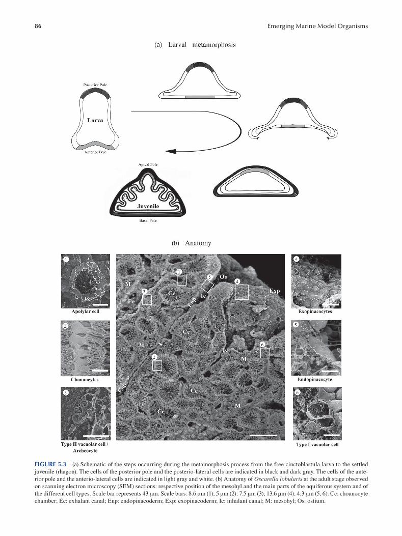
86 Emerging Marine Model Organisms
FIGURE 5.3 (a) Schematic of the steps occurring during the metamorphosis process from the free cinctoblastula larva to the settled
juvenile (rhagon). The cells of the posterior pole and the posterio-lateral cells are indicated in black and dark gray. The cells of the ante
rior pole and the anterio-lateral cells are indicated in light gray and white. (b) Anatomy of Oscarella lobularis at the adult stage observed
on scanning electron microscopy (SEM) sections: respective position of the mesohyl and the main parts of the aquiferous system and of
the different cell types. Scale bar represents 43 μm. Scale bars: 8.6 μm (1); 5 μm (2); 7.5 μm (3); 13.6 μm (4); 4.3 μm (5, 6). Cc: choanocyte
chamber; Ec: exhalant canal; Enp: endopinacoderm; Exp: exopinacoderm; Ic: inhalant canal; M: mesohyl; Os: ostium.
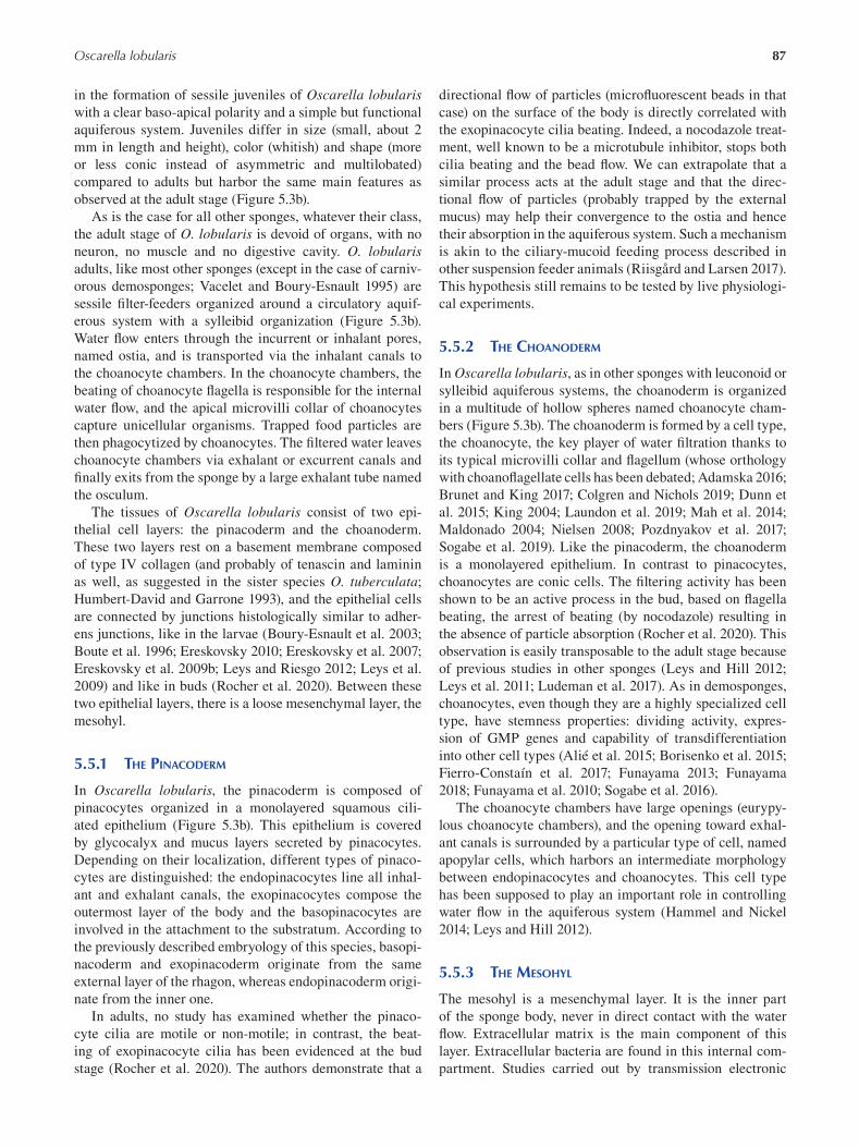
87 Oscarella lobularis
in the formation of sessile juveniles of Oscarella lobularis with a clear baso-apical polarity and a simple but functional
aquiferous system. Juveniles differ in size (small, about 2
mm in length and height), color (whitish) and shape (more
or less conic instead of asymmetric and multilobated)
compared to adults but harbor the same main features as
observed at the adult stage (Figure 5.3b).
As is the case for all other sponges, whatever their class,
the adult stage of O. lobularis is devoid of organs, with no
neuron, no muscle and no digestive cavity. O. lobularis adults, like most other sponges (except in the case of carniv
orous demosponges; Vacelet and Boury-Esnault 1995) are
sessile filter-feeders organized around a circulatory aquif
erous system with a sylleibid organization (Figure 5.3b).
Water flow enters through the incurrent or inhalant pores,
named ostia, and is transported via the inhalant canals to
the choanocyte chambers. In the choanocyte chambers, the
beating of choanocyte flagella is responsible for the internal
water flow, and the apical microvilli collar of choanocytes
capture unicellular organisms. Trapped food particles are
then phagocytized by choanocytes. The filtered water leaves
choanocyte chambers via exhalant or excurrent canals and
finally exits from the sponge by a large exhalant tube named
the osculum.
The tissues of Oscarella lobularis consist of two epi
thelial cell layers: the pinacoderm and the choanoderm.
These two layers rest on a basement membrane composed
of type IV collagen (and probably of tenascin and laminin
as well, as suggested in the sister species O. tuberculata;
Humbert-David and Garrone 1993), and the epithelial cells
are connected by junctions histologically similar to adher
ens junctions, like in the larvae (Boury-Esnault et al. 2003;
Boute et al. 1996; Ereskovsky 2010; Ereskovsky et al. 2007;
Ereskovsky et al. 2009b; Leys and Riesgo 2012; Leys et al.
2009) and like in buds (Rocher et al. 2020). Between these
two epithelial layers, there is a loose mesenchymal layer, the
mesohyl.
5.5.1 THE PINACODERM
In Oscarella lobularis, the pinacoderm is composed of
pinacocytes organized in a monolayered squamous cili
ated epithelium (Figure 5.3b). This epithelium is covered
by glycocalyx and mucus layers secreted by pinacocytes.
Depending on their localization, different types of pinaco
cytes are distinguished: the endopinacocytes line all inhal
ant and exhalant canals, the exopinacocytes compose the
outermost layer of the body and the basopinacocytes are
involved in the attachment to the substratum. According to
the previously described embryology of this species, basopi
nacoderm and exopinacoderm originate from the same
external layer of the rhagon, whereas endopinacoderm origi
nate from the inner one.
In adults, no study has examined whether the pinaco
cyte cilia are motile or non-motile; in contrast, the beat
ing of exopinacocyte cilia has been evidenced at the bud
stage (Rocher et al. 2020). The authors demonstrate that a
directional flow of particles (microfluorescent beads in that
case) on the surface of the body is directly correlated with
the exopinacocyte cilia beating. Indeed, a nocodazole treat
ment, well known to be a microtubule inhibitor, stops both
cilia beating and the bead flow. We can extrapolate that a
similar process acts at the adult stage and that the direc
tional flow of particles (probably trapped by the external
mucus) may help their convergence to the ostia and hence
their absorption in the aquiferous system. Such a mechanism
is akin to the ciliary-mucoid feeding process described in
other suspension feeder animals (Riisgård and Larsen 2017).
This hypothesis still remains to be tested by live physiologi
cal experiments.
5.5.2 THE CHOANODERM
In Oscarella lobularis, as in other sponges with leuconoid or
sylleibid aquiferous systems, the choanoderm is organized
in a multitude of hollow spheres named choanocyte cham
bers (Figure 5.3b). The choanoderm is formed by a cell type,
the choanocyte, the key player of water filtration thanks to
its typical microvilli collar and flagellum (whose orthology
with choanoflagellate cells has been debated; Adamska 2016;
Brunet and King 2017; Colgren and Nichols 2019; Dunn et
al. 2015; King 2004; Laundon et al. 2019; Mah et al. 2014;
Maldonado 2004; Nielsen 2008; Pozdnyakov et al. 2017;
Sogabe et al. 2019). Like the pinacoderm, the choanoderm
is a monolayered epithelium. In contrast to pinacocytes,
choanocytes are conic cells. The filtering activity has been
shown to be an active process in the bud, based on fl agella
beating, the arrest of beating (by nocodazole) resulting in
the absence of particle absorption (Rocher et al. 2020). This
observation is easily transposable to the adult stage because
of previous studies in other sponges (Leys and Hill 2012;
Leys et al. 2011; Ludeman et al. 2017). As in demosponges,
choanocytes, even though they are a highly specialized cell
type, have stemness properties: dividing activity, expres
sion of GMP genes and capability of transdifferentiation
into other cell types (Alié et al. 2015; Borisenko et al. 2015;
Fierro-Constaín et al. 2017; Funayama 2013; Funayama
2018; Funayama et al. 2010; Sogabe et al. 2016 ).
The choanocyte chambers have large openings (eurypy
lous choanocyte chambers), and the opening toward exhal
ant canals is surrounded by a particular type of cell, named
apopylar cells, which harbors an intermediate morphology
between endopinacocytes and choanocytes. This cell type
has been supposed to play an important role in controlling
water flow in the aquiferous system (Hammel and Nickel
2014; Leys and Hill 2012).
5.5.3 THE MESOHYL
The mesohyl is a mesenchymal layer. It is the inner part
of the sponge body, never in direct contact with the water
flow. Extracellular matrix is the main component of this
layer. Extracellular bacteria are found in this internal com
partment. Studies carried out by transmission electronic
88
microscopy (TEM), by denaturating gradient gel electro
phoresis (DGGE) or by 16S sequencing have shown that
O. lobularis is a low microbial abundance (LMA) sponge
(Gloeckner et al. 2013; Vishnyakov and Ereskovsky 2009). Its
phylum-level microbial diversity is represented by three bac
terial phyla with a large dominance (76%) of Proteobacteria.
Phylogenetic analysis revealed four sequences affi liated
with Verrucomicrobia, three with Gammaproteobacteria
and two sequences with Bacteroidetes, and the 16 remain
ing sequences were affiliated with Alphaproteobacteria.
Moreover, microbial diversity is neither signifi cantly dif
ferent between color morphs nor between individuals of
different locations or depths (Gerçe et al. 2011; Gloeckner
et al. 2013). More recently, metagenomic analyses sug
gest that the main bacterial symbiot of O. lobularis is an
Alphaproteobacteria of the Rhodobacteriaceae family. This
new species was named Candidatus Rhodobacter lobularis, it is about 20-fold more numerous than sponge cells in
the mesohyl and its draft genome is available (Jourda et al.
2015). Even though no physiological studies have yet been
performed to identify the mutual benefits of this association,
members of the Rhodobacter group often perform aerobic
anoxygenic photoheterotrophy (Labrenz et al. 2009; Pohlner
et al. 2019; Sorokin et al. 2005); we therefore suggest that
hosting such Rhodobacter species may supply O. lobularis with carbon.
In addition, several sponge cell types are present in the
mesohyl (Figure 5.3b). Classically, photonic and electronic
observations have defined two cell types: type I vacuolar
and type II vacuolar (Boury-Esnault et al. 1992; Ereskovsky
et al. 2009b). Type I vacuolar cells are characterized by two
to four large empty vacuoles and a small nucleus placed lat
erally, and their role is unknown. Type II vacuolar cells are
amoeboid cells with numerous filopodia, numerous small
vacuoles and a large nucleus with a nucleolus. Because
of these cytological features and the fact that these cells
express 11 genes of the GMP program, they were supposed
to correspond to what are defined as archaeocytes in other
sponges (Fierro-Constaín et al. 2017). Comparative single-
cell transcriptomic data are now awaited to establish homol
ogy between cell types between sponge species and to make
clearer the sponge cell type terminology only based on cell
morphology (De Vos et al. 1991; Musser et al. 2019; Rocher
et al. 2020; Sogabe et al. 2019). Interestingly, the use of
scanning electron microscopy and immunofl uorescent tech
niques resulted in the identification of at least one additional
cell type in the mesohyl of O. lobularis bud: a third vacuolar
cell type (Rocher et al. 2020). Additionally, numerous, pre
viously undescribed, tiny anucleate cell-like structures were
interpreted as apoptotic extracellular vesicles (EVs) (Rocher
et al. 2020). Because buds originate directly from adult tis
sues (see previous sections), we do not believe that these type
III vacuolar cells and EVs are bud specific but rather that they
were not observed on adults until now because of technical
limitations. These recent findings highlight the subjectivity
of cell type definition, and again, much is expected from
ongoing single cell transcriptomic approaches to defi ne cell
Emerging Marine Model Organisms
types on the basis of a shared regulatory network (Arendt
et al. 2016 ).
5.6 TRANSCRIPTOMIC AND GENOMIC DATA
Since the first genome of the demosponge Amphimedon queenslandica was published (Srivastava et al. 2010), sponge
genomic resources have significantly increased (for review,
see Renard et al. 2018 and references included, plus Kenny
et al. 2020). These data revealed that sponges have a genome
size and number of genes comparable to those of most inver
tebrates. In addition, these studies indicate striking genome
feature differences between sponge species even within the
same class: differences in predicted genome size (from 57 to
357 Mb) in agreement with very variable DNA content evi
denced by old cytogenetic approaches; differences in ploidy
(diploidy or probable tetraploidy in Calcarea), amount and
length of non-coding regions and genes present, among others
(Kenny et al. 2020; Renard et al. 2018; Santini et al. in prep).
Concerning Oscarella lobularis, a genome draft was
sequenced with illumina technology (Belahbib et al. 2018);
ongoing additional sequencing efforts are expected to
improve the assembly of this genome in a near future. At
present, the predicted length of the genome of O. lobularis is 52.34 Mb ( Belahbib et al. 2018); this is even smaller than
what was predicted for O. pearsei (57.7 Mb; Nichols et al.
2006). If confirmed when a better assembly is obtained,
this genome would represent the smallest sponge genome
reported so far. This genome is predicted to contain 17,885
protein-coding genes (Belahbib et al. 2018). This is surpris
ingly low compared to demosponges: Ephydatia muelleri is supposed to harbor 39,245 protein-coding genes (Kenny
et al. 2020), Amphimedon queenslandica 40,122 (Fernandez-
Valverde et al. 2015) and Tethya wilhelma 37,416 (Francis
et al. 2017). We are expecting a better genome assembly for
both O. pearsei and O. lobularis in order to be able to deci
pher whether these small genome sizes and low numbers of
genes are due to sequencing pitfalls or represent a common
feature of Oscarellidae genomes.
To date, only one study has used this genomic data to
compare epithelial genes of O. lobularis to other sponges
(Belahbib et al. 2018). All other comparative molecu
lar studies published so far were either based on PCR
approaches (Gazave et al. 2008; Lapébie et al. 2009) or on
transcriptomic data obtained by 454 sequencing technol
ogy performed on a mixture of developmental stages (adult,
embryos and larvae) to maximize the representativity of this
transcriptome (Fierro-Constaín et al. 2017; Schenkelaars
et al. 2015; Schenkelaars et al. 2016a).
These transcriptomic and genomic studies published thus
far have focused on genes involved in epithelial functions, in
Notch and WNT signaling and genes pertaining to the GMP.
As far as the GMP and the canonical WNT pathways are
concerned, genes present in O. lobularis are not different
from what is found in other sponge classes (Fierro-Constaín
et al. 2017; Lapebie 2010; Lapébie et al. 2009; Schenkelaars
2015; Schenkelaars et al. 2015). When comparisons are
89 Oscarella lobularis
made at the level of gene content only, O. lobularis, like all
other sponges, possesses all nine genes coding for proteins
involved in the establishment of the CRUMBS, PAR and
SCRIBBLE complexes of bilaterians needed to establish cell
polarity, as well as all three genes encoding proteins needed
to establish the cadherin-catenin complex (CCC) required for
the formation of adherens junctions (namely alpha, beta and
delta catenins as well as classical cadherin) (Belahbib et al.
2018). However, key functional domains and motif sequences
are amazingly more conserved in O. lobularis than they are
in other sponge classes. For example, PatJ protein (one of the
three components of the crumbs polarity complex containing
Crumbs, MPP5 and PatJ) binds MPP5 via the L27 domain:
The L27 domain sequence is more conserved in O. lobularis compared to the other sponges (Belahbib et al. 2018). It is the
same for cadherin/-catenin/-catenin complex. The com
parison of the E-cadherin cytoplasmic tail, which contains
the conserved specific binding domain for delta-catenin and
-catenin, is more conserved in O. lobularis than in other
sponges relative to bilaterian sequences (Belahbib et al. 2018).
Concerning pathways commonly involved in epithelial
patterning, it was shown that O. lobularis possesses all
the core gene encoding for proteins needed to establish a
planar cell polarity (PCP) pathway. Indeed, Strabismus
(Stbm)/Van Gogh (Vang), Flamingo (Fmi), Prickle (Pk),
Dishevelled (Dsh) and frizzled (Fzd) proteins are present in
O. lobularis (Schenkelaars 2015; Schenkelaars et al. 2016a),
whereas other sponges lack either one or several members
of this pathway (Fmi, Fzd and/or Vang) (Schenkelaars 2015;
Schenkelaars et al. 2016a) (Figure 5.4a). This fi nding chal
lenged previous studies in Ctenophora and Porifera suggest
ing that the PCP pathway arose in the last common ancestor
of Parahoxozoa (Bilateria, Cnidaria and Placozoa) (Adamska
et al. 2010; Ryan et al. 2013), meaning that the PCP pathway
may date back to the emergence of Metazoa. This unex
pected result calls for functional studies in O. lobularis : Is
this pathway involved in the coordination and orientation
of exopinacocyte cilia (Figure 5.4b and c) in the same way
it is in other animals (Devenport 2014; Schenkelaars et al.
2016a ; Wallingford 2010 )?
Other key genes considered absent in other sponge
classes are present in O. lobularis. This is notably the case
for the Hes gene belonging to group E bHLH transcrip
tion factors. To date, only Hey genes have been reported
in Demospongiae and Calcarea (Fortunato et al. 2016;
Simionato et al. 2007; Srivastava et al. 2010); in contrast,
O. pearsei and O. lobularis possess bona fide Hes ( Gazave
2010; Gazave et al. 2014) (Figure 5.4d). This means that this
gene was ancestrally present in the last common ancestor of
Porifera and was lost in other sponge classes. This fi nding
offers additional possibilities to test the respective roles of
canonical and non-canonical Notch signaling pathways in
Metazoa and notably to explore the role of Notch signaling
in animals devoid of neurons (Layden et al. 2013).
Despite a lack of neurons and conventional neurotrans
mitters, sponges perceive and respond to a large range of
stimuli. In animals, Glutamate is the principal excitatory
neurotransmitter in the central nervous system. All sponges
have a number of metabotropic glutamate (mGlu) and
GABA receptors, suggesting that glutamatergic signaling
is common in sponges (Leys et al. 2019). In contrast, the
ionotropic glutamate receptor iGluR gene is found only in
calcareous sponges and homoscleromorphs (Figure 5.4e)
(Ramos-Vicente et al. 2018; Renard et al. 2018; Stroebel
and Paoletti 2020). However, the localization and function
of these receptors remain to be identified in these animals
devoid of neurons and synapses.
Much remains to explore in the transcriptome and
genome of O. lobularis; nevertheless, according to the pres
ent knowledge, compared to other sponge classes, the homo
scleromorph sponges O. pearsei and O. lobularis seem to
exhibit the most complete and conserved bilaterian gene
repertoire (Babonis and Martindale 2017; Fortunato et al.
2015; Gazave et al. 2014; Renard et al. 2018; Riesgo et al.
2014; Schenkelaars et al. 2016a).
5.7 FUNCTIONAL APPROACHES: TOOLS FOR MOLECULAR AND CELLULAR ANALYSES
5.7.1 DEVELOPMENTAL AND NON-DEVELOPMENTAL
MORPHOGENETIC CONTEXTS ACCESSIBLE
Embryos and larvae are accessible only once a year between
August and October, and the reproductive effort is vari
able from one population to another and from one year to
another (Ereskovsky et al. 2013a). Therefore, because sexual
reproduction cannot be triggered in the laboratory, so far,
the access to embryonic developmental processes remains
very limited.
To compensate for this difficulty, experimental protocols
were designed to access non-developmental morphogenetic
processes (Table 5.2). Wound healing experiments have
already been successfully used at the adult stage (Ereskovsky
et al. 2015; Fierro-Constaín et al. 2017 ); wound-healing
and regenerative experiments are now also mastered at the
bud stage: stage 3 with osculum regenerates an osculum in
less than four days (Rocher et al. 2020); cell-dissociation/
reaggregation experiments resulting in neo-epithelialized
primmorphs (in less than three days) can be performed
both on adults (unpublished data) and on buds (Rocher et al.
2020; Vernale et al. in press).
5.7.2 POLYMERASE CHAIN REACTION AND RELATIVES
As in the case of other sponge species studied for evo-devo
purposes, the first molecular studies undergone on Oscarella lobularis were performed using the polymerase chain reac
tion (PCR) technique. This resulted in the description of
the phylogenetic relationships among Homoscleromorpha
(described in the first section), including O. lobularis (Borchiellini et al. 2004; Gazave et al. 2010; Gazave et
al. 2012; Gazave et al. 2013). The main pitfall faced dur
ing this simple classical PCR/cloning/sequencing was at
the step of DNA extraction. For O. lobularis, as for other
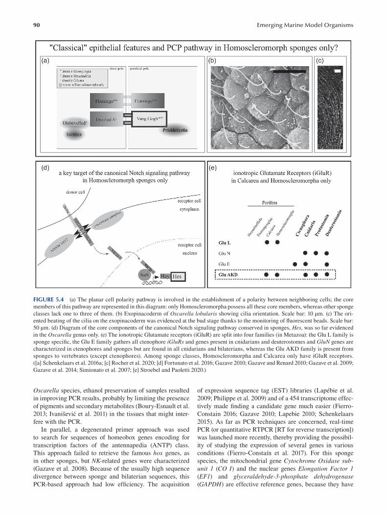
90 Emerging Marine Model Organisms
FIGURE 5.4 (a) The planar cell polarity pathway is involved in the establishment of a polarity between neighboring cells; the core
members of this pathway are represented in this diagram: only Homoscleromorpha possess all these core members, whereas other sponge
classes lack one to three of them. (b) Exopinacoderm of Oscarella lobularis showing cilia orientation. Scale bar: 10 μm. (c) The ori
ented beating of the cilia on the exopinacoderm was evidenced at the bud stage thanks to the monitoring of fluorescent beads. Scale bar:
50 μm. (d) Diagram of the core components of the canonical Notch signaling pathway conserved in sponges, Hes, was so far evidenced
in the Oscarella genus only. (e) The ionotropic Glutamate receptors (iGluR) are split into four families (in Metazoa): the Glu L family is
sponge specific, the Glu E family gathers all ctenophore iGluRs and genes present in cnidarians and deuterostomes and GluN genes are
characterized in ctenophores and sponges but are found in all cnidarians and bilaterians, whereas the Glu AKD family is present from
sponges to vertebrates (except ctenophores). Among sponge classes, Homoscleromorpha and Calcarea only have iGluR receptors.
([a] Schenkelaars et al. 2016a; [c] Rocher et al. 2020; [d] Fortunato et al. 2016; Gazave 2010; Gazave and Renard 2010; Gazave et al. 2009;
Gazave et al. 2014; Simionato et al. 2007; [e] Stroebel and Paoletti 2020.)
Oscarella species, ethanol preservation of samples resulted of expression sequence tag (EST) libraries (Lapébie et al.
in improving PCR results, probably by limiting the presence 2009; Philippe et al. 2009) and of a 454 transcriptome effec
of pigments and secondary metabolites (Boury-Esnault et al. tively made finding a candidate gene much easier (Fierro
2013 ; Ivanišević et al. 2011) in the tissues that might inter- Constain 2016; Gazave 2010; Lapebie 2010; Schenkelaars
fere with the PCR. 2015). As far as PCR techniques are concerned, real-time
In parallel, a degenerated primer approach was used PCR (or quantitative RTPCR [RT for reverse transcription])
to search for sequences of homeobox genes encoding for was launched more recently, thereby providing the possibil
transcription factors of the antennapedia (ANTP) class. ity of studying the expression of several genes in various
This approach failed to retrieve the famous hox genes, as conditions (Fierro-Constaín et al. 2017). For this sponge
in other sponges, but NK-related genes were characterized species, the mitochondrial gene Cytochrome Oxidase sub(Gazave et al. 2008). Because of the usually high sequence unit 1 (CO I) and the nuclear genes Elongation Factor 1 divergence between sponge and bilaterian sequences, this (EF1) and glyceraldehyde-3-phosphate dehydrogenase PCR-based approach had low efficiency. The acquisition (GAPDH) are effective reference genes, because they have
91 Oscarella lobularis
stable expression during their life cycle (Fierro-Constaín et
al. 2017) but also under contaminant exposure conditions (de
Pao Mendonca, unpublished data).
5.7.3 IN SITU HYBRIDIZATION
The in situ hybridization (ISH) technique is also mastered
in Oscarella lobularis, thereby allowing access to qualita
tive data (localization) in addition to quantitative expres
sion gene information provided by the previously mentioned
real-time PCR. The first ISH data were acquired in 2008
(Gazave et al. 2008; Lapébie et al. 2009), and the proto
col was subsequently improved (Fierro-Constaín et al.
2017; Fierro-Constaín et al. 2021). The ISH can be per
formed at all stages (adult, bud, larvae) on sections or in
whole mounts. Fluorescent ISH (FISH) is also in progress
(Prünster, unpublished data). For colorimetric ISH, 5-brom
4-chloro-3’-indolyphosphate p-toluidine salt/nitro blue tet
razolium chloride (BCIP/NBT) was successfully used as
a chromogenic phosphatase substrate for the detection of
alkaline phosphatase labeled probes (with better results
than BM-purple, for example). The automating of the whole
mount ISH on an Intavis pro device increased the output and
replicability of the technique (detailed protocols provided in
Fierro-Constaín et al. 2021).
5.7.4 FLUORESCENT IMMUNOLOCALIZATION
Fluorescent immunolocalization (IF) can be performed
either on paraffin sections of adults and buds (unpublished data) or on whole mount on buds thanks to their transparency
(Rocher et al. 2020) (Table 5.2). Unsurprisingly, the use of
paraffin sections not only takes much longer to achieve
but also can result in losing antigenic reactivity, as often
observed in other tissues (Krenacs et al. 2010); for this rea
son, most IF experiments are performed on whole mounted
buds or juveniles. The IF protocol used in O. lobularis buds is a classical one (Rocher et al. 2020; detailed proto
col provided in Borchiellini et al. 2021). Nevertheless, the
main difficulty faced is the divergence of sponge antigen
sequences relative to vertebrate antigens. Most commer
cialized antibodies, designed against vertebrate proteins,
are therefore unusable, except for highly conserved pro
teins. For instance, we successfully used antibodies against
alpha-tubulin (Sigma) and acetylated alpha-tubulin (Sigma),
phospho-histone H3 (Abcam) (Rocher et al. 2020). For other
proteins, specifi c antibodies were raised against peptides of
interest, for example, against type IV collagen (Rocher et al.
2020; Vernale et al. in press); other specific antibodies are
currently under testing.
5.7.5 CELL VIABILITY, CELL APOPTOSIS AND
CELL PROLIFERATION ASSAYS
During the study of morphogenetic processes or for ecotoxi
cological purposes, being able to measure and compare cell
viability and cell activity can be useful.
Cell viability/death can be estimated very quickly (a
couple of minutes) in O. lobularis (Table 5.2), on both disso
ciated cells (see next sections) or whole buds, by using prop
idium iodide (PI) staining on dead cell nuclei in orange and
fluorescein diacetate (FA) staining on live cell cytoplasm
TABLE 5.2 Tools for Cellular and Molecular Analyses Available in Oscarella lobularis Resources/Techniques Availability in O. lobularis References
Transcriptome X For review Renard et al. (2018 )
Mitochondrial genome X Gazave et al. (2010 )
Genome IP * Belahbib et al. (2018), Renard et al. (2018 )
Single-cell transcriptome IP *
PCR, real-time PCR X Gazave et al. (2008 , 2010 , 2013 ), Fierro-Consta ín et al. (2017)
In situ hybridization X Gazave et al. (2008 ), Lapébie et al. (2009 ), Fierro-Constaín et al. (2017),
Fierro-Constaín et al. (2021)
Section 1.01 Immunolocalization X Boute et al. (1996 ), Rocher et al. (2020)
RNA interference Rocher et al. (2020 )
Morpholino Rocher et al. (2020 )
Plasmid expression Rocher et al. (2020 )
Pharmacological approach X Lapébie et al. (2009 )
Cell proliferation assays X Ereskovsky et al. (2015 ), Rocher et al. (2020)
Cell death assays X Rocher et al. (2020 )
Cell staining methods X Ereskovsky et al. (2015 ), Rocher et al. (2020), Borchiellini et al. (2021)
Wound healing X Ereskovsky et al. (2015 ), Rocher et al. (2020)
Regeneration X Rocher et al. (2020 )
Cell dissociation/reaggreagation X Rocher et al. (2020 ), Vernale et al. (in press)
* IP = in progress
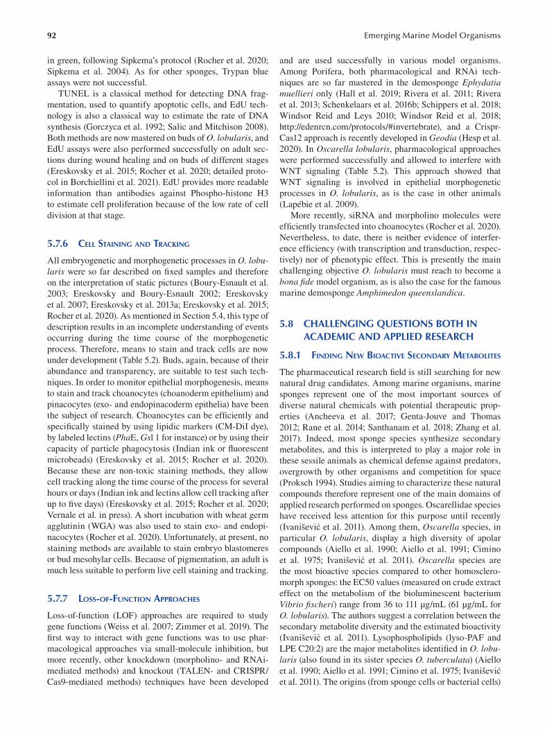
92
in green, following Sipkema’s protocol (Rocher et al. 2020;
Sipkema et al. 2004). As for other sponges, Trypan blue
assays were not successful.
TUNEL is a classical method for detecting DNA frag
mentation, used to quantify apoptotic cells, and EdU tech
nology is also a classical way to estimate the rate of DNA
synthesis (Gorczyca et al. 1992; Salic and Mitchison 2008).
Both methods are now mastered on buds of O. lobularis , and
EdU assays were also performed successfully on adult sec
tions during wound healing and on buds of different stages
(Ereskovsky et al. 2015; Rocher et al. 2020; detailed proto
col in Borchiellini et al. 2021). EdU provides more readable
information than antibodies against Phospho-histone H3
to estimate cell proliferation because of the low rate of cell
division at that stage.
5.7.6 CELL STAINING AND TRACKING
All embryogenetic and morphogenetic processes in O. lobularis were so far described on fixed samples and therefore
on the interpretation of static pictures (Boury-Esnault et al.
2003 ; Ereskovsky and Boury-Esnault 2002 ; Ereskovsky
et al. 2007; Ereskovsky et al. 2013a ; Ereskovsky et al. 2015;
Rocher et al. 2020). As mentioned in Section 5.4, this type of
description results in an incomplete understanding of events
occurring during the time course of the morphogenetic
process. Therefore, means to stain and track cells are now
under development (Table 5.2). Buds, again, because of their
abundance and transparency, are suitable to test such tech
niques. In order to monitor epithelial morphogenesis, means
to stain and track choanocytes (choanoderm epithelium) and
pinacocytes (exo- and endopinacoderm epithelia) have been
the subject of research. Choanocytes can be effi ciently and
specifically stained by using lipidic markers (CM-DiI dye),
by labeled lectins (PhaE, Gsl 1 for instance) or by using their
capacity of particle phagocytosis (Indian ink or fl uorescent
microbeads) (Ereskovsky et al. 2015; Rocher et al. 2020).
Because these are non-toxic staining methods, they allow
cell tracking along the time course of the process for several
hours or days (Indian ink and lectins allow cell tracking after
up to fi ve days) (Ereskovsky et al. 2015; Rocher et al. 2020;
Vernale et al. in press). A short incubation with wheat germ
agglutinin (WGA) was also used to stain exo- and endopi
nacocytes (Rocher et al. 2020). Unfortunately, at present, no
staining methods are available to stain embryo blastomeres
or bud mesohylar cells. Because of pigmentation, an adult is
much less suitable to perform live cell staining and tracking.
5.7.7 LOSS-OF-FUNCTION APPROACHES
Loss-of-function (LOF) approaches are required to study
gene functions (Weiss et al. 2007; Zimmer et al. 2019). The
first way to interact with gene functions was to use phar
macological approaches via small-molecule inhibition, but
more recently, other knockdown (morpholino- and RNAi
mediated methods) and knockout (TALEN- and CRISPR/
Cas9-mediated methods) techniques have been developed
Emerging Marine Model Organisms
and are used successfully in various model organisms.
Among Porifera, both pharmacological and RNAi tech
niques are so far mastered in the demosponge Ephydatia muellieri only (Hall et al. 2019; Rivera et al. 2011; Rivera
et al. 2013; Schenkelaars et al. 2016b; Schippers et al. 2018;
Windsor Reid and Leys 2010; Windsor Reid et al. 2018;
http://edenrcn.com/protocols/#invertebrate), and a Crispr
Cas12 approach is recently developed in Geodia (Hesp et al.
2020). In Oscarella lobularis, pharmacological approaches
were performed successfully and allowed to interfere with
WNT signaling (Table 5.2). This approach showed that
WNT signaling is involved in epithelial morphogenetic
processes in O. lobularis, as is the case in other animals
(Lapébie et al. 2009).
More recently, siRNA and morpholino molecules were
efficiently transfected into choanocytes (Rocher et al. 2020).
Nevertheless, to date, there is neither evidence of interfer
ence efficiency (with transcription and transduction, respec
tively) nor of phenotypic effect. This is presently the main
challenging objective O. lobularis must reach to become a
bona fide model organism, as is also the case for the famous
marine demosponge Amphimedon queenslandica.
5.8 CHALLENGING QUESTIONS BOTH IN ACADEMIC AND APPLIED RESEARCH
5.8.1 FINDING NEW BIOACTIVE SECONDARY METABOLITES
The pharmaceutical research field is still searching for new
natural drug candidates. Among marine organisms, marine
sponges represent one of the most important sources of
diverse natural chemicals with potential therapeutic prop
erties (Ancheeva et al. 2017; Genta-Jouve and Thomas
2012; Rane et al. 2014; Santhanam et al. 2018; Zhang et al.
2017). Indeed, most sponge species synthesize secondary
metabolites, and this is interpreted to play a major role in
these sessile animals as chemical defense against predators,
overgrowth by other organisms and competition for space
(Proksch 1994). Studies aiming to characterize these natural
compounds therefore represent one of the main domains of
applied research performed on sponges. Oscarellidae species
have received less attention for this purpose until recently
( Ivanišević et al. 2011). Among them, Oscarella species, in
particular O. lobularis, display a high diversity of apolar
compounds (Aiello et al. 1990; Aiello et al. 1991; Cimino
et al. 1975; Ivanišević et al. 2011). Oscarella species are
the most bioactive species compared to other homosclero
morph sponges: the EC50 values (measured on crude extract
effect on the metabolism of the bioluminescent bacterium
Vibrio fischeri) range from 36 to 111 μg/mL (61 μg/mL for
O. lobularis). The authors suggest a correlation between the
secondary metabolite diversity and the estimated bioactivity
( Ivanišević et al. 2011). Lysophospholipids (lyso-PAF and
LPE C20:2) are the major metabolites identified in O. lobularis (also found in its sister species O. tuberculata ) ( Aiello
et al. 1990; Aiello et al. 1991; Cimino et al. 1975; Ivanišević et al. 2011). The origins (from sponge cells or bacterial cells)
93 Oscarella lobularis
of these compounds and their individual bioactive properties
have not been characterized yet.
5.8.2 UNDERSTANDING HOST–SYMBIONT INTERACTIONS
Thanks to molecular techniques, the microbial community
of Oscarella lobularis is now well described (Gloeckner et
al. 2013; Jourda et al. 2015). It has been described that (at
least part of) this microbiont is vertically inherited (from
parent to offspring) both during sexual and asexual repro
duction (Boury-Esnault et al. 2003; Ereskovsky and Boury-
Esnault 2002; Ereskovsky and Tokina 2007; Ereskovsky et
al. 2007). But, as for many sponges, the exact nature and
mutual benefits of this biotic association are not determined
yet and for now remain hypothetical. Because of recent
findings on the variation of the bacterial community dur
ing the life cycle in other sponges (Fieth et al. 2016), of
potential metabolic complementarity between bacteria and
the sponge host (Gauthier et al. 2016 ), evidence of bacteria–
sponge horizontal gene transfers (Conaco et al. 2016) and
now that metagenomic data are acquired for O. lobularis, we should take advantage of these data to explore by experi
mental approaches the ecological and physiological roles of
these associations (resource partitioning/supplying between
bacteria and sponge host) but also the potential impact of
the microbial community on the developmental processes of
the sponge as recently observed in marine cnidarians (Tivey
et al. 2020; Ueda et al. 2016 ).
5.8.3 DECIPHERING THE ORIGIN AND
EVOLUTION OF METAZOAN EPITHELIA
Epithelia are considered one of the four fundamental tis
sue types of animals (Edelblum and Turner 2015; Lowe and
Anderson 2015; Yathish and Grace 2018). Epithelia cover
body surfaces, organs and internal cavities, and they are
essential for controlling permeability and selective exchanges
between internal and external environments and between the
different compartments of a body. Epithelia are patterned
at the end of cleavage during embryological development
(Gilbert and Barresi 2018; Tyler 2003) (see Section 5.4).
Epithelia are layers of cells defi ned by three main histo
logical features, according to what is observed in bilaterians:
cell polarity, lateral junctions and a basal lamina made of
collagen IV (Edelblum and Turner 2015; Lowe and Anderson
2015; Tyler 2003; Renard et al. 2021). Until 1996 (Boute et al.
1996 ), no sponge species were known to possess all three
features; sponges were therefore considered devoid of epi
thelia. Among sponges, Hexactinellida do not have cell lay
ers but syncytia instead; Demospongiae and Calcarea have
cell layers with cell polarity, atypical cell junctions but no
basement membrane; in contrast, Homoscleromorpha pos
sess clear cell polarity, unequivocal adherens-like junctions
and obvious basement membrane. Whereas the cell layers
of demosponges have similar mechanical and physiological
properties like bilaterian epithelia, the epithelia of homo
scleromorph sponges are the only ones that present similar
histological features compared to bilaterians (Ereskovsky
2010; Ereskovsky et al. 2009b; Leys and Hill 2012; Leys
and Riesgo 2012; Leys et al. 2009; Renard et al. 2021). For
a while, this “true epithelium” was interpreted as a synapo
morphy of Homoscleromorpha and Eumetazoa (Borchiellini
et al. 2001; Sperling et al. 2007) and suggested the inclusion
of Homoscleromorpha in the Epitheliozoa lineage (a clade
combining Eumetazoa and Placozoa) (Sperling et al. 2009).
The monophyly of Porifera, now supported by numerous
phylogenomic analyses (Philippe et al. 2009; Pick et al.
2010; Pisani et al. 2015; Redmond et al. 2013; Simion et al.
2017; Thacker et al. 2013; Whelan et al. 2017; Wörheide
et al. 2012), means instead that the last common ancestor of
Porifera possessed all three classical features of “typical”
epithelia and that some of these features were secondarily
lost independently in the three other sponge classes.
Interestingly, whether species present all epithelial fea
tures or not, all sponge classes possess the same set of epithe
lial genes involved in the establishment of cell polarity and
the composition of adherens junctions (Belahbib et al. 2018;
Renard et al. 2018; Riesgo et al. 2014; Renard et al. 2021).
Similar inconsistency between gene content and histological
features was reported concerning the basal lamina (Fidler
et al. 2017). These findings question the homology of epithelial
features between sponges and other animals: Is polarity con
trolled by the same three polarity complexes as in bilaterians
(namely Crumbs, Par and Scribble)? Are adherens junctions
described in Homoscleromorpha homologous to bilaterian
adherens junctions (i.e. composed of classical cadherin and
alpha-beta and delta-catenins)? To answer these questions,
complementary molecular and biochemical approaches are
in progress in both O. pearsei and O. lobularis and in par
allel in demosponges. The first results obtained suggest that
the proteins involved in cell–cell and cell–matrix adhesion
would be the same in demosponges and homoscleromorphs,
in particular vinculin and beta-catenin ( Miller et al. 2018;
Mitchell and Nichols 2019; Schippers et al. 2018). To date,
there is no clear information concerning the eventual impli
cation of classical cadherins in these junctions.
5.8.4 SPONGE GASTRULATION AND THE
ORIGIN OF GERM LAYERS
Despite the true multicellular and metazoan nature of
sponges having been elucidated decades ago (reviewed in
Schenkelaars et al. 2019), there is a longstanding debate in
the spongiologist community on whether sponges gastrulate.
Different points of view compete: i) for some authors, multi
polar egression leading to the formation of the coeloblastula
during embryogenesis marks the onset of polarization and
regionalization processes, suggesting it may be similar to
gastrulation (Maldonado and Riesgo 2008); ii) others con
sider that this process differs from gastrulation in that the
resulting embryo apparently consists of one uniform cell
layer and lacks polarity (Ereskovsky 2010; Ereskovsky and
Dondua 2006) and prefer to hypothesize the gastrulation
during larval metamorphosis (reviewed in Ereskovsky 2010;
94
Ereskovsky et al. 2013b; Lanna 2015; Leys 2004; Wörheide
et al. 2012), when an “inversion of germ layers” results in
the formation of the aquiferous system. In the last case, the
term “inversion” means that external-most larval cells form
the internal-most (“gut-like”) structures of an adult sponge,
namely the aquiferous system.
However, cellular tracking during the larval metamor
phosis in Amphimedon queenslandica has shown no relation
between larval and juvenile cell layers; the cells of the larvae
do not have specifi cation: all larval cell types are capable of
transdifferentiating into all juvenile cell types (Nakanishi et
al. 2014; Sogabe et al. 2016). This apparent lack of cell layer
and fate determination and stability during metamorphosis
in this sponge argues for an absence of gastrulation. In this
context, the expression of the transcription factor GATA , a
highly conserved eumetazoan endomesodermal marker,
in the inner layer of A. queenslandica embryos, free lar
vae and juveniles has been interpreted to provide positional
information to cells (Nakanishi et al. 2014). In contrast, in
Sycon ciliatum, expression of the same marker in embryo/
larva ciliated micromeres (at the origin of adult choanocytes)
and in adult choanoderm has given rise to other conclusions
(Leininger et al. 2014). Indeed, the authors suggest that the
calcareous sponge choanoderm and the bilaterian endoderm
are homologous structures and ciliated choanocytes are germ
layers. Thus, the origin of gastrulation and germ layers is still
controversial (Degnan et al. 2015; Lanna 2015). Yet the reso
lution of this problem is the key to comparing embryological
stages between sponges and other metazoans and to discuss
ing germ layer homology between all animal phyla.
As mentioned in the section on embryology and in the
previous section, Oscarella lobularis (like other homo
scleromorph) presents clear epithelial characteristics, and
all morphogenetic processes (development, regeneration,
budding) are based mainly on epithelial morphogenetic
movements in contrast to demosponges (Boury-Esnault
et al. 2003; Ereskovsky 2010; Ereskovsky and Tokina 2007;
Ereskovsky et al. 2007; Ereskovsky et al. 2009b; Ereskovsky
et al. 2013a ; Ereskovsky et al. 2013b; Ereskovsky et al. 2015).
This feature is expected to result in the formation of more
stable cell layers during embryogenesis compared to demo-
sponges (Ereskovsky 2010; Lanna 2015). O. lobularis is thus
an interesting model to answer questions about the homol
ogy of embryonic morphogenesis (gastrulation) and germ
layers in animals (Degnan et al. 2015; Lanna 2015). The
techniques now available in this sponge species (see Section
5.7 on functional approaches) are highly signifi cant innova
tions to answer this fundamental question. The main experi
mental limitation to do so is the difficult and limited access
to embryos and larvae in this species, as sexual reproduction
cannot be triggered in aquaria.
ACKNOWLEDGMENTS
The authors thank all those whose involvement made the
development of this emerging model possible: Nicole Boury-
Esnault and Jean Vacelet for their help in launching our
Emerging Marine Model Organisms
studies on this species; our PhD students Pascal Lapébie,
Eve Gazave, Quentin Schenkelaars, Laura Fierro-Constain,
Amélie Vernale and Kassandra de Pao Mendonca; the
numerous internship students who helped in perform
ing preliminary experiments; the imaging facilities of the
France Bioimaging infrastructure; the diving facilities of the
Institute OSU Pytheas and divers from the IMBE lab; and
the molecular biology and morphology support services of
IMBE. We thank Haley Flom for English editing.
The authors acknowledge the Région Sud (Provence
Alpes Côte d’Azur), the French Research ministry, the
French National Center for Scientific Research (CNRS),
Aix-Marseille University and the Excellence Initiative of
Aix-Marseille Université–A*MIDEX for providing funds
to support our fundamental research, in particular for the
funding of the project for international scientifi c cooperation
(PICS) STraS, the A*MIDEX foundation projects (n° ANR
11-IDEX-0001–02 and AMX-18-INT-021) and the LabEx
INFORM (ANR-11-LABX-0054), both funded by the
“Investissements d’Avenir” French Government program,
managed by the French National Research Agency (ANR).
The Russian Science Foundation, Grant n° 17-14-01089.
BIBLIOGRAPHY
Adamska, M. 2016. Sponges as models to study emergence of
complex animals. Curr. Opin. Genet. Dev. 39, 21–28.
Adamska, M., Degnan, S. M., Green, K. M., Adamski, M., Craigie,
A., Larroux, C. and Degnan, B. M. 2007. Wnt and TGF-beta
expression in the sponge Amphimedon queenslandica and
the origin of metazoan embryonic patterning. PLoS One 2,
e1031.
Adamska, M., Degnan, B. M., Green, K. and Zwafink, C. 2011.
What sponges can tell us about the evolution of developmen
tal processes. Zool. Jena Ger. 114, 1–10.
Adamska, M., Larroux, C., Adamski, M., Green, K., Lovas, E.,
Koop, D., Richards, G. S., Zwafink, C. and Degnan, B. M.
2010. Structure and expression of conserved Wnt pathway
components in the demosponge Amphimedon queenslandica.
Evol. Dev. 12, 494–518.
Aiello, A., Fattorusso, E., Magno, S., Mayol, L. and Menna, M.
1990. Isolation of two novel 5α,6α-Epoxy-7-ketosterols from
the encrusting demospongia Oscarella lobularis. J. Nat. Prod. 53, 487–491.
Aiello, A., Fattorusso, E., Magno, S. and Menna, M. 1991. Isolation
of five new 5 alpha-hydroxy-6-keto-delta 7 sterols from the
marine sponge Oscarella lobularis. Steroids 56, 337–340.
Alié, A., Hayashi, T., Sugimura, I., Manuel, M., Sugano, W., Mano,
A., Satoh, N., Agata, K. and Funayama, N. 2015. The ances
tral gene repertoire of animal stem cells. Proc. Natl. Acad. Sci. U. S. A. 112, E7093–E7100.
Ancheeva, E., El-Neketi, M., Song, W., Lin, W., Daletos, G.,
Ebrahim, W. and Proksch, P. 2017. Structurally unprec
edented metabolites from marine sponges. Curr. Org. Chem.
21, 426–449.
Arendt, D., Musser, J. M., Baker, C. V. H., Bergman, A., Cepko,
C., Erwin, D. H., Pavlicev, M., Schlosser, G., Widder, S.,
Laubichler, M. D., et al. 2016. The origin and evolution of
cell types. Nat. Rev. Genet. 17, 744–757.
Babonis, L. S. and Martindale, M. Q. 2017. Phylogenetic evidence
for the modular evolution of metazoan signalling pathways.
Philos. Trans. R. Soc. B Biol. Sci. 372 (1713), 20150477.
95 Oscarella lobularis
Belahbib, H., Renard, E., Santini, S., Jourda, C., Claverie, J.-M.,
Borchiellini, C. and Le Bivic, A. 2018. New genomic data
and analyses challenge the traditional vision of animal epi
thelium evolution. BMC Genomics 19, 393.
Bergquist, P. R. 1978. Sponges. Hutchinson, London; University of
California Press, Berkeley & Los Angeles, CA, 268 pp.
Bergquist, P. R. and Kelly, M. 2004. Taxonomy of some Halisarcida
and Homosclerophorida Porifera: Demospongiae from the
Indo-Pacifi c. N. Z. J. Mar. Freshw. Res. 38, 51–66.
Bertolino, M., Cerrano, C., Bavestrello, G., Carella, M., Pansini,
M. and Calcinai, B. 2013. Diversity of Porifera in the
Mediterranean coralligenous accretions, with description of
a new species. ZooKeys , 1–37.
Borchiellini, C., Degnan, S. M., Le Goff, E., Rocher, C., Vernale,
A., Baghdiguian, S., Séjourné, N., Marschal, F., Le Bivic,
A., Godefroy, N., et al. 2021. Staining and tracking meth
ods for studying sponge cell dynamics. In Developmental Biology of the Sea Urchin and Other Marine Invertebrates: Methods and Protocols, ed. Carroll, D. J. and Stricker, S. A.,
pp. 81–97. Springer US, New York, NY.
Borchiellini, C., Chombard, C., Manuel, M., Alivon, E., Vacelet,
J. and Boury-Esnault, N. 2004. Molecular phylogeny of
Demospongiae: Implications for classifi cation and scenarios
of character evolution. Mol. Phylogenet. Evol. 32, 823–837.
Borchiellini, C., Manuel, M., Alivon, E., Boury-Esnault, N.,
Vacelet, J. and Parco, Y. L. 2001. Sponge paraphyly and the
origin of Metazoa. J. Evol. Biol. 14, 171–179.
Borisenko, I. E., Adamski, M., Ereskovsky, A. and Adamska, M.
2016. Surprisingly rich repertoire of Wnt genes in the demo-
sponge Halisarca dujardini. BMC Evol. Biol. 16, 123.
Borisenko, I. E., Adamska, M., Tokina, D. B. and Ereskovsky, A.
V. 2015. Transdifferentiation is a driving force of regenera
tion in Halisarca dujardini Demospongiae, Porifera. PeerJ.
3, e1211.
Boury-Esnault, N., Ereskovsky, A., Bézac, C. and Tokina, D. 2003.
Larval development in the Homoscleromorpha Porifera,
Demospongiae. Invertebr. Biol. 122, 187–202.
Boury-Esnault, N., Lavrov, D. V., Ruiz, C. A. and Pérez, T. 2013.
The integrative taxonomic approach applied to Porifera: A
case study of the Homoscleromorpha. Integr. Comp. Biol . 53,
416–427.
Boury-Esnault, N., Sole-Cava, A. M. and Thorpe, J. P. 1992.
Genetic and cytological divergence between colour morphs
of the Mediterranean sponge Oscarella lobularis Schmidt
Porifera, Demospongiae, Oscarellidae. J. Nat. Hist. 26,
271–284.
Boute, N., Exposito, J.-Y., Boury-Esnault, N., Vacelet, J., Noro, N.,
Miyazaki, K., Yoshizato, K. and Garrone, R. 1996. Type IV
collagen in sponges, the missing link in basement membrane
ubiquity. Biol. Cell. 88, 37–44.
Brunet, T. and King, N. 2017. The origin of animal multicellularity
and cell differentiation. Dev. Cell 43, 124–140.
Brusca, R. C. and Brusca, G. J. 2003. Invertebrates. 2rd ed. Sinauer
Associates, Sunderland, MA, USA, 936 pp.
Brusca, R. C., Moore, W. and Shuster, S. M. 2016. Invertebrates.
3rd ed. Sinauer Associates is an imprint of Oxford University
Press, Sunderland, MA, USA, 1104 pp.
Cardone, F., Gaino, E. R. and Corriero, G. 2010. The budding pro
cess in Tethya citrina Sarà & Melone Porifera, Demospongiae
and the incidence of post-buds in sponge population mainte
nance. J. Exp. Mar. Biol. Ecol. 389, 93–100.
Carter, H. J. 1876. Descriptions and figures of deep-sea sponges
and their spicules, from the Atlantic Ocean, dredged up on
board H.M.S. ‘Porcupine’, chiefly in 1869 (concluded). Ann. Mag. Nat. Hist. 18, 226–240.
Cimino, G., de Stefano, S. and Minale, L. 1975. Long alkyl chains
3-substituted pyrrole-2-aldehyde-2-carboxylic acid and
methyl ester from the marine sponge Oscarella lobularis.
Experientia 31, 1387–1389.
Colgren, J. and Nichols, S. A. 2019. The significance of sponges
for comparative studies of developmental evolution. Wiley Interdiscip. Rev. Dev. Biol , e359.
Coll, M., Piroddi, C., Steenbeek, J., Kaschner, K., Lasram, F.
B. R., Aguzzi, J., Ballesteros, E., Bianchi, C. N., Corbera,
J., Dailianis, T., et al. 2010. The biodiversity of the
Mediterranean Sea: Estimates, patterns, and threats. PLoS One 5, e11842.
Conaco, C., Tsoulfas, P., Sakarya, O., Dolan, A., Werren, J. and
Kosik, K. S. 2016. Detection of prokaryotic genes in
the Amphimedon queenslandica genome. PLoS One 11,
e0151092.
Degnan, B. M., Adamska, M., Richards, G. S., Larroux, C.,
Leininger, S., Bergum, B., Calcino, A., Taylor, K., Nakanishi,
N. and Degnan, S. M. 2015. Porifera. In Evolutionary Developmental Biology of Invertebrates 1: Introduction, Non-Bilateria, Acoelomorpha, Xenoturbellida, Chaetognatha , ed.
Wanninger, A., pp. 65–106. Springer, Vienna.
Dendy, A. 1905. Report on the sponges collected by Professor
Herdman, at Ceylon, in 1902. In Report to the Government of Ceylon on the Pearl Oyster Fisheries of the Gulf of Manaar,
ed. Herdman, W. A., pp. 57–246. Royal Society, London. 3
(Supplement 18).
Devenport, D. 2014. The cell biology of planar cell polarity. J. Cell Biol. 207, 171–179.
De Vos, L., Rützler, K., Boury-Esnault, N., Donadey, C. and
Vacelet, J. 1991. Atlas of sponge morphology. Smithson. Inst. Press Wash. Lond., 117 pp.
Domingos, C., Lage, A. and Muricy, G. 2016. Overview of the bio
diversity and distribution of the class Homoscleromorpha in
the tropical Western Atlantic. J. Mar. Biol. Assoc. U. K . 96,
379–389.
Dunn, C. W., Leys, S. P. and Haddock, S. H. D. 2015. The hidden
biology of sponges and ctenophores. Trends Ecol. Evol . 30,
282–291.
Edelblum, K. L. and Turner, J. R. 2015. Chapter 12: Epithelial
cells: Structure, transport, and barrier function. In Mucosal Immunology. 4th ed., ed. Mestecky, J., Strober, W., Russell,
M. W., Kelsall, B. L., Cheroutre, H. and Lambrecht, B. N.,
pp. 187–210. Academic Press, Boston.
Ereskovsky, A. V. 2006. A new species of Oscarella Demospongiae:
Plakinidae from the Western Sea of Japan. Zootaxa 1376,
37–51.
Ereskovsky, A. V. 2010. The Comparative Embryology of Sponges.
Springer-Verlag, Dordrecht Heidelberg, London and New
York, 329 pp.
Ereskovsky, A. V., Borchiellini, C., Gazave, E., Ivanisevic, J.,
Lapébie, P., Pérez, T., Renard, E. and Vacelet, J. 2009b. The
Homoscleromorph sponge Oscarella lobularis, a promising
sponge model in evolutionary and developmental biology.
BioEssays 31, 89–97.
Ereskovsky, A. V., Borisenko, I. E., Lapébie, P., Gazave, E.,
Tokina, D. B. and Borchiellini, C. 2015. Oscarella lobularis Homoscleromorpha, Porifera Regeneration: Epithelial mor
phogenesis and metaplasia. PLoS One 108: e0134566.
Ereskovsky, A. V. and Boury-Esnault, N. 2002. Cleavage pat
tern in Oscarella species Porifera, Demospongiae,
Homoscleromorpha: Transmission of maternal cells and
symbiotic bacteria. J. Nat. Hist. 36, 1761–1775.
Ereskovsky, A. V., Dubois, M., Ivanišević, J., Gazave, E., Lapebie,
P., Tokina, D. and Pérez, T. 2013a. Pluri-annual study of
96
the reproduction of two Mediterranean Oscarella species
Porifera, Homoscleromorpha: Cycle, sex-ratio, reproductive
effort and phenology. Mar. Biol. 160, 423–438.
Ereskovsky, A. V., Geronimo, A. and Pérez, T. 2017a. Asexual and
puzzling sexual reproduction of the Mediterranean sponge
Haliclona fulva Demospongiae: Life cycle and cytological
structures. Invertebr. Biol. 136, 403–421.
Ereskovsky, A. V. and Dondua, A. K. 2006. The problem of germ
layers in sponges Porifera and some issues concerning early
metazoan evolution. Zool. Anz.: J. Comp. Zool. 245, 65–76.
Ereskovsky, A. V., Konyukov, P. Yu. and Tokina, D. B. 2010.
Morphogenesis accompanying larval metamorphosis in
Plakina trilopha Porifera, Homoscleromorpha. Zoomorphology 129, 21–31.
Ereskovsky, A. V., Lavrov, D. V. and Willenz, P. 2014. Five new
species of Homoscleromorpha Porifera from the Caribbean
Sea and re-description of Plakina jamaicensis. J. Mar. Biol. Assoc. U. K. 94, 285–307.
Ereskovsky, A. V., Renard, E. and Borchiellini, C. 2013b. Cellular
and molecular processes leading to embryo formation in
sponges: Evidences for high conservation of processes
throughout animal evolution. Dev. Genes Evol. 223, 5–22.
Ereskovsky, A. V., Richter, D. J., Lavrov, D. V., Schippers, K. J.
and Nichols, S. A. 2017b. Transcriptome sequencing and
delimitation of sympatric Oscarella species O. carmela and
O. pearsei sp. nov from California, USA. PLoS One 129,
e0183002.
Ereskovsky, A. V., Sanamyan, K. and Vishnyakov, E. 2009a. A new
species of the genus Oscarella Porifera: Homosclerophorida:
Plakinidae from the North-West Pacifi c. Cah. Biol. Mar . 50,
369–381.
Ereskovsky, A. V. and Tokina, D. B. 2007. Asexual reproduction
in homoscleromorph sponges Porifera; Homoscleromorpha.
Mar. Biol. 151, 425–434.
Ereskovsky, A. V., Tokina, D. B., Bézac, C. and Boury-
Esnault, N. 2007. Metamorphosis of cinctoblastula larvae
Homoscleromorpha, Porifera. J. Morphol. 268, 518–528.
Fell, P. 1993. Porifera. In Asexual Propagation and Reproductive Strategies, eds. Adiyodi, K. G. and Adiyodi, R. G., pp. 1–44.
Oxford and IBH, New Delhi.
Fernandez-Valverde, S. L., Calcino, A. D. and Degnan, B. M.
2015. Deep developmental transcriptome sequencing uncov
ers numerous new genes and enhances gene annotation in
the sponge Amphimedon queenslandica. BMC Genomics 16,
387.
Feuda, R., Dohrmann, M., Pett, W., Philippe, H., Rota-Stabelli, O.,
Lartillot, N., Wörheide, G. and Pisani, D. 2017. Improved
modeling of compositional heterogeneity supports sponges
as sister to all other animals. Curr. Biol. 18, 2724, 3864–3870.
Fidler, A. L., Darris, C. E., Chetyrkin, S. V., Pedchenko, V. K.,
Boudko, S. P., Brown, K. L., Gray Jerome, W., Hudson, J. K.,
Rokas, A. and Hudson, B. G. 2017. Collagen IV and base
ment membrane at the evolutionary dawn of metazoan tis
sues. eLife 18, 6, e24176.
Fierro-Constain, L. 2016. La reproduction chez Oscarella lobularis Porifera-Homoscleromorpha: gènes impliqués et effets de
l’environnement. www.theses.fr .
Fierro-Constaín, L., Rocher, C., Marschal, F., Schenkelaars, Q.,
Séjourné, N., Borchiellini, C. and Renard, E. 2021. In situ
hybridization techniques in the homoscleromorph sponge
Oscarella lobularis. Methods Mol. Biol. Clifton NJ 2219,
181–194.
Fierro-Constaín, L., Schenkelaars, Q., Gazave, E., Haguenauer,
A., Rocher, C., Ereskovsky, A., Borchiellini, C. and Renard,
E. 2017. The conservation of the Germline multipotency
Emerging Marine Model Organisms
program, from sponges to vertebrates: A stepping stone to
understanding the somatic and germline origins. Genome Biol. Evol. 9, 474–488.
Fieth, R. A., Gauthier, M.-E. A., Bayes, J., Green, K. M. and
Degnan, S. M. 2016. Ontogenetic changes in the bacte
rial symbiont community of the tropical demosponge
Amphimedon queenslandica: Metamorphosis is a new begin
ning. Front. Mar. Sci. 3, 228.
Fortunato, S. A. V., Adamski, M. and Adamska, M. 2015.
Comparative analyses of developmental transcription factor
repertoires in sponges reveal unexpected complexity of the
earliest animals. Mar. Genomics 24 (2), 121–129.
Fortunato, S. A. V., Vervoort, M., Adamski, M. and Adamska, M.
2016. Conservation and divergence of bHLH genes in the
calcisponge Sycon ciliatum. EvoDevo 7, 23.
Francis, W. R. and Canfield, D. E. 2020. Very few sites can reshape
the inferred phylogenetic tree. PeerJ. 8, e8865.
Francis, W. R., Eitel, M., R. S. V., Adamski, M., Haddock, S. H.,
Krebs, S., Blum, H., Erpenbeck, D. and Wörheide, G. 2017.
The genome of the contractile demosponge Tethya wilhelma and the evolution of metazoan neural signalling pathways.
bioRxiv 120998.
Funayama, N. 2013. The stem cell system in demosponges:
Suggested involvement of two types of cells: Archeocytes
active stem cells and choanocytes food-entrapping fl agel
lated cells. Dev. Genes Evol. 223, 23–38.
Funayama, N. 2018. The cellular and molecular bases of the sponge
stem cell systems underlying reproduction, homeostasis and
regeneration. Int. J. Dev. Biol. 62, 513–525.
Funayama, N., Nakatsukasa, M., Mohri, K., Masuda, Y. and Agata,
K. 2010. Piwi expression in archeocytes and choanocytes in
demosponges: Insights into the stem cell system in demo-
sponges. Evol. Dev. 12, 275–287.
Gaino, E., Burlando, B. and Buffa, P. 1986a. The vacuolar cells of
Oscarella lobularis Porifera, Demospongiae: Ulatrastructural
organization, origin, and function. J. Morphol. 188, 29–37.
Gaino, E., Burlando, B. and Buffa, P. 1986b. Contribution to the
study of egg development and derivation in Oscarella lobularis Porifera, Demospongiae. Int. J. Invertebr. Rep. Dev . 9,
59–69.
Gaino, E., Burlando, B., Buffa, P. and Sará, M. 1986c. Ultrastructural
study of spermatogenesis in Oscarella lobularis Porifera,
Demospongiae. Int. J. Invertebr. Rep. Dev. 10, 297–305.
Garrabou, J. and Zabala, M. 2001. Growth dynamics in four
Mediterranean demosponges. Estuar. Coast. Shelf Sci. 52,
293–303.
Gatenby, J. B. 1920. IX.: Further notes on the Oögenesis and
fertilization of Grantia compressa. J. R. Microsc. Soc . 40,
277–282.
Gauthier, M.-E. A., Watson, J. R. and Degnan, S. M. 2016. Draft
genomes shed light on the dual bacterial symbiosis that dom
inates the microbiome of the coral reef sponge Amphimedon queenslandica. Front. Mar. Sci. 3, 196.
Gazave, E. 2010. Etude de gène et voie de signalisation impli
qués dans les processus morphogénétiques chez Oscarella lobularis: implications potentielles sur la compréhension de
l’origine du système nerveux. www.theses.fr.
Gazave, E., Lapébie, P., Ereskovsky, A. V., Vacelet, J., Renard,
E., Cárdenas, P. and Borchiellini, C. 2012. No longer
Demospongiae: Homoscleromorpha formal nomination as a
fourth class of Porifera. Hydrobiologia 687, 3–10.
Gazave, E., Lapébie, P., Renard, E., Bézac, C., Boury-Esnault, N.,
Vacelet, J., Pérez, T., Manuel, M. and Borchiellini, C. 2008.
NK homeobox genes with choanocyte-specifi c expression in
homoscleromorph sponges. Dev. Genes Evol. 218, 479–489.
97 Oscarella lobularis
Gazave, E., Lapébie, P., Richards, G. S., Brunet, F., Ereskovsky, A.
V., Degnan, B. M., Borchiellini, C., Vervoort, M. and Renard,
E. 2009. Origin and evolution of the Notch signalling path
way: An overview from eukaryotic genomes. BMC Evol. Biol. 9, 249.
Gazave, E., Lapébie, P., Renard, E., Vacelet, J., Rocher, C.,
Ereskovsky, A. V., Lavrov, D. V. and Borchiellini, C. 2010.
Molecular phylogeny restores the supra-generic subdivision
of homoscleromorph sponges Porifera, Homoscleromorpha.
PLoS One 512, e14290.
Gazave, E., Guillou, A. and Balavoine, G. 2014. History of a
prolific family: The Hes/Hey-related genes of the annelid
Platynereis. EvoDevo 5, 29.
Gazave, E., Lavrov, D. V., Cabrol, J., Renard, E., Rocher, C.,
Vacelet, J., Adamska, M., Borchiellini, C. and Ereskovsky,
A. V. 2013. Systematics and molecular phylogeny of the fam
ily Oscarellidae Homoscleromorpha with description of two
new Oscarella species. PLoS One 8, e63976.
Gazave, E. and Renard, E. 2010. Evolution of Notch Transmembrane
Receptors. In eLS, p. American Cancer Society.
Genta-Jouve, G. and Thomas, O. P. 2012. Sponge chemical diver
sity: From biosynthetic pathways to ecological roles. Adv. Mar. Biol. 62, 183–230.
Gerçe, B., Schwartz, T., Syldatk, C. and Hausmann, R. 2011.
Differences between bacterial communities associated
with the surface or tissue of Mediterranean sponge species.
Microb. Ecol. 61, 769–782.
Gilbert, S. F. and Barresi, M. J. F. 2018. Developmental Biology.
11th ed. Sinauer Associates, Sunderland, MA, USA.
Gloeckner, V., Hentschel, U., Ereskovsky, A. V. and Schmitt, S.
2013. Unique and species-specific microbial communities in
Oscarella lobularis and other Mediterranean Oscarella spe
cies Porifera: Homoscleromorpha. Mar. Biol. 160, 781–791.
Gorczyca, W., Bruno, S., Darzynkiewicz, R., Gong, J. and
Darzynkiewicz, Z. 1992. DNA strand breaks occurring dur
ing apoptosis : T heir early in situ detection by the terminal
deoxynucleotidyl transferase and nick translation assays
and prevention by serine protease inhibitors . Int. J. Oncol. 1, 639–648.
Hall, C., Rodriguez, M., Garcia, J., Posfai, D., DuMez, R., Wictor,
E., Quintero, O. A., Hill, M. S., Rivera, A. S. and Hill, A. L.
2019. Secreted frizzled related protein is a target of PaxB and
plays a role in aquiferous system development in the fresh
water sponge, Ephydatia muelleri. PLoS One 14, e0212005.
Hammel, J. U. and Nickel, M. 2014. A new fl ow-regulating cell
type in the demosponge Tethya wilhelma: Functional cellular
anatomy of a leuconoid canal system. PLoS One 9, e113153.
Hanitsch, R. 1890. Third report on the Porifera of the L.M.B.C.
district. Proc. Trans. Liverpool Biol. Soc. 4, 192–238.
Hentschel, E. 1909. Tetraxonida. I. Teil. In Die Fauna Südwest-Australiens. Ergebnisse der Hamburger südwest-australischen Forschungsreise 1905, eds. Michaelsen, W. and Hartmeyer,
R., pp. 347–402. Jena: Fischer. 2 (21).
Hesp, K., Flores Alvarez, J. L., Alexandru, A.-M., van der Linden,
J., Martens, D. E., Wijffels, R. H. and Pomponi, S. A. 2020.
CRISPR/Cas12a-mediated gene editing in Geodia barretti sponge cell culture. Front. Mar. Sci. 7, 599825.
Hill, M. S., Hill, A. L., Lopez, J., Peterson, K. J., Pomponi, S., Diaz,
M. C., Thacker, R. W., Adamska, M., Boury-Esnault, N.,
Cárdenas, P., et al. 2013. Reconstruction of family-level phy
logenetic relationships within Demospongiae, Porifera using
nuclear encoded housekeeping genes. PLoS One 8, e50437.
Humbert-David, N. and Garrone, R. 1993. A six-armed, tenascin
like protein extracted from the Porifera Oscarella tuberculata Homosclerophorida. Eur. J. Biochem. 216, 255–260.
Ivanišević, J., Thomas, O. P., Lejeusne, C., Chevaldonné, P. and
Pérez, T. 2011. Metabolic fingerprinting as an indicator of
biodiversity: Towards understanding inter-specifi c relation
ships among Homoscleromorpha sponges. Metabolomics 7,
289–304.
Jenner, R. A. and Wills, M. A. 2007. The choice of model organ
isms in evo-devo. Nat. Rev. Genet. 8, 311–314.
Jourda, C., Santini, S., Rocher, C., Le Bivic, A. and Claverie, J.-M.
2015. Draft genome sequence of an alphaproteobacterium
associated with the Mediterranean sponge Oscarella lobularis. Genome Announc. 3 (5), e00977–15.
Kanska, J. and Frank, U. 2013. New roles for nanos in neural cell
fate determination revealed by studies in a cnidarian. J. Cell Sci. 126, 3192–3203.
Kenny, N. J., Francis, W. R., Rivera-Vicéns, R. E., Juravel, K.,
Mendoza, A. de, Díez-Vives, C., Lister, R., Bezares-Calderon,
L., Grombacher, L., Roller, M., et al. 2020. Tracing animal
genomic evolution with the chromosomal-level assembly of
the freshwater sponge Ephydatia muelleri. bioRxiv Feb 18,
954784.
King, N. 2004. The unicellular ancestry of animal development.
Dev. Cell 7, 313–325.
Krenacs, L., Krenacs, T., Stelkovics, E. and Raffeld, M. 2010.
Heat-induced antigen retrieval for immunohistochemi
cal reactions in routinely processed paraffin sections. In
Immunocytochemical Methods and Protocols, eds. Oliver, C.
and Jamur, M. C., pp. 103–119. Humana Press, Totowa, NJ.
Labrenz, M., Lawson, P. A., Tindall, B. J. and Hirsch, P. 2009.
Roseibaca ekhonensis gen. nov., sp. nov., an alkalitolerant
and aerobic bacteriochlorophyll a-producing alphaproteo
bacterium from hypersaline Ekho Lake. Int. J. Syst. Evol. Microbiol. 59, 1935–1940.
Lage, A., Gerovasileiou, V., Voultsiadou, E. and Muricy, G. 2019.
Taxonomy of Plakina Porifera: Homoscleromorpha from
Aegean submarine caves, with descriptions of three new spe
cies and new characters for the genus. Mar. Biodivers . 49,
727–747.
Lage, A., Muricy, G., Ruiz, C. and Pérez, T. 2018. New sciaphilic
plakinids Porifera, Homoscleromorpha from the Central-
Western Pacifi c. Zootaxa 4466, 8–38.
Lanna, E. 2015. Evo-devo of non-bilaterian animals. Genet. Mol. Biol. 38, 284–300.
Lapebie, P. 2010. L’origine des morphogenèses épithéliales et leurs
implications concernant l’évolution précoce des métazoaires.
www.theses.fr .
Lapébie, P., Gazave, E., Ereskovsky, A., Derelle, R., Bézac, C.,
Renard, E., Houliston, E. and Borchiellini, C. 2009. WNT/
beta-catenin signalling and epithelial patterning in the homo
scleromorph sponge Oscarella. PLoS One 4, e5823.
Laundon, D., Larson, B. T., McDonald, K., King, N. and Burkhardt,
P. 2019. The architecture of cell differentiation in choanofl a
gellates and sponge choanocytes. PLoS Biol. 17, e3000226.
Layden, M. J., Röttinger, E., Wolenski, F. S., Gilmore, T. D. and
Martindale, M. Q. 2013. Microinjection of mRNA or mor
pholinos for reverse genetic analysis in the starlet sea anem
one, Nematostella vectensis. Nat. Protoc. 8, 924–934.
Leininger, S., Adamski, M., Bergum, B., Guder, C., Liu, J.,
Laplante, M., Bråte, J., Hoffmann, F., Fortunato, S., Jordal,
S., et al. 2014. Developmental gene expression provides
clues to relationships between sponge and eumetazoan body
plans. Nat. Commun. 5, ncomms4905.
Lendenfeld, R. von. 1887. On the systematic position and classi
fication of sponges. Proc. Zool. Soc. London. 18, 558–662.
Lévi, C. 1956. Etude des Hailsarca de Roscoff. Embryologie et sys
tematique des demosponges. Arch. Zool. Exp. Gen. 93, 1–184.
98
Lévi, C. 1973. Systématique de la classe des Demospongiaria
Démosponges. Pp. 577–631. In Traité de Zoologie. Anatomie, Systématique, Biologie, ed. Grassé, P.-P. Spongiaires. 3(1),
pp. 577–631. Masson et Cie., Paris.
Lévi, C. and Porte, A. 1962. Étude au microscope électronique de
l’éponge Oscarella lobularis schmidt et de sa larve amphi
blastula. Cah. Biol. Mar. 3, 307–315.
Leys, S. P. 2004. Gastrulation in sponges. Gastrulation Cells Embryo Ed. Stern CD 2004 N. Y. Cold Spring Harb. Lab. Press , 23–31.
Leys, S. P. and Degnan, B. M. 2001. Cytological basis of photores
ponsive behavior in a sponge larva. Biol. Bull. 201, 323–338.
Leys, S. P. and Ereskovsky, A. V. 2006. Embryogenesis and larval
differentiation in sponges. Can. J. Zool. 84, 262–287.
Leys, S. P. and Hill, A. 2012. The physiology and molecular biol
ogy of sponge tissues. In Advanc. Mar. Biol ., pp. 1–56.
Leys, S. P., Mah, J. L., McGill, P. R., Hamonic, L., De Leo, F.
C. and Kahn, A. S. 2019. Sponge behavior and the chemical
basis of responses: A post-genomic view. Integr. Comp. Biol. 59, 751–764.
Leys, S. P., Nichols, S. A. and Adams, E. D. M. 2009. Epithelia
and integration in sponges. Integr. Comp. Biol. 49, 167–177.
Leys, S. P. and Riesgo, A. 2012. Epithelia, an evolutionary nov
elty of metazoans. J. Exp. Zoolog. B Mol. Dev. Evol . 318,
438–447.
Leys, S. P., Yahel, G., Reidenbach, M. A., Tunnicliffe, V., Shavit,
U. and Reiswig, H. M. 2011. The sponge pump: The role of
current induced flow in the design of the sponge body plan.
PLoS One 6, e27787.
Love, A. C. and Yoshida, Y. 2019. Reflections on model organisms
in evolutionary developmental biology. Results Probl. Cell Differ . 3–20.
Lowe, J. S. and Anderson, P. G. 2015. Chapter 3: Epithelial cells. In
Stevens & Lowe’s Human Histology. 4th ed., ed. Lowe, J. S.
and Anderson, P. G., pp. 37–54. Mosby, Philadelphia.
Ludeman, D. A., Reidenbach, M. A. and Leys, S. P. 2017. The ener
getic cost of filtration by demosponges and their behavioural
response to ambient currents. J. Exp. Biol. 220, 995–1007.
Mah, J. L., Christensen-Dalsgaard, K. K. and Leys, S. P. 2014.
Choanoflagellate and choanocyte collar-flagellar systems and
the assumption of homology. Evol. Dev. 16, 25–37.
Maldonado, M. 2004. Choanoflagellates, choanocytes, and animal
multicellularity. Invertebr. Biol. 123, 1–22.
Maldonado, M. and Riesgo, A. 2008. Reproduction in Porifera: A
synoptic overview. Treb. Soc. Cat. Biol. 59, 29–49.
Marion, A. F. 1883. Esquisse d’une topographie zoologique du golfe de Marseille. Cayer et cie.
Meewis, H. 1938. Contribution a l’étude de l’embryogenése des
Myxospongiae: Halisarca lobularis Schmidt. Arch. Biol. Liege 59, 1–66.
Miller, P. W., Pokutta, S., Mitchell, J. M., Chodaparambil, J. V.,
Clarke, D. N., Nelson, W. J., Weis, W. I. and Nichols, S. A.
2018. Analysis of a vinculin homolog in a sponge phylum
Porifera reveals that vertebrate-like cell adhesions emerged
early in animal evolution. J. Biol. Chem. 293, 11674–11686.
Mitchell, J. M. and Nichols, S. A. 2019. Diverse cell junctions
with unique molecular composition in tissues of a sponge
Porifera. EvoDevo 10, 26.
Muricy, G., Boury-Esnault, N., Bézac, C. and Vacelet, J. 1996.
Cytological evidence for cryptic speciation in Mediterranean
Oscarella species Porifera, Homoscleromorpha. Can. J. Zool. 74, 881–896.
Muricy, G. and Pearse, J. S. 2004. A new species of Oscarella Demospongiae: Plakinidae from California. Proc. Calif. Acad. Sci. 55, 598–612.
Emerging Marine Model Organisms
Musser, J. M., Schippers, K. J., Nickel, M., Mizzon, G., Kohn, A.
B., Pape, C., Hammel, J. U., Wolf, F., Liang, C., Hernández-
Plaza, A., et al. 2019. Profiling cellular diversity in sponges
informs animal cell type and nervous system evolution.
bioRxiv , 758276.
Nakanishi, N., Sogabe, S. and Degnan, B. M. 2014. Evolutionary
origin of gastrulation: Insights from sponge development.
BMC Biol. 12, 26.
Nichols, S. A., Dirks, W., Pearse, J. S. and King, N. 2006. Early
evolution of animal cell signaling and adhesion genes. Proc. Natl. Acad. Sci. U. S. A. 103, 12451–12456.
Nichols, S. A., Roberts, B. W., Richter, D. J., Fairclough, S. R. and
King, N. 2012. Origin of metazoan cadherin diversity and the
antiquity of the classical cadherin/β-catenin complex. Proc. Natl. Acad. Sci. U. S. A. 109, 13046–13051.
Nielsen, C. 2008. Six major steps in animal evolution: Are we
derived sponge larvae? Evol. Dev. 10, 241–257.
Pérez, T., Ivanisevic, J., Dubois, M., Pedel, L., Thomas, O. P.,
Tokina, D. and Ereskovsky, A. V. 2011. Oscarella balibaloi, a new sponge species Homoscleromorpha: Plakinidae from
the Western Mediterranean Sea: Cytological description,
reproductive cycle and ecology. Mar. Ecol. 32, 174–187.
Pérez, T. and Ruiz, C. 2018. Description of the fi rst Caribbean
Oscarellidae Porifera: Homoscleromorpha. Zootaxa 4369,
501–514.
Philippe, H., Derelle, R., Lopez, P., Pick, K., Borchiellini, C.,
Boury-Esnault, N., Vacelet, J., Renard, E., Houliston, E.,
Quéinnec, E., et al. 2009. Phylogenomics revives tradi
tional views on deep animal relationships. Curr. Biol. CB 19,
706–712.
Pick, K. S., Philippe, H., Schreiber, F., Erpenbeck, D., Jackson, D.
J., Wrede, P., Wiens, M., Alié, A., Morgenstern, B., Manuel,
M., et al. 2010. Improved phylogenomic taxon sampling
noticeably affects nonbilaterian relationships. Mol. Biol. Evol. 27, 1983–1987.
Pisani, D., Pett, W., Dohrmann, M., Feuda, R., Rota-Stabelli, O.,
Philippe, H., Lartillot, N. and Wörheide, G. 2015. Genomic
data do not support comb jellies as the sister group to all other
animals. Proc. Natl. Acad. Sci. U. S. A. 112, 15402–15407.
Pohlner, M., Dlugosch, L., Wemheuer, B., Mills, H., Engelen, B.
and Reese, B. K. 2019. The majority of active rhodobacte
raceae in marine sediments belong to uncultured genera: A
molecular approach to link their distribution to environmen
tal conditions. Front. Microbiol. 10, 659.
Pozdnyakov, I. R., Sokolova, A. M., Ereskovsky, A. V. and Karpov,
S. A. 2017. Kinetid structure of choanoflagellates and cho
anocytes of sponges does not support their close relationship.
Protistology 11, 248–264.
Proksch, P. 1994. Defensive roles for secondary metabolites from
marine sponges and sponge-feeding nudibranchs. Toxicon 32, 639–655.
Ramos-Vicente, D., Ji, J., Gratacòs-Batlle, E., Gou, G., Reig-
Viader, R., Luís, J., Burguera, D., Navas-Perez, E., García-
Fernández, J., Fuentes-Prior, P., et al. 2018. Metazoan
evolution of glutamate receptors reveals unreported phylo
genetic groups and divergent lineage-specific events. eLife 7,
e35774.
Rane, R., Sahu, N., Shah, C. and Karpoormath, R. 2014. Marine
bromopyrrole alkaloids: Synthesis and diverse medicinal
applications. Curr. Top. Med. Chem. 14, 253–273.
Redmond, N. E., Morrow, C. C., Thacker, R. W., Diaz, M. C.,
Boury-Esnault, N., Cárdenas, P., Hajdu, E., Lôbo-Hajdu, G.,
Picton, B. E., Pomponi, S. A., et al. 2013. Phylogeny and sys
tematics of demospongiae in light of new small-subunit ribo
somal DNA 18S sequences. Integr. Comp. Biol. 53, 388–415.
99 Oscarella lobularis
Renard, E., Le Bivic, A. and Borchiellini, C. 2021. Origin and
evolution of epithelial cell types. In Origin and Evolution of Metazoan Cell Types. 1st ed., eds. Leys, S. and Hejnol, A.
CRC Press, Boca Raton. https://doi.org/10.1201/b21831 .
Renard, E., Leys, S. P., Wörheide, G. and Borchiellini, C. 2018.
Understanding animal evolution: The added value of sponge
transcriptomics and genomics. BioEssays 409, e1700237.
Richards, G. S. and Degnan, B. M. 2009. The dawn of developmen
tal signaling in the metazoa. Cold Spring Harb. Symp. Quant. Biol. 74, 81–90.
Riesgo, A., Farrar, N., Windsor, P. J., Giribet, G. and Leys, S. P.
2014. The analysis of eight transcriptomes from all Poriferan
classes reveals surprising genetic complexity in sponges.
Mol. Biol. Evol. 31, 1102–1120.
Riisgård, H. U. and Larsen, P. S. 2017. Particle capture mecha
nisms in suspension-feeding invertebrates. Mar. Ecol. Prog. Ser. 418, 255–293.
Rivera, A. S., Hammel, J. U., Haen, K. M., Danka, E. S., Cieniewicz,
B., Winters, I. P., Posfai, D., Wörheide, G., Lavrov, D. V.,
Knight, S. W., et al. 2011. RNA interference in marine and
freshwater sponges: Actin knockdown in Tethya wilhelma and Ephydatia muelleri by ingested dsRNA expressing bac
teria. BMC Biotechnol. 11, 67.
Rivera, A. S., Ozturk, N., Fahey, B., Plachetzki, D. C., Degnan, B.
M., Sancar, A. and Oakley, T. H. 2012. Blue-light-receptive
cryptochrome is expressed in a sponge eye lacking neurons
and opsin. J. Exp. Biol. 215, 1278–1286.
Rivera, A. S., Winters, I., Rued, A., Ding, S., Posfai, D., Cieniewicz,
B., Cameron, K., Gentile, L. and Hill, A. 2013. The evolution
and function of the Pax/Six regulatory network in sponges.
Evol. Dev. 15, 186–196.
Rocher, C., Vernale, A., Fierro-Constain, L., Sejourne, N.,
Chenesseau, S., Marschal, C., Golf, E. L., Dutilleul, M.,
Matthews, C., Marschal, F., et al. 2020. The buds of Oscarella lobularis Porifera: A new convenient model for sponge cell
and developmental biology. bioRxiv June 23, 167296.
Ruiz, C., Muricy, G., Lage, A., Domingos, C., Chenesseau, S.
and Pérez, T. 2017. Descriptions of new sponge species
and genus, including aspiculate Plakinidae, overturn the
Homoscleromorpha classifi cation. Zool. J. Linn. Soc. 179,
707–724.
Ryan, J. F., Pang, K., Schnitzler, C. E., Nguyen, A.-D., Moreland,
R. T., Simmons, D. K., Koch, B. J., Francis, W. R., Havlak,
P., Smith, S. A., et al. 2013. The genome of the ctenophore
Mnemiopsis leidyi and its implications for cell type evolu
tion. Science 13, 3426164, 1242592.
Salic, A. and Mitchison, T. J. 2008. A chemical method for fast
and sensitive detection of DNA synthesis in vivo. Proc. Natl. Acad. Sci. 105, 2415–2420.
Santhanam, R., Ramesh, S., Sunilson, A. J., Ramesh, S. and
Sunilson, A. J. 2018. Biology and Ecology of Pharmaceutical Marine Sponges. CRC Press. https://doi.org/10.1201/97813
51132473.
Schenkelaars, Q. 2015. Origine et évolution des voies Wnt chez les
métazoaires: étude comparée de diverses espèces d’éponges.
www.theses.fr .
Schenkelaars, Q., Fierro-Constain, L., Renard, E. and Borchiellini,
C. 2016a. Retracing the path of planar cell polarity. BMC Evol. Biol. 16, 69.
Schenkelaars, Q., Fierro-Constain, L., Renard, E., Hill, A. L. and
Borchiellini, C. 2015. Insights into Frizzled evolution and
new perspectives. Evol. Dev. 17, 160–169.
Schenkelaars, Q., Quintero, O., Hall, C., Fierro-Constain, L.,
Renard, E., Borchiellini, C. and Hill, A. L. 2016b. ROCK
inhibition abolishes the establishment of the aquiferous
system in Ephydatia muelleri Porifera, Demospongiae. Dev. Biol. 412, 298–310.
Schenkelaars, Q., Vernale, A., Fierro-Constaín, L., Borchiellini,
C. and Renard, E. 2019. A look back over 20 years of evo
devo studies on sponges: A challenged view of Urmetazoa.
In Evolution, Origin of Life, Concepts and Methods , ed.
Pontarotti, P., pp. 135–160. Springer International Publishing,
Cham, Switzerland.
Schippers, K. J., Nichols, S. A. and Wittkopp, P. 2018. Evidence
of signaling and adhesion roles for β-catenin in the sponge
Ephydatia muelleri. Mol. Biol. Evol. 35, 1407–1421.
Schmidt, O. 1862. Die Spongien des adriatischen Meeres . Wilhelm
Engelmann, Leipzig, 88p.
Schmidt, O. 1868. Die Spongien der Küste von Algier. Mit Nachträgen zu den Spongien des Adriatischen Meeres.
Wilhelm Engelmann, Leipzig, 44 pp.
Simion, P., Philippe, H., Baurain, D., Jager, M., Richter, D. J., Di
Franco, A., Roure, B., Satoh, N., Quéinnec, É., Ereskovsky,
A., et al. 2017. A large and consistent phylogenomic data-
set supports sponges as the sister group to all other animals.
Curr. Biol. CB 27, 958–967.
Simionato, E., Ledent, V., Richards, G., Thomas-Chollier, M.,
Kerner, P., Coornaert, D., Degnan, B. M. and Vervoort, M.
2007. Origin and diversification of the basic helix-loop
helix gene family in metazoans: Insights from comparative
genomics. BMC Evol. Biol. 7, 33.
Simpson, T. L. 1984. The Cell Biology of Sponges . Springer-Verlag,
New York.
Singh, A. and Thakur, N. L. 2015. Field and laboratory investiga
tions of budding in the tetillid sponge Cinachyrella cavernosa. Invertebr. Biol. 134, 19–30.
Sipkema, D., Snijders, A. P. L., Schroën, C. G. P. H., Osinga, R.
and Wijffels, R. H. 2004. The life and death of sponge cells.
Biotechnol. Bioeng. 85, 239–247.
Sogabe, S., Hatleberg, W. L., Kocot, K. M., Say, T. E., Stoupin, D.,
Roper, K. E., Fernandez-Valverde, S. L., Degnan, S. M. and
Degnan, B. M. 2019. Pluripotency and the origin of animal
multicellularity. Nature 570, 519–522.
Sogabe, S., Nakanishi, N. and Degnan, B. M. 2016. The ontogeny
of choanocyte chambers during metamorphosis in the demo-
sponge Amphimedon queenslandica. EvoDevo 7, 6.
Sorokin, D. Y., Tourova, T. P. and Muyzer, G. 2005. Citreicella thiooxidans gen. nov., sp. nov., a novel lithoheterotrophic
sulfur-oxidizing bacterium from the Black Sea. Syst. Appl. Microbiol. 28, 679–687.
Sperling, E. A., Pisani, D. and Peterson, K. J. 2007. Poriferan para
phyly and its implications for Precambrian palaeobiology.
Geol. Soc. Lond. Spec. Publ. 286, 355–368.
Sperling, E. A., Peterson, K. J. and Pisani, D. 2009. Phylogenetic-
signal dissection of nuclear housekeeping genes supports
the paraphyly of sponges and the monophyly of Eumetazoa.
Mol. Biol. Evol. 26, 2261–2274.
Srivastava, M., Simakov, O., Chapman, J., Fahey, B., Gauthier, M.
E. A., Mitros, T., Richards, G. S., Conaco, C., Dacre, M.,
Hellsten, U., et al. 2010. The Amphimedon queenslandica genome and the evolution of animal complexity. Nature 466,
720–726.
Stroebel, D. and Paoletti, P. 2020. Architecture and function of
NMDA receptors: An evolutionary perspective. J. Physiol. 1–24.
Thacker, R. W., Hill, A. L., Hill, M. S., Redmond, N. E., Collins,
A. G., Morrow, C. C., Spicer, L., Carmack, C. A., Zappe, M.
E., Pohlmann, D., et al. 2013. Nearly complete 28S rRNA
gene sequences confirm new hypotheses of sponge evolution.
Integr. Comp. Biol. 533, 373–387.
100
Tivey, T. R., Parkinson, J. E. and Weis, V. M. 2020. Host and sym
biont cell cycle coordination is mediated by symbiotic state,
nutrition, and partner identity in a model cnidarian-dinofl a
gellate symbiosis. mBio 112, e02626–19.
Tyler, S. 2003. Epithelium: The primary building block for
Metazoan complexity. Integr. Comp. Biol. 43, 55–63.
Ueda, N., Richards, G. S., Degnan, B. M., Kranz, A., Adamska,
M., Croll, R. P. and Degnan, S. M. 2016. An ancient role for
nitric oxide in regulating the animal pelagobenthic life cycle:
Evidence from a marine sponge. Sci. Rep. 6, 37546.
Vacelet, J. and Boury-Esnault, N. 1995. Carnivorous sponges.
Nature 373, 333–335.
Van Soest, R. W. M., Boury-Esnault, N., Hooper, J. N. A., Rützler,
K., de Voogd, N. J., Alvarez, B., Hajdu, E., Pisera, A. B.,
Manconi, R., Schönberg, C., et al. 2021. World Porifera Database. www.marinespecies.org/porifera on yyyy-mm-dd.
Van Soest, R. W. M., de Kluijver, M. J., van Bragt, P. H., Faasse,
M., Nijland, R., Beglinger, E. J., de Weerdt, W. H. and de
Voogd, N. J. 2007. Sponge invaders in Dutch coastal waters.
J. Mar. Biol. Assoc. U. K. (876), 1733–1748.
Vernale, A., Prünster, M. M., Marchianò, F., Debost, H., Brouilly,
N., Rocher, C., Massey-Harroche, D., Renard, E., Bivic, A.
L., Habermann, B. H., et al. In press. Evolution of mecha
nisms controlling epithelial morphogenesis across animals:
New insights from dissociation – Reaggregation experiments
in the sponge Oscarella lobularis. BMC Ecol. Evol. Vicente, J., Zea, S. and Hill, R. T. 2016. Sponge epizoism in the
Caribbean and the discovery of new Plakortis and Haliclona species, and polymorphism of Xestospongia deweerdtae Porifera. Zootaxa 4178, 209–233.
Vishnyakov, A. E. and Ereskovsky, A. V. 2009. Bacterial symbi
onts as an additional cytological marker for identifi cation of
sponges without a skeleton. Mar. Biol. 156, 1625–1632.
Vosmaer, G. C. J. 1884. Porifera. In: Bronn, H.G., Die Klassen
und Ordnungen des Thierreichs, Volume II: 65–176, Pl. III,
VII-XVIII.
Emerging Marine Model Organisms
Wallingford, J. B. 2010. Planar cell polarity signaling, cilia
and polarized ciliary beating. Curr. Opin. Cell Biol. 22,
597–604.
Wang, X. and Lavrov, D. V. 2007. Mitochondrial genome
of the Homoscleromorph Oscarella carmela Porifera,
Demospongiae reveals unexpected complexity in the com
mon ancestor of sponges and other animals. Mol. Biol. Evol. 24, 363–373.
Weiss, W. A., Taylor, S. S. and Shokat, K. M. 2007. Recognizing
and exploiting differences between RNAi and small-mole
cule inhibitors. Nat. Chem. Biol. 3, 739–744.
Whelan, N. V., Kocot, K. M., Moroz, T. P., Mukherjee, K.,
Williams, P., Paulay, G., Moroz, L. L. and Halanych, K.
M. 2017. Ctenophore relationships and their placement
as the sister group to all other animals. Nat. Ecol. Evol . 1,
1737–1746.
Windsor Reid, P. J. and Leys, S. P. 2010. Wnt signaling and induc
tion in the sponge aquiferous system: Evidence for an ancient
origin of the organizer. Evol. Dev. 12, 484–493.
Windsor Reid, P. J., Matveev, E., McClymont, A., Posfai, D., Hill,
A. L. and Leys, S. P. 2018. Wnt signaling and polarity in
freshwater sponges. BMC Evol. Biol. 18, 12.
Wörheide, G., Dohrmann, M., Erpenbeck, D., Larroux, C.,
Maldonado, M., Voigt, O., Borchiellini, C. and Lavrov, D.
V. 2012. Deep phylogeny and evolution of sponge phylum
Porifera. Adv. Mar. Biol. 61, 1–78.
Yathish, R. and Grace, R. 2018. Cell-cell junctions and epithelial
differentiation. J. Morphol. Anat. 2, 111.
Zhang, H., Dong, M., Chen, J., Wang, H., Tenney, K. and Crews,
P. 2017. Bioactive secondary metabolites from the marine
sponge genus agelas. Mar. Drugs 1511, 351.
Zimmer, A. M., Pan, Y. K., Chandrapalan, T., Kwong, R. W. M.
and Perry, S. F. 2019. Loss-of-function approaches in com
parative physiology: Is there a future for knockdown experi
ments in the era of genome editing? J. Exp. Biol. 222 (7),
jeb175737.
6 Placozoa
Bernd Schierwater and Hans-Jürgen Osigus
CONTENTS
6.1 History of the Model ....................................................................................................................................................101
6.2 Geographical Location .................................................................................................................................................101
6.3 Life Cycle .....................................................................................................................................................................102
6.4 Embryogenesis .............................................................................................................................................................103
6.5 Anatomy .......................................................................................................................................................................103
6.6 Genomic Data ............................................................................................................................................................. 104
6.7 Functional Approaches: Tools for Molecular and Cellular Analyses.......................................................................... 104
6.8 Challenging Questions .................................................................................................................................................105
Acknowledgments .................................................................................................................................................................105
Bibliography .........................................................................................................................................................................105
6.1 HISTORY OF THE MODEL
More than a century ago, the simplest of all metazoan ani
mals was discovered in a seawater aquarium and described
as Trichoplax adhaerens ( Schulze 1883). This tiny, fl attened
animal lacked any kind of symmetry, mouth, gut, nervous
system and extra-cellular matrix and immediately stimu
lated inspiring discussions on the ancestral morphology of
a hypothetical “urmetazoon” (for overview, see Schierwater
and DeSalle 2007; Schierwater et al. 2016; Schierwater and
DeSalle 2018 and references therein). For more than half
a century, this important animal was completely ignored,
however, because of a wrong claim that Trichoplax was
a larva form of a hydrozoan (see Ender and Schierwater
2003; Schierwater 2005 and references therein). It was the
very tedious and precise work of the German zoologist Karl
Gottlieb Grell which led to the erection of its own phylum
for Trichoplax in 1971 (Grell 1971). Just recently, two more
placozoan species were described, Hoilungia hongkongensis and Polyplacotoma mediterranea ( Eitel et al. 2018; Osigus
et al. 2019). Genetic data suggest the presence of even more—
at least several dozen—placozoan species, which might be
morphologically indistinguishable, that is, cryptic species
(Eitel and Schierwater 2010). A yet-undescribed species, rep
resented by the haplotype H2 (see e.g. Kamm et al. 2018),
seems to be the most robust placozoan species for culturing
and manipulations in the laboratory, and we use it, for exam
ple, for gravity research on earth and in space. Most people
prefer to work with the original species, Trichoplax adhaerens, which has been the best-studied species, since it harbors
the first characterized genome (Srivastava et al. 2008).
Placozoans diverged early in metazoan history, and their
morphology fits nicely into almost any of the existing urmeta
zoan hypotheses, no matter if we derive placozoans from
an early benthic gallertoid stage or any pelagic placula or
planula stage (for overview, see Syed and Schierwater 2002;
Schierwater et al. 2009 and references therein). In addition,
DOI: 10.1201/9781003217503-6
the Trichoplax genome resembles the best living surrogate
for a metazoan ancestor genome (Srivastava et al. 2008),
and almost all major gene families known from humans are
already present in Trichoplax. Thus, it comes as no surprise
that from comparative morphology to cell physiology and
molecular development to cancer research, Trichoplax has
now been used as a basic model system to answer complex
questions. From the very beginning of placozoan research
and also from modern integration of molecular data, many
evolutionary biologists have seen compelling evidence for
an early branching position of placozoans at the very root
of the metazoan tree of life (e.g. Schierwater et al. 2009;
Schierwater et al. 2016 for references). However, a variety of
molecular trees suggests Porifera as the earliest branching
metazoans (e.g. Philippe et al. 2009; Pick et al. 2010; Simion
et al. 2017 ).
When we have been sending placozoan cultures to dif
ferent laboratories worldwide, we have mostly sent benign
Trichoplax adhaerens (the original Grell culture-strain
originating from the Red Sea, haplotype H1 (Figure 6.1);
see Schierwater 2005 for details) or the yet-unnamed haplo
type H2 (see e.g. Eitel and Schierwater 2010; Schleicherova
et al. 2017; Kamm et al. 2018). For some literature on T. adhaerens, it is unclear, however, which species or haplo
type was actually studied. This is because of the existence
of an estimated number of at least two dozen cryptic placo
zoan species, which under the microscope all look identical
to T. adhaerens (e.g. Voigt et al. 2004; Eitel and Schierwater
2010; Eitel et al. 2013).
6.2 GEOGRAPHICAL LOCATION
The precise geographical and global distribution of placo
zoans is difficult to define, since their microscopic size and
fluctuating population densities call for time-intense sam
pling and microscopy efforts (see Eitel et al. 2013; Voigt
and Eitel 2018). Nonetheless, from available records and
101

102 Emerging Marine Model Organisms
FIGURE 6.1 Life image of Trichoplax adhaerens. The shown
animal measures about 3 mm in diameter.
mathematical modelling, we conclude that placozoans are
strictly marine (although they show some tolerance to brack
ish water, Eitel et al. 2013; Eitel et al. 2018) and are found
between 55° northern and 44° southern latitude (Figure 6.2)
(Paknia and Schierwater 2015). Placozoans live in all marine
waters where the lowest water temperature is above 10°C (see
Eitel et al. 2013).
While Trichoplax adhaerens (H1) is cosmopolitic and
has been repeatedly found in warm oceans (see Eitel et al.
2013), the other two described placozoan species have each
been found at one specific location only, but it remains to be
seen whether these species are endemic (Eitel et al. 2018;
Osigus et al. 2019). In general, there are clear differences
between placozoan clades with respect to global distribu
tion patterns (Eitel et al. 2013; Voigt and Eitel 2018). But,
as noted before, global sampling records are highly prelimi
nary, and reports are hard to compare because of different
sampling and identification methods used. The two main
sampling methods, trap-sampling and hard substrate sam
pling, differ substantially not only with respect to effi ciency,
but they also collect different life-cycle stages of placozoans
(Pearse and Voigt 2007; Eitel and Schierwater 2010; Eitel et
al. 2013; Miyazawa and Nakano 2018; Voigt and Eitel 2018):
substrate sampling depends on the presence of a natural bio
film and mainly collects feeding adult animals, while trap
sampling rather targets the planctonic placozoan swarmer
stages. Thus, trap sampling methods in general shift the
sampling bias toward placozoan species with higher rates of
swarmer formation.
6.3 LIFE CYCLE
The complete life cycle of placozoans remains an unresolved
mystery since the discovery of the first placozoan specimen
in 1883 (Schulze 1883). The typical adult placozoan, that
is, the benthic, disc-shaped (in one case ramifi ed, Osigus
FIGURE 6.2 Inferred geographic distribution of placozoans based on
habitat modeling predictions. (From Paknia and Schierwater 2015.)
FIGURE 6.3 Schematic life cycle of placozoans. Vegetative
reproduc tion in placozoans comprises the process of fission as well
as the budding of mobile swarmer stages. Sexual development has
only been recorded up to the 128-cell-stage of the embryo. (From
Eitel et al. 2011.)
et al. 2019) animal with no symmetry, normally reproduces
by vegetative fission (see Figure 6.3), that is, by dividing
into two—sometimes three—daughter individuals (Schulze
1883; Schulze 1891). Sometimes the vegetative formation of
swarmers from the upper epithelium is seen in laboratory
cultures (e.g. Thiemann and Ruthmann 1988). These pelagic
swarmers are believed to float in the open water to eventu
ally attach to a new substrate and this way allow dispersal if
local conditions become unfavorable or population density
calls for a change of location.
We know from observations in the laboratory and also
from population genetics that placozoans do also repro
duce sexually in the field (e.g. Eitel et al. 2011; Signorovitch
et al. 2005; Kamm et al. 2018), and eggs or early embryo
stages have sporadically been seen in laboratory cultures.
However, a complete sexual reproductive cycle has never
been reported in all the decades the animals have been kept
in culture under laboratory conditions. Although eggs and
early cleavage stages have been observed, the latter are cyto
logically anomalous and die at the 128-cell stage at the lat
est (Eitel et al. 2011 and references therein); neither meiosis,
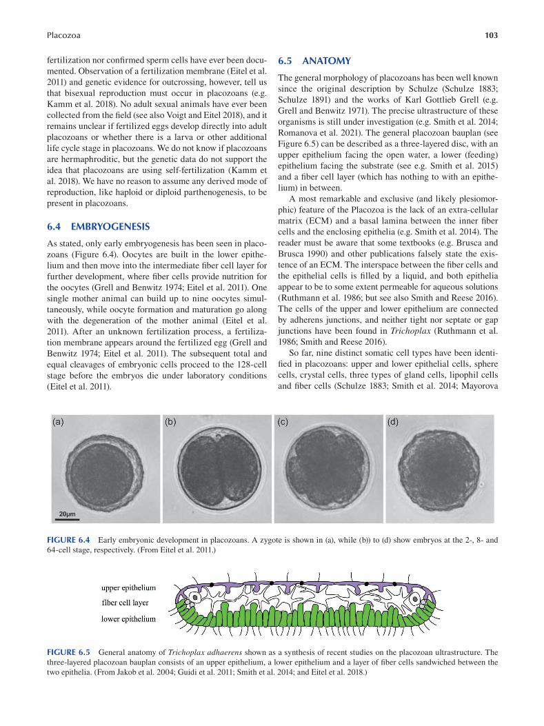
Placozoa 103
fertilization nor confirmed sperm cells have ever been docu
mented. Observation of a fertilization membrane (Eitel et al.
2011) and genetic evidence for outcrossing, however, tell us
that bisexual reproduction must occur in placozoans (e.g.
Kamm et al. 2018). No adult sexual animals have ever been
collected from the field (see also Voigt and Eitel 2018), and it
remains unclear if fertilized eggs develop directly into adult
placozoans or whether there is a larva or other additional
life cycle stage in placozoans. We do not know if placozoans
are hermaphroditic, but the genetic data do not support the
idea that placozoans are using self-fertilization (Kamm et
al. 2018). We have no reason to assume any derived mode of
reproduction, like haploid or diploid parthenogenesis, to be
present in placozoans.
6.4 EMBRYOGENESIS
As stated, only early embryogenesis has been seen in placo
zoans (Figure 6.4). Oocytes are built in the lower epithe
lium and then move into the intermediate fiber cell layer for
further development, where fiber cells provide nutrition for
the oocytes (Grell and Benwitz 1974; Eitel et al. 2011). One
single mother animal can build up to nine oocytes simul
taneously, while oocyte formation and maturation go along
with the degeneration of the mother animal ( Eitel et al.
2011). After an unknown fertilization process, a fertiliza
tion membrane appears around the fertilized egg (Grell and
Benwitz 1974; Eitel et al. 2011). The subsequent total and
equal cleavages of embryonic cells proceed to the 128-cell
stage before the embryos die under laboratory conditions
(Eitel et al. 2011).
6.5 ANATOMY
The general morphology of placozoans has been well known
since the original description by Schulze (Schulze 1883;
Schulze 1891) and the works of Karl Gottlieb Grell (e.g.
Grell and Benwitz 1971). The precise ultrastructure of these
organisms is still under investigation (e.g. Smith et al. 2014;
Romanova et al. 2021). The general placozoan bauplan (see
Figure 6.5) can be described as a three-layered disc, with an
upper epithelium facing the open water, a lower (feeding)
epithelium facing the substrate (see e.g. Smith et al. 2015)
and a fiber cell layer (which has nothing to with an epithe
lium) in between.
A most remarkable and exclusive (and likely plesiomor
phic) feature of the Placozoa is the lack of an extra-cellular
matrix (ECM) and a basal lamina between the inner fi ber
cells and the enclosing epithelia (e.g. Smith et al. 2014). The
reader must be aware that some textbooks (e.g. Brusca and
Brusca 1990) and other publications falsely state the exis
tence of an ECM. The interspace between the fiber cells and
the epithelial cells is filled by a liquid, and both epithelia
appear to be to some extent permeable for aqueous solutions
(Ruthmann et al. 1986; but see also Smith and Reese 2016).
The cells of the upper and lower epithelium are connected
by adherens junctions, and neither tight nor septate or gap
junctions have been found in Trichoplax (Ruthmann et al.
1986; Smith and Reese 2016 ).
So far, nine distinct somatic cell types have been identi
fied in placozoans: upper and lower epithelial cells, sphere
cells, crystal cells, three types of gland cells, lipophil cells
and fiber cells (Schulze 1883; Smith et al. 2014; Mayorova
FIGURE 6.4 Early embryonic development in placozoans. A zygote is shown in (a), while (b)) to (d) show embryos at the 2-, 8- and
64-cell stage, respectively. (From Eitel et al. 2011.)
FIGURE 6.5 General anatomy of Trichoplax adhaerens shown as a synthesis of recent studies on the placozoan ultrastructure. The
three-layered placozoan bauplan consists of an upper epithelium, a lower epithelium and a layer of fiber cells sandwiched between the
two epithelia. (From Jakob et al. 2004; Guidi et al. 2011; Smith et al. 2014; and Eitel et al. 2018.)
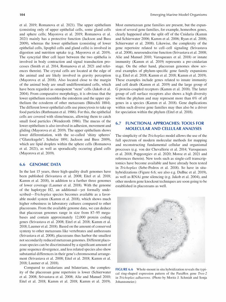
104
et al. 2019; Romanova et al. 2021). The upper epithelium
(consisting only of upper epithelial cells, some gland cells
and sphere cells; Mayorova et al. 2019; Romanova et al.
2021) mainly has a protective function (Jackson and Buss
2009), whereas the lower epithelium (consisting of lower
epithelial cells, lipophil cells and gland cells) is involved in
digestion and nutrition uptake (e.g. Mayorova et al. 2019).
The syncytial fiber cell layer between the two epithelia is
involved in body contraction and signal transduction pro
cesses (Smith et al. 2014, Romanova et al. 2021 and refer
ences therein). The crystal cells are located at the edge of
the animal and are likely involved in gravity perception
(Mayorova et al. 2018). Also located close to the margin
of the animal body are small undifferentiated cells, which
have been regarded as omnipotent “stem” cells (Jakob et al.
2004). From comparative morphology, it is obvious that the
lower epithelium resembles the entoderm and the upper epi
thelium the ectoderm of other metazoans (Bütschli 1884).
The different lower epithelial cells use pinocytosis to take up
food particles (Ruthmann et al. 1986). For this, the epithelial
cells are covered with slime/mucus, allowing them to catch
small food particles (Wenderoth 1986). The mucus of the
lower epithelium is also involved in adhesion, movement and
gliding (Mayorova et al. 2019). The upper epithelium shows
lower differentiation, with the so-called ‘shiny spheres’
(“Glanzkugeln”; Schulze 1891; Jackson and Buss 2009),
which are lipid droplets within the sphere cells (Romanova
et al. 2021), as well as sporadically occuring gland cells
(Mayorova et al. 2019).
6.6 GENOMIC DATA
In the last 15 years, three high-quality draft genomes have
been published (Srivastava et al. 2008; Eitel et al. 2018;
Kamm et al. 2018), in addition to a further three genomes
of lower coverage (Laumer et al. 2018). With the genome
of the haplotype H2, an additional—yet formally unde
scribed—Trichoplax species becomes available as a favor
able model system (Kamm et al. 2018), which shows much
higher robustness in laboratory cultures compared to other
placozoans. From the available genome data, we can deduce
that placozoan genomes range in size from 87–95 mega-
bases and contain approximately 12,000 protein coding
genes (Srivastava et al. 2008; Eitel et al. 2018; Kamm et al.
2018; Laumer et al. 2018). Based on the amount of conserved
synteny to other metazoans like vertebrates and anthozoans
(Srivastava et al. 2008), placozoans thus harbor the smallest
not secondarily reduced metazoan genomes. Different placo
zoan species can be discriminated by a significant amount of
gene sequence divergence, and less related species also show
substantial differences in their gene’s chromosomal arrange
ment (Srivastava et al. 2008; Eitel et al. 2018; Kamm et al.
2018; Laumer et al. 2018).
Compared to cnidarians and bilaterians, the complex
ity of the placozoan gene repertoire is lower (Schierwater
et al. 2008; Srivastava et al. 2008; Alie and Manuel 2010;
Eitel et al. 2018; Kamm et al. 2018; Kamm et al. 2019).
Emerging Marine Model Organisms
Most eumetazoan gene families are present, but the expan
sion of several gene families, for example, homeobox genes,
clearly happened after the split off of the Cnidaria (Kamm
and Schierwater 2006; Kamm et al. 2006; Ryan et al. 2006;
Schierwater et al. 2008). Likewise, the complexity of the
gene repertoire related to cell–cell signaling (Srivastava
et al. 2008), neuroendocrine function ( Srivastava et al. 2008;
Alie and Manuel 2010; Varoqueaux et al. 2018) or innate
immunity (Kamm et al. 2019) represents a pre-cnidarian
stage. On the other hand, placozoan genomes show sev
eral examples of phylum-specific gene family expansions
(e.g. Eitel et al. 2018; Kamm et al. 2018; Kamm et al. 2019).
These examples include genes related to innate immunity
and cell death (Kamm et al. 2019) and the large group of
G protein-coupled receptors (Kamm et al. 2018). The latter
group of cell surface receptors also shows a high diversity
within the phylum and may represent more than 6% of all
genes in a species (Kamm et al. 2018). Gene duplications
within such diverse gene families may thus also be a driver
for speciation within the phylum (Eitel et al. 2018).
6.7 FUNCTIONAL APPROACHES: TOOLS FOR MOLECULAR AND CELLULAR ANALYSES
The simplicity of the Trichoplax model allows the use of the
full spectrum of modern molecular methods for mapping
and reconstructing fundamental cellular and organismal
processes (e.g. von der Chevallerie et al. 2014; Varoqueaux
et al. 2018; Popgeorgiev et al. 2020; Moroz et al. 2021 and
references therein). New tools such as single-cell transcrip
tomics have become available and have already been tested
in Trichoplax (Sebe-Pedros et al. 2018). So have in situ-
hybridizations (Figure 6.6; see also e.g. DuBuc et al. 2019),
as well as RNAi gene silencing (e.g. Jakob et al. 2004), and
other modern gene knockout techniques are soon going to be
established in placozoans as well.
FIGURE 6.6 Whole-mount in situ hybridization reveals the typi
cal ring-shaped expression pattern of the ParaHox gene Trox-2 in Trichoplax adhaerens. (Photo by Moritz J. Schmidt and Sonja
Johannsmeier.)
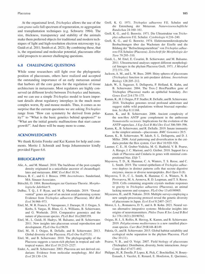
105 Placozoa
At the organismal level, Trichoplax allows the use of the
cum grano salis full spectrum of regeneration, re-aggregation
and transplantation techniques (e.g. Schwartz 1984). The
size, thickness, transparency and stability of the animals
make them preferred objects for traditional and modern tech
niques of light and high-resolution electron microscopy (e.g.
Guidi et al. 2011; Smith et al. 2021). By combining these, that
is, the organismal and molecular potential, placozoans offer
solid prospects to answer challenging questions.
6.8 CHALLENGING QUESTIONS
While some researchers still fight over the phylogenetic
position of placozoans, others have realized and accepted
the outstanding importance of an early metazoan animal
that harbors all the core genes for the regulation of tissue
architecture in metazoans. Most regulators are highly con
served (at different levels) between Trichoplax and humans,
and we can use a simple Trichoplax model to learn impor
tant details about regulatory interplays in the much more
complex worm, fly and mouse models. Thus, it comes as no
surprise that the current questions we are asking Trichoplax range from “How can symmetry be derived from polar
ity?” to “What is the basic genetics behind apoptosis?” to
“What are the initial genetic malfunctions that start cancer
growth?”. And there will be many more to come.
ACKNOWLEDGMENTS
We thank Kristin Fenske and Kai Kamm for help and com
ments. Moritz J. Schmidt and Sonja Johannsmeier kindly
provided Figure 6.6 .
BIBLIOGRAPHY
Alie, A., and M. Manuel. 2010. The backbone of the post-synaptic
density originated in a unicellular ancestor of choanofl agel
lates and metazoans. BMC Evol Biol 10:34.
Brusca, R. C., and G. J. Brusca. 1990. Invertebrates . Sunderland,
MA: Sinauer Associates.
Bütschli, O. 1884. Bemerkungen zur Gastraea-Theorie. Morphologische Jahrblatt 9.
DuBuc, T. Q., J. F. Ryan, and M. Q. Martindale. 2019. “Dorsal
ventral” genes are part of an ancient axial patterning system:
Evidence from Trichoplax adhaerens (Placozoa). Mol Biol Evol 36:966–973.
Eitel, M., W. R. Francis, F. Varoqueaux, J. Daraspe, H. J. Osigus, S.
Krebs, S. Vargas, H. Blum, G. A. Williams, B. Schierwater,
and G. Worheide. 2018. Comparative genomics and the
nature of placozoan species. PLoS Biol 16:e2005359.
Eitel, M., L. Guidi, H. Hadrys, M. Balsamo, and B. Schierwater.
2011. New insights into placozoan sexual reproduction and
development. PLoS One 6:e19639.
Eitel, M., H. J. Osigus, R. DeSalle, and B. Schierwater. 2013.
Global diversity of the Placozoa. PLoS One 8:e57131.
Eitel, M., and B. Schierwater. 2010. The phylogeography of the
Placozoa suggests a taxon-rich phylum in tropical and sub
tropical waters. Mol Ecol 19:2315–2327.
Ender, A., and B. Schierwater. 2003. Placozoa are not derived cni
darians: Evidence from molecular morphology. Mol Biol Evol 20:130–134.
Grell, K. G. 1971. Trichoplax adhaerens F.E. Schulze und
die Entstehung der Metazoan. Naturwissenschaftliche Rundschau 24:160–161.
Grell, K. G., and G. Benwitz. 1971. Die Ultrastruktur von Trichoplax adhaerens F.E. Schulze. Cytobiologie 4:216–240.
Grell, K. G., and G. Benwitz. 1974. Elektronenmikroskopische
Beobachtungen über das Wachstum der Eizelle und die
Bildung der “Befruchtungsmembran” von Trichoplax adhaerens F.E.Schulze (Placozoa). Zeitschrift für Morphologie der Tiere 79:295–310.
Guidi, L., M. Eitel, E. Cesarini, B. Schierwater, and M. Balsamo.
2011. Ultrastructural analyses support different morphologi
cal lineages in the phylum Placozoa Grell, 1971. J Morphol 272:371–378.
Jackson, A. M., and L. W. Buss. 2009. Shiny spheres of placozoans
(Trichoplax) function in anti-predator defense. Invertebrate Biology 128:205–212.
Jakob, W., S. Sagasser, S. Dellaporta, P. Holland, K. Kuhn, and
B. Schierwater. 2004. The Trox-2 Hox/ParaHox gene of
Trichoplax (Placozoa) marks an epithelial boundary. Dev Genes Evol 214:170–175.
Kamm, K., H. J. Osigus, P. F. Stadler, R. DeSalle, and B. Schierwater.
2018. Trichoplax genomes reveal profound admixture and
suggest stable wild populations without bisexual reproduc
tion. Sci Rep 8:11168.
Kamm, K., and B. Schierwater. 2006. Ancient complexity of
the non-Hox ANTP gene complement in the anthozoan
Nematostella vectensis: Implications for the evolution of the
ANTP superclass. J Exp Zool B Mol Dev Evol 306:589–596.
Kamm, K., B. Schierwater, and R. DeSalle. 2019. Innate immunity
in the simplest animals—placozoans. BMC Genomics 20:5.
Kamm, K., B. Schierwater, W. Jakob, S. L. Dellaporta, and D. J.
Miller. 2006. Axial patterning and diversification in the cni
daria predate the Hox system. Curr Biol 16:920–926.
Laumer, C. E., H. Gruber-Vodicka, M. G. Hadfield, V. B. Pearse,
A. Riesgo, J. C. Marioni, and G. Giribet. 2018. Support for a
clade of Placozoa and Cnidaria in genes with minimal com
positional bias. Elife 7.
Mayorova, T. D., K. Hammar, C. A. Winters, T. S. Reese, and C.
L. Smith. 2019. The ventral epithelium of Trichoplax adhaerens deploys in distinct patterns cells that secrete digestive
enzymes, mucus or diverse neuropeptides. Biol Open 8 (8).
Mayorova, T. D., C. L. Smith, K. Hammar, C. A. Winters, N. B.
Pivovarova, M. A. Aronova, R. D. Leapman, and T. S. Reese.
2018. Cells containing aragonite crystals mediate responses
to gravity in Trichoplax adhaerens (Placozoa), an animal
lacking neurons and synapses. PLoS One 13:e0190905.
Miyazawa, H., and H. Nakano. 2018. Multiple surveys employing a
new sample-processing protocol reveal the genetic diversity
of placozoans in Japan. Ecol Evol 8:2407–2417.
Moroz, L. L., Romanova, D. Y., and A. B. Kohn. 2021. Neural ver
sus alternative integrative systems: Molecular insights into
origins of neurotransmitters. Philos Trans R Soc Lond B Biol Sci 376 (1821):20190762.
Osigus, H. J., S. Rolfes, R. Herzog, K. Kamm, and B. Schierwater.
2019. Polyplacotoma mediterranea is a new ramifi ed placo
zoan species. Curr Biol 29:R148–R149.
Paknia, O., and B. Schierwater. 2015. Global habitat suitability and
ecological niche separation in the phylum Placozoa. PLoS One 10 (11).
Pearse, V. B., and O. Voigt. 2007. Field biology of placozoans
(Tricho plax): Distribution, diversity, biotic interactions. Integr Comp Biol 47:677–692.
Philippe, H., R. Derelle, P. Lopez, K. Pick, C. Borchiellini, N. Boury-
Esnault, J. Vacelet, E. Renard, E. Houliston, E. Queinnec,
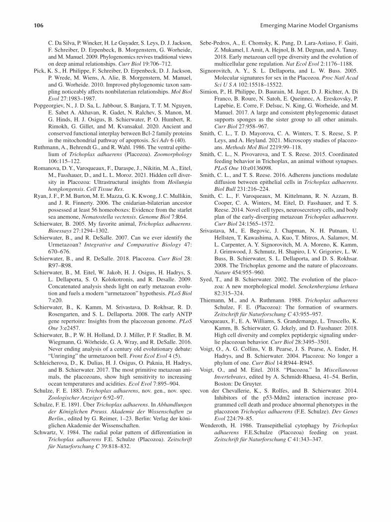
106
C. Da Silva, P. Wincker, H. Le Guyader, S. Leys, D. J. Jackson,
F. Schreiber, D. Erpenbeck, B. Morgenstern, G. Worheide,
and M. Manuel. 2009. Phylogenomics revives traditional views
on deep animal relationships. Curr Biol 19:706–712.
Pick, K. S., H. Philippe, F. Schreiber, D. Erpenbeck, D. J. Jackson,
P. Wrede, M. Wiens, A. Alie, B. Morgenstern, M. Manuel,
and G. Worheide. 2010. Improved phylogenomic taxon sam
pling noticeably affects nonbilaterian relationships. Mol Biol Evol 27:1983–1987.
Popgeorgiev, N., J. D. Sa, L. Jabbour, S. Banjara, T. T. M. Nguyen,
E. Sabet A. Akhavan, R. Gadet, N. Ralchev, S. Manon, M.
G. Hinds, H. J. Osigus, B. Schierwater, P. O. Humbert, R.
Rimokh, G. Gillet, and M. Kvansakul. 2020. Ancient and
conserved functional interplay between Bcl-2 family proteins
in the mitochondrial pathway of apoptosis. Sci Adv 6 (40).
Ruthmann, A., Behrendt G., and R. Wahl. 1986. The ventral epithe
lium of Trichoplax adhaerens (Placozoa). Zoomorphology 106:115–122.
Romanova, D. Y., Varoqueaux, F., Daraspe, J., Nikitin, M. A., Eitel,
M., Fasshauer, D., and L. L. Moroz. 2021. Hidden cell diver
sity in Placozoa: Ultrastructural insights from Hoilungia hongkongensis. Cell Tissue Res.
Ryan, J. F., P. M. Burton, M. E. Mazza, G. K. Kwong, J. C. Mullikin,
and J. R. Finnerty. 2006. The cnidarian-bilaterian ancestor
possessed at least 56 homeoboxes: Evidence from the starlet
sea anemone, Nematostella vectensis. Genome Biol 7:R64.
Schierwater, B. 2005. My favorite animal, Trichoplax adhaerens.
Bioessays 27:1294–1302.
Schierwater, B., and R. DeSalle. 2007. Can we ever identify the
Urmetazoan? Integrative and Comparative Biology 47:
670–676.
Schierwater, B., and R. DeSalle. 2018. Placozoa. Curr Biol 28:
R97–R98.
Schierwater, B., M. Eitel, W. Jakob, H. J. Osigus, H. Hadrys, S.
L. Dellaporta, S. O. Kolokotronis, and R. Desalle. 2009.
Concatenated analysis sheds light on early metazoan evolu
tion and fuels a modern “urmetazoon” hypothesis. PLoS Biol 7:e20.
Schierwater, B., K. Kamm, M. Srivastava, D. Rokhsar, R. D.
Rosengarten, and S. L. Dellaporta. 2008. The early ANTP
gene repertoire: Insights from the placozoan genome. PLoS One 3:e2457.
Schierwater, B., P. W. H. Holland, D. J. Miller, P. F. Stadler, B. M.
Wiegmann, G. Wörheide, G. A. Wray, and R. DeSalle. 2016.
Never ending analysis of a century old evolutionary debate:
“Unringing” the urmetazoon bell. Front Ecol Evol 4 (5).
Schleicherova, D., K. Dulias, H. J. Osigus, O. Paknia, H. Hadrys,
and B. Schierwater. 2017. The most primitive metazoan ani
mals, the placozoans, show high sensitivity to increasing
ocean temperatures and acidities. Ecol Evol 7:895–904.
Schulze, F. E. 1883. Trichoplax adhaerens, nov. gen., nov. spec.
Zoologischer Anzeiger 6:92–97.
Schulze, F. E. 1891. Über Trichoplax adhaerens. In Abhandlungen der Königlichen Preuss. Akademie der Wissenschaften zu Berlin., edited by G. Reimer, 1–23. Berlin: Verlag der köni
glichen Akademie der Wissenschaften.
Schwartz, V. 1984. The radial polar pattern of differentiation in
Trichoplax adhaerens F.E. Schulze (Placozoa). Zeitschrift für Naturforschung C 39:818–832.
Emerging Marine Model Organisms
Sebe-Pedros, A., E. Chomsky, K. Pang, D. Lara-Astiaso, F. Gaiti,
Z. Mukamel, I. Amit, A. Hejnol, B. M. Degnan, and A. Tanay.
2018. Early metazoan cell type diversity and the evolution of
multicellular gene regulation. Nat Ecol Evol 2:1176–1188.
Signorovitch, A. Y., S. L. Dellaporta, and L. W. Buss. 2005.
Molecular signatures for sex in the Placozoa. Proc Natl Acad Sci U S A 102:15518–15522.
Simion, P., H. Philippe, D. Baurain, M. Jager, D. J. Richter, A. Di
Franco, B. Roure, N. Satoh, E. Queinnec, A. Ereskovsky, P.
Lapebie, E. Corre, F. Delsuc, N. King, G. Worheide, and M.
Manuel. 2017. A large and consistent phylogenomic dataset
supports sponges as the sister group to all other animals.
Curr Biol 27:958–967.
Smith, C. L., T. D. Mayorova, C. A. Winters, T. S. Reese, S. P.
Leys, and A. Heyland. 2021. Microscopy studies of placozo
ans. Methods Mol Biol 2219:99–118.
Smith, C. L., N. Pivovarova, and T. S. Reese. 2015. Coordinated
feeding behavior in Trichoplax, an animal without synapses.
PLoS One 10:e0136098.
Smith, C. L., and T. S. Reese. 2016. Adherens junctions modulate
diffusion between epithelial cells in Trichoplax adhaerens.
Biol Bull 231:216–224.
Smith, C. L., F. Varoqueaux, M. Kittelmann, R. N. Azzam, B.
Cooper, C. A. Winters, M. Eitel, D. Fasshauer, and T. S.
Reese. 2014. Novel cell types, neurosecretory cells, and body
plan of the early-diverging metazoan Trichoplax adhaerens.
Curr Biol 24:1565–1572.
Srivastava, M., E. Begovic, J. Chapman, N. H. Putnam, U.
Hellsten, T. Kawashima, A. Kuo, T. Mitros, A. Salamov, M.
L. Carpenter, A. Y. Signorovitch, M. A. Moreno, K. Kamm,
J. Grimwood, J. Schmutz, H. Shapiro, I. V. Grigoriev, L. W.
Buss, B. Schierwater, S. L. Dellaporta, and D. S. Rokhsar.
2008. The Trichoplax genome and the nature of placozoans.
Nature 454:955–960.
Syed, T., and B. Schierwater. 2002. The evolution of the placo
zoa: A new morphological model. Senckenbergiana lethaea 82:315–324.
Thiemann, M., and A. Ruthmann. 1988. Trichoplax adhaerens Schulze, F. E. (Placozoa): The formation of swarmers.
Zeitschrift für Naturforschung C 43:955–957.
Varoqueaux, F., E. A. Williams, S. Grandemange, L. Truscello, K.
Kamm, B. Schierwater, G. Jekely, and D. Fasshauer. 2018.
High cell diversity and complex peptidergic signaling under
lie placozoan behavior. Curr Biol 28:3495–3501.
Voigt, O., A. G. Collins, V. B. Pearse, J. S. Pearse, A. Ender, H.
Hadrys, and B. Schierwater. 2004. Placozoa: No longer a
phylum of one. Curr Biol 14:R944–R945.
Voigt, O., and M. Eitel. 2018. “Placozoa.” In Miscellaneous Invertebrates, edited by A. Schmidt-Rhaesa, 41–54. Berlin,
Boston: De Gruyter.
von der Chevallerie, K., S. Rolfes, and B. Schierwater. 2014.
Inhibitors of the p53-Mdm2 interaction increase pro
grammed cell death and produce abnormal phenotypes in the
placozoon Trichoplax adhaerens (F.E. Schulze). Dev Genes Evol 224:79–85.
Wenderoth, H. 1986. Transepithelial cytophagy by Trichoplax adhaerens F.E.Schulze (Placozoa) feeding on yeast.
Zeitschrift für Naturforschung C 41:343–347.
7 Nematostella vectensis asa Model System
Layla Al-Shaer, Jamie Havrilak and Michael J. Layden
CONTENTS
7.1 History of the Model....................................................................................................................................................107
7.2 Geography and Habitat ............................................................................................................................................... 109
7.3 Anatomy...................................................................................................................................................................... 110
7.4 Life History ................................................................................................................................................................. 112
7.5 Embryogenesis............................................................................................................................................................ 113
7.5.1 Process of Development ..................................................................................................................................113
7.5.2 Axial Patterning Programs ...............................................................................................................................115
7.5.3 Regeneration ....................................................................................................................................................115
7.6 Genomic Data ............................................................................................................................................................. 115
7.7 Methods and Functional Approaches ...........................................................................................................................116
7.7.1 Culture and Care ..............................................................................................................................................116
7.7.2 Behavioral and Ecological Approaches ...........................................................................................................116
7.7.3 Tissue Manipulation and Tracking ...................................................................................................................117
7.7.3.1 Detection of Cellular Processes ........................................................................................................118
7.7.3.2 Regeneration .....................................................................................................................................118
7.7.4 Genetic Approaches .........................................................................................................................................118
7.7.4.1 Microinjection and Electroporation..................................................................................................118
7.7.4.2 Gene Disruption ................................................................................................................................118
7.7.4.3 Transgenics .......................................................................................................................................119
7.7.4.4 Visualizing Gene Expression ............................................................................................................119
7.7.4.5 Genome- and “Omics”-Level Approaches....................................................................................... 120
7.7.4.6 Pharmacological Manipulation ........................................................................................................ 120
7.7.5 Integration of Approaches ............................................................................................................................... 120
7.8 Challenging Questions ................................................................................................................................................ 120
7.8.1 Is There a Deep Evolutionary Origin for Key Bilaterian Traits? .................................................................... 120
7.8.1.1 Origin of the Mesoderm................................................................................................................... 120
7.8.1.2 Mechanisms of Axial Patterning Leading to Bilaterality..................................................................121
7.8.2 Can Nematostella Be Used as a Cnidarian Model for Cnidarians? ................................................................ 122
7.8.3 How Do Novel Cell Types Evolve? ................................................................................................................ 122
7.8.4 Does Regeneration Recapitulate Development? ............................................................................................. 123
7.8.5 Other Challenging Questions.......................................................................................................................... 123
Acknowledgments ................................................................................................................................................................ 123
Bibliography ........................................................................................................................................................................ 123
7.1 HISTORY OF THE MODEL or the “button” (oncus), when the tentacles are retracted and
Nematostella vectensis (the starlet sea anemone) are antho- The first description of Nematostella vectensis was
zoan cnidarians. Anthozoans (e.g. corals, anemones) derive published in 1935 by Thomas A. Stephenson. Stephenson
their name from the Greek anthos —fl ower—and zōia—
the oral end closes in around them (Gosse 1860).
attributed the discovery of Nematostella vectensis and obseranimals—because their dominant polyp form shared by
vations of their nematosomes to Ms. Gertrude F. Selwood. this class represents “a highly colored and many-petaled
She found them at the Isle of Wight (England) (Figures 7.1b, flower” (Figure 7.1a) (Gosse 1860). Additionally, the differ-
7.2) in 1929 when she was a lecturer at Municipal College, ent morphological states of the animal can be described as
Portsmith, and sent specimens to Stephenson. Stephenson the “fl ower” (anthus), when all the tentacles are extended,
described the free-swimming nematosomes in the gastric
DOI: 10.1201/9781003217503-7 107

108 Emerging Marine Model Organisms
FIGURE 7.1 Characteristics and geographical range of Nematostella vectensis. (a) An adult polyp, image courtesy of Eric Röttinger.
(b) Known geographical range. (c) First known illustrations of a Nematostella polyp and nematosomes with intact (left) and fi red (right)
cnidocytes. (d) Polyp showing tentacles (T) pharynx (P), mesenteries (M) and nematosomes (N). ([b] Illustrations by Sears Crowell 1946;
[d] image modified from Babonis et al. 2016.)
cavity that became the characteristic feature of Nematostella ( Figure 7.1c , d ) ( Stephenson 1935 ).
In 1939, William J. Bowden discovered Nematostella pellucida at Woods Hole, Massachusetts (Figure 7.1b), which
was later described and published by Sears Crowell in 1946
( Figure 7.1c ). Nematostella pellucida was initially consid
ered distinct from vectensis due to color patterns on the
body and the large geographical separation (Williams 1975;
Williams 1976). Crowell suspected N. vectensis and N. pellucida were synonymous species, but because of the war,
he was unable to get hold of Nematostella from the British
Isles for direct comparisons (Crowell 1946 ). In 1957, Cadet
Hand compared anemones from America and England and
determined they were both Nematostella vectensis (Figure
7.2) (Hand 1957; Williams 1975; Williams 1976).
The various life history stages of Nematostella were
described by several different groups from the 1940s to
the 1980s (summarized in Hand and Uhlinger 1992). The
potential for Nematostella as a laboratory model came when
Cadet Hand and Kevin Uhlinger documented the ease of its
culturability in the early 1990s (1992). Its ability to tolerate
wide variations in salinity and temperature made it easy to
maintain in laboratory cultures (Williams 1975; Williams
1976). Perhaps most importantly, Nematostella spawn read
ily with increased temperature and light. Early work estab
lished various environmental conditions that infl uence
oogenesis, such as nutrient amount, temperature, light, den
sity of sperm and the ideal timeframe for fertilization (Hand
and Uhlinger 1992; Fritzenwanker and Technau 2002). The
ability to reliably obtain thousands of embryos per spawn
and close the life cycle in culture made Nematostella stand
out as a potential cnidarian model system. Plus, due to its
phylogenetic position as a basal metazoan, it is also espe
cially well suited for evolutionary and developmental biol
ogy (evo-devo) studies.
Through the 1990s and early 2000s, studies focused on
identifying the expression of known bilaterian homologues
during Nematostella development ( Figure 7.2 ). Initial stud
ies focused on genes involved in axial patterning and trip
loblasty and provided initial insights into the origin and
evolution of these genes and thus the bilaterian traits they
regulate (see Darling et al. 2005 ). Similarly, extensive efforts
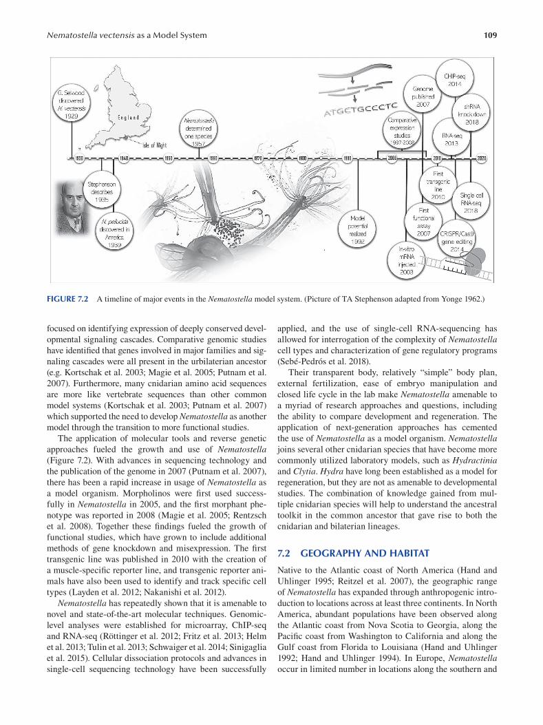
109 Nematostella vectensis as a Model System
FIGURE 7.2 A timeline of major events in the Nematostella model system. (Picture of TA Stephenson adapted from Yonge 1962.)
focused on identifying expression of deeply conserved devel
opmental signaling cascades. Comparative genomic studies
have identified that genes involved in major families and sig
naling cascades were all present in the urbilaterian ancestor
(e.g. Kortschak et al. 2003; Magie et al. 2005; Putnam et al.
2007). Furthermore, many cnidarian amino acid sequences
are more like vertebrate sequences than other common
model systems (Kortschak et al. 2003; Putnam et al. 2007)
which supported the need to develop Nematostella as another
model through the transition to more functional studies.
The application of molecular tools and reverse genetic
approaches fueled the growth and use of Nematostella (Figure 7.2). With advances in sequencing technology and
the publication of the genome in 2007 (Putnam et al. 2007),
there has been a rapid increase in usage of Nematostella as
a model organism. Morpholinos were first used success
fully in Nematostella in 2005, and the first morphant phe
notype was reported in 2008 (Magie et al. 2005; Rentzsch
et al. 2008). Together these findings fueled the growth of
functional studies, which have grown to include additional
methods of gene knockdown and misexpression. The fi rst
transgenic line was published in 2010 with the creation of
a muscle-specific reporter line, and transgenic reporter ani
mals have also been used to identify and track specifi c cell
types (Layden et al. 2012; Nakanishi et al. 2012).
Nematostella has repeatedly shown that it is amenable to
novel and state-of-the-art molecular techniques. Genomic-
level analyses were established for microarray, ChIP-seq
and RNA-seq (Röttinger et al. 2012; Fritz et al. 2013; Helm
et al. 2013; Tulin et al. 2013; Schwaiger et al. 2014; Sinigaglia
et al. 2015). Cellular dissociation protocols and advances in
single-cell sequencing technology have been successfully
applied, and the use of single-cell RNA-sequencing has
allowed for interrogation of the complexity of Nematostella cell types and characterization of gene regulatory programs
(Sebé-Pedrós et al. 2018).
Their transparent body, relatively “simple” body plan,
external fertilization, ease of embryo manipulation and
closed life cycle in the lab make Nematostella amenable to
a myriad of research approaches and questions, including
the ability to compare development and regeneration. The
application of next-generation approaches has cemented
the use of Nematostella as a model organism. Nematostella joins several other cnidarian species that have become more
commonly utilized laboratory models, such as Hydractinia and Clytia. Hydra have long been established as a model for
regeneration, but they are not as amenable to developmental
studies. The combination of knowledge gained from mul
tiple cnidarian species will help to understand the ancestral
toolkit in the common ancestor that gave rise to both the
cnidarian and bilaterian lineages.
7.2 GEOGRAPHY AND HABITAT
Native to the Atlantic coast of North America (Hand and
Uhlinger 1995; Reitzel et al. 2007), the geographic range
of Nematostella has expanded through anthropogenic intro
duction to locations across at least three continents. In North
America, abundant populations have been observed along
the Atlantic coast from Nova Scotia to Georgia, along the
Pacific coast from Washington to California and along the
Gulf coast from Florida to Louisiana (Hand and Uhlinger
1992; Hand and Uhlinger 1994). In Europe, Nematostella occur in limited number in locations along the southern and
110
eastern coasts of England (Stephenson 1935; Sheader et al.
1997; Pearson et al. 2002), and in South America, where they
have been found in locales off the coast of Brazil (Figure
7.1b) (Silva et al. 2010; Brandão et al. 2019). Genetic and
phylogeographical analyses indicate that global populations
are isolated and therefore unlikely to have spread via natural
dispersal mechanisms. (Hand and Uhlinger 1995; Reitzel et
al. 2007). More plausible is that they were carried as hitch
hikers in the ballasts of commercial seafood vessels, creat
ing the potential for new populations to become established
outside of the natural range (Sheader et al. 1997; Takahashi
et al. 2008).
The successful geographic expansion of Nematostella can likely be attributed to their environmental plasticity,
as they can inhabit a variety of coastal habitats and can
tolerate fluctuating environmental conditions. They often
occur burrowed in soft muddy sediments of poikilohaline
lagoons, brackish mudflats, salt marshes and creeks and
subtidal areas of certain estuaries and bays (Williams 1975;
Williams 1976). As eurythermal animals, they can survive
and adapt to a wide range of temperatures and have even
been found living in habitats that approach their physi
ological upper limit of approximately 40°C (Williams 1975;
Williams 1976). As euryhaline animals, they can contend
with the spatiotemporal fluctuations in salinity common in
the estuarine habitats where they are found. A testament to
the remarkably flexible physiology of this anemone is that
both asexual and sexual reproduction can occur under a
wide range of salinities (Hand and Uhlinger 1992).
7.3 ANATOMY
Adult Nematostella are transparent and possess the classic
polyp morphology found throughout the cnidarian lineage
(Figure 7.3a). Atop the oral end of the body column is an
opening that is surrounded by 4–18 long stinging tentacles,
which aid in prey capture and defense but also expand the
surface area of the gastric cavity (Fritz et al. 2013; Ikmi
et al. 2020). This oral opening serves as both a mouth and
anus by attaching to a blind-ended gut through a noticeable
pharynx (Williams 1975; Williams 1976). There is also a
small pore at the aboral pole (Amiel et al. 2015). The oral–
aboral axis is elongated, which gives the body column a
tube-like structure. Eight radially repeating body segments,
which are centered around the long oral–aboral axis, give
the animal what appears to be an octoradial symmetry
( Figure 7.3a ).
Cnidarians are generally classified as having a radially
symmetric body plan, but many species have subtle bilat
eral differences in their anatomy that are superimposed over
a general radial body plan (Martindale et al. 2002). These
bilateral differences point to the presence of a secondary
directive axis, which runs perpendicular to the primary oral–
aboral axis (Figure 7.3b) (Berking 2007). In Nematostella,
the presence of a directive axis is morphologically evident in
adult polyps from the slit-like shape of the oral opening and
pharynx, the presence of a ciliated groove (siphonoglyph)
Emerging Marine Model Organisms
on one side of the pharynx, the asymmetric arrangement of
the retractor muscles within the mesenteries and the asym
metric arrangement of the tentacles around the oral opening
(Martindale et al. 2002; Berking 2007; He et al. 2018).
Nematostella are diploblastic, meaning that the entire
body is composed of cells derived from two germ layers:
an outer ectoderm which forms the epidermis and a bifunc
tional internal endoderm which forms the gastrodermis
(Figure 7.3a) (Finnerty et al. 2004; Wijesena et al. 2017; but
see Steinmetz et al. 2017). The epidermis covers the out
side of the animal and serves as a protective barrier between
the animal and its environment, while the bifunctional gas
trodermis lines the coelenteron and provides both absorp
tive and contractile functions (Martindale et al. 2004).
Separating the ectoderm and endoderm is the mesoglea,
a thin extracellular matrix with no organized tissue and
only a few migratory amoebocyte cells of unknown func
tion (Tucker et al. 2011). The ectoderm contains primarily
columnar cell epithelia (Magie et al. 2007), along with other
differentiated cells, including stinging cells called cnido
cytes (Frank and Bleakney 1976), sensory neurons, ganglion
neurons (Marlow et al. 2009; Sinigaglia et al. 2015; Leclère
et al. 2016), a population of myoepithelial (muscle) cells in
the tentacles (Jahnel et al. 2014) and gland cells (Frank and
Bleakney 1976 ). Ectodermal gland cells include those with
exocrine and insulinergic functions (Steinmetz et al. 2017),
and some produce a potent neurotoxin for both prey cap
ture and defense (Moran et al. 2011). The endoderm pos
sesses squamous epithelial cells (Magie et al. 2007), sensory
and ganglion neurons (Marlow et al. 2009; Sinigaglia et al.
2015; Leclère et al. 2016 ), the majority of myoepithelial
cells (Jahnel et al. 2014), gland cells (Frank and Bleakney
1976; Steinmetz et al. 2017) and gametic and absorptive
cells (Layden et al. 2012; Nakanishi et al. 2012). This basic
organization results in epithelial cells and differentiated cell
types being scattered and intermixed with one another, as
opposed to being organized into discrete organ systems.
Apart from the pharynx, the most obvious internal struc
tures of adult Nematostella are the ecto- and endodermally
derived lamellae known as mesenteries (Steinmetz et al.
2017). Adults have eight mesenteries, one in each body seg
ment, that look ruffled in appearance and run the length of
the body column (Figure 7.3a). Each mesentery arises from
the pharynx and consists of two layers of gastrodermis epi
thelium separated by a layer of mesoglea (Martindale et al.
2004). Structurally, the mesenteries are important because
they provide support for the pharynx, they contain mus
cles that allow for quick contractions of the body column
(Renfer et al. 2010) and they increase the surface area of the
gastrodermis. Physiologically, the mesenteries are incred
ibly multifunctional, as they contain absorptive cells that
aid in digestion and nutrient uptake and are where gam
etes (Martindale et al. 2004), cnidocytes (Steinmetz 2019)
and nematosomes are produced (Williams 1975, 1976).
Nematosomes are the defining apomorphy of Nematostella (Williams 1975, 1976, 1979). They are multicellular, spheri
cal, flagellated bodies that contain cnidocytes and can be
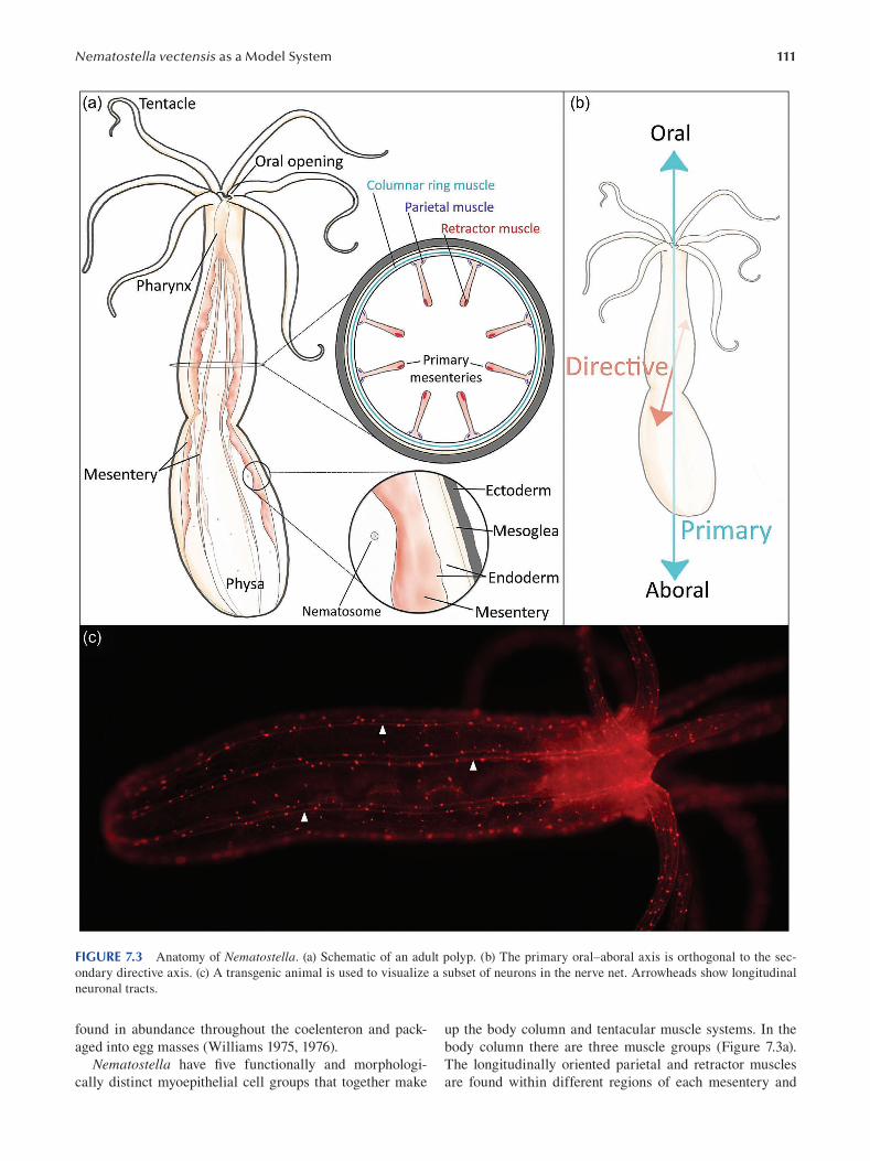
111 Nematostella vectensis as a Model System
FIGURE 7.3 Anatomy of Nematostella. (a) Schematic of an adult polyp. (b) The primary oral–aboral axis is orthogonal to the sec
ondary directive axis. (c) A transgenic animal is used to visualize a subset of neurons in the nerve net. Arrowheads show longitudinal
neuronal tracts.
found in abundance throughout the coelenteron and pack- up the body column and tentacular muscle systems. In the
aged into egg masses (Williams 1975, 1976). body column there are three muscle groups (Figure 7.3a).
Nematostella have five functionally and morphologi- The longitudinally oriented parietal and retractor muscles
cally distinct myoepithelial cell groups that together make are found within different regions of each mesentery and
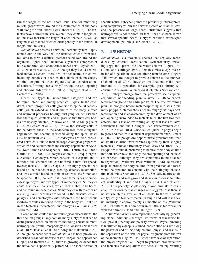
112
run the length of the oral–aboral axis. The columnar ring
muscle group wraps around the circumference of the body
wall along the oral–aboral axis (Jahnel et al. 2014). The ten
tacles have a similar muscle system; they contain longitudi
nal muscles that run the length of each tentacle, as well as
ring muscles that are oriented orthogonally to the tentacular
longitudinal muscles.
Nematostella possess a nerve-net nervous system—aptly
named due to the way that the neurites extend from neu
ral soma to form a diffuse interconnected web around the
organism ( Figure 7.3c). The nervous system is composed of
both ectodermal and endodermal nerve nets (Layden et al.
2012; Nakanishi et al. 2012). Although they lack a central
ized nervous system, there are distinct neural structures,
including bundles of neurons that flank each mesentery
within a longitudinal tract (Figure 7.3c) and condensations
of neurons forming “nerve rings” around the oral opening
and pharynx (Marlow et al. 2009; Sinigaglia et al. 2015;
Leclère et al. 2016 ).
Neural cell types fall under three categories and can
be found intermixed among other cell types. In the ecto
derm, neural progenitor cells give rise to epithelial sensory
cells (which extend an apical cilium to the body surface
and neuronal processes basally) and ganglion cells (which
lose their apical contacts and migrate so that their cell bod
ies are basally situated) (Marlow et al. 2009; Sinigaglia et
al. 2015; Leclère et al. 2016 ). Unlike the sensory cells in
the ectoderm, those in the endoderm lose their elongated
appearance and become shortened along the apical–basal
axis (Nakanishi et al. 2012). Cnidocytes are also consid
ered nerve cells due to their neurophysiological properties,
structure and calcium/mechanosensory-dependent exocyto
sis (Kass-Simon and Scappaticci 2002; Thurm et al. 2004;
Galliot et al. 2009). Cnidocytes contain a unique organ
elle called a cnidocyst, which consists of a capsule and a
harpoon-like structure that can be fired at ultra-fast speeds
(Szczepanek et al. 2002). Capsules are highly specialized
based on their function (e.g. feeding, defense, locomotion)
and are classifi ed based on their structure (Kass-Simon and
Scappaticci 2002). Nematostella have three types of cnido
cytes: spirocytes and two types of nematocytes. Spirocytes
contain spirocyst capsules, which lack a shaft and barbs,
and are found in the tentacles. Nematocytes with microbasic
p-mastigophore capsules are found in the mesenteries and
pharynx, and nematocytes with different-sized basitrichous
isorhiza capsules are found mostly in the body wall, but also
in the tentacles, mesenteries and pharynx (Williams 1975;
Williams 1976 ).
Based on molecular and morphological observations, the
three neural groups likely contain many subtypes that can be
distinguished based on attributes including neurite number,
neuropeptide profile, morphology and location (Nakanishi
et al. 2012; Havrilak et al. 2017; Zang and Nakanishi 2020).
Although the nerve net of Nematostella has been previously
described as random because of its disorganized appearance
(Hejnol and Rentzsch 2015), there is growing evidence that
the nerve net is specifically patterned. The identifi cation of
Emerging Marine Model Organisms
specific neural subtypes points to a previously underappreci
ated complexity within the nervous system of Nematostella,
and the presence of large neural structures suggests that
neurogenesis is not random. In fact, it has also been shown
that several specific neural subtypes exhibit a stereotyped
developmental pattern (Havrilak et al. 2017).
7.4 LIFE HISTORY
Nematostella is a dioecious species that sexually repro
duces by external fertilization, synchronously releas
ing eggs and sperm into the water column (Figure 7.4a)
(Hand and Uhlinger 1992). Females release egg masses
inside of a gelatinous sac containing nematosomes (Figure
7.4b), which are thought to provide defense to the embryos
(Babonis et al. 2016). However, this does not make them
immune to all predation; for example, grass shrimp will
consume Nematostella embryos (Columbus-Shenkar et al.
2018). Embryos emerge from the protective sac as spheri
cal, ciliated, non-feeding, planula larvae ~36–48 hours post
fertilization (Hand and Uhlinger 1992). The free-swimming
planulae elongate before metamorphosing into sessile pri
mary polyps. Metamorphosis occurs roughly six days post
fertilization and is characterized by the development of an
oral opening surrounded by tentacle buds, the first two mes
enteries and a loss of swimming ability that leads to larval
settlement (Hand and Uhlinger 1992; Fritzenwanker et al.
2007; Fritz et al. 2013). Once settled, juvenile polyps begin
to grow and mature in a nutrient-dependent manner (Ikmi et
al. 2020). The polyps are opportunistic predators that feed
on small estuarine invertebrates captured by their stinging
tentacles (Frank and Bleakney 1978; Posey and Hines 1991).
Polyps are infaunal, preferring to burrow their body column
into soft substrate so that only the oral opening and tentacles
are exposed (although they are sometimes found attached
to vegetation) (Williams 1975; Williams 1976). Burrowing
helps to protect the body column from predation and forces
would-be predators to contend with their stinging tentacles
first (Columbus-Shenkar et al. 2018). Sexually mature adults
range in size and will grow and shrink in response to nutri
ent availability (Hand and Uhlinger 1994; Havrilak et al.
2021). This phenotypic plasticity allows animals to easily
adapt to environmental changes and suggests that there is
no set size state (Havrilak et al. 2021). In the wild, adults
are typically a few centimeters in length and will reach sex
ual maturity in approximately six months or less (Williams
1983). In culture, this can occur in as little as ten weeks for
well-fed animals (Hand and Uhlinger 1992).
Adult Nematostella also reproduce asexually by generat
ing clonal individuals through two forms of transverse fi s
sion: physal pinching and polarity reversal. Physal pinching
is facilitated by a deep, sustained, constriction of a site along
the posterior end of the body column (physa) and results in
the separation of the smaller physal fragment from the rest
of the anemone (Figure 7.4a). After a few days of separation,
the physal fragment will begin to generate oral structures
and tentacles that will allow it to feed, ultimately resulting
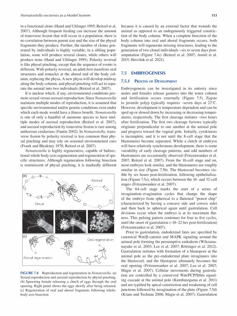
113 Nematostella vectensis as a Model System
in a functional clone (Hand and Uhlinger 1995; Reitzel et al.
2007). Although frequent feeding can increase the amount
of transverse fi ssion that will occur in a population, there is
no correlation between parent size and the size of the physal
fragments they produce. Further, the number of clones gen
erated by individuals is highly variable; in a sibling popu
lation, some will produce several clones, while others will
produce none (Hand and Uhlinger 1995). Polarity reversal
is like physal pinching, except that the sequence of events is
different. With polarity reversal, an adult first manifests oral
structures and tentacles at the aboral end of the body col
umn, replacing the physa. A new physa will develop midway
along the body column, and physal pinching will act to sepa
rate the animal into two individuals (Reitzel et al. 2007).
It is unclear which, if any, environmental conditions pro
mote sexual versus asexual reproduction. Since Nematostella maintain multiple modes of reproduction, it is assumed that
specific environmental and/or genetic conditions exist under
which each mode would have a fi tness benefi t. Nematostella is one of only a handful of anemone species to have mul
tiple modes of asexual reproduction (Reitzel et al. 2007),
and asexual reproduction by transverse fission is rare among
anthozoan cnidarians (Fautin 2002). In Nematostella , trans
verse fission by polarity reversal is less common than phy
sal pinching and may rely on seasonal environmental cues
(Frank and Bleakney 1978; Reitzel et al. 2007).
Nematostella is highly regenerative, capable of bidirec
tional whole body-axis regeneration and regeneration of spe
cific structures. Although regeneration following bisection
is reminiscent of physal pinching, it is markedly different
FIGURE 7.4 Reproduction and regeneration in Nematostella . (a)
Sexual reproduction and asexual reproduction by physal pinching.
(b) Spawning female releasing a clutch of eggs through the oral
opening. Right panel shows the eggs shortly after being released.
(c) Regeneration of oral and aboral fragments following whole-
body axis bisection.
because it is caused by an external factor that wounds the
animal as opposed to an endogenously triggered constric
tion of the body column. When a complete bisection of the
body column into oral and aboral fragments occurs, both
fragments will regenerate missing structures, leading to the
generation of two clonal individuals ~six to seven days post-
amputation (Figure 7.4c) (Reitzel et al. 2007; Amiel et al.
2015; Havrilak et al. 2021).
7.5 EMBRYOGENESIS
7.5.1 PROCESS OF DEVELOPMENT
Embryogenesis can be investigated in its entirety since
males and females release gametes into the water column
and fertilization occurs externally (Figure 7.5). Zygote
to juvenile polyp typically requires ~seven days at 22°C.
However, development is temperature dependent and can be
sped up or slowed down by increasing or decreasing temper
atures, respectively. The first cleavage initiates ~two hours
after fertilization. The first two cleavage furrows typically
originate perpendicular to one another at the animal pole
and progress toward the vegetal pole. Initially, cytokinesis
is incomplete, and it is not until the 8-cell stage that the
blastomeres become separated. While a clutch of embryos
will have relatively synchronous development, there is some
variability of early cleavage patterns, and odd numbers of
blastomeres are occasionally observed (Fritzenwanker et al.
2007, Reitzel et al. 2007). From the 16-cell stage and on,
most embryos look similar, and the blastomeres are roughly
similar in size (Figure 7.5b). The blastocoel becomes vis
ible by six hours post-fertilization, following epithelializa
tion (Figure 7.5c), which occurs between the 16- and 32-cell
stages (Fritzenwanker et al. 2007).
The 64-cell stage marks the start of a series of
invagination–evagination cycles that change the shape
of the embryo from spherical to a flattened “prawn chip”
(characterized by having a concave side and convex side)
and then back to spherical again until gastrulation. Cell
divisions occur when the embryo is at its maximum fl at-
ness. This pulsing pattern continues for four to fi ve cycles,
until the onset of gastrulation (~18–22 hrs post-fertilization)
(Fritzenwanker et al. 2007).
Prior to gastrulation, endodermal fates are specifi ed by
canonical Wnt/β-catenin and MAPK signaling around the
animal pole forming the presumptive endoderm (Wikrama
nayake et al. 2003; Lee et al. 2007; Röttinger et al. 2012).
Gastrulation initiates with formation of a blastopore at the
animal pole as the pre-endodermal plate invaginates into
the blastocoel, and the blastopore ultimately becomes the
oral opening (Fritzenwanker et al. 2007; Lee et al. 2007;
Magie et al. 2007). Cellular movements during gastrula
tion are controlled by a conserved Wnt/PCP/Stbm signal
ing cascade at the animal pole (Kumburegama et al. 2011)
and are typified by apical constriction and weakening of cell
junctions followed by invagination of the plate (Figure 7.5d)
(Kraus and Technau 2006; Magie et al. 2007). Gastrulation
114 Emerging Marine Model Organisms
FIGURE 7.5 Developmental stages of Nematostella. Oral end is to the left. Scale bar = 100 μm.
completes when the ectoderm of the blastopore lip also rolls
inward. This ectoderm retains its epithelial organization and
gives rise to the pharynx and septal filaments at the tips of
the mesenteries.
Following gastrulation, FGF activity at the aboral pole
regulates formation of the apical tuft and apical organ
(Rentzsch et al. 2008). The resulting planula larvae break out
of the egg jelly by ~two days post-fertilization and are now
free-swimming larvae (Figure 7.5e). Planula larvae initially
swim in circles, but by ~three days post-fertilization they
exhibit directional swimming with the apical ciliary tuft fac
ing forward (Hand and Uhlinger 1992). The planula stage
lasts three to four days, during which the planulae elongate
and form the pharynx and the first two (primary) mesenteries
(Figure 7.5f) (Fritzenwanker et al. 2007). A heterogeneous
distribution of ectodermally derived secretory gland cells is
found in the pharynx and mesenteries of the primary polyp,
and gene expression studies suggest that development of these
cells begins in the planula stage, as the tissues they reside in
are formed (Frank and Bleakney 1976; Babonis et al. 2019).
Presumptive muscle cells are detected in the early planula
with F-actin staining, which becomes concentrated and ori
ented along the oral–aboral axis in the late planula (Jahnel
et al. 2014). Besides the tentacle ring muscles, which are
derived from the ectoderm, all other muscle groups are of
endodermal origin, with many orthologs of genes that drive
muscle development in bilaterians observed in Nematostella during the planula and juvenile polyp stages (Jahnel et al.
2014; Steinmetz et al. 2017). NvMyHC1 is first detected at
the mid-planula stage and is expressed in retractor muscle
cells of both the tentacles and the eight mesenteries of the
developing primary polyp (Renfer et al. 2010; Jahnel et al.
2014). The NvMyHC1 transgene is further detected in retrac
tor muscles of adult mesenteries, suggesting muscle cell dif
ferentiation in the mesenteries continues in the adult (Renfer
et al. 2010).
The four tentacle buds emerge toward the end of the
planula stage (~five days post-fertilization) (Figure 7.5f,e).
Tentacle primordia are fi rst identified by Fgfrb-positive cells
in ring muscle around the oral opening. Stereotyped devel
opment and outgrowth of the tentacles is nutrient dependent
and driven by crosstalk between TOR-mediated and FGFR
signaling pathways. The tentacle buds elongate into the ini
tial tentacles of the juvenile polyp, and Nematostella con
tinue to add tentacles in the adult polyp (Stephenson 1935;
Fritz et al. 2013).
Nematostella fully metamorph into juvenile polyps
by 6–7 days post-fertilization (Figure 7.5h). The planula
larvae settle with the aboral pole down (Rentzsch et al.
2008), then transform into a tube-shaped polyp with four
tentacles around a single oral opening. Growth and matu
ration of the juvenile polyp into an adult is nutrient depen
dent, and sexual maturity can be reached in the lab in 10
weeks with regular care and feeding (Hand and Uhlinger
1992 ).
Neurogenesis begins with the emergence of NvSoxB(2) and NvAth-like expressing neural progenitor cells in the
blastula (Richards and Rentzsch 2014, 2015) and continues
throughout development. Molecular regulation of neuro
genesis in Nematostella resembles the neurogenic cascades
found in bilaterian species (Rentzsch et al. 2017), involv
ing MEK/MAPK (Layden et al. 2016), Wnt (Marlow et
115 Nematostella vectensis as a Model System
al. 2013; Sinigaglia et al. 2015; Leclère et al. 2016 ), BMP
(Watanabe et al. 2014; Saina et al. 2009) and Notch (Layden
and Martindale 2014; Richards and Rentzsch 2015).
Nematostella has both ectodermal and endodermal nerve
nets (Nakanishi et al. 2012). Some neural subtypes arise at
the same time as their namesake structures (e.g. tentacu
lar neurons in the tentacles and pharyngeal neurons in the
pharynx) (Havrilak et al. 2017). Cnidocyte stinging cells
are also thought to be neuronal, which is supported by the
fact that they require NvSoxB(2) and NvPaxA ( Babonis and
Martindale 2017). Cnidocyte-specific genes and proteins are
detected throughout the ectoderm in the early gastrula stage
to the primary polyp and in the tentacles and mesenteries of
the adult ( Zenkert et al. 2011; Babonis and Martindale 2014;
Babonis and Martindale 2017 ).
7.5.2 AXIAL PATTERNING PROGRAMS
Throughout Nematostella development, conserved mor
phogen gradients and signaling cascades pattern the
oral–aboral axis. Wnt/β-catenin signaling is a main driver
in establishing and patterning the primary oral–aboral
body axis and has a role in gastrulation, and high Wnt/β
catenin promotes oral identity (Wikramanayake et al. 2003;
Kusserow et al. 2005; Kraus and Technau 2006; Lee et al.
2007; Marlow et al. 2013; Röttinger et al. 2012; Kraus et al.
2016 ). Nvsix3/6 regulates the aboral domain, and its initial
expression is dependent on low Wnt/β-catenin in the aboral
region (Leclère et al. 2016 ). Further, a conserved mecha
nism whereby β-catenin target genes act to repress aboral
gene expression in the oral region represents an ancient
regulatory “logic” that may have been present in the urbi
laterian ancestor (Bagaeva et al. 2020). The interaction of
Wnt/β-catenin with specifi c hox genes further fi ne-tunes
patterning along the oral–aboral axis of the Nematostella embryo and reflects mechanisms of patterning in bilaterians
(DuBuc et al. 2018). However, much more work is needed
to resolve the role that hox genes have in patterning the pri
mary axis in Nematostella, and whether hox expression can
be used to elucidate how the oral–aboral axis relates to the
anterior–posterior axis remains a major question (Layden et
al. 2016; DuBuc et al. 2018).
The secondary directive axis is established and pat
terned by graded BMP signaling. Following an initial radial
expression in the gastrula around the blastopore, NvBmp2/4,
NvBmp5/8 and NvChordin become co-expressed on one side
(Matus et al. 2006; Rentzsch et al. 2006 ). Active pSMAD
(BMP signal transducer) is concentrated on the oppo
site side, suggesting a low BMP signal defines the domain
and initiates transcription (Saina et al. 2009; Leclère and
Rentzsch 2014). Hox genes also play a role in patterning the
directive axis in Nematostella. Hox genes control bound
ary formation, which leads to the radial segmentation of
the developing endoderm and positions the eight radial seg
ments along the directive axis—thereby providing their
spatial identity (He et al. 2018). Further, cross-regulatory
interactions between hox genes occur in both bilaterians
and Nematostella during axial patterning (Matus et al.
2006; DuBuc et al. 2018). While many of the same play
ers are involved in patterning the secondary directive and
dorsal–ventral axes in Nematostella and bilaterians, respec
tively, their positions and functions vary (see “Challenging
Questions”).
7.5.3 REGENERATION
Many aspects of the regenerative process have been charac
terized at the behavioral, morphological, cellular and molec
ular levels (see DuBuc et al. 2014; Bossert and Thomsen
2017). The stages of oral regeneration follow a stereotypic
pattern, with initial wound healing complete in ~six hours
post-amputation, and complete regeneration in ~six to seven
days (Figure 7.4c). In subsequent days, the mesenteries fuse,
contact the wounded epithelial and then reform the phar
ynx as new tentacle buds elongate (Amiel et al. 2015). It is
hypothesized that a population of quiescent/slow cycling
stem cells in the mesenteries are necessary for regeneration
(Amiel et al. 2019). Regeneration following bisection occurs
at the same rate in both juvenile and adult polyps, is tem
perature dependent and requires both cellular proliferation
and apoptosis (see DuBuc et al. 2014; Bossert and Thomsen
2017). Like what has been observed in other animals that
undergo whole-body axis regeneration, some tissue remodel
ing may also occur during regeneration of oral structures in
Nematostella (Amiel et al. 2015; Havrilak et al. 2021). While
many of the signaling pathways necessary for Nematostella are redeployed during regeneration, the regulatory logic and
the number of genes utilized varies, with unique gene regula
tory networks utilized (Warner et al. 2019).
7.6 GENOMIC DATA
The generation of the Nematostella genome was a catalyst
that greatly advanced the species as a model system and led
to a rapid explosion of molecular techniques and publica
tions (Figure 7.2; Table 7.1). The genome was fi rst sequenced
and assembled by the Joint Genome Institute in 2007 using
a random shotgun strategy and published as a searchable
database (https://mycocosm.jgi.doe.gov/Nemve) (Putnam et
al. 2007 ). While this first genome has only partial sequence
coverage and is not mapped back to chromosomes, the scaf
fold organization still informs researchers about syntenic
relationships, gene structure and sequence. Improvements to
the genome have recently been made with the publication
of a second genome (Zimmermann et al. 2020). This new
assembly has enhanced sequence coverage and increased
chromosomal resolution (https://simrbase.stowers.org).
It was expected that the Nematostella genome would be
relatively simple and lack many of the major gene fami
lies found in bilaterians. However, bioinformatic analysis
uncovered a complex genome comparable in many ways to
other animals. It turns out that the Nematostella genome
is more like vertebrates than some popular bilaterian
models such as Caenorhabditis elegans and Drosophila
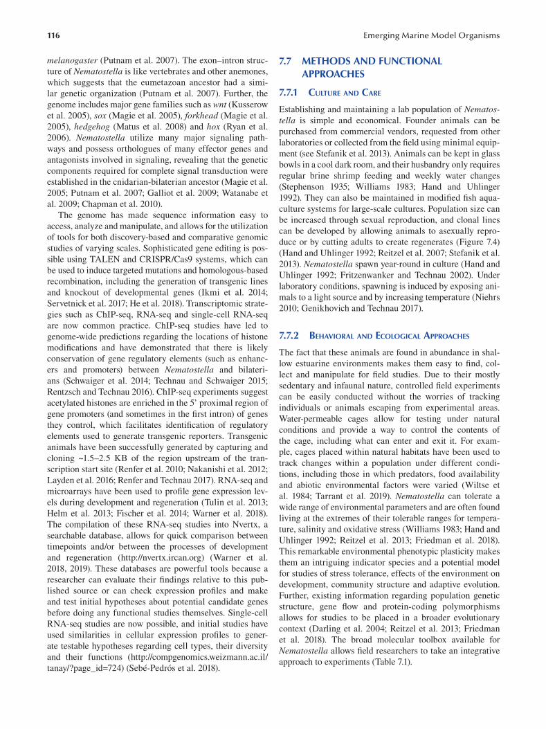
116 Emerging Marine Model Organisms
melanogaster (Putnam et al. 2007). The exon–intron struc
ture of Nematostella is like vertebrates and other anemones,
which suggests that the eumetazoan ancestor had a simi
lar genetic organization (Putnam et al. 2007). Further, the
genome includes major gene families such as wnt ( Kusserow
et al. 2005), sox (Magie et al. 2005), forkhead (Magie et al.
2005 ), hedgehog (Matus et al. 2008) and hox (Ryan et al.
2006 ). Nematostella utilize many major signaling path
ways and possess orthologues of many effector genes and
antagonists involved in signaling, revealing that the genetic
components required for complete signal transduction were
established in the cnidarian-bilaterian ancestor (Magie et al.
2005; Putnam et al. 2007; Galliot et al. 2009; Watanabe et
al. 2009; Chapman et al. 2010).
The genome has made sequence information easy to
access, analyze and manipulate, and allows for the utilization
of tools for both discovery-based and comparative genomic
studies of varying scales. Sophisticated gene editing is pos
sible using TALEN and CRISPR/Cas9 systems, which can
be used to induce targeted mutations and homologous-based
recombination, including the generation of transgenic lines
and knockout of developmental genes (Ikmi et al. 2014;
Servetnick et al. 2017; He et al. 2018). Transcriptomic strate
gies such as ChIP-seq, RNA-seq and single-cell RNA-seq
are now common practice. ChIP-seq studies have led to
genome-wide predictions regarding the locations of histone
modifications and have demonstrated that there is likely
conservation of gene regulatory elements (such as enhanc
ers and promoters) between Nematostella and bilateri
ans (Schwaiger et al. 2014; Technau and Schwaiger 2015;
Rentzsch and Technau 2016 ). ChIP-seq experiments suggest
acetylated histones are enriched in the 5’ proximal region of
gene promoters (and sometimes in the first intron) of genes
they control, which facilitates identification of regulatory
elements used to generate transgenic reporters. Transgenic
animals have been successfully generated by capturing and
cloning ~1.5–2.5 KB of the region upstream of the tran
scription start site (Renfer et al. 2010; Nakanishi et al. 2012;
Layden et al. 2016; Renfer and Technau 2017). RNA-seq and
microarrays have been used to profile gene expression lev
els during development and regeneration (Tulin et al. 2013;
Helm et al. 2013; Fischer et al. 2014; Warner et al. 2018).
The compilation of these RNA-seq studies into Nvertx, a
searchable database, allows for quick comparison between
timepoints and/or between the processes of development
and regeneration (http://nvertx.ircan.org) (Warner et al.
2018 , 2019). These databases are powerful tools because a
researcher can evaluate their findings relative to this pub
lished source or can check expression profiles and make
and test initial hypotheses about potential candidate genes
before doing any functional studies themselves. Single-cell
RNA-seq studies are now possible, and initial studies have
used similarities in cellular expression profiles to gener
ate testable hypotheses regarding cell types, their diversity
and their functions (http://compgenomics.weizmann.ac.il/
tanay/?page_id=724) (Sebé-Pedrós et al. 2018).
7.7 METHODS AND FUNCTIONAL APPROACHES
7.7.1 CULTURE AND CARE
Establishing and maintaining a lab population of Nematostella is simple and economical. Founder animals can be
purchased from commercial vendors, requested from other
laboratories or collected from the field using minimal equip
ment (see Stefanik et al. 2013). Animals can be kept in glass
bowls in a cool dark room, and their husbandry only requires
regular brine shrimp feeding and weekly water changes
(Stephenson 1935; Williams 1983; Hand and Uhlinger
1992). They can also be maintained in modifi ed fi sh aqua
culture systems for large-scale cultures. Population size can
be increased through sexual reproduction, and clonal lines
can be developed by allowing animals to asexually repro
duce or by cutting adults to create regenerates (Figure 7.4)
(Hand and Uhlinger 1992; Reitzel et al. 2007; Stefanik et al.
2013 ). Nematostella spawn year-round in culture (Hand and
Uhlinger 1992; Fritzenwanker and Technau 2002). Under
laboratory conditions, spawning is induced by exposing ani
mals to a light source and by increasing temperature (Niehrs
2010; Genikhovich and Technau 2017 ).
7.7.2 BEHAVIORAL AND ECOLOGICAL APPROACHES
The fact that these animals are found in abundance in shal
low estuarine environments makes them easy to fi nd, col
lect and manipulate for field studies. Due to their mostly
sedentary and infaunal nature, controlled fi eld experiments
can be easily conducted without the worries of tracking
individuals or animals escaping from experimental areas.
Water-permeable cages allow for testing under natural
conditions and provide a way to control the contents of
the cage, including what can enter and exit it. For exam
ple, cages placed within natural habitats have been used to
track changes within a population under different condi
tions, including those in which predators, food availability
and abiotic environmental factors were varied (Wiltse et
al. 1984; Tarrant et al. 2019). Nematostella can tolerate a
wide range of environmental parameters and are often found
living at the extremes of their tolerable ranges for tempera
ture, salinity and oxidative stress (Williams 1983; Hand and
Uhlinger 1992; Reitzel et al. 2013; Friedman et al. 2018).
This remarkable environmental phenotypic plasticity makes
them an intriguing indicator species and a potential model
for studies of stress tolerance, effects of the environment on
development, community structure and adaptive evolution.
Further, existing information regarding population genetic
structure, gene flow and protein-coding polymorphisms
allows for studies to be placed in a broader evolutionary
context (Darling et al. 2004; Reitzel et al. 2013; Friedman
et al. 2018). The broad molecular toolbox available for
Nematostella allows field researchers to take an integrative
approach to experiments (Table 7.1).
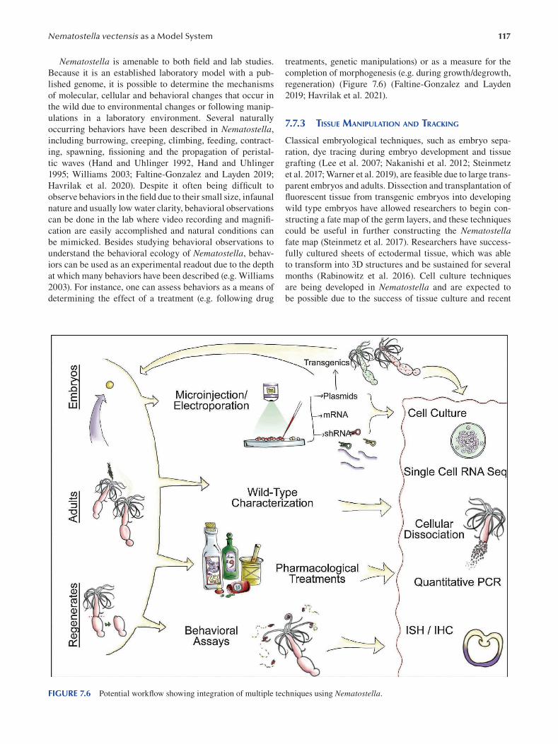
117 Nematostella vectensis as a Model System
Nematostella is amenable to both field and lab studies.
Because it is an established laboratory model with a pub
lished genome, it is possible to determine the mechanisms
of molecular, cellular and behavioral changes that occur in
the wild due to environmental changes or following manip
ulations in a laboratory environment. Several naturally
occurring behaviors have been described in Nematostella,
including burrowing, creeping, climbing, feeding, contract
ing, spawning, fissioning and the propagation of peristal
tic waves (Hand and Uhlinger 1992, Hand and Uhlinger
1995; Williams 2003; Faltine-Gonzalez and Layden 2019;
Havrilak et al. 2020). Despite it often being diffi cult to
observe behaviors in the field due to their small size, infaunal
nature and usually low water clarity, behavioral observations
can be done in the lab where video recording and magnifi
cation are easily accomplished and natural conditions can
be mimicked. Besides studying behavioral observations to
understand the behavioral ecology of Nematostella , behav
iors can be used as an experimental readout due to the depth
at which many behaviors have been described (e.g. Williams
2003). For instance, one can assess behaviors as a means of
determining the effect of a treatment (e.g. following drug
treatments, genetic manipulations) or as a measure for the
completion of morphogenesis (e.g. during growth/degrowth,
regeneration) (Figure 7.6 ) (Faltine-Gonzalez and Layden
2019; Havrilak et al. 2021).
7.7.3 TISSUE MANIPULATION AND TRACKING
Classical embryological techniques, such as embryo sepa
ration, dye tracing during embryo development and tissue
grafting (Lee et al. 2007; Nakanishi et al. 2012; Steinmetz
et al. 2017; Warner et al. 2019), are feasible due to large trans
parent embryos and adults. Dissection and transplantation of
fluorescent tissue from transgenic embryos into developing
wild type embryos have allowed researchers to begin con
structing a fate map of the germ layers, and these techniques
could be useful in further constructing the Nematostella fate map (Steinmetz et al. 2017). Researchers have success
fully cultured sheets of ectodermal tissue, which was able
to transform into 3D structures and be sustained for several
months (Rabinowitz et al. 2016). Cell culture techniques
are being developed in Nematostella and are expected to
be possible due to the success of tissue culture and recent
FIGURE 7.6 Potential workflow showing integration of multiple techniques using Nematostella.
118
ability to successfully dissociate animals into their cellular
components (see the following paragraph). These culturing
methods would allow research to be focused on a specifi c
tissue or cell type and could negate the need to maintain an
animal population due to the ability to freeze cell stocks (e.g.
Fricano et al. 2020).
Dissociation of cells from transgenic and wild type adult
animals has been accomplished using different combina
tions of enzymatic, chemical and mechanical techniques
(Sebé-Pedrós et al. 2018; Clarke et al. 2019; Torres-Méndez
et al. 2019; Weir et al. 2020). Cellular dissociation has also
allowed for studies of cellular adhesion using the hanging
drop method to study reaggregation (Clarke et al. 2019).
Further, cellular dissociation has opened the door to the fi eld
of electrophysiology. For example, single-cell recordings
from nematocytes have given us insight into the physiology
of a novel cell type and bettered our understanding of how
Nematostella distinguish salient environmental information
to regulate cnidocyte firing (Weir et al. 2020), contributing
to our understanding of cnidarian sensory systems and their
stinging response.
7.7.3.1 Detection of Cellular Processes The relatively simple body plan of Nematostella , consist
ing of only two transparent tissue layers, facilitates the
use of common labeling techniques to investigate cellular
processes utilized during morphogenesis and homeostasis.
Standard techniques for cellular proliferation have been used
by labeling animals with EdU and BrdU (Passamaneck and
Martindale 2012; Richards and Rentzsch 2014; Amiel et al.
2015; Rabinowitz et al. 2016; Warner et al. 2019; Havrilak
et al. 2021). TUNEL assays have been used to detect apop
totic cells during development and regeneration (Warner et al.
2019; Zang and Nakanishi 2020).
7.7.3.2 Regeneration Inducing a regenerative response in Nematostella is sim
ple, and the process has been characterized at many levels.
Regeneration is induced by wounding the animal with a scal
pel or probe (see DuBuc et al. 2014; Bossert and Thomsen
2017 ). The wound site and severity of the injury infl icted
are dictated by the research question. Typically, studies have
focused on whole-body axis regeneration, where live ani
mals are bisected along the body column into oral and aboral
halves and regeneration of one or both fragments is observed
(Figure 7.4c) (Passamaneck and Martindale 2012; Amiel et
al. 2015; Schaffer et al. 2016 ). However, a more acute regen
erative response can be triggered following a focal injury
where whole-body axis regeneration is not required (e.g.
tentacle amputation, puncture wound, incomplete bisection
along the body column) (Reitzel et al. 2007; DuBuc et al.
2014). This flexibility in the regeneration paradigm allows
for a variety of questions to be asked. The ability to docu
ment gene expression in different regenerative paradigms, as
well as to compare it to development, will continue to make
this a fruitful area of research in this model. For example,
Emerging Marine Model Organisms
many hypotheses can be tested due to comparative tran
scriptome analysis using RNAseq during the regeneration
of oral vs. aboral fragments—which identifi ed similarities
and differences in gene expression profiles between the two
halves (Schaffer et al. 2016). Methods for assessing wound
closure, and detailed descriptions of key morphological
landmarks that occur throughout the process of regenera
tion, have been described and can be used to assess the prog
ress of the regenerative response (Bossert et al. 2013; Amiel
et al. 2015). Assaying the regenerative phenotype following
pharmacological or genetic manipulation could be used to
understand the mechanisms of regeneration (e.g. using an
inducible promoter or knockout transgenic line). Transgenic
reporter lines allow for the tracking of specific cell types in
live animals, including specific neural subtypes, which has
made the regeneration of the nerve net tractable (Figure 7.6 )
(Layden et al. 2016; Havrilak et al. 2017; Sunagar et al. 2018;
Havrilak et al. 2021).
7.7.4 GENETIC APPROACHES
7.7.4.1 Microinjection and Electroporation Molecules can be introduced into live embryos using
microinjection and electroporation techniques, which
facilitate the delivery of compounds such as shRNA,
mRNA, morpholinos and plasmids into eggs. With micro
injection, a very fine glass needle is used to penetrate an
egg and deliver a small volume of the loaded injection
mixture using forced air (Layden et al. 2013; Renfer and
Technau 2017; Havrilak and Layden 2019). An experienced
researcher can inject thousands of embryos in a single ses
sion. Microinjection offers more experimental utility due
to the variety of molecular compounds that can be injected,
ranging from plasmids to shRNAs. Microinjection has
been used successfully for genetic knockdown and misex
pression experiments, as well as for the generation of trans
genic animals in Nematostella (Layden et al. 2013; Ikmi
et al. 2014; Renfer and Technau 2017). Electroporation
offers a simple and quick method for the delivery of mol
ecules into hundreds of animals simultaneously by gen
erating electrical pulses that create pores in the plasma
membrane that allow small molecules to be taken up. So
far, this method has only proved successful in the deliv
ery of shRNA for knockdown experiments in Nematostella (Karabulut et al. 2019).
7.7.4.2 Gene Disruption Tools for both gain and loss of function experiments are avail
able (Table 7.1). Injection of in vitro synthesized mRNA allows
for a gene of interest to be overexpressed (Wikramanayake
et al. 2003), while introduction of shRNA or morpholinos
facilitates genetic knockdown of a gene of interest (Magie
et al. 2005; Rentzsch et al. 2008; He et al. 2018). Gene edit
ing technologies can also be used to silence, move, knock
down or overexpress a particular gene in both F0 and F1
generation mutants, and pharmacological treatments can also
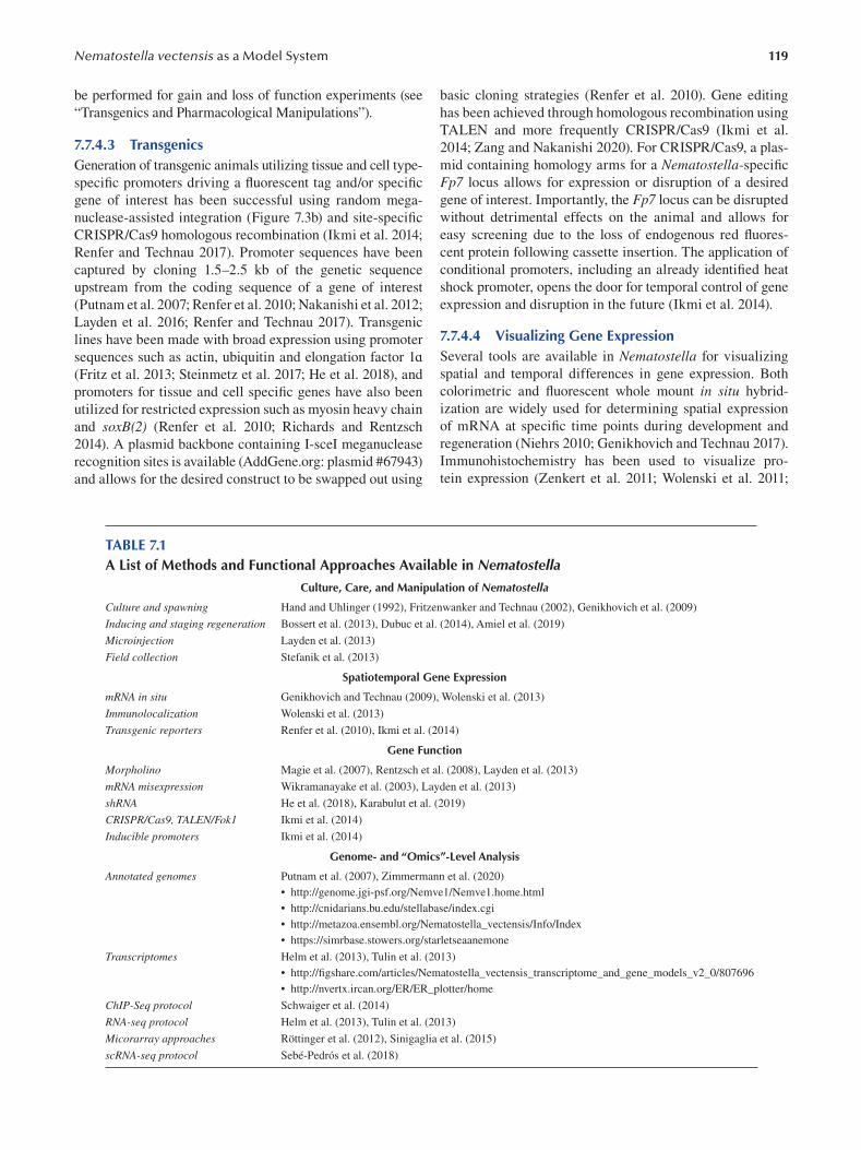
119 Nematostella vectensis as a Model System
be performed for gain and loss of function experiments (see
“Transgenics and Pharmacological Manipulations”).
7.7.4.3 Transgenics Generation of transgenic animals utilizing tissue and cell type-
specific promoters driving a fluorescent tag and/or specifi c
gene of interest has been successful using random mega-
nuclease-assisted integration (Figure 7.3b) and site-specifi c
CRISPR/Cas9 homologous recombination (Ikmi et al. 2014;
Renfer and Technau 2017). Promoter sequences have been
captured by cloning 1.5–2.5 kb of the genetic sequence
upstream from the coding sequence of a gene of interest
(Putnam et al. 2007; Renfer et al. 2010; Nakanishi et al. 2012;
Layden et al. 2016; Renfer and Technau 2017). Transgenic
lines have been made with broad expression using promoter
sequences such as actin, ubiquitin and elongation factor 1ɑ (Fritz et al. 2013; Steinmetz et al. 2017; He et al. 2018), and
promoters for tissue and cell specific genes have also been
utilized for restricted expression such as myosin heavy chain
and soxB(2) (Renfer et al. 2010; Richards and Rentzsch
2014). A plasmid backbone containing I-sceI meganuclease
recognition sites is available (AddGene.org: plasmid #67943)
and allows for the desired construct to be swapped out using
basic cloning strategies (Renfer et al. 2010). Gene editing
has been achieved through homologous recombination using
TALEN and more frequently CRISPR/Cas9 (Ikmi et al.
2014; Zang and Nakanishi 2020). For CRISPR/Cas9, a plas
mid containing homology arms for a Nematostella -specifi c
Fp7 locus allows for expression or disruption of a desired
gene of interest. Importantly, the Fp7 locus can be disrupted
without detrimental effects on the animal and allows for
easy screening due to the loss of endogenous red fl uores
cent protein following cassette insertion. The application of
conditional promoters, including an already identifi ed heat
shock promoter, opens the door for temporal control of gene
expression and disruption in the future (Ikmi et al. 2014).
7.7.4.4 Visualizing Gene Expression Several tools are available in Nematostella for visualizing
spatial and temporal differences in gene expression. Both
colorimetric and fluorescent whole mount in situ hybrid
ization are widely used for determining spatial expression
of mRNA at specific time points during development and
regeneration (Niehrs 2010; Genikhovich and Technau 2017 ).
Immunohistochemistry has been used to visualize pro
tein expression ( Zenkert et al. 2011; Wolenski et al. 2011;
TABLE 7.1 A List of Methods and Functional Approaches Available in Nematostella
Culture, Care, and Manipulation of Nematostella
Culture and spawning Hand and Uhlinger (1992 ), Fritzenwanker and Technau (2002 ), Genikhovich et al. (2009 )
Inducing and staging regeneration Bossert et al. (2013 ), Dubuc et al. (2014 ), Amiel et al. (2019 )
Microinjection Layden et al. (2013 )
Field collection Stefanik et al. (2013 )
Spatiotemporal Gene Expression
mRNA in situ Genikhovich and Technau (2009 ), Wolenski et al. (2013)
Immunolocalization Wolenski et al. (2013)
Transgenic reporters Renfer et al. (2010 ), Ikmi et al. (2014 )
Gene Function
Morpholino Magie et al. (2007 ), Rentzsch et al. (2008 ), Layden et al. (2013 )
mRNA misexpression Wikramanayake et al. (2003 ), Layden et al. (2013 )
shRNA He et al. (2018 ), Karabulut et al. (2019 )
CRISPR/Cas9, TALEN/Fok1 Ikmi et al. (2014 )
Inducible promoters Ikmi et al. (2014 )
Genome- and “Omics”-Level Analysis
Annotated genomes Putnam et al. (2007 ), Zimmermann et al. (2020 )
• http://genome.jgi-psf.org/Nemve1/Nemve1.home.html
• http://cnidarians.bu.edu/stellabase/index.cgi
• http://metazoa.ensembl.org/Nematostella_vectensis/Info/Index
• https://simrbase.stowers.org/starletseaanemone
Transcriptomes Helm et al. (2013 ), Tulin et al. (2013 )
• http://fi gshare.com/articles/Nematostella_vectensis_transcriptome_and_gene_models_v2_0/807696
• http://nvertx.ircan.org/ER/ER_plotter/home
ChIP-Seq protocol Schwaiger et al. (2014 )
RNA-seq protocol Helm et al. (2013 ), Tulin et al. (2013 )
Micorarray approaches Röttinger et al. (2012 ), Sinigaglia et al. (2015 )
scRNA-seq protocol Sebé-Pedrós et al. (2018 )
120 Emerging Marine Model Organisms
Nakanishi et al. 2012; Zang and Nakanishi 2020). Transgenic
reporter lines provide another means of assaying spatial and
temporal protein expression and allow for live visualization
and imaging. Quantitative real-time polymerase chain reac
tion is a quick method for determining mRNA expression
levels at a given point in time and is often used experimen
tally in tandem with in situ hybridization and in the confi r
mation of sequencing results. Together these methods are
powerful tools for characterizing wild type and transgenic
expression and as a readout for gain and loss of function
experiments (Figure 7.6 ).
7.7.4.5 Genome- and “Omics”-Level Approaches Genomic- and transcriptomic-level experiments under vary
ing developmental, regenerative and/or environmental par
adigms are possible since the publication of the genome.
ChIP-seq studies can be used to determine epigenetic pro
tein interactions with open chromatin. For example, ChIP
seq can uncover potential genomic interactions of a protein
of interest or facilitate the identification of regulatory ele
ments for a gene of interest (Schwaiger et al. 2014; Technau
and Schwaiger 2015; Rentzsch and Technau 2016 ). RNA-seq
and single cell RNA-seq have allowed for the investigation
of global gene expression levels in whole animals and sin
gle cells, respectively (Tulin et al. 2013; Helm et al. 2013;
Fischer et al. 2014; Warner et al. 2018; Sebé-Pedrós et al.
2018). Ultimately, each of these methods provides different
levels of resolution, and the method used will depend on the
question being asked.
7.7.4.6 Pharmacological Manipulation Pharmacological agents have been used to target specifi c
developmental pathways, as well as to target pathways to alter
the physiology of the adult animal. Administering pharmaco
logical agents requires only introducing the desired concentra
tion to the sea water in which treatment animals are growing.
Treatments can be administered at any stage from develop
ing embryos to mature adults. Pharmacological agents offer a
quick and easy way to target pathways in a high-throughput
manner. It is possible to alter basic cellular processes using
drug treatments. For example, cell proliferation has been
blocked with hydroxyurea ( Amiel et al. 2015 ). Wnt/β-catenin
activity can be overactivated using 1-azakenpaullone and/or
alsterpaullone, and inhibited using iCRT14 ( Trevino et al.
2011 ; Watanabe et al. 2014 ). The gamma secretase inhibi
tor DAPT can be given to effectively disrupt the Notch/Delta
pathway ( Layden and Martindale 2014 ), and the receptor
tyrosine kinase inhibitor SU5402 can be used to effectively
inhibit Fgf receptors ( Rentzsch et al. 2008 ). Additionally,
the mTOR pathway can be disrupted by bathing animals in
rapamycin ( Ikmi et al. 2020 ). While many of the treatments
discussed previously would typically be applicable to devel
oping animals, there are also several agents that can alter
the physiology of adult Nematostella. For example, bathing
adults in the neurotransmitter acetylcholine can induce ten
tacle contractions, while lidocaine can suppress these contrac
tions ( Faltine-Gonzalez and Layden 2019 ).
7.7.5 INTEGRATION OF APPROACHES
While the approaches discussed here are organized into
subsections, there is no hard line defining what they can
be used for. The combination of various tools from fi eld
approaches to molecular, cellular and behavioral tech
niques can be combined to address a nearly limitless range
of questions (Figure 7.6 ). Following the establishment of
a lab population, a basic molecular biology lab setup will
allow a researcher to tackle questions pertaining to the
fi elds of molecular ecology, mechanisms of behavior, evo
lution, development, regeneration and so on (Figure 7.6 ).
There is also the expectation that the Nematostella model
will keep up with major advances in technology, since
cutting-edge techniques continue to become available in
this system. Advances in single-cell technologies and the
application of conditional/inducible alleles will further
refine the resolution and control at which experiments can
be performed. Adding in the fact that field and lab com
parisons and/or wild type and transgenic comparisons can
be included as additional variables makes it so researchers
have a high level of control, allowing them to implement
experimental parameters beyond those offered by other
model systems.
7.8 CHALLENGING QUESTIONS
7.8.1 IS THERE A DEEP EVOLUTIONARY ORIGIN
FOR KEY BILATERIAN TRAITS?
An explosive radiation of taxa occurred within the bilat
erian lineage, and it is believed to be due to the evolution
of several unique characteristics (e.g. mesodermal germ
layer and bilateral symmetry) that allowed them to occupy
previously inaccessible niches. The evolution of these traits
allowed for the evolution of larger, more complex body plans
and increased specialization of structure organization and
function—including cephalization and the centralization
of nervous systems. Understanding the mechanisms that
led to the bilaterian radiation is a longstanding evolution
ary question that can only be answered by studying animals
that are closely related to bilaterians in order to infer what
molecular tool-kit was available to their common ancestor.
Cnidarians are regarded as the sister taxon to the bilaterians
(Wainright et al. 1993; Medina et al. 2001; Collins 2002),
and therefore cnidarian models, such as Nematostella , offer
an appropriate outgroup species to study the molecular basis
for the origin of key bilaterian traits, such as the mesoderm
and bilaterality, because they allow us to deduce the evolu
tionary history of these derived traits (Figure 7.7 ). In fact,
Nematostella first gained momentum as a model species for
its utility in uncovering the evolutionary mechanisms that
led to key bilaterian features.
7.8.1.1 Origin of the Mesoderm Thus far, studies with Nematostella have used compara
tive genetic approaches and germ layer fate mapping to
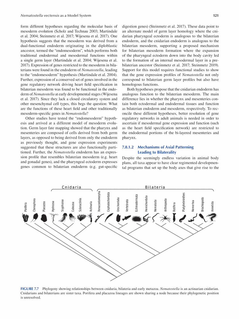
121 Nematostella vectensis as a Model System
form different hypotheses regarding the molecular basis of
mesoderm evolution (Scholz and Technau 2003; Martindale
et al. 2004; Steinmetz et al. 2017; Wijesena et al. 2017). One
hypothesis suggests that the mesoderm was derived from a
dual-functional endoderm originating in the diploblastic
ancestor, termed the “endomesoderm”, which performs both
traditional endodermal and mesodermal functions within
a single germ layer (Martindale et al. 2004; Wijesena et al.
2017). Expression of genes restricted to the mesoderm in bila
terians were found in the endoderm of Nematostella , leading
to the “endomesoderm” hypothesis (Martindale et al. 2004).
Further, expression of a conserved set of genes involved in the
gene regulatory network driving heart fi eld specifi cation in
bilaterian mesoderm was found to be functional in the endo
derm of Nematostella at early developmental stages (Wijesena
et al. 2017). Since they lack a closed circulatory system and
other mesenchymal cell types, this begs the question: What
are the functions of these heart field and other traditionally
mesoderm-specific genes in Nematostella?
Other studies have tested the “endomesoderm” hypoth
esis and arrived at a different model of mesoderm evolu
tion. Germ layer fate mapping showed that the pharynx and
mesenteries are composed of cells derived from both germ
layers, as opposed to being derived from only the endoderm
as previously thought, and gene expression experiments
suggested that these structures are also functionally parti
tioned. Further, the Nematostella endoderm has an expres
sion profile that resembles bilaterian mesoderm (e.g. heart
and gonadal genes), and the pharyngeal ectoderm expresses
genes common to bilaterian endoderm (e.g. gut-specifi c
digestion genes) (Steinmetz et al. 2017). These data point to
an alternate model of germ layer homology where the cni
darian pharyngeal ectoderm is analogous to the bilaterian
endoderm, and the cnidarian endoderm is analogous to the
bilaterian mesoderm, supporting a proposed mechanism
for bilaterian mesoderm formation where the expansion
of the pharyngeal ectoderm down into the body cavity led
to the formation of an internal mesodermal layer in a pre
bilaterian ancestor (Steinmetz et al. 2017; Steinmetz 2019).
Support for this model requires functional studies to show
that the gene expression profiles of Nematostella not only
correspond to bilaterian germ layer profiles but also have
homologous functions.
Both hypotheses propose that the cnidarian endoderm has
analogous function to the bilaterian mesoderm. The main
difference lies in whether the pharynx and mesenteries con
tain both ectodermal and endodermal tissues and function
as bilaterian endoderm and mesoderm, respectively. To rec
oncile these different hypotheses, better resolution of gene
regulatory networks in adult animals is needed in order to
ascertain if mesodermal gene expression and function (such
as the heart fi eld specification network) are restricted to
the endodermal portions of the bi-layered mesenteries and
pharynx.
7.8.1.2 Mechanisms of Axial Patterning Leading to Bilaterality
Despite the seemingly endless variation in animal body
plans, all taxa appear to have clear regimented developmen
tal programs that set up the body axes that give rise to the
FIGURE 7.7 Phylogeny showing relationships between cnidaria, bilateria and early metazoa. Nematostella is an actinarian cnidarian.
Cnidarians and bilaterians are sister taxa. Porifera and placazoa lineages are shown sharing a node because their phylogenetic position
is unresolved.
122
unique morphology of each species. Many similarities in the
mechanics of axial patterning have been observed between
taxa. This has led to the question: Is there a conserved
molecular program central to axial patterning that has been
co-opted across evolutionary time?
At first glance, Nematostella appear radially sym
metrical, with their body plan centered around a primary
oral–aboral axis that runs the length of the body column.
However, upon closer examination of the structural features
of Nematostella, it is evident that they possess bilateral sym
metry along a secondary “directive” axis that runs perpen
dicular to the primary axis (see “Anatomy”). At a molecular
level, the perpendicular primary and secondary axes are
derived from orthogonal morphogen gradients that work in
concert to set up the body plan. In bilaterians, the orthogonal
arrangement of morphogen gradients is also a fundamental
aspect of body axis patterning (Niehrs 2010; Genikhovich
and Technau 2017 ).
Major morphogen signaling pathways, with an estab
lished role in bilaterian anterior–posterior axial patterning,
play a similar role in setting up domains along the oral–
aboral axis in Nematostella (Leclère et al. 2016; Amiel et al.
2017; Bagaeva et al. 2020). Like bilaterians, a Wnt/β-catenin
gradient, with a similar regulatory logic, is established along
the primary axis (Marlow et al. 2013, Kraus et al. 2016,
Bagaeva et al. 2020). In bilaterians, a key factor in forming
the dorsal–ventral axis is the establishment of opposing gra
dients of bone morphogenic protein and its antagonist chor
din on opposite ends of the secondary axis, perpendicular to
the primary axis (Niehrs 2010, Genikhovich and Technau
2017). In contrast, expression domains of bone morphoge
netic protein and chordin overlap and are on the same side
of the directive axis in Nematostella (Matus et al. 2006;
Rentzsch et al. 2006; Leclère and Rentzsch 2014). This sug
gests evolutionary plasticity in the BMP/Chordin systems
but does not answer how they functioned and were co-opted
in the establishment of diverse secondary axial patterning
programs throughout evolutionary history. It is worth noting
that besides these two major upstream morphogen pathways,
other factors, such as hox genes, play critical roles as down
stream effectors in shaping and refining axial patterning in
Nematostella and bilaterians (Graham et al. 1991; Pearson
et al. 2005; DuBuc et al. 2018; He et al. 2018).
The accumulation of data thus far suggests deep evolu
tionary roots for the morphogenetic programs governing axis
patterning (Matus et al. 2006; Bagaeva et al. 2020), regard
less of body plan complexity. Although the same morphoge
netic pathways seem to play an important role in patterning
the primary and secondary axes in Nematostella, there
appear to be key differences in how morphogens are spatially
distributed and interacting (Matus et al. 2006; Rentzsch
et al. 2006; Leclère and Rentzsch 2014). Similarities in axial
programming between Nematostella and bilaterians make
them an ideal candidate for understanding if/when a gen
eral morphogenetic program was co-opted for the evolution
of bilateral symmetry. Comparisons with other cnidarians
Emerging Marine Model Organisms
and early metazoans will help to resolve how these pattern
ing mechanisms evolved and functioned in the urbilaterian
ancestor and prior to the cnidarian-bilaterian split.
7.8.2 CAN NEMATOSTELLA BE USED AS A
CNIDARIAN MODEL FOR CNIDARIANS?
Establishing a genetically amenable, high-throughput, cni
darian model would improve our understanding of many
aspects of cnidarian biology, which has been hindered by
our inability to easily access, observe and culture many spe
cies within this phylum. A major question is: Can we bet
ter understand the effects of the changing environment and
inform conservation strategies by utilizing established cni
darian models that are amenable to high-throughput labora
tory techniques? Although corals can be harvested and kept
under laboratory conditions (provided that specifi c environ
mental parameters are met) their natural history makes it
very difficult to control spawning behavior and therefore
makes it so that embryos are only available up to a few times
a year (Harrison et al. 1984; Baird et al. 2009; Keith et al.
2016; Craggs et al. 2017; Pollock et al. 2017; Cleves et al.
2018). In addition, there are few tools and resources available
for conducting molecular, cellular or physiological research
in non-model cnidarian systems (Technau and Steele 2011).
A notable exception is a study that used CRISPR/Cas9 in
the coral Acropora millepora to target a few genes of inter
est. However, to obtain embryos, prior knowledge of when
spawning would occur was necessary so that corals could be
harvested and brought into the lab just prior to their natu
ral spawning event (Cleves et al. 2018). This exemplifi es the
logistical hurdles that are often present in coral and other
cnidarian research.
An intriguing possibility is that Nematostella could be
employed as a cnidarian model for cnidarians due to the rep
ertoire of tools available and easy culture. Nematostella has
no symbionts, and therefore it would not be useful in model
ing symbiotic relationships. However, the fact that we can eas
ily manipulate Nematostella at a molecular level sets it up as
a good proxy to investigate fundamental molecular programs
in other cnidarians. This way, hypotheses could be quickly
tested in this developed model so that resources can be mobi
lized most efficiently in hard to study cnidarian species. An
additional question is: Can Nematostella be used as a cnidar
ian model for environmental stress tolerance and adaptation?
This could broaden our understanding of how imperiled cni
darians are likely to cope with ongoing environmental change.
Plus, understanding the underlying mechanisms responsible
for environmental plasticity in Nematostella could potentially
be exploited in the conservation of other species.
7.8.3 HOW DO NOVEL CELL TYPES EVOLVE?
Longstanding evolutionary questions are: How does evolu
tionary novelty arise, and how does novelty lead to major
evolutionary transitions? To investigate these questions
123 Nematostella vectensis as a Model System
requires a model that possesses cell types with true morpho
logical and functional novelty. Cnidocytes are phenotypi
cally unique stinging cells and a defining characteristic of
the cnidarian phylum. Cnidocytes are one of only a handful
of examples of an unequivocal evolutionary novelty and thus
offer a unique opportunity to investigate the mechanisms
that lead to evolutionary novelty—something that is not pos
sible in many model systems. Using Nematostella , studies
can be focused on the molecular basis of cnidocyte develop
ment (Babonis and Martindale 2017; Sunagar et al. 2018).
This will inform how newly generated genes/proteins inter
act with existing biological programs, leading to the emer
gence of novel proteins and, in turn, cell types (Babonis and
Martindale 2014; Babonis et al. 2016; Layden et al. 2016 ).
7.8.4 DOES REGENERATION RECAPITULATE DEVELOPMENT?
Unraveling the molecular basis of development and regen
eration is pivotal to answering the question of whether devel
opmental programs are co-opted for regenerative processes.
Complicating matters is that historically, researchers were
limited by models suited to either the study of development
or regeneration or those that had limited regenerative capa
bilities (e.g. Ambystoma mexicanum, Danio rerio, Xenopus laevis). Models where both processes can be examined
within the same species took longer to become established
(e.g. Nematostella vectensis, Hofstenia miamia). A dis
tinguishing feature of Nematostella is that it is capable of
whole-body axis regeneration. This, coupled with the fact
that it is becoming a strong model for development, offers
the unique ability to directly compare these two processes
within the same animal. Studies in Nematostella and other
species are gaining support for the hypothesis that regenera
tion is only a partial redeployment of embryonic develop
ment (e.g. Schaffer et al. 2016; Warner et al. 2019).
Moving forward, it will be necessary to study whether
the same program differences arise regardless of regenera
tion paradigm. For example, during whole-body axis regen
eration in Nematostella, the initial regenerative response of
certain neural subtypes differs under varying regenerative
paradigms, suggesting that there may be cell type differ
ences (Havrilak et al. 2021). As we gain functional under
standing of these processes, it begs the question of whether
we can unlock regenerative potential in non-regenerative
models and use this knowledge to develop medical therapies.
7.8.5 OTHER CHALLENGING QUESTIONS
The topics addressed previously are only a small subset of
the challenging questions that Nematostella is poised to
address. For example, other questions pertaining to evolution
and development, such as the centralization of the nervous
system within the bilaterian lineage, are possible because of
the position of cnidarians as sister taxa. Outside of its use in
academia, there is also definite potential for Nematostella within applied research fields, such as biotechnology.
Within the biotech industry, one innovative group looks to
use Nematostella to help consumers combat the signs of age
ing by harnessing the stinging action of cnidocyte cells to
optimize the delivery of skin care agents deep into the skin
(Toren and Gurovich 2016 ). Although few other examples of
Nematostella in applied research exist, it is easy to imagine
other uses for this cnidocyte-mediated injection technology
throughout the beauty and medical industries, as well as
many other untapped applications waiting to be uncovered.
ACKNOWLEDGMENTS
Thanks to Anna Delaney and Mark Williams for uncover
ing archival information on Miss. Gertrude F. Selwood from
when she was at their respective universities. We thank Eric
Röttinger for the photograph of the adult Nematostella,
MingHe Chen for developmental images and Dylan Faltine-
Gonzalez for insightful discussions.
BIBLIOGRAPHY
Amiel, A.R., Foucher, K., Ferreira, S., and E. Röttinger. 2019.
Synergic coordination of stem cells is required to induce
a regenerative response in anthozoan cnidarians. BioRxiv.
December 31:891804.
Amiel, A.R., Hereroa J., Nedoncelle, K., Warner, J., Ferreira, S.,
and E. Röttinger. 2015. Characterization of morphological
and cellular events underlying oral regeneration in the sea
anemone. Nematostella vectensis. International Journal of Molecular Sciences . 16:28449–28471.
Amiel, A.R., Johnston, H., Chock, T., Dahlin, P., Iglesias, M.,
Layden, M., Röttinger, E., and M.Q. Martindale. 2017. A
bipolar role of the transcription factor erg for cnidarian germ
layer formation and apical domain patterning. Developmental Biology . 430:346–361.
Babonis, L.S., and M.Q. Martindale. 2014. Old cell, new trick?
Cnidocytes as a model for the evolution of novelty. Integrative and Comparative Biology . 54:714–722.
Babonis, L.S., and M.Q. Martindale. 2017. PaxA, but not PaxC,
is required for cnidocyte development in the sea anemone
Nematostella vectensis: EvoDevo . 8:14.
Babonis, L.S., Martindale, M.Q., and J.F. Ryan. 2016. Do novel
genes drive morphological novelty? An investigation of the
nematosomes in the sea anemone Nematostella vectensis.
BMC Evolutionary Biology . 16:114.
Babonis, L.S., Ryan, J.F., Enjolras, C., and M.Q. Martindale. 2019.
Genomic analysis of the tryptome reveals molecular mecha
nisms of gland cell evolution. EvoDevo . 10:23.
Bagaeva, T., Aman, A.J., Graf, T., Niedermoser, I., Zimmermann,
B., Kraus, Y., Schatka, M., Demilly, A., Technau, U., and G.
Genikhovich. 2020. β-catenin dependent axial patterning in
Cnidaria and Bilateria uses similar regulatory logic. BioRxiv.
September 8:287821.
Baird, A.H., Guest, J.R., and B.L. Willis. 2009. Systematic and
biogeographical patterns in the reproductive biology of
Scleractinian corals. Ecology, Evolution, and Systematics.
40:551–571.
Berking, S. 2007. Generation of bilateral symmetry in Anthozoa: A
model. Journal of Theoretical Biology . 246:477–490.
Bossert, P.E., Dunn, M.P., and G.H. Thomsen. 2013. A stag
ing system for the regeneration of a polyp from the aboral
124
physa of the anthozoan cnidarian Nematostella vectensis.
Developmental Dynamics . 242:1320–1331.
Bossert, P.E., and G.H. Thomsen. 2017. Inducing complete
polyp regeneration from the aboral physa of the starlet sea
anemone Nematostella vectensis. Journal of Visualized Experiments . 119.
Brandão, R.A., Gusmão, L.C., and P.B. Gomes. 2019. Diversity of
Edwardsiidae sea anemones (Cnidaria: Anthozoa: Actiniaria)
from Brazil, with the description of a new genus and species.
Journal of the Marine Biological Association of the United Kingdom . 99:1087–1098.
Chapman, J.A., Kirkness, E.F., Simakov, O., Hampson, S.E., Mitros,
T., Weinmaier, T., Rattei, T., Balasubramanian, P.G., Borman,
J., Busam, D., Disbennett, K., Pfannkoch, C., Sumin, N.,
Sutton, G.G., Viswanathan, L.D., Walenz, B., Goodstein,
D.M., Hellsten, U., Kawashima, T., Prochnik, S.E., Putnam,
N.H., Shu, S., Blumberg, B., Dana, C.E., Gee, L., Kibler, D.F.,
Law, L., Lindgens, D., Martinez, D.E., Peng, J., Wigge, P.A.,
Bertulat, B., Guder, C., Nakamura, Y., Ozbek, S., Watanabe,
H., Khalturin, K., Hemmrich, G., Franke, A., Augustin,
R., Fraune, S., Hayakawa, E., Hayakawa, S., Hirose, M.,
Hwang, J.S., Ikeo, K., Nishimiya-Fujisawa, C., Ogura, A.,
Takahashi, T., Steinmetz, P.R.H., Zhang, X., Aufschnaiter, R.,
Eder, M.K., Gorny, A.K., Salvenmoser, W., Heimberg, A.M.
Wheeler, B.M., Peterson, K.J., Böttger, A., Tischler, P., Wolf,
A., Gojobori, T., Remington, K.A., Strausberg, R.L., Venter,
J.C., Technau, U., Hobmayer, B., Bosch, T.C.G., Holstein,
T.W., Fujisawa, T., Bode, H.R., David, C.N., Rokhsar, D.S.,
and R.E. Steele. 2010. The dynamic genome of Hydra.
Nature . 464:592–596.
Clarke, D.N., Lowe, C.J., and W.J. Nelson. 2019. The cadherin
catenin complex is necessary for cell adhesion and embryo
genesis in Nematostella vectensis. Developmental Biology.
447:170–181.
Cleves, P.A., Strader, M.E., Bay, L.K., Pringle, J.R., and M.V.
Matz. 2018. CRISPR/Cas9-mediated genome editing in a
reef-building coral. Proceedings of the National Academy of Sciences . 115:201722151.
Collins, A.G. 2002. Phylogeny of Medusozoa and the evolution
of cnidarian life cycles. Journal of Evolutionary Biology.
15:418–432.
Columbus-Shenkar, Y.Y., Sachkova, M.Y., Macrander, J., Fridrich,
A., Modepalli, V., Reitzel, A.M., Sunagar, K., and Y. Moran.
2018. Dynamics of venom composition across a complex life
cycle. ELife . 7:e35014.
Craggs, J., Guest, J.R., Davis, M., Simmons, J., Dashti, E., and
M. Sweet. 2017. Inducing broadcast coral spawning ex situ:
Closed system mesocosm design and husbandry protocol.
Ecology and Evolution . 7:11066–11078.
Crowell, S. 1946. A new sea anemone from Woods Hole,
Massachusetts. Journal of the Washington Academy of Sciences . 36:47–60.
Darling, J.A., Reitzel, A.M., Burton, P.M., Mazza, M.E., Ryan,
J.F., Sullivan, J.C., and J.R. Finnerty. 2005. Rising starlet:
The starlet sea anemone, Nematostella vectensis. BioEssays.
27:211–221.
Darling, J.A., Reitzel, A.M., and J.R. Finnerty. 2004. Regional
population structure of a widely introduced estuarine inver
tebrate: Nematostella vectensis Stephenson in New England.
Molecular Ecology . 13:2969–2981.
DuBuc, T.Q., Stephenson, T.B., Rock, A.Q., and M.Q. Martindale.
2018. Hox and Wnt pattern the primary body axis of an antho
zoan cnidarian before gastrulation. Nature Communications.
9:2007.
Emerging Marine Model Organisms
DuBuc, T.Q., Traylor-Knowles, N., and M.Q. Martindale. 2014.
Initiating a regenerative response; cellular and molecular
features of wound healing in the cnidarian Nematostella vectensis. BMC Biology . 12:24.
Faltine-Gonzalez, D.Z., and M.J. Layden. 2019. Characterization
of NAChRs in Nematostella vectensis supports neuronal
and non-neuronal roles in the cnidarian–bilaterian common
ancestor. EvoDevo . 10:27.
Fautin, D.G. 2002. Reproduction of Cnidaria. Canadian Journal of Zoology . 80:1735–1754.
Finnerty, J.R., Pang, K., Burton, P., Paulson, D., and M.Q.
Martindale. 2004. Origins of bilateral symmetry: Hox and
Dpp expression in a sea anemone. Science . 304:1335–1337.
Fischer, A.H.L., Mozzherin, D.A.M.E., Lans, K.D., Wilson, N.,
Cosentino, C., and J. Smith. 2014. SeaBase: A multispecies
transcriptomic resource and platform for gene network infer
ence. Integrative and Comparative Biology . 54:250–263.
Frank, P., and J.S. Bleakney. 1976. Histology and sexual repro
duction of the anemone Nematostella vectensis Stephenson
1935. Journal of Natural History . 10:441–449.
Frank, P., and J.S. Bleakney. 1978. Asexual reproduction diet and
anomalies of the anemone Nematostella vectensis in Nova
Scotia Canada. Canadian Field Naturalist . 259–263.
Fricano, C., Röttinger, E., Furla, P., and S. Barnay-Verdier. 2020.
Cnidarian cell cryopreservation: A powerful tool for cultiva
tion and functional assays. Cells . 9:2541.
Friedman, L.E., Gilmore, T.D., and J.R. Finnerty. 2018. Intraspecifi c
variation in oxidative stress tolerance in a model cnidarian:
Differences in peroxide sensitivity between and within popu
lations of Nematostella vectensis. PLoS One . 13:e0188265.
Fritz, A.E., Ikmi, A., Seidel, C., Paulson, A., and M.C. Gibson. 2013.
Mechanisms of tentacle morphogenesis in the sea anemone
Nematostella vectensis. Development . 140:2212–2223.
Fritzenwanker, J.H., Genikhovich, G., Kraus, Y., and U. Technau.
2007. Early development and axis specification in the sea
anemone Nematostella vectensis. Developmental Biology.
310:264–279.
Fritzenwanker, J.H., and U. Technau. 2002. Induction of game
togenesis in the basal cnidarian Nematostella vectensis (Anthozoa). Development Genes and Evolution . 212:99–103.
Galliot, B., Quiquand, M., Ghila, L., de Rosa, R., Miljkovic-Licina,
M., and S. Chera. 2009. Origins of neurogenesis, a Cnidarian
view. Developmental Biology . 332:2–24.
Genikhovich, G., and U. Technau. 2009. The starlet sea anemone
Nematostella vectensis: An anthozoan model organism for
studies in comparative genomics and functional evolution
ary developmental biology. Cold Spring Harbor Protocols.
2009:pdb.emo129.
Genikhovich, G., and U. Technau. 2017. On the evolution of bilat
erality. Development . 144:3392–3404.
Gosse, P.H. 1860. Actinologia britannica: A history of the British sea-anemones and corals. London: Van Voorst.
Graham, A., Maden, M., and R. Krumlauf. 1991. The murine Hox-2
genes display dynamic dorsoventral patterns of expression
during central nervous system development. Development. 112:255–264.
Hand, C. 1957. Another sea anemone from California and the types
of certain Californian anemones. Journal of the Washington Academy of Sciences . 47:411–414.
Hand, C., and K.R. Uhlinger. 1992. The culture, sexual and asexual
reproduction, and growth of the sea anemone Nematostella vectensis. The Biological Bulletin . 182:169–76.
Hand, C., and K.R. Uhlinger. 1994. The unique, widely dis
tributed, estuarine sea anemone, Nematostella vectensis
125 Nematostella vectensis as a Model System
Stephenson: A review, new facts, and questions. Estuaries.
17:501.
Hand, C., and K.R. Uhlinger. 1995. Asexual reproduction by
transverse fission and some anomalies in the sea anemone
Nematostella vectensis. Invertebrate Biology . 114:9.
Harrison, P.L., Babcock, R.C., Bull, G.D., Oliver, J.K., Wallace,
C.C., and B.L. Willis. 1984. Mass spawning in tropical reef
corals. Science . 223:1186–1189.
Havrilak, J.A., Al-Shaer, L., Baban, N., Akinci, N., and M.J. Layden.
2021. Characterization of the dynamics and variability of neu
ronal subtype responses during growth, degrowth, and regen
eration. BMC Biology. 19:104.
Havrilak, J.A., Faltine-Gonzalez, D., Wen, Y., Fodera, D., Simpson,
A.C., Magie, C.R., and M.J. Layden. 2017. Characterization of
NvLWamide-like neurons reveals stereotypy in Nematostella nerve net development. Developmental Biology . 431:336–346.
Havrilak, J.A., and M.J. Layden. 2019. Reverse genetic approaches
to investigate the neurobiology of the cnidarian sea anemone
Nematostella vectensis. In Methods in molecular biology , ed.
S.G. Sprecher, 25–43. New York: Humana Press.
He, S., del Viso, F., Chen, C.Y., Ikmi, A., Kroesen, A.E., and M.C.
Gibson. 2018. An axial Hox code controls tissue segmenta
tion and body patterning in Nematostella vectensis. Science.
361:1377–1380.
Hejnol, A., and F. Rentzsch. 2015. Neural nets. Current Biology.
25:R782–R786.
Helm, R.R., Siebert, S., Tulin, S., Smith, J., and C.W. Dunn. 2013.
Characterization of differential transcript abundance through
time during Nematostella vectensis development. BMC Genomics . 14:266.
Ikmi, A., McKinney, S.A., Delventhal, K.M., and M.C. Gibson.
2014. TALEN and CRISPR/Cas9-mediated genome edit
ing in the early-branching metazoan Nematostella vectensis.
Nature Communications . 5:5486.
Ikmi, A., Steenbergen, P.J., Anzo, M., McMullen, M.R.
Stokkermans, A., Ellington, L.R., and M.C. Gibson. 2020.
Feeding-dependent tentacle development in the sea anemone
Nematostella vectensis. Nature Communications . 11:4399.
Jahnel, S.M., Walzl, M., and U. Technau. 2014. Development and
epithelial organisation of muscle cells in the sea anemone
Nematostella vectensis. Frontiers in Zoology . 11:44.
Karabulut, A., He, S., Chen, C.Y., McKinney, S.A., and M.C.
Gibson. 2019. Electroporation of short hairpin RNAs for rapid
and efficient gene knockdown in the starlet sea anemone,
Nematostella vectensis. Developmental Biology . 448:7–15.
Kass-Simon, G., and A.A. Scappaticci Jr. 2002. The behavioral
and developmental physiology of nematocysts. Canadian Journal of Zoology . 80:1772–1794.
Keith, S.A., Maynard, J.A., Edwards, A.J., Guest, J.R., Bauman,
A.G., van Hooidonk, R., Heron, S.F., Berumen, M.L.,
Bouwmeester, J., Piromvaragorn, S., Rahbek C., and A.H.
Baird. 2016. Coral mass spawning predicted by rapid seasonal
rise in ocean temperature. Proceedings of the Royal Society B: Biological Sciences . 283:20160011.
Kortschak, R.D., Samuel, G., Saint, R., and D.J. Miller. 2003. EST
analysis of the cnidarian Acropora millepora reveals exten
sive gene loss and rapid sequence divergence in the model
invertebrates. Current Biology . 13:2190–2195.
Kraus, Y., Aman, A., Technau, U., and G. Genikhovich. 2016. Pre
bilaterian origin of the blastoporal axial organizer. Nature Communications . 7:11694.
Kraus, Y., and U. Technau. 2006. Gastrulation in the sea anem
one Nematostella vectensis occurs by invagination and
immigration: An ultrastructural study. Development Genes and Evolution . 216:119–132.
Kumburegama, S., Wijesena, N., Xu, R., and A.H. Wikramanayake.
2011. Strabismus-mediated primary archenteron invagina
tion is uncoupled from Wnt/β-catenin-dependent endoderm
cell fate specification in Nematostella vectensis (Anthozoa,
Cnidaria): Implications for the evolution of gastrulation.
EvoDevo . 2:2.
Kusserow, A., Pang, K., Sturm, C., Hrouda, M., Lentfer, J., Schmidt,
H.A., Technau, U., von Haeseler, A., Hobmayer, B., Martindale
M.Q., and W. Thomas. 2005. Unexpected complexity of the
Wnt gene family in a sea anemone. Nature . 433:156–160.
Layden, M.J., Boekhout, M., and M.Q. Martindale. 2012.
Nematostella vectensis achaete-scute homolog NvashA
regulates embryonic ectodermal neurogenesis and represents
an ancient component of the metazoan neural specifi cation
pathway. Development . 139:1013–1022.
Layden, M.J., Johnston, H., Amiel, A.R., Havrilak, J., Steinworth,
B., Chock, T., Röttinger, E., and M.Q. Martindale.
2016. MAPK signaling is necessary for neurogenesis in
Nematostella vectensis. BMC Biology . 14:61.
Layden, M.J., and M.Q. Martindale. 2014. Non-canonical Notch
signaling represents an ancestral mechanism to regulate neu
ral differentiation. EvoDevo . 5:30.
Layden, M.J., Rentzsch, F., and E. Röttinger. 2016. The rise of the
starlet sea anemone Nematostella vectensis as a model sys
tem to investigate development and regeneration. Wiley Interdisciplinary Reviews: Developmental Biology . 5:408–428.
Layden, M.J., Röttinger, E., Wolenski, F.S., Gilmore, T.D., and
M.Q. Martindale. 2013. Microinjection of mRNA or mor
pholinos for reverse genetic analysis in the starlet sea anem
one, Nematostella vectensis. Nature Protocols . 8:924–934.
Leclère, L., Bause, M., Sinigaglia, C., Steger, J., and F. Rentzsch.
2016. Development of the aboral domain in Nematostella requires β-catenin and the opposing activities of Six3/6 and
Frizzled5/8. Development . 143:1766–1777.
Leclère, L., and F. Rentzsch. 2014. RGM regulates BMP-mediated
secondary axis formation in the sea anemone Nematostella vectensis. Cell Reports . 9:1921–1930.
Lee, P.N., Kumburegama, S., Marlow, H.Q., M.Q. Martindale, and
A.H. Wikramanayake. 2007. Asymmetric developmental
potential along the animal: Vegetal axis in the anthozoan cni
darian, Nematostella vectensis, is mediated by Dishevelled.
Developmental Biology . 310:169–186.
Magie, C.R., Daly, M., and M.Q. Martindale. 2007. Gastrulation in
the cnidarian Nematostella vectensis occurs via invagination
not ingression. Developmental Biology . 305:483–497.
Magie, C.R., Pang, K., and M.Q. Martindale. 2005. Genomic inven
tory and expression of Sox and Fox genes in the cnidarian
Nematostella vectensis. Development Genes and Evolution.
215:618–630.
Marlow, H.Q., Matus, D.Q., and M.Q. Martindale. 2013. Ectopic
activation of the canonical Wnt signaling pathway affects
ectodermal patterning along the primary axis during lar
val development in the anthozoan Nematostella vectensis.
Developmental Biology . 380:324–334.
Marlow, H.Q., Srivastava, M., Matus, D.Q., Rokhsar, D., and M.Q.
Martindale. 2009. Anatomy and development of the nervous
system of Nematostella vectensis, an anthozoan cnidarian.
Developmental Neurobiology . 69:235–254.
Martindale, M.Q., Finnerty, J.R., and J.Q. Henry. 2002. The radi
ata and the evolutionary origins of the bilaterian body plan.
Molecular Phylogenetics and Evolution . 24:358–365.
126
Martindale, M.Q., Pang, K., and J.R. Finnerty. 2004. Investigating
the origins of triploblasty: ‘mesodermal’ gene expression in
a diploblastic animal, the sea anemone Nematostella vectensis (Phylum, Cnidaria; Class, Anthozoa). Development. 131:2463–2474.
Matus, D.Q., Magie, C.R., Pang, K., Martindale, M.Q., and G.H.
Thomsen. 2008. The Hedgehog gene family of the cnidar
ian, Nematostella vectensis, and implications for understand
ing metazoan Hedgehog pathway evolution. Developmental Biology . 313:501–518.
Matus, D.Q., Pang, K., Marlow, H.Q., Dunn, C.W., Thomsen,
G.H., and M.Q. Martindale. 2006. Molecular evidence for
deep evolutionary roots of bilaterality in animal develop
ment. Proceedings of the National Academy of Sciences.
103:11195–11200.
Medina, M., Collins, A.G., Silberman, J.D., and M.L. Sogin. 2001.
Evaluating hypotheses of basal animal phylogeny using com
plete sequences of large and small subunit rRNA. Proceedings of the National Academy of Sciences . 98:9707–9712.
Moran, Y., Genikhovich, G., Gordon, D., Wienkoop, S., Zenkert,
C., Özbek, S., Technau, U., and M. Gurevitz. 2011.
Neurotoxin localization to ectodermal gland cells uncov
ers an alternative mechanism of venom delivery in sea
anemones. Proceedings of the Royal Society B: Biological Sciences . 279:1351–1358.
Nakanishi, N., Renfer, E., Technau, U., and F. Rentzsch. 2012.
Nervous systems of the sea anemone Nematostella vectensis are generated by ectoderm and endoderm and shaped by dis
tinct mechanisms. Development . 139:347–357.
Niehrs, C. 2010. On growth and form: A Cartesian coordinate
system of Wnt and BMP signaling specifies bilaterian body
axes. Development . 137:845–857.
Passamaneck, Y.J., and M.Q. Martindale. 2012. Cell proliferation is
necessary for the regeneration of oral structures in the antho
zoan cnidarian Nematostella vectensis. BMC Developmental Biology . 12:34.
Pearson, C.V.M., Rogers, A.D., and M. Sheader. 2002. The genetic
structure of the rare lagoonal sea anemone, Nematostella vectensis Stephenson (Cnidaria; Anthozoa) in the United
Kingdom based on RAPD analysis. Molecular Ecology.
11:2285–2293.
Pearson, J.C., Lemons, D., and W. McGinnis. 2005. Modulating
Hox gene functions during animal body patterning. Nature Reviews Genetics . 6:893–904.
Pollock, F.J., Katz, S.M., van de Water, J.A.J.M., Davies, S.W.,
Hein, M., Torda, G., Matz, M.V., Beltran, V.H., Buerger, P.,
Puill-Stephan, E., Abrego, D., Bourne, D.G., and B.L. Willis.
2017. Coral larvae for restoration and research: A large-scale
method for rearing Acropora millepora larvae, inducing set
tlement, and establishing symbiosis. PeerJ . 5:e3732.
Posey, M.H., and A.H. Hines. 1991. Complex predator-prey inter
actions within an estuarine benthic community. Ecology . 72:
2155–2169.
Putnam, N.H., Srivastava, M., Hellsten, U., Dirks, B., Chapman, J.,
Salamov, A., Terry, A., Shapiro, H., Lindquist, E., Kapitonov,
V.V., Jurka, J., Genikhovich, G., Grigoriev, I.V., Lucas, S.M.,
Steele, R.E., Finnerty, J.R., Technau, U., Martindale, M.Q.,
and D.S. Rokhsar. 2007. Sea anemone genome reveals ances
tral eumetazoan gene repertoire and genomic organization.
Science . 317:86–94.
Rabinowitz, C., Moiseeva, E., and B. Rinkevich. 2016. In vitro
cultures of ectodermal monolayers from the model sea
anemone Nematostella vectensis. Cell and Tissue Research.
366:693–705.
Emerging Marine Model Organisms
Reitzel, A.M., Burton, P.M., Krone, C., and J.R. Finnerty. 2007.
Comparison of developmental trajectories in the starlet sea
anemone Nematostella vectensis: Embryogenesis, regenera
tion, and two forms of asexual fi ssion. Invertebrate Biology.
126:99–112.
Reitzel, A.M., Chu, T., Edquist, S., Genovese, C., Church, C.,
Tarrant, A.M., and J.R. Finnerty. 2013. Physiological and
developmental responses to temperature by the sea anemone
Nematostella vectensis. Marine Ecology Progress Series.
484:115–130.
Reitzel, A.M., Darling, J.A., Sullivan, J.C., and J.R. Finnerty.
2007. Global population genetic structure of the starlet
anemone Nematostella vectensis: Multiple introductions and
implications for conservation policy. Biological Invasions.
10:1197–1213.
Reitzel, A.M., Herrera, S., Layden, M.J., Martindale, M.Q., and
T.M. Shank. 2013. Going where traditional markers have not
gone before: Utility of and promise for RAD sequencing in
marine invertebrate phylogeography and population genom
ics. Molecular Ecology . 22:2953–2970.
Renfer, E., Amon-Hassenzahl, A., Steinmetz, P.R.H., and U.
Technau. 2010. A muscle-specific transgenic reporter line
of the sea anemone, Nematostella vectensis. Proceedings of the National Academy of Sciences of the United States of America . 107:104–108.
Renfer, E., and U. Technau. 2017. Meganuclease-assisted genera
tion of stable transgenics in the sea anemone Nematostella vectensis. Nature Protocols . 12:1844–1854.
Rentzsch, F., Anton, R., Saina, M., Hammerschmidt, M., Holstein,
T.W., and U. Technau. 2006. Asymmetric expression of the
BMP antagonists Chordin and Gremlin in the sea anemone
Nematostella vectensis: Implications for the evolution of
axial patterning. Developmental Biology . 296:375–387.
Rentzsch, F., Fritzenwanker, J.H., Scholz, C.B., and U. Technau.
2008. FGF signaling controls formation of the apical sensory
organ in the cnidarian Nematostella vectensis. Development. 135:1761–1769.
Rentzsch, F., Layden, M.J., and M. Manuel. 2017. The cellu
lar and molecular basis of cnidarian neurogenesis. Wiley Interdisciplinary Reviews: Developmental Biology . 6:e257.
Rentzsch, F., and U. Technau. 2016. Genomics and development
of Nematostella vectensis and other anthozoans. Current Opinion in Genetics & Development . 39:63–70.
Richards, G.S., and F. Rentzsch. 2014. Transgenic analysis of a
SoxB gene reveals neural progenitor cells in the cnidarian
Nematostella vectensis. Development . 141:4681–4689.
Richards, G.S., and F. Rentzsch. 2015. Regulation of Nematostella neural progenitors by Soxb, Notch and bHLH genes.
Development . 142:3332–3342.
Röttinger, E., Dahlin, P., and M.Q. Martindale. 2012. A framework
for the establishment of a cnidarian gene regulatory network
for ‘endomesoderm’ specification: The inputs of ß-catenin/
TCF signaling. PLoS Genetics . 8:e1003164.
Ryan, J.F., Burton, P.M., Mazza, M.E., Kwong, G.K., Mullikin,
J.C., and J.R. Finnerty. 2006. The cnidarian-bilaterian ances
tor possessed at least 56 homeoboxes: Evidence from the star
let sea anemone, Nematostella vectensis. Genome Biology.
7:R64.
Saina, M., Genikhovich, G., Renfer, E., and U. Technau. 2009.
BMPs and Chordin regulate patterning of the directive axis
in a sea anemone. Proceedings of the National Academy of Sciences of the United States of America . 106:18592–18597.
Schaffer, A.A., Bazarsky, M., Levy, K., Chalifa-Caspi, V., and U.
Gat. 2016. A transcriptional time-course analysis of oral
127 Nematostella vectensis as a Model System
vs. aboral whole-body regeneration in the sea anemone
Nematostella vectensis. BMC Genomics . 17:718.
Scholz, C.B., and U. Technau. 2003. The ancestral role of
Brachyury: Expression of NemBra1 in the basal cnidarian
Nematostella vectensis (Anthozoa). Development Genes and Evolution . 212:563–570.
Schwaiger, M., Schönauer, A., Rendeiro, A.F., Pribitzer, C., Schauer,
A., Gilles, A.F., Schinko, J.B., Renfer, E., Fredman, D., and U.
Technau. 2014. Evolutionary conservation of the eumetazoan
gene regulatory landscape. Genome Research . 24:639–650.
Sebé-Pedrós, A., Saudemont, B., Chomsky, E., Plessier, F., Mailhé,
M.P., Renno, J., Loe-Mie, Y., Lifshitz A., Mukamel Z.,
Schmutz S., Novault S., Steinmetz P.R.H., Spitz F., Tanay A.,
H. Marlow. 2018. Cnidarian cell type diversity and regula
tion revealed by whole-organism single-cell RNA-seq. Cell. 173:1520–1534.
Servetnick, M.D., Steinworth, B., Babonis, L.S., Simmons, D.,
Salinas-Saavedra, M., and M.Q. Martindale. 2017. Cas9
mediated excision of Nematostella brachyury disrupts
endoderm development, pharynx formation, and oral-aboral
patterning. Development . 144:dev.145839.
Sheader, M., Suwailem, A.M., and G.A. Rowe. 1997. The anem
one, Nematostella vectensis, in Britain: Considerations for
conservation management. Aquatic Conservation: Marine and Freshwater Ecosystems . 7:13–25.
Silva, J.F., Lima, C.A.C., Perez, C.D., and P.B. Gomes. 2010.
First record of the sea anemone Nematostella vectensis (Actiniaria: Edwardsiidae) in southern hemisphere waters.
Zootaxa . 2343:66–68.
Sinigaglia, C., Busengdal, H., Lerner, A., Oliveri, P., and F. Rentzsch.
2015. Molecular characterization of the apical organ of the
anthozoan Nematostella vectensis. Developmental Biology.
398:120–133.
Stefanik, D.J., Friedman, L.E., and J.R. Finnerty. 2013. Collecting,
rearing, spawning and inducing regeneration of the starlet
sea anemone, Nematostella vectensis. Nature Protocols.
8:916–923.
Steinmetz, P.R.H. 2019. A non-bilaterian perspective on the devel
opment and evolution of animal digestive systems. Cell and Tissue Research . 377:321–339.
Steinmetz, P.R.H., Aman, A., Kraus, J.E.M., and U. Technau. 2017.
Gut-like ectodermal tissue in a sea anemone challenges germ
layer homology. Nature Ecology & Evolution . 1:1535–1542.
Stephenson, T.A. 1935. The British sea anemones. London: Ray
Society.
Sunagar, K., Columbus-Shenkar, Y.Y., Fridrich, A., Gutkovich, N.,
Aharoni, R., and Y. Moran. 2018. Cell type-specifi c expres
sion profiling unravels the development and evolution of
stinging cells in sea anemone. BMC Biology . 16:108.
Szczepanek, S., Cikala, M., and C.N. David. 2002. Poly-gamma
glutamate synthesis during formation of nematocyst capsules
in Hydra. Journal of Cell Science . 115:745–751.
Takahashi, C.K., Lourenço, N.G.G.S., Lopes, T.F., Rall, V.L.M., and
C.A.M. Lopes. 2008. Ballast water: A review of the impact
on the world public health. Journal of Venomous Animals and Toxins Including Tropical Diseases . 14:393–408.
Tarrant, A.M., Helm, R.R., Levy, O., and H.E. Rivera. 2019.
Environmental entrainment demonstrates natural circadian
rhythmicity in the cnidarian Nematostella vectensis. The Journal of Experimental Biology. 222: jeb205393.
Technau, U., and M. Schwaiger. 2015. Recent advances in genomics
and transcriptomics of cnidarians. Marine Genomics . 24:31–38.
Technau, U., and R.E. Steele. 2011. Evolutionary crossroads in
develop mental biology: Cnidaria. Development . 138:1447–
1458.
Thurm, U., Brinkmann, M., Golz, R., Holtmann, M., Oliver, D., and
T. Sieger. 2004. Mechanoreception and synaptic transmission
of hydrozoan nematocytes. Hydrobiologia . 530–531:97–105.
Toren, A., and M. Gurovich. 2016. An innovative method to opti
mize cosmeceutical delivery to skin natural, safe, painless,
sustainable transdermal microinjection based on sea anem
one nematocysts. Household and Personal Care Today . 11:
41–46.
Torres-Méndez, A., Bonnal, S., Marquez, Y., Roth, J., Iglesias, M.,
Permanyer, J., Almudí, I., O’Hanlon, D., Guitart, T., Soller,
M., Gingras, A.C., Gebauer, F., Rentzsch, F., Blencowe, B.J.,
Valcárcel, J., and M. Irimia. 2019. A novel protein domain
in an ancestral splicing factor drove the evolution of neural
microexons. Nature Ecology & Evolution . 3:691–701.
Trevino, M., Stefanik, D.J., Rodriguez, R., Harmon, S., and
P.M. Burton. 2011. Induction of canonical Wnt signaling
by alsterpaullone is sufficient for oral tissue fate during
regeneration and embryogenesis in Nematostella vectensis.
Developmental Dynamics . 240:2673–2679.
Tucker, R.P., Shibata, B., and T.N. Blankenship. 2011. Ultrastructure
of the mesoglea of the sea anemone Nematostella vectensis (Edwardsiidae). Invertebrate Biology . 130:11–24.
Tulin, S., Aguiar, D., Istrail, S., and J. Smith. 2013. A quantita
tive reference transcriptome for Nematostella vectensis early
embryonic development: A pipeline for de novo assembly in
emerging model systems. EvoDevo . 4:16.
Wainright, P.O., Hinkle, G., Sogin, M.L., and S.K. Stickel. 1993.
Monophyletic origins of the metazoa: An evolutionary link
with fungi. Science . 260:340–342.
Warner, J.F., Amiel, A.R., Johnston, H., and E. Röttinger. 2019.
Regeneration is a partial redeployment of the embryonic
gene network. BioRxiv.
Warner, J.F., Guerlais, V., Amiel, A.R., Johnston, H., Nedoncelle,
K., and E. Röttinger. 2018. NvERTx: A gene expression
database to compare embryogenesis and regeneration in
the sea anemone Nematostella vectensis. Development. 145:dev.162867.
Watanabe, H., Fujisawa, T., and T.W. Holstein. 2009. Cnidarians and
the evolutionary origin of the nervous system. Development, Growth & Differentiation . 51:167–183.
Watanabe, H., Kuhn, A., Fushiki, M., Agata, K., Özbek, S.,
Fujisawa, T., and T.W. Holstein. 2014. Sequential actions of
β-catenin and Bmp pattern the oral nerve net in Nematostella vectensis. Nature Communications . 5:5536.
Weir, K., Dupre, C., van Giesen, L., Lee, A.S.Y., and N.W. Bellono.
2020. A molecular filter for the cnidarian stinging response.
eLife . 9:e57578.
Wijesena, N., Simmons, D.K., and M.Q. Martindale. 2017.
Antagonistic BMP–cWNT signaling in the cnidarian
Nematostella vectensis reveals insight into the evolution
of mesoderm. Proceedings of the National Academy of Sciences . 114:E5608–E5615.
Wikramanayake, A.H., Hong, M., Lee, P.N., Pang, K., Byrum,
C.A., Bince, J.M., Xu, R., and M.Q. Martindale. 2003. An
ancient role for nuclear β-catenin in the evolution of axial
polarity and germ layer segregation. Nature . 426:446–450.
Williams, R.B. 1975. A redescription of the brackish-water sea anem
one Nematostella vectensis Stephenson, with an appraisal
of congeneric species. Journal of Natural History . 9:51–64.
128
Williams, R.B. 1976. Conservation of the sea anemone Nematostella vectensis in Norfolk, England and its world distribution. Transactions of the Norfolk and Norwich Naturalists’ Society.
23:257–266.
Williams, R.B. 1979. Studies on the nematosomes of Nematostella vectensis Stephenson (Coelenterata: Actiniaria). Journal of Natural History . 13:69–80.
Williams, R.B. 1983. Starlet sea anemone: Nematostella vectensis. In The IUCN invertebrate red data book, ed. S.M. Wells,
R.M. Pyle, and N.M. Collins, 43–46. Gland: IUCAN.
Williams, R.B. 2003. Locomotory behaviour and functional mor
phology of Nematostella vectensis (Anthozoa: Actiniaria:
Edwardsiidae): A contribution to a comparative study of bur
rowing behaviour in athenarian sea anemones. Zoologische Verhandelingen . 345:437–84.
Wiltse, W.I., Foreman, K.H., Teal, J.M., and I. Valiela. 1984. Effects
of predators and food resources on the macrobenthos of salt
marsh creeks. Journal of Marine Research . 42:923–942.
Wolenski, F.S., Garbati, M.R., Lubinski, T.J., Traylor-Knowles, N.,
Dresselhaus, E., Stefanik, D.J., Goucher, H., Finnerty, J.F., and
T.D. Gilmore. 2011. Characterization of the core elements of
Emerging Marine Model Organisms
the NF-ΚB signaling pathway of the sea anemone Nematostella vectensis. Molecular and Cellular Biology . 31:1076–1087.
Wolenski, F.S., Layden, M.J., Martindale, M.Q., Gilmore
T.D., and J.R. Finnerty. 2013. Characterizing the spatio
temporal expression of RNAs and proteins in the starlet
sea anemone, Nematostella vectensis. Nature Protocols.
8:900–915.
Yonge, M. 1962. Thomas Alan Stephenson, 1898–1961. Biographical Memoirs of Fellows of the Royal Society . 8:136–148.
Zang, H., and N. Nakanishi. 2020. Expression analysis of cni
darian-specific neuropeptides in a sea anemone unveils an
apical-organ-associated nerve net that disintegrates at meta
morphosis. Frontiers in Endocrinology . 11:63.
Zenkert, C., Takahashi, T., Diesner, M.O., and S. Özbek. 2011.
Morphological and molecular analysis of the Nematostella vectensis cnidom. PLoS One . 6:e22725.
Zimmermann, B., Robb, S.M.C., Genikhovich, G., Fropf, W.J.,
Weilguny, L., He, S., Chen, S., Lovegrove-Walsh, J., Hill, E.M.,
Ragkousi K., Praher, D., Fredman, D., Moran, Y., Gibson, M.C.,
and U. Technau. 2020. Sea anemone genomes reveal ancestral
metazoan chromosomal macrosynteny. BioRxiv.
8 The Marine Jellyfi sh Model, Clytia hemisphaerica
Sophie Peron, Evelyn Houliston and Lucas Leclère
CONTENTS
8.1 History of the Model................................................................................................................................................... 130
8.1.1 Early Studies on Clytia hemisphaerica Anatomy and Development .............................................................. 130
8.1.1.1 First Descriptions of Clytia Embryonic Development..................................................................... 130
8.1.1.2 Clytia as a Model for Experimental Embryology............................................................................ 130
8.1.1.3 Clytia Medusa Regeneration ............................................................................................................ 130
8.1.1.4 Sex Determination and the Origin of Germ Cells.............................................................................131
8.1.2 Clytia as a Model after 2000............................................................................................................................131
8.2 Geographical Location .................................................................................................................................................132
8.3 Life Cycle .................................................................................................................................................................... 132
8.3.1 From Eggs to Larva .........................................................................................................................................132
8.3.2 The Polyp Colony ............................................................................................................................................132
8.3.3 The Swimming Medusa ...................................................................................................................................132
8.3.4 Life Cycle in the Laboratory............................................................................................................................132
8.4 Embryogenesis and Planula Larva Formation .............................................................................................................133
8.4.1 Embryonic Development .................................................................................................................................133
8.4.2 The Planula Larva ........................................................................................................................................... 134
8.5 Anatomy of the Polyps and Jellyfi sh ............................................................................................................................135
8.5.1 Anatomy of Clytia Polyps................................................................................................................................135
8.5.2 Anatomy of the Clytia Jellyfi sh .......................................................................................................................135
8.5.2.1 Umbrella Organization .....................................................................................................................135
8.5.2.2 A Cnidarian with Organs ..................................................................................................................135
8.6 Genomic Data ............................................................................................................................................................. 137
8.6.1 The Clytia hemisphaerica Genome .................................................................................................................137
8.6.2 Transcriptomic Data .........................................................................................................................................138
8.7 Functional Approaches: Tools for Molecular and Cellular Analyses...........................................................................138
8.7.1 Cellular Analysis ..............................................................................................................................................138
8.7.2 Gene Function Analysis during Embryogenesis and Oocyte Maturation ........................................................138
8.7.3 Gene Function Analysis in the Adult ...............................................................................................................138
8.7.3.1 RNA Interference ..............................................................................................................................138
8.7.3.2 The Development of Mutant Lines ...................................................................................................138
8.8 Challenging Questions .................................................................................................................................................140
8.8.1 Clytia as a Regeneration Model .......................................................................................................................140
8.8.1.1 How Is the Cellular Response Controlled during Regeneration? .....................................................140
8.8.1.2 What Are the I-Cell Fates in Clytia?.................................................................................................140
8.8.1.3 How Are Mechanical Cues and Signaling Pathways Integrated? .....................................................142
8.8.2 Regulation of Behavior and Physiology by Environmental Cues ....................................................................142
8.8.2.1 Which Bacterial Cues Induce Settlement of the Planula? Which Molecular
Mechanisms Are Triggered? ............................................................................................................ 142
8.8.2.2 Is There a Physiological Link between Gametogenesis and Nutrition? ...........................................142
8.8.2.3 How Does Feeding Availability Regulate Growth of Polyps and Medusa? .....................................142
Bibliography ........................................................................................................................................................................ 143
DOI: 10.1201/9781003217503-8 129
130 Emerging Marine Model Organisms
8.1 HISTORY OF THE MODEL
Classical “model” organisms (such as mouse, drosophila,
nematode, etc.) have contributed a huge amount of knowl
edge in biology but represent only a small fraction of the
diversity of organisms. Marine animals are very diverse in
term of morphology and physiology and cover a wide range
of taxa. Therefore, they can make a valuable contribution to
research, both for addressing biological processes and evo
lutionary questions.
This chapter presents Clytia hemisphaerica, a jellyfi sh
with growing interest as an experimental model. This spe
cies can be cultured in the lab in reconstituted sea water,
allowing use in any laboratory and a constant supply in ani
mals. First the history of Clytia as an experimental model
and the characteristics of Clytia life stages will be presented.
Then diverse experimental tools, currently available and still
in development, will be described, before presenting some
biological questions that can be addressed using Clytia.
Due to their phylogenetic position as a sister group to
the bilaterians, cnidarians are a valuable study group for
addressing evolutionary questions. Cnidarians are divided
into two main clades: the anthozoans, comprising animals
only living as polyps for the adult form, and the meduso
zoans, characterized by the presence of the jellyfi sh stage
in the life cycle (Collins et al. 2006 ). Clytia hemisphaerica (Linnaeus 1767) is a medusozoan species of the class
Hydrozoa, the order Leptothecata (characterized by a chitin
ous envelop protecting the polyps and by the flat shape of
the jellyfish) and the family Clytiidae (Cunha et al. 2020).
Its life cycle is typical of hydrozoans, alternating between
two adult forms: the free-swimming jellyfish (= medusa) and
asexually propagating polyp-forming colonies.
Clytia hemisphaerica has long been recognized as a
valuable research organism for studying several aspects of
hydrozoan biology thanks to its ease of culture; its total trans
parency; and its triphasic life cycle, including a medusa stage.
This last feature distinguishes it from the other main cnidar
ian model organisms (Hydra, Nematostella and Hydractinia).
It is thus possible to study in Clytia hemisphaerica complex
characters absent from the polyp-only model species, notably:
striated muscles; a well-organized nervous system condensed
in two nerve rings at the margin of the umbrella; and well-
defined and localized organs: the gonads, the manubrium
regrouping the mouth and the stomach and the tentacle bulbs.
8.1.1 EARLY STUDIES ON CLYTIA HEMISPHAERICA
ANATOMY AND DEVELOPMENT
Clytia hemisphaerica was referred to in earlier literature
under a number of synonyms, such as Clytia johnstoni (in: Alder 1856 ), Clytia laevis (in: Weismann 1883), Clytia viridicans (in: Metchnikoff 1886), Phialidium hemisphaericum (in: Bodo and Bouillon 1968) or Campanularia johnstoni (in: Schmid and Tardent 1971; Schmid et al. 1976).
Clytia, when used alone in this chapter, will refer to the spe
cies Clytia hemisphaerica.
8.1.1.1 First Descriptions of Clytia Embryonic Development
The first detailed description of embryogenesis in Clytia was conducted by Elie Metschnikoff in the late 19th century
in the marine stations of Naples and Villefranche-sur-Mer
(Metchnikoff 1886). In his book Embryologische studien an Medusen (1886 ), he described and compared the devel
opment and larva morphology of several medusa species
from these sites, including Clytia hemisphaerica (= Clytia viridicans). Lacassagne (1961) performed histological stud
ies, comparing planulae belonging to the family of “calypto
blastiques à gonophores” including Clytia. Seven years later,
Bodo and Bouillon (1968) published a description of the
embryonic development of five hydromedusae from Roscoff.
Their study contains a detailed description of Clytia planu
lae, particularly their cell types and mode of settlement.
8.1.1.2 Clytia as a Model for Experimental Embryology
A distinct but closely related species, Clytia gregaria (= Phialidium gregarium), abundant on the west coast of the
United States, was used extensively by the embryologist Gary
Freeman and played an important part in the history of cni
darian experimental embryology (Freeman 1981a; Freeman
1981b; Freeman 2005; Freeman and Ridgway 1987; Thomas
et al. 1987). Through cutting and grafting experiments using
embryos and larvae from wild caught medusae, Freeman
investigated the establishment of polarity in Clytia gregaria larvae, termed antero-posterior (AP) at that time but now
commonly referred to as oral–aboral (OA). He determined i)
that isolated parts of the cleaving embryo develop into normal
planulae; ii) that they conserve their original antero-posterior
axis (Freeman 1981a); iii) that the position of the posterior
(oral) pole can be traced back to the initiation site of the fi rst
cleavage (Freeman 1980); and iv) that during gastrulation,
interactions between the parts of the embryo determine the
axis of the planula (Freeman 1981a). This work highlighted
the precise regulation of Clytia embryogenesis and its fl ex
ibility, allowing the development of a correctly patterned
planula even if a part of the embryo is missing.
8.1.1.3 Clytia Medusa Regeneration The Clytia medusa, like its embryo, can cope with vari
ous types of injuries by repatterning and restoration of
lost parts. This marked ability to self-repair and regener
ate is another particularity that raised interest in early stud
ies. Among cnidarians, the regenerative abilities of polyps
(e.g. Hydra, Hydractinia, Nematostella) are well known
( Amiel et al. 2015 ; Bradshaw et al. 2015 ; DuBuc et al.
2014 ; Galliot 2012 ; Schaffer et al. 2016 ). The huge regen
erative abilities of Hydra were first documented in the 18th
century by Trembley in an attempt to determine whether
Hydra belonged to plants or animals ( 1744 ). In contrast, jel
lyfish were considered to have lesser abilities due to their
greater anatomic complexity ( Hargitt 1897 ). Compared to
the literature about the regeneration abilities of the polyps,
131 Marine Jellyfi sh Clytia hemisphaerica
relatively few studies documented the abilities of hydrozo
ans and scyphozoans jellyfish ( Abrams et al. 2015 ; Hargitt
1897 ; Morgan 1899 ; Okada 1927 ; Schmid and Tardent 1971 ;
Schmid et al. 1982 ; Weber 1981 ; Zeleny 1907 ).
Neppi (1918) documented the regeneration abilities of
wild-caught Clytia (Phialidium variabile). She concluded
that fragments of the umbrella can restore their typical bell
shape, and the manubrium and radial canals are restored
if they are missing from the fragment, as seen also for
other hydrozoan jellyfi sh (Gonionemus: Morgan 1899 ; and
Obelia: Neppi 1918). More detailed studies were performed
in the 1970s by Schmid and collaborators (Schmid and
Tardent 1971; Schmid 1974; Schmid et al. 1976; Schneider
1975; Stidwill 1974). These researchers documented the self-
repair and regeneration abilities of wild-caught Clytia caught
near Villefranche and Banyuls marine stations (Schmid and
Tardent 1971). Like Neppi in 1918, they observed that a frag
ment of the umbrella is able to restore the circular jellyfi sh
shape in a quick and stereotypical process. Any missing
organs (manubrium, canals and gonads) then regenerate, the
manubrium being the first organ to reform. While the circu
lar shape and missing organs are consistently restored, they
found that the original tetraradial symmetry is not neces
sarily reestablished (Schmid and Tardent 1971). Subsequent
studies focused on the mechanisms regulating manubrium
regeneration (Schmid 1974; Schmid et al. 1976). They fi rst
looked for an induction/inhibition system based on morpho-
gens, similar to that described in Hydra. The results of graft
ing experiments suggested that such diffusing molecules in
the tissue are not responsible for guiding the regeneration of
the manubrium in Clytia (Schmid 1974; Schmid et al. 1976;
Stidwill 1974). An alternative hypothesis coming from this
work was that tension forces generated by the muscle fi bers
and the underlying mesoglea are important in patterning
during regeneration (Schmid et al. 1976; Schneider 1975).
Further regeneration studies on jellyfi sh were performed on
Podocoryna carnea and focused on the ability of its striated
muscle cells to transdifferentiate after isolation from the jel
lyfish (Schmid et al. 1982). Clytia was not used further to
study regeneration until its establishment as an experimental
lab-cultured model species (see Section 8.8.1).
8.1.1.4 Sex Determination and the Origin of Germ Cells
In cnidarian life cycles, asexual and sexual reproduction
often coexist. In medusozoans, the polyp stage ensures asex
ual reproduction, whereas the jellyfish is the sexual and dis
persive form. Some medusae, including Clytia mccrady , a
leptomedusa found in the Atlantic ocean and Mediterranean
sea, are also able to generate medusae asexually through a
budding zone, called the blastostyle, positioned in the place
of the gonads (Carré et al. 1995). Carré et al. (1995) showed
that, in this species, asexually reproducing jellyfi sh produce
asexual jellyfi sh.
The origin of germ cells and the mode of sex determi
nation were studied in Clytia hemisphaerica by Carré and
Carré (2000). Medusae produced from newly established
polyp colonies kept at 15°C were mostly male, whereas most
of those produced at 24°C were female. However, some
medusa produced at 24°C, then raised at 15°C, became male.
These findings indicate that sex is not determined geneti
cally. Carré and Carré proposed that two populations of
germ cell precursors could coexist in newly released Clytia medusa: a dominant female population, temperature sensi
tive and inactivated at 15°C, and a male population, active
at low temperatures (2000). In Hydra, it has been shown
that grafting of male germ cells in a female polyp leads
to the masculinization of the polyp. The male germ cells
migrate into the polyp and proliferate, whereas the existing
female germ cells are eliminated (Nishimiya-Fujisawa and
Kobayashi 2012). In Clytia, the male and female germ cell
populations could be competing as well, with low tempera
ture favoring male germ cells (Siebert and Juliano 2017).
8.1.2 CLYTIA AS A MODEL AFTER 2000
Following the suggestion of Danielle Carré, Evelyn
Houliston started in 2002 Clytia cultures in the marine
station of Villefranche-sur-Mer, initially to study egg and
embryo polarity in this transparent animal. Daily spawn
ing of males and females and external fertilization allowed
easy access to all developmental stages for microscopy
and experimentation. The culture system is now standard
ized (Lechable et al. 2020), and different inbred lines have
been established by successive self-crossing, starting from
a founder colony “Z” obtained from crossing wild medusa
collected in the bay of Villefranche. A male colony result
ing from three successive self-crossing (Z4C)2 was used for
genome sequencing (Leclère et al. 2019). Several Z-derived
male and female lines are currently used in Villefranche
(Houliston et al. 2010; Leclère et al. 2019). Medusae from a
given line are produced asexually from a polyp colony and
therefore are genetically identical.
Clytia started as a model for developmental studies from
2005. Until 2010, it was mostly studied in two laboratories,
in Villefranche-sur-Mer and Paris. The main research top
ics were oogenesis, embryonic patterning and polarity, evo
lution of developmental mechanisms, nematogenesis and
gametogenesis (Amiel et al. 2009; Amiel and Houliston
2009; Chevalier et al. 2006; Chiori et al. 2009; Denker et al.
2008a ; Denker et al. 2008b; Denker et al. 2008c; Derelle
et al. 2010; Forêt et al. 2010; Fourrage et al. 2010; Momose
et al. 2008; Momose and Houliston 2007; Philippe et al.
2009; Quiquand et al. 2009, reviewed by Houliston et al.
2010; Leclère et al. 2016 ). Tools have been progressively
developed for imaging during embryogenesis and in the
adult, and for gene function analysis in the embryo (injec
tion of Morpholino oligonucleotides [MOs] or mRNAs into
the egg or the embryo: Houliston et al. 2010) and in the adult
(gene knock out with CRISPR-Cas9: Momose et al. 2018).
Clytia studies continue in Villefranche, with recently
published work concerning, for instance, oocyte matura
tion (Quiroga Artigas et al. 2020; Quiroga Artigas et al.
2018), embryogenesis (Kraus et al. 2020; van der Sande et
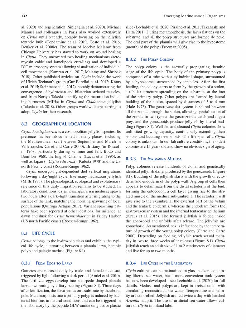
132
al. 2020) and regeneration (Sinigaglia et al. 2020). Michael
Manuel and colleagues in Paris also worked extensively
on Clytia until recently, notably focusing on the jellyfi sh
tentacle bulb (Condamine et al. 2019; Coste et al. 2016;
Denker et al. 2008c). The team of Jocelyn Malamy from
Chicago University has started to work on wound healing
in Clytia. They uncovered two healing mechanisms (acto
myosin cable and lamelipods crawling) and developed a
DIC microscopy system allowing visualization of individual
cell movements (Kamran et al. 2017; Malamy and Shribak
2018). Other published articles on Clytia include the work
of Ulrich Technau’s group (Gur Barzilai et al. 2012; Kraus
et al. 2015; Steinmetz et al. 2012), notably demonstrating the
convergence of hydrozoan and bilaterian striated muscles,
and from Noriyo Takeda identifying the maturation-induc
ing hormones (MIHs) in Clytia and Cladonema jellyfi sh
(Takeda et al. 2018). Other groups worldwide are starting to
adopt Clytia for their research.
8.2 GEOGRAPHICAL LOCATION
Clytia hemisphaerica is a cosmopolitan jellyfish species. Its
presence has been documented in many places, including
the Mediterranean sea (between September and March in
Villefranche; Carré and Carré 2000), Brittany (in Roscoff
in 1968, particularly during summer and fall; Bodo and
Bouillon 1968), the English Channel (Lucas et al. 1995), as
well as Japan (= Clytia edwardsi) (Kubota 1978) and the US
north Pacific coast (Roosen-Runge 1962).
Clytia undergo light-dependent diel vertical migrations
following a day/night cycle, like many hydrozoan jellyfi sh
(Mills 1983). The physiological, ecological and evolutionary
relevance of this daily migration remains to be studied. In
laboratory conditions, Clytia hemisphaerica medusae spawn
two hours after a dark–light transition after migrating to the
surface of the tank, matching the morning spawning of local
populations (Quiroga Artigas 2017). Variant spawning pat
terns have been reported at other locations, for instance, at
dawn and dusk for Clytia hemisphaerica in Friday Harbor
(US north Pacific coast) (Roosen-Runge 1962).
8.3 LIFE CYCLE
Clytia belongs to the hydrozoan class and exhibits the typi
cal life cycle, alternating between a planula larva, benthic
polyp and pelagic medusa (Figure 8.1).
8.3.1 FROM EGGS TO LARVA
Gametes are released daily by male and female medusae,
triggered by light following a dark period (Amiel et al. 2010).
The fertilized eggs develop into a torpedo-shaped planula
larva, swimming by ciliary beating (Figure 8.1). Three days
after fertilization, the larva settles on a substrate by the aboral
pole. Metamorphosis into a primary polyp is induced by bac
terial biofilms in natural conditions and can be triggered in
the laboratory by the peptide GLW-amide on glass or plastic
Emerging Marine Model Organisms
slide (Lechable et al. 2020; Piraino et al. 2011; Takahashi and
Hatta 2011). During metamorphosis, the larva flattens on the
substrate, and all the polyp structures are formed de novo.
The oral part of the planula will give rise to the hypostome
(mouth) of the polyp (Freeman 2005).
8.3.2 THE POLYP COLONY
The polyp colony is the asexually propagating, benthic
stage of the life cycle. The body of the primary polyp is
composed of a tube with a cylindrical shape, surmounted
by a hypostome, surrounded by tentacles. After the fi rst
feeding, the colony starts to form by the growth of a stolon,
a tubular structure spreading on the substrate, at the foot
of the primary polyp. Other polyps are formed by lateral
budding of the stolon, spaced by distances of 3 to 4 mm
(Hale 1973). The gastrovascular system is shared between
all the zooids through the stolon, allowing specialization of
the zooids in two types: the gastrozooids catch and digest
prey, and the gonozooids produce jellyfish by lateral bud
ding (Figure 8.1). Well-fed and cleaned Clytia colonies show
unlimited growing capacity, continuously extending their
stolons and budding new zooids. The life span of a Clytia colony is unknown. In our lab culture conditions, the oldest
colonies are 15 years old and show no obvious sign of aging.
8.3.3 THE SWIMMING MEDUSA
Polyp colonies release hundreds of clonal and genetically
identical jellyfish daily, produced by the gonozooids (Figure
8.1). Budding of the jellyfi sh starts with the growth of ecto
derm and endoderm of the polyp wall. A group of cells then
appears to delaminate from the distal ectoderm of the bud,
forming the entocodon, a cell layer giving rise to the stri
ated muscle of the medusa sub-umbrella. The ectoderm will
give rise to the exumbrella, the external part of the velum
and the tentacle epidermis, whereas the endoderm forms the
gastrovascular system and the internal tentacular epithelium
(Kraus et al. 2015). The formed jellyfish is folded inside
the gonozooid and unfolds after release. The jellyfi sh are
gonochoric. As mentioned, sex is influenced by the tempera
ture of growth of the young polyp colony (Carré and Carré
2000). Depending on feeding, jellyfish reach sexual matu
rity in two to three weeks after release (Figure 8.1). Clytia jellyfish reach an adult size of 1 to 2 centimeters of diameter
and live for up to two months.
8.3.4 LIFE CYCLE IN THE LABORATORY
Clytia cultures can be maintained in glass beakers contain
ing filtered sea water, but a more convenient tank system
has now been developed—see Lechable et al. (2020) for full
details. Medusa and polyps are kept in kreisel tanks with
circulating reconstituted sea water. Temperature and salin
ity are controlled. Jellyfish are fed twice a day with hatched
Artemia nauplii. The use of artificial sea water allows cul
ture of Clytia in inland labs.
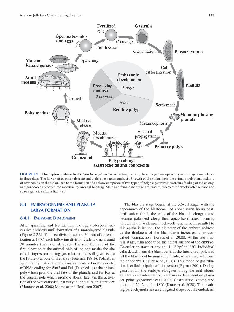
133 Marine Jellyfi sh Clytia hemisphaerica
FIGURE 8.1 The triphasic life cycle of Clytia hemisphaerica. After fertilization, the embryo develops into a swimming planula larva
in three days. The larva settles on a substrate and undergoes metamorphosis. Growth of the stolon from the primary polyp and budding
of new zooids on the stolon lead to the formation of a colony composed of two types of polyps: gastrozooids ensure feeding of the colony,
and gonozooids produce the medusae by asexual budding. Male and female medusae are mature two to three weeks after release and
spawn gametes after a light cue.
8.4 EMBRYOGENESIS AND PLANULA LARVA FORMATION
8.4.1 EMBRYONIC DEVELOPMENT
After spawning and fertilization, the egg undergoes suc
cessive divisions until formation of a monolayered blastula
(Figure 8.2A). The first division occurs 50 min after fertil
ization at 18°C, each following division cycle taking around
30 minutes (Kraus et al. 2020). The initiation site of the
first cleavage at the animal pole of the egg marks the site
of cell ingression during gastrulation and will give rise to
the future oral pole of the larva (Freeman 1981b). Polarity is
specified by maternal determinants localized in the oocyte:
mRNAs coding for Wnt3 and Fz1 (Frizzled 1) at the animal
pole which promote oral fate of the planula and for Fz3 at
the vegetal pole which promote aboral fate, via the activa
tion of the Wnt canonical pathway in the future oral territory
(Momose et al. 2008; Momose and Houliston 2007).
The blastula stage begins at the 32-cell stage, with the
appearance of the blastocoel. At about seven hours post
fertilization (hpf), the cells of the blastula elongate and
become polarized along their apico-basal axes, forming
an epithelium with apical cell–cell junctions. In parallel to
this epithelialization, the diameter of the embryo reduces
as the thickness of the blastoderm increases, a process
called “compaction” (Kraus et al. 2020). At the late blas
tula stage, cilia appear on the apical surface of the embryo.
Gastrulation starts at around 11–12 hpf at 18°C. Individual
cells detach from the blastoderm at the future oral pole and
fill the blastocoel by migrating inside, where they will form
the endoderm (Figure 8.2A, B, C). This mode of gastrula
tion is called unipolar cell ingression (Byrum 2001). During
gastrulation, the embryo elongates along the oral–aboral
axis by a cell intercalation mechanism dependent on planar
cell polarity (Momose et al. 2012). Gastrulation is completed
at around 20–24 hpf at 18°C (Kraus et al. 2020). The result
ing parenchymula has an elongated shape, but the endoderm
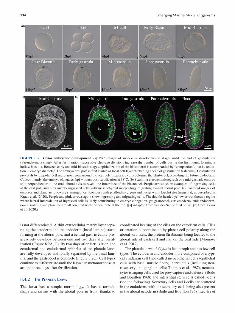
134 Emerging Marine Model Organisms
FIGURE 8.2 Clytia embryonic development. (a) DIC images of successive developmental stages until the end of gastrulation
(Parenchymula stage). After fertilization, successive cleavage divisions increase the number of cells during the first hours, forming a
hollow blastula. Between early and mid-blastula stages, epithelization of the blastoderm is accompanied by “compaction”, that is, reduc
tion in embryo diameter. The embryo oral pole is first visible as local cell layer thickening ahead of gastrulation (asterisks). Gastrulation
proceeds by unipolar cell ingression from around the oral pole. Ingressed cells colonize the blastocoel, providing the future endoderm.
Concomitantly, the embryo elongates. hpf = hours post-fertilization at 18°C. (b) Scanning electron micrograph of a mid-gastrula embryo
split perpendicular to the oral–aboral axis to reveal the inner face of the blastocoel. Purple arrows show examples of ingressing cells
at the oral pole and pink arrows ingressed cells with mesenchymal morphology migrating toward aboral pole. (c) Confocal images of
embryos and planulae following staining of cell contours with phalloidin (green) and nuclei with Hoechst dye (magenta), as described in
Kraus et al. (2020). Purple and pink arrows again show ingressing and migrating cells. The double-headed yellow arrow shows a region
where lateral intercalation of ingressed cells is likely contributing to embryo elongation. gc: gastrocoel, ect: ectoderm, end: endoderm.
(a–c) Gastrula and planulae are all oriented with the oral pole at the top. ([a] Adapted from van der Sande et al. 2020; [b] from Kraus
et al. 2020.)
is not differentiated. A thin extracellular matrix layer sepa- coordinated beating of the cilia on the ectoderm cells. Cilia
rating the ectoderm and the endoderm (basal lamina) starts orientation is coordinated by planar cell polarity along the
forming at the aboral pole, and a central gastric cavity pro- aboral–oral axis, the protein Strabismus being located to the
gressively develops between one and two days after fertil- aboral side of each cell and Fz1 on the oral side (Momose
ization (Figure 8.2A, C). By two days after fertilization, the et al. 2012).
ectodermal and endodermal epithelia of the planula larva The planula larva of Clytia is lecitotroph and has few cell
are fully developed and totally separated by the basal lam- types. The ectoderm and endoderm are composed of a typi
ina, and the gastrocoel is complete (Figure 8.2C). Cell types cal cnidarian cell type called myoepithelial cells (epithelial
continue to differentiate until the larva can metamorphose at cells with basal muscle fibers), nerve cells (including neu
around three days after fertilization. rosensory and ganglion cells; Thomas et al. 1987), nemato
cytes (stinging cells used for prey capture and defense) (Bodo
8.4.2 THE PLANULA LARVA and Bouillon 1968) and interstitial stem cells called i-cells
(see the following). Secretory cells and i-cells are scattered
The larva has a simple morphology. It has a torpedo in the endoderm, with the secretory cells being also present
shape and swims with the aboral pole in front, thanks to in the aboral ectoderm (Bodo and Bouillon 1968; Leclère et
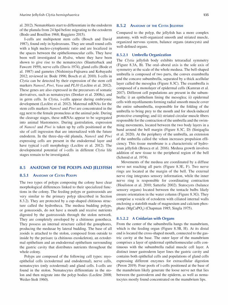
135 Marine Jellyfi sh Clytia hemisphaerica
al. 2012). Nematoblasts start to differentiate in the endoderm
of the planula from 24 hpd before migrating to the ectoderm
(Bodo and Bouillon 1968; Ruggiero 2015).
I-cells are multipotent stem cells (Bosch and David
1987), found only in hydrozoans. They are small round cells
with a high nucleo-cytoplasmic ratio and are localized in
the spaces between the epitheliomuscular cells. They have
been well investigated in Hydra, where they have been
shown to give rise to the nematocytes (Slautterback and
Fawcett 1959), nerve cells (Davis 1974), gland cells (Bode et
al. 1987) and gametes (Nishimiya-Fujisawa and Kobayashi
2012; reviewed in: Bode 1996; Bosch et al. 2010). I-cells in
Clytia can be detected by their expression of the stem cell
markers Nanos1, Piwi, Vasa and PL10 (Leclère et al. 2012).
These genes are also expressed in the precursors of somatic
derivatives, such as nematocytes (Denker et al. 2008c), and
in germ cells. In Clytia, i-cells appear during embryonic
development (Leclère et al. 2012). Maternal mRNAs for the
stem cells markers Nanos1 and Piwi are concentrated in the
egg next to the female pronucleus at the animal pole. During
the cleavage stages, these mRNAs appear to be segregated
into animal blastomeres. During gastrulation, expression
of Nanos1 and Piwi is taken up by cells positioned at the
site of cell ingression that are internalized with the future
endoderm. In the three-day-old planula, Nanos1 and Piwi expressing cells are present in the endodermal layer and
have typical i-cell morphology (Leclère et al. 2012). The
developmental potential of i-cells in different Clytia life
stages remain to be investigated.
8.5 ANATOMY OF THE POLYPS AND JELLYFISH
8.5.1 ANATOMY OF CLYTIA POLYPS
The two types of polyps composing the colony have clear
morphological differences linked to their specialized func
tions in the colony. The feeding polyps or gastrozooids are
very similar to the primary polyp (described in Section
8.3.2). They are protected by a cup-shaped chitinous struc
ture called the hydrotheca. The medusa budding polyps,
or gonozooids, do not have a mouth and receive nutrients
digested by the gastrozooids through the stolon network.
They are completely enveloped by a chitinous gonotheca.
They possess an internal structure called the gonophores,
producing the medusae by lateral budding. The base of all
zooids is attached to the stolon, composed from outside to
inside by the perisarc (a chitinous exoskeleton), an ectoder
mal epithelium and an endodermal epithelium surrounding
the gastric cavity that distributes nutrients throughout the
whole colony.
Polyps are composed of the following cell types: myo
epithelial cells (ectodermal and endodermal), nerve cells,
nematocytes (only ectodermal) and gland cells. I-cells are
found in the stolon. Nematocytes differentiate in the sto
lon and then migrate into the polyp bodies (Leclère 2008;
Weiler-Stolt 1960 ).
8.5.2 ANATOMY OF THE CLYTIA JELLYFISH
Compared to the polyp, the jellyfish has a more complex
anatomy, with well-organized smooth and striated muscle,
organized nervous system, balance organs (statocysts) and
well-defi ned organs.
8.5.2.1 Umbrella Organization The Clytia jellyfish body exhibits tetraradial symmetry
(Figure 8.3A, B). The oral–aboral axis is the sole axis of
symmetry at the scale of the whole medusa. The bell-shaped
umbrella is composed of two parts, the convex exumbrella
and the concave subumbrella, separated by a thick acellular
layer called the mesoglea (Figure 8.3C ). The exumbrella is
composed of a monolayer of epidermal cells (Kamran et al.
2017). Different cell populations are present in the subum
brella: i) an epithelium lining the mesoglea; ii) epidermal
cells with myofilaments forming radial smooth muscle cover
the entire subumbrella, responsible for the folding of the
umbrella to bring prey to the mouth and for shock-induced
protective crumpling; and iii) striated circular muscle fi bers
responsible for the contraction of the umbrella and the swim
ming movements, located between the two body layers in a
band around the bell margin (Figure 8.3C, D) (Sinigaglia
et al. 2020). At the periphery of the umbrella, an extension
of the umbrella called the velum increases propulsion effi
ciency. This tissue membrane is a characteristic of hydro
zoan jellyfish (Brusca et al. 2016). Medusa growth involves
addition of new tissue to the peripheral region of the bell
(Schmid et al. 1974).
Movements of the medusa are coordinated by a diffuse
nerve net reaching all parts (Figure 8.3E, F). Two nerve
rings are located at the margin of the bell. The external
nerve ring integrates sensory information, while the inner
nerve ring is responsible for coordinating contraction
(Houliston et al. 2010; Satterlie 2002). Statocysts (balance
sensory organs) located between the tentacle bulbs likely
ensure orientation in the water column (Figure 8.3G). They
comprise a vesicle of ectoderm with ciliated internal walls
enclosing a statolith made of magnesium and calcium phos
phate (MgCaPO4 ) ( Chapman 1985 ; Singla 1975 ).
8.5.2.2 A Cnidarian with Organs From the center of the subumbrella hangs the manubrium,
which is the feeding organ (Figure 8.3B, H). At its distal
end is located the cross-shaped mouth, connected to the gas
tric cavity at the base. The outer layer of the manubrium
comprises a layer of epidermal epitheliomuscular cells con
tinuous with the subumbrella radial muscle cell layer. A
distinct inner gastroderm layer lines the gastric cavity and
contains both epithelial cells and populations of gland cells
expressing different enzymes for extracellular digestion
(Peron 2019). Four pools of i-cells positioned at the base of
the manubrium likely generate the loose nerve net that lies
between the gastroderm and the epiderm, as well as nema
tocytes mostly found concentrated on the manubrium lips.
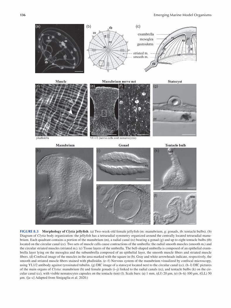
136 Emerging Marine Model Organisms
FIGURE 8.3 Morphology of Clytia jellyfi sh. (a) Two-week-old female jellyfish (m: manubrium, g: gonads, tb: tentacle bulbs). (b)
Diagram of Clytia body organization: the jellyfish has a tetraradial symmetry organized around the centrally located tetraradial manu
brium. Each quadrant contains a portion of the manubrium (m), a radial canal (rc) bearing a gonad (g) and up to eight tentacle bulbs (tb)
located on the circular canal (cc). Two sets of muscle cells cause contractions of the umbrella: the radial smooth muscles (smooth m.) and
the circular striated muscles (striated m.). (c) Tissue layers of the umbrella. The bell-shaped umbrella is composed of an epithelial exum
brella layer lying on the mesoglea and the subumbrella composed of an epithelial layer, the smooth muscle fibers and striated muscle
fibers. (d) Confocal image of the muscles in the area marked with the square in (b). Gray and white arrowheads indicate, respectively, the
smooth and striated muscle fi bers stained with phalloidin. (e–f) Nervous system of the manubrium visualized by confocal microscopy,
using YL1/2 antibody against tyrosinated tubulin. (g) DIC image of a statocyst located next to the circular canal (cc). (h–l) DIC pictures
of the main organs of Clytia: manubrium (h) and female gonads (i–j) linked to the radial canals (rc), and tentacle bulbs (k) on the cir
cular canal (cc), with visible nematocytes capsules on the tentacle (ten) (l). Scale bars: (a) 1 mm, (d,f) 20 μm, (e) (h–k) 100 μm, (G,L) 50
μm. ([a–c] Adapted from Sinigaglia et al. 2020.)
137 Marine Jellyfi sh Clytia hemisphaerica
Nutrients are distributed to the umbrella through four
radial canals, which run from the manubrium to the umbrella
margin and are linked to the circular canal around the bell
periphery. Four gonads are located on the radial canals and
become visible as they start to swell during the growth of
the medusa (Figure 8.3I, J). They become ready to release
fully grown oocytes or sperm after two to three weeks.
Proliferating cells, germline precursors deriving from the
i-cells and growing oocytes are sandwiched between two
epithelial layers: the gastroderm, continuous with the radial
canal endoderm, and a thin epidermal covering (Amiel et
al. 2010). Proliferating cells and early stages of differentia
tion are positioned closer to the bell, whereas the growing
oocytes are located on the flanks of the gonad (Amiel and
Houliston 2009; Jessus et al. 2020). Spawning is triggered
by dark–light transitions.
The circular canal bears the tentacle bulbs, the struc
ture producing nematocyte-rich tentacles (Figure 8.3K, L).
After release from the gonozooid, the baby jellyfi sh has
four primary tentacle bulbs located at the junction between
the radial and circular canals. Additional bulbs are added
during the growth of the umbrella, to a maximum of 32.
Nematogenesis takes place in the ectoderm of the tentacle
bulbs, which is polarized (Denker et al. 2008c). I-cells
expressing Nanos1 and Piwi are located in the proximal
area only, while genes for the different stages of nematogen
esis (mcol3–4a, dkk, NOWA) are expressed in a staggered
way along the ectoderm of the bulb. During nematogenesis,
nematoblasts are thus displaced from the proximal area of
the bulb to the distal area and end up in the tentacle, forming
a conveyor belt (Condamine et al. 2019; Coste et al. 2016;
Denker et al. 2008c).
Cnidarians are often considered to lack true organs (e.g.:
Pierobon 2012). In Clytia medusae, however, manubrium,
gonads and tentacle bulbs can be defined as such. Indeed,
they are specialized structures performing specifi c func
tions (feeding and digestion, tentacle production, oocyte
production), harboring distinct cell types (gland cells,
nematocytes, germ line) and i-cell populations (manubrium:
Sinigaglia et al. 2020; gonads: Leclère et al. 2012; and tenta
cle bulbs: Denker et al. 2008c). Moreover, these three organs
are still able to perform their functions for several days after
isolation from the jellyfish. Isolated gonads are able to sup
port oocyte growth, maturation and spawning (Amiel and
Houliston 2009; Quiroga Artigas et al. 2018); isolated manu
bria will catch and digest prey (Peron 2019); and isolated
tentacle bulbs will keep producing tentacles.
8.6 GENOMIC DATA
8.6.1 THE CLYTIA HEMISPHAERICA GENOME
The genomes of Nematostella vectensis ( Putnam et al.
2007 ) and Hydra magnipapillata (Chapman et al. 2010)
were the fi rst cnidarian genomes to be published. Genomes
from the five main cnidarians classes are now available,
with the first genomes of jellyfish species published in 2019
(Gold et al. 2019; Khalturin et al. 2019; Kim et al. 2019;
Leclère et al. 2019; Ohdera et al. 2019). The sequences of
the different genomes showed that cnidarians possess all the
main families of signaling pathways and transcription fac
tors regulating development found in bilaterians (reviewed
in: Schnitzler 2019; Technau and Schwaiger 2015).
The genome of Clytia, derived from the self-crossed
lab Z strains (see Section 8.1.2), was made publicly avail
able in 2019 (Leclère et al. 2019; http://marimba.obs-vlfr.fr/
home). It was the first published genome of a hydrozoan jel
lyfish. Sequencing was performed by the Genoscope using a
whole-genome shotgun approach. The overall length of the
published assembly was 445 megabases (Leclère et al. 2019);
26,727 genes and 69,083 transcripts were identifi ed, which
are distributed on 15 chromosome pairs. The frequency of
polymorphism was relatively low (0.9%).
Analyses of the genome highlighted gene gain and loss
in the Clytia lineage. Examples of horizontal gene transfer
(HGT) were identified including one of two UDP-glucose
6-dehydrogenase-like genes (Leclère et al. 2019). This
enzyme is used for biosynthesis of proteoglycans and known
to regulate signaling pathways during embryonic devel
opment. Some examples of gene family expansion were
also identified in Clytia, such as the Innexin gap junction
genes, GFP and Clytin photoprotein genes, with 39, 14 and
18 copies, respectively (Leclère et al. 2019). The analyses
also revealed extensive losses of transcription factors in
the hydrozoan lineage and notably several homeobox-con
taining transcription factors involved in nervous system
development in bilaterians, as well as genes regulating the
anthozoan secondary body axis.
Comparisons of transcriptomes from life cycle stages
(Leclère et al. 2019) highlighted the different gene usage
at planula, polyp and medusa stages. Planula stages are
enriched with GPCR signaling components, polyp and
medusa stages with cell–cell and cell–matrix adhesion pro
teins and medusa stages with a subset of transcription fac
tors (Leclère et al. 2019). Many of the bilaterian orthologs
of transcription factors specifically expressed at the medusa
play important functions in neural patterning during devel
opment. Clytia -specific genes, with no identifi able ortholog
in any other species, were also found to be enriched in all
three stages (Leclère et al. 2019).
Together, Clytia recently published genomic and tran
scriptomic data revealed that: i) the genome of Clytia evolved rapidly since the divergence of hydrozoans and
anthozoans, ii) this rapid evolution in the hydrozoan lineage
can be linked to the evolutionary acquisition of the medusa
stage and to morphological simplification of the planula and
polyp and iii) the medusa stage is enriched in transcription
factors conserved between bilaterians and cnidarians. Since
these genes are not expressed in the planula and associ
ated with nervous structures, they are likely involved in the
establishment or maintenance of neural cell types (Leclère
et al. 2019).
138
8.6.2 TRANSCRIPTOMIC DATA
In addition to the data included in the genome release,
other transcriptomic data have been published. These
focus on the gastrula stage (Lapébie et al. 2014) and ten
tacle bulbs (Condamine et al. 2019), as well the early
stages of manubrium regeneration (Sinigaglia et al. 2020).
Transcriptomes of the different tissue composing the gonad
(ectoderm, endoderm, growing and fully grown oocytes)
were also generated to help identify actors of oocyte matu
ration (Quiroga Artigas et al. 2018). About 90,000 EST
and full-length sequences from cDNA libraries derived
from a mix of stages (embryo, larva and medusa) are also
available on NCBI dbEST (Forêt et al. 2010; Philippe et al.
2009 ).
8.7 FUNCTIONAL APPROACHES: TOOLS FOR MOLECULAR AND CELLULAR ANALYSES
Clytia is amenable for the development of tools for experi
mental biology at the cellular and molecular levels.
8.7.1 CELLULAR ANALYSIS
Clytia eggs and jellyfish can be easily manipulated in a
petri dish under a stereomicroscope and kept in beakers
or six-well plastic plates in an incubator for further obser
vation and manipulation. This allows pharmacological
treatments for several days, as well as surgical proce
dures like dissections and grafts (Figure 8.4A–E) (jelly
fish: Sinigaglia et al. 2020; embryos: Leclère et al. 2012;
Momose and Houliston 2007). Manubriums and gonads
can be easily grafted, the grafted organs connecting to the
canal system of the host jellyfish (Figure 8.4A–E). The
grafting approach in adult jellyfish was used to deter
mine whether the manubrium could be a source of induc
tive of inhibitory signals during manubrium regeneration
(Sinigaglia et al. 2020). Regeneration of the manubrium
was not impaired by the grafting of an entire manubrium
on the medusa subumbrella except after a graft in close
proximity to the wound area, therefore excluding the
hypothesis of long-range inhibition from the manubrium
(Sinigaglia et al. 2020).
Embryonic stages, polyps and jellyfish are entirely
transparent, making staining and imaging of differ
ent cell populations possible on fixed and living samples.
Immunohistochemistry, in situ hybridization and stain
ing using the click-it chemistry (EdU and TUNEL) are
performed routinely on this species and can be combined
with in situ hybridization ( Figure 8.4F –H) ( Sinigaglia et al.
2018 ). A combination of the EdU click-it staining marking
proliferating cell and detection of i-cells by in situ hybrid
ization with the probe Nanos1 during regeneration of the
manubrium demonstrated the displacement of Nanos1+ cells from the gonad to the regenerating manubrium to be
followed ( Sinigaglia et al. 2020 ).
Emerging Marine Model Organisms
8.7.2 GENE FUNCTION ANALYSIS DURING
EMBRYOGENESIS AND OOCYTE MATURATION
The jellyfish used in the lab have the same genetic back
ground, and it is easy to perform fertilizations and obtain
embryo stages, facilitating gene function analyses (gain
and loss of function) by injection of ARNs or MOs into the
unfertilized egg (Figure 8.4I) (Momose and Houliston 2007;
Momose et al. 2008). The high efficiency of loss of function
by MO is likely due to low sequence polymorphism in the
laboratory strains. Injection of mRNAs and MOs into the
egg has helped us understand mechanisms involved in estab
lishing polarity in Clytia larvae by revealing the function
of maternal localized mRNAs (Wnt3, Fzl1 and Fzl3—see
Section 8.4.1) (Figure 8.4J).
Clytia gonads are particularly convenient to study the
molecular mechanisms underlying oogenesis. They are
transparent, contain different stages of oocyte growth and
continue to mature and release eggs following dark–light
transition even isolated from the body of the jellyfi sh (Amiel
et al. 2009). These characteristics were used to study the role
of the Mos proteins, a conserved kinase family regulating
meiosis (Amiel et al. 2009). Injection of MOs and mRNAs
into the oocyte demonstrated the role of the two Clytia Mos
homologs during oocyte maturation in regulating the for
mation and localization of the meiotic spindle, as well as
oocyte cell cycle arrest after meiosis (Amiel et al. 2009).
These functions have also been described in bilaterian spe
cies and likely represent an ancestral function of this protein
family (Amiel et al. 2009).
8.7.3 GENE FUNCTION ANALYSIS IN THE ADULT
8.7.3.1 RNA Interference RNA interference (RNAi) has been successfully used for
downregulation of gene expression in the adult in the cni
darian Hydractinia, allowing, for instance, study of the role
of i-cell genes during regeneration (Bradshaw et al. 2015).
Gene expression perturbation through RNAi has not yet been
performed in Clytia jellyfish; however, preliminary results
indicate that the cellular machinery is present in Clytia lar
vae. Another promising avenue to explore is shRNA, also
effective in both Hydractinia and Nematostella ( DuBuc
et al. 2020; He et al. 2018).
8.7.3.2 The Development of Mutant Lines A robust protocol for achieving loss of gene function in
Clytia lines by CRISPR/Cas9 has been developed (Figure
8.4K) (Momose et al. 2018). The approach was first tested on
a gene involved in ciliogenesis (CheRfx123), whose defect
leads to defect in sperm motility, and genes coding for
the fluorescent protein GFP (Figure 8.4K) (double mutant
GFP1/GFP2 in F1) (Momose et al. 2018). After injection
of high doses of Cas9 RNP, mutants in the F0 generation
were nearly non-mosaic and already had visible phenotypes
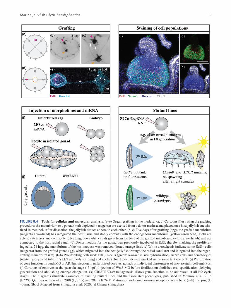
139 Marine Jellyfi sh Clytia hemisphaerica
FIGURE 8.4 Tools for cellular and molecular analysis. (a–e) Organ grafting in the medusa. (a, d) Cartoons illustrating the grafting
procedure: the manubrium or a gonad (both depicted in magenta) are excised from a donor medusa and placed on a host jellyfi sh anesthe
tized in menthol. After dissection, the jellyfish tissues adhere to each other. (b, c) Five days after grafting (dpg), the grafted manubrium
(magenta arrowhead) has integrated the host tissue and stably coexists with the endogenous manubrium (yellow arrowhead). Both are
able to catch prey and contribute to feeding; new radial canals grew from the base of the grafted manubrium (white arrowheads) and are
connected to the host radial canal. (d) Donor medusa for the gonad was previously incubated in EdU, thereby marking the proliferat
ing cells. 24 hpg, the manubrium of the host medusa was removed (dotted orange line). (e) White arrowheads indicate some EdU+ cells
(magenta) from the grafted gonad (gg), which migrated into the host jellyfish through the radial canal (rc) and integrated into the regen
erating manubrium (rm). (f–h) Proliferating cells (red: EdU), i-cells (green: Nanos1 in situ hybridization), nerve cells and nematocytes
(white: tyrosynated tubulin YL1/2 antibody staining) and nuclei (blue: Hoechst) were marked in the same tentacle bulb. (i) Perturbation
of gene function through MO or ARNm injection in unfertilized oocytes, gonads or individual blastomeres of two- to eight-cell embryos.
(j) Cartoons of embryos at the gastrula stage (15 hpf). Injection of Wnt3 MO before fertilization abolishes oral specifi cation, delaying
gastrulation and abolishing embryo elongation. (k) CRISPR/Cas9 mutagenesis allows gene function to be addressed at all life cycle
stages. The diagrams illustrate examples of existing mutant lines and the associated phenotypes, published in Momose et al. 2018
(GFP1), Quiroga Artigas et al. 2018 (Opsin9 ) and 2020 (MIH-R: Maturation inducing hormone receptor). Scale bars: (e–h) 100 μm, (J)
40 μm. ([b, c] Adapted from Sinigaglia et al. 2020; [e] Chiara Sinigaglia.)
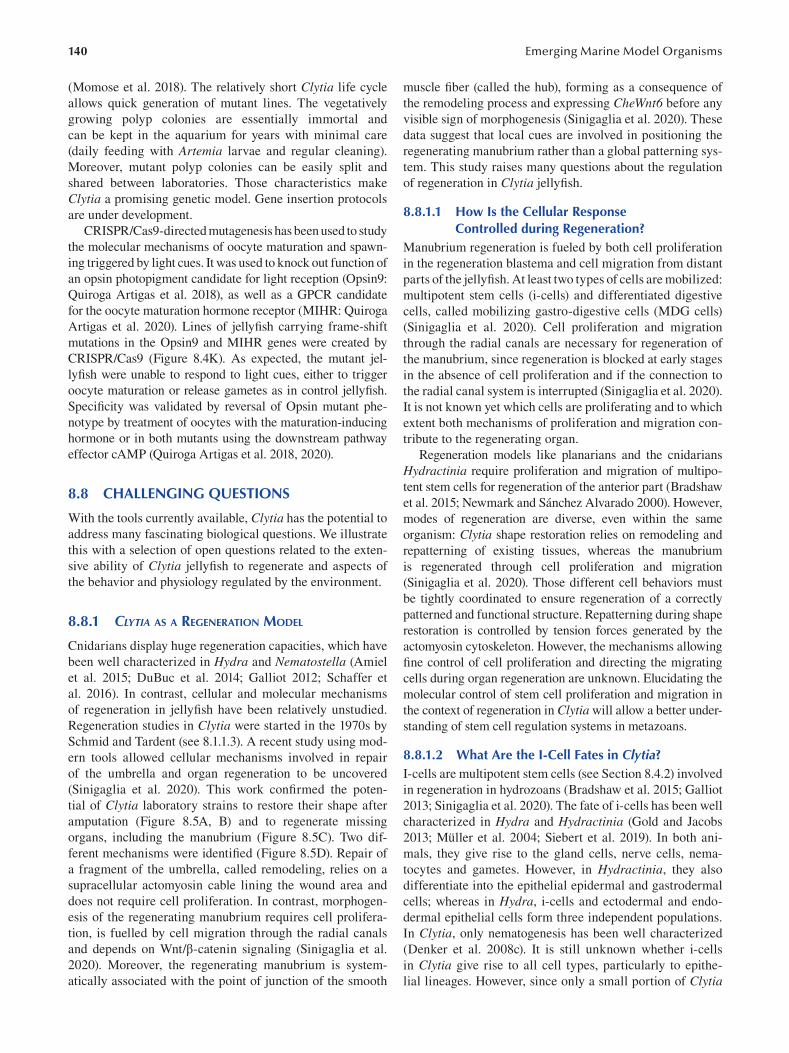
140
(Momose et al. 2018). The relatively short Clytia life cycle
allows quick generation of mutant lines. The vegetatively
growing polyp colonies are essentially immortal and
can be kept in the aquarium for years with minimal care
(daily feeding with Artemia larvae and regular cleaning).
Moreover, mutant polyp colonies can be easily split and
shared between laboratories. Those characteristics make
Clytia a promising genetic model. Gene insertion protocols
are under development.
CRISPR/Cas9-directed mutagenesis has been used to study
the molecular mechanisms of oocyte maturation and spawn
ing triggered by light cues. It was used to knock out function of
an opsin photopigment candidate for light reception (Opsin9:
Quiroga Artigas et al. 2018), as well as a GPCR candidate
for the oocyte maturation hormone receptor (MIHR: Quiroga
Artigas et al. 2020). Lines of jellyfish carrying frame-shift
mutations in the Opsin9 and MIHR genes were created by
CRISPR/Cas9 (Figure 8.4K). As expected, the mutant jel
lyfish were unable to respond to light cues, either to trigger
oocyte maturation or release gametes as in control jellyfi sh.
Specificity was validated by reversal of Opsin mutant phe
notype by treatment of oocytes with the maturation-inducing
hormone or in both mutants using the downstream pathway
effector cAMP (Quiroga Artigas et al. 2018, 2020).
8.8 CHALLENGING QUESTIONS
With the tools currently available, Clytia has the potential to
address many fascinating biological questions. We illustrate
this with a selection of open questions related to the exten
sive ability of Clytia jellyfish to regenerate and aspects of
the behavior and physiology regulated by the environment.
8.8.1 CLYTIA AS A REGENERATION MODEL
Cnidarians display huge regeneration capacities, which have
been well characterized in Hydra and Nematostella ( Amiel
et al. 2015; DuBuc et al. 2014; Galliot 2012; Schaffer et
al. 2016). In contrast, cellular and molecular mechanisms
of regeneration in jellyfish have been relatively unstudied.
Regeneration studies in Clytia were started in the 1970s by
Schmid and Tardent (see 8.1.1.3). A recent study using mod
ern tools allowed cellular mechanisms involved in repair
of the umbrella and organ regeneration to be uncovered
(Sinigaglia et al. 2020). This work confirmed the poten
tial of Clytia laboratory strains to restore their shape after
amputation (Figure 8.5A, B) and to regenerate missing
organs, including the manubrium (Figure 8.5C ). Two dif
ferent mechanisms were identified (Figure 8.5D). Repair of
a fragment of the umbrella, called remodeling, relies on a
supracellular actomyosin cable lining the wound area and
does not require cell proliferation. In contrast, morphogen
esis of the regenerating manubrium requires cell prolifera
tion, is fuelled by cell migration through the radial canals
and depends on Wnt/β-catenin signaling (Sinigaglia et al.
2020). Moreover, the regenerating manubrium is system
atically associated with the point of junction of the smooth
Emerging Marine Model Organisms
muscle fiber (called the hub), forming as a consequence of
the remodeling process and expressing CheWnt6 before any
visible sign of morphogenesis (Sinigaglia et al. 2020). These
data suggest that local cues are involved in positioning the
regenerating manubrium rather than a global patterning sys
tem. This study raises many questions about the regulation
of regeneration in Clytia jellyfi sh.
8.8.1.1 How Is the Cellular Response Controlled during Regeneration?
Manubrium regeneration is fueled by both cell proliferation
in the regeneration blastema and cell migration from distant
parts of the jellyfish. At least two types of cells are mobilized:
multipotent stem cells (i-cells) and differentiated digestive
cells, called mobilizing gastro-digestive cells (MDG cells)
(Sinigaglia et al. 2020). Cell proliferation and migration
through the radial canals are necessary for regeneration of
the manubrium, since regeneration is blocked at early stages
in the absence of cell proliferation and if the connection to
the radial canal system is interrupted (Sinigaglia et al. 2020).
It is not known yet which cells are proliferating and to which
extent both mechanisms of proliferation and migration con
tribute to the regenerating organ.
Regeneration models like planarians and the cnidarians
Hydractinia require proliferation and migration of multipo
tent stem cells for regeneration of the anterior part (Bradshaw
et al. 2015; Newmark and Sánchez Alvarado 2000). However,
modes of regeneration are diverse, even within the same
organism: Clytia shape restoration relies on remodeling and
repatterning of existing tissues, whereas the manubrium
is regenerated through cell proliferation and migration
(Sinigaglia et al. 2020). Those different cell behaviors must
be tightly coordinated to ensure regeneration of a correctly
patterned and functional structure. Repatterning during shape
restoration is controlled by tension forces generated by the
actomyosin cytoskeleton. However, the mechanisms allowing
fine control of cell proliferation and directing the migrating
cells during organ regeneration are unknown. Elucidating the
molecular control of stem cell proliferation and migration in
the context of regeneration in Clytia will allow a better under
standing of stem cell regulation systems in metazoans.
8.8.1.2 What Are the I-Cell Fates in Clytia? I-cells are multipotent stem cells (see Section 8.4.2) involved
in regeneration in hydrozoans (Bradshaw et al. 2015; Galliot
2013; Sinigaglia et al. 2020). The fate of i-cells has been well
characterized in Hydra and Hydractinia (Gold and Jacobs
2013; Müller et al. 2004; Siebert et al. 2019). In both ani
mals, they give rise to the gland cells, nerve cells, nema
tocytes and gametes. However, in Hydractinia, they also
differentiate into the epithelial epidermal and gastrodermal
cells; whereas in Hydra, i-cells and ectodermal and endo
dermal epithelial cells form three independent populations.
In Clytia, only nematogenesis has been well characterized
(Denker et al. 2008c). It is still unknown whether i-cells
in Clytia give rise to all cell types, particularly to epithe
lial lineages. However, since only a small portion of Clytia
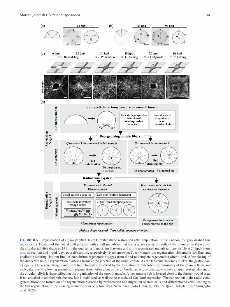
141 Marine Jellyfi sh Clytia hemisphaerica
FIGURE 8.5 Regeneration of Clytia jellyfish. (a–b) Circular shape restoration after amputation. In the cartoon, the gray dashed line
indicates the location of the cut. A half jellyfish with a half manubrium (a) and a quarter jellyfish without the manubrium (b) recover
the circular jellyfish shape in 24 h. In the quarter, a manubrium blastema and a tiny regenerated manubrium are visible at 24 hpd (hours
post-dissection) and 4 dpd (days post-dissection), respectively (black arrowhead). (c) Manubrium regeneration. Schematic (top line) and
phalloidin staining (bottom line) of manubrium regeneration stages from 6 hpd to complete regeneration after 4 dpd. After closing of
the dissection hole, a regeneration blastema forms at the junction of the radial canals. As the blastema becomes thicker, the gastric cav
ity opens. The regenerating manubrium fi rst elongates, followed by the formation of four lobes. (d) Summary of the main cellular and
molecular events allowing manubrium regeneration. After a cut in the umbrella, an actomyosin cable allows a rapid reestablishment of
the circular jellyfish shape, affecting the organization of the smooth muscle. A new muscle hub is formed close to the former wound area.
If not attached to another hub, the new hub is stabilized, as well as the associated CheWnt6 expression. The connection to the radial canal
system allows the formation of a regeneration blastema by proliferation and migration of stem cells and differentiated cells, leading to
the full regeneration of the missing manubrium in only four days. Scale bars: (a–b) 1 mm, (c) 100 μm. ([a–d] Adapted from Sinigaglia
et al. 2020.)
142
proliferating cells express Nanos1, the Clytia i-cell system
is likely to be similar to Hydra with separated i-cells and
epithelial lineages. Transgenic lines with reporters for dif
ferent cell populations allowing in vivo tracing of i-cell are
necessary to identify i-cell derivatives.
It is also unknown whether all Nanos1 -expressing cells
have the same potency and particularly whether some are
committed to the germline. After complete ablation, the
gonads regenerate, and oocyte growth resumes. This could
indicate the presence of multipotent stem cell populations in
the main organs, migrating through the radial canal to repop
ulate the regenerating gonads. Clytia is a promising model
to study early oocyte differentiation because the gonads are
fully transparent and continue to function when isolated
from the jellyfi sh.
8.8.1.3 How Are Mechanical Cues and Signaling Pathways Integrated?
After amputation, actomyosin contractility at the wound
area ensures restoration of the circular jellyfi sh shape.
During shape restoration, the signaling molecule CheWnt6 is expressed at the wound site. Its expression is inhibited
by pharmacological inhibition of actomyosin contractility,
suggesting a likely modulation of Wnt/β-catenin activity by
mechanical cues (Sinigaglia et al. 2020). How mechanical
cues can activate Wnt/β-catenin pathway and thus permit
regeneration of the manubrium is unknown. The integra
tion between mechanical cues and signaling pathways has
been raising interest (Chiou and Collins 2018; Heisenberg
and Bellaïche 2013; Urdy 2012; Vining and Mooney 2017 ).
In Hydra, the actin cytoskeleton has also been proposed to
influence body axis formation during regeneration (Livshits
et al. 2017, Maroudas-Sacks et al. 2021) and is likely to
be interacting with the Wnt/β-catenin signaling pathway,
inducing hypostome formation at the oral pole (Broun 2005;
Gee et al. 2010).
8.8.2 REGULATION OF BEHAVIOR AND
PHYSIOLOGY BY ENVIRONMENTAL CUES
Clytia life cycle and physiology of the different life stages
are influenced by the environment in many ways: i) in
the ocean, settlement of the planula larva occurs upon an
unknown cue from bacterial biofilms; ii) growth of the
polyp colony is constrained by feeding and space availabil
ity; iii) sex of the released medusa can be infl uenced by the
temperature at which the polyp colony is growing; and iv) in
the jellyfish, oocyte maturation and gamete release are trig
gered by a light stimulus. Gamete release is associated with
light information in many cnidarian species (e.g. scyphozo
ans Pelagia: Lilley et al. 2014; Clytia: Amiel et al. 2010).
8.8.2.1 Which Bacterial Cues Induce Settlement of the Planula? Which Molecular Mechanisms Are Triggered?
In cnidarians, including Clytia, settlement of the planula
larva and metamorphosis into a primary polyp is induced
Emerging Marine Model Organisms
by bacterial biofi lms (Hydractinia echinata: Kroiher and
Berking 1999; Leitz and Wagner 1993; Seipp et al. 2007;
Acropora sp: Negri et al. 2001; Tebben et al. 2011; Webster
et al. 2004). The cellular response is mediated by neuro
peptides of the GLW-amide family, secreted by sensory
neurons of the planula (Takahashi and Takeda 2015).
Synthetic GLW-amide neuropeptides induce settlement
and metamorphosis in laboratory conditions in several
planulae (Acropora: Iwao et al. 2002; Hydractinia: Müller
and Leitz 2002; both reviewed in: Takahashi and Hatta
2011). Concerning Clytia planula, the synthetic peptide
GLWamide2 (GNPPGLW-NH2) has been used in the labo
ratory to induce settlement (Momose et al. 2018; Quiroga
Artigas et al. 2018). A recent study testing the effi ciency of
15 other neuropeptides, derived from sequences of potential
GLWamide precursors, showed that GLWamide-6 (pyro-
Glu-QQAPKGLW-NH3) has an even greater effi ciency
(Lechable et al. 2020).
The roles of bacteria and neuropeptides in settlement
have long been known. However, the signal from the bac
teria inducing settlement and metamorphosis, as well as the
molecular mechanisms triggering settlement and metamor
phosis, are still unknown. The morphological and cellular
events occurring during the metamorphosis of Clytia plan
ula have been recently studied (Krasovec 2020) and provide
a framework for further studies on metamorphosis.
8.8.2.2 Is There a Physiological Link between Gametogenesis and Nutrition?
In Clytia jellyfish, spawning and oocyte maturation occurs
in males and females two hours after a light stimulus (Amiel
et al. 2010). Part of the signaling cascade triggering light-
induced oocyte maturation has recently been elucidated.
After light reception by the photoprotein Opsin9 by neuro
secretory cells of the gonad ectoderm, those cells release a
maturation-inducing hormone (Quiroga Artigas et al. 2018).
MIH activates in turn a GPCR, located on the oocyte sur
face, called the MIH-Receptor, thus triggering the rise in
cAMP responsible for the initiation of oocyte maturation
(Quiroga Artigas et al. 2020). Besides their function in
oocyte maturation, Clytia MIH and MIH-R are likely to play
a role in nutrition or other physiological processes. Indeed,
both are expressed in the gastrovascular system and the ten
tacles as well as in the gonads. Moreover, MIHR is part of
a superfamily of cnidarian and bilaterian GPCRs playing a
role in nutrition, as well as regulation of sexual reproduction
(Quiroga Artigas et al. 2020). Additional knowledge in the
functions of Clytia MIHR could give insight in the evolution
of the link between gametogenesis and nutrition.
8.8.2.3 How Does Feeding Availability Regulate Growth of Polyps and Medusa?
Some cnidarians are able to modify their size depending
on feeding availability. The jellyfi sh Pelagia noctiluca and
Aurelia aurita shrink during starvation conditions and re
grow when prey are again available (Frandsen and Riisgård
1997; Hamner and Jenssen 1974; Lilley et al. 2014). In
143 Marine Jellyfi sh Clytia hemisphaerica
laboratory conditions, Aurelia aurita loses 3–5% of its mass
per day without feeding and regrows after feeding to reach
the original size. Starved jellyfish are not able to spawn
(Frandsen and Riisgård 1997; Hamner and Jenssen 1974).
Similarly to Aurelia, Pelagia loses about 7% of its mass per
day and can regrow after feeding. However, egg production
is maintained, with a number of eggs correlated with the
size of the jellyfish (Lilley et al. 2014).
The process of shrinking in conditions of starvation is
also a feature of other invertebrates. In planarians, the size
depends on the feeding levels (Felix et al. 2019); in the anne
lid Pristina leidyi, feeding causes the increase and decrease
of the gonads (Özpolat et al. 2016). This process has also
been documented, although more rarely, in the vertebrates.
The marine iguana Amblyrhynchus cristatus can lose up
to 20% of its size after the loss of its main source of food
during El Niño events (Wikelski and Thom 2000). Whether
the same mechanisms are involved between metazoans still
remains to be investigated.
A similar shrinking/re-growth event in case of starva
tion has been observed in Clytia jellyfi sh (unpublished).
Moreover, the gonads also shrink and egg production declines
before totally stopping. Gametogenesis resumes after feeding
of the jellyfish. The recently described MDG cells, with a
putative role in the distribution of nutrients, circulate more
in the canals in case of starvation (Sinigaglia et al. 2020).
Feeding also influences the growth of newly released jelly
fish: indeed, jellyfish fed with smaller prey, and thus with a
bigger food intake, grow faster than jellyfish fed with bigger
prey that are harder to catch (Lechable et al. 2020).
To summarize, in Clytia, like in other cnidarians, the
feeding levels control the rate of growth and gametogenesis.
The cellular and molecular mechanisms allowing the con
trol of growth in Clytia jellyfish are unknown. One level of
regulation is potentially the cell cycle, since in Hydra and
Nematostella polyps, the rate of cell proliferation depends
on the feeding level of the animal (Campbell 1967; Otto
and Campbell 1977; Passamaneck and Martindale 2012;
Webster and Hamilton 1972). Clytia jellyfish could be used
to investigate the feedback between feeding levels and cell
proliferation, as well as cellular events during degrowth.
Many fascinating questions can be addressed with
Clytia. Due to its practicality as a model organism and the
tools already available and in development, Clytia has the
potential to provide a fresh perspective on a wide range of
research topics.
BIBLIOGRAPHY
Abrams, M.J., T. Basinger, W. Yuan, C.-L. Guo, and L. Goentoro.
2015. Self-repairing symmetry in jellyfish through mechani
cally driven reorganization. Proceedings of the National Academy of Sciences. 112: E3365–E3373.
Alder, J. 1856. A notice of some new genera and species of British
hydroid zoophytes. Annals and Magazine of Natural History.
2: 12–14.
Amiel, A., P. Chang, T. Momose, and E. Houliston. 2010. Clytia hemisphaerica: A cnidarian model for studying oogenesis. In
Oogenesis, ed. M.-H. Verlhac & A. Villeneuve, 81–101. John
Wiley & Sons.
Amiel, A., and E. Houliston. 2009. Three distinct RNA localization
mechanisms contribute to oocyte polarity establishment in
the cnidarian Clytia hemisphærica. Developmental Biology.
327: 191–203.
Amiel, A.R., H.T. Johnston, K. Nedoncelle, J.F. Warner, S. Ferreira,
and E. Röttinger. 2015. Characterization of morphological
and cellular events underlying oral regeneration in the sea
anemone, Nematostella vectensis. International Journal of Molecular Sciences. 16: 28449–28471.
Amiel, A., L. Leclère, L. Robert, S. Chevalier, and E. Houliston.
2009. Conserved functions for Mos in eumetazoan oocyte
maturation revealed by studies in a cnidarian. Current Biology. 19: 305–311.
Bodo, F., and J. Bouillon. 1968. Etude histologique du dével
oppement embryonnaire de quelques hydromeduses de
Roscoff: Phialidium hemisphaericum (l.), Obelia sp. peron
et lesueur, Sarsia eximia (allman), Podocoryne carnea (sars),
Gonionemus vertens Agassiz. Cahiers de Biologie Marine.
9: 69–104.
Bode, H.R. 1996. The interstitial cell lineage of Hydra: A stem cell
system that arose early in evolution. Journal of Cell Science.
109: 1155–1164.
Bode, H.R., S. Heimfeld, M.A. Chow, and L.W. Huang. 1987.
Gland cells arise by differentiation from interstitial cells in
Hydra attenuata. Developmental Biology. 122: 577–585.
Bosch, T.C.G., F. Anton-Erxleben, G. Hemmrich, and K. Khalturin.
2010. The Hydra polyp: Nothing but an active stem cell
community. Development Growth and Differentiation . 52:
15–25.
Bosch, T.C.G., and C.N. David. 1987. Stem cells of Hydra magnipapillata can differentiate into somatic cells and germ line
cells. Developmental Biology. 121: 182–191.
Bradshaw, B., K. Thompson, and U. Frank. 2015. Distinct mecha
nisms underlie oral vs aboral regeneration in the cnidarian
Hydractinia echinata. ELife. 4: 1–19.
Broun, M. 2005. Formation of the head organizer in hydra involves
the canonical Wnt pathway. Development. 132: 2907–2916.
Brusca, R.C., W. Moore, and S.M. Shuster. 2016. Invertebrates . 3rd
ed. Sinauer Associates Inc., Sunderland, MA.
Byrum, C.A. 2001. An analysis of hydrozoan gastrulation by uni
polar ingression. Developmental Biology. 240: 627–640.
Campbell, R.D. 1967. Tissue dynamics of steady state growth in
Hydra littoralis. I: Patterns of cell division. Developmental Biology. 15: 487–502.
Carré, D., and C. Carré. 2000. Origin of germ cells, sex determi
nation, and sex inversion in medusae of the genus Clytia (Hydrozoa, Leptomedusae): The influence of temperature.
Journal of Experimental Zoology. 287: 233–242.
Carré, D., C. Carré, F. Pagès, and J.-M. Gili. 1995. Asexual repro
duction in the pelagic phase of Clytia mccrady (Hydrozoa,
Leptomedusae). Scientia Marina. 59: 193–202.
Chapman, D.M. 1985. X-ray microanalysis of selected coelenterate
statoliths. Journal of the Marine Biological Association of the United Kingdom. 65: 617–627.
Chapman, J.A., E.F. Kirkness, O. Simakov, S.E. Hampson, T.
Mitros, T. Weinmaier, T. Rattei, P.G. Balasubramanian, J.
Borman, D. Busam, K. Disbennett, C. Pfannkoch, N. Sumin,
G.G. Sutton, L.D. Viswanathan, B. Walenz, D.M. Goodstein,
U. Hellsten, T. Kawashima, et al. 2010. The dynamic genome
of Hydra. Nature. 464: 592–596.
Chevalier, S., A. Martin, L. Leclère, A. Amiel, and E. Houliston.
2006. Polarised expression of FoxB and FoxQ2 genes
144
during development of the hydrozoan Clytia hemisphaerica.
Development Genes and Evolution. 216: 709–720.
Chiori, R., M. Jager, E. Denker, P. Wincker, C. Da Silva, H. Le
Guyader, M. Manuel, and E. Quéinnec. 2009. Are Hox genes
ancestrally involved in axial patterning? evidence from the
hydrozoan Clytia hemisphaerica (Cnidaria). PLoS One . 4:
e4231.
Chiou, K., and E.M.S. Collins. 2018. Why we need mechanics to
understand animal regeneration. Developmental Biology.
433: 155–165.
Collins, A., P. Schuchert, A. Marques, T. Jankowski, M. Medina,
and B. Schierwater. 2006. Medusozoan phylogeny and char
acter evolution clarified by new large and small subunit
RDNA data and an assessment of the utility of phylogenetic
mixture models. Systematic Biology. 55: 97–115.
Condamine, T., M. Jager, L. Leclère, C. Blugeon, S. Lemoine, R.R.
Copley, and M. Manuel. 2019. Molecular characterisation of
a cellular conveyor belt in Clytia medusae. Developmental Biology. 456: 212–225.
Coste, A., M. Jager, J.P. Chambon, and M. Manuel. 2016.
Comparative study of Hippo pathway genes in cellular con
veyor belts of a ctenophore and a cnidarian. EvoDevo . 7: 1–19.
Cunha, A.F., A.G. Collins, and A.C. Marques. 2020. When mor
phometry meets taxonomy: Morphological variation and
species boundaries in Proboscoida (Cnidaria: Hydrozoa).
Zoological Journal of the Linnean Society. 190: 417–447.
Davis, L.E. 1974. Ultrastructural studies of the development of
nerves in Hydra. Integrative and Comparative Biology . 14:
551–573.
Denker, E., E. Bapteste, H. Le Guyader, M. Manuel, and N. Rabet.
2008a. Horizontal gene transfer and the evolution of cnidar
ian stinging cells. Current Biology. 18: 858–859.
Denker, E., A. Chatonnet, and N. Rabet. 2008b. Acetylcholinesterase
activity in Clytia hemisphaerica (Cnidaria). Chemico-Biological Interactions. 175: 125–128.
Denker, E., M. Manuel, L. Leclère, H. Le Guyader, and N. Rabet.
2008c. Ordered progression of nematogenesis from stem
cells through differentiation stages in the tentacle bulb of
Clytia hemisphaerica (Hydrozoa, Cnidaria). Developmental Biology. 315: 99–113.
Derelle, R., T. Momose, M. Manuel, C.D.A. Silva, P. Wincker, and
E. Houliston. 2010. Convergent origins and rapid evolution
of spliced leader trans-splicing in Metazoa: Insights from the
Ctenophora and Hydrozoa. RNA. 16: 696–707.
DuBuc, T.Q., C.E. Schnitzler, E. Chrysostomou, E.T. McMahon,
Febrimarsa, J.M. Gahan, T. Buggie, S.G. Gornik, S. Hanley,
S.N. Barreira, P. Gonzalez, A.D. Baxevanis, and U. Frank.
2020. Transcription factor AP2 controls cnidarian germ cell
induction. Science. 367: 757–762.
DuBuc, T.Q., N. Traylor-Knowles, and M.Q. Martindale. 2014.
Initiating a regenerative response: Cellular and molecular
features of wound healing in the cnidarian Nematostella vectensis. BMC Biology. 12: 1–20.
Felix, D.A., Ó. Gutiérrez-Gutiérrez, L. Espada, A. Thems, and
C. González-Estévez. 2019. It is not all about regeneration:
Planarians striking power to stand starvation. Seminars in Cell and Developmental Biology. 87: 169–181.
Forêt, S., B. Knack, E. Houliston, T. Momose, M. Manuel, E.
Quéinnec, D.C. Hayward, E.E. Ball, and D.J. Miller. 2010.
New tricks with old genes: The genetic bases of novel cnidar
ian traits. Trends in Genetics. 26: 154–158.
Fourrage, C., S. Chevalier, and E. Houliston. 2010. A highly con
served poc1 protein characterized in embryos of the hydro
zoan Clytia hemisphaerica: Localization and functional
studies. PLoS One. 5: e13994.
Emerging Marine Model Organisms
Frandsen, K.T., and H.U. Riisgård. 1997. Size dependent respi
ration and growth of jellyfi sh, Aurelia aurita. Sarsia. 82:
307–312.
Freeman, G. 1980. The role of cleavage in the establishment of
the anterior-posterior axis of the hydrozoan embryo. In
Developmental and Cellular Biology of Coelenterates , ed.
R. Tardent & P. Tardent, 97–108. Elsevier/North Holland.
Amsterdam.
Freeman, G. 1981a. The role of polarity in the development of
the hydrozoan planula larva. Wilhelm Roux’s Archives of Developmental Biology. 190: 168–184.
Freeman, G. 1981b. The cleavage initiation site establishes the
posterior pole of the hydrozoan embryo. Wilhelm Roux’s Archives of Developmental Biology. 190: 123–125.
Freeman, G. 2005. The effect of larval age on developmental
changes in the polyp prepattern of a hydrozoan planula.
Zoology. 108: 55–73.
Freeman, G., and E.B. Ridgway. 1987. Endogenous photoproteins,
calcium channels and calcium transients during metamor
phosis in hydrozoans. Roux’s Archives of Developmental Biology. 196: 30–50.
Galliot, B. 2012. Hydra, a fruitful model system for 270 years.
International Journal of Developmental Biology. 56:
411–423.
Galliot, B. 2013. Regeneration in Hydra. In ELS, John Wiley &
Sons. Chichester.
Gee, L., J. Hartig, L. Law, J. Wittlieb, K. Khalturin, T.C.G. Bosch,
and H.R. Bode. 2010. β-Catenin plays a central role in set
ting up the head organizer in Hydra. Developmental Biology.
340: 116–124.
Gold, D.A., and D.K. Jacobs. 2013. Stem cell dynamics in cni
daria: Are there unifying principles? Development Genes and Evolution. 223: 53–66.
Gold, D.A., T. Katsuki, Y. Li, X. Yan, M. Regulski, D. Ibberson,
T. Holstein, R.E. Steele, D.K. Jacobs, and R.J. Greenspan.
2019. The genome of the jellyfi sh Aurelia and the evolu
tion of animal complexity. Nature Ecology & Evolution . 3:
96–104.
Gur Barzilai, M., A.M. Reitzel, J.E.M. Kraus, D. Gordon, U.
Technau, M. Gurevitz, and Y. Moran. 2012. Convergent evo
lution of sodium ion selectivity in metazoan neuronal signal
ing. Cell Reports. 2: 242–248.
Hale, L.J. 1973. The pattern of growth of Clytia johnstoni. Journal of Embryology and Experimental Morphology. 29: 283–309.
Hamner, W.M., and R.M. Jenssen. 1974. Growth, degrowth, and
irreversible cell differentiation in Aurelia aurita. American Zoologist. 14: 833–849.
Hargitt, C.W. 1897. Recent experiments on regeneration. Zoological Bulletin. 1: 27.
He, S., F. Del Viso, C.Y. Chen, A. Ikmi, A.E. Kroesen, and M.C.
Gibson. 2018. An axial Hox code controls tissue segmenta
tion and body patterning in Nematostella vectensis. Science.
361: 1377–1380.
Heisenberg, C.P., and Y. Bellaïche. 2013. Forces in tissue morpho
genesis and patterning. Cell. 153: 948.
Houliston, E., T. Momose, and M. Manuel. 2010. Clytia hemisphaerica : A jellyfish cousin joins the laboratory. Trends in Genetics. 26: 159–167.
Iwao, K., T. Fujisawa, and M. Hatta. 2002. A cnidarian neuro
peptide of the glwamide family induces metamorphosis of
reef-building corals in the genus Acropora. Coral Reefs . 21:
127–129.
Jessus, C., C. Munro, and E. Houliston. 2020. Managing the oocyte
meiotic arrest—Lessons from frogs and jellyfi sh. Cells . 9:
1150–1185.
145 Marine Jellyfi sh Clytia hemisphaerica
Kamran, Z., K. Zellner, H. Kyriazes, C.M. Kraus, J.B. Reynier, and
J.E. Malamy. 2017. In vivo imaging of epithelial wound heal
ing in the cnidarian Clytia hemisphaerica demonstrates early
evolution of purse string and cell crawling closure mecha
nisms. BMC Developmental Biology. 17: 1–14.
Khalturin, K., C. Shinzato, M. Khalturina, M. Hamada, M. Fujie,
R. Koyanagi, M. Kanda, H. Goto, F. Anton-Erxleben, M.
Toyokawa, S. Toshino, and N. Satoh. 2019. Medusozoan
genomes inform the evolution of the jellyfish body plan.
Nature Ecology and Evolution. 3: 811–822.
Kim, H.M., J.A. Weber, N. Lee, S.G. Park, Y.S. Cho, Y. Bhak, N.
Lee, Y. Jeon, S. Jeon, V. Luria, A. Karger, M.W. Kirschner,
Y.J. Jo, S. Woo, K. Shin, O. Chung, J.C. Ryu, H.S. Yim, J.H.
Lee, J.S. Edwards, A. Manica, J. Bhak, and S. Yum. 2019.
The genome of the giant nomura’s jellyfish sheds light on the
early evolution of active predation. BMC Biology. 17: 1–12.
Krasovec, G. 2020. Compréhension du role morphogenetique
de l’apoptose et de son évolution: apports de l’etude de la
metamorphose de Ciona Intestinalis (Tunicata) et de Clytia hemisphaerica (Cnidaria). PhD thesis. Sorbonne Université,
Paris.
Kraus, J.E., D. Fredman, W. Wang, K. Khalturin, and U. Technau.
2015. Adoption of conserved developmental genes in devel
opment and origin of the medusa body plan. EvoDevo . 6.
Kraus, Y., S. Chevalier, and E. Houliston. 2020. Cell shape changes
during larval body plan development in Clytia hemisphaerica.
Developmental Biology. 468: 59–79.
Kroiher, M., and S. Berking. 1999. On natural metamorphosis
inducers of the cnidarians Hydractinia echinata (Hydrozoa)
and Aurelia aurita (Scyphozoa). Helgoland Marine Research.
53: 118–121.
Kubota, S. 1978. The life-history of Clytia edwardsi (Hydrozoa;
Campanulariidae) in Hokkaido, Japan. Jour. Fac. Sci. Hokkaido Univ. Ser. VI, Zool. 21: 317–354.
Lacassagne M. 1961. Histologie comparée des planulas de quelques Hydraires. Diplome d’études supérieures. Paris.
Lapébie, P., A. Ruggiero, C. Barreau, S. Chevalier, P. Chang, P. Dru,
E. Houliston, and T. Momose. 2014. Differential responses
to Wnt and PCP disruption predict expression and develop
mental function of conserved and novel genes in a cnidarian.
PLoS Genetics. 10: e1004590.
Lechable, M., A. Jan, A. Duchene, J. Uveira, B. Weissbourd, L.
Gissat, S. Collet, L. Gilletta, S. Chevalier, L. Leclère, S.
Peron, C. Barreau, R. Lasbleiz, E. Houliston, and T. Momose.
2020. An improved whole life cycle culture protocol for the
hydrozoan genetic model Clytia hemisphaerica. Biology Open : bio.051268.
Leclère, L. 2008. Evolution de la reproduction sexuee des hydro
zoaires: aspects historiques, analyse phylogenetique et devel
oppementale. PhD thesis. Université Pierre et Marie Curie,
Paris.
Leclère, L., R.R. Copley, T. Momose, and E. Houliston. 2016.
Hydrozoan insights in animal development and evolu
tion. Current Opinion in Genetics and Development. 39:
157–167.
Leclère, L., C. Horin, S. Chevalier, P. Lapébie, P. Dru, S. Peron,
M. Jager, T. Condamine, K. Pottin, S. Romano, J. Steger, C.
Sinigaglia, C. Barreau, G. Quiroga Artigas, A. Ruggiero, C.
Fourrage, J.E.M. Kraus, J. Poulain, J.M. Aury, P. Wincker, E.
Quéinnec, U. Technau, M. Manuel, T. Momose, E. Houliston,
and R.R. Copley. 2019. The genome of the jellyfi sh Clytia hemisphaerica and the evolution of the cnidarian life-cycle.
Nature Ecology and Evolution. 3: 801–810.
Leclère, L., M. Jager, C. Barreau, P. Chang, H. Le Guyader, M.
Manuel, and E. Houliston. 2012. Maternally localized germ
plasm mRNAs and germ cell/stem cell formation in the cni
darian Clytia. Developmental Biology. 364: 236–248.
Leitz, T., and T. Wagner. 1993. The marine bacterium Alteromonas espejiana induces metamorphosis of the hydroid Hydractinia Echinata. Marine Biology. 115: 173–178.
Lilley, M.K.S., A. Elineau, M. Ferraris, A. Thiéry, L. Stemmann,
G. Gorsky, and F. Lombard. 2014. Individual shrinking to
enhance population survival: Quantifying the reproductive
and metabolic expenditures of a starving jellyfi sh, Pelagia noctiluca. Journal of Plankton Research. 36: 1585–1597.
Linnaeus, C. 1767. Systema naturae per regna tria naturae: Secundum classes, ordines, genera, species, cum characteribus, differentiis, synonymis, locis. Laurentii Salvii. Stockholm.
Livshits, A., L. Shani-Zerbib, Y. Maroudas-Sacks, E. Braun, and K.
Keren. 2017. Structural inheritance of the actin cytoskeletal
organization determines the body axis in regenerating Hydra.
Cell Reports. 18: 1410–1421.
Lucas, C.H., D.W. Williams, J.A. Wiliams, and M. Sheader. 1995.
Seasonal dynamics and production of the hydromedusan
Clytia hemisphaerica (Hydromedusa: Leptomedusa) in
Southampton water. Estuaries. 18: 362–372.
Malamy, J.E., and M. Shribak. 2018. An orientation-independent
DIC microscope allows high resolution imaging of epithe
lial cell migration and wound healing in a cnidarian model.
Journal of Microscopy. 270: 290–301.
Maroudas-Sacks Y., L. Garion, L. Shani-Zerbib, A. Livshits, E.
Braun, and K. Keren. 2021. Topological defects in the nem
atic order of actin fibres as organization centres of Hydra
morphogenesis. Nature Physics. 17: 251–259.
Metchnikoff, E. 1886. Embryologische Studien an Medusen: Ein Beitrag Zur Genealogie Der Primitiv-Organe . Alfred Hölder.
Wien.
Mills, C.E. 1983. Vertical migration and diel activity patterns of
hydromedusae: Studies in a large tank. Journal of Plankton Research. 5: 619–635.
Momose, T., A. De Cian, K. Shiba, K. Inaba, C. Giovannangeli,
and J.P. Concordet. 2018. High doses of CRISPR/Cas9 ribo
nucleoprotein efficiently induce gene knockout with low
mosaicism in the hydrozoan Clytia hemisphaerica through
microhomology-mediated deletion. Scientifi c Reports. 8:
5–10.
Momose, T., R. Derelle, and E. Houliston. 2008. A maternally
localised Wnt ligand required for axial patterning in
the cnidarian Clytia hemisphaerica. Development. 135:
2105–2113.
Momose, T., and E. Houliston. 2007. Two oppositely localised
Frizzled RNAs as axis determinants in a cnidarian embryo.
PLoS Biology. 5: 889–899.
Momose, T., Y. Kraus, and E. Houliston. 2012. A conserved func
tion for Strabismus in establishing planar cell polarity in
the ciliated ectoderm during cnidarian larval development.
Development. 139: 4374–4382.
Morgan, T.H. 1899. Regeneration in the Hydromedusa, Gonionemus vertens. The American Naturalist. 33: 939–951.
Müller, W.A., and T. Leitz. 2002. Metamorphosis in the Cnidaria.
Canadian Journal of Zoology. 80: 1755–1771.
Müller, W.A., R. Teo, and U. Frank. 2004. Totipotent migra
tory stem cells in a hydroid. Developmental Biology . 275:
215–224.
Negri, A.P., N.S. Webster, R.T. Hill, and A.J. Heyward. 2001.
Metamorphosis of broadcast spawning corals in response
to bacteria isolated from crustose algae. Marine Ecology Progress Series. 223: 121–131.
Neppi, V. 1918. Sulla rigenerazione nelle idromeduse. Pubblicazioni Della Stazione Zoologica Di Napoli. 2: 191–207.
146
Newmark, P.A., and A. Sánchez Alvarado. 2000. Bromodeo
xyuridine specifically labels the regenerative stem cells of
planarians. Developmental Biology. 220: 142–153.
Nishimiya-Fujisawa, C., and S. Kobayashi. 2012. Germline stem
cells and sex determination in Hydra. International Journal of Developmental Biology. 56: 499–508.
Ohdera, A., C.L. Ames, R.B. Dikow, E. Kayal, M. Chiodin, B.
Busby, S. La, S. Pirro, A.G. Collins, M. Medina, and J.F.
Ryan. 2019. Box, stalked, and upside-down? Draft genomes
from diverse jellyfish (Cnidaria, Acraspeda) lineages: Alatina alata (Cubozoa), Calvadosia cruxmelitensis (Staurozoa), and
Cassiopea xamachana (Scyphozoa). GigaScience. 8: 1–15.
Okada, Y.K. 1927. Etudes sur la regeneration chez les coelentérés.
Archives de Zoologie Expérimentale et Générale: Histoire Naturelle. 66: 497–551.
Otto, J.J., and R.D. Campbell. 1977. Tissue economics of Hydra:
Regulation of cell cycle, animal size and development by con
trolled feeding rates. Journal of Cell Science. 28: 117–132.
Özpolat, B.D., E.S. Sloane, E.E. Zattara, and A.E. Bely. 2016.
Plasticity and regeneration of gonads in the annelid Pristina leidyi. EvoDevo. 7: 22.
Passamaneck, Y.J., and M.Q. Martindale. 2012. Cell proliferation is
necessary for the regeneration of oral structures in the antho
zoan cnidarian Nematostella vectensis. BMC Developmental Biology. 12: 34.
Peron, S. 2019. Bases cellulaires et moleculaires de la regeneration
chez la meduse Clytia hemisphaerica. PhD thesis. Sorbonne
université, Paris.
Philippe, H., R. Derelle, P. Lopez, K. Pick, C. Borchiellini, N.
Boury-Esnault, J. Vacelet, E. Renard, E. Houliston, E.
Quéinnec, C. Da Silva, P. Wincker, H. Le Guyader, S. Leys,
D.J. Jackson, F. Schreiber, D. Erpenbeck, B. Morgenstern, G.
Wörheide and M. Manuel. 2009. Phylogenomics revives tra
ditional views on deep animal relationships. Current Biology.
19: 706–712.
Pierobon, P. 2012. Coordinated modulation of cellular signaling
through ligand-gated ion channels in Hydra vulgaris (Cnidaria,
Hydrozoa). International Journal of Developmental Biology.
56: 551–565.
Piraino, S., G. Zega, C. Di Benedetto, A. Leone, A. Dell’Anna, R.
Pennati, D. Candia Carnevali, V. Schmid, and H. Reichert.
2011. Complex neural architecture in the diploblastic larva
of Clava multicornis (Hydrozoa, Cnidaria). Journal of Comparative Neurology. 519: 1931–1951.
Putnam, N.H., M. Srivastava, U. Hellsten, B. Dirks, J. Chapman, A.
Salamov, A. Terry, H. Shapiro, E. Lindquist, V. Vladimir, J.
Jurka, G. Genikhovich, I. Grigoriev, J.G.I. Sequencing, R.E.
Steele, J. Finnerty, U. Technau, M.Q. Martindale, and D.S.
Rokhsar. 2007. Sea anemone genome reveals the gene reper
toire and genomic organization of the eumetazoan ancestor.
Science. 317: 86–94.
Quiquand, M., N. Yanze, J. Schmich, V. Schmid, B. Galliot, and
S. Piraino. 2009. More constraint on ParaHox than Hox
gene families in early metazoan evolution. Developmental Biology. 328: 173–187.
Quiroga Artigas, G. 2017. Light-induced oocyte maturation in the
hydrozoan Clytia hemisphaerica. PhD thesis. Université
Pierre et Marie Curie, Paris.
Quiroga Artigas, G., P. Lapébie, L. Leclère, P. Bauknecht, J. Uveira,
S. Chevalier, G. Jékely, T. Momose, and E. Houliston. 2020.
A G protein-coupled receptor mediates neuropeptide-induced
oocyte maturation in the jellyfi sh Clytia. PLoS Biology . 18:
1–38.
Quiroga Artigas, G., P. Lapébie, L. Leclère, N. Takeda, R.
Deguchi, G. Jékely, T. Momose, and E. Houliston. 2018. A
Emerging Marine Model Organisms
gonad-expressed opsin mediates light-induced spawning in
the jellyfi sh Clytia. ELife. 7: 1–22.
Roosen-Runge, E. 1962. On the biology of sexual reproduction of
hydromedusae, genus Phialidium Leuckhart. Pacifi c Science.
16: 15–31.
Ruggiero, A. 2015. Impact of Wnt signalling on multipotent stem
cell dynamics during Clytia hemisphaerica embryonic and
larval development. PhD thesis. Université Pierre et Marie
Curie, Paris.
Satterlie, R.A. 2002. Neuronal control of swimming in jellyfi sh:
A comparative story. Canadian Journal of Zoology. 80:
1654–1669.
Schaffer, A.A., M. Bazarsky, K. Levy, V. Chalifa-Caspi, and U. Gat.
2016. A transcriptional time-course analysis of oral vs. aboral
whole-body regeneration in the sea anemone Nematostella vectensis. BMC Genomics. 17: 718.
Schmid, B., V. Schmid, and P. Tardent. 1974. The umbrellar growth
process in the Leptomedusae Phialidium hemisphaericum (Syn. Campanularia johnstoni). Experienti 30: 1399–1400.
Schmid, V. 1974. Regeneration in medusa buds and medusae of
Hydrozoa. American Zoologist. 14: 773–781.
Schmid, V., and P. Tardent. 1971. The reconstitutional perfor
mances of the Leptomedusa Campanularia jonstoni. Marine Biology. 8: 99–104.
Schmid, V., B. Schmid, B. Schneider, and G. Baker. 1976. Factors
effecting manubrium-regeneration in Hydromedusae
(Coelenterata). Roux’s Archives of Developmental Biology.
56: 41–56.
Schmid, V., M. Wydler, and H. Alder. 1982. Transdifferentiation and
regeneration in vitro. Developmental Biology. 92: 476–488.
Schneider, B. 1975. Die manubrium-regeneration bei der
Leptomeduse Phialidium hemisphaericum: einfl uss der
ernafhrung und der gewebespannung. Diplomarbeit.
Universität Zürich, Zürich.
Schnitzler, C.E. 2019. What makes a jellyfi sh. Nature Ecology & Evolution. 3: 724–725.
Seipp, S., J. Schmich, T. Kehrwald, and T. Leitz. 2007.
Metamorphosis of Hydractinia echinata—Natural ver
sus artificial induction and developmental plasticity.
Development Genes and Evolution. 217: 385–394.
Siebert, S., J.A. Farrell, J.F. Cazet, Y. Abeykoon, A.S. Primack,
C.E. Schnitzler, and C.E. Juliano. 2019. Stem cell differenti
ation trajectories in Hydra resolved at single-cell resolution.
Science. 365: eaav9314.
Siebert, S., and C.E. Juliano. 2017. Sex, polyps, and medu
sae: Determination and maintenance of sex in cnidarians.
Molecular Reproduction and Development. 84: 105–119.
Singla, C.L. 1975. Statocysts of Hydromedusae. Cell and Tissue Research. 158: 391–407.
Sinigaglia, C., S. Peron, J. Eichelbrenner, S. Chevalier, J. Steger, C.
Barreau, E. Houliston, and L. Leclère. 2020. Pattern regula
tion in a regenerating jellyfi sh. ELife. 9: 1–33.
Sinigaglia, C., D. Thiel, A. Hejnol, E. Houliston, and L. Leclère.
2018. A safer, urea-based in situ hybridization method
improves detection of gene expression in diverse animal spe
cies. Developmental Biology. 434: 15–23.
Slautterback, D.B., and D.W. Fawcett. 1959. The development of
the cnidoblasts of Hydra. an electron microscope study of cell
differentiation. The Journal of Biophysical and Biochemical Cytology. 5: 441–452.
Steinmetz, P.R.H., J.E.M. Kraus, C. Larroux, J.U. Hammel, A.
Amon-Hassenzahl, E. Houliston, G. Wörheide, M. Nickel,
B.M. Degnan, and U. Technau. 2012. Independent evolution
of striated muscles in cnidarians and bilaterians. Nature . 487:
231–234.
147 Marine Jellyfi sh Clytia hemisphaerica
Stidwill, R. 1974. Inhibitions-und induktionspheanomene bei der
manubriumregeneration der leptomeduse Phialidium hemisphaericum (L.). Diplomarbeit. Universität Zürich, Zürich.
Takahashi, T., and M. Hatta. 2011. The importance of GLWamide
neuropeptides in cnidarian development and physiology.
Journal of Amino Acids. 2011: 1–8.
Takahashi, T., and N. Takeda. 2015. Insight into the molecular and
functional diversity of cnidarian neuropeptides. International Journal of Molecular Sciences .16: 2610–2625.
Takeda, N., Y. Kon, G. Quiroga Artigas, P. Lapébie, C. Barreau,
O. Koizumi, T. Kishimoto, K. Tachibana, E. Houliston, and
R. Deguchi. 2018. Identification of jellyfi sh neuropeptides
that act directly as oocyte maturation-inducing hormones.
Development. 145: dev156786.
Tebben, J., D.M. Tapiolas, C.A. Motti, D. Abrego, A.P. Negri,
L.L. Blackall, P.D. Steinberg, and T. Harder. 2011. induc
tion of larval metamorphosis of the coral Acropora millepora by tetrabromopyrrole isolated from a Pseudoalteromonas Bacterium. PLoS One. 6: 1–8.
Technau, U., and M. Schwaiger. 2015. Recent advances in genom
ics and transcriptomics of cnidarians. Marine Genomics 24:
131–138.
Thomas, M.B., G. Freeman, and V.J. Martin. 1987. The embryonic
origin of neurosensory cells and the role of nerve cells in meta
morphosis in Phialidium gregarium (Cnidaria, Hydrozoa).
International Journal of Invertebrate Reproduction and Development 11: 265–287.
Trembley, A. 1744. Memoires pour servir a l’histoire d’un genre de polyes d’eau douce, a bras en forme de cornes . Leide:
Verbeek, Jean & Herman.
Urdy, S. 2012. On the evolution of morphogenetic models:
Mechano-chemical interactions and an integrated view of
cell differentiation, growth, pattern formation and morpho
genesis. Biological Reviews. 87: 786–803.
van der Sande, M., Y. Kraus, E. Houliston, and J. Kaandorp. 2020.
A cell-based boundary model of gastrulation by unipolar
ingression in the hydrozoan cnidarian Clytia hemisphaerica.
Developmental Biology. 460: 176–186.
Vining, K.H., and D.J. Mooney. 2017. Mechanical forces direct
stem cell behaviour in development and regeneration. Nature Reviews Molecular Cell Biology. 18: 728–742.
Weber, C. 1981. Structure, histochemistry, ontogenetic develop
ment, and regeneration of the ocellus of Cladonema radiatum Dujardin (Cnidaria, Hydrozoa, Anthomedusae). Journal of Morphology. 167: 313–331.
Webster, B.G., and S. Hamilton. 1972. Budding in Hydra : The role
of cell multiplication and cell movement in bud initiation.
Journal of Embryology and Experimental Morphology . 27:
301–316.
Webster, N.S., L.D. Smith, A.J. Heyward, J.E.M. Watts, R.I. Webb,
L.L. Blackall, and A.P. Negri. 2004. Metamorphosis of a
scleractinian coral in response to microbial biofi lms. Applied and Environmental Microbiology .70: 1213–1221.
Weiler-Stolt, B. 1960. Über die bedeutung der interstitiellen zellen
für die entwicklung und fortpflanzung mariner hydroiden.
Roux’s Archives of Developmental Biology. 152: 398–454.
Weismann, A. 1883. Die entstehung der sexualzellen bei den hydromedusen. Gustav Fisher. Jena.
Wikelski, M., and C. Thom. 2000. Marine iguanas shrink to survive
El Niño. Nature. 403: 37–38.
Zeleny, C. 1907. The effect of degree of injury, successive injury
and functional activity upon regeneration in the scypho
medusan, Cassiopea xamachana. Journal of Experimental Zoology. 5: 265–274.

9 The Upside-Down Jellyfi shCassiopea xamachana as an Emerging Model System to Study Cnidarian–Algal Symbiosis
Mónica Medina, Victoria Sharp, Aki Ohdera, Anthony Bellantuono, Justin Dalrymple, Edgar Gamero-Mora , Bailey Steinworth, Dietrich K. Hofmann , Mark Q. Martindale , André C. Morandini, Matthew DeGennaro and William K. Fitt
CONTENTS
9.1 History of the Model....................................................................................................................................................149
9.2 Geographical Location ................................................................................................................................................ 150
9.2.1 Species and Endemic Distributions ................................................................................................................ 150
9.2.2 Invasion and Human Impacts ...........................................................................................................................151
9.3 Life Cycle .....................................................................................................................................................................152
9.4 Embryogenesis............................................................................................................................................................ 154
9.4.1 Sexual Reproduction ....................................................................................................................................... 154
9.4.2 Asexual Reproduction ......................................................................................................................................156
9.5 Anatomy.......................................................................................................................................................................156
9.6 Genomic Data ..............................................................................................................................................................158
9.7 Functional Approaches: Tools for Molecular and Cellular Analyses...........................................................................160
9.7.1 Toward a Genetic Model to Study Cnidarian Symbiosis .................................................................................160
9.7.2 Establishing a Lab Colony from Wild Collection ............................................................................................161
9.7.3 Culturing Cassiopea in the Lab .......................................................................................................................161
9.7.4 Microinjection of Single-Cell Embryos for the Generations of Mutants and Transgenic Cassiopea .............163
9.8 Challenging Questions .................................................................................................................................................164
Acknowledgments .................................................................................................................................................................165
Bibliography .........................................................................................................................................................................166
9.1 HISTORY OF THE MODEL Cassiopea) frondosa in 1774, based on a preserved speci
men originating from an unreported site in the Caribbean. The model Cassiopea xamachana, also known as the
However, Peter Forskål, a member of a Danish expediupside-down jellyfish, was first described for the Caribbean
tion sent to explore Arab countries in the years 1761–1767, (Jamaica) by Bigelow in 1892. Cassiopea xamachana is a
first observed, collected and described in his data log tropical species belonging to the cnidarian class Scyphozoa,
an upside-down–type rhizostomatous medusa under the order Rhizostomeae, family Cassiopeidae. Substantially
name Medusa (now Cassiopea) andromeda at Tôr on the different from typically pelagic scyphozoan medusae,
southwestern coast of the Sinai Peninsula in October 1762. Cassiopea spp. jellyfish show an epibenthic lifestyle, resting
Tragically, Forskål and all but one participant of the expeupside-down with the bell turned to the substrate and the
dition succumbed to disease or fatal incidents. As the only oral arms and appendages exposed upward. They preferen-
survivor, the surveyor Carsten Nibuhr wrote an account tially occur in shallow water on soft bottom areas, often also
of the expedition and published postum only in 1775 the in seagrass beds, in tropical, mangrove-sheltered lagoons.
scientific descriptions of plants and animals Forskål had Historically, Peter S. Pallas published the fi rst formal
left behind. The plates depicting the described C. andromdescription of a rhizostome medusa termed Medusa (now eda specimen were published a year later in 1776. Several
DOI: 10.1201/9781003217503-9 149
150
more forms of Cassiopea medusae have been described
from various tropical regions of the world by 19th-century
authors, either as varieties of C. andromeda or as sepa
rate species and varieties thereof. These descriptions were
compiled and critically reviewed by Mayer (1910). For
an actual listing of valid Cassiopea species, see Ohdera
et al. (2018) and Jarms and Morandini (2019). Cassiopea spp. have been recorded as alien or introduced species fi rst
in the Mediterranean Sea by Maas (1903), as so-called
“Lessepsian migrants” originating from the Red Sea
through the Suez Canal, and in O’ahu, Hawaii, described
by Cutress in Doty (1961) as most probably introduced dur
ing World War II.
In his keystone paper, Bigelow (1892) provided a detailed
description of the anatomy and development of C. xamachana from Jamaica bearing on both the medusa and the scy
phopolyp (scyphistoma). He included medusa formation by
strobilation of the polyp and the asexual propagation of the
polyp through the budding of ciliated, spindle-shaped prop
agules that settle and develop into new polyps. Sexual repro
duction by the typically gonochoric medusae was assessed
much later and embryonic development approached only
recently (see Section 9.4). Bigelow was a pioneer in noticing
the presence of green cells, or “zoanthelae”, in medusae, scy
phistomae and buds of this species‚ recognized as symbiotic
unicellular algae and described much later by Freudenthal
(1959). They became commonly termed “zooxanthellae”. A
wealth of information on C. andromeda from the Red Sea
became available through the two monographs by Gohar
and Eisawy (1960a, 1960b), closing gaps in knowledge of
the life-history. In contrast, information on C. frondosa remained scarce (Bigelow 1893; Smith 1936; Hummelinck
1968). Providing easily collectable mature medusae from
tropical and subtropical habitats almost year-round, and
with scyphistomae performing asexual reproduction under
relatively simple conditions in the lab, C. xamachana was
setting out to become a versatile symbiotic scyphozoan
model species.
The Carnegie Marine Biological Laboratory on
Loggerhead Key in the Dry Tortugas, Gulf of Mexico, com
monly called Tortugas Marine Laboratory, was founded in
1904 with Alfred Goldsborough Mayer as its fi rst director
(Stephens and Calder 2006). This lab, in fortunate asso
ciation with the publication series Papers from Tortugas Laboratory by the Carnegie Institution, was pivotal in
hosting experimental studies of Cassiopea spp. (Perkins
1908). Some of the research topics included Cassiopea’s
rhythmical pulsation and its causes (Mayer 1908), the rate
of regeneration in C. xamachana medusae (Stockard 1908),
the physiology of the C. xamachana nervous system (Cary
1917) and the anatomy and physiology of the sympatric
C. frondosa (Smith 1936). Mayer (1910) contributed vol
ume III, The Scyphomedusae, of his monumental work,
Medusae of the World. In it, he provides a detailed account
of the genera Toreuma and Cassiopea in the context of his
tory, taxonomy and biology. After those early 20th-century
works, there was a slowdown in research in Cassiopea , with
Emerging Marine Model Organisms
a renaissance in the 1970s. Curtis and Cowden (1972) metic
ulously investigated the significant regenerative capacities
of C. xamachana scyphistomae. More recently, Hamlet
et al. (2011) and Santhanakrishnan et al. (2012) introduced
advanced high speed kinematic and modeling techniques
to study the hydrodynamics of the conspicuous pulsation
behavior of the Cassiopea jellyfish. Moreover, in the wake
of photo-physiological studies of zooxanthellate scleractin
ian corals (e.g. Yonge and Nicholls 1931), the Cassiopea– Symbiodinium symbiosis prompted a rapidly growing
number of studies bearing on the mutualistic relationship
between the host and the algal symbionts in different phases
of the life cycle (e.g. Ludwig 1969; Balderston and Claus
1969; Hofmann and Kremer 1981; Fitt and Trench 1983a).
Contemporary work on bud-to-polyp transition by Curtis
and Cowden (1971) initiated a search for extrinsic natural
and synthetic factors inducing metamorphosis of planula
larvae and buds and studies to elucidate their putative mode
of action (see Section 9.3). In recent years, research on C. xamachana diversified considerably, as described in 2018 by
Ohdera and a consort of co-authors. Their review exposes
work on behavior, quiescence, bioinvasions and blooms,
environmental monitoring and ecotoxicology, toxicology
and cnidome and virology, in addition to expanding on top
ics that have briefly been considered here. The isolation of
Hox genes by Kuhn et al. (1999) was a landmark timepoint
indicating that C. xamachana research had entered the age
of evo-devo and genomics (see Section 9.6).
9.2 GEOGRAPHICAL LOCATION
9.2.1 SPECIES AND ENDEMIC DISTRIBUTIONS
It is often the case that jellyfish clades include cryptic spe
cies not easily distinguished by morphological character
istics (Holland et al. 2004; Arai 2001), and this is further
complicated by the fact that intraspecifi c morphological
diversity is often quite high (Gomez-Daglio and Dawson
2017 ). Nine Cassiopea species are currently recognized
by the World Register of Marine Species: C. andromeda ( Forskål 1775 ), C. depressa ( Haeckel 1880 ), C. frondosa (Pallas 1774 ), C. maremetens (Gershwin et al. 2010), C. medusa ( Light 1914 ), C. mertensi ( Brandt 1835 ), C. ndrosia (Agassiz and Mayer 1899), C. ornata ( Haeckel 1880 )
and C. xamachana (Bigelow 1892). Additionally, C. vanderhorsti has been proposed as a species (Stiasny 1924) but
may be a variety of C. xamachana (Jarms and Morandini
2019 ). Cassiopea species are distributed throughout tropical
and subtropical waters all over the world, with C. frondosa and C. xamachana in the Caribbean and Gulf of Mexico;
C. andromeda in the Red Sea, invasive in Hawaii, Brazil
and the Asian-Australian sea; C. medusa, C. mertensi, C. maremetens, C. ndrosia and C. ornata in the eastern South
Pacific; and C. depressa along the coral coast of eastern
African in the Indian Ocean (Figure 9.1).
Morphological work would go on to merge C. medusa and
C. mertensi into C. andromeda (Gohar and Eisawy 1960a)
151 Upside-Down Jellyfi sh Cassiopea xamachana
FIGURE 9.1 Estimated global distribution of Cassiopea species, compiled from the World Register of Marine Species. (From Holland
et al. 2004, Arai et al. 2017, and Morandini et al. 2017.)
before further reorganization of the clade by molecular
phylogenetic analysis. In recent years, several groups have
used DNA barcoding of the mitochondrial gene cytochrome
c oxidase subunit 1 (usually denoted as COI or COX1) to
resolve ambiguities in the phylogeny of Cassiopea . Analysis
of COX1 sequences from Cassiopea around the world by
Holland et al. (2004) supports six species: C. frondosa in
the western Atlantic; C. andromeda in the Red Sea, west
ern Atlantic and Hawaii; C. ornata in Indonesia, Palau and
Fiji; cryptic Cassiopea species 1 in eastern Australia; cryp
tic Cassiopea species 2 in Papua New Guinea; and cryptic
Cassiopea species 3 in Papua New Guinea and Hawaii. The
three cryptic species suggested by this analysis were pre
viously classified as C. andromeda. This study also shows
that specimens identified as C. xamachana from the Gulf of
Mexico and the Caribbean are actually C. andromeda . Later
studies by Morandini et al. (2017 ) and Arai et al. (2017 )
largely recapitulate these findings, but Arai et al. (2017 ) sug
gest three more cryptic species within C. andromeda , poten
tially bringing the total number of Cassiopea species to as
many as nine, plus the valid morphospecies without molec
ular data associated with them (C. depressa, C. maremetens, C. medusa, C. mertensi and C. ndrosia). Further work
remains to be done in this field, especially considering the
claim that COX1 barcoding may be insufficient to distin
guish between cnidarian congeners due to exceptionally low
rates of mitochondrial evolution within Cnidaria (France
and Hoover 2002; Shearer et al. 2002). This is possibly due
to the presence of excision repair, which is absent in other
animal mitochondria (Hebert et al. 2003).
9.2.2 INVASION AND HUMAN IMPACTS
Cassiopea jellyfish possess multiple characteristics which
make them a potential invasive threat, particularly their high
tolerance to both salinity (Goldfarb 1914) and thermal stress
(Klein et al. 2019), as well as their capacity for thermal accli
mation to 32°C (Al-jbour et al. 2017). Recent work suggests
that rising seawater temperatures may increase the range of
Cassiopea (Al-jbour et al. 2017). With cryptic life phases
and potential to persist as scyphistomae (= benthic stages)
for extended periods of time, Cassiopea have great potential
to be transported as hitchhikers on ships. Additionally, prox
imity to human populations may enhance Cassiopea growth:
there is some evidence from Abaco Island (Bahamas) that
Cassiopea populations are larger in areas with high human
density, presumably since high human densities are also cor
related with higher levels of nutrients (Stoner et al. 2011; Thé
et al. 2020).
The potential for Cassiopea invasion and blooms has
been realized in multiple instances. Humans have a histori
cal role in spreading Cassiopea, with molecular evidence
suggesting that Floridian and Bermudan Cassiopea were
spread to Brazil approximately 500 years ago—a time con
temporaneous with the beginning of Portuguese shipping
and colonization in the region (Morandini et al. 2017).
The relationship between human movement and Cassiopea range extension has also been documented more recently.
The Hawaiian Islands have apparently been colonized by
Cassiopea in the past century, as a 1902 survey by Mayer
(1906 ) on the USS Albatross, the first purpose-built marine
research ship, found no Cassiopea on the islands. Cassiopea
152
were first reported after World War II, presumably trans
ported to Hawaii by US naval traffic. According to reports
by residents, Cassiopea medusa first appeared exclusively
in Pearl Harbor on O’ahu between 1941 and 1945 but were
observed circa 1950 in Honolulu Harbor and the Ala Wai
Canal (Doty 1961). Observations in 1964 (Uchida 1970)
reported Cassiopea in Kane’ohe Bay. These early reports of
Cassiopea initially identifi ed C. medusa and C. mertensi, but the taxa have since been collapsed to a single species,
Cassiopea andromeda, due to morphological similarity
( Hofmann and Hadfield 2002). Curiously, however, the
Cassiopea found near Ala Wai Harbor exhibited hermaph
roditism, though this characteristic was not stable over time
( Hofmann and Hadfi eld 2002).
Baker’s law (1955) hypothesizes that species which can
reproduce with only a single hermaphroditic parent will
colonize new areas more successfully than gonochoristic
species. While the advantages in invasion capacity of uni
parental reproduction have not been tested in cnidarians,
this ability is the basis of a longstanding hypothesis in ter
restrial plants (Baker 1965; Van Etten et al. 2017 ). The her
maphroditic capacity of some Cassiopea may facilitate their
invasion, particularly of islands seeded by chance through
human introduction, where a founding population may orig
inate from a single scyphistoma hitchhiking on a hull or in
ballast water. Indeed, Hofmann and Hadfield (2002 ) hypoth
esize that the founder of the invasive population in Ala Wai
Canal may have consisted of a single clonal individual.
Morandini et al. (2017 ) note that all 200 medusae collected
in Cabo Frio (Brazil) were male and potentially the result
of clonal reproduction, suggesting that asexual reproduction
as scyphistomae is yet another method of uniparental repro
duction that may play a part in the capacity of Cassiopea to
expand their range. A recent study from northeastern Brazil
(Ceará state) also reported only female individuals in the
population (Thé et al. 2020).
The first molecular phylogenetics of Cassiopea indi
cated that the species identified as C. andromeda in O’ahu,
Hawaii, waters in fact comprised two distinct clades rep
resenting a cryptic species (Holland et al. 2004), with one
clade of Indo-Pacific origin and the other established from
either the Western Atlantic or Red Sea. Arai et al. (2017 )
further examined the molecular phylogenetics of Cassiopea and also found that C. xamachana from the Western Atlantic
and C. andromeda from the Red Sea fell into the same clade,
indicating that these are likely the result of an introduction
of C. andromeda into the Caribbean.
Cassiopea have recently spread even farther, with reports
in the central Mediterranean originally in 2005 in the
Maltese Islands (Schembri et al. 2010) and again in 2006
in the Levantine coast of Turkey (Çevik et al. 2006). Keable
and Ahyong (2016 ) identified multiple species in coastal
lakes of eastern Australia, representing the southernmost
reported invasion of the genus (Figure 9.1). The grow
ing geographic range and propensity of Cassiopea to form
blooms further supports the need for revised systematic and
taxonomic methods for the accurate classification of these
Emerging Marine Model Organisms
organisms in order to more meaningfully categorize them
and identify their origins.
9.3 LIFE CYCLE
Like the majority of scyphozoans, C. xamachana alternates
between the asexual polyp (i.e. scyphistoma) and a sexual
medusa (Figure 9.2). Planula larvae, the result of sexual
reproduction, settle and metamorphose in response to bacte
rial cues on environmental substrates (Hofmann et al. 1996 )
(for early development, see Section 9.4). The resulting scy
phistomae can reproduce asexually via budding or strobila
tion to produce either a male or female medusa. Strobilation
is initiated following the establishment of symbiosis with
dinoflagellates of the family Symbiodiniaceae (LaJeunesse
et al. 2018). Therefore, in addition to environmental factors,
life cycle completion partly involves association with two
different organisms: settlement of the larvae happens in
response to different bacterial cues, and strobilation occurs
in response to cues associated with the establishment of
symbiosis with Symbiodiniaceae.
The planula larva does not have dinofl agellate symbi
onts but does rely on specific bacteria such as Vibrio spp.
(Neumann 1979; Hofmann and Brand 1987) and Pseudoalteromonas sp. (Ohdera, et al., in prep a) that release cues to
induce their settlement and metamorphosis. The cues appear
to be peptides that are either released by the bacteria or the
result of biodegradation of the substrate they are on (Fleck
et al. 1999). A number of artificial peptides have been identi
fied and the mechanism of interaction with larval receptors
proposed (Hofmann et al. 1996; Fleck and Hofmann 1995).
The scyphistomae are frequently found on the shaded side
of degraded mangrove leaves during the summer (Fleck and
Fitt 1999; Fleck et al. 1999) but also settle on other leaves
and hard surfaces.
Newly settled scyphistomae of C. xamachana exhibit
horizontal transfer of symbiotic Symbiodiniaceae, meaning
they collect their symbionts from the environment rather than
inheriting them. Shortly after settling and metamorphosing
into polyps and developing a mouth, endodermal diges
tive cells (i.e. gastrodermis) phagocytose Symbiodiniaceae
from the water column (Colley and Trench 1983). Soon after
being infected with symbiotic algae, the scyphistoma under
goes strobilation. Algae live within the symbiosome, also
known as the amoebocyte, formed from the initial vacuoles
which engulf the ingested symbiont cells. Amoebocytes
migrate to the base of the gastrodermis by approximately
day 3 after ingestion and subsequently migrate to the meso
glea by approximately day 8 post-infection (Colley and
Trench 1985). When the number of Symbiodiniaceae reach
5–12,000 in large (>1 mm) scyphistomae at ≥25°C, they will
strobilate a single medusa in one to three weeks depending
on temperature and light levels (Hofmann et al. 1978). We
have observed that scyphistomae can continue strobilating
throughout the summer and fall in the Florida Keys and in
culture indefi nitely. C. xamachana has been found to estab
lish a symbiosis with different Symbiodiniaceae species in
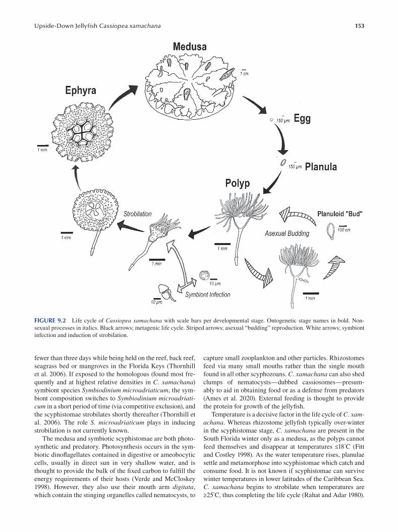
153 Upside-Down Jellyfi sh Cassiopea xamachana
FIGURE 9.2 Life cycle of Cassiopea xamachana with scale bars per developmental stage. Ontogenetic stage names in bold. Non
sexual processes in italics. Black arrows; metagenic life cycle. Striped arrows; asexual “budding” reproduction. White arrows; symbiont
infection and induction of strobilation.
fewer than three days while being held on the reef, back reef,
seagrass bed or mangroves in the Florida Keys (Thornhill
et al. 2006). If exposed to the homologous (found most fre
quently and at highest relative densities in C. xamachana)
symbiont species Symbiodinium microadriaticum, the sym
biont composition switches to Symbiodinium microadriaticum in a short period of time (via competitive exclusion), and
the scyphistomae strobilates shortly thereafter (Thornhill et
al. 2006). The role S. microadriaticum plays in inducing
strobilation is not currently known.
The medusa and symbiotic scyphistomae are both photo
synthetic and predatory. Photosynthesis occurs in the sym
biotic dinofl agellates contained in digestive or ameobocytic
cells, usually in direct sun in very shallow water, and is
thought to provide the bulk of the fixed carbon to fulfi ll the
energy requirements of their hosts (Verde and McCloskey
1998). However, they also use their mouth arm digitata,
which contain the stinging organelles called nematocysts, to
capture small zooplankton and other particles. Rhizostomes
feed via many small mouths rather than the single mouth
found in all other scyphozoans. C. xamachana can also shed
clumps of nematocysts—dubbed cassiosomes—presum
ably to aid in obtaining food or as a defense from predators
(Ames et al. 2020). External feeding is thought to provide
the protein for growth of the jellyfi sh.
Temperature is a decisive factor in the life cycle of C. xamachana. Whereas rhizostome jellyfi sh typically over-winter
in the scyphistomae stage, C. xamachana are present in the
South Florida winter only as a medusa, as the polyps cannot
feed themselves and disappear at temperatures ≤18° C ( Fitt
and Costley 1998). As the water temperature rises, planulae
settle and metamorphose into scyphistomae which catch and
consume food. It is not known if scyphistomae can survive
winter temperatures in lower latitudes of the Caribbean Sea.
C. xamachana begins to strobilate when temperatures are
≥25°C, thus completing the life cycle (Rahat and Adar 1980).
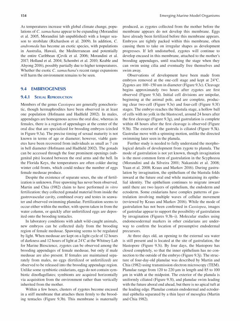
154
As temperatures increase with global climate change, popu
lations of C. xamachana appear to be expanding (Morandini
et al. 2005, Morandini lab unpublished) with a longer sea
son to strobilate (Richardson et al. 2009). In addition, C. andromeda has become an exotic species, with populations
in Australia, Hawaii, the Mediterranean and potentially
the entire Caribbean (Çevik et al. 2006; Morandini et al.
2017; Holland et al. 2004; Schembri et al. 2010; Keable and
Ahyong 2016), possibly partially due to higher temperatures.
Whether the exotic C. xamachana’s recent range expansions
will harm the environment remains to be seen.
9.4 EMBRYOGENESIS
9.4.1 SEXUAL REPRODUCTION
Members of the genus Cassiopea are generally gonochoris
tic, though hermaphrodites have been observed in at least
one population (Hofmann and Hadfield 2002). In males,
appendages are homogenous across the oral disc, whereas in
females, there is a region of appendages at the center of the
oral disc that are specialized for brooding embryos (circled
in Figure 9.3a). The precise timing of sexual maturity is not
known in terms of age or diameter; however, viable gam
etes have been recovered from individuals as small as 7 cm
in bell diameter ( Hofmann and Hadfi eld 2002). The gonads
can be accessed through the four prominent openings (sub
genital pits) located between the oral arms and the bell. In
the Florida Keys, the temperatures are often colder during
winter cold fronts, which could reduce the number of eggs
female medusae produce.
Despite the existence of separate sexes, the site of fertil
ization is unknown. Free spawning has never been observed.
Martin and Chia (1982) claim to have performed in vitro fertilization: they collected gonadal material from inside the
gastrovascular cavity, combined ovary and testes in seawa
ter and observed swimming planulae. Fertilization seems to
occur either within the mother, with sperm taken in from the
water column, or quickly after unfertilized eggs are depos
ited onto the brooding tentacles.
In laboratory conditions with adult wild-caught animals,
new embryos can be collected daily from the brooding
region of female medusae. Spawning seems to be regulated
by light. When medusae are kept on a light cycle of 12 hours
of darkness and 12 hours of light at 24°C at the Whitney Lab
for Marine Bioscience, zygotes can be observed among the
brooding appendages of female medusae, but only if male
medusae are also present. If females are maintained sepa
rately from males, no eggs (fertilized or unfertilized) are
observed to be released into the brooding appendage region.
Unlike some symbiotic cnidarians, eggs do not contain sym
biotic dinoflagellates; symbionts are acquired horizontally
via acquisition from the environment rather than vertically
inherited from the mother.
Within a few hours, clusters of zygotes become encased
in a stiff membrane that attaches them fi rmly to the brood
ing tentacles (Figure 9.3b). This membrane is maternally
Emerging Marine Model Organisms
produced, as zygotes collected from the mother before the
membrane appears do not develop this membrane. Eggs
have already been fertilized before this membrane appears.
Embryos are tightly packed within this membrane, often
causing them to take on irregular shapes as development
progresses. If left undisturbed, zygotes will continue to
develop encased in this membrane, attached to the mother’s
brooding appendages, until reaching the stage when they
can swim using cilia and eventually free themselves and
swim away.
Observations of development have been made from
embryos removed at the one-cell stage and kept at 24°C.
Zygotes are 100–150 um in diameter (Figure 9.3c). Cleavage
begins approximately two hours after zygotes are fi rst
observed (Figure 9.3d). Initial cell divisions are unipolar,
beginning at the animal pole, and are complete, produc
ing clear two-cell (Figure 9.3e) and four-cell (Figure 9.3f)
stages. The embryo reaches the blastula stage, a hollow ball
of cells with no yolk in the blastocoel, around 24 hours after
the first cleavage (Figure 9.3g), and gastrulation is complete
within 48 hours after the first cleavage is observed (Figure
9.3h). The exterior of the gastrula is ciliated (Figure 9.3k).
Gastrulae move with a spinning motion, unlike the directed
swimming later seen in the planula.
Further study is needed to fully understand the morpho
logical details of development from zygote to planula. The
mode of gastrulation is not yet known, though invagination
is the most common form of gastrulation in the Scyphozoa
(Morandini and da Silveira 2001; Nakanishi et al. 2008;
Yuan et al. 2008; Kraus and Markov 2016 ). During gastru
lation by invagination, the epithelium of the blastula folds
inward at the future oral end while maintaining its epithe
lial identity. The epithelium continues to migrate inward
until there are two layers of epithelium, the endoderm and
ectoderm. Some cnidarians have complex patterns of gas
trulation involving multiple waves of cellular movement
(reviewed by Kraus and Markov 2016). While the mode of
gastrulation has not been confirmed in Cassiopea , images
of gastrulae appear to support the possibility of gastrulation
by invagination (Figures 9.3h–i). Molecular studies using
endomesodermal markers in other cnidarians are under
way to confirm the location of presumptive endodermal
precursors.
At three days old, an opening to the external sea water
is still present and is located at the site of gastrulation, the
blastopore (Figure 9.3i). By four days, the blastopore has
closed completely, so that the inner epithelium has no con
nection to the outside of the embryo (Figure 9.3j). The struc
ture of four-day-old planulae was described by Martin and
Chia (1982) using transmission electron microscopy (TEM).
Planulae range from 120 to 220 μm in length and 85 to 100
μm in width at the midpoint. The exterior of the planula is
uniformly ciliated (Figure 9.3l), and planulae swim leading
with the future aboral end ahead, but there is no apical tuft at
the leading edge. Planulae contain endodermal and ectoder
mal epithelia separated by a thin layer of mesoglea (Martin
and Chia 1982).
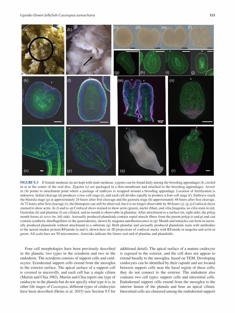
155 Upside-Down Jellyfi sh Cassiopea xamachana
FIGURE 9.3 If female medusae (a) are kept with male medusae, zygotes can be found daily among the brooding appendages (b, circled
in a) at the center of the oral disc. Zygotes (c) are packaged in a thin membrane and attached to the brooding appendages. Arrow
in (b) points to attachment point where a package of embryos is wrapped around a brooding appendage. Location of fertilization is
unknown. Initial cleavage (d) produces a two-cell stage (e), and each cell divides equally to produce a four-cell stage (f). Embryos reach
the blastula stage (g) at approximately 24 hours after first cleavage and the gastrula stage (h) approximately 48 hours after fi rst cleavage.
At 72 hours after first cleavage (i), the blastopore can still be observed, but it is no longer observable by 96 hours (j). (g–j) Confocal slices
stained to show actin. (k–l) and (o–p) Confocal slices stained to show actin (green), nuclei (blue), and cilia [magenta, no cilia stain in (o)].
Gastrulae (k) and planulae (l) are ciliated, and no mouth is observable in planulae. After attachment to a surface (m, right side), the polyp
mouth forms de novo (m, left side). Asexually produced planuloids contain septal muscle fibers from the parent polyp (o and p) and can
contain symbiotic dinoflagellates in the gastrodermis, shown by magenta autofluorescence in (p). Mouth and tentacles can form in asexu
ally produced planuloids without attachment to a substrate (q). Both planulae and asexually produced planuloids stain with antibodies
to the neural marker protein RFamide (n and r), shown here on 3D projections of confocal stacks with RFamide in magenta and actin in
green. All scale bars are 50 micrometers. Asterisks indicate the future oral end of planulae and planuloids.
Four cell morphologies have been previously described additional detail). The apical surface of a mature cnidocyte
in the planula: two types in the ectoderm and two in the is exposed to the exterior, and the cell does not appear to
endoderm. The ectoderm consists of support cells and cnid- extend basally to the mesoglea, based on TEM. Developing
ocytes. Ectodermal support cells extend from the mesoglea cnidocytes can be identified by their capsule and are located
to the exterior surface. The apical surface of a support cell between support cells near the basal region of these cells;
is covered in microvilli, and each cell has a single cilium they do not connect to the exterior. The endoderm also
(Martin and Chia 1982). Martin and Chia report one type of contains two cell types: support cells and interstitial cells.
cnidocyte in the planula but do not specify what type it is; in Endodermal support cells extend from the mesoglea to the
other life stages of Cassiopea, different types of cnidocytes interior lumen of the planula and bear an apical cilium.
have been described (Heins et al. 2015) (see Section 9.5 for Interstitial cells are clustered among the endodermal support
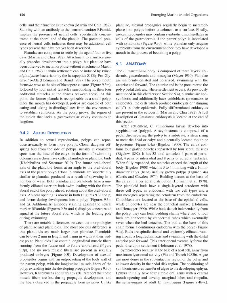
156
cells, and their function is unknown (Martin and Chia 1982).
Staining with an antibody to the neurotransmitter RFamide
implies the presence of neural cells, specifi cally concen
trated at the aboral end of the planula. The potential pres
ence of neural cells indicates there may be additional cell
types present that have not yet been described.
Planulae are competent to settle by the age of four or fi ve
days (Martin and Chia 1982). Attachment to a surface usu
ally precedes development into a polyp, but planulae have
been observed to metamorphose without attachment (Martin
and Chia 1982). Planula settlement can be induced by Vibrio alginolyticus bacteria or by the hexapeptide Z-Gly-Pro-Gly-
Gly-Pro-Ala (Hofmann and Brand 1987). The polyp mouth
forms de novo at the site of blastopore closure (Figure 9.3m),
followed by four initial tentacles surrounding it, then four
additional tentacles at the spaces between those. At this
point, the former planula is recognizable as a small polyp.
Once the mouth has developed, polyps are capable of both
eating and taking in dinoflagellates from the environment
to establish symbiosis. As the polyp grows, the region of
the stolon that lacks a gastrovascular cavity continues to
lengthen.
9.4.2 ASEXUAL REPRODUCTION
In addition to sexual reproduction, polyps can repro
duce asexually to form more polyps. Clonal daughter off
spring bud from the side of polyps, usually at consistent
spots near the base of the calyx, in the form of swimming
oblongs researchers have called planuloids or planuloid buds
(Khabibulina and Starunov 2019). The future oral–aboral
axis of the planuloid forms at an angle to the oral–aboral
axis of the parent polyp. Clonal planuloids are superfi cially
similar to planulae produced as a result of spawning in a
number of ways. Both planulae and planuloids have a uni
formly ciliated exterior; both swim leading with the future
aboral end of the polyp ahead, rotating about the oral–aboral
axis. An oral opening is absent in both (Figures 9.3l and p)
and forms during development into a polyp (Figures 9.3m
and q). Additionally, antibody staining against the neural
marker RFamide (Figures 9.3n and r) displays concentrated
signal at the future aboral end, which is the leading pole
during swimming.
There are notable differences between the morphologies
of planulae and planuloids. The most obvious difference is
that planuloids are much larger than planulae. Planuloids
can be over 2 mm in length and 1 mm in width at their wid
est point. Planuloids also contain longitudinal muscle fi bers
running from the future oral to future aboral end ( Figure
9.3p), and no such muscle fibers are present in sexually
produced embryos (Figure 9.3l). Development of asexual
propagules begins with an outpocketing of the body wall of
the parent polyp, with the longitudinal muscle fibers of the
polyp extending into the developing propagule (Figure 9.3o).
However, Khabibulina and Starunov (2019) report that these
muscle fibers are lost during propagule development, and
the fibers observed in the propagule form de novo . Unlike
Emerging Marine Model Organisms
planulae, asexual propagules regularly begin to metamor
phose into polyps before attachment to a surface. Finally,
asexual propagules may contain symbiotic dinofl agellates in
cells of the gastrodermis if the parent polyp is inoculated
with symbionts (Figure 9.3p), while planulae only acquire
symbionts from the environment once they have developed a
mouth in the process of becoming a polyp.
9.5 ANATOMY
The C. xamachana body is composed of three layers: epi
dermis, gastrodermis and mesoglea (Mayer 1910). Planulae
are uniformly ciliated and polarized, swimming with the
anterior end forward. The anterior end is the precursor to the
polyp pedal disk and where settlement occurs. As previously
mentioned in this chapter (see Section 9.4), planulae are apo
symbiotic and additionally have cnidoblasts (precursors to
cnidocytes, the cells which produce cnidocysts or “stinging
cells”) in their epidermis. Fully differentiated cnidocytes
are present in the ectoderm (Martin and Chia 1982). A full
description of Cassiopea cnidocysts is located at the end of
this section.
After settlement, C. xamachana larvae develop into
scyphistomae (polyps). A scyphistoma is composed of a
pedal disc securing the polyp to a substrate, a stem rising
to meet the head or calyx and a centrally located mouth or
hypostome (Figure 9.4a) (Bigelow 1900). The calyx con
tains four gastric pouches separated by four septal muscles
(Bigelow 1892). It has 32 total tentacles: 4 pairs of perra
dial, 4 pairs of interradial and 8 pairs of adradial tentacles.
When fully expanded, the tentacles exceed the length of the
body (Bigelow 1900) which is 3 to 4 mm long with a 1-mm
diameter calyx (head) in fully grown polyps (Figure 9.4a)
(Curtis and Cowden 1974). Budding occurs at the base of
the calyx in a perradial distribution (Hofmann et al. 1978).
The planuloid buds have a single-layered ectoderm with
three cell types, an endoderm with two cell types and a
thin mesoglea separating the ectoderm from the endoderm.
Cnidoblasts are located at the base of the epithelial cells,
while cnidocytes are near the epithelial surface (Hofmann
and Honegger 1990). While buds detach independently from
the polyp, they can form budding chains where two to four
buds are connected by ectodermal tubes which eventually
sever when the bud detaches. The bud at the base of this
chain forms a continuous endoderm with the polyp (Figure
9.4a). Buds are spindle shaped and uniformly ciliated, rotat
ing around a longitudinal axis and swimming with the distal
anterior pole forward. This anterior end eventually forms the
pedal disc upon settlement (Hofmann et al. 1978).
Symbiosomes localize at the base of a host cell, away from
maximum lysosomal activity (Fitt and Trench 1983b). Algae
are most dense in the subtentacular region of the polyp and
at lowest density in the pedal disk region. The positioning of
symbionts ensures transfer of algae to the developing ephyra.
Ephyra initially have four simple oral arms with a central
mouth opening and develop marginal lobes and rhopalia,
the sense-organs of adult C. xamachana ( Figure 9.4b–c ).
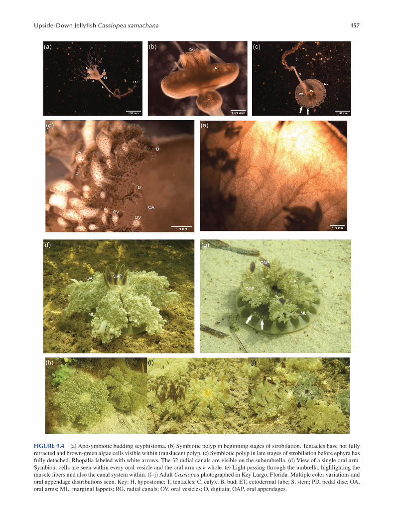
157 Upside-Down Jellyfi sh Cassiopea xamachana
FIGURE 9.4 (a) Aposymbiotic budding scyphistoma. (b) Symbiotic polyp in beginning stages of strobilation. Tentacles have not fully
retracted and brown-green algae cells visible within translucent polyp. (c) Symbiotic polyp in late stages of strobilation before ephyra has
fully detached. Rhopalia labeled with white arrows. The 32 radial canals are visible on the subumbrella. (d) View of a single oral arm.
Symbiont cells are seen within every oral vesicle and the oral arm as a whole. (e) Light passing through the umbrella, highlighting the
muscle fibers and also the canal system within. (f–j) Adult Cassiopea photographed in Key Largo, Florida. Multiple color variations and
oral appendage distributions seen. Key: H, hypostome; T, tentacles; C, calyx; B, bud; ET, ectodermal tube; S, stem; PD, pedal disc; OA,
oral arms; ML, marginal lappets; RG, radial canals; OV, oral vesicles; D, digitata; OAP, oral appendages.
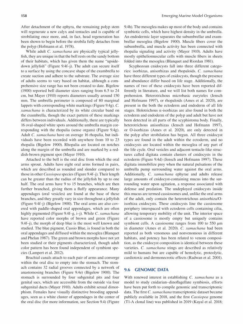
158
After detachment of the ephyra, the remaining polyp stem
will regenerate a new calyx and tentacles and is capable of
strobilating once more, and, in fact, head regeneration has
been shown to begin before the strobila fully detaches from
the polyp (Hofmann et al. 1978).
While adult C. xamachana are physically typical jelly
fish, they are unique in that the bell rests on the sandy bottom
of their habitats, which has given them the name “upside
down jellyfish” (Figure 9.4f–j). The adult can secure itself
to a surface by using the concave shape of the exumbrella to
create suction and adhere to the substrate. The average size
of adults seems to vary based on habitat, although a com
prehensive size range has not been created to date. Bigelow
(1900) reported bell diameter sizes ranging from 6.5 to 24
cm, but Mayer (1910) reports diameters usually around 150
mm. The umbrella perimeter is composed of 80 marginal
lappets with corresponding white markings (Figure 9.4g). C. xamachana is characterized by its white circular band on
the exumbrella, though the exact pattern of these markings
differs between individuals. Additionally, there are typically
16 oval-shaped white spots around the umbrella margin cor
responding with the rhopalia (sense organs) (Figure 9.4g).
Adult C. xamachana have on average 16 rhopalia, but indi
viduals have been recorded with anywhere from 10 to 23
rhopalia (Bigelow 1900). Rhopalia are located on notches
along the margin of the umbrella and are marked by a red
dish-brown pigment spot (Mayer 1910).
Attached to the bell is the oral disc from which the oral
arms sprout. Adults have eight oral arms formed in pairs,
which are described as rounded and slender compared to
those in other Cassiopea species (Figure 9.4f–j). Their length
can be greater than the radius of the jellyfish by up to one
half. The oral arms have 9 to 15 branches, which are then
further branched, giving them a fluffy appearance. Many
appendages (oral vesicles) are found at the base of these
branches, and they greatly vary in size throughout a jellyfi sh
(Figure 9.4f–j) (Bigelow 1900). The oral arms are also cov
ered with paddle-shaped oral appendages, which are often
highly pigmented ( Figure 9.4f–g , i – j ). While C. xamachana have reported color morphs of brown and green (Figure
9.4f–j), the morph of deep blue is the most well known and
studied. The blue pigment, Cassio Blue, is found in both the
oral appendages and diffused within the mesoglea (Blanquet
and Phelan 1987). The green and brown morphs have not yet
been studied or their pigments characterized, though adult
color pattern has been found independent of symbiont spe
cies (Lampert et al. 2012).
Brachial canals attach to each pair of arms and converge
within the oral disc to empty into the stomach. The stom
ach contains 32 radial grooves connected by a network of
anastomosing branches (Figure 9.4e) (Bigelow 1900). The
stomach is surrounded by four subgenital pits and four
genital sacs, which are accessible from the outside via four
subgenital ducts (Mayer 1910). Adults exhibit sexual dimor
phism. Females have visually distinctive brooding append
ages, seen as a white cluster of appendages in the center of
the oral disc (for more information, see Section 9.4) (Figure
Emerging Marine Model Organisms
9.4h). The mesoglea makes up most of the body and contains
symbiotic cells, which have highest density in the umbrella.
An endodermic layer separates the subumbrellar and exum
brellar mesoglea (Bigelow 1900). Muscle fibers cover the
subumbrella, and muscle activity has been connected with
rhopalia signaling and activity (Mayer 1910). Adults have
mostly epitheliomuscular cells with muscle fibers in sheets
folded into the mesoglea (Blanquet and Riordan 1981).
Scyphozoan cnidocysts fall into three different catego
ries: isorhizas, anisorhizas and rhopaloids. C. xamachana have three different types of cnidocysts, though the presence
and abundance differ based on life stage. Additionally, the
names of two of these cnidocysts have been reported dif
ferently in literature, and we will list both names for com
prehension. Heterotrichous microbasic euryteles (Jensch
and Hofmann 1997 ), or rhopaloids (Ames et al. 2020), are
present in the both the ectoderm and endoderm of all life
stages. Holotrichous -isorhizas are also found in both the
ectoderm and endoderm of the polyp and adult but have not
been detected in all parts of the scyphistoma body. Finally,
heterotrichous anisorhizas (Jensch and Hofmann 1997 ),
or O-isorhizas (Ames et al. 2020), are only detected in
the polyp after strobilation has begun. All three cnidocyst
types are found in the adult within the ectoderm, and no
cnidocysts are located within the mesoglea of any part of
the life cycle. Oral vesicles and adjacent tentacle-like struc
tures called digitata contain clusters of cnidocysts in the
ectoderm (Figure 9.4d) (Jensch and Hofmann 1997 ). These
digitata immobilize prey when the natural pulsations of the
umbrella pump surrounding water against the oral arms.
Additionally, C. xamachana ephyrae and adults release
large amounts of cnidocyst-containing mucus into the sur
rounding water upon agitation, a response associated with
defense and predation. The undeployed cnidocysts inside
this mucus are termed cassiosomes and, unlike the oral arms
of the adult, only contain the heterotrichous anisorhiza/O
isorhiza cnidocysts. These cnidocysts line the cassiosome
periphery interspaced with ectoderm cells containing cilia,
allowing temporary mobility of the unit. The interior space
of a cassiosome is mostly empty but uniquely contains
symbiont cells. A cassiosome ranges from 100 to 550 μm
in diameter (Ames et al. 2020). C. xamachana had been
reported as both venomous and nonvenomous in different
habitats, and potency has been related to venom composi
tion, as the cnidocyst composition is identical between these
varieties. C. xamachana stings are described as relatively
mild to humans but are capable of hemolytic, proteolytic,
cardiotoxic and dermonecrotic effects (Radwan et al. 2001).
9.6 GENOMIC DATA
With renewed interest in establishing C. xamachana as a
model to study cnidarian–dinoflagellate symbiosis, efforts
have been put forth to compile genomic and transcriptomic
data. The fi rst C. xamachana transcriptomic dataset became
publicly available in 2018, and the fi rst Cassiopea genome
(T1-A clonal line) was published in 2019 (Kayal et al. 2018;
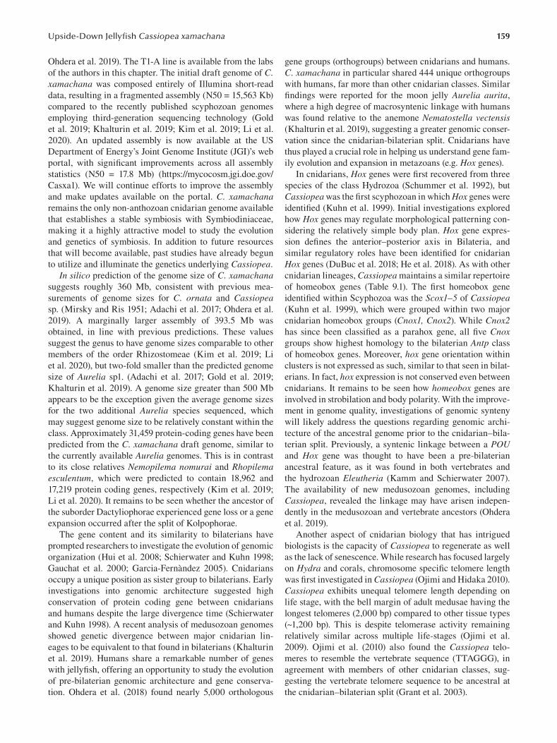
159 Upside-Down Jellyfi sh Cassiopea xamachana
Ohdera et al. 2019). The T1-A line is available from the labs
of the authors in this chapter. The initial draft genome of C. xamachana was composed entirely of Illumina short-read
data, resulting in a fragmented assembly (N50 = 15,563 Kb)
compared to the recently published scyphozoan genomes
employing third-generation sequencing technology (Gold
et al. 2019; Khalturin et al. 2019; Kim et al. 2019; Li et al.
2020). An updated assembly is now available at the US
Department of Energy’s Joint Genome Institute (JGI)’s web
portal, with significant improvements across all assembly
statistics (N50 = 17.8 Mb) (https://mycocosm.jgi.doe.gov/
Casxa1). We will continue efforts to improve the assembly
and make updates available on the portal. C. xamachana remains the only non-anthozoan cnidarian genome available
that establishes a stable symbiosis with Symbiodiniaceae,
making it a highly attractive model to study the evolution
and genetics of symbiosis. In addition to future resources
that will become available, past studies have already begun
to utilize and illuminate the genetics underlying Cassiopea.
In silico prediction of the genome size of C. xamachana suggests roughly 360 Mb, consistent with previous mea
surements of genome sizes for C. ornata and Cassiopea sp. (Mirsky and Ris 1951; Adachi et al. 2017; Ohdera et al.
2019). A marginally larger assembly of 393.5 Mb was
obtained, in line with previous predictions. These values
suggest the genus to have genome sizes comparable to other
members of the order Rhizostomeae (Kim et al. 2019; Li
et al. 2020), but two-fold smaller than the predicted genome
size of Aurelia sp1. (Adachi et al. 2017; Gold et al. 2019;
Khalturin et al. 2019). A genome size greater than 500 Mb
appears to be the exception given the average genome sizes
for the two additional Aurelia species sequenced, which
may suggest genome size to be relatively constant within the
class. Approximately 31,459 protein-coding genes have been
predicted from the C. xamachana draft genome, similar to
the currently available Aurelia genomes. This is in contrast
to its close relatives Nemopilema nomurai and Rhopilema esculentum, which were predicted to contain 18,962 and
17,219 protein coding genes, respectively (Kim et al. 2019;
Li et al. 2020). It remains to be seen whether the ancestor of
the suborder Dactyliophorae experienced gene loss or a gene
expansion occurred after the split of Kolpophorae.
The gene content and its similarity to bilaterians have
prompted researchers to investigate the evolution of genomic
organization (Hui et al. 2008; Schierwater and Kuhn 1998;
Gauchat et al. 2000; Garcia-Fernàndez 2005). Cnidarians
occupy a unique position as sister group to bilaterians. Early
investigations into genomic architecture suggested high
conservation of protein coding gene between cnidarians
and humans despite the large divergence time (Schierwater
and Kuhn 1998). A recent analysis of medusozoan genomes
showed genetic divergence between major cnidarian lin
eages to be equivalent to that found in bilaterians (Khalturin
et al. 2019). Humans share a remarkable number of genes
with jellyfish, offering an opportunity to study the evolution
of pre-bilaterian genomic architecture and gene conserva
tion. Ohdera et al. (2018) found nearly 5,000 orthologous
gene groups (orthogroups) between cnidarians and humans.
C. xamachana in particular shared 444 unique orthogroups
with humans, far more than other cnidarian classes. Similar
findings were reported for the moon jelly Aurelia aurita,
where a high degree of macrosyntenic linkage with humans
was found relative to the anemone Nematostella vectensis (Khalturin et al. 2019), suggesting a greater genomic conser
vation since the cnidarian-bilaterian split. Cnidarians have
thus played a crucial role in helping us understand gene fam
ily evolution and expansion in metazoans (e.g. Hox genes).
In cnidarians, Hox genes were first recovered from three
species of the class Hydrozoa (Schummer et al. 1992), but
Cassiopea was the first scyphozoan in which Hox genes were
identified (Kuhn et al. 1999). Initial investigations explored
how Hox genes may regulate morphological patterning con
sidering the relatively simple body plan. Hox gene expres
sion defines the anterior–posterior axis in Bilateria, and
similar regulatory roles have been identified for cnidarian
Hox genes (DuBuc et al. 2018; He et al. 2018). As with other
cnidarian lineages, Cassiopea maintains a similar repertoire
of homeobox genes (Table 9.1). The first homeobox gene
identified within Scyphozoa was the Scox1–5 of Cassiopea (Kuhn et al. 1999), which were grouped within two major
cnidarian homeobox groups (Cnox1, Cnox2). While Cnox2 has since been classified as a parahox gene, all fi ve Cnox groups show highest homology to the bilaterian Antp class
of homeobox genes. Moreover, hox gene orientation within
clusters is not expressed as such, similar to that seen in bilat
erians. In fact, hox expression is not conserved even between
cnidarians. It remains to be seen how homeobox genes are
involved in strobilation and body polarity. With the improve
ment in genome quality, investigations of genomic synteny
will likely address the questions regarding genomic archi
tecture of the ancestral genome prior to the cnidarian–bila
terian split. Previously, a syntenic linkage between a POU and Hox gene was thought to have been a pre-bilaterian
ancestral feature, as it was found in both vertebrates and
the hydrozoan Eleutheria (Kamm and Schierwater 2007).
The availability of new medusozoan genomes, including
Cassiopea, revealed the linkage may have arisen indepen
dently in the medusozoan and vertebrate ancestors (Ohdera
et al. 2019).
Another aspect of cnidarian biology that has intrigued
biologists is the capacity of Cassiopea to regenerate as well
as the lack of senescence. While research has focused largely
on Hydra and corals, chromosome specific telomere length
was first investigated in Cassiopea (Ojimi and Hidaka 2010).
Cassiopea exhibits unequal telomere length depending on
life stage, with the bell margin of adult medusae having the
longest telomeres (2,000 bp) compared to other tissue types
(~1,200 bp). This is despite telomerase activity remaining
relatively similar across multiple life-stages (Ojimi et al.
2009). Ojimi et al. (2010) also found the Cassiopea telo
meres to resemble the vertebrate sequence (TTAGGG), in
agreement with members of other cnidarian classes, sug
gesting the vertebrate telomere sequence to be ancestral at
the cnidarian–bilaterian split (Grant et al. 2003).
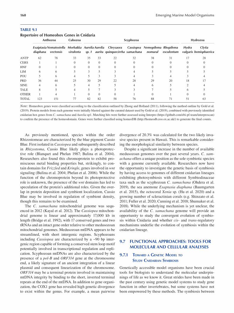
160 Emerging Marine Model Organisms
TABLE 9.1 Repertoire of Homeobox Genes in Cnidaria
Anthozoa Cubozoa Scyphozoa Hydrozoa
Exaiptasia Nematostella Morbakka Aurelia Aurelia Chrysaora Cassiopea Nemopilema Rhopilema Hydra Clytia diaphana vectensis virulenta sp. 1 aurita quinquecirrha xamachana nomural esculentum vulgaris hemisphaerica
ANTP 62 78 33 35 33 22 32 38 31 17 28
CERS 1 1 0 0 0 0 0 0 0 0 0
HNF 0 1 0 0 0 0 0 0 0 0 0
LIM 6 6 5 3 5 3 4 5 5 5 5
POU 5 6 4 5 3 3 4 3 4 3 4
PRD 36 44 25 30 29 22 28 29 20 18 17
SINE 4 6 5 4 5 5 4 6 5 2 4
TALE 8 5 4 5 7 3 3 7 5 6 5
OTHER 1 4 1 0 0 0 1 0 1 0 0
TOTAL 123 151 77 82 82 58 76 88 71 51 63
Note: Homeobox genes were classified according to the classification outlined by Zhong and Holland (2011 ), following the method outline by Gold et al.
(2019 ). Protein models from each genome were initially blasted against the curated dataset used by Gold et al. (2019), combined with previously identifi ed
cnidarian hox genes from C. xamachana and Aurelia sp1. Matching hits were further assessed using Interpro (https://github.com/ebi-pf-team/interproscan)
to confirm the presence of the homeodomain. Genes were further classified using homeoDB (http://homeodb.zoo.ox.ac.uk/) to generate the fi nal counts.
As previously mentioned, species within the order
Rhizostomeae are characterized by the blue pigment Cassio
Blue. First isolated in Cassiopea and subsequently described
in Rhizostoma, Cassio Blue likely plays a photoprotec
tive role (Blanquet and Phelan 1987; Bulina et al. 2004).
Researchers also found this chromoprotein to exhibit pro
miscuous metal binding properties but, strikingly, to con
tain domains for Frizzled and Kringle, genes involved in wnt signaling (Bulina et al. 2004; Phelan et al. 2006 ). While the
function of the chromoprotein beyond its photoprotective
role is unknown, the presence of the wnt domains has led to
speculation of the protein’s additional roles. Given the over
lap in protein deposition and symbiont localization, Cassio
Blue may be involved in regulation of symbiont density,
though this remains to be examined.
The C. xamachana mitochondrial genome was sequ
enced in 2012 (Kayal et al. 2012). The Cassiopea mitochon
drial genome is linear and approximately 17,000 kb in
length (Bridge et al. 1992), with 17 conserved genes and two
tRNAs and an intact gene order relative to other medusozoan
mitochondrial genomes. Medusozoan mtDNA appears to be
streamlined, with short intergenic regions. Scyphozoans
including Cassiopea are characterized by a ~90 bp inter-
genic region capable of forming a conserved stem loop motif
potentially involved in transcriptional regulation and repli
cation. Scyphozoan mtDNAs are also characterized by the
presence of a pol-B and ORF314 gene at the chromosome
end, a likely signature of an ancient integration of a linear
plasmid and consequent linearization of the chromosome.
ORF314 may be a terminal protein involved in maintaining
mtDNA integrity by binding to the short, inverted terminal
repeats at the end of the mtDNA. In addition to gene organi
zation, the COX1 gene has revealed high genetic divergence
to exist within the genus. For example, a mean pairwise
divergence of 20.3% was calculated for the two likely inva
sive species present in Hawaii. This is remarkable consider
ing the morphological similarity between species.
Despite a significant increase in the number of available
medusozoan genomes over the past several years. C. xamachana offers a unique position as the sole symbiotic species
with a genome currently available. Researchers now have
the opportunity to investigate the genetic basis of symbiosis
by having access to genomes of different cnidarian lineages
exhibiting photosymbiosis with different Symbiodinaceae
taxa such as the scyphozoan C. xamachana (Ohdera et al.
2019), the sea anemone Exaptasia diaphana ( Baumgarten
et al. 2015), the octocoral Xenia sp. (Hu et al. 2020) and a
growing number of scleractinian corals (e.g. Shinzato et al.
2011; Fuller et al. 2020; Cunning et al. 2018; Shumaker et al.
2018). While the underlying mechanism is yet unclear, the
availability of the C. xamachana genome will provide an
opportunity to study the convergent evolution of symbio
sis within Cnidaria and whether cis- and trans- regulatory
mechanisms underlie the evolution of symbiosis within the
cnidarian lineage.
9.7 FUNCTIONAL APPROACHES: TOOLS FOR MOLECULAR AND CELLULAR ANALYSES
9.7.1 TOWARD A GENETIC MODEL TO
STUDY CNIDARIAN SYMBIOSIS
Genetically accessible model organisms have been crucial
tools for biologists to understand the molecular underpin
nings of life as we know it. Great strides have been made in
the past century using genetic model systems to study gene
function in other invertebrates, but some systems have not
been empowered by these methods. The symbiosis between
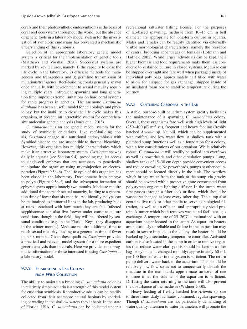
161 Upside-Down Jellyfi sh Cassiopea xamachana
corals and their photosynthetic endosymbionts is the basis of
coral reef ecosystems throughout the world, but the absence
of genetic tools in a laboratory model system for the investi
gation of symbiotic cnidarians has prevented a mechanistic
understanding of this symbiosis.
Selection of an appropriate laboratory genetic model
system is critical for the implementation of genetic tools
(Matthews and Vosshall 2020). Successful systems are
marked by key features, namely 1) the capacity to close the
life cycle in the laboratory, 2) efficient methods for muta
genesis and transgenesis and 3) germline transmission of
mutations/transgenes. Reef-building corals generally spawn
once annually, with development to sexual maturity requir
ing multiple years. Infrequent spawning and long genera
tion time impose extreme limitations on hard coral systems
for rapid progress in genetics. The anemone Exaiptasia diaphana has been a useful model for cell biology and phys
iology, but the inability to close the life cycle makes this
organism, at present, an intractable system for comprehen
sive molecular genetic analysis (Jones et al. 2018).
C. xamachana is an apt genetic model system for the
study of symbiotic cnidarians. Like reef-building cor
als, Cassiopea engage in a nutritional endosymbiosis with
Symbiodiniaceae and are susceptible to thermal bleaching.
However, this organism has multiple characteristics which
make it an attractive laboratory system. Cassiopea spawns
daily in aquaria (see Section 9.4), providing regular access
to single-cell embryos that are necessary to genetically
manipulate the organism using microinjection or electro
poration (Figure 9.5a–b). The life cycle of this organism has
been closed in the laboratory. Development from embryo
to polyp (Figure 9.5c–f) and the subsequent formation of
ephyrae spans approximately two months. Medusae require
additional time to reach sexual maturity, leading to a genera
tion time of fewer than six months. Additionally, polyps can
be maintained as immortal lines in the lab, producing buds
at rates associated with how much they are fed. Infected
scyphistomae can also live forever under constant culture
conditions, though in the field, they will be affected by sea
sonal conditions (e.g. in the Florida Keys, they disappear
in the winter months). Medusae require additional time to
reach sexual maturity, leading to a generation time of fewer
than six months. Given these qualities, Cassiopea provides
a practical and relevant model system for a more expedient
genetic analysis than in corals. Here we provide some prag
matic information for those interested in using Cassiopea as
a laboratory model.
9.7.2 ESTABLISHING A LAB COLONY
FROM WILD COLLECTION
The ability to maintain a breeding C. xamachana colonies
in relatively simple aquaria is a strength of this model system
for cnidarian symbiosis. Reproductive adults can be readily
collected from their nearshore natural habitats by snorkel
ing or wading in the shallow waters they inhabit. In the state
of Florida, USA, C. xamachana can be collected under a
recreational saltwater fishing license. For the purposes
of lab-based spawning, medusae from 10–15 cm in bell
diameter are appropriate for long-term culture in aquaria.
Males and females can be readily identified via externally
visible morphological characteristics, namely the presence
of central brooding appendages on females (Hofmann and
Hadfield 2002). While larger individuals can be kept, their
higher biomass and food requirements make them less con
ducive to sustained culture in closed systems. Medusae can
be shipped overnight and fare well when packaged inside of
individual poly bags, approximately half filled with water
to allow for airspace for gas exchange, shipped inside of
an insulated foam box to stabilize temperature during the
journey.
9.7.3 CULTURING CASSIOPEA IN THE LAB
A stable, purpose-built aquarium system greatly facilitates
the maintenance of a spawning C. xamachana colony.
Overall, these organisms fare well with high levels of light
(250–400 μE m−2 s−1), frequent and heavy feeding (freshly
hatched Artemia sp. Nauplii, which can be supplemented
with rotifers) and low water flow. A shallow tank with a
plumbed sump functions well as a foundation for a colony,
with a few considerations of our organism. While relatively
robust, C. xamachana will readily be pulled into overfl ows
as well as powerheads and other circulation pumps. Long,
shallow tanks of 15–30 cm depth provide convenient access
and reduce crowding. No powerheads, pumps or other equip
ment should be located directly in the tank. The overfl ow
which brings water from the tank to the sump via gravity
should be covered with a protective grate constructed from
polystyrene egg crate lighting diffuser. In the sump, water
first passes through a filter sock or floss, which should be
washed/exchanged at least every other day. The sump also
contains live rock or other media to serve as biological fi l
tration, as well as an efficient and appropriately sized pro
tein skimmer which both removes waste and facilitates gas
exchange. A temperature of 25–26°C is maintained with an
aquarium heater located in the sump. As aquarium heaters
are notoriously unreliable and failure in the on position may
result in severe impacts to the colony, the heater should be
backed up by a secondary temperature controller. Activated
carbon is also located in the sump in order to remove organ
ics that reduce water clarity; this should be kept in a fi lter
bag or nylons and changed monthly; approximately 60 mL
per 100 liters of water in the system is sufficient. The return
pump delivers water back to the aquarium. This should be
relatively low flow so as not to unnecessarily disturb the
medusae in the main tank; approximate turnover of one
to three times the volume of the aquarium is suffi cient.
Diffusing the water returning to the tank will also prevent
the disturbance of the medusae (Widmer 2008).
Heavy feeding of freshly hatched live Artemia sp. one
to three times daily facilitates continued, regular spawning.
Though C. xamachana are not particularly demanding of
water quality, attention to water parameters will promote the
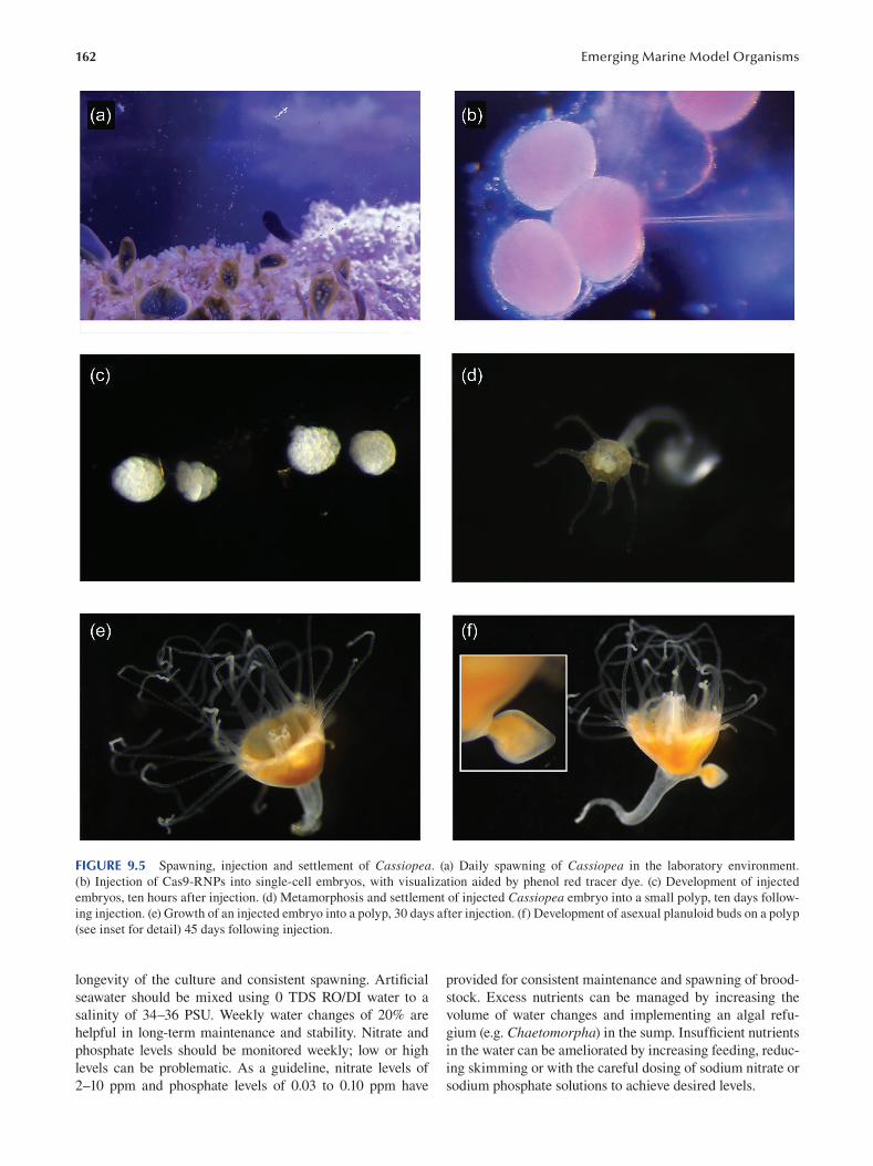
162 Emerging Marine Model Organisms
FIGURE 9.5 Spawning, injection and settlement of Cassiopea. (a) Daily spawning of Cassiopea in the laboratory environment.
(b) Injection of Cas9-RNPs into single-cell embryos, with visualization aided by phenol red tracer dye. (c) Development of injected
embryos, ten hours after injection. (d) Metamorphosis and settlement of injected Cassiopea embryo into a small polyp, ten days follow
ing injection. (e) Growth of an injected embryo into a polyp, 30 days after injection. (f) Development of asexual planuloid buds on a polyp
(see inset for detail) 45 days following injection.
longevity of the culture and consistent spawning. Artifi cial provided for consistent maintenance and spawning of brood-
seawater should be mixed using 0 TDS RO/DI water to a stock. Excess nutrients can be managed by increasing the
salinity of 34–36 PSU. Weekly water changes of 20% are volume of water changes and implementing an algal refu
helpful in long-term maintenance and stability. Nitrate and gium (e.g. Chaetomorpha) in the sump. Insuffi cient nutrients
phosphate levels should be monitored weekly; low or high in the water can be ameliorated by increasing feeding, reduc
levels can be problematic. As a guideline, nitrate levels of ing skimming or with the careful dosing of sodium nitrate or
2–10 ppm and phosphate levels of 0.03 to 0.10 ppm have sodium phosphate solutions to achieve desired levels.
163 Upside-Down Jellyfi sh Cassiopea xamachana
As photosymbiotic organisms with spawning controlled
by the daily light cycle, appropriate lighting is a critical com
ponent of Cassiopea husbandry. Lighting solutions designed
for reef-building corals are appropriate for these shallow-
water animals that require high levels of photosynthetically
available radiation (PAR) to maximize the nutritional ben
efits from their endosymbionts. Modern high-output LEDs
designed for reef tanks can be implemented to blanket the
bottom of the tank with PAR levels of 250–400 μE on a
12:12 daily cycle. Light levels should be assessed with a sub
mersible PAR meter and lighting adjusted as appropriate.
9.7.4 MICROINJECTION OF SINGLE-CELL EMBRYOS
FOR THE GENERATIONS OF MUTANTS
AND TRANSGENIC CASSIOPEA
The study of symbiosis in cnidarians has long sought to iden
tify the mechanistic basis of the interactions between the
animal host and intracellular algal partner. Studies compar
ing symbiotic and aposymbiotic hosts have been performed
in numerous cnidarian taxa (Lehnert et al. 2014; Rodriguez-
Lanetty et al. 2006), as well as numerous studies examining
the response to heat stress and the breakdown of symbiosis
(Pinzón et al. 2015; DeSalvo et al. 2010) and gene expression
patterns associated with thermal tolerance (Bellantuono et
al. 2012; Barshis et al. 2013). This broad body of work has
resulted in the identification of numerous genes of interest,
including molecular chaperones and antioxidant enzymes
associated with the response to thermal stress (Császár et al.
2009; Fang et al. 1997 ), as well as lectins which may mediate
the relationship between the host and symbiont (Kvennefors
et al. 2008). However, the field has largely been missing
crucial tools of genetics to robustly test these hypotheses.
Microinjection of C. xamachana embryos opens a path to
understand the molecular genetic basis of symbiosis, che
mosensation and sleep in an early diverging metazoan with
a decentralized nervous system (Figure 9.5b).
A basic tool of genetics is the capacity to perform loss-
of-function studies such as gene knockout experiments.
With the development of genome editing techniques, C. xamachana is an apt model system to test hypotheses of
cnidarian symbiosis. Using microinjection, C. xamachana embryos are amenable to CRISPR-mediated mutagenesis, a
technology which allows for precise, targeted mutagenesis
and transgenesis using a programmable nuclease comprised
of a guide RNA and the protein Cas9 (Jinek et al. 2012).
CRISPR-Cas9 can be used by delivering the Cas9 protein
complexed with single guide RNAs (sgRNA) which direct
the nuclease to the locus of interest in the nucleus of a living
cell. This Cas9-sgRNA complex cleaves the targeted DNA,
resulting in endogenous DNA repair. In the absence of
homologous template, non-homologous end joining (NHEJ)
repair occurs (Doudna and Charpentier 2014). By injecting
a Cas9-sgRNA complex into single cell embryos, mutants
are generated with small insertions or deletions (indels)
induced by the imperfect DNA repair mechanisms of the
cell. These indels often result in frameshift mutations of the
target gene, generating loss-of-function alleles. These muta
genized embryos can then be reared to polyps and induced
to strobilate by exposure to an algal symbiont, generating
medusae that can be used for subsequent crosses once sexu
ally mature. As the life cycle of Cassiopea can be completed
in the lab within four to six months, the crosses necessary
to generate a homozygous mutant can be completed within
18 months. Work to establish this technology in Cassiopea is ongoing.
In addition to using CRISPR to generate loss-of-func
tion alleles, this technology can also be implemented to
perform gene knock-in. By providing donor DNA consist
ing of a transgene flanked by sequence homologous to the
both sides of the cut site, CRISPR can be used to engineer
knock-in at a specific locus (Barrangou and Doudna 2016).
This will allow the generation of diverse molecular tools
for Cassiopea for the study of cnidarian symbiosis, devel
opment and neuroscience in this unique model system with
the future implementation of genetically encoded calcium
indicators (GECIs) such as GCaMP (Nakai et al. 2001) for
the real-time fluorescent readout of nervous system activ
ity, as well as genetically encoded fluorescent redox sensors
(Lukyanov and Belousov 2014) to test longstanding hypoth
eses regarding the role of ROS stress in cnidarian bleaching.
Cassiopea are transparent and lack endogenous host autofl u
orescence, making them well suited to molecular imaging.
Spawning is timed by the daily light cycle, occurring fi ve
to six hours after artificial sunrise in aquaria. In order to
collect unicellular embryos, clear selected spawning female
medusae of previously extruded, multicellular embryos
approximately two hours prior to spawning using a baster.
Selected female medusae can then be placed in shallow
black polycarbonate pans under a light source to improve the
visibility of embryos at the time of release. Once released,
the 80-μm embryos can be collected with a transfer pipette
into small glass dishes, taking care to avoid mucus. Prior
to injection, unicellular embryos are transferred and aligned
in polystyrene culture dishes containing 40 PSU seawater.
The increased salinity results in a slight reduction of cell
volume due to osmosis and allows the cell to accommo
date the volume of the injected liquid payload. Transfer and
positioning of embryos is performed using an aspirator con
structed from a 1-mm glass capillary fitted with a length of
1-mm ID silicone tubing. Embryos readily adhere to new,
virgin polystyrene and can be arranged in a row for effi
cient microinjection. Dishes with tight-fitting lids are best
employed to reduce evaporation, as the injection dish also
houses embryos during development to planulae.
Typical injection payloads include Cas9-sgRNA ribonu
cleoprotein injection mixture, composed of a guide RNA
complexed with Cas9 protein (with NLS), injection buffer
and phenol red dye microinjected into single-cell Cassiopea embryos (Figure 9.5b–c). Custom needles are prepared
with thin-walled 1-mm aluminosilicate glass capillaries on
a P-1000 horizontal pipette puller (Sutter Instrument, CA,
USA) and beveled on BV-10 micropipette beveler (Sutter) to
17°. Microinjection is performed using a Xenoworks digital
164
injector and manipulator system (Sutter Instrument, CA,
USA) under a SteREO Discovery V8 microscope (Zeiss,
Germany). Current injection methods yield survival rates
of up to 40%. In the three to six hours following injection,
each embryo is examined to assess whether it has survived
and entered the cleavage stage. Non-dividing embryos are
culled and removed, and the water in the dish is carefully
replaced with filtered 34 PSU artificial seawater. Planulation
of viable embryos occurs approximately one week follow
ing injection, with a developmental delay often observed in
comparison to uninjected embryos. Cassiopea larvae read
ily settle and metamorphose in response to a number of
cues, including bacteria, degrading mangrove leaves and the
previously mentioned endogenous metamorphosis-inducing
peptide (Neumann 1979; Fleck and Fitt 1999; Thieme and
Hofmann 2003). We have found that settlement dishes can
easily be prepared by using a cotton swab to transfer biofi lm
from the sump of an established Cassiopea tank to poly
styrene dishes and then covering with seawater and incu
bating at room temperature for three to five days. Prior to
transferring planulae to settlement dishes, water should be
exchanged with filtered 34 PSU artificial seawater. Planulae
should be monitored regularly; once settlement occurs and
nascent scyphistomae have developed tentacles, regular
feeding of freshly hatched Artemia nauplii should begin.
Daily feeding is optimal. The survival of recent settlers can
be enhanced by placing a nauplius on the hypostome with
forceps. In order to maintain polyps in an aposymbiotic
state and prevent strobilation, polyps can be maintained in
10 μm DCMU without apparent detriment. In order to gen
erate medusae, mature polyps can be challenged with sym
bionts to induce strobilation. Once released from the polyp,
the ephyra will develop into a medusa. Growth is facilitated
with ample feeding of Artemia (at least daily) and high arti
ficial light levels (250–400 μE) on a 12:12 cycle or natural
light. With regular water changes, medusae can be cultured
in 1-liter beakers or polycarbonate pans to bell diameters
of at least 5 cm. The generation of sexually mature medu
sae takes several months. Work is in progress to develop the
most efficient methods to cross medusae.
9.8 CHALLENGING QUESTIONS
While a lot of emphasis has been placed on understanding
the origins of the first metazoan body plans, less is known
about how those early animals interacted with their sur
rounding microbial seas. The establishment of holobiont
communities (i.e. a multicellular host and its associated
microbiome) required the evolution of novel interkingdom
communication. As metazoan life cycles evolved, their
associated microbial communities diversified with them
(McFall-Ngai et al. 2013). The study of host-microbe asso
ciations throughout an organism’s life cycle is now feasible
(Gilbert et al. 2015; Gilbert 2016 ). There is a growing inter
est in ontogenetic microbiomes (i.e. microbial associates
over a host developmental time course) (Fieth et al. 2016;
Carrier and Reitzel 2018; Vijayan et al. 2019) and how they
Emerging Marine Model Organisms
can affect developmental phenotypes (Tran and Hadfi eld
2011; Thompson et al. 2015; Fieth et al. 2016; Shikuma et al.
2016; Carrier and Reitzel 2018). While a few microbes have
been shown to induce larval settlement in C. xamachana,
such as Vibrio spp. (Neumann 1979; Hofmann and Brand
1987) and Pseudoalteromonas sp. (Ohdera et al. in prep a),
it is likely that the complex microbiomes in settlement sub
strates as well as developmental microbiomes acquired by
the organism through ontogeny will also play critical roles
in driving phenotypic and physiological traits as C. xamachana goes through its life cycle (Medina lab, unpublished).
Our ability to infect with different Symbiodiniaceae that
will in turn harbor different microbiomes as well as poten
tially developing axenic and gnotobiotic animals will also
open doors to understand host–microbiome interactions at
the developmental level (Medina lab, unpublished).
Many cnidarian taxa establish endosymbioses with
Symbiodiniaceae, and this symbiosis is crucial in the
maintenance of coral reef ecosystems (LaJeunesse 2020).
Scleractinian corals usually establish their photosymbio
sis during the larval stage (Schwarz et al. 1999; Abrego
et al. 2009; Voolstra et al. 2009; McIlroy and Coffroth
2017). Mounting evidence now supports the role of
Symbiodiniaceae (LaJeunesse et al. 2018) in the onset of
host development (Mohamed et al. 2016; Reich et al. 2017).
Coral larval manipulation experiments are challenging given
the limited availability of larvae due to annual spawning
events (Harrison et al. 1984; Szmant 1986; Van Woesik et al.
2006). Although the pelago-benthic transition from larva to
settled polyp is partially linked to onset of photosymbiosis
(Mohamed et al. 2016; Reich et al. 2017 ), discerning the role
of photosymbionts as drivers of this developmental transi
tion has not been clearly elucidated (Hartmann et al. 2019).
Cassiopea therefore represents an effi cient model system to
study developmental symbioses.
We believe that C. xamachana can become an ideal sys
tem to study environmental canalization (Waddington 1942)
because of the clear and easily manipulated developmental
switch (i.e. onset of photosymbiosis) that we can also obvi
ate with artificial inducers. We can alter the phenotypic out
come of strobilation by using different photosymbionts in
comparative infection experiments. Once the polyp stage is
infected, it can take different developmental trajectories that
lead to divergent morphospaces between homologous and
heterologous photosymbiotic infections (Figure 9.6). These
different developmental phenotypes also likely have diverg
ing underlying molecular regulatory mechanisms. Robert
Trench had indeed already proposed that this type of pho
tosymbiosis would be ideal for the study of cross-genome
regulation (Trench 1979). In support of this idea, we have
uncovered a possible role of S. microadriaticum photosyn
thetic pigments in the regulation of C. xamachana strobila
tion (Ohdera et al. in prep b).
Both the host (C. xamachana) (Ohdera et al. 2019)
and the homologous photosymbiont (S. microadriaticum)
(Aranda et al. 2016 ) are now genome enabled, facilitat
ing any downstream molecular analysis. Establishing
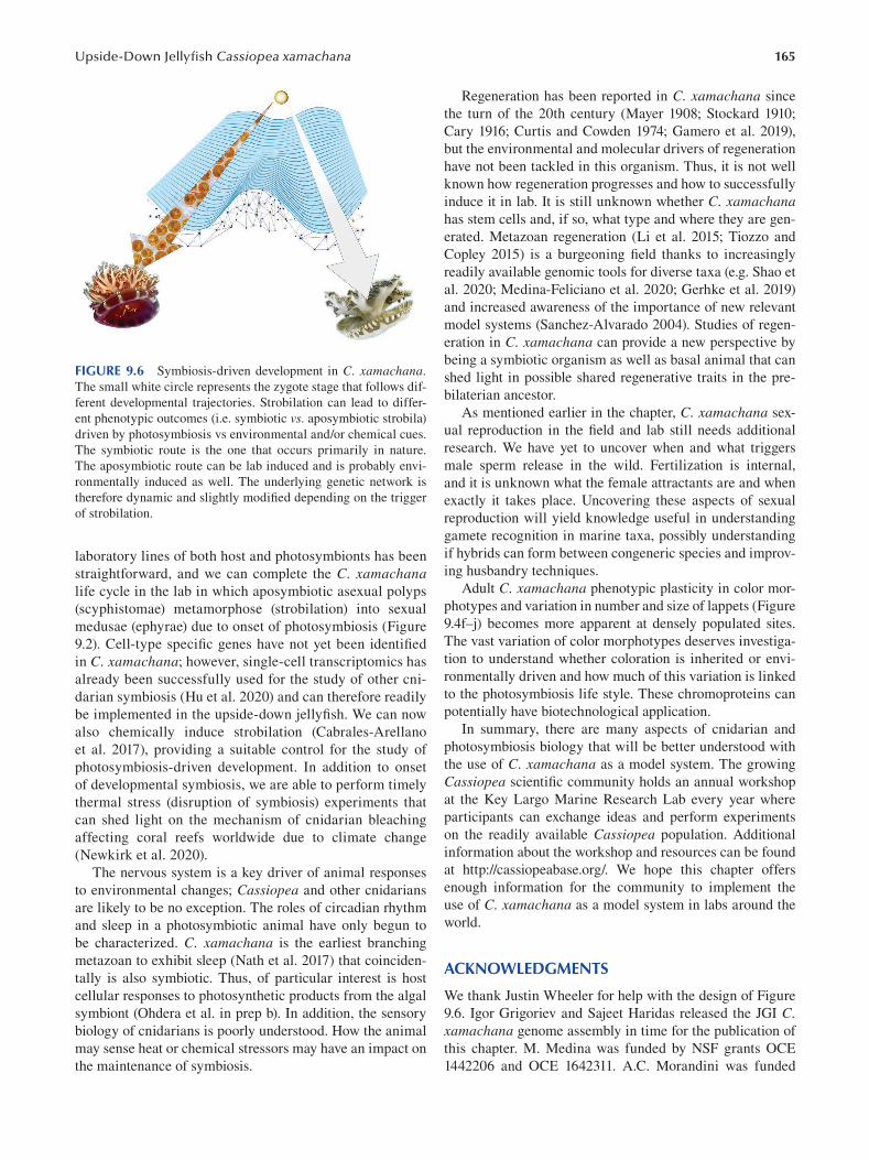
165 Upside-Down Jellyfi sh Cassiopea xamachana
FIGURE 9.6 Symbiosis-driven development in C. xamachana. The small white circle represents the zygote stage that follows dif
ferent developmental trajectories. Strobilation can lead to differ
ent phenotypic outcomes (i.e. symbiotic vs. aposymbiotic strobila)
driven by photosymbiosis vs environmental and/or chemical cues.
The symbiotic route is the one that occurs primarily in nature.
The aposymbiotic route can be lab induced and is probably envi
ronmentally induced as well. The underlying genetic network is
therefore dynamic and slightly modified depending on the trigger
of strobilation.
laboratory lines of both host and photosymbionts has been
straightforward, and we can complete the C. xamachana life cycle in the lab in which aposymbiotic asexual polyps
(scyphistomae) metamorphose (strobilation) into sexual
medusae (ephyrae) due to onset of photosymbiosis (Figure
9.2). Cell-type specific genes have not yet been identifi ed
in C. xamachana; however, single-cell transcriptomics has
already been successfully used for the study of other cni
darian symbiosis (Hu et al. 2020) and can therefore readily
be implemented in the upside-down jellyfish. We can now
also chemically induce strobilation (Cabrales-Arellano
et al. 2017), providing a suitable control for the study of
photosymbiosis-driven development. In addition to onset
of developmental symbiosis, we are able to perform timely
thermal stress (disruption of symbiosis) experiments that
can shed light on the mechanism of cnidarian bleaching
affecting coral reefs worldwide due to climate change
(Newkirk et al. 2020).
The nervous system is a key driver of animal responses
to environmental changes; Cassiopea and other cnidarians
are likely to be no exception. The roles of circadian rhythm
and sleep in a photosymbiotic animal have only begun to
be characterized. C. xamachana is the earliest branching
metazoan to exhibit sleep (Nath et al. 2017) that coinciden
tally is also symbiotic. Thus, of particular interest is host
cellular responses to photosynthetic products from the algal
symbiont (Ohdera et al. in prep b). In addition, the sensory
biology of cnidarians is poorly understood. How the animal
may sense heat or chemical stressors may have an impact on
the maintenance of symbiosis.
Regeneration has been reported in C. xamachana since
the turn of the 20th century (Mayer 1908; Stockard 1910;
Cary 1916; Curtis and Cowden 1974; Gamero et al. 2019),
but the environmental and molecular drivers of regeneration
have not been tackled in this organism. Thus, it is not well
known how regeneration progresses and how to successfully
induce it in lab. It is still unknown whether C. xamachana has stem cells and, if so, what type and where they are gen
erated. Metazoan regeneration (Li et al. 2015; Tiozzo and
Copley 2015) is a burgeoning field thanks to increasingly
readily available genomic tools for diverse taxa (e.g. Shao et
al. 2020; Medina-Feliciano et al. 2020; Gerhke et al. 2019)
and increased awareness of the importance of new relevant
model systems (Sanchez-Alvarado 2004). Studies of regen
eration in C. xamachana can provide a new perspective by
being a symbiotic organism as well as basal animal that can
shed light in possible shared regenerative traits in the pre
bilaterian ancestor.
As mentioned earlier in the chapter, C. xamachana sex
ual reproduction in the field and lab still needs additional
research. We have yet to uncover when and what triggers
male sperm release in the wild. Fertilization is internal,
and it is unknown what the female attractants are and when
exactly it takes place. Uncovering these aspects of sexual
reproduction will yield knowledge useful in understanding
gamete recognition in marine taxa, possibly understanding
if hybrids can form between congeneric species and improv
ing husbandry techniques.
Adult C. xamachana phenotypic plasticity in color mor
photypes and variation in number and size of lappets (Figure
9.4f–j) becomes more apparent at densely populated sites.
The vast variation of color morphotypes deserves investiga
tion to understand whether coloration is inherited or envi
ronmentally driven and how much of this variation is linked
to the photosymbiosis life style. These chromoproteins can
potentially have biotechnological application.
In summary, there are many aspects of cnidarian and
photosymbiosis biology that will be better understood with
the use of C. xamachana as a model system. The growing
Cassiopea scientific community holds an annual workshop
at the Key Largo Marine Research Lab every year where
participants can exchange ideas and perform experiments
on the readily available Cassiopea population. Additional
information about the workshop and resources can be found
at http://cassiopeabase.org/. We hope this chapter offers
enough information for the community to implement the
use of C. xamachana as a model system in labs around the
world.
ACKNOWLEDGMENTS
We thank Justin Wheeler for help with the design of Figure
9.6 . Igor Grigoriev and Sajeet Haridas released the JGI C. xamachana genome assembly in time for the publication of
this chapter. M. Medina was funded by NSF grants OCE
1442206 and OCE 1642311. A.C. Morandini was funded
166
by CNPq 309440/2019–0 and FAPESP 2011/50242–5,
2015/21007–9. We thank Key Largo Marine Research Lab
for the incessant hospitality over the years. D. Hoffman also
extends his thanks and deep appreciation to Dr. Bill Fitt for
decades of joint research on Cassiopea.
BIBLIOGRAPHY
Abrego, D., M. J. H. Van Oppen and B. L. Willis. 2009. Onset of
algal endosymbiont specificity varies among closely related
species of Acropora corals during early ontogeny. Molecular Ecology 18: 3532–3543.
Adachi, K., H. Miyake, T. Kuramochi, et al. 2017. Genome size dis
tribution in phylum Cnidaria. Fisheries Science 83: 107–112.
Agassiz, A. and A. G. Mayer. 1899. Acalephs from the Fiji Islands.
33: 157–189.
Al-jbour, S. M., M. Zimmer and A. Kunzmann. 2017. Cellular res
piration, oxygen consumption, and trade-offs of the jellyfi sh
Cassiopea sp. in response to temperature change. Journal of Sea Research 128: 92–97.
Ames, C. L., A. M. L. Klompen, K. Badhiwala, et al. 2020.
Cassiosomes are stinging-cell structures in the mucus
of the upside-down jellyfi sh Cassiopea xamachana.
Communications Biology 3: 67.
Arai, M. N. 2001. Pelagic coelenterates and eutrophication: A
review. In: Hydrobiologia 451: 69–87.
Arai, Y., R. Gotoh, J. Yokoyama, et al. 2017. Phylogenetic relation
ships and morphological variations of upside-down jelly
fi shes, Cassiopea spp. inhabiting Palau Islands. Biogeography 10: 133–141.
Aranda, M., Y. Li, Y. J. Liew, et al. 2016. Genomes of coral dino
flagellate symbionts highlight evolutionary adaptations con
ducive to a symbiotic lifestyle. Scientifi c Reports 6: 39734.
Baker, H. G. 1955. Self-compatibility and establishment after
“long-distance” dispersal. Evolution 9: 347–349.
Baker, H. G. 1965. Characteristics and modes of origin of weeds, In
The genetics of colonizing species.
Balderston, L. and G. Claus 1969. Study of symbiotic relationship
between Symbiodinium microadriaticum Freudenthal, zooxan
thella and upside down jellyfi sh. Nova Hedwigia 17: 373–382.
Barrangou, R. and J. A. Doudna. 2016. Applications of CRISPR
technologies in research and beyond. Nature Biotechnology 34: 933–941.
Barshis, D. J., J. T. Ladner, T. A. Oliver, et al. 2013. Genomic
basis for coral resilience to climate change. Proceedings of the National Academy of Sciences of the United States of America 110: 1387–1392.
Baumgarten, S., O. Simakov, L. Y. Esherick, et al. 2015. The
genome of Aiptasia, a sea anemone model for coral symbio
sis. Proceedings of the National Academy of Sciences 112:
11893–11898.
Bayha, K. M. and W. M. Graham. 2014. Nonindigenous marine
jellyfish: invasiveness, invasibility, and impacts. In Jellyfi sh blooms, K. A. Pitt and C. H. Lucas (Eds.), pp. 45–78.
Springer.
Bellantuono, A. J., C. Granados-Cifuentes, D. J. Miller, et al. 2012.
Coral thermal tolerance: Tuning gene expression to resist
thermal stress. PLoS One 7, Nr. 11.
Bigelow, R. P. 1892. On a new species of Cassiopea from Jamaica.
Zoologischer Anzeiger 15: 212–214.
Bigelow, R. P. 1893. Some observations on Polyclonia frondosa.
Baltimore: John Hopkins University.
Bigelow, R. P. 1900. The anatomy and development of Cassiopea xamachana. Boston Society of Natural History.
Emerging Marine Model Organisms
Blanquet, R. S. and M. A. Phelan. 1987. An unusual blue mesogleal
protein from the mangrove jellyfi sh Cassiopea xamachana.
Marine Biology 94: 423–430.
Blanquet, R. S. and G. P. Riordan. 1981. An ultrastructural study of
the subumbrellar musculature and desmosomal complexes of
Cassiopea xamachana (Cnidaria: Scyphozoa). Transactions of the American Microscopical Society 100: 109–119.
Brandt, J. F. 1835. Prodromus Descriptionis Animalium Ab H. Mertensio in Orbis Terrarum Circumnavigatione Observatorum.
Brandt, J. F. and C. H. Mertens. 1838. Ausführliche Beschreibung
der von CH Mertens auf seiner Weltumsegelung beobachteten
Schirmquallen: Nebst allgemeinen Bemerkungen über die
Schirmquallen überhaupt. 2: 237–411.
Bridge, D., C. W. Cunningham, B. Schierwater et al. 1992. Class-
level relationships in the phylum Cnidaria: Evidence from
mitochondrial genome structure. Proceedings of the National Academy of Sciences of the United States of America 89:
8750–8753.
Bulina, M. E., K. A. Lukyanov, I. V. Yampolsky, et al. 2004. New
class of blue animal pigments based on Frizzled and Kringle
protein domains. Journal of Biological Chemistry 279:
43367–43370.
Cabrales-Arellano, P., T. Islas-Flores, P. E. Thomé, et al. 2017.
Indomethacin reproducibly induces metamorphosis in
Cassiopea xamachana scyphistomae. PeerJ, Nr. 3.
Carrier, T. J. and A. M. Reitzel. 2018. Convergent shifts in host-
associated microbial communities across environmentally
elicited phenotypes. Nature Communications 9: 952.
Cary, L. R. 1916. The influence of the marginal sense organs on
the rate of regeneration in Cassiopea xamachana. Journal of Experimental Zoology 21: 1–32.
Cary, L. R. 1917. Studies on the physiology of the nervous system
of Cassiopea xamachana. Carnegie Institution of Washington Plub, Nr 251: 121–170.
Çevik, C., I. T. Erkol and B. Toklu. 2006. A new record of an alien
jellyfish from the Levantine coast of Turkey— Cassiopea andromeda (Forskål, 1775) [Cnidaria: Scyphozoa:
Rhizostomea]. Aquatic Invasions 1, Nr. 3.
Colley, N. J. and R. K. Trench. 1983. Selectivity in phagocytosis and
persistence of symbiotic algae in the scyphistoma stage of the
jellyfi sh Cassiopeia xamachana. Proceedings of the Royal Society of London. Series B, Containing papers of a Biological character. Royal Society (Great Britain) 219: 61–82.
Colley, N. J. and R. K. Trench. 1985. Cellular events in the reestab
lishment of a symbiosis between a marine dinofl agellate and
a coelenterate. Cell and Tissue Research 239: 93–103.
Császár, N. B. M., F. O. Seneca and M. J. H. Van Oppen. 2009.
Variation in antioxidant gene expression in the scleractinian
coral Acropora millepora under laboratory thermal stress.
Marine Ecology Progress Series 392: 93–102.
Cunning, R., R. A. Bay, P. Gillette, et al. 2018. Comparative anal
ysis of the Pocillopora damicornis genome highlights role
of immune system in coral evolution. Scientifi c Reports 8:
16134.
Curtis, S. K. and R. R. Cowden. 1971. Normal and experimen
tally modified development of buds in Cassiopea (phylum
Coelenterata; class Scyphozoa). Acta embryologiae experimentalis 3: 239–259.
Curtis, S. K. and R. R. Cowden. 1972. Regenerative capacities
of the scyphistoma of Cassiopea (Phylum, Coelenterata;
class, Scyphozoa). Acta embryologiae experimentalis 1972:
429–454.
Curtis, S. K. and R. R. Cowden. 1974. Some aspects of regenera
tion in the scyphistoma of Cassiopea (class Scyphozoa) as
167 Upside-Down Jellyfi sh Cassiopea xamachana
revealed by the use of antimetabolites and microspectropho
tometry. American Zoologist 14: 851–866.
DeSalvo, M. K., S. Sunagawa, P. L. Fisher, et al. 2010. Coral host
transcriptomic states are correlated with Symbiodinium geno
types. Molecular Ecology 19: 1174–1186.
Doty, M. S. 1961. Acanthophora, a possible invader of the marine
flora of Hawaii. Pacifi c Science 15: 547–552.
Doudna, J. A. and E. Charpentier. 2014. The new frontier of genome
engineering with CRISPR-Cas9. Science 346: 1258096.
Dubuc, T. Q., T. B. Stephenson, A. Q. Rock, et al. 2018. Hox and
Wnt pattern the primary body axis of an anthozoan cnidarian
before gastrulation. Nature Communications 9, Nr. 1.
Fang, L. S., S. P. Huang and K. L. Lin. 1997. High temperature
induces the synthesis of heat-shock proteins and the elevation
of intracellular calcium in the coral Acropora grandis. Coral Reefs 16: 127–131.
Fieth, R. A., M. E. A. Gauthier, J. Bayes, et al. 2016.
Ontogenetic changes in the bacterial symbiont community
of the tropical demosponge Amphimedon queenslandica:
Metamorphosis is a new beginning. Frontiers in Marine Science 3: 228.
Fitt, W. K. 1984. The role of chemosensory behavior of
Symbiodinium microadriaticum, intermediate hosts, and host
behavior in the infection of coelenterates and molluscs with
zooxanthellae. Marine Biology 81: 9–17.
Fitt, W. K. 1985. Effect of different strains of the zooxanthellae
Symbiodinium microadriaticum on growth and survival of
their coelenterate and molluscan hosts. Proceedings of the 5th International Coral Reef Congress, 6: 228.
Fitt, W. K. and R. K. Trench. 1983a. Endocytosis of the symbiotic
dinofl agellate Symbiodinium microadriaticum Freudenthal
by endodermal cells of the scyphistomae of Cassiopeia xamachana and resistance of the algae to host digestion. Journal of Cell Science 64: 195–212.
Fitt, W. K. and R. K. Trench. 1983b. Infection of coelenterate hosts
with the symbiotic dinofl agellate Symbiodinium microadriaticum. In: Intracellular space as oligogenetic ecosystem. Proceedings, 2: 675–681.
Fitt, W. K. and K. Costley. 1998. The role of temperature in sur
vival of the polyp stage of the tropical rhizostome jellyfi sh
Cassiopea xamachana. Journal of Experimental Marine Biology and Ecology 222: 79–91.
Fitt, W. K. and D. K. Hofmann. 1985. Chemical induction of settle
ment and metamorphosis of planulae and buds of the reef-
dwelling coelenterate Cassiopeia andromeda. Proceedings of the 5th International Coral Reef Congress, 5: 239–244,
Tahiti.
Fitt, W. K., D. K. Hofmann and M. Rahat. 1987. Requirement of
exogenous inducers for metamorphosis of axenic larvae and
buds of Cassiopeia andromeda (Cnidaria: Scyphozoan).
Marine Biology 94: 415–422.
Fleck, J. and A. Bischoff. 1993. Protein kinase C is possi
bly involved in chemical induction of metamorphosis in
Cassiopea spp. (Cnidaria: Scyphozoa). Proceedings of the 5th International Coral Reef Congress, 456–462. Richmond
RH (ed) University of Guam Press, KOG Station, Mangilao,
Guam, USA.
Fleck, J., W. K. Fitt and M. G. Hahn. 1999. A proline-rich
peptide originating from decomposing mangrove leaves
is one natural metamorphic cue of the tropical jellyfi sh
Cassiopea xamachana. Marine Ecology Progress Series 183: 115–124.
Fleck, J. and W. K. Fitt. 1999. Degrading mangrove leaves of
Rhizophora mangle Linne provide a natural cue for settlement
and metamorphosis of the upside down jellyfi sh Cassiopea xamachana. Journal of Experimental Marine Biology and Ecology 234: 83–94.
Fleck, J. and D. K. Hofmann. 1995. In vivo binding of a bio
logically active oligopeptide in vegetative buds of the
scyphozoan Cassiopea andromeda: Demonstration of recep
tor-mediated induction of metamorphosis. Marine Biology 122: 447–451.
Fleck, J. and D. K. Hofmann. 1990. The efficiency of metamorpho
sis inducing oligopeptides in Cassiopea species (Cnidaria:
Scyphozoa) depends on both primary structure and amino-
and carboxy terminal substituents. Verh Dtsch Zool Ges 83:
452–453.
Fleck, J. 1998. Chemical fate of a metamorphic inducer in larvae-
like buds of the cnidarian Cassiopea andromeda. Biological Bulletin 194: 83–91.
Forskål, P. 1775. Desriptiones Animalium Avium, Amphibiorum, Piscium, Insectorum, Vermium; Quae in Intinere Orientali Observavit Petrus Forskål. Copenhagen: Niebuhr, Carsten.
Forskål, P. and C. Niebuhr. 1776. Icones rerum naturalium: quas in itinere orientali depingi . Copenhagen.
France, S. C. and L. L. Hoover. 2002. DNA sequences of the mitochon
drial COI gene have low levels of divergence among deep-sea
octocorals (Cnidaria: Anthozoa). Hydrobiologia 471: 149–155.
Freeman, C. J., E. W. Stoner, C. G. Easson, et al. 2016. Symbiont
carbon and nitrogen assimilation in the Cassiopea-Symbiodinium mutualism. Marine Ecology Progress Series 544: 281–286.
Freudenthal, H. D. 1959. Observations on the algal cells (zooxan
thellae) inhabiting the anemone Cassiopea sp. J. Protozool Nr. 6: 12.
Freudenthal, H. D. 1962. Symbiodinium gen. nov. and Symbiodinium microadriaticum sp. nov., a Zooxanthella: Taxonomy, life
cycle, and morphology. J. Protozool. 9: 45–52.
Fuller, Z. L., V. J. L. Mocellin, L. A. Morris, et al. 2020. Population
genetics of the coral Acropora millepora : Toward genomic
prediction of bleaching. Science 369: eaba4674.
Gamero-Mora, E., R. Halbauer, V. Bartsch, et al. 2019. Regenerative
capacity of the upside-down jellyfi sh Cassiopea xamachana.
Zoological Studies 58.
Garcia-Fernàndez, J. 2005. Hox, ParaHox, ProtoHox: Facts and
guesses. Hereditary 94: 145–152.
Gauchat, D., F. Mazet, C. Berney, et al. 2000. Evolution of Antp
class genes and differential expression of Hydra Hox/para-
Hox genes in anterior patterning. Proceedings of the National Academy of Sciences of the United States of America 97:
4493–4498.
Gehrke, A. R., E. Neverett, Y. Luo, et al. 2019. Acoel genome
reveals the regulatory landscape of whole-body regeneration.
Science 363, Nr. 6432.
Gershwin, L. A., W. Zeidler and P. J. F. Davie. 2010. Medusae
(Cnidaria) of Moreton Bay, Queensland, Australia. Memoirs of the Queensland Museum 54: 47–108.
Gilbert, S. F. 2016. Chapter twenty-two: Developmental plastic
ity and developmental symbiosis: The return of eco-devo.
In Current topics in developmental biology, hg. von Paul
M. Wassarman, 116: 415–433. Essays on Developmental
Biology, Part A. Academic Press, 1.
Gilbert, S. F. and D. Epel. 2015. Ecological developmental biology: The environmental regulation of development, health, and evolution. 2nd ed. Sunderland, MA, USA: Sinauer
Associates is an imprint of Oxford University Press.
Gohar, H. A. F. and A. M. Eisawy. 1960a. The biology of Cassiopea andromeda (from the Red Sea) (with a note on the species
168
problem). Publications of the Marine Biological Station, Ghardaqa 11: 3–39.
Gohar, H. A. F. and M. Eisawy. 1960b. The development of
Cassiopea andromeda (Scyphomedusae). Publs Marine Biology Station Ghardaqa 11: 148–190.
Gold, D. A., T. Katsuki, Y. L., Xifeng, et al. 2019. The genome of
the jellyfi sh Aurelia and the evolution of animal complexity.
Nature Ecology and Evolution 3: 96–104.
Goldfarb, A. J. 1914. Changes in salinity and their effects upon
the regeneration of Cassiopea xamachana. Papers from the Tortugas Laboratory of the Carnegie Institution 6: 85–94.
Gómez-Daglio, L. and M. N. Dawson. 2017. Species richness of jel
lyfishes (Scyphozoa: Discomedusae) in the tropical Eastern
Pacific: Missed taxa, molecules, and morphology match in a
biodiversity hotspot. Invertebrate Systematics 31: 635–663.
Grant, A. J., D. A. Trautman, S. Frankland, et al. 2003. A symbio
some membrane is not required for the actions of two host
signalling compounds regulating photosynthesis in symbiotic
algae isolated from cnidarians. Comparative Biochemistry and Physiology: A Molecular and Integrative Physiology 135: 337–345.
Haeckel, E. 1880. System der Acraspeden-Zweite Hälfte des
Systems der Medusen. Denkschriften der Medizinisch— Naturwissenschaftlichen Gesellschaft zu Jena : 82.
Hamlet, C., A. Santhanakrishnan and L. A. Miller. 2011. A numeri
cal study of the effects of bell pulsation dynamics and oral
arms on the exchange currents generated by the upside-down
jellyfi sh Cassiopea xamachana. Journal of Experimental Biology 214: 1911–1921.
Harrison, P. L., R. C. Babcock, G. D. Bull, et al. 1984. Mass spawn
ing in tropical reef corals. Science 223: 1186–1189.
Hartmann, A. C., K. L. Marhaver, A. Klueter, et al. 2019. Acquisition
of obligate mutualist symbionts during the larval stage is not
beneficial for a coral host. Molecular Ecology 28: 141–155.
He, S., F. Del Viso, C. Y. Chen, et al. 2018. An axial Hox code con
trols tissue segmentation and body patterning in Nematostella vectensis. Science 361: 1377–1380.
Hebert, P. D. N., S. Ratnasingham and J. R. DeWaard. 2003.
Barcoding animal life: Cytochrome c oxidase subunit 1
divergences among closely related species. Proceedings of the Royal Society B: Biological Sciences 270: S96–S99.
Heins, A., T. Glatzel and S. Holst. 2015. Revised descriptions of
the nematocysts and the asexual reproduction modes of the
scyphozoan jellyfi sh Cassiopea andromeda (Forskål, 1775).
Zoomorphology 134: 351–366.
Hofmann, D. K. and M. Gottlieb. 1991. Bud formation in the scy
phozoan Cassiopea andromeda: Epithelial dynamics and fate
map. Hydrobiologia 216: 53–59.
Hofmann, D. K. and M. G. Hadfield. 2002. Hermaphroditism,
gonochorism, and asexual reproduction in Cassiopea sp.: An immigrant in the islands of Hawai’i. Invertebrate Reproduction and Development 41: 215–221.
Hofmann, D. K. and T. G. Honegger. 1990. Bud formation and meta
morphosis in Cassiopea andromeda (Cnidaria: Scyphozoa):
A developmental and ultrastructural study. Marine Biology 105: 509–518.
Hofmann, D. K. and B. P. Kremer. 1981. Carbon metabolism
and strobilation in Cassiopea andromedea (Cnidaria:
Scyphozoa): Significance of endosymbiotic dinofl agellates.
Marine Biology 65: 25–33.
Hofmann, D. K., R. Neumann and K. Henne. 1978. Strobilation,
budding and initiation of scyphistoma morphogenesis in the
Rhizostome Cassiopea andromeda (Cnidaria: Scyphozoa).
Marine Biology 47: 161–176.
Emerging Marine Model Organisms
Hofmann, D. K. and U. Brandt. 1987. Induction of metamorphosis
in the symbiotic scyphozoan Cassiopea andromeda: Role of
marine bacteria and of biochemicals. Symbiosis 4: 99–116.
Hofmann, D. K., W. K. Fitt and J. Fleck. 1996. Checkpoints in the
life-cycle of Cassiopea spp.: Control of metagenesis and
metamorphosis in a tropical jellyfi sh. International Journal of Developmental Biology 40: 331–338.
Hofmann, D. K. and G. Henning. 1991. Effects of axenic culture
conditions on asexual reproduction and metamorphosis in the
symbiotic scyphozoan Cassiopea andromeda. Symbiosis 10:
83–93.
Holland, B. S., M. N. Dawson, G. L. Crow, et al. 2004. Global
phylogeography of Cassiopea (Scyphozoa: Rhizostomeae):
Molecular evidence for cryptic species and multiple invasions
of the Hawaiian Islands. Marine Biology 145: 1119–1128.
Hu, M., X. Zheng, C. M. Fan, et al. 2020. Lineage dynamics of the
endosymbiotic cell type in the soft coral Xenia. Nature 582:
534–538.
Hui, J. H. L., P. W. H. Holland and D. E. K. Ferrier. 2008. Do cni
darians have a ParaHox cluster? Analysis of synteny around
a Nematostella homeobox gene cluster. Evolution and Development 10: 725–730.
Hummelinck, P. Wagenaar. 1968. Caribbean scyphomedusae of the
genus Cassiopea. Studies on the Fauna of Curaçao and other Caribbean Islands 25: 1–57.
Jantzen, C., C. Wild, M. Rasheed, et al. 2010. Enhanced pore-water
nutrient fluxes by the upside-down jellyfi sh Cassiopea sp. in
a Red Sea coral reef. Marine Ecology Progress Series 411:
117–125.
Jarms, G. and A. C. Morandini. 2019. World Atlas of Jellyfish.
Dölling and Galitz Verlag.
Jensch, F. and D. K. Hofmann. 1997. The cnidomes of Cassiopea andromeda Forskål, 1775, and Cassiopea xamachana Bigelow, 1882 (Cnidaria: Scyphozoa). Proceedings of the 6th International Conference on Coelenterate Biology , 279–285.
Jinek, M., K. Chylinski, I. Fonfara, et al. 2012. A programmable
dual-RNA-guided DNA endonuclease in adaptive bacterial
immunity. Science 337, Nr. 6096.
Jones, V. A. S., M. Bucher, E. A. Hambleton et al. 2018.
Microinjection to deliver protein, mRNA, and DNA into
zygotes of the cnidarian endosymbiosis model Aiptasia sp.
Scientifi c Reports 8: 16437.
Kamm, K. and B. Schierwater. 2007. Ancient linkage of a POU class
6 and an anterior Hox-like gene in Cnidaria: Implications for
the evolution of homeobox genes. Journal of Experimental Zoology Part B: Molecular and Developmental Evolution 308B: 777–784.
Kayal, E., B. Bentlage, A. G. Collins, et al. 2012. Evolution of
linear mitochondrial genomes in medusozoan cnidarians.
Genome Biology and Evolution 4: 1–12.
Kayal, E., B. Bentlage, M. S. Pankey, et al. 2018. Phylogenomics
provides a robust topology of the major cnidarian lineages
and insights on the origins of key organismal traits. BMC Evolutionary Biology 18, Nr. 68.
Keable, S. J. and S. T. Ahyong. 2016. First records of the invasive
“upside-down jellyfi sh”, Cassiopea (Cnidaria: Scyphozoa:
Rhizostomeae: Cassiopeidae), from coastal lakes of New South
Wales, Australia. Records of the Australian Museum 68: 23–30.
Khabibulina, V. and V. Starunov. 2019. Musculature development in
planuloids of Cassiopeia xamachana (Cnidaria: Scyphozoa).
Zoomorphology 138: 297–306.
Khalturin, K., C. Shinzato, M. Khalturina, et al. 2019. Medusozoan
genomes inform the evolution of the jellyfish body plan.
Nature Ecology and Evolution 3: 811–822.
169 Upside-Down Jellyfi sh Cassiopea xamachana
Kim, H. M., J. A. Weber, N. Lee, et al. 2019. The genome of the
giant Nomura ’s jellyfish sheds light on the early evolution of
active predation. BMC Biology 17: 28.
King, J. M. 1980. Direct fission: An undescribed reproductive method
in hydromedusae. Bulletin of Marine Science 30: 522–525.
Klein, S. G., K. A. Pitt, C. H. Lucas, et al. 2019. Night-time
temperature reprieves enhance the thermal tolerance of a
symbiotic cnidarian. Frontiers in Marine Science 6, Nr.
July: 453.
Kraus, Y. A. and A. V. Markov. 2016. The gastrulation in Cnidaria:
A key to understanding phylogeny or the chaos of secondary
modifi cations? Zhurnal Obshcheĭ Biologii 77: 83–105.
Kuhn, K., B. Streit and Bernd Schierwater. 1999. Isolation of
Hox genes from the Scyphozoan Cassiopeia xamachana:
Implications for the early evolution of Hox genes. Journal of Experimental Zoology 285: 63–75.
Kvennefors, E. C. E., W. Leggat, O. Hoegh-Guldberg, et al. 2008.
An ancient and variable mannose-binding lectin from the
coral Acropora millepora binds both pathogens and sym
bionts. Developmental and Comparative Immunology 32:
1582–1592.
LaJeunesse, T. C. 2017. Validation and description of Symbiodinium microadriaticum, the type species of Symbiodinium
(Dinophyta). Journal of Phycology 53: 1109–1114.
LaJeunesse, T. C. 2020. Zooxanthellae. Current Biology 30:
R1110–R1113.
LaJeunesse, T. C., J. E. Parkinson, P. W. Gabrielson, et al. 2018.
Systematic revision of Symbiodiniaceae highlights the antiq
uity and diversity of coral endosymbionts. Current Biology 28: 2570–2580.
LaJeunesse, T. C., D. T. Pettay, E. M. Sampayo, et al. 2010. Long-
standing environmental conditions, geographic isolation and
host-symbiont specifi city influence the relative ecological
dominance and genetic diversification of coral endosymbi
onts in the genus Symbiodinium. Journal of Biogeography 37: 785–800.
Lampert, K. P. 2016. Cassiopea and its zooxanthellae. In: The Cnidaria, past, present and future: The world of Medusa and her sisters, Stefano Goffredo and Zvy Dubinsky (Eds.).
Springer International Publishing.
Lampert, K. P., P. Bürger, S. Striewski, et al. 2012. Lack of asso
ciation between color morphs of the jellyfi sh Cassiopea andromeda and zooxanthella clade. Marine Ecology 33:
364–369.
Lehnert, E. M., M. E. Mouchka, M. S. Burriesci, et al. 2014.
Extensive differences in gene expression between symbiotic
and aposymbiotic cnidarians. G3: Genes, Genomes, Genetics 4: 277–295.
Li, Qiao, H. Y. and T. P. Zhong. 2015. Regeneration across meta
zoan phylogeny: Lessons from model organisms. Journal of Genetics and Genomics 42: 57–70.
Li, Yunfeng, L. G., Y. Pan, M. Tian, et al. 2020. Chromosome-level
reference genome of the jellyfi sh Rhopilema esculentum.
GigaScience 9, Nr. 4.
Lieshout, J., S. V. and V. J. Martin. 1992. Development of planu
loid buds of Cassiopea xamachana (Cnidaria: Scyphozoa).
Transactions of the American Microscopical Society 111:
89–110.
Light, S. F. 1914. Some Philippine Scyphomedusae, including two
new genera, five new species, and one new variety. Philippine Journal of Science 9: 195–231.
Light, S. F. 1924. A new species of Scyphomedusan jellyfi sh in
Chinese waters. The China Journal of Science and Arts 2:
449–450.
Ludwig, F. D. 1969. Die Zooxanthellen bei Cassiopea androm
eda Eschscholtz 1829 (Polyp-Stadium) und ihre Bedeutung
für die Strobilation. Zoologische Jahrbücher. Abteilung für Anatomie und Ontogenie der Tiere Abteilung für Anatomie und Ontogenie der Tiere 86: 238–277.
Lukyanov, K. A. and V. V. Belousov. 2014. Genetically encoded
fluorescent redox sensors. Biochimica et Biophysica Acta (BBA)—General Subjects 1840: 745–756.
Maas, O. 1903. Die Scyphomedusen der Siboga-Expedition.
Siboga-expeditie 11. Leiden: Buchhandlung und druckerei
vormals E. J. Brill.
Martin, V. J. and F. S. Chia. 1982. Fine structure of a scyphozoan
planula, Cassiopeia xamachana. The Biological Bulletin 163, Nr. 2.
Matthews, B. J. and L. B. Vosshall. 2020. How to turn an organ
ism into a model organism in 10 “easy” steps. Journal of Experimental Biology 223: 223.
Mayer, A. G. 1908. Rhythmical pulsation of the medusae . 47. Aufl .
Washington, DC.
Mayer, A. G. 1906. Medusae of the Hawaiian Islands collected by the steamer Albatross in 1902. Washington, DC: Government
Printing Offi ce.
Mayer, A. G. 1910. Medusae of the world: Volume III the scyphomedusae. Washington, DC: Carnegie Institution of
Washington.
McFall-Ngai, M., M. G. Hadfield, T. C. G. Bosch, et al. 2013.
Animals in a bacterial world, a new imperative for the life
sciences. Proceedings of the National Academy of Sciences of the United States of America 110: 3229–3236.
McGill, C. J. and C. M. Pomory. 2008. Effects of bleaching and nutri
ent supplementation on wet weight in the jellyfi sh Cassiopea xamachana (Bigelow) (Cnidaria: Scyphozoa). Marine and Freshwater Behaviour and Physiology 41: 179–189.
McIlroy, S. E. and M. A. Coffroth. 2017. Coral ontogeny affects
early symbiont acquisition in laboratory-reared recruits.
Coral Reefs 36: 927–932.
Medina-Feliciano, J. G., S. Pirro, J. E. García-Arrarás, et al. 2020.
Draft genome of the sea cucumber Holothuria glaberrima , a
model for the study of regeneration. bioRxiv.
Mellas, R. E., S. E. McIlroy, W. K. Fitt et al. 2014. Variation in
symbiont uptake in the early ontogeny of the upside-down
jellyfi sh, Cassiopea spp. Journal of Experimental Marine Biology and Ecology 459: 38–44.
Mirsky, A. E. and H. Ris. 1951. The desoxyribonucleic acid content
of animal cells and its evolutionary signifi cance. The Journal of general physiology 34: 451–462.
Mohamed, A. R., V. Cumbo, S. Harii, et al. 2016. The transcrip
tomic response of the coral Acropora digitifera to a compe
tent Symbiodinium strain: The symbiosome as an arrested
early phagosome. Molecular Ecology 25: 3127–3141.
Morandini, A. C., D. Ascher, S. N. Stampar, et al. 2005. Cubozoa
e Scyphozoa (Cnidaria: Medusozoa) de águas costeiras do
Brasil. Iheringia. Série Zoologia 95: 281–294.
Morandini, A. C. and F. L. da Silveira. 2001. Sexual reproduction
of Nausithoe aurea (Scyphozoa, Coronatae). Gametogenesis,
egg release, embryonic development, and gastrulation.
Scientia Marina 65: 139–149.
Morandini, A. C., S. N. Stampar, M. M. Maronna, et al. 2017.
All non-indigenous species were introduced recently? The
case study of Cassiopea (Cnidaria: Scyphozoa) in Brazilian
waters. Journal of the Marine Biological Association of the United Kingdom 97: 321–328.
Mortillaro, J. M., K. A. Pitt, S. Y. Lee, et al. 2009. Light inten
sity influences the production and translocation of fatty acids
170
by zooxanthellae in the jellyfi sh Cassiopea sp. Journal of Experimental Marine Biology and Ecology 378: 22–30.
Muscatine, L. and J. W. Porter. 1977. Reef Corals: Mutualistic sym
bioses adapted to nutrient-poor environments. BioScience 27: 454–460.
Nakai, J., M. Ohkura and K. Imoto. 2001. A high signal-to-noise
Ca2+ probe composed of a single green fl uorescent protein.
Nature Biotechnology 19: 137–141.
Nakanishi, N., D. Yuan, D. K. Jacobs, et al. 2008. Early develop
ment, pattern, and reorganization of the planula nervous sys
tem in Aurelia (Cnidaria, Scyphozoa). Development Genes and Evolution 218: 511–524.
Nath, R. D., C. N. Bedbrook, M. J. Abrams, et al. 2017. The jel
lyfi sh Cassiopea exhibits a sleep-like state. Current Biology 27: 2984–2990.
Neumann, R. 1979. Bacterial induction of settlement and meta
morphosis in the planula larvae of Cassiopea andromeda (Cnidaria: Scyphozoa, Rhizostomeae). Marine Ecology Progress Series 1, Nr. 1: 21–28.
Newkirk, C. R., T. K. Frazer, M. Q. Martindale, et al. 2020.
Adaptation to bleaching: Are thermotolerant Symbiodiniaceae
strains more successful than other strains under elevated
temperatures in a model symbiotic cnidarian? Frontiers in Microbiology 11: 822.
Niggl, W., M. S. Naumann, U. Struck, et al. 2010. Organic mat
ter release by the benthic upside-down jellyfi sh Cassiopea sp. fuels pelagic food webs in coral reefs. Journal of Experimental Marine Biology and Ecology 384: 99–106.
Ohdera, A., C. L. Ames, R. B. Dikow, et al. 2019. Box, stalked,
and upside-down? Draft genomes from diverse jellyfi sh
(Cnidaria, Acraspeda) lineages: Alatina alata (Cubozoa),
Calvadosia cruxmelitensis (Staurozoa), and Cassiopea xamachana (Scyphozoa). GigaScience 8, Nr. 7.
Ohdera, A. H., M. J. Abrams, C. L. Ames et al. 2018. Upside-down
but headed in the right direction: Review of the highly versa
tile Cassiopea xamachana system. Frontiers in Ecology and Evolution 6: 35.
Ohdera, A. H., K. Attarwala, H. Rubain, et al. In prep (a). Genomic
insights of bacteria responsible for settlement and metamor
phosis of Cassiopea xamachana larvae.
Ohdera, A. H., V. Avila-Magaña, V. Sharp et al. In prep (b).
Modulation of gene expression driven by symbiosis:
Strobilation mechanism in the upside-down jellyfi sh.
Ojimi, M. C. and M. Hidaka. 2010. Comparison of telomere length
among different life cycle stages of the jellyfi sh Cassiopea andromeda. Marine Biology 157: 2279–2287.
Ojimi, M. C., N. Isomura and M. Hidaka. 2009. Telomerase activity
is not related to life history stage in the jellyfi sh Cassiopea sp. Comparative Biochemistry and Physiology: A Molecular and Integrative Physiology 152: 240–244.
Pallas, Peter Simon. 1774. Spicilegia zoologica: quibus novae imprimis et obscurae animalium species iconibus, descriptionibus atque commentariis illustrantur. Hg. von August
Lange. 10. Aufl . Berlin.
Passano, L. M. 2004. Spasm behavior and the diffuse nerve-net
in Cassiopea xamachana (Scyphozoa: Coelenterata). In:
Hydrobiologia , 530–531:91–96.
Perkins, H. F. 1908. Notes on the occurrence of Cassiopea xamachana and Polyclonia frondosa at the Tortugas. Papers from the Tortugas Laboratory of the Carnegie Institution 1.
Phelan, M. A., J. L. Matta, Y. M. Reyes, et al. 2006. Associations
between metals and the blue mesogleal protein of Cassiopea xamachana. Marine Biology 149: 307–312.
Emerging Marine Model Organisms
Pierce, J. 2005. A system for mass culture of upside-down jellyfi sh
Cassiopea spp. as a potential food item for medusivores in
captivity. International Zoo Yearbook 39: 62–69.
Pinzón, J. H., B. Kamel, C. A. Burge, et al. 2015. Whole transcrip
tome analysis reveals changes in expression of immune-
related genes during and after bleaching in a reef-building
coral. Royal Society Open Science 2: 140214.
Polteya, D. G. D. Zhinidarich and A. Lui. 1985. Observations of asex
ual reproduction and regeneration in Cassiopea (Scyphozoa,
Coelenterata). Zoologicheski Zhurnal 64:172–180.
Radwan, F. F. Y., J. W. Burnett, D. A. Bloom, et al. 2001. A com
parison of the toxinological characteristics of two Cassiopea and Aurelia species. Toxicon 39: 245–257.
Rahat, M. and O. Adar. 1980. Effect of symbiotic zooxanthellae
and temperature on budding and strobilation in Cassiopeia andromeda (Eschscholz). Biological Bulletin 159: 394–401.
Reich, H. G., D. L. Robertson and G. Goodbody-Gringley.
2017. Do the shuffle: Changes in Symbiodinium consor
tia throughout juvenile coral development. PLoS One 12:
e0171768.
Richardson, A. J., A. Bakun, G. C. Hays, et al. 2009. The jellyfi sh
joyride: Causes, consequences and management responses to
a more gelatinous future. Trends in Ecology & Evolution 24:
312–322.
Rodriguez-Lanetty, M., W. S. Phillips and V. M. Weis. 2006.
Transcriptome analysis of a cnidarian–dinofl agellate mutu
alism reveals complex modulation of host gene expression.
BMC Genomics 7, Nr. 23.
Ruppert, E. E., R. S. Fox and R. D. Barnes. 2004. Invertebrate zoology: A functional evolutionary approach. Belmont, CA:
Thomson-Brooks/Cole.
Sánchez-Alvarado, A. 2004. Regeneration and the need for sim
pler model organisms. Philosophical Transactions of the Royal Society of London. Series B: Biological Sciences 359:
759–763.
Santhanakrishnan, A., M. Dollinger, C. L. Hamlet, et al. 2012.
Flow structure and transport characteristics of feeding and
exchange currents generated by upside-down Cassiopea jel
lyfi sh. Journal of Experimental Biology 215: 2369–2381.
Schembri, P. J., A. Deidun and P. J. Vella. 2010. First record
of Cassiopea andromeda (Scyphozoa: Rhizostomeae:
Cassiopeidae) from the central Mediterranean Sea. Marine Biodiversity Records 3: e6.
Schierwater, B. and K. Kuhn. 1998. Homology of Hox genes and
the Zootype concept in early metazoan evolution. Molecular Phylogenetics and Evolution 9: 375–381.
Schummer, M., I. Scheurlen, C. Schaller, et al. 1992. HOM/HOX
homeobox genes are present in hydra (Chlorohydra viridissima) and are differentially expressed during regeneration.
EMBO Journal 11: 1815–1823.
Schwarz, J. A., D. A. Krupp and V. M. Weis. 1999. Late larval
development and onset of symbiosis in the scleractinian coral
Fungia scutaria. The Biological Bulletin 196: 70–79.
Shao, Y., X. B. Wang, J. J. Zhang, et al. 2020. Genome and single-
cell RNA-sequencing of the earthworm Eisenia andrei iden
tifies cellular mechanisms underlying regeneration. Nature Communications 11: 2656.
Shearer, T. L., M. J. H. Van Oppen, S. L. Romano, et al. 2002. Slow
mitochondrial DNA sequence evolution in the Anthozoa
(Cnidaria). Molecular Ecology 11: 2475–2487.
Shikuma, N. J., I. Antoshechkin, J. M. Medeiros, et al. 2016.
Stepwise metamorphosis of the tubeworm Hydroides elegans is mediated by a bacterial inducer and MAPK signaling.
171 Upside-Down Jellyfi sh Cassiopea xamachana
Proceedings of the National Academy of Sciences of the United States of America 113: 10097–10102.
Shinzato, C., E. Shoguchi, T. Kawashima, et al. 2011. Using the
Acropora digitifera genome to understand coral responses to
environmental change. Nature 476: 320–323.
Shumaker, A., H. M. Putnam, H. Qiu et al. 2019. Genome analy
sis of the rice coral Montipora capitata. Scientifi c Reports 9: 2571.
Smith, H. G. 1936. Contribution to the anatomy and physiology of Cassiopea frondosa. Papers from the Tortugas Laboratory of the Carnegie Institution 31 p.
Stephens, L. and D. Calder. 2006. Seafaring scientist: Alfred Goldsborough Mayor, pioneer in marine biology . University
of South Caroline Press, Columbia.
Stiasny, G. 1924. Ueber einige von Dr. CJ van der Horst bei Curaçao
gesammelte Medusen. Bijdragen tot de Dierkunde 23: 83–92.
Stockard, C. R. 1908. Studies of tissue growth. Archiv für Entwicklungsmechanik der Organismen 29: 15–32.
Stoner, E. W., C. A. Layman, et al. 2011. Effects of anthropogenic
disturbance on the abundance and size of epibenthic jellyfi sh
Cassiopea spp. Marine Pollution Bulletin 62: 1109–1114.
Szmant, A. M. 1986. Reproductive ecology of Caribbean reef cor
als. Coral Reefs 5: 43–53.
Thé, J., H. S. Barroso, M. Mammone, et al. 2020. Aquaculture facil
ities promote populational stability throughout seasons and
increase medusae size for the invasive jellyfi sh Cassiopea andromeda. Marine Environmental Research : 105161.
Thieme, C. and D. K. Hofmann. 2003. An endogenous peptide is
involved in internal control of metamorphosis in the marine
invertebrate Cassiopea xamachana (Cnidaria: Scyphozoa).
Development Genes and Evolution 213: 97–101.
Thompson, J. R., H. E. Rivera, C. J. Closek, et al. 2015. Microbes
in the coral holobiont: Partners through evolution, develop
ment, and ecological interactions. Frontiers in Cellular and Infection Microbiology 4: 176.
Thornhill, D. J., M. W. Daniel, T. C. LaJeunesse, et al. 2006. Natural
infections of aposymbiotic Cassiopea xamachana scyphisto
mae from environmental pools of Symbiodinium. Journal of Experimental Marine Biology and Ecology 338: 50–56.
Tiozzo, S. and R. R. Copley. 2015. Reconsidering regeneration in
metazoans: An evo-devo approach. Frontiers in Ecology and Evolution 3: 67.
Todd, B. D., D. J. Thornhill and W. K. Fitt. 2006. Patterns of inor
ganic phosphate uptake in Cassiopea xamachana : A bioindi
cator species. Marine Pollution Bulletin 52: 515–521.
Tran, C. and M. G. Hadfield. 2011. Larvae of Pocillopora damicornis (Anthozoa) settle and metamorphose in response to
surface-biofilm bacteria. Marine Ecology Progress Series 433: 85–96.
Trench, R. K. 1979. The cell biology of plant-animal symbiosis.
Annual Review of Plant Physiology 30: 485–531.
Uchida, T. 1970. Occurrence of a rhizostome medusa, Cassiopea mertensii Brandt from the Hawaiian Islands. Annotat. Zool. Jap. 43: 102–104.
Van Etten, M. L., J. K. Conner, S. M. Chang, et al. 2017. Not all
weeds are created equal: A database approach uncovers dif
ferences in the sexual system of native and introduced weeds.
Ecology and Evolution 7: 2636–2642.
Van Woesik, R., F. Lacharmoise and S. Köksal. 2006. Annual cycles
of solar insolation predict spawning times of Caribbean cor
als. Ecology Letters 9: 390–398.
Verde, E. A. and L. R. McCloskey. 1998. Production, respiration,
and photophysiology of the mangrove jellyfi sh Cassiopea xamachana symbiotic with zooxanthellae: Effect of jelly
fish size and season. Marine Ecology Progress Series 168:
147–162.
Vijayan, N., K. A. Lema, B. T. Nedved, et al. 2019. Microbiomes of
the polychaete Hydroides elegans (Polychaeta: Serpulidae)
across its life-history stages. Marine Biology 166: 19.
Voolstra, C. R., J. A. Schwarz, J. Schnetzer, et al. 2009. The
host transcriptome remains unaltered during the establish
ment of coral–algal symbioses. Molecular Ecology 18:
1823–1833.
Waddington, C. H. 1942. Canalization of development and the
inheritance of acquired characters. Nature 150: 563–565.
Welsh, D. T., R. J. K. Dunn and T. Meziane. 2009. Oxygen and
nutrient dynamics of the upside down jellyfi sh ( Cassiopea sp.) and its influence on benthic nutrient exchanges and pri
mary production. Hydrobiologia 635: 351–362.
Widmer, C. L. 2008. Life cycle of Chrysaora fuscescens (Cnidaria:
Scyphozoa) and a key to sympatric ephyrae. Pacifi c Science 62: 71–82.
Winstead, D., A. Ohdera, M. Medina, et al. 2018. Symbiodinium proliferation inside a cnidarian host vessel are competitive
and dynamic. SICB 2018 Annual Meeting Abstracts, 58: e1–
e264. San Francisco, CA, März.
Wolk, M., M. Rahat, W. K. Fitt, et al. 1985. Cholera toxin and thy
rotropine can replace natural inducers required for the meta
morphosis of larvae and buds of the scyphozoan Cassiopea andromeda. Wilhelm Roux’s Archives of Developmental Biology 194: 487–490.
Yokoyama, J., N. Hanzawa, Y. Arai, et al. 2017. Phylogenetic rela
tionships and morphological variations of upside-down jelly
fi shes, Cassiopea spp. inhabiting Palau Islands. Phylogenetic
relationships and morphological variations of upside
down jellyfi shes, Cassiopea spp. inhabiting Palau Islands.
Biogeography 10: 133–141.
Yonge, C. M. 1928–1929. Studies on the physiology of corals. IV:
The structure, distribution and physiology of the zooxan
thellae. Scientific Reports/Great Barrier Reef Expedition 1:
135–176.
Yonge, C.M., Nicholls, A.G. 1931. Studies on the physiology of
corals. IV. The structure, distribution and physiology of the
zooxanthellae. Sci. Rept. Gr. Barrier Reef Exped. 1928–1929.
Brit. Mus. (Nat. Hist.) 1:135–176.
Yuan, D., N. Nakanishi, D. K. Jacobs, et al. 2008. Embryonic
development and metamorphosis of the scyphozoan Aurelia.
Development Genes and Evolution 218: 525–539.

10 Acropora —The Most-Studied Coral Genus
Eldon E. Ball, David C. Hayward, Tom C.L. Bridge and David J. Miller
CONTENTS
10.1 History and Taxonomic Status of the Genus ..............................................................................................................173
10.2 Geographical Occurrence—Past and Present ............................................................................................................176
10.3 Life Cycle .................................................................................................................................................................. 177
10.4 Embryogenesis.......................................................................................................................................................... 179
10.5 Anatomy.................................................................................................................................................................... 182
10.6 Genomics .................................................................................................................................................................. 184
10.6.1 What Have We Learned from All of Those Genomes? ................................................................................184
10.6.2 How Does the Acropora Genome Compare with Those of Other Coral Genera? .......................................185
10.6.3 Why Has Acropora Been Such an Evolutionary Success Story? .................................................................185
10.7 Functional Approaches: Tools for Molecular and Cellular Analyses.........................................................................185
10.8 Challenging Questions ...............................................................................................................................................186
10.8.1 How Can We Deal with Hybridization and the Species Problem? ...............................................................186
10.8.2 What Is the Genomic Basis of the Differing Morphologies of Different Species
of Acropora and Other Corals?.................................................................................................................... 187
10.8.3 What Determines the Time and Place at Which Coral Larvae Settle and Undergo Metamorphosis? ..........187
10.8.4 What Are the Receptor Molecules Driving Metamorphosis and How Is the Signal Transduced? ...............187
10.8.5 There Are Many Questions Relating to the Symbiosis between Corals and Their Photosynthetic
Dinoflagellate Endosymbionts Belonging to the Family Symbiodinaceae ................................................. 187
10.8.6 How Does the Coral Interact with Its Non-Dinoflagellate Endosymbionts and They with Each Other? ....188
10.8.7 Can Coral Reefs Be Restored, and What Is the Best Way to Accomplish This? .........................................188
10.8.7.1 Assisted Settlement ......................................................................................................................188
10.8.7.2 Planting of Nubbins .....................................................................................................................188
10.8.7.3 Assisted Evolution .......................................................................................................................189
10.8.7.4 Conclusions..................................................................................................................................189
Acknowledgments ................................................................................................................................................................ 189
Bibliography ........................................................................................................................................................................ 189
10.1 HISTORY AND TAXONOMIC of corals (Madin et al. 2016; Renema et al. 2016 ), as well as
STATUS OF THE GENUS being responsible for much of the three-dimensional struc
ture of modern reefs. Members of this family are commonly Corals belong to the phylum Cnidaria, the class Anthozoa known as staghorn or elkhorn corals. (along with the sea anemones) and the Order Scleractinia As summarized in Table 10.1, based on the number of (the stony corals). Within this order, there are two major mentions in Google Scholar, the genus Acropora is by far clades, the Complexa and Robusta (Romano & Palumbi the most-studied genus of corals, and this has meant that 1996 ). These clades, which were originally separated on we have had to be very selective in what to include in this the basis of 16S sRNA sequences and named on the basis chapter. For this, we apologize to the many authors whose of their skeletal characteristics, have been confi rmed by excellent work we have failed to cite. more recent sequencing approaches that have resulted in Our goal has been to provide the information required the phylogenetic reclassification of corals at all taxonomic for an understanding of the basic biology of members of the levels (Kitahara et al. 2016; Ying et al. 2018). The family genus Acropora and then to focus on some of the most recent Acroporidae, to which the genus Acropora belongs, falls findings and debates. Within the genus, the Caribbean spewithin the complex clade and is the most speciose family cies Acropora palmata (#1) and Acropora cervicornis (#3)
DOI: 10.1201/9781003217503-10 173
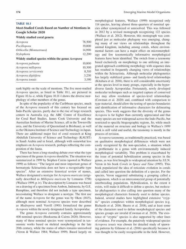
174
TABLE 10.1 Most-Studied Corals Based on Number of Mentions in Google Scholar 2020
Widely studied coral genera
Acropora 78,500
Pocillopora 19,000
Orbicella (Montastraea) 14,990
Stylophora 12,200
Widely studied species within the genus Acropora
Acropora palmata 10,800
Acropora millepora 10,300
Acropora cervicornis 8,310
Acropora digitifera 3,230
Acropora tenuis 3,190
rank highly on the scale of mentions. The fi ve most-studied
Acropora species, as listed in Table 10.1, are pictured in
Figure 10.1a – e , while Figure 10.1f – l shows the diverse mor
phology of other members of the genus.
In spite of the popularity of the Caribbean species, much
of the Acropora research of this century has focused on
Indo-Pacifi c species, partly due to the rise of large research
centers in Australia (e.g. the ARC Centre of Excellence
for Coral Reef Studies, James Cook University and the
Australian Institute of Marine Science, all in the Townsville
area, and the University of Queensland in Brisbane), as well
as the Okinawa Institute of Science and Technology in Japan.
There are additional major foci of coral research at King
Abdullah University of Science and Technology (KAUST)
in Saudi Arabia and in Israel, although with somewhat less
emphasis on Acropora research, perhaps reflecting the com
position of the fauna.
There has been a long-standing debate over what the type
specimen of the genus Acropora should be. The situation was
summarized in 1999 by Stephen Cairns (quoted in Wallace
1999) as follows: “The largest and most important genus of
hermatypic Scleractinia does not have a recognisable type
species”. After an extensive historical review of names,
Wallace designated a neotype for Acropora muricata (origi
nally described as Millepora muricata by Linnaeus 1758)
(Wallace 1999, p. iv). The description by Linnaeus was based
on a drawing of a specimen from Ambon, Indonesia, by G.E.
Rumphius, and therefore did not include a type specimen,
necessitating Wallace to designate a neotype. The fi rst use
of the name Acropora for the genus was by Oken (1815),
although most nominal Acropora species were described
as Madrepora until Verrill (1901) formalized the genus
Acropora within the newly designated family Acroporidae.
The genus Acropora currently contains approximately
408 nominal species (Hoeksema & Cairns 2020). However,
many of these nominal species were synonymized in tax
onomic works based on skeletal morphology in the late
20th century, while the status of others remains unresolved
(Veron & Wallace 1984; Wallace 1999). Based largely on
Emerging Marine Model Organisms
morphological features, Wallace (1999) recognized only
114 species, leaving almost three-quarters of nominal spe
cies either synonymized or unresolved. This was followed
in 2012 by a revised monograph recognizing 122 species
(Wallace et al. 2012). However, this monograph was com
pleted just as molecular phylogeny was emerging, chang
ing many of our views on relationships throughout the
animal kingdom, including among corals, where environ
mental factors can have a major effect on micromorphol
ogy and few taxonomically informative morphological
features have been identified. The switch from a taxonomy
based exclusively on morphology to one utilizing an inte
grated approach combining morphology with sequence data
has resulted in frequently changing views of relationships
within the Scleractinia. Although molecular phylogenetics
has largely stabilized genus- and family-level relationships
(Kitahara et al. 2016 ), there is still considerable uncertainty
at the species level in many groups, especially in the hyper-
diverse family Acroporidae. Fortunately, newly developed
molecular techniques such as targeted capture of conserved
loci may allow resolution of species-level relationships
(Cowman et al. 2020) and, combined with comparison to
type material, should allow the testing of species boundaries
and identification of informative characters for delineating
species. This work suggests that the diversity of the genus
Acropora is far higher than currently appreciated and that
many species are not widespread across the Indo-Pacifi c, but
restricted to specific biogeographic regions. So, while much
of the material on structure and biology in Wallace’s 1999
book is still valid and useful, the taxonomy is mostly in the
process of revision.
Acropora taxonomy, as traditionally practiced, was based
on qualitative morphological differences which were not
easily recognized by the non-specialist, a situation which
is problematic in a genus with environmentally induced
morphological variability. This problem is exacerbated by
the issue of potential hybridization among species in the
genus, as was first brought to widespread attention by J.E.N.
Veron in his book Corals in Space and Time ( 1995 ). This
book popularized the idea of reticulate evolution in corals
and called into question the definition of a species. For the
species, Veron suggested substituting a grouping called a
syngameon, which is an interconnected group of potentially
interbreeding populations. Hybridization, to the extent it
exists, will make it difficult to define a species, but molecu
lar phylogenetics is also calling into question many of the
morphological characters formerly used to defi ne species.
Indeed, several studies have highlighted extensive “cryp
tic” species complexes within morphological species (e.g.
Richards et al. 2016; Sheets et al. 2018), and at least some
of the characters used to define morphological species and
species groups are invalid (Cowman et al. 2020). The exis
tence of “cryptic” species is also supported by other lines
of evidence. For example, the putatively widespread species
Acropora tenuis was chosen for detailed study of spawn
ing patterns by Gilmour et al. (2016 ) specifically because it
was thought to be easily recognizable in the fi eld. However,
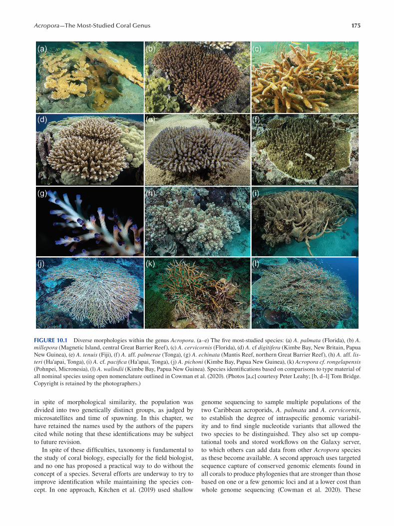
175 Acropora—The Most-Studied Coral Genus
FIGURE 10.1 Diverse morphologies within the genus Acropora. (a–e) The five most-studied species: (a) A. palmata (Florida), (b) A. millepora (Magnetic Island, central Great Barrier Reef), (c) A. cervicornis (Florida), (d) A. cf digitifera (Kimbe Bay, New Britain, Papua
New Guinea), (e) A. tenuis (Fiji), (f) A. aff. palmerae (Tonga), (g) A. echinata (Mantis Reef, northern Great Barrier Reef), (h) A. aff. listeri (Ha’apai, Tonga), (i) A. cf. pacifica (Ha’apai, Tonga), (j) A. pichoni (Kimbe Bay, Papua New Guinea), (k) Acropora cf. rongelapensis (Pohnpei, Micronesia), (l) A. walindii (Kimbe Bay, Papua New Guinea). Species identifications based on comparisons to type material of
all nominal species using open nomenclature outlined in Cowman et al. (2020). (Photos [a,c] courtesy Peter Leahy; [b, d–l] Tom Bridge.
Copyright is retained by the photographers.)
in spite of morphological similarity, the population was genome sequencing to sample multiple populations of the
divided into two genetically distinct groups, as judged by two Caribbean acroporids, A. palmata and A. cervicornis, microsatellites and time of spawning. In this chapter, we to establish the degree of intraspecific genomic variabil
have retained the names used by the authors of the papers ity and to find single nucleotide variants that allowed the
cited while noting that these identifications may be subject two species to be distinguished. They also set up compu
to future revision. tational tools and stored workflows on the Galaxy server,
In spite of these difficulties, taxonomy is fundamental to to which others can add data from other Acropora species
the study of coral biology, especially for the fi eld biologist, as these become available. A second approach uses targeted
and no one has proposed a practical way to do without the sequence capture of conserved genomic elements found in
concept of a species. Several efforts are underway to try to all corals to produce phylogenies that are stronger than those
improve identification while maintaining the species con- based on one or a few genomic loci and at a lower cost than
cept. In one approach, Kitchen et al. (2019) used shallow whole genome sequencing (Cowman et al. 2020). These
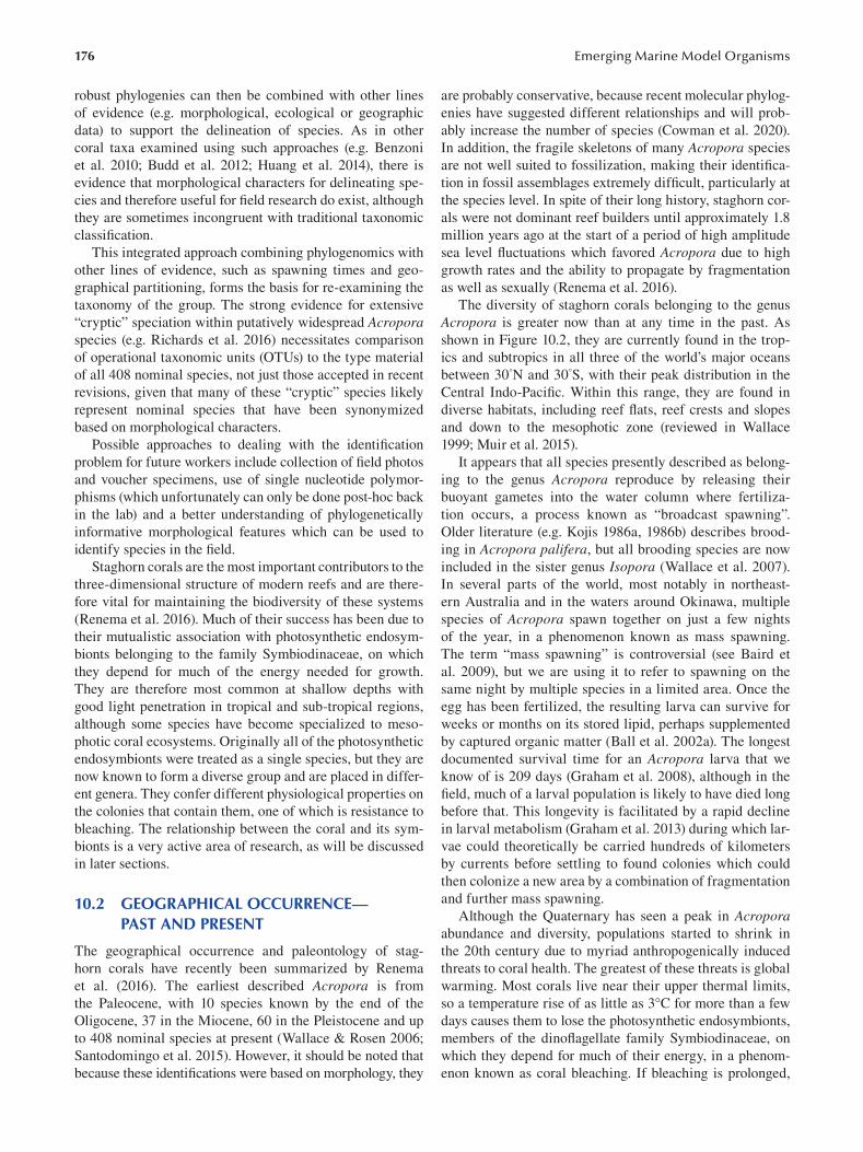
176
robust phylogenies can then be combined with other lines
of evidence (e.g. morphological, ecological or geographic
data) to support the delineation of species. As in other
coral taxa examined using such approaches (e.g. Benzoni
et al. 2010; Budd et al. 2012; Huang et al. 2014), there is
evidence that morphological characters for delineating spe
cies and therefore useful for field research do exist, although
they are sometimes incongruent with traditional taxonomic
classifi cation.
This integrated approach combining phylogenomics with
other lines of evidence, such as spawning times and geo
graphical partitioning, forms the basis for re-examining the
taxonomy of the group. The strong evidence for extensive
“cryptic” speciation within putatively widespread Acropora species (e.g. Richards et al. 2016) necessitates comparison
of operational taxonomic units (OTUs) to the type material
of all 408 nominal species, not just those accepted in recent
revisions, given that many of these “cryptic” species likely
represent nominal species that have been synonymized
based on morphological characters.
Possible approaches to dealing with the identifi cation
problem for future workers include collection of fi eld photos
and voucher specimens, use of single nucleotide polymor
phisms (which unfortunately can only be done post-hoc back
in the lab) and a better understanding of phylogenetically
informative morphological features which can be used to
identify species in the fi eld.
Staghorn corals are the most important contributors to the
three-dimensional structure of modern reefs and are there
fore vital for maintaining the biodiversity of these systems
(Renema et al. 2016 ). Much of their success has been due to
their mutualistic association with photosynthetic endosym
bionts belonging to the family Symbiodinaceae, on which
they depend for much of the energy needed for growth.
They are therefore most common at shallow depths with
good light penetration in tropical and sub-tropical regions,
although some species have become specialized to meso
photic coral ecosystems. Originally all of the photosynthetic
endosymbionts were treated as a single species, but they are
now known to form a diverse group and are placed in differ
ent genera. They confer different physiological properties on
the colonies that contain them, one of which is resistance to
bleaching. The relationship between the coral and its sym
bionts is a very active area of research, as will be discussed
in later sections.
10.2 GEOGRAPHICAL OCCURRENCE— PAST AND PRESENT
The geographical occurrence and paleontology of stag
horn corals have recently been summarized by Renema
et al. (2016 ). The earliest described Acropora is from
the Paleocene, with 10 species known by the end of the
Oligocene, 37 in the Miocene, 60 in the Pleistocene and up
to 408 nominal species at present (Wallace & Rosen 2006;
Santodomingo et al. 2015). However, it should be noted that
because these identifications were based on morphology, they
Emerging Marine Model Organisms
are probably conservative, because recent molecular phylog
enies have suggested different relationships and will prob
ably increase the number of species (Cowman et al. 2020).
In addition, the fragile skeletons of many Acropora species
are not well suited to fossilization, making their identifi ca
tion in fossil assemblages extremely difficult, particularly at
the species level. In spite of their long history, staghorn cor
als were not dominant reef builders until approximately 1.8
million years ago at the start of a period of high amplitude
sea level fluctuations which favored Acropora due to high
growth rates and the ability to propagate by fragmentation
as well as sexually (Renema et al. 2016).
The diversity of staghorn corals belonging to the genus
Acropora is greater now than at any time in the past. As
shown in Figure 10.2, they are currently found in the trop
ics and subtropics in all three of the world’s major oceans
between 30°N and 30°S, with their peak distribution in the
Central Indo-Pacifi c. Within this range, they are found in
diverse habitats, including reef flats, reef crests and slopes
and down to the mesophotic zone (reviewed in Wallace
1999; Muir et al. 2015).
It appears that all species presently described as belong
ing to the genus Acropora reproduce by releasing their
buoyant gametes into the water column where fertiliza
tion occurs, a process known as “broadcast spawning”.
Older literature (e.g. Kojis 1986a, 1986b) describes brood
ing in Acropora palifera, but all brooding species are now
included in the sister genus Isopora (Wallace et al. 2007 ).
In several parts of the world, most notably in northeast
ern Australia and in the waters around Okinawa, multiple
species of Acropora spawn together on just a few nights
of the year, in a phenomenon known as mass spawning.
The term “mass spawning” is controversial (see Baird et
al. 2009), but we are using it to refer to spawning on the
same night by multiple species in a limited area. Once the
egg has been fertilized, the resulting larva can survive for
weeks or months on its stored lipid, perhaps supplemented
by captured organic matter (Ball et al. 2002a). The longest
documented survival time for an Acropora larva that we
know of is 209 days (Graham et al. 2008), although in the
field, much of a larval population is likely to have died long
before that. This longevity is facilitated by a rapid decline
in larval metabolism (Graham et al. 2013) during which lar
vae could theoretically be carried hundreds of kilometers
by currents before settling to found colonies which could
then colonize a new area by a combination of fragmentation
and further mass spawning.
Although the Quaternary has seen a peak in Acropora abundance and diversity, populations started to shrink in
the 20th century due to myriad anthropogenically induced
threats to coral health. The greatest of these threats is global
warming. Most corals live near their upper thermal limits,
so a temperature rise of as little as 3°C for more than a few
days causes them to lose the photosynthetic endosymbionts,
members of the dinoflagellate family Symbiodinaceae, on
which they depend for much of their energy, in a phenom
enon known as coral bleaching. If bleaching is prolonged,
177 Acropora—The Most-Studied Coral Genus
FIGURE 10.2 The worldwide distribution of Acropora species is essentially between 30°N and 30° S. (Modified from Wallace and
Rosen 2006.)
the corals die, and members of the genus Acropora are par
ticularly susceptible to bleaching. Episodes of bleaching
are becoming increasingly widespread and frequent and
have considerably reduced Acropora populations worldwide
(Hughes et al. 2017, 2018). In addition to global warming, a
second threat arising from rising atmospheric CO 2 levels is
ocean acidification. Although a less immediate threat than
bleaching, ocean acidifi cation slows the rate of calcifi cation
and weakens coral skeletons and may therefore prove signif
icant in the longer term. Other anthropogenic threats include
severe weather events, reduced water quality, predator out
breaks (e.g. Crown of Thorns on the Great Barrier Reef),
incidental damage due to fishing and diving, the aquarium
trade and so on. All of these threats will result in changes to
the distribution of individual species and may result in the
extinction of some within this century.
10.3 LIFE CYCLE
There is a vast literature on various aspects of reproduction
in Acropora to which we can’t hope to do justice. Among
the major reviews of coral reproduction which include infor
mation on Acropora are those of Harrison and Wallace
(1990), Baird et al. (2009) and Harrison (2011), as well as a
chapter specifi cally on reproduction in Acropora ( Morita &
Kitanobo 2020). In addition, the other references cited in
this chapter contain many further references. Here we focus
our discussion on the life cycle of A. millepora, as that is the
species with which we are most familiar, but to the best of
our knowledge, the life cycles of all members of the genus
are very similar.
The month and day of spawning are determined mainly
by seawater temperatures in the weeks before potential
spawning dates and by phases of the moon, which in turn
determine the tides (Keith et al. 2016 ). The importance of a
rapid increase in temperature as a cue for spawning is evi
dent on the central Great Barrier Reef (GBR) where corals
on inshore reefs, where the water warms fi rst, frequently
spawn one month ahead of offshore reefs, although sepa
rated from the latter by only tens of kilometers. Thus, on the
central GBR, inshore reefs usually spawn three to fi ve days
after the full moon in October or November, with offshore
reefs a month later. The night of spawning is not totally syn
chronous within a population, as spawning may extend over
a few nights, although peak spawning is usually restricted
to a single night. Not only is there a peak night, but there
is usually a peak time of the night at which each species
characteristically spawns. For instance, at Magnetic Island,
A. tenuis usually spawns approximately two hours before A. millepora (personal observation). For broadcast spawning
corals, onset of darkness is typically the final cue determin
ing the hour of spawning (Babcock et al. 1986). Fukami et
al. (2003) describe a similar temporal separation of spawn
ing times in sympatric acroporids in Okinawa.
In some years on the GBR, there is a split spawning,
with part of the population spawning in one month and the
remainder a month later. A recent modeling study using
seven years of data from the GBR has combined data on
the time and place of Acropora spawning with oceano
graphic data and has found that split spawning increases
the robustness of coral larval supply and inter-reef con
nectivity due to temporal changes in the currents (Hock
et al. 2019).
While the spectacular synchronous multispecies mass
spawnings on the Great Barrier Reef have attracted consid
erable popular and scientific attention, synchrony is by no
means universal, even there. In fact, in eastern Australia,
synchrony is greatest at mid-latitudes and is reduced to both
north and south, and populations in the north often have two
spawnings per year.
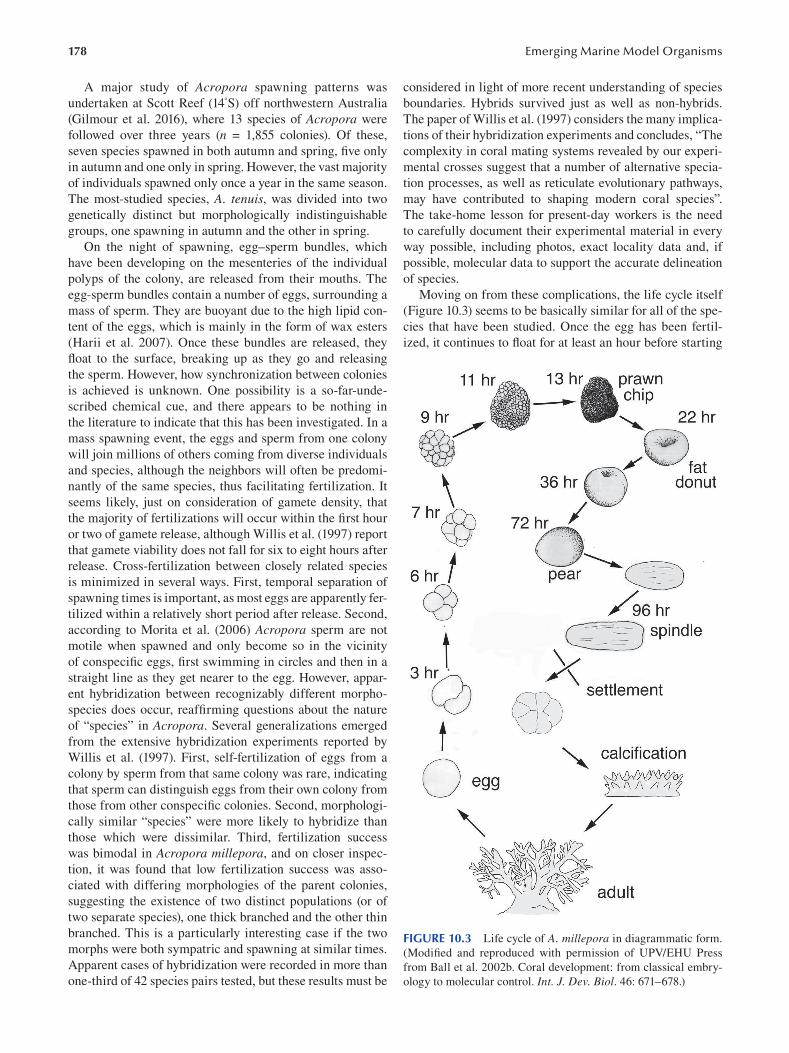
178
A major study of Acropora spawning patterns was
undertaken at Scott Reef (14°S) off northwestern Australia
(Gilmour et al. 2016 ), where 13 species of Acropora were
followed over three years (n = 1,855 colonies). Of these,
seven species spawned in both autumn and spring, fi ve only
in autumn and one only in spring. However, the vast majority
of individuals spawned only once a year in the same season.
The most-studied species, A. tenuis, was divided into two
genetically distinct but morphologically indistinguishable
groups, one spawning in autumn and the other in spring.
On the night of spawning, egg–sperm bundles, which
have been developing on the mesenteries of the individual
polyps of the colony, are released from their mouths. The
egg-sperm bundles contain a number of eggs, surrounding a
mass of sperm. They are buoyant due to the high lipid con
tent of the eggs, which is mainly in the form of wax esters
(Harii et al. 2007). Once these bundles are released, they
float to the surface, breaking up as they go and releasing
the sperm. However, how synchronization between colonies
is achieved is unknown. One possibility is a so-far-unde
scribed chemical cue, and there appears to be nothing in
the literature to indicate that this has been investigated. In a
mass spawning event, the eggs and sperm from one colony
will join millions of others coming from diverse individuals
and species, although the neighbors will often be predomi
nantly of the same species, thus facilitating fertilization. It
seems likely, just on consideration of gamete density, that
the majority of fertilizations will occur within the fi rst hour
or two of gamete release, although Willis et al. (1997) report
that gamete viability does not fall for six to eight hours after
release. Cross-fertilization between closely related species
is minimized in several ways. First, temporal separation of
spawning times is important, as most eggs are apparently fer
tilized within a relatively short period after release. Second,
according to Morita et al. (2006 ) Acropora sperm are not
motile when spawned and only become so in the vicinity
of conspecific eggs, fi rst swimming in circles and then in a
straight line as they get nearer to the egg. However, appar
ent hybridization between recognizably different morpho-
species does occur, reaffirming questions about the nature
of “species” in Acropora. Several generalizations emerged
from the extensive hybridization experiments reported by
Willis et al. (1997). First, self-fertilization of eggs from a
colony by sperm from that same colony was rare, indicating
that sperm can distinguish eggs from their own colony from
those from other conspecific colonies. Second, morphologi
cally similar “species” were more likely to hybridize than
those which were dissimilar. Third, fertilization success
was bimodal in Acropora millepora, and on closer inspec
tion, it was found that low fertilization success was asso
ciated with differing morphologies of the parent colonies,
suggesting the existence of two distinct populations (or of
two separate species), one thick branched and the other thin
branched. This is a particularly interesting case if the two
morphs were both sympatric and spawning at similar times.
Apparent cases of hybridization were recorded in more than
one-third of 42 species pairs tested, but these results must be
Emerging Marine Model Organisms
considered in light of more recent understanding of species
boundaries. Hybrids survived just as well as non-hybrids.
The paper of Willis et al. (1997 ) considers the many implica
tions of their hybridization experiments and concludes, “The
complexity in coral mating systems revealed by our experi
mental crosses suggest that a number of alternative specia
tion processes, as well as reticulate evolutionary pathways,
may have contributed to shaping modern coral species”.
The take-home lesson for present-day workers is the need
to carefully document their experimental material in every
way possible, including photos, exact locality data and, if
possible, molecular data to support the accurate delineation
of species.
Moving on from these complications, the life cycle itself
(Figure 10.3) seems to be basically similar for all of the spe
cies that have been studied. Once the egg has been fertil
ized, it continues to fl oat for at least an hour before starting
FIGURE 10.3 Life cycle of A. millepora in diagrammatic form.
(Modified and reproduced with permission of UPV/EHU Press
from Ball et al. 2002b. Coral development: from classical embry
ology to molecular control. Int. J. Dev. Biol. 46: 671–678.)
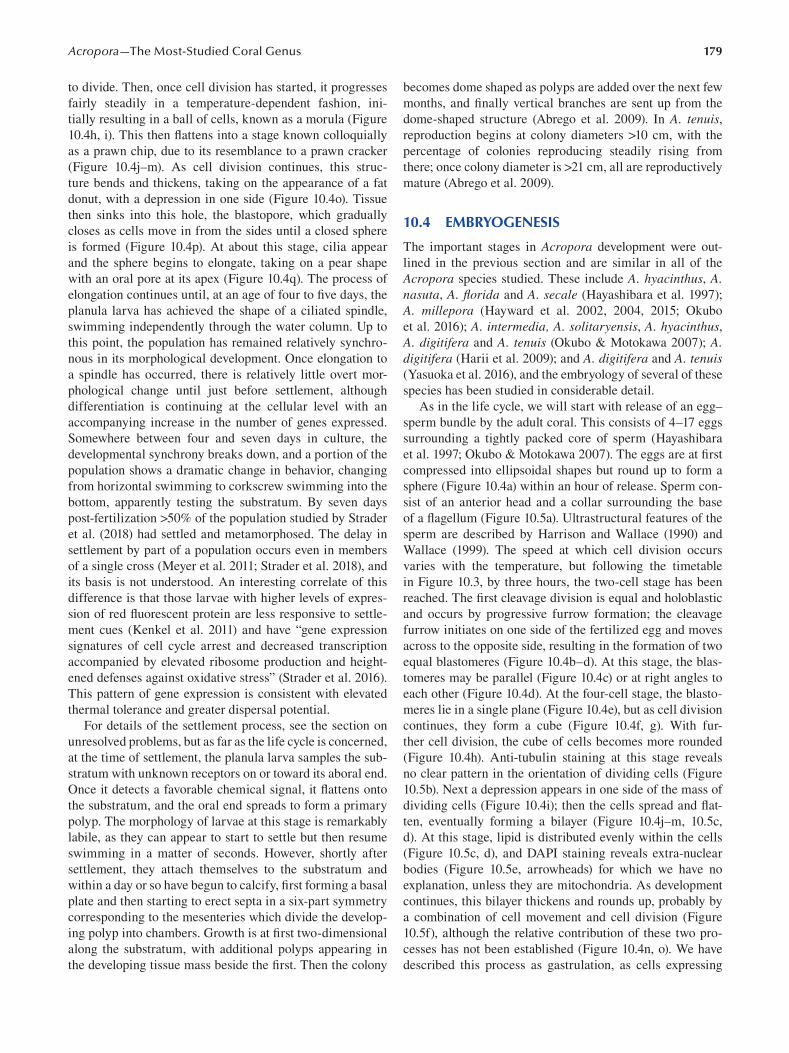
179 Acropora—The Most-Studied Coral Genus
to divide. Then, once cell division has started, it progresses
fairly steadily in a temperature-dependent fashion, ini
tially resulting in a ball of cells, known as a morula (Figure
10.4h, i). This then flattens into a stage known colloquially
as a prawn chip, due to its resemblance to a prawn cracker
(Figure 10.4j–m). As cell division continues, this struc
ture bends and thickens, taking on the appearance of a fat
donut, with a depression in one side (Figure 10.4o). Tissue
then sinks into this hole, the blastopore, which gradually
closes as cells move in from the sides until a closed sphere
is formed (Figure 10.4p). At about this stage, cilia appear
and the sphere begins to elongate, taking on a pear shape
with an oral pore at its apex (Figure 10.4q). The process of
elongation continues until, at an age of four to five days, the
planula larva has achieved the shape of a ciliated spindle,
swimming independently through the water column. Up to
this point, the population has remained relatively synchro
nous in its morphological development. Once elongation to
a spindle has occurred, there is relatively little overt mor
phological change until just before settlement, although
differentiation is continuing at the cellular level with an
accompanying increase in the number of genes expressed.
Somewhere between four and seven days in culture, the
developmental synchrony breaks down, and a portion of the
population shows a dramatic change in behavior, changing
from horizontal swimming to corkscrew swimming into the
bottom, apparently testing the substratum. By seven days
post-fertilization >50% of the population studied by Strader
et al. (2018) had settled and metamorphosed. The delay in
settlement by part of a population occurs even in members
of a single cross (Meyer et al. 2011; Strader et al. 2018), and
its basis is not understood. An interesting correlate of this
difference is that those larvae with higher levels of expres
sion of red fluorescent protein are less responsive to settle
ment cues (Kenkel et al. 2011) and have “gene expression
signatures of cell cycle arrest and decreased transcription
accompanied by elevated ribosome production and height
ened defenses against oxidative stress” (Strader et al. 2016 ).
This pattern of gene expression is consistent with elevated
thermal tolerance and greater dispersal potential.
For details of the settlement process, see the section on
unresolved problems, but as far as the life cycle is concerned,
at the time of settlement, the planula larva samples the sub
stratum with unknown receptors on or toward its aboral end.
Once it detects a favorable chemical signal, it fl attens onto
the substratum, and the oral end spreads to form a primary
polyp. The morphology of larvae at this stage is remarkably
labile, as they can appear to start to settle but then resume
swimming in a matter of seconds. However, shortly after
settlement, they attach themselves to the substratum and
within a day or so have begun to calcify, first forming a basal
plate and then starting to erect septa in a six-part symmetry
corresponding to the mesenteries which divide the develop
ing polyp into chambers. Growth is at fi rst two-dimensional
along the substratum, with additional polyps appearing in
the developing tissue mass beside the first. Then the colony
becomes dome shaped as polyps are added over the next few
months, and finally vertical branches are sent up from the
dome-shaped structure (Abrego et al. 2009). In A. tenuis, reproduction begins at colony diameters >10 cm, with the
percentage of colonies reproducing steadily rising from
there; once colony diameter is >21 cm, all are reproductively
mature (Abrego et al. 2009).
10.4 EMBRYOGENESIS
The important stages in Acropora development were out
lined in the previous section and are similar in all of the
Acropora species studied. These include A. hyacinthus, A. nasuta, A. florida and A. secale (Hayashibara et al. 1997 );
A. millepora ( Hayward et al. 2002 , 2004 , 2015 ; Okubo
et al. 2016); A. intermedia, A. solitaryensis, A. hyacinthus, A. digitifera and A. tenuis (Okubo & Motokawa 2007 ); A. digitifera (Harii et al. 2009); and A. digitifera and A. tenuis (Yasuoka et al. 2016), and the embryology of several of these
species has been studied in considerable detail.
As in the life cycle, we will start with release of an egg–
sperm bundle by the adult coral. This consists of 4–17 eggs
surrounding a tightly packed core of sperm (Hayashibara
et al. 1997; Okubo & Motokawa 2007). The eggs are at fi rst
compressed into ellipsoidal shapes but round up to form a
sphere (Figure 10.4a) within an hour of release. Sperm con
sist of an anterior head and a collar surrounding the base
of a flagellum (Figure 10.5a). Ultrastructural features of the
sperm are described by Harrison and Wallace (1990) and
Wallace (1999). The speed at which cell division occurs
varies with the temperature, but following the timetable
in Figure 10.3, by three hours, the two-cell stage has been
reached. The first cleavage division is equal and holoblastic
and occurs by progressive furrow formation; the cleavage
furrow initiates on one side of the fertilized egg and moves
across to the opposite side, resulting in the formation of two
equal blastomeres (Figure 10.4b–d). At this stage, the blas
tomeres may be parallel (Figure 10.4c) or at right angles to
each other (Figure 10.4d). At the four-cell stage, the blasto
meres lie in a single plane (Figure 10.4e), but as cell division
continues, they form a cube (Figure 10.4f, g). With fur
ther cell division, the cube of cells becomes more rounded
(Figure 10.4h). Anti-tubulin staining at this stage reveals
no clear pattern in the orientation of dividing cells (Figure
10.5b). Next a depression appears in one side of the mass of
dividing cells (Figure 10.4i); then the cells spread and fl at-
ten, eventually forming a bilayer (Figure 10.4j–m, 10.5c,
d). At this stage, lipid is distributed evenly within the cells
(Figure 10.5c, d), and DAPI staining reveals extra-nuclear
bodies (Figure 10.5e, arrowheads) for which we have no
explanation, unless they are mitochondria. As development
continues, this bilayer thickens and rounds up, probably by
a combination of cell movement and cell division (Figure
10.5f), although the relative contribution of these two pro
cesses has not been established (Figure 10.4n , o). We have
described this process as gastrulation, as cells expressing
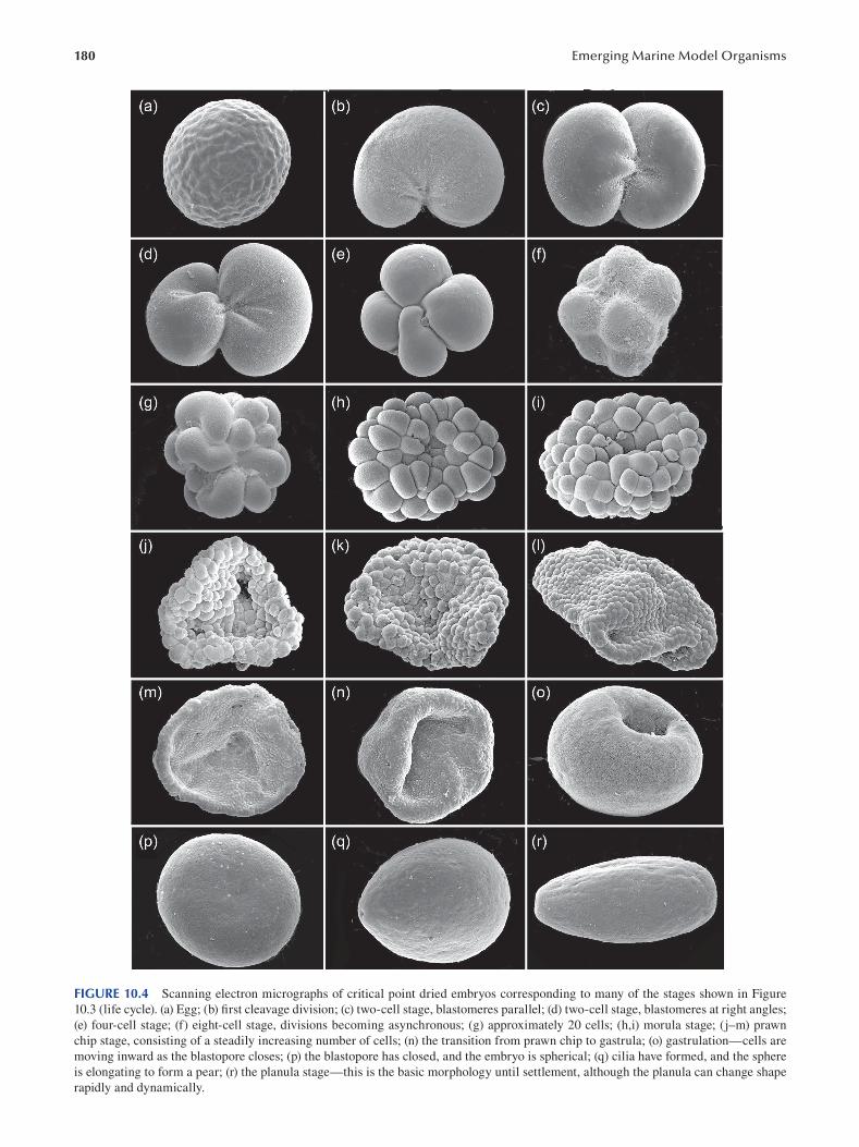
180 Emerging Marine Model Organisms
FIGURE 10.4 Scanning electron micrographs of critical point dried embryos corresponding to many of the stages shown in Figure
10.3 (life cycle). (a) Egg; (b) first cleavage division; (c) two-cell stage, blastomeres parallel; (d) two-cell stage, blastomeres at right angles;
(e) four-cell stage; (f) eight-cell stage, divisions becoming asynchronous; (g) approximately 20 cells; (h,i) morula stage; (j–m) prawn
chip stage, consisting of a steadily increasing number of cells; (n) the transition from prawn chip to gastrula; (o) gastrulation—cells are
moving inward as the blastopore closes; (p) the blastopore has closed, and the embryo is spherical; (q) cilia have formed, and the sphere
is elongating to form a pear; (r) the planula stage—this is the basic morphology until settlement, although the planula can change shape
rapidly and dynamically.
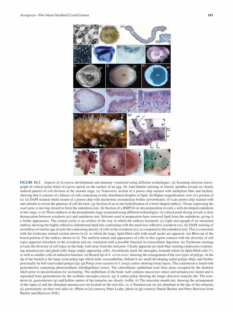
181 Acropora—The Most-Studied Coral Genus
FIGURE 10.5 Aspects of Acropora development and anatomy visualized using different technologies. (a) Scanning electron micro
graph of critical point dried Acropora sperm on the surface of an egg. (b) Anti-tubulin staining of mitotic spindles reveals no clearly
ordered pattern of cell division at the morula stage. (c) Transverse section of a prawn chip stained with methylene blue and fuchsin,
showing that it consists of a bilayer of cells containing evenly distributed droplets of lipid. (d) Higher magnification view of a portion of
(c). (e) DAPI-stained whole mount of a prawn chip with mysterious extranuclear bodies (arrowheads). (f) Late prawn chip stained with
anti-tubulin to reveal the patterns of cell division. (g) Section of an in situ hybridization of a bowl-shaped embryo. Tissue expressing the
snail gene is moving inward to form the endoderm (en). (h) Section of a BMP2/4 in situ preparation reveals a well-developed endoderm
at this stage. (i–k) Three embryos at the pear/planula stage examined using different technologies: (i) critical point drying reveals a clear
demarcation between ectoderm (ec) and endoderm (en). Solvents used in preparation have removed lipid from the endoderm, giving it
a frothy appearance. The central cavity is an artifact of the way in which the embryo fractured. (j) Light micrograph of an unstained
embryo showing the highly reflective endodermal lipid (en) contrasting with the much less reflective ectoderm (ec). (k) DAPI staining of
an embryo of similar age reveals the contrasting density of cells in the ectoderm (ec), as compared to the endoderm (en). This is consistent
with the trichrome stained section shown in (l), in which the large, lipid-filled cells with small nuclei are apparent. (m) Blow-up of the
boxed portion of the embryo shown in (l). The uniform nature and appearance of cells in this region contrast with the diversity of cell
types apparent elsewhere in the ectoderm and are consistent with a possible function in extracellular digestion. (n) Trichrome staining
reveals the diversity of cell types in the body wall away from the oral pore. Clearly apparent are dark-blue-staining cnidocytes (contain
ing nematocysts) and gland cells (large empty-appearing cells). Arrowheads mark the mesoglea, beneath which lie lipid-filled cells (*),
as well as smaller cells of unknown function. (o) Branch tip of A. cervicornis, showing the arrangement of the two types of polyps. At the
tip of the branch is the large axial polyp (ap) which lacks zooxanthellae; behind it are small developing radial polyps (drp), and further
proximally lie full-sized radial polyps (rp). (p) Polyp cross-section of A. longicyathus showing tissue layers. The coelenteron is lined with
gastrodermis containing photosynthetic dinoflagellates (zoox). The calicoblastic epithelium (cal) lines areas occupied by the skeleton
(skel) prior to decalcification for sectioning. The epithelium of the body wall contains mucocytes (muc) and nematocytes (nem) and is
separated from gastrodermis by the acellular mesoglea (meso). (q) A radial polyp showing the longer directive tentacle (dt). The ecto
derm (e), gastrodermis (g) and hollow nature of the tentacles are clearly visible. (r) The muscular mouth (m), showing the arrangement
of the septa (s) and the abundant nematocysts (n) located on the oral disc. (s, t) Nematocysts (n) are abundant at the tips of the tentacles
(s), particularly on their oral sides (t). (Photo in [o] courtesy Peter Leahy; photo in [p] courtesy Daniel Bucher and Peter Harrison from
Bucher and Harrison 2018.)
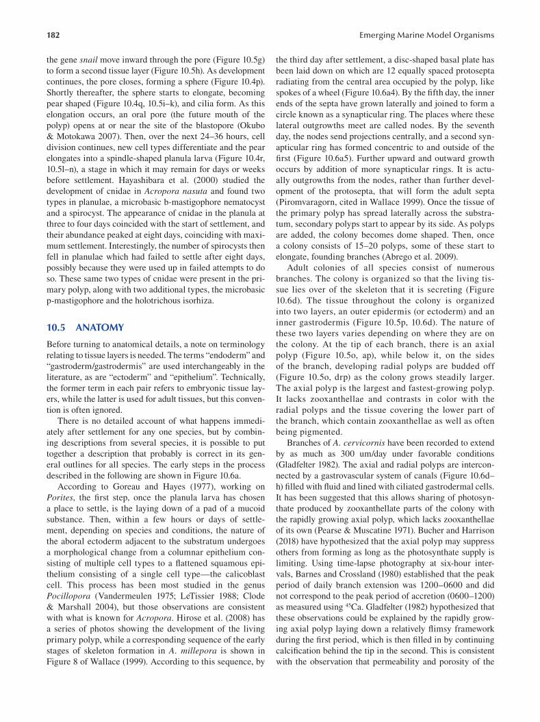
182
the gene snail move inward through the pore (Figure 10.5g)
to form a second tissue layer (Figure 10.5h). As development
continues, the pore closes, forming a sphere (Figure 10.4p).
Shortly thereafter, the sphere starts to elongate, becoming
pear shaped (Figure 10.4q, 10.5i–k), and cilia form. As this
elongation occurs, an oral pore (the future mouth of the
polyp) opens at or near the site of the blastopore (Okubo
& Motokawa 2007). Then, over the next 24–36 hours, cell
division continues, new cell types differentiate and the pear
elongates into a spindle-shaped planula larva (Figure 10.4r,
10.5l–n), a stage in which it may remain for days or weeks
before settlement. Hayashibara et al. (2000) studied the
development of cnidae in Acropora nasuta and found two
types in planulae, a microbasic b-mastigophore nematocyst
and a spirocyst. The appearance of cnidae in the planula at
three to four days coincided with the start of settlement, and
their abundance peaked at eight days, coinciding with maxi
mum settlement. Interestingly, the number of spirocysts then
fell in planulae which had failed to settle after eight days,
possibly because they were used up in failed attempts to do
so. These same two types of cnidae were present in the pri
mary polyp, along with two additional types, the microbasic
p-mastigophore and the holotrichous isorhiza.
10.5 ANATOMY
Before turning to anatomical details, a note on terminology
relating to tissue layers is needed. The terms “endoderm” and
“gastroderm/gastrodermis” are used interchangeably in the
literature, as are “ectoderm” and “epithelium”. Technically,
the former term in each pair refers to embryonic tissue lay
ers, while the latter is used for adult tissues, but this conven
tion is often ignored.
There is no detailed account of what happens immedi
ately after settlement for any one species, but by combin
ing descriptions from several species, it is possible to put
together a description that probably is correct in its gen
eral outlines for all species. The early steps in the process
described in the following are shown in Figure 10.6a.
According to Goreau and Hayes (1977 ), working on
Porites, the first step, once the planula larva has chosen
a place to settle, is the laying down of a pad of a mucoid
substance. Then, within a few hours or days of settle
ment, depending on species and conditions, the nature of
the aboral ectoderm adjacent to the substratum undergoes
a morphological change from a columnar epithelium con
sisting of multiple cell types to a flattened squamous epi
thelium consisting of a single cell type—the calicoblast
cell. This process has been most studied in the genus
Pocillopora (Vandermeulen 1975; LeTissier 1988; Clode
& Marshall 2004), but those observations are consistent
with what is known for Acropora. Hirose et al. (2008) has
a series of photos showing the development of the living
primary polyp, while a corresponding sequence of the early
stages of skeleton formation in A. millepora is shown in
Figure 8 of Wallace (1999). According to this sequence, by
Emerging Marine Model Organisms
the third day after settlement, a disc-shaped basal plate has
been laid down on which are 12 equally spaced protosepta
radiating from the central area occupied by the polyp, like
spokes of a wheel (Figure 10.6a4). By the fifth day, the inner
ends of the septa have grown laterally and joined to form a
circle known as a synapticular ring. The places where these
lateral outgrowths meet are called nodes. By the seventh
day, the nodes send projections centrally, and a second syn
apticular ring has formed concentric to and outside of the
first (Figure 10.6a5). Further upward and outward growth
occurs by addition of more synapticular rings. It is actu
ally outgrowths from the nodes, rather than further devel
opment of the protosepta, that will form the adult septa
(Piromvaragorn, cited in Wallace 1999). Once the tissue of
the primary polyp has spread laterally across the substra
tum, secondary polyps start to appear by its side. As polyps
are added, the colony becomes dome shaped. Then, once
a colony consists of 15–20 polyps, some of these start to
elongate, founding branches (Abrego et al. 2009).
Adult colonies of all species consist of numerous
branches. The colony is organized so that the living tis
sue lies over of the skeleton that it is secreting (Figure
10.6d). The tissue throughout the colony is organized
into two layers, an outer epidermis (or ectoderm) and an
inner gastrodermis (Figure 10.5p, 10.6d). The nature of
these two layers varies depending on where they are on
the colony. At the tip of each branch, there is an axial
polyp (Figure 10.5o, ap), while below it, on the sides
of the branch, developing radial polyps are budded off
(Figure 10.5o, drp) as the colony grows steadily larger.
The axial polyp is the largest and fastest-growing polyp.
It lacks zooxanthellae and contrasts in color with the
radial polyps and the tissue covering the lower part of
the branch, which contain zooxanthellae as well as often
being pigmented.
Branches of A. cervicornis have been recorded to extend
by as much as 300 um/day under favorable conditions
(Gladfelter 1982). The axial and radial polyps are intercon
nected by a gastrovascular system of canals (Figure 10.6d–
h ) filled with fluid and lined with ciliated gastrodermal cells.
It has been suggested that this allows sharing of photosyn
thate produced by zooxanthellate parts of the colony with
the rapidly growing axial polyp, which lacks zooxanthellae
of its own (Pearse & Muscatine 1971). Bucher and Harrison
(2018) have hypothesized that the axial polyp may suppress
others from forming as long as the photosynthate supply is
limiting. Using time-lapse photography at six-hour inter
vals, Barnes and Crossland (1980) established that the peak
period of daily branch extension was 1200–0600 and did
not correspond to the peak period of accretion (0600–1200)
as measured using 45Ca. Gladfelter (1982) hypothesized that
these observations could be explained by the rapidly grow
ing axial polyp laying down a relatively fl imsy framework
during the first period, which is then filled in by continuing
calcification behind the tip in the second. This is consistent
with the observation that permeability and porosity of the
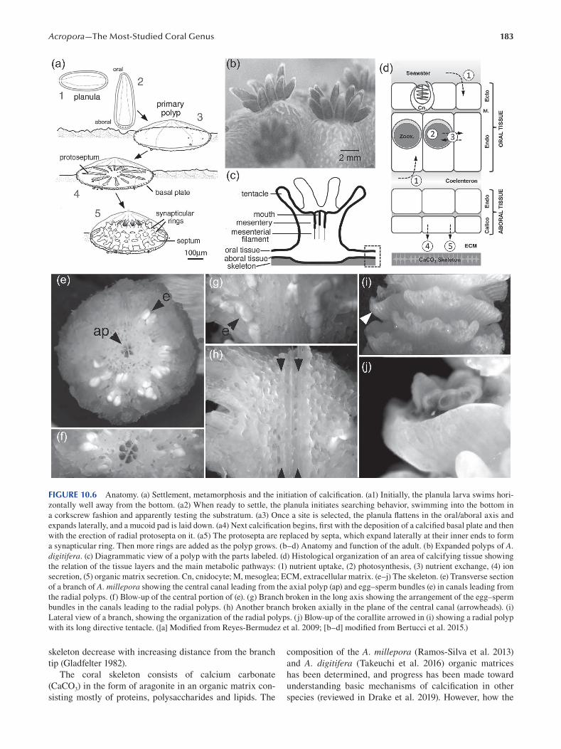
183 Acropora—The Most-Studied Coral Genus
FIGURE 10.6 Anatomy. (a) Settlement, metamorphosis and the initiation of calcifi cation. (a1) Initially, the planula larva swims hori
zontally well away from the bottom. (a2) When ready to settle, the planula initiates searching behavior, swimming into the bottom in
a corkscrew fashion and apparently testing the substratum. (a3) Once a site is selected, the planula flattens in the oral/aboral axis and
expands laterally, and a mucoid pad is laid down. (a4) Next calcification begins, first with the deposition of a calcified basal plate and then
with the erection of radial protosepta on it. (a5) The protosepta are replaced by septa, which expand laterally at their inner ends to form
a synapticular ring. Then more rings are added as the polyp grows. (b–d) Anatomy and function of the adult. (b) Expanded polyps of A. digitifera. (c) Diagrammatic view of a polyp with the parts labeled. (d) Histological organization of an area of calcifying tissue showing
the relation of the tissue layers and the main metabolic pathways: (1) nutrient uptake, (2) photosynthesis, (3) nutrient exchange, (4) ion
secretion, (5) organic matrix secretion. Cn, cnidocyte; M, mesoglea; ECM, extracellular matrix. (e–j) The skeleton. (e) Transverse section
of a branch of A. millepora showing the central canal leading from the axial polyp (ap) and egg–sperm bundles (e) in canals leading from
the radial polyps. (f) Blow-up of the central portion of (e). (g) Branch broken in the long axis showing the arrangement of the egg–sperm
bundles in the canals leading to the radial polyps. (h) Another branch broken axially in the plane of the central canal (arrowheads). (i)
Lateral view of a branch, showing the organization of the radial polyps. (j) Blow-up of the corallite arrowed in (i) showing a radial polyp
with its long directive tentacle. ([a] Modified from Reyes-Bermudez et al. 2009; [b–d] modified from Bertucci et al. 2015.)
skeleton decrease with increasing distance from the branch composition of the A. millepora (Ramos-Silva et al. 2013)
tip (Gladfelter 1982). and A. digitifera (Takeuchi et al. 2016) organic matrices
The coral skeleton consists of calcium carbonate has been determined, and progress has been made toward
(CaCO3) in the form of aragonite in an organic matrix con- understanding basic mechanisms of calcification in other
sisting mostly of proteins, polysaccharides and lipids. The species (reviewed in Drake et al. 2019). However, how the
184
characteristic morphology of individual species is produced
is still not understood.
As the colony grows, new branches are founded by
appearance of a new axial polyp somewhere along an
existing branch or by conversion of a radial polyp into an
axial polyp (Wallace 1999). The tentacles of the polyps
are mostly in multiples of six (hence the classifi cation of
Acropora in the Hexacorallia), with 12 tentacles being
the most frequent ( Figure 10.5o , q ; Figure 10.6b , c , j ).
The radial polyps are retractile and can withdraw into the
skeleton surrounding them when disturbed. The parts of a
radial polyp are shown schematically in Figure 10.6c and
in greater detail in Figure 10.5q–t . One tentacle (known as
the directive tentacle) is consistently longer than all of the
rest and is typically unpigmented, in contrast to the oth
ers (Figure 10.5o, q;10.6i , j). The organization of a radial
polyp is clearly apparent in Figure 10.5q. Each tentacle is
hollow and consists of an outer layer of ectoderm surround
ing an inner layer of gastroderm, which in turn surrounds
a hollow cavity, connecting to the central cavity, or coel
enteron, of the columnar polyp. The mouth is at the cen
ter of a flattened area known as the oral disc and is closed
by a muscular sphincter (Figure 10.5r). The central cavity
is partially partitioned by mesenteries from which hang
mesenterial filaments, containing nematocysts which help
to subdue struggling prey. Nematocysts are also abundant
at the tips of the tentacles (Figure 10.5s) and particularly
on their oral sides (Figure 10.5t). The ectoderm consists
of diverse cell types, including cnidocytes (which produce
several types of nematocyst) as well as gland cells and neu
rons. Gastrodermal cells are ciliated, have a digestive func
tion and frequently contain photosynthetic dinofl agellates
belonging to the family Symbiodinaceae (LaJeunesse et al.
2018 ).
10.6. GENOMICS
Prior to 2011, only limited transcriptomic and genomic data
were available for corals (reviewed in Miller et al. 2011), but
in that year, the first coral whole genome assembly was pub
lished (Shinzato et al. 2011). Fittingly, the species sequenced
was A. digitifera—a common species that dominates reefs
in many parts of Okinawa and on which Japanese biologists
regularly carry out research. Comparison of the A. digitifera genome with that of the sea anemone Nematostella vectensis (the first cnidarian whole genome sequence assembly)
revealed a number of differences. For example, it was sug
gested that the requirement for a sophisticated symbiont
recognition system might underlie the observed enrichment
of predicted immune receptors in the A. digitifera genome
relative to N. vectensis (Shinzato et al. 2011). Another sur
prise was the discovery in A. digitifera of a suite of genes
that together may enable biosynthesis of mycosporine-like
amino acids, “natural sunscreen” which was previously
assumed to be produced by the algal symbionts rather than
the coral animal. A third key finding arising from analy
ses of the A. digitifera genome was that this coral lacked
Emerging Marine Model Organisms
cystathionine ß-synthase (Cbs), one of the enzymes required
for biosynthesis of cysteine. All Acropora species examined
to date lack Cbs, although a Cbs homolog is present in a
wide range of other corals (Shinzato et al. 2011).
The early availability of significant bodies of molecular
data for A. millepora (e.g. Kortschak et al. 2003; Meyer et
al. 2009; Moya et al. 2012) led to widespread use of this
coral for experimental purposes, making this species an
obvious target for whole genome sequencing. In 2019, the
first genome assembly for A. millepora became available
(Ying et al. 2019); as with the A. digitifera assembly, the
fi rst A. millepora genome was based on short-read data,
but a long-read–based assembly became available shortly
thereafter (Fuller et al. 2020). There has recently been a
rapid increase in the number of genome assemblies avail
able for Acropora species, largely carried out at the Okinawa
Institute for Science and Technology (OIST)—the institu
tion responsible for the first coral genome assembly. Mao et
al. (2018) generated short-read assemblies for four additional
species of Acropora (A. gemmifera, A. echinata, A. subglabra and A. tenuis), and Shinzato et al. (2020) analyzed the
genomes of an additional 11 Acropora species and those of
the confamilial taxa Montipora cactus, M. efflorescens and
Astreopora myriophthalma.
Although genomes were not actually assembled, extensive
genomic sequence data are also available for the Caribbean
species A. palmata and A. cervicornis (Kitchen et al. 2019).
10.6.1 WHAT HAVE WE LEARNED FROM
ALL OF THOSE GENOMES?
Despite early speculation on the possibility of a whole
genome duplication having facilitated the evolutionary suc
cess of Acropora (Mao & Satoh 2019), it is now clear that
such a duplication is unlikely to have occurred (Shinzato
et al. 2020). Rather, many independent gene duplication
events occurred in the Acropora lineage (Hislop et al. 2005;
Shinzato et al. 2020).
The genomes of Acropora species vary surprisingly lit
tle. Based on short-read assemblies, Shinzato et al. (2020)
estimated gene numbers across the genus to be around
22–24,000. However, gene predictions from the two long-
read assemblies are significantly higher—28,000 for A. millepora (Fuller et al. 2020) and around 30,000 for A. tenuis (Cooke et al. 2020). Within the genus Acropora,
some gene families have been dramatically expanded,
interesting examples of which are those encoding the atyp
ical two-domain caspase-X, small cysteine-rich proteins
(SCRiPs) and dimethylsulfoniopropionate (DMSP)-lyases
(Shinzato et al. 2020). The caspase-X proteins have both
active and inactive caspase domains, the latter being likely
to normally hold the protein in an inactive state in a manner
resembling the interaction of caspase-8 and c-FLIP (Moya
et al. 2016). SCRiPs have been implicated in a wide range
of functions, including skeletogenesis (Sunagawa et al.
2009; Hayward et al. 2011) and stress responses (DeSalvo
et al. 2008; Meyer et al. 2011; Moya et al. 2012), as toxins
185 Acropora—The Most-Studied Coral Genus
(Jouiaei et al. 2015) and possibly also in symbiont acqui
sition (Mohamed et al. 2020a). Acropora spp. are known
to produce large amounts of DMSP, which is cleaved by
DMSP-lyase to dimethyl sulfate (DMS) and acrylate. As
DMS is volatile and can seed cloud formation, a role in
local climate moderation has been proposed (Vallina &
Simó 2007). Although roles for SCRiPs and caspase-X
proteins in stress responses and for DMSP-lyases in miti
gating solar radiation have been interpreted as adaptations
within the Acropora lineage to deal with environmental
stressors (Shinzato et al. 2020), Acropora species remain
among the most sensitive of reef-building corals to thermal
stress, and at this stage, it is unclear whether these gene
family expansions are related to that.
With the exceptions of the Fuller et al. (2020) assembly
for A. millepora and the Cooke et al. (2020) assembly for A. tenuis, all of these other genomes have been based on short-
read data. So, while they have provided some high-quality
gene prediction datasets, they do not provide comprehen
sive coverage. Comparison between the Cooke et al. (2020)
A. tenuis and the Fuller et al. (2020) A. millepora assem
blies shows a remarkable level of macrosynteny (Cooke
et al. 2020). Given that these species are highly diverged
within the genus (Cowman et al. 2020), it is likely that the
overall genome architecture varies little within Acropora—
note that data from Shinzato et al. (2020) are consistent
with this view.
10.6.2 HOW DOES THE ACROPORA GENOME COMPARE
WITH THOSE OF OTHER CORAL GENERA?
With the caveat that, at the time of writing, data are not
available for a representative range of reef-building cor
als, based on the long-read assemblies, at around ~480 Mb
(Fuller et al. 2020; Cooke et al. 2020), the estimated size of
the Acropora genome appears to be fairly typical of corals.
Although estimates of both genome size and gene number
for some members of the Robusta are much larger (Ying et
al. 2018), these were based on short-read assemblies, and
it is as yet unclear whether the larger genomes are conse
quences of higher content of repetitive elements and trans
posons—as in the case of several bilaterian lineages—or
higher gene content. Until higher-quality genome assem
blies are available for a phylogenetically representative
range of corals, general evolutionary patterns will remain
unclear.
10.6.3 WHY HAS ACROPORA BEEN SUCH AN
EVOLUTIONARY SUCCESS STORY?
Throughout the Indo-Pacifi c, Acropora is the dominant reef-
building coral and is one of the most speciose coral genera.
As speculated on by Shinzato et al. (2020) and others, its
evolutionary success may be due to acquisition and ampli
fication of gene families that have enabled rapid adaptation
to changing conditions. However, Acropora is almost always
associated with one particular genus of Symbiodiniaceae,
Cladocopium, and we speculate that this partnership may
have facilitated the observed rise to dominance of this
genus. Comparative transcriptomics has demonstrated the
over-representation of (for example) ABC-transporters in
Cladocopium goreaui compared to Breviolum minutum and
Fugacium kawagutii—other Symbiodiniaceae associated
with corals—and among the transporters known so far only
in Cladocopium, there are components of transport systems
for both cysteine and histidine (Mohamed et al. 2020b).
The significance of cysteine in the case of Acropora was
discussed previously; although members of the Robusta
are capable of histidine biosynthesis, along with other
Complexa and bilaterians, Acropora species cannot syn
thesize it. Hence the association between Acropora as host
and Cladocopium as symbiont may be a particularly good
“fit” and have contributed to the rise of the genus during the
Neogene and Quaternary.
10.7 FUNCTIONAL APPROACHES: TOOLS FOR MOLECULAR AND CELLULAR ANALYSES
For many reasons, the functional approaches that have
proven so fruitful in other organisms such as Drosophila and Caenorhabditis have been difficult or impossible to
implement in Acropora. First, there is ease and cost of cul
ture. While adult corals have been kept in aquaria for years,
albeit in varying degrees of health, it is only in the past year
that there has been a report in the literature of successful
production of a second generation of Acropora in captivity
(Craggs et al. 2020), and this required a sophisticated and
expensive aquarium system. Second, there is the problem
of generation time; it is probably at least three years before
a second generation of Acropora would produce suffi cient
embryos for experimental purposes. Third, there is genome
size. Compared to the best-understood “model” organism,
Drosophila melanogaster (genome size ~140 Mb; 15,700
genes), at 400–500 Mb and with ~28–30,000 genes, the
genomes of A. millepora and A. tenuis, the two Acropora species for which we have the best data, are relatively large.
In addition, Drosophila has only 8 chromosomes (four
pairs), while A. millepora has 28 (Kenyon 1997; Flot et al.
2006), as does A. digitifera (Supp Fig 1 in Shinzato et al.
2011). Twenty-eight chromosomes is most common in the
genus, as Kenyon (1997) found this number in 16 species,
but this is by no means universal, as 6 other species had 24,
30 (2 species), 42, 48 and 54.
Studies on Acropora also require several additional con
siderations that may not be relevant to other organisms.
One is the taxonomic problem dealt with in Section 10.1.
Molecular markers may be required in the future to be sure
that one is really dealing with the same species in differ
ent parts of the world. A fi nal difficulty is that a coral is in
fact a holobiont, usually consisting of the coral itself, one
or more species of photosynthetic microalgae and numerous
other micro-organisms. In nature, this assemblage will vary
186
somewhat from coral to coral and locality to locality and
may have considerable effects on the health and physiol
ogy of the individual coral and therefore on experimental
repeatability.
Genetic and cell biological manipulations have been
done on other cnidarians, most notably on Hydra and
Nematostella, in both of which gene knockdown experi
ments have been successful. However, culturing these spe
cies is much less demanding than for corals. Of greater
relevance to studies on corals have been experiments on the
sea anemone Exaiptasia (often under the name Aiptasia),
which is relatively easy to culture and which shares with
corals the presence of photosynthetic endosymbionts. There
has been an attempt by the Exaiptasia community to stan
dardize strains of anemone and endosymbionts in order to
achieve a greater level of experimental consistency across
the community (e.g. Cziesielski et al. 2018), but this will be
difficult in the case of Acropora.
In spite of the challenges noted previously, there have
been some successful attempts at experimental manipula
tion in corals. For example, lithium chloride and 1-azaken
paullone (AZ) have been used to inhibit GSK3 and activate
the wnt pathway in A. digitifera (Yasuoka et al. 2016 ),
resulting in the expansion of brachyury expression through
out the embryonic ectoderm in a dose-dependent manner.
In contrast, wnt/ßcatenin signaling inhibitors (pyrvinium
pamoate, IWR1 or iCRT14) reduced Adi_bra expression in
a dose-dependent fashion, leading to the conclusion that it
is positively regulated by wnt/ßcatenin signalling. In a fol
lowing experiment, FITC-labeled anti-sense morpholinos
were designed to bind to and inhibit Adi_bra RNAs, result
ing in loss of function of the brachyury gene and a lack
of pharynx formation in the morphants, although gastrula
tion still occurred. The authors then went on to compare
bra-morphants, control morphants and uninjected embryos
using RNA seq in order to identify genes downstream from
Adi_bra.
Although morpholinos gave results which could be inter
preted in the case described previously, in most studies in
other organisms, they have now been replaced by CRISPR/
Cas9 gene editing technology, which can result in perma
nent heritable genetic changes. This was first applied to
corals by Cleves et al. (2018 ), who targeted the A. millepora genes encoding fibroblast growth factor 1a (FGF1a),
green fluorescent protein (GFP) and red fl uorescent pro
tein (RFP) in an attempt to prove that CRISPR/Cas9 could
be applied to corals. FGF1a is a single copy gene chosen
for its probable role “in sensing the environment and/or in
modulating gene expression during larval settlement and
metamorphosis”. The GFP and RFP are multicopy but were
chosen for ease of assay and for their probable ecological
importance as well as the ability to target multiple copies
due to their sequence similarity. Sequencing of 11 mutant
larvae revealed both wild type and multiple different mutant
alleles of target genes, indicating that the injected sgRNA
Cas9 remained active for several cell cycles after injection
Emerging Marine Model Organisms
and that the target gene was never knocked out biallelically
(i.e. on both copies of the chromosome). While this study
was a great technical success, the authors are careful to
point out some of its limitations and provide recommenda
tions for further studies using this technique. They point out
that “As there is little immediate prospect of raising muta
genized animals to adulthood and generating homozygous
individuals by genetic crosses, obtaining animals that have
sustained early biallelic mutations will be critical to the
analysis of phenotypes of interest”. A further consideration,
in order to avoid equivocal results, is the need to choose a
single copy gene with a clear assay for whether gene knock
out has been achieved.
The examples discussed previously were both carried
out by injecting eggs, and it should be stressed that such
experiments require a high degree of organization on the
part of the experimenters because eggs from mass-spawning
acroporids are only available for a few nights once or twice
per year. A promising new gene knockdown technology has
recently been developed using electroporation of short hair
pin RNA that has been successfully used on Nematostella (Karabulut et al. 2019) and on the hydroid Hydractinia symbiolongicarpus (Quiroga-Artigas et al. 2020). This technol
ogy would mark a huge advance if it could be developed
for broadcast spawning corals such as Acropora, as it would
allow processing of hundreds of embryos, and testing of
multiple genes, in the short annual time window that eggs
are available.
Another recently reported innovation, which may
prove important for future studies, is gel immobilization
(Randall et al. 2019), in which developmental stages of
corals are embedded in low-melting-point agarose. The
authors used this on developmental stages of fi ve spe
cies of corals, including A. millepora, and obtained good
survival in all species when embedding was done after
larvae had become ciliated. This technique could prove
particularly valuable for experimental studies since it
allows larvae to be individually tracked, manipulated and
photographed.
Living Acropora muricata colonies were recently imaged
in unprecedented detail using light sheet illumination
(Laissue et al. 2020). This technique allows the study of any
processes in the living coral that would be interfered with by
bright light. Unfortunately, it requires a rather specialized
optical setup, so it probably will not be widely available, but
it may enable certain observations that would not otherwise
be possible.
10.8 CHALLENGING QUESTIONS
10.8.1 HOW CAN WE DEAL WITH HYBRIDIZATION
AND THE SPECIES PROBLEM?
The taxonomic problems outlined in Section 10.1 may cause
issues with reproducibility and will have to be taken into
consideration as possible causes of differing experimental
187 Acropora—The Most-Studied Coral Genus
results. For this reason, careful documentation of specimens
is of the utmost importance.
10.8.2 WHAT IS THE GENOMIC BASIS OF
THE DIFFERING MORPHOLOGIES OF
DIFFERENT SPECIES OF ACROPORA
AND OTHER CORALS?
Presumably the answer to this question lies in gene regula
tion, as there are few genes involved in skeletogenesis that
are species specific, especially if we limit consideration to
the genus Acropora. So, this will be an interesting, but prob
ably diffi cult-to-resolve, question.
10.8.3 WHAT DETERMINES THE TIME AND
PLACE AT WHICH CORAL LARVAE SETTLE
AND UNDERGO METAMORPHOSIS?
Settlement and metamorphosis in Acropora are obviously
critical for completion of the life cycle and survival of the
species but are surprisingly poorly understood. A fi rst impor
tant question is what triggers the process of searching and
settlement. Some of the temporal variability has a genetic
basis, with 47% of variation due to parental effects ( Kenkel
et al. 2011 ), but what is it that sends some larvae into search
ing behavior (a dramatic behavioral change in which larvae
go from horizontal swimming to corkscrew swimming into
the bottom, apparently testing for chemical cues) in a few
days, while others take weeks?
In an early effort to identify the inducer, Morse et al.
(1996 ) surveyed the responses of ten species of Indo-
Pacifi c Acropora and found that for all of them, an
unidentified sulfated glycosaminoglycan emanating from
crustose coralline algae (CCA) was the settlement inducer.
While this compound may be the most effective settlement
cue, it appears from several lines of evidence that there
may be more than one cue that induces settlement and that
there is a hierarchy of such cues in relation to their effec
tiveness in inducing the normally combined processes
of settlement and metamorphosis. For instance, Negri et
al. (2001) reported that it was actually inducers from the
bacterium Pseudoalteromonas growing on the CCA that
were responsible for settlement. Tebben et al. (2011) took
this analysis further, establishing that it was tetrabromo
pyrrole (TBP) produced by the Pseudoalteromonas that
was the critical compound for successful metamorphosis
of A. millepora. However, 90% of the larvae induced to
metamorphose by application of TBP did so in the water
column and did not successfully attach to the substratum.
Successful completion of the entire sequence of settle
ment, metamorphosis and attachment was only observed
in the presence of two species of CCA, and it was deter
mined in a later paper (Tebben et al. 2015) that in order
to produce the complete normal sequence of going to the
bottom, metamorphosing and attaching, the presence of
CCA cell-wall–associated glycoglycerolipids and polysac
charides was required.
10.8.4 WHAT ARE THE RECEPTOR MOLECULES
DRIVING METAMORPHOSIS AND HOW
IS THE SIGNAL TRANSDUCED?
There are further related questions about how the larva
receives and processes the information relating to settle
ment and metamorphosis. First, what is the receptor (or
receptors) for the CCA compounds that stimulate settle
ment and metamorphosis? Second, what is the chain of
transduction between this receptor and the effector mole
cules that produce the morphological changes of metamor
phosis? There are some clues relating to the answer to the
second question in that Iwao et al. (2002) tested the effect
of several GLWamide peptides on larvae of Acropora and
found that the Hydra peptide Hym-248 (EPLPIGLWa)
induced metamorphosis in all of them but not in the other
corals tested, while Erwin & Szmant (2010) found that the
same peptide induced metamorphosis in A. palmata but
not in Orbicella (Montastrea) faveolata. The cell bodies
of cells expressing the A. millepora LWamide gene lie
on the mesoglea but project to the surface of the planula
larva (Attenborough et al. 2019), but whether these cells
also contain the unknown metamorphosis receptors is
unknown. A final puzzle is how the signal to metamor
phose is distributed to the cells that must respond in larvae
that lack a circulatory system.
10.8.5 THERE ARE MANY QUESTIONS RELATING
TO THE SYMBIOSIS BETWEEN CORALS AND
THEIR PHOTOSYNTHETIC DINOFLAGELLATE
ENDOSYMBIONTS BELONGING TO
THE FAMILY SYMBIODINACEAE
The ecological success of reef-building corals in nutri
ent-poor tropical waters is due to their symbiosis with
photosynthetic dinoflagellates belonging to the family
Symbiodinaceae. These dinoflagellates are remarkable in
that many or all occur in both a free-living, fl agellated form
and a coccoid symbiotic form, with individuals capable of
switching between these forms depending on their envi
ronment. The relationship with the coral has been assumed
to be a classical symbiosis (i.e. a mutualism) from which
both partners benefit, with the coral receiving the energy
for growth from the dinoflagellate’s photosynthate, while
the latter utilizes the nitrogenous and phosphate-containing
waste produced by the coral, as well as obtaining what is
normally a secure place to live. However, the assumption of
mutualism as a general property of Symbiodiniaceae is cur
rently being revisited (LaJeunesse et al. 2018; Liu et al. 2018;
Mohamed et al. 2020b).
188
Understanding of the relationship between corals and
their symbionts has grown explosively in the last few years,
driven by the worldwide breakdown in this symbiosis
reflected in widespread coral bleaching, which occurs when
the symbionts leave or are expelled by the coral. Bleaching
is most commonly caused by thermal stress, as most corals
live very near their upper thermal limit and will die if the
heating is prolonged.
Progress and problems in studying the symbiosis
between cnidarians and their photosynthetic endosymbionts
were summarized in a comprehensive review by Davy et al.
(2012), and while considerable progress has been made in
the intervening years, most of the questions raised in that
review are still under investigation using newly developed
molecular techniques which have opened the way to a much
greater understanding of the symbiotic relationship and its
complexity. So, just in the last 20 years, the field has gone
from lumping all of the endosymbionts into a common bas
ket, to recognizing a steadily increasing number of clades, to
realizing that members of these clades differed in their phys
iology, to most recently classifying these clades into differ
ent genera (LaJeunesse et al. 2018). In the space available, it
is only possible to outline some of the most active areas of
research and some key literature references. These involve
all aspects of the relationship between host and symbiont,
including establishment, maintenance and breakdown.
Unfortunately, the literature is full of apparently contradic
tory results which are difficult to interpret because of differ
ing combinations of corals and their potential symbionts and
differing experimental techniques. Some of the areas under
most active investigation are the following. When and how is
symbiosis established in Acropora? What is the mechanism
of symbiont uptake and retention or rejection? What do the
host and symbiont contribute to each other? What happens
when corals bleach—does the coral evict its symbionts, or
do they flee? Recent summaries of research in these areas
include Morrow et al. (2018) and van Oppen and Medina
(2020 ).
10.8.6 HOW DOES THE CORAL INTERACT WITH ITS
NON-DINOFLAGELLATE ENDOSYMBIONTS
AND THEY WITH EACH OTHER?
The coral is a metaorganism, playing host to many microor
ganisms in addition to the members of the Symbiodinaceae
on which it is reliant for much of its energy. These include
bacteria, viruses and other microbes such as apicomplex
ans. Recently, many techniques, including genomics and
metabolomics, have been developed that facilitate study of
these interactions. Deep sequencing enabled Robbins et al.
(2019) to assemble “complete” metagenomes for 52 bacte
rial and archaeal taxa associated with in the coral Porites lutea, and analyses of these reveal numerous ways in which
they could be contributing to the success of the metaorgan
ism. Now it is a matter of establishing actual, as opposed to
Emerging Marine Model Organisms
theoretical, contributions. Similarly, certain micro-organ
isms seem to be associated with coral diseases, but is the
relationship causal, or is it just a reflection of stress? A few
of the many recent reviews of this area include O’Brien
et al. (2019), Matthews et al. (2020) and McIllroy et al.
(2020 ).
10.8.7 CAN CORAL REEFS BE RESTORED, AND WHAT
IS THE BEST WAY TO ACCOMPLISH THIS?
Due to their morphology, corals belonging to the genus
Acropora are among the most sensitive to bleaching and
death induced by global warming and, as pointed out in ear
lier sections, they are among the most important structural
constituents of many reef systems. As a result of this, a great
deal of effort is going into reef restoration, with much of
it centered on Acropora. Three approaches which we will
discuss here are assisted settlement, planting of nubbins and
assisted evolution. A comprehensive summary and evalu
ation of reef restoration techniques is given by Boström-
Einarsson et al. (2018) and Zoccola et al. (2020). In the
following, we have discussed examples particularly involv
ing Acropora.
10.8.7.1 Assisted Settlement Optimal laboratory conditions have been determined for
culture of larvae, induction of settlement and infection
with symbiont (Pollock et al. 2017). In fi eld applications
of this technique, eggs and sperm are trapped in large
floating traps, moved to enclosed rearing pens and then
moved on to the desired site of settlement. This technique
was pioneered in the Philippines (dela Cruz & Harrison
2017) and on the southern Great Barrier Reef by Peter
Harrison and his colleagues and has now moved to a
larger scale project near Cairns (https://citizensgbr.org/p/
larval-restoration-project). The greatest effectiveness of
this technique will almost certainly be in restoration of
relatively small areas of high tourist value or for seeding
source reefs for recolonization, for example, following a
cyclone.
10.8.7.2 Planting of Nubbins This technique has been attempted in several parts of the
world, most notably in the Caribbean and in the waters
surrounding Okinawa. There is no doubt that, although it
is expensive, it can be successful, at least in limited areas,
especially where reefs have suffered physical damage due
to hurricanes or cyclones. However, it is difficult to judge
success objectively since successes are considered newswor
thy, while failures are generally ignored. Efforts over many
years in the Caribbean are summarized by Calle-Triviño
et al. (2020), and there are certainly examples of success.
However, in Okinawa, restoration efforts seem to have been
much less successful. For example, 89.2 % of the 79,487 cor
als transplanted in the Onna village area of Okinawa died
within the fi rst five years due to typhoons, bleaching and for
189 Acropora—The Most-Studied Coral Genus
“unknown reasons” (Nature Conservation Division D.o.E.A.
2017).
10.8.7.3 Assisted Evolution These approaches, which have been championed by Madeleine
van Oppen and colleagues (van Oppen et al. 2015), were nicely
summarized by Zoccola et al (2020) as follows:
The authors propose to promote resilience/resistance of
coral colonies by (1) inducing laboratory stress and select
ing the colonies that survive, (2) actively modifying the
coral-associated microbiota, (3) applying environmental
stress hardening to generate more resistant phenotypes, and
(4) genetically enhancing coral host-associated microalgae
by means of mutation and selection using artifi cial evolu
tion. Subsequently, methods for active modification of the
coral genome through approaches such as CRISPR and syn
thetic biology were suggested.
While these methods may have some success, they may be
outrun by climate change, and selection in the lab may not
be relevant to survival in the field due to fi tness tradeoffs.
10.8.7.4 Conclusions While the previous measures may have some success, eco
nomics limits their application to relatively small scales.
Experiments conducted under the umbrella of “assisted
evolution” will be useful in delivering basic science out
comes, but their real-world relevance has yet to be demon
strated. Technical solutions would be much closer if coral
holobionts comprised “plug-and-play” components, but this
is clearly not the case (see, for example, Herrera et al. 2020).
Moreover, there is a real danger that by focusing attention
on reef restoration efforts, perspective on the big picture is
lost—ultimately, there is only one solution to the problem
of coral bleaching and death, and that means dealing with
the anthropogenic impacts of pollution, coastal runoff and
climate change. In the meantime, conservation of genetic
resources is of critical importance in ensuring the long-term
survival of coral reefs in anything like their current state.
ACKNOWLEDGMENTS
The authors would like to thank the Australian Research
Council for supporting our coral research over many years
via the ARC Centre of Excellence for Coral Reef Research,
the Centre for Molecular Genetics of Development and
Discovery Grants, as well as the many students and col
leagues who have participated in the research reported on
here. We thank Peter Leahy for use of his photos, and we
dedicate this chapter to the memory of Sylvain Forêt, an
enthusiastic collaborator in our research until his untimely
death.
BIBLIOGRAPHY
Abrego, D., Van Oppen, M. J., & Willis, B. L. 2009. Onset of
algal endosymbiont specificity varies among closely related
species of Acropora corals during early ontogeny. Molecular Ecology 18:3532–3543.
Attenborough, R. M., Hayward, D. C., Wiedemann, U., Forêt, S.,
Miller, D. J., & Ball, E. E. 2019. Expression of the neuropep
tides RFamide and LWamide during development of the coral
Acropora millepora in relation to settlement and metamor
phosis. Developmental Biology 446:56–67.
Babcock, R. C., Bull, G. D., Harrison, P. L., Heyward, A. J., Oliver,
J. K., Wallace, C. C., & Willis, B. L. 1986. Synchronous
spawnings of 105 scleractinian coral species on the Great
Barrier Reef. Marine Biology 90:379–394.
Baird, A. H., Guest, J. R., & Willis, B. L. 2009. Systematic and
biogeographical patterns in the reproductive biology of scler
actinian corals. Annual Review of Ecology, Evolution, and Systematics 40:551–571.
Ball, E. E., Hayward, D. C., Catmull, J., Reece-Hoyes, J. S., Hislop,
N. R., Harrison, P. L., Samuel, G., et al. 2002a. Molecular
control of development in the reef coral, Acropora millepora.
Proceedings of the 9th International Coral Reef Symposium (Bali, Indonesia). Vol. 1, 395–402.
Ball, E. E., Hayward, D. C., Reece-Hoyes, J., Hislop, N., Samuel,
G., Saint, R., Harrison, P. L., et al. 2002b. Coral develop
ment: From classical embryology to molecular control.
International Journal of Developmental Biology 46:671–678.
Barnes, D. J., & Crossland, C. J. 1980. Diurnal and seasonal varia
tions in the growth of a staghorn coral measured by time-lapse
photography. Limnology and Oceanography 25:1113–1117.
Benzoni, F., Stefani, F., Pichon, M., & Galli, P. 2010. The name
game: Morpho-molecular species boundaries in the genus
Psammocora (Cnidaria, Scleractinia). Zoological Journal of the Linnean Society 160:421–456.
Bertucci, A., Forêt, S., Ball, E. E., & Miller, D. J. 2015.
Transcriptomic differences between day and night in
Acropora millepora provide new insights into metabo
lite exchange and light-enhanced calcification in corals.
Molecular Ecology 24:4489–4504.
Boström-Einarsson, L., Ceccarelli, D., Babcock, R. C., Bayraktarov,
E., Cook, N., Harrison, P., & Hein, M. 2018. Coral restora
tion in a changing world: A global synthesis of methods and
techniques. Report to the National Environmental Science
Program. Reef and Rainforest Research Centre Ltd, Cairns
(63pp.).
Bucher, D. J., & Harrison, P. L. 2018. Changes in radial polyp tis
sues of Acropora longicyathus after long-term exposure to
experimentally elevated nutrient concentrations. Frontiers in Marine Science 5:390.
Budd, A. F., Fukami, H., Smith, N. D., & Knowlton, N. 2012.
Taxonomic classification of the reef coral family Mussidae
(Cnidaria: Anthozoa: Scleractinia). Zoological Journal of the Linnean Society 166:465–529.
Calle-Triviño, J., Rivera-Madrid, R., León-Pech, M. G., Cortés-
Useche, C., Sellares-Blasco, R. I., Aguilar-Espinosa, M., &
Arias-González, J. E. 2020. Assessing and genotyping threat
ened staghorn coral Acropora cervicornis nurseries during
restoration in southeast Dominican Republic. PeerJ 8:e8863.
DOI:10.7717/peerj.8863.
Cleves, P. A., Strader, M. E., Bay, L. K., Pringle, J. R., & Matz,
M. V. 2018. CRISPR/Cas9-mediated genome editing in a
reef-building coral. Proceedings of the National Academy of Sciences 115:5235–5240.
Clode, P. L., & Marshall, A. T. 2004. Calcium localisation by X-ray
microanalysis and fluorescence microscopy in larvae of
zooxanthellate and azooxanthellate corals. Tissue and Cell 36:379–390.
190
Cooke, I. R., Ying, H., Forêt, S., Bongaerts, P., Strugnell, J. M.,
Simakov, O., Zhang, J., et al. 2020. Genomic signatures
in the coral holobiont reveal host adaptations driven by
Holocene climate change and reef specifi c symbionts.
Science Advances 6:eabc6318.
Cowman, P. F., Quattrini, A. M., Bridge, T. C., Watkins-Colwell,
G. J., Fadli, N., Grinblat, M., Roberts, T. E., et al. 2020.
An enhanced target-enrichment bait set for Hexacorallia
provides phylogenomic resolution of the staghorn corals
(Acroporidae) and close relatives. Molecular Phylogenetics and Evolution . DOI:10.1016/j.ympev.2020.106944.
Craggs, J., Guest, J., Davis, M., & Sweet, M. 2020. Completing the
life cycle of a broadcast spawning coral in a closed meso
cosm. Invertebrate Reproduction & Development :1–4. DOI:
10.1080/07924259.2020.1759704.
Cziesielski, M. J., Liew, Y. J., Cui, G., Schmidt-Roach, S., Campana,
S., Marondedze, C., & Aranda, M. 2018. Multi-omics analy
sis of thermal stress response in a zooxanthellate cnidarian
reveals the importance of associating with thermotolerant
symbionts. Proceedings of the Royal Society B: Biological Sciences 285:20172654.
Davy, S. K., Allemand, D., & Weis, V. M. 2012. Cell biology
of cnidarian-dinoflagellate symbiosis. Microbiology and Molecular Biology Reviews 76:229–261.
dela Cruz, D. W., & Harrison, P. L. 2017. Enhanced larval supply
and recruitment can replenish reef corals on degraded reefs.
Scientifi c Reports 7:1–13.
DeSalvo, M. K., Voolstra, C. R., Sunagawa, S., Schwarz, J. A.,
Stillman, J. H., Coffroth, M. A., Szmant, A. M., et al. 2008.
Differential gene expression during thermal stress and
bleaching in the Caribbean coral Montastraea faveolata.
Molecular Ecology 17:3952–3971.
Drake, J. L., Mass, T., Stolarski, J., Von Euw, S., van de
Schootbrugge, B., & Falkowski, P. G. 2019. How corals
made rocks through the ages. Global Change Biology 26:31.
Erwin, P. M., & Szmant, A. M. 2010. Settlement induction of
Acropora palmata planulae by a GLW-amide neuropeptide.
Coral Reefs 29:929–939.
Flot, J. F., Ozouf-Costaz, C., Tsuchiya, M., & Van Woesik, R. 2006.
Comparative coral cytogenetics. Proceedings of the 10th International Coral Reef Symposium, Okinawa , 4–8.
Fukami, H., Omori, M., Shimoike, K., Hayashibara, T., & Hatta,
M. 2003. Ecological and genetic aspects of reproductive
isolation by different spawning times in Acropora corals.
Marine Biology 142:679–684.
Fuller, Z. L., Mocellin, V. J., Morris, L. A., Cantin, N., Shepherd,
J., Sarre, L., Peng, J., et al. 2020. Population genetics of the
coral Acropora millepora: Toward genomic prediction of
bleaching. Science 369(6501).
Gilmour, J. P., Underwood, J. N., Howells, E. J., Gates, E., &
Heyward, A. J. 2016. Biannual spawning and tempo
ral reproductive isolation in Acropora corals. PLoS One 11(3):e0150916.
Gladfelter, E. H. 1982. Skeletal development in Acropora cervicornis. I: Patterns of calcium carbonate accretion in the axial
corallite. Coral Reefs 1:45–51.
Goreau, N. I., & Hayes, R. L. 1977. Nucleation catalysis in coral
skeletogenesis. Proceedings of the 3rd International Coral Reef Symposium, Miami 2:439–445.
Graham, E. M., Baird, A. H., & Connolly, S. R. 2008. Survival
dynamics of scleractinian coral larvae and implications for
dispersal. Coral Reefs 27:529–539.
Graham, E. M., Baird, A. H., Connolly, S. R., Sewell, M. A., &
Willis, B. L. 2013. Rapid declines in metabolism explain
extended coral larval longevity. Coral Reefs 32:539–549.
Emerging Marine Model Organisms
Harii, S., Nadaoka, K., Yamamoto, M., & Iwao, K. 2007. Temporal
changes in settlement, lipid content and lipid composition
of larvae of the spawning hermatypic coral Acropora tenuis.
Marine Ecology Progress Series 346:89–96.
Harii, S., Yasuda, N., Rodriguez-Lanetty, M., Irie, T., & Hidaka,
M. 2009. Onset of symbiosis and distribution patterns of
symbiotic dinoflagellates in the larvae of scleractinian corals.
Marine Biology 156:1203–1212.
Harrison, P. L. 2011. Sexual reproduction of scleractinian corals. In
Coral Reefs: An Ecosystem in Transition. eds. Z. Dubinsky &
N. Stambler, 59–85. Springer, Dordrecht.
Harrison, P. L., & Wallace, C. C. 1990. A review of reproduction,
larval dispersal and settlement of scleractinian corals. In
Ecosystems of the World 25 Coral Reefs. ed. Z. Dubinsky,
133–196. Elsevier, Amsterdam.
Hayashibara, T., Kimura, T., & Hatta, M. 2000. Changes of cnida
composition during planula development of a reef-building
coral Acropora nasuta. Journal of the Japanese Coral Reef Society 2000(2):39–42.
Hayashibara, T., Ohike, S., & Kakinuma, Y. 1997. Embryonic
and larval development and planula metamorphosis of
four gamete-spawning Acropora (Anthozoa, Scleractinia).
Proceedings of the 8th International Coral Reef Symposium, Panama 2:1231–1236.
Hayward, D. C., Grasso, L. C., Saint, R., Miller, D. J., & Ball, E.
E. 2015. The organizer in evolution: Gastrulation and orga
nizer gene expression highlight the importance of Brachyury
during development of the coral, Acropora millepora.
Developmental Biology 399:337–347.
Hayward, D. C., Hetherington, S., Behm, C. A., Grasso, L. C.,
Forêt, S., Miller, D. J., & Ball, E. E. 2011. Differential gene
expression at coral settlement and metamorphosis—A sub
tractive hybridization study. PLoS One 6(10):e26411.
Hayward, D. C., Miller, D. J., & Ball, E. E. 2004. Snail expres
sion during embryonic development of the coral Acropora:
Blurring the diploblast/triploblast divide? Development Genes and Evolution 214:257–260.
Hayward, D. C., Samuel, G., Pontynen, P. C., Catmull, J., Saint, R.,
Miller, D. J., & Ball, E. E. 2002. Localized expression of a
dpp/BMP2/4 ortholog in a coral embryo. Proceedings of the National Academy of Sciences 99:8106–8111.
Herrera, M., Klein, S. G., Schmidt-Roach, S., Campana, S.,
Cziesielski, M. J., Chen, J. E., Duarte, C. M., et al. 2020.
Unfamiliar partnerships limit cnidarian holobiont acclima
tion to warming. Global Change Biology 26:5539–5553.
Hirose, M., Yamamoto, H., & Nonaka, M. 2008. Metamorphosis
and acquisition of symbiotic algae in planula larvae and pri
mary polyps of Acropora spp. Coral Reefs 27:247–254.
Hislop, N. R., de Jong, D., Hayward, D. C., Ball, E. E., & Miller,
D. J. 2005. Tandem organization of independently duplicated
homeobox genes in the basal cnidarian Acropora millepora.
Development Genes and Evolution 215:268–273.
Hock, K., Doropoulos, C., Gorton, R., Condie, S. A., & Mumby, P.
J. 2019. Split spawning increases robustness of coral larval
supply and inter-reef connectivity. Nature Communications 10:1–10.
Hoeksema, B. W., & Cairns, S. 2020. World list of Scleractinia.
Acropora Oken, 1815. Accessed through: World Register
of Marine Species. http://marinespecies.org/aphia.php?p=tax
details&id=205469 on 2020-09-15.
Huang, D., Benzoni, F., Arrigoni, R., Baird, A. H., Berumen, M.
L., Bouwmeester, J., Chou, L. M., et al. 2014. Towards a
phylogenetic classification of reef corals: The Indo-Pacifi c
genera Merulina, Goniastrea and Scapophyllia (Scleractinia,
Merulinidae). Zoologica Scripta 43:531–548.
191 Acropora—The Most-Studied Coral Genus
Hughes, T. P., Anderson, K. D., Connolly, S. R., Heron, S. F.,
Kerry, J. T., Lough, J. M., Baird, A. H., et al. 2018. Spatial
and temporal patterns of mass bleaching of corals in the
Anthropocene. Science 359:80–83.
Hughes, T. P., Kerry, J. T., Álvarez-Noriega, M., Álvarez-Romero,
J. G., Anderson, K. D., Baird, A. H., Babcock, R. C., et al.
2017. Global warming and recurrent mass bleaching of cor
als. Nature 543:373–377.
Iwao, K., Fujisawa, T., & Hatta, M. 2002. A cnidarian neuro
peptide of the GLWamide family induces metamorphosis
of reef-building corals in the genus Acropora. Coral Reefs 2:127–129.
Jouiaei, M., Sunagar, K., Federman Gross, A., Scheib, H., Alewood,
P. F., Moran, Y., & Fry, B. G. 2015. Evolution of an ancient
venom: Recognition of a novel family of cnidarian toxins
and the common evolutionary origin of sodium and potas
sium neurotoxins in sea anemone. Molecular Biology and Evolution 32:1598–1610.
Karabulut, A., He, S., Chen, C. Y., McKinney, S. A., & Gibson, M.
C. 2019. Electroporation of short hairpin RNAs for rapid
and efficient gene knockdown in the starlet sea anemone,
Nematostella vectensis. Developmental Biology 448:7–15.
Keith, S. A., Maynard, J. A., Edwards, A. J., Guest, J. R., Bauman,
A. G., Van Hooidonk, R., Heron, S. F., et al. 2016. Coral
mass spawning predicted by rapid seasonal rise in ocean
temperature. Proceedings of the Royal Society B: Biological Sciences 283:20160011.
Kenkel, C. D., Traylor, M. R., Wiedenmann, J., Salih, A., & Matz,
M. V. 2011. Fluorescence of coral larvae predicts their settle
ment response to crustose coralline algae and refl ects stress.
Proceedings of the Royal Society B: Biological Sciences 278:2691–2697.
Kenyon, J. C. 1997. Models of reticulate evolution in the coral
genus Acropora based on chromosome numbers: Parallels
with plants. Evolution 51:756–767.
Kitahara, M. V., Fukami, H., Benzoni, F., & Huang, D. 2016. The
new systematics of Scleractinia: Integrating molecular and
morphological evidence. In The Cnidaria, Past, Present and Future: The World of Medusa and Her Sisters. eds. S.
Goffredo & Z. Dubinsky, 41–59. Springer, Cham.
Kitchen, S. A., Ratan, A., Bedoya-Reina, O. C., Burhans, R.,
Fogarty, N. D., Miller, W., & Baums, I. B. 2019. Genomic
variants among threatened Acropora corals. G3: Genes, Genomes, Genetics 9:1633–1646.
Kojis, B. L. 1986a. Sexual reproduction in Acropora (Isopora) spe
cies (Coelenterata: Scleractinia). I. A. cuneata and A. palifera on Heron Island reef, Great Barrier Reef. Marine Biology 91:291–309.
Kojis, B. L. 1986b. Sexual reproduction in Acropora (Isopora)
species (Coelenterata: Scleractinia). II. Latitudinal variation
in A. palifera from the Great Barrier Reef and Papua New
Guinea. Marine Biology 91:311–318.
Kortschak, R. D., Samuel, G., Saint, R., & Miller, D. J. 2003. EST
analysis of the cnidarian Acropora millepora reveals exten
sive gene loss and rapid sequence divergence in the model
invertebrates. Current Biology 13:2190–2195.
Laissue, P. P., Roberson, L., Gu, Y., Qian, C., & Smith, D. J. 2020.
Long-term imaging of the photosensitive, reef-building coral
Acropora muricata using light-sheet illumination. Scientifi c Reports 10:1–12.
LaJeunesse, T. C., Parkinson, J. E., Gabrielson, P. W., Jeong, H.
J., Reimer, J. D., Voolstra, C. R., & Santos, S. R. 2018.
Systematic revision of Symbiodiniaceae highlights the antiq
uity and diversity of coral endosymbionts. Current Biology 28:2570–2580.
LeTissier, M. D. A. 1988. Patterns of formation and the ultrastruc
ture of the larval skeleton of Pocillopora damicornis. Marine Biology 98:493–501.
Linnaeus, C. 1758. Systema naturae (ed. 10) 1:1–824. Laurentii
Salvii, Holmiae.
Liu, H., Stephens, T. G., Galzalez-Pech, R., Beltran, V. H., Lapeyre,
B., Bongaerts, P., Cooke, I., et al. 2018. Symbiodinium genomes reveal adaptive evolution of functions related to
coral-dinoflagellate symbiosis. Communications Biology 1:95.
Madin, J. S., Hoogenboom, M. O., Connolly, S. R., Darling, E. S.,
Falster, D. S., Huang, D., Keith, S. A., et al. 2016. A trait-
based approach to advance coral reef science. Trends in Ecology & Evolution 31:419–428.
Mao, Y., Economo, E. P., & Satoh, N. 2018. The roles of introgres
sion and climate change in the rise to dominance of Acropora
corals. Current Biology 28:3373–3382.
Mao, Y., & Satoh, N. 2019. A likely ancient genome duplication
in the speciose reef-building coral genus, Acropora. iScience 13:20–32.
Matthews, J. L., Raina, J. B., Kahlke, T., Seymour, J. R., van Oppen,
M. J., & Suggett, D. J. 2020. Symbiodiniaceae-bacteria inter
actions: Rethinking metabolite exchange in reef-building
corals as multi-partner metabolic networks. Environmental Microbiology 22:1675–1687.
McIlroy, S. E., Wong, J. C., & Baker, D. M. 2020. Competitive
traits of coral symbionts may alter the structure and function
of the microbiome. ISMEJ 14:2424–2432.
Meyer, E., Aglyamova, G. V., & Matz, M. V. 2011. Profi ling gene
expression responses of coral larvae ( Acropora millepora ) to
elevated temperature and settlement inducers using a novel
RNA-Seq procedure. Molecular Ecology 20:3599–3616.
Meyer, E., Aglyamova, G. V., Wang, S., Buchanan-Carter, J., Abrego,
D., Colbourne, J. K., Willis, B. L., et al. 2009. Sequencing and
de novo analysis of a coral larval transcriptome using 454
GSFlx. BMC Genomics 10:219.
Miller, D. J., Ball, E. E., Forêt, S., & Satoh, N. 2011. Coral genom
ics and transcriptomics—Ushering in a new era in coral biol
ogy. Journal of Experimental Marine Biology and Ecology 408:114–119.
Mohamed, A. R., Andrade, N., Moya, A., Chan, C. X., Negri, A.
P., Bourne, D. G., Ying, H., et al. 2020a. Dual RNA-seq
analyses of a coral and its native symbiont during the estab
lishment of symbiosis. Molecular Ecology. DOI:10.1111/
mec.15612.
Mohamed, A. R., Chan, C. X., Ragan, M. A., Zhang, J., Cooke, I.,
Ball, E. E., & Miller, D. J. 2020b. Comparative transcriptomic
analyses of Chromera and Symbiodiniaceae. Environmental Microbiology Reports 12:435–443.
Morita, M., & Kitanobo, S. 2020. Reproduction in the coral
Acropora. In Reproduction in Aquatic Animals, eds. M
Yoshida & J. Asturiano, 167–177. Springer, Singapore.
Morita, M., Nishikawa, A., Nakajima, A., Iguchi, A., Sakai, K.,
Takemura, A., & Okuno, M. 2006. Eggs regulate sperm
flagellar motility initiation, chemotaxis and inhibition in
the coral Acropora digitifera, A. gemmifera and A. tenuis.
Journal of Experimental Biology 209:4574–4579.
Morrow, K., Muller, E., & Lesser, M. 2018. How does the coral
microbiome cause, respond to, or modulate the bleaching
process? In Coral Bleaching: Patterns, Processes, Causes and Consequences, 2nd edn, eds. M. J. H. van Oppen & J. M.
Lough, 153–188. Springer, Berlin.
Morse, A. N., Iwao, K., Baba, M., Shimoike, K., Hayashibara,
T., & Omori, M. 1996. An ancient chemosensory mecha
nism brings new life to coral reefs. The Biological Bulletin 191:149–154.
192
Moya, A., Huisman, L., Ball, E. E., Hayward, D. C., Grasso, L. C.,
Chua, C. M., Woo, H. N., et al. 2012. Whole transcriptome
analysis of the coral Acropora millepora reveals complex
responses to CO2 -driven acidification during the initiation of
calcifi cation. Molecular Ecology 21:2440–2454.
Moya, A., Sakamaki, K., Mason, B. M., Huisman, L., Forêt, S.,
Weiss, Y., Bull, T. E., et al. 2016. Functional conservation
of the apoptotic machinery from coral to man: The diverse
and complex Bcl-2 and caspase repertoires of Acropora millepora. BMC Genomics 17:62.
Muir, P., Wallace, C., Bridge, T. C., & Bongaerts, P. 2015. Diverse
staghorn coral fauna on the mesophotic reefs of north-east
Australia. PLoS One 10(2):e0117933.
Nature Conservation Division D.o.E.A., Okinawa Prefectural
Government. 2017. Report of the Coral Reef Preservation
and Restoration Project. p. 68.
Negri, A. P., Webster, N. S., Hill, R. T., & Heyward, A. J. 2001.
Metamorphosis of broadcast spawning corals in response
to bacteria isolated from crustose algae. Marine Ecology Progress Series 223:121–131.
O’Brien, P. A., Webster, N. S., Miller, D. J., & Bourne, D. G. 2019.
Host-microbe coevolution: Applying evidence from model
systems to complex marine invertebrate holobionts. MBio 10(1), e02241–18.
Oken, L. 1815. Steinkorallen. Lehrbuch Naturgesch 3:59–74.
Okubo, N., Hayward, D. C., Forêt, S., & Ball, E. E. 2016. A
comparative view of early development in the corals Favia lizardensis, Ctenactis echinata, and Acropora millepora
morphology, transcriptome, and developmental gene expres
sion. BMC Evolutionary Biology 16:48.
Okubo, N., & Motokawa, T. 2007. Embryogenesis in the reef-build
ing coral Acropora spp. Zoological Science 24:1169–1177.
Pearse, V. B., & Muscatine, L. 1971. Role of symbiotic algae (zoo
xanthellae) in coral calcifi cation. The Biological Bulletin 141:350–363.
Pollock, F. J., Katz, S. M., van der Water, A. J. M., Davies, S. W.,
Hein, M., Torda, G., Matz, M. V., et al. 2017. Coral larvae
for restoration and research: A large-scale method for rearing
Acropora millepora larvae, inducing settlement, and estab
lishing symbiosis. PeerJ 5:e3732.
Quiroga-Artigas, G., Alexandrea, D., Katelyn, L., Justin, W., &
Schnitzler, C. E. 2020. Gene knockdown via electropora
tion of short hairpin RNAs in embryos of the marine hydroid
Hydractinia symbiolongicarpus. Scientifi c Reports 10:1.
Ramos-Silva, P., Kaandorp, J., Huisman, L., Marie, B., Zanella-
Cléon, I., Guichard, N., Miller, D. J., et al. 2013. The skeletal
proteome of the coral Acropora millepora: The evolution of
calcification by co-option and domain shuffl ing. Molecular Biology and Evolution 30:2099–2112.
Randall, C. J., Giuliano, C., Mead, D., Heyward, A. J., & Negri, A.
P. 2019. Immobilisation of living coral embryos and larvae.
Scientifi c Reports 9:14596.
Renema, W., Pandolfi, J. M., Kiessling, W., Bosellini, F. R., Klaus,
J. S., Korpanty, C., Rosen, B. R., et al. 2016. Are coral
reefs victims of their own past success? Science Advances 2:e1500850.
Reyes-Bermudez, A., Lin, Z., Hayward, D. C., Miller, D. J., & Ball,
E. E. 2009. Differential expression of three galaxin-related
genes during settlement and metamorphosis in the scleractin
ian coral Acropora millepora. BMC Evolutionary Biology 9:1–12.
Richards, Z. T., Berry, O., & van Oppen, M. J. 2016. Cryptic
genetic divergence within threatened species of Acropora
coral from the Indian and Pacific Oceans. Conservation Genetics 17:577–591.
Emerging Marine Model Organisms
Robbins, S. J., Singleton, C. M., Chan, C. X., Messer, L. F., Geers,
A. U., Ying, H., Baker, A., et al. 2019. A genomic view of the
reef-building coral Porites lutea and its microbial symbionts.
Nature Microbiology 4912:2090–2100.
Romano, S. L., & Palumbi, S. R. 1996. Evolution of scleractin
ian corals inferred from molecular systematics. Science 271:640–642.
Santodomingo, N., Novak, V., Pretković, V., Marshall, N., Di
Martino, E., Capelli, E. L. G., Roesler, A., et al. 2015. A
diverse patch reef from turbid habitats in the middle Miocene
(East Kalimantan, Indonesia). Palaios 30:128–149.
Sheets, E. A., Warner, P. A., & Palumbi, S. R. 2018. Accurate popu
lation genetic measurements require cryptic species identifi
cation in corals. Coral Reefs 37:549–563.
Shinzato, C., Khalturin, K., Inoue, J., Zayasu, Y., Kanda, M.,
Kawamitsu, M., Yoshioka, Y., et al. 2020. Eighteen coral
genomes reveal the evolutionary origin of Acropora strate
gies to accommodate environmental changes. Molecular Biology and Evolution . DOI:10.1093/molbev/msaa216.
Shinzato, C., Shoguchi, E., Kawashima, T., Hamada, M., Hisata,
K., Tanaka, M., Fujie, M., et al. 2011. Using the Acropora digitifera genome to understand coral responses to environ
mental change. Nature 476:320–323.
Strader, M. E., Aglyamova, G. V., & Matz, M. V. 2016. Red fl uores
cence in coral larvae is associated with a diapause-like state.
Molecular Ecology . 25:559–569.
Strader, M. E., Aglyamova, G. V., & Matz, M. V. 2018. Molecular char
acterization of larval development from fertilization to meta
morphosis in a reef-building coral. BMC Genomics 19:1–17.
Sunagawa, S., DeSalvo, M. K., Voolstra, C. R., Reyes-Bermudez,
A., & Medina, M. 2009. Identification and gene expression
analysis of a taxonomically restricted cysteine-rich protein
family in reef-building corals. PLoS One 4:e4865.
Takeuchi, T., Yamada, L., Shinzato, C., Sawada, H., & Satoh, N.
2016. Stepwise evolution of coral biomineralization revealed
with genome-wide proteomics and transcriptomics. PLoS One 11:e0156424.
Tebben, J., Motti, C. A., Siboni, N., Tapiolas, D. M., Negri, A. P.,
Schupp, P. J., Kitamura, M., et al. 2015. Chemical media
tion of coral larval settlement by crustose coralline algae.
Scientifi c Reports 5(1):1–11.
Tebben, J., Tapiolas, D. M., Motti, C. A., Abrego, D., Negri, A. P.,
Blackall, L. L. Steinberg, P. D., et al. 2011. Induction of lar
val metamorphosis of the coral Acropora millepora by tetra
bromopyrrole isolated from a Pseudoalteromonas bacterium.
PLoS One 6(4):e19082.
Vallina, S. M., & Simó, R. 2007. Strong relationship between DMS
and the solar radiation dose over the global surface ocean.
Science 315:506–508.
van Oppen, M. J. H., & Medina, M. 2020. Coral evolution
ary responses to microbial symbioses. Philosophical Transactions of the Royal Society B 375:20190591.
van Oppen, M. J. H., Oliver, J. K., Putnam, H. M., & Gates, R.
D. 2015. Building coral reef resilience through assisted evo
lution. Proceedings of the National Academy of Sciences 112:2307–2313.
Vandermeulen, J. H. 1975. Studies on reef corals. III: Fine struc
tural changes of calicoblast cells in Pocillopora damicornis during settling and calcifi cation. Marine Biology 31:69–77.
Veron, J. E. N. 1995. Corals in Space and Time: The Biogeography and Evolution of the Scleractinia. Cornell University Press,
Ithaca, NY.
Veron, J. E. N., & Wallace, C. C. 1984. Scleractinia of Eastern
Australia: Part V. Family Acroporidae. Australian Institute of Marine Science Monograph Series 6:1–485.
193 Acropora—The Most-Studied Coral Genus
Verrill, A. E. 1901. Variations and nomenclature of Bermudian,
West Indian and Brazilian reef corals, with notes on vari
ous Indo-Pacific corals. Transactions of the Connecticut Academy of Arts and Sciences 11:63–168.
Wallace, C. C. 1999. Staghorn corals of the world: A revision of the coral genus Acropora (Scleractinia; Astrocoeniina; Acroporidae) worldwide, with emphasis on morphology, phylogeny and biogeography. CSIRO publishing. Collingwood,
Victoria.
Wallace, C. C., Chen, C. A., Fukami, H., & Muir, P. R. 2007.
Recognition of separate genera within Acropora based on
new morphological, reproductive and genetic evidence
from Acropora togianensis, and elevation of the subgenus
Isopora Studer, 1878 to genus (Scleractinia: Astrocoeniidae;
Acroporidae). Coral Reefs 26:231–239.
Wallace, C. C., Done, B. J., & Muir, P. R. 2012. Revision and catalogue
of worldwide staghorn corals Acropora and Isopora (Scleractina:
Acroporidae) in the Museum of Tropical Queensland. Memoirs of the Queensland Museum—Nature 57:1–255.
Wallace, C. C., & Rosen, B. R. 2006. Diverse staghorn cor
als (Acropora) in high-latitude Eocene assemblages:
Implications for the evolution of modern diversity patterns
of reef corals. Proceedings of the Royal Society B: Biological Sciences 273:975–982.
Willis, B. L., Babcock, R. C., Harrison, P. L., & Wallace, C. C.
1997. Experimental hybridization and breeding incompat
ibilities within the mating systems of mass spawning reef
corals. Coral Reefs 16:S53–S65.
Yasuoka, Y., Shinzato, C., & Satoh, N. 2016. The mesoderm-
forming gene Brachyury regulates ectoderm-endoderm
demarcation in the coral Acropora digitifera. Current Biology 26:2885–2892.
Ying, H., Hayward, D. C., Cooke, I., Wang, W., Moya, A.,
Siemering, K. R., Sprungala, S., et al. 2019. The whole-
genome sequence of the coral Acropora millepora. Genome Biology and Evolution 11:1374–1379.
Ying, H., Cooke, I., Sprungala, S., Wang, W., Hayward, D. C., Tang,
Y., Huttley, G., et al. 2018. Comparative genomics reveals the
distinct evolutionary trajectories of the robust and complex
coral lineages. Genome Biology 19:175.
Zoccola, D., Ounais, N., Barthelemy, D., Calcagno, R., Gaill, F.,
Henard, S., Hoegh-Guldberg, O., et al. 2020. The World
Coral Conservatory: A Noah’s ark for corals to support sur
vival of reef ecosystems. PLoS Biology 18(9):e3000823.

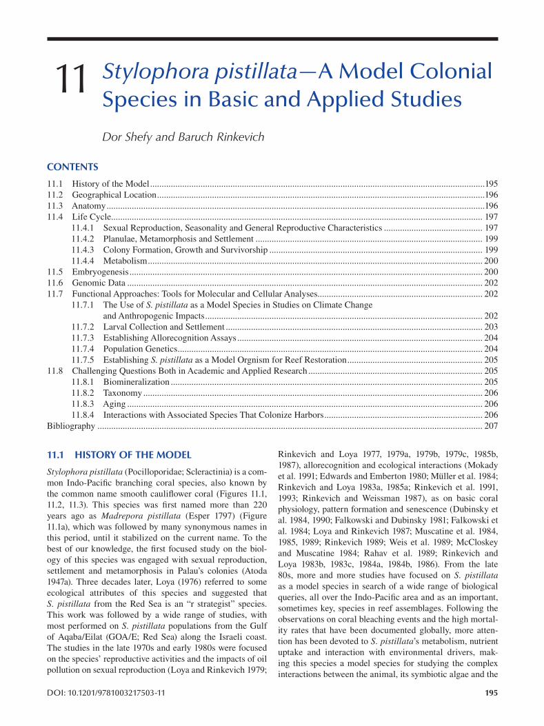
11 Stylophora pistillata—A Model Colonial Species in Basic and Applied Studies
Dor Shefy and Baruch Rinkevich
CONTENTS
11.1 History of the Model..................................................................................................................................................195
11.2 Geographical Location...............................................................................................................................................196
11.3 Anatomy.....................................................................................................................................................................196
11.4 Life Cycle.................................................................................................................................................................. 197
11.4.1 Sexual Reproduction, Seasonality and General Reproductive Characteristics ........................................... 197
11.4.2 Planulae, Metamorphosis and Settlement ................................................................................................... 199
11.4.3 Colony Formation, Growth and Survivorship ............................................................................................. 199
11.4.4 Metabolism.................................................................................................................................................. 200
11.5 Embryogenesis.......................................................................................................................................................... 200
11.6 Genomic Data ........................................................................................................................................................... 202
11.7 Functional Approaches: Tools for Molecular and Cellular Analyses........................................................................ 202
11.7.1 The Use of S. pistillata as a Model Species in Studies on Climate Change
and Anthropogenic Impacts......................................................................................................................... 202
11.7.2 Larval Collection and Settlement ................................................................................................................ 203
11.7.3 Establishing Allorecognition Assays........................................................................................................... 204
11.7.4 Population Genetics..................................................................................................................................... 204
11.7.5 Establishing S. pistillata as a Model Orgnism for Reef Restoration........................................................... 205
11.8 Challenging Questions Both in Academic and Applied Research ............................................................................ 205
11.8.1 Biomineralization ........................................................................................................................................ 205
11.8.2 Taxonomy .................................................................................................................................................... 206
11.8.3 Aging ........................................................................................................................................................... 206
11.8.4 Interactions with Associated Species That Colonize Harbors..................................................................... 206
Bibliography ........................................................................................................................................................................ 207
11.1 HISTORY OF THE MODEL Rinkevich and Loya 1977, 1979a, 1979b, 1979c, 1985b,
1987), allorecognition and ecological interactions (MokadyStylophora pistillata (Pocilloporidae; Scleractinia) is a com-
et al. 1991; Edwards and Emberton 1980; Müller et al. 1984;mon Indo-Pacific branching coral species, also known by
Rinkevich and Loya 1983a, 1985a; Rinkevich et al. 1991,the common name smooth cauliflower coral (Figures 11.1,
1993; Rinkevich and Weissman 1987), as on basic coral 11.2, 11.3). This species was first named more than 220
physiology, pattern formation and senescence (Dubinsky et years ago as Madrepora pistillata (Esper 1797 ) (Figure
al. 1984, 1990; Falkowski and Dubinsky 1981; Falkowski et 11.1a), which was followed by many synonymous names in
al. 1984; Loya and Rinkevich 1987; Muscatine et al. 1984,this period, until it stabilized on the current name. To the
1985, 1989; Rinkevich 1989; Weis et al. 1989; McCloskeybest of our knowledge, the first focused study on the biol-
and Muscatine 1984; Rahav et al. 1989; Rinkevich and ogy of this species was engaged with sexual reproduction,
Loya 1983b, 1983c, 1984a, 1984b, 1986). From the late settlement and metamorphosis in Palau’s colonies (Atoda
80s, more and more studies have focused on S. pistillata 1947a). Three decades later, Loya (1976) referred to some
as a model species in search of a wide range of biological ecological attributes of this species and suggested that
queries, all over the Indo-Pacific area and as an important,S. pistillata from the Red Sea is an “r strategist” species.
sometimes key, species in reef assemblages. Following the This work was followed by a wide range of studies, with
observations on coral bleaching events and the high mortal-most performed on S. pistillata populations from the Gulf
ity rates that have been documented globally, more attenof Aqaba/Eilat (GOA/E; Red Sea) along the Israeli coast.
tion has been devoted to S. pistillata’s metabolism, nutrient The studies in the late 1970s and early 1980s were focused
uptake and interaction with environmental drivers, makon the species’ reproductive activities and the impacts of oil
ing this species a model species for studying the complex pollution on sexual reproduction (Loya and Rinkevich 1979;
interactions between the animal, its symbiotic algae and the
DOI: 10.1201/9781003217503-11 195
196
environment (Abramovitch-Gottlib et al. 2003; Dubinsky
et al. 1990; Dubinsky and Jokiel 1994; Ferrier-Pagès et al.
2000 , 2001 , 2003 , 2010 ; Franklin et al. 2004 ; Grover et al.
2002 , 2003 , 2006 , 2008 ; Hoegh-Guldberg and Smith 1989a ,
1989b; Houlbrèque et al. 2003, 2004; Lampert-Karako et al.
2008; Muscatine et al. 1989; Nakamura et al. 2003; Rahav
et al. 1989; Rinkevich 1989; Shashar et al. 1993; Tchernov
et al. 2004; Titlyanov et al. 2000a; Titlyanov et al. 2000b;
Titlyanov et al. 2001; Weis et al. 1989). The accumulated
knowledge on the species distribution and the reproductive
mode of S. pistillata has led researchers to study popula
tion dynamics, population genetic structures, modes of
reproduction and larval dispersal in a specific reef and
among reefs (Ayre and Hughes 2000; Zvuloni et al. 2008;
Klueter and Andreakis 2013; Douek et al. 2011; Guerrini et
al. 2020; Takabayashi et al. 2003; Nishikawa et al. 2003).
S. pistillata colonies are also often used for understanding
the impacts of anthropogenic activities and climate change
disturbances on coral reefs and, together with the rapid
advances in technology, scientists have examined the com
bined effects of anthropogenic/climate change impacts on S. pistillata’s biological and ecological parameters (Ammar et
al. 2012; Guerrini et al. 2020; Horwitz et al. 2017; Loya and
Rinkevich 1979 ; Shefy et al. 2018 ; Tamir et al. 2020 ), physi
ology (Abramovitch-Gottlib et al. 2003; Banc-Prandi and
Fine 2019; Bellworthy and Fine 2017; Bellworthy et al. 2019;
Dias et al. 2019; Epstein et al. 2005; Fitt et al. 2009; Grinblat
et al. 2018; Hall et al. 2018; Hawkins et al. 2015; Hoegh-
Guldberg and Smith 1989b; Krueger et al. 2017; Reynaud et
al. 2003; Rinkevich et al. 2005; Rosic et al. 2020; Sampayo
et al. 2008; Sampayo et al. 2016; Saragosti et al. 2010; Shick
et al. 1999; Stat et al. 2009) and gene expression patterns
(Maor-Landaw and Levy 2016; Oren et al. 2010, 2013;
Voolstra et al. 2017). Several studies have focused on in vitro approaches with S. pistillata cells and minute fragments for
the development of novel methodologies; cell culture, nub
bin and larvae usage for ecotoxicology and for reef restora
tion and for the elucidation of biological features, such as
calcification and algal movements (Bockel and Rinkevich
2019; Danovaro et al. 2008; Downs et al. 2014; Epstein et al.
2000; Frank et al. 1994; Horoszowski-Fridman et al. 2020;
Mass et al. 2012, 2017a; Raz-Bahat et al. 2006; Shafir et al.
2001 , 2003 , 2007 , 2014 ); on anatomical features ( Raz-Bahat
et al. 2017); and on applied approaches (Rinkevich 2015a ;
Rinkevich and Shafir 1998; Rinkevich et al. 1999; Shafi r
et al. 2001). The understanding that coral reefs around the
world are degrading has led, in the last two decades, to the
development of an additional applied route, an active reef
restoration that is based on a wide range of methodologies
being tested on S. pistillata as a model species (Amar and
Rinkevich 2007; Epstein and Rinkevich 2001 Epstein et
al. 2001, 2005; Golomb et al. 2020; Horoszowski-Fridman
et al. 2015, 2020; Horoszowski-Fridman and Rinkevich
2020; Linden and Rinkevich 2011, 2017; Linden et al. 2019;
Rachmilovitz and Rinkevich 2017; Rinkevich 2000, 2015a,
2019a , 2019b ; Shafir and Rinkevich 2008, 2010; Shafir et al.
2006a , 2009 ).
Emerging Marine Model Organisms
Here, we aim to review the knowledge about S. pistil-lata’s biological features in various scientific disciplines for
the last eight decades of research.
11.2 GEOGRAPHICAL LOCATION
S. pistillata colonies are found in shallow waters and up to
70 meters deep (Fishelson 1971; Kramer et al. 2019; Muir
and Pichon 2019; Veron 2000). This species has a wide geo
graphical range in the tropical and sub-tropical Indo-Pacifi c
Ocean; central and west Pacific; tropical Australia; South
China Sea; southern Japan; central Indian Ocean; southwest
and northwest Indian Ocean; Arabian/Iranian Gulf; Gulf
of Aden and the Red Sea, including the gulfs of Suez and
Aqaba/Eilat (Veron 2000).
11.3 ANATOMY
An S. pistillata colony consists of up to tens of thousands
of polyps at adulthood, each about 1–2 mm in diameter,
where each polyp creates a small skeletal cup (termed a
corallite), the hard supporting blueprint of the polyp’s tissue
(Veron 2000). The external soft tissues of the polyps and
their extensions that connect between the polyps (coenosarc)
overlie the coral skeleton that is made of calcium carbonate
(Veron 2000). The polyps are anchored to the underlying
skeletons by cells called desmocytes that connect the lower
ectodermic layer (the calicoblastic layer) to the perforated
calcium carbonate milieu (Muscatine et al. 1997; Raz-Bahat
et al. 2006; Tambutté et al. 2007). Each polyp is a hollow
cylindrical blind-ended sac that resembles a sea anemone in
structure with a mouth in the center of the polyp, surrounded
by 12 hollow retractable tentacles (Figure 11.2d) that con
nect to the gastric cavity by the pharynx. This is the gateway
for food particles to the coelenteron, but studies revealed
further roles in chemical digestion (Raz-Bahat et al. 2017).
All polyps within a colony are connected to each other via a
network of cell-lined tubes (gastrovascular canals) that radi
ate from the gastric cavity of the polyps. The polyp’s inter
nal gastric cavity is divided by 12 partitions (mesenteries; 6
are complete) into compartments which run radially from
the body wall’s gastrodermis to the actinopharynx and are
connected to the pharynx carrying six long extensions (mes
enterial filaments; Raz-Bahat et al. 2017). Two types of mes
enterial filaments exist in S. pistillata, distinct, as much to
be known by general morphology: four short fi laments with
no secretory cells and two long convoluted fi laments with
stinging and secretory cells (Raz-Bahat et al. 2017) that pen
etrate the gastric cavity and into the gastrovascular canals.
The compartments between the mesenteries are also the
sites where male and female gonads are developed (Ammar
et al. 2012; Rinkevich and Loya 1979a). As in all corals, each
polyp and the connected coenosarc consist of two epithelial
layers, the ectodermic and gastrodermis (endodermis), sepa
rated by the mesoglea. This non-epithelial milieu binds the
two epithelial layers together throughout the colony while
consisting of a gelatinous substance, with collagen fi bers
197 Stylophora pistillata—A Model Colonial Species in Basic and Applied Studies
and some cells. The columnar ectodermic layer contains
mucus gland cells, nematocytes and spyrocyte cells, and the
gastrodermis layer contains the zooxanthellae (Al-Sofyani
1991; Raz-Bahat et al. 2017, Bockel and Rinkevich 2019).
The tentacles that are located above the oral disk are loaded
with zooxanthellae in their gastrodermis cells, while the epi
dermis contains nematocytes.
As mentioned, the skeleton is secreted by the calico-
blastic tissue (also named calicodermis), which forms the
lower ectodermal layer (Allemand et al. 2004, 2011). The
calicoblastic epithelium is very thin and has only calicoblas
tic cells anchored to the skeleton by the desmocytes (Raz-
Bahat et al. 2006; Tambutté et al. 2007). The calicoblastic
epithelium secretes amorphous nano-calcium carbonate
crystals into microenvironments enriched in organic mate
rial. The carbonate crystals aggregate and then crystallize to
create ordered aragonitic structures (Mass et al. 2017b; Von
Euw et al. 2017). On the coenosteum (skeleton secreted by
the coenosarc), skeletal spines called coenosteal spines are
developed, and in shallow water colonies, they have granular
textures as compared to smoother textures in deeper water
colonies (Malik et al. 2020).
11.4 LIFE CYCLE
11.4.1 SEXUAL REPRODUCTION, SEASONALITY AND
GENERAL REPRODUCTIVE CHARACTERISTICS
While most of the coral species are broadcast spawners,
together with other 61 species, S. pistillata belongs to a
group of brooding coral species, where fertilization and lar
val development take place inside the polyps (Ammar et al.
2012; Fan and Dai 2002; Rinkevich and Loya 1979a) for an
estimated duration of two weeks (Fan and Dai 2002; Shefy
et al. 2018). The planula larvae are released to the water
column about one to two hours after sunset (Atoda 1947a ;
Rinkevich and Loya 1979b)
S. pistillata is a hermaphrodite species, and male and
female gonads are situated side by side within the polyp’s
coelenteron, extended into the body cavities and attached to
the mesenteries by stalks. Along astogeny, the male gonads
appear first when the colonies reach an approximate radius
of 2 cm, and female gonads develop a year later (Rinkevich
and Loya 1979a). A wide range of anthropogenic and natu
ral stressors may affect gonadal development. Early studies
revealed that oil pollution and sedimentation directly reduce
male and female gonad numbers and significantly affect the
developing planulae (Loya and Rinkevich 1979; Rinkevich
and Loya 1979c). Even nutrient-enriched environments may
affect gonads and larval development, and while phosphorus
load may have a minor impact on the reproductive efforts
(Ammar et al. 2012), particulate matter (PM) and particu
late organic matter (POM) may increase the size and num
ber of oocytes and testes (Bongiorni et al. 2003a 2003b).
Yet, resident fish within coral colonies that secrete nutrients
(Liberman et al. 1995) do not have impacts on fecundity,
as on the colony color morph (Rinkevich 1982). In contrast,
intraspecific (within the same species) and interspecifi c (with
different species) interactions have impacts on the number
of female gonads per polyp (Rinkevich and Loya 1985b).
S. pistillata’s reproductive patterns, seasonality and
reproductive efforts vary among bio-geographical regions.
In Palau, Atoda (1947a) recorded planulae release one to
two weeks after a full moon all year long. Differences in
seasonality are also present in the population at Yabnu
(South Res Sea) and Tarut Bay (Arabian Gulf), which are
in the same latitude but in different seas. In Tarut Bay,
embryos were observed for just two months a year (before
seawater temperature exceeded 31°C), while in Yabnu,
embryos were documented ten months a year (before tem
perature exceeded 29°C) (Fadlallah and Lindo 1988). In
the Philippines, the reproductive season of S. pistillata lasts just three months, from November to January (Baird
et al. 2015), while in Taiwan, documentations revealed all-
year-round larval release, with no obvious lunar periodicity
(Fan and Dai 2002). S. pistillata colonies in the southern
hemisphere release planulae from August to December
in the Great Barrier Reef (GBR) (Tanner 1996 ) and from
August to May with lunar periodicity in south Australia
(Villanueva et al. 2008).
The reproduction of the S. pistillata populations in the
Gulf of Aqaba/Eilat, Red Sea, is a model case for coral
reproduction for over five decades, allowing a glimpse of
changes in reproduction on an extended time scale. During
the 1970s and 1980s, shallow-water S. pistillata colo
nies in Eilat released planulae for seven to eight months
(December–July) (Rinkevich and Loya 1979b, 1987).
Recent observations revealed that seasonality of larval
release during the 2010s is extended by one to two months,
from December to September–October (Rinkevich and
Loya 1979b, 1987; Shefy et al. 2018) and year-round
recruitment (Guerrini et al. 2020). Studies also revealed
a bell-shaped curve in the larval release of most S. pistil-lata populations characterized in Eilat by an increase in
planulae numbers until reaching a peak and then, in the
second half, a decrease in the release until the end of the
season (Amar et al. 2007; Fan and Dai 2002; Rinkevich
and Loya 1979a, 1979b, 1987; Shefy et al. 2018; Tanner
1996 ). Fecundity among different colonies (even those of
the same size that are situated side by side in the reef) or
within a coral colony over several reproductive seasons is
portrayed by high variability (Rinkevich and Loya 1987;
Shefy et al. 2018). Variation is also recorded for lunar
periodicity that was assigned for some populations (Atoda
1947a ; Dai et al. 1992; Fan and Dai 2002; Tanner 1996;
Villanueva et al. 2008; Zakai et al. 2006) while miss
ing in others (Linden et al. 2018; Rinkevich and Loya
1979b). Linden et al. (2018) revealed that larval release by
S. pistillata colonies does not comply with the assumed
entrainment by the lunar cycle, further documenting that
the lunar cycle does not provide a strict zeitgeber and can
better be classifi ed as a circatrigintan pattern. Water tem
perature and solar radiation did not correlate signifi cantly
with larval release.
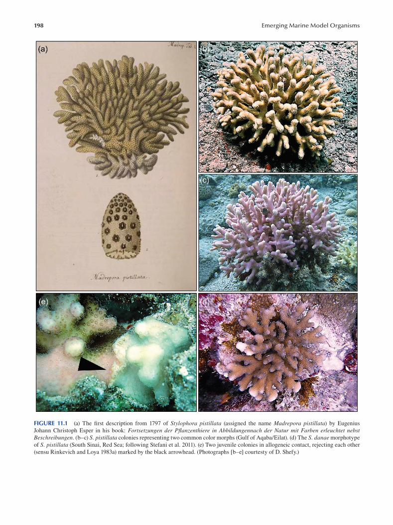
198 Emerging Marine Model Organisms
FIGURE 11.1 (a) The first description from 1797 of Stylophora pistillata (assigned the name Madrepora pistillata) by Eugenius
Johann Christoph Esper in his book: Fortsetzungen der Pflanzenthiere in Abbildungennach der Natur mit Farben erleuchtet nebst Beschreibungen. (b–c) S. pistillata colonies representing two common color morphs (Gulf of Aqaba/Eilat). (d) The S. danae morphotype
of S. pistillata (South Sinai, Red Sea; following Stefani et al. 2011). (e) Two juvenile colonies in allogeneic contact, rejecting each other
(sensu Rinkevich and Loya 1983a) marked by the black arrowhead. (Photographs [b–e] courtesty of D. Shefy.)
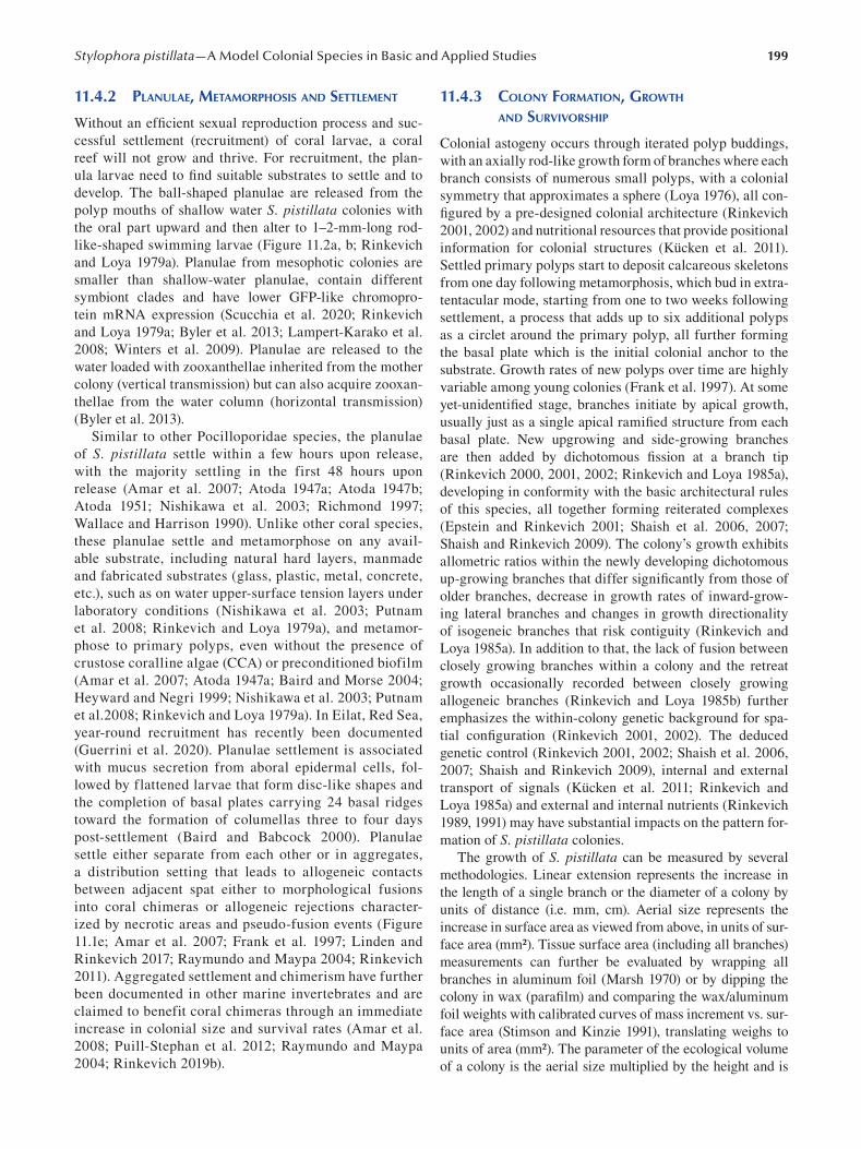
199 Stylophora pistillata—A Model Colonial Species in Basic and Applied Studies
11.4.2 PLANULAE, METAMORPHOSIS AND SETTLEMENT
Without an efficient sexual reproduction process and suc
cessful settlement (recruitment) of coral larvae, a coral
reef will not grow and thrive. For recruitment, the plan
ula larvae need to find suitable substrates to settle and to
develop. The ball-shaped planulae are released from the
polyp mouths of shallow water S. pistillata colonies with
the oral part upward and then alter to 1–2-mm-long rod-
like-shaped swimming larvae (Figure 11.2a, b; Rinkevich
and Loya 1979a). Planulae from mesophotic colonies are
smaller than shallow-water planulae, contain different
symbiont clades and have lower GFP-like chromopro
tein mRNA expression (Scucchia et al. 2020; Rinkevich
and Loya 1979a ; Byler et al. 2013; Lampert-Karako et al.
2008; Winters et al. 2009). Planulae are released to the
water loaded with zooxanthellae inherited from the mother
colony (vertical transmission) but can also acquire zooxan
thellae from the water column (horizontal transmission)
(Byler et al. 2013).
Similar to other Pocilloporidae species, the planulae
of S. pistillata settle within a few hours upon release,
with the majority settling in the first 48 hours upon
release (Amar et al. 2007; Atoda 1947a ; Atoda 1947b;
Atoda 1951; Nishikawa et al. 2003; Richmond 1997;
Wallace and Harrison 1990). Unlike other coral species,
these planulae settle and metamorphose on any avail
able substrate, including natural hard layers, manmade
and fabricated substrates (glass, plastic, metal, concrete,
etc.), such as on water upper-surface tension layers under
laboratory conditions (Nishikawa et al. 2003; Putnam
et al. 2008; Rinkevich and Loya 1979a), and metamor
phose to primary polyps, even without the presence of
crustose coralline algae (CCA) or preconditioned biofilm
(Amar et al. 2007; Atoda 1947a ; Baird and Morse 2004;
Heyward and Negri 1999; Nishikawa et al. 2003; Putnam
et al.2008; Rinkevich and Loya 1979a). In Eilat, Red Sea,
year-round recruitment has recently been documented
(Guerrini et al. 2020). Planulae settlement is associated
with mucus secretion from aboral epidermal cells, fol
lowed by flattened larvae that form disc-like shapes and
the completion of basal plates carrying 24 basal ridges
toward the formation of columellas three to four days
post-settlement (Baird and Babcock 2000). Planulae
settle either separate from each other or in aggregates,
a distribution setting that leads to allogeneic contacts
between adjacent spat either to morphological fusions
into coral chimeras or allogeneic rejections character
ized by necrotic areas and pseudo-fusion events (Figure
11.1e; Amar et al. 2007; Frank et al. 1997; Linden and
Rinkevich 2017; Raymundo and Maypa 2004; Rinkevich
2011). Aggregated settlement and chimerism have further
been documented in other marine invertebrates and are
claimed to benefit coral chimeras through an immediate
increase in colonial size and survival rates (Amar et al.
2008; Puill-Stephan et al. 2012; Raymundo and Maypa
2004 ; Rinkevich 2019b ).
11.4.3 COLONY FORMATION, GROWTH
AND SURVIVORSHIP
Colonial astogeny occurs through iterated polyp buddings,
with an axially rod-like growth form of branches where each
branch consists of numerous small polyps, with a colonial
symmetry that approximates a sphere (Loya 1976), all con
figured by a pre-designed colonial architecture (Rinkevich
2001, 2002) and nutritional resources that provide positional
information for colonial structures (Kücken et al. 2011).
Settled primary polyps start to deposit calcareous skeletons
from one day following metamorphosis, which bud in extra-
tentacular mode, starting from one to two weeks following
settlement, a process that adds up to six additional polyps
as a circlet around the primary polyp, all further forming
the basal plate which is the initial colonial anchor to the
substrate. Growth rates of new polyps over time are highly
variable among young colonies (Frank et al. 1997 ). At some
yet-unidentified stage, branches initiate by apical growth,
usually just as a single apical ramified structure from each
basal plate. New upgrowing and side-growing branches
are then added by dichotomous fission at a branch tip
( Rinkevich 2000 , 2001 , 2002 ; Rinkevich and Loya 1985a ),
developing in conformity with the basic architectural rules
of this species, all together forming reiterated complexes
(Epstein and Rinkevich 2001; Shaish et al. 2006, 2007;
Shaish and Rinkevich 2009). The colony’s growth exhibits
allometric ratios within the newly developing dichotomous
up-growing branches that differ signifi cantly from those of
older branches, decrease in growth rates of inward-grow
ing lateral branches and changes in growth directionality
of isogeneic branches that risk contiguity (Rinkevich and
Loya 1985a). In addition to that, the lack of fusion between
closely growing branches within a colony and the retreat
growth occasionally recorded between closely growing
allogeneic branches (Rinkevich and Loya 1985b) further
emphasizes the within-colony genetic background for spa
tial configuration (Rinkevich 2001, 2002). The deduced
genetic control (Rinkevich 2001, 2002; Shaish et al. 2006,
2007; Shaish and Rinkevich 2009), internal and external
transport of signals (Kücken et al. 2011; Rinkevich and
Loya 1985a) and external and internal nutrients (Rinkevich
1989, 1991) may have substantial impacts on the pattern for
mation of S. pistillata colonies.
The growth of S. pistillata can be measured by several
methodologies. Linear extension represents the increase in
the length of a single branch or the diameter of a colony by
units of distance (i.e. mm, cm). Aerial size represents the
increase in surface area as viewed from above, in units of sur
face area (mm2). Tissue surface area (including all branches)
measurements can further be evaluated by wrapping all
branches in aluminum foil ( Marsh 1970) or by dipping the
colony in wax (parafi lm) and comparing the wax/aluminum
foil weights with calibrated curves of mass increment vs. sur
face area (Stimson and Kinzie 1991), translating weighs to
units of area (mm2). The parameter of the ecological volume
of a colony is the aerial size multiplied by the height and is
200
measured by an increase of the whole space encompassed by
the coral branches in mm3 ( Shafir and Rinkevich 2010; Shafi r
et al. 2006b). Other size methods, such as 3D photography
for measuring parameters of growth rates (surface areas, vol
umes, etc.), do not always give accurate results due to the
high structural complexity of developing colonies.
S. pistillata is a fast-growing species as compared to mas
sive and encrusting species and some other branching spe
cies. Branches can grow up to 5 cm per year, depending on the
conditions and the initial fragment size (Dar and Mohamed
2017; Bockel and Rinkevich 2019; Hasan 2019; Liberman et
al. 1995; Loya 1976; Shafir and Rinkevich 2010; Shafir et al.
2006b; Tamir et al. 2020), and small fragments can multiply
their ecological volumes by 200 times within 8–12 months
( Shafir and Rinkevich 2008). In old senescent colonies, calci
fication rates, as reproductive activities, decrease synchron
ically in all branches, and the whole colony as a single unit,
new and old polyps alike, exhibits senescence concurrently,
leading to accelerated degradation and colonial death within
few months (Rinkevich and Loya 1986).
S. pistillata colonies that grow under improved water
flows (primarily in mid-water floating nurseries) that assist
the polyps in catching prey exhibit enhanced growth rates
and advanced recovery from bleaching (in all parameters
mentioned previously) (Bongiorni et al. 2003a ; Nakamura
et al. 2003; Shafir and Rinkevich 2010). In contrast to the
high and fast growth rates characteristic to S. pistillata and although it is one of the most abundant species in the
GOA/E (Shaked and Genin 2019; Shlesinger and Loya
2016), adult colonies and primarily recruits have high mor
tality rates (Doropoulos et al. 2015; Linden and Rinkevich
2011 , 2017 ; Loya 1976 ; Shafir, Van Rijn, and Rinkevich
2006b; Shlesinger and Loya 2016; Tamir et al. 2020).
Assuming 50–80% settlement rates in the wild (Amar et al.
2007; Linden and Rinkevich 2011), only a small portion of
recruits will develop into gravid colonies out of tens of mil
lions and more of planulae released during any reproduction
season. Under in-situ aqua-culture conditions, young colo
nies can reach a 40–80% survival rate if protected by cages
and 10–30% if not protected (Linden and Rinkevich 2017;
Shafir et al. 2006b), orders of magnitude above natural fi g
ures. Nevertheless, size structure demographic models for S. pistillata populations in various reefs were not constructed
and are not yet available, in spite of their importance for con
servation and management plans (Doropoulos et al. 2015).
11.4.4 METABOLISM
In the past four decades, S. pistillata has been used as a
model species in studies on carbon and nutrients assimila
tion and their acquisition, allocation and uptake by coral and
by symbiotic algae. Since most coral reefs thrive in oligotro
phic waters, it is essential to understand nutrient recycling by
reef communities, as it may shed light on coral life histories
and reef-resilient. S. pistillata colonies, as other coral spe
cies get carbon and nutrients through two main processes:
via photosynthesis, provided by the symbiotic autotrophic
Emerging Marine Model Organisms
algae (Muscatine et al. 1981), and by feeding on particular
or dissolved sources of organic carbon (Houlbrèque and
Ferrier-Pagès 2009). The symbiotic dinofl agellates can
not provide all the essential carbon and organic nitrogen
needed for the coral, especially under low light regimens
(Falkowski et al. 1984; Muscatine et al. 1984; Tremblay et al.
2014). Yet corals may modify their algal numbers and their
activities. Studies on S. pistillata revealed that under high
light regimes, respiration and calcification rates increased
(Dubinsky and Jokiel 1994), while the symbiotic algae
decreased in size and numbers, further showing high res
piration and lower quantum yields (Dubinsky et al. 1984).
With regard to nitrogen, another limiting nutrient source for
the algae (Hoegh-Guldberg and Smith 1989a), increasing
concentrations of nitrogen compounds such as ammonium,
urea, amino acids, nitrite and nitrate lead to an increase in
the nitrogen uptake by the holobiont (Dubinsky and Jokiel
1994 ; Grover et al. 2002 , 2003 , 2006 , 2008 ; Houlbre’que
and Ferrier-page 2009; Rahav et al. 1989). The fate and path
of each nitrogen source, whether consumed via water or by
feeding (praying of zooplankton), is mostly determined by
light intensity and photosynthetic products (Dubinsky and
Jokiel 1994; Houlbrèque and Ferrier-Pagès 2009). Assuming
constant low nutrient concentration in the reef, under high
light intensity regimes, most of the carbon goes to respira
tion and growth, including calcification by the host, while
under low light, the zooxanthellae use the carbon and nutri
ents (Dubinsky and Jokiel 1994). Feeding on zooplankton
or other pico- and nano-planktonic organisms increases
nutrients uptake (including phosphate) that provides the
nutrients needed for coral growth and reproduction (Ferrier-
Pagès et al. 2003; Houlbrèque et al. 2004; Houlbrèque et
al. 2003) and enhances the numbers of zooxanthellae in the
coral tissues (Dubinsky et al. 1990; Houlbrèque et al. 2003;
Titlyanov et al. 2001; Titlyanov et al. 2000, 2001; Titlyanov
et al. 2000). Studies on S. pistillata’s symbiotic relationships
further revealed the translocation of photosynthates between
branches and along a branch within a colony and between
genotypes (Rinkevich 1991; Rinkevich and Loya 1983b,
1983c, 1984a), and were used in quest on the “light enhanced
calcification” enigma (Houlbrèque et al. 2003; Moya et al.
2006; Muscatine et al. 1984; Reynaud-Vaganay et al. 2001;
Rinkevich and Loya 1984b). Despite all the previous stud
ies on S. pistillata symbiotic relationships, there is a need
for additional studies to reveal the more intimate interac
tions between the holobiont participants (Ferrier-Pagès et al.
2018; Hédouin et al. 2016; Metian et al. 2015).
11.5 EMBRYOGENESIS
As a hermaphroditic brooder species, S. pistillata fertil
ization and larval development take place within the body
cavities of the polyps, thus making it challenging to study
embryogenesis and larval development. Rinkevich and Loya
(1979a) and then Ammar et al. (2012) observed that male and
female gonads, situated on small stalks, start to develop at
two and fi ve months, respectively, before the onset of larval
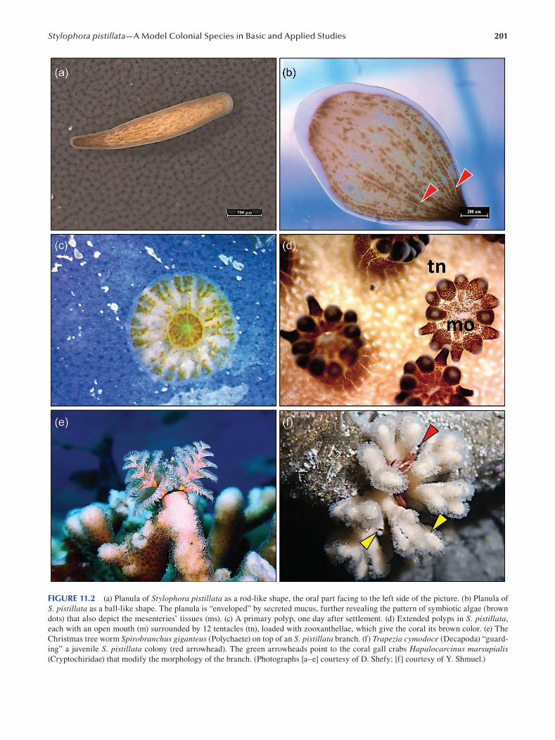
201 Stylophora pistillata—A Model Colonial Species in Basic and Applied Studies
FIGURE 11.2 (a) Planula of Stylophora pistillata as a rod-like shape, the oral part facing to the left side of the picture. (b) Planula of
S. pistillata as a ball-like shape. The planula is “enveloped” by secreted mucus, further revealing the pattern of symbiotic algae (brown
dots) that also depict the mesenteries’ tissues (ms). (c) A primary polyp, one day after settlement. (d) Extended polyps in S. pistillata,
each with an open mouth (m) surrounded by 12 tentacles (tn), loaded with zooxanthellae, which give the coral its brown color. (e) The
Christmas tree worm Spirobranchus giganteus (Polychaete) on top of an S. pistillata branch. (f) Trapezia cymodoce (Decapoda) “guard
ing” a juvenile S. pistillata colony (red arrowhead). The green arrowheads point to the coral gall crabs Hapalocarcinus marsupialis (Cryptochiridae) that modify the morphology of the branch. (Photographs [a–e] courtesy of D. Shefy; [f] courtesy of Y. Shmuel.)
202
release (reproductive season), filling up the gastric cavities
of the polyps during the peak of reproduction season. At the
start, 4–16 oocytes per polyp develop; some are absorbed
during the development in such a way that only a single
mature egg at a specific time is left (Rinkevich and Loya
1979a). The migration of the egg nuclei to the periphery sig
nals that the eggs are ready for fertilization (Rinkevich and
Loya 1979a). Larval development is assumed to take 14 days,
but the whole development process was not studied (Fan and
Dai 2002; Rinkevich and Loya 1979b). Planulae develop in
most polyps (except for the sexually sterile branch tips), and
upon the release of the larva from a specific polyp, another
oocyte becomes ready for fertilization. A mature planula has
an organized ectodermal epithelium and a less organized
gastrodermis loaded with zooxanthellae, separated by a thin
mesoglea, and has six pairs of mesenteries (Figure 11.2a, b)
(Atoda 1947a ; Fan and Dai 2002; Rinkevich and Loya 1979a ;
Scucchia et al. 2020). Further, planulae of S. pistillata from
the Red Sea (not observed in other planulae, including of
S. pistillata from other places) show temporary extensions
from the body wall, consisting of ectodermal-mesogleal
material (“filaments”) and extensions containing endoder
mal epithelium only (“nodules”) that regularly appear and
absorb (Rinkevich and Loya 1979a). The developing larvae
are flexible in their morphologies, and, while globular upon
release, they appear as pear-like, disk-like or rod-like struc
tures (Figure 11.2a, b) (Atoda 1947a ; Rinkevich and Loya
1979a). Planulae of S. pistillata that are released from shal
low water gravid colonies are fluorescent (Grinblat et al.
2018; Rinkevich and Loya 1979a ; Scucchia et al. 2020), with
a lower expression of the green fluorescence protein (GFP)
gene in planulae originating from >30 m colonies (Scucchia
et al. 2020). It has further been documented that planulae
start to precipitate minerals in the form of small crystals
that may assist in rapid calcification upon settlement (Akiva
et al. 2018).
11.6 GENOMIC DATA
Advances, reduced costs of sequencing and improved tech
nologies over the past decade enabled the recent sequenc
ing and assembling of the S. pistillata genome (the full
sequenced genome can be found at http://spis.reefgenomics.
org/) (Banguera-Hinestroza et al. 2013; Voolstra et al. 2017 ).
The sequenced genome enabled studies on evolutionary
adaptation and origin of this species (Voolstra et al. 2017),
algae–host relationships, gene expression analyses (Barott et
al. 2015b; Gutner-Hoch et al. 2017; Karako-Lampert et al.
2014; Liew et al. 2014; Maor-Landaw and Levy 2016 ) and
studies on epigenetics (Dimond and Roberts 2016; Liew et al.
2018). Results further revealed the genes involved in stressed
(and not stressed) colonies as the molecular mechanisms for
adaptation to global change impacts. S. pistillata mitochon
drial DNA (mDNA) was used to investigate phylogenetic
aspects, species delineation and the taxonomical status of
this species (Chen et al. 2008; Flot et al. 2011; Keshavmurthy
et al. 2013; Klueter and Andreakis 2013; Stefani et al. 2011),
Emerging Marine Model Organisms
further elucidating that the origin of S. pistillata is from the
west Indian ocean and that this species presents of up to six
distinct morphs. Molecular markers such as ITS1, ampli
fied fragment length polymorphism (AFLP) and allozymes
were used to assess the genetic structure among different S. pistillata populations, within populations and coral recruits
(Amar et al. 2008; Ayre and Hughes 2000; Douek et al. 2011;
Takabayashi et al. 2003; Zvuloni et al. 2008) yet are too few
to reveal clear genetic landscapes.
11.7 FUNCTIONAL APPROACHES: TOOLS FOR MOLECULAR AND CELLULAR ANALYSES
Despite the claim that S. pistillata is a “weedy species”
(Loya 1976), the biological characteristics of this species,
such as its fast growth rates, abundance and long reproduc
tive season, made S. pistillata a model animal in a wide
range of ecological settings and for functional approaches.
It also helped that while S. pistillata colonies present several
color morphs (Figure 11.1b, c) (Stambler and Shashar 2007 ),
this diversity has no connection to either ecological feature
studied (Rinkevich and Loya 1979b, 1985b).
11.7.1 THE USE OF S. PISTILLATA AS A MODEL
SPECIES IN STUDIES ON CLIMATE CHANGE
AND ANTHROPOGENIC IMPACTS
The decline of coral reef resilience and persistence due to
anthropogenic impacts and global warming is of great con
cern for the future of reef ecosystems (Bindoff et al. 2019).
S. pistillata has further served as a model species for ana
lyzing a wide range of stressors on corals and symbionts,
on various life history parameters and on coral adaption to
changing environments. These studies further examined the
holobiont (coral/algal) symbiotic relationships on the whole-
organism level (respiration, calcification rates, survival and
photosynthesis), on the cellular level (organelles, lipids, pro
teins and stress-related proteins) and on a molecular level
(DNA damage, gene expression and symbiont identity). In
these studies, S. pistillata colonies are often used for eluci
dating coral responses to thermal stress (increasing of sea
water temperatures), with consequences that are determined
by the specific zooxanthellae species and the coral genotype
subjected to specific stress conditions (Sampayo et al. 2008),
further associated with alteration in the symbiont clades
toward more physiologically suited algal populations (Fitt et
al. 2009; Sampayo et al. 2016 ).
Ex-situ and in-situ experiments with S. pistillata revealed
damages to the thylakoid membranes of the symbiotic
algae when colonies are exposed to elevated temperatures
and increased light intensities (Tchernov et al. 2004), also
following other biological and physiological stresses, all
expressed with induced photoinhibition and decreased
photosynthesis (Bhagooli and Hidaka 2004; Cohen and
Dubinsky 2015; Falkowski and Dubinsky 1981; Franklin et
al. 2004; Hawkins et al. 2015; Hoegh-Guldberg and Smith
203 Stylophora pistillata—A Model Colonial Species in Basic and Applied Studies
1989b; Yakovleva et al. 2004), reduced algal density with
time (Abramovitch-Gottlib et al. 2003; Biscéré et al. 2018;
Cohen and Dubinsky 2015) and decreased protein concen
tration (Falkowski and Dubinsky 1981; Hoegh-Guldberg
and Smith 1989b; Rosic et al. 2020). When evaluating the
impacts on the host S. pistillata and its responses, studies
documented that elevated temperatures increase coral respi
ration (Hall et al. 2018; Hoegh-Guldberg and Smith 1989b;
Reynaud et al. 2003); enforced impacts on calcifi cation rates
(mixed results, increase or decrease; Abramovitch-Gottlib
et al. 2003; Biscéré et al. 2018; Hall et al. 2018; Reynaud et
al. 2003); decreased protein and lipid contents (Falkowski
and Dubinsky 1981; Hall et al. 2018; Rosic et al. 2020);
imposed fluctuations in ROS and antioxidant enzymes ) such
as superoxide dismutase [SOD], catalase [CAT], ascorbate
peroxidase [APX], glutathione S-transferase [GST] and
glutathione peroxidase [GPX]), primarily if light stress
was co-involved (Hawkins et al. 2015; Saragosti et al. 2010;
Yakovleva et al. 2004); and increased coral mortality rates
(Dias et al. 2019). These physiological responses are further
reflected in gene expression patterns, including the upregu
lation of key cellular processes associated with heat stress
such as oxidative stress, energy metabolism, DNA repair
and apoptosis (Maor-Landaw and Levy 2016). While it is a
possibility that higher-latitude S. pistillata populations show
a general improved tendency for adaptation to temperature
changes (Pontasch et al. 2017), the suggestion that S. pistil-lata from the Red Sea specifically went through evolutionary
adaptation to heat stress (Fine et al. 2013) made this species
a model animal for experiments examining climate change
impacts on corals (Bellworthy and Fine 2017; Bellworthy et
al. 2019; Bellworthy et al. 2019; Hall et al. 2018; Grottoli et
al. 2017; Krueger et al. 2017). Other studies examined the
ecological consequences of global change, such as on allo
geneic and xenogeneic interactions (Horwitz et al. 2017).
Following the results that S. pistillata colonies accumu
late metal from seawater (Ali et al. 2011; Al-Sawalmih et al.
2017; Ferrier-Pagès et al. 2005), studies have further inves
tigated S. pistillata holobiont responses to metal pollution
and the combined effects with warming seas. High concen
trations of copper have negative impacts on the holobiont,
expressed as a decrease in photosynthesis effi ciency, algal
density, host respiration rate and host protein and increase in
SOD activity, especially when combined with elevated tem
perature (Banc-Prandi and Fine 2019). Biscéré et al. (2018)
further found that while manganese (Mn) enhances cellular
chlorophyll concentration and photosynthesis effi ciency and
increases S. pistillata resistance to heat stress, and iron (Fe)
positively affects the holobiont and symbionts (Biscéré et al.
2018; Shick et al. 2011), seawater enriched with Mn and iron
decreases calcification and induces bleaching. Increased
concentrations of Cobalt (Co) inflicted decreased growth
rates under ambient pH conditions and in lower-pH water
but had no impacts on photosynthesis under ambient pH
conditions (Biscéré et al. 2015).
Numerous studies used S. pistillata as a model coral spe
cies to investigate the impacts of a wide range of pollutants
on corals, such as oil pollution, sunscreen lotion detergents
and eutrophication. Results revealed that some sunscreen
ingredients might induce extensive necrosis in the coral’s
epidermis and gastrodermis layers (Downs et al. 2014),
impair photosynthetic activity (Fel et al. 2019) and promote
viral infection followed by bleaching (Danovaro et al. 2008).
In-situ and ex-situ experiments showed that crude oil and
its derivatives have a destructive effect on sexual repro
duction in S. pistillata by reducing the number of female
gonads per polyp (Rinkevich and Loya 1979c), by induc
ing the abortion of planulae (Epstein et al. 2000; Loya and
Rinkevich 1979), by decreasing the settlement rate (Epstein
et al. 2000), through DNA damage (Kteifan et al. 2017)
and by intensifying coral and larval mortalities (Epstein
et al. 2000). The same applies to detergents in seawater
that impair basic S. pistillata biological features (Shafi r et
al. 2014) and anti-fouling compounds (Shafir et al. 2009).
Studies also revealed that under various scenarios for nutri
ent-enriched environments, eutrophication even enhances
S. pistillata performance, as colonies exhibited increased
growth rates (Bongiorni et al. 2003a, 2003b), increases in
host mitochondrial and protein concentrations (Kramarsky-
Winter et al. 2009; Sawall et al. 2011), decreases in oxida
tion (Kramarsky-Winter et al. 2009) and increases in teste
and egg numbers with a decrease in their size (Ammar et
al. 2012; Bongiorni et al. 2003a). The healthy physiological
status, in contrast to lab experiment results, suggests that the
corals gain more energy through heterotrophy (increase in
zooplankton) rather than autotrophy (Rinkevich 2015c).
Light has a significant role in marine invertebrates’ bio
logical clocks and is a cue in the regulation of circadian
rhythms (zeitgeber) and physiological processes. Therefore,
S. pistillata was further used as a model species for light pol
lution, following the observation that the coral reefs in the
northern tip of the GOA/E, Red Sea, are heavily subjected
to artifi cial light pollution at night (ALAN) (Aubrecht et al.
2008; Tamir et al. 2017). Shefy et al. (2018) postulated that
changes in the length of the reproductive season in S. pistil-lata from Eilat might be the outcomes of increased ALAN
in the last four decades. Further, reduced settlement rates
were recorded in planulae exposed to ALAN as compared
to regular light regimes, and a year upon settlement, the
formerly impacted young colonies exhibited lower photo
synthesis efficiency, albeit higher survival, growth and calci
fication rates (Tamir et al. 2020). Adult S. pistillata colonies
as their symbionts showed increased oxidative damage in
lipids and increased respiration rate and experienced loss of
symbionts and enhanced photoinhibition at decreased pho
tosynthetic rates (Levy et al. 2020).
11.7.2 LARVAL COLLECTION AND SETTLEMENT
As mentioned, S. pistillata is a brooding coral with a long
reproduction season in some bio-geographical areas. By
using this reproduction strategy, scientists can also use the
planulae of S. pistillata as a model animal. In order to catch
planulae easily, a planulae trap is used (Akiva et al. 2018;
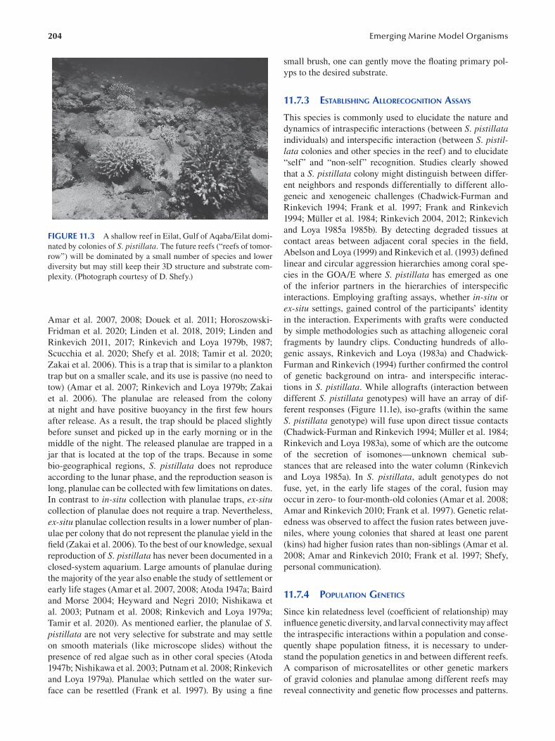
204
FIGURE 11.3 A shallow reef in Eilat, Gulf of Aqaba/Eilat domi
nated by colonies of S. pistillata. The future reefs (“reefs of tomor
row”) will be dominated by a small number of species and lower
diversity but may still keep their 3D structure and substrate com
plexity. (Photograph courtesy of D. Shefy.)
Amar et al. 2007, 2008; Douek et al. 2011; Horoszowski-
Fridman et al. 2020; Linden et al. 2018 , 2019; Linden and
Rinkevich 2011, 2017; Rinkevich and Loya 1979b, 1987;
Scucchia et al. 2020; Shefy et al. 2018; Tamir et al. 2020;
Zakai et al. 2006). This is a trap that is similar to a plankton
trap but on a smaller scale, and its use is passive (no need to
tow) (Amar et al. 2007; Rinkevich and Loya 1979b; Zakai
et al. 2006 ). The planulae are released from the colony
at night and have positive buoyancy in the first few hours
after release. As a result, the trap should be placed slightly
before sunset and picked up in the early morning or in the
middle of the night. The released planulae are trapped in a
jar that is located at the top of the traps. Because in some
bio-geographical regions, S. pistillata does not reproduce
according to the lunar phase, and the reproduction season is
long, planulae can be collected with few limitations on dates.
In contrast to in-situ collection with planulae traps, ex-situ collection of planulae does not require a trap. Nevertheless,
ex-situ planulae collection results in a lower number of plan
ulae per colony that do not represent the planulae yield in the
field ( Zakai et al. 2006). To the best of our knowledge, sexual
reproduction of S. pistillata has never been documented in a
closed-system aquarium. Large amounts of planulae during
the majority of the year also enable the study of settlement or
early life stages (Amar et al. 2007, 2008; Atoda 1947a; Baird
and Morse 2004; Heyward and Negri 2010; Nishikawa et
al. 2003; Putnam et al. 2008; Rinkevich and Loya 1979a ;
Tamir et al. 2020). As mentioned earlier, the planulae of S. pistillata are not very selective for substrate and may settle
on smooth materials (like microscope slides) without the
presence of red algae such as in other coral species (Atoda
1947b; Nishikawa et al. 2003; Putnam et al. 2008; Rinkevich
and Loya 1979a). Planulae which settled on the water sur
face can be resettled (Frank et al. 1997 ). By using a fi ne
Emerging Marine Model Organisms
small brush, one can gently move the floating primary pol
yps to the desired substrate.
11.7.3 ESTABLISHING ALLORECOGNITION ASSAYS
This species is commonly used to elucidate the nature and
dynamics of intraspecific interactions (between S. pistillata individuals) and interspecific interaction (between S. pistil-lata colonies and other species in the reef) and to elucidate
“self” and “non-self” recognition. Studies clearly showed
that a S. pistillata colony might distinguish between differ
ent neighbors and responds differentially to different allo
geneic and xenogeneic challenges (Chadwick-Furman and
Rinkevich 1994; Frank et al. 1997; Frank and Rinkevich
1994; Müller et al. 1984; Rinkevich 2004, 2012; Rinkevich
and Loya 1985a 1985b). By detecting degraded tissues at
contact areas between adjacent coral species in the fi eld,
Abelson and Loya (1999) and Rinkevich et al. (1993) defi ned
linear and circular aggression hierarchies among coral spe
cies in the GOA/E where S. pistillata has emerged as one
of the inferior partners in the hierarchies of interspecifi c
interactions. Employing grafting assays, whether in-situ or
ex-situ settings, gained control of the participants’ identity
in the interaction. Experiments with grafts were conducted
by simple methodologies such as attaching allogeneic coral
fragments by laundry clips. Conducting hundreds of allo
genic assays, Rinkevich and Loya (1983a) and Chadwick-
Furman and Rinkevich (1994) further confirmed the control
of genetic background on intra- and interspecifi c interac
tions in S. pistillata. While allografts (interaction between
different S. pistillata genotypes) will have an array of dif
ferent responses (Figure 11.1e), iso-grafts (within the same
S. pistillata genotype) will fuse upon direct tissue contacts
(Chadwick-Furman and Rinkevich 1994; Müller et al. 1984;
Rinkevich and Loya 1983a), some of which are the outcome
of the secretion of isomones—unknown chemical sub
stances that are released into the water column (Rinkevich
and Loya 1985a). In S. pistillata, adult genotypes do not
fuse, yet, in the early life stages of the coral, fusion may
occur in zero- to four-month-old colonies (Amar et al. 2008;
Amar and Rinkevich 2010; Frank et al. 1997 ). Genetic relat
edness was observed to affect the fusion rates between juve
niles, where young colonies that shared at least one parent
(kins) had higher fusion rates than non-siblings (Amar et al.
2008; Amar and Rinkevich 2010; Frank et al. 1997; Shefy,
personal communication).
11.7.4 POPULATION GENETICS
Since kin relatedness level (coefficient of relationship) may
influence genetic diversity, and larval connectivity may affect
the intraspecific interactions within a population and conse
quently shape population fitness, it is necessary to under
stand the population genetics in and between different reefs.
A comparison of microsatellites or other genetic markers
of gravid colonies and planulae among different reefs may
reveal connectivity and genetic flow processes and patterns.
205 Stylophora pistillata—A Model Colonial Species in Basic and Applied Studies
Elements of population genetic structures of S. pistillata populations were studied along the GBR, Okinawa and
GOA\E, revealing a high contribution of sexual reproduction
to the populations (Ayre and Hughes 2000; Takabayashi et
al. 2003; Zvuloni et al. 2008). Yet significant differences in
polymorphic allozyme loci diversity were recorded between
populations in the same geographical region, implying low
levels of connectivity but sufficient genetic diversity to
maintain gene flow among reefs (Ayre and Hughes 2000).
The low genetic flow among reefs is also related to the fast
settlement rates of most released larvae, where the vast
majority of the planulae metamorphose 24–48 hours upon
release, a time scale that is varied between early and late
phases of the reproduction season (Nishikawa et al. 2003;
Amar et al. 2007; Rinkevich and Loya 1979a). Yet there are
no detailed population genetics studies that employed highly
polymorphic markers, reinforcing the need to develop addi
tional efficient and inexpensive tools.
11.7.5 ESTABLISHING S. PISTILLATA AS A MODEL
ORGNISM FOR REEF RESTORATION
The accelerating climate change and its effects on the coral
reefs and the recognition that passive management measures
(such as the declaration of marine protected areas) are not
enough to cope with climate change (Bindoff et al. 2019;
Rinkevich 2008) have raised the need for active reef resto
ration ( Rinkevich 1995 , 2000 , 2005 , 2014 , 2015a , 2015b).
Much of the work published on active reef restoration has
emerged as of the end of the 1990s and has considered
colonies of S. pistillata for the research and development
of new reef restoration methods and approaches. Most of
the colonies that were maintained in the fi rst constructed
floating nurseries in the GOA/E, including microcolonies
and 2–5-cm-long fragments of S. pistillata, exhibited fast
growth rates and high survival rates (Epstein et al. 2001;
Linden and Rinkevich 2017; Linden et al. 2018; Rinkevich
2000 ; Shafir and Rinkevich 2010; Shafir et al. 2001, 2003,
2006b). The same applied to transplantation acts performed
in Eilat and other Indo-Pacific sites (Golomb et al. 2020;
Horoszowski-Fridman et al. 2015; Horoszowski-Fridman et
al. 2020). S. pistillata was further used in various ecological
engineering approaches. To achieve higher genetic diversity,
several studies (Linden and Rinkevich 2011, 2017; Linden et
al. 2019) worked on S. pistillata planulae as source material
for reef restoration. They collected planulae and reared them
in two ways: (1) in situ, using a special designated settlement
box that allowed the planulae to settle in situ on artifi cial
substrates (Linden et al. 2019), and (2) ex situ, in outdoor
aquarium systems (Linden and Rinkevich 2011), and then
developing spat were moved and farmed in fl oating nurser
ies (Linden and Rinkevich 2011, 2017). Several versions of
methodologies adopted various colony orientations (vertical
or horizontal), protection methods against predation (in or
out of cages) and locations in the nursery. These developing
methods yielded high survival rates, involved minimal main
tenance in the developing spat and successfully enhanced
genetic diversity. By harnessing the ability of isogeneic frag
ments to fuse, Rachmilovitz and Rinkevich (2017 ) formed,
within six to seven months, fl at S. pistillata tissue plates
from glued fragments on plastic tiles in the purpose of creat
ing two-dimensional corals units (that can cover degraded
substrates). Furthermore, it was shown that nursery-farmed
coral colonies that had been transplanted into a degraded
reef at Eilat (Dekel Beach) revealed higher fecundity
(Horoszowski-Fridman et al. 2020) than native colonies, and
when transplanted with other species, they attracted planu
lae settlement (Golomb et al. 2020). Harnessing chimerism,
the fusion between different genotypes (possible during
only at early life stages), has also been proposed as an active
reef restoration tool to mitigate climate change impacts
(Rinkevich 2019b). Chimerism can benefit the coral entity
by causing increased sizes, high genotypic diversity and and
enhanced phenotipic plasticity.
11.8 CHALLENGING QUESTIONS BOTH IN ACADEMIC AND APPLIED RESEARCH
Out of the many challenging topics associated with the use
of S. pistillata as a model system for coral biology, three
challenging topics are outlined in the following as being of
primary importance in the biology of this species.
11.8.1 BIOMINERALIZATION
The mechanisms controlling coral calcification at the molec
ular, cellular and entire tissue levels are still not fully under
stood. Over the past few decades, S. pistillata has been used
as one of the model organisms for studying calcifi cation in
corals. Although numerous papers has been published, the
calcification process remains an enigmatic biological phe
nomenon, as its nature, including physiochemically con
trolled mechanisms or its biologically mediated machinery,
have not yet been resolved (Allemand et al. 2011). Within
the last three decades, numerous studies have engaged
with various aspects of coral calcification, while many of
them have used S. pistillata as the model organism for cor
als (Allemand et al. 2004; Drake et al. 2019; Falini et al.
2015). As mentioned earlier, the calcifying tissue is the cali
coblastic layer, an epithelium attached to the skeleton with
desmocytes (Muscatine et al. 1997; Raz-Bahat et al. 2006;
Tambutté et al. 2007), thus found in direct contact with the
skeleton surface (Tambutté et al. 2007). The calicoblastic
ectoderm produces the extracellular matrix (ECM) proteins
that are secreted to the calcifying medium and remain pre
served in the skeleton organic matrix (Allemand et al. 2011).
Coral skeletal aragonite is produced within the ECM, which
is secreted into semi-enclosed extracellular compartments
and composed of a few nano-micrometers-thick matrix ele
ments (Mass et al. 2017a ; Sevilgen et al. 2019; Tambutté et
al. 2007). The cells in the calicoblastic layer are connected
through tight junctions that control the diffusion of mole
cules to the ECM (Barott et al. 2015a; Raz-Bahat et al. 2006;
Tambutté et al. 1996 , 2007, 2012; Zoccola et al. 1999, 2004).
206
This paracellular pathway depends on the charge and size of
the molecules (Tambutté et al. 2012). Furthermore, a second
path of calcium ions to the center of calcification through an
intracellular pathway was proposed. By using in vitro pri
mary cell cultures of S. pistillata and employing antibodies
against ion transporters, several studies (Barott et al. 2015a ;
Mass et al. 2012, 2017a) showed that calcium is concentrated
in intracellular pockets and is exported to the site of calcifi
cation via vesicles (Ganot et al. 2020). Dissolved inorganic
carbon (DIC) can diffuse from the coral tissue to the ECM
(Furla et al. 2000) or, alternatively, be transported via bicar
bonate transporters from the calicoblastic cells’ cytosol to
the ECM ( Zoccola et al. 2015). The transport of proteins
and minerals to the ECM is influenced and mediated by
environmental parameters such as temperature, pH, calcium
saturation levels, pollutants and enzymes (Al-Sawalmih
2016; Allemand et al. 2004; Furla et al. 2000; Gattuso et al.
1998; Gutner-Hoch et al. 2017; Malik et al. 2020; Puverel
et al. 2005; Zoccola et al. 1999, 2004, 2015). It is suggested
that high amounts of acidic amino acids and glycine in the
ECM (Puverel et al. 2005) allow the control of its chemi
cal composition by increasing pH and DIC concentration
above the surrounding water and enable the formation of
aragonite (Drake et al. 2019; Venn et al. 2011). The skeletal
organic matrix within the skeletal framework contains at
least 60 proteins and glycosylated derivatives which remain
entrapped within the crystalline units (Allemand et al. 2011;
Drake et al. 2013; Mass et al. 2014; Peled et al. 2020; Puverel
et al. 2007). The calicoblastic tissue secretes amorphous
nano-calcium carbonate particles in the created microenvi
ronments enriched in organic material aggregates that then
crystallize to create ordered aragonitic structures (Mass et
al. 2012, 2017b; Von Euw et al. 2017 ). S. pistillata colonies
grow their skeletons from the centers of calcifi cation areas
of spherulitic shapes (radial distributions of acicular crys
tals), forming bundles of aragonite crystals (Sun et al. 2017,
2020 ).
11.8.2 TAXONOMY
S. pistillata is considered a model organism in research
and has been the focus of coral research over the past four
decades. This species is widely distributed in the Indo-Pacifi c
region and represented by numerous morphological varia
tions (morphotypes) associated with different reef habitats,
geographical regions and reef depth zones (Figures 11.1b,
c, d, 11.3). Thus, for comparative studies, it is imperative to
ensure its correct taxonomy and species delineation. Using
molecular markers (mitochondrial and nuclear genes), aided
by comparisons of morphological characteristics, enabled
scientists to point toward the west Pacific and not the coral
triangle, like for other corals, as the origin of S. pistillata (Flot et al. 2011; Stefani et al. 2011). Keshavmurthy et al.
(2013) further revealed the presence of cryptic divergence
and four distinct evolutionary lineages (clades) within S. pistillata across its distribution range: clade 1 is distributed in
the Pacific Ocean (Klueter and Andreakis 2013), clade 2 is
Emerging Marine Model Organisms
distributed over the Indian Ocean and clade 3 is found in
the west Indian Ocean. The distribution of the fourth clade
overlaps with clades 2 and 3, but this clade inhabits the Red
Sea as well (Keshavmurthy et al. 2013). In contrast, Arrigoni
et al. (2016 ) postulated that the different species of the genus
Stylophora found in the Red Sea are actually ecomorphs
of a single phenotypically plastic species that belong to a
single molecular lineage. Further analyses are thus needed
to evaluate the taxonomic status of S. pistillata and whether
other species of Stylophora represent valid endemic species
arising from speciation or locally emerged ecomorphs of S. pistillata that had been adapted to different environmental
conditions (depth, temperature, etc.).
11.8.3 AGING
How long can a colony of S. pistillata live? Are colonies
that Jacques Cousteau saw still alive? Some of the coral
species attain considerable ages (>400 years), but others
have a shorter life span (reviewed in Bythell et al. 2018).
The life span of S. pistillata was never followed in detail,
but studies assumed it to be in the range of 20–30 years
(Rinkevich, personal communication). Before natural death,
a colony exhibits a decrease in the rate of reproduction, tis
sue degradation and a decrease in growth (Rinkevich and
Loya 1986). Aging in such colonial species is of great inter
est, and telomeres can be used in the research as molecu
lar markers of aging due to the common loss of telomeres
repeating in other aging multicellular organisms, including
humans. Additionally, coral stem cells, which can be used as
another marker for aging, are not yet known in S. pistillata,
nor in other coral species. Decreased regeneration abilities
in some colonies could also be related to stem cell aging (Y.
Rinkevich et al. 2009). Hence, S. pistillata may be used as
a model species for aging and stem cell biology research of
corals in general.
11.8.4 INTERACTIONS WITH ASSOCIATED
SPECIES THAT COLONIZE HARBORS
S. pistillata is an ecologically important key species, con
sidered an r-strategist (Loya 1976 ) and an ecological engi
neering species (Rinkevich 2020) that harbors on branches,
between branches and within the skeleton a wide range of fi sh
species and species of large invertebrates, including cryptic,
boring and encrusting organisms such as sponges, bivalves,
polychaetes, crabs and others (Figure 11.2e, f) (Barneah
et al. 2007; Belmaker et al. 2007; Berenshtein et al. 2015;
El-Damhougy et al. 2018; Mbije et al. 2019; Garcia-Herrera
et al. 2017; Goldshmid et al. 2004; Kotb and Hartnoll 2002;
Kuwamura et al. 1994; Limviriyakul et al. 2016; Mohammed
and Yassien 2013; Mokady et al. 1991, 1993, 1994; Pratchett
2001; Rinkevich et al. 1991; Shafir et al. 2008). Some of these
organisms are commensals; others are corallivores, passing
organisms or symbionts. The nature of such interactions is
not always explicit. Garcia-Herrera et al. (2017 ) found that
Dascyllus marginatus fish that are fanning their fi ns keep
207 Stylophora pistillata—A Model Colonial Species in Basic and Applied Studies
oxygen levels high during the night hours in the inner spaces
of the colony between branches, where the photosynthetic
oxygen levels are decreased (Shashar et al. 1993). Trapezia cymodoce, a xanthid crab which lives between S. pistillata’s
colony branches, grazes on the coral tissue (Rinkevich et
al. 1991), yet colonies harboring this “parasitic” crab dem
onstrated higher survival rates (Glynn 1983), partly due to
their aggressive behavior toward predators (Pratchett 2001).
Some of the species live exclusively on/in S. pistillata colo
nies, including the gobiid fi sh Paragobiodon echinocephalus (Belmaker et al. 2007; Kuwamura et al. 1994) and the boring
bivalve Lithophaga lessepsiana (Mokady et al. 1994). While
very little is known about such biological associations, bor
ing organisms such as bivalves and crustaceans can modify
the colony morphology (Abelson et al. 1991). These associa
tions become a challenging question, further highlighted by
reef restoration acts that consider the whole reef communities
and not solely the coral transplants.
BIBLIOGRAPHY
Abelson, A., B. Galil, and Y. Loya. 1991. “Skeletal modifi cations
in stony corals caused by indwelling crabs: Hydrodynamical
advantages for crab feeding.” Symbiosis 10: 233–248.
Abelson, A., and Y. Loya. 1999. “Interspecific aggression among
stony corals in Eilat, Red Sea: A hierarchy of aggression abil
ity and related parameters.” Bulletin of Marine Science 65
(3): 851–860.
Abramovitch-Gottlib, L., D. Katoshevski, and R. Vago. 2003.
“Responses of Stylophora pistillata and Millepora dichotoma to seawater temperature elevation.” Bulletin of Marine Science 73 (3): 745–755.
Akiva, A., M. Neder, K. Kahil, R. Gavriel, I. Pinkas, G. Goobes, and
T. Mass. 2018. “Minerals in the pre-settled coral Stylophora pistillata crystallize via protein and ion changes.” Nature Communications 9 (1). doi:10.1038/s41467-018-04285-7.
Al-Sawalmih, A. 2016. “Calcium composition and microstruc
ture of coral Stylophora pistillata under phosphate pollution
stress in the Gulf of Aqaba.” Natural Science 8 (3): 89–95.
doi:10.4236/ns.2016.83012.
Al-Sawalmih, A., F.A. Al-Horani, and S. Al-Rousan. 2017.
“Elemental analysis of the coral Stylophora pistillata incu
bated along the Jordanian coast of the Gulf of Aqaba.”
Fresenius Environmental Bulletin 26 (4): 3029–3036.
Al-Sofyani, A.A. 1991. “Physiology and ecology of Stylophora pistillata and Echinopora gemmacea from the Red Sea.”
University of Glasgow, United Kindom.
Ali, A. hamid A.M., M.A. Hamed, and H.A. El-Azim. 2011.
“Heavy metals distribution in the coral reef ecosystems of
the Northern Red Sea.” Helgoland Marine Research 65 (1):
67–80. doi:10.1007/s10152-010-0202-7.
Allemand, D., C. Ferrier-pagès, P. Furla, F. Houlbrèque, S. Puverel,
S. Reynaud, É. Tambutté, S. Tambutté, and D. Zoccola.
2004. “Biomineralisation in reef-building corals: From
molecular mechanisms to environmental control.” General Palaeontology (Palaeobiochemistry) Biomineralisation 3:
453–467. doi:10.1016/j.crpv.2004.07.011.
Allemand, D., É. Tambutté, D. Zoccola, and S. Tambutté. 2011.
“Coral calcification, cells to reefs.” In Coral Reefs: An Ecosystem in Transition, eds. Dubinsky, Z. and
Stambler, N., 119–150. Dordrecht: Springer Netherlands.
doi:10.1007/978-94-007-0114-4_9.
Amar, K.O., N. Chadwick, and B. Rinkevich. 2007. “Coral plan
ulae as dispersion vehicles: Biological properties of lar
vae released early and late in the season.” Marine Ecology Progress Series 350 (November): 71–78. doi:10.3354/
meps07125.
Amar, K.O., N.E. Chadwick, and B. Rinkevich. 2008. “Coral
kin aggregations exhibit mixed allogeneic reactions and
enhanced fi tness during early ontogeny.” BMC Evolutionary Biology 8 (1): 126. doi:10.1186/1471-2148-8-126.
Amar, K.O., J. Douek, C. Rabinowitz, and B. Rinkevich. 2008.
“Employing of the amplified fragment length polymorphism
(AFLP) methodology as an efficient population genetic tool
for symbiotic cnidarians.” Marine Biotechnology 10 (4):
350–357. doi:10.1007/s10126-007-9069-2.
Amar, K.O., and B. Rinkevich. 2007. “A floating mid-water coral
nursery as larval dispersion hub: Testing an idea.” Marine Biology 151: 713–718. doi:10.1007/s00227-006-0512-0.
Amar, K.O., and B. Rinkevich. 2010. “Mounting of erratic histo
incompatible responses in hermatypic corals: A multi-year
interval comparison.” Journal of Experimental Biology 213
(4): 535–540.
Ammar, M.S.A., A.H. Obuid-Allah, and M.A.M. Al-Hammady.
2012. “Patterns of fertility in the two Red Sea corals
Stylophora pistillata and Acropora humilis.” Nusantara Bioscience 4 (2): 62–75. doi:10.13057/nusbiosci/n040204.
Arrigoni, R., F. Benzoni, T.I. Terraneo, A. Caragnano, and M.L.
Berumen. 2016. “Recent origin and semi-permeable spe
cies boundaries in the scleractinian coral genus Stylophora from the Red Sea.” Scientifi c Reports 6 (January): 1–13.
doi:10.1038/srep34612.
Atoda, K. 1947a. “The larva and postlarval development of some
reef-building corals. II. Stylophora pistillata (Esper).”
Science Reports of the Tohoku Imperial University, 4th Series (Biology) 18: 48–64.
Atoda, K. 1947b. “The larva and postlarval development of some
reef-building corals. I. Pocillopora damicornis cespitosa (Dana).” Science Reports of the Tohoku Imperial University, 4th Series (Biology) 18: 24–47.
Atoda, K. 1951. “The larva and postlarval development of some
reef-building corals. V. ( Seritopora hystrix ) Dana.” Science Reports of the Tohoku University 4th Ser. (Biology) 19 (1):
33–39.
Aubrecht, C., C.D. Elvidge, T. Longcore, C. Rich, J. Safran, A.E.
Strong, M. Eakin, et al. 2008. “A global inventory of coral
reef stressors based on satellite observed nighttime lights.”
Geocarto International 23 (December 2015): 467–479.
doi:10.1080/10106040802185940.
Ayre, D.J., and T.P. Hughes. 2000. “Genotypic diversity and
gene flow in brooding and spawning corals along the Great
Barrier Reef, Australia.” Evolution 54 (5): 1590–1605.
doi:10.1554/0014-3820(2000)054[1590:GDAGFI]2.0.CO;2.
Baird, A.H., and R.C. Babcock. 2000. “Morphological differ
ences among three species of newly settled pocilloporid
coral recruits.” Coral Reefs 19 (2): 179–183. doi:10.1007/
PL00006955.
Baird, A.H., V.R. Cumbo, S. Gudge, S.A. Keith, J.A. Maynard,
C.H. Tan, and E.S. Woolsey. 2015. “Coral reproduction on the
world’s southernmost reef at Lord Howe Island, Australia.”
Aquatic Biology 23 (3): 275–284. doi:10.3354/ab00627.
Baird, A.H., and A.N.C. Morse. 2004. “Induction of metamorpho
sis in larvae of the brooding corals Acropora palifera and
Stylophora pistillata.” Marine and Freshwater Research 55
(5): 469–472. doi:10.1071/MF03121.
Banc-Prandi, G., and M. Fine. 2019. “Copper enrichment
reduces thermal tolerance of the highly resistant Red Sea
208
coral Stylophora pistillata.” Coral Reefs 38 (2): 285–296.
doi:10.1007/s00338-019-01774-z.
Banguera-Hinestroza, E., P. Saenz-Agudelo, T. Bayer, M.L.
Berumen, and C.R. Voolstra. 2013. “Characterization of new
microsatellite loci for population genetic studies in the smooth
cauliflower coral ( Stylophora sp .).” Conservation Genetics Resources 5 (2): 561–563. doi:10.1007/s12686-012-9852-x.
Barneah, O., I. Brickner, M. Hooge, V.M. Weis, T.C. LaJeunesse, and
Y. Benayahu. 2007. “Three party symbiosis: Acoelomorph
worms, corals and unicellular algal symbionts in Eilat (Red
Sea).” Marine Biology 151 (4): 1215–1223. doi:10.1007/
s00227-006-0563-2.
Barott, K.L., S.O. Perez, L.B. Linsmayer, and M. Tresguerres. 2015a.
“Differential localization of ion transporters suggests distinct
cellular mechanisms for calcification and photosynthesis
between two coral species.” American Journal of Physiology: Regulatory Integrative and Comparative Physiology 309 (3):
R235–R246. doi:10.1152/ajpregu.00052.2015.
Barott, K.L., A.A. Venn, S.O. Perez, S. Tambutteeé, M. Tresguerres,
and G.N. Somero. 2015b. “Coral host cells acidify symbi
otic algal microenvironment to promote photosynthesis.”
Proceedings of the National Academy of Sciences of the United States of America 112 (2): 607–612. doi:10.1073/
pnas.1413483112.
Bellworthy, J., and M. Fine. 2017. “Beyond peak summer temper
atures, branching corals in the Gulf of Aqaba are resilient
to thermal stress but sensitive to high light.” Coral Reefs 36
(4). Springer Berlin Heidelberg: 1071–1082. doi:10.1007/
s00338-017-1598-1.
Bellworthy, J., M. Menoud, T. Krueger, A. Meibom, and M. Fine.
2019. “Developmental carryover effects of ocean warming
and acidification in corals from a potential climate refugium,
the Gulf of Aqaba.” Journal of Experimental Biology 222 (1).
doi:10.1242/jeb.186940.
Bellworthy, J., J.E. Spangenberg, and M. Fine. 2019. “Feeding
increases the number of offspring but decreases parental
investment of Red Sea coral Stylophora pistillata.” Ecology and Evolution 9 (21): 12245–12258. doi:10.1002/ece3.5712.
Belmaker, J., O. Polak, N. Shashar, and Y. Ziv. 2007. “Geographic
divergence in the relationship between Paragobiodon echinocephalus and its obligate coral host.” Journal of Fish Biology 71 (5): 1555–1561. doi:10.1111/j.1095-8649.2007.01619.x.
Berenshtein, I., Y. Reuben, and A. Genin. 2015. “Effect of oxygen
on coral fanning by mutualistic fi sh.” Marine Ecology 36 (4):
1171–1175. doi:10.1111/maec.12218.
Bhagooli, R., and M. Hidaka. 2004. “Photoinhibition, bleach
ing susceptibility and mortality in two scleractinian cor
als, Platygyra ryukyuensis and Stylophora pistillata,
in response to thermal and light stresses.” Comparative Biochemistry and Physiology: A Molecular and Integrative Physiology 137 (3): 547–555. doi:10.1016/j.
cbpb.2003.11.008.
Bindoff, N.L., W.W.L. Cheung, J.G. Kairo, J. Arístegui, V.A.
Guinder, R. Hallberg, N. Hilmi, et al. 2019. Changing Ocean, Marine Ecosystems, and Dependent Communities. IPCC Special Report on the Ocean and Cryosphere in a Changing Climate [H.-O. Pörtner, D.C. Roberts, V. Masson-Delmotte, P. Zhai, M. Tignor, E. Poloczanska, K. Mintenbeck, A. Alegría, M. Nicolai, A. Okem, J. Petzold, B. Rama, N.M. Weyer (Eds.)].
www.ipcc.ch/srocc/download-report/ .
Biscéré, T., C. Ferrier-Pagès, A. Gilbert, T. Pichler, and F.
Houlbrèque. 2018. “Evidence for mitigation of coral
bleaching by manganese.” Scientifi c Reports 8 (1): 1–10.
doi:10.1038/s41598-018-34994-4.
Emerging Marine Model Organisms
Biscéré, T., R. Rodolfo-Metalpa, A. Lorrain, L. Chauvaud, J.
Thébault, J. Clavier, and F. Houlbrèque. 2015. “Responses
of two scleractinian corals to cobalt pollution and ocean
acidifi cation.” PLoS One 10 (4): 1–18. doi:10.1371/journal.
pone.0122898.
Bockel, T., and B. Rinkevich. 2019. “Rapid recruitment of sym
biotic algae into developing Scleractinian coral tissues.”
Journal of Marine Science and Engineering 7 (306).
doi:10.3390/jmse7090306.
Bongiorni, L., S. Shafir, D. Angel, and B. Rinkevich. 2003a.
“Survival, growth and gonad development of two hermatypic
corals subjected to in situ fish-farm nutrient enrichment.”
Marine Ecology Progress Series 253 (2001): 137–144.
doi:10.3354/meps253137.
Bongiorni, L., S. Shafi r, and B. Rinkevich. 2003b. “Effects of par
ticulate matter released by a fish farm (Eilat, Red Sea) on
survival and growth of Stylophora pistillata coral nubbins.”
Marine Pollution Bulletin 46 (9): 1120–1124. doi:10.1016/
S0025-326X(03)00240-6.
Byler, K.A., M. Carmi-Veal, M. Fine, and T.L. Goulet. 2013.
“Multiple symbiont acquisition strategies as an adaptive
mechanism in the coral Stylophora pistillata.” PLoS One 8
(3): 1–7. doi:10.1371/journal.pone.0059596.
Bythell, J.C., B.E. Brown, and T.B.L. Kirkwood. 2018. “Do
reef corals age?” Biological Reviews 93 (2): 1192–1202.
doi:10.1111/brv.12391.
Chadwick-Furman, N., and B. Rinkevich. 1994. “A complex
allorecognition system in a reef-building coral: Delayed
responses, reversals and nontransitive hierarchies.” Coral Reefs 13 (1): 57–63. doi:10.1007/BF00426436.
Chen, C., C.Y. Chiou, C.F. Dai, and C.A. Chen. 2008. “Unique
mitogenomic features in the scleractinian family pocillopori
dae (Scleractinia: Astrocoeniina).” Marine Biotechnology 10
(5): 538–553. doi:10.1007/s10126-008-9093-x.
Cohen, I., and Z. Dubinsky. 2015. “Long term photoacclimation
responses of the coral Stylophora pistillata to reciprocal deep
to shallow transplantation: Photosynthesis and calcifi cation.”
Frontiers in Marine Science 2 (June): 1–13. doi:10.3389/
fmars.2015.00045.
Dai, C.F., K. Soong, and T.Y. Fan. 1992. “Sexual reproduction of
corals in northern and southern Taiwan.” 7th Int’l. Coral Reef Symp., 1: 448–455.
Danovaro, R., L. Bongiorni, C. Corinaldesi, D. Giovannelli,
E. Damiani, P. Astolfi, L. Greci, and A. Pusceddu. 2008.
“Sunscreens cause coral bleaching by promoting viral infec
tions.” Environmental Health Perspectives 116 (4): 441–447.
doi:10.1289/ehp.10966.
Dar, M.A., and T.A.A. Mohamed. 2017. “Coral growth and skeletal
density relationships in some branching corals of the Red Sea,
Egypt.” Journal of Environment and Earth Science 7 (11): 66–79.
Dias, M., A. Ferreira, R. Gouveia, C. Madeira, N. Jogee, H. Cabral,
M. Diniz, and C. Vinagre. 2019. “Long-term exposure to
increasing temperatures on scleractinian coral fragments
reveals oxidative stress.” Marine Environmental Research 150
(January): 104758. doi:10.1016/j.marenvres.2019.104758.
Dimond, J.L., and S.B. Roberts. 2016. “Germline DNA methyla
tion in reef corals: Patterns and potential roles in response
to environmental change.” Molecular Ecology 25 (8): 1895–
1904. doi:10.1111/mec.13414.
Doropoulos, C., S. Ward, G. Roff, M. González-Rivero, and P.J.
Mumby. 2015. “Linking demographic processes of juve
nile corals to benthic recovery trajectories in two common
reef habitats.” PLoS One 10 (5): 1–23. doi:10.1371/journal.
pone.0128535.
209 Stylophora pistillata—A Model Colonial Species in Basic and Applied Studies
Douek, J., K.O. Amar, and B. Rinkevich. 2011. “Maternal-larval
population genetic traits in Stylophora pistillata, a hermaph
roditic brooding coral species.” Genetica 139 (11–12): 1531–
1542. doi:10.1007/s10709-012-9653-x.
Downs, C.A., E. Kramarsky-Winter, J.E. Fauth, R. Segal, O. Bronstein,
R. Jeger, Y. Lichtenfeld, et al. 2014. “Toxicological effects of
the sunscreen UV filter, benzophenone-2, on planulae and in
vitro cells of the coral, Stylophora pistillata.” Ecotoxicology 23 (2): 175–191. doi:10.1007/s10646-013-1161-y.
Drake, J.L., T. Mass, L. Haramaty, E. Zelzion, D. Bhattacharya,
and P.G. Falkowski. 2013. “Proteomic analysis of skeletal
organic matrix from the stony coral Stylophora pistillata.”
Proceedings of the National Academy of Sciences 110 (10):
3788–3793. doi:10.1073/pnas.1304972110.
Drake, J.L., T. Mass, J. Stolarski, S. Von Euw, B. van de
Schootbrugge, and P.G. Falkowski. 2019. “How corals made
rocks through the ages.” Global Change Biology 26 (1):
31–53. doi:10.1111/gcb.14912.
Dubinsky, Z., P.G. Falkowski, J.W. Porter, and L. Muscatine. 1984.
“Absorption and utilization of radiant energy by light and
shade adapted colonies of the hermatypic coral Stylophora pistillata.” Proceeding of the Royal Society of London 214
(1227): 203–214. https://doi.org/10.1098/rspb.1984.0059 .
Dubinsky, Z., and P.L. Jokiel. 1994. “Ratio of energy and nutrient
fluxes regulates symbiosis between zooxanthelae and cor
als.” Pacifi c Science 48 (3): 313–324.
Dubinsky, Z., N. Stambler, M. Ben-Zion, L.R. McCloskey, L.
Muscatine, and P.G. Falkowski. 1990. “The effect of external
nutrient resources on the optical properties and photosyn
thetic efficiency of Stylophora pistillata.” Proc. R. Soc. Lond. B 239: 231–246. doi:10.1098/rspb.1990.0015.
Edwards, A., and H. Emberton. 1980. “Crustacea associated with the
scleractinian coral,Stylophora pistillata(Esper), in the Sudanese
Red Sea.” Journal of Experimental Marine Biology and Ecology 42: 225–240. doi:10.1016/0022-0981(80)90178-1.
El-Damhougy, K.A., E.-S.S.E. Salem, M.M.A. Fouda, and
M.A.M.M. Al-Hammady. 2018. “The growth and reproduc
tive biology of the coral gall crab, Hapalocarcinus marsupialis Stimpson, 1859 (Crustacea: Cryptochiridae) from Gulf of
Aqaba, Red Sea, Egypt.” The Journal of Basic and Applied Zoology 79 (1). The Journal of Basic and Applied Zoology.
doi:10.1186/s41936-017-0010-6.
Epstein, N., R.P.M. Bak, and B. Rinkevich. 2000. “Toxicity of third
generation dispersants and dispersed Egyptian crude oil on
Red Sea coral larvae.” Marine Pollution Bulletin 40 (6): 497–
503. doi:10.1016/S0025-326X(99)00232-5.
Epstein, N., R.P.M. Bak, and B. Rinkevich. 2001. “Strategies for
gardening denuded coral reef areas: The spplicability of
using different types of coral material for reef restoration.”
Restoration Ecology 9 (4): 432–442.
Epstein, N., and B. Rinkevich. 2001. “From isolated ramets to coral
colonies: The significance of colony pattern formation in reef
restoration practices.” Basic and Applied Ecology 2 (3): 219–
222. doi:10.1078/1439-1791-00045.
Epstein, N., M.J.A. Vermeij, R.P.M. Bak, and B. Rinkevich. 2005.
“Alleviating impacts of anthropogenic activities by traditional
conservation measures: Can a small reef reserve be sustainedly
managed?” Biological Conservation 121 (2): 243–255.
doi:10.1016/j.biocon.2004.05.001.
Esper, E.J.C. 1797. Fortsetzungen Der Pfl anzenthiere in Abbildugen Nach Der Natur Mit Farben Erleuchtet Nebst Beschreibungen.
Vol. 1. In der Raspeschen Buchhandlung.
Fadlallah, Y.H., and R.T. Lindo. 1988. “Contrasting cycles of repro
duction in Stylophora pistillata from the Red Sea and the
Arabian Gulf, with emphasis on temperature.” Proceedings of the 6th International Coral Reef Symposium 3: 225–230.
Falini, G., S. Fermani, and S. Goffredo. 2015. “Coral biomineral
ization: A focus on intra-skeletal organic matrix and calci
fi cation.” Seminars in Cell and Developmental Biology 46:
17–26. doi:10.1016/j.semcdb.2015.09.005.
Falkowski, P.G., and Z. Dubinsky. 1981. “Light-shade adaptation
of Stylophora pistillata, a hermatypic coral from the Gulf of
Eilat.” Nature 289 (5794): 172–174.
Falkowski, P.G., Z. Dubinsky, L. Muscatine, and J.W. Porter.
1984. “Light and the bioenergetics of a symbiotic coral.”
BioScience 34 (11): 705–709.
Fan, T., and C. Dai. 2002. “Sexual reproduction of the reef
coral Stylophora pistillata in southern Taiwan.” Acta Oceanographica Taiwanica 40 (2): 107–120.
Fel, J.P., C. Lacherez, A. Bensetra, S. Mezzache, E. Béraud,
M. Léonard, D. Allemand, and C. Ferrier-Pagès. 2019.
“Photochemical response of the scleractinian coral Stylophora pistillata to some sunscreen ingredients.” Coral Reefs 38 (1):
109–122. doi:10.1007/s00338-018-01759-4.
Ferrier-Pagès, C., J.P. Gattuso, S. Dallot, and J. Jaubert. 2000.
“Effect of nutrient enrichment on growth and photosynthe
sis of the zooxanthellate coral Stylophora pistillata.” Coral Reefs 19 (2): 103–113. doi:10.1007/s003380000078.
Ferrier-Pagès, C., F. Houlbrèque, E. Wyse, C. Richard, D.
Allemand, and F. Boisson. 2005. “Bioaccumulation of zinc
in the scleractinian coral Stylophora pistillata.” Coral Reefs 24 (4): 636–645. doi:10.1007/s00338-005-0045-x.
Ferrier-Pagès, C., C. Rottier, E. Beraud, and O. Levy. 2010.
“Experimental assessment of the feeding effort of three
scleractinian coral species during a thermal stress: Effect on
the rates of photosynthesis.” Journal of Experimental Marine Biology and Ecology 390 (2). Elsevier B.V.: 118–124.
doi:10.1016/j.jembe.2010.05.007.
Ferrier-Pagès, C., L. Sauzéat, and V. Balter. 2018. “Coral bleaching
is linked to the capacity of the animal host to supply essen
tial metals to the symbionts.” Global Change Biology 24 (7):
3145–3157. doi:10.1111/gcb.14141.
Ferrier-Pagès, C., V. Schoelzke, J. Jaubert, L. Muscatine, and O.
Hoegh-Guldberg. 2001. “Response of a scleractinian coral,
Stylophora pistillata, to iron and nitrate enrichment.” Journal of Experimental Marine Biology and Ecology 259 (2): 249–
261. doi:10.1016/S0022-0981(01)00241-6.
Ferrier-Pagès, C., J. Witting, E. Tambutté, and K.P. Sebens.
2003. “Effect of natural zooplankton feeding on the tissue
and skeletal growth of the scleractinian coral Stylophora pistillata.” Coral Reefs 22 (3): 229–240. doi:10.1007/
s00338-003-0312-7.
Fine, M., H. Gildor, and A. Genin. 2013. “A coral reef refuge in
the Red Sea.” Global Change Biology 19 (12): 3640–3647.
doi:10.1111/gcb.12356.
Fishelson, L. 1971. “Ecology and distribution of the benthic fauna
in the shallow waters of the Red Sea.” Marine Biology: International Journal on Life in Oceans and Coastal Waters 10 (2): 113–133. doi:10.1007/BF00354828.
Fitt, W.K., R.D. Gates, O. Hoegh-Guldberg, J.C. Bythell, A. Jatkar,
A.G. Grottoli, M. Gomez, et al. 2009. “Response of two spe
cies of Indo-Pacific corals, Porites cylindrica and Stylophora pistillata, to short-term thermal stress: The host does matter
in determining the tolerance of corals to bleaching.” Journal of Experimental Marine Biology and Ecology 373 (2).
Elsevier B.V.: 102–110. doi:10.1016/j.jembe.2009.03.011.
Flot, J.F., J. Blanchot, L. Charpy, C. Cruaud, W.Y. Licuanan,
Y. Nakano, C. Payri, and S. Tillier. 2011. “Incongruence
210
between morphotypes and genetically delimited species in
the coral genus Stylophora: phenotypic plasticity, morpho
logical convergence, morphological stasis or interspecifi c
hybridization?” BMC Ecology 11 (1). BioMed Central Ltd:
22. doi:10.1186/1472-6785-11-22.
Frank, U., U. Oren, Y. Loya, and B. Rinkevich. 1997. “Alloimmune
maturation in the coral Stylophora pistillata is achieved
through three distinctive stages, 4 months post-metamorpho
sis.” Proceedings of the Royal Society of London: Series B: Biological Sciences 264 (1378): 99–104.
Frank, U., C. Rabinowitz, and B. Rinkevich. 1994. “ In vitro estab
lishment of continuous cell cultures and cell lines from ten
colonial cnidarians.” Marine Biology 120: 491–499.
Frank, U., and B. Rinkevich. 1994. “Nontransitive patterns of
historecognition phenomena in the Red Sea hydrocoral
Millepora dichotoma.” Marine Biology 118 (4): 723–729.
doi:10.1007/BF00347521.
Franklin, D.J., O. Hoegh-Guldberg, R.J. Jones, and J.A. Berges.
2004. “Cell death and degeneration in the symbiotic dinofl a
gellates of the coral Stylophora pistillata during bleaching.”
Marine Ecology Progress Series 272: 117–130. doi:10.3354/
meps272117.
Furla, P., I. Galgani, I. Durand, and D. Allemand. 2000. “Sources
and mechanisms of inorganic carbon transport for coral
calcification and photosynthesis.” Journal of Experimental Biology 203 (22): 3445–3457.
Ganot, P., E. Tambutté, N. Caminiti-Segonds, G. Toullec, D.
Allemand, and S. Tambutté. 2020. “Ubiquitous macropinocy
tosis in anthozoans.” ELife 9: 1–25. doi:10.7554/eLife.50022.
Garcia-Herrera, N., S.C.A. Ferse, A. Kunzmann, and A. Genin.
2017. “Mutualistic damselfish induce higher photosynthetic
rates in their host coral.” Journal of Experimental Biology 220 (10): 1803–1811. doi:10.1242/jeb.152462.
Gattuso, J.P., M. Frankignoulle, I. Bourge, S. Romaine, and
R.W. Buddemeier. 1998. “Effect of calcium carbonate
saturation of seawater on coral calcifi cation.” Global and Planetary Change 18 (1–2): 37–46. doi:10.1016/S0921
8181(98)00035-6.
Glynn, P.W. 1983. “Increased survivorship on corals harbouring
crustacean symbionts.” Marine Biology and Fisheries 4 (2):
105–111.
Goldshmid, R., R. Holzman, D. Weihs, and A. Genin. 2004.
“Aeration of corals by sleep-swimming fi sh.” Limnology and Oceanography 49 (5): 1832–1839. doi:10.4319/
lo.2004.49.5.1832.
Golomb, D., N. Shashar, and B. Rinkevich. 2020. “Coral carpets—A
novel ecological engineering tool aimed at constructing coral
communities on soft sand bottoms.” Ecological Engineering 145 (February): 105743. doi:10.1016/j.ecoleng.2020.105743.
Grinblat, M., M. Fine, Y. Tikochinski, and Y. Loya. 2018. “ Stylophora pistillata in the Red Sea demonstrate higher GFP fl uores
cence under ocean acidification conditions.” Coral Reefs 37 (1). Springer Berlin Heidelberg: 309–320. doi:10.1007/
s00338-018-1659-0.
Grottoli, A.G., D. Tchernov, and G. Winters. 2017. “Physiological
and biogeochemical responses of super-corals to ther
mal stress from the northern gulf of Aqaba, Red Sea.”
Frontiers in Marine Science 4 (July): 1–12. doi:10.3389/
fmars.2017.00215.
Grover, R., J.F. Maguer, D. Allemand, and C. Ferrier-page. 2006.
“Urea uptake by the scleractinian coral Stylophora pistil-lata.” Journal of Experimental Marine Biology and Ecology 332: 216–225. doi:10.1016/j.jembe.2005.11.020.
Grover, R., J.F. Maguer, D. Allemand, and C. Ferrier-Pagès.
2003. “Nitrate uptake in the scleractinian coral Stylophora
Emerging Marine Model Organisms
pistillata.” Limnology and Oceanography 48 (6): 2266–2274.
doi:10.4319/lo.2003.48.6.2266.
Grover, R., J.F. Maguer, D. Allemand, and C. Ferrier-Pagès. 2008.
“Uptake of dissolved free amino acids by the scleractinian
coral Stylophora pistillata.” Journal of Experimental Biology 211 (6): 860–865. doi:10.1242/jeb.012807.
Grover, R., J.F. Maguer, S. Reynaud-Vaganay, and C. Ferrier-
Pagès. 2002. “Uptake of ammonium by the scleractinian
coral Stylophora pistillata: Effect of feeding, light, and
ammonium concentrations.” Limnology and Oceanography 47 (3): 782–790. doi:10.4319/lo.2002.47.3.0782.
Guerrini, G., M. Yerushalmy, D. Shefy, N. Shashar, and B.
Rinkevich. 2020. “Apparent recruitment failure for the vast
majority of coral species at Eilat, Red Sea.” Coral Reefs.
doi:10.1007/s00338-020-01998-4.
Gutner-Hoch, E., H.W. Ben-Asher, R. Yam, A. Shemesh, and O.
Levy. 2017. “Identifying genes and regulatory pathways
associated with the scleractinian coral calcifi cation process.”
PeerJ 2017 (7): 3590. doi:10.7717/peerj.3590.
Hall, E.R., E.M. Muller, T. Goulet, J. Bellworthy, K.B. Ritchie,
and M. Fine. 2018. “Eutrophication may compromise the
resilience of the Red Sea coral Stylophora pistillata to global
change.” Marine Pollution Bulletin 131 (May). Elsevier:
701–711. doi:10.1016/j.marpolbul.2018.04.067.
Hasan, M.H. 2019. “Effect of hard installation on coral commu
nity succession and growth rate at taba heights international
marina at the northern Gulf of Aqaba, Egypt.” Egyptian Journal of Aquatic Biology and Fisheries 23 (3): 225–243.
doi:10.21608/ejabf.2019.41918.
Hawkins, T.D., T. Krueger, S.P. Wilkinson, P.L. Fisher, and S.K.
Davy. 2015. “Antioxidant responses to heat and light stress
differ with habitat in a common reef coral.” Coral Reefs 34
(4): 1229–1241. doi:10.1007/s00338-015-1345-4.
Hédouin, L., M. Metian, J.L. Teyssié, F. Oberhänsli, C. Ferrier-
Pagès, and M. Warnau. 2016. “Bioaccumulation of 63Ni
in the scleractinian coral Stylophora pistillata and isolated
Symbiodinium using radiotracer techniques.” Chemosphere 156: 420–427. doi:10.1016/j.chemosphere.2016.04.097.
Heyward, A.J., and A.P. Negri. 1999. “Natural inducers for coral
larval metamorphosis.” Coral Reefs 18 (3): 273–279.
doi:10.1007/s003380050193.
Heyward, A.J., and A.P. Negri. 2010. “Plasticity of larval pre-com
petency in response to temperature: Observations on multiple
broadcast spawning coral species.” Coral Reefs 29 (3): 631–
636. doi:10.1007/s00338-009-0578-5.
Hoegh-Guldberg, O., and J.G. Smith. 1989a. “Influence of the
population density of zooxanthellae and supply of ammo
nium on the biomass and metabolic characteristics of the
reef corals Seriatopora hystrix and Stylophora pistillata.”
Marine Ecology Progress Series 57 (October): 173–186.
doi:10.3354/meps057173.
Hoegh-Guldberg, O., and J.G. Smith. 1989b. “The effect of sudden
changes in temperature, light and salinity on the popula
tion density and export of zooxathellae from the reef corals
Stylophora pistillata Esper and Seriatopora hystrix Dana.”
Genetics and Ceff Biology 129: 279–303.
Horoszowski-Fridman, Y., J.C. Brêthes, N. Rahmani, and B.
Rinkevich. 2015. “Marine silviculture: Incorporating eco
system engineering properties into reef restoration acts.”
Ecological Engineering 82. Elsevier B.V.: 201–213.
doi:10.1016/j.ecoleng.2015.04.104.
Horoszowski-Fridman, Y., I. Izhaki, and B. Rinkevich. 2020.
“Long-term heightened larval production in nursery-bred
coral transplants.” Basic and Applied Ecology: 0–25.
doi:10.1016/j.baae.2020.05.003.
211 Stylophora pistillata—A Model Colonial Species in Basic and Applied Studies
Horoszowski-Fridman, Y., and B. Rinkevich. 2020. “Active coral
reef restoration in Eilat, Israel: Reconnoitering the long-term
prospectus.” In Active Coral Restoration, ed. Vaughan, D.
Platation, FL: J. Ross Publishing.
Horwitz, R., M.O. Hoogenboom, and M. Fine. 2017. “Spatial com
petition dynamics between reef corals under ocean acidifi ca
tion.” Scientifi c Reports 7: 1–13. doi:10.1038/srep40288.
Houlbre’que, F., and C. Ferrier-page. 2009. “Heterotrophy in
tropical scleractinian corals.” Biological Reviews 84: 1–17.
doi:10.1111/j.1469-185X.2008.00058.x.
Houlbrèque, F., E. Tambutté, D. Allemand, and C. Ferrier-Pagès.
2004. “Interactions between zooplankton feeding, pho
tosynthesis and skeletal growth in the scleractinian coral
Stylophora pistillata.” Journal of Experimental Biology 207
(9): 1461–1469. doi:10.1242/jeb.00911.
Houlbrèque, F., E. Tambutté, and C. Ferrier-Pagès. 2003. “Effect
of zooplankton availability on the rates of photosynthesis,
and tissue and skeletal growth in the scleractinian coral
Stylophora pistillata.” Journal of Experimental Marine Biology and Ecology 296 (2): 145–166. doi:10.1016/
S0022-0981(03)00259-4.
Karako-Lampert, S., D. Zoccola, M. Salmon-Divon, M.
Katzenellenbogen, S. Tambutté, A. Bertucci, O. Hoegh-
Guldberg, E. Deleury, D. Allemand, and O. Levy. 2014.
“Transcriptome analysis of the scleractinian coral
Stylophora pistillata.” PLoS One 9 (2). doi:10.1371/journal.
pone.0088615.
Keshavmurthy, S., S.Y. Yang, A. Alamaru, Y.Y. Chuang, M. Pichon,
D. Obura, S. Fontana, et al. 2013. “DNA barcoding reveals the
coral ‘laboratory-rat’, Stylophora pistillata encompasses multi
ple identities.” Scientifi c Reports 3: 1–7. doi:10.1038/srep01520.
Klueter, A., and N. Andreakis. 2013. “Assessing genetic diversity
in the scleractinian coral Stylophora pistillata (Esper 1797)
from the Central Great Barrier Reef and the Coral Sea.”
Systematics and Biodiversity 11 (1): 67–76. doi:10.1080/147
72000.2013.770419.
Kotb, M.M.A., and R.G. Hartnoll. 2002. “Aspects of the growth
and reproduction of the coral gall crab Hapalocarcinus marsupialis.” Journal of Crustacean Biology 22 (3): 558–566.
doi:10.1651/0278-0372(2002)022[0558:aotgar]2.0.co;2.
Kramarsky-Winter, E., C.A. Downs, A. Downs, and Y. Loya. 2009.
“Cellular responses in the coral Stylophora pistillata exposed
to eutrophication from fish mariculture.” Evolutionary Ecology Research 11 (3): 381–401.
Kramer, N., G. Eyal, R. Tamir, and Y. Loya. 2019. “Upper meso
photic depths in the coral reefs of Eilat, Red Sea, offer suit
able refuge grounds for coral settlement.” Scientifi c Reports 9 (1): 1–12. doi:10.1038/s41598-019-38795-1.
Krueger, T., N. Horwitz, J. Bodin, M.E. Giovani, S. Escrig, A.
Meibom, and M. Fine. 2017. “Common reef-building coral
in the northern red sea resistant to elevated temperature and
acidifi cation.” Royal Society Open Science 4 (5). doi:10.1098/
rsos.170038.
Kteifan, M., M. Wahsha, and F.A. Al-Horani. 2017. “Assessing
stress response of Stylophora pistillata towards oil and phos
phate pollution in the Gulf of Aqaba, using molecular and
biochemical markers.” Chemistry and Ecology 33 (4): 281–
294. doi:10.1080/02757540.2017.1308500.
Kücken, M., B. Rinkevich, L. Shaish, and A. Deutsch. 2011.
“Nutritional resources as positional information for mor
phogenesis in the stony coral Stylophora pistillata.” Journal of Theoretical Biology 275 (1): 70–77. doi:10.1016/j.
jtbi.2011.01.018.
Kuwamura, T., Y. Yogo, and Y. Nakashima. 1994. “Population
dynamics of goby Paragobiodon echinocephalus and host
coral Stylophora pistillata.” Marine Ecology Progress Series 103 (1–2): 17–24. doi:10.3354/meps103017.
Lampert-Karako, S., N. Stambler, D.J. Katcoff, Y. Achituv, Z.
Dubinsky, and N. Simon-Blecher. 2008. “Effects of depth
and eutrophication on the zooxanthella clades of Stylophora pistillata from the Gulf of Eilat (Red Sea).” Aquatic Conservetion: Marine and Fresh Water Ecosystems 18:
1039–1045. doi:10.1002/aqc.927 Effects.
Levy, O., L. Fernandes de Barros Marangoni, J.I. Cohen, C. Rottier,
E. Béraud, R. Grover, and C. Ferrier-Pagès. 2020. “Artifi cial
light at night (ALAN) alters the physiology and biochemistry
of symbiotic reef building corals.” Environmental Pollution 266: 114987. doi:10.1016/j.envpol.2020.114987.
Liberman, T., A. Genin, and Y. Loya. 1995. “Effects on growth and
reproduction of the coral Stylophora pistillata by the mutual
istic damselfi sh Dascyllus marginatus.” Marine Biology 121
(4): 741–746. doi:10.1007/BF00349310.
Liew, Y.J., M. Aranda, A. Carr, S. Baumgarten, D. Zoccola, S.
Tambutté, D. Allemand, G. Micklem, and C.R. Voolstra.
2014. “Identification of microRNAs in the coral Stylophora pistillata.” PLoS One 9 (3): 1–11. doi:10.1371/journal.
pone.0091101.
Liew, Y.J., D. Zoccola, Y. Li, E. Tambutte, A.A. Venn, C.T. Michell,
G. Cui, et al. 2018. “Epigenome-associated phenotypic accli
matization to ocean acidification in a reef-building coral.”
Science Advances 4 (6). doi:10.1126/sciadv.aar8028.
Limviriyakul, P., L.C. Tseng, J.S. Hwang, and T.W. Shih. 2016.
“Anomuran and brachyuran symbiotic crabs in coastal areas
between the Southern Ryukyu arc and the coral triangle.”
Zoological Studies 55. doi:10.6620/ZS.2016.55-07.
Linden, B., J. Huisman, and B. Rinkevich. 2018. “Circatrigintan
instead of lunar periodicity of larval release in a brooding
coral species.” Scientifi c Reports 8 (1): 1–9. doi:10.1038/
s41598-018-23274-w.
Linden, B., and B. Rinkevich. 2011. “Creating stocks of young
colonies from brooding coral larvae, amenable to active reef
restoration.” Journal of Experimental Marine Biology and Ecology 398 (1–2): 40–46. doi:10.1016/j.jembe.2010.12.002.
Linden, B., and B. Rinkevich. 2017. “Elaborating an eco
engineering approach for stock enhanced sexually derived
coral colonies.” Journal of Experimental Marine Biology and Ecology 486 (October): 314–321. doi:10.1016/j.
jembe.2016.10.014.
Linden, B., M.J.A. Vermeij, and B. Rinkevich. 2019. “The coral
settlement box: A simple device to produce coral stock
from brooded coral larvae entirely in situ.” Ecological Engineering 132. Elsevier: 115–119. doi:10.1016/j.
ecoleng.2019.04.012.
Loya, Y. 1976. “The Red Sea coral Stylophora pistillata is an r
strategist.” Nature 259: 478–480.
Loya, Y., and B. Rinkevich. 1979. “Abortion effect in corals induced
by oil pollution.” Marine Ecology Progress Series 1: 77–80.
doi:10.3354/meps001077.
Loya, Y., and B. Rinkevich. 1987. “Effects of petroleum hydro
carbons on corals.” Human Impacts on Coral Reefs: Facts and Recommendations. French Polynesia: Antenne Museum
EPHE, 91–102.
Malik, A., S. Einbinder, S. Martinez, D. Tchernov, S. Haviv, R.
Almuly, P. Zaslansky, et al. 2020. “Molecular and skeletal
fingerprints of scleractinian coral biomineralization: From
the sea surface to mesophotic depths.” Acta Biomaterialia,
1–14. doi:10.1016/j.actbio.2020.01.010.
Maor-Landaw, K., and O. Levy. 2016. “Gene expression profi les during
short-term heat stress; branching vs. massive Scleractinian
corals of the Red Sea.” PeerJ 4 (E1814). doi:10.7717/peerj.1814.
212
Marsh, J.A. 1970. “Primary productivity of reef-building calcareous
red algae.” Ecological Society of America 51 (2): 255–263.
Mass, T., J.L. Drake, L. Haramaty, Y. Rosenthal, O.M.E. Schofi eld,
R.M. Sherrell, and P.G. Falkowski. 2012. “Aragonite precipi
tation by ‘proto-polyps’ in coral cell cultures.” PLoS One 7
(4): 8–15. doi:10.1371/journal.pone.0035049.
Mass, T., J.L. Drake, J.M. Heddleston, and P.G. Falkowski. 2017a.
“Nanoscale visualization of biomineral formation in coral
proto-polyps.” Current Biology 27 (20). Elsevier Ltd.: 3191–
3196.e3. doi:10.1016/j.cub.2017.09.012.
Mass, T., J.L. Drake, E.C. Peters, W. Jiang, and P.G. Falkowski.
2014. “Immunolocalization of skeletal matrix proteins
in tissue and mineral of the coral Stylophora pistil-lata.” Proceedings of the National Academy of Sciences of the United States of America 111 (35): 12728–12733.
doi:10.1073/pnas.1408621111.
Mass, T., A.J. Giuffre, C.Y. Sun, C.A. Stifler, M.J. Frazier, M.
Neder, N. Tamura, C. V. Stan, M.A. Marcus, and P.U.P.A.
Gilbert. 2017b. “Amorphous calcium carbonate particles
form coral skeletons.” Proceedings of the National Academy of Sciences of the United States of America 114 (37): E7670–
E7678. doi:10.1073/pnas.1707890114.
Mbije, N.E.J., J. Douek, E. Spanier, and B. Rinkevich. 2019.
“Population genetic parameters of the emerging coralli
vorous snail Drupella cornus in the northern Gulf of Eilat
and Tanzanian coastlines based on mitochondrial COI gene
sequences.” Marine Biodiversity 49: 147–161. doi:10.1007/
s12526-017-0768-2.
McCloskey, L.R., and L. Muscatine. 1984. “Production and respi
ration in the Red Sea coral Stylophora pistillata as a func
tion of depth.” Proceedings of the Royal Society of London: Series B, Biological Sciences 222 (1227): 215–230.
Metian, M., L. Hédouin, C. Ferrier-Pagès, J.L. Teyssié, F.
Oberhansli, E. Buschiazzo, and M. Warnau. 2015. “Metal
bioconcentration in the scleractinian coral Stylophora pistillata: Investigating the role of different components of the
holobiont using radiotracers.” Environmental Monitoring and Assessment 187 (4). doi:10.1007/s10661-015-4383-z.
Mohammed, T.A.A., and M.H. Yassien. 2013. “Assemblages
of two gall crabs within coral species northern Red Sea,
Egypt.” Asian Journal of Scientific Research . doi:10.3923/
ajsr.2013.98.106.
Mokady, O., D.B. Bonar, G. Arazi, and Y. Loya. 1991. “Coral host
specificity in settlement and metamorphosis of the date mus
sel Lithophaga lessepsiana (Vaillant, 1865).” Journal of Experimental Marine Biology and Ecology 146 (2): 205–
216. doi:10.1016/0022-0981(91)90026-S.
Mokady, O., D.B. Bonar, G. Arazi, and Y. Loya. 1993. “Spawning
and development of three coral-associated Lithophaga spe
cies in the Red Sea.” Marine Biology 115 (2): 245–252.
doi:10.1007/BF00346341.
Mokady, O., S. Rozenblatt, D. Graur, and Y. Loya. 1994. “Coral
host specificity of Red Sea Lithophaga bivalves: Interspecifi c
and intraspecific variation in 12S mitochondrial ribosomal
RNA.” Molecular Marine Biology and Biotechnology 3 (3):
158–164.
Moya, A., S. Tambutté, E. Tambutté, D. Zoccola, N. Caminiti,
and D. Allemand. 2006. “Study of calcification during a
daily cycle of the coral Stylophora pistillata : Implications
for ‘light-enhanced calcifi cation.’” Journal of Experimental Biology 209 (17): 3413–3419. doi:10.1242/jeb.02382.
Muir, P.R., and M. Pichon. 2019. “Biodiversity of reef-building, scler
actinian corals.” In Mesophotic Coral Ecosystems, eds. Loya,
Y., Puglise, K.A., and Bridge, T.C.L., 589–620. Cham: Springer
International Publishing. doi:10.1007/978-3-319-92735-0_33.
Emerging Marine Model Organisms
Müller, W.E.G., I. Müller, R. Zhan, and A. Maidhof. 1984.
“Intraspecific recognition system in scleractinian corals:
Morphological and cytochemical description of the autolysis
mechanism.” Journal of Histochemistry & Cytochemistry 32
(3): 285–288.
Muscatine, L., P. Falkowski, J.W. Porter, and Z. Dubinsky. 1984.
“Fate of photosynthetic fixed carbon in light-and shade-
adapted colonies of the symbiotic coral Stylophora pistil-lata.” Society of London: Series B: Biological Sciences 222
(1227): 181–202.
Muscatine, L., L.R. McCloskey, and Y. Loya. 1985. “A comparison
of the growth rates of zooxanthellae and animal tissue in the
red sea coral Stylophora pistillata.” Proceedings of the Fifth International Coral Reef Congress, Tahiti, 6: 119–123.
Muscatine, L., L.R. Mccloskey, and R.E. Marian. 1981. “Estimating
the daily contribution of carbon from zooxanthellae to coral
animal respiration.” Limnolgy and Oceanography 26 (4):
601–611.
Muscatine, L., J.W. Porter, and I.R. Kaplan. 1989. “Resource parti
tioning by reef corals as determined from stable isotope com
position.” Marine Biology 100: 185–193.
Muscatine, L., E. Tambutte, and D. Allemand. 1997. “Morphology
of coral desmocytes, cells that anchor the calicoblastic
epithelium to the skeleton.” Coral Reefs 16 (4): 205–213.
doi:10.1007/s003380050075.
Nakamura, T., H. Yamasaki, and R. Van Woesik. 2003. “Water fl ow
facilitates recovery from bleaching in the coral Stylophora pistillata.” Marine Ecology Progress Series 256: 287–291.
Nishikawa, A., M. Katoh, and K. Sakai. 2003. “Larval settle
ment rates and gene flow of broadcast-spawning ( Acropora tenuis) and planula-brooding (Stylophora pistillata ) corals.”
Marine Ecology Progress Series 256: 87–97. doi:10.3354/
meps256087.
Oren, M., K.O. Amar, J. Douek, T. Rosenzweig, G. Paz, and B.
Rinkevich. 2010. “Assembled catalog of immune-related
genes from allogeneic challenged corals that unveils the
participation of vWF-like transcript.” Developmental and Comparative Immunology 34 (6): 630–637. doi:10.1016/j.
dci.2010.01.007.
Oren, M., G. Paz, J. Douek, A. Rosner, K.O. Amar, and B. Rinkevich.
2013. “Marine invertebrates cross phyla comparisons reveal
highly conserved immune machinery.” Immunobiology 218
(4): 484–495. doi:10.1016/j.imbio.2012.06.004.
Peled, Y., J.L. Drake, A. Malik, R. Almuly, M. Lalzar, D.
Morgenstern, and T. Mass. 2020. “Optimization of skeletal
protein preparation for LC-MS/MS sequencing yields addi
tional coral skeletal proteins in Stylophora pistillata.” BMC Materials 2 (1): 8. doi:10.1186/s42833-020-00014-x.
Pontasch, S., P.L. Fisher, T. Krueger, S. Dove, O. Hoegh-Guldberg,
W. Leggat, and S.K. Davy. 2017. “Photoacclimatory and
photoprotective responses to cold versus heat stress in high
latitude reef corals.” Journal of Phycology 53 (2): 308–321.
doi:10.1111/jpy.12492.
Pratchett, M.S. 2001. “Influence of coral symbionts on feeding
preferences of crown-of-thorns starfi sh Acanthaster planci in
the Western Pacifi c.” Marine Ecology Progress Series 214:
111–119. doi:10.3354/meps214111.
Puill-Stephan, E., M.J.H. van Oppen, K. Pichavant-Rafini, and B.L.
Willis. 2012. “High potential for formation and persistence
of chimeras following aggregated larval settlement in the
broadcast spawning coral, Acropora millepora.” Proceedings of the Royal Society of London: Series B: Biological Sciences 279 (1729): 699–708. doi:10.1098/rspb.2011.1035.
Putnam, H.M., P.J. Edmunds, and T.-Y. Fan. 2008. “Effect of
temperature on the settlement choice and photophysiology
213 Stylophora pistillata—A Model Colonial Species in Basic and Applied Studies
of larvae from the reef coral Stylophora pistillata.” The Biological Bulletin 215 (2): 135–142. doi:10.2307/25470694.
Puverel, S., F. Houlbrèque, E. Tambutté, D. Zoccola, P. Payan, N.
Caminiti, S. Tambutté, and D. Allemand. 2007. “Evidence
of low molecular weight components in the organic
matrix of the reef building coral, Stylophora pistillata.”
Comparative Biochemistry and Physiology: A Molecular and Integrative Physiology 147 (4): 850–856. doi:10.1016/j.
cbpa.2006.10.045.
Puverel, S., E. Tambutté, L. Pereira-Mouriès, D. Zoccola, D.
Allemand, and S. Tambutté. 2005. “Soluble organic matrix of
two Scleractinian corals: Partial and comparative analysis.”
Comparative Biochemistry and Physiology: B Biochemistry and Molecular Biology 141 (4): 480–487. doi:10.1016/j.
cbpc.2005.05.013.
Rachmilovitz, E.N., and B. Rinkevich. 2017. “Tiling the reef:
Exploring the first step of an ecological engineering tool that
may promote phase-shift reversals in coral reefs.” Ecological Engineering 105. Elsevier B.V.: 150–161. doi:10.1016/j.
ecoleng.2017.04.038.
Rahav, O., Z. Dubinsky, Y. Achituv, and P.G. Falkowski. 1989.
“Ammonium metabolism in the zooxanthellate coral,
Stylophora pistillata.” Proc. R. Soc. Lond. B 236: 325–337.
Raymundo, L.J., and A.P. Maypa. 2004. “Getting bigger faster:
Mediation of size-specific mortality via fusion in juvenile
coral transplants.” Ecological Applications 14 (1): 281–295.
Raz-Bahat, M., J. Douek, E. Moiseeva, E.C. Peters, and B.
Rinkevich. 2017. “The digestive system of the stony coral
Stylophora pistillata.” Cell and Tissue Research 368 (2):
311–323. doi:10.1007/s00441-016-2555-y.
Raz-Bahat, M., J. Erez, and B. Rinkevich. 2006. “In vivo
light-microscopic documentation for primary calcifica
tion processes in the hermatypic coral Stylophora pistillata.” Cell Tisue Res 325: 361–368. doi:10.1007/
s00441-006-0182-8.
Reynaud-Vaganay, S., A. Juillet-Leclerc, J. Jaubert, and J.P.
Gattuso. 2001. “Effect of light on skeletal δ13C and
δ18O, and interaction with photosynthesis, respiration and
calcification in two zooxanthellate scleractinian corals.”
Palaeogeography, Palaeoclimatology, Palaeoecology.
doi:10.1016/S0031-0182(01)00382-0.
Reynaud, S., N. Leclercq, S. Romaine-Lioud, C. Ferrier-Pagès, J.
Jaubert, and J. Pierre-Gattuso. 2003. “Interacting effects of
CO2 partial pressure and temperature on photosynthesis and
calcification in a scleractinian coral.” Global Change Biology 9: 1660–1668. doi:10.1046/j.1529-8817.2003.00678.x.
Richmond, R.H. 1997. “Reproduction and recruitment in corals:
Critical links in the persistence of reefs.” Life and Death of Coral Reefs, 175–197. New York: Chapman & Hall.
Rinkevich, B. 1982. “ Stylophora pistillata: Ecophysiological
aspects in biology of an hermatypic coral.” Tel-Aviv univer
sity, Tel-Aviv.
Rinkevich, B. 1989. “The contribution of photosynthetic products
to coral reproduction.” Marine Biology 101 (2): 259–263.
doi:10.1007/BF00391465.
Rinkevich, B. 1991. “A long-term compartmental partitioning of
photosynthetically fixed carbon in a symbiotic reef coral.”
Symbiosis 10: 175–194.
Rinkevich, B. 1995. “Restoration strategies for coral reefs dam
aged by recreational activities: The use of sexual and asexual
recruits.” Society for Ecological Restoration 3 (4): 241–251.
doi:10.1111/j.1526-100X.1995.tb00091.x.
Rinkevich, B. 2000. “Steps towards the evaluation of coral reef res
toration by using small branch fragments.” Marine Biology 136 (5): 807–812. doi:10.1007/s002270000293.
Rinkevich, B. 2001. “Genetic regulation in the branching stony
coral Stylophora pistillata.” In The Algorithmic Beauty of Seaweeds, Sponges, and Corals, 62–66. Berlin: Springer.
Rinkevich, B. 2002. “The branching coral Stylophora pistillata:
Contribution of genetics in shaping colony landscape.”
Israel Journal of Zoology 48 (1): 71–82. doi:10.1560/
bcpa-um3a-mkbp-hgl2.
Rinkevich, B. 2004. “Allorecognition and xenorecognition in reef
corals: A decade of interactions.” Hydrobiologia 530–531
(March): 443–450. doi:10.1007/s10750-004-2686-0.
Rinkevich, B. 2005. “Conservation of coral reefs through active
restoration measures: Recent approaches and last decade
progress.” Environmental Science and Technology 39 (12):
4333–4342. doi:10.1021/es0482583.
Rinkevich, B. 2008. “Management of coral reefs: We have gone
wrong when neglecting active reef restoration.” Marine Pollution Bulletin 56 (11). Elsevier Ltd: 1821–1824.
doi:10.1016/j.marpolbul.2008.08.014.
Rinkevich, B. 2011. “Quo vadis chimerism?” Chimerism 2 (1):
1–5. doi:10.4161/chim.2.1.14725.
Rinkevich, B. 2012. “Neglected biological features in Cnidarians
self-nonself recognition.” In Self-NonSelf Recognition , ed.
López-Larrea, C., 46–59. New York: Springer.
Rinkevich, B. 2014. “Rebuilding coral reefs: Does active reef
restoration lead to sustainable reefs?” Current Opinion in Environmental Sustainability 7: 28–36. doi:10.1016/j.
cosust.2013.11.018.
Rinkevich, B. 2015a. “Novel tradable instruments in the conser
vation of coral reefs, based on the coral gardening concept
for reef restoration.” Journal of Environmental Management 162: 199–205. doi:10.1016/j.jenvman.2015.07.028.
Rinkevich, B. 2015b. “Climate change and active reef restoration-
ways of constructing the ‘reefs of tomorrow’.” Journal of Marine Science and Engineering 3 (1): 111–127.
doi:10.3390/jmse3010111.
Rinkevich, B. 2015c. “A critique of why looks can be deceptive in
judging the health of well-fed corals (related to DOI 10.1002/
bies.201400074).” BioEssays 37 (4): 354–355. doi:10.1002/
bies.201400216.
Rinkevich, B. 2019a. “The active reef restoration toolbox is a
vehicle for coral resilience and adaptation in a changing
world.” Journal of Marine Science and Engineering 7: 201.
doi:10.3390/jmse7070201.
Rinkevich, B. 2019b. “Coral chimerism as an evolutionary res
cue mechanism to mitigate global climate change impacts.”
Global Change Biology 25 (4): 1198–1206. doi:10.1111/
gcb.14576.
Rinkevich, B. 2020. “Ecological engineering approaches in coral
reef restoration.” ICES Journal of Marine Science 2100.
doi:10.1093/icesjms/fsaa022.
Rinkevich, B., S. Ben-Yakir, and R. Ben-Yakir. 1999. “Regeneration
of amputated avian bone by a coral skeletal implant.”
Biological Bulletin 197 (1): 11–13.
Rinkevich, B., and Y. Loya. 1977. “Harmful effects of chronic
oil pollution on a Red Sea scleractinian coral popula
tion.” Proceedings of the Third Internacional Coral Reef Symposium . Miami.
Rinkevich, B., and Y. Loya. 1979a. “The reproduction of the Red
Sea coral Stylophora pistillata. I: Gonads and planulae.”
Marine Ecology Progress Series 1: 145–152. doi:10.3354/
meps001145.
Rinkevich, B., and Y. Loya. 1979b. “The reproduction of the Red
Sea coral Stylophora pistillata. II: Synchronization in breed
ing and seasonality of planulae shedding.” Marine Ecology Progress Series 1: 133–144. doi:10.3354/meps001133.
214
Rinkevich, B., and Y. Loya. 1979c. “Laboratory experiments on
the effects of crude oil on the Red Sea coral Stylophora pistillata.” Marine Pollution Bulletin 10 (11): 328–330.
doi:10.1016/0025-326X(79)90402-8.
Rinkevich, B., and Y. Loya. 1983a. “Intraspecific competitive net
works in the Red Sea coral Stylophora pistillata.” Coral Reefs 1 (3): 161–172. doi:10.1007/BF00571193.
Rinkevich, B., and Y. Loya. 1983b. “Oriented translocation of
energy in grafted reef corals.” Coral Reefs 1 (4): 243–247.
doi:10.1007/BF00304422.
Rinkevich, B., and Y. Loya. 1983c. “Short-term fate of pho
tosynthetic products in a hermatypic coral.” Journal of Experimental Marine Biology and Ecology 13: 175–184.
Rinkevich, B., and Y. Loya. 1984a. “Coral illumination through
an optic glass-fiber: Incorporation of 14C photosynthates.”
Marine Biology 80: 7–15. doi:10.1038/164914a0.
Rinkevich, B., and Y. Loya. 1984b. “Does light enhance calcifi
cation in hermatypic corals?” Marine Biology 80 (1): 1–6.
doi:10.1007/BF00393120.
Rinkevich, B., and Y. Loya. 1985a. “Coral isomone: A proposed
chemical signal controlling intraclonal growth patterns in
a branching coral.” Bulletin of Marine Science 36 (2):
319–324.
Rinkevich, B., and Y. Loya. 1985b. “Intraspecific competition in a
reef coral: Effects on growth and reproduction.” Oecologia 66 (1): 100–105. doi:10.1007/BF00378559.
Rinkevich, B., and Y. Loya. 1986. “Senescence and dying signals in
a reef building coral.” Experientia 42: 320–322.
Rinkevich, B., and Y. Loya. 1987. “Variability in the pattern of
sexual reproduction of the coral Stylophora pistillata at Eilat,
Red Sea: A long-term study.” Biological Bulletin 173 (2):
335. doi:10.2307/1541546.
Rinkevich, B., A. Nanthawan, and C. Rabinowitz. 2005. “UV incites
diverse levels of DNA breaks in different cellular compart
ments of a branching coral species.” Journal of Experimental Biology 208 (5): 843–848. doi:10.1242/jeb.01496.
Rinkevich, B., and S. Shafir. 1998. “Ex situ culture of colonial
marine ornamental invertebrates: Concepts for domestica
tion.” Aquarium Sciences and Conservation 2: 237–250.
Rinkevich, B., N. Shashar, and T. Liberman. 1993. “Nontransitive
xenogeneic interactions between four common Red Sea ses
sile invertebrates.” Proceedings of the Seventh International Coral Reef Symposium 2: 833–839.
Rinkevich, B., and I.L. Weissman. 1987. “Chimeras in colonial
invertebrates: A synergistic symbiosis or somatic-cell and
germ-cell parasitism?” Symbiosis 4 (1–3): 117–134.
Rinkevich, B., Z. Wolodarsky, and Y. Loya. 1991. “Coral-crab
association: A compact domain of a multilevel trophic sys
tem.” Hydrobiologia 216–217 (1): 279–284. doi:10.1007/
BF00026475.
Rinkevich, Y., V. Matranga, and B. Rinkevich. 2009. “Stem cells
in aquatic invertebrates: Common premises and emerging
unique themes.” In Stem Cells in Marine Organisms , eds.
Rinkevich, B. and Matranga, V., 61–103. Dordrecht: Springer
Netherlands. doi:10.1007/978-90-481-2767-2_4.
Rosic, N., C. Rémond, and M.A. Mello-Athayde. 2020. “Differential
impact of heat stress on reef-building corals under differ
ent light conditions.” Marine Environmental Research 158.
doi:10.1016/j.marenvres.2020.104947.
Sampayo, E.M., T. Ridgway, P. Bongaerts, and O. Hoegh-
Guldberg. 2008. “Bleaching susceptibility and mortality of
corals are determined by fine-scale differences in symbiont
type.” Proceedings of the National Academy of Sciences of the United States of America 105 (30): 10444–10449.
doi:10.1073/pnas.0708049105.
Emerging Marine Model Organisms
Sampayo, E.M., T. Ridgway, L. Franceschinis, G. Roff, and O.
Hoegh-Guldberd. 2016. “Coral symbioses under prolonged
environmental change: Living near tolerance range limits.”
Scientifi c Reports 6 (1): 1–12. doi:10.1038/srep36271.
Saragosti, E., D. Tchernov, A. Katsir, and Y. Shaked. 2010.
“Extracellular production and degradation of superoxide in
the coral Stylophora pistillata and cultured Symbiodinium.”
PLoS One 5 (9): 1–10. doi:10.1371/journal.pone.0012508.
Sawall, Y., M.C. Teichberg, J. Seemann, M. Litaay, J. Jompa, and
C. Richter. 2011. “Nutritional status and metabolism of the
coral Stylophora subseriata along a eutrophication gradient
in Spermonde Archipelago (Indonesia).” Coral Reefs 30 (3):
841–853. doi:10.1007/s00338-011-0764-0.
Scucchia, F., H. Nativ, M. Neder, G. Goodbody-Gringley, and T.
Mass. 2020. “Physiological characteristics of Stylophora pistillata larvae across a depth gradient.” Frontiers in Marine Science 7 (January): 1–9. doi:10.3389/fmars.2020.00013.
Sevilgen, D.S., A.A. Venn, M.Y. Hu, E. Tambutté, D. De Beer, V.
Planas-Bielsa, and S. Tambutté. 2019. “Full in vivo characteriza
tion of carbonate chemistry at the site of calcifi cation in corals.”
Science Advances 5 (1): 1–10. doi:10.1126/sciadv.aau7447.
Shafir, S., S. Abady, and B. Rinkevich. 2009. “Improved sustainable
maintenance for mid-water coral nursery by the application
of an anti-fouling agent.” Journal of Experimental Marine Biology and Ecology Improv 368: 124–128. doi:10.1016/j.
jembe.2008.08.017.
Shafir, S., O. Gur, and B. Rinkevich. 2008. “A Drupella cornus out
break in the northern Gulf of Eilat and changes in coral prey.”
Coral Reefs 27 (2): 379. doi:10.1007/s00338-008-0353-z.
Shafir, S., I. Halperin, and B. Rinkevich. 2014. “Toxicology of
household detergents to reef corals.” Water, Air, and Soil Pollution 225 (3). doi:10.1007/s11270-014-1890-4.
Shafir, S., and B. Rinkevich. 2008. “The underwater silviculture
approach for reef restoration: An emergent aquaculture
theme.” In Aquaculture Research Trends, ed. Schwartz, S.H.,
279–295. New York: Nova Science Publishers, Inc.
Shafir, S., and B. Rinkevich. 2010. “Integrated long-term mid-
water coral nurseries: A management instrument evolving
into a floating ecosystem.” University of Mauritius Research Journal 16: 365–386.
Shafir, S., J. Van Rijn, and B. Rinkevich. 2001. “Nubbing of coral
colonies: A novel approach for the development of inland
broodstocks.” Aquarium Sciences and Conservation 3:
183–190.
Shafir, S., J. Van Rijn, and B. Rinkevich. 2003. “The use of coral nubbins
in coral reef ecotoxicology testing.” Biomolecular Engineering 20: 401–406. doi:10.1016/S1389-0344(03)00062-5.
Shafir, S., J. Van Rijn, and B. Rinkevich. 2006a. “A mid-water
coral nursery.” In Proceeding of 10th International Coral Reef Symposium, 1674–1679. Okinawa, Japan: International
Society for Reef Stusied.
Shafir, S., J. Van Rijn, and B. Rinkevich. 2006b. “Steps in the con
struction of underwater coral nursery, an essential component
in reef restoration acts.” Marine Biology 149 (3): 679–687.
doi:10.1007/s00227-005-0236-6.
Shafir, S., J. Van Rijn, and B. Rinkevich. 2007. “Short and long
term toxicity of crude oil and oil dispersants to two represen
tative coral species.” Environmental Science and Technology 41 (15): 5571–5574. doi:10.1021/es0704582.
Shaish, L., A. Abelson, and B. Rinkevich. 2006. “Branch to colony
trajectory in a modular organism: Pattern formation in the
Indo-Pacific coral Stylophora pistillata.” Developmental Dynamics 235 (8). Wiley Online Library: 2111–2121.
Shaish, L., A. Abelson, and B. Rinkevich. 2007. “How plastic can
phenotypic plasticity be? The branching coral Stylophora
215 Stylophora pistillata—A Model Colonial Species in Basic and Applied Studies
pistillata as a model system.” PLoS One 2 (7): 1–9.
doi:10.1371/journal.pone.0000644.
Shaish, L., and B. Rinkevich. 2009. “Critical evaluation of branch
polarity and apical dominance as dictators of colony astog
eny in a branching coral.” PLoS One 4 (1). doi:10.1371/jour
nal.pone.0004095.
Shaked, Y., and A. Genin. 2019. The Israel National Monitoring Program in the Northern Gulf of Aqaba 2018 . Jerusalem.
Shashar, N., Y. Cohen, and Y. Loya. 1993. “Extreme diel fl uctua
tions of oxygen in diffusive boundary layers surrounding
stony corals.” The Biological Bulletin 185 (3): 455–461.
doi:10.2307/1542485.
Shefy, D., N. Shashar, and B. Rinkevich. 2018. “The reproduction of
the Red Sea coral Stylophora pistillata from Eilat: 4-decade
perspective.” Marine Biology 165 (2): 27. doi:10.1007/
s00227-017-3280-0.
Shick, J.M., S. Romaine-Lioud, C. Ferrier-Pagès, and J.P. Gattuso.
1999. “Ultraviolet-B radiation stimulates shikimate pathway-
dependent accumulation of mycosporine-like amino acids in
the coral Stylophora pistillata despite decreases in its popu
lation.” Limnology and Oceanography 44 (7): 1667–1682.
Shick, J.M., M.L. Wells, C.G. Trick, and W.C. Dunlap. 2011.
“Responces to iron limitation in two colonies of Stylophora pistillata exposed to high temperature: Implications for coral
bleaching.” Limnology and Oceanography 56 (3): 813–828.
doi:10.4319/LO.2011.56.3.0813.
Shlesinger, T., and Y. Loya. 2016. “Recruitment, mortality, and
resilience potential of scleractinian corals at Eilat, Red Sea.”
Coral Reefs 35 (4): 1–12. doi:10.1007/s00338-016-1468-2.
Stambler, N., and N. Shashar. 2007. “Variation in spectral refl ec
tance of the hermatypic corals, Stylophora pistillata and
Pocillopora damicornis.” Journal of Experimental Marine Biology and Ecology 351 (1): 143–149.
Stat, M., W.K.W. Loh, T.C. LaJeunesse, O. Hoegh-Guldberg,
and D.A. Carter. 2009. “Stability of coral-endosymbiont
associations during and after a thermal stress event in the
southern Great Barrier Reef.” Coral Reefs 28 (3): 709–713.
doi:10.1007/s00338-009-0509-5.
Stefani, F., F. Benzoni, S.Y. Yang, M. Pichon, P. Galli, and C.A.
Chen. 2011. “Comparison of morphological and genetic
analyses reveals cryptic divergence and morphological plas
ticity in Stylophora (Cnidaria, Scleractinia).” Coral Reefs 30
(4): 1033–1049. doi:10.1007/s00338-011-0797-4.
Stimson, J., and R.A. Kinzie. 1991. “The temporal pattern and rate
of release of zooxanthellae from the reef coral Pocillopora damicornis (Linnaeus) under nitrogen-enrichment and con
trol conditions.” Journal of Experimental Marine Biology and Ecology 153: 63–67.
Sun, C.-Y., L. Gránásy, C.A. Stifler, T. Zaquin, R. V. Chopdekar,
N. Tamura, J.C. Weaver, et al. 2020. “Crystal nucleation and
growth of spherulites demonstrated by coral skeletons and
phase-field simulations.” Acta Biomaterialia . doi:10.1016/j.
actbio.2020.06.027.
Sun, C.-Y., M.A. Marcus, M.J. Frazier, A.J. Giuffre, T. Mass, and
P.U.P.A. Gilbert. 2017. “Spherulitic growth of coral skeletons
and synthetic aragonite: Nature’s three-dimensional printing.”
ACS Nano 11 (7): 6612–6622. doi:10.1021/acsnano.7b00127.
Takabayashi, M., D.A. Carter, J. V. Lopez, and O. Hoegh-Guldberg.
2003. “Genetic variation of the scleractinian coral Stylophora pistillata, from western Pacific reefs.” Coral Reefs 22 (1):
17–22. doi:10.1007/s00338-002-0272-3.
Tambutté, É., D. Allemand, E. Mueller, and J. Jaubert. 1996. “A
compartmental approach to the mechanism of calcifi cation
in hermatypic corals.” Journal of Experimental Biology 199
(5): 1029–1041.
Tambutté, É., A. D. D. Zoccola, A. Meibom, S. Lotto, N. Caminiti,
and S. Tambutté. 2007. “Observations of the tissue-skeleton
interface in the scleractinian coral Stylophora pistillata.” Coral Reefs 205: 517–529. doi:10.1007/s00338-007-0263-5.
Tambutté, É., S. Tambutté, N. Segonds, D. Zoccola, A. Venn, J.
Erez, and D. Allemand. 2012. “Calcein labelling and elec
trophysiology: Insights on coral tissue permeability and cal
cifi cation.” Proceedings of the Royal Society B: Biological Sciences 279 (1726): 19–27. doi:10.1098/rspb.2011.0733.
Tamir, R., G. Eyal, I. Cohen, and Y. Loya. 2020. “Effects of light
pollution on the early life stages of the most abundant north
ern red sea coral.” Microorganisms 8 (2). doi:10.3390/
microorganisms8020193.
Tamir, R., A. Lerner, C. Haspel, Z. Dubinsky, and D. Iluz. 2017.
“The spectral and spatial distribution of light pollution in
the waters of the northern Gulf of Aqaba (Eilat).” Scientifi c Reports 7: 42329. doi:10.1038/srep42329.
Tanner, J.E. 1996. “Seasonality and lunar periodicity in the repro
duction of Pocilloporid corals.” Coral Reefs 15 (1): 59–66.
doi:10.1007/s003380050028.
Tchernov, D., M.Y. Gorbunov, C. De Vargas, S.N. Yadav, A.J. Milligant,
M. Häggblom, and P.G. Falkowski. 2004. “Membrane lipids of
symbiotic algae are diagnostic of sensitivity to thermal bleach
ing in corals.” Proceedings of the National Academy of Sciences of the United States of America 101 (37): 13531–13535.
doi:10.1073/pnas.0402907101.
Titlyanov, E.A., K. Bil’, I. Fomina, T. Titlyanova, V. Leletkin,
N. Eden, A. Malkin, and Z. Dubinsky. 2000a. “Effects
of dissolved ammonium addition and host feeding with
Artemia salina on photoacclimation of the hermatypic coral
Stylophora pistillata.” Marine Biology 137 (3): 463–472.
doi:10.1007/s002270000370.
Titlyanov, E.A., T. V Titlyanova, K. Yamazato, and R. Van Woesik.
2001. “Photo-acclimation of the hermatypic coral Stylophora pistillata while subjected to either starvation or food pro
visioning.” Journal of Experimental Marine Biology and Ecology 257 (2001): 163–181.
Titlyanov, E.A., J. Tsukahara, T. V Titlyanova, V.A. Leletkin, R.
Van Woesik, and K. Yamazato. 2000b. “Zooxanthellae popu
lation density and physiological state of the coral Stylophora pistillata during starvation and osmotic shock.” Symbiosis 28: 303–322.
Tremblay, P., R. Grover, J.F. Maguer, M. Hoogenboom, and C.
Ferrier-Pagès. 2014. “Carbon translocation from symbiont to
host depends on irradiance and food availability in the tropi
cal coral Stylophora pistillata.” Coral Reefs 33 (1): 1–13.
doi:10.1007/s00338-013-1100-7.
Venn, A., E. Tambutté, M. Holcomb, D. Allemand, and S. Tambutté.
2011. “Live tissue imaging shows reef corals elevate pH
under their calcifying tissue relative to seawater.” PLoS One 6 (5). doi:10.1371/journal.pone.0020013.
Veron, J.E.N. 2000. “Corals of the World, vol. 1–3.” Australian Institute of Marine Science 58–63. Townsville.
Villanueva, R., H. Yap, and M. Montaño. 2008. “Timing of planula
tion by pocilloporid corals in the northwestern Philippines.”
Marine Ecology Progress Series 370 (October): 111–119.
doi:10.3354/meps07659.
Von Euw, S., Q. Zhang, V. Manichev, N. Murali, J. Gross, L.C.
Feldman, T. Gustafsson, C. Flach, R. Mendelsohn, and P.G.
Falkowski. 2017. “Biological control of aragonite formation
in stony corals.” Science 356 (6341): 933–938. doi:10.1126/
science.aam6371.
Voolstra, C.R., Y. Li, Y.J. Liew, S. Baumgarten, D. Zoccola, J.F. Flot,
S. Tambutté, D. Allemand, and M. Aranda. 2017. “Comparative
analysis of the genomes of Stylophora pistillata and Acropora
216
digitifera provides evidence for extensive differences between
species of corals.” Scientifi c Reports 7 (1): 1–14. doi:10.1038/
s41598-017-17484-x.
Wallace, C.C., and P.L. Harrison. 1990. “Reproduction, dispersal and
recruitment of scleractinian corals.” In Ecosystems of the World, 25: Coral Reefs, ed. Dubinsky, Z., 25:133–207. Amsterdam:
Coral Reefs. Elsevier Science Publishing Company, Inc.
Weis, V.M., G.J. Smith, and L. Muscatine. 1989. “A ‘CO 2 supply’
mechanism in zooxanthellate cnidarians: Role of carbonic
anhydrase.” Marine Biology 100: 195–202.
Winters, G., S. Beer, B. Ben Zvi, I. Brickner, and Y. Loya. 2009.
“Spatial and temporal photoacclimation of Stylophora pistil-lata: Zooxanthella size, pigmentation, location and clade.”
Marine Ecology Progress Series 384: 107–119. doi:10.3354/
meps08036.
Yakovleva, I., R. Bhagooli, A. Takemura, and M. Hidaka. 2004.
“Differential susceptibility to oxidative stress of two scler
actinian corals: Antioxidant functioning of mycosporine
glycine.” Comparative Biochemistry and Physiology: B Biochemistry and Molecular Biology 139 (4): 721–730.
doi:10.1016/j.cbpc.2004.08.016.
Zakai, D., Z. Dubinsky, A. Avishai, T. Caaras, and N. Chadwick.
2006. “Lunar periodicity of planula release in the reef-building
Emerging Marine Model Organisms
coral Stylophora pistillata.” Marine Ecology Progress Series 311: 93–102. doi:10.3354/meps311093.
Zoccola, D., P. Ganot, A. Bertucci, N. Caminiti-Segonds, N.
Techer, C.R. Voolstra, M. Aranda, et al. 2015. “Bicarbonate
transporters in corals point towards a key step in the evolu
tion of cnidarian calcifi cation.” Scientifi c Reports 5: 1–11.
doi:10.1038/srep09983.
Zoccola, D., E. Tambutté, E. Kulhanek, S. Puverel, J.C. Scimeca,
D. Allemand, and S. Tambutté. 2004. “Molecular cloning
and localization of a PMCA P-type calcium ATPase from
the coral Stylophora pistillata.” Biochimica et Biophysica Acta—Biomembranes 1663 (1–2): 117–126. doi:10.1016/j.
bbamem.2004.02.010.
Zoccola, D., E. Tambutté, F. Sénégas-Balas, J.F. Michiels, J.P.
Failla, J. Jaubert, and D. Allemand. 1999. “Cloning of a
calcium channel α1 subunit from the reef-building coral,
Stylophora pistillata.” Gene 227 (2): 157–167. doi:10.1016/
S0378-1119(98)00602-7.
Zvuloni, A., O. Mokady, M. Al-Zibdah, G. Bernardi, S.D.
Gaines, and A. Abelson. 2008. “Local scale genetic struc
ture in coral populations: A signature of selection.” Marine Pollution Bulletin 56 (3): 430–438. doi:10.1016/j.marpolbul.
2007.11.002.
12 Symsagittifera roscoffensis as a Model in Biology
Pedro Martinez, Volker Hartenstein, Brenda Gavilán, Simon G. Sprecher and Xavier Bailly
In memoriam of our friend and colleague Heinrich Reichert.
CONTENTS
12.1 Introduction................................................................................................................................................................217
12.2 History of the Model and Geographical Location .....................................................................................................219
12.3 Life Cycle and Reproduction .....................................................................................................................................219
12.3.1 Reproductive Organs ....................................................................................................................................219
12.3.2 Egg Deposition .............................................................................................................................................221
12.4 Anatomy.....................................................................................................................................................................221
12.4.1 General Architecture of Cells and Tissues ...................................................................................................221
12.4.2 Epidermis .................................................................................................................................................... 223
12.4.3 Muscle System ............................................................................................................................................ 223
12.4.4 Central Nervous System .............................................................................................................................. 223
12.4.5 Peripheral Nervous System and Sensory Receptors .................................................................................... 225
12.4.6 Glandular System ........................................................................................................................................ 225
12.4.7 Parenchyma and Digestive Syncytium ........................................................................................................ 226
12.4.8 Neoblasts (Stem Cells) ................................................................................................................................ 226
12.5 Embryogenesis.......................................................................................................................................................... 227
12.6 Regeneration ............................................................................................................................................................. 229
12.7 Preliminary Genomic Data ....................................................................................................................................... 230
12.8 Challenging Questions for the Future ........................................................................................................................231
Bibliography ........................................................................................................................................................................ 231
12.1 INTRODUCTION complex assemblages formed by different organisms that
constantly communicate. Lynn Margulis (1938–2011), the iconoclastic scientist who
We present herein descriptions related to the history, biolshed light on biological evolutionary mechanisms that
ogy and ecology of Symsagittifera roscoffensis which have have driven the emergence of eukaryotic cell complex-
led to the emergence of this metazoan as a marine model ity by sequences of mergers of different type of bacteria,
organism, a photosymbiotic flatworm living together with often referred in her works to marine “sunbathing green in hospite green microalgae in its tissues, giving the typiworms” from beaches of Brittany, France (Margulis 1998).
cally green color to the animals (hence the name “mint-sauce She exemplified the sometimes uncritically accepted serial
worm”). Symsagittifera roscoffensis became attractive for endosymbiotic theory (Sagan 1967) by pointing at this pho-
research because gravid specimens can be found abundantly tosynthetic animal, a sustainable assemblage combining a
on specific beaches along the Atlantic coast, and all stages marine flatworm and a dense population of photosyntheti-
of development are easily accessible in the lab. Recent zoocally active green microalgae localized under its epidermis
technical advances allow for completing the life cycle in cap(Figure 12.1a, b). From a rhetorical standpoint, the use of an
tivity; this includes deseasonalization (bypassing the annual oxymoron to describe a biological system (photosynthesis
reproductive diapause) but above all conserving colonies for is not expected to be a property of metazoan tissues) can
months, with very low mortality and high reproduction rate. be a crucial educational and pedagogical lever. It provides
Culture standardization is critical to provide wide access a strong illustration for introducing and promoting the holo-
to S. roscoffensis as a system exhibiting various biological biont paradigm, which conceives of all living beings as
properties, from brain regeneration to photosymbiosis.
DOI: 10.1201/9781003217503-12 217
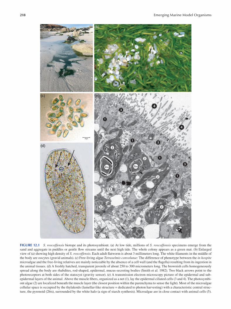
218 Emerging Marine Model Organisms
FIGURE 12.1 S. roscoffensis biotope and its photosymbiont. (a) At low tide, millions of S. roscoffensis specimens emerge from the
sand and aggregate in puddles or gentle flow streams until the next high tide. The whole colony appears as a green mat. (b) Enlarged
view of (a) showing high density of S. roscoffensis. Each adult flatworm is about 3 millimeters long. The white filaments in the middle of
the body are oocytes (gravid animals). (c) Free-living algae Tetraselmis convolutae: The difference of phenotype between the in hospite microalgae and the free-living relatives are mainly noticeable by the absence of a cell wall (and the flagella) resulting from its ingestion in
the animal tissues. (d) A freshly hatched, transparent juvenile of about 250 to 300 micrometers long. The brownish cells homogeneously
spread along the body are rhabdites, rod-shaped, epidermal, mucus-secreting bodies (Smith et al. 1982). Two black arrows point to the
photoreceptors at both sides of the statocyst (gravity sensor). (e) A transmission electron microscopy picture of the epidermal and sub
epidermal layers of the animal. Above the muscle fibers, organized as a net (1), lay the epidermal ciliated cells (3 and 4). The photosymbi
ont algae (2) are localized beneath the muscle layer (the closest position within the parenchyma to sense the light). Most of the microalgae
cellular space is occupied by the thylakoids (lamellar-like structure = dedicated to photon harvesting) with a characteristic central struc
ture, the pyrenoid (2bis), surrounded by the white halo (a sign of starch synthesis). Microalgae are in close contact with animal cells (5).
219 Symsagittifera roscoffensis as a Model in Biology
12.2 HISTORY OF THE MODEL AND GEOGRAPHICAL LOCATION
In the first publications, addressing the nature and origin
of the “green bodies” conferring the animals’ green color
(Geddes 1879) and the intriguing simplicity of the body plan
( Delage 1886 ), S. roscoffensis was fi rst mistakenly referred
to as Convoluta schultzii, a phenotypically similar species
previously described from the Adriatic Sea. An accurate
taxonomic description was performed by Ludwig von Graff,
hosted in a marine biological laboratory outpost on the coasts
of North Brittany, France, now called the Station Biologique de Roscoff. As a tribute to the spirit of hospitality associated
with facilities provided for exploration and experimentation
of the surrounding marine environment, von Graff named
this species Convoluta roscoffensis (von Graff 1891). Since
then, colonies of billions of individuals have been observed
on sandy beaches, distributed all along the Atlantic coast of
Europe, from Wales to Portugal. The in hospite enigmatic
green cells in the original description were fi rst described
as chloroplasts vertically transferred as colorless leuco
plasts (Graff and Haberlandt 1891). They were later iso
lated and identified as free-living quadri-fl agellate green
microalgae (Gamble and Keeble 1904), known today as
Tetraselmis convolutae (Figure 12.1c), and formerly named
Platymonas convolutae (Parke and Manton 1967). Revisited
with molecular taxonomy tools (Kostenko and Mamkaev
1990 ), Convoluta roscoffensis was renamed Symsagittifera roscoffensis. Initially positioned inside the Platyhelminthes
phylum as an acoel turbellarian, this species is now a mem
ber of the phylum Xenacoelomorpha (Philippe et al. 2011),
whose critically—and currently unresolved—phylogenetic
position in the animal tree of life is discussed further.
S. roscoffensis has initially been used in a wide range
of studies as a model for deciphering the mechanisms of
the setting up, specificity and trophic relationship of this
photosymbiosis in the intertidal zone. Gravid adult S. roscoffensis lay a translucid cocoon with embryos that
develop to the aposymbiotic juvenile stage within four
to five days (Figure 12.1d). If juveniles, once outside the
cocoon, fail to ingest the microalgae, they do not survive to
maturity, indicating that this association is obligate, with
the animal feeding on photosynthates transferred from
the photosymbiont (Keeble 1907). The aposymbiotic S. roscoffensis juvenile specifically incorporates but do not
digest some Tetraselmis convolutae. These microalgae, in
comparison to other closely related species (T. chui/subcoriformis/suecica), exhibit a special mode of division,
whereby daughter cells stay in pairs in the parent theca
for a much longer period, a factor favoring ingestion by
the “benthic” juvenile acoel. The in hospite microalgae are
taken up into the digestive syncytium and undergo morpho
logical alterations compared to the free-living state, losing
their theca (cell wall), eyespot and flagella but retaining
an imposing chloroplast and a specific shape with fi nger
like processes (Oshman 1966; Figure 12.1e). This suggests
that microalgal cellular processes leading to high levels
of energy consumption are drastically reduced in favor of
increasing photosynthesis and production of organic mol
ecules. Mannitol and starch (visible as grains in the chlo
roplast—Figure 12.1e) are the major carbohydrates in both
free-living and in hospite microalgae (Gooday 1970). The
photosynthetically fixed carbon, moving from the micro-
algae to the animal are mostly amino acids (Muscatine
1974). The nitrogen source for the in hospite algae (i.e. for
amino acid synthesis) is ammonia stemming from the ani
mal’s uric acid catabolism (Boyle 1975). Both adult and
aposymbiotic juvenile worms produce nitrogen waste (i.e.
uric acid/ammonia) that is recycled by the algae for protein
synthesis. In juveniles, uric acid crystals accumulate until
photosynthesis sets in, then decline once photosynthesis is
fully operational (Douglas 1983a).
According to the literature (Oshman 1966; Nozawa et al.
1972; Muscatine et al. 1974; Meyer et al. 1979), microalgal
photosynthetic activity provides all of the energy and nutri
ents (proteins, polysaccharides, lipids) for feeding the worm.
However, strict photo-autotrophy has never been formally
demonstrated for this association, and one cannot rule out
a mixotrophic regime: S. roscoffensis could indeed take
up some additional organic molecules released by benthic
organisms, including the environmental microbiome.
The paucity of data describing the trophic relationship
between S. roscoffensis and T. convolutae prevents one from
assigning a mutualistic status between these organisms, with
the idea of a reciprocal benefit and egalitarian partnership,
as has often been claimed. Controversially, recent surveys
on photosynthetic endosymbiosis rather suggest that micro-
algae are exploited by their host (Kiers and West 2016; Lowe
et al. 2016 ).
The S. roscoffensis biotope is localized within the upper
sandy part of the intertidal zone. During high tide, ani
mals live inside the interstitial sandy net, but as soon as
the tide goes out (uncovers the sand) and until it comes in
again, the animals are exposed to the sunlight in seepages
or pools of seawater.
12.3 LIFE CYCLE AND REPRODUCTION
Exploring the diversity and complexity of body plans and
their evolutionary and developmental basis requires that the
entire life cycle of a species be accessible, from the freshly
fertilized oocyte to the gravid reproducer. Controlling all the
developmental steps of a species in captivity is essential to
undertake necessary experimental steps, including genetic
analysis and genome editing. An often-ignored obstacle is
a non-negligible investment in time and expenses, a suite of
trials, errors and chance findings that slow down access to
many crucial stages of ontogenesis.
12.3.1 REPRODUCTIVE ORGANS
Acoels are hermaphroditic and reproduce by internal fertil
ization. Sperm cells and eggs develop from neoblast-derived
progenitors which divide and mature in the parenchyma
in an anterior–posterior gradient (Figure 12.2a, b). Figure
12.2c shows V-shaped bundles of sperm (“sperm tracts”),
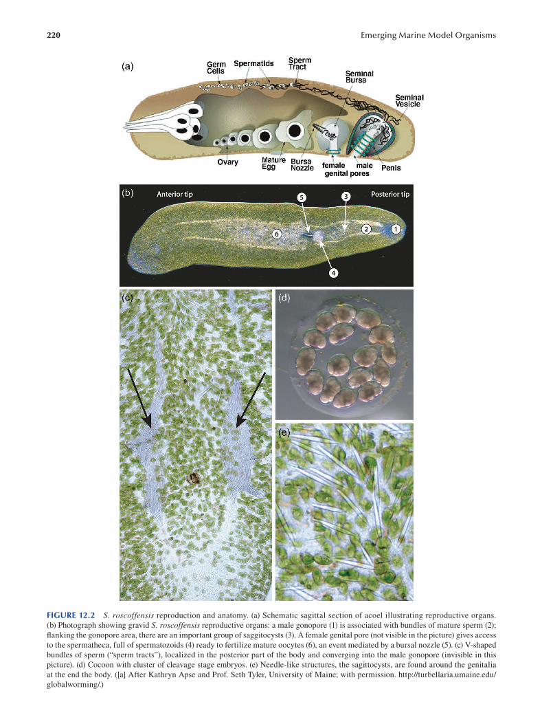
220 Emerging Marine Model Organisms
FIGURE 12.2 S. roscoffensis reproduction and anatomy. (a) Schematic sagittal section of acoel illustrating reproductive organs.
(b) Photograph showing gravid S. roscoffensis reproductive organs: a male gonopore (1) is associated with bundles of mature sperm (2);
flanking the gonopore area, there are an important group of saggitocysts (3). A female genital pore (not visible in the picture) gives access
to the spermatheca, full of spermatozoids (4) ready to fertilize mature oocytes (6), an event mediated by a bursal nozzle (5). (c) V-shaped
bundles of sperm (“sperm tracts”), localized in the posterior part of the body and converging into the male gonopore (invisible in this
picture). (d) Cocoon with cluster of cleavage stage embryos. (e) Needle-like structures, the sagittocysts, are found around the genitalia
at the end the body. ([a] After Kathryn Apse and Prof. Seth Tyler, University of Maine; with permission. http://turbellaria.umaine.edu/
globalworming/.)
221 Symsagittifera roscoffensis as a Model in Biology
localized in the posterior part of the body and converging
onto the male gonopore. Fertilization is mutual, and sperm
are transferred into the seminal bursa and stored there until
the eggs are ready to be fertilized (Figure 12.2a, b). Acoel
egg and sperm morphologies vary among species, and their
characteristics have been used for taxonomic classifi cation
(e.g. Achatz et al. 2013). Their copulatory organs are well
developed and also show great morphological variety across
different taxa. The members of the family Sagittiferidae, for
example, develop an antrum that is turned inside out, and
the bursa of many sagittiferid species lacks a muscular lin
ing (Kostenko and Mamkaev 1990). In general, the copula
tory apparatus of Sagittiferidae is considered a simplifi ed
version when compared to those of other families, such as
Convolutidae ( Zabotin and Golubev 2014).
Most species release the fertilized eggs through the
mouth. A few species release eggs through the female
genital pore (in those species that have this structure), but
all species release the sperm through the male gonopores
(Figure 12.2a, b). Genital pores in Acoela are by no means
simple structures but have specific associated muscle sys
tems. Symsagittifera roscoffensis has both male and female
genital pores. The female genital pore lies in much closer
proximity to the male pore than to the mouth, namely at
70% of the anterior–posterior axis, while the male genital
pore is located at the 90% position (Semmler et al. 2008).
The male copulatory organ presents a complex associated
musculature. In the position where it is located, the regular
grid of circular and longitudinal muscles of the body wall is
disrupted, as also happens in the area of the female genital
pore. The bursal nozzle is composed of a sclerotized lamel
late stack of cells, forming a tubule. This tubiform structure
on the seminal bursa is believed to behave like a sperm duct,
through which allosperm are transported to the oocytes
( Figure 12.2a , b ).
In addition to the copulatory organs themselves, certain
structures of yet-unknown function are clustered around the
male gonopore. Called saggitocysts, these have a needle-like
shape with a clear muscle mantle that wraps around an inte
rior protusible filament, being located below the body’s mus
cular grid (Figure 12.2d). Some authors have speculated that
the needles might be released and be functionally relevant
during copulation (e.g. Yamasu 1991).
12.3.2 EGG DEPOSITION
In the natural environment, S. roscoffensis is not gravid
from July to September and usually reproduces from
October to June. In the lab, each gravid adult (Figure 12.2b)
maintained in filtered or artificial sea-water spontaneously
lays embryos. Embryos are surrounded by a viscous mucous layer, a cocoon or capsule (Figure 12.2e). The lack of extra
cellular coats around oocytes prior to capsule formation is
functionally very significant, since it allows the incorpora
tion of multiple cells per capsule (Shinn 1993). Once the
cocoon with a diameter of approximately 750 micrometers is
finished, the adult deposits the eggs inside it. The number of
eggs inside each cocoon can reach a maximum of 30. After
four to five days of development, embryos become actively
moving transparent juvenile flatworms, approximately 250
micrometers long (Figure 12.1d). After some hours, the
juveniles hatch from the cocoon. The absence of microal
gae in the juvenile tissues indicates that the transmission
of the microalgae is not vertical (i.e. transmission through
the oocytes) but horizontal: the free-living microalgae live
in the sand and seawater of flatworm’s habitat. In the lab,
without providing the free-living algae, the juvenile reared
in sterile seawater do not survive more than 10 to 15 days,
indicating that this partnership is obligatory with respect to
the animal.
12.4 ANATOMY
12.4.1 GENERAL ARCHITECTURE OF CELLS AND TISSUES
As a member of the clade Acoelomorpha, S. roscoffensis lacks a body cavity. A body wall, consisting of processes
of epidermal cells and muscle cells, encloses a solid paren
chyma whose cells serve the digestion and distribution of
nutrients. Embedded in the parenchyma are the nervous sys
tem, a variety of glands and the reproductive organs (Ehlers
1985; Rieger et al. 1991).
A fundamental aspect of acoelomorph cellular architec
ture is the highly branched nature of virtually all cell types.
Cells possess a cell body, formed by the nucleus surrounded
by scant cytoplasm, and one or (more often) multiple pro
cesses which emerge from the cell body (Ehlers 1985;
Rieger et al. 1991; Figure 12.3a, b). Processes display a great
variety of shapes depending on the type of cell considered.
There is the main, or “functional” process(es), next to one or
more leaf-like ensheathing processes that many cells project
around neighboring structures. Epidermal cells, for exam
ple, emit their one “connecting” process radially toward the
periphery, where it spreads out to form a large (compared
to the size of the cell body), flattened layer that displays
the complex ultrastructural features, such as microvilli and
cilia, intercellular junctional complexes and epitheliosomes
(Rieger et al. 1991; Lundin 1997; Figure 12.3b, c and see
subsequently). Additional branched and variably shaped
processes of the epidermal cell body project horizontally
and intermingle with peripheral nerves, muscle fi bers and
parenchymal cells (Figure 12.3b, c). Similar to epidermal
cells, muscle cells give rise to connecting processes which
branch out into long, slender fi bers (myofibers) that contain
contractile actin-myosin fi laments (myofi laments; Figure
12.3b, c). Many cells, including muscle and glands, possess
a third type of thin, cylindrical process that enters the neuro
pil of the central nervous system (see subsequently).
Their branched anatomy implies that the cell bodies of
epidermal cells or muscle cells (and other cell types) are
located at a distance from their “functional parts”, that is,
the myofibers or epithelial processes forming the body wall.
Cell bodies are embedded in the parenchyma, where they are
arranged as an irregular layer (“cell body domain”) around
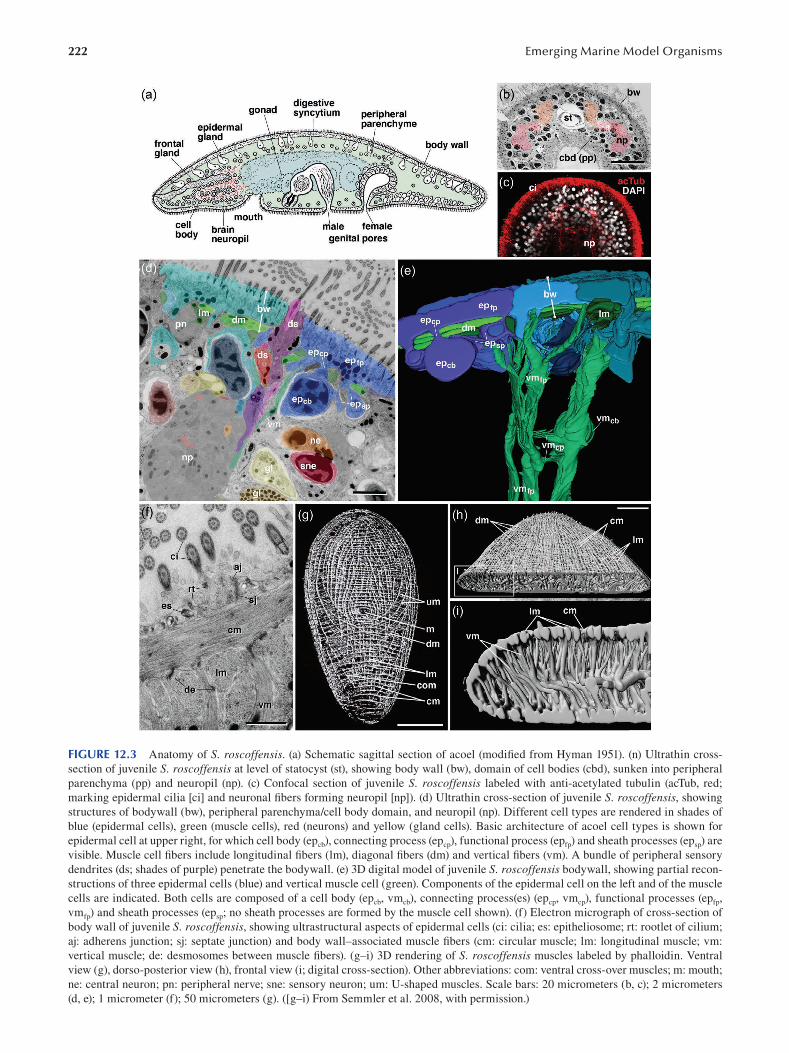
222 Emerging Marine Model Organisms
FIGURE 12.3 Anatomy of S. roscoffensis. (a) Schematic sagittal section of acoel (modified from Hyman 1951). (n) Ultrathin cross-
section of juvenile S. roscoffensis at level of statocyst (st), showing body wall (bw), domain of cell bodies (cbd), sunken into peripheral
parenchyma (pp) and neuropil (np). (c) Confocal section of juvenile S. roscoffensis labeled with anti-acetylated tubulin (acTub, red;
marking epidermal cilia [ci] and neuronal fi bers forming neuropil [np]). (d) Ultrathin cross-section of juvenile S. roscoffensis , showing
structures of bodywall (bw), peripheral parenchyma/cell body domain, and neuropil (np). Different cell types are rendered in shades of
blue (epidermal cells), green (muscle cells), red (neurons) and yellow (gland cells). Basic architecture of acoel cell types is shown for
epidermal cell at upper right, for which cell body (epcb), connecting process (ep cp), functional process (epfp) and sheath processes (ep sp ) are
visible. Muscle cell fi bers include longitudinal fi bers (lm), diagonal fi bers (dm) and vertical fi bers (vm). A bundle of peripheral sensory
dendrites (ds; shades of purple) penetrate the bodywall. (e) 3D digital model of juvenile S. roscoffensis bodywall, showing partial recon
structions of three epidermal cells (blue) and vertical muscle cell (green). Components of the epidermal cell on the left and of the muscle
cells are indicated. Both cells are composed of a cell body (epcb , vmcb), connecting process(es) (epcp , vmcp), functional processes (epfp,
vmfp) and sheath processes (ep sp; no sheath processes are formed by the muscle cell shown). (f) Electron micrograph of cross-section of
body wall of juvenile S. roscoffensis, showing ultrastructural aspects of epidermal cells (ci: cilia; es: epitheliosome; rt: rootlet of cilium;
aj: adherens junction; sj: septate junction) and body wall–associated muscle fibers (cm: circular muscle; lm: longitudinal muscle; vm:
vertical muscle; de: desmosomes between muscle fibers). (g–i) 3D rendering of S. roscoffensis muscles labeled by phalloidin. Ventral
view (g), dorso-posterior view (h), frontal view (i; digital cross-section). Other abbreviations: com: ventral cross-over muscles; m: mouth;
ne: central neuron; pn: peripheral nerve; sne: sensory neuron; um: U-shaped muscles. Scale bars: 20 micrometers (b, c); 2 micrometers
(d, e); 1 micrometer (f); 50 micrometers (g). ([g–i) From Semmler et al. 2008, with permission.)
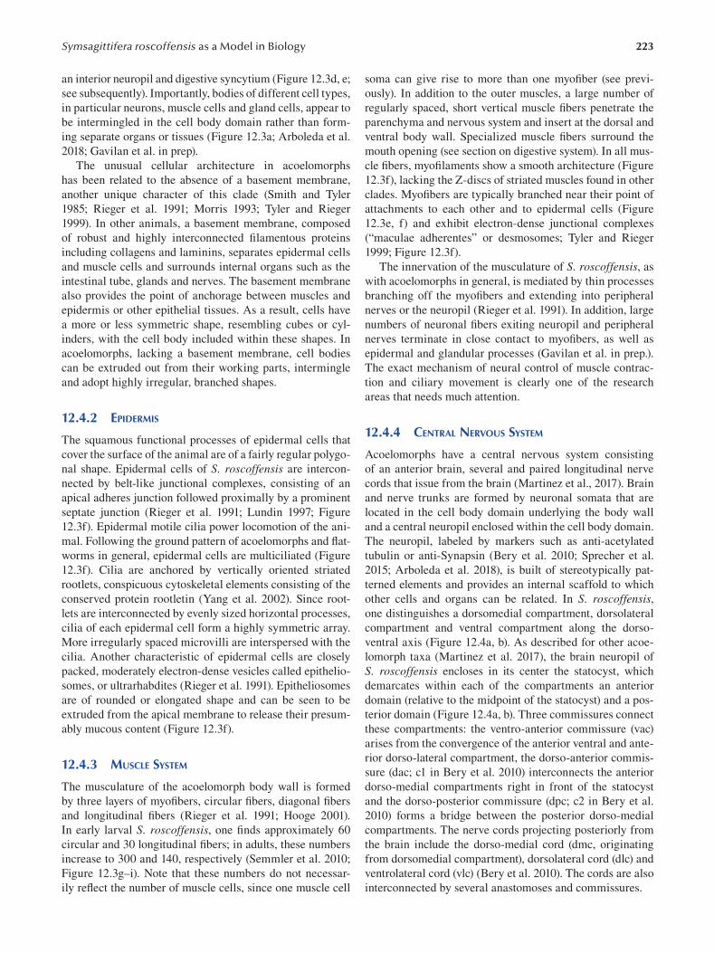
223 Symsagittifera roscoffensis as a Model in Biology
an interior neuropil and digestive syncytium (Figure 12.3d, e;
see subsequently). Importantly, bodies of different cell types,
in particular neurons, muscle cells and gland cells, appear to
be intermingled in the cell body domain rather than form
ing separate organs or tissues (Figure 12.3a ; Arboleda et al.
2018; Gavilan et al. in prep).
The unusual cellular architecture in acoelomorphs
has been related to the absence of a basement membrane,
another unique character of this clade (Smith and Tyler
1985; Rieger et al. 1991; Morris 1993; Tyler and Rieger
1999). In other animals, a basement membrane, composed
of robust and highly interconnected fi lamentous proteins
including collagens and laminins, separates epidermal cells
and muscle cells and surrounds internal organs such as the
intestinal tube, glands and nerves. The basement membrane
also provides the point of anchorage between muscles and
epidermis or other epithelial tissues. As a result, cells have
a more or less symmetric shape, resembling cubes or cyl
inders, with the cell body included within these shapes. In
acoelomorphs, lacking a basement membrane, cell bodies
can be extruded out from their working parts, intermingle
and adopt highly irregular, branched shapes.
12.4.2 EPIDERMIS
The squamous functional processes of epidermal cells that
cover the surface of the animal are of a fairly regular polygo
nal shape. Epidermal cells of S. roscoffensis are intercon
nected by belt-like junctional complexes, consisting of an
apical adheres junction followed proximally by a prominent
septate junction (Rieger et al. 1991; Lundin 1997; Figure
12.3f). Epidermal motile cilia power locomotion of the ani
mal. Following the ground pattern of acoelomorphs and fl at-
worms in general, epidermal cells are multiciliated (Figure
12.3f). Cilia are anchored by vertically oriented striated
rootlets, conspicuous cytoskeletal elements consisting of the
conserved protein rootletin (Yang et al. 2002). Since root
lets are interconnected by evenly sized horizontal processes,
cilia of each epidermal cell form a highly symmetric array.
More irregularly spaced microvilli are interspersed with the
cilia. Another characteristic of epidermal cells are closely
packed, moderately electron-dense vesicles called epithelio
somes, or ultrarhabdites (Rieger et al. 1991). Epitheliosomes
are of rounded or elongated shape and can be seen to be
extruded from the apical membrane to release their presum
ably mucous content (Figure 12.3f).
12.4.3 MUSCLE SYSTEM
The musculature of the acoelomorph body wall is formed
by three layers of myofibers, circular fibers, diagonal fi bers
and longitudinal fibers (Rieger et al. 1991; Hooge 2001).
In early larval S. roscoffensis, one finds approximately 60
circular and 30 longitudinal fi bers; in adults, these numbers
increase to 300 and 140, respectively (Semmler et al. 2010;
Figure 12.3g–i). Note that these numbers do not necessar
ily refl ect the number of muscle cells, since one muscle cell
soma can give rise to more than one myofiber (see previ
ously). In addition to the outer muscles, a large number of
regularly spaced, short vertical muscle fibers penetrate the
parenchyma and nervous system and insert at the dorsal and
ventral body wall. Specialized muscle fibers surround the
mouth opening (see section on digestive system). In all mus
cle fi bers, myofilaments show a smooth architecture (Figure
12.3f), lacking the Z-discs of striated muscles found in other
clades. Myofibers are typically branched near their point of
attachments to each other and to epidermal cells (Figure
12.3e, f) and exhibit electron-dense junctional complexes
(“maculae adherentes” or desmosomes; Tyler and Rieger
1999 ; Figure 12.3f ).
The innervation of the musculature of S. roscoffensis , as
with acoelomorphs in general, is mediated by thin processes
branching off the myofibers and extending into peripheral
nerves or the neuropil (Rieger et al. 1991). In addition, large
numbers of neuronal fibers exiting neuropil and peripheral
nerves terminate in close contact to myofibers, as well as
epidermal and glandular processes (Gavilan et al. in prep.).
The exact mechanism of neural control of muscle contrac
tion and ciliary movement is clearly one of the research
areas that needs much attention.
12.4.4 CENTRAL NERVOUS SYSTEM
Acoelomorphs have a central nervous system consisting
of an anterior brain, several and paired longitudinal nerve
cords that issue from the brain (Martinez et al., 2017). Brain
and nerve trunks are formed by neuronal somata that are
located in the cell body domain underlying the body wall
and a central neuropil enclosed within the cell body domain.
The neuropil, labeled by markers such as anti-acetylated
tubulin or anti-Synapsin (Bery et al. 2010; Sprecher et al.
2015; Arboleda et al. 2018), is built of stereotypically pat
terned elements and provides an internal scaffold to which
other cells and organs can be related. In S. roscoffensis, one distinguishes a dorsomedial compartment, dorsolateral
compartment and ventral compartment along the dorso
ventral axis (Figure 12.4a, b). As described for other acoe
lomorph taxa (Martinez et al. 2017), the brain neuropil of
S. roscoffensis encloses in its center the statocyst, which
demarcates within each of the compartments an anterior
domain (relative to the midpoint of the statocyst) and a pos
terior domain (Figure 12.4a, b). Three commissures connect
these compartments: the ventro-anterior commissure (vac)
arises from the convergence of the anterior ventral and ante
rior dorso-lateral compartment, the dorso-anterior commis
sure (dac; c1 in Bery et al. 2010) interconnects the anterior
dorso-medial compartments right in front of the statocyst
and the dorso-posterior commissure (dpc; c2 in Bery et al.
2010) forms a bridge between the posterior dorso-medial
compartments. The nerve cords projecting posteriorly from
the brain include the dorso-medial cord (dmc, originating
from dorsomedial compartment), dorsolateral cord (dlc) and
ventrolateral cord (vlc) (Bery et al. 2010). The cords are also
interconnected by several anastomoses and commissures.
224 Emerging Marine Model Organisms
FIGURE 12.4 Anatomy of S. roscoffensis. (a, b) Central nervous system and neuropil. (a) A confocal section of adult S. roscoffensis. Muscles are labeled by phalloidin (green), central neuropil by an antibody against synapsin (red). (b) A 3D digital model of neuropil
with different neuropil domains rendered in different colors. Neuropil domains visible in the dorsal view shown include dorso-anterior
compartment (da), dorso-intermediate compartment (di; flanking statocyst shaded gray), dorso-posterior compartment (dp), and ventro
anterior compartment (va). The three brain commissures connecting right and left compartments are the ventro-anterior (ring) com
missure (vac), dorso-anterior commissure (dac) and dorso-posterior commissure (dpc). Three pairs of nerve cords exit the brain: the
dorso-medial cord (dmc), dorso-lateral cord (dlc) and ventro-lateral cord (vlc). (c) Schematic section of S. roscoffensis, illustrating the
processes of neurons (red), sensory neurons (purple) and gland cells (yellow) in relationship to the body wall (bw), neuropil (np) and cell
body domain (cbd). Thick black arrows symbolize synaptic interaction between central processes of the cells shown and elements of the
neuropil. (d–g) Cytological details of central neurons. (d) Cell bodies surrounding neuropil (np; shaded blue). Three cell bodies belong to
central neurons (ne; rendered in shades of red). Central neurons emit processes into neuropil. In some cases, processes exhibit particular
sheath-like shapes (“lamellar processes”), aside from the cylindrical processes typical for neurons in general. (e) 3D digital model (lateral
view) of four representative partially reconstructed central neurons exhibiting different shapes. (f, g) Electron microscopic sections of
neuropil at high magnification. Note the high proportion of axons with dense core vesicles (dcv). Vertical muscle fibers (vm) penetrate
neuropil and could receive extra-synaptic input from these axons. (g) An example of synaptic connection between large presynaptic ele
ment (pre) with small synaptic vesicles (ssv) and two small postsynaptic elements (post). (h–m) Cytological details of sensory neurons.
As shown in (h), cell bodies of sensory neurons (sne) frequently lie adjacent to the neuropil and emit cylindrical or lamellar processes into
the neuropil. (i) Shapes of ciliated sensory neurons (lateral view). (j) Bundle of four sensory processes linking neuropil to the body wall.
(k–m) Three different types of frequently seen sensory endings, a collared receptor (k), non-collared receptor (l) and non-ciliated recep
tor (m). (n–p) Details of gland cell structure. In (n), cell bodies of three gland cells (rendered in shades of yellow) surround the central
neuropil (np). One gland cell emits a central process into the neuropil. Digital 3D models shown in (o) illustrate representative gland cells
(lateral view). (p) Section of body wall with endings of two different types of gland cells, a mucus gland cell with large electron-lucent
vesicles (glmu) and a rhabdoid gland cell (glrh) with elongated, electron-dense inclusions. Scale bars: 40 micrometers (a); 2 micrometers
(d, h–p); 0.5 micrometers (f, g). (From Sprecher et al. 2015, with permission.)
225 Symsagittifera roscoffensis as a Model in Biology
Neuronal cell bodies (somata) of the S. roscoffensis ner
vous system are small and have a round heterochromatin-
rich nucleus (Figure 12.4d). Based on light-microscopic
analysis, the larval brain contains an estimated 800 somata
overall, but more precise numbers have to await serial EM
analysis, since somata of neurons located in the diffuse cell
body domain that surrounds the neuropil cannot be told
apart with certainty from cell bodies of muscle cells or gland
cells. EM reconstruction shows that many neurons are bipo
lar, extending an anterior process that in many cases may
reach the epidermal surface to end as a sensory receptor, and
one or more posterior or central process(es) that reaches into
the neuropil, where it shows a modest amount of branching
(Figure 12.4d, e). Along with neuronal processes, central
extensions of muscle cells and gland cells also form part of
the neuropil (Figure 12.4c).
Based on the types of vesicles they contain, neuronal
processes of acoelomorphs were divided into four classes
(Bedini and Lanfranchi 1991; Bery et al. 2010), including
fibers with small clear vesicles (20–40 nm), which are asso
ciated with the “classical” transmitters acetylcholine, GABA
or glutamate, and dense vesicles (70–90 nm), which resemble
the dense core vesicles that, in vertebrates and many inver
tebrates alike, have been described to contain neuropeptides
(Figure 12.4f, g). As in these other species, many neurons of
S. roscoffensis have both types of vesicles. What stands out,
however, is the large proportion of neuronal processes with
dense vesicles, a finding that matches descriptions of light
microscopic studies detecting peptide transmitters in large
neuron populations in acoelomorphs (Reuter et al. 2001).
Aside from small clear vesicles and dense vesicles, two other
types with so far unknown significance and neurotransmit
ter content were described for acoelomorphs: another type of
“dense core vesicles” (60–120 nm), containing small, dense
centers surrounded by a light halo (not to be confused with
the peptide-containing dense core vesicles in vertebrates or
insects) and large irregularly shaped clear vesicles (20–400
nm; Bedini and Lanfranchi 1991).
Neuronal processes containing small clear vesicles in con
junction with membrane densities can be recognized as syn
apses (Bedini and Lanfranchi 1991; Bery et al. 2010; Figure
12.4g). However, thus defined synapses are relatively few in
number, at least in the larval brain, and it is very possible that
neural transmission relies heavily on extra-synaptic transmit
ter release. This is made all the more likely looking at the
processes with dense vesicles, which fi ll the entire length of
neurons, including the cell body, and peripheral processes.
Peptide release from dense core vesicles in vertebrates has
been definitively shown to occur extrasynaptically (“volume
release”) in many instances (Fuxe et al. 2007).
12.4.5 PERIPHERAL NERVOUS SYSTEM
AND SENSORY RECEPTORS
The peripheral nervous system consists of sensory recep
tors integrated in the body wall and an anastomosing mesh
work of thin “nerves” that contain fibers formed by sensory
receptors, muscle cells and gland cells, as well as cells effec
tor cells (“motor neurons”) that, aside from processes in the
neuropil, project processes through the peripheral nerves
into the periphery. Sensory neurons form part of the cell
body domain surrounding the neuropil (Figure 12.4c, h , i).
Their peripheral dendrites project into the body wall (Figure
12.4j), where they terminate as conspicuous elements that
have been described for many flatworms, including acoels
(Rieger et al. 1991). Unlike epidermal cells, sensory recep
tors typically contain a single cilium, aside from other apical
membrane specializations. Based on these specializations,
one distinguishes collared receptors from non-collared
receptors (Bedini et al. 1973; Todt and Tyler 2006 ). In the
former, a central cilium is surrounded by a ring (collar) of
long, stout microvilli; this collar is lacking in the latter class.
Both classes are further subdivided into several types (Todt
and Tyler 2006). In S. roscoffensis, three types of sensory
receptors have described, including non-collared receptors
with a hollow ciliary rootlet containing a granulated core
(Type 3 of Todt and Tyler 2006; Figure 12.4l), collared
receptors with rootlets (Type 4) and collared receptors with
granular body (Type 5; Figure 12.4k). Another frequently
encountered type of presumed receptors are non-ciliated
endings (Figure 12.4m). Receptors are distributed in charac
teristic patterns all at different positions (Bery et al. 2010).
Nothing is known about the specific modalities and func
tions of sensory receptors.
Two other sensory elements, the statocyst and eyes, are
surrounded by neuropil and thereby form part of the CNS
(Figure 12.1c). The statocyst, thought to sense gravity, is
formed by a capsule of two parietal cells enclosing a cav
ity that houses a specialized statolith cell (lithocyte; Ferrero
1973; Ehlers 1991). A small group of specialized muscle
cells inserts at the capsule. No recognizable sensory neu
ronal structures are associated with the statocyst, and it has
been proposed that gravity-induced displacements of the
statolith could inform the CNS by affecting the muscles by
which the statocyst is suspended.
The eye of convolutid acoels, including S. roscoffensis , is
embedded into the brain on either side of the statocyst. The
eye consists of a pigment cell with electron-dense granules
and crystalline inclusions (“platelets”) that may act as refl ec
tors; enclosed by the pigment cell are two to three receptor
cells with axons connecting to the neuropil (Yamasu 1991).
Unlike most photoreceptors described for other taxa, acoel
photoreceptors cells lack conspicuous microvilli or cilia.
12.4.6 GLANDULAR SYSTEM
Glands are unicellular, consisting of individual gland cells
that constitute a major part of the acoelomorph body in terms
of number and function. As stated for epidermal and muscle
cells, gland cells consist of a cell body that forms part of
the internal cell body domain and one or more elongated
processes (“gland necks”) that project peripherally and open
to the outside (Figure 12.4c, n , o). Certain clusters of gland
cells, located posteriorly of the brain, project their long
226
necks forward through the neuropil and open at the anterior
tip of the body, some of them in an acoelomorph-charac
teristic pore, the “frontal pore” (Pedersen 1965; Smith and
Tyler 1986; Klauser et al. 1986; Ehlers 1992; Figure 12.3a).
Cell bodies and gland necks contain secretory vesicles of
different shape and texture by which gland cells have been
divided into different classes, as summarized in the follow
ing. Gland necks carry a characteristic array of microtubules
around their periphery. In addition to secretory gland necks,
many gland cells appear to have central processes that invade
peripheral nerves or the neuropil. These processes, like the
ones formed by myofibers (see previously), may mediate the
connection between nerve impulses and secretory function
( Figure 12.4c , n ).
Functionally and biochemically, acoelomorph gland
secretions include mucus (mucopolysaccharides) that serves
for locomotion, attachment and protection, as well as protein
aceous enzymes for digestion and degradation of macromol
ecules. Mucus-producing glands, called cyanophilic glands
in the classical light microscopy literature, are structurally
associated with densely packed, electron-lucent vesicles with
a rounded or oval shape (Pedersen 1965; Rieger et al. 1991).
Gland cells of this type open in the frontal pore but also occur
all over the body surface of S. roscoffensis. Aside from gland
cells with electron-lucent inclusions, a variety of cells with
electron-dense vesicles of different sizes and shapes have
been described for the acoelomorphs (Smith and Tyler 1986;
Klauser et al. 1986; Todt 2009). These have been given dif
ferent names (e.g. “ellipsoid” glands, “target glands”, “alcian
blue-positive rhabdoid glands”) but cannot be assigned to
specific functions. In the larva of S. roscoffensis, we detect
glands with large, electron-lucent inclusions (mucus glands;
Figure 12.4p) all over the body but preferentially anteriorly
and ventrally; in addition, there are three clearly distinguish
able types of gland cells with electron-dense inclusions
(Gavilan et al. in prep):
1. A rare type we call a rhabdoid gland cell, with elon
gated inclusions of approximately 500 nm length
and 100 nm diameter (Figure 12.4p).
2. Glands with pleomorphic vesicles: Inclusions are
more rounded than those of rhabdoid glands and
possess different diameters and electron densities
(Figure 12.4n , bottom). Rhabdoid glands and pleo
morphic glands are located ventro-anteriorly.
3. Glands with mixed electron-dense and electron-
lucent vesicles: These are more numerous and ven
tro-laterally overlie the ventral nerve cord.
12.4.7 PARENCHYMA AND DIGESTIVE SYNCYTIUM
The name-giving feature of acoels is their lack of a gut cavity.
The interior of the animal is filled with a solid parenchyma
that is divided into a central and peripheral domain (Smith
and Tyler 1985; Gavilán et al. 2019). The central parenchyma
is typically a syncytium (“digestive syncytium”) formed by
the merger of multiple endodermal cells; in the larva of S.
Emerging Marine Model Organisms
roscoffensis, the digestive syncytium contains an estimated
6–10 nuclei (Gavilan et al. in prep.). At a mid-ventral position,
the digestive syncytium is in contact with the interior through
a pore (“mouth”) in the epidermal covering. A pharynx, in
the shape of an invagination of the ventral epidermis sur
rounded by specialized muscle and neural elements, is absent
(Todt 2009; Semmler et al. 2010). Only a slender muscle ring
from which a few fibers radiate outward marks the mouth.
In addition, several ventral longitudinal muscle fi bers cross
over the midline right behind the mouth, giving rise to the
U-shaped muscles that are the characteristic of the derived
acoel clade of “Crucimusculata” to which S. roscoffensis belongs. It is thought that contraction of these fi bers tilts the
mouth forward, facilitating the uptake of food stuff.
The digestive syncytium is filled with a great diversity
of organelles related to phagocytosis and digestion. In S. roscoffensis, symbiotic algae of the genus Tetraselmys are
taken into the syncytium, where they lose part of their cell
wall. The digestive syncytium emits processes that reach
throughout the entire body, ensheathing (parts of) many
cell bodies in the cell body domain and wrapping around
peripheral nerves, muscle fibers and epidermal processes
(Smith and Tyler 1985; Gavilan et al. in prep). One has to
assume that this architecture enables the syncytium not only
to digest but also distribute nutrients throughout the body. In
the case of S. roscoffensis, algae ingested at the early larval
stage multiply within vacuoles of the digestive syncytium
(Oshman 1966; Douglas 1983b). In the adult, algae form a
dense layer underneath the body wall, interspersed with epi
dermal and muscle processes (Figure 12.1d). EM analysis
indicates that algae remain enclosed within the processes of
the digestive syncytium (Douglas 1983b).
The peripheral parenchyma is formed by cells called
“wrapping cells” (Smith and Tyler 1985) which are similar
in ultrastructure to the digestive syncytium. They also form
elaborate sheaths around other cells, interdigitating with pro
cesses of the digestive syncytium. It has been proposed that
wrapping cells merge with the digestive syncytium, mani
festing part of a dynamic process whereby newly generated
cells proliferated from neoblasts (see section 12.4.8) mature,
have a transient life as wrapping cells and end up as part of
the central syncytium (reviewed in Gavilán et al. 2019).
12.4.8 NEOBLASTS (STEM CELLS)
Regeneration of acoel tissues is a well-known phenomenon.
This process depends on the deployment of a pool of stem-
like cells called neoblasts that are present within parenchy
mal tissues (De Mulder et al. 2009; Srivastava et al. 2014). In
all species in which neoblasts have been mapped, these cells
are distributed in two lateral bands and mostly excluded
from the head region. Neoblasts are easily identifi able by
their intensive basophilic cytoplasm and relative scarcity
of cytoplasmic organelles (Brøndsted 1955). Neoblasts are
the only dividing cells in adult organisms, and they have the
potential to differentiate into all, or most, cell types during
regeneration (Gschwentner et al. 2001). In Symsagittifera
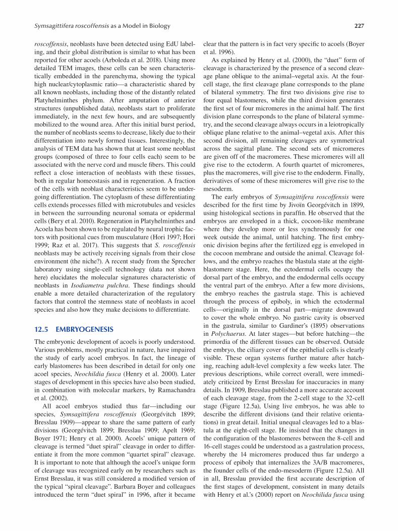
227 Symsagittifera roscoffensis as a Model in Biology
roscoffensis, neoblasts have been detected using EdU label
ing, and their global distribution is similar to what has been
reported for other acoels (Arboleda et al. 2018). Using more
detailed TEM images, these cells can be seen characteris
tically embedded in the parenchyma, showing the typical
high nuclear/cytoplasmic ratio—a characteristic shared by
all known neoblasts, including those of the distantly related
Platyhelminthes phylum. After amputation of anterior
structures (unpublished data), neoblasts start to proliferate
immediately, in the next few hours, and are subsequently
mobilized to the wound area. After this initial burst period,
the number of neoblasts seems to decrease, likely due to their
differentiation into newly formed tissues. Interestingly, the
analysis of TEM data has shown that at least some neoblast
groups (composed of three to four cells each) seem to be
associated with the nerve cord and muscle fibers. This could
reflect a close interaction of neoblasts with these tissues,
both in regular homeostasis and in regeneration. A fraction
of the cells with neoblast characteristics seem to be under
going differentiation. The cytoplasm of these differentiating
cells extends processes filled with microtubules and vesicles
in between the surrounding neuronal somata or epidermal
cells (Bery et al. 2010). Regeneration in Platyhelminthes and
Acoela has been shown to be regulated by neural trophic fac
tors with positional cues from musculature (Hori 1997; Hori
1999; Raz et al. 2017). This suggests that S. roscoffensis neoblasts may be actively receiving signals from their close
environment (the niche?). A recent study from the Sprecher
laboratory using single-cell technology (data not shown
here) elucidates the molecular signatures characteristic of
neoblasts in Isodiametra pulchra. These fi ndings should
enable a more detailed characterization of the regulatory
factors that control the stemness state of neoblasts in acoel
species and also how they make decisions to differentiate.
12.5 EMBRYOGENESIS
The embryonic development of acoels is poorly understood.
Various problems, mostly practical in nature, have impaired
the study of early acoel embryos. In fact, the lineage of
early blastomeres has been described in detail for only one
acoel species, Neochildia fusca (Henry et al. 2000). Later
stages of development in this species have also been studied,
in combination with molecular markers, by Ramachandra
et al. (2002).
All acoel embryos studied thus far—including our
species, Symsagittifera roscoffensis ( Georgévitch 1899 ;
Bresslau 1909)—appear to share the same pattern of early
divisions (Georgévitch 1899; Bresslau 1909; Apelt 1969;
Boyer 1971; Henry et al. 2000). Acoels’ unique pattern of
cleavage is termed “duet spiral” cleavage in order to differ
entiate it from the more common “quartet spiral” cleavage.
It is important to note that although the acoel’s unique form
of cleavage was recognized early on by researchers such as
Ernst Bresslau, it was still considered a modified version of
the typical “spiral cleavage”. Barbara Boyer and colleagues
introduced the term “duet spiral” in 1996, after it became
clear that the pattern is in fact very specific to acoels (Boyer
et al. 1996 ).
As explained by Henry et al. (2000), the “duet” form of
cleavage is characterized by the presence of a second cleav
age plane oblique to the animal–vegetal axis. At the four-
cell stage, the first cleavage plane corresponds to the plane
of bilateral symmetry. The first two divisions give rise to
four equal blastomeres, while the third division generates
the first set of four micromeres in the animal half. The fi rst
division plane corresponds to the plane of bilateral symme
try, and the second cleavage always occurs in a leiotropically
oblique plane relative to the animal–vegetal axis. After this
second division, all remaining cleavages are symmetrical
across the sagittal plane. The second sets of micromeres
are given off of the macromeres. These micromeres will all
give rise to the ectoderm. A fourth quartet of micromeres,
plus the macromeres, will give rise to the endoderm. Finally,
derivatives of some of these micromeres will give rise to the
mesoderm.
The early embryos of Symsagittifera roscoffensis were
described for the first time by Jivoïn Georgévitch in 1899,
using histological sections in paraffin. He observed that the
embryos are enveloped in a thick, cocoon-like membrane
where they develop more or less synchronously for one
week outside the animal, until hatching. The fi rst embry
onic division begins after the fertilized egg is enveloped in
the cocoon membrane and outside the animal. Cleavage fol
lows, and the embryo reaches the blastula state at the eight-
blastomere stage. Here, the ectodermal cells occupy the
dorsal part of the embryo, and the endodermal cells occupy
the ventral part of the embryo. After a few more divisions,
the embryo reaches the gastrula stage. This is achieved
through the process of epiboly, in which the ectodermal
cells—originally in the dorsal part—migrate downward
to cover the whole embryo. No gastric cavity is observed
in the gastrula, similar to Gardiner’s (1895) observations
in Polychaerus. At later stages—but before hatching—the
primordia of the different tissues can be observed. Outside
the embryo, the ciliary cover of the epithelial cells is clearly
visible. These organ systems further mature after hatch
ing, reaching adult-level complexity a few weeks later. The
previous descriptions, while correct overall, were immedi
ately criticized by Ernst Bresslau for inaccuracies in many
details. In 1909, Bresslau published a more accurate account
of each cleavage stage, from the 2-cell stage to the 32-cell
stage (Figure 12.5a), Using live embryos, he was able to
describe the different divisions (and their relative orienta
tions) in great detail. Initial unequal cleavages led to a blas
tula at the eight-cell stage. He insisted that the changes in
the configuration of the blastomeres between the 8-cell and
16-cell stages could be understood as a gastrulation process,
whereby the 14 micromeres produced thus far undergo a
process of epiboly that internalizes the 3A/B macromeres,
the founder cells of the endo-mesoderm (Figure 12.5a). All
in all, Bresslau provided the first accurate description of
the first stages of development, consistent in many details
with Henry et al.’s (2000) report on Neochilida fusca using
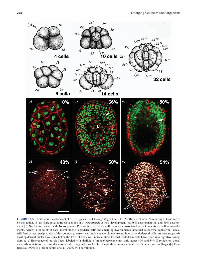
228 Emerging Marine Model Organisms
FIGURE 12.5 Embryonic development of S. roscoffensis. (a) Cleavage stages 4 cells to 32 cells, lateral view. Numbering of blastomeres
by the author. (b–d) Horizontal confocal sections of S. roscoffensis at 10% development (b), 66% development (c) and 80% develop
ment (d). Nuclei are labeled with Topro (green). Phalloidin (red) labels cell membrane associated actin filaments as well as myofi la
ments. Arrow in (c) points at basal membranes of ectoderm cells and emerging myofilaments; note that ectodermal (epidermal) nuclei
still form a layer peripherally of this boundary. Arrowhead indicates membrane around internal endodermal cells. At later stages (d),
most epidermal nuclei have sunk below the level of body wall muscle fibers (arrow); endoderm cells have fused into digestive syncy
tium. (e–g) Emergence of muscle fibers, labeled with phalloidin (orange) between embryonic stages 40% and 54%. Z-projection, dorsal
view. Abbreviations: cm: circular muscles; dm: diagonal muscles; lm: longitudinal muscles. Scale bar: 50 micrometers (b–g). ([a] From
Bresslau 1909; [e-g] from Semmler et al. 2008, with permission.)
229 Symsagittifera roscoffensis as a Model in Biology
lineage tracing. Moreover, Bresslau is the first to present a
lineage map of the Convoluta (Symsagitifera) embryo, an
impressive feat of detailed observation at the beginning of
the 20th century. Notably, the duet spiral cleavage charac
teristic of acoels is not present in members of the closely
related Nemertodermatida order (Børve and Hejnol 2014),
which exhibit a slightly different pattern of blastomere divi
sions during early embryonic development.
The embryological origin of tissues hasn’t been thor
oughly studied in S. roscoffensis. Following cleavage and
gastrulation, the embryo forms a solid mass of cells, with
an outer epithelial layer giving rise to the epidermis and an
inner mass of cells to digestive cells (Figure 12.5b), paren
chyma and musculature. It is not known whether, at this
stage, progenitors of neurons or gland cells are already part
of the inner mass or are still integrated in the epithelial outer
layer. Until about 60% of development, a regular surface
epithelium remains visible; subsequently, cell bodies of epi
dermal cells, as well as all other cells which potentially are
initially at the surface, like glands or sensory neurons, sink
inward ( Figure 12.5c , d ).
The genesis of the musculature has been observed in
detail using F-actin labeling (Semmler et al. 2008). The
process of myogenesis is very similar to that observed in
another acoels (i.e. Isodiametra pulchra: Ladurner et al.
2000 or Neochildia fusca: Ramachandra et al. 2002). The
latter study shows the initial stages of muscle formation,
probably common to many acoels, with the first signs of
musculature being myoblasts forming a thin layer under
neath the epidermis, laterally and posteriorly to the brain.
Some early muscular fibers penetrate the brain. During the
very first days of Symsagittifera embryo development, a grid
of circular and longitudinal muscles appears, with circular
muscles preceding longitudinal ones. Myogenesis in the
anterior part of the animal occurs first and then proceeds in
an anterior–posterior progression ( Figure 12.5e). Muscular
circular fibers are added by a process involving the branch
ing of previous ones (Figure 12.5f). The grid of muscles is
more regular in the dorsal part of the embryo than in the
ventral, probably due to the need to accommodate addi
tional muscles in ventral structures such as the mouth and
the copulatory organs (Figure 12.5g). The embryos hatch
with a basic grid composed of about 30 longitudinal and
60 circular muscles (Semmler et al. 2008). During the later
development, additional muscles are incorporated, includ
ing specialized muscles around the mouth and the copula
tory system, plus a whole array of transversal (dorso-ventral)
fibers. The adults have a total of about 300 circular muscles
and 140 longitudinal ones.
The embryonic origin of the brain and the neural chords
hasn’t been studied in detail, but it is assumed to occur
in early embryogenesis, based on early embryonic expres
sion (bilateral lobes) of some bHLH “neurogenic” genes
(Perea-Atienza et al. 2018). A better understanding of the
genesis of the nervous system is derived from the study
of Neochildia fusca embryos (Ramachandra et al. 2002).
These authors documented the presence in late embryos
of the brain primordia, which can be clearly distinguished
at the anterior pole of the embryo and consists of an exter
nal cortex of neuronal bodies around an internal neuropil.
Given the consistency of these observations with those of
Perea-Atienza, and with both acoels being members of the
same class, Crucimusculata, we can hypothesize that the
neurogenesis is following identical, or very similar, paths.
A more comprehensive analysis of gene expression patterns
during S. roscoffensis embryogenesis is urgently needed in
order to understand the mechanisms regulating embryonic
development and patterning.
12.6 REGENERATION
Acoel flatworms show an enormous capacity for regenera
tion. The extent of this regeneration varies from species to
species, with some even relying on regeneration for repro
duction (Sikes and Bely 2010). Investigation of the regen
erative capacity of acoels dates back to the beginning of
the 20th century, when Elsa Keil (1929) described some
histological aspects of regeneration in the acoel fl atworm
Polychaerus caudatus. Keil’s work was a revision of even
earlier data provided by Stevens and Boring (1905) and
Child (1907 ). In the 1950s and 1960s, researchers includ
ing Steinböck (1954) and Hanson (1960, 1967) undertook
a more systematic analysis of the regeneration process in
some acoel “turbellarians”, resulting in the creation of some
now-classical monographs.
One interesting aspect of acoel regeneration is that dif
ferent species have the capacity to regenerate different
bodily areas. For example, Symsagittifera roscoffensis and
Hofstenia miamia can regenerate the anterior area (Bailly et
al. 2014; Hulett et al. 2020), while Isodiametra pulchra can
regenerate the posterior area (De Mulder et al. 2009; Perea-
Atienza et al. 2013). Many other varieties of regeneration
have been described for other species (Bely and Sikes 2010).
The reasons underlying these different capacities remain
unknown.
Symsagittifera roscoffensis is a particularly interesting
system in which to study regeneration, since this species
has the capacity to regenerate the whole brain anew. This
has interesting implications for understanding the mecha
nisms involved in the regeneration of the nervous tissue. In
Symsagittifera roscoffensis, the regeneration of the brain
anatomy after amputation takes between one week and
ten days, similar to the time taken by Hofstenia miamia.
However, some additional structures, such as the statocyst,
require a few weeks for complete regeneration. The regen
erative process involves the mobilization of stem cells
(neoblasts) that begin actively proliferating in response
to amputation and subsequently concentrate in the wound
area (BG and PM, unpublished data). The active prolifera
tion of neoblasts is followed by a differentiation of mature
tissues. A clear blastemal area is missing in this process.
Regeneration follows three broad and distinct steps: (1) a
contraction of the anterior musculature immediately fol
lowing amputation; (2) a subsequent closure of the wound
230
area; (3) an extension of the three pairs of nerve cords into
the anterior domain of the animal’s body; and (4) the fi nal
connection of these nerves to form two ring-shaped, sym
metrical neuronal structures with increasing numbers of
mature neurons (i.e. the brain). Based on indirect observa
tions (see Bery et al. 2010), it has been proposed that nerve
chords and muscular fibers at the amputation site could
somehow guide the process of tissue repair. This would be
in line with indications in Hofstenia miamia that muscles
provide positional information to regenerating tissue in
acoels (Raz et al. 2017), as is also the case in platyhelminth
species. The process of regeneration in Symsagittifera roscoffensis has not been well characterized due to a
lack of studies using molecular markers. Studies of this
nature have been undertaken recently in Hofstenia miamia (Hulett et al. 2020). However, it is important to note that in
Symsagittifera, it has been possible to test the functional
reconstruction of the brain area using various behavioral
tests assessing functions such as phototaxis and geotaxis
(Sprecher et al. 2015). These behaviors, though recognized
for decades (Keeble 1910), are only now being studied
quantitatively (Nissen et al. 2015). Sprecher and colleagues
(2015) have used different paradigms to assess the behav
ior of amputated worms at different stages of recovery,
evaluating their responses to light, vibration and settling
in columns. The researchers also followed the motility of
the animals over the recovery period (Sprecher et al. 2015).
The functional assessment of brain activity was done in
parallel with a careful analysis of nervous system anatomy
by immunostaining, allowing the correlation of functional
and structural aspects of the regeneration process. This
study represents the first time that tests of this nature have
been used to understand the physiological consequences
of acoel regenerative processes (beyond the obvious char
acteristics like recovery of body movement). A striking
finding of this study is that different sensory modalities
are restored at different times. For instance, phototaxis is
restored at about 20 days post-decapitation, while geotaxis
takes approximately 50 days to be restored. The growing
recognition that Symsagittifera roscoffensis is able to fol
low more complex behaviors (Franks et al. 2016) and even
social behaviors offers further opportunities to study func
tional recovery in the nervous systems of these animals,
once considered “simple”. The use of automated tracking
systems and computer simulation of individual and collec
tive behaviors—as Franks and collaborators (2016 ) have
done—will provide us with the necessary tools to ana
lyze different aspects of the brain’s functional recovery in
detail.
12.7 PRELIMINARY GENOMIC DATA
The so-called post-genomic era has produced a flurry of papers
addressing the characterization of many animal genomes and
transcriptomes, information that allows us to trace the evolu
tionary history of animals with unprecedented detail. Among
those animals for which new information has been gathered
Emerging Marine Model Organisms
are several members of the phylum Xenacoelomorpha (an
updated list appears in: Jondelius et al. 2019).
Three acoel genomes with different degrees of com
pleteness have been produced in the last few years—
those of species Hofstenia miamia (Gehrke et al. 2019),
Praesagittifera naikaiensis (Arimoto et al. 2019) and
Symsagittifera roscoffensis ( Philippe et al. 2019). While
the first is quite complete, that of our species is only a
preliminary draft. Despite the relatively low quality
of the Symsagittifera genome (a high-quality version
is currently being generated), some basic facts can be
extracted. The first is that the genome of Symsagittifera is quite big, around 1.4 Gb, approximately half the size of
the human genome. This is supported by an independent
analysis of the genome size carried out by flow cytom
etry. This genome is much bigger than that of Hofstenia miamia, which has been reported to be 950 Mb long,
and Praesagittifera naikaiensis, which is estimated at
654 Mb. The genome of Symsagittifera is packed into
20 chromosomes of seemingly equal size (2n = 20), as
determined cytochemically using chromosomal spreads
(Moreno et al. 2009).
Briefly, in the case of Symsagittifera roscoffensis, a stan
dard fragment Illumina library was made from a pool of
symbiont-free hatchlings, which were raised in artifi cial sea
water in the presence of antibiotics. The genome fragments
were assembled with a mix of SOAPdenovo2 (–M3, –R,–
d1, –K31) and the Celera assemblers, resulting in an N50 of
2,905 bp. The introduction of PacBio sequencing method
ologies has recently allowed us to increase the N50 to above
100 kb (PM, unpublished data). Genome and transcriptome
assemblies, including the genome of Symsagittifera , have
been deposited in https://figshare.com/search, project num
ber PRJNA517079. In parallel, a transcriptome was also
sequenced from mixed-stage S. roscoffensis embryos using
standard methods.
This is an A+T-rich genome with a 36% content of G+C
and a high representation of repetitive elements and trans
posons (data not shown). Some of the transposon sequences
have been mapped to specific locations in the genome,
such as the neighborhood of the Hox genes (Moreno et
al. 2011), a particularity that would explain their disper
sion in different chromosomes by rearrangements. The
draft genome and the transcriptomes have allowed for
the exploration of gene families and their compositions.
Families such as those containing bHLH, GPCRs, Wnts or
homeobox have been explored extensively in recent years
(Perea-Atienza et al. 2015; Gavilán et al. 2016; Brauchle
et al. 2018). Strikingly, many of these sequences show spe
cific patterns of divergence with respect to the putative
orthologs in other bilaterian clades (i.e. Wnts), corroborat
ing the well-known fast rate of evolution of acoels, and in
particular Symsagittifera, genomes (Philippe et al. 2019).
Moreover, these gene family characterizations provide a
source of sequences necessary for the design of probes
used in downstream experiments by situ hybridization
(Perea-Atienza et al. 2018) or in the identifi cation of BAC
231 Symsagittifera roscoffensis as a Model in Biology
clones used in studies of chromosomal mapping (Moreno
et al. 2009).
12.8 CHALLENGING QUESTIONS FOR THE FUTURE
Some challenging questions need to be addressed in this
model. The lack of functional tools has been a hindrance
in the analysis of Symsagittifera biology from both a devel
opmental and physiological perspective. Until now, we have
relied on several molecular, anatomical and biochemical
techniques to analyze aspects of the anatomy, embryol
ogy and metabolic activity of these animals under differ
ent conditions. This has provided us with an enormous body
of knowledge, though mostly descriptive. The development
of tools for knockdown and biochemical intervention (i.e.
pharmacological agents) should be a priority in the fi eld, so
that phenomena discovered observationally can be tested
directly through experimental intervention. Specifi cally, the
following are needed:
1. A deeper understanding of the embryology of S. roscoffensis, including lineage maps and a dissec
tion of blastomere contributions (through ablation
methodologies). Furthermore, molecular markers
should be incorporated into our understanding of
embryonic regulation in S. roscoffensis.
2. We need a better understanding of how the S. roscoffensis genome is organized. This is necessary
not only for the identifi cation of key features of the
genome (including intron/exon boundaries, synteny
conservation, non-coding RNAs, indels, etc.) but
also as an alternative tool for tackling the diffi cult
problem of phylogenetic affinities. We believe that
genomic characteristics can be of critical impor
tance for phylogenomic reconstruction, beyond the
“classical” use of primary sequence data.
3. A detailed characterization of cell types and their
architectural organization in tissues is still missing in
S. roscoffensis. High-throughput TEM reconstruc
tions aided by single-cell transcriptomics would
provide ample opportunities to understand how cell
types are organized in S. roscoffensis and their puta
tive enrichment in different subtypes. Combinations
of single-cell data plus in situ hybridization will be
necessary to reach this goal (spatial transcriptomics).
4. S. roscoffensis is a unique system for the study of
symbiotic relationships. The host–algae interac
tion provides a rich metabolic partnership and is
critical to the survival of animals in their environ
ment. It is unknown how this symbiosis is achieved
and controlled at the genetic level. The fact that
both the host and the algae can be independently
cultivated and mixed provides us with a unique
opportunity to follow, in real time, the molecular
activities involved in the symbiogenic process. The
use of complementary techniques, such as TEM,
can also aid our understanding of the morphologi
cal changes that take place in both partners during
the symbiogenic process.
5. S. roscoffensis exhibits complex behavior at both
the individual and collective levels. Factors such
light, gravity or animal crowds elicit a clear behav
ioral response in S. roscoffensis. These diverse and
rich behaviors observed in a relatively “simple”
animal merit a deeper investigation. Genetic
intervention—and, perhaps, neuronal ablations—
could provide insight into the regulation of the S. roscoffensis behavioral repertoire.
6. Acoels show a remarkable capacity for regenera
tion of body parts. S. roscoffensis has been identi
fied as an ideal system to study the regeneration of
the head (and brain) from scratch. Understanding
how this process occurs could be of great impor
tance beyond the domain of fundamental biology.
A combination of tools including gene mapping,
gene editing or gene knockout approaches (such as
CRISPR/CAS9) and single-cell sequencing could
give us unprecedented access to the mechanisms
that regulate nervous system reconstruction.
The implications of this work for biomedicine cannot be
overstated.
The availability of some of the required technologies in
related acoel species should prove especially relevant. Over
the last years, we have seen the incorporation of RNAi meth
odologies in the study of the development of Isodiametra pulchra (De Mulder et al. 2009; Moreno et al. 2010) and
Hofstenia miamia (Srivastava et al. 2014). Moreover, con
ventional techniques such as colorimetric and fl uorescent
multiplex in situ hybridizations plus immunochemical tools
are now regular tools used in the analysis of the species of
this chapter, S. roscoffensis, and have been described at
extenso in the chapter published by Perea-Atienza and col
laborators (Perea-Atienza et al. 2018; Perea-Atienza et al.
2020). To end this short overview, note that S. roscoffensis is the first acoel species in which behavioral tests have been
devised (Nissen et al. 2015; Sprecher et al. 2015), opening
the possibility of carrying out detailed analysis of the physi
ological role that tissues, cells and genes have in the Acoela.
BIBLIOGRAPHY
Achatz, J. G., Chiodin, M., Salvenmoser, W., Tyler, S., and Martinez,
P. 2013. The Acoela: On their kind and kinships, especially
with nemertodermatids and xenoturbellids (Bilateria incertae
sedis). Org Divers Evol 13(2):267–286.
Apelt, G. 1969. Fortpflanzungsbiologie, Entwicklungszyklen und
vergleichende Frü hentwicklung acoeler Turbellarien. Marine Biol 8:267–325.
Arboleda, E., Hartenstein, V., Martinez, P., Reichert, H., Sen, S.,
Sprecher, S., and Bailly, X. 2018. An emerging system to
study photosymbiosis, brain regeneration, chronobiology,
and behavior: The marine acoel Symsagittifera roscoffensis.
Bioessays 40:e1800107.
232
Arimoto, A., Hikosaka-Katayama, T., Hikosaka, A., Tagawa, K.,
Inoue, T., Ueki, T., Yoshida, M. A., Kanda, M., Shoguchi,
E., Hisata, K., and Satoh, N. 2019. A draft nuclear-genome
assembly of the acoel fl atworm Praesagittifera naikaiensis.
GigaScience 8:giz023.
Bailly, X., Laguerre, L., Correc, G., Dupont, S., Kurth, T.,
Pfannkuchen, A., Entzeroth, R., Probert, I., Vinogradov,
S., Lechauve, C., Garet-Delmas, M. J., Reichert, H., and
Hartenstein, V. 2014. The chimerical and multifaceted marine
acoel Symsagittifera roscoffensis: From photosymbiosis to
brain regeneration. Front Microbiol 5:498.
Bedini, C., Ferrero, E., and Lanfranchi, A. 1973. The ultrastructure
of ciliary sensory cells in two turbellaria acoela. Tissue and Cell 5:359–372.
Bedini, C., and Lanfranchi, A. 1991. The central and peripheral
nervous system of Acoela (Plathelminthes): An electron
microscopical study. Acta Zoologica 72:101–106.
Bery, A. E., Cardona, A., Martinez, P., and Hartenstein, V. 2010.
Structure of the central nervous system of a juvenile acoel,
Symsagittifera roscoffensis. Development Genes and Evolution 220:61–76.
Bely, A. E., and Sikes, J. M. 2010. Acoel and platyhelminth models
for stem-cell research. J Biol 9:14.
Børve, A., and Hejnol, A. 2014. Development and juvenile anatomy
of the nemertodermatid Meara stichopi (Bock) Westblad
1949 (Acoelomorpha). Front Zool 11:50.
Boyer, B. C. 1971. Regulative development in a spiralian embryo
as shown by cell deletion experiments on the Acoel, Childia.
J Exp Zool 176:97–105.
Boyer, B. C., Henry, J. Q., and Martindale, M. Q. 1996. Modifi ed
spiral cleavage: The duet cleavage pattern and early blasto
mere fates in the acoel turbellarian Neochildia fusca. Biol Bull 191:285–286.
Boyle, J. E., and Smith, D. C. 1975. Biochemical interac
tions between the symbionts of Convoluta roscoffensis.
Proceedings of the Royal Society of London: Series B. Biological Sciences 189:121–135.
Brauchle, M., Bilican, A., Eyer, C., Bailly, X., Martínez, P.,
Ladurner, P., Bruggmann, R., and Sprecher, S. G. 2018.
Xenacoelomorpha survey reveals that all 11 animal homeo
box classes were present in the first bilaterians. Genome. Biol. Evol . 10:2205–2217.
Bresslau, E. 1909. Die Entwicklung der Acoelen. Verh Deutsch Zoologisch Gesell 19:314–323.
Brøndsted, H. V. 1955. Planarian regeneration. Biol Rev 30:65–126.
Child, C. M. 1907. The localisation of different methods of
form-regulation in Polychoerus caudatus. Arch Entwm Org 23:227–248.
Delage, Y. 1886. Etudes histologiques sur les planaires rhabdo
coeles acoeles (Convoluta schultzii O. Sch.). Arch Zool Exp Gen T4:109–144.
De Mulder, K., Kuales, G., Pfister, D., Willems, M., Egger, B.,
Salvenmoser, W., Thaler, M., Gorny, A. K., Hrouda, M., Borgonie,
G., and Ladurner, P. 2009. Characterization of the stem cell sys
tem of the acoel Isodiametra pulchra. BMC Dev Bio 9:69.
Douglas, A. E. 1983a. Uric acid utilization in Platymonas convolutae and symbiotic Convoluta roscoffensis. J Mar Biol Ass UK 63:435–447.
Douglas, A. E. 1983b. Establishment of the symbiosis in Convoluta roscoffensis. J Mar Biol Ass UK 63:419–434.
Ehlers, U. 1985. Das phylogenetische System der Plathelminthes.
Gustav Fischer Verlag, Stuttgart, New York.
Ehlers, U. 1991. Comparative morphology of statocysts in the
Plathelminthes and the Xenoturbellida. Hydrobiologia 227:263–271.
Emerging Marine Model Organisms
Ehlers, U. 1992. Frontal glandular and sensory structures in
Nemertoderma (Nemertodermatida) and Paratomella (Acoela): Ultrastructure and phylogenetic implications for
the monophyly of the Euplathelminthes (Plathelminthes).
Zoomorphology 112:227–236.
Ferrero, E. 1973. A fine structural analysis of the statocyst in
Turbellaria acoela. Zoologica Scripta 2:5–16.
Franks, N. R., Worley, A., Grant, K. A., Gorman, A. R., Vizard, V.,
Plackett, H., Doran, C., Gamble, M. L., Stumpe, M. C., and
Sendova-Franks, A. B. 2016. Social behaviour and collective
motion in plant-animal worms. Proc Biol Sci 283.
Fuxe, K., Dahlström, A., Höistad, M., Marcellino, D., Jansson, A.,
Rivera, A., Diaz-Cabiale, Z., Jacobsen, K., Tinner-Staines, B.,
Hagman, B., Leo, G., Staines, W., Guidolin, D., Kehr, J.,
Genedani, S., Belluardo, N., and Agnati, L. F. 2007. From the
Golgi-Cajal mapping to the transmitter-based characterization of
the neuronal networks leading to two modes of brain communica
tion: Wiring and volume transmission. Brain Res Rev 55:17–54.
Gamble, F. W., and Keeble, F. 1904. The bionomics of Convoluta roscoffensis, with special reference to its green cells.
Proceedings of the Royal Society of London 72:93–98.
Gardiner, E. G. 1895. Early development of Polychoerus caudatus Mark. J Morphol 11:155–176.
Gavilán, B., Perea-Atienza, E., and Martinez, P. 2016.
Xenacoelomorpha: A case of independent nervous system
centralization? Philos Trans R Soc Lond B Biol Sci 371:1685.
Gavilán, B., Sprecher, S. G., Hartenstein, V., and Martinez, P. 2019.
The digestive system of xenacoelomorphs. Cell Tissue Res 377:369–382.
Geddes, P. 1879. II: Observations on the physiology and histology
of Convoluta schultzii. Proceedings of the Royal Society of London 28:449–457.
Gehrke, A. R., Neverett, E., Luo, Y. J., Brandt, A., Ricci, L.,
Hulett, R. E., Gompers, A., Ruby, J. G., Rokhsar, D. S.,
Reddien, P. W., and Srivastava, M. 2019. Acoel genome
reveals the regulatory landscape of whole-body regenera
tion. Science 363.
Georgévitch, J. 1899. Etude sur le développement de la Convoluta roscoffensis Graff. Arch Zool Experim 3:343–361.
Gooday, G. W. 1970. A physiological comparison of the symbiotic
alga Platymonas convolutae and its free-living relatives. J Mar Biol Ass UK 50:199–208.
Graff, L. 1891. Sur l’organisation des turbellariés acoeles. Arch Zool Exp Gén T9:1–12.
Graff, L., and Haberlandt, G. 1891. Die Organisation der Turbellaria acoela; mit einem Anhange über den Bau und die Bedeutung der Chlorophyllzellen von Convoluta roscoffensis von Gottlieb Haberlandt. Engelmann, Leipzig.
Gschwentner, R., Ladurner, P., Nimeth, K., and Rieger, R. 2001.
Stem cells in a basal bialterian: S phase and mitotic cells in
Convolutriloba longifi ssura. Cell Tissue Res 304:401–408.
Hanson, E. D. 1960. Asexual reproduction in acoelous Turbellaria.
Yale J Biol Med 33:107–111.
Hanson, E. D. 1967. Regeneration in acoelous fl atworms: The
role of the peripheral parenchyma. W. Roux’ Archiv f. Entwicklungsmechanik 159:298–313.
Henry, J. Q., Martindale, M. Q., and Boyer, B. C. 2000. The unique
developmental program of the acoel fl atworm, Neochildia fusca. Dev Biol 220:285–295.
Hooge, M. D. 2001. Evolution of body-wall musculature in the
platyhelminthes (Acoelomorpha, catenulida, rhabditophora).
J Morphol 249:171–194.
Hori, I. 1997. Cytological approach to morphogenesis in the pla
narian blastema: The effect of neuropeptides. J Submicrosc Cytol Pathol 29:91–97.
233 Symsagittifera roscoffensis as a Model in Biology
Hori, I. 1999. Cytological approach to morphogenesis in the pla
narian blastema: Ultrastructure and regeneration of the acoel
turbellarian Convoluta naikaiensis. J Submicrosc Cytol Pathol 31:247–258.
Hulett, R. E., Potter, D., and Srivastava, M. 2020. Neural architec
ture and regeneration in the acoel Hofstenia miamia. Proc Biol Sci 287.
Hyman, L. H. 1951. Invertebrates: Platyhelminthes and Rhynchocoela,
vol. 2. McGraw-Hill Inc., New York.
Jondelius, U., Raikova, O. I., and Martinez, P. 2019. Xenacoelomorpha,
a key group to understand bilaterian evolution: Morphological
and molecular perspectives. In P. Pontarotti (Ed.), Evolution, Origin of Life, Concepts and Methods (pp. 287–315). Springer
Nature.
Keeble, F. 1910. Plant-Animals: A Study in Symbiosis . Cambridge
University Press, Cambridge.
Keeble, F., and Gamble, F. W. 1907. The origin and nature of
the green cells of Convoluta roscoffensis. QJ Microsc. Sci 51:167–217.
Keil, E. 1929. Regeneration in Polychaerus caudatus Mark. Bio Bull 57:223–244.
Klauser, M. D., Smith, J. P. S., and Tyler, S. 1986. Ultrastructure
of the frontal organ in Convoluta and Macrostomum spp.:
Significance for models of the turbellarian archetype.
Hydrobiologia 132:47–52.
Kiers, E. T., and West, S. A. 2016. Evolution: Welcome to symbiont
prison. Current Biology 26:R66–R68.
Kostenko, A. G., and Mamkaev, Y. V. 1990. The position of green
convoluts in the system of acoel turbellarians (Turbellaria,
Acoela). 2. Sagittiferidae fam. n. Zool. Zh. (Moscow) 69:5–16.
Ladurner, P., and Rieger, R. 2000. Embryonic muscle development
of Convoluta pulchra (Turbellaria-acoelomorpha, platyhel
minthes). Dev Biol 222:359–375.
Lowe, C. D., Minter, E. J., Cameron, D. D., and Brockhurst,
M. A. 2016. Shining a light on exploitative host con
trol in a photosynthetic endosymbiosis. Current Biology 26:207–211.
Lundin, K. 1997. Comparative ultrastructure of the epider
mal ciliary rootlets and associated structures in species
of the Nemertodermatida and Acoela (Plathelminthes).
Zoomorphology 117:81–92.
Margulis, L. 1998. Symbiotic Planet: A New Look at Evolution,
Kindle edition. Basic Books, New York.
Martinez, P., Hartenstein, V., and Sprecher, S. 2017.
Xenacoelomorpha Nervous Systems. In S. Murray Sherman
(Ed.), Oxford Research Encyclopedia of Neuroscience.
Oxford University Press, New York.
Meyer, H., Provasoli, L., and Meyer, F. 1979. Lipid biosynthesis
in the marine fl atworm Convoluta roscoffensis and its algal
symbiont Platymonas convoluta. Biochim Biophys Acta 573:464–480.
Moreno, E., De Mulder, K., Salvenmoser, W., Ladurner, P., and
Martínez, P. 2010. Inferring the ancestral function of the pos
terior Hox gene within the Bilateria: Controlling the main
tenance of reproductive structures, the musculature and the
nervous system in the acoel fl atworm Isodiametra pulchra.
Evolution and Development 12:258–266.
Moreno, E., Nadal, M., Baguñá, J., and Martínez, P. 2009. The ori
gin of the bilaterian Hox patterning system: Insights from the
acoel fl atworm Symsagittifera roscoffensis. Evolution and Development 11:574–581.
Moreno, E., Permanyer, J., and Martinez, P. 2011. The origin of
patterning systems in bilateria-insights from the Hox and
ParaHox genes in Acoelomorpha. Genomics, Proteomics and Bioinformatics 9:65–76.
Morris, P. J. 1993. The developmental role of the extracellu
lar matrix suggests a monophyletic origin of the kingdom
Animalia. Evolution 47:152–165.
Muscatine, L., Boyle, J. E., and Smith, D. C. 1974. Symbiosis
of the acoel fl atworm Convoluta roscoffensis with the alga
Platymonas convolutae. Proceedings of the Royal Society of London: Series B. Biological Sciences 187:221–234.
Nissen, M., Shcherbakov, D., Heyer, A. G., Brümmer, F., and Schill,
R. O. 2015. Behaviour of the platyhelminth Symsagittifera roscoffensis under different light conditions and the conse
quences for the symbiotic algae Tetraselmis convolutae. J Exper Biol 218:1693–1698.
Nozawa, K., Taylor, D. L., and Provasoli, L. 1972. Respiration and
photosynthesis in Convoluta roscoffensis Graff, infected with
various symbionts. Biol Bull 143:420–430.
Oshman, J. 1966. Development of the symbiosis of Convoluta voscoffensis Graff and Platy-monas sp. J Phycol 2:111–116.
Parke, M., and Manton, I. 1967. The specific identity of the algal
symbiont in Convoluta roscoffensis. J Mar Biol Ass UK 47:445–464.
Pedersen, K. J. 1965. Cytological and cytochemical observations
on the mucous gland cells of an acoel turbellarian, Convoluta convulta. Ann NY Acad Sci 118:930–965.
Perea-Atienza, E., Botta, M., Salvenmoser, W., Gschwentner, R.,
Egger, B., Kristof, A., Martinez, P., and Achatz, J. G. 2013.
Posterior regeneration in Isodiametra pulchra (Acoela,
Acoelomorpha). Front Zool 10:64.
Perea-Atienza, E., Gavilan, B., Chiodin, M., Abril, J. F., Hoff, K.
J., Poustka, A. J., and Martinez, P. 2015. The nervous system
of Xenacoelomorpha: A genomic perspective. J. Exp. Biol. 218:618–628.
Perea-Atienza, E., Gavilán, B., Sprecher, S. G., and Martinez, P.
2020. Immunostaining and in situ hybridization of the devel
oping acoel nervous system. In: S. Sprecher (Ed.), Brain Development: Methods in Molecular Biology, vol. 2047.
Humana, New York, NY.
Perea-Atienza, E., Sprecher, S. G., and Martinez, P. 2018.
Characterization of the bHLH family of transcriptional regu
lators in the acoel S. roscoffensis and their putative role in
neurogenesis. EvoDevo 9:8.
Philippe, H., Brinkmann, H., Copley, R. R., Moroz, L. L., Nakano,
H., Poustka, A. J., Wallberg, A., Peterson, K. J., and Telford,
M. J. 2011. Acoelomorph flatworms are deuterostomes
related to Xenoturbella. Nature 470:255–258.
Philippe, H., Poustka, A. J., Chiodin, M., Hoff, K. J., Dessimoz,
C., Tomiczek, B., Schiffer, P. H., Müller, S., Domman, D.,
Horn, M., Kuhl, H., Timmermann, B., Satoh, N., Hikosaka-
Katayama, T., Nakano, H., Rowe, M. L., Elphick, M. R.,
Thomas-Chollier, M., Hankeln, T., Mertes, F., Wallberg,
A., Rast, J. P., Copley, R. R., Martinez, P., and Telford, M.
J. 2019. Mitigating anticipated effects of systematic errors
supports sister-group relationship between xenacoelomorpha
and ambulacraria. Curr Biol 29:1818–1826.
Ramachandra, N. B., Gates, R. D., Ladurner, P., Jacobs, D. K., and
Hartenstein, V. 2002. Embryonic development in the primi
tive bilaterian Neochildia fusca: Normal morphogenesis
and isolation of POU genes Brn-1 and Brn-3. Development Genes and Evolution 212:55–69.
Raz, A. A., Srivastava, M., Salvamoser, R., and Reddien, P. W. 2017.
Acoel regeneration mechanisms indicate an ancient role for
muscle in regenerative patterning. Nat Commun 8:1260.
Reuter, M., Raikova, O. I., Jondelius, U., Gustafsson, M. K. S.,
Maule, A. G., and Halton, D. W. 2001. Organisation of the
nervous system in the Acoela: An immunocytochemical
study. Tissue and Cell 33:119–128.
234
Rieger, R. M., Tyler, S., Smith, J. P. S., III, and Rieger, G. E. 1991.
Platyhelminthes: Turbellaria. In F. W. Harrison and B. J.
Bogitsh (Eds.), Microscopic Anatomy of Invertebrates, Vol. 3: Platyhelminthes and Nemertinea (pp. 7–140). Wiley-
Liss, New York.
Sagan, L. 1967. On the origin of mitosing cells. Journal of Theoretical Biology 14:225-IN6.
Semmler, H., Bailly, X., and Wanninger, A. 2008. Myogenesis in
the basal bilaterian Symsagittifera roscoffensis (Acoela).
Front Zool 5:14.
Semmler, H., Chiodin, M., Bailly, X., Martinez, P., and Wanninger, A.
2010. Steps towards a centralized nervous system in basal bila
terians: Insights from neurogenesis of the acoel Symsagittifera roscoffensis. Dev Growth Differ 52(8):701–713.
Shinn, G. 1993. Formation of egg capsules by fl atworms
(Phylum platyhelminthes). Transactions of the American Microscopical Society 112:18–34.
Sikes, J. M., and Bely, A. E. 2010. Making heads from tails:
Development of a reversed anterior-posterior axis during
budding in an acoel. Dev Biol 338(1):86–97.
Smith, J., III, and Tyler, S. 1985. The acoel turbellarians: Kingpins
of metazoan evolution or a specialized offshoot? In H. M.
Conway, S. Morris, J. D. George, R. Gibson, and Platt (Eds.),
The Origins and Relationships of Lower Invertebrates (p. 123142). Clarendon Press, Oxford.
Smith, J. P. S., and Tyler, S. 1986. Frontal organs in the
Acoelomorpha (Turbellaria): Ultrastructure and phyloge
netic signifi cance. Hydrobiologia 132:71–78.
Smith, J. P. S., Tyler, S., Thomas, M. B., and Rieger, R. M. 1982.
The morphology of turbellarian rhabdites: Phylogenetic
implications. Trans Am Microsc Soc 101(3):209–228.
Emerging Marine Model Organisms
Sprecher, S. G., Bernardo-Garcia, F. J., van Giesen, L., Hartenstein,
V., Reichert, H., Neves, R., Bailly, X., Martinez, P., and
Brauchle, M. 2015. Functional brain regeneration in the acoel
worm Symsagittifera roscoffensis. Biol Open 4:1688–1695.
Srivastava, M., Mazza-Curll, K. L., van Wolfswinkel, J. C., and
Reddien, P. W. 2014. Whole-body acoel regeneration is
controlled by Wnt and Bmp-Admp signaling. Curr Biol 24:1107–1113.
Steinböck, O. 1954. Regeneration azöler Turbellarien. Verb. Dtsch. Zool. Gesell., Tübingen, S . 86–94.
Stevens, N. M., and Boring, A. M. 1905. Regeneration in
Polychœrus caudatus, Part I: Observations on living mate
rial. J. Experimental Zoology 2:335–346.
Todt, C. 2009. Structure and evolution of the pharynx sim
plex in acoel flatworms (Acoela). Journal of Morphology 270:271–290.
Todt, C., and Tyler, S. 2006. Ciliary receptors associated with the
mouth and pharynx of Acoela (Acoelomorpha): A compara
tive ultrastructural study. Acta Zoologica 88:41–58.
Tyler, S., and Rieger, R. 1999. Functional morphology of mus
culature in the acoelomate worm, Convoluta pulchra (Platyhelminthes). Zoomorphology 119:127–141.
Yamasu, T. 1991. Fine structure and function of ocelli and sagitto
cysts of acoel fl atworms. Hydrobiologia 227:273–282.
Yang, J., Liu, X., Yue, G., Adamian, M., Bulgakov, O., and
Li, T. J. 2002. Rootletin, a novel coiled-coil protein, is
a structural component of the ciliary rootlet. J Cell Biol 159:431–440.
Zabotin, Y. I., and Golubev, A. I. 2014. Ultrastructure of oocytes
and female copulatory organs of acoela. Biology Bulletin 41:722–735.
13 The Annelid Platynereis dumerilii as an Experimental Model for Evo-Devo and Regeneration Studies
Quentin Schenkelaars and Eve Gazave
CONTENTS
13.1 History of the Model................................................................................................................................................. 236
13.2 Geographical Location .............................................................................................................................................. 236
13.3 Life Cycle .................................................................................................................................................................. 238
13.4 Anatomy.................................................................................................................................................................... 238
13.4.1 External Anatomy of Platynereis dumerilii Juvenile (Atoke) Worms ........................................................ 238
13.4.2 Internal Anatomy of Platynereis dumerilii Juvenile (Atoke) Worms ...........................................................241
13.4.2.1 Nervous System ..........................................................................................................................241
13.4.2.2 Circulatory System ......................................................................................................................241
13.4.2.3 Musculature.................................................................................................................................241
13.4.2.4 Excretory System ........................................................................................................................241
13.4.2.5 Digestive System ........................................................................................................................ 242
13.4.3 External and Internal Anatomy of Platynereis dumerilii Adult (Epitoke) Worms ...................................... 242
13.5 Embryogenesis and Larval Development ................................................................................................................. 242
13.5.1 Embryo Development ................................................................................................................................. 242
13.5.1.1 Unfertilized Eggs ....................................................................................................................... 242
13.5.1.2 Fertilization ................................................................................................................................ 242
13.5.1.3 First Cleavages (120–420 mnpf) ................................................................................................ 243
13.5.1.4 Stereoblastula/Stereogastrula/Protrochophore Larva (7–24 Hours Post-Fertilization of hpf) ... 243
13.5.2 Larvae Development ................................................................................................................................... 243
13.5.2.1 Trochophore Larva (24–48hpf) .................................................................................................. 243
13.5.2.2 Metatrochophore Larva (48–66 hpf) .......................................................................................... 245
13.5.2.3 Nectochaete Larva (66 hpf–5 dpf) ............................................................................................. 245
13.5.2.4 Young Errant Juvenile ................................................................................................................ 245
13.6 Genomic Data ........................................................................................................................................................... 245
13.7 Functional Approaches: Tools for Molecular and Cellular Analyses........................................................................ 246
13.7.1 Descriptive Approaches ............................................................................................................................... 246
13.7.1.1 Detection of mRNA: Whole-Mount In Situ Hybridization ........................................................ 246
13.7.1.2 Detection of Proteins: Immunohistochemistry and Western Blot .............................................. 247
13.7.1.3 Tracking Cell, Cell Components and Monitoring Key Cellular Processes ................................ 247
13.7.2 Functional Approaches ................................................................................................................................ 249
13.7.2.1 Gene Knock-Down: Translation-Blocking Morpholinos ........................................................... 249
13.7.2.2 Protein Inhibition/Activation: Pharmacological and Peptide Treatments .................................. 249
13.7.2.3 Genome Editing ......................................................................................................................... 249
13.8 Challenging Questions .............................................................................................................................................. 250
13.8.1 Regeneration ............................................................................................................................................... 250
13.8.2 Epigenetic Modifications during Embryonic/Larval Development and Regeneration ................................251
Acknowledgments ................................................................................................................................................................ 252
Bibliography ........................................................................................................................................................................ 252
DOI: 10.1201/9781003217503-13 235
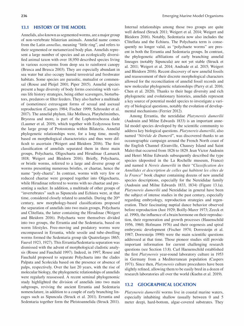
236 Emerging Marine Model Organisms
13.1 HISTORY OF THE MODEL
Annelids, also known as segmented worms, are a major group
of non-vertebrate bilaterian animals. Annelid name comes
from the Latin annellus, meaning “little ring”, and refers to
their segmented or metamerized body plan. Annelids repre
sent a large number of species and an ecologically diversi
fi ed animal taxon with over 18,950 described species living
in various ecosystems from deep sea to rainforest canopy
(Brusca and Brusca 2003). They are especially abundant in
sea water but also occupy humid terrestrial and freshwater
habitats. Some species are parasitic, mutualist or commen
sal (Rouse and Pleijel 2001; Piper 2015). Annelid species
present a huge diversity of body forms coexisting with vari
ous life history strategies, being either scavengers, bioturba
tors, predators or filter feeders. They also harbor a multitude
of (sometimes) extravagant forms of sexual and asexual
reproduction (Caspers 1984; Fischer 1999; Schroeder et al.
2017). The annelid phylum, like Mollusca, Platyhelminthes,
Bryozoa and more, is part of the Lophotrochozoa clade
(Laumer et al. 2019), which together with Ecdyzosoa form
the large group of Protostomia within Bilateria. Annelid
phylogenetic relationships were, for a long time, mostly
based on morphological characteristics and thus were dif
ficult to ascertain (Weigert and Bleidorn 2016 ). The fi rst
classification of annelids separated them in three main
groups, Polychaeta, Oligochaeta and Hirudinae (Lamarck
1818; Weigert and Bleidorn 2016 ). Briefl y, Polychaeta,
or bristle worms, referred to a large and diverse group of
worms presenting numerous bristles, or chaetae, hence the
name “poly-chaeta”. In contrast, worms with very few or
reduced chaetae were grouped together into Oligochaeta,
while Hirudinae referred to worms with no chaetae and pre
senting a sucker. In addition, a multitude of other groups of
“invertebrates” such as Sipuncula and Echiura were, at that
time, considered closely related to annelids. During the 20th
century, new morphology-based classifi cations proposed
the separation of annelids into two main groups, Polychaeta
and Clitellata, the latter containing the Hirudinae (Weigert
and Bleidorn 2016). Polychaeta were themselves divided
into two groups, the Errantia and the Sedentaria, based on
worm lifestyles. Free-moving and predatory worms were
encompassed in Errantia, while sessile and tube-dwelling
worms formed the Sedentaria group (de Quatrefarges 1865;
Fauvel 1923, 1927). This Errantia/Sedentaria separation was
dismissed with the advent of morphological cladistic analy
sis (Rouse and Fauchald 1997 ). Indeed, in 1997, Rouse and
Fauchald proposed to separate Polychaeta into the clades
Palpata and Scolecida based on the presence or absence of
palps, respectively. Over the last 20 years, with the rise of
molecular biology, the phylogenetic relationships of annelids
were regularly reassessed. A recent seminal phylogenomic
study highlighted the division of annelids into two main
subgroups, reviving the ancient Errantia and Sedentaria
nomenclature, in addition to a couple of early branching lin
eages such as Sipuncula (Struck et al. 2011). Errantia and
Sedentaria together form the Pleistoannelida (Struck 2011).
Internal relationships among those two groups are quite
well defined (Struck 2011; Weigert et al. 2014; Weigert and
Bleidorn 2016). Notably, Sedentaria now also includes the
Clitellata and the Echiura. The Polychaeta term is conse
quently no longer valid, as “polychaete worms” are pres
ent in both the Errantia and Sedentaria groups. In contrast,
the phylogenetic affiliations of early branching annelid
lineages (notably Sipuncula) are not yet stable (Struck et
al. 2011; Weigert et al. 2014; Andrade et al. 2015; Weigert
and Bleidorn 2016). Recent discovery of new annelid fossils
and reassessment of their discrete morphological characters
allowed for the reconciliation of annelid fossil records and
new molecular phylogenetic relationships (Parry et al. 2016;
Chen et al. 2020). Thanks to their huge diversity and rich
phylogenetic and evolutionary histories, annelids represent
a key source of potential model species to investigate a vari
ety of biological questions, notably the evolution of develop
mental mechanisms (Ferrier 2012).
Among Errantia, the nereididae Platynereis dumerilii (Audouin and Milne Edwards 1833) is an important anne
lid model species developed by the scientific community to
address key biological questions. Platynereis dumerilii , also
named “Néréide de Dumeril”, was discovered thanks to an
oceanographic campaign around the French North coast of
the English Channel (Granville, Chausey Island and Saint
Malo) that occurred from 1826 to 1829. Jean Victor Audouin
and Henri Milne Edwards subsequently described the type
species (deposited in the La Rochelle museum, France)
and named it Nereis dumerilii in their “Classifi cation des Annélides et description de celles qui habitent les côtes de la France” book chapter containing dozens of new annelid
species descriptions, especially for the Nereididae family
(Audouin and Milne Edwards 1833, 1834) (Figure 13.1a).
Platynereis dumerilii and Nereididae in general have been
the subject of intense studies in the past century, especially
regarding embryology, reproduction strategies and regen
eration. Their fascinating nuptial dance behavior observed
before reproduction (Just 1929; Boilly-Marer 1973; Zeeck et
al. 1990), the influence of a brain hormone on their reproduc
tion, their regeneration and growth processes (Hauenschild
1956, 1960; Hofmann 1976) and their oogenesis and spiral
embryonic development (Fischer 1974; Dorresteijn et al.
1987; Dorresteijn 1990) were the main scientifi c questions
addressed at that time. Those pioneer studies still provide
important information for current challenging research
questions (see Section 13.8). Carl Haeuenschild established
the fi rst Platynereis year-round laboratory culture in 1953
in Germany from a Mediterranean population (Caspers
1971). Since then, Platynereis culture procedures have been
slightly refined, allowing them to be easily bred in a dozen of
research laboratories all over the world (Kuehn et al. 2019).
13.2 GEOGRAPHICAL LOCATION
Platynereis dumerilii worms live in coastal marine waters,
especially inhabiting shallow (usually between 0 and 5
meter deep), hard-bottom, algae-covered substrates. They
237 The annelid model Platynereis dumerilii
FIGURE 13.1 Original and historical drawings of Platynereis (initially named Nereis) dumerilii. (a) Drawings of the original descrip
tion of Platynereis dumerilii mentioned as “Nereide de dumeril”. (Plate 4A, drawings 9 to 12: 9 = parapodia; 10 = anterior part and
head sensory structures; 11 = parapodia; 12 = denticulated jaw). (b) Drawing of annelids (5 = Nereis dumerilli, from a sketch drawn at
Lochmaddy). ([a] From Audouin and Milne Edwards 1833, 1834; [b] from M’Intosh et al. 1910.)
can also directly live on seaweeds and marine plant leaves
such as Posidonia oceanica and Zostera marina ( Jacobs
and Pierson 1979). As mentioned before, Platynereis dumerilii was first described from the French north coast
of the English Channel. Surprisingly, Platynereis dumerilii is also found in many other locations, from temperate
to tropical zones: they are often encountered throughout
the Mediterranean Sea (Gambi et al. 2000) but also in the
North Sea, the English Channel (Figure 13.1b), the Atlantic
down to the Cape of Good Hope, the Black Sea, the Red
Sea, the Persian Gulf, the Gulf of Mexico, Cuba, the Sea of
Japan, the Pacific, the Kerguelen Islands and the coasts of
Mozambique and South Africa (Read and Fauchald 2018;
Kara et al. 2020). As a consequence of this very broad geo
graphical distribution, Platynereis dumerilii is considered a
cosmopolitan species (Fischer and Dorresteijn 2004; Read
and Fauchald 2018). However, cosmopolitan species rarely
exist, since they actually often pool together sibling species
or a species complex with (nearly) identical morphologies
(Knowlton 1993). As shown for many other marine non-ver
tebrates species, recent population genetic studies from the
Mediterranean Sea (Italian coast) and South Africa revealed
that Platynereis dumerilii, in those localities, is in fact a spe
cies complex. In Italy, P. dumerilii is frequently mistaken
for it sibling species P. massiliensis ( Moquin-Tandon 1869 ;
Valvassori et al. 2015; Wäge et al. 2017). In South Africa,
P. dumerilii lives in sympatry with P. australis ( Schmarda
1861), another morphologically sibling species (Kara et al.
2020). Population genetic analysis of specimens found in
South Africa initially identified as P. dumerilii are probably
a new species, P. entshonae, highlighting the fact that only
rigorous and broad-scale population genetic studies world
wide will help to uncover the real geographic distribution
of P. dumerilii (Kara et al. 2020) and the diversity of the
Platynereis genus that currently contains 41 valid species
(Read and Fauchald 2018).
238
13.3 LIFE CYCLE
Platynereis’s life cycle exhibits several interesting features
and encompasses three phases separated by metamorphosis
events (Fischer and Dorresteijn 2004) (Figure 13.2). Like
many other marine animals, such as corals, sea urchins and
even fi shes, Platynereis sexual maturation and reproduction
are synchronized with the natural moon phases (Bentley
et al. 1999). This fascinating biological characteristic of
lunar-controlled reproductive periodicity, regulated thanks
to an endogenous oscillator, is called a circalunar life cycle
(Tessmar-Raible et al. 2011; Raible et al. 2017). Each worm
reproduce only once, and the timing of this reproduction
is tightly regulated by this clock ( Zantke et al. 2013). The
number of animals reaching sexual maturity is maximal
shortly after the new moon and minimal during the full
moon (Hauenschild 1955; Zantke et al. 2013). In nature, this
reproductive period occurs between May and September
in the Mediterranean Sea (Giangrande et al. 2002). When
Platynereis dumerilii worms are ready to spawn, usually at
night, males and females reach the surface, start an elegant
nuptial dance and synchronously release eggs and sperm in a
massive spawning event. This external fertilization induces
the formation of thousands of small zygotes and ultimately
implies the death of the reproducing males and females
(Figure 13.2). By the third day, the zygote develops into
small, segmented, planktonic larva named nectochaetae (see
Section 13.5). Nectochaete larvae live on their own nutritive
stock and move thanks to marine currents and ciliary belts.
After five to seven days of planktonic life, small juvenile
worms, while still able to swim, switch to a benthic/errant
life mode following metamorphosis (Fischer et al. 2010).
This first metamorphosis event corresponds to the disap
pearance of ciliated belts. Juvenile worms then continue to
grow throughout their lives at a rate that is highly dependent
on food availability. At some point, a second metamorpho
sis occurs, inducing profound morphological modifi cations
of the head and first segment. Additionally, worms start to
produce silk, important for the building of a tube in which
they will live for several months. They continue to grow,
to regenerate following injury and to grow posteriorly until
they initiate their last metamorphosis, corresponding to
the appearance of sexual traits (Fischer et al. 2010). This
sexual metamorphosis is moon dependent and implies dras
tic morphological changes (see Section 13.4) to allow the
production of thousands of gametes. During this very short
reproductive period, worms become pelagic.
This eventful life cycle can be reproduced in laboratory
in culture rooms maintained at 18°C and a daily artifi cial
illumination regime (16 hours of light/8 hours of darkness).
To induce sexual maturation, a low-light lamp is used to
mimic the lunar stimulus seven days per month. A couple
of days after this week of artificial full moon, juvenile (or
atoke) worms start sexual maturation for a two-week period,
allowing the production of sexually-mature (or epitoke)
worms every day (Fischer and Dorresteijn 2004; Kuehn et
al. 2019; Vervoort and Gazave in press).
Emerging Marine Model Organisms
13.4 ANATOMY
Like many other annelid species, Platynereis dumerilii worms have a complex body plan with various tissues, struc
tures and organs that are described in the following sections
for both atoke and epitoke forms.
13.4.1 EXTERNAL ANATOMY OF PLATYNEREIS
DUMERILII JUVENILE (ATOKE) WORMS
Platynereis dumerilii juvenile worm size can be up to 90
mm for around 100 segments (Figure 13.3a). Their body
color is highly variable, from yellowish and reddish to
greenish, and this coloration mostly relies on pigmented
cells, or chromatophores (Arboleda et al. 2019), that shine at
the surface of the epidermis, itself secreting a cellular cuti
cle. While their sex is genetically determined, at the juvenile
stage, male and female animals are indistinguishable. The
morphology of Nereid annelids such as Platynereis is often
described in zoological textbooks as representative of the
typical annelid body plan, composed of three main parts:
(i) an anterior region, the head with a substantial cephali
zation; (ii) the segmented or metamerized trunk composed
of many identical units called segments, with appendages
named parapodia; and (iii) a post-segmental terminal part,
containing the pygidium, a differentiated structure notably
containing the anus (Figure 13.3a) (Fischer and Dorresteijn
2004; Fischer et al. 2010).
Platynereis’s head is composed of different structures,
many of them being sensory (Chartier et al. 2018). These
structures ensure crucial functions for the worm’s life
(Purschke 2005) (Figure 13.3b and c). To begin, Platynereis possesses two pairs of pigmented cup brown adult eyes, in
a trapezoid arrangement, only visible on the dorsal part of
the worm (Figure 13.3b). These pairs of adult eyes repre
sent a distinct type of eyes in comparison to larval eyes, as
revealed by their specific developmental program (Arendt
et al. 2002; Guhmann et al. 2015). They also harbor a very
specific cellular structure with rhabdomeric photoreceptor
extensions traversing the pigmented cell layer (Arendt et al.
2002). These eyes are localized on a specific structure of
the head, named the prostomium (Figure 13.3b). The pro
stomium also bears a pair of highly chemosensory anten
nae localized at the front of the head (Chartier et al. 2018)
(Figure 13.3b). A pair of sensory palps are present near the
antennae; based on their cellular ultrastructure (Dorsett
and Hyde 1969) and a physiological experiment (Chartier
et al. 2018), they have been proposed to be chemosensory
as well (Figure 13.3c). The head is also composed of four
pairs of long sensory and photosensitive tentacular cirri
(namely anterior/posterior dorsal/ventral tentacle cirrus)
(Figure 13.3c). They are involved in the worm’s “shadow
refl ex”, a defensive behavior triggered by a decrease in illu
mination (Ayers et al. 2018). At the posterior dorsal mar
gin of the prostomium are nuchal organs, a pair of ciliated
cavities also considered important chemosensory structures
(Schmidtberg and Dorresteijn 2010; Chartier et al. 2018)
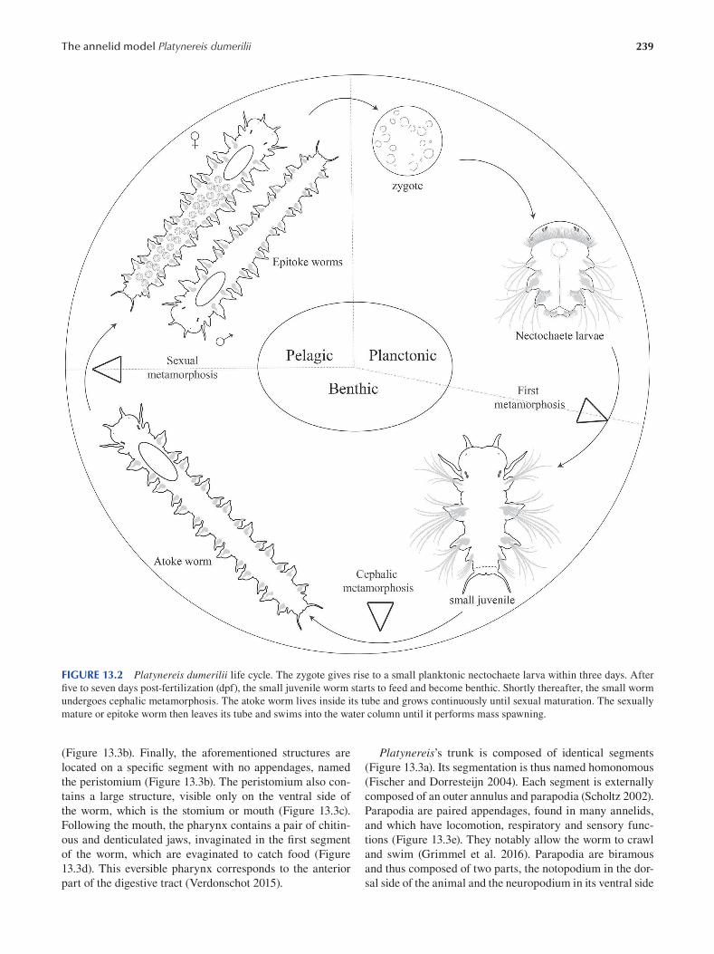
239 The annelid model Platynereis dumerilii
FIGURE 13.2 Platynereis dumerilii life cycle. The zygote gives rise to a small planktonic nectochaete larva within three days. After
five to seven days post-fertilization (dpf), the small juvenile worm starts to feed and become benthic. Shortly thereafter, the small worm
undergoes cephalic metamorphosis. The atoke worm lives inside its tube and grows continuously until sexual maturation. The sexually
mature or epitoke worm then leaves its tube and swims into the water column until it performs mass spawning.
(Figure 13.3b). Finally, the aforementioned structures are
located on a specific segment with no appendages, named
the peristomium (Figure 13.3b). The peristomium also con
tains a large structure, visible only on the ventral side of
the worm, which is the stomium or mouth (Figure 13.3c).
Following the mouth, the pharynx contains a pair of chitin
ous and denticulated jaws, invaginated in the fi rst segment
of the worm, which are evaginated to catch food (Figure
13.3d). This eversible pharynx corresponds to the anterior
part of the digestive tract (Verdonschot 2015).
Platynereis’s trunk is composed of identical segments
(Figure 13.3a). Its segmentation is thus named homonomous
(Fischer and Dorresteijn 2004). Each segment is externally
composed of an outer annulus and parapodia (Scholtz 2002).
Parapodia are paired appendages, found in many annelids,
and which have locomotion, respiratory and sensory func
tions (Figure 13.3e). They notably allow the worm to crawl
and swim (Grimmel et al. 2016). Parapodia are biramous
and thus composed of two parts, the notopodium in the dor
sal side of the animal and the neuropodium in its ventral side
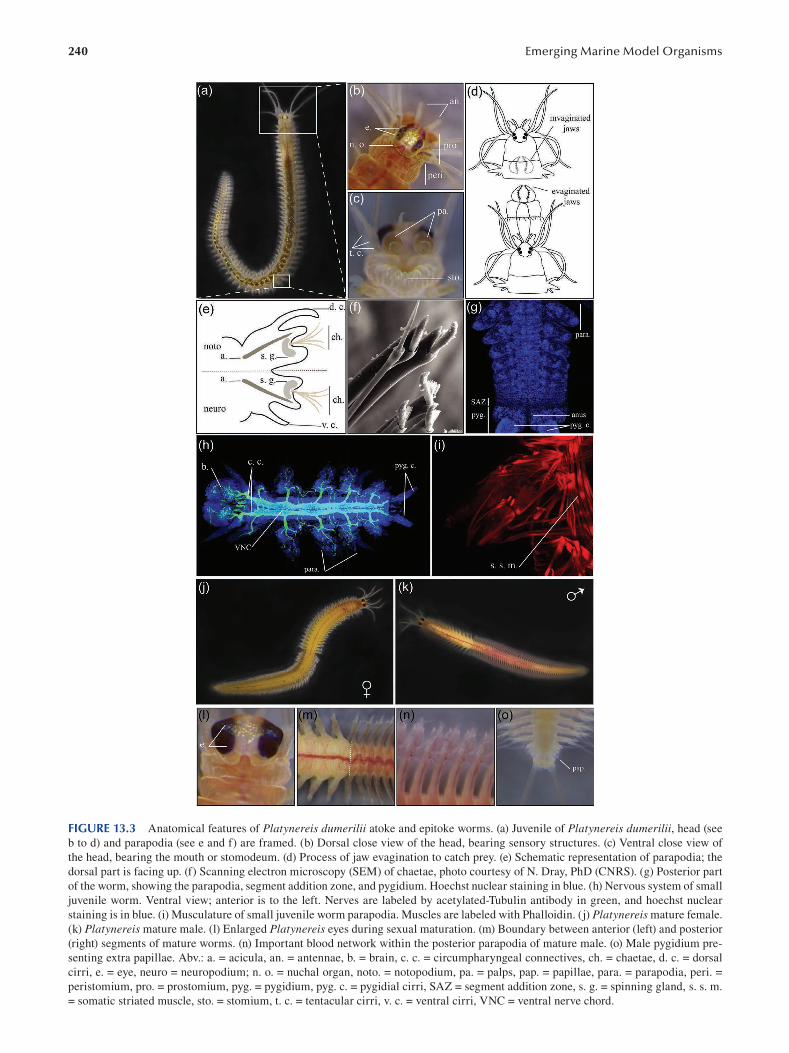
240 Emerging Marine Model Organisms
FIGURE 13.3 Anatomical features of Platynereis dumerilii atoke and epitoke worms. (a) Juvenile of Platynereis dumerilii, head (see
b to d) and parapodia (see e and f) are framed. (b) Dorsal close view of the head, bearing sensory structures. (c) Ventral close view of
the head, bearing the mouth or stomodeum. (d) Process of jaw evagination to catch prey. (e) Schematic representation of parapodia; the
dorsal part is facing up. (f) Scanning electron microscopy (SEM) of chaetae, photo courtesy of N. Dray, PhD (CNRS). (g) Posterior part
of the worm, showing the parapodia, segment addition zone, and pygidium. Hoechst nuclear staining in blue. (h) Nervous system of small
juvenile worm. Ventral view; anterior is to the left. Nerves are labeled by acetylated-Tubulin antibody in green, and hoechst nuclear
staining is in blue. (i) Musculature of small juvenile worm parapodia. Muscles are labeled with Phalloidin. (j) Platynereis mature female.
(k) Platynereis mature male. (l) Enlarged Platynereis eyes during sexual maturation. (m) Boundary between anterior (left) and posterior
(right) segments of mature worms. (n) Important blood network within the posterior parapodia of mature male. (o) Male pygidium pre
senting extra papillae. Abv.: a. = acicula, an. = antennae, b. = brain, c. c. = circumpharyngeal connectives, ch. = chaetae, d. c. = dorsal
cirri, e. = eye, neuro = neuropodium; n. o. = nuchal organ, noto. = notopodium, pa. = palps, pap. = papillae, para. = parapodia, peri. =
peristomium, pro. = prostomium, pyg. = pygidium, pyg. c. = pygidial cirri, SAZ = segment addition zone, s. g. = spinning gland, s. s. m.
= somatic striated muscle, sto. = stomium, t. c. = tentacular cirri, v. c. = ventral cirri, VNC = ventral nerve chord.
241 The annelid model Platynereis dumerilii
(Figure 13.3e). Each rami is composed of cirri (dorsal and
ventral cirrus), a lobe and a beam of extracellular chitine
ous structures named chaetae or bristles (Verdonschot 2015)
(Figure 13.3f). The latter are surrounded by specifi c glands
that secrete material for tube or cocoon synthesis and are
named spinning glands (Fischer et al. 2010). These exter
nal chaetae are constantly produced by internal structures
named chaetal sacs (Gazave et al. 2017). Chaetae have a ter
minal articulated portion at their tips (Figure 13.3f). In addi
tion, a robust, skeletal, internal, peculiar chaeta, named the
acicula, stabilizes each lobe (Figure 13.3e). Interestingly, in
annelid systematics, the shape of the parapodia and the type
of chaetae are informative characteristics to determine spe
cies ( Zakrzewski 2011).
The terminal part of the worm is named the pygidium
(Starunov et al. 2015) (Figure 13.3g). The pygidium contains
the anus and presents two sensory anal or pygidial cirri in its
ventral part (Ayers et al. 2018).
13.4.2 INTERNAL ANATOMY OF PLATYNEREIS
DUMERILII JUVENILE (ATOKE) WORMS
As for its external anatomy, Platynereis’s internal body plan
is segmented and presents a repetition of internal struc
tures within each segment. Indeed, each body unit contains
a body cavity or coelom (separated from the next one by
an incomplete intersegmental piece of tissue called septa),
each containing a part of the (i) nervous system, (ii) circu
latory system, (iii) musculature and (iv) excretory system
(Verdonschot 2015). The non-metamerized digestive tract
(v) runs along the antero–posterior axis of the worm.
13.4.2.1 Nervous System Platynereis worms possess a central nervous system (CNS)
and a peripheral nervous system (PNS). One main ele
ment of the CNS is the highly developed brain that resides
in a dorsal position within the head (Starunov et al. 2017)
(Figure 13.3h). Another important element of the CNS is a
paired serial chain of spherical ganglia in a ventral position
(named the ventral nerve chord or VNC) that runs all along
the length of the worm’s body, making a ladder-shaped
structure (Figure 13.3h). The brain is connected to the VNC
by circumpharyngeal connectives, which surround the phar
ynx (Verdonschot 2015) (Figure 13.3h). Platynereis ’s brain
contains prominent dorsal neuropile arrangements named
mushroom bodies (Tomer et al. 2010), a structure shared
with arthropods (Heuer et al. 2010). Interestingly, the mush
room body anlagen in Platynereis larvae expresses a simi
lar molecular signature to developing mushroom bodies in
Drosophila melanogaster, thus providing strong evidence in
favor of an evolutionary relatedness of insect and annelid
mushroom bodies (Tomer et al. 2010). Platynereis ’s brain
is also an important neurosecretory center. Its developing
forebrain expresses the neuropeptides FMRFa and vasoto
cin (Tessmar-Raible et al. 2007), plus a diversity of other
neuropeptides recently identified, such as somatostatin,
galanin and so on (Williams et al. 2017). Interestingly, this
annelid brain region shares a common molecular signature
with the vertebrate hypothalamus, furthering the hypothesis
of an evolutionary relationship between those two structures
(Williams and Nagy 2017). In addition, Platynereis ’s brain
produces a brain hormone responsible for the switch from a
growing juvenile to a sexually mature worm (Hauenschild
1956). This hormone, whose activity suppresses reproduc
tion, was recently identified as Methylfarnesoate (Schenk et
al. 2016). In addition to the brain, the VNC is also a complex
structure that has been shown to harbor around 200 distinct
types of neurons, expressing specific combinations of tran
scription factors in a small juvenile (Vergara et al. 2017).
Platynereis’s PNS is prominent in the head, being associ
ated with the many sensory structures it contains. The PNS
also contains the parapodial and pygidial nerve extensions.
Indeed, the terminal part of the worm is highly innervated
with nerve projections into the pygidium and the anal cirri
(Starunov et al. 2015) (Figure 13.3h).
13.4.2.2 Circulatory System Platynereis has a closed circulatory system mainly com
posed of two vessels and capillary networks. The dorsal and
ventral vessels are connected by a capillary network form
ing a ring around the intestine. The dorsal pulsatile vessel
is the main pump of the circulatory system, pumping the
blood anteriorly (from the tail to the head), while the ven
tral vessel pumps the blood posteriorly. Segmental lateral
vessels irrigate the parapodia in each segment in order to
ensure their respiratory function (Saudemont et al. 2008;
Verdonschot 2015). A circular blood sinus is present in the
pygidium (Starunov et al. 2015).
13.4.2.3 Musculature Platynereis has two main types of muscles, smooth and
striated, which together ensure precise movements of the
worm’s body structures (Brunet et al. 2016) (Figure 13.3i).
Some somatic striated muscles run longitudinally from the
head to the tail of the animal. Additional somatic striated
muscles control the movements of parapodia thanks to ven
tral oblique and parapodial fibers (Figure 13.3i). In contrast,
visceral muscles are mainly smooth muscle (with the notice
able exception of the anterior part of the gut that contains
striated visceral muscles). They form a specifi c muscular
structure, the orthogon, which is composed of both circular
and longitudinal fibers (Brunet et al. 2016). Smooth muscles
are also associated with the pulsatile dorsal vessel (Brunet
et al. 2016). A peculiar somatic striated and longitudinal
muscle, the axochord, is found between the VNC and the
dorsal vessel and is proposed to be at the origin of the chor
date notochord (Lauri et al. 2014; Brunet et al. 2015). The
pygidium musculature is also highly complex, mainly com
posed of a strong array of circular muscles that plays the role
of the anal sphincter (Starunov et al. 2015).
13.4.2.4 Excretory System Platynereis atoke worms possess in each segment, except
the pygidium, a pair of metanephridia that connects the coe
lomic compartment to the exterior to ensure the excretion of
waste products (Hasse et al. 2010; Verdonschot 2015).
242
13.4.2.5 Digestive System The digestive system of Platynereis is mainly composed
of three successive elements called the foregut, the midgut
and the hindgut (Fischer et al. 2010; Zidek et al. 2018). The
foregut is composed of the mouth, the eversible pharynx
and the jaws, in charge of collecting and grinding the food.
Digestive enzymes are secreted and active in the midgut,
where food absorption occurs. The last section of the diges
tive system is a hindgut connecting the midgut to the anus
and producing digestive enzymes, too (Verdonschot 2015;
Williams et al. 2015).
13.4.3 EXTERNAL AND INTERNAL ANATOMY OF
PLATYNEREIS DUMERILII ADULT (EPITOKE) WORMS
As mentioned before, one of the main events in Platynereis life cycle is sexual maturation (epitoky), since it induces not
only drastic morphological modifications but also changes
in behavior (Fischer and Dorresteijn 2004). A striking dif
ference between mature and juvenile worms is the difference
in body color (Figure 13.3j and k): while juveniles mainly
show a sex-independent brownish color, sexual dimorphism
appears during epitoky, as females become bright yellow
(Figure 13.3j) and males display white anterior and red poste
rior body regions ( Figure 13.3k). During sexual maturation,
worms stop food intake, their gut regresses and becomes
non-functional. The trunk of the animal is progressively
modified to become a “bag” full of gametes, visible through
the body wall, which loses its pigmentation (Fischer and
Dorresteijn 2004; Fischer et al. 2010). The yellow oocytes
and white spermatozoids both contribute to the main color
of the female and male anterior parts, respectively. Among
other morphological changes, the eyes enlarge dramatically
(Figure 13.3l), and the homonomous segmentation present
in juveniles is lost. Indeed, while anterior segments are not
modified, posterior segments are substantially reshaped,
and a clear boundary between these two parts of the trunk
becomes visible (between the 15th and 20th segments,
depending on the sex; Figure 13.3m) (Schulz et al. 1989;
Fischer 1999). In modified posterior segments, parapodia
flatten and develop paddle-shaped chaetae in both sexes. In
males, posterior parapodia show a significant increase in
vascularization, conferring its red color to the posterior part
(Figure 13.3n). Muscles present in juvenile worms degener
ate and are replaced by new muscle fibers which are specifi c
to sexually mature animals. This dramatic reorganization
of the body enables the formerly benthic juvenile worms to
swim quickly to ensure the nuptial dance required for sexual
reproduction (Fischer and Dorresteijn 2004; Fischer et al.
2010). Finally, while the terminal part of the female is not
modifi ed, the male pygidium presents extra papillae, allow
ing the sperm to be released in many directions (Figure
13.3o) (Starunov et al. 2015). In Platynereis, the switch from
a growing worm to its reproductive life stage is controlled
by brain hormone activity. Interestingly, worm decapitation
(i.e. artificial reduction of brain hormone) induces worms’
Emerging Marine Model Organisms
sexual maturation similarly to natural conditions (Schenk et
al. 2016).
13.5 EMBRYOGENESIS AND LARVAL DEVELOPMENT
More than a century ago, Edmund B. Wilson retraced an
incredibly relevant and reliable cell lineage of embryo blas
tomeres in order to depict the origin of the germ layers in
annelids (Wilson 1892). To do so, he took advantage of the
transparency of Nereis limbate (now Alitta succinea ) and
Nectonereis megalops (now Platynereis megalops ) embryos
and of their stereotypic development. Indeed, as all embryos
develop in exactly the same way, they provide an ideal
framework to link cell division to blastomere formation
and cell fate. Interestingly, since publication, his work has
been reasserted by the description of Platynereis dumerilli embryogenesis in the early 90s (Dorresteijn 1990), and his
assumptions regarding blastomere cleavage and fate remain
a reference in the field of annelid development. Indeed,
micro-injection of individual blastomeres at different
embryonic stages with fluorescent dyes has more recently
confirmed previous observations (Fischer and Dorresteijn
2004; Ackermann et al. 2005). Hereafter, we have mainly
compiled the previously mentioned publications to depict
the main events of embryogenesis and larval development
(Fischer et al. 2010).
13.5.1 EMBRYO DEVELOPMENT
13.5.1.1 Unfertilized Eggs Unfertilized eggs are packed within the coelomic cavity
of the mature female, causing their polymorphous shapes.
Upon laying, the pressure is released and the eggs rapidly
undergo a massive shape change to become ellipsoid (the
short axis of the unfertilized egg corresponds to the future
animal–vegetal axis of the zygote). At that stage, their cyto
plasm is organized, in a concentric fashion, around the cen
tral nucleus which is wrapped in yolk-free cytoplasm. The
latter is surrounded by a shell of yolk containing large lipid
droplets (in particular in the equatorial plane where they are
bigger) and a thick outer layer of cortical granules (secre
tory organelles found within oocytes). Finally, the egg is
itself protected within a vitelline envelope. Interestingly,
in Platynereis, eggs are in fact oocytes blocked in meta
phase and, as such, the release of polar bodies occurs after
fertilization.
13.5.1.2 Fertilization Upon fertilization, the fertilizing spermatozoid sticks to the
cell surface until the emission of the first polar body (a small
haploid cell). As soon as this contact is established, substan
tial changes in the cytoplasmic organization of the oocyte
occur. The cortical granules are released to form an exter
nal jelly layer (0–23 minutes post-fertilization, mnpf). As a
consequence, the yolk granules are less packed within the
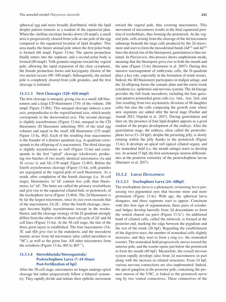
243 The annelid model Platynereis dumerilii
spherical egg and more broadly distributed, while the lipid
droplet pattern remains as a readout of the equatorial plan.
When the vitelline envelope breaks down (18 mnpf), a small
area is progressively cleared from yolk at one pole of the egg
compared to the equatorial location of lipid droplets. This
area marks the future animal pole where the first polar body
is formed (60 mnpf, Figure 13.4a). The sperm pronucleus
finally enters into the ooplasm, and a second polar body is
formed (80 mnpf). Yolk granules migrate toward the vegetal
pole, allowing the rapid expansion of the clear cytoplasm,
the female pronucleus forms and karyogamy (fusion of the
two nuclei) occurs (90–100 mnpf). Subsequently, the animal
pole is completely cleared from yolk granules, and the fi rst
cleavage is initiated.
13.5.1.3 First Cleavages (120–420 mnpf) The first cleavage is unequal, giving rise to a small AB blas
tomere and a large CD blastomere (73% of the volume, 100
mnpf, Figure 13.4b1). This unequal cleavage induces a new
axis, perpendicular to the vegetal/animal axis, which nearly
corresponds to the dorsoventral axis. The second cleavage
is slightly asynchronous (Figure 13.4a), unequal in the CD
blastomere (D blasomere inheriting 50% of the total egg
volume) and equal in the small AB blastomere (135 mnpf;
Figure 13.4a, 4b2). Each of the resulting four macromeres
is the founder of a distinct quadrant (e.g. A-quadrant corre
sponds to the offspring of A macromere). The third cleavage
is slightly asynchronous as well (Figure 13.4a) and corre
sponds to the first “spiral” cleavage (clockwise), produc
ing two batches of two nearly identical micromeres (1a and
1b versus 1c and 1d) (170 mnpf, Figure 13.4b3). Before the
fourth asynchronous cleavage (Figure 13.4a), yolk granules
are segregated at the vegetal pole of each blastomere. As a
result, after completion of the fourth cleavage (i.e. 16-cell
stage), blastomeres 1a1–1d1 contain less yolk than blasto
meres 1a2–1d2. The latter are called the primary trochoblasts
and give rise to the equatorial ciliated belt, or prototroch, of
the trochophore larva (Figure 13.4b4). The 2d blastomere is
by far the largest micromere, since its size even exceeds that
of the macromeres 2A-2C. After the fourth cleavage, cleav
ages become highly asynchronous (except in the trocho
blasts), and the cleavage strategy of the D quadrant strongly
differs from the others with the short cell cycle of 2d1 and 4d
cell lines (Figure 13.4a). At the 38-cell stage, the fate of the
three germ layers is established. The four macromeres (3A
3C and 4D) give rise to the endoderm, and the mesoderm
mainly arises from 4d micromere (also called mesoblast or
“M”), as well as the germ line. All other micromeres form
the ectoderm (Figure 13.4a, 4b5 to 4b5’’’).
13.5.1.4 Stereoblastula/Stereogastrula/ Protrochophore Larva (7–24 Hours Post-Fertilization of hpf)
After the 38-cell stage, micromeres no longer undergo spiral
cleavage but rather progressively follow a bilateral symme
try. They rapidly divide and initiate their epibolic movement
toward the vegetal pole, thus covering macromeres. This
movement of micromeres results in the final equatorial posi
tion of trochoblasts, thus forming the prototroch. At the veg
etal pole, cells arising from the cleavage of the 4d micromere
submerge beneath the large cells produced by the 2d micro
mere and start to form the mesodermal bands (4d122 and 4d222
lines the dorsal rim of the blastopore); gastrulation is thus ini
tiated. In Platynereis, this process shows amphistome mode,
meaning that the blastopore gives rise to both the mouth and
the anus (Figure 13.4c) (Steinmetz et al. 2007). During this
massive rearrangement of embryonic cells, the D-quadrant
plays a key role, especially in the formation of trunk tissues.
Indeed, the 4D blastomere participates in midgut anlage, and
the 2d offspring forms the somatic plate and the entire trunk
ectoderm (i.e. epidermis and nervous system). The 4d lineage
provides the full trunk mesoderm, including the four quies
cent putative primordial germ cells (i.e. 1mL, 1mr, 2mL and
2mr resulting from two asymmetric divisions of M-daughter
cells) but also the cells composing the growth zone where
new segments are added after the larval stage (Fischer and
Arendt 2013; Ozpolat et al. 2017). During gastrulation and
later on, the presence of four lipid droplets appears as a good
readout of the proper development of the embryo. After the
gastrulation stage, the embryo, often called the protrocho
phore larva (13–24 hpf), despite the persisting jelly, is slowly
rotating within the jelly thanks to the prototroch ( Figure
13.4c). It develops an apical tuft (apical ciliated organ), and
the stomodeal field (i.e. the mouth anlage) starts to develop
too. At around 17 hpf, the first serotonergic neuron differenti
ates at the posterior extremity of the protrochophore larvae
(Starunov et al. 2017 ).
13.5.2 LARVAE DEVELOPMENT
13.5.2.1 Trochophore Larva (24–48hpf) The trochophore larva is a phototactic swimming larva pos
sessing two pigmented eyes that become more and more
prominent (Figure 13.4c). With age, the spherical larva
elongates, and three segments start to appear. Consistent
with this first sign of segmentation, three pairs of ectoder
mal bulges develop laterally from 2d descendants to form
the ventral chaetal sac pairs (Figure 13.3c’). An additional
band of ciliated cells, called the telotroch, is formed at the
posterior end, marking the edge between the pygidium and
the rest of the trunk (26 hpf). Regarding the establishment
of the digestive tract, the number of stomodeal cells slightly
increases, and they start to form a ring (i.e. the stomodeal
rosette). The stomodeal field progressively moves toward the
anterior pole, and the rosette opens just below the prototroch
to form the mouth (40 hpf). Meanwhile, the overall nervous
system rapidly develops (also from 2d micromeres) in part
along with the increase in ciliated structures. From 24 hpf,
various nervous connections are also implemented. Indeed,
the apical ganglion at the posterior pole, containing the pio
neer neuron of the VNC, is linked to the prototroch nerve
ring by two ventral connectives. These connectives of the
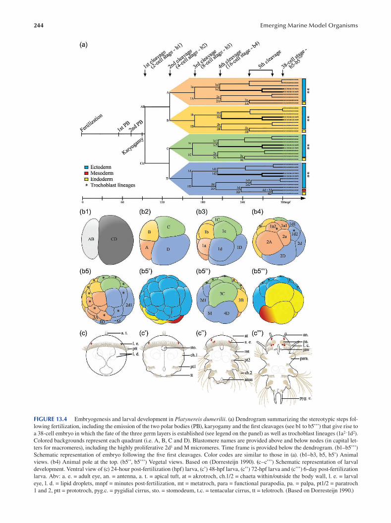
244 Emerging Marine Model Organisms
FIGURE 13.4 Embryogenesis and larval development in Platynereis dumerilii. (a) Dendrogram summarizing the stereotypic steps fol
lowing fertilization, including the emission of the two polar bodies (PB), karyogamy and the first cleavages (see b1 to b5’’’) that give rise to
a 38-cell embryo in which the fate of the three germ layers is established (see legend on the panel) as well as trochoblast lineages (1a2–1d2).
Colored backgrounds represent each quadrant (i.e. A, B, C and D). Blastomere names are provided above and below nodes (in capital let
ters for macromeres), including the highly proliferative 2d1 and M micromeres. Time frame is provided below the dendrogram. (b1–b5’’’)
Schematic representation of embryo following the fi ve first cleavages. Color codes are similar to those in (a). (b1–b3, b5, b5’) Animal
views. (b4) Animal pole at the top. (b5’’, b5’’’) Vegetal views. Based on (Dorresteijn 1990). (c–c’’’) Schematic representation of larval
development. Ventral view of (c) 24-hour post-fertilization (hpf) larva, (c’) 48-hpf larva, (c’’) 72-hpf larva and (c’’’) 6-day post-fertilization
larva. Abv: a. e. = adult eye, an. = antenna, a. t. = apical tuft, at = akrotroch, ch.1/2 = chaeta within/outside the body wall, l. e. = larval
eye, l. d. = lipid droplets, mnpf = minutes post-fertilization, mt = metatroch, para = functional parapodia, pa. = palpa, pt1/2 = paratroch
1 and 2, ptt = prototroch, pyg.c. = pygidial cirrus, sto. = stomodeum, t.c. = tentacular cirrus, tt = telotroch. (Based on Dorresteijn 1990.)
245 The annelid model Platynereis dumerilii
VNC represent the two first axons of the brain. Immediately
thereafter, the dorsal root of the circumesophagial connec
tives develop as well, followed by the ventral root (26 hpf) to
connect the VNC to the brain. At the same time, the single
asymmetric unpaired dorsal axon and the first cerebral com
missures appear. Later on, three additional serotoninergic
cells arise at the apical part (30–34 hpf), as well as one pair
at the first ventral commissure (40 hpf). Finally, the second
ventral commissure appears (44 hpf) (Starunov et al. 2017).
Similarly, muscles appear and develop during the trocho
phore stage. The dorsal longitudinal muscles develop fi rst
(28 hpf), followed by ventral longitudinal muscles (32 hpf),
while the oblique and parapodial muscles start to be vis
ible at the late trochophore stage (46–48 hpf). The excre
tory system appears also at the trochophore stage, with the
emergence of small, lateral, non-ciliated tubules (Hasse
et al. 2010).
13.5.2.2 Metatrochophore Larva (48–66 hpf) The metatrochophore larval stage is marked by the appear
ance of the two adult eye pairs (Figure 13.4c’’). In addition,
the three first segments appear more defined due to the
formation of non-functional parapodia and the signifi cant
growth of the chaetae outside the body wall (these segments
are so called the chaetigerous segments). In addition, the fi rst
paratroch appears between the second and the third chaetig
erous segments (48 hpf), and a second one is visible later on
between the fi rst and the second chaetigerous segments (56
hpf), thus participating in segment delimitation. Then, above
the prototroch, an additional ciliated structure progressively
develops—the akrotroch—close to the apical tuft (60 hpf).
The stomodeal rosette size increases with an additional ring
of cells (52 hpf). The stomodeum invaginates, resulting in
the larval foregut that elongates toward the posterior part.
The nervous system also rapidly develops. A third commis
sure appears (48 hpf), and all commissures thicken (54 hpf).
Axon projections from the VNC are observed laterally and
redirected ventrally toward the surface. The circumesopha
gial connectives get closer to each other, and the prototroch
nerve ring moves toward the brain. All these phenomena
participate in the formation and growth of the brain (52 hpf).
Additionally, the number of serotonergic cells along the ven
tral nerve cord increases with the occurrence of three addi
tional pairs. Finally, the ventral medial longitudinal muscle
appears (56 hpf) and elongates up to the posterior border of
the third segment. Similarly, oblique and parapodial mus
cles also elongate (Figure 13.4c’’). Excretory system devel
opment continues as non-ciliated tubules elongate laterally
toward the developing stomodeum (Hasse et al. 2010).
13.5.2.3 Nectochaete Larva (66 hpf–5 dpf) The nectochaete larva corresponds to a major lifestyle tran
sition. Indeed, the pelago-benthic larva starts crawling on
the substrate thanks to functional parapodia and starts to
eat. The sensory organs, including antenna, palps, tentac
ular antero-dorsal cirri and anal cirri, appear and develop
(75 hpf) (Figure 13.4c’’’). The trunk continues to elongate,
providing a worm-like shape to the larva, and a constriction
distinguishes the trunk from the head. The two adult eyes
found on both sides of the head increase in size and become
extremely close. Lipid droplets progressively move toward
the posterior part. Ciliogenesis progresses with the estab
lishment of the metatroch, an additional line of ciliated cells
that develops below the prototroch and fuses with this latter
on the lateral sides. The midgut forms, as well as the procto
deum (anal region), and the stomodeum/foregut continues to
elongate toward the posterior part, resulting in a fully func
tional digestive tract (75 hpf–4 dpf). Furthermore, the jaws
develop within the foregut (4 dpf) and a pair of primary teeth
appears (5 dpf). Meanwhile, the brain continuously grows,
the convergence of the circumesophagial connective roots
progresses, axon numbers increase in connectives and com
missures and additional serotonergic cells arise both in the
ventral nerve cord (66–72 hpf) and in the brain (4–5 dpf).
The overall musculature develops as well, especially around
the stomodeum, to form the pharynx. Additionally, muscles
and nerves associated with the development of antennae
and cirri increase. Seventy-two-hpf larvae possess a pair of
anterio-lateral non-ciliated tubules named “head kidneys”
located close to the episphere. These larval structures are
transitory, since they disappear before 96 hpf. In parallel,
larval nephridia or protonephridia, formed from ciliated
tubules and localized between segments, start to appear
(Hasse et al. 2010).
13.5.2.4 Young Errant Juvenile At this stage, the development of animals is no longer syn
chronous. Very young worms start to sequentially produce
additional segments through posterior elongation, a process
relying on a thin row of cells (presumably stem cells) that
forms the segment addition zone (SAZ) in front of the pygid
ium (Gazave et al. 2013). Worms also lose several larva-spe
cific features such as the prototroch, the apical tuft, larval
eyes and lipid droplets. The excretory system is composed
of segmented protonephridia until the worms reach the size
of 20 segments, at which stage metanephridia appear (Hasse
et al. 2010). In addition, the first chaetigerous segment fuses
with the head. This important morphological transition,
called cephalization, consists of the transformation of the
first pair of parapodia into tentacular posterior–dorsal cirri
and the progressive loss of chaetae. Finally, spinning glands
develop and produce mucus, allowing worms to build their
first cocoon network.
13.6 GENOMIC DATA
As in many animals, counting chromosomes during meta
phase revealed that Platynereis dumerilii is diploid (2n = 28)
(Jha et al. 1995). More precisely, the Platynereis karyotype
encompasses seven chromosome pairs showing a median
arm ratio, while the seven other pairs show a sub-median
ratio (Figure 13.5). Different regular staining techniques
were used to further characterize chromosome pairs. For
instance, Chromosome 2 shows a clear C-band-positive
246 Emerging Marine Model Organisms
FIGURE 13.5 Schematic representation of Platynereis chromosomes. Platynereis possesses 2 n = 28 chromosomes, five pairs showing
a median position of centromeres (Chromosome 1 to 7). Black and white areas represent heterochromatin (C-band-positive band) and
ribosomal RNA genes (NOR staining), respectively.
region, revealing a constitutive heterochromatin region not
localized at the centromic region. Nuclear organizer regions
(NORs) are found at the terminal positions of Chromosomes 5 and 6, thus revealing the localization of genes coding for
ribosomal RNA.
The precise genetic content of these chromosomes is
in the course of being uncovered. Indeed, the Platynereis dumerilii genome has recently been sequenced by the D.
Arendt laboratory (EMBL, Germany), notably from sperm.
Although this genome is currently being refined with the
aim of obtaining a chromosome-level assembly, a high-
quality draft version is already available, upon request, for
the whole community working on Platynereis . Preliminary
data revealed that Platynereis genome appears less com
pact than in other annelids (~1 Gpb) (Zantke et al. 2014),
and a previous analysis comparing bacterial artifi cial chro
mosome (BAC) sequencing and expressed sequence tags
(ESTs) on a subset of 30 randomly detected genes sug
gested that Platynereis genes are intron rich, surprisingly,
with two-thirds of introns shared between Platynereis and
human orthologs (Raible et al. 2005). Various additional
transcriptomic databases have been acquired during the
past years (Table 13.1), including bulk RNA-seq data for all
key stages of embryonic and larval development, juveniles
of different ages and adults (Conzelmann et al. 2013; Chou
et al. 2016). These data have been grouped together and
are now publicly available on the Pdumbase website (Chou
et al. 2018) (http://140.109.48.81/platynereis/controller.
php?action=home). Platynereis is being actively studied by
a scientific community, notably in the field of evolution and
developmental biology, and as such, additional transcrip
tomic databases are constantly produced. For instance,
Achim and collaborators shed light on the transcriptomic
landscape of cell diversity in 48 hpf-larvae using a single-
cell RNA-seq (scRNA-seq) approach (Achim et al. 2015;
Williams et al. 2017; Achim et al. 2018). In addition, bulk
RNA-seq were acquired to unravel the dynamic of gene
expression during circalunar-dependent sexual maturation
(Schenk et al. 2019) and posterior regeneration (Vervoort’s
Lab, unpublished data).
Finally, in addition to the signifi cant Platynereis resources acquired during the past decade, the availability
of genome sequences of the Sedentaria Capitella teleta,
Helobdella robusta (Simakov et al. 2013), Spirobranchus lamarcki (Kenny et al. 2015), Lamellibrachia luymesi ( Li
et al. 2019), Eisenia Andrei (Shao et al. 2020) and Eisenia fetida (Bhambri et al. 2018) as well as the Dinophiliformia
(sister group to Sedentaria + Errantia) Dimorphilus gyrociliatus (Martin-Duran et al. 2021) allow for comparative
analyses within annelids.
13.7 FUNCTIONAL APPROACHES: TOOLS FOR MOLECULAR AND CELLULAR ANALYSES
In addition to its scientific relevance and its easy mainte
nance in laboratory, the success of Platynereis as a new
model system also strongly relies on the efforts that have
been undertaken to develop a large panel of molecular and
cellular tools to successfully tackle interesting biologi
cal questions in evolutionary and developmental biology
( Backfisch et al. 2014; Williams and Jekely 2016 ).
13.7.1 DESCRIPTIVE APPROACHES
13.7.1.1 Detection of mRNA: Whole-Mount In Situ Hybridization
As mentioned in the genomic data section, several high-
quality bulk RNA-seq and scRNA-seq were recently used
to investigate modulations in gene expression during vari
ous processes in Platynereis. Nevertheless, bulk RNA-seq
average information from various cell populations and
scRNA-seq remains expensive, and their interpretation
relies on a comprehensive description of cell populations
in vivo. Accordingly, despite important breakthroughs in
sequencing technologies, whole-mount in situ hybridization
(WMISH) remains an indispensable molecular approach
to localize gene expression. WMISH has been established
in Platynereis to investigate gene expression during early
embryonic/larval stages (Arendt et al. 2001), posterior elon
gation (Prud’homme et al. 2003, Gazave et al., 2013), regen
eration (Planques et al. 2019) and the adult stage (Backfi sch
et al. 2013) using the regular NBT/BCIP colorimetric stain
ing (Figure 13.6a and a’). Similarly, fl uorescent in situ hybridization (FISH, Figure 13.6b and b’) has been estab
lished (Tessmar-Raible et al. 2005), while current efforts are
now also dedicated to implement hybridization chain reac
tions (HCRs) (Choi et al. 2018), thus allowing multiple tran
script detection to be required for co-expression analysis.
Finally, the stereotypic development of embryo and larva
coupled with in situ hybridizations allows for image regis
tration (Figure 13.6c), which consists of a virtual atlas of
expression patterns for their systematic comparison (Tomer
et al. 2010; Asadulina et al. 2012).
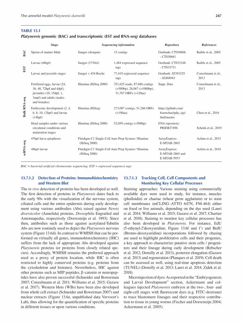
247 The annelid model Platynereis dumerilii
TABLE 13.1 Platynereis genomic (BAC) and transcriptomic (EST and RNA-seq) databases
Stage Sequencing information Repository References
Sperm of mature Male Sanger (shotgun) 15 contigs Genbank: CT030666 Raible et al., 2005
- CT030681
Larvae (48hpf) Sanger (3730xl) 1,484 expressed sequence Genbank: CT032248 Raible et al., 2005
tags - CT033731
scR
NA
-seq
B
ulk
RN
A-s
eq
EST
B
AC
Larvae and juvenile stages Sanger + 454 Roche 77,419 expressed sequence Genbank: JZ391525 Conzelmann et al.,
tags - JZ468943 2013
Fertilized eggs, larvae (24, Illumina (HiSeq 2000) 351,625 reads, 87,686 contigs Supp. Data Conzelmann et al.,
36, 48, 72hpf and 4dpf), (>500bp), 28,067 (>1000bp), 2013
juveniles (10, 15dpf, 1, 51,767 ORFs (>120aa)
3mpf) and adults (males
and females)
Embryonic development (2, 4, Illumina (HiSeq) 273,087 contigs, 51,260 ORFs https://github.com/
6, 8, 10, 12hpf) and larvae (>100aa) hsienchao/pdu_sqs/ Chou et al., 2016
(14hpf) fi nd/master
Head samples under various Illumina (HiSeq 2000) 52,059 contigs (>500bp) ENA repository:
circalunar conditions and PRJEB27496 Schenk et al., 2019
maturation stages
47hpf-larva epispheres Fluidigm C1 Single-Cell Auto Prep System / Illumina ArrayExpress: Achim et al., 2015
(HiSeq 2000) E-MTAB-2865
48hpf-larvae Fluidigm C1 Single-Cell Auto Prep System / Illumina ArrayExpress: Achim et al., 2018
(HiSeq 2000) E-MTAB-2865 and
E-MTAB-5953
BAC = bacterial artifi cial chromosome sequencing; EST = expressed sequences tags
13.7.1.2 Detection of Proteins: Immunohistochemistry and Western Blot
The in vivo detection of proteins has been developed as well.
The first detection of proteins in Platynereis dates back to
the early 90s with the visualization of the nervous system,
ciliated cells and the entire epidermis during early develop
ment using various antibodies (Abs) raised against Nereis diversicolor (Annelida) proteins, Drosophila Engrailed and
Antennapedia, respectively (Dorresteijn et al. 1993). Since
then, antibodies such as those against acetylated-Tubulin
Abs are now routinely used to depict the Platynereis nervous
system (Figure 13.6d). In contrast to WMISH that can be per
formed on virtually all genes, immunohistochemistry (IHC)
suffers from the lack of appropriate Abs developed against
Platynereis proteins (or proteins from closely related spe
cies). Accordingly, WMISH remains the preferred approach
used as a proxy of protein location, while IHC is often
restricted to highly conserved proteins (e.g. proteins from
the cytoskeleton and histones). Nevertheless, IHC against
other proteins such as MIP peptides, -catenin or neuropep
tides have also proven successful (Schneider and Bowerman
2007; Conzelmann et al. 2011; Williams et al. 2015; Gazave
et al. 2017). Western blots (WBs) have been also developed
from whole cell extract (Schneider and Bowerman 2007 ) and
nuclear extracts (Figure 13.6e, unpublished data Vervoort’s
Lab), thus allowing for the quantification of specifi c proteins
in different tissues or upon various conditions.
13.7.1.3 Tracking Cell, Cell Components and Monitoring Key Cellular Processes
Staining approaches: Various staining using commercially
available dyes were used to study, for instance, muscles
(phalloidin) or chaetae (wheat germ agglutinin) or to stain
cell membranes (mCLING–ATTO 647N, FM-464) either
on fixed or live animals, depending on the dye used (Lauri
et al. 2014; Williams et al. 2015; Gazave et al. 2017; Chartier
et al. 2018). Staining to monitor key cellular processes has
also been developed in Platynereis. For instance, EdU
(5-ethynyl-2’deoxyuridine, Figure 13.6f and f’) and BrdU
( Bromo-desoxyuridine) incorporations followed by chasing
are used to highlight proliferative cells and their progenies,
a key approach to characterize putative stem cells / progeni
tors and their lineage during early development (Rebscher
et al. 2012; Demilly et al. 2013), posterior elongation (Gazave
et al. 2013) and regeneration (Planques et al. 2019). Cell death
can be assessed as well, using real-time apoptosis detection
(TUNEL) (Demilly et al. 2013; Lauri et al. 2014; Zidek et al.
2018 ).
Microinjection of dyes: As reported in the “Embryogenesis
and Larval Development” section, Ackermann and col
leagues injected Platynereis embryos at the two-, four- and
eight-cell stages with fluorescent dyes (e.g. FITC-dextrane)
to trace blastomere lineages and their respective contribu
tion to tissue in young worms (Fischer and Dorresteijn 2004;
Ackermann et al. 2005).
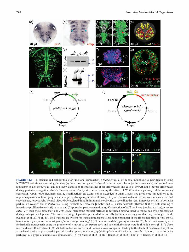
248 Emerging Marine Model Organisms
FIGURE 13.6 Molecular and cellular tools for functional approaches in Platynereis. (a–a’) Whole-mount in situ hybridizations using
NBT/BCIP colorimetric staining showing (a) the expression pattern of pax6 in brain hemispheres (white arrowheads) and ventral neu
rectoderm (black arrowhead) and (a’) nrarp expression in chaetal sacs (blue arrowheads) and cells of growth zone (purple arrowhead)
during posterior elongation. (b–b’) Fluorescent in situ hybridization showing the effect of Wnt/-catenin pathway inhibition on tcf expression. Upon JW55 treatment (Axin2 stabilization), tcf expression is extended to other tissues (red arrowhead) in addition to its
regular expression in brain ganglia and midgut. (c) Image registration showing Platynereis twist and delta expressions in mesoderm and
chaetal sacs, respectively. Ventral view. (d) Acetylated-Tubulin immunohistochemistry revealing the ventral nervous system in posterior
part. (e–e’) Western blot of Platynereis using (e) whole cell extracts (-Actin) and (e’) nuclear extracts (Histone 3). (f–f’) EdU staining to
investigate proliferative cells (f) in larva and (f’) posterior part regeneration. (g) Co-injection of H2B-mcherry (nuclear marker), mvenuscdt 11–147 (cell cycle biosensor) and egfp-caax (membrane marker) mRNAs in fertilized embryo used to follow cell cycle progression
during embryo development. The green staining of putative primordial germ cells (white circle) suggests that they no longer divide
(Ozpolat et al. 2017). (h–h’’) Tol2 transposase system for transient transgenesis using the promoter of the ribosomal protein Rps9 (rps9)
to ubiquitously express enhanced green fluorescent protein (egfp) (h’) in larvae and (h’’) young worms. (i–i’’’’) Mos transposase system
for heritable transgenesis using the promoter of r-opsin1 to co-express egfp and bacterial nitroreductase in (i’) adult eyes. (i’–i’’’’) Upon
metronidazole 48h-treatment (MTZ), Nitroreductase converts MTZ into a toxic compound leading to the death of positive cells (yellow
arrowheads). Abv: a. p. = anterior part, dpa = days post-amputation, hpf/dpf/mpf = hours/days/month post-fertilization, p. p. = posterior
part, pyg. c. = pygidial cirrus, sto = stomodeum. ([b–b’] Zidek et al. 2018; [h’’] Backfisch et al. 2014; [i’–i’’’’] Backfisch et al. 2014.)
249 The annelid model Platynereis dumerilii
Microinjection of mRNA: mRNA were also successfully
injected into fertilized embryos to induce the expression
of different fluorescent proteins such as the photoactivable
mCherry (PAmCherry1) and the photoconvertible Kikume
Green-Red (KIKGR) protein for cell tracking (Lauri et al.
2014; Veraszto et al. 2017), biomarkers to mark nucleus
(H2A-mCherry; H2B-eGFP) and cell membranes (egfpcaax; mYfp) (Lauri et al. 2014; Ozpolat et al. 2017; Kuehn
et al. 2019) or biosensors to monitor cell cycle progression
(mVenus-cdt1aa1–147) (Ozpolat et al. 2017) (Figure 13.6g) and
neuronal activity (GCaMP6, calcium imaging) (Veraszto et
al. 2017; Chartier et al. 2018) in live animals.
Transposon-based transgenesis of reporter cassettes: While
mRNA represent an incredible useful technique for bio
marker and biosensor expression during early development,
transgenic animals allow for a tight control of gene expres
sion. In Platynereis, two transposon-mediated systems (i.e.
Tol2 and Mos-based constructs) were efficiently developed for
both transient and stable transgenesis ( Backfi sch et al. 2014).
To implement this approach, the promoter of the ribosomal
protein Rps9 has been used to drive ubiquitous expression of
the enhanced green fluorescent protein (egfp ) ( Figure 13.6h
to h’’). Interestingly, comparison of Tol2- and Mos-based
systems using similar constructs [i.e. pTol2(rps9::egfp ) and
pMos(rps9::egfp)] revealed that whereas embryos injected
with Tol2-based plasmids tend to show a higher frequency
of genome integration than those injected with Mos-based
plasmids, transgenes are heritable to progeny only through
Mos-mediated transgenesis (Backfisch et al. 2014). Additional
promoters to rps9 have been developed to target specifi c
cell populations such as the r-opsin1 promoter for adult eye
cells and their neuronal projections (Backfisch et al. 2013;
Veedin-Rajan et al. 2013) (Figure 13.6i to i’), a specifi c alpha-tubulin promoter (tuba) for cells with motile cilia in larvae, a
maf promoter for a subtype of nerve cells in the larval brain
( Backfisch et al. 2014) and a guanylyl cyclase- promoter for
the cholinergic motorneurons (Veraszto et al. 2017).
Serial section transmission electron microscopy (ssTEM):
By imaging and assembling numerous serial sections (around
1,700 sections for a head and the first chaetigerous segment or
5,000 sections for a full individual) and manually tracing all
neurons, researchers were able to reconstruct a comprehensive
three-dimensional cell atlas of the visual neuronal circuit in
72 hpf larvae, including 106 neurons (i.e. photoreceptor cells,
interneurons and motoneurons) and their synaptic connectiv
ity (Randel et al. 2014; Randel et al. 2015). This sophisticated
approach has been more recently extended to other circuits
such as the neurosecretory connectome (Williams et al. 2017).
13.7.2 FUNCTIONAL APPROACHES
13.7.2.1 Gene Knock-Down: Translation-Blocking Morpholinos
Although they are used infrequently, morpholinos (MOs)
represent an interesting knock-down approach to assess
gene functions during early development. In a study aiming
to show the implication of myoinhibitory (MIP) peptides on
larval settlement, two MIP-receptor MOs were successfully
used. Indeed, in embryos injected with MOs, MIP treatment-
induced settlement was no longer observed (Conzelmann et
al. 2013).
13.7.2.2 Protein Inhibition/Activation: Pharmacological and Peptide Treatments
Although concerns regarding putative off-target effects
have been raised with the pharmacological approaches,
often addressed by the use of different molecules in parallel,
the treatment using inhibitors is an easy approach to assess
the function of specific proteins in live animals, especially
in water-dwelling animals such as Platynereis. In addition,
this approach allows researchers to interfere with proteins
at specific timepoints and during processes that cannot be
reached using MOs (e.g. post-larval and regeneration pro
cesses). Accordingly, a broad range of studies has developed
this approach, for instance, to investigate the function of
key signaling pathways such as Wnt/-catenin (Schneider
and Bowerman 2007; Steinmetz et al. 2007; Demilly et al.
2013; Marlow et al. 2014; Zidek et al. 2018) (Figure 13.6b
and b’), Planar cell polarity (Steinmetz et al. 2007), Notch
(Gazave et al. 2017) or Hedgehog (Dray et al. 2010) or to
assess the role of key cellular processes such as cell prolif
eration (Planques et al. 2019). Similarly, successful results
were obtained by incubating Platynereis larvae with zebraf
ish BMP4 peptides (Denes et al. 2007), Platynereis syn
thetic neuropeptide (Conzelmann et al. 2011) or Platynereis synthetic MIB peptides (Conzelmann et al. 2013; Williams
et al. 2015).
13.7.2.3 Genome Editing Transgenesis: Transgenesis in Platynereis has so far mainly
been used to monitor gene expression and to study spe
cific cell populations (see previously). However, this tech
nique now opens a broad range of subsequent functional
approaches, including conditional knock-down and ectopic
expression. In Platynereis, transgenesis has been used for
effective targeted cell ablation. Indeed, the use of r-opsin1 promoter allowed the expression of the bacterial nitroreduc
tase enzyme (Ntr) in Platynereis adult eyes (Veedin-Rajan
et al. 2013). This enzyme converts metronidazole (MTZ)
into a toxic product that induces the death of the correspond
ing cells (Figure 13.6i to i’’’’). Thus, transgenic animals
expressing nitroreductase represent a great alternative to
laser ablation to specifically remove a subset of cells.
Transcriptional activator-like nuclease (TALEN): In
Platynereis, TALEN has been established as an effi cient
tool to induce heritable mutagenesis (Bannister et al. 2014),
and this approach has been recently used to highlight the
involvement of gonadotropin-releasing hormone (GnRH,
known to integrate environmental stimuli for vertebrate
sexual maturation and breeding) in the regulation of growth
and sexual maturation by lunar phases. Indeed, maturation,
growth and regeneration were reduced in animals where
mutations leading to corazonin1/gnrhl1 knock-outs were
performed (Andreatta et al. 2020).
250
CRISPR/Cas9: CRISPR/Cas9 also has recently been used
in Platynereis. In planktonic larvae, the startle response is
mediated by collar receptor neurons expressing polycystin genes (PKD1-1 and PKD2-1). Interestingly, this freezing
response is abolished in both PKD1-1 and PKD2-1 mutants
(Bezares-Calderon et al. 2018).
13.8 CHALLENGING QUESTIONS
Platynereis has been successfully developed as a power
ful marine model thanks to the development of many tools
(see Section 13.7), allowing researchers to address a vari
ety of biological questions, mostly related to evolutionary
developmental biology (Ferrier 2012). Several of these
questions have already been raised earlier in this chap
ter and have been the subject of detailed recent reviews,
notably (i) biological rhythms and clocks (Tessmar-Raible
et al. 2011; Raible and Falciatore 2014; Raible et al. 2017;
Andreatta and Tessmar-Raible 2020), (ii) neuronal connec
tomics and plankton behavior (Jekely et al. 2018; Williams
and Jekely 2019; Bezares-Calderon et al. 2020; Marinkovic
et al. 2020) and (iii) cell type evolution (Brunet et al. 2015;
Arendt, Musser et al. 2016; Arendt, Tosches et al. 2016;
Arendt 2018; Nielsen et al. 2018; Arendt et al. 2019). Here,
we choose to introduce two additional lines of research
that are currently (re)emerging: the regeneration processes
and epigenetic modifications during embryonic and post
embryonic development.
13.8.1 REGENERATION
Animal regeneration is defined as the ability to restore a lost
or damaged body part (Poss 2010). This fascinating pro
cess has intrigued scientists for centuries, and we recently
observed a strong re-emergence of the regeneration fi eld
thanks to the availability of new tools for less conventional
models (Gazave and Rottinger 2021). Injury-induced regen
eration is a widespread phenomenon harbored by species of
all the major lineages of Metazoa. In addition, the extent
of what can be regenerated after an injury greatly var
ies among animals (Grillo et al. 2016; Bideau et al. 2021).
The origin and evolution of animal regeneration is a long-
standing debate, and the questions of why and how regen
eration abilities evolved are still poorly understood (Bely
2010). Annelids show amazing regenerative capabilities, as
most species are able to regenerate the posterior part of their
body and their parapodia following an amputation, as well
as, for some species, their anterior part (including the head)
(Ozpolat and Bely 2016). Experimental and descriptive mor
phological studies of annelid regeneration have provided
important knowledge (Boilly 1969a, 1969b) (for recent
reviews, see Kostyuchenko and Kozin 2020; Nikanorova et
al. 2020). Nowadays, some cellular and molecular aspects
of these processes have been addressed in a limited num
ber of models (Myohara 2012; Sugio et al. 2012; de Jong
and Seaver 2018; Ribeiro et al. 2019), notably Platynereis (Planques et al. 2019).
Emerging Marine Model Organisms
Platynereis is able to regenerate its posterior part as
well as various body outgrowths, such as tentacles and
parapodia, but not its head. Its posterior regeneration was
recently carefully described at the morphological, cellular
and molecular levels (Planques et al. 2019). After amputa
tion of the posterior part of their body (segments, growth
zone and pygidium), Platynereis worms rapidly regenerate
both the posterior-most part of the body, the pygidium and
the stem cell-rich growth zone, the latter then producing
new segments through posterior elongation (Gazave et al.
2013). Interestingly, both complex differentiated structures
and stem cell populations are regenerated during this event
(Gazave et al. 2013). In precise conditions of worm age/size
and a specific amputation procedure, Platynereis posterior
regeneration follows fi ve well-defined stages, which corre
spond to particular timepoints after amputation. Briefl y, (i)
wound healing is achieved one day post-amputation (1 dpa);
(ii) a proliferating blastema appears around 2 dpa; (iii) at
3 dpa, this blastema shows a conspicuous antero-posterior
and dorso-ventral organization; (iv) a well-differentiated
pygidium is formed at 4 dpa; and (v) from 5 dpa, new mor
phologically visible segments are produced by the growth
zone (Planques et al. 2019). While several parameters such
as the size of the worms, the position of amputation, and
the realization of serial amputations affect the timing of the
process, posterior regeneration is always successful (except
when the amputation is performed close to the pharynx and
in sexually mature animals). Further characterization of
posterior regeneration using various labelings and in situ hybridizations for tissue patterning genes indicates that
regeneration is a rapid process: important cell and tissue dif
ferentiation starts at 3 dpa, and at this stage, the growth zone
is already re-established and starts to produce segments.
Thanks to EdU incorporations, cell cycle marker labelings
and the use of an inhibitor of cell divisions, it has been also
shown that cell proliferation is strictly required for regenera
tion (Planques et al. 2019). These fi ndings pave the way for
a better understanding of Platynereis posterior regeneration,
while many pressing questions remain unanswered.
An important question in the regeneration fi eld con
cerns the initiation and control of regeneration (Ricci and
Srivastava 2018). Recent studies have suggested that cell
death could be a crucial event by triggering cell prolifera
tion (Perez-Garijo and Steller 2015). Cell death seems to
be itself stimulated by the production of reactive oxygen
species (ROS), essential for regeneration in several models
(Hydra, Drosophila and so on) through the activation of
various signaling pathways (Vriz et al. 2014). Whether the
cascade ROS apoptosis proliferation may represent
a general principle of regeneration is, however, not known.
In annelids, this question has not been addressed yet, but
preliminary data for Platynereis strongly suggest the occur
rence of cell death at 1 and 2 dpa, concomitantly with a peak
of cell proliferation (unpublished data).
Thanks to recently developed tools for molecular and cel
lular analyses in Platynereis, it is now possible to character
ize the in vivo distribution of apoptotic cells and to detect
251 The annelid model Platynereis dumerilii
the ROS production cells using fluorescent dyes or geneti
cally encoded biosensors (Vullien et al. 2021). This in-depth
description of the processes at play, combined with func
tional tools and transcriptomic analysis, will certainly in the
near future uncover the initiation and control mechanisms of
Platynereis posterior regeneration.
Another key question is to determine the origin and fate
of blastema cells, which give rise to the regenerated struc
tures (Morgan 1901; Tanaka 2016 ). They can derive from
pre-existing stem cells present in the body before the ampu
tation and/or being produced by dedifferentiation of cells at
the amputation site (Tanaka and Reddien 2011). These cells
could be pluripotent stem cells and/or more tissue-restricted
progenitor cells with limited potency. In annelids, the source
of cells involved in posterior and anterior regeneration has
been partially addressed in a couple of species, such as
Nereis diversicolor ( Boilly 1969c ), Enchytraeus japonensis (Myohara 2012; Sugio et al. 2012) or Capitella teleta ( de Jong
and Seaver 2018). During Platynereis posterior regenera
tion, EdU pulse and chase experiments strongly support the
idea that blastema cells mostly derive from dedifferentiation
of cells coming from the segment abutting the amputation
plane, with the notable exception of the gut, which probably
regenerates from pre-existing gut stem cells (Planques et
al. 2019). In addition, blastema cells from very early stages
express a collection of genes belonging to the GMP signa
ture (Juliano et al. 2010), whose orthologs in other species
are expressed in pluripotent/multipotent somatic stem cells
and primordial germ cells. This suggests that blastema may
contain multi- or pluripotent progenitors/stem cells, even if
this needs to be confirmed. To better assess the origin and
fate of blastema cells, it would be highly valuable to perform
blastema cell lineage tracing experiments. This would allow
us to clearly define the respective contribution of resident
stem cells and local dedifferentiation events to blastema for
mation in Platynereis as well as the fate of blastema cells.
13.8.2 EPIGENETIC MODIFICATIONS DURING EMBRYONIC/ LARVAL DEVELOPMENT AND REGENERATION
Development and regeneration are highly dynamic processes
both requiring important changes in gene expression to han
dle the establishment of various cell populations (Gerber et
al. 2018; Cao et al. 2019; Pijuan-Sala et al. 2019; Shao et al.
2020). This cell fate trajectory, allowing cells to progressively
acquire their molecular and functional identities, implies
dynamic modulations of epigenetic marks. Nowadays, in
developmental biology and cell biology, epigenetics includes
any alteration of gene expression that is not associated with
changes in the DNA sequence but is due to other molecu
lar mechanisms such as changes in the chromatin structure,
histone post-translational modifications and non-coding
RNAs (Nicoglou and Merlin 2017 ). By revealing how each
locus is activated or downregulated, epigenetics represents a
tremendous step forward by allowing comprehensive over
views of biological processes. Among epigenetic marks,
DNA methylation (5-methyl-cytosine, 5mC) appears to be
the most extensively studied one (Greenberg and Bourc’his
2019). Basically, two different DNA methylation patterns
exist, both occurring at CpG sites (CG motif in the DNA
sequence) ( Zemach et al. 2010). On the one hand, high lev
els of methylation at CpG islands (DNA regions where CpG
sites are abundant) of promoter regions tends to be associ
ated with low gene expression, while low methylation cor
responds to active genes. Although this regulatory-promoter
methylation is well identified in vertebrates, only few cases
have been reported in non-vertebrates so far (de Mendoza et
al. 2019). On the other hand, gene body methylation (GBM,
i.e. methylation on coding regions, exons and introns) is
found in vertebrates, non-vertebrate animals and other mul
ticellular organisms (Suzuki and Bird 2008; Zemach et al.
2010). However, the function of this type of methylation
remains largely unknown. Beyond DNA methylation, epi
genetics also strongly relies on Histone mark modifi cations
(e.g. acetylation, methylation, phosphorylation, ubiquitina
tion). For instance, the study of Histone methylation and
acetylation in vertebrates allowed researchers to describe
specific marks of active and inactive genes (Karlic et al.
2010; Dai and Wang 2014). Among them, Histone 3 (H3)
tri-methylation (me3) at lysine 4 (H3K4me3), H3K36me and
H3 acetylation at K27 (H3K27ac) coincide with gene activa
tion during embryonic development in sponges, cnidarians,
planarians and vertebrates, while H3K9me3 and H3K27me3
represent repressive marks (Karlic et al. 2010; Schwaiger et
al. 2014; Cunliffe 2016; Gaiti et al. 2017; Dattani et al. 2018).
Accordingly, epigenetics represents one of the most active
domains in biology, especially in the context of biological
phenomena such as cell differentiation and development.
However, epigenetics is often restricted to vertebrates and
a few non-vertebrate organisms (e.g. cnidarians and porif
erans), while no data have been acquired for other lineages
such as annelids, thus calling for comparative studies. In
Platynereis, gene coding for orthologous proteins of all main
actors of 5mC DNA methylation/demethylation machinery
were found (Planques et al. 2021). In addition, computa
tional analyses (CpG observed/expected) and assays with
methylation-sensitive restriction enzymes revealed a high
level of DNA methylation during embryonic and larval
development. Interestingly, treatment with a hypomethylat
ing agent (Decitabine/5-aza-2’deoxycytidine) during larval
development impairs parapodia, chaetae and pygidium for
mation and eventually leads to the death of juvenile worms,
suggesting a fundamental role of DNA methylation during
larval development. Similarly, Decitabine greatly delays
worm regeneration and sometimes leads to abnormal pos
terior elongation (i.e. no or reduced number of new seg
ments, abnormal parapodia and cirri) after drug removal.
This suggests that the regenerated growth zone is affected
by Decitabine-mediated hypomethylation, leading to per
sistent defects of its function thereafter. Now, additional
data are required to assess the precise methylation pat
terns in Platynereis (e.g. genome-wide bisulfi te sequencing)
and the link between modulations in methylation patterns
and changes in gene expression. Furthermore, extending
252
research to other epigenetic mechanisms such as the role
of post-translational Histone marks and non-coding RNA
would bring additional clues to questions on the tight mech
anisms controlling cell fate trajectories during dynamic
processes, especially in non-vertebrate animals and during
regeneration, for which studies remain highly scarce.
ACKNOWLEDGMENTS
Work in our team is supported by funding from the Labex
“Who Am I” laboratory of excellence (No. ANR-11
LABX-0071) funded by the French government through
its “Investments for the Future” program operated by the
Agence Nationale de la Recherche under grant No. ANR
11-IDEX-0005–01, the Centre National de la Recherche
Scientifique, the INSB department (grant “Diversity of
Biological Mechanisms”), the Agence Nationale de la
Recherche (grant TELOBLAST no. ANR-16-CE91–0007
and grant STEM no. ANR-19-CE27-0027-02), the
“Association pour la Recherche sur le Cancer” (grant PJA
20191209482) and the “Ligue Nationale Contre le Cancer”
(grant RS20/75–20). QS is a fellow of the labex “Who Am
I” and the “Paris Region Fellowship Programme” 2021.
We thank Dr. Nicolas Dray and Loïc Bideau for providing
pictures. We are grateful to Haley Flom for critical read
ing of the manuscript. The authors warmly thank all cur
rent and past members of the ‘Stem cells, Development and
Evolution’ team at the Institut Jacques Monod, Paris, France,
especially Prof. Michel Vervoort for his valuable comments
on this chapter.
BIBLIOGRAPHY
Achim, K., Eling, N., Vergara, H. M., Bertucci, P. Y., Musser, J.,
Vopalensky, P., Brunet, T., Collier, P., Benes, V., Marioni, J.
C., and Arendt, D. 2018. Whole-body single-cell sequencing
reveals transcriptional domains in the annelid larval body.
Mol Biol Evol 35 (5):1047–1062.
Achim, K., Pettit, J. B., Saraiva, L. R., Gavriouchkina, D., Larsson,
T., Arendt, D., and Marioni, J. C. 2015. High-throughput spa
tial mapping of single-cell RNA-seq data to tissue of origin.
Nature Biotechnology 33 (5):503-U215.
Ackermann, C., Dorresteijn, A., and Fischer, A. 2005. Clonal
domains in postlarval Platynereis dumerilii (Annelida:
Polychaeta). J Morphol 266 (3):258–280.
Andrade, S. C., Novo, M., Kawauchi, G. Y., Worsaae, K., Pleijel, F.,
Giribet, G., and Rouse, G. W. 2015. Articulating “archianne
lids”: Phylogenomics and annelid relationships, with empha
sis on meiofaunal taxa. Mol Biol Evol 32 (11):2860–2875.
Andreatta, G., Broyart, C., Borghgraef, C., Vadiwala, K., Kozin,
V., Polo, A., Bileck, A., Beets, I., Schoofs, L., Gerner, C.,
and Raible, F. 2020. Corazonin signaling integrates energy
homeostasis and lunar phase to regulate aspects of growth
and sexual maturation in Platynereis. Proc Natl Acad Sci U S A 117 (2):1097–1106.
Andreatta, G., and Tessmar-Raible, K. 2020. The still dark side
of the moon: Molecular mechanisms of lunar-controlled
rhythms and clocks. J Mol Biol 432 (12):3525–3546.
Arboleda, E., Zurl, M., Waldherr, M., and Tessmar-Raible, K. 2019.
Differential impacts of the head on Platynereis dumerilii peripheral circadian rhythms. Front Physiol 10:900.
Emerging Marine Model Organisms
Arendt, D. 2018. Animal evolution: Convergent nerve cords? Curr Biol 28 (5):R225–R227.
Arendt, D., Bertucci, P. Y., Achim, K., and Musser, J. M. 2019.
Evolution of neuronal types and families. Curr Opin Neurobiol 56:144–152.
Arendt, D., Musser, J. M., Baker, C. V. H., Bergman, A., Cepko,
C., Erwin, D. H., Pavlicev, M., Schlosser, G., Widder, S.,
Laubichler, M. D., and Wagner, G. P. 2016. The origin and
evolution of cell types. Nat Rev Genet 17 (12):744–757.
Arendt, D., Technau, U., and Wittbrodt, J. 2001. Evolution of the
bilaterian larval foregut. Nature 409 (6816):81–85.
Arendt, D., Tessmar, K., de Campos-Baptista, M. I., Dorresteijn,
A., and Wittbrodt, J. 2002. Development of pigment-cup
eyes in the polychaete Platynereis dumerilii and evolution
ary conservation of larval eyes in Bilateria. Development 129
(5):1143–1154.
Arendt, D., Tosches, M. A., and Marlow, H. 2016. From nerve net
to nerve ring, nerve cord and brain-evolution of the nervous
system. Nat Rev Neurosci 17 (1):61–72.
Asadulina, A., Panzera, A., Veraszto, C., Liebig, C., and Jekely, G.
2012. Whole-body gene expression pattern registration in
Platynereis larvae. Evodevo 3 (1):27.
Audouin, J. V., and Milne Edwards, H. 1833. Classifi cation des
annélides, et description de celles qui habitent les côtes de la
France. In Annales des Sciences Naturelles , 195–269.
Audouin, J. V., and Milne Edwards, H. 1834. Recherches pour servir à l’histoire naturelle du littoral de la France Vol. 2,
Recueil de mémoires sur l’anatomie, la physiologie, la classifi cation et les mœurs des animaux de nos côtes . Paris.
Ayers, T., Tsukamoto, H., Guhmann, M., Veedin Rajan, V. B., and
Tessmar-Raible, K. 2018. A Go-type opsin mediates the
shadow reflex in the annelid Platynereis dumerilii. BMC Biol 16 (1):41.
Backfisch, B., Kozin, V. V., Kirchmaier, S., Tessmar-Raible, K.,
and Raible, F. 2014. Tools for gene-regulatory analyses
in the marine annelid Platynereis dumerilii. PLoS One 9
(4):e93076.
Backfisch, B., Veedin Rajan, V. B., Fischer, R. M., Lohs, C.,
Arboleda, E., Tessmar-Raible, K., and Raible, F. 2013. Stable
transgenesis in the marine annelid Platynereis dumerilii sheds new light on photoreceptor evolution. Proc Natl Acad Sci U S A 110 (1):193–198.
Bannister, S., Antonova, O., Polo, A., Lohs, C., Hallay, N.,
Valinciute, A., Raible, F., and Tessmar-Raible, K. 2014.
TALENs mediate efficient and heritable mutation of endog
enous genes in the marine annelid Platynereis dumerilii. Genetics 197 (1):77–89.
Bely, A. E. 2010. Evolutionary loss of animal regeneration: Pattern
and process. Integr Comp Biol 50 (4):515–527.
Bentley, M. G., Olive, P. J. W., and Last, K. 1999. Sexual satellites,
moonlight and the nuptial dances of worms: The infl uence
of the moon on the reproduction of marine animals. Earth, Moon, and Planets 85/86 (0):67–84.
Bezares-Calderon, L. A., Berger, J., Jasek, S., Veraszto, C., Mendes,
S., Guhmann, M., Almeda, R., Shahidi, R., and Jekely, G.
2018. Neural circuitry of a polycystin-mediated hydrody
namic startle response for predator avoidance. Elife 7.
Bezares-Calderon, L. A., Berger, J., and Jekely, G. 2020. Diversity
of cilia-based mechanosensory systems and their functions in
marine animal behaviour. Philos Trans R Soc Lond B Biol Sci 375 (1792):20190376.
Bhambri, A., Dhaunta, N., Patel, S. S., Hardikar, M., Bhatt, A.,
Srikakulam, N., Shridhar, S., Vellarikkal, S., Pandey, R.,
Jayarajan, R., Verma, A., Kumar, V., Gautam, P., Khanna, Y.,
Khan, J. A., Fromm, B., Peterson, K. J., Scaria, V., Sivasubbu,
The annelid model Platynereis dumerilii
S., and Pillai, B. 2018. Large scale changes in the transcrip
tome of Eisenia fetida during regeneration. PLoS One 13
(9):e0204234.
Bideau, L., Kerner, P., Hui, J., Vervoort, M., and Gazave, E. 2021.
Animal regeneration in the era of transcriptomics. Cell Mol Life Sci 78:3941–3956.
Boilly, B. 1969a. Sur l’origine des cellules régénératrices chez les
annélides polychètes. Archives de Zoologie expérimentale et générale 110 (1):127–143.
Boilly, B. 1969b. Experimental study of the localization, by rela
tion to the amputation plan, of the source of mesodermal
regeneration cells in an annelid polychaete ( Syllis amica Quatrefages). J Embryol Exp Morphol 21 (1):193–206.
Boilly, B. 1969c. Origine des cellules régénératrices chez Nereis
diversicolor O. F. Müller (Annélide Polychète). Wilhelm Roux’Archiv für Entwicklungsmechanik der Organismen 162
(3):286–305.
Boilly-Marer, Y. 1973. Etude expérimentale du comportement nup
tial de Platynereis dumerilii (Annélide Polychète): chémo
reception, émission des produits génitaux. Marine Biology 24:167–179.
Brunet, T., Fischer, A. H., Steinmetz, P. R., Lauri, A., Bertucci, P.,
and Arendt, D. 2016. The evolutionary origin of bilaterian
smooth and striated myocytes. Elife 5.
Brunet, T., Lauri, A., and Arendt, D. 2015. Did the notochord
evolve from an ancient axial muscle? The axochord hypoth
esis. Bioessays 37 (8):836–850.
Brusca, R. C., and Brusca, G. J., eds. 2003. Invertebrates , second
edition. Sunderland: Sinauer Associates, Inc.
Cao, J., Spielmann, M., Qiu, X., Huang, X., Ibrahim, D. M., Hill,
A. J., Zhang, F., Mundlos, S., Christiansen, L., Steemers, F.
J., Trapnell, C., and Shendure, J. 2019. The single-cell tran
scriptional landscape of mammalian organogenesis. Nature 566 (7745):496–502.
Caspers, H. 1971. C. Hauenschild und A. Fischer: Platynereis dumerilii. Mikroskopische Anatomie, Fortpfl anzung,
Entwicklung.—Großes Zoologisches Praktikum Heft 10b.
Mit 37 Abb., Stuttgart: Gustav Fischer Verlag 1969. 55 S. DM
26,—. Internationale Revue der gesamten Hydrobiologie und Hydrographie 56 (2):326–326.
Caspers, H. 1984. Spawning periodicity and habitat of the palolo
worm eunice-viridis (Polychaeta, eunicidae) in the Samoan
Islands. Marine Biology 79 (3):229–236.
Chartier, T. F., Deschamps, J., Durichen, W., Jekely, G., and Arendt,
D. 2018. Whole-head recording of chemosensory activity in
the marine annelid Platynereis dumerilii. Open Biol 8 (10).
Chen, H., Parry, L. A., Vinther, J., Zhai, D., Hou, X., and Ma, X.
2020. A Cambrian crown annelid reconciles phylogenomics
and the fossil record. Nature 583 (7815):249–252.
Choi, H. M. T., Schwarzkopf, M., Fornace, M. E., Acharya, A.,
Artavanis, G., Stegmaier, J., Cunha, A., and Pierce, N. A.
2018. Third-generation in situ hybridization chain reac
tion: Multiplexed, quantitative, sensitive, versatile, robust.
Development 145 (12).
Chou, H. C., Acevedo-Luna, N., Kuhlman, J. A., and Schneider, S.
Q. 2018. PdumBase: A transcriptome database and research
tool for Platynereis dumerilii and early development of other
metazoans. BMC Genomics 19 (1):618.
Chou, H. C., Pruitt, M. M., Bastin, B. R., and Schneider, S. Q. 2016.
A transcriptional blueprint for a spiral-cleaving embryo.
BMC Genomics 17:552.
Conzelmann, M., Offenburger, S. L., Asadulina, A., Keller, T.,
Munch, T. A., and Jekely, G. 2011. Neuropeptides regulate
swimming depth of Platynereis larvae. Proc Natl Acad Sci U S A 108 (46):E1174–E1183.
253
Conzelmann, M., Williams, E. A., Tunaru, S., Randel, N., Shahidi,
R., Asadulina, A., Berger, J., Offermanns, S., and Jekely,
G. 2013. Conserved MIP receptor-ligand pair regulates
Platynereis larval settlement. Proc Natl Acad Sci U S A 110
(20):8224–8229.
Cunliffe, V. T. 2016. Histone modifications in zebrafi sh develop
ment. Methods Cell Biol 135:361–385.
Dai, H., and Wang, Z. 2014. Histone modification patterns and their
responses to environment. Current Environmental Health Reports 1 (1):11–21.
Dattani, A., Kao, D., Mihaylova, Y., Abnave, P., Hughes, S., Lai,
A., Sahu, S., and Aboobaker, A. A. 2018. Epigenetic analyses
of planarian stem cells demonstrate conservation of bivalent
histone modifications in animal stem cells. Genome Res 28
(10):1543–1554.
de Jong, D. M., and Seaver, E. C. 2018. Investigation into the cellu
lar origins of posterior regeneration in the annelid Capitella teleta. Regeneration (Oxf) 5 (1):61–77.
de Mendoza, A., Hatleberg, W. L., Pang, K., Leininger, S.,
Bogdanovic, O., Pflueger, J., Buckberry, S., Technau, U.,
Hejnol, A., Adamska, M., Degnan, B. M., Degnan, S. M.,
and Lister, R. 2019. Convergent evolution of a vertebrate-
like methylome in a marine sponge. Nat Ecol Evol 3
(10):1464–1473.
Demilly, A., Steinmetz, P., Gazave, E., Marchand, L., and Vervoort,
M. 2013. Involvement of the Wnt/beta-catenin pathway
in neurectoderm architecture in Platynereis dumerilii. Nat Commun 4:1915.
Denes, A. S., Jekely, G., Steinmetz, P. R., Raible, F., Snyman, H.,
Prud’homme, B., Ferrier, D. E., Balavoine, G., and Arendt,
D. 2007. Molecular architecture of annelid nerve cord sup
ports common origin of nervous system centralization in
bilateria. Cell 129 (2):277–288.
de Quatrefarges, A. 1865. Note sur la classifi cation des Annélides.
Comptes rendus hebdomadaires des séances de l’Académie des sciences 60:586–600.
Dorresteijn, A. W. 1990. Quantitative analysis of cellular differen
tiation during early embryogenesis of Platynereis dumerilii. Rouxs Arch Dev Biol 199 (1):14–30.
Dorresteijn, A. W., Bornewasser, H., and Fischer, A. 1987. A cor
relative study of experimentally changed fi rst cleavage and
Janus development in the trunk of Platynereis dumerilii (Annelida, Polychaeta). Rouxs Arch Dev Biol 196 (1):51–58.
Dorresteijn, A. W., O’Grady, B., Fischer, A., Porchet-Hennere, E.,
and Boilly-Marer, Y. 1993. Molecular specification of cell
lines in the embryo of Platynereis (Annelida). Rouxs Arch Dev Biol 202 (5):260–269.
Dorsett, D. A., and Hyde, R. 1969. The fine structure of the com
pound sense organs on the cirri of Nereis diversicolor. Z Zellforsch Mikrosk Anat 97 (4):512–527.
Dray, N., Tessmar-Raible, K., Le Gouar, M., Vibert, L., Christodoulou,
F., Schipany, K., Guillou, A., Zantke, J., Snyman, H., Behague,
J., Vervoort, M., Arendt, D., and Balavoine, G. 2010.
Hedgehog signaling regulates segment formation in the annelid
Platynereis. Science 329 (5989):339–342.
Fauvel, P. 1923. Polychètes errantes. Faune de France 5:1–488.
Fauvel, P. 1927. Polychètes Sédentaires. Addenda aux Errantes,
Archiannélides, Myzostomaires. Faune de France 16:1–494.
Ferrier, D. E. 2012. Evolutionary crossroads in developmental biol
ogy: Annelids. Development 139 (15):2643–2653.
Fischer, A. 1974. Stages and stage distribution in early oogenesis
in the Annelid, Platynereis dumerilii. Cell Tissue Res 156
(1):35–45.
Fischer, A. 1999. Reproductive and developmental phenomena
in annelids: A source of exemplary research problems. In
254
Reproductive Strategies and Developmental Patterns in Annelids, edited by W. Westheide and A. W. C. Dorresteijn,
1–20. Dordrecht: Springer Netherlands.
Fischer, A., and Dorresteijn, A. 2004. The polychaete Platynereis dumerilii (Annelida): A laboratory animal with spiralian
cleavage, lifelong segment proliferation and a mixed benthic/
pelagic life cycle. Bioessays 26 (3):314–325.
Fischer, A. H., Henrich, T., and Arendt, D. 2010. The normal devel
opment of Platynereis dumerilii (Nereididae, Annelida).
Front Zool 7:31.
Fischer, A. H. L., and Arendt, D. 2013. Mesoteloblast-like meso
dermal stem cells in the polychaete annelid Platynereis dumerilii (Nereididae). Journal of Experimental Zoology Part B-Molecular and Developmental Evolution 320b
(2):94–104.
Gaiti, F., Jindrich, K., Fernandez-Valverde, S. L., Roper, K. E.,
Degnan, B. M., and Tanurdzic, M. 2017. Landscape of his
tone modifications in a sponge reveals the origin of animal
cis-regulatory complexity. Elife 6.
Gambi, M. C., Zupo, V., Buia, M. C., and Mazzella, L. 2000.
Feeding Ecology of Platynereis dumerilii (Audouin &
Milne-Edwards) in the Seagrass Posidonia Oceanica System:
The Role of the Epiphytic Flora (Polychaeta, Nereididae).
Ophelia 12 (53):189–202.
Gazave, E., Behague, J., Laplane, L., Guillou, A., Preau, L.,
Demilly, A., Balavoine, G., and Vervoort, M. 2013. Posterior
elongation in the annelid Platynereis dumerilii involves stem
cells molecularly related to primordial germ cells. Dev Biol 382 (1):246–267.
Gazave, E., Lemaitre, Q. I. B., and Balavoine, G. 2017. The Notch
pathway in the annelid Platynereis: Insights into chaetogen
esis and neurogenesis processes. Open Biology 7 (2).
Gazave, E., and Rottinger, E. 2021. 7th Euro Evo Devo meeting:
Report on the “evolution of regeneration in Metazoa” sym
posium. J Exp Zool (Mol Dev Evol) 336:89–93.
Gerber, T., Murawala, P., Knapp, D., Masselink, W., Schuez, M.,
Hermann, S., Gac-Santel, M., Nowoshilow, S., Kageyama,
J., Khattak, S., Currie, J. D., Camp, J. G., Tanaka, E. M.,
and Treutlein, B. 2018. Single-cell analysis uncovers con
vergence of cell identities during axolotl limb regeneration.
Science 362 (6413).
Giangrande, A., Fraschetti, S., and Terlizzi, A. 2002. Local recruit
ment differences in Platynereis dumerilii (Polychaeta,
Nereididae) and their consequences for population structure.
Italian Journal of Zoology 69 (2):133–139.
Greenberg, M. V. C., and Bourc’his, D. 2019. The diverse roles of
DNA methylation in mammalian development and disease.
Nat Rev Mol Cell Biol 20 (10):590–607.
Grillo, M., Konstantinides, N., and Averof, M. 2016. Old ques
tions, new models: Unraveling complex organ regeneration
with new experimental approaches. Curr Opin Genet Dev 40:23–31.
Grimmel, J., Dorresteijn, A. W., and Frobius, A. C. 2016.
Formation of body appendages during caudal regeneration
in Platynereis dumerilii: Adaptation of conserved molecular
toolsets. Evodevo 7:10.
Guhmann, M., Jia, H., Randel, N., Veraszto, C., Bezares-Calderon,
L. A., Michiels, N. K., Yokoyama, S., and Jekely, G. 2015.
Spectral tuning of phototaxis by a go-opsin in the rhabdo
meric eyes of Platynereis. Curr Biol 25 (17):2265–2271.
Hasse, C., Rebscher, N., Reiher, W., Sobjinski, K., Moerschel,
E., Beck, L., Tessmar-Raible, K., Arendt, D., and Hassel,
M. 2010. Three consecutive generations of nephridia occur
during development of Platynereis dumerilii (Annelida,
Polychaeta). Dev Dyn 239 (7):1967–1976.
Emerging Marine Model Organisms
Hauenschild, C. 1955. Photoperiodizität als ursache des von
der mondphase abhangigen metamorphose-rhythmus bei
dem polychaeten Platynereis dumerilii. Z. Naturforsch. B.
10:658–662.
Hauenschild, C. 1956. Hormonale Hemmung der Geschlechtsreife
und Metamorphose bei dem Polychaeten Platynereis dumerilii. Zeitschrift für Naturforschung B 11 (3):125–132.
Hauenschild, C. 1960. Abhängigkeit der Regenerationsleistung von
der inneren Sekretion im Prostomium bei Platynereis dumerilii. Zeitschrift für Naturforschung B 15 (1):52–55.
Heuer, C. M., Muller, C. H., Todt, C., and Loesel, R. 2010.
Comparative neuroanatomy suggests repeated reduction of
neuroarchitectural complexity in Annelida. Front Zool 7:13.
Hofmann, D. K. 1976. Regeneration and endocrinology in the poly
chaete Platynereis dumerilii: An experimental and structural
study. Wilehm Roux Arch Dev Biol 180 (1):47–71.
Jacobs, R. P. W. M., and Pierson, E. S. 1979. Zostera marina spathes as a habitat for Platynereis dumerilii (Audouin and
Milne-Edwards, 1834). Aquatic Botany 6:403–406.
Jekely, G., Melzer, S., Beets, I., Kadow, I. C. G., Koene, J., Haddad,
S., and Holden-Dye, L. 2018. The long and the short of it: A
perspective on peptidergic regulation of circuits and behav
iour. J Exp Biol 221 (Pt 3).
Jha, A. N., Hutchinson, T. H., Mackay, J. M., Elliott, B. M.,
Pascoe, P. L., and Dixon, D. R. 1995. The chromosomes of
Platynereis dumerilii (Polychaeta, Nereidae). Journal of the Marine Biological Association of the United Kingdom 75
(3):551–562.
Juliano, C. E., Swartz, S. Z., and Wessel, G. M. 2010. A con
served germline multipotency program. Development 137
(24):4113–4126.
Just, E. E. 1929. Breeding habits of Nereis dumerilii at Naples. Biol Bull 57 (5):307–310.
Kara, J., Santos, C. S. G., Macdonald, Angus, H. H., and Simon,
C. A. 2020. Resolving the taxonomic identities and genetic
structure of two cryptic Platynereis Kinberg species from
South Africa. Invertebrate Systematics 34 (6):618–636, 619.
Karlic, R., Chung, H. R., Lasserre, J., Vlahovicek, K., and Vingron,
M. 2010. Histone modification levels are predictive for gene
expression. Proc Natl Acad Sci U S A 107 (7):2926–2931.
Kenny, N. J., Namigai, E. K., Marletaz, F., Hui, J. H., and
Shimeld, S. M. 2015. Draft genome assemblies and pre
dicted microRNA complements of the intertidal lophotro
chozoans Patella vulgata (Mollusca, Patellogastropoda)
and Spirobranchus (Pomatoceros) lamarcki (Annelida,
Serpulida). Mar Genomics 24 (Pt 2):139–146.
Knowlton, N. 1993. Sibling species in the sea. Annual Review of Ecology and Systematics 24 (1):189–216.
Kostyuchenko, R. P., and Kozin, V. V. 2020. Morphallaxis versus
epimorphosis? Cellular and molecular aspects of regenera
tion and asexual reproduction in annelids. Biology Bulletin 47 (3):237–246.
Kuehn, E., Stockinger, A. W., Girard, J., Raible, F., and Ozpolat, B.
D. 2019. A scalable culturing system for the marine annelid
Platynereis dumerilii. PLoS One 14 (12):e0226156.
Lamarck, J. B. 1818. Histoire naturelle des animaux sans vertèbres. Vol. 5. Paris: Deterville & Verdiere.
Laumer, C. E., Fernandez, R., Lemer, S., Combosch, D., Kocot,
K. M., Riesgo, A., Andrade, S. C. S., Sterrer, W., Sorensen,
M. V., and Giribet, G. 2019. Revisiting metazoan phylog
eny with genomic sampling of all phyla. Proc Biol Sci 286
(1906):20190831.
Lauri, A., Brunet, T., Handberg-Thorsager, M., Fischer, A.
H., Simakov, O., Steinmetz, P. R., Tomer, R., Keller,
P. J., and Arendt, D. 2014. Development of the annelid
255 The annelid model Platynereis dumerilii
axochord: Insights into notochord evolution. Science 345
(6202):1365–1368.
Li, Y., Tassia, M. G., Waits, D. S., Bogantes, V. E., David, K. T., and
Halanych, K. M. 2019. Genomic adaptations to chemosymbi
osis in the deep-sea seep-dwelling tubeworm Lamellibrachia
luymesi. BMC Biol 17 (1):91.
Marinkovic, M., Berger, J., and Jekely, G. 2020. Neuronal coor
dination of motile cilia in locomotion and feeding. Philos Trans R Soc Lond B Biol Sci 375 (1792):20190165.
Marlow, H., Tosches, M. A., Tomer, R., Steinmetz, P. R., Lauri,
A., Larsson, T., and Arendt, D. 2014. Larval body patterning
and apical organs are conserved in animal evolution. BMC Biol 12:7.
Martin-Duran, J. M., Vellutini, B. C., Marletaz, F., Cetrangolo,
V., Cvetesic, N., Thiel, D., Henriet, S., Grau-Bové, X.,
Carillo-Baltodano, A. M., Gu, W., Kerbl, A., Marquez, Y.,
Bekkouche, N., Chourrout, D., Gomez-Skarmeta, J. L.,
Irimia, M., Lenhard, B., Worsaae, K., and Hejnol, A. 2021.
Conservative route to genome compaction in a miniature
annelid. Nature Ecology & Evolution 5:231–242.
M’Intosh, W. C., Ford, G. H., McIntosh, R., and Walker, A. H.
1910. A Monograph of the British Marine Annelids . Vol.
2:pt.2 (1910). London: The Ray Society.
Moquin-Tandon, G. 1869. Note sur une nouvelle annelide cheto
pode hermaphrodite (Nereis massiliensis). Annales des Sciences Naturelles 9:97–106.
Morgan, T. H. 1901. Regeneration. New York: Macmillan.
Myohara, M. 2012. What role do annelid neoblasts play? A com
parison of the regeneration patterns in a neoblast-bearing
and a neoblast-lacking enchytraeid oligochaete. PLoS One 7 (5):e37319.
Nicoglou, A., and Merlin, F. 2017. Epigenetics: A way to bridge the
gap between biological fi elds. Stud Hist Philos Biol Biomed Sci 66:73–82.
Nielsen, C., Brunet, T., and Arendt, D. 2018. Evolution of the bilat
erian mouth and anus. Nat Ecol Evol 2 (9):1358–1376.
Nikanorova, D. D., Kupriashova, E. E., and Kostyuchenko, R. P.
2020. Regeneration in annelids: Cell sources, tissue remod
eling, and differential gene expression. Russian Journal of Developmental Biology 51 (3):148–161.
Ozpolat, B. D., and Bely, A. E. 2016. Developmental and molecu
lar biology of annelid regeneration: A comparative review of
recent studies. Curr Opin Genet Dev 40:144–153.
Ozpolat, B. D., Handberg-Thorsager, M., Vervoort, M., and
Balavoine, G. 2017. Cell lineage and cell cycling analyses
of the 4d micromere using live imaging in the marine annelid
Platynereis dumerilii. Elife 6.
Parry, L. A., Edgecombe, G. D., Eibye-Jacobsen, D., and Vinther,
J. 2016. The impact of fossil data on annelid phylogeny
inferred from discrete morphological characters. Proc Biol Sci 283 (1837).
Perez-Garijo, A., and Steller, H. 2015. Spreading the word:
Non-autonomous effects of apoptosis during develop
ment, regeneration and disease. Development 142 (19):
3253–3262.
Pijuan-Sala, B., Griffiths, J. A., Guibentif, C., Hiscock, T. W.,
Jawaid, W., Calero-Nieto, F. J., Mulas, C., Ibarra-Soria, X.,
Tyser, R. C. V., Ho, D. L. L., Reik, W., Srinivas, S., Simons,
B. D., Nichols, J., Marioni, J. C., and Gottgens, B. 2019. A
single-cell molecular map of mouse gastrulation and early
organogenesis. Nature 566 (7745):490–495.
Piper, R. 2015. Animal Earth: The Amazing Diversity of Living Creatures. London: Thames & Hudson, Ltd.
Planques, A., Kerner, P., Ferry, L., Grunau, C., Gazave, E., and
Vervoort, M. 2021. DNA methylation atlas and machinery in
the developing and regenerating annelid Platynereis dumerilii. BMC Bio. 19: 148.
Planques, A., Malem, J., Parapar, J., Vervoort, M., and Gazave, E.
2019. Morphological, cellular and molecular characterization
of posterior regeneration in the marine annelid Platynereis dumerilii. Dev Biol 445 (2):189–210.
Poss, K. D. 2010. Advances in understanding tissue regenerative
capacity and mechanisms in animals. Nat Rev Genet 11
(10):710–722.
Prud’homme, B., de Rosa, R., Arendt, D., Julien, J. F., Pajaziti, R.,
Dorresteijn, A. W., Adoutte, A., Wittbrodt, J., and Balavoine,
G. 2003. Arthropod-like expression patterns of engrailed and
wingless in the annelid Platynereis dumerilii suggest a role in
segment formation. Curr Biol 13 (21):1876–1881.
Purschke, G. 2005. Sense organs in polychaetes (Annelida).
Hydrobiologia 535:53–78.
Raible, F., and Falciatore, A. 2014. It’s about time: Rhythms as a
new dimension of molecular marine research. Mar Genomics 14:1–2.
Raible, F., Takekata, H., and Tessmar-Raible, K. 2017. An overview
of monthly rhythms and clocks. Front Neurol 8:189.
Raible, F., Tessmar-Raible, K., Osoegawa, K., Wincker, P.,
Jubin, C., Balavoine, G., Ferrier, D., Benes, V., de Jong, P.,
Weissenbach, J., Bork, P., and Arendt, D. 2005. Vertebrate-
type intron-rich genes in the marine annelid Platynereis dumerilii. Science 310 (5752):1325–1326.
Randel, N., Asadulina, A., Bezares-Calderon, L. A., Veraszto, C.,
Williams, E. A., Conzelmann, M., Shahidi, R., and Jekely, G.
2014. Neuronal connectome of a sensory-motor circuit for
visual navigation. Elife 3.
Randel, N., Shahidi, R., Veraszto, C., Bezares-Calderon, L. A.,
Schmidt, S., and Jekely, G. 2015. Inter-individual stereotypy
of the Platynereis larval visual connectome. Elife 4:e08069.
Read, G., and Fauchald, K. 2018. World Polychaeta database.
Platynereis dumerilii (Audouin & Milne Edwards, 1833).
Rebscher, N., Lidke, A. K., and Ackermann, C. F. 2012. Hidden
in the crowd: Primordial germ cells and somatic stem cells
in the mesodermal posterior growth zone of the polychaete
Platynereis dumerilii are two distinct cell populations.
Evodevo 3:9.
Ribeiro, R. P., Ponz-Segrelles, G., Bleidorn, C., and Aguado, M.
T. 2019. Comparative transcriptomics in Syllidae (Annelida)
indicates that posterior regeneration and regular growth are
comparable, while anterior regeneration is a distinct process.
BMC Genomics 20 (1).
Ricci, L., and Srivastava, M. 2018. Wound-induced cell prolifera
tion during animal regeneration. Wiley Interdiscip Rev Dev Biol : e321.
Rouse, G. W., and Fauchald, K. 1997. Cladistics and polychaetes.
Zoologica Scripta 26 (2):139–204.
Rouse, G. W., and Pleijel, F. 2001. Polychaetes. New York: Oxford
University Press.
Saudemont, A., Dray, N., Hudry, B., Le Gouar, M., Vervoort, M.,
and Balavoine, G. 2008. Complementary striped expression
patterns of NK homeobox genes during segment formation in
the annelid Platynereis. Dev Biol 317 (2):430–443.
Schenk, S., Bannister, S. C., Sedlazeck, F. J., Anrather, D., Minh, B.
Q., Bileck, A., Hartl, M., von Haeseler, A., Gerner, C., Raible,
F., and Tessmar-Raible, K. 2019. Combined transcriptome
and proteome profiling reveals specific molecular brain signa
tures for sex, maturation and circalunar clock phase. Elife 8.
Schenk, S., Krauditsch, C., Fruhauf, P., Gerner, C., and Raible, F.
2016. Discovery of methylfarnesoate as the annelid brain
hormone reveals an ancient role of sesquiterpenoids in repro
duction. Elife 5.
256
Schmarda, L. K. 1861. Neue Wirbellose Thiere: Beobachted und
Gesammelt auf einer Reise um die Erdr 1853 bis 1857.
In In Turbellarien, Rotatorien und Anneliden, edited by
Zweite Hälfte Erster Band. Leipzig: Verlag von Wilhelm
Engelmann.
Schmidtberg, H., and Dorresteijn, A. W. C. 2010. Ultrastructure
of the nuchal organs in the polychaete Platynereis dumerilii (Annelida, Nereididae). Invertebrate Biology 129
(3):252–265.
Schneider, S. Q., and Bowerman, B. 2007. Beta-catenin asym
metries after all animal/vegetal-oriented cell divisions in
Platynereis dumerilii embryos mediate binary cell-fate speci
fi cation. Dev Cell 13 (1):73–86.
Scholtz, G. 2002. The Articulata hypothesis: Or what is a segment?
Organisms Diversity & Evolution 2 (3):197–215.
Schroeder, P. C., Aguado, M. T., Malpartida, A., and Glasby, C. J.
2017. New observations on reproduction in the branching poly
chaetes Ramisyllis multicaudata and Syllis ramosa (Annelida:
Syllidae: Syllinae). Journal of the Marine Biological Association of the United Kingdom 97 (5):1167–1175.
Schulz, G., Ulbrich, K. P., Hauenschild, C., and Pfannenstiel, H. D.
1989. The atokous-epitokous border is determined before the
onset of heteronereid development in Platynereis dumerilii (Annelida, Polychaeta). Rouxs Arch Dev Biol 198 (1):29–33.
Schwaiger, M., Schonauer, A., Rendeiro, A. F., Pribitzer, C.,
Schauer, A., Gilles, A. F., Schinko, J. B., Renfer, E., Fredman,
D., and Technau, U. 2014. Evolutionary conservation of the
eumetazoan gene regulatory landscape. Genome Res 24
(4):639–650.
Shao, Y., Wang, X. B., Zhang, J. J., Li, M. L., Wu, S. S., Ma, X. Y.,
Wang, X., Zhao, H. F., Li, Y., Zhu, H. H., Irwin, D. M., Wang,
D. P., Zhang, G. J., Ruan, J., and Wu, D. D. 2020. Genome
and single-cell RNA-sequencing of the earthworm Eisenia andrei identifies cellular mechanisms underlying regenera
tion. Nat Commun 11 (1):2656.
Simakov, O., Marletaz, F., Cho, S. J., Edsinger-Gonzales, E.,
Havlak, P., Hellsten, U., Kuo, D. H., Larsson, T., Lv, J.,
Arendt, D., Savage, R., Osoegawa, K., de Jong, P., Grimwood,
J., Chapman, J. A., Shapiro, H., Aerts, A., Otillar, R. P., Terry,
A. Y., Boore, J. L., Grigoriev, I. V., Lindberg, D. R., Seaver,
E. C., Weisblat, D. A., Putnam, N. H., and Rokhsar, D. S.
2013. Insights into bilaterian evolution from three spiralian
genomes. Nature 493 (7433):526–531.
Starunov, V. V., Dray, N., Belikova, E. V., Kerner, P., Vervoort, M.,
and Balavoine, G. 2015. A metameric origin for the annelid
pygidium? BMC Evol Biol 15:25.
Starunov, V. V., Voronezhskaya, E. E., and Nezlin, L. P. 2017.
Development of the nervous system in Platynereis dumerilii (Nereididae, Annelida). Front Zool 14:27.
Steinmetz, P. R., Zelada-Gonzales, F., Burgtorf, C., Wittbrodt, J.,
and Arendt, D. 2007. Polychaete trunk neuroectoderm con
verges and extends by mediolateral cell intercalation. Proc Natl Acad Sci U S A 104 (8):2727–2732.
Struck, T. H. 2011. Direction of evolution within Annelida and
the definition of Pleistoannelida. Journal of Zoological Systematics and Evolutionary Research 49 (4):340–345.
Struck, T. H., Paul, C., Hill, N., Hartmann, S., Hosel, C., Kube,
M., Lieb, B., Meyer, A., Tiedemann, R., Purschke, G., and
Bleidorn, C. 2011. Phylogenomic analyses unravel annelid
evolution. Nature 471 (7336):95–98.
Sugio, M., Yoshida-Noro, C., Ozawa, K., and Tochinai, S. 2012.
Stem cells in asexual reproduction of Enchytraeus japonensis (Oligochaeta, Annelid): Proliferation and migration
of neoblasts. Development Growth & Differentiation 54
(4):439–450.
Emerging Marine Model Organisms
Suzuki, M. M., and Bird, A. 2008. DNA methylation landscapes:
Provocative insights from epigenomics. Nat Rev Genet 9
(6):465–476.
Tanaka, E. M. 2016. The molecular and cellular choreography of
appendage regeneration. Cell 165 (7):1598–1608.
Tanaka, E. M., and Reddien, P. W. 2011. The cellular basis for ani
mal regeneration. Dev Cell 21 (1):172–185.
Tessmar-Raible, K., Raible, F., and Arboleda, E. 2011. Another
place, another timer: Marine species and the rhythms of life.
Bioessays 33 (3):165–172.
Tessmar-Raible, K., Raible, F., Christodoulou, F., Guy, K., Rembold,
M., Hausen, H., and Arendt, D. 2007. Conserved sensory-neu
rosecretory cell types in annelid and fish forebrain: Insights
into hypothalamus evolution. Cell 129 (7):1389–1400.
Tessmar-Raible, K., Steinmetz, P. R., Snyman, H., Hassel, M.,
and Arendt, D. 2005. Fluorescent two-color whole mount
in situ hybridization in Platynereis dumerilii (Polychaeta,
Annelida), an emerging marine molecular model for evolu
tion and development. Biotechniques 39 (4):460, 462, 464.
Tomer, R., Denes, A. S., Tessmar-Raible, K., and Arendt, D. 2010.
Profiling by image registration reveals common origin of
annelid mushroom bodies and vertebrate pallium. Cell 142
(5):800–809.
Valvassori, G., Massa-Gallucci, A., and Gambi, M. C. 2015.
Reappraisal of Platynereis massiliensis (Moquin-Tandon)
(Annelida, Nereididae), a neglected sibling species of
Platynereis dumerilii (Audouin & Milne Edwards). Biologia Marina Mediterranea 22 (1):113–116.
Veedin-Rajan, V. B., Fischer, R. M., Raible, F., and Tessmar-Raible,
K. 2013. Conditional and specific cell ablation in the marine
annelid Platynereis dumerilii. PLoS One 8 (9):e75811.
Veraszto, C., Ueda, N., Bezares-Calderon, L. A., Panzera, A.,
Williams, E. A., Shahidi, R., and Jekely, G. 2017. Ciliomotor
circuitry underlying whole-body coordination of ciliary
activity in the Platynereis larva. Elife 6.
Verdonschot, P. F. M. 2015. Introduction to annelida and the class
polychaeta. In Thorp and Covich’s Freshwater Invertebrates,
edited by James H. Thorp and Christopher Rogers, 509–528.
Elsevier Inc.; London: Academic Press.
Vergara, H. M., Bertucci, P. Y., Hantz, P., Tosches, M. A., Achim,
K., Vopalensky, P., and Arendt, D. 2017. Whole-organism
cellular gene-expression atlas reveals conserved cell types
in the ventral nerve cord of Platynereis dumerilii. Proc Natl Acad Sci U S A 114 (23):5878–5885.
Vervoort, M., and Gazave, E. in press. Zoological and molecular
methods to study Annelida regeneration using Platynereis dumerilii. In Methods in Molecular Biology, edited by
Stephen Stricker David Carroll. Springer.
Vriz, S., Reiter, S., and Galliot, B. 2014. Cell death: A program to
regenerate. Curr Top Dev Biol 108:121–151.
Vullien, A., Röttinger, E., Vervoort, M., and Gazave, E. 2021. A trio
of mechanisms involved in regeneration initiation in animals.
Médecine/Sciences (in French) 37 (4):349–358.
Wäge, J., Valvassori, G., Hardege, J. D., Schulze, A., and Gambi,
M. C. 2017. The sibling polychaetes Platynereis dumerilii and Platynereis massiliensis in the Mediterranean Sea: Are
phylogeographic patterns related to exposure to ocean acidi
fi cation? Marine Biology 164 (10):164–199.
Weigert, A., and Bleidorn, C. 2016. Current status of annelid phy
logeny. Organisms Diversity & Evolution 16 (2):345–362.
Weigert, A., Helm, C., Meyer, M., Nickel, B., Arendt, D.,
Hausdorf, B., Santos, S. R., Halanych, K. M., Purschke, G.,
Bleidorn, C., and Struck, T. H. 2014. Illuminating the base
of the annelid tree using transcriptomics. Mol Biol Evol 31
(6):1391–1401.
257 The annelid model Platynereis dumerilii
Williams, E. A., Conzelmann, M., and Jekely, G. 2015.
Myoinhibitory peptide regulates feeding in the marine anne
lid Platynereis. Front Zool 12 (1):1.
Williams, E. A., and Jekely, G. 2016. Towards a systems-level
understanding of development in the marine annelid
Platynereis dumerilii. Curr Opin Genet Dev 39:175–181.
Williams, E. A., and Jekely, G. 2019. Neuronal cell types in the annelid
Platynereis dumerilii. Curr Opin Neurobiol 56:106–116.
Williams, E. A., Veraszto, C., Jasek, S., Conzelmann, M., Shahidi,
R., Bauknecht, P., Mirabeau, O., and Jekely, G. 2017.
Synaptic and peptidergic connectome of a neurosecretory
center in the annelid brain. Elife 6.
Williams, T. A., and Nagy, L. M. 2017. Linking gene regulation to
cell behaviors in the posterior growth zone of sequentially
segmenting arthropods. Arthropod Struct Dev 46 (3):380–394.
Wilson, E. B. 1892. The cell-lineage of Nereis: A contribution to
the cytogeny of the annelid body. J Morphol 6 (3):361–480.
Zakrzewski, A. C. 2011. Molecular Characterization of Chaetae
Formation in Annelida and Other Lophotrochozoa. Doktors
der Naturwissenschaften, Fachbereich Biologie, Chemie und
Pharmazie der Freien, Berlin.
Zantke, J., Bannister, S., Rajan, V. B., Raible, F., and Tessmar-
Raible, K. 2014. Genetic and genomic tools for the marine
annelid Platynereis dumerilii. Genetics 197 (1):19–31.
Zantke, J., Ishikawa-Fujiwara, T., Arboleda, E., Lohs, C., Schipany,
K., Hallay, N., Straw, A. D., Todo, T., and Tessmar-Raible, K.
2013. Circadian and circalunar clock interactions in a marine
annelid. Cell Rep 5 (1):99–113.
Zeeck, E., Hardege, J., and Bartels-Hardege, H. 1990. Sex pher
omones and reproductive isolation in two nereid species,
Nereis succinea and Platynereis dumerilii. Marine Ecology Progress Series 67:183–188.
Zemach, A., McDaniel, I. E., Silva, P., and Zilberman, D. 2010.
Genome-wide evolutionary analysis of eukaryotic DNA
methylation. Science 328 (5980):916–919.
Zidek, R., Machon, O., and Kozmik, Z. 2018. Wnt/beta-catenin sig
nalling is necessary for gut differentiation in a marine anne
lid, Platynereis dumerilii. Evodevo 9:14.

14 Cycliophora—An Emergent Model Organism for Life Cycle Studies
Peter Funch
CONTENTS
14.1 History of the Model ................................................................................................................................................. 259
14.2 Geographical Location .............................................................................................................................................. 259
14.3 Life Cycle ...................................................................................................................................................................261
14.4 Embryogenesis ...........................................................................................................................................................261
14.5 Anatomy .................................................................................................................................................................... 265
14.6 Genomic Data ........................................................................................................................................................... 267
14.7 Functional Approaches: Tools for Molecular and Cellular Analyses........................................................................ 267
14.8 Challenging Questions Both in Academic and Applied Research ............................................................................ 268
Acknowledgments ................................................................................................................................................................ 268
Bibliography ........................................................................................................................................................................ 268
14.1 HISTORY OF THE MODEL
Cycliophora is a phylum of marine, microscopic, solitary epi
zoans found on the mouthparts of three common species of
commercially exploited lobsters (Decapoda, Nephropidae)
(Figures 14.1 and 14.2). Surprisingly, they were described as
late as in 1995, but they were noticed already in the 1960s by
Profs. Tom Fenchel and José Bresciani (Funch and Kristensen
1995; Funch and Kristensen 1997; Kristensen 2002; Funch
and Neves 2019). At that time, cycliophorans were regarded
as aberrant rotifers and got the nickname “Mysticus enig
maticus”. Prof. Claus Nielsen at the Zoological Museum
in Copenhagen then collected mouth parts from N. norvegicus with cycliophorans and prepared this material for
ultrastructural studies. He kindly handed the embedded
material to the author to be included in his master thesis
project ( Andersen 1992). Transmission electron microscopy
of this material revealed that the cycliophorans had a well-
developed cuticle very different from the syncytial integu
ment with an intracytoplasmic lamina known form rotifers
(Clément and Wurdak 1991). This observation lead to more
extensive studies of ultrastructure, life cycle and host range
( Andersen 1992 ).
To date, only two species have been formally described.
The first studies showed that cycliophorans have an elabo
rate life cycle with a number of morphologically distinct
stages that involve alternations between attached and free
stages and asexual and sexual cycles (Funch and Kristensen
1995; Funch and Kristensen 1997 ) (Figure 14.3). The fi rst
species, Symbion pandora (Funch and Kristensen 1995),
was described from the Norway lobster, Nephrops norvegicus, from Scandinavian waters, but before this descrip
tion, a similar epibiont, still undescribed, was found on the
DOI: 10.1201/9781003217503-14
mouthparts of the European lobster, Homarus gammarus (Andersen 1992; Funch and Kristensen 1997 ). The sec
ond described cycliophoran species, Symbion americanus, occurs on the American lobster, Homarus americanus ( Obst
et al. 2006), but cycliophorans from this host species are
more genetically diverse due to the presence of at least three
cryptic lineages (Obst et al. 2005; Baker et al. 2007; Baker
and Giribet 2007). A study on S. pandora on N. norvegicus showed that this epizoan species is an obligatory commensal
that depends on microscopic food particles generated during
host feeding (Funch et al. 2008). Cycliophorans have also
been found attached to harpacticoid copepods in a study of
cycliophorans from European lobsters (Neves et al. 2014),
but how common this association is and if it has any role in
assisted migration of the cycliophorans is unclear. The inte
gument including gills and mouth parts of a broader range of
crustaceans—for example, Cancer pagurus, Carcinus maenas, Pagurus bernhardus, Geryon trispinosus, Galathea sp.,
Hyas sp. and Munida sp.—were examined for Cycliophora
but did not reveal any (Andersen 1992). Also, a survey on a
broader range of crustaceans from museum material only
recovered cycliophorans on nephropid hosts (Funch and
Kristensen 1997; Plaza 2012).
14.2 GEOGRAPHICAL LOCATION
Thus far, cycliophorans are known from coastal areas of
the North Atlantic Ocean and the Mediterranean Sea where
their decapod hosts also occur. The first known observations
of cycliophorans from the 1960s were from mouthparts of
Nephrops norvegicus from Kattegat, Denmark, and later
the Gulf of Naples, Italy (pers. comm. Tom Fenchel and
José Bresciani). The type locality for Symbion pandora is
259
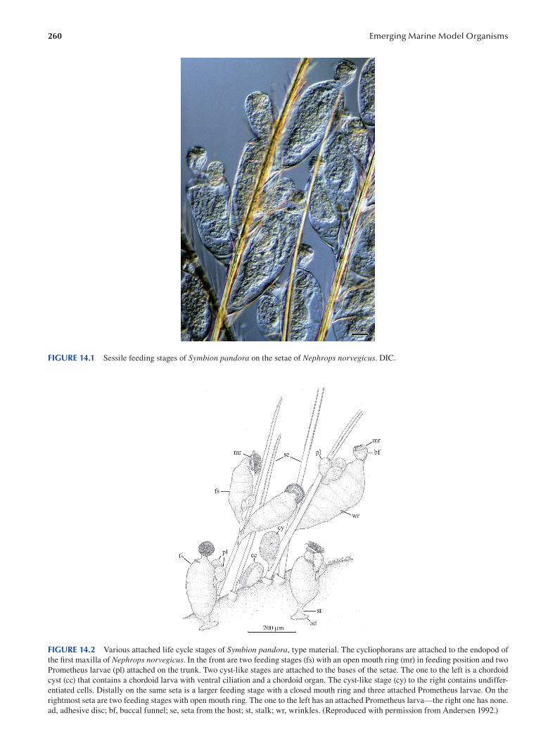
260 Emerging Marine Model Organisms
FIGURE 14.1 Sessile feeding stages of Symbion pandora on the setae of Nephrops norvegicus. DIC.
FIGURE 14.2 Various attached life cycle stages of Symbion pandora, type material. The cycliophorans are attached to the endopod of
the first maxilla of Nephrops norvegicus. In the front are two feeding stages (fs) with an open mouth ring (mr) in feeding position and two
Prometheus larvae (pl) attached on the trunk. Two cyst-like stages are attached to the bases of the setae. The one to the left is a chordoid
cyst (cc) that contains a chordoid larva with ventral ciliation and a chordoid organ. The cyst-like stage (cy) to the right contains undiffer
entiated cells. Distally on the same seta is a larger feeding stage with a closed mouth ring and three attached Prometheus larvae. On the
rightmost seta are two feeding stages with open mouth ring. The one to the left has an attached Prometheus larva—the right one has none.
ad, adhesive disc; bf, buccal funnel; se, seta from the host; st, stalk; wr, wrinkles. (Reproduced with permission from Andersen 1992.)
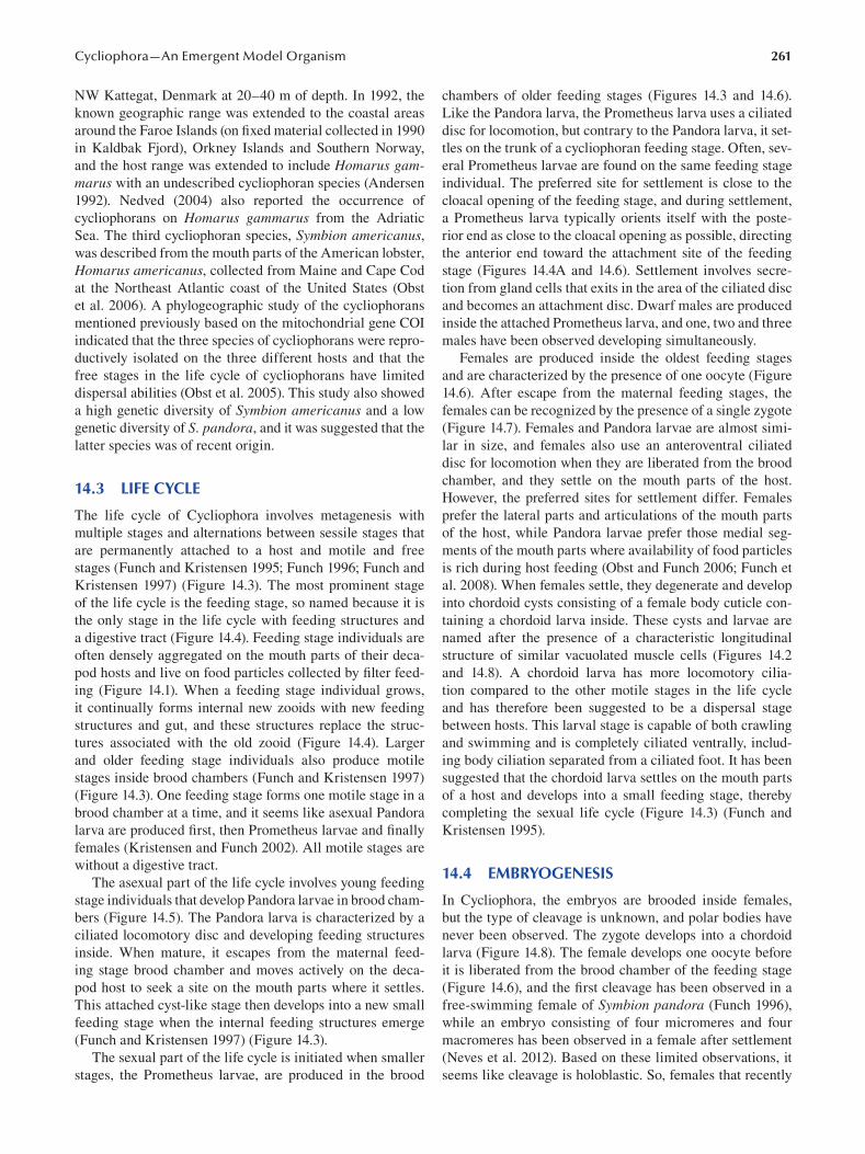
261 Cycliophora—An Emergent Model Organism
NW Kattegat, Denmark at 20–40 m of depth. In 1992, the
known geographic range was extended to the coastal areas
around the Faroe Islands (on fixed material collected in 1990
in Kaldbak Fjord), Orkney Islands and Southern Norway,
and the host range was extended to include Homarus gammarus with an undescribed cycliophoran species (Andersen
1992). Nedved (2004) also reported the occurrence of
cycliophorans on Homarus gammarus from the Adriatic
Sea. The third cycliophoran species, Symbion americanus, was described from the mouth parts of the American lobster,
Homarus americanus, collected from Maine and Cape Cod
at the Northeast Atlantic coast of the United States (Obst
et al. 2006). A phylogeographic study of the cycliophorans
mentioned previously based on the mitochondrial gene COI
indicated that the three species of cycliophorans were repro
ductively isolated on the three different hosts and that the
free stages in the life cycle of cycliophorans have limited
dispersal abilities (Obst et al. 2005). This study also showed
a high genetic diversity of Symbion americanus and a low
genetic diversity of S. pandora, and it was suggested that the
latter species was of recent origin.
14.3 LIFE CYCLE
The life cycle of Cycliophora involves metagenesis with
multiple stages and alternations between sessile stages that
are permanently attached to a host and motile and free
stages (Funch and Kristensen 1995; Funch 1996; Funch and
Kristensen 1997 ) (Figure 14.3). The most prominent stage
of the life cycle is the feeding stage, so named because it is
the only stage in the life cycle with feeding structures and
a digestive tract (Figure 14.4). Feeding stage individuals are
often densely aggregated on the mouth parts of their deca
pod hosts and live on food particles collected by fi lter feed
ing (Figure 14.1). When a feeding stage individual grows,
it continually forms internal new zooids with new feeding
structures and gut, and these structures replace the struc
tures associated with the old zooid (Figure 14.4). Larger
and older feeding stage individuals also produce motile
stages inside brood chambers (Funch and Kristensen 1997 )
(Figure 14.3). One feeding stage forms one motile stage in a
brood chamber at a time, and it seems like asexual Pandora
larva are produced first, then Prometheus larvae and fi nally
females (Kristensen and Funch 2002). All motile stages are
without a digestive tract.
The asexual part of the life cycle involves young feeding
stage individuals that develop Pandora larvae in brood cham
bers (Figure 14.5). The Pandora larva is characterized by a
ciliated locomotory disc and developing feeding structures
inside. When mature, it escapes from the maternal feed
ing stage brood chamber and moves actively on the deca
pod host to seek a site on the mouth parts where it settles.
This attached cyst-like stage then develops into a new small
feeding stage when the internal feeding structures emerge
(Funch and Kristensen 1997 ) (Figure 14.3).
The sexual part of the life cycle is initiated when smaller
stages, the Prometheus larvae, are produced in the brood
chambers of older feeding stages (Figures 14.3 and 14.6 ).
Like the Pandora larva, the Prometheus larva uses a ciliated
disc for locomotion, but contrary to the Pandora larva, it set
tles on the trunk of a cycliophoran feeding stage. Often, sev
eral Prometheus larvae are found on the same feeding stage
individual. The preferred site for settlement is close to the
cloacal opening of the feeding stage, and during settlement,
a Prometheus larva typically orients itself with the poste
rior end as close to the cloacal opening as possible, directing
the anterior end toward the attachment site of the feeding
stage (Figures 14.4A and 14.6). Settlement involves secre
tion from gland cells that exits in the area of the ciliated disc
and becomes an attachment disc. Dwarf males are produced
inside the attached Prometheus larva, and one, two and three
males have been observed developing simultaneously.
Females are produced inside the oldest feeding stages
and are characterized by the presence of one oocyte (Figure
14.6). After escape from the maternal feeding stages, the
females can be recognized by the presence of a single zygote
(Figure 14.7). Females and Pandora larvae are almost simi
lar in size, and females also use an anteroventral ciliated
disc for locomotion when they are liberated from the brood
chamber, and they settle on the mouth parts of the host.
However, the preferred sites for settlement differ. Females
prefer the lateral parts and articulations of the mouth parts
of the host, while Pandora larvae prefer those medial seg
ments of the mouth parts where availability of food particles
is rich during host feeding (Obst and Funch 2006; Funch et
al. 2008). When females settle, they degenerate and develop
into chordoid cysts consisting of a female body cuticle con
taining a chordoid larva inside. These cysts and larvae are
named after the presence of a characteristic longitudinal
structure of similar vacuolated muscle cells (Figures 14.2
and 14.8). A chordoid larva has more locomotory cilia
tion compared to the other motile stages in the life cycle
and has therefore been suggested to be a dispersal stage
between hosts. This larval stage is capable of both crawling
and swimming and is completely ciliated ventrally, includ
ing body ciliation separated from a ciliated foot. It has been
suggested that the chordoid larva settles on the mouth parts
of a host and develops into a small feeding stage, thereby
completing the sexual life cycle (Figure 14.3) (Funch and
Kristensen 1995).
14.4 EMBRYOGENESIS
In Cycliophora, the embryos are brooded inside females,
but the type of cleavage is unknown, and polar bodies have
never been observed. The zygote develops into a chordoid
larva (Figure 14.8). The female develops one oocyte before
it is liberated from the brood chamber of the feeding stage
(Figure 14.6 ), and the first cleavage has been observed in a
free-swimming female of Symbion pandora ( Funch 1996 ),
while an embryo consisting of four micromeres and four
macromeres has been observed in a female after settlement
(Neves et al. 2012). Based on these limited observations, it
seems like cleavage is holoblastic. So, females that recently
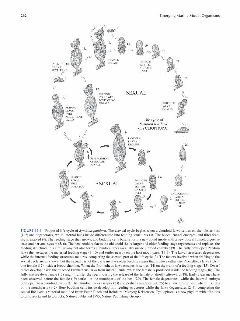
262 Emerging Marine Model Organisms
FIGURE 14.3 Proposed life cycle of Symbion pandora. The asexual cycle begins when a chordoid larva settles on the lobster host
(1–2) and degenerates, while internal buds inside differentiate into feeding structures (3). The buccal funnel emerges, and fi lter feed
ing is enabled (4). The feeding stage then grows, and budding cells basally form a new zooid inside with a new buccal funnel, digestive
tract and nervous system (5, 6). The new zooid replaces the old zooid (8). A larger and older feeding stage regenerates and replaces the
feeding structures in a similar way but also forms a Pandora larva asexually inside a brood chamber (9). The fully developed Pandora
larva then escapes the maternal feeding stage (9–10) and settles nearby on the host mouthparts (11–3). The larval structures degenerate,
while the internal feeding structures matures, completing the asexual part of the life cycle (3). The factors involved when shifting to the
sexual cycle are unknown, but the sexual part of the cycle involves older feeding stages that produce either one Prometheus larva (13) or
one female (12) inside a brood chamber. When the Prometheus larva escapes, it settles (14) on the trunk of a feeding stage (15). Dwarf
males develop inside the attached Prometheus larva from internal buds, while the female is produced inside the feeding stage (16). The
fully mature dwarf male (17) might transfer the sperm during the release of the female or shortly afterward (18). Early cleavages have
been observed before the female (19) settles on the mouthparts of the host (20). The female degenerates, while the internal embryo
develops into a chordoid cyst (21). The chordoid larva escapes (23) and perhaps migrates (24, 25) to a new lobster host, where it settles
on the mouthparts (1–2). Here budding cells inside develop into feeding structures while the larva degenerates (2–3), completing the
sexual life cycle. (Material modified from: Peter Funch and Reinhardt Møbjerg Kristensen, Cycliophora is a new phylum with affi nities
to Entoprocta and Ectoprocta, Nature, published 1995, Nature Publishing Group.)
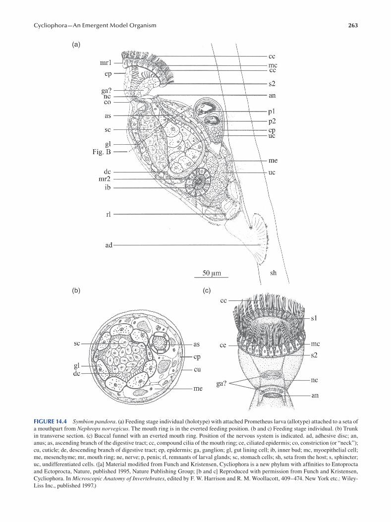
263 Cycliophora—An Emergent Model Organism
FIGURE 14.4 Symbion pandora. (a) Feeding stage individual (holotype) with attached Prometheus larva (allotype) attached to a seta of
a mouthpart from Nephrops norvegicus. The mouth ring is in the everted feeding position. (b and c) Feeding stage individual. (b) Trunk
in transverse section. (c) Buccal funnel with an everted mouth ring. Position of the nervous system is indicated. ad, adhesive disc; an,
anus; as, ascending branch of the digestive tract; cc, compound cilia of the mouth ring; ce, ciliated epidermis; co, constriction (or “neck”);
cu, cuticle; de, descending branch of digestive tract; ep, epidermis; ga, ganglion; gl, gut lining cell; ib, inner bud; mc, myoepithelial cell;
me, mesenchyme; mr, mouth ring; ne, nerve; p, penis; rl, remnants of larval glands; sc, stomach cells; sh, seta from the host; s, sphincter;
uc, undifferentiated cells. ([a] Material modified from Funch and Kristensen, Cycliophora is a new phylum with affinities to Entoprocta
and Ectoprocta, Nature, published 1995, Nature Publishing Group; [b and c] Reproduced with permission from Funch and Kristensen,
Cycliophora. In Microscopic Anatomy of Invertebrates, edited by F. W. Harrison and R. M. Woollacott, 409–474. New York etc.: Wiley-
Liss Inc., published 1997.)
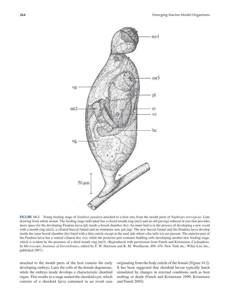
264 Emerging Marine Model Organisms
FIGURE 14.5 Young feeding stage of Symbion pandora attached to a host seta from the mouth parts of Nephrops norvegicus . Line
drawing from whole mount. The feeding stage individual has a closed mouth ring (mr1) and an old gut (og) reduced in size that provides
more space for the developing Pandora larva (pl) inside a brood chamber (bc). An inner bud is in the process of developing a new zooid
with a mouth ring (mr2), a ciliated buccal funnel and an immature new gut (ng). The new buccal funnel and the Pandora larva develop
inside the same brood chamber (bc) lined with a thin cuticle except at the anal side where cilia tufts (ct) are present. The anterior part of
the Pandora larva has a ventral ciliated disc (vc), while the posterior part contains budding cells developing another new feeding stage,
which is evident be the presence of a third mouth ring (mr3). (Reproduced with permission from Funch and Kristensen, Cycliophora.
In Microscopic Anatomy of Invertebrates, edited by F. W. Harrison and R. M. Woollacott, 409–474. New York etc.: Wiley-Liss Inc.,
published 1997.)
attached to the mouth parts of the host contain the early originating from the body cuticle of the female (Figure 14.2).
developing embryo. Later the cells of the female degenerate, It has been suggested that chordoid larvae typically hatch
while the embryo inside develops a characteristic chordoid stimulated by changes in external conditions such as host
organ. This results in a stage named the chordoid cyst, which molting or death (Funch and Kristensen 1999; Kristensen
consists of a chordoid larva contained in an ovoid case and Funch 2002).
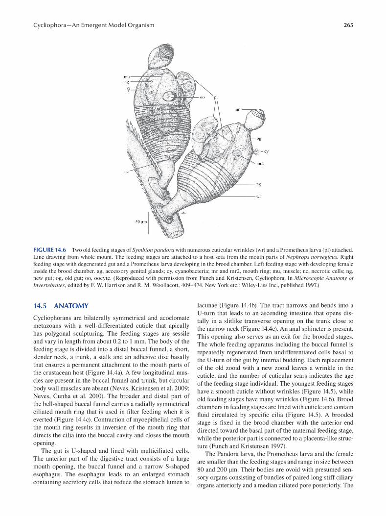
265 Cycliophora—An Emergent Model Organism
FIGURE 14.6 Two old feeding stages of Symbion pandora with numerous cuticular wrinkles (wr) and a Prometheus larva (pl) attached.
Line drawing from whole mount. The feeding stages are attached to a host seta from the mouth parts of Nephrops norvegicus . Right
feeding stage with degenerated gut and a Prometheus larva developing in the brood chamber. Left feeding stage with developing female
inside the brood chamber. ag, accessory genital glands; cy, cyanobacteria; mr and mr2, mouth ring; mu, muscle; nc, necrotic cells; ng,
new gut; og, old gut; oo, oocyte. (Reproduced with permission from Funch and Kristensen, Cycliophora. In Microscopic Anatomy of Invertebrates, edited by F. W. Harrison and R. M. Woollacott, 409–474. New York etc.: Wiley-Liss Inc., published 1997.)
14.5 ANATOMY
Cycliophorans are bilaterally symmetrical and acoelomate
metazoans with a well-differentiated cuticle that apically
has polygonal sculpturing. The feeding stages are sessile
and vary in length from about 0.2 to 1 mm. The body of the
feeding stage is divided into a distal buccal funnel, a short,
slender neck, a trunk, a stalk and an adhesive disc basally
that ensures a permanent attachment to the mouth parts of
the crustacean host (Figure 14.4a). A few longitudinal mus
cles are present in the buccal funnel and trunk, but circular
body wall muscles are absent (Neves, Kristensen et al. 2009;
Neves, Cunha et al. 2010). The broader and distal part of
the bell-shaped buccal funnel carries a radially symmetrical
ciliated mouth ring that is used in filter feeding when it is
everted (Figure 14.4c). Contraction of myoepithelial cells of
the mouth ring results in inversion of the mouth ring that
directs the cilia into the buccal cavity and closes the mouth
opening.
The gut is U-shaped and lined with multiciliated cells.
The anterior part of the digestive tract consists of a large
mouth opening, the buccal funnel and a narrow S-shaped
esophagus. The esophagus leads to an enlarged stomach
containing secretory cells that reduce the stomach lumen to
lacunae (Figure 14.4b). The tract narrows and bends into a
U-turn that leads to an ascending intestine that opens dis
tally in a slitlike transverse opening on the trunk close to
the narrow neck (Figure 14.4c). An anal sphincter is present.
This opening also serves as an exit for the brooded stages.
The whole feeding apparatus including the buccal funnel is
repeatedly regenerated from undifferentiated cells basal to
the U-turn of the gut by internal budding. Each replacement
of the old zooid with a new zooid leaves a wrinkle in the
cuticle, and the number of cuticular scars indicates the age
of the feeding stage individual. The youngest feeding stages
have a smooth cuticle without wrinkles (Figure 14.5), while
old feeding stages have many wrinkles (Figure 14.6). Brood
chambers in feeding stages are lined with cuticle and contain
fluid circulated by specific cilia (Figure 14.5). A brooded
stage is fixed in the brood chamber with the anterior end
directed toward the basal part of the maternal feeding stage,
while the posterior part is connected to a placenta-like struc
ture (Funch and Kristensen 1997 ).
The Pandora larva, the Prometheus larva and the female
are smaller than the feeding stages and range in size between
80 and 200 μm. Their bodies are ovoid with presumed sen
sory organs consisting of bundles of paired long stiff ciliary
organs anteriorly and a median ciliated pore posteriorly. The
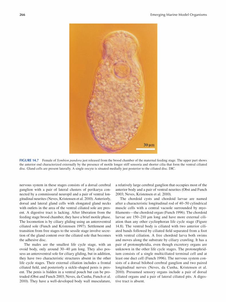
266 Emerging Marine Model Organisms
FIGURE 14.7 Female of Symbion pandora just released from the brood chamber of the maternal feeding stage. The upper part shows
the anterior end characterized externally by the presence of motile longer stiff sensoria and shorter cilia that form the ventral ciliated
disc. Gland cells are present laterally. A single oocyte is situated medially just posterior to the ciliated disc. DIC.
nervous system in these stages consists of a dorsal cerebral
ganglion with a pair of lateral clusters of perikarya con
nected by a commissural neuropil and a pair of ventral lon
gitudinal neurites (Neves, Kristensen et al. 2010). Anteriorly,
dorsal and lateral gland cells with elongated gland necks
with outlets in the area of the ventral ciliated sole are pres
ent. A digestive tract is lacking. After liberation from the
feeding stage brood chamber, they have a brief motile phase.
The locomotion is by ciliary gliding using an anteroventral
ciliated sole (Funch and Kristensen 1997 ). Settlement and
transition from free stages to the sessile stage involve secre
tion of the gland content over the ciliated sole that becomes
the adhesive disc.
The males are the smallest life cycle stage, with an
ovoid body, only around 30–40 μm long. They also pos
sess an anteroventral sole for ciliary gliding, but in addition,
they have two characteristic structures absent in the other
life cycle stages. Their external ciliation includes a frontal
ciliated field, and posteriorly a sickle-shaped penis is pres
ent. The penis is hidden in a ventral pouch but can be pro
truded (Obst and Funch 2003; Neves, da Cunha, Funch et al.
2010). They have a well-developed body wall musculature,
a relatively large cerebral ganglion that occupies most of the
anterior body and a pair of ventral neurites (Obst and Funch
2003; Neves, Kristensen et al. 2010).
The chordoid cysts and chordoid larvae are named
after a characteristic longitudinal rod of 40–50 cylindrical
muscle cells with a central vacuole surrounded by myo
filaments—the chordoid organ (Funch 1996 ). The chordoid
larvae are 150–210 μm long and have more external cili
ation than any other cycliophoran life cycle stage (Figure
14.8). The ventral body is ciliated with two anterior cili
ated bands followed by ciliated fi eld separated from a foot
with ventral ciliation. A free chordoid larva both swims
and moves along the substrate by ciliary crawling. It has a
pair of protonephridia, even though excretory organs are
unknown in the other life cycle stages. The protonephrid
ium consists of a single multiciliated terminal cell and at
least one duct cell (Funch 1996 ). The nervous system con
sists of a dorsal bilobed cerebral ganglion and two paired
longitudinal nerves (Neves, da Cunha, Kristensen et al.
2010). Presumed sensory organs include a pair of dorsal
ciliated organs and a pair of lateral ciliated pits. A diges
tive tract is absent.
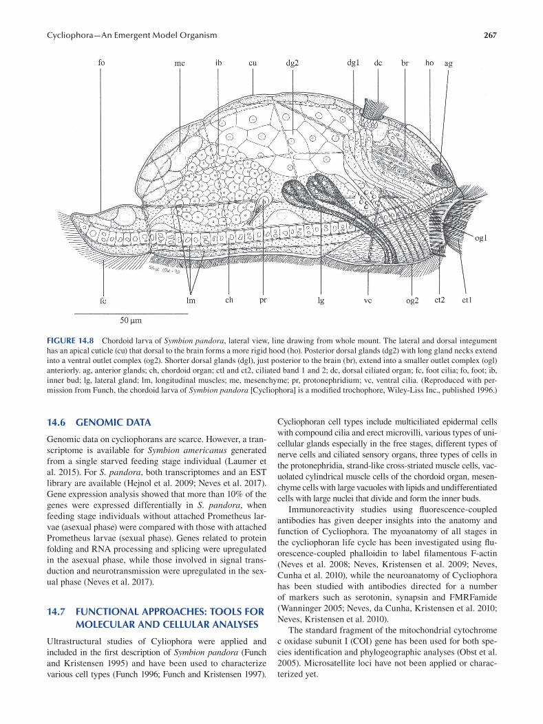
267 Cycliophora—An Emergent Model Organism
FIGURE 14.8 Chordoid larva of Symbion pandora, lateral view, line drawing from whole mount. The lateral and dorsal integument
has an apical cuticle (cu) that dorsal to the brain forms a more rigid hood (ho). Posterior dorsal glands (dg2) with long gland necks extend
into a ventral outlet complex (og2). Shorter dorsal glands (dgl), just posterior to the brain (br), extend into a smaller outlet complex (ogl)
anteriorly. ag, anterior glands; ch, chordoid organ; ctl and ct2, ciliated band 1 and 2; dc, dorsal ciliated organ; fc, foot cilia; fo, foot; ib,
inner bud; lg, lateral gland; lm, longitudinal muscles; me, mesenchyme; pr, protonephridium; vc, ventral cilia. (Reproduced with per
mission from Funch, the chordoid larva of Symbion pandora [Cycliophora] is a modified trochophore, Wiley-Liss Inc., published 1996.)
14.6 GENOMIC DATA
Genomic data on cycliophorans are scarce. However, a tran
scriptome is available for Symbion americanus generated
from a single starved feeding stage individual (Laumer et
al. 2015). For S. pandora, both transcriptomes and an EST
library are available (Hejnol et al. 2009; Neves et al. 2017).
Gene expression analysis showed that more than 10% of the
genes were expressed differentially in S. pandora , when
feeding stage individuals without attached Prometheus lar
vae (asexual phase) were compared with those with attached
Prometheus larvae (sexual phase). Genes related to protein
folding and RNA processing and splicing were upregulated
in the asexual phase, while those involved in signal trans
duction and neurotransmission were upregulated in the sex
ual phase (Neves et al. 2017).
14.7 FUNCTIONAL APPROACHES: TOOLS FOR MOLECULAR AND CELLULAR ANALYSES
Ultrastructural studies of Cyliophora were applied and
included in the first description of Symbion pandora ( Funch
and Kristensen 1995) and have been used to characterize
various cell types (Funch 1996; Funch and Kristensen 1997 ).
Cycliophoran cell types include multiciliated epidermal cells
with compound cilia and erect microvilli, various types of uni
cellular glands especially in the free stages, different types of
nerve cells and ciliated sensory organs, three types of cells in
the protonephridia, strand-like cross-striated muscle cells, vac
uolated cylindrical muscle cells of the chordoid organ, mesen
chyme cells with large vacuoles with lipids and undifferentiated
cells with large nuclei that divide and form the inner buds.
Immunoreactivity studies using fl uorescence-coupled
antibodies has given deeper insights into the anatomy and
function of Cycliophora. The myoanatomy of all stages in
the cycliophoran life cycle has been investigated using fl u
orescence-coupled phalloidin to label fi lamentous F-actin
(Neves et al. 2008; Neves, Kristensen et al. 2009; Neves,
Cunha et al. 2010), while the neuroanatomy of Cycliophora
has been studied with antibodies directed for a number
of markers such as serotonin, synapsin and FMRFamide
(Wanninger 2005; Neves, da Cunha, Kristensen et al. 2010;
Neves, Kristensen et al. 2010).
The standard fragment of the mitochondrial cytochrome
c oxidase subunit I (COI) gene has been used for both spe
cies identification and phylogeographic analyses (Obst et al.
2005). Microsatellite loci have not been applied or charac
terized yet.
268
14.8 CHALLENGING QUESTIONS BOTH IN ACADEMIC AND APPLIED RESEARCH
One of the challenging questions that remain to be answered
is the phylogenetic position of Cycliophora inside Spiralia.
Phylogenetic affinities to Bryozoa and Entoprocta were
suggested when Cycliophora was first described (Funch
and Kristensen 1995), and later a sister group relationship
between Entoprocta and Cycliophora was proposed (Funch
and Kristensen 1997; Zrzavy et al. 1998; Sørensen et al.
2000). These suggestions were supported by limited and
ambiguous morphological characters such as the presence
of asexual reproduction by internal budding, complete ner
vous system degeneration during the transition and settle
ment from smaller free motile life cycle stages to sessile
larger stages and mushroom-shaped extensions of the basal
lamina into the epidermis. An alternative hypothesis was
proposed based on only molecular data (18S rRNA), namely
Syndermata (Rotifera + Acanthocephala) as sister group to
Cycliophora (Winnepenninckx et al. 1998). This relation
ship to gnathiferan taxa was later supported in a number
of phylogenetic analyses (Giribet et al. 2000; Peterson and
Eernisse 2001; Zrzavy et al. 2001; Zrzavy 2003; Giribet et
al. 2004). In the latter study based on four molecular loci,
the phylogenetic position of Cycliophora was uncertain, but
it tended to support a relationship to Syndermata (Rotifera
+ Acanthocephala), but the morphological data supporting
this relationship were weak (Funch et al. 2005), although
Wanninger (2005) suggested similarities in myoanatomy
of the cycliophoran chordoid larva and certain rotifers.
Phylogenetic analyses using more molecular data resur
rected the cycliophoran affinity to entoprocts (Passamaneck
and Halanych 2006; Paps et al. 2009), enforced by phyloge
nomic analyses based on expressed sequence tags (Hejnol et
al. 2009; Nesnidal et al. 2013) and transcriptomes (Laumer
et al. 2015; Kocot et al. 2017; Laumer et al. 2019). So, while
the Cycliophora + Entoprocta clade seems to be well sup
ported, its placement within Spiralia is still unsettled.
There are numerous remaining questions to clarify
regarding the life cycle and reproduction in Cycliophora.
First, fertilization has never been observed. It is known
that females inside brood chambers have oocytes and that
free females have embryos (Figures 14.6 and 14.7). It seems
likely that fertilization could happen during escape or just
after escape of the female, which could explain the preferred
site for settlement of the Prometheus larva close to the clo
aca opening of the feeding stage, which is also the site of
escape of the female. Second, how do free motile stages
select the right site for settlement and permanent attach
ment? Pandora larvae and chordoid larvae seem to prefer the
same sites for settlement, namely the food-rich medial areas
of the mouth parts of the host. Females prefer to settle upon
areas of the mouth parts laterally, maybe because of less
mechanical stress and risk of dislocation by the movements
of the host, while Prometheus larvae settle upon feeding
stages that develop females inside. In spite of these differ
ences in preferred sites to settle, they are all equipped with
Emerging Marine Model Organisms
morphologically similar long stiff ciliary sensory organs
that are absent in the sessile stages in the life cycle. Most
likely, these sensory organs are involved in sensing and test
ing if a given substrate is suitable for settlement, but nothing
is known about the sensory physiology and type of mecha
nisms involved. The chordoid larva is equipped with more
types of sensory organs, probably because it is a dispersal
stage between hosts and uses some of these sensory organs
for long-distance sensing. Third, the sex determination sys
tem in Cycliophora is unknown. Is haplodiploidy involved,
and are cycliophoran dwarf males haploid like the males in,
for example, monogonont rotifers? Probably not. In mono
gonont rotifers, haploid males develop from unfertilized
meiotic eggs, while cycliophoran males seem to develop
asexually from budding cells. Finally, the mechanism for
shifting from asexual to sexual reproduction is unknown.
It is unknown if a feeding stage produces a fixed number of
Pandora larvae before the shift to sexual reproduction or if it
depends on population density of cycliophorans on the host
or food availability. Maybe starving of a feeding stage could
induce formation of a Prometheus larva instead of a Pandora
larva since the latter larva is large and requires more energy
to produce.
Dwarf males of Cycliophora consist of less than 200 cells
and have only few cell types (Obst and Funch 2003; Neves,
Sørensen et al. 2009; Neves and Reichert 2015). Still, the
body architecture is relatively complex, with well-developed
nervous system, sensory organs, musculature and reproduc
tive organs, which contradicts the general assumption about
correlation of complexity of the body plan and the number of
cells and cell types (Bell and Mooers 1997 ). Future explora
tion of the cycliophoran genome could provide new insights
into how high body plan complexity can be achieved with
few cells.
ACKNOWLEDGMENTS
I greatly acknowledge the collaboration with Stine Elle and
Birgitte Rubæk and Reinhardt Møbjerg Kristensen produc
ing the line drawings.
BIBLIOGRAPHY
Andersen, P. F. 1992. Beskrivelse af en ny klasse—Mesoprocta.
Master thesis, University of Copenhagen. Copenhagen.
Baker, J. M., P. Funch, and G. Giribet. 2007. Cryptic speciation
in the recently discovered American cycliophoran Symbion americanus: Genetic structure and population expansion.
Marine Biology 151 (6):2183–2193.
Baker, J. M., and G. Giribet. 2007. A molecular phylogenetic
approach to the phylum Cycliophora provides further
evidence for cryptic speciation in Symbion americanus.
Zoologica Scripta 36 (4):353–359.
Bell, G., and A. O. Mooers. 1997. Size and complexity among
multicellular organisms. Biological Journal of the Linnean Society 60 (3):345–363.
Clément, P., and E. Wurdak. 1991. Rotifera. In Microscopic Anatomy of Invertebrates, edited by F. W. Harrison and R.
M. Woollacott, 219–297. New York etc.: Wiley-Liss.
269 Cycliophora—An Emergent Model Organism
Funch, P. 1996. The chordoid larva of Symbion pandora (Cycliophora)
is a modified trochophore. Journal of Morphology 230 (3):
231–263.
Funch, P., and R. M. Kristensen. 1995. Cycliophora is a new phy
lum with affinities to Entoprocta and Ectoprocta. Nature 378
(6558):711–714.
Funch, P., and R. M. Kristensen. 1997. Cycliophora. In Microscopic Anatomy of Invertebrates, edited by F. W. Harrison and R. M.
Woollacott, 409–474. New York etc.: Wiley-Liss.
Funch, P., and R. M. Kristensen. 1999. Cycliophora. In Encyclopedia of Reproduction, edited by Ernst Knobil and Jimmy D. Neill,
800–808. San Diego: Academic Press.
Funch, P., and R. Neves. 2019. Cycliophora. In Miscellaneous Invertebrates, edited by A. Schmidt-Rhaesa, 87–110. Berlin/
Boston: Walter de Gruyter GmbH & Co. KG.
Funch, P., M. V. Sørensen, and M. Obst. 2005. On the phyloge
netic position of Rotifera: Have we come any further?
Hydrobiologia 546:11–28.
Funch, P., P. Thor, and M. Obst. 2008. Symbiotic relations and feed
ing biology of Symbion pandora (Cycliophora) and Triticella fl ava (Bryozoa). Vie et Milieu-Life and Environment 58
(2):185–188.
Giribet, G., D. L. Distel, M. Polz, W. Sterrer, and W. C. Wheeler.
2000. Triploblastic relationships with emphasis on the acoe
lomates and the position of Gnathostomulida, Cycliophora,
Plathelminthes, and Chaetognatha: A combined approach of
18S rDNA sequences and morphology. Systematic Biology 49 (3):539–562.
Giribet, G., M. V. Sørensen, P. Funch, R. M. Kristensen, and W.
Sterrer. 2004. Investigations into the phylogenetic position
of Micrognathozoa using four molecular loci. Cladistics 20
(1):1–13.
Hejnol, A., M. Obst, A. Stamatakis, M. Ott, G. W. Rouse, G. D.
Edgecombe, P. Martinez, J. Baguna, X. Bailly, U. Jondelius,
M. Wiens, W. E. G. Muller, E. Seaver, W. C. Wheeler, M. Q.
Martindale, G. Giribet, and C. W. Dunn. 2009. Assessing the
root of bilaterian animals with scalable phylogenomic meth
ods. Proceedings of the Royal Society B-Biological Sciences 276 (1677):4261–4270.
Kocot, K. M., T. H. Struck, J. Merkel, D. S. Waits, C. Todt, P.
M. Brannock, D. A. Weese, J. T. Cannon, L. L. Moroz,
B. Lieb, and K. M. Halanych. 2017. Phylogenomics of
Lophotrochozoa with consideration of systematic error.
Systematic Biology 66 (2):256–282.
Kristensen, R. M. 2002. An introduction to Loricifera, Cycliophora,
and Micrognathozoa. Integrative and Comparative Biology 42 (3):641–651.
Kristensen, R. M., and P. Funch. 2002. Phylum Cycliophora. In
Atlas of Marine Invertebrate Larvae, edited by Craig M.
Young, M. A. Sewell and M. Rice, 231–240. San Diego, CA:
Academic Press.
Laumer, C. E., N. Bekkouche, A. Kerb, F. Goetz, R. C. Neves, M.
V. Sørensen, R. M. Kristensen, A. Hejno, C. W. Dunn, G.
Giribet, and K. Worsaae. 2015. Spiralian phylogeny informs
the evolution of microscopic lineages. Current Biology 25
(15):2000–2006.
Laumer, C. E., R. Fernandez, S. Lemer, D. Combosch, K. M.
Kocots, A. Riesgo, S. C. S. Andrade, W. Sterrer, M. V.
Sørensen, and G. Giribet. 2019. Revisiting metazoan phylo
geny with genomic sampling of all phyla. Proceedings of the Royal Society B-Biological Sciences 286 (1906).
Nesnidal, M. P., M. Helmkampf, A. Meyer, A. Witek, I. Bruchhaus,
I. Ebersberger, T. Hankeln, B. Lieb, T. H. Struck, and
B. Hausdorf. 2013. New phylogenomic data support the
monophyly of Lophophorata and an ectoproct-phoronid
clade and indicate that Polyzoa and Kryptrochozoa are
caused by systematic bias. BMC Evolutionary Biology 13.
Neves, R. C., X. Bailly, and H. Reichert. 2014. Are copepods
secondary hosts of Cycliophora? Organisms Diversity & Evolution 14 (4):363–367.
Neves, R. C., M. R. Cunha, P. Funch, R. M. Kristensen, and A.
Wanninger. 2010. Comparative myoanatomy of cycliophoran
life cycle stages. Journal of Morphology 271 (5):596–611.
Neves, R. C., M. R. da Cunha, P. Funch, A. Wanninger, and R. M.
Kristensen. 2010. External morphology of the cycliophoran
dwarf male: A comparative study of Symbion pandora and
S. americanus. Helgoland Marine Research 64 (3):257–262.
Neves, R. C., M. R. da Cunha, R. M. Kristensen, and A. Wanninger.
2010. Expression of synapsin and co-localization with sero
tonin and RFamide-like immunoreactivity in the nervous sys
tem of the chordoid larva of Symbion pandora (Cycliophora).
Invertebrate Biology 129 (1):17–26.
Neves, R. C., J. C. Guimaraes, S. Strempel, and H. Reichert.
2017. Transcriptome profiling of Symbion pandora (phylum
Cycliophora): Insights from a differential gene expression
analysis. Organisms Diversity & Evolution 17 (1):111–119.
Neves, R. C., R. M. Kristensen, and P. Funch. 2012. Ultrastructure
and morphology of the cycliophoran female. Journal of Morphology 273 (8):850–869.
Neves, R., R. M. Kristensen, and A. Wanninger. 2008. New
insights from the myoanatomy of Symbion americanus (phylum Cycliophora) revealed by confocal microscopy
and 3D reconstruction software. Journal of Morphology 269
(12):1480–1480.
Neves, R. C., R. M. Kristensen, and A. Wanninger. 2009. Three-
dimensional reconstruction of the musculature of various
life cycle stages of the cycliophoran Symbion americanus.
Journal of Morphology 270 (3):257–270.
Neves, R. C., R. M. Kristensen, and A. Wanninger. 2010. Serotonin
immunoreactivity in the nervous system of the Pandora larva,
the Prometheus larva, and the dwarf male of Symbion americanus (Cycliophora). Zoologischer Anzeiger 249 (1):1–12.
Neves, R. C., and H. Reichert. 2015. Microanatomy and devel
opment of the dwarf male of Symbion pandora (phylum
Cycliophora): New insights from ultrastructural investigation
based on serial section electron microscopy. PLoS One 10 (4).
Neves, R. C., K. J. K. Sørensen, R. M. Kristensen, and A.
Wanninger. 2009. Cycliophoran dwarf males break the rule:
High complexity with low cell numbers. Biological Bulletin 217 (1):2–5.
Obst, M., and P. Funch. 2003. Dwarf male of Symbion pandora (Cycliophora). Journal of Morphology 255 (3):261–278.
Obst, M., and P. Funch. 2006. The microhabitat of Symbion pandora (Cycliophora) on the mouthparts of its host Nephrops norvegicus (Decapoda: Nephropidae). Marine Biology 148
(5):945–951.
Obst, M., P. Funch, and G. Giribet. 2005. Hidden diversity and host
specificity in cycliophorans: A phylogeographic analysis
along the North Atlantic and Mediterranean Sea. Molecular Ecology 14 (14):4427–4440.
Obst, M., P. Funch, and R. M. Kristensen. 2006. A new species of
Cycliophora from the mouthparts of the American lobster,
Homarus americanus (Nephropidae, Decapoda). Organisms Diversity & Evolution 6 (2):83–97.
Paps, J., J. Baguna, and M. Riutort. 2009. Lophotrochozoa inter
nal phylogeny: New insights from an up-to-date analysis of
nuclear ribosomal genes. Proceedings of the Royal Society B-Biological Sciences 276 (1660):1245–1254.
270
Passamaneck, Y., and K. M. Halanych. 2006. Lophotrochozoan
phylogeny assessed with LSU and SSU data: evidence of
lophophorate polyphyly. Molecular Phylogenetics and Evolution 40 (1):20–28.
Peterson, K. J., and D. J. Eernisse. 2001. Animal phylogeny and
the ancestry of bilaterians: Inferences from morphology and
18S rDNA gene sequences. Evolution & Development 3
(3):170–205.
Plaza, M. 2012. Cycliophoran host range. Bulletin of the Peabody Museum of Natural History 53 (2):389–395.
Sørensen, M. V., P. Funch, E. Willerslev, A. J. Hansen, and J.
Olesen. 2000. On the phylogeny of the Metazoa in the light
of Cycliophora and Micrognathozoa. Zoologischer Anzeiger 239 (3–4):297–318.
Emerging Marine Model Organisms
Wanninger, A. 2005. Immunocytochemistry of the nervous system
and the musculature of the chordoid larva of Symbion pandora (Cycliophora). Journal of Morphology 265 (2):237–243.
Winnepenninckx, B. M. H., T. Backeljau, and R. M. Kristensen.
1998. Relations of the new phylum Cycliophora. Nature 393
(6686):636–638.
Zrzavy, J. 2003. Gastrotricha and metazoan phylogeny. Zoologica Scripta 32 (1):61–81.
Zrzavy, J., V. Hypsa, and D. F. Tietz. 2001. Myzostomida are not
annelids: Molecular and morphological support for a clade of
animals with anterior sperm fl agella. Cladistics 17 (2):170–198.
Zrzavy, J., S. Mihulka, P. Kepka, A. Bezdek, and D. Tietz. 1998.
Phylogeny of the Metazoa based on morphological and 18S
ribosomal DNA evidence. Cladistics 14 (3):249–285.
15 Crustaceans
Nicolas Rabet
CONTENTS
15.1 History of the Model..................................................................................................................................................271
15.2 Geographical Location .............................................................................................................................................. 273
15.3 Life Cycle ...................................................................................................................................................................274
15.4 Embryogenesis and Larval Development ..................................................................................................................275
15.4.1 Embryogenesis .............................................................................................................................................275
15.4.2 Larval Development .....................................................................................................................................275
15.5 Anatomy.................................................................................................................................................................... 278
15.6 Genomic Data ........................................................................................................................................................... 279
15.7 Functional Approaches: Tools for Molecular and Cellular Analyses........................................................................ 279
15.8 Challenging Questions Both in Academic and Applied Research ............................................................................ 280
15.8.1 Crustaceans and Food .................................................................................................................................. 280
15.8.2 Biofouling.................................................................................................................................................... 280
15.8.3 Ecotoxicology...............................................................................................................................................281
15.8.4 Body Elongation and Segmentation .............................................................................................................281
15.8.5 Evolution of Ontogeny .................................................................................................................................281
15.8.6 Terrestrialization and Origin of Insects ........................................................................................................281
15.8.7 The Emergence of Parasitic Forms .............................................................................................................. 282
15.8.8 Evolution of Cryptobiosis ............................................................................................................................ 282
Bibliography ........................................................................................................................................................................ 282
15.1 HISTORY OF THE MODEL Aptera and recognized only three genera: Cancer with mala-
The word crustacea is derived from the Latin crusta , whichbranchiura, other branchiopods, copepods, ostracods and
costracans and branchiopod anostracans; Monoculus with
means that the body is covered with a hard shell. The two taxa including horseshoe crabs (which are now excluded
name Crustacea was first proposed by Brünnich (1772). from crustaceans); and Oniscus, regrouped malacostracan
Nevertheless, it took decades for it to establish itself, and isopods. In addition, the cirripeds with genus Lepas was clas
the boundaries of the group have also changed signifi cantly. sified in the Vermes Testacea, while the parasitic copepods
Today, crustaceans are a paraphyletic group, representing with the genus Lernaea were classified among the Vermes
approximately 70,000 currently valid species distributed in Mollusca.
nearly 1,000 families and in 9 major lineages (Remipedia, Gradually, many species were described, and crusta-
Cephalocarida, Malacostraca, Copepoda, Thecostraca, ceans were separated from insects on the basis of having
Branchiopoda, Mystacocarida, Branchiura and Ostracoda) a predominantly aquatic life, the presence of two pairs of
( Ahyong et al. 2011; Regier et al. 2010 ). antennae, biramate appendages and a nauplius larva. Like
Large crustaceans (malacostracans and barnacles— the morpho-anatomical diversity of the group, its classifi
Figure 15.1) have always been known to humanity because cation has carried out numerous regroupings, and as such,
they have been eaten for thousands of years (Gutiérrez-many have been forgotten. The copepods, ostracods, bran-
Zugasti 2 011; Zilhão et al. 2020). It is therefore quite logical chiopods and cirripeds were gradually individualized and
that we can find crustaceans in old illustrations or in fi rst grouped in the entomostracans as opposed to the malacos
classifications. In Aristotle’s classification, some crustaceans tracans (see Monod and Forest 1996 ). In the 20th century,
were already listed under the name μαλακόστρακα (mala-new lineages of crustaceans were discovered, such as mys
kostraka), which means animals with soft (malakós ) shell tacocarids (Pennak and Zinn 1943), cephalocarids (Sanders
(óstrakon) (Zucker 2005). Even if the word Malacostraca 1955) and remipeds (Yager 1981). Bowman and Abele (1982)
evokes a classic name of the current classification, for a proposed a classification with six classes (Cephalocarida,
very long time, most crustaceans were integrated among Branchiopoda, Remipedia, Maxillopoda, Ostracoda and
the insects without a specific group. Others were ignored Malacostraca). The Maxillopoda grouped together the
or sometimes classified with other organisms. For example, Mystacocarida, Cirripedia, Copepoda and Branchiura.
Linnaeus (1758) classified some crustaceans in the order of
DOI: 10.1201/9781003217503-15 271
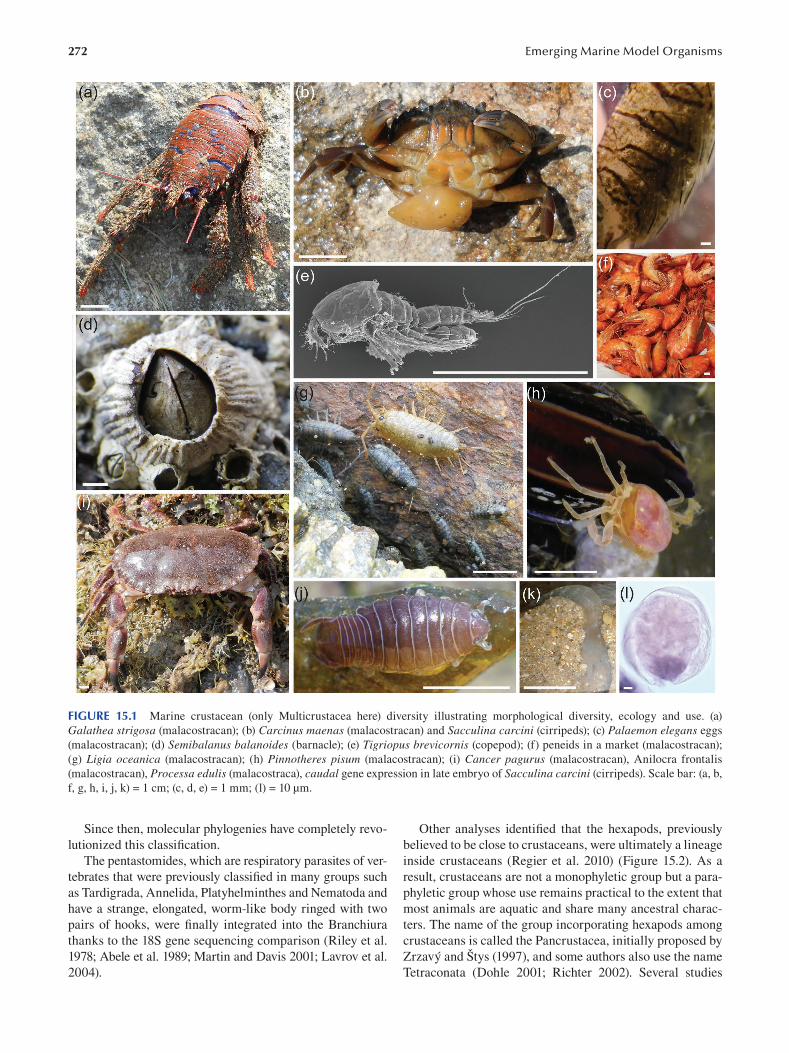
272 Emerging Marine Model Organisms
FIGURE 15.1 Marine crustacean (only Multicrustacea here) diversity illustrating morphological diversity, ecology and use. (a)
Galathea strigosa (malacostracan); (b) Carcinus maenas (malacostracan) and Sacculina carcini (cirripeds); (c) Palaemon elegans eggs
(malacostracan); (d) Semibalanus balanoides (barnacle); (e) Tigriopus brevicornis (copepod); (f) peneids in a market (malacostracan);
(g) Ligia oceanica (malacostracan); (h) Pinnotheres pisum (malacostracan); (i) Cancer pagurus (malacostracan), Anilocra frontalis
(malacostracan), Processa edulis (malacostraca), caudal gene expression in late embryo of Sacculina carcini (cirripeds). Scale bar: (a, b,
f, g, h, i, j, k) = 1 cm; (c, d, e) = 1 mm; (l) = 10 μm.
Since then, molecular phylogenies have completely revo- Other analyses identified that the hexapods, previously
lutionized this classifi cation. believed to be close to crustaceans, were ultimately a lineage
The pentastomides, which are respiratory parasites of ver- inside crustaceans (Regier et al. 2010) (Figure 15.2). As a
tebrates that were previously classified in many groups such result, crustaceans are not a monophyletic group but a para-
as Tardigrada, Annelida, Platyhelminthes and Nematoda and phyletic group whose use remains practical to the extent that
have a strange, elongated, worm-like body ringed with two most animals are aquatic and share many ancestral charac
pairs of hooks, were finally integrated into the Branchiura ters. The name of the group incorporating hexapods among
thanks to the 18S gene sequencing comparison (Riley et al. crustaceans is called the Pancrustacea, initially proposed by
1978; Abele et al. 1989; Martin and Davis 2001; Lavrov et al. Zrzavý and Štys (1997 ), and some authors also use the name
2004 ). Tetraconata ( Dohle 2001; Richter 2002 ). Several studies
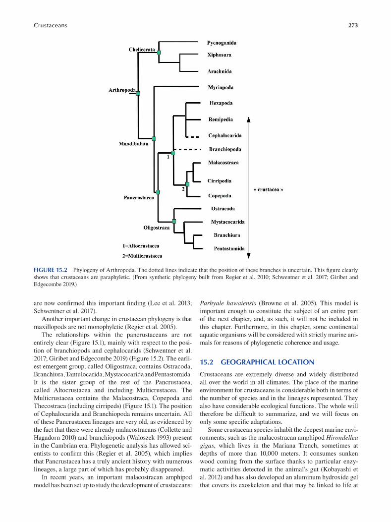
Crustaceans 273
FIGURE 15.2 Phylogeny of Arthropoda. The dotted lines indicate that the position of these branches is uncertain. This fi gure clearly
shows that crustaceans are paraphyletic. (From synthetic phylogeny built from Regier et al. 2010; Schwentner et al. 2017; Giribet and
Edgecombe 2019.)
are now confirmed this important finding (Lee et al. 2013;
Schwentner et al. 2017 ).
Another important change in crustacean phylogeny is that
maxillopods are not monophyletic (Regier et al. 2005).
The relationships within the pancrustaceans are not
entirely clear (Figure 15.1), mainly with respect to the posi
tion of branchiopods and cephalocarids (Schwentner et al.
2017; Giribet and Edgecombe 2019 ) ( Figure 15.2 ). The earli
est emergent group, called Oligostraca, contains Ostracoda,
Branchiura, Tantulocarida, Mystacocarida and Pentastomida.
It is the sister group of the rest of the Pancrustacea,
called Altocrustacea and including Multicrustacea. The
Multicrustacea contains the Malacostraca, Copepoda and
Thecostraca (including cirripeds) (Figure 15.1). The position
of Cephalocarida and Branchiopoda remains uncertain. All
of these Pancrustacea lineages are very old, as evidenced by
the fact that there were already malacostracans (Collette and
Hagadorn 2010) and branchiopods (Waloszek 1993) present
in the Cambrian era. Phylogenetic analysis has allowed sci
entists to confirm this (Regier et al. 2005), which implies
that Pancrustacea has a truly ancient history with numerous
lineages, a large part of which has probably disappeared.
In recent years, an important malacostracan amphipod
model has been set up to study the development of crustaceans:
Parhyale hawaiensis ( Browne et al. 2005 ). This model is
important enough to constitute the subject of an entire part
of the next chapter, and, as such, it will not be included in
this chapter. Furthermore, in this chapter, some continental
aquatic organisms will be considered with strictly marine ani
mals for reasons of phylogenetic coherence and usage.
15.2 GEOGRAPHICAL LOCATION
Crustaceans are extremely diverse and widely distributed
all over the world in all climates. The place of the marine
environment for crustaceans is considerable both in terms of
the number of species and in the lineages represented. They
also have considerable ecological functions. The whole will
therefore be difficult to summarize, and we will focus on
only some specifi c adaptations.
Some crustacean species inhabit the deepest marine envi
ronments, such as the malacostracan amphipod Hirondellea gigas, which lives in the Mariana Trench, sometimes at
depths of more than 10,000 meters. It consumes sunken
wood coming from the surface thanks to particular enzy
matic activities detected in the animal’s gut (Kobayashi et
al. 2012) and has also developed an aluminum hydroxide gel
that covers its exoskeleton and that may be linked to life at
274
great depths (Kobayashi et al. 2019). In the deep sea, there
are also many crustaceans that live around hydrothermal
vents. Many of them use chemo-autotrophic bacteria that
provide nutrients to animals. This is particularly the case
with the malacostracan Rimicaris exoculata on the Mid-
Atlantic Ridge, which harbors bacterial communities in its
branchial cavities (Petersen et al. 2010; Zbinden et al. 2020).
The diversity of crustaceans is also considerable in the
tidal zone, with some species able to survive conditions that
vary according to the water level variations. Some, like the
malacostracan Carcinus maenas (Figure 15.1), are able to
temporarily acclimatize to the absence of water and resist
consequent variations in the environment. Native to Europe,
this particularly well-adapted species has colonized many
temperate sites around the world (Jensen et al. 2007). In pools
of the highest tidal levels, we can often observe copepods
Tigriopus (Figure 15.1), which are also impacted by high
temperatures and consequent variations in salinity (Fraser
1936; Raisuddin et al. 2007). As in many groups, underwa
ter caves have also been colonized and can be the refuge of
many specialized and original organisms. Among these are
remipeds, a group of blind, predatory crustaceans that inhabit
anchialine underwater caves (Yager 1981; Koenemann et al.
2007). These are also the only venomous crustaceans (von
Reumont et al. 2014). Among the meiofauna, there are many
species of crustaceans such as copepods and ostracods living
in sediments. It is also in this type of biotope that we can fi nd
the odd cephalocarids (Sanders 1955; Neiber et al. 2011).
Many crustaceans such as ostracods, malacostracans,
copepods and branchiopods have also colonized brackish
or fresh water. The border between the two environments is
not necessarily clear, and after passing through fresh water,
some organisms then return to the marine environment, such
as the marine cladocerans that represent few species but have
a global distribution (Durbin et al. 2008). The hypersaline
environments that form in coastal areas or sometimes in the
middle of continents have also been colonized by crusta
ceans, in particular ostracods and copepods. However, the
champion of resistance is unmistakably the branchiopod
Artemia, which can survive in supersaturated salty environ
ments up to 340 g/l (Gajardo and Beardmore 2012).
As it is sometimes difficult to dissociate marine crusta
ceans from freshwater or hypersaline crustaceans in an evo
lutionary way, they will be partially integrated in this chapter.
There have also been several colonizations by pancrusta
ceans of terrestrial environments such as hexapods or woodlice,
but there are also terrestrial lineages in the adult state whose
larvae are completely marine, as is the case for many terrestrial
crabs or terrestrial hermit crabs. In this category, there are the
largest land-living arthropods, like the coconut crab (Birgus latro) (Krieger et al. 2010). This hybrid lifestyle, which is also
found in amphidromic crustaceans (living partially in freshwa
ter and seawater), allows these animals to exploit the dispersive
abilities of marine planktonic life and to colonize more or less
isolated continental environments (Bauer 2013).
Crustaceans are also an essential component in the plank
ton of all seas. Some species live their entire life cycle as
Emerging Marine Model Organisms
plankton and play a major ecological role (copepods, euphau
siids). However, for many species, the passage through plank
ton is transient as part of a marine bentho-pelagic species or
many terrestrial or freshwater crustaceans.
Crustaceans are so ubiquitous, it is almost impossible to
study the aquatic environment without fi nding one!
15.3 LIFE CYCLE
In crustaceans, the life cycle presents extremely variable
modalities. The majority of species are gonochoric with
separate sexes, but there are cases of parthenogenesis in the
brine shrimp Artemia (Bowen et al. 1978) and many fresh
water and terrestrial species, probably due to the dispersive
advantage ( Scholtz et al. 2003; Kawai et al. 2009 ). There are
cases of simultaneous hermaphroditism (both type of gonads
are present simultaneously) in remipeds (Neiber et al. 2011),
cephalocarids ( Addis et al. 2012 ), cirripeds ( Charnov 1987)
and some branchiopods (Scanabissi and Mondini 2002;
Weeks et al. 2014). Sequential hermaphrodism (change of
sex during the life) is more observed in malacostracans
( Benvenuto and Weeks 2020 ).
The mating modalities are also extremely varied in crus
taceans and result in very different appendicular adaptations.
The most original is undoubtedly the presence of a long penis
in the barnacles which is always fixed and which compen
sates for the low mobility of the gametes (Barazandeh et al.
2013 ).
In most species, the mother will protect her offspring to
allow the release of larvae. However, most calanoid copepods,
euphausiids and dendrobranchiate decapods (Penaeoidea and
Sergestoidea) shed their eggs into the water column (Lindley
1997 ).
In many crustaceans, the instability of trophic resources
and living conditions has favored the development of a strat
egy of slowing down or stopping development during the
deficit season (Alekseev and Starobogatov 1996). In this
case, the eggs are laid and start diapausis. There are also
resistance forms in anhydrobiosis or cryptobiosis (absence
of metabolism with dehydration) (Fryer 1996; Alekseev and
Starobogatov 1996). This innovation sometimes concerns
the larvae, as in the copepod Metacyclops minutus ( Maier
1992), but more often, it is the embryo that enters a state of
suspended life. The embryo can be enveloped by different
layers of varying natures and becomes resistant to drying
out or freezing. In this form, we speak of a resting egg (also
called a “duration egg” or “cyst”), and, when conditions are
favorable, development resumes, leading to the release of a
larva or an aquatic juvenile (Brendonck 2008).
In a group of malacostracan shrimps of the Alpheidae
family, the existence of eusocial behavior has recently been
reported, such as is found in insects and vertebrates (Duffy
1996 ).
In many species, the larvae released after hatching
become planktonic. During this planktonic phase, the ani
mals grow and disperse. At the end of the larval stages, there
are animals whose adults remain in the plankton (many
275 Crustaceans
copepods, euphausiids) and others which emerge, most often
becoming benthic. Sometimes the modifications are brutal
and called metamorphosis for sessile animals, like in bar
nacles (Høeg and Møller 2006; Maruzzo et al. 2012). In this
group, the transformation will result in a completely fi xed
animal. The choice of the fixation site is therefore essen
tial for the survival of the individual, because it will sub
sequently have to withstand the conditions imposed by the
environment. Recruitment is carried out by olfaction through
antenna 1 of the substrate (Figure 15.4c). The bacterial fi lm
can be detected and, depending on its composition, induce
the attachment of the cyprid larva (Rajitha et al. 2020). The
presence of congeners due to the release of pheromones from
adults that are not always necessarily from the same species
is also an essential factor for fixation ( Abramova et al. 2019 ).
After an exploration phase using the attachment discs located
at the end of the antenna 1, the fi nal fixation is achieved by
the deposition of a cement comprised of lipids and phospho
proteins (Liang et al. 2019).
In parasitic crustaceans, the life cycle is often highly
modified. The most extensive parasitic life transformations
are found in pentastomids, copepods and cirripeds. Adults
are often very divergent from their non-parasitic parents, to
the point that association with a taxonomic group has only
been possible by studying the larval stages like for the cir
riped Rhizocephala (Thompson 1836) (Figure 15.1b) or more
recently by molecular data, like for the pentastomids (Abele et
al. 1989). Rhizocephalic cirripeds are parasites characterized
by considerable morphological transformations but also con
siderable modifications of their life cycle. The female larva
will transform into a kentrogon, a kind of injection system
that allows a few cells to invade the host, which will develop
into a network resembling roots and allowing it to feed. The
male larvae transform into trichogons and settle as hyperpara
sites on the females. The mature parasite profoundly modifi es
the physiology of the organism by feminizing it and blocking
the molt (Delage 1884; Høeg and Lützen 1995).
15.4 EMBRYOGENESIS AND LARVAL DEVELOPMENT
In crustaceans, embryonic development is very variable
depending on the groups or species. In the case of direct
development, all of the ontogenetic stages lead to the release
of a juvenile. In the case of the release of a larva, the steps
missing to obtain a juvenile will be performed by larval
development. The predominance of one or the other is there
fore variable depending on phylogenetic history and eco
logical context, and both must be studied to understand the
ontogeny of a species. In the case of the release of a larva,
the essential difference with the equivalent embryonic stages
in another species is at least the acquisition of mobility and
sometimes early nutrition.
The modalities of embryonic development are extremely
variable in crustaceans, and it is not possible to present
them all here. We will use Chapter 16 as a reference for
malacostracans, and here we will mainly develop the Artemia
model, which is the organism with the best-studied anamor
phic development.
15.4.1 EMBRYOGENESIS
The embryonic development of Artemia has been described
by Benesch (1969) and Rosowski et al. (1997 ). After fertil
ization, the embryo forms a gastrula. Postgastrulean devel
opment until nauplius hatching occurs without any cell
division (Olson and Clegg 1978). The 5,000 cells present in
the gastrula organize and differentiate the head structures,
including the three pairs of appendages and the salt gland.
The rest of the head and the post-cephalic structures are
formed from the remaining 2,000 cells. The posterior region
of the embryo then takes the shape of a cone, and the ecto
derm of this post-mandibular region takes on the appear
ance of a grid with long columns of cells arranged in parallel
along the antero–posterior axis. The posterior region thus
resembles that of other crustaceans, but in this case, it results
from a phenomenon of reorganization. Upon hatching, the
cells that compose the larva are small and diploid in the
posterior region, while the cephalic elements (salt gland and
appendages) are constituted by polyploid cells (Olson and
Clegg 1978). At the gastrula stage, the embryo can go into
cryptobiosis, and the dormant state is stabilized by the P26
protein (Malitan et al. 2019). In this case, the outer layers
(shell) of the embryo are produced by the shell glands of the
female ( Morris and Afzelius 1967; Anderson 1970; Garreau
de Loubresse 1974) and allow the protection of the embryo
against variations in the environment. A shell gland specifi
cally expressed gene (SGEG) has been found to be involved
in egg shell formation. Lacking SGEG protein (by RNA
interference) caused the eggs’ shell to become translucent
and induce a defective resting egg (Liu et al. 2009).
15.4.2 LARVAL DEVELOPMENT
The emblematic larva of crustaceans is undoubtedly the
nauplius larva (Figure 15.3). The first observation of nau
plius dates back to the emergence of the fi rst microscopes
and was made by Antonie van Leeuwenhoek in 1699 on
Cyclops copepods (Gurney 1942). Since then, it is found in
many lineages of Pancrustacea and is probably one of the
synapomorphies of this group (Regier et al. 2010). It is an
externally unsegmented oligomeric head larva with three
pairs of appendages and one pair of eyes corresponding to
the most anterior part of the head (Figure 15.3a–c) (Dahms
2000). It shows similarities with the protonymphon larva of
the pycnogonids, and the presence of homologous append
ages (Figure 15.3d) suggests that this type of larva is pos
sibly ancestral (Alexeeva et al. 2017). In crustaceans, the
nauplius is the earliest larval stage observed.
The larval development of Artemia has been studied in
detail ( Anderson 1967; Benesch 1969; Schrehardt 1987).
The development of the anterior structures leads to the
replacement of structures composed of polyploid cells by
the definitive adult organs, developed from diploid precursor
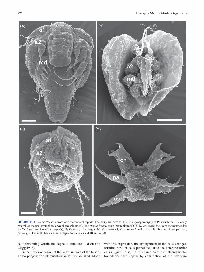
276 Emerging Marine Model Organisms
FIGURE 15.3 Some “head larvae” of different arthropods. The nauplius larva (a, b, c) is a synapomorphy of Pancrustacea. It closely
resembles the protonymphon larva of sea spiders (d). (a) Artemia franciscana (branchiopods); (b) Heterocypris incongruens (ostracods);
(c) Tigriopus brevicornis (copepods); (d) Endeis sp. (pycnogonids). a1: antenna 1, a2: antenna 2, md: mandible, ch: cheliphore, pa: palp,
ov: oviger. The scale bar measures 50 μm for (a, b, c) and 10 μm for (d).
cells remaining within the cephalic structures (Olson and with this expression, the arrangement of the cells changes,
Clegg 1978 ). forming rows of cells perpendicular to the anteroposterior
In the posterior region of the larva, in front of the telson, axis (Figure 15.3a). In this same area, the intersegmental
a “morphogenetic differentiation area” is established. Along boundaries then appear by constriction of the ectoderm
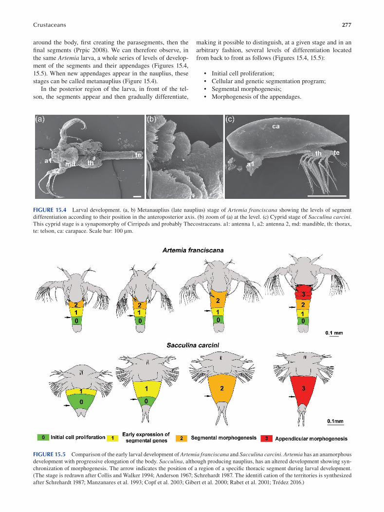
Crustaceans 277
around the body, first creating the parasegments, then the
final segments (Prpic 2008). We can therefore observe, in
the same Artemia larva, a whole series of levels of develop
ment of the segments and their appendages (Figures 15.4,
15.5). When new appendages appear in the nauplius, these
stages can be called metanauplius (Figure 15.4).
In the posterior region of the larva, in front of the tel
son, the segments appear and then gradually differentiate,
making it possible to distinguish, at a given stage and in an
arbitrary fashion, several levels of differentiation located
from back to front as follows (Figures 15.4, 15.5):
• Initial cell proliferation;
• Cellular and genetic segmentation program;
• Segmental morphogenesis;
• Morphogenesis of the appendages.
FIGURE 15.4 Larval development. (a, b) Metanauplius (late nauplius) stage of Artemia franciscana showing the levels of segment
differentiation according to their position in the anteroposterior axis. (b) zoom of (a) at the level. (c) Cyprid stage of Sacculina carcini. This cyprid stage is a synapomorphy of Cirripeds and probably Thecostraceans. a1: antenna 1, a2: antenna 2, md: mandible, th: thorax,
te: telson, ca: carapace. Scale bar: 100 μm.
FIGURE 15.5 Comparison of the early larval development of Artemia franciscana and Sacculina carcini. Artemia has an anamorphous
development with progressive elongation of the body. Sacculina, although producing nauplius, has an altered development showing syn
chronization of morphogenesis. The arrow indicates the position of a region of a specific thoracic segment during larval development.
(The stage is redrawn after Collis and Walker 1994; Anderson 1967; Schrehardt 1987. The identifi cation of the territories is synthesized
after Schrehardt 1987; Manzanares et al. 1993; Copf et al. 2003; Gibert et al. 2000; Rabet et al. 2001; Trédez 2016.)
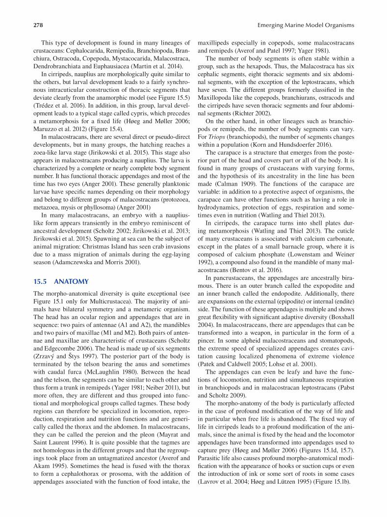
278
This type of development is found in many lineages of
crustaceans: Cephalocarida, Remipedia, Branchiopoda, Bran
chiura, Ostracoda, Copepoda, Mystacocarida, Malacostraca,
Dendrobranchiata and Euphausiacea (Martin et al. 2014).
In cirripeds, nauplius are morphologically quite similar to
the others, but larval development leads to a fairly synchro
nous intracuticular construction of thoracic segments that
deviate clearly from the anamorphic model (see Figure 15.5)
(Trédez et al. 2016). In addition, in this group, larval devel
opment leads to a typical stage called cypris, which precedes
a metamorphosis for a fixed life (Høeg and Møller 2006;
Maruzzo et al. 2012 ) ( Figure 15.4 ).
In malacostracans, there are several direct or pseudo-direct
developments, but in many groups, the hatching reaches a
zoea-like larva stage (Jirikowski et al. 2015). This stage also
appears in malacostracans producing a nauplius. The larva is
characterized by a complete or nearly complete body segment
number. It has functional thoracic appendages and most of the
time has two eyes (Anger 2001). These generally planktonic
larvae have specific names depending on their morphology
and belong to different groups of malacostracans (protozoea,
metazoea, mysis or phyllosoma) (Anger 2001)
In many malacostracans, an embryo with a nauplius-
like form appears transiently in the embryo reminiscent of
ancestral development ( Scholtz 2002; Jirikowski et al. 2013;
Jirikowski et al. 2015). Spawning at sea can be the subject of
animal migration: Christmas Island has seen crab invasions
due to a mass migration of animals during the egg-laying
season (Adamczewska and Morris 2001).
15.5 ANATOMY
The morpho-anatomical diversity is quite exceptional (see
Figure 15.1 only for Multicrustacea). The majority of ani
mals have bilateral symmetry and a metameric organism.
The head has an ocular region and appendages that are in
sequence: two pairs of antennae (A1 and A2), the mandibles
and two pairs of maxillae (M1 and M2). Both pairs of anten
nae and maxillae are characteristic of crustaceans (Scholtz
and Edgecombe 2006 ). The head is made up of six segments
( Zrzavý and Štys 1997 ). The posterior part of the body is
terminated by the telson bearing the anus and sometimes
with caudal furca (McLaughlin 1980). Between the head
and the telson, the segments can be similar to each other and
thus form a trunk in remipeds (Yager 1981; Neiber 2011), but
more often, they are different and thus grouped into func
tional and morphological groups called tagmes. These body
regions can therefore be specialized in locomotion, repro
duction, respiration and nutrition functions and are generi
cally called the thorax and the abdomen. In malacostracans,
they can be called the pereion and the pleon (Mayrat and
Saint Laurent 1996 ). It is quite possible that the tagmes are
not homologous in the different groups and that the regroup
ings took place from an untagmatized ancestor (Averof and
Akam 1995). Sometimes the head is fused with the thorax
to form a cephalothorax or prosoma, with the addition of
appendages associated with the function of food intake, the
Emerging Marine Model Organisms
maxillipeds especially in copepods, some malacostracans
and remipeds (Averof and Patel 1997; Yager 1981).
The number of body segments is often stable within a
group, such as the hexapods. Thus, the Malacostraca has six
cephalic segments, eight thoracic segments and six abdomi
nal segments, with the exception of the leptostracans, which
have seven. The different groups formerly classifi ed in the
Maxillopoda like the copepods, branchiurans, ostracods and
the cirripeds have seven thoracic segments and four abdomi
nal segments ( Richter 2002 ).
On the other hand, in other lineages such as branchio
pods or remipeds, the number of body segments can vary.
For Triops (branchiopods), the number of segments changes
within a population (Korn and Hundsdoerfer 2016).
The carapace is a structure that emerges from the poste
rior part of the head and covers part or all of the body. It is
found in many groups of crustaceans with varying forms,
and the hypothesis of its ancestrality in the line has been
made (Calman 1909). The functions of the carapace are
variable: in addition to a protective aspect of organisms, the
carapace can have other functions such as having a role in
hydrodynamics, protection of eggs, respiration and some
times even in nutrition (Watling and Thiel 2013).
In cirripeds, the carapace turns into shell plates dur
ing metamorphosis (Watling and Thiel 2013). The cuticle
of many crustaceans is associated with calcium carbonate,
except in the plates of a small barnacle group, where it is
composed of calcium phosphate (Lowenstam and Weiner
1992), a compound also found in the mandible of many mal
acostracans (Bentov et al. 2016 ).
In pancrustaceans, the appendages are ancestrally bira
mous. There is an outer branch called the expopodite and
an inner branch called the endopodite. Additionally, there
are expansions on the external (epipodite) or internal (endite)
side. The function of these appendages is multiple and shows
great flexibility with significant adaptive diversity (Boxshall
2004). In malacostraceans, there are appendages that can be
transformed into a weapon, in particular in the form of a
pincer. In some alpheid malacostraceans and stomatopods,
the extreme speed of specialized appendages creates cavi
tation causing localized phenomena of extreme violence
( Patek and Caldwell 2005; Lohse et al. 2001 ).
The appendages can even be leafy and have the func
tions of locomotion, nutrition and simultaneous respiration
in branchiopods and in malacostracan leptostracans ( Pabst
and Scholtz 2009).
The morpho-anatomy of the body is particularly affected
in the case of profound modification of the way of life and
in particular when free life is abandoned. The fixed way of
life in cirripeds leads to a profound modification of the ani
mals, since the animal is fixed by the head and the locomotor
appendages have been transformed into appendages used to
capture prey (Høeg and Møller 2006 ) (Figures 15.1d, 15.7 ).
Parasitic life also causes profound morpho-anatomical modi
fication with the appearance of hooks or suction cups or even
the introduction of ink or some sort of roots in some cases
( Lavrov et al. 2004; Høeg and Lützen 1995 ) ( Figure 15.1b ).
279 Crustaceans
15.6 GENOMIC DATA
New sequencing methods (NGS) make it possible to obtain
DNA fragments at low cost to reconstruct genome fragments
or complete genomes. With a bar-coding approach by PCR
and transcriptome sequencing, we are able to obtain data for
phylogenetic analyses essential to further understanding crus
taceans and to proposing evolutionary scenarios. The mito
chondrial genome has been obtained from many species, and
there are rearrangements that may be useful in identifying or
confirming delicate parts of the phylogeny. This is, for exam
ple, the case of a reorganization observed in the pentastomides
that we found also in the branchiurans (Lavrov et al. 2004).
The first complete crustacean genome published is that
of Daphnia pulex ( Colbourne et al. 2011 ), but currently
the number of sequenced genomes is increasing rapidly.
However, the choice of crustacean models mainly concerns
freshwater or brackish water models and few truly marine
animals (Table 15.1).
There is a strong variation in the sizes of genomes in
crustaceans. The smallest appears to be the branchiopod
Lepidurus, with a little less than 0.11 Gb (Savojardo et al.
2019), and the largest, the arctic malacostracan Ampelisca macrocephala, seems to be the biggest with about 63.2 Gb
(Rees et al. 2007), or almost 600 times bigger.
It would seem that crustaceans living in constant and cold
environments would have genomes larger than others (Alfsnes
et al. 2017). Similarly, the control region of the mitogenome
in polar copepods of the genus Calanus is known to be the
longest of the crustaceans (Weydmann et al. 2017).
15.7 FUNCTIONAL APPROACHES: TOOLS FOR MOLECULAR AND CELLULAR ANALYSES
Paryhale hawaiensis is arguably the richest and most tooled
model today in crustaceans and will not be presented here (see
Chapter 16). Historically, early work on larval gene expres
sion used immunohisto-chemistry and in situ hybridization
performed in Artemia through sonication processes to make
the cuticle permeable (Manzanares et al. 1993; Averof and
Akam 1995). This method has been improved by chemical
permeabilization (Blin et al. 2003; Copf et al. 2003). RNAi
has been successfully tested on Artemia ( Copf et al. 2004)
and on Litopenaeus vannamei ( Robalino et al. 2004 ).
The intense development of crustacean cultures for food
production was quickly accompanied by the proliferation
of numerous studies on farming models. Studies have been
conducted on genes related to biomineralization and genes
related to RNAi machinery, but many of the studies are
focused on reproductive mechanisms to optimize reproduc
tion such as encoding genes for eyestalk neuropeptides, gene
receptor-encoding genes and genes related to sexual differ
entiation (Sagi et al. 2013).
In addition, many diseases have developed due to
the high concentrations of animals, the impact of which
remains a major concern for aquaculture maintenance
( Stentiford et al. 2012 ).
Thus, RNAi provides modern and promising tools to treat
shrimp that can be affected by nearly 20 different viruses
( Krishnan et al. 2009; Escobedo-Bonilla 2011 ; Gong and
Zhang 2021).
TABLE 15.1 List of Complete Genomes Published Species Name Group Habitat Size in Gb Publication
Acartia tonsa Copepoda Marine 2.5 Jørgensen et al. (2019b )
Amphibalanus amphitrite Cirrepedia Marine 0.481 Kim et al. (2019 )
Apocyclops royi Copepoda Fresh to brackish water 0.45 Jørgensen et al. (2019a )
Armadillidium vulgare Malacostraca Terrestrial 1.72 Chebbi et al. (2019 )
Daphnia pulex Branchiopoda Fresh water 0.2 Colbourne et al. (2011 )
Daphnia magma Branchiopoda Fresh water 0.123 Lee et al. (2019 )
Diaphanosoma celebensis Branchiopoda Brackish water 2.56 Kim et al. (2021 )
Eriocheir sinensis Malacostraca Fresh water to marine 1.66 Song et al. (2016 )
Eulimnadia texana Branchiopoda Fresh water 0.12 Baldwin-Brown et al. (2017 )
Lepidurus apus Branchiopoda Fresh water 0.1075 Savojardo et al. (2019 )
Lepidurus articus Branchiopoda Fresh water 0.1075 Savojardo et al. (2019 )
Macrobrachium nipponense Malacostraca Fresh water 4.5 Jin et al. (2021 )
Neocaridina denticulata Malacostraca Fresh water 3.2 Kenny et al. (2014 )
Parhyale hawaiensis Malacostraca Marine 3.6 Kao et al. (2016 )
Portunus trituberculatus Malacostraca Marine 1.0 Tang et al. (2020 )
Procambarus clarkii Malacostraca Fresh water 8.5 Shi et al. (2018 )
Procambarus virginalis Malacostraca Fresh water 3.5 Gutekunst et al. (2018 )
Tigriopus californicus Copepoda Marine 0.190 Barreto et al. (2018 )
Tigriopus japonicus Copepoda Marine 0.197 Jeong et al. (2020 )
Tigriopus kingsejongensis Copepoda Marine 0.295 Kang et al. (2017 )
Trinorchestia longiramus Malacostraca Semi-terrestrial 0.89 Patra et al. (2020 )
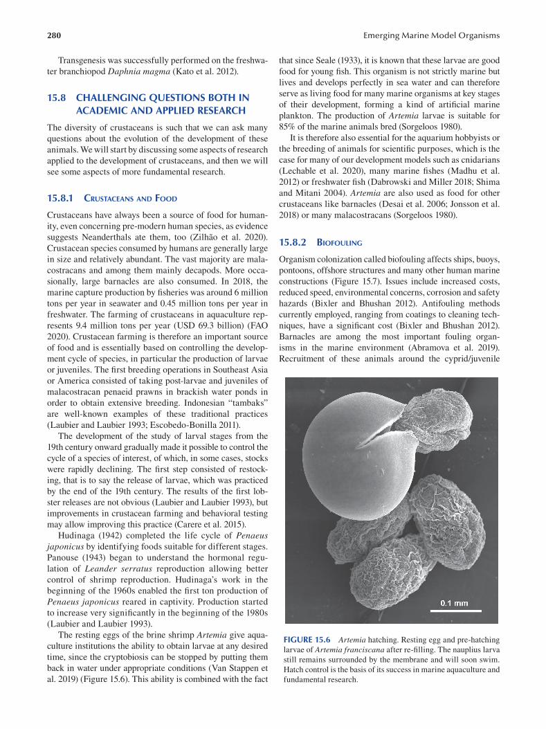
280
Transgenesis was successfully performed on the freshwa
ter branchiopod Daphnia magma ( Kato et al. 2012 ).
15.8 CHALLENGING QUESTIONS BOTH IN ACADEMIC AND APPLIED RESEARCH
The diversity of crustaceans is such that we can ask many
questions about the evolution of the development of these
animals. We will start by discussing some aspects of research
applied to the development of crustaceans, and then we will
see some aspects of more fundamental research.
15.8.1 CRUSTACEANS AND FOOD
Crustaceans have always been a source of food for human
ity, even concerning pre-modern human species, as evidence
suggests Neanderthals ate them, too ( Zilhão et al. 2020).
Crustacean species consumed by humans are generally large
in size and relatively abundant. The vast majority are mala
costracans and among them mainly decapods. More occa
sionally, large barnacles are also consumed. In 2018, the
marine capture production by fisheries was around 6 million
tons per year in seawater and 0.45 million tons per year in
freshwater. The farming of crustaceans in aquaculture rep
resents 9.4 million tons per year (USD 69.3 billion) (FAO
2020). Crustacean farming is therefore an important source
of food and is essentially based on controlling the develop
ment cycle of species, in particular the production of larvae
or juveniles. The first breeding operations in Southeast Asia
or America consisted of taking post-larvae and juveniles of
malacostracan penaeid prawns in brackish water ponds in
order to obtain extensive breeding. Indonesian “tambaks”
are well-known examples of these traditional practices
( Laubier and Laubier 1993; Escobedo-Bonilla 2011 ).
The development of the study of larval stages from the
19th century onward gradually made it possible to control the
cycle of a species of interest, of which, in some cases, stocks
were rapidly declining. The first step consisted of restock
ing, that is to say the release of larvae, which was practiced
by the end of the 19th century. The results of the fi rst lob
ster releases are not obvious (Laubier and Laubier 1993), but
improvements in crustacean farming and behavioral testing
may allow improving this practice (Carere et al. 2015).
Hudinaga (1942) completed the life cycle of Penaeus japonicus by identifying foods suitable for different stages.
Panouse (1943) began to understand the hormonal regu
lation of Leander serratus reproduction allowing better
control of shrimp reproduction. Hudinaga’s work in the
beginning of the 1960s enabled the first ton production of
Penaeus japonicus reared in captivity. Production started
to increase very significantly in the beginning of the 1980s
( Laubier and Laubier 1993 ).
The resting eggs of the brine shrimp Artemia give aqua
culture institutions the ability to obtain larvae at any desired
time, since the cryptobiosis can be stopped by putting them
back in water under appropriate conditions (Van Stappen et
al. 2019) (Figure 15.6 ). This ability is combined with the fact
Emerging Marine Model Organisms
that since Seale (1933), it is known that these larvae are good
food for young fi sh. This organism is not strictly marine but
lives and develops perfectly in sea water and can therefore
serve as living food for many marine organisms at key stages
of their development, forming a kind of artifi cial marine
plankton. The production of Artemia larvae is suitable for
85% of the marine animals bred (Sorgeloos 1980).
It is therefore also essential for the aquarium hobbyists or
the breeding of animals for scientific purposes, which is the
case for many of our development models such as cnidarians
(Lechable et al. 2020), many marine fishes (Madhu et al.
20 12 ) or freshwater fish (Dabrowski and Miller 2018; Shima
and Mitani 2004). Artemia are also used as food for other
crustaceans like barnacles (Desai et al. 2006; Jonsson et al.
2018) or many malacostracans (Sorgeloos 1980).
15.8.2 BIOFOULING
Organism colonization called biofouling affects ships, buoys,
pontoons, offshore structures and many other human marine
constructions (Figure 15.7). Issues include increased costs,
reduced speed, environmental concerns, corrosion and safety
hazards (Bixler and Bhushan 2012). Antifouling methods
currently employed, ranging from coatings to cleaning tech
niques, have a significant cost (Bixler and Bhushan 2012).
Barnacles are among the most important fouling organ
isms in the marine environment (Abramova et al. 2019).
Recruitment of these animals around the cyprid/juvenile
FIGURE 15.6 Artemia hatching. Resting egg and pre-hatching
larvae of Artemia franciscana after re-filling. The nauplius larva
still remains surrounded by the membrane and will soon swim.
Hatch control is the basis of its success in marine aquaculture and
fundamental research.
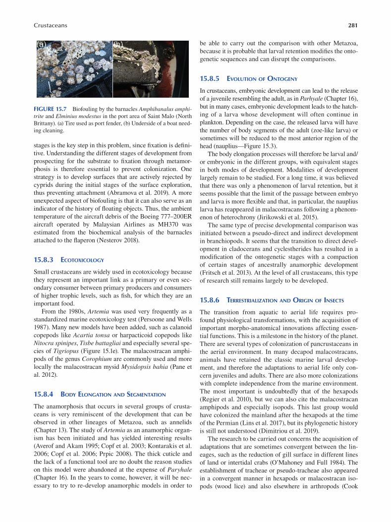
281 Crustaceans
FIGURE 15.7 Biofouling by the barnacles Amphibanalus amphitrite and Elminius modestus in the port area of Saint Malo (North
Brittany). (a) Tire used as port fender, (b) Underside of a boat need
ing cleaning.
stages is the key step in this problem, since fixation is defi ni
tive. Understanding the different stages of development from
prospecting for the substrate to fixation through metamor
phosis is therefore essential to prevent colonization. One
strategy is to develop surfaces that are actively rejected by
cyprids during the initial stages of the surface exploration,
thus preventing attachment (Abramova et al. 2019). A more
unexpected aspect of biofouling is that it can also serve as an
indicator of the history of floating objects. Thus, the ambient
temperature of the aircraft debris of the Boeing 777–200ER
aircraft operated by Malaysian Airlines as MH370 was
estimated from the biochemical analysis of the barnacles
attached to the flaperon ( Nesterov 2018 ).
15.8.3 ECOTOXICOLOGY
Small crustaceans are widely used in ecotoxicology because
they represent an important link as a primary or even sec
ondary consumer between primary producers and consumers
of higher trophic levels, such as fish, for which they are an
important food.
From the 1980s, Artemia was used very frequently as a
standardized marine ecotoxicology test (Persoone and Wells
1987). Many new models have been added, such as calanoid
copepods like Acartia tonsa or harpacticoid copepods like
Nitocra spinipes, Tisbe battagliai and especially several spe
cies of Tigriopus (Figure 15.1e). The malacostracan amphi
pods of the genus Corophium are commonly used and more
locally the malacostracan mysid Mysidopsis bahia (Pane et
al. 2012 ).
15.8.4 BODY ELONGATION AND SEGMENTATION
The anamorphosis that occurs in several groups of crusta
ceans is very reminiscent of the development that can be
observed in other lineages of Metazoa, such as annelids
(Chapter 13). The study of Artemia as an anamorphic organ
ism has been initiated and has yielded interesting results
( Averof and Akam 1995; Copf et al. 2003; Kontarakis et al.
2006; Copf et al. 2006; Prpic 2008 ). The thick cuticle and
the lack of a functional tool are no doubt the reason studies
on this model were abandoned at the expense of Paryhale (Chapter 16). In the years to come, however, it will be nec
essary to try to re-develop anamorphic models in order to
be able to carry out the comparison with other Metazoa,
because it is probable that larval retention modifies the onto
genetic sequences and can disrupt the comparisons.
15.8.5 EVOLUTION OF ONTOGENY
In crustaceans, embryonic development can lead to the release
of a juvenile resembling the adult, as in Parhyale (Chapter 16),
but in many cases, embryonic development leads to the hatch
ing of a larva whose development will often continue in
plankton. Depending on the case, the released larva will have
the number of body segments of the adult (zoe-like larva) or
sometimes will be reduced to the most anterior region of the
head (nauplius— Figure 15.3 ).
The body elongation processes will therefore be larval and/
or embryonic in the different groups, with equivalent stages
in both modes of development. Modalities of development
largely remain to be studied. For a long time, it was believed
that there was only a phenomenon of larval retention, but it
seems possible that the limit of the passage between embryo
and larva is more flexible and that, in particular, the nauplius
larva has reappeared in malacostracans following a phenom
enon of heterochrony (Jirikowski et al. 2015).
The same type of precise developmental comparison was
initiated between a pseudo-direct and indirect development
in branchiopods. It seems that the transition to direct devel
opment in cladocerans and cyclestherides has resulted in a
modification of the ontogenetic stages with a compaction
of certain stages of ancestrally anamorphic development
(Fritsch et al. 2013). At the level of all crustaceans, this type
of research still remains largely to be developed.
15.8.6 TERRESTRIALIZATION AND ORIGIN OF INSECTS
The transition from aquatic to aerial life requires pro
found physiological transformations, with the acquisition of
important morpho-anatomical innovations affecting essen
tial functions. This is a milestone in the history of the planet.
There are several types of colonization of pancrustaceans in
the aerial environment. In many decapod malacostracans,
animals have retained the classic marine larval develop
ment, and therefore the adaptations to aerial life only con
cern juveniles and adults. There are also more colonizations
with complete independence from the marine environment.
The most important is undoubtedly that of the hexapods
(Regier et al. 2010), but we can also cite the malacostracan
amphipods and especially isopods. This last group would
have colonized the mainland after the hexapods at the time
of the Permian (Lins et al. 2017), but its phylogenetic history
is still not understood (Dimitriou et al. 2019).
The research to be carried out concerns the acquisition of
adaptations that are sometimes convergent between the lin
eages, such as the reduction of gill surface in different lines
of land or intertidal crabs (O’Mahoney and Full 1984). The
establishment of tracheae or pseudo-tracheae also appeared
in a convergent manner in hexapods or malacostracan iso
pods (wood lice) and also elsewhere in arthropods (Cook
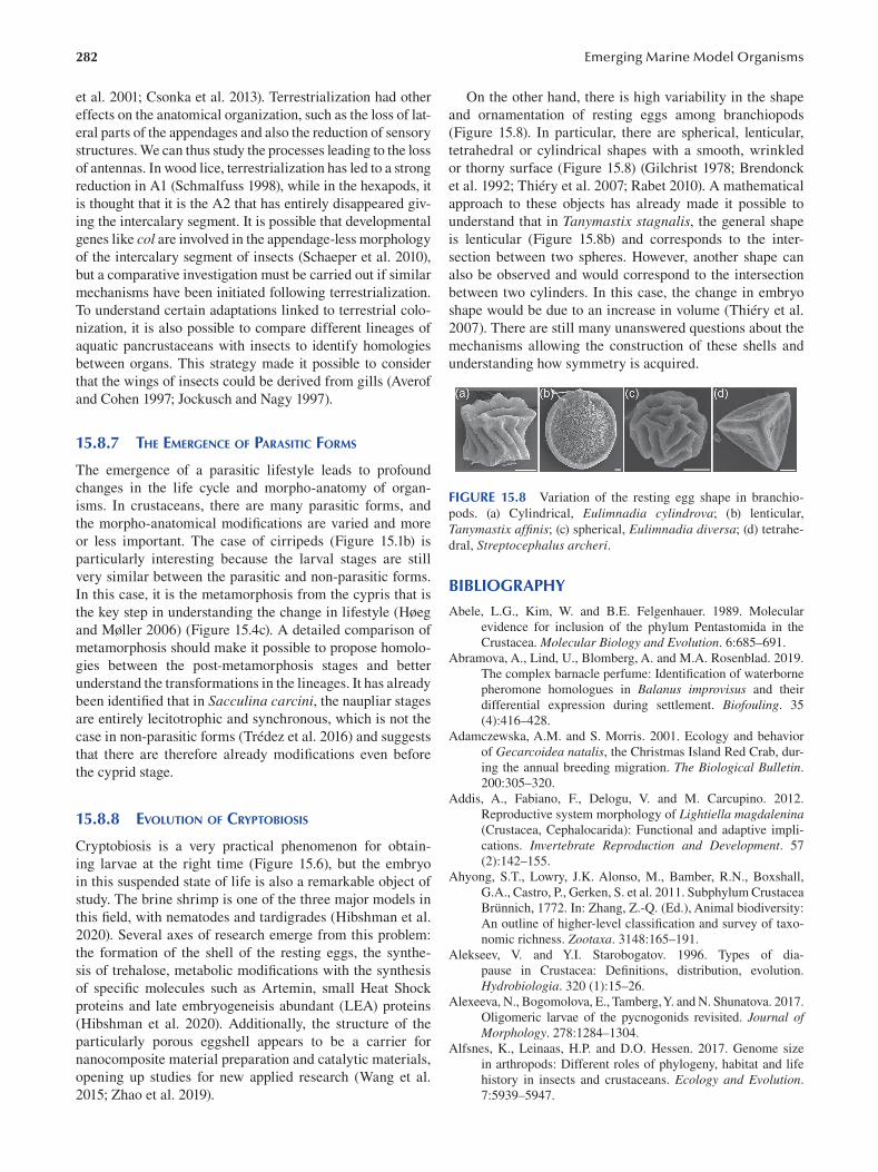
282
et al. 2001; Csonka et al. 2013). Terrestrialization had other
effects on the anatomical organization, such as the loss of lat
eral parts of the appendages and also the reduction of sensory
structures. We can thus study the processes leading to the loss
of antennas. In wood lice, terrestrialization has led to a strong
reduction in A1 (Schmalfuss 1998), while in the hexapods, it
is thought that it is the A2 that has entirely disappeared giv
ing the intercalary segment. It is possible that developmental
genes like col are involved in the appendage-less morphology
of the intercalary segment of insects (Schaeper et al. 2010),
but a comparative investigation must be carried out if similar
mechanisms have been initiated following terrestrialization.
To understand certain adaptations linked to terrestrial colo
nization, it is also possible to compare different lineages of
aquatic pancrustaceans with insects to identify homologies
between organs. This strategy made it possible to consider
that the wings of insects could be derived from gills (Averof
and Cohen 1997; Jockusch and Nagy 1997 ).
15.8.7 THE EMERGENCE OF PARASITIC FORMS
The emergence of a parasitic lifestyle leads to profound
changes in the life cycle and morpho-anatomy of organ
isms. In crustaceans, there are many parasitic forms, and
the morpho-anatomical modifications are varied and more
or less important. The case of cirripeds (Figure 15.1b) is
particularly interesting because the larval stages are still
very similar between the parasitic and non-parasitic forms.
In this case, it is the metamorphosis from the cypris that is
the key step in understanding the change in lifestyle (Høeg
and Møller 2006 ) (Figure 15.4c). A detailed comparison of
metamorphosis should make it possible to propose homolo
gies between the post-metamorphosis stages and better
understand the transformations in the lineages. It has already
been identified that in Sacculina carcini, the naupliar stages
are entirely lecitotrophic and synchronous, which is not the
case in non-parasitic forms (Trédez et al. 2016 ) and suggests
that there are therefore already modifications even before
the cyprid stage.
15.8.8 EVOLUTION OF CRYPTOBIOSIS
Cryptobiosis is a very practical phenomenon for obtain
ing larvae at the right time (Figure 15.6), but the embryo
in this suspended state of life is also a remarkable object of
study. The brine shrimp is one of the three major models in
this field, with nematodes and tardigrades (Hibshman et al.
2020). Several axes of research emerge from this problem:
the formation of the shell of the resting eggs, the synthe
sis of trehalose, metabolic modifications with the synthesis
of specific molecules such as Artemin, small Heat Shock
proteins and late embryogeneisis abundant (LEA) proteins
(Hibshman et al. 2020). Additionally, the structure of the
particularly porous eggshell appears to be a carrier for
nanocomposite material preparation and catalytic materials,
opening up studies for new applied research (Wang et al.
2015; Zhao et al. 2019 ).
Emerging Marine Model Organisms
On the other hand, there is high variability in the shape
and ornamentation of resting eggs among branchiopods
(Figure 15.8). In particular, there are spherical, lenticular,
tetrahedral or cylindrical shapes with a smooth, wrinkled
or thorny surface ( Figure 15.8 ) ( Gilchrist 1978; Brendonck
et al. 1992; Thiéry et al. 2007; Rabet 2010 ). A mathematical
approach to these objects has already made it possible to
understand that in Tanymastix stagnalis, the general shape
is lenticular (Figure 15.8b) and corresponds to the inter
section between two spheres. However, another shape can
also be observed and would correspond to the intersection
between two cylinders. In this case, the change in embryo
shape would be due to an increase in volume (Thiéry et al.
2007). There are still many unanswered questions about the
mechanisms allowing the construction of these shells and
understanding how symmetry is acquired.
FIGURE 15.8 Variation of the resting egg shape in branchio
pods. (a) Cylindrical, Eulimnadia cylindrova; (b) lenticular,
Tanymastix affinis; (c) spherical, Eulimnadia diversa; (d) tetrahe
dral, Streptocephalus archeri.
BIBLIOGRAPHY
Abele, L.G., Kim, W. and B.E. Felgenhauer. 1989. Molecular
evidence for inclusion of the phylum Pentastomida in the
Crustacea. Molecular Biology and Evolution . 6:685–691.
Abramova, A., Lind, U., Blomberg, A. and M.A. Rosenblad. 2019.
The complex barnacle perfume: Identification of waterborne
pheromone homologues in Balanus improvisus and their
differential expression during settlement. Biofouling. 35
(4):416–428.
Adamczewska, A.M. and S. Morris. 2001. Ecology and behavior
of Gecarcoidea natalis, the Christmas Island Red Crab, dur
ing the annual breeding migration. The Biological Bulletin.
200:305–320.
Addis, A., Fabiano, F., Delogu, V. and M. Carcupino. 2012.
Reproductive system morphology of Lightiella magdalenina (Crustacea, Cephalocarida): Functional and adaptive impli
cations. Invertebrate Reproduction and Development. 57
(2):142–155.
Ahyong, S.T., Lowry, J.K. Alonso, M., Bamber, R.N., Boxshall,
G.A., Castro, P., Gerken, S. et al. 2011. Subphylum Crustacea
Brünnich, 1772. In: Zhang, Z.-Q. (Ed.), Animal biodiversity:
An outline of higher-level classification and survey of taxo
nomic richness. Zootaxa . 3148:165–191.
Alekseev, V. and Y.I. Starobogatov. 1996. Types of dia
pause in Crustacea: Definitions, distribution, evolution.
Hydrobiologia. 320 (1):15–26.
Alexeeva, N., Bogomolova, E., Tamberg, Y. and N. Shunatova. 2017.
Oligomeric larvae of the pycnogonids revisited. Journal of Morphology . 278:1284–1304.
Alfsnes, K., Leinaas, H.P. and D.O. Hessen. 2017. Genome size
in arthropods: Different roles of phylogeny, habitat and life
history in insects and crustaceans. Ecology and Evolution.
7:5939–5947.
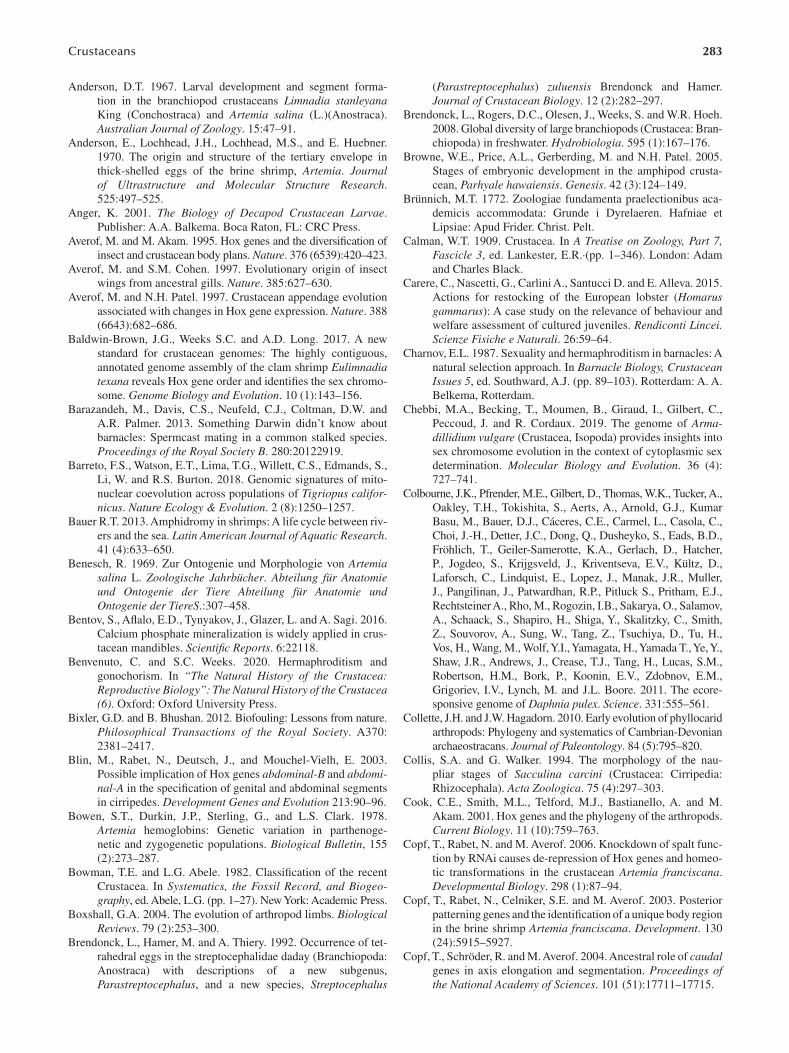
283 Crustaceans
Anderson, D.T. 1967. Larval development and segment forma
tion in the branchiopod crustaceans Limnadia stanleyana King (Conchostraca) and Artemia salina (L.)(Anostraca).
Australian Journal of Zoology . 15:47–91.
Anderson, E., Lochhead, J.H., Lochhead, M.S., and E. Huebner.
1970. The origin and structure of the tertiary envelope in
thick-shelled eggs of the brine shrimp, Artemia. Journal of Ultrastructure and Molecular Structure Research.
525:497–525.
Anger, K. 2001. The Biology of Decapod Crustacean Larvae.
Publisher: A.A. Balkema. Boca Raton, FL: CRC Press.
Averof, M. and M. Akam. 1995. Hox genes and the diversifi cation of
insect and crustacean body plans. Nature. 376 (6539):420–423.
Averof, M. and S.M. Cohen. 1997. Evolutionary origin of insect
wings from ancestral gills. Nature . 385:627–630.
Averof, M. and N.H. Patel. 1997. Crustacean appendage evolution
associated with changes in Hox gene expression. Nature . 388
(6643):682–686.
Baldwin-Brown, J.G., Weeks S.C. and A.D. Long. 2017. A new
standard for crustacean genomes: The highly contiguous,
annotated genome assembly of the clam shrimp Eulimnadia texana reveals Hox gene order and identifies the sex chromo
some. Genome Biology and Evolution. 10 (1):143–156.
Barazandeh, M., Davis, C.S., Neufeld, C.J., Coltman, D.W. and
A.R. Palmer. 2013. Something Darwin didn’t know about
barnacles: Spermcast mating in a common stalked species.
Proceedings of the Royal Society B . 280:20122919.
Barreto, F.S., Watson, E.T., Lima, T.G., Willett, C.S., Edmands, S.,
Li, W. and R.S. Burton. 2018. Genomic signatures of mito
nuclear coevolution across populations of Tigriopus californicus. Nature Ecology & Evolution. 2 (8):1250–1257.
Bauer R.T. 2013. Amphidromy in shrimps: A life cycle between riv
ers and the sea. Latin American Journal of Aquatic Research.
41 (4):633–650.
Benesch, R. 1969. Zur Ontogenie und Morphologie von Artemia salina L. Zoologische Jahrbücher. Abteilung für Anatomie und Ontogenie der Tiere Abteilung für Anatomie und Ontogenie der TiereS.:307–458.
Bentov, S., Aflalo, E.D., Tynyakov, J., Glazer, L. and A. Sagi. 2016.
Calcium phosphate mineralization is widely applied in crus
tacean mandibles. Scientifi c Reports . 6:22118.
Benvenuto, C. and S.C. Weeks. 2020. Hermaphroditism and
gonochorism. In “The Natural History of the Crustacea: Reproductive Biology”: The Natural History of the Crustacea (6). Oxford: Oxford University Press.
Bixler, G.D. and B. Bhushan. 2012. Biofouling: Lessons from nature.
Philosophical Transactions of the Royal Society. A370:
2381–2417.
Blin, M., Rabet, N., Deutsch, J., and Mouchel-Vielh, E. 2003.
Possible implication of Hox genes abdominal-B and abdominal-A in the specification of genital and abdominal segments
in cirripedes. Development Genes and Evolution 213:90–96.
Bowen, S.T., Durkin, J.P., Sterling, G., and L.S. Clark. 1978.
Artemia hemoglobins: Genetic variation in parthenoge
netic and zygogenetic populations. Biological Bulletin , 155
(2):273–287.
Bowman , T.E. and L.G. Abele. 1982. Classification of the recent
Crustacea. In Systematics, the Fossil Record, and Biogeography, ed. Abele, L.G. (pp. 1–27). New York: Academic Press.
Boxshall, G.A. 2004. The evolution of arthropod limbs. Biological Reviews. 79 (2):253–300.
Brendonck, L., Hamer, M. and A. Thiery. 1992. Occurrence of tet
rahedral eggs in the streptocephalidae daday (Branchiopoda:
Anostraca) with descriptions of a new subgenus,
Parastreptocephalus, and a new species, Streptocephalus
(Parastreptocephalus) zuluensis Brendonck and Hamer.
Journal of Crustacean Biology. 12 (2):282–297.
Brendonck, L., Rogers, D.C., Olesen, J., Weeks, S. and W.R. Hoeh.
2008. Global diversity of large branchiopods (Crustacea: Bran
chiopoda) in freshwater. Hydrobiologia. 595 (1):167–176.
Browne, W.E., Price, A.L., Gerberding, M. and N.H. Patel. 2005.
Stages of embryonic development in the amphipod crusta
cean, Parhyale hawaiensis. Genesis. 42 (3):124–149.
Brünnich , M.T. 1772. Zoologiae fundamenta praelectionibus aca
demicis accommodata: Grunde i Dyrelaeren. Hafniae et
Lipsiae: Apud Frider. Christ. Pelt.
Calman, W.T. 1909. Crustacea. In A Treatise on Zoology, Part 7, Fascicle 3, ed. Lankester, E.R.·(pp. 1–346). London: Adam
and Charles Black.
Carere, C., Nascetti, G., Carlini A., Santucci D. and E. Alleva. 2015.
Actions for restocking of the European lobster (Homarus gammarus): A case study on the relevance of behaviour and
welfare assessment of cultured juveniles. Rendiconti Lincei. Scienze Fisiche e Naturali . 26:59–64.
Charnov, E.L. 1987. Sexuality and hermaphroditism in barnacles: A
natural selection approach. In Barnacle Biology, Crustacean Issues 5, ed. Southward, A.J. (pp. 89–103). Rotterdam: A. A.
Belkema, Rotterdam.
Chebbi, M.A., Becking, T., Moumen, B., Giraud, I., Gilbert, C.,
Peccoud, J. and R. Cordaux. 2019. The genome of Armadillidium vulgare (Crustacea, Isopoda) provides insights into
sex chromosome evolution in the context of cytoplasmic sex
determination. Molecular Biology and Evolution. 36 (4):
727–741.
Colbourne, J.K., Pfrender, M.E., Gilbert, D., Thomas, W.K., Tucker, A.,
Oakley, T.H., Tokishita, S., Aerts, A., Arnold, G.J., Kumar
Basu, M., Bauer, D.J., Cáceres, C.E., Carmel, L., Casola, C.,
Choi, J.-H., Detter, J.C., Dong, Q., Dusheyko, S., Eads, B.D.,
Fröhlich, T., Geiler-Samerotte, K.A., Gerlach, D., Hatcher,
P., Jogdeo, S., Krijgsveld, J., Kriventseva, E.V., Kültz, D.,
Laforsch, C., Lindquist, E., Lopez, J., Manak, J.R., Muller,
J., Pangilinan, J., Patwardhan, R.P., Pitluck S., Pritham, E.J.,
Rechtsteiner A., Rho, M., Rogozin, I.B., Sakarya, O., Salamov,
A., Schaack, S., Shapiro, H., Shiga, Y., Skalitzky, C., Smith,
Z., Souvorov, A., Sung, W., Tang, Z., Tsuchiya, D., Tu, H.,
Vos, H., Wang, M., Wolf, Y.I., Yamagata, H., Yamada T., Ye, Y.,
Shaw, J.R., Andrews, J., Crease, T.J., Tang, H., Lucas, S.M.,
Robertson, H.M., Bork, P., Koonin, E.V., Zdobnov, E.M.,
Grigoriev, I.V., Lynch, M. and J.L. Boore. 2011. The ecore
sponsive genome of Daphnia pulex. Science . 331:555–561.
Collette, J.H. and J.W. Hagadorn. 2010. Early evolution of phyllocarid
arthropods: Phylogeny and systematics of Cambrian-Devonian
archaeostracans. Journal of Paleontology. 84 (5):795–820.
Collis, S.A. and G. Walker. 1994. The morphology of the nau
pliar stages of Sacculina carcini (Crustacea: Cirripedia:
Rhizocephala). Acta Zoologica. 75 (4):297–303.
Cook, C.E., Smith, M.L., Telford, M.J., Bastianello, A. and M.
Akam. 2001. Hox genes and the phylogeny of the arthropods.
Current Biology. 11 (10):759–763.
Copf, T., Rabet, N. and M. Averof. 2006. Knockdown of spalt func
tion by RNAi causes de-repression of Hox genes and homeo
tic transformations in the crustacean Artemia franciscana.
Developmental Biology. 298 (1):87–94.
Copf, T., Rabet, N., Celniker, S.E. and M. Averof. 2003. Posterior
patterning genes and the identification of a unique body region
in the brine shrimp Artemia franciscana. Development . 130
(24):5915–5927.
Copf, T., Schröder, R. and M. Averof. 2004. Ancestral role of caudal genes in axis elongation and segmentation. Proceedings of the National Academy of Sciences. 101 (51):17711–17715.
284
Csonka, D., Halasy, K., Szabó, P., Mrak, P., Štrus, J. and E.
Hornung. 2013. Eco-morphological studies on pleopo
dal lungs and cuticle in Armadillidium species (Crustacea,
Isopoda, Oniscidea). Arthropod Structure and Development. 42:229–235.
Dabrowski, K. and M. Miller. 2018. Contested paradigm in raising
zebrafi sh ( Danio rerio). Zebrafi sh. 15 (3):295–309.
Dahms, H.-U. 2000. Phylogenetic implications of the Crustacean
nauplius. Hydrobiologia . 417:91–99.
Delage, Y. 1884. Évolution de la Sacculine ( Sacculina carcini Thomps.): crustacé endoparasite de l’ordre nouveau des
Kentrogonides. Archives de zoologie expérimentale et générale . 2:417–736.
Desai, D.V., Anil, A.C. and K. Venkat. 2006. Reproduction
in Balanus amphitrite Darwin (Cirripedia: Thoracica):
Influence of temperature and food concentration. Marine Biology . 149:1431–1441.
Dimitriou, A.C., Taiti S. and S. Sfenthourakis. 2019. Genetic evi
dence against monophyly of Oniscidea implies a need to
revise scenarios for the origin of terrestrial isopods. Scientifi c Reports. 9 (1):18508.
Dohle, W. 2001. Are the insects terrestrial crustaceans? A discus
sion of some new facts and arguments and the proposal of
the proper name ‘Tetraconata’ for the monophyletic unit
Crustacea + Hexapoda. In: Deuve, T. (ed.) Origin of the
Hexapoda. Annales de la Société entomologique de France (N.S.) . 37:85–103.
Duffy, E. 1996. Eusociality in a coral-reef shrimp. Nature. 381
(6582):512–514.
Durbin, A.P., Hebert, D.N. and M.E.A. Cristescu. 2008. Compara
tive phylogeography of marine cladocerans. Marine Biology.
155:1–10.
Escobedo-Bonilla, C.M. 2011. Application of RNA interfer
ence (RNAi) against viral infections in shrimp. Journal of Antivirals & Antiretrovirals . S9.
FAO. 2020. The State of World Fisheries and Aquaculture 2020: Sustainability in Action. Rome. https://doi.org/10.4060/
ca9229en
Fraser J.H. 1936. The occurrence, ecology and life history of
Tigriopus fulvus (Fischer). Journal of the Marine Biological Association of the United Kingdom. 20 (3):523–536.
Fritsch, M., Bininda-Emonds, O.R.P. and S. Richter. 2013.
Unraveling the origin of Cladocera by identifying heter
ochrony in the developmental sequences of Branchiopoda.
Frontiers in Zoology. 10 (1):35.
Fryer, G.F.L.S. 1996. Diapause, a potent force in the evolution of
freshwater crustaceans. Hydrobiologia . 320:1–14.
Gajardo, G. and J.A. Beardmore. 2012. The brine shrimp Artemia:
Adapted to critical life conditions. Frontiers in Zoology . 3:185.
Garreau de Loubresse, N. 1974. Etude chronologique de la mise
en place des enveloppes de l’œuf d’un crustacé phyllopode,
Tanymastix lacunae. Journal of Microscopy . 20:21–38.
Gibert, J.M., Mouchel-Vielh, E., Quéinnec, E and J.S. Deutsch.
2000. Barnacle duplicate engrailed genes: Divergent expres
sion patterns and evidence for a vestigial abdomen. Evolution and Development . 2:194–202.
Gilchrist, B.M. 1978. Scanning electron microscope studies of the
egg shell in some anostraca (Crustacea: Branchiopoda). Cell and Tissue Research . 193:337–351.
Giribet, G. and G.D. Edgecombe. 2019. The phylogeny and
evolutionary history of Arthropods. Current Biology. 29
(12):R592–R602.
Gong, Yi and Xiaobo Zhang. 2021. RNAi-based antiviral immu
nity of shrimp: Review. Developmental and Comparative Immunology . 115:103907.
Emerging Marine Model Organisms
Gurney, R. 1942. Larvae of Decapod Crustacea. London: Ray
Society.
Gutekunst, J., Andriantsoa, R., Falckenhayn, C., Hanna, K., Stein,
W., Rasamy, J. and F. Lyko. 2018. Clonal genome evolution
and rapid invasive spread of the marbled crayfi sh. Nature Ecology & Evolution . 2:567–573.
Gutiérrez -Zugasti, I. 2011. The use of echinoids and crustaceans as
food during the Pleistocene-Holocene transition in Northern
Spain: Methodological contribution and dietary assessment.
The Journal of Island and Coastal Archaeology. 6 (1):115–133.
Hibshman, J.D., Clegg, J.S. and B. Goldstein. 2020. Mechanisms
of desiccation tolerance: Themes and variations in brine
shrimp, roundworms, and tardigrades. Frontiers in Physiology.
11:592016.
Høeg, J.S. and J. Lützen. 1995. Life cycle and reproduction in
the Cirripedia Rhizocephala. Oceanography and Marine Biology: An Annual Review . 33:427–485.
Høeg, J.T. and O.S. Møller. 2006. When similar beginnings lead to
different ends: Constraints and diversity in cirripede larval
development. Invertebrate Reproduction and Development. 49 (3):125–142.
Hudinaga, M. 1942. Reproduction, development and rearing of
Penaeus japonicus Bate. Japanese Journal of Environmental Entomology and Zoology . 10:305–393.
Jensen, G.C., Sean McDonald, P. and D.A. Armstrong. 2007. Biotic
resistance to green crab, Carcinus maenas, in California
bays. Marine Biology . 151:2231–2243.
Jeong, C.B., Lee, B.Y., Choi, B.S., Kim, M.S., Park, J.C., Kim,
D.-H., Wang M. et al. 2020. The genome of the harpacticoid
copepod Tigriopus japonicus: Potential for its use in marine
molecular ecotoxicology. Aquatic Toxicology . 222:105462.
Jin, S., Bian, C., Jiang, S., Han, K., Xiong, Y., Zhang, W., Shi, C. et
al. 2021. A chromosome-level genome assembly of the ori
ental river prawn, Macrobrachium nipponense. GigaScience.
10:1–9.
Jirikowski, G.J., Richter, S. and C. Wolff. 2013. Myogenesis of
Malacostraca: The “egg-nauplius” concept revisited. Frontiers in Zoology . 10:76.
Jirikowski, G.J., Wolff, C. and S. Richter. 2015. Evolution of eum
alacostracan development: New insights into loss and reac
quisition of larval stages revealed by heterochrony analysis.
EvoDevo . 6:4.
Jockusch, E.L. and L.M. Nagy. 1997. Insect evolution: How did
insect wings originate? Current Biolology. 7 (6):R358–R361.
Jonsson, P.R., Wrange, A.L., Lind, U., Abramova, A., Ogemark, M.
and A. Blomberg. 2018. The barnacle Balanus improvisus as
a marine model: Culturing and gene expression. Journal of Visualized Experiments . (138):e57825.
Jørgensen, S., B. Lykke, H. Nielsen, et al. 2019a. The whole
genome sequence and mRNA transcriptome of the tropical
cyclopoid copepod Apocyclops royi. G3: Genes, Genomes, Genetics. 9 (5):1295–1302.
Jørgensen, T.S., Petersen, B., Petersen, H.C.B., et al. 2019b. The
genome and mRNA transcriptome of the cosmopolitan cala
noid copepod Acartia tonsa Dana improve the understand
ing of copepod genome size evolution. Genome Biology and Evolution. 11 (5):1440–1450.
Kang, S., Ahn, D.-H., Lee, J.H., Lee, S.G., Shin, S.C., Lee, J., et
al. 2017. The genome of the Antarctic-endemic copepod,
Tigriopus kingsejongensis. GigaScience . 6:1–9.
Kao, D., Lai, A.G., Stamataki, E., Rosic, S., Konstantinides, N.,
Jarvis, E., Di Donfrancesco, et al. 2016. The genome of the
crustacean Parhyale hawaiensis, a model for animal develop
ment, regeneration, immunity and lignocellulose digestion.
Elife . 5:e20062.
285 Crustaceans
Kato, Y., Matsuura, T. and H. Watanabe. 2012. Genomic integration
and germline transmission of plasmid injected into crusta
cean Daphnia magna eggs. PLoS One. 7 (9):e45318.
Kawai, T., Scholtz, G., Morioka, S., Ramanamandimby, F.,
Lukhaup, C. and Y. Hanamura. 2009. Parthenogenetic alien
crayfish (Decapoda: Cambaridae) spreading in Madagascar.
Journal of Crustacean Biology. 29 (4):562–567.
Kenny, N.J., Sin, Y.W., Shen, X., Zhe, Q., Wang, W., Chan, T.F.,
Tobe, S.S., et al. 2014. Genomic sequence and experimen
tal tractability of a new decapod shrimp model, Neocaridina denticulate. Marine Drugs . 12:1419–1437.
Kim, D.-H., Choi, B.-S., Kang, H.-M., Park, J.C., Kim, M.-S.,
Hagiwara, A. and J.-S. Lee. 2021. The genome of the marine
water fl ea Diaphanosoma celebensis: Identifi cation of
phase I, II, and III detoxification genes and potential appli
cations in marine molecular ecotoxicology. Comparative Biochemistry and Physiology Part D: Genomics and Proteomics . 37:100787.
Kim, J.-H., Kim, H.K., Chan, K.H., BKK, Kang, S. and W. Kim. 2019.
Draft genome assembly of a fouling barnacle, Amphibalanus amphitrite (Darwin, 1854): The first reference genome for
Thecostraca. Frontiers in Ecology and Evolution . 7:465.
Kobayashi, H., Hatada, Y., Tsubouchi, T., Nagahama, T. and H.
Takami. 2012. The hadal Amphipod Hirondellea gigas pos
sessing a unique cellulase for digesting wooden debris buried
in the deepest seafl oor. PLoS One . 7(8):e42727.
Kobayashi, H., Shimoshige, H., Nakajima, Y., Arai, W. and H.
Takami. 2019. An aluminum shield enables the amphipod
Hirondellea gigas to inhabit deep-sea environments. PLoS One. 14 (4):e0206710.
Koenemann, S., Schram, F.R., Iliffe, T.M., Hinderstein, L.M. and
A. Bloechl. 2007. Behavior of remipedia in the laboratory,
with supporting field observations. Journal of Crustacean Biology . 27:534–542.
Kontarakis, Z., Copf, T. and M. Averof. 2006. Expression of
hunchback during trunk segmentation in the branchiopod
crustacean Artemia franciscana. Development Genes and Evolution. 216 (2):89–93.
Korn, M. and A.K. Hundsdoerfer. 2016. Molecular phylogeny,
morphology and taxonomy of Moroccan Triops granarius (Lucas, 1864) (Crustacea: Notostraca), with the description
of two new species. Zootaxa. 4178 (3):328–346.
Krieger, J., Sandeman, R.E., Sandeman, D.C., Hansson, B.S. and
S. Harzsch. 2010. Brain architecture of the largest living
land arthropod, the giant robber crab Birgus latro (Crustacea,
Anomura, Coenobitidae): Evidence for a prominent central
olfactory pathway? Frontiers in Zoology . 7:25.
Krishnan, P., Gireesh-Babu, P., Rajendran, K.V. and A. Chaudhar.
2009. RNA interference-based therapeuticsfor shrimp viral
diseases. Aquatic Organisms . 86:263–272.
Laubier, A. and L. Laubier. 1993. Marine crustacean farming:
Present status and perspectives. Aquatic Living Resources . 6
(4):319–329.
Lavrov, D.V., Brown, W.M. and J.L. Boore. 2004. Phylogenetic
position of the Pentastomida and (pan)crustacean rela
tionships. Proceedings of the Royal Society B: Biological Sciences . 271:537–544.
Lechable, M., Jan, A., Duchene, A., Uveira, J., Weissbourd, B.,
Gissat, L., Collet, S., et al. 2020. An improved whole life
cycle culture protocol for the hydrozoan genetic model Clytia hemisphaerica. Open Biology. 9 (11):bio051268.
Lee, B.-Y., Choi, B.-S., Kim, M.-S., Park, J.C., Jeong, C.-B., Han,
J. and J.-S. Lee. 2019. The genome of the freshwater water
fl ea Daphnia magna: A potential use for freshwater molecu
lar ecotoxicology. Aquatic Toxicology . 210:69–84.
Lee, M.S.Y., Soubrier, J. and G.D. Edgecombe. 2013. Rates of phe
notypic and genomic evolution during the Cambrian explo
sion. Current Biology . 23:1889–1895.
Liang, C., Strickland, J., Ye, Z., Wu, W., Hu, B. and D. Rittschof.
2019. Biochemistry of barnacle adhesion: An updated review.
Frontiers in Marine Science . 6:2296–7745.
Lindley, J.A. 1997. Eggs and their incubation as factors in the
ecology of planktonic crustacea. Journal of Crustacean Biolology. 17 (4):569–576.
Linnaeus , C. von. 1758. Caroli Linnaei Equitis De Stella Polari,
Archiatri Regii, Med. & Botan. Profess. Upsal.; Systema
Naturae Per Regna Tria Naturae, Secundum Classes,
Ordines, Genera, Species, Cum Characteribus, Differentiis,
Synonymis, Locis, Holmiae.
Lins, L.S.F., Ho, S.Y.W. and N. Lo. 2017. An evolutionary tim
escale for terrestrial isopods and a lack of molecular sup
port for the monophyly of Oniscidea (Crustacea: Isopoda).
Organisms Diversity and Evolution . 17:813–820.
Liu, Y.-L., Zhao, Y., Dai, Z.-M., Chen, H.-M., and W.-J. Yang.
2009. Formation of diapause cyst shell in brine shrimp,
Artemia parthenogenetica, and its resistance role in environ
mental stresses. The Journal of Biological Chemistry . 284
(25):16931–16938.
Lohse, D., Schmitz, B. and M. Versluis. 2001. Snapping shrimp
make flashing bubbles. Nature. 413 (6855):477–478.
Lowenstam, H.A. and S. Weiner. 1992. Phosphatic shell plate of the
barnacle Ibla (Cirripedia): A bone-like structure. Proceedings of the National Academy of Sciences. 89 (22):10573–10577.
Madhu , R., Madhu, K. and T. Retheesh. 2012. Life history path
ways in false clown Amphiprion ocellaris Cuvier, 1830: A
journey from egg to adult under captive condition. Journal of the Marine Biological Association of India. 54 (1):77–90.
Maier, G. 1992. Metacyclops minutus (Claus, 1863): Population
dynamics and life history characteristics of a rapidly devel
oping copepod. International Review of Hydrobiology . 77:
455–466.
Malitan, H.S., Cohen, A.M. and T.H. MacRae. 2019. Knockdown
of the small heat-shock protein p26 by RNA interference
modifies the diapause proteome of Artemia franciscana.
Biochemistry and Cell Biology . 97(4):471–479.
Manzanares, M., Marco, R. and R. Garesse. 1993. Genomic orga
nization and developmental pattern of expression of the
engrailed gene from the brine shrimp Artemia. Development 118:1209–1219.
Martin, J.W. and G.E. Davis. 2001. An updated classification of the
recent Crustacea. Natural History Museum of Los Angeles County Contributions in Science . 39:1–124.
Martin, J.W., Olesen, J. and J.T. Høeg. 2014. The Crustacean Nauplius in Atlas of Crustacean Larvae, ed. Martin, J.W.,
Olesen, J. and J.T. Høeg. Baltimore, MD: Johns Hopkins
University Press.
Maruzzo, D., Aldred, N., Clare, A.S. and J.T. Høeg. 2012.
Metamorphosis in the cirripede crustacean Balanus amphitrite. PLoS One. 7 (5):e37408.
Mayrat, A. and M. de Saint Laurent. 1996. Considérations sur la
Classe de Malacostracés. (Malacostraca Latreille, 1802). In
Traité de zoologie, ed. GrasséÉ, P.-P. and J. Forest. (pp. 841–
862). Masson, Paris: Crustacea.
McLaughlin, P.A. 1980. Comparative Morphology of Recent Crustacea. San Francisco: W. H. Freeman and Company.
[General crustacean morphology].
Monod, T. and J. Forest. 1996. Histoire de la classification des Crustacés, Traité de zoologie, Anatomie, systématique, biologie, Crustacés, Tome VII, fascicule II (pp. 235–267). Paris,
Milan, Barcelone: Masson.
286
Morris, J.E. and B.A. Afzelius. 1967. The structure of the shell and
outer membranes in encysted Artemia salina embryos dur
ing cryptobiosis and development. Journal of Ultrastructure Research. 20 (3–4): 244–259.
Neiber, M.T., Hartke, T.R., Stemme, T., Bergmann, A., Rust, J.,
Iliffe, T.M. and S. Koenemann. 2011. Global biodiversity
and phylogenetic evaluation of Remipedia (Crustacea). PLoS One. 6 (5):e19627.
Nesterov, O. 2018. Consideration of various aspects in a drift study
of MH370 debris. Ocean Science . 14:387–402.
Olson, C.S. and J.S. Clegg. 1978. Cell division during the devel
opment of Artemia salina. Wilhelm Roux’s Archives of Developmental Biology . 184:1–13.
O’Mahoney, P.M. and R.J. Full. 1984. Respiration of crabs in air
and water. Comparative Biochemistry and Physiology Part A: Physiology. 79 (2):275–282.
Pabst, T. and G. Scholtz. 2009. The development of phyllopodous
limbs in leptostraca and branchiopoda. Journal of Crustacean Biology. 29 (1):1–12.
Pane, L., Agrone, C., Giacco, E., Somà, A. and G.L. Mariottini.
2012. Utilization of marine crustaceans as study models:
A new approach in marine ecotoxicology for European
(REACH) regulation. In Ecotoxicology, ed. Begum, G.
(pp. 91–106). Crotia: InTech.
Panouse, J.B. 1943. Influence de l’ablation du pédoncule ocu
laire sur la croissance de l’ovaire chez la crevette Leander serratus. Comptes rendus hebdomadaires des séances de l’Académie des sciences . 217:553–555.
Patek, S.N. and R.L Caldwell. 2005. Extreme impact and cavita
tion forces of a biological hammer: Strike forces of the pea
cock mantis shrimp Odontodactylus scyllarus. Journal of Experimental Biology . 208:3655–3664.
Patra, A.K., Chung, O., Yoo, J.Y., Kim, M.S., Yoon, M.G., Choi,
J.-H. and Y. Yang. 2020. First draft genome for the sand-
hopper Trinorchestia longiramus. Scientifi c Data. 7 (1):85.
Pennak, R.W. and D.J. Zinn. 1943. Mystacocarida, a new order
of Crustacea from intertidal beaches in Massachusetts and
Connecticut. Smithsonian Miscellaneous Collections. 103
(9):1–11, pls.1–2.
Persoone, G. and P.G. Wells. 1987. Artemia in aquatic toxicology:
A review. Artemia Research and Its Applications . 1:259–275.
Petersen, J., Ramette, M.A., Lott C., Cambon-Bonavita, M.A.,
Zbinden, M. and N. Dubilier. 2010. Dual symbiosis of the
vent shrimp Rimicaris exoculata with fi lamentous gamma-
and epsilonproteobacteria at four Mid-Atlantic Ridge hydro
thermal vent fi elds. Environmental Microbiology. 12 (8):
2204–2218.
Prpic, N.-M. 2008. Parasegmental appendage allocation in annelids
and arthropods and the homology of parapodia and arthropo
dia. Frontiers in Zoology . 5:17.
Rabet, N. 2010. Revision of the egg morphology of Eulimnadia (Crustacea, Branchiopoda, Spinicaudata). Zoosystema 32
(3):373–391.
Rabet, N., Gibert, J.M., Quéinnec, E., Deutsch, J.S. and E. Mouchel-
Vielh. 2001. The caudal gene of the barnacle Sacculina carcini is not expressed in its vestigial abdomen. Development Genes and Evolution . 211:172–178.
Raisuddin, S., Kwok, K.W.H., Leung, K.M.Y., Schlenk, D. and
J.-S. Lee. 2007. The copepod Tigriopus : A promising marine
model organism for ecotoxicology and environmental
genomics. Aquatic Toxicology. 83 (3):161–173.
Rajitha, K., Nancharaiaha, Y.V. and V.P. Venugopalan. 2020.
Insight into bacterial biofilm-barnacle larvae interactions for
environmentally benign antifouling strategies. International Biodeterioration & Biodegradation . 149:104937.
Emerging Marine Model Organisms
Rees , D.J., Dufresne, F., Glémet H. and C. Belzile. 2007.
Amphipod genome sizes: First estimates for arctic species
reveal genomic giants. Genome. 50 (2):151–158.
Regier, J.C., Jeffrey, W.S. and R.E. Kambic. 2005. Pancrustacean
phylogeny: Hexapods are terrestrial crustaceans and maxillo
pods are not monophyletic. Proceedings of the Royal Society B . 272:395–401.
Regier, J.C., Shultz, J.W., Zwick, A., Hussey, A., Ball, B., Wetzer
R., Martin, J.W. and C.W. Cunningham. 2010. Arthropod
relationships revealed by phylogenomic analysis of nuclear
protein-coding sequences. Nature. 463 (7284):1079–1083.
Richter, S. 2002. The Tetraconata concept: Hexapod-crustacean
relationships and the phylogeny of Crustacea. Organisms Diversity and Evolution . 2:217–237.
Riley, J., Banaja, A.A. and J.L. James. 1978. The phylogenetic rela
tionships of the Pentastomida: The case for their inclusion
within the Crustacea. International Journal for Parasitology.
8 (4):245–254.
Robalino, J., Browdy, C.L., Prior, S., Metz, A., Parnell, P., Gross,
P. and G. Warr. 2004. Induction of antiviral immunity by
double-stranded RNA in a marine invertebrate. Journal of Virology. 78 (19):10442–10448.
Rosowski, J.R., Belk D., Gouthro, M.A. and K.W. Lee. 1997.
Ultrastructure of the cyst shell and underlying membranes
of the brine shrimp Artemia franciscana Kellogg (Anostraca)
during postencystic development, emergence, and hatching.
Journal of Shellfi sh Research. 16 (1):233–249.
Sagi, A., Manor, R. and T. Ventura. 2013. Gene silencing in crus
taceans: From basic research to biotechnologies. Genes . 4:
620–645.
Sanders, H.L. 1955. The Cephalocarida, a new subclass of Crustacea
from Long Island Sound. Proceedings of National Academy of Sciences of the United States of America. 41 (1):61–66.
Savojardo, C., Luchetti, A., Martelli, P.L., Casadio, R. and B.
Mantovani. 2019. Draft genomes and genomic diver
gence of two Lepidurus tadpole shrimp species (Crustacea,
Branchiopoda, Notostraca). Molecular Ecology Resources.
19 (1):235–244.
Scanabissi, F. and C. Mondini. 2002. A survey of the reproduc
tive biology in Italian branchiopods. Hydrobiologia. 486
(1):263–272.
Schaeper, N.D., Pechmann, M., Damen, W.G.M., Prpic, N.M.
and E.A. Wimmer. 2010. Evolutionary plasticity of col
lier function in head development of diverse arthropods.
Developmental Biology. 344 (1):363–376.
Schmalfuss, H. 1998. Evolutionary strategies of the antennae in ter
restrial Isopods. Journal of Crustacean Biology. 18 (1):10–24.
Scholtz, G. 2002. Evolution of the nauplius stage in malacostra
can crustaceans. Journal of Zoological Systematics and Evolutionary Research. 38 (3):175–187.
Scholtz, G., Braband, A., Tolley, L., Reimann, A., Mittmann,
B., Lukhaup, C., Steuerwald, F. and G. Vogt. 2003.
Parthenogenesis in an outsider crayfi sh. Nature . 421:806.
Scholtz, G. and G.D. Edgecombe, 2006. The evolution of arthropod
heads: reconciling morphological, developmental and palae
ontological evidence. Development Genes and Evolution.
216:395–415.
Schrehardt, A. 1987. A scanning electron-microscope study of
the post-embryonic development of Artemia. In Artemia Research and Its Applications. Vol. 1: Morphology, Genetics, Strain Characterisation, Toxicology, ed. Sorgeloos, P.,
Bengtson, D.A., Decleir, W. and E. Jaspers (pp. 5–32).
Wetteren, Belgium: Universa Press.
Schwentner, M., Combosch, D.J., Pakes, J.N., and G. Giribet. 2017.
A phylogenomic solution to the origin of insects by resolving
287 Crustaceans
Crustacean-Hexapod relationships. Current Biology. 27
(12):1818–1824.
Seale, A. 1933. Brine shrimp ( Artemia) as a satisfactory live food
for fi shes. Transactions of the American Fisheries Society.
63:129–130.
Shi, L., Yi, S., and Y. Li. 2018. Genome survey sequencing of red
swamp crayfi sh Procambarus clarkia. Molecular Biology Reports. 45 (5):799–806.
Shima, A. and H. Mitani. 2004. Medaka as a research organism:
Past, present and future. Mechanisms of Development . 121
(7–8):599–604.
Song, L., Bian, C., Luo, Y., Wang, L., You, X., Li, J., Qiu, Y., et al.
2016. Draft genome of the Chinese mitten crab, Eriocheir sinensis. Gigascience . 5:5.
Sorgeloos, P. 1980. The use of the brine shrimp Artemia in aqua
culture. In The Brine Shrimp Artemia. Vol. 3. Ecology, Culturing, Use in Aquaculture, ed. Persoone, G., Sorgeloos,
P., Roels, O. and E. Jaspers (p. 456). Wetteren, Belgium:
Universa Press.
Stentiford, G.D., Neil, D.M., Peeler, E.J., Shields, J.D., Small,
H.J., Flegel, T.W., Vlak, J.M., et al. 2012. Disease will limit
future food supply from the global crustacean fi shery and
aquaculture sectors. Journal of Invertebrate Pathology.
110:141–157.
Tang, B., Zhang, D., Li, H., Jiang, S., Zhang, H., Xuan, F., Ge, B.,
et al. 2020. Chromosome-level genome assembly reveals the
unique genome evolution of the swimming crab ( Portunus trituberculatus). GigaScience . 9:1–10.
Thiéry, A., Rabet, N. and G. Nève. 2007. Models for intraspecifi c
resting egg shape variation in a freshwater fairy shrimp
Tanymastix stagnalis (L., 1758) (Crustacea, Branchiopoda).
Biological Journal of the Linnean Society. 90 (1):55–60.
Thompson , J.V. 1836. Natural history and metamorphosis of an
anomalous crustaceous parasite of Carcinus maenas, the
Sacculina carcini. Entomological Magazine . 3:452–456.
Trédez, F., Rabet, N., Bellec, L. and F. Audebert. 2016. Synchronism
of naupliar development of Sacculina carcini Thompson,
1836 (Pancrustacea, Rhizocephala) revealed by precise mon
itoring. Helgoland Marine Research . 70:26.
Van Stappen, G., Sui, L., Nguyen Hoa, V., Tamtin, M., Nyonje, B.,
de Medeiros Rocha, R., Sorgeloos, P. and G. Gajardo. 2019.
Review on integrated production of the brine shrimp Artemia in solar salt ponds. Reviews in Aquaculture . 12:1054–1071.
von Reumont, B.M., Blanke, A., Richter, S. Alvarez, F., Bleidorn C.
and R.A. Jenner. 2014. The first venomous crustacean revealed
by transcriptomics and functional morphology: Remipede
venom glands express a unique toxin cocktail dominated by
enzymes and a neurotoxin. Molecular Biology and Evolution.
31 (1):48–58.
Waloszek, D. 1993. The upper Cambrian Rehbachiella and the phy
logeny of Branchiopoda and Crustacea. Fossils and Strata.
32 (4):1–202.
Wang, S.F., Lv, F.J., Jiao, T.F., Ao, J.F., Zhang, X.C. and F.D. Jin.
2015. A novel porous carrier found in nature for nanocom
posite materials preparation: A case study of Artemia egg
shell-supported TiO 2 for formaldehyde removal. Journal of Nanomaterials. ID 963012.
Watling, L. and M. Thiel. 2013. The crustacean carapace: Morpho
logy, function, development, and phylogenetic history. In Book: The Natural History of the Crustacea, Vol. 1: Functional Morphology and Diversity (pp. 103–127). Chapter: Publisher:
Oxford: Oxford University Press Editors.
Weeks, S.C., Brantner, J.S., Astrop, T.I., Ott, D.W. and N. Rabet.
2014. The evolution of hermaphroditism from dioecy in crus
taceans: Selfing hermaphroditism described in a fourth spini
caudatan genus. Evolutionary Biology . 41:251–261.
Weydmann, A., Przyłucka, A., Lubośny, M., Walczyńska, K.S., Serrão,
E.A., Pearson, G.A. and A. Burzyński. 2017. Mitochondrial
genomes of the key zooplankton copepods Arctic Calanus glacialis and North Atlantic Calanus finmarchicus with the
longest crustacean non-coding regions. Scientifi c Reports . 7:
13702.
Yager, J. 1981. A new class of Crustacea from a marine cave in
the Bahamas. Journal of Crustacean Biology. 1 (3):328–333.
Zbinden, M. and M.A. Cambon-Bonavita. 2020. Rimicaris exoculata: Biology and ecology of a shrimp from deep-sea
hydrothermal vents associated with ectosymbiotic bacteria.
Journal Marine Ecology Progress Series . 652:187–222.
Zhao, J., Yin, J., Zhong, J., Jiao, T., Bai, Z., Wang, S., Zhang, L., et
al. 2019. Facile preparation of a self-assembled Artemia cyst
shell-TiO 2-MoS2 porous composite structure with highly effi
cient catalytic reduction of nitro compounds for wastewater
treatment. Nanotechnology. 31 (8):085603.
Zilhão, J., Angelucci, D.E., Araújo Igreja, M., Arnold, L.J., Badal,
E., Callapez, P., Cardoso, et al. 2020. Last interglacial
Iberian Neandertals as fi sher-hunter-gatherers. Science . 367
(6485):eaaz7943.
Zrzavý, J. and P. Štys. 1997. The basic body plan of arthropods:
Insights from evolutionary morphology and developmental
biology. Journal of Evolutionary Biology. 10 (3):353–367.
Zucker, A. 2005. Aristote et les classifi cations zoologiques.
Louvain-La-Neuve, Paris; Dudley, MA: Peeters.

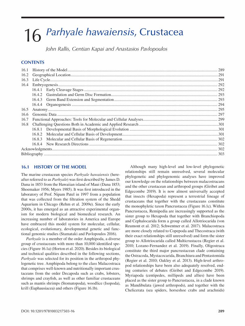
16 Parhyale hawaiensis , Crustacea
John Rallis, Gentian Kapai and Anastasios Pavlopoulos
CONTENTS
16.1 History of the Model................................................................................................................................................. 289
16.2 Geographical Location ...............................................................................................................................................291
16.3 Life Cycle ...................................................................................................................................................................291
16.4 Embryogenesis.......................................................................................................................................................... 292
16.4.1 Early Cleavage Stages ................................................................................................................................. 292
16.4.2 Gastrulation and Germ Disc Formation ....................................................................................................... 293
16.4.3 Germ Band Extension and Segmentation .................................................................................................... 293
16.4.4 Organogenesis ............................................................................................................................................. 294
16.5 Anatomy.................................................................................................................................................................... 295
16.6 Genomic Data ........................................................................................................................................................... 297
16.7 Functional Approaches: Tools for Molecular and Cellular Analyses........................................................................ 299
16.8 Challenging Questions Both in Academic and Applied Research .............................................................................301
16.8.1 Developmental Basis of Morphological Evolution ......................................................................................301
16.8.2 Molecular and Cellular Basis of Development .............................................................................................301
16.8.3 Molecular and Cellular Basis of Regeneration ............................................................................................ 302
16.8.4 New Research Directions ............................................................................................................................ 302
Acknowledgments ................................................................................................................................................................ 302
Bibliography ........................................................................................................................................................................ 303
16.1 HISTORY OF THE MODEL Although many high-level and low-level phylogenetic
The marine crustacean species Parhyale hawaiensis (here-phylogenetic and phylogenomic analyses have improved
after referred to as Parhyale) was first described by James D. our knowledge on the relationships between malacostracans
relationships still remain unresolved, several molecular
Dana in 1853 from the Hawaiian island of Maui (Dana 1853;and the other crustacean and arthropod groups (Giribet and
Shoemaker 1956; Myers 1985). It was first introduced in the Edgecombe 2019). It is now almost universally accepted
laboratory of Prof. Nipam Patel in 1997 from a population that insects (Hexapoda) represent a terrestrial lineage of
that was collected from the filtration system of the Shedd crustaceans that together with the crustaceans constitute
Aquarium in Chicago (Rehm et al. 2009e). Since the early the monophyletic taxon Pancrustacea (Figure 16.1c). Within
2000s, it has emerged as an attractive experimental organ-Pancrustacea, Remipedia are increasingly supported as the
ism for modern biological and biomedical research. An sister group to Hexapoda that together with Branchiopoda
increasing number of laboratories in America and Europe and Cephalocarida form a group called Allotriocarida (von
have embraced this model system for molecular, cellular, Reumont et al. 2012; Schwentner et al. 2017). Malacostraca
ecological, evolutionary, developmental genetic and func-are more closely related to Copepoda and Thecostraca (with
tional genomic studies (Stamataki and Pavlopoulos 2016). their exact relationships still unresolved) and form the sister
Parhyale is a member of the order Amphipoda, a diverse group to Allotriocarida called Multicrustacea (Regier et al.
group of crustaceans with more than 10,000 identifi ed spe-2010; Lozano-Fernandez et al. 2019). Finally, Oligostraca
cies (Figure 16.1a) (Horton et al. 2020). Besides its biological constitute the third major pancrustacean clade containing
and technical qualities described in the following sections, the Ostracoda, Mystacocarida, Branchiura and Pentastomida
Parhyale was selected for its position in the arthropod phy-(Regier et al. 2010; Oakley et al. 2013). High-level arthro
logenetic tree. Amphipoda belong to the class Malacostraca pod relationships have been also adequately resolved, end-
that comprises well-known and nutritionally important crus-ing centuries of debates (Giribet and Edgecombe 2019).
taceans from the order Decapoda such as crabs, lobsters, Myriapoda (centipedes, millipeds and allies) have been
shrimps and crayfish, as well as other familiar crustaceans placed as the sister group to Pancrustacea, in a clade known
such as mantis shrimps (Stomatopoda), woodlice (Isopoda), as Mandibulata (jawed arthropods), and together with the
krill (Euphausiacea) and others (Figure 16.1b). Chelicerata (sea spiders, horseshoe crabs and arachnids)
DOI: 10.1201/9781003217503-16 289
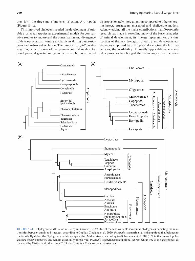
290
they form the three main branches of extant Arthropoda
( Figure 16.1c ).
This improved phylogeny seeded the development of suit
able crustacean species as experimental models for compar
ative studies to understand the conservation and divergence
of developmental patterning mechanisms during pancrusta
cean and arthropod evolution. The insect Drosophila melanogaster, which is one of the premier animal models for
developmental genetic and genomic research, has attracted
Emerging Marine Model Organisms
disproportionately more attention compared to other emerg
ing insect, crustacean, myriapod and chelicerate models.
Acknowledging all the major contributions that Drosophila research has made in revealing many of the basic principles
of animal development, its lineage represents only a tiny
fraction of the morphological diversity and developmental
strategies employed by arthropods alone. Over the last two
decades, the availability of broadly applicable experimen
tal approaches has bridged the technological gap between
FIGURE 16.1 Phylogenetic affiliation of Parhyale hawaiensis. (a) One of the few available molecular phylogenies depicting the rela
tionships between amphipod lineages, according to Copilaş-Ciocianu et al. 2020. Parhyale is a marine talitrid amphipod that belongs to
the family Hyalidae. (b) Phylogenetic relationships within Malacostraca, according to (Schwentner et al. 2018). Note that many topolo
gies are poorly supported and remain essentially unresolved. Parhyale is a peracarid amphipod. (c) Molecular tree of the arthropods, as
reviewed by Giribet and Edgecombe 2019. Parhyale is a Malacostracan crustacean.
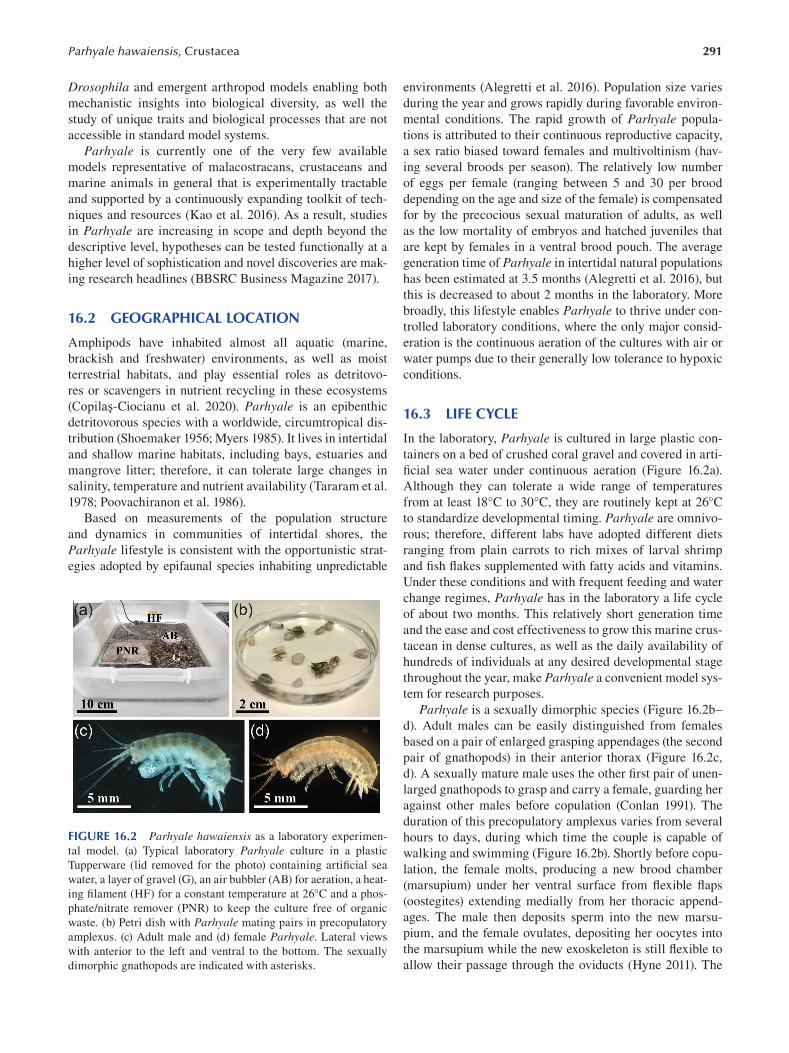
291 Parhyale hawaiensis, Crustacea
Drosophila and emergent arthropod models enabling both
mechanistic insights into biological diversity, as well the
study of unique traits and biological processes that are not
accessible in standard model systems.
Parhyale is currently one of the very few available
models representative of malacostracans, crustaceans and
marine animals in general that is experimentally tractable
and supported by a continuously expanding toolkit of tech
niques and resources (Kao et al. 2016). As a result, studies
in Parhyale are increasing in scope and depth beyond the
descriptive level, hypotheses can be tested functionally at a
higher level of sophistication and novel discoveries are mak
ing research headlines (BBSRC Business Magazine 2017 ).
16.2 GEOGRAPHICAL LOCATION
Amphipods have inhabited almost all aquatic (marine,
brackish and freshwater) environments, as well as moist
terrestrial habitats, and play essential roles as detritovo
res or scavengers in nutrient recycling in these ecosystems
(Copilaş-Ciocianu et al. 2020). Parhyale is an epibenthic
detritovorous species with a worldwide, circumtropical dis
tribution (Shoemaker 1956; Myers 1985). It lives in intertidal
and shallow marine habitats, including bays, estuaries and
mangrove litter; therefore, it can tolerate large changes in
salinity, temperature and nutrient availability (Tararam et al.
1978; Poovachiranon et al. 1986).
Based on measurements of the population structure
and dynamics in communities of intertidal shores, the
Parhyale lifestyle is consistent with the opportunistic strat
egies adopted by epifaunal species inhabiting unpredictable
FIGURE 16.2 Parhyale hawaiensis as a laboratory experimen
tal model. (a) Typical laboratory Parhyale culture in a plastic
Tupperware (lid removed for the photo) containing artifi cial sea
water, a layer of gravel (G), an air bubbler (AB) for aeration, a heat
ing filament (HF) for a constant temperature at 26°C and a phos
phate/nitrate remover (PNR) to keep the culture free of organic
waste. (b) Petri dish with Parhyale mating pairs in precopulatory
amplexus. (c) Adult male and (d) female Parhyale. Lateral views
with anterior to the left and ventral to the bottom. The sexually
dimorphic gnathopods are indicated with asterisks.
environments (Alegretti et al. 2016). Population size varies
during the year and grows rapidly during favorable environ
mental conditions. The rapid growth of Parhyale popula
tions is attributed to their continuous reproductive capacity,
a sex ratio biased toward females and multivoltinism (hav
ing several broods per season). The relatively low number
of eggs per female (ranging between 5 and 30 per brood
depending on the age and size of the female) is compensated
for by the precocious sexual maturation of adults, as well
as the low mortality of embryos and hatched juveniles that
are kept by females in a ventral brood pouch. The average
generation time of Parhyale in intertidal natural populations
has been estimated at 3.5 months (Alegretti et al. 2016), but
this is decreased to about 2 months in the laboratory. More
broadly, this lifestyle enables Parhyale to thrive under con
trolled laboratory conditions, where the only major consid
eration is the continuous aeration of the cultures with air or
water pumps due to their generally low tolerance to hypoxic
conditions.
16.3 LIFE CYCLE
In the laboratory, Parhyale is cultured in large plastic con
tainers on a bed of crushed coral gravel and covered in arti
ficial sea water under continuous aeration (Figure 16.2a).
Although they can tolerate a wide range of temperatures
from at least 18°C to 30°C, they are routinely kept at 26°C
to standardize developmental timing. Parhyale are omnivo
rous; therefore, different labs have adopted different diets
ranging from plain carrots to rich mixes of larval shrimp
and fi sh flakes supplemented with fatty acids and vitamins.
Under these conditions and with frequent feeding and water
change regimes, Parhyale has in the laboratory a life cycle
of about two months. This relatively short generation time
and the ease and cost effectiveness to grow this marine crus
tacean in dense cultures, as well as the daily availability of
hundreds of individuals at any desired developmental stage
throughout the year, make Parhyale a convenient model sys
tem for research purposes.
Parhyale is a sexually dimorphic species (Figure 16.2b–
d). Adult males can be easily distinguished from females
based on a pair of enlarged grasping appendages (the second
pair of gnathopods) in their anterior thorax (Figure 16.2c,
d). A sexually mature male uses the other first pair of unen
larged gnathopods to grasp and carry a female, guarding her
against other males before copulation (Conlan 1991). The
duration of this precopulatory amplexus varies from several
hours to days, during which time the couple is capable of
walking and swimming (Figure 16.2b). Shortly before copu
lation, the female molts, producing a new brood chamber
(marsupium) under her ventral surface from fl exible fl aps
(oostegites) extending medially from her thoracic append
ages. The male then deposits sperm into the new marsu
pium, and the female ovulates, depositing her oocytes into
the marsupium while the new exoskeleton is still fl exible to
allow their passage through the oviducts (Hyne 2011). The
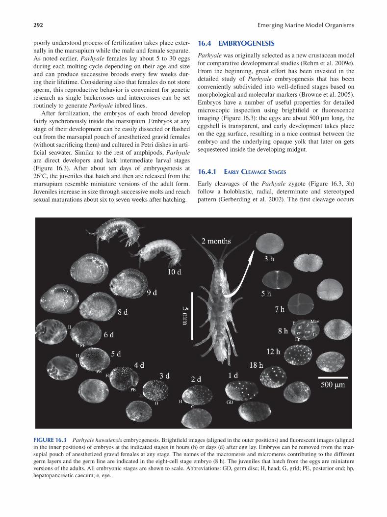
292 Emerging Marine Model Organisms
poorly understood process of fertilization takes place exter
nally in the marsupium while the male and female separate.
As noted earlier, Parhyale females lay about 5 to 30 eggs
during each molting cycle depending on their age and size
and can produce successive broods every few weeks dur
ing their lifetime. Considering also that females do not store
sperm, this reproductive behavior is convenient for genetic
research as single backcrosses and intercrosses can be set
routinely to generate Parhyale inbred lines.
After fertilization, the embryos of each brood develop
fairly synchronously inside the marsupium. Embryos at any
stage of their development can be easily dissected or fl ashed
out from the marsupial pouch of anesthetized gravid females
(without sacrificing them) and cultured in Petri dishes in arti
ficial seawater. Similar to the rest of amphipods, Parhyale are direct developers and lack intermediate larval stages
(Figure 16.3). After about ten days of embryogenesis at
26°C, the juveniles that hatch and then are released from the
marsupium resemble miniature versions of the adult form.
Juveniles increase in size through successive molts and reach
sexual maturations about six to seven weeks after hatching.
16.4 EMBRYOGENESIS
Parhyale was originally selected as a new crustacean model
for comparative developmental studies (Rehm et al. 2009e).
From the beginning, great effort has been invested in the
detailed study of Parhyale embryogenesis that has been
conveniently subdivided into well-defined stages based on
morphological and molecular markers (Browne et al. 2005).
Embryos have a number of useful properties for detailed
microscopic inspection using brightfield or fl uorescence
imaging (Figure 16.3): the eggs are about 500 μm long, the
eggshell is transparent, and early development takes place
on the egg surface, resulting in a nice contrast between the
embryo and the underlying opaque yolk that later on gets
sequestered inside the developing midgut.
16.4.1 EARLY CLEAVAGE STAGES
Early cleavages of the Parhyale zygote (Figure 16.3, 3h)
follow a holoblastic, radial, determinate and stereotyped
pattern (Gerberding et al. 2002). The fi rst cleavage occurs
FIGURE 16.3 Parhyale hawaiensis embryogenesis. Brightfield images (aligned in the outer positions) and fluorescent images (aligned
in the inner positions) of embryos at the indicated stages in hours (h) or days (d) after egg lay. Embryos can be removed from the mar
supial pouch of anesthetized gravid females at any stage. The names of the macromeres and micromeres contributing to the different
germ layers and the germ line are indicated in the eight-cell stage embryo (8 h). The juveniles that hatch from the eggs are miniature
versions of the adults. All embryonic stages are shown to scale. Abbreviations: GD, germ disc; H, head; G, grid; PE, posterior end; hp,
hepatopancreatic caecum; e, eye.
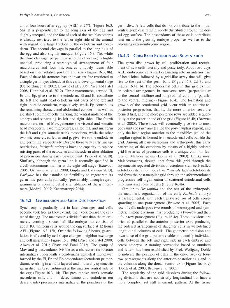
293 Parhyale hawaiensis, Crustacea
about four hours after egg lay (AEL) at 26°C (Figure 16.3,
5h). It is perpendicular to the long axis of the egg and
slightly unequal, and the fate of each of the two blastomeres
is already restricted to the left or right side of the animal
with regard to a large fraction of the ectoderm and meso
derm. The second cleavage is parallel to the long axis of
the egg and also slightly unequal (Figure 16.3, 7h), while
the third cleavage (perpendicular to the other two) is highly
unequal, producing a stereotypical arrangement of four
macromeres and four micromeres uniquely identifi able
based on their relative position and size (Figure 16.3, 8h).
Each of these blastomeres has an invariant fate restricted to
a single germ layer already at this early developmental stage
(Gerberding et al. 2002; Browne et al. 2005; Price and Patel
2008; Hannibal et al. 2012). Three macromeres, termed El,
Er and Ep, give rise to the ectoderm: El and Er contribute
the left and right head ectoderm and parts of the left and
right thoracic ectoderm, respectively, while Ep contributes
the remaining thoracic and abdominal ectoderm, as well as
a distinct column of cells marking the ventral midline of the
embryo and separating its left and right sides. The fourth
macromere, termed Mav, generates the visceral and somatic
head mesoderm. Two micromeres, called mL and mr, form
the left and right somatic trunk mesoderm, while the other
two micromeres, called en and g, give rise to the endoderm
and germ line, respectively. Despite these very early lineage
restrictions, Parhyale embryos have the capacity to replace
missing parts of the ectoderm and mesoderm after ablation
of precursors during early development (Price et al. 2010).
Similarly, although the germ line is normally specifi ed in
a cell-autonomous manner at the eight-cell stage (Extavour
2005; Ozhan-Kizil et al. 2009; Gupta and Extavour 2013),
Parhyale has the astonishing flexibility to regenerate its
germ line post-embryonically (presumably through repro
gramming of somatic cells) after ablation of the g micro
mere ( Modrell 2007 ; Kaczmarczyk 2014 ).
16.4.2 GASTRULATION AND GERM DISC FORMATION
Synchrony is gradually lost in later cleavages, and cells
become yolk free as they extrude their yolk toward the cen
ter of the egg. The macromeres divide faster than the micro
meres, forming a soccer ball-like embryo that consists of
about 100 uniform cells around the egg surface at 12 hours
AEL (Figure 16.3, 12h). Over the following 8 hours, gastru
lation is effected by cell shape changes, neighbor exchange
and cell migration (Figure 16.3, 18h) (Price and Patel 2008;
Alwes et al. 2011; Chaw and Patel 2012). The group of
Mav and g descendants (visible as a characteristic rosette)
internalizes underneath a condensing epithelial monolayer
formed by the El, Er and Ep descendants (ectoderm primor
dium), resulting in a multi-layered and bilaterally symmetric
germ disc (embryo rudiment) at the anterior ventral side of
the egg (Figure 16.3, 1d). The presumptive trunk somatic
mesoderm (mL and mr descendants) and endoderm (en
descendants) precursors internalize at the periphery of the
germ disc. A few cells that do not contribute to the initial
ventral germ disc remain widely distributed around the dor
sal egg surface. The descendants of these cells contribute
later on to the growing embryo proper, as well as to the
adjoining extra-embryonic region.
16.4.3 GERM BAND EXTENSION AND SEGMENTATION
The germ disc grows by cell proliferation and recruit
ment of new cells laterally and posteriorly. About two days
AEL, embryonic cells start organizing into an anterior pair
of head lobes followed by a grid-like array that will give
rise to the rest of the germ band (Figure 16.3, 2d–3d and
Figure 16.4a, b). The ectodermal cells in this grid exhibit
an ordered arrangement in transverse rows (perpendicular
to the ventral midline) and longitudinal columns (parallel
to the ventral midline) (Figure 16.4). The formation and
growth of the ectodermal grid occur with an anterior-to
posterior progression, that is, the more anterior rows are
formed first, and the more posterior rows are added sequen
tially at the posterior end of the grid (Figure 16.4b) (Browne
et al. 2005). These rows will eventually give rise to most
body units of Parhyale (called the post-naupliar region), and
only the head region anterior to the mandibles (called the
naupliar region) is formed from ectodermal cells outside the
grid. Among all pancrustaceans and arthropods, this early
patterning of the ectoderm by means of a highly ordered
grid-like array of precursor cells is a unique common fea
ture of Malacostracans (Dohle et al. 2003). Unlike most
Malacostracans, though, that form this grid through the
asymmetric repeated divisions of ectoderm stem cells called
ectoteloblasts, amphipods like Parhyale lack ectoteloblasts
and form the post-naupliar grid through the aforementioned
progressive self-organization of scattered ectodermal cells
into transverse rows of cells (Figure 16.4b).
Similar to Drosophila and the rest of the arthropods,
the metameric organization of the early Parhyale embryo
is parasegmental, with each transverse row of cells corre
sponding to one parasegment (Browne et al. 2005). Each
row of cells undergoes two rounds of stereotyped and sym
metric mitotic divisions, first producing a two-row and then
a four-row parasegment (Figure 16.4c). These divisions are
oriented parallel to the anterior–posterior axis, producing
the ordered arrangement of daughter cells in well-defi ned
longitudinal columns of cells. The geometric precision and
invariance of the grid pattern enables to identify individual
cells between the left and right side in each embryo and
across embryos. A naming convention based on numbers
and letters has been established by Prof. Wolfgang Dohle
to indicate the position of cells in the one-, two- or four-
row parasegments along the anterior–posterior axis and in
the columns along the dorsal-ventral axis (Figure 16.4b, c)
(Dohle et al. 2003; Browne et al. 2005).
The regularity of the grid dissolves during the follow
ing divisions that are not strictly longitudinal but have a
more complex, yet still invariant, pattern. At the tissue
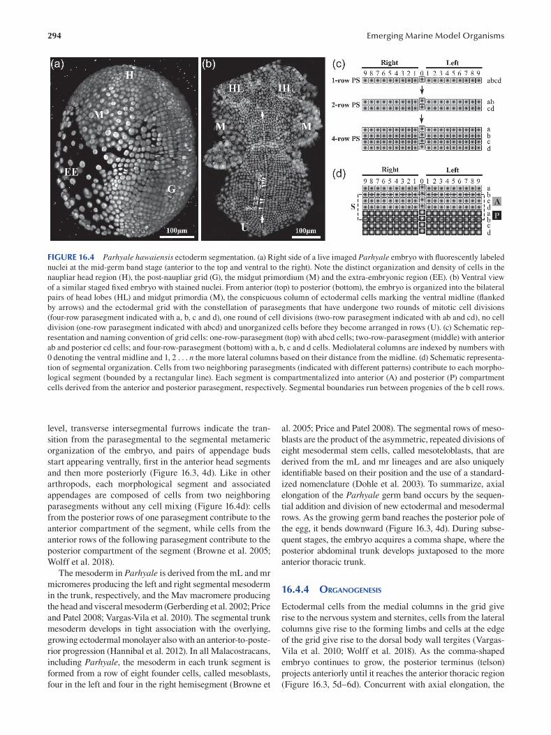
294 Emerging Marine Model Organisms
FIGURE 16.4 Parhyale hawaiensis ectoderm segmentation. (a) Right side of a live imaged Parhyale embryo with fl uorescently labeled
nuclei at the mid-germ band stage (anterior to the top and ventral to the right). Note the distinct organization and density of cells in the
naupliar head region (H), the post-naupliar grid (G), the midgut primordium (M) and the extra-embryonic region (EE). (b) Ventral view
of a similar staged fixed embryo with stained nuclei. From anterior (top) to posterior (bottom), the embryo is organized into the bilateral
pairs of head lobes (HL) and midgut primordia (M), the conspicuous column of ectodermal cells marking the ventral midline (fl anked
by arrows) and the ectodermal grid with the constellation of parasegments that have undergone two rounds of mitotic cell divisions
(four-row parasegment indicated with a, b, c and d), one round of cell divisions (two-row parasegment indicated with ab and cd), no cell
division (one-row parasegment indicated with abcd) and unorganized cells before they become arranged in rows (U). (c) Schematic rep
resentation and naming convention of grid cells: one-row-parasegment (top) with abcd cells; two-row-parasegment (middle) with anterior
ab and posterior cd cells; and four-row-parasegment (bottom) with a, b, c and d cells. Mediolateral columns are indexed by numbers with
0 denoting the ventral midline and 1, 2 . . . n the more lateral columns based on their distance from the midline. (d) Schematic representa
tion of segmental organization. Cells from two neighboring parasegments (indicated with different patterns) contribute to each morpho
logical segment (bounded by a rectangular line). Each segment is compartmentalized into anterior (A) and posterior (P) compartment
cells derived from the anterior and posterior parasegment, respectively. Segmental boundaries run between progenies of the b cell rows.
level, transverse intersegmental furrows indicate the tran- al. 2005; Price and Patel 2008). The segmental rows of meso
sition from the parasegmental to the segmental metameric blasts are the product of the asymmetric, repeated divisions of
organization of the embryo, and pairs of appendage buds eight mesodermal stem cells, called mesoteloblasts, that are
start appearing ventrally, first in the anterior head segments derived from the mL and mr lineages and are also uniquely
and then more posteriorly (Figure 16.3, 4d). Like in other identifiable based on their position and the use of a standard-
arthropods, each morphological segment and associated ized nomenclature (Dohle et al. 2003). To summarize, axial
appendages are composed of cells from two neighboring elongation of the Parhyale germ band occurs by the sequen
parasegments without any cell mixing (Figure 16.4d): cells tial addition and division of new ectodermal and mesodermal
from the posterior rows of one parasegment contribute to the rows. As the growing germ band reaches the posterior pole of
anterior compartment of the segment, while cells from the the egg, it bends downward (Figure 16.3, 4d). During subse
anterior rows of the following parasegment contribute to the quent stages, the embryo acquires a comma shape, where the
posterior compartment of the segment (Browne et al. 2005; posterior abdominal trunk develops juxtaposed to the more
Wolff et al. 2018). anterior thoracic trunk.
The mesoderm in Parhyale is derived from the mL and mr
micromeres producing the left and right segmental mesoderm 16.4.4 ORGANOGENESIS in the trunk, respectively, and the Mav macromere producing
the head and visceral mesoderm (Gerberding et al. 2002; Price Ectodermal cells from the medial columns in the grid give
and Patel 2008; Vargas-Vila et al. 2010). The segmental trunk rise to the nervous system and sternites, cells from the lateral
mesoderm develops in tight association with the overlying, columns give rise to the forming limbs and cells at the edge
growing ectodermal monolayer also with an anterior-to-poste- of the grid give rise to the dorsal body wall tergites (Vargas
rior progression ( Hannibal et al. 2012). In all Malacostracans, Vila et al. 2010; Wolff et al. 2018). As the comma-shaped
including Parhyale, the mesoderm in each trunk segment is embryo continues to grow, the posterior terminus (telson)
formed from a row of eight founder cells, called mesoblasts, projects anteriorly until it reaches the anterior thoracic region
four in the left and four in the right hemisegment (Browne et (Figure 16.3, 5d–6d). Concurrent with axial elongation, the
295 Parhyale hawaiensis, Crustacea
lateral edges of the ectoderm expand dorsally and the form
ing tergites from the two body halves fuse along the dorsal
midline completing dorsal closure. Starting from the ante
rior head region backward and sequentially bulging out in
the thorax and the abdomen, a total of 19 pairs of append
ages develop along the Parhyale body (Figure 16.3, 4d–6d).
Appendages increase in size and elongate along their respec
tive proximal–distal axes (Browne et al. 2005; Wolff et al.
2018). As detailed in the next sections, the elaboration of the
proximal–distal axis varies between different appendage
types in terms of their pattern, size and shape, resulting in a
remarkable morphological diversity along the anterior–pos
terior axis. Appendage growth, morphogenesis and differen
tiation continue until the late stages of embryogenesis, when
the fully formed appendages occupy almost half of the egg
space before hatching (Figure 16.3, 8d–9d).
The naupliar (anterior head) and post-naupliar somatic
mesoderm are separated early on as they derive from the
Mav macromere and the mL/mr micromeres at the eight-
cell stage, respectively (Figure 16.3, 8h) (Gerberding et al.
2002; Browne et al. 2005). The micromere-derived rows of
four mesoteloblasts (labeled M1 to M4 medial-to-lateral)
under each side the ectodermal grid generate the segmen
tal mesodermal founders (mesoblasts labeled m1 to m4) in
the posterior head (second maxillary segment) and the tho
racic and abdominal segments. Similar to the ectodermal
structures, patterning of mesoderm occurs with an anterior
(earlier developing) to posterior (later developing) progres
sion. The origin and first division of mesoblasts has been
described in Parhyale (Price and Patel 2008). The contribu
tion of these mesoblasts to the different muscle groups along
the dorsal–ventral body axis has been studied in the closely
related amphipod Orchestia cavimana and is only briefl y
summarized here (Hunnekuhl and Wolff 2012). Descendant
cells from the medial-most m1 mesoblasts give rise to the
ventromedian muscles, cells from the central m2 and m3
mesoblasts generate the extrinsic and intrinsic musculature
of the appendages and cells from the m3 and m4 mesoblasts
give rise to the dorsolateral trunk musculature and the heart
( Figure 16.7e ).
The Mav macromere gives rise to the head musculature
of the antennae and the mandibular and first maxillary seg
ments (Price and Patel 2008; Price et al. 2010; Hunnekuhl
and Wolff 2012), as well as to the visceral mesoderm. After
gastrulation, a subset of the Mav progeny migrates under
the developing head segments and becomes partitioned
into the differentiating head segments in a less studied
manner. The majority of Mav progeny, together with the
descendants from the en micromere, give rise to the mid
gut tube that will eventually spread over and encapsulate
the central yolk mass (Gerberding et al. 2002). During the
germ band stages, the midgut primordium becomes visible
as a bilateral pair of discs under the head lobes (Figure
16.4a, b). The discs increase in size, forming a continuous
ventral layer that expands dorsally and posteriorly under
the ectoderm and mesoderm to cover the yolk (Gerberding
et al. 2002). The midgut develops a number of blind tubes
(caeca) that function in food digestion and absorption
(Schmitz and Scherrey 1983). The most conspicuous pair
of anterior caeca, called hepatopancreatic caeca, extend in
synchrony through peristaltic contractions from the ante
rior end of the midgut until the posterior abdomen of the
embryo (Browne et al. 2005). The hepatopancreatic caeca
flank and extend parallel to the midgut that is visible along
the dorsal side (Figure 16.3, 8d–9d). The Parhyale heart
develops as a muscular tube along the dorsal thoracic region
with three pairs of lateral inflow valves and an anterior out
flow valve, and it can be observed while beating on top of
the midgut (Kontarakis et al. 2011b). At around the same
stage when the heart starts beating, the bilaterally sym
metric compound eyes become visible in the head capsule
as small white clusters, each with about three ommatidia
(Figure 16.3, 8d). During the last two days of embryogen
esis, the eyes become dark pigmented, and Parhyale hatch
with about eight to nine pigmented ommatidia per eye
(Figure 16.3, 9d–10d), but this number increases gradually
to about 50 in older adults (Ramos et al. 2019).
The smallest micromere g at the eight-cell stage is the
source of germ line cells in the adult ovaries and testes
(Figure 16.3, 8h) (Gerberding et al. 2002; Extavour 2005).
There is strong evidence that germ cells in Parhyale are
specified by a cell-autonomous mechanism (preformation)
via the early asymmetric segregation of maternally provided
germ line determinants (Extavour 2005; Modrell 2007;
Gupta and Extavour 2013). The primordial germ cells (prog
eny of the g micromere) that have internalized and prolif
erated during the gastrulation and germ disc stages form a
single medial cluster of about 15 cells under the posterior
head ectoderm as the germ band elongates. During organo
genesis stages, they split into two bilaterally opposed cell
populations that migrate separately under the lateral ecto
derm toward the dorsal side of the embryo (Extavour 2005;
Browne et al. 2005). At the end of embryogenesis, when the
eyes and the heart have formed, the primordial germ cells
are aligned in two rows flanking the dorsal midline at the
site of the future gonads (Extavour 2005).
16.5 ANATOMY
Parhyale displays the typical amphipod body plan that is lat
erally compressed and consists of a series of repeating seg
mental units along the anterior–posterior axis organized into
three major tagmata: the head, the thorax and the abdomen
(Figure 16.5a, b). The head (a.k.a. cephalon) is composed of
six segments with five pairs of appendages. The most ante
rior limbless pre-antennal segment is followed by fi ve seg
ments bearing the first and second pair of antennae (An1
and An2; Figure 16.5a, b) and three pairs of medially fused
gnathal appendages: the mandibles (Mn; Figure 16.5c) and
the first and second maxillae (Mx1 and Mx2; Figure 16.5d).
The thoracic region is composed of eight segments, each
bearing a pair of jointed uniramous appendages (I-shaped
limbs with a single proximal–distal axis) (Figure 16.5e–i).
The abdominal region is composed of six segments, each

296
bearing a pair of jointed biramous appendages (Y-shaped
limbs with a bifurcated proximal–distal axis) (Figure 16.5J,
K). Each thoracic and abdominal appendage consists of a
proximal part and a distal part (Boxshall 2004; Pavlopoulos
and Wolff 2020). The proximal part, called a protopod, is
composed of two appendage articles (a.k.a. podomeres or
limb segments), namely the proximal coxa and the distal
basis (Figure 16.5g). The existence of a third proximal-most
podomere, the precoxa, has been also proposed recently
(Bruce and Patel 2020). In uniramous thoracic appendages
(Figure 16.5e–i), a single branch extends distally from the
protopod called the endopod (or telopod). In abdominal
biramous appendages (Figure 16.5j, k), two branches extend
distally from the protopod called the endopod (inner branch)
and exopod (outer branch). As detailed in the following, dif
ferent types of appendages develop also a variable number
Emerging Marine Model Organisms
of ventral and/or dorsal outgrowths from their protopod
called endites and exites, respectively,
The first thoracic segment (T1) is fused to the head that
is also referred to as the cephalothorax. The T1 appendages,
called maxillipeds (T1/Mxp; Figure 16.5e), are jointed,
and uniramous like the more posterior thoracic append
ages. However, unlike the other thoracic appendages and
similar to the more anterior maxillae, maxillipeds are
reduced in size, are medially fused at their base and have
two prominent endites on their proximal segments (Figure
16.5e). Maxillipeds and gnathal appendages are special
ized for feeding and have a compact arrangement around
the mouth region (Figure 16.5b). The thoracic region behind
T1, known as the pereon, is composed of seven segments
(T2 to T8), each with a pair of uniramous appendages (a.k.a.
pereopods or thoracopods) that articulate independently on
FIGURE 16.5 Appendage diversity in Parhyale hawaiensis. (a) Scanning electron micrograph of a Parhyale juvenile showing the dif
ferent tagmata along the anterior–posterior body axis and the first and second pair of antennae (An1 and An2). Lateral view with anterior
to the left and ventral to the bottom. (b) Similar to (a) from a ventral view. (c–k) Cuticle preparations of dissected appendages with their
proximal side to left and their distal side to the right: (c) mandible (Mn); (d) Maxilla 1 (Mx1) and Maxilla 2 (Mx2); (e) bilateral pair of
maxillipeds from the first thoracic segment (T1/Mxp) indicating the pair of endites (2Xen) on each side; (f) gnathopod from the second
thoracic segment (T2); (g) gnathopod from the third thoracic segment (T3) indicating the seven segments, coxa (cx), basis (ba), ischium
(is), merus (me), carpus (ca), propodus (pro) and dactylus (da), as well as the two exites, the coxal plate (cp) and the gill (g); (h) pereopod
from the fourth thoracic segment (T4); (i) pereopod from the eighth thoracic segment (T8); (j) bilateral pair of pleopods from the fi rst
abdominal segment (A1) and (k) bilateral pair of uropods from the fourth abdominal segment (A4) indicating the endopod (endo) and
exopod (exo) on each side. All appendages are shown to scale.
297 Parhyale hawaiensis, Crustacea
each side (Figure 16.5f–i). From proximal to distal, each
jointed pereopod is made of seven segments: two protopo
dal segments (coxa and basis) and five endopodal segments
(ischium, merus, carpus, propodus and dactylus) (Figure
16.5g). The T2 and T3 segments bear subchelate (clawed)
grasping appendages, called gnathopods (Figure 16.5f, g),
that are used for defense, grooming and as precopulatory
organs (the T2 gnathopods) by males to carry the females
(Holmquist 1982). The post-embryonic enlargement of the
propodus and dactylus exclusively in the male T3 gnatho
pod is the most striking sexually dimorphic character in
Parhyale (Figure 16.2c, d). The remaining five pereonic seg
ments T4 to T8 bear elongated walking appendages (Figure
16.5h , i). Importantly, the opposite orientation between the
T4/T5 pereopods that extend anteriorly and the T6/T7/T8
pereopods that extend posteriorly (Figure 16.5a, b) is what
gives the group its name (from Greek words αμφί [amphi =
both ways] and πόδι [podi = limb]). Besides their distinct
function, podomere morphology and orientation, the T2–T8
pereopods are also distinguished by the presence or absence
and the shape of exites attached on their protopodal coxa.
Protective coxal plates of variable size and shapes are pres
ent on all pereopods, while respiratory gills are present on
T3 to T7 appendages (Figure 16.5g). In the case of adult
females, special endites (oostegites) forming the marsupium
are attached on the pereopods T2 to T5.
The abdominal (pleonic) segments A1 to A6 develop two
types of paired biramous appendages: pleopods on A1 to A3
(Figure 16.5j) and uropods on A4 to A6 (Figure 16.5k). Each
of these biramous limbs has similar endopodal and exopo
dal branches. The A1–A3 pleopods (a.k.a. swimmerets) are
highly setose and are coupled together for swimming and
moving water over the thoracic gills. The A4–A6 uropods
are thickened and spiky appendages used for jumping. The
most posterior terminal structure is the telson, which is a
small flap over the anus attached to segment A6. Overall, the
morphological and functional specialization of body parts
and associated appendages has been one of the main reasons
for putting Parhyale forward as an attractive model organ
ism for molecular, cellular, developmental and evolutionary
studies described in Section 16.8.
Much less work has been invested in Parhyale to study
the development, anatomy and physiology of the nervous
system compared to other crustaceans (Wiese 2002).
Parhyale neuroanatomy was recently described using a
combination of histological, immuno-histochemical, opti
cal and X-ray tomography methods (Wittfoth et al. 2019).
The central nervous system consists of the brain and the
ventral nerve cord. The ventral nerve cord is composed of
the subesophageal ganglion, seven segmental ganglia of the
pereon, three segmental ganglia of the pleosome and one
fused ganglion of the urosome. The brain lies between the
compound eyes in the dorsal part of the head capsule with
its three neuromeres, the protocerebrum, deutocerebrum
and tritocerebrum lining up from dorsal to ventral. The pro
tocerebrum is equipped with the optic neuropils, the deu
tocerebrum with the antenna 1 neuropil and the olfactory
lobe and the tritocerebrum with the antenna 2 neuropil. The
three optic neuropils, the lamina, medulla and lobula, are
in close proximity with each other, but only the lamina con
nects to the photoreceptors of the ommatidia in the com
pound eye (Wittfoth et al. 2019; Ramos et al. 2019). The
architecture and neural connectivity of the Parhyale visual
system have diverged from the typical organization exhib
ited by other malacostracan crustaceans and are associated
with a shift to low spatial resolution and simple visual tasks
(Ramos et al. 2019).
16.6 GENOMIC DATA
For many years, the high cost of next-generation sequenc
ing technologies and the big size of malacostracan crus
tacean genomes have been prohibitive for amphipod
genomics. Thanks to the decreasing sequencing costs, this
limitation was overcome during the last five years, fi rst
with the sequencing, de novo assembly and annotation of
the Parhyale genome in 2016, followed more recently by
genome assemblies of variable quality for the amphipods
Hyalella azteca, Trinorchestia longiramous, Platorchestia hallaensis, Orchestia grillus and Gammarus roeselii ( Table
16.1) (Poynton et al. 2018; Patra et al. 2020a, 2020b; Cormier
et al. 2021).
The Parhyale genome resembles and even exceeds in
many respects the complexity of the human genome. The
genome consists of 23 pairs of chromosomes (2n = 46;
Figure 16.6a), and its size is estimated at 3.6 Gb. The huge
genome size is associated with an expansion in repetitive
and intronic sequences and exhibits very high levels of
TABLE 16.1 Sequenced Amphipod Genomes Species Size No. of Scaffold N50 NCBI Link
(Gb) Scaffolds (Kb)
Parhyale hawaiensis 2.75 278,189 20,229 www.ncbi.nlm.nih.gov/assembly/GCA_001587735.2
Hyalella azteca 0.55 18,000 215 www.ncbi.nlm.nih.gov/assembly/GCA_000764305.3
Trinorchestia longiramus 0.89 30,897 120 www.ncbi.nlm.nih.gov/assembly/GCA_006783055.1
Platorchestia hallaensis 1.18 39,873 87 www.ncbi.nlm.nih.gov/assembly/GCA_014220935.1
Orchestia grillus 0.81 143,039 17 www.ncbi.nlm.nih.gov/assembly/GCA_014899125.1
Gammarus roeselii 3.2 1,130,582 4.8 www.ncbi.nlm.nih.gov/assembly/GCA_016164225.1
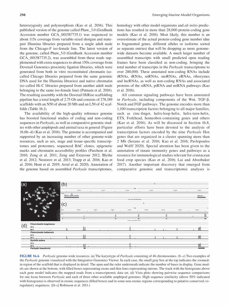
298
heterozygosity and polymorphism (Kao et al. 2016). This
published version of the genome called Phaw_3.0 (GenBank
Accession number GCA_001587735.1) was sequenced to
about 115x coverage from variable-sized shotgun and mate-
pair Illumina libraries prepared from a single adult male
from the Chicago-F iso-female line. The latest version of
the genome, called Phaw_5.0 (GenBank Accession number
GCA_001587735.2), was assembled from these reads sup
plemented with extra sequences to about 150x coverage from
Dovetail Genomics proximity ligation libraries, which were
generated from both in vitro reconstituted chromatin (so
called Chicago libraries prepared from the same genomic
DNA used for the Illumina libraries) and native chromatin
(so-called Hi-C libraries prepared from another adult male
belonging to the same iso-female line) (Putnam et al. 2016).
The resulting assembly with the Dovetail HiRise scaffolding
pipeline has a total length of 2.75 Gb and consists of 278,189
scaffolds with an N50 of about 20 Mb and an L50 of 42 scaf
folds ( Table 16.1 ).
The availability of the high-quality reference genome
has boosted functional studies of coding and non-coding
sequences in Parhyale, as well as comparative genomic stud
ies with other amphipods and animal taxa in general (Figure
16.6b–d) (Kao et al. 2016 ). The genome is accompanied and
supported by an increasing number of other genome-wide
resources, such as sex, stage and tissue-specifi c transcrip
tomes and proteomes, sequenced BAC clones, epigenetic
marks and chromatin accessibility profiles (Parchem et al.
2010; Zeng et al. 2011; Zeng and Extavour 2012; Blythe
et al. 2012; Nestorov et al. 2013; Trapp et al. 2016; Kao et
al. 2016; Hunt et al. 2019; Artal et al. 2020). Annotation of
the genome based on assembled Parhyale transcriptomes,
Emerging Marine Model Organisms
homology with other model organisms and ab initio predic
tions has resulted in more than 28,000 protein-coding gene
models (Kao et al. 2016). Most likely, this number is an
overestimate of the actual protein-coding gene number (due
to fragmented genes, different alleles or isoforms sorted
as separate entries) that will be dropping as more genome-
wide datasets become available. A much larger number of
assembled transcripts with small predicted open reading
frames have been classified as non-coding, bringing the
total number of transcripts in the Parhyale transcriptome to
over 280,000. These annotated non-coding RNAs include
rRNAs, tRNAs, snRNAs, snoRNAs, eRNAs, ribozymes
and lncRNAs, as well as non-coding RNAs and associated
proteins of the siRNA, piRNA and miRNA pathways (Kao
et al. 2016 ).
All common signaling pathways have been annotated
in Parhyale, including components of the Wnt, TGF-β,
Notch and FGF pathways. The genome encodes more than
1,100 transcription factors belonging to all major families,
such as zinc-finger, helix-loop-helix, helix-turn-helix,
ETS, Forkhead, homeobox-containing genes and others
(Kao et al. 2016). As will be discussed in Section 16.8,
particular efforts have been devoted to the analysis of
transcription factors encoded by the nine Parhyale Hox
genes that are organized in a cluster spanning more than
2 Mb (Serano et al. 2016; Kao et al. 2016; Pavlopoulos
and Wolff 2020). Special attention has been given to the
annotation of innate immunity genes and pathways as a
resource for immunological studies relevant for crustacean
food crop species (Kao et al. 2016; Lai and Aboobaker
2017). Another important discovery that emerged from
comparative genomic and transcriptomic analyses is
FIGURE 16.6 Parhyale genome-wide resources. (a) The karyotype of Parhyale consisting of 46 chromosomes. (b–c) Two examples of
the Parhyale genome visualized with the Integrative Genomics Viewer. In each case, the small gray box at the top indicates the zoomed-
in region of the scaffold that is displayed in detail. The span and the ruler underneath indicate the number of bases in display. Gene mod
els are shown at the bottom, with filled boxes representing exons and thin lines representing introns. The track with the histograms above
each gene model indicates the mapped reads from a transcriptomic data set. (d) Vista plots showing pairwise sequence comparisons
for one locus between Parhyale and each of three other available amphipod genomes. High sequence similarity (above 50% indicated
with histograms) is observed in exonic sequences (filled boxes) and in some non-exonic regions corresponding to putative conserved cis
regulatory sequences. ([b-c] Robinson et al. 2011.)
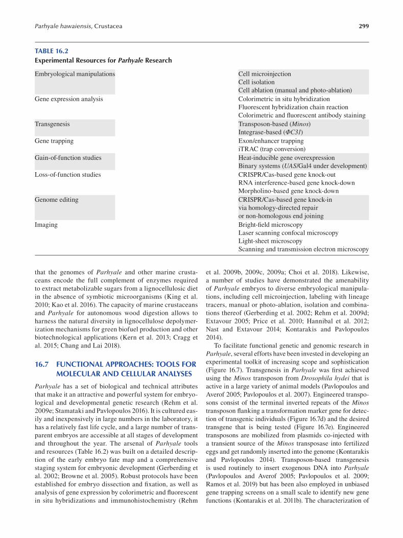
299 Parhyale hawaiensis, Crustacea
TABLE 16.2 Experimental Resources for Parhyale Research
Embryological manipulations Cell microinjection
Cell isolation
Cell ablation (manual and photo-ablation)
Gene expression analysis Colorimetric in situ hybridization
Fluorescent hybridization chain reaction
Colorimetric and fluorescent antibody staining
Transgenesis Transposon-based (Minos) Integrase-based (ΦC31)
Gene trapping Exon/enhancer trapping
iTRAC (trap conversion)
Gain-of-function studies Heat-inducible gene overexpression
Binary systems (UAS/Gal4 under development)
Loss-of-function studies CRISPR/Cas-based gene knock-out
RNA interference-based gene knock-down
Morpholino-based gene knock-down
Genome editing CRISPR/Cas-based gene knock-in
via homology-directed repair
or non-homologous end joining
Imaging Bright-fi eld microscopy
Laser scanning confocal microscopy
Light-sheet microscopy
Scanning and transmission electron microscopy
that the genomes of Parhyale and other marine crusta
ceans encode the full complement of enzymes required
to extract metabolizable sugars from a lignocellulosic diet
in the absence of symbiotic microorganisms (King et al.
2010; Kao et al. 2016 ). The capacity of marine crustaceans
and Parhyale for autonomous wood digestion allows to
harness the natural diversity in lignocellulose depolymer
ization mechanisms for green biofuel production and other
biotechnological applications (Kern et al. 2013; Cragg et
al. 2015; Chang and Lai 2018).
16.7 FUNCTIONAL APPROACHES: TOOLS FOR MOLECULAR AND CELLULAR ANALYSES
Parhyale has a set of biological and technical attributes
that make it an attractive and powerful system for embryo
logical and developmental genetic research (Rehm et al.
2009e; Stamataki and Pavlopoulos 2016 ). It is cultured eas
ily and inexpensively in large numbers in the laboratory, it
has a relatively fast life cycle, and a large number of trans
parent embryos are accessible at all stages of development
and throughout the year. The arsenal of Parhyale tools
and resources (Table 16.2) was built on a detailed descrip
tion of the early embryo fate map and a comprehensive
staging system for embryonic development (Gerberding et
al. 2002; Browne et al. 2005). Robust protocols have been
established for embryo dissection and fixation, as well as
analysis of gene expression by colorimetric and fl uorescent
in situ hybridizations and immunohistochemistry (Rehm
et al. 2009b, 2009c, 2009a ; Choi et al. 2018). Likewise,
a number of studies have demonstrated the amenability
of Parhyale embryos to diverse embryological manipula
tions, including cell microinjection, labeling with lineage
tracers, manual or photo-ablation, isolation and combina
tions thereof (Gerberding et al. 2002; Rehm et al. 2009d;
Extavour 2005; Price et al. 2010; Hannibal et al. 2012;
Nast and Extavour 2014; Kontarakis and Pavlopoulos
2014 ).
To facilitate functional genetic and genomic research in
Parhyale, several efforts have been invested in developing an
experimental toolkit of increasing scope and sophistication
(Figure 16.7). Transgenesis in Parhyale was fi rst achieved
using the Minos transposon from Drosophila hydei that is
active in a large variety of animal models (Pavlopoulos and
Averof 2005; Pavlopoulos et al. 2007). Engineered transpo
sons consist of the terminal inverted repeats of the Minos transposon flanking a transformation marker gene for detec
tion of transgenic individuals (Figure 16.7d) and the desired
transgene that is being tested (Figure 16.7e). Engineered
transposons are mobilized from plasmids co-injected with
a transient source of the Minos transposase into fertilized
eggs and get randomly inserted into the genome (Kontarakis
and Pavlopoulos 2014). Transposon-based transgenesis
is used routinely to insert exogenous DNA into Parhyale (Pavlopoulos and Averof 2005; Pavlopoulos et al. 2009;
Ramos et al. 2019) but has been also employed in unbiased
gene trapping screens on a small scale to identify new gene
functions (Kontarakis et al. 2011b). The characterization of
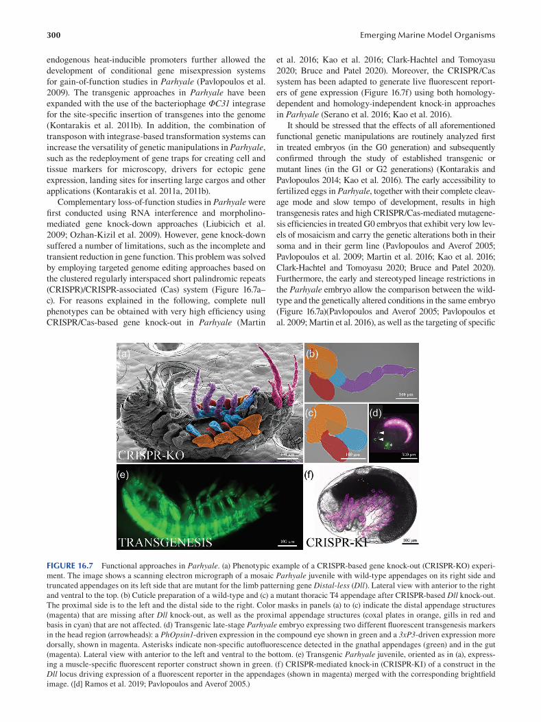
300
endogenous heat-inducible promoters further allowed the
development of conditional gene misexpression systems
for gain-of-function studies in Parhyale (Pavlopoulos et al.
2009). The transgenic approaches in Parhyale have been
expanded with the use of the bacteriophage ΦC31 integrase
for the site-specific insertion of transgenes into the genome
(Kontarakis et al. 2011b). In addition, the combination of
transposon with integrase-based transformation systems can
increase the versatility of genetic manipulations in Parhyale,
such as the redeployment of gene traps for creating cell and
tissue markers for microscopy, drivers for ectopic gene
expression, landing sites for inserting large cargos and other
applications (Kontarakis et al. 2011a, 2011b).
Complementary loss-of-function studies in Parhyale were
first conducted using RNA interference and morpholino
mediated gene knock-down approaches (Liubicich et al.
2009; Ozhan-Kizil et al. 2009). However, gene knock-down
suffered a number of limitations, such as the incomplete and
transient reduction in gene function. This problem was solved
by employing targeted genome editing approaches based on
the clustered regularly interspaced short palindromic repeats
(CRISPR)/CRISPR-associated (Cas) system (Figure 16.7a–
c). For reasons explained in the following, complete null
phenotypes can be obtained with very high effi ciency using
CRISPR/Cas-based gene knock-out in Parhyale ( Martin
Emerging Marine Model Organisms
et al. 2016; Kao et al. 2016; Clark-Hachtel and Tomoyasu
2020; Bruce and Patel 2020). Moreover, the CRISPR/Cas
system has been adapted to generate live fl uorescent report
ers of gene expression (Figure 16.7f) using both homology-
dependent and homology-independent knock-in approaches
in Parhyale (Serano et al. 2016; Kao et al. 2016 ).
It should be stressed that the effects of all aforementioned
functional genetic manipulations are routinely analyzed fi rst
in treated embryos (in the G0 generation) and subsequently
confirmed through the study of established transgenic or
mutant lines (in the G1 or G2 generations) (Kontarakis and
Pavlopoulos 2014; Kao et al. 2016 ). The early accessibility to
fertilized eggs in Parhyale, together with their complete cleav
age mode and slow tempo of development, results in high
transgenesis rates and high CRISPR/Cas-mediated mutagene
sis efficiencies in treated G0 embryos that exhibit very low lev
els of mosaicism and carry the genetic alterations both in their
soma and in their germ line (Pavlopoulos and Averof 2005;
Pavlopoulos et al. 2009; Martin et al. 2016; Kao et al. 2016;
Clark-Hachtel and Tomoyasu 2020; Bruce and Patel 2020).
Furthermore, the early and stereotyped lineage restrictions in
the Parhyale embryo allow the comparison between the wild-
type and the genetically altered conditions in the same embryo
(Figure 16.7a)(Pavlopoulos and Averof 2005; Pavlopoulos et
al. 2009; Martin et al. 2016 ), as well as the targeting of specifi c
FIGURE 16.7 Functional approaches in Parhyale. (a) Phenotypic example of a CRISPR-based gene knock-out (CRISPR-KO) experi
ment. The image shows a scanning electron micrograph of a mosaic Parhyale juvenile with wild-type appendages on its right side and
truncated appendages on its left side that are mutant for the limb patterning gene Distal-less (Dll). Lateral view with anterior to the right
and ventral to the top. (b) Cuticle preparation of a wild-type and (c) a mutant thoracic T4 appendage after CRISPR-based Dll knock-out.
The proximal side is to the left and the distal side to the right. Color masks in panels (a) to (c) indicate the distal appendage structures
(magenta) that are missing after Dll knock-out, as well as the proximal appendage structures (coxal plates in orange, gills in red and
basis in cyan) that are not affected. (d) Transgenic late-stage Parhyale embryo expressing two different fluorescent transgenesis markers
in the head region (arrowheads): a PhOpsin1-driven expression in the compound eye shown in green and a 3xP3-driven expression more
dorsally, shown in magenta. Asterisks indicate non-specifi c autofl uorescence detected in the gnathal appendages (green) and in the gut
(magenta). Lateral view with anterior to the left and ventral to the bottom. (e) Transgenic Parhyale juvenile, oriented as in (a), express
ing a muscle-specifi c fluorescent reporter construct shown in green. (f) CRISPR-mediated knock-in (CRISPR-KI) of a construct in the
Dll locus driving expression of a fl uorescent reporter in the appendages (shown in magenta) merged with the corresponding brightfi eld
image. ([d] Ramos et al. 2019; Pavlopoulos and Averof 2005.)
301 Parhyale hawaiensis, Crustacea
lineages for labeling or ablation (Price et al. 2010; Alwes et
al. 2011; Hannibal et al. 2012; Konstantinides and Averof
2014). All these features are very useful for experimentation in
Parhyale, because they provide fast and reliable information
about gene expression, regulation and function months before
stable lines are available for analysis.
Parhyale is not only a genetically tractable but also an
optically tractable experimental model, which is ideal to
make the connection between the molecular and cellular
basis of development. Light and electron microscopy analy
ses of fixed specimens have been used widely to character
ize wild-type and mutant phenotypes in detail (Pavlopoulos
et al. 2009; Serano et al. 2016; Martin et al. 2016; Ramos
et al. 2019; Clark-Hachtel and Tomoyasu 2020; Bruce and
Patel 2020). The increasing collection of genetic tools
and transgenic lines for imaging, in combination with the
transparency and low autofluorescence of embryos, have
enabled the implementation of live microscopic inspections
of cellular dynamics with exceptional spatial and tempo
ral resolution. Different microscopy modalities, including
bright-field, confocal and multi-view light-sheet microscopy,
have been adapted successfully to image embryonic and
post-embryonic processes over several days of development,
such as Parhyale gastrulation and germ band formation,
appendage development and regeneration (Price and Patel
2008; Alwes et al. 2011; Chaw and Patel 2012; Hannibal
et al. 2012; Alwes et al. 2016; Wolff et al. 2018). Last but
not least, thanks to a very productive collaboration between
biologists, microscopists and computer scientists, a suite of
sophisticated and open-source software is available for the
visualization of image datasets, the manual and automated
tracking of cells and the reconstruction and editing of cell
lineages to understand the cellular behaviors contributing to
tissue and organ development in Parhyale (Wolff et al. 2018;
Salvador-Martínez et al. 2020; Sugawara et al. 2021).
16.8 CHALLENGING QUESTIONS BOTH IN ACADEMIC AND APPLIED RESEARCH
Parhyale lends itself to address several longstanding questions
and problems in modern biological and biomedical research
(Stamataki and Pavlopoulos 2016). Based on its phyloge
netic position and its technical and biological attributes, it has
increased the breadth and depth of comparative developmen
tal studies with other pancrustacean, arthropod and animal
groups. As a malacostracan crustacean, it is also closely related
to shrimps, crabs and lobsters that have attracted research inter
est as commercially and nutritionally important crop species.
16.8.1 DEVELOPMENTAL BASIS OF
MORPHOLOGICAL EVOLUTION
Research in Parhyale was inspired by and has greatly con
tributed toward our understanding of the developmental
mechanisms driving body plan evolution and specialization
of body parts. Crustaceans exhibit a tremendous morpho
logical diversity observed both within and between spe
cies. Seminal studies in crustaceans were among the fi rst
to implicate changes in the expression of Hox genes with
the evolution of animal body plans and the diversifi cation
of developing appendages (Averof and Akam 1995; Averof
and Patel 1997 ). Although expression studies of Hox genes
have been carried out in all major crustacean lineages, the
most comprehensive analysis of all nine Hox genes has been
carried out in Parhyale, where they exhibit both spatial and
temporal collinearity (Serano et al. 2016). Hox expression
domains correspond to the subdivision of the body into
morphologically and functionally distinct regions and cor
relate with the development of distinct appendages types.
Importantly, systematic loss-of-function and gain-of-func
tion studies of Hox genes in Parhyale have provided com
pelling evidence for the causal association between Hox genes and crustacean segmental organization and append
age diversification (Pavlopoulos et al. 2009; Liubicich et al.
2009; Martin et al. 2016). The homeotic transformations
produced in these functional studies were recapitulating in
Parhyale macroevolutionary changes observed in the body
organization of other crustacean lineages, like the repeated
evolution of feeding maxillipeds from locomotory append
ages in the anterior thorax of many crustacean lineages or
the change in the relative number of abdominal pleopods
and uropods between malacostracan lineages (Averof et al.
2010; Martin et al. 2016; Pavlopoulos and Wolff 2020).
Along similar lines, expression and functional studies of
developmental patterning genes in Parhyale have enabled
to test century-old hypotheses about the homology and evo
lutionary novelty of arthropod appendages (McKenna et al.
2021). Considering that winged insects evolved from wingless
crustaceans, different theories have been proposed to explain
the origin of insect wings (Clark-Hachtel and Tomoyasu
2016 ): they are novel lateral outgrowths from the dorsal body
wall (tergal origin or paranotal hypothesis), or they evolved
from the exites of proximal leg segments (pleural origin
hypothesis). By comparing the expression patterns and the
loss-of-function phenotypes of leg, wing and body wall pat
terning genes between insects and Parhyale, it was proposed
that the proximal exite-bearing leg segments present in the
common ancestor of insects and crustaceans were incorpo
rated into the insect body wall, giving rise to the insect wings
(Clark-Hachtel and Tomoyasu 2020; Bruce and Patel 2020).
Thus, these elegant studies in Parhyale have provided a fresh
and unified model in favor of the evolution of insect wings
from a pre-existing structure in their crustacean ancestor. A
similar framework has been adopted to homologize pancrus
tacean, myriapod and chelicerate appendages, suggesting an
eight-segment ground plan for the arthropod leg (Bruce 2021).
16.8.2 MOLECULAR AND CELLULAR
BASIS OF DEVELOPMENT
One of the biggest challenges in developmental biology is to
understand how the genomic information encodes the mor
phogenetic cell behaviors, like cell proliferation and death,
cell shape changes and cell movements, that produce the
characteristic size and shape of developing tissues and organs
302
in multicellular organisms (Heisenberg and Bellaïche 2013;
Wan et al. 2019). The optical properties of the Parhyale egg
and the embryonic development of its appendages as direct
outgrowths from the body wall have enabled to advance
beyond a gene-centric view of development and start inte
grating the molecular with the cellular aspects of appendage
formation. In a tour-de-force study that involved advanced
light-sheet fluorescence microscopy and image analysis
tools, the complete lineage of developing Parhyale limbs was
reconstructed with single-cell resolution (Wolff et al. 2018).
The spatial coordinates for all constituent cells, their tem
poral dynamics and mother-daughter relationships were then
analyzed to shed light on the cellular mechanisms driving
appendage outgrowth, elongation and segmentation. These
analyses revealed the cellular architecture and patterned cell
activities operating at different stages of appendage develop
ment that were then correlated with the expression patterns
of candidate patterning genes known from limb studies in
Drosophila (Wolff et al. 2018). Interestingly, some of these
cellular events were similar, but some were distinct com
pared to the textbook Drosophila paradigm, motivating
future experiments to understand the conservation and diver
gence of appendage patterning mechanism during pancrusta
cean and arthropod evolution (Pavlopoulos and Wolff 2020).
In a broader perspective, all recent technical break
throughs in Parhyale research provide the opportunity to
study gene expression and function in the context of single-
cell-resolution fate maps, both under wild-type and under
genetically perturbed conditions. These multidisciplinary
approaches will be employed by the community to advance
our knowledge on longstanding questions in developmental
biology, such as the identity and function of cell fate deter
minants (Nestorov et al. 2013; Gupta and Extavour 2013),
the molecular and cellular mechanisms underlying embryo
formation and healing (Alwes et al. 2011; Chaw and Patel
2012), the relative contributions of cell history and cell com
munication in development (Price et al. 2010; Hannibal et
al. 2012) and the allometric growth of serially homologous
appendages (Pavlopoulos et al. 2009; Martin et al. 2016).
16.8.3 MOLECULAR AND CELLULAR
BASIS OF REGENERATION
Besides studying embryonic development, Parhyale has
emerged as an attractive model system for regenerative stud
ies, as it has the capacity to replace lost tissues and entire
body parts post-embryonically (Grillo et al. 2016). It has
been demonstrated that Parhyale has the ability to regener
ate missing limbs after amputation (Kontarakis et al. 2011b)
and its germ line after ablation of the g micromere (Modrell
2007; Kaczmarczyk 2014). In principle, new cells for regen
eration can be produced from the activation of pluripotent or
lineage-restricted stem cells, as well as the de-differentiation
or trans-differentiation of differentiated cells (Tanaka and
Reddien 2011). Thanks to the early lineage restrictions in
the Parhyale embryo, it has been possible to label and iden
tify the source cells and examine their regenerative potential
Emerging Marine Model Organisms
during regrowth of limbs (Konstantinides and Averof 2014).
The sources for the new cells are restricted by their lineage
and proximity to the regenerating appendage: the ectoder
mal and mesodermal lineages make distinct contributions
to ectoderm-derived tissues (epidermis and neurons) and
mesoderm-derived tissues (muscles and blood cells), respec
tively. Importantly, the availability of cell-specifi c markers
led to the major discovery of invertebrate muscle stem cells
in Parhyale that, similar to satellite cells in vertebrates, serve
as progenitors for muscle repair during limb regeneration
(Konstantinides and Averof 2014). It has been also possible
to trace cell behaviors through live imaging of appendage
regeneration in Parhyale with high resolution and over sev
eral days after amputation (Alwes et al. 2016). For example,
the epidermis of the new limb is not formed by specialized
stem cells but by the cell proliferation and redifferentiation of
existing epidermal cells. Overall, crustaceans have a long his
tory in regenerative research, albeit at the physiological and
anatomical level. The addition of Parhyale as a new geneti
cally and optically tractable regenerative model has opened
new possibilities to dissect the molecular and cellular mecha
nisms that can be redeployed during its lifetime to replace
missing limbs, germ cells and possibly other structures.
16.8.4 NEW RESEARCH DIRECTIONS
We will conclude this chapter with some more exciting new
research avenues that, like regeneration, were not conceiv
able when Parhyale was first introduced in the laboratory
but have the potential to make big contributions to both basic
and applied fields of research. The first steps have been taken
already in establishing Parhyale as a model in the fi elds of
chronobiology and ecotoxicology (Hunt et al. 2019; Artal
et al. 2018 , 2020; Diehl et al. 2021). Studies of the Parhyale innate immunity have been also proposed for disease control
in crustacean aquaculture through a better understanding of
infectious pathogens and host defense mechanisms (Kao et al.
2016; Lai and Aboobaker 2017). Last but not least, studies of
lignocellulose digestion in Parhyale can offer novel insights
into the ecologically important and understudied mechanisms
of wood recycling in marine environments and can unleash
their significant potential for biotechnological applications
(Cragg et al. 2015; Kao et al. 2016; Chang and Lai 2018).
ACKNOWLEDGMENTS
This chapter was prepared amid the most challenging condi
tions imposed by the COVID-19 pandemic. It was only com
pleted thanks to the unceasing patience and encouragement
of our editors, Dr. Agnès Boutet and Dr. Bernd Schierwater,
to whom we are deeply indebted. We would also like to
thank Dr. Carsten Wolff and Suyash Kumar for providing
the images in Figures 16.6a and 16.7a, and Dr. Evangelia
Stamataki for comments on the manuscript. John Rallis and
Gentian Kapai were supported by Fondation Santé graduate
studentships and Anastasios Pavlopoulos by IMBB-FORTH
intramural funds.
303 Parhyale hawaiensis, Crustacea
BIBLIOGRAPHY
Alegretti, L., de Aragão Umbuzeiro, G., and M.N. Flynn. 2016.
“Population Dynamics of Parhyale hawaiensis (Dana, 1853)
(Amphipoda: Hyalidae) Associated with an Intertidal Algal
Belt in Southeastern Brazil.” Journal of Crustacean Biology 36 (6): 785–791. doi:10.1163/1937240X-00002480.
Alwes, F., Enjolras, C., and M. Averof. 2016. “Live Imaging Reveals
the Progenitors and Cell Dynamics of Limb Regeneration.”
ELife 5. doi:10.7554/eLife.19766.
Alwes, F., Hinchen, B., and C.G. Extavour. 2011. “Patterns of
Cell Lineage, Movement, and Migration from Germ Layer
Specification to Gastrulation in the Amphipod Crustacean
Parhyale hawaiensis.” Developmental Biology 359 (1): 110–
123. doi:10.1016/j.ydbio.2011.07.029.
Artal, M.C., Dos Santos, A., Henry, T.B., and G. de Aragão
Umbuzeiro. 2018. “Development of an Acute Toxicity Test
with the Tropical Marine Amphipod Parhyale hawaiensis.” Ecotoxicology (London, England) 27 (2): 103–108.
doi:10.1007/s10646-017-1875-3.
Artal, M.C., Pereira, K.D., Luchessi, A.D., Okura, V.K., Henry,
T.B., Marques-Souza, H., and G. de Aragão Umbuzeiro.
2020. “Transcriptome Analysis in Parhyale hawaiensis Reveal
Sex-Specific Responses to AgNP and AgCl Exposure.”
Environmental Pollution (Barking, Essex: 1987) 260 (May):
113963. doi:10.1016/j.envpol.2020.113963.
Averof, M., and M. Akam. 1995. “Hox Genes and the Diversifi cation
of Insect and Crustacean Body Plans.” Nature 376 (6539):
420–423. doi:10.1038/376420a0.
Averof, M., and N.H. Patel. 1997. “Crustacean Appendage Evolution
Associated with Changes in Hox Gene Expression.” Nature 388 (6643): 682–686. doi:10.1038/41786.
Averof, M., Pavlopoulos, A., and Z. Kontarakis. 2010. “Evolution
of New Appendage Types by Gradual Changes in Hox Gene
Expression: The Case of Crustacean Maxillipeds.” 5.
“BBSRC Business Magazine.” 2017. January 1. https://bbsrc.ukri.
org/documents/bbsrc-business-winter-2017-pdf/ .
Blythe, M.J., Malla, S., Everall, R., Shih, Y.H., Lemay, V., Moreton,
J., Wilson, R., and A.A. Aboobaker. 2012. “High Through-
Put Sequencing of the Parhyale hawaiensis MRNAs and
microRNAs to Aid Comparative Developmental Studies.”
PLoS One 7 (3): e33784. doi:10.1371/journal.pone.0033784.
Boxshall, G.A. 2004. “The Evolution of Arthropod Limbs.”
Biological Reviews of the Cambridge Philosophical Society 79 (2): 253–300. doi:10.1017/s1464793103006274.
Browne, W.E., Price, A.L., Gerberding, M., and N.H. Patel. 2005.
“Stages of Embryonic Development in the Amphipod
Crustacean, Parhyale hawaiensis.” Genesis (New York, N.Y.: 2000) 42 (3): 124–149. doi:10.1002/gene.20145.
Bruce, H.S. 2021. “How to Align Arthropod Leg Segments.”
BioRxiv, January. Cold Spring Harbor Laboratory, 2021.
01.20.427514. doi:10.1101/2021.01.20.427514.
Bruce, H.S., and N.H. Patel. 2020. “Knockout of Crustacean
Leg Patterning Genes Suggests That Insect Wings and
Body Walls Evolved from Ancient Leg Segments.” Nature Ecology & Evolution 4 (12): 1703–1712. doi:10.1038/
s41559-020-01349-0.
Chang, W.H., and A.G. Lai. 2018. “Mixed Evolutionary Origins of
Endogenous Biomass-Depolymerizing Enzymes in Animals.”
BMC Genomics 19 (1): 483. doi:10.1186/s12864-018-4861-0.
Chaw, R.C., and N.H. Patel. 2012. “Independent Migration of
Cell Populations in the Early Gastrulation of the Amphipod
Crustacean Parhyale hawaiensis.” Developmental Biology 371 (1): 94–109. doi:10.1016/j.ydbio.2012.08.012.
Choi, H.M.T., Schwarzkopf, M., Fornace, M.E., Acharya, A.,
Artavanis, G., Stegmaier, J., Cunha, A., and N.A. Pierce.
2018. “Third-Generation In Situ Hybridization Chain Reaction:
Multiplexed, Quantitative, Sensitive, Versatile, Robust.”
Development (Cambridge, England) 145 (12). doi:10.1242/
dev.165753.
Clark-Hachtel, C.M., and Y. Tomoyasu. 2016. “Exploring the
Origin of Insect Wings from an Evo-Devo Perspective.”
Current Opinion in Insect Science 13 (February): 77–85.
doi:10.1016/j.cois.2015.12.005.
Clark-Hachtel, C.M., and Y. Tomoyasu. 2020. “Two Sets of Candidate
Crustacean Wing Homologues and Their Implication for the
Origin of Insect Wings.” Nature Ecology & Evolution , August.
doi:10.1038/s41559-020-1257-8.
Conlan, K.E. 1991. “Precopulatory Mating Behavior and Sexual
Dimorphism in the Amphipod Crustacea.” Hydrobiologia 223 (1): 255–282. doi:10.1007/BF00047644.
Copilaş-Ciocianu, D., Borko, Š., and C. Fišer. 2020. “The Late
Blooming Amphipods: Global Change Promoted Post-
Jurassic Ecological Radiation Despite Palaeozoic Origin.”
Molecular Phylogenetics and Evolution 143: 106664.
doi:10.1016/j.ympev.2019.106664.
Cormier, A., Chebbi, M.A., Giraud, I., Wattier, R., Teixeira, M., Gilbert,
C., Rigaud, T., and R. Cordaux. 2021. “Comparative Genomics
of Strictly Vertically Transmitted, Feminizing Microsporidia
Endosymbionts of Amphipod Crustaceans.” Genome Biology and Evolution 13 (1). doi:10.1093/gbe/evaa245.
Cragg, S.M., Beckham, G.T., Bruce, N.C., Bugg, T.D.H., Distel,
D.L., Dupree, P., Green Etxabe, A., Goodell, B.S., Jellison, J.,
McGeehan, J.E., McQueen-Mason, S.J., Schnorr, K., Walton,
P.H., Watts, J.E.M., and M. Zimmer. 2015. “Lignocellulose
Degradation Mechanisms across the Tree of Life.” Current Opinion in Chemical Biology, Energy • Mechanistic Biology 29 (December): 108–119. doi:10.1016/j.cbpa.2015.10.018.
Dana, J.D. 1853. United States Exploring Expedition: 14: Crustacea . Sherman.
Diehl, O.J., Assano, P.K., da Costa, T.R.G., Oliveira, R., Marques-
Souza, H., and G. de Aragão Umbuzeiro. 2021. “Antenna
Regeneration as an Ecotoxicological Endpoint in a Marine
Amphipod: A Proof of Concept Using Dimethyl Sulfoxide
and Difl ubenzuron.” Ecotoxicology (London, England), March. doi:10.1007/s10646-021-02395-5.
Dohle, W., Gerberding, M., Hejnol, A., and G. Scholtz. 2003. “Cell
Lineage, Segment Differentiation, and Gene Expression in
Crustaceans.” In Evolutionary Developmental Biology of Crustacea. 1st Edition, 95–133. CRC Press. doi:10.13140/
RG.2.1.4984.9841.
Extavour, C.G. 2005. “The Fate of Isolated Blastomeres with
Respect to Germ Cell Formation in the Amphipod Crustacean
Parhyale hawaiensis.” Developmental Biology 277 (2): 387–
402. doi:10.1016/j.ydbio.2004.09.030.
Gerberding, M., Browne, W.E., and N.H. Patel. 2002. “Cell Lineage
Analysis of the Amphipod Crustacean Parhyale hawaiensis Reveals an Early Restriction of Cell Fates.” Development (Cambridge, England) 129 (24): 5789–5801. doi:10.1242/
dev.00155.
Giribet, G., and G.D. Edgecombe. 2019. “The Phylogeny and
Evolutionary History of Arthropods.” Current Biology: CB 29 (12): R592–602. doi:10.1016/j.cub.2019.04.057.
Grillo, M., Konstantinides, N., and M. Averof. 2016. “Old Questions,
New Models: Unraveling Complex Organ Regeneration
with New Experimental Approaches.” Current Opinion in Genetics & Development 40 (October): 23–31. doi:10.1016/j.
gde.2016.05.006.
304
Gupta, T., and C.G. Extavour. 2013. “Identification of a Putative
Germ Plasm in the Amphipod Parhyale hawaiensis.”
EvoDevo 4 (1): 34. doi:10.1186/2041-9139-4-34.
Hannibal, R.L., Price, A.L., and N.H. Patel. 2012. “The Functional
Relationship between Ectodermal and Mesodermal
Segmentation in the Crustacean, Parhyale hawaiensis.”
Developmental Biology 361 (2): 427–438. doi:10.1016/j.
ydbio.2011.09.033.
Heisenberg, C.P., and Y. Bellaïche. 2013. “Forces in Tissue
Morphogenesis and Patterning.” Cell 153 (5): 948–962.
doi:10.1016/j.cell.2013.05.008.
Holmquist, J.G. 1982. “The Functional Morphology of Gnathopods:
Importance in Grooming, and Variation with Regard to
Habitat, in Talitroidean Amphipods.” Journal of Crustacean Biology 2 (2): 159–179. doi:10.2307/1547997.
Horton, T., Lowry, J., De Broyer, C., Bellan-Santini, D., Coleman,
C.O., Corbari, L., Costello, M.J., Daneliya, M., Dauvin, J.C.,
Fišer, C., Gasca, R., Grabowski, M., Guerra-García, J.M.,
Hendrycks, E., Hughes, L., Jaume, D., Jazdzewski, K., Kim,
Y.H., King, R., Krapp-Schickel, T., LeCroy, S., Lörz, A.N.,
Mamos, T., Senna, A.R., Serejo, C., Sket, B., Souza-Filho,
J.F., Tandberg, A.H., Thomas, J.D., Thurston, M., Vader, W.,
Väinölä, R., Vonk, R., White, K., and W. Zeidler. 2020. “World
Amphipoda Database.” www.marinespecies.org/amphipoda/ .
Hunnekuhl, V.S., and C. Wolff. 2012. “Reconstruction of Cell
Lineage and Spatiotemporal Pattern Formation of the
Mesoderm in the Amphipod Crustacean Orchestia cavimana.” Developmental Dynamics: An Offi cial Publication of the American Association of Anatomists 241 (4): 697–717.
doi:10.1002/dvdy.23758.
Hunt, B.J., Mallon, E.B., and E. Rosato. 2019. “In Silico
Identification of a Molecular Circadian System with Novel
Features in the Crustacean Model Organism Parhyale hawaiensis.” Frontiers in Physiology 10. Frontiers. doi:10.
3389/fphys.2019.01325.
Hyne, R.V. 2011. “Review of the Reproductive Biology of
Amphipods and Their Endocrine Regulation: Identifi cation
of Mechanistic Pathways for Reproductive Toxicants.”
Environmental Toxicology and Chemistry 30 (12): 2647–
2657. https://doi.org/10.1002/etc.673 .
Kaczmarczyk, A.N. 2014. “Germline Maintenance and Regeneration
in the Amphipod Crustacean, Parhyale hawaiensis.” UC Berkeley. https://escholarship.org/uc/item/5h94b7kg .
Kao, D., Lai, A.G., Stamataki, E., Rosic, S., Konstantinides, N.,
Jarvis, E., Di Donfrancesco, A., Pouchkina-Stancheva, N.,
Sémon, M., Grillo, M., Bruce, H., Kumar, S., Siwanowicz, I.,
Le, A., Lemire, A., Eisen, M.B., Extavour, C., Browne, W.E.,
Wolff, C., Averof, M., Patel, N.H., Sarkies, P., Pavlopoulos,
A., and A. Aboobaker. 2016. “The Genome of the Crustacean
Parhyale hawaiensis, a Model for Animal Development,
Regeneration, Immunity and Lignocellulose Digestion.”
ELife 5. doi:10.7554/eLife.20062.
Kern, M., McGeehan, J.E., Streeter, S.D., Martin, R.N.A., Besser,
K., Elias, L., Eborall, W., Malyon, G.P., Payne, C.M.,
Himmel, M.E., Schnorr, K., Beckham, G.T., Cragg, S.M.,
Bruce, N.C., and S.J. McQueen-Mason. 2013. “Structural
Characterization of a Unique Marine Animal Family 7
Cellobiohydrolase Suggests a Mechanism of Cellulase Salt
Tolerance.” Proceedings of the National Academy of Sciences of the United States of America 110 (25): 10189–10194.
doi:10.1073/pnas.1301502110.
King, A.J., Cragg, S.M., Li, Y., Dymond, J., Guille, M.J., Bowles,
D.J., Bruce, N.C., Graham, I.A., and S.J. McQueen-Mason.
2010. “Molecular Insight into Lignocellulose Digestion
by a Marine Isopod in the Absence of Gut Microbes.”
Proceedings of the National Academy of Sciences of the
Emerging Marine Model Organisms
United States of America 107 (12): 5345–5350. doi:10.1073/
pnas.0914228107.
Konstantinides, N., and M. Averof. 2014. “A Common Cellular
Basis for Muscle Regeneration in Arthropods and
Vertebrates.” Science (New York, N.Y.) 343 (6172): 788–791.
doi:10.1126/science.1243529.
Kontarakis, Z., Konstantinides, N., Pavlopoulos, A., and M. Averof.
2011a. “Reconfiguring Gene Traps for New Tasks Using
ITRAC.” Fly 5 (4): 352–355. doi:10.4161/fl y.5.4.18108.
Kontarakis, Z., and A. Pavlopoulos. 2014. “Transgenesis in
Non-Model Organisms: The Case of Parhyale.” Methods in Molecular Biology (Clifton, N.J.) 1196: 145–181.
doi:10.1007/978-1-4939-1242-1_10.
Kontarakis, Z., Pavlopoulos, A., Kiupakis, A., Konstantinides, N.,
Douris, V., and M. Averof. 2011b. “A Versatile Strategy for
Gene Trapping and Trap Conversion in Emerging Model
Organisms.” Development (Cambridge, England) 138 (12):
2625–2630. doi:10.1242/dev.066324.
Lai, A.G., and A.A. Aboobaker. 2017. “Comparative Genomic
Analysis of Innate Immunity Reveals Novel and Conserved
Components in Crustacean Food Crop Species.” BMC Genomics 18 (1): 389. doi:10.1186/s12864-017-3769-4.
Liubicich, D.M., Serano, J.M., Pavlopoulos, A., Kontarakis,
Z., Protas, M.E., Kwan, E., Chatterjee, S., Tran, K.D.,
Averof, M., and N.H. Patel. 2009. “Knockdown of Parhyale
Ultrabithorax Recapitulates Evolutionary Changes in
Crustacean Appendage Morphology.” Proceedings of the National Academy of Sciences of the United States of America 106 (33): 13892–13896. doi:10.1073/pnas.0903105106.
Lozano-Fernandez, J., Giacomelli, M., Fleming, J.F., Chen, A.,
Vinther, J., Thomsen, P.F., Glenner, H., Palero, F., Legg, D.A.,
Iliffe, T.M., Pisani, D., and J. Olesen. 2019. “Pancrustacean
Evolution Illuminated by Taxon-Rich Genomic-Scale Data
Sets with an Expanded Remipede Sampling.” Genome Biology and Evolution 11 (8). Oxford Academic: 2055–70.
doi:10.1093/gbe/evz097.
Martin, A., Serano, J.M., Jarvis, E., Bruce, H.S., Wang, J., Ray, S.,
Barker, C.A., O’Connell, L.C., and N.H. Patel. 2016. “CRISPR/
Cas9 Mutagenesis Reveals Versatile Roles of Hox Genes
in Crustacean Limb Specification and Evolution.” Current Biology: CB 26 (1): 14–26. doi:10.1016/j.cub.2015.11.021.
McKenna, K.Z., Wagner, G.P., and K.L. Cooper. 2021. “A
Developmental Perspective of Homology and Evolutionary
Novelty.” Current Topics in Developmental Biology 141:
1–38. doi:10.1016/bs.ctdb.2020.12.001.
Modrell, M.S. 2007. “Early Cell Fate Specification in the Amphipod
Crustacean, Parhyale hawaiensis.” UC Berkeley.
Myers, A.A. 1985. “Shallow-Water, Coral Reef and Mangrove
Amphipoda (Gammaridea) of Fiji.” Records of the Australian Museum, Supplement 5 (December). The Australian Museum:
1–143. doi:10.3853/j.0812–7387.5.1985.99.
Nast, A.R., and C.G. Extavour. 2014. “Ablation of a Single Cell
from Eight-Cell Embryos of the Amphipod Crustacean
Parhyale hawaiensis.” Journal of Visualized Experiments: JoVE (85) (March). doi:10.3791/51073.
Nestorov, P., Battke, F., Levesque, M.P., and M. Gerberding. 2013.
“The Maternal Transcriptome of the Crustacean Parhyale hawaiensis Is Inherited Asymmetrically to Invariant Cell
Lineages of the Ectoderm and Mesoderm.” PLoS One 8 (2):
e56049. doi:10.1371/journal.pone.0056049.
Oakley, T.H., Wolfe, J.M., Lindgren, A.R., and A.K. Zaharoff.
2013. “Phylotranscriptomics to Bring the Understudied into
the Fold: Monophyletic Ostracoda, Fossil Placement, and
Pancrustacean Phylogeny.” Molecular Biology and Evolution 30 (1). Oxford Academic: 215–233. doi:10.1093/molbev/
mss216.
305 Parhyale hawaiensis, Crustacea
Ozhan-Kizil, G., Havemann, J., and M. Gerberding. 2009. “Germ
Cells in the Crustacean Parhyale hawaiensis Depend on Vasa
Protein for Their Maintenance but Not for Their Formation.”
Developmental Biology 327 (1): 230–239. doi:10.1016/j.
ydbio.2008.10.028.
Parchem, R.J., Poulin, F., Stuart, A.B., Amemiya, C.T., and N.H.
Patel. 2010. “BAC Library for the Amphipod Crustacean,
Parhyale hawaiensis.” Genomics 95 (5): 261–267.
doi:10.1016/j.ygeno.2010.03.005.
Patra, A.K., Chung, O., Yoo, J.Y., Baek, S.H., Jung, T.W., Kim,
M.S., Yoon, M.G., Yang, Y., and J.H. Choi. 2020a. “The Draft
Genome Sequence of a New Land-Hopper Platorchestia hallaensis.” Frontiers in Genetics 11: 621301. doi:10.3389/
fgene.2020.621301.
Patra, A.K., Chung, O., Yoo, J.Y., Kim, M.S., Yoon, M.G., Choi,
J.H., and Y. Yang. 2020b. “First Draft Genome for the Sand-
Hopper Trinorchestia longiramus.” Scientifi c Data 7 (1): 85.
doi:10.1038/s41597-020-0424-8.
Pavlopoulos, A., and M. Averof. 2005. “Establishing Genetic
Transformation for Comparative Developmental Studies in
the Crustacean Parhyale hawaiensis.” Proceedings of the National Academy of Sciences of the United States of America 102 (22): 7888–7893. doi:10.1073/pnas.0501101102.
Pavlopoulos, A., Kontarakis, Z., Liubicich, D.M., Serano, J.M.,
Akam, M., Patel, N.H., and M. Averof. 2009. “Probing
the Evolution of Appendage Specialization by Hox Gene
Misexpression in an Emerging Model Crustacean.”
Proceedings of the National Academy of Sciences of the United States of America 106 (33): 13897–13902.
doi:10.1073/pnas.0902804106.
Pavlopoulos, A., Oehler, S., Kapetanaki, M.G., and C.
Savakis. 2007. “The DNA Transposon Minos as a Tool
for Transgenesis and Functional Genomic Analysis in
Vertebrates and Invertebrates.” Genome Biology 8 (Suppl 1):
S2. doi:10.1186/gb-2007-8-s1-s2.
Pavlopoulos, A., and C. Wolff. 2020. “Crustacean Limb Morphogenesis
during Normal Development and Regeneration.” In The Natural History of the Crustacea: Developmental Biology and Larval Ecology, edited by Klaus Anger, Steffen Harzsch,
and Martin Thiel. Vol. 7. Oxford: Oxford University Press.
Poovachiranon, S., Boto, K., and N. Duke. 1986. “Food Preference
Studies and Ingestion Rate Measurements of the Mangrove
Amphipod Parhyale hawaiensis (Dana).” Journal of Experimental Marine Biology and Ecology 98 (1): 129–140.
doi:10.1016/0022-0981(86)90078-X.
Poynton, H.C., Hasenbein, S., Benoit, J.B., Sepulveda, M.S.,
Poelchau, M.F., Hughes, D.S.T., Murali, S.C., Chen, S.,
Glastad, K.M., Goodisman, M.A.D., Werren, J.H., Vineis,
J.H., Bowen, J.L., Friedrich, M., Jones, J., Robertson, H.M.,
Feyereisen, R., Mechler-Hickson, A., Mathers, N., Lee, C.E.,
Colbourne, J.K., Biales, A., Johnston, J.S., Wellborn, G.A.,
Rosendale, A.J., Cridge, A.G., Munoz-Torres, M.C., Bain,
P.A., Manny, A.R., Major, K.M., Lambert, F.N., Vulpe, C.D.,
Tuck, P., Blalock, B.J., Lin, Y.Y., Smith, M.E., Ochoa-Acuña,
H., Chen, M.J.M., Childers, C.P., Qu, J., Dugan, S., Lee, S.L.,
Chao, H., Dinh, H., Han, Y., Doddapaneni, H., Worley, K.C.,
Muzny, D.M., Gibbs, R.A., and S. Richards. 2018. “The
Toxicogenome of Hyalella azteca: A Model for Sediment
Ecotoxicology and Evolutionary Toxicology.” Environmental Science & Technology 52 (10): 6009–6022. doi:10.1021/acs.
est.8b00837.
Price, A.L., Modrell, M.S., Hannibal, R.L., and N.H. Patel.
2010. “Mesoderm and Ectoderm Lineages in the
Crustacean Parhyale hawaiensis Display Intra-Germ Layer
Compensation.” Developmental Biology 341 (1): 256–266.
doi:10.1016/j.ydbio.2009.12.006.
Price, A.L., and N.H. Patel. 2008. “Investigating Divergent
Mechanisms of Mesoderm Development in Arthropods: The
Expression of Ph-Twist and Ph-Mef2 in Parhyale hawaiensis.” Journal of Experimental Zoology: Part B, Molecular and Developmental Evolution 310 (1): 24–40. doi:10.1002/
jez.b.21135.
Putnam, N.H., O’Connell, B.L., Stites, J.C., Rice, B.J., Blanchette,
M., Calef, R., Troll, C.J., Fields, A., Hartley, P.D., Sugnet,
C.W., Haussler, D., Rokhsar, D.S., and R.E. Green. 2016.
“Chromosome-Scale Shotgun Assembly Using an In Vitro
Method for Long-Range Linkage.” Genome Research 26 (3):
342–350. doi:10.1101/gr.193474.115.
Ramos, A.P., Gustafsson, O., Labert, N., Salecker, I., Nilsson, D.E.,
and M. Averof. 2019. “Analysis of the Genetically Tractable
Crustacean Parhyale hawaiensis Reveals the Organisation of
a Sensory System for Low-Resolution Vision.” BMC Biology 17 (1): 67. doi:10.1186/s12915-019-0676-y.
Regier, J.C., Shultz, J.W., Zwick, A., Hussey, A., Ball, B., Wetzer,
R., Martin, J.W., and C.W. Cunningham. 2010. “Arthropod
Relationships Revealed by Phylogenomic Analysis of Nuclear
Protein-Coding Sequences.” Nature 463 (7284). Nature
Publishing Group: 1079–1083. doi:10.1038/nature08742.
Rehm, J.E., Hannibal, R.L., Chaw, C.R., Vargas-Vila, M.A., and
N.H. Patel. 2009a. “Antibody Staining of Parhyale hawaiensis Embryos.” Cold Spring Harbor Protocols 2009 (1): pdb.
prot5129. doi:10.1101/pdb.prot5129.
Rehm, J.E., Hannibal, R.L., Chaw, C.R., Vargas-Vila, M.A., and
N.H. Patel. 2009b. “Fixation and Dissection of Parhyale hawaiensis Embryos.” Cold Spring Harbor Protocols 2009
(1): pdb.prot5127. doi:10.1101/pdb.prot5127.
Rehm, J.E., Hannibal, R.L., Chaw, C.R., Vargas-Vila, M.A., and
N.H. Patel. 2009c. “In Situ Hybridization of Labeled RNA
Probes to Fixed Parhyale hawaiensis Embryos.” Cold Spring Harbor Protocols 2009 (1): pdb.prot5130. doi:10.1101/pdb.
prot5130.
Rehm, J.E., Hannibal, R.L., Chaw, C.R., Vargas-Vila, M.A.,
and N.H. Patel. 2009d. “Injection of Parhyale hawaiensis Blastomeres with Fluorescently Labeled Tracers.”
Cold Spring Harbor Protocols 2009 (1): pdb.prot5128.
doi:10.1101/pdb.prot5128.
Rehm, J.E., Hannibal, R.L., Chaw, C.R., Vargas-Vila, M.A., and
N.H. Patel. 2009e. “The Crustacean Parhyale hawaiensis : A
New Model for Arthropod Development.” Cold Spring Harbor Protocols 2009 (1): pdb.emo114. doi:10.1101/pdb.emo114.
Robinson, J.T., Thorvaldsdóttir, H., Winckler, W., Guttman, M.,
Lander, E.S., Getz, G., and J.P. Mesirov. 2011. “Integrative
Genomics Viewer.” Nature Biotechnology 29 (1): 24–26.
doi:10.1038/nbt.1754.
Salvador-Martínez, I., Grillo, M., Averof, M., and M.J. Telford.
2020. “CeLaVi: An Interactive Cell Lineage Visualisation
Tool.” BioRxiv, December. Cold Spring Harbor Laboratory,
2020.12.14.422765. doi:10.1101/2020.12.14.422765.
Schmitz, E.H., and P.M. Scherrey. 1983. “Digestive Anatomy
of Halella azteca (Crustacea, Amphipoda).” Journal of Morphology 175 (1): 91–100. doi:10.1002/jmor.1051750109.
Schwentner, M., Combosch, D.J., Pakes Nelson, J., and G. Giribet. 2017.
“A Phylogenomic Solution to the Origin of Insects by Resolving
Crustacean-Hexapod Relationships.” Current Biology: CB 27
(12): 1818–1824.e5. doi:10.1016/j.cub.2017.05.040.
Schwentner, M., Richter, S., Rogers, C.D., and G. Giribet.
2018. “Tetraconatan Phylogeny with Special Focus on
Malacostraca and Branchiopoda: Highlighting the Strength
of Taxon-Specific Matrices in Phylogenomics.” Proceedings: Biological Sciences 285 (1885). doi:10.1098/rspb.2018.1524.
Serano, J.M., Martin, A., Liubicich, D.M., Jarvis, E., Bruce, H.S.,
La, K., Browne, W.E., Grimwood, J., and N.H. Patel. 2016.
306
“Comprehensive Analysis of Hox Gene Expression in the
Amphipod Crustacean Parhyale hawaiensis.” Developmental Biology 409 (1): 297–309. doi:10.1016/j.ydbio.2015.10.029.
Shoemaker, C.R. 1956. “Observations on the Amphipod Genus
Parhyale .” http://repository.si.edu/xmlui/handle/10088/16629 .
Stamataki, E., and A. Pavlopoulos. 2016. “Non-Insect Crustacean
Models in Developmental Genetics Including an Encomium
to Parhyale hawaiensis.” Current Opinion in Genetics & Development 39: 149–156. doi:10.1016/j.gde.2016.07.004.
Sugawara, K., Cevrim, C., and M. Averof. 2021. “Tracking
Cell Lineages in 3D by Incremental Deep Learning.”
BioRxiv, February. Cold Spring Harbor Laboratory, 2021.
02.26.432552. doi:10.1101/2021.02.26.432552.
Tanaka, E. M., and P. W. Reddien. 2011. “The Cellular Basis for
Animal Regeneration.” Developmental Cell 21 (1): 172–185.
doi:10.1016/j.devcel.2011.06.016.
Tararam, A.S., Wakabara, Y., and F.P.P. Leite. 1978. “Notes on
Parhyale hawaiensis (Dana), Crustacea-Amphipoda.” Bulletin of Marine Science 28 (4): 782–786.
Trapp, J., Almunia, C., Gaillard, J.C., Pible, O., Chaumot, A.,
Geffard, O., and J. Armengaud. 2016. “Proteogenomic
Insights into the Core-Proteome of Female Reproductive
Tissues from Crustacean Amphipods.” Journal of Proteomics, Proteomics in Evolutionary Ecology, 135 (March): 51–61.
doi:10.1016/j.jprot.2015.06.017.
Vargas-Vila, M.A., Hannibal, R.L., Parchem, R.J., Liu, P.Z.,
and N.H. Patel. 2010. “A Prominent Requirement for
Single-Minded and the Ventral Midline in Patterning the
Dorsoventral Axis of the Crustacean Parhyale hawaiensis.”
Development (Cambridge, England) 137 (20): 3469–3476.
doi:10.1242/dev.055160.
von Reumont, B.M., Jenner, R.A., Wills, M.A., Dell’Ampio, E.,
Pass, G., Ebersberger, I., Meyer, B., Koenemann, S., Iliffe,
Emerging Marine Model Organisms
T.M., Stamatakis, A., Niehuis, O., Meusemann, K., and B.
Misof. 2012. “Pancrustacean Phylogeny in the Light of New
Phylogenomic Data: Support for Remipedia as the Possible
Sister Group of Hexapoda.” Molecular Biology and Evolution 29 (3). Oxford Academic: 1031–1045. doi:10.1093/molbev/
msr270.
Wan, Y., McDole, K., and P.J. Keller. 2019. “Light-Sheet Microscopy
and Its Potential for Understanding Developmental Processes.”
Annual Review of Cell and Developmental Biology 35: 655–
681. doi:10.1146/annurev-cellbio-100818-125311.
Wiese, K. 2002. The Crustacean Nervous System. Berlin, Heidelberg:
Springer-Verlag. www.springer.com/gp/book/9783540669005 .
Wittfoth, C., Harzsch, S., Wolff, C., and A. Sombke. 2019. “The
‘Amphi’-Brains of Amphipods: New Insights from the
Neuroanatomy of Parhyale hawaiensis (Dana, 1853).”
Frontiers in Zoology 16: 30. doi:10.1186/s12983-019-0330-0.
Wolff, C., Tinevez, J.Y., Pietzsch, T., Stamataki, E., Harich, B.,
Guignard, L., Preibisch, S., Shorte, S., Keller, P.J., Tomancak,
P., and A. Pavlopoulos. 2018. “Multi-View Light-Sheet
Imaging and Tracking with the MaMuT Software Reveals the
Cell Lineage of a Direct Developing Arthropod Limb.” ELife 7. doi:10.7554/eLife.34410.
Zeng, V., and C.G. Extavour. 2012. “ASGARD: An Open-Access
Database of Annotated Transcriptomes for Emerging Model
Arthropod Species.” Database: The Journal of Biological Databases and Curation 2012: bas048. doi:10.1093/
database/bas048.
Zeng, V., Villanueva, K.E., Ewen-Campen, B.S., Alwes, F.,
Browne, W.E., and C.G. Extavour. 2011. “De Novo Assembly
and Characterization of a Maternal and Developmental
Transcriptome for the Emerging Model Crustacean
Parhyale hawaiensis.” BMC Genomics 12 (November): 581.
doi:10.1186/1471-2164-12-581.
17 Echinoderms Focus on the Sea Urchin Model in Cellular and Developmental Biology
Florian Pontheaux, Fernando Roch, Julia Morales and Patrick Cormier
CONTENTS
17.1 Historical Contributions of Sea Urchin Gametes and Embryos ............................................................................... 307
17.1.1 How Did the Optical Transparency of Sea Urchin Eggs Foster Signifi cant Advances in
the Understanding of Fertilization? ..............................................................................................................307
17.1.2 Sea Urchin’s Contribution to Our Understanding of the Role Played by Calcium Signalling during
Fertilization...................................................................................................................................................309
17.1.3 Sea Urchin Egg Abundance and Synchronous Early Embryonic Development Are Optimal for
Biochemistry and Cell Biology Analyses .....................................................................................................309
17.1.4 Embryonic and Larval Development of the Sea Urchin in the Age of Molecular Biology ...........................310
17.2 Echinoderm Phylogeny ..............................................................................................................................................311
17.3 Geographical Location of Echinoderms ....................................................................................................................313
17.4 Sea Urchin Life Cycle................................................................................................................................................314
17.5 Sea Urchin Embryogenesis ........................................................................................................................................316
17.6 Anatomy of the Adult Sea Urchin ..............................................................................................................................321
17.7 Genomic Data of Echinoderms................................................................................................................................. 322
17.8 Functional Approaches: Tools for Molecular and Cellular Analyses........................................................................ 323
17.9 Challenging Questions .............................................................................................................................................. 325
Acknowledgements and Funding ......................................................................................................................................... 325
Bibliography .........................................................................................................................................................................325
17.1 HISTORICAL CONTRIBUTIONS OF SEA Sea urchins are gonochoric, and their gametes can be eas
millions of eggs, and males release an even greater quantity Sea urchins, and in particular their gametes, have been an of functional gametes. In addition, both eggs and sperm are important experimental model since the end of the 19th century immediately competent to accomplish fertilization without and throughout the 20th century (reviewed in Monroy 1986; any complementary maturation. Consequently, the simple Briggs and Wessel 2006; Pederson 2006; Hamdoun et al. 2018). mixing of sperm and eggs initiates fertilization and devel-From Aristotle’s description of sea urchins’ feeding apparatus opment, which take place externally. Using this material, (350 BCE) to the genome sequencing of Strongylocentrotus Derbès was able to produce accurate descriptions of fertilpurpuratus in the 21st century, echinoderms on many occa- ization, holoblastic radial cleavages and larval development. sions have been the involuntary protagonists of the history of The size of the sea urchin eggs (≈100 m diameter, see Table science (Sodergren et al. 2006; Pederson 2006 ). Indeed, as we 17.1) and the optical characteristics of their oligolecithal will discuss in detail in the following sections, sea urchins have cytoplasm make them a valuable system for manipulation, played a paramount role in the fields of embryology and cell microinjection and observation under optical microscopy
URCHIN GAMETES AND EMBRYOS ily obtained in large quantities: a single female can produce
biology (Pederson 2006; Briggs and Wessel 2006 ). (Angione et al. 2015; Stepicheva and Song 2014). Derbès
was the first scientist to hypothesize the existence of a trans
17.1.1 HOW DID THE OPTICAL TRANSPARENCY OF SEA parent layer surrounding the unfertilized eggs: the egg-jelly.
URCHIN EGGS FOSTER SIGNIFICANT ADVANCES However, he did not properly grasp the importance of this
IN THE UNDERSTANDING OF FERTILIZATION? protective coat, as he suggested that it was dispensable for
fertilization (Briggs and Wessel 2006 ). We now know that
As early as 1840, Derbès, like Dufossé or von Baër, was this glycoprotein meshwork has several functions, which
probably seduced by the transparency of the sea urchin include attracting and activating the sperm and providing
egg, which makes these animals an excellent experimental a carbohydrate-based mechanism to allow species-specifi c
model system for the study of fertilization (Derbès 1847). recognition (Vilela-silva et al. 2008). In fact, jelly layers
DOI: 10.1201/9781003217503-17 307
308
secrete chemo-attractants that drive sperm swimming, and
more than 100 sperm-activating peptides have been identi
fied in the egg-jelly of various sea urchin species (Darszon
et al. 2005).
Two of these peptides are known as Resact, isolated
from Arbacia punctulata, and Speract, purifi ed from
Strongylocentrotus purpuratus. They bind to their respec
tive receptors that are placed on the sperm’s outer membrane
and trigger changes in sperm metabolism and motility by
regulating its membrane potential (Darszon et al. 2005).
Sperm swimming toward the egg is controlled by fl agellar
curvature modifications, which depend on oscillations in
the intracellular Ca2+ concentration [Ca2+]i (Böhmer et al.
2005). The egg-jelly is also responsible for the induction of
the acrosome reaction of the sperm (Santella et al. 2012).
In S. purpuratus, fucose-sulfate polymers are the jelly-coat
specific components prompting acrosome reaction (SeGall
and Lennarz 1979), a process that can be separated in con
secutive phases (Vacquier 2012). First, the outer acrosomal
membrane fuses with the plasma membrane of the sperm
head, triggering actin polymerization. Then, the acrosomal
vesicle releases its contents. Finally, the Bindin protein pres
ent on the acrosomal membrane is exposed to the egg sur
face. The acrosome reaction is essential for fertilization and
ensures that it only occurs between gametes of homologous
species. Fucose-sulfate polymers are central components of
the egg-jelly, and their diversity seems to confer specifi city
to egg and sperm interactions (Pomin 2015).
The most remarkable and accurate observations of
Derbès concern the establishment of the fertilization enve
lope. He documented the effects elicited by sperm on the
egg for the first time, including the separation of the vitelline
membrane from the egg plasma membrane. He interpreted
the formation of this fertilization envelope as a landmark
of fertilization (Derbès 1847), and high school, community
college and university practical courses still use fertilization
envelope elevation as the first visible sign of sperm-mediated
egg activation (Vacquier 2011). Ernest Everett Just proposed
that fertilization envelope elevation occurs within one min
ute after sperm-egg fusion and acts as a mechanical block
to polyspermy (Just 1919) (reviewed in Byrnes and Newman
2014). E. E. Just was an African American cell biologist and
embryologist of international renown who can be consid
ered an early ecological developmental biologist (Just 1939)
(reviewed in Byrnes and Eckberg 2006 ). Fertilization enve
lope elevation is accomplished by the cortical granule reac
tion occurring at the egg’s surface. Several organelles are
present on the cortex of unfertilized eggs, including corti
cal granules (Vacquier 1975), acidic vesicles (Sardet 1984;
Morgan 2011) and endoplasmic reticulum (Sardet 1984).
Cortical granules, about 1 μm in diameter, are especially
abundant and are present immediately beneath the cyto
plasmic membrane. Following sperm entry, their content is
released into the space between the cell membrane and the
structured mesh of proteins that forms the vitelline enve
lope. This exocytosis process releases several biological
compounds. A trypsin-like protease called cortical granule
Emerging Marine Model Organisms
serine-protease digests the proteins linking the cell mem
brane to the vitelline membrane and degrades the Bindin
receptors, immediately removing any sperm (Haley and
Wessel 1999). However, the cortical granules also release
mucopolysaccharides, highly hydrophilic compounds gen
erating an osmotic gradient that pumps water into the space
between the cell membrane and the vitelline membrane,
which swells and detaches from the egg (reviewed in the
textbook Gilbert 2006). Finally, a peroxidase enzyme pres
ent in the cortical granules hardens the fertilization envelope
by crosslinking the tyrosine residues of neighbouring pro
teins (Foerder and Shapiro 1977; Wong et al. 2004).
In 1876, Oskar Hertwig published the fi rst observations
indicating that only one sperm enters the egg during fertiliza
tion (Hertwig 1876; Fol 1879). Using the Mediterranean sea
urchin Paracentrotus lividus (named at the time Toxopneustes lividus), he was also the first to observe the fusion of egg
and sperm pronuclei (Hertwig 1876; Clift and Schuh 2013).
Three years later, Hermann Fol further characterized the
mechanism of sperm entry and made similar observations,
primarily the gametes from the starfi sh Marthasterias glacialis (named at the time Asterias glacialis) and to a lesser
extent Paracentrotus lividus (Fol 1879). Ever since, technical
developments in optical microscopy have made it possible to
refine these observations, and ultimately electron microscopy
has enabled ultrastructural investigation of sea urchin fertil
ization. The surface of the fertilized egg changes abruptly
during cortical granule exocytosis. Two minutes after insem
ination, actin filaments assemble and participate in the for
mation of the so-called fertilization cone (Tilney and Jaffe
1980). In Arbacia punctulata, the sperm passes through this
structure, makes a 180° U-turn and comes to rest lateral to
its penetration site (Longo and Anderson 1968). This pro
cess has been carefully documented using scanning electron
microscopy (Schatten and Mazia 1976). The male pronucleus
and its centriole separate from the mitochondria and fl agel
lum, which then disassemble in the cytoplasm. According
to Monroy (Monroy 1986), Friedrich Meves was the fi rst to
observe that sperm mitochondria do not proliferate in the egg,
leading him to propose that embryonic mitochondria have a
maternal origin (Meves 1912). After mitochondria and fl a
gellum dissolution, the centriole localizes between the male
pronucleus and the egg pronucleus. This centriole extends
its microtubules to form an aster so that the two pronuclei
migrate toward each other and occupy a central position in
the egg, where karyogamy proceeds. DNA synthesis can
occur during the migration of the two pronuclei or after their
fusion into the zygote nucleus (Gilbert 2006). Centrosome
inheritance in echinoderms is exclusively paternal ( Zhang et
al. 2004). The two sperm centrioles duplicate concomitantly
with DNA synthesis and end up producing the centrosomes
that will steer embryonic development (Longo and Plunkett
1973 ; Sluder 2016 ).
Embryonic development proceeds normally only if a
single sperm enters the egg. Fertilization by two sperms
leads to a triploid nucleus, where each sperm’s centriole
divides independently to form four centrosomes. Theodor
309 Echinoderms
Boveri already observed in 1902 that dispermic sea urchin
eggs develop abnormally or die, and based on these observa
tions, he was the first scientist to speculate that malignant
tumours could be the consequence of an abnormal chromo
some constitution (Boveri 1902, translated in Boveri 2008;
Maderspacher 2008; Scheer 2018). His contributions to the
elucidation of the role played by chromosomes as the vectors
of the genetic inheritance are widely acknowledged among
cell biologists.
17.1.2 SEA URCHIN’S CONTRIBUTION TO OUR
UNDERSTANDING OF THE ROLE PLAYED BY
CALCIUM SIGNALLING DURING FERTILIZATION
The term “egg activation” designates the multiple changes—
both biochemical and morphological—that transform the
egg cytoplasm after sperm penetration and prepare the cell
for mitosis. In sea urchins, egg meiotic maturation occurs in
the female gonads before gamete spawning. Thus, the activa
tion of the sea urchin egg, which already possesses a haploid
pronucleus, is independent of meiotic maturation. The events
triggered by the activation of the sea urchin egg can be clas
sified as early responses, occurring within seconds, and late
responses, taking place several minutes after fertilization
(Allen and Griffin 1958; Gilbert 2006). The early responses
include the fast block of polyspermy and the exocytosis of
the cortical granules. Among the late responses, we can cite
the activation of mRNA translation and the duplication of
DNA. Strikingly, all these events can occur independently
of fertilization and are also triggered by artifi cial activation
or parthenogenesis, which was discovered in the sea urchin
by Jacques Loeb (Loeb 1899; Monroy 1986). Analyzing the
effect of ions on the sea urchin egg, he observed that a treat
ment with a hypertonic solution of MgCl2 provokes the ele
vation of the fertilization envelope (Loeb 1899). As Monroy
points out (Monroy 1986), Loeb’s work prompted Otto
Heinrich Warburg to use sea urchins to develop his work
on oxygen consumption in living cells (Warburg 1908). He
observed that the fertilization of sea urchin eggs resulted in
a rapid and nearly six-fold increase in oxygen consumption.
Refining this observation to the metabolic abnormalities of
cancer cells, Warburg was awarded the Nobel Prize in 1931
for his discovery of the “nature and mode of action of the
respiratory enzyme”. In the context of fertilization, a specifi c
NADPH oxidase of the egg’s surface uses oxygen and pro
duces a burst of hydrogen peroxide (Wong et al. 2004; Finkel
2011). Rather than damaging the egg, the hydrogen peroxide
hardens the fertilization envelope and contributes to blocking
polyspermy. H2O2 is produced by a Ca2+ -dependent mecha
nism that involves the reduction of one molecule of oxygen
and the oxidation of two proton donors. Parthenogenetic acti
vation by A23187 ionophore is sufficient to trigger this oxida
tive burst by using free cytosolic calcium (Wong et al. 2004).
The hypothesis of Ca2+ release following sea urchin
fertilization was first proposed in the mid-20th century
(Mazia 1937). The Ca2+ ion is essential for egg activation in
all metazoans but more specifically in marine invertebrate
deuterostomes, which has been extensively discussed (Runft
et al. 2002; Whitaker 2006; Ramos and Wessel 2013;
Costache et al. 2014; Swann and Lai 2016 ). Calcium release
triggered by fertilization or ionophore treatment was fi rst
demonstrated in sea urchin eggs using the luminescent cal
cium sensor aequorin (Steinhardt and Epel 1974; Steinhardt
et al. 1977). Two independent types of Ca2+ waves have been
observed following fertilization in sea urchins. The fi rst one,
a small initial cortical flash, results from an action potential-
mediated influx of extracellular Ca2+. A second cytosolic
wave, due to the release of Ca2+ from the intracellular stores,
begins at the sperm entry point and travels throughout the
cytoplasm to encompass the entire egg (Parrington et al.
2007; Whitaker and Steinhardt 1982). The initial cortical
flash does not automatically provoke the second Ca2+ wave,
which is a distinct process exclusively triggered by sperm
arrival. Notably, fertilization elicits a single Ca2+ wave in the
sea urchin, whereas it provokes multiple Ca2+ oscillations in
ascidians and mammals (Whitaker 2006; Sardet et al. 1998;
Dupont and Dumollard 2004).
Research into the mechanisms triggering the calcium
wave in sea urchins has given rise to abundant literature
(reviewed in Ramos and Wessel 2013). Just after fertilization,
the Ca2+ rise occurs as a result of inositol 1,4,5-triphosphate
(IP3)-mediated release of Ca2+ from the endoplasmic reticu
lum (Terasaki and Sardet 1991). Other intracellular second
messengers, including nicotinic acid adenine dinucleotide
phosphate (NAADP), cyclic guanosine monophosphate
(cGMP), cyclic ADP-ribose (cADPR) and nitric oxide (NO),
were shown to increase at fertilization and could trigger
Ca2+ release (Kuroda et al. 2001). However, in contrast to
IP3, none of these second messengers is indispensable to the
fertilization wave in the sea urchin egg.
17.1.3 SEA URCHIN EGG ABUNDANCE AND
SYNCHRONOUS EARLY EMBRYONIC
DEVELOPMENT ARE OPTIMAL FOR BIOCHEMISTRY
AND CELL BIOLOGY ANALYSES
Unfertilized sea urchin eggs are physiologically blocked
at the G1 stage of the cell cycle. Fertilization thus triggers
entry into the S-phase and completion of the fi rst mitotic
division. Thanks to the large number of cells that can be
recovered from a single female and their embryonic mitotic
division synchronicity, these gametes have been crucial for
the development of biochemical approaches studying cell
cycle progression and protein translation (Evans et al. 1983;
Humphreys 1969 ).
Unravelling the mechanisms controlling protein synthe
sis has been a central area of research in the 20th century
(Thieffry and Burian 1996 ). In the 1940s, it was generally
admitted that thymonucleic acid (DNA) existed only in
animals and zymonucleic acid (RNA) in plants. However,
Jean Brachet was the first biologist to localize both nucleic
acids first in sea urchin and then in other animals (Brachet
1941). This critical observation led him to conclude that both
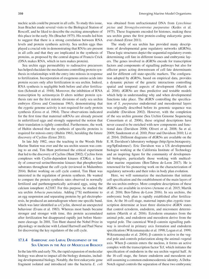
310
nucleic acids could be present in all cells. To study this issue,
Jean Brachet made several visits to the Biological Station of
Roscoff, and he liked to describe the exciting atmosphere of
this place in the early 30s (Brachet 1975). His results led him
to suggest that there is a strong correlation between RNA
levels and protein synthesis activity. Sea urchin eggs thus
played a crucial role in demonstrating that RNAs are present
in all cells and that they are implicated in the synthesis of
proteins, as proposed by the central dogma of Francis Crick
(DNA makes RNA, which in turn makes protein).
Sea urchin eggs permeability to radioactive precursors
has helped elucidate the mechanisms controlling protein syn
thesis in relationships with the entry into mitosis in response
to fertilization. Incorporation of exogenous amino acids into
protein occurs only after fertilization in sea urchin. Indeed,
RNA synthesis is negligible both before and after fertiliza
tion (Schmidt et al. 1948). Moreover, the inhibition of RNA
transcription by actinomycin D alters neither protein syn
thesis rate nor the first mitotic divisions of early sea urchin
embryos (Gross and Cousineau 1963), demonstrating that
the zygotic genome activity is not required for early protein
synthesis (Gross et al. 1964). These observations indicated
for the first time that maternal mRNAs are already present
in unfertilized eggs and strongly supported the notion that
their translation is tightly controlled. Furthermore, the work
of Hultin showed that the synthesis of specific proteins is
required for mitosis entry (Hultin 1961), heralding the future
discovery of Cyclins (Ernst 2011).
In late July 1982, once the teaching in the Woods Hole
Marine Station was over and the sea urchin season was com
ing to an end, Tim Hunt performed the critical experiment
that led to the discovery of Cyclins (Hunt 2002). Cyclins form
complexes with Cyclin-dependent kinases (CDKs), a fam
ily of conserved serine/threonine kinases that phosphorylate
substrates throughout the cell cycle (reviewed in Malumbres
2014). Before working on cell cycle control, Tim Hunt was
interested in the regulation of protein synthesis. He wanted
to compare the protein synthesis rates observed in normally
fertilized and parthenogenetically activated eggs, using the
calcium ionophore A23187. For this purpose, he studied the
sea urchin Arbacia punctulata. Adding [35S] methionine to
an egg suspension and separating proteins by gel electropho
resis, he produced an autoradiogram where one specifi c band,
which was later identified as a Cyclin, showed an unexpected
behaviour (Evans et al. 1983). Whereas most bands became
stronger and stronger with time, this protein accumulated
after fertilization but disappeared rapidly just before blasto
mere cleavage. In 2001, Tim Hunt shared the Nobel Prize in
physiology or medicine with Leland Hartwell and Paul Nurse
for discovering the key regulators of the cell cycle.
17.1.4 EMBRYONIC AND LARVAL DEVELOPMENT OF THE
SEA URCHIN IN THE AGE OF MOLECULAR BIOLOGY
In the late 60s and early 70s, the rapid expansion of molecular
biology was about to impact all the biology domains, includ
ing developmental biology. Notably, the first eukaryotic gene
fragment isolated and introduced into the bacteria E. coli
Emerging Marine Model Organisms
was obtained from unfractionated DNA from Lytechinus pictus and Strongylocentrotus purpuratus (Kedes et al.
1975). These fragments encoded for histones, making these
sea urchin genes the first protein-coding eukaryotic genes
ever cloned (Ernst 2011).
The study of sea urchin has provided many descrip
tions of developmental gene regulatory networks (dGRNs).
These logic structures depict the sequential regulatory events
determining cell fate in different tissues and embryonic lay
ers. The genes involved in dGRNs encode for transcription
factors and components of signalling pathways but also for
effector genes acting downstream of cell fate determinants
and for different cell state-specific markers. The confi gura
tion adopted by dGRNs, based on empirical data, provides
a dynamic picture of the genetic interactions controlling
spatial and temporal aspects of development (Martik et
al. 2016 ). dGRNs are thus predictive and testable models
which help in understanding why and when developmental
functions take place. The dGRN controlling the specifi ca
tion of S. purpuratus endodermal and mesodermal layers
was originally described before its genomic sequence was
available (Davidson 2002). However, with the completion
of the sea urchin genome (Sea Urchin Genome Sequencing
Consortium et al. 2006), these original descriptions have
never ceased to be enriched with new components and func
tional data (Davidson 2006; Oliveri et al. 2008; Su et al.
2009; Saudemont et al. 2010; Peter and Davidson 2010; Li et
al. 2014). Different diagrams of dGRNs are available on the
E. H. Davidson’s laboratory webpage (http://grns.biotapestry.
org/SpEndomes/). Eric Davidson was a US developmental
biologist working at the California Institute of Technology
and an inspiring figure for the community of developmen
tal biologists, particularly those working with multicel
lular marine organisms (Ben-Tabou de-Leon 2017). He is
renowned for his pioneering work on the characterization of
regulatory networks and their roles in body plan evolution.
Here, we will summarize the mechanisms that initiate
cell specification and the establishment of the main layers of
the sea urchin embryo. More complete descriptions of these
dGRNs are available in reviews (Arnone et al. 2015; Martik
et al. 2016; Ben-Tabou de-Leon 2016 ). In sea urchins, the
embryonic body plan is rapidly established after fertiliza
tion. At the 16-cell stage, maternal inputs plus zygotic tran
scription determine at least three distinctive dGRN states
that control ectoderm, endoderm, and micromere determi
nation (Martik et al. 2016). Ectoderm emanates from the
animal pole, and endoderm and mesoderm derive from the
vegetal pole. The canonical Wnt--catenin signalling path
way is involved in primary axis formation and endoderm
specification (Wikramanayake et al. 1998; Logan et al. 1999;
Wikramanayake et al. 2004). -catenin is active in the veg
etal pole and controls polarization along the animal–vegetal
axis. When -catenin enters the nucleus, it forms an active
complex with the transcription factor Tcf, which initiates the
specifi cation of endoderm in the sea urchin vegetal half. At
the 16-cell stage, the future endoderm and mesoderm are
still assuming a common endomesodermic identity. A Delta-
Notch signal controls the separation of these two embryonic
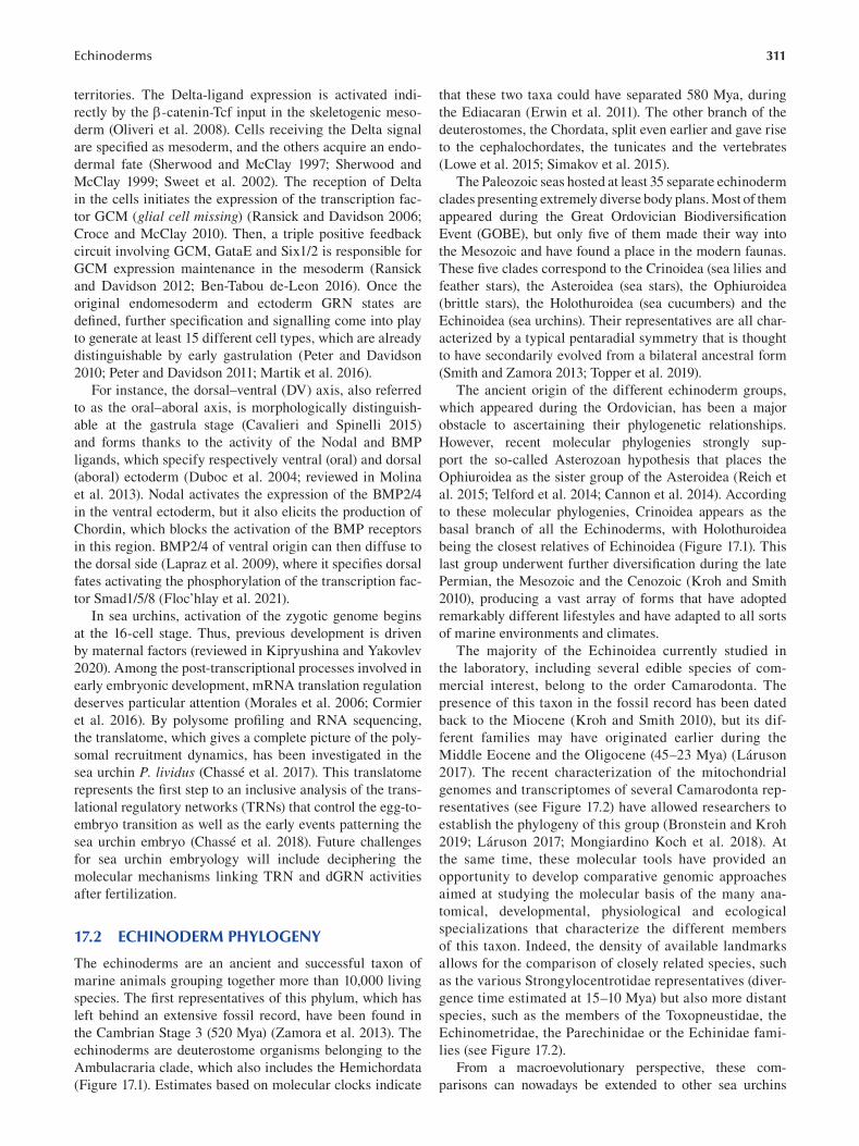
311 Echinoderms
territories. The Delta-ligand expression is activated indi
rectly by the -catenin-Tcf input in the skeletogenic meso
derm (Oliveri et al. 2008). Cells receiving the Delta signal
are specifi ed as mesoderm, and the others acquire an endo
dermal fate (Sherwood and McClay 1997; Sherwood and
McClay 1999; Sweet et al. 2002). The reception of Delta
in the cells initiates the expression of the transcription fac
tor GCM (glial cell missing) (Ransick and Davidson 2006;
Croce and McClay 2010). Then, a triple positive feedback
circuit involving GCM, GataE and Six1/2 is responsible for
GCM expression maintenance in the mesoderm (Ransick
and Davidson 2012; Ben-Tabou de-Leon 2016 ). Once the
original endomesoderm and ectoderm GRN states are
defined, further specification and signalling come into play
to generate at least 15 different cell types, which are already
distinguishable by early gastrulation (Peter and Davidson
2010; Peter and Davidson 2011; Martik et al. 2016 ).
For instance, the dorsal–ventral (DV) axis, also referred
to as the oral–aboral axis, is morphologically distinguish
able at the gastrula stage (Cavalieri and Spinelli 2015)
and forms thanks to the activity of the Nodal and BMP
ligands, which specify respectively ventral (oral) and dorsal
(aboral) ectoderm (Duboc et al. 2004; reviewed in Molina
et al. 2013). Nodal activates the expression of the BMP2/4
in the ventral ectoderm, but it also elicits the production of
Chordin, which blocks the activation of the BMP receptors
in this region. BMP2/4 of ventral origin can then diffuse to
the dorsal side ( Lapraz et al. 2009), where it specifi es dorsal
fates activating the phosphorylation of the transcription fac
tor Smad1/5/8 (Floc’hlay et al. 2021).
In sea urchins, activation of the zygotic genome begins
at the 16-cell stage. Thus, previous development is driven
by maternal factors (reviewed in Kipryushina and Yakovlev
2020). Among the post-transcriptional processes involved in
early embryonic development, mRNA translation regulation
deserves particular attention (Morales et al. 2006; Cormier
et al. 2016). By polysome profiling and RNA sequencing,
the translatome, which gives a complete picture of the poly
somal recruitment dynamics, has been investigated in the
sea urchin P. lividus (Chassé et al. 2017). This translatome
represents the first step to an inclusive analysis of the trans
lational regulatory networks (TRNs) that control the egg-to
embryo transition as well as the early events patterning the
sea urchin embryo (Chassé et al. 2018). Future challenges
for sea urchin embryology will include deciphering the
molecular mechanisms linking TRN and dGRN activities
after fertilization.
17.2 ECHINODERM PHYLOGENY
The echinoderms are an ancient and successful taxon of
marine animals grouping together more than 10,000 living
species. The first representatives of this phylum, which has
left behind an extensive fossil record, have been found in
the Cambrian Stage 3 (520 Mya) ( Zamora et al. 2013). The
echinoderms are deuterostome organisms belonging to the
Ambulacraria clade, which also includes the Hemichordata
(Figure 17.1). Estimates based on molecular clocks indicate
that these two taxa could have separated 580 Mya, during
the Ediacaran (Erwin et al. 2011). The other branch of the
deuterostomes, the Chordata, split even earlier and gave rise
to the cephalochordates, the tunicates and the vertebrates
(Lowe et al. 2015; Simakov et al. 2015).
The Paleozoic seas hosted at least 35 separate echinoderm
clades presenting extremely diverse body plans. Most of them
appeared during the Great Ordovician Biodiversifi cation
Event (GOBE), but only five of them made their way into
the Mesozoic and have found a place in the modern faunas.
These five clades correspond to the Crinoidea (sea lilies and
feather stars), the Asteroidea (sea stars), the Ophiuroidea
(brittle stars), the Holothuroidea (sea cucumbers) and the
Echinoidea (sea urchins). Their representatives are all char
acterized by a typical pentaradial symmetry that is thought
to have secondarily evolved from a bilateral ancestral form
(Smith and Zamora 2013; Topper et al. 2019).
The ancient origin of the different echinoderm groups,
which appeared during the Ordovician, has been a major
obstacle to ascertaining their phylogenetic relationships.
However, recent molecular phylogenies strongly sup
port the so-called Asterozoan hypothesis that places the
Ophiuroidea as the sister group of the Asteroidea (Reich et
al. 2015; Telford et al. 2014; Cannon et al. 2014). According
to these molecular phylogenies, Crinoidea appears as the
basal branch of all the Echinoderms, with Holothuroidea
being the closest relatives of Echinoidea (Figure 17.1). This
last group underwent further diversification during the late
Permian, the Mesozoic and the Cenozoic (Kroh and Smith
2010), producing a vast array of forms that have adopted
remarkably different lifestyles and have adapted to all sorts
of marine environments and climates.
The majority of the Echinoidea currently studied in
the laboratory, including several edible species of com
mercial interest, belong to the order Camarodonta. The
presence of this taxon in the fossil record has been dated
back to the Miocene (Kroh and Smith 2010), but its dif
ferent families may have originated earlier during the
Middle Eocene and the Oligocene (45–23 Mya) (Láruson
2017). The recent characterization of the mitochondrial
genomes and transcriptomes of several Camarodonta rep
resentatives (see Figure 17.2) have allowed researchers to
establish the phylogeny of this group (Bronstein and Kroh
2019; Láruson 2017; Mongiardino Koch et al. 2018). At
the same time, these molecular tools have provided an
opportunity to develop comparative genomic approaches
aimed at studying the molecular basis of the many ana
tomical, developmental, physiological and ecological
specializations that characterize the different members
of this taxon. Indeed, the density of available landmarks
allows for the comparison of closely related species, such
as the various Strongylocentrotidae representatives (diver
gence time estimated at 15–10 Mya) but also more distant
species, such as the members of the Toxopneustidae, the
Echinometridae, the Parechinidae or the Echinidae fami
lies (see Figure 17.2).
From a macroevolutionary perspective, these com
parisons can nowadays be extended to other sea urchins
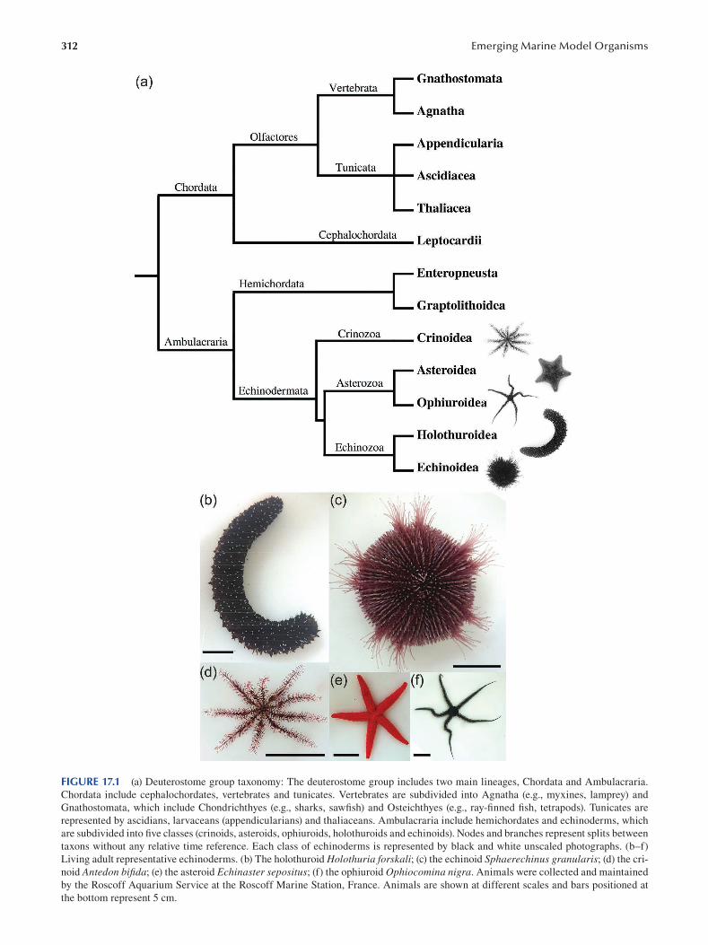
312 Emerging Marine Model Organisms
FIGURE 17.1 (a) Deuterostome group taxonomy: The deuterostome group includes two main lineages, Chordata and Ambulacraria.
Chordata include cephalochordates, vertebrates and tunicates. Vertebrates are subdivided into Agnatha (e.g., myxines, lamprey) and
Gnathostomata, which include Chondrichthyes (e.g., sharks, sawfish) and Osteichthyes (e.g., ray-fi nned fish, tetrapods). Tunicates are
represented by ascidians, larvaceans (appendicularians) and thaliaceans. Ambulacraria include hemichordates and echinoderms, which
are subdivided into five classes (crinoids, asteroids, ophiuroids, holothuroids and echinoids). Nodes and branches represent splits between
taxons without any relative time reference. Each class of echinoderms is represented by black and white unscaled photographs. (b–f)
Living adult representative echinoderms. (b) The holothuroid Holothuria forskali; (c) the echinoid Sphaerechinus granularis; (d) the cri
noid Antedon bifida; (e) the asteroid Echinaster sepositus; (f) the ophiuroid Ophiocomina nigra. Animals were collected and maintained
by the Roscoff Aquarium Service at the Roscoff Marine Station, France. Animals are shown at different scales and bars positioned at
the bottom represent 5 cm.
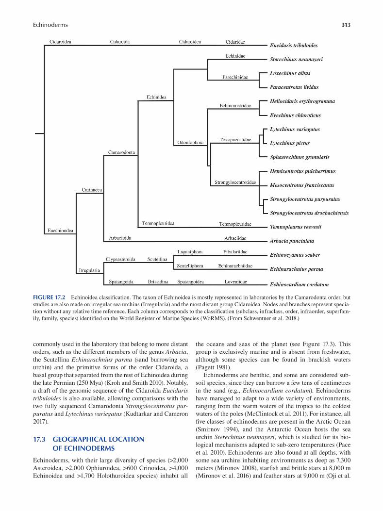
Echinoderms 313
FIGURE 17.2 Echinoidea classification. The taxon of Echinoidea is mostly represented in laboratories by the Camarodonta order, but
studies are also made on irregular sea urchins (Irregularia) and the most distant group Cidaroidea. Nodes and branches represent specia
tion without any relative time reference. Each column corresponds to the classification (subclass, infraclass, order, infraorder, superfam
ily, family, species) identified on the World Register of Marine Species (WoRMS). (From Schwentner et al. 2018.)
commonly used in the laboratory that belong to more distant
orders, such as the different members of the genus Arbacia,
the Scutellina Echinarachnius parma (sand burrowing sea
urchin) and the primitive forms of the order Cidaroida, a
basal group that separated from the rest of Echinoidea during
the late Permian (250 Mya) (Kroh and Smith 2010). Notably,
a draft of the genomic sequence of the Cidaroida Eucidaris tribuloides is also available, allowing comparisons with the
two fully sequenced Camarodonta Strongylocentrotus purpuratus and Lytechinus variegatus (Kudtarkar and Cameron
2017 ).
17.3 GEOGRAPHICAL LOCATION OF ECHINODERMS
Echinoderms, with their large diversity of species (>2,000
Asteroidea, >2,000 Ophiuroidea, >600 Crinoidea, >4,000
Echinoidea and >1,700 Holothuroidea species) inhabit all
the oceans and seas of the planet (see Figure 17.3). This
group is exclusively marine and is absent from freshwater,
although some species can be found in brackish waters
(Pagett 1981).
Echinoderms are benthic, and some are considered sub
soil species, since they can burrow a few tens of centimetres
in the sand (e.g., Echinocardium cordatum ). Echinoderms
have managed to adapt to a wide variety of environments,
ranging from the warm waters of the tropics to the coldest
waters of the poles (McClintock et al. 2011). For instance, all
five classes of echinoderms are present in the Arctic Ocean
(Smirnov 1994), and the Antarctic Ocean hosts the sea
urchin Sterechinus neumayeri, which is studied for its bio
logical mechanisms adapted to sub-zero temperatures (Pace
et al. 2010). Echinoderms are also found at all depths, with
some sea urchins inhabiting environments as deep as 7,300
meters (Mironov 2008), starfish and brittle stars at 8,000 m
(Mironov et al. 2016) and feather stars at 9,000 m (Oji et al.
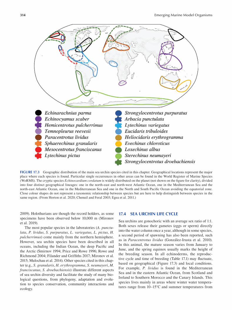
314 Emerging Marine Model Organisms
FIGURE 17.3 Geographic distribution of the main sea urchin species cited in this chapter. Geographical locations represent the major
place where each species is found. Particular single occurrences in other areas can be found in the World Register of Marine Species
(WoRMS). The cryptic species Echinocardium cordatum is widely distributed on the planet (not shown on the figure for clarity), divided
into four distinct geographical lineages: one in the north-east and north-west Atlantic Ocean, one in the Mediterranean Sea and the
north-east Atlantic Ocean, one in the Mediterranean Sea and one in the North and South Pacific Ocean avoiding the equatorial zone.
Close colour shapes do not represent a taxonomic relationship between species but are here to help distinguish between species in the
same region. (From Horton et al. 2020; Chenuil and Feral 2003; Egea et al. 2011.)
2009). Holothurians are though the record holders, as some
specimens have been observed below 10,000 m (Mironov
et al. 2019).
The most popular species in the laboratories (A. punctulata, P. lividus, S. purpuratus, L. variegatus, L. pictus, H. pulcherrimus) come mainly from the northern hemisphere.
However, sea urchin species have been described in all
oceans, including the Indian Ocean, the deep Pacifi c and
the Arctic (Smirnov 1994; Price and Rowe 1996; Rowe and
Richmond 2004; Filander and Griffiths 2017; Mironov et al.
2015; Mulochau et al. 2014). Other species cited in this chap
ter (e.g., S. granularis, H. erythrogramma, S. neumayeri, M. franciscanus, S. droebachiensis) illustrate different aspects
of sea urchin diversity and facilitate the study of many bio
logical questions, from phylogeny, adaptation and evolu
tion to species conservation, community interactions and
ecology.
17.4 SEA URCHIN LIFE CYCLE
Sea urchins are gonochoric with an average sex ratio of 1:1.
Both sexes release their gametes (eggs or sperm) directly
into the water column once a year, although in some species,
a second period of spawning has also been reported, such
as in Paracentrotus lividus (González-Irusta et al. 2010).
In this animal, the mature season varies from January to
June, and the spring equinox usually marks the height of
the breeding season. In all echinoderms, the reproduc
tive cycle and time of breeding (Table 17.1) may fl uctuate,
based on geographical (Figure 17.3) and local conditions.
For example, P. lividus is found in the Mediterranean
Sea and in the eastern Atlantic Ocean, from Scotland and
Ireland to Southern Morocco and the Canary Islands. This
species lives mainly in areas where winter water tempera
tures range from 10–15°C and summer temperatures from
Echinoderms 315
TABLE 17.1 Breeding Season and Egg Diameter in Different Echinoidea Species Echinoidea Species Breeding Season Egg Diameter References
Eucidaris tribuloides — 94 μm ( McAlister and Moran 2012 ; Lessios 1988 ; Lessios 1990 )
Sterechinus neumayeri September–November 180 μm ( Bosch et al. 1987 ; Stanwell-Smith and Peck 1998 )
Paracentrotus lividus May–September (AO) 75 μm ( Hamdoun et al. 2018 ; Ouréns et al. 2011 ; Rocha et al. 2019 ;
April–June and September– Byrne 1990 )
November (MS)
Heliocidaris erythrogramma November–February (SEA) 400–450 μm ( Binks et al. 2012 ; Foo et al. 2018 ; Raff 1987 )
February–May (SWA)
Evechinus chloroticus November–February — ( Delorme and Sewell 2016 )
Hemicentrotus pulcherrimus January–March — ( Kiyomoto et al. 2014 )
Mesocentrotus franciscanus June–September 130 μm ( Bernard 1977 ; Bolton et al. 2000 )
Strongylocentrotus purpuratus November–March 80 μm ( Bolton et al. 2000 ; Hamdoun et al. 2018 )
Strongylocentrotus droebachiensis March–May 145 μm ( Himmelman 1978 ; Levitan 1993 ; Meidel and Scheibling 1998 )
Lytechinus variegatus May–September 100 μm ( Hamdoun et al. 2018 ; Lessios 1990 , 1988 ; Schatten 1981 )
Lytechinus pictus May–September 120 μm ( Hamdoun et al. 2018 )
Sphaerechinus granularis April–June (Brittany) 100 μm ( Guillou and Lumingas 1998 ; Guillou and Michel 1993 ; Vafi dis
June–November (MS) et al. 2020 )
Temnopleurus reevesii July–January 100 μm ( Hamdoun et al. 2018 )
Arbacia punctulata June–August 69 μm ( Bolton et al. 2000 ; Gianguzza and Bonaviri 2013 )
Echinarachnius parma March–July (NWP) 110–135 μm ( Costello and Henley 1971 ; Drozdov and Vinnikova 2010 ;
Summers and Hylander 1974 )
Echinocardium cordatum May–July (NWP) 110 μm ( Drozdov and Vinnikova 2010 ; Egea et al. 2011 ; Hibino et al.
April–October (MS) 2019 )
May–October (AO)
AO: Atlantic Ocean, MS: Mediterranean Sea, NWP: North-West Pacific Ocean, SWA: South-West Australia, SEA: South-East Australia
18–25°C. Several factors, like temperature, photoperiod,
resource availability and water turbulence contribute to
the regulation of gametogenesis in these populations (Gago
and Luís 2011). On the other hand, records from the North
Pacific, Arctic and North Atlantic Oceans show that spawn
ing of the sea urchin Strongylocentrotus droebachiensis may also be synchronized with the spring phytoplankton
increase (Himmelman 1978; Starr et al. 1990). However,
the main environmental factors triggering spawning and
the molecular mechanisms that mediate this response are
not yet known.
During the reproductive cycle, P. lividus gonads go
through different development stages, which have been
exhaustively characterized (Byrne 1990). Observation of
gametogenesis in P. lividus through histological examina
tions allows us to classify the annual reproductive cycle
of this species in six developmental stages: 1) recovery, 2)
growing, 3) premature, 4) mature, 5) partly spawned and 6)
spent. In turn, in Strongylocentrotus droebachiensis , four
stages have been recognized by examining the activity of
the two main cell populations composing the germinal epi
thelium (Walker et al. 2007, 2013). These populations are
the germinal cells, which are either ova in the ovary or
spermatogonia in the testis, and a group of somatic cells
called nutritive phagocytes (NPs), which are functionally
equivalent to the vertebrate Sertoli cells and are present
in both sexes. Stage 1, called inter-gametogenesis, occurs
directly after spring spawning and lasts for about three
months. Residual reproductive cells are present, but other
wise, the gonads look empty. Toward the end of this stage,
NP cells increase in number and resume nutrient storage,
doubling their size by the end of this phase. In addition,
reproductive cells begin to appear. NPs are involved in the
phagocytosis of residual ova and spermatozoa, and thus
participate in the recycling of derived nutrients. Stage 2
is called pre-gametogenesis and NP renewal. This stage
begins in summer and lasts for approximately three to
four months. Reproductive cells, present at the periphery
of the gonad, increase both in number and size. Stage 3,
gametogenesis and NP utilization, takes place during fi ve
winter months. The reproductive cells continue to develop
and migrate into the centre of the gonad. Conversely, the
NPs cells shrink, and their number decreases. Stage 4 cor
responds to pre-spawning and spawning. This stage occurs
in late winter and lasts around three months. The lumen
of the gonad is packed with fully differentiated gametes,
and the NP cells are barely observable. At the end of stage
4, spawning occurs, and gametes are released from the
gonads by the gonopores.
Several holistic approaches have been generated to
understanding the molecular mechanisms of gametogenesis
and the events of the life cycle. Whole-genome and Q-PCR
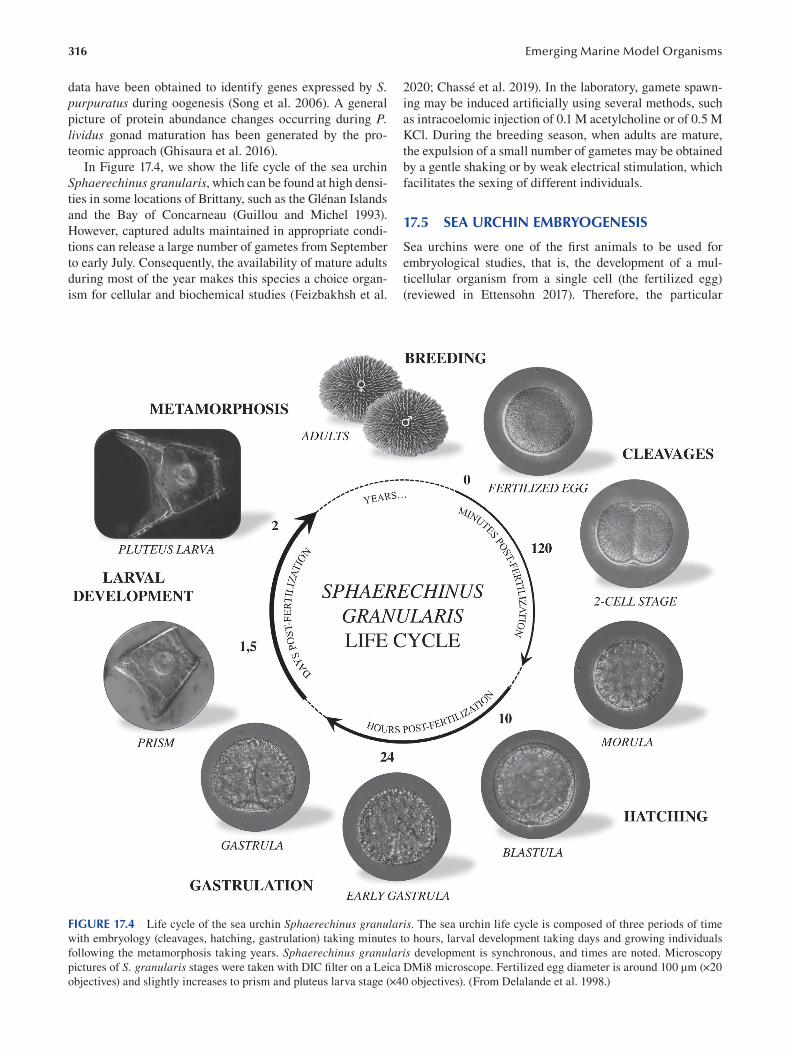
316
data have been obtained to identify genes expressed by S. purpuratus during oogenesis (Song et al. 2006 ). A general
picture of protein abundance changes occurring during P. lividus gonad maturation has been generated by the pro
teomic approach (Ghisaura et al. 2016).
In Figure 17.4, we show the life cycle of the sea urchin
Sphaerechinus granularis, which can be found at high densi
ties in some locations of Brittany, such as the Glénan Islands
and the Bay of Concarneau (Guillou and Michel 1993).
However, captured adults maintained in appropriate condi
tions can release a large number of gametes from September
to early July. Consequently, the availability of mature adults
during most of the year makes this species a choice organ
ism for cellular and biochemical studies (Feizbakhsh et al.
Emerging Marine Model Organisms
2020; Chassé et al. 2019). In the laboratory, gamete spawn
ing may be induced artificially using several methods, such
as intracoelomic injection of 0.1 M acetylcholine or of 0.5 M
KCl. During the breeding season, when adults are mature,
the expulsion of a small number of gametes may be obtained
by a gentle shaking or by weak electrical stimulation, which
facilitates the sexing of different individuals.
17.5 SEA URCHIN EMBRYOGENESIS
Sea urchins were one of the first animals to be used for
embryological studies, that is, the development of a mul
ticellular organism from a single cell (the fertilized egg)
(reviewed in Ettensohn 2017). Therefore, the particular
FIGURE 17.4 Life cycle of the sea urchin Sphaerechinus granularis. The sea urchin life cycle is composed of three periods of time
with embryology (cleavages, hatching, gastrulation) taking minutes to hours, larval development taking days and growing individuals
following the metamorphosis taking years. Sphaerechinus granularis development is synchronous, and times are noted. Microscopy
pictures of S. granularis stages were taken with DIC filter on a Leica DMi8 microscope. Fertilized egg diameter is around 100 μm (×20
objectives) and slightly increases to prism and pluteus larva stage (×40 objectives). (From Delalande et al. 1998.)
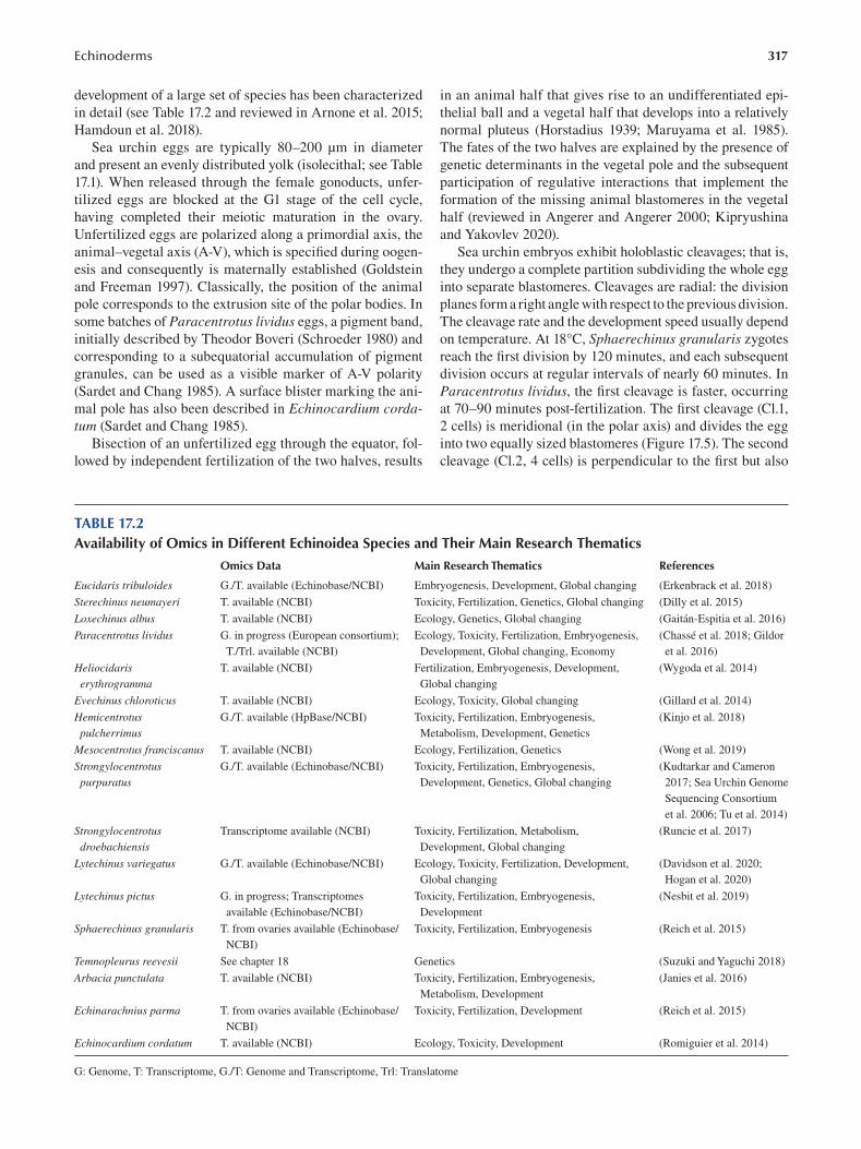
317 Echinoderms
development of a large set of species has been characterized
in detail (see Table 17.2 and reviewed in Arnone et al. 2015;
Hamdoun et al. 2018).
Sea urchin eggs are typically 80–200 μm in diameter
and present an evenly distributed yolk (isolecithal; see Table
17.1). When released through the female gonoducts, unfer
tilized eggs are blocked at the G1 stage of the cell cycle,
having completed their meiotic maturation in the ovary.
Unfertilized eggs are polarized along a primordial axis, the
animal–vegetal axis (A-V), which is specified during oogen
esis and consequently is maternally established (Goldstein
and Freeman 1997 ). Classically, the position of the animal
pole corresponds to the extrusion site of the polar bodies. In
some batches of Paracentrotus lividus eggs, a pigment band,
initially described by Theodor Boveri (Schroeder 1980) and
corresponding to a subequatorial accumulation of pigment
granules, can be used as a visible marker of A-V polarity
(Sardet and Chang 1985). A surface blister marking the ani
mal pole has also been described in Echinocardium cordatum (Sardet and Chang 1985).
Bisection of an unfertilized egg through the equator, fol
lowed by independent fertilization of the two halves, results
in an animal half that gives rise to an undifferentiated epi
thelial ball and a vegetal half that develops into a relatively
normal pluteus (Horstadius 1939; Maruyama et al. 1985).
The fates of the two halves are explained by the presence of
genetic determinants in the vegetal pole and the subsequent
participation of regulative interactions that implement the
formation of the missing animal blastomeres in the vegetal
half (reviewed in Angerer and Angerer 2000; Kipryushina
and Yakovlev 2020).
Sea urchin embryos exhibit holoblastic cleavages; that is,
they undergo a complete partition subdividing the whole egg
into separate blastomeres. Cleavages are radial: the division
planes form a right angle with respect to the previous division.
The cleavage rate and the development speed usually depend
on temperature. At 18°C, Sphaerechinus granularis zygotes
reach the first division by 120 minutes, and each subsequent
division occurs at regular intervals of nearly 60 minutes. In
Paracentrotus lividus, the first cleavage is faster, occurring
at 70–90 minutes post-fertilization. The first cleavage (Cl.1,
2 cells) is meridional (in the polar axis) and divides the egg
into two equally sized blastomeres (Figure 17.5). The second
cleavage (Cl.2, 4 cells) is perpendicular to the first but also
TABLE 17.2 Availability of Omics in Different Echinoidea Species and Their Main Research Thematics
Omics Data Main Research Thematics References
Eucidaris tribuloides G./T. available (Echinobase/NCBI) Embryogenesis, Development, Global changing ( Erkenbrack et al. 2018 )
Sterechinus neumayeri T. available (NCBI) Toxicity, Fertilization, Genetics, Global changing ( Dilly et al. 2015 )
Loxechinus albus T. available (NCBI) Ecology, Genetics, Global changing ( Gaitán-Espitia et al. 2016 )
Paracentrotus lividus G. in progress (European consortium); Ecology, Toxicity, Fertilization, Embryogenesis, ( Chassé et al. 2018 ; Gildor
T./Trl. available (NCBI) Development, Global changing, Economy et al. 2016 )
Heliocidaris T. available (NCBI) Fertilization, Embryogenesis, Development, ( Wygoda et al. 2014 )
erythrogramma Global changing
Evechinus chloroticus T. available (NCBI) Ecology, Toxicity, Global changing ( Gillard et al. 2014 )
Hemicentrotus G./T. available (HpBase/NCBI) Toxicity, Fertilization, Embryogenesis, ( Kinjo et al. 2018 )
pulcherrimus Metabolism, Development, Genetics
Mesocentrotus franciscanus T. available (NCBI) Ecology, Fertilization, Genetics ( Wong et al. 2019 )
Strongylocentrotus G./T. available (Echinobase/NCBI) Toxicity, Fertilization, Embryogenesis, ( Kudtarkar and Cameron
purpuratus Development, Genetics, Global changing 2017 ; Sea Urchin Genome
Sequencing Consortium
et al. 2006; Tu et al. 2014 )
Strongylocentrotus Transcriptome available (NCBI) Toxicity, Fertilization, Metabolism, ( Runcie et al. 2017 )
droebachiensis Development, Global changing
Lytechinus variegatus G./T. available (Echinobase/NCBI) Ecology, Toxicity, Fertilization, Development, ( Davidson et al. 2020 ;
Global changing Hogan et al. 2020 )
Lytechinus pictus G. in progress; Transcriptomes Toxicity, Fertilization, Embryogenesis, ( Nesbit et al. 2019 )
available (Echinobase/NCBI) Development
Sphaerechinus granularis T. from ovaries available (Echinobase/ Toxicity, Fertilization, Embryogenesis ( Reich et al. 2015 )
NCBI)
Temnopleurus reevesii See chapter 18 Genetics ( Suzuki and Yaguchi 2018 )
Arbacia punctulata T. available (NCBI) Toxicity, Fertilization, Embryogenesis, ( Janies et al. 2016 )
Metabolism, Development
Echinarachnius parma T. from ovaries available (Echinobase/ Toxicity, Fertilization, Development ( Reich et al. 2015 )
NCBI)
Echinocardium cordatum T. available (NCBI) Ecology, Toxicity, Development ( Romiguier et al. 2014 )
G: Genome, T: Transcriptome, G./T: Genome and Transcriptome, Trl: Translatome
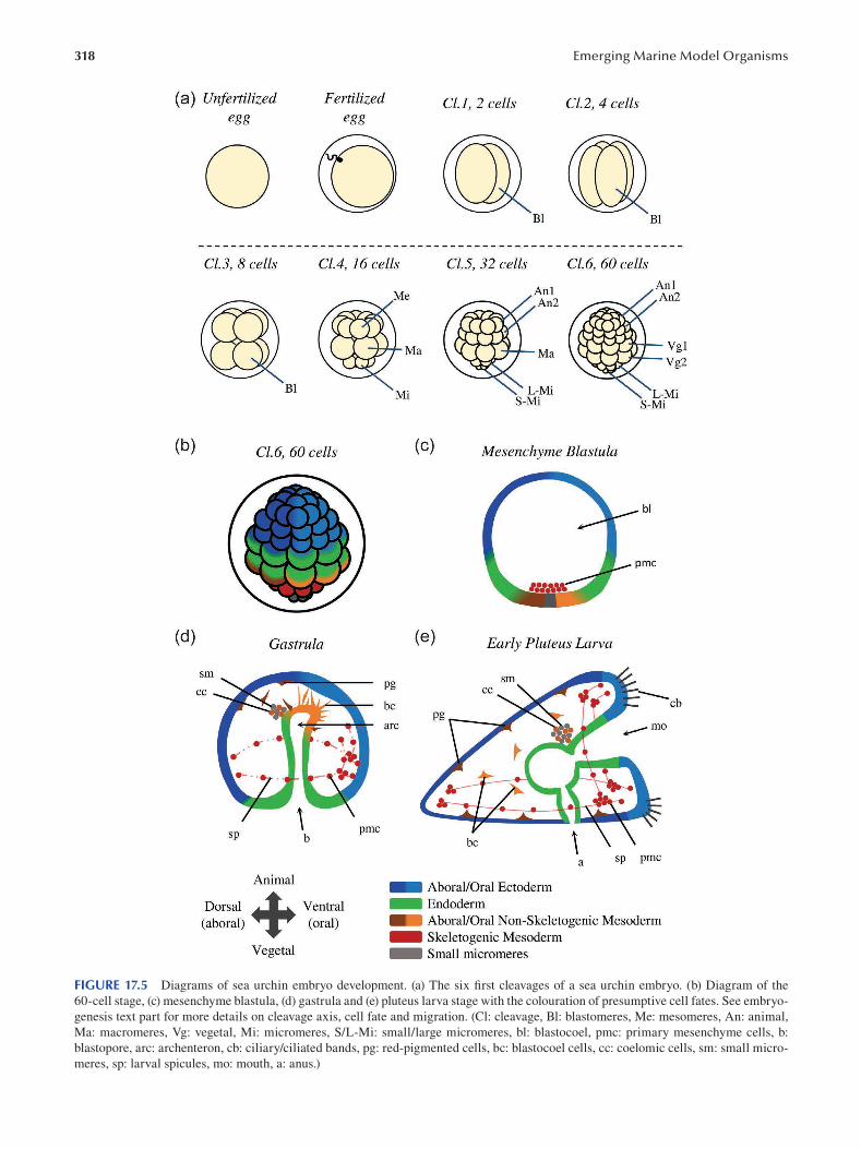
318 Emerging Marine Model Organisms
FIGURE 17.5 Diagrams of sea urchin embryo development. (a) The six first cleavages of a sea urchin embryo. (b) Diagram of the
60-cell stage, (c) mesenchyme blastula, (d) gastrula and (e) pluteus larva stage with the colouration of presumptive cell fates. See embryo
genesis text part for more details on cleavage axis, cell fate and migration. (Cl: cleavage, Bl: blastomeres, Me: mesomeres, An: animal,
Ma: macromeres, Vg: vegetal, Mi: micromeres, S/L-Mi: small/large micromeres, bl: blastocoel, pmc: primary mesenchyme cells, b:
blastopore, arc: archenteron, cb: ciliary/ciliated bands, pg: red-pigmented cells, bc: blastocoel cells, cc: coelomic cells, sm: small micro
meres, sp: larval spicules, mo: mouth, a: anus.)
319 Echinoderms
occurs in a meridional plane, resulting in the production of
four equally sized blastomeres. The third cleavage (Cl.3, 8
cells) is equatorial (at right angles of the polar axis), resulting
in four upper and four lower blastomeres, all of equal sizes.
The fourth cleavage (Cl.4, 16 cells) exhibits a complex and
characteristic pattern that reveals the basic A-V polarity of
the embryo. In the vegetal pole, the four blastomeres divide
asymmetrically and horizontally, forming four small cells
placed in the egg’s pole (the micromeres) and four larger
cells situated above (the macromeres). In the animal pole,
the four blastomeres divide meridionally and symmetrically,
resulting in eight equally sized cells (the mesomeres). At the
fifth cleavage (Cl.5, 32 cells), the eight mesomeres at the ani
mal half divide equatorially and symmetrically, resulting in
two layers of cells called “an1” for the upper one and “an2”
for the lower one. In the vegetal pole, the four macromeres
instead divide meridionally, forming a tier of eight cells. The
four micromeres divide horizontally and asymmetrically,
resulting in four small micromeres at the extreme vegetal
pole hemisphere and four large micromeres above. At the
sixth cleavage (Cl.6, 60 cells), all the cleavage furrows are
equatorial. The macromeres divide, giving rise to two eight-
cell tiers called “veg1” and “veg2”. The large micromeres
divide as well, but not the small micromeres. In total, the
60-cell embryo shows, from top to bottom, 16 “an1” cells
distributed in two layers of 8 cells each, 16 “an2” cells form
ing also two layers of 8 cells each, 8 “vg1”, 8 “vg2”, 8 large
micromeres and 4 small micromeres.
The macromeres producing the “vg1” and “vg2” cells
are the endomesoderm progenitors. The large micromeres
contribute instead to the skeletogenic mesenchyme, and
the small micromeres to the primordial germ cells (PGCs)
(Okazaki 1975; Yajima and Wessel 2012). In the Echinoderm
phylum, micromeres are only observed in echinoids and
are thus considered a derived character. Asymmetric cell
division is directed by the control of spindle and furrow
cleavage position and by uneven repartition of molecules.
Although the precise molecular mechanisms that orches
trate these asymmetric divisions are still poorly understood,
it has been shown that the AGS/Pins proteins (activator of
G-protein signalling/partner of Inscuteable) are required for
normal asymmetrical division during micromere formation
(Voronina and Wessel 2006; Poon et al. 2019).
As soon as the eight-cell stage is reached, a small cen
tral cavity forms in the centre of the embryo. As cleavage
proceeds, this space enlarges and forms the blastocoel. A
morula appears roughly six hours after fertilization, but at
the 120-cell stage, the smooth-surfaced blastula becomes a
continuous spherical monolayer surrounded by an outer hya
line layer. The epithelium sits on an inner basal membrane;
cell adhesion is mediated by tight junctions. Cilia develop
on the surface of the blastula, and their coordinated action
triggers the rotation of the blastula within the fertilization
envelope. Ten hours after fertilization, the blastula is com
posed of about 600 cells. Cell division rates decrease as the
cell cycle lengthens. At the end of segmentation, the blastula
is covered by cilia, presents a conspicuous apical ciliary tuft
in the animal pole and starts secreting a hatching enzyme
that digests the fertilization envelope. The synthesis of this
hatching enzyme takes place in the animal-most two-thirds
of the blastula and is likely to be restricted to the presump
tive ectoderm territory (Lepage et al. 1992a, 1992b). Finally,
a swimming blastula is released into the sea.
The blastula wall thickens at the vegetal pole, forming
the vegetal plate. In the central region of this vegetal plate,
the micromere descendants display pulsatile movements
and start developing filopodia in their basal face. These
cells lose their affinity for the outer hyaline structure and
gain affinity for the fibronectin present in the basal lamina
and the extracellular matrix lining the blastocoel (Fink and
McClay 1985). Eventually, they detach from the epithelium
and enter the blastocoel, forming the primary mesenchyme
(Peterson and McClay 2003). As these cells are the fi rst
ones to ingress into the blastocoel, they are called primary
mesenchyme cells (PMCs) (Burke et al. 1991). Adhering to
the blastocoel matrix, these cells progress from the vegetal
pole toward the animal pole and then reverse their trajec
tory. Finally, the PMCs reach an area located between the
vegetal pole and the equator and form a ring pattern con
sisting of two ventrolateral cell clusters and dorsal and ven
tral interconnected chains of cells (Malinda and Ettensohn
1994). Then their filopodia coalesce, and the characteristic
syncytial bridges of the larval skeleton appear. The primary
mesenchyme cells of the sea urchin represent one of the
best developmental models for studying mesodermal migra
tion (Anstrom 1992; Ettensohn 1999; Ettensohn and Sweet
2000; Peterson and McClay 2003), and the cellular basis of
skeletogenic cells has been characterized in detail (Okazaki
1975; Ettensohn and McClay 1988; Armstrong and McClay
1994). Moreover, the gene regulatory network (GRN) that
controls their formation has also been described not only
in species of the order Camarodonta (Oliveri and Davidson
2004; Oliveri et al. 2008) but also in other echinoid orders
( Minokawa 2017 ).
In euechinoids, the ingression of the PMCs marks the
onset of gastrulation. The invagination of the Vg2 territory
in the blastocoel gives rise to the archenteron (primitive gut),
opened to the outside by a circular blastopore (the future
anus). Invagination of the vegetal plate, a universal feature
of echinoderm gastrulation, is traditionally divided into
“primary” and “secondary” invagination (Gustafson and
Kinnander 1956). The “primary” invagination corresponds
to an initial phase of gut extension that involves extensive
extracellular matrix remodelling and cell shape changes
(reviewed in Kominami and Takata 2004). Three hypoth
eses have been advanced to explain the “primary” invagina
tion (reviewed in Ettensohn 2020). First, according to the
so-called apical constriction hypothesis, a ring of vegetal
plate cells become bottle shaped, compressing their apical
ends (Kimberly and Hardin 1998). This cell shape modi
fication causes the cells to pucker inward. However, bottle
cells could be a specialized feature of euechinoids and not a
general characteristic of all echinoderms (Ettensohn 2020).
A second hypothesis proposes that invagination could be
320
driven by changes in extracellular matrix composition (Lane
et al. 1993). In fact, the hyaline layer is made up of two layers:
an outer lamina composed of hyalin protein and glycopro
teins and an inner lamina composed of fi bropellin proteins
(Hall and Vacquier 1982; Bisgrove et al. 1991). After PMC
ingression, the vegetal plate cells secrete chondroitin sulfate
proteoglycans into the inner lamina of the hyaline layer. As
these chondroitin sulfate proteoglycans capture abundant
water, the inner layer expands even if the outer layer remains
stiff. The result is a force pushing the epithelium toward the
blastocoel (Lane et al. 1993). A third hypothesis suggests
another force arising from the concerted movement of cells
toward the vegetal pole that may facilitate the invagination
by drawing the buckled layer inward (Burke et al. 1991).
The “secondary” invagination ensues after a brief pause.
During this stage, the archenteron extends and produces a
long thin tube. The cells of the archenteron, which are orga
nized as a monolayered epithelium, move over one another
and flatten (Ettensohn 1985; Hardin 1989). In Lytechinus variegatus, gastrulation has been analyzed at a high resolu
tion by live imaging and using transplantation techniques
(Martik and McClay 2017). In this species, the process of
archenteron elongation is mainly driven by the elongation of
Vg2 endoderm cells. In fact, even if oriented cell divisions
also contribute to gut elongation, cell proliferation inhibition
does not preclude gastrulation, indicating that cell prolifera
tion is not essential for this process (Stephens et al. 1986;
Martik and McClay 2017 ).
The oral ectoderm of the gastrula flattens as the gastrula
becomes roughly triangular, forming the prism larva. The
embryonic radial symmetry is gradually replaced by a bilat
eral symmetry. An early sign of this transformation consists
in the aggregation of primary mesenchyme cells into two
clusters that develop in the opposite posterolateral–ventral
angles of the prism larva. The cells of the primary mesen
chyme form then a syncytium, in which two calcitic spicules
develop. These spicules, flanking the primitive digestive
tract, will constitute the endoskeleton of the pluteus larva.
For this, the primary mesenchyme cells endocytose seawa
ter from the larval internal body cavity and form a series of
vacuoles where calcium can concentrate and precipitate as
amorphous calcium carbonate (Kahil et al. 2020).
Once the archenteron reaches about two-thirds of its fi nal
length, the third and last stage of archenteron elongation
begins (Hardin 1988). This phase is driven by the second
ary mesenchyme cells, which extend filopodia through the
blastocoel cavity to reach a specific area in the inner sur
face of the blastocoel roof (Hardin and McClay 1990). These
filopodia pull the archenteron toward the animal pole and
contact the region where the mouth will form. The mouth
forms in the future ventral side of the larva after the fusion
of the archenteron and the ectoderm epithelium. Typical of
the deuterostomes, the mouth and the archenteron create a
continuous digestive tube that joins the blastopore, which
coincides with the anus.
During the processes of archenteron elongation, the sec
ondary mesenchyme cells spread into the blastocoel fl uid,
Emerging Marine Model Organisms
where they form at least four non-skeletogenic mesoderm
cells (Ettensohn and Ruffins 1993). Early in gastrulation,
a population of red-pigmented cells forms (Gustafson and
Wolpert 1967; Gibson and Burke 1985). It is interesting to
note that independent knock out of the genes encoding for
polyketide synthase, flavin monooxygenase family 3, and
the glial cells missing (gcm) protein results in the disappear
ance of red-pigmented cells throughout the body of the larva
(Wessel et al. 2020).
Later in gastrulation, a group of cells coming from the
tip of the archenteron moves into the blastocoel and adopts
a fibroblast-like morphology: they are the so-called basal
cells (Cameron et al. 1991), or blastocoel cells (Tamboline
and Burke 1992). At the end of gastrulation, two coelomic
cavities appear as a bilateral out-pocketing of the fore
gut (Gustafson and Wolpert 1963). Afterwards, secondary
mesenchymal cells move out of these coelomic cavities and
produce the circumesophageal musculature of the pluteus
larvae (Ishimoda-Takagi et al. 1984; Burke and Alvarez
1988; Wessel et al. 1990; Andrikou et al. 2013). While the
right coelomic pouch remains rudimentary, the left coelo
mic pouch undergoes massive development to build many
of the structures of the future adult sea urchin. The left side
of the pluteus contributes to the formation of the future oral
surface of the sea urchin adult (Aihara and Amemiya 2001).
The left pouch splits into three smaller sacs. A duct-like
structure, the hydroporic canal, extends from the anterior
left coelomic pouch to the aboral ectoderm where the hydro-
pore forms (Gustafson and Wolpert 1963). This hydroporic
canal is covered by cilia and could be an excretory organ of
the larvae (Hara et al. 2003) and later differentiates into a
part of the adult water vascular system (Hyman 1955). The
hydroporic canal formation constitutes the fi rst morphologi
cal signature of left–right asymmetry in the pluteus larva
(Luo and Su 2012). An invagination from the ectoderm fuses
with the intermediate sac to form the imaginal rudiment,
from which the pentaradial symmetry of the adult body plan
is established (Smith et al. 2008). To facilitate the obser
vation and the study of complex phases of development, a
larval staging schematic of Strongylocentrotus purpuratus has been proposed (Smith et al. 2008). This schematic sub
divides larval life into seven stages: 1) four-arm stage, 2)
eight-arm stage, 3) vestibula invagination stage, 4) rudiment
initiation stage, 5) pentagonal disc stage, 6) advanced rudi
ment stage and 7) tube-foot protrusion stage.
In the late gastrula, primary germ cells located in the
archenteron tip incorporate into the imaginal rudiment.
Skeletogenic mesenchyme cells penetrate the rudiment to
produce the first skeletal plates of the future adult endoskel
eton (Gilbert 2006 ). The rudiment separates from the rest
of the larva during metamorphosis, reorganizes its digestive
tract and then settles on the ocean floor, where the miniature
sea urchin juvenile starts a benthic life.
This mode of development, however, is not universal
among echinoids (reviewed in Raff 1987). Indeed, many sea
urchins endowed with large eggs bypass the pluteus stage and
directly form a non-feeding larva. For instance, Peronella
321 Echinoderms
japonica, a species that possesses 300-μm-diameter eggs,
produces a partial pluteus with a variable skeleton but no
larval gut. Heliocidaris erythrogramma produces from a
450-μm-diameter egg a free-floating larva but lacks any
relic pluteus structure except for the vestibule. The sea
urchin Abatus cordatus, with a 1,300-μm-diameter egg,
undergoes direct development in a brood chamber placed
inside the mother.
17.6 ANATOMY OF THE ADULT SEA URCHIN
A regular adult sea urchin resembles a sphere densely cov
ered with spines. Animal size usually varies between 5 and
12 cm, but Echinocyamus scaber, an irregular echinoid,
is the smallest known species (6 mm in size). The largest
one is the red sea urchin Mesocentrotus franciscanus (syn.
Strongylocentrotus franciscanus), with a body diameter of
15 to 17 cm and spines up to 30 cm.
Adult sea urchins exhibit a pentaradial symmetry with
five equally sized parts radiating out of a central axis. The
body is divided into radial (= ambulacral) and interradial
(= interambulacral) alternate sectors. The mouth is present
in the ventral side, and the anus appears in the dorsal—or
aboral—region. The body plan is therefore organized around
an oral–aboral axis, with no cephalic structures. Irregular
echinoids, which include many species used for biological
studies (Hibino et al. 2019), deviate from this regular pattern
and belong to different clades such as the cidaroids (Order
Cidaroida), the clypeasteroids (also known as sand dollars;
Order Clypeasteroida) and the spatangoids (also known as
heart urchins; Order Spatangoida). In these species, the
anus and often also the mouth are no longer present in the
two poles of the animal, generating a bilateral symmetry.
Whereas regular sea urchins live often on rocky or sandy
substrates, most of the irregular sea urchins are burrowing
animals that dig in the sediment thanks to their specialized
spines.
Sea urchins—like other echinoderms—have a der
maskeleton, which is a thin shell consisting of separate
plates of hard calcite that is produced by mesenchyme cells
of mesodermal origin. This dermaskeleton, called the test,
is made of living cells surrounded by both organic and
inorganic extracellular matrices. This calcium carbonate
shell (mainly formed by CaCO3) displays a specifi c three-
dimensional organization known as stereom (an echinoderm
synapomorphy). The cells constituting the stroma fi ll the
open spaces of these stereomic structures with their min
eral secretions. In echinoids, the plates forming the test are
tightly apposed and bound together by connective tissue,
generating a resistant armoured structure. A thin dermis and
epidermis cover the dermaskeleton, which often bears pro
truding tubercles and rows of spines (in fact, the term echi
noderm means in Greek “spiny skin”). The form and size of
these spines are extremely variable. The base of the spine
is attached by different sets of muscles capable of orienting
the spine in different directions. The spine base contains a
collagen matrix that can reversibly change its confi guration
and become flexible or rigid, which allows immobilizing the
spine in one particular direction.
The ambulacra of most echinoderms, including echi
noids, consist of longitudinal rows of tube feet (podia) pro
truding out of the test. Sea urchin adoral podia are highly
specialized organs that have evolved to provide an effi cient
attachment to the substratum. These feet generally secrete
in their tips a series of adhesive proteins sticking to differ
ent supports. Podia are the external appendages of the water
vascular system and consequently can be hydraulically
extended or contracted. This sophisticated hydraulic system
consists of five radially arranged channels connected to a
central ring channel surrounding the mouth. Water enters
the system through the madreporite, a plate with a light-col
ored calcareous opening placed on the aboral side. The mad
reporite filters the seawater, which passes over a short stone
channel and joins the ring channel. The tube feet are con
nected to five main radial channels by a network of lateral
branches. Feet have two parts: the ampulla and the podium.
The ampulla is a water-fi lled sac located inside the test and
is flanked by circular and longitudinal muscles. The podium
protrudes out of the test and is surrounded by a sheet of lon
gitudinal muscles. When the muscles around the ampulla
contract, water flows into the connected podium, inducing
elongation. On the contrary, when the podia muscles con
tract, water returns to the ampulla and the podia retract.
Differing from adoral podia, peristomal podia are not
involved in adhesion and locomotion and have a sensory role.
A large family of genes predicted to act in both chemo- and
photoreception is expressed in tube feet or pedicellariae and
reveals a complex sensory system in sea urchins (Sea Urchin
Genome Sequencing Consortium et al. 2006). Pedicellariae
are small claw-shaped structures found on the echinoderm
endoskeleton, particularly in Asteroidea and Echinoidea.
In some taxa, they are presented as cleaning appendages
thought to keep the animal’s surface free of parasites, debris
and algae. Four primary forms of pedicellariae can be found
in sea urchins: globiferous, triphyllous, ophicephalous and
tridactylous. They typically present a claw shape consisting
of three valves that have inspired the production of micro-
actuated forceps (Leigh et al. 2012). Appendages, including
tube feet, spines, pedicellariae and gills, are all present on
the surface of the sea urchin. They present a broad diversity
of shapes and offer a fantastic and strange spectacle under a
simple dissecting microscope (for an excellent illustration of
the different appendage types classified according to Hyman
1955, see Figure 4 of Burke et al. 2006 ).
These appendages are richly innervated sensory organs
allowing sea urchins to interact with their environment
(Yoshimura et al. 2012). Like other echinoderms, the
sea urchin nervous system is dispersed, but it cannot be
reduced to a loose neuron network. Although the adult is
not cephalized, the radial nerve presents a segmental orga
nization. The adult sea urchin nervous system is composed
of five radial cords. They extend underneath the ambulacra
and join their base by commissures that form the circum
oral nerve ring, placed around the oesophagus next to the
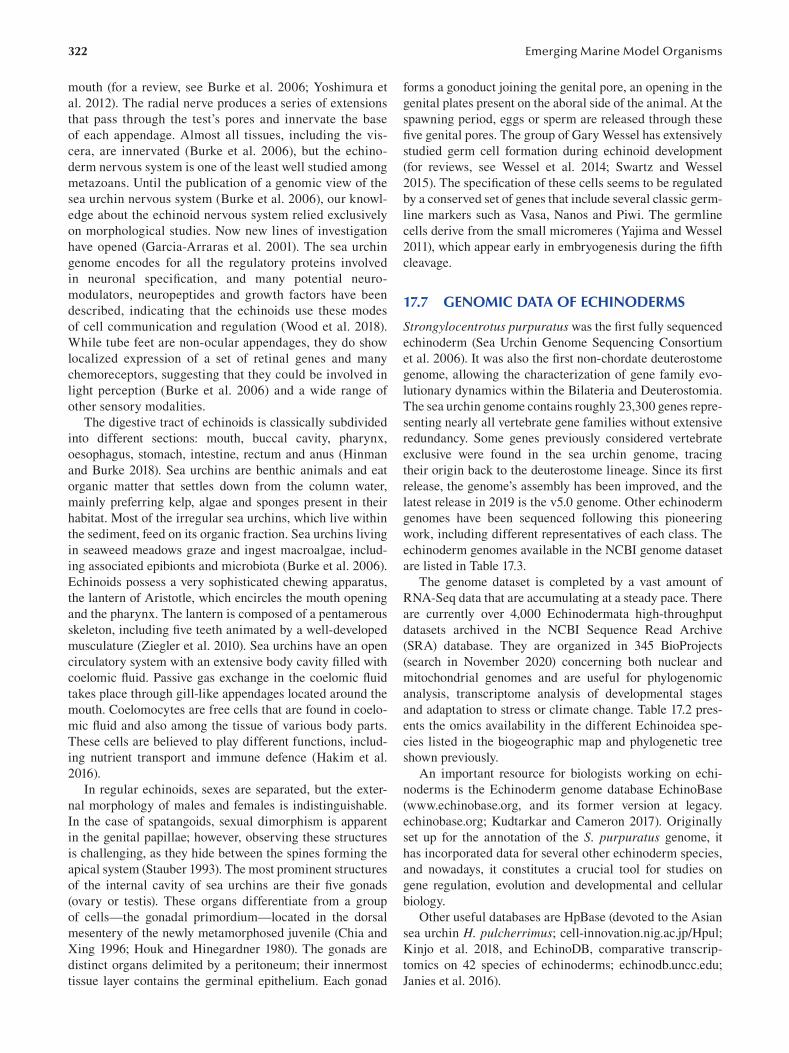
322
mouth (for a review, see Burke et al. 2006; Yoshimura et
al. 2012). The radial nerve produces a series of extensions
that pass through the test’s pores and innervate the base
of each appendage. Almost all tissues, including the vis
cera, are innervated (Burke et al. 2006), but the echino
derm nervous system is one of the least well studied among
metazoans. Until the publication of a genomic view of the
sea urchin nervous system (Burke et al. 2006), our knowl
edge about the echinoid nervous system relied exclusively
on morphological studies. Now new lines of investigation
have opened (Garcia-Arraras et al. 2001). The sea urchin
genome encodes for all the regulatory proteins involved
in neuronal specification, and many potential neuro
modulators, neuropeptides and growth factors have been
described, indicating that the echinoids use these modes
of cell communication and regulation (Wood et al. 2018).
While tube feet are non-ocular appendages, they do show
localized expression of a set of retinal genes and many
chemoreceptors, suggesting that they could be involved in
light perception (Burke et al. 2006) and a wide range of
other sensory modalities.
The digestive tract of echinoids is classically subdivided
into different sections: mouth, buccal cavity, pharynx,
oesophagus, stomach, intestine, rectum and anus ( Hinman
and Burke 2018). Sea urchins are benthic animals and eat
organic matter that settles down from the column water,
mainly preferring kelp, algae and sponges present in their
habitat. Most of the irregular sea urchins, which live within
the sediment, feed on its organic fraction. Sea urchins living
in seaweed meadows graze and ingest macroalgae, includ
ing associated epibionts and microbiota (Burke et al. 2006).
Echinoids possess a very sophisticated chewing apparatus,
the lantern of Aristotle, which encircles the mouth opening
and the pharynx. The lantern is composed of a pentamerous
skeleton, including five teeth animated by a well-developed
musculature ( Ziegler et al. 2010). Sea urchins have an open
circulatory system with an extensive body cavity fi lled with
coelomic fluid. Passive gas exchange in the coelomic fl uid
takes place through gill-like appendages located around the
mouth. Coelomocytes are free cells that are found in coelo
mic fluid and also among the tissue of various body parts.
These cells are believed to play different functions, includ
ing nutrient transport and immune defence (Hakim et al.
2016 ).
In regular echinoids, sexes are separated, but the exter
nal morphology of males and females is indistinguishable.
In the case of spatangoids, sexual dimorphism is apparent
in the genital papillae; however, observing these structures
is challenging, as they hide between the spines forming the
apical system (Stauber 1993). The most prominent structures
of the internal cavity of sea urchins are their fi ve gonads
(ovary or testis). These organs differentiate from a group
of cells—the gonadal primordium—located in the dorsal
mesentery of the newly metamorphosed juvenile (Chia and
Xing 1996; Houk and Hinegardner 1980). The gonads are
distinct organs delimited by a peritoneum; their innermost
tissue layer contains the germinal epithelium. Each gonad
Emerging Marine Model Organisms
forms a gonoduct joining the genital pore, an opening in the
genital plates present on the aboral side of the animal. At the
spawning period, eggs or sperm are released through these
five genital pores. The group of Gary Wessel has extensively
studied germ cell formation during echinoid development
(for reviews, see Wessel et al. 2014; Swartz and Wessel
2015). The specification of these cells seems to be regulated
by a conserved set of genes that include several classic germ-
line markers such as Vasa, Nanos and Piwi. The germline
cells derive from the small micromeres (Yajima and Wessel
2011), which appear early in embryogenesis during the fi fth
cleavage.
17.7 GENOMIC DATA OF ECHINODERMS
Strongylocentrotus purpuratus was the first fully sequenced
echinoderm (Sea Urchin Genome Sequencing Consortium
et al. 2006). It was also the first non-chordate deuterostome
genome, allowing the characterization of gene family evo
lutionary dynamics within the Bilateria and Deuterostomia.
The sea urchin genome contains roughly 23,300 genes repre
senting nearly all vertebrate gene families without extensive
redundancy. Some genes previously considered vertebrate
exclusive were found in the sea urchin genome, tracing
their origin back to the deuterostome lineage. Since its fi rst
release, the genome’s assembly has been improved, and the
latest release in 2019 is the v5.0 genome. Other echinoderm
genomes have been sequenced following this pioneering
work, including different representatives of each class. The
echinoderm genomes available in the NCBI genome dataset
are listed in Table 17.3.
The genome dataset is completed by a vast amount of
RNA-Seq data that are accumulating at a steady pace. There
are currently over 4,000 Echinodermata high-throughput
datasets archived in the NCBI Sequence Read Archive
(SRA) database. They are organized in 345 BioProjects
(search in November 2020) concerning both nuclear and
mitochondrial genomes and are useful for phylogenomic
analysis, transcriptome analysis of developmental stages
and adaptation to stress or climate change. Table 17.2 pres
ents the omics availability in the different Echinoidea spe
cies listed in the biogeographic map and phylogenetic tree
shown previously.
An important resource for biologists working on echi
noderms is the Echinoderm genome database EchinoBase
(www.echinobase.org, and its former version at legacy.
echinobase.org; Kudtarkar and Cameron 2017). Originally
set up for the annotation of the S. purpuratus genome, it
has incorporated data for several other echinoderm species,
and nowadays, it constitutes a crucial tool for studies on
gene regulation, evolution and developmental and cellular
biology.
Other useful databases are HpBase (devoted to the Asian
sea urchin H. pulcherrimus ; cell-innovation.nig.ac.jp/Hpul;
Kinjo et al. 2018 , and EchinoDB, comparative transcrip
tomics on 42 species of echinoderms; echinodb.uncc.edu;
Janies et al. 2016 ).
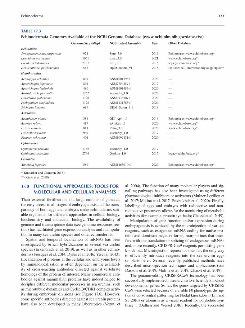
Echinoderms 323
TABLE 17.3 Echinodermata Genomes Available at the NCBI Genome Database (www.ncbi.nlm.nih.gov/datasets/)
Genome Size (Mbp) NCBI Latest Assembly Year Other Database
Echinoidea
Strongylocentrotus purpuratus 921 Spur_5.0 2019 Echinobase: www.echinobase.org *
Lytechinus variegatus 1061 Lvar_3.0 2021 www.echinobase.org*
Eucidaris tribuloides 2187 Etri_1.0 2015 legacy.echinobase.org *
Hemicentrotus pulcherrimus 568 HpulGenome_v1 2018 HpBase: cell-innovation.nig.ac.jp/Hpul/ **
Holothuroidea
Actinopyga echinites 899 ASM1001598v1 2020 —
Apostichopus japonicus 804 ASM275485v1 2017 —
Apostichopus leukothele 480 ASM1001483v1 2020 —
Australostichopus mollis 1252 assembly_1.0 2020 —
Holothuria glaberrima 1128 ASM993650v1 2020 —
Paelopatides confundens 1128 ASM1131785v1 2020 —
Stichopus horrens 689 UKM_Sthorr_1.1 2019 —
Asteroidea
Acanthaster planci 384 OKI-Apl_1.0 2016 Echinobase: www.echinobase.org *
Asterias rubens 417 eAstRub1.3 2020 www.echinobase.org*
Patiria miniata 811 Pmin_3.0 2020 www.echinobase.org*
Patiriella regularis 949 assembly_1.0 2017 —
Pisaster ochraceus 401 ASM1099431v1 2020 —
Ophuiroidea
Ophionereis fasciata 1185 assembly_1.0 2017 —
Ophiothrix spiculata 2764 Ospi.un_1.0 2015 legacy.echinobase.org *
Crinoidea
Anneissia japonica 589 ASM1163010v1 2020 Echinobase: www.echinobase.org *
*( Kudtarkar and Cameron 2017 );
** ( Kinjo et al. 2018 )
17.8 FUNCTIONAL APPROACHES: TOOLS FOR MOLECULAR AND CELLULAR ANALYSES
Their external fertilization, the large number of gametes,
the easy access to all stages of embryogenesis and the trans
parency of both eggs and embryos make echinoderms suit
able organisms for different approaches in cellular biology,
biochemistry and molecular biology. The availability of
genome and transcriptome data (see genomic resources sec
tion) has facilitated gene expression analysis and manipula
tion in many sea urchin species and other echinoderms.
Spatial and temporal localization of mRNAs has been
investigated by in situ hybridizations in several sea urchin
species (Erkenbrack et al. 2019), as well as in other echino
derms (Fresques et al. 2014; Dylus et al. 2016; Yu et al. 2013).
Localization of proteins at the cellular and embryonic levels
by immunolocalization is often dependent on the availabil
ity of cross-reacting antibodies directed against vertebrate
homologs of the protein of interest. Many commercial anti
bodies against mammalian proteins have indeed helped to
decipher different molecular processes in sea urchins, such
as microtubule dynamics and Cyclin B/CDK1 complex activ
ity during embryonic divisions (see Figure 17.6). However,
some specific antibodies directed against sea urchin proteins
have also been developed in many laboratories (Venuti et
al. 2004). The function of many molecular players and sig
nalling pathways has also been investigated using different
pharmacological inhibitors or activators (Mulner-Lorillon et
al. 2017; Molina et al. 2017; Feizbakhsh et al. 2020). Finally,
labelling of eggs and embryos with radioactive and non
radioactive precursors allows for the monitoring of metabolic
activities (for example, protein synthesis; Chassé et al. 2019).
Manipulation of gene function and/or expression during
embryogenesis is achieved by the microinjection of various
reagents, such as exogenous mRNA coding for native pro
teins and dominant-negative forms, morpholinos that inter
fere with the translation or splicing of endogenous mRNAs
and, more recently, CRISPR-Cas9 reagents permitting gene
knock-out. Microinjection represents, thus far, the only way
to efficiently introduce reagents into the sea urchin eggs
or blastomeres. Several recently published methods have
described microinjection techniques and applications (von
Dassow et al. 2019; Molina et al. 2019; Chassé et al. 2019).
The genome-editing CRISPR/Cas9 technology has been
successfully implemented in sea urchin to effi ciently knockout
developmental genes. So far, the genes targeted by CRISPR/
Cas9 were selected because of a visible F0 phenotype: disrup
tion of dorsoventral patterning for Nodal knockdown (Lin and
Su 2016 ) or albinism as a visual readout for polyketide syn
thase 1 (Oulhen and Wessel 2016). Recently, the successful
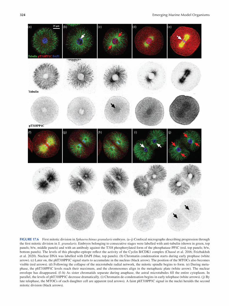
324 Emerging Marine Model Organisms
FIGURE 17.6 First mitotic division in Sphaerechinus granularis embryos. (a–j) Confocal micrographs describing progression through
the first mitotic division in S. granularis. Embryos belonging to consecutive stages were labelled with anti-tubulin (shown in green, top
panels; b/w, middle panels) and with an antibody against the T318 phosphorylated form of the phosphatase PP1C (red, top panels; b/w,
bottom panels). The levels of this phospho-epitope reflect the activity of the Cyclin B/CDK1 complex (Chassé et al. 2016; Feizbakhsh
et al. 2020). Nuclear DNA was labelled with DAPI (blue, top panels). (b) Chromatin condensation starts during early prophase (white
arrow). (c) Later on, the phT318PP1C signal starts to accumulate in the nucleus (black arrow). The position of the MTOCs also becomes
visible (red arrows). (d) Following the collapse of the microtubule radial network, the mitotic spindle begins to form. (e) During meta
phase, the phT318PP1C levels reach their maximum, and the chromosomes align in the metaphasic plate (white arrow). The nuclear
envelope has disappeared. (f–h) As sister chromatids separate during anaphase, the astral microtubules fill the entire cytoplasm. In
parallel, the levels of phT318PP1C decrease dramatically. (i) Chromatin de-condensation begins in early telophase (white arrows). (j) By
late telophase, the MTOCs of each daughter cell are apparent (red arrows). A faint phT318PP1C signal in the nuclei heralds the second
mitotic division (black arrows).
325 Echinoderms
production of a homozygous F2 mutant using the CRISPR
Cas9 system was obtained in Temnopleurus reevesii , which
takes advantage of the relatively short life cycle of this species
(Yaguchi et al. 2020; see also Chapter 18). This breakthrough
gives us the possibility to implement genetic analyses in the
sea urchin model (in species with short generation time) and
study the function of many maternal factors and mRNAs.
17.9 CHALLENGING QUESTIONS
During their long evolutionary trajectory, echinoderms have
adapted to all sorts of climatic conditions and have colo
nized most of the ocean floor, from the intertidal areas to
the deep-sea benthos. The study of this adaptation capacity
has just begun and should foster many exciting discoveries.
Moreover, it has become evident that echinoderms constitute
a valuable biological system to analyze the potential of marine
species to adapt to anthropogenic disturbance. Their popu
lation densities are very sensitive to climate change, ocean
acidification, eutrophication, overfishing, predatory removal
and the introduction of alien species (Uthicke et al. 2009). In
addition, this group of animals plays a crucial role in many
marine habitats and food webs, and several members of this
clade have been recognized as “keystone species” in differ
ent ecosystems (Power et al. 1996 ). Echinoderms have thus
acquired an essential place in experimental marine ecology.
As detailed previously, many genomic resources are
available nowadays for the researchers studying this clade,
in particular for those interested in the analysis of the echi
noids. These resources have greatly facilitated the develop
ment of comparative approaches aimed at understanding
the genetic basis of adaptive traits. The density of available
landmarks, including closely related species but also differ
ent groups separated by increasing phylogenetic distances,
allows dissection at the molecular level of both micro- and
macroevolutionary processes.
In echinoderms, many studies have focused on the
acquisition of evolutionary novelties and the diversifi cation
of life strategies. For instance, it has been shown that sev
eral species have significantly accelerated their life cycles,
reprogramming their ancestral planktotrophic larvae into
non-feeding lecithotrophic forms (Raff and Byrne 2006).
These evolutionary transitions have obvious adaptive roles.
In lecithotrophic species, the life cycle becomes independent
of fluctuations in plankton levels since their development
relies on the nutrients supplied by their mothers. Indeed,
it has been argued that the disturbance of planktonic food
chains could contribute in the near future to the decline of
planktotrophic species (Uthicke et al. 2009). At the same
time, these developmental transitions can now be analyzed
in great detail both at the cellular and molecular levels.
For instance, it has been shown in the Heliocidaris genus that the eggs of lecithotrophic species have under
gone an outstanding increase in size, driven by a thorough
remodelling of their oogenesis program (Byrne et al. 1999).
Moreover, the comparison of gene regulatory networks
controlling early development, like in the lecithotrophic
Heliocidaris erythrogramma and the planktotrophic
Heliocidaris tuberculata, provides important hints about
the identity of the molecular players participating in evolu
tionary change (Israel et al. 2016 ). These approaches have
greatly benefited from the deep knowledge of developmental
networks acquired thanks to the study of early development
in Strongylocentrotus purpuratus and other echinoderm
species (Cary and Hinman 2017).
Echinoderm biology stands now at the intersection
between ecology, cell and developmental and evolution
ary biology and should greatly profit from this privileged
position.
ACKNOWLEDGEMENTS AND FUNDING
We apologize to those whose work was not cited or dis
cussed here because of the broad scope of this review and
space limitations. We are indebted to the Marine and Diving
Facility and the Aquarium Service of the Roscoff Marine
Station for echinoderm collection and rearing. We are also
grateful to the imaging platform of the FR2424 (Plateforme
MerImage, SU/CNRS). We thank H. Flom for manuscript
corrections. The authors acknowledge the support of “La
Ligue contre le Cancer (coordination du Grand Ouest:
comités Finistère, Côtes d’Armor, Deux-Sèvres, Morbihan,
Ille-et-Villaine, Loire Atlantique, Charente, Sarthe)”, the
Brittany Regional Council (Région Bretagne), the Finistère
Department Council (CG29). Florian Pontheaux is funded
by a PhD fellowship from the French Education Ministry
[SU; Doctoral School ED515].
BIBLIOGRAPHY
Aihara, M. and S. Amemiya. 2001. Left-right positioning of the
adult rudiment in sea urchin larvae is directed by the right
side. Development 128:4935–4948.
Allen, R.D. and J.L. Griffin. 1958. The time sequence of early
events in the fertilization of sea urchin eggs. Experimental Cell Research 15:163–173.
Andrikou, C., Iovene, E., Rizzo, F., Oliveri, P. and M.I. Arnone.
2013. Myogenesis in the sea urchin embryo: The molecular
fingerprint of the myoblast precursors. EvoDevo 4:33–48.
Angerer, L.M. and R.C. Angerer. 2000. Animal-vegetal axis
patterning mechanisms in the early sea urchin embryo.
Developmental Biology 218:1–12.
Angione, S.L., Oulhen, N., Brayboy, L.M., Tripathi, A. and G.M.
Wessel. 2015. Simple perfusion apparatus for manipulation,
tracking, and study of oocytes and embryos. Fertility and Sterility 103:281–290.
Anstrom, J.A. 1992. Microfilaments, cell shape changes, and the
formation of primary mesenchyme in sea urchin embryos.
Journal of Experimental Zoology 264:312–322.
Armstrong, N. and D.R. McClay. 1994. Skeletal pattern is speci
fied autonomously by the primary mesenchyme cells in sea
urchin embryos. Developmental Biology 162:329–338.
Arnone, M.I., Byrne, M. and P. Martinez. 2015. Echinodermata. In
Evolutionary Developmental Biology of Invertebrates , ed. A.
Wanninger, 6:1–58. Springer-Verlag, Wien.
Ben-Tabou de-Leon, S. 2017. The network remains. History and Philosophy of the Life Sciences 39:32.
326
Ben-Tabou de-Leon, S. 2016. Robustness and accuracy in sea
urchin developmental gene regulatory networks. Frontiers in Genetics 7:16–21.
Bernard, F.R. 1977. Fishery and reproductive cycle of the red sea
urchin, strongylocentrotus franciscanus, in British Columbia.
Journal of the Fisheries Board of Canada 34:604–610.
Binks, R.M., Prince, J., Evans, J.P. and W.J. Kennington. 2012.
More than bindin divergence: Reproductive isolation
between sympatric subspecies of a sea urchin by asynchro
nous spawning. Evolution 66:3545–3557.
Bisgrove, B.W., Andrews, M.E. and R.A. Raff. 1991. Fibropellins,
products of an EGF repeat-containing gene, form a unique
extracellular matrix structure that surrounds the sea urchin
embryo. Developmental Biology 146:89–99.
Böhmer, M., Van, Q., Weyand, I., Hagen, V., Beyermann, M.,
Matsumoto, M., Hoshi, M., Hildebrand, E. and U.B. Kaupp.
2005. Ca2+ spikes in the flagellum control chemotactic behav
ior of sperm. EMBO J 24:2741–2752.
Bolton, T.F., Thomas, F.I. and C.N. Leonard. 2000. Maternal
energy investment in eggs and jelly coats surrounding eggs
of the echinoid Arbacia punctulata. The Biological Bulletin 199:1–5.
Bosch, I., Beauchamp, K.A., Steele, M.E. and J.S. Pearse. 1987.
Development, metamorphosis, and seasonal abundance of
embryos and larvae of the Antarctic sea urchin Sterechinus neumayeri. The Biological Bulletin 173:126–135.
Boveri, T. 2008. Concerning the origin of malignant tumours by
Theodor Boveri: Translated and annotated by Henry Harris.
Journal of Cell Science 121:1–84.
Boveri, T. 1902. Über mehrpolige Mitosen als Mittel zur Analyse
des Zellkerns. [Concerning multipolar mitoses as a means of
analysing the cell nucleus]. Verhandlungen der Physikalisch-Medizinischen Gesellschaft zu Würzburg 35:67–90.
Brachet, J. 1975. From chemical to molecular sea urchin embryol
ogy. American Zoologist 15:485–491.
Brachet, J. 1941. La localisation des acides pentose-nucléiques
dans les tissus animaux et les oeufs d’amphibiens en voie de
développement. Archive de Biologie 53:207–257.
Briggs, E. and G.M. Wessel. 2006. In the beginning . . . animal
fertilization and sea urchin development. Developmental Biology 300:15–26.
Bronstein, O. and A. Kroh. 2019. The first mitochondrial genome
of the model echinoid Lytechinus variegatus and insights into
Odontophoran phylogenetics. Genomics 111:710–718.
Burke, R.D. and C.M. Alvarez. 1988. Development of the esopha
geal muscles in embryos of the sea urchin Strongylocentrotus purpuratus. Cell Tissue Research 252:411–417.
Burke, R.D., Angerer, L.M., Elphick, M.R., Humphrey, G.W.,
Yaguchi, S., Kiyama, T., Liang, S., Mu, X., Agca, C., Klein,
W.H., Brandhorst, B.P., Rowe, M., Wilson, K., Churcher,
A.M., Taylor, J.S., Chen, N., Murray, G., Wang, D.,
Mellott, D., Olinski, R., Hallböök, F. and M.C. Thorndyke.
2006. A genomic view of the sea urchin nervous system.
Developmental Biology 300:434–460.
Burke, R.D., Myers, R.L., Sexton, T.L. and C. Jackson. 1991. Cell
movements during the initial phase of gastrulation in the sea
urchin embryo. Developmental Biology 146:542–557.
Byrne, M. 1990. Annual reproductive cycles of the commercial
sea urchin Paracentrotus lividus from an exposed intertidal
and a sheltered subtidal habitat on the west coast of Ireland.
Marine Biology 104:275–289.
Byrne, M., Villinski, J.T., Cisternas, P., Siegel, R.K., Popodi, E. and
R.A. Raff. 1999. Maternal factors and the evolution of develop
mental mode: Evolution of oogenesis in Heliocidaris erythrogramma. Developmental Genes and Evolution 209:275–283.
Emerging Marine Model Organisms
Byrnes, W.M. and W.R. Eckberg. 2006. Ernest Everett Just
(1883–1941): An early ecological developmental biologist.
Developmental Biology 296:1–11.
Byrnes, W.M. and S.A. Newman. 2014. Ernest Everett Just: Egg
and embryo as excitable systems. Journal of Experimental Zoology. PartB, Molecular and Developmental Evolution 322B:191–201.
Cameron, R.A., Fraser, S.E., Britten, R.J. and E.H. Davidson.
1991. Macromere cell fates during sea urchin development.
Development 113:1085–1091.
Cannon, J.T., Kocot, K.M., Waits, D.S., Weese, D.A., Swalla, B.J.,
Santos, S.R. and K.M. Halanych. 2014. Phylogenomic reso
lution of the hemichordate and echinoderm clade. Current Biology 24:2827–2832.
Cary, G.A. and V.F. Hinman. 2017. Echinoderm development and
evolution in the post-genomic era. Developmental Biology 427:203–211.
Cavalieri, V. and G. Spinelli. 2015. Symmetry breaking and estab
lishment of dorsal/ventral polarity in the early sea urchin
embryo. Symmetry 7:1721–1733.
Chassé, H., Aubert, J., Boulben, S., Le Corguille, G., Corre, E.,
Cormier, P. and J. Morales. 2018. Translatome analysis at
the egg-to-embryo transition in sea urchin. Nucleic Acids Research 46:4607–4621.
Chassé, H., Boulben, S., Costache, V., Cormier, P. and J. Morales.
2017. Analysis of translation using polysome profi ling.
Nucleic Acids Research 45:e15. doi: 10.1093/nar/gkw907.
Chassé, H., Boulben, S., Glippa, V., Pontheaux, F., Cormier, P.
and J. Morales. 2019. In vivo analysis of protein translation
activity in sea urchin eggs and embryos. In Methods in Cell Biology, Echinoderms, Part B, ed. A. Hamdoun and K.R.
Foltz, 150:335–352. Elsevier; Academic Press, London UK.
Chassé, H., Mulner-Lorillon, O., Boulben, S., Glippa, V., Morales,
J. and P. Cormier. 2016. Cyclin B translation depends on
mTOR activity after fertilization in sea urchin embryos.
PLoS One 11:e0150318. https://doi.org/10.1371/journal.
pone.0150318 .
Chenuil, A. and J.-P. Féral. 2003. Sequences of mitochondrial
DNA suggest that Echinocardium cordatum is a complex
of several sympatric or hybridizing species: A pilot study.
In Echinoderm Research 2001, ed. J.-P. Féral and B. David,
15–21. Proc. 6th Eur. Conf. Echinoderm, Banyuls-sur-mer,
France. Swets & Zeitlinger Publishers, Lisse, NL.
Chia, F.S. and Xing, J. 1996. Echinoderm coelomocytes. Zoological Studies 35:231–254.
Clift, D. and Schuh, M. 2013. Restarting life: Fertilization and
the transition from meiosis to mitosis. Nature Reviews: Molecular Cell Biology 14:549–562.
Cormier, P., Chassé, H., Cosson, B., Mulner-Lorillon, O. and J.
Morales. 2016. Translational control in echinoderms: The
calm before the storm. In Evolution of the Protein Synthesis Machinery and Its Regulation, ed. G. Hernandez and R. Jagus,
413–434. Springer International Publishing, Switzerland.
Costache, V., McDougall, A. and R. Dumollard. 2014. Cell cycle arrest
and activation of development in marine invertebrate deutero
stomes. Biochemical and Biophysical Research Communications 450:1175–1181.
Costello, D.P. and C. Henley. 1971. Methods for obtaining and
handling marine eggs and embryos. Marine Biological Laboratory. https://hdl.handle.net/1912/295 .
Croce, J.C. and D.R. McClay. 2010. Dynamics of Delta/Notch
signaling on endomesoderm segregation in the sea urchin
embryo. Development 137:83–91.
Darszon, A., Nishigaki, T., Wood, C., Treviño, C.L., Felix, R. and
C. Beltrán. 2005. Calcium channels and Ca2+ fl uctuations
327 Echinoderms
in sperm physiology. International Review of Cytology 243:79–172.
Davidson, E.H. 2006. Gene regulatory networks and the evolution
of animal body plans. Science 311:796–800.
Davidson, E.H. 2002. A genomic regulatory network for develop
ment. Science 295:1669–1678.
Davidson, P.L., Guo, H., Wang, L., Berrio, A., Zhang, H., Chang,
Y., Soborowski, A.L., McClay, D.R., Fan, G. and G.A. Wray.
2020. Chromosomal-level genome assembly of the sea
urchin Lytechinus variegatus substantially improves func
tional genomic analyses. Genome Biology and Evolution 12:1080–1086.
Delalande, C., Monnier, A., Minella, O., Geneviere, A.M., Mulner-
Lorillon, O., Belle, R. and P. Cormier. 1998. Developmental
regulation of elongation factor-1 delta in sea urchin suggests
appearance of a mechanism for alternative poly(A) site selec
tion in gastrulae. Experimental Cell Research 242:228–234.
Delorme, N.J. and M.A. Sewell. 2016. Effects of warm accli
mation on physiology and gonad development in the sea
urchin Evechinus chloroticus. Comparative Biochemistry and Physiology Part A: Molecular & Integrative Physiology 198:33–40.
Derbès, A.A. 1847. Observations sur le méchanisme et les phe
nomènes qui accompagnent la formation de l’embryon chez
l’oursin comestible. Annales des Sciences Naturelles (Zool) 8:80–98.
Dilly, G.F., Gaitán-Espitia, J.D. and G.E. Hofmann. 2015.
Characterization of the Antarctic sea urchin ( Sterechinus neumayeri) transcriptome and mitogenome: A molecular
resource for phylogenetics, ecophysiology and global change
biology. Molecular Ecology Resources 15:425–436.
Drozdov, A.L. and V.V. Vinnikova. 2010. Morphology of gam
etes in sea urchins from Peter the Great Bay, Sea of Japan.
Ontogenez 41:47–57.
Duboc, V., Röttinger, E., Besnardeau, L. and T. Lepage. 2004.
Nodal and BMP2/4 signaling organizes the oral-aboral axis
of the sea urchin embryo. Developmental Cell . 6:397–410.
Dupont, G. and R. Dumollard. 2004. Simulation of calcium waves
in ascidian eggs: Insights into the origin of the pacemaker
sites and the possible nature of the sperm factor. Journal of Cell Science 117:4313–4323.
Dylus, D.V., Czarkwiani, A., Stångberg, J., Ortega-Martinez, O.,
Dupont, S. and P. Oliveri. 2016. Large-scale gene expression
study in the ophiuroid Amphiura filiformis provides insights
into evolution of gene regulatory networks. EvoDevo 7:2.
Egea, E., Mérigot, B., Mahé-Bézac, C., Féral, J.-P. and A. Chenuil.
2011a. Differential reproductive timing in Echinocardium spp.: The first Mediterranean survey allows interoceanic
and interspecific comparisons. Comptes Rendus Biologies 334:13–23.
Erkenbrack, E.M., Croce, J.C., Miranda, E., Gautam, S., Martinez-
Bartolome, M., Yaguchi, S. and R.C. Range. 2019. Whole
mount in situ hybridization techniques for analysis of the
spatial distribution of mRNAs in sea urchin embryos and
early larvae. In Methods in Cell Biology: Echinoderms, Part B, ed. A. Hamdoun and K.R. Foltz, 150:177–196. Elsevier;
Academic Press, London UK.
Erkenbrack, E.M., Davidson, E.H. and I.S. Peter. 2018. Conserved
regulatory state expression controlled by divergent develop
mental gene regulatory networks in echinoids. Development 145:1–11.
Ernst, S.G. 2011. Offerings from an urchin. Developmental Biology 358:285–294.
Erwin, D.H., Laflamme, M., Tweedt, S.M., Sperling, E.A., Pisani,
D. and K.J. Peterson. 2011. The Cambrian conundrum: Early
divergence and later ecological success in the early history of
animals. Science 334:1091–1097.
Ettensohn, C.A. 2020. The gene regulatory control of sea urchin
gastrulation. Mechanisms of Development 162:103599.
Ettensohn, C.A. 2017. Sea urchins as a model system for studying
embryonic development. In Reference Module in Biomedical Sciences, ed M.J. Caplan. Elsevier, Amsterdam.
Ettensohn, C.A. 1999. Cell movements in the sea urchin embryo.
Current Opinion in Genetics & Development 9:461–465.
Ettensohn, C.A. 1985. Gastrulation in the sea urchin embryo is
accompanied by the rearrangement of invaginating epithelial
cells. Developmental Biology 112:383–390.
Ettensohn, C.A. and D.R. McClay. 1988. Cell lineage conversion in
the sea urchin embryo. Developmental Biology 125:396–409.
Ettensohn, C.A. and S.W. Ruffins. 1993. Mesodermal cell inter
actions in the sea urchin embryo: Properties of skeletogenic
secondary mesenchyme cells. Development 117:1275–1285.
Ettensohn, C.A. and H.C. Sweet. 2000. Patterning the early sea
urchin embryo. Current Topics in Developmental Biology 50:1–44.
Evans, T., Rosenthal, E.T., Youngblom, J., Distel, D. and T. Hunt.
1983. Cyclin: A protein specified by maternal mRNA in sea
urchin eggs that is destroyed at each cleavage division. Cell 33:389–396.
Feizbakhsh, O., Pontheaux, F., Glippa, V., Morales, J., Ruchaud, S.,
Cormier, P. and F. Roch. 2020. A peak of H3T3 phosphory
lation occurs in synchrony with mitosis in sea urchin early
embryos. Cells 9:898.
Filander, Z. and C. Griffiths. 2017. Illustrated guide to the echinoid
(Echinodermata: Echinoidea) fauna of South Africa. Zootaxa 4296:1–72.
Fink, R.D. and D.R. McClay. 1985. Three cell recognition changes
accompany the ingression of sea urchin primary mesen
chyme cells. Developmental Biology 107:66–74.
Finkel, T. 2011. Signal transduction by reactive oxygen species.
The Journal of Cell Biology 194:7–15.
Floc’hlay, S., Molina, M.D., Hernandez, C., Haillot, E., Thomas-
Chollier, M., Lepage, T. and D. Thieffry. 2021. Deciphering
and modelling the TGF-β signalling interplays specifying the
dorsal-ventral axis of the sea urchin embryo. Development 148:dev189944.
Foerder, C.A. and B.M. Shapiro. 1977. Release of ovoperoxidase
from sea urchin eggs hardens the fertilization membrane with
tyrosine crosslinks. Proceedings of the National Academy of Sciences of the United States of America 74:4214–4218.
Fol, H. 1879. Recherches sur la fécondation et le commencement
de l’hénogénie chez divers animaux. Mémoires de la Société de Physique et d’Histoire Naturelle (Geneve) 26:89–250.
Foo, S.A., Deaker, D. and M. Byrne. 2018. Cherchez la femme
impact of ocean acidification on the egg jelly coat and
attractants for sperm. Journal of Experimental Biology 221:jeb177188.
Fresques, T., Zazueta-Novoa, V., Reich, A. and G.M. Wessel. 2014.
Selective accumulation of germ-line associated gene prod
ucts in early development of the sea star and distinct differ
ences from germ-line development in the sea urchin: Sea
star germ-line gene expression. Developmental Dynamics 243:568–587.
Gago, J. and O.J. Luís. 2011. Comparison of spawning induc
tion techniques on Paracentrotus lividus (Echinodermata:
Echinoidea) broodstock. Aquaculture International 19:181–191.
Gaitán-Espitia, J.D., Sánchez, R., Bruning, P. and L. Cárdenas.
2016. Functional insights into the testis transcriptome of
the edible sea urchin Loxechinus albus. Scientifi c Reports 6:36516.
328
Garcia-Arraras, J.E., Rojas-Soto, M., Jimenez, L.B. and L. Diaz-
Miranda. 2001. The enteric nervous system of echinoderms:
Unexpected complexity revealed by neurochemical analysis.
Journal of Experimental Biology 204:865–873.
Ghisaura, S., Loi, B., Biosa, G., Baroli, M., Pagnozzi, D., Roggio,
T., Uzzau, S., Anedda, R. and M.F. Addis. 2016. Proteomic
changes occurring along gonad maturation in the edible
sea urchin Paracentrotus lividus. Journal of Proteomics 144:63–72.
Gianguzza, P. and C. Bonaviri. 2013. Chapter 19: Arbacia. In
Developments in Aquaculture and Fisheries Science, Sea Urchins: Biology and Ecology, ed. J.M. Lawrence, 275–283.
Elsevier, London UK.
Gibson, A.W. and R.D. Burke. 1985. The origin of pigment cells
in embryos of the sea urchin Strongylocentrotus purpuratus.
Developmental Biology 107:414–419.
Gilbert, S.F. 2006. Developmental Biology, eighth ed. Sinauer
Associates Inc., Sunderland, MA, USA.
Gildor, T., Malik, A., Sher, N., Avraham, L. and S. Ben-Tabou de-
Leon. 2016. Quantitative developmental transcriptomes of
the Mediterranean sea urchin Paracentrotus lividus. Marine Genomics 25:89–94.
Gillard, G.B., Garama, D.J. and C.M. Brown. 2014. The transcrip
tome of the NZ endemic sea urchin Kina (Evechinus chloroticus). BMC Genomics 15:45.
Goldstein, B. and G. Freeman. 1997. Axis specification in animal
development. Bioessays 19:105–116.
González-Irusta, J.M., Goñi de Cerio, F. and J.C. Canteras. 2010.
Reproductive cycle of the sea urchin Paracentrotus lividus in
the Cantabrian Sea (Northern Spain): Environmental effects.
Journal of the Marine Biological Association of the United Kingdom 90:699–709.
Gross, P.R. and G.H. Cousineau. 1963. Effects of actinomycin
D on macromolecule synthesis and early development in
sea urchin eggs. Biochemical and Biophysical Research Communications 10:321–326.
Gross, P.R., Malkin, L.I. and W.A. Moyer. 1964. Templates for
the first proteins of embryonic development. Proceedings of the National Academy of Sciences of the United States of America 51:407–414.
Guillou, M. and L.J.L. Lumingas. 1998. The reproductive cycle of
the ‘blunt’ sea urchin. Aquaculture International 6:147–160.
Guillou, M. and C. Michel. 1993. Reproduction and growth of
Sphaerechinus granularis (Echinodermata: Echinoidea)
in Southern Brittany. Journal of the Marine Biological Association of the United Kingdom 73:179–192.
Gustafson, T. and H. Kinnander. 1956. Microaquaria for time-
lapse cinematographic studies of morphogenesis in swim
ming larvae and observations on sea urchin gastrulation.
Experimental Cell Research 11:36–51.
Gustafson, T. and L. Wolpert. 1967. Cellular movement and
contact in sea urchin morphogenesis. Biological Reviews 42:442–498.
Gustafson, T. and L. Wolpert. 1963. Studies on the cellular basis of
morphogenesis in the sea urchin embryo: Formation of the
coelom, the mouth, and the primary pore-canal. Experimental Cell Research 29:561–582.
Hakim, J.A., Koo, H., Kumar, R., Lefkowitz, E.J., Morrow, C.D.,
Powell, M.L., Watts, S.A. and A.K. Bej. 2016. The gut
microbiome of the sea urchin, Lytechinus variegatus, from its
natural habitat demonstrates selective attributes of microbial
taxa and predictive metabolic profi les. FEMS Microbiology Ecology 92:146–157.
Haley, S.A. and G.M. Wessel. 1999. The cortical granule ser
ine protease CGSP1 of the sea urchin, Strongylocentrotus
Emerging Marine Model Organisms
purpuratus, is autocatalytic and contains a low-density
lipoprotein receptor-like domain. Developmental Biology 211:1–10.
Hall, H.G. and V.D. Vacquier. 1982. The apical lamina of the sea
urchin embryo: Major glycoproteins associated with the hya
line layer. Developmental Biology 89:168–178.
Hamdoun, A., Schrankel, C.S., Nesbit, K.T. and J.A. Espinoza.
2018. Sea urchins as lab animals for reproductive and
developmental biology. Encyclopedia of Reproduction 6:696–703.
Hara, Y., Kuraishi, R., Uemura, I. and H. Katow. 2003. Asymmetric
formation and possible function of the primary pore canal in
plutei of Temnopleurus hardwicki. Development, Growth & Differentiation 45:295–308.
Hardin, J. 1989. Local shifts in position and polarized motil
ity drive cell rearrangement during sea urchin gastrulation.
Developmental Biology 136:430–445.
Hardin, J. 1988. The role of secondary mesenchyme cells during sea
urchin gastrulation studied by laser ablation. Development 103:317–324.
Hardin, J. and D.R. McClay. 1990. Target recognition by the
archenteron during sea urchin gastrulation. Developmental Biology 142:86–102.
Hertwig, O. 1876. Beitrage zur Erkenntnis der Bildung, Befruchtung,
und Theilung des tierischen Eies. Morphologisches Jahrbuch 1:347–352.
Hibino, T., Minokawa, T. and A. Yamazaki. 2019. Chapter 4:
Cidaroids, clypeasteroids, and spatangoids: Procurement,
culture, and basic methods. In Methods in Cell Biology: Echinoderms, Part B, ed. A. Hamdoun and K.R. Foltz,
150:81–103. Elsevier; Academic Press, London UK.
Himmelman, J.H. 1978. Reproductive cycle of the green sea urchin,
Strongylocentrotus droebachiensis. Canadian Journal of Zoology 56:1828–1836.
Hinman, V.F. and R.D. Burke. 2018. Embryonic neurogen
esis in echinoderms. Wiley Interdisciplinary Reviews. Developmental Biology 7:e316.
Hogan, J.D., Keenan, J.L., Luo, L., Ibn-Salem, J., Lamba, A.,
Schatzberg, D., Piacentino, M.L., Zuch, D.T., Core, A.B.,
Blumberg, C., Timmermann, B., Grau, J.H., Speranza, E.,
Andrade-Navarro, M.A., Irie, N., Poustka, A.J. and C.A.
Bradham. 2020. The developmental transcriptome for
Lytechinus variegatus exhibits temporally punctuated gene
expression changes. Developmental Biology 460:139–154.
Horstadius, S. 1939. The mechanics of sea urchin develop
ment, studied by operative methods. Biological Reviews 14:132–179.
Horton, T., Kroh, A., Ahyong, S., Bailly, N., Boyko, C.B., et al.
2020. World Register of Marine Species (WoRMS). https://
doi.org/10.14284/170 .
Houk, M.S. and R.T. Hinegardner. 1980. The formation and early
differentiation of sea urchin gonads. The Biological Bulletin 159:280–294.
Hultin, T. 1961. The effect of puromycin on protein metabolism
and cell division in fertilized sea urchin eggs. Experientia 17:410–411.
Humphreys, T. 1969. Efficiency of translation of messenger-RNA
before and after fertilization in sea urchins. Developmental Biology 20:435–458.
Hunt, T. 2002. Nobel lecture: Protein synthesis, proteolysis, and
cell cycle transitions. Bioscience Reports 22:465–486.
Hyman, L.H. 1955. The invertebrates, echinodermata (4) . McGraw-
Hill Education, New York, NY.
Ishimoda-Takagi, T., Chino, I. and H. Sato. 1984. Evidence for
the involvement of muscle tropomyosin in the contractile
329 Echinoderms
elements of the coelom-esophagus complex in sea urchin
embryos. Developmental Biology 105:365–376.
Israel, J.W., Martik, M.L., Byrne, M., Raff, E.C., Raff, R.A.,
McClay, D.R. and G.A. Wray. 2016. Comparative devel
opmental transcriptomics reveals rewiring of a highly con
served gene regulatory network during a major life history
switch in the sea urchin genus Heliocidaris. PLoS Biology 14:e1002391.
Janies, D.A., Witter, Z., Linchangco, G.V., Foltz, D.W., Miller,
A.K., Kerr, A.M., Jay, J., Reid, R.W. and G.A. Wray. 2016.
EchinoDB, an application for comparative transcriptomics of
deeply-sampled clades of echinoderms. BMC Bioinformatics 17:48.
Just, E.E. 1939. The Biology of the Cell Surface. The Technical
Press LTD; Ave Maria Lane, Ludgate Hill, E.C., London.
Just, E.E. 1919. The fertilization reaction in Echinarachnius parma: I. Cortical response of the egg to insemination. The Biological Bulletin 36:110.
Kahil, K., Varsano, N., Sorrentino, A., Pereiro, E., Rez, P.,
Weiner, S. and L. Addadi. 2020. Cellular pathways of
calcium transport and concentration toward mineral for
mation in sea urchin larvae. Proceedings of the National Academy of Sciences of the United States of America 117:30957–30965.
Kedes, L.H., Chang, A.C.Y., Houseman, D. and S.N. Cohen. 1975.
Isolation of histone genes from unfractionated sea urchin
DNA by subculture cloning in E. coli. Nature 255:533–538.
Kimberly, E.L. and J. Hardin. 1998. Bottle cells are required for the
initiation of primary invagination in the sea urchin embryo.
Developmental Biology 204:235–250.
Kinjo, S., Kiyomoto, M., Yamamoto, T., Ikeo, K. and S. Yaguchi.
2018. HpBase: A genome database of a sea urchin,
Hemicentrotus pulcherrimus. Development, Growth & Differentiation 60:174–182.
Kipryushina, Y.O. and K.V. Yakovlev. 2020. Maternal control of early
patterning in sea urchin embryos. Differentiation 113:28–37.
Kiyomoto, M., Hamanaka, G., Hirose, M. and M. Yamaguchi.
2014. Preserved echinoderm gametes as a useful and ready-
to-use bioassay material. Marine Environmental Research 93:102–105.
Kominami, T. and Takata. 2004. Gastrulation in the sea urchin
embryo: A model system for analyzing the morphogen
esis of a monolayered epithelium. Development, Growth & Differentiation 46:309–326.
Kroh, A. and A.B. Smith. 2010. The phylogeny and classifi ca
tion of post-Palaeozoic echinoids. Journal of Systematic Palaeontology 8:147–212.
Kudtarkar, P. and R.A. Cameron. 2017. Echinobase: An expand
ing resource for echinoderm genomic information. Database 2017:bax074.
Kuroda, R., Kontani, K., Kanda, Y., Katada, T., Nakano, T., Satoh,
Y., Suzuki, N. and H. Kuroda. 2001. Increase of cGMP,
cADP-ribose and inositol 1,4,5-trisphosphate preceding Ca2+
transients in fertilization of sea urchin eggs. Development 128:4405–4414.
Lane, M.C., Koehl, M.A., Wilt, F. and R. Keller. 1993. A role
for regulated secretion of apical extracellular matrix dur
ing epithelial invagination in the sea urchin. Development 117:1049–1060.
Lapraz, F., Besnardeau, L. and T. Lepage. 2009. Patterning of the
dorsal-ventral axis in echinoderms: Insights into the evolu
tion of the BMP-chordin signaling network. PLoS Biology 7:e1000248.
Láruson, Á.J. 2017. Rates and relations of mitochondrial genome
evolution across the Echinoidea, with special focus on
the superfamily Odontophora. Ecology and Evolution 7:4543–4551.
Leigh, S.J., Bowen, J., Purssell, C.P., Covington, J.A., Billson, D.R.
and D.A. Hutchins. 2012. Rapid manufacture of monolithic
micro-actuated forceps inspired by echinoderm pedicellar
iae. Bioinspiration & Biomimetics 7:044001.
Lepage, T., Ghiglione, C. and C. Gache. 1992a. Spatial and tempo
ral expression pattern during sea urchin embryogenesis of a
gene coding for a protease homologous to the human protein
BMP-1 and to the product of the Drosophila dorsal-ventral
patterning gene tolloid. Development 114:147–163.
Lepage, T., Sardet, C. and C. Gache, C. 1992b. Spatial expres
sion of the hatching enzyme gene in the sea urchin embryo.
Developmental Biology 150:23–32.
Lessios, H.A. 1990. Adaptation and phylogeny as determinants of
egg size in echinoderms from the two sides of the isthmus of
panama. The American Naturalist 135:1–13.
Lessios, H.A. 1988. Temporal and spatial variation in egg size of
13 Panamanian echinoids. Journal of Experimental Marine Biology and Ecology 114:217–239.
Levitan, D.R. 1993. The importance of sperm limitation to the
evolution of egg size in marine invertebrates. The American Naturalist 141:517–536.
Li, E., Cui, M., Peter, I.S. and E.H. Davidson. 2014. Encoding reg
ulatory state boundaries in the pregastrular oral ectoderm of
the sea urchin embryo. Proceedings of the National Academy of Sciences of the United States of America 111:E906–E913.
Lin, C.-Y. and Y.-H. Su. 2016. Genome editing in sea urchin
embryos by using a CRISPR/Cas9 system. Developmental Biology 409:420–428.
Loeb, J. 1899. On the nature of the process of fertilization and
the artificial production of normal larvæ (plutei) from the
unfertilized eggs of the sea urchin. American Journal of Physiology 3:135–138.
Logan, C.Y., Miller, J.R., Ferkowicz, M.J. and D.R. McClay. 1999.
Nuclear beta-catenin is required to specify vegetal cell fates
in the sea urchin embryo. Development 126:345–357.
Longo, F.J. and E. Anderson. 1968. The fine structure of pronuclear
development and fusion in the sea urchin, Arbacia punctulata. The Journal of Cell Biology 39:339–368.
Longo, F.J. and W. Plunkett. 1973. The onset of DNA synthesis
and its relation to morphogenetic events of the pronuclei
in activated eggs of the sea urchin, Arbacia punctulata.
Developmental Biology 30:56–67.
Lowe, C.J., Clarke, D.N., Medeiros, D.M., Rokhsar, D.S. and J.
Gerhart. 2015. The deuterostome context of chordate origins.
Nature 520:456–465.
Luo, Y.-J. and Y.-H. Su. 2012. Opposing nodal and BMP signals
regulate left right asymmetry in the sea urchin larva. PLoS Biol 10:e1001402.
Maderspacher, F. 2008. Theodor Boveri and the natural experi
ment. Current Biology 18:R279–R286.
Malinda, K.M. and C.A Ettensohn. 1994. Primary mesenchyme
cell migration in the sea urchin embryo: Distribution of
directional cues. Developmental Biology 164:562–578.
Malumbres, M. 2014. Cyclin-dependent kinases. Genome Biology 15:122.
Martik, M.L., Lyons, D.C. and D.R. McClay. 2016. Developmental
gene regulatory networks in sea urchins and what we can
learn from them. F1000Research 5:203.
Martik, M.L. and D.R. McClay. 2017. New insights from a high-
resolution look at gastrulation in the sea urchin, Lytechinus variegatus. Mechanisms of Development 148:3–10.
Maruyama, Y.K., Nakaseko, Y. and S. Yagi. 1985. Localization
of cytoplasmic determinants responsible for primary
330
mesenchyme formation and gastrulation in the unfertilized
egg of the sea urchin Hemicentrotus pulcherrimus. Journal of Experimental Zoology 236:155–163.
Mazia, D. 1937. The release of calcium in Arbacia eggs on fer
tilization. Journal of Cellular and Comparative Physiology 10:291–304.
McAlister, J.S. and A.L. Moran. 2012. Relationships among egg
size, composition, and energy: A comparative study of gemi
nate sea urchins. PLoS One 7:e41599.
McClintock, J.B., Amsler, M.O., Angus, R.A., Challener, R.C.,
Schram, J.B., Amsler, C.D., Mah, C.L., Cuce, J. and B.J.
Baker. 2011. The Mg-calcite composition of Antarctic
echinoderms: Important implications for predicting the
impacts of ocean acidifi cation. The Journal of Geology 119:457–466.
Meidel, S.K. and R.E. Scheibling. 1998. Annual reproductive cycle
of the green sea urchin, Strongylocentrotus droebachiensis,
in differing habitats in Nova Scotia, Canada. Marine Biology 131:461–478.
Meves, F. 1912. Verfolgung des sogenannten Mittelstückes des
Echinidenspermiums im befruchteten Ei bis zum Ende der
ersten Furchungstheilung. [Tracking of the so-called middle
section of the echinid sperm in the fertilized egg until the
end of the first cleavage division]. Archiv für mikroskopische Anatomie 80:811–823.
Minokawa, T. 2017. Comparative studies on the skeletogenic mes
enchyme of echinoids. Developmental Biology 427:212–218.
Mironov, A.N. 2008. Pourtalesiid sea urchins (Echinodermata:
Echinoidea) of the northern Mid-Atlantic Ridge. Marine Biology Research 4:3–24.
Mironov, A.N., Dilman, A.B., Vladychenskaya, I.P. and N.B. Petrov.
2016. Adaptive strategy of the Porcellanasterid sea stars.
Biology Bulletin Russian Academic Science 43:503–516.
Mironov, A.N., Minin, K.V. and A.B. Dilman. 2015. Abyssal
echinoid and asteroid fauna of the North Pacifi c. Deep Sea Research Part II: Topical Studies in Oceanography 111:357–375.
Mironov, A.N., Minin, K.V. and A.V. Kremenetskaia. 2019. Two
new genera of the family Myriotrochidae (Echinodermata,
Holothuroidea). Progress in Oceanography 178:102195.
Molina, M.D., de Crozé, N., Haillot, E. and T. Lepage. 2013. Nodal:
Master and commander of the dorsalventral and left-right
axes in the sea urchin embryo. Current Opinion in Genetics & Development 23:445–453.
Molina, M.D., Gache, C. and T. Lepage. 2019. Expression of
exogenous mRNAs to study gene function in echinoderm
embryos. In Methods in Cell Biology: Echinoderms, Part B, ed. A. Hamdoun and K.R. Foltz, 150:239–282. Elsevier;
Academic Press, London UK.
Molina, M.D., Quirin, M., Haillot, E., Jimenez, F., Chessel, A.
and T. Lepage. 2017. p38 MAPK as an essential regulator
of dorsal-ventral axis specification and skeletogenesis dur
ing sea urchin development: A re-evaluation. Development 144:2270–2281.
Mongiardino Koch, N., Coppard, S.E., Lessios, H.A., Briggs,
D.E.G., Mooi, R. and G.W. Rouse. 2018. A phylogenomic
resolution of the sea urchin tree of life. BMC Evolutionary Biology 18:189.
Monroy, A. 1986. A centennial debt of developmental biology to
the sea urchin. The Biological Bulletin 171:509–519.
Morales, J., Mulner-Lorillon, O., Cosson, B., Morin, E., Belle,
R., Bradham, C.A., Beane, W.S. and P. Cormier. 2006.
Translational control genes in the sea urchin genome.
Developmental Biology 300:293–307.
Emerging Marine Model Organisms
Morgan, A.J. 2011. Sea urchin eggs in the acid reign. Cell Calcium 50:147–156.
Mulner-Lorillon, O., Chassé, H., Morales, J., Belle, R. and P.
Cormier. 2017. MAPK/ERK activity is required for the
successful progression of mitosis in sea urchin embryos.
Developmental Biology 421:194–203.
Mulochau, T., Conand, C., Stöhr, S., Eléaume, M. and P. Chabanet.
2014. First inventory of the echinoderms from Juan de Nova
(Iles Eparses, France) in the Mozambique Channel, south
western Indian Ocean. Western Indian Ocean Journal of Marine Science 13:23–30.
Nesbit, K.T., Fleming, T., Batzel, G., Pouv, A., Rosenblatt, H.D.,
Pace, D.A., Hamdoun, A. and D.C. Lyons. 2019. The painted
sea urchin, Lytechinus pictus, as a genetically-enabled devel
opmental model. In Methods in Cell Biology: Echinoderms, Part A, ed. A. Hamdoun and K.R. Foltz, 150:105–123.
Elsevier; Academic Press, London UK.
Oji, T., Ogawa, Y., Hunter, A.W. and K. Kitazawa, K. 2009.
Discovery of dense aggregations of stalked crinoids in Izu-
Ogasawara Trench, Japan. Zoological Science 26:406–408.
Okazaki, K. 1975. Spicule formation by isolated micromeres of the
sea urchin embryo. American Zoologist 15:567–581.
Oliveri, P. and E.H. Davidson. 2004. Gene regulatory network con
trolling embryonic specification in the sea urchin. Current Opinion in Genetics & Development 14:351–360.
Oliveri, P., Tu, Q. and E.H. Davidson. 2008. Global regulatory logic
for specification of an embryonic cell lineage. Proceedings of the National Academy of Sciences of the United States of America 105:5955–5962.
Oulhen, N. and G.M. Wessel. 2016. Albinism as a visual, in vivo
guide for CRISPR/Cas9 functionality in the sea urchin
embryo: Albinism as a visual metric for gene disruption in
the sea urchin. Molecular Reproduction and Development 83:1046–1047.
Ouréns, R., Fernández, L. and J. Freire. 2011. Geographic, population,
and seasonal patterns in the reproductive parameters of the sea
urchin Paracentrotus lividus. Marine Biology 158:793–804.
Pace, D.A., Maxson, R. and D.T. Manahan. 2010. Ribosomal
analysis of rapid rates of protein synthesis in the Antarctic
sea urchin Sterechinus neumayeri. The Biological Bulletin 218:48–60.
Pagett, R.M. 1981. The penetration of brackish-water by the echi
nodermata. In Feeding and Survival Srategies of Estuarine Organisms, ed N. V. Jones and W. J. Wolff, 135–151. Marine
Science. Springer US, Boston, MA.
Parrington, J., Davis, L.C., Galione, A. and G. Wessel. 2007.
Flipping the switch: How a sperm activates the egg at fertil
ization. Developmental Dynamics 236:2027–2038.
Pederson, T. 2006. The sea urchin’s siren. Developmental Biology 300:9–14.
Peter, I.S. and E.H. Davidson. 2011. A gene regulatory network
controlling the embryonic specification of endoderm. Nature 474:635–639.
Peter, I.S. and E.H. Davidson. 2010. The endoderm gene regula
tory network in sea urchin embryos up to mid-blastula stage.
Developmental Biology 340:188–199.
Peterson, R.E. and D.R. McClay. 2003. Primary mesenchyme
cell patterning during the early stages following ingression.
Developmental Biology 254:68–78.
Pomin, V.H. 2015. Sulfated glycans in sea urchin fertilization.
Glycoconjugate Journal 32:9–15.
Poon, J., Fries, A., Wessel, G.M. and M. Yajima. 2019. Evolutionary
modification of AGS protein contributes to formation of
micromeres in sea urchins. Nature Communications 10:3779.
331 Echinoderms
Power, M.E., Tilman, D., Estes, J.A., Menge, B.A., Bond, W.J.,
Mills, L.S., Daily, G., Castilla, J.C., Lubchenco, J. and
R.T. Paine. 1996. Challenges in the quest for keystones:
Identifying keystone species is difficult: But essential to
understanding how loss of species will affect ecosystems.
BioScience 46:609–620.
Price, A.R.G. and F.W.E. Rowe. 1996. Indian Ocean echino
derms collected during the Sindbad Voyage (1980–81): 3.
Ophiuroidea and echinoidea. Bulletin of the Natural History Museum: Zoology Series 62:71–82.
Raff, R.A. 1987. Constraint, flexibility, and phylogenetic his
tory in the evolution of direct development in sea urchins.
Developmental Biology 119:6–19.
Raff, R.A. and M. Byrne. 2006. The active evolutionary lives of
echinoderm larvae. Heredity 97:244–252.
Ramos, I. and G.M. Wessel. 2013. Calcium pathway machinery at
fertilization in echinoderms. Cell Calcium 53:16–23.
Ransick, A. and E.H. Davidson. 2012. Cis-regulatory logic driving
glial cells missing: Self-sustaining circuitry in later embryo
genesis. Developmental Biology 364:259–267.
Ransick, A. and E.H. Davidson. 2006. Cis-regulatory processing
of Notch signaling input to the sea urchin glial cells missing
gene during mesoderm specifi cation. Developmental Biology 297:587–602.
Reich, A., Dunn, C., Akasaka, K. and G. Wessel. 2015.
Phylogenomic analyses of echinodermata support the sister
groups of asterozoa and echinozoa. PLoS One 10:e0119627.
Rocha, F., Rocha, A.C., Baião, L.F., Gadelha, J., Camacho, C.,
Carvalho, M.L., Arenas, F., Oliveira, A., Maia, M.R.G.,
Cabrita, A.R., Pintado, M., Nunes, M.L., Almeida, C.M.R.
and L.M.P. Valente. 2019. Seasonal effect in nutritional qual
ity and safety of the wild sea urchin Paracentrotus lividus harvested in the European Atlantic shores. Food Chemistry 282:84–94.
Romiguier, J., Gayral, P., Ballenghien, M., Bernard, A., Cahais,
V., Chenuil, A., Chiari, Y., Dernat, R., Duret, L., Faivre,
N., Loire, E., Lourenco, J.M., Nabholz, B., Roux, C.,
Tsagkogeorga, G., Weber, A.A.-T., Weinert, L.A., Belkhir,
K., Bierne, N., Glémin, S. and N. Galtier. 2014. Comparative
population genomics in animals uncovers the determinants of
genetic diversity. Nature 515:261–263.
Rowe, F. and M. Richmond. 2004. A preliminary account of the
shallow-water echinoderms of Rodrigues, Mauritius, Western
Indian Ocean. Journal of Natural History 38:3273–3314.
Runcie, D.E., Dorey, N., Garfield, D.A., Stumpp, M., Dupont, S.
and G.A. Wray. 2017. Genomic characterization of the evo
lutionary potential of the sea urchin Strongylocentrotus droebachiensis facing ocean acidifi cation. Genome Biology and Evolution 8:3672–3684.
Runft, L.L., Jaffe, L.A. and L.M. Mehlmann. 2002. Egg activation
at fertilization: Where it all begins. Developmental Biology 245:237–254.
Santella, L., Vasilev, F. and J.T. Chun. 2012. Fertilization in echino
derms. Biochemical and Biophysical Research Communications 425:588–594.
Sardet, C. 1984. The ultrastructure of the sea urchin egg cortex iso
lated before and after fertilization. Developmental Biology 105:196–210.
Sardet, C. and P. Chang. 1985. A marker of animal-vegetal polar
ity in the egg of the sea urchin Paracentrotus lividus.
Experimental Cell Research 160:73–82.
Sardet, C., Roegiers, F., Dumollard, R., Rouviere, C. and A.
McDougall. 1998. Calcium waves and oscillations in eggs.
Biophysical Chemistry 72:131–140.
Saudemont, A., Haillot, E., Mekpoh, F., Bessodes, N., Quirin, M.,
Lapraz, F., Duboc, V., Röttinger, E., Range, R., Oisel, A.,
Besnardeau, L., Wincker, P. and T. Lepage. 2010. Ancestral
regulatory circuits governing ectoderm patterning down
stream of nodal and BMP2/4 revealed by gene regulatory net
work analysis in an echinoderm. PLOS Genetics 6:e1001259.
Schatten, G. 1981. The movements and fusion of the pronuclei at fer
tilization of the sea urchin Lytechinus variegatus : Time-lapse
video microscopy. Journal of Morphology 167:231–247.
Schatten, G. and D. Mazia, D. 1976. The penetration of the sper
matozoon through the sea urchin egg surface at fertilization:
Observations from the outside on whole eggs and from the
inside on isolated surfaces. Experimental Cell Research 98:325–337.
Scheer, U. 2018. Boveri’s research at the Zoological Station
Naples: Rediscovery of his original microscope slides at the
University of Würzburg. Marine Genomics 40:1–8.
Schmidt, G., Hecht, L. and S.J. Thannhauser. 1948. The behavior
of the nucleic acids during the early development of the sea
urchin egg (Arbacia). The Journal of General Physiology 31:203–207.
Schroeder, T.E. 1980. Expressions of the prefertilization polar axis
in sea urchin eggs. Developmental Biology 79:428–443.
Sea Urchin Genome Sequencing Consortium, Sodergren, E.,
Weinstock, G.M., Davidson, E.H., et al. The genome of the sea
urchin Strongylocentrotus purpuratus. Science 314:941–952.
SeGall, G.K. and W.J. Lennarz. 1979. Chemical characterization of
the component of the jelly coat from sea urchin eggs respon
sible for induction of the acrosome reaction. Developmental Biology 71:33–48.
Sherwood, D.R. and D.R. McClay. 1999. LvNotch signaling medi
ates secondary mesenchyme specification in the sea urchin
embryo. Development 126:1703–1713.
Sherwood, D.R. and D.R. McClay. 1997. Identification and local
ization of a sea urchin Notch homologue: Insights into veg
etal plate regionalization and Notch receptor regulation.
Development 124:3363–3374.
Simakov, O., Kawashima, T., Marlétaz, F., et al. 2015. Hemichordate
genomes and deuterostome origins. Nature 527:459–465.
Sluder, G. 2016. Using sea urchin gametes and zygotes to e centro
some duplication. Cilia 5:20.
Smirnov, A. 1994. Arctic echinoderms: Composition distribution
and history of the fauna. In Echinoderms through Time , ed.
B. David, A. Guille, J.P. Féral and M. Roux, 135–143. A.A.
Balkema, Rotterdam, Brookfi eld.
Smith, A.B. and S. Zamora. 2013. Cambrian spiral-plated echino
derms from Gondwana reveal the earliest pentaradial body
plan. Proceedings of the Royal Society B 280:20131197.
Smith, M.M., Smith, L.C., Cameron, R.A. and L.A. Urry. 2008.
The larval stages of the sea urchin, Strongylocentrotus purpuratus. Journal of Morphology 269:713–733.
Sodergren, E., Shen, Y., Song, X., Zhang, L., Gibbs, R.A. and G.M.
Weinstock. 2006. Shedding genomic light on Aristotle’s lan
tern. Developmental Biology 300:2–8.
Song, J.L., Wong, J.L. and G.M. Wessel. 2006. Oogenesis: Single
cell development and differentiation. Developmental Biology 300:385–405.
Stanwell-Smith, D. and L.S. Peck. 1998. Temperature and
embryonic development in relation to spawning and fi eld
occurrence of larvae of three Antarctic echinoderms. The Biological Bulletin 194:44–52.
Starr, M., Himmelman, J.H. and J.-C. Therriault. 1990. Direct cou
pling of marine invertebrate spawning with phytoplankton
blooms. Science 247:1071–1074.
332
Stauber, M. 1993. The lantern of Aristotle: Organization of its coe
lom and origin of its muscles (Echinodermata, Echinoida).
Zoomorphology 113:137–151.
Steinhardt, R.A. and D. Epel. 1974. Activation of sea-urchin
eggs by a calcium ionophore. Proceedings of the National Academy of Sciences of the United States of America 71:1915–1919.
Steinhardt, R.A., Zucker, R. and G. Schatten. 1977. Intracellular
calcium release at fertilization in the sea urchin egg.
Developmental Biology 58:185–196.
Stephens, L., Hardin, J., Keller, R. and F. Wilt. 1986. The effects of
aphidicolin on morphogenesis and differentiation in the sea
urchin embryo. Developmental Biology 118:64–69.
Stepicheva, N.A. and J.L. Song. 2014. High throughput micro
injections of sea urchin zygotes. Journal of Visualized Experiments JoVE e50841.
Su, Y.-H., Li, E., Geiss, G.K., Longabaugh, W.J.R., Krämer, A. and
E.H. Davidson. 2009. A perturbation model of the gene regu
latory network for oral and aboral ectoderm specifi cation in
the sea urchin embryo. Developmental Biology 329:410–421.
Summers, R.G. and B.L. Hylander. 1974. An ultrastructural analy
sis of early fertilization in the sand dollar, Echinarachnius parma. Cell and Tissue Research 150:343–368.
Suzuki, H. and S. Yaguchi. 2018. Transforming growth factor-β signal regulates gut bending in the sea urchin embryo.
Development, Growth & Differenciation 60:216–225.
Swann, K. and F.A. Lai. 2016. Egg activation at fertilization by a
soluble sperm protein. Physiologycal Reviews 96:127–149.
Swartz, S.Z. and G.M. Wessel. 2015. Germ line versus soma
in the transition from egg to embryo. Current Topics in Developmental Biology 113:149–190.
Sweet, H.C., Gehring, M. and C.A. Ettensohn. 2002. LvDelta is
a mesoderm-inducing signal in the sea urchin embryo and
can endow blastomeres with organizer-like properties.
Development 129:1945–1955.
Tamboline, C.R. and R.D. Burke. 1992. Secondary mesenchyme
of the sea urchin embryo: Ontogeny of blastocoelar cells.
Journal of Experimental Zoology 262:51–60.
Telford, M.J., Lowe, C.J., Cameron, C.B., Ortega-Martinez, O.,
Aronowicz, J., Oliveri, P. and R.R. Copley. 2014. Phylogenomic
analysis of echinoderm class relationships supports Asterozoa.
Proceedings of the Royal Society B 281:20140479.
Terasaki, M. and C. Sardet. 1991. Demonstration of calcium uptake
and release by sea urchin egg cortical endoplasmic reticulum.
Journal of Cell Biology 115:1031–1037.
Thieffry, D. and R.M. Burian. 1996. Jean Brachet’s alternative
scheme for protein synthesis. Trends in Biochemical Sciences 21:114–117.
Tilney, L.G. and L.A. Jaffe. 1980. Actin, microvilli, and the fertil
ization cone of sea urchin eggs. The Journal of Cell Biology 87:771–782.
Topper, T.P., Guo, J., Clausen, S., Skovsted, C.B. and Z. Zhang.
2019. A stem group echinoderm from the basal Cambrian
of China and the origins of Ambulacraria. Nature Communications 10:1366.
Tu, Q., Cameron, R.A. and E.H. Davidson. 2014. Quantitative devel
opmental transcriptomes of the sea urchin Strongylocentrotus purpuratus. Developmental Biology 385:160–167.
Uthicke, S., Schaffelke, B. and M. Byrne. 2009. A boom: Bust
phylum? Ecological and evolutionary consequences of den
sity variations in echinoderms. Ecological Monographs 79:3–24.
Vacquier, V.D. 2012. The quest for the sea urchin egg receptor for
sperm. Biochemical and Biophysical Research Communications 425:583–587.
Emerging Marine Model Organisms
Vacquier, V.D. 2011. Laboratory on sea urchin fertilization.
Molecular Reproduction and Development 78:553–564.
Vacquier, V.D. 1975. The isolation of intact cortical granules from
sea urchin eggs: Calcium lons trigger granule discharge.
Developmental Biology 43:62–74.
Vafidis, D., Antoniadou, C. and V. Ioannidi. 2020. Population den
sity, size structure, and reproductive cycle of the comestible
sea urchin Sphaerechinus granularis (Echinodermata: Echinoidea)
in the Pagasitikos Gulf (Aegean Sea). Animals 10:1506.
Venuti, J.M., Pepicelli, C. and Flowers, V.L. 2004. Analysis of sea urchin
embryo gene expression by immunocytochemistry. In Methods in Cell Biology: Development of Sea Urchins, Ascidians, and Other Invertebrate Deuterostomes: Experimental Approaches, ed. C.A. Ettensohn, G.A. Wray and G.M. Wessel, 74:333–369.
Elsevier; Academic Press, London UK.
Vilela-silva, A.-C.E.S., Hirohashi, N. and P.A.S. Mouro. 2008. The
structure of sulfated polysaccharides ensures a carbohydrate-
based mechanism for species recognition during sea urchin
fertilization. The International Journal of Developmental Biology 52:551–559.
von Dassow, G., Valley, J. and K. Robbins. 2019. Microinjection of
oocytes and embryos with synthetic mRNA encoding molec
ular probes. In Methods in Cell Biology: Echinoderms, Part B, ed. A. Hamdoun and K.R. Foltz, 150:189–222. Elsevier;
Academic Press, London UK.
Voronina, E. and G.M. Wessel. 2006. Activator of G-protein signal
ing in asymmetric cell divisions of the sea urchin embryo.
Development, Growth & Differenciation 48:549–557.
Walker, C.W., Lesser, M.P. and T. Unuma. 2013. Sea urchin game
togenesis: Structural, functional and molecular/genomic biol
ogy. In Developments in Aquaculture and Fisheries Science: Sea Urchins: Biology and Ecology, ed. J.M. Lawrence,
38:25–43. Elsevier, London UK.
Walker, C.W., Unuma, T. and M.P. Lesser. 2007. Chapter
2: Gametogenesis and reproduction of sea urchins. In
Developments in Aquaculture and Fisheries Science, Edible Sea Urchins: Biology and Ecology, ed. J.M. Lawrence,
37:11–33. Elsevier, London, UK.
Warburg, O. 1908. Beobachtungen über die Oxydationsprozesse
im Seeigelei. Hoppe-Seyler’s Zeitschrift für physiologische Chemie 57:1–16.
Wessel, G.M., Brayboy, L., Fresques, T., Gustafson, E.A., Oulhen,
N., Ramos, I., Reich, A., Swartz, S.Z., Yajima, M. and
V. Zazueta. 2014. The biology of the germ line in echino
derms: The germ-cell lineage in echinoderms. Molecular Reproduction and Development 81:679–711.
Wessel, G.M., Kiyomoto, M., Shen, T.-L. and M. Yajima. 2020. Genetic
manipulation of the pigment pathway in a sea urchin reveals
distinct lineage commitment prior to metamorphosis in the bilat
eral to radial body plan transition. Scientifi c Reports 10:1973.
Wessel, G.M., Zhang, W. and W.H. Klein. 1990. Myosin heavy
chain accumulates in dissimilar cell types of the macromere
lineage in the sea urchin embryo. Developmental Biology 140:447–454.
Whitaker, M.J. 2006. Calcium at fertilization and in early develop
ment. Physiological Reviews 86:25–88.
Whitaker, M.J. and R.A. Steinhardt. 1982. Ionic regulation of egg
activation. Quaterly Reviews of Biophysics 15:593–666.
Wikramanayake, A.H., Huang, L. and W.H. Klein. 1998. β-catenin
is essential for patterning the maternally specifi ed animal-
vegetal axis in the sea urchin embryo. Proceedings of the National Academy of Sciences of the United States of America 95:9343–9348.
Wikramanayake, A.H., Peterson, R., Chen, J., Huang, L., Bince,
J.M., McClay, D.R. and W.H. Klein. 2004. Nuclearß-catenin
333 Echinoderms
dependent Wnt8 signaling in vegetal cells of the early sea
urchin embryo regulates gastrulation and differentiation of
endoderm and mesodermal cell lineages. Genesis 39:194–205.
Wong, J.L., Créton, R. and G.M. Wessel. 2004. The oxidative burst
at fertilization is dependent upon activation of the dual oxi
dase udx1. Developmental Cell 7:801–814.
Wong, J.M., Gaitán-Espitia, J.D. and G.E. Hofmann. 2019. Transcriptional
profiles of early stage red sea urchins ( Mesocentrotus francis
canus) reveal differential regulation of gene expression across
development. Marine Genomics 48:100692.
Wood, N.J., Mattiello, T., Rowe, M.L., Ward, L., Perillo, M., Arnone,
M.I., Elphick, M.R. and P. Oliveri. 2018. Neuropeptidergic
systems in pluteus larvae of the sea urchin Strongylocentrotus purpuratus: Neurochemical complexity in a “simple” nervous
system. Frontiers in Endocrinology 9:628.
Wygoda, J.A., Yang, Y., Byrne, M. and G.A. Wray. 2014.
Transcriptomic analysis of the highly derived radial body plan
of a sea urchin. Genome Biology and Evolution 6:964–973.
Yaguchi, S., Yaguchi, J., Suzuki, H., Kinjo, S., Kiyomoto, M., Ikeo,
K. and T. Yamamoto. 2020. Establishment of homozygous
knock-out sea urchins. Current Biology 30:R427–R429.
Yajima, M. and G.M. Wessel. 2012. Autonomy in specifi cation of
primordial germ cells and their passive translocation in the
sea urchin. Development 139:3786–3794.
Yajima, M. and G.M. Wessel. 2011. Small micromeres con
tribute to the germline in the sea urchin. Development 138:237–243.
Yoshimura, K., Iketani, T. and T. Motokawa. 2012. Do regular sea
urchins show preference in which part of the body they orient
forward in their walk? Marine Biology 159:959–965.
Yu, L., Yan, M., Sui, J., Sheng, W.-Q. and Z.-F. Zhang. 2013.
Gonadogenesis and expression pattern of the vasa gene in
the sea cucumber Apostichopus japonicus during early
development. Molecular Reproduction and Development 80:744–752.
Zamora, S., Lefebvre, B., Javier Álvaro, J., Clausen, S., Elicki,
O., Fatka, O., Jell, P., Kouchinsky, A., Lin, J.-P., Nardin,
E., Parsley, R., Rozhnov, S., Sprinkle, J., Sumrall, C.D.,
Vizcaïno, D. and A.B. Smith. 2013. Chapter 13 Cambrian
echinoderm diversity and palaeobiogeography. Geological Society, London, Memoirs 38:157–171.
Zhang, Q.Y., Tamura, M., Uetake, Y., Washitani-Nemoto, S. and
S. Nemoto. 2004. Regulation of the paternal inheritance of
centrosomes in starfish zygotes. Developmental Biology 266:190–200.
Ziegler, A., Mooi, R., Rolet, G. and C. De Ridder. 2010. Origin and
evolutionary plasticity of the gastric caecum in sea urchins
(Echinodermata: Echinoidea). BMC Evolutionary Biology 10:313.

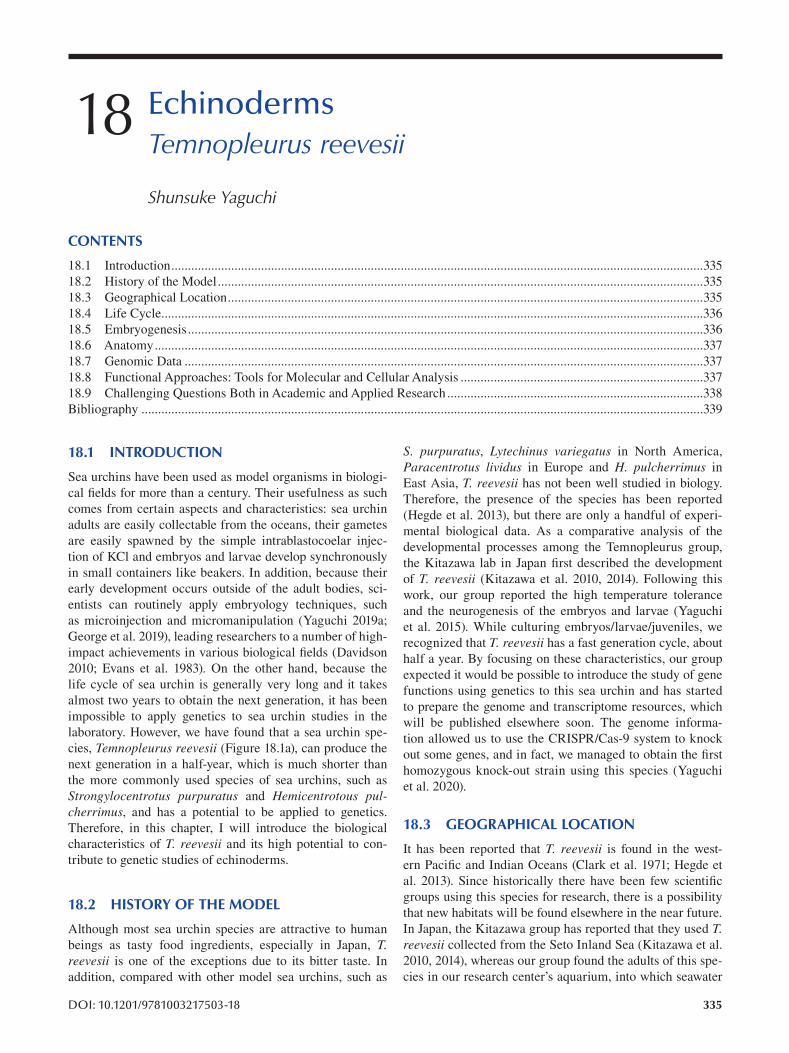
18 Echinoderms Temnopleurus reevesii
Shunsuke Yaguchi
CONTENTS
18.1 Introduction ................................................................................................................................................................335
18.2 History of the Model ..................................................................................................................................................335
18.3 Geographical Location ...............................................................................................................................................335
18.4 Life Cycle ...................................................................................................................................................................336
18.5 Embryogenesis ...........................................................................................................................................................336
18.6 Anatomy .....................................................................................................................................................................337
18.7 Genomic Data ............................................................................................................................................................337
18.8 Functional Approaches: Tools for Molecular and Cellular Analysis .........................................................................337
18.9 Challenging Questions Both in Academic and Applied Research .............................................................................338
Bibliography .........................................................................................................................................................................339
18.1 INTRODUCTION
Sea urchins have been used as model organisms in biologi
cal fields for more than a century. Their usefulness as such
comes from certain aspects and characteristics: sea urchin
adults are easily collectable from the oceans, their gametes
are easily spawned by the simple intrablastocoelar injec
tion of KCl and embryos and larvae develop synchronously
in small containers like beakers. In addition, because their
early development occurs outside of the adult bodies, sci
entists can routinely apply embryology techniques, such
as microinjection and micromanipulation (Yaguchi 2019a;
George et al. 2019), leading researchers to a number of high-
impact achievements in various biological fi elds (Davidson
2010; Evans et al. 1983). On the other hand, because the
life cycle of sea urchin is generally very long and it takes
almost two years to obtain the next generation, it has been
impossible to apply genetics to sea urchin studies in the
laboratory. However, we have found that a sea urchin spe
cies, Temnopleurus reevesii (Figure 18.1a), can produce the
next generation in a half-year, which is much shorter than
the more commonly used species of sea urchins, such as
Strongylocentrotus purpuratus and Hemicentrotous pulcherrimus, and has a potential to be applied to genetics.
Therefore, in this chapter, I will introduce the biological
characteristics of T. reevesii and its high potential to con
tribute to genetic studies of echinoderms.
18.2 HISTORY OF THE MODEL
Although most sea urchin species are attractive to human
beings as tasty food ingredients, especially in Japan, T. reevesii is one of the exceptions due to its bitter taste. In
addition, compared with other model sea urchins, such as
DOI: 10.1201/9781003217503-18
S. purpuratus, Lytechinus variegatus in North America,
Paracentrotus lividus in Europe and H. pulcherrimus in
East Asia, T. reevesii has not been well studied in biology.
Therefore, the presence of the species has been reported
(Hegde et al. 2013), but there are only a handful of experi
mental biological data. As a comparative analysis of the
developmental processes among the Temnopleurus group,
the Kitazawa lab in Japan first described the development
of T. reevesii (Kitazawa et al. 2010, 2014). Following this
work, our group reported the high temperature tolerance
and the neurogenesis of the embryos and larvae (Yaguchi
et al. 2015). While culturing embryos/larvae/juveniles, we
recognized that T. reevesii has a fast generation cycle, about
half a year. By focusing on these characteristics, our group
expected it would be possible to introduce the study of gene
functions using genetics to this sea urchin and has started
to prepare the genome and transcriptome resources, which
will be published elsewhere soon. The genome informa
tion allowed us to use the CRISPR/Cas-9 system to knock
out some genes, and in fact, we managed to obtain the fi rst
homozygous knock-out strain using this species (Yaguchi
et al. 2020).
18.3 GEOGRAPHICAL LOCATION
It has been reported that T. reevesii is found in the west
ern Pacific and Indian Oceans (Clark et al. 1971; Hegde et
al. 2013). Since historically there have been few scientifi c
groups using this species for research, there is a possibility
that new habitats will be found elsewhere in the near future.
In Japan, the Kitazawa group has reported that they used T. reevesii collected from the Seto Inland Sea (Kitazawa et al.
2010, 2014), whereas our group found the adults of this spe
cies in our research center’s aquarium, into which seawater
335
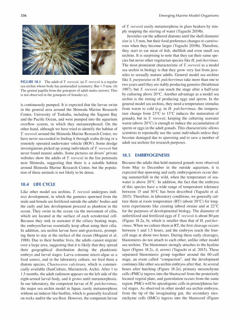
336
FIGURE 18.1 The adult of T. reevesii. (a) T. reevesii is a regular
sea urchin whose body has pentaradial symmetry. Bar = 5 mm. (b)
The genital papilla from the gonopore of adult males (arrow). This
is not observed in the gonopore of females (c).
is continuously pumped. It is expected that the larvae swim
in the general area around the Shimoda Marine Research
Center, University of Tsukuba, including the Sagami Bay
and the Pacifi c Ocean, and were pumped into the aquarium
overflow system, in which they metamorphosed. On the
other hand, although we have tried to identify the habitat of
T. reevesii around the Shimoda Marine Research Center, we
have never succeeded in finding it through scuba diving or a
remotely operated underwater vehicle (ROV). Some dredge
investigations picked up young individuals of T. reevesii but
never found mature adults. Some pictures on divers’ private
websites show the adults of T. reevesii in the Izu peninsula
near Shimoda, suggesting that there is a suitable habitat
around Shimoda Marine Research Center, but the popula
tion of these animals is not likely to be dense.
18.4 LIFE CYCLE
Like other model sea urchins, T. reevesii undergoes indi
rect development, in which the gametes spawned from the
male and female are fertilized outside the adults’ bodies and
the early and late development proceed as plankton in the
ocean. They swim in the ocean via the movement of cilia,
which are located at the surface of each ectodermal cell.
Because they sink in seawater if the ciliary beating stops,
the embryos/larvae essentially keep afloat using their cilia.
In addition, sea urchin larvae have anti-gravitaxis, prompt
ing them to stay at the surface of the ocean (Mogami et al.
1988). Due to their benthic lives, the adults cannot migrate
over a large area, suggesting that it is likely that they spread
their geographical distribution during the planktonic
embryo and larval stages. Larva consume micro-algae as a
food source, and in the laboratory culture, we feed them a
diatom species, Chaetoceros calcitrans, which is commer
cially available (SunCulture, Marinetech, Aichi). After 1 to
1.5 months, the adult rudiment appears on the left side of the
eight-armed larval body, and it grows until metamorphosis.
In our laboratory, the competent larvae of H. pulcherrimus, the major sea urchin model in Japan, rarely metamorphose
without an inducer like biofilm, which is generally localized
on rocks and/or the sea floor. However, the competent larvae
Emerging Marine Model Organisms
of T. reevesii easily metamorphose in glass beakers by sim
ply stopping the stirring of water (Yaguchi 2019b).
Juveniles eat the adhered diatoms until the shell diameter
size is 1.5 mm, but their food preference changes to carnivo
rous when they become larger (Yaguchi 2019b). Therefore,
they start to eat meat of fi sh, shellfish and even small sea
urchins. It is surprising to note that they eat their same spe
cies but never other vegetarian species like H. pulcherrimus. The most prominent characteristic of T. reevesii as a model
sea urchin in biology is that they grow very fast from juve
niles to sexually mature adults. General model sea urchins
like S. purpuratus or H. pulcherrimus take more than one to
two years until they are stably producing gametes (Strathman
1987), but T. reevesii can reach the stage after a half-year
by culturing above 20°C. Another advantage as a model sea
urchin is the timing of producing eggs and sperm. In the
general model sea urchins, they need a temperature stimulus
from warm to cold (e.g. in H. pulcherrimus , the tempera
ture change from 23°C to 13°C induces the maturation of
gonads), but in T. reevesii, keeping the culturing seawater
warm (above 20°C) is enough to induce the accumulation of
sperm or eggs in the adult gonads. This characteristic allows
scientists to repeatedly use the same individuals unless they
become damaged due to spawning and to save a number of
adult sea urchins for research purposes.
18.5 EMBRYOGENESIS
Because the adults that hold matured gonads were observed
from May to December in the outside aquarium, it is
expected that spawning and early embryogenesis occur dur
ing summer/fall in the wild, when the temperature of sea
water is above 20°C. In addition, the fact that the embryos
of this species have a wide range of temperature tolerance
between 15 and 30°C has been described (Yaguchi et al.
2015). Therefore, in laboratory conditions, we generally cul
ture them at room temperature (RT) (about 20°C) for long-
term experiments like creating inbred strains and at 22°C
for the purposes of developmental biology. The diameter of
unfertilized and fertilized eggs of T. reevesii is about 80 μm
(Figure 18.2a, b), which is smaller than that of H. pulcherrimus. When we culture them at RT, the first cleavage occurs
between 1 and 1.5 hours, and the embryos reach the four-
cell stage at about two hours. During these early cleavages,
blastomeres do not attach to each other, unlike other model
sea urchins. The blastomere strongly attaches to the hyaline
layer (Figure 18.2c, d, arrow) (Yaguchi et al. 2015). These
separated blastomeres group together around the 60-cell
stage, an event called “compaction”, and the development
continues like other sea urchin embryos after that. At several
hours after hatching (Figure 18.2e), primary mesenchyme
cells (PMCs) ingress into the blastocoel from the posteriorly
located vegetal plate, and gastrulation occurs from the same
region. PMCs will be spiculogenic cells in prism/pluteus lar
val stages. As observed in other model sea urchin embryos,
from the tip of the invaginating gut, the secondary mes
enchyme cells (SMCs) ingress into the blastocoel (Figure
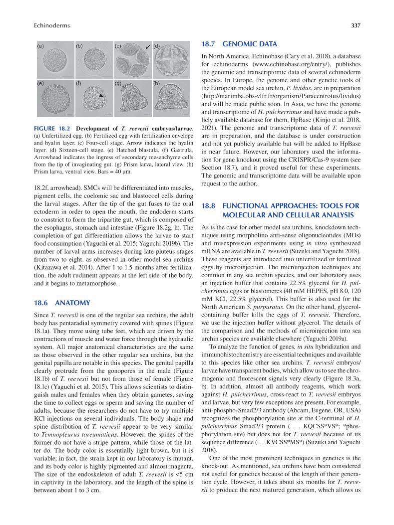
Echinoderms 337
FIGURE 18.2 Development of T. reevesii embryos/larvae.
(a) Unfertilized egg. (b) Fertilized egg with fertilization envelope
and hyalin layer. (c) Four-cell stage. Arrow indicates the hyalin
layer. (d) Sixteen-cell stage. (e) Hatched blastula. (f) Gastrula.
Arrowhead indicates the ingress of secondary mesenchyme cells
from the tip of invaginating gut. (g) Prism larva, lateral view. (h)
Prism larva, ventral view. Bars = 40 μm.
18.2f, arrowhead). SMCs will be differentiated into muscles,
pigment cells, the coelomic sac and blastocoel cells during
the larval stages. After the tip of the gut fuses to the oral
ectoderm in order to open the mouth, the endoderm starts
to constrict to form the tripartite gut, which is composed of
the esophagus, stomach and intestine (Figure 18.2g, h). The
completion of gut differentiation allows the larvae to start
food consumption (Yaguchi et al. 2015; Yaguchi 2019b). The
number of larval arms increases during late pluteus stages
from two to eight, as observed in other model sea urchins
(Kitazawa et al. 2014). After 1 to 1.5 months after fertiliza
tion, the adult rudiment appears at the left side of the body,
and it begins to metamorphose.
18.6 ANATOMY
Since T. reevesii is one of the regular sea urchins, the adult
body has pentaradial symmetry covered with spines (Figure
18.1a). They move using tube feet, which are driven by the
contractions of muscle and water force through the hydraulic
system. All major anatomical characteristics are the same
as those observed in the other regular sea urchins, but the
genital papilla are notable in this species. The genital papilla
clearly protrude from the gonopores in the male (Figure
18.1b) of T. reevesii but not from those of female (Figure
18.1c) (Yaguchi et al. 2015). This allows scientists to distin
guish males and females when they obtain gametes, saving
the time to collect eggs or sperm and saving the number of
adults, because the researchers do not have to try multiple
KCl injections on several individuals. The body shape and
spine distribution of T. reevesii appear to be very similar
to Temnopleurus toreumaticus. However, the spines of the
former do not have a stripe pattern, while those of the lat
ter do. The body color is essentially light brown, but it is
variable; in fact, the strain kept in our laboratory is mutant,
and its body color is highly pigmented and almost magenta.
The size of the endoskeleton of adult T. reevesii is <5 cm
in captivity in the laboratory, and the length of the spine is
between about 1 to 3 cm.
18.7 GENOMIC DATA
In North America, Echinobase (Cary et al. 2018), a database
for echinoderms (www.echinobase.org/entry/), publishes
the genomic and transcriptomic data of several echinoderm
species. In Europe, the genome and other genetic tools of
the European model sea urchin, P. lividus, are in preparation
(http://marimba.obs-vlfr.fr/organism/Paracentrotus/lividus)
and will be made public soon. In Asia, we have the genome
and transcriptome of H. pulcherrimus and have made a pub
licly available database for them, HpBase (Kinjo et al. 2018,
2021). The genome and transcriptome data of T. reevesii are in preparation, and the database is under construction
and not yet publicly available but will be added to HpBase
in near future. However, our laboratory used the informa
tion for gene knockout using the CRISPR/Cas-9 system (see
Section 18.7), and it proved useful for these experiments.
The genomic and transcriptome data will be available upon
request to the author.
18.8 FUNCTIONAL APPROACHES: TOOLS FOR MOLECULAR AND CELLULAR ANALYSIS
As is the case for other model sea urchins, knockdown tech
niques using morpholino anti-sense oligonucleotides (MOs)
and misexpression experiments using in vitro synthesized
mRNA are available in T. reevesii (Suzuki and Yaguchi 2018).
These reagents are introduced into unfertilized or fertilized
eggs by microinjection. The microinjection techniques are
common in any sea urchin species, and our laboratory uses
an injection buffer that contains 22.5% glycerol for H. pulcherrimus eggs or blastomeres (40 mM HEPES, pH 8.0, 120
mM KCl, 22.5% glycerol). This buffer is also used for the
North American S. purpuratus. On the other hand, glycerol-
containing buffer kills the eggs of T. reevesii. Therefore,
we use the injection buffer without glycerol. The details of
the comparison and the methods of microinjection into sea
urchin species are available elsewhere (Yaguchi 2019a).
To analyze the function of genes, in situ hybridization and
immunohistochemistry are essential techniques and available
to this species like other sea urchins. T. reevesii embryos/
larvae have transparent bodies, which allow us to see the chro
mogenic and fluorescent signals very clearly (Figure 18.3a,
b). In addition, almost all antibody reagents, which work
against H. pulcherrimus, cross-react to T. reevesii embryos
and larvae, but very few exceptions are present. For example,
anti-phospho-Smad2/3 antibody (Abcam, Eugene, OR, USA)
recognizes the phosphorylation site at the C-terminal of H. pulcherrimus Smad2/3 protein (. . . KQCSS*VS*; *phos
phorylation site) but does not for T. reevesii because of its
sequence difference (. . . KVCSS*MS*) (Suzuki and Yaguchi
2018).
One of the most prominent techniques in genetics is the
knock-out. As mentioned, sea urchins have been considered
not useful for genetics because of the length of their genera
tion cycle. However, it takes about six months for T. reevesii to produce the next matured generation, which allows us
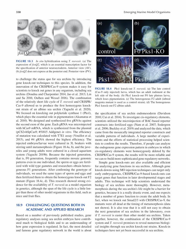
338 Emerging Marine Model Organisms
FIGURE 18.3 In situ hybridization using T. reevesii . (a) The
expression of foxQ2, which is an essential transcription factor for
the specification of anterior neuroectoderm. Anterior view (AV).
(b) foxQ2 does not express at the posterior end. Posterior view (PV).
to challenge the status quo for sea urchins by introducing
gene knock-out techniques to this species. In addition, the
innovation of the CRISPR/Cas-9 system makes it easy for
scientists to knock out genes in any organism, including sea
urchins (Doudna and Charpentier 2014; Jao et al. 2013; Lin
and Su 2016; Oulhen and Wessel 2016). The combination
of the relatively short life cycle of T. reevesii and CRISPR/
Cas-9 allowed us to produce the first homozygous knock
out strain of an albino sea urchin (Yaguchi et al. 2020).
We focused on knocking out polyketide synthase 1 (Pks1),
which plays the essential role in pigmentation (Akamatsu et
al. 2010). We designed and synthesized five gRNAs against
the second exon of the gene. Each gRNA was microinjected
with hCas9 mRNA, which is synthesized from the plasmid
(pCS2+hSpCas9; #51815 Addgene) in vitro. The effi ciency
of mutation was calculated with T7E1 assay (Vouillot et al.
2015), and #4 gRNA showed the highest effi ciency. The
injected embryos/larvae were cultured in 3L beakers with
stirring until metamorphosis (Figure 18.4a, b), and the juve
niles and young adults were cultured in a closed aquarium
system (Yaguchi 2019b). Because the injected generation,
that is, F0 generation, frequently contains mosaic genomic
patterns even in one individual, the sperm or eggs are fertil
ized with wild type gametes and researchers obtain hetero
geneous F1 generations. After confirming the genotype of
individuals, we used the same types of sperm and eggs and
then fertilized them to obtain the homozygous knock-out F2
mutant (Figure 18.4c, d). This research showed strong evi
dence for the availability of T. reevesii as a model organism
in genetics, although the span of the life cycle is a little lon
ger than those of other model organisms in this field, such as
mice and fruit fl ies.
18.9 CHALLENGING QUESTIONS BOTH IN ACADEMIC AND APPLIED RESEARCH
Based on a number of previously published studies, gene
regulatory analyses using sea urchin embryos have contrib
uted much to biological fields and to an understanding of
how gene expression is regulated. In fact, the most detailed
and famous gene regulatory network in the world is about
FIGURE 18.4 Pks1 knock-out T. reevesii. (a) The late control
(Cas-9 only injected) larva, which has an adult rudiment at the
left side of the body. (b) Pks1 knock-out F0 late pluteus larva,
which loses pigmentation. (c) The heterogenous F2 adult (inbred
magenta mutant is used as a control strain). (d) The homogenous
Pks1 knock-out F2 albino adult.
the specification of sea urchin endomesoderm (Davidson
2010; Cui et al. 2014). To investigate cis-regulatory elements,
scientists utilized the microinjection of BAC-based reporter
constructs into fertilized eggs (Nam et al. 2007; Sodergren
et al. 2006; Buckley et al. 2019) and analyzed the data, which
came from the mosaically integrated reporter constructs and
variable patterns of individuals. A large number of experi
ments and the efforts of statistical processing helped scien
tists to confirm the results. Therefore, if people can analyze
the endogenous gene expression pattern in embryos in which
cis-regulatory elements were homozygously deleted by the
CRISPR/Cas-9 system, the results will be more reliable and
we can re-build more sophisticated gene regulatory networks.
Simple gene knock-outs are also available and effi cient
for analyzing gene functions in sea urchins. Although gene
knock-downs using MO injection techniques can target only
early embryogenesis, CRISPR/Cas-9-based knock-outs can
target genes that function in later developmental stages and
adults. This technique will help scientists understand the
biology of sea urchins more thoroughly. However, meta
morphosis during the sea urchin’s life might be a barrier for
genetics, because it is a really drastic event, and one expects
that a number of genes function to create the adult body. In
fact, when we knock out Smad2/3 with CRISPR/Cas-9, the
mutants were all dead at the timing of metamorphosis (data
not shown). It is also true that it is still not easy to obtain
the next generation of sea urchins in the laboratory, even
if T. reevesii is easier than other model sea urchins. Taken
together, however, the combination of the CRISPR/Cas-9
system and T. reevesii promises to reveal numerous biologi
cal insights through sea urchin knock-out strains. Knock-in
techniques have not yet been successful in sea urchins.
339 Echinoderms
Although many sea urchin species are famous for being a
source of tasty ingredients worldwide, T. reevesii is not suitable
for food. The Japanese name of T. reevesii is “hari sanshou uni”,
and the meanings of “hari”, “sanshou” and “uni” are “spined”,
“bitter/hot” and “sea urchins”, respectively. Therefore, it is said
that T. reevesii is not good as food, and, in fact, people do not
find this species in seafood markets. However, in genetics, T. reevesii can be useful to understand gene functions related to
the taste and the size of gonads. At the same time, when com
pared with other model sea urchins which are commonly used
in food, it is a mystery why T. reevesii can grow faster. If this
question can be answered using T. reevesii, sea urchin farm
ers in the fishery industries will obtain ideas for culturing sea
urchins from the basic sciences.
BIBLIOGRAPHY
Akamatsu, H. O., Chilvers, M. I., Stewart, J. E. and Peever, T. L. 2010.
Identification and Function of a Polyketide Synthase Gene
Responsible for 1,8-Dihydroxynaphthalene-Melanin Pigment
Biosynthesis in Ascochyta rabiei. Current Genetics . 56:349–360.
Buckley, K. M. and Ettensohn, C. A. 2019. Techniques for Analyzing
Gene Expression Using BAC-Based Reporter Constructs.
Methods in Cell Biology . 151:197–218.
Cary, G. A., Cameron, R. A. and Hinman, V. F. 2018. EchinoBase:
Tools for Echinoderm Genome Analyses. Methods in Cell Biology . 151:349–369.
Clark, A. M. and Row, F. E. W. 1971. Monograph of Shallow-
Water Indo-West Pacific Echinoderms. Trustees of the British Museum of Natural History, London . 238.
Cui, M., Siriwon, N., Li, E., Davidson, E. H. and Peter, I. S. 2014.
Specific Functions of the Wnt Signaling System in Gene
Regulatory Networks throughout the Early Sea Urchin
Embryo. Proceedings of the National Academy of Sciences of the United States of America . 111:E5029–E5038.
Davidson, E. H. 2010. Emerging Properties of Animal Gene
Regulatory Networks. Nature . 468:911–920.
Doudna, J. A. and Charpentier, E. 2014. The New Frontier of
Genome Engineering with CRISPR-Cas9. Science . 346:6213.
Evans, T., Rosenthal, E. T., Youngblom, J., Distel, D. and Hunt, T.
1983. Cyclin: A Protein Specified by Maternal MRNA in Sea
Urchin Eggs That Is Destroyed at Each Cleavage Division.
Cell . 33:389–396.
George, A. N. and McClay, D. R. 2019. Methods for Transplantation
of Sea Urchin Blastomeres. Methods in Cell Biology.
151:223–233.
Hegde, M. R. and Rivonker, C. U. 2013. A New Record of
Temnopleurus decipiens (De Meijere, 1904) (Echinoidea,
Temnopleuroida, Temnopleuridae) from Indian Waters.
Zoosystema . 35:97–111.
Jao, L. E., Wente, S. R. and Chen, W. 2013. Effi cient Multiplex
Biallelic Zebrafish Genome Editing Using a CRISPR
Nuclease System. Proceedings of the National Academy of Sciences of the United States of America . 110:13904–13909.
Kinjo, S., Kiyomoto, M., Yamamoto, T., Ikeo, K. and Yaguchi,
S. 2018. HpBase: A Genome Database of a Sea Urchin,
Hemicentrotus pulcherrimus. Development Growth and Differentiation . 60(3):174–182.
Kinjo, S., Kiyomoto, M., Yamamoto, T., Ikeo, K. and Yaguchi,
S. 2021. Usage of Sea Urchin Hemicentrotus pulcherrimus Database, HpBase. Methods in Molecular Biology.
2219:267–275.
Kitazawa, C., Sakaguchi, C., Nishimura, H., Kobayashi, C., Baba,
T. and Yamanaka, A. 2014. Development of the Sea Urchins
Temnopleurus toreumaticus Leske, 1778 and Temnopleurus reevesii Gray, 1855 (Camarodonta: Temnopleuridae). Zoological Studies . 53:3.
Kitazawa, C., Tsuchihashi, Y., Egusa, Y., Genda, T. and Yamanaka,
A. 2010. Morphogenesis during Early Development in Four
Temnopleuridae Sea Urchins. Information . 13:1075–1089.
Lin, C. Y. and Su, Y. H. 2016. Genome Editing in Sea Urchin
Embryos by Using a CRISPR/Cas9 System. Developmental Biology . 409:420–428.
Mogami, B. Y. Y., Oobayashi, C. and Baba, S. A. 1988. Negative
Geotaxis in Sea Urchin Larvae: A Possible Role of
Mechanoreception in the Late Stages of Development.
Journal of Experimental Biology . 137:141–156.
Nam, J., Su, Y.-H., Lee, P. Y., Robertson, A. J., Coffman, J. A.
and Davidson, E. H. 2007. Cis-Regulatory Control of the
Nodal Gene, Initiator of the Sea Urchin Oral Ectoderm Gene
Network. Developmental Biology . 306:860–869.
Oulhen, N. and Wessel, G. M. 2016. Albinism as a Visual, In
Vivo Guide for CRISPR/Cas9 Functionality in the Sea
Urchin Embryo. Molecular Reproduction and Development. 83:1046–1047.
Sodergren, E., Weinstock, G. M., Davidson, E. H., Cameron, R. A.,
Gibbs, R. A., Angerer, R. C., Angerer, L. M., et al. 2006. The
Genome of the Sea Urchin Strongylocentrotus purpuratus.
Science . 314:5801.
Strathman, M. F. 1987. Reproduction and Development of Marine Invertebrates of the Northern Pacific Coast . The University
of Washington Press, Seatle, WA.
Suzuki, H. and Yaguchi, S. 2018. Transforming Growth Factor-β Signal Regulates Gut Bending in the Sea Urchin Embryo.
Development Growth and Differentiation . 60:216–225.
Vouillot, L., Thélie, A. and Pollet, N. 2015. Comparison of T7E1
and Surveyor Mismatch Cleavage Assays to Detect Mutations
Triggered by Engineered Nucleases. G3: Genes, Genomes, Genetics . 5:407–415.
Yaguchi, J. 2019a. Microinjection Methods for Sea Urchin Eggs
and Blastomeres. Methods in Cell Biology . 150:173–188.
Yaguchi, S. 2019b. Temnopleurus as an Emerging Echinoderm
Model. Methods in Cell Biology . 150:71–79.
Yaguchi, S., Yaguchi, J., Suzuki, H., Kinjo, S., Kiyomoto, M., Ikeo,
K. and Yamamoto, T. 2020. Establishment of Homozygous
Knock-Out Sea Urchins. Current Biology . 30:R427–R429.
Yaguchi, S., Yamazaki, A., Wada, W., Tsuchiya, Y., Sato, T.,
Shinagawa, H., Yamada, Y. and Yaguchi, J. 2015. Early
Development and Neurogenesis of Temnopleurus reevesii. Development Growth and Differentiation . 57:242–250.

19 Cephalochordates
Salvatore D’Aniello and Stéphanie Bertrand
CONTENTS
19.1 History of the Model..................................................................................................................................................341
19.2 Geographical Location .............................................................................................................................................. 342
19.3 Life Cycle .................................................................................................................................................................. 343
19.3.1 Animals in the Field..................................................................................................................................... 343
19.3.2 Animals in the Laboratory ........................................................................................................................... 343
19.4 Embryogenesis.......................................................................................................................................................... 344
19.5 Anatomy.................................................................................................................................................................... 347
19.6 Genomic Data ........................................................................................................................................................... 347
19.6.1 Branchiostoma fl oridae ............................................................................................................................... 347
19.6.2 Branchiostoma belcheri ............................................................................................................................... 347
19.6.3 Branchiostoma lanceolatum ........................................................................................................................ 348
19.6.4 Asymmetron lucayanum ............................................................................................................................... 348
19.7 Functional Approaches: Tools for Molecular and Cellular Analyses........................................................................ 348
19.8 Challenging Questions .............................................................................................................................................. 349
19.8.1 Chordate Genome and Evolution of Genomic Regulation .......................................................................... 349
19.8.2 Evolution of Vertebrate Morphological Traits ............................................................................................. 349
19.8.2.1 Cartilage and Bones..................................................................................................................... 350
19.8.2.2 Neural Crest Cells ....................................................................................................................... 350
19.8.2.3 Eyes ............................................................................................................................................. 350
19.8.3 Evolution of Cell–Cell Signaling Pathways .................................................................................................351
19.8.4 Evolution of the Immune System .................................................................................................................351
19.8.5 Evolution of Regeneration ............................................................................................................................352
Bibliography .........................................................................................................................................................................352
19.1 HISTORY OF THE MODEL Cornwall coast, UK, and classified amphioxus as a mol
lusk. In 1834, Gabriele Costa, a zoologist in Naples, Italy, Amphioxus are small, worm-like animals that resemble a
described amphioxus as a fish and hypothesized it could fish without a head or a skeleton. They live burrowed in the
represent the “missing link” between invertebrates and versand of temperate and tropical costal areas, usually at shal-
tebrates (Costa 1834). He was able to observe live animals low depths (1–50 m). Amphioxus, also called lancelets, is the
and described the oral cirri around the mouth as gills. For common name for members of the cephalochordate clade.
this reason, he gave the name Branchiostoma to the genus The first description of amphioxus came from a Chinese
(“branchio” for “gills” and “stoma” for “mouth”). In 1836, legend: Wenchang (or Wen Chang), the literature deity, was
William Yarrell, who was unfamiliar with Costa’s work traveling around the world in search of new knowledge on
but knew about the description by Pallas, proposed “lance-the back of his pet crocodile. When the crocodile died in
let” as a common name for specimens from the Cornwall the Bay of Xiamen, larva emerged from its corpse. These
coast and changed the genus name Limax, given by Pallas, “larva” were amphioxus, and even today the Chinese call
to Amphioxus (“amphi” for “both sides” and “oxus” for amphioxus “Fish of the God of Literature” or “Wenchang
“pointed”) (Yarrell 1836). Later on, the genus name became fish” (Stokes and Holland 1998; Feng et al. 2016; Holland Branchiostoma. However, Yarrell is at the origin of the two and Holland 2017 ). These animals are consumed as food in
common names of cephalochordate animals: amphioxus and some Chinese regions, although the amphioxus population
lancelet. Thereafter, many zoologists developed an interest in greatly decreased in the Bay of Xiamen during the second
amphioxus because of its proposed key evolutionary position half of the 20th century.
as a close relative of vertebrates and made in-depth descrip-While much more abundant in China than in Europe,
tions of its morphology; however, these zoologists were only the fi rst scientific description of a cephalochordate came
working with adult specimens. The first researcher who from the German zoologist and botanist Peter Simon Pallas
described amphioxus embryos was the Russian embryoloin 1774, who named it Limax lanceolatus ( Pallas 1774 ).
gist Alexander Onufrievich Kowalevsky. After his studies He could only observe two fixed adult specimens from the
DOI: 10.1201/9781003217503-19 341
342
FIGURE 19.1 Deuterostome group classifi cation. Deuterostomes
are subdivided into Ambulacraria, composed of echinoderms and
hemichordates, and chordates, which include cephalochordates and
olfactores (tunicates and vertebrates). The three cephalochordate
genera are represented in bold. The two whole genome duplications
that occurred during vertebrate evolution are also indicated. The
first took place before the divergence between gnathostomes
(jawed vertebrates) and cyclostomes (lampreys and hagfi sh),
whereas the position of the second is still debated.
in Russia and at the University of Heidelberg, Germany, he
came to Naples in 1863 and 1864 in an attempt to obtain
amphioxus embryos from local specimens (Davydoff 1960).
Kowalevsky frequently collected amphioxus and kept them
in his aquarium for months in hopes of the animals spawn
ing. Finally, in May 1864, some adult animals spawned,
and Kowalevsky was able, for the first time, to observe the
development of amphioxus embryos (Kovalevskij 1867). He
noticed that the blastula would flatten on one side that subse
quently invaginated to create two embryonic layers through
a process of gastrulation. His work was assembled in a man
uscript thanks to which he obtained his Magister degree in
St. Petersburg, Russia.
Many other zoologists became interested in amphi
oxus (Gans 1996), among whom were the famous Berthold
Hatschek (Hatschek and Tuckey 1893) and Edwin Grant
Conklin (Conklin 1932), who made many descriptions of
amphioxus embryogenesis, as well as the German naturalist
Ernst Haeckel, who wrote in the fifth edition of the book
The Evolution of Man: “We begin with the lancelet—after
man the most important and interesting of all animals. Man
is at the highest summit, the lancelet at the lowest root, of
the vertebrate stem” (Haeckel et al. 1905). However, being
extant animals, cephalochordates cannot be at the root of
vertebrates, but evolutionarily they are closely related; ceph
alochordates, together with vertebrates and their sister group
the tunicates, form the chordate clade (Figure 19.1). This
evolutionary proximity is one of the reasons many research
ers use amphioxus as a model in research.
Therefore, the study of amphioxus development and
its comparison with tunicate and vertebrate embryogen
esis allows us to define ancestral traits of chordates and to
understand the appearance of vertebrate-specifi c morpho
logical characters.
Emerging Marine Model Organisms
During the second half of the 20th century, research on
amphioxus slowed down in Europe and the United States
while flourishing in China with the species Branchiostoma belcheri (Light 1923). Among Chinese researchers, Ti Chou
Tung elegantly studied embryonic cell fate in amphioxus
using vital staining and delicate micro-manipulations, pro
viding the scientific community with important insights into
cephalochordate development (Tung et al. 1958 , 1960, 1962,
1965). Later, amphioxus entered the molecular biology era
thanks to American researchers Dr. Linda and Prof. Nicholas
Holland from the University of California, San Diego. They
began to collect adults from the species Branchiostoma fl oridae in Tampa, Florida, during the summer of 1988
and were able to obtain embryos from in vitro fertilization
and using gametes obtained by spawning induction of the
adults through electric stimulation (Holland and Holland
1989). In collaboration with Prof. Peter Holland from Oxford
University, they developed a protocol to analyze embryonic
gene expression through whole mount in situ hybridization
experiments, allowing the scientific community to renew its
interest in amphioxus as a modern model to study the evolu
tion of developmental mechanisms (Holland et al. 1992).
At the beginning of the 21st century, the development of
new sequencing techniques accompanied the transition to
whole-genome level studies for many organisms, including
amphioxus. The first whole-genome sequence was obtained
for the American species B. floridae (Putnam et al. 2008),
followed by the genome of B. belcheri (Huang et al. 2012)
and the genome and epigenome of the European species B. lanceolatum (Marletaz et al. 2018). These advances have
made amphioxus a good model not only to understand mor
phological evolution in the chordate clade through develop
mental biology approaches but also to study the evolution of
genome structure and function. Before any cephalochordate
genome was published, multigene phylogenetic studies taking
advantage of the whole genome sequencing of the tunicate
Oikopleura dioica showed that, contrary to what was glob
ally accepted in the community, tunicates, and not cephalo
chordates, are the sister group of vertebrates, with which they
form the Olfactores clade (Delsuc et al. 2006 ). Comparing
vertebrates and amphioxus thus gives us information on the
chordate ancestor that probably had characters more closely
related to those of vertebrates than previously thought!
19.2 GEOGRAPHICAL LOCATION
Cephalochordates include three genera—Branchiostoma,
Epigonichtys and Asymmetron—with around 30–40 species
described to date (Poss and Boschung 1996 ). All animals
of this chordate group are very similar morphologically, the
only major difference being that adults of the Branchiostoma genus species have two rows of gonads on both sides of the
body, whereas Asymmetron and Epigonichtys species have
only one row of gonads on the right side. Amphioxus live in
the sand of the seafloor with the anterior part of their body
sticking out of the sediment and feed by filtering the seawa
ter. Cephalochordates are widely distributed, with species
343 Cephalochordates
described along tropical and temperate coasts in sandy
sediments all around the world (Poss and Boschung 1996 ).
The precise distribution of each species is hard to defi ne,
as historically the identification of species was only based
on morphological and meristic data, which, as stated before,
are not sufficiently discriminant due to the high morphologi
cal resemblance among cephalochordates. Development of
molecular identification is rising and recently allowed sev
eral research groups to suggest the existence of more species
than previously described (Nishikawa 2004; Nohara et al.
2005; Kon et al. 2006; Kon et al. 2007; Igawa et al. 2017;
Subirana et al. 2020). Moreover, regarding Asian species,
recent studies showed that western Pacific lancelet popula
tions that were for a long period recognized as belonging
to one species, B. belcheri, belong instead to two distinct
species, B. belcheri and B. japonicum ( Zhang et al. 2006;
Li et al. 2013). Molecular phylogenetic data also allowed the
clarification of evolutionary relationships between species
and showed that Branchiostoma and Epigonichtys are more
closely related to each other than to the Asymmetron genus
(Igawa et al. 2017 ). Interestingly, although Asymmetron and
Branchiostoma diverged between 46 and 150 Mya (Igawa
et al. 2017; Subirana et al. 2020), viable hybrid embryos
from A. lucayanum and B. floridae can be obtained by in vitro fertilization (Holland et al. 2015).
19.3 LIFE CYCLE
19.3.1 ANIMALS IN THE FIELD
Amphioxus are gonochoric animals presenting a typical
bentho-pelagic life cycle. Males and females live burrowed
in the sand, and during the breeding season, they swim into
the water column just after sunset and release all their gam
etes into the environment: hundreds of oocytes are spawned
by each female, whereas males release sperm full of sperma
tozoids. After external fertilization, the embryo continues
its development protected by the fertilization envelope, also
called the chorion. Hatching occurs at the end of the gastru
lation process, and the ciliated embryo continues developing
to form a planktonic larva that moves thanks to both the
epidermal cilia and the newly formed trunk striated muscles.
The larva then metamorphoses and becomes a juvenile that
returns to a life in the sediment and reaches adulthood after
sexual maturation (Stokes and Holland 1998).
The duration and timing of the breeding season depend
on the species, as well as the speed of embryonic and post
embryonic development. In the B. floridae population of
Tampa Bay, the breeding season starts in early May and ends
at the beginning of September (Stokes and Holland 1996 ).
During this period, animals might spawn several times and
produce new gametes more or less every two weeks. In the
Mediterranean B. lanceolatum population of Argelès-sur-
Mer, France, the breeding season starts in May and ends in
July, with animals capable of spawning at least twice during
this period, although, contrary to observations made for B. fl oridae, animals from the same location do not always spawn
synchronously (Fuentes et al. 2004; Fuentes et al. 2007). The
two Asian species B. belcheri and B. japonicum can also
spawn at least twice in the field during their reproductive sea
sons, which range from May to the end of July and from late
April to late August, respectively ( Zhang et al. 2007; Li et al.
2013). Finally, the A. lucayanum population from Bimini, the
Bahamas, has two breeding periods during the year: in fall
and spring, when the water temperature is moderate and the
animals tend to spawn the same day, one or two days before
the new moon (Holland and Holland 2010).
The length of the life cycle is variable from one species to
the other: B. floridae can reach the adult stage several months
after fertilization (Stokes and Holland 1998), whereas a
whole year is needed for B. belcheri ( Zhang et al. 2007 ) and
more than two years for B. lanceolatum (Fuentes et al. 2007;
Desdevises et al. 2011).
19.3.2 ANIMALS IN THE LABORATORY
For several years now, some research groups have tried to
maintain live amphioxus in their laboratories. Two hus
bandry systems are mainly used for adults (Carvalho et al.
2017), which both consist of small tanks filled with seawater
with or without sediment that are either placed in a water
bath to stabilize the temperature or not. In both systems,
the water is changed regularly by continuous fl ow or by big
volume changes several times per day, and light is applied
in order to get a day/night cycle of 24 hours. Less regular
water changes have also been reported for inland labora
tories without access to fresh seawater (Theodosiou et al.
2011; Benito-Gutierrez et al. 2013). Adult amphioxus in the
field feed by filtering the sea water from which they ingest
all the particles less than 100 μm in diameter (Ruppert et
al. 2000). Studies of stable isotopes and feces showed that
they consume a wide variety of organisms, from bacteria to
zooplankton and phytoplankton (Chen et al. 2008; Pan et al.
2015). In the laboratory, a mixture of different algae can be
efficiently used to feed adults, although they can survive for
months without a food supply (Carvalho et al. 2017). Ripe
adults of the four main species used for evo-devo studies—
B. floridae, B. belcheri, B. japonicum and B. lanceolatum— can be induced to spawn in the laboratory in order to obtain
gametes for in vitro fertilization (Garcia-Fernàndez et al.
2009). The artificial induction of gamete release was fi rst
achieved for B. fl oridae using an electric shock, undertaken
at the time of the natural sunset on collected adults kept
with a light on (Holland and Holland 1989). However, this
method was shown to be efficient only on the days the ani
mals collected would have spawned in the fi eld. For B. lanceolatum, heat stimulation by increasing the temperature of
the water by 4°C 24 to 36 hours before the desired spawning
night can be efficiently used to induce spawning (Fuentes
et al. 2007). This technique allows working with embryos
at any desired day during the breeding season of this spe
cies. The same method has been successfully used in the
other Branchiostoma species, although with apparently less
efficiency. Interestingly, some rearing conditions allow us to
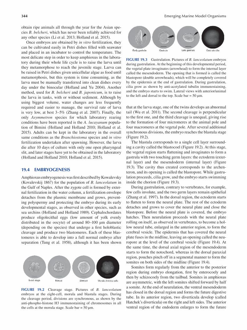
344
obtain ripe animals all through the year for the Asian spe
cies B. belcheri, which has never been reliably achieved for
any other species (Li et al. 2013; Holland et al. 2015).
Once embryos are obtained by in vitro fertilization, they
can be cultivated easily in Petri dishes filled with seawater
and placed in an incubator to control the temperature. The
most delicate step in order to keep amphioxus in the labora
tory during their whole life cycle is to raise the larva until
they metamorphose to reach the juvenile stage. Larva can
be raised in Petri dishes given unicellular algae as food until
metamorphosis, but this system is time consuming, as the
larva must be manually transferred into clean dishes every
day under the binocular (Holland and Yu 2004). Another
method, used for B. belcheri and B. japonicum, is to raise
the larva in tanks, with or without sediment. Although by
using biggest volume, water changes are less frequently
required and easier to manage, the survival rate of larva
is very low, at best 3–5% (Zhang et al. 2007). Finally, the
only Asymmetron species for which laboratory rearing
conditions have been reported is the A. lucayanum popula
tion of Bimini (Holland and Holland 2010; Holland et al.
2015). Adults can be kept in the laboratory in the overall
same conditions as the Branchiostoma species and in vitro fertilization undertaken after spawning. However, the larva
die after 10 days of culture with only one open pharyngeal
slit, and later stages have yet to be obtained in the laboratory
(Holland and Holland 2010; Holland et al. 2015).
19.4 EMBRYOGENESIS
Amphioxus embryogenesis was first described by Kowalevsky
(Kovalevskij 1867) for the population of B. lanceolatum in
the Gulf of Naples. After the zygote cell is formed by exter
nal fertilization in the water column, a fertilization envelope
detaches from the plasmic membrane and grows, prevent
ing polyspermy and protecting the embryo during its early
developmental stages, as observed in other species, such as
sea urchins (Holland and Holland 1989). Cephalochordates
produce oligolecithal eggs (low amount of yolk evenly
distributed in the oocyte) of around 80–100 μm diameter
(depending on the species) that undergo a fi rst holoblastic
cleavage and produce two blastomeres. Each of these blas
tomeres is able to develop into a full normal embryo after
separation (Tung et al. 1958), although it has been shown
FIGURE 19.2 Cleavage stage. Pictures of B. lanceolatum embryos at the eight-cell, morula and blastula stages. During
the cleavage period, divisions are synchronous, as shown by the
anti-phospho-histone H3 immunostaining of chromosomes in all
the cells at the morula stage. Scale bar = 50 μm.
Emerging Marine Model Organisms
FIGURE 19.3 Gastrulation. Pictures of B. lanceolatum embryos
during gastrulation. At the beginning of this developmental period,
the vegetal plate invaginates (arrowhead) to form the internal layer
called the mesendoderm. The opening that is formed is called the
blastopore (double arrowheads), which will be completely covered
by the epidermis at the end of gastrulation. During gastrulation,
cilia grow as shown by anti-acetylated tubulin immunostaining,
and the embryo starts to swim. Lateral views with anterior/animal
to the left and dorsal to the top. Scale bar = 50 μm.
that at the larva stage, one of the twins develops an abnormal
tail (Wu et al. 2011). The second cleavage is perpendicular
to the first one, and the third cleavage is unequal, giving rise
to the formation of four micromeres at the animal pole and
four macromeres at the vegetal pole. After several additional
synchronous divisions, the embryo reaches the blastula stage
( Figure 19.2 ).
The blastula corresponds to a single cell layer surround
ing a cavity called the blastocoel (Figure 19.2). At this stage,
the vegetal region starts flattening and invaginates to form a
gastrula with two touching germ layers: the ectoderm (exter
nal layer) and the mesendoderm (internal layer) (Figure
19.3). The cavity thus created corresponds to the archen
teron, and its opening is called the blastopore. While gastru
lation proceeds, cilia grow, and the embryo starts swimming
inside the chorion (Figure 19.3).
During gastrulation, contrary to vertebrates, for example,
few cells involute, and the two germ layers remain epithelial
( Zhang et al. 1997 ). In the dorsal region, the ectoderm starts
to flatten to form the neural plate. The rest of the ectoderm
detaches and grows to cover the neural plate and close the
blastopore. Before the neural plate is covered, the embryo
hatches. Then neurulation proceeds with the neural plate
rolling on itself, as observed in vertebrates, to become a hol
low neural tube, enlarged in the anterior region, to form the
cerebral vesicle. The epidermis that has covered the neural
plate fuses in the midline, leaving an opening called the neu
ropore at the level of the cerebral vesicle (Figure 19.4). At
the same time, the dorsal axial region of the mesendoderm
starts to form the notochord, whereas in the dorsal paraxial
region, pouches pinch off in a segmental manner to form the
somites on both sides of the midline (Figure 19.4).
Somites form regularly from the anterior to the posterior
region during embryo elongation, first by enterocoely and
then by schizocoely from the tailbud. Somites in amphioxus
are asymmetric, with the left somites shifted forward by half
a somite. At the end of neurulation, the ventral mesendoderm
has closed in the dorsal region and forms the future digestive
tube. In its anterior region, two diverticula develop (called
Hatchek’s diverticula) on the right and left sides. The anterior
ventral region of the endoderm enlarges to form the future
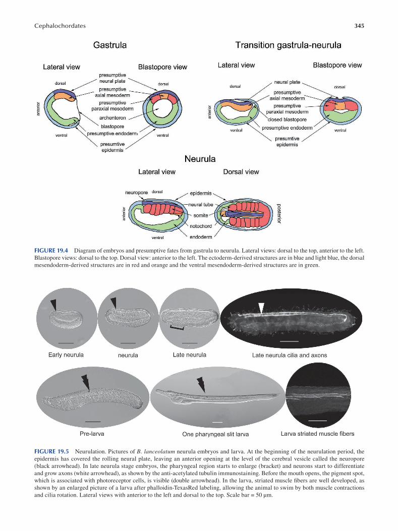
345 Cephalochordates
FIGURE 19.4 Diagram of embryos and presumptive fates from gastrula to neurula. Lateral views: dorsal to the top, anterior to the left.
Blastopore views: dorsal to the top. Dorsal view: anterior to the left. The ectoderm-derived structures are in blue and light blue, the dorsal
mesendoderm-derived structures are in red and orange and the ventral mesendoderm-derived structures are in green.
FIGURE 19.5 Neurulation. Pictures of B. lanceolatum neurula embryos and larva. At the beginning of the neurulation period, the
epidermis has covered the rolling neural plate, leaving an anterior opening at the level of the cerebral vesicle called the neuropore
(black arrowhead). In late neurula stage embryos, the pharyngeal region starts to enlarge (bracket) and neurons start to differentiate
and grow axons (white arrowhead), as shown by the anti-acetylated tubulin immunostaining. Before the mouth opens, the pigment spot,
which is associated with photoreceptor cells, is visible (double arrowhead). In the larva, striated muscle fibers are well developed, as
shown by an enlarged picture of a larva after phalloidin-TexasRed labeling, allowing the animal to swim by both muscle contractions
and cilia rotation. Lateral views with anterior to the left and dorsal to the top. Scale bar = 50 μm.
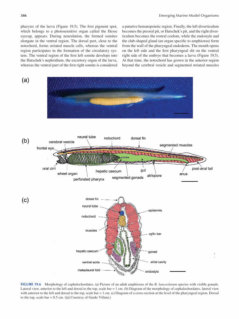
346
pharynx of the larva (Figure 19.5). The first pigment spot,
which belongs to a photosensitive organ called the Hesse
eyecup, appears. During neurulation, the formed somites
elongate in the ventral region. The dorsal part, close to the
notochord, forms striated muscle cells, whereas the ventral
region participates in the formation of the circulatory sys
tem. The ventral region of the first left somite develops into
the Hatschek’s nephridium, the excretory organ of the larva,
whereas the ventral part of the first right somite is considered
Emerging Marine Model Organisms
a putative hematopoietic region. Finally, the left diverticulum
becomes the preoral pit, or Hatschek’s pit, and the right diver
ticulum becomes the rostral coelom, while the endostyle and
the club-shaped gland (an organ specific to amphioxus) form
from the wall of the pharyngeal endoderm. The mouth opens
on the left side and the first pharyngeal slit on the ventral
right side of the embryo that becomes a larva (Figure 19.5).
At that time, the notochord has grown in the anterior region
beyond the cerebral vesicle and segmented striated muscles
FIGURE 19.6 Morphology of cephalochordates. (a) Picture of an adult amphioxus of the B. lanceolatum species with visible gonads.
Lateral view, anterior to the left and dorsal to the top, scale bar = 1 cm. (b) Diagram of the morphology of cephalochordates, lateral view
with anterior to the left and dorsal to the top, scale bar = 1 cm. (c) Diagram of a cross-section at the level of the pharyngeal region. Dorsal
to the top, scale bar = 0.5 cm. ([a] Courtesy of Guido Villani.)
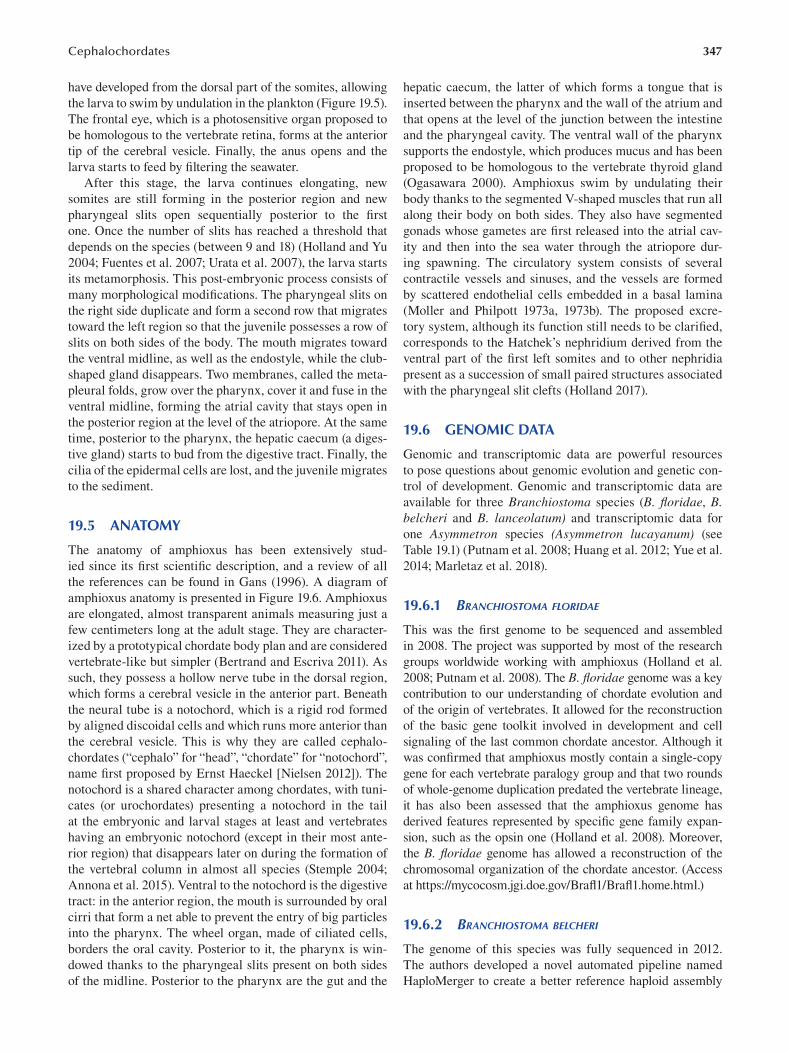
347 Cephalochordates
have developed from the dorsal part of the somites, allowing
the larva to swim by undulation in the plankton (Figure 19.5).
The frontal eye, which is a photosensitive organ proposed to
be homologous to the vertebrate retina, forms at the anterior
tip of the cerebral vesicle. Finally, the anus opens and the
larva starts to feed by filtering the seawater.
After this stage, the larva continues elongating, new
somites are still forming in the posterior region and new
pharyngeal slits open sequentially posterior to the fi rst
one. Once the number of slits has reached a threshold that
depends on the species (between 9 and 18) (Holland and Yu
2004; Fuentes et al. 2007; Urata et al. 2007), the larva starts
its metamorphosis. This post-embryonic process consists of
many morphological modifications. The pharyngeal slits on
the right side duplicate and form a second row that migrates
toward the left region so that the juvenile possesses a row of
slits on both sides of the body. The mouth migrates toward
the ventral midline, as well as the endostyle, while the club-
shaped gland disappears. Two membranes, called the meta
pleural folds, grow over the pharynx, cover it and fuse in the
ventral midline, forming the atrial cavity that stays open in
the posterior region at the level of the atriopore. At the same
time, posterior to the pharynx, the hepatic caecum (a diges
tive gland) starts to bud from the digestive tract. Finally, the
cilia of the epidermal cells are lost, and the juvenile migrates
to the sediment.
19.5 ANATOMY
The anatomy of amphioxus has been extensively stud
ied since its fi rst scientific description, and a review of all
the references can be found in Gans (1996 ). A diagram of
amphioxus anatomy is presented in Figure 19.6. Amphioxus
are elongated, almost transparent animals measuring just a
few centimeters long at the adult stage. They are character
ized by a prototypical chordate body plan and are considered
vertebrate-like but simpler (Bertrand and Escriva 2011). As
such, they possess a hollow nerve tube in the dorsal region,
which forms a cerebral vesicle in the anterior part. Beneath
the neural tube is a notochord, which is a rigid rod formed
by aligned discoidal cells and which runs more anterior than
the cerebral vesicle. This is why they are called cephalo
chordates (“cephalo” for “head”, “chordate” for “notochord”,
name first proposed by Ernst Haeckel [Nielsen 2012]). The
notochord is a shared character among chordates, with tuni
cates (or urochordates) presenting a notochord in the tail
at the embryonic and larval stages at least and vertebrates
having an embryonic notochord (except in their most ante
rior region) that disappears later on during the formation of
the vertebral column in almost all species (Stemple 2004;
Annona et al. 2015). Ventral to the notochord is the digestive
tract: in the anterior region, the mouth is surrounded by oral
cirri that form a net able to prevent the entry of big particles
into the pharynx. The wheel organ, made of ciliated cells,
borders the oral cavity. Posterior to it, the pharynx is win
dowed thanks to the pharyngeal slits present on both sides
of the midline. Posterior to the pharynx are the gut and the
hepatic caecum, the latter of which forms a tongue that is
inserted between the pharynx and the wall of the atrium and
that opens at the level of the junction between the intestine
and the pharyngeal cavity. The ventral wall of the pharynx
supports the endostyle, which produces mucus and has been
proposed to be homologous to the vertebrate thyroid gland
(Ogasawara 2000). Amphioxus swim by undulating their
body thanks to the segmented V-shaped muscles that run all
along their body on both sides. They also have segmented
gonads whose gametes are first released into the atrial cav
ity and then into the sea water through the atriopore dur
ing spawning. The circulatory system consists of several
contractile vessels and sinuses, and the vessels are formed
by scattered endothelial cells embedded in a basal lamina
(Moller and Philpott 1973a, 1973b). The proposed excre
tory system, although its function still needs to be clarifi ed,
corresponds to the Hatchek’s nephridium derived from the
ventral part of the first left somites and to other nephridia
present as a succession of small paired structures associated
with the pharyngeal slit clefts (Holland 2017).
19.6 GENOMIC DATA
Genomic and transcriptomic data are powerful resources
to pose questions about genomic evolution and genetic con
trol of development. Genomic and transcriptomic data are
available for three Branchiostoma species (B. floridae, B. belcheri and B. lanceolatum) and transcriptomic data for
one Asymmetron species (Asymmetron lucayanum) (see
Table 19.1) (Putnam et al. 2008; Huang et al. 2012; Yue et al.
2014; Marletaz et al. 2018).
19.6.1 BRANCHIOSTOMA FLORIDAE
This was the first genome to be sequenced and assembled
in 2008. The project was supported by most of the research
groups worldwide working with amphioxus (Holland et al.
2008; Putnam et al. 2008). The B. floridae genome was a key
contribution to our understanding of chordate evolution and
of the origin of vertebrates. It allowed for the reconstruction
of the basic gene toolkit involved in development and cell
signaling of the last common chordate ancestor. Although it
was confirmed that amphioxus mostly contain a single-copy
gene for each vertebrate paralogy group and that two rounds
of whole-genome duplication predated the vertebrate lineage,
it has also been assessed that the amphioxus genome has
derived features represented by specific gene family expan
sion, such as the opsin one (Holland et al. 2008). Moreover,
the B. floridae genome has allowed a reconstruction of the
chromosomal organization of the chordate ancestor. (Access
at https://mycocosm.jgi.doe.gov/Brafl 1/Brafl 1.home.html.)
19.6.2 BRANCHIOSTOMA BELCHERI
The genome of this species was fully sequenced in 2012.
The authors developed a novel automated pipeline named
HaploMerger to create a better reference haploid assembly
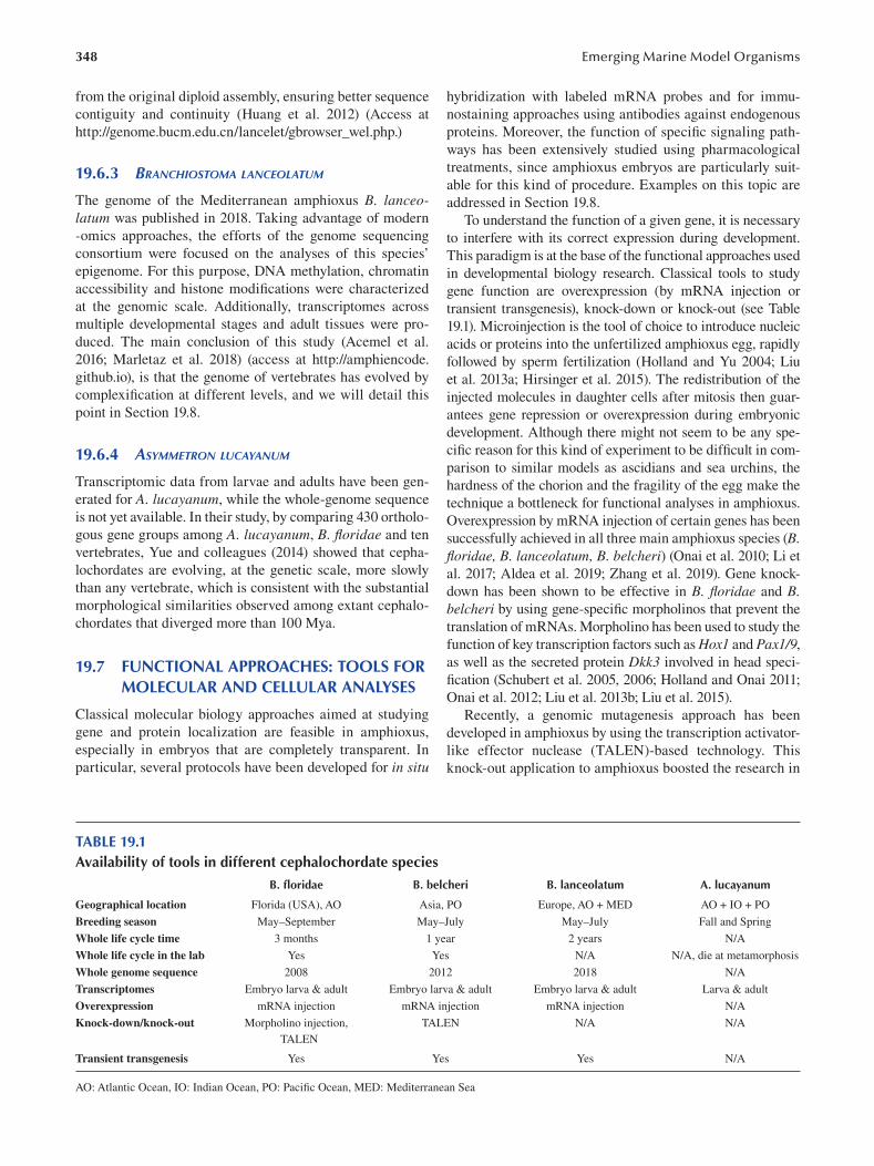
348
from the original diploid assembly, ensuring better sequence
contiguity and continuity (Huang et al. 2012) (Access at
http://genome.bucm.edu.cn/lancelet/gbrowser_wel.php. )
19.6.3 BRANCHIOSTOMA LANCEOLATUM
The genome of the Mediterranean amphioxus B. lanceolatum was published in 2018. Taking advantage of modern
-omics approaches, the efforts of the genome sequencing
consortium were focused on the analyses of this species’
epigenome. For this purpose, DNA methylation, chromatin
accessibility and histone modifications were characterized
at the genomic scale. Additionally, transcriptomes across
multiple developmental stages and adult tissues were pro
duced. The main conclusion of this study (Acemel et al.
2016; Marletaz et al. 2018) (access at http://amphiencode.
github.io), is that the genome of vertebrates has evolved by
complexification at different levels, and we will detail this
point in Section 19.8.
19.6.4 ASYMMETRON LUCAYANUM
Transcriptomic data from larvae and adults have been gen
erated for A. lucayanum, while the whole-genome sequence
is not yet available. In their study, by comparing 430 ortholo
gous gene groups among A. lucayanum, B. floridae and ten
vertebrates, Yue and colleagues (2014) showed that cepha
lochordates are evolving, at the genetic scale, more slowly
than any vertebrate, which is consistent with the substantial
morphological similarities observed among extant cephalo
chordates that diverged more than 100 Mya.
19.7 FUNCTIONAL APPROACHES: TOOLS FOR MOLECULAR AND CELLULAR ANALYSES
Classical molecular biology approaches aimed at studying
gene and protein localization are feasible in amphioxus,
especially in embryos that are completely transparent. In
particular, several protocols have been developed for in situ
Emerging Marine Model Organisms
hybridization with labeled mRNA probes and for immu
nostaining approaches using antibodies against endogenous
proteins. Moreover, the function of specific signaling path
ways has been extensively studied using pharmacological
treatments, since amphioxus embryos are particularly suit
able for this kind of procedure. Examples on this topic are
addressed in Section 19.8.
To understand the function of a given gene, it is necessary
to interfere with its correct expression during development.
This paradigm is at the base of the functional approaches used
in developmental biology research. Classical tools to study
gene function are overexpression (by mRNA injection or
transient transgenesis), knock-down or knock-out (see Table
19.1). Microinjection is the tool of choice to introduce nucleic
acids or proteins into the unfertilized amphioxus egg, rapidly
followed by sperm fertilization (Holland and Yu 2004; Liu
et al. 2013a ; Hirsinger et al. 2015). The redistribution of the
injected molecules in daughter cells after mitosis then guar
antees gene repression or overexpression during embryonic
development. Although there might not seem to be any spe
cific reason for this kind of experiment to be difficult in com
parison to similar models as ascidians and sea urchins, the
hardness of the chorion and the fragility of the egg make the
technique a bottleneck for functional analyses in amphioxus.
Overexpression by mRNA injection of certain genes has been
successfully achieved in all three main amphioxus species (B. floridae, B. lanceolatum, B. belcheri) (Onai et al. 2010; Li et
al. 2017; Aldea et al. 2019; Zhang et al. 2019). Gene knock
down has been shown to be effective in B. floridae and B. belcheri by using gene-specific morpholinos that prevent the
translation of mRNAs. Morpholino has been used to study the
function of key transcription factors such as Hox1 and Pax1/9,
as well as the secreted protein Dkk3 involved in head speci
fication (Schubert et al. 2005, 2006; Holland and Onai 2011;
Onai et al. 2012; Liu et al. 2013b; Liu et al. 2015).
Recently, a genomic mutagenesis approach has been
developed in amphioxus by using the transcription activator-
like effector nuclease (TALEN)-based technology. This
knock-out application to amphioxus boosted the research in
TABLE 19.1 Availability of tools in different cephalochordate species
B. floridae B. belcheri B. lanceolatum A. lucayanum
Geographical location Florida (USA), AO Asia, PO Europe, AO + MED AO + IO + PO
Breeding season May–September May–July May–July Fall and Spring
Whole life cycle time 3 months 1 year 2 years N/A
Whole life cycle in the lab Yes Yes N/A N/A, die at metamorphosis
Whole genome sequence 2008 2012 2018 N/A
Transcriptomes Embryo larva & adult Embryo larva & adult Embryo larva & adult Larva & adult
Overexpression mRNA injection mRNA injection mRNA injection N/A
Knock-down/knock-out Morpholino injection, TALEN N/A N/A
TALEN
Transient transgenesis Yes Yes Yes N/A
AO: Atlantic Ocean, IO: Indian Ocean, PO: Pacific Ocean, MED: Mediterranean Sea
349 Cephalochordates
the developmental biology field and filled the gaps with other
chordate models (Li et al. 2014). Germ line mutagenesis has
been used to study several important developmental genes,
such as Pax1/9, Pax3/7, two ParaHox genes: Pdx and Cdx,
Hedgehog, Cerberus and Nodal (Li et al. 2014; Wang et al.
2015; Hu et al. 2017; Li et al. 2017; Ren et al. 2020; Zhong et
al. 2020; Zhu et al. 2020). Nevertheless, the long life cycle
of amphioxus make these and other similar approaches very
time consuming; this is the reason the tropical species B. fl oridae is more suitable than temperate species such as B. lanceolatum, which takes a few years to reach sexual matu
rity. It is foreseeable that in the next few years, gene function
studies in amphioxus will also take advantage of the genome
editing CRISPR/Cas9 (clustered regularly interspaced short
palindromic repeats/Cas9) technique that represents the
next-generation genome editing tool and provides high lev
els of gene-specific targeting and effi ciency.
An efficient transgenic method to study enhancer activity
has been recently developed for amphioxus: two transgenic
amphioxus lines have been generated using the Tol2 transpo
son system, based on a hAT family transposon (Shi et al. 2018).
None of these functional approaches have been success
fully developed in the Asymmetron genus, probably because
only a few labs have access to live animals.
19.8 CHALLENGING QUESTIONS
Over the last decades, cephalochordates have become impor
tant animal models in the field of evo-devo. The phyloge
netic position of amphioxus and its evolutionarily conserved
morphology and genome organization make it an extremely
useful organism for answering important evolutionary biol
ogy questions, in particular with respect to chordate evo
lutionary history. This section describes some important
results obtained using amphioxus as a model as well as key
questions for which the full answer is still to be found.
19.8.1 CHORDATE GENOME AND EVOLUTION
OF GENOMIC REGULATION
In the 1970s Susumu Ohno, a Japanese-American geneticist,
proposed in his book Evolution by Gene Duplication that
morphological novelties could result from gene duplications
and that vertebrate genomes were built by one or probably two
whole-genome duplications that took place during the inver
tebrate chordate to vertebrate transition (Ohno 1970). This
hypothesis is named the 2R (for two rounds of duplication)
hypothesis. Researchers have long tried to test this assump
tion using several arguments, such as the number of isozymes,
the number of genes or the number of paralogues in verte
brates versus invertebrates. For example, it was shown that
amphioxus has a single complete Hox gene cluster containing
15 genes, whereas mammals have four incomplete clusters
(Amemiya et al. 2008; Putnam et al. 2008). The defi nitive
argument for the 2R hypothesis came with the sequencing of
the whole genome of the tunicate Ciona intestinalis and was
confirmed by the sequencing of amphioxus’s genome (Dehal
and Boore 2005; Putnam et al. 2008). Cephalochordates,
therefore, have an unduplicated genome compared to ver
tebrates, and it has been shown that, unlike tunicates, they
have retained most of the genes present in the chordate ances
tor genome, although some lineage-specifi c duplications
occurred in several gene families (Holland et al. 2008). The
cephalochordate genome thus represents the best proxy for
the chordate ancestor genome, and analyses of B. floridae data allowed the reconstruction of the gene complement of the
last common chordate ancestor and the partial reconstruction
of its genomic organization (Holland et al. 2008).
Although the evolution of gene content during chordate
evolution was probably crucial for their morphological
diversification, the contribution of genome architecture
and genome regulation is still to be finely studied. In this
context, the recent description of the epigenome of the
Mediterranean amphioxus, B. lanceolatum, already brought
new insights. The characterization of the methylome, of
chromatin accessibility and of histone modifi cations at
different development stages and in several adult tissues
allowed for the discovery of some functional changes that
might have given rise to the greater complexity observed
in vertebrates (Marletaz et al. 2018). For example, in verte
brates, there has been an increase in regulatory sequences,
in particular those that regulate the expression of genes
involved in the control of embryonic development. It was
also shown that duplicate genes in vertebrates (after the 2R)
have evolved mainly by subfunctionalization and specializa
tion and that specialization of gene function was accompa
nied by an increase in regulatory complexity. Another study,
focused on the Hox genomic region, showed that the com
plex regulation of Hox genes expression in vertebrate is in
part due to the acquisition of a new three-dimensional orga
nization of the chromatin around some of the Hox clusters
(Acemel et al. 2016). Indeed, the amphioxus Hox gene clus
ter is contained in a single topologically associated domain
(TAD), while in vertebrates, there are two TADs, one on
each side of the cluster, and regulatory sequences present
in these two TADs are responsible for the regulation of Hox
genes expression in the limbs. This study of the B. lanceolatum genome also showed that although amphioxus pres
ents a similar pattern of methylation to that of invertebrates
(low methylation compared to vertebrates), the expression of
some genes is regulated by demethylation in the same way
as vertebrates (Marletaz et al. 2018). These recent data pave
the way for a better understanding of the genomic regula
tion principles underlying the morphological and functional
innovations of vertebrates. Nevertheless, further effort is
necessary to overcome difficulties associated with enhancer
element identifi cation and understanding of their functional
evolution throughout the last 500 million years.
19.8.2 EVOLUTION OF VERTEBRATE
MORPHOLOGICAL TRAITS
Although amphioxus share a typical chordate body plan with
vertebrates, they lack key vertebrate characters such as the
350
head, endoskeleton, migratory neural crest cells, placodes
and paired appendages. Therefore, a comparative approach
between invertebrate chordates and vertebrates should allow
us to discover the main evolutionary innovations that led to the
appearance of these complex structures, and amphioxus has
been extensively used to answer such questions. In this section,
as an example, the contribution of some studies using cephalo
chordates as a model for our understanding of the evolution of
key vertebrate morphological features will be addressed.
19.8.2.1 Cartilage and Bones One of the most iconic and specific structures of extant ver
tebrates is their endoskeleton made of cartilage and/or bone
that is absent in tunicates and cephalochordates. However,
in amphioxus, cartilage-like structures are found at the adult
stage in the rods of the cirri that surround the mouth, which
consist of cells embedded in a matrix, and in the “gill” bars
of the pharynx, which were described as an acellular car
tilage (Wright et al. 2001). Although it was proposed that
both cartilage-like tissues were non-collagenous (Wright
et al. 2001), it has been shown that fibrillar collagen, which
is a major component of the cartilage matrix in vertebrates,
is present in the pharyngeal “gill” bars (Rychel and Swalla
2007 ). In search of a conserved gene toolkit for cartilage
formation, the expression pattern of amphioxus orthologues
of genes controlling cartilage formation in vertebrates has
been studied during embryogenesis. No co-expression
could be observed, suggesting that cartilage did not appear
by co-option of a pre-existing toolkit but probably by the
appearance of new gene interactions (Meulemans and
Bronner-Fraser 2007). However, these studies were carried
out on embryos and not at later stages when the cartilage-
like structures form (during metamorphosis). More recent
studies using metamorphosing B. floridae larvae or regen
erating oral cirri in adults have brought new insights on this
issue (Kaneto and Wada 2011; Jandzik et al. 2015). It has
been shown that during metamorphosis, ColA, coding for
a collagen in amphioxus, is expressed in the forming oral
cirri and in regenerating adult oral cirri as well as transcrip
tion factors required for cartilage formation in vertebrates
(Kaneto and Wada 2011; Jandzik et al. 2015). The authors
also showed that oral cirri formation is dependent upon FGF
signaling, a signal which is required in vertebrates for cellu
lar cartilage differentiation, and that adult regenerating cirri
rods are expressing genes that are known to be required for
osteogenesis in vertebrates (Kaneto and Wada 2011; Jandzik
et al. 2015). All together, these data have shown that some
elements of the chondrogenic and osteogenic programs of
vertebrates were probably already required for the forma
tion of cartilage-like structures in the chordate ancestor.
However, more functional data, particularly focusing on
amphioxus metamorphosis, are still required to understand
the appearance of the vertebrate endoskeleton.
19.8.2.2 Neural Crest Cells The neural crest cells (NCCs) are a specific transient popu
lation of cells specific to vertebrates that are sometimes
Emerging Marine Model Organisms
referred to as the “fourth germ layer” of these animals
(Gilbert 2000). They originate from the border of the neu
ral plate at the time at which the neuroectoderm and the
future epidermis separate during neurulation (Gilbert 2000).
These cells undergo an epithelial-mesenchymal transition,
delaminate and migrate all through the body where they
differentiate into many different cell types such as melano
cytes, adipocytes, neurons, smooth muscles, chondroblasts,
odontoblasts and so on (Bronner and Simoes-Costa 2016).
NCCs participate in the formation of structures that are
vertebrate specific such as bones, cartilage and ganglia of
the vertebrate head, and Gans and Northcutt even proposed
that the vertebrates’ “New Head” (an anterior structure with
unsegmented muscles, well-developed brain and sensory
organs) appearance was favored by the emergence of NCCs
(Gans and Northcutt 1983). In amphioxus, there is no evi
dence of the existence of such cells, and it is considered that
cephalochordates do not have migratory NCCs. However,
neurulation occurs in a similar way as observed in verte
brates, and it has been shown that the neural plate border
expresses genes that are orthologues of neural plate border
specification genes in vertebrates (Yu et al. 2008). On the
other hand, among the genes that are known to be required
in vertebrates for the specification of NCC or among effec
tor genes (that are downstream of the neural plate border
specifying genes in the NCC gene regulatory network), only
Snail is expressed in the neural plate border of amphioxus
(Langeland et al. 1998). Concerning tunicates, the sister
group of vertebrates, it has been shown in Ciona intestinalis that some cells expressing the NCC specifi cation genes
Id, Snail, FoxD and Ets differentiate into pigmented cells
and that overexpression of Twist in these cells induces them
to migrate (Abitua et al. 2012), suggesting that NCC would
have appeared thanks to the recruitment of a “migratory”
program at the neural plate border. However, tunicates have
specific developmental modalities among chordates, and
cephalochordates seem, at least during early embryogenesis,
to develop most of their structures without any step of epi
thelial-mesenchymal transition, leaving the mystery of NCC
emergence still incompletely resolved.
19.8.2.3 Eyes Among the characters specific to vertebrates, the well-devel
oped pair sensory organs are the most elaborate. The image-
forming camera-type eye of vertebrates is a very complex
structure composed of different tissues with various embry
onic origins. Amphioxus, on the other hand, possess various
photoreceptive organs: the lamellar body, Joseph cells, dor
sal ocelli and the frontal eye, which is considered homolo
gous to the vertebrate retina (Glardon et al. 1998; Pergner
and Kozmik 2017). This very simple organ is formed at the
larva stage at the tip of the cerebral vesicle, which is consid
ered homologous to the vertebrate brain. The frontal eye con
sists of around six photoreceptor cells (Lacalli et al. 1994)
of the ciliary type, like the cones and rods of the vertebrate
retina, positioned posterior to nine pigment cells (Lacalli
et al. 1994). The amphioxus photoreceptors and pigment
351 Cephalochordates
cells express genes that are orthologous to genes known to
be expressed in the photoreceptor cells and pigmented epi
thelium of the vertebrate retina, respectively (Vopalensky et
al. 2012). Interestingly, other neurons positioned posterior to
the row of photoreceptors were proposed to be homologous
to the other cell types present in the vertebrate retina: inter
neurons and/or retinal ganglion cells (Lacalli et al. 1994;
Lacalli 1996; Vopalensky et al. 2012). However, data are
still missing in order to clearly answer this point. Another
important aspect that would support the homology between
the amphioxus frontal eye and vertebrate retina is the under
standing of the developmental control of frontal eye forma
tion. A recent study showed that, as in vertebrate embryos,
inhibiting the Notch signaling pathway during amphioxus
embryogenesis increases the number of photoreceptors
formed (Pergner et al. 2020), but we are far from a com
plete understanding of the gene regulatory network underly
ing the formation of the frontal eye. Another key point that
needs to be addressed is how vertebrate paired eyes evolved
from a single, midline-positioned ancestral eye.
19.8.3 EVOLUTION OF CELL–CELL SIGNALING PATHWAYS
Harmonious embryonic development relies on the capabili
ties of cells to communicate in order to construct the correct
body plan. For this purpose, they use few signaling path
ways, most of them being present in all metazoans (Barolo
and Posakony 2002). One important question in the evo-devo
field is therefore to understand how the evolution of these
pathways (of their actors, roles and interactions) might have
participated in the morphological diversification among ani
mals. Amphioxus possess in their genome genes that code
for the main actors of all the major signaling pathways, often
with one orthologue for several paralogues in vertebrates
that resulted from the two whole-genome duplications char
acterizing vertebrate early evolutionary history (Bertrand
et al. 2017). One major issue that needs to be solved is how
the multiplication of signaling pathway actors in vertebrates
lead to the appearance of their morphological characters.
There are still few data concerning this point, but we can
cite the case of the retinoic acid receptors (RARs). This
transcription factor, which is a nuclear receptor of retinoic
acid, is encoded by a unique gene in amphioxus, whereas
three paralogues, RARα, β and γ, are found in mammals. By comparing the expression pattern, the function and the binding capacity of vertebrate and amphioxus RARs, it has been proposed that RARβ kept chordate ancestral charac-teristics, whereas RARα and RARγ acquired new roles (i.e.
neofunctionalization) during vertebrate evolution, which
might explain the embryonic functions of retinoic acid that
are specific to vertebrates (Escriva et al. 2006).
In cephalochordates, the developmental function of many
cell–cell communication pathways has been studied mainly
thanks to pharmacological treatments capable of inhibiting
or activating these signals (for a review, see Bertrand et al.
2017). One of the advantages of using such an approach is the
possibility to interfere with signaling pathways at different
developmental time windows and therefore to study their
implication in diverse developmental processes. Many data
obtained in amphioxus have highlighted conservation in
the use of different signals for the control of developmental
processes with vertebrates, as might be expected given that
chordates share a similar body plan. As an example, BMP
and Nodal are opposing signals controlling the dorso–ven
tral patterning of the amphioxus embryo (Onai et al. 2010),
the Wnt/β-catenin pathway regulates the formation of the
dorsal organizer (Kozmikova and Kozmik 2020) and reti
noic acid has been shown to act as a posteriorizing signal and
to control the expression of Hox genes (Holland and Holland
1996; Escriva et al. 2002; Schubert et al. 2005), as is the case
in vertebrates. However, we can point out some studies that
reveal differences between amphioxus and vertebrates that
might explain the emergence of some vertebrate novelties.
In vertebrates, the somitogenesis process, which consists of
the progressive segmentation of the paraxial mesoderm of
the trunk during the embryo elongation (Pourquie 2001b),
relies on the opposition of two main signals: the retinoic acid
differentiating signal in the anterior region that acts in oppo
sition to the fibroblast growth factor (FGF) and Wnt pos
terior proliferative signals (Pourquie 2001a). In amphioxus,
the paraxial mesoderm gets segmented through a similar
somitogenesis process, although it is also segmented in the
anterior/head region, contrary to what happens in verte
brates. Interestingly, it has been shown in amphioxus that
FGF controls only the formation of the anterior somites, that
retinoic acid is not involved in this process and that FGF
and retinoic acid do not seem to regulate each other during
embryogenesis (Bertrand et al. 2011; Bertrand et al. 2015).
These results might in part explain how the segmentation
of the head mesoderm of vertebrates was lost during evolu
tion and might indicate that the opposition between the FGF
and retinoic acid signals, which controls the development of
several vertebrate structures, would be a vertebrate novelty.
19.8.4 EVOLUTION OF THE IMMUNE SYSTEM
The vertebrate immune system consists of two major compo
nents: innate and adaptive immunity. The former is common
to all animals, while the latter was believed to be a vertebrate-
specific system that relies on lymphocyte cells responsible
for the so-called immune long-term memory. Amphioxus
genomes possess homologs of most innate immune receptor
genes found in vertebrates (Han et al. 2010; Dishaw et al.
2012), and many of these gene families have undergone large
lineage-specific expansions, resulting in an extraordinary
complexity and diversity of amphioxus innate immune gene
complement (Huang et al. 2008). On the other hand, the iden
tification of lymphocyte-like cells in the amphioxus pharynx
and the finding of lymphoid proliferation and differentiation
genes in cephalochordates indicate the presence of a kind of
adaptive immunity system (Huang et al. 2007).
One of the most important events in the acquisition of
adaptive immunity in vertebrates was the co-option of the
RAG proteins for the antigen receptor gene assembly by V(D)
352
J recombination. It was long thought that RAG genes evolved
from a transposon, and recent data in amphioxus support
this hypothesis. Indeed, the amphioxus genome possesses a
transposable element called ProtoRAG that codes for proteins
showing sequence and function similarities with vertebrates
RAG1 and RAG2 (Huang et al. 2016 ). These results highlight
how amphioxus immune system studies might bring valuable
insights into the evolution of vertebrate immunity.
19.8.5 EVOLUTION OF REGENERATION
Regeneration is a variable feature in chordates, with some
species capable of regenerating entire body parts, while oth
ers have only reduced abilities to do so. As a result, amphi
oxus has been shown to be a particularly relevant model
organism for our understanding of the evolution and diversity
of regeneration mechanisms in chordates. The fi rst observa
tions of this fascinating biological process go back to the
beginning of the 20th century, but there has been a revival
of interest in this topic in recent years. The latest pivotal
studies have highlighted remarkable regenerative features
of amphioxus both at the anatomical and molecular levels.
In fact, similarities were found between tail regeneration
in amphioxus and in vertebrates, although amphioxus can
also rebuild the head region, a characteristic that vertebrates
have lost (Kaneto and Wada 2011; Somorjai et al. 2012;
Somorjai 2017; Liang et al. 2019). Moreover, the regenera
tion genetic toolkit seems in part to be conserved between
amphioxus and vertebrates, as demonstrated by the key role
of Pax, Sox and Msx genes (Somorjai et al. 2012; Somorjai
2017) and of the BMP signaling pathway (Liang et al. 2019).
Nevertheless, since we are only beginning to dissect the
regeneration process in cephalochordates, the potential of
amphioxus as a non-vertebrate chordate regeneration model,
and to what extent the progress made on understanding the
regulation of amphioxus genome may highlight processes
that are too complex in vertebrates, remains to be shown.
Importantly, in the last years, evidence of stem cell popu
lations that could contribute to the regenerative process in
amphioxus is opening new perspectives. Moreover, recent
data suggest the possibility that cephalochordates possess
an inherited mechanism for primordial germ cell (PGC)
specification rather than an inductive one, as previously
thought. PGCs are grouped posteriorly in the endoderm of
the neurula tailbud and cluster near the anus at larval stages
(Wu et al. 2011; Zhang et al. 2013; Dailey et al. 2016 ). It is
thus very likely that what we will learn from cephalochor
date research will complement and help further the study of
regeneration and stem cells in vertebrates.
BIBLIOGRAPHY
Abitua, P. B., E. Wagner, I. A. Navarrete et al. 2012. Identifi cation
of a rudimentary neural crest in a non-vertebrate chordate.
Nature 492 (7427):104–107.
Acemel, R. D., J. J. Tena, I. Irastorza-Azcarate et al. 2016. A single
three-dimensional chromatin compartment in amphioxus
Emerging Marine Model Organisms
indicates a stepwise evolution of vertebrate Hox bimodal
regulation. Nature Genetics 48 (3):336–341.
Aldea, D., L. Subirana, C. Keime et al. 2019. Genetic regulation
of amphioxus somitogenesis informs the evolution of the
vertebrate head mesoderm. Nature Ecology and Evolution 3
(8):1233–1240.
Amemiya, C. T., S. J. Prohaska, A. Hill-Force et al. 2008. The amphi
oxus Hox cluster: Characterization, comparative genomics,
and evolution. Journal of Experimental Zoology Part B: Molecular and Developmental Evolution 310 (5):465–477.
Annona, G., N. D. Holland and S. D’Aniello. 2015. Evolution of
the notochord. Evodevo 6 (1):30.
Barolo, S. and J. W. Posakony. 2002. Three habits of highly effec
tive signaling pathways: Principles of transcriptional control
by developmental cell signaling. Genes & Development 16
(10):1167–1181.
Benito-Gutierrez, E., H. Weber, D. V. Bryant et al. 2013. Methods
for generating year-round access to amphioxus in the labora
tory. PLoS One 8 (8).
Bertrand, S., D. Aldea, S. Oulion et al. 2015. Evolution of the role
of RA and FGF signals in the control of somitogenesis in
chordates. PLoS One 10 (9):e0136587.
Bertrand, S., A. Camasses, I. Somorjai et al. 2011. Amphioxus FGF
signaling predicts the acquisition of vertebrate morphologi
cal traits. Proceedings of the National Academy of Sciences of the United States of America 108 (22):9160–9165.
Bertrand, S. and H. Escriva. 2011. Evolutionary crossroads in
developmental biology: Amphioxus. Development 138 (22):
4819–4830.
Bertrand, S., Y. Le Petillon, I. M. L. Somorjai et al. 2017.
Developmental cell-cell communication pathways in the ceph
alochordate amphioxus: Actors and functions. International Journal of Developmental Biology 61 (10–11–12):697–722.
Bronner, M. E. and M. Simoes-Costa. 2016. The neural crest
migrating into the twenty-first century. Current Topics in Developmental Biology 116:115–134.
Carvalho, J. E., F. Lahaye and M. Schubert. 2017. Keeping amphi
oxus in the laboratory: An update on available husbandry
methods. International Journal of Developmental Biology 61
(10–11–12):773–783.
Chen, Y., S. G. Cheung and P. K. S. Shin. 2008. The diet of
amphioxus in subtropical Hong Kong as indicated by
fatty acid and stable isotopic analyses. Journal of the Marine Biological Association of the United Kingdom 88
(7):1487–1491.
Conklin, E. G. 1932. The embryology of amphioxus. Journal of Morphology 54 (1):69–151.
Costa, O. 1834. Annuario zoologico. Cenni zoologici, ossia descrizione sommaria delle specie nuove di animali discoperti in diverse contrade del Regno nell’anno 1834 . Napoli: Azzolino.
Dailey, S. C., R. F. Planas, A. R. Espier et al. 2016. Asymmetric
distribution of pl10 and bruno2, new members of a conserved
core of early germline determinants in cephalochordates.
Frontiers in Ecology and Evolution 3:156.
Davydoff, C. 1960. Alexandre Kovalevsky (1840–1901): Souvenirs
d’un disciple. Revue d’histoire des sciences 325–348.
Dehal, P. and J. L. Boore. 2005. Two rounds of whole genome dupli
cation in the ancestral vertebrate. PLoS Biol. 3 (10):e314.
Delsuc, F., H. Brinkmann, D. Chourrout et al. 2006. Tunicates and
not cephalochordates are the closest living relatives of verte
brates. Nature 439 (7079):965–968.
Desdevises, Y., V. Maillet, M. Fuentes et al. 2011. A snapshot of
the population structure of Branchiostoma lanceolatum in
the Racou beach, France, during its spawning season. PLoS One 6 (4):e18520.

353 Cephalochordates
Dishaw, L. J., R. N. Haire and G. W. Litman. 2012. The amphioxus
genome provides unique insight into the evolution of immu
nity. Briefi ngs in Functional Genomics 11 (2):167–176.
Escriva, H., S. Bertrand, P. Germain et al. 2006. Neofunctionalization
in vertebrates: The example of retinoic acid receptors. PLoS Genetics 2 (7):e102.
Escriva, H., N. D. Holland, H. Gronemeyer et al. 2002. The retinoic
acid signaling pathway regulates anterior/posterior pattern
ing in the nerve cord and pharynx of amphioxus, a chordate
lacking neural crest. Development 129 (12):2905–2916.
Feng, Y., J. Li and A. Xu. 2016. Chapter 1: Amphioxus as a model
for understanding the evolution of vertebrates. In Amphioxus Immunity, edited by Anlong Xu, 1–13. Beijing: Academic Press.
Fuentes, M., E. Benito, S. Bertrand et al. 2007. Insights into spawn
ing behavior and development of the European amphioxus
(Branchiostoma lanceolatum). Journal of Experimental Zoology Part B: Molecular and Developmental Evolution 308
(4):484–493.
Fuentes, M., M. Schubert, D. Dalfo et al. 2004. Preliminary obser
vations on the spawning conditions of the European amphi
oxus (Branchiostoma lanceolatum) in captivity. Journal of
Experimental Zoology Part B: Molecular and Developmental Evolution 302 (4):384–391.
Gans, C. 1996. Study of lancelets: The first 200 years. Israel Journal of Zoology 42:S3–S11.
Gans, C. and R. G. Northcutt. 1983. Neural crest and the origin of
vertebrates: A new head. Science 220 (4594):268–273.
Garcia-Fernàndez, J., S. Jiménez-Delgado, J. Pascual-Anaya
et al. 2009. From the American to the European amphi
oxus: Towards experimental Evo-Devo at the origin of chor
dates. International Journal of Developmental Biology 53
(8–10):1359–1366.
Gilbert, S. F. 2000. Developmental Biology. Sunderland, MA:
Sinauer Associates.
Glardon, S., L. Z. Holland, W. J. Gehring et al. 1998. Isolation
and developmental expression of the amphioxus Pax-6 gene
(AmphiPax-6): Insights into eye and photoreceptor evolu
tion. Development 125 (14):2701–2710.
Haeckel, E. 1905. The Evolution of Man: A Popular Scientific Study, vol. II. Joseph McCabe (trans. from the 5th ed.). New
York: Putnam and Sons.
Han, Y., G. Huang, Q. Zhang et al. 2010. The primitive immune
system of amphioxus provides insights into the ancestral
structure of the vertebrate immune system. Developmental & Comparative Immunology 34 (8):791–796.
Hatschek, B. and J. Tuckey. 1893. The Amphioxus and Its Development. London: Swan, Sonnenschein & Co.
Hirsinger, E., J. E. Carvalho, C. Chevalier et al. 2015. Expression
of fluorescent proteins in Branchiostoma lanceolatum by mRNA injection into unfertilized oocytes. Journal of Visualized Experiments: JoVE (95):52042.
Holland, L. Z., R. Albalat, K. Azumi et al. 2008. The amphioxus
genome illuminates vertebrate origins and cephalochordate
biology. Genome Research 18 (7):1100–1111.
Holland, L. Z. and N. D. Holland. 1996. Expression of AmphiHox-1
and AmphiPax-1 in amphioxus embryos treated with reti
noic acid: Insights into evolution and patterning of the
chordate nerve cord and pharynx. Development 122 (6):
1829–1838.
Holland, L. Z. and T. Onai. 2011. Analyses of gene function in amphi
oxus embryos by microinjection of mRNAs and morpholino
oligonucleotides. Methods in Molecular Biology 770:423–438.
Holland, L. Z. and J. K. Yu. 2004. Cephalochordate (amphioxus)
embryos: Procurement, culture, and basic methods. Methods in Cell Biology 74:195–215.
Holland, N. D. 2017. The long and winding path to understand
ing kidney structure in amphioxus: A review. International Journal of Developmental Biology 61 (10–12):683–688.
Holland, N. D. and L. Z. Holland. 1989. Fine-structural study of the
cortical reaction and formation of the egg coats in a lancelet
(= Amphioxus), Branchiostoma floridae (Phylum chordata,
subphylum cephalochordata = acrania). Biological Bulletin 176 (2):111–122.
Holland, N. D. and L. Z. Holland. 2010. Laboratory spawning and
development of the Bahama lancelet, Asymmetron lucayanum (cephalochordata): Fertilization through feeding larvae.
Biolical Bulletin 219 (2):132–141.
Holland, N. D. and L. Z. Holland. 2017. The ups and downs of
amphioxus biology: A history. International Journal of Developmental Biology 61 (10–12):575–583.
Holland, N. D., L. Z. Holland and A. Heimberg. 2015. Hybrids
between the Florida amphioxus (Branchiostoma fl oridae ) and
the Bahamas lancelet (Asymmetron lucayanum ): Developmental
morphology and chromosome counts. Biological Bulletin 228
(1):13–24.
Holland, P. W., L. Z. Holland, N. A. Williams et al. 1992. An
amphioxus homeobox gene: Sequence conservation, spatial
expression during development and insights into vertebrate
evolution. Development 116 (3):653–661.
Hu, G., G. Li, H. Wang et al. 2017. Hedgehog participates in
the establishment of left-right asymmetry during amphi
oxus development by controlling Cerberus expression.
Development 144 (24):4694–4703.
Huang, G., X. Xie, Y. Han et al. 2007. The identifi cation of
lymphocyte-like cells and lymphoid-related genes in amphi
oxus indicates the twilight for the emergence of adaptive
immune system. PLoS One 2 (2):e206.
Huang, S., Z. Chen, G. Huang et al. 2012. HaploMerger:
Reconstructing allelic relationships for polymorphic diploid
genome assemblies. Genome Research 22 (8):1581–1588.
Huang, S., X. Tao, S. Yuan et al. 2016. Discovery of an active RAG
transposon illuminates the origins of V(D)J recombination.
Cell 166 (1):102–114.
Huang, S., S. Yuan, L. Guo et al. 2008. Genomic analysis of the
immune gene repertoire of amphioxus reveals extraordi
nary innate complexity and diversity. Genome Res earch 18
(7):1112–1126.
Igawa, T., M. Nozawa, D. G. Suzuki et al. 2017. Evolutionary his
tory of the extant amphioxus lineage with shallow-branching
diversifi cation. Scientifi c Reports 7 (1):1–14.
Jandzik, D., A. T. Garnett, T. A. Square et al. 2015. Evolution of
the new vertebrate head by co-option of an ancient chordate
skeletal tissue. Nature 518 (7540):534–537.
Kaneto, S. and H. Wada. 2011. Regeneration of amphioxus oral
cirri and its skeletal rods: Implications for the origin of the
vertebrate skeleton. Journal of Experimental Zoology Part B: Molecular and Developmental Evolution 316 (6):409–417.
Kon, T., M. Nohara, M. Nishida et al. 2006. Hidden ancient diver
sifi cation in the circumtropical lancelet Asymmetron lucayanum complex. Marine Biology 149 (4):875–883.
Kon, T., M. Nohara, Y. Yamanoue et al. 2007. Phylogenetic position of a
whale-fall lancelet (Cephalochordata) inferred from whole mito
chondrial genome sequences. BMC Evolutionary Biology 7 (1).
Kovalevskij, A. O. 1867. Entwickelungsgeschichte des Amphioxus lanceolatus. St-Pétersboug: Eggers & Schmitzdorff.
Kozmikova, I. and Z. Kozmik. 2020. Wnt/beta-catenin signaling is
an evolutionarily conserved determinant of chordate dorsal
organizer. Elife 9:e56817.
Lacalli, T. C. 1996. Frontal eye circuitry, rostral sensory path
ways and brain organization in amphioxus larvae: Evidence
354
from 3D reconstructions. Philosophical Transactions of the Royal Society of London: Series B: Biological Sciences 351
(1337):243–263.
Lacalli, T. C., N. Holland and J. West. 1994. Landmarks in the
anterior central nervous system of amphioxus larvae.
Philosophical Transactions of the Royal Society of London: Series B: Biological Sciences 344 (1308):165–185.
Langeland, J. A., J. M. Tomsa, W. R. Jackman, Jr. et al. 1998. An
amphioxus snail gene: Expression in paraxial mesoderm
and neural plate suggests a conserved role in patterning the
chordate embryo. Development Genes and Evolution 208
(10):569–577.
Li, G., J. Feng, Y. Lei et al. 2014. Mutagenesis at specifi c genomic
loci of amphioxus Branchiostoma belcheri using TALEN
method. Journal of Genetics and Genomics 41 (4):215–219.
Li, G., X. Liu, C. Xing et al. 2017. Cerberus-Nodal-Lefty-Pitx sig
naling cascade controls left-right asymmetry in amphioxus.
Proceedings of the National Academy of Sciences of the United States of America 114 (14):3684–3689.
Li, G., Z. Shu and Y. Wang. 2013. Year-round reproduction and
induced spawning of Chinese amphioxus, Branchiostoma belcheri, in laboratory. PLoS One 8 (9):e75461.
Liang, Y., D. Rathnayake, S. Huang et al. 2019. BMP signaling is
required for amphioxus tail regeneration. Development 146
(4):dev166017.
Light, S. F. 1923. Amphioxus fisheries near the University of Amoy,
China. Science 58 (1491):57–60.
Liu, X., G. Li, J. Feng et al. 2013a. An efficient microinjection method
for unfertilized eggs of Asian amphioxus Branchiostoma belcheri. Development Genes and Evolution 223 (4):269–278.
Liu, X., G. Li, X. Liu et al. 2015. The role of the Pax1/9 gene in
the early development of amphioxus pharyngeal gill slits.
Journal of Experimental Zoology Part B: Molecular and Developmental Evolution 324 (1):30–40.
Liu, X., H. Wang, G. Li et al. 2013b. The function of DrPax1b gene
in the embryonic development of zebrafi sh. Genes & Genetic Systems 88 (4):261–269.
Marletaz, F., P. N. Firbas, I. Maeso et al. 2018. Amphioxus func
tional genomics and the origins of vertebrate gene regulation.
Nature 564 (7734):64–70.
Meulemans, D. and M. Bronner-Fraser. 2007. Insights from amphioxus
into the evolution of vertebrate cartilage. PLoS One 2 (8):e787.
Moller, P. C. and C. W. Philpott. 1973a. Circulatory-system of amphi
oxus (Branchiostoma fl oridae). 1: Morphology of major vessels
of pharyngeal area. Journal of Morphology 139 (4):389–406.
Moller, P. C. and C. W. Philpott. 1973b. Circulatory-system of
amphioxus (Branchiostoma floridae). 2: Uptake of exogenous
proteins by endothelial cells. Zeitschrift Fur Zellforschung Und Mikroskopische Anatomie 143 (1):135–141.
Nielsen, C. 2012. The authorship of higher chordate taxa. Zoologica Scripta 41 (4):435–436.
Nishikawa, T. 2004. A new deep-water lancelet (Cephalochordata)
from off Cape Nomamisaki, SW Japan, with a proposal of the
revised system recovering the genus Asymmetron. Zoological Science 21 (11):1131–1136.
Nohara, M., M. Nishida, M. Miya et al. 2005. Evolution of the
mitochondrial genome in Cephalochordata as inferred from
complete nucleotide sequences from two Epigonichthys spe
cies. Journal of Molecular Evolution 60 (4):526–537.
Ogasawara, M. 2000. Overlapping expression of amphioxus homologs
of the thyroid transcription factor-1 gene and thyroid peroxidase
gene in the endostyle: Insight into evolution of the thyroid gland.
Development Genes and Evolution 210 (5):231–242.
Emerging Marine Model Organisms
Ohno, S. 1970. Evolution by Gene Duplication. Berlin, Heidelberg:
Springer.
Onai, T., A. Takai, D. H. Setiamarga et al. 2012. Essential role of
Dkk3 for head formation by inhibiting Wnt/beta-catenin and
Nodal/Vg1 signaling pathways in the basal chordate amphi
oxus. Evolution & Development 14 (4):338–350.
Onai, T., J. K. Yu, I. L. Blitz et al. 2010. Opposing Nodal/Vg1
and BMP signals mediate axial patterning in embryos of
the basal chordate amphioxus. Developmental Biology 344
(1):377–389.
Pallas, P. 1774. Limax lanceolatus: Descriptio limacis lanceolaris.
Spicilegia Zoologica, quibus novae imprimus et obscurae animalium species iconibus, descriptionibus. Gottlieb August Lange, Berlin 10:19.
Pan, M. M., D. J. Yuan, S. W. Chen et al. 2015. Diversity and com
position of the bacterial community in Amphioxus feces.
Journal of Basic Microbiology 55 (11):1336–1342.
Pergner, J. and Z. Kozmik. 2017. Amphioxus photoreceptors:
Insights into the evolution of vertebrate opsins, vision and cir
cadian rhythmicity. International Journal of Developmental Biology 61 (10–11–12):665–681.
Pergner, J., A. Vavrova, I. Kozmikova et al. 2020. Molecular fi n
gerprint of amphioxus frontal eye illuminates the evolution
of homologous cell types in the chordate retina. Frontiers in Cell and Developmental Biology 8:705.
Poss, S. G. and H. T. Boschung. 1996. Lancelets (Cephalochordata:
Branchiostomatidae): How many species are valid? Israel Journal of Zoology 42:S13–S66.
Pourquie, O. 2001a. The vertebrate segmentation clock. Journal of Anatomy 199 (Pt 1–2):169–175.
Pourquie, O. 2001b. Vertebrate somitogenesis. Annual Review of Cell and Developmental Biology 17:311–350.
Putnam, N. H., T. Butts, D. E. Ferrier et al. 2008. The amphioxus
genome and the evolution of the chordate karyotype. Nature 453 (7198):1064–1071.
Ren, Q., Y. Zhong, X. Huang et al. 2020. Step-wise evolution of
neural patterning by Hedgehog signalling in chordates.
Nature Ecology & Evolution 4 (9):1247–1255.
Ruppert, E. E., T. R. Nash and A. J. Smith. 2000. The size range of
suspended particles trapped and ingested by the fi lter-feeding
lancelet Branchiostoma floridae (Cephalochordata: Acrania).
Journal of the Marine Biological Association of the United Kingdom 80 (2):329–332.
Rychel, A. L. and B. J. Swalla. 2007. Development and evolution of
chordate cartilage. Journal of Experimental Zoology Part B: Molecular and Developmental Evolution 308 (3):325–335.
Schubert, M., N. D. Holland, V. Laudet et al. 2006. A retinoic acid-
Hox hierarchy controls both anterior/posterior patterning
and neuronal specifi cation in the developing central nervous
system of the cephalochordate amphioxus. Developmental Biology 296 (1):190–202.
Schubert, M., J. K. Yu, N. D. Holland et al. 2005. Retinoic acid
signaling acts via Hox1 to establish the posterior limit of
the pharynx in the chordate amphioxus. Development 132
(1):61–73.
Shi, C., J. Huang, S. Chen et al. 2018. Generation of two transgenic
amphioxus lines using the Tol2 transposon system. Journal of Genetics and Genomics 45 (9):513–516.
Somorjai, I. M. L. 2017. Amphioxus regeneration: Evolutionary
and biomedical implications. International Journal of Developmental Biology 61 (10–11–12):689–696.
Somorjai, I. M. L., R. L. Somorjai, J. Garcia-Fernandez et al. 2012.
Vertebrate-like regeneration in the invertebrate chordate
355 Cephalochordates
amphioxus. Proceedings of the National Academy of Sciences of the United States of America 109 (2):517–522.
Stemple, D. L. 2004. The notochord. Current Biology 14 (20):
R873–R874.
Stokes, M. D. and N. D. Holland. 1996. Reproduction of the Florida
lancelet (Branchiostoma floridae): Spawning patterns and
fluctuations in gonad indexes and nutritional reserves.
Invertebrate Biology 115 (4):349–359.
Stokes, M. D. and N. D. Holland. 1998. The lancelet. American Scientist 86 (6):552–560.
Subirana, L., V. Farstey, S. Bertrand et al. 2020. Asymmetron lucaya
num: How many species are valid? PLoS One 15 (3):e0229119.
Theodosiou, M., A. Colin, J. Schulz et al. 2011. Amphioxus spawn
ing behavior in an artificial seawater facility. Journal of Experimental Zoology Part B: Molecular and Developmental Evolution 316 (4):263–275.
Tung, T. C., S. C. Wu and Y. F. Tung. 1958. The development of isolated
blastomeres of Amphioxus. Scientia Sinica 7 (12):1280–1320.
Tung, T. C., S. C. Wu and Y. Y. Tung. 1960. The developmental
potencies of the blastomere layers in Amphioxus egg at the
32-cell stage. Scientia Sinica 9:119–141.
Tung, T. C., S. C. Wu and Y. Y. F. Tung. 1962. Presumptive areas of
egg of amphioxus. Scientia Sinica 11 (5):629–644.
Tung, T. C., S. C. Wu and Y. Y. F. Tung. 1965. Differentiation of
prospective ectodermal and entodermal cells after transplan
tation to new surroundings in amphioxus. Scientia Sinica 14
(12):1785–1794.
Urata, M., N. Yamaguchi, Y. Henmi et al. 2007. Larval develop
ment of the oriental lancelet, Branchiostoma belcheri , in
laboratory mass culture. Zoological Science 24 (8):787–797.
Vopalensky, P., J. Pergner, M. Liegertova et al. 2012. Molecular
analysis of the amphioxus frontal eye unravels the evolution
ary origin of the retina and pigment cells of the vertebrate
eye. Proceedings of the National Academy of Sciences of the United States of America 109 (38):15383–15388.
Wang, H., G. Li and Y. Q. Wang. 2015. Generating amphioxus
Hedgehog knockout mutants and phenotype analysis. Yi Chuan 37 (10):1036–1043.
Wright, G. M., F. W. Keeley and P. Robson. 2001. The unusual car
tilaginous tissues of jawless craniates, cephalochordates and
invertebrates. Cell and Tissue Research 304 (2):165–174.
Wu, H. R., Y. T. Chen, Y. H. Su et al. 2011. Asymmetric local
ization of germline markers Vasa and Nanos during early
development in the amphioxus Branchiostoma floridae.
Developmental Biology 353 (1):147–159.
Yarrell, W. 1836. A History of British Fishes. London: J. Van Voorst.
Yu, J. K., D. Meulemans, S. J. McKeown et al. 2008. Insights from
the amphioxus genome on the origin of vertebrate neural
crest. Genome Research 18 (7):1127–1132.
Yue, J. X., J. K. Yu, N. H. Putnam et al. 2014. The transcriptome of
an amphioxus, Asymmetron lucayanum, from the Bahamas:
A window into chordate evolution. Genome Biology and Evolution 6 (10):2681–2696.
Zhang, H., S. Chen, C. Shang et al. 2019. Interplay between Lefty
and Nodal signaling is essential for the organizer and axial
formation in amphioxus embryos. Developmental Biology 456 (1):63–73.
Zhang, Q. J., Y. J. Luo, H. R. Wu et al. 2013. Expression of germline
markers in three species of amphioxus supports a preforma
tion mechanism of germ cell development in cephalochor
dates. Evodevo 4 (1):17.
Zhang, Q. J., Y. Sun, J. Zhong et al. 2007. Continuous culture
of two lancelets and production of the second fi lial gen
erations in the laboratory. The Journal of Experimental Zoology Part B: Molecular and Developmental Evolution 308 (4):464–472.
Zhang, Q.-J., J. Zhong, S.-H. Fang et al. 2006. Branchiostoma japonicum and B. belcheri are distinct lancelets (Cephalochordata) in
Xiamen waters in China. Zoological Science 23 (6):573–579.
Zhang, S. C., N. D. Holland and L. Z. Holland. 1997. Topographic
changes in nascent and early mesoderm in amphioxus
embryos studied by DiI labeling and by in situ hybridization
for a Brachyury gene. Development Genes and Evolution 206
(8):532–535.
Zhong, Y., C. Herrera-Ubeda, J. Garcia-Fernandez et al. 2020.
Mutation of amphioxus Pdx and Cdx demonstrates con
served roles for ParaHox genes in gut, anus and tail pattern
ing. BMC Biology 18 (1):1–15.
Zhu, X., C. Shi, Y. Zhong et al. 2020. Cilia-driven asymmetric
Hedgehog signalling determines the amphioxus left-right
axis by controlling Dand5 expression. Development 147 (1):
dev182469.

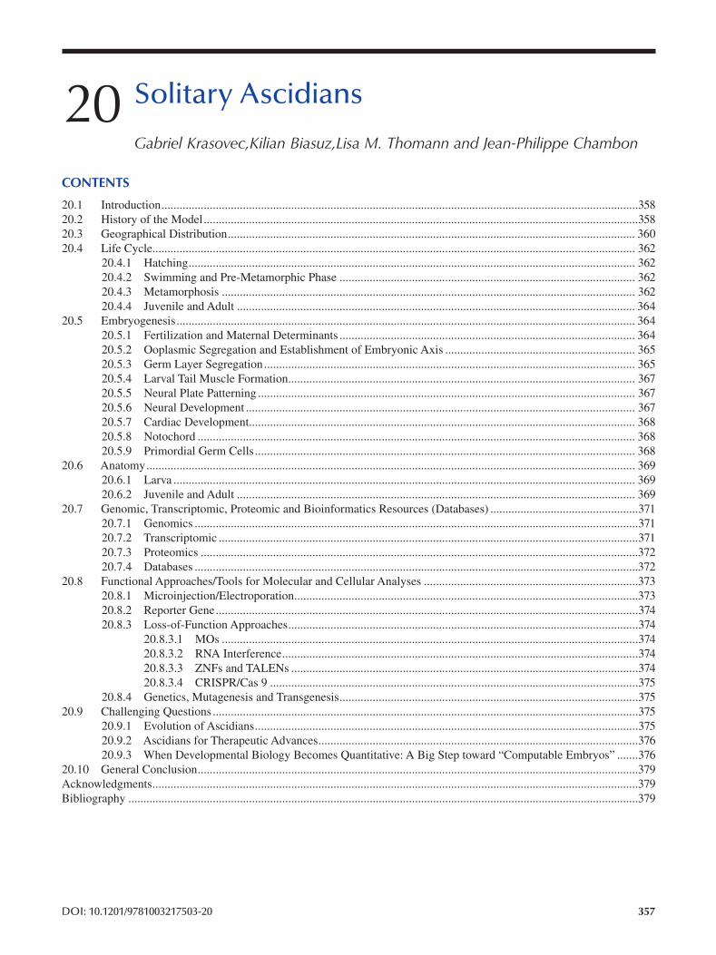
20 Solitary Ascidians
Gabriel Krasovec, Kilian Biasuz, Lisa M. Thomann and Jean-Philippe Chambon
CONTENTS
20.1 Introduction..............................................................................................................................................................358
20.2 History of the Model................................................................................................................................................358
20.3 Geographical Distribution ....................................................................................................................................... 360
20.4 Life Cycle................................................................................................................................................................ 362
20.4.1 Hatching.................................................................................................................................................... 362
20.4.2 Swimming and Pre-Metamorphic Phase .................................................................................................. 362
20.4.3 Metamorphosis ......................................................................................................................................... 362
20.4.4 Juvenile and Adult .................................................................................................................................... 364
20.5 Embryogenesis........................................................................................................................................................ 364
20.5.1 Fertilization and Maternal Determinants .................................................................................................. 364
20.5.2 Ooplasmic Segregation and Establishment of Embryonic Axis ............................................................... 365
20.5.3 Germ Layer Segregation ........................................................................................................................... 365
20.5.4 Larval Tail Muscle Formation ................................................................................................................... 367
20.5.5 Neural Plate Patterning ............................................................................................................................. 367
20.5.6 Neural Development ................................................................................................................................. 367
20.5.7 Cardiac Development................................................................................................................................ 368
20.5.8 Notochord ................................................................................................................................................. 368
20.5.9 Primordial Germ Cells.............................................................................................................................. 368
20.6 Anatomy.................................................................................................................................................................. 369
20.6.1 Larva ......................................................................................................................................................... 369
20.6.2 Juvenile and Adult .................................................................................................................................... 369
20.7 Genomic, Transcriptomic, Proteomic and Bioinformatics Resources (Databases) .................................................371
20.7.1 Genomics ...................................................................................................................................................371
20.7.2 Transcriptomic ...........................................................................................................................................371
20.7.3 Proteomics .................................................................................................................................................372
20.7.4 Databases ...................................................................................................................................................372
20.8 Functional Approaches/Tools for Molecular and Cellular Analyses .......................................................................373
20.8.1 Microinjection/Electroporation..................................................................................................................373
20.8.2 Reporter Gene ............................................................................................................................................374
20.8.3 Loss-of-Function Approaches ....................................................................................................................374
20.8.3.1 MOs ..........................................................................................................................................374
20.8.3.2 RNA Interference ......................................................................................................................374
20.8.3.3 ZNFs and TALENs ...................................................................................................................374
20.8.3.4 CRISPR/Cas 9 ..........................................................................................................................375
20.8.4 Genetics, Mutagenesis and Transgenesis ...................................................................................................375
20.9 Challenging Questions .............................................................................................................................................375
20.9.1 Evolution of Ascidians ...............................................................................................................................375
20.9.2 Ascidians for Therapeutic Advances ..........................................................................................................376
20.9.3 When Developmental Biology Becomes Quantitative: A Big Step toward “Computable Embryos” .......376
20.10 General Conclusion ..................................................................................................................................................379
Acknowledgments .................................................................................................................................................................379
Bibliography .........................................................................................................................................................................379
DOI: 10.1201/9781003217503-20 357
358
20.1 INTRODUCTION
The tunicates present various ecological behaviors com
prising sessile or pelagic adult forms in addition to colonial
or solitary animals. Solitary ascidians are present in sev
eral tunicate groups, meaning that both solitary and colo
nial ascidians are not restrictive or typical to a given clade.
Whereas distribution of solitary ascidians is scattered in the
urochordate tree, they share some common features, and
these can be studied in a common specific chapter. Despite
the large diversity of solitary ascidians, they can be char
acterized by typical features such as an individual sessile
adult presenting two siphons (one inhalant and one exhalant,
allowing the circulation of sea water), a pharynx supported
by an endostyle and a large branchial basket structure.
Usually hermaphrodites, fertilization takes place in sea
water after the release of gametes and gives rise to a swim
ming pelagic larva which will have to settle in a defi nitive
substrate. Unlike in colonial ascidians, asexual reproduction
is not documented.
Solitary ascidians have had a noticeable historical contri
bution to developmental and cell biology studies and include
several well-established models in marine biology such as
Ciona intestinalis. As the sister group of vertebrates, ascid
ians genetics data and genomics tools have opened broad
perspectives to understand the development and evolution
of chordates. Moreover, the financial importance of some
solitary ascidians species is notable as marine alimentary
resources, like Microcosmus sabatieri (usually named
“violet” or “sea fig”) in the south of France; Styela clava in Korea; or Halocyntia roretzi, which has been popular in
Japan. On the contrary, negative ecological consequences
can result from invasive species, like Styela clava. The pre
ceding succinct presentation of solitary ascidians highlights
the necessity and relevance of an overview.
20.2 HISTORY OF THE MODEL
The evolutionary history of tunicates is documented by fos
sil records comprising organisms attested or suggested to
be solitary ascidians. The fact that the first tunicate fossil
evidence seems to correspond to solitary ascidians is prob
ably due to a typical shape presenting two siphons in a “bag-
shaped” morphology characterized by a pharynx and gill
slit, making fossils of solitary ascidians easier to identify
than other tunicates. The oldest attested representative is
Shankouclava shankpuense, which has an estimated age of
524 million years corresponding to the second Turgenevian
stage of the Cambrian, discovered in China (Chen et al.
2003). This discovery introduced tunicates, at least soli
tary ascidians, as part of the high diversity explosion of the
Cambrian, witnessing the emergence of several major cur
rent groups of animals. Finally, some hypothetical identifi
cations, such as Yarnemia ascidiformis (Chistyakov et al.
1984) or Burykhia hunti (Fedonkin et al. 2012) from the
Russian Ediacaran (550 and 555 million years old, respec
tively), suggest an older appearance of ascidians.
Emerging Marine Model Organisms
The current species, Ascidiella aspersa, was the fi rst
experimental model in developmental biology, on which
Laurent Chabry studied blastomere recombination at the end
of the 19th century (Chabry 1887). Chabry destroyed one of
the blastomeres of two-cell embryos and found that the sur
viving one was able to form a half-embryo (more precisely, a
dwarf malformed larva). He obtained similar results with the
same kind of experiment on four-cell embryos and deduced
that an amputated early embryo is unable to compensate for
deleted cells during the development. Consequently, pioneer
experiments made by Chabry suggested that each part of the
larva came from specific cells emerging during the fi rst divi
sions. Next, Edwin Grant Conklin deepened our understand
ing of embryogenesis by working on the lineage of embryonic
cells and the segregation of the egg cytoplasm of various spe
cies of solitary ascidians such as Styela canopus (Conklin
1905a, 1905b). He reconstructed the lineage of cells from the
first divisions to the well-developed larva and confi rmed the
suggestions coming from Chabry’s experiments; development
is characterized by cell lineages, which give specific tissues in
the future larva what was called “a development in mosaic”.
Conklin’s studies on egg cytoplasm segregation, in addition to
cell lineage characterization, led to the hypothesis that female
determinants are present in the eggs to drive and participate
in the cell fate establishment during development. Solitary
ascidians consequently allowed the discovery of two funda
mental points in developmental and cell biology: the existence
of maternal determinants (now known as maternal RNA) and
the existence of cell lineages. In the same period as Conklin’s
experiments, other biologists focused on tunicate reproduc
tion biology, such as Thomas Morgan, who demonstrated in
1904, on Ciona intestinalis, that self-fertilization is blocked.
We currently know that this kind of biological barrier has
probably been selected to prevent consanguinity and facilitate
genetic mixing and increasing variability (see embryogenesis
section for details). From these pioneer studies by Chabry
and Conklin, interest in solitary ascidian biology crossed
time, and several biologists continued descriptive works.
Throughout his career, Norman John Berrill developed ascid
ians as biological models (Berrill and Watson 1930; Berrill
1932a ; Berrill and Watson 1936; Berrill and Sheldon 1964).
He described various species (Berrill 1932b) and also focused
on development and organ functionality, such as the gut and
stomach (Berrill 1929). He particularly took advantage of sol
itary ascidians as an easy model to understand seminal func
tionality. Importantly, Berrill participated in the validation of
the mosaic development theory, in opposition to regulative
development, which considers that blastomere fate can be reg
ulated during development to be able to form a normal embryo
in case of cell destruction. Next, since the 70s, a new genera
tion of researchers from several countries have expanded our
understanding of ascidian biology. As one example among
others, Guisseppina Ortolani worked on cell lineage differ
entiation or fertilization mechanisms on Ciona, Phallusia or
Ascidia. She notably participated in the discovery of muscle
cell lineages. Richard Whitteker validated Conklin’s propo
sition in 1973 of the presence of maternal determinants in
359 Solitary Ascidians
eggs driving cell lineage. In addition to research in Europe,
strong expertise on solitary ascidians emerged in Japan, led
by Noriyuki Satoh. One of Satoh’s major contributions is his
research on egg cytoplasmic factors establishing cell fate dur
ing embryogenesis. Thanks to horseradish peroxidase tracer
techniques, he was able to follow cell lineage and identify
maternal factors with monoclonal antibodies, an innovative
approach at the beginning of its career. Next, he described the
mechanism regulating expression of acetylcholinesterase in
muscle differentiation. With his research and the formation of
several future researchers, he actively participated in develop
ing molecular techniques on ascidian species. For instance, he
was at the origin of the first transcriptomic project but also on
the sequencing of the Ciona genome. In addition, he provided,
thanks to the ghost database, several molecular tools and data
on Ciona development to the scientifi c community. Finally,
in the 90s, the complementarity between developmental biol
ogy, genetics and incorporation of new molecular approaches
opened new perspectives to discover maternal determinants,
making mosaic development possible, but also on the impor
tance of regulation between blastomeres. In 2002, the fi rst
ascidian genome, from Ciona intestinalis, was sequenced
and annotated, opening an avenue of possibilities on embryo
genesis, metamorphosis and molecular signaling pathway
understanding. To date, several genomes and transcriptomes
from different solitary ascidians such as Phallusia mammilata, Ciona savigny, Molgula occulata and Halocynthia roretzi have expanded the amount of molecular data on this
group and contributed to easier molecular phylogenetic analy
sis, accessible molecular functionality comparison between
chordates and experiment design. The International Tunicate
Meeting (ITM), which occurs every two years, alternately in
Japan, Europe and the United States, was initiated in 2001,
illustrating the dynamism of research on ascidians where
solitary species count as most of the biological models, in
addition to a few colonial species such as Botryllus genus or
Appendicularia such as Oikopleura genus. This focus on solitary ascidian models’ contribution to
developmental biology is fundamental, but one must not ignore
the debate on ascidian evolution and their position among the
animals’ phylogenetic tree in the 19th century. Ascidians have
been considered close to molluscs for a long time because
of their flask adult body devoid of hard structure. The fi rst
questioning of this belonging was made by Savigny in 1816,
who recognized tunicates as distinct and separate from mol
luscs. Next, studies from Vadimir Kovalevsky during the
19th century questioned the relationship between tunicates
and other animals. Indeed, Kovalevsky described the larval
body plan of two species of solitary ascidians, Ciona intestinalis and Phallusia mammilata, and discovered an organi
zation similar to chordate animals (1866). In particular, the
presence of a dorsal chord in tadpole swimming larvae led to
considering chordates as composed of three groups: tunicates
(comprising solitary ascidians), cephalochordates (as genus
Amphioxus) and vertebrates. Consequently, thanks to solitary
ascidian larval descriptions, the phylogenetic position and
evolutionary history of tunicates became better understood.
From Kovalevsky’s studies to the beginning of the 21st cen
tury, ascidians were considered the first divergent branch of
chordates (making cephalochordates the sister-group of ver
tebrates). More recently, thanks to molecular phylogeny made
possible by genome sequencing and statistical method devel
opment, it was established that tunicates are the sister-group of
vertebrates, whereas cephalochordates are the fi rst divergent
chordate phylum, making tunicates the closest “invertebrates”
to vertebrates (Delsuc et al. 2006). Consequently, ascidians
became important in comparative studies from an evo-devo
perspective to understand vertebrate evolution. Whereas the
phylogenetic position of tunicates is now consensual and
established, the relationship inside tunicates is more debated,
and several phylogenies frequently emerge in the literature,
although a consensus is currently appearing (Figure 20.1).
Tunicates are commonly considered to be composed of fi ve
major phyla: Appendicularia, Phlebobranchia, Aplousobranchia,
Thaliacea and Stolidobranchia. Appendicularia are character
ized by a pelagic lifestyle with a tadpole-shaped adult form,
illustrated by the best-known species, Oikopleura dioika.
Though Appendicularia are often positioned as the fi rst
branch separated from other tunicate groups, debate on the
phylogenetic position of this group is not totally closed, and
it could be the sister of the Stolidobranchia (Delsuc et al.
2006; Delsuc et al. 2018; Kocot et al. 2018; Tatián et al.
2011; Satoh 2013). The four other groups (Phlebobranchia,
Aplousobranchia, Thaliacea, Stolidobranchia) are grouped
together in recent phylogenetic analysis and form a mono
phyletic clade. Phylogeny inside this large group has been
debated because of the difficulties of reconstructing the
life history for several reasons: the convergent features,
the secondary loss and the high evolution rate of DNA
sequences, making molecular phylogeny difficult to per
form. According to the current consensual phylogeny,
Stolidobranchia was the first group to diverge from the oth
ers. Then, Phlebobranchia, Thaliacea and Aplousobranchia
are considered monophyletic. Thaliacea diverged fi rst, and
Phlebobranchia grouped with Aplousobranchia to compose
Enterogona.
Thaliacea, including salps, are pelagic only and form a
planktonic colony made by the aggregation of multiple indi
viduals. An important point to keep in mind is the presence
of both solitary and colonial ascidians in Stolidobranchia
and Phlebobranchia, whereas Aplousobranchia are only
colonial and represent the group containing the high
est number of species. In these three groups, adult forms
are settled to the substrate, whereas Thaliacea are pelagic.
Stolidobranchia, characterized by the presence of one
gonad pair and an atrium formed from a unique indenta
tion, is composed of colonial ascidians like Botryllus schlosseri as well as solitary ones such as Molgula oculata. Stolidobranchia are also characterized by a folded
branchial sac. Phlebobranchia and Aplousobranchia, both
usually grouped into Enterogona, possess an even number
of gonads, and the atrium is formed by two indentations.
Phlebobranchia present a branchial sac vascularized by
longitudinal blood vessels, whereas Aplousobranchia have
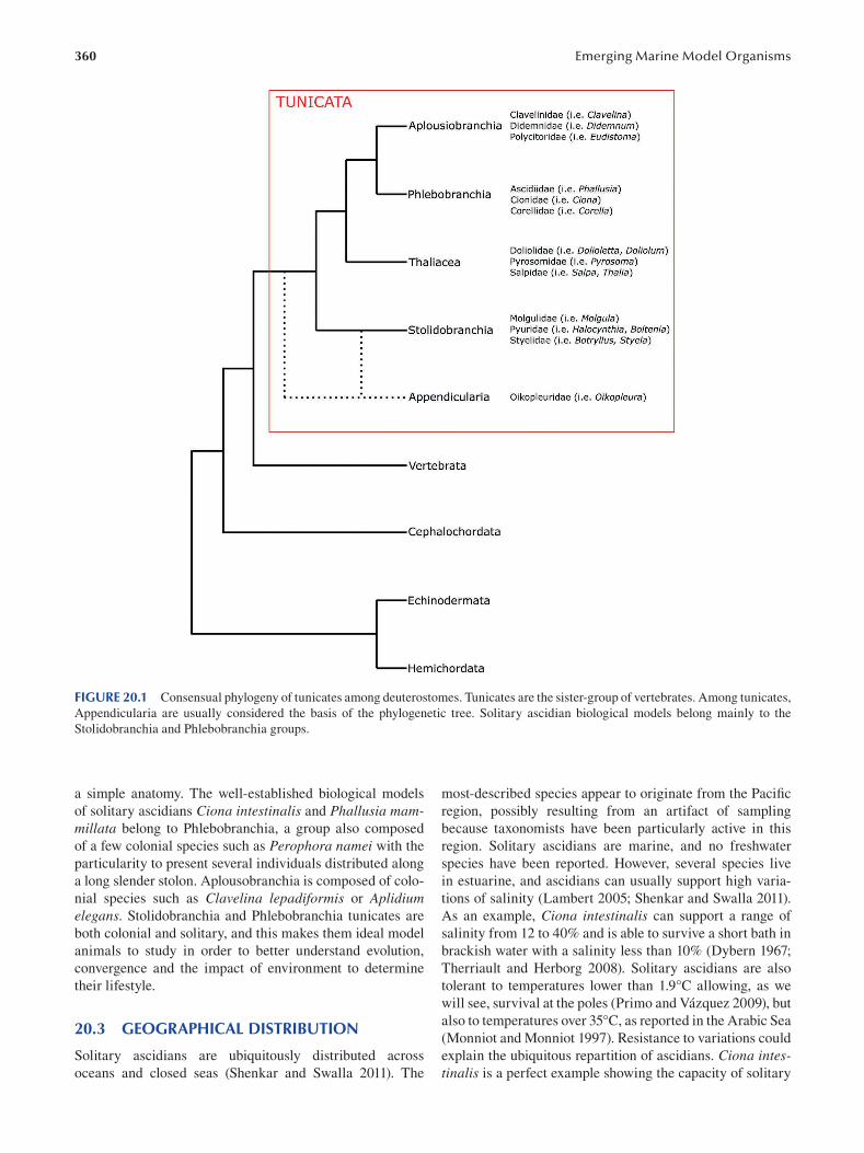
360 Emerging Marine Model Organisms
FIGURE 20.1 Consensual phylogeny of tunicates among deuterostomes. Tunicates are the sister-group of vertebrates. Among tunicates,
Appendicularia are usually considered the basis of the phylogenetic tree. Solitary ascidian biological models belong mainly to the
Stolidobranchia and Phlebobranchia groups.
a simple anatomy. The well-established biological models
of solitary ascidians Ciona intestinalis and Phallusia mammillata belong to Phlebobranchia, a group also composed
of a few colonial species such as Perophora namei with the
particularity to present several individuals distributed along
a long slender stolon. Aplousobranchia is composed of colo
nial species such as Clavelina lepadiformis or Aplidium elegans. Stolidobranchia and Phlebobranchia tunicates are
both colonial and solitary, and this makes them ideal model
animals to study in order to better understand evolution,
convergence and the impact of environment to determine
their lifestyle.
20.3 GEOGRAPHICAL DISTRIBUTION
Solitary ascidians are ubiquitously distributed across
oceans and closed seas (Shenkar and Swalla 2011). The
most-described species appear to originate from the Pacifi c
region, possibly resulting from an artifact of sampling
because taxonomists have been particularly active in this
region. Solitary ascidians are marine, and no freshwater
species have been reported. However, several species live
in estuarine, and ascidians can usually support high varia
tions of salinity (Lambert 2005; Shenkar and Swalla 2011).
As an example, Ciona intestinalis can support a range of
salinity from 12 to 40% and is able to survive a short bath in
brackish water with a salinity less than 10% (Dybern 1967;
Therriault and Herborg 2008). Solitary ascidians are also
tolerant to temperatures lower than 1.9°C allowing, as we
will see, survival at the poles (Primo and Vázquez 2009), but
also to temperatures over 35°C, as reported in the Arabic Sea
(Monniot and Monniot 1997 ). Resistance to variations could
explain the ubiquitous repartition of ascidians. Ciona intestinalis is a perfect example showing the capacity of solitary
361 Solitary Ascidians
ascidians to colonize various environments, leading to a
ubiquitous distribution. It has been sampled in the Pacifi c
Ocean (east and west), in the Atlantic on both American and
European coasts and in the Mediterranean Sea.
In addition to the presence of several ubiquitous species,
the capacity of larvae to settle in any substrate, such as soft
sediments, rocks or coral reefs, facilitates colonization and
expansion. Particularly, larvae can settle on several artifi cial
substrates such as floating dock or ship hulls, leading to an
artifi cial geographical spreading of some species at harbors
around the world. Consequently, some solitary ascidians
have a current ubiquitous repartition, but this does not seem
natural as resulting from a secondary colonization mediated
by human activities. For example, it has been reported in
the port of Salvador, which receives cargo ships from sev
eral continents, that the ascidians species inventory presents
a mix between possible endogenous ones (such as Ascidia nordestina), introduced ones (such as Cnemidocarpa irene)
and ubiquitous ones. Importantly, for some solitary ascidi
ans characterized by a wide/ubiquitous distribution, it can be
difficult to evaluate if the geographical distribution is natu
ral or artificial, resulting from centuries of spreading thanks
to travels and maritime trades. It is thus assumed that some
ascidians can have an unknown natural repartition. On the
other hand, some cases of invasion are clearly documented.
Corella eumyota, found natively in the southern hemisphere,
is now established in the north Atlantic and Mediterranean
Sea (Lambert et al. 1995; Collin et al. 2010). Moreover,
Styela genus represents a relevant example of global reparti
tion induced artifi cially. Styela clava, although coming from
the northwest Pacific, was accidentally introduced in the
East Pacific, Atlantic and European coasts. In Canada, this
species has been described to disturb aquaculture, probably
due to a overabundant population leading to the decrease
of food availability for filter animal culture such as mussels
or oysters, which suffer growth delay (Bourque et al. 2007;
Arsenault et al. 2009). Coupled with dispersion driven by
settlement on mobile artificial supports, some solitary ascid
ians can extend their life area by taking advantage of arti
ficial waterways. This is the case of the Suez Canal, which
has allowed to the endemic species Herdmania momus to
disperse from the Red Sea toward the Mediterranean Sea
(Shenkar and Loya 2008). Taken together, this high toler
ance of ascidians to various environments, their capacity to
spread thanks to artificial support and their potential impact
on food availability for other filter animals make solitary
ascidians a suitable model to understand the consequences
of invasive species.
In opposition to species presenting a ubiquitous geo
graphical repartition, some ascidians exhibit a specifi c
distribution, making them endemic to a given area. The
majority of ascidian species inventories reveal, in addition
to new species description, a mixed composition with both
ubiquitous and endemic species. This is typically the case in
the Port of Salvador or more recently in the Gulf of Mexico.
The Brazilian coast is also rich in endemic tunicates, such
as the solitary ascidian Eudistoma vannamei. Relatively
“closed” environments such as the Mediterranean Sea or
the Red Sea present various endemic species, likely because
of the reduced dispersal capacity compared to open envi
ronments. For example, 12 species are considered endemic
to the Red Sea, representing 17% of the ascidian diversity
(Shenkar and Loya 2008; Shenkar 2012).
Several solitary ascidians have been discovered in low-
temperature environments in both the Arctic and Antarctic.
Styela rustica can live in the north Atlantic in the Svalbard
region, a colonization which seems recent (Demarchi et al.
2008). In the southern hemisphere, a number of species
have been discovered in the South Shetland Islands such as
Styela wandeli or Molgula pedonculata (Tatian et al. 1998).
Antarctic species seem to be particularly adapted to survive
in extreme conditions, such as Cnemidocarpa verrucose,
known to be able to filter all ranges, particularly the fi nest,
of organic particles to get enough nutrients in a poor envi
ronment (Tatián et al. 2004).
This large repartition shows also that the majority of
solitary ascidians are shallow-water species and live on the
continental shelf in harbors, reefs, and various coastal envi
ronments. In addition, abyssal species are also documented
thanks to several sampling campaigns in the Pacifi c and
other deep-sea regions. Abyssal species from the Pacifi c are
represented by Molgula sphaeroidea or Adagnesia bafida,
also discovered in the Atlantic at a depth of about 3,000 m.
The deepest solitary ascidian discovered was in the Pacifi c
at 7,000 m depth. Illustrating the ubiquitous presence of
deep-sea species, we can also cite Agnezia monnioti , discov
ered in the Arabian Sea at 3,162 m depth. In Styela gagetyleri, localized in the same region but at 368 m depth (which
is already considered a deep-sea conditions), the number of
folds of the branchial sac is reduced, implying a decrease of
cilia quantity and thus oxygen exchange surface. This could
result from an adaptation to low oxygen levels and an opti
mization of the capacity to capture nutrients. Observations
and species descriptions have led scientists to notice that
abyssal species are in the high majority of solitary ascidians
and not colonial ones. It has been proposed that the column
shape of the body of solitary species allows a vertical elon
gation, creating a distance between the siphons and deep-sea
soft and muddy sediments, whereas colonial ascidians are
closed to the substrate and cover it in such a way that the
siphon stays close to the mud, which could be problematic to
capture food in a poor environment.
All studies made on Tunicate spatial distribution brought
to light that solitary ascidians composed between 20% and
40% of the diversity (others are colonial ascidians) in tropi
cal environments, whereas solitary ascidians represent most
of the species at the two poles and temperate climates, with,
for example, 58% and 70% of the diversity in the Antarctic
and European coasts, respectively. This distribution is
explained by the lifestyle of colonial ascidians presenting an
indeterminate growth allowing colonization of most biologi
cal matter support in rich tropical environments.
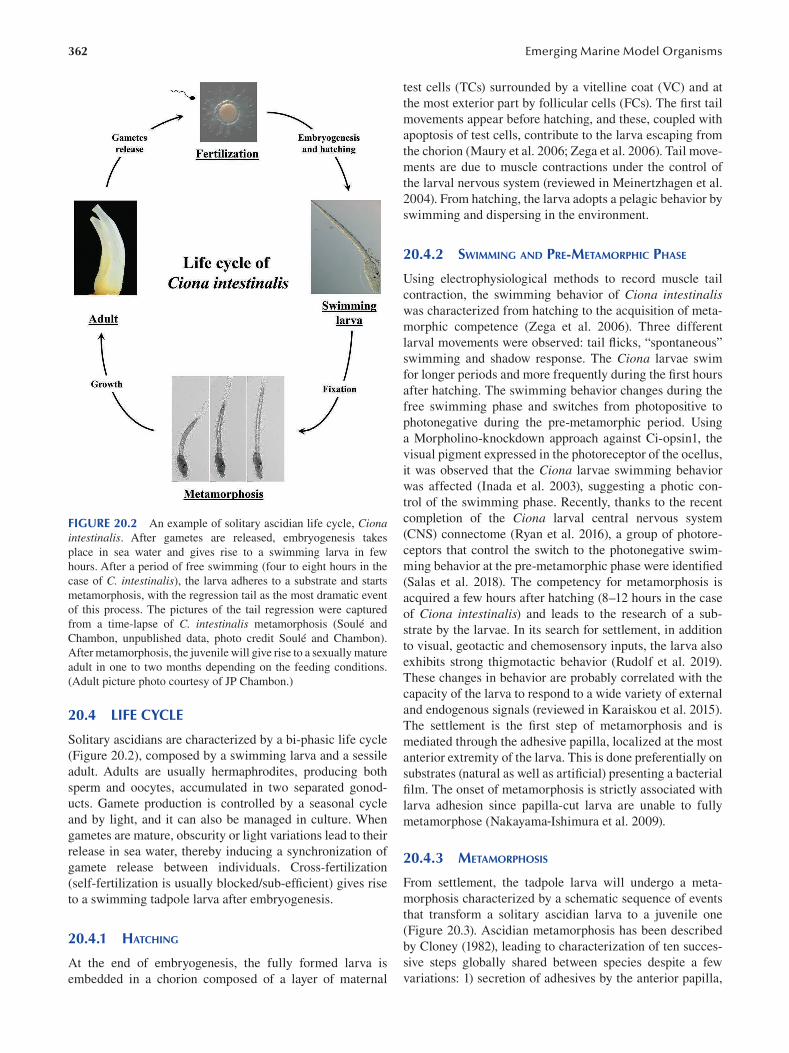
362
FIGURE 20.2 An example of solitary ascidian life cycle, Ciona intestinalis. After gametes are released, embryogenesis takes
place in sea water and gives rise to a swimming larva in few
hours. After a period of free swimming (four to eight hours in the
case of C. intestinalis), the larva adheres to a substrate and starts
metamorphosis, with the regression tail as the most dramatic event
of this process. The pictures of the tail regression were captured
from a time-lapse of C. intestinalis metamorphosis (Soulé and
Chambon, unpublished data, photo credit Soulé and Chambon).
After metamorphosis, the juvenile will give rise to a sexually mature
adult in one to two months depending on the feeding conditions.
(Adult picture photo courtesy of JP Chambon.)
20.4 LIFE CYCLE
Solitary ascidians are characterized by a bi-phasic life cycle
( Figure 20.2 ), composed by a swimming larva and a sessile
adult. Adults are usually hermaphrodites, producing both
sperm and oocytes, accumulated in two separated gonod
ucts. Gamete production is controlled by a seasonal cycle
and by light, and it can also be managed in culture. When
gametes are mature, obscurity or light variations lead to their
release in sea water, thereby inducing a synchronization of
gamete release between individuals. Cross-fertilization
(self-fertilization is usually blocked/sub-effi cient) gives rise
to a swimming tadpole larva after embryogenesis.
20.4.1 HATCHING
At the end of embryogenesis, the fully formed larva is
embedded in a chorion composed of a layer of maternal
Emerging Marine Model Organisms
test cells (TCs) surrounded by a vitelline coat (VC) and at
the most exterior part by follicular cells (FCs). The fi rst tail
movements appear before hatching, and these, coupled with
apoptosis of test cells, contribute to the larva escaping from
the chorion (Maury et al. 2006; Zega et al. 2006). Tail move
ments are due to muscle contractions under the control of
the larval nervous system (reviewed in Meinertzhagen et al.
2004). From hatching, the larva adopts a pelagic behavior by
swimming and dispersing in the environment.
20.4.2 SWIMMING AND PRE-METAMORPHIC PHASE
Using electrophysiological methods to record muscle tail
contraction, the swimming behavior of Ciona intestinalis was characterized from hatching to the acquisition of meta
morphic competence ( Zega et al. 2006). Three different
larval movements were observed: tail fl icks, “spontaneous”
swimming and shadow response. The Ciona larvae swim
for longer periods and more frequently during the fi rst hours
after hatching. The swimming behavior changes during the
free swimming phase and switches from photopositive to
photonegative during the pre-metamorphic period. Using
a Morpholino-knockdown approach against Ci-opsin1, the
visual pigment expressed in the photoreceptor of the ocellus,
it was observed that the Ciona larvae swimming behavior
was affected (Inada et al. 2003), suggesting a photic con
trol of the swimming phase. Recently, thanks to the recent
completion of the Ciona larval central nervous system
(CNS) connectome (Ryan et al. 2016), a group of photore
ceptors that control the switch to the photonegative swim
ming behavior at the pre-metamorphic phase were identifi ed
(Salas et al. 2018). The competency for metamorphosis is
acquired a few hours after hatching (8–12 hours in the case
of Ciona intestinalis) and leads to the research of a sub
strate by the larvae. In its search for settlement, in addition
to visual, geotactic and chemosensory inputs, the larva also
exhibits strong thigmotactic behavior (Rudolf et al. 2019).
These changes in behavior are probably correlated with the
capacity of the larva to respond to a wide variety of external
and endogenous signals (reviewed in Karaiskou et al. 2015).
The settlement is the first step of metamorphosis and is
mediated through the adhesive papilla, localized at the most
anterior extremity of the larva. This is done preferentially on
substrates (natural as well as artificial) presenting a bacterial
film. The onset of metamorphosis is strictly associated with
larva adhesion since papilla-cut larva are unable to fully
metamorphose (Nakayama-Ishimura et al. 2009).
20.4.3 METAMORPHOSIS
From settlement, the tadpole larva will undergo a meta
morphosis characterized by a schematic sequence of events
that transform a solitary ascidian larva to a juvenile one
(Figure 20.3). Ascidian metamorphosis has been described
by Cloney (1982), leading to characterization of ten succes
sive steps globally shared between species despite a few
variations: 1) secretion of adhesives by the anterior papilla,
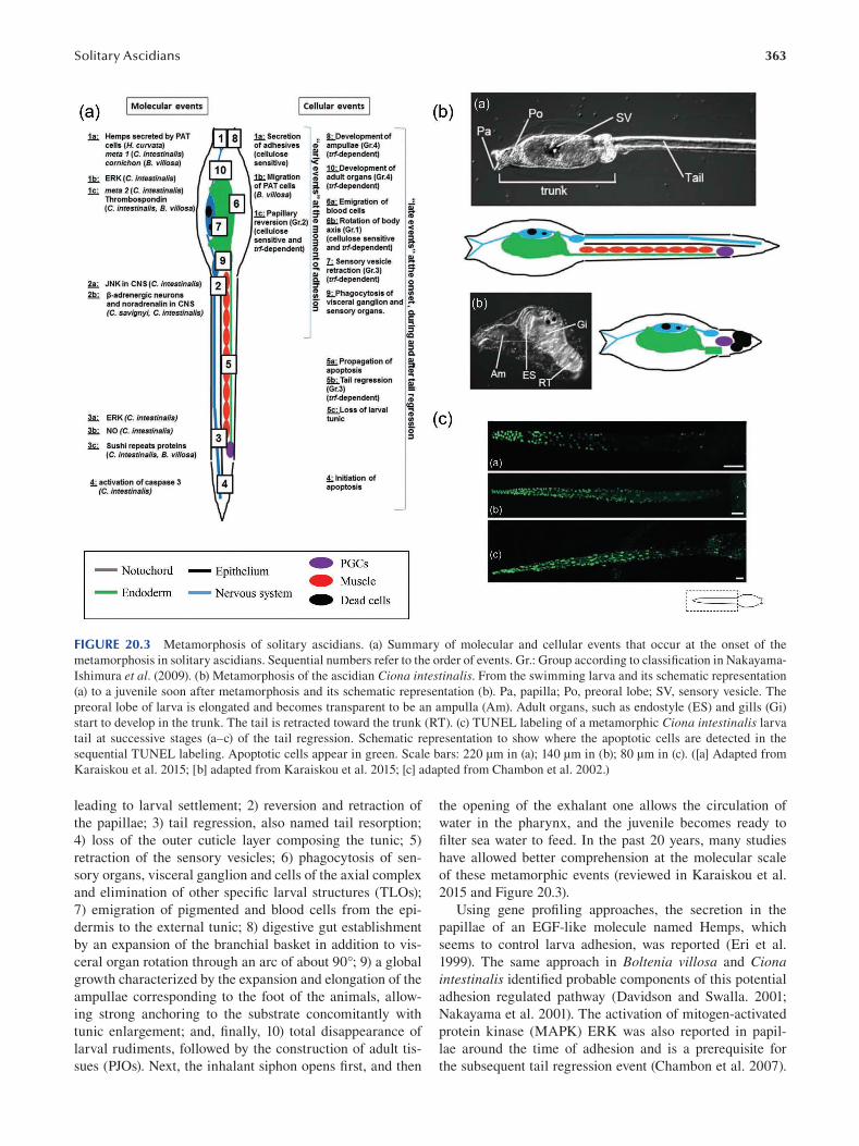
363 Solitary Ascidians
FIGURE 20.3 Metamorphosis of solitary ascidians. (a) Summary of molecular and cellular events that occur at the onset of the
metamorphosis in solitary ascidians. Sequential numbers refer to the order of events. Gr.: Group according to classification in Nakayama-Ishimura et al. (2009). (b) Metamorphosis of the ascidian Ciona intestinalis. From the swimming larva and its schematic representation
(a) to a juvenile soon after metamorphosis and its schematic representation (b). Pa, papilla; Po, preoral lobe; SV, sensory vesicle. The
preoral lobe of larva is elongated and becomes transparent to be an ampulla (Am). Adult organs, such as endostyle (ES) and gills (Gi)
start to develop in the trunk. The tail is retracted toward the trunk (RT). (c) TUNEL labeling of a metamorphic Ciona intestinalis larva
tail at successive stages (a–c) of the tail regression. Schematic representation to show where the apoptotic cells are detected in the
sequential TUNEL labeling. Apoptotic cells appear in green. Scale bars: 220 μm in (a); 140 μm in (b); 80 μm in (c). ([a] Adapted from
Karaiskou et al. 2015; [b] adapted from Karaiskou et al. 2015; [c] adapted from Chambon et al. 2002.)
leading to larval settlement; 2) reversion and retraction of
the papillae; 3) tail regression, also named tail resorption;
4) loss of the outer cuticle layer composing the tunic; 5)
retraction of the sensory vesicles; 6) phagocytosis of sen
sory organs, visceral ganglion and cells of the axial complex
and elimination of other specific larval structures (TLOs);
7) emigration of pigmented and blood cells from the epi
dermis to the external tunic; 8) digestive gut establishment
by an expansion of the branchial basket in addition to vis
ceral organ rotation through an arc of about 90°; 9) a global
growth characterized by the expansion and elongation of the
ampullae corresponding to the foot of the animals, allow
ing strong anchoring to the substrate concomitantly with
tunic enlargement; and, finally, 10) total disappearance of
larval rudiments, followed by the construction of adult tis
sues (PJOs). Next, the inhalant siphon opens first, and then
the opening of the exhalant one allows the circulation of
water in the pharynx, and the juvenile becomes ready to
filter sea water to feed. In the past 20 years, many studies
have allowed better comprehension at the molecular scale
of these metamorphic events (reviewed in Karaiskou et al.
2015 and Figure 20.3).
Using gene profiling approaches, the secretion in the
papillae of an EGF-like molecule named Hemps, which
seems to control larva adhesion, was reported (Eri et al.
1999). The same approach in Boltenia villosa and Ciona intestinalis identifi ed probable components of this potential
adhesion regulated pathway (Davidson and Swalla. 2001;
Nakayama et al. 2001). The activation of mitogen-activated
protein kinase (MAPK) ERK was also reported in papil
lae around the time of adhesion and is a prerequisite for
the subsequent tail regression event (Chambon et al. 2007).
364
Simultaneously, the JNK/MAPK pathway is also activated
in the CNS, and similarly to the ERK pathway, it is essen
tial for tail regression. The CNS seems to have a preponder
ant part in the onset of metamorphosis; expression of the
β1 -adrenergic receptor was reported in this tissue in Ciona intestinalis and Ciona savignyi (Kimura et al. 2003). More
recently, the neurotransmitter GABA was reported as a key
regulator of Ciona metamorphosis (Hozumi et al. 2020),
reinforcing the previous hypothesis of the preponderant role
of the larval nervous system and sensory organs in selecting
sites for adhesion and in the onset of metamorphosis (Cloney
1982). One of the most dramatic event of this process is the
regression of the tail larva, which occurs a few hours after
adhesion. Two not mutually exclusive mechanisms were
reported during this event: the first involves the contractile
properties of either the tail epithelial layer (observed in the
solitary ascidian Distaplia occidentalis, Aplidium constellatum, Diplosoma, Ecteinascidia turbiniata, C. intestinalis, Ascidia callosa, Corella willmeriana macdonaldi and the
colonial ascidian Botryllus schlosseri) or notochord cells
(observed in Boltenia villosa, Herdmania curvata, Styela gibbsii, Molgula mahattensis, Molgula occidentalis and
Polycitor mutabilis; reviewed by Cloney 1982); the second
involves a massive apoptotic cell death of almost all of the
cells that composed the tail and was observed in C. intestinalis (Chambon et al. 2002; Tarallo and Sordino 2004)
and Molgula oculata (Jeffery 2002). Recently, using live
microscopy, both mechanisms were observed during Ciona intestinalis tail regression, and they seem to be sequential,
since initial contraction of the tip tail preceded apoptosis
(Krasovec et al. 2019). Apoptosis appears to be the driv
ing force of tail regression in solitary ascidians and affects
almost all the cell types that compose the tail (the tunic,
epidermal, notochord, tail muscle cells and the CNS), with
two exceptions, the endodermal strand cells and the pri
mordial germ cells (PGCs) (Figure 20.3). These two cell
types escape apoptosis, the endodermal strand by migrating
before the tail regression (Nakazawa et al. 2013), while the
PGCs move toward the trunk at the time of tail regression
in coordination with the progression of cell death (Krasovec
et al. 2019). The most remarkable feature is that through
sequential TUNEL pictures, it has been confi rmed in vivo that apoptosis starts at the tail tip and continues up to the
tail base by a perfect antero-posterior wave (Chambon et al.
2002; Krasovec et al. 2019). The same polarized propagation
of apoptosis was reported in two other species of ascidians,
Molgula occidentalis and Asicidia ceratodes ( Jeffery 2002 ).
An arising and challenging question is the coordination
mechanism of the metamorphic events. New insights were
provided by the identification of the gene network down
stream of the MAPK, ERK and JNK activation previously
reported, respectively, in the papillae and the CNS. Among
them is Ci-sushi, a gene under JNK control, with expres
sion patterns at the tip of the tail, for which loss of function
experiments lead to the inhibition of the initiation of apop
tosis (Chambon et al. 2007). In addition, papilla and tail cut
experiments on larva coupled with analyses of metamorphic
Emerging Marine Model Organisms
mutants (swimming juveniles and tail-regression fail [trf]) allowed classification of metamorphic events in four groups
(Nakayama-Ishimura et al. 2009). Group 1 includes a
cellulose-sensitive and trf-independent event: body axis
rotation; Group 2 encompasses a cellulose-sensitive and
trf-dependent event: papillae retraction; Group 3 includes
cellulose-independent and trf-dependent events, sensory
vesicle retraction and tail regression; and Group 4 comprises
cellulose-independent and trf-independent events, including
ampullae formation and adult organ growth.
20.4.4 JUVENILE AND ADULT
Metamorphosis in ascidians results in a dramatic modifi ca
tion of their body plan, transforming them in a few hours
from swimming larva to sessile juvenile and after few
months of growing to a sexually mature adult. Classically,
juvenile growth timing depends on food availability and
temperature. Consequently, the settled phase represents
almost the entire life cycle, whereas the swimming phase is
transitory and allows the dispersion of individuals.
20.5 EMBRYOGENESIS
20.5.1 FERTILIZATION AND MATERNAL DETERMINANTS
Ascidian embryogenesis is a rapid process involving a small
number of cells (about 2,600 cells in Ciona intestinalis ) and
occurs within a chorion composed of test cells, a vitelline
coat and follicular cells (Figure 20.4). It starts with fertiliza
tion, which, in solitary ascidians, occurs after the release
of sperm and eggs into the surrounding seawater. To ensure
fertilization, spermatozoids are activated and then attracted
toward the eggs by a common factor released by mature
oocytes (after germinal vesicle breakdown) called sperm-
activating and sperm-attracting factor (SAAF) (Kondoh
et al. 2008; Yoshida et al. 2002). The ascidians eggs are
spawned embedded in a layer of follicular cells surrounding
a vitelline coat, under which the test cells enclose the egg
itself. In some species, such as Styela plicata, sperm and
eggs are released at different times, while they are released
simultaneously in Ciona and Halocyntia, allowing sperm
to interact with self-eggs. In these latter species, which are
known to be self-sterile, a self- and non-self recognition sys
tem was reported during fertilization, probably to promote
outcrossing. In Ciona, this process is ensured by a couple of
receptors expressed at the surface of the sperm (s-Themis A
and B) and ligands expressed on the VC (v-Themis A and B).
If a sperm containing s-Themis A and B interacts with an
egg expressing both v-Themis A and B on the VC, its abil
ity to bind the VC is reduced, and it is not able to fertilize
the self-recognized egg (Harada et al. 2008). In addition to
this self-recognition system, the polyspermy block involves
a glycosidase enzyme released from the surface of FCs. It is
interesting to notice that this enzyme activity release is not
species specific, which means that sperm of a species could
block the egg of an another (Lambert 2000). This sperm
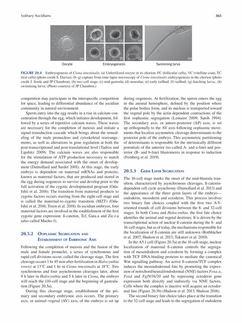
365 Solitary Ascidians
FIGURE 20.4 Embryogenesis of Ciona intestinalis. (a) Unfertilized oocyte in its chorion, FC (follicular cells), VC (vitelline coat), TC
(test cells) (photo credit S. Darras); (b–g) capture from time-lapse microscopy of Ciona intestinalis embryogenesis in the chorion (photo
credit J. Soule and JP Chambon); (b) two-cell stage; (c) mid-gastrula; (d) neurulae; (e) early tailbud; (f) tailbud; (g) hatching larva.; (h)
swimming larva. (Photo courtesy of JP Chambon.)
competition may participate in the interspecifi c competition
for space, leading to differential abundance of the ascidian
community in natural environment.
Sperm entry into the egg results in a rise in calcium con
centration through the egg, which initiates development, fol
lowed by a series of repetitive calcium waves. These waves
are necessary for the completion of meiosis and initiate a
signal-transduction cascade which brings about the remod
eling of the male pronucleus and cytoskeletal rearrange
ments, as well as alterations in gene regulation at both the
post-transcriptional and post-translational level (Tadros and
Lipshitz 2009). The calcium waves are also responsible
for the stimulation of ATP production necessary to match
the energy demand associated with the onset of develop
ment (Dumollard and Sardet 2001). At this stage, the early
embryo is dependent on maternal mRNAs and proteins,
known as maternal factors, that are produced and stored in
the egg during oogenesis to survive and develop prior to the
full activation of the zygotic developmental program (Oda-
Ishii et al. 2016 ). The transition from maternal products to
zygotic factors occurs starting from the eight-cell stage and
is called the maternal-to-zygotic transition (MZT) (Oda-
Ishii et al. 2016; Treen et al. 2018). In ascidian embryos, four
maternal factors are involved in the establishment of the fi rst
zygotic gene expression: ß-catenin, Tcf, Gata.a and Zic.r-a
(also called Macho-1).
20.5.2 OOPLASMIC SEGREGATION AND
ESTABLISHMENT OF EMBRYONIC AXIS
Following the completion of meiosis and the fusion of the
male and female pronuclei, a series of synchronous and
rapid cell divisions occur, called the cleavage stage. The fi rst
cleavage occurs 1 hr 45 min after fertilization in Halocynthia roretzi at 13°C and 1 hr in Ciona intestinalis at 18°C. Two
synchronous and four asynchronous cleavages later, about
9 h later in Halocynthia and 5 h later in Ciona, the embryo
will reach the 110-cell stage and the beginning of gastrula
tion (Figure 20.5a).
During this cleavage stage, establishment of the pri
mary and secondary embryonic axis occurs. The primary
axis, or animal–vegetal (AV) axis, of the embryo is set up
during oogenesis. At fertilization, the sperm enters the egg
in the animal hemisphere, defined by the position where
the polar bodies form, and its nucleus is transported toward
the vegetal pole by the actin-dependent contractions of the
first ooplasmic segregation (Lemaire 2009; Satoh 1994).
The secondary axis, or antero-posterior (AP) axis, is set
up orthogonally to the AV axis following ooplasmic move
ments that localize asymmetric cleavage determinants to the
posterior pole of the embryo. This asymmetric partitioning
of determinants is responsible for the intrinsically different
potentials of the anterior (so-called A- and a-line) and pos
terior (B- and b-line) blastomeres in response to induction
(Feinberg et al. 2019).
20.5.3 GERM LAYER SEGREGATION
The 16-cell stage marks the onset of the mid-blastula tran
sition, characterized by asynchronous cleavages, ß-catenin
dependent cell cycle asynchrony (Dumollard et al. 2013) and
the appearance of the three germ layers of the embryo—
endoderm, mesoderm and ectoderm. This process involves
two binary fate choices coupled with the first two A-V
oriented rounds of cell divisions between the 8- and 32-cell
stages. In both Ciona and Halocynthia, the first fate choice
identifies the animal and vegetal destinies. It is driven by the
transcriptional action of nuclear ß-catenin during the 8- and
16-cell stages, but as of today, the mechanisms responsible for
the localization of ß-catenin are still unknown (Rothbächer
et al. 2007; Hudson et al. 2013; Takatori et al. 2010).
In the A5.1 cell (Figure 20.5a) at the 16 cell-stage, nuclear
localization of maternal ß-catenin controls the segrega
tion of mesendoderm and ectoderm by forming a complex
with TCF DNA-binding proteins to mediate the canonical
Wnt signalling pathway. An active ß-catenin/TCF complex
induces the mesendodermal fate by promoting the expres
sion of notochord/neural/endodermal (NNE) factors Foxa.a,
Foxd and Fgf9/16/20 and by repressing ectoderm gene
expression both directly and indirectly via NNE factors.
Cells where the complex is inactive will acquire an ectoder
mal fate (Figure 20.5b) (Hudson et al. 2013; Hudson 2016 ).
The second binary fate choice takes place at the transition
to the 32-cell stage and leads to the segregation of endoderm
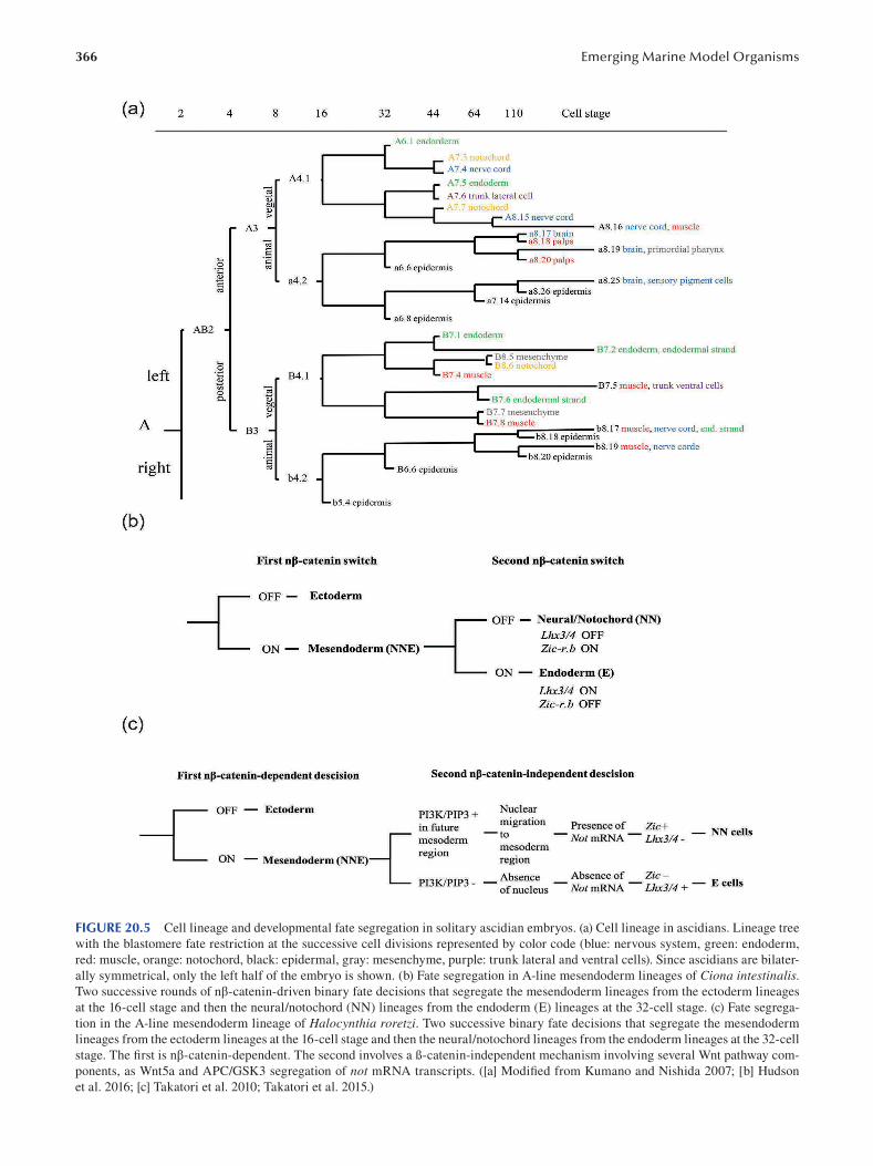
366 Emerging Marine Model Organisms
FIGURE 20.5 Cell lineage and developmental fate segregation in solitary ascidian embryos. (a) Cell lineage in ascidians. Lineage tree
with the blastomere fate restriction at the successive cell divisions represented by color code (blue: nervous system, green: endoderm,
red: muscle, orange: notochord, black: epidermal, gray: mesenchyme, purple: trunk lateral and ventral cells). Since ascidians are bilater
ally symmetrical, only the left half of the embryo is shown. (b) Fate segregation in A-line mesendoderm lineages of Ciona intestinalis. Two successive rounds of nβ-catenin-driven binary fate decisions that segregate the mesendoderm lineages from the ectoderm lineages
at the 16-cell stage and then the neural/notochord (NN) lineages from the endoderm (E) lineages at the 32-cell stage. (c) Fate segrega
tion in the A-line mesendoderm lineage of Halocynthia roretzi. Two successive binary fate decisions that segregate the mesendoderm
lineages from the ectoderm lineages at the 16-cell stage and then the neural/notochord lineages from the endoderm lineages at the 32-cell
stage. The fi rst is nβ-catenin-dependent. The second involves a ß-catenin-independent mechanism involving several Wnt pathway com
ponents, as Wnt5a and APC/GSK3 segregation of not mRNA transcripts. ([a] Modified from Kumano and Nishida 2007; [b] Hudson
et al. 2016; [c] Takatori et al. 2010; Takatori et al. 2015.)
367 Solitary Ascidians
and notochord/neural (NN cells or mesoderm) from mesen
doderm precursors. Two distinct regulatory processes have
been discovered to achieve the same fate decision in the
same A lineage in Ciona and Halocynthia.
In the case of Ciona embryos, this second fate choice
involves a second ß-catenin-dependent process during the
32-cell stage. Continued activity of the ß-catenin/TCF
complex in mesendodermal cells induces endoderm fate
(E cells), whereas inactivation of the complex leads to the
acquisition of the notochord/neural fate. During this second
phase, ß-catenin/TCF works directly or indirectly in the E
cells with the targets of the first phase of ß-catenin activ
ity, Foxa.a, FoxD and Fgf9/16/20, to activate the E speci
fi er Lhx3/4 and to repress the NN specifi er Zic-r.b ( Figure
20.5B) (Hudson et al. 2016 ).
In Halocynthia embryos, a different mechanism exists.
A possible explanation for this difference is the presence of
nuclear ß-catenin in NN cells at the 32-cell stage (Hudson
et al. 2013). Thus, Halocynthia NN specifi cation depends
on a Wnt-dependent but ß-catenin-independent mechanism
involving Not mRNA transcripts. The asymmetrical parti
tioning of Not mRNA regulates the expression of transcrip
tion factors required for fate segregation. In endoderm cells,
Not will be absent, and thus endoderm differentiation will
occur. On the contrary, in NN cells, Not is present and will
promote Zic expression as well as repressing Lhx3/4 expres
sion, thus promoting NN fate and repressing E fate (Figure
20.5c) (Hudson et al. 2016; Takatori et al. 2010; Takatori
et al. 2015) (Figure 20.5c).
20.5.4 LARVAL TAIL MUSCLE FORMATION
Muscle formation in ascidian is a well-known example of cell
autonomous process first demonstrated by Conklin in 1905.
However, recent studies have brought to light the importance
of cell–cell interaction as another important factor.
At the larval stage, the only fully differentiated and func
tional muscles are those of the tail and most solitary species
present between 18 and 21 muscle cells on either side of the
tail. Muscle cells originated either from the primary muscle
cell lineage and the B4.1 blastomeres or from the secondary
lineage of A4.1 and b4.2 (Figure 20.5a) (Razy-Krajka and
Stolfi 2019; Satoh 2013).
The primary lineage consists of 14 muscle cells located
on either side of the tail specified following a cell autono
mous specification and differentiation involving the Zic.r-a
(Macho-1) maternal determinant. Zic.r-a will trigger the
primary tail muscle specification regulatory network by
activating the transcription of Tbx6-related (Tbx6-r ) muscle
determinants at the 16-cell stage and downstream factors
at the 64-cell stage (Razy-Krajka and Stolfi 2019; Satoh
2013; Yagi et al. 2005). On the other hand, the secondary
lineage gives rise to the muscle cells flanking the tip of the
tail, whose numbers vary between species (ten cells of b4.2
origin in Halocynthia compared to four in Ciona) . In the
A-line, muscle potential is induced by intricate feed-forward
signaling relay from the neighboring b6.5 lineage cells to
A7.6 to A8.16. In Ciona, the Nodal and Delta/Notch signal
ing pathways are responsible for this, while in Halocynthia,
a yet-unknown signal from the same b6.5 lineage induces
the expression of Wnt5.a, which then promotes muscle fate
in A8.16 (Figure 20.5a) (Tokuoka et al. 2007 ). Finally, the
last muscle/neural cell fate decision in Ciona will see FGF/
ERK signaling activating the muscle determinants Tbx6-r. b and Mrf expression. In Halocynthia, what regulates this
final fate decision is yet another unknown parameter, but
FGF/ERK signaling is not involved (Razy-Krajka and Stolfi
2019; Tokuoka et al. 2007 ).
20.5.5 NEURAL PLATE PATTERNING
Similar to vertebrate neurulation, the ascidian neural plate
is curled up dorsally to form a tube-like structure known
as the neural tube. The neural plate emerges at the mid-
gastrula stage and is composed of 40 cells at the neural plate
stage, arranged in six rows and eight columns of cells along
the A-P axis formed from posterior to anterior. The I and
II rows compose the posterior neural plate and derive from
the A-lineage. They will contribute to the caudal nerve cord,
motor ganglions and posterior sensory vesicle. On the other
hand, the a-lineage will give rise to the anterior four rows III
to VI. Rows III and VI will contribute to the anterior part
of the sensory vesicle, part of the oral siphon primordium
and anterior brains. Finally, rows V and VI give rise to neu
rons of the peripheral neural system (PNS) (Hudson 2016;
Imai et al. 2009; Wagner and Levine 2012). Once the neural
tube is completely closed, the tail becomes distinguishable
(Kumano and Nishida 2007 ).
Different signaling pathways are responsible for the pat
terning of the neural plate, such as Nodal, Nodal-dependent
Snail, FGF/MEK/ERK and Delta/Notch (Hudson 2016;
Hudson et al. 2007; Razy-Krajka and Stolfi 2019; Satoh 2013).
20.5.6 NEURAL DEVELOPMENT
The ascidian nervous system is composed of the periph
eral neural system and the central nervous system, and its
development starts with neural induction at the 32-cell stage.
CNS development starts in two blastomeres, pairs A6.2 and
A6.4 (Figure 20.5a), which become neural fate restricted at
the 64-cell stage under FGF induction (Hudson et al. 2016).
It consists of approximately 330 cells and about 117 neurons
and originates from three lineages: the A and a- and b-lines
(Hudson et al. 2007). The CNS presents three morphologi
cally distinct structures: the anterior-most sensory vesicle,
the trunk ganglion (also called visceral ganglion) and the
tail nerve cord (Hudson et al. 2016). The A-line blasto
meres become fate restricted following a neuro-epidermal
binary fate decision involving a β-catenin-driven binary fate
switch. This lineage will give rise to the posterior part of
the sensory vesicle as well as the ventral and lateral parts
of both trunk ganglion and tail nerve cord (Hudson et al.
2013). The anterior part of the sensory vesicle and the dorsal
part of the visceral ganglion and tail nerve cord respectively
368 Emerging Marine Model Organisms
originate from the a-line (a6.5) and b-line (b6.5) blasto
meres, which become restricted to neural fate at the 112-cell
stage (Hudson et al. 2016; Roure et al. 2014).
PNS development starts with the birth of the a6.5 blas
tomere (Figure 20.5a). It is composed of different types of
epidermal sensory neurons (ESNs): the papillary neurons of
the adhesive papillae, the epidermal sensory neurons and
the bipolar tail neurons (BTNs) distributed in the epider
mis of the trunk and tail (Hudson 2016; Meinertzhagen and
Okamura 2001).
20.5.7 CARDIAC DEVELOPMENT
The adult ascidian heart consists of a one-cell-layer single
myocardial tube surrounded by a pericardium. It is formed
of two distinct territories: the fi rst heart field (FHF) and the
second heart field (SHF) and originates from a single pair
of blastomeres in the 64-cell stage embryos, the B7.5 cells
(Figure 20.5a). The first division of the cardiac founder cells
is symmetric and occurs during gastrulation. It leads to the
appearance of two symmetrical pairs of pre-cardiac founder
cells each consisting of a B8.9 and B8.10 blastomeres (Figure
20.5a) (Cooley et al. 2011). During neurulation, in each pre-
cardiac lineage, founder cells divide a second time, asym
metrically this time, and each blastomere will give rise to
four cells: two small anterior cells, which will migrate to
form the heart, and two large posterior B7.5 granddaughter
cells, which will differentiate as anterior tail muscles in both
Halocynthia and Ciona (Figure 20.5a) (Christiaen et al. 2010;
Davidson et al. 2006).
Two maternal determinants are responsible for the
specification of the blastomeres: macho-1 and β-catenin.
They activate the B7.5-specific expression of the tran
scription factor Mesp (Christiaen et al. 2009; Stolfi et al.
2010), which determines a competence domain facilitat
ing either pre-cardiac or pre-vascular specifi cation (Satou
et al. 2004). Within the Mesp-expressing cells, subse
quent inductive signals will induce specific identities. In
the future cardioblasts, Mesp, in conjunction with FGF/
MAPK signaling, will activate downstream components
of the core cardiac regulatory (Davidson et al. 2006).
BMP and FGF signalling will then either directly or indi
rectly regulate cardiac target gene expression of FoxF and
the heart determinants Nkx2.5, GATAa and Hand-like/ NoTrlc in the anterior the trunk ventral cells (Christiaen
et al. 2010).
Following the second division, a fi rst FGF-dependent
migration of the trunk ventral cells (TVCs) to the ventral
trunk region occurs. There they will undergo a series of suc
cessive asymmetric divisions along the mediolateral axis,
followed by a second migration that will lead to a segrega
tion of the heart cells from the lateral TVCs, precursors of
the atrial siphon muscle (ASM) cells (Stolfi et al. 2010). The
TVCs migrate dorsally toward each side of the trunk, where
they will settle as a ring of cells at the base of the atrial
siphon primordia (Stolfi et al. 2010).
20.5.8 NOTOCHORD
The ascidian larval notochord is composed of a single row
of 40 cells that form through intercalation and originate
from two of the four founder cell lineages. The anterior 32
notochord cells, termed the primary notochord, derive from
the A-line founder lineage, whereas the posterior eight cells,
termed the secondary notochord, are generated from the
B-line founder lineage.
The anterior notochord precursors originate from A6.2
and A6.4 blastomeres, which are bipotential notochord/
nerve cord precursors at the 32-cell stage. They are induced
at the 32-cell stage and acquire developmental autonomy at
the 64-cell stage (Jiang and Smith 2007).
In Ciona embryos, FGF and MAPK signaling are
required at the 32–64-cell stage to polarize the blastomeres,
which will divide asymmetrically into the induced noto
chord precursors and nerve cord precursors, which are the
default fates (Hashimoto et al. 2011). In the secondary noto
chord lineages, which become fate restricted at the 110-cell
stage (Jiang and Smith 2007), FGF signaling is necessary
for two processes. It is fi rst required at the 64-cell stage to
suppress muscle fate in the mother cell of the notochord
and mesenchyme precursors (Darras and Nishida 2001;
Imai et al. 2002; Kim and Nishida 1999; Kim et al. 2000;
Kim and Nishida 2001). Second, it is required to activate
expression of Ci-Nodal in the b6.5 blastomere at the 32-cell
stage, which is required for the specification of the second
ary notochord precursor (Hudson and Yasuo 2005; Hudson
and Yasuo 2006).
In the primary notochord precursors of Halocynthia,
FGF is expressed in the notochord precursor and inhibited
in the nerve cord precursor cells by the Efna.d signal com
ing from the animal hemisphere (Satou and Imai 2015). FGF
expression leads to activation of Hr-Ets, which, coupled with
Hr-FoxA and Hr-Zic.r-d, promotes the expression of the
notochord-specific gene Brachyury (Hr-Bra) at the 64-cell
stage. Bra then activates various downstream genes that are
essential for notochord formation (Hashimoto et al. 2011).
BMP2/4 is, on the other hand, implicated in the secondary
notochord induction in Halocynthia. BMP2/4 is involved in
the asymmetric cleavage of the B7.3 blastomeres as well as
in the specification of secondary notochord cells (Darras and
Nishida 2001).
20.5.9 PRIMORDIAL GERM CELLS
Primordial germ cells are the founders of gametes. It has
been observed in several animals that the germ line is set
aside early in embryogenesis and has to be “maintained” until
differentiation of gametes in the mature gonads. PGCs can
be specified by either inheritance of maternal determinant
(pre-formation) or by induction (epigenesis). In ascidians,
PGCs are specified during embryogenesis in posterior-
vegetal blastomeres by the inheritance of postplasmic/PEM
mRNAs in B7.6 blastomeres (reviewed in Kawamura et al.
369 Solitary Ascidians
2011), among them Ci-Vasa, an ATP-dependent DEAD-box
RNA helicase, and pem1, which have been shown to repress
mRNA transcription by inhibiting activating phosphory
lations on the C-terminal domain (CTD) of the RNAPII
(Shirae-Kurabayashi et al. 2006; Shirae-Kurabayashi et al.
2011; Kumano et al. 2011). During gastrulation, B7.6 divides
asymmetrically, giving rise notably to B8.12, the founder of
the eight PGCs localized at the tip of the larva tail at the end
of the embryogenesis.
These cells will remain at this localization until the tail
regression at metamorphosis, during which the PGCs will
reach the trunk and the presumptive gonad.
20.6 ANATOMY
20.6.1 LARVA
Anatomy of the larva is fundamental to the understanding of
the phylogenetic affiliation of urochordates. The character
istic chordate body plan allowed Kovalevsky to discover that
ascidians are closer to vertebrates and cephalochordates.
The ascidian larva presents a morphology divided in two
parts: the anterior trunk and the posterior tail. The larva is
usually composed of a low number of cells, 2,600 cells in
the case of Ciona intestinalis. A typical anatomy is common
to the solitary ascidian larva with some tissues present all
along the larva, whereas others are specific to the tail or to
the trunk (Figure 20.6a).
The totality of the larval body is surrounded by the tunic,
which is composed of a cellulose derivative, the tunicine. The
epidermis, under the tunic, covers the entire animal body.
Two internal tissues are distributed along the entire antero
posterior axis. The central nervous system is characterized
by a dorsal neural tube as in a classical deuterostomian body
plan organization. In the most anterior part of the trunk,
neurones of the CNS compose the adhesive papilla, a sensi
tive structure which interacts with the environment to fi nd a
suitable substrate. These adhesive papillae allow the fi xation
of the larva. From the adhesive papilla to the tip of the tail,
the CNS is then composed by the brain, in the trunk, the
nerve ganglion which allows the junction between the pos
terior trunk and the most anterior part of the tail and fi nally
the neural tube prolonged until the tip of the tail. Additional
peripheral neurons are distributed along the tail epidermis.
In the brain composing the CNS, an otolith and an ocellus are
present and allow analyses of gravity and luminosity, respec
tively. The second tissue present both in the trunk and the tail
is the endoderm. Endoderm is present in the postero-ventral
part of the trunk and is prolonged in the ventral side of the
tail by a line of cells named the endodermal strand.
The other tissues are specific either to the tail or of the
trunk. In the tail, ventrally to the CNS but dorsally to other
tissues, the notochord is present in almost the total length of
the tail. Note that the presence of the notochord in the larva,
absent in the adult, argues in favor of this model as suitable
for the study of the anatomy and development of embryos and
larvae to better understand animal evolution. The notochord
plays the role of support structure for the muscles distrib
uted laterally along the tail. These muscles allow the swim
ming movement of the larva after hatching and research on
an adapted support for the settlement. Last, in the ventral
side of the tip of the tail, in the posterior prolongation of the
endodermal strand, are eight localized primordial germ cells,
which will give rise to the gonads and the gametes in the
adult. Finally, the larval trunk houses the heart in its ventral
side and a sub-developed gut with a non-functional stomach.
The outline of the pharynx is also present.
After hatching, the swimming phase and settlement lead
to the metamorphosis phase, which will give rise to the
adult animal. Tissues have been divided in three groups by
Cloney according to their fate during metamorphosis, and
this classification is still used. Group 1 correspond to tis
sues that exclusively function in the larval stage (transitory
larval organs or TLOs) and can disappear during the meta
morphosis; group 2 are tissues that function in both larval
and adult stages (larval-juvenile organs/tissues or LJOs),
conserved during the metamorphosis transition; and group
3 includes tissues emerging during the metamorphosis and
consequently exclusively functioning in juvenile and the
next adult stage (prospective juvenile organs or PJOs). Adult
anatomy depends on LJOs, and PJO tissues compose a typi
cal morphotype of solitary ascidians.
20.6.2 JUVENILE AND ADULT
The adults of solitary ascidians are characterized by a bag-
shaped morphology settled by a foot and distally to the point
of fixation two siphons with sensory organs (usually a paired
number) distributed around their opening (Figure 20.6b). The
largest siphon, farthest from the foot, is the inhalant one,
which allows the entry of the sea water in a large and sur-
dimensioned pharynx upholstered with mucus and gill slits
allowing respiration and filtration of nutriments. The pharynx
is supported by a developed endostyle along its height on the
side of the animal carrying the inhaling siphon. At the basis
of the pharynx is the esophagus, driving aliments to the stom
ach, localized in the foot of the ascidian proximally to the
substrate. From the stomach, the intestine climbs upward and
the anus opens into the peribranchial cavity, opened on the
outside by the exhaling siphon. Near the stomach, the heart
surrounded by a pericarp manages the circulation through
a few vessels carrying blood cells through the animal via
a circuit organized around the gill sac. Around the stom
ach and the heart are localized the gonads, one for solitary
ascidians belonging to Phlebobranchia and Aplousobranchia,
two for those belonging to Stolidobranchia. Gonads produce
both sperm and oocytes, which accumulate in two sepa
rated gonoducts alongside to the exhalant siphon parallel to
the gut. Distally to the substrate and localized between the
two siphons is the nerve ganglion from which the innerva
tion is made toward the other organs of the animal. Finally,
muscles are distributed all over the animal, participating in
the maintenance body shape and fundamentally in pharynx
contraction, thus allowing control of the water flow and its
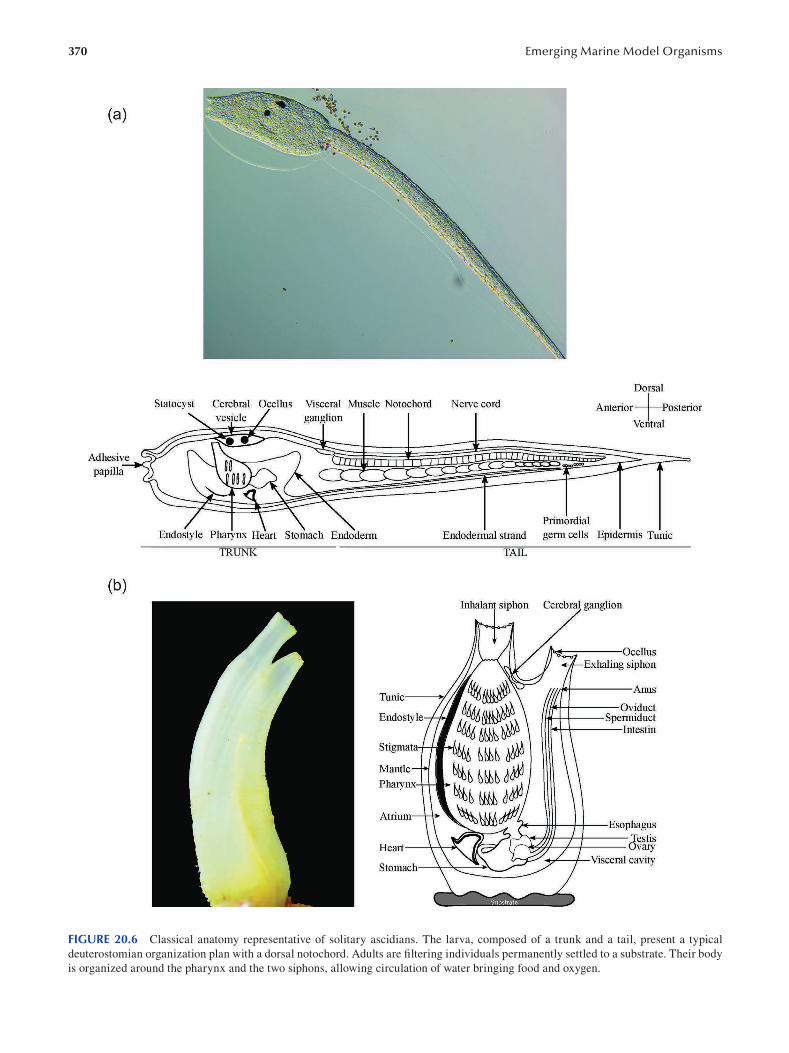
370 Emerging Marine Model Organisms
FIGURE 20.6 Classical anatomy representative of solitary ascidians. The larva, composed of a trunk and a tail, present a typical
deuterostomian organization plan with a dorsal notochord. Adults are filtering individuals permanently settled to a substrate. Their body
is organized around the pharynx and the two siphons, allowing circulation of water bringing food and oxygen.
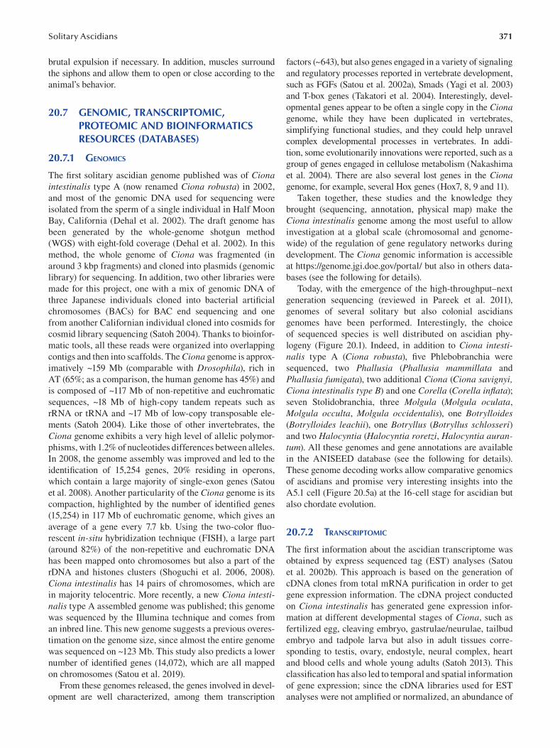
371 Solitary Ascidians
brutal expulsion if necessary. In addition, muscles surround
the siphons and allow them to open or close according to the
animal’s behavior.
20.7 GENOMIC, TRANSCRIPTOMIC, PROTEOMIC AND BIOINFORMATICS RESOURCES (DATABASES)
20.7.1 GENOMICS
The first solitary ascidian genome published was of Ciona intestinalis type A (now renamed Ciona robusta) in 2002,
and most of the genomic DNA used for sequencing were
isolated from the sperm of a single individual in Half Moon
Bay, California (Dehal et al. 2002). The draft genome has
been generated by the whole-genome shotgun method
(WGS) with eight-fold coverage (Dehal et al. 2002). In this
method, the whole genome of Ciona was fragmented (in
around 3 kbp fragments) and cloned into plasmids (genomic
library) for sequencing. In addition, two other libraries were
made for this project, one with a mix of genomic DNA of
three Japanese individuals cloned into bacterial artifi cial
chromosomes (BACs) for BAC end sequencing and one
from another Californian individual cloned into cosmids for
cosmid library sequencing ( Satoh 2004). Thanks to bioinfor
matic tools, all these reads were organized into overlapping
contigs and then into scaffolds. The Ciona genome is approx
imatively ~159 Mb (comparable with Drosophila), rich in
AT (65%; as a comparison, the human genome has 45%) and
is composed of ~117 Mb of non-repetitive and euchromatic
sequences, ~18 Mb of high-copy tandem repeats such as
rRNA or tRNA and ~17 Mb of low-copy transposable ele
ments (Satoh 2004). Like those of other invertebrates, the
Ciona genome exhibits a very high level of allelic polymor
phisms, with 1.2% of nucleotides differences between alleles.
In 2008, the genome assembly was improved and led to the
identification of 15,254 genes, 20% residing in operons,
which contain a large majority of single-exon genes (Satou
et al. 2008). Another particularity of the Ciona genome is its
compaction, highlighted by the number of identifi ed genes
(15,254) in 117 Mb of euchromatic genome, which gives an
average of a gene every 7.7 kb. Using the two-color fl uo
rescent in-situ hybridization technique (FISH), a large part
(around 82%) of the non-repetitive and euchromatic DNA
has been mapped onto chromosomes but also a part of the
rDNA and histones clusters (Shoguchi et al. 2006, 2008).
Ciona intestinalis has 14 pairs of chromosomes, which are
in majority telocentric. More recently, a new Ciona intestinalis type A assembled genome was published; this genome
was sequenced by the Illumina technique and comes from
an inbred line. This new genome suggests a previous overes
timation on the genome size, since almost the entire genome
was sequenced on ~123 Mb. This study also predicts a lower
number of identified genes (14,072), which are all mapped
on chromosomes (Satou et al. 2019).
From these genomes released, the genes involved in devel
opment are well characterized, among them transcription
factors (~643), but also genes engaged in a variety of signaling
and regulatory processes reported in vertebrate development,
such as FGFs (Satou et al. 2002a), Smads (Yagi et al. 2003)
and T-box genes (Takatori et al. 2004). Interestingly, devel
opmental genes appear to be often a single copy in the Ciona genome, while they have been duplicated in vertebrates,
simplifying functional studies, and they could help unravel
complex developmental processes in vertebrates. In addi
tion, some evolutionarily innovations were reported, such as a
group of genes engaged in cellulose metabolism (Nakashima
et al. 2004). There are also several lost genes in the Ciona genome, for example, several Hox genes (Hox7, 8, 9 and 11).
Taken together, these studies and the knowledge they
brought (sequencing, annotation, physical map) make the
Ciona intestinalis genome among the most useful to allow
investigation at a global scale (chromosomal and genome-
wide) of the regulation of gene regulatory networks during
development. The Ciona genomic information is accessible
at https://genome.jgi.doe.gov/portal/ but also in others data
bases (see the following for details).
Today, with the emergence of the high-throughput–next
generation sequencing (reviewed in Pareek et al. 2011),
genomes of several solitary but also colonial ascidians
genomes have been performed. Interestingly, the choice
of sequenced species is well distributed on ascidian phy
logeny (Figure 20.1). Indeed, in addition to Ciona intestinalis type A (Ciona robusta ), five Phlebobranchia were
sequenced, two Phallusia (Phallusia mammillata and
Phallusia fumigata), two additional Ciona (Ciona savignyi, Ciona intestinalis type B) and one Corella (Corella inflata);
seven Stolidobranchia, three Molgula (Molgula oculata,
Molgula occulta, Molgula occidentalis), one Botrylloides (Botrylloides leachii), one Botryllus (Botryllus schlosseri) and two Halocyntia (Halocyntia roretzi, Halocyntia aurantum). All these genomes and gene annotations are available
in the ANISEED database (see the following for details).
These genome decoding works allow comparative genomics
of ascidians and promise very interesting insights into the
A5.1 cell (Figure 20.5a) at the 16-cell stage for ascidian but
also chordate evolution.
20.7.2 TRANSCRIPTOMIC
The first information about the ascidian transcriptome was
obtained by express sequenced tag (EST) analyses (Satou
et al. 2002b). This approach is based on the generation of
cDNA clones from total mRNA purification in order to get
gene expression information. The cDNA project conducted
on Ciona intestinalis has generated gene expression infor
mation at different developmental stages of Ciona, such as
fertilized egg, cleaving embryo, gastrulae/neurulae, tailbud
embryo and tadpole larva but also in adult tissues corre
sponding to testis, ovary, endostyle, neural complex, heart
and blood cells and whole young adults (Satoh 2013). This
classification has also led to temporal and spatial information
of gene expression; since the cDNA libraries used for EST
analyses were not amplified or normalized, an abundance of
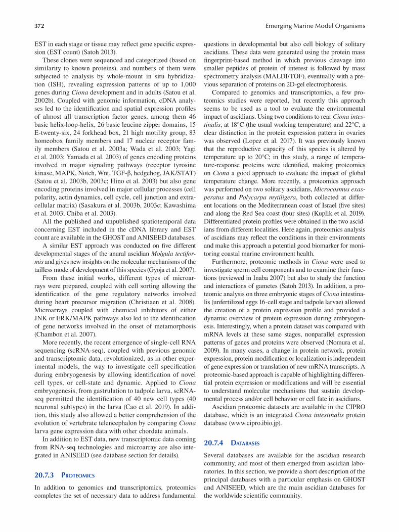
372
EST in each stage or tissue may reflect gene specifi c expres
sion (EST count) (Satoh 2013).
These clones were sequenced and categorized (based on
similarity to known proteins), and numbers of them were
subjected to analysis by whole-mount in situ hybridiza
tion (ISH), revealing expression patterns of up to 1,000
genes during Ciona development and in adults (Satou et al.
2002b). Coupled with genomic information, cDNA analy
ses led to the identification and spatial expression profi les
of almost all transcription factor genes, among them 46
basic helix-loop-helix, 26 basic leucine zipper domains, 15
E-twenty-six, 24 forkhead box, 21 high motility group, 83
homeobox family members and 17 nuclear receptor fam
ily members (Satou et al. 2003a; Wada et al. 2003; Yagi
et al. 2003; Yamada et al. 2003) of genes encoding proteins
involved in major signaling pathways (receptor tyrosine
kinase, MAPK, Notch, Wnt, TGF-β, hedgehog, JAK/STAT)
(Satou et al. 2003b, 2003c; Hino et al. 2003) but also gene
encoding proteins involved in major cellular processes (cell
polarity, actin dynamics, cell cycle, cell junction and extra
cellular matrix) (Sasakura et al. 2003b, 2003c; Kawashima
et al. 2003; Chiba et al. 2003).
All the published and unpublished spatiotemporal data
concerning EST included in the cDNA library and EST
count are available in the GHOST and ANISEED databases.
A similar EST approach was conducted on fi ve different
developmental stages of the anural ascidian Molgula tectiformis and gives new insights on the molecular mechanisms of the
tailless mode of development of this species (Gyoja et al. 2007).
From these initial works, different types of microar
rays were prepared, coupled with cell sorting allowing the
identification of the gene regulatory networks involved
during heart precursor migration (Christiaen et al. 2008).
Microarrays coupled with chemical inhibitors of either
JNK or ERK/MAPK pathways also led to the identifi cation
of gene networks involved in the onset of metamorphosis
(Chambon et al. 2007 ).
More recently, the recent emergence of single-cell RNA
sequencing (scRNA-seq), coupled with previous genomic
and transcriptomic data, revolutionized, as in other exper
imental models, the way to investigate cell specifi cation
during embryogenesis by allowing identification of novel
cell types, or cell-state and dynamic. Applied to Ciona embryogenesis, from gastrulation to tadpole larva, scRNA
seq permitted the identification of 40 new cell types (40
neuronal subtypes) in the larva (Cao et al. 2019). In addi
tion, this study also allowed a better comprehension of the
evolution of vertebrate telencephalon by comparing Ciona larva gene expression data with other chordate animals.
In addition to EST data, new transcriptomic data coming
from RNA-seq technologies and microarray are also inte
grated in ANISEED (see database section for details).
20.7.3 PROTEOMICS
In addition to genomics and transcriptomics, proteomics
completes the set of necessary data to address fundamental
Emerging Marine Model Organisms
questions in developmental but also cell biology of solitary
ascidians. These data were generated using the protein mass
fingerprint-based method in which previous cleavage into
smaller peptides of protein of interest is followed by mass
spectrometry analysis (MALDI/TOF), eventually with a pre
vious separation of proteins on 2D-gel electrophoresis.
Compared to genomics and transcriptomics, a few pro
teomics studies were reported, but recently this approach
seems to be used as a tool to evaluate the environmental
impact of ascidians. Using two conditions to rear Ciona intestinalis, at 18°C (the usual working temperature) and 22°C, a
clear distinction in the protein expression pattern in ovaries
was observed (Lopez et al. 2017 ). It was previously known
that the reproductive capacity of this species is altered by
temperature up to 20°C; in this study, a range of tempera
ture-response proteins were identified, making proteomics
on Ciona a good approach to evaluate the impact of global
temperature change. More recently, a proteomics approach
was performed on two solitary ascidians, Microcosmus exasperatus and Polycarpa mytiligera, both collected at differ
ent locations on the Mediterranean coast of Israel (fi ve sites)
and along the Red Sea coast (four sites) (Kuplik et al. 2019).
Differentiated protein profiles were obtained in the two ascid
ians from different localities. Here again, proteomics analysis
of ascidians may reflect the conditions in their environments
and make this approach a potential good biomarker for moni
toring coastal marine environment health.
Furthermore, proteomic methods in Ciona were used to
investigate sperm cell components and to examine their func
tions (reviewed in Inaba 2007) but also to study the function
and interactions of gametes (Satoh 2013). In addition, a pro
teomic analysis on three embryonic stages of Ciona intestina
lis (unfertilized eggs 16-cell stage and tadpole larvae) allowed
the creation of a protein expression profile and provided a
dynamic overview of protein expression during embryogen
esis. Interestingly, when a protein dataset was compared with
mRNA levels at these same stages, nonparallel expression
patterns of genes and proteins were observed (Nomura et al.
2009). In many cases, a change in protein network, protein
expression, protein modification or localization is independent
of gene expression or translation of new mRNA transcripts. A
proteomic-based approach is capable of highlighting differen
tial protein expression or modifications and will be essential
to understand molecular mechanisms that sustain develop
mental process and/or cell behavior or cell fate in ascidians.
Ascidian proteomic datasets are available in the CIPRO
database, which is an integrated Ciona intestinalis protein
database (www.cipro.ibio.jp).
20.7.4 DATABASES
Several databases are available for the ascidian research
community, and most of them emerged from ascidian labo
ratories. In this section, we provide a short description of the
principal databases with a particular emphasis on GHOST
and ANISEED, which are the main ascidian databases for
the worldwide scientifi c community.
373 Solitary Ascidians
• The Ascidians Chemical Biology Database (ACBD)
(created in 2010 in Japan) is a bibliographical data
base that compiles publications concerning the
effect of chemical compounds on ascidian devel
opment and tends to promote ascidians as a model
organism for whole-animal chemical screening.
• The Database of Tunicate Gene Regulation (DBTGR)
(created in 2005 in Japan) focuses on tunicate gene
regulation, including regulatory elements in the pro
moter region and the associated TF. In addition, it
integrates a list of gene reporter constructs.
• The website of the Joint Genome Institute (JGI) (cre
ated in 1997 in United States) hosts the Ciona intestinalis type A genome and contains a genome browser.
• MAboya Gene Expression pattern and Sequence
Tags (MAGEST) (created in 2000 in Japan) pro
vides Halocyntia roretzi 3’- and 5’-tag sequences
(20,000 clones) from the fertilized egg cDNA
library, the amino acid fragment sequences pre
dicted from the EST data set and the expression
data from whole-mount in situ hybridization.
• Ciona intestinalis Adult In Situ hybridization
Database (CiAID) (created in 2009 in Japan) gives
access to gene expression patterns in adult juve
niles with a body atlas.
• The Ciona intestinalis Protein (CIPRO) database
(created in 2006 in Japan) is a Ciona intestinalis pro
tein database that contains 3D expression profi ling,
2D-PAGE and mass spectrometry-based large-scale
analyses at various developmental stages, curated
annotation data and various bioinformatic data.
• Four-Dimensional Ascidian Body Atlas (FABA)
(created in 2010 in Japan) contains ascidian three-
dimensional (3D) and cross-sectional images
through the developmental time course (from fer
tilized egg to larva) to allow morphology compari
son and provide a guideline for several functional
studies of a body plan in chordate. Note that a
second database called FABA2 (created in Japan)
exists, focusing on later developmental stages, from
hatching to seven-day-old juveniles.
• Ciona intestinalis Transgenic Line RESsources
(CITRES) (created in 2012 in Japan) provides
the ascidian research community with transgenic
lines but also contains DNA constructs to perform
transgenesis, image collections of Ciona GFP-
expressing strains and publications.
• Ghost database (originally created in 2002 in
Japan; http://ghost.zool.kyoto-u.ac.jp/) is one of
the first ascidian databases available for the ascid
ian research community and the most useful from
the beginning. This database provides all the data
concerning the Ciona intestinalis EST project con
ducted by Satoh’s lab (see transcriptomic section
for details), such as EST count, that provide tem
poral expression information and published and
unpublished ISH at several developmental stages.
In addition to that, the database contains a genome
browser, a search engine for specifi c expression
or expression pattern of a given genes and gene
annotation. At the beginning, this database repre
sented an extraordinary source of molecular tools,
since it provides a set of 13,464 unique cDNA
clones available as the “Ciona intestinalis gene
collection released” for the scientifi c community,
ready for use in cDNA cloning, microarray analy
sis and other genome-wide analyses. Almost the
entire database is now integrated in the ANISEED
database.
• ANISEED (created in 2010 in France; www.ani
seed.cnrs.fr) is the biggest and most complete
database for the ascidian community (Dardaillon
et al. 2019). There is a constant input of new data,
and it provides functionally annotated gene and
transcript models in both wild-type and experi
mentally manipulated conditions using formal
anatomical ontologies. The advantages of this
database are the extra information, going beyond
genes by pointing out repeated elements and cis-
regulatory modules and also providing orthol
ogy comparison within or even outside ascidians
(tunicates, echinoderms, cephalochordates and
vertebrates). There are enhanced functional anno
tations for each species, achieved by an improved
orthology detection and manual curation of gene
models. This database is user friendly, with three
types of browsers, each offering a different but
complementary point of view: a developmen
tal browser which selects data based either on
the gene expression or the territory of interest,
an advanced genomic browser focusing on gene
sets and gene regulation and a genomicus synteny
browser that explores the conservation of local
gene order across deuterostome. This later new
release has a reference of the taxonomic range of
14 species, among them a non-ascidian species,
the appendicularian Oikopleura dioika, which is a
novelty. Finally, the new and powerful Morphonet
morphogenetic browser enables a 4D exploration
of gene expression profiles and territories.
20.8 FUNCTIONAL APPROACHES/TOOLS FOR MOLECULAR AND CELLULAR ANALYSES
In addition to classical over/ectopic expression of genes, several
tools or technical approaches were developed by the ascidian
community by taking advantage of biological particularities
and/or experimental advantages offered by solitary ascidians.
20.8.1 MICROINJECTION/ELECTROPORATION
To follow specific expression patterns of regulatory genes
or to probe gene function, experimental biologists usu
ally introduce reporter constructs or synthetic mRNA in
374
fertilized eggs. In most animal models, these approaches are
usually achieved by the microinjection technique. Solitary
ascidians, essentially Ciona, allow an alternative technique,
a simple electroporation method. This permits manipula
tion and screening of hundreds of synchronous developing
embryos, either wild type or mutant, thus allowing greater
confidence in functional screening, which is not possible
with most of the other animal models.
20.8.2 REPORTER GENE
The efficient introduction of reporter constructs by
electroporation (Corbo et al. 1997), coupled with the
facility (compared to the other animal models) to identify
and clone the core promoter and associated enhancers of a
given gene, made the solitary ascidian Ciona intestinalis an
excellent model to study cis-regulation. Indeed, due to the
Ciona compact genome, the cis-regulatory elements (CREs)
are usually located within the first 1.5 kb upstream of the
transcription start site, making it relatively easy to capture
significant transcriptional units and clone them upstream
of a reporter gene to drive its expression. Coupled with the
electroporation technique, this allows a simple and rapid
generation of hundreds of transient transgenic embryos
expressing fluorescent proteins, which develop quickly to
the larval stage ( Zeller et al. 2006). These transient assays
allowed rapid identification and characterization of up to
83 Ciona cis-regulatory elements, almost all enhancers,
which activate transcription in a more or less tissue-specifi c
manner (reviewed in Irvine 2013).
20.8.3 LOSS-OF-FUNCTION APPROACHES
To understand the molecular basis of development, experi
mental biologists expect to specifically inhibit the functions
of a particular gene in particular cells at particular devel
opmental stages. The basic technologies for examining
gene functions by loss of function approaches have been
established in Ciona, such as the knockdown of genes by
antisense morpholino oligonucleotides (MOs) (Satou et al.
2001 ), transposon-mediated germ cell transformation and
mutagenesis (Sasakura et al. 2003c, Sasakura et al. 2005),
zinc-finger nucleases (ZFNs) (Kawai et al. 2012), transcrip
tional activator-like effector nucleases (TALENs) ( Treen
et al. 2014) and clustered regularly interspaced short palin
dromic repeats (CRISPR/Cas9) (Sasaki et al. 2014). These
technologies have supported detailed and thorough analyses
to reveal molecular and cellular mechanisms that underlie
development of Ciona, since almost of them can be per
formed in a tissue-specific manner during embryogenesis.
20.8.3.1 MOs The antisense morpholino oligonucleotide strategy con
sists of MOs that bind to the targeted mRNA and prevent
translation. They were tested in a range of models, including
Emerging Marine Model Organisms
ascidians, in which they were extensively used since they
allow a rapid and high-throughput approach for functional
studies. In addition, MOs are able to target maternal mRNA
determinants as well as zygotic genes. The efficiency of this
technique was fi rst tested in Ciona savignyi, in which MOs
were able to target the maternal pool of β-catenin mRNA and
abolish endodermal differentiation (Satou et al. 2001). Since
then, MOs were extensively used and allowed identifi cation
of key genes in tissue differentiation during embryogen
esis, such as the maternal determinant Macho-1 for muscle
differentiation in Halocyntia roretzi (Nishida and Sawada
2001) and in Ciona savignyi (Satou et al. 2002c); in tissue
formation, for instance, chondroitin-6-O-sulfotransferase
involved in Ciona intestinalis notochord morphogenesis
(Nakamura et al. 2014); or even for cell fate, for example,
Ci-Sushi, which controls the initiation of apoptosis at the
onset of Ciona intestinalis metamorphosis (Chambon et al.
2007). However, there are several limitations to injecting
MOs in solitary ascidians, notably the restricted numbers
of mutants to analyze and the difficulty of interpreting some
phenotypes due to off-target effects.
20.8.3.2 RNA Interference Based on the introduction in the cells of double-strand RNA,
which are converted in small interfering RNA (siRNA),
causing the destruction of specific mRNA, this approach
was successfully used in colonial ascidians but has had few
successes with solitary ones, except the electroporation of
short-hairpin RNA targeting tyrosinase-encoding gene in
Ciona embryo leading to the absence of melanization of the
tailbud pigmented cells (Nishiyama and Fujiwara 2008). To
date, the use in solitary ascidians is very limited.
20.8.3.3 ZNFs and TALENs The nuclease activity ZNFs and TALENs induces dou
ble-strand breaks (DSBs) at target sequences. In the case
of ZNFs, mutations occur when DSBS are repaired by
non-homologous end joining (NHEJ), which introduces
insertional or deletional mutations at the target sequence.
TALENs provoke mutations when the cellular DNA repair
mechanisms fail. Both approaches were established in Ciona intestinalis by the Sasakura lab (Kawai et al. 2012; Treen et
al. 2014) and are a very promising strategy to mutate endog
enous genes during development. ZNFs were tested in a
Ciona transgenic line expressing EGFP to introduce muta
tions in EGFP loci. When eggs were injected, it resulted in
inheritable mutations with high frequency (about 100%),
no toxic effect on embryogenesis and few off-target effects
(Kawai et al. 2012). TALEN knockouts can be performed
by electroporation and allow fast generation of mutants and
a quick screening involving numbers of embryos not pos
sible with other animals. Toxicity is a major concern with
TALEN when ubiquitous knockouts are generated, but using
tissue-specific promoters reduces this problem and allows
mutations in a tissue-specific manner (Treen et al. 2014).
375 Solitary Ascidians
20.8.3.4 CRISPR/Cas 9 Since its discovery in 1987 (Ishino et al. 1987), CRISPR/Cas9
has become one of the most powerful tools for researchers
to alter the genomes of a large range of organisms. CRISPR/
Cas9 uses a short guide RNA (sgRNA) that binds to its target
site; Cas9 protein is recruited to the binding site and induces
a double-strand break at the target genomic region. In soli
tary ascidians, this technique was first successfully tested in
Ciona intestinalis (Sasaki et al. 2014) and more recently in
Phallusia mammillata (McDougall et al. 2021).
In Ciona, the most widely used application of CRISPR is
for targeted mutagenesis in somatic cells of electroporated
embryos. In this method established in 2014 by Sasakura lab
(Sasaki et al. 2014) and recently improved by Stolfi (Gandhi
et al. 2018), in vitro fertilized one-cell-stage embryos are
electroporated with plasmids, allowing the zygotic expres
sion of Cas9 protein and sgRNA. Interestingly, Cas9 can be
expressed in a cell-specific manner, and the targeted muta
tions are a powerful means to dissect the tissue-specifi c
functions of a gene during development.
20.8.4 GENETICS, MUTAGENESIS AND TRANSGENESIS
Natural mutants often arise in wild populations, probably
due to the high polymorphism between individuals within a
given population (Satoh 2013). Moreover, the rapid life cycle
and the possibility of self-fertilization (natural or induced
with chemical or enzyme treatment), coupled with a rapid
embryogenesis and a morphologically simple tadpole that
allows simple phenotype detection, make both Ciona (intestinalis and savignyi) excellent models for mutagenesis. In
addition to characterization of the Ciona savignyi natural
mutant frimousse (Deschet and Smith 2004), Smith’s lab
took advantage of the self-fertility in this species to perform
a mutagenesis screen notably using N-ethyl-N-nitrosourea
(ENU)-induced mutations affecting early development. This
random approach led to the isolation of a number of mutants
with notochord defects such as chongmague and chobi (Nakatani et al. 1999). Since then, the transgenesis tech
nique was established in Ciona using transposon-mediated
transgenesis that allow creation of stable germ lines but also
to use it for insertional mutagenesis and enhancer trapping.
The Tc1/mariner transposable element Minos (isolated
from Drosophila hydei) is a small DNA transposon (2000
bp) activated by a “cut and paste” system in which a trans
posase is able to excise the transposon from the DNA and
integrate it into a target sequence. When a plasmid contain
ing Minos is microinjected or electroporated in Ciona eggs
with transposase mRNA, Minos is excised from the vector
DNA and integrated in the Ciona genome, and this event is
observed in somatic and germ cells (Sasakura et al. 2003c).
In the latter case, this insertion is inherited by the progeny,
and its stability was reported over ten generations in several
transgenic lines (Sasakura 2007). Insertions of Minos can
disrupt gene function to create mutants, such as the swimming juvenile, which exhibits a cellulose synthesis defect and
absence of tail regression during metamorphosis due to the
integration of Minos at Ci-CesA promoter, a gene involved
in cellulose synthesis in Ciona intestinalis (Sasakura et al.
2005). In addition to insertional mutagenesis, the transpo
son-based technique was also able to create stable marker
lines when CRE of tissue-specific gene driving expression
of fluorescent proteins were used with a Minos -based trans
posable element. Another potentiality of Minos transposons
is the enhancer trapping technique. It consists of insertions
using a reporter gene in a Minos transposons construct
(GFP, for example), and if there is an enhancer close to the
transposon insertions, the expression patterns of reporter
genes are affected according to the enhancer. In Ciona , an
intronic enhancer in the Ci-Musashi gene was identifi ed by
this approach (Awazu et al. 2004).
20.9 CHALLENGING QUESTIONS
Researchers in the ascidian field face many challenging
questions. In this section, a brief overview of some of them
will be given, followed by a detailed discussion of the unique
opportunity provided by the ascidians to develop quantita
tive modeling of chordate embryos.
20.9.1 EVOLUTION OF ASCIDIANS
As described in the genomic section, 11 ascidian genomes
are now sequenced and annotated, some of them with tran
scriptomic data and identification of cis-regulatory modules.
In addition, the compilation of these data in the ANISEED
database will greatly facilitate comparative developmen
tal genomics between ascidian species and allow new
insights in ascidian evolution. Immediate application of this
approach could lead to better understanding of the differ
ences in gene-regulatory networks during embryogenesis
observed between Ciona intestinalis and Halocyntia roretzi (see embryogenesis section for details). Indeed, these
two species exhibit at least two differences for notochord
and muscle secondary lineage which both require FGF but
dependent on nodal and Delta/Notch for Ciona and inde
pendent of both of them for Halocyntia. Further analyses of
the developmental genomics of these two species may allow
evolutionary inference to better understand these changes.
Another example concerns the phenotypic change observed
in several species that do not develop a tail during embryogen
esis and do not develop notochord or tail muscles; instead,
they give rise to non-motile tail-less larva without functional
notochord or larval tail muscle or directly to a juvenile (Satoh
2013). Anural development occurred independently several
times during ascidian evolution. Cross-fertilization approach
of the tail-less Molgula occulta, and its close relative urodele
species Molgula oculata gives rise to a hybrid embryo with a
short tail containing a notochord. Swalla and Jeffery (1990)
suggested an evolution of the anural mode of development
by relatively simple genetic changes. Comparative genomics
studies permitted by the release of the genome of these two
376
species will certainly detect key genomics changes for these
different modes of embryogenesis.
20.9.2 ASCIDIANS FOR THERAPEUTIC ADVANCES
In the last few years, several studies have been conducted on
the identification and characterization of chemical diversity
produced from marine ascidians (Palanisamy et al. 2017).
The essential part of these chemical compounds is used by
ascidian species to prevent predatory fish, as an anti-fouling
and anti-microbial mechanism and to control settlement
(reviewed in Watters 2018). Ascidians, like several marine
organisms, produce a rich variety of secondary metabolites
with potential therapeutic properties in human medicine,
with a range of biological activities such as cytotoxicity,
antibiotic and immunosuppressive activities, inhibition of
topoisomerases and cyclin-dependent kinases (Duran et al.
1998). Most of these compounds were identified by the liq
uid chromatography-mass spectrometry method. Among
them, Ecteinascidin was isolated from Ecteinascidia turbinata and is currently used as a cancer drug to treat soft-
tissue sarcoma and ovarian cancer (Gordon et al. 2016);
Aplidin isolated from Aplidium albicans has given prom
ising results in myeloma treatment (Delgado-Calle et al.
2019). In addition, anti-malarial effects were identifi ed
from extracts coming from three ascidians, Microcosmus goanus, Ascidia sydneiensis and Phallusia nigra ( Mendiola
et al. 2006). Between 1994 and 2014, up to 580 compounds
were isolated from ascidians and offer a wide range of
opportunities to identify molecules with therapeutic prop
erties for human diseases.
In addition to screening for molecules with potential ther
apeutic effects, ascidian embryos have also started to be used
as an experimental model to study the neurodevelopmental
toxicity of different compounds (Dumollard et al. 2017).
20.9.3 WHEN DEVELOPMENTAL BIOLOGY
BECOMES QUANTITATIVE: A BIG STEP
TOWARD “COMPUTABLE EMBRYOS”
The transition from a single fertilized cell to a complex organ
ism, with various cell types that compose its tissues in the
correct numbers and their fine regulation in space and time,
is the question at the heart of developmental biology. Decades
of research in this field have designed a broad portrait of the
fundamental processes involved during embryogenesis: from
the description of the genetic programs of embryonic cells
and the mechanisms regulating gene transcription to how cell
fates and behaviors are coordinated by cell communication
and the way this translates into morphogenesis.
Developmental mechanisms have traditionally been stud
ied at the tissue level in a qualitative manner. For example,
consider the current view of the classical chemical signal
ing during fate specification. A surprisingly small number
of signaling pathways involving cell surface receptor and
activating ligands act in widely different cellular contexts
Emerging Marine Model Organisms
to produce the diversity of fate specification events occur
ring during embryogenesis (Perrimon et al. 2012). Despite
this, many simple questions remain unanswered, such as:
“What is the mechanism regulating the dose-response to
increasing concentrations of ligands or receptors?” “How
are ligand concentration and time of exposure integrated by
cells?” To deepen the understanding of the principles which
govern embryonic development, it is important to combine
quantitative experimental approaches at the cellular scale
with dynamic mathematical models including mechanistic
details. For example, the recent development in quantitative
imaging, sequencing, proteomics and physical measure
ments have allowed us to refine the historical morphogen
concept, in which diffusible signaling molecules are pro
posed to coordinate cell fate specification and tissue forma
tion using concentration-dependent mechanisms (a static
readout), because it was insufficient to describe or model the
complexities of patterning observed with these techniques
in developing embryos (Garcia et al. 2020; Huang and
Saunders 2020; Jaeger and Verd 2020; Rogers and Müller
2020; Schloop et al. 2020).
While physical modeling of life has a long history
(Thompson 1917), it has remained a theoretical exercise for a
long time: insuffi cient measurements of physical parameters
for constraining models coupled with a largely qualitative
and static description of phenotypes have rendered it diffi cult
to apply physics to developing embryos and even to single
cells. The recent technological breakthrough mentioned pre
viously, however, reduced this difficulty while making “com
putable embryos” through a precise physical description of
embryonic development more necessary than ever to capture
key developmental concepts and bridge genomic informa
tion and dynamic phenotypes (Biasuz et al. 2018). First, our
brains are simply unable to cope with the large amount of
data generated, much of which are unrelated to the mecha
nism being studied. Second, biology involves several layers
of feedback, resulting in unintuitive non-linear behaviors.
Third, biology is a multiscale process in which macroscopic
properties of cells and tissues arise from the mesoscopic
properties of molecules or subcellular structures.
Ascidians definitively constitute a model of choice to
build a global computational model of embryogenesis.
Embryonic development is a continuous progression in time.
The “computable embryo” is based on the idea that a mathe
matical description of the system can predict the future state
of the embryo from the knowledge of its current state. This
global computational model of embryogenesis at the single-
cell, genome-wide and whole-embryo level is a challenging
task and will only be achieved using the most appropriate
developmental systems (Biasuz et al. 2018).
Solitary ascidian embryos seem to be good candidates
for this breakthrough. At first glance, one would rather
think of the Drosophila melanogaster or vertebrate embryos
for this role. Indeed, thanks to decades of research, a deep
understanding of core developmental mechanisms has been
achieved, and powerful genetic and cell biology tools exist.
377 Solitary Ascidians
These embryos, while remaining a significant motor for defi n
ing new concepts, may, however, be too complicated to incor
porate these concepts into a global model of embryogenesis.
In contrast, ascidian embryos, as nematodes, are simpler and
develop stereotypically with few cells and invariant cell lin
eages, so that each cell can be named and found at the same
position in all embryos (Lemaire 2009). Unlike those of nem
atodes (Goldstein 2001), ascidian embryo geometries have
even remained essentially unchanged since the emergence
of the group, around 400 million years ago, despite exten
sive genomic divergence (Delsuc et al. 2018; Lemaire 2011).
The development of ascidians is also characterized by ear
lier fate restriction than most animal embryos: 94 of the 112
early gastrula cells in the ascidian Ciona are fate restricted,
each contributing to a single larval tissue type (Nishida 1987).
Moreover, ascidians are closely related to vertebrates, as they
belong to the vertebrate sister-group, but ascidians kept their
genomic simplicity. Indeed, they diverged before the two
rounds of whole-genome duplication events which occurred
in the vertebrate lineage leading to the apparition of multiple
paralogues for each gene (Dehal and Boore 2005), with poten
tially slightly divergent activities. Finally, ascidian embryos
are small (~130 μm) and transparent, and they develop rapidly
externally in sea water up to the larval stage (~12 h), making
them very easy to image. Thus, ascidian embryos provide a
rigid framework that allows combination of analyses at cel
lular resolution with mathematical modeling.
These advantageous properties of ascidian embryos,
especially Phallusia mammillata embryos, which are fully
transparent, combined with the breakthrough development
of light sheet microscopy (Power and Huisken 2017), have
enabled the production of the first digitized version of a meta
zoan embryo (Figure 20.7a) (Guignard et al. 2020). Based on
automatic whole-cell segmentation and tracking over fi ve cell
generations of membrane-labeled cells with two-minute tem
poral resolution, this research offers a complete description
of early ascidian embryo development, accounting for each
cell in the ten embryos analyzed. Moreover, this quantita
tive and dynamic atlas of cell positions and geometries can
be associated with the known cell fates and interactively
explored through the MorphoNet online morphological
browser (Leggio et al. 2019). These “digital embryos” show
that ascidian development is reproducible down to the scale of
cell–cell contacts and, combined with modeling and experi
mental manipulations, it allows us to establish contact area-
dependent inductions as an alternative to classical morphogen
gradients. This work opens the door to quantitative single-cell
morphology and mechanical morphogenesis modeling.
In parallel with this work, another group combined high-
resolution single-cell transcriptomics (single-cell RNA
sequencing) and light-sheet imaging to build the fi rst full
comprehensive atlas which describes the genome-wide
gene expression of every single cell of an embryo in the
early stages of development, showing the evolution from a
single cell up to gastrulation in the ascidian Phallusia mammillata (Sladitschek et al. 2020). By providing a complete
representation of the gene expression programs, which
instruct individual cells to form the different cell types
necessary to build an embryo, and therefore by allowing us
to know precisely cell-specific expression of transcription
factors at the single-cell level, this study will signifi cantly
enhance current single-cell-based gene regulatory network
inference algorithms (Aibar et al. 2017) and will help to
further develop single-cell-based physical models of the dif
ferent steps of transcriptional control during development.
Moreover, these single-cell gene expression data will feed
several layers of physical description of biological processes.
For example, identification of cell-adhesion molecules will
allow the refining of morphogenetic models, such as oriented
cell divisions, cell shape changes or cell neighbor exchanges
models (Etournay et al. 2015), thereby linking mechanical
and genetic information at the cellular resolution.
In spite of the convenient properties and the recent
advances that have been realized thanks to the ascidian
embryo model, there is still a long way to go to be able to
“compute the embryo”.
Typically, studies at the single-cell level are in their early
days, as can be illustrated by signal transduction studies.
The MAPK/ERK signaling pathway is one of the important
embryonic signaling pathways used by vertebrates and inver
tebrates, controlling many physiological processes (Lavoie
et al. 2020), and is the main inducing pathway in early ascid
ian embryos (Lemaire 2009). The signaling cascade from the
activation of the transmembrane receptor to the phosphory
lation of the ERK nuclear targets is well described (Figure
20.7b) (Lavoie et al. 2020). Our current knowledge of this
pathway is, however, mostly static, and an integrated under
standing of its spatio-temporal dynamics is lacking (Patel
and Shvartsman 2018). For example, it has been shown that
the ERK pathway can trigger two qualitatively different types
of ERK activity: pulsatile or continuous (Aikin et al. 2019).
To understand these non-intuitive results, it is important to
combine quantitative experimental approaches at the cellu
lar resolution with dynamic mathematical models including
mechanistic details. Genetically encoded fl uorescent activ
ity sensors that convert kinase activity into nucleocytoplas
mic events have been recently developed (Durandau et al.
2015; Regot et al. 2014), and these tools can now be coupled
with optogenetic systems in order to activate the ERK path
way with high spatiotemporal accuracy at different levels
(Gagliardi and Pertz 2019). However, these techniques were
only used to track a single pathway component at a time. Yet
they suggest that multiplexing sensors at different levels of
the cascade could reveal the dynamics of information fl ow
through the cell. Such quantitative measures are required
to more realistically model the catalogue of cell-signaling
modalities (Biasuz et al. 2018).
The technological breakthroughs of the last quarter of
the century have brought a whole new perspective to devel
opmental biology, which is now seen through the combined
lenses of mathematical modeling and experimental biology.
A major challenge for the future will now be to integrate
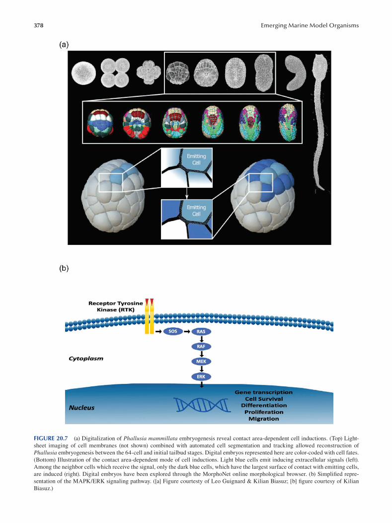
378 Emerging Marine Model Organisms
FIGURE 20.7 (a) Digitalization of Phallusia mammillata embryogenesis reveal contact area-dependent cell inductions. (Top) Light-
sheet imaging of cell membranes (not shown) combined with automated cell segmentation and tracking allowed reconstruction of
Phallusia embryogenesis between the 64-cell and initial tailbud stages. Digital embryos represented here are color-coded with cell fates.
(Bottom) Illustration of the contact area-dependent mode of cell inductions. Light blue cells emit inducing extracellular signals (left).
Among the neighbor cells which receive the signal, only the dark blue cells, which have the largest surface of contact with emitting cells,
are induced (right). Digital embryos have been explored through the MorphoNet online morphological browser. (b) Simplifi ed repre
sentation of the MAPK/ERK signaling pathway. ([a] Figure courtesty of Leo Guignard & Kilian Biasuz; [b] figure courtesy of Kilian
Biasuz.)
379 Solitary Ascidians
partial models accounting for short-term activities into a
global view of biological processes. Indeed, most of the
modeling efforts were designed to shed light on specifi c
processes over a short period of time. As a consequence,
our physical knowledge of embryogenesis is reduced to a
few unconnected kernels of insight. Increasing the number
of kernels is imperative to “compute the embryo” but will
not suffice: kernels will need to be incorporated into a bigger
picture. The solitary ascidian embryos, which are simple and
transparent and contain a relatively small number of cells
and invariant cell lineages are perfect candidates to inte
grate these principles into a global model of embryogenesis.
20.10 GENERAL CONCLUSION
The last 20 years have been marked by extraordinary
advances in the comprehensive biology of ascidians. Starting
as the first experimental model organism in embryology,
the ascidian embryo offers today an avenue of investigation
in several biological research fields such as developmental
biology, cell biology, comparative genomics, drug screening
or evo-devo. The decoded genome of 13 ascidians, coupled
with gene annotation, large transcriptomic data, proteomics,
identification of cis-regulatory elements, large coverage of
gene expression patterns by in situ hybridization, stereo
typed and well-described cell lineages, physical maps of
the genome onto chromosomes and routine generation of
transgenic lines combined with cell line markers and single-
cell transcriptomics (supported by FACS) render this “old”
marine model one of the most promising for modern biology.
ACKNOWLEDGMENTS
The authors want to thank A. Karaiskou for critical read
ing of the manuscript and C. Dantec for her help with the
database section.
BIBLIOGRAPHY
Aibar, S., González-Blas, C.B., Moerman, T., Huynh-Thu, V.A.,
Imrichova, H., Hulselmans, G., Rambow, F., Marine, J.C.,
Geurts, P., Aerts, J., van den Oord, J., Atak, Z.K., Wouters,
J. & Aerts, S. 2017. SCENIC: Single-cell regulatory net
work inference and clustering. Nature Methods 14(11):
1083–1086.
Aikin, T.J., Peterson, A.F., Pokrass, M.J., Clark, H.R. & Regot, S.
2019. Collective MAPK signaling dynamics coordinates epi
thelial homeostasis. bioRxiv 826917.
Arsenault, G., Davidson, J. & Ramsay, A. 2009. Temporal and spa
tial development of an infestation of Styela clava on mus
sel farms in Malpeque Bay, Prince Edward Island, Canada.
Aquatic Invasions 4: 189–194.
Awazu, S., Sasaki, A., Matsuoka, T., Satoh, N. & Sasakura, Y. 2004.
An enhancer trap in the ascidian Ciona intestinalis identifi es
enhancers of its Musashi orthologous gene. Developmental Biology 275: 459–472.
Berrill, N.J. 1929. Digestion in ascidians and the influence of tem
perature. Journal of Experimental Biology 6: 275–292.
Berrill, N.J. 1932a. The mosaic development of the ascidian egg.
The Biological Bulletin 63: 381–386.
Berrill, N.J. 1932b. Ascidians of the Bermudas. The Biological Bulletin 62: 77–88.
Berrill, N.J. & Sheldon, H. 1964. The fine structure of the connec
tions between muscle cells in ascidian tadpole larva. Journal of Cell Biology 23: 664–669.
Berrill, N.J. & Watson, D.M.S. 1930. II: Studies in tunicate devel
opment. Part I: General physiology development of simple
ascidians. Philosophical Transactions of the Royal Society of London: Series B, Containing Papers of a Biological Character 218: 37–78.
Berrill, N.J. & Watson, D.M.S. 1936. II: Studies in tunicate devel
opment. Part V: The evolution and classification of ascidians.
Philosophical Transactions of the Royal Society of London: Series B, Biological Sciences 226: 43–70.
Biasuz, K., Leggio, B., Faure, E. & Lemaire, P. 2018. The “com
putable egg”: Myth or useful concept? Current Opinion in Systems Biology 11: 91–97.
Bourque, D., Davidson, J., MacNair, N.G., Arsenault, G., LeBlanc,
A.R., Landry, T. & Miron, G. 2007. Reproduction and early
life history of an invasive ascidian Styela clava Herdman
in Prince Edward Island, Canada. Journal of Experimental Marine Biology and Ecology 342: 78–84.
Cao, C., Lemaire, L.A., Wang, W., Yoon, P.H., Choi, Y.A., Parsons,
L.R., Matese, J.C., Wang, W., Levine, M. & Chen, K. 2019.
Comprehensive single-cell transcriptome lineages of a proto
vertebrate. Nature 571: 349–354.
Chabry, L. 1887. Contribution à l’embryologie normale et téra
tologique des ascidies simples. Journal of Anatomy and Physiology (Paris) 23: 167–319.
Chambon, J.-P., Nakayama, A., Takamura, K., McDougall, A. &
Satoh, N. 2007. ERK- and JNK-signalling regulate gene
networks that stimulate metamorphosis and apoptosis in tail
tissues of ascidian tadpoles. Development 134: 1203–1219.
Chambon, J.-P., Soule, J., Pomies, P., Fort, P., Sahuquet, A.,
Alexandre, D., Mangeat, P.-H. & Baghdiguian. 2002. Tail
regression in Ciona intestinalis (Prochordate) involves a
Caspase-dependent apoptosis event associated with ERK
activation. Development 129: 3105–3114.
Chen, J.-Y., Huang, D.-Y., Peng, Q.-Q., Chi, H.-M., Wang, X.-Q. &
Feng, M. 2003. The first tunicate from the Early Cambrian of
South China. PNAS 100: 8314–8318.
Chiba, S., Awazu, S., Itoh, M., Chin-Bow, S.T., Satoh, N., Satou, Y. &
Hastings, K.E.M. 2003. A genomewide survey of developmen
tally relevant genes in Ciona intestinalis. Development Genes and Evolution 213: 291–302.
Chistyakov, V.G., Kalmykova, N.A., Nesov, L.A. & Suslov, G.A.
1984. О наличии вендских отложений в среднем течении р. Онеги и возможном существовании оболочечников (Tunicata: Chordata) в докембрии [On the presence of
vendian deposits in the middle course of the Onega River
and presumable existence of tunica (Tunicata: Chordata) in
Precambrian]. Vestnik Leningrad University (LGU) 6: 11–19.
Christiaen, L., Davidson, B., Kawashima, T., Powell, W., Nolla, H.,
Vranizan, K. & Levine, M. 2008. The transcription/migration
interface in heart precursors of Ciona intestinalis. Science 320: 1349–1352.
Christiaen, L., Stolfi, A., Davidson, B. & Levine, M. 2009. Spatio
temporal intersection of Lhx3 and Tbx6 defines the cardiac
field through synergistic activation of Mesp. Developmental Biology 328: 552–560.
Christiaen, L., Stolfi , A. & Levine, M. 2010. BMP signaling coor
dinates gene expression and cell migration during precardiac
380
mesoderm development. Developmental Biology 340:
179–187.
Cloney, R.A. 1982. Ascidian larvae and the events of metamorpho
sis. American Zoologist 22: 817–826.
Collin, S., Oakley, J., Sewell, J. & Bishop, J. 2010. Widespread
occurrence of the non-indigenous ascidian Corella eumyota Traustedt, 1882 on the shores of Plymouth Sound and
Estuaries Special Area of Conservation, UK. Aquatic Invasions 5: 175–179.
Conklin, E.G. 1905a. The Organization and Cell-Lineage of the Ascidian Egg. Academy of Natural Sciences, Philadelphia.
Conklin, E.G. 1905b. Mosaic development in ascidian eggs.
Journal of Experimental Zoology 2: 145–223.
Cooley, J., Whitaker, S., Sweeney, S., Fraser, S. & Davidson, B.
2011. Cytoskeletal polarity mediates localized induction
of the heart progenitor lineage. Nature Cell Biology 13:
952–957.
Corbo, Joseph C., Levine, Michael & Zeller, Robert W. 1997.
Characterization of a notochord-specific enhancer from the
Brachyury promoter region of the ascidian, Ciona intestinalis. Development 124(3): 589–602.
Dardaillon, J., Dauga, D., Simion, P., Faure, E., Onuma, T.A.,
DeBiasse, M.B., Louis, A., Nitta, K.R., Naville, M.,
Besnardeau, L., Reeves, W., Wang, K., Fagotto, M., Guéroult-
Bellone, M., Fujiwara, S., Dumollard, R., Veeman, M., Volff,
J.-N., Roest Crollius, H., Douzery, E., Ryan, J.F., Davidson,
B., Nishida, H., Dantec, C. & Lemaire, P. 2020. ANISEED
2019: 4D exploration of genetic data for an extended range of
tunicates. Nucleic Acids Research 48: D668–D675.
Darras, S. & Nishida, H. 2001. The BMP signaling pathway is
required together with the FGF pathway for notochord induc
tion in the ascidian embryo. Development 128: 2629–2638.
Davidson, B., Shi, W., Beh, J., Christiaen, L. & Levine, M. 2006.
FGF signaling delineates the cardiac progenitor field in the
simple chordate, Ciona intestinalis. Genes & Development 20: 2728–2738.
Davidson, B. & Swalla, B.J. 2001. Isolation of genes involved in
ascidian metamorphosis: Epidermal growth factor signal
ing and metamorphic competence. Development Genes and Evolution 211: 190–194.
Dehal, P. & Boore, J.L. 2005. Two rounds of whole genome dupli
cation in the ancestral vertebrate. PLoS Biology 3: e314.
Dehal, P., Satou, Y., Campbell, R.K., Chapman, J., et al. 2002.
The draft genome of Ciona intestinalis: Insights into chor
date and vertebrate origins. Science 298: 2157–2167.
Delgado-Calle, J., Kurihara, N., Atkinson, E.G., Nelson, J.,
Miyagawa, K., Galmarini, C.M., Roodman, G.D. & Bellido,
T. 2019. Aplidin (plitidepsin) is a novel anti-myeloma agent
with potent anti-resorptive activity mediated by direct effects
on osteoclasts. Oncotarget 10: 2709–2721.
Delsuc, F., Brinkmann, H., Chourrout, D. & Philippe, H. 2006.
Tunicates and not cephalochordates are the closest living
relatives of vertebrates. Nature 439: 965–968.
Delsuc, F., Philippe, H., Tsagkogeorga, G., Simion, P., Tilak, M.-K.,
Turon, X., López-Legentil, S., Piette, J., Lemaire, P. &
Douzery, E.J.P. 2018. A phylogenomic framework and times
cale for comparative studies of tunicates. BMC Biology 16: 39.
Demarchi, M., Chiappero, M., Laudien, J. & Sahade, R. 2008.
Population genetic structure of the ascidian Styela rustica at Kongsfjorden, Svalbard, Arctic. Journal of Experimental Marine Biology and Ecology 364: 29–34.
Deschet, K. & Smith, W.C. 2004. Frimousse: A spontaneous ascid
ian mutant with anterior ectodermal fate transformation.
Current Biology 14: R408–R410.
Emerging Marine Model Organisms
Dumollard, R., Gazo, I., Gomes, I.D.L., Besnardeau, L. &
McDougall, A. 2017. Ascidians: An emerging marine
model for drug discovery and screening. Current Topics in Medicinal Chemistry 17: 2056–2066.
Dumollard, R., Hebras, C., Besnardeau, L. & McDougall, A. 2013.
Beta-catenin patterns the cell cycle during maternal-to
zygotic transition in urochordate embryos. Developmental Biology 384(2): 331–342.
Dumollard, R. & Sardet, C. 2001. Three different calcium wave
pacemakers in ascidian eggs. Journal of Cell Science 114:
2471–2481.
Duran, Rosario, Zubia, Eva, Ortega, Maria J., Naranjo, Santiago &
Salva, Javier. 1998. Phallusides, new glucosphingolip
ids from the ascidian Phallusia fumigata. Tetrahedron 54:
14597–14602.
Durandau, E., Aymoz, D. & Pelet, S. 2015. Dynamic single cell
measurements of kinase activity by synthetic kinase activity
relocation sensors. BMC Biology 13: 55.
Dybern, B.I. 1967. Settlement of sessile animals on eternite slabs in
two polls near Bergen. Sarsia 29: 137–150.
Eri, R., Arnold, J.M., Hinman, V.F., Green, K.M., Jones, M.K.,
Degnan, B.M. & Lavin, M.F. 1999. Hemps, a novel EGF-
like protein, plays a central role in ascidian metamorphosis.
Development 126: 5809–5818.
Etournay, R., Popović, M., Merkel, M., Nandi, A., Blasse, C.,
Aigouy, B., Brandl, H., Myers, G., Salbreux, G., Jülicher,
F. & Eaton, S. 2015. Interplay of cell dynamics and epithe
lial tension during morphogenesis of the Drosophila pupal
wing. eLife 4: e07090.
Fedonkin, M.A., Vickers-Rich, P., Swalla, B.J., Trusler, P. &
Hall, M. 2012. A new metazoan from the Vendian of the
White Sea, Russia, with possible affinities to the ascidians.
Paleontological Journal 46: 1–11.
Feinberg, S., Roure, A., Piron, J. & Darras, S. 2019. Antero
posterior ectoderm patterning by canonical Wnt signaling
during ascidian development. PLoS Genetics 15: e1008054.
Gagliardi, P.A. & Pertz, O. 2019. Developmental ERK signaling
illuminated. Developmental Cell 48: 289–290.
Gandhi, S., Razy-Krajka, F., Christiaen, L. & Stolfi, A. 2018. CRISPR
knockouts in Ciona embryos. In: Transgenic Ascidians (Y.
Sasakura, ed.), pp. 141–152. Springer, Singapore. Advances in Experimental Medicine and Biology 1029.
Garcia, H.G., Berrocal, A., Kim, Y.J., Martini, G. & Zhao, J. 2020.
Lighting up the central dogma for predictive developmental
biology. Current Topics in Developmental Biology 137: 1–35.
Goldstein, B. 2001. On the evolution of early development in the
Nematoda. Philosophical Transactions of the Royal Society B 356: 1521–1531.
Gordon, E.M., Sankhala, K.K., Chawla, N. & Chawla, S.P. 2016.
Trabectedin for soft tissue sarcoma: Current status and future
perspectives. Advances in Therapy 33: 1055–1071.
Guignard, L., Fiúza, U.M., Leggio, B., Laussu, J., Faure, E.,
Michelin, G., Biasuz, K., Hufnagel, L., Malandain, G.,
Godin, C. & Lemaire, P. 2020. Contact area-dependent cell
communication and the morphological invariance of ascidian
embryogenesis. Science 369(6500): eaar5663.
Gyoja, F., Satou, Y., Shin-i, T., Kohara, Y., Swalla, B.J. & Satoh,
N. 2007. Analysis of large scale expression sequenced
tags (ESTs) from the anural ascidian, Molgula tectiformis.
Developmental Biology 307: 460–482.
Harada, Y., Takagaki, Y., Sunagawa, M., Saito, T., Yamada, L.,
Taniguchi, H., Shoguchi, E. & Sawada, H. 2008. Mechanism
of self-sterility in a hermaphroditic chordate. Science 320:
548–550.
381 Solitary Ascidians
Hashimoto, H., Enomoto, T., Kumano, G. & Nishida, H. 2011. The
transcription factor FoxB mediates temporal loss of cellular
competence for notochord induction in ascidian embryos.
Development 138: 2591–2600.
Hino, K., Satou, Y., Yagi, K. & Satoh, N. 2003. A genomewide sur
vey of developmentally relevant genes in Ciona intestinalis.
Development Genes and Evolution 213: 264–272.
Hozumi, A., Matsunobu, S., Mita, K., Treen, N., Sugihara, T.,
Horie, T., Sakuma, T., Yamamoto, T., Shiraishi, A., Hamada,
M., Satoh, N., Sakurai, K., Satake, H. & Sasakura, Y. 2020.
GABA-induced GnRH release triggers chordate metamor
phosis. Current Biology 30: 1555–1561.e4.
Huang, A. & Saunders, T.E. 2020. A matter of time: Formation and
interpretation of the Bicoid morphogen gradient. Current Topics in Developmental Biology 137: 79–117. Elsevier.
Hudson, C. 2016. The central nervous system of ascidian larvae.
WIREs Developmental Biology 5: 538–561.
Hudson, C., Kawai, N., Negishi, T. & Yasuo, H. 2013. β-catenin
driven binary fate specification segregates germ layers in
ascidian embryos. Current Biology 23: 491–495.
Hudson, C., Lotito, S. & Yasuo, H. 2007. Sequential and combina
torial inputs from Nodal, Delta2/Notch and FGF/MEK/ERK
signalling pathways establish a grid-like organisation of dis
tinct cell identities in the ascidian neural plate. Development 134: 3527–3537.
Hudson, C., Sirour, C. & Yasuo, H. 2016. Co-expression of Foxa.a,
Foxd and Fgf9/16/20 defines a transient mesendoderm regu
latory state in ascidian embryos. eLife 5: e14692.
Hudson, C. & Yasuo, H. 2005. Patterning across the ascidian neural
plate by lateral Nodal signalling sources. Development 132:
1199–1210.
Hudson, C. & Yasuo, H. 2006. A signalling relay involving Nodal
and Delta ligands acts during secondary notochord induction
in Ciona embryos. Development 133: 2855–2864.
Imai, K.S., Satoh, N. & Satou, Y. 2002. Early embryonic expression
of FGF4/6/9 gene and its role in the induction of mesenchyme
and notochord in Ciona savignyi embryos. Development 129:
1729–1738.
Imai, K.S., Stolfi, A., Levine, M. & Satou, Y. 2009. Gene regulatory
networks underlying the compartmentalization of the Ciona central nervous system. Development 136: 285–293.
Inaba, K. 2007. Molecular basis of sperm fl agellar axonemes:
Structural and evolutionary aspects. Annals of the New York Academy of Sciences 1101: 506–526.
Inada, K., Horie, T., Kusakabe, T. & Tsuda, M. 2003. Targeted
knockdown of an opsin gene inhibits the swimming behav
iour photoresponse of ascidian larvae. Neuroscience Letters 347: 167–170.
Irvine, S.Q. 2013. Study of cis-regulatory elements in the ascidian
Ciona intestinalis. Current Genomics 14: 56–67.
Ishino, Y., Shinagawa, H., Makino, K., Amemura, M. & Nakata,
A. 1987. Nucleotide sequence of the iap gene, responsible
for alkaline phosphatase isozyme conversion in Escherichia coli, and identification of the gene product. Journal of Bacteriology 169: 5429–5433.
Jaeger, J. & Verd, B. 2020. Dynamic positional information:
Patterning mechanism versus precision in gradient-driven
systems. Current Topics in Developmental Biology 137:
219–246. Elsevier.
Jeffery, W.R. 2002. Programmed cell death in the ascidian embryo:
Modulation by FoxA5 and Manx and roles in the evolution of
larval development. Mechanisms of Development 118: 111–124.
Jiang, D. & Smith, W.C. 2007. Ascidian notochord morphogenesis.
Developmental Dynamics 236(7): 1748–1757.
Karaiskou, A., Swalla, B.J., Sasakura, Y. & Chambon, J.-P. 2015.
Metamorphosis in solitary ascidians. Genesis 53: 34–47.
Kawai, N., Ochiai, H., Sakuma, T., Yamada, L., Sawada, H.,
Yamamoto, T. & Sasakura, Y. 2012. Effi cient targeted muta
genesis of the chordate Ciona intestinalis genome with zinc-
finger nucleases. Development, Growth & Differentiation 54:
535–545.
Kawamura, K., Tiozzo, S., Manni, L., Sunanaga, T., Burighel,
P. & Tomaso, A.W.D. 2011. Germline cell formation and
gonad regeneration in solitary and colonial ascidians.
Developmental Dynamics 240: 299–308.
Kawashima, T., Tokuoka, M., Awazu, S., Satoh, N. & Satou,
Y. 2003. A genomewide survey of developmentally rel
evant genes in Ciona intestinalis. Development Genes and Evolution 213: 284–290.
Kim, G.J. & Nishida, H. 1999. Suppression of muscle fate by cellu
lar interaction is required for mesenchyme formation during
ascidian embryogenesis. Developmental Biology 214: 9–22.
Kim, G.J. & Nishida, H. 2001. Role of the FGF and MEK signal
ing pathway in the ascidian embryo. Development, Growth & Differentiation 43: 521–533.
Kim, G.J., Yamada, A. & Nishida, H. 2000. An FGF signal from
endoderm and localized factors in the posterior-vegetal egg
cytoplasm pattern the mesodermal tissues in the ascidian
embryo. Development 127: 2853–2862.
Kimura, Y., Yoshida, M. & Morisawa, M. 2003. Interaction between
noradrenaline or adrenaline and the β1-adrenergic receptor in
the nervous system triggers early metamorphosis of larvae
in the ascidian, Ciona savignyi. Developmental Biology 258:
129–140.
Kocot, K.M., Tassia, M.G., Halanych, K.M. & Swalla, B.J. 2018.
Phylogenomics offers resolution of major tunicate relation
ships. Molecular Phylogenetics and Evolution 121: 166–173.
Kondoh, E., Konno, A., Inaba, K., Oishi, T., Murata, M. & Yoshida,
M. 2008. Valosin-containing protein/p97 interacts with
sperm-activating and sperm-attracting factor (SAAF) in
the ascidian egg and modulates sperm-attracting activity.
Development, Growth & Differentiation 50: 665–673.
Kovalevsky, A.O. 1866. Entwickelungsgeschichte der einfachen Ascidien. St. Petersburg: Commissionä re der Kaiserlichen akad
emie der wissenschaften, Eggers et cie und H. Schmitzdorff.
Krasovec, G., Robine, K., Quéinnec, E., Karaiskou, A. & Chambon,
J.P. 2019. Ci-hox12 tail gradient precedes and participates in
the control of the apoptotic-dependent tail regression during
Ciona larva metamorphosis. Developmental Biology 448:
237–246.
Kumano, G. & Nishida, H. 2007. Ascidian embryonic develop
ment: An emerging model system for the study of cell fate
specification in chordates. Developmental Dynamics 236:
1732–1747.
Kumano, G., Takatori, N., Negishi, T., Takada, T. & Nishida, H.
2011. A maternal factor unique to ascidians silences the
germline via binding to P-TEFb and RNAP II regulation.
Current Biology 21: 1308–1313.
Kuplik, Z., Novak, L. & Shenkar, N. 2019. Proteomic profi ling of
ascidians as a tool for biomonitoring marine environments.
PLoS One 14: e0215005.
Lambert, C.C. 2000. Germ-cell warfare in ascidians: Sperm from
one species can interfere with the fertilization of a second
species. The Biological Bulletin 198: 22–25.
Lambert, C.C., Lambert, I.M. & Lambert, G. 1995. Brooding strat
egies in solitary ascidians: Corella species from north and
south temperate waters. Canadian Journal of Zoology . doi:
10.1139/z95-198.
382
Lambert, G. 2005. Ecology and natural history of the protochord
ates. Canadian Journal of Zoology 83: 34–50.
Lavoie, H., Gagnon, J. & Therrien, M. 2020. ERK signalling:
A master regulator of cell behaviour, life and fate. Nature Reviews Molecular Cell Biology 21: 607–632.
Leggio, B., Laussu, J., Carlier, A., Godin, C., Lemaire, P. &
Faure, E. 2019. MorphoNet: An interactive online morpho
logical browser to explore complex multi-scale data. Nature Communications 10: 2812.
Lemaire, P. 2009. Unfolding a chordate developmental program,
one cell at a time: Invariant cell lineages, short-range induc
tions and evolutionary plasticity in ascidians. Developmental Biology 332: 48–60.
Lemaire, P. 2011. Evolutionary crossroads in developmental biol
ogy: The tunicates. Development 138: 2143–2152.
Lopez, C.E., Sheehan, H.C., Vierra, D.A., Azzinaro, P.A., Meedel,
T.H., Howlett, N.G. & Irvine, S.Q. 2017. Proteomic responses
to elevated ocean temperature in ovaries of the ascidian
Ciona intestinalis. Biology Open 6: 943–955.
Maury, B., Martinand-Mari, C., Chambon, J-P., Soulé, J., Degols,
G., Sahuquet, A., Weill, M., Berthomieu, A., Fort, F.,
Mangeat, P. & Baghdiguian, S. 2006. Fertilization regulates
apoptosis of Ciona intestinalis extra-embryonic cells through
thyroxine (T4)-dependent NF-κB pathway activation dur
ing early embryonic development. Developmental Biology 289(1): 152–165.
McDougall, A., Hebras, C., Gomes, I. & Dumollard, R. 2021.
Gene editing in the ascidian Phallusia mammillata and tail
nerve cord formation. Methods in Molecular Biology 2219:
217–230.
Meinertzhagen, I.A., Lemaire, P. & Okamura, Y. 2004. The neurobiol
ogy of the ascidian tadpole larva: Recent developments in an
ancient chordate. Annual Review of Neuroscience 27: 453–485.
Meinertzhagen, I.A. & Okamura, Y. 2001. The larval ascidian ner
vous system: The chordate brain from its small beginnings.
Trends in Neurosciences 24: 401–410.
Mendiola, J., Hernández, H., Sariego, I., Rojas, L., Otero, A., Ramírez,
A., de los Angeles Chávez, M., Payrol, J.A. & Hernández,
A. 2006. Antimalarial activity from three ascidians: An
exploration of different marine invertebrate phyla. Transactions of the Royal Society of Tropical Medicine and Hygiene 100:
909–916.
Monniot, C. & Monniot, F. 1997. Records of ascidians from
Bahrain, Arabian Gulf with three new species. Journal of Natural History 31: 1623–1643.
Morgan, T.H. 1904. Self-fertilization induced by artifi cial means.
Journal of Experimental Zoology 1: 135–178.
Nakamura, J., Yoshida, K., Sasakura, Y. & Fujiwara, S. 2014. Chondroitin
6-O-sulfotransferases are required for morphogenesis of the
notochord in the ascidian embryo. Developmental Dynamics 243(12): 1637–1645.
Nakashima, K., Yamada, L., Satou, Y., Azuma, J. & Satoh, N.
2004. The evolutionary origin of animal cellulose synthase.
Development Genes and Evolution 214: 81–88.
Nakatani, Y., Moody, R. & Smith, W.C. 1999. Mutations affecting
tail and notochord development in the ascidian Ciona savignyi. Development 126: 3293–3301.
Nakayama, A., Satou, Y. & Satoh, N. 2001. Isolation and
characterization of genes that are expressed during Ciona intestinalis metamorphosis. Development Genes and Evolution 211: 184–189.
Emerging Marine Model Organisms
Nakayama-Ishimura, A., Chambon, J., Horie, T., Satoh, N. & Sasakura,
Y. 2009. Delineating metamorphic pathways in the ascidian
Ciona intestinalis. Developmental Biology 326: 357–367.
Nakazawa, K., Yamazawa, T., Moriyama, Y., Ogura, Y., Kawai,
N., Sasakura, Y. & Saiga, H. 2013. Formation of the
digestive tract in Ciona intestinalis includes two distinct
morphogenic processes between its anterior and posterior
parts. Developmental Dynamics 242: 1172–1183.
Nishida, H. 1987. Cell lineage analysis in ascidian embryos by
intracellular injection of a tracer enzyme: III: Up to the tissue
restricted stage. Developmental Biology 121: 526–541.
Nishida, H. & Sawada, K. 2001. Macho-1 encodes a localized
mRNA in ascidian eggs that specifies muscle fate during
embryogenesis. Nature 409: 724–729.
Nishiyama, A. & Fujiwara, S. 2008. RNA interference by express
ing short hairpin RNA in the Ciona intestinalis embryo.
Development, Growth & Differentiation 50: 521–529.
Nomura, M., Nakajima, A. & Inaba, K. 2009. Proteomic profi les
of embryonic development in the ascidian Ciona intestinalis.
Developmental Biology 325 (2): 468–481.
Oda-Ishii, I., Kubo, A., Kari, W., Suzuki, N., Rothbächer, U. &
Satou, Y. 2016. A maternal system initiating the zygotic
developmental program through combinatorial repression in
the ascidian embryo. PLoS Genetics 12: e1006045.
Palanisamy, S.K., Rajendran, N.M. & Marino, A. 2017. Natural
products diversity of marine ascidians (Tunicates: Ascidiacea)
and successful drugs in clinical development. Natural Products and Bioprospecting 7: 1–111.
Pareek, C.S., Smoczynski, R. & Tretyn, A. 2011. Sequencing
technologies and genome sequencing. Journal of Applied Genetics 52: 413–435.
Patel, A.L. & Shvartsman, S.Y. 2018. Outstanding questions in
developmental ERK signaling. Development 145.
Perrimon, N., Pitsouli, C. & Shilo, B.-Z. 2012. Signaling mecha
nisms controlling cell fate and embryonic patterning. Cold Spring Harbor Perspectives in Biology 4: a005975.
Power, R.M. & Huisken, J. 2017. A guide to light-sheet fl uores
cence microscopy for multiscale imaging. Nature Methods 14: 360–373.
Primo, C. & Vázquez, E. 2009. Antarctic ascidians: An isolated and
homogeneous fauna. Polar Research 28: 403–414.
Razy-Krajka, F. & Stolfi, A. 2019. Regulation and evolution of
muscle development in tunicates. Evo Devo 10: 13.
Regot, S., Hughey, J.J., Bajar, B.T., Carrasco, S. & Covert, M.W.
2014. High-sensitivity measurements of multiple kinase
activities in live single cells. Cell 157: 1724–1734.
Rogers, K.W. & Müller, P. 2020. Optogenetic approaches to inves
tigate spatiotemporal signaling during development. Current Topics in Developmental Biology 137: 37–77. Elsevier.
Rothbä cher, U., Bertrand, V., Lamy, C. & Lemaire, P. 2007. A com
binatorial code of maternal GATA, Ets and β-catenin-TCF
transcription factors specifies and patterns the early ascidian
ectoderm. Development 134(22): 4023–4032.
Roure, A., Lemaire, P. & Darras, S. 2014. An Otx/Nodal regula
tory signature for posterior neural development in ascidians.
PLoS Genetics 10: e1004548.
Rudolf, J., Dondorp, D., Canon, L., Tieo, S. & Chatzigeorgiou,
M. 2019. Automated behavioural analysis reveals the basic
behavioural repertoire of the urochordate Ciona intestinalis.
Scientifi c Reports 9: 2416.
Ryan, Kerrianne, Lu, Zhiyuan & Meinertzhagen, Ian A. 2016. The
CNS connectome of a tadpole larva of Ciona intestinalis (L.)
383 Solitary Ascidians
highlights sidedness in the brain of a chordate sibling. eLife 16962.001.
Salas, P., Vinaithirthan, V., Newman-Smith, E., Kourakis, M.J. &
Smith, W.C. 2018. Photoreceptor specialization and the visuo
motor repertoire of the primitive chordate Ciona. Journal of Experimental Biology 221.
Sasaki, H., Yoshida, K., Hozumi, A. & Sasakura, Y. 2014. CRISPR/
Cas9-mediated gene knockout in the ascidian Ciona intestinalis. Development, Growth & Differentiation 56: 499–510.
Sasakura, Y. 2007. Germline transgenesis and insertional muta
genesis in the ascidian Ciona intestinalis. Developmental Dynamics 236: 1758–1767.
Sasakura, Y., Shoguchi, E., Takatori, N., Wada, S., Meinertzhagen,
I.A., Satou, Y. & Satoh, N. 2003a. A genomewide survey
of developmentally relevant genes in Ciona intestinalis.
Development Genes and Evolution 213: 303–313.
Sasakura, Y., Yamada, L., Takatori, N., Satou, Y. & Satoh, N. 2003b. A
genomewide survey of developmentally relevant genes in Ciona intestinalis. Development Genes and Evolution 213: 273–283.
Sasakura, Y., Awazu, S., Chiba, S. & Satoh, N. 2003c. Germ-line
transgenesis of the Tc1/mariner superfamily transposon
Minos in Ciona intestinalis. Proceedings of the National Academy of Sciences 100: 7726–7730.
Sasakura, Y., Nakashima, K., Awazu, S., Matsuoka, T., Nakayama,
A., Azuma, J. & Satoh, N. 2005. Transposon-mediated inser
tional mutagenesis revealed the functions of animal cellu
lose synthase in the ascidian Ciona intestinalis. Proceedings of the National Academy of Sciences 102: 15134–15139.
Satoh, N. 1994. Developmental Biology of Ascidians . Cambridge
University Press, Cambridge.
Satoh, N. 2004. Genomic resources for ascidians: Sequence/
expression databases and genome projects. Methods in Cell Biology 74: 759–774. Academic Press.
Satoh, N. 2013.Developmental Genomics of Ascidians . Wiley-Blackwell.
Satou, Y. & Imai, K.S. 2015. Gene regulatory systems that control
gene expression in the Ciona embryo. Proceedings of the Japan Academy, Ser. B, Physical and Biological Sciences 91: 33–51.
Satou, Y., Imai, K.S. & Satoh, N. 2001. Action of morpholinos in
Ciona embryos. Genesis 30: 103–106.
Satou, Y., Imai, K.S. & Satoh, N. 2004. The ascidian Mesp gene
specifies heart precursor cells. Development 131(11):
2533–2541.
Satou, Y., Imai, K.S. & Satoh, N. 2002a. FGF genes in the basal
chordate Ciona intestinalis. Development Genes and Evolution 212: 432–438.
Satou, Y., Takatori, N., Fujiwara, S., Nishikata, T., Saiga, H.,
Kusakabe, T., Shin-i, T., Kohara, Y. & Satoh, N. 2002b.
Ciona intestinalis cDNA projects: Expressed sequence tag
analyses and gene expression profiles during embryogenesis.
Gene 287: 83–96.
Satou, Y., Yagi, K., Imai, K.S., Yamada, L., Nishida, H. & Satoh,
N. 2002c. Macho-1-related genes in Ciona embryos.
Development Genes and Evolution 212: 87–92.
Satou, Y., Imai, K.S., Levine, M., Kohara, Y., Rokhsar, D. & Satoh,
N. 2003a. A genomewide survey of developmentally rel
evant genes in Ciona intestinalis. Development Genes and Evolution 213: 213–221.
Satou, Y., Kawashima, T., Kohara, Y. & Satoh, N. 2003b. Large
scale EST analyses in Ciona intestinalis. Development Genes and Evolution 213: 314–318.
Satou, Y., Sasakura, Y., Yamada, L., Imai, K.S., Satoh, N. &
Degnan, B. 2003c. A genomewide survey of developmentally
relevant genes in Ciona intestinalis. Development Genes and Evolution 213: 254–263.
Satou, Y., Mineta, K., Ogasawara, M., Sasakura, Y., Shoguchi,
E., Ueno, K., Yamada, L., Matsumoto, J., Wasserscheid, J.,
Dewar, K., Wiley, G.B., Macmil, S.L., Roe, B.A., Zeller,
R.W., Hastings, K.E., Lemaire, P., Lindquist, E., Endo, T.,
Hotta, K. & Inaba, K. 2008. Improved genome assembly and
evidence-based global gene model set for the chordate Ciona intestinalis: New insight into intron and operon populations.
Genome Biology 9: 1–11.
Satou, Y., Nakamura, R., Yu, D., Yoshida, R., Hamada, M., Fujie, M.,
Hisata, K., Takeda, H. & Satoh, N. 2019. A nearly complete
genome of Ciona intestinalis type A ( C. robusta) reveals the
contribution of inversion to chromosomal evolution in the
genus Ciona. Genome Biology and Evolution 11: 3144–3157.
Schloop, A.E., Bandodkar, P.U. & Reeves, G.T. 2020. Formation,
interpretation, and regulation of the Drosophila dorsal/
NF-κB gradient. Current Topics in Developmental Biology 137: 143–191. Elsevier.
Shenkar, N. 2012. Ascidian (Chordata, ascidiacea) diversity in the
Red Sea. Marine Biodiversity 42: 459–469.
Shenkar, N. & Loya, Y. 2008. The solitary ascidian Herdmania momus:
Native (Red Sea) versus non-indigenous (Mediterranean) pop
ulations. Biological Invasions 10: 1431–1439.
Shenkar, N. & Swalla, B.J. 2011. Global diversity of ascidiacea.
PLoS One 6: e20657.
Shirae-Kurabayashi, M., Matsuda, K. & Nakamura, A. 2011.
Ci-Pem-1 localizes to the nucleus and represses somatic gene
transcription in the germline of Ciona intestinalis embryos.
Development 138: 2871–2881.
Shirae-Kurabayashi, M., Nishikata, T., Takamura, K., Tanaka, K.J.,
Nakamoto, C. & Nakamura, A. 2006. Dynamic redistribu
tion of vasa homolog and exclusion of somatic cell deter
minants during germ cell specification in Ciona intestinalis.
Development 133: 2683–2693.
Shoguchi, E., Hamaguchi, M. & Satoh, N. 2008. Genome-wide
network of regulatory genes for construction of a chordate
embryo. Developmental Biology 316: 498–509.
Shoguchi, E., Kawashima, T., Satou, Y., Hamaguchi, M., Sin-I, T.,
Kohara, Y., Putnam, N., Rokhsar, D.S. & Satoh, N. 2006.
Chromosomal mapping of 170 BAC clones in the ascidian
Ciona intestinalis. Genome Research 16: 297–303.
Sladitschek, H.L., Fiuza, U.-M., Pavlinic, D., Benes, V., Hufnagel,
L. & Neveu, P.A. 2020. MorphoSeq: Full single-cell tran
scriptome dynamics up to gastrulation in a chordate. Cell 181: 922–935.e21.
Stolfi, A., Gainous, T.B., Young, J.J., Mori, A., Levine, M. &
Christiaen, L. 2010. Early chordate origins of the vertebrate
second heart fi eld. Science 329: 565–568.
Swalla, B.J. & Jeffery, W.R. 1990. Interspecifi c hybridization
between an anural and urodele ascidian: Differential expres
sion of urodele features suggests multiple mechanisms control
anural development. Developmental Biology 142: 319–334.
Tadros, W. & Lipshitz, H.D. 2009. The maternal-to-zygotic transi
tion: A play in two acts. Development 136: 3033–3042.
Takatori, N., Hotta, K., Mochizuki, Y., Satoh, G., Mitani, Y., Satoh,
N., Satou, Y. & Takahashi, H. 2004. T-box genes in the ascid
ian Ciona intestinalis: Characterization of cDNAs and spatial
expression. Developmental Dynamics 230: 743–753.
Takatori, N., Kumano, G., Saiga, H. & Nishida, H. 2010. Segregation
of germ layer fates by nuclear migration-dependent localiza
tion of not mRNA. Developmental Cell 19: 589–598.
384
Takatori, N., Oonuma, K., Nishida, H. & Saiga, H. 2015. Polarization
of PI3K activity initiated by ooplasmic segregation guides
nuclear migration in the mesendoderm. Developmental Cell 35: 333–343.
Tarallo, R. & Sordino, P. 2004. Time course of programmed cell
death in Ciona intestinalis in relation to mitotic activity and
MAPK signaling. Developmental Dynamics 230: 251–262.
Tatian, M., Sahade, R.J., Doucet, M.E. & Esnal, G.B. 1998.
Ascidians (Tunicata, ascidiacea) of Potter Cove, South
Shetland Islands, Antarctica. Antarctic Science 10: 147–152.
Tatián, M., Lagger, C., Demarchi, M. & Mattoni, C. 2011.
Molecular phylogeny endorses the relationship between car
nivorous and filter-feeding tunicates (Tunicata, Ascidiacea).
Zoologica Scripta 40: 603–612.
Tatián, M., Sahade, R.J. & Esnal, G.B. 2004. Diet components in
the food of Antarctic ascidians living at low levels of primary
production. Antarctic Science 16: 123–128.
Therriault, T.W. & Herborg, L. 2008. Predicting the potential dis
tribution of the vase tunicate Ciona intestinalis in Canadian
waters: Informing a risk assessment. ICES Journal of Marine Science 65(5): 788–794. doi: 10.1093/ICESJMS/FSN054.
Thompson, D.W. 1917. On growth and form. Cambridge Univ. Press. Available at: www.cabdirect.org/cabdirect/abstract/
19431401837 .
Tokuoka, M., Kumano, G. & Nishida, H. 2007. FGF9/16/20 and
Wnt-5α signals are involved in specification of secondary
muscle fate in embryos of the ascidian, Halocynthia roretzi. Development Genes and Evolution 217: 515–527.
Treen, N., Heist, T., Wang, W. & Levine, M. 2018. Depletion of
maternal cyclin B3 contributes to zygotic genome activation
in the Ciona embryo. Current Biology 28: 1150–1156.e4.
Treen, N., Yoshida, K., Sakula, T., Sasaki, H., Kawai, N. & Sasakura,
Y. 2014. Tissue-specific and ubiquitous gene knockouts
by TALEN electroporation provide new approaches to
Emerging Marine Model Organisms
investigating gene function in Ciona. Development 141(2):
481–487.
Wada, S., Tokuoka, M., Shoguchi, E., Kobayashi, K., Di Gregorio,
A., Spagnuolo, A., Branno, M., Kohara, Y., Rokhsar, D.,
Levine, M., Saiga, H., Satoh, N. & Satou, Y. 2003. A genome-
wide survey of developmentally relevant genes in Ciona intestinalis. Development Genes and Evolution 213: 222–234.
Wagner, E. & Levine, M. 2012. FGF signaling establishes the
anterior border of the Ciona neural tube. Development 139:
2351–2359.
Watters, D.J. 2018. Ascidian toxins with potential for drug develop
ment. Marine Drugs 16: 162.
Yagi, K., Satou, Y., Mazet, F., Shimeld, S.M., Degnan, B., Rokhsar,
D., Levine, M., Kohara, Y. & Satoh, N. 2003. A genomewide
survey of developmentally relevant genes in Ciona intestinalis. Development Genes and Evolution 213: 235–244.
Yagi, K., Takatori, N., Satou, Y. & Satoh, N. 2005. Ci-Tbx6b and
Ci-Tbx6c are key mediators of the maternal effect gene
Ci-macho1 in muscle cell differentiation in Ciona intestinalis embryos. Developmental Biology 282: 535–549.
Yamada, L., Kobayashi, K., Degnan, B., Satoh, N. & Satou, Y. 2003. A
genomewide survey of developmentally relevant genes in Ciona intestinalis. Development Genes and Evolution 213: 245–253.
Yoshida, M., Murata, M., Inaba, K. & Morisawa, M. 2002. A
chemoattractant for ascidian spermatozoa is a sulfated ste
roid. Proceedings of the National Academy of Sciences . 99:
14831–14836.
Zega, G., Thorndyke, M.C. & Brown, E.R. 2006. Development of
swimming behaviour in the larva of the ascidian Ciona intestinalis. Journal of Experimental Biology 209: 3405–3412.
Zeller, R.W., Virata, M.J. & Cone, A.C. 2006. Predictable mosaic
transgene expression in ascidian embryos produced with a
simple electroporation device. Developmental Dynamics 235: 1921–1932.
21 Botryllus schlosseri—A Model Colonial Species in Basic and Applied Studies
Oshrat Ben-Hamo and Baruch Rinkevich
CONTENTS
21.1 History of the Model ................................................................................................................................................. 385
21.2 Geographical Location .............................................................................................................................................. 386
21.3 Life Cycle .................................................................................................................................................................. 387
21.4 Embryogenesis .......................................................................................................................................................... 389
21.5 Anatomy .................................................................................................................................................................... 390
21.6 Genomic Data ........................................................................................................................................................... 392
21.7 Functional Approaches: Tools for Molecular and Cellular Analyses........................................................................ 393
21.7.1 A Model for Chimerism .............................................................................................................................. 393
21.7.2 Accessible Regeneration/Aging Stem Cell-Mediated System .................................................................... 394
21.7.3 Accessible In Vitro Invertebrate Cultures .................................................................................................... 395
21.8 Challenging Questions Both in Academic and Applied Research ............................................................................ 395
21.8.1 Breeding in the Laboratory ......................................................................................................................... 395
21.8.2 Lack of Sufficient Molecular Research Tools ............................................................................................. 395
21.8.3 Lack of Inbred Strains/Lines ....................................................................................................................... 395
Bibliography ........................................................................................................................................................................ 395
21.1 HISTORY OF THE MODEL The first biologist who successfully grew and bred
Botryllus schlosseri colonies in the lab was Sabbadin in 1955. The apparent first description of Botryllus schlosseri colo-
This opened a door for other laboratories to adopt Botryllus as nies is attributed to Rondelet Guillaume (1555), under the
a model in their studies. For the past decades, three main lab-name uva marina. With the increased interest in this spe-
oratories have been investigating and focusing on Botryllus cies, about two centuries later, Botryllus was re-described by schlosseri. These labs are located in California, United States J.A. Schlosser and J. Ellis in a letter (1756) as: “I discovered
(Weissman’s lab); Italy (Sabbadin, Ballarin and Manni’s labs); a most extraordinary sea-production surrounding the stem
and Israel (Rinkevich’s Lab). Several important milestones of an old fucus teres [a brown algae]: it was of a hardish,
in the history of this species deal with the Botryllus palleal but fleshy substance . . . of a light brown or ash colour, the
budding (asexual reproduction), whole body regeneration and whole surface covered over with bright yellow shining and
allorecognition. The first study that described the complex star-like bodies”. Later the animal was portrayed by Pallas
weekly budding process in this species and the life and death (1766 ) as a zoophyte, that is to say, an animal-plant, and
cycles of Botryllus zooids (blastogenesis) was Spallanzani was named by Pallas Alcyonium schlosseri Pallas (1766 ).
(1784). Important milestones in the study of bud development Linnaeus (1767 ) defi ned Botryllus as a soft coral from the
and life-and-death cycles were published by Metschnikow family Alcyoniidae (Pallas 1766; Linnaeus 1767). Following
(1869), Hjort (1893) and Pizon (1893), Berrill (1941a, 1941b,these authors, Gärtner, Bruguière and Renier ascribed the
1951 ), Watterson (1945 ), Sabbadin (1955 ) and Izzard (1973 ). animal as Botryllus stellatus ( Gärtner 1774 ; Bruguière 1792 ;
Also, the phenomenon of whole-body regeneration (vascular Renier 1793), and in 1816, the animal got its permanent
budding) in Botryllus was reported by Ganin (1870) and folname: Botryllus schlosseri (Savigny 1816 ). In a comprehen-
lowed by Giard (1872) and Herdman (1924).sive review on this species, Manni et al. (2019) covered a list
For allorecognition, the first documentation for fusion/of authors who described Botryllus during the 19th century,
rejection phenomena between contacting Botryllus colonies and it will not be repeated here. Many of these papers were
(self/non-self recognition) was made by Bancroft (1903). Only written in local languages (Italian, German, French) and
six decades thereafter, basic genetic studies and searches for the most comprehensive, as pioneering studies on Botryllus,
allorecognition properties were followed by Sabbadin (1962 ) are those published by Savigny (1816 ), Ganin (1870), Giard
and Scofield et al. (1982) focusing on Botryllus schlosseri,(1872) and Della Valle (1881). The famous biologist, zoolo-
while other studies evaluated allorecognition in Botryllus gist and gifted painter Ernst Haeckel (1899) created a known primigenus (Oka and Watanabe 1957, 1960; Taneda and drawing of Botryllus including anatomy.
Watanabe 1982a, 1982b; Taneda et al. 1985). Results were
DOI: 10.1201/9781003217503-21 385
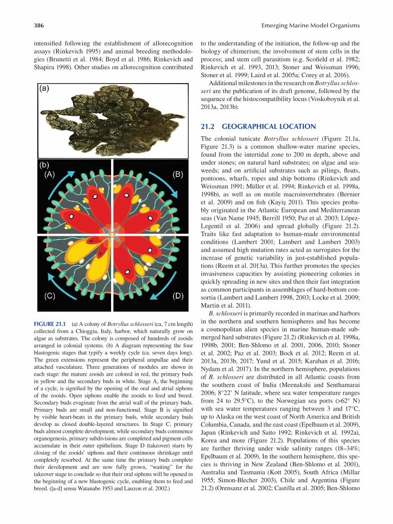
386
intensified following the establishment of allorecognition
assays (Rinkevich 1995) and animal breeding methodolo
gies (Brunetti et al. 1984; Boyd et al. 1986; Rinkevich and
Shapira 1998). Other studies on allorecognition contributed
FIGURE 21.1 (a) A colony of Botryllus schlosseri (ca, 7 cm length)
collected from a Chioggia, Italy, harbor, which naturally grow on
algae as substrates. The colony is composed of hundreds of zooids
arranged in colonial systems. (b) A diagram representing the four
blastogenic stages that typify a weekly cycle (ca. seven days long).
The green extensions represent the peripheral ampullae and their
attached vasculature. Three generations of modules are shown in
each stage: the mature zooids are colored in red, the primary buds
in yellow and the secondary buds in white. Stage A, the beginning
of a cycle, is signified by the opening of the oral and atrial siphons
of the zooids. Open siphons enable the zooids to feed and breed.
Secondary buds evaginate from the atrial wall of the primary buds.
Primary buds are small and non-functional. Stage B is signifi ed
by visible heart-beats in the primary buds, while secondary buds
develop as closed double-layered structures. In Stage C, primary
buds almost complete development, while secondary buds commence
organogenesis, primary subdivisions are completed and pigment cells
accumulate in their outer epithelium. Stage D (takeover) starts by
closing of the zooids’ siphons and their continuous shrinkage until
completely resorbed. At the same time the primary buds complete
their development and are now fully grown, “waiting” for the
takeover stage to conclude so that their oral siphons will be opened in
the beginning of a new blastogenic cycle, enabling them to feed and
breed. ([a-d] sensu Watanabe 1953 and Lauzon et al. 2002.)
Emerging Marine Model Organisms
to the understanding of the initiation, the follow-up and the
biology of chimerism; the involvement of stem cells in the
process; and stem cell parasitism (e.g. Scofield et al. 1982;
Rinkevich et al. 1993, 2013; Stoner and Weissman 1996;
Stoner et al. 1999; Laird et al. 2005a ; Corey et al. 2016).
Additional milestones in the research on Botryllus schlosseri are the publication of its draft genome, followed by the
sequence of the histocompatibility locus (Voskoboynik et al.
2013a , 2013b ).
21.2 GEOGRAPHICAL LOCATION
The colonial tunicate Botryllus schlosseri ( Figure 21.1a ,
Figure 21.3) is a common shallow-water marine species,
found from the intertidal zone to 200 m depth, above and
under stones; on natural hard substrates; on algae and sea
weeds; and on artificial substrates such as pilings, fl oats,
pontoons, wharfs, ropes and ship bottoms (Rinkevich and
Weissman 1991; Müller et al. 1994; Rinkevich et al. 1998a,
1998b), as well as on motile macroinvertebrates (Bernier
et al. 2009) and on fi sh (Kayiş 2011). This species proba
bly originated in the Atlantic European and Mediterranean
seas (Van Name 1945; Berrill 1950; Paz et al. 2003; López-
Legentil et al. 2006 ) and spread globally (Figure 21.2).
Traits like fast adaptation to human-made environmental
conditions (Lambert 2001; Lambert and Lambert 2003)
and assumed high mutation rates acted as surrogates for the
increase of genetic variability in just-established popula
tions (Reem et al. 2013a). This further promotes the species
invasiveness capacities by assisting pioneering colonies in
quickly spreading in new sites and then their fast integration
as common participants in assemblages of hard-bottom con
sortia (Lambert and Lambert 1998 , 2003; Locke et al. 2009;
Martin et al. 2011).
B. schlosseri is primarily recorded in marinas and harbors
in the northern and southern hemispheres and has become
a cosmopolitan alien species in marine human-made sub
merged hard substrates (Figure 21.2) (Rinkevich et al. 1998a,
1998b , 2001 ; Ben-Shlomo et al. 2001 , 2006 , 2010 ; Stoner
et al. 2002; Paz et al. 2003; Bock et al. 2012; Reem et al.
2013a, 2013b, 2017; Yund et al. 2015; Karahan et al. 2016;
Nydam et al. 2017). In the northern hemisphere, populations
of B. schlosseri are distributed in all Atlantic coasts from
the southern coast of India (Meenakshi and Senthamarai
2006; 8°22’ N latitude, where sea water temperature ranges
from 24 to 29.5°C), to the Norwegian sea ports (>62° N)
with sea water temperatures ranging between 3 and 17°C,
up to Alaska on the west coast of North America and British
Columbia, Canada, and the east coast (Epelbaum et al. 2009),
Japan (Rinkevich and Saito 1992; Rinkevich et al. 1992a),
Korea and more (Figure 21.2). Populations of this species
are further thriving under wide salinity ranges (18–34%;
Epelbaum et al. 2009). In the southern hemisphere, this spe
cies is thriving in New Zealand (Ben-Shlomo et al. 2001),
Australia and Tasmania (Kott 2005), South Africa (Millar
1955; Simon-Blecher 2003), Chile and Argentina (Figure
21.2) (Orensanz et al. 2002; Castilla et al. 2005; Ben-Shlomo
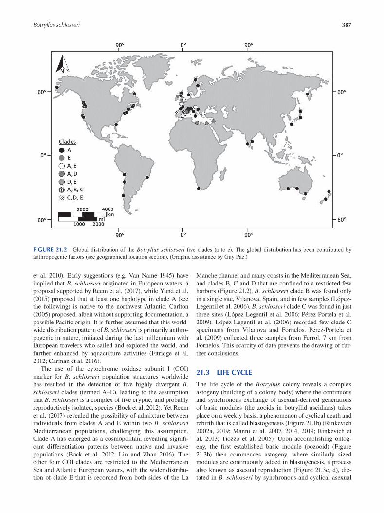
387 Botryllus schlosseri
FIGURE 21.2 Global distribution of the Botryllus schlosseri five clades (a to e). The global distribution has been contributed by
anthropogenic factors (see geographical location section). (Graphic assistance by Guy Paz.)
et al. 2010). Early suggestions (e.g. Van Name 1945) have
implied that B. schlosseri originated in European waters, a
proposal supported by Reem et al. (2017), while Yund et al.
(2015) proposed that at least one haplotype in clade A (see
the following) is native to the northwest Atlantic. Carlton
(2005) proposed, albeit without supporting documentation, a
possible Pacific origin. It is further assumed that this world
wide distribution pattern of B. schlosseri is primarily anthro
pogenic in nature, initiated during the last millennium with
European travelers who sailed and explored the world, and
further enhanced by aquaculture activities (Fitridge et al.
2012; Carman et al. 2016).
The use of the cytochrome oxidase subunit I (COI)
marker for B. schlosseri population structures worldwide
has resulted in the detection of five highly divergent B. schlosseri clades (termed A–E), leading to the assumption
that B. schlosseri is a complex of five cryptic, and probably
reproductively isolated, species (Bock et al. 2012). Yet Reem
et al. (2017 ) revealed the possibility of admixture between
individuals from clades A and E within two B. schlosseri Mediterranean populations, challenging this assumption.
Clade A has emerged as a cosmopolitan, revealing signifi
cant differentiation patterns between native and invasive
populations (Bock et al. 2012; Lin and Zhan 2016 ). The
other four COI clades are restricted to the Mediterranean
Sea and Atlantic European waters, with the wider distribu
tion of clade E that is recorded from both sides of the La
Manche channel and many coasts in the Mediterranean Sea,
and clades B, C and D that are confined to a restricted few
harbors (Figure 21.2). B. schlosseri clade B was found only
in a single site, Vilanova, Spain, and in few samples (López-
Legentil et al. 2006). B. schlosseri clade C was found in just
three sites (López-Legentil et al. 2006; Pérez-Portela et al.
2009). López-Legentil et al. (2006 ) recorded few clade C
specimens from Vilanova and Fornelos. Pérez-Portela et
al. (2009) collected three samples from Ferrol, 7 km from
Fornelos. This scarcity of data prevents the drawing of fur
ther conclusions.
21.3 LIFE CYCLE
The life cycle of the Botryllus colony reveals a complex
astogeny (building of a colony body) where the continuous
and synchronous exchange of asexual-derived generations
of basic modules (the zooids in botryllid ascidians) takes
place on a weekly basis, a phenomenon of cyclical death and
rebirth that is called blastogenesis (Figure 21.1b) (Rinkevich
2002a , 2019 ; Manni et al. 2007 , 2014 , 2019 ; Rinkevich et
al. 2013; Tiozzo et al. 2005). Upon accomplishing ontog
eny, the first established basic module (oozooid) (Figure
21.3b) then commences astogeny, where similarly sized
modules are continuously added in blastogenesis, a process
also known as asexual reproduction (Figure 21.3c, d), dic
tated in B. schlosseri by synchronous and cyclical asexual
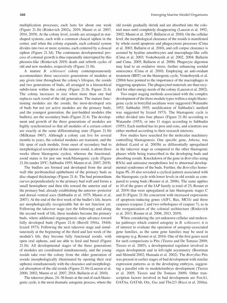
388
multiplication processes; each lasts for about one week
(Figure 21.1b) (Rinkevich 2002a, 2019; Manni et al. 2007,
2014 , 2019). At the colony level, zooids are arranged in star-
shaped systems, each with a common cloacal siphon in the
center, and when the colony expands, each colonial system
divides into two or more systems, each centered by a cloacal
siphon (Figure 21.3d). The continuous developmental pro
cess of colonial growth is thus repeatedly interrupted by this
phoenix-like (Rinkevich 2019) death and rebirth cycles of
old and new modules, respectively (Figure 21.1b).
A mature B. schlosseri colony contemporaneously
accommodates three successive generations of modules at
any given time throughout the colony’s lifespan, the zooids
and two generations of buds, all arranged in a hierarchical
subdivision within the colony (Figure 21.1b, Figure 21.4).
The colony increases in size when more than one bud
replaces each zooid of the old generation. The mature func
tioning modules are the zooids; the most-developed sets
of buds but not yet active modules are the primary buds;
and the youngest generation, the just-budded modules (the
budlets), are the secondary buds (Figure 21.4). The develop
ment and growth of the three generations of modules are
highly synchronized so that all modules of a certain cohort
are exactly at the same differentiating state (Figure 21.1b)
(Milkman 1967). Although a colony can live for several
months to years, the colonial modules are transient, and the
life span of each module, from onset of secondary bud to
morphological resorption of the mature zooid, is about three
weeks (three blastogenic cycles), whereas the functional-
zooid status is for just one week/blastogenic cycle (Figure
21.1b) (under 20ºC; Sabbadin 1955; Manni et al. 2007, 2019).
The budlets are formed and developed from the atrial
wall (the peribranchial epithelium) of the primary buds as
disc-shaped thickenings (Figure 21.4). The bud primordium
curves perpendicularly to the primary bud wall and forms a
small hemisphere and then tilts toward the anterior end of
the primary bud, already establishing the anterior–posterior
and dorsal–ventral axes (Sabbadin et al. 1975; Manni et al.
2007). At the end of the first week of the budlet’s life, hearts
are morphologically recognizable but do not function yet.
Following the takeover stage (see the following) and along
the second week of life, these modules become the primary
buds, where additional organogenesis steps advance toward
fully developed buds (Figure 21.4) (Berrill 1941a, 1941b;
Izzard 1973). Following the next takeover stage and simul
taneously at the beginning of the third and last week of the
module’s life, they become fully functional zooids, with
open oral siphons, and are able to feed and breed (Figure
21.1b). All developmental stages of the three generations
of modules are coordinated simultaneously, and the young
zooids take over the colony from the older generation of
zooids (morphologically illuminated by opening their oral
siphons) simultaneously with the clearance and morphologi
cal absorption of the old zooids (Figure 21.1b) (Lauzon et al.
2000, 2002; Manni et al. 2007, 2014; Ballarin et al. 2010).
The takeover phase, 24–36 hours at the end of each blasto
genic cycle, is the most dramatic astogenic process, where the
Emerging Marine Model Organisms
old zooids gradually shrink and are absorbed into the colo
nial mass until completely disappearing (Lauzon et al. 1992,
2002 ; Manni et al. 2007 ; Ballarin et al. 2010 ). On the cellular
level, the morphological clearance of the zooids is manifested
by whole-zooid apoptosis and phagocytosis processes (Cima
et al. 2003; Ballarin et al. 2010), and cell corpse clearance is
assisted by hyaline amoebocytes and macrophage-like cells
(Cima et al. 2003; Voskoboynik et al. 2002, 2004; Ballarin
and Cima. 2005; Ballarin et al. 2008). Phagocyte digestion
may lead to an oxidative stress, further enhancing zooidal
senescence (Cima et al. 2010). Employing an anti-oxidant
treatment (BHT) on the blastogenic cycle, Voskoboynik et al.
(2004) have pointed to the importance of the macrophages in
triggering apoptosis. The phagocyted materials are than recy
cled for other energy needs of the colony (Lauzon et al. 2002).
Two major staging methods associated with the complex
development of the three module types within a single blasto
genic cycle in botryllid ascidians were suggested (Watanabe
1953 ; Sabbadin 1955 ; modification of Sabbadin’s method
was suggested by Izzard 1973). The blastogenic cycle is
either divided into four phases (Figure 21.1b) according to
Watanabe (1953), or into 11 stages according to Sabbadin
(1955). Each method has its pros and cons, and scientists use
either method according to their research interests.
Few studies have searched for the molecular machinery
controlling blastogenesis. One specific gene, Athena , was
defined (Laird et al. 2005b) as differentially upregulated
in the takeover stage as compared to the other blastogenic
phases while being transcribed in the developing buds and
absorbing zooids. Knockdown of the gene in Botryllus using
RNAi and antisense morpholinos led to abnormal develop
mental syndromes of the buds. Further, the Botryllus homo
logue PL-10 also revealed a cyclical pattern associated with
the blastogenic cycle with lower levels in old zooids as com
pared to young buds (Rosner et al. 2006). The same applies
to 10 of the genes of the IAP family (a total of 25; Rosner et
al. 2019) that were upregulated at late blastogenic stages C
and D (Figure 21.1b) concurrent with increased expressions
of apoptosis-inducing genes (AIF1, Bax, MCl1) and three
caspases (caspase 2 and two orthologues of caspase 7), as in
the reorganization of the colonial architecture (Rinkevich
et al. 2013; Rosner et al. 2006, 2013, 2019).
When considering the yet-unknown cellular and molecu
lar pathways which control astogeny in B. schlosseri, it is
of interest to evaluate the operation of astogeny-associated
gene families, as the same gene families may be used in
ontogeny (e.g. Rosner et al. 2014). One of the first genes used
for such comparisons is Pitx (Tiozzo and De Tomaso 2009;
Tiozzo et al. 2005), a developmental regulator involved in
organ development and in left-right asymmetry (Boorman
and Shimeld 2002; Hamada et al. 2002). The Botryllus Pitx
was present in earlier stages of bud development with similar
expression patterns as in the developing embryos, suggest
ing a parallel role in module/embryo development (Tiozzo
et al. 2005; Tiozzo and De Tomaso 2009). Other tran
scription factors involved in bud development are FoxA1,
GATAa, GATAb, Otx, Gsc and Tbx2/3 (Ricci et al. 2016a).
389 Botryllus schlosseri
Further research studied the expression along blastogenesis
of three conserved signal transduction pathways, Wnt/β
catenin, TGF-β and MAPK/ERK (Rosner et al. 2014), by
studying representative gene β-catenin (for Wnt/β-catenin
pathway), p-Smad2 and p-Smad1/5/8 (for TGF-β pathway)
and p-Mek1/2 (for MAPK/ERK pathway). Results revealed
that while the same molecular machinery is functioning in
Botryllus schlosseri astogeny and ontogeny, astogenic devel
opment is not an ontogenic replicate (Rosner et al. 2014).
Blastogenesis (Figure 21.1b) in B. schlosseri holds some
unique characteristics for aging in colonial (modular) organ
isms that distinguishes this type of aging from aging in uni
tary organisms (Rinkevich 2017). Some characteristics that
refer to non-random (genetic based) mortality were recorded
in these organisms (Rinkevich et al. 1992b; Lauzon et al.
FIGURE 21.3 (a–d) Life stages of a Botryllus schlosseri colony.
(a) Botryllus begins its life as a mobile larva, composed of a visceral
trunk and locomotory tail. The mobility enables the larva to swim
and find adequate substrate to settle on. (b) At metamorphosis,
the attached larva becomes the first zooid, the oozooid, with open
oral and atrial siphons. On both sides of the oozooid, the new
generation of buds (white arrowheads) are formed and developed.
(c) After a few days, the oozooid is resorbed and is replaced by two
new zooids. (d) The numbers of zooids grows over time, forming a
mature colony. The size of a colony differs between colonies and
depends on the number of zooids (see colony in Figure 21.1a for
comparison). (e) A chimera composed of two distinct colonies,
connected via a blood vessel (two arrowheads). The chimera is
formed after a physical contact between the ampullae of both of
colonies (details in “Functional Approaches: Tools for Molecular
and Cellular Analyses”). Scale bars = 0.5 mm.
2000; Rabinowitz and Rinkevich 2004; Rinkevich 2017 ).
The phenomena of budding, as well as module senescence,
can be concurrently expressed at three hierarchical levels
of colonial organization: the zooids, ramets and genets,
including the weekly blastogenesis, the whole-genet pro
grammed life span (Rinkevich et al. 1992b); ageing at the
ramet level (Rabinowitz and Rinkevich 2004); and rejuve
nilization after acute damage (Voskoboynik et al. 2002).
To further understand Botryllus blastogenesis and aging
at the molecular level, the aging-related heat-shock protein
mortalin was studied (Ben-Hamo et al. 2018). RT-PCR and
in-situ hybridization revealed significant upregulation of
mortalin in colonies during the takeover phase as compared
to other blastogenic phases. Quantitative PCR analyses of
excised buds and zooids showed significantly higher lev
els of mortalin in buds as compared to functioning zooids.
These findings are in line with literature that demonstrated
lowering levels of mortalin in old organisms as compared
to young organisms (Yokoyama et al. 2002; Kimura et al.
2007; Yaguchi et al. 2007), demonstrating a possible aging
process that is restricted to the modules and associated with
the blastogenic cycle (Ben-Hamo et al. 2018).
21.4 EMBRYOGENESIS
Like the rest of the many colonial tunicates and unlike the
other chordates, Botryllus schlosseri is an ovoviviparous
species ( Zaniolo et al. 1987) that reproduces both asexually
and sexually, and colonies are simultaneous hermaphrodites
(Berrill 1950; Rodriguez et al. 2014). The colonies can ran
domly switch between male and hermaphrodite states fol
lowing physiological stress or become sterile (Rodriguez
et al. 2016 ). Yet self-fertilization is eliminated, as male
gonads mature two days following the eggs’ fertilization
by foreign sperm (Milkman and Borgmann 1963; Milkman
1967; Mukai 1977). Nonetheless, self-fertilization was suc
cessfully achieved under laboratory conditions (Milkman
and Borgmann 1963; Milkman 1967; Rinkevich 1993) and in
the field after surgical or natural separation of a colony into
systems (Sabbadin 1971; Gasparini et al. 2014), document
ing that in the absence of foreign sperm, self-fertilization
may occur. Late fertilization is prone to embryo resorption
at blastogenesis stage D (the takeover phase) due to larval
delayed development (Milkman 1967, Stewart-Savage et al.
2001). Following metamorphosis of the larva (Figure 21.3a)
into the fi rst established zooid (the oozooid) (Figure 21.3b),
it takes between 8 and 12 blastogenic cycles for the male
gonads to develop and mature (Sabbadin 1971; Sabbadin and
Zaniolo 1979). The female gonads mature afterward, estab
lishing the hermaphrodite type of B. schlosseri’s sexual
reproduction (Figure 21.4).
The gonads are first observed in the secondary buds. At
onset, a bilateral gonad blastema appears where its medial
part will give rise to the testis and the lateral part to the
ovary. The oocytes and testes develop in the buds’ blastema,
and the oocytes move through several generations of buds
(Sabbadin and Zaniolo 1979). Testes and ovaries are formed
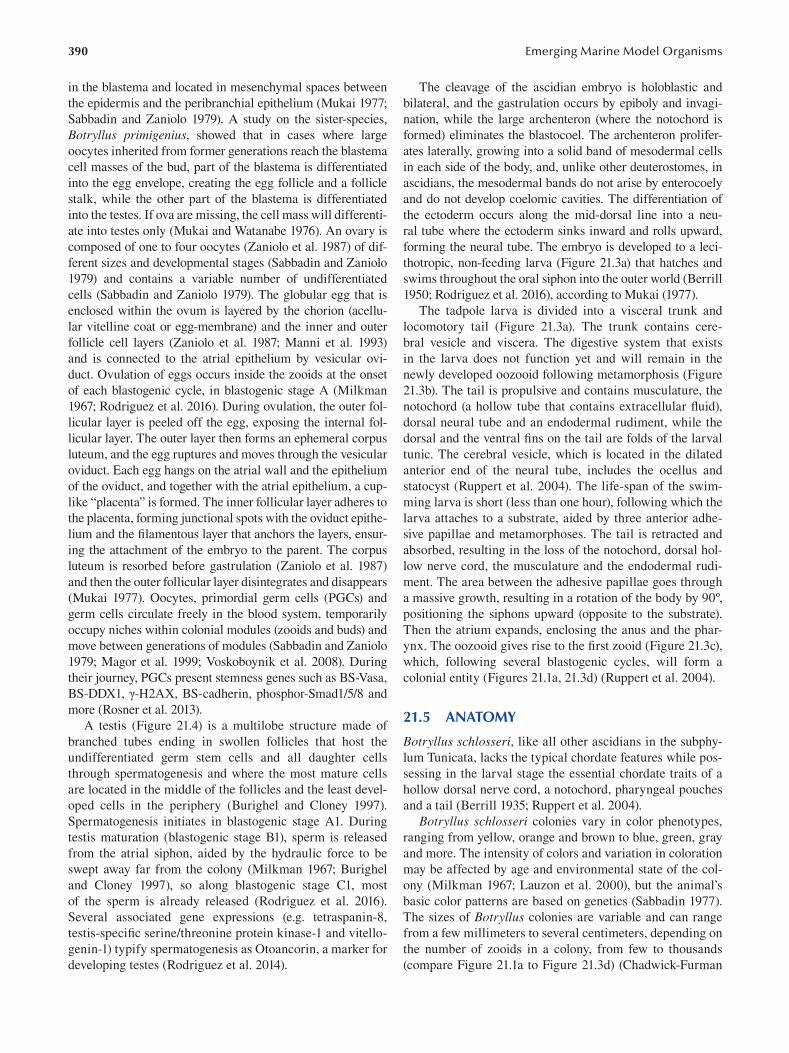
390
in the blastema and located in mesenchymal spaces between
the epidermis and the peribranchial epithelium (Mukai 1977;
Sabbadin and Zaniolo 1979). A study on the sister-species,
Botryllus primigenius, showed that in cases where large
oocytes inherited from former generations reach the blastema
cell masses of the bud, part of the blastema is differentiated
into the egg envelope, creating the egg follicle and a follicle
stalk, while the other part of the blastema is differentiated
into the testes. If ova are missing, the cell mass will differenti
ate into testes only (Mukai and Watanabe 1976). An ovary is
composed of one to four oocytes ( Zaniolo et al. 1987) of dif
ferent sizes and developmental stages (Sabbadin and Zaniolo
1979) and contains a variable number of undifferentiated
cells (Sabbadin and Zaniolo 1979). The globular egg that is
enclosed within the ovum is layered by the chorion (acellu
lar vitelline coat or egg-membrane) and the inner and outer
follicle cell layers ( Zaniolo et al. 1987; Manni et al. 1993)
and is connected to the atrial epithelium by vesicular ovi
duct. Ovulation of eggs occurs inside the zooids at the onset
of each blastogenic cycle, in blastogenic stage A (Milkman
1967; Rodriguez et al. 2016). During ovulation, the outer fol
licular layer is peeled off the egg, exposing the internal fol
licular layer. The outer layer then forms an ephemeral corpus
luteum, and the egg ruptures and moves through the vesicular
oviduct. Each egg hangs on the atrial wall and the epithelium
of the oviduct, and together with the atrial epithelium, a cup
like “placenta” is formed. The inner follicular layer adheres to
the placenta, forming junctional spots with the oviduct epithe
lium and the filamentous layer that anchors the layers, ensur
ing the attachment of the embryo to the parent. The corpus
luteum is resorbed before gastrulation (Zaniolo et al. 1987)
and then the outer follicular layer disintegrates and disappears
(Mukai 1977). Oocytes, primordial germ cells (PGCs) and
germ cells circulate freely in the blood system, temporarily
occupy niches within colonial modules (zooids and buds) and
move between generations of modules (Sabbadin and Zaniolo
1979; Magor et al. 1999; Voskoboynik et al. 2008). During
their journey, PGCs present stemness genes such as BS-Vasa,
BS-DDX1, γ-H2AX, BS-cadherin, phosphor-Smad1/5/8 and
more (Rosner et al. 2013).
A testis (Figure 21.4) is a multilobe structure made of
branched tubes ending in swollen follicles that host the
undifferentiated germ stem cells and all daughter cells
through spermatogenesis and where the most mature cells
are located in the middle of the follicles and the least devel
oped cells in the periphery (Burighel and Cloney 1997 ).
Spermatogenesis initiates in blastogenic stage A1. During
testis maturation (blastogenic stage B1), sperm is released
from the atrial siphon, aided by the hydraulic force to be
swept away far from the colony (Milkman 1967; Burighel
and Cloney 1997 ), so along blastogenic stage C1, most
of the sperm is already released (Rodriguez et al. 2016 ).
Several associated gene expressions (e.g. tetraspanin-8,
testis-specifi c serine/threonine protein kinase-1 and vitello
genin-1) typify spermatogenesis as Otoancorin, a marker for
developing testes (Rodriguez et al. 2014).
Emerging Marine Model Organisms
The cleavage of the ascidian embryo is holoblastic and
bilateral, and the gastrulation occurs by epiboly and invagi
nation, while the large archenteron (where the notochord is
formed) eliminates the blastocoel. The archenteron prolifer
ates laterally, growing into a solid band of mesodermal cells
in each side of the body, and, unlike other deuterostomes, in
ascidians, the mesodermal bands do not arise by enterocoely
and do not develop coelomic cavities. The differentiation of
the ectoderm occurs along the mid-dorsal line into a neu
ral tube where the ectoderm sinks inward and rolls upward,
forming the neural tube. The embryo is developed to a leci
thotropic, non-feeding larva (Figure 21.3a) that hatches and
swims throughout the oral siphon into the outer world (Berrill
1950; Rodriguez et al. 2016 ), according to Mukai (1977 ).
The tadpole larva is divided into a visceral trunk and
locomotory tail (Figure 21.3a). The trunk contains cere
bral vesicle and viscera. The digestive system that exists
in the larva does not function yet and will remain in the
newly developed oozooid following metamorphosis (Figure
21.3b). The tail is propulsive and contains musculature, the
notochord (a hollow tube that contains extracellular fl uid),
dorsal neural tube and an endodermal rudiment, while the
dorsal and the ventral fi ns on the tail are folds of the larval
tunic. The cerebral vesicle, which is located in the dilated
anterior end of the neural tube, includes the ocellus and
statocyst (Ruppert et al. 2004). The life-span of the swim
ming larva is short (less than one hour), following which the
larva attaches to a substrate, aided by three anterior adhe
sive papillae and metamorphoses. The tail is retracted and
absorbed, resulting in the loss of the notochord, dorsal hol
low nerve cord, the musculature and the endodermal rudi
ment. The area between the adhesive papillae goes through
a massive growth, resulting in a rotation of the body by 90º,
positioning the siphons upward (opposite to the substrate).
Then the atrium expands, enclosing the anus and the phar
ynx. The oozooid gives rise to the first zooid (Figure 21.3c),
which, following several blastogenic cycles, will form a
colonial entity (Figures 21.1a, 21.3d) (Ruppert et al. 2004).
21.5 ANATOMY
Botryllus schlosseri, like all other ascidians in the subphy
lum Tunicata, lacks the typical chordate features while pos
sessing in the larval stage the essential chordate traits of a
hollow dorsal nerve cord, a notochord, pharyngeal pouches
and a tail (Berrill 1935; Ruppert et al. 2004).
Botryllus schlosseri colonies vary in color phenotypes,
ranging from yellow, orange and brown to blue, green, gray
and more. The intensity of colors and variation in coloration
may be affected by age and environmental state of the col
ony (Milkman 1967; Lauzon et al. 2000), but the animal’s
basic color patterns are based on genetics (Sabbadin 1977).
The sizes of Botryllus colonies are variable and can range
from a few millimeters to several centimeters, depending on
the number of zooids in a colony, from few to thousands
(compare Figure 21.1a to Figure 21.3d) (Chadwick-Furman
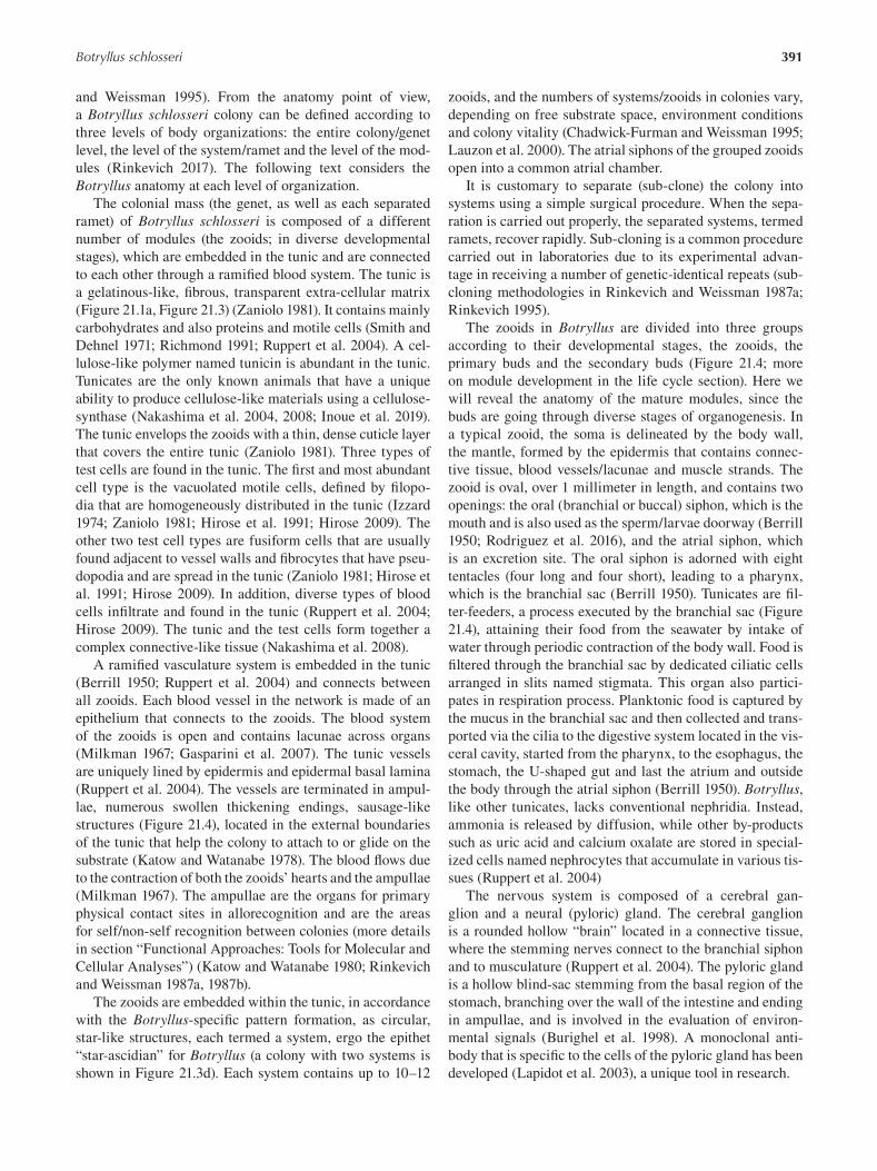
391 Botryllus schlosseri
and Weissman 1995). From the anatomy point of view,
a Botryllus schlosseri colony can be defined according to
three levels of body organizations: the entire colony/genet
level, the level of the system/ramet and the level of the mod
ules (Rinkevich 2017). The following text considers the
Botryllus anatomy at each level of organization.
The colonial mass (the genet, as well as each separated
ramet) of Botryllus schlosseri is composed of a different
number of modules (the zooids; in diverse developmental
stages), which are embedded in the tunic and are connected
to each other through a ramifi ed blood system. The tunic is
a gelatinous-like, fibrous, transparent extra-cellular matrix
(Figure 21.1a, Figure 21.3) ( Zaniolo 1981). It contains mainly
carbohydrates and also proteins and motile cells (Smith and
Dehnel 1971; Richmond 1991; Ruppert et al. 2004). A cel
lulose-like polymer named tunicin is abundant in the tunic.
Tunicates are the only known animals that have a unique
ability to produce cellulose-like materials using a cellulose-
synthase (Nakashima et al. 2004, 2008; Inoue et al. 2019).
The tunic envelops the zooids with a thin, dense cuticle layer
that covers the entire tunic ( Zaniolo 1981). Three types of
test cells are found in the tunic. The first and most abundant
cell type is the vacuolated motile cells, defined by fi lopo
dia that are homogeneously distributed in the tunic (Izzard
1974; Zaniolo 1981; Hirose et al. 1991; Hirose 2009). The
other two test cell types are fusiform cells that are usually
found adjacent to vessel walls and fibrocytes that have pseu
dopodia and are spread in the tunic ( Zaniolo 1981; Hirose et
al. 1991; Hirose 2009). In addition, diverse types of blood
cells infiltrate and found in the tunic (Ruppert et al. 2004;
Hirose 2009). The tunic and the test cells form together a
complex connective-like tissue (Nakashima et al. 2008).
A ramified vasculature system is embedded in the tunic
(Berrill 1950; Ruppert et al. 2004) and connects between
all zooids. Each blood vessel in the network is made of an
epithelium that connects to the zooids. The blood system
of the zooids is open and contains lacunae across organs
(Milkman 1967; Gasparini et al. 2007). The tunic vessels
are uniquely lined by epidermis and epidermal basal lamina
(Ruppert et al. 2004). The vessels are terminated in ampul
lae, numerous swollen thickening endings, sausage-like
structures (Figure 21.4), located in the external boundaries
of the tunic that help the colony to attach to or glide on the
substrate (Katow and Watanabe 1978). The blood fl ows due
to the contraction of both the zooids’ hearts and the ampullae
(Milkman 1967). The ampullae are the organs for primary
physical contact sites in allorecognition and are the areas
for self/non-self recognition between colonies (more details
in section “Functional Approaches: Tools for Molecular and
Cellular Analyses”) (Katow and Watanabe 1980; Rinkevich
and Weissman 1987a, 1987b).
The zooids are embedded within the tunic, in accordance
with the Botryllus -specific pattern formation, as circular,
star-like structures, each termed a system, ergo the epithet
“star-ascidian” for Botryllus (a colony with two systems is
shown in Figure 21.3d). Each system contains up to 10–12
zooids, and the numbers of systems/zooids in colonies vary,
depending on free substrate space, environment conditions
and colony vitality (Chadwick-Furman and Weissman 1995;
Lauzon et al. 2000). The atrial siphons of the grouped zooids
open into a common atrial chamber.
It is customary to separate (sub-clone) the colony into
systems using a simple surgical procedure. When the sepa
ration is carried out properly, the separated systems, termed
ramets, recover rapidly. Sub-cloning is a common procedure
carried out in laboratories due to its experimental advan
tage in receiving a number of genetic-identical repeats (sub
cloning methodologies in Rinkevich and Weissman 1987a;
Rinkevich 1995 ).
The zooids in Botryllus are divided into three groups
according to their developmental stages, the zooids, the
primary buds and the secondary buds (Figure 21.4; more
on module development in the life cycle section). Here we
will reveal the anatomy of the mature modules, since the
buds are going through diverse stages of organogenesis. In
a typical zooid, the soma is delineated by the body wall,
the mantle, formed by the epidermis that contains connec
tive tissue, blood vessels/lacunae and muscle strands. The
zooid is oval, over 1 millimeter in length, and contains two
openings: the oral (branchial or buccal) siphon, which is the
mouth and is also used as the sperm/larvae doorway (Berrill
1950; Rodriguez et al. 2016), and the atrial siphon, which
is an excretion site. The oral siphon is adorned with eight
tentacles (four long and four short), leading to a pharynx,
which is the branchial sac (Berrill 1950). Tunicates are fi l
ter-feeders, a process executed by the branchial sac (Figure
21.4), attaining their food from the seawater by intake of
water through periodic contraction of the body wall. Food is
fi ltered through the branchial sac by dedicated ciliatic cells
arranged in slits named stigmata. This organ also partici
pates in respiration process. Planktonic food is captured by
the mucus in the branchial sac and then collected and trans
ported via the cilia to the digestive system located in the vis
ceral cavity, started from the pharynx, to the esophagus, the
stomach, the U-shaped gut and last the atrium and outside
the body through the atrial siphon (Berrill 1950). Botryllus, like other tunicates, lacks conventional nephridia. Instead,
ammonia is released by diffusion, while other by-products
such as uric acid and calcium oxalate are stored in special
ized cells named nephrocytes that accumulate in various tis
sues (Ruppert et al. 2004)
The nervous system is composed of a cerebral gan
glion and a neural (pyloric) gland. The cerebral ganglion
is a rounded hollow “brain” located in a connective tissue,
where the stemming nerves connect to the branchial siphon
and to musculature (Ruppert et al. 2004). The pyloric gland
is a hollow blind-sac stemming from the basal region of the
stomach, branching over the wall of the intestine and ending
in ampullae, and is involved in the evaluation of environ
mental signals ( Burighel et al. 1998). A monoclonal anti
body that is specific to the cells of the pyloric gland has been
developed (Lapidot et al. 2003), a unique tool in research.
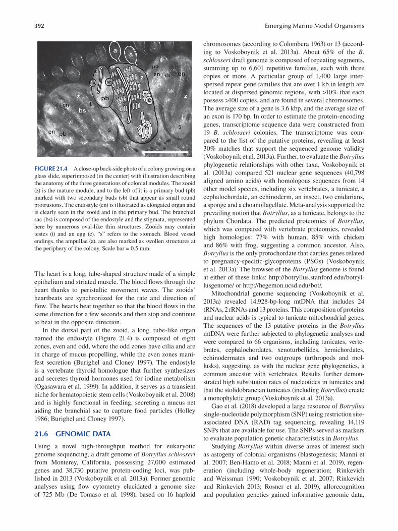
392
FIGURE 21.4 A close-up back-side photo of a colony growing on a
glass slide, superimposed (in the center) with illustration describing
the anatomy of the three generations of colonial modules. The zooid
(z) is the mature module, and to the left of it is a primary bud (pb)
marked with two secondary buds (sb) that appear as small round
protrusions. The endostyle (en) is illustrated as elongated organ and
is clearly seen in the zooid and in the primary bud. The branchial
sac (bs) is composed of the endostyle and the stigmata, represented
here by numerous oval-like thin structures. Zooids may contain
testes (t) and an egg (e). “s” refers to the stomach. Blood vessel
endings, the ampullae (a), are also marked as swollen structures at
the periphery of the colony. Scale bar = 0.5 mm.
The heart is a long, tube-shaped structure made of a simple
epithelium and striated muscle. The blood flows through the
heart thanks to peristaltic movement waves. The zooids’
heartbeats are synchronized for the rate and direction of
flow. The hearts beat together so that the blood flows in the
same direction for a few seconds and then stop and continue
to beat in the opposite direction.
In the dorsal part of the zooid, a long, tube-like organ
named the endostyle (Figure 21.4) is composed of eight
zones, even and odd, where the odd zones have cilia and are
in charge of mucus propelling, while the even zones mani
fest secretion (Burighel and Cloney 1997 ). The endostyle
is a vertebrate thyroid homologue that further synthesizes
and secretes thyroid hormones used for iodine metabolism
(Ogasawara et al. 1999). In addition, it serves as a transient
niche for hematopoietic stem cells (Voskoboynik et al. 2008)
and is highly functional in feeding, secreting a mucus net
aiding the branchial sac to capture food particles (Holley
1986; Burighel and Cloney 1997 ).
21.6 GENOMIC DATA
Using a novel high-throughput method for eukaryotic
genome sequencing, a draft genome of Botryllus schlosseri from Monterey, California, possessing 27,000 estimated
genes and 38,730 putative protein-coding loci, was pub
lished in 2013 (Voskoboynik et al. 2013a). Former genomic
analyses using flow cytometry elucidated a genome size
of 725 Mb (De Tomaso et al. 1998), based on 16 haploid
Emerging Marine Model Organisms
chromosomes (according to Colombera 1963) or 13 (accord
ing to Voskoboynik et al. 2013a). About 65% of the B. schlosseri draft genome is composed of repeating segments,
summing up to 6,601 repetitive families, each with three
copies or more. A particular group of 1,400 large inter
spersed repeat gene families that are over 1 kb in length are
located at dispersed genomic regions, with >10% that each
possess >100 copies, and are found in several chromosomes.
The average size of a gene is 3.6 kbp, and the average size of
an exon is 170 bp. In order to estimate the protein-encoding
genes, transcriptome sequence data were constructed from
19 B. schlosseri colonies. The transcriptome was com
pared to the list of the putative proteins, revealing at least
30% matches that support the sequenced genome validity
(Voskoboynik et al. 2013a). Further, to evaluate the Botryllus phylogenetic relationships with other taxa, Voskoboynik et
al. (2013a) compared 521 nuclear gene sequences (40,798
aligned amino acids) with homologous sequences from 14
other model species, including six vertebrates, a tunicate, a
cephalochordate, an echinoderm, an insect, two cnidarians,
a sponge and a choanoflagellate. Meta-analysis supported the
prevailing notion that Botryllus, as a tunicate, belongs to the
phylum Chordata. The predicted proteomics of Botryllus, which was compared with vertebrate proteomics, revealed
high homologies: 77% with human, 85% with chicken
and 86% with frog, suggesting a common ancestor. Also,
Botryllus is the only protochordate that carries genes related
to pregnancy-specific-glycoproteins (PSGs) ( Voskoboynik
et al. 2013a). The browser of the Botryllus genome is found
at either of these links: http://botryllus.stanford.edu/botryl
lusgenome/ or http://hegemon.ucsd.edu/bot/ .
Mitochondrial genome sequencing (Voskoboynik et al.
2013a) revealed 14,928-bp-long mtDNA that includes 24
tRNAs, 2 rRNAs and 13 proteins. This composition of proteins
and nuclear acids is typical to tunicate mitochondrial genes.
The sequences of the 13 putative proteins in the Botryllus mtDNA were further subjected to phylogenetic analyses and
were compared to 66 organisms, including tunicates, verte
brates, cephalochordates, xenoturbellides, hemichordates,
echinodermates and two outgroups (arthropods and mol
lusks), suggesting, as with the nuclear gene phylogenetics, a
common ancestor with vertebrates. Results further demon
strated high substitution rates of nucleotides in tunicates and
that the stolidobrancian tunicates (including Botryllus ) create
a monophyletic group (Voskoboynik et al. 2013a).
Gao et al. (2018) developed a large resource of Botryllus single-nucleotide polymorphism (SNP) using restriction site-
associated DNA (RAD) tag sequencing, revealing 14,119
SNPs that are available for use. The SNPs served as markers
to evaluate population genetic characteristics in Botryllus. Studying Botryllus within diverse areas of interest such
as astogeny of colonial organisms (blastogenesis; Manni et
al. 2007; Ben-Hamo et al. 2018; Manni et al. 2019), regen
eration (including whole-body regeneration; Rinkevich
and Weissman 1990; Voskoboynik et al. 2007; Rinkevich
and Rinkevich 2013; Rosner et al. 2019), allorecognition
and population genetics gained informative genomic data,
393 Botryllus schlosseri
partially unveiling the cryptic biology underlying these phe
nomena. The following paragraphs summarize the central
publications on the genomic data.
Blastogenesis has been well characterized in Botryllus both anatomically and ontogenically (Manni et al. 2019;
Manni et al. 2014; Sabbadin et al. 1975; Izzard 1973; Berrill
1941a, 1941b). Recent years have yielded novel insights on
the molecular processes underlying blastogenesis (Franchi
et al. 2017; Campagna et al. 2016; Ricci et al. 2016a ;
Rodriguez et al. 2014; Rinkevich et al. 2013; Rosner et al.
2014, 2019; Qarri et al. 2020). Transcriptomes of three
major stages along the Botryllus blastogenic cycle (mid
cycle, the pre-takeover and the takeover phases; Campagna
et al. 2016; available at http://botryllus.cribi.unipd.it) have
revealed 11,337 new genes, of which 581 transcripts were
determined with complete open reading frames. Many
sequences emerged as genes involved in apoptosis activa
tion, de-activation and regulation (Campagna et al. 2016).
Analyzing the differential expression for fertile vs. infertile
B. schlosseri colonies, Rodriguez et al. (2014) revealed a set
of genes that are differentially expressed in every blastogen
esis stage analyzed. The highest numbers of differentially
expressed genes were found in early stages, many of which
are homologous to vertebrates. These genes have conserved
roles in organism fertility (Rodriguez et al. 2014).
Ricci et al. (2016b) constructed transcriptomics librar
ies from epithelial tissues of developing buds and from
non-developing buds and revealed differentially expressed
gene expressions in the developing bud epithelial tissues
that are associated with regeneration and stem cell func
tions and homologous to genes in other model organisms.
Further sets of unknown genes were elucidated, indicating
possible specific genes and functions associated with bud
ding in B. schlosseri colonies (Ricci et al. 2016b), while in
other cases, such as in response to reactive oxygen species
(ROS) that emerge during the takeover stage (Cima et al.
1996; Voskoboynik et al. 2004), five transcripts for antioxi
dant defense enzymes [SOD (superoxide dismutase), GCLM
(glutamyl-cysteine ligase modulatory subunit), GS (gluta
thione synthase), GPx3 and GPx5 (two glutathione peroxi
dases)] were identified (Franchi et al. 2017).
Allorecognition in botryllid ascidians is manifested when
two or more genotypes come into physical contact with
each other, resulting in either fusion (chimera formation)
or rejection (see more in “Functional Approaches: Tools for
Molecular and Cellular Analyses”). To assess the repertoire
of differentially expressed genes during rejection, Oren et
al. (2007 ) constructed expressed sequence tag (EST) librar
ies where allogeneic challenged colonies were compared
to naïve counterparts and revealed dozens of specifi cally
expressed genes homologous to genes involved in diverse
immunological processes. The list includes stress proteins,
pattern recognition receptors, complement proteins, prote
ases and protease inhibitors, cell adhesion and coagulation
proteins, cytokine-related proteins, programmed cell death
and proteasome-related proteins (Oren et al. 2007). Then
Oren et al. (2010) elucidated transcriptional differences
between the genotypes involved in the allogeneic rejection
processes, the partner that displays the points of rejection
(PORs; rejected partner) and the rejecting partner “caus
ing” the PORs. Microarray and complementary qPCR
assays revealed two distinct transcriptional landscapes for
“rejected” vs. “rejecting” colonies in the same allogeneic
assay. In the “rejected” colonies, 87% of the ESTs were
downregulated as compared to the “rejecting” partner show
ing only minor changes (0.7%) in the allogeneic assay. In
the “rejected” transcriptome, three functional groups were
downregulated substantially: protein biosynthesis, cell
structure and motility and immune-related genes, overall
depicting the inhibition of response components rather than
enhancement of immunologic responses (Oren et al. 2010).
Studies were further engaged with the Botryllus regen
eration abilities and the roles of stem cells in this process
(Braden et al. 2014; including whole-body regeneration;
Rinkevich and Weissman 1990; Voskoboynik et al. 2007;
Rinkevich and Rinkevich 2013; Rosner et al. 2019).
According to these studies, stem cells circulate the blood
system of the colonies and are confined to dedicated stem
cell niches as the niches adjacent to the endostyle. Stem cells
play a pivotal role in budding de novo of new generations
of modules and in regeneration according to their genomic
signatures. Three presumed stem cell populations were
described in Botryllus (CP25, CP33 and CP34), and their
expressed genes overlap with those of the mouse hematopoi
etic stem cells (Rosental et al. 2018).
21.7 FUNCTIONAL APPROACHES: TOOLS FOR MOLECULAR AND CELLULAR ANALYSES
Colonial tunicates such as Botryllus schlosseri express
unique biological phenomena and are valuable models for
variety of research fields, yielding novel discoveries and
functional tools in the research. We detail an overview of
three main tools that can be applied for diverse studies.
21.7.1 A MODEL FOR CHIMERISM
The first research tool is the use of Botryllus schlosseri as
an accessible model system for allorecognition, primarily
for chimera formation. Chimerism is the biological state
where an organism is composed of cells originating from
two genetically distinct conspecifics and is based on the
capacity for morphological fusions between these organisms
( Figure 21.3e ). Artificial chimerism (performed in research
institutions) is being achieved in model organisms such as
frogs (Volpe and Earley 1970), rats (Fang 1971) and mice
(Eichwald et al. 1959), established by uniting allogeneic cells
during early embryonic stages or via surgical interventions in
adults. These systems have proved an indispensible tool for
a variety of research fields, such as hematology (Abkowitz et
al. 2003), immunology (Liu et al. 2007 ), aging (Conboy et al.
2013) and more. Although parabiosis is an important system
for studies, two main challenges keep it from being used on a
wide scale in biology. First, growing public concern in recent
394
years delegitimizes the use of adult parabionts in experi
mental settings, and second, the traumatic protocols cause
enormous stress that may influence the results of the studies.
Botryllus chimerism may alleviate these challenges.
Botryllid ascidians possess a unique type of immunity
(allorecognition system) that may reveal the evolutionary
routes for vertebrate immune systems (Magor et al. 1999;
Weissman et al. 1990; Cooper et al. 1992; Rinkevich 2004,
2005a), as well as chimerism, revealing evolutionary and
ecological aspects for this phenomenon (Rinkevich 2005b).
Interest in B. schlosseri immunity has centered on allogeneic
recognition and its consequences, as pairs of colonies that
meet naturally (or in the laboratory) either anastomose con
tacting ampullae to form a vascular parabiont (Figure 21.3e)
or develop cytotoxic lesions in the contact zones (termed
points of rejection; Sabbadin and Astorri 1988; Teneda et al.
1985 ; Rinkevich and Weissman 1987a, 1987b, 1987c, 1991;
Weissman et al. 1990 ; Rinkevich 1992 , 1996 , 1999a ). In
many cases, pairs of colonies that fused or rejected each other
retreat, growing from their points/areas of contact (Rinkevich
and Weissman 1988). B. schlosseri chimeras were widely
recorded in the field (Ben-Shlomo et al. 2001), most likely
the outcome of co-settlement aggregates of histocompatible
kin colonies (Grosberg and Quinn 1986). Once colonies fuse,
a second allorecognition phenomenon begins which leads to
the morphological elimination (resorption) of one partner in
the chimera (Rinkevich and Weissman 1987a, 1987b, 1987c,
1989; 1992a, 1992b; Sabbadin and Astorri 1988), termed allo
geneic or chimeric resorption (Rinkevich 2005a) and based on
a highly complex and polymorphic organization of histocom
patibility alleles, revealing a clear hierarchy in the resorption
phenomenon (Rinkevich 1993; Rinkevich et al. 1993). Yet a
mild stress may change resorption directionality in B. schlosseri chimeras by expressing a non-genetic type of apoptotic
pathways (Rinkevich et al. 1994).
One of the most interesting outcomes of chimerism in
B. schlosseri are the phenomena of somatic/germ cell par
asitism (Sabbadin and Zaniolo 1979; Pancer et al. 1995;
Stoner and Weissman 1996; Magor et al. 1999; Stoner et
al. 1999; Rinkevich and Yankelevich 2004; Simon-Blecher
et al. 2004). Somatic and germ cell parasitism in chimeric
B. schlosseri colonies are recognized when the soma and/
or the gonads do not reflect equal contributions by the part
ners involved and are further recorded in “forced chime
ras” established between allogeneic noncompatible partners
(Rinkevich and Weissman 1998; Simon-Blecher et al. 2004).
Germ cell parasitism in this system is fi xed, reproducible,
reveals hierarchical arrangements and, above all, is sexually
inherited (Stoner et al. 1999; Rinkevich and Yankelevich
2004). In contrast, somatic cell parasitism, while reproduc
ible and hierarchical, has not been characterized by the trait
of sexual inheritance through a pedigree (Stoner et al. 1999).
It may thus be concluded that somatic and germ cell parasit
ism are unlinked phenomena (Stoner et al. 1999; Magor et al.
1999; Rinkevich and Yankelevich 2004) and that for both
types of cell parasitism, the chimeric entity enables foreign
Emerging Marine Model Organisms
somatic and germ stem cells to hitchhike within the “winner”
genotypes without being visible to natural selection forces
that act on the winner genotypes (Rinkevich 2002a, 2002b,
2004a, 2004b, 2011a), part of the proposed “costs” for chi
merism (Rinkevich 2002b, 2005b, 2011a). Yet several studies
that evaluated “costs” and “benefits” predictions for chime
rism in B. schlosseri revealed two major benefits, the shifts
of the somatic constituents within chimeras in accordance
with changes in environmental conditions and the expres
sion of the heterosis phenomenon in chimeras, occurred
via scrutinizing against genotypes that are less adapted to
adverse environmental conditions (Rinkevich 1993, 2005b;
Rinkevich et al. 1993; Rinkevich and Yankelevich 2004).
This attests to the indispensable tool of B. schlosseri in the
study on chimerism, allorecognition (see also Oren et al.
2010, 2013) and the evolution of immunity.
21.7.2 ACCESSIBLE REGENERATION/AGING
STEM CELL-MEDIATED SYSTEM
Scientific efforts that have been made over the years to
study the biology of stem cells in vertebrates and have led
to important understanding in the roles of stem cells in
regeneration and aging (Conboy et al. 2015; Singer 2016;
Bacakova et al. 2018; Busque et al. 2018; Keyes and Fuchs
2018). Since stem cells play a crucial role in regenerative
abilities and aging of multi-cellular organisms, some con
sider these two phenomena opposite correlated and bounded
by stem cell fi tness (Conboy et al. 2015; Singer 2016; Keyes
and Fuchs 2018). In comparison to the vast knowledge
gained on stem cells in vertebrates, little is known on the
function of stem cells in invertebrates (Vogt 2012; Ballarin
et al. 2018). As opposed to vertebrates, invertebrates have
impressive abilities to regenerate their bodies. Some hypoth
eses suggest reasoning for the gradual loss of regenerative
abilities from invertebrates to vertebrates (Rinkevich and
Rinkevich 2013; Luisetto et al. 2020). Botryllus schlosseri is an optimal model for studies of adult stem cells, regen
eration and aging (Rosner et al. 2006, 2007, 2013, 2019;
Voskoboynik et al. 2007, 2008, 2009; Rosner and Rinkevich
2007; Rinkevich 2011b; Rinkevich et al. 2013; Munday et
al. 2015; Voskoboynik and Weissman 2015; Rinkevich 2017;
Ben-Hamo et al. 2018; Qarri et al. 2020). Asexual budding
cycles (blastogenesis) include de novo whole body regenera
tion every week throughout the life of colonies (more info
in life-cycle section). In addition to the weekly death and
growth cycles ( Rinkevich 2019), Botryllus is able to perform
vascular budding of new modules after amputating all exist
ing modules except tunic and blood vessels (Sabbadin et al.
1975; Voskoboynik et al. 2007) and following major stress
phenomena, including irradiation (Rinkevich and Weissman
1990; Voskoboynik et al. 2002, 2004; Qarri et al. 2020).
Stem cells were further defined as units of selection of the
species (Laird et al. 2005a ; Rinkevich et al. 2009; Weissman
2015). Thus, Botryllus is a unique, omnipotent model organ
ism for studies of regeneration, aging and stem cell biology.
395 Botryllus schlosseri
21.7.3 ACCESSIBLE IN VITRO INVERTEBRATE CULTURES
In vitro approaches in research have advanced scientifi c dis
ciplines, yet, in spite of significant efforts invested, they have
not been successful in obtaining stable in vitro tissue cul
tures from any marine invertebrate, including from Botryllus ( Rinkevich 1999b , 2005c , 2011b ; Grasela et al. 2012 ). In
spite of these failures, several primary cultures were devel
oped successfully from embryos and larvae (Rinkevich
and Rabinowitz 1994) and epithelial cell cultures from pal-
leal buds (Rinkevich and Rabinowitz 1997; Rabinowitz
and Rinkevich 2003, 2004, 2011; Rabinowitz et al. 2009).
These in vitro approaches revealed that abrogating the in vivo colonial homeostasis resulted in extended life span and
developmental features not recorded along blastogenesis. For
example, extirpated buds (in vitro organ cultures) at blasto
genesis stages B to D attached to the bottoms developed novel
spheres (up to 1 mm diameter), and then they developed epi
thelial monolayers on substrates for the next ten days, about a
fivefold increase in life expectancy under in vitro conditions.
Further, instead of the apoptotic death of cells under normal
blastogenesis (Lauzon et al. 2002), the in vitro death of epi
thelial monolayers was necrotic (Rabinowitz and Rinkevich
2004). Results revealed the unexpected regenerative power
of isolated blastogenic stage D zooids (at the takeover phase
process) under in vitro conditions that developed almost three
times more epithelial monolayers than blastogenetic stages B
and C buds, with a higher order of magnitude in monolayer-
to-sphere ratio (Rabinowitz and Rinkevich 2004, 2011), and
the vast majority of these stage D buds developed epithelial
monolayers directly, without forming spheres. Generally
speaking, Rabinowitz et al. (2009) showed enhanced expres
sions of actin, PL10, P-MEK, MAP-kinase, Piwi and cad
herin in extirpated buds and monolayers, exhibiting de novo emergent stemness signatures.
21.8 CHALLENGING QUESTIONS BOTH IN ACADEMIC AND APPLIED RESEARCH
Botryllus schlosseri presents unique biological phenomena
which are highly valuable to several fields in biology (Rinkevich
2002a ; Manni et al. 2007; Voskoboynik and Weissman 2015;
Manni et al. 2019). Yet studies on Botryllus are engaged with
challenges that have not yet been solved. In the following, we
will overview three major research challenges.
21.8.1 BREEDING IN THE LABORATORY
In spite of the growing scientific interest in using Botryllus schlosseri as a model organism in a wide range of scientifi c
disciplines, only three laboratories worldwide hold colo
nies in captivity (in California, at Hopkins Marine Station,
Stanford University; in Italy, at the University of Padova;
and in Israel, at the National Institute of Oceanography,
Haifa). In some other laboratories, such as in Japan
(Shimoda Marine Station), some B. schlosseri colonies
were held in the past. All these sites commonly have access
to seawater facilities, while the methodologies of animal
maintenance differ (e.g. in Israel, Rinkevich and Shapira
1998; in California, Boyd et al. 1986; in Italy, Brunetti et
al. 1984). One of the challenges holding back development
of brood stocks for research is therefore the development
of methodologies and facilities for inland maintenance of
the animals. For example, the use of artifi cial seawater
has not yet been reported in the literature, and the current
only way to hold stocks of breeding, healthy and fertile
Botryllus colonies over time is the use of fresh seawater, in
most cases using running seawater systems.
21.8.2 LACK OF SUFFICIENT MOLECULAR
RESEARCH TOOLS
For esoteric model organisms such as Botryllus schlosseri , one
major obstacle is the lower efforts dedicated to developing ade
quate molecular tools by research laboratories and commer
cial companies, in contradiction to the investment in molecular
tools for “popular” model organisms. Even basic tools, such
as specific antibodies for Botryllus, cannot be commercially
supplied and should be prepared in the lab, a time- and money-
consuming process. Another struggle is the current failure to
produce transgenic Botryllus or apply CRISPR gene editing on
this species. These burdens slow the progress of research on
Botryllus and can be eased if more laboratories will join the
community of Botryllus schlosseri researchers.
21.8.3 LACK OF INBRED STRAINS/LINES
In popular models, a variety of inbred lines and strains of ani
mals are available, including strains that are being used as
models for specific diseases and deficiencies. At the moment,
there is no single inbred strain or line of Botryllus, and the
diverse laboratories obtain the animals from their geographic
marine locations, revealing high variations between animals.
The lack of common strains for research may harm the abil
ity to compare between studies due to variations between and
within Botryllus ecotypes that stem from sampling different
geographic locations and/or different Botryllus clades.
BIBLIOGRAPHY
Abkowitz, J.L., Abigail E.R., Sujata K., Michael W.L., and Jing,
C. 2003. Mobilization of hematopoietic stem cells dur
ing homeostasis and after cytokine exposure. Blood.
102:1249–1253.
Bacakova, L., Zarubova, J., Travnickova, M., Musilkova, J.,
Pajorova, J., Slepicka, P., Kasalkova, N.S., Svorcik, V.,
Kolska, Z., Motarjemi, H., and Molitor, M. 2018. Stem cells:
Their source, potency and use in regenerative therapies with
focus on adipose-derived stem cells: A review. Biotechnology Advances . 36:1111–1126.
Ballarin, L., Burighel, P., and Cima, F. 2008. A tale of death and life:
Natural apoptosis in the colonial ascidian Botryllus schlosseri
396
(Urochordata, Ascidiacea). Current Pharmaceutical Design.
14:138–147.
Ballarin, L., and Cima, F. 2005. Cytochemical properties of
Botryllus schlosseri haemocytes: Indications for mor
pho-functional characterisation. European Journal of Histochemistry . 49:255–264.
Ballarin, L., Rinkevich, B., Bartscherer, K., Burzynski, A.,
Cambier, S., Cammarata, M., Domart-Coulon, I., Drobne,
D., Encinas, J., Frank, U., Geneviere, A.M., Hobmayer, B.,
Löhelaid, H., Lyons, D., Martinez, P., Oliveri, P., Peric, L.,
Piraino, S., Ramšak, A., Rakers, S., Rentzsch, F., Rosner,
A., Silva, T.H. da, Somorjai, I., Suleiman, S., and Coelho,
A.V. 2018. Maristem: Stem cells of marine/aquatic inver
tebrates: From basic research to innovative applications.
Sustainability . 10:1–21.
Ballarin, L., Schiavon, F., and Manni, L. 2010. Natural apopto
sis during the blastogenetic cycle of the colonial ascidian
Botryllus schlosseri: A morphological analysis. Zoological Science. 27: 96–102.
Bancroft, F.W. 1903. Variation and fusion of colonies in com
pound ascidians. Proceedings of the California Academy of Sciences, 3rd Series . 3:137–186.
Ben-Hamo, O., Rosner, A., Rabinowitz, C., Oren, M., and
Rinkevich, B. 2018. Coupling astogenic aging in the colonial
tunicate Botryllus schlosseri with the stress protein mortalin.
Developmental Biology . 433:33–46.
Ben-Shlomo, R., Douek, J., and Rinkevich, B. 2001. Heterozygote
deficiency and chimerism in remote populations of a colo
nial ascidian from New Zealand. Marine Ecology Progress Series . 209:109–117.
Ben-Shlomo, R., Paz, G., and Rinkevich, B. 2006. Postglacial-
period and recent invasions shape the population genet
ics of botryllid ascidians along European Atlantic coasts.
Ecosystems . 9:1118–1127.
Ben-Shlomo, R., Reem, E., Douek, J., and Rinkevich, B. 2010.
Population genetics of the invasive ascidian Botryllus schlosseri from South American coasts. Marine Ecology Progress Series . 412:85–92.
Bernier, R.Y., Locke, A., and Hanson, J.M. 2009. Lobsters and crabs
as potential vectors for tunicate dispersal in the southern Gulf
of St. Lawrence, Canada. Aquatic Invasions . 4:105–110.
Berrill, N.J. 1935. VIII: Studies in tunicate development, Part
III: Differential retardation and acceleration. Philosophical Transactions of the Royal Society B: Biological Sciences.
225:255–326.
Berrill, N.J. 1941a. The development of the bud in Botryllus. The Biological Bulletin . 80:169–184.
Berrill, N.J. 1941b. Size and morphogenesis in the bud of Botryllus.
The Biological Bulletin . 80:185–193.
Berrill, N.J. 1950. The Tunicata with an Account of the British Species. Ray Society, London, UK. 3–56.
Berrill, N.J. 1951. Regeneration and budding in tunicates.
Biological Reviews . 26:456–475.
Bock, D.G., Macisaac, H.J., and Cristescu, M.E. 2012. Multilocus
genetic analyses differentiate between widespread and
spatially restricted cryptic species in a model ascidian.
Proceedings of the Royal Society B: Biological Sciences.
279:2377–2385.
Boorman, C.J., and Shimeld, S.M. 2002. The evolution of left-right
asymmetry in chordates. BioEssays . 24:1004–1011.
Boyd, H.C., Brown, S.K., Harp, J.A., and Weissman, I.L. 1986.
Growth and sexual maturation of laboratory-cultured Monterey
Botryllus schlosseri. Biological Bulletin . 170:91–109.
Emerging Marine Model Organisms
Braden, B.P., Taketa, D.A., Pierce, J.D., Kassmer, S., Lewis, D.D.,
and De Tomaso, A.W. 2014. Vascular regeneration in a basal
chordate is due to the presence of immobile, bi-functional
cells. PLoS One . 9:e95460.
Bruguière, J.G. 1792. Encyclopédie méthodique. Histoire naturelle des vers. Tome Premier. Panckoucke, Paris. 1–757.
Brunetti, R., Marin, M.G., and Bressan, M. 1984. Combined effects
of temperature and salinity on sexual reproduction and colo
nial growth of Botryllus schlosseri (Tunicata). Bollettino di Zoologia . 51:405–411.
Burighel, P., and Cloney, R.A. 1997. Urochordata: Ascidiacea. In
Microscopic Anatomy of Invertebrates, eds. Harrison, F.W.,
and Ruppert, E.E. 221–347. Wiley-Liss Inc, New York.
Burighel, P., Lane, N.J., Zaniolo, G., and Manni, L. 1998.
Neurogenic role of the neural gland in the development of
the ascidian, Botryllus schlosseri (tunicata, urochordata).
Journal of Comparative Neurology . 394:230–241.
Busque, L., Buscarlet, M., Mollica, L., and Levine, R.L. 2018.
Concise review: Age-related clonal hematopoiesis: Stem
cells tempting the devil. Stem Cells . 36:1287–1294.
Campagna, D., Gasparini, F., Franchi, N., Vitulo, N., Ballin, F.,
Manni, L., Valle, G., and Ballarin, L. 2016. Transcriptome
dynamics in the asexual cycle of the chordate Botryllus schlosseri. BMC Genomics . 17:275.
Carlton J.T. 2005. Setting ascidian invasions on the global stage.
In Proc Int Invasive Sea Squirt Conf. 21–22. Woods Hole
Oceanographic Institution, Woods Hole, MA.
Carman, M.R., Lindell, S., Green-Beach, E., and Starczak, V.R.
2016. Treatments to eradicate invasive tunicate fouling from
blue mussel seed and aquaculture socks. Management of Biological Invasions . 7:101–110.
Castilla, J.C., Uribe, M., Bahamonde, N., Clarke, M., Desqueyroux-
Faúndez, R., Kong, I., Moyano, H., Rozbaczylo, N.,
Santelices, B., Valdovinos, C., and Zavala, P. 2005. Down
under the Southeastern Pacific: Marine non-indigenous spe
cies in Chile. Biological Invasions . 7:213–232.
Chadwick-Furman, N.E., and Weissman, I.L. 1995. Life histories
and senescence of Botryllus schlosseri (chordata, ascidiacea)
in Monterey Bay. Biological Bulletin . 189:36–41.
Cima, F., Ballarin, L., and Sabbadin, A. 1996. New data on phago
cytes and phagocytosis in the compound ascidian Botryllus schlosseri (tunicata, ascidiacea). Italian Journal of Zoology.
63:357–364.
Cima, F., Basso, G., and Ballarin, L. 2003. Apoptosis and
phosphatidylserine-mediated recognition during the take
over phase of the colonial life-cycle in the ascidian Botryllus schlosseri. Cell and Tissue Research . 312:369–376.
Cima, F., Manni, L., Basso, G., Fortunato, E., Accordi, B.,
Schiavon, F., and Ballarin, L. 2010. Hovering between
death and life: Natural apoptosis and phagocytes in the
blastogenetic cycle of the colonial ascidian Botryllus schlosseri. Developmental and Comparative Immunology.
34:272–285.
Colombera, D. 1963. Chromosomes of Botryllus schlosseri (Ascidiacea). La Ricerca scientifica. Parte 2, Rendiconti. Sezione B, Biologica . 3:443–448.
Conboy, I.M., Conboy, M.J., and Rebo, J. 2015. Systemic prob
lems: A perspective on stem cell aging and rejuvenation.
Aging . 7:754–765.
Conboy, M.J., Conboy, I.M., and Rando, T.A. 2013. Heterochronic
parabiosis: Historical perspective and methodological con
siderations for studies of aging and longevity. Aging Cell. 12:525–530.
397 Botryllus schlosseri
Cooper, E.L., Rinkevich, B., Uhlenbruck, G., and Valembois, P. 1992.
Invertebrate immunity: Another viewpoint. Scandinavian Journal of Immunology . 35:247–266.
Corey, D.M., Rosental, B., Kowarsky, M., Sinha, R., Ishizuka, K.J.,
Palmeri, K.J., Quake, S.R., Voskoboynik, A., and Weissman,
I.L. 2016. Developmental cell death programs license cyto
toxic cells to eliminate histocompatible partners. Proceedings of the National Academy of Sciences of the United States of America . 113:6520–6525.
Della Valle, A. 1881. Nuove contribuzioni alla storia naturale delle
ascidie composte del Golfo di Napoli. Atti della Accademia nazionale dei Lincei (Ser. 3) 10:431–498.
De Tomaso, A.W., Saito, Y., Ishizuka, K.J., Palmeri, K.J., and
Weissman, I.L. 1998. Mapping the genome of a model pro
tochordate. i.a low resolution genetic map encompassing the
fusion/histocompatibility (Fu/HC) locus of Botryllus schlosseri. Genetics . 149:277–287.
Eichwald, E.J., Lustgraaf, E.C., and Strainer, M. 1959. Genetic fac
tors in parabiosis. Journal of the National Cancer Institute.
23:1193–1213.
Epelbaum, A., Herborg, L.M., Therriault, T.W., and Pearce, C.M.
2009. Temperature and salinity effects on growth, sur
vival, reproduction, and potential distribution of two non-
indigenous botryllid ascidians in British Columbia. Journal of Experimental Marine Biology and Ecology . 369:43–52.
Fang, C.H. 1971. Improvement of the method of rats parabiosis with
aortic anastomoses. Acta Medica Okayama . 25:597–603.
Fitridge, I., Dempster, T., Guenther, J., and de Nys, R. 2012. The
impact and control of biofouling in marine aquaculture: A
review. Biofouling . 28:649–669.
Franchi, N., Ballin, F., and Ballarin, L. 2017. Protection from
oxidative stress in immunocytes of the colonial ascidian
Botryllus schlosseri: Transcript characterization and expres
sion studies. Biological Bulletin . 232:45–57.
Ganin, M. 1870. Neue Tatsachen aus der Entwicklungsgeschichte
der Ascidien. Zeitschrift für wissenschaftliche Zoologie.
20:512–518.
Gao, Y., Li, S., and Zhan, A. 2018. Genome-wide single nucleo
tide polymorphisms (SNPs) for a model invasive ascidian
Botryllus schlosseri. Genetica . 146:227–234.
Gärtner, J. 1774. Zoophyta, quaedam minuta. In Specilegia Zoologia fasc, ed. Pallas, P.S., Lange, Berlin. 10:24–41.
Gasparini, F., Longo, F., Manni, L., Burighel, P., and Zaniolo, G.
2007. Tubular sprouting as a mode of vascular formation in
a colonial ascidian (Tunicata). Developmental Dynamics.
236:719–731.
Gasparini, F., Manni, L., Cima, F., Zaniolo, G., Burighel, P., Caicci,
F., Franchi, N., Schiavon, F., Rigon, F., Campagna, D., and
Ballarin, L. 2014. Sexual and asexual reproduction in the
colonial ascidian Botryllus schlosseri. Genesis . 16:1–16.
Giard, A.M. 1872. Recherches sur les ascidies composée ou syn
ascidies. Archives de zoologie expérimentale et générale.
1:501–687.
Grasela, J.J., Pomponi, S.A., Rinkevich, B., and Grima, J. 2012.
Efforts to develop a cultured sponge cell line: Revisiting
an intractable problem. In Vitro Cellular & Developmental Biology – Animal . 48:12–20.
Grosberg, R.K., and Quinn, J.F. 1986. The genetic control and
consequences of kin recognition by the larvae of a colonial
marine invertebrate. Nature . 322:456–459.
Haeckel, E. 1899. Kunstformen der Natur. Verlag des Bibliograpischen
Insitutts, Leipzig und Wien.
Hamada, H., Meno, C., Watanabe, D., and Saijoh, Y. 2002.
Establishment of vertebrate left-right asymmetry. Nature Reviews Genetics . 3:103–113.
Herdman, E.C. 1924. Botryllus. Vol. 26. University Press.
Hirose, E. 2009. Ascidian tunic cells: Morphology and functional
diversity of free cells outside the epidermis. Invertebrate Biology . 128:83–96.
Hirose, E., Saito, Y., and Watanabe, H. 1991. Tunic cell morphol
ogy and classification in botryllid ascidians. Zoological Science . 8:951–958.
Hjort, J. 1893. Über den Entwicklungscyclus der zusammengeset
zen Ascidien. Mittheilungen aus der Zoologischen Station zu Neapel . 10:584–617.
Holley, M.C. 1986. Cell shape, spatial patterns of cilia, and mucus-
net construction in the ascidian endostyle. Tissue and Cell. 18:667–684.
Inoue, J., Nakashima, K., and Satoh, N. 2019. ORTHOSCOPE
analysis reveals the presence of the cellulose synthase gene
in all tunicate genomes but not in other animal genomes . Genes . 10:1–9.
Izzard, C.S. 1973. Development of polarity and bilateral asymme
try in the palleal bud of Botryllus schlosseri (Pallas). Journal of Morphology . 139:1–26.
Izzard, C.S. 1974. Contractile filopodia and in vivo cell movement
in the tunic of the ascidian, Botryllus schlosseri. Journal of Cell Science . 15:513–535.
Karahan, A., Douek, J., Paz, G., and Rinkevich, B. 2016.
Population genetics features for persistent, but transient,
Botryllus schlosseri (urochordata) congregations in a central
Californian marina. Molecular Phylogenetics and Evolution.
101:19–31.
Katow, H., and Watanabe, H. 1978. Fine structure and possible role
of ampullae on tunic supply and attachment in a compound
ascidian, Botryllus primigenus Oka. Journal of Ultrastructure Research . 64:23–34.
Katow, H., and Watanabe, H. 1980. Fine structure of fusion reac
tion in compound ascidian Botryllus primigenus Oka.
Developmental Biology . 76:1–14.
Kayiş, Ş. 2011. Ascidian tunicate, Botryllus schlosseri (Pallas,
1766) infestation on seahorse. Bulletin of the European Association of Fish Pathologists . 31:81–84.
Keyes, B.E., and Fuchs, E. 2018. Stem cells: Aging and transcrip
tional fi ngerprints. Journal of Cell Biology . 217:79–92.
Kimura, K., Tanaka, N., Nakamura, N., Takano, S., and
Ohkuma, S. 2007. Knockdown of mitochondrial heat
shock protein 70 promotes progeria-like phenotypes in
Caenorhabditis elegans. Journal of Biological Chemistry.
282:5910–5918.
Kott, P. 2005. Catalogue of Tunicata in Australian Waters.
Australian Biological Resources S tudy, Canberra.
Laird, D.J., Chang, W., Weissman, I.L., and Lauzon, R.J. 2005b.
Identification of a novel gene involved in asexual organo
genesis in the budding ascidian Botryllus schlosseri. Developmental Dynamics . 234:997–1005.
Laird, D.J., De Tomaso, A.W., and Weissman, I.L. 2005a. Stem
cells are units of natural selection in a colonial ascidian. Cell. 123:1351–1360.
Lambert, C.C., and Lambert, G. 1998. Non-indigenous ascidians
in Southern California harbors and marinas. Marine Biology.
130:675–688.
Lambert, C.C., and Lambert, G. 2003. Persistence and differential
distribution of nonindigenous ascidians in harbors of the
398
Southern California Bight. Marine Ecology Progress Series.
259:145–161.
Lambert, G. 2001. A global overview of ascidian introductions and
their possible impact on the endemic fauna. In The Biology of Ascidians, eds. Sawada, H., Yokosawa, H., and Lambert,
C.C. 249–257. Springer, Tokyo.
Lapidot, Z., Paz, G., and Rinkevich, B. 2003. Monoclonal antibody
specific to urochordate Botryllus schlosseri pyloric gland.
Marine Biotechnology . 5:388–394.
Lauzon, R.J., Ishizuka, K.J., and Weissman, I.L. 1992. A cyclical,
developmentally-regulated death phenomenon in a colonial
urochordate. Developmental Dynamics . 194:71–83.
Lauzon, R.J., Ishizuka, K.J., and Weissman, I.L. 2002. Cyclical
generation and degeneration of organs in a colonial uro
chordate involves crosstalk between old and new: A model
for development and regeneration. Developmental Biology.
249:333–348.
Lauzon, R.J., Rinkevich, B., Patton, C.W., and Weissman, I.L.
2000. A morphological study of nonrandom senescence in a
colonial urochordate. Biological Bulletin . 198:367–378.
Lin, Y., and Zhan, A. 2016. Population genetic structure and iden
tification of loci under selection in the invasive tunicate,
Botryllus schlosseri, using newly developed EST-SSRs.
Biochemical Systematics and Ecology . 66:331–336.
Linnaeus, C. 1767. Systema naturae per regna tria naturae, secundum classes, ordines, genera, species, cum characteribus, differentiis, synonymis, locis 12th Edition. Salvius, Stockholm.
Liu, K., Waskow, C., Liu, X., Yao, K., Hoh, J., and Nussenzweig,
M. 2007. Origin of dendritic cells in peripheral lymphoid
organs of mice. Nature Immunology . 8:578–583.
Locke, A., Hanson, J.M., MacNair, N.G., and Smith, A.H. 2009.
Rapid response to non-indigenous species. 2: Case stud
ies of invasive tunicates in Prince Edward Island. Aquatic Invasions . 4:249–258.
López-Legentil, S., Turon, X., and Planes, S. 2006. Genetic
structure of the star sea squirt, Botryllus schlosseri , intro
duced in southern European harbours. Molecular Ecology.
15:3957–3967.
Luisetto, M., Almukthar, N., and Hamid, G.A. 2020. Regeneration
abilities of vertebrates and invertebrates and relationship
with pharmacological research: Hypothesis of genetic evolu
tion work and micro-environment inhibition role. Journal of Cell Biology and Cell Metabolism . 7:1–21.
Magor, B.G., De Tomaso, A.W., Rinkevich, B., and Weissman,
I.L. 1999. Allorecognition in colonial tunicates: Protection
against predatory cell lineages? Immunological Revies.
167:69–79.
Manni, L., Anselmi, C., Cima, F., Gasparini, F., Voskoboynik, A.,
Martini, M., Peronato, A., Burighel, P., Zaniolo, G., and
Ballarin, L. 2019. Sixty years of experimental studies on the
blastogenesis of the colonial tunicate Botryllus schlosseri. Developmental Biology . 448:293–308.
Manni, L., Gasparini, F., Hotta, K., Ishizuka, K.J., Ricci, L., Tiozzo,
S., Voskoboynik, A., and Dauga, D. 2014. Ontology for the
asexual development and anatomy of the colonial chordate
Botryllus schlosseri. PLoS One . 9:1–15.
Manni, L., Zaniolo, G., and Burighel, P. 1993. Egg envelope cyto
differentiation in the colonial ascidian Botryllus schlosseri (tunicata) Acta Zoologica Stockholm . 74:103–113.
Manni, L., Zaniolo, G., Cima, F., Burighel, P., and Ballarin, L. 2007.
Botryllus schlosseri: A model ascidian for the study of asex
ual reproduction. Developmental Dynamics . 236:335–352.
Emerging Marine Model Organisms
Martin, J.L., LeGresley, M.M., Thorpe, B., and McCurdy, P. 2011.
Non-indigenous tunicates in the Bay of Fundy, eastern
Canada (2006–2009). Aquatic Invasions . 6:405–412.
Meenakshi, V.K., and Senthamarai, S. 2006. First report on two spe
cies of ascidians to represent the genus Botryllus Gaertner,
1774 from Indian waters. Journal of the Marine Biological Association of India . 48:100–102.
Metschnikow, E. 1869. Entwicklungsgeschichtliche Beiträge.
VII. Über die Larven und Knospen von Botryllus. Bulletin de l’Acadé mie impé riale des sciences de St.-Pé tersbourg.
13:291–293.
Milkman, R. 1967. Genetic and developmental studies on Botryllus schlosseri. Biological Bulletin . 132:229–243.
Milkman, R., and Borgmann, M. 1963. External fertilization of
Botryllus schlosseri. Biological Bulletin . 125:385.
Millar, R.H. 1955. On a collection of ascidians from South
Africa. Proceedings of the Zoological Society of London.
125:169–221.
Mukai, H. 1977. Comparative studies on the structure of repro
ductive organs of four botryllid ascidians. Journal of Morphology . 152:363–379.
Mukai, H., and Watanabe, H. 1976. Studies on the formation of
germ cells in a compound ascidian Botryllus primigenus Oka. Journal of Morphology . 148:337–362.
Müller, W.E.G., Pancer, Z., and Rinkevich, B. 1994. Molecular
cloning and localization of a novel serine protease from the
colonial tunicate Botryllus schlosseri. Molecular Marine Biology and Biotechnology . 3:70–77.
Munday, R., Rodriguez, D., Di Maio, A., Kassmer, S., Braden,
B., Taketa, D.A., Langenbacher, A., and De Tomaso, A.W.
2015. Aging in the colonial chordate, Botryllus schlosseri. Invertebrate Reproduction and Development . 59:45–50.
Nakashima, K., Sugiyama, J., and Satoh, N. 2008. A spectroscopic
assessment of cellulose and the molecular mechanisms of
cellulose biosynthesis in the ascidian Ciona intestinalis.
Marine Genomics . 1:9–14.
Nakashima, K., Yamada, L., Satou, Y., Azuma, J.I., and Satoh, N.
2004. The evolutionary origin of animal cellulose synthase.
Development Genes and Evolution . 214:81–88.
Nydam, M.L., Giesbrecht, K.B., and Stephenson, E.E. 2017. Origin
and dispersal history of two colonial ascidian clades in the
Botryllus schlosseri species complex. PLoS One . 1:1–30.
Ogasawara, M., Lauro, R.D.I., and Satoh, N. 1999. Ascidian homo
logs of mammalian thyroid peroxidase genes are expressed
in the thyroid-equivalent region of the endostyle. Journal of Experimental Zoology Part B . 285:158–169.
Oka, H., and Watanabe, H. 1957. Colony-specificity in compound
ascidians as tested by fusion experiments. Proceedings of the Japan Academy . 33:657–659.
Oka, H., and Watanabe, H. 1960. Problems of colony-specifi city
in compound ascidians. Bulletin of the Marine Biological Station of Asamushi . 10:153–155.
Oren, M., Douek, J., Fishelson, Z., and Rinkevich, B. 2007.
Identification of immune-relevant genes in histoincompat
ible rejecting colonies of the tunicate Botryllus schlosseri. Developmental & Comparative Immunology . 31:889–902.
Oren, M., Paz, G., Douek, J., Rosner, A., Amar, K.O., and
Rinkevich, B. 2013. Marine invertebrates cross phyla
comparisons reveal highly conserved immune machinery.
Immunobiology . 218:484–495.
Oren, M., Paz, G., Douek, J., Rosner, A., Fishelson, Z., Goulet,
T.L., Henckel, K., and Rinkevich, B. 2010. “Rejected” vs.
399 Botryllus schlosseri
“rejecting” transcriptomes in allogeneic challenged colonial
urochordates. Molecular Immunology . 47:2083–2093.
Orensanz, J.M., Schwindt, E., Pastorino, G., Bortolus, A., Casas,
G., Darrigran, G., Elías, R., López Gappa, J.J., Obenat, S.,
Pascual, M., Penchaszadeh, P., Piriz, M.L., Scarabino, F.,
Spivak, E.D., and Vallarino, E.A. 2002. No longer the pris
tine confines of the world ocean: A survey of exotic marine
species in the Southwestern Atlantic. Biological Invasions.
4:115–143.
Pallas, P.S. 1766. Elenchus zoophytorum sistens generum adumbrationes generaliores et specierum cognitarum succinctas descriptiones cum selectis auctorum synonymis. van Cleef,
The Hague.
Pancer, Z., Gershon, H., and Rinkevich, B. 1995. Coexistence
and possible parasitism of somatic and germ cell lines in
chimeras of the colonial urochordate Botryllus schlosseri. Biological Bulletin . 189:106–112.
Paz, G., Douek, J., Mo, C., Goren, M., and Rinkevich, B. 2003.
Genetic structure of Botryllus schlosseri (Tunicata) popula
tions from the Mediterranean coast of Israel. Marine Ecology Progress Series . 250:153–162.
Pérez-Portela, R., Bishop, J.D.D., Davis, A.R., and Turon, X.
2009. Phylogeny of the families Pyuridae and Styelidae
(Stolidobranchiata, Ascidiacea) inferred from mitochondrial
and nuclear DNA sequences. Molecular Phylogenetics and Evolution . 50:560–570.
Pizon, A. 1893. Histoire de la blastogénèse chez les Botryllides.
Annales des sciences naturelles Series 7. 14:1–386.
Qarri, A., Rosner, A., Rabinowitz, C., and Rinkevich, B. 2020.
UV-B radiation bearings on ephemeral soma in the shal
low water tunicate Botryllus schlosseri. Ecotoxicology and Environmental Safety . 196:110489.
Rabinowitz, C., Alfassi, G., and Rinkevich, B. 2009. Further
portrayal of epithelial monolayers emergent de novo
from extirpated ascidians palleal buds. In Vitro Cellular & Developmental Biology – Animal . 45:334–342.
Rabinowitz, C., and Rinkevich, B. 2003. Epithelial cell cultures
from Botryllus schlosseri palleal buds: Accomplishments
and challenges. Methods in Cell Science . 25:137–148.
Rabinowitz, C., and Rinkevich, B. 2004. In vitro delayed senes
cence of extirpated buds from zooids of the colonial tuni
cate Botryllus schlosseri. Journal of Experimental Biology.
207:1523–1532.
Rabinowitz, C., and Rinkevich, B. 2011. De novo emerged stem-
ness signatures in epithelial monolayers developed from
extirpated palleal buds. In Vitro Cellular and Developmental Biology – Animal . 47:26–31.
Reem, E., Douek, J., Katzir, G., and Rinkevich, B. 2013a. Long-
term population genetic structure of an invasive urochordate:
The ascidian Botryllus schlosseri. Biological Invasions.
15:225–241.
Reem, E., Douek, J., Paz, G., Katzir, G., and Rinkevich, B. 2017.
Phylogenetics, biogeography and population genetics of
the ascidian Botryllus schlosseri in the Mediterranean
Sea and beyond. Molecular Phylogenetics and Evolution.
107:221–231.
Reem, E., Mohanty, I., Katzir, G., and Rinkevich, B. 2013b.
Population genetic structure and modes of dispersal
for the colonial ascidian Botryllus schlosseri along the
Scandinavian Atlantic coasts. Marine Ecology Progress Series . 485:143–154.
Renier, S.A. 1793. Sopra il Botrillo piantanimale marino. Opuscoli
scelti sulle scienze e sulle arti. Milano . 4:256–267.
Ricci, L., Cabrera, F., Lotito, S., and Tiozzo, S. 2016a.
Redeployment of germ layers related TFs shows regional
ized expression during two non-embryonic developments.
Developmental Biology . 416:235–248.
Ricci, L., Chaurasia, A., Lapébie, P., Dru, P., Helm, R.R., Copley,
R.R., and Tiozzo, S. 2016b. Identification of differentially
expressed genes from multipotent epithelia at the onset of an
asexual development. Scientifi c Reports . 6:1–10.
Richmond, P.A. 1991. Occurrence and functions of native cellu
lose. In Biosynthesis and Biodegradation of Cellulose , eds.
Haigler, C.H., and Weimer, P.J. 5–23. Marcel Dekker, Inc.,
New York.
Rinkevich, B. 1992. Aspects of the incompatibility in botryllid
ascidians. Animal Biology . 1:17–28.
Rinkevich, B. 1993. Immunological resorption in Botryllus schlosseri (Tunicata) chimeras is characterized by multilevel
hierarchical organization of histocompatibility alleles: A
speculative endeavor. Biological Bulletin . 184:342–345.
Rinkevich, B. 1995. Morphologically related allorecognition assays
in botryllid ascidians. In Techniques in Fish Immunology. 4: Immunology and Pathology of Aquatic Invertebrates,
eds. Stolen, J.S., Fletcher, T.C., Smith, S.A., Zelikoff, J.T.,
Kaattari, S.L., Anderson, R.S., Söderhäll, K., and Weeks-
Perkins, B.A. 17–21. SOS Publications, Fair Haven, NJ.
Rinkevich, B. 1996. Bi-versus multichimerism in colonial urochor
dates: A hypothesis for links between natural tissue transplan
tation, allogenetics and evolutionary ecology. Experimental and Clinical Immunogenetics . 13:61–69.
Rinkevich, B. 1999a. Invertebrates versus vertebrates innate immu
nity: In the light of evolution. Scandinavian Journal of Immunology . 50:456–460.
Rinkevich, B. 1999b. Cell cultures from marine invertebrates:
Obstacles, new approaches and recent improvements.
Journal of Biotechnology . 70:133–153.
Rinkevich, B. 2002a. The colonial urochordate Botryllus schlosseri: From stem cells and natural tissue transplantation to
issues in evolutionary ecology. Bio Essays . 24:730–740.
Rinkevich, B. 2002b. Germ cell parasitism as an ecological and
evolutionary puzzle: Hitchhiking with positively selected
genotypes. Oikos . 96:25–30.
Rinkevich, B. 2004a. Primitive immune systems: Are your ways
my ways? Immunological Reviews . 198:25–35.
Rinkevich, B. 2004b. Will two walk together, except they have
agreed? Journal of Evolutionary Biology . 17:1178–1179.
Rinkevich, B. 2005a. Rejection patterns in botryllid ascidian
immunity: The first tier of allorecognition. Canadian Journal of Zoology . 83:101–121.
Rinkevich, B. 2005b. Natural chimerism in colonial urochordates.
Journal of Experimental Marine Biology and Ecology.
322:93–109.
Rinkevich, B. 2005c. Marine invertebrate cell cultures: New mil
lennium trends. Marine Biotechnology . 7:429–439.
Rinkevich, B. 2011a. Quo vadis chimerism? Chimerism . 2:1–5.
Rinkevich, B. 2011b. Cell cultures from marine invertebrates:
New insights for capturing endless stemness. Marine Biotechnology . 13:345–354.
Rinkevich, B. 2017. Senescence in modular animals: Botryllid
ascidians as a unique aging system. In The Evolution of Senescence in the Tree of Life, eds. Salguero-Gomez, R.,
Shefferson, R., and Jones, O. 220–237. Cambridge University
Press, Cambridge.
Rinkevich, B. 2019. The tail of the underwater phoenix.
Developmental Biology . 448:291–292.
400
Rinkevich, B., Lauzon, R.J., Brown, B.W.M., and Weissman, I.L.
1992b. Evidence for a programmed life span in a colonial
protochordate. Proceedings of the National Academy of Sciences of the United States of America . 89:3546–3550.
Rinkevich, B., Porat, R., and Goren, M. 1998a. Ecological and
life history characteristics of Botryllus schlosseri (Tunicata)
populations inhabiting undersurface shallow-water stones.
Marine Ecology . 19:129–145.
Rinkevich, B., Porat, R., and Goren, M. 1998b. On the develop
ment and reproduction of Botryllus schlosseri (Tunicata)
colonies from the eastern Mediterranean Sea: Plasticity of
life history traits. Invertebrate Reproduction & Development. 34:207–218.
Rinkevich, B., Paz, G., Douek, J., and Ben-Shlomo, R. 2001.
Allorecognition and microsatellite allele polymorphism of
Botryllus schlosseri from the Adriatic Sea. In The Biology of Ascidians, eds. Sawada H., Yokosawa H., Lambert C.C.
426–435. Springer, Tokyo.
Rinkevich, B., and Rabinowitz, C. 1994. Acquiring embryo-derived
cell cultures and aseptic metamorphosis of larvae from the
colonial protochordate Botryllus schlosseri. Invertebrate Reproduction & Development . 25:59–72.
Rinkevich, B., and Rabinowitz, C. 1997. Initiation of epithelial cell
cultures from palleal buds of Botryllus schlosseri, a colo
nial tunicate. In Vitro Cellular & Developmental Biology - Animal . 33:422–424.
Rinkevich, B., and Rinkevich, Y. 2013. The “stars and stripes” met
aphor for animal regeneration-elucidating two fundamental
strategies along a continuum. Cells . 2:1–18.
Rinkevich, B., and Saito, Y. 1992. Self-nonself recognition in the
colonial protochordate Botryllus schlosseri from Mutsu Bay,
Japan. Zoological Science . 9:983–988.
Rinkevich, B., Saito, Y., and Weissman, I.L. 1993. A colonial inver
tebrate species that displays a hierarchy of allorecognition
responses. Biological Bulletin . 184:79–86.
Rinkevich, B., and Shapira, M. 1998. An improved diet for inland
broodstock and the establishment of an inbred line from
Botryllus schlosseri, a colonial sea squirt (Ascidiacea).
Aquatic Living Resources . 11:163–171.
Rinkevich, B., Shapira, M., Weissman, I.L., and Yasunory, S. 1992a.
Allogeneic responses between three remote populations of
the cosmopolitan ascidian Botryllus schlosseri. Zoological Science . 9:989–994.
Rinkevich, B., and Weissman, I.L. 1987a. A long-term study on
fused subclones in the ascidian Botryllus schlosseri : The
resorption phenomenon (Protochordata: Tunicata). Journal of Zoology . 213:717–733.
Rinkevich, B., and Weissman, I.L. 1987b. Chimeras in colonial
invertebrates: A synergistic symbiosis or somatic- and germ-
cell parasitism? Symbiosis . 4:117–134.
Rinkevich, B., and Weissman, I.L. 1987c. The fate of Botryllus (Ascidiacea) larvae cosettled with parental colonies:
Beneficial or deleterious consequences? Biological Bulletin.
173:474–488.
Rinkevich, B., and Weissman, I.L. 1988. Retreat growth in the
ascidian Botryllus schlosseri: The consequences of non-self
recognition. In Invertebrate Historecognition, ed. Grosberg,
R.K. 93–109. Plenum Press, New York.
Rinkevich, B., and Weissman, I.L. 1989. Variation in the outcomes
following chimera formation in the colonial tunicate Botryllus schlosseri. Bulletin of Marine Science . 45:213–227.
Rinkevich, B., and Weissman, I.L. 1990. Botryllus schlosseri (Tunicata) whole colony irradiation: Do senescent zooid
Emerging Marine Model Organisms
resorption and immunological resorption involve simi
lar recognition events? Journal of Experimental Zoology.
253:189–201.
Rinkevich, B., and Weissman, I.L. 1991. Interpopulational alloge
neic reactions in the colonial protochordate Botryllus schlosseri. International Immunology . 3:1265–1272.
Rinkevich, B., and Weissman, I.L. 1992a. Chimeras vs genetically
homogeneous individuals: Potential fitness costs and ben
efi ts. Oikos . 63:119–124.
Rinkevich, B., and Weissman, I.L. 1992b. Allogeneic resorption in
colonial protochordates: Consequences of nonself recognition.
Developmental & Comparative Immunology . 16:275–286.
Rinkevich, B., and Weissman, I.L. 1998. Transplantation of Fu/
HC-incompatible zooids in Botryllus schlosseri results in
chimerism. Biological Bulletin . 195:98–106.
Rinkevich, B., Weissman, I.L., and Shapira, M. 1994. Alloimmune
hierarchies and stress-induced reversals in the resorption of
chimeric protochordate colonies. Proceedings of the Royal Society of London Series B . 258:215–220.
Rinkevich, B., and Yankelevich, I. 2004. Environmental split
between germ cell parasitism and somatic cell synergism in
chimeras of a colonial urochordate. Journal of Experimental Biology . 207:3531–3536.
Rinkevich, Y., Matranga, V., and Rinkevich. B. 2009. Stem cells in
aquatic invertebrates: Common premises and emerging unique
themes. In Stem Cells in Marine Organisms, eds. Rinkevich,
B., and Matranga, B. 60–103. Springer, the Netherlands.
Rinkevich, Y., Voskoboynik, A., Rosner, A., Rabinowitz, C., Paz,
G., Oren, M., Douek, J., Alfassi, G., Moiseeva, E., Ishizuka,
K.J., Palmeri, K.J., and Weissman, I.L. 2013. Repeated, long-
term cycling of putative stem cells between niches in a basal
chordate. Developmental Cell . 24:76–88.
Rodriguez, D., Kassmer, S.H., and De Tomaso. A.W. 2016. Gonad
development and hermaphroditism in the ascidian Botryllus schlosseri. Molecular Reproduction & Development. 84:
158–170.
Rodriguez, D., Sanders, E.N., Farell, K., Langenbacher, A.D.,
Taketa, D.A., Hopper, M.R., Kennedy, M., Gracey, A., and
De Tomaso, A.W. 2014. Analysis of the basal chordate
Botryllus schlosseri reveals a set of genes associated with
fertility. BMC Genomics . 15:1183.
Rondelet, G. 1555. Universae aquatilium historiae pars altera, cum veris ipsorum imaginibus. M Bonhomme, Lugduni.
Rosental, B., Kowarsky, M., Seita, J., Corey, D.M., Ishizuka, K.J.,
Palmeri, K.J., Chen, S., Sinha, R., Okamoto, J., Mantalas, G.,
Manni, L., Raveh, T., Clarke, D.N., Quake, S.R., Weissman,
I.L., Tsai, J.M., Newman, A.M., Neff, N.F., and Garry, P.
2018. Complex mammalian-like haematopoietic system
found in a colonial chordate. Nature . 564:425–429.
Rosner, A., Alfassi, G., Moiseeva, E., Paz, G., Rabinowitz, C.,
Lapidot, Z., Douek, J., Haim, A., and Rinkevich, B. 2014.
The involvement of three signal transduction pathways in
botryllid ascidian astogeny, as revealed by expression pat
terns of representative genes. International Journal of Developmental Biology . 58:677–692.
Rosner, A., Kravchenko, O., and Rinkevich, B. 2019. IAP genes
partake weighty roles in the astogeny and whole body regen
eration in the colonial urochordate Botryllus schlosseri. Developmental Biology . 448:320–341.
Rosner, A., Moiseeva, E., Rabinowitz, C., and Rinkevich, B. 2013.
Germ lineage properties in the urochordate Botryllus schlosseri: From markers to temporal niches. Developmental Biology . 384:356–374.
401 Botryllus schlosseri
Rosner, A., Paz, G., and Rinkevich, B. 2006. Divergent roles
of the DEAD-box protein BS-PL10, the urochordate
homologue of human DDX3 and DDX3Y proteins, in
colony astogeny and ontogeny. Developmental Dynamics.
235:1508–1521.
Rosner, A., Rabinowitz, C., Moiseeva, E., Voskoboynik, A., and
Rinkevich, B. 2007. BS-cadherin in the colonial urochor
date Botryllus schlosseri: One protein, many functions.
Developmental Biology . 304:687–700.
Rosner, A., and Rinkevich, B. 2007. The DDX3 subfamily of the
DEAD box helicases: Divergent roles as unveiled by studying
different organisms and in vitro assays. Current Medicinal Chemistry . 14:2517–2525.
Ruppert, E.E., Fox, R.S., and Barnes, R.D. 2004. Chapter
29: Chordata. In Invertebrate Zoology: A Functional Evolutionary Approach. Vol. 6, 941–951. Saunders College
Publishing, New York.
Sabbadin, A. 1955. Osservazioni sullo sviluppo, l’accrescimento e
la riproduzione di Botryllus schlosseri (Pallas) in condizioni
di laboratorio. Bollettino di. Zoologia . 22:243–263.
Sabbadin, A. 1962. Le basi genetiche della capacità di fusione
fra colonie in Botryllus schlosseri (Ascidiacea). Rendiconti Accademia Nazionale dei Lincei . 32:1031–1035.
Sabbadin, A. 1971. Self- and cross-fertilization in the compound
ascidian Botryllus schlosseri. Developmental Biology.
24:379–391.
Sabbadin, A. 1977.Linkage between two loci controlling colour
polymorphism in the colonial ascidian, Botryllus schlosseri. Experientia . 33:876–877.
Sabbadin, A., and Astorri, C. 1988. Chimeras and histocompatibility
in the colonial ascidian Botryllus schlosseri. Developmental & Comparative Immunology . 12:737–747.
Sabbadin, A., and Zaniolo, G. 1979. Sexual differentiation and
germ cell transfer in the colonial ascidian Botryllus schlosseri. Journal of Experimental Zoology . 207:289–304.
Sabbadin, A., Zaniolo, G., and Majone, F. 1975. Determination of
polarity and bilateral asymmetry in palleal and vascular buds
of the ascidian Botryllus schlosseri. Developmental Biology.
46:79–87.
Savigny, J.C. 1816. Mémoires sur les animaux sans vertèbres 2.
Doufour, Paris.
Schlosser, J.A., and Ellis, J. 1756. An account of a curious, fl eshy,
coral-like substance: In a letter to Mr. Peter Collinson, F. R.
S. from Dr. John Albert Schlosser M. D. F. R. S. with some
observations on it communicated to Mr. Collinson by Mr.
John Ellis, F. R. S. Philosophical Transections . 49:449–452.
Scofield, V.L., Schlumpberger, J.M., West, L.A., and Weissman,
I.L. 1982. Protochordate allorecognition is controlled by a
MHC-like gene system. Nature . 295:499–502.
Simon-Blecher, N. 2003. Aspects of allorecognition in botryllid
ascidians. PhD dissertation, Faculty of Life Sciences, Bar
Ilan Univerity, Israel.
Simon-Blecher, N., Achituv, Y., and Rinkevich, B. 2004.
Protochordate concordant xenotransplantation settings
reveal outbreaks of donor cells and divergent life span traits.
Developmental & Comparative Immunology . 28:983–991.
Singer, M.A. 2016. Stem cells and aging. Stem Cell & Translational Investigation . 3:1–8.
Smith, M.J., and Dehnel, P.A. 1971. The composition of tunic
from four species of ascidians. Comperative Biochemistry & Physiology Part B: Comparative Biochemistry . 40:615–622.
Spallanzani, L. 1784. Giornale di esperienze sulla fauna marina
della laguna di Chioggia. Biblioteca Digitale Reggiana . Mss.
Reggio. B57.
Stewart-Savage, J., Phillippi, A., and Yund, P.O. 2001. Delayed
insemination results in embryo mortality in a brooding ascid
ian. Biological Bulletin . 201:52–58.
Stoner, D.S., Ben-Shlomo, R., Rinkevich, B., and Weissman, I.L.
2002. Genetic variability of Botryllus schlosseri invasions
to the east and west coasts of the USA. Marine Ecology Progress Series . 243:93–100.
Stoner, D.S., Rinkevich, B., and Weissman, I.L. 1999. Heritable
germ and somatic cell lineage competitions in chimeric colo
nial protochordates. Proceedings of the Nationall Academy of Sciences of the United States of America . 96:9148–9153.
Stoner, D.S., and Weissman, I.L. 1996. Somatic and germ cell
parasitism in a colonial ascidian: Possible role for a highly
polymorphic allorecognition system. Proceedings of the Nationall Academy of Sciences of the United States of America . 93:15254–15259.
Taneda, Y., Saito, Y., and Watanabe, H. 1985. Self or non-self dis
crimination in ascidians. Zoological Science . 2:433–442.
Taneda, Y., and Watanabe, H. 1982a. Studies on colony specifi c
ity in the compound ascidian, Botryllus primigenus Oka.
1: Initiation of “nonfusion” reaction with special reference
to blood cells infi ltration. Developmental & Comparative Immunology . 6:43–52.
Taneda, Y., and Watanabe, H. 1982b. Effects of X-irradiation on
colony specificity in the compound ascidian, Botryllus primigenus Oka. Developmental & Comparative Immunology.
6:665–673.
Tiozzo, S., Christiaen, L., Deyts, C., Manni, L., Joly, J.S., and
Burighel, P. 2005. Embryonic versus blastogenetic develop
ment in the compound ascidian Botryllus schlosseri : Insights
from Pitx expression patterns. Developmental Dynamics.
232:468–478.
Tiozzo, S., and De Tomaso, A.W. 2009. Functional analysis of Pitx
during asexual regeneration in a basal chordate. Evolutionary Development . 11:152–162.
Van Name, W.G. 1945. The North and South American ascidians. B Am Mus Mat Hist . 84.
Vogt, G. 2012. Hidden treasures in stem cells of indeterminately
growing bilaterian invertebrates. Stem Cell Reviews and Reports . 8:305–317.
Volpe, E.P., and Earley, E.M. 1970. Somatic cell mating and segre
gation in chimeric frogs. Science . 168:850–852.
Voskoboynik, A., Neff, N.F., Sahoo, D., Newman, A.M., Pushkarev,
D., Koh, W., Passarelli, B., Fan, H.C., Mantalas, G.L., Palmeri,
K.J., Ishizuka, K.J., Gissi, C., Griggio, F., Ben-Shlomo, R.,
Corey, D.M., Penland, L., White, R.A., Weissman, I.L., and
Quake, S.R. 2013a. The genome sequence of the colonial
chordate, Botryllus schlosseri. eLife . 2:1–24.
Voskoboynik, A., Newman, A.M., Corey, D.M., Sahoo, D.,
Pushkarev, D., Neff, N.F., Passarelli, B., Koh, W., Ishizuka,
K.J., Palmeri, K.J., Dimov, I.K., Keasar, C., Fan, H.C.,
Mantalas, G.L., Sinha, R., Penland, L., Quake, S.R., and
Weissman, I.L. 2013b. Identification of a colonial chordate
histocompatibility gene. Science . 341:384–387.
Voskoboynik, A., Reznick, A.Z., and Rinkevich, B. 2002.
Rejuvenescence and extension of an urochordate life span
following a single, acute administration of an anti-oxidant,
butylated hydroxytoluene. Mechanisms of Ageing and Development . 123:1203–1210.
Voskoboynik, A., Rinkevich, B., Weiss, A., Moiseeva, E., and
Reznick, A.Z. 2004. Macrophage involvement for success
ful degeneration of apoptotic organs in the colonial urochor
date Botryllus schlosseri. Journal of Experimental Biology.
207:2409–2416.
402
Voskoboynik, A., Rinkevich, B., and Weissman, I.L. 2009. Stem
cells, chimerism and tolerance: Lessons from mammals and
ascidians. In Stem Cells in Marine Organisms, eds. Rinkevich,
B., and Matranga, B. 281–308. Springer, the Netherlands.
Voskoboynik, A., Simon-Blecher, N., Soen, Y., Rinkevich, B., De
Tomaso, A.W., Ishizuka, K.J., and Weissman, I.L. 2007.
Striving for normality: Whole body regeneration through a
series of abnormal generations. FASEB Journal . 21:1335–1344.
Voskoboynik, A., Soen, Y., Rinkevich, Y., Rosner, A., Ueno, H.,
Reshef, R., Ishizuka, K.J., Palmeri, K.J., Moiseeva, E.,
Rinkevich, B., and Weissman, I.L. 2008. Identification of the
endostyle as a stem cell niche in a colonial chordate. Cell Stem Cell . 3:456–464.
Voskoboynik, A., and Weissman, I.L. 2015. Botryllus schlosseri, an emerging model for the study of aging, stem cells, and
mechanisms of regeneration. Invertebrate Reproduction & Development . 59:33–38.
Watanabe, H. 1953. Studies on the regulation in fused colonies
in Botryllus primigenus (Ascidiae Compositae). Science reports of the Tokyo Bunrika Daigaku. Section B . 7:183–198.
Watterson, R.L. 1945. Asexual reproduction in the colonial tuni
cate, Botryllus schlosseri (pallas) savigny, with special refer
ence to the developmental history of intersiphonal bands of
pigment cells. Biological Bulletin . 88:71–103.
Weissman, I.L. 2015. Stem cells are units of natural selection
for tissue formation, for germline development, and in
Emerging Marine Model Organisms
cancer development. Proceedings of the National Academy of Sciences of the United States of America . 112:8922–8928.
Weissman, I.L., Saito, Y., and Rinkevich, B. 1990. Allorecognition
histocompatibility in a protochordate species: Is the relation
ship to MHC somatic or structural? Immunological Reviews.
113:227–241.
Yaguchi, T., Aida, S., Kaul, S.C., and Wadhwa, R. 2007.
Involvement of mortalin in cellular senescence from the per
spective of its mitochondrial import, chaperone, and oxida
tive stress management functions. Annals of the New York Academy of Sciences . 1100:306–311.
Yokoyama, K., Fukumoto, K., Murakami, T., Harada, S., Hosono,
R., Wadhwa, R., Mitsui, Y., and Ohkuma, S. 2002. Extended
longevity of Caenorhabditis elegans by knocking in extra
copies of hsp70F, a homolog of mot-2 (mortalin)/mthsp70/
Grp75. FEBS Letters . 516:53–57.
Yund, P.O., Collins, C., and Johnson, S.L. 2015. Evidence of a
native Northwest Atlantic COI Haplotype clade in the cryp
togenic colonial ascidian Botryllus schlosseri. Biological Bulletin . 228:201–216.
Zaniolo, G. 1981. Histology of the ascidian Botryllus schlosseri tunic: In particular, the test cells. Italian Journal of Zoology.
48:169–178.
Zaniolo, G., Burighel, P., and Martinucci, G. 1987. Ovulation and
placentation in Botryllus schlosseri (ascidiacea): An ultrastruc
tural study. Canadian Journal of Zoology . 65:1181–1190.
22 Cyclostomes (Lamprey and Hagfi sh)
Fumiaki Sugahara
CONTENTS
22.1 Introduction............................................................................................................................................................... 403
22.1.1 Cyclostomes for Evolutionary Research of Vertebrates .............................................................................. 403
22.1.2 What Are Cyclostomes? .............................................................................................................................. 404
22.2 History of the Model................................................................................................................................................. 404
22.2.1 History of the Classification of Lampreys and Hagfi sh .............................................................................. 404
22.2.2 Relationship with Fossil Vertebrates ........................................................................................................... 405
22.3 Geographical Location .............................................................................................................................................. 406
22.3.1 Geographical Location of Lampreys........................................................................................................... 406
22.3.2 Geographical Location of Hagfi sh .............................................................................................................. 406
22.4 Life Cycle .................................................................................................................................................................. 406
22.4.1 Life Cycle of Lampreys .............................................................................................................................. 406
22.4.2 Life Cycle of Hagfi sh .................................................................................................................................. 407
22.5 Embryogenesis.......................................................................................................................................................... 407
22.5.1 Development of Lamprey Embryos ............................................................................................................ 407
22.5.2 Development of Hagfi sh Embryos .............................................................................................................. 407
22.6 Anatomy.................................................................................................................................................................... 409
22.6.1 Lamprey Anatomy ...................................................................................................................................... 409
22.6.2 Hagfi sh Anatomy .........................................................................................................................................410
22.7 Genomic Data ............................................................................................................................................................411
22.7.1 Genomic Features of the Cyclostomes ........................................................................................................411
22.7.2 Chromosome Elimination and Programmed Sequence Loss in Cyclostomes .............................................412
22.7.3 Hox Clusters and Whole-Genome Duplication ...........................................................................................413
22.8 Functional Approaches: Tools for Molecular and Cellular Analyses.........................................................................413
22.8.1 Advantages of Lamprey Developmental Research ......................................................................................413
22.8.2 Drug Application ..........................................................................................................................................414
22.8.3 Morpholino Antisense Oligomers ................................................................................................................414
22.8.4 CRISPR/Cas9 Gene Editing ........................................................................................................................414
22.9 Challenging Questions ...............................................................................................................................................415
22.9.1 Cerebellar Evolution ....................................................................................................................................415
22.9.2 Evolution of the Paired Nostrils ...................................................................................................................415
22.9.3 Origin of the Paired Appendages .................................................................................................................415
22.9.4 Evolution of the Thyroid Gland ...................................................................................................................415
22.9.5 Timing of Whole-Genome Duplication .......................................................................................................415
Acknowledgments .................................................................................................................................................................415
Bibliography .........................................................................................................................................................................415
22.1 INTRODUCTION it should be noted that since they are not “ancestral ani-
RESEARCH OF VERTEBRATES and thus possess independently evolved traits. Therefore,
Living jawless fish diverged from a common vertebrate careful comparison of each trait among lampreys, hagfi sh
mals”, cyclostomes lived independently from the jawed 22.1.1 CYCLOSTOMES FOR EVOLUTIONARY vertebrate (or gnathostome) lineages following divergence
ancestor over 500 million years ago (mya). They comprise and jawed vertebrates would allow us to determine which
two groups, lampreys and hagfish, which form the monophy-traits are primitive and which are derived and thus depict
letic group Cyclostomata based on molecular phylogenetic the ancestry of early vertebrates. Until recently, lampreys
analyses. Cyclostomes are important model organisms for have been used as model organisms of jawless vertebrates,
understanding early vertebrate evolution because they retain especially in developmental biology. Recently, however, it
many features that ancient jawless vertebrates had. However, has become possible to obtain fertilized eggs from inshore
DOI: 10.1201/9781003217503-22 403
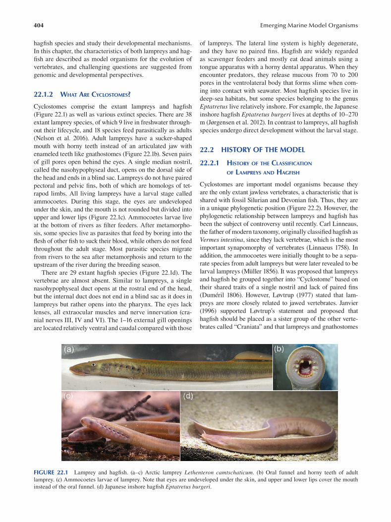
404
hagfi sh species and study their developmental mechanisms.
In this chapter, the characteristics of both lampreys and hag
fish are described as model organisms for the evolution of
vertebrates, and challenging questions are suggested from
genomic and developmental perspectives.
22.1.2 WHAT ARE CYCLOSTOMES?
Cyclostomes comprise the extant lampreys and hagfi sh
(Figure 22.1) as well as various extinct species. There are 38
extant lamprey species, of which 9 live in freshwater through
out their lifecycle, and 18 species feed parasitically as adults
(Nelson et al. 2016). Adult lampreys have a sucker-shaped
mouth with horny teeth instead of an articulated jaw with
enameled teeth like gnathostomes (Figure 22.1b). Seven pairs
of gill pores open behind the eyes. A single median nostril,
called the nasohypophyseal duct, opens on the dorsal side of
the head and ends in a blind sac. Lampreys do not have paired
pectoral and pelvic fins, both of which are homologs of tet
rapod limbs. All living lampreys have a larval stage called
ammocoetes. During this stage, the eyes are undeveloped
under the skin, and the mouth is not rounded but divided into
upper and lower lips (Figure 22.1c). Ammocoetes larvae live
at the bottom of rivers as filter feeders. After metamorpho
sis, some species live as parasites that feed by boring into the
flesh of other fish to suck their blood, while others do not feed
throughout the adult stage. Most parasitic species migrate
from rivers to the sea after metamorphosis and return to the
upstream of the river during the breeding season.
There are 29 extant hagfish species (Figure 22.1d). The
vertebrae are almost absent. Similar to lampreys, a single
nasohypophyseal duct opens at the rostral end of the head,
but the internal duct does not end in a blind sac as it does in
lampreys but rather opens into the pharynx. The eyes lack
lenses, all extraocular muscles and nerve innervation (cra
nial nerves III, IV and VI). The 1–16 external gill openings
are located relatively ventral and caudal compared with those
Emerging Marine Model Organisms
of lampreys. The lateral line system is highly degenerate,
and they have no paired fi ns. Hagfish are widely regarded
as scavenger feeders and mostly eat dead animals using a
tongue apparatus with a horny dental apparatus. When they
encounter predators, they release mucous from 70 to 200
pores in the ventrolateral body that forms slime when com
ing into contact with seawater. Most hagfish species live in
deep-sea habitats, but some species belonging to the genus
Eptatretus live relatively inshore. For example, the Japanese
inshore hagfi sh Eptatretus burgeri lives at depths of 10–270
m (Jørgensen et al. 2012). In contrast to lampreys, all hagfi sh
species undergo direct development without the larval stage.
22.2 HISTORY OF THE MODEL
22.2.1 HISTORY OF THE CLASSIFICATION
OF LAMPREYS AND HAGFISH
Cyclostomes are important model organisms because they
are the only extant jawless vertebrates, a characteristic that is
shared with fossil Silurian and Devonian fi sh. Thus, they are
in a unique phylogenetic position (Figure 22.2). However, the
phylogenetic relationship between lampreys and hagfi sh has
been the subject of controversy until recently. Carl Linneaus,
the father of modern taxonomy, originally classifi ed hagfi sh as
Vermes intestina, since they lack vertebrae, which is the most
important synapomorphy of vertebrates (Linnaeus 1758). In
addition, the ammocoetes were initially thought to be a sepa
rate species from adult lampreys but were later revealed to be
larval lampreys (Müller 1856). It was proposed that lampreys
and hagfi sh be grouped together into “Cyclostome” based on
their shared traits of a single nostril and lack of paired fi ns
(Duméril 1806). However, Løvtrup (1977) stated that lam
preys are more closely related to jawed vertebrates. Janvier
(1996 ) supported Løvtrup’s statement and proposed that
hagfish should be placed as a sister group of the other verte
brates called “Craniata” and that lampreys and gnathostomes
FIGURE 22.1 Lamprey and hagfish. (a–c) Arctic lamprey Lethenteron camtschaticum. (b) Oral funnel and horny teeth of adult
lamprey. (c) Ammocoetes larvae of lamprey. Note that eyes are undeveloped under the skin, and upper and lower lips cover the mouth
instead of the oral funnel. (d) Japanese inshore hagfi sh Eptatretus burgeri.
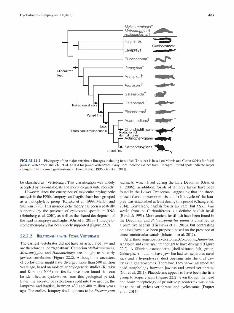
405 Cyclostomes (Lamprey and Hagfi sh)
FIGURE 22.2 Phylogeny of the major vertebrate lineages including fossil fish. This tree is based on Morris and Caron (2014) for fossil
jawless vertebrates and Zhu et al. (2013) for jawed vertebrates. Gray lines indicate extinct fossil lineages. Round spots indicate major
changes toward crown gnathostomes. (From Janvier 1996; Gai et al. 2011).
be classified as “Vertebrata”. This classification was widely
accepted by paleontologists and morphologists until recently.
However, since the emergence of molecular phylogenetic
analysis in the 1990s, lampreys and hagfish have been grouped
as a monophyletic group (Kuraku et al. 1999; Mallatt and
Sullivan 1998). This monophyletic theory has been repeatedly
supported by the presence of cyclostome-specifi c miRNA
(Heimberg et al. 2010), as well as the shared development of
the head in lampreys and hagfish ( Oisi et al. 2013). Thus, cyclo
stome monophyly has been widely supported (Figure 22.2).
22.2.2 RELATIONSHIP WITH FOSSIL VERTEBRATES
The earliest vertebrates did not have an articulated jaw and
are therefore called “Agnathan”. Cambrian Myllokunmingia,
Metaspriggina and Haikouichthys are thought to be early
jawless vertebrates (Figure 22.2). Although the ancestors
of cyclostomes might have diverged more than 500 million
years ago, based on molecular phylogenetic studies (Kuraku
and Kuratani 2006 ), no fossils have been found that can
be identified as cyclostomes from this geological period.
Later, the ancestor of cyclostomes split into two groups, the
lampreys and hagfish, between 430 and 480 million years
ago. The earliest lamprey fossil appears to be Priscomyzon
riniensis, which lived during the Late Devonian (Gess et
al. 2006). In addition, fossils of lamprey larvae have been
found in the Lower Cretaceous, suggesting that the three-
phased (larva–metamorphosis–adult) life cycle of the lam
prey was established at least during this period (Chang et al.
2014). Conversely, hagfish fossils are rare, but Myxinikela siroka from the Carboniferous is a defi nite hagfi sh fossil
( Bardack 1991). More ancient fossil fi sh have been found in
the Devonian, and Palaeospondylus gunni is classifi ed as
a primitive hagfish (Hirasawa et al. 2016), but contrasting
opinions have also been proposed based on the presence of
three semicircular canals (Johanson et al. 2017).
After the divergence of cyclostomes, Conodonts, Jamoytius, Anaspida and Pteraspis are thought to have diverged (Figure
22.2). A Silurian osteocoderm (shell-skinned fi sh) group,
Galeaspis, still did not have jaws but had two separated nasal
sacs and a hypophyseal duct opening into the oral cav
ity as in gnathostomes. Therefore, they show intermediate
head morphology between jawless and jawed vertebrates
(Gai et al. 2011). Placoderms appear to have been the fi rst
group to acquire jaws (Figure 22.2), even though the head
and brain morphology of primitive placoderms was simi
lar to that of jawless vertebrates and cyclostomes (Dupret
et al. 2014).
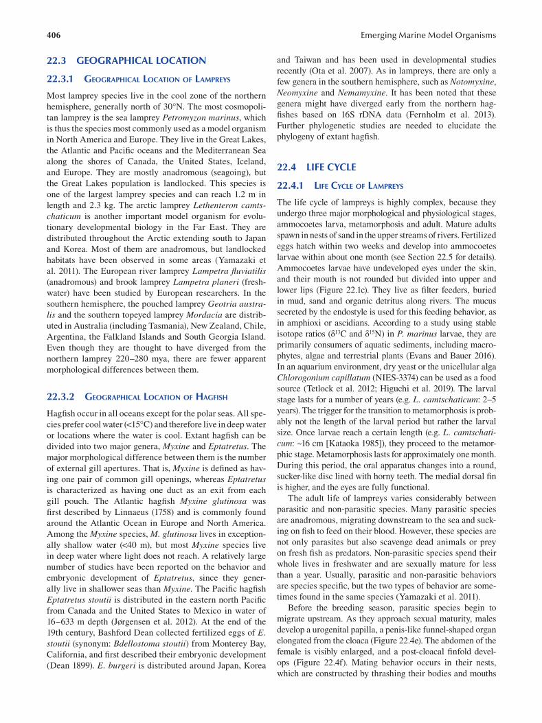
406
22.3 GEOGRAPHICAL LOCATION
22.3.1 GEOGRAPHICAL LOCATION OF LAMPREYS
Most lamprey species live in the cool zone of the northern
hemisphere, generally north of 30°N. The most cosmopoli
tan lamprey is the sea lamprey Petromyzon marinus , which
is thus the species most commonly used as a model organism
in North America and Europe. They live in the Great Lakes,
the Atlantic and Pacific oceans and the Mediterranean Sea
along the shores of Canada, the United States, Iceland,
and Europe. They are mostly anadromous (seagoing), but
the Great Lakes population is landlocked. This species is
one of the largest lamprey species and can reach 1.2 m in
length and 2.3 kg. The arctic lamprey Lethenteron camtschaticum is another important model organism for evolu
tionary developmental biology in the Far East. They are
distributed throughout the Arctic extending south to Japan
and Korea. Most of them are anadromous, but landlocked
habitats have been observed in some areas (Yamazaki et
al. 2011). The European river lamprey Lampetra fluviatilis (anadromous) and brook lamprey Lampetra planeri (fresh
water) have been studied by European researchers. In the
southern hemisphere, the pouched lamprey Geotria australis and the southern topeyed lamprey Mordacia are distrib
uted in Australia (including Tasmania), New Zealand, Chile,
Argentina, the Falkland Islands and South Georgia Island.
Even though they are thought to have diverged from the
northern lamprey 220–280 mya, there are fewer apparent
morphological differences between them.
22.3.2 GEOGRAPHICAL LOCATION OF HAGFISH
Hagfish occur in all oceans except for the polar seas. All spe
cies prefer cool water (<15°C) and therefore live in deep water
or locations where the water is cool. Extant hagfish can be
divided into two major genera, Myxine and Eptatretus . The
major morphological difference between them is the number
of external gill apertures. That is, Myxine is defined as hav
ing one pair of common gill openings, whereas Eptatretus is characterized as having one duct as an exit from each
gill pouch. The Atlantic hagfi sh Myxine glutinosa was
first described by Linnaeus (1758) and is commonly found
around the Atlantic Ocean in Europe and North America.
Among the Myxine species, M. glutinosa lives in exception
ally shallow water (<40 m), but most Myxine species live
in deep water where light does not reach. A relatively large
number of studies have been reported on the behavior and
embryonic development of Eptatretus, since they gener
ally live in shallower seas than Myxine. The Pacifi c hagfi sh
Eptatretus stoutii is distributed in the eastern north Pacifi c
from Canada and the United States to Mexico in water of
16–633 m depth (Jørgensen et al. 2012). At the end of the
19th century, Bashford Dean collected fertilized eggs of E. stoutii (synonym: Bdellostoma stoutii) from Monterey Bay,
California, and first described their embryonic development
( Dean 1899 ). E. burgeri is distributed around Japan, Korea
Emerging Marine Model Organisms
and Taiwan and has been used in developmental studies
recently (Ota et al. 2007). As in lampreys, there are only a
few genera in the southern hemisphere, such as Notomyxine,
Neomyxine and Nemamyxine. It has been noted that these
genera might have diverged early from the northern hag-
fishes based on 16S rDNA data (Fernholm et al. 2013).
Further phylogenetic studies are needed to elucidate the
phylogeny of extant hagfi sh.
22.4 LIFE CYCLE
22.4.1 LIFE CYCLE OF LAMPREYS
The life cycle of lampreys is highly complex, because they
undergo three major morphological and physiological stages,
ammocoetes larva, metamorphosis and adult. Mature adults
spawn in nests of sand in the upper streams of rivers. Fertilized
eggs hatch within two weeks and develop into ammocoetes
larvae within about one month (see Section 22.5 for details).
Ammocoetes larvae have undeveloped eyes under the skin,
and their mouth is not rounded but divided into upper and
lower lips ( Figure 22.1c). They live as filter feeders, buried
in mud, sand and organic detritus along rivers. The mucus
secreted by the endostyle is used for this feeding behavior, as
in amphioxi or ascidians. According to a study using stable
isotope ratios (δ13 C and δ15N) in P. marinus larvae, they are
primarily consumers of aquatic sediments, including macro
phytes, algae and terrestrial plants (Evans and Bauer 2016).
In an aquarium environment, dry yeast or the unicellular alga
Chlorogonium capillatum (NIES-3374) can be used as a food
source (Tetlock et al. 2012; Higuchi et al. 2019). The larval
stage lasts for a number of years (e.g. L. camtschaticum : 2–5
years). The trigger for the transition to metamorphosis is prob
ably not the length of the larval period but rather the larval
size. Once larvae reach a certain length (e.g. L. camtschaticum: ~16 cm [Kataoka 1985]), they proceed to the metamor
phic stage. Metamorphosis lasts for approximately one month.
During this period, the oral apparatus changes into a round,
sucker-like disc lined with horny teeth. The medial dorsal fi n
is higher, and the eyes are fully functional.
The adult life of lampreys varies considerably between
parasitic and non-parasitic species. Many parasitic species
are anadromous, migrating downstream to the sea and suck
ing on fish to feed on their blood. However, these species are
not only parasites but also scavenge dead animals or prey
on fresh fi sh as predators. Non-parasitic species spend their
whole lives in freshwater and are sexually mature for less
than a year. Usually, parasitic and non-parasitic behaviors
are species specific, but the two types of behavior are some
times found in the same species (Yamazaki et al. 2011).
Before the breeding season, parasitic species begin to
migrate upstream. As they approach sexual maturity, males
develop a urogenital papilla, a penis-like funnel-shaped organ
elongated from the cloaca (Figure 22.4e). The abdomen of the
female is visibly enlarged, and a post-cloacal fi nfold devel
ops (Figure 22.4f). Mating behavior occurs in their nests,
which are constructed by thrashing their bodies and mouths
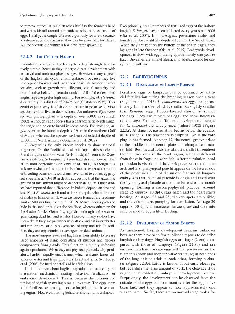
407 Cyclostomes (Lamprey and Hagfi sh)
to remove stones. A male attaches itself to the female’s head
and wraps his tail around her trunk to assist in the extrusion of
eggs. Finally, the couple vibrates vigorously for a few seconds
to release eggs and sperm so they can be externally fertilized.
All individuals die within a few days after spawning.
22.4.2 LIFE CYCLE OF HAGFISH
In contrast to lampreys, the life cycle of hagfish might be rela
tively simple, because they undergo direct development with
no larval and metamorphosis stages. However, many aspects
of the hagfish life cycle remain unknown because they live
in deep-sea habitats, and even their basic life history charac
teristics, such as growth rate, lifespan, sexual maturity and
reproductive behavior, remain unclear. All of the described
hagfish species prefer high salinity. For example, M. glutinosa dies rapidly in salinities of 20–25 ppt (Gustafson 1935). This
could explain why hagfish do not occur in polar seas. Most
species tend to live in deep waters. An unknown Eptatretus sp. was photographed at a depth of over 5,000 m (Sumich
1992). Although each species has a characteristic depth range,
the range can be quite broad in some cases. For instance, M. glutinosa can be found at depths of 30 m in the northern Gulf
of Maine, whereas this species has been collected at depths of
1,100 m in North America (Jørgensen et al. 2012).
E. burgeri is the only known species to show seasonal
migration. On the Pacific side of mid-Japan, this species is
found in quite shallow water (6–10 m depth) from mid-Octo
ber to mid-July. Subsequently, these hagfi sh swim deeper than
50 m until September (Ichikawa et al. 2000). Although it is
unknown whether this migration is related to water temperature
or breeding behavior, researchers have failed to collect eggs by
net sweeping at 40–110 m depth, suggesting that the spawning
ground of this animal might be deeper than 100 m. Other stud
ies have reported that differences in habitat depend on size and
sex. Most E. stoutii are found at 100 m depth, where the ratio
of males to females is 1:1, whereas larger females are predomi
nant at 500 m (Jørgensen et al. 2012). Many species prefer to
hide in the sand or mud on the sea floor, whereas others prefer
the shade of rocks. Generally, hagfish are thought to be scaven
gers, eating dead fish and whales. However, many studies have
showed that they are predators who attack and eat invertebrates
and vertebrates, such as polychaetes, shrimp and fi sh. In addi
tion, they are opportunistic scavengers on dead animals.
The most unique feature of hagfish is their ability to release
large amounts of slime consisting of mucous and fi brous
components from glands. This function is mainly defensive
against predators. When they are physically attacked by pred
ators, hagfish rapidly eject slime, which entrains large vol
umes of water and traps predators’ head and gills. See Fudge
et al. (2016) for further details of hagfi sh slime.
Little is known about hagfish reproduction, including the
maturation mechanism, mating behavior, fertilization or
embryonic development. This is because the location and
timing of hagfish spawning remain unknown. The eggs seem
to be fertilized externally, because hagfish do not have mat
ing organs. However, mating behavior also remains unknown.
Exceptionally, small numbers of fertilized eggs of the inshore
hagfi sh E. burgeri have been collected every year since 2006
(Ota et al. 2007 ). In mid-August, pre-mature males and
females can be caught at a depth of 100 m in the Sea of Japan.
When they are kept on the bottom of the sea in cages, they
lay eggs in late October (Oisi et al. 2015). Embryonic devel
opment is slow, with eggs taking approximately one year to
hatch. Juveniles are almost identical to adults, except for car
rying the yolk sac.
22.5 EMBRYOGENESIS
22.5.1 DEVELOPMENT OF LAMPREY EMBRYOS
Fertilized eggs of lampreys can be obtained by artifi
cial fertilization during the breeding season once a year
(Sugahara et al. 2015). L. camtschaticum eggs are approx
imately 1 mm in size, which is similar but slightly smaller
than Xenopus eggs. Double-layered chorion surrounds
the eggs. They are telolecithal eggs and show holoblas
tic cleavage. For staging, Tahara’s developmental stages
for L. reisnneri are widely used (Tahara 1988) (Figure
22.3a). At stage 13, gastrulation begins below the equator
as in Xenopus. The blastopore is elliptical, while the yolk
plug is not formed. At stage 17, the neural groove arises
in the middle of the neural plate and changes to a neu
ral fold. Both neural folds are almost parallel throughout
the embryos, even in the head region, which is different
from those in frogs and zebrafish. After neurulation, head
protrusion is visible, and the cheek processes (mandibular
arch and first pharyngeal pouch) appear on the lateral side
of the protrusion. One of the unique features of lamprey
embryos is that the nasal placode is single and fused with
the hypophyseal placode at the anterior end to the mouth
opening, forming a nasohypophyseal placode. Around
stage 25 (approx. 10 dpf), eggs hatch and the heart starts
beating. At stages 27 and 28, the eye spots are visible,
and the velum starts pumping for ventilation. At stage 30
(approx. 30 dpf), ammocoetes larvae grow and dive into
sand or mud to begin fi lter feeding.
22.5.2 DEVELOPMENT OF HAGFISH EMBRYOS
As mentioned, hagfish development remains unknown
because there have been few published reports to describe
hagfish embryology. Hagfish eggs are large (2 cm) com
pared with those of lampreys (Figure 22.3b) and are
encased in a hard, orange eggshell that possesses anchor
filaments (hook and loop tape-like structure) at both ends
of the long axis to stick to each other, forming a clus
ter (Figure 22.3c). Little is known about early cleavage,
but regarding the large amount of yolk, the cleavage style
might be meroblastic. Embryonic development is slow.
Surprisingly, the development can be observed from the
outside of the eggshell four months after the eggs have
been laid, and they appear to take approximately one
year to hatch. So far, there are no normal stage tables for
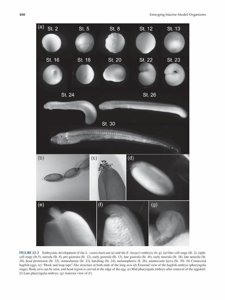
408 Emerging Marine Model Organisms
FIGURE 22.3 Embryonic development of the L. camtschaticum (a) and the E. burgeri embryos (b–g). (a) One-cell stage (St. 2), eight-
cell stage (St.5), morula (St. 8), pre-gastrula (St. 12), early gastrula (St. 13), late gastrula (St. 16), early neurula (St. 18), late neurula (St.
20), head protrusion (St. 22), stomodaeum (St. 23), hatching (St. 24), melanophores (S. 26), ammocoete larva (St. 30). (b) Connected
hagfish eggs. (c) “Hook and loop tape”-like structure at both ends of the long axis (d) External view of the hagfish embryo (pharyngula
stage). Body axis can be seen, and head region is curved at the edge of the egg. (e) Mid-pharyngula embryo after removal of the eggshell.
(f) Late-pharyngula embryo. (g) Anterior view of (f).
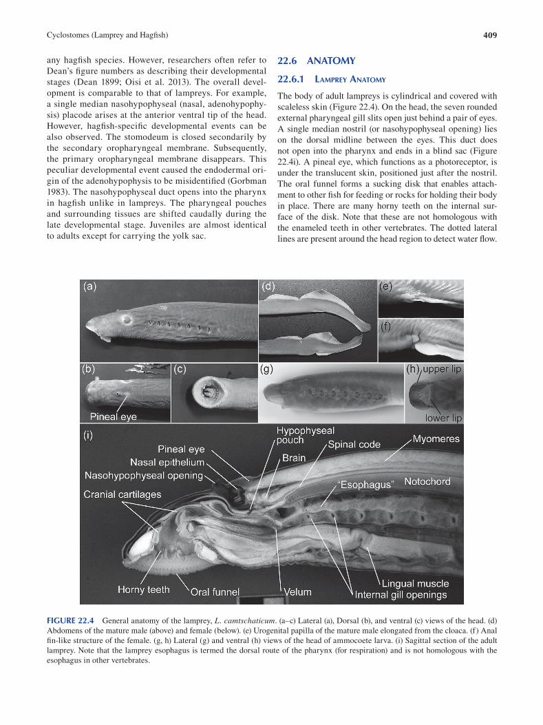
409 Cyclostomes (Lamprey and Hagfi sh)
any hagfish species. However, researchers often refer to
Dean’s figure numbers as describing their developmental
stages (Dean 1899; Oisi et al. 2013). The overall devel
opment is comparable to that of lampreys. For example,
a single median nasohypophyseal (nasal, adenohypophy
sis) placode arises at the anterior ventral tip of the head.
However, hagfi sh-specific developmental events can be
also observed. The stomodeum is closed secondarily by
the secondary oropharyngeal membrane. Subsequently,
the primary oropharyngeal membrane disappears. This
peculiar developmental event caused the endodermal ori
gin of the adenohypophysis to be misidentifi ed (Gorbman
1983). The nasohypophyseal duct opens into the pharynx
in hagfish unlike in lampreys. The pharyngeal pouches
and surrounding tissues are shifted caudally during the
late developmental stage. Juveniles are almost identical
to adults except for carrying the yolk sac.
22.6 ANATOMY
22.6.1 LAMPREY ANATOMY
The body of adult lampreys is cylindrical and covered with
scaleless skin (Figure 22.4). On the head, the seven rounded
external pharyngeal gill slits open just behind a pair of eyes.
A single median nostril (or nasohypophyseal opening) lies
on the dorsal midline between the eyes. This duct does
not open into the pharynx and ends in a blind sac (Figure
22.4i). A pineal eye, which functions as a photoreceptor, is
under the translucent skin, positioned just after the nostril.
The oral funnel forms a sucking disk that enables attach
ment to other fish for feeding or rocks for holding their body
in place. There are many horny teeth on the internal sur
face of the disk. Note that these are not homologous with
the enameled teeth in other vertebrates. The dotted lateral
lines are present around the head region to detect water fl ow.
FIGURE 22.4 General anatomy of the lamprey, L. camtschaticum. (a–c) Lateral (a), Dorsal (b), and ventral (c) views of the head. (d)
Abdomens of the mature male (above) and female (below). (e) Urogenital papilla of the mature male elongated from the cloaca. (f) Anal
fin-like structure of the female. (g, h) Lateral (g) and ventral (h) views of the head of ammocoete larva. (i) Sagittal section of the adult
lamprey. Note that the lamprey esophagus is termed the dorsal route of the pharynx (for respiration) and is not homologous with the
esophagus in other vertebrates.
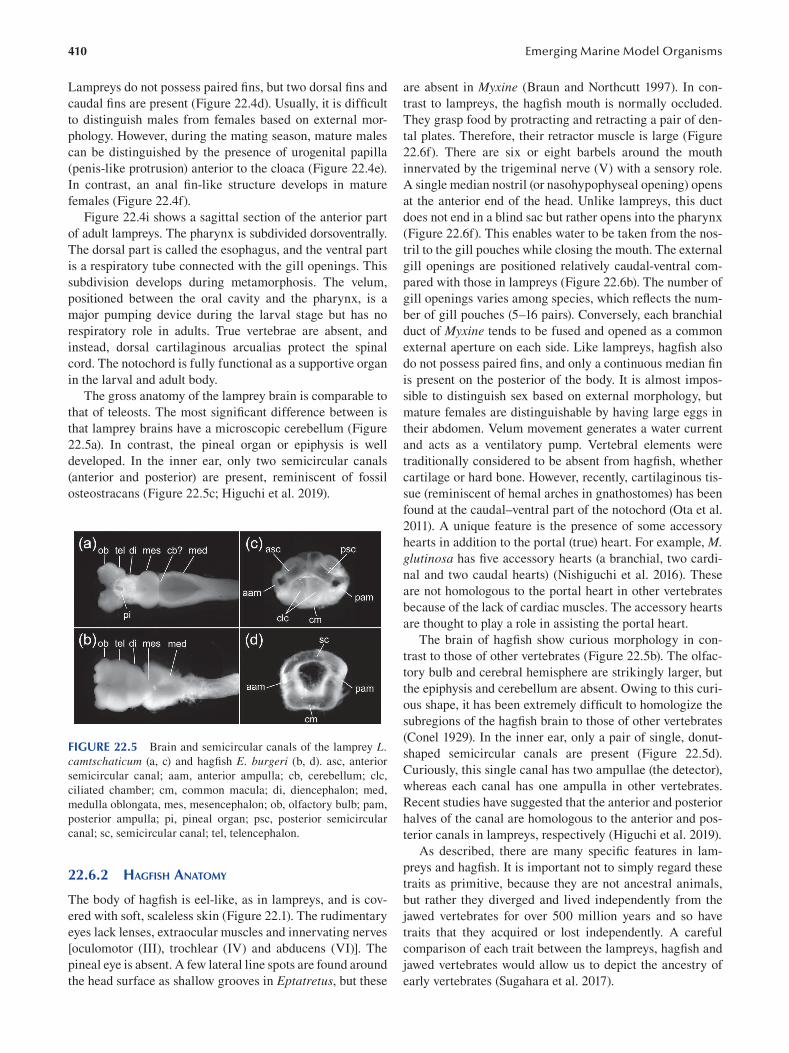
410
Lampreys do not possess paired fins, but two dorsal fi ns and
caudal fins are present (Figure 22.4d). Usually, it is diffi cult
to distinguish males from females based on external mor
phology. However, during the mating season, mature males
can be distinguished by the presence of urogenital papilla
(penis-like protrusion) anterior to the cloaca (Figure 22.4e).
In contrast, an anal fin-like structure develops in mature
females (Figure 22.4f).
Figure 22.4i shows a sagittal section of the anterior part
of adult lampreys. The pharynx is subdivided dorsoventrally.
The dorsal part is called the esophagus, and the ventral part
is a respiratory tube connected with the gill openings. This
subdivision develops during metamorphosis. The velum,
positioned between the oral cavity and the pharynx, is a
major pumping device during the larval stage but has no
respiratory role in adults. True vertebrae are absent, and
instead, dorsal cartilaginous arcualias protect the spinal
cord. The notochord is fully functional as a supportive organ
in the larval and adult body.
The gross anatomy of the lamprey brain is comparable to
that of teleosts. The most significant difference between is
that lamprey brains have a microscopic cerebellum (Figure
22.5a). In contrast, the pineal organ or epiphysis is well
developed. In the inner ear, only two semicircular canals
(anterior and posterior) are present, reminiscent of fossil
osteostracans (Figure 22.5c; Higuchi et al. 2019).
FIGURE 22.5 Brain and semicircular canals of the lamprey L. camtschaticum (a, c) and hagfi sh E. burgeri (b, d). asc, anterior
semicircular canal; aam, anterior ampulla; cb, cerebellum; clc,
ciliated chamber; cm, common macula; di, diencephalon; med,
medulla oblongata, mes, mesencephalon; ob, olfactory bulb; pam,
posterior ampulla; pi, pineal organ; psc, posterior semicircular
canal; sc, semicircular canal; tel, telencephalon.
22.6.2 HAGFISH ANATOMY
The body of hagfish is eel-like, as in lampreys, and is cov
ered with soft, scaleless skin (Figure 22.1). The rudimentary
eyes lack lenses, extraocular muscles and innervating nerves
[oculomotor (III), trochlear (IV) and abducens (VI)]. The
pineal eye is absent. A few lateral line spots are found around
the head surface as shallow grooves in Eptatretus, but these
Emerging Marine Model Organisms
are absent in Myxine (Braun and Northcutt 1997 ). In con
trast to lampreys, the hagfish mouth is normally occluded.
They grasp food by protracting and retracting a pair of den
tal plates. Therefore, their retractor muscle is large (Figure
22.6f). There are six or eight barbels around the mouth
innervated by the trigeminal nerve (V) with a sensory role.
A single median nostril (or nasohypophyseal opening) opens
at the anterior end of the head. Unlike lampreys, this duct
does not end in a blind sac but rather opens into the pharynx
(Figure 22.6f). This enables water to be taken from the nos
tril to the gill pouches while closing the mouth. The external
gill openings are positioned relatively caudal-ventral com
pared with those in lampreys (Figure 22.6b). The number of
gill openings varies among species, which reflects the num
ber of gill pouches (5–16 pairs). Conversely, each branchial
duct of Myxine tends to be fused and opened as a common
external aperture on each side. Like lampreys, hagfi sh also
do not possess paired fins, and only a continuous median fi n
is present on the posterior of the body. It is almost impos
sible to distinguish sex based on external morphology, but
mature females are distinguishable by having large eggs in
their abdomen. Velum movement generates a water current
and acts as a ventilatory pump. Vertebral elements were
traditionally considered to be absent from hagfi sh, whether
cartilage or hard bone. However, recently, cartilaginous tis
sue (reminiscent of hemal arches in gnathostomes) has been
found at the caudal–ventral part of the notochord (Ota et al.
2011). A unique feature is the presence of some accessory
hearts in addition to the portal (true) heart. For example, M. glutinosa has five accessory hearts (a branchial, two cardi
nal and two caudal hearts) (Nishiguchi et al. 2016). These
are not homologous to the portal heart in other vertebrates
because of the lack of cardiac muscles. The accessory hearts
are thought to play a role in assisting the portal heart.
The brain of hagfish show curious morphology in con
trast to those of other vertebrates (Figure 22.5b). The olfac
tory bulb and cerebral hemisphere are strikingly larger, but
the epiphysis and cerebellum are absent. Owing to this curi
ous shape, it has been extremely diffi cult to homologize the
subregions of the hagfi sh brain to those of other vertebrates
(Conel 1929). In the inner ear, only a pair of single, donut-
shaped semicircular canals are present (Figure 22.5d).
Curiously, this single canal has two ampullae (the detector),
whereas each canal has one ampulla in other vertebrates.
Recent studies have suggested that the anterior and posterior
halves of the canal are homologous to the anterior and pos
terior canals in lampreys, respectively (Higuchi et al. 2019).
As described, there are many specific features in lam
preys and hagfish. It is important not to simply regard these
traits as primitive, because they are not ancestral animals,
but rather they diverged and lived independently from the
jawed vertebrates for over 500 million years and so have
traits that they acquired or lost independently. A careful
comparison of each trait between the lampreys, hagfi sh and
jawed vertebrates would allow us to depict the ancestry of
early vertebrates (Sugahara et al. 2017 ).
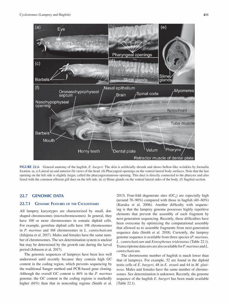
411 Cyclostomes (Lamprey and Hagfi sh)
FIGURE 22.6 General anatomy of the hagfi sh, E. burgeri. The skin is artificially shrunk and shows bellow-like wrinkles by formalin
fixation. (a, c) Lateral (a) and anterior (b) views of the head. (b) Pharyngeal openings on the ventral lateral body surfaces. Note that the last
opening on the left side is slightly larger, called the pharyngocutaneous opening. This duct is directly connected to the pharynx and also
fused with the common efferent gill duct on the left side. (d, e) Slime glands on the ventral lateral sides of the body. (f) Sagittal section.
22.7 GENOMIC DATA
22.7.1 GENOMIC FEATURES OF THE CYCLOSTOMES
All lamprey karyotypes are characterized by small, dot-
shaped chromosomes (microchromosomes). In general, they
have 100 or more chromosomes in somatic diploid cells.
For example, germline diploid cells have 198 chromosomes
in P. marinus and 168 chromosomes in L. camtschaticum (Ishijima et al. 2017 ). Males and females have the same num
ber of chromosomes. The sex determination system is unclear
but may be determined by the growth rate during the larval
period (Johnson et al. 2017).
The genomic sequences of lampreys have been less well
understood until recently because they contain high GC
content in the coding region, which prevents sequencing by
the traditional Sanger method and PCR-based gene cloning.
Although the overall GC content is 46% in the P. marinus genome, the GC content in the coding regions is markedly
higher (61%) than that in noncoding regions (Smith et al.
2013). Four-fold degenerate sites (GC4) are especially high
(around 70–90%) compared with those in hagfi sh (40–60%)
(Kuraku et al. 2006). Another difficulty with sequenc
ing is that the lamprey genome possesses highly repetitive
elements that prevent the assembly of each fragment by
next-generation sequencing. Recently, these diffi culties have
been overcome by optimizing the computational assembly
that allowed us to assemble fragments from next-generation
sequence data (Smith et al. 2018). Currently, the lamprey
genome sequence is available from three species (P. marinus, L. camtschaticum and Entosphenus tridentatus ( Table 22.1 ).
Transcriptome data sets are also available for P. marinus and L. camtschaticum.
The chromosome number of hagfish is much lower than
that of lampreys. For example, 52 are found in the diploid
testis cells of E. burgeri, 48 in E. stoutii and 44 in M. glutinosa. Males and females have the same number of chromo
somes. Sex determination is unknown. Recently, the genome
sequence of the hagfi sh E. burgeri has been made available
( Table 22.1 ).
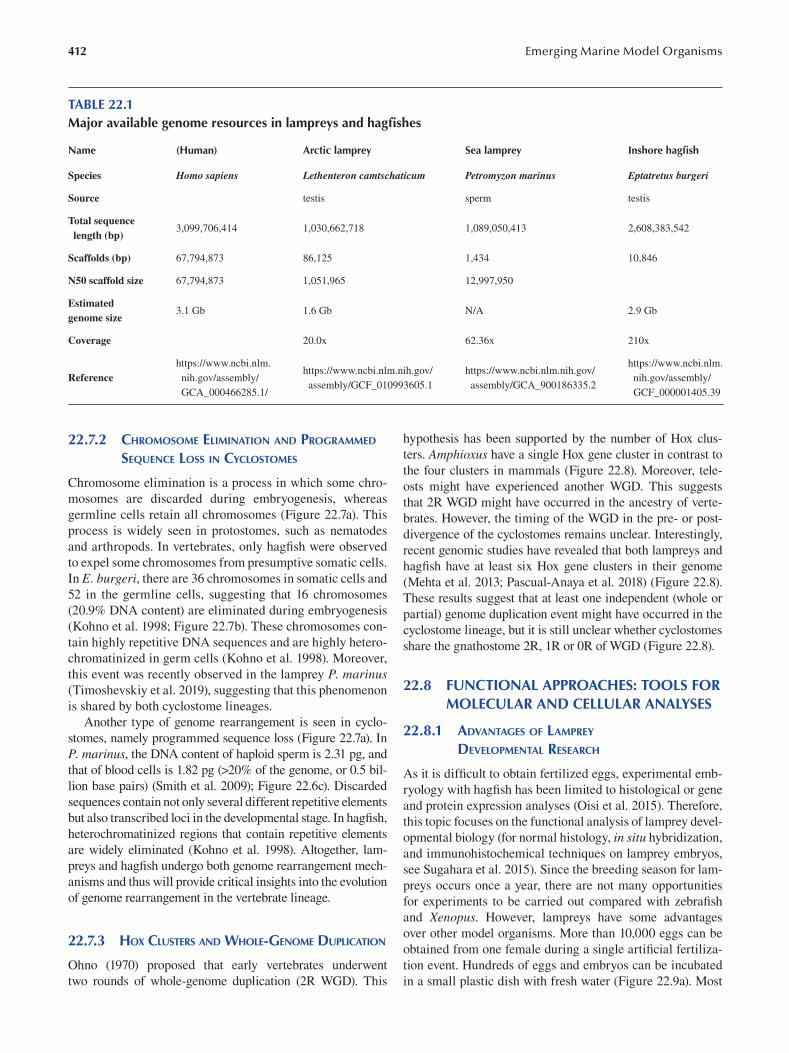
412 Emerging Marine Model Organisms
TABLE 22.1 Major available genome resources in lampreys and hagfi shes
Name (Human) Arctic lamprey Sea lamprey Inshore hagfi sh
Species Homo sapiens Lethenteron camtschaticum Petromyzon marinus Eptatretus burgeri
Source testis sperm testis
Total sequence length (bp)
3,099,706,414 1,030,662,718 1,089,050,413 2,608,383,542
Scaffolds (bp) 67,794,873 86,125 1,434 10,846
N50 scaffold size 67,794,873 1,051,965 12,997,950
Estimated genome size
3.1 Gb 1.6 Gb N/A 2.9 Gb
Coverage 20.0x 62.36x 210x
https://www.ncbi.nlm. https://www.ncbi.nlm. https://www.ncbi.nlm.nih.gov/ https://www.ncbi.nlm.nih.gov/
Reference nih.gov/assembly/ nih.gov/assembly/ assembly/GCF_010993605.1 assembly/GCA_900186335.2
GCA_000466285.1/ GCF_000001405.39
22.7.2 CHROMOSOME ELIMINATION AND PROGRAMMED
SEQUENCE LOSS IN CYCLOSTOMES
Chromosome elimination is a process in which some chro
mosomes are discarded during embryogenesis, whereas
germline cells retain all chromosomes (Figure 22.7a). This
process is widely seen in protostomes, such as nematodes
and arthropods. In vertebrates, only hagfish were observed
to expel some chromosomes from presumptive somatic cells.
In E. burgeri, there are 36 chromosomes in somatic cells and
52 in the germline cells, suggesting that 16 chromosomes
(20.9% DNA content) are eliminated during embryogenesis
(Kohno et al. 1998; Figure 22.7b). These chromosomes con
tain highly repetitive DNA sequences and are highly hetero
chromatinized in germ cells (Kohno et al. 1998). Moreover,
this event was recently observed in the lamprey P. marinus (Timoshevskiy et al. 2019), suggesting that this phenomenon
is shared by both cyclostome lineages.
Another type of genome rearrangement is seen in cyclo
stomes, namely programmed sequence loss (Figure 22.7a). In
P. marinus, the DNA content of haploid sperm is 2.31 pg, and
that of blood cells is 1.82 pg (>20% of the genome, or 0.5 bil
lion base pairs) (Smith et al. 2009); Figure 22.6c). Discarded
sequences contain not only several different repetitive elements
but also transcribed loci in the developmental stage. In hagfi sh,
heterochromatinized regions that contain repetitive elements
are widely eliminated (Kohno et al. 1998). Altogether, lam
preys and hagfish undergo both genome rearrangement mech
anisms and thus will provide critical insights into the evolution
of genome rearrangement in the vertebrate lineage.
22.7.3 HOX CLUSTERS AND WHOLE-GENOME DUPLICATION
Ohno (1970) proposed that early vertebrates underwent
two rounds of whole-genome duplication (2R WGD). This
hypothesis has been supported by the number of Hox clus
ters. Amphioxus have a single Hox gene cluster in contrast to
the four clusters in mammals (Figure 22.8). Moreover, tele
osts might have experienced another WGD. This suggests
that 2R WGD might have occurred in the ancestry of verte
brates. However, the timing of the WGD in the pre- or post-
divergence of the cyclostomes remains unclear. Interestingly,
recent genomic studies have revealed that both lampreys and
hagfish have at least six Hox gene clusters in their genome
(Mehta et al. 2013; Pascual-Anaya et al. 2018) (Figure 22.8).
These results suggest that at least one independent (whole or
partial) genome duplication event might have occurred in the
cyclostome lineage, but it is still unclear whether cyclostomes
share the gnathostome 2R, 1R or 0R of WGD (Figure 22.8).
22.8 FUNCTIONAL APPROACHES: TOOLS FOR MOLECULAR AND CELLULAR ANALYSES
22.8.1 ADVANTAGES OF LAMPREY
DEVELOPMENTAL RESEARCH
As it is difficult to obtain fertilized eggs, experimental emb
ryology with hagfish has been limited to histological or gene
and protein expression analyses (Oisi et al. 2015). Therefore,
this topic focuses on the functional analysis of lamprey devel
opmental biology (for normal histology, in situ hybridization,
and immunohistochemical techniques on lamprey embryos,
see Sugahara et al. 2015). Since the breeding season for lam
preys occurs once a year, there are not many opportunities
for experiments to be carried out compared with zebrafi sh
and Xenopus. However, lampreys have some advantages
over other model organisms. More than 10,000 eggs can be
obtained from one female during a single artifi cial fertiliza
tion event. Hundreds of eggs and embryos can be incubated
in a small plastic dish with fresh water (Figure 22.9a). Most
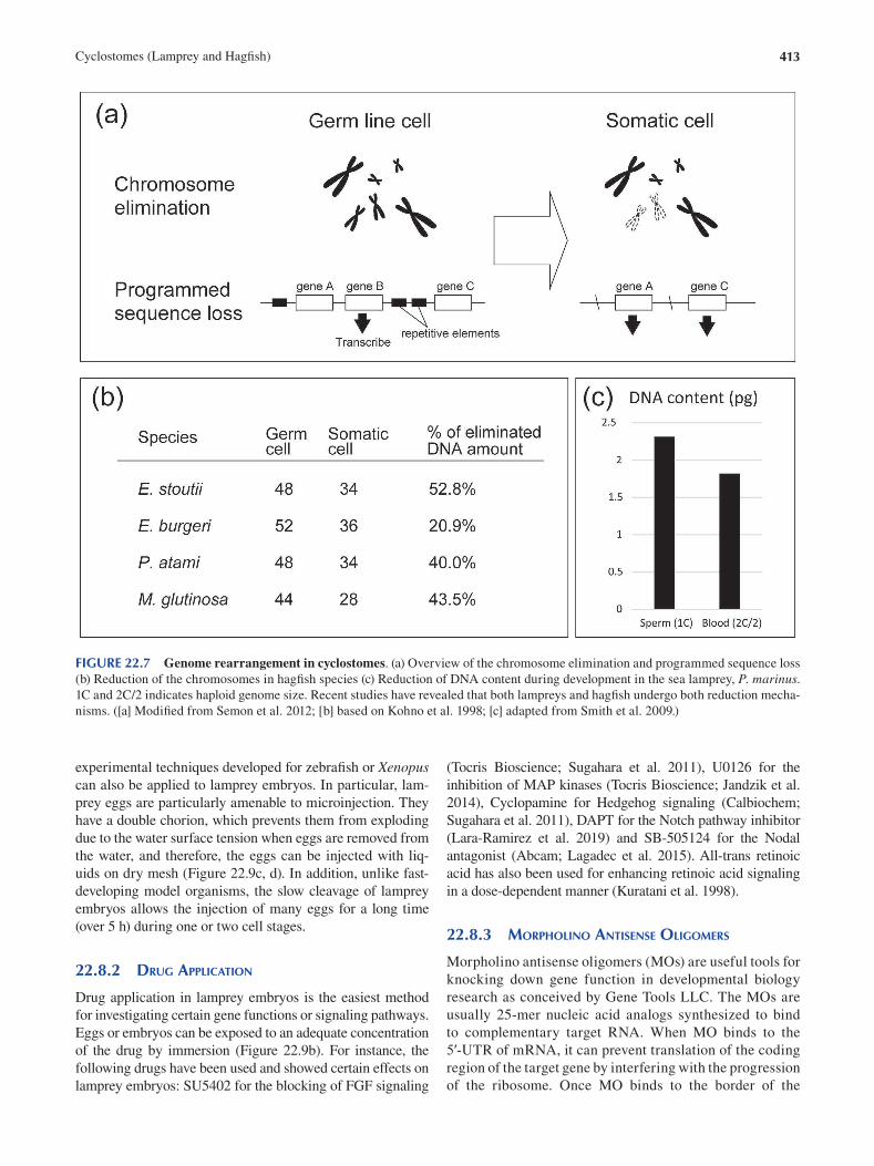
413 Cyclostomes (Lamprey and Hagfi sh)
FIGURE 22.7 Genome rearrangement in cyclostomes. (a) Overview of the chromosome elimination and programmed sequence loss
(b) Reduction of the chromosomes in hagfish species (c) Reduction of DNA content during development in the sea lamprey, P. marinus. 1C and 2C/2 indicates haploid genome size. Recent studies have revealed that both lampreys and hagfish undergo both reduction mecha
nisms. ([a] Modified from Semon et al. 2012; [b] based on Kohno et al. 1998; [c] adapted from Smith et al. 2009.)
experimental techniques developed for zebrafish or Xenopus can also be applied to lamprey embryos. In particular, lam
prey eggs are particularly amenable to microinjection. They
have a double chorion, which prevents them from exploding
due to the water surface tension when eggs are removed from
the water, and therefore, the eggs can be injected with liq
uids on dry mesh (Figure 22.9c, d). In addition, unlike fast-
developing model organisms, the slow cleavage of lamprey
embryos allows the injection of many eggs for a long time
(over 5 h) during one or two cell stages.
22.8.2 DRUG APPLICATION
Drug application in lamprey embryos is the easiest method
for investigating certain gene functions or signaling pathways.
Eggs or embryos can be exposed to an adequate concentration
of the drug by immersion ( Figure 22.9b ). For instance, the
following drugs have been used and showed certain effects on
lamprey embryos: SU5402 for the blocking of FGF signaling
(Tocris Bioscience; Sugahara et al. 2011 ), U0126 for the
inhibition of MAP kinases (Tocris Bioscience; Jandzik et al.
2014 ), Cyclopamine for Hedgehog signaling (Calbiochem;
Sugahara et al. 2011 ), DAPT for the Notch pathway inhibitor
( Lara-Ramirez et al. 2019 ) and SB-505124 for the Nodal
antagonist (Abcam; Lagadec et al. 2015 ). All-trans retinoic
acid has also been used for enhancing retinoic acid signaling
in a dose-dependent manner ( Kuratani et al. 1998 ).
22.8.3 MORPHOLINO ANTISENSE OLIGOMERS
Morpholino antisense oligomers (MOs) are useful tools for
knocking down gene function in developmental biology
research as conceived by Gene Tools LLC. The MOs are
usually 25-mer nucleic acid analogs synthesized to bind
to complementary target RNA. When MO binds to the
5′-UTR of mRNA, it can prevent translation of the coding
region of the target gene by interfering with the progression
of the ribosome. Once MO binds to the border of the
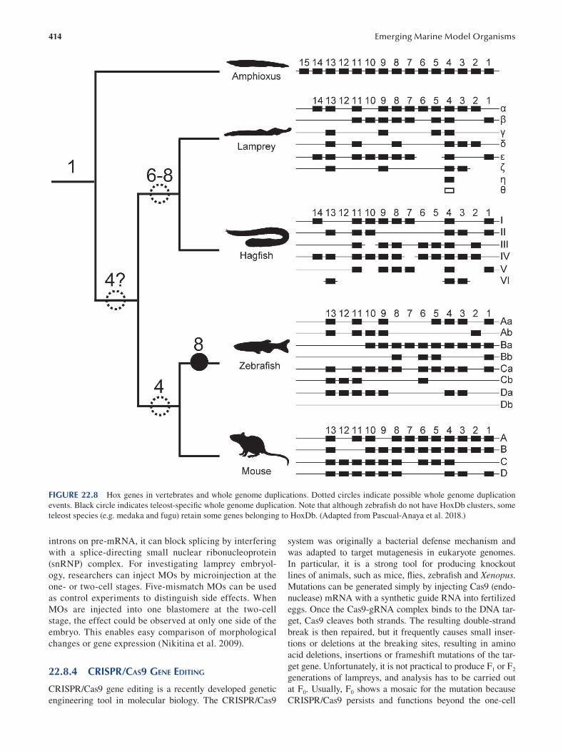
414 Emerging Marine Model Organisms
FIGURE 22.8 Hox genes in vertebrates and whole genome duplications. Dotted circles indicate possible whole genome duplication
events. Black circle indicates teleost-specific whole genome duplication. Note that although zebrafish do not have HoxDb clusters, some
teleost species (e.g. medaka and fugu) retain some genes belonging to HoxDb. (Adapted from Pascual-Anaya et al. 2018.)
introns on pre-mRNA, it can block splicing by interfering
with a splice-directing small nuclear ribonucleoprotein
(snRNP) complex. For investigating lamprey embryol
ogy, researchers can inject MOs by microinjection at the
one- or two-cell stages. Five-mismatch MOs can be used
as control experiments to distinguish side effects. When
MOs are injected into one blastomere at the two-cell
stage, the effect could be observed at only one side of the
embryo. This enables easy comparison of morphological
changes or gene expression (Nikitina et al. 2009).
22.8.4 CRISPR/CAS9 GENE EDITING
CRISPR/Cas9 gene editing is a recently developed genetic
engineering tool in molecular biology. The CRISPR/Cas9
system was originally a bacterial defense mechanism and
was adapted to target mutagenesis in eukaryote genomes.
In particular, it is a strong tool for producing knockout
lines of animals, such as mice, fl ies, zebrafi sh and Xenopus. Mutations can be generated simply by injecting Cas9 (endo
nuclease) mRNA with a synthetic guide RNA into fertilized
eggs. Once the Cas9-gRNA complex binds to the DNA tar
get, Cas9 cleaves both strands. The resulting double-strand
break is then repaired, but it frequently causes small inser
tions or deletions at the breaking sites, resulting in amino
acid deletions, insertions or frameshift mutations of the tar
get gene. Unfortunately, it is not practical to produce F1 or F2
generations of lampreys, and analysis has to be carried out
at F0. Usually, F0 shows a mosaic for the mutation because
CRISPR/Cas9 persists and functions beyond the one-cell

415 Cyclostomes (Lamprey and Hagfi sh)
FIGURE 22.9 Embryonic manipulation of lamprey embryos.
(a) Incubation of lamprey embryos in 9-cm dishes. Hundreds of
embryos can be kept in one dish. Blue water is the 10% Steinberg’s
solution containing 0.6 ppm of methylene blue to prevent bacterial
growth. (b) Drug application in lamprey embryos; 20–30 embryos
can be exposed to a certain concentration of drugs in each 12-well
dish. (c,d) Microinjection in lamprey embryos. The sieve mesh size
is 0.61 mm, and wire diameter is 0.23 mm. (e,f) KAEDE (pho
toconvertible protein) expression in lamprey embryos (stages 18
and 23). KAEDE mRNA combined with nuclear localized signal
injected in one-cell eggs after fertilization. The expression can be
seen only in each cell nucleus and lasts at least until hatching stage.
stage. However, several reports have shown that CRISPR/
Cas9-injected F0 embryos effectively disrupted target genes,
even though each cell was differentially mutated (Square
et al. 2015).
22.9 CHALLENGING QUESTIONS
Finally, I suggest some challenging questions in the
developmental and genomic fields from an evolutionary
perspective.
22.9.1 CEREBELLAR EVOLUTION
The cerebellum plays an essential role in controlling coordi
nated movements as well as cognitive and emotional func
tions in humans. All living gnathostomes have distinct,
three-layered cerebella (granular, Purkinje and molecular
layers). However, lampreys have an undifferentiated cerebel
lum, which is only visible as a dorsal lip at the anterior end of
the rhombencephalon. They do not have a layered structure,
but some cerebellum-specific neuron subtypes have been
found. In contrast, the presence of the cerebellum in hagfi sh
is uncertain. Recently, Sugahara et al. (2016 ) reported on
the gene expression in lamprey and hagfish embryos that is
essential for cerebellar development. When and how the cer
ebellum was established and acquired a three-layered struc
ture during vertebrate evolution are intriguing questions. A
comparison of cerebellar development between cyclostomes
and gnathostomes would answer this question. See Sugahara
et al. (2017) for detailed information.
22.9.2 EVOLUTION OF THE PAIRED NOSTRILS
Most fossil jawless fish have a single median nostril, and
cyclostomes might also retain this ancestral condition.
During development, the gnathostome nasal placode is
generated as paired and separated from the hypophyseal
placode (Rathke’s pouch). In contrast, the median nasal
placode and hypophyseal placode arise as a single ecto
dermal thickening in lampreys and hagfish. The sepa
ration of the nasohypophyseal placode and subsequent
changes in the migration of neural crest cells might be
a key innovation for the acquisition of the jaw (Kuratani
et al. 2001).
22.9.3 ORIGIN OF THE PAIRED APPENDAGES
Cyclostomes do not possess paired fins that are homolo
gous to human arms and legs. So far, two major theories
have been proposed to explain the origin of paired fi ns.
The fin-fold theory posits that paired fins evolved from a
longitudinal paired fin-fold. Anaspida, an early Silurian
fish, might have had paired folds on the ventral side of the
body (Janvier 1996 ). Another theory is the gill-arch the
ory that posits that the pectoral fins were the result of the
transformation or co-option of the gill arches. It may well
be the case that vertebrates acquired the pectoral fi n fi rst
(see Osteostracans in Figure 22.2). It would be interest
ing to investigate whether cyclostomes have the potential
to form paired appendages. In lamprey embryos, different
distribution patterns of the lateral plate mesoderm, which
contributes to limb growth, have been reported (Tulenko
et al. 2013).
22.9.4 EVOLUTION OF THE THYROID GLAND
The ammocoetes larvae of lampreys have an endostyle under
the pharynx as a secreting organ for filter feeding. Non-
vertebrate chordates, Amphioxus and ascidians also possess
this organ. During metamorphosis, the lamprey endostyle
changes into the thyroid gland. Therefore, it is thought that
the chordate endostyle is homologous to the vertebrate thyroid
gland and transitioned from an endostyle to the thyroid in
lamprey evolutionary history (Ogasawara et al. 2001). This
theory is based on the homology of the endostyle between
lampreys and non-vertebrate chordates. However, the homol
ogy of the endostyle remains unclear. In addition, although
the hagfish undergoes direct development and thus does not
have an endostyle, there is only one much older study regard
ing the thyroid gland (Stockard 1906). Detailed analysis of
416
hagfish thyroid gland development would shed light on this
question.
22.9.5 TIMING OF WHOLE-GENOME DUPLICATION
As noted previously, hagfish and lampreys possess at least six
Hox clusters (Mehta et al. 2013; Pascual-Anaya et al. 2018).
Since the homology and relationship between each cluster
and gnathostome clusters remain unclear, it is yet to be deter
mined whether the two rounds of WGDs that gnathostome
experienced occurred before or after the divergence of the
cyclostomes. Deep, detailed comparative synteny analysis of
cyclostome genomes would lead to a clearer understanding
of the evolution of the vertebrate genome.
ACKNOWLEDGMENTS
I thank Shigeru Kuratani and his past and current laboratory
members for cyclostome research, Noboru Sato and Hiroshi
Nagashima for lamprey sampling and Osamu Kakitani for
hagfi sh sampling.
BIBLIOGRAPHY
Bardack, D. 1991. First fossil hagfish (myxinoidea): A record from
the Pennsylvanian of Illinois. Science 254 (5032):701–703.
Braun, C.B., and R.G. Northcutt. 1997. The lateral line system
of hagfishes (Craniata: Myxinoidea). Acta Zoologica 78
(3):247–268.
Chang, M.M., Wu, F., Miao, D., and J. Zhang. 2014. Discovery
of fossil lamprey larva from the Lower Cretaceous reveals
its three-phased life cycle. Proceedings of the National Academy of Sciences of the United States of America 111
(43):15486–15490.
Conel, J.L. 1929. The development of the brain of Bdellostoma stouti 1: External growth changes. Journal of Comparative Neurology 47 (3):343–403.
Dean, B. 1899. On the embryology of Bdellostoma stouti : A gen
eral account of myxinoid development from the egg and
segmentation to hatching. Festschrift zum 70ten Geburststag Carl von Kupffer:220–276.
Dumé ril, C. 1806. Zoologie analytique, ou méthode naturelle de classification des animaux, Rendue plus facile a l’Aide de Tableaux Synoptiques . Paris: Allais.
Dupret, V., Sanchez, S., Goujet, D., Tafforeau, P., and P.E. Ahlberg.
2014. A primitive placoderm sheds light on the origin of the
jawed vertebrate face. Nature 507 (7493):500–3.
Evans, T.M., and J.E. Bauer. 2016. Identification of the nutritional
resources of larval sea lamprey in two Great Lakes tributar
ies using stable isotopes. Journal of Great Lakes Research 42
(1):99–107.
Fernholm, B., Norén, M., Kullander, S.O., et al. 2013. Hagfi sh
phylogeny and taxonomy, with description of the new genus
Rubicundus (Craniata, Myxinidae). Journal of Zoological Systematics Evolutionary Research 51 (4):296–307.
Fudge, D.S., Herr, J.E., and T.M. Winegard. 2016. Hagfi sh Slime, Hagfi sh Biology. New York: CRC Press.
Gai, Z., Donoghue, P.C., Zhu, M., Janvier, P., and M. Stampanoni.
2011. Fossil jawless fish from China foreshadows early
jawed vertebrate anatomy. Nature 476 (7360):324–327.
Emerging Marine Model Organisms
Gess, R.W., Coates M.I., and B.S. Rubidge. 2006. A lamprey from the
Devonian period of South Africa. Nature 443 (7114):981–984.
Gorbman, A. 1983. Early development of the hagfi sh pituitary-
gland: Evidence for the endodermal origin of the adenohy
pophysis. American Zoologist 23 (3):639–654.
Gustafson, G. 1935. On the biology of Myxine glutinosa L. Arkiv Zool 28:1–8.
Heimberg, A.M., Cowper-Sal-lari, R., Sémon, M., Donoghue,
P.C., and K.J. Peterson. 2010. microRNAs reveal the inter
relationships of hagfish, lampreys, and gnathostomes and the
nature of the ancestral vertebrate. Proceedings of the National Academy of Sciences of the United States of America 107
(45):19379–19383.
Higuchi, S., Sugahara, F., Pascual-Anaya J., Takagi W., Oisi, Y.,
and S. Kuratani. 2019. Inner ear development in cyclostomes
and evolution of the vertebrate semicircular canals. Nature 565 (7739):347–350.
Hirasawa, T., Oisi, Y., and S. Kuratani. 2016. Palaeospondylus as a
primitive hagfi sh. Zoological Letters 2 (1):20.
Ichikawa, T., Kobayashi, H., and M. Nozaki. 2000. Seasonal migra
tion of the hagfi sh, Eptatretus burgeri, Girard. Zoological Science 17 (2):217–223.
Ishijima, J., Uno, Y., Nunome, M. Nishida, C., Kuraku, S., and Y.
Matsuda. 2017. Molecular cytogenetic characterization of
chromosome site-specific repetitive sequences in the Arctic
lamprey ( Lethenteron camtschaticum, Petromyzontidae).
DNA Research 24 (1):93–101.
Jandzik, D., Hawkins, M.B., Cattell, M.V., Cerny, R., Square, T.A.,
and D.M. Medeiros. 2014. Roles for FGF in lamprey pharyn
geal pouch formation and skeletogenesis highlight ancestral
functions in the vertebrate head. Development 141 (3):629–638.
Janvier, P. 1996. Early Vertebrates. Oxford: Oxford University Press.
Johnson, N.S., Swink, W.D., and T.O. Brenden. 2017. Field study
suggests that sex determination in sea lamprey is directly
influenced by larval growth rate. Proceedings of the Royal Society: Biological Sciences 284 (1851).
Johanson, Z., Smith, M., Sanchez, S., Senden, T., Trinajstic, K.,
and C. Pfaff. 2017. Questioning hagfi sh affinities of the enig
matic Devonian vertebrate Palaeospondylus. Royal Society Open Science 4 (7):170214.
Jørgensen, J.M., Lomholt, J.P., Weber R.E., and H. Malte. 2012.
The Biology of Hagfishes. London: Springer Science &
Business Media.
Kataoka, T. 1985. Studies on the propagation of Lethenteron camtschaticum (IV) (in Japanese). Research Reports of Niigata Prefectural Freshwater Fisheries Experiment Station 12:23–27.
Kohno, S., Kubota, S., and Y. Nakai. 1998. Chromatin diminution
and chromosome elimination in hagfishes. In The Biology of Hagfi shes. London: Springer.
Kuraku, S., Hoshiyama, D., Katoh, K., Suga, H., and T. Miyata.
1999. Monophyly of lampreys and hagfishes supported by
nuclear DNA-coded genes. Journal of Molecular Evolution 49 (6):729–735.
Kuraku, S., Ishijima, J., Nishida-Umehara, C., Agata, K., Kuratani,
S., and Y. Matsuda. 2006. cDNA-based gene mapping and
GC3 profiling in the soft-shelled turtle suggest a chro
mosomal size-dependent GC bias shared by sauropsids.
Chromosome Research 14 (2):187–202.
Kuraku, S., and S. Kuratani. 2006. Time scale for cyclostome evolu
tion inferred with a phylogenetic diagnosis of hagfish and lam
prey cDNA sequences. Zoological Science 23 (12):1053–1064.
Kuratani, S., Nobusada, Y., Horigome, N., and Y. Shigetani. 2001.
Embryology of the lamprey and evolution of the verte
brate jaw: Insights from molecular and developmental
Cyclostomes (Lamprey and Hagfi sh)
perspectives. Philosophical Transactions of the Royal Society B: Biological Sciences 356 (1414):1615–1632.
Kuratani, S., Ueki, T., Hirano, S., and S. Aizawa. 1998. Rostral
truncation of a cyclostome, Lampetra japonica , induced
by all-trans retinoic acid defines the head/trunk interface
of the vertebrate body. Developmental Dynamics 211
(1):35–51.
Lagadec, R., Laguerre, L., Menuet, A., Amara, A., Rocancourt,
C., Péricard, P., Godard, B.G., Rodicio, M.C., Rodriguez-
Moldes, I., Mayeur, H., Rougemont, Q., Mazan, S., and
A. Boutet. 2015. The ancestral role of nodal signalling in
breaking L/R symmetry in the vertebrate forebrain. Nature Communications 6.
Lara-Ramirez, R., Pérez-González, C., Anselmi, C., Patthey, C.,
and S.M. Shimeld. 2019. A Notch-regulated proliferative
stem cell zone in the developing spinal cord is an ancestral
vertebrate trait. Development 146 (1).
Linnaeus, C. 1758. Systema naturae. Vol. 1. Stockholm: Stockholm
Laurentii Salvii.
Løvtrup, S. 1977. The Phylogeny of Vertebrates. New York: John
Wiley.
Mallatt, J., and J. Sullivan. 1998. 28S and 18S rDNA sequences
support the monophyly of lampreys and hagfi shes. Molecular Biology and Evolution 15 (12):1706–1718.
Mehta, T.K., Ravi, V., Yamasaki, S., et al. 2013. Evidence for at
least six Hox clusters in the Japanese lamprey ( Lethenteron japonicum). Proceedings of the National Academy of Sciences 110 (40):16044–16049.
Morris, S.C., and J.B. Caron. 2014. A primitive fish from the
Cambrian of North America. Nature 512 (7515):419–422.
Müller, A. 1856. XXVI: On the development of the lampreys.
Annals and Magazine of Natural History 18 (106):298–301.
Nelson, J.S., Grande, T.C., and M.V.H. Wilson. 2016. Fishes of the World. Hoboken, NJ: John Wiley & Sons.
Nikitina, N., Bronner-Fraser, M., and T. Sauka-Spengler. 2009. The
sea lamprey Petromyzon marinus: A model for evolutionary
and developmental biology. Cold Spring Harbor Protocols 2009 (1):pdb.emo113.
Nishiguchi, Y., Tomita, T., Sato, et al. 2016. Examination of the
hearts and blood vascular system of Eptatretus okinoseanus using computed tomography images, diagnostic sonogra
phy, and histology. International Journal of Analytical Bio-Science 4 (3):46–54.
Ogasawara, M., Shigetani, Y., Suzuki, S., Kuratani, S., and N. Satoh.
2001. Expression of thyroid transcription factor-1 (TTF-1)
gene in the ventral forebrain and endostyle of the agnathan
vertebrate, Lampetra japonica. Genesis 30 (2):51–58.
Ohno, S. 1970. Evolution by Gene Duplication. New York: Springer
Science & Business Media.
Oisi, Y., Kakitani, O., Kuratani, S., and K.G. Ota. 2015. Analysis of
embryonic gene expression patterns in the hagfi sh. In In Situ Hybridization Methods. New York: Springer.
Oisi, Y., Ota, K.G., Kuraku, S., Fujimoto, S., and S. Kuratani. 2013.
Craniofacial development of hagfishes and the evolution of
vertebrates. Nature 493 (7431):175–180.
Ota, K.G., Fujimoto, S., Oisi, Y., and S. Kuratani. 2011.
Identification of vertebra-like elements and their possible
differentiation from sclerotomes in the hagfi sh. Nature Communications 2:373.
Ota, K.G., Kuraku, S., and S. Kuratani. 2007. Hagfi sh embryology
with reference to the evolution of the neural crest. Nature 446
(7136):672–675.
417
Pascual-Anaya, J., Sato, I., Sugahara, F., et al. 2018. Hagfi sh and
lamprey Hox genes reveal conservation of temporal colinear
ity in vertebrates. Nature Ecology Evolution 2 (5):859–866.
Sémon, M., Schubert, M., and V. Laudet. 2012. Programmed
genome rearrangements: In lampreys, all cells are not equal.
Current Biology 22 (16):R641–643.
Smith, J.J., Antonacci, F., Eichler, E.E., and C.T. Amemiya. 2009.
Programmed loss of millions of base pairs from a vertebrate
genome. Proceedings of the National Academy of Sciences of the United States of America 106 (27):11212–11217.
Smith, J.J., Kuraku, S., Holt, C., et al. 2013. Sequencing of the sea lam
prey ( Petromyzon marinus) genome provides insights into verte
brate evolution. Nature Genetics 45 (4):415–421, 421e1–421e2.
Smith, J.J., Timoshevskaya, N., Ye, C., et al. 2018. The sea lam
prey germline genome provides insights into programmed
genome rearrangement and vertebrate evolution. Nature Genetics 50 (2):270–277.
Square, T., Romášek, M., Jandzik, D., Cattell, M.V., Klymkowsky,
M., and D.M. Medeiros. 2015. CRISPR/Cas9-mediated
mutagenesis in the sea lamprey Petromyzon marinus : A pow
erful tool for understanding ancestral gene functions in ver
tebrates. Development 142 (23):4180–4187.
Stockard, C.R. 1906. The development of the thyroid gland in
Bdellostoma stouti. Anatomischer. Anzeiger . 29:91–99.
Sugahara, F., Aota, S., Kuraku, S., et al. 2011. Involvement of
hedgehog and FGF signalling in the lamprey telencephalon:
Evolution of regionalization and dorsoventral patterning of
the vertebrate forebrain. Development 138 (6):1217–1226.
Sugahara, F., Murakami, Y., Pascual-Anaya, P., and S. Kuratani.
2017. Reconstructing the ancestral vertebrate brain.
Development, Growth & Differentiation 59 (4):163–174.
Sugahara, F., Pascual-Anaya, J., Oisi, Y., et al. 2016. Evidence from
cyclostomes for complex regionalization of the ancestral ver
tebrate brain. Nature 531 (7592):97–100.
Sugahara, F., Yasunori M., and S. Kuratani. 2015. Gene expres
sion analysis of lamprey embryos. In In Situ Hybridization Methods. New York: Springer.
Sumich, J.L. 1992. Benthic communities. In Introduction to the Biology of Marine Life. Dubuque, Iowa: Wn. C. Brown.
Tahara, Y. 1988. Normal stages of development in the lamprey,
Lampetra reissneri (Dybowski). Zoological Science 5 (1):
109–118.
Tetlock, A., Yost, C.K., Stavrinides, J., and R.G. Manzon. 2012.
Changes in the gut microbiome of the sea lamprey during
metamorphosis. Applied Environmental Microbiology 78
(21):7638–7644.
Timoshevskiy, V.A., Timoshevskaya, N.Y., and Smith, J.J. 2019.
Germline-specific repetitive elements in programmatically
eliminated chromosomes of the sea lamprey ( Petromyzon marinus). Genes (Basel) 10 (10).
Tulenko, F.J., McCauley, D.J., Mackenzie, E.L., et al. 2013. Body
wall development in lamprey and a new perspective on the
origin of vertebrate paired fi ns. Proceedings of the National Academy of Sciences of the United States of America 110
(29):11899–11904.
Yamazaki, Y., Yokoyama, R., Nagai, T., and A. Goto. 2011.
Formation of a fluvial non-parasitic population of Lethenteron camtschaticum as the first step in petromyzontid speciation.
Journal of Fish Biol 79 (7):2043–2059.
Zhu, M., Yu, X., Ahlberg, P.E., et al. 2013. A Silurian placoderm with
osteichthyan-like marginal jaw bones. Nature 502 (7470):
188–193.

23 Current Trends in Chondrichthyes Experimental Biology
Yasmine Lund-Ricard and Agnès Boutet
CONTENTS
23.1 Introduction to Chondrichthyes Models ................................................................................................................... 420
23.1.1 Phylogeny ..................................................................................................................................................... 420
23.2 Chondrichthyes in the Past and Present .....................................................................................................................421
23.2.1 The Rise of Chondrichthyes as Models in Experimental Biology.................................................................421
23.2.2 The Study of Chondrichthyes Behavior ........................................................................................................ 422
23.2.3 Current Trends in Chondrichthyes Research ................................................................................................ 422
23.2.4 Chondrichthyes Conservation Status ............................................................................................................ 422
23.2.5 The Science behind Conservation Efforts ..................................................................................................... 422
23.3 Biogeography............................................................................................................................................................ 423
23.4 Chondrichthyes Life Cycles...................................................................................................................................... 423
23.4.1 Reproductive Strategies ................................................................................................................................ 423
23.4.2 Chondrichthyes Species in Developmental Biology ..................................................................................... 423
23.5 Chondrichthyes Embryogenesis................................................................................................................................ 424
23.5.1 Early Embryogenesis and Gastrulation ........................................................................................................ 424
23.5.2 From Axis Formation to Pharynx Segmentation .......................................................................................... 426
23.6 Chondrichthyes Anatomy and Sensory Biology ....................................................................................................... 426
23.6.1 External Features .......................................................................................................................................... 426
23.6.2 Internal Anatomy .......................................................................................................................................... 427
23.6.3 Sensory Biology ............................................................................................................................................ 428
23.6.3.1 Photoreception ............................................................................................................................... 428
23.6.3.2 Audition ......................................................................................................................................... 428
23.6.3.3 Mechanosensory System ............................................................................................................... 428
23.6.3.4 Chemoreception............................................................................................................................. 428
23.6.3.5 Magnetoreception .......................................................................................................................... 429
23.6.3.6 Electroreception............................................................................................................................. 429
23.7 Genomic Data ........................................................................................................................................................... 429
23.7.1 Genomes and Transcriptomes ....................................................................................................................... 429
23.7.2 Gene Family Studies ..................................................................................................................................... 430
23.8 Tools for Molecular and Cellular Analyses ............................................................................................................... 430
23.8.1 Cell Lines ...................................................................................................................................................... 430
23.8.2 Descriptive and Functional Approaches ........................................................................................................431
23.9 Challenging Questions ...............................................................................................................................................432
23.9.1 Endogenous Chondrichthyes Molecules for Biomedical Applications .........................................................432
23.9.1.1 Molecules Displaying Antibiotic Activity ......................................................................................432
23.9.1.2 The High Specifi city of Chondrichthyes Antibodies ......................................................................432
23.9.1.3 The Different Properties of Squalamine .........................................................................................432
23.9.1.4 Molecules Displaying Anti-Cancer Activity ...................................................................................433
23.9.2 Evo-Devo Studies in the Search for the Origin of Skeleton and Brain Asymmetries ...................................433
23.9.2.1 Endoskeleton and Bone-Like Tissue in Chondrichthyes ................................................................433
23.9.2.2 Exoskeleton (Teeth and Dermal Denticles) in Chondrichthyes ..................................................... 434
23.9.2.3 Evolution of Brain Asymmetries in Vertebrates ............................................................................ 434
23.9.3 The Elasmobranch Properties of Kidney Regeneration .................................................................................435
Acknowledgments .................................................................................................................................................................435
Bibliography .........................................................................................................................................................................435
DOI: 10.1201/9781003217503-23 419
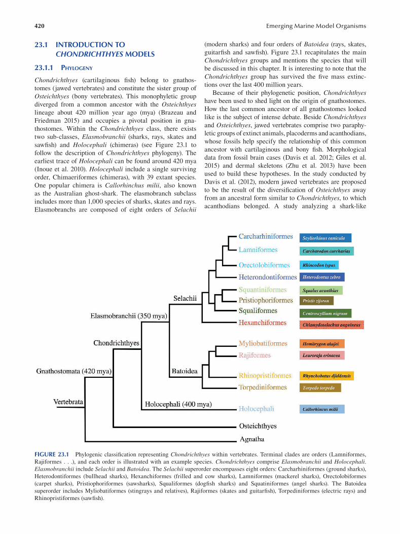
420 Emerging Marine Model Organisms
23.1 INTRODUCTION TO CHONDRICHTHYES MODELS
23.1.1 PHYLOGENY
Chondrichthyes (cartilaginous fish) belong to gnathos
tomes (jawed vertebrates) and constitute the sister group of
Osteichthyes (bony vertebrates). This monophyletic group
diverged from a common ancestor with the Osteichthyes lineage about 420 million year ago (mya) (Brazeau and
Friedman 2015) and occupies a pivotal position in gna
thostomes. Within the Chondrichthyes class, there exists
two sub-classes, Elasmobranchii (sharks, rays, skates and
sawfish) and Holocephali (chimeras) (see Figure 23.1 to
follow the description of Chondrichthyes phylogeny). The
earliest trace of Holocephali can be found around 420 mya
(Inoue et al. 2010). Holocephali include a single surviving
order, Chimaeriformes (chimeras), with 39 extant species.
One popular chimera is Callorhinchus milii, also known
as the Australian ghost-shark. The elasmobranch subclass
includes more than 1,000 species of sharks, skates and rays.
Elasmobranchs are composed of eight orders of Selachii
(modern sharks) and four orders of Batoidea (rays, skates,
guitarfish and sawfish). Figure 23.1 recapitulates the main
Chondrichthyes groups and mentions the species that will
be discussed in this chapter. It is interesting to note that the
Chondrichthyes group has survived the five mass extinc
tions over the last 400 million years.
Because of their phylogenetic position, Chondrichthyes have been used to shed light on the origin of gnathostomes.
How the last common ancestor of all gnathostomes looked
like is the subject of intense debate. Beside Chondrichthyes and Osteichthyes, jawed vertebrates comprise two paraphy
letic groups of extinct animals, placoderms and acanthodians,
whose fossils help specify the relationship of this common
ancestor with cartilaginous and bony fi sh. Morphological
data from fossil brain cases (Davis et al. 2012; Giles et al.
2015) and dermal skeletons ( Zhu et al. 2013) have been
used to build these hypotheses. In the study conducted by
Davis et al. (2012), modern jawed vertebrates are proposed
to be the result of the diversification of Osteichthyes away
from an ancestral form similar to Chondrichthyes, to which
acanthodians belonged. A study analyzing a shark-like
FIGURE 23.1 Phylogenic classifi cation representing Chondrichthyes within vertebrates. Terminal clades are orders (Lamniformes,
Rajiformes . . .), and each order is illustrated with an example species. Chondrichthyes comprise Elasmobranchii and Holocephali. Elasmobranchii include Selachii and Batoidea. The Selachii superorder encompasses eight orders: Carcharhiniformes (ground sharks),
Heterodontiformes (bullhead sharks), Hexanchiformes (frilled and cow sharks), Lamniformes (mackerel sharks), Orectolobiformes
(carpet sharks), Pristiophoriformes (sawsharks), Squaliformes (dogfish sharks) and Squatiniformes (angel sharks). The Batoidea
superorder includes Myliobatiformes (stingrays and relatives), Rajiformes (skates and guitarfish), Torpediniformes (electric rays) and
Rhinopristiformes (sawfi sh).
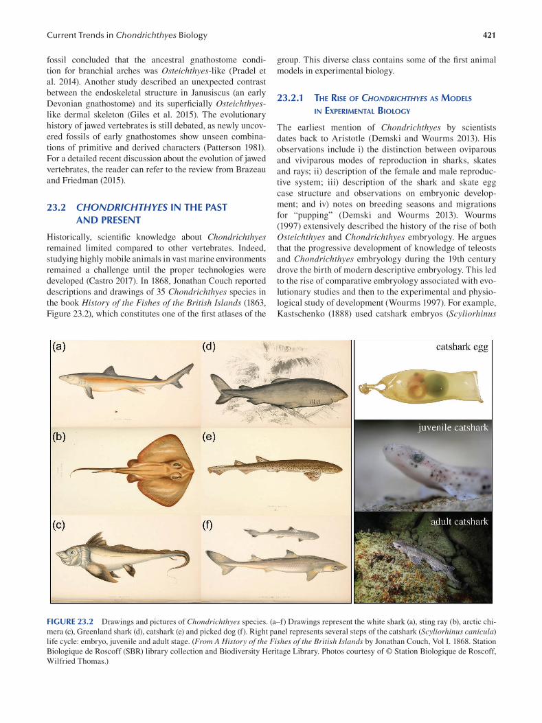
421 Current Trends in Chondrichthyes Biology
fossil concluded that the ancestral gnathostome condi
tion for branchial arches was Osteichthyes -like ( Pradel et
al. 2014). Another study described an unexpected contrast
between the endoskeletal structure in Janusiscus (an early
Devonian gnathostome) and its superfi cially Osteichthyes
like dermal skeleton (Giles et al. 2015). The evolutionary
history of jawed vertebrates is still debated, as newly uncov
ered fossils of early gnathostomes show unseen combina
tions of primitive and derived characters (Patterson 1981).
For a detailed recent discussion about the evolution of jawed
vertebrates, the reader can refer to the review from Brazeau
and Friedman (2015).
23.2 CHONDRICHTHYES IN THE PAST AND PRESENT
Historically, scientific knowledge about Chondrichthyes remained limited compared to other vertebrates. Indeed,
studying highly mobile animals in vast marine environments
remained a challenge until the proper technologies were
developed (Castro 2017). In 1868, Jonathan Couch reported
descriptions and drawings of 35 Chondrichthyes species in
the book History of the Fishes of the British Islands ( 1863 ,
Figure 23.2), which constitutes one of the first atlases of the
group. This diverse class contains some of the fi rst animal
models in experimental biology.
23.2.1 THE RISE OF CHONDRICHTHYES AS MODELS
IN EXPERIMENTAL BIOLOGY
The earliest mention of Chondrichthyes by scientists
dates back to Aristotle (Demski and Wourms 2013). His
observations include i) the distinction between oviparous
and viviparous modes of reproduction in sharks, skates
and rays; ii) description of the female and male reproduc
tive system; iii) description of the shark and skate egg
case structure and observations on embryonic develop
ment; and iv) notes on breeding seasons and migrations
for “pupping” (Demski and Wourms 2013). Wourms
(1997) extensively described the history of the rise of both
Osteichthyes and Chondrichthyes embryology. He argues
that the progressive development of knowledge of teleosts
and Chondrichthyes embryology during the 19th century
drove the birth of modern descriptive embryology. This led
to the rise of comparative embryology associated with evo
lutionary studies and then to the experimental and physio
logical study of development (Wourms 1997 ). For example,
Kastschenko (1888) used catshark embryos (Scyliorhinus
FIGURE 23.2 Drawings and pictures of Chondrichthyes species. (a–f) Drawings represent the white shark (a), sting ray (b), arctic chi
mera (c), Greenland shark (d), catshark (e) and picked dog (f). Right panel represents several steps of the catshark (Scyliorhinus canicula)
life cycle: embryo, juvenile and adult stage. (From A History of the Fishes of the British Islands by Jonathan Couch, Vol I. 1868. Station
Biologique de Roscoff (SBR) library collection and Biodiversity Heritage Library. Photos courtesy of © Station Biologique de Roscoff,
Wilfried Thomas.)
422
canicula, Figure 23.2) as experimental models to test a
developmental theory.
23.2.2 THE STUDY OF CHONDRICHTHYES BEHAVIOR
The first reported studies on Chondrichthyes behavior
emerged later. The initial studies on shark behavior include
those carried out by Sheldon (1909, 1911) and by Parker
(1914). The focus of these studies was the influence of the
eyes, ears and other allied sense organs on the movements of
the dogfi sh Mustelus canis. Remarkably, a military project
entitled “Project Headgear” (1958–1971) conducted experi
ments in which sharks were trained to carry explosives.
The details of this project have never been released. With
increasingly sophisticated technology, the themes addressed
in behavioral research have widened, and an array of studies
can be found (Tricas and Gruber 2001; Sundström et al. 2001;
Kelly et al. 2019; Gardiner 2012; Myrberg 2003; Gruber and
Myrberg 1977; Hammerschlag 2016; Aidan et al. 2005). As
the field of animal cognition expands, social learning in
lemon sharks (Guttridge et al. 2013); tool use in batoids (Kuba
et al. 2010); learning, habituations and memory in a benthic
shark (Kimber et al. 2014); and spatial memory and orien
tation strategies in stingrays (Schluessel and Bleckmann
2005) have helped build a picture of Chondrichthyes cogni
tive functions. Schluessel (2015) reviewed the evidence for
cognitive abilities in elasmobranchs.
23.2.3 CURRENT TRENDS IN CHONDRICHTHYES RESEARCH
Current trends in Chondrichthyes research were analyzed in
a recent review (Shiffman et al. 2020). This review depicts
the trends in research efforts over three decades (1985–2016)
by analyzing the content of all the abstracts presented at the
annual conferences of the American Elasmobranch Society
(AES), the oldest and largest professional society for the
scientific study and management of these fish (Shiffman et
al. 2020). AES research was most frequently on movement/
telemetry, age and growth, population genetics, reproduc
tive biology and diet/feeding ecology, with different areas
of focus for different species or families. Certain biases
exist in areas of investigations such as species “charisma”
(e.g. white shark, Carcharodon carcharias ), accessibil
ity to long-term established field research programs (e.g.
lemon shark, Negaprion brevirostris, and sandbar shark,
Carcharhinus plumbeus) or ease of model maintenance
for lab-based research (e.g. bonnethead shark, Sphyrna tiburo) (Shiffman et al. 2020). Nearly 90% of all described
Chondrichthyes species have never been mentioned in an
AES abstract, including some of the most threatened species
in the Americas (Shiffman et al. 2020).
23.2.4 CHONDRICHTHYES CONSERVATION STATUS
Chondrichthyes are considered one of the most threat
ened vertebrate groups by the International Union for the
Emerging Marine Model Organisms
Conservation of Nature (IUCN) Red List (McClenachan
et al. 2012; Dulvy et al. 2014; White and Last 2012). The
threats faced by Chondrichthyes can be grouped into the
effects of various fishing activities and of habitat loss (Dulvy
et al. 2014; Jennings et al. 2008) and environmental degra
dation such as pollution (Lyons and Wynne-Edwards 2018).
Alarmingly, sharks are subject to a global slaughter; shark
products such as dried fins have high commercial value and
a high exposure to international trade (Gross 2019). Human
exploitation of Chondrichthyes is aggravated by certain life
history traits, like low fecundity, the production of small
numbers of highly precocious young, slow growth rates
and late sexual maturity (Collin 2012). In 2020, a study
showed that fishing exploitation in the Mediterranean might
exert an evolutionary pressure toward early maturation in
the catshark, Scyliorhinus canicula (Ramírez-Amaro et al.
2020). Additionally, sharks are considered at a relatively
high risk for climate change (Cavanagh et al. 2005; Rosa et
al. 2014). Indeed, climate change is already affecting ocean
temperatures, pH and oxygen levels. How ocean warming,
acidification, deoxygenation and fishery exploitation may
interact to impact Chondrichthyes populations is yet to be
determined (Sims 2019; Rosa et al. 2017; Wheeler et al.
2020). The use of Chondrichthyes models in experimental
biology must pay heed to conservation status.
23.2.5 THE SCIENCE BEHIND CONSERVATION EFFORTS
Conservation efforts benefit from multidisciplinary
approaches in assessing what conditions impact species
survival. For example, quantifying distribution patterns
and species-specific habitat associations in response to geo
graphic and environmental drivers is critical to assessing risk
of exposure to fishing, habitat degradation and the effects
of climate change (Espinoza et al. 2014). Chondrichthyes extinction risk has been found to be determined by repro
ductive mode but not by body size (García et al. 2008). In
this same study, extinction risk was highly correlated with
phylogeny, and as such, the loss of species is predicted to be
accompanied by a loss of phylogenetic diversity (García et
al. 2008). Moreover, distribution patterns (Espinoza et al.
2014) ecosystem diversity (Boussarie et al. 2018), ecologi
cal context (Collin 2012) and behavior (Wheeler et al. 2020)
are valuable for meaningful management and conservation.
Behavioral differences within and between species, as well
as the ecological context in which a species exists, can have
important management implications. In an effort to combat
the many threats Chondrichthyes face, several regions now
have shark sanctuaries or have banned shark fi shing—these
regions include American Samoa, the Bahamas, Honduras,
Dominican Republic, the Cook Islands, French Polynesia,
Guam, the Maldives, Saba, St Marteen, New Caledonia,
Bonaire, The Cayman Islands, the Marshall Islands,
Micronesia, the Northern Mariana Islands and Palau (Bell
2018). These measures reveal that shark conservation has
been understood as important.
423 Current Trends in Chondrichthyes Biology
23.3 BIOGEOGRAPHY
Chondrichthyes occupy a variety of ecological habitats all
around the world. While some are restricted to relatively
specific zones (as a function of temperature, osmolality or
resources), other Chondrichthyes have wider distributions
and migratory routes that lead them across the oceans.
These habitats include;
• Benthic zones (e.g. the little skate Leuroraja erinacea)
• Coastal waters (e.g. the spiny dogfi sh Squalus acanthias)
• Cold waters (e.g. the Greenland shark Somniosus microcephalus)
• Deep sea (e.g. the Portuguese dogfi sh
Centroscymnus coelolepis) • Estuaries (e.g. the smalltooth sawfi sh Pristis
pectinate)
• Lakes (e.g. the bull shark Carcharhinus leucas). • Mangroves (e.g. the long comb sawfi sh Pristis
zijsron)
• Open sea (e.g. pelagic sting ray Pteroplatytrygon violacea)
• Reefs (e.g. the blacktip reef shark Carcharhinus melanopterus)
• Rivers (e.g. the ocellate river stingray Potamotrygon motor)
• Tropical waters (e.g. the reef manta ray Mobula alfredi)
Depending on local availability, scientists have devel
oped different models. In Europe, Scyliorhinus canicula,
or the small-spotted catshark, can be described as a his
torical Chondrichthyes model in biology (Coolen et al.
2008) (Figure 23.2). Their spatial distribution spans from
the Northeast and Eastern Central Atlantic, Norway and
the Shetland Islands to Senegal (possibly along the Ivory
Coast), as well as throughout the Mediterranean Sea. The
IUCN defines the small-spotted catshark as one of the
most abundant elasmobranchs in the Northeast Atlantic
and Mediterranean Sea (IUCN SSC Shark Specialist
Group et al. 2014). As such, the species is assessed as
Least Concern.
23.4 CHONDRICHTHYES LIFE CYCLES
23.4.1 REPRODUCTIVE STRATEGIES
For all Chondrichthyes, fertilization is internal, and a
paired pelvic male organ called claspers deliver sperm
inside the female. Additionally to the pelvic claspers, Holocephali have a cephalic clasper (Tozer and Dagit
2004). Female elasmobranchs have been shown to store
sperm (Pratt and Carrier 2001). Advantageously for
science, Chondrichthyes are the vertebrates with the most
diverse reproductive strategies; these include maternal
investment, placental viviparity, ovoviviparity or strict
lecithotrophic oviparity (yolk-dependent) (Dulvy and
Reynolds 1997). These species-specifi c developmental
specializations enable investigations on the evolution of
reproductive strategies within a single clade (Mull et al.
2011). Ovoviviparous development, in which eggs hatch
internally, is the norm in manta rays, the spiny dogfi sh,
sawfish and whale sharks. The majority of Chondrichthyes species are oviparous (egg-laying): examples include the
little skate and the small-spotted catshark. Viviparity or
live birth is found in hammerhead sharks, bull sharks and
blue sharks. Besides sexual reproduction, asexual parthe
nogenesis has been observed in captive Chondrichthyes such as the zebrashark (Dudgeon et al. 2017), the hammer
head shark (Chapman et al. 2007) and the sawfi sh (Fields
et al. 2015). Fecundity is as few as 1 to 10 per litter in the
electric ray, Torpedo torpedo (Diatta 2000), and as many
as 300 per litter for the whale shark, Rhincodon typus (Joung et al. 1996 ).
Of these reproductive mechanisms, the most conducive to
experimental manipulation is oviparity, as it facilitates han
dling. Importantly, oviparous species act as a steady sam
ple bank for molecular and cellular investigations without
needing to sacrifice the mothers. According to Compagno’s
review (1990) on Chondrichthyes life-history styles, approxi
mately 43% of Chondrichthyes utilize oviparity, including all
Chimaeriformes (chimeras), Heterodontiformes (bullhead
sharks), Rajoidae (skates) and Scyliorhinidae (catsharks)
(Compagno 1990). Many species can be maintained in captiv
ity and will lay eggs throughout an annual season; embryos at
various developmental stages can thus be obtained in the lab
oratory year-round. Artificial insemination has been reported
for two oviparous species, the clearnose skate, Raja eglanteria (Luer et al. 2007), and the cloudy catshark, Scyliorhinus torazame (Motoyasu et al. 2003). Additionally, sperm stor
age allows wild-caught females to lay eggs for several months
(Scyliorhinus canicula, Figure 23.2) without requiring males
or captive mating events.
23.4.2 CHONDRICHTHYES SPECIES IN
DEVELOPMENTAL BIOLOGY
Compared to other model species in genetics and development
(such as C. elegans or Drosophila), the slow development of
Chondrichthyes can be an advantage, as it confers a better spa
tial and temporal resolution. The choice of a Chondrichthyes model for developmental biology warrants knowledge on the
species lifecycle; fecundity, sexual maturity and longevity.
Estimated longevity can be as short as ten years for sharpnose
sharks, Rhizoprionodon spp. (Cailliet et al. 2001), and as long
as 272 years for Greenland sharks (Figure 23.2), Somniosus microcephalus (Nielsen et al. 2016 ).
A common Chondrichthyes shark model is the ovipa
rous S. canicula. Detailed information on the small-spot
ted catshark such as maturity, fecundity and occurrence is
described by Capapé (2008). This species deposits egg-cases
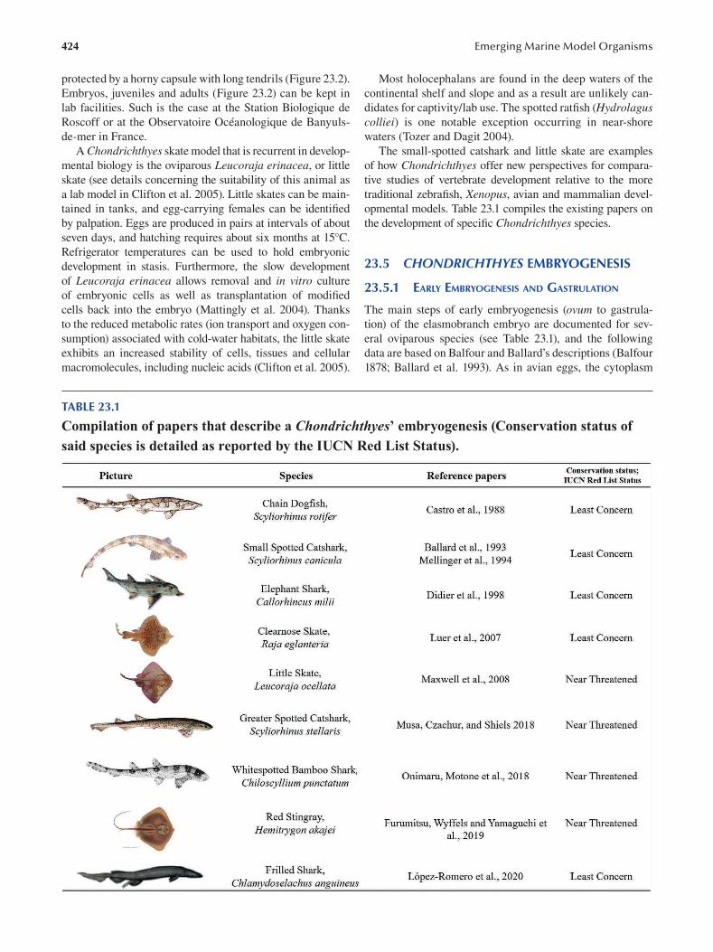
424
protected by a horny capsule with long tendrils (Figure 23.2).
Embryos, juveniles and adults (Figure 23.2) can be kept in
lab facilities. Such is the case at the Station Biologique de
Roscoff or at the Observatoire Océanologique de Banyuls
de-mer in France.
A Chondrichthyes skate model that is recurrent in develop
mental biology is the oviparous Leucoraja erinacea, or little
skate (see details concerning the suitability of this animal as
a lab model in Clifton et al. 2005). Little skates can be main
tained in tanks, and egg-carrying females can be identifi ed
by palpation. Eggs are produced in pairs at intervals of about
seven days, and hatching requires about six months at 15°C.
Refrigerator temperatures can be used to hold embryonic
development in stasis. Furthermore, the slow development
of Leucoraja erinacea allows removal and in vitro culture
of embryonic cells as well as transplantation of modifi ed
cells back into the embryo (Mattingly et al. 2004). Thanks
to the reduced metabolic rates (ion transport and oxygen con
sumption) associated with cold-water habitats, the little skate
exhibits an increased stability of cells, tissues and cellular
macromolecules, including nucleic acids (Clifton et al. 2005).
Emerging Marine Model Organisms
Most holocephalans are found in the deep waters of the
continental shelf and slope and as a result are unlikely can
didates for captivity/lab use. The spotted ratfi sh (Hydrolagus colliei) is one notable exception occurring in near-shore
waters (Tozer and Dagit 2004).
The small-spotted catshark and little skate are examples
of how Chondrichthyes offer new perspectives for compara
tive studies of vertebrate development relative to the more
traditional zebrafi sh, Xenopus, avian and mammalian devel
opmental models. Table 23.1 compiles the existing papers on
the development of specifi c Chondrichthyes species.
23.5 CHONDRICHTHYES EMBRYOGENESIS
23.5.1 EARLY EMBRYOGENESIS AND GASTRULATION
The main steps of early embryogenesis (ovum to gastrula
tion) of the elasmobranch embryo are documented for sev
eral oviparous species (see Table 23.1), and the following
data are based on Balfour and Ballard’s descriptions (Balfour
1878; Ballard et al. 1993). As in avian eggs, the cytoplasm
TABLE 23.1
Compilation of papers that describe a Chondrichthyes’ embryogenesis (Conservation status of said species is detailed as reported by the IUCN Red List Status).
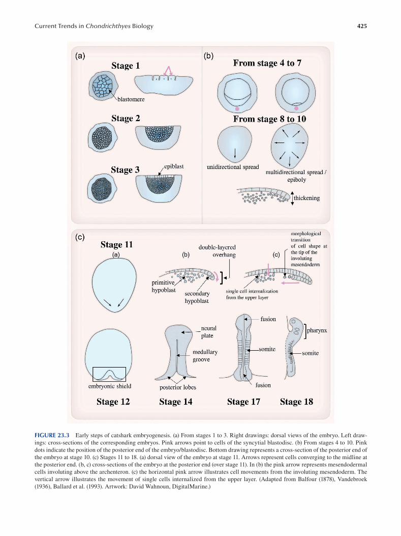
425 Current Trends in Chondrichthyes Biology
FIGURE 23.3 Early steps of catshark embryogenesis. (a) From stages 1 to 3. Right drawings: dorsal views of the embryo. Left draw
ings: cross-sections of the corresponding embryos. Pink arrows point to cells of the syncytial blastodisc. (b) From stages 4 to 10. Pink
dots indicate the position of the posterior end of the embryo/blastodisc. Bottom drawing represents a cross-section of the posterior end of
the embryo at stage 10. (c) Stages 11 to 18. (a) dorsal view of the embryo at stage 11. Arrows represent cells converging to the midline at
the posterior end. (b, c) cross-sections of the embryo at the posterior end (over stage 11). In (b) the pink arrow represents mesendodermal
cells involuting above the archenteron. (c) the horizontal pink arrow illustrates cell movements from the involuting mesendoderm. The
vertical arrow illustrates the movement of single cells internalized from the upper layer. (Adapted from Balfour (1878), Vandebroek
(1936), Ballard et al. (1993). Artwork: David Wahnoun, DigitalMarine.)
426
comprises large amounts of yolk, making segmentation pos
sible in only a small portion of the telolecithal egg cell. These
fi rst cleavages start within the oviduct with the particularity
of being incomplete. Cell membranes of the fi rst blastomeres
do not close up at their bases so that the cytoplasms and the
underlying yolk are continuous and form a syncytial blasto
disc (stage 1, Figure 23.3A). Around the 100-cell stage, the
blastodisc is not syncytial anymore, and the loosely arranged
blastomeres exhibit a spherical morphology (stage 2, Figure
23.3A). Later on, the density of inner blastomeres increases,
and they are covered by an epithelium made of columnar
cells, the epiblast (stage 3, Figure 23.3A). Dorsal views of
the blastodisc will later display a crescent-like structure at
the posterior end that will finally disappear (from stages 4
to 7, Figure 23.3B). From stages 8 to 10, the round blasto
disc shifts to an oval shape due to a unidirectional posterior
spread (Figure 23.3B). As epiboly proceeds, the spreading
of the blastodisc becomes multidirectional, and a thicken
ing starts to be observed at the posterior end. This cellular
densifi cation in the posterior area of the blastoderm tends to
intensify at stage 10 (Figure 23.3B, cross-section).
An important feature of the stage 11 embryo is the folding
of the epithelial upper layer over the yolk, generating a dou
ble-layered overhang (Figure 23.3C, a, b and c). The space
created between this overhang and the yolk corresponds to
the future archenteron of the embryo. Gastrulation prop
erly starts at this stage, with the lower layer of the overhang
representing the mesendodermal cells involuting above the
archenteron (Figure 23.3C, b). This forming mesendoder
mal layer can be referred to as a secondary hypoblast, while
the mass of inner blastomeres is called the primitive hypo
blast (Ballard et al. 1993, Figure 23.3C, b). Several studies
described cell movements accompanying mesendoderm
and mesoderm formation during gastrulation. Cell tracking
experiments showed that labeled cells within the upper layer
of the overhang at the very beginning of stage 11 are later
displaced onward within the involuting mesendoderm layer
(Godard et al. 2014, Figure 23.3C, c). Similar experimen
tal approaches revealed that single cells can be internalized
from the upper layer of the blastoderm to take part in meso
derm formation (Godard et al. 2014, Figure 23.3C, c). On
the other hand, Balfour (1878) observed that epithelial cells
at the tip of the involuting mesendoderm undergo a morpho
logical “transition”, acquiring the shape of the inner rounded
blastomeres (Figure 23.3C, c). Similar cell shape changes in
this area have also been reported by Coolen et al. (2007).
These observations suggest that epithelial cells both from
the upper layer (epiblast) and from the tip of the involuting
mesendoderm undergo an epithelial to mesenchymal transi
tion (EMT) during gastrulation. In situ hybridization experi
ments performed with the mesoderm marker Brachyury at
this stage in catshark embryos suggest other types of move
ments. In addition to being expressed at the site of the invo
luting mesendoderm, its expression pattern also describes
a thin ring all around the blastoderm, which suggests that
cells from the margin are converging to the midline at the
posterior end of the embryo (Sauka-Spengler et al. 2003)
Emerging Marine Model Organisms
(Figure 23.3C, a). The convergence of cells from the periph
ery of the blastoderm to the posterior end of the midline has
been initially reported by Vandebroek (1936). In the future,
development of live imaging approaches on elasmobranch
embryos would defi nitively help to shed light on the spatial
and temporal behaviors of their cells during gastrulation.
23.5.2 FROM AXIS FORMATION TO
PHARYNX SEGMENTATION
At stage 12, the posterior end of the blastoderm exhibits
a V-shaped structure referred to as the embryonic shield
(Figure 23.3C). A slight depression is observed in the
middle of the embryonic shield. It will give rise, by exten
sion from posterior to anterior, to the medullary groove,
stating the position of the embryonic axis (Figure 23.3C,
stage 14). As the embryo increases in length, the anterior
part will enlarge (neural plate, Figure 23.3C, stage 14) and
rise to form the neural/medullary folds. In the posterior
region, the two arms of the embryonic shield (posterior
lobes, Figure 23.3C, stage 14) will progressively shrink to
fuse and enclose the neural tube and the archenteric cav
ity (Figure 23.3C, stage 17). Similar fusion of the neural
folds is observed in the anterior part (Figure 23.3c, stage
17). Several pairs of somites are formed during the pro
cess of neural tube closure (Figure 23.3C, stage 17). While
the trunk pursues its segmentation through the formation
of additional somite pairs, the pharynx area undergoes
metamerization, too; several branchial clefts will appear
(Figure 23.3C, stage 18).
23.6 CHONDRICHTHYES ANATOMY AND SENSORY BIOLOGY
23.6.1 EXTERNAL FEATURES
In this section, typical Chondrichthyes body plans will be
proposed for Selachimorpha, Batoids and Holocephalans, and general external features will be briefly discussed. For
a more detailed account of Chondrichthyes anatomy, The Dissection of Vertebrates, Second Edition by Gerardo De
Iuliis and Dino Pulerà is highly informative (2019).
All Chondrichthyes breathe through five to seven pairs
of gills, depending on the species. As a general rule, pelagic
(open sea) species have to keep swimming to ensure that
oxygenated water is moving through their gills. Demersal
species, which live in the water column near the sea fl oor,
will actively pump water in through their spiracles and out
through their gills (Salazar 2018). Spiracles are respiratory
openings into the pharynx. For sharks, the gills are located
on the sides of the body, while the gills are ventral for batoids
(De Iuliis and Pulerà 2019). Elasmobranch gill structure and
function are described by Wegner (2015). Holocephalans
have a single gill opening, on each side, located just anterior
to the base of the pectoral fi n.
Most sharks, sawfish and chimeras have a heterocercal
tail (with unequal upper and lower lobes). This particular
427 Current Trends in Chondrichthyes Biology
structure has been showed to aid in locomotion (Wilga and
Lauder 2002). For skates, however, tails range from a thick
tail extending from the body to a whip to almost no tail.
Stingrays (batoids) possess a venomous stinger located in
the mid-area of the tail. This particularity has brought on
studies on the chemistry of their venom (da Silva et al. 2015).
In most holocephalans, the first dorsal fin is preceded by a
venomous spine that can inflict a serious wound (Halstead
and Bunker 1952).
Chondrichthyes have tough skin covered with dermal
teeth, also called placoid scales (or dermal denticles). The
dermal skeleton is the most ancestral mineralized skeleton
(see Gillis et al. 2017 for more information) and dermal den
ticles in the skin of elasmobranchs as well as teeth in the head
of all jawed vertebrates are remnants of this structure (Gillis
et al. 2017 ). Torpediniformes (electric rays) form an excep
tion, as they have a thick and fl abby body, with smooth and
loose skin. Notably, Holocephali lose their dermal denticles
as adults to keep only those on the clasping organ seen on the
caudal ventral surface of the male (Salazar 2018). Denticles
usually provide protection and, in most cases, streamlining
(Salazar 2018). On another level, denticles make the skin of
the catshark and the common stingray a highly sought-after
product for luxury lining and leatherwork. Called shagreen
(or galuchat in French), the use of this skin to wrap travel
cases and manufacture holders is mentioned by Buffon as
early as 1789 in the second volume of Histoire naturelle des poissons ( Buffon 1789 ).
In some shark species, such as the lantern shark, denticles
even house bioluminescent bacteria that aid in intraspecifi c
communication (Claes et al. 2015). In 2018, shark denticles
were discovered to be laid out according to a Turing-like
developmental mechanism explained by a reaction-diffusion
system (Cooper et al. 2018).
As aforementioned, bioluminescence and biofl uo
rescence can occur in certain Chondrichthyes species.
Bioluminescence is the ability of living beings to radiate
light on their own or with the help of certain symbiotes (e.g.
bacteria). Biofluorescence is the process in which ambient
light is absorbed via fluorescent compounds and reemitted
at longer, lower-energy wavelengths. Examples of biolu
minescent sharks include Etmopterus spinax (velvet belly
lantern shark) (Claes et al. 2010), Euprotomicrus bispinatus (dwarf pelagic shark) (Hubbs et al. 1967) or Squaliolus aliae (smalleye pygmy shark) (Claes et al. 2012). They dis
play light-emitting organs (photophores) on their under
sides that form species-specifi c patterns over the fl anks and
abdomen. The ventral photophores are considered to par
ticipate in counter-illumination, a method of camoufl age
that uses light production to match background brightness
and wavelength (Sparks et al. 2014). The bioluminescent
flank markings may play a role in intraspecifi c communica
tion (Gruber et al. 2016 ). The roles of biofl uorescence are
more elusive. The Urotrygonidae (American round sting
rays), Orectolobidae (wobbegongs) and Scyliorhinidae (catsharks) families include fluorescent species. As these
families are distantly related, biofluorescence is thought to
have evolved at least three times in elasmobranchs (Gruber
et al. 2016 ). The swell shark (Cephaloscyllium ventriosum),
the chain catshark (Scyliorhinus rotifer) and round stingray
(Urobatis jamaicensis) are known to exhibit bright green
fluorescence (Sparks et al. 2014). The family of small mol
ecules behind marine biofluorescence reviewed in Park et
al. (2019) have been hypothesized to play a role in central
nervous system signaling, resilience to microbial infections
and photoprotection.
23.6.2 INTERNAL ANATOMY
This section is a selection of specific traits of Chondrichthyes anatomy deemed important to mention. As the etymology of
the term Chondrichthyes indicates, they possess a cartilagi
nous skeleton.
For Selachii, the mouth is ventrally located. The upper
and lower jaws are lined by multiple rows of serrated, tri
angular and pointed teeth that continuously grow and shed
(De Iuliis and Pulerà 2019). Instead, batoids possess fl at
tened plates for crushing bottom-dwelling prey (De Iuliis
and Pulerà 2019). Gynandric heterodonty (sexual dimor
phism in teeth) is very common in elasmobranchs, and
Berio et al. (2020) described the intraspecific diversity of
tooth morphology in the large-spotted catshark and revealed
some of the ontogenic cues driving this sexual dimorphism.
Holocephalans possess three pairs of tooth plates, two in the
upper jaw and a single pair in the lower jaw (Tozer and Dagit
2004 ). Sawfish (Rhinopristiformes, Batoids, Elasmobranch)
are characterized by a long, narrow and fl attened rostrum
(nose extension) lined with transversal teeth. This fea
ture can also be found in sawsharks (Pristiophoriformes,
Selachii, Elasmobranch).
Chondrichthyes have no swim bladders. Buoyancy is
rather controlled with a large oil-filled liver, which reduces
their specific density. An interesting feature of sharks is
the valvular intestine, which bears a spiral valve, a cork
screw-shaped lower portion of the intestine that increases
its effective length (De Iuliis and Pulerà 2019). Remarkably,
chimeariformes lack stomachs (Salazar 2018).
Unlike mammals, Chondrichthyes do not have bone mar
row, and red blood cells are produced in the spleen and the
epigonal organ. The epigonal organ is a special tissue around
the gonads that is only found in certain cartilaginous fi sh and
thought to play a role in the immune system. Red blood cells
are also produced in the Leydig’s organ (nested along the top
and bottom of the esophagus), which is also considered part of
the immune system (Mattisson and Faänge 1982). The subclass
Holocephali lacks both the Leydig’s and epigonal organs.
Elasmobranch kidneys deserve a special mention, and
the little skate and spotted catshark have been of particu
lar interest for the study of kidney development. The func
tional unit of the kidney is the nephron, and the process of
nephron formation is termed nephrogenesis. In mammals,
nephrogenesis comes to a stop shortly after birth. This
means nephron endowment is definitive in mammals at
birth. Some elasmobranchs have been found to continually
428
form nephrons even after embryonic development. Using
kidney histological sections from a spotted catshark
juvenile, Hentschel (1991) described nephrogenesis with
similar morphological steps as found during mammalian
nephrogenesis (Hentschel 1991). This unique capacity is a
promising research area to better understand the orches
trating factors behind kidney morphogenesis.
Elasmobranch species possess a rectal (or salt) gland.
This epithelial organ is located in the distal intestine and
empties into the cloaca. It is composed of many tubules that
serve a single function: the secretion of hypertonic NaCl
solution (Forrest 2016 ). Initially discovered by Wendell
Burger and Walter Hess (1960), this organ can be cannu
lated and perfused, and chloride secretion can be measured.
As highlighted by Forrest (2016 ), this organ has helped
in understanding the physiology of the mammalian thick
ascending limb (TAL), an inaccessible portion of the kid
ney, which functions to filter sodium (Na +), potassium (K+)
and chloride (Cl-).
23.6.3 SENSORY BIOLOGY
Chondrichthyes are gifted with a plethora of senses that are
more or less developed depending on the species. The sensory
biology of Chondrichthyes can be divided into visual, acous
tic, mechanical, chemical, magnetic and electrical detection.
23.6.3.1 Photoreception Studies that focus on visual function in Chondrichthyes have described differing sensitivities to light and colors
(Douglas and Djamgoz 2012). Depending on the ecologi
cal niche they occupy, Chondricthyes have evolved differ
ent morphological adaptations to optimize photoreception.
These include variation in eye size, eye positioning, mobile
pupils, elaborate pupillary opercula and refl ective retinal
media (Walls 1942). The variety of pupil shapes (horizon
tal, oblique, U-crescent shaped slits) and pupillary oper
cula is striking. Usually, elasmobranchs benefit from large
visual fields—a horizontal arc of up to 360° (McComb
and Kajiura 2008)—while humans have a 210° horizontal
arc. Elasmobranch retinas include both rod cells, which
allow perception in dim-light conditions, and cone cells,
which allow perception in bright-light conditions, higher
acuity and possible color distinction (Jordan et al. 2013).
Ecological factors seem to condition the proportion of rods
and cones and the spectral sensitivity of cones. For exam
ple, species that inhabit the dysphotic and aphotic zone
possess fewer to no cones (Collin et al. 2006). Concerning
batoids, eyes are usually located dorsally, though lateral eye
position can also be observed, and eyes can even be vesti
gial in some electric rays. Some batoids (skates, rays and
guitarfish) exhibit several spectrally specific cone pigments
that would entail the ability for color discrimination (Hart
et al. 2004, Theiss et al. 2007 ). In 2016, the giant guitarfi sh
(Rhynchobatus djiddensis) was discovered to possess the
ability to retract its eyes, possibly as a means of protection
during predation (Tomita et al. 2016 ).
Emerging Marine Model Organisms
23.6.3.2 Audition Myrberg recounts the history of investigations concerning
the hearing abilities of sharks in his Acoustical Biological of Elasmobranch review (2001). For sharks, the highest sensi
tivity has been demonstrated for low-frequency sounds (40 to
800 Hz). Specific sound characteristics attract free-ranging
sharks: irregular pulses without sudden increases in inten
sity and frequencies below 80 Hz. Such characteristics are
evocative of wounded or struggling prey (Myrberg 2001).
This is an auditory explanation behind the role that sharks
play in regulating the health of ocean populations. Recently,
Parmentier et al. (2020) described the hearing abilities of
the catshark, Scyliorhinus canicula, from early embryos to
juveniles. Stage 31 embryos were able to detect sounds from
100 to 300 Hz, while juveniles were able to detect sounds
from 100 to 600 Hz. As hearing development continues in
the catshark, only the frequency range appears to widen, as
sensitivity and thresholds were not found to improve with
development (Parmentier et al. 2020). This last paper con
tains references to other studies on Chondrichthyes hearing
abilities, namely hearing thresholds, frequency range and
ear morphology.
23.6.3.3 Mechanosensory System The mechanosensory systems of elasmobranchs include
different tactile sense organs; receptor types and distribu
tion depend on the species (Maruska 2001; Jordan 2008).
These systems include lateral line canals, neuromasts and
vesicules of Savi (types of sensory hair cells and their sup
porting cells) and spiracular organs. The lateral line marks
the lateral line canals, which contain sensory nerve endings
and open to the surface through tiny pores (De Iuliis and
Pulerà 2019). These tactile sense organs respond to pressure
variations induced by the velocity or acceleration of water
flow. The electrosensory and lateral line systems of sawfi sh
extend out along the rostrum. This allows them to sense and
manipulate prey (Wueringer et al. 2011).
23.6.3.4 Chemoreception Sensitivity to chemical signals through taste, chemical sense
and olfaction constitutes another sense for Chondrichthyes. The underlying organs behind these functions include olfac
tory sacs (for olfaction) and taste papillae (gustation). Sharks
have been found to locate potential food using the difference
in bilateral odor arrival times (Gardiner and Atema 2010).
Pharyngeal denticles and taste papillae possess receptors
used for gustation. The morphological adaptations that are
pharyngeal denticles could help sharks catch and direct food
items and prevent injury of the mouth lining during food
manipulation and consumption (Atkinson et al. 2016). Both
dermal and oral denticles possess species-specifi c micro
structural morphology that can be applied as a taxonomical
tool (Bs et al. 2019). During odor source localization, com
binatory signals will help locate potential prey. Gardiner
and Atema (2007) looked into the contribution of different
senses (olfaction, mechanoreception and vision) to odor per
ception in the smooth dogfi sh Mustelus canis . Interestingly,
429 Current Trends in Chondrichthyes Biology
they found that the lateral line is required to locate odor
sources (Gardiner and Atema 2007).
23.6.3.5 Magnetoreception Fascinatingly, elasmobranchs have been observed to swim
in straight lines for extended periods of time in a highly ori
ented manner and to navigate in relation to magnetic fi elds.
These observations are true for tiger sharks (Galeocerdo cuvier; Holland et al. 1999), blue sharks (Prionace glauca;
Carey et al. 1990) and scalloped hammerhead sharks
(Sphyrna lewini; Klimley 1993). Meyer et al. (2005) showed
experimentally that sharks can detect variations in the geo
magnetic field. They performed condition experiments on
captive sharks to determine how they detect magnetic fi elds
and to measure detection thresholds. The anatomical mod
ules underlying magnetoreception could be mediated directly
via a magnetite-based sensory system or indirectly via the
electrosensory system (Sundström et al. 2001). Indeed, the
exact cells, molecules and receptors behind magnetorecep
tion in elasmobranches remain unknown.
23.6.3.6 Electroreception Electroreception is important in many Chondrichthyes. In 1678, Stefano Lorenzini first described pores dispersed
on a shark’s head without identifying their sensory role. It
was only in the 1960s that their function began to be elu
cidated and identified as a modified part of the lateral line
system. Named after Stefano Lorenzini, the ampullae of
Lorenzini form a network of jelly-filled pores that act as
sensing organs. These pores are connected to sensory cells
by gel-filled canals and are highly sensitive to low-frequency
electrical stimuli produced by both non-biological and
biological sources. Ampullae of Lorenzini are mostly
described in Chondrichthyes; however, they are also found
in Chondrostei. Chondrostei are Actinopterygii in which
the cartilaginous skeleton is a derived feature. They include
reedfish, sturgeon and bichir. On the other hand, rays pos
sess an electric organ that originates from modified nerve or
muscle tissue. The electric field created by this organ is used
for navigation, communication, mating (Feulner et al. 2009),
defense and the incapacitation of prey.
Jordan et al. (2013) extensively reviewed both the current
knowledge on elasmobranch sensory systems and the way in
which these sensory systems could inspire methods for bycatch
reduction. The following references will allow a deeper look
into the sensory system anatomies of sharks (De Iuliis and
Pulerà 2019), batoids (Bedore et al. 2014; Wueringer et al.
2011) and holocephalans (Tozer and Dagit 2004; Lisney 2010).
23.7 GENOMIC DATA
23.7.1 GENOMES AND TRANSCRIPTOMES
With millions of species on earth, very few genomes or tran
scriptomes are in fact assembled, annotated and published.
However, the availability of this data (genomes, transcrip
tomes or protein sequences) greatly accelerates studies on
phylogeny (Li et al. 2012; Straube et al. 2015), species diver
sity and population structure (Boussarie et al. 2018), conser
vation (Corlett 2017), evolutionary history (Inoue et al. 2010;
Renz et al. 2013) or human health research. More generally,
studies that encompass diverse animal models to compare
sequences have been critical for deciphering fundamental
physiological mechanisms and conserved gene and protein
functions. Another approach is to compare closely related
genomes to identify divergent sequences that may underlie
unique phenotypes (Stedman et al. 2004). Several studies
have shown that non-coding sequences are more comparable
between the genomes of humans and cartilaginous fi sh than
between those of humans and zebrafish (Venkatesh et al.
2006; Lee et al. 2011). Both the slower molecular clock of
cartilaginous fish relative to teleosts’ (Venkatesh et al. 2014;
Renz et al. 2013; Martin et al. 1992), as well as the extra
whole-genome duplication specific of teleosts (Glasauer and
Neuhauss 2014), can explain the comparability of human
and Chondrichthyes genomes.
In 2013, a tissue-specific transcriptome was generated
from the heart tissue of the great white shark (Carcharodon carcharias) (Richards et al. 2013). This represented the
fi rst transcriptome of any tissue for this species. Strikingly,
this transcriptome revealed that the percentage of anno
tated transcripts involved in metabolic processes was more
similar between the white shark and humans than between
the white shark and a teleost (Richards et al. 2013). This
finding is consistent with those of Venkatesh et al. (2006)
who found genomic non-coding elements and the relative
position of genes to be more similar between the elephant
shark and humans than between the elephant shark and a
teleost. In 2014, the first large-scale comparative transcrip
tomic survey of multiple cartilaginous fish tissues was ana
lyzed: the pancreas, brain and liver of the lesser spotted
catshark, Scyliorhinus canicula (Mulley et al. 2014). This
study contributes to deciphering the molecular-level func
tions of pancreatic metabolic processes of Chondrichthyes. Uncommonly, Chondrichthyes possess the ability to both
maintain stable blood glucose levels and tolerate exten
sive periods of hypoglycemia (Mulley et al. 2014). A high-
coverage whole-genome sequencing project of S. canicula is underway (Génoscope, French National Sequencing
Center and laboratory of Sylvie Mazan, Observatoire
Océanologique de Banyuls sur Mer, France). A collection
of catshark expressed sequence tags (ESTs) is also available
in Mazan’s lab. The fi rst Chondrichthyes whole genome
to be sequenced was of the holocephalan Callorhincus milii, published by Venkatesh et al. (2014). The genome
size is approximately 1 Gbp. The same year, Wyffels et al.
sequenced both the nuclear and mitochondrial genomes of
the little skate (Leucoraja erinacea). The genome repre
sents 3.42 Gbp across 49 chromosomes. Wyffels et al. (2014)
introduced Skatebase (www.skatebase.org), a project for the
collection of elasmobranch genomes to complete molecular
resources for Chondrichthyes fish. Additionally, to the little
skate genome, mitochondrion sequences from the ocellate
spot skate (Okamejei kenojei) and thorny skate (Amblyoraja
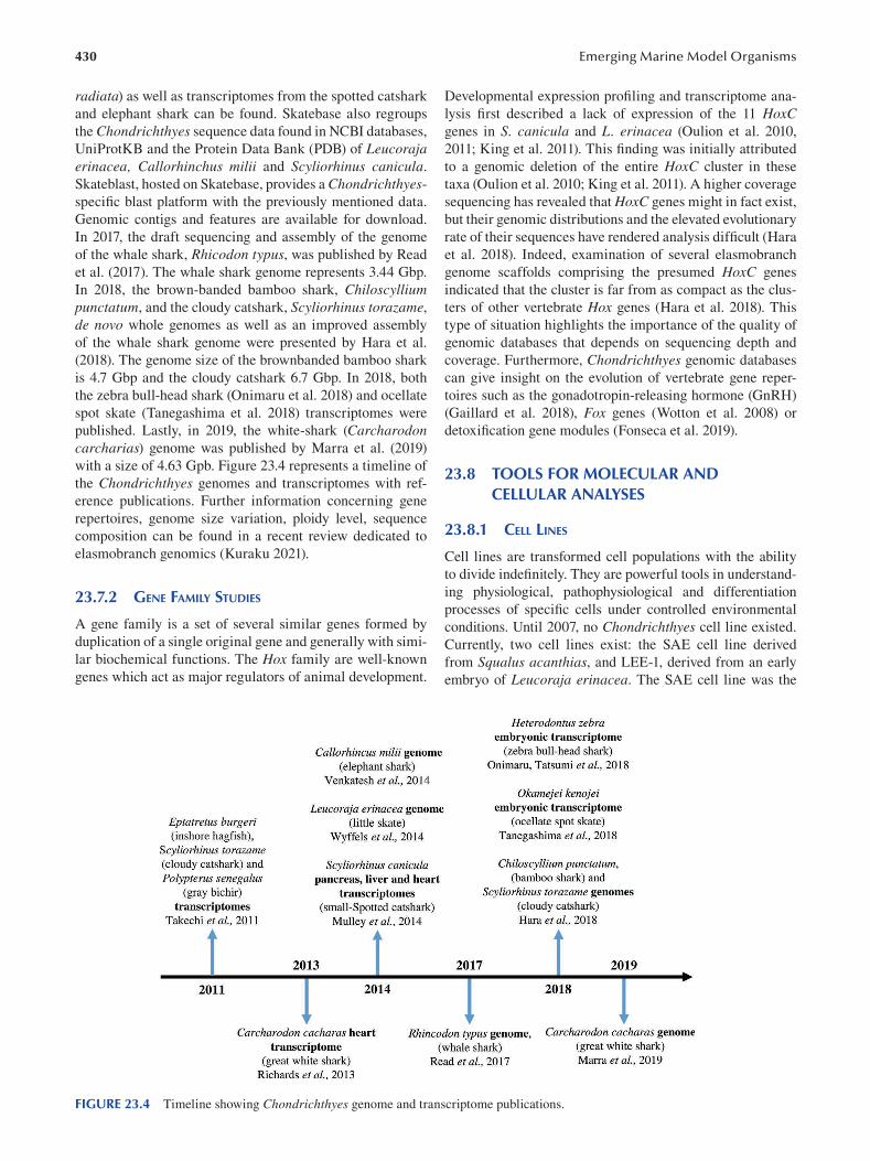
430
radiata) as well as transcriptomes from the spotted catshark
and elephant shark can be found. Skatebase also regroups
the Chondrichthyes sequence data found in NCBI databases,
UniProtKB and the Protein Data Bank (PDB) of Leucoraja erinacea, Callorhinchus milii and Scyliorhinus canicula.
Skateblast, hosted on Skatebase, provides a Chondrichthyes
specific blast platform with the previously mentioned data.
Genomic contigs and features are available for download.
In 2017, the draft sequencing and assembly of the genome
of the whale shark, Rhicodon typus, was published by Read
et al. (2017). The whale shark genome represents 3.44 Gbp.
In 2018, the brown-banded bamboo shark, Chiloscyllium punctatum, and the cloudy catshark, Scyliorhinus torazame,
de novo whole genomes as well as an improved assembly
of the whale shark genome were presented by Hara et al.
(2018). The genome size of the brownbanded bamboo shark
is 4.7 Gbp and the cloudy catshark 6.7 Gbp. In 2018, both
the zebra bull-head shark (Onimaru et al. 2018) and ocellate
spot skate (Tanegashima et al. 2018) transcriptomes were
published. Lastly, in 2019, the white-shark (Carcharodon carcharias) genome was published by Marra et al. (2019)
with a size of 4.63 Gpb. Figure 23.4 represents a timeline of
the Chondrichthyes genomes and transcriptomes with ref
erence publications. Further information concerning gene
repertoires, genome size variation, ploidy level, sequence
composition can be found in a recent review dedicated to
elasmobranch genomics (Kuraku 2021).
23.7.2 GENE FAMILY STUDIES
A gene family is a set of several similar genes formed by
duplication of a single original gene and generally with simi
lar biochemical functions. The Hox family are well-known
genes which act as major regulators of animal development.
Emerging Marine Model Organisms
Developmental expression profiling and transcriptome ana
lysis first described a lack of expression of the 11 HoxC genes in S. canicula and L. erinacea (Oulion et al. 2010,
2011; King et al. 2011). This finding was initially attributed
to a genomic deletion of the entire HoxC cluster in these
taxa (Oulion et al. 2010; King et al. 2011). A higher coverage
sequencing has revealed that HoxC genes might in fact exist,
but their genomic distributions and the elevated evolutionary
rate of their sequences have rendered analysis diffi cult (Hara
et al. 2018). Indeed, examination of several elasmobranch
genome scaffolds comprising the presumed HoxC genes
indicated that the cluster is far from as compact as the clus
ters of other vertebrate Hox genes (Hara et al. 2018). This
type of situation highlights the importance of the quality of
genomic databases that depends on sequencing depth and
coverage. Furthermore, Chondrichthyes genomic databases
can give insight on the evolution of vertebrate gene reper
toires such as the gonadotropin-releasing hormone (GnRH)
(Gaillard et al. 2018), Fox genes (Wotton et al. 2008) or
detoxification gene modules (Fonseca et al. 2019).
23.8 TOOLS FOR MOLECULAR AND CELLULAR ANALYSES
23.8.1 CELL LINES
Cell lines are transformed cell populations with the ability
to divide indefinitely. They are powerful tools in understand
ing physiological, pathophysiological and differentiation
processes of specific cells under controlled environmental
conditions. Until 2007, no Chondrichthyes cell line existed.
Currently, two cell lines exist: the SAE cell line derived
from Squalus acanthias, and LEE-1, derived from an early
embryo of Leucoraja erinacea. The SAE cell line was the
FIGURE 23.4 Timeline showing Chondrichthyes genome and transcriptome publications.
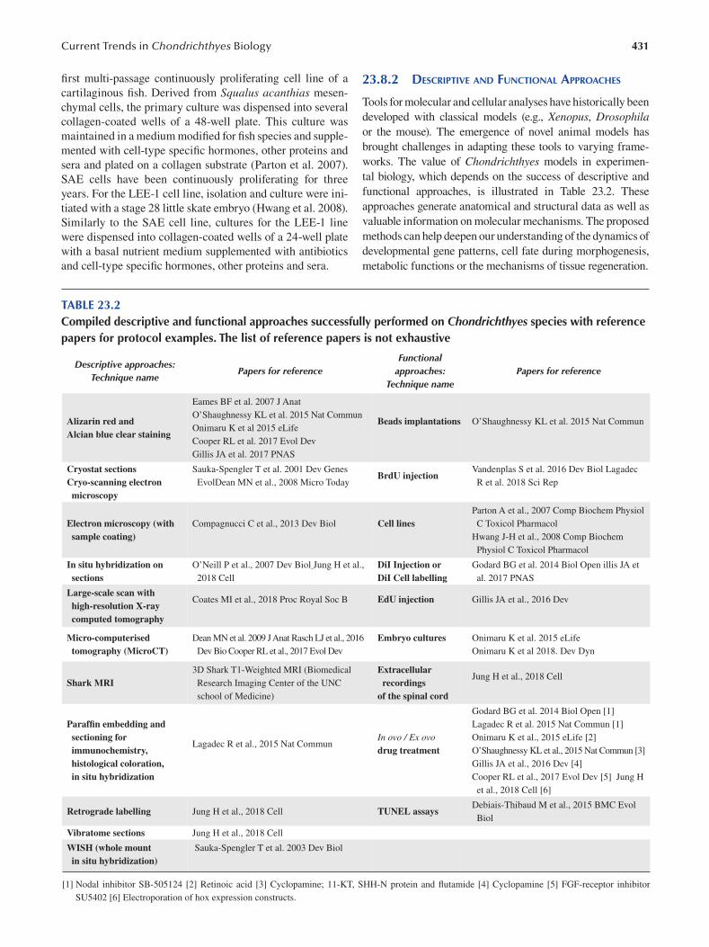
431 Current Trends in Chondrichthyes Biology
first multi-passage continuously proliferating cell line of a
cartilaginous fish. Derived from Squalus acanthias mesen
chymal cells, the primary culture was dispensed into several
collagen-coated wells of a 48-well plate. This culture was
maintained in a medium modified for fish species and supple
mented with cell-type specifi c hormones, other proteins and
sera and plated on a collagen substrate (Parton et al. 2007).
SAE cells have been continuously proliferating for three
years. For the LEE-1 cell line, isolation and culture were ini
tiated with a stage 28 little skate embryo (Hwang et al. 2008).
Similarly to the SAE cell line, cultures for the LEE-1 line
were dispensed into collagen-coated wells of a 24-well plate
with a basal nutrient medium supplemented with antibiotics
and cell-type specific hormones, other proteins and sera.
23.8.2 DESCRIPTIVE AND FUNCTIONAL APPROACHES
Tools for molecular and cellular analyses have historically been
developed with classical models (e.g., Xenopus, Drosophila or the mouse). The emergence of novel animal models has
brought challenges in adapting these tools to varying frame
works. The value of Chondrichthyes models in experimen
tal biology, which depends on the success of descriptive and
functional approaches, is illustrated in Table 23.2. These
approaches generate anatomical and structural data as well as
valuable information on molecular mechanisms. The proposed
methods can help deepen our understanding of the dynamics of
developmental gene patterns, cell fate during morphogenesis,
metabolic functions or the mechanisms of tissue regeneration.
TABLE 23.2 Compiled descriptive and functional approaches successfully performed on Chondrichthyes species with reference papers for protocol examples. The list of reference papers is not exhaustive
Functional Descriptive approaches:
Technique name Papers for reference approaches:
Technique name Papers for reference
Eames BF et al. 2007 J Anat
Alizarin red and Alcian blue clear staining
O’Shaughnessy KL et al. 2015 Nat Commun
Onimaru K et al 2015 eLife
Cooper RL et al. 2017 Evol Dev
Beads implantations O’Shaughnessy KL et al. 2015 Nat Commun
Gillis JA et al. 2017 PNAS
Cryostat sections Cryo-scanning electron
Sauka-Spengler T et al. 2001 Dev Genes
EvolDean MN et al., 2008 Micro Today BrdU injection
Vandenplas S et al. 2016 Dev Biol Lagadec
R et al. 2018 Sci Rep
microscopy
Parton A et al., 2007 Comp Biochem Physiol
Electron microscopy (with Compagnucci C et al., 2013 Dev Biol Cell lines C Toxicol Pharmacol
sample coating) Hwang J-H et al., 2008 Comp Biochem
Physiol C Toxicol Pharmacol
In situ hybridization on O’Neill P et al., 2007 Dev Biol Jung H et al., DiI Injection or Godard BG et al. 2014 Biol Open illis JA et
sections 2018 Cell DiI Cell labelling al. 2017 PNAS
Large-scale scan with high-resolution X-ray
Coates MI et al., 2018 Proc Royal Soc B EdU injection Gillis JA et al., 2016 Dev
computed tomography
Micro-computerised Dean MN et al. 2009 J Anat Rasch LJ et al., 2016 Embryo cultures Onimaru K et al. 2015 eLife
tomography (MicroCT) Dev Bio Cooper RL et al., 2017 Evol Dev Onimaru K et al 2018. Dev Dyn
Shark MRI 3D Shark T1-Weighted MRI (Biomedical
Research Imaging Center of the UNC
Extracellular recordings
Jung H et al., 2018 Cell
school of Medicine) of the spinal cord
Godard BG et al. 2014 Biol Open [1]
Paraffin embedding and Lagadec R et al. 2015 Nat Commun [1]
sectioning for immunochemistry,
Lagadec R et al., 2015 Nat Commun In ovo / Ex ovo drug treatment
Onimaru K et al., 2015 eLife [2]
O’Shaughnessy KL et al., 2015 Nat Commun [3]
histological coloration, Gillis JA et al., 2016 Dev [4]
in situ hybridization Cooper RL et al., 2017 Evol Dev [5] Jung H
et al., 2018 Cell [6]
Retrograde labelling Jung H et al., 2018 Cell TUNEL assays Debiais-Thibaud M et al., 2015 BMC Evol
Biol
Vibratome sections Jung H et al., 2018 Cell
WISH (whole mount in situ hybridization)
Sauka-Spengler T et al. 2003 Dev Biol
[1] Nodal inhibitor SB-505124 [2] Retinoic acid [3] Cyclopamine; 11-KT, SHH-N protein and flutamide [4] Cyclopamine [5] FGF-receptor inhibitor
SU5402 [6] Electroporation of hox expression constructs.
432
23.9 CHALLENGING QUESTIONS
23.9.1 ENDOGENOUS CHONDRICHTHYES MOLECULES
FOR BIOMEDICAL APPLICATIONS
23.9.1.1 Molecules Displaying Antibiotic Activity The well-known squalamine is a cationic steroid isolated
from stomach extracts of the spiny dogfi sh Squalus acanthias. It has been demonstrated to display antibacterial activity
against Gram-negative and Gram-positive bacteria (Moore et
al. 1993). Interestingly, the same study reported that squala
mine induced osmotic lysis of Paramecium caudatum and had
activity against Candida albicans, indicating that this shark
molecule also holds antiprotozoal and fungicidal properties.
As the research on squalamine progressed, it revealed that its
chemical features extend beyond the antimicrobial fi eld. This
aspect will be presented in the following paragraph.
Microorganisms themselves can produce natural antimi
crobial agents, meaning that bacterial symbionts in general
can constitute an additional “tissue” to look for putative antibi
otics. One specificity of Chondrichthyes is their considerable
resistance to infection even when their skin is profoundly dam
aged due to events related to their lifestyle (mating, predation)
or to anthropogenic activities. This observation strongly sug
gests that an innate immunity is operating through the mutu
alistic interactions taking place in the epidermal mucus layer
between marine bacteria and shark epidermis. The most recent
study that has investigated the property of these probiotic bac
teria is the one from Ritchie et al. (2017 ). They analyzed the
entire bacterial community of the epidermal mucus of three
ray species (two marine and one freshwater) and of the clear-
nose skate, Raja eglanteria. They clearly identifi ed particu
lar strains displaying broad-spectrum antibiotic activity and
activity against important nosocomial bacteria (Vancomycin
resistant Enterococcus [VRE] and Methicillin-resistant S. aureus [MRSA]). It goes without saying that interdisciplinary
research, in this case intermingling marine microbiology and
organism biology, has always sharpened our understanding of
immune defense mechanisms. These data on shark epidermis
might help medical research in seeking new antimicrobial
compounds but also, more generally, in focusing on the pres
ervation of symbiotic bacteria to prevent many types of human
diseases and infections. As in Chondrichthyes, these bacteria
play a fundamental role in our immunity.
23.9.1.2 The High Specifi city of Chondrichthyes Antibodies
The Chondrichthyes adaptative immune system has many
shared features with other gnathostomes (Flajnik 2018), except
for their particular antibodies. These immunoglobulin (Ig)-like
molecules, also called immunoglobulin new antigen receptors
(IgNARs), are made of two heavy chains, lack light chains
and bear a single variable region domain (V-NARs). In other
words, they have one antigen recognition site instead of two,
as is the case in the large majority of jawed vertebrate antibod
ies. Discovered in the 1990s (Greenberg et al. 1995; Roux et
al. 1998), IgNARs rapidly raised important interest in the area
of drug development. Indeed, the particular folding properties
Emerging Marine Model Organisms
of V-NARs allow them to reach a large panel of protein sites,
including hidden epitopes such as those found in the substrate
pocket of enzymes that cannot be targeted by “classical” anti
bodies. Finally, V-NARs also present great solubility and sta
bility, and their small size is another advantage within the fi eld
of antibody-based targeting strategy. Specific V-NARs from
different elasmobranch species have already been developed to
target viral proteins or toxins for medical applications such as
anti-viral activity, immunodiagnostics or the development of
biosensors. A list of these already-existing targeting V-NARs
is available in the review from Kovaleva et al. (2014).
Within gnathostomes, camelids have also evolved such
single-domain antibodies, from which the monomeric vari
able (V) antibody domain constitutes the VHH fragment.
As they have been found only in sharks and camels so far,
it is believed that these single-domain antibodies are the
result of convergent evolution ( Flajnik 2018). Nanobody is
the name commonly used to indicate camelid VHH and
shark V-NAR fragments. The important contribution that
nanobodies can bring to the treatment of viral diseases has
been spotlighted very recently, in the midst of the COVID
19 pandemic. Wrapp et al. (2020) managed to produce
VHH fragments able to prevent the spike (S) glycoprotein
of several coronavirus (SARS-CoV-1, SARS-CoV-2 and
MERS-CoV) from interacting with their cellular receptors.
23.9.1.3 The Different Properties of Squalamine As mentioned, squalamine is a polyvalent molecule that also
displays antiviral activity, an ability linked to its biochemical
properties. The positive charge on account of the spermidine
moiety of squalamine (Moore et al. 1993) provides it with high
affinity for negatively charged phospholipids of the membrane
lipid bilayer (Selinsky et al. 2000). As anionic phospholipids
are important to regulate surface charge and protein localiza
tion (Yeung et al. 2008), the neutralization of negative charges
by squalamine may lead to the disruption of electrostatic
potential and shuffle membrane-anchored proteins. This has
been demonstrated for Rac1, a GTPase used by many viruses
during the process of cell entry, which might impact the viral
replication cycle ( Zasloff et al. 2011). In the same study, they
observed that a wide range of viral pathogens (such as those
responsible for dengue, yellow fever, equine encephalitis and
Hepatitis B) exhibit variable susceptibility to squalamine in
both in vitro and in vivo tests ( Zasloff et al. 2011).
The ability of squalamine to interact with the negatively
charged lipids of the cell membrane also represents the under
lying mechanism of α-synuclein aggregation impairment
(Perni et al. 2017 ). These α-synuclein aggregates are part of
pathogenesis hallmarks of several neurodegenerative disor
ders, and their destruction constitutes an important challenge
to limiting toxicity within the brain parenchyma. Perni et al.
(2017) also showed that squalamine exposure led to motility
recovery in an animal model of Parkinson disease.
Finally, squalamine has also been demonstrated to impede
tumor-associated angiogenesis and the growth of several
solid neoplasms (reviewed in Luer and Walsh 2018; Márquez-
Garbán et al. 2019). The mechanism of the angiostatic prop
erty of squalamine is not fully understood but might rely on,
433 Current Trends in Chondrichthyes Biology
among other explanations, its ability to control endothelial
cell shape/volume, as demonstrated by Sills et al. (1998) on
embryonic vascular beds. More specifi cally, squalamine
blocks the Na +/H+ exchanger (isoform NHE3) (Akhter et al.
1999). Such inhibition of the sodium-hydrogen antiporter will
result in the modification of the hydrogen efflux out of the
cell, which can explain volume change of endothelial cells.
23.9.1.4 Molecules Displaying Anti-Cancer Activity Lacking bone marrow, lymphatic system and nodes, elasmo
branchs have evolved two particular lymphomyeloïd struc
tures: the epigonal organ associated with the gonads and the
Leydig organ located around the esophageal wall, as previ
ously mentioned (Honma et al. 1984). They are involved in
the production of red blood cells and play an important role
in immune system function.
With the aim to better characterize cell function of these
tissues, Walsh and Luer (2018) first showed that cells from
the Leydig and epigonal organs display phagocytic and
pinocytic activities (Luer and Walsh 2018). Next, looking
for more specific bioactive compounds, they tested epigonal
conditioned medium (prepared from adult bonnethead shark)
and found that it was able to inhibit growth of several mam
malian tumor cell lines (Walsh et al. 2006). More specifi cally
on Jurkat T-cell lines, this medium induced caspase-medi
ated apoptosis (Walsh et al. 2013), but the biochemical nature
of this (or these) cell death inducer(s) released from shark
epigonal conditioned medium still has/have to be discovered.
As previously mentioned, blocking the neovascular
ization that accompanies tumor growth is another way to
restrain malignancy progression. Besides squalamine,
Neovastat (AE-941), a shark cartilage extract, has been
shown to inhibit matrix metallopeptidase and VEGF activity
(Falardeau et al. 2001; Béliveau et al. 2002), which is consis
tent with antiangiogenic property. More specifi cally, Zheng
et al. (2007 ) isolated from the cartilage of the blue shark
Prionace glauca, a 15.5 kDa polypeptide (PG155) with the
ability to reduce vessel formation in vertebrate embryos and
tube formation of human umbilical vein endothelial cells
(HUVECs). However, Neovastat hasn’t gotten beyond phase
II of clinical trials so far (Kang et al. 2019 ), meaning that the
use of shark cartilage in the treatment of human malignan
cies is still exploratory.
23.9.2 EVO-DEVO STUDIES IN THE SEARCH FOR THE
ORIGIN OF SKELETON AND BRAIN ASYMMETRIES
23.9.2.1 Endoskeleton and Bone-Like Tissue in Chondrichthyes
Although skates, rays, sharks and chimeras are called car
tilaginous fish, they possess mineralized structures in their
endoskeleton and dermoskeleton (or exoskeleton). Their
embryonic endoskeleton is made of a gel-like structure
produced by chondrocytes: the hyaline cartilage, a carti
laginous matrix classically stained and observable using
Alcian blue pigments. As development progresses, cer
tain parts of the axial endoskeleton such as the vertebrae
undergo mineralization, a process that can be visualized
using Alizarin red staining.
Truncal vertebrae in elasmobranchs are made of i) a cen
trum that surrounds the notochord and ii) a dorsal neural arch
delimiting the neural canal that contains the spinal cord. Caudal
vertebrae also have, ventral to the centrum, a hemal arch that
surrounds arteries and veins. Both the centrum and the neu
ral arch of vertebrae of several elasmobranch species display
Alcian blue staining at mid-embryogenesis, while Alizarin red
coloration is observable in near-hatching embryos (Eames et al.
2007, Enault et al. 2015; Atake et al. 2019), meaning that a min
eralization process is occurring on a cartilage-based matrix.
However, in contrast to the mineralization mechanism
occurring in the long bones of Osteichthyes (the so-called
endochondral ossification that also begins within cartilage
but from the center to the periphery of the bone), the miner
alization in elasmobranch vertebrae starts on the periphery
of both the neural arch and the centrum. Interestingly, the
expression pattern of type I and type II collagen in these
elasmobranch structures is similar to that accompanying the
shift from cartilage to mineralized cartilage during endo
chondral ossification of tetrapod long bones. Type II colla
gen (cartilage specific) is observed within the cartilaginous
center of the neural arch, while type I collagen stains the
outer surface of the neural arch (Eames et al. 2007; Enault et
al. 2015). It is important, however, to outline that in several
teleost species, some cartilages lack type II collagen expres
sion, and bones can exhibit important immunostaining
against type II collagen (Benjamin and Ralphs 1991). This
indicates that the use of type II collagen as a pure cartilage
marker must be considered cautiously.
Type X collagen is another collagen accompanying the
process of endochondral ossification. Its expression was
demonstrated in the mineralizing sites of catshark vertebrae
but not in the type II collagen-expressing non-calcifi ed ele
ments (Debiais-Thibaud et al. 2019).
Another biochemical feature of mineralization is the
presence of alkaline phosphatase (AP) activity that can be
observed when the inner cartilage of tetrapod long bones is
converted into a mineralized matrix. Such AP activity can
also be detected in the mineralizing neural arches of near-
hatching swell shark embryos (Eames et al. 2007).
Finally, Eames et al. (2007 ) described a specifi c cell
population in the mineralizing sites of swell shark vertebrae
that are morphologically different from chondrocytes, the
rounded and well-separated cells embedded into the Alcian
blue-positive matrix. These cells, located in the outer min
eralizing layer of the neural arches, were surrounded by an
Alizarin red-positive matrix and displayed an elongated
shape (Eames et al. 2007). Similar flattened cells have been
observed at the mineralizing sites of vertebrae in skates
(Atake et al. 2019). The nature of these cells has not been
investigated yet. Expression of signaling molecules (such
as Ihh and several Wnt ligands) and transcription factors
(mainly Sp7/Osterix and Runx2) known to be involved in
the osteogenic program (Hartmann 2006 ) would be inter
esting to explore within the mineralizing elements of elas
mobranchs. Such molecular studies would inform us about
434
the mechanism underlying the calcification process in the
elasmobranch axial endoskeleton and to what extent this
mechanism shares genetic features with the one controlling
endochondral ossification of the long bones in Osteichthyes. In the field of evo-devo, this last issue constitutes a fasci
nating question that can now be addressed, since functional
experiments are possible (see Table 23.2) in several shark or
skate species at different embryonic stages.
23.9.2.2 Exoskeleton (Teeth and Dermal Denticles) in Chondrichthyes
Teeth and dermal denticles (also named placoid or dermal
scales) constitute the exoskeleton of Chondrichthyes . These
mineralized appendages/structures made of enamel and den
tine surrounding a pulp cavity are known under the general
term of odontodes and can easily be observed in laboratories
using Alizarin red staining. One important feature of elasmo
branchs is that they are polyphyodont, meaning their teeth are
continually replaced. The lower and upper jaws are lined by
an initial row of mature individual teeth that can display sev
eral shapes (for example: needle-like, triangular or fl attened).
Posterior to this first line of teeth, multiple rows of develop
ing teeth are present, intended to replace those that fall out.
Unlike teeth, dermal denticles do not continuously regener
ate throughout life. The dentition of holocephalans, the sister
group of elasmobranchs, does not possess separate individual
teeth but dental plates that grow continuously.
As with many other vertebrate embryonic structures, the
development of teeth and dermal denticles involves reciprocal
inductive interactions between an epithelium and its underly
ing mesenchyme, which are engineered by a set of signaling
molecules and transcription factors. Using catshark models
(Scyliorhinus stellaris and S. canicula), several works have
demonstrated the expression of Shh, Wnt/β-catenin, BMP and FGF gene products in the developing dentition of these
species, as reported in other bony vertebrates, which suggests
that the dental gene regulatory network (GRN) is conserved
within gnathostomes (Smith et al. 2009; Debiais-Thibaud et
al. 2015; Martin et al. 2016; Rasch et al. 2016 ). However, the
enamel knot, a transient signaling center present in the grow
ing bud and controlling the morphogenesis of teeth cusps,
seems to be missing in catshark teeth, indicating that the
regulation point for cusp shape works differently in elasmo
branchs (Debiais-Thibaud et al. 2015; Rasch et al. 2016 ).
BrdU pulse-chase experiments performed in embryonic
and juvenile catsharks revealed the presence of slow cycling
cells within the dental lamina, an epithelial tissue that inter
acts with the underlying mesenchyme and goes with tooth
development (Martin et al. 2016; Vandenplas et al. 2016).
These BrdU-positive cells that exhibit a low rate of mito
sis constitute a stem cell population that expresses the Sox2
marker (Martin et al. 2016).
Questioning the homology between teeth and dermal den
ticles, Debiais-Thibaud et al. (2011) investigated the expres
sion of several Dlx genes, a family of transcription factors
involved in the early specification of dental epithelium and
Emerging Marine Model Organisms
mesenchyme, and found that catshark teeth and caudal pri
mary scales share common expression of Dlx1, Dlx3, Dlx4 and Dlx5 mRNAs. In addition, developing dermal scales in
the catshark display the expression of signaling molecules
such as BMP4, several FGFs and Shh (Debiais-Thibaud et
al. 2015; Martin et al. 2016; Cooper et al. 2017). The con
servation of the expression of this gene set supports the
hypothesis that the appearance of additional odontodes on
body surfaces or within cavities might be the result of a het
erotopy, that is, of the dedicated gene regulatory network
recruitment at this specific body part (Debiais-Thibaud et al.
2011; Martin et al. 2016 ).
In amniotes, integumentary structures such as feathers,
hair and scales that also derive from epithelial placodes
require FGF signaling for their development. FGF ligands
are not only expressed in the developing dermal denticles
in catsharks. In ovo injection of the FGF receptor inhibitor
SU5402 leads to the perturbation of caudal dermal scale for
mation, indicating that this pathway is mandatory for their
morphogenesis (Cooper et al. 2017). Such data also suggest
that a common GRN might operate within the epithelial
placodes of both amniote integumentary structures and elas
mobranch dermal scales (Cooper et al. 2017).
Within the developing tooth or dermal denticle, enamel
is produced by ameloblasts that differentiate from the epi
thelial compartment of the bud, while dentine is secreted
by odontoblasts deriving from the mesenchymal compart
ment. Gillis et al. (2017 ) demonstrated that odontoblasts of
the trunk denticles in the little skate (Leucoraja erinacea)
are derived from trunk neural crest cells. This study con
stitutes one of the examples of successful cell-lineage trac
ing experiments in Chondrichthyes embryos (by means of
DiI microinjection and staining). This work also shows that
neural crest cells from the trunk can be skeletogenic, which
is different from what has been reported in teleosts (Gillis
et al. 2017 ).
An exhaustive discussion about the origin of teeth in ver
tebrates and their evolutionary relationship with odontodes
in extinct or living species can be found in the recent review
from Donoghue and Rücklin (2016 ).
23.9.2.3 Evolution of Brain Asymmetries in Vertebrates
The position of Chondrichthyes as the sister group of all
bony vertebrates (Osteichthyes, Figure 23.1 ) undoubtedly
makes cartilaginous fish species valuable to study the evolu
tion of a biological structure or process. A recent example
is the mechanism underlying asymmetry of the epithala
mus, whose evolutionary history in gnathostomes has been
brought to light thanks to an elasmobranch model.
The epithalamus arises from the dorsal part of the dien
cephalon and is composed of two habenular nuclei and a
pineal complex (pineal and parapineal glands). In a great
majority of vertebrate species, the habenular nuclei display
left/right (L/R) asymmetries in size, neurotransmitter and
developmental gene expression and in neuronal organiza
tion (Concha and Wilson 2001). In addition, while the pineal
435 Current Trends in Chondrichthyes Biology
gland, involved in melatonin secretion, is generally located
on the midline, the parapineal gland is found to be con
nected to the left habenulae and, in rare cases, to the right
one ( Boutet 2017). Finally, during zebrafi sh embryogenesis,
the dorsal diencephalon displays a left-sided activity of the
Nodal pathway known to be involved in L/R asymmetry of
internal organs (Signore et al. 2009).
In zebrafish, the connection of the parapineal gland to the
left habenula is important, as its experimental removal restores
the symmetry of the two habenulae. In contrast, Nodal abroga
tion leads to randomized connection of the parapineal gland: it
is either associated with the left habenula (50% of the time) or
the right (50%). In other terms, asymmetry is still present, but
laterality is lost (Signore et al. 2009).
The absence of data concerning Nodal expression out
side the Osteichthyes group and the fact that the left-sided
expression of Nodal in the diencephalon had been reported
only in teleosts led to the hypothesis that L/R laterality of
the epithalamus might have been stochastic at the base of the
vertebrate lineage. Experiments performed with the catshark
indicated, however, that Nodal is asymmetrically expressed
in the dorsal diencephalon as in zebrafish and that it con
trols habenular asymmetries, including neurogenic asym
metry (Lagadec et al. 2015; Lagadec et al. 2018). Similar
results are obtained using lamprey embryos (cyclostome/
agnatha; Lagadec et al. 2015). These findings obtained from
jawless vertebrates and from Chondrichthyes demonstrate
that epithalamic asymmetry was not random in the last
common ancestor of vertebrates and that diencephalic left-
sided Nodal expression was already present in this ances
tor. Chondrichthyes, and also cyclostomes, thus allowed to
understand the evolution of the mechanisms driving this
particular brain asymmetry (Boutet 2017).
Note that evolutionary scenarios dealing with brain
asymmetry or other processes are never set in stone and can
be redrafted later on in light of data collected from addi
tional species. This last point highlights the importance for
experimental biology to diversify its models as much as pos
sible. Much more than bringing complexity, embryonic and
molecular results raised from a wide range of models, scat
tered over several taxa, contribute to broadening our view
related to evolutionary mechanisms. Data obtained from
fossil records are also very useful in such a kind of study.
23.9.3 THE ELASMOBRANCH PROPERTIES
OF KIDNEY REGENERATION
As previously mentioned, elasmobranch fish have been
found to possess a regenerative kidney. In 2003, Elger et
al. described a nephrogenic zone in the adult kidneys of
the little skate, Leucoraja erinacea. This nephrogenic zone
represents a niche within the kidney where stem cell-like
cells could reside. The tissue responds to partial reduction
of renal mass with the formation of new nephrons. The
morphogenic process of neonephrogenesis appears to be an
important mechanism for renal growth, as well as for repair
of injured kidneys. Renal hypertrophy (a common response
to renal mass reduction in humans) contributed only slightly
to the reconstitution of the little skate renal mass follow
ing the renal reduction experiment (Elger et al. 2003). The
morphological analyses demonstrated that a zone of embry
onic renal tissue persists in adult skates (Elger et al. 2003).
S. canicula, S. acanthias and L. erinacea have been pow
erful models for the description of kidney morphogenesis,
and multiple studies have detailed renal morphogenesis and
architecture using sections (Hentschel 1987; Hentschel 1991;
Elger et al. 2003; Cutler et al. 2012). This neonephrogenetic
ability found in Chondrichthyes is a valuable framework
which warrants studies on stem cell homeostasis during
nephron ontogeny or repair.
As a conclusion, it appears that Chondrichthyes have
accompanied experimental biology for a long time. The
place they occupy in the vertebrate phylogenetic tree and
their particular physiological and biological properties, such
as the possibility to regenerate the adult kidney, to replace
teeth continually or the unique structure of their antibodies
make cartilaginous fish metazoans of great interest.
Human impact on Earth’s ecosystems remains, how
ever, overwhelming and a great threat to hundreds of
Chondrichthyes species. Conservation status has to be taken
into account when choosing a model for experimental stud
ies if we want cartilaginous fish to continue to reveal new
secrets for the next decades and beyond.
ACKNOWLEDGMENTS
We thank Nicole Guyard from the SBR library, Wilfried
Thomas from the marine diving facility of the SBR and all the
people at the Roscoff Aquarium Service (RAS) for their valu
able help. We are also grateful to David Wahnoun and Haley
Flom from the Erasmus+ funded project, DigitalMarine.
Research in the laboratory is funded by Sorbonne Universités
Emergence Grant [SU-16-R-EMR610_Seakidstem] (IDEX
SUPER), and the “Ligue contre le Cancer” (Grand Ouest)].
Y.L.-R. is a student funded by Sorbonne Université (Ecole
doctorale Complexité du Vivant ED515).
BIBLIOGRAPHY
Aidan, M.R., Hammerschalg, N., Collier, R.S. and Fallows, C.
2005. Predatory behaviour of white sharks ( Carcharodon carcharias) at Seal Island, South Africa. J. Mar. Biol. UK 85: 1121–1135.
Akhter, S., Nath, S.K., Tse, C.M., Williams, J., Zasloff, M. and
Donowitz, M. 1999. Squalamine, a novel cationic steroid,
specifically inhibits the brush-border Na +/H+ exchanger iso
form NHE3. Am. J. Physiol. 276: C136–144.
Atake, O., Cooper, D.M.L. and Eames, B.F. 2019. Bone-like fea
tures in skate suggest a novel elasmobranch synapomorphy
and deep homology of trabecular mineralization patterns.
Acta Biomater. 84: 424–436.
Atkinson, C.J.L., Martin, K.J., Fraser, G.J. and Collin, S.P. 2016.
Morphology and distribution of taste papillae and oral
436
denticles in the developing oropharyngeal cavity of the
bamboo shark, Chiloscyllium punctatum. Biol. Open 5:
1759–1769.
Balfour, F.M. 1878. A Monograph on the Development of Elasmobranch Fishes. Hardpress, Miami.
Ballard, W.W., Mellinger, J. and Lechenault, H. 1993. A series of
normal stages for development of Scyliorhinus canicula,
the lesser spotted dogfish (Chondrichthyes: Scyliorhinidae).
Journal of Experimental Zoology 267: 318–336
Bedore, C.N., Harris, L.L. and Kajiura, S.M. 2014. Behavioral
responses of batoid elasmobranchs to prey-simulating elec
tric fields are correlated to peripheral sensory morphology
and ecology. Zoology 117: 95–103.
Béliveau, R., Gingras, D., Kruger, E.A., Lamy, S., Sirois, P.,
Simard, B., Sirois, M.G., Tranqui, L., Baffert, F., Beaulieu,
E., Dimitriadou, V., Pépin, M.-C., Courjal, F., Ricard, I.,
Poyet, P., Falardeau, P., Figg, W.D. and Dupont, E. 2002. The
antiangiogenic agent neovastat (AE-941) inhibits vascular
endothelial growth factor-mediated biological effects. Clin. Cancer Res. Off. J. Am. Assoc. Cancer Res. 8: 1242–1250.
Bell, G. 2018. Shark Sanctuaries around the World. The Pew
Charitable Trust, Philadelphia, PA.
Benjamin, M. and Ralphs, J.R. 1991. Extracellular matrix of con
nective tissues in the heads of teleosts. J. Anat. 179: 137–148.
Berio, F., Evin, A., Goudemand, N. and Debiais-Thibaud, M.
2020. The intraspecific diversity of tooth morphology in the
large-spotted catshark Scyliorhinus stellaris: Insights into
the ontogenetic cues driving sexual dimorphism. J Anat. 237:960–978.
Boussarie, G., Bakker, J., Wangensteen, O.S., Mariani, S., Bonnin,
L., Juhel, J.-B., Kiszka, J.J., Kulbicki, M., Manel, S., Robbins,
W.D., Vigliola, L. and Mouillot, D. 2018. Environmental
DNA illuminates the dark diversity of sharks. Sci. Adv . 4.
Boutet, A. 2017. The evolution of asymmetric photosensitive struc
tures in metazoans and the Nodal connection. Mech. Dev.
147: 49–60.
Brazeau, M.D. and Friedman, M. 2015. The origin and early
phylogenetic history of jawed vertebrates. Nature 520:
490–497.
Bs, R., Af, A., Jr, K.J. and Reg, R. 2019. Microstructural morphol
ogy of dermal and oral denticles of the sharpnose sevengill
shark Heptranchias perlo (Elasmobranchii: Hexanchidae), a
deep-water species. Microsc. Res. Tech. 82: 1243–1248.
Buffon, G.-L.L. 1789. Histoire Naturelles de Poissons . L’imprimerie
Royale, Paris.
Burger, J.W. and Hess, W.N. 1960. Function of the rectal gland in
the spiny dogfi sh. Science 131: 670–671.
Cailliet, G.M., Andrews, A.H., Burton, E.J., Watters, D.L., Kline,
D.E. and Ferry-Graham, L.A. 2001. Age determination and
validation studies of marine fishes: Do deep-dwellers live
longer? Exp. Gerontol. 36: 739–764.
Capapé, C., Vergne, Y., Guélorguet, O. and Quignard, J.P. 2008.
Maturity, fecundity and occurrence of the smallspot
ted catshark Scyliorhinus canicula (Chondrichthyes:
Scyliorhinidae) off the Languedocian caost (Southern France,
north-western Mediterranean). Life Environ. 58: 47–55.
Carey, F.G., Scharold, J.V. and Kalmijn, A.J. 1990. Movements of
blue sharks (Prionace glauca) in depth and course. Mar. Biol. 106: 329–342.
Castro, J.I. 2017. The origins and rise of shark biology in the 20th
century. Mar. Fish. Rev. 78: 14–33.
Castro, J.I., Bubucis P.M. and Overstrom, N. 1988. The reproduc
tive biology of the chain dogfi sh. Copeia. 3: 740–746.
Emerging Marine Model Organisms
Cavanagh, R.D., Camhi, M., Burgess, G.H., Cailliet, G.M.,
Fordham, S.V., Simpfendorfer, C.A. and Musick, J.A. 2005.
Sharks, rays and chimaeras: The status of the chondrichthyan fishes (S.L. Folwer, ed.). IUCN. Gland, Switzerland
and Cambridge, UK.
Chapman, D.D., Shivji, M.S., Louis, E., Sommer, J., Fletcher, H.
and Prodöhl, P.A. 2007. Virgin birth in a hammerhead shark.
Biol. Lett. 3: 425–427.
Claes, J.M., Aksnes, D.L. and Mallefet, J. 2010. Phantom hunter
of the fjords: Camouflage by counterillumination in a shark
(Etmopterus spinax). J. Exp. Mar. Biol. Ecol. 388: 28–32.
Claes, J.M., Ho, H.-C. and Mallefet, J. 2012. Control of lumines
cence from pygmy shark ( Squaliolus aliae) photophores. J. Exp. Biol. 215: 1691–1699.
Claes, J.M., Nilsson, D.-E., Mallefet, J. and Straube, N. 2015. The
presence of lateral photophores correlates with increased
speciation in deep-sea bioluminescent sharks. R. Soc. Open Sci. 2: 150219.
Clifton, S., Amemiya, C.T., Barnes, D., Forrest, J.N., Mattingly,
C., Mardis, E., Minx, P., Waren, W. and Wilson, R.K. 2005.
Proposal to sequence the genomes of the spiny dogfi sh
shark (Squalus acanthias) and the little skate (Raja erinacea). Washington University, Genome Sequencing Center.
Available at https://www.genome.gov/Pages/Research/
Sequencing/SeqProposals/SharkSkateSeq.pdf
Coates, M.I., Finarelli, J.A., Sansom, I.J., Andreev, P.S., Criswell,
K.E., Tietjen, K., Rivers, M.L. and La Riviere, P.J. 2018. An
early chondrichthyan and the evolutionary assembly of a
shark body plan. Proc. R. Soc. B 285: 20172418.
Collin, S.P. 2012. The neuroecology of cartilaginous fi shes: Sensory
strategies for survival. Brain. Behav. Evol. 80: 80–96.
Collin, S.P., Lisney, T.J. and Hart, N.S. 2006. Visual communica
tion in elasmobranchs. In: Communication in Fishes . Science
Publishers Inc., Enfield, NH and Plymouth, UK.
Compagno, L.J.V. 1990. Alternative life-history styles of carti
laginous fishes in time and space. Environ. Biol. Fishes 28:
33–75.
Compagnucci, C., Debiais-Thibaud, M., Coolen, M., Fish, J.,
Griffin, J.N., Bertocchini, F., Minoux, M., Rijli, F.M.,
Borday-Birraux, V., Casane, D., Mazan, S. and Depew, M.J.
2013. Pattern and polarity in the development and evolution
of the gnathostome jaw: Both conservation and heterotopy in
the branchial arches of the shark, Scyliorhinus canicula. Dev. Biol. 377: 428–448.
Concha, M.L. and Wilson, S.W. 2001. Asymmetry in the epithala
mus of vertebrates. J. Anat. 199: 63–84.
Coolen, M., Menuet, A., Chassoux, D., Compagnucci, C.,
Henry, S., Lévèque, L., Silva, C.D., Gavory, F., Samain,
S., Wincker, P., Thermes, C., D’Aubenton-Carafa, Y.,
Rodriguez-Moldes, I., Naylor, G., Depew, M., Sourdaine, P.
and Mazan, S. 2008. The dogfi sh Scyliorhinus canicula : A
reference in jawed vertebrates. Cold Spring Harb. Protoc.
2008: pdb.emo111.
Coolen, M., Sauka-Spengler, T., Nicolle, D., Le-Mentec, C.,
Lallemand, Y., Silva, C.D., Plouhinec, J.-L., Robert, B.,
Wincker, P., Shi, D.-L. and Mazan, S. 2007. Evolution of axis
specification mechanisms in jawed vertebrates: Insights from
a chondrichthyan. PLoS One 2: e374.
Cooper, R.L., Martin, K.J., Rasch, L.J. and Fraser, G.J. 2017.
Developing an ancient epithelial appendage: FGF signalling
regulates early tail denticle formation in sharks. Evo Devo 8.
Cooper, R.L., Thiery, A.P., Fletcher, A.G., Delbarre, D.J., Rasch,
L.J. and Fraser, G.J. 2018. An ancient Turing-like patterning
437 Current Trends in Chondrichthyes Biology
mechanism regulates skin denticle development in sharks.
Sci. Adv. 4: eaau5484.
Corlett, R.T. 2017. A bigger toolbox: Biotechnology in biodiversity
conservation. Trends Biotechnol. 35: 55–65.
Couch, J. 1863. A History of the Fishes of the British Islands.
Groombridge and sons. London, UK.
Cutler, C.P., Harmon, S., Walsh, J. and Burch, K. 2012. Characterization
of aquaporin 4 protein expression and localization in tissues of
the dogfi sh ( Squalus acanthias). Front. Physiol . 3.
da Silva, N.J., Clementino Ferreira, K.R., Leite Pinto, R.N. and
Aird, S.D. 2015. A severe accident caused by an ocellate
river stingray ( Potamotrygon motoro) in Central Brazil: How
well do we really understand stingray venom chemistry,
envenomation, and therapeutics? Toxins 7: 2272–2288.
Davis, S.P., Finarelli, J.A. and Coates, M.I. 2012. Acanthodes and
shark-like conditions in the last common ancestor of modern
gnathostomes. Nature 486: 247–250.
Dean, M.N., Mull, C.G., Gorb, S.N. and Summers, A.P. 2009.
Ontogeny of the tessellated skeleton: Insight from the skel
etal growth of the round stingray Urobatis halleri. J Anat. 215: 227–239.
Debiais-Thibaud, M., Chiori, R., Enault, S., Oulion, S., Germon,
I., Martinand-Mari, C., Casane, D. and Borday-Birraux, V.
2015. Tooth and scale morphogenesis in shark: An alterna
tive process to the mammalian enamel knot system. BMC Evol. Biol . 15.
Debiais-Thibaud, M., Oulion, S., Bourrat, F., Laurenti, P., Casane,
D. and Borday-Birraux, V. 2011. The homology of odontodes
in gnathostomes: Insights from Dlx gene expression in the
dogfi sh, Scyliorhinus canicula. BMC Evol. Biol. 11: 307.
Debiais-Thibaud, M., Simion, P., Ventéo, S., Muñoz, D.,
Marcellini, S., Mazan, S. and Haitina, T. 2019. Skeletal min
eralization in association with type X collagen expression is
an ancestral feature for jawed vertebrates. Mol. Biol. Evol. 36: 2265–2276.
De Iuliis, G. and Pulerà, D. 2019. Chapter 3: The Shark. In: The Dissection of Vertebrates (Third Edition) (G. De Iuliis and D.
Pulerà, eds.), pp. 53–109. Academic Press, Boston.
Demski, L.S. and Wourms, J.P. 2013. The Reproduction and Development of Sharks, Skates, Rays and Ratfishes . Springer
Science & Business Media, New York, NY.
Diatta, Y. 2000. Reproductive biology of the common torpedo,
Torpedo torpedo (Linnaeus, 1758) (Pisces, Torpedinidae)
from the coast of Senegal (Eastern Tropical Atlantic).
Available at: https://core.ac.uk/reader/39078552 .
Didier, D.A. 1998. Embryonic staging and external features of
development of the Chimaroid fi sh, Callorhincus milii (Holocephali, Callorhinchidae). J. Morphol. 236: 25–47.
Donoghue, P.C.J. and Rücklin, M. 2016. The ins and outs of the
evolutionary origin of teeth. Evol. Dev. 18: 19–30.
Douglas, R. and Djamgoz, M. 2012. The Visual System of Fish.
Springer Science & Business Media, New York, NY.
Dudgeon, C.L., Coulton, L., Bone, R., Ovenden, J.R. and Thomas,
S. 2017. Switch from sexual to parthenogenetic reproduction
in a zebra shark. Sci. Rep. 7: 40537.
Dulvy, N.K., Fowler, S.L., Musick, J.A., Cavanagh, R.D., Kyne,
P.M., Harrison, L.R., Carlson, J.K., Davidson, L.N.,
Fordham, S.V., Francis, M.P., Pollock, C.M., Simpfendorfer,
C.A., Burgess, G.H., Carpenter, K.E., Compagno, L.J.,
Ebert, D.A., Gibson, C., Heupel, M.R., Livingstone, S.R.,
Sanciangco, J.C., Stevens, J.D., Valenti, S. and White, W.T.
2014. Extinction risk and conservation of the world’s sharks
and rays. eLife 3: e00590.
Dulvy, N.K. and Reynolds, J.D. 1997. Evolutionary transitions
among egg-laying, live-bearing and maternal inputs in sharks
and rays. Proc. R. Soc. B Biol. Sci. 264: 1309–1315.
Eames, B.F., Allen, N., Young, J., Kaplan, A., Helms, J.A. and
Schneider, R.A. 2007. Skeletogenesis in the swell shark
Cephaloscyllium ventriosum. J. Anat. 210: 542–554.
Elger, M., Hentschel, H., Litteral, J., Wellner, M., Kirsch, T., Luft,
F.C. and Haller, H. 2003. Nephrogenesis is induced by par
tial nephrectomy in the elasmobranch Leucoraja erinacea. J. Am. Soc. Nephrol. 14: 1506–1518.
Enault, S., Muñoz, D.N., Silva, W.T.A.F., Borday-Birraux, V.,
Bonade, M., Oulion, S., Ventéo, S., Marcellini, S. and
Debiais-Thibaud, M. 2015. Molecular footprinting of skel
etal tissues in the catshark Scyliorhinus canicula and the
clawed frog Xenopus tropicalis identifies conserved and
derived features of vertebrate calcifi cation. Front. Genet . 6.
Espinoza, M., Cappo, M., Heupel, M.R., Tobin, A.J. and
Simpfendorfer, C.A. 2014. Quantifying shark distribution
patterns and species-habitat associations: Implications of
marine park zoning. PLoS One 9: e106885.
Falardeau, P., Champagne, P., Poyet, P., Hariton, C. and Dupont, E.
2001. Neovastat, a naturally occurring multifunctional anti-
angiogenic drug, in phase III clinical trials. Semin. Oncol. 28: 620–625.
Feulner, P.G.D., Plath, M., Engelmann, J., Kirschbaum, F. and
Tiedemann, R. 2009. Electrifying love: Electric fi sh use species-
specific discharge for mate recognition. Biol. Lett. 5: 225–228.
Fields, A.T., Feldheim, K.A., Poulakis, G.R. and Chapman, D.D.
2015. Facultative parthenogenesis in a critically endangered
wild vertebrate. Curr. Biol. 25: R446–R447.
Flajnik, M.F. 2018. A cold-blooded view of adaptive immunity.
Nat. Rev. Immunol. 18: 438–453.
Fonseca, E.S.S., Ruivo, R., Machado, A.M., Conrado, F., Tay,
B.-H., Venkatesh, B., Santos, M.M. and Castro, L.F.C.
2019. Evolutionary plasticity in detoxification gene mod
ules: The preservation and loss of the pregnane X receptor in
Chondrichthyes lineages. Int. J. Mol. Sci. 20: 2331.
Forrest, J.N. 2016. The shark rectal gland model: A champion of
receptor mediated chloride secretion through CFTR. Trans. Am. Clin. Climatol. Assoc. 127: 162–175.
Furumitsu, K., Wyffels, J.T. and Yamaguchi, A. 2019. Reproduction
and embryonic development of the red stingray Hemitrygon akajei from Ariake Bay, Japan. Ichtyol. Res. 66: 419–436.
Gaillard, A.-L., Tay, B.-H., Pérez Sirkin, D.I., Lafont, A.-G., De
Flori, C., Vissio, P.G., Mazan, S., Dufour, S., Venkatesh, B.
and Tostivint, H. 2018. Characterization of gonadotropin-
releasing hormone (GnRH) genes from cartilaginous fi sh:
Evolutionary perspectives. Front. Neurosci. 12: 607.
García, V.B., Lucifora, L.O. and Myers, R.A. 2008. The importance
of habitat and life history to extinction risk in sharks, skates,
rays and chimaeras. Proc. R. Soc. B Biol. Sci. 275: 83–89.
Gardiner, J.M. 2012. Sensory physiology and behavior of
elasmobranchs. In: Biology of Sharks and Their Relatives.
CRC Press, Boca Raton, FL.
Gardiner, J.M. and Atema, J. 2007. Sharks need the lateral line to
locate odor sources: Rheotaxis and eddy chemotaxis. J. Exp. Biol. 210: 1925–1934.
Gardiner, J.M. and Atema, J. 2010. The function of bilateral odor
arrival time differences in olfactory orientation of sharks.
Curr. Biol. CB 20: 1187–1191.
Giles, S., Friedman, M. and Brazeau, M.D. 2015. Osteichthyan-like
cranial conditions in an Early Devonian stem gnathostome.
Nature 520: 82–85.
438
Gillis, J.A., Alsema, E.C. and Criswell, K.E. 2017. Trunk neural
crest origin of dermal denticles in a cartilaginous fi sh. Proc. Natl. Acad. Sci. 114: 13200–13205.
Gillis J.A. and Hall, B.K. 2016. A shared role for Sonic Hedgehog
signaling in patterning chondrichthyan gill arch appendages
and tetrapod limbs. Dev. 143: 1313–1317.
Glasauer, S.M.K. and Neuhauss, S.C.F. 2014. Whole-genome dupli
cation in teleost fishes and its evolutionary consequences.
Mol. Genet. Genomics MGG 289: 1045–1060.
Godard, B.G., Coolen, M., Le Panse, S., Gombault, A., Ferreiro-
Galve, S., Laguerre, L., Lagadec, R., Wincker, P., Poulain, J.,
Da Silva, C., Kuraku, S., Carre, W., Boutet, A. and Mazan, S.
2014. Mechanisms of endoderm formation in a cartilaginous
fish reveal ancestral and homoplastic traits in jawed verte
brates. Biol. Open 3: 1098–1107.
Greenberg, A.S., Avila, D., Hughes, M., Hughes, A., McKinney,
E.C. and Flajnik, M.F. 1995. A new antigen receptor gene
family that undergoes rearrangement and extensive somatic
diversification in sharks. Nature 374: 168–173.
Gross, M. 2019. Stop the global slaughter of sharks. Curr. Biol . 29:
R819–R822.
Gruber, D.F., Loew, E.R., Deheyn, D.D., Akkaynak, D., Gaffney,
J.P., Smith, W.L., Davis, M.P., Stern, J.H., Pieribone,
V.A. and Sparks, J.S. 2016. Biofluorescence in catsharks
(Scyliorhinidae): Fundamental description and relevance for
elasmobranch visual ecology. Sci. Rep. 6: 1–16.
Gruber, S.H. and Myrberg, A.A. 1977. Approaches to the study of
the behavior of sharks. Am. Zool. 17: 471–486.
Guttridge, T.L., van Dijk, S., Stamhuis, E.J., Krause, J., Gruber,
S.H. and Brown, C. 2013. Social learning in juvenile lemon
sharks, Negaprion brevirostris. Anim. Cogn. 16: 55–64.
Halstead, B.W. and Bunker, N.C. 1952. The venom apparatus of the
ratfi sh, Hydrolagus colliei. Copeia 1952: 128–138.
Hammerschlag, N. 2016. Nocturnal and crepuscular behavior in
elasmobranchs: A review of movement, habitat use, foraging,
and reproduction in the dark. Bull. Mar. Sci. doi: 10.5343/
bms.2016.1046.
Hara, Y., Yamaguchi, K., Onimaru, K., Kadota, M., Koyanagi, M.,
Keeley, S.D., Tatsumi, K., Tanaka, K., Motone, F., Kageyama,
Y., Nozu, R., Adachi, N., Nishimura, O., Nakagawa, R.,
Tanegashima, C., Kiyatake, I., Matsumoto, R., Murakumo,
K., Nishida, K., Terakita, A., Kuratani, S., Sato, K., Hyodo,
S. and Kuraku, S. 2018. Shark genomes provide insights into
elasmobranch evolution and the origin of vertebrates. Nat. Ecol. Evol. 2: 1761–1771.
Hart, N.S., Lisney, T.J., Marshall, N.J. and Collin, S.P. 2004.
Multiple cone visual pigments and the potential for trichro
matic colour vision in two species of elasmobranch. J. Exp. Biol. 207: 4587–4594.
Hartmann, C. 2006. A Wnt canon orchestrating osteoblastogenesis.
Trends Cell Biol. 16: 151–158.
Hentschel, H. 1987. Renal architecture of the dogfi sh
Scyliorhinus caniculus (Chondrichthyes, Elasmobranchii).
Zoomorphology 107: 115–125.
Hentschel, H. 1991. Developing nephrons in adolescent dogfi sh,
Scyliorhinus caniculus (L.), with reference to ultrastructure
of early stages, histogenesis of the renal countercurrent sys
tem, and nephron segmentation in marine elasmobranchs.
Am. J. Anat. 190: 309–333.
Holland, K.N., Wetherbee, B.M., Lowe, C.G. and Meyer, C.G.
1999. Movements of tiger sharks ( Galeocerdo cuvier ) in
coastal Hawaiian waters. Mar. Biol. 134: 665–673.
Honma, Y., Okabe, K. and Chiba, A. 1984. Comparative histology
of the Leydig and epigonal organs in some elasmobranchs.
Jpn. J. Ichthyol. 31: 47–54.
Emerging Marine Model Organisms
Hubbs, C.L., Iwai, T. and Matsubara, K. 1967. External and Internal Characters, Horizontal and Vertical Distribution, Luminescence, and Food of the Dwarf Pelagic Shark, Euprotomicrus Bispinatus. University of California Press,
Berkeley and Los Angeles, LA.
Hwang, J.-H., Parton, A., Czechanski, A., Ballatori, N. and Barnes,
D. 2008. Arachidonic acid-induced expression of the organic
solute and steroid transporter-beta (Ost-beta) in a carti
laginous fish cell line. Comp. Biochem. Physiol. Toxicol. Pharmacol. CBP 148: 39–47.
Inoue, J.G., Miya, M., Lam, K., Tay, B.-H., Danks, J.A., Bell, J.,
Walker, T.I. and Venkatesh, B. 2010. Evolutionary origin and
phylogeny of the modern holocephalans (Chondrichthyes:
Chimaeriformes): A mitogenomic perspective. Mol. Biol. Evol. 27: 2576–2586.
IUCN SSC Shark Specialist Group, C.M. (IUCN S.S.S.G., Javier
Guallart (IUCN SSC Shark Specialist Group), Ferid Haka
(IUCN SSC Shark Specialist Group), Titian Schembri
(IUCN SSC Shark Specialist Group), Nicola Ungaro (IUCN
SSC Shark Specialist Group), Jim Ellis (IUCN SSC Shark
Specialist Group), Abella, A. and Kirsteen, M. 2014. IUCN
red list of threatened species: Small spotted catshark. In:
IUCN Red List of Threatened Species. Gland, Switzerland
and Cambridge, UK.
Jennings, D.E., Gruber, S.H., Franks, B.R., Kessel, S.T. and
Robertson, A.L. 2008. Effects of large-scale anthropogenic
development on juvenile lemon shark ( Negaprion brevirostris) populations of Bimini, Bahamas. Environ. Biol. Fishes 83: 369–377.
Jordan, L.K. 2008. Comparative morphology of stingray lateral
line canal and electrosensory systems. J. Morphol. 269:
1325–1339.
Jordan, L.K., Mandelman, J.W., McComb, D.M., Fordham, S.V.,
Carlson, J.K. and Werner, T.B. 2013. Linking sensory biol
ogy and fisheries bycatch reduction in elasmobranch fi shes: A
review with new directions for research. Conserv. Physiol . 1.
Joung, S.-J., Chen, C.-T., Clark, E., Uchida, S. and Huang, W.Y.P.
1996. The whale shark, Rhincodon typus, is a livebearer: 300
embryos found in one ‘megamamma’ supreme. Environ. Biol. Fishes 46: 219–223.
Jung H., Baek M., D’Elia, K.P., Boisvert C., Currie P.D., Tay B.H.,
Venkatesh B., Brown S.M., Heguy A., Schoppik D. and
Dasen, J.S. 2018. The ancient origins of neural substrates for
land walking. Cell. 172: 667–682.
Kang, B., Park, H. and Kim, B. 2019. Anticancer activity and
underlying mechanism of phytochemicals against multiple
myeloma. Int. J. Mol. Sci . 20.
Kastschenko, N. 1888. Zur Entwicklungsgeschichte des
Selachierembryos. Anat Anz 3: 445–467.
Kelly, M.L., Collin, S.P., Hemmi, J.M. and Lesku, J.A. 2019. Evidence
for sleep in sharks and rays: Behavioural, physiological, and
evolutionary considerations. Brain. Behav. Evol. 94: 37–50.
Kimber, J.A., Sims, D.W., Bellamy, P.H. and Gill, A.B. 2014.
Elasmobranch cognitive ability: Using electroreceptive for
aging behaviour to demonstrate learning, habituation and
memory in a benthic shark. Anim. Cogn. 17: 55–65.
King, B.L., Gillis, J.A., Carlisle, H.R. and Dahn, R.D. 2011. A
natural deletion of the HoxC cluster in elasmobranch fi shes.
Science 334: 1517.
Klimley, A.P. 1993. Highly directional swimming by scalloped
hammerhead sharks, Sphyrna lewini, and subsurface irradi
ance, temperature, bathymetry, and geomagnetic fi eld. Mar. Biol. 117: 1–22.
Kovaleva, M., Ferguson, L., Steven, J., Porter, A. and Barelle,
C. 2014. Shark variable new antigen receptor biologics: A
439 Current Trends in Chondrichthyes Biology
novel technology platform for therapeutic drug development.
Expert Opin. Biol. Ther. 14: 1527–1539.
Kuba, M.J., Byrne, R.A. and Burghardt, G.M. 2010. A new method
for studying problem solving and tool use in stingrays
(Potamotrygon castexi). Anim. Cogn. 13: 507–513.
Kuraku, S. 2021. Shark and ray genomics for disentangling their
morphological diversity and vertebrate evolution. Dev Biol. 477: 262–272
Lagadec, R., Laguerre, L., Menuet, A., Amara, A., Rocancourt, C.,
Péricard, P., Godard, B.G., Celina Rodicio, M., Rodriguez-
Moldes, I., Mayeur, H., Rougemont, Q., Mazan, S. and
Boutet, A. 2015. The ancestral role of nodal signalling in
breaking L/R symmetry in the vertebrate forebrain. Nat. Commun. 6: 6686.
Lagadec, R., Lanoizelet, M., Sánchez-Farías, N., Hérard, F.,
Menuet, A., Mayeur, H., Billoud, B., Rodriguez-Moldes, I.,
Candal, E. and Mazan, S. 2018. Neurogenetic asymmetries
in the catshark developing habenulae: Mechanistic and evo
lutionary implications. Sci. Rep. 8: 4616.
Lee, A.P., Kerk, S.Y., Tan, Y.Y., Brenner, S. and Venkatesh, B.
2011. Ancient vertebrate conserved noncoding elements
have been evolving rapidly in teleost fi shes. Mol. Biol. Evol. 28: 1205–1215.
Li, C., Matthes-Rosana, K.A., Garcia, M. and Naylor, G.J.P. 2012.
Phylogenetics of chondrichthyes and the problem of rooting
phylogenies with distant outgroups. Mol. Phylogenet. Evol. 63: 365–373.
Lisney, T.J. 2010. A review of the sensory biology of chimaeroid
fishes (Chondrichthyes: Holocephali). Rev. Fish Biol. Fish.
20: 571–590.
López-Romero, F.A., Klimpfinger, C., Tanaka, S. and Kriwet, J.
2020. Growth trajectories of prenatal embryos of the deep-
sea shark Chlamydoselachus anguineus (Chondrichthyes). J. Fish. Biol. 97: 212–224.
Luer, C.A. and Walsh, C.J. 2018. Potential human health applica
tions from marine biomedical research with elasmobranch
fi shes. Fishes 3: 47.
Luer, C.A., Walsh, C.J., Bodine, A.B. and Wyffels, J.T. 2007.
Normal embryonic development in the clearnose skate,
Raja eglanteria, with experimental observations on artifi cial
insemination. Environ. Biol. Fishes 80: 239.
Lyons, K. and Wynne-Edwards, K.E. 2018. Legacy polychlorinated
biphenyl contamination impairs male embryonic develop
ment in an elasmobranch with matrotrophic histotrophy, the
round stingray (Urobatis halleri). Environ. Toxicol. Chem.
37: 2904–2911.
Márquez-Garbán, D.C., Gorrín-Rivas, M., Chen, H.-W., Sterling,
C., Elashoff, D., Hamilton, N. and Pietras, R.J. 2019.
Squalamine blocks tumor-associated angiogenesis and
growth of human breast cancer cells with or without HER-2/
neu overexpression. Cancer Lett. 449: 66–75.
Marra, N.J., Stanhope, M.J., Jue, N.K., Wang, M., Sun, Q., Pavinski
Bitar, P., Richards, V.P., Komissarov, A., Rayko, M., Kliver,
S., Stanhope, B.J., Winkler, C., O’Brien, S.J., Antunes, A.,
Jorgensen, S. and Shivji, M.S. 2019. White shark genome
reveals ancient elasmobranch adaptations associated with
wound healing and the maintenance of genome stability.
Proc. Natl. Acad. Sci. U. S. A. 116: 4446–4455.
Martin, A.P., Naylor, G.J.P. and Palumbi, S.R. 1992. Rates of mito
chondrial DNA evolution in sharks are slow compared with
mammals. Nature 357: 153–155.
Martin, K.J., Rasch, L.J., Cooper, R.L., Metscher, B.D., Johanson,
Z. and Fraser, G.J. 2016. Sox2+ progenitors in sharks link
taste development with the evolution of regenerative teeth
from denticles. Proc. Natl. Acad. Sci. 113: 14769–14774.
Maruska, K.P. 2001. Morphology of the mechanosensory lateral
line system in elasmobranch fishes: Ecological and behav
ioral considerations. Environ. Biol. Fishes 60: 47–75.
Mattingly, C., Parton, A., Dowell, L., Rafferty, J. and Barnes, D.
2004. Cell and molecular biology of marine elasmobranchs:
Squalus acanthias and Raja erinacea. Zebrafi sh 1: 111–120.
Mattisson, A. and Faänge, R. 1982. The cellular structure of the
Leydig organ in the shark, Etmopterus spinax (l.). Biol. Bull. 162: 182–194.
Maxwell, E.E., Fröbisch, N.B. and Heppleston, A.C. 2008.
Variability and conservation in late chondrichthyan devel
opment: Ontogeny of the winter skate ( Leucoraja ocellata).
Anat. Rec. 291: 1079–1087.
McClenachan, L., Cooper, A.B., Carpenter, K.E. and Dulvy, N.K.
2012. Extinction risk and bottlenecks in the conservation of
charismatic marine species. Conserv. Lett. 5: 73–80.
McComb, D.M. and Kajiura, S.M. 2008. Visual fields of four batoid
fishes: A comparative study. J. Exp. Biol. 211: 482–490.
Mellinger, J. 1994. L’œuf de roussette (Scyliorhinus canicula)
incubé au labortatoire: un matériel de recherche pour
l’embryologiste, l’éthologiste, le physiologiste. Ichtyophysiol. Acta. 17: 9–27.
Meyer, C.G., Holland, K.N. and Papastamatiou, Y.P. 2005. Sharks
can detect changes in the geomagnetic fi eld. J. R. Soc. Interface 2: 129–130.
Moore, K.S., Wehrli, S., Roder, H., Rogers, M., Forrest, J.N.,
McCrimmon, D. and Zasloff, M. 1993. Squalamine: An
aminosterol antibiotic from the shark. Proc. Natl. Acad. Sci. U. S. A. 90: 1354–1358.
Motoyasu, M., Yoshiyuki, I., Shigenori, K., Haruyuki, I. and Tooru,
I. 2003. Artificial insemination of the cloudy catshark. JAZA 44: 39–43.
Mull, C.G., Yopak, K.E. and Dulvy, N.K. 2011. Does more maternal
investment mean a larger brain? Evolutionary relationships
between reproductive mode and brain size in chondrichthy
ans. Mar. Freshw. Res. 62: 567.
Mulley, J.F., Hargreaves, A.D., Hegarty, M.J., Heller, R.S. and
Swain, M.T. 2014. Transcriptomic analysis of the lesser spot
ted catshark (Scyliorhinus canicula) pancreas, liver and brain
reveals molecular level conservation of vertebrate pancreas
function. BMC Genomics 15.
Musa, S.M., Czachur M.V. and Shiels, H.A. 2018. Oviparous elas
mobranch inside the egg case in 7 key stages. PLoS One . 13.
Myrberg, A.A. 2003. Cognition in elasmobranch fishes, a likely
possibility. In: ResearchGate. Max Planck Institute, Tutzing,
Germany.
Myrberg, A.A. 2001. The acoustical biology of elasmobranchs.
Environ. Biol. Fishes 60: 31–46.
Nielsen, J., Hedeholm, R.B., Heinemeier, J., Bushnell, P.G.,
Christiansen, J.S., Olsen, J., Ramsey, C.B., Brill, R.W., Simon,
M., Steffensen, K.F. and Steffensen, J.F. 2016. Eye lens radio
carbon reveals centuries of longevity in the Greenland shark
(Somniosus microcephalus). Science 353: 702–704.
Onimaru, K., Tatsumi, K., Shibagaki, K. and Kuraku, S. 2018. A
de novo transcriptome assembly of the zebra bullhead shark,
Heterodontus zebra. Sci. Data 5: 1–5.
Oulion, S., Borday-Birraux, V., Debiais-Thibaud, M., Mazan,
S., Laurenti, P. and Casane, D. 2011. Evolution of repeated
structures along the body axis of jawed vertebrates, insights
from the Scyliorhinus canicula Hox code. Evol. Dev. 13:
247–259.
Oulion, S., Debiais-Thibaud, M., d’Aubenton-Carafa, Y., Thermes,
C., Da Silva, C., Bernard-Samain, S., Gavory, F., Wincker,
P., Mazan, S. and Casane, D. 2010. Evolution of Hox gene
clusters in gnathostomes: Insights from a survey of a shark
440
(Scyliorhinus canicula) transcriptome. Mol. Biol. Evol . 27:
2829–2838.
Park, H.B., Lam, Y.C., Gaffney, J.P., Weaver, J.C., Krivoshik, S.R.,
Hamchand, R., Pieribone, V., Gruber, D.F. and Crawford, J.M.
2019. Bright green biofluorescence in sharks derives from
bromo-kynurenine metabolism. iScience 19: 1291–1336.
Parker, G.H. 1914. The directive infl uence of the sense of smell in
the dogfi sh. Bull. US. Bureau. Fish. 33: 61–68.
Parmentier, E., Banse, M., Boistel, R., Compère, P., Bertucci, F.
and Colleye, O. 2020. The development of hearing abilities
in the shark Scyliorhinus canicula. J. Anat . n/a.
Parton, A., Forest, D., Kobayashi, H., Dowell, L., Bayne, C. and
Barnes, D. 2007. Cell and molecular biology of SAE, a cell
line from the spiny dogfish shark, Squalus acanthias. Comp. Biochem. Physiol. Toxicol. Pharmacol. CBP 145: 111–119.
Patterson, C. 1981. Significance of fossils in determining evolu
tionary relationships. Annu. Rev. Ecol. Syst. 12: 195–223.
Perni, M., Galvagnion, C., Maltsev, A., Meisl, G., Müller, M.B.D.,
Challa, P.K., Kirkegaard, J.B., Flagmeier, P., Cohen, S.I.A.,
Cascella, R., Chen, S.W., Limbocker, R., Sormanni, P.,
Heller, G.T., Aprile, F.A., Cremades, N., Cecchi, C., Chiti, F.,
Nollen, E.A.A., Knowles, T.P.J., Vendruscolo, M., Bax, A.,
Zasloff, M. and Dobson, C.M. 2017. A natural product inhib
its the initiation of α-synuclein aggregation and suppresses its
toxicity. Proc. Natl. Acad. Sci. U. S. A. 114: E1009–E1017.
Pradel, A., Maisey, J.G., Tafforeau, P., Mapes, R.H. and Mallatt,
J. 2014. A Palaeozoic shark with osteichthyan-like branchial
arches. Nature 509: 608–611.
Pratt, H.L. and Carrier, J.C. 2001. A review of elasmobranch
reproductive behavior with a case study on the nurse shark,
Ginglymostoma cirratum. Environ. Biol. Fishes 60: 157–188.
Ramírez-Amaro, S., Ordines, F., Esteban, A., García, C., Guijarro,
B., Salmerón, F., Terrasa, B. and Massutí, E. 2020. The diver
sity of recent trends for chondrichthyans in the Mediterranean
refl ects fishing exploitation and a potential evolutionary pres
sure towards early maturation. Sci. Rep. 10: 1–18.
Rasch, L.J., Martin, K.J., Cooper, R.L., Metscher, B.D., Underwood,
C.J. and Fraser, G.J. 2016. An ancient dental gene set governs
development and continuous regeneration of teeth in sharks.
Dev. Biol. 415: 347–370.
Read, T.D., Petit, R.A., Joseph, S.J., Alam, Md.T., Weil, M.R.,
Ahmad, M., Bhimani, R., Vuong, J.S., Haase, C.P., Webb,
D.H., Tan, M. and Dove, A.D.M. 2017. Draft sequencing and
assembly of the genome of the world’s largest fish, the whale
shark: Rhincodon typus Smith 1828. BMC Genomics 18: 532.
Renz, A.J., Meyer, A. and Kuraku, S. 2013. Revealing less derived
nature of cartilaginous fish genomes with their evolutionary
time scale inferred with nuclear genes. PLoS One 8: e66400.
Richards, V.P., Suzuki, H., Stanhope, M.J. and Shivji, M.S. 2013.
Characterization of the heart transcriptome of the white
shark (Carcharodon carcharias). BMC Genomics 14: 697.
Ritchie, K.B., Schwarz, M., Mueller, J., Lapacek, V.A., Merselis,
D., Walsh, C.J. and Luer, C.A. 2017. Survey of antibiotic-
producing bacteria associated with the epidermal mucus lay
ers of rays and skates. Front. Microbiol . 8.
Rosa, R., Baptista, M., Lopes, V.M., Pegado, M.R., Paula, J.R.,
Trübenbach, K., Leal, M.C., Calado, R. and Repolho, T.
2014. Early-life exposure to climate change impairs tropical
shark survival. Proc. Biol. Sci . 281.
Rosa, R., Rummer, J.L. and Munday, P.L. 2017. Biological
responses of sharks to ocean acidifi cation. Biol. Lett. 13:
20160796.
Roux, K.H., Greenberg, A.S., Greene, L., Strelets, L., Avila, D.,
McKinney, E.C. and Flajnik, M.F. 1998. Structural analysis
Emerging Marine Model Organisms
of the nurse shark (new) antigen receptor (NAR): Molecular
convergence of NAR and unusual mammalian immunoglob
ulins. Proc. Natl. Acad. Sci. 95: 11804–11809.
Salazar, A. 2018. Advanced Chordate Zoology (First Edition).
Ed-Tech Press, Waltham Abbey, UK.
Sauka-Spengler, T., Baratte, B., Lepage, M. and Mazan, S. 2003.
Characterization of Brachyury genes in the dogfi sh S. canicula and the lamprey L. fluviatilis: Insights into gastrulation in
a chondrichthyan. Dev. Biol. 263: 296–307.
Schluessel, V. 2015. Who would have thought that ‘Jaws’ also has
brains? Cognitive functions in elasmobranchs. Anim. Cogn.
18: 19–37.
Schluessel, V. and Bleckmann, H. 2005. Spatial memory and orien
tation strategies in the elasmobranch Potamotrygon motoro.
J. Comp. Physiol. A 191: 695–706.
Selinsky, B.S., Smith, R., Frangiosi, A., Vonbaur, B. and Pedersen,
L. 2000. Squalamine is not a proton ionophore. Biochim. Biophys. Acta 1464: 135–141.
Sheldon, R.E. 1909. The reactions of the dogfish to chemical stim
uli. J. Comp. Neurol. Psychol. 19: 273–311.
Sheldon, R.E. 1911. The sense of smell in Selachians. J. Exp. Zool. 10: 51–62.
Shiffman, D.S., Ajemian, M.J., Carrier, J.C., Daly-Engel, T.S.,
Davis, M.M., Dulvy, N.K., Grubbs, R.D., Hinojosa, N.A.,
Imhoff, J., Kolmann, M.A., Nash, C.S., Paig-Tran, E.W.M.,
Peele, E.E., Skubel, R.A., Wetherbee, B.M., Whitenack, L.B.
and Wyffels, J.T. 2020. Trends in chondrichthyan research:
An analysis of three decades of conference abstracts. Copeia 108: 122–131.
Signore, I.A., Guerrero, N., Loosli, F., Colombo, A., Villalón, A.,
Wittbrodt, J. and Concha, M.L. 2009. Zebrafish and medaka:
Model organisms for a comparative developmental approach
of brain asymmetry. Philos. Trans. R. Soc. B Biol. Sci . 364:
991–1003.
Sills, A. K., Williams, J. I., Tyler, B. M., Epstein, D. S., Sipos, E.
P., Davis, J. D., McLane, M. P., Pitchford, S., Chesire, K.,
Gannon, F. H., Kinney, W. A., hao, T. L., Donowitz, M.,
Laterra, J., Zasloff, M. and Brem, H. 1998. Squalamine
inhibits angiogenesis and solid tumor growth in vivo and
perturbs embryonic vasculature. Cancer Res . 58:2784–2792.
Sims, D.W. 2019. 8.9 the significance of ocean deoxygenation for Elasmobranchs. Ocean deoxygenation: Everyone’s problem; Causes, impacts, consequences and solution. Edited by
D. Laffolry and JM Baxter. IUCN. Gland, Switzerland and
Cambridge, UK.
Smith, M.M., Fraser, G.J., Chaplin, N., Hobbs, C. and Graham,
A. 2009. Reiterative pattern of Sonic Hedgehog expres
sion in the catshark dentition reveals a phylogenetic tem
plate for jawed vertebrates. Proc. R. Soc. B Biol. Sci . 276:
1225–1233.
Sparks, J.S., Schelly, R.C., Smith, W.L., Davis, M.P., Tchernov,
D., Pieribone, V.A. and Gruber, D.F. 2014. The covert world
of fi sh biofluorescence: A phylogenetically widespread and
phenotypically variable phenomenon. PLoS One 9.
Stedman, H.H., Kozyak, B.W., Nelson, A., Thesier, D.M., Su, L.T.,
Low, D.W., Bridges, C.R., Shrager, J.B., Minugh-Purvis, N.
and Mitchell, M.A. 2004. Myosin gene mutation correlates
with anatomical changes in the human lineage. Nature 428:
415–418.
Straube, N., Li, C., Claes, J.M., Corrigan, S. and Naylor, G.J.P.
2015. Molecular phylogeny of squaliformes and fi rst occur
rence of bioluminescence in sharks. BMC Evol. Biol . 15.
Sundström, L.F., Gruber, S.H., Clermont, S.M., Correia, J.P.S., de
Marignac, J.R.C., Morrissey, J.F., Lowrance, C.R., Thomassen,
441 Current Trends in Chondrichthyes Biology
L. and Oliveira, M.T. 2001. Review of elasmobranch behav
ioral studies using ultrasonic telemetry with special reference
to the lemon shark, Negaprion brevirostris, around Bimini
Islands, Bahamas. Environ. Biol. Fishes 60: 225–250.
Tanegashima, C., Nishimura, O., Motone, F., Tatsumi, K., Kadota,
M. and Kuraku, S. 2018. Embryonic transcriptome sequencing
of the ocellate spot skate Okamejei kenojei. Sci. Data 5: 1–6.
Theiss, S.M., Lisney, T.J., Collin, S.P. and Hart, N.S. 2007. Colour
vision and visual ecology of the blue-spotted maskray,
Dasyatis kuhlii Müller & Henle, 1814. J. Comp. Physiol. A 193: 67–79.
Tomita, T., Murakumo, K., Miyamoto, K., Sato, K., Oka, S.,
Kamisako, H. and Toda, M. 2016. Eye retraction in the
giant guitarfi sh, Rhynchobatus djiddensis (Elasmobranchii:
Batoidea): A novel mechanism for eye protection in batoid
fi shes. Zoology 119: 30–35.
Tozer, H. and Dagit, D.D. 2004. Husbandry of spotted ratfi sh,
Hydrolagus colliei. In: The Elasmobranch Husbandry Manual: Captive Care of Sharks, Rays and their Relatives,
pp. 488–491. Ohio Biological Survey, Inc. Columbus, OH.
Tricas, T.C and Gruber, S.H. 2001. The Behavior and Sensory Biology of Elasmobranch Fishes: An Anthology in Memory of Donald Richard Nelson: Developments in Environmental Biology of Fishes. Kluwer Academic Publishers, Dordrecht,
Netherlands.
Vandebroek, G. 1936. Les mouvements morphogénétiques au cours
de la gastrulation chez Scyllium canicula Cuv. In: Archives de Biologie, pp. 499–582. Bruxelles.
Vandenplas, S., Vandeghinste, R., Boutet, A., Mazan, S. and
Huysseune, A. 2016. Slow cycling cells in the continuous
dental lamina of Scyliorhinus canicula: New evidence for
stem cells in sharks. Dev. Biol. 413: 39–49.
Venkatesh, B., Kirkness, E.F., Loh, Y.-H., Halpern, A.L., Lee,
A.P., Johnson, J., Dandona, N., Viswanathan, L.D., Tay, A.,
Venter, J.C., Strausberg, R.L. and Brenner, S. 2006. Ancient
noncoding elements conserved in the human genome.
Science 314: 1892–1892.
Venkatesh, B., Lee, A.P., Ravi, V., Maurya, A.K., Lian, M.M.,
Swann, J.B., Ohta, Y., Flajnik, M.F., Sutoh, Y., Kasahara,
M., Hoon, S., Gangu, V., Roy, S.W., Irimia, M., Korzh, V.,
Kondrychyn, I., Lim, Z.W., Tay, B.-H., Tohari, S., Kong,
K.W., Ho, S., Lorente-Galdos, B., Quilez, J., Marques-Bonet,
T., Raney, B.J., Ingham, P.W., Tay, A., Hillier, L.W., Minx,
P., Boehm, T., Wilson, R.K., Brenner, S. and Warren, W.C.
2014. Elephant shark genome provides unique insights into
gnathostome evolution. Nature 505: 174–179.
Walls, G.L. 1942. The vertebrate eye and its adaptive radiation.
Anat. Rec. 88: 411–413.
Walsh, C.J., Luer, C.A., Bodine, A.B., Smith, C.A., Cox, H.L.,
Noyes, D.R. and Maura, G. 2006. Elasmobranch immune
cells as a source of novel tumor cell inhibitors: Implications
for public health. Integr. Comp. Biol. 46: 1072–1081.
Walsh, C.J., Luer, C.A., Yordy, J.E., Cantu, T., Miedema, J.,
Leggett, S.R., Leigh, B., Adams, P., Ciesla, M., Bennett,
C. and Bodine, A.B. 2013. Epigonal conditioned media
from bonnethead shark, Sphyrna tiburo, induces apoptosis
in a T-cell leukemia cell line, jurkat E6–1. Mar. Drugs 11:
3224–3257.
Wegner, N.C. 2015. 3: Elasmobranch gill structure. In: Fish Physiology (R.E. Shadwick, A.P. Farrell and C.J. Brauner,
eds.), pp. 101–151. Academic Press. Elsevier, Amsterdam, the
Netherlands.
Wheeler, C.R., Gervais, C.R., Johnson, M.S., Vance, S., Rosa, R.,
Mandelman, J.W. and Rummer, J.L. 2020. Anthropogenic
stressors influence reproduction and development in elasmo
branch fi shes. Rev. Fish Biol. Fish. 30: 373–386.
White, W.T. and Last, P.R. 2012. A review of the taxonomy of
chondrichthyan fishes: A modern perspective. J. Fish Biol. 80: 901–917.
Wilga, C.D. and Lauder, G.V. 2002. Function of the heterocercal
tail in sharks: Quantitative wake dynamics during steady
horizontal swimming and vertical maneuvering. J. Exp. Biol. 205: 2365–2374.
Wotton, K.R., Mazet, F. and Shimeld, S.M. 2008. Expression
of FoxC, FoxF, FoxL1, and FoxQ1 genes in the dogfi sh
Scyliorhinus canicula defines ancient and derived roles for fox
genes in vertebrate development. Dev. Dyn. 237: 1590–1603.
Wourms, J.P. 1997. The rise of fish embryology in the nineteenth
century. Am. Zool. 37: 269–310.
Wrapp, D., De Vlieger, D., Corbett, K.S., Torres, G.M., Wang, N.,
Van Breedam, W., Roose, K., van Schie, L., Hoffmann, M.,
Pöhlmann, S., Graham, B.S., Callewaert, N., Schepens, B.,
Saelens, X. and McLellan, J.S. 2020. Structural basis for
potent neutralization of betacoronaviruses by single-domain
camelid antibodies. Cell 181: 1004–1015.e15.
Wueringer, B.E., Peverell, S.C., Seymour, J., L. Squire, J., Kajiura,
S.M. and Collin, S.P. 2011. Sensory systems in sawfi shes. 1:
The ampullae of Lorenzini. Brain. Behav. Evol. 78: 139–149.
Wyffels, J., L. King, B., Vincent, J., Chen, C., Wu, C.H. and Polson,
S.W. 2014. SkateBase, an elasmobranch genome project and
collection of molecular resources for chondrichthyan fi shes.
F1000Research 3.
Yeung, T., Gilbert, G.E., Shi, J., Silvius, J., Kapus, A. and Grinstein,
S. 2008. Membrane phosphatidylserine regulates surface
charge and protein localization. Science 319: 210–213.
Zasloff, M., Adams, A.P., Beckerman, B., Campbell, A., Han,
Z., Luijten, E., Meza, I., Julander, J., Mishra, A., Qu,
W., Taylor, J.M., Weaver, S.C. and Wong, G.C.L. 2011.
Squalamine as a broad-spectrum systemic antiviral agent
with therapeutic potential. Proc. Natl. Acad. Sci. 108:
15978–15983.
Zheng, L., Ling, P., Wang, Z., Niu, R., Hu, C., Zhang, T. and Lin, X.
2007. A novel polypeptide from shark cartilage with potent
anti-angiogenic activity. Cancer Biol. Ther. 6: 775–780.
Zhu, M., Yu, X., Ahlberg, P.E., Choo, B., Lu, J., Qiao, T., Qu, Q.,
Zhao, W., Jia, L., Blom, H. and Zhu, Y. 2013. A Silurian plac
oderm with osteichthyan-like marginal jaw bones. Nature 502: 188–193.

24 Anemonefi shes
Marleen Klann, Manon Mercader, Pauline Salis, Mathieu Reynaud, Natacha Roux, Vincent Laudet and Laurence Besseau
CONTENTS
24.1 History of the Model................................................................................................................................................. 443
24.2 Geographical Location and Phylogeny ..................................................................................................................... 444
24.3 Life Cycle .................................................................................................................................................................. 446
24.4 Development ............................................................................................................................................................. 449
24.4.1 Embryonic Stage 1: Early Cleavages (Figure 24.4a) ................................................................................ 449
24.4.2 Embryonic Stage 2: Late Cleavages (Figure 24.4b) ................................................................................. 449
24.4.3 Embryonic Stage 3: Gastrulation (Figure 24.4c) ...................................................................................... 450
24.4.4 Embryonic Stage 4: Cephalization and Somite Development (Figure 24.4d) ...........................................451
24.4.5 Embryonic Stage 5: Turn-Over (Figure 24.4e) ..........................................................................................451
24.4.6 Embryonic Stage 6: Blood Formation (Figure 24.4f) ................................................................................451
24.4.7 Embryonic Stage 7: Remaining Organ and Fin Development (Figure 24.4g) ...........................................451
24.4.8 Embryonic Stage 8: Hatching (Figure 24.4h) ............................................................................................451
24.4.9 Larval Stage 1: Preflexion of the Notochord (Figure 24.4i) ......................................................................451
24.4.10 Larval Stage 2: Flexion of the Notochord (Figure 24.4k) ..........................................................................451
24.4.11 Larval Stage 3: Postflexion of the Notochord (Figure 24.4l) .....................................................................451
24.4.12 Larval Stage 4: Pelvic Spine (Figure 24.4m) .............................................................................................452
24.4.13 Larval Stage 5: Appearance of White Bands (Figure 24.4n) .....................................................................452
24.4.14 Larval Stage 6: Maturation of Adult Color Pattern (Figure 24.4o) ............................................................452
24.5 Anatomy.....................................................................................................................................................................452
24.6 Genomic Data ............................................................................................................................................................453
24.7 Functional Approaches: Tools for Molecular and Cellular Analysis ........................................................................ 454
24.7.1 Husbandry ................................................................................................................................................... 454
24.7.2 In Situ Hybridization................................................................................................................................... 454
24.7.3 Immunoassay ...............................................................................................................................................455
24.7.4 Use of Drugs for Functional Experiments ...................................................................................................455
24.7.5 Cell Culture ..................................................................................................................................................455
24.7.6 Genetic Markers ...........................................................................................................................................455
24.8 Challenging Questions, Both in Academic and Applied Research ............................................................................455
24.8.1 Human Impact and Conservation .................................................................................................................455
24.8.2 Host Recognition and Settlement Clues ..................................................................................................... 456
24.8.3 Evolutionary Mechanisms............................................................................................................................457
24.8.4 Biomedical Research ...................................................................................................................................458
24.8.5 Missing Functional Approaches ...................................................................................................................458
24.9 Conclusion .................................................................................................................................................................458
Note...................................................................................................................................................................................... 459
Bibliography .........................................................................................................................................................................459
24.1 HISTORY OF THE MODEL alarmed at my movements, I made several attempts
I noticed a very pretty little fi sh which hovered in the ing away, however, as might be expected, but always
water close by, and nearly over the anemone. This fi sh returning presently to the same spot. . . . I visited from
to catch it; but it always eluded my efforts, not dart-
was six inches long, the head bright orange, and the time to time the place where the anemone was fi xed,
body vertically banded with broad rings of opaque and each time, in spite of all my disturbance of it, I
white and orange alternately, three bands of each. As found the little fish there also. This singular persistence
the fi sh remained stationary, and did not appear to be of the fish to the same spot, and to the close vicinity of
DOI: 10.1201/9781003217503-24 443
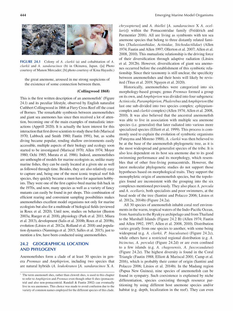
444
FIGURE 24.1 Colony of A. clarkii (a) and cohabitation of A. clarkii and A. sandaracinos (b) in Okinawa, Japan. ([a] Photo
courtesy of Manon Mercader; [b] photo courtesy of Kina Hayashi.)
the great anemone, aroused in me strong suspicions of
the existence of some connection between them.
( Collingwood 1868 )
This is the first written description of an anemonefi sh* ( Figure
24.1) and its peculiar lifestyle, observed by English naturalist
Cuthbert Collingwood in 1866 at Fiery Cross Reef off the coast
of Borneo. The remarkable symbiosis between anemonefi shes
and giant sea anemones has since then received a lot of atten
tion, becoming one of the main examples of mutualistic inter
actions (Apprill 2020). It is actually the keen interest for this
interaction that first drove scientists to study these fi sh (Mariscal
1970; Lubbock and Smith 1980; Fautin 1991), but, as scuba
diving became popular, rending shallow environments easily
accessible, multiple aspects of their biology and ecology soon
started to be investigated (Mariscal 1970; Allen 1974; Moyer
1980; Ochi 1985; Murata et al. 1986). Indeed, anemonefi shes
are unthought-of models for marine ecologists as, unlike many
marine fishes, they can be easily located at a given site as well
as followed through time. Besides, they are also relatively easy
to capture and, being one of the most iconic tropical reef fi sh
species, they quickly became a must-have for aquarium hobby
ists. They were one of the first captive-bred marine fish back in
the 1970s, and now, many species as well as a variety of fancy
mutants can easily be found in pet shops. This combination of
efficient rearing and convenient sampling possibilities makes
anemonefishes excellent model organisms not only for marine
ecologists but also for a multitude of biological fi elds (reviewed
in Roux et al. 2020). Until now, studies on behavior (Buston
2003a ; Rueger et al. 2018), physiology (Park et al. 2011; Miura
et al. 2013), development (Salis et al. 2018b; Roux et al. 2019b),
evolution (Litsios et al. 2012a ; Rolland et al. 2018) and popula
tion dynamics ( Nanninga et al. 2015; Salles et al. 2015), just to
mention a few, have been conducted using anemonefi shes.
24.2 GEOGRAPHICAL LOCATION AND PHYLOGENY
Anemonefishes form a clade of at least 30 species in gen
era Premnas and Amphiprion, including two species that
are natural hybrids (A. leukokranos [A. sandaracinos X A.
* The term anemonefi shes, rather than clownfi shes, is used in this chapter
to refer to Amphiprion and Premnas even though other fi shes (pomacen
trid and also non-pomacentrid; Randall & Fautin 2002) can eventually
live in sea anemones. This choice was made to avoid confusion due to the
variety of common names employed for the different species of this clade.
Emerging Marine Model Organisms
chrysopterus] and A. thiellei [A. sandaracinos X A. ocellaris]) within the Pomacentridae family (Frédérich and
Parmentier 2016 ). All are living as symbionts with ten sea
anemone species that belong to three distantly related fami
lies (Thalassianthidae, Actinidae, Stichodactilidae ) ( Allen
1974; Fautin and Allen 1997; Ollerton et al. 2007; Allen et al.
2008, 2010). This mutualistic relationship is the driving force
of their diversification through adaptive radiation (Litsios
et al. 2012b). However, diversification of giant sea anemo
nes occurred before the establishment of this symbiotic rela
tionship. Since their taxonomy is still unclear, the specifi city
between anemonefishes and their hosts will likely be revis
ited (Titus et al. 2019; Nguyen et al. 2020).
Historically, anemonefishes were categorized into six
morphology-based groups; genus Premnas formed a group
on its own, and Amphiprion was divided into four subgenera:
Actinicola, Paramphiprion, Phalerebus and Amphiprion (the
last one sub-divided into two species complex: ephippium
complex and clarkii-complex) (Allen 1974; Allen et al. 2008 ,
2010). It was also believed that the ancestral anemonefi sh
was able to live in association with multiple sea anemone
species (i.e. generalist) that later radiated into various more
specialized species (Elliott et al. 1999). This process is com
monly used to explain the evolution of symbiotic organisms
(Futuyma and Moreno 1988). A. clarkii was then believed to
be at the base of the anemonefish phylogenetic tree, as it is
the most widespread and generalist species of the tribe. It is
also less dependent on its host sea anemone due to its good
swimming performance and its morphology, which resem
bles that of other free-living pomacentrids. However, the
latest molecular phylogenetic studies do not support those
hypotheses based on morphological traits. They support the
monophyletic origin of anemonefish species, but the topolo
gies found are inconsistent with the grouping into the six
complexes mentioned previously. They also place A. percula and A. ocellaris, both specialists and poor swimmers, at the
basal node of the tree (Santini and Polacco 2006; Litsios et
al. 2012a , 2014b ) ( Figure 24.2a ).
All 30 species of anemonefish inhabit coral reef environ
ments in the warm, tropical waters of the Indo-Pacifi c Ocean,
from Australia to the Ryukyu archipelago and from Thailand
to the Marshall Islands (Figure 24.2 B) (Allen 1974; Fautin
and Allen 1992, 1997; Allen et al. 2008 , 2010). Distribution
varies greatly from one species to another, with some being
widespread (e.g. A. clarkii, P. biaculeatus ) ( Figure 24.2c ),
while others have a restricted regional distribution (e.g. A. bicinctus, A. percula) (Figure 24.2d) or are even confi ned
to a few islands (e.g. A. chagosensis, A. fuscocaudatus) (Figure 24.2e). The highest diversity is found in the Coral
Triangle (Fautin 1988; Elliott & Mariscal 2001; Camp et al.
2016 ), which is probably their center of origin (Santini and
Polacco 2006; Litsios et al. 2014b). In the Madang region
(Papua New Guinea), nine species of anemonefish can be
found in sympatry. Such coexistence is explained by niche
differentiation, species coexisting through resource par
titioning by using different host anemone species and/or
habitat (e.g. depth, localization in the reef). They can even
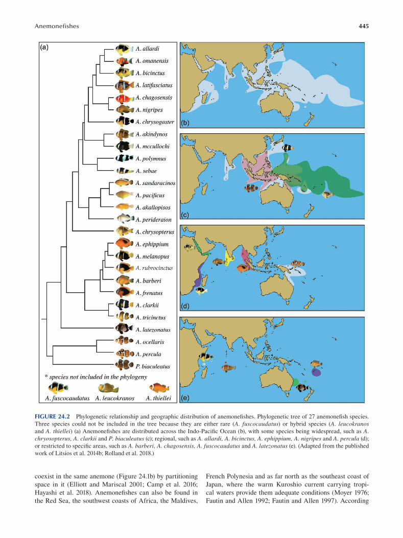
445 Anemonefi shes
FIGURE 24.2 Phylogenetic relationship and geographic distribution of anemonefishes. Phylogenetic tree of 27 anemonefi sh species.
Three species could not be included in the tree because they are either rare (A. fuscocaudatus) or hybrid species (A. leucokranos and A. thiellei) (a) Anemonefishes are distributed across the Indo-Pacific Ocean (b), with some species being widespread, such as A. chryosopterus, A. clarkii and P. biaculeatus (c); regional, such as A. allardi, A. bicinctus, A. ephippium, A. nigripes and A. percula (d);
or restricted to specific areas, such as A. barberi, A. chagosensis, A. fuscocaudatus and A. latezonatus (e). (Adapted from the published
work of Litsios et al. 2014b; Rolland et al. 2018.)
coexist in the same anemone (Figure 24.1b) by partitioning French Polynesia and as far north as the southeast coast of
space in it (Elliott and Mariscal 2001; Camp et al. 2016; Japan, where the warm Kuroshio current carrying tropi-
Hayashi et al. 2018). Anemonefishes can also be found in cal waters provide them adequate conditions (Moyer 1976;
the Red Sea, the southwest coasts of Africa, the Maldives, Fautin and Allen 1992; Fautin and Allen 1997 ). According
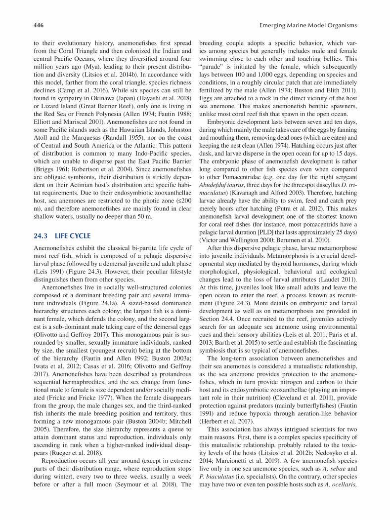
446
to their evolutionary history, anemonefi shes fi rst spread
from the Coral Triangle and then colonized the Indian and
central Pacific Oceans, where they diversified around four
million years ago (Mya), leading to their present distribu
tion and diversity (Litsios et al. 2014b). In accordance with
this model, farther from the coral triangle, species richness
declines (Camp et al. 2016 ). While six species can still be
found in sympatry in Okinawa (Japan) (Hayashi et al. 2018)
or Lizard Island (Great Barrier Reef), only one is living in
the Red Sea or French Polynesia (Allen 1974; Fautin 1988;
Elliott and Mariscal 2001). Anemonefi shes are not found in
some Pacific islands such as the Hawaiian Islands, Johnston
Atoll and the Marquesas (Randall 1955), nor on the coast
of Central and South America or the Atlantic. This pattern
of distribution is common to many Indo-Pacifi c species,
which are unable to disperse past the East Pacifi c Barrier
(Briggs 1961; Robertson et al. 2004). Since anemonefi shes
are obligate symbionts, their distribution is strictly depen
dent on their Actinian host’s distribution and specifi c habi
tat requirements. Due to their endosymbiotic zooxanthellae
host, sea anemones are restricted to the photic zone (≤200
m), and therefore anemonefishes are mainly found in clear
shallow waters, usually no deeper than 50 m.
24.3 LIFE CYCLE
Anemonefishes exhibit the classical bi-partite life cycle of
most reef fish, which is composed of a pelagic dispersive
larval phase followed by a demersal juvenile and adult phase
( Leis 1991) ( Figure 24.3). However, their peculiar lifestyle
distinguishes them from other species.
Anemonefishes live in socially well-structured colonies
composed of a dominant breeding pair and several imma
ture individuals (Figure 24.1a). A sized-based dominance
hierarchy structures each colony; the largest fish is a domi
nant female, which defends the colony, and the second larg
est is a sub-dominant male taking care of the demersal eggs
(Olivotto and Geffroy 2017). This monogamous pair is sur
rounded by smaller, sexually immature individuals, ranked
by size, the smallest (youngest recruit) being at the bottom
of the hierarchy (Fautin and Allen 1992; Buston 2003a ;
Iwata et al. 2012; Casas et al. 2016; Olivotto and Geffroy
2017 ). Anemonefishes have been described as protandrous
sequential hermaphrodites, and the sex change from func
tional male to female is size dependent and/or socially medi
ated (Fricke and Fricke 1977). When the female disappears
from the group, the male changes sex, and the third-ranked
fish inherits the male breeding position and territory, thus
forming a new monogamous pair (Buston 2004b; Mitchell
2005). Therefore, the size hierarchy represents a queue to
attain dominant status and reproduction, individuals only
ascending in rank when a higher-ranked individual disap
pears (Rueger et al. 2018).
Reproduction occurs all year around (except in extreme
parts of their distribution range, where reproduction stops
during winter), every two to three weeks, usually a week
before or after a full moon (Seymour et al. 2018). The
Emerging Marine Model Organisms
breeding couple adopts a specific behavior, which var
ies among species but generally includes male and female
swimming close to each other and touching bellies. This
“parade” is initiated by the female, which subsequently
lays between 100 and 1,000 eggs, depending on species and
conditions, in a roughly circular patch that are immediately
fertilized by the male (Allen 1974; Buston and Elith 2011).
Eggs are attached to a rock in the direct vicinity of the host
sea anemone. This makes anemonefish benthic spawners,
unlike most coral reef fish that spawn in the open ocean.
Embryonic development lasts between seven and ten days,
during which mainly the male takes care of the eggs by fanning
and mouthing them, removing dead ones (which are eaten) and
keeping the nest clean (Allen 1974). Hatching occurs just after
dusk, and larvae disperse in the open ocean for up to 15 days.
The embryonic phase of anemonefish development is rather
long compared to other fish species even when compared
to other Pomacentridae (e.g. one day for the night sergeant
Abudefduf taurus, three days for the threespot dascyllus D. trimaculatus) (Kavanagh and Alford 2003). Therefore, hatching
larvae already have the ability to swim, feed and catch prey
merely hours after hatching (Putra et al. 2012). This makes
anemonefish larval development one of the shortest known
for coral reef fishes (for instance, most pomacentrids have a
pelagic larval duration [PLD] that lasts approximately 25 days)
(Victor and Wellington 2000; Berumen et al. 2010).
After this dispersive pelagic phase, larvae metamorphose
into juvenile individuals. Metamorphosis is a crucial devel
opmental step mediated by thyroid hormones, during which
morphological, physiological, behavioral and ecological
changes lead to the loss of larval attributes (Laudet 2011).
At this time, juveniles look like small adults and leave the
open ocean to enter the reef, a process known as recruit
ment (Figure 24.3). More details on embryonic and larval
development as well as on metamorphosis are provided in
Section 24.4. Once recruited to the reef, juveniles actively
search for an adequate sea anemone using environmental
cues and their sensory abilities (Leis et al. 2011; Paris et al.
2013; Barth et al. 2015) to settle and establish the fascinating
symbiosis that is so typical of anemonefi shes.
The long-term association between anemonefi shes and
their sea anemones is considered a mutualistic relationship,
as the sea anemone provides protection to the anemone-
fishes, which in turn provide nitrogen and carbon to their
host and its endosymbiotic zooxanthellae (playing an impor
tant role in their nutrition) (Cleveland et al. 2011), provide
protection against predators (mainly butterfl yfi shes) (Fautin
1991) and reduce hypoxia through aeration-like behavior
(Herbert et al. 2017 ).
This association has always intrigued scientists for two
main reasons. First, there is a complex species specifi city of
this mutualistic relationship, probably related to the toxic
ity levels of the hosts (Litsios et al. 2012b; Nedosyko et al.
2014; Marcionetti et al. 2019). A few anemonefi sh species
live only in one sea anemone species, such as A. sebae and
P. biaculatus (i.e. specialists). On the contrary, other species
may have two or even ten possible hosts such as A. ocellaris,
447 Anemonefi shes
FIGURE 24.3 Anemonefish life cycle. Eggs are laid on the substrate close to the host sea anemone. After hatching, the pelagic larvae dis
perse in the open ocean. Recruitment to the reef coincides with metamorphosis from larvae to juveniles, which then settle into a sea anemone.
A. bicinctus, A clarkii and A. perideraion (i.e. generalists)
(Fautin and Allen 1997 ) (Table 24.1).
Second, anemonefishes are able to live unharmed inside
the tentacles of their host, which are known to discharge
stinging cells called nematocysts (Mebs 2009). Two main
hypotheses have been formulated to explain this ability. The
first one suggests that anemonefishes coat themselves with
sea anemone mucus, which is therefore used as a chemi
cal camouflage (Fautin 1991; Scott 2008). This is achieved
during an acclimation process that corresponds to a series
of behaviors during which anemonefishes carefully enter
their hosts (Schlichter 1968). First, they kiss the tentacles,
then touch them with their pectoral fins and fi nally scrub
their entire body against the tentacles. This behavior has
been observed in several species, but not all, and it also
seems different depending on the sea anemone species.
Surprisingly, A. clarkii needs to acclimate when entering
in Entacmea quadricolor but not when entering the more
toxic Stichodactyla haddoni ( Lubbock 1981 ; Elliott and
Mariscal 1997; Mebs 2009). The second hypothesis sug
gests that anemonefishes are protected from sea anemone
stinging by their own mucus that either prevents nematocyst
discharge or protects the fish from the consequence of the
discharge. Indeed, it has been shown that A. ocellaris lacks
N-acetylneuraminic acid in its mucus, which is normally
detected by sea anemone tentacles to discharge stinging
cells (Abdullah and Saad 2015). All these studies suggest
that the mucus of both partners is the key to understanding
how anemonefishes are able to live in sea anemones without
being harmed. Moreover, it has recently been demonstrated
that changes in the microbial composition are occurring in
both partners during initiation of the symbiosis, suggesting a
potential role of bacterial communities in the establishment
of this relationship (Pratte et al. 2018; Roux et al. 2019a).
After settlement, anemonefishes integrate into the colony
hierarchy, queuing for breeding positions. Why and how
anemonefishes engage in such a social system is starting to
be understood thanks to extensive work on A. percula colo
nies and may have a great contribution to the understanding
of complex societies. Buston and collaborators have shown
that members of a colony are not composed of close relatives
(2007) and that non-breeders don’t provide alloparental care,
their presence having neither a positive or negative effect on
the dominant pair’s breeding success (Buston 2004a). Non-
breeders can adjust their size and growth rate in order to
maintain a clear size difference with respect to individuals
of higher social rank so that conflicts are limited, thereby
reducing the risk of eviction and the potential cost to the
breeding dominant pair (Buston 2003a ). Consequently, there
seem to be no direct benefits of living in such social groups.
However, withholding reproduction by staying small and not
contesting to remain part of the colony might represent a bet
ter option than either leaving the host anemone to breed else
where (because of predation risk) or contesting for breeding
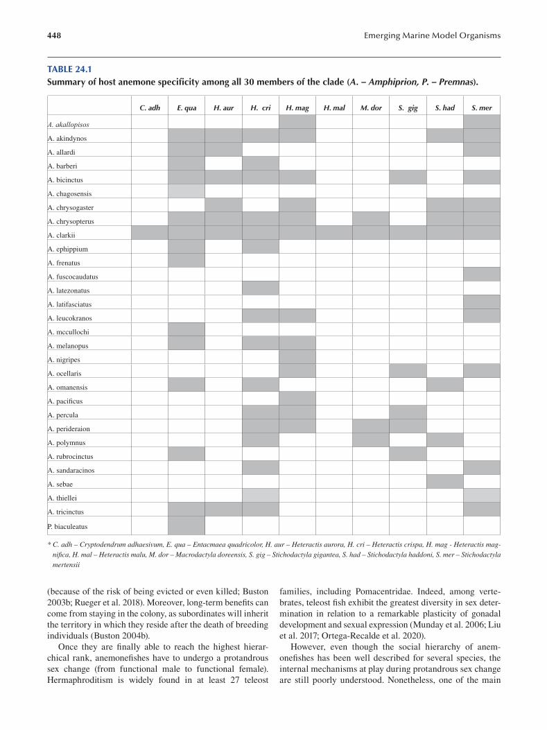
448 Emerging Marine Model Organisms
TABLE 24.1 Summary of host anemone specificity among all 30 members of the clade (A. – Amphiprion, P. – Premnas).
C. adh E. qua H. aur H. cri H. mag H. mal M. dor S. gig S. had S. mer
A. akallopisos
A. akindynos
A. allardi
A. barberi
A. bicinctus
A. chagosensis
A. chrysogaster
A. chrysopterus
A. clarkii
A. ephippium
A. frenatus
A. fuscocaudatus
A. latezonatus
A. latifasciatus
A. leucokranos
A. mccullochi
A. melanopus
A. nigripes
A. ocellaris
A. omanensis
A. pacifi cus
A. percula
A. perideraion
A. polymnus
A. rubrocinctus
A. sandaracinos
A. sebae
A. thiellei
A. tricinctus
P. biaculeatus
* C. adh – Cryptodendrum adhaesivum, E. qua – Entacmaea quadricolor, H. aur – Heteractis aurora, H. cri – Heteractis crispa, H. mag - Heteractis magnifica, H. mal – Heteractis malu, M. dor – Macrodactyla doreensis, S. gig – Stichodactyla gigantea, S. had – Stichodactyla haddoni, S. mer – Stichodactyla mertensii
(because of the risk of being evicted or even killed; Buston families, including Pomacentridae. Indeed, among verte
2003b; Rueger et al. 2018). Moreover, long-term benefi ts can brates, teleost fish exhibit the greatest diversity in sex deter-
come from staying in the colony, as subordinates will inherit mination in relation to a remarkable plasticity of gonadal
the territory in which they reside after the death of breeding development and sexual expression (Munday et al. 2006; Liu
individuals (Buston 2004b). et al. 2017; Ortega-Recalde et al. 2020).
Once they are finally able to reach the highest hierar- However, even though the social hierarchy of anem
chical rank, anemonefishes have to undergo a protandrous onefishes has been well described for several species, the
sex change (from functional male to functional female). internal mechanisms at play during protandrous sex change
Hermaphroditism is widely found in at least 27 teleost are still poorly understood. Nonetheless, one of the main
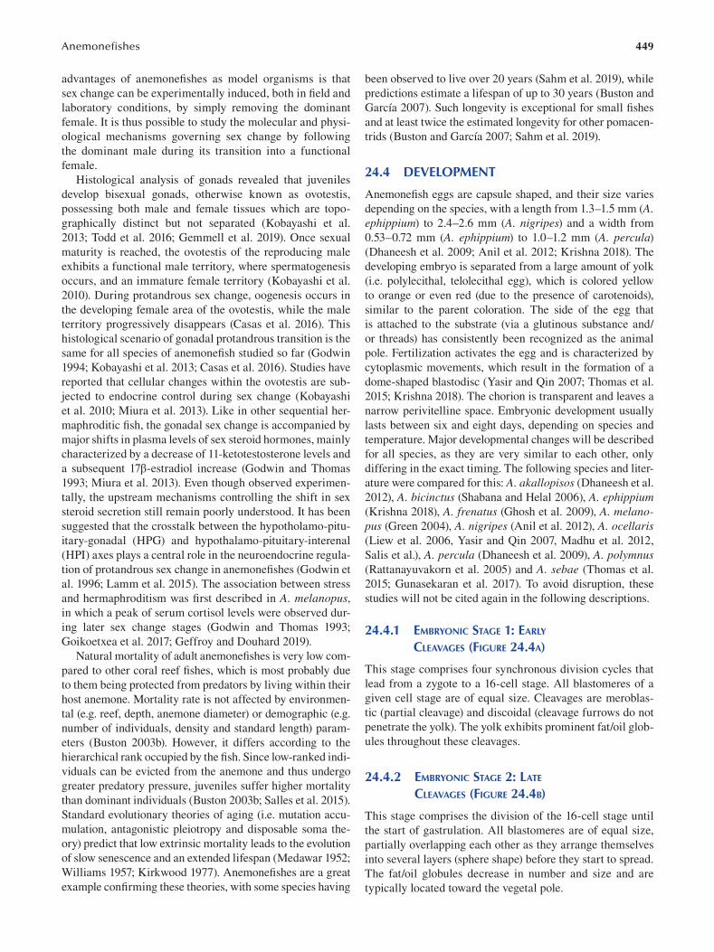
449 Anemonefi shes
advantages of anemonefishes as model organisms is that
sex change can be experimentally induced, both in fi eld and
laboratory conditions, by simply removing the dominant
female. It is thus possible to study the molecular and physi
ological mechanisms governing sex change by following
the dominant male during its transition into a functional
female.
Histological analysis of gonads revealed that juveniles
develop bisexual gonads, otherwise known as ovotestis,
possessing both male and female tissues which are topo
graphically distinct but not separated (Kobayashi et al.
2013; Todd et al. 2016; Gemmell et al. 2019). Once sexual
maturity is reached, the ovotestis of the reproducing male
exhibits a functional male territory, where spermatogenesis
occurs, and an immature female territory (Kobayashi et al.
2010). During protandrous sex change, oogenesis occurs in
the developing female area of the ovotestis, while the male
territory progressively disappears (Casas et al. 2016 ). This
histological scenario of gonadal protandrous transition is the
same for all species of anemonefish studied so far (Godwin
1994; Kobayashi et al. 2013; Casas et al. 2016). Studies have
reported that cellular changes within the ovotestis are sub
jected to endocrine control during sex change (Kobayashi
et al. 2010; Miura et al. 2013). Like in other sequential her
maphroditic fish, the gonadal sex change is accompanied by
major shifts in plasma levels of sex steroid hormones, mainly
characterized by a decrease of 11-ketotestosterone levels and
a subsequent 17β-estradiol increase (Godwin and Thomas
1993; Miura et al. 2013). Even though observed experimen
tally, the upstream mechanisms controlling the shift in sex
steroid secretion still remain poorly understood. It has been
suggested that the crosstalk between the hypotholamo-pitu
itary-gonadal (HPG) and hypothalamo-pituitary-interenal
(HPI) axes plays a central role in the neuroendocrine regula
tion of protandrous sex change in anemonefishes (Godwin et
al. 1996; Lamm et al. 2015). The association between stress
and hermaphroditism was first described in A. melanopus, in which a peak of serum cortisol levels were observed dur
ing later sex change stages (Godwin and Thomas 1993;
Goikoetxea et al. 2017; Geffroy and Douhard 2019).
Natural mortality of adult anemonefishes is very low com
pared to other coral reef fishes, which is most probably due
to them being protected from predators by living within their
host anemone. Mortality rate is not affected by environmen
tal (e.g. reef, depth, anemone diameter) or demographic (e.g.
number of individuals, density and standard length) param
eters (Buston 2003b). However, it differs according to the
hierarchical rank occupied by the fish. Since low-ranked indi
viduals can be evicted from the anemone and thus undergo
greater predatory pressure, juveniles suffer higher mortality
than dominant individuals (Buston 2003b; Salles et al. 2015).
Standard evolutionary theories of aging (i.e. mutation accu
mulation, antagonistic pleiotropy and disposable soma the
ory) predict that low extrinsic mortality leads to the evolution
of slow senescence and an extended lifespan ( Medawar 1952;
Williams 1957 ; Kirkwood 1977 ). Anemonefishes are a great
example confirming these theories, with some species having
been observed to live over 20 years (Sahm et al. 2019), while
predictions estimate a lifespan of up to 30 years (Buston and
García 2007). Such longevity is exceptional for small fi shes
and at least twice the estimated longevity for other pomacen
trids (Buston and García 2007; Sahm et al. 2019).
24.4 DEVELOPMENT
Anemonefish eggs are capsule shaped, and their size varies
depending on the species, with a length from 1.3–1.5 mm (A. ephippium) to 2.4–2.6 mm (A. nigripes) and a width from
0.53–0.72 mm ( A. ephippium) to 1.0–1.2 mm (A. percula)
(Dhaneesh et al. 2009; Anil et al. 2012; Krishna 2018). The
developing embryo is separated from a large amount of yolk
(i.e. polylecithal, telolecithal egg), which is colored yellow
to orange or even red (due to the presence of carotenoids),
similar to the parent coloration. The side of the egg that
is attached to the substrate (via a glutinous substance and/
or threads) has consistently been recognized as the animal
pole. Fertilization activates the egg and is characterized by
cytoplasmic movements, which result in the formation of a
dome-shaped blastodisc (Yasir and Qin 2007; Thomas et al.
2015; Krishna 2018). The chorion is transparent and leaves a
narrow perivitelline space. Embryonic development usually
lasts between six and eight days, depending on species and
temperature. Major developmental changes will be described
for all species, as they are very similar to each other, only
differing in the exact timing. The following species and liter
ature were compared for this: A. akallopisos (Dhaneesh et al.
2012 ), A. bicinctus (Shabana and Helal 2006), A. ephippium ( Krishna 2018 ), A. frenatus (Ghosh et al. 2009), A. melanopus ( Green 2004 ), A. nigripes (Anil et al. 2012), A. ocellaris (Liew et al. 2006, Yasir and Qin 2007, Madhu et al. 2012,
Salis et al.), A. percula (Dhaneesh et al. 2009), A. polymnus (Rattanayuvakorn et al. 2005) and A. sebae (Thomas et al.
2015; Gunasekaran et al. 2017 ). To avoid disruption, these
studies will not be cited again in the following descriptions.
24.4.1 EMBRYONIC STAGE 1: EARLY
CLEAVAGES (FIGURE 24.4A)
This stage comprises four synchronous division cycles that
lead from a zygote to a 16-cell stage. All blastomeres of a
given cell stage are of equal size. Cleavages are meroblas
tic (partial cleavage) and discoidal (cleavage furrows do not
penetrate the yolk). The yolk exhibits prominent fat/oil glob
ules throughout these cleavages.
24.4.2 EMBRYONIC STAGE 2: LATE
CLEAVAGES (FIGURE 24.4B)
This stage comprises the division of the 16-cell stage until
the start of gastrulation. All blastomeres are of equal size,
partially overlapping each other as they arrange themselves
into several layers (sphere shape) before they start to spread.
The fat/oil globules decrease in number and size and are
typically located toward the vegetal pole.
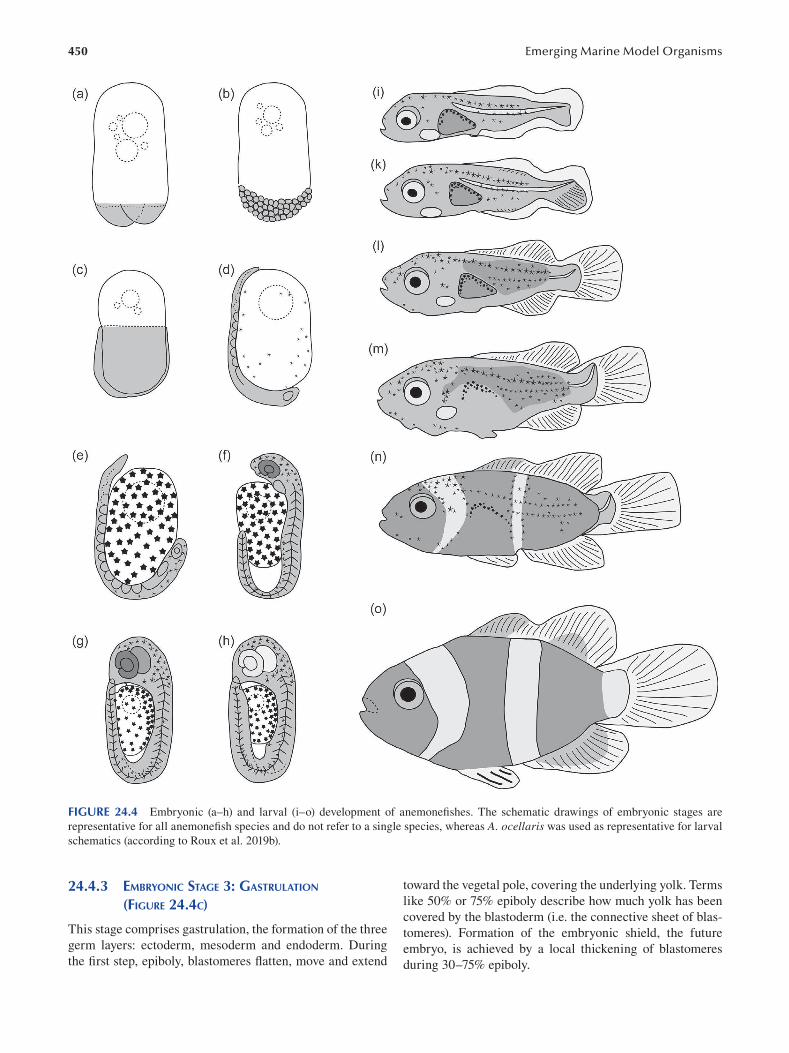
450 Emerging Marine Model Organisms
FIGURE 24.4 Embryonic (a–h) and larval (i–o) development of anemonefishes. The schematic drawings of embryonic stages are
representative for all anemonefish species and do not refer to a single species, whereas A. ocellaris was used as representative for larval
schematics (according to Roux et al. 2019b).
24.4.3 EMBRYONIC STAGE 3: GASTRULATION toward the vegetal pole, covering the underlying yolk. Terms
(FIGURE 24.4C) like 50% or 75% epiboly describe how much yolk has been
covered by the blastoderm (i.e. the connective sheet of blas-This stage comprises gastrulation, the formation of the three tomeres). Formation of the embryonic shield, the future germ layers: ectoderm, mesoderm and endoderm. During embryo, is achieved by a local thickening of blastomeres the first step, epiboly, blastomeres flatten, move and extend during 30–75% epiboly.
451 Anemonefi shes
24.4.4 EMBRYONIC STAGE 4: CEPHALIZATION AND
SOMITE DEVELOPMENT (FIGURE 24.4D)
The head, including optic buds (located at the animal pole),
as well as neural ectoderm, is formed. The tail bud begins
to develop later on. Overall, this stage marks the beginning
of organogenesis and metamerization. The fi rst appearance
of paired somites occurs before 100% epiboly is reached
(around 60–80% epiboly). Stellate melanophores begin to
cover the yolk.
24.4.5 EMBRYONIC STAGE 5: TURNOVER (FIGURE 24.4E)
The entire body of the embryo is covered with few melano
phores, particularly abundant in the head region. The head is
clearly distinguishable, and the brain has differentiated into
three parts: the prosencephalon, mesencephalon and rhomben
cephalon. Primitive optic buds/vesicles have formed, with sub
sequent induction of eye formation (eye cup, lens and cornea).
Somitogenesis (trunk segmentation) is finished at the end of
this stage. The body is transparent due to the absence of mus
cular structure at beginning, but later on, myotomes are rec
ognizable. The embryo completely turns itself (body reversal
by positioning the head toward the vegetal pole) while the tip
of the tail is still attached to the yolk sac. This is a critical step
for further development to proceed. The body is attached to the
yolk sac, while the tail detaches from the yolk toward the end
of this stage and exhibits increasing tail movements. A tubular,
pink-colored heart has been differentiated and begins to beat.
24.4.6 EMBRYONIC STAGE 6: BLOOD
FORMATION (FIGURE 24.4F)
The head and tail of the embryo have distinctly separated
from the yolk, which is reduced in its volume. The body
length has increased distinctly. Transparent (later a light
shade of pink) spherical blood cells and subsequently blood
circulation can be observed. Pigmentation is prominent in
the head, especially in the large eyes displaying brownish
pigments, but less in the tail region. Skeletal muscles and
myotomes become clearly visible.
24.4.7 EMBRYONIC STAGE 7: REMAINING ORGAN
AND FIN DEVELOPMENT (FIGURE 24.4G)
The head occupies one-third of the capsule space and has
salient eyes with brown melanin pigmentation. The size
of the entire embryo has increased substantially, with the
tail reaching the posterior part of the eyes, and it displays
continuous movement. The yolk sac becomes quite small,
and yellow pigments start to appear on the trunk. Branchial
arches with ventilating gills and opercula, a looped alimen
tary tract and jaws have developed. The fin folds have devel
oped and are clearly visible.
24.4.8 EMBRYONIC STAGE 8: HATCHING (FIGURE 24.4H)
A hindgut has formed, and the embryo fully occupies the
capsule. The spinal cord is not flexed. The eyes are turn
ing and silver shining (eyeshine from the tapetum). The
embryo tries to hatch out: vigorous movements of the tail
rupture an area close to the base of the eggshell (where the
egg is attached to the substrate). The hatchlings emerge
tail first, which usually takes place after sunset in com
plete darkness.
A relatively short larval development follows hatching
and precedes metamorphosis. Even though developmental
time frames for larvae are more variable than for embryos,
the following studies have been combined to describe lar
val development and metamorphosis for anemonefi shes in
general: A. ephippium ( Krishna 2018 ), A. frenatus ( Putra et
al. 2012), A. nigripes (Anil et al. 2012), A. ocellaris ( Madhu
et al. 2012; Roux et al. 2019b), A. perideraion ( Salis et al.
2018a) and A. sebae (Gunasekaran et al. 2017 ).
24.4.9 LARVAL STAGE 1: PREFLEXION OF THE
NOTOCHORD (FIGURE 24.4I)
The larvae are mainly transparent, with some melano
phores and xanthophores scattered over the head and body.
Additionally, one or two horizontal lines of melanophores
are present on the trunk, along the ventral midline. The
embryonic fin folds remain undifferentiated and transparent.
The notochord is still straight, in preflexion. Larvae are able
to feed on live prey soon after hatching and process the food
in a short, straight alimentary canal with the anus located in
the middle of the body length. Stomach, midgut and hindgut
are distinct, and the liver and pancreas are differentiated.
The larvae display phototropic behavior and swim at the top
of the water column.
24.4.10 LARVAL STAGE 2: FLEXION OF THE
NOTOCHORD (FIGURE 24.4K)
The embryonic fin folds start to differentiate into the caudal,
dorsal and anal fins, which exhibit first signs of soft rays.
The notochord begins to flex by bending dorsally.
24.4.11 LARVAL STAGE 3: POSTFLEXION OF THE
NOTOCHORD (FIGURE 24.4L)
The embryonic fin folds have completely differentiated into
caudal, dorsal and anal fins. Both anal and dorsal fi ns exhibit
the complete set of soft rays and spines that start to appear
in a posterior–anterior gradient. The pelvic fins begin to
differentiate. The notochord is in postflexion, resulting in
a vertical position of the hypural bones. There are no major
changes in pigmentation pattern or swimming behavior.
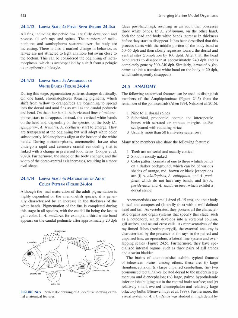
452
24.4.12 LARVAL STAGE 4: PELVIC SPINE (FIGURE 24.4M)
All fins, including the pelvic fins, are fully developed and
possess all soft rays and spines. The numbers of mela
nophores and xanthophores scattered over the body are
increasing. There is also a marked change in behavior, as
larvae are not attracted to light anymore but swim close to
the bottom. This can be considered the beginning of meta
morphosis, which is accompanied by a shift from a pelagic
to an epibenthic lifestyle.
24.4.13 LARVAL STAGE 5: APPEARANCE OF
WHITE BANDS (FIGURE 24.4N)
During this stage, pigmentation patterns changes drastically.
On one hand, chromatophores (bearing pigments, which
shift from yellow to orange/red) are beginning to spread
into the dorsal and anal fins as well as the caudal peduncle
and head. On the other hand, the horizontal lines of melano
phores start to disappear. Instead, the vertical white bands
on the head and, depending on the species, on the body (A. ephippium, A. frenatus, A. ocellaris) start to emerge. They
are transparent at the beginning but will adopt white color
subsequently. Melanophores align at the border of the white
bands. During metamorphosis, anemonefish larvae also
undergo a rapid and extensive cranial remodeling that is
linked with a change in preferred food items (Cooper et al.
2020). Furthermore, the shape of the body changes, and the
width of the dorso-ventral axis increases, resulting in a more
oval shape.
24.4.14 LARVAL STAGE 6: MATURATION OF ADULT
COLOR PATTERN (FIGURE 24.4O)
Although the final maturation of the adult pigmentation is
highly dependent on the anemonefish species, it is gener
ally characterized by an increase in the thickness of the
white bands. Pigmentation of the fins is completed during
this stage in all species, with the caudal fin being the last to
gain color. In A. ocellaris, for example, a third white band
appears on the caudal peduncle after approximately 20 dph
FIGURE 24.5 Schematic drawing of A. ocellaris showing exter
nal anatomical features.
Emerging Marine Model Organisms
(days post-hatching), resulting in an adult that possesses
three white bands. In A. ephippium, on the other hand,
both the head and body white bands increase in thickness
before they start to disappear. It has been described that this
process starts with the middle portion of the body band at
50–55 dph and then slowly regresses toward the dorsal and
ventral sites (completion by 160 dph). After that, the head
band starts to disappear at approximately 240 dph and is
completely gone by 300–310 dph. Similarly, larvae of A. frenatus exhibit a transient white band on the body at 20 dph,
which subsequently disappears.
24.5 ANATOMY
The following anatomical features can be used to distinguish
members of the Amphiprioninae (Figure 24.5) from the
remainder of the pomacentrids (Allen 1974; Nelson et al. 2016):
1 Nine to 11 dorsal spines
2 Suborbital, preopercle, opercle and interopercle
bones with serrated or spinous margins and/or
sculptured with radiating striae
3 Usually more than 50 transverse scale rows
Many tribe members also share the following features:
1 Teeth are uniserial and usually conical
2 Snout is mostly naked
3 Color pattern consists of one to three whitish bands
on a darker background, which can be of various
shades of orange, red, brown or black [exceptions
are (i) A. akallopisos, A. ephippium, and A. pacifi cus, which do not have any bands, and (ii) A. perideraion and A. sandaracinos, which exhibit a
dorsal stripe]
Anemonefishes are small sized (5–15 cm), and their body
is oval and compressed (laterally thin) with a well-defi ned
head and tail. As vertebrates, they possess all the character
istic organs and organ systems that specify this clade, such
as a notochord, which develops into a vertebral column,
gill arches, and neural crest cells. As representatives of the
ray-fi nned fishes (Actinopterygii), the external anatomy is
characterized by the presence of fin rays in the paired and
unpaired fins, an operculum, a lateral line system and over
lapping scales (Figure 24.5). Furthermore, they have spe
cialized internal organs, such as three pairs of gill arches
and a swim bladder.
The brains of anemonefishes exhibit typical features
of teleostean brains; among others, these are: (i) large
rhombencephalon; (ii) large unpaired cerebellum; (iii) two
pronounced tectal halves located dorsal to the midbrain teg
mentum and diencephalon; (iv) large, paired hypothalamic
inferior lobe bulging out in the ventral brain surface; and (v)
relatively small, everted telencephalon and relatively large
olfactory bulbs (Nieuwenhuys et al. 1998). Furthermore, the
visual system of A. akindynos was studied in high detail by
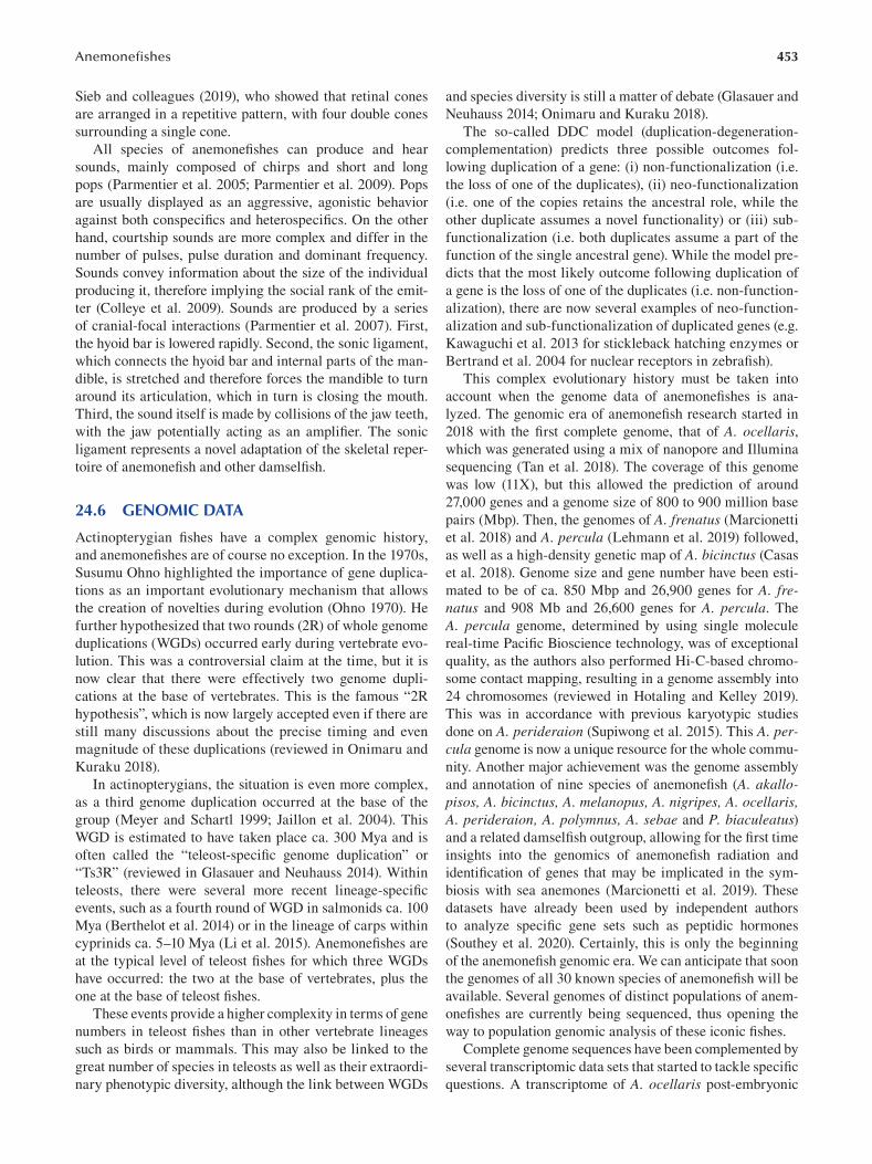
453 Anemonefi shes
Sieb and colleagues (2019), who showed that retinal cones
are arranged in a repetitive pattern, with four double cones
surrounding a single cone.
All species of anemonefishes can produce and hear
sounds, mainly composed of chirps and short and long
pops (Parmentier et al. 2005; Parmentier et al. 2009). Pops
are usually displayed as an aggressive, agonistic behavior
against both conspecifics and heterospecifics. On the other
hand, courtship sounds are more complex and differ in the
number of pulses, pulse duration and dominant frequency.
Sounds convey information about the size of the individual
producing it, therefore implying the social rank of the emit
ter (Colleye et al. 2009). Sounds are produced by a series
of cranial-focal interactions (Parmentier et al. 2007). First,
the hyoid bar is lowered rapidly. Second, the sonic ligament,
which connects the hyoid bar and internal parts of the man
dible, is stretched and therefore forces the mandible to turn
around its articulation, which in turn is closing the mouth.
Third, the sound itself is made by collisions of the jaw teeth,
with the jaw potentially acting as an amplifier. The sonic
ligament represents a novel adaptation of the skeletal reper
toire of anemonefish and other damselfi sh.
24.6 GENOMIC DATA
Actinopterygian fishes have a complex genomic history,
and anemonefishes are of course no exception. In the 1970s,
Susumu Ohno highlighted the importance of gene duplica
tions as an important evolutionary mechanism that allows
the creation of novelties during evolution (Ohno 1970). He
further hypothesized that two rounds (2R) of whole genome
duplications (WGDs) occurred early during vertebrate evo
lution. This was a controversial claim at the time, but it is
now clear that there were effectively two genome dupli
cations at the base of vertebrates. This is the famous “2R
hypothesis”, which is now largely accepted even if there are
still many discussions about the precise timing and even
magnitude of these duplications (reviewed in Onimaru and
Kuraku 2018).
In actinopterygians, the situation is even more complex,
as a third genome duplication occurred at the base of the
group (Meyer and Schartl 1999; Jaillon et al. 2004). This
WGD is estimated to have taken place ca. 300 Mya and is
often called the “teleost-specific genome duplication” or
“Ts3R” (reviewed in Glasauer and Neuhauss 2014). Within
teleosts, there were several more recent lineage-specifi c
events, such as a fourth round of WGD in salmonids ca. 100
Mya (Berthelot et al. 2014) or in the lineage of carps within
cyprinids ca. 5–10 Mya (Li et al. 2015). Anemonefi shes are
at the typical level of teleost fishes for which three WGDs
have occurred: the two at the base of vertebrates, plus the
one at the base of teleost fi shes.
These events provide a higher complexity in terms of gene
numbers in teleost fishes than in other vertebrate lineages
such as birds or mammals. This may also be linked to the
great number of species in teleosts as well as their extraordi
nary phenotypic diversity, although the link between WGDs
and species diversity is still a matter of debate (Glasauer and
Neuhauss 2014; Onimaru and Kuraku 2018).
The so-called DDC model (duplication-degeneration
complementation) predicts three possible outcomes fol
lowing duplication of a gene: (i) non-functionalization (i.e.
the loss of one of the duplicates), (ii) neo-functionalization
(i.e. one of the copies retains the ancestral role, while the
other duplicate assumes a novel functionality) or (iii) sub
functionalization (i.e. both duplicates assume a part of the
function of the single ancestral gene). While the model pre
dicts that the most likely outcome following duplication of
a gene is the loss of one of the duplicates (i.e. non-function
alization), there are now several examples of neo-function
alization and sub-functionalization of duplicated genes (e.g.
Kawaguchi et al. 2013 for stickleback hatching enzymes or
Bertrand et al. 2004 for nuclear receptors in zebrafi sh).
This complex evolutionary history must be taken into
account when the genome data of anemonefishes is ana
lyzed. The genomic era of anemonefish research started in
2018 with the first complete genome, that of A. ocellaris, which was generated using a mix of nanopore and Illumina
sequencing (Tan et al. 2018). The coverage of this genome
was low (11X), but this allowed the prediction of around
27,000 genes and a genome size of 800 to 900 million base
pairs (Mbp). Then, the genomes of A. frenatus ( Marcionetti
et al. 2018) and A. percula ( Lehmann et al. 2019 ) followed,
as well as a high-density genetic map of A. bicinctus ( Casas
et al. 2018). Genome size and gene number have been esti
mated to be of ca. 850 Mbp and 26,900 genes for A. frenatus and 908 Mb and 26,600 genes for A. percula . The
A. percula genome, determined by using single molecule
real-time Pacific Bioscience technology, was of exceptional
quality, as the authors also performed Hi-C-based chromo
some contact mapping, resulting in a genome assembly into
24 chromosomes (reviewed in Hotaling and Kelley 2019).
This was in accordance with previous karyotypic studies
done on A. perideraion (Supiwong et al. 2015). This A. percula genome is now a unique resource for the whole commu
nity. Another major achievement was the genome assembly
and annotation of nine species of anemonefi sh ( A. akallopisos, A. bicinctus, A. melanopus, A. nigripes, A. ocellaris, A. perideraion, A. polymnus, A. sebae and P. biaculeatus) and a related damselfish outgroup, allowing for the fi rst time
insights into the genomics of anemonefish radiation and
identification of genes that may be implicated in the sym
biosis with sea anemones (Marcionetti et al. 2019). These
datasets have already been used by independent authors
to analyze specific gene sets such as peptidic hormones
(Southey et al. 2020). Certainly, this is only the beginning
of the anemonefish genomic era. We can anticipate that soon
the genomes of all 30 known species of anemonefish will be
available. Several genomes of distinct populations of anem
onefishes are currently being sequenced, thus opening the
way to population genomic analysis of these iconic fi shes.
Complete genome sequences have been complemented by
several transcriptomic data sets that started to tackle specifi c
questions. A transcriptome of A. ocellaris post-embryonic
454
development, spanning newly hatched larvae until settled
juveniles, has been determined (Roux et al. in preparation).
Another area of interest is the identification of genes related
to the differently colored areas (white, orange and black) of
A. ocellaris (Maytin et al. 2018; Salis et al. 2019a). This,
combined with detailed pharmacological and microscopic
analysis, has allowed researchers to determine that irido
phores are responsible for the white color in this species but
also to identify new iridophore and xanthophore genes in fi sh
(Salis et al. 2019a, reviewed in Irion and Nüsslein-Volhard
2019; Patterson and Parichy 2019). Transcriptomic analysis
has also been applied to the spectacular sex change abili
ties of anemonefishes. For example, a study of A. bicinctus from the Red Sea has revealed a complex genomic response
in the brain and subsequently in the gonads with a promi
nent effect on genes implicated in steroidogenesis (Casas et
al. 2016). Genes implicated in reproduction have also been
studied in A. ocellaris (Yang et al. 2019).
Last, transcriptome analysis was used in the context of
aging, as anemonefishes are known to have a long lifespan
(Sahm et al. 2019). The authors have detected positively
selected genes in A. clarkii and A. percula and tested if
these genes were similar to those found in other models of
aging such as mole rats or short-lived killifishes. They con
cluded that molecular convergence is likely to occur in the
evolution of lifespan.
These examples are in fact the exhaustive list of genomic
and transcriptomic studies done so far on anemonefi shes. Due
to low-cost high-throughput sequencing, it is likely that this will
increase exponentially in the coming years as these fi shes will
be used more and more as experimental models which allow to
link ecological, evolutionary and developmental studies.
24.7 FUNCTIONAL APPROACHES: TOOLS FOR MOLECULAR AND CELLULAR ANALYSIS
24.7.1 HUSBANDRY
Generally, the success of an emerging model species is
linked to a feasible husbandry as well as the ease of obtain
ing samples. For marine teleosts, this can pose diffi culties,
as it might be difficult to achieve reproduction in captiv
ity or to reliably locate them in the natural environment.
Anemonefishes provide an excellent model for both sce
narios. On the one hand, due to their close association with
sea anemones, researchers are able to locate and re-locate
anemonefishes with relative ease in the wild, enabling
them to conduct long-term experiments with the same indi
viduals. On the other hand, they are very well adapted for
captive life, having been in the hobbyist trade for decades.
For tropical marine fi shes, anemonefishes are relatively tol
erant to temperature (24°C to 28°C) and salinity variations
(25 to 40‰) (Dhaneesh et al. 2012). Smaller species, like
A. ocellaris, A. percula and A. sandaracinos, can be kept
in 60-L tanks, while bigger species, such as A. clarkii, A. frenatus and P. biaculetatus, will need up to 200-L tanks.
In captivity, anemonefishes thrive without the addition of
Emerging Marine Model Organisms
sea anemones and establish breeding pairs, which usually
reproduce all year around. Both partners will participate
in selection of an appropriate substrate and its cleaning,
usually a terra cotta pot, ceramic tiles or even the glass
walls. Egg clutch sizes vary greatly between and within
species and depend on previous reproductive experience,
nutrition and body size. A sufficient amount of eggs can be
obtained for experimental purposes (up to 700–1,000 eggs)
every 14–21 days. For experiments that require embryonic
stages (such as micro-injection), the eggs can be scraped
off substrate (for example, with a razor blade) and can be
transferred to an egg tumbler or petri dishes for incubation.
For experiments that require larval stages, the eggs remain
with the parents until they are supposed to hatch (night of
hatching). For hatching, they can be transferred into a sep
arate aquarium by replacing the substrate with the attached
eggs. Alternatively, if external water circulation can be
interrupted, the larvae can hatch in the parent’s aquarium
and subsequently be transferred to a different aquarium by
attracting them with a light source. This, however, is only
advisable if there is no sea anemone in the same aquarium.
Larvae can either be raised in small aquaria (20–30 L) or
in 500–1,000-mL beakers (containing 1–20 larvae per bea
ker; Roux et al.). They are first fed with a mixture of micro
algae and rotifers and later on Artemia nauplii . Juveniles
are also fed with Artemia nauplii and either powdered food
or food pellets (depending on size). The diet of adult fi sh is
diverse and can be adjusted easily: Artemia, food pellets,
chopped mussels, squid, shrimp and egg yolk, as well as
vitamin supplements (Anil et al. 2012).
Several standard approaches have been successfully
established in anemonefishes, and only a few will be high
lighted here.
24.7.2 IN SITU HYBRIDIZATION
In situ hybridization is a very powerful tool to study tempo
ral and spatial requirements of specific genes in their cellular
context. In A. frenatus, embryonic mesodermal and neuro
ectodermal development has been followed by gene expres
sion analysis of no tail (ntl) and sox3, respectively (Ghosh
et al. 2009). Further, a comparative expression analysis of
orthodenticle homeobox 2 (otx2) in the olfactory placode
of larval A. percula indicates that this gene is required for
olfactory responses to settlement cues (Veilleux et al. 2013).
Moreover, in situ hybridization can validate results acquired
employing alternative approaches, such as transcriptomics.
For example, a recent study revealed several upregulated
genes in the white skin of A. ocellaris, some of which could
be confi rmed via in situ hybridization on juvenile skin sec
tions (Salis et al. 2019a). Fluorescent in situ hybridization
(FISH) has also been successfully established in anemone-
fishes. In A. akindynos, it has been shown that long wave
length-sensitive (LWS)-related opsin genes are exclusively
expressed in double cones, while short wavelength-sensitive
(SWS)-related opsins are only expressed in the interspaced
single cones (Stieb et al. 2019).
455 Anemonefi shes
24.7.3 IMMUNOASSAY
Commercial enzyme immunoassay (EIA) kits are available
to analyze biochemical aspects of cells, such as hormones,
neurotransmitters and second messenger molecules (such as
cAMP). In 2010, Mills and colleagues validated two such kits
for measuring 11-ketotestosterone and cortisol concentra
tion, respectively, using blood plasma from A. chrysopterus and A. percula. They found that a minimum of 5–7 μL blood
plasma is suffi cient to confi dently estimate steroid hormone
concentrations, which is especially valuable when working
in the field. Other hormones, such as thyroid hormones, can
be routinely measured using phenobarbital extraction and
ELISA detection according to the method developed by
Kawakami et al. (2008) and Holzer et al. (2017 ).
24.7.4 USE OF DRUGS FOR FUNCTIONAL EXPERIMENTS
Pharmacological reagents/small molecules have been used
widely in zebrafi sh, Danio rerio, and helped to broaden
our understanding of zebrafish biology. To date, only few
of them have been tested in anemonefishes, but they pose a
great potential in a variety of fields. For example, it has been
shown that the small molecule TAE 684 inhibits Alk and Ltk dependent iridophores in zebrafish (Rodrigues et al. 2012).
In A. ocellaris, TAE 684 treatment of larvae results in juve
niles without white bands, thus providing evidence that iridi
ophores are responsible for the white color of anemonefi shes
(Salis et al. 2019a). Furthermore, treatment with BMP inhibi
tors, such as dorsomorphin or DMH1, in early embryonic
stages can result in dorsalization in zebrafish (Yu et al. 2008)
and A. ocellaris (M. Klann personal observations) alike.
24.7.5 CELL CULTURE
So far, there is only one report on cell culture from anemone-
fish explants, even though this technique is extremely valu
able for research projects focusing, for example, on virology,
cytobiology and oncology/disease, but also for environmental
toxicology/ecotoxicology or genetics/genomics. Patkaew and
colleagues (2014) used A. ocellaris vertebrae explants to estab
lish a corresponding primary culture. Four days after the ini
tial implantation, fibroblastic cells could be seen, which then
multiplied rapidly, reaching 70–80% confluence within four to
five days. The fifth passage was preserved in liquid nitrogen for
one month and subsequently assessed. The average viability
after thawing and seeding has been reported with 80%, with a
57% cell recovery and no obvious changes in cell morphology
or growth pattern. Even though they do not give details, the
authors also state that the employed explant method (without
the use of enzymes) resulted in successful primary cultures
from gills, skin and vertebrae from other anemonefi shes.
24.7.6 GENETIC MARKERS
Genetic markers, particularly microsatellites, have been devel
oped and are now available for several anemonefi sh species.
They are widely used to study population genetics and have
been used for example to investigate phylogeographic con
nectivity (Dohna et al. 2015), detect and monitor hybridization
events (He et al. 2019; Gainsford et al. 2020), elucidate self-
recruitment of larval dispersal (Jones et al. 2005), estimate
connectivity between marine protected areas (MPAs) (Planes
et al. 2009) and even to determine the composition of social
groups (Buston et al. 2007). A substantial number of population
genetic and dynamic studies have been done on A. percula pop
ulations of Kimbe Bay (Papua New Guinea), with the notable
construction of the first multigenerational pedigree for a marine
fish population (Salles et al. 2016). Such genealogy provides an
opportunity to investigate how maternal effect, environment or
even philopatry can shape wild fish populations (Salles et al.
2020). Probably due to its localization in the diversity center of
anemonefishes, Kimbe Bay represents a privileged study site
for the investigation and testing of numerous ecological and
evolutionary theories and mechanisms. For example, a recent
study demonstrated that the combination of ecological and
social pressure promotes the evolution of non-breeding strate
gies (Branconi et al. 2020). The integration of the generated
data provides an invaluable cornerstone for future studies in the
general field of ecology and evolution.
24.8 CHALLENGING QUESTIONS, BOTH IN ACADEMIC AND APPLIED RESEARCH
Anemonefishes are ideal emerging model systems to answer
a wide range of questions in biology, including but not limited
to conservation, host recognition, evolutionary mechanisms
and biomedical research. Missing functional approaches are
also discussed at the end of this section.
24.8.1 HUMAN IMPACT AND CONSERVATION
Anemonefishes live in coral reefs, which are among the most
threatened ecosystems. Many anthropogenic stressors act
either globally or at a local scale: global warming, pollution,
ocean acidification and deoxygenation, to name just a few
(Altieri et al. 2017; Albright et al. 2018; Hughes et al. 2018;
Porter et al. 2018). The effects of stressors on coral reef fi shes
can be studied at different levels, including growth, physiol
ogy, development, genetics, bioaccumulation and behavior.
Information gained in any of these fields will provide a bet
ter understanding of the coral reef ecosystem and ultimately,
its conservation. A few exploratory studies investigating the
effect of anthropogenic stressors on anemonefi shes have
already been conducted, and some will be introduced subse
quently. A chemical compound found in sunscreens acting as
a UV filter (benzophenone-3) perturbed feeding and swim
ming behavior and led to a decrease of body weight even at
small concentrations of 1 mg/l (Chen et al. 2018; Barone et al.
2019), whereas higher concentrations of 100 mg/l resulted in
25% increased mortality rate (Barone et al. 2019). The direct
impact of global warming (increased water temperature)
on the physiology of anemonefishes has been investigated.
The cellular stress responses (quantification of molecular
456
biomarkers) of adults raised for one month at 26°C (control)
or 30°C (elevated temperature) have been compared, and
tissue-specific differences could be found, with muscles,
gills and liver being the most reactive tissues (Madeira et
al. 2016 ). The authors concluded that if individuals are not
able to adapt to elevated temperatures, lower reproductive
success, reduced growth and disease resistance would most
likely occur (Madeira et al. 2016 ). Sea anemone bleaching
(loss of symbiotic zooxanthellae) poses an important indi
rect effect of global warming for anemonefi shes. It has been
shown that juveniles of A. chrysopterus living in bleached
sea anemones (H. magnifica) had an increased standard
metabolic rate (up to 8%) when compared to juveniles from
unbleached sea anemones (Norin et al. 2018). The authors
suggested that this increased minimum cost of living might
result in reduced fitness (revised energy allocation) such as
reduced growth rate, spawning frequency or lower fecun
dity. In the same species, it has been shown that fish living in
bleached hosts experienced changes in stress and reproduc
tive hormones (cortisol and 11-KT and 17β-estradiol, respec
tively) (Beldade et al. 2017). Spawning frequency and clutch
sizes were lower than in unbleached hosts (respectively, 51%
and 64%), while egg mortality was higher (38%), leading to
an overall fecundity decrease of 73%. However, after host
recovery, all hormonal and reproductive parameters went
back to their pre-bleaching levels. This strongly suggests a
key role of hormonal response plasticity in fish acclimation to
climate changes (Beldade et al. 2017 ). Similarly, a decrease
in egg production in bleached anemone has been reported
for A. polymnus (Saenz-Agudelo et al. 2011). None of the
previously mentioned studies reported mortality of adult fi sh
subsequent to a bleaching event. However, by following two
consecutive bleaching events, Hayashi and Reimer (2020)
showed that host anemones took longer to recover after the
second bleaching and that one individual even completely
disappeared, together with the anemonefish pair living in it.
This study indicates that if temperature abnormalities are to
happen regularly, sea anemone resilience to bleaching might
be impaired, which can have direct consequences for anem
onefishes. Another indirect effect of global warming is ocean
acidification. Indeed, when reared under simulated ocean
acidification conditions, olfactory and auditory abilities of
anemonefish larvae were disrupted, which usually provide
important cues to locate the reef and their hosts (Munday
et al. 2008; Dixson et al. 2010; Simpson et al. 2011; Holmberg
et al. 2019). Noise induced by humans is classified as a form of
pollution. Indeed, a study showed that embryos of A. melanopus reared under the influence of playback boat noise exhib
ited faster heart rates (about 10% increase of cardiovascular
activity) than ambient reef controls (Fakan and McCormick
2019). Although survival rates of embryos subjected to noise
did not change, it is possible that embryogenesis is neverthe
less negatively affected, leading to larvae and juveniles with
reduced fitness (Fakan and McCormick 2019). Besides boat
noise, anemonefishes can also be directly affected by other
recreational activities such as scuba diving. Indeed, divers
tend to approach these iconic fishes as closely as possible,
Emerging Marine Model Organisms
but this human attitude could induce changes in the behavior
and stress level of the fish (Hayashi et al. 2019a). In the long
run, repeated human presence could affect anemonefi sh fi t-
ness by impairing essential behaviors such as courtship, egg
care and feeding (Nanninga et al. 2017). Another drawback
of their popularity is that anemonefishes are highly targeted
by the aquarium trade. Indeed, the same attributes that make
them good model organisms attract aquarists (longevity and
exotic symbiosis) and permit easy harvesting in their natural
environment (Shuman et al. 2005 ). Pomacentrids represent
around 76% of wild-caught ornamental fish imported in the
United States, with A. percula and A ocellaris in fi fth place
(after four species of damselfish) (Rhyne et al. 2012), even
though they can be captive-bred easily. Anemonefi shes rep
resent up to 57% of all collected organisms in the Philippines
(Shuman et al. 2005). There, exploited sites exhibit lower
anemonefish biomass than protected sites, and fish size dis
tribution tends to be skewed toward small fish. For A. clarkii, even the number of individuals present in exploited sites was
lower, and similar results were observed for the anemone H. crispa (Shuman et al. 2005). Those results reflect the non-
negligible impact of aquarium trade on anemonefi shes and
host anemone populations.
Another human impact that has been studied is coastline
anthropization. Recent studies showed that it could not only
lead to low replenishment rates but also affect community
structures and diversity of anemonefishes (Hayashi et al.
2019b; Hayashi et al. 2020).
While many aspects of anemonefishes biology and ecology
have been studied, very little has been done to integrate those
findings in applied fields such as conservation biology (but
see Planes et al. 2009; Hayashi et al. 2019b, 2020), which, in
the actual context of ever-growing human pressures, should
be one of the priorities of the research community.
24.8.2 HOST RECOGNITION AND SETTLEMENT CLUES
Numerous studies have focused on the symbiotic relation
ship between anemonefishes and their host anemones, with
the aim to understand how juvenile recruitment occurs.
Although it is well documented that anemonefi shes can
distinguish different host anemones and their health status
(bleached vs. unbleached) using chemical cues (Murata et
al. 1986; Arvedlund and Nielsen 1996; Arvedlund et al.
1999; Miyagawa-Kohshima et al. 2014; Scott and Dixson
2016), composition and structure of these chemicals still
remain unknown. A study found an upregulation of otx2 expression, a transcription factor frequently associated with
olfactory imprinting, in larvae which were exposed to set
tlement odors compared with no-odor control larvae of A. percula (Veilleux et al. 2013). This chemical imprinting is
believed to occur during late embryonic development and
the first hours after hatching and is sufficient to recognize
all species-specific partner host anemones regardless of the
parents’ host anemone (Arvedlund et al. 2000; Miyagawa-
Kohshima et al. 2014). However, it has also been shown
that anemonefishes possess a limited innate recognition
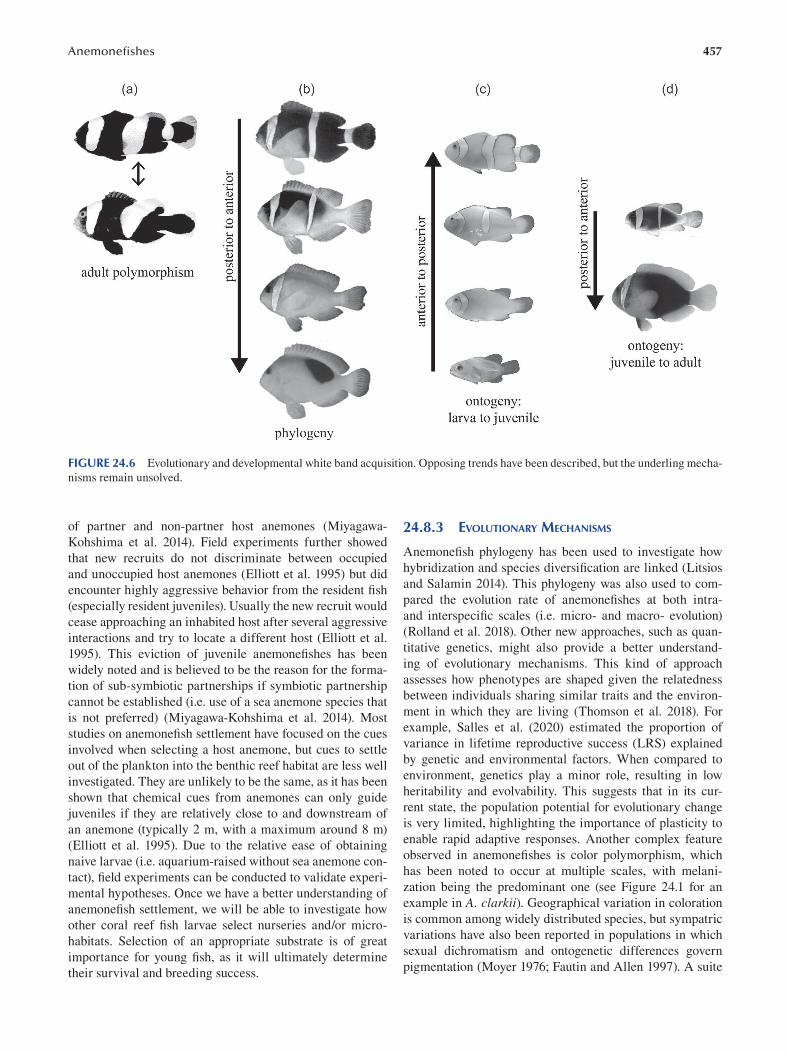
457 Anemonefi shes
FIGURE 24.6 Evolutionary and developmental white band acquisition. Opposing trends have been described, but the underling mecha
nisms remain unsolved.
of partner and non-partner host anemones (Miyagawa-
Kohshima et al. 2014). Field experiments further showed
that new recruits do not discriminate between occupied
and unoccupied host anemones (Elliott et al. 1995) but did
encounter highly aggressive behavior from the resident fi sh
(especially resident juveniles). Usually the new recruit would
cease approaching an inhabited host after several aggressive
interactions and try to locate a different host (Elliott et al.
1995). This eviction of juvenile anemonefishes has been
widely noted and is believed to be the reason for the forma
tion of sub-symbiotic partnerships if symbiotic partnership
cannot be established (i.e. use of a sea anemone species that
is not preferred) (Miyagawa-Kohshima et al. 2014). Most
studies on anemonefish settlement have focused on the cues
involved when selecting a host anemone, but cues to settle
out of the plankton into the benthic reef habitat are less well
investigated. They are unlikely to be the same, as it has been
shown that chemical cues from anemones can only guide
juveniles if they are relatively close to and downstream of
an anemone (typically 2 m, with a maximum around 8 m)
(Elliott et al. 1995). Due to the relative ease of obtaining
naive larvae (i.e. aquarium-raised without sea anemone con
tact), field experiments can be conducted to validate experi
mental hypotheses. Once we have a better understanding of
anemonefish settlement, we will be able to investigate how
other coral reef fish larvae select nurseries and/or micro
habitats. Selection of an appropriate substrate is of great
importance for young fish, as it will ultimately determine
their survival and breeding success.
24.8.3 EVOLUTIONARY MECHANISMS
Anemonefish phylogeny has been used to investigate how
hybridization and species diversification are linked (Litsios
and Salamin 2014). This phylogeny was also used to com
pared the evolution rate of anemonefishes at both intra-
and interspecific scales (i.e. micro- and macro- evolution)
(Rolland et al. 2018). Other new approaches, such as quan
titative genetics, might also provide a better understand
ing of evolutionary mechanisms. This kind of approach
assesses how phenotypes are shaped given the relatedness
between individuals sharing similar traits and the environ
ment in which they are living (Thomson et al. 2018). For
example, Salles et al. (2020) estimated the proportion of
variance in lifetime reproductive success (LRS) explained
by genetic and environmental factors. When compared to
environment, genetics play a minor role, resulting in low
heritability and evolvability. This suggests that in its cur
rent state, the population potential for evolutionary change
is very limited, highlighting the importance of plasticity to
enable rapid adaptive responses. Another complex feature
observed in anemonefishes is color polymorphism, which
has been noted to occur at multiple scales, with melani
zation being the predominant one (see Figure 24.1 for an
example in A. clarkii). Geographical variation in coloration
is common among widely distributed species, but sympatric
variations have also been reported in populations in which
sexual dichromatism and ontogenetic differences govern
pigmentation (Moyer 1976; Fautin and Allen 1997 ). A suite
458
of interacting and conditional ecological factors encompass
ing social rank, host anemone species and location had been
identified as the primary factors predicting distribution of
melanistic morphs (Militz et al. 2016). However, phyloge
netic studies on melanistic A. clarkii showed that specimens
cluster by color rather than geographical origin: a melanis
tic specimen from Bali is more closely related to another
melanistic individual originating from Papua New Guinea
than to a syntopic orange A. clarkii (Litsios et al. 2014a).
Another common polymorphic feature of anemonefi sh color
pattern is the variation of band number, regularly observed
in A. clarkii, A. melanopus and A. plolymnus ( Figure 24.6a ).
This suggests complex mechanisms might be involved in
anemonefish polymorphism. Salis and colleagues (2018b)
mapped the occurrence and number of bands on the phy
logeny to reconstruct the ancestral state and could show that
the diversification of anemonefish color pattern results from
successive caudal to rostral losses of bands during evolution
(Figure 24.6b). This is in contrast with the developmental
acquisition of bands, which appear in an anterior to posterior
gradient (Figure 24.6c). Interestingly, juveniles of some spe
cies have supplementary bands that disappear later caudo
rostrally (Figure 24.6d). The reduction of band number
during ontogeny matches the sequence of band loss during
evolution, demonstrating that diversification in color pattern
among anemonefish lineages resulted from changes in devel
opmental processes. The functional aspect of anemonefi sh
skin color and pattern remains unclear. However, it has been
suggested that color patterns may (i) be used in advertising
social rank (Fautin and Allen 1997; Militz et al. 2016 ), (ii)
signal individual identity (Fricke 1973; Buston 2003a), (iii)
provide disruptive coloration (Salis et al. 2018b) and (iv) be
used for species recognition (Salis et al. 2018b; Salis et al.
2019b). Yet developmental mechanisms underlying the color
pattern formation have still not been identified. However, a
Turing-like model (that patterns zebrafish or angelfi sh, for
example) cannot explain the appearance and/or disappear
ance of bands during ontogeny, thus suggesting that band
formation is controlled by specific patterning mechanisms
that remain to be analyzed. The dorsal fin might act as a spa
tial reference, since its size and geometry have been signifi
cantly correlated with the number of white bands (Salis et al.
2018b). Given the increase in interdisciplinary studies, con
siderable improvement in the understanding of evolutionary
mechanisms should be expected in the coming years.
24.8.4 BIOMEDICAL RESEARCH
Anemonefishes are a promising model system for biomedi
cal research, even though studies in this field are limited
so far. On one hand, they have a relatively long life span
and, on the other hand, their ability to avoid nematocyst dis
charge is rare among vertebrates. Anemonefishes are one of
a few species that offer the opportunity to study longevity
and aging. Indeed, they have a long life expectancy, which is
approximately six times longer than that predicted for other
small fish (Buston and García 2007; Sahm et al. 2019), and
Emerging Marine Model Organisms
they reproduce monthly all year around. Using anemonefi sh,
a recent study (Sahm et al. 2019) suggested that the mito
nuclear balance (i.e. balance between expression of nuclear
and mitochondrially encoded mitochondrial proteins) plays
a key role in aging, which opens the gate to explore those
genetic pathways involved.
Although many studies have attempted to unveil how
anemonefishes avoid the negative effects of nematocyst sting
ing, there are still many open questions and various com
peting hypotheses (see Section 24.3). Indeed, a fi eld study
with several species of anemonefish showed that new naive
recruits (around 20 dph) are able to enter their host anemo
nes without being harmed on the first attempt (Elliott et al.
1995). Occasionally, the new recruits adhered to the tentacle
but usually could break free and, after a short acclimation
process, could enter unharmed. From a biomedical stand
point, it is of great interest, as understanding how anemone-
fishes avoid being stung by the hosts’ nematocysts might lay
a foundation for possible prevention and therapy of negative
human interactions with jellyfish, for example. Additionally
and rather unexpectedly, the anemonefish queuing system
has been used to serve as the basis of a novel brain tumor
segmentation algorithm (Mc and Subramanian 2016).
24.8.5 MISSING FUNCTIONAL APPROACHES
Casas et al. (2016 ) performed the fi rst de novo transcriptome
analysis of wild A. bicinctus and highlighted the rapid and
complex genomic responses of the brain during sex change,
which is subsequently transmitted to the gonads. This tran
scriptomic data (Casas et al. 2016; Yang et al. 2019) will
broaden our understanding not only of the physiological
mechanisms involved but also of the perception and process
ing of external cues into a coordinated response that char
acterizes sex change (Lamm et al. 2015; Liu et al. 2017).
Advances in molecular endocrinology, genomic and tran
scriptomic data in anemonefishes will allow opening new
avenues in our understanding of sex change and sex deter
mination in fishes and more widely in vertebrates. Moreover,
extensive efforts have been put in by several research groups
to establish micro-injection (Roux et al. 2020) and associ
ated genome editing, such as CRISPR/Cas9 in anemonefi shes
(Mitchell et al. 2020). This is a much-needed toolkit to gain
functional data and will be applicable to a range of research
areas. Micro-injection is possible, yet mortality rates are still
high, and obtaining larvae remains difficult (Mitchell et al.
2020; Roux et al. 2020). However, once established, the pos
sibility of modifying specific genetic aspects will advance the
field of anemonefish research, as well as research on coral reef
fish, immensely. Although there are several pet shop mutants
available with diverse color patterns, the underlying muta
tions and exact mechanisms have not been studied in detail.
24.9 CONCLUSION
This chapter summarizes the past and most recent research
finding as well as future perspectives, revealing the great
459 Anemonefi shes
potential anemonefishes offer as emerging marine fi sh mod
els. Future research on anemonefishes will complement
studies on traditional model organisms in a wide variety of
biological areas, from pigmentation to neurobiology. Their
unique biological attributes open perspectives to tackle new
questions related to aging, sexual differentiation, symbiosis,
growth or even social organization. Anemonefi shes have
and will always remain prominent models for ecological
studies, but now those can be linked with lab based evo
devo approaches, which is hardly possible with other model
organisms. As there is a lack of convenient experimental
models for marine fi shes, we hope and strongly believe that
this model will find its place in the vast array of new models
available for the biologists of tomorrow.
BIBLIOGRAPHY
Abdullah NS, Saad S. 2015. Rapid detection of N-acetylneuraminic
acid from false clownfish using HPLC-FLD for symbiosis to
host sea anemone. Asian Journal of Applied Sciences 3:858–864.
Albright R, Takeshita Y, Koweek DA, Ninokawa A, Wolfe K, Rivlin
T, Nebuchina Y, Young J, Caldeira K. 2018. Carbon dioxide
addition to coral reef waters suppresses net community calci
fi cation. Nature 555:516–519.
Allen GR. 1974. The anemonefi sh: Their classification and biology.
Second edition. T.F.H. Publications Inc., Neptune City, NJ.
Allen GR, Drew J, Fenner D. 2010. Amphiprion pacificus, a new
species of anemonefish (Pomacentridae) from Fiji, Tonga,
Samoa, and Wallis Island . Aqua, International Journal of Ichthyology 16:10.
Allen GR, Drew J, Kaufman L. 2008. Amphiprion barberi, a new
species of anemonefish (Pomacentridae) from Fiji, Tonga,
and Samoa. Aqua, International Journal of Ichthyology 14:10.
Altieri AH, Harrison SB, Seemann J, Collin R, Diaz RJ, Knowlton
N. 2017. Tropical dead zones and mass mortalities on coral
reefs. Proceedings of the National Academy of Sciences 114:3660–3665.
Anil MK, Santhosh B, Prasad BO, George RM. 2012. Broodstock
development and breeding of black-finned anemone fi sh
Amphiprion nigripes Regan, 1908 under captive conditions.
Indian Journal of Fisheries 59:77–82.
Apprill A. 2020. The role of symbioses in the adaptation and stress
responses of marine organisms. Annual Review of Marine Science 12:291–314.
Arvedlund M, Larsen K, Winsor H. 2000. The embryonic devel
opment of the olfactory system in Amphiprion melanopus (Perciformes: Pomacentridae) related to the host imprinting
hypothesis. Journal of the Marine Biological Association of the United Kingdom 80:1103–1109.
Arvedlund M, McCormick M, Fautin D, Bildsøe M. 1999. Host
recognition and possible imprinting in the anemonefi sh
Amphiprion melanopus (Pisces: Pomacentridae). Marine Ecology Progress Series 188:207–218.
Arvedlund M, Nielsen LE. 1996. Do the anemonefi sh Amphiprion ocellaris (Pisces: Pomacentridae) imprint themselves to
their host sea anemone heteractis magnifi ca (Anthozoa:
Actinidae)? Ethology 102:197–211.
Barone AN, Hayes CE, Kerr JJ, Lee RC, Flaherty DB. 2019. Acute
toxicity testing of TiO 2-based vs. oxybenzone-based sun
screens on clownfi sh ( Amphiprion ocellaris). Environmental Science and Pollution Research 26:14513–14520.
Barth P, Berenshtein I, Besson M, Roux N, Parmentier E, Banaigs
B, Lecchini D. 2015. From the ocean to a reef habitat: How
do the larvae of coral reef fi shes find their way home? A state
of art on the latest advances. Vie et milieu 65:91–100.
Beldade R, Blandin A, O’Donnell R, Mills SC. 2017. Cascading
effects of thermally-induced anemone bleaching on associ
ated anemonefish hormonal stress response and reproduc
tion. Nature Communications 8:716.
Berthelot C et al. 2014. The rainbow trout genome provides novel
insights into evolution after whole-genome duplication in
vertebrates. Nature Communications 5:3657.
Bertrand S, Brunet FG, Escriva H, Parmentier G, Laudet V,
Robinson-Rechavi M. 2004. Evolutionary genomics of
nuclear receptors: From twenty-five ancestral genes to
derived endocrine systems. Molecular Biology and Evolution 21:1923–1937.
Berumen ML, Walsh HJ, Raventos N, Planes S, Jones GP,
Starczak V, Thorrold SR. 2010. Otolith geochemistry does
not refl ect dispersal history of clownfi sh larvae. Coral Reefs 29:883–891.
Branconi R, Barbasch TA, Francis RK, Srinivasan M, Jones GP,
Buston PM. 2020. Ecological and social constraints combine
to promote evolution of non-breeding strategies in clownfi sh.
Communications Biology 3:649.
Briggs JC. 1961. The East Pacific Barrier and the distribution of
marine shore fi shes. Evolution 15:545–554.
Buston PM. 2003a. Size and growth modification in clownfi sh.
Nature 424:145–146.
Buston PM. 2003b. Mortality is associated with social rank in the
clown anemonefi sh ( Amphiprion percula). Marine Biology 143:811–815.
Buston, Buston PM. 2004a. Does the presence of non-breeders
enhance the fitness of breeders? An experimental analysis
in the clown anemonefi sh Amphiprion percula. Behavioral Ecology and Sociobiology 57:23–31.
Buston PM. 2004b. Territory inheritance in clownfi sh. Proceedings of the Royal Society of London. Series B: Biological Sciences
271. Available from https://royalsocietypublishing.org/
doi/10.1098/rsbl.2003.0156 (accessed November 8, 2020).
Buston PM, Bogdanowicz SM, Wong A, Harrison RG. 2007. Are
clownfish groups composed of close relatives? An analy
sis of microsatellite DNA variation in Amphiprion percula.
Molecular Ecology 16:3671–3678.
Buston PM, Elith J. 2011. Determinants of reproductive success in
dominant pairs of clownfish: A boosted regression tree analy
sis: Determinants of reproductive success. Journal of Animal Ecology 80:528–538.
Buston PM, García MB. 2007. An extraordinary life span estimate
for the clown anemonefi sh Amphiprion percula. Journal of Fish Biology 70:1710–1719.
Camp EF, Hobbs J-PA, De Brauwer M, Dumbrell AJ, Smith DJ.
2016. Cohabitation promotes high diversity of clownfi shes
in the Coral Triangle. Proceedings of the Royal Society B: Biological Sciences 283:20160277.
Casas L, Saborido-Rey F, Ryu T, Michell C, Ravasi T, Irigoien X.
2016. Sex change in clownfish: Molecular insights from tran
scriptome analysis. Scientifi c Reports 6:35461.
Casas L, Saenz-Agudelo P, Irigoien X. 2018. High-throughput
sequencing and linkage mapping of a clownfi sh genome
provide insights on the distribution of molecular players
involved in sex change. Scientifi c Reports 8:4073.
Chen T-H, Hsieh C-Y, Ko F-C, Cheng J-O. 2018. Effect of the
UV-filter benzophenone-3 on intra-colonial social behav
iors of the false clown anemonefi sh ( Amphiprion ocellaris).
Science of The Total Environment 644:1625–1629.
460
Christ MCJ, Subramanian R. 2016. Clown fish queuing and switch
ing optimization algorithm for brain tumor segmentation.
Biomedical Research 27:5.
Cleveland A, Verde EA, Lee RW. 2011. Nutritional exchange in a
tropical tripartite symbiosis: Direct evidence for the transfer
of nutrients from anemonefish to host anemone and zooxan
thellae. Marine Biology 158:589–602.
Colleye O, Frederich B, Vandewalle P, Casadevall M, Parmentier E.
2009. Agonistic sounds in the skunk clownfi sh Amphiprion akallopisos: Size-related variation in acoustic features.
Journal of Fish Biology 75:908–916.
Collingwood C. 1868. Rambles of a naturalist on the shores and waters of the China Sea. John Murray, London.
Cooper W, Van Hall R, Sweet E, Milewski H, DeLeon Z, Verderber
A, DeLeon A, Galindo D, Lazono O. 2020. Functional
morphogenesis from embryos to adults: Late development
shapes trophic niche in coral reef damselfi shes. Evolution & Development 22:221–240.
Dhaneesh KV, Kumar TTA, Shunmugaraj T. 2009. Embryonic
development of percula clownfi sh, Amphiprion percula (Lacepede, 1802). Middle-East Journal of Scientific Research 4:84–89.
Dhaneesh KV, Nanthini Devi K, Ajith Kumar TT, Balasubramanian
T, Tissera K. 2012. Breeding, embryonic development and
salinity tolerance of skunk clownfi sh Amphiprion akallopisos. Journal of King Saud University-Science 24:201–209.
Dixson DL, Munday PL, Jones GP. 2010. Ocean acidifi cation dis
rupts the innate ability of fish to detect predator olfactory
cues. Ecology Letters 13:68–75.
Dohna TA, Timm J, Hamid L, Kochzius M. 2015. Limited con
nectivity and a phylogeographic break characterize popu
lations of the pink anemonefi sh, Amphiprion perideraion,
in the Indo-Malay Archipelago: Inferences from a mito
chondrial and microsatellite loci. Ecology and Evolution 5:1717–1733.
Elliott JK, Elliott JM, Mariscal RN. 1995. Host selection, location,
and association behaviors of anemonefishes in fi eld settle
ment experiments. Marine Biology 122:377–389.
Elliott JK, Lougheed SC, Bateman B, McPhee LK, Boag PT. 1999.
Molecular phylogenetic evidence for the evolution of special
ization in anemonefi shes. Proceedings of the Royal Society of London. Series B: Biological Sciences 266:677–685.
Elliott JK, Mariscal RN. 1997. Acclimation or innate protection of
anemonefishes from sea anemones? Copeia:284–289.
Elliott JK, Mariscal RN. 2001. Coexistence of nine anemonefi sh
species: Differential host and habitat utilization, size and
recruitment. Marine Biology 138:23–36.
Fakan EP, McCormick MI. 2019. Boat noise affects the early
life history of two damselfi shes. Marine Pollution Bulletin 141:493–500.
Fautin DG. 1988. Sea anemones of Madang Province. Science in New Guinea 14:22–29.
Fautin DG. 1991. The anemonefish symbiosis: What is known and
what is not? Symbiosis 10:23, 46.
Fautin DG, Allen GR. 1992. Field guide to anemonefishes and their
host sea anemones. Western Australian Museum . Available
from https://books.google.fr/books?id=WRLGjwEACAAJ .
Fautin DG, Allen GR. 1997. Anemone fishes and their host sea anemones: A guide for aquarists and divers. Rev. edition.
Western Australian Museum, Perth, WA.
Frédérich B, Parmentier E, editors. 2016. Biology of damselfi shes.
CRC Press, Taylor & Francis Group, Boca Raton.
Fricke HW. 1973. Individual partner recognition in fi sh: Field
studies on Amphiprion bicinctus. Die Naturwissenschaften 60:204–205.
Emerging Marine Model Organisms
Fricke HW, Fricke S. 1977. Monogamy and sex change by aggres
sive dominance in coral reef fi sh. Nature 266:830–832.
Futuyma DJ, Moreno G. 1988. The evolution of ecological spe
cialization. Annual Review of Ecology and Systematics 19:207–233.
Gainsford A, Jones GP, Gardner MG, van Herwerden L. 2020.
Characterisation and cross-amplification of 42 microsatellite
markers in two Amphiprion species (Pomacentridae) and a
natural hybrid anemonefish to inform genetic structure within
a hybrid zone. Molecular Biology Reports 47:1521–1525.
Geffroy B, Douhard M. 2019. The adaptive sex in stressful environ
ments. Trends in Ecology & Evolution 34:628–640.
Gemmell NJ, Todd EV, Goikoetxea A, Ortega-Recalde O, Hore TA.
2019. Natural sex change in fish. Pages 71–117 in Current topics in developmental biology book series: Sex determination in vertebrates. Elsevier, New Zealand.
Ghosh J, Wilson RW, Kudoh T. 2009. Normal development of the
tomato clownfi sh Amphiprion frenatus: Live imaging and in situ hybridization analyses of mesodermal and neurectoder
mal development. Journal of Fish Biology 75:2287–2298.
Glasauer SMK, Neuhauss SCF. 2014. Whole-genome duplica
tion in teleost fishes and its evolutionary consequences.
Molelucar Genetics and Genomics 289:1045–1060.
Godwin JR. 1994. Behavioural aspects of protandrous sex change
in the anemonefi sh, Amphiprion melanopus, and endocrine
correlates. Animal Behaviour 48:551–567.
Godwin JR, Crews D, Warner RR. 1996. Behavioural sex change
in the absence of gonads in a coral reef fi sh. Proceedings of the Royal Society of London. Series B: Biological Sciences
263:1683–1688.
Godwin JR, Thomas P. 1993. Sex change and steroid profi les
in the protandrous anemonefi sh Amphiprion melanopus (Pomacentridae, teleostei). General and Comparative Endocrinology 91:144–157.
Goikoetxea A, Todd EV, Gemmell NJ. 2017. Stress and sex:
Does cortisol mediate sex change in fi sh? Reproduction 154:R149–R160.
Green BS. 2004. Embryogenesis and oxygen consumption in benthic
egg clutches of a tropical clownfi sh, Amphiprion melanopus (Pomacentridae). Comparative Biochemistry and Physiology Part A: Molecular & Integrative Physiology 138:33–38.
Gunasekaran K, Sarvanakumar A, Selvam D, Mahesh R. 2017.
Embryonic and larval developmental stages of sebae clown
fi sh Amphiprion sebae (Bleeker 1853) in captive condition.
Indian Journal of Marine Sciences 46:8.
Hayashi K, Reimer JD. 2020. Five-year study on the bleaching
of anemonefish-hosting anemones (Cnidaria: Anthozoa:
Actiniaria) in subtropical Okinawajima Island. Regional Studies in Marine Science 35:101240.
Hayashi K, Tachihara K, Reimer JD. 2018. Patterns of coexistence
of six anemonefish species around subtropical Okinawa-jima
Island, Japan. Coral Reefs 37:1027–1038.
Hayashi K, Tachihara K, Reimer JD. 2019a. Species and sexual differ
ences in human-oriented behavior of anemonefish at Okinawa
Island, Japan. Marine Ecology Progress Series 616:219–224.
Hayashi K, Tachihara K, Reimer JD. 2019b. Low density popula
tions of anemonefish with low replenishment rates on a reef
edge with anthropogenic impacts. Environmental Biology of Fishes 102:41–54.
Hayashi K, Tachihara K, Reimer JD. 2020. Loss of natural coastline
influences species diversity of anemonefish and host anem
ones in the Ryukyu Archipelago. Aquatic Conservation: Marine and Freshwater Ecosystems . doi:10.1002/aqc.3435.
He S, Planes S, Sinclair-Taylor TH, Berumen ML. 2019.
Diagnostic nuclear markers for hybrid Nemos in Kimbe Bay,
461 Anemonefi shes
PNG-Amphiprion chrysopterus x Amphiprion sandaracinos hybrids. Marine Biodiversity 49:1261–1269.
Herbert NA, Bröhl S, Springer K, Kunzmann A. 2017. Clownfi sh in
hypoxic anemones replenish host O2 at only localised scales.
Scientifi c Reports 7:6547.
Holmberg RJ et al. 2019. Ocean acidification alters morphology of
all otolith types in Clark’s anemonefi sh ( Amphiprion clarkii). Peer Journal of Life and Environment 7:e6152.
Holzer G, Besson M, Lambert A, Barth P, Gillet B, Hughes S,
Leulier F, Viriot L, Lecchini D, Laudet V. 2017. Fish larval
recruitment to reefs is a thyroid hormone-mediated metamor
phosis sensitive to the pesticide chlorpyrifos. Elife e27595.
Hotaling S, Kelley JL. 2019. The rising tide of high-quality genomic
resources. Molecular Ecology Resources 19:567–569.
Hughes TP et al. 2018. Global warming transforms coral reef
assemblages. Nature 556:492–496.
Irion U, Nüsslein-Volhard C. 2019. The identification of genes
involved in the evolution of color patterns in fi sh. Current Opinion in Genetics & Development 57:31–38.
Iwata E, Mikami K, Manbo J, Moriya-Ito K, Sasaki H. 2012. Social
interaction influences blood cortisol values and brain aro
matase genes in the protandrous false clown anemonefi sh,
Amphiprion ocellaris. Zoological Science 29:849–855.
Jaillon O et al. 2004. Genome duplication in the teleost fi sh
Tetraodon nigroviridis reveals the early vertebrate proto
karyotype. Nature 431:946–957.
Jones GP, Planes S, Thorrold SR. 2005. Coral reef fish larvae settle
close to home. Current Biology 15:1314–1318.
Kavanagh KD, Alford RA. 2003. Sensory and skeletal development
and growth in relation to the duration of the embryonic and
larval stages in damselfi shes (Pomacentridae): Development
and growth in damselfi shes. Biological Journal of the Linnean Society 80:187–206.
Kawaguchi M, Takahashi H, Takehana Y, Naruse K, Nishida M,
Yasumasu S. 2013. Sub-functionalization of duplicated genes
in the evolution of nine-spined stickleback hatching enzyme:
Sub-functionalization of hatching enzyme. Journal of Experimental Zoology Part B: Molecular and Developmental Evolution 320:140–150.
Kawakami Y, Nozaki J, Seoka M, Kumai H, Ohta H. 2008.
Characterization of thyroid hormones and thyroid hormone
receptors during the early development of Pacifi c blue
fin tuna ( Thunnus orientalis). General and Comparative Endocrinology 155:597–606.
Kirkwood T. 1977. Evolution of aging. Nature 270:301–304.
Kobayashi Y, Horiguchi R, Miura S, Nakamura M. 2010. Sex- and
tissue-specific expression of P450 aromatase (cyp19a1a) in
the yellowtail clownfi sh, Amphiprion clarkii. Comparative Biochemistry and Physiology Part A: Molecular & Integrative Physiology 155:237–244.
Kobayashi Y, Nagahama Y, Nakamura M. 2013. Diversity and
plasticity of sex determination and differentiation in fi shes.
Sexual Development 7:115–125.
Krishna R. 2018. Larval development and growth of red saddleback
anemonefi sh, Amphiprion ephippium (Bloch, 1790) under
captive conditions. Indian Journal of Geo-Marine Sciences 47:2421–2428.
Lamm MS, Liu H, Gemmell NJ, Godwin JR. 2015. The need for
speed: Neuroendocrine regulation of socially-controlled sex
change. Integrative and Comparative Biology 55:307–322.
Laudet V. 2011. The origins and evolution of vertebrate metamor
phosis. Current Biology 21:R726–R737.
Lehmann R et al. 2019. Finding Nemo’s genes: A chromosome-scale
reference assembly of the genome of the orange clownfi sh
Amphiprion percula. Molecular Ecology Resources 19:570–585.
Leis JM. 1991. The pelagic stage of reef fishes: The larval biology
of coral reef fishes. Pages 183–230 in Sale PF, editor. The ecology of fi shes on coral reefs. Academic Press, San Diego.
Leis JM, Siebeck U, Dixson DL. 2011. How Nemo fi nds home: The
neuroecology of dispersal and of population connectivity in
larvae of marine fi shes. Integrative and Comparative Biology 51:826–843.
Li J-T, Hou G-Y, Kong X-F, Li C-Y, Zeng J-M, Li H-D, Xiao G-B,
Li X-M, Sun X-W. 2015. The fate of recent duplicated genes
following a fourth-round whole genome duplication in a
tetraploid fish, common carp ( Cyprinus carpio). Scientifi c Reports 5:8199.
Liew HJ, Ambak MA, Abol-Munafi AB, Chuah TS. 2006.
Embryonic development of clownfi sh Amphiprion ocellaris under laboratory conditions. Journal of Sustainability Science and Management 1:64–73.
Litsios G, Pearman PB, Lanterbecq D, Tolou N, Salamin N. 2014b.
The radiation of the clownfi shes has two geographical repli
cates. Journal of Biogeography 41:2140–2149.
Litsios G, Salamin N. 2014. Hybridisation and diversifi cation in
the adaptive radiation of clownfi shes. BMC Evolutionary Biology 14:245.
Litsios G, Sims CA, Wüest RO, Pearman PB, Zimmermann NE,
Salamin N. 2012a. Mutualism with sea anemones triggered
the adaptive radiation of clownfi shes. BMC Evolutionary Biology 12:212.
Litsios G, Sims CA, Wüest RO, Pearman PB, Zimmermann NE,
Salamin N. 2012b. Mutualism with sea anemones triggered
the adaptive radiation of clownfi shes. BMC Evolutionary Biology 12:1.
Liu H, Todd EV, Lokman PM, Lamm MS, Godwin JR, Gemmell NJ.
2017. Sexual plasticity: A fishy tale. Molecular Reproduction and Development 84:171–194.
Lubbock R. 1981. The clownfi sh/anemone symbiosis: A problem
of cell recognition. Parasitology 82(159):173.
Lubbock R, Smith DC. 1980. Why are clownfishes not stung by
sea anemones? Proceedings of the Royal Society of London.
Series B: Biological Sciences 207:35–61. Royal Society.
Madeira C, Madeira D, Diniz MS, Cabral HN, Vinagre C. 2016.
Thermal acclimation in clownfish: An integrated biomarker
response and multi-tissue experimental approach. Ecological Indicators 71:280–292.
Madhu R, Madhu K, Retheesh T. 2012. Life history pathways in
false clown Amphiprion ocellaris Cuvier, 1830: A journey
from egg to adult under captive condition. Journal of Marine Biological Association of India 54:77–90.
Marcionetti A, Rossier V, Bertrand JAM, Litsios G, Salamin N. 2018.
First draft genome of an iconic clownfish species ( Amphiprion frenatus). Molecular Ecology Resources 18:1092–1101.
Marcionetti A, Rossier V, Roux N, Salis P, Laudet V, Salamin N.
2019. Insights into the genomics of clownfish adaptive radia
tion: Genetic basis of the mutualism with sea anemones.
Genome Biology and Evolution 11:869–882.
Mariscal RN. 1970. The nature of the symbiosis between Indo-Pacifi c
anemone fishes and sea anemones. Marine Biology 6:58–65.
Maytin AK, Davies SW, Smith GE, Mullen SP, Buston PM. 2018.
De novo transcriptome assembly of the clown anemonefi sh
(Amphiprion percula): A new resource to study the evolution
of fish color. Frontiers in Marine Science 5:284.
Mebs D. 2009. Chemical biology of the mutualistic relationships of
sea anemones with fish and crustaceans. Toxicon 54:1071–1074.
Medawar P. 1952. An unsolved problem of biology: Printed lecture.
University College London, London.
Meyer A, Schartl M. 1999. Gene and genome duplications in
vertebrates: The one-to-four (-to-eight in fish) rule and the
462
evolution of novel gene functions. Current Opinion in Cell Biology 11:699–704.
Militz TA, McCormick MI, Schoeman DS, Kinch J, Southgate
PC. 2016. Frequency and distribution of melanistic morphs
in coexisting population of nine clownfish species in Papua
New Guinea. Marine Biology 163:200–210.
Mills SC, Mourier J, Galzin R. 2010. Plasma cortisol and 11-ketotes
tosterone enzyme immunoassay (EIA) kit validation for three
fish species: The orange clownfi sh Amphiprion percula , the
orangefi n anemonefi sh Amphiprion chrysopterus and the
blacktip reef shark Carcharhinus melanopterus. Journal of Fish Biology 77:769–777.
Mitchell J. 2005. Queue selection and switching by false clown
anemonefi sh, Amphiprion ocellaris. Animal Behaviour 69:643–652.
Mitchell LJ, Tettamanti V, Marshall JN, Cheney KL, Cortesi F.
2020. CRISPR/Cas9-mediated generation of biallelic G0
anemonefi sh (Amphiprion ocellaris) mutants. preprint.
Molecular Biology. Available from http://biorxiv.org/lookup/
doi/10.1101/2020.10.07.330746 (accessed November 15,
2020).
Miura S, Kobayashi Y, Bhandari RK, Nakamura M. 2013. Estrogen
favors the differentiation of ovarian tissues in the ambisexual
gonads of anemonefi sh Amphiprion clarkii: The role of estro
gen for gonad in anemonefi sh. Journal of Experimental Zoology Part A: Ecological Genetics and Physiology 319:560–568.
Miyagawa-Kohshima K et al. 2014. Embryonic learning of chemi
cal cues via the parents’ host in anemonefi sh ( Amphiprion ocellaris). Journal of Experimental Marine Biology and Ecology 457:160–172.
Moyer JT. 1976. Geographical variation and social dominance in
Japanese populations of the anemonefi sh Amphiprion clarkii. Japane Journal of Ichthyology 23:12–22.
Moyer JT. 1980. Influence of temperate waters on the behavior of
the tropical anemonefi sh Amphiprion clarkii at Miyake-jima,
Japan. Bulletin of Marine Science:261–272.
Munday PL, Buston P, Warner R. 2006. Diversity and fl exibility
of sex-change strategies in animals. Trends in Ecology & Evolution 21:89–95.
Munday PL, Jones GP, Pratchett MS, Williams AJ. 2008. Climate
change and the future for coral reef fi shes. Fish and Fisheries 9:261–285.
Murata M, Miyagawa-Kohshima K, Nakanishi K, Naya Y. 1986.
Characterization of compounds that induce symbiosis between
sea anemone and anemone fi sh. Science 234:585–587.
Nanninga GB, Côté IM, Beldade R, Mills SC. 2017. Behavioural
acclimation to cameras and observers in coral reef fi shes.
Ethology 123:705–711.
Nanninga GB, Saenz-Agudelo P, Zhan P, Hoteit I, Berumen ML.
2015. Not finding Nemo: Limited reef-scale retention in a
coral reef fi sh. Coral Reefs 34:383–392.
Nedosyko AM, Young JE, Edwards JW, Burke da Silva K. 2014.
Searching for a toxic key to unlock the mystery of anemone-
fish and anemone symbiosis. PLoS One 9:e98449.
Nelson JS, Grande T, Wilson MVH. 2016. Fishes of the world.
Fifth edition. John Wiley & Sons, Hoboken, NJ.
Nguyen H-TT, Dang BT, Glenner H, Geffen AJ. 2020. Cophylogenetic
analysis of the relationship between anemonefi sh Amphiprion (Perciformes: Pomacentridae) and their symbiotic host
anemones (Anthozoa: Actiniaria). Marine Biology Research 16:117–133.
Nieuwenhuys R, ten Donkelaar HJ, Nicholson C. 1998. The central nervous system of vertebrates. Springer Berlin Heidelberg,
Berlin, Heidelberg. Available from http://link.springer.com/
10.1007/978-3-642-18262-4 (accessed November 13, 2020).
Emerging Marine Model Organisms
Norin T, Mills SC, Crespel A, Cortese D, Killen SS, Beldade R.
2018. Anemone bleaching increases the metabolic demands
of symbiont anemonefi sh. Proceedings of the Royal Society B: Biological Sciences 285:20180282.
Ochi H. 1985. Temporal patterns of breeding and larval settlement in a
temperate population of the tropical anemonefi sh, Amphiprion clarkii. Japanese Journal of Ichthyology 32:248–257.
Ohno S. 1970. Evolution by gene duplication. Springer Berlin
Heidelberg, Berlin, Heidelberg.
Olivotto I, Geffroy B. 2017. Clownfish. Pages 177–199 in Calado
R, Olivotto I, Oliver MP, Holt GJ, editors. Marine ornamental species aquaculture. First edition. Wiley Online Books,
Chichester, West Sussex, UK.
Ollerton J, McCollin D, Fautin DG, Allen GR. 2007. Finding
NEMO: Nestedness engendered by mutualistic organization
in anemonefish and their hosts. Proceedings of the Royal Society B: Biological Sciences 274:591–598.
Onimaru K, Kuraku S. 2018. Inference of the ancestral vertebrate
phenotype through vestiges of the whole-genome duplica
tions. Briefi ngs in Functional Genomics 17:352–361.
Ortega-Recalde O, Goikoetxea A, Hore TA, Todd EV, Gemmell
NJ. 2020. The genetics and epigenetics of sex change in fi sh.
Annual Review of Animal Biosciences 8:47–69.
Paris CB, Atema J, Irisson J-O, Kingsford M, Gerlach G, Guigand
CM. 2013. Reef odor: A wake up call for navigation in reef
fish larvae. PLoS One 8:e72808.
Park MS, Shin HS, Kil G-S, Lee J, Choi CY. 2011. Monitoring of
Na+/K+-ATPase mRNA expression in the cinnamon clown
fi sh, Amphiprion melanopus, exposed to an osmotic stress
environment: Profiles on the effects of exogenous hormone.
Ichthyological Research 58:195–201.
Parmentier E, Colleye O, Fine ML, Frederich B, Vandewalle
P, Herrel A. 2007. Sound production in the clownfi sh
Amphiprion clarkii. Science 316:1006.
Parmentier E, Colleye O, Mann D. 2009. Hearing ability in three clown-
fish species. Journal of Experimental Biology 212:2023–2026.
Parmentier E, Lagardère JP, Vandewalle P, Fine ML. 2005.
Geographical variation in sound production in the anem
onefi sh Amphiprion akallopisos. Proceedings of the Royal Society B: Biological Sciences 272:1697–1703.
Patkaew S, Direkbusarakom S, Tantithakura O. 2014. A simple
method for cell culture of ‘Nemo’ ocellaris clownfi sh ( Amphiprion ocellaris, Cuvier 1830). Cell Biology International Reports 7.
Patterson LB, Parichy DM. 2019. Zebrafish pigment pattern for
mation: Insights into the development and evolution of adult
form. Annual Review of Genetics 53:505–530.
Planes S, Jones GP, Thorrold SR. 2009. Larval dispersal connects fi sh
populations in a network of marine protected areas. Proceedings of the National Academy of Sciences 106:5693–5697.
Porter SN, Humphries MS, Buah-Kwofie A, Schleyer MH. 2018.
Accumulation of organochlorine pesticides in reef organ
isms from marginal coral reefs in South Africa and links with
coastal groundwater. Marine Pollution Bulletin 137:295–305.
Pratte ZA, Patin NV, McWhirt ME, Caughman AM, Parris DJ,
Stewart FJ. 2018. Association with a sea anemone alters the
skin microbiome of clownfi sh. Coral Reefs 37:1119–1125.
Putra DF, Abol-MunafiAB, Muchlisin ZA, Chen J-C. 2012. Preliminary
studies on morphology and digestive tract development of
tomato clownfi sh, Amphiprion frenatus under captive condition.
International Journal of the Bioflux Society AACL Biofl ux 5:8.
Randall JE. 1955. Fishes of the Gilbert Islands. Atoll Research Bulletin 47:1–243.
Randall JE, Fautin D. 2002. Fishes other than anemonefi shes that
associate with sea anemones. Coral Reefs 21:188–190.
Anemonefi shes 463
Rattanayuv akorn S, Mungkornkarn P, Thongpan A, Chatchavalvanich habitat and weak genetic effects shape the lifetime reproduc-
K. 2005. Embryonic development of saddleback anemone- tive success in a wild clownfi sh population. Ecology Lettersfi sh, Amphiprion polymnus , Linnaeus (1758). Natural Science 23:265–273.
39:455–463. Salles OC, Maynard JA, Joannides M, Barbu CM, Saenz-Agudelo
Rhyne AL, Tlusty MF, Schofi eld PJ, Kaufman L, Morris JA, P, Almany GR, Berumen ML, Thorrold SR, Jones GP, Planes
Bruckner AW. 2012. Revealing the appetite of the marine S. 2015. Coral reef fi sh populations can persist without
aquarium fi sh trade: The volume and biodiversity of fi sh immigration. Proceedings of the Royal Society B: Biological imported into the United States. PLoS One 7:e35808. Sciences 282:20151311.
Robertson DR, Gro ve JS, McCosker JE. 2004. Tropical transpacifi c Salles OC, Pujol B, Maynard JA, Almany GR, Berumen ML, Jones
shore fi shes. Pacifi c Science 58:507–565. GP, Saenz-Agudelo P, Srinivasan M, Thorrold SR, Planes
Rodrigues FSLM, Yang X, Nikaido M, Liu Q, Kelsh RN. 2012. A S. 2016. First genealogy for a wild marine fi sh population
simple, highly visual in vivo screen for anaplastic lymphoma reveals multigenerational philopatry. Proceedings of the kinase inhibitors. ACS Chemical Biology 7:1968–1974. National Academy of Sciences 113:13245–13250.
Rolland J, Silvestro D, Litsios G, Faye L, Salamin N. 2018. Clownfi shes Santini S, Polacco G. 2006. Finding Nemo: Molecular phylogen y
evolution below and above the species level. Proceedings of the and evolution of the unusual life style of anemonefi sh. GeneRoyal Society B: Biological Sciences 285:20171796. 385:19–27.
Roux N, Lami R, Salis P , Magré K, Romans P, Masanet P, Lecchini Schlichter D. 1968. Das Zusammenleben v on Riffanemonen und
D, Laudet V. 2019a. Sea anemone and clownfi sh microbiota Anemonenfi schen. Zeitschrift für Tierpsychologie 25:933–954.
diversity and variation during the initial steps of symbiosis. Scott A, Dixson DL. 2016. Reef fi shes can recognize bleached hab-
Scientifi c Reports 9:19491. itat during settlement: Sea anemone bleaching alters anem-
Roux N, Salis P, Lambert A, Logeux V, Soulat O, Romans P, Frédérich onefi sh host selection. Proceedings of the Royal Society B: B, Lecchini D, Laudet V. 2019b. Staging and normal table of Biological Sciences 283:20152694.
postembryonic development of the clownfi sh ( Amphiprion Scott MW. 2008. Damsefi shes and anemonefi shes: The complete ocellaris ). Developmental Dynamics 248:545–568. illustrated guide to their identifi cation, behaviors and cap-
Roux N, Salis P , Lee S-H, Besseau L, Laudet V. 2020. Anemonefi sh, tive care. T. F. H. Publications, Neptune City.
a model for eco-evo-devo. EvoDevo 11:20. Se ymour J, Barbasch T, Buston P. 2018. Lunar cycles of repro-
Roux N, Logeux V, Trouillard N., Pillot R Magré K, Salis P, duction in the clown anemonefi sh Amphiprion percula :
Lecchini D, Besseau L, Laudet V, Romans P. 2021. A star Individual-level strategies and population-level patterns.
is born again: Methods for larval rearing of an emerging Marine Ecology Progress Series 594:193–201.
model organism, the False clownfi sh Amphiprion ocellaris. Shabana NMA, Helal AM. 2006. Reproduction in captivity, brood-
Journal of Experimental Zoology Part B: Molecular and stock rearing and embryology of the anemone fi sh Amphiprion
Developmental Evolution, Jun: 336(4): 376–85. bicintus inhabiting the Red Sea. Egyptian Journal of Aquatic Rue ger T, Barbasch TA, Wong MYL, Srinivasan M, Jones GP, Research 32:438–446.
Buston PM. 2018. Reproductive control via the threat of Shuman CS, Hodgson G, Ambrose RF. 2005. Population impacts
eviction in the clown anemonefi sh. Pr oceedings of the Royal of collecting sea anemones and anemonefi sh for the marine
Society B: Biological Sciences 285:20181295. aquarium trade in the Philippines. Coral Reefs 24:564–573.
Saenz-Agudelo P, Jones GP, Thorrold SR, Planes S. 2011. Simpson SD, Munday PL, Wittenrich ML, Manassa R, Dixson DL,
Detrimental effects of host anemone bleaching on anemone- Gagliano M, Yan HY. 2011. Ocean acidifi cation erodes crucial
fi sh populations. Coral Reefs 30:497–506. auditory behaviour in a marine fi sh. Biology Letters 7:917–920.
Sahm A, Almaida-Pagán P, Bens M, Mutalipassi M, Lucas-Sánchez A, Southe y BR, Rodriguez-Zas SL, Rhodes JS, Sweedler JV.
de Costa Ruiz J, Görlach M, Cellerino A. 2019. Analysis of the 2020. Characterization of the prohormone complement in
coding sequences of clownfi sh reveals molecular convergence Amphiprion and related fi sh species integrating genome and
in the evolution of lifespan. BMC Evolutionary Biology 19:89. transcriptome assemblies. PLoS One 15:e0228562.
Salis P et al. 2019a. De velopmental and comparative transcriptomic Stieb SM, de Busserolles F , Carleton KL, Cortesi F, Chung W-S, Dalton
identifi cation of iridophore contribution to white barring in BE, Hammond LA, Marshall NJ. 2019. A detailed investigation
clownfi sh. Pigment Cell & Melanoma Research 32:391–402. of the visual system and visual ecology of the Barrier Reef anem-
Salis P , Lorin T, Laudet V, Frédérich B. 2019b. Magic traits in onefi sh, Amphiprion akindynos . Scientifi c Reports 9:16459.
magic fi sh: Understanding color pattern evolution using reef Supiwong W, Tanomtong A, Pinthong K, Kaewmad P, Poungnak
fi sh. Trends in Genetics 35:265–278. P, Jangsuwan N. 2015. The fi rst chromosomal characteris-
Salis P , Roux N, Lecchini D, Laudet V. 2018a. The post-embryonic tics of nucleolar organizer regions and karyological analysis
development of Amphiprion perideraion reveals a decoupling of pink anemonefi sh, Amphiprion perideraion (Perciformes,
between morphological and pigmentation change. Société Amphiprioninae). Cytologia 80:271–278.
Française d’Ichtyologie . Available from http://sfi -cybium. Tan MH, Austin CM, Hammer MP, Lee YP, Croft LJ, Gan HM.
fr/fr/post-embryonic-development-amphiprion-perideraion- 2018. Finding Nemo: Hybrid assembly with Oxford Nano-
reveals-decoupling-between-morphological-and (accessed pore and Illumina reads greatly improves the clownfi sh
November 13, 2020). ( Am phi prion ocellaris ) genome assembly. GigaScience 7.
Salis P, Roux N, Soulat O, Lecchini D, Laudet V, Frédérich B. Available from https://academic.oup.com/gigascience/article/
2018b. Ontogenetic and phylogenetic simplifi cation during doi/10.1093/gigascience/gix137/4803946 (accessed Septem-
white stripe evolution in clownfi shes. BMC Biology 16(1):90. ber 25, 2020).
Salis P, Lee SH, Roux N, Lecchini D, Laudet V. 2021. The real Thomas D, Prakashand C, Gopakumar G. 2015. Spa wning behav-
Nemo movie: Description of embryonic development iour and embryonic development in the sebae anemone-
in Amphiprion ocellaris from fi rst division to hatching. fi sh Amphiprion sebae (Bleek er, 1853). Indian Journal of Developmental Dynamics, May 7. Fisheries 62:58–65.
Salles OC, Almany GR, Berumen ML, Jones GP, Saenz-Agudelo P, Thomson CE, Winney IS, Salles OC, Pujol B. 2018. A guide to
Srinivasan M, Thorrold SR, Pujol B, Planes S. 2020. Strong using a multiple-matrix animal model to disentangle genetic
464
and nongenetic causes of phenotypic variance. PLoS One 13:e0197720.
Titus BM et al. 2019. Phylogenetic relationships among the clown-
fish-hosting sea anemones. Molecular Phylogenetics and Evolution 139:106526.
Todd EV, Liu H, Muncaster S, Gemmell NJ. 2016. Bending gen
ders: The biology of natural sex change in fi sh. Sexual Development 10:223–241.
Veilleux HD, Van Herwerden L, Cole NJ, Don EK, De Santis C,
Dixson DL, Wenger AS, Munday PL. 2013. Otx2 expression
and implications for olfactory imprinting in the anemonefi sh,
Amphiprion percula. Biology Open 2:907–915.
Victor B, Wellington G. 2000. Endemism and the pelagic larval
duration of reef fishes in the eastern Pacific Ocean. Marine Ecology Progress Series 205:241–248.
Emerging Marine Model Organisms
Williams GC. 1957. Pleiotropy, natural selection, and the evolution
of senescence. Evolution 11:398–411.
Yang W, Lin B, Li G, Chen H, Liu M. 2019. Sequencing and
transcriptome analysis for reproduction-related genes iden
tification and SSRs discovery in sequential hermaphrodite
Amphiprion ocellaris. Turkish Journal of Fisheries and Aquatic Sciences 19. Available from www.trjfas.org/pdf/
issue_19_12/1207.pdf (accessed November 11, 2020).
Yasir I, Qin JG. 2007. Embryology and early ontogeny of an
anemonefi sh Amphiprion ocellaris. Journal of the Marine Biological Association of the United Kingdom 87:1025–1033.
Yu PB, Hong CC, Sachidanandan C, Babitt JL, Deng DY, Hoyng
SA, Lin HY, Bloch KD, Peterson RT. 2008. Dorsomorphin
inhibits BMP signals required for embryogenesis and iron
metabolism. Nature Chemical Biology 4:33–41.
Index
Note: Page numbers in italic indicate a fi gure
and page numbers in bold indicate a table on
the corresponding page.
A
Abeoforma whisleri, 58–59, 69 Acropora
anatomy, 181, 182–184, 183 challenging questions, 186–189
development, 181 embryogenesis, 179–182, 180 functional approaches, 185–186
genomics, 184–185
geographical occurrence, 176–177, 177 history and taxonomic status, 173–176
life cycle, 177–179, 178 morphologies, 175
adaptive strategies
to light, 7
aging
Botryllus schlosseri , 394–395
Stylophora pistillata , 206
algal sugars
and Z. galactanivorans , 10–12
algal surfaces
Z. galactanivorans as a model to study
bacterial colonization of, 12
alkane degradation, 12–13
allorecognition assays
Stylophora pistillata , 204
anatomy
Acropora , 182–184
anemonefi shes, 452
Botryllus schlosseri , 390–392
Cassiopea xamachana , 156–158
cephalochordates, 347
Chondrichthyes , 426–428
Clytia hemisphaerica, 130–131, 135–137
crustaceans, 278–279
Cycliophora, 265–267
Ectocarpus sp, 32–33
hagfi sh, 409–411
lamprey, 409
Nematostella vectensis , 110–112
Oscarella lobularis , 85–88
Parhyale hawaiensis , 295–296
Placozoa, 103–104
Platynereis dumerilii , 238–242
Porifera, 71–72
Saccharina latissimi , 37–39
sea urchin, 321–322
solitary ascidians, 369–371
Symsagittifera roscoffensis , 221–227
Stylophora pistillata , 196–197
Temnopleurus reevesii , 337
anemonefi shes, 444 anatomy, 452, 452 challenging questions, 455–458
development, 449–452, 450 functional approaches, 453–455
genomic data, 452–453
geographical location and phylogeny,
444–445, 445
history of the model, 443–444
life cycle, 446–449, 447 white band acquisition, 457
annelids, see Platynereis dumerilii anthropogenic impacts, studies on
Stylophora pistillata , 202–203
antibiotic activity
Chondrichthyes , 431–432
antibodies
Chondrichthyes , 432
anti-cancer activity
Chondrichthyes , 432–433
appendages, paired
cyclostomes, 415
aquaculture
brown algae, 41
architecture of cells and tissues
Symsagittifera roscoffensis , 221–223
ascidians, see solitary ascidians
asexual reproduction
Cassiopea xamachana , 156
Oscarella lobularis , 82
assisted settlement
coral reefs, 188
asymmetries, skeleton and brain
Chondrichthyes , 433–435
Asymmetron lucayanum genomic data, 348
audition
Chondrichthyes , 428
axial patterning
Nematostella vectensis, 115, 121–122
axis formation
Chondrichthyes , 426
B
bacteria
diversity in seawater, 2
see also marine bacteria
bacterial colonization
Z. galactanivorans as a model, 12
bacterial cues
Clytia hemisphaerica , 142
bacterial degradation
Z. galactanivorans as a model for, 9–12
bacterial models
bacterial model organism toolkit, 14–20
early, 2
the future of gene editing in, 17–18
see also marine bacterial models; and specifi c models
behavior, regulation of
Clytia hemisphaerica , 142–143
behavior, study of
Chondrichthyes , 422
behavioral approaches
Nematostella vectensis , 116–117
bilaterian traits
Nematostella vectensis , 120–122
phylogeny, 121 bioactive secondary metabolites
Oscarella lobularis , 92–93
biochemistry analysis
sea urchin egg abundance and synchronous
early embryonic development,
309–310
biofi lm formation
M. hydrocarbonoclasticus as a model, 12–14
biofueling
crustaceans, 280–281
biogeography
Chondrichthyes , 423
see also geography
biological models
brown algae, 41
biology, see experimental biology
bioluminescence, 3–4
biomedical applications
anemonefi shes, 458
Chondrichthyes , 431–433
biomineralization
Stylophora pistillata , 205–206
biophysical techniques
Ectocarpus sp, 33–34
biosynthesis, 13
blood formation
anemonefi shes, 450–451
body elongation and segmentation
crustaceans, 281
bone-like tissue
Chondrichthyes , 433
bones, evolution of, 350
Botryllus schlosseri, 386 anatomy, 390–392, 392 challenging questions, 395
embryogenesis, 389–390
functional approaches, 393–395
genomic data, 392–393
geographical location, 386–387, 387 history of the model, 385–386
life cycle, 387–389, 389 brain asymmetries
Chondrichthyes , 433–435
Branchiostoma belcheri genomic data, 347–348
Branchiostoma floridae genomic data, 347
Branchiostoma lanceolatum genomic data, 348
brown algae, 27–29
challenging questions, 41–42
Ectocarpus sp, 29–34, 28 Saccharina latissimi, 34–41, 28
budding
Oscarella lobularis , 82
C
calcium signalling
sea urchin’s contribution to our
understanding of the role played
by, 309
Capsaspora owczarzaki , 55–56
cardiac development
solitary ascidians, 368
care
Nematostella vectensis , 116
465
466 Index
cartilage, evolution of, 350
Cassiopea xamachana, 157 anatomy, 156–158
challenging questions, 164–165
embryogenesis, 154–156, 155functional approaches, 160–164
genomic data, 158–160
geographical location, 150–152, 151history of the model, 149–150
life cycle, 152–154, 153spawning, injection and settlement, 162 symbiosis-driven development, 165
cell apoptosis
Oscarella lobularis , 91–92
cell biology
Ectocarpus sp, 33–34
sea urchin egg abundance and synchronous
early embryonic development,
309–310
cell–cell signaling pathways, evolution of, 351
cell components, tracking
Platynereis dumerilii , 247–249
cell culture
anemonefi shes, 454–455
cell lines
Chondrichthyes , 430–431
cell proliferation
Oscarella lobularis , 91–92
cells, architecture of
Symsagittifera roscoffensis , 221–223
cells, evolution of
neural crest cells, 350–351
cell staining
Oscarella lobularis , 92
cell-to-cell communication system, 4, 351
cell tracking
Oscarella lobularis , 92
Platynereis dumerilii , 247–249
cell types, novel
Nematostella vectensis , 122–123
cellular analysis
anemonefi shes, 453–455
Botryllus schlosseri , 393–395
Cassiopea xamachana , 160–164
cephalochordates, 348–349
Chondrichthyes , 430–431
Clytia hemisphaerica , 138–140
crustaceans, 279 –280
Cycliophora, 267–268
cyclostomes, 413–414
echinoderms, 323–325
Ectocarpus sp, 33–34
Oscarella lobularis , 89–92
Parhyale hawaiensis , 298–300
Placozoa, 104–105
Platynereis dumerilii , 246–250
Porifera, 73
Saccharina latissimi , 40–41
solitary ascidians, 373–375
Stylophora pistillata , 202–205
Temnopleurus reevesii, 337–338
cellular basis of development
Parhyale hawaiensis , 301–302
cellular basis of regeneration
Parhyale hawaiensis , 302
cellular processes
Nematostella vectensis , 118
Platynereis dumerilii , 247–249
cellular response
Clytia hemisphaerica , 140
cell viability
Oscarella lobularis , 91–92
central nervous system
Symsagittifera roscoffensis , 223–225
cephalization
anemonefi shes, 449
cephalochordates
anatomy, 347
challenging questions, 349–352
classifi cation, 342 cleavage stage, 344embryogenesis, 344–347, 345functional approaches, 348–349
genomic data, 347–348
geographical location, 342–343
history of the model, 341–342
life cycle, 343–344
morphology, 346 neurulation, 345
cerebellar evolution
cyclostomes, 414–415
chemoreception
Chondrichthyes , 428
chimerism
Botryllus schlosseri , 393–394
choanoderm
Oscarella lobularis , 87
choanofl agellata, 51–53
Chondrichthyes, 420, 421anatomy and sensory biology, 426–429
biogeography, 423
challenging questions, 431–435
embryogenesis, 424–426, 425genomic data, 429–430, 430life cycles, 423–424
past and present, 421–423
phylogeny, 420–421, 420tools for molecular and cellular analyses,
430–431
chordate genome
cephalochordates, 349–350
chromosome elimination
cyclostomes, 411–412
cinctoblastula larva
Oscarella lobularis , 85
circulatory systems
Platynereis dumerilii , 241
classifi cation
history of the classification of lampreys and
hagfi sh, 404
cleavage
anemonefi shes, 449
Oscarella lobularis , 84–85
Parhyale hawaiensis , 292–293
climate change, studies on
Stylophora pistillata , 202–203
Clytia hemisphaerica anatomy of the polyps and jellyfi sh, 135–137
challenging questions, 140–143
embryogenesis and planula larva
formation, 133–135, 134functional approaches, 138–140, 139genomic data, 137–138
geographical location, 132
history of the model, 130–132
life cycle, 132–133, 133 morphology, 136 regeneration, 141
cnidarians
Clytia hemisphaerica , 135–137
cnidarian–algal symbiosis, see Cassiopea xamachana
homeobox genes in, 160Nematostella vectensis as a cnidarian
model for, 122
phylogeny, 121 coeloblastula
Oscarella lobularis , 84–85
colonial species, see Botryllus schlosseri; Stylophora pistillata
colony formation, growth and survivorship
Stylophora pistillata , 199–200
color pattern
anemonefishes, 451–452, 457 computable embryos
solitary ascidians, 376–379
conservation
anemonefi shes, 455–456
Chondrichthyes , 422–423
coral
most-studied, 174see also Acropora; Stylophora pistillata
Corallochytrea/Pluriformea, 60–63
Corallochytrium limacisporum, 62–63, 69 coral reefs
restoration of, 188–189, 205
Creolimax fragrantissima, 59–60, 69 CRISPR-Cas9, 17–18
cyclostomes, 414
solitary ascidians, 375
unicellular relatives of animals, 53
see also gene editing
crustaceans
anatomy, 278–279
biofouling, 281challenging questions, 280–282
embryogenesis, 275
functional approaches, 279–280
genomic data, 279, 279, 298geographical location, 273–274
history of the model, 271–273
larval development, 275–278, 276–277life cycle, 274–275
morphology, 272 phylogeny, 273 resting egg shape, 282see also Parhyale hawaiensis
cryptobiosis, evolution of, 282
culture, 14–16
Cassiopea xamachana , 161–163
Nematostella vectensis , 116
Saccharina latissimi , 40
Cycliophora
anatomy, 265–267, 267challenging questions, 268
embryogenesis, 261–265
feeding stages, 260, 263–266functional approaches, 267–268
genomic data, 267
geographical location, 259–261
history of the model, 259
life cycle, 260, 261, 262 cyclostomes, 404
anatomy, 409–411, 409–410challenging questions, 414–415
embryogenesis, 407–409, 408evolutionary research of vertebrates,
403–404
functional approaches, 413–414, 414genomic data, 411–413, 412–413
Index 467
geographical location, 405–406
history of the model, 404–405
life cycle, 406–407
phylogeny, 405
D
databases
solitary ascidians, 372–373
dermal denticles
Chondrichthyes , 433–434
descriptive approaches
Chondrichthyes , 431
Platynereis dumerilii , 246–249
development
advantages of lamprey developmental
research, 413–414
anemonefi shes, 449–452
cardiac development in solitary ascidians,
368
early development of Ectocarpus sp, 29–32
early studies on Clytia hemisphaerica,
130–131
indirect development of Oscarellalobularis , 82–84
later development of Ectocarpus sp, 32–33
molecular and cellular basis of Parhyale hawaiensis , 301–302
molecular control of development of
Oscarella lobularis , 85
morphological evolution and Parhyale hawaiensis , 300–301
Nematostella vectensis , 113–115
neural development in solitary ascidians,
367–368
see also cleavage; embryogenesis;
gastrulation; larval development
developmental biology, 27–29
challenging questions in basic and applied
research, 41–42
Chondrichthyes in, Chondrichthyes,
423–424
Ectocarpus sp as experimental model for,
29–34
Saccharina latissimi as experimental
model for, 34–41
solitary ascidians, 376–379
digestive syncytium
Symsagittifera roscoffensis , 226
digestive systems
Platynereis dumerilii , 242
diversifi cation
Saccharina latissimi , 34
drug application
anemonefi shes, 454
cyclostomes, 414
E
echinoderms
anatomy of the adult sea urchin, 321–322
breeding season and egg diameter, 315 challenging questions, 325
classifi cation, 313 functional approaches, 323–325, 324 genomic data, 322–323, 323 geographical location, 313–314, 314 historical contributions of sea urchin
gametes and embryos, 307–311
omics, availability of, 317
phylogeny, 311–313
sea urchin embryogenesis, 316–321, 318 sea urchin life cycle, 314–316, 316 taxonomy, 312 see also Temnopleurus reevesii
ecological approaches
Nematostella vectensis , 116–117
ecotoxicology
crustaceans, 281
Ectocarpus sp, 29–34, 41, 30–31, 42 egg deposition
Symsagittifera roscoffensis , 221
eggs
Clytia hemisphaerica , 132
optical transparency of sea urchin eggs,
307–309
Platynereis dumerilii , 242
sea urchin and biochemistry and cell
biology analyses, 309–310
elasmobranch properties of kidney
regeneration
Chondrichthyes , 435
electroporation
Nematostella vectensis , 118
solitary ascidians, 373–374
electroreception
Chondrichthyes , 429
embryogenesis
Acropora , 179–182
anemonefi shes, 449–451
Botryllus schlosseri , 389–390
Cassiopea xamachana , 154–156
cephalochordates, 344–347
Chondrichthyes , 424–426
Clytia hemisphaerica, 130, 133–135, 138
crustaceans, 275
Cycliophora, 261–265
Ectocarpus sp, 29–32
hagfi sh, 407–409
lamprey, 407
Nematostella vectensis, 113–115
Oscarella lobularis , 84–85
Parhyale hawaiensis , 292–295
Placozoa, 103
Platynereis dumerilii, 242–243, 251–252
Porifera, 69–71
Saccharina latissimi , 37
sea urchin, 309–311, 316–321
solitary ascidians, 364–369
Stylophora pistillata , 200–202
Symsagittifera roscoffensis , 227–229
Temnopleurus reevesii , 336–337
embryology, experimental
Clytia hemisphaerica , 130
embryonic axis, establishment of
solitary ascidians, 365
embryos
historical contributions of sea urchin
embryos, 307–311
endogenous molecules for biomedical
applications
Chondrichthyes , 431–433
endoskeleton
Chondrichthyes , 433
environmental cues
Clytia hemisphaerica , 142–143
enzymes
and Zobellia galactanivorans , 10–12
epidermis
Symsagittifera roscoffensis , 223
epigenetic modifi cations
Platynereis dumerilii , 251–252
evolution
assisted, 189
cerebellar, 414–415
cyclostomes for evolutionary research of
vertebrates, 403–404
Nematostella vectensis , 120–122
paired nostrils, 415
Saccharina latissimi , 34
thyroid gland, 415
evolutionary mechanisms
anemonefi shes, 456–458
excretory systems
Platynereis dumerilii , 241–242
exoskeleton
Chondrichthyes , 433–434
experimental biology, 2–3, 20
the bacterial model organism toolkit,
14–20
Chondrichthyes as models in, 421–422
examples of marine bacterial models, 3–14
eyes, evolution of, 351
F
feeding availability
Clytia hemisphaerica , 142–143
fertilization
optical transparency of sea urchin eggs,
307–309
Platynereis dumerilii , 242–243
sea urchin and calcium signalling, 309
solitary ascidians, 364–365
fi lasterea, 53–56
fi n development
anemonefi shes, 451
fi rst cleavages
Platynereis dumerilii, 243, 244 sea urchins, 318 see also cleavage; embryogenesis
fl uorescent immunolocalization
Oscarella lobularis , 91
food
and crustaceans, 280
fossil vertebrates
cyclostomes, 404–405
fragmentation
Oscarella lobularis , 82
functional approaches
Acropora , 185–186
anemonefi shes, 453–455
Botryllus schlosseri , 393–395
Cassiopea xamachana , 160–164
cephalochordates, 348–349
Chondrichthyes , 430–431
Clytia hemisphaerica , 138–140
crustaceans, 279 –280
Cycliophora, 267–268
cyclostomes, 413–414
echinoderms, 323–325
Ectocarpus sp, 33–34
Nematostella vectensis , 116–120
Oscarella lobularis , 89–92
Parhyale hawaiensis , 298–300
Placozoa, 104–105
Platynereis dumerilii , 249–250
Porifera, 73
Saccharina latissimi , 40–41
solitary ascidians, 373–375
468 Index
Stylophora pistillata , 202–205
Temnopleurus reevesii , 337–338
G
gametes
historical contributions of sea urchin
gametes, 307–311
gametogenesis
Clytia hemisphaerica , 142
Oscarella lobularis , 82–84
gametophytic phase
of Ectocarpus sp, 32
gastrulation
anemonefi shes, 449
Chondrichthyes , 424–426
Parhyale hawaiensis , 293
gene, reporter
solitary ascidians, 374
gene disruption
Nematostella vectensis , 118–119
gene editing
bacterial models, 17–18
cyclostomes, 414
Salp ingoeca rosetta , 53
gene expression, modifi cation of
Ectocarpus sp, 34
Saccharina latissimi , 40–41
gene expression, visualizing
Nematostella vectensis , 119–120
gene family studies
Chondrichthyes , 430
gene function analysis
Clytia hemisphaerica , 138–140
gene knock-down
Platynereis dumerilii , 249
genetic markers
anemonefi shes, 455
genetics and genetic approaches
genetic manipulation of marine bacteria,
16–17
M. hydrocarbonoclasticus , 13
Nematostella vectensis , 118–120
solitary ascidians, 375
Stylophora pistillata , 204–205
genome editing
Platynereis dumerilii , 249–250
genome-level approaches
Nematostella vectensis , 120
genomic regulation, evolution of
cephalochordates, 349–350
genomics
Acropora , 184–185
anemonefi shes, 452–453
Botryllus schlosseri , 392–393
Cassiopea xamachana , 158–160
cephalochordates, 347–348
Chondrichthyes , 429–430
Clytia hemisphaerica , 137–138
crustaceans, 279
Cycliophora, 267
cyclostomes, 411–413
echinoderms, 322–323
Ectocarpus sp, 33
M. hydrocarbonoclasticus , 13
Nematostella vectensis , 115–116
Oscarella lobularis , 88–89
Parhyale hawaiensis , 296–298
Placozoa, 104
Platynereis dumerilii, 245–246, 249–250
Porifera, 72–73
Saccharina latissimi , 39–40
solitary ascidians, 371
Stylophora pistillata , 202
Symsagittifera roscoffensis , 230–231
Temnopleurus reevesii , 337
geography
Acropora , 176–177
anemonefi shes, 444–446
Botryllus schlosseri , 386–387
Cassiopea xamachana , 150–152
cephalochordates, 342–343
Clytia hemisphaerica , 132
crustaceans, 273–274
Cycliophora, 259–261
echinoderms, 313–314
Ectocarpus sp, 29
hagfi sh, 406
lampreys, 405–406
Nematostella vectensis , 109–110
Oscarella lobularis , 81–82
Parhyale hawaiensis , 291
Platynereis dumerilii , 236–238
Porifera, 68–69
Placozoa, 101–102
Saccharina latissimi , 34–35
solitary ascidians, 360–362
Stylophora pistillata , 296
Symsagittifera roscoffensis , 219
Temnopleurus reevesii , 335–336
germ band extension and segmentation
Parhyale hawaiensis , 293–294
germ cells
Clytia hemisphaerica , 131
solitary ascidians, 368–369
germ disc formation
Parhyale hawaiensis , 293
germ layers
Oscarella lobularis , 93–94
solitary ascidians, 365–367
glandular system
Symsagittifera roscoffensis , 225–226
H
habitat
Nematostella vectensis , 109–110
hagfi sh, 404 anatomy, 409–411
embryogenesis, 407–409
geographical location, 406
history of the model, 404
life cycle, 406–407
see also cyclostomes
hatching
anemonefi shes, 451
solitary ascidians, 362
homoscleromorph sponge, see Oscarella lobularis
host recognition
anemonefi shes, 456
host–symbiont interactions
Oscarella lobularis , 93
hox clusters
cyclostomes, 412–413
human impacts
anemonefi shes, 455–456
Cassiopea xamachana , 151–152
husbandry
anemonefi shes, 453–454
hybridization
Acropora , 186–187
anemonefi shes, 454
Oscarella lobularis , 91
Platynereis dumerilii , 246–247
I
i-cells
Clytia hemisphaerica , 140–142
ichthyosporeans, 56–60
immune system, evolution of, 351–352
immunoassay
anemonefi shes, 454
immunochemistry
Saccharina latissimi , 40
immunohistochemistry
Platynereis dumerilii , 247
inbred strains/lines, lack of
Botryllus schlosseri , 395
insects, origin of, 281–282
in situ hybridization
anemonefi shes, 454
Oscarella lobularis , 91
Platynereis dumerilii , 246–247
invasion
Cassiopea xamachana , 151–152
in vitro invertebrate cultures
Botryllus schlosseri , 395
iron acquisition
M. hydrocarbonoclasticus as a model,
12–14
J
jellyfi sh, see Cassiopea xamachana; Clytia hemisphaerica
K
kidney regeneration, elasmobranch
properties of
Chondrichthyes , 435
knowledge
acquiring knowledge on model strains,
18–20
advancing knowledge on developmental
mechanisms of brown algae, 41
L
laboratory cultivation
Botryllus schlosseri , 395
Cassiopea xamachana , 161–163
Ectocarpus sp, 33
lampreys, 404 advantages of developmental research,
413–414
anatomy, 409
embryogenesis, 407
geographical location, 405–406
history, 404
life cycle, 406
see also cyclostomes
larva
Clytia hemisphaerica, 132, 133–135
coral, 187
Platynereis dumerilii , 243
solitary ascidians, 369
Stylophora pistillata , 203–204
larval development
anemonefi shes, 451–452
crustaceans, 275–278
Index 469
Platynereis dumerilii, 243–245, 251–252
sea urchin, 310–311
larval metamorphosis
coral, 187
Oscarella lobularis , 85
larval tail muscle formation
solitary ascidians, 367
life cycle
Acropora , 177–179
anemonefi shes, 446–449
Botryllus schlosseri , 387–389
Cassiopea xamachana , 152–154
cephalochordates, 343–344
Chondrichthyes , 423–424
Clytia hemisphaerica , 132–133
crustaceans, 274–275
Cycliophora, 261
Ectocarpus sp, 29
hagfi sh, 406–407
lampreys, 406
Nematostella vectensis , 112–113
Oscarella lobularis , 82–84
Parhyale hawaiensis , 291–292
Placozoa, 102–103
Platynereis dumerilii , 238
Porifera, 69
Saccharina latissimi , 35–37
sea urchin, 314–316
solitary ascidians, 362–364
Stylophora pistillata , 197–200
Symsagittifera roscoffensis , 219–221
Temnopleurus reevesii , 336
light
adaptive strategies to, 7
lipid biodegradation
M. hydrocarbonoclasticus as a model,
12–14
loss-of-function approaches
Oscarella lobularis , 92
solitary ascidians, 374–375
M
macroalgal biomass
Zobellia galactanivorans as a model for
bacterial degradation of, 9–12
magnetoreception
Chondrichthyes , 428–429
marine bacteria
genetic manipulation of, 16–17
marine bacterial models, 2–3, 20
bacterial model organism toolkit, 14–20
examples of, 3–14
Marinobacter hydrocarbonoclasticus,
12–14
picocyanobacteria, 6–9
Vibrio fischeri , 3–6
Zobellia galactanivorans , 9–12
need for new models, 2–3
marine environments
bioluminescence mechanisms in, 3–4
marine jellyfish model, see Clytia hemisphaerica
marine organisms
bioluminescence mechanisms in, 3–4
Marinobacter hydrocarbonoclasticus,
12–14
maternal determinants
solitary ascidians, 364–365
mechanical cues
Clytia hemisphaerica , 142
mechanosensory system
Chondrichthyes , 428
medusa, Clytia hemisphaerica feeding availability, 142–143
regeneration, 130–131
swimming, 132
meiosis
of Ectocarpus sp, 32
mesoderm
Nematostella vectensis , 121
mesohyl
Oscarella lobularis , 87–88
metabolism
Stylophora pistillata , 200
metamorphosis
Acropora and other corals, 187
solitary ascidians, 362–364
Stylophora pistillata , 199
metatrochophore larva
Platynereis dumerilii , 245
metazoan epithelia
Oscarella lobularis , 93
methods
Nematostella vectensis , 116–120
microinjection
Cassiopea xamachana , 163–164
Nematostella vectensis , 118
solitary ascidians, 373–374
molecular analysis
anemonefi shes, 453–455
Botryllus schlosseri , 393–395
Cassiopea xamachana, 160–164
cephalochordates, 348–349
Chondrichthyes , 430–431
Clytia hemisphaerica , 138–140
crustaceans, 279 –280
Cycliophora, 267–268
cyclostomes, 413–414
echinoderms, 323–325
Ectocarpus sp, 33–34
Oscarella lobularis , 89–92
Parhyale hawaiensis , 298–300
Placozoa, 104–105
Platynereis dumerilii , 246–250
Porifera, 73
Saccharina latissimi , 40–41
solitary ascidians, 373–375
Stylophora pistillata , 202–205
Temnopleurus reevesii , 337–338
molecular basis of development
Parhyale hawaiensis , 301–302
molecular basis of regeneration
Parhyale hawaiensis , 302
molecular control of development
Oscarella lobularis , 85
molecular mechanisms
Clytia hemisphaerica , 142
molecular research tools, lack of
Botryllus schlosseri , 395
morphogenesis
Oscarella lobularis, 85, 89
morpholino antisense oligomers
cyclostomes, 414
morpholinos (MOs)
solitary ascidians, 374
morphological evolution, developmental
basis of
Parhyale hawaiensis , 300–301
morphological traits, evolution of
cartilage and bones, 350
eyes, 351
neural crest cells, 350–351
Parhyale hawaiensis , 300–301
morphologies, genomic basis of
Acropora and other corals, 187
mRNA, detection of
Platynereis dumerilii , 246–247
muscle system
Symsagittifera roscoffensis , 223
musculature
Platynereis dumerilii , 241
mutagenesis
solitary ascidians, 375
mutants
Cassiopea xamachana , 163–164
Clytia hemisphaerica , 138–140
N
nectochaete larva
Platynereis dumerilii , 245
Nematostella vectensis, 109 anatomy, 110–112, 111 challenging questions, 120–123
developmental stages, 114 embryogenesis, 113–115
genomic data, 115–116
geography and habitat, 109–110, 108 history of the model, 107–109
life history, 112–113
methods and functional approaches,
116–120, 117, 119 phylogeny, 121reproduction and regeneration, 113
neoblasts
Symsagittifera roscoffensis , 226–227
nervous systems
Platynereis dumerilii , 241
Symsagittifera roscoffensis , 223–225
neural crest cells, evolution of, 350–351
neural development
solitary ascidians, 367–368
neural plate patterning
solitary ascidians, 367
nomenclature
Saccharina latissimi , 34
nostrils, paired
cyclostomes, 415
notochord
anemonefi shes, 451
solitary ascidians, 368
nubbins, planting of
coral reefs, 188–189
nutrition
Clytia hemisphaerica , 142
nutritive surface
biofilm formation on, 12–13
O
“omics”-level approaches
Nematostella vectensis , 120
ontogeny, evolution of
crustaceans, 281
oocyte maturation
Clytia hemisphaerica , 138
ooplasmic segregation
solitary ascidians, 365
optical transparency of sea urchin eggs, 307–309
organogenesis
anemonefi shes, 451
Parhyale hawaiensis , 294–295
470 Index
Oscarella lobularis, 80, 90 anatomy, 85–88
challenging questions both in academic
and applied research, 92–94
developmental stages, 83 embryogenesis, 84–85
functional approaches, 89–92, 91 geographical location, 81–82
history of the model, 79–81
life cycle, 82–84
metamorphosis, 86 phylogeny, 81 transcription and genomic data, 88–89
P
paired appendages, origin of
cyclostomes, 415
paired nostrils, evolution of
cyclostomes, 415
parasitic forms
crustaceans, 282
parenchyma
Symsagittifera roscoffensis , 226
Parhyale hawaiensis anatomy, 295–296
appendage diversity, 297 challenging questions, 300–302
embryogenesis, 292–295, 292, 294 functional approaches, 298–300, 299,
301 genomic data, 296–298, 298, 299 geographical location, 291
history of the model, 289–291
laboratory experimental model, 291 life cycle, 291–292
phylogeny, 290 pelvic spine
anemonefi shes, 451
peptide treatments
Platynereis dumerilii , 249
peripheral nervous system
Symsagittifera roscoffensis , 225
pharmacological manipulation
Nematostella vectensis , 120
pharmacological treatments
Platynereis dumerilii , 249
pharynx segmentation
Chondrichthyes , 426
phenotyping, 18–20
photoreception
Chondrichthyes , 428
photosynthetic adaptations
picocyanobacteria as models, 6–9
phylogeny
anemonefi shes, 444–446
Chondrichthyes , 420–421
echinoderms, 311–313
physiology, regulation of
Clytia hemisphaerica , 142–143
picocyanobacteria, 6–9
pinacoderm
Oscarella lobularis , 87
Placozoa, 101–105, 102–103 planulae
Clytia hemisphaerica, 133–135, 142
Stylophora pistillata , 199
plasmids
Abeoforma whisleri , 58
Capsaspora owczarzaki , 56
Corallochytrium limacisporum , 62–63
Creolimax fragrantissima , 60
Salp ingoeca rosetta , 52–53
Platynereis dumerilii, 237 anatomy, 238–242, 240 challenging questions, 250–252
descriptive approaches, 246–249
embryogenesis, 242–243, 244 functional approaches, 248 , 249–250
genomic data, 245–246, 246 geographical location, 236–238
history of the model, 236
larval development, 243–245, 244 life cycle, 238, 239
polymerase chain reaction
Oscarella lobularis , 89–91
polyps
Clytia hemisphaerica, 132, 135–137,
142–143
population genetics
Stylophora pistillata , 204–205
Porifera, 67–75, 68–72, 74, 81 pre-metamorphic phase
solitary ascidians, 362
primordial germ cells
solitary ascidians, 368–369
Prochlorococcus, 8 adaptation of the photosynthetic
apparatus of, 7
adaptive strategies of, 7
key features of, 6–7
programmed sequence loss
cyclostomes, 411–412
prokaryotic models, 2
proteins
Platynereis dumerilii, 247, 249
proteomics
solitary ascidians, 372
protrochophore larva
Platynereis dumerilii , 243
see also larva
Q
quorum sensing, 4, 5
R
receptor molecules
Acropora , 187
regeneration
Botryllus schlosseri , 394–395
cephalochordates, 352
Chondrichthyes , 435
Clytia hemisphaerica, 130–131, 140–142
Nematostella vectensis, 115, 118, 123
Parhyale hawaiensis , 302
Platynereis dumerilii , 250–252
Symsagittifera roscoffensis , 229–230
reporter gene
solitary ascidians, 374
reproduction
Symsagittifera roscoffensis , 219–221
reproductive characteristics
Stylophora pistillata , 197–199
reproductive organs
Symsagittifera roscoffensis , 219–221
reproductive strategies
Chondrichthyes , 423
research, challenging questions for
Acropora , 186–189
anemonefi shes, 455–458
Botryllus schlosseri , 395
brown algae, 41–42
Cassiopea xamachana , 164–165
cephalochordates, 349–352
Chondrichthyes , 431–435
Clytia hemisphaerica , 140–143
crustaceans, 280–282
Cycliophora, 268
echinoderms, 325
Nematostella vectensis , 120–123
Oscarella lobularis , 92–94
Parhyale hawaiensis , 300–302
Placozoa, 105
Platynereis dumerilii , 250–252
Porifera, 73–75
solitary ascidians, 375
Stylophora pistillata , 205–207
Symsagittifera roscoffensis , 231
Temnopleurus reevesii , 338–339
research, current trends in
Chondrichthyes , 422
RNA interference
Clytia hemisphaerica , 138
solitary ascidians, 374
S
Saccharina latissimi, 34–41, 28, 36, 38 Salp ingoeca rosetta , 51–53
seasonality
Stylophora pistillata , 197–199
sea urchins
anatomy, 321–322
embryogenesis, 316–321
historical contributions of gametes and
embryos, 307–311
life cycle, 314–316
see also echinoderms; Temnopleurus reevesii seawater, 2
selection
Abeoforma whisleri , 58
Corallochytrium limacisporum , 62
Salp ingoeca rosetta , 52
sensory biology
Chondrichthyes , 428–429
sensory receptors
Symsagittifera roscoffensis , 225
settlement
anemonefi shes, 456
coral reefs, 188
Stylophora pistillata, 199, 203–204
sex determination
Clytia hemisphaerica , 131
Ectocarpus sp, 32–33
sexual reproduction
Cassiopea xamachana , 154–156
Oscarella lobularis , 82–84
Stylophora pistillata , 197–199
signaling pathways
Clytia hemisphaerica , 142
skeleton asymmetries, origin of
Chondrichthyes , 433–435
solitary ascidians
anatomy, 369–371, 370 challenging questions, 375–379, 378 databases, 372–373
embryogenesis, 364–369, 365–366 functional approaches, 373–375
genomics, 371
geographical distribution, 360–362
history of the model, 358–360
Index 471
life cycle, 362–364, 362 metamorphosis, 363 phylogeny, 360 proteomics, 372
transcriptomic, 371–372
somite development
anemonefi shes, 449
species problem
Acropora , 186–187
Sphaeroforma arctica , 60
sponge gastrulation
Oscarella lobularis , 93–94
sponges, see Oscarella lobularis ; Porifera
squalamine, different properties of
Chondrichthyes , 432
stem cells
Botryllus schlosseri , 394–395
Symsagittifera roscoffensis , 226–227
stereoblastula larva
Platynereis dumerilii , 243
see also larva
stereogastrula larva
Platynereis dumerilii , 243
see also larva
Stylophora pistillata, 198, 201, 204 anatomy, 196–197
challenging questions both in academic
and applied research,
205–207
embryogenesis, 200–202
functional approaches, 202–205
genomic data, 202
geographical location, 196
history of the model, 195–196
life cycle, 197–200
swimming phase
solitary ascidians, 362
Symbiodinaceae, 187
symbiotic associations
corals and their photosynthetic
dinofl agellate endosymbionts,
187–188
molecular mechanisms of, 4–6
Symsagittifera roscoffensis, 217–219, 218 anatomy, 220, 221–227, 222, 224challenging questions for the future, 231
embryogenesis, 227–229, 228history of the model and geographic
location, 219
life cycle and reproduction, 219–221
preliminary genomic data, 230–231
regeneration, 229–230
reproduction, 220Synechococcus, 8
adaptation of the photosynthetic apparatus
of, 7–9
adaptive strategies, 7
key features, 6–7
T
TALENs
solitary ascidians, 374–375
taxonomy
Acropora , 173–176
Stylophora pistillata , 206
teeth
Chondrichthyes , 433–434
Temnopleurus reevesii, 336 anatomy, 337
challenging questions, 338–339
embryogenesis, 336–337, 337functional approaches, 337–338, 338genomic data, 337
geographical location, 335–336
history of the model, 335
in situ hybridization, 338life cycle, 336
terrestrialization
crustaceans, 281–282
therapeutic advances
solitary ascidians, 376
thyroid gland, evolution of
cyclostomes, 415
tissue, architecture of
Symsagittifera roscoffensis , 221–223
tissue, bone-like
Chondrichthyes , 433
tissue manipulation
Nematostella vectensis , 117–118
transcriptomic data
Chondrichthyes , 429–430
Clytia hemisphaerica , 138
Oscarella lobularis , 88–89
solitary ascidians, 371–372
transduction
Acropora , 187
transfection
Abeoforma whisleri , 58
Capsaspora owczarzaki , 55–56
Corallochytrium limacisporum , 62
Creolimax fragrantissima , 59–60
Salp in goeca rosetta , 52
transgenics
Cassiopea xamachana , 163–164
Nematostella vectensis , 119
solitary ascidians, 375
translation-blocking morpholinos
Platynereis dumerilii , 249
see also morpholinos (MOs)
Trichoplax adhaerens, 102–104 see also Placozoa
trochophore larva
Platynereis dumerilii , 243–245
see also larva
turn-over
anemonefi shes, 449–450
U
ultrastructure protocols
Saccharina latissimi , 40
umbrella organization
Clytia hemisphaerica , 135–137
unfertilized eggs
Platynereis dumerilii , 242
unicellular relatives of animals, 49, 63
availability of genetic tools for, 50 choanofl agellata, 51–53
corallochytrea/pluriformea, 60–63
fi lasterea, 53–56
ichthyosporeans, 56–60
models of the life cycle of, 54 transfection protocols among, 57
upside-down jellyfi sh, see Cassiopea xamachana uses
Saccharina latissimi , 35
V
vertebrate morphological traits, evolution of
cartilage and bones, 350
eyes, 351
neural crest cells, 350–351
vertebrates, evolutionary research of
cyclostomes, 403–404
vertebrates, fossil
cyclostomes, 404–405
Vibrio fischeri, 3–6, 5
W
wax esters, 13
western blots (WBs)
Platynereis dumerilii, 247, 248 whole-genome duplication
cyclostomes, 412–413, 415
whole-mount in situ hybridization
Platynereis dumerilii , 246–247
wild collection
Cassiopea xamachana , 161
worms, see Platynereis dumerilii
Z
ZNFs
solitary ascidians, 374–375
Zobellia galactanivorans, 9–12, 11

Related Documents