Handbook of Critical and Intensive Care Medicine Joseph Varon Fourth Edition 123

Welcome message from author
This document is posted to help you gain knowledge. Please leave a comment to let me know what you think about it! Share it to your friends and learn new things together.
Transcript

Handbook of Critical and Intensive Care MedicineJoseph Varon
Fourth Edition
123

Handbook of Critical and Intensive Care Medicine

Joseph Varon
Handbook of Critical and Intensive Care Medicine
Fourth Edition

ISBN 978-3-030-68269-9 ISBN 978-3-030-68270-5 (eBook)https://doi.org/10.1007/978-3-030-68270-5
© Springer Nature Switzerland AG 2021This work is subject to copyright. All rights are reserved by the Publisher, whether the whole or part of the material is concerned, specifically the rights of transla-tion, reprinting, reuse of illustrations, recitation, broadcasting, reproduction on microfilms or in any other physical way, and transmission or information storage and retrieval, electronic adaptation, computer software, or by similar or dissimi-lar methodology now known or hereafter developed.The use of general descriptive names, registered names, trademarks, service marks, etc. in this publication does not imply, even in the absence of a specific statement, that such names are exempt from the relevant protective laws and regulations and therefore free for general use.The publisher, the authors, and the editors are safe to assume that the advice and information in this book are believed to be true and accurate at the date of pub-lication. Neither the publisher nor the authors or the editors give a warranty, expressed or implied, with respect to the material contained herein or for any errors or omissions that may have been made. The publisher remains neutral with regard to jurisdictional claims in published maps and institutional affiliations.
This Springer imprint is published by the registered company Springer Nature Switzerland AGThe registered company address is: Gewerbestrasse 11, 6330 Cham, Switzerland
Joseph VaronUnited Memorial Medical Center/United GeneralUnited Memorial Medical Center and United General Hospital Chief of Staff and Chief of Critical Care ServicesHouston, TX USA

This book is again dedicated to all the healthcare workers that have fought against COVID-19 this year and to my children Adylle, Jacques, Daryelle, and Michelle for their understanding as adults, about those countless days, nights, and weekends, in which I was away from them caring for those patients who needed me the most at the time.
Joseph Varon, MD, FACP, FCCP FCCM, FRSM

Preface
This year more than ever has been challenging for healthcare providers. The COVID-19 pandemic has shown us that criti-cal care is, by far, one of the most important specialties. So, why write another book on the management of critically ill patients? When I wrote the first edition of this book, over 25 years ago, I had realized the importance of a small pocket book that would be useful for those caring for critically ill patients. Over the past six decades we have seen an enormous growth in the number of intensive care units (ICU) across the world. Indeed, it is estimated that a large proportion of healthcare expenses are devoted to patients in these special-ized units. Medical students, residents, fellows, attending phy-sicians, critical care nurses, pharmacists, respiratory therapists, and other healthcare providers (irrespective of their ultimate field of practice) will spend several months or years of their professional lives taking care of critically ill or severely injured patients. These clinicians must have special training, experience, and competence in managing complex problems in their patients. Moreover, these clinicians must interpret data obtained by many kinds of monitoring devices, and they must integrate this information with their knowledge of the pathophysiology of disease. Even more important is the fact that anyone working in an ICU or with a critically ill patient must approach patients with a multidisciplinary team. The phrase “there is no I in TEAM” comes to mind.
This fourth edition was written for every practitioner engaged in Critical Care Medicine across the world. I have attempted to present basic and generally accepted clinical information, my own personal experience in the field, facts

viii
and some important formulas, as well as laboratory values and tables which we feel will be useful to the practitioner of Critical Care Medicine. The chapters of this book follow an outline format and are divided by organ-system (i.e., neuro-logic disorders, cardiovascular disorders), as well as special topics (i.e., environmental disorders, trauma, toxicology). Every chapter has been updated and many chapters are com-pletely new.
It is important for the reader of this handbook to under-stand that Critical Care Medicine is not a static field and changes occur every day. Therefore, this handbook is not meant to define the standard of care, but rather to be a gen-eral guide to current clinical practice used in Critical Care Medicine. I wrote this book hoping that it will benefit thou-sands of critically ill patients, but more importantly that it will aid practicing clinicians to assume a multidisciplinary approach.
Houston, TX Joseph Varon MD, FACP, FCCP, FCCM, FRSM USA
Preface
Collaborators
The following individuals assisted in the review of this edition of my book. I would like to acknowledge their help and assis-tance in making this manuscript accurate and up-to-date.
Abbas Alshami, MDSteven Douedi, MD
Mustafa AlTaei, MDMohammed Alazzawi, MD
Division of Internal MedicineJersey Shore University Medical
Center, Hackensack Meridian HealthNeptune, NJ, USA
Swapnil Patel, MDDivision of Internal Medicine
Internal Medicine Residency ProgramHackensack Meridian
School of Medicine Jersey Shore University
Medical Center, Hackensack Meridian Health
Neptune, NJ, USA

Contents
1 Approach to the Intensive Care Unit (ICU) . . . . . . . . 1Welcome to the ICU. . . . . . . . . . . . . . . . . . . . . . . . . . . 1
What Is an ICU? . . . . . . . . . . . . . . . . . . . . . . . . . 1Historical Development of the ICU . . . . . . . . . 1Economic Impact of the ICU . . . . . . . . . . . . . . . 2Organization of the ICU . . . . . . . . . . . . . . . . . . . 2
Teamwork . . . . . . . . . . . . . . . . . . . . . . . . . . . . . . . . . . . 3The Flow Sheet . . . . . . . . . . . . . . . . . . . . . . . . . . . . . . . 4The Critically Ill Patient . . . . . . . . . . . . . . . . . . . . . . . . 4System-Oriented Rounds . . . . . . . . . . . . . . . . . . . . . . . 5
Identification . . . . . . . . . . . . . . . . . . . . . . . . . . . . 7Major Events Over the Last 24 h . . . . . . . . . . . . 8System Review . . . . . . . . . . . . . . . . . . . . . . . . . . . 8Do Not Resuscitate (DNR) and Ethical Issues . . . . . . . . . . . . . . . . . . . . . . . . . . . . . . . . . . . 14
2 The Basics of Critical Care . . . . . . . . . . . . . . . . . . . . . . 17Cardiac Arrest and Resuscitation . . . . . . . . . . . . . . . . 17The Alveolar Air Equation . . . . . . . . . . . . . . . . . . . . . 28Oxygen Transport . . . . . . . . . . . . . . . . . . . . . . . . . . . . . 35Mechanical Ventilation . . . . . . . . . . . . . . . . . . . . . . . . 42Hemodynamics . . . . . . . . . . . . . . . . . . . . . . . . . . . . . . . 63The Cardiopulmonary Interaction . . . . . . . . . . . . . . . 72Integrated Cardiopulmonary Management Principles . . . . . . . . . . . . . . . . . . . . . . . . . . . . . . . . . . . . 76
3 Cardiovascular Disorders . . . . . . . . . . . . . . . . . . . . . . . 79Ischemic Heart Disease . . . . . . . . . . . . . . . . . . . . . . . . 79
Unstable Angina Pectoris . . . . . . . . . . . . . . . . . . 79Myocardial Infarction . . . . . . . . . . . . . . . . . . . . . 85

xii
Cardiac Pacemakers . . . . . . . . . . . . . . . . . . . . . . 98Congestive Heart Failure . . . . . . . . . . . . . . . . . . 99Cardiomyopathies . . . . . . . . . . . . . . . . . . . . . . . . 103Myocarditis . . . . . . . . . . . . . . . . . . . . . . . . . . . . . . 105Pericarditis . . . . . . . . . . . . . . . . . . . . . . . . . . . . . . 106Valvular Heart Disease . . . . . . . . . . . . . . . . . . . . 109Aortic Dissection . . . . . . . . . . . . . . . . . . . . . . . . . 115Shock States . . . . . . . . . . . . . . . . . . . . . . . . . . . . . 117Infective Endocarditis . . . . . . . . . . . . . . . . . . . . . 120Dysrhythmias . . . . . . . . . . . . . . . . . . . . . . . . . . . . 122Hypertensive Crises. . . . . . . . . . . . . . . . . . . . . . . 128Useful Facts and Formulas . . . . . . . . . . . . . . . . . 130
4 Endocrinologic Disorders . . . . . . . . . . . . . . . . . . . . . . . 141Adrenal Insufficiency . . . . . . . . . . . . . . . . . . . . . . . . . . 141Diabetes Insipidus . . . . . . . . . . . . . . . . . . . . . . . . . . . . 145Syndrome of Inappropriate Antidiuretic Hormone Secretion (SIADH) . . . . . . . . . . . . . . . . . . 151Diabetic Ketoacidosis and Hyperosmolar Nonketotic Coma . . . . . . . . . . . . . . . . . . . . . . . . . . . . . 155Tight Glycemic Control in the ICU . . . . . . . . . . . . . . 162Myxedema . . . . . . . . . . . . . . . . . . . . . . . . . . . . . . . . . . . 162Thyrotoxic Crisis . . . . . . . . . . . . . . . . . . . . . . . . . . . . . . 170Sick Euthyroid Syndrome . . . . . . . . . . . . . . . . . . . . . . 175Hypoglycemia . . . . . . . . . . . . . . . . . . . . . . . . . . . . . . . . 177Pheochromocytoma . . . . . . . . . . . . . . . . . . . . . . . . . . . 183
5 Environmental Disorders . . . . . . . . . . . . . . . . . . . . . . . 189Burns . . . . . . . . . . . . . . . . . . . . . . . . . . . . . . . . . . . . . . . 189Decompression Illness and Air Embolism . . . . . . . . 195Electrical Injuries . . . . . . . . . . . . . . . . . . . . . . . . . . . . . 198Heat Exhaustion and Heatstroke . . . . . . . . . . . . . . . . 203Hypothermia . . . . . . . . . . . . . . . . . . . . . . . . . . . . . . . . . 208Smoke Inhalation and Carbon Monoxide Poisoning . . . . . . . . . . . . . . . . . . . . . . . . . . . . . . . . . . . . 212Scorpion Envenomation . . . . . . . . . . . . . . . . . . . . . . . 217Snakebite . . . . . . . . . . . . . . . . . . . . . . . . . . . . . . . . . . . . 219Spider Bite . . . . . . . . . . . . . . . . . . . . . . . . . . . . . . . . . . . 224Useful Facts and Formulas . . . . . . . . . . . . . . . . . . . . . 227
Contents

xiii
6 Gastrointestinal Disorders . . . . . . . . . . . . . . . . . . . . . .235Gastrointestinal Bleeding . . . . . . . . . . . . . . . . . . . . . . 235Acute Mesenteric Ischemia . . . . . . . . . . . . . . . . . . . . . 241Fulminant Hepatic Failure and Encephalopathy . . . 243Pancreatitis . . . . . . . . . . . . . . . . . . . . . . . . . . . . . . . . . . 248Useful Facts and Formulas . . . . . . . . . . . . . . . . . . . . . 254
7 Hematologic Disorders . . . . . . . . . . . . . . . . . . . . . . . . .257Anemia. . . . . . . . . . . . . . . . . . . . . . . . . . . . . . . . . . . . . . 257Leukopenia . . . . . . . . . . . . . . . . . . . . . . . . . . . . . . . . . . 261Thrombocytopenia . . . . . . . . . . . . . . . . . . . . . . . . . . . . 266Anticoagulation and Fibrinolysis . . . . . . . . . . . . . . . . 269Blood and Blood Product Transfusion . . . . . . . . . . . . 278Disseminated Intravascular Coagulation . . . . . . . . . . 282Hemolytic Syndromes . . . . . . . . . . . . . . . . . . . . . . . . . 285Useful Facts and Formulas . . . . . . . . . . . . . . . . . . . . . 290
8 Infectious Diseases . . . . . . . . . . . . . . . . . . . . . . . . . . . . .297Pneumonia (Nosocomial) . . . . . . . . . . . . . . . . . . . . . . 297Community-Acquired Pneumonia . . . . . . . . . . . . . . . 301Novel Coronavirus 2019 (COVID-19) . . . . . . . . . . . . 305Severe Adult Respiratory Syndrome (SARS) . . . . . . 307Sepsis . . . . . . . . . . . . . . . . . . . . . . . . . . . . . . . . . . . . . . . 307Toxic Shock Syndrome . . . . . . . . . . . . . . . . . . . . . . . . . 310Meningitis . . . . . . . . . . . . . . . . . . . . . . . . . . . . . . . . . . . 311Infections in Patients with AIDS . . . . . . . . . . . . . . . . 317Infections in the Immunocompromised Host . . . . . . 322Antimicrobials . . . . . . . . . . . . . . . . . . . . . . . . . . . . . . . . 323Infectious Diseases: “Pearls” for ICU Care . . . . . . . . 323Useful Facts and Formulas . . . . . . . . . . . . . . . . . . . . . 329
9 Neurologic Disorders . . . . . . . . . . . . . . . . . . . . . . . . . . .333Brain Death . . . . . . . . . . . . . . . . . . . . . . . . . . . . . . . . . . 333Coma . . . . . . . . . . . . . . . . . . . . . . . . . . . . . . . . . . . . . . . 336Intracranial Hypertension . . . . . . . . . . . . . . . . . . . . . . 341Cerebrovascular Disease . . . . . . . . . . . . . . . . . . . . . . . 344Status Epilepticus . . . . . . . . . . . . . . . . . . . . . . . . . . . . . 350Neuromuscular Disorders . . . . . . . . . . . . . . . . . . . . . . 353Delirium in the ICU . . . . . . . . . . . . . . . . . . . . . . . . . . . 356Useful Facts and Formulas . . . . . . . . . . . . . . . . . . . . . 358
Contents

xiv
10 Nutrition . . . . . . . . . . . . . . . . . . . . . . . . . . . . . . . . . . . . .363Aims of Nutritional Support . . . . . . . . . . . . . . . . . . . . 363Timing of Nutritional Support . . . . . . . . . . . . . . . . . . 363Route of Nutritional Support . . . . . . . . . . . . . . . . . . . 364Gastrointestinal Function During Critical Illness . . . 368Nutrient Requirements (Quantity) . . . . . . . . . . . . . . 369Role of Specific Nutrients (Quality) . . . . . . . . . . . . . 375Monitoring Responses to Nutritional Support . . . . . 377Nutrition for Specific Disease Processes . . . . . . . . . . 379Nasoduodenal Feeding Tube Placement . . . . . . . . . . 380Recommendations for TPN Use . . . . . . . . . . . . . . . . . 382Approach to Enteral Feeding . . . . . . . . . . . . . . . . . . . 382Useful Facts and Formulas . . . . . . . . . . . . . . . . . . . . . 387
11 Critical Care Oncology . . . . . . . . . . . . . . . . . . . . . . . . .393Central Nervous System . . . . . . . . . . . . . . . . . . . . . . . . 394Pulmonary . . . . . . . . . . . . . . . . . . . . . . . . . . . . . . . . . . . 402Cardiovascular . . . . . . . . . . . . . . . . . . . . . . . . . . . . . . . 407Gastroenterology . . . . . . . . . . . . . . . . . . . . . . . . . . . . . 413Renal/Metabolic . . . . . . . . . . . . . . . . . . . . . . . . . . . . . . 415Hematology . . . . . . . . . . . . . . . . . . . . . . . . . . . . . . . . . . 422Chemotherapy-Induced Hypersensitivity Reactions . . . . . . . . . . . . . . . . . . . . . . . . . . . . . . . . . . . . 423Immunocompromise . . . . . . . . . . . . . . . . . . . . . . . . . . 424Useful Facts and Formulas . . . . . . . . . . . . . . . . . . . . . 426
12 Critical Care of the Pregnant Patient . . . . . . . . . . . . . .429Pregnancy-Induced Hypertension . . . . . . . . . . . . . . . 430Prevention . . . . . . . . . . . . . . . . . . . . . . . . . . . . . . . . . . . 454Amniotic Fluid Embolism . . . . . . . . . . . . . . . . . . . . . . 454Useful Facts and Formulas . . . . . . . . . . . . . . . . . . . . . 458
13 Pulmonary Disorders . . . . . . . . . . . . . . . . . . . . . . . . . . .461Chronic Obstructive Pulmonary Disease (COPD) . 461Asthma . . . . . . . . . . . . . . . . . . . . . . . . . . . . . . . . . . . . . . 469Pulmonary Embolism . . . . . . . . . . . . . . . . . . . . . . . . . . 472Adult Respiratory Distress Syndrome (ARDS) . . . . 481Acute Respiratory Failure . . . . . . . . . . . . . . . . . . . . . . 485Barotrauma . . . . . . . . . . . . . . . . . . . . . . . . . . . . . . . . . . 492
Contents
xv
Massive Hemoptysis (Life-Threatening Hemoptysis “Previously Called Massive Hemoptysis”) . . . . . . . . . . . . . . . . . . . . . . . . . . . . . . . . 495Upper Airway Obstruction . . . . . . . . . . . . . . . . . . . . . 499Useful Facts and Formulas . . . . . . . . . . . . . . . . . . . . . 499
14 Renal and Fluid–Electrolyte Disorders . . . . . . . . . . . .511Acid–Base Disturbances . . . . . . . . . . . . . . . . . . . . . . . 511Acute Renal Failure/Acute Kidney Injury. . . . . . . . . 524Electrolyte Abnormalities . . . . . . . . . . . . . . . . . . . . . . 531Fluid and Electrolyte Therapy . . . . . . . . . . . . . . . . . . 552Dialysis . . . . . . . . . . . . . . . . . . . . . . . . . . . . . . . . . . . . . . 553Rhabdomyolysis . . . . . . . . . . . . . . . . . . . . . . . . . . . . . . 556Useful Facts and Formulas . . . . . . . . . . . . . . . . . . . . . 558
15 Special Techniques . . . . . . . . . . . . . . . . . . . . . . . . . . . . .573Airway Management . . . . . . . . . . . . . . . . . . . . . . . . . . 573Cardioversion/Defibrillation . . . . . . . . . . . . . . . . . . . . 580Vascular Access . . . . . . . . . . . . . . . . . . . . . . . . . . . . . . . 582Arterial Line . . . . . . . . . . . . . . . . . . . . . . . . . . . . . . . . . 589Pulmonary Artery Catheterization . . . . . . . . . . . . . . . 590Tube Thoracostomy . . . . . . . . . . . . . . . . . . . . . . . . . . . 592Intra-aortic Balloon Pump (IABP) . . . . . . . . . . . . . . 595Pericardiocentesis . . . . . . . . . . . . . . . . . . . . . . . . . . . . . 596Therapeutic Hypothermia (TH) . . . . . . . . . . . . . . . . . 598Bronchoscopy . . . . . . . . . . . . . . . . . . . . . . . . . . . . . . . . 600
16 Toxicology . . . . . . . . . . . . . . . . . . . . . . . . . . . . . . . . . . . .601General Management . . . . . . . . . . . . . . . . . . . . . . . . . 601Acetaminophen . . . . . . . . . . . . . . . . . . . . . . . . . . . . . . . 605Alcohol. . . . . . . . . . . . . . . . . . . . . . . . . . . . . . . . . . . . . . 607Angiotensin-Converting Enzyme (ACE) Inhibitors . . . . . . . . . . . . . . . . . . . . . . . . . . . . . . . . . . . . 611Beta-Blockers . . . . . . . . . . . . . . . . . . . . . . . . . . . . . . . . 612Cocaine . . . . . . . . . . . . . . . . . . . . . . . . . . . . . . . . . . . . . 613Cyanide . . . . . . . . . . . . . . . . . . . . . . . . . . . . . . . . . . . . . 615Cyclic Antidepressants . . . . . . . . . . . . . . . . . . . . . . . . . 616Digoxin. . . . . . . . . . . . . . . . . . . . . . . . . . . . . . . . . . . . . . 618Narcotics . . . . . . . . . . . . . . . . . . . . . . . . . . . . . . . . . . . . 620Phencyclidine . . . . . . . . . . . . . . . . . . . . . . . . . . . . . . . . 622
Contents

xvi
Phenytoin . . . . . . . . . . . . . . . . . . . . . . . . . . . . . . . . . . . . 623Salicylates . . . . . . . . . . . . . . . . . . . . . . . . . . . . . . . . . . . 624Sedatives/Hypnotics . . . . . . . . . . . . . . . . . . . . . . . . . . . 627Theophylline . . . . . . . . . . . . . . . . . . . . . . . . . . . . . . . . . 629Crystal Meth . . . . . . . . . . . . . . . . . . . . . . . . . . . . . . . . . 631Useful Facts and Formulas . . . . . . . . . . . . . . . . . . . . . 631
17 Trauma . . . . . . . . . . . . . . . . . . . . . . . . . . . . . . . . . . . . . . .635Multisystem Trauma . . . . . . . . . . . . . . . . . . . . . . . . . . . 635Head Trauma . . . . . . . . . . . . . . . . . . . . . . . . . . . . . . . . . 643Crush Injury . . . . . . . . . . . . . . . . . . . . . . . . . . . . . . . . . 649Chest Trauma . . . . . . . . . . . . . . . . . . . . . . . . . . . . . . . . 650Abdominal Trauma . . . . . . . . . . . . . . . . . . . . . . . . . . . . 653Multiple Fractures . . . . . . . . . . . . . . . . . . . . . . . . . . . . 656Spinal Cord Injury . . . . . . . . . . . . . . . . . . . . . . . . . . . . 659Useful Facts and Formulas . . . . . . . . . . . . . . . . . . . . . 662
18 Allergic and Immunologic Emergencies . . . . . . . . . . .671Anaphylaxis . . . . . . . . . . . . . . . . . . . . . . . . . . . . . . . . . . 671Stevens–Johnson Syndrome (Erythema Multiforme) . . . . . . . . . . . . . . . . . . . . . . . . . . . . . . . . . . 676Angioneurotic Laryngeal Edema . . . . . . . . . . . . . . . . 679
19 Pharmacologic Agents Commonly Used in the ICU . . . . . . . . . . . . . . . . . . . . . . . . . . . . . . . . . . . .681
20 Common Laboratory Values in the ICU . . . . . . . . . . .697
Index . . . . . . . . . . . . . . . . . . . . . . . . . . . . . . . . . . . . . . . . . . . . .709
Contents

About the Author
Joseph Varon, MD, FACP, FCCP, FCCM, FRSM is the chair-man of the boards of United Memorial Medical Center and United General Hospital. He is chief of staff and chief of critical care at United Memorial Medical Center and United General Hospital in Houston. Dr. Varon is the former chief of critical care services and past chief of staff at University General Hospital. He is an Adjunct professor of acute and continuing care at the University of Texas Health Science Center in Houston, Texas, and formerly a clinical professor of medicine at the University of Texas Medical Branch in Galveston. He is also professor of medicine and surgery and professor of emergency medicine at several universities in Mexico, the Middle East, and Europe. After completing medical training at the UNAM Medical School in Mexico City, Mexico, Dr. Varon served as an intern in internal medi-cine at Providence Hospital/George Washington University, Washington, D.C. A subsequent residency in internal medi-cine was completed at Stanford University School of Medicine in Stanford, California. Dr. Varon also served fellowships in critical care medicine and pulmonary diseases at Baylor College of Medicine in Houston. An avid researcher, Dr. Varon has contributed more than 830 peer-reviewed journal articles, 10 full textbooks, and 15 dozen book chapters to the medical literature. He is also a reviewer for multiple journals and currently serves as editor-in-chief for Critical Care and Shock and Current Respiratory Medicine Reviews. Dr. Varon has won many prestigious awards and is considered among one of the top physicians in the United States. Dr. Varon is also known for his groundbreaking contributions to Critical

xviii
Care Medicine in the fields of cardiopulmonary resuscitation and therapeutic hypothermia. He has developed and studied technology for selective brain cooling. He is also a well- known expert in the area of hypertensive crises management. With Dr. Carlos Ayus, he co-described the hyponatremia associated to extreme exercise syndrome also known as the “Varon-Ayus syndrome” and with Mr. James Boston code-scribed the healthcare provider anxiety syndrome also known as the “Boston-Varon syndrome.” Dr. Varon has lectured in over 58 different countries around the globe. Along with Professor Luc Montagnier (Nobel Prize Winner for Medicine in 2008), Dr. Varon created the Medical Prevention and Research Institute in Houston, Texas, where they conduct work on basic sciences projects. Dr. Varon has appeared in national and international television and radio shows with his techniques and care of patients. Dr. Varon is well known for his academic and clinical work in the management of acute hypertension and has published extensively on this subject. In addition, Dr. Varon has worked on studies related to ethical issues in acute care medicine and has several peer- reviewed publications on this controversial subject. In the past 10 months, Dr. Varon has become a world leader for his work on COVID19 and his development of the MATH+ protocol to care for these patients. For this he has won multiple awards, including a proclamation by the Mayor of the City of Houston of the “Dr. Joseph Varon Day.”
About the Author
1© Springer Nature Switzerland AG 2021J. Varon, Handbook of Critical and Intensive Care Medicine, https://doi.org/10.1007/978-3-030-68270-5_1
Welcome to the ICU
What Is an ICU?
An intensive care unit (ICU) is an area of a hospital that provides aggressive therapy, using state-of-the-art technology and both invasive and noninvasive monitoring for critically ill and high-risk patients. In these units the patient’s physiologi-cal variables are reported to the practitioner on a continuous basis, so that titrated care can be provided.
As a medical student, resident physician, attending physi-cian, or other healthcare providers, one is likely to spend several hundreds of hours in these units caring for very sick patients. Knowing the function and organization of these specialized areas will help the practitioner in understanding critical care.
Historical Development of the ICU
The origin of the ICU remains controversial. In 1863, Florence Nightingale wrote, “In small country hospitals there are areas that have a recess or small room leading from the operating theater in which the patients remain until they
Chapter 1Approach to the Intensive Care Unit (ICU)

2
have recovered, or at least recover from the immediate effects of the operation.” This is probably the earliest descrip-tion of what would become the ICU. Recovery rooms were developed at the Johns Hopkins Hospital in the 1920s. In Germany in the 1930s, the first well-organized postoperative ICU was developed. In the United States, more specialized postoperative recovery rooms were implemented in the 1940s at the Mayo Clinic. By the late 1950s, the first shock unit was established in Los Angeles. The initial surveillance unit for patients after acute myocardial infarction was started in Kansas City in 1962.
Economic Impact of the ICU
Since their initial development, there has been a rapid and remarkable growth of ICU beds in the United States. There are presently more than 60,000 ICU beds in the United States, and critical care consumes more than 2.5% of the gross national product.
Organization of the ICU
ICUs in the United States may be open or closed. Open ICUs may be utilized by any attending physician with admitting privileges in that institution, and many subspecialists may manage the patient at the same time. These physicians do not need to be specifically trained in critical care medicine. A dif-ferent system is provided in closed ICUs, in which the man-agement of the patient on admission to the unit is provided by an ICU team and orchestrated by physicians with special-ized training in critical care medicine. Although consultants may be involved in the patient’s care, all orders are written by the ICU team, and all decisions are approved by this team.
ICUs may also be organized by the type of patients whom they are intended to treat. In some studies, these “closed” units have shown shorter length of stay for the ICU patients due to the standardization of care.
Chapter 1. Approach to the Intensive Care Unit (ICU)

3
ICUs can also be divided on the basis of the patients they have. Examples include the neurosurgical ICU (NICU), pedi-atric ICU (PICU), cardiovascular surgery ICU (CVICU), surgical ICU (SICU), medical ICU (MICU), and coronary care unit (CCU).
Most ICUs in the United States have a medical director who, with varying degrees of authority, is responsible for bed allocation, policy making, and quality assurance and who may be, particularly in closed ICUs, the primary attending physi-cian for patients admitted to that unit.
Teamwork
Care of the critically ill patient has evolved into a discipline that requires specialized training and skills. The physician in the ICU depends on nursing for accurate charting and assess-ment of the patients during the times when he or she is not at the bedside and for the provision of the full spectrum of nurs-ing care, including psychological and social support and the administration of ordered therapies.
Complex mechanical ventilation devices need appropriate monitoring and adjustment. This expertise and other func-tions are provided by a professional team of respiratory therapy practitioners. The wide spectrum of the pharmaco-peia used in the ICU is greatly enhanced by the assistance of our colleagues in pharmacy. Many institutions find it useful to have pharmacists with advanced training participate in rounding to help practitioners in the appropriate pharmaco-logic management of the critically ill. Additionally, techni-cians with experience in monitoring equipment may help in obtaining physiologic data and maintaining the associated equipment. Without these additional healthcare profession-als, optimal ICU management would not be possible.
As many ICU patients remain in these units for prolonged periods of time, additional healthcare providers, such as the nutritional support team and physical/occupational therapy, remain important components of the management of these patients.
Teamwork

4
The Flow Sheet
ICU patients, by virtue of their critical illnesses, present with complex pathophysiology and symptomatology. In many cases, these patients are endotracheally intubated, with men-tal status depression, and cannot provide historical informa-tion. The physical examination and monitoring of physiology and laboratory data must provide the information on which to base a diagnosis and initiate appropriate treatment in these cases.
The flow sheet is the repository of information necessary for the recognition and management of severe physiological derangements in critically ill patients. A well-organized flow sheet provides around-the-clock information regarding the different organ systems rather than just vital signs alone. In many institutions, these flow sheets are computerized, potentially improving accessibility and allowing real-time data. These devices are complex and in many instances expensive.
Major categories appropriate for an ICU flow sheet include:
• Vital signs• Neurological status• Hemodynamic parameters• Ventilator settings• Respiratory parameters• Inputs and outputs• Laboratory data• Medications
The Critically Ill Patient
In general, ICU patients not only are very ill but also may have disease processes that involve a number of different organ systems. Therefore, the approach to the critically ill patient needs to be systematic and complete (see below).
Chapter 1. Approach to the Intensive Care Unit (ICU)

5
Several issues need to be considered in the initial approach to the critically ill patient. The initial evaluation consists of assessment of the ABC (airway, breathing, circulation), with simultaneous interventions performed as needed. An orga-nized and efficient history and physical examination should then be conducted for all patients entering the ICU, and a series of priorities for therapeutic interventions should be established.
System-Oriented Rounds
In the ICU accurate transmission of clinical information is required. It is important to be compulsive and follow every single detail. The mode of presentation during ICU rounds may vary based on institutional tradition. Nevertheless, because of multiple medical problems, systematic gathering and presentation of data are needed for proper management of these patients. We prefer presenting and writing notes in a “head-to-toe” format (see Table 1.1).
The ICU progress note is system oriented, which differs from the problem-oriented approach commonly utilized on the general medicine–surgery wards. The assessment and plan are formulated for each of the different organ systems as aids to organization, but like in the non-ICU chart, each progress note should contain a “problem list” that is addressed daily. This problem list allows the healthcare provider to keep track of multiple problems simultaneously and enables a physician unfamiliar with a given case to efficiently understand its com-plexities if the need arises.
The art of presenting cases during rounds is perfected at the bedside over many years, but the following abbreviated guide may get the new member of the ICU team off to a good start. A “how-to” for examining an ICU patient and a stylized ICU progress note guide are also presented. Remember that for each system reviewed, a full review of data, assessment, and management plan should be provided. Using this simple technique avoids important data to be skipped or forgotten.
System-Oriented Rounds

6
Table 1.1 Minimum amount of information necessary for presentation during rounds (see text for details)ICU survival guide for presentation during rounds
1. Identification/problem list
2. Major events during the last 24 h
3. Neurological:
Mental status, complaints, detailed neurological exam (if pertinent)
4. Cardiovascular:
Record symptoms and physical findings, BP, pulse variability over the past 24 h, ECG, and echocardiogram results
If CVP line and/or Swan-Ganz catheter is in place, check CVP and hemodynamics yourself
5. Respiratory:
Ventilator settings, latest ABGs, symptoms and physical findings, CXR (daily if the patient is intubated). Other calculations (e.g., compliance, minute volume, etc.)
6. Renal/metabolic:
Urine output (per hour and during the last 24 h), inputs/outputs with balance (daily, weekly), weight, electrolytes, and, if done, creatinine clearance. Acid–base balance interpretation
7. Gastrointestinal:
Abdominal exam, oral intake, coffee grounds, diarrhea. Abdominal X-rays, liver function tests, amylase, etc.
8. Infectious diseases:
Temperature curve, WBC, cultures, current antibiotics (number of days on each drug), and antibiotic levels
9. Hematology:
CBC, PT, PTT, TT, BT, DIC screen (if pertinent), peripheral smear. Medications altering bleeding
Chapter 1. Approach to the Intensive Care Unit (ICU)

7
Table 1.1 (continued)
10. Nutrition:
TPN, enteral feedings, rate, caloric intake, and grams of protein
11. Endocrine:
Do you need to check TFTs or cortisol? Give total insulin needs per hour and 24 h
12. Psychosocial:
Is the patient depressed or suicidal? Is the family aware of his or her present condition?
13. Others:
Check the endotracheal tube position (from lips or nostrils in centimeters) and check CXR position. Check all lines and transducers. Note position of the catheter and skin insertion sites. Skin examination for pressure ulcers, rash, and any other changes should be documented
All medications and drips must be known. All drips must be renewed before or during rounds
ABG arterial blood gas, BP blood pressure, BT bleeding time, CBC complete blood count, CXR chest X-ray, CVP central venous pres-sure, DIC disseminated intravascular coagulation, ECG electrocar-diogram, PT prothrombin time, PTT partial thromboplastin time, TFT thyroid function tests, TPN total parenteral nutrition, TT throm-bin time, WBC white blood cell count
When you arrive in the ICU in the morning:
1. Ask the previous night’s physicians and nurses about your patients.
2. Go to the patient’s room. Review the flow sheet. Then pro-ceed by examining and reviewing each organ system as follows:
Identification
• Provide name, age, major diagnoses, day of entry to the hospital, and day of admission to the ICU.
System-Oriented Rounds
8
Major Events Over the Last 24 h
• Mention (or list in the progress note) any medical event or diagnostic endeavor that was significant. For example, major thoracic surgery or cardiopulmonary arrest, computed tomography (CT) scan of the head, reintuba-tion, or changes in mechanical ventilation.
System Review
Neurologic• Mental status: Is the patient awake? If so, can you perform
a mental status examination? If the patient is comatose, is he or she spontaneously breathing?
• What is the Glasgow Coma Scale score? Does the patient have a cough or gag reflex?
• If the patient is sedated, what is the Ramsay score, or what is the score or any other scales (i.e., RASS, Ramsay) used at the institution for patients who are sedated?
• If pertinent (in patients with major neurological abnor-malities or whose major disease process involves the cen-tral nervous system), a detailed neurological exam should be performed.
• What are the results of any neurological evaluation in the past 24 h, such as a lumbar puncture or CT scan?
Cardiovascular• Symptoms and physical findings: It is important to specifi-
cally inquire for symptoms of dyspnea, chest pain, or dis-comfort, among others. The physical examination should be focused on the cardiac rhythm, presence of congestive heart failure, pulmonary hypertension, pericardial effu-sion, and valvulopathies.
• Electrocardiogram (ECG): We recommend that a diagnos-tic ECG be considered in every ICU patient on a frequent basis. Many ICU patients cannot communicate chest pain or other cardiac symptomatologies, so an ECG may be the
Chapter 1. Approach to the Intensive Care Unit (ICU)

9
only piece of information pointing toward cardiac pathology.
• If the patient has a central venous pressure (CVP) line and/or a pulmonary artery (Swan-Ganz) catheter in place, check the CVP and hemodynamics yourself. Hemodynamic calculations of oxygen consumption and delivery should be noted if the patient has a pulmonary artery catheter or an oximetric intravascular device. A detailed list of hemo-dynamic parameters useful in the management of critically ill patients can be found in Chaps. 3, “Cardiovascular Disorders,” and 13, “Pulmonary Disorders.”
• Note the blood pressure (BP) and pulse variability over the past 24 h. Calculate the mean arterial pressure (MAP) changes over the time period.
• If the patient had an echocardiogram, review the findings in detail.
• If the patient is receiving assisted mechanical cardiac sup-port (i.e., intra-aortic balloon pump) or has a temporary pacemaker, the settings need to be recorded and compared to prior days.
Respiratory• If the patient is on mechanical ventilation, the current
ventilator settings need to be charted, including the venti-latory mode, tidal volume, preset respiratory rate and patient’s own respiratory rate, amount of oxygen being provided (FiO2), and whether or not the patient is receiv-ing positive end-expiratory pressure (PEEP) and/or pres-sure support (PS) and their levels. When pertinent, peak flow settings and inspiration–expiration (I:E) ratio should be noted. Mechanically ventilated patients should have a daily measurement of the static and dynamic compliance, minute volume, and other parameters (see Chaps. 2, “The Basics of Critical Care,” and 13, “Pulmonary Disorders”). If weaning parameters were performed, they need to be addressed.
• The most recent arterial blood gases (ABGs) should be compared with previous measurements. Calculation of the
System-Oriented Rounds

10
alveolar–arterial oxygen gradient should be performed in all ABGs.
• Symptoms and physical findings should be noted, and if pertinent, sputum characteristics should be mentioned.
• Generally, a portable chest X-ray is obtained in all intu-bated patients daily. Attention is paid to CVP lines, endo-tracheal tubes, chest tubes, pericardiocentesis catheters, opacities in the lung fields (infiltrates), pneumothoraces, pneumomediastinum, and subcutaneous air.
Renal/Metabolic• Urine output is quantified per hour and during the past
24 h. In patients requiring intensive care for more than 2 days, it is important to keep track of their inputs, outputs, and overall daily and weekly fluid balance.
• Daily weights.• If the patient underwent hemodialysis or is on peritoneal
dialysis, it is important to include it on the daily note.• Electrolytes are noted including magnesium, phosphorus,
and calcium (ionized) and, if done, creatinine clearance, urine electrolytes, etc. Any changes in these values need special consideration.
• The ABGs are used for acid–base balance interpretation. The formulas most commonly used for these calculations are depicted in Chap. 14, “Renal and Fluid-Electrolyte Disorders.”
Gastrointestinal• Abdominal examination: A detailed abdominal examina-
tion may uncover new pathology or allow one to assess changes in recognized problems.
• If the patient is awake and alert, mention his or her oral intake (e.g., determine whether clear liquids are well tolerated).
• The characteristics of the gastric contents or stool (e.g., coffee grounds, diarrhea, etc.) should also be mentioned and recorded.
Chapter 1. Approach to the Intensive Care Unit (ICU)

11
• Abdominal X-rays, if pertinent, are reviewed with special attention to the duration of feeding tubes, free air under the diaphragm, and bowel gas pattern.
• Liver function tests (transaminases, albumin, coagulation measurements, etc.) and pancreatic enzymes (amylase, lipase, etc.) are mentioned and recorded when pertinent, as well as their change since previous measurements.
Infectious Diseases• Temperature curve: Changes in temperature (e.g., “fever
spike” or hypothermia) should be noted as well as the interventions performed to control the temperature. Note fever character, maximum temperature in 24 h (T-max), and response to antipyretics.
• The total white blood cell (WBC) count is recorded, when pertinent, with special attention to changes in the differential.
• Cultures: Culture (blood, sputum, urine, etc.) results should be checked daily with the microbiology laboratory and recorded. Those positive cultures, when mentioned, should include the antibiotic sensitivity profile, when available.
• Current antibiotics: Current dosages and routes of admin-istration as well as the number of days on each drug should be reported. If an adverse reaction occurred related to the administration of antibiotics, it should be reported.
• Antibiotic levels are drawn for many antibiotics with known pharmacokinetics to adjust their dosage (e.g., peak and trough levels for vancomycin).
• If the patient is receiving a new drug, either investigational or FDA approved, side effects and/or the observed salu-tary effects are reported.
• Duration of all catheters and lines (e.g., central lines and Foley’s catheter) should be noted and their indications revised, to unnecessary risk of infections.
Hematology• Complete blood cell count (CBC): When presenting the
results, it is important to be aware of the characteristics of the peripheral blood smear.
System-Oriented Rounds

12
• Coagulation parameters: The prothrombin time (PT), par-tial thromboplastin time (PTT), thrombin time (TT), bleeding time (BT), and disseminated intravascular coagu-lation (DIC) screen (e.g., fibrinogen, fibrin split products, d-dimer, platelet count) should be addressed when pertinent.
• If the patient has received blood products or has under-gone plasma exchange, this should be noted.
• In this context special attention is paid to all medications that alter bleeding, both directly (e.g., heparin, desmopressin acetate) and indirectly (e.g., ticarcillin-induced thrombocy-topathy, ranitidine-induced thrombocytopenia).
Nutrition• Total parenteral nutrition (TPN): You need to state what
kind of formula the patient is receiving, the total caloric intake provided by TPN with the percentage of fat and carbohydrates given. The total amount of protein is men-tioned with an assessment of the anabolic or catabolic state (see Chap. 10, “Nutrition”).
• Enteral feedings: These are reported similar to TPN, with mention of any gastrointestinal intolerance (e.g., diarrhea).
• For both of the above, the nutritional needs of the patient and what percentage of these needs is actually being pro-vided must be reported.
Endocrine• Special attention is paid to pancreatic, adrenal, and thyroid
function. If needed, a cortisol level or thyroid function tests are performed. In most situations these determina-tions are not appropriate in the ICU except under special circumstances (e.g., hypotension refractory to volume resuscitation in a patient with disseminated tuberculosis, Addisonian crisis), and the results are usually not available immediately.
• Glucose values: The data are clear that good glycemic con-trol helps patients in the ICU. Therefore, you must include
Chapter 1. Approach to the Intensive Care Unit (ICU)

13
the glycemic variation that the patient has over the past 24 h.
• Insulin: The total insulin needs per hour and per 24 h as well as the blood sugar values should be reported. The type of insulin preparation being used should be specified.
• In patients with hyperosmolal states and diabetic ketoaci-dosis, it is necessary to determine calculated and measured serum osmolality as well as ketones. The values for these are charted and compared with previous results.
Psychosocial• Patients in the ICU tend to be confused and in many
instances disoriented. Although these symptoms and signs are reviewed as part of the neurological examination, it is important to consider other diagnoses (e.g., depression, psychosis).
• For drug overdoses and patients with depression, specific questions need to be asked regarding the potential of new suicidal and homicidal ideations.
OthersOther parameters also must be checked daily before the morning (or evening) rounds:
• Check the endotracheal tube size and position (from the lips or nostrils in centimeters), and check its position on chest X-ray, as mentioned above.
• If the patient has a nasotracheal or orotracheal tube, a detailed ear, nose, and throat examination should be per-formed (because patients with nasotracheal tubes may develop severe sinusitis).
• Check all lines with their corresponding equipment (e.g., transducers must be at an adequate level). Note the posi-tion of the catheter(s) both on physical examination and on X-ray, as well as the appearance of the skin insertion site(s) (e.g., infection).
• All medications and continuous infusions and their proper concentrations and infusion rates must be known and recorded.
System-Oriented Rounds

14
• At the time of “pre-rounding,” all infusions must be renewed. TPN orders need to be written early, with changes based on the most recent laboratory findings.
• At the end of rounds every morning, it is important to keep a list of the things that need to be done that day, for example, changes in central venous lines or arterial lines, performing a lumbar puncture, etc.
Do Not Resuscitate (DNR) and Ethical Issues
Ethical issues arise every day in the ICU. For example, should a particular patient be kept on mechanical ventilation when he has an underlying malignancy? Should the patient with acquired immune deficiency syndrome (AIDS) receive car-diopulmonary resuscitation (CPR) in the event of a cardiore-spiratory arrest? Should the family be permitted to terminate mechanical ventilation or tube feedings?
These and similar questions are frequently asked and in reality may have no single correct answer. Patients must be allowed the opportunity to express their wishes about resus-citation. ICU physicians need to educate the patient and the family regarding prognosis. Physicians are not obliged to provide futile interventions, but communication is the key to avoiding conflicts in this arena.
Do-not-resuscitate (DNR) orders have become widely used in US hospitals. A DNR order specifically instructs the patient’s healthcare provider to forego CPR if the patient undergoes cardiac or respiratory arrest. Various levels of sup-port may be agreed upon by patients, their physicians, and family.
Different institutions have distinct categories of support. Examples include the following:
• Code A or code I: Full support, including CPR, vasopres-sors, mechanical ventilation, surgery, etc.
• Code B or code II: Full support except CPR (no endotra-cheal intubation or chest compressions). However, vaso-pressor drugs are utilized in these cases.
Chapter 1. Approach to the Intensive Care Unit (ICU)

15
• Code C or code III: Comfort care only. Depending on the policies of the institution, intravenous fluids, antibiotics, and other medications may be withheld.
A patient who is DNR may be in either of the last two groups. It is important then that a full description of a particu-lar triage status is provided and carefully explained to the patient and/or family and discussed as needed. Remember to document all your discussions with the family on the medical record.
As mentioned, the level of resuscitative efforts will there-fore depend on the patient’s wishes. When the patient cannot express his or her wishes, then these questions are asked to the closest family member or designated individual. For example, would the patient have wanted full mechanical ven-tilatory support for a cardiopulmonary arrest? Were provi-sions made for a healthcare surrogate if the patient became incompetent?
During the pandemic of Coronavirus 2019, a new ethical situation evolved. Performing CPR on these patients repre-sents a risk for the staff. In addition, many of these patients had such severe conditions that CPR is considered medically futile. Therefore, in these patients, CPR procedure imposes a higher risk than benefit, and a discussion with the patient’s family to explain the situation should be attempted to place a DNR order. In situations where the family refuses to take such decision, several hospitals and lawmakers adopted a mandatory DNR order if the managing team, as well as an independent clinician, determined that outcome is futile.
Ethical problems often can be resolved by seeking consul-tation with a group of individuals who are experienced in dealing with these issues. In many institutions an “ethics com-mittee” is available to provide consultation to practitioners and families regarding moral and ethical dilemmas.
System-Oriented Rounds

17© Springer Nature Switzerland AG 2021J. Varon, Handbook of Critical and Intensive Care Medicine, https://doi.org/10.1007/978-3-030-68270-5_2
Critical and intensive care medicine is an integrated discipline that requires the clinician to examine a number of important basic interactions. These include the interactions among organ systems, between the patient and his or her environ-ment and between the patient and life support equipment. Gas exchange within the lung, for example, is dependent on the matching of ventilation and perfusion—in quantity, space, and time. Thus, neither the lungs nor the heart is solely responsible; rather, it is the cardiopulmonary interaction that determines the adequacy of gas exchange.
Critical care often entails providing advanced life support through the application of technology. Mechanical ventila-tion is a common example. Why is it that positive pressure ventilation and positive end-expiratory pressure (PEEP) can result in oliguria or reduction of cardiac output? Many times clinical assessments and your therapeutic plans will be directed at the interaction between the patient and technol-ogy; this represents a unique “physiology” in itself.
Cardiac Arrest and Resuscitation
Resuscitation from death is not an everyday event but is no longer a rarity. In 2014, it is estimated that 356,500 people experienced an out-of-hospital cardiac arrest in the United States. In addition, each year 209,000 people have a cardiac
Chapter 2The Basics of Critical Care

18
arrest while in the hospital. The goal of resuscitation is resto-ration of normal or near-normal cardiopulmonary and cere-bral function, without deterioration of other organ systems.
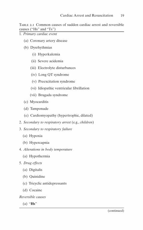
A. EtiologyThe most common causes of sudden cardiac arrest are depicted in Table 2.1.
About 35% are not caused by a heart condition, such as trauma, hemorrhage, and poisoning. The mnemonics for reversible causes of cardiac arrests are the “Hs” and “Ts” (see Table 2.1).
B. Pathogenesis
1. Ventricular fibrillation (VF) or pulseless ventricular tachycardia (VT).
2. Asystole. 3. Pulseless electrical activity (PEA) (electromechanical
dissociation). Patients arresting with PEA can have any cardiac rhythm but no effective mechanical systole (thus, blood pressure [BP] is unobtainable).
4. Cardiogenic shock: No effective cardiac output is generated.
5. The central nervous system (CNS) will not tolerate >6 min of ischemia at normothermia.
C. Diagnosis
1. Unexpected loss of consciousness in the unmonitored patient
2. Loss of palpable central arterial pulse 3. Respiratory arrest in a patient previously breathing
spontaneously 4. Detection of dangerous rhythm in a monitored patient
with loss of palpable pulse
D. Differential diagnosis
1. Syncope or vasovagal reactions 2. Coma 3. “Collapse” 4. Seizures
Chapter 2. The Basics of Critical Care
19
Table 2.1 Common causes of sudden cardiac arrest and reversible causes (“Hs” and “Ts”)1. Primary cardiac event
(a) Coronary artery disease
(b) Dysrhythmias
(i) Hyperkalemia
(ii) Severe acidemia
(iii) Electrolyte disturbances
(iv) Long QT syndrome
(v) Preexcitation syndrome
(vi) Idiopathic ventricular fibrillation
(vii) Brugada syndrome
(c) Myocarditis
(d) Tamponade
(e) Cardiomyopathy (hypertrophic, dilated)
2. Secondary to respiratory arrest (e.g., children)
3. Secondary to respiratory failure
(a) Hypoxia
(b) Hypercapnia
4. Alterations in body temperature
(a) Hypothermia
5. Drug effects
(a) Digitalis
(b) Quinidine
(c) Tricyclic antidepressants
(d) Cocaine
Reversible causes
(a) “Hs”
(continued)
Cardiac Arrest and Resuscitation
20
E. Management
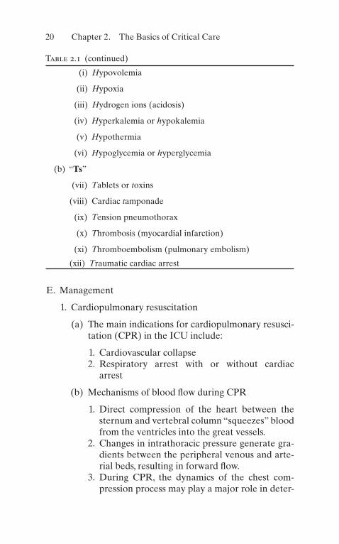
1. Cardiopulmonary resuscitation
(a) The main indications for cardiopulmonary resusci-tation (CPR) in the ICU include:
1. Cardiovascular collapse 2. Respiratory arrest with or without cardiac
arrest
(b) Mechanisms of blood flow during CPR
1. Direct compression of the heart between the sternum and vertebral column “squeezes” blood from the ventricles into the great vessels.
2. Changes in intrathoracic pressure generate gra-dients between the peripheral venous and arte-rial beds, resulting in forward flow.
3. During CPR, the dynamics of the chest com-pression process may play a major role in deter-
Table 2.1 (continued)
(i) Hypovolemia
(ii) Hypoxia
(iii) Hydrogen ions (acidosis)
(iv) Hyperkalemia or hypokalemia
(v) Hypothermia
(vi) Hypoglycemia or hyperglycemia
(b) “Ts”
(vii) Tablets or toxins
(viii) Cardiac tamponade
(ix) Tension pneumothorax
(x) Thrombosis (myocardial infarction)
(xi) Thromboembolism (pulmonary embolism)
(xii) Traumatic cardiac arrest
Chapter 2. The Basics of Critical Care

21
mining the outcome of the resuscitation effort. Indeed, chest compressions by themselves may provide ventilation.
4. Interposed abdominal compression CPR increases aortic diastolic blood pressure, improv-ing blood perfusion to the coronary arteries.
(c) Technique
1. Establish an effective airway (see Chap. 15):
(a) Assess breathing first (open airway, look, listen, and feel).
(b) If respiratory arrest has occurred, the possi-bility of a foreign body obstruction needs to be considered and measures taken to relieve it.
(c) If endotracheal intubation is to be per-formed, give two breaths during a 2-s pause every 30 chest compressions.
(d) The minimum respiratory rate during car-diac or respiratory arrest should be one breath every 6 s (ten breaths per minute). Once spontaneous circulation has been restored, the rate should be ten breaths per minute, to avoid excessive ventilation. Titrate to target PETCO2 of 35–40 mmHg.
(e) Ventilations should be performed with a tidal volume of 5–7 mL/kg of ideal body weight.
(f) The highest possible concentration of oxy-gen (100%) should be administered to all patients receiving CPR.
2. Determine pulselessness (ideally, carotid pulse; if no pulse, start CPR immediately).
3. Chest compressions, current advanced cardiac life support (ACLS) recommendations:
(a) Rescuer’s hand located in the lower margin of sternum.
Cardiac Arrest and Resuscitation

22
(b) Heel of one hand is placed on the lower half of the sternum, and the other hand is placed on top of the hand on the sternum so that the hands are parallel.
(c) Elbows are locked in position, the arms are straightened, and the rescuer’s shoulders are positioned directly over the hands, pro-viding a straight thrust.
(d) The sternum is depressed 2–2.4 in. in normal- sized adults with each compression at a rate of 100–120/min.
(e) The American Heart Association addresses alternative techniques to standard manual CPR, specifically mechanical devices (i.e., vest CPR, LUCAS™). These devices have the purpose to enhance compression and diminish exhaustion of the person deliver-ing CPR. To date, no single, randomized, controlled study has shown that these devices provide a better chance of hospital discharge with good neurological outcome.
(f) Extracorporeal CPR is an option for patients who have a known reversible etiol-ogy for their cardiac arrest, in those centers that have such supports available 24 h a day.
4. Cardiac monitoring and dysrhythmia recogni-tion (see also Chap. 3).
(a) Distinguish between ventricular and supra-ventricular rhythms:
(i) Most rapid, wide QRS rhythms are VT. (ii) Initiate therapy immediately (see
below).
5. Defibrillation is the major determinant of sur-vival in cardiac arrest due to VF or pulseless VT:
(a) Integrating early defibrillation and CPR provides better outcome.
Chapter 2. The Basics of Critical Care
23
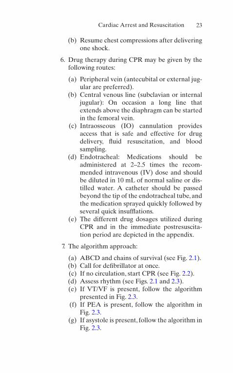
(b) Resume chest compressions after delivering one shock.
6. Drug therapy during CPR may be given by the following routes:
(a) Peripheral vein (antecubital or external jug-ular are preferred).
(b) Central venous line (subclavian or internal jugular): On occasion a long line that extends above the diaphragm can be started in the femoral vein.
(c) Intraosseous (IO) cannulation provides access that is safe and effective for drug delivery, fluid resuscitation, and blood sampling.
(d) Endotracheal: Medications should be administered at 2–2.5 times the recom-mended intravenous (IV) dose and should be diluted in 10 mL of normal saline or dis-tilled water. A catheter should be passed beyond the tip of the endotracheal tube, and the medication sprayed quickly followed by several quick insufflations.
(e) The different drug dosages utilized during CPR and in the immediate postresuscita-tion period are depicted in the appendix.
7. The algorithm approach:
(a) ABCD and chains of survival (see Fig. 2.1). (b) Call for defibrillator at once. (c) If no circulation, start CPR (see Fig. 2.2). (d) Assess rhythm (see Figs. 2.1 and 2.3). (e) If VT/VF is present, follow the algorithm
presented in Fig. 2.3. (f) If PEA is present, follow the algorithm in
Fig. 2.3. (g) If asystole is present, follow the algorithm in
Fig. 2.3.
Cardiac Arrest and Resuscitation

24
(h) For bradycardia, follow the algorithm in Fig. 2.4.
(i) For tachycardia, follow the algorithms pre-sented in Figs. 2.5, 2.6, 2.7, and 2.8.
(d) Cerebral resuscitation
1. The primary goal of cardiopulmonary resuscita-tion is a neurologically intact patient.
Person collapsesPossible cardiac arrest
Assess responsiveness at once
Unresponsive
Primary ABCD EvaluationA: Assess breathing (OpenAirway, look, listen and feel)
B: Give 1 breath every 3-5 seconds(12-20 breaths/min)
C: Assess pulse; add compressionsif pulse remains <60/min withsigns of poor perfusion. Check
pulse every 2 minutes, if no pulseSTART CPR
D: Attach monitor/defibrillator assoon as it’s available
Shout for nearby helpActivate emergency responsesystem via mobile device (if
appropriate)Call for defibrillator
ASSESS RHYTHM
INHOSPITALCARDIACARREST:
OUTHOSPITALCARDIACARREST:
Primaryproviders
Lay rescuers
Code team
Emergencymedicalservice
Cath lab
Emergencydepartment
ICU
Cath lab ICU
Figure 2.1 The algorithm approach
Chapter 2. The Basics of Critical Care

25
2. Maintain resuscitated patients at a systolic blood pressure of no less than 90 mmHg or a mean arterial pressure of no less than 65 mmHg. Immediate correction of hypotension directly after resuscitation is recommended to maintain proper brain and other organ perfusion.
3. Maintain patient within 32–36 °C after return of spontaneous circulation.
4. Mild therapeutic hypothermia (32–34 °C) improves neurological outcome, as demon-strated in many clinical trials. Therapeutic hypo-thermia (TH) decreases metabolic rate and decreases the release of free oxygen radicals (see Chap. 15).
5. Avoid hyperoxia (defined as partial pressure of oxygen >300 mmHg).
6. Optimize cerebral perfusion pressure by main-taining a normal or slightly elevated mean arte-rial pressure and by reducing intracranial pressure, if increased (see Chap. 9).
2. When to stop CPR?
(a) No return of spontaneous circulation after 30 min.
• Compression rate 100-120: Quality decreases with >120 compressions per minute
• One cycle of CPR: 30 compressions then 2 breaths
• Avoid hyperventilation
• Maximize compression time: Minimize the time without compression to maximize coronary perfusion
• Depth of chest compression should be between 2” and 2.5” (5 cm to 6 cm): Deeper can be harmful
• Secure airway and confirm placement: Use capnography and listen to breath sound in lungs
• Give continues chest compressions once the advance airway is placed
Figure 2.2 CPR
Cardiac Arrest and Resuscitation

26
(i) Prolongation of efforts can be considered in patients with return of spontaneous circula-tion in the time CPR was performed.
(b) ETCO2 of greater than 10 mmHg after 20 min may be considered as a criterion to discontinue CPR as stated in recent updates.
Shockable rhythm?
Start CPR
VF/VT Asystole/PEA
SHOCK!Biphasic: 120-200 JMonophasic: 360 J
CPR 2 minutes• IV/IO Access
Shockable rhythm?If YES
YES NO
CPR 2 minutes• Epinephrine 1 mg every 3-5 min
• Advanced airway withcapnography
Shockable rhythm?If YES
CPR 2 minutes• Amiodarone first dose: 300 mg
bolus. Second dose: 150 mg• Treat reversible Causes (H’s and
T’s)
CPR 2 minutes• IV/IO Access• Epinephrine 1 mg every 3- 5 min• Advanced airway with capnography
Shockable rhythm?If NO
CPR 2 minutes• Treat reversible causes
Shockable rhythm?If NO
If no signs of return of spontaneouscirculation keep on CPR
If ROSC: Go to Post cardiac arrestcare
ROSC: • Pulse and blood pressure
• Abrupt sustained increase in PETCO2• Spontaneous arterial pressure waves in intra-arterial monitoring
Figure 2.3 Algorithm for ventricular fibrillation (VF), pulseless ventricular tachycardia (VT), pulseless electrical activity (PEA), and asystole
Chapter 2. The Basics of Critical Care

27
3. Predictors of poor outcome in resuscitation
(a) Preterminal illness (i.e., sepsis, malignancies) (b) Catastrophic events (i.e., massive pulmonary embo-
lism, ruptured aneurysms, cardiogenic shock, etc.) (c) Delayed performance of basic life support (BLS)/
ACLS
NO
BRADYCARDIA ALGORITHM
• Asses using the ABCDE approach • Heart Rate < 50 bpm • Give oxygen if appropriate and obtain IV access • Monitor ECG, BP, SpO2 record 12-lead ECG • Identify and treat reversible causes (e.g electrolyte abnormalities • IV access
YES
Atropine500 mcg IV
NO
Risk of asystole
• Recent asystole• Mobitz II AV block• Complete heart block with borad QRS• Ventricular pause > 3s
Satisfactoryresponse?
Interim measures:atropine 0.5 mg IV repeat every3-5 min. to maximum of 3 mgDopamine 2-20 mcg/kg per min.Epinephrine 2-10 mcg min−1 IVAlternative drugs*ORtranscutaneous pacing
NO
YES
ObserveSeek expert help
Arrange transvenous pacing
*Alternatives include:AminophyllineGlucagon (if beta-blocker orcalcium channel blockeroverdose)Glycopyrrolate can be usedinstead of atropine
Assess for evidence of evidence of adverse signs1. Shock2. Syncope
3. Myocardial ischemia4. Heart failure
YES
Figure 2.4 Bradycardia
Cardiac Arrest and Resuscitation

28
The Alveolar Air Equation
A. Dalton’s law states that the partial pressure of a mixture of gases is equal to the sum of the partial pressures of the constituent gases. Thus, the total pressure of alveolar gases must equal the sum of its constituents and, in turn, equili-brate with atmospheric pressure. We are most often con-cerned with the respiratory gases, O2 and CO2.
B. The alveolar air equation is based firmly on Dalton’s law but is expressed in terms that emphasize alveolar O2 and CO2:
EVALUATE PATIENT
STABLE PATIENT• No serious signs or symptoms
1.- Atrialfibrillation/Atrialflutter
2.- Narrow complextachycardia
3.- Stable wide-complextachycardia: unknown type
4.- Stable monomorphicVT and/or polymorphicVT
Refer to Figure 2.6 4 clinical features:• Is the patient unstable?• Cardiac function impaired?• WPW present?• Duration <48 hours or >48 hours
• Treat unstable patients urgently• Control the rate• Convert the rhythm
TREATMENT OFATRIAL
FIBRILLATION/ATRIAL FLUTTER
IV b-blockers andnondihydropyridine
calcium channel blockerssuch as diltiazem are thedrugs of choice for acute
rate control in mostindividuals with atrialfibrillation and rapidventricular response
• 12-Lead ECG• Vagal maneuvers
• Adenosine
Diagnostic efforts yield:• Ectopic atrial tachycardia• Multifocal atrial tachycardia• Paroxysmal supraventricular tachycardia
TREATMENT OFSVT (Refer to Figure
2.7)
• 12-Lead ECG• Clinical information
Wide-complex tachycardia of unknown type• Preserved cardiac function• Ejection fraction <40% Clinical CHF
• DC CARDIOVERSION OR AMIODARONE
UNSTABLE PATIENT• Rate related serious signs or symptoms (150 bpm)• Prepare for cardioversion
Figure 2.5 Tachycardia
Chapter 2. The Basics of Critical Care

29
P O P PA ATM H O
FiO PCO RQ2 2 2 2= -( ) - /
PAO2 = partial pressure of O2 in the alveolus under present conditions. PATM = current, local atmospheric pressure. PH2O = vapor pressure of water at body temperature and 100% relative humidity. FiO2 = fraction of inspired O2. PCO2 = partial pressure of CO2 in arterial blood. RQ = respiratory quotient.
C. Many clinical and environmental influences are immedi-ately obvious when considering the terms of the equation:
Stable ventricular TachycardiaMonomorphic or Polymorphic?
NOTE!May go directly tocardioversion
Polymorphic VTIs QT baseline intervalprolonged?
Normal function
Medications: any oneProcainamideAmiodaroneOther acceptable:Lidocaine
Poor ejection fraction
Normal baseline QTintervaltreat ischemiacorrect electrolytesMedications: any oneB-blockers orLidocaine orAmiodarone orProcainamide orSotalol
Long baseline QT intervalCorrect abnormal electrolytesMedications: any oneMagnesiumOverdrive pacingIsoproterenolPhenytoin
Normal baseline interval Prolonged baseline QT interval (suggests Torsades)
Amiodarone150 mg IV bolus over 10 minutesthen useSynchronized cardioversion
Cardiac functionimpaired
MonomorphicIs cardiac function impaired?
Figure 2.6 Tachycardia algorithm
The Alveolar Air Equation

30
1. PATM: Altitude per se can clearly result in hypoxemia. A given patient’s PO2 must be considered in the context of location. A “normal” arterial PO2 is not the same in Denver (average = 73 mmHg) as it is at sea level (average = 95 mmHg).
2. FiO2: While atmospheric air is uniformly about 21% O2, one must ask, 21% of what? The FiO2 on a mountain-
Narrow-Complex SupraventricularTachycardia, Stable
Attempt therapeutic diagnostic maneuver *Vagal stimulation *Adenosine
Junctional tachycardia
Paroxysmal supraventriculartachycardia
Ectopic or multifocal atrialtachycardia
No DC cardioversion?AmiodaroneB-blockerCa2+channel blocker
No DC cardioversionAmiodarone
Priority order:Ca2 channel blockerB-blockerDigoxinDC cardioversionConsider procainamide,amiodarone, sotalol.C
Priority Order:No DC cardioversionDigoxinAmiodarone
No DC cardioversionCa2 channel blockerB-blockerAmiodarone C
No DC cardioversionAmiodaroneDiltiazem
Preserved
Preserved
Preserved
EF <40% CHF
EF <40% CHF
EF <40% CHF
Figure 2.7 Tachycardia algorithm
Chapter 2. The Basics of Critical Care

31
• Serious signs and symptoms-rate related (AMS/chest pain/hypotension) >150 bpm
• Symptoms can be seen at lower rates if patients have impaired cardiac function.
• Prepare for immediate cardioversion
• May give adenosine while preparing for cardioversion
• Assess and support ABC’s as needed
• Give oxygen
• Monitor vital signs and ECG
Synchronized cardioversion
• Ventricular tachycardia
• Paroxysmal Supraventricular Tachycardia
• Atrial fibrillation
• Atrial Flutter
Monophasicwaveform
100 J, 200 J
300 J, 360 J
Figure 2.8 Electrical synchronized cardioversion algorithm
The Alveolar Air Equation
32
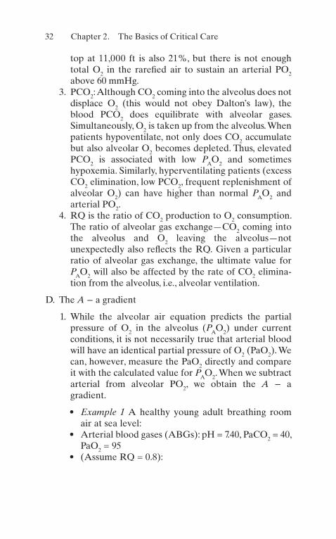
top at 11,000 ft is also 21%, but there is not enough total O2 in the rarefied air to sustain an arterial PO2 above 60 mmHg.
3. PCO2: Although CO2 coming into the alveolus does not displace O2 (this would not obey Dalton’s law), the blood PCO2 does equilibrate with alveolar gases. Simultaneously, O2 is taken up from the alveolus. When patients hypoventilate, not only does CO2 accumulate but also alveolar O2 becomes depleted. Thus, elevated PCO2 is associated with low PAO2 and sometimes hypoxemia. Similarly, hyperventilating patients (excess CO2 elimination, low PCO2, frequent replenishment of alveolar O2) can have higher than normal PAO2 and arterial PO2.
4. RQ is the ratio of CO2 production to O2 consumption. The ratio of alveolar gas exchange—CO2 coming into the alveolus and O2 leaving the alveolus—not unexpectedly also reflects the RQ. Given a particular ratio of alveolar gas exchange, the ultimate value for PAO2 will also be affected by the rate of CO2 elimina-tion from the alveolus, i.e., alveolar ventilation.
D. The A − a gradient
1. While the alveolar air equation predicts the partial pressure of O2 in the alveolus (PAO2) under current conditions, it is not necessarily true that arterial blood will have an identical partial pressure of O2 (PaO2). We can, however, measure the PaO2 directly and compare it with the calculated value for PAO2. When we subtract arterial from alveolar PO2, we obtain the A − a gradient.
• Example 1 A healthy young adult breathing room air at sea level:
• Arterial blood gases (ABGs): pH = 7.40, PaCO2 = 40, PaO2 = 95
• (Assume RQ = 0.8):
Chapter 2. The Basics of Critical Care

33
PAO2760 47 21 40 0 8= -( ) -. / .
PAO2
150 50 100= - =
A P- = -agradient O PaOA 2 2
A- = - =agradient mmHg100 95 5
• This person has an A − a gradient of 5 mmHg, which is normal (0–10).
• Example 2 An elderly patient in respiratory distress secondary to pulmonary edema breathing 40% O2 (FiO2 = 0.4):
ABGs pH PaCO PaO: . , ,= = =7 43 36 702 2
PAO
2760 47 40 36 0 8= -( ) -. / .
PAO
2285 45 240= - =
A P- = -agradient O PaOA 2 2
A- = - =agradient mmHg240 70 170
• This person has an A − a gradient of 170 mmHg, which is markedly elevated.
2. Significance: The presence of an A − a gradient tells you that something is wrong—gas exchange is impaired. It does not tell you what is wrong, nor does it tell you the etiology of hypoxemia when present. A widened A − a gradient simply indicates that alveolar O2 tension is not successfully reflected in arterial blood. Common causes include diffusion disorders (e.g., interstitial lung dis-eases), mismatched ventilation/perfusion (e.g., pulmo-nary embolism), and shunting (e.g., pneumonia, cyanotic heart disease, pulmonary AV shunt, among others).
(a) Note that at a given FiO2, PAO2 varies inversely as the PaCO2. Thus, at any A − a gradient, a high PaCO2 is associated with a low PAO2 and vice versa.
The Alveolar Air Equation
34
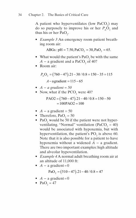
A patient who hyperventilates (low PaCO2) may do so purposely to improve his or her PAO2 and thus his or her PaO2.
• Example 3 An emergency room patient breath-ing room air:
ABGs pH PaCO PaO: . , , .= = =7 50 30 652 2
• What would the patient’s PaO2 be with the same A − a gradient and a PaCO2 of 40?
• Room air:
PAO
2760 47 21 30 0 8 150 35 115= -( ) - = - =. / .
A- = -agradient 115 65 • A − a gradient = 50• Now, what if the PCO2 were 40?
PAO
PAO
2 760 47 21 40 0 8 150 50
100 2 100
= -( ) - = -= =
. / .
• A − a gradient = 50• Therefore, PaO2 = 50• PaO2 would be 50 if the patient were not hyper-
ventilating. “Normal” ventilation (PaCO2 = 40) would be associated with hypoxemia, but with hyperventilation, the patient’s PO2 is above 60. Note that it is also possible for a patient to have hypoxemia without a widened A − a gradient. There are two important examples: high altitude and alveolar hypoventilation.
• Example 4 A normal adult breathing room air at an altitude of 11,000 ft:
• A − a gradient = 0
PaO2
510 47 21 40 0 8 47= -( ) - =. / .
• A − a gradient = 0• PaO2 = 47
Chapter 2. The Basics of Critical Care

35
• This patient has hypoxemia without an A − a gradient.
• Example 5 A patient with pure alveolar hypoventilation secondary to narcotic overdose breathing room air:
• PCO2 = 80; A − a gradient = 0
PAO
2760 47 21 80 0 8= -( ) -. / .
• PAO2 = 50• A − a gradient = 0• PaO2 = 50• This patient has hypoxemia without an A − a
gradient.
3. Summary (a) The alveolar air equation shows the relationships
among atmospheric pressure, FiO2, PaCO2, and alveolar O2 tension (PAO2).
(b) When alveolar O2 tension (PAO2) is not reflected faithfully in arterial blood (PaO2)—i.e., a widened A − a gradient—the calculation indicates that gas exchange is impaired, but it does not tell you how or why.
(c) Calculation of the A − a gradient is a useful bed-side tool for evaluation of patients with respiratory distress or abnormal ABGs and to follow their progress.
(d) It is possible to have hypoxemia without a widened A − a gradient. High altitude and hypoventilation (elevated PaCO2) are examples.
Oxygen Transport
A. Oxygen Delivery: Calculations 1. Calculation of oxygen delivery (ḊO2) and oxygen con-
sumption (VO2) are useful bedside techniques in the ICU.
Oxygen Transport

36
2. DO CO CaO2 2= ´
Oxygen delivery Cardiacoutput ArterialO content= ´2
3. CaO Hb SaO K2 2= ´ ´
ArterialO content Hemoglobin ArterialO saturation
aconstan
2 2= ´
´ tt *
∗We will use 1.34 mL O2/g Hb.
4. Resolving the units:
DO mLO CO mL Hb g mL
mLO g SaO sca
2 2
2 2
100
1 34
/ min / min /
. /
[ ] = [ ]´ [ ]´ [ ]´ llar[ ]
5. Normal values (70-kg man at rest):
DO mL CO g mL Hb
mLO g constant
2
2
5 000 15 100
1 34 1
= [ ]´ [ ]´ [ ]´
, / min /
. / .0002
SaO[ ]DO mLO
2 21 005= , / min
6. This value does not take into account dissolved O2 in the plasma, 0.003 mL O2/100 cc/mmHg PaO2, which adds another 15 mL O2 of arterial O2 content.
7. Values to remember:
NormalCaO gHb, SaO
mLO cc vol
2 2
2
15 100
20 4 100 20 4
%
. / . %
( )= ( )
NormalDO kgman,t rest,CO , mL
mLO
2
2
700 5 000
1 020
=( )=
/ min
, / min
Chapter 2. The Basics of Critical Care
37
B. Oxygen Transport: ConceptsOnly three clinical variables can affect ḊO2: cardiac
output, hemoglobin, and oxygen saturation.Note that what looks very simple is not:
1. Cardiac output entails all of normal cardiodynamics (preload, afterload, contractility), hemodynamics, state of hydration, blood gas and electrolyte influences, the influence of mechanical ventilation and other technolo-gies, intrinsic cardiac disease, dysrhythmias, etc.
2. Hemoglobin is largely a quantitative problem (i.e., oxy-gen-carrying capacity), but it also includes the effects of abnormal hemoglobins, massive transfusions, pH and temperature, other causes of shift in the oxyhemoglo-bin dissociation curve, and hemoglobin substitutes.
3. Arterial oxygen saturation embodies the pathophysiol-ogy of acute and chronic lung disease, management of mechanical ventilation, the cardiopulmonary interac-tion, venous admixture, intrapulmonary or intracardiac shunting, etc.
4. If this is not complicated enough, recall that what you may be doing to support the lungs may have a detri-mental effect on cardiac output (see below). Similarly, failure to correct severe blood gas abnormalities may also adversely affect cardiac function. This makes the bedside management of oxygen delivery in critically ill patients straightforward, although at times very difficult:
(a) Support oxygenation such that PO2 >60, SaO2 >0.9 on nontoxic FiO2 (≤0.5).
(b) Ensure hemoglobin concentration of at least 10 g/100 cc.
(c) Optimize cardiac output (CO) under current con-ditions (i.e., current ventilator settings).
Oxygen Transport

38
C. Physiologic Maintenance of Oxygen Delivery: Since ḊO2 is dependent on only three variables, how does a normal person respond to abnormalities of one of the values?
1. Fall in SaO2
If SaO2 falls to 0.5, a person can achieve normal O2 delivery by doubling CO:
DO CO Hb SaO
DO CO Hb SaO
2 2
2 22 1 2
= ´ ´= ´ ´ /
(a) Therefore, in the short term, increased CO can compensate for even severe hypoxemia.
(b) Note that when SaO2 = 0.5, PaO2 = 27! This is the definition of P50 for normal adult hemoglobin A, namely, the PaO2 at which hemoglobin is 50% satu-rated (27 mmHg). Thus, even severe hypoxemia can be tolerated well as long as hemoglobin is nor-mal and CO can be enhanced.
(c) In patients with chronically low SaO2 (high alti-tude, chronic lung disease, cyanotic heart disease), they will also increase their hemoglobin concentration.
2. Fall in HemoglobinIf hemoglobin falls dramatically, ḊO2 is again main-tained by increasing CO.
(a) Note that SaO2 can never increase beyond 100% and therefore cannot compensate for low hemo-globin. The ability to increase and maintain CO is an important mechanism by which anemia can be tolerated:
DO CO Hb SaO
DO CO Hb SaO
2 2
2 22 1 2
= ´ ´= ´ ´/
Chapter 2. The Basics of Critical Care

39
3. Fall in Cardiac OutputWhat if CO falls dramatically, then how is ḊO2 main-tained? The answer is, ḊO2 in totality is not maintained, but tissue ḊO2 is maintained by enhanced extraction.
(a) If fewer liters of oxygenated blood are delivered, then the tissues must extract more from every liter that is delivered.
(b) Normally, arterial blood is nearly 100% saturated with O2. Venous blood returning to the heart is the same in terms of hemoglobin and quantitatively the same as CO. Thus, it is the venous oxygen satu-ration (SvO2) that reflects O2 extraction. Normal SvO2 = 0.75. Therefore, normal extraction is about 25%.
(c) Looking at extraction (i.e., A − V O2 difference) is therefore a good probe (under some circumstances, such as heart failure) of the adequacy of CO:High extraction implies inadequate CO.
4. Fall in Oxygen DeliveryHere is a general rule of thumb: A normal person can withstand a severe abnormality of any one of the O2 delivery variables (CO, Hb, SaO2) without developing lactic acidosis (lactic acidosis would indicate cellular O2 deprivation with resultant anaerobic metabolism).
(a) Note that during cardiac arrest lactate is generated not because of hypoxemia alone but, rather, because the cardiac output is also severely compromised and unable to compensate for low PaO2 to maintain ḊO2.
D. Oxygen Consumption VO2
1. ḊO2 (oxygen delivery) is what leaves the heart both quantitatively and qualitatively. What returns to the heart should be the same quantitatively, with the same hemoglobin concentration, different only in terms of oxygen saturation.
Oxygen Transport

40
2. If we know the ḊO2 (what left the heart) and we calcu-late what has returned to the heart, we can then sub-tract to ascertain the amount consumed:
Oxygen consumptionVO CO CaO CO CvO
2 2 2( ) = ´( ) - ´( )*
what leftwhat returned to
the heartthe heart
Thus VO CO CaO CvO,
2 2 2= -( )**
This is the Fick equation:CvO
2 is the mixed venous O2 content.
CaO CvO2 2- is the arteriovenous O2 content
difference. 3. CvO2 (mixed venous O2 content) is calculated in exactly
the same way as the CaO2 (arterial content), namely:
Hb SvO´ ´1 342
.
(SvO2
is the mixed venous O2 saturation.) 4. If VO
2 is known, the Fick equation can be used to cal-
culate the cardiac output:
COVO
CaO CvO=
-( )
2
2 2
5. At the bedside, what do you need to calculate VO2
?
(a) The patient needs a pulmonary artery (Swan- Ganz) catheter:
1. For CO determination2. To obtain a true mixed venous blood sample
from the pulmonary artery (SvO2
)
(b) Arterial blood gas determination (or SaO2 determination)
(c) Hemoglobin determination
Chapter 2. The Basics of Critical Care

41
6. Example of normal values (70-kg man at rest):
CaO Hb constant SaO2 2
15 1 34 1 00 20 1= [ ]´ [ ]´ [ ] =. . .
CvO Hb constant SvO2 2
15 1 34 0 75 15 1= [ ]´ [ ]´ [ ] =. . .
CO mL L= [ ]5 000 5, / min
VO mL mLO mL mLO cc2 2 2
5 000 20 1 100 15 1 100= -, / min( . / . /
VO2
50 20 15= -( )VO normal,at rest mLO
2 2250[ ] = / min
7. Bedside application in the ICU: Human life depends on oxygen. This is a good reason to assess the adequacy of ḊO2 in critically ill patients. Where there is life, there is O2 consumption:
(a) We are concerned about factors that increase rest-ing O2 consumption such as fever. Febrile patients increase their resting O2 consumption by 10–13% °C (approximately 7% °F).
(b) We are also concerned when calculated O2 con-sumption is less than predicted (for body surface area, temperature), such as may occur in sepsis. Many times, in spite of high ḊO2, patients with sepsis have low calculated
VO
2 lactic acidosis, oli-
guria, and other signs of poor parenchymal organ function.
(c) Instead of arbitrary end points, it is best to look for physiological end points. When measured O2 con-sumption, SvO2 (A − V) O2 content difference, and serum lactate are all normal, then it is likely that ḊO2 is adequate. Evidence that you have satisfied the body’s (tissues’) needs is better evidence of the adequacy of ḊO2 than any arbitrary number.
Oxygen Transport

42
(d) Make sure that you see ḊO2 as an integrated vari-able. If you change ventilator settings (see below)—for example, raise the PEEP to enhance SaO2—but in the process cause a fall in CO, you may not have achieved any overall benefit in terms of ḊO2. Cardiac output, hemoglobin, and SaO2 require individual attention and management.
(e) Look for opportunities to get the best results for each intervention. For example, a transfusion of packed red blood cells may increase hemoglobin and raise CO. This may be substantially better management than trying to raise CO with crystal-loid IV fluids to compensate for a borderline hemo-globin and/or SaO2.
(f) Check CO and O2 transport variables often and measure the response to your interventions. Note that we often record heart rate and blood pressure every hour although CO can vary over a wide range irrespective of these more traditional signs. Cardiac output is a vital sign!
(g) Current technology now provides continuous data for SaO2, SvO2
, and even CO. These can obviate the necessity of repeated blood gas determinations and facilitate frequent assessment of O2 transport variables.
Mechanical Ventilation
Humans breathe for two reasons: to take in oxygen (oxy-genation) and to eliminate carbon dioxide (ventilation). A patient’s inability to perform either or both of these functions defines respiratory failure.
A. Ventilation. Normal people produce CO2 continuously; thus, there is a constant need for CO2 elimination. We all eliminate CO2 by a process that entails breathing in fresh air (essentially devoid of CO2), allowing it to equilibrate
Chapter 2. The Basics of Critical Care
43
with the CO2 dissolved in capillary blood and then exhal-ing it laden with CO2. We perform this process 10–14 times each minute with significant volumes of air, such that under normal conditions arterial CO2 (PaCO2) is kept nearly constant at 40 mmHg (torr).More precisely, we move a tidal volume (Vt) in and out at a certain frequency (f) or respiratory rate (RR). The prod-uct of rate and tidal volume is the minute ventilation (Vmin). Thus, it is the minute ventilation that is fundamen-tally responsible for CO2 elimination:
V Vmin
= ´t
RR
1. The minute ventilation can be further divided into the gases that reach the alveoli and are therefore available for exchange (the alveolar ventilation, VA) and those gases that fill the airways or that reach unperfused (see below) alveoli and therefore cannot exchange gases (the anatomical and physiological dead space, respec-tively, VD).
2. CO2 elimination is therefore directly proportional to the minute alveolar ventilation at any level of CO2 pro-duction or blood PCO2:
CO elimination PCOA2 2
= ( ) ´Vmin
3. Since any physiologic parameter (i.e., serum creatinine, platelet count, PCO2) is ultimately the result of the bal-ance between production and elimination, it follows that PCO2 (under any conditions affecting production) can be controlled by adjusting minute ventilation.
B. Oxygenation. How people accomplish oxygenation is equally simple but considerably different from how we accomplish ventilation. We purposely inhale an oxygen- enriched atmosphere all the way down to our alveoli to allow the oxygen to be taken up by the capillary blood—both dissolved in proportion to its partial pressure (obey-ing Henry’s law) and in combination with hemoglobin.
Mechanical Ventilation

44
More precisely, the air we inspire has a certain fraction that is oxygen—that is, a certain fraction of inspired O2 (FiO2).
Although we breathe only intermittently, we need to accomplish gas exchange continuously. If there were oxy-gen in our alveoli only when we inhaled, then blood would pass through the lungs unoxygenated in between breaths. Thus, we need to maintain volume in our lungs even at end exhalation. This is accomplished by maintaining a pressure gradient across the lungs between breaths. The pressure in the pleural space (outside the lungs) is nega-tive (approximately [–] 5 cmH2O) with respect to the atmospheric pressure present in our airways. If we sub-tract vectorially, there is a 0 − (−5) = + 5 cm H2O pressure gradient across the lungs even at end exhalation—in effect, a PEEP. Thus, oxygenation is accomplished in nor-mal people by purposely inspiring a certain FiO2 and maintaining a certain PEEP.
C. PEEP and Compliance
1. ComplianceThe volume in the lungs is related to the transpulmo-nary pressure. Indeed, volume and pressure are inti-mately related in many systems (such as ventilator tubing, cardiac filling, resting lung volume) through the variable of compliance (C):
C VP
=DD
Compliance is defined as the change in volume for a given change in pressure. Thus, in order for us to achieve a given volume change—such as a tidal volume—in our lungs, we must make a pressure change. The precise pressure necessary will be determined by the lung (and chest wall) compliance.
Mathematically, it is clear that, as compliance falls (as may occur in pulmonary edema, adult respiratory distress syndrome (ARDS), lung fibrosis, and many other conditions), one must achieve ever-increasing ∆P
Chapter 2. The Basics of Critical Care

45
just to achieve the same ∆V. It is often the case that a patient’s inability to do the work required to increase ∆P to maintain an adequate tidal exchange (∆V) is the ultimate cause of respiratory failure.
The fundamental role that lung compliance plays in determining the relationship between clinically signifi-cant lung volumes (e.g., tidal volume) and the pressures required to achieve them has many important clinical implications:
(a) If there is no pressure gradient (∆P = 0), then there is no volume change.When a patient develops a pneumothorax, the pressure in the pleural space equals the pressure in the airways. As a result, there is no transpulmonary pressure (∆P) and thus no lung volume—i.e., the lung collapses—because of the lungs’ intrinsic compliance (and elastance, which is defined as 1/compliance).
Pneumothorax results in no lung volume (zero ∆V), because there is no transpulmonary pressure (zero ∆P).
(b) To create a volume change, we must affect a pres-sure change. Thus, tidal volume is determined by the ∆P generated as the chest wall expands and the diaphragm contracts. Similarly, to increase our tidal volume, we must generate a larger ∆P, or if compli-ance falls, we may need a larger ∆P just to achieve the same tidal volume.
(c) If a person has low lung compliance (i.e., restrictive disease), then normal resting negative intrapleural pressure (∆P) will result in lower resting lung vol-ume (∆V).
(d) If a person has high lung compliance (i.e., emphy-sema with destruction of lung parenchyma), then normal resting negative intrapleural pressure will result in high resting lung volume (e.g., “barrel chest” of emphysema).
Mechanical Ventilation
46
(e) Since the lungs are merely populations of alveoli, these relationships among pressure, volume, and compliance apply to individual alveoli and specific lung regions as well as to whole lungs.
(f) Compliance contributes to the logical connection between the requirements of gas exchange and the respiratory work:
1. CO2 production demands minute ventilation. 2. Minute ventilation requires a certain tidal
volume. 3. This change in volume requires a change in
pressure. 4. How much pressure for a given volume is deter-
mined by compliance? 5. The amount of pressure that must be generated
is a major determinant of the work of breathing.
2. PEEPFrom our description of oxygenation and ventilation, it should be clear that we are clinically concerned about maintaining the adequacy of two important lung volumes: the tidal volume of each breath and the resting lung volume in between breaths. The pressure generated during active inspiration either by the ven-tilator or the patient will determine the tidal volume (mediated, of course, through compliance). But, what determines the resting lung volume? The answer is the resting transpulmonary pressure. In normal peo-ple (with normal lung compliance), the vectorial dif-ference between airway and intrapleural pressures (∆P) determines the resting lung volume, known more precisely as the functional residual capacity (FRC):
Chapter 2. The Basics of Critical Care

47
P P P
P
airway pleural transpulmonary
cmH O
- =
- -( ) = += +
D
D0 5 5
52
Resting lung volume (FRC) is therefore determined by the AP and compliance:
C VP
CP
C P
=
=
= ( )
DDFRC
FRC
transpulmonary
transpulmonary
Since ∆P is positive and present at end expiration, what we are talking about is positive end-expiratory pressure (PEEP). It should also be clear that PEEP directly determines FRC.
We have all had the experience of inflating a bal-loon. It’s difficult at first and then suddenly gets easier once some volume is inside. As we reach the full infla-tion, it may again become difficult as we reach the limits of the balloon’s compliance. If we let go, the balloon recoils (elastance) and collapses.
Our alveoli, in many ways, are similar: If they start fully collapsed, they are difficult to inflate at first. Once there is some volume, it becomes easier; this point of change in compliance is referred to as critical opening pressure (COP).
Unlike balloons, normal alveoli do not immediately lose all of their volume when pressure is released but may maintain some volume (thanks in large measure to surfactant) until distending pressure is critically low and then collapse. The point at which this occurs is criti-cal closing pressure (CCP).
Mechanical Ventilation

48
If one could maintain end-expiratory pressure (i.e., PEEP) above CCP, then alveoli would not collapse; their volume would be enhanced, and, in the aggregate, lung volume (FRC) would be enhanced. If low lung compliance results in high CCP, then PEEP must be increased above the CCP to prevent alveolar collapse. This is precisely the rationale for PEEP in the manage-ment of acute low-compliance lung disease (e.g., ARDS).
(a) Summary of the effects of PEEP:
1. PEEP increases FRC. 2. PEEP increases compliance. 3. PEEP reduces shunt fraction (see below) by
maintaining volume for gas exchange in per-fused lung units in between breaths.
4. PEEP increases dead space by overdistending normally compliant alveoli.
5. PEEP increases intrathoracic pressure, which can impede venous return into the chest or spe-cifically restrict cardiac filling, both of which may result in reduced cardiac output.
6. PEEP may contribute to barotrauma because it represents the baseline (end expiration) for all pressure changes, because it may cause overdis-tention of compliant lung regions and because of the nature of the acute lung diseases in which PEEP is most frequently useful.
D. Modes of Mechanical Ventilation. Under routine condi-tions, when a patient develops respiratory failure and is intubated, initial mechanical ventilatory support is pro-vided by some form of conventional volume- cycled venti-lation (VCV). Volume cycled means that the end point for the ventilator is the delivery of a selected tidal volume, leaving the machine itself to determine what pressure is necessary to deliver that volume, to that patient, at that time. Pressure-controlled ventilation is a modality in which tidal volume that is delivered is variable and dependent of
Chapter 2. The Basics of Critical Care
49
the peak pressure, inspiratory time, and patient’s compli-ance. It reduces the risk for barotrauma by using lower peak pressures and longer inspiratory times. This type of ventilation is preferred in patients with poor pulmonary compliance.
In addition to the standard array of choices for how to deliver VCV, there are also modes that do not use quasi- physiologic parameters, such as high-frequency ventila-tion. These modes of mechanical ventilatory support are beyond the scope of this manual but have been reviewed in depth elsewhere. For conventional VCV, essentially four modes are commonly used, as depicted in Table 2.2.
1. Controlled Mechanical Ventilation (CMV)From our discussion above, it follows that the basic functions of ventilation and oxygenation can be accom-plished by providing four basic settings: respiratory rate, tidal volume, FiO2, and PEEP. Given these param-eters, the ventilator will provide the patient with a con-stant minute ventilation and oxygen. These are the settings for CMV. The only gases these patients receive are from the machine breaths. These patients cannot initiate a breath, change their rate, or access any other source of fresh gases. This mode is therefore useful in a limited number of settings, such as the following:
(a) In the operating room, when patients are fully anesthetized
Table 2.2 Commonly used modes of volume-cycled ventilation1. Controlled mechanical ventilation (CMV)
2. Assisted/controlled mechanical ventilation (A/C)
3. Synchronized intermittent mandatory ventilation (SIMV)
4. Continuous positive airway pressure (CPAP)
5. Pressure support ventilation (PSV) is not a separate mode but, rather, an adjunct that can be used with several other modes
Mechanical Ventilation
50
(b) When patients are apneic and likely to remain so (c) When patients are sedated/anesthetized and para-
lyzed, in the intensive care unit (ICU)
It is important to note that in CMV, patients abso-lutely cannot breathe on their own. If these patients should awaken or attempt to breathe, they can become agitated and dyspneic. It is extremely frightening to be unable to breathe and to experience essentially a chronically occluded airway. Worse yet, if these patients become detached from the ventilator, anes-thetized and/or paralyzed patients will be functionally apneic and may soon experience full cardiopulmonary arrest.
2. Assisted/Controlled Mechanical Ventilation (A/C)The settings for A/C are the same as the four basic ones used for CMV. There is no difference between CMV and A/C in anesthetized or apneic patients. The singu-lar difference is that in A/C mode, patients can initiate breaths. Unlike in you and me, however, the amount of effort these patients make does not determine the tidal volume. When these patients initiate a breath with suf-ficient force to “trigger” the ventilator, the tidal volume they receive is the one already preset to be delivered as a “controlled” breath. Moreover, the machine will use whatever pressure is required to deliver the volume, and the patients’ lungs and chest must then accommo-date that tidal volume. The set rate in A/C is essentially a default control rate; that is, it is the number of volume- cycled breaths the machine will deliver on its own, even if the patient is apneic. Thus the “controlled” aspect of A/C is the guaranteed minute volume delivered regard-less of the patient’s spontaneous efforts.
While this mode has some value in its ability to relieve dyspnea in the immediate postintubation period, it is not a good choice for prolonged mechani-cal support. Evidence now indicates that in accom-modating “assisted” breaths, the respiratory muscles may actually be fatigued rather than “rested,” as was
Chapter 2. The Basics of Critical Care
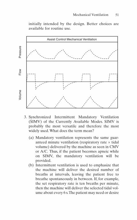
51
initially intended by the design. Better choices are available for routine use.
Assist Control Mechanical Ventilation
Pre
ssur
eF
low
Vol
ume
3. Synchronized Intermittent Mandatory Ventilation (SIMV) of the Currently Available Modes. SIMV is probably the most versatile and therefore the most widely used. What does the term mean?
(a) Mandatory ventilation represents the same guar-anteed minute ventilation (respiratory rate × tidal volume) delivered by the machine as seen in CMV or A/C. Thus, if the patient becomes apneic while on SIMV, the mandatory ventilation will be provided.
(b) Intermittent ventilation is used to emphasize that the machine will deliver the desired number of breaths at intervals, leaving the patient free to breathe spontaneously in between. If, for example, the set respiratory rate is ten breaths per minute, then the machine will deliver the selected tidal vol-ume about every 6 s. The patient may need or desire
Mechanical Ventilation

52
to breathe more often than ten times per minute and can do so in the intervals (approximately 6 s) between machine breaths.
(c) Synchronized IMV is a relatively new refinement of the original IMV design. In the example above, the machine will cycle every 6 s, but it would be unde-sirable for the ventilator to attempt delivery of a new breath if the patient were in the process of exhaling a spontaneous breath. Without synchroni-zation, this kind of “collision” could occur in the airways, causing very high airway pressure, a risk of barotrauma, ineffective ventilation, and enhanced (rather than relieved) dyspnea. The synchronizer looks at a “window” period when the next machine breath is due. If the patient is exhaling, the ventila-tor can wait to begin inspiration. If the patient initi-ates a breath at the time the ventilator is due to cycle, the machine breath and the spontaneous breath will merge into one synchronized breath not unlike the “triggered” breaths in A/C mode. This represents a major improvement for IMV, espe-cially in patients with prolonged expiratory times such as those with bronchial asthma.
(d) SIMV was introduced into wide usage as a “wean-ing” modality. Weaning from mechanical ventila-tion is discussed below, but in this connection, the principle is simple. Initially, the mandatory ventila-tion provides the entire minute ventilation neces-sary to maintain the patient’s PCO2 within normal limits. As patients begin to breathe spontaneously, the mandatory ventilation is gradually reduced until they are essentially providing the entire min-ute ventilation (and therefore CO2 elimination) through their own spontaneous efforts. At this point these patients no longer require mechanical ventilatory support.
Chapter 2. The Basics of Critical Care
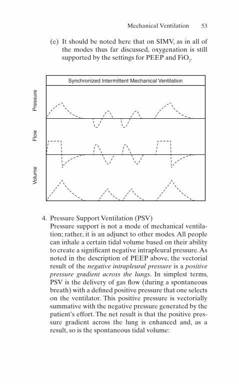
53
(e) It should be noted here that on SIMV, as in all of the modes thus far discussed, oxygenation is still supported by the settings for PEEP and FiO2.
Synchronized Intermittent Mechanical Ventilation
Pre
ssur
eF
low
Vol
ume
4. Pressure Support Ventilation (PSV)Pressure support is not a mode of mechanical ventila-tion; rather, it is an adjunct to other modes. All people can inhale a certain tidal volume based on their ability to create a significant negative intrapleural pressure. As noted in the description of PEEP above, the vectorial result of the negative intrapleural pressure is a positive pressure gradient across the lungs. In simplest terms, PSV is the delivery of gas flow (during a spontaneous breath) with a defined positive pressure that one selects on the ventilator. This positive pressure is vectorially summative with the negative pressure generated by the patient’s effort. The net result is that the positive pres-sure gradient across the lung is enhanced and, as a result, so is the spontaneous tidal volume:
Mechanical Ventilation

54
NoPSV
H Otranspulmonarypressure
WithPSVof
P
:
0 10 10
10
10
2- -( ) = +
++ SSV cmH Otranspulmonarypressure( ) - -( ) = +10 20
2
Respiratory muscles do not benefit from “rest,” defined as not contracting at all; in fact they may rapidly atro-phy if not allowed to perform as they usually do. However, with acute lung disease, the work of breath-ing may result in fatigue. PSV should allow the respira-tory muscles to perform a manageable amount of work, without the risk of atrophy on the one hand or fatigue on the other.
5. Airway Pressure Release Ventilation (APRV)APRV is a mode that delivers a high continuous posi-tive airway pressure for increased duration of time, and then pressure falls to lower levels for a shorter dura-tion. This transition of pressures (from high to low) helps eliminate carbon dioxide. The high continuous positive pressure improves alveolar recruitment, thus indicated in patients with acute lung injury or ARDS. The unique feature of this mode of ventilation is that it is permissible on patients with spontaneous breathing.
6. Pressure-Cycled VentilationIn this mode, the physician sets the inspiratory and expiratory pressures, as well as the inspiration duration, and the tidal volume taken by the patient varies accord-ing to the patient’s compliance. The advantage of this mode is to prevent excessive peak pressure and decrease the risk of barotrauma. It decreases the risk of barotrauma by equilibrating pressures across the lung units during a predetermined time. Another advantage is in patients with severe bronchospasm (e.g., severe asthma) as it allows for, with its decelerating flow, deliv-ery of a tidal volume with less peak pressure leading to more efficient overcoming of high airway resistance.
Chapter 2. The Basics of Critical Care

55
Pressure Cycled Ventilation
Pre
ssur
eF
low
Vol
ume
7. Pressure-Regulated Volume Control (PRVC)This is a new mode of pressure-cycled ventilation that aims to deliver a predetermined tidal volume. The machine senses the exhaled tidal volumes during a number of breaths and adjusts pressures to deliver the determined tidal volume, so it utilizes the decelerating flow patterns of pressure-cycled mode to deliver the target tidal volume, leading to less peak pressures and risk of barotrauma. Since it’s a new mode, the clinical significance of these changes has not completely been determined.
8. Noninvasive VentilationUnlike the previously mentioned settings, noninvasive ventilation is a much more flexible and conserved approach to ventilation. In noninvasive ventilation, patients are not supported with an artificial airway, such as an endotracheal tube, but are ventilated with more benign methods such as an orofacial mask. These methods have received widespread attention due to the recent trend of replacing traditional intubation with noninvasive ventilation methods in certain patients.
Mechanical Ventilation

56
This form of ventilation also offers the added benefits of being available to patients at home with little issue, unlike invasive ventilation methods. Noninvasive meth-ods are oftentimes used to maintain the patient while weaning off invasive ventilation.
(a) Continuous Positive Airway Pressure (CPAP)CPAP is a system for spontaneously breathing patients, in which the machine is providing only PEEP, FiO2, and humidification (maintained in all modes), but does not deliver any mechanical breaths. In this sense, it is the mode that would result if the patient were on either SIMV or A/C and the machine respiratory rate were set at zero. It may be provided via tight-fitting face mask or endotracheal/tracheostomy tube. Some of the applications for CPAP are patients with no ventila-tory difficulty who require positive airway pressure (PEEP) to support oxygenation; it reduces alveolar collapse to patients in the final stages of weaning who are being observed while they breathe without ventilatory support.
(b) Bi-level Positive Airway Pressure (BIPAP)BPAP is a form of noninvasive pressure support ventilation that uses positive airway pressure to complement a patient’s own respiratory cycle. It uses inspiratory (IPAP) and expiratory (EPAP) pressure gradients to reduce the effort required by a patient to take a breath. IPAP is the pressure that is given when a breath is triggered. EPAP is a pres-sure used when a patient is not taking a breath; it provides the user a constant positive airway pres-sure. Some of the different uses of BIPAP are for patient with difficulty inhaling and exhaling breaths, such as asthma and COPD.
(c) Average Volume-Assured Pressure Support (AVAPS)This relatively new mode of noninvasive positive pressure ventilation adjusts pressure support to
Chapter 2. The Basics of Critical Care
57
deliver a preset tidal volume. By doing so, it is the preferred noninvasive mode to control the ventila-tion, thus PaCO2. Due to its versatility, this more is preferred in patients with variable respiratory efforts, such as patients with sleep apnea who have different degrees of hypoventilation in rapid eye movement and nonrapid eye movement phases of sleep. Similarly, patients with obesity hypoventila-tion syndrome required a variable degree of pres-sure support based on position.
E. Initiation of Mechanical Ventilation
1. Indications for Initiation of Mechanical Ventilation
(a) Failure to ventilate (i.e., respiratory distress in patient with COPD or asthma)
(b) Failure to oxygenate (i.e., transport oxygen to pul-monary capillary blood, such as pulmonary edema, ARDS, or pulmonary embolism)
(c) Decreased consciousness and loss of airway reflexes (i.e., patient with traumatic brain injury or cerebrovascular accident)
(d) Need for airway protection (i.e., patient in high risk of aspiration or with present airway trauma, swell-ing, or bleeding)
(e) Anticipated clinical course or deterioration (i.e., patient in need of situation control such as in a sur-gery, life-threatening trauma, or septic shock)
2. Clinical Criteria for Need of Mechanical Ventilation
(a) Physical assessment: The patient is apneic, severely tachypneic, or in respiratory distress unresponsive to therapeutic interventions and supplemental oxygen.
(b) Gas exchange: Hypoxemia (PO2 <50) despite high-flow oxygen and hypercarbia (acute, PCO2 >50 with acidic pH).
(c) Pulmonary function tests: Low vital capacity (<10 mL/kg) or low FEV1 (<10 mL/kg).
Mechanical Ventilation

58
(d) Clinical judgment: The constellation of laboratory and physical findings may be the most compelling. A PCO2 of 60 and a respiratory rate of 35 may be the usual baseline for some patients but may repre-sent a direct emergency in others.
3. Initial Ventilator Settings (see Table 2.3).
F. General Principles of Ventilator Management (see Also Table 2.4)
Table 2.4 The ventilator principlesVentilator principle 1: To reach a desirable clinical end point for the patient’s PaCO2, the ventilator settings you will adjust are the respiratory rate (RR) and/or the tidal volume (Vt) delivered by the machine
Ventilator principle 2: To reach a desirable clinical end point for a patient’s PaO2, the ventilator settings you will adjust are the fraction of inspired O2 (FiO2) and/or the positive end-expiratory pressure (PEEP)Ventilator principle 3: Patients do not “buck” or “fight” the ventilator; patients buck ill-conceived ventilator settings
Table 2.3 Initial ventilator settings1. Mode: Unless there is a compelling reason not to, A/C is the most versatile mode to use
2. Respiratory rate: Generally between 10 and 16 breaths per minute, adjusted according to the PaCO2
3. Tidal volume : In the ICU setting, 5–8 mL/kg lean body weight (do not count adipose tissue or edema)
4. FiO2: Many people start with 1.00 (100% O2), but often 0.8 (80% O2) will suffice. A phenomenon called “absorption atelectasis” occurs proportionately more with higher FiO2. Regardless, the FiO2 should be adjusted down as soon as possible
5. PEEP: Normal people can create positive pressure in their airways with their lips, palate, and glottis. Since the endotracheal tube bypasses all of these structures, most initial setups include “physiologic” PEEP of +3 to +5 cm of H2O
6. PSV: 8–10 cmH2O can usually overcome the additional work imposed by the endotracheal tube and ventilator circuit, but larger amounts may substantially reduce dyspnea. Remember, PSV only has relevance for spontaneously breathing patients
7. I:E 1:2
Chapter 2. The Basics of Critical Care

59
1. Therapeutic End Points
(a) PaCO2: Ventilatory parameters are adjusted to achieve a PaCO2 of 35–45 with the pH also in the normal physiologic range of 7.35–7.45.
(b) PaO2: A PaO2 >60 that corresponds to an SaO2 >0.9 with the patient receiving nontoxic FiO2 (≤0.5). If this is not achievable on physiologic PEEP, the PEEP can be raised in + 2-cm H2O increments to achieve this end point:
1. Note that this end point can be expressed as a PaO2/FiO2 ratio 60/0.4 = 150 (FiO2 = 0.4 is not associated with O2 toxicity).
2. PEEP will be most beneficial in acute low- compliance lung disease. Patients with markedly asymmetrical lung disease, bullous emphysema, or asthma may actually have a worsening gas exchange with significant PEEP.
2. When patients are in respiratory distress, it is accept-able to change any or all of the ventilator settings at one time; during weaning the same is not true.
3. Monitoring During Mechanical Ventilation
(a) It is important to monitor a patient’s blood pres-sure, heart rate and rhythm, and their oxygen satu-ration. Of these, oxygen saturation is the most important parameter to keep in mind when judging proper ventilation.
(b) An arterial blood gas should be taken less than 20 min after intubation to correlate with the oxy-gen saturation monitor. This will allow you to know if the monitor is properly recording the patient’s saturation.
(c) A venous blood gas can be used instead of arterial blood gas in reasonably stable patients.
(d) Peak inspiratory and plateau pressures must be monitored frequently. It is important to maintain pressure levels lower than 35 cm H2O to reduce the risk of barotrauma.
Mechanical Ventilation

60
4. Patient Comfort
(a) Having an endotracheal tube in place is not com-fortable and, because one cannot speak, frustrating as well.
(b) Patients who will be intubated for a short time should receive mild sedation with agents having no significant respiratory suppression.
(c) Patients who require high ventilator settings and will likely be intubated for several days before weaning should receive more substantial sedation.
(d) In our opinion, only a few selected patients require paralytic agents.
5. Some Simple Rules of Thumb
(a) If a patient’s clinical presentation seems to be in need of immediate intubation, one should follow through. Waiting for laboratory tests to decide if the patient is fit for mechanical ventilation can be a cause of prolonged hypoxemia or hypercapnia which can lead to a higher rate of morbidity and mortality.
(b) Endotracheal tubes should be as large (diameter) as possible and cut as short as possible once posi-tion is verified.
(c) Endotracheal tubes must be carefully secured and should be out from between the patient’s teeth.
(d) Suctioning is important but should be minimal or strictly pm when the patient is on >+10 cm H2O PEEP, to minimize volume loss from within the lungs.
(e) When setting up the ventilator, the peak inspira-tory flow rate is best kept relatively low (≤50 L/min [LPM]) but must be at least three times the minute ventilation or the patient may be dyspneic.
(f) Generally, a PSV of 8–10 cmH2O overcomes the extra flow resistive work of the endotracheal tube, but the optimal level usually results in a spontane-ous respiratory rate <25 breaths per minute and absence of accessory muscle use.
Chapter 2. The Basics of Critical Care

61
(g) Any intubated patient should have a nasogastric tube placed; this immediately empties the stomach of liquid contents and of the air that is often swal-lowed during respiratory distress. It can then be used for stress ulcer prophylaxis and enteral nutri-tional support.
G. Weaning from Mechanical Ventilation. The ultimate goal of placing patients on mechanical ventilation is to take them off of mechanical ventilation:
1. When reducing the ventilator settings, you may change two settings at once, but not two that serve the same function: For example, you may reduce the FiO2 and the respiratory rate at the same time, because one serves oxygenation and the other serves ventilation; you would not reduce the respiratory rate and the PSV at the same time, since both serve the minute ventilation.
2. When beginning to wean PEEP from its peak setting, it is prudent to reduce it in 1-cm H2O increments for the first few changes.
3. When weaning PEEP, changes should not be made more frequently than every 3–4 h. Some alveoli may destabilize and collapse rapidly, others may take time; hence, whether or not the patient will tolerate the change may not be clear for 3–4 h.
4. To be successfully weaned from the ventilator and extubated (removal of the endotracheal tube), patients must meet three sets of criteria: gas exchange, pulmo-nary mechanics, and circumstantial:
(a) Gas Exchange
1. The FiO2 should be ≤0.5, and the PEEP should be physiologic before active weaning is contemplated.
2. The PaCO2 should be normal (or normal for the patient), and the pH should be in the normal range (not acidic) before parameters that serve ventilation are reduced.
3. In general, the desirable therapeutic end points outlined above should be met before the set-tings are reduced.
Mechanical Ventilation

62
(b) Pulmonary Mechanics
1. Although often referred to as “weaning param-eters,” these are more correctly thought of as extubation criteria.
2. The parameters measured and desirable values are depicted in Table 2.5.
3. The rapid shallow breathing index (RSBI) should be <105. This value is obtained by divid-ing frequency (f) over tidal volume (Vt).
(c) Circumstantial Criteria
1. The patient should be as awake and alert as pos-sible, for extubation.
2. If the patient is neurologically impaired, careful testing of the gag and cough reflexes is necessary.
3. Secretions should have minimized, or the patient clearly demonstrated an ability to manage them.
4. The airway should be patent and nonedematous, and the patient should have control (voluntary or reflex) over it. Some authors suggested cuff- leak test as a predictor of edematous airways and higher risk of postintubation stridor; how-ever, the predictability of this test is question-able and needs further investigation.
5. Extraneous stresses placed on respiratory requirements must be corrected, such as meta-bolic acidosis, anemia, fever, bronchospasm, and cardiac dysrhythmia.
Table 2.5 Weaning parametersTidal volume 4–7 mL/kgRespiratory rate <30 breaths/min
Negative inspiratory force More negative than 20 cm H2O
Minute ventilation <10 L/min
RSBI <105
Vital capacity >10 mL/kg
Chapter 2. The Basics of Critical Care
63
6. Electrolyte abnormalities that compromise respiratory muscle function must be corrected, such as low values for potassium, magnesium, phosphorus, and ionized calcium.
Hemodynamics
The subject of hemodynamics has two major components: cardiodynamics, the physiology of heart function per se, and traditional hemodynamics, which embraces the pulmonary circulation, the systemic circulation, and the right and left sides of the heart as the functional pumps, respectively, of these circuits.
The bedside tool that is most useful for both continuous assessment of cardiac pump function and management of the hemodynamic state of the circulation is the pulmonary artery (PA) (Swan-Ganz) catheter. Although much useful informa-tion can be derived from the measurements and calculations made possible through the use of a PA catheter, the single most important reason to place one in a critically ill patient is to measure cardiac output. In the critical care unit, ensur-ing the adequacy of cardiac output and its integral role in life- sustaining ḊO2 is the principle rationale for placement of the catheter. Proper interpretation and manipulation of other values (e.g., the pulmonary capillary wedge pressure) depend on the cardiac output for context.
The PA catheter is not a therapy. It is a monitoring device. Like all interventions it carries an inherent risk/benefit ratio. Unless the catheter is used actively to assess the patient, guide management, and reassess the response to interven-tions, its placement is all risk and no benefit. Indeed, in recent years, the authors of this book have seldom used this device.
A. Physics and Physiology. Conceptually, the flow of any fluid through a conduit is governed by the following general principle:
Pressure Flow Resistance= ´
Hemodynamics

64
Note that this applies to airway pressure, inspiratory flow rate, and airway resistance or as in the present discussion, blood pressure, blood flow (cardiac output), and vascular resistance.
Specifically
MAP = CO × SVRMean arterial
Cardiac Systemic
Pressure Output Vascular
Resistance
Or
PAP = CO × PVRPulmonary Cardiac Pulmonary
Artery Output Vascular
Pressure Resistance
We can solve these equations for the calculated resistances:
SVRMAP
CO
PVRPAP
CO
=´ *
=´ *
79 9
79 9
.
.
∗79.9 is a constant that converts the units according to the resistance equation of Poiseuille.
Vascular resistance: Conceptually, the resistance to flow of a fluid through a conduit is given by the equation of Poiseuille:
Resistancelength velocity viscosity
radius=
´ ´ ´
( )8
4p
Chapter 2. The Basics of Critical Care

65
B. Cardiodynamics. The pump function of the heart results from the interaction of three variables: preload, afterload, and contractility.
1. PreloadIn simplest terms, preload is the amount of cardiac fill-ing during diastole. In this sense, one can think of it as the end-diastolic volume (EDV), which is either the cause or the result of end-diastolic pressure (EDP). In the purest terms, the preload is the resting fiber length of the myofibrils and forms the fundamental basis for the Frank–Starling curve.
2. AfterloadIn the simplest terms, afterload can be thought of as the impedance to cardiac ejection. If, for example, the dia-stolic blood pressure were high, the heart would have to overcome that pressure just to open the aortic valve. In truth, afterload is much more complicated than simply blood pressure. In physiological terms, afterload is the developed wall tension during systole and is affected by several factors. Generally speaking, anything that makes it easier for the heart to eject blood is an after-load reducer, and anything that makes it more difficult increases afterload.
3. ContractilityContractility is literally the force with which the heart contracts. Note that, given any amount of filling and a constant afterload, the amount of blood ejected by a given heartbeat will depend on the force of contraction.
4. Cardiac OutputThe CO is the product of the amount of blood pumped each time the heart contracts (stroke volume, SV) times the number of contractions per minute (HR):
CO HR SV
SVCO
HR
= ´
=
Hemodynamics

66
It is clear from the above that a person can have a CO of 5 LPM with a HR of 70 or a HR of 140. Part of the clinical assessment of the CO includes looking at the efficiency of cardiac performance. In general, better SV and lower HR represent greater efficiency.
5. IndexingNormal adults come in all shapes and sizes. The abso-lute CO for a healthy 45-kg woman may be the same as the value for an NFL football player in hemorrhagic shock. How can we compare the values? The answer is to index the values to body surface area (BSA). Thus, raw values for CO in adults may vary widely, while indexed values are usually closely comparable:
(a) 45-kg female: CO = 4.0 LPM, BSA = 1.387 m2
CI
CO
BSA
LPM
mLPM m= = =
4 0
1 3872 88
2
2.
.. /
(b) 145-kg male: CO = 8.0 LPM, BSA = 2.77 m2
CI
CO
BSA
LPM
mLPM m= = =
8 0
2 7742 88
2
2.
.. /
Notice that in the above example, a 4-LPM CO yields an excellent cardiac index (CI) for the smaller patient but would result in a value (1.44) for the larger patient consistent with a diagnosis of shock.
Any of the values calculated as part of a hemody-namic profile can be indexed: CI, stroke volume index (SVI), systemic vascular resistance index (SVRI), etc. The normal values for these indices are useful, since they are the standard for comparison of all patients, not just those of average size.
6. Basic Approach to Hemodynamic ManagementBedside pulmonary artery catheterization in the ICU is not the same procedure as diagnostic cardiac catheter-ization in the laboratory. The fundamental purpose in
Chapter 2. The Basics of Critical Care

67
the ICU is to measure, monitor, and manage the CO as an integral part of oxygen delivery. Since many of the patients are intubated, on positive pressure ventilation (VCV), and may have multiple other problems that affect cardiac performance (even if not of cardiac origin per se), the CO must be managed to meet O2 transport end points, not to make “the numbers” look like text-book normal.
(a) Do not place a pulmonary artery catheter unless you are going to use it: The actual bedside method-ology for catheter placement is reviewed elsewhere. The important principles to remember follow:
1. The catheter is placed to measure CO; all of the other values and calculations are only useful for management in the context of a known CO.
2. Record and evaluate all of the values during placement of the catheter: central venous pres-sure (CVP), right atrial pressure (RA), right ventricular pressure (RV), pulmonary artery pressure (PA systolic, diastolic, and mean), and the pulmonary capillary occlusion (wedge) pres-sure (PCWP). All of these can be helpful in assessment, diagnosis, and, most importantly, management.
3. Understand the clinical circumstances of the catheterization. Interpretation of the measured pressures in a patient on PEEP (see below) and positive pressure ventilation differs markedly from interpretation of the same values in a non-intubated, spontaneously breathing patient.
(b) If the CO is lower than you desire for good O2 trans-port, or is acceptable but achieved inefficiently such as with low SV and a significant tachycardia, then the following interventions are recommended:
1. Optimize the Preload: In general, the higher the filling pressure (PCWP), the higher the EDV
Hemodynamics
68
and, thus, the higher the preload. Remember, the end point is optimized CO as part of optimized O2 delivery. On any Starling curve, maximal CO (or SV or stroke work index [SWI]) is achieved at an optimal filling pressure of about 18–22 mmHg. This is true only as a general rule of thumb. It does not take into account other mitigating factors such as oncotic pressure, capil-lary permeability, or the transmural pressure (which may be markedly different in a patient on significant PEEP) (see below).
2. Inotropic Support: If CO is still not adequate or still achieved inefficiently, then the addition of an inotrope is justified. An inotrope enables the heart to achieve a higher function curve at the same filling pressure. A good choice for this intervention is dobutamine at between 5 and 30 μg/kg/min.
3. Reduced Afterload: If CO is still less than desired, further enhancement may be achieved by afterload reduction. Dobutamine itself has some peripheral vasodilating properties. Amrinone is a phosphodiesterase inhibitor with both inotropic and vasodilating properties and may work well in tandem with dobutamine. If the patient’s blood pressure is frankly elevated, afterload reduction can be achieved by adequate blood pressure control with a variety of agents.
4. Perform a Two-Dimensional Echocardiogram: If available, a bedside two-dimensional echocar-diogram can further enhance your assessment:
(a) Evaluate chamber size/ventricular volume. (b) Evaluate contractility/regional wall motion. (c) Evaluate valvular function, which may be
hemodynamically important. (d) Estimate ejection fraction.
5. Reevaluation: After each incremental interven-tion (volume loading, inotropic support, after-
Chapter 2. The Basics of Critical Care

69
load reduction), reevaluate CO, oxygen transport, and the derived hemodynamic profile.
(c) Transmural pressures: In our discussion of transpul-monary pressure, we looked at the gradient across the lungs from the airways to the pleural space. An exactly analogous gradient exists across the wall of the heart between intracavitary pressure (inside the atria or ventricles) and the intrathoracic (intra-pleural) pressure (ITP). Again, it is the vectorial difference across the wall of the heart that is the true transmural pressure and therefore the real determinant of ventricular volume. This concept has many important physiologic implications:
1. A “normal” filling pressure for the right ventri-cle of 5 cm H2O (i.e., a normal CVP) is not really 5 but, rather, 10, i.e., 5 cm H2O filling from within and –5 cm H2O pulling from outside:
CVP ITP
cmH O
transmural- =
- -( ) = +P
5 5 102
2. This is a fundamental observation of the Starling relationship: When people inhale (more nega-tive ITP), right ventricular filling is enhanced and, as a result, so is cardiac output.
3. Similarly, a “normal” PCWP (which serves as left ventricular filling pressure) is not the measured intracavitary pressure (normal ≈8–12 mmHg) but, rather, the transmural pressure, which includes the effect of negative ITP.
4. Note that the heart (or any of its chambers) has compliance. Just as transpulmonary pressure determines lung volume through compliance, so does transmural filling pressure determine ven-tricular volume (EDV, i.e., true preload).
5. Since ventricular volume (EDV) is the preload and a major determinant of CO, appreciation of transmural pressures may help you in the man-agement of CO and ḊO2.
Hemodynamics

70
(d) Spontaneously breathing patients with negative ITP make it easy to use readily measured values for CVP or PCWP. When patients are on positive pres-sure ventilation (any conventional mode of VCV) and especially when they are on PEEP, ITP may no longer be negative and may even be substantially positive. Thus, instead of enhancing transmural fill-ing pressures, positive (+) ITP may reduce transmu-ral gradients, ventricular filling, and, therefore, cardiac output.No PEEP:
CVP ITPtransmural
- = P
+ - -( ) = +5 5 10
2cmH O
PEEP cmH O= +102
:
CVP ITPtransmural
- = P
+ - +( ) = +12 10 22
cmH O
1. Note that even though the measured CVP (inside the vena cava) is higher (+12) in the patient on PEEP, the transmural filling pressure is substantially lower and, as a result, so is the preload (and CO).
2. Normally, if the CVP or PCWP rises, one pre-sumes that the RVEDV and the LVEDV also increase, respectively. This is because the nor-mally negative ITP vectorially enhances the transmural pressures and therefore end-diastolic volumes.
3. If positive ITP actually “squeezes” the heart from without, the measured filling pressure within the chambers will go up as the volume (EDV) is going down!
4. A patient on high PEEP who has high measured values for CVP and PCWP may have any of the following:
Chapter 2. The Basics of Critical Care

71
(a) High end-diastolic volume, good preload, and therefore good CO, as would normally be predicted by elevated filling pressures
(b) Less than expected EDVs and CO because of restricted cardiac filling secondary to increased ITP
(c) Very small EDVs because of “squeezing” from positive ITP with resultant poor CO
5. No algorithm can tell you to what degree PEEP is affecting the measured filling pressures nor the degree to which positive airway pressure (PEEP) is transmitted to the pleural space and mediastinum. The very best indicator of ade-quate filling pressures (CVP and/or PCWP) is a good CO achieved efficiently with a good SV.
6. A bedside echocardiogram maybe of value to look at chamber size (EDV) and correlate it with measured filling pressures (from the PA catheter). This will help you decide whether a given PCWP does indeed yield a good preload (EDV) and whether the CO may benefit from further volume (preload) enhancement. The findings can also help you decide whether ino-tropic support should be added.
C. Summary
1. The PA catheter can tell you a great deal about the patient’s condition but nothing more important than the cardiac output. Its use in recent years is controversial.
2. The CO is one of only three parameters you can manip-ulate to optimize life-sustaining oxygen delivery.
3. The derived hemodynamic calculations and profiles may help you categorize shock states and suggest spe-cific management (see Chap. 3).
4. Regardless, in the ICU setting, particularly with patients on positive pressure ventilation, all manipulations of the patient will likely have hemodynamic effects, and
Hemodynamics

72
thus you must use the catheter as a monitoring device and update data frequently.
5. Ventilator setting changes, diuresis, IV fluids, fever, ane-mia, acidosis/alkalosis, electrolyte abnormalities, medi-cations, and anesthesia/analgesics—all can and will affect cardiac performance, CO, and, as a result, ḊO2.
The Cardiopulmonary Interaction
A. Proper gas exchange within the lungs depends on the matching of ventilation (V) and perfusion (QT). It is imme-diately obvious that both pulmonary and cardiac func-tions are intimately involved.
B. By far the most common cause of arterial hypoxemia is mismatching of V and QT. If ventilation and perfusion are not matched in space, in quantity, or in time, then some mixed venous blood will pass from the right side of the heart to the left side of the heart without being oxygenated.
1. Matching in SpaceIf there is a region of the lung that receives blood flow but there is reduced or no ventilation in the same region, then mixed venous blood flowing through that area will reach the left side of the heart without picking up oxygen. A good clinical example of this problem is acute atelectasis.
2. Matching in QuantitySuppose a region of the lung receives normal capillary blood flow but a reduced amount of ventilation. When fresh gas is present in the alveoli, the capillary blood is initially well oxygenated, but because of reduced venti-lation, the O2 tension in the alveolus falls, and subse-quent blood flow is poorly oxygenated until alveolar gas is replenished. This kind of V/QT abnormality is very common in both acute and chronic lung diseases. It follows that supplemental O2 (which allows capillary oxygen uptake to take place over a longer period of
Chapter 2. The Basics of Critical Care

73
time) improves the hypoxemia, which results from this V/QT abnormality.
3. Matching in TimeImagine a region of lung that only has ventilation dur-ing the inspiratory phase of respiration but no residual gas after exhalation (see discussion of PEEP above.) Gas exchange would take place only during inspiration. Note that ventilation and perfusion would not be pres-ent at the same time and mixed venous blood would be allowed to pass unoxygenated through the lungs between active inspirations. Such severe mismatch can occur in acute low-compliance lung disease, such as hyaline membrane disease or ARDS.
4. Overall V/QT MatchingNote that any of the scenarios above—V and QT not in the same place, not in equal quantities, and not present together at the same time—could all be represented mathematically as a low V/QT ratio. However, such a depiction would hide the individual pathogenesis and the accompanying rationale for therapy.
5. Venous AdmixtureIn the examples above of mismatching of V and QT, note that the low V/QT ratio means that some venous blood is not oxygenated and is returned as such to the left side of the heart. Thus, either as a result of dispro-portionately high perfusion for the ventilation, perfu-sion that exhausts available oxygen, or blood that passes an alveolus that is only intermittently ventilated, some quantity of mixed venous blood is added to oxy-genated blood with a resultant decrease in the final PaO2 or frank hypoxemia.
6. True ShuntA true shunt occurs when there is zero ventilation and some measurable perfusion. This shunted blood never comes in contact with air and thus reaches the left side of the heart as unaltered mixed venous blood. No change in FiO2 will improve the resultant hypoxemia, since oxygenated blood cannot saturate beyond 100%
The Cardiopulmonary Interaction

74
and a fixed quantity of unoxygenated blood (shunt) will be added to the final mix.
C. General Principles
1. Venous AdmixtureThe venous admixture will show improvement in hypoxemia with enhanced FiO2. The component that is the result of low V/Q will be corrected, and the true shunt component will not.
2. True ShuntA true shunt does not show any improvement of hypox-emia with enhanced FiO2. If you increase the FiO2 by 10% increments three times (0.21–0.30 to 0.40–0.50) and the PO2 increases <10 torr, you are dealing with a fixed shunt.
3. Clinical End PointsPO2 >60 torr, SaO2 >0.90 achieved a nontoxic FiO2 (≤0.5) are acceptable. If the patient either requires higher FiO2 or fails to reach these end points regardless of FiO2, then therapy directed at shunt reduction per se should be instituted.
4. Shunt ReductionShunt reduction may occasionally be achieved rapidly, such as by relieving atelectasis or collapse, but it often requires positive pressure ventilation and PEEP. The rationale for this approach is the recruitment and stabi-lization of alveoli such that there is now measurable ventilation in perfused lung regions where previously there was none:
(a) Normal people have <5% shunt resulting from some venous blood being returned directed to the left heart (most importantly, the bronchial circulation).
(b) Patients with >15% usually require mechanical ventilation.
(c) It is interesting to note that the desirable clinical end points (PO2 >60, SaO2 >90 on FiO2 ≤.5, and PO2/FiO2 ratio ≥150) are all achieved when Qs/QT is reduced to <15%.
Chapter 2. The Basics of Critical Care

75
(d) Shunt reduction is often achieved by mechanical ventilation per se, sometimes by the application of PEEP (above physiologic), but it ultimately depends on reversal of the pathologic condition (e.g., atelectasis, pulmonary edema, ARDS).
5. Shunt EquationIn a modified form, the shunt equation can be used at the bedside to calculate the “shunt fraction” and fol-lowed during the course of management:
S
T
SaO
SvO=
--
1
1
2
2
D. The Cardiopulmonary Interaction. From the above it is clear that management of V/QT means the management of V and the management of QT. It is of paramount impor-tance that you realize that the two are intimately and inex-tricably related:
1. Cardiac Output and Hypoxemia 2. Ventilators and Cardiac Output
Patients who require shunt reduction to reach clinical end points are likely to be on positive pressure ventila-tion and PEEP. While positive airway pressure may help recruit and stabilize alveoli, it may also signifi-cantly impede venous return to the chest, restrict car-diac diastolic filling, and thus reduce CO (see discussion above). Note that in the scheme of optimizing ḊO2, rais-ing the SaO2 with a maneuver (e.g., raising PEEP) that results in the loss of CO achieves nothing in terms of ḊO2:(a)
DO CO Hb SaO2 2
10 12 1 34 0 80 1286= ( )´ ( )´ ´ ( ) =. .
(b) DO CO Hb SaO
2 28 12 1 34 0 99 1273= ( )´ ( )´ ´ ( ) =. .
ḊO2 is the same in both cases: What has been gained in SaO2 has been lost in CO—perhaps secondary to the application of PEEP.
The Cardiopulmonary Interaction

76
Integrated Cardiopulmonary Management Principles
A. Practical Suggestions. Based on the acknowledged inter-dependence of ventilator management and hemodynam-ics, below are some practical suggestions:
1. Adjust the ventilator settings to reach clinical end points as outlined previously.
2. If the patient requires PEEP greater than the physio-logical, and especially ≥15 cm H2O, the patient prob-ably should have more aggressive hemodynamic monitoring to assist management.
3. CO (CI) should be optimized to (at least) within nor-mal limits on the current ventilator settings.
4. Do not detach the patient from the ventilator to try to obtain “true readings.” The relevant hemodynamics are those measured on the ventilator.
5. Optimize CO first by preload enhancement followed by the use of inotropes or afterload-reducing agents, as indicated.
6. Reassess. You may see that the ABGs improve with good hemodynamic management and no further esca-lation of ventilator support will be needed.
7. If the filling pressures are elevated or suggest high pre-load with poor cardiac output in a patient on PEEP, an echocardiogram may help define ventricular volume and ventricular function and help you interpret the measured filling pressures.
8. PEEP (like everything else) should be increased as much as necessary and as little as possible.
9. If the patient is breathing spontaneously, you may reduce the number of IMV breaths as tolerated and maintain Vmin with spontaneous (usually with appro-priate PSV) breaths. The fewer positive pressure breaths, the less impedance to venous return and ven-tricular filling and, therefore, cardiac output.
Chapter 2. The Basics of Critical Care
77
10. There are 1,440 min in a day. A reduction in IMV breaths from 10 to 8 spares the heart 2880 positive pressure breaths and translates into many liters of CO and ḊO2.
11. Do not forget to make measurements and calculations frequently. ḊO2,
VO2
, Qs/QT, CO, PCWP, SV, SVR, and others can all be assessed and reassessed anytime. These are much more precise insights into why a patient is hypoxemic or hypotensive than the simple measure of PO2 or blood pressure.
12. Patients with respiratory failure who are on mechani-cal ventilation should also receive:
(a) Stress ulcer prophylaxis (b) Nutritional support (preferably enteral) (c) Deep venous thrombosis prophylaxis (d) Appropriate sedation/analgesia 13. Noninvasive and/or continuous monitors such as pulse
oximetry, end-tidal CO2 monitoring, and SvO2
fiber- optic catheters can all help guide dynamic manage-ment and reduce the need for arterial and mixed venous blood gas determinations.
14. Invasive arterial and central venous lines are needed for monitoring, blood sampling, and administration of vasoactive substances, concentrated electrolytes, or emergency medications.
15. Such patients should have a daily chest X-ray to verify positions of tubes and catheters, follow lung pathol-ogy, and monitor for complications such as baro-trauma, atelectasis, or ICU-acquired disease.
Integrated Cardiopulmonary Management Principles

79© Springer Nature Switzerland AG 2021J. Varon, Handbook of Critical and Intensive Care Medicine, https://doi.org/10.1007/978-3-030-68270-5_3
Cardiovascular disease is the number one cause of death in the United States, killing more than 2100 patients every day (consider rewording: claiming the lives of over 17 million people per year). Recent advances in our understanding of the pathogenesis of some of these disorders, as well as new therapeutic techniques, have greatly improved our ability to treat these patients.
Ischemic Heart Disease
Unstable Angina Pectoris
A. Definition. Angina pectoris is chest discomfort that occurs when myocardial oxygen demand exceeds supply. Unstable angina is the manifestation of coronary artery disease that falls somewhere between angina pectoris and myocardial infarction. It is characterized by:
1. Recent onset of ischemic chest pain 2. Increase of severity, duration, or frequency or chronic
anginal chest pain 3. Angina pain that occurs at rest (differentiating it from
stable angina—occurring on exertion only)The term acute coronary syndrome is used to describe the spectrum of acute unstable manifestations of
Chapter 3Cardiovascular Disorders

80
coronary disease including unstable angina and myo-cardial infarction (Prinzmetal angina is also included here).
B. Risk Factors
1. Unhealthy cholesterol levels 2. High blood pressure 3. Smoking 4. Diabetes mellitus 5. Overweight or obesity 6. Metabolic syndrome 7. Inactivity 8. Unhealthy diet 9. Family history
C. Pathophysiology. Coronary artery atherosclerosis most commonly underlies unstable angina. Unstable atheroma-tous plaque with the development of thrombus is thought to cause the transformation of a stable angina picture into that of unstable angina. Up to 7–9% of hospitalized patients with unstable angina will develop myocardial infarction (MI). Coronary artery spasm, hemorrhage, and increased platelet aggregation also play a role in this syndrome.
D. Clinical Presentation. Substernal pain (pressure, heavi-ness, tightness, and/or burning) that is new in onset, pro-longed, or occurring at rest is common. Shortness of breath, diaphoresis, nausea, and pain in the left arm may be present. On occasion, back and jaw pains are the cardi-nal features. However, many patients may have unusual symptoms, such as abdominal or back pain.
E. Differential Diagnosis
1. Acute myocardial infarction (MI) 2. Acute aortic dissection 3. Pericarditis 4. Pulmonary disorders including pulmonary embolism,
pleurisy, pneumothorax, and pneumonia 5. Peptic ulcer disease, pancreatitis, esophageal reflux and
spasm, cholecystitis, and biliary colic
Chapter 3. Cardiovascular Disorders

81
6. Musculoskeletal conditions, chest wall pain, and costochondritis
7. Herpes zoster
F. Diagnostic Studies
1. Diagnosis is made primarily by history.Physical examination is usually unraveling. However, one should look for evidence of dyslipidemia, hyper-tension, and congestive heart failure (CHF) as well as the presence of murmurs.
2. Electrocardiograms (ECGs) during episodes of pain may show tangent repolarization abnormalities. Normal tracing may be present also. Chest radiographs should be obtained and may show evidence of cardiomegaly and/or pulmonary edema.
3. Cardiac troponins may be used to differentiate between unstable angina and non-ST segment elevation MI (NSTEMI) or STEMI.
4. Further testing such as cardiac stress testing, echocar-diogram, cardiac CT/MRI scans, or even cardiac cathe-terization may also be used for diagnosis and identification of underlying causes.
G. Treatment. A patient with unstable angina should be placed on bed rest in the intensive care unit (ICU) and under close telemetry monitoring.
1. Pharmacotherapy
(a) NitratesThis class of agents causes relaxation of vascular
muscle and venodilatation. Diastolic ventricular wall tension is reduced by decreased venous return following administration of these agents, thus decreasing myocardial oxygen consumption. Nitrates have also been found to reduce preload, systolic wall stress, and afterload as well as to pre-vent vasospasms and thus decrease myocardial work and anginal pain. Therapy may be started
Ischemic Heart Disease
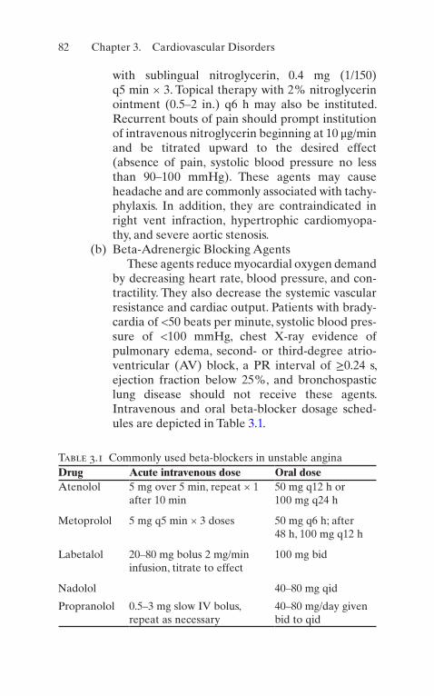
82
with sublingual nitroglycerin, 0.4 mg (1/150) q5 min × 3. Topical therapy with 2% nitroglycerin ointment (0.5–2 in.) q6 h may also be instituted. Recurrent bouts of pain should prompt institution of intravenous nitroglycerin beginning at 10 μg/min and be titrated upward to the desired effect (absence of pain, systolic blood pressure no less than 90–100 mmHg). These agents may cause headache and are commonly associated with tachy-phylaxis. In addition, they are contraindicated in right vent infraction, hypertrophic cardiomyopa-thy, and severe aortic stenosis.
(b) Beta-Adrenergic Blocking AgentsThese agents reduce myocardial oxygen demand
by decreasing heart rate, blood pressure, and con-tractility. They also decrease the systemic vascular resistance and cardiac output. Patients with brady-cardia of <50 beats per minute, systolic blood pres-sure of <100 mmHg, chest X-ray evidence of pulmonary edema, second- or third-degree atrio-ventricular (AV) block, a PR interval of ≥0.24 s, ejection fraction below 25%, and bronchospastic lung disease should not receive these agents. Intravenous and oral beta-blocker dosage sched-ules are depicted in Table 3.1.
Table 3.1 Commonly used beta-blockers in unstable anginaDrug Acute intravenous dose Oral doseAtenolol 5 mg over 5 min, repeat × 1
after 10 min50 mg q12 h or 100 mg q24 h
Metoprolol 5 mg q5 min × 3 doses 50 mg q6 h; after 48 h, 100 mg q12 h
Labetalol 20–80 mg bolus 2 mg/min infusion, titrate to effect
100 mg bid
Nadolol 40–80 mg qid
Propranolol 0.5–3 mg slow IV bolus, repeat as necessary
40–80 mg/day given bid to qid
Chapter 3. Cardiovascular Disorders
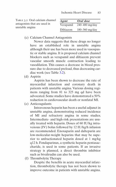
83
(c) Calcium Channel AntagonistsNewer data suggests that these drugs no longer
have an established role in unstable angina although their use has been more used in vasospas-tic or stable angina. It is proposed calcium channel blockers such as verapamil and diltiazem prevent vascular smooth muscle contraction leading to vasodilation. This causes a decrease in blood pres-sure due to decreased preload, thus decreasing car-diac work (see Table 3.2).
(d) AspirinAspirin has been shown to decrease the rate of
myocardial infarction and coronary death in patients with unstable angina. Various dosing regi-mens ranging from 81 to 325 mg qd have been advocated. Some studies have demonstrated a 50% reduction in cardiovascular death or nonfatal MI.
(e) AnticoagulantsIntravenous heparin has been a useful adjunct in
unstable angina, demonstrating reduced incidence of MI and refractory angina in some studies. Intermediate- and high-risk presentations are usu-ally treated with heparin. Doses of 60 IU/kg intra-venous (IV) bolus followed by 12 IU/kg/h infusion are recommended. Enoxaparin and dalteparin are low-molecular-weight heparins that may be supe-rior to unfractionated heparin dosed at 1 mg/kg q12 h. Fondaparinux, a synthetic heparin pentasac-charide, is used in some patients. If an invasive strategy is planned, a direct thrombin inhibitor such as bivalirudin can also be used.
(f) Thrombolytic TherapyDespite the benefits in acute myocardial infarc-
tion, thrombolytic therapy has not been shown to improve outcome in patients with unstable angina.
Table 3.2 Oral calcium channel antagonists that are used in unstable angina
Agent Oral doseVerapamil 240–480 mg/day
Diltiazem 180–360 mg/day
Ischemic Heart Disease
84
(g) Glycoprotein IIb/IIIa Receptor InhibitorsThese agents block the receptors that lead to
platelet aggregation. Two relatively short-acting (4–8 h) agents (eptifibatide, tirofiban) and one longer- acting antibody (abciximab) are available. These agents are beneficial during percutaneous coronary interventions, and the shorter-acting agents are approved for use in non-Q MI and unstable angina being medically managed. Decreased combined end points of death, infarc-tion, and urgent intervention have been reported. These agents should be administered to high-risk patients (ST depression 1 mm, persistent or recur-rent symptoms, widespread electrocardiographic [ECG] abnormalities, depressed left ventricular [LV] function, positive cardiac markers). They should also be used when a second antiplatelet cannot be given in combination with aspirin:
1. Eptifibatide, 180-μg/kg IV bolus followed by a 2-μg/kg/min infusion. A double-bolus regimen has been shown to improve platelet inhibition in some clinical studies (180 mg/kg × 2, 10 min apart). Tirofiban, 0.4 μg/kg/min × 30 min fol-lowed by 0.1-μg/min infusion.
2. Abciximab, 0.25-mg/kg IV bolus followed by 0.125-μg/kg/min infusion.
(h) Clopidogrel—given (75–300 mg loading) in combi-nation with aspirin—can lead to benefit. New P2Y12 inhibitors have been approved since 2009; the first is prasugrel (60 mg loading and then 10 mg/day). This agent is more effective to prevent clinical events but has slightly higher risk of bleeding. The second one is ticagrelor (180 mg loading and then 90 mg BID) and should be used with low doses of aspirin (<100 mg daily).
(i) Morphine—Intravenous morphine sulfate (2–4 mg initially) should be given for the relief of chest pain and anxiety.
Chapter 3. Cardiovascular Disorders
85
(j) Statins—Many authors recommend early adminis-tration of these agents (i.e., atorvastatin 80 mg/day).
(k) ACE inhibitors and angiotensin II receptor block-ers are utilized also in patients with concomitant diabetes, heart failure, and a documented ejection fraction <40%.
2. Nonpharmacologic TherapyEvery patient with angina must be placed on supple-
mental oxygen. Persistent chest pain despite maximal therapy with nitrates, beta-blockers, aspirin, heparin, etc. may require early cardiac catheterization, with the view toward potential mechanical intervention (percu-taneous transluminal coronary angioplasty (PTCA) or coronary bypass surgery). Intra-aortic balloon pump (IABP) insertion should be performed with the goal of stabilizing the patient when needed. The IABP relieves pain and may provide relative stability for evaluation before intervention.
Myocardial Infarction
A. Definition. Myocardial infarction (MI) is necrosis of the cardiac muscle resulting from insufficient supply of oxy-genated blood:
1. Q-Wave MIQ-wave MI presents with ST-segment elevation and the subsequent development of pathologic Q waves in the ECG.
2. Nonacute MIMore than 50% of acute MIs in the United States do not present with ST-segment elevation but rather have nonspecific ECG changes or even normal ECGs.
B. Pathophysiology
1. MI is nearly universally the result of coronary artery atherosclerosis.
Ischemic Heart Disease

86
2. Atherosclerotic lesions reduce and limit the flow through coronary arteries, resulting in ischemic myo-cardial cells.
3. The formation of thrombi plays a significant role in acute MI, and almost all ST-segment elevation infarcts will have an occlusive thrombus in the infarct-related artery if examined early enough in the course of the MI.
4. Occlusion of the right coronary artery (RCA) generally results in inferior/posterior MI.
5. Occlusion of the left anterior descending artery (LAD) generally leads to anterior infarctions, while blockage of the left circumflex artery (LCA) results in lateral and/or inferior/posterior MI.
6. Spasm of the coronary arteries may also play a role in MI. As many as 2% of all MI patients, a significantly higher percentage of those patients who are less than 35 years of age will have angiographically normal coro-nary arteries, and presumably spasm is a significant pathophysiologic event.
C. Risk Factors. Risk factors for coronary artery disease including MI are age, male gender, family history, smok-ing, hypertension, elevated cholesterol, and diabetes mel-litus. Cocaine use is a significant risk factor for MI.
D. Clinical Presentation
1. Patients present with chest pain (typically substernal) lasting 30 min or longer, which is unrelieved by rest or nitroglycerin and pain that may radiate from the left or right arm into the jaw. The pain is typically nonpleuritic and may be associated with dyspnea, diaphoresis, nau-sea, or vomiting.
2. As many as 25% of all MIs are painless, this can espe-cially be seen in the diabetic population as pain recep-tors can be affected.
3. “Burning” discomfort is as predictive of acute MI as pressure-type discomfort.
Chapter 3. Cardiovascular Disorders
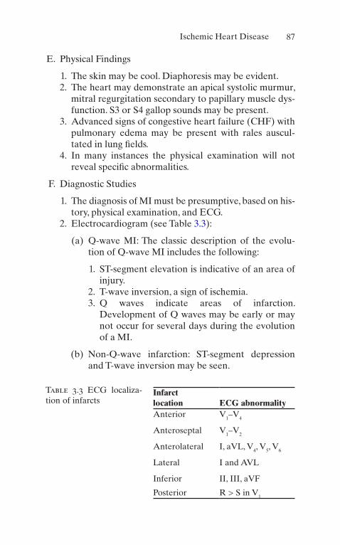
87
E. Physical Findings
1. The skin may be cool. Diaphoresis may be evident. 2. The heart may demonstrate an apical systolic murmur,
mitral regurgitation secondary to papillary muscle dys-function. S3 or S4 gallop sounds may be present.
3. Advanced signs of congestive heart failure (CHF) with pulmonary edema may be present with rales auscul-tated in lung fields.
4. In many instances the physical examination will not reveal specific abnormalities.
F. Diagnostic Studies
1. The diagnosis of MI must be presumptive, based on his-tory, physical examination, and ECG.
2. Electrocardiogram (see Table 3.3):
(a) Q-wave MI: The classic description of the evolu-tion of Q-wave MI includes the following:
1. ST-segment elevation is indicative of an area of injury.
2. T-wave inversion, a sign of ischemia. 3. Q waves indicate areas of infarction.
Development of Q waves may be early or may not occur for several days during the evolution of a MI.
(b) Non-Q-wave infarction: ST-segment depression and T-wave inversion may be seen.
Table 3.3 ECG localiza-tion of infarcts
Infarct location ECG abnormalityAnterior V1–V4
Anteroseptal V1–V2
Anterolateral I, aVL, V4, V5, V6
Lateral I and AVL
Inferior II, III, aVF
Posterior R > S in V1
Ischemic Heart Disease

88
3. Enzyme StudiesNecrotic heart muscle cells release enzymes into the
bloodstream. Classically, cardiac-specific troponins and creatine kinase (CK or CPK) have been used in labora-tory diagnosis of MI:
(a) CK becomes elevated within 24 h. CK is also pres-ent in the skeletal muscle and brain and thus may be released in other clinical conditions. To increase specificity, the assay of the MB isoenzyme of CK is used. This enzyme is found primarily in the myocardium.
(b) Cardiac-specific troponin I and troponin T are reg-ulatory components of the contractile apparatus of the heart. These proteins are very specific for myo-cardial injury and are released into the blood in the hours following myocardial infarction. They remain elevated for several days and are thus quite useful in patients presenting late after infarct. Troponin elevation, however, can occur in the absence of MI in patients with renal dysfunction.
(c) CK-MB subforms: The MB isoenzyme of CK exists in only one form in the myocardial cell. After it is released into the blood stream, enzymatically mediated cleavage of a terminal lysine residue occurs, creating two subforms of CK-MB. The ratio of the freshly released CK-MB to the old cleaved CK-MB is a very sensitive and specific early marker of myocardial injury.
4. Nuclear Medicine TechniquesThallium 201 is taken up by perfused viable cardiac
myocytes and may indicate areas of infarction by the presence of “cold spots.” Unfortunately, this technique may not distinguish between acute MI and previous scar. Technetium 99 (Tc99) results in “hot spots” as the tracer accumulates in damaged myocardial cells.
5. Other diagnostic studies in patients with suspected MI that should be obtained include blood counts, electro-lytes, glucose, blood urea nitrogen (BUN) and creati-nine, and lipid profile.
Chapter 3. Cardiovascular Disorders

89
G. Treatment of Acute MI. Several goals exist in the manage-ment of acute ST-elevation MI (STEMI): minimizing the amount of infracted myocardium, optimizing function, and controlling the complications of acute MI:
1. Patients with suspected MI should have continuous ECG monitoring and an IV line established. They should also receive supplemental oxygen to maintain adequate oxygen saturation.
2. If the clinical condition of the patient permits, sublin-gual nitroglycerin (0.4 mg q5 min × 3) should be given to help differentiate those patients who may be suffer-ing from angina rather than MI.
3. Aspirin (ASA) should be given to all patients without contraindications (160–325 mg PO).
4. Thrombolytic Therapy
(a) Patients with ST-segment elevation without contra-indications for thrombolytic therapy and who pres-ent within 6–12 h of the onset of their symptoms should be considered for thrombolytic therapy. If a cardiac catheterization laboratory is available on- site, we recommend primary percutaneous coro-nary intervention (see below) for all patients with a STEMI. It is presumed that the procedure will be done expeditiously (door-to-balloon time less than 90 min) in such hospitals.
(b) Patients presenting perhaps up to 24 h may also be considered for thrombolysis, as some studies have reported improved outcomes (ISIS II trial LATE trial).
(c) Guidelines for Thrombolytic Agent Administration
1. Symptoms suggestive of acute MI not resolved with sublingual nitroglycerin, lasting 20 min and <12 h
2. ST-segment elevation in two or more continu-ous ECG leads or left bundle branch block not known to be old
Ischemic Heart Disease

90
3. Exclusion Criteria
(a) Bleeding diathesis (b) Active gastric or duodenal ulcers (c) Significant surgery within 3 weeks (d) Severe trauma within 6 months (e) History of cardiovascular accident (CVA)
within 1 year or other central nervous system (CNS) processes or hemorrhage with a potential for bleeding
(f) Severe, poorly controlled hypertension (180/110)
(g) Poor underlying prognosis (i.e., malignancy) where risk/benefit assessment may not favor treatment
4. Thrombolytic Agents
(a) Tissue plasminogen activator (rt-PA, acti-vase) dosage 100 mg (accelerated dosing improves patency rates without increasing complications). Give 15 mg as an IV bolus followed by 50 mg infused over 30 min and the remaining 35 mg infused over 60 min. Concomitantly administer heparin intrave-nously (HART trial evidence suggests unac-ceptable reocclusion rates if rt-PA is given without heparin). ASA should be given as well.
(b) Streptokinase (Streptase)1.5 million units IV over 1 h. Because of
the systemic state induced by streptokinase and high levels of fibrin split products, the need for heparin therapy with streptokinase has been questioned.
(c) Anistreplase anisoylated (plasminogen/streptokinase activator complex, Eminase). This drug is given as a bolus of 30 U over 5 min. Like streptokinase, heparin therapy is uncertain.
Chapter 3. Cardiovascular Disorders
91
(d) Reteplase: This agent is a modified rt-PA. In Gusto III reteplase had a similar efficacy to rt-PA. It is given as two IV boluses of 10 U over 2 min, 30 min apart. Heparin and ASA should be used.
(e) Tenecteplase (TNK): This is a modified rt-PA that is given as a single IV bolus (0.5 mg/kg). In ASSENT-II, this drug was equivalent to rt-PA in overall mortality, with fewer rates of some bleeding complications. Heparin and ASA should be used.
5. Percutaneous Transluminal Coronary Angioplasty (PTCA)/Percutaneous Coronary Intervention (PCI)
PTCA/PCI is an excellent alternative revasculariza-tion technique where available quickly (door-to-needle insertion time of less than 30 min). This should be strongly considered in patients with contraindications to thrombolytic therapy. If skilled operators and facili-ties for rapid institution are available, the outcome of patients with primary PTCA appears equivalent to or better than that obtained with thrombolytic therapy.
6. Beta-blockersBeta-blockers are useful in preventing tachydys-
rhythmias and in reducing myocardial oxygen con-sumption. Early intravenous beta-blockade followed by oral maintenance therapy reduces recurrent isch-emia and infarction, even in patients receiving throm-bolytic therapy. Patients without contraindications should receive these agents, as they also prevent recur-rent ischemia and life-threatening ventricular dysrhyth-mias (see Table 3.1).
7. Angiotensin-Converting Enzyme (ACE) InhibitorsACE inhibitors in the setting of large acute MI may
have an impact on LV remodeling and improve survival in patients with LV dysfunction. These drugs should probably not be administered in the first few hours after infarction.
Ischemic Heart Disease

92
8. Patients should be classified clinically for prognosis as well as to determine therapy (see Tables 3.4 and 3.5).
9. Additional management of patients may be based on the hemodynamic subset in which they fall:
(a) Uncomplicated MI
1. In addition to the therapeutic regimens men-tioned above, IV nitroglycerin should be used in pain control. Clinical studies have suggested that mortality and infarct size may be reduced by the use of nitrates. Therapy should be started at 10 μg/min and increased until the patient is free of pain, or the systolic blood pressure falls below 100 mmHg, or a maximal dose of approxi-mately 200 μg/min has been achieved.
2. Morphine sulfate in 2-mg (IV) increments as needed for pain unrelieved by nitroglycerin.
Table 3.4 Killip classification of acute MIClass I No heart failure: mortality <6%
Class II S3 basal rales: mortality <17%
Class III Acute pulmonary edema: mortality 38%
Class IV Cardiogenic shock: mortality 81%
Adapted from Killip PT: Am J Cardiol 1967;20:457
Table 3.5 Hemodynamic subsets after acute MI
SubsetCardiac index (L/m2)
Wedge pressure (torr)
No pulmonary congestion or peripheral hypoperfusion
2.7 + 0.5 12 ± 7
Isolated pulmonary congestion 2.3 ± 0.4 23 ± 5
Isolated peripheral hypoperfusion
1.9 ± 0.4 12 ± 5
Pulmonary congestion and hypoperfusion
1.6 ± 0.6 27 ± 8
Adapted from Forrester GA: Am J Cardiol 1977;39:137
Chapter 3. Cardiovascular Disorders

93
3. Heparin 5000 SQ q8–12 h or low-molecular- weight heparin in deep venous thrombosis (DVT) prophylaxis doses in patients without contraindications and who are not receiving full- dose heparin should be given. Note: Patients with anterior wall MI have a lower incidence of LV thrombosis if full heparinization is used.
4. Statin therapy to lower cholesterol. 5. Strict bed rest for 24 h followed by gradual
increase in activity. 6. Stool softener, commonly docusate sodium
(Colace) 100 mg PO qd. 7. A low cholesterol, no added salt diet should be
prescribed.
(b) Complicated MI (see Table 3.5)
1. Left Ventricular Dysfunction Manifested by Pulmonary Congestion
(a) Decrease left ventricular end-diastolic pres-sure with IV nitroglycerin, and consider dobutamine, diuretics, or other vasodilators (see dosages below).
2. Patients with Hypoperfusion Without Pulmonary Congestion
(a) Careful IV hydration with normal saline. Pulmonary capillary wedge pressure is tar-geted at approximately 18 mmHg.
(b) Right ventricular MI accompanying inferior infarct may be present in this manner. Diagnosis may be made using right-sided precordial chest ECG leads. Significant vol-ume administration may be required for adequate LV preload.
3. Severe LV DysfunctionPulmonary artery cannulation should be
performed.
Ischemic Heart Disease

94
(a) If the systolic blood pressure is >100 mmHg, dobutamine up to 20 μg/kg/min intrave-nously should be started. Milrinone can also be used. If the patient demonstrates hypo-tension with systolic blood pressure (BP) <70–100 mmHg, dopamine in alpha-agonist doses or vasopressin 1–4 U/h IV or norepi-nephrine 0.5–30 μg/min IV should be administered.
(b) Hypertensive patients should be treated with IV nitroglycerin beginning at 10 μg/min. Dihydropyridine calcium channel blockers (i.e., clevidipine, nicardipine) can also be used.
(c) Mechanical support can be done with:
(i) IABP (ii) Left ventricular and biventricular assist
devices (iii) Percutaneous left atrial-to-femoral
arterial ventricular assist device (iv) Extracorporeal membrane
oxygenation
(c) Other Complications Following MI
1. Mitral Regurgitation: This is characterized by the sudden appearance of a systolic murmur (radiating to the axilla) and worsening CHF.
(a) Diagnostic Studies: Physical examination will demonstrate a systolic murmur and worsening pulmonary congestion. Cardiac catheterization will demonstrate giant V waves in the pulmonary wedge tracing.
(b) Therapy: Afterload reduction (i.e., IV sodium nitroprusside) to decrease pulmo-nary capillary wedge pressure. Hypotensive patients may require catecholamines (i.e., dopamine and/or dobutamine).
Chapter 3. Cardiovascular Disorders
95
(c) IABP may improve coronary perfusion and ventricular emptying.
(d) Surgical Repair.
2. Ventricular Septal Defect (VSD): VSD is an event occurring in <1% of Q-wave MIs and may occur at any point from several hours to several days after the onset of symptoms. It is most com-monly seen during the first 7 days.
(a) Diagnosis: Acute VSD results in a loud holosystolic murmur and sudden severe CHF with cardiogenic shock. Right heart catheterization with oxygen saturation mea-surements will exhibit an oxygen saturation step-up between the right atrium and right ventricle, and “contrast” echocardiography will many times identify the defect.
(b) Treatment: Acute afterload reduction with IV sodium nitroprusside and IABP is required for acute VSD with subsequent surgical repair.
(d) Dysrhythmias Following MI: 90% of the patients suffering from acute MI will have dysrhythmias during the first 24 h.
1. Sinus Bradycardia: The most commonly seen dysrhythmia in acute MI. It should be treated only when signs of diminished cardiac output are present. Atropine 0.5–1 mg IV q3–5 min until a total dose of 0.04 mg/kg has been given. If this proves ineffective, dopamine up to 20 μg/kg/min and epinephrine 10 μg/min should be considered.
2. Supraventricular Dysrhythmias: Sinus tachycar-dia should be addressed by treating the underly-ing cause. Pain relief and sedation many times are all that is required. Patients with atrial fibril-lation or flutter in emergent conditions may
Ischemic Heart Disease

96
require acute cardioversion. Stable patients should be treated with calcium channel blockers, beta-blockers, etc.
3. Paroxysmal Supraventricular Tachycardia: This should be approached initially with vagal maneuvers; if these are unsuccessful, approach with adenosine 6 mg rapid IV push; and if still unsuccessful, approach with adenosine 12 mg rapid IV push, followed by verapamil 2.5–5 mg IV push. Beta-blockers, amiodarone, or procain-amide are alternatives.
4. Ventricular Dysrhythmias: Prophylactic therapy with lidocaine does not result in improvement of overall survival and thus is not indicated in patients with acute MI. In the patient showing stable ventricular tachycardia with normal LV function, amiodarone is given (250 mg over 10 min, followed by an intravenous infusion 1 mg/h for 6 h and then 0.5 mg/min). Other choices include procainamide, administered at 20–30 mg/min to a maximum dose of 17 mg/kg/h. End points for therapy include abolition of dys-rhythmia, 50% widening of the QRS complex, and/or hypotension. Amiodarone is a preferred agent if depressed LV function is present.
5. Magnesium sulfate has also been demonstrated to be useful, particularly in polymorphic ven-tricular tachycardia: 1–2 g over 1–2 min, IV.
(e) Conduction Disturbances Accompanying Acute MI
1. Atrioventricular (AV) Conduction Disturbances
(a) First-Degree AV Block: Occurs in 4–14% of acute MIs.
(b) Second-Degree AV Block, Mobitz Type I: This is progressive prolongation of the PR interval with intermittent nonconduction of an atrial beat. It is commonly seen in infe-rior infarction and rarely progresses to com-plete heart block.
Chapter 3. Cardiovascular Disorders

97
(c) Second-Degree AV Block, Mobitz Type II: Represents 10% of all second-degree blocks during acute MI. This is commonly seen in anterior infarction and infrequently pro-gresses to complete heart block.
(d) Third-Degree AV Block: Occurs in 6% of patients. Mortality with inferior MI is 20–25%; mortality with anterior MI is even greater.
(e) Intraventricular Block: Refers to abnormal-ities within the three divisions of the intra-ventricular conduction system. These blocks may progress to higher degrees of heart block. One in five patients with bundle branch block in acute MI will develop second- degree or third-degree AV block. Mortality rates are double of those who do not.
(f) Complete Heart Block: Occurs frequently in MIs with right bundle branch block plus block of the anterior fascicle or posterior fascicle and, less frequently, an isolated left or right bundle branch block. Similarly, patients with alternating forms of bundle branch block have a high incidence of com-plete heart block.
2. Therapy
(a) Atrioventricular Block
(i) First-Degree AV Block: No specific therapy.
(ii) Mobitz Type I Second-Degree AV Block: Unless unusually slow ventricu-lar rates occur, therapy is not needed. Atropine is given (as for bradycardia) followed by temporary transvenous pacemaker insertion in those patients who are symptomatic.
Ischemic Heart Disease

98
(iii) Mobitz Type II Second-Degree AV Block: Particularly when associated with anterior MI, it should result in placement of transvenous pacemaker.
(iv) Complete Heart Block: Temporary transvenous pacemaker (some would advocate pacemaker therapy in inferior MI for hemodynamically compromised individuals only).
(v) Intraventricular Conduction Disturbances: A transvenous pace-maker should be inserted for right bun-dle branch block plus either anterior fascicular, posterior fascicular, or alter-nating bundle branch blocks. Patients with first-degree AV block and new- onset right or left bundle branch block also should receive transvenous pacing.
Cardiac Pacemakers
A. Definition. Cardiac pacemakers are complicated devices that may be used to accelerate cardiac rate, bypass, block conduction tissue, and/or disrupt dysrhythmias. Advancing technology has resulted in new modes of operation, with dual-chamber pacing being used more often. A five- position code has been developed to describe clinical pac-ing mode (see Table 3.6) (The North American Society of Pacing and Electrophysiology/British Pacing Electrophysiology Group Generic Pacemaker Code).
B. Pacemaker Evaluation. Rhythm strips and 12-lead ECGs can be useful in determining the mode of functioning of cardiac pacemakers placed in ICU patients. Patients should be examined for failure to sense, as indicated by inappropriate pacemaker spikes, and failure to capture, as indicated by pacemaker spikes without subsequent cham-
Chapter 3. Cardiovascular Disorders
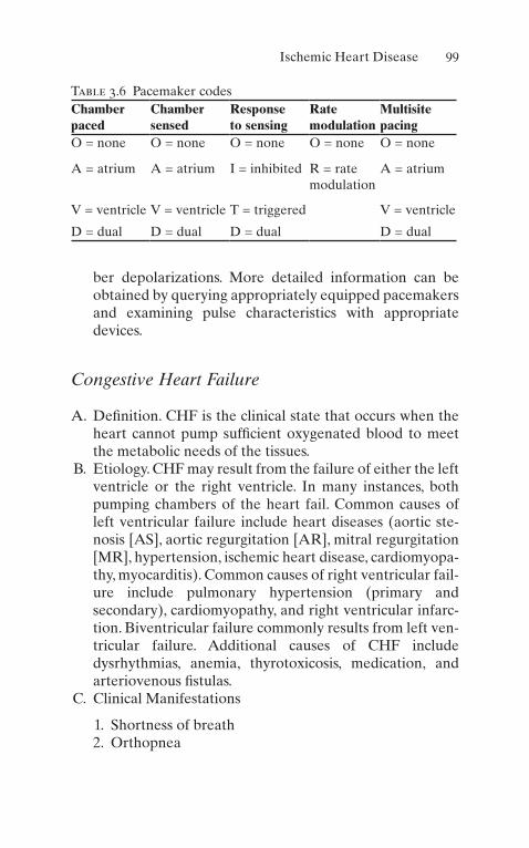
99
Table 3.6 Pacemaker codesChamber paced
Chamber sensed
Response to sensing
Rate modulation
Multisite pacing
O = none O = none O = none O = none O = none
A = atrium A = atrium I = inhibited R = rate modulation
A = atrium
V = ventricle V = ventricle T = triggered V = ventricle
D = dual D = dual D = dual D = dual
ber depolarizations. More detailed information can be obtained by querying appropriately equipped pacemakers and examining pulse characteristics with appropriate devices.
Congestive Heart Failure
A. Definition. CHF is the clinical state that occurs when the heart cannot pump sufficient oxygenated blood to meet the metabolic needs of the tissues.
B. Etiology. CHF may result from the failure of either the left ventricle or the right ventricle. In many instances, both pumping chambers of the heart fail. Common causes of left ventricular failure include heart diseases (aortic ste-nosis [AS], aortic regurgitation [AR], mitral regurgitation [MR], hypertension, ischemic heart disease, cardiomyopa-thy, myocarditis). Common causes of right ventricular fail-ure include pulmonary hypertension (primary and secondary), cardiomyopathy, and right ventricular infarc-tion. Biventricular failure commonly results from left ven-tricular failure. Additional causes of CHF include dysrhythmias, anemia, thyrotoxicosis, medication, and arteriovenous fistulas.
C. Clinical Manifestations
1. Shortness of breath 2. Orthopnea
Ischemic Heart Disease

100
Increased venous return associated with a recumbent position leads to worsening of shortness of breath.
3. Paroxysmal nocturnal dyspneaThis is the result of a number of physiologic factors including the increased venous return in patients who are recumbent.
4. Fatigue and lethargyThese are due to a low cardiac output.
D. Physical Examination. Signs of left ventricular failure on physical examination include tachycardia and tachypnea. Pulmonary venous congestion results in rales bilaterally. S3 and S4 gallop sounds may be heard. Patients with val-vular heart disease may manifest cardiac murmurs. Signs of right heart failure include venous distension in the jug-ular veins, peripheral edema, ascites, and congestive hepa-tomegaly with hepatojugular reflux.
E. Diagnosis of CHF is based on physical exam findings. B-type natriuretic peptide (BNP) or NT-proBNP is useful to support clinical judgment for the diagnosis of acutely decompensated CHF.
F. Classification of CHF. Functional classification is com-monly reported per the New York Heart Association cri-teria (see Table 3.7). Another common way to classify CHF is based on systolic or diastolic dysfunction or based on left ventricular ejection fraction (LVEF). Reduced ejection fraction (with LVEF ≤40 percent) is also known as HFrEF or systolic CHF. Preserved ejection fraction (with LVEF ≥50 percent) is also known as HFpEF or dia-stolic CHF. CHF with mid-range ejection fraction (with LVEF 41–49%) is also known as HFmrEF.
Table 3.7 New York Heart Association Classification of CHFClass I Symptomatic with extraordinary activity
Class II Symptomatic with ordinary activity
Class III Symptomatic with minimum activity
Class IV Symptomatic at rest
Chapter 3. Cardiovascular Disorders

101
G. Chest X-Ray. Cardiomegaly with enlargement of involved heart chambers may be seen. Pulmonary vascular conges-tion progresses to alveolar edema.
H. Echocardiogram: A two-dimensional echocardiogram with Doppler should be used to assess ventricular func-tion, size, wall thickness, wall motion, and valve function.
I. Therapy
1. Correct and identify the underlying cause (i.e., treat anemia, infections, hypertension, control ventricular rate in patients with atrial fibrillation).
2. Decrease cardiac workload with bed rest. 3. Supplemental oxygen is given as necessary.
(a) If respiratory distress and/or hypoxia persist, we suggest noninvasive positive pressure ventilation as long as the patient does not have a contraindication
4. Sodium restriction. 5. Preload reduction.
(a) Nitrates: Venous dilatation associated with nitrates results in prompt improvement of symptoms in many patients with CHF. (See angina section in the beginning of this chapter for dosing recommendations.)
(b) Diuretic Agents
1. Loop-Acting Agents
(a) Furosemide (Lasix) 10–240 mg IV or PO or a continuous IV infusion induces a prompt diuresis and results in venodilatation with rapid improvement in patient symptomatology.
(b) Bumetanide (Bumex) 0.5–1 mg IV or 0.5–2 mg PO.
(c) Thiazides (i.e., hydrochlorothiazide 25–50 mg) are less potent diuretics that may be of value in mild-to-moderate CHF.
Ischemic Heart Disease

102
(d) Metolazone (Zaroxolyn) may potentiate the effect of loop-acting diuretics in doses of 2.5–10 mg.
(e) Morphine sulfate has traditionally been used in the management of severe pulmo-nary edema because of its venodilatory properties and its anxiolytic effects. This agent may depress respirations, and thus other vasoactive substances may be prefer-able. If used, increments of 2 mg IV, titrated to effect, are recommended.
6. Arteriodilators (afterload reduction)
(a) ACE Inhibitors: These agents result in dilatation of the arteriolar resistant vessels and also increase venous capacity, having effects on both preload and afterload. They decrease mortality in patients with CHF and are indicated as long-term medications for patients with reduced EF. Enalaprilat is avail-able for IV administration (1.25 mg IV over several minutes). Oral ACE inhibitors are also available.
(b) Nesiritide: This recombinant human BNP has been used with some success. However, some studies have raised concerns about the adverse impact on mortality rate and a potential risk of worsening renal function.
7. Inotropic agentsIntravenous inotropes (i.e., milrinone or dobuta-
mine) may be used to relieve symptoms and improve end- organ function, particularly if these patients have marginal systolic blood pressure (<90 mmHg), have symptomatic hypotension despite adequate filling pres-sure, or are unresponsive to, or intolerant of, intrave-nous vasodilators.
8. Digitalis glycosides and beta-blockersThese agents have limited value in the acute setting
of pulmonary edema. Beta-blockers reduce mortality in the long-term management of these patients.
Chapter 3. Cardiovascular Disorders

103
9. In severe unresponsive cases or in cases where a differ-ent procedure is indicated due to erythrocytosis, phle-botomy may be used.
Cardiomyopathies
A. Dilated Congestive Cardiomyopathy
1. EtiologiesPrimary disorders of heart muscle in which dilatation of the ventricles and enlargement of the heart occur (see Table 3.8).
2. Symptoms of CHF, dysrhythmias, and pulmonary and systemic embolization
3. Physical ExaminationSigns of CHF are commonly seen. A laterally placed point of maximal impulse [PMI] may be noted along with gallop sounds.
4. Diagnostic Studies
(a) Chest X-Ray: Cardiac enlargement may be seen; pulmonary congestion with interstitial edema, pleural effusion, etc. also may be seen.
(b) ECG: Dysrhythmias may be seen, as may conduc-tion abnormalities, chamber enlargement/hypertro-phy, and nonspecific repolarization abnormalities.
Table 3.8 Etiologies of dilated cardiomyopathy
Idiopathic
Collagen vascular disease
Postmyocarditis
Peripartum
Familial
Toxins and nutritional deficiency
Radiation
Ischemic Heart Disease

104
(c) Echocardiography: May demonstrate a low ejec-tion fraction and global hypokinesis and chamber enlargement.
(d) Cardiac catheterization and myocardial biopsy.
5. Therapy
(a) Treat the underlying cause. (b) Management of CHF as noted above. (c) Prevent thromboembolism. (d) Consider low-dose beta-blocker. (e) Consider transplantation with potential mechani-
cal support as bridging maneuver (i.e., left ventric-ular assist device).
B. Restrictive Cardiomyopathy. This is a myocardial disorder characterized by decreased ventricular compliance.
1. EtiologyInfiltrative disorders (sarcoidosis, hemochromatosis, amyloidosis, etc.), radiation, endocardial fibroelastosis, endomyocardial fibrosis, and scleroderma.
2. SymptomsRight-sided CHF signs, fatigue, and weakness.
3. Specific Diagnostic StudiesEchocardiogram or magnetic resonance imaging (MRI) may help distinguish restrictive cardiomyopathy from constrictive pericarditis (pericardial thickening). Cardiac catheterization and/or biopsy may be used.
4. TherapyControl of CHF as previously noted. Special attention to volume status.
C. Hypertrophic Cardiomyopathy. Familial or sporadic dis-order with marked hypertrophy of the myocardium. Focal or diffuse forms of hypertrophy may occur.
1. Symptoms: Syncope, dyspnea, chest pain, palpitations, sudden death
2. Physical Findings
Chapter 3. Cardiovascular Disorders

105
(a) Crescendo–decrescendo murmur at the left sternal border, which increases with Valsalva maneuver
(b) S4 gallop sound
3. Diagnostic StudiesChest X-ray may be normal; ECG may show left
ventricular hypertrophy and abnormal Q waves (ante-rior, lateral, and inferior leads); echocardiography dem-onstrates ventricular hypertrophy.
4. Treatment
(a) Beta-blockers and/or verapamil. These agents slow the heart rate and prolong diastole, allowing increased ventricular filling.
(b) Surgical and nonsurgical myectomy should be used when optimal medical therapy has failed in appro-priately selected patients. Ethanol septal infusion reduces aortic gradient and symptoms in a large proportion of cases and is rapidly becoming the therapy of choice in severely symptomatic cases.
(c) Digitalis, nitrates, diuretics, and vasodilators may worsen the clinical condition of this subset of patients.
Myocarditis
Myocarditis is an inflammatory condition of the myocardium.
A. Etiology
1. Infection
(a) Viral (echovirus, adenovirus, COVID-19, etc.) (b) Bacterial (c) Mycoplasma (d) Mycotic (e) Rickettsial (f) Spirochetal (g) Parasitic (Trichinella, Trypanosoma cruzi)
Ischemic Heart Disease

106
2. Toxins and drugs (i.e., cocaine) 3. Collagen vascular disease (scleroderma, systemic lupus
erythematosus, rheumatic fever, sarcoidosis)
B. Symptoms
1. Dyspnea 2. Chest discomfort
C. Physical Examination
1. Tachycardia 2. Pericardial friction rub (in the presence of coexistent
pericarditis) 3. Evidence of CHF
D. Therapy
1. Supportive care.
(a) Treatment of CHF (b) Treatment of dysrhythmias, as necessary (c) Anticoagulation to prevent thromboembolism
2. Treat the underlying cause (the use of corticosteroids, immunoglobulins, and immunosuppressive therapy in selective populations with inflammatory infiltrates on endomyocardial biopsy may be warranted).
Pericarditis
Inflammation of the pericardium is associated with many dif-ferent etiologic factors.
A. Etiology (see Table 3.9) B. Symptoms
1. Anterior chest pain, commonly radiating to the arms and back, which classically increases with inspiration and is relieved by sitting up or leaning forward. Palpitations and tachycardia may also occur.
C. Physical Examination
Chapter 3. Cardiovascular Disorders

107
1. Pericardial friction rub is best heard with the patient upright and leaning forward.
2. Tachycardia or other dysrhythmias may be auscultated.
3. If pericardial tamponade occurs, low blood pressure, narrow pulse pressure, and accentuated pulsus para-doxus may be seen.
D. Diagnostic Studies
1. ECG (see Fig. 3.1)An acute MI evolutionary pattern of ECG is seen
with initial ST-segment elevations with concavity upward, subsequent T-wave inversion, and, finally, late resolution of the repolarization abnormalities. PR seg-ment depression may also be seen.
2. Laboratory EvaluationMI should be ruled out as noted above. Other potential
useful studies might include erythrocyte sedimentation rate (ESR), antinuclear antibodies (ANA), rheumatoid factor, viral titers, and tuberculin skin test (PPD).
3. EchocardiogramTo document pericardial effusion (may not be
present).
Table 3.9 Etiologies of pericarditis
1. Idiopathic
2. Infectious (tuberculosis, bacterial, viral, fungal, protozoal)
3. Collagen vascular disease
4. Drug induced
5. Trauma
6. Acute MI and post MI (Dressler’s syndrome)
7. Uremia
8. Postradiation
9. Rheumatic fever
10. Neoplasms
Ischemic Heart Disease

108
E. Treatment
1. Anti-inflammatories (i.e., indomethacin 25–50 mg PO q8 h or salicylates 2–5 g/day). In severe cases or if con-traindications to NSAIDs (renal impairment or GI ulcer history), then corticosteroids (i.e., prednisone 60 mg PO qd) can be used.
2. Analgesia for pain unrelieved by anti-inflammatories. 3. Observation for signs of cardiac tamponade. 4. Treatment of underlying causes.
F. Complications
1. Cardiac Tamponade
Accumulation of pericardial fluid may impair cardiac function, mainly through thinning of diastolic filling.
(a) Symptoms: Dyspnea, orthopnea, and fatigue (b) Physical Findings
1. Neck pain 2. Distant heart sounds
I II III aVR aVL aVF
V1 V2 V3 V4 V5 V6
Figure 3.1 Pericarditis. Diffuse ST-segment elevation, which is con-cave upward, is present in all leads except aVR and V1. Depression of the PR segment, an electrocardiographic abnormality that is common in patients with acute pericarditis, is not evident because of the short PR interval (Braunwald, E: Heart Disease: A Textbook of Cardiovascular Medicine, 5th ed. Saunders, 1997. Used with permission)
Chapter 3. Cardiovascular Disorders

109
3. Tachycardia 4. Pulsus paradoxus 5. Hypotension and a narrow pulse pressure
(c) Diagnostic Studies
1. ECG: Decreased QRS amplitude and beat-to- beat changes in the R wave.
2. Echocardiography: Demonstrates effusions and early right ventricle diastolic collapse.
3. Cardiac Catheterization: Right heart catheter-ization will reveal equalization of diastolic pres-sures, which includes pericardial pressure if measured.
(d) Therapy
1. Pericardiocentesis (see Chap. 15, “Special Techniques”). Removal of a relatively small amount of pericardial fluid will improve diastolic filling in the ventricle and greatly improve the patient’s symptomatology. A drainage catheter may also be left in place. Fluid obtained should be tested for protein, lactic dehydrogenase (LDH), cell count, Gram’s stain, acid-fast bacilli stain (AFB), culture/sensitivity, and cytology.
2. Pericardiectomy, Pericardial Window: These sur-gical procedures may be performed to relieve pericardial tamponade.
Valvular Heart Disease
A. Aortic Stenosis
1. Etiology
(a) Rheumatic inflammation of the aortic valve (b) Progressive stenosis secondary to congenital bicus-
pid valve (c) Congenital aortic stenosis (d) Idiopathic calcification stenosis of the aortic valve
Ischemic Heart Disease
110
2. PathophysiologyStenosis of the aortic valve results in increased resis-
tance to ventricular ejection and increased left ventricu-lar pressure. Hypertrophy of the ventricle will occur. Normal aortic valve area is approximately 3 cm2. Aortic valves of <1 cm2 generally produce symptoms, and those with <0.5 cm2 with pressure gradients of ≥50 mmHg are considered severe.
3. Symptoms
(a) Syncope: Commonly with exertion and frequently associated with vasodilatation in muscle beds, lead-ing to cerebral ischemia
(b) Shortness of breath (c) Palpitations (d) Decreased activity level (e) Chest pain/pressure/tightness
4. Physical Findings
(a) Slow-rising, delayed carotid upstroke with decreased amplitude
(b) Narrowing of pulse pressure (c) Loud systolic ejection murmur heard at the base of
the heart and radiating to the neck, often with a palpable thrill
5. Diagnostic Studies
(a) ECG
1. Left ventricular hypertrophy 2. Nonspecific repolarization abnormalities
(b) Chest X-ray
1. Pulmonary congestion in patients with CHF 2. Aortic dilatation 3. Calcification of the aortic valve
(c) Echocardiography
1. Gold standard for diagnosis 2. Hypertrophy of the left ventricular wall 3. Visualization of the abnormal aortic valve
Chapter 3. Cardiovascular Disorders

111
(d) Cardiac catheterization documents severity of dis-ease and calculation of valve area
6. Therapy
(a) Judicious management for CHF and angina as they occur (see appropriate sections as above. These patients may be very preload sensitive).
(b) Valve replacement should be reserved as palliative therapy for patients who have poor surgical risks.
B. Aortic Insufficiency
1. Etiology
(a) Infective endocarditis (b) Trauma with valvular rupture (c) Congenital bicuspid aortic valve (d) Rheumatic fibrosis (e) Myxomatous degeneration (f) Accompanying aortic dissection
2. PathophysiologyLeft ventricular pressure increases secondary to regur-
gitation of blood from the aorta, resulting in diastolic vol-ume overload and subsequent decompensation.
3. Symptoms
(a) Many patients remain asymptomatic for many years.
(b) Symptoms during decompensation include dys-pnea on exertion, syncope, chest pain, and CHF.
4. Physical Findings
(a) Widening pulse pressure with bounding pulses. Rapid rise and sudden fall in arterial pressure may result in head bobbing, capillary pulsations in the nail beds (Quincke’s pulse), and “water hammer” pulse. In addition, a murmur can be heard over the femoral arteries.
(b) PMI may be displaced laterally, and S3 gallop may be heard. A diastolic blowing decrescendo murmur occurs along the left sternal border.
Ischemic Heart Disease

112
(c) Austin Flint murmur (apical diastolic rumble of low pitch secondary to aortic regurgitation, which affects the anterior mitral leaflet).
(d) Systolic apical ejection murmur may also be heard.
5. Diagnostic Studies
(a) Chest X-Ray: May show left ventricular and/or aortic dilation.
(b) ECG: Left ventricular hypertrophy is usually present.
(c) Echocardiogram: Gold standard for diagnosis. Increased left ventricular dimensions and Doppler documentation of regurgitant aortic flow. Fluttering of the anterior mitral leaflet may also be seen.
(d) Cardiac Catheterization: Contrast study of the aor-tic root will demonstrate aortic regurgitation.
6. Therapy
(a) Medical management of CHF as noted above. (b) Surgical therapy for patients unresponsive to medi-
cal management or with acute aortic regurgitation and left ventricular failure or with a declining ejec-tion fraction.
C. Mitral Stenosis
1. Etiology
(a) Rheumatic fever (b) Congenital defects
2. PathophysiologyThe normal mitral orifice is 4–6 cm2 in area. An
obstruction of the orifice results in impedance of flow into the left ventricle. When the orifice area approaches 1 cm2, symptoms appear.
3. Symptoms
(a) Dyspnea, orthopnea, and paroxysmal nocturnal dyspnea (pulmonary edema may develop follow-ing exertion)
Chapter 3. Cardiovascular Disorders

113
(b) Systemic embolization, secondary to thrombi form-ing in a dilated left atrium
(c) Dysrhythmias, particularly atrial fibrillation (d) Hemoptysis, secondary to persistent pulmonary
hypertension
4. Physical Findings
(a) Auscultation reveals an opening snap in early diastole.
(b) Apical presystolic or mid-diastolic rumble. (c) Accentuated S1, Graham Steell murmur. (d) Pulmonary regurgitation.
5. Diagnostic Studies
(a) ECG: Right ventricle hypertrophy, right axis devia-tion, left atrial enlargement, atrial fibrillation
(b) Chest X-Ray
1. Left atrial enlargement is seen on the lateral chest and a double density on the chest X-ray.
2. Elevation of the left main stem bronchus and widening of the angle between the right and left main stem bronchi.
3. Pulmonary arterial prominence.
(c) Echocardiography: Gold standard for diagnosis. Abnormalities of the valve itself may be seen with calcification and reduction of the E–F slope of the anterior mitral leaflet during diastole.
6. Treatment
(a) Control of ventricular rate in patients with atrial fibrillation and anticoagulation to prevent thromboembolism.
(b) Management of CHF as noted above. (c) Surgical therapy if the valve orifice is less than
approximately 0.8 cm2 or if symptoms persist despite optimal therapy.
(d) Balloon valvuloplasty may be of value in poor sur-gical candidates.
Ischemic Heart Disease

114
D. Mitral Regurgitation
1. Etiology
(a) Papillary muscle dysfunction or rupture of the chordae tendineae (i.e., MI)
(b) Infective endocarditis (c) Left ventricle dilatation of any cause (d) Mitral valvular calcification (e) Rheumatic heart disease (f) Mitral valve prolapse (g) Idiopathic myxomatous degeneration of the mitral
valve (h) Atrial myxoma
2. Symptoms
(a) Dyspnea, orthopnea, and CHF of varying severity (b) Hemoptysis (c) Atrial fibrillation (d) Systemic embolization
3. Physical Findings
(a) Holosystolic murmur at the apex with radiation to the base or to the left axilla
(b) Rarely, early to mid-diastolic rumble secondary to increased mitral blood flow
(c) Signs of CHF (d) Left ventricular lift and apical thrill
4. Diagnostic Studies
(a) ECG: Left atrial enlargement, left ventricular hypertrophy, atrial fibrillation
(b) Chest X-Ray: Left atrial enlargement, left ventricu-lar enlargement, pulmonary congestion
(c) Echocardiography
1. Gold standard for diagnosis. 2. Hyperdynamic left ventricle with enlarged left
atrium 3. Doppler studies demonstrating regurgitant flow 4. Flail leaflet in patients with ruptured chordae
Chapter 3. Cardiovascular Disorders

115
5. Therapy
(a) Medical management of CHF as noted above, with particular attention to afterload reduction and con-trol of ventricular rate.
(b) Severity of acute disease may be temporized with IABP or surgical intervention.
Aortic Dissection
A. Definition. Although commonly called aneurysms, this disorder is more appropriately termed aortic dissection. This condition results when there is a tear of the aortic intima, dissection of blood into the media, and stripping away of the vessel wall from the adventitia.
B. Etiology
1. Hypertension (present in 90% of patients) 2. Connective tissue disorders (i.e., Marfan’s syndrome,
Ehlers–Danlos syndrome) 3. Bicuspid aortic valve 4. Granulomatous arteritis and syphilitic aortitis 5. Pregnancy 6. Aortic injury
C. Classification. These lesions are commonly classified by their location (see Fig. 3.2). Type A dissections involve the proximal aorta, from the aortic valve to the aortic arch, and type B dissections arise beyond the takeoff of the left subclavian artery.
D. Symptoms
1. Chest pain (almost always present and usually abrupt, severe, and tearing or burning)
2. Syncope 3. CHF 4. Cerebrovascular accidents
E. Physical Findings
1. Hypotension or hypertension 2. Pulse deficits
Ischemic Heart Disease

116
3. Aortic regurgitation murmur 4. Pericardial friction rub 5. Neurologic signs 6. Horner’s syndrome and/or hoarseness
F. Diagnostic Studies
1. Chest X-Ray
(a) Abnormal in 90% of aortic dissections (b) Widened aortic shadow (c) Pleural effusions (d) Aortic calcification
a b
Figure 3.2 Classification of aortic dissection (Stanford). In type A, the ascending aorta is dissected (a). The intimal tear has always been at point 1 but can occur at position 2 or 3. In type B dissection, the dissection is limited to the descending aorta (b). (Thubrikar, M: Vascular Mechanics and Pathology, New York, Springer, 2007. Used with permission)
Chapter 3. Cardiovascular Disorders

117
2. ECG: Frequently abnormal (90%), however nondiagnostic.
3. Computed tomography of the chest with or without contrast will reveal the lesion.
4. Echocardiogram: Transthoracic and transesophageal studies may reveal the dissection.
5. Aortogram: The old “gold standard” for diagnosis. 6. Magnetic resonance imaging.
G. Therapy
1. Surgical
(a) Proximal dissections (type A) (b) Distal dissection, particularly if vital organs are
compromised or persistent pain occurs despite medical management
2. Medical
(a) Aggressive control of blood pressure. The typical regimen is administration of IV beta-blocker (tra-ditionally, propranolol 1 mg IV q5 min until evi-dence of beta-blockade or labetalol) followed by sodium nitroprusside. Initial targets of heart rate less than 60 bpm and systolic blood pressure between 100 and 120 mmHg are recommended in order to maintain adequate end-organ perfusion.
(b) Alternative regimens using clevidipine or nicardip-ine combined with a beta-blocker have also been used.
(c) Transcatheter stenting techniques are becoming more common and used in some centers.
Shock States
Shock can be defined as a state of inadequate tissue perfu-sion, which, unless reversed, results in progressive organ dys-function, damage, and death. Mortality rates for shock of many causes still exceed 50%. In early stages of shock, the
Ischemic Heart Disease

118
patient may be relatively asymptomatic. Sympathetic dis-charge and other compensatory mechanisms may cause tachycardia and mild peripheral vasoconstriction in attempts to maintain blood pressure. When the state of shock worsens, organ hypoperfusion continues and blood pressure declines; signs of organ dysfunction including restlessness and agita-tion, decreased urine output, and cool and clammy skin become evident.
A. Classification and Etiology. A number of classification schemes for shock states have been devised. These include cardiogenic shock; myopathic (reduced systolic function, i.e., acute MI), mechanical (mitral regurgitation, ventricu-lar septal defect), and extracardiac obstructive shock (i.e., pericardial tamponade, massive pulmonary embolus, or severe pulmonary hypertension); oligemic shock (i.e., hemorrhage or fluid depletion); and distributive shock (i.e., septic shock, anaphylaxis, neurogenic shock, etc.).
B. Diagnostic Evaluation
1. Physical Examination. Tachycardia, hypotension, and evidence of hypoperfusion (i.e., altered mental status, decreased urine output, cool and clammy skin) are gen-erally present. Other manifestations may be seen on physical examination, depending upon the etiology of the shock state.
2. Laboratory Evaluation
(a) ECG: Useful for identifying dysrhythmias and acute MI.
(b) Chest X-Ray: Pneumothorax, abnormal cardiac sil-houette, and pulmonary edema.
(c) Hematology and Chemistry: CBC, BUN, creati-nine, electrolytes, glucose, liver function tests (LFTs), and arterial blood gases should be obtained in the evaluation of any patient with shock.
3. Monitoring
(a) Foley Catheter: Patients in shock without contrain-dication should receive catheter insertion for the monitoring of urine output.
Chapter 3. Cardiovascular Disorders

119
(b) Arterial Line: For direct intra-arterial pressure determination and to allow easy vascular access for laboratory and arterial blood gas monitoring.
(c) Central Venous Pressure (CVP) Monitoring Catheter, Oximetric CVP Catheter, or (Rarely) PA Catheter: These are commonly employed. Most patients can be managed without pulmonary artery cannulation, and the PA catheter is being used less frequently.
C. Therapy. Primary treatment goals include restoring oxy-gen transport and organ perfusion (i.e., urine output 0.5–1 mL/kg/h and the absence of lactic acidosis).
1. Airway, breathing, and circulation (ABCs), as in all critically ill patients.
2. Supportive Measures
(a) Two large-bore IV catheters for those patients requiring volume resuscitation. For patients in whom fluid status is normal or elevated, a central IV line for administration of medication will usu-ally be required. For those patients not volume overloaded, the initial management of hypotension and shock is volume administration. Volume chal-lenges of 250 cc to 1 L at a time should be rapidly administered with reassessment of the patient’s clinical condition.
(b) Supplemental oxygen appropriate for the patient’s clinical status.
(c) Those patients not responding to volume adminis-tration should receive beta-receptor stimulants. Dopamine, although commonly used in hypoten-sive patients, is not ideal as it produces significant shunts and can decrease cerebral perfusion pres-sure. In the authors’ experience, vasopressin 1–6 U/h is the agent of choice in these conditions. Other agents, such as norepinephrine, can also be used.
Ischemic Heart Disease

120
(d) Mechanical support of the circulation may be nec-essary in patients with refractory cardiogenic shock and amenable lesions.
(e) Additional interventions and therapeutic goals for management of shock must be based on specific etiologies.
Infective Endocarditis
A. Definition. Infection of the endocardial structures of the heart.
B. Etiology
1. Streptococcus viridans: Streptococci is the most com-mon organism isolated, excluding prosthetic valve or right-sided endocarditis.
2. Staphylococcus aureus: The most frequent organism isolated in right-sided endocarditis.
3. Staphylococcus epidermidis. 4. Others.
(a) Gonococci (b) Other bacteria (HACEK Group) (c) Fungi
C. Risk Factors. A number of disorders and behaviors are risk factors for the development of endocarditis. These include the following:
1. Valvular abnormalities
(a) Rheumatic valvulitis (b) Bicuspid aortic valve (c) Aortic stenosis or insufficiency (d) Mitral stenosis, prolapse, or insufficiency (e) Mechanical heart valves (f) Previous endocarditis
2. IV drug abuse 3. Marfan’s syndrome 4. Instrumentation
Chapter 3. Cardiovascular Disorders
121
D. Diagnosis
1. History and Physical Examination. Careful history for underlying risk factors should be elicited.
2. Physical Examination
(a) Fever: Generally present but may not be noted in elderly or immunocompromised patients.
(b) Cardiac Murmurs: Are usually present but may not be detected, particularly in right-sided endocarditis.
(c) Peripheral Manifestations: These include painless erythematous papules and macules of the soles and palms (Janeway lesions) and painful erythematous subcutaneous papules (Osler’s nodes), as well as petechia and splinter hemorrhages of the nail beds.
3. Laboratory Evaluation
(a) Blood Cultures: Before antibiotic therapy, positive cultures are quite common (85–95%). Reasons for negative cultures include prior antibiotic therapy, slow-growing or very fastidious organisms, or improper collection.
(b) Nonspecific Laboratory Findings
1. Includes decreased hemoglobin/hematocrit. 2. Elevated, decreased, or normal white blood cell
count with a left shift, hematuria on urinalysis, and an elevated sedimentation rate. Rheumatoid factor may be positive in half of the cases by 6 weeks, and assays of teichoic acid antibodies have been advocated for Staphylococcus aureus endocarditis.
(c) Echocardiography: Transthoracic and transesopha-geal echocardiography may reveal valvular dam-age, impairments of left ventricular function, and valvular vegetations. Transesophageal echocar-diography enhances sensitivity. Some patients will not demonstrate abnormal echocardiographic studies.
Ischemic Heart Disease
122
E. Major Complications
1. CHF secondary to valvular destruction, dysrhythmias, or myocarditis
2. Embolization 3. Cardiac dysrhythmias 4. Myocarditis and pericarditis
F. Therapy
1. Antibiotics appropriate for the clinical setting. For a valvular endocarditis, penicillin G IV (12–24 million U/day) and gentamicin (dosed by body weight and renal function) are commonly advocated. For IV drug addicts, penicillinase-resistant penicillin or vancomycin plus gentamicin are advocated.
2. Surgical therapy for endocarditis should occur if severe heart failure or valvular obstruction is present or if uncontrolled infection exists. Relative indications for cardiac surgery include two or more embolic events, unusually large vegetations, extension of the infection to other intracardiac structures, or in the case of pros-thetic valve endocarditis, periprosthetic leak.
Dysrhythmias (See Also Chap. 2)
A. Supraventricular Dysrhythmias. A group of dysrhythmias whose site of origin and pathway is not confined to the ventricles.
1. Paroxysmal Supraventricular Tachycardia (PSVT). PSVT commonly originates through a reentrant mecha-nism in the AV node, characterized by abrupt onset and termination. PSVT may occur in young patients with-out other evidence of cardiac disease, as well as in patients with acute MI, Wolff–Parkinson–White syn-drome, or other structural heart diseases.
(a) ECG Characteristics: Regular tachycardia of 150–220 beats per minute. Atrial activity (P waves) may or may not be seen, depending upon the rate and relationship between atrial and ventricular depo-
Chapter 3. Cardiovascular Disorders

123
larizations. QRS complex is frequently narrow. However, a wide QRS complex may be seen.
1. Symptoms
(a) Palpitations (b) May produce hypotension during acute MI
or may precipitate CHF
2. Therapy
(a) ABCs. (b) For patients demonstrating clinical instabil-
ity (i.e., cardiogenic shock, ischemic chest pain, or CHF), synchronized DC counter-shock should be used, starting with a charge of 50 J.
(c) Treatment of the stable patient should begin with a vagal maneuver. Valsalva or carotid sinus massage following exclusion of carotid disease may abort the dysrhythmia.
(d) Adenosine should be administered to those patients who do not respond to vagal maneu-vers, 6-mg rapid IV bolus. A second bolus of 12 mg rapid IV may be given. Methylxanthines (i.e., theophylline, amino-phylline, caffeine) are competitive antago-nists, and dipyridamole enhances the pharmacologic effect of adenosine.
(e) Verapamil 5–10 mg over 5 min (may repeat dose in 20–30 min if ineffective) may be used if adenosine is ineffective in patients with narrow complex PSVT. Pretreatment with a slow injection of 10 mL of 10% cal-cium chloride may decrease the common hypotensive effects of this drug. Patients with wide-complex tachycardia that cannot be confidently diagnosed as supraventricu-lar should not receive verapamil nor should patients with depressed ejection fraction. Digoxin or amiodarone as noted above for stable PSVT should be considered.
Ischemic Heart Disease

124
(f) Additional considerations include digoxin, beta-blockers, propafenone, diltiazem, pace termination, and synchronized cardiover-sion for patients with preserved ejection fraction.
B. Atrial Fibrillation. Atrial fibrillation is characterized by chaotic atrial activity without an organized atrial rhythm. This dysrhythmia may accompany coronary artery dis-ease, mitral and aortic valvular disease, thyrotoxicosis, peri- and myocarditis, alcoholic heart disease, and MI without evidence of other organic cardiac diseases.
1. ElectrocardiogramIrregular, chaotic atrial activity without an organized
rhythmic pattern. Conductive QRS complexes will have an irregularly irregular pattern. However, atrial ventricular block with emergence of a lower pace-maker site may result in irregular ventricular response.
2. Other Diagnostic Studies
(a) Thyroid function tests (b) Echocardiography
3. Therapy
(a) In unstable patients, as in PSVT, DC cardiover-sion is indicated.
(b) Digoxin Loading: Digoxin has been the tradi-tional therapy for new-onset atrial fibrillation. Administer 0.5-mg IV loading dose followed by 0.25 mg q3–4 h until the ventricular rate is con-trolled. For patients with impaired ejection frac-tion, this remains a good choice.
(c) Alternatives for rapid rate control include:
1. Diltiazem 20–25 mg IV over 2 min with con-tinuous infusion of 5–15 mg/h.
2. Beta-blockers (i.e., propranolol 0.5 mg IV slowly followed by boluses of 1 mg q5 min to a total of 0.1 mg/kg). Atenolol 5 mg IV slowly × 2. Metoprolol 5 mg slow IV q5 min × 3.
Chapter 3. Cardiovascular Disorders

125
3. Amiodarone in a dose of 150 mg over 10 min followed by 360 mg over 6 h and 0.5 mg/min is a good choice, particularly with depressed ejection fraction.
(d) Anticoagulation is advisable pre- and postcar-dioversion, particularly in patients with mitral valve disease or a history of embolic phenome-non. Patients with a duration of atrial fibrillation >48 h should not be converted acutely, if avoid-able, because of embolization risk. Three to 4 weeks of anticoagulation precardioversion is recommended, with at least 4 weeks of antico-agulation postcardioversion suggested.
(e) Stroke risk in atrial fibrillation—CHADSVASC score
Congestive Heart Failure (one point)High blood pressure (one point)Age (two points if above 75)Diabetes (one point)Previous stroke or clot (two points)Vascular disease (one point)Age (one point if between 65 and 74)Sex (one point if female)
CHA2DS2-VASc score
Risk of ischemic stroke
Risk of stroke/TIA/systemic embolism
0 0.2% 0.3%
1 0.6% 0.9%
2 2.2% 2.9%
3 3.2% 4.6%
4 4.8% 6.7%
5 7.2% 10.0%
6 9.7% 13.6%
7 11.2% 15.7%
8 10.8% 15.2%
9 12.2% 17.4%
Ischemic Heart Disease
126
CHA2DS2-VASc score of 2 and above, oral anticoagulation therapy (OAC) with a vitamin K antagonist (e.g., warfarin), or one of the direct oral anticoagulant drugs (DOACs, e.g., dabigatran, rivaroxaban, or apixaban) is recommended.
C. Atrial Flutter. Atrial flutter is characterized by a rapid regu-lar atrial rate of 280–340 beats per minute, generally associ-ated with varying degrees of AV block. This dysrhythmia may occur with coronary artery disease, including MI, thy-rotoxicosis, pulmonary embolism, and mitral valve disease.
1. ECG: Atrial depolarization classically has a “sawtooth” pattern with varying AV conduction block. Vagal maneuvers may slow the ventricular response rate, making atrial flutter waves more readily apparent.
2. Therapy
(a) As for atrial fibrillation. In patients who are com-promised, cardioversion with DC countershock is indicated.
(b) Commonly responds to the pharmacologic inter-ventions previously denoted for atrial fibrillation. In addition, atrial pacing may also terminate atrial flutter.
D. Multifocal Atrial Tachycardia. In multifocal atrial tachy-cardia, a chaotic irregular atrial activity is seen, with rates between 100 and 180 and varying P-wave morphology (three consecutive different P-wave morphologies). This disorder commonly accompanies chronic obstructive pul-monary disease, theophylline toxicity, hypoxemia, and/or other metabolic disturbances.
1. ECG (see Fig. 3.3)
Varying P-to-P intervals and beat-to-beat variability in P-wave morphology
2. Therapy
(a) Treat the underlying cause. Rate may be controlled, if necessary, with diltiazem or amiodarone. In the absence of depressed ejection fraction, beta-
Chapter 3. Cardiovascular Disorders

127
blockers may be used. In addition, IV magnesium has been advocated by some authorities.
E. Bradycardias and AV Conduction Blocks. These are char-acterized by a low intrinsic rate from the sinus node or blockade of sinus impulses in the AV node, which result in slow ventricular rates.
1. Etiology
(a) Vagotonia (b) Ischemic heart disease (c) Cardiomyopathies (d) Drugs (e) Degenerative diseases of the AV conduction
system
2. Treatment
Patients who are symptomatic due to low ventricular response rate may be treated with the following:
(a) Initially with atropine 0.5–1.0 mg IV, repeated every 3–5 min with a total dose of 0.04 mg/kg.
(b) Transcutaneous pacing when available may be employed.
(c) Pharmacologic therapy may include dopamine 5–20 μg/kg/min IV infusion or epinephrine 2–10 μg/min IV infusion titrated to heart rate. Patients requiring transcutaneous pacers for high degrees of AV block should receive consideration for urgent transvenous pacemaker placement.
Lead II
Figure 3.3 Multifocal atrial tachycardia (Criner, G: Critical Care Study Guide, Springer, 2002. Used with permission)
Ischemic Heart Disease

128
F. Ventricular Tachycardia. Ventricular tachycardia is defined as three or more consecutive beats of ventricular origin. Common rates are between 100 and 200 beats per minute. The differentiation of ventricular and supraventricular dysrhythmia with conduction may be difficult. A good rule of thumb is that wide-complex QRS tachycardia should be considered ventricular tachycardia until proven otherwise.
1. Monomorphic ventricular tachycardia (a single QRS morphology) should be treated as noted in Chap. 3, “The Basics of Critical Care.”
2. Polymorphic ventricular tachycardia or “torsade de pointes” may be caused by agents frequently used in the treatment of monomorphic ventricular tachycardia. Electrolyte disturbances including hypokalemia, hypo-magnesemia, and the presence of cardiac and psycho-tropic medications should be sought. Unstable patients should receive electrical therapy as previously outlined. Stable patients may respond to overdrive pacing, IV magnesium, and correction of underlying causes.
Hypertensive Crises
Hypertensive crises are potentially life-threatening situations that are the result of elevated blood pressure. One percent of all patients with hypertension (HTN) may present with hypertensive crises. Manifestations include hypertension with end-organ dysfunction (see Table 3.10). They rarely occur
Table 3.10 End-organ dysfunction in hyperten-sive emergencies
Hypertensive encephalopathy
Acute aortic dissection
Acute myocardial infarction
Acute cerebral vascular accident
Acute hypertensive renal injury
Acute congestive heart failure
Chapter 3. Cardiovascular Disorders

129
with blood pressures <130 mmHg diastolic. Hypertensive crises can be further classified into hypertensive urgencies (when the BP is elevated but no active organ dysfunction is found) and hypertensive emergencies (when there is evi-dence of organ dysfunction). The management of hyperten-sive urgencies differs from that of hypertensive emergencies.
A. Treatment
1. Blood pressure should be promptly reduced in patients with hypertensive emergencies. Most authorities rec-ommend mean arterial pressure reductions of approxi-mately 15% (10–20%) in the first hour with gradual reduction to diastolic BP of 100–110 mmHg or a reduc-tion of 25% of initial reading over the first 4–24 h.
2. Reductions in BP may result in ischemia, and thus these patients must be carefully followed. Parenteral therapy with short-acting agents is initially recommended (see Table 3.11).
3. Patients receiving parenteral therapy commonly need continuous arterial pressure monitoring.
(a) Cyanide poisoning may occur with IV administra-tion of sodium nitroprusside to renal failure patients. Consider this if such patients develop CNS depression, seizures, lactic acidosis, or cardio-vascular instability.
Table 3.11 Intravenous antihypertensive medicationsClevidipine Start at 2 mg/h and double to the dose every 3 min
(max 32 mg/h)
Labetalol 20-mg bolus, 2 mg/min (max 300 mg/day)
Esmolol 1 mg/kg ideal body weight as bolus then 50–150 μg/kg/min
Nicardipine 5–20 mg/h
Fenoldopam 0.1–2 μg/kg/min
Nitroglycerin 5 μg/min (increase by 5–10 μg q3–5 min as needed)
Diazoxide 25–150 mg IV over 5 min or infusion of 30 mg/min to effect
Ischemic Heart Disease

130
1. May occur with infusion rate 2 μg/kg/min. 2. Infusion rates of 10 μg/kg/min should not be
continued for prolonged periods of time because of this hazard.
3. If cyanide intoxication suspected, discontinue infusion and treat as described in Chap. 16, “Toxicology.”
4. Oral therapy with clonidine (Catapres) (0.1mg PO q20 min) or a host of other agents may be used in less severe cases of hypertension in the ICU patient.
Useful Facts and Formulas
A. Pressure = Flow × Resistance: This is true in the airways as well as in the cardiovascular system. For example:
Mean arterial pressure = cardiac output × systemic vascu-lar resistance.
Mean pulmonary arterial pressure = cardiac output × pul-monary vascular resistance.
The unmeasured resistance term is usually calculated by solving the equations:
systemic vascular resistance
mean arterial pressure
cardiac outp=
uut
B. The Primary Determinants of Cardiovascular Performance
1. Heart rate 2. Preload 3. Afterload 4. Contractility
C. Other Principles and Conversion FactorsFluid flow:
Fluid flowpressure difference radius
vessel length fluid�
� �� �� �
4
vviscosity� �8
Chapter 3. Cardiovascular Disorders
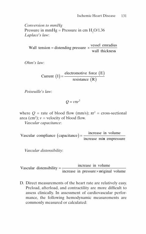
131
Conversion to mmHgPressure in mmHg = Pressure in cm H2O/1.36Laplace’s law:
Wall tension distending pressure
vessel emradius
wall thicknes� �
ss
Ohm’s law:
Current I
electromotive force E
resistance R� � � � �
� �
Poiseuille’s law:
Q v r� � 2
where Q = rate of blood flow (mm/s); πr2 = cross-sectional area (cm2); v = velocity of blood flow.
Vascular capacitance:
Vascular compliance capacitance
increase in volume
increase� � �
minn empressure
Vascular distensibility:
Vascular distensibilityincrease in volume
increase in pressure�
�ooriginal volume
D. Direct measurements of the heart rate are relatively easy. Preload, afterload, and contractility are more difficult to assess clinically. In assessment of cardiovascular perfor-mance, the following hemodynamic measurements are commonly measured or calculated:
Ischemic Heart Disease

132
1. Arteriovenous Oxygen Content Difference (avDO2)This is the difference between the arterial oxygen
content (CaO2) and the venous oxygen content (CvO2).
2. Body Surface Area (BSA)Calculated from height and weight, BSA is gener-
ally used to index measured and derived values according to the size of the patient.
3. Cardiac Index (CI)Calculated as cardiac output/BSA, CI is the prime
determinant of hemodynamic function. 4. Left Ventricular Stroke Work Index (LVSWI)
LVSWI is the product of the stroke index (SI) and mean arterial pressure [MAP] − pulmonary artery occlusion pressure [PAOP] and a unit correction fac-tor of 0.0136. The LVSWI measures the work of the left ventricle (LV) as it ejects into the aorta.
5. Mean Arterial Pressure (MAP)The MAP is estimated as one-third of pulse pres-
sure plus the diastolic blood pressure. 6. Oxygen Consumption ( VO2 )
Calculated as C(a–v)O2 H CO H 10, it is the amount of oxygen extracted in mL/min by the tissue from the arterial blood.
7. Oxygen Delivery (ḊO2)Calculated as (CaO2) H CO H 10, it is the total oxy-
gen delivered by the cardiorespiratory systems. 8. Pulmonary Vascular Resistance Index (PVRI)
Calculated as (MAP − PAOP)/CI, it measures the resistance in the pulmonary vasculature.
9. Right Ventricular Stroke Work Index (RVSWI)RVSWI is the product of the SI and mean pulmo-
nary artery pressure [MPAP] − central venous pres-sure [CVP] and a unit correction factor of 0.0136. It measures the work of the right ventricle as it ejects into the pulmonary artery.
10. Stroke Index (SI)Calculated as CI/heart rate, SI is the average vol-
ume of blood ejected by the ventricle with each beat.
Chapter 3. Cardiovascular Disorders

133
11. Systemic Vascular Resistance Index (SVRI)Calculated as (MAP − CVP)/CI, SVRI is the custom-
ary measure of the resistance in the systemic circuit. 12. Venous Admixture (Qva/QT)
Calculated as CcO CaO CcO CvO2 2 2 2�� � �� �/ , it represents the fraction of cardiac output not oxygen-ated in an idealized lung.
E. Cardiac Output Formulas
Output of left ventricleO consumption mL
AO VO�
� �� �� �� ��
2
2 2
/ min
It may also be measured by thermodilution techniques:
Q V K t� �� � � � �Tb Ti Tb dt/
where Q = cardiac output; V = volume of injectate; Tb = blood temperature; Ti = injectate temperature; K = a constant including the density factor and catheter characteristics; ∫Tb(t)dt = area under the blood temperature–time curve.
The same principle is applicable for the pulmonary blood flow:
Q B� �� �/ Cv Ca
where Q = pulmonary blood flow; B = rate of loss of the indi-cator of alveolar gas; Cv = concentration of the indicator in the venous blood; Ca = concentration of the indicator in the arterial blood.
Q V� �� �/ CaO cO2 2
where Q = total pulmonary blood flow; VO2 = oxygen uptake; CaO2 = arterial oxygen concentration; CVO 2 = mixed venous oxygen concentration.
Ischemic Heart Disease

134
F. Other Cardiovascular Performance Formulas/Tables
Alveolar arterial O difference or A a gradient
Alveolar pO art
� �� �
2
2 eerial O
Torr mmHg2
10 10Normal � � �
Alveolar PO at sea PAO
FiO PaCO2 2
2 2713 1 2
� �� �� � � �.
Arterial blood O content CaO
PaO Hb in g arter2 2
2 0 003 1 34
� �� �� � � � �. . iial blood Hb O sat
Normal mL dL2
18 20
%
/
� �� �
Arteriovenous oxygen difference avDO
CaO CvO2
2 2
4
� �� � � � � �
�Normal �� 5 mL dL/
Cardiac index CI cardiac output body surface area� � �� �
/
.Normal 3 0 3.. / min4 2L m�
Ejection Fraction
end diastolic volume end systolic volum
EF� �
�� �� ee
end diastolic volume
� ��
�%
Mean arterial or empulmonary pressure
DBP SBP DBP
� �� � �� �1 3/
Mean pulmonary arterial pressure
DPAP em SPAP DPAP� � �� �1 3/
Chapter 3. Cardiovascular Disorders
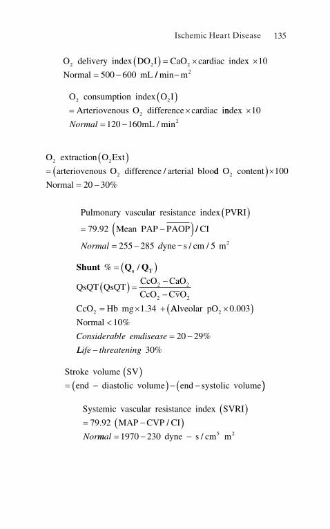
135
O delivery index DO I CaO cardiac index
Normal mL2 2 2 10
500 600
� � � � �� � // min�m2
O consumption index O I
Arteriovenous O difference cardiac i2 2
2
� �� � nndex
mL
�� �
10
120 160 2Normal / min
O extraction O Ext
arteriovenous O difference arterial bloo2 2
2
� �� / dd O content
Normal2 100
20 30
� ��� � %
Pulmonary vascular resistance index PVRI
Mean PAP PAOP
� �� �� �79 92. //
/ /
CI
yne s cm mNormal d� �255 285 5 2-
Shunt Q Qs T% /
.
� � �
� � � ��
� � �
QsQT QsQTCcO CaO
CcO CvO
CcO Hb mg
2 2
2 2
2 1 34 AAlveolar pO
Normal2 0 003
10
20 29
�� ��
� �
.
%
%Considerable emdisease
LLife threatening� 30%
Stroke volume SV
end diastolic volume end systolic volume
� �� �� � � �� ��
Systemic vascular resistance index SVRI
MAP CVP CI
� �� �� �79 92. /
Normmal � � �1970 230 5 2dyne s cm m/
Ischemic Heart Disease

136
Venous
Hb
blood O content CvO
PvO in g venous b2 2
2 0 003 1 34
� �� �� � � � �. . llood Hb O sat
mL dL2
13 16
%
/
� �� �Normal
Normal hemodynamic parameters are depicted in Tables 3.12, 3.13, and 3.14:
Table 3.12 Normal hemodynamic parameters, adultParameter Equation Normal rangeArterial blood pressure (BP)
Systolic (SBP) 90–140 mmHg
Diastolic (DBP) 60–90 mmHg
Mean arterial pressure (MAP)
[SBP + (2 2 × DBP)]/3 70–105 mmHg
Right atrial pressure (RAP)
2–6 mmHg
Right ventricular pressure (RVP)
Systolic (RVSP) 15–25 mmHg
Diastolic (RVDP) 0–8 mmHg
Pulmonary artery pressure (PAP)
Systolic (PASP) 15–25 mmHg
Diastolic (PADP) 8–15 mmHg
Mean pulmonary artery pressure (MPAP)
[PASP + (2 2 × PADP)]/3 10–20 mmHg
Pulmonary artery wedge pressure (PAWP)
6–12 mmHg
Left atrial pressure (LAP)
6–12 mmHg
Cardiac output (CO)
HR × SV/1, 000 4.0–8.0 L/min
Chapter 3. Cardiovascular Disorders

137
Table 3.12 (continued)
Parameter Equation Normal range
Cardiac index (CI) CO/BSA 2.5–4.0 L/min/m2
Stroke volume (SV)
CO/HR × 1, 000 60–100 mL/beat
Stroke volume index (SVI)
CI/HR × 1, 000 33–47 mL/m2/beat
Systemic vascular resistance (SVR)
80 × (MAP − RAP)/CO 800–1200 dynes/cm5
Systemic vascular resistance index (SVRI)
80 × (MAP − AP)/CI 1970–2390 dynes/cm5/m2
Pulmonary vascular resistance (PVR)
80 × (MPAP − PAWP)/CO
<250 dynes/cm5
Pulmonary vascular resistance index (PVRI)
80 × (MPAP − PAWP)/CI 255–285 dynes/cm5/m2
Table 3.13 Hemodynamic parameters, adultParameter Equation Normal rangeLeft ventricular stroke work (LVSW)
SV × (MAP − PAWP) × 0.0136 58–104 g-m/beat
Left ventricular stroke work index (LVSWI)
SVI × (MAP − PAWP) × 0.0136 50–62 g-m/m2/beat
Right ventricular stroke work (RVSW)
SV × (MPAP − RAP) × 0.0136 8–16 g-m/beat
Right ventricular stroke work index (RVSWI)
SV × (MPAP − RAP) × 0.0136 5–10 g-m/m2/beat
(continued)
Ischemic Heart Disease

138
Parameter Equation Normal range
Coronary artery perfusion pressure (CPP)
Diastolic BP − PAWP 60–80 mmHg
Right ventricular end-diastolic volume (RVEDV)
SV/EF 100–160 mL
Right ventricular end-systolic volume (RVESV)
EDV − SV 50–100 mL
Right ventricular ejection fraction (RVEF)
SV/EDV 40–60%
Table 3.13 (continued)
Chapter 3. Cardiovascular Disorders

139
Tabl
e 3.
14 O
xyge
nati
on p
aram
eter
s, ad
ult
Part
ial p
ress
ure
of a
rter
ial o
xyge
n (P
aO2)
80–1
00 m
mH
g
Part
ial p
ress
ure
of a
rter
ial C
O2 (
PaC
O2)
35–4
5 m
mH
g
Bic
arbo
nate
(H
CO
3)22
–28
mE
q/L
PH
7.38–
7.42
Art
eria
l oxy
gen
satu
rati
on (
SaO
2)95
–100
%
Mix
ed v
enou
s sa
tura
tion
(SvO
2)
60–8
0%
Art
eria
l oxy
gen
cont
ent
(CaO
2)(0
.013
8 ×
Hb
× S
aO2)
+ 0
.003
1 ×
PaO
217
–20
mL
/dL
Ven
ous
oxyg
en c
onte
nt (CvO
2)
00138
00031
22
..
��
���
�Hb
SvO
PvO
12–1
5 m
L/d
L
AV
oxy
gen
cont
ent
(CaO
2)CaO
CvO
22
−4–
6 m
L/d
L
Oxy
gen
deliv
ery
( DO
2)
CaO
2 × C
O ×
10
950–
1150
mL
/dL
Oxy
gen
deliv
ery
inde
x ( DO
I2
)C
aO2 ×
CI
× 1
050
0–60
0 m
L/m
in/m
2
Oxy
gen
cons
umpt
ion
( VO
2)
(C(a
− v
)O2)
× C
O ×
10
200–
250
mL
/min
Oxy
gen
cons
umpt
ion
inde
x ( VO
I2
)(C
(a −
v)O
2) ×
CI
× 1
012
0–16
0 m
L/m
in/m
2
Oxy
gen
extr
acti
on r
atio
(O
2ER
)[(
CaO
2 − C
vO2)
/CaO
2] ×
100
22–3
0%
Oxy
gen
extr
acti
on in
dex
(O2E
I)(S
aO2 −
SvO
2)/S
aO2 ×
100
25–2
5%
Ischemic Heart Disease

141© Springer Nature Switzerland AG 2021J. Varon, Handbook of Critical and Intensive Care Medicine, https://doi.org/10.1007/978-3-030-68270-5_4
Adrenal Insufficiency
A. Definition. Adrenal insufficiency is a deficiency of gluco-corticoid production, which can result from adrenal gland failure (primary adrenal insufficiency) or failure of hypo-thalamic–pituitary secretion of corticotropin-releasing hormone (CRH) or adrenocorticotropic hormone (ACTH) (secondary adrenal insufficiency).
B. Pathophysiology
1. Glucocorticoid synthesis is regulated by the hypotha-lamic–pituitary–adrenal (HPA) axis (hypothalamus secretes CRH, which stimulates the pituitary to release ACTH, which in turn increases the release of cortisol by the adrenal cortex).
2. Cortisol provides a negative feedback system for fur-ther CRH and ACTH production.
3. Mineralocorticoid production is regulated primarily by the renin–angiotensin system, blood pressure, and extracellular potassium level.
4. Catecholamines are synthesized by the adrenal cortex and medulla.
5. The adrenal cortex is composed of the zona glomeru-losa (aldosterone synthesis), zona fasciculata (gluco-
Chapter 4Endocrinologic Disorders
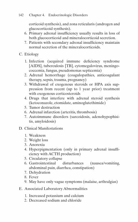
142
corticoid synthesis), and zona reticularis (androgen and glucocorticoid synthesis).
6. Primary adrenal insufficiency usually results in loss of both glucocorticoid and mineralocorticoid secretion.
7. Patients with secondary adrenal insufficiency maintain normal secretion of the mineralocorticoids.
C. Etiology
1. Infection (acquired immune deficiency syndrome [AIDS], tuberculosis [TB], cytomegalovirus, meningo-coccemia, fungus, pseudomonas septicemia)
2. Adrenal hemorrhage (coagulopathies, anticoagulant therapy, sepsis, trauma, pregnancy)
3. Withdrawal of exogenous steroids or HPA axis sup-pression from recent (up to 1 year prior) treatment with exogenous corticosteroids
4. Drugs that interfere with adrenal steroid synthesis (ketoconazole, etomidate, aminoglutethimide)
5. Tumor destruction 6. Adrenal infarction (arteritis, thrombosis) 7. Autoimmune disorders (sarcoidosis, adenohypophisi-
tis, amyloidosis)
D. Clinical Manifestations
1. Weakness 2. Weight loss 3. Anorexia 4. Hyperpigmentation (only in primary adrenal insuffi-
ciency with ACTH production) 5. Circulatory collapse 6. Gastrointestinal disturbances (nausea/vomiting,
abdominal pain, diarrhea, constipation) 7. Dehydration 8. Fever 9. May have only vague symptoms (malaise, arthralgias)
E. Associated Laboratory Abnormalities
1. Increased potassium and calcium 2. Decreased sodium and chloride
Chapter 4. Endocrinologic Disorders

143
3. Decreased glucose 4. Metabolic acidosis 5. Increased blood, urea, nitrogen/creatinine (BUN/Cr)
ratio 6. Normocytic, normochromic anemia, neutropenia, lym-
phocytosis, eosinophilia
F. Diagnostic Evaluation
1. Physical Examination
(a) Hypotension (b) Hyperpigmentation (not in acute or secondary
adrenal insufficiency) (c) Females may have loss of axillary hair (d) Auricular cartilage calcification (e) Vitiligo (f) Lymphoid tissue hyperplasia or splenomegaly
2. Laboratory Evaluation
(a) Measurement of a single plasma cortisol level usu-ally does not allow reliable appraisal of pituitary and adrenal function; however, in stressed, criti-cally ill patients, it is sufficient to rule out or to sug-gest the diagnosis of adrenal insufficiency (a serum cortisol level ≥20 μg/dL indicates adequate adrenal glucocorticoid secretion following stress, ACTH, or CRH).
(b) If the patient is hemodynamically unstable and adrenal insufficiency is suspected, a random corti-sol level should be drawn and treatment with hydrocortisone (see “Therapy” section below) should be initiated immediately.
(c) If the patient is hemodynamically stable and adre-nal insufficiency is suspected, a random cortisol level may be measured and the patient should be treated with dexamethasone (stress dose of 2–10 mg IV q6 h) until the result is available; then, if the cortisol level is <20 μg/dL, a screening syn-thetic ACTH stimulation test may be performed:
Adrenal Insufficiency
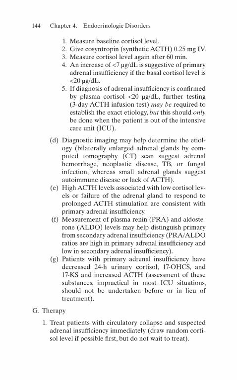
144
1. Measure baseline cortisol level. 2. Give cosyntropin (synthetic ACTH) 0.25 mg IV. 3. Measure cortisol level again after 60 min. 4. An increase of <7 μg/dL is suggestive of primary
adrenal insufficiency if the basal cortisol level is <20 μg/dL.
5. If diagnosis of adrenal insufficiency is confirmed by plasma cortisol <20 μg/dL, further testing (3-day ACTH infusion test) may be required to establish the exact etiology, but this should only be done when the patient is out of the intensive care unit (ICU).
(d) Diagnostic imaging may help determine the etiol-ogy (bilaterally enlarged adrenal glands by com-puted tomography (CT) scan suggest adrenal hemorrhage, neoplastic disease, TB, or fungal infection, whereas small adrenal glands suggest autoimmune disease or lack of ACTH).
(e) High ACTH levels associated with low cortisol lev-els or failure of the adrenal gland to respond to prolonged ACTH stimulation are consistent with primary adrenal insufficiency.
(f) Measurement of plasma renin (PRA) and aldoste-rone (ALDO) levels may help distinguish primary from secondary adrenal insufficiency (PRA/ALDO ratios are high in primary adrenal insufficiency and low in secondary adrenal insufficiency).
(g) Patients with primary adrenal insufficiency have decreased 24-h urinary cortisol, 17-OHCS, and 17-KS and increased ACTH (assessment of these substances, impractical in most ICU situations, should not be undertaken before or in lieu of treatment).
G. Therapy
1. Treat patients with circulatory collapse and suspected adrenal insufficiency immediately (draw random corti-sol level if possible first, but do not wait to treat).
Chapter 4. Endocrinologic Disorders

145
2. Administer hydrocortisone 100 mg IV q8 h for 24 h; if the patient shows a good clinical response (or cortisol level confirms diagnosis), the dosage may be tapered gradually and eventually changed to oral.
3. Provide adequate volume replacement with D5NS, until hypotension, dehydration, and hypoglycemia are corrected.
4. Identify and treat any precipitating factors (e.g., infec-tions and hemorrhage).
H. Complications
1. Short-term corticosteroid therapy is safe. 2. When there is a question of adrenal insufficiency dur-
ing an emergency, short-term supplemental corticoste-roids should be given until adrenal integrity can be assessed.
3. It is of the utmost importance to treat patients in these situations, because adrenal crisis will progress to death if untreated.
Diabetes Insipidus
A. Definitions
1. Diabetes insipidus is a disorder of fluid homeostasis resulting from inadequate vasopressin or antidiuretic hormone (ADH) secretion or action.
2. Neurogenic diabetes insipidus may be caused by a lack of production of ADH precursor molecule by the hypo-thalamus or inadequate secretion of ADH by the pos-terior pituitary.
3. Nephrogenic diabetes insipidus is caused by unrespon-siveness of renal tubules to ADH.
4. Both types of diabetes insipidus result in excretion of large volumes of hypotonic fluid.
B. EtiologySee Table 4.1.
Diabetes Insipidus

146
C. Clinical Manifestations
1. Polyuria: Urine volume usually 30 mL/kg/day 2. Neurologic manifestations: Seizures, profound central
nervous system (CNS) dysfunction
Table 4.1 Causes of diabetes insipidus1. Neurogenic diabetes insipidus (ADH deficiency)
(a) Head traumaa
(b) Postoperative (secondary to neurosurgical procedures)a
(c) Neoplasms of the brain or pituitary fossaa
(d) Postanoxic/ischemic injurya
(e) Vascular injury (subarachnoid hemorrhage)a
(f) Meningoencephalitisa
(g) Infiltrative hypothalamic disorders (sarcoidosis, histiocytosis)
(h) Familial (autosomal dominant)
(i) Idiopathic
2. Nephrogenic diabetes insipidus (ADH resistance)
(a) Pharmacologic causes (lithium, amphotericin B)a
(b) Postobstructivea
(c) Metabolic disturbances (hypercalcemia, hypokalemia)a
(d) Pyelonephritis
(e) Polycystic kidney disease
(f) Sickle cell disease and trait
(g) Infiltrative diseases (sarcoidosis, amyloidosis)
(h) Familial (X-linked recessive)
3. Diabetes insipidus of pregnancy (secondary to ADH degradation by vasopressinase produced by the placenta)—a rare form of diabetes insipidus and even less commonly seen in the ICU
aA cause likely to be seen in critically ill patients
Chapter 4. Endocrinologic Disorders

147
3. Dehydration 4. Polydipsia: Not likely to occur in the critically ill patient
with limited access to water
D. Evaluation of Polyuria
1. History
(a) Review clinical history for evidence of recent trauma, neurosurgical procedure, and pharmaco-logic causes.
(b) Rule out excess water administration. (c) Rule out solute load.
2. Laboratory Evaluation
(a) Decreased urine specific gravity (≤1.010) (b) Inappropriately dilute urine (≤300 mOsm/kg H2O) (c) Electrolytes: hypernatremia, hypercalcemia, and
hypokalemia (d) Increased plasma osmolality (300 mOsm/kg H2O)
3. Differential Diagnosis
(a) Diabetes insipidus (b) Solute diuresis (glucose, mannitol, urea, IV con-
trast media, sodium diuresis secondary to diuretic or dopamine administration)
(c) Primary polydipsia
E. Diagnostic Approach
1. First exclude solute diuresis as etiology (check urine glucose, osmolality, serum electrolytes, and specific gravity).
2. If the patient has hypernatremia and either an inappro-priately low plasma ADH level or an inappropriately low urine osmolality, the diagnosis of diabetes insipidus is made (further testing is needed only to distinguish the type of diabetes insipidus):
(a) Administer a therapeutic trial of 1-μg DDAVP (desmopressin) subcutaneously.
Diabetes Insipidus
148
(b) If urine osmolality increases ≥50%, the diagnosis of neurogenic diabetes insipidus is confirmed and therapy can be instituted.
(c) If urine osmolality does not increase in response to DDAVP, renal resistance to ADH is suggested and nephrogenic causes of diabetes insipidus should be addressed.
3. If the plasma sodium is <145 mEq/L and the patient is hemodynamically stable, a water-deprivation test may be initiated (to stimulate secretion of ADH and to determine if the patient will respond appropriately to ADH):
(a) Fluid restriction should be continued until plasma osmolality is ≥295 mOsm/kg or sodium level is ≥145 mEq/L (attempt to decrease fluid intake to ≤1–2 L/day and follow serial urine output, osmo-lality, and plasma sodium).
(b) If plasma sodium ≥145 mEq/L while urine osmo-lality remains ≤300 mOsm/kg, DDAVP may be given (as above) to distinguish between neuro-genic and nephrogenic diabetes insipidus.
(c) If plasma sodium ≥145 mEq/L and urine osmolal-ity ≤300 mOsm/kg, secondary diabetes insipidus is suggested (nondiagnostic).
(d) If the patient tolerated the fluid restriction well, it may be continued on the suspicion that the cause is excessive water administration.
(e) If the patient develops thirst or evidence of hypo-volemia before polyuria resolves, DDAVP therapy may be started with close monitoring of water bal-ance until further testing can be performed safely (testing of plasma ADH levels may be needed when the patient is out of the critical care setting) to distinguish between partial neurogenic diabetes insipidus and nephrogenic diabetes insipidus.
Chapter 4. Endocrinologic Disorders

149
F. Therapy
1. Neurogenic Diabetes Insipidus
(a) Desmopressin or DDAVP (vasopressin analog desamino-D-arginine-8-vasopressin) is preferred because it can be administered easily (parenterally, intranasally, or directly onto the buccal mucosa), it has a relatively long duration of action, and this preparation does not have vasoconstrictor or intes-tinal motility effects (note that others have advo-cated use of aqueous vasopressin in critically ill patients because of the shorter duration of action allowing for flexibility as the patient’s status changes). (See Table 4.2.)
(b) Regardless of initial sodium levels, avoid excess water administration and development of iatro-genic syndrome of inappropriate antidiuretic hor-mone (SIADH), especially after neurosurgical procedures, which may have short duration of dia-betes insipidus.
(c) Patients with trauma or postoperative diabetes insipidus should have therapy withheld every 3–5 days for assessment of potential resolution of diabetes insipidus.
(d) Many patients with neurogenic diabetes insipidus will require long-term hormone replacement.
Table 4.2 Replacement therapy for diabetes insipidusAgent Dose Route DurationDesmopressin (DDAVP)
1–4 μg Subcutaneous, intravenous, intranasal
12–24 h
5–40 μg 8–20 h
Aqueous vasopressin
5–10 U Subcutaneous, intramuscular
2–8 h
Vasopressin tannate in oil
2.5–5.0 U
Intramuscular 24–72 h
Diabetes Insipidus

150
(e) Low solute, especially low sodium, diet can help decrease urine output and is frequently combined with thiazide diuretics.
2. Incomplete Neurogenic Diabetes Insipidus
(a) The preferred agent in critically ill patients is DDAVP.
(b) Other options include medications that augment ADH action in the kidney or increase ADH release:
1. Chlorpropamide (Diabinese) 100–500 mg qd (concern is hypoglycemia).
2. Clofibrate (Atromid-S) 500 mg q6 h (associated with increased risk of gallstones).
3. Carbamazepine (Tegretol) 200–600 mg qd.
3. Nephrogenic Diabetes Insipidus
(a) Discontinue all medications that could be caus-ative if possible.
(b) During critical illness, these patients will require fluid administration titrated against urine outputs and plasma sodium levels.
(c) These patients frequently respond well to chronic therapy with thiazide diuretics, which cause vol-ume contraction and subsequent increased proxi-mal tubule water and sodium reabsorption, decreased water delivery to the distal nephron, and ultimately decreased urine output.
4. Diabetes Insipidus of Pregnancy
(a) Due to the unique pathophysiology, this form of diabetes insipidus will not respond well to treat-ment with vasopressin (aqueous or in oil), but responds well to DDAVP, which is resistant to vasopressinase action.
Chapter 4. Endocrinologic Disorders
151
Syndrome of Inappropriate Antidiuretic Hormone Secretion (SIADH)
A. Definition. SIADH is a state of euvolemic hyponatremia caused by excessive excretion of ADH.
B. Pathophysiology
1. ADH secretion is independent of normal osmotic or hemodynamic stimuli.
2. Excess ADH may be secreted ectopically or by the pos-terior pituitary.
3. Normal controls of sodium balance are maintained; high sodium intake increases urinary sodium excretion and low sodium intake reduces urinary sodium excre-tion, resulting in maintenance of extracellular volume within normal limits.
4. Free water cannot be excreted normally; persistent ADH secretion causes water retention, hyponatremia, and progressive expansion of intracellular and extra-cellular fluids.
5. Expanded extracellular fluid (ECF) stimulates natri-uresis with an isotonic loss of ECF, bringing the extra-cellular compartment back to its baseline volume.
6. The intracellular compartment remains expanded.
C. Etiology
1. Postoperative (surgical stress, anesthetic agents, positive- pressure ventilation)
2. CNS disorders (head trauma, neoplasm, meningitis, encephalitis, brain abscess, hydrocephalus, intracranial hemorrhage)
3. Pulmonary diseases (pneumonia, TB, bronchiectasis, chronic obstructive pulmonary disease [COPD], sta-tus asthmaticus)
4. Drug induced (vasopressin, DDAVP, chlorprop-amide, carbamazepine, clofibrate, oxytocin, thiazide
Syndrome of Inappropriate Antidiuretic Hormone…

152
diuretics, vincristine, vinblastine, cyclophosphamide, phenothiazines, tricyclic antidepressants, narcotics, nicotine, monoamine oxidase inhibitors)
5. Ectopic ADH production by tumors (carcinoma of lung, duodenum, pancreas, thymoma, lymphoma, hepatoma, carcinoid tumors, Ewing’s sarcoma)
6. Stress or pain 7. Nausea 8. Acute psychosis 9. Endocrinologic disorders (myxedema, ACTH defi-
ciency, panhypopituitarism) 10. HIV infection 11. Hereditary SIADH 12. Idiopathic
D. Clinical Manifestations
1. Mental status changes may be present (confusion or lethargy).
2. Seizures or coma may occur, especially if the hypona-tremia is severe or of rapid onset.
E. Diagnostic Evaluation
1. Physical Examination
(a) Evaluate for evidence of euvolemia. (b) Edema may be absent.
2. Laboratory Evaluation
(a) Serum sodium ≤130 mEq/L (b) Urine osmolality 200 mOsm/kg (most reliable
diagnostic test) (c) Urinary sodium usually 30 mEq/L (due to increased
atrial natriuretic hormone or suppressed aldosterone)
(d) Plasma osmolality <280 mOsm/kg (e) Normal or decreased BUN, normal creatinine (f) Decreased uric acid (g) Normal thyroid, adrenal, and cardiac function
Chapter 4. Endocrinologic Disorders

153
F. Differential Diagnosis
1. Factitious hyponatremia (substantial elevations of serum proteins or lipids, osmotically induced shifts of water into the intravascular space)
2. Hypovolemic hyponatremia (gastrointestinal losses, renal dysfunction, diuretics, aldosterone deficiency)
3. Hypervolemic hyponatremia (congestive heart failure, nephrotic syndrome, cirrhosis)
4. Hypothyroidism 5. Adrenal insufficiency
G. Therapy
1. Therapy must be individualized. 2. Treat the underlying disease (i.e., antibiotics for TB,
evacuate subdural hematoma). 3. Restrict fluids to 500–800 cc/day (difficult in the ICU,
at least attempt to decrease free water intake). 4. It is important to distinguish acute onset (<2–3 days)
versus chronic hyponatremia and to determine the presence or absence of neurological symptoms.
5. Acute hyponatremia with symptoms may be treated with isotonic or hypertonic (3%) saline combined with a loop diuretic (furosemide is usually added to diminish the ability of the renal tubules to concentrate urine and therefore increase free water excretion), aimed at increasing the serum sodium by ≤1 mEq/L/h (maximum safe increase of 12–24 mEq/L/day).
6. Although infusion of isotonic saline alone would seem to be reasonable, since isotonic saline is hypertonic relative to the patient’s plasma, the patient with SIADH can excrete the infused sodium in a more con-centrated form than it was given (the net effect can be further retention of free water and exacerbation of hyponatremia).
7. Hypertonic saline (3%) should be given slowly (1–2 mL/kg/h) until the sodium is at a “safe” asymp-tomatic level (usually 120–125 mEq/L); monitor serum sodium levels every 1–2 h when using hypertonic
Syndrome of Inappropriate Antidiuretic Hormone…

154
saline to avoid dangerously rapid correction, which can result in neurologic sequelae or death.
8. In chronic hyponatremia with minimal symptomatol-ogy, the sodium should be corrected more slowly at approximately 0.5 mEq/L/h to avoid central pontine myelinolysis, which can result from aggressive sodium replacement and can result in permanent neurological impairment or death.
9. Fluid balances should be measured every 4–8 h until the serum sodium level is 125 mEq/L or is corrected halfway to normal.
10. Demeclocycline 300–600 mg PO bid may be useful in patients with chronic SIADH (it causes nephrogenic diabetes insipidus and counteracts the effects of the high ADH levels).
11. Oral salt tablets have been used in some cases. 12. Vasopressin receptor antagonists can be used.
Tolvaptan and conivaptan have been studied in this setting.
13. Administration of urea has also been recommended for chronic hyponatremia. It is well tolerated and can be used in ambulatory patients. However, no clinical trials were conducted on its efficacy yet.
14. Other agents include demeclocycline and lithium.
H. Precautions Regarding Therapy
1. It is never necessary to rapidly raise the serum sodium to normal or even to a preconceived “safe” level if that requires an increase of >10–12 mEq/L/day.
2. Water moves freely across the blood–brain barrier in response to osmotic gradients; therefore, even small increases in plasma osmolality will reduce brain edema to some extent.
3. Brain water can never increase by more than 10%, due to constraints imposed by the skull; a 5–10% increase in sodium concentration (6–12 mEq/L) can be expected to virtually eliminate cerebral edema, and clinical expe-rience has shown this to be effective in symptomatic patients.
Chapter 4. Endocrinologic Disorders

155
Diabetic Ketoacidosis and Hyperosmolar Nonketotic Coma
A. Definitions
1. Diabetic ketoacidosis (DKA) and hyperosmolar non-ketotic coma (HNKC) are hyperglycemic states in patients with diabetes mellitus characterized by insulin deficiency and relative excess of glucagon and other counterregulatory hormones.
2. DKA is also characterized by ketosis.
B. Pathophysiology
1. Insulin deficiency leads to:
(a) Increased glucagon, which causes excessive hepatic glucose production (gluconeogenesis/gly-cogenolysis)
(b) Decreased glucose clearance from peripheral tissues (c) Development of hyperglycemia causing an osmotic
diuresis (loss of Na+/H2O), hypovolemia, and decreased glomerular filtration rate (GFR)
(d) Increased muscle proteolysis and decreased protein synthesis leading to loss of nitrogen and electro-lytes from intracellular fluid (ICF) to ECF to urine
2. In DKA, insulin deficiency also leads to:
(a) Increased cortisol, epinephrine, and growth hor-mone, which stimulate excessive adipose tissue lipolysis and free fatty acid delivery to liver and subsequent ketogenesis and hyperketonemia (beta-hydroxybutyrate and acetoacetate) in DKA
(b) Development of an anion gap metabolic acidosis (secondary to ketoacids, which neutralize the bicarbonate buffering system) and loss of K+ and Na+, which buffer urinary excretion of ketoacids
3. In HNKC, insulin levels may be sufficient to prevent lipolysis and ketogenesis.
Diabetic Ketoacidosis and Hyperosmolar Nonketotic Coma

156
C. Etiology
1. Common precipitating events include:
(a) Infection (b) Acute stress (myocardial infarction, stroke, trauma) (c) Discontinuation of insulin (d) Discontinuation of parenteral nutrition (e) Use of some medications (glucocorticoids, diphe-
nylhydantoin, propranolol)
2. Rarely, nondiabetics may develop HNKC.
(a) Iatrogenic (hypertonic intravenous hyperalimenta-tion, hyperosmolar peritoneal dialysis)
(b) Severe fluid losses in burn patients (c) Feeding infants hypertonic oral formulas
D. Diagnostic Evaluation
1. History
(a) DKA develops in patients of all ages (75% of DKA patients are adults) with insulin-dependent diabetes mellitus (IDDM) and infrequently with noninsulin-dependent diabetes mellitus (NIDDM), in the setting of coexisting severe med-ical problems.
(b) HNKC patients are typically elderly with a history of NIDDM.
(c) Duration of symptoms averages 12 days in HNKC and 3 days in DKA.
(d) Gastrointestinal symptoms are common (abdomi-nal pain ≈50%, nausea or vomiting ≈75%).
(e) Seizures are common in HNKC.
2. Physical Examination
(a) Altered mental status (severely depressed senso-rium in HNKC, associated with osmolalities of 340–350 mOsm/kg)
Chapter 4. Endocrinologic Disorders

157
(b) Dehydrated (dry mucous membranes, orthostatic hypotension, tachycardia)
(c) Patients with DKA may also have:
1. Fruity breath 2. Hyperventilation (Kussmaul respirations) 3. Abdominal tenderness
3. Laboratory Evaluation
(a) Table 4.3 lists the common diagnostic laboratory findings in DKA and HNKC.
(b) DKA may also be associated with:
1. Low serum Na+ (if corrected for hyperglycemia, it may be normal or slightly elevated).
2. Normal to slightly elevated serum K+ (note total body K+ depleted).
3. Leukocytosis (with neutrophilia) may occur sec-ondary to stress or dehydration instead of infection.
4. Serum amylase is often elevated (even without pancreatitis).
Table 4.3 Laboratory values in DKA and HNKCLab test DKA HNKCBlood glucose (mg/dL)
200–2000 Usually 600
Blood ketonesa Present Absent
Arterial pH <7.4 Normalb
Anion gap Elevated (usually 18)
Normal or elevated
Osmolality Slightly elevated Elevated
Urine dipstick Glucose and ketones
Glucose
aBeta-hydroxybutyrate/acetoacetatebMay be low if hypovolemia causes poor tissue perfusion
Diabetic Ketoacidosis and Hyperosmolar Nonketotic Coma

158
E. Management
1. Initial Evaluation
(a) Rule out infection as precipitant
1. Complete blood count (CBC), urine analysis (UA), chest X-ray, appropriate cultures.
2. Altered mental status: Consider lumbar punc-ture to rule out meningitis.
3. Abdominal pain: Consider appendicitis, diver-ticulitis, pelvic inflammatory disease.
(b) Rule out (R/O) myocardial infarction (electrocar-diogram) in older patients or those with long- standing DM (even without complaint of chest pain)
2. Institute insulin therapy (0.1 U/kg regular insulin IV bolus) and begin infusion of regular insulin at rate of 0.1 U/kg/h (if IV access is unobtainable, 0.1 U/kg IM q1 h may be used).
(a) Expect blood glucose to decrease by ~75 mg/dL/h on this regimen.
(b) Follow blood glucose q1 h. (c) If blood glucose levels do not begin to decrease
after 4 h of insulin therapy, increase insulin dosage every hour until blood glucose begins to decrease (doses of 100 U/h have occasionally been required in a rare, extraordinarily insulin-resistant patient).
(d) Continue infusion until blood glucose level is 250–300 mg/dL and serum ketones are negative (or positive only in undiluted serum) or urine ketones are “small or moderate”; anion gap should also be decreasing to near-normal range unless additional cause of persistent metabolic acidosis exists.
(e) Lower insulin infusion to 2 U/h, and change intra-vascular fluid (IVF) to D5 0.45% normal saline
Chapter 4. Endocrinologic Disorders

159
(NS) or D5NS (add K+ if appropriate), aiming for blood glucose levels of 150–200 until patient can eat and drink.
(f) Give regular insulin dose (5–10 U SQ) before stop-ping continuous infusion (because half-life of insu-lin via infusion is only 6–8 min) to avoid recurrence of hyperglycemia and ketosis.
3. Immediately begin volume repletion.
(a) Restore circulating volume (isotonic fluid: NS, Plasmalyte, or lactated ringer’s [LR]).
1. First liter IV over the first 30 min. 2. Second liter IV over the next hour. 3. Then reassess volume status and continue as
clinical situation warrants. 4. If the patient presents in shock:
(a) May use colloid for volume expansion (b) May require 2–3 L (or more) over first 1–2 h
(b) Replace intracellular and total body fluid losses.
1. Patients typically lose water and sodium at around 100 mL H2O/kg and 7 mEq Na+/kg, so 0.45% NS is a good choice at this stage.
2. Gradual replacement is appropriate (150–300 cc/h × 12–24 h).
3. Patients are continuing to have excessive uri-nary losses via osmotic diuresis while hypergly-cemia persists.
4. Fluid balance should be followed every 1–2 h, and positive fluid balance should be maintained (adjust rate of IV replacement as needed).
5. The average patient in DKA requires 5–7 L pos-itive balance during treatment.
6. When the patient’s blood glucose level is ≤250 mg/dL, IVF should contain 5% dextrose.
Diabetic Ketoacidosis and Hyperosmolar Nonketotic Coma

160
4. Monitor patient.
(a) CBC, Na+/K+/Cl−/HCO3−/Cr, UA, chest X-ray, elec-
trocardiogram (ECG), and appropriate cultures, on admission.
(b) Follow vital signs. (c) Record cumulative patient intake and output every
hour. (d) Check blood glucose q1 h while the patient is on
continuous insulin infusion and q4 h after changing to SQ insulin.
(e) Check serum potassium 2 h after insulin treatment started and q2–4 h thereafter.
(f) Check serum electrolytes (Na+/HCO3−/Cl−), and
determine the anion gap q4–6 h. (g) Check the serum ketone level q4 h. (h) Check arterial blood gases (ABGs) on admission
and repeat as clinical situation warrants. (i) Check serum phosphate, magnesium, and ionized
calcium levels on admission. If low, replace. (j) Check urine dipstick q1 h for presence of glucose/
ketones until they are negative/small.
5. K+ Supplementation
(a) Serum K+ level decreases as soon as insulin action begins.
(b) K+ administration should begin when serum K+ ≤5.0 mEq/L and urine output is documented.
(c) Goal of K+ supplementation at this point is to keep serum K+ ≥3.5 mEq/L to help prevent cardiac arrhythmias.
(d) Patients typically have average total K+ deficit of ~5 mEq/kg (but it can be much greater).
(e) Total K+ repletion can be completed gradually (after DKA is resolved).
(f) If admission K+ is <3.5, immediately add 40 mEq KCl to each liter of IVF (KCl usually used initially; if phosphate is low, may use K-Phos).
(g) Use IV K+ until DKA is reversed and patient can take potassium orally.
Chapter 4. Endocrinologic Disorders

161
6. Metabolic Acidosis
(a) Insulin inhibits lipolysis and ketogenesis. (b) Fluid replacement restores perfusion. (c) Bicarbonate administration has not been shown to
accelerate resolution of acidosis and is generally not advisable. The exception is in severe hyperka-lemia with characteristic ECG changes, i.e., wid-ened QRS or peaked T waves.
1. If needed, use 50–100 mEq NaHCO3 in 500–1000 cc 0.45% saline over 1–2 h.
2. The main risk of bicarbonate therapy is the induction of severe hypokalemia as potassium enters cell in exchange for hydrogen ions.
7. Phosphate Supplementation
(a) Phosphate depletion in DKA averages ~1.0 mmol/kg.
(b) Prospective randomized studies showed no benefi-cial effect of phosphate treatment on recovery from DKA.
(c) Frequently, phosphate levels plummet to ~1.5 mg/dL with insulin treatment.
(d) IV replacement is usually not essential. (e) However, if phosphorus levels are ≤1.0 mg/dL,
then replacement is generally given (potassium phosphate IV is usually the best choice with con-comitant hypokalemia), since severe hypophos-phatemia may cause respiratory failure.
8. Magnesium Supplementation
(a) Mild magnesium depletion occurs in DKA and also falls with insulin treatment.
(b) Typically does not need parenteral supplementation.
(c) If the patient develops ventricular irritability with hypomagnesemia, magnesium sulfate 1–2 g IV may be given.
Diabetic Ketoacidosis and Hyperosmolar Nonketotic Coma
162
F. ComplicationsCerebral edema may develop; its therapy is not clear,
but mannitol may be helpful.
Tight Glycemic Control in the ICU
Recent evidence from randomized clinical trials demon-strates that tight glycemic control in some critically ill patients may improve outcomes. In order to achieve this, the use of intravenous fluids that contain glucose should be avoided. The administration of insulin therapy should be considered, using either sliding scale or continuous drip. The recom-mended target blood sugar is around 180 mg/dL. The use of oral hypoglycemic agents should be discontinued to avoid hypoglycemia. Opponents of this therapy cite a high inci-dence of unrecognized hypoglycemic events as a serious concern. Close monitoring of the glycemic index is, therefore, necessary.
Myxedema
A. Definition and Epidemiology
1. Myxedema is severe thyroid hormone deficiency, which can lead to a decreased level of consciousness, even coma.
2. It has a reported fatality rate as high as 80%. 3. Actual coma associated with severe hypothyroidism is
rare (only about 200 cases reported in the literature). 4. The incidence of myxedema is three times higher in
females than males; elderly females seem most suscep-tible to myxedema coma.
B. Pathophysiology
1. Thyroid hormone is essential for normal metabolism of all cells.
Chapter 4. Endocrinologic Disorders
163
2. Thyroid-stimulating hormone (TSH) secreted by the pituitary (under regulation of the hypothalamus) stimulates the thyroid to secrete thyroxine (T4) and smaller amounts of triiodothyronine (T3), which is the active form of thyroid hormone.
3. Most T3 is produced in the peripheral tissues by mono-deiodination of circulating T4.
4. T3 and T4 circulate bound to serum proteins; the free T3 and T4 are metabolically active.
5. T3 feeds back on the pituitary gland to inhibit produc-tion of TSH.
6. In myxedema coma, the cause of coma is multifacto-rial (decreased cerebral perfusion associated with low cardiac output from bradycardia and reduced stroke volume, decreased circulating levels of thyroid hor-mones resulting in decreased mental responsiveness).
7. Hypothermia may result from decreased T3 or T4, leading to reduced metabolic rate in addition to an inability to shiver.
8. Hypoventilation (alveolar) is secondary to respiratory center depression (exacerbated by use of analgesics, sedatives, and anesthesia), defective respiratory mus-cle function, and occasionally airway obstruction (enlarged tongue).
9. Hyponatremia often accompanies myxedema and may have associated hypochloremia (may contribute to altered mental status).
10. Decreased plasma volume and intense peripheral vasoconstriction are typical.
C. Etiology
1. Occasionally, myxedema may be the result of chronic, severe primary thyroid failure; patients with classic signs and symptoms lapse into stupor, coma, and death.
2. Myxedema may be precipitated in patients with moder-ate or unrecognized hypothyroidism by a superimposed acute illness (infection) or following administration of narcotics or sedatives.
3. See Table 4.4.
Myxedema

164
D. Risk Factors
1. Infection 2. Surgery 3. Anesthesia 4. Myocardial infarction 5. Sedating drugs 6. Cerebrovascular accidents 7. Bleeding 8. Cold exposure 9. Trauma 10. Hyponatremia
E. Symptoms
1. Decreased mental acuity 2. Hoarseness 3. Increased somnolence 4. Cold intolerance 5. Dry skin 6. Brittle hair
F. Diagnostic Evaluation
1. History
Table 4.4 Causes of hypothyroidism1. Autoimmune thyroid disease (Hashimoto’s thyroiditis)
2. Thyroid ablation (radioactive iodine or surgical)
3. Surgical thyroid resection
4. Pituitary disease (secondary hypothyroidism)
5. Hypothalamic disease (tertiary hypothyroidism)
6. Chemical agents (antithyroid drugs, e.g., lithium, organic goitrogens, iodine-containing drugs, e.g., amiodarone)
7. Congenital thyroid agenesis
8. Iodine deficiency or excess
9. Thyroid hormone resistance
Chapter 4. Endocrinologic Disorders

165
(a) Usually, the patient has a long history of gradual deterioration.
(b) Gradual weight gain or inability to lose weight. (c) May have been noncompliant with thyroid replace-
ment therapy. (d) May have history of Graves’ disease or other thy-
roid dysfunction.
2. Physical Examination
(a) Hypothermia (core or rectal temperature <35 °C) (b) Bradycardia (c) Hypoventilation (slow respiratory rate, shallow
breaths) (d) Hypotension (e) Physical features consistent with long-standing
hypothyroidism
1. Thick and doughy-appearing skin (may have orange or yellow tint)
2. Facial and general puffiness, periorbital edema 3. Large tongue 4. Alopecia, loss of lateral aspects of eyebrows
(f) Palpable thyroid (present in <50%) or thyroidec-tomy scar
(g) Cardiac examination may be consistent with peri-cardial effusion (e.g., muffled heart tones and cardiomegaly)
(h) Neurologic examination rarely reveals focal findings
1. Altered level of consciousness 2. Delayed relaxation phase of deep tendon
reflexes may be present but difficult to detect 3. Disorders of muscular function (paralytic ileus,
urinary retention, atonic bowel with fecal impaction)
3. Laboratory Evaluation
Myxedema

166
(a) Confirmation of diagnosis relies on thyroid func-tion tests to document hypothyroidism (measure TSH, T4, free T4, reverse T3, T3RU) (see Table 4.5).
1. TSH level is elevated in primary hypothy-roidism.
2. In secondary and tertiary hypothyroidism, TSH is not elevated, and diagnosis will rely on other laboratory parameters and clinical judgment.
(b) Serum cortisol level should be drawn initially to evaluate for concomitant adrenal insufficiency.
(c) CBC, UA, blood, and urine cultures should be sent. (d) Serum cholesterol is usually elevated. (e) Chest X-ray may reveal signs of pleural or pericar-
dial effusion or of infection. (f) ECG is often abnormal (sinus bradycardia, small
voltage QRS complexes, prolonged QT intervals, isoelectric T-wave changes, supraventricular tachycardia).
(g) ABG may reveal hypoxemia, hypercarbia, and respiratory acidosis.
(h) Serum glucose or sodium may be low. (i) Normochromic normocytic anemia is typical.
G. Differential Diagnosis
1. Other causes of altered mental status (e.g., stroke and electrolyte disturbance such as hyponatremia)
2. Sepsis 3. Hypothermia (especially with associated anemia) 4. Hypopituitarism 5. Hypoglycemia 6. Renal failure
H. Therapy
1. Initiate thyroid hormone replacement upon suspicion (awaiting confirmation of the diagnosis may be too late).
Chapter 4. Endocrinologic Disorders

167
Tabl
e 4.
5 T
hyro
id f
unct
ion
test
s in
thy
roid
dis
orde
rsTe
stH
ypot
hyro
idH
igh
T4 s
yndr
ome
Hyp
erth
yroi
dL
ow T
3 syn
drom
eL
ow T
3/T4 s
yndr
ome
TSH
Hig
haN
l/low
Low
Low
to
slL
ow t
o sl
↑↑
Tota
l T4
Low
Hig
hH
igh
Nl
Low
Tota
l T3
Low
to
low
Nl
Low
/Nl/h
igh
Hig
hL
owL
ow
Rev
erse
T3
Nl/l
owN
l/hig
hH
igh
Hig
hH
igh
Free
T4
Low
Nl/h
igh
Hig
hN
lN
l
T3R
UL
owN
l/low
Hig
hN
l/hig
hH
igh
Nl n
orm
al; s
l slig
hta E
xcep
tion
s: T
SH is
low
in h
ypot
hyro
idis
m o
f se
cond
ary
and
tert
iary
cau
ses
Myxedema
168
(a) Intravenous administration of replacement hor-mone is necessary due to unreliable gastrointesti-nal absorption in the myxedematous state.
(b) We prefer T3 and T4 combination therapy (T3 20 μg IV bolus followed by 10 μg q8 h with T4 200 μg IV followed by 100 μg IV q24 h) for 1–2 days, followed by T4 alone.
(c) T3 for IV administration recently became available in the United States, so many authors recommend treatment with IV T3 alone.
(d) Peripheral conversion of T4 → T3 requires the pres-ence of some T3 for enzyme activity.
(e) An advantage of IV T3 includes a more rapid onset of action than T4; also, peripheral conversion of T4 is not required for activity.
(f) T3 is more arrhythmogenic than T4, and careful car-diac monitoring is essential, especially since the risk of coronary artery disease is high in these patients.
(g) If IV T3 is not immediately available, IV T4 (as above) may be given with oral T3 (25 μg q12 h) until the patient can be treated with oral T4 alone.
(h) Previously, T4 alone was frequently used (200–500 μg IV bolus followed by 50–100 μg IV q24 h).
(i) Controversy exists regarding the best therapeutic regimen, and controlled trials will probably never be performed because the disease is so rare.
(j) Monitor T3 and T4 levels after 5 days and adjust doses accordingly if the patient remains unconscious.
2. Metabolic Support
(a) Hypothermia is best treated with passive rewarm-ing (active rewarming can cause peripheral vasodilatation and worsening of shock) (See Chap. 5, “Environmental Disorders”).
(b) Hyponatremia generally responds well to free water restriction.
Chapter 4. Endocrinologic Disorders

169
(c) Hypoglycemia should be treated with IV dextrose. (d) Seizures may be treated with standard anticonvul-
sant drugs. (e) Identify and treat precipitating causes (e.g., infec-
tion, stroke, myocardial infarction, narcotics, and gastrointestinal bleeding).
3. Supportive Care
(a) Respiratory support with mechanical ventilation may be required.
(b) Hypotension should be treated aggressively with IV fluids (avoid free water) and vasopressor ther-apy (dopamine is preferable over norepinephrine, as it may better maintain coronary blood flow and renal/mesenteric blood flow).
(c) Hypotension is poorly responsive to vasopressors until thyroid hormone replacement is initiated.
(d) Hydrocortisone (100 mg IV q8 h) should be given until initial cortisol level is available (if this indi-cates a normal stress response to the acute medical illness, this therapy may be stopped) or for 3–7 days, followed by a rapid taper in the absence of hypo-thalamic–pituitary–adrenal disease; this therapy may be lifesaving in patients with secondary/ter-tiary hypothyroidism.
(e) Monitor for the presence of arrhythmias (decrease dosage of thyroid hormone replacement if arrhyth-mias occur); hypothyroidism is associated with a high incidence of coronary artery disease, and these patients should be monitored for evidence of myocardial ischemia exacerbated by increased myocardial oxygen consumption with T3/T4 treatment.
(f) Avoid sedatives.
I. Complications
1. Euthyroid patients tolerate short-term administration of thyroid hormone well.
Myxedema

170
2. Delay of treatment in a myxedematous patient can make the difference between survival and death.
3. This treatment regimen should not routinely be insti-tuted in hypothyroid patients without clinical evidence of myxedema coma because of potential cardiac com-plications (myocardial infarction, arrhythmias).
4. Severe hypothermia (<32 °C) is thought to have prog-nostic significance.
Thyrotoxic Crisis
A. Definitions
1. Thyrotoxic crisis or thyroid storm is a life-threatening complication of hyperthyroidism characterized by a severe, sudden exacerbation of thyrotoxicosis.
2. The general term thyrotoxicosis refers to the clinical and biochemical manifestations of excess thyroid hor-mone at the tissue level.
3. True hyperthyroidism refers to disorders of thyroid glandular hyperfunction with increased synthesis and secretion of thyroid hormone.
4. There are states of thyrotoxicosis without true hyper-thyroidism (e.g., factitious ingestion of thyroid hor-mone, chronic thyroiditis with transient thyrotoxicosis, ectopic thyroid hormone production, subacute thyroid-itis) that are associated with decreased thyroidal syn-thesis of new hormone; these states are rare in the critically ill patient with the possible exception of T4 or T3 overdose (see Table 4.6).
B. Pathophysiology
1. The actual mechanisms by which a patient with thyro-toxicosis decompensates into thyroid crisis are poorly understood.
2. A crisis develops most often after a stressful precipitat-ing event (e.g., trauma, infection, DKA, surgical emer-gency, parturition, or myocardial infarction).
Chapter 4. Endocrinologic Disorders
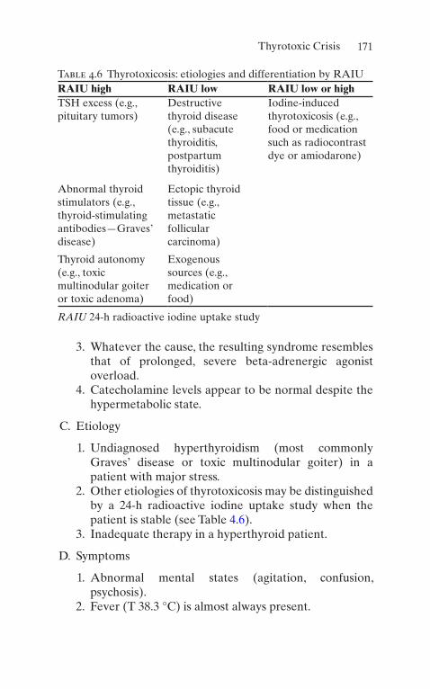
171
3. Whatever the cause, the resulting syndrome resembles that of prolonged, severe beta-adrenergic agonist overload.
4. Catecholamine levels appear to be normal despite the hypermetabolic state.
C. Etiology
1. Undiagnosed hyperthyroidism (most commonly Graves’ disease or toxic multinodular goiter) in a patient with major stress.
2. Other etiologies of thyrotoxicosis may be distinguished by a 24-h radioactive iodine uptake study when the patient is stable (see Table 4.6).
3. Inadequate therapy in a hyperthyroid patient.
D. Symptoms
1. Abnormal mental states (agitation, confusion, psychosis).
2. Fever (T 38.3 °C) is almost always present.
Table 4.6 Thyrotoxicosis: etiologies and differentiation by RAIURAIU high RAIU low RAIU low or highTSH excess (e.g., pituitary tumors)
Destructive thyroid disease (e.g., subacute thyroiditis, postpartum thyroiditis)
Iodine-induced thyrotoxicosis (e.g., food or medication such as radiocontrast dye or amiodarone)
Abnormal thyroid stimulators (e.g., thyroid-stimulating antibodies—Graves’ disease)
Ectopic thyroid tissue (e.g., metastatic follicular carcinoma)
Thyroid autonomy (e.g., toxic multinodular goiter or toxic adenoma)
Exogenous sources (e.g., medication or food)
RAIU 24-h radioactive iodine uptake study
Thyrotoxic Crisis
172
3. Heat intolerance, diaphoresis. 4. Palpitations (sinus tachycardia and atrial fibrillation
are the most common dysrhythmias). 5. Gastrointestinal disturbances (diarrhea, nausea, vomit-
ing, abdominal pain). 6. Muscle wasting and weakness. 7. Dyspnea.
E. Diagnostic Evaluation
1. History
(a) Marked and rapid recent weight loss may warn of impending storm.
(b) Unexplained fever in a thyrotoxic patient may pre-cede a storm.
(c) A precipitating event may be evident.
2. Physical Examination
(a) Goiter (palpate cautiously, since vigorous massage may cause release of more hormone into the circulation)
(b) Tachycardia—may have tachydysrhythmias (c) Hyperthermia (d) Mental status changes (e) Tremor (f) Warm, moist skin (g) Ophthalmic signs of hyperthyroidism (proptosis,
lid lag, lid retraction) (h) Signs of congestive heart failure (high-output fail-
ure or cardiomyopathy)
3. Laboratory Evaluation
(a) Confirmation of the diagnosis relies on thyroid studies (see Tables 4.5 and 4.6):
1. Elevated T4
2. Elevated T3
3. Decreased TSH
Chapter 4. Endocrinologic Disorders

173
(b) Routine studies include CBC, electrolytes, urinaly-sis, chest X-ray, and ECG.
(c) Evaluate for infection as indicated by history and physical examination.
(d) Associated laboratory abnormalities:
1. Hypercalcemia 2. Hypokalemia 3. Hyperglycemia 4. Hypocholesterolemia 5. Mild microcytic anemia 6. Lymphocytosis 7. Granulocytopenia 8. Hyperbilirubinemia 9. Elevated alkaline phosphatase
F. Differential Diagnosis
1. Hypermetabolic states (sepsis, pheochromocytoma, Cushing’s syndrome)
2. Thyrotoxicosis without crisis/storm
G. Therapy. Do not wait for lab values to begin treatment; diagnosis is made on clinical suspicion and treatment should be initiated immediately.
1. Supportive Measures
(a) IV fluids for volume replacement. (b) Acetaminophen for hyperthermia. Avoid aspirin,
since it displaces T4 from thyroid-binding globulin, thereby increasing the level of free T4.
(c) Cooling blankets, hydrogel pads.
2. Inhibition of Thyroid Hormone Synthesis
(a) Propylthiouracil (PTU) (200–300 mg oral or via nasogastric tube q6 h)
(b) Methimazole (20–25 mg oral or via nasogastric tube q4 h)
Thyrotoxic Crisis

174
3. Inhibition of Thyroid Hormone Release with Iodide Therapy (Beginning 1 h After PTU Therapy)
(a) Sodium iodide (1 g IV q8 h) (b) Potassium iodide and iodine—Lugol’s solution
(ten drops q8 h orally)
4. Inhibition of Peripheral Beta-Adrenergic Activity
(a) Most beta-blockers also block peripheral conver-sion of T4–T3.
(b) Propranolol (Inderal) 0.5–1.0 mg/min IV to total dose of 2–10 mg IV q3–4 h; may treat with 20–40 mg PO q6 h after initial control with IV administra-tion. An occasional patient has required up to 2 g/day PO due to variability of hepatic metabolism in thyrotoxic individuals.
(c) Esmolol (Brevibloc) 0.5–1.0 mg/kg bolus followed by infusion with 50 μg/kg/min; if inadequate effect within 5 min, repeat bolus and increase infusion to 100 μg/kg/min; can repeat procedure to 200–300 μg/kg/min.
(d) Titrate beta-blockade to achieve heart rate of 80 beats per minute.
(e) If the patient has a history of reactive airway dis-ease, use caution and a short-acting cardioselective agent such as esmolol.
(f) Caution is also required in patients with CHF. Controlling the heart rate may be of benefit; however, these agents are negative inotropes and may worsen low-output failure.
(g) If beta-blockade are contraindicated, other sympa-tholytic drugs (reserpine, a depleter of catechol-amines, or guanethidine, an inhibitor of catecholamine release) may be useful as second- line agents.
5. Inhibition of Peripheral Conversion of T4–T3
(a) PTU (see above dosages). (b) Beta-blockade (see above).
Chapter 4. Endocrinologic Disorders

175
(c) Hydrocortisone IV 100 mg q8. (d) Bile acid sequestrants (e.g. cholestyramine) inhibit
recycling of the thyroid hormones.
6. Definitive therapy may require surgery or radioactive iodine therapy, after etiology being determined by RAIU.
7. Diagnose and treat underlying precipitating disorders (i.e., infection, other major stresses).
Sick Euthyroid Syndrome
A. Definitions
1. Sick euthyroid syndrome is characterized by thyroid hormone alterations associated with acute non- thyroidal illness.
2. Two syndromes are described:
(a) Low T3 syndrome (b) Low T3/T4 syndrome
3. These syndromes are thought to represent euthyroid states by many; however, specific tissues may actually be hypothyroid.
B. Review of Normal Thyroid Hormone Physiology
1. Thyrotropin-releasing hormone (TRH) is released from the hypothalamus into the bloodstream.
2. TRH stimulates the synthesis and release of thyrotro-pin (TSH) from the pituitary gland.
3. TSH stimulates the thyroid gland to produce and secrete thyroxine (T4) and smaller amounts of triiodo-thyronine (T3).
4. The thyroid gland secretes predominantly T4 (80%), with smaller amounts of T3 (20%).
5. The remainder of T3 (the physiologically active form of thyroid hormone) is produced in extrathyroidal tis-sues (primarily, the liver and kidneys) by monodeio-dination of circulating T4.
Sick Euthyroid Syndrome
176
6. T3 and T4 circulate bound to serum proteins; the free T3 and T4 are metabolically active.
7. T3 feeds back on the pituitary gland to inhibit the pro-duction of TSH.
8. Some of the circulating T4 is metabolized to the inac-tive product reverse T3.
9. Both T3 and reverse T3 are rapidly cleared from the serum by further deiodination.
10. Thyroid hormone activity begins with binding of T3 to receptors on cell nuclei.
11. Post-binding effects of T3 are needed for normal cel-lular function.
12. The T3 resin uptake test is commonly performed to give an approximation of binding proteins (primarily thyroxine-binding globulin).
C. Pathophysiology
1. In acute non-thyroidal illness, peripheral thyroid hor-mone metabolism is altered.
2. The exact mechanism of decreased T3 production is unknown.
D. Etiology
1. Systemic illness (sepsis, cardiac or respiratory failure, neoplastic processes, stroke)
2. Surgery 3. Caloric deprivation 4. Drug induced (glucocorticoids, iodides, amiodarone,
propylthiouracil)
E. SymptomsNo specific symptoms are associated with the thyroidal
hormone alterations. F. Laboratory Evaluation
1. Serum T3 is low (see Table 4.5). 2. Serum T4 is low or normal.
Chapter 4. Endocrinologic Disorders
177
3. Reverse T3 is elevated (usually this is the most useful discriminator between the sick euthyroid syndrome and hypothyroidism).
4. Serum TSH is normal.
G. Therapy
1. Attempts should be made to distinguish sick euthyroid syndrome from hypothyroidism.
2. Currently, there is no proven benefit in using thyroid hormone to treat patients with sick euthyroid syndrome.
3. Indeed, some researchers believe that the sick euthy-roid state may be protective by conserving energy under stress.
Hypoglycemia
A. Definition. Hypoglycemia is defined by plasma glucose <40–50 mg/dL. This definition does not include the pres-ence of associated symptoms, since critically ill patients may not reliably demonstrate classic symptomatology.
B. Pathophysiology
1. Clinical situations that result in increased insulin, inability of the liver to generate glucose from its glyco-gen stores, or problems with the counterregulatory sys-tem may lead to hypoglycemia.
2. Insulin suppresses hepatic glucose production and stimulates glucose utilization by peripheral tissues such as muscle.
3. Insulin secretion lowers plasma glucose concentration. 4. In response to onset of hypoglycemia, the major coun-
terregulatory hormones glucagon and epinephrine increase and cause an acceleration of glycogenolysis.
5. Catecholamines are glucose-counterregulatory hor-mones but do not play essential roles as long as secre-
Hypoglycemia

178
tion of glucagon is initiated; however, in the presence of glucagon deficiency (long-standing diabetes mellitus or patients with total pancreatectomy), catecholamines become major counterregulatory hormones.
6. Patients treated with nonselective beta-adrenergic- blocking agents may have severe impairment of coun-terregulation mechanisms.
7. Patients with long-standing DM (10–15 years) may lose the ability to secrete epinephrine in response to hypo-glycemia (becoming virtually defenseless against even moderate degrees of hyperinsulinemia).
C. EtiologySee Table 4.7.
D. Symptoms
1. Adrenergic activation
(a) Palpitations (b) Tremor (c) Diaphoresis (d) Pallor (e) Anxiety
2. Neuroglycopenia
(a) Fatigue (b) Faintness (c) Dizziness (d) Hunger (e) Inappropriate behavior (f) Visual symptoms (g) Focal neurologic symptoms (h) Seizures (i) Coma
E. Diagnostic Evaluation
1. History
(a) Check for history of DM.
Chapter 4. Endocrinologic Disorders

179
Table 4.7 Causes of hypoglycemia1. Hyperinsulin states
(a) Exogenous insulin administrationa
(b) Endogenous insulin excess (e.g., insulinomas)
2. Ethanol-induced (after ingestion in otherwise healthy patients or chronic alcoholics)a
3. Drug-induced (e.g., sulfonylureas, quinine, propranolol, pentamidine)a
4. Systemic disordersa
(a) Hepatic disease (e.g., cirrhosis and fulminant viral hepatitis)
(b) Renal disease (e.g., chronic renal disease associated with liver disease, CHF, and sepsis)
(c) Sepsis (e.g., gram-negative sepsis and empyema of gallbladder)
(d) AIDS
5. Extensive thermal burnsa
6. Total parenteral nutritiona
7. Insulin treatment of hyperkalemiaa
8. Factitious (insulin injection or sulfonylurea ingestion)a
9. Endocrine causes (hypopituitarism, hypoadrenalism, hypothyroidism)
10. Autoimmune causes (insulin antireceptor antibodies, anti- insulin autoantibodies)
11. Starvation (severe caloric restriction)
12. Alimentary—following gastric surgery (e.g., gastrectomy)
13. Idiopathic (functional)—typically postprandial; this is a diagnosis of exclusion and is usually not seen in the critically ill patient
aCauses likely to be seen in the critically ill
Hypoglycemia
180
1. Recent insulin or oral hypoglycemic therapy? 2. If DM is long-standing, the patient may also
have glucagon deficiency and therefore be at higher risk for hypoglycemia, since the counter-regulatory mechanisms may also be ineffective.
(b) Check for history of alcohol ingestion.
2. Physical Examination
(a) Tachycardia (b) Pupillary dilation (c) Cold, moist skin (d) Changes in body temperature (hypothermia,
hyperthermia) (e) Between hypoglycemic episodes, the examination
may be normal.
3. Laboratory Evaluation
(a) Plasma glucose level <40–50 mg/dL. Whole blood glucose is usually about 15% less than the corre-sponding plasma glucose level.
(b) The patient may have changes in blood counts.
1. Acute lymphocytosis is followed later by neutrophilia.
2. May have increased hemoglobin, total red blood cell (RBC) count, or packed RBC volume.
(c) ECG changes (ST depression, flat T waves, QT interval prolongation).
(d) Electroencephalogram (EEG) changes (dimin-ished frequency of alpha waves, increased delta waves).
(e) Artifactual hypoglycemia must be ruled out, espe-cially if laboratory results indicate hypoglycemia with no apparent cause.
1. Samples in serum separator tubes left at room temperature for extended periods of time (blood glucose levels may decrease by 10–20 mg/dL/h due to ongoing blood cell metabolism).
Chapter 4. Endocrinologic Disorders

181
2. Patients with increased numbers of blood cells (polycythemia vera, leukemia, leuke-moid reactions) may have low measured plasma glucose levels secondary to increased metabolism.
3. These problems may be avoided by collecting blood in tubes containing oxalate and fluoride (gray tubes), since fluoride acts as a glycolytic enzyme poison.
4. Diagnostic Approach
(a) Measure simultaneous blood glucose and plasma insulin levels during an episode of hypoglycemia. The best way is to demonstrate insulin secretion inappropriate to the prevailing blood glucose concentration.
1. Relative hyperinsulinemia can be demonstrated by the simultaneous determination of blood glu-cose and plasma insulin levels after an overnight fast or during a 24- to 72-h fast.
2. Diagnosis of hyperinsulinism can further be supported by elevated levels of plasma C- peptide and proinsulin concentrations.
3. Insulinomas can be localized by ultrasonogra-phy, CT scanning, magnetic resonance imaging (MRI), arteriography, transhepatic percutane-ous venous sampling, and intraoperative high- frequency sonography.
(b) Factitious hypoglycemia resulting from administra-tion of insulin or sulfonylurea agents is typically characterized by inappropriately high plasma insu-lin levels (similar to insulinomas).
1. C-peptide levels remain low in insulin-induced factitious hypoglycemia; the presence of insu-lin antibodies in patients who have no reason to take insulin injections also suggests this etiology.
Hypoglycemia

182
2. C-peptide levels are elevated in sulfonylurea- induced factitious hypoglycemia (similar to insulinomas); screening for plasma or urine sul-fonylureas may confirm the diagnosis.
(c) In spontaneous hypoglycemia, alcohol ingestion should be ruled out as a cause (alcohol levels may not be helpful, since hypoglycemia may not occur for as long as 36 h after ingestion).
(d) Associated systemic disorders must be ruled out as appropriate for each patient (evaluate for liver, renal, endocrine dysfunction, or sepsis).
F. Therapy
1. If the patient is comatose, glucose should be adminis-tered intravenously (25–50 cc of 50% dextrose followed by infusion of 10% dextrose) until persistent or mild hyperglycemia is present.
2. Treatment with dextrose should cause resolution of symptoms rapidly unless organic changes have occurred in the brain.
3. Some patients may require additional IV boluses of 50% dextrose with continuous infusion of 10% dextrose.
4. In drug-induced hypoglycemia (especially secondary to chlorpropamide), prolonged treatment with IV dex-trose may be required to keep the blood glucose levels in the 200 mg/dL range; if this does not maintain the blood glucose level at 200 mg/dL, one of the following should be added:
(a) One hundred milligrams hydrocortisone and 1 mg glucagon per liter of 10% dextrose. Continue until blood glucose levels are maintained at 200 mg/dL.
(b) An additional infusion of 300 mg diazoxide in 5% dextrose given over a 30-min period and repeated every 4 h. Continue until blood glucose levels are maintained at 200 mg/dL.
Chapter 4. Endocrinologic Disorders

183
(c) When the blood glucose levels rise, the hydrocorti-sone, glucagon, and diazoxide are stopped, and the rate of infusion of 10% dextrose is decreased.
(d) Persistent hyperglycemia maintained on 5% dex-trose is a sign to discontinue the infusion gradually over a 24-h period.
5. Insulin-induced hypoglycemia in diabetic patients may be effectively treated with 0.5–1.0 mg glucagon IV, IM, or SQ; the patient should also ingest 20–40 g of carbo-hydrate, since the glucagon effect lasts only 1–1.5 h.
6. Insulinomas may be surgically cured with resection. 7. Medical management of insulinomas is indicated for
malignant insulinoma, patients with major contraindica-tions to surgery, and rare patients in whom surgery fails.
(a) Diazoxide (3–8 mg/kg/day PO in two to three divided doses) is the drug of choice (it inhibits insulin secretion).
(b) Thiazide diuretics, diphenylhydantoin, proprano-lol, or calcium channel blockers may also be useful.
(c) Combination chemotherapy with streptozotocin and 5-fluorouracil has been reported to achieve partial or complete remission in 60% of patients with malignant insulinomas.
8. Treat the underlying cause if it is related to systemic disorders.
G. ComplicationsPrompt recognition and treatment of hypoglycemia are
required to prevent long-term neurologic sequelae or death.
Pheochromocytoma
A. Definition and Epidemiology
Pheochromocytoma

184
1. Catecholamine-producing tumor of the adrenergic sys-tem (chromaffin cells); ~90% are adrenomedullary.
2. Non-adrenal tumors (~10%) arising from the sympa-thetic nervous system are designated extra-adrenal pheochromocytomas or functioning paragangliomas and most commonly occur in the abdomen, chest, and neck.
3. Pheochromocytomas are rare (occur in only 1–2/100,000 adults).
B. PathophysiologySymptoms are secondary to catecholamines secreted
by the tumor:
1. Alpha1-adrenergic stimulation results in vasoconstriction.
2. Alpha2-adrenergic stimulation results in decreased insulin secretion.
3. Beta1-adrenergic stimulation results in cardiac inotropy/chronotropy.
4. Beta2-adrenergic stimulation results in bronchodilation and vasodilation.
5. Dopa1-receptor stimulation results in renal and mesen-teric vasodilation.
6. Excessive levels of catecholamines are toxic to the myo-cardium and cardiomyopathy may result.
7. Symptoms may be episodic in nature; paroxysms may last from less than 1 min to several hours and may occur only once every few months or as frequently as multiple times per day.
C. Symptoms
1. Hypertension (paroxysmal or persistent) 2. Headache 3. Pallor 4. Hyperhidrosis 5. Anxiety 6. Tachycardia 7. Palpitations
Chapter 4. Endocrinologic Disorders
185
8. Angina 9. Hyperglycemia 10. Weight loss 11. Paresthesias (second-degree vasoconstriction) 12. Visual disturbances (second-degree hypertensive
retinopathy) 13. Dilated pupils
D. Diagnostic Evaluation
1. History
(a) Check for the presence of the above symptoms. (b) A pressor response to histamine, glucagon, droper-
idol, tyramine, metoclopramide, saralasin, tricyclic antidepressants, or phenothiazines suggests the possibility of a pheochromocytoma.
2. Physical Examination
(a) May be normal if performed during a symptom- free time interval.
(b) May exhibit above symptoms during a paroxysm.
3. Laboratory Evaluation
(a) Plasma or urinary catecholamine levels or urinary catecholamine metabolite (VMA or metaneph-rine) levels.
(b) Rarely, suppression tests (clonidine) or provoca-tive tests (glucagon) are needed.
(c) Measurement of plasma and urinary catechol-amines are interfered with by the following:
1. Stimulation of endogenous catecholamines (e.g., surgery and stroke)
2. Administration of exogenous catecholamines 3. Various drugs (alpha2-agonists, methyldopa,
converting enzyme inhibitors, monoamine oxidase inhibitors, phenothiazines, tricyclic antidepressants)
Pheochromocytoma
186
4. Tumors may be localized by a variety of scanning tech-niques (CT, MRI, or venous blood sampling for catecholamines).
5. In patients with high suspicion for pheochromocytoma despite negative CT or MRI, additional imaging (e.g., Gallium-68 DOTA-0-Phe1-Tyr-3 octreotide positron emission tomography) can be utilized.
6. Arteriography is generally avoided because it can pre-cipitate hypertensive crisis.
E. Differential Diagnosis
1. Malignant hypertension 2. Thyrotoxic crisis 3. Hypertensive response (to stress, surgery, anesthesia) 4. Cushing’s syndrome
F. Therapy
1. Surgical excision is the definitive treatment. 2. Preoperative management is important in determin-
ing surgical outcome. 3. Preoperative goals:
(a) Control blood pressure (b) Provide adequate intravascular volume (c) Treat tachyarrhythmias (d) Treat heart failure (e) Treat glucose intolerance
4. Alpha-adrenergic blockade is the basis of hyperten-sive therapy, and patients are generally treated 1–2 weeks before surgery with phenoxybenzamine (or labetalol or prazosin) that is tapered to produce a 10- to 15-mmHg orthostatic blood pressure decrease; 1–2 weeks of pretreatment allows for intravascular volume repletion (diminished by excess endogenous catecholamines).
5. Tachycardia may occur from unopposed beta- adrenergic receptor activation, and if the heart rate is 110 beats per minute, a beta-blocker should be added; beta-blockers should not be used before institution of
Chapter 4. Endocrinologic Disorders

187
alpha-adrenergic blockade. Elimination of vasodila-tory effects of beta-receptors results in unopposed alpha-adrenergic vasoconstriction and may provoke hypertensive crisis, and the negative inotropic effects of beta-blockers may precipitate heart failure in the face of hypertension.
6. Reflex tachycardia is less with prazosin or labetalol. 7. Alpha-methylparatyrosine (an inhibitor of tyrosine
hydroxylase, the rate-limiting enzyme in catechol-amine production) may be used in patients who cannot tolerate alpha-adrenergic blockade (postural hypoten-sion) and can be especially useful in patients with car-diomyopathy. Indeed, phenoxybenzamine and alpha-methylparatyrosine in combination result in less tachycardia and less postoperative hypotension.
8. Metyrosine, an inhibitor of catecholamine synthesis, has been suggested for patients unresponsive to stan-dard therapy.
9. Dysrhythmias are treated with standard antidysrhyth-mic agents.
10. Blood should be available for transfusion in the peri-operative period, since pheochromocytomas are vas-cular tumors and bleeding is frequent.
11. Diuretics should be used sparingly, since intravascular volume is already constricted by catecholamines; con-versely, over-administration of fluids can worsen heart failure.
12. Perioperatively, drugs that cause catecholamine release or potentiate catecholamine action are to be avoided (e.g., morphine can cause histamine-induced catecholamine release).
13. Postoperatively, patient’s need cardiovascular and metabolic monitoring.
14. Hypoglycemia may occur following tumor removal (loss of catecholamine-induced hyperglycemia).
15. Pain is best treated with meperidine and benzodiaze-pines are useful for sedation.
16. Recurrent hypertension should suggest the possibility of residual tumor.
Pheochromocytoma

189© Springer Nature Switzerland AG 2021J. Varon, Handbook of Critical and Intensive Care Medicine, https://doi.org/10.1007/978-3-030-68270-5_5
Burns
It is estimated that two million people are burned annually in the United States. Of them, over 130,000 are children under the age of 15. Although most of these burns are minor, approximately 3–5% of burn injuries are life-threatening.
A. Pathophysiology
1. Partial-thickness burns involve heat damage to the epi-dermis or a portion of the dermis. The dermis contains sensory nerve endings, vascular supply, hair follicles, and sweat glands.
2. Full-thickness burns involve injury to tissues deep to the sweat glands and hair follicles. The presence of thrombosed blood vessels or charring is characteristic of full-thickness burns. However, final determination of burn depth may not be possible for several days follow-ing the injury. These lesions are usually very painful.
3. Shock may develop due to transudation and sequestra-tion of fluid in the burned areas and elsewhere in the body. Cardiac output may drop in major burns, due to myocardial dysfunction. There is no pin prick sensation in the burned area.
Chapter 5Environmental Disorders

190
B. Predisposition
1. Burn injury is the second leading cause of death among children under age 12 years.
2. Children are more likely to suffer burns than adults because of their greater vulnerability to accidents due to inability to recognize or react to hazardous situa-tions. Half of all victims of hot water burns are children under age 5 years.
C. Clinical Presentation
1. Partial-Thickness Burns
(a) First-degree burn involves only the epidermis; there is blanching erythema but no bullae formation.
(b) Second-degree burn involves a portion of the der-mis and produces edema and fluid exudation. Bullae formation is characteristic. These develop quickly after burn injury. Consider infection if bul-lae appear 18 h or later after a burn occurs.
2. Full-Thickness Burns
(a) Third-Degree Burn. Surface is dry and inelastic. Skin surface may become white or gray. This burn will not regenerate from unburned edges.
(b) Fourth-Degree Burn: Extends beyond the depth of the skin to involve underlying muscle, tendons, vas-cular structures, periosteum, or bone.
3. Survival depends on the extent and depth of the burn, the age of the patient, and associated injuries.
4. Vascular effects in burned skin are immediate vasocon-striction followed by increased capillary permeability and plasma extravasation. Burned skin also permits increased insensible water loss.
Chapter 5. Environmental Disorders
191
D. Complications
1. Coagulation necrosis at the burn site produces an advantageous setting for bacterial growth. Infection is one of the most important causes of death in severe burn injury.
2. Gastric dilatation and adynamic ileus occur in major burns.
3. Acute hemolysis may occur due to heat damage of red blood cells.
4. Acute renal failure may occur as a result of shock. 5. Hypertension may be present in burn victims, espe-
cially children. 6. Multiorgan failure is the leading cause of death in
patients with burns. 7. Manipulation of burn wounds has been shown to result
in bacteremia in 20% of cases.
E. Treatment
1. Evaluate airway (see smoke inhalation, below) and perform endotracheal intubation if indicated by upper airway edema or deterioration of arterial blood gases. Endotracheal intubation is appropriate for any patients with acute burn injury who display respira-tory distress.
2. Establish intravenous access. 3. Evaluate burned areas under sterile technique. 4. Assess for presence of other injuries, especially with
burns associated with explosions. 5. Insert nasogastric tube (to treat ileus) and urinary
catheter to monitor urine output. 6. Obtain baseline laboratory values: complete blood
count (CBC), electrolytes, serum urea nitrogen (BUN), creatinine, glucose, arterial blood gases, and carboxyhemoglobin level.
Burns

192
7. Estimate the extent of burned area.
(a) Rule of nine for body surface area (BSA)
1. Adults: arms 9% each; legs 18% each; head 9%; trunk 18% anterior, 18% posterior; genita-lia 1%
2. Children: arms 9% each; legs 16% each; head 13%; trunk 18% anterior, 18% posterior; geni-talia 1%
3. Infants: arms 9% each; legs 14% each; head 18%; trunk 18% anterior, 18% posterior; geni-talia 1%
(b) Lund and Browder chart (See Fig. 5.1)
1. More accurate in children
LUND AND BROWDER CHARTS
RELATIVE PERCENTAGE OF BODY SURFACE AREAAFFECTED BY AGE
AREAA = 1/2 OF HEAD 9 1/2 8 1/2 6 1/2
2 3/4 3 1/44 3/4
343 1/4
2 1/2
5 1/2 4 1/2 3 1/2
3 1/24 1/24 1/22 3/4
2 1/2B = 1/2 OF THIGHC = 1/2 OF ONE LOWER LEG
AGE 0 1 5 10 15 ADULT
A1
1
2 2 2
2½1½
1½ 1½
1½
1½
1½
1½
1½ 1½ 1½ 1½
1½2½
213 13
B B B B
C C C C
A
Fig. 5.1 Lund and Browder chart for estimation of burn extent
Chapter 5. Environmental Disorders

193
8. Classify severity of burn.
(a) Major burns
1. Partial-thickness burns >20% BSA in children or the elderly or 25% BSA in adults
2. Full-thickness burns >10% BSA 3. Burns involving the face, hands, feet, or
perineum that may produce functional or cos-metic impairment
4. Caustic chemical burns 5. High-voltage electrical injury (see below) 6. Burns complicated by inhalation injury (see
below) or major trauma
(b) Moderate burns
1. Partial-thickness burns of 10–20% BSA in chil-dren or elderly or 15–25% BSA in adults
2. Full-thickness burns <10% BSA
9. Fluid resuscitation.
(a) Within the first 24 h, administer isotonic balanced crystalloid solution according to the recom-mended formula:
1. Multiple formulas for fluid administration exist, which recommend 1–4 mL/kg/% burn to be administered within the first 24 h.
2. One recommendation (the Parkland formula) is to administer 4 mL of crystalloid solution per kilogram per percent BSA in the first 24 h of treatment. Administer half this quantity within the first 8 h following the burn, one-fourth dur-ing the second 8 h, and the remaining one- fourth in the last 8 h.
(b) May administer colloid-containing solution as needed after 24 h, at least 12.5 g of albumin for every liter of crystalloid administered.
Burns

194
(c) Monitor urine output. The patient should have at least 50 mL/h in the adult (or 1 mL/kg ideal body weight) and 1 mL/kg/h in the child. Should urine output fall below 0.5 mL/kg per hour or other clinical parameters suggest inadequate resusci-tation, additional fluid should be infused. A bolus of IV crystalloid should be given and the infusion of crystalloid increased by approxi-mately 20–30%.
(d) Inhalation injury may significantly increase fluid requirements.
10. Topical care.
(a) Initially, cover burned areas with dry sterile sheets.
(b) Clean burned areas with water and mild soap (Dial or Ivory), and remove particulate matter from burn.
(c) Debride any overtly necrotic skin. (d) Apply topical agents, such as silver sulfadiazine or
mafenide acetate. (e) Apply biologic dressings and synthetic skin sub-
stitutes to achieve temporary wound closure.
11. Nutrition.
(a) Early enteral feeding may attenuate the hyper-metabolic response by preserving the intestinal mucosal barrier.
(b) High-protein diets appear preferable to conven-tional diets.
12. Escharotomy.
(a) Circumferential burns producing a constricting eschar may result in respiratory (thorax) or circu-latory (extremities) impairment.
(b) Escharotomy may be necessary:
Chapter 5. Environmental Disorders
195
1. Chest: diminished respiratory excursion, hypoxemia, diminished tidal volume.
2. Extremities: diminished pulse and deteriora-tion of circulation distal to the burn.
(c) Fasciotomy may be necessary with high-voltage electrical burns or with associated crush injury.
Decompression Illness and Air Embolism
The occurrence of decompression illness has paralleled the increased popularity of sport diving and may also occur in commercial divers and tunnel workers.
A. Pathophysiology
1. Decompression illness occurs when gas dissolved in body fluids separates to form bubbles as ambient pres-sure decreases. Nitrogen accumulates in tissue during a dive, the amount being dependent on the dive’s depth and duration. When a diver ascends too rapidly and ambient pressure decreases, there is insufficient time for nitrogen to equilibrate, and gas bubbles form in tis-sue and the venous circulation. Symptoms of decom-pression illness then occur.
2. Flying shortly after diving increases the risk of decom-pression illness, as decreased ambient pressure at high altitude promotes gas bubble formation.
B. Predisposition
1. Patients who are older or obese have a higher incidence of decompression illness.
2. Prior joint injury predisposes to development of decompression illness in that joint.
3. Air embolism tends to occur more frequently in less- experienced divers.
Decompression Illness and Air Embolism

196
C. Clinical Presentation
1. Decompression Illness
(a) The vast majority of patients exhibit symptoms immediately following diving, though the onset may be delayed by 12–36 h.
(b) The clinical manifestations of decompression sick-ness (DCS) can be divided into two major groups:
1. Type I DCS: pain only, usually involving the joints.
2. Type II DCS: involvement of the central ner-vous system (CNS). This type accounts for 10–25% of instances in the United States.
(c) The most frequent presentation (two-thirds to three-quarters of cases) involves joint pain.
1. Knees, shoulders, and elbows are most com-monly involved.
2. There may be slight tenderness or edema, but the severity of pain is out of proportion to objec-tive clinical findings.
(d) Cutaneous decompression illness produces pruri-tus, erythematous eruption, or mottling.
(e) Pulmonary involvement
1. Symptoms include pleuritic substernal chest pain, dyspnea, and cough.
2. Physical examination is usually unremarkable.
(f) Circulatory collapse and death may result from serious decompression illness.
(g) Spinal cord decompression illness produces back pain, paresthesias, weakness, or paralysis due to obstruction of epidural vertebral veins.
(h) Cerebral decompression illness may present with headache, confusion, hallucinations, delirium, sei-zures, visual disturbance, or cranial nerve involve-
Chapter 5. Environmental Disorders

197
ment (especially, for unknown reasons, cranial nerve VIII, producing vomiting, vertigo, tinnitus, and nystagmus).
2. Air Embolism
(a) Cerebral air embolism is an important cause of death in divers.
(b) Focal neurologic deficit occurring within 15–20 min of ascent is the most common presentation. Consider air embolism if immediate loss of con-sciousness occurs upon ascent.
(c) Mediastinal emphysema, subcutaneous emphy-sema, or pneumothorax may be associated with cerebral air embolism.
(d) Coronary embolization can produce acute myocar-dial infarction or cardiac arrest.
D. Complications
1. Persistent neurologic defect may result from CNS decompression illness.
2. Osteonecrosis may be a late consequence of bone involvement, but this is less commonly seen now than previously.
E. Treatment
1. For air embolism, maintain patient in left lateral decu-bitus Trendelenburg position. This is done in an effort to restore forward blood flow, permitting air to migrate to a safe position by placing the right ventricular out-flow tract inferior to the right ventricular cavity.
2. Apply 100% oxygen by mask or via endotracheal intubation.
3. Administer normal saline intravenously as required. 4. Treat seizures and cardiac dysrhythmias in standard
fashion. 5. Treatment of all patients with decompression illness
and air embolism is with recompression in a hyperbaric
Decompression Illness and Air Embolism
198
chamber. Recompression may be beneficial for decom-pression illness even if treatment is delayed several days.
Major complications of hyperbaric oxygen therapy:
(a) Barotrauma (e.g., pneumothorax and sinus trauma) (b) Oxygen toxicity (c) Psychiatric (e.g., claustrophobia)
6. One randomized trial suggests that NSAIDs may reduce the number of recompression treatments required.
7. For the location of the closest hyperbaric chamber, contact the Divers Alert Network (DAN) at +1-919- 684-9111 and DAN Medical Information Line at +1-919-684-2948.
Electrical Injuries
Approximately 3% of burn injuries that require hospital admission are the result of electrical injury. Of these, approxi-mately 40% are fatal.
A. Pathophysiology
1. Injuries are classified as high voltage (>1000 V) or low voltage (<1000 V). High voltages are generally more severe injuries, but the degree of injury also depends on tissue resistance and type of current.
2. Lightning produces current with many millions of volts, but exposure may be extremely brief.
3. Electrical energy causes injury through release of heat within tissues.
4. The severity of tissue damage is directly related to duration of contact.
B. Predisposition
1. There is a high incidence of electrical injury among children <6 years; most are related to injury by electri-cal outlets and cords.
Chapter 5. Environmental Disorders

199
2. Two-thirds of injuries among adults (mostly males) involve electrical and construction workers.
C. Clinical Presentation
1. Wounds occurring at sites where electricity enters and exits the body are typically small areas of full-thickness burn. There may be considerable underlying tissue damage that is poorly reflected by the extent of visible skin damage.
2. Musculoskeletal injury results from tetanic contraction of skeletal muscle during exposure. Fractures or dislo-cations may occur. Heat injury may produce bony or periosteal destruction.
3. Electrical injury may produce vascular endothelial dis-ruption, hemorrhage, arterial or venous thrombosis, or peripheral ischemia.
4. Cardiac dysrhythmias are a frequent cause of immedi-ate death. Dysrhythmias may continue to appear within 24 h of injury.
(a) Sinus tachycardia, supraventricular tachycardia, ventricular tachycardia, atrial fibrillation, atrioven-tricular (AV) block, and intraventricular conduc-tion delay may occur.
(b) The overall estimate of dysrhythmias following electrical injury is 15%. Most of these are benign and occur within the first few hours of admission. Ventricular fibrillation occurs in up to 60% of patients in whom the electrical current pathway travels from one hand to the other.
(c) Myocardial injury resulting from high-voltage injury is uncommon but may occur.
(d) The most common ECG alterations are nonspe-cific ST-T-wave changes and sinus tachycardia.
5. Gastrointestinal tract injuries include intestinal perfo-ration and stress ulcer formation with gastrointestinal hemorrhage.
Electrical Injuries

200
6. Signs of CNS injury include coma, confusion, disorien-tation, and seizures. Central respiratory center depres-sion may occur.
D. Complications
1. Renal failure may result from rhabdomyolysis. 2. Delayed or long-term motor paralysis may be caused
by injury to the central or peripheral nervous system. Other long-term neural deficits include personality changes, memory or concentration impairment, and depression.
3. Cataracts may develop 6–12 months following electri-cal injury to the head. Other ophthalmic sequelae include corneal ulceration and retinal or optic nerve damage.
E. Admission CriteriaICU admission criteria include:
1. Thermal injury ≥20% BSA 2. Suspicion of inhalation injury 3. History of loss of consciousness 4. Cardiac dysrhythmias 5. Cardiopulmonary arrest 6. Rhabdomyolysis
F. Treatment
1. Clear the airway and support respiration if necessary. 2. Administer intravenous fluids as required to maintain a
urine output of at least 50–100 mL/h or 1 mL/kg ideal body weight/h. Traditional burn formulas for fluid replacement are not applicable to electrical burns.
3. Monitor the cardiac rhythm. 4. Obtain CBC, electrolytes, BUN, creatinine, prothrom-
bin time (PT), partial thromboplastin time (PTT), myo-globin, creatine kinase (CK), serum troponin I, urinalysis, and 12-lead ECG.
5. If myoglobinuria appears, treat with mannitol and sodium bicarbonate (see Chap. 14, “Renal and Fluid- Electrolyte Disorders”).
Chapter 5. Environmental Disorders

201
6. Obtain surgical consultation for debridement of deep tissue, as necessary. Technetium scanning may identify areas of muscular injury.
7. Observe for development of sepsis, especially with Pseudomonas or Clostridium spp. (in instances of myo-necrosis). Antibiotics should be given prophylactically, as should tetanus prophylaxis.
G. Pathophysiology
1. Injuries are classified as high voltage (>1000 V) or low voltage (<1000 V). High voltages are generally more severe injuries, but the degree of injury also depends on tissue resistance and type of current.
2. Lightning produces current with many millions of volts, but exposure may be extremely brief.
3. Electrical energy causes injury through release of heat within tissues.
4. The severity of tissue damage is directly related to duration of contact.
H. Predisposition
1. There is a high incidence of electrical injury among children <6 years; most are related to injury by electri-cal outlets and cords.
2. Two-thirds of injuries among adults (mostly males) involve electrical and construction workers.
I. Clinical Presentation
1. Wounds occurring at sites where electricity enters and exits the body are typically small areas of full-thickness burn. There may be considerable underlying tissue damage that is poorly reflected by the extent of visible skin damage.
2. Musculoskeletal injury results from tetanic contraction of skeletal muscle during exposure. Fractures or dislo-cations may occur. Heat injury may produce bony or periosteal destruction.
Electrical Injuries

202
3. Electrical injury may produce vascular endothelial dis-ruption, hemorrhage, arterial or venous thrombosis, or peripheral ischemia.
4. Cardiac dysrhythmias are a frequent cause of immedi-ate death. Dysrhythmias may continue to appear within 24 h of injury.
(a) Sinus tachycardia, supraventricular tachycardia, ventricular tachycardia, atrial fibrillation, atrioven-tricular (AV) block, and intraventricular conduc-tion delay may occur.
(b) Myocardial injury resulting from high-voltage injury is uncommon but may occur.
(c) The most common ECG alterations are nonspe-cific ST-T-wave changes and sinus tachycardia.
5. Gastrointestinal tract injuries include intestinal perfo-ration and stress ulcer formation with gastrointestinal hemorrhage.
6. Signs of CNS injury include coma, confusion, disorien-tation, and seizures. Central respiratory center depres-sion may occur.
J. Complications
1. Renal failure may result from rhabdomyolysis. 2. Delayed or long-term motor paralysis may be caused
by injury to the central or peripheral nervous system. Other long-term neural deficits include personality changes, memory or concentration impairment, and depression.
3. Cataracts may develop 6–12 months following electri-cal injury to the head. Other ophthalmic sequelae include corneal ulceration and retinal or optic nerve damage.
K. Admission CriteriaICU admission criteria include:
1. Thermal injury ≥20% BSA 2. Suspicion of inhalation injury
Chapter 5. Environmental Disorders
203
3. History of loss of consciousness 4. Cardiac dysrhythmias 5. Cardiopulmonary arrest 6. Rhabdomyolysis
L. Treatment
1. Clear the airway and support respiration if necessary. 2. Administer intravenous fluids as required to maintain a
urine output of at least 50–100 mL/h or 1 mL/kg ideal body weight/h. Traditional burn formulas for fluid replacement are not applicable to electrical burns.
3. Monitor the cardiac rhythm. 4. Obtain CBC, electrolytes, BUN, creatinine, prothrom-
bin time (PT), partial thromboplastin time (PTT), myo-globin, creatine kinase (CK), urinalysis, and 12-lead ECG.
5. If myoglobinuria appears, treat with mannitol and sodium bicarbonate (see Chap. 14, “Renal and Fluid- Electrolyte Disorders”).
6. Obtain surgical consultation for debridement of deep tissue, as necessary. Technetium scanning may identify areas of muscular injury.
7. Observe for development of sepsis, especially with Pseudomonas or Clostridium spp. (in instances of myo-necrosis). Antibiotics should be given prophylactically, as should tetanus prophylaxis.
Heat Exhaustion and Heatstroke
Approximately 5000 deaths due to heat-related illness occur annually in the United States. This is one of the leading causes of preventable death in sports. Mortality rate of heat-stroke may be 30–80%. Heat exhaustion and heatstroke do not represent distinct pathophysiologic entities but rather constitute heat-induced illnesses of varying severity.
A. Pathophysiology. Radiation of heat from the body accounts for approximately 65% of cooling, provided that
Heat Exhaustion and Heatstroke

204
the air temperature is lower than body temperature. This mechanism becomes less effective as ambient tempera-ture approaches 35 °C. Sweating with the ensuing evapo-ration produces approximately 30% of cooling. As ambient temperature rises, this becomes the primary method of cooling. When air temperature exceeds body temperature, heat gain by radiation is possible. As relative humidity rises, evaporative heat loss diminishes. Heat illness is exacerbated by excessive fluid loss and electro-lyte depletion. Physiologic responses to increased body heat:
1. Cutaneous vasodilatation 2. Increased cardiac output to maintain blood pressure 3. Splanchnic vasoconstriction 4. Increased sweat volume
B. Predisposition
1. Neonates, who have poor thermoregulatory capability 2. The elderly, especially those with cardiac disease 3. Obese individuals 4. Underlying illnesses: congestive heart failure, coronary
artery disease, hyperthyroidism, dermatologic disor-ders, and major burns
5. Medications and drugs: beta-adrenergic blockers, phe-nothiazines, lithium, cyclic antidepressants, antihista-mines, amphetamines, cocaine, and phencyclidine
C. Clinical Presentation
1. Heat Exhaustion
(a) Temperature elevation but temperature is gener-ally <39 °C.
(b) Symptoms: nausea, vomiting, headache, light- headedness, malaise, and muscular cramping.
(c) Diaphoresis, tachycardia, hypotension, or ortho-static hypotension.
(d) Mental status is unimpaired.
Chapter 5. Environmental Disorders

205
2. Heatstroke
(a) Temperature is generally >40.5 °C. (b) Signs: tachypnea (respiratory rate as high as 60/
min), tachycardia, and hypotension. (c) Dry, hot skin is classic, but not necessary to make
the diagnosis. Lack of sweating may be a late find-ing, and diaphoretic skin is seen in about half of cases.
(d) Major diagnostic point: CNS dysfunction (confu-sion, bizarre behavior, delirium, obtundation, coma, seizures). Ataxia may result from cerebellar involvement. Most CNS deficits are reversible with treatment but may become fixed.
D. Complications
1. Heatstroke
(a) Muscular injury causing rhabdomyolysis. (b) Hypoglycemia. (c) Hypocalcemia (does not usually require specific
treatment). (d) Renal failure resulting from acute tubular necrosis
or rhabdomyolysis (see Chap. 14, “Renal and Fluid- Electrolyte Disorders”).
(e) Hepatocellular injury producing elevation of liver enzymes.
(f) Disseminated intravascular coagulation (DIC) may occur 1–3 days following onset of heatstroke.
(g) Adult respiratory distress syndrome (see Chap. 13, “Pulmonary Disorders”).
(h) Neurologic: ataxia, dementia, cerebral edema, and brain death.
E. Treatment
1. Heat Exhaustion
(a) Place patient in a cool environment.
Heat Exhaustion and Heatstroke

206
(b) Fluid replacement with normal saline or half- normal saline; amount to be guided by vital signs. Invasive cardiovascular monitoring may be required, especially in older patients.
2. Heatstroke
(a) Maintain airway and breathing. Administer intrave-nous fluids, as directed by vital signs. The goal of treat-ment is to reduce the temperature by at least 0.2 °C/min to approximately 39 °C as fast as possible.
(b) Maintain continuous core temperature monitoring with rectal, vaginal, or esophageal probe.
Interventions to enable monitoring include the following:
1. Insert a thermistor probe or temperature-sens-ing Foley catheter to monitor temperature continuously.
2. Insert a nasogastric tube to monitor for gastro-intestinal bleeding and fluid losses.
3. Place a Foley catheter to monitor urine output and/or monitor body temperature.
(c) Initiate immediate vigorous cooling by one or more of the following measures:
1. Evaporative technique: Spray patient with warm water (15 °C) and cool with fan. This method prevents cutaneous vasoconstriction and does not induce shivering. Requires low-humidity environment.
2. Ice packs to neck, axilla, and groin. Produces cooling in area of major vessels and is useful as an adjunct to other techniques.
3. Ice water bath: May cause cutaneous vasocon-striction. It is also difficult to monitor an immersed patient.
4. Iced gastric lavage. 5. Cool (6–10 °C) peritoneal lavage. 6. Intermittent positive-pressure breathing (IPPB)
with cold inhaled air.
Chapter 5. Environmental Disorders

207
7. Cardiopulmonary bypass (this method produces the most rapid cooling).
(d) Do not administer antipyretics (e.g., salicylates or acetaminophen).
(e) Administer D5 half-normal saline to replace fluid loss initially while awaiting electrolyte results.
(f) Monitor closely blood pressure, urine output, and urine for myoglobin.
(g) Follow oxygen saturation and chest radiograph to determine the development of pulmonary compli-cations, coagulation studies for DIC, liver enzymes, creatine kinase, BUN, and creatinine.
(h) May require paralysis and mechanical ventilation (especially if associated with amphetamine use).
(i) If shivering occurs with cooling, may administer chlorpromazine (25–50 mg IV). However, this must be done with care, as chlorpromazine may produce cardiac dysrhythmias, hypotension, or a neurolep-tic malignant syndrome, which in itself produces hyperthermia.
(j) Dantrolene may be used, 1 mg/kg IV (may be increased to as high as 10 mg/kg). The mechanism appears to involve calcium-release inhibition in skeletal muscle. Muscular weakness may be pro-duced. Further research into the use of this agent in heatstroke is required. Although initial evidence suggested that dantrolene shortened cooling times in adults with heatstroke, additional trials have not identified a consistent benefit.
(k) Patients with heatstroke may receive benzodiaze-pines (e.g., midazolam 0.05–0.1 mg/kg IV) to pre-vent shivering during cooling measures. Benzodiazepines also have the benefit of treating and preventing seizures. Antipyretic medications, like acetaminophen and ibuprofen, are not effective for the treatment of hyperthermia in heatstroke and should not be used because they may exacer-bate liver injury or coagulation disorders.
Heat Exhaustion and Heatstroke

208
Hypothermia
Normal core temperature is 36–37.5 °C. Thermoregulation is controlled in the hypothalamus. Shivering is initiated in the anterior hypothalamus and is the body’s most effective method to raise temperature. Hypothermia may be divided into mild (body temperature 33–35 °C), moderate (27–32 °C), and severe (<27 °C) forms. It is seen in a variety of circum-stances. The so-called “Swiss” staging system divides acciden-tal hypothermia into four stages: stage I, conscious and shivering (32–35 °C); stage II, impaired consciousness and not shivering (28–32 °C); stage III, unconscious, not shivering with vital signs present (24–28 °C); and stage IV when the patient presents no vital signs (<24 °C).
Primary (accidental) hypothermia is caused by exposure to low environmental temperature. Secondary hypothermia occurs when heat conservation mechanisms are abnormal due to underlying disease. Induced hypothermia has been used as a therapeutic measure in certain neurosurgical and cardiovas-cular surgical procedures.
A. Pathophysiology. In secondary hypothermia, there may be interference with the hypothalamic temperature- regulating center or an inability to shiver, redistribute blood flow, or move from a cold environment.
B. Predisposition
1. Infants and the elderly are most susceptible.
(a) Infants: large surface area to mass ratio, inability to protect selves from cool environment
(b) Elderly: decreased sensory temperature apprecia-tion, deficient centrally-mediated response to cold, failure to shiver, limited ability to vasoconstrict peripheral vessels
2. Drug use and intoxication:
(a) Agents that depress the level of consciousness (especially ethanol) are often related to exposure- related hypothermia.
Chapter 5. Environmental Disorders
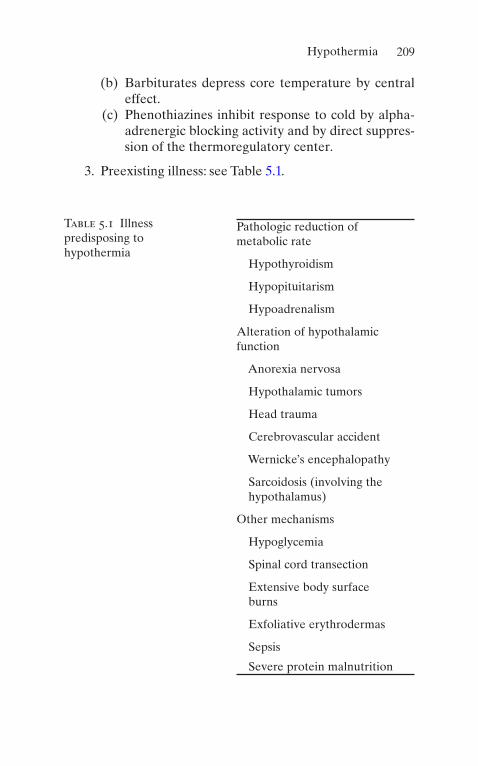
209
(b) Barbiturates depress core temperature by central effect.
(c) Phenothiazines inhibit response to cold by alpha- adrenergic blocking activity and by direct suppres-sion of the thermoregulatory center.
3. Preexisting illness: see Table 5.1.
Table 5.1 Illness predisposing to hypothermia
Pathologic reduction of metabolic rate
Hypothyroidism
Hypopituitarism
Hypoadrenalism
Alteration of hypothalamic function
Anorexia nervosa
Hypothalamic tumors
Head trauma
Cerebrovascular accident
Wernicke’s encephalopathy
Sarcoidosis (involving the hypothalamus)
Other mechanisms
Hypoglycemia
Spinal cord transection
Extensive body surface burns
Exfoliative erythrodermas
Sepsis
Severe protein malnutrition
Hypothermia

210
C. Clinical Presentation
1. Initial sympathetic response (producing peripheral vasoconstriction and tachycardia) causes transient ele-vation of blood pressure and cardiac output.
(a) Include: Renal cell dysfunction and decreased lev-els of vasopressin lead to increased renal perfusion that produces increased urine production (“cold diuresis”), which may lead to hypovolemia, hemo-concentration, and increased blood viscosity. Also include the “diving reflex,” which involves vaso-constriction in visceral muscles; blood is shunted to essential organs.
(b) Response is ablated below 30 °C.
2. Clouding of sensorium regularly occurs at 30–32 °C. 3. Mild hypothermia: Tachypnea, tachycardia, shivering,
amnesia, ataxia, and dysarthria. 4. Moderate hypothermia: Decreased level of conscious-
ness, mydriasis, atrial fibrillation, and bradycardia. 5. Severe hypothermia: Absent reflexes and response to
pain, coma, and hypotension. 6. Cardiac complications:
(a) Dysrhythmias: atrial fibrillation is common. (b) Patients are increasingly susceptible to ventricular
fibrillation with diminishing temperature. Below 28 °C, even minor procedures, such as moving or repositioning the patient, may initiate ventricular fibrillation.
(c) Ventricular fibrillation is resistant to pharmaco-logic treatment or electrical defibrillation until temperature has increased.
(d) Susceptibility to ventricular dysrhythmias may persist for several days, even after normal body temperature has been restored.
(e) ECG changes include PR, QT, and QRS prolonga-tion and J-point elevation (“Osborn wave”).
Chapter 5. Environmental Disorders

211
D. Complications
1. Morbidity and mortality are related to the degree and duration of hypothermia and to appropriate therapy.
2. Bronchorrhea and depressed cough reflexes lead to bronchopneumonia and aspiration pneumonitis.
3. Punctate hemorrhages may occur in the gastrointesti-nal tract, but significant bleeding is rare.
4. Pancreatitis is common. 5. Fatal disseminated intravascular coagulation may
occur.
E. Treatment
1. Cardiovascular
(a) Employ basic life support protocols to support ventilation and circulation.
(b) Perform endotracheal intubation and mechanical ventilation as indicated.
(c) Initiate cardiopulmonary resuscitation (CPR) if cardiac arrest is present. Continue until core temperature of 32–35 °C is reached or response achieved.
(d) Ventricular fibrillation may be resistant to defibril-lation in severe hypothermia. Attempt single defi-brillation; if this is not successful, continue rewarming until temperature is >30 °C, and admin-ister bretylium 5–10 mg/kg IV over 5–10 min and then infusion of 2 mg/min.
2. Rewarming
(a) For mild and moderate hypothermia (including most cases of secondary hypothermia), passive rewarming is indicated.
1. Remove patient from cold environment. 2. Apply dry, unheated blankets.
(b) For some cases of moderate hypothermia, use active surface rewarming.
Hypothermia

212
1. Heating blankets, hot-water bottles, heat cra-dles, heat-fluidized beds, and warm water immersion.
2. Moderate hypothermia may be associated with afterdrop of core temperature as peripheral vasoconstriction is reversed, producing hypovo-lemic shock and ventricular fibrillation.
(c) For severe hypothermia, active core rewarming is indicated. Use one or more of the following:
1. Infusion of warmed intravenous fluids (temper-ature not to exceed 40 °C).
2. Intubation and ventilation with warmed, humid-ified oxygen. Keep airway temperature <45 °C.
3. Peritoneal dialysis (usually requires 6–8 exchanges of potassium-free dialysate heated to 43 °C).
4. Gastric irrigation with warmed fluids with intro-duction of intragastric balloon. May provoke dysrhythmias.
5. Mediastinal irrigation (operative technique). 6. Extracorporeal blood rewarming (hemodialysis,
venovenous, continuous arteriovenous, and car-diopulmonary bypass) with a heat exchanger at 40 °C.
(d) If the patient does not respond to rewarming, con-sider cerebral edema.
Smoke Inhalation and Carbon Monoxide Poisoning
Carbon monoxide (CO) intoxication is the leading cause of death by poisoning in the United States, accounting for 3800 accidental and suicidal deaths annually. It is also the most com-mon cause of death in combustion-related inhalation injury. Many nonlethal exposures may, however, go undetected.
Chapter 5. Environmental Disorders

213
A. Pathophysiology
1. Carbon Monoxide Poisoning
(a) Carbon monoxide combines preferentially with hemoglobin to produce carboxyhemoglobin. This displaces oxygen and reduces systemic arterial content.
(b) Carbon monoxide binds reversibly to hemoglobin with an affinity over 200 times that of oxygen, so a relatively minute concentration of CO in the envi-ronment can lead to toxic concentrations in blood.
(c) Mechanisms of toxicity:
1. Decreased oxygen-carrying capacity of the blood.
2. Alteration of dissociation characteristics of oxy-hemoglobin: shifts oxyhemoglobin dissociation curve to the left.
3. Decreased cellular respiration due to binding with cytochrome c oxidase.
4. Binding to myoglobin, producing myocardial and skeletal muscle dysfunction.
(d) The half-life of carboxyhemoglobin at room air is 320 min; on 100% O2 at 1 atm, it is 60 min; and on 100% O2 at 3 atm, it is 23 min.
2. Smoke Inhalation
(a) Smoke contains carbon particles and various gases (including nitrogen oxide, chlorine, phosgene, ammonia, and hydrogen cyanide).
(b) These gases adhere to the respiratory mucosa and produce compounds that are locally and systemi-cally toxic.
B. Predisposition
1. The most common sources of CO are listed in Table 5.2. 2. Smoke inhalation from fires accounts for the vast
majority of cases.
Smoke Inhalation and Carbon Monoxide Poisoning

214
3. Vapor of methylene chloride (contained in many paint removers) is readily absorbed through the lungs and is converted to CO by the liver.
C. Clinical Presentation
1. Carbon Monoxide Poisoning
(a) Symptoms and signs in acute CO poisoning depend on carboxyhemoglobin (COHb) level (see Table 5.3).
1. Patients with COHb levels <10% are usually asymptomatic.
Table 5.2 Exogenous sources of carbon monoxide
Smoke from all types of fires
Paint remover containing methylene chloride
Furnaces
Gasoline-powered engines
Swimming pool heaters
Sterno fuel
Tobacco smoke
Vehicular exhaust fumes
Water heaters
Table 5.3 Acute CO poisoningCOHb level (%) Symptoms10 Headache
20 Dizziness, nausea, dyspnea
30 Visual disturbances
40 Confusion, syncope
50 Seizures, coma
>60 Cardiopulmonary failure and death
Chapter 5. Environmental Disorders

215
2. Patients with levels >25% should be considered for hospital admission.
3. Those with levels >50% commonly have coma and seizures resulting from cerebral edema.
4. Death is likely if levels >60%. 5. Determination of treatment only on COHb
level is inappropriate, though, because the level alone is a poor predictor of degree of injury. Therefore, clinical assessment is vital to plan-ning treatment.
(b) Chronic or subacute poisoning may present with less characteristic symptoms.
2. Smoke InhalationSuspect if
1. Exposure occurred in a closed space. 2. There are facial or pharyngeal burns or burned nasal
vibrissae. 3. There is carbonaceous sputum or hoarseness.
D. Complications
1. Neuropsychiatric problems, such as memory loss, per-sonality changes, mutism, and parkinsonism may occur as long-term sequelae to CO poisoning.
2. Indicators of a poor prognosis include altered level of consciousness, advanced age, metabolic acidosis, and structural abnormalities on computed tomography (CT) or magnetic resonance (MR) scanning.
E. Treatment
1. Carbon Monoxide Poisoning
(a) Obtain the COHb level in all patients suspected of having CO exposure (e.g., all victims at a fire scene).
(b) Apply supplemental oxygen 100% by mask while awaiting results.
Smoke Inhalation and Carbon Monoxide Poisoning

216
1. The goal is to improve blood oxygen content by maximizing the fraction dissolved in plasma.
2. Monitor patients with serial COHb levels. Continue 100% oxygen until the COHb level is <5%. Continue to monitor COHb level thereaf-ter to guard against undetected release of CO from tissue sites.
(c) Obtain an ECG and monitor with serial CK levels if the ECG is suggestive of ischemia.
(d) Consider treatment with hyperbaric oxygen (HBO) in the following patients:
1. Patients with serious CO poisoning (e.g., coma, neurologic deficit, cardiac or hemodynamic instability, history of loss of consciousness)
2. Pregnant women 3. Patients with underlying heart disease or abnor-
mal ECG
(e) Whether HBO alters outcome is still controversial, and some authors have recommended its use up to 24 h following the acute exposure.
(f) If HBO treatment is unavailable, consider intuba-tion and mechanical ventilation with administra-tion of 100% oxygen if clinical findings merit.
(g) Consider transfusion with packed red blood cells in severe poisoning.
2. Smoke Inhalation
(a) If findings of smoke inhalation are present, evalu-ate the airway with fiber-optic bronchoscopy.
(b) Obtain arterial blood gases and chest radiograph. (c) Administer humidified oxygen. (d) Perform early endotracheal intubation in the pres-
ence of upper airway edema.
Chapter 5. Environmental Disorders
217
Scorpion Envenomation
Scorpions are arachnids that typically inhabit temperate cli-mates. The only dangerous species of scorpion in the United States is the Centruroides genus, which is found primarily in the southwestern states (Arizona, California, New Mexico, Texas).
A. Pathophysiology. The venom is complex and contains a number of proteins, polypeptides, enzymes (e.g., hyaluron-idases and phospholipases), and neurotoxins. The venom affects sodium channels to prolong action potentials and produces spontaneous neural depolarizations of the sym-pathetic and parasympathetic systems.
Alpha-toxins are the most common ones. They bind to the site, inhibit inactivation of the sodium channel, and result in longer depolarization that will create neuronal excitation.
B. Predisposition. Most serious envenomations occur in children.
C. Clinical Presentation
1. The most common finding is severe local pain at the site of the sting. In most cases, this resolves within sev-eral hours. There is severe tenderness to palpation or percussion over the site.
2. The sting may be accompanied by wheal and flare reac-tion, paresthesias at the site of sting.
3. Significant envenomation produces tachycardia, hyper-tension, restlessness, hyperexcitability, diaphoresis, piloerection, nystagmus, diplopia, opisthotonos, muscu-lar fasciculations, or hypersalivation.
4. The most severe cases produce seizures, pulmonary edema, muscular paralysis, respiratory arrest, cardio-vascular collapse, and death.
D. Complications. These include DI, pancreatitis, jaundice, and renal failure.
Scorpion Envenomation

218
E. Treatment
1. Maintain airway and assist ventilations if necessary. 2. Clean the wound and administer tetanus prophylaxis if
indicated. Do not cool or incise wound. 3. Classify Centruroides sting by severity of involvement,
according to Table 5.4.Start antivenin from grade 2 while ICU admission
from grade 3; dobutamine infusion and noninvasive or mechanical ventilation from grade 4 are considered as just supportive care phase, with multiorgan failure.
(a) Grade I and II envenomation: Wound care as above and provide oral analgesics.
(b) Grade III and IV: An antivenin has been produced but is not generally available. Contact your regional poison control center. Sedate and provide support-ive care. High doses of phenobarbital have been recommended by some. These patients may pres-ent pulmonary edema and require aggressive mechanical ventilator support.
(c) Treat severe tachycardia with beta-blockers.
Table 5.4 Severity of Centruroides scorpion envenomationGrade Signs and symptomsI Local pain or paresthesias at site; tenderness to touch or
percussion
II Local findings and pain and paresthesias remote from sting site
III Symptomatic skeletal neuromuscular dysfunction (muscular jerking or shaking) or cranial nerve dysfunction (e.g., blurred vision, difficulty swallowing, hypersalivation, slurred speech, tongue fasciculations)
IV Somatic skeletal and cranial nerve dysfunction
Chapter 5. Environmental Disorders

219
F. Prognosis
1. Though deaths may occur, none has been reported in the United States since 1968.
2. If untreated, the sting may be lethal in 1% of adults and as many as 25% of children under age 5 years.
Snakebite
Approximately 8000 instances of poisonous snakebite occur annually in the United States, with 9–15 deaths resulting. Most bites occur in the southern and southwestern states, with a peak incidence during the summer months. Fifteen percent of the 3000 species of snakes worldwide are consid-ered to be dangerous to humans.
There are four venomous species in the United States: rattlesnakes (genus Crotalus and Sistrurus), copperheads (Agkistrodon contortrix), cottonmouths (Agkistrodon piscivorus)—all of which are pit vipers—and coral snakes (genus Micrurus). Rattlesnakes account for 65% of reported venomous snakebites.
A. Pathophysiology. Snake venoms are complex mixtures of toxins that have cytotoxic, hemotoxic, and neurotoxic components. Cytotoxic effects produce tissue necrosis. Hemotoxic venoms interfere with the coagulation system. Rattlesnakes are classically considered to have cytotoxic and hemotoxic venom, but neurotoxic activity may also be present. Coral snake venom has largely neurotoxic activity.
Snakes are capable of controlling the quantity of venom administered, and approximately 25–30% of bites by poi-sonous snakes do not result in envenomation.
B. Predisposition
1. The majority of victims are males younger than age 20. 2. Severity of a bite depends on:
Snakebite

220
(a) Size and species of snake (b) Location of bite (c) Grade of envenomation (see below in Tables 5.5
and 5.6) (d) Age and medical condition of the patient
C. Clinical PresentationIdentification of the snake is important. Coral snakes
have shorter fangs and a smaller mouth; this allows them less opportunity for envenomation than crotalids; the bite resembles more chewing rather than the strike for which the pit vipers are famous. Although less dramatic, it can later result in respiratory failure from systemic neuromus-cular blockade.
1. Pit Vipers
(a) The most important findings of pit viper envenom-ation are fang punctures at the bite site (usually on the extremities), local pain, and adjacent erythema and edema.
1. Edema and erythema are characteristic and usu-ally develop within 30 min of envenomation.
2. May spread for following 24 h and develop hem-orrhagic bullae.
3. If no erythema or edema develops within 4 h of bite, it is unlikely that envenomation has occurred.
Copperhead snakebites generally are limited to local tissue destruction. Rattlesnakes can leave impressive wounds and cause systemic toxicity.
(b) Grade the severity of the bite according to the cri-teria in Table 5.5.
(c) Swelling resulting from edema and capillary rup-ture may produce increased fascial compartment pressure.
(d) Severe envenomation produces hypotension due to hemorrhage and third-space fluid loss, shock, pares-thesias, and muscular fasciculations. Nausea, vomit-ing, giddiness, and elevation or depression of
Chapter 5. Environmental Disorders

221
temperature may be produced. Coma, convulsions, and death may result.
(e) Hematologic sequelae of severe envenomation include anemia due to hemolysis, hypothrombin-emia, thrombocytopenia, hypofibrinogenemia, and hypercoagulability. Gastrointestinal, urinary tract, or intracerebral hemorrhage may result.
(f) CNS effects include seizures, coma, and respiratory paralysis.
2. Coral Snakes
(a) Coral snakebites do not produce prominent swell-ing or other local findings.
(b) Neurotoxic venom may cause ptosis, diplopia, dys-phagia, dysarthria, salivation, paresthesias, muscu-lar fasciculations, loss of deep tendon reflexes, muscular weakness, and respiratory paralysis.
(c) Onset of symptoms may be delayed by 1–5 h fol-lowing the bite.
(d) Grade the severity of the bite according to the cri-teria in Table 5.6. Repeat the evaluation every 15 min for the first 4 h following the bite.
Table 5.5 Severity of pit viper envenomationGrade Envenomation FindingsO None Fang marks; minimal pain; <2.5 cm
circumferential edema
I Minimal 2.5–12.5 cm edema and erythema in first 12 h
II Moderate 15–36 cm edema and erythema in first 12 h
III Severe Edema >36 cm in 24 h; systemic symptoms (including coagulation defects) present
IV Very severe Systemic symptoms; rapid development of edema, erythema; ecchymoses, bullae; coagulation defects
Snakebite
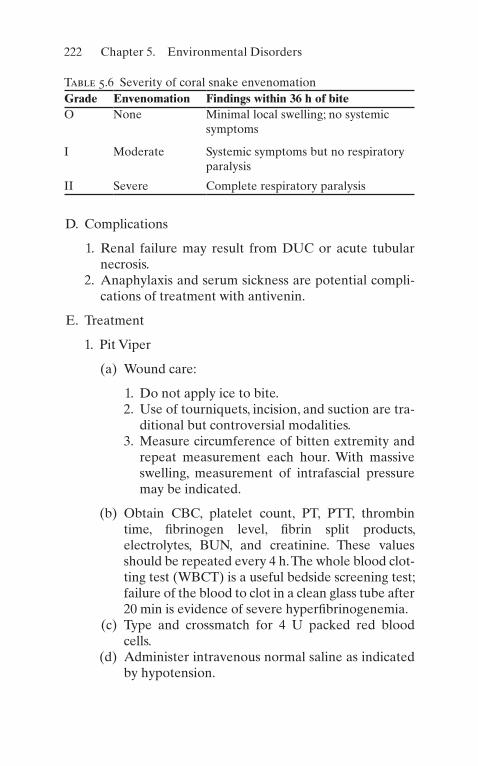
222
D. Complications
1. Renal failure may result from DUC or acute tubular necrosis.
2. Anaphylaxis and serum sickness are potential compli-cations of treatment with antivenin.
E. Treatment
1. Pit Viper
(a) Wound care:
1. Do not apply ice to bite. 2. Use of tourniquets, incision, and suction are tra-
ditional but controversial modalities. 3. Measure circumference of bitten extremity and
repeat measurement each hour. With massive swelling, measurement of intrafascial pressure may be indicated.
(b) Obtain CBC, platelet count, PT, PTT, thrombin time, fibrinogen level, fibrin split products, electrolytes, BUN, and creatinine. These values should be repeated every 4 h. The whole blood clot-ting test (WBCT) is a useful bedside screening test; failure of the blood to clot in a clean glass tube after 20 min is evidence of severe hyperfibrinogenemia.
(c) Type and crossmatch for 4 U packed red blood cells.
(d) Administer intravenous normal saline as indicated by hypotension.
Table 5.6 Severity of coral snake envenomationGrade Envenomation Findings within 36 h of biteO None Minimal local swelling; no systemic
symptoms
I Moderate Systemic symptoms but no respiratory paralysis
II Severe Complete respiratory paralysis
Chapter 5. Environmental Disorders

223
(e) Antivenin
1. Perform skin or eye test for horse serum hyper-sensitivity. If negative, may proceed with anti-venin administration. If positive, consider risk of envenomation against possibility of anaphylac-tic reaction. In severe envenomation, may pre-cede antivenin infusion with 250 mg methylprednisolone IV. Be prepared to treat anaphylaxis (see Chap. 18, “Allergic and Immunologic Emergencies”).
2. No universally accepted standard recommenda-tions for antivenin administration exist. Controversy about its use exists.
3. No antivenin is required for grades 0 or I pit viper bites.
4. Administer to grades II–IV bites. Test dose: 0.2–0.5 mL IV over 5 min; if no adverse effect, administer remainder over 30 min to 2 h. Individualize dosage to severity and rate of pro-gression: grade II, up to 5 vials; grade III, 5–15 vials; grade IV, 15–20 vials.
5. Observe the patient for 3–5 h after initial admin-istration; administer an additional vial every 1–2 h if pain persists or swelling progresses.
(f) Use of fasciotomy in severe envenomation with increased intrafascial pressure is controversial. This may be necessary if intrafascial pressure >30 Torr. Obtain surgical consultation as required in instances of massive edema.
(g) The routine use of antibiotics is controversial and depends, in part, upon the local rates of infection.
F. Coral Snakes
(a) An antivenin is available for bites of Eastern coral snakes. Administer antivenin to all bite victims of this snake, even before appearance of symptoms. Dosage is 3–6 vials in 300–500 mL normal saline.
Snakebite

224
(b) There is no antivenin available for bites of the Arizona coral snake.
(c) Perform endotracheal intubation if there are any signs of bulbar paralysis, such as diplopia or dysphagia.
(d) Institute respiratory support with mechanical ventila-tion as required. Antivenin may not completely reverse respiratory depression.
Spider Bite
The spiders that can cause medically significant bites include widow and false widow spiders (worldwide), recluse spiders (mostly North and South America), Australian funnel web spiders (eastern coastal Australia), and Phoneutria spiders (Brazil).
A. Black Widow Spider. The black widow spider is a member of the Latrodectus genus and is found throughout the United States. The female, larger and more dangerous than the male, is black or brown, with a characteristic red hourglass marking on the ventral abdomen.
1. PathophysiologyBlack widow venom is extremely potent. The major
activity of the venom lies in provoking the release of catecholamines at adrenergic synaptic terminals and depletion of acetylcholine from motor nerve endings.
2. PredispositionThere is a high risk for mortality among the follow-
ing patients:
(a) Patients younger than 16 or older than 65 years. (b) Those with hypertension or cardiovascular disease
(envenomation may cause heart failure, cerebrovascu-lar accident, or myocardial ischemia).
3. Clinical presentation based on severity of illness
(a) Mild: Pain restricted to the bite site and normal vital signs
Chapter 5. Environmental Disorders

225
(b) Moderate: Diaphoresis and piloerection in the area of the bite, cramping pain in large muscle groups of the trunk, and normal vital signs
(c) Severe (also called latrodectism): Diaphoresis at a remote site; intense generalized cramping pain in large muscle groups of the trunk; hypertension and tachycardia; often headache, nausea, and vomiting
4. Important points to remember
(a) Pain at the bite site may be minimal, but severe pain may develop in the extremity, along with pain-ful lymphadenopathy, erythema, swelling, and pilo-erection 20–60 min following the bite.
(b) Cramping and painful muscular contractions in the back, thigh, abdomen, and chest produce abdomi-nal rigidity, tonic contractions, and tremor.
(c) There may be restlessness, weakness, dizziness, uri-nary retention, diaphoresis, salivation, nausea, vomiting, priapism, and hypertension.
(d) Distinction must be made from the acute abdo-men: despite rigidity, there is minimal tenderness and no rebound tenderness.
5. Complications
(a) Hypertensive crisis (b) Paralysis and respiratory arrest (especially in
children) (c) Severe envenomation may produce shock and/or
coma
6. Treatment
(a) Clean wound and apply cool compresses (not ice) to the bite site.
(b) Administer diazepam (5–10 mg IV q3 h as needed) and methocarbamol (1000 mg IV no faster than 100 mg/min and 1000 mg additionally as IV infu-sion) for symptomatic treatment of muscular spasm.
Spider Bite

226
(c) Calcium gluconate 10% (1–2 mL/kg IV over 20 min, maximum 10 mL) may also provide relief of muscular spasm, but this is transient, lasting <30 min.
(d) Treat elevated blood pressure (see Chap. 3, “Cardiovascular Disorders”).
(e) Antivenin
1. Administration of antivenin is recommended for symptomatic patients <16 or >65 years old, those with preexisting hypertension or cardio-vascular disease, and those displaying respira-tory distress, pronounced hypertension, or persistent severe muscular symptoms despite above treatment.
2. Before administration, skin test for horse serum sensitivity (included with antivenin).
3. Administer 1 vial antivenin diluted in 50 mL normal saline over 15–30 min. A second vial will be required only in severe cases.
7. Prognosis
(a) Prognosis for recovery with treatment is good and deaths are rare.
(b) There is a 0.5% incidence of anaphylaxis and a 2% incidence of serum sickness associated with anti-venin use.
B. Brown Recluse Spider. The brown recluse spider is of the Loxosceles species and is present throughout the southern United States. It is tan or brown, with a violin-shaped mark on the dorsum of the cephalothorax.
1. PathophysiologyThe venom contains a variety of enzymes, including
hyaluronidase, protease, collagenase, and sphingomye-linase D, which are thought to be responsible for der-mal necrosis and hemolysis.
Chapter 5. Environmental Disorders

227
2. PredispositionMost severe envenomations occur in children.
3. Clinical Presentation
(a) Local pain and burning, bulla formation at site of bite, becoming necrotic over hours to days.
(b) Systemic signs and symptoms: fever, chills, pete-chiae, nausea, vomiting, and weakness.
(c) Severe cases may produce hemolysis, disseminated intravascular coagulation, thrombocytopenia, jaun-dice, or shock.
4. Complications
(a) Severe hemolysis may result in death. (b) Renal failure may result from hemoglobinuria or
myoglobinuria.
5. Treatment
(a) There is no commercially available antivenin avail-able in the United States.
(b) Begin intravenous infusion with normal saline. (c) Obtain CBC, platelet count, electrolytes, BUN, cre-
atinine, PT, PTT, and urinalysis. (d) Clean the wound and administer tetanus prophy-
laxis if indicated. Do not cool or incise wound. (e) Some recommend the use of dapsone (50–200 mg/
day), a leukocyte inhibitor, which relieves pain and reduces erythema and induration. Do not adminis-ter to children.
(f) Obtain surgical consultation for wound care.
Useful Facts and Formulas
A. Temperature. Temperature conversion calculations are often done in the management of critically ill patients. Degrees Celsius (°C) and Fahrenheit (°F) are most com-monly utilized:
Useful Facts and Formulas

228
� �
� �
� �
� �
� �� � �
� �� ��
Cto F
F C
F to C
C F
9 5 32
32 5 9
/
/
Occasionally, the Kelvin (K) temperature scale is used, primarily in gas law calculations:
� �
�� �K to C
K C 273
B. Humidity. Relative humidity (RH) is usually measured by hygrometers, thus, eliminating the need of extracting and measuring the humidity content of the air samples:
RH
Content mg LormmHg
Content mg Lor mmHg�
� �� �
�/
/%
The humidity deficit (HD) represents the maximum humid-ity capacity at body temperature:
HD Capacity content mg L� � � /
Where capacity is the amount of water the alveolar air can hold at body temperature (also known as absolute humidity) and content is the humidity content of inspired air.
The humidity capacity of saturated gases is shown in Table 5.7.
C. Pressure. Pressure is defined as force per unit area, and there are various ways of measuring this force. One way that force can be recorded is in a form of the height of a
Chapter 5. Environmental Disorders

229
Table 5.7 Humidity capacity of saturated gases from 0 °C to 43 °CGas temperature (°C)
Water content (mg/L)
Water vapor pressure (mmHg)
0 4.9 4.6
5 6.8 6.6
10 9.4 9.3
17 14.5 14.6
18 15.4 15.6
19 16.3 16.5
20 17.3 17.5
21 18.4 18.7
22 19.4 19.8
23 20.6 21.1
24 21.8 22.4
25 23.1 23.8
26 24.4 25.2
27 25.8 26.7
28 27.2 28.3
29 28.8 30.0
30 30.4 31.8
31 32.0 33.7
32 33.8 35.7
33 35.6 37.7
34 37.6 39.9
35 39.6 42.2
36 41.7 44.6
37 43.9 47.0
(continued)
Useful Facts and Formulas

230
column, as in the mercury barometer; therefore, it can be recorded in milliliters of mercury (mmHg) pressure or centimeters of water pressure. To convert cmH2O to mmHg:
cmH O mmHg2 0 735� �.
To convert mmHg to cmH2O:
mmHg cmH O� �1 36 2.
A less commonly used conversion in clinical medicine includes converting psi to mmHg:
Psi mmHg� �51 7.
Other useful pressure-related formulas/facts include the following:
Table 5.7 (continued)
Gas temperature (°C)
Water content (mg/L)
Water vapor pressure (mmHg)
38 46.2 49.8
39 48.6 52.5
40 51.1 55.4
41 53.7 58.4
42 56.5 61.6
Chapter 5. Environmental Disorders

231
Total Pressure P P P Dalton slaw
atmosphere mmHg
� � � � ��
�
1 2 3
1 760
2
& ’
99 921
33 93
1034
1034
14 7
2
2
22
.
.
/
. /
in Hg
ft H O
cmH O
gm cm
lb in
����
Useful pressure/volume relationships that can be used in the management of critically ill patients include:
Volume Volume FactorATPS� �
Where VolumeBTPS is the gas volume saturated with water at body temperature (37 °C) and ambient pressure (BTPS = barometric temperature pressure saturation); VolumeATPS is the gas volume saturated with water at ambient (room) temperature and pressure (ATPS = ambient tempera-ture pressure saturation); and Factor is the factor for convert-ing gas volumes from ATPS to BTPS:
Conversion factorH O
CB
B
���
��� ��
P P
P2
47
310
273
Table 5.8 shows the conversions factors required to con-vert ATPS to BTPSA.
D. Altitude. As altitude varies, changes in atmospheric pres-sure produce alterations in gas density (see Table 5.9).
Useful Facts and Formulas

232
Table 5.8 Factors for converting gas volumes from ATPS to BTPSGas temperature (°C)
Factors to convert to 37 °C saturated
Water vapor pressure (mmHg)
18 1.112 15.6
19 1.107 16.5
20 1.102 17.5
21 1.096 18.7
22 1.091 19.8
23 1.085 21.1
24 1.080 22.4
25 1.075 23.8
26 1.068 25.2
27 1.063 26.7
28 1.057 28.3
29 1.051 30.0
30 1.045 31.8
31 1.039 33.7
32 1.032 35.7
33 1.026 37.7
34 1.020 39.9
35 1.014 42.2
36 1.007 44.6
37 1.000 47.0
38 0.993 49.8
39 0.986 52.5
40 0.979 55.4
41 0.971 58.4
42 0.964 61.6
Chapter 5. Environmental Disorders

233
Table 5.9 Changes in density with altitude, assuming a constant temperature
Altitude (feet)
Standard temperature (°C)
Density ratio constant temperature
Density ratio standard temperature
0 15.00 1.0000 1.0000
5000 5.09 0.8320 0.8617
10,000 −4.81 0.6877 0.7385
15,000 −14.72 0.5643 0.6292
Useful Facts and Formulas

235© Springer Nature Switzerland AG 2021J. Varon, Handbook of Critical and Intensive Care Medicine, https://doi.org/10.1007/978-3-030-68270-5_6
Gastrointestinal Bleeding
A. Classification
1. Upper gastrointestinal (GI) bleeding is above the liga-ment of Treitz.
2. Lower GI bleeding is below the ligament of Treitz.
B. Etiology. The most common causes of acute GI bleeding requiring admission to the intensive care unit (ICU) are depicted in Table 6.1. The most common sources of GI bleeding in the ICU are gastroduodenal stress ulcerations.
C. Diagnostic Evaluation
1. HistoryAlthough the history and physical assessment in a
critically ill patient with acute GI bleeding may be lim-ited by the patient’s clinical condition, the following are points that need to be investigated:
(a) History of hematemesis or melena(b) Time of onset(c) Amount of blood(d) Color and character
Chapter 6Gastrointestinal Disorders

236
(e) Drug or alcohol use (i.e., nonsteroidal anti- inflammatory drug [NSAID], prednisone, warfarin, direct oral anticoagulants [DOAC])
(f) Past medical history (i.e., cirrhosis, peptic ulcer dis-ease [PUD], inflammatory bowel disease [IBD], etc.)
2. Physical ExaminationThe precise cause of acute GI bleeding is unlikely to
be evident from physical examination alone (except in
Table 6.1 Etiologies of acute GI bleedingUpper LowerEsophagus Small intestine
Mucosal tear Arteriovenous malformations
Esophageal rupture Inflammatory bowel disease ischemia
Esophagitis Meckel’s diverticulum
Neoplasms Neoplasms
Varices
Stomach Large intestine
Arteriovenous malformations
Diverticulosis
Gastritis (any etiology) Hemorrhoids
Neoplasms Inflammatory bowel disease infections
Peptic ulcer disease Ischemia
Stress ulcers Neoplasms
Duodenum
Arteriovenous malformations
Neoplasms (rare)
Peptic ulcer disease
Chapter 6. Gastrointestinal Disorders
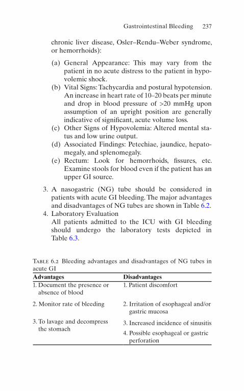
237
chronic liver disease, Osler–Rendu–Weber syndrome, or hemorrhoids):
(a) General Appearance: This may vary from the patient in no acute distress to the patient in hypo-volemic shock.
(b) Vital Signs: Tachycardia and postural hypotension. An increase in heart rate of 10–20 beats per minute and drop in blood pressure of >20 mmHg upon assumption of an upright position are generally indicative of significant, acute volume loss.
(c) Other Signs of Hypovolemia: Altered mental sta-tus and low urine output.
(d) Associated Findings: Petechiae, jaundice, hepato-megaly, and splenomegaly.
(e) Rectum: Look for hemorrhoids, fissures, etc. Examine stools for blood even if the patient has an upper GI source.
3. A nasogastric (NG) tube should be considered in patients with acute GI bleeding. The major advantages and disadvantages of NG tubes are shown in Table 6.2.
4. Laboratory EvaluationAll patients admitted to the ICU with GI bleeding should undergo the laboratory tests depicted in Table 6.3.
Table 6.2 Bleeding advantages and disadvantages of NG tubes in acute GIAdvantages Disadvantages1. Document the presence or
absence of blood1. Patient discomfort
2. Monitor rate of bleeding 2. Irritation of esophageal and/or gastric mucosa
3. To lavage and decompress the stomach
3. Increased incidence of sinusitis
4. Possible esophageal or gastric perforation
Gastrointestinal Bleeding

238
5. Endoscopy is indicated in the vast majority of patients requiring ICU admission for GI bleeding:
Upper endoscopy is indicated when blood is obtained from the NG tube or when frank hematemesis is present.
(a) If lower GI bleed is suspected, colonoscopy is the diagnostic study of choice.
(b) Special endoscopic procedures may be required (i.e., wireless video capsule endoscopy, push enter-oscopy, double balloon enteroscopy).
6. Radiologic EvaluationChest radiograph and abdominal X-rays should be
considered to check for evidence of perforation or obstruction and may indicate ischemic changes.
Contrast studies have a low diagnostic yield and may be hazardous for the critically ill patient. They may also interfere with other diagnostic studies (i.e., endoscopy, angiography). Special tests may be required in the eval-uation of acute GI bleeding. These include:
(a) Selective angiography may be used as a diagnostic as well as therapeutic tool (e.g., embolization). A bleeding rate ≥0.5 mL/min at the time of the proce-dure is needed for diagnosis.
Table 6.3 Initial laboratory evaluation in GI bleedingComplete blood count (H/H should be repeated every 4 h until patient is stable or bleeding has been controlled)
BUN, creatinine, and electrolytes
PT, PTT
Type and crossmatch for 2–8 U of PRBCs, FFP
Other tests are ordered according to suspected or known underlying disease (i.e., LFTs, CK, etc.)
BUN blood urea nitrogen, CK creatine kinase, FFP fresh-frozen plasma, H/H hemoglobin/hematocrit, LFT liver function tests, PRBCs packed red blood cells, PT prothrombin time, PTT partial thromboplastin time
Chapter 6. Gastrointestinal Disorders

239
(b) Radionuclide scans are sensitive in detecting lesions with lower bleeding rates.
D. Initial ICU Management
1. As in any critically ill patient, the management of acute GI bleeding starts with assessment of the airway, breathing, and circulation (ABCs). A low threshold for endotracheal intubation is recommended in the event of clouding of consciousness or overt shock, to prevent aspiration.
2. Insert at least two large-bore IV catheters. 3. Infuse blood, plasma expanders, and/or normal saline
to maintain a mean arterial pressure ≥65 mmHg. 4. Some authors still recommend NG placement in all
patients with GI bleeding and lavage of the stomach until the return is clear. This practice is considered useful only in settings where emergency endoscopy is not available.
5. Correction of preexisting coagulopathy (i.e., fresh- frozen plasma [FFP], vitamin K, etc.).
Intravenous proton pump inhibitors in either con-tinuous or bolus form. Continue infusions (i.e., esome-prazole [Nexium™] 20–40 mg/24 h, pantoprazole [Protonix™] 8 mg/h. High-dose bolus are esomepra-zole 80 mg or pantoprazole 80 mg.
6. Endoscopic and/or angiographic verification of the source of bleeding will allow more definitive therapy (i.e., thermal coagulation, injection therapy, fibrin seal-ant, endoclips, surgery). Should be done within 12 h of presentation.
E. Specific Management of Selected Conditions
1. Variceal Hemorrhage
(a) Somatostatin Analogs: Somatostatin inhibits the release of vasodilator hormones, such as glucagon, indirectly causing splanchnic vasoconstriction and decreased portal inflow:
Gastrointestinal Bleeding

240
1. Octreotide is a long-acting analog of somatostatin. 2. Dosing: 25–50-mcg IV bolus followed by con-
tinuous IV infusion of 25–50 mcg/h.
(b) Prophylactic Antibiotic Therapy: Antibiotics should be given in patient with cirrhosis who pres-ents with upper GI bleed due to high incidence of infection.
(c) Balloon Tamponade: Temporizing measure only. Should be with plans of additional intervention.
(d) Endoscopic Variceal Ligation: Preferred due to high efficacy and lower complication rate.
(e) Sclerotherapy: Used as additional option during endoscopic treatment of varices.
(f) Esophageal Stent: New technique with stent place-ment around the bleeding site. Promising new studies, however, are used in cases with refractory bleeding.
(g) Endoscopic Therapy Failure: Rebleeding happens in 10–20 percent of patients. Should be considered in cases in which bleeding does not stop despite multiple therapies or continuous bleeding post endoscopy therapy. Options include:
1. Transjugular intrahepatic portosystemic shunt. 2. Emergency shunt surgery. 3. Hemorrhage from ulcers and erosive lesions:
Endoscopy therapy with injection therapy, hemoclips, thermal coagulation, or fibrin seal-ants. This should be considered in patients with active bleeding/oozing or visible vessel. PPI infusion should be continued for 72 h after endoscopy.
2. Surgical Interventions
(a) Interventional angiography: Preferred due to lower risk and less invasive
(b) Surgery: Alternative option if intervention angiog-raphy not available or failed
Chapter 6. Gastrointestinal Disorders
241
3. Active Lower GI Bleeding
(a) If a lesion is reachable with colonoscopy, local ther-apy may be attempted (e.g., laser coagulation).
(b) Arterial embolization is indicated if the above fails. (c) All patients with active lower GI bleeding should
receive surgical consultation in case an emergent intervention is needed.
Acute Mesenteric Ischemia
A. Definition. Acute mesenteric ischemia (AMI) is an acute reduction in blood flow to the intestine leading to inade-quate perfusion. AMI may be a reflection of generalized poor perfusion, or it may result from local pathology.
B. Epidemiology. The incidence of AMI has increased over the past few decades. The rising incidence may be attribut-able to advances in medical technology and to new thera-pies extending the life of critically ill patients who are prone to develop AMI (e.g., elderly). The mortality in AMI is between 55% and 100%.
C. Etiology
1. Occlusive
(a) Atherosclerotic narrowing of the mesenteric bed (b) Systemic emboli from any source (e.g.,
endocarditis) (c) Vasculitis (d) Hypercoagulable states
2. Nonocclusive
(a) Splanchnic vasoconstriction
1. Hypovolemia 2. Hypotension 3. Low cardiac output 4. Vasopressor agent use
Acute Mesenteric Ischemia

242
D. Risk Factors for AMI. The most common predisposing conditions are depicted in Table 6.4.
E. Diagnostic Evaluation
1. History and Physical ExaminationThe classic complaint of severe abdominal pain that
is out of proportion to the findings of physical examina-tion, in our experience, is rarely seen. If peritoneal signs are present (e.g., rebound tenderness), intestinal infarc-tion is likely to have occurred. Abdominal distention, emesis, and other signs of intestinal obstruction may occur in patients with AMI in situ. Lower GI bleeding may occur.
2. Laboratory Studies
(a) Leukocytosis in 75% of patients (b) Metabolic acidosis (c) Elevated amylase, creatine kinase (CK) (6–12 h
after infarction has occurred), lactate, and phosphate
3. Radiologic Evaluation. Should be done as soon as the patient has been adequately resuscitated (including measures aimed at relieving acute congestive heart fail-ure and hypotension, correction of hypovolemia, and cardiac dysrhythmias).
(a) Plain Abdominal X-Rays1. Useful in excluding other causes of abdominal
pain (i.e., mechanical obstruction, perforation).2. Seventy percent of patients will show at least
one of the following:
Table 6.4 Risk factors for the development of AMI
Age ≥50 years
Atherosclerotic heart disease
Congestive heart failure
Recent myocardial infarction
Valvular heart disease
Chapter 6. Gastrointestinal Disorders
243
(a) Ileus(b) Ascites(c) Small bowel dilation(d) Separation of small bowel loops(e) Thickening of valvulae conniventes(f) Thumb printing
Advanced Abdominal Imaging: Computed tomography (CT) or MRI can be considered. CT scan is preferred due to lower cost and availability. MRI may be not sen-sitive. CT scan should be done without oral contrast.
(b) Duplex Ultrasound: Can be used to identify occlu-sions; however imaging can be limited due to bowel loops.
(c) Laparotomy: Should be done in patients with signs of peritonitis or perforation.
F. Therapy
1. Supportive Care: ABCs should be monitored along with hemodynamic support to maximize cardiac out-put, oxygen delivery, and volume status.
2. Anticoagulation: Should be started to prevent clot formation.
3. Invasive Treatment Options: Arterial embolectomy, arterial bypass, arterial stenting, arterial or venous thrombolysis, and intra-arterial vasodilator infusion.
Fulminant Hepatic Failure and Encephalopathy
A. Definition
1. Acute Fulminant Hepatic FailureAcute fulminant hepatic failure (FHF) is defined as
acute liver failure associated with the development of hepatic encephalopathy within 8 weeks of the onset of symptoms attributable to hepatocellular dysfunction. This definition assumes that there is no preexisting liver disease.
Fulminant Hepatic Failure and Encephalopathy

244
2. Hepatic EncephalopathyHepatic encephalopathy (HE) is a complex neuro-
psychiatric syndrome precipitated by abnormal liver function. This syndrome is a feature of acute and/or chronic hepatocellular failure.
B. EtiologyCommon causes of FHF and HE are depicted in
Table 6.5. C. Diagnostic Evaluation
1. HistoryA detailed history should be obtained from family
members. The following points need to be investigated:
(a) History of preexisting liver disease(b) Drug or alcohol use(c) Toxin exposure or ingestion
Table 6.5 Causes of acute liver failure
1. Viral hepatitis (i.e., A, B, C)
2. Drugs or toxins
Acetaminophen
Acute alcohol intoxication
Carbon tetrachloride
Halothane
Isoniazid
Monoamine oxidase inhibitors
Mushroom poisoning
3. Fatty liver of pregnancy
4. Shock of any etiology
5. Massive liver infiltration (i.e., leukemia)
6. Decompensation of chronic liver failure
Chapter 6. Gastrointestinal Disorders

245
2. Physical ExaminationThis may vary from the patient in no distress to the
patient in overt shock.
(a) Vital signs: Tachycardia, hypotension.(b) Associated findings: Petechiae, jaundice, hepato-
megaly, splenomegaly.(c) The encephalopathy may begin with confusion, dis-
orientation, and irrational behavior. Coma may develop rapidly (see Table 6.6.)
3. Laboratory and Radiologic EvaluationAll patients with HE and/or FHF should undergo
the following tests:
(a) Chest X-ray, abdominal X-rays.(b) Blood glucose may reveal hypoglycemia.(c) Serum bilirubin: A value >23 mg/dL is the best pre-
dictor of nonsurvival.(d) AST and ALT have little prognostic value as levels
tend to fall as the patient’s condition worsens.(e) Serum albumin: Its decrease reflects poor
outcome.(f) Serum electrolytes.(g) Complete blood count.(h) Head computed tomography (CT) scan to rule out
a structural lesion (e.g., hemorrhage).(i) Lumbar puncture needs to be considered and per-
formed if meningitis is suspected.
Table 6.6 Clinical stages of hepatic encephalopathyStage Neurological findingsI Confusion, mild changes in personality,
psychometric defects
II Drowsiness to lethargy
III Somnolent but arousable
IV Coma
Fulminant Hepatic Failure and Encephalopathy

246
(j) If the etiology of FHF is unknown, the following needs to be ordered:
1. Acetaminophen level2. Hepatitis profile:
(a) Viral hepatitis A is diagnosed by detection of HAV-IgM in the patient’s serum.
(b) Viral hepatitis B is diagnosed by:
(i) Detection of HBsAg(ii) Anti-HBc IgM
(c) Viral hepatitis C is diagnosed by detection of anti-HCV.
(d) Delta virus hepatitis is diagnosed by detec-tion of anti- HDV in a patient coinfected with hepatitis B virus.
3. Alkaline phosphatase4. Amylase
(k) Serum ammonia level.(l) Electroencephalograms (EEGs) are used to assess
clinical response and prognosis in patients with HE.
D. Complications of FHFWhen the liver fails acutely, all organ systems are
involved to some extent:
1. Central Nervous System (CNS)Hepatic encephalopathy, cerebral edema.
2. CardiovascularDysrhythmias (particularly in patients with advanced
FHF), hypotension. 3. Pulmonary
Hypoxemia advancing to adult (acute) respiratory distress syndrome. (ARDS)
4. RenalThe development of renal failure with FHF carries a
poor prognosis:
Chapter 6. Gastrointestinal Disorders

247
(a) In most instances, the renal failure is related to “prerenal” causes.
(b) The hepatorenal syndrome is a diagnosis of exclu-sion. It is associated with a normal urine sediment, a urinary sodium concentration of <20 mmol/L, and resolution if liver function improves.
5. HematologicThrombocytopenia, diminished clotting factors with
episodes of severe bleeding. 6. Infection
Susceptibility to infection is increased in patients with FHF.
7. MetabolicHypoglycemia, metabolic acidosis, hypokalemia,
hyponatremia.
E. Management
1. Supportive therapy:
(a) As in any critically ill patient, the management of AMI starts with assessment of the ABCs.
(b) The usual indications for endotracheal intubation and assisted mechanical ventilation apply to these patients.
2. The use of corticosteroids for patients with FHF has not been proven to improve survival.
Nutrition: Oral or enteral feeding should be used as tolerated and meeting metabolic requirements. Avoid severe protein restriction. If unable to meet metabolic requirements, consider parenteral nutrition. NG tubes may increase ICP.
The management of FHF-associated cerebral edema is no different from that for nonhepatic-related causes (see Chap. 9, “Neurologic Disorders”). In recent years, emphasis on the use of therapeutic hypothermia for these patients seems encouraging.
Fulminant Hepatic Failure and Encephalopathy
248
3. Bleeding prevention: INR may not be accurate and markers such as fibrinogen may be used. Prophylactic preventive treatments are not recommended.
4. N-Acetylcysteine is useful in acetaminophen toxicity but may also be beneficial in other forms of liver fail-ure. Investigational data have shown some improve-ment in the hemodynamics of patients with FHF treated with.
5. Liver transplantation may be an alternative form of therapy (in a few specialized transplant centers) for some patients with no known contraindication to the procedure.
6. Liver “dialysis”: A few specialized centers are currently exploring this form of therapy.
7. Agents aimed at stimulating ammonia metabolism have also been tried (e.g., ornithine-aspartate, sodium benzoate).
Pancreatitis
A. Definition. Acute pancreatitis is an inflammatory process of the pancreas with a wide range of clinical severity rang-ing from self-limited to a lethal disease, complicated by multiple organ system failure (10% of cases).
B. Etiology. The most common causes of pancreatitis are:
1. Alcoholism 2. Gallstones 3. Hyperlipidemia 4. Trauma (blunt or penetrating) 5. Infections (i.e., mumps, mycoplasma) 6. Hypoperfusion states (i.e., shock, cardiopulmonary
bypass) 7. Hypercalcemia 8. Drugs (i.e., sulfonamides, thiazides)
C. Diagnostic Evaluation
1. History
Chapter 6. Gastrointestinal Disorders

249
Ninety-five percent of patients with acute pancreati-tis present with abdominal pain, of which 50% will present with upper abdominal discomfort radiating to the back. Nausea and vomiting are also present.
2. Physical ExaminationDepending on the severity of the situation, the
patient may have overt signs of shock or may be hemo-dynamically stable. Other findings include the following:
(a) Abdominal tenderness and distention (b) Abdominal ileus (c) Low-grade fever (Note: A fever >39 °C should sug-
gest cholangitis, peritonitis, or a pancreatic abscess.) (d) Mild jaundice (e) Ascites (f) Pleural effusion
3. Laboratory Evaluation
(a) Complete blood count (CBC): Shows marked leu-kocytosis. Thrombocytopenia may be present in those cases complicated by disseminated intravas-cular coagulation (DIC).
(b) Amylase: Elevated initially but may decrease after 2–3 days if necrosis of the pancreas is widespread. False-positive results may occur in perforation of the esophagus, stomach, intestine, gynecologic disorders, renal failure, severe burns, diabetic keto-acidosis (DKA), salivary gland disorders, and macroamylasemia.
(c) Lipase: Hyperlipasemia persists longer than hyper-amylasemia. However, if necrosis of the pancreas is widespread, these values may be normal.
(d) Serum calcium is usually low. When levels are <8 mg/dL, the prognosis is poor.
(e) Other electrolyte imbalances as well as hypergly-cemia are usually present.
(f) Metabolic acidosis may be present. (g) C-reactive protein: Usually elevated.
Pancreatitis

250
(h) Urinalysis may reveal proteinuria, casts (25% of the cases), and glycosuria.
4. Radiologic EvaluationEvery patient with suspected acute pancreatitis
should get a chest X-ray (to rule out free air under the diaphragm, evidence of pleural effusions, etc.) and an abdominal X-ray (signs of intestinal obstruction, ileus, gallstones, the so- called sentinel loop of pancreatitis, or the colon “cutoff” sign, etc.). In addition, when the diagnosis remains in doubt, especially in the more severely ill, the following can be obtained:
(a) Ultrasonography (US) is the modality of choice in patients with edematous pancreatitis or suspected biliary pancreatitis and to follow up phlegmon or abscesses. Unfortunately, US cannot be accurately performed in obese patients and in those with moderate-to-severe ileus.
(b) CT is the most useful tool in assessing the retro-peritoneum. Its use in acute pancreatitis is mainly to follow up on significant complications (i.e., abscess, phlegmon, pseudoaneurysms).
• Balthazar CT scoring system was the first and is still in use. This scoring system includes five grades: grade A (normal), grade B (pancreas enlargement), grade C (inflammation of the pancreas and surrounding tissue), grade D (sin-gle peripancreatic fluid accumulation), and grade E (two or more peripancreatic fluid accu-mulation and/or air accumulation). Grade D and E have a mortality of 14% and morbidity of 54% (Table 6.7).
• Therefore, contrast CT and necrosis classifica-tion, found by Balthazar, were used simultane-ously (CT Severity Index): grade 1 (<30%), grade 2 (30–50%), and grade 3 (>50% necrosis) (Table 6.8).
Chapter 6. Gastrointestinal Disorders
251
D. Management
1. Fluid replacement: Isotonic crystalloid fluid is first line usually started at a rate of 5–10 mL/kg per hour. In patients with severe volume depletion, fluids can be given at a rate of 20 mL/kg over 30 min followed by 3 mL/kg/hour for 8–12 h. Avoid lactated ringers in patients with hypocalcemia.
2. Pain control: Usually requires utilization of intravenous opioid therapy. Patient-controlled analgesic pumps can be used.
3. Monitoring: Oxygen saturation, electrolytes, glucose, urine output, glucose levels, and serial abdominal exams to check for intra-abdominal pressures.
Table 6.7 Grades of acute pancreatitis by points (Balthazar score)Grade of acute pancreas points (Balthazar score) PointsA = Normal pancreas 0
B = Pancreatic enlargement alone 1
C = Inflammation confined to the pancreas and peripancreatic fat
2
D = One pancreatic fluid collection 3
E = Two or more peripancreatic fluid collections 4
Table 6.8 Degree of pancreatic necrosis
Degree of pancreatic necrosis
Points
No necrosis 0
Necrosis of one-third of pancreas (30%)
2
Necrosis of one-half of pancreas (50%)
4
Necrosis of more than one- half of pancreas
6
(>50%)
Pancreatitis

252
4. Nutrition: Patients with mild pancreatitis can start soft diet as earlier as 24 h after presentation. Severe pancre-atitis may require nasojejunal tube over parenteral nutrition. If lack of response to NJ tube, can initiative parenteral nutrition.
5. As in any critically ill patient, the management of acute pancreatitis starts with assessment of the ABCs.
6. Adequate hydration. 7. Correct underlying factors.
E. Complications. The most common complications of acute pancreatitis are depicted in Table 6.9;
1. Those patients who demonstrate fever >39 °C with a white blood cell count >20,000/mm3 should be evalu-ated for the presence of a pancreatic abscess (with the use of CT). If there are any fluid collections, CT-guided fine-needle aspiration is then indicated (for Gram’s stain and cultures).
Table 6.9 Complications of acute pancreatitis
1. Intravascular fluid depletion
(a) Prerenal azotemia
(b) Shock
2. ARDS (3–7 days after the onset)
3. Cardiac dysfunction
4. Pancreatic abscess
5. Pancreatic pseudocysts
6. Chronic pancreatitis
7. Permanent diabetes mellitus
8. Multiorgan system failure
Chapter 6. Gastrointestinal Disorders

253
2. If the suspected diagnosis is pancreatic abscess, broad- spectrum antibiotics should be started and an emergent surgical consultation obtained.
3. Some authors advocate necrosectomy in patients with necrotizing pancreatitis.
F. Prognosis
1. In assessing the severity of the disease and prognosis, several classifications have been used. The most com-monly utilized is the Ranson’s criteria (initially devel-oped for patients with alcoholic pancreatitis):
(a) Three or more of the following criteria must be met:
1. Age >55 years 2. White blood cell count >16,000/mm3
3. Glucose >200 mg/dL 4. Base deficit >4 mEq/L 5. Lactic dehydrogenase (LDH) >350 IU/L 6. AST (serum glutamate pyruvate transaminase
[SGPT]) >250 IU/L
(b) Development of the following within 48 h indicates a worsening prognosis:
1. Hematocrit drop >10% 2. Serum urea nitrogen (BUN) rise >5 mg/dL 3. Partial pressure of O2 in arterial blood (PaO2)
<60 Torr (mmHg) 4. Calcium <8 mg/dL 5. Fluid sequestration >6 L
(c) Mortality rates correlate with the number of crite-ria present:
1. 0–2 criteria, 1% mortality 2. 3–4 criteria, 16% mortality 3. 5–6 criteria, 40% mortality 4. 7–8 criteria, 100% mortality
Pancreatitis
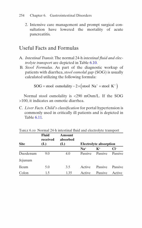
254
2. Intensive care management and prompt surgical con-sultation have lowered the mortality of acute pancreatitis.
Useful Facts and Formulas
A. Intestinal Transit. The normal 24-h intestinal fluid and elec-trolyte transport are depicted in Table 6.10.
B. Stool Formulas. As part of the diagnostic workup of patients with diarrhea, stool osmolal gap (SOG) is usually calculated utilizing the following formula:
SOG stool osmolality stool Na stool K� � � �� �� �2
Normal stool osmolality is <290 mOsm/L. If the SOG >100, it indicates an osmotic diarrhea.
C. Liver Facts. Child’s classification for portal hypertension is commonly used in critically ill patients and is depicted in Table 6.11.
Table 6.10 Normal 24-h intestinal fluid and electrolyte transport
Site
Fluid received (L)
Amount absorbed (L) Electrolyte absorption
Na+ K+ Cl−
Duodenum 9.0 4.0 Passive Passive Passive
Jejunum
Ileum 5.0 3.5 Active Passive Passive
Colon 1.5 1.35 Active Passive Active
Chapter 6. Gastrointestinal Disorders

255
Table 6.11 Child’s classification of portal hypertensionClass A B CSerum bilirubin (mg/dL)
<2 2–3 >3
Serum albumin (g/dL)
>3.5 3–3.5 <3
Ascites None Easily controlled
Poorly controlled
Encephalopathy None Minimal Advanced
Nutrition Excellent Good Poor
Useful Facts and Formulas
257© Springer Nature Switzerland AG 2021J. Varon, Handbook of Critical and Intensive Care Medicine, https://doi.org/10.1007/978-3-030-68270-5_7
Anemia
A. Definition. Anemia is defined as an absolute decrease in the circulating red blood cell (RBC) mass.
B. Etiology
1. Decreased RBC Production
(a) Deficiency of hematinic agents (i.e., iron, vitamin B12, folate)
(b) Bone marrow failure
2. Increased RBC Destruction or Loss
(a) Hemolysis (b) Hemorrhage
C. Diagnostic Evaluation. The approach to the anemic patient in the intensive care unit (ICU) will differ depend-ing on whether the patient was admitted with anemia or if the anemia has developed during the ICU stay.
1. In the Patient Admitted with Anemia
(a) The symptoms of anemia will depend on the degree of anemia, the rapidity of development, cardiopul-monary reserve, and underlying disease.
(b) As a general rule, a hemoglobin <7 g/dL represents severe anemia, and such patients may present with
Chapter 7Hematologic Disorders

258
dyspnea on exertion, lightheadedness, angina, and/or fatigue.
(c) The absence of symptoms in patients with hemo-globin <7 g/dL suggests a gradual onset.
2. HistoryInquire about previous hematologic values, family and ethnic history (i.e., sickle cell, thalassemia), history of splenectomy, cholelithiasis at an early age, medications, drugs, alcohol use, dietary habits, gastrectomy, and bleeding history.
3. Physical Examination
(a) General Appearance: Nutritional status or evi-dence of specific deficiencies, evidence of chronic illness.
(b) Vital Signs: Tachycardia, postural hypotension, other signs of hypoperfusion (i.e., decreased men-tal status, low urine output), petechiae, purpura.
(c) Associated Findings: Jaundice, glossitis (i.e., perni-cious anemia, iron deficiency), neurologic abnor-malities (i.e., vitamin B12, folate deficiency), lymphadenopathy, hepatomegaly, splenomegaly (i.e., hemolysis, neoplasms, infiltrative disorders). Heart: Listen for flow murmurs, prosthetic valves (i.e., increased RBC destruction). Rectal: Examine stools for blood.
4. Laboratory EvaluationLaboratory evaluation usually provides a diagnosis and should always be done in a stepwise manner unless the patient’s condition requires emergent transfusion. In this case, a blood sample for RBC indices, peripheral blood smear, and iron, folate, and vitamin B12 studies should be obtained before transfusion.
(a) Hemoglobin and hematocrit (Hct) estimate RBC mass and severity of anemia, and in the patient sus-pected to have active bleeding, they should be
Chapter 7. Hematologic Disorders

259
monitored serially. Acute blood loss does not influ-ence the Hct immediately.
(b) Mean corpuscular volume (MCV) is a measure of the average size of the RBCs. Classification of the anemia according to the MCV is helpful in gener-ating the differential diagnosis and workup. The smear must be examined to determine whether multiple cell populations are present.
1. Low MCV (<80): Generally limits the diagnosis to a few disorders: Iron deficiency, thalassemia, sideroblastic anemia, other hemoglobinopa-thies, and some cases of anemia of chronic disease.
2. High MCV (<100): Megaloblastic anemias, liver disease, alcoholism, drugs (i.e., methotrexate, AZT), and myelodysplastic syndrome.
3. Normal MCV: Acute blood loss, hemolytic ane-mia, pituitary or thyroid failure, aplastic anemia, myelofibrosis, and anemia of chronic disease.
(c) Reticulocyte count is also essential in the evalua-tion of the anemic patient. It reflects the rate of production of RBCs by the bone marrow. According to the reticulocyte count, anemia can be classified into:
1. Increased RBC destruction (i.e., bleeding, hemolysis) reflected in a high reticulocyte count
2. Decreased RBC production (i.e., iron deficiency, anemia of chronic disease) reflected in an abnormally low reticulocyte count
5. Bleeding should be the first concern in patients who develop anemia while in the ICU. Common sites of bleeding are the gastrointestinal (GI) tract, venipunc-tures, pulmonary tree, genitourinary tract, and the ret-roperitoneum. Development of anemia in the ICU should prompt additional investigation (i.e., gastric aspiration to look for blood, stools checked for gross or
Anemia

260
occult blood, prothrombin time [PT], partial thrombo-plastin time [PTT], and platelet count).
D. Therapy
1. Patient Acutely Bleeding
(a) General Measures
1. Airway management: Assess the need for intu-bation to prevent aspiration, especially in upper GI bleeding.
2. Obtain adequate venous access. Large-bore peripheral catheters allow greater volume administration rates than long central lines.
3. Obtain blood for type and crossmatch and diag-nostic laboratories, as discussed above.
4. Fluid resuscitation: Start with colloids or crys-talloids, and continue with whole blood or packed red blood cells as required:
(a) Healthy adult patients can tolerate blood losses up to 20–30% of their blood volume if adequate replacement with crystalloid is provided.
(b) Patients with impaired cardiac reserve, cor-onary artery disease, or the elderly can develop symptoms with a decrease of about 10% of their blood volume.
5. Identify the source of bleeding. 6. Monitor end points: Patients with acute bleed-
ing should be closely monitored in the ICU for two goals:
(a) Adequate blood volume replacement: Reflected in vital signs, urine output, mental status, central venous pressure (CVP), etc.
(b) Control of bleeding: Follow serial hemoglo-bin (Hb) and Hct, and monitor the bleeding site (GI, genitourinary [GU], etc.).
(b) Specific measures will depend on the cause of the bleeding.
Chapter 7. Hematologic Disorders
261
2. Patient Not Acutely Bleeding
(a) The therapy of anemia will depend on its etiology. (b) A specific hemoglobin concentration should not be
used as the only parameter to decide on the need for transfusion. Transfusion of red cells is usually not necessary in patients with either chronic stable anemia or anemia of acute blood loss unless the patient is symptomatic. Patients with chronic ane-mia, with hemoglobin levels >7 g/dL, rarely require blood transfusion, unless cardiopulmonary or cere-brovascular disease is present.
(c) The use of erythropoietin/darbepoetin in selected populations (i.e., chronic kidney disease) may be advantageous, if such patient remains in the ICU for several days.
Leukopenia
A. Definitions. Leukopenia is defined by blood leukocyte count below the normal range (in our laboratories <3800/μL). Neutropenia is defined as absolute neutrophil count <2000/mL for white patients and below 1500/mL for patients who are black or are Yemenite Jews. Lymphopenia is defined as an absolute lymphocyte count <1500/μL.
B. Etiology
1. Neutropenia (see Table 7.1) 2. Lymphocytopenia (see Table 7.2)
C. Diagnostic Evaluation
1. History
(a) Ethnic background: Black and Yemenite Jew (b) Family history: Congenital or hereditary defect (c) Medications: Chemotherapeutic agents, antibiotics,
etc. (d) Alcohol and dietary history (e) Diet habits: Nutritional deficiency (B12, folate)
Leukopenia

262
Table 7.1 Neutropenia: etiologyFalse decrease in white blood cells
Counts done long after blood has been drawn
Disintegration of fragile cells (i.e., blasts, immature white blood cells)
Presence of paraproteins (monoclonal gammopathies), which can produce white blood cell clumping
Decreased production Bone marrow injury due to ionizing radiation or drugs
Bone marrow replacement or destruction by tumor or infection
Nutritional deficiencies: vitamin B12, folate
Congenital stem cell defects
Increased neutrophil destruction, utilization, sequestration
Hypersplenism
Autoimmune sepsis
Combination (increased destruction and decreased production)
Sepsis
Antineutrophil antibodies
Drugs
Felty’s syndrome
Table 7.2 Lymphocytopenia: etiologyDecreased production Primary immunodeficiency
diseases (HIV)
Increased destruction, utilization, loss
Collagen vascular diseases
Acute infections or stress ionizing radiation
Cytotoxic drugs
Unknown mechanisms Malignancies
Chronic infection
Chapter 7. Hematologic Disorders
263
(f) Underlying illness: Malignancies and human immunodeficiency virus (HIV)
2. Physical Examination
(a) General Appearance: Acute distress, mental status, and evidence of chronic illness
(b) Vital Signs: Fever, hypotension, tachycardia, tachy-pnea, and low urine output (e.g., sepsis)
(c) Tables 7.1 and 7.2 (d) Associated Findings: Hepato- or splenomegaly,
lymphadenopathy, abdominal masses, oral thrush, skin rash, purpura, jaundice, etc.
3. Laboratory Evaluation
(a) Complete blood count (CBC) and differential to assess the degree and type of leukopenia.
(b) Bone marrow aspiration and biopsy are pivotal in the evaluation of the leukopenic patient without obvious cause. Analysis of the bone marrow:
1. Will classify the leukopenia, by revealing the degree of bone marrow cellularity: decreased production, decreased survival, or a combined defect
2. May indicate the etiology of the leukopenia as in aplastic anemia, bone marrow infiltration (e.g., leukemia), infection, etc.
(c) Other laboratory tests that may help in identifying the cause of leukopenia are blood and tissue cul-tures, vitamin levels, and autoantibodies.
4. TherapyThe mainstay of therapy for the leukopenic patient is to treat the underlying disease. For example, in the patient with suspected drug-induced leukopenia, the offending medications should be discontinued. Vitamin deficiency should also be treated when suspected.
(a) Colony-stimulating factors (G-CSF and GM-CSF) represent a line of therapy for the treatment of leu-
Leukopenia

264
kopenia secondary to a decrease in bone marrow production.
(b) Transfusions of white blood cell (WBC) concen-trates have not been proven to be of benefit in many controlled trials.
(c) Supportive therapy for the leukopenic patient requires special consideration, particularly in the ICU environment, where there is a higher risk for nosocomial infections:
1. Granulocyte counts <1000/μL result in patients who are severely immunocompromised.
2. Patients who are immunocompromised should not receive rectal manipulations. Strict handwash-ing for caregivers should be enforced. Avoid intramuscular (IM) or subcutaneous (SQ) routes.
3. If the temperature rises >100.5 °F (38.5 °C), the patient should be fully examined, pan-cultured, and started on broad-spectrum antibiotics.
4. Hematopoietic growth factors are hormonelike substances that stimulate bone marrow to pro-duce blood cells.
(a) Erythropoietin promotes red blood cell pro-duction. Combining erythropoietin with a growth factor for white blood cells improves the patient’s response to the erythropoietin.
(b) Darbepoetin is a long-acting form of erythropoietin.
(c) Oprelvekin (interleukin-11 or IL-11) used to stimulate platelet product after chemother-apy and in some other diseases.
(d) Granulocyte colony-stimulating factors (G-CSFs).
1. Filgrastim, pegfilgrastim, and lenogras-tim are commonly employed in clinical practice to reduce the risk of chemotherapy- induced neutropenia.
Chapter 7. Hematologic Disorders

265
2. The most relevant and acutely harmful side effect of G-CSF is bone or musculo-skeletal pain. Lenograstim and filgrastim are derived from hamster ovary cells; the recommended dose is 5 μg/kg (150 μg/m2) once daily of neutropenia and the prevention of the febrile neutropenia.
• Pegfilgrastim: The main difference compared to lenograstim and filgras-tim is the possibility to administer pegfilgrastim at the dose of 6 mg once a cycle: Being too large for renal clearance has neutrophil-mediated self-regulated clearance that depends on the absolute neutrophil count.
• The main cause of G-CSF-related bone pain is bone marrow.
• Quantitative and qualitative expan-sion but G-CSFs can cause bone pain through other three main mechanisms.
• Directly active receptors located on primary afferent nerve fibers produc-ing peripheral nociceptor sensitiza-tion to nociceptive stimuli through the development of morphological and electrophysiological changes in nerves fibers.
• Modulation of immune function: Stimulate inflammatory cells (mono-cytes, macrophages) that can sensitize themselves peripheral nerve fibers and contribute to nerve remodeling through the development of morpho-logical and electrophysical changes in nerve fibers.
Leukopenia
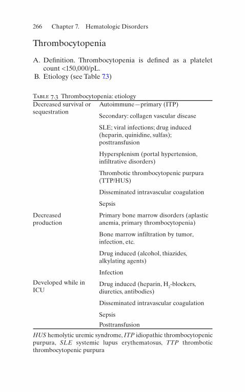
266
Thrombocytopenia
A. Definition. Thrombocytopenia is defined as a platelet count <150,000/pL.
B. Etiology (see Table 7.3)
Table 7.3 Thrombocytopenia: etiologyDecreased survival or sequestration
Autoimmune—primary (ITP)
Secondary: collagen vascular disease
SLE; viral infections; drug induced (heparin, quinidine, sulfas); posttransfusion
Hypersplenism (portal hypertension, infiltrative disorders)
Thrombotic thrombocytopenic purpura (TTP/HUS)
Disseminated intravascular coagulation
Sepsis
Decreased production
Primary bone marrow disorders (aplastic anemia, primary thrombocytopenia)
Bone marrow infiltration by tumor, infection, etc.
Drug induced (alcohol, thiazides, alkylating agents)
Infection
Developed while in ICU
Drug induced (heparin, H2-blockers, diuretics, antibodies)
Disseminated intravascular coagulation
Sepsis
Posttransfusion
HUS hemolytic uremic syndrome, ITP idiopathic thrombocytopenic purpura, SLE systemic lupus erythematosus, TTP thrombotic thrombocytopenic purpura
Chapter 7. Hematologic Disorders

267
C. Diagnostic Evaluation
1. HistoryInquire about bleeding, thrombotic events, mental sta-tus changes, alcohol use, drugs and medications, and associated illness.
(a) Heparin-induced thrombocytopeniaMore common with the use of unfractionated hepa-rin (UFH) rather than low-molecular-weight hepa-rin (LMWH). More common among surgical rather than medical patients.
2. Physical Examination
(a) Vital Signs: Fever, tachycardia, hypotension, tachy-pnea, oliguria (i.e., sepsis)
(b) Skin: Purpura, hematomas, gingival bleeding, lymphadenopathy, hepato- or splenomegaly, abdominal masses
3. Laboratory Evaluation
(a) Complete Blood Count and Platelet Count
1. Platelet counts >50,000/μL in isolation are not associated with significant bleeding problems, and severe spontaneous bleeding is unusual in patients with counts >20,000/μL in the absence of coagulation factor abnormalities.
2. Thrombocytopenia associated with anemia sug-gests thrombotic thrombocytopenic purpura (TTP), hemolytic uremic syndrome (HUS), dis-seminated intravascular coagulation (DIC), or other microangiopathic processes.
3. Pancytopenia should suggest leukemia, aplastic anemia, or other bone marrow disorders.
(b) Peripheral Blood Smear: Note platelet size and other abnormalities (i.e., fragmented RBCs may indicate TTP or DIC; increased platelet size sug-gests accelerated destruction).
Thrombocytopenia
268
(c) Coagulation Evaluation: PT, PTT, D-dimer, and fibrin degradation products may indicate the pres-ence of consumption coagulopathy (e.g., DIC).
(d) Bone Marrow Aspiration and Biopsy (not always necessary): To assess the number of megakaryo-cytes and the presence of bone marrow disorders (i.e., leukemia, aplastic anemia, tumor infiltration).
(e) For patients with suspected heparin-induced thrombocytopenia, 14C-serotonin release assay remains the gold standard. Heparin-induced plate-let aggregation tests can also be used as well as the solid-phase ELISA immunoassay.
4. TherapyDetailed treatment for the various causes of thrombo-cytopenia is beyond the scope of this chapter. We will concentrate on relevant topics for the acute manage-ment of patients in ICU setting.
1. As hemostasis approaches normal at platelet counts >50,000/μL, patients with active bleeding should be transfused (6–10 U) to attempt to achieve levels >50,000/μL.
2. Lumbar puncture and needle organ biopsies (lung, liver, kidneys, etc.) are more hazardous than thoracen-tesis, paracentesis, and bone marrow biopsy. Transfusion to 50,000/μL before such procedures is indicated.
3. Patients with platelet counts <20,000/μL are at higher risk for hemorrhage. However, there is no clear threshold for prophylactic platelet transfusion.
4. Discontinue all nonessential medications including heparin. Consider changing H2-blockers to coating agents or antacids. Avoid agents known to inhibit platelet function (i.e., nonsteroidal anti-inflamma-tory drugs [NSAIDs], ticarcillin, etc.).
5. Avoid trauma, IM and SQ injections, rectal manipu-lations, hard toothbrush, razors, etc., in thrombocy-topenic patients.
6. TTP and HUS deserve special consideration because their management differs.
Chapter 7. Hematologic Disorders
269
(a) TTP is a syndrome characterized by microangi-opathic hemolytic anemia, fever, fluctuating neurologic deficits, and renal insufficiency. HUS is felt to be a variant of this syndrome in which renal failure is the predominant feature. TTP/HUS should be considered a medical emergency.
(b) Patients with TTP/HUS should not receive platelet transfusions, unless life-threatening bleeding occurs.
(c) Therapy for TTP/HUS includes plasma exchange by plasmapheresis if available, other-wise fresh-frozen plasma (FFP) transfusions. Intravenous (IV) steroids can be used, and in unresponsive cases, vincristine has been recommended.
(d) Heparin-induced thrombocytopenia: The first maneuver is to stop all heparin and heparin-containing products (i.e., heparin flush). There are a number of alternative anticoagulants to heparin in these patients, such as direct throm-bin inhibitors like lepirudin [recombinant hiru-din], bivalirudin, argatroban, fondaparinux, or danaparoids.
Anticoagulation and Fibrinolysis
Anticoagulants and thrombolytic agents are potentially life- saving drugs when used prophylactically or employed thera-peutically in critically ill patients.
A. Anticoagulation
1. Heparin
(a) Mechanism of ActionHeparin acts by potentiating the activity of the plasma protease inhibitor antithrombin III, which
Anticoagulation and Fibrinolysis

270
rapidly inhibits the activity of factors XIIa, XIa, Xa, and IXa, and thrombin (factor II).
(b) Heparin prolongs the thrombin time (TT), bleed-ing time, PTT, and to a lesser extent the PT.
(c) Heparin half-life is 1–3 h, but in patients with pul-monary embolism, clearance is accelerated (20–40%) compared to normal.
(d) Indications
1. Prophylaxis of deep venous thrombosis (DVT) and pulmonary embolism (PE). All patients in the ICU should be on some form of DVT prophylaxis (commonly, heparin 5000 U SQ q12 h) or low-molecular-weight heparin (e.g., enoxaparin 40 mg SQ qd). Heparin has proved to be an effective agent in DVT prophylaxis, except after major orthopedic procedures (particularly hip and knee replacement) and after prostate surgery.
2. Full anticoagulation (PTT approximately two times normal) in pulmonary embolism or DVT, arterial thrombosis, and other disorders. A 5000–8000 U IV bolus is commonly used and infusion rates of 1000 U/h (12–25 U/kg/h).
(e) Monitoring of Anticoagulation
1. Not required for prophylactic doses. 2. For full anticoagulation, the heparin dose should
be adjusted to maintain the PTT at 1.5–2.0 times control. To avoid the tendency to under- anticoagulate patients on heparin therapy, a standardized dosing regimen has been devel-oped (Table 7.4).
3. Low-molecular-weight heparin for DVT and acute coronary syndromes (ACS).
• Enoxaparin• For DVT, 1 mg/kg SQ q12 h or 1.5 mg/kg SQ qd• For ACS, 1 mg/kg SQ q12 h
Chapter 7. Hematologic Disorders

271
(f) Complications
1. Bleeding: Occurs in 7–20% of patients during full-dose heparinization. Hemorrhage typically occurs from the GI, urinary tract, or surgical incisions. Less common sites are intracranial, retroperitoneal, soft tissues, nose, and pleural space. Bleeding is associated with the intensity of the anticoagulation (e.g., when the PTT is more than three times normal, the risk of hem-orrhage is substantially higher).
2. Thrombocytopenia: Heparin use is associated with thrombocytopenia in 5–30% of patients, and its incidence is higher with the use of bovine lung heparin.
(a) If thrombocytopenia is mild (>100,000/μL), not associated with bleeding or thrombotic events, heparin therapy can be continued.
Table 7.4 Standardized protocol for dosing of intravenous heparin
PTTa Dose adjustmentb
Repeat PTT
<50 5000-U bolus, increase infusion by 2400 U/24 h
6 h
50–59 Increase infusion by 2400 U/24 h 6 h
60–85 Therapeutic range, no change Next morning
86–95 High therapeutic range, decrease infusion by 1920 U/24 h
Next morning
96–120
Stop infusion for 30 min, decrease infusion by 1920 U/24 h
6 h
120 Stop infusion for 60 min, decrease infusion by 3840 U/24 h
6 h
aNormal PTT range is 27–35 sbDosing protocol is based on an initial IV bolus of 5000–8000 U, followed by continuous infusion of 24,000 U/24 h. The first PTT should be obtained 6 h after the bolus of heparin
Anticoagulation and Fibrinolysis
272
(b) Severe thrombocytopenia can occur but is rare. It may be associated with bleeding or paradoxic thrombotic events. Diagnosis is made by detection of heparin-dependent immunoglobulin G (IgG). Treatment con-sists of discontinuation of all heparin use and avoid platelet transfusion.
3. Osteoporosis can be seen with the long-term use of heparin.
4. Hypoaldosteronism rarely is seen. 5. Antidote: Heparin is generally undetectable in
patient’s plasma within 3 h after discontinuation of therapy. In the rare instance in which antico-agulation must be reversed more rapidly, prot-amine sulfate can be used.
2. WarfarinWarfarin (Coumadin™) is the most frequently used oral anticoagulant in the United States. Sometimes, the transition to chronic oral therapy is begun before the patient leaves the ICU or must be initiated because of heparin-induced thrombocytopenia or as DVT prophy-laxis in certain cases. The physician caring for the criti-cally ill can also encounter patients who have accidentally or purposely overdosed with warfarin.
(a) Mechanism of Action: Warfarin interferes with the hepatic vitamin K-dependent carboxylation of fac-tors II, VII, IX, and X. It also inhibits the synthesis of the anticoagulant factors protein C and S and may thereby be thrombogenic. The antithrombotic effects of warfarin occur only after several days of treat-ment. In patients on IV anticoagulation therapy, who require chronic oral anticoagulation, heparin and warfarin should be overlapped for at least 48 h.
(b) Indications
1. Prophylaxis of DVT and PE, especially in those patients in whom heparin has not proved to be effective
2. For chronic anticoagulation therapy
Chapter 7. Hematologic Disorders

273
(c) Dosing
1. Loading Dose: 5–10 PO qd for 2–4 days 2. Maintenance Dose: 2–15 mg PO qd to keep INR
(international normalized ratio) therapeutic
(d) Complications
1. Bleeding occurs in 2.4–8.1% of patients chronically anticoagulated. The risk is dose related and propor-tional to the prolongation of the PT. Treatment con-sists of FFP transfusions. Vitamin K replacement is only recommended for warfarin overdose, because of its delayed onset of action and because it makes reinstitution of warfarin therapy complicated.
2. Warfarin skin necrosis secondary to a paradoxic hypercoagulable state due to the warfarin-induced protein C reduction.
3. Reversal of coagulopathy can be accomplished uti-lizing factors II, VII, IX, and X, protein C, and pro-tein S (Kcentra™) in patients requiring emergency surgical procedures.
(e) Supratherapeutic INR
1. INR greater than therapeutic range but <4.5
(a) Hold next dose and resume when INR therapeutic
2. INR 4.5 – 10.0, with no or low bleeding risk
(a) Stop warfarin, resume when INR therapeutic
3. INR 4.5 – 10.0, with moderate or high bleeding risk
(a) Stop warfarin, give 1.0–2.0 mg PO or 0.5–1.0 mg IV vitamin K
(b) Resume warfarin when INR therapeutic
4. INR >10.0
(a) Low bleeding risk: Give 3.0–5.0 mg oral or IV vitamin K
(b) High bleeding risk: Give 3.0–5.0 mg oral or IV vitamin K and 15–30 IU/kg prothrombin complex
Anticoagulation and Fibrinolysis

274
3. Direct Thrombin Inhibitors (DTIs)
(a) Mechanism of action: Bivalent DTIs (hirudin and analogs) bind both to the active site and exosite 1 of thrombin, while univalent DTIs bind only to the active site. The third class of inhibitors, which are gaining importance recently, are the allosteric inhibitors.
1. Bivalent
(a) Hirudin (b) Bivalirudin (c) Lepirudin (d) Desirudin
2. Univalent
(a) Argatroban (b) Inogatran (c) Melagatran (and its prodrug ximelagatran) (d) Dabigatran
(b) Indications
1. Prophylaxis and treatment of DVT and PE 2. Used for anticoagulation in patients with
heparin- induced thrombocytopenia (HIT)
(c) Contraindications
1. Active pathological bleeding 2. History of a serious hypersensitivity reaction 3. Mechanical prosthetic heart valve 4. Kidney disease with certain medications 5. Chronic anticoagulation use
(d) Monitoring
1. Unlike heparin and warfarin, there are no moni-toring levels that need to be obtained
(e) Complications
1. Bleeding 2. Gastrointestinal upset (nausea, vomiting,
diarrhea)
Chapter 7. Hematologic Disorders

275
(f) Reversal
1. Idarucizumab is used for dabigatran reversal. 2. Fresh frozen plasma has been used in studies for
argatroban reversal although there is no defined agent.
4. Factor Xa (10 subunit a) inhibitors
(a) Mechanism of action: Selectively and reversibly blocking the activity of clotting factor Xa, prevent-ing clot formation. They affect factor Xa both within the blood and within a preexisting clot.
(b) Drugs in class:
1. Fondaparinux 2. Rivaroxaban 3. Apixaban 4. Edoxaban 5. Betrixaban
(c) Indications
1. Prophylaxis and treatment of DVT and PE 2. Reduction of the risk of stroke in nonvalvular
atrial fibrillation 3. Venous thromboembolism (VTE) prophylaxis
following knee or hip replacement surgery 4. Reduction in the risk of major CV events in
patients with chronic CAD or PAD (rivaroxaban)
(d) Contraindications
1. Active pathological bleeding 2. History of a serious hypersensitivity reaction 3. Spinal/epidural hematomas
(e) Monitoring
1. There are no monitoring levels that need to be obtained
Anticoagulation and Fibrinolysis
276
(f) Complications
1. Bleeding—In atrial fibrillation and VTE trials, apixaban and edoxaban conferred a lower risk of bleeding when compared to warfarin, while the risk with rivaroxaban was similar to warfarin.
(g) Reversal
1. There are no currently approved antidotes for use in reversal of factor Xa inhibitors. Due to the short half-lives of the factor Xa inhibitors, the first step in treating a bleeding event should be discontinuation of the anticoagulant.
B. Fibrinolysis
1. Fibrinolytic therapy has an expanding role in the treat-ment of many thromboembolic disorders. Many fibri-nolytic drugs are currently marketed: streptokinase (SK), anisoylated plasminogen streptokinase activator complex (APSAC), urokinase (UK), recombinant human tissue-type plasminogen activator (rt-PA), reteplase, and TNK, among others. All drugs activate the fibrinolytic system by converting plasminogen to the active enzyme plasmin. Plasmin degrades fibrin and dissolves the thrombus.
2. Indications
(a) Acute Myocardial Infarction (AMI): Thrombolytic therapy for AMI is discussed in Chap. 3, “Cardiovascular Disorders.”
(b) Pulmonary Embolism: While the effectiveness and role of thrombolytic agents in AMIs are firmly established, their use in venous thromboembolism remains infrequent and controversial, mainly because of the fear of a negative benefit/risk ratio. SK, UK, and tPA have been shown to be more effective than heparin alone in accelerating clot lysis and improving pulmonary tissue perfusion.
Chapter 7. Hematologic Disorders

277
Current recommendations for the use of fibrinoly-sis in PE are for patients with massive pulmonary embolism and persistent systemic hypotension in whom rapid resolution of pulmonary obstruction is desired. It is still questionable if there is significant improvement to survival in those patients.
(c) Deep Venous Thrombosis: Even more debated is the use of thrombolytic agents in the treatment of DVT. Potential advantages of fibrinolysis over anticoagulation include prevention of PE by lysing the source of thrombus in situ, rapid restoration of normal venous circulation with a prompt resolu-tion of symptoms, and prevention of valve damage, which would otherwise result in chronic venous insufficiency. Risks include a much higher inci-dence of bleeding.
3. Dosage for Selected Thrombolytic Regimens
(a) Pulmonary Embolism
• UK: 4400 U/kg bolus, followed by 4400 U/kg/h for 24 h
• UK: 15,000 U/kg bolus over 10 min• SK: 250,000 U over 30 min, followed by
100,000 U/h for 24 h• rt-PA: 100 mg as continuous peripheral infusion
over 2 h• Selective use of lysis in the pulmonary artery
directed by catheter (the Varon–Strickman pro-cedure) can also be used.
(b) Deep Venous Thrombosis
• SK: 250,000 U over 30 min, followed by 100,000 U/h for 48–72 h
• rt-PA: 0.5 mg/kg over 4–8 h• rt-PA: 0.05 mg/kg/h for 24 h
(c) Myocardial Infarction (see section “Thrombocytopenia”)
Anticoagulation and Fibrinolysis

278
4. Monitoring
(a) Clinical monitoring should include serial neuro-logic examinations to detect central nervous system (CNS) bleeding and frequent vital signs to detect bleeding. All puncture sites should be exam-ined frequently.
(b) Laboratory monitoring should include Hb/Hct, platelet, fibrinogen, PT, and PTT.
5. Complications
(a) Bleeding: The greatest limitation of the thrombo-lytic drugs and the factor that has limited their acceptance for the treatment of DVT and PE are the incidence of bleeding.
(b) Allergic Reactions: Reactions including skin rashes, fever, and hypotension are rare and usually are associated with the use of SK and APSAC. The induction of antibodies against streptococcal anti-gens can occur after the administration of SK or APSAC or after streptococcal infection, which may neutralize the fibrinolytic activity of SK.
Blood and Blood Product Transfusion
Transfusion therapy may be associated with several immedi-ate and delayed adverse effects. Therefore, risks and benefits must be carefully weighed before any blood product is administered. The use of blood components should be guided by a rational diagnostic and therapeutic approach.
A. Whole Blood. Whole blood stored >24 h contains few viable platelets or granulocytes; factors V and VIII are decreased, but stable clotting factors are maintained. One unit is 450 mL and when transfused to an average-sized adult will increase the hemoglobin by 1.0 g/dL and Hct by 3%.
1. Indications: Symptomatic anemia with massive hemorrhage
Chapter 7. Hematologic Disorders
279
2. Risks
(a) Allergic reactions (b) Infectious diseases (i.e., HIV, hepatitis B) (c) Febrile reactions (d) Volume overload (e) Noncardiogenic pulmonary edema
B. Packed Red Blood Cells. Removal of 200–250 mL of plasma from whole blood results in packed RBCs (PRBCs). Transfusion of 1 U of PRBCs will increase the Hb and Hct by the same amount as will 1 U of whole blood. One unit is 250–300 mL.
1. Indications: Symptomatic anemia 2. Risks: Same as for whole blood (see above)
C. Packed Red Blood Cells, Leukocyte Poor. Most of the WBCs are removed from the packed RBCs by saline washing.
1. Indications
(a) Symptomatic anemia and allergic or febrile reac-tion from leukocyte antibodies
(b) Patients with paroxysmal nocturnal hemoglobinuria
2. RisksSame as for whole blood (see above)
D. Fresh-Frozen Plasma. FFP is separated from freshly drawn whole blood and then frozen, with a volume of 200–250 mL. Rich in all coagulation factors; 1 mL supplies approximately 1 U of coagulation activity. FFP should be ABO compatible; Rh type or crossmatching is not required.
1. Indications
(a) Bleeding due to coagulation factor deficiency (b) Treatment of TIT and HUS (c) Rapid reversal of vitamin K deficiency or warfarin
overdose
2. RisksSame as for whole blood (see above)
Blood and Blood Product Transfusion

280
E. Cryoprecipitate. Cryoprecipitate is made by thawing a unit of FFP at 4 °C. White precipitate forms; most of supernatant plasma is removed and refrozen. The volume is 10 mL. A pack of cryoprecipitate contains von Willebrand factor, lesser amounts of factor VIII, fibrino-gen, factor XIII, and fibronectin. ABO compatibility is preferred.
1. Indications
(a) von Willebrand disease (b) Mild-to-moderate hemophilia A (c) Factor XIII deficiency (d) Fibrinogen deficiency
2. Risks
(a) Infectious disease (b) Hyperfibrinogenemia (c) Allergic reactions
F. Platelets. Platelet packs are obtained from whole blood; 1 U contains at least 5.5 × 10¹⁰ platelets/mm3 in approxi-mately 50 mL volume. In a normal 70-kg adult, 1 U of platelets should increase platelet count by 5000–10,000/mm3.
1. Indications
(a) To correct bleeding secondary to thrombocytope-nia or abnormal platelet function.
(b) Prophylactically (e.g., in patients with chemotherapy- induced thrombocytopenia, the threshold is somewhat controversial, 10,000–20,000/mm3) and before invasive procedures (tar-get counts of 50,000/mm3).
2. Risks
(a) Infectious diseases (b) Allergic reactions (c) Alloimmunization
Chapter 7. Hematologic Disorders

281
G. Complications of Transfusion Therapy
1. Disease transmission: HIV, hepatitis, cytomegalovirus (CMV), Epstein–Barr virus (EBV), Chagas’ disease, malaria.
2. Allergic reactions characterized by fever, chills, urti-caria, and respiratory distress. These events are second-ary to antileukocytic antibodies or antibodies against antigenic proteins in donor plasma. Therapy is symp-tomatic (acetaminophen; antihistamines; rarely, epi-nephrine or glucocorticoids are needed).
3. Red Cell Transfusion Related
(a) Acute Hemolytic Reactions: Fever, chills, back pain, nausea, vomiting, hypotension, dark urine, and chest pain. Acute renal failure with hemoglo-binuria and DIC may occur. If suspected:
1. Inform the blood bank. 2. Stop transfusion. 3. Replace all IV tubing. 4. Send clotted and ETDA-treated blood samples
from patient’s blood along with the remainder of the unit of blood to the blood bank for crossmatch.
5. Send blood samples for DIC screen, bilirubin, and free hemoglobin.
6. Management
(a) Intravascular volume expansion plus man-nitol to keep urine output >100 mL/h or 1 cc/kg/h.
(b) Alkalization of urine with IV bicarbonate to keep urine pH >7.0 to avoid hemoglobin tubular precipitation.
(c) Treatment of DIC
(b) Delayed Hemolytic Transfusion Reactions: 24 h to 25 days posttransfusion. These are secondary to an amnestic (1–3 days) or primary (7–25 days) anti-body response to RBC antigens. Patients usually
Blood and Blood Product Transfusion

282
develop a drop in the Hb and Hct with an increase in bilirubin. Coombs’ test is positive.
(c) Noncardiogenic pulmonary edema (acute respira-tory distress syndrome [ARDS]), caused by anti-leukocytic antibodies.
(d) Coagulopathy associated with a large volume of PRBC transfusions, secondary to dilution of plate-let and coagulation factors. Treatment consists of FFP and platelet transfusions.
(e) Citrate intoxication, also seen with large-volume transfusion of PRBCs. Patients present with hypo-calcemia, hypotension, and drop in cardiac output. Treatment: IV calcium.
4. Volume Overload: Especially in patients with conges-tive heart failure (CHF). Diuretics may be needed after transfusion.
5. Platelet alloimmunization develops in patients who have received multiple transfusions. Approximately 75% of patients receiving platelets on a regular basis will become alloimmunized to platelet antigens. Increments <20% of expected generally indicate allo-immunization. Patients may respond to single-donor platelets, but HLA-matched platelets may be needed.
Disseminated Intravascular Coagulation
A. Definition. Disseminated intravascular coagulation (DIC) is a dynamic pathologic process triggered by activation of the clotting cascade with resultant generation of excess thrombin within the vascular system. Most consider DIC to be a systemic hemorrhagic syndrome; however, this is only because hemorrhage is obvious and often impressive. What is less commonly appreciated is the significant amount of microvascular thrombosis and, in some instances, large-vessel thrombosis that occurs. This throm-bosis is usually the more life-threatening insult.
Chapter 7. Hematologic Disorders

283
B. Etiology (see Table 7.5) C. Diagnostic Evaluation. Since DIC is associated with an
underlying disease state, the clinical evaluation will be directed toward identifying (1) primary illness, (2) the sta-tus of the coagulation system, and (3) the focal and sys-temic consequences of the DIC-associated hemorrhage and/or thrombosis.
1. Clinical Findings Associated with the Primary Illness: These findings will vary according to the precipitating event—obstetrical accident, infection, malignancy, etc.
2. Clinical Findings Associated with the Coagulation Status
(a) Bleeding from venipuncture sites, mucous mem-branes, hemorrhagic bullae, hematuria, GI bleed-ing, etc.
(b) Purpura, petechiae, and subcutaneous hematomas
3. Clinical Findings Associated with End-Organ Thrombosis and Hemorrhage
(a) Lungs: Respiratory distress, hypoxia, ARDS (b) Kidneys: Proteinuria, renal insufficiency (c) Liver: Budd–Chiari syndrome, hepatitis, hepatic
failure (d) Skin: Necrosis, acrocyanosis (e) CNS: Mental status changes, neurologic deficits
Table 7.5 Disseminated intravascular coagulation: etiologyObstetric accidents (amniotic fluid embolism, abruptio placentae)
Intravascular hemolysis
Sepsis
Malignancy
Trauma
Vascular disorders
Disseminated Intravascular Coagulation
284
4. Laboratory Evaluation
(a) Peripheral blood smear will show fragmented RBCs, thrombocytopenia, with large platelets.
(b) Prolonged PT and PTT. (c) Thrombocytopenia is usually around 60,000/μL,
but values ranging between 3000 and 100,000/μL can be seen.
(d) Decreased fibrin level. (e) Decreased antithrombin III level. (f) Elevated levels of fibrin degradation products
(FDP). (g) Elevation of D-dimer neoantigens is a specific test
for degradation products of fibrin, whereas non-specific FDP may be either fibrinogen or fibrin derived.
D. Therapy. The treatment of DIC is confusing and still con-troversial. Therapy must be individualized according to the cause of DIC, severity of hemorrhage, severity of thrombosis, hemodynamic status, and age.
1. The most important and effective treatment for DIC is removal of the triggering disease process (i.e., evacuate the uterus, control of shock, control of infection, removal of tumors, chemo- or radiotherapy, or other indicated therapies).
2. In cases of obstetric complications, anticoagulation is rarely needed, and evacuation of the uterus usually stops the intravascular clotting process.
3. If the patient continues to bleed or clot significantly after 6 h of initiation of therapy directed to stop or blunt the triggering event, anticoagulation therapy may be indicated. There is a general agreement on the need for anticoagulation in acute promyelocytic leukemia and perhaps DIC with solid tumors. We favor the use of low-dose, subcutaneous heparin at doses of 80–100 U/kg q6 h. Other anticoagulant modalities available are IV heparin and antithrombin III concentrates.
Chapter 7. Hematologic Disorders

285
4. If the patient continues to bleed after reasonable attempts to treat the triggering event of the DIC, and if anticoagulation therapy has been initiated, clotting fac-tor depletion is the most probable cause of bleeding, and replacement therapy should be considered.
5. The experimental use of thrombomodulin appears promising.
Hemolytic Syndromes
A. Definition. Premature destruction of red blood cells. This process may occur either because of abnormal factors in the intravascular environment or because of defective red blood cells.
B. Etiology (see Table 7.6) C. Diagnostic Evaluation
1. History and Physical Examination: Clinical manifesta-tions will depend on the underlying disorder, on the severity of the anemia, and on whether the hemolysis is intravascular or extravascular.
(a) Intravascular hemolysis can present as an acute event with back pain, dyspnea, chills, fever, tachy-cardia, dark urine, and hypotension, and it may result in renal failure.
(b) Extravascular hemolysis is usually less dramatic and may be accompanied only by jaundice and splenomegaly.
2. Laboratory Evaluation
(a) Elevated reticulocyte count. (b) Peripheral blood smear can provide a diagnosis in
cases of spherocytosis; microangiopathic disorders will show the presence of fragmented RBCs; Heinz bodies suggest enzymatic defects; or the presence of anisocytosis or sickle cells is consistent with hemoglobinopathies.
Hemolytic Syndromes

286
(c) Other laboratory data suggestive of hemolysis are:
1. Hemoglobinuria (indicative of intravascular hemolysis)
2. Hemoglobinemia (indicative of intravascular hemolysis)
3. Low levels of haptoglobin 4. Elevated lactic dehydrogenase (LDH) 5. Positive Coombs’ test
D. Sickle Cell Disease. Sickle cell disease is a heterogeneous group of defects of hemoglobin synthesis, all of which can cause clinically significant illness due to sickling of red cells. Sickle hemoglobin (Hb S) is less soluble when deox-
Table 7.6 Hemolytic syndromes: etiologyAcquired hemolytic disorders
Immune hemolytic anemia
Warm antibody (idiopathic, neoplasia, collagen vascular disorder, drugs)
Cold antibody (idiopathic, mycoplasma infection, lymphoproliferative disorder, paroxysmal cold hemoglobinuria)
Microangiopathic hemolytic anemia (TTP, DIC, eclampsia)
Direct toxic effect (malaria, clostridial infection)
Splenomegaly
Membrane defects
Paroxysmal nocturnal hemoglobinuria
Spur cell anemia
Hereditary hemolytic disorders
Membrane defects (spherocytosis, elliptosis)
Enzyme defects (G-6PD deficiency)
Thalassemias
Hemoglobinopathies
Chapter 7. Hematologic Disorders

287
ygenated and forms polymers that precipitate inside the RBCs, leading to membrane abnormalities, decreased deformability, and increased blood viscosity.
1. Clinical ManifestationsThe clinical manifestations of sickle cell disease are secondary to vaso-occlusive phenomena, which may lead to microinfarctions with resultant painful crises and, eventually, chronic organ damage.
2. DiagnosisDemonstration of sickling under reduced oxygen ten-sion. Hemoglobin electrophoresis should be performed to discriminate homozygous SS from AS and to deter-mine the presence of other abnormal hemoglobins.
3. TreatmentThe treatment of sickle cell disease is supportive and limited to management of acute and chronic complica-tions. Frequently, these patients need to be admitted to the ICU due to the severity and life-threatening dimen-sion of their acute attacks.
(a) Early antibiotic treatment at the first evidence of infection. Pneumococcal sepsis is a leading cause of mortality. Other prevalent pathogens include Escherichia coli, Haemophilus influenzae, Salmonella sp., Shigella sp., and Mycoplasma pneumoniae.
(b) Painful crises: IV hydration, adequate analgesia (usually, a regular schedule of opioids is necessary), oxygen administration which is indicated when hypoxemia is present, correction of acidosis.
(c) Look for precipitating events (i.e., infections, sur-gery, dehydration, trauma, cold temperatures, alco-hol ingestion). When abdominal pain is one of the manifestations, other causes of abdominal pain must be ruled out (i.e., acute abdomen, hepatobili-ary disease).
(d) Acute chest syndrome: Characterized by pleuritic chest pain, fever, cough, hypoxia, and pulmonary infiltrates. Lung scans and pulmonary angiograms
Hemolytic Syndromes
288
are usually of no help. In addition, the latter test is associated with added risk because of possible induction of sickling by the hypertonic contrast media. Differentiation between pneumonia and infarction is often difficult. Features favoring infarc-tion include painful bone crisis, clear chest radio-graph at onset, lower lobe disease, and negative blood cultures. Treatment includes oxygen therapy, mechanical ventilation when indicated, empiric antibiotic therapy, and correction of acidosis.
(e) Sickle cell crisis associated with cerebrovascular accidents or repeated veno-occlusive crisis may benefit from transfusion or exchange transfusion to keep the Hb S levels <39%.
E. Autoimmune Hemolytic Anemia
1. Warm-Antibody Autoimmune Hemolytic AnemiaWarm-antibody autoimmune hemolytic anemia (AHA) is usually extravascular and IgG mediated. This type of hemolytic anemia can be seen in the ICU not only in patients admitted with collagen vascular diseases or lym-phomas but also in drug-induced hemolytic anemias.
(a) Diagnosis is made by signs of hemolysis and posi-tive direct Coombs’ test.
(b) Treatment
1. If the suspected mechanism is drug induced, all nonessential medications should be discontinued.
2. Sixty percent of cases will respond to steroid ther-apy (e.g., prednisone 1.0–1.5 mg/kg PO daily).
3. Splenectomy will increase the success rate to ≈80–90%.
4. Cytotoxic drugs are reserved for patients who fail to respond to steroid plus splenectomy.
5. Transfusions are indicated only in severe cases of anemia. In emergency situations, most patients can be managed with careful transfu-
Chapter 7. Hematologic Disorders

289
sion (ABO- and Rh-compatible blood) adminis-tered slowly while watching for reactions.
2. Microangiopathic Hemolytic AnemiaMicroangiopathic hemolytic anemia (MAHA) is a syn-drome caused by traumatic intravascular hemolysis. Intraluminal deposition of fibrin strands in small vessels is presumed to be responsible for the red cell destruction.
(a) Etiology includes DIC, TTP, HUS, malignant hypertension, vasculitis, and eclampsia.
(b) Diagnosis
1. Evidence of hemolysis (reticulocytosis, elevated LDH, depressed haptoglobin, etc.)
2. Fragmented RBCs in the peripheral blood smear
(c) TreatmentTherapy is directed toward the underlying disor-der. Management of TTP, HUS, and DIC is dis-cussed elsewhere. Transfusion is rarely indicated.
3. Glucose-6-Phosphate Dehydrogenase DeficiencyThis is a hereditary deficiency of the enzyme glucose-6- phosphate dehydrogenase deficiency (G-6PD) in the red cells. It is a sex-linked disorder that affects men and rarely women of Mediterranean, African, or Chinese ancestry. The disease is associated with episodic hemolysis.
(a) Clinical Manifestations
1. Hemolytic episodes are sometimes triggered by infections or the ingestion of some drugs (e.g., sulfonamides, antimalarials, nitrofurantoin, nali-dixic acid, etc.).
2. Patients present with acute intravascular hemo-lysis associated with hemoglobinemia, hemoglo-binuria, decreased haptoglobin, and jaundice.
3. Peripheral vascular collapse can occur in severe cases.
4. Hemolysis is usually self-limited, even if the exposure to the oxidant agent continues, since
Hemolytic Syndromes
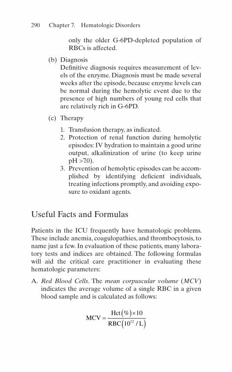
290
only the older G-6PD- depleted population of RBCs is affected.
(b) DiagnosisDefinitive diagnosis requires measurement of lev-els of the enzyme. Diagnosis must be made several weeks after the episode, because enzyme levels can be normal during the hemolytic event due to the presence of high numbers of young red cells that are relatively rich in G-6PD.
(c) Therapy
1. Transfusion therapy, as indicated.2. Protection of renal function during hemolytic
episodes: IV hydration to maintain a good urine output, alkalinization of urine (to keep urine pH >7.0).
3. Prevention of hemolytic episodes can be accom-plished by identifying deficient individuals, treating infections promptly, and avoiding expo-sure to oxidant agents.
Useful Facts and Formulas
Patients in the ICU frequently have hematologic problems. These include anemia, coagulopathies, and thrombocytosis, to name just a few. In evaluation of these patients, many labora-tory tests and indices are obtained. The following formulas will aid the critical care practitioner in evaluating these hematologic parameters:
A. Red Blood Cells. The mean corpuscular volume (MCV) indicates the average volume of a single RBC in a given blood sample and is calculated as follows:
MCVHct
RBC L�
� ��� �%
/
10
1012
Chapter 7. Hematologic Disorders

291
The mean corpuscular hemoglobin (MCH) indicates the average weight of hemoglobin per erythrocyte:
MCHHb g dL
RBC L�
� ��� �/
/
10
1012
The mean corpuscular hemoglobin concentration (MCHC) indicates the average concentration of hemoglobin in the RBCs of any specimen:
MCHC
Hb g dL
Hct�
� �� �
�/
%100
The red blood cell volume can be calculated via a radionu-clide study:
RBC volume
cpm of isotope injected
cpm mL RBC in sample=
/:
where cpm = counts per million.
B. Reticulocyte Counts. To calculate the percentage of reticu-locytes, usually based on counting 1000 RBCs, the follow-ing formula is commonly utilized:
Reticulocytes
Number of reticulocytes
Number of RBCs observed%� � � ��100
The actual reticulocyte count (ARC) reflects the actual number of reticulocytes in 1 L of whole blood:
ARC
ReticulocytesRBC count L�
� �� �� ��%
/100
10 100012
Useful Facts and Formulas
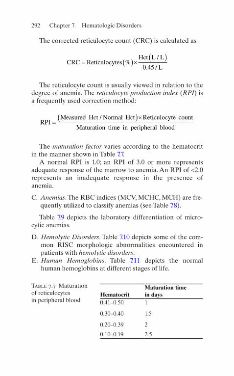
292
The corrected reticulocyte count (CRC) is calculated as
CRC Reticulocytes
Hct L L
L� � �� � �
%/
. /0 45
The reticulocyte count is usually viewed in relation to the degree of anemia. The reticulocyte production index (RPI) is a frequently used correction method:
RPI
Measured Hct Normal Hct Reticulocyte count
Maturation tim�� ��/
ee in peripheral blood
The maturation factor varies according to the hematocrit in the manner shown in Table 7.7.
A normal RPI is 1.0; an RPI of 3.0 or more represents adequate response of the marrow to anemia. An RPI of <2.0 represents an inadequate response in the presence of anemia.
C. Anemias. The RBC indices (MCV, MCHC, MCH) are fre-quently utilized to classify anemias (see Table 7.8).
Table 7.9 depicts the laboratory differentiation of micro-cytic anemias.
D. Hemolytic Disorders. Table 7.10 depicts some of the com-mon RISC morphologic abnormalities encountered in patients with hemolytic disorders.
E. Human Hemoglobins. Table 7.11 depicts the normal human hemoglobins at different stages of life.
Table 7.7 Maturation of reticulocytes in peripheral blood
HematocritMaturation time in days
0.41–0.50 1
0.30–0.40 1.5
0.20–0.39 2
0.10–0.19 2.5
Chapter 7. Hematologic Disorders

293
To convert colorimetric readings into grams of Hb/dL (g/dL) using a standard curve setup with the same equipment and reagents used for specimen or calculate specimen con-centration (Cu) based on Beer’s law, the following formula is utilized:
C
A C
A
A C
Auu s
s
u s
s
g dL/.
� � ��� �
� ��� �
3011
1000
0 301
where Au = the absorbance of the unknown; Cs = the concen-tration of the standard (usually 80 mg/dL); and As = the absorbance of the standard run most recently under the same conditions as the patient specimen.
To calculate the fraction of hemoglobin F as a percentage, the following formula is used:
Table 7.8 RBC indices in hypochromic and microcytic anemiasMCV (fl)
MCHC (g/dL)
MCH (pg)
Normal 83–96 32–36 28–34
Hypochromic 83–100 28–31 23–31
Microcytic 70–82 32–36 22–27
Hypochromic–microcytic
50–79 24–31 11–29
Table 7.9 Differentiation of microcytic anemiasAbnormality Ferritin Serum iron TIBC RDWChronic disease N/↑ ↓ ↓ N
Iron deficiency ↓ ↓ ↑ ↑
Sideroblastic anemia N/↑ ↑ N N
Thalassemia N/↑ N/↑ N N/↑
N normal, ↑ increased, ↓ decreasedAbbreviations: RDW red cell distribution width, TIBC total iron- binding capacity
Useful Facts and Formulas
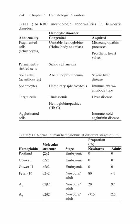
294
Table 7.10 RBC morphologic abnormalities in hemolytic disorders
AbnormalityHemolytic disorderCongenital Acquired
Fragmented cells (schistocytes)
Unstable hemoglobins (Heinz body anemias)
Microangiopathic processes
Prosthetic heart valves
Permanently sickled cells
Sickle cell anemia
Spur cells (acanthocytes)
Abetalipoproteinemia Severe liver disease
Spherocytes Hereditary spherocytosis Immune, warm- antibody type
Target cells Thalassemia Liver disease
Hemoglobinopathies (Hb C)
Agglutinated cells
Immune, cold agglutinin disease
Table 7.11 Normal human hemoglobins at different stages of life
HemoglobinMolecular structure Stage
Proportion (%)Newborns Adults
Portland ζ2γ2 Embryonic 0 0
Gower I ζ2ε2 Embryonic 0 0
Gower II α2ε2 Embryonic 0 0
Fetal (F) α2γ2 Newborn/adult
80 <1
A1 α2β2 Newborn/adult
20 97
A2 α2δ2 Newborn/adult
<0.5 2.5
Chapter 7. Hematologic Disorders

295
HbF test
dilutedtotal
%� � ��
�A
A 5100
where A = absorbance and 5 = the additional dilution factor.To calculate the percentage of hemoglobin A2,
HbA of total A fraction2 100% %� � � �
Useful Facts and Formulas

297© Springer Nature Switzerland AG 2021J. Varon, Handbook of Critical and Intensive Care Medicine, https://doi.org/10.1007/978-3-030-68270-5_8
The number of infectious complications encountered in the intensive care unit (ICU) continues to increase. Patients who otherwise would have not survived in the past are now improving due to new technical advancements. However, the length of stay, as well as the large number of devices employed for this purpose, predisposes patients to difficult and often fatal infections. Clinical characteristics of patients who are treated in the ICU have evolved in recent years. Those who are immunocompromised, posttransplant, and the geriatric population are now regularly treated in the ICU with the consequent increase in morbidity, mortality, and cost.
From the infectious disease point of view, the approach to a critically ill patient who is admitted to the ICU should immediately differentiate if the patient was transferred from the floor versus if the patient was directly admitted to the ICU from the community. This constitutes a paramount parameter to categorize the etiologic agents, to understand the pathophysiology of their processes, and mostly to decide which therapeutic antimicrobial interventions are needed.
Pneumonia (Nosocomial)
A. If the patient is transferred to the ICU after being in the hospital for several days, then treatment should address
Chapter 8Infectious Diseases

298
the nosocomial aspect of infection and the following important facts:
1. Mortality rates among these patients are 20–60%. 2. These patients represent 15% of all hospital deaths. 3. Successful treatment depends upon underlying disease,
specific causative organisms, and timely institution of therapy.
B. Predisposing Factors
1. Intubation. 2. ICU: Especially the patient who is receiving sedation. 3. Antibiotics: Broad-spectrum agents will rapidly change
normal flora of the mouth and gastrointestinal (GI) tract.
4. Surgery: Especially thoracic, abdominal, or neurosur-gery, which increases the risk of aspiration.
5. Chronic lung disease. 6. Advanced age. 7. Immunosuppression.
C. Etiologic Agents
1. Common – Gram-negative bacteria such as Klebsiella sp.,
Escherichia coli, Pseudomonas aeruginosa, Enterobacter sp., and Acinetobacter sp.
– Gram-positive bacteria: Staphylococcus aureus.
2. Less Common
(a) Anaerobic mouth flora (i.e., streptococci) (b) Other gram-negative bacilli (i.e., Serratia sp.,
Xanthomonas sp.) (c) Haemophilus influenzae (d) Legionella sp. (e) Candida sp. (f) Aspergillus sp. (g) Influenza virus (h) Streptococcus pneumoniae
Chapter 8. Infectious Diseases

299
(i) Miscellaneous: According to prevalent organisms in each hospital
(j) Tuberculosis (TB, typical and atypical)Another helpful approach is to consider the likely pathogens according to the time after hospitalization the pneumonia developed. Late-onset pneumonia (after more than 5 days of hospitalization) is usually characterized with more resistant organisms.
D. Clinical Manifestations. Patients in the ICU, especially those who are intubated or sedated, will not manifest the usual symptoms of pneumonia such as cough, chest pain, or dyspnea. Patients who are neutropenic cannot mount an inflammatory response, and, therefore, the sputum will not show purulent material. Subtle changes in oxygen-ation, fever, and clinical deterioration are clues for the diagnosis of pneumonia in intubated patients. Leukocytosis or leukopenia can be the first manifestation of occult pneumonia. In some instances, i.e., Pneumocystis pneumo-nia, the presence of spontaneous pneumothorax can be the first indication of pulmonary involvement. Thick, foul- smelling sputum is characteristic of anaerobic and aspira-tion pneumonia.
E. Diagnosis
1. On chest X-ray, look for new or changing infiltrates. 2. Obtain sputum for Gram’s stain immediately on every
patient. 3. Remember the concept of colonization versus true
infection; this distinction is sometimes very difficult. 4. Be aggressive in trying to obtain diagnosis (i.e., bron-
choalveolar lavage [BAL]). Transtracheal aspirates are not commonly employed.
5. Obtain other stains (i.e., acid-fast bacilli stain [AFB], Giemsa, wet prep).
6. Order serologies, if appropriate (i.e., Legionella, fungal serologies, cryptococcal antigen, CIE).
7. Remember the microbiological pattern of your hospital.
Pneumonia (Nosocomial)

300
F. Treatment Options
1. Empiric options most commonly utilized in the ICU
(a) Beta-lactam plus aminoglycoside (i.e., piperacillin and tobramycin).
(b) Cephalosporin plus aminoglycoside (i.e., ceftazi-dime and gentamicin).
(c) Clindamycin plus gentamicin. (d) Clindamycin plus quinolone (i.e., ciprofloxacin). (e) Imipenem/cilastatin plus aminoglycoside. (f) Cephalosporin plus fluoroquinolone. (g) Add trimethoprim–sulfamethoxazole [TMP-SMX]
if Pneumocystis carinii pneumonia is suspected. (h) Add erythromycin or azithromycin 500 mg IV qd
or erythromycin 0.5–1 g IV q6 h if Legionella is suspected.
(i) TMP-SMX 15–20 mg/kg/day TMP. (j) Doxycycline 100 mg IV q12 h. (k) Rifampin 300 mg IV q12 h. (l) Amphotericin B 0.6–1 mg/kg/day.
Duration of therapy is not well defined, but most authors agree on treating gram-negative and anaer-obic pneumonia for 10–21 days. Gram-positive pro-cesses are usually treated between 10 and 14 days, and atypical pneumonias receive 2 weeks of antimi-crobial therapy. Candida pneumonia requires pro-longed treatment with up to 1.5 g of amphotericin B as a total dose.
G. Prevention
1. Preoperative and postoperative measures for preven-tion of pneumonia
(a) Identification of high-risk patients (b) Treatment of respiratory infections, removal of
respiratory secretions (c) Instruction and therapy to expand patients’ lungs
(i.e., chest physiotherapy, incentive spirometry)
Chapter 8. Infectious Diseases

301
2. Proper handwashing 3. Appropriate maintenance of in-use respiratory therapy
equipment
(a) Use of sterile fluids in nebulizers (b) Proper use of single-dose and multidose medica-
tions for respiratory therapy
4. Proper sterilization and disinfection of reusable respi-ratory equipment
5. Proper suctioning of the respiratory tract 6. Protection of patients from other infected patients or
staff
Community-Acquired Pneumonia
A. Acquired outside the health-care system B. Common Organisms
1. Streptococcus pneumoniae 2. Mycoplasma pneumoniae 3. Haemophilus influenzae 4. Klebsiella sp. 5. Respiratory viruses (influenza A and B) 6. Adenovirus, respiratory syncytial virus, parainfluenza 7. Legionella sp.
C. Other Less Common Organisms
1. Pneumocystis carinii 2. Mycobacterium tuberculosis 3. Cryptococcus sp. 4. Chlamydia psittaci 5. Histoplasma sp. 6. Nocardia sp.
D. Common Manifestations
1. Fever, cough, dyspnea, sputum production usually purulent but not in all cases.
Community-Acquired Pneumonia

302
2. Hypoxemia is common. 3. Anxiety. 4. Leukocytosis; also leukopenia in severe infections. 5. Pulmonary consolidation and presence or absence of
pleural effusion.
E. Uncommon Presentations in Patients Who Are 1/4
1. Elderly 2. Immunocompromised (especially neutropenic) 3. Posttransplantation
F. Clinical Clues for Diagnosis
1. Acute onset: Bacterial, viral, aspiration, tularemia, Pneumocystis
2. Subacute onset: Viral, Legionella, Haemophilus sp., Mycoplasma, Q fever, Psittacosis, Chlamydia, Pneumocystis
3. Aerogenous route: Any segment 4. Hematogenous: Most commonly in both bases, as blood
flow is preferential to these areas
G. Associations
1. Birds: Psittacosis 2. Turtles: Typhoid 3. Dogs: Pasteurella multocida 4. Cattle: Q fever 5. Rabbits: Tularemia 6. Air conditioners: Legionella 7. COPD and smoking: H. Influenzae, Pseudomonas
aeruginosa 8. Hides: Anthrax 9. Foreign travel: Echinococcus, paragonimiasis 10. Barracks: Neisseria meningitidis, group A Streptococcus
H. Treatment. Empiric treatment is usually dictated by the geographical background, clinical presentation, and host status.
• Levofloxacin 750 mg IV/oral daily or moxifloxacin 400 mg IV/oral dailyor
Chapter 8. Infectious Diseases
303
• Ceftaroline 600 mg IV q 12 h (adjust in renal dysfunc-tion patients) plus azithromycin 500 mg IV/oral dailyor
• Ceftriaxone 1 g IV plus azithromycin 500 mg IV/oral daily
• Doxycycline 100 mg IV/oral twice daily is an alterna-tive to macrolides
1. Streptococcus pneumonia and Haemophilus influenza
(a) Quinolone (moxifloxacin or levofloxacin)(b) Ertapenem (1 g q24 h)(c) Ceftriaxone (1 g q24 h)
2. Legionella sp., Mycoplasma pneumoniae, Chlamydia pneumoniae
(a) Moxifloxacin (400 mg IV qd)(b) Levofloxacin (500 mg IV qd)(c) Doxycycline (200 mg IV qd)
3. Pseudomonas aeruginosa
(a) Meropenem (2 g IV q8 h)(b) Cefepime (2 g IV q8 h) and amikacin (1 g IV
q24 h)(c) For multidrug resistant P. aeruginosa: Colistin
(80 mg IV q8 h)
4. Influenza A/B, avian influenza
(a) Oseltamivir (Tamiflu) 75 mg PO q24 h plus rimantadine 100 mg PO.
(b) Avian influenza (influenza virus type A H5N1)—influenza following close contact with infected poultry. Several outbreaks in humans have been identified in Asia: Flu-like symptoms with vague gastrointestinal complaints that rapidly progress to acute respiratory failure. Diagnosis is by hemagglutinin- specific RT-PCR for avian influ-enza. Treatment with antivirals should be given
Community-Acquired Pneumonia

304
early, and it includes oseltamivir (150 mg), with amantadine and rimantadine.
I. Mortality Estimate in Community-Acquired Pneumonia
1. Pneumonia Severity Index (PSI)
(a) Factors in age, medical history, and vital signs
2. CURB-65
(a) CURB-65 is fast to compute, requires already available patient information, and provides an excellent risk stratification of community-acquired pneumonia
J. Complications After 72 h
1. Persistent fever 2. Empyema 3. Obstruction 4. Lung abscess 5. Resistant organism 6. Focus of infection
Risk class Risk Point valueI Low None from comorbidities, PE findings, or
lab findings
II Low ≤70 points
III Low 71–90
IV Moderate 91–130
V High >130 total points
CURB-65 score
Mortality risk Recommendation per derivation study
0 0.60% Low risk; consider home treatment
1 2.70% Low risk; consider home treatment
2 6.80% Short inpatient hospitalization or closely supervised outpatient treatment
Chapter 8. Infectious Diseases

305
CURB-65 score
Mortality risk Recommendation per derivation study
3 14.00% Severe pneumonia; hospitalize and consider admitting to intensive care
4 or 5 27.80% Severe pneumonia; hospitalize and consider admitting to intensive care
Novel Coronavirus 2019 (COVID-19)
1. Coronaviruses are large, enveloped, single-stranded RNA viruses found in humans and other mammals, such as dogs, cats, chicken, cattle, pigs, and birds.
2. Coronaviruses cause respiratory, gastrointestinal, and neu-rological disease.
3. Spread primarily via respiratory droplets during close con-tact. Infection can be spread by asymptomatic, presymp-tomatic, and symptomatic carriers.
4. Most common symptoms include fever (temperature greater than 100.4 °F), dry or nonproductive cough, short-ness of breath, fatigue, weakness, and headache.
5. Imaging modalities can be used to identify/suspect COVID-19 infection.
(a) Chest X-ray can reveal bilateral, lower-lobe predomi-nant infiltrates.
(b) Chest computed tomography (CT) imaging can show bilateral, peripheral, lower-lobe ground-glass opacities and consolidations.
6. Definitive diagnosis is made by detection of SARS-CoV-2 via reverse transcription polymerase chain reaction (RT- PCR) testing, although false-negative test results may occur in up to 20–50% of patients.
7. Disease Severity
(a) Up to 80% of the infected patients will have mild infection and mostly restricted to the upper and con-ducting airways.
(b) Some individuals may have more severe infection leading to sepsis or acute respiratory failure requiring
Novel Coronavirus 2019 (COVID-19)

306
mechanical ventilation with increased morbidity and mortality.
(c) In severe COVID-19, fulminant activation of coagula-tion and consumption of clotting factors occur requir-ing the use of therapeutic dosages of anticoagulation.
(d) RT-PCR value for the viral RNA might be useful to predict the viral load and the subsequent infectivity and clinical course.
8. Treatment
(a) Data have shown that steroids such as methylprednis-olone and dexamethasone therapy may reduce mortal-ity in patients requiring supplemental oxygen compared with usual care.
(b) The best treatment strategy arrives from the MATH+ protocol (methylprednisolone, ascorbic acid, thiamine, heparin as primary elements). The full protocol and rational is found at www.covid19criticalcare.com.
(c) Remdesivir improves time to recovery (hospital dis-charge or no supplemental oxygen requirement). However its use is limited to the first few days od symptoms.
(d) Data suggest convalescent plasma did not shorten time to recovery in hospitalized patients.
(e) Monoclonal antibodies have been used to target the cytokine storm induced by CODI-19; these antibodies are directed against inflammatory mediators such as interferon gamma, interleukin 1, interleukin 6, and complement factor 5a.
(f) Thromboembolic prophylaxis with high-dose subcuta-neous low molecular weight heparin is recommended for all hospitalized patients with COVID-19.
9. Prevention
(a) At least 120 SARS-CoV-2 vaccines are under development.
(b) Primary methods to reduce spread are face masks, social distancing, and contact tracing. Other methods that the author recommends is the use of the iMASK+ protocol (flccc.net) that includes ivermectin.
Chapter 8. Infectious Diseases
307
Severe Adult Respiratory Syndrome (SARS)
Term given by the World Health Organization which describes a rapidly progressive respiratory illness with documented outbreaks in China, Hong Kong, Vietnam, Singapore, and Canada. The presumable pathogen is a Coronavirus that spreads person to person via droplets, sewage, and water and potentially through human feces. It is hypothesized that bats are the primary reservoir for the disease. The mortality rate from SARS is high (up to 20%). In severe cases, respiratory function may worsen during the second week of illness and progress to ARDS. This is a two-stage illness:
1. Prodrome (2–7 day): Includes fever, malaise, headache, and myalgias, and diarrhea may occur.
2. Respiratory phase (8–12 day): Nonproductive cough and dyspnea that rapidly progress to respiratory failure.
A rapid diagnosis of SARS can be made by reverse tran-scriptase PCR of respiratory tract, samples, and plasma early in illness.
There is no current treatment available for this illness, except for supportive care in the intensive care unit. Preventive measures against SARS are mainly focused on travel advisories to countries and cities with active out-breaks. Efforts are underway to prepare a vaccine for the prevention of SARS.
Sepsis
A. More than 750,000 cases of sepsis, with an associated mor-tality of 20–60%, are estimated to occur annually. Despite improvements in antimicrobial therapy and supportive care, the incidence of and mortality associated with sepsis have not declined. This is, in part, a consequence of an array of medical advances that can place patients at increased risk for development of infection and, poten-tially, sepsis.
Sepsis
308
B. Sepsis and Related Disorders
1. Definitions
(a) Bacteremia: Positive blood cultures (may be transient)
(b) Sepsis: Clinical evidence suggestive of infection plus signs of a systemic response to the infection (all of the following):
1. Tachypnea (respiration 20 breaths per minute; if patient is mechanically ventilated, minute vol-ume 10 L/min)
2. Tachycardia (heart rate >90 beats per minute) 3. Hyperthermia or hypothermia (core or rectal
temperature >38.4 °C [101 °F] or <35.6 °C [96.1 °F])
(c) Sepsis Syndrome (may also be considered incipient septic shock in patients who later become hypoten-sive): Clinical diagnosis of sepsis outlined above, plus evidence of altered organ perfusion (one or more of the following):
1. PaO2/FiO2 no higher than 280 (in the absence of other pulmonary or cardiovascular diseases).
2. Lactate level above the upper limit of normal. 3. Oliguria (documented urine output <0.5 mL/kg
body weight for at least 1 h in patients with uri-nary catheters in place).
4. Acute alteration in mental status. 5. Positive blood cultures are not required.
(d) Early Septic Shock: Clinical diagnosis of sepsis syn-drome as outlined above, plus hypotension (sys-tolic blood pressure <90 mmHg or a 40-mmHg decrease below baseline systolic blood pressure) that lasts for <1 h and is responsive to conventional therapy (intravenous fluid administration or phar-macologic intervention)
(e) Refractory Septic Shock: Clinical diagnosis of the sepsis syndrome outlined above, plus hypotension
Chapter 8. Infectious Diseases
309
(systolic blood pressure <90 mmHg or a 40-mmHg decrease below baseline systolic blood pressure) that lasts for >1 h despite adequate volume resusci-tation and that requires vasopressors
C. Pathophysiology. Cell walls of gram-negative bacteria contain proteins, lipids, and lipopolysaccharides. Endotoxin (lipopolysaccharide) has three components: an O-specific polysaccharide, the R-core, and lipid A. Lipid A may be the major culprit in initiating the endotoxic symp-toms. It is this component of endotoxin that stimulates the release of tissue necrosis factor (TNF) and can also acti-vate the complement pathway. The sepsis syndrome is caused by endothelial damage following endotoxin- stimulated activation of neutrophils, coagulation, comple-ment, and macrophages. Macrophages are stimulated to release TNF, interleukins, leukotrienes, thromboxane, and other cardioactive substances. Endotoxemia markedly increases the risk of myocardial depression and multiple organ failure. In patients who have positive blood cultures, those with severe endotoxemia have five times the mortal-ity of those who do not have endotoxemia.
D. Priorities in the Treatment of Sepsis
(a) Early recognition. (b) Cardiovascular/pulmonary support. (c) Fluid resuscitation. (d) Pressor agents. (e) Empiric antibiotic therapy. (f) Other immunotherapeutic agents (investigational). (g) Corticosteroids are not effective. However, if the
patient has acute adrenal insufficiency due to sepsis, steroid replacement therapy is a must.
(h) Drainage of any foci of infection.
E. Prognosis. Mortality in sepsis is a function of the severity of physiologic derangements, the duration of illness, and the number of organ system failures. These organ systems include, but are not limited to, the lungs, kidneys, and liver. When the pulmonary system becomes dysfunctional, the
Sepsis

310
resultant clinical entity is known as the adult respiratory distress syndrome (ARDS). The sequence has been termed the multiple organ dysfunction syndrome (MODS). MODS is the most common cause of demise in patients who experience uncontrolled inflammation and infection.
Toxic Shock Syndrome
A. Clinical Case Definition (See Table 8.1)
1. Severe febrile (38.9 °C) illness with rash (erythroderma followed by desquamation), hypotension or syncope, and multiple organ system involvement (at least four of
Table 8.1 Toxic shock syndromeCriteria for diagnosis
Temperature <38.9 °C
Systolic blood pressure <90 mmHg
Rash with subsequent desquamation, especially on palms and soles
Involvement of >3 of following organ systems:
Gastrointestinal: vomiting or severe diarrhea
Muscular: severe myalgias or fivefold increase in creatine kinase
Mucous membranes: frank hyperemia
Renal insufficiency: serum urea nitrogen, creatinine, double of normal
Liver: enzymes, twice upper limits of normal
Blood: thrombocytopenia <100,000/mm3
CNS: disorientation without focal findings
Negative tests for leptospirosis, Rocky Mountain spotted fever, and measles
Chapter 8. Infectious Diseases
311
the following: mucous membrane, GI, muscular, central nervous system [CNS], renal, hepatic, hematologic, car-diopulmonary, metabolic).
2. Hypotension: Probably due to small-vessel and capil-lary leakage with extravascular accumulation of fluid (edema).
3. Blood cultures are usually negative. 4. Acute episode followed by desquamation. 5. No evidence of other causes: Scarlet fever, Kawasaki’s
disease, Rocky Mountain spotted fever, etc.
B. Epidemiology and Other Clinical Features
1. Affects mostly young menstruating women. Tampon use, especially continuous use and Rely brand in some studies. S. aureus colonization of the vagina. Recurrence rate of 30%. Decrease in the number of reported cases.
2. Also occurs in non-menstruating women, men, and children (colonization or focal infection with S. aureus, including postoperative infections). Common occur-rence after surgery. Fatality rate, 5–10%.
C. Etiology. Exotoxin(s) of S. aureus appear to cause the dis-ease. Recently, streptococci have been shown to cause the same syndrome.
D. Differential Diagnosis. Kawasaki’s disease, scarlet fever, leptospirosis, Rocky Mountain spotted fever, measles.
E. Treatment. The most important treatment is volume expansion and correction of hypotension; removal of the tampon, if present, in menstruating women; debridement of wounds, etc.; and administration of antistaphylococcal antibiotics (after cultures have been obtained). Steroids have not been proven to be effective or to alter outcome.
Meningitis
A. Acute meningitis is a medical emergency that requires early recognition, rapid diagnosis, precise antimicrobial therapy, and aggressive ICU support.
Meningitis

312
1. Etiologic Agents
(a) Streptococcus pneumoniae: The most common cause in adults.
(b) Neisseria meningitis: Common among groups of young individuals and children.
(c) Haemophilus influenzae: Common in children up to 12 years of age.
(d) Staphylococcus aureus and S. epidermidis: Seen in the elderly or postoperatively (CNS shunts).
(e) Listeria monocytogenes: Usually mistaken with diphtheroids or contaminants.
(f) Streptococci other than S. pneumoniae: Especially group B in neonatal disease.
(g) Gram-negative bacilli: After surgery or trauma. (h) Mycobacterium tuberculosis: Increasing in
frequency. (i) Cryptococcus: Usually in immunosuppressed
patients (i.e., those with acquired immune defi-ciency syndrome [AIDS] or impaired cell- mediated immunity).
(j) Syphilis: Presentation variable. (k) Herpes simplex. (l) Toxoplasma: Can present as meningoencephalitis
or brain abscess. (m) Naegleria: Epidemiological history is paramount. (n) Other viruses (i.e., echovirus, St. Louis, equine,
and Western encephalitis).
2. Associations: Epidemiology and Organisms
(a) Summer and fall: Coxsackie or echovirus; leptospira
(b) Previous meningitis: S. pneumoniae (c) Alcoholism: S. pneumoniae (d) Young adults: N. meningitis (e) Elderly: S. pneumoniae, Listeria, gram-negative
bacilli (f) Lymphoma: Cryptococcus sp. (g) Petechia: N. meningitidis, echovirus
Chapter 8. Infectious Diseases
313
(h) Sinusitis: H. influenzae, S. pneumoniae, anaerobic bacteria
(i) Cellulitis: Aerobic, gram-positive cocci (j) Brain abscess: Mixed flora (k) Swimming in fresh water: Amoebas (l) Other family members with meningitis: N.
meningitidis (m) Water contact: Leptospira (n) Hospital acquired: Gram-negative bacilli, staphy-
lococcus, Candida (o) Head trauma
1. Close fracture: S. pneumoniae, gram-negative bacilli
2. Craniotomy: Gram-negative bacilli, staphylococci
3. Cerebrospinal fluid rhinorrhea: S. pneumoniae
3. Cerebrospinal Fluid (CSF) Findings (See Table 8.2) 4. Diagnostic Approach
(a) Order antigen detection for H. influenzae, S. pneu-moniae, and N. meningitidis.
(b) Obtain high-volume CSF for AFB concentrate and fungal cultures (20–30 mL).
(c) If CSF is normal or viruses are suspected, repeat lumbar puncture (LP) in 24–36 h.
(d) Upon admission, obtain serologies for viral infec-tions (i.e., St. Louis encephalitis, California encephalitis).
(e) Obtain serologies in serum and CSF for fungal infections.
(f) Polymerase chain reaction (PCR) may be helpful (especially for TB and cytomegalovirus [CMV] infections).
5. TreatmentIn acutely ill patients, the goal of therapy is to institute treatment before the pathologic process of inflamma-tion can produce irreversible progression and/or death.
Meningitis
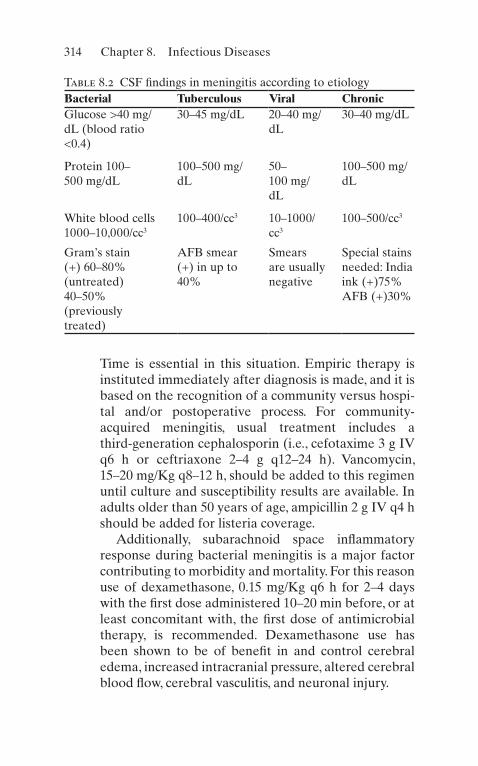
314
Time is essential in this situation. Empiric therapy is instituted immediately after diagnosis is made, and it is based on the recognition of a community versus hospi-tal and/or postoperative process. For community-acquired meningitis, usual treatment includes a third-generation cephalosporin (i.e., cefotaxime 3 g IV q6 h or ceftriaxone 2–4 g q12–24 h). Vancomycin, 15–20 mg/Kg q8–12 h, should be added to this regimen until culture and susceptibility results are available. In adults older than 50 years of age, ampicillin 2 g IV q4 h should be added for listeria coverage.
Additionally, subarachnoid space inflammatory response during bacterial meningitis is a major factor contributing to morbidity and mortality. For this reason use of dexamethasone, 0.15 mg/Kg q6 h for 2–4 days with the first dose administered 10–20 min before, or at least concomitant with, the first dose of antimicrobial therapy, is recommended. Dexamethasone use has been shown to be of benefit in and control cerebral edema, increased intracranial pressure, altered cerebral blood flow, cerebral vasculitis, and neuronal injury.
Table 8.2 CSF findings in meningitis according to etiologyBacterial Tuberculous Viral ChronicGlucose >40 mg/dL (blood ratio <0.4)
30–45 mg/dL 20–40 mg/dL
30–40 mg/dL
Protein 100–500 mg/dL
100–500 mg/dL
50–100 mg/dL
100–500 mg/dL
White blood cells 1000–10,000/cc3
100–400/cc3 10–1000/cc3
100–500/cc3
Gram’s stain (+) 60–80% (untreated) 40–50% (previously treated)
AFB smear (+) in up to 40%
Smears are usually negative
Special stains needed: India ink (+)75% AFB (+)30%
Chapter 8. Infectious Diseases

315
B. Pneumococcal Meningitis
1. Pneumococcal meningitis is still the most common cause of bacterial meningitis in adults. Underlying dis-eases: sickle cell disease, splenectomy and splenic dys-function, hypogammaglobulinemia, alcoholism, head trauma (CSF fistula), and chronic pulmonary, hepatic, or renal disease.
2. Associated infections: Pneumonia, otitis, bacteremia, endocarditis, mastoiditis.
3. Therapy: Ceftriaxone 4 g/day and vancomycin 2 g/day should be given if there has been beta-lactam resis-tance noted locally.
C. Haemophilus Meningitis
1. Underlying disease (adults): Alcoholism, compromised host defenses, head trauma.
2. Associated infections: Pneumonia, sinusitis, otitis. Secondary cases can occur in close contacts.
3. Therapy: Cefotaxime (2 g IV q6 h), ceftriaxone (2 g IV q12 h), and chloramphenicol (500 mg PO q6 h for 2 weeks) as IV to PO switch.
D. Meningococcal Meningitis
1. Meningococcal meningitis is seen primarily in children, adolescents, and young adults. Secondary infection in close contacts can occur. Predisposing factors include complement defects.
2. Disseminated neisserial infection (often recurrent in persons with C5–C8 deficiency). Waterhouse–Friderichsen syndrome is an acute, often fatal, syn-drome of septic shock associated with massive adrenal necrosis, associated with bacteremia due to this organ-ism. It requires early recognition, antibiotic therapy, and especially aggressive ICU/hemodynamic support.
3. Early antimicrobial therapy is needed. Ceftriaxone 2 g IV q12 h is the preferred IV therapy; as an alternative, meropenem 2 g q8 h can be administered.
Meningitis

316
E. Listeria Meningitis. Listeria is an important cause of bac-teremia and meningitis, particularly in the elderly. Epidemiological history is important. Therapy is with ampicillin (2 g q4 h) or meropenem (excellent in vitro activity against Listeria).
F. Staphylococcus aureus and Staphylococcus epidermidis. Infection with these organisms is common after neurosur-gery and/or ventricular peritoneal shunt placement.
1. Therapy
(a) Methicillin sensitive: Cefotaxime (3 g IV q6 h) or cefepime (2 g IV q8 h).
(b) Methicillin resistant: Linezolid (600 mg IV q12 h) or vancomycin (2 g IV q12 h).
(c) An infected shunt may need to be removed early in the course of therapy if the patient is not respond-ing. Repeat LP at 2–3 days is needed in order to reach this decision (persistent growth of organisms, despite adequate therapy).
G. Gram-Negative Bacilli
1. Infections with gram-negative bacilli are challenging to treat due to their high morbidity and mortality. Development of resistance can occur while on therapy (especially with Enterobacter sp.). Most organisms will respond to ceftriaxone, cefotaxime, or ceftazidime. For Pseudomonas aeruginosa, ceftazidime 2 g IV q8 h is the drug of choice. It should be given with gentamicin (1–2 mg/kg/8 h).
H. Complications of Bacterial Meningitis
1. Brain Abscess: Usually follows trauma, contiguous infection, hematogenous dissemination.
2. Subdural Empyema: Primarily disease of the young but, in elderly, may complicate neurosurgery or subdu-ral hematoma.
3. Epidural Abscess: Usually accompanied by focal osteo-myelitis and subdural empyema.
Chapter 8. Infectious Diseases

317
4. All of the above are caused by mixed bacteria and usu-ally require drainage, as well as prolonged IV antibiotic therapy.
I. Herpes Meningitis/Encephalitis. Herpes meningitis/encephalitis is a devastating necrotizing type of encepha-litis. Temporal spikes on electroencephalogram (EEG) are characteristic. Treatment is given with acyclovir 15 mg/kg q8 h (high dose) for 2 weeks. Careful attention to hydration is mandatory to avoid renal insufficiency.
Infections in Patients with AIDS
A. Opportunistic infections are the most common causes of morbidity and mortality in patients with human immuno-deficiency virus (HIV). Patients with CD4 cells <250 are at risk for developing severe infectious complications. Their approach is depicted in Table 8.3.
B. Summary of Current Therapeutic Approaches
1. Pulmonary Disease
(a) Disease Due to Pneumocystis carinii (Pneumocystis jirovecii) Pneumonia (PCP) (Table 8.4)
(b) Disease Due to M. Tuberculosis
1. Start with at least four drugs, preferably five; INH 300 mg/day, rifampin 600 mg/day, pyrazin-amide 15/kg/day, ciprofloxacin 750 mg PO bid, ethambutol 15–20 mg/kg/day.
2. If TB is sensitive to INH and/or rifampin, con-tinue for 12–18 months (not in the ICU).
3. If TB is resistant to either or both drugs (INH and rifampin), multiple drug resistant, continue with five to six drugs, and adjust according to sensitivities. Prognosis is very poor.
4. Follow liver function tests, initially weekly and later monthly.
5. If patient cannot use PO drugs, give IV INH and rifampin (same dose) and IM streptomycin (1 g/day).
Infections in Patients with AIDS

318
(c) Pulmonary Disease Due To Histoplasma capsulatum
1. Initiate therapy with amphotericin B at 0.8–1 mg/kg/day.
2. Search for other sites of involvement (i.e., bone marrow biopsy, lumbar puncture, chest X-ray, barium enema, and small bowel series).
3. Once the patient is stable, switch to fluconazole 200 mg PO bid.
Table 8.3 Approach to HIV patients with opportunistic infectionsClinical presentation Common organisma
Diagnostic procedure
Pulmonary infiltrates
P. carinii (PCP); tuberculosis (TB); Mycobacterium avium–intracellulare (MAI); histoplasma, aerobic bacteria, Legionella
BAL and/or lung biopsy; appropriate serologies
Seizures, headache, vertigo, facial palsy
Toxoplasma, Cryptococcus MAI, herpes, CMV
MRI, head CT, LP, and appropriate serologies
Esophagitis Candida, herpes, CMV, cryptosporidium
Endoscopy with biopsy and washings
Diarrhea CMV, Cryptosporidium, Giardia, MAI, Isospora, C. difficile, Salmonella
Stool culture (initially)b AFB stain, colonoscopy, and biopsy
Persistent fever
MAI, Histoplasma, TB, Cryptococcus
CT abdomenc
Bone marrow
Blood cultures with special stains (AFB)
aRemember that each one of these syndromes can be caused by noninfectious processesbAlso useful to obtain fecal leukocytes for diagnosis of colitiscPerformed when fever persists despite initial evolution
Chapter 8. Infectious Diseases
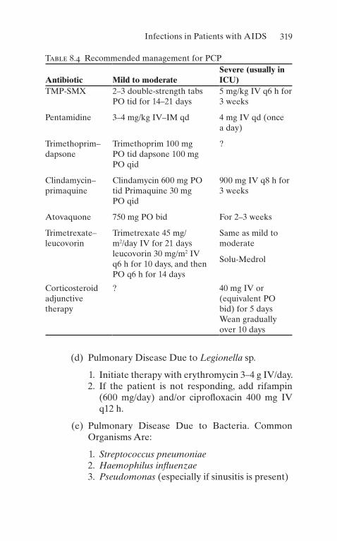
319
(d) Pulmonary Disease Due to Legionella sp.
1. Initiate therapy with erythromycin 3–4 g IV/day. 2. If the patient is not responding, add rifampin
(600 mg/day) and/or ciprofloxacin 400 mg IV q12 h.
(e) Pulmonary Disease Due to Bacteria. Common Organisms Are:
1. Streptococcus pneumoniae 2. Haemophilus influenzae 3. Pseudomonas (especially if sinusitis is present)
Table 8.4 Recommended management for PCP
Antibiotic Mild to moderateSevere (usually in ICU)
TMP-SMX 2–3 double-strength tabs PO tid for 14–21 days
5 mg/kg IV q6 h for 3 weeks
Pentamidine 3–4 mg/kg IV–IM qd 4 mg IV qd (once a day)
Trimethoprim–dapsone
Trimethoprim 100 mg PO tid dapsone 100 mg PO qid
?
Clindamycin–primaquine
Clindamycin 600 mg PO tid Primaquine 30 mg PO qid
900 mg IV q8 h for 3 weeks
Atovaquone 750 mg PO bid For 2–3 weeks
Trimetrexate–leucovorin
Trimetrexate 45 mg/m2/day IV for 21 days leucovorin 30 mg/m2 IV q6 h for 10 days, and then PO q6 h for 14 days
Same as mild to moderate
Solu-Medrol
Corticosteroid adjunctive therapy
? 40 mg IV or (equivalent PO bid) for 5 days Wean gradually over 10 days
Infections in Patients with AIDS

320
(f) Add Antibacterial Therapy Empirically on Admission
1. Ticarcillin–clavulanic acid 3.1 g IV q6 h (will also cover anaerobes in the sinuses) or piper-acillin–tazobactam 3.375–4.5 g IV q6 h.
2. Cefuroxime 1.5 g IV q8 h. 3. Adjust when cultures and sensitivities become
available.
(g) Pulmonary Disease Due to M. avium–intracellulare
1. Ethambutol 15 mg/kg/day PO plus clarithromy-cin 500 mg PO q12 h or azithromycin 500 mg PO q24 h plus rifampin.
2. Treatment is given for at least 6 months after a negative sputum for MAI.
2. Enteric Pathogens in Patients with AIDS (See Table 8.5)
3. CNS Infections in AIDS
(a) Cryptococcal Meningitis
1. Acute: Amphotericin B 0.7–1 mg/kg/day plus 5-fluorocytosine 25 mg/kg/day until the patient is stable or improving. Then switch to flucon-azole 400 mg/day PO for 3 months.
2. Maintenance: Fluconazole 200–400 mg/day PO.
(b) Toxoplasmosis
1. Pyrimethamine 200 mg PO: Loading dose fol-lowed by 75 mg PO daily with folinic acid 5 mg PO daily. (No IV presentation available.)
2. Sulfadiazine 1.5 g PO q6 h; plus leucovorin 10 mg PO q24 h.
Chapter 8. Infectious Diseases

321
(c) CMV (Including Retinitis)
1. Ganciclovir 5–10 mg/kg IV q12 h for 14 days (initial therapy)
2. Foscarnet 60 mg/kg IV q8 h for 14 days (initial therapy)
3. Lifelong suppressive therapy with valganciclo-vir 900 mg PO q24 h
(d) Herpes Simplex
1. Acyclovir 10–15 mg/kg IV q8 h
(e) Syphilis
1. Crystalline penicillin 24 million U/day for 14 days 2. Ceftriaxone 2–4 g/day IV for 14 days
Table 8.5 Enteric pathogens commonly seen in patients with AIDS
Organism Antimicrobial agentDirection of therapy (days)
G. lamblia Metronidazole 250 mg PO tid 5
E. histolytica Metronidazole 750 mg tid and diiodohydroxyquin 650 mg PO tid
10
Shigella sp. Fluoroquinolone IV or PO 3–7
C. jejuni Ciprofloxacin 500 mg IV q12 h 7
I. belli TMP-SMX 1 double-strength qd 14
CMV Ganciclovir 5 mg/kg IV q12 h 30
Herpes simplex
Fluconazole 100 mg PO q24 h 14
Oral thrush Ketoconazole 200–400 mg/day PO
10
Candida esophagitis
Fluconazole 200–400 mg/day IV 7–10
Infections in Patients with AIDS
322
C. Important Facts to Remember in Treating HIV-Infected Patients in the ICU
1. Patients may have more than one infection at the same time.
2. Blood precautions should be instituted immediately to avoid unnecessary exposure.
3. Noninfectious processes (i.e., tumors) can mimic infections.
4. Patients require a full physical examination daily, including mouth, perirectal area, and eyes.
5. Superinfections are common (i.e., fungal and resistant bacteria).
6. When fever persists, consider lumbar puncture, liver, and bone marrow biopsy.
7. Obtain CD4–CD8 counts if not recently cloned. 8. Code status needs to be established early. 9. Privacy of and respect toward patient are essential and
mandatory.
Infections in the Immunocompromised Host
A. The number of critically ill patients with impaired host defense mechanisms who are admitted to the ICU has dramatically increased in recent years. The knowledge and recognition of the basic deficiency enable the physician to predict the type and site of infection and allow the institu-tion of early empiric therapy (see Tables 8.6 and 8.7).
B. Immunocompromised patients admitted to the ICU should be categorized according to the time of acquisition of infection.Hospital-acquired infections have different etiologic agents compared to those from the community, despite having the same basic immunologic defect.
Chapter 8. Infectious Diseases

323
Table 8.6 Selected immunological defects and clinical presentationsDefect Organism ManifestationsPhagocytes/neutrophils (i.e., neutropenia)
Gram-positive cocci Bacteremia
Gram-negative bacilli Sepsis
P. aeruginosa Tissue invasion, pneumonia, rhinocerebral and cutaneous
Candida sp.
Aspergillus sp.
Mucor sp.
Absidia sp.
Fusarium sp.
Complement (i.e., C5–C8 deficiency)
Neisseria sp. Fulminant sepsis
S. pneumoniae Recurrent infection
H. influenzae Pneumonia
P. aeruginosa Sepsis
Brucella sp. Recurrent fever
Antibody (i.e., IgA- IgG deficiency)
Gram-positive cocci Pneumonia, otitis
H. influenzae Meningitis
Herpes simplex Encephalitis
Giardia lamblia Liver disease
Diarrhea
(continued)
Antimicrobials (See Table 8.8)
Infectious Diseases: “Pearls” for ICU Care
A. Handwashing is the single most important procedure to prevent infection.
B. Improving the nutritional status is of great importance for the outcome of infections.
Infectious Diseases: “Pearls” for ICU Care

324
C. Remove bladder catheters as soon as possible. D. Complete daily physical examination is mandatory. E. Gram’s stain is the single best and least expensive test for
early diagnosis of several infections (i.e., pulmonary, soft tissue, meningitis).
F. Hypothermia, especially in elderly patients, suggests sepsis.
G. Central catheters should be changed every 5–7 days. H. Peripheral lines should be changed every 2–3 days. I. If prolonged ICU stay is expected, early placement of
subcutaneous catheters is recommended. J. Patients with high fever require special attention to fluid
management.
Table 8.6 (continued)Defect Organism Manifestations
Cell-mediated immunity (i.e., decrease in CD4 counts)
Salmonella Diarrhea, sepsis
Listeria sp. Meningitis
Mycobacterium sp. Pneumonia
Nocardia sp. CNS/lungs
Cryptococcus neoformans
Lungs
Histoplasma capsulatum
Mucocutaneous
Coccidioides immitis Disseminated
Herpes simplex Pneumonia
Varicella zoster CNS/myocardium
CMV
P. carinii
Strongyloides stercoralis
Toxoplasma gondii
Chapter 8. Infectious Diseases

325
Table 8.7 Common clinical presentations in compromised patients in the ICUReason for admission Common pathogen
Initial therapeutic approach
Fever and neutropenia
Early: Early empiric therapy mandatory
Gram-negative bacilli and gram- positive cocci (usually catheter related)
Late:
Resistant gram- negative bacilli
Fungi (Candida sp., Aspergillus sp., Fusarium sp., Mucor sp.)
Sepsis: postsplenectomy
Encapsulated bacterial organisms
Emergency institution of antibacterial therapy
Neurologic deterioration in patient with cell- mediated immune deficit
Intracellular organisms
Obtain CT, LP, and treat for bacteria and possibly for Cryptococcus
Sepsis after solid organ transplantation
Immediately after surgery: Common local bacteria
Choose antibacterials according to site. Empiric therapy with extensive workup needed
Not related to surgery: Virus, fungus, Nocardia
(continued)
Infectious Diseases: “Pearls” for ICU Care

326
Table 8.7 (continued)Reason for admission Common pathogen
Initial therapeutic approach
Bilateral pulmonary infiltrates
Organism depends on causative defect
Treat empirically, and obtain BAL and biopsy (if possible)
Diabetic ketoacidosis Bacterial organisms, mucormycosis, Aspergillus
Treat for mixed bacterial infection
AIDS Depends on sites of infection
See section on AIDS
Postoperative status and malnutrition
Antibiotic-resistant gram-negative bacilli Group D streptococci Candida sp.
Utilize broad- spectrum therapy
Table 8.8 Selected antimicrobials commonly used in the ICU
Drug Dose
Renal adjustment: creatinine clearance >80 50–10 <10
Comments and side effects
Aminoglycosides (i.e., gentamicin)
1–2 mg/kg IV q8 h
8–12 h Monitor levels, renal function, and hearing12 h
24–48 h
Broad-spectrum penicillin (i.e., piperacillin)
3–4 g IV q8 h
4–6 h Monitor Na+ and coagulation profile
8–12 h
12–24 h
Imipenem 500 mg to 1 g
6 h Seizures, twitching, facial palsies12 h
24 h
Chapter 8. Infectious Diseases

327
Table 8.8 (continued)
Drug Dose
Renal adjustment: creatinine clearance >80 50–10 <10
Comments and side effects
Cephalosporins (i.e., ceftazidime)
2 g IV q8 h 6–12 h Penetrates CSF well
12 h
14 h
Aztreonam 2 g IV q8 h 6–12 h Tolerated in penicillin- allergic patients
12 h
24 h
Vancomycin 1 g IV q12 h 6–12 h Monitor levels; interstitial nephritis2–3 days
weekly
Oxacillin 6–12 g IV 4–6 h Infuse in at least 1 h
6–8 h
8–12 h
Acyclovir 2–3 g/day IV
8 h Monitor WBC and renal function12–24 h
24–48 h
Ganciclovir 5 mg/kg IV 12 h Monitor bone marrow depression12 h
24–48 h
Clindamycin 600–900 mg IV
8 h Diarrhea
8 h
8 h
(continued)
Infectious Diseases: “Pearls” for ICU Care

328
Table 8.8 (continued)
Drug Dose
Renal adjustment: creatinine clearance >80 50–10 <10
Comments and side effects
Chloramphenicol 3–4 g IV or PO
6 h Monitor bone marrow function6 h
6 h
Metronidazole 30 mg/kg/day IV or PO
6 h Metallic taste
6 h
6 h
Amphotericin B 0.5–1 mg/kg IV
24 h Monitor renal function
Once a day 24 h
48 h
Fluconazole 200–400 mg 12 h Interacts with anticoagulants
q12 h IV or PO 24 h
48 h
Itraconazole 2–4 g PO 12–24 h
24 h
24 h
TMP-SMX 4–5 mg/kg IV (TMP) or higher
6–12 h Monitor WBC; skin rash
12–24 h
24–48 h
Doxycycline 100–200 mg IV
12–24 h Impairs neutrophil function12–24 h
12–24 h
Levofloxacin 500–750 mg IV
24 h Do not use in children
Chapter 8. Infectious Diseases
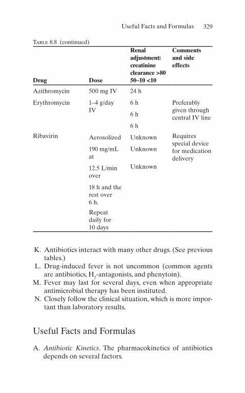
329
K. Antibiotics interact with many other drugs. (See previous tables.)
L. Drug-induced fever is not uncommon (common agents are antibiotics, H2-antagonists, and phenytoin).
M. Fever may last for several days, even when appropriate antimicrobial therapy has been instituted.
N. Closely follow the clinical situation, which is more impor-tant than laboratory results.
Useful Facts and Formulas
A. Antibiotic Kinetics. The pharmacokinetics of antibiotics depends on several factors.
Table 8.8 (continued)
Drug Dose
Renal adjustment: creatinine clearance >80 50–10 <10
Comments and side effects
Azithromycin 500 mg IV 24 h
Erythromycin 1–4 g/day IV
6 h Preferably given through central IV line6 h
6 h
Ribavirin Aerosolized Unknown Requires special device for medication delivery
190 mg/mL at
Unknown
12.5 L/min over
Unknown
18 h and the rest over 6 h.
Repeat daily for 10 days
Useful Facts and Formulas
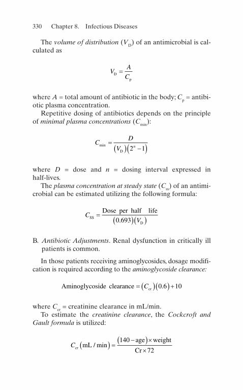
330
The volume of distribution (VD) of an antimicrobial is cal-culated as
V
A
CDp
=
where A = total amount of antibiotic in the body; Cp = antibi-otic plasma concentration.
Repetitive dosing of antibiotics depends on the principle of minimal plasma concentrations (Cmin):
CD
V nmin � � � �� �D 2 1
where D = dose and n = dosing interval expressed in half-lives.
The plasma concentration at steady state (Css) of an antimi-crobial can be estimated utilizing the following formula:
C
VSSD
Dose per half life�
� �� �0 693.
B. Antibiotic Adjustments. Renal dysfunction in critically ill patients is common.
In those patients receiving aminoglycosides, dosage modifi-cation is required according to the aminoglycoside clearance:
Aminoglycoside clearance cr� � �� � �C 0 6 10.
where Ccr = creatinine clearance in mL/min.To estimate the creatinine clearance, the Cockcroft and
Gault formula is utilized:
Ccr mL
age weight
Cr/ min� � �
�� ���
140
72
Chapter 8. Infectious Diseases

331
where Cr = serum creatinine in mg/dL. Another modification to this formula is the Spyker and Guerrant method:
Ccr mL
age Cr
Cr/ min
. .� � �
�� �� � �� �140 1 03 0 053
C. Antibiotic Levels. Some of the clinically employed antibi-otic levels are depicted in Table 8.9.
D. Other Facts. Some of the atypical mycobacteria commonly encountered in the critical care setting are depicted in Table 8.10.
Table 8.9 Selected antibiotic levelsAntibiotic Level (μg/mL)Amikacin Peak 20–30 Through <8
Gentamicin Peak 10–20 Through 5–10
Chloramphenicol Peak 5–10 Through <2
Tobramycin Peak 5–10 Through <2
Vancomycin Peak 30–40 Through 5–10
Table 8.10 Selected atypical mycobacteria
CategoryRunyon group Mycobacterial species
Photochromogens I M. kansasii
M. marinum
Scotochromogens II M. scrofulaceum
Nonchromogens III M. avium–intracellulare
Rapid growers IV M. fortuitum
M. chelonae ssp. chelonae M. chelonae ssp. abscessus M. ulcerans
Useful Facts and Formulas

333© Springer Nature Switzerland AG 2021J. Varon, Handbook of Critical and Intensive Care Medicine, https://doi.org/10.1007/978-3-030-68270-5_9
Brain Death
A. Definition. Traditionally, death has been defined as the absence of spontaneous respirations and spontaneous pulse. A more contemporary definition of death includes the concept of brain death, defined as the permanent ces-sation of all brain function. This concept has evolved coin-cident with the increasing use of transplantation and, thus, is an important concept for the critical care practitioner to master.
B. Legal Status of Brain Death. In the United States, the con-cept of brain death and the criteria for its diagnosis have been codified in the vast majority of states. Physicians operating outside of areas with specific legislation on brain death often rely on common law for legal certifica-tion of death, but the judicial acceptance of the brain death concept is universal in the United States.
C. Determination of Brain Death. Specific requirements for determination of brain death vary from institution to institution. In most institutions, specific sets of criteria are established. The recognition of irreversibility in most instances requires that the cause of the coma be estab-lished and be sufficient to account for the loss of brain function observed. For example, when drugs or toxins have been implicated, blood levels of these agents must be
Chapter 9Neurologic Disorders

334
absent or below therapeutic levels before the determina-tion of brain death by clinical examination.
1. Clinical Determination of Brain Death
(a) A common checklist for the diagnosis of brain death is depicted in Table 9.1.
(b) The specific brain stem functions tested vary from site to site, but every institutional protocol includes a number of simple bedside tests demonstrating the absence of brain stem function. One such example is the cold water caloric test (Table 9.2).
(c) In most institutions, two clinical observers must concur with diagnosis of brain death and so note in the patient’s chart.
Table 9.1 Clinical determination of brain deathA. Coma of established cause
Temperature >36 °C
Absence of significant central nervous system depressants or significant metabolic disturbances
Patient not in shock (SBP>100 mmHg)
B. Absence of spontaneous movements, decerebrate or decorticate posturing
C. Absence of brain stem responses
Pupils fixed (unresponsive to light)
Corneal reflex absent
Unresponsiveness to pain in the distribution of the cranial nerves (i.e., supraorbital pressure)
Absence of cough or gag reflex
Absence of “doll’s eyes”
No eye movement with cold water (caloric test) bilaterally
D. Absence of respiratory activity for at least 3 min (see apnea test)
Chapter 9. Neurologic Disorders
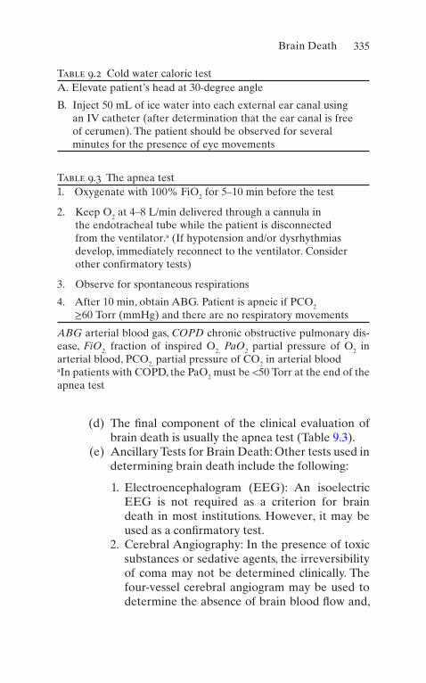
335
(d) The final component of the clinical evaluation of brain death is usually the apnea test (Table 9.3).
(e) Ancillary Tests for Brain Death: Other tests used in determining brain death include the following:
1. Electroencephalogram (EEG): An isoelectric EEG is not required as a criterion for brain death in most institutions. However, it may be used as a confirmatory test.
2. Cerebral Angiography: In the presence of toxic substances or sedative agents, the irreversibility of coma may not be determined clinically. The four-vessel cerebral angiogram may be used to determine the absence of brain blood flow and,
Table 9.2 Cold water caloric testA. Elevate patient’s head at 30-degree angle
B. Inject 50 mL of ice water into each external ear canal using an IV catheter (after determination that the ear canal is free of cerumen). The patient should be observed for several minutes for the presence of eye movements
Table 9.3 The apnea test1. Oxygenate with 100% FiO2 for 5–10 min before the test
2. Keep O2 at 4–8 L/min delivered through a cannula in the endotracheal tube while the patient is disconnected from the ventilator.a (If hypotension and/or dysrhythmias develop, immediately reconnect to the ventilator. Consider other confirmatory tests)
3. Observe for spontaneous respirations
4. After 10 min, obtain ABG. Patient is apneic if PCO2 ≥60 Torr (mmHg) and there are no respiratory movements
ABG arterial blood gas, COPD chronic obstructive pulmonary dis-ease, FiO2, fraction of inspired O2, PaO2 partial pressure of O2 in arterial blood, PCO2, partial pressure of CO2 in arterial bloodaIn patients with COPD, the PaO2 must be <50 Torr at the end of the apnea test
Brain Death

336
thus, the irreversible nature of coma, confirming the diagnosis of brain death.
3. Cerebral Radionuclide Studies: Technetium 99 (T99) nuclear imaging studies of the cerebral cir-culation have been used in some centers as cor-roborative tests in determination of brain death. This procedure is not as sensitive for cerebral blood flow as four-vessel arteriography in deter-mination of brain death.
4. Transcranial Doppler: This determines the blood flow patterns through the anterior and posterior brain’s circulatory system and can be used as an ancillary test for diagnosing cerebral circulatory arrest.
5. Computed Tomography Angiography: This modal-ity has been suggested as a cheaper and more available alternative to ancillary tests. Despite these advantages, this modality needs further vali-dation before being standardized, as the current level of evidence is variable and of low quality.
(f) The patient who is brain dead is DEAD. The physician does not require any permission of the family or other individuals to remove a dead patient from mechanical ventilation or other life support maneuvers.
Coma
A. Definition. Coma is a term denoting neurologic unrespon-siveness. It represents part of a continuum from normal functioning to the absence of neurologic functioning with intermediate states of drowsiness and stupor. Consciousness is separated into two components: level of arousal and the content of consciousness.
1. Level of ArousalLevel of arousal depends upon the interaction between the reticular activating system of the brain stem and the cerebral hemispheres bilaterally.
Chapter 9. Neurologic Disorders
337
2. Content of Consciousness Exists Within the Cerebrum. These two components—content and consciousness—may be affected individually. For example, dyskinetic mutism is a term applied to patients who appear awake (with open eyes, which, on occasion, may even track movements within the room) but have an absence of the content of consciousness. An individual who suffers basilar artery occlusion may develop the “locked-in” syndrome. In this syndrome, the content of conscious-ness is preserved, but the ability to communicate directly with the environment is absent. Glasgow Coma Scale provides a useful system to assess the level of consciousness in response to a specific stimulus (see Table 9.4).
B. Etiology. Coma is a frequent cause of hospital admission. Common causes of coma are depicted in Table 9.5.
C. Diagnosis
1. Careful History and Physical ExaminationHistory should include information leading up to the discovery of the comatose patient. Pertinent points in the physical examination include evidence of head trauma (e.g., hemotympanum), cerebrospinal fluid (CSF) rhinorrhea, contusions, or lacerations. A com-plete neurologic examination looking for focal signs should be recorded.
2. Toxic–Metabolic PhenomenaToxic–metabolic phenomena are found eventually to be responsible for the majority of patients with coma without obvious cause and thus evaluation for hypo- or hyperglycemia, hypo- or hypernatremia, renal failure, liver dysfunction with subsequent hepatic encephalop-athy, and toxin ingestions as the cause of coma should be sought.
3. Computed Tomography (CT) Scan of the HeadMass lesions, supratentorial or in the posterior fossa, may be found unexpectedly at CT scan and account for unconsciousness. Thus, all patients with coma of
Coma

338
unknown etiology should have neuroimaging studies completed.
4. Lumbar PuncturePatients without evidence of mass lesion should undergo CSF examination primarily to rule out infec-tion. Specific diagnostic studies are noted in Table 9.6.
D. Treatment. Always remember the ABCs (airway, breath-ing, and circulation).
1. Comatose patients with absent airway-protective reflexes should undergo endotracheal intubation (with
Table 9.4 Glasgow Coma ScaleBehavior Response ScoreEye opening Spontaneously 4
To speech 1
To pain 2
None 3
Verbal response Oriented to time, place, and person
5
Confused 4
Inappropriate words 3
Incomprehensible sounds 2
None 1
Motor response Obeys commands 6
Localize pain 5
Flexion withdrawal from pain 4
Abnormal flexion (decorticate) 3
Abnormal extension (decerebrate)
2
None 1
Total score: best response, 15; comatose, 8 or less; unresponsive, 3
Chapter 9. Neurologic Disorders

339
assisted mechanical ventilation in those patients with inadequate spontaneous ventilations).
(a) Circulation: Assessment of blood pressure and pulse rate to determine the adequacy of cardiovas-cular function.
2. Specific management is dictated by the clinical condi-tion of the patient.
(a) The patient with an infectious source should be treated aggressively with intravenous (IV) antibi-
Table 9.5 Common causes of coma
Cerebrovascular accidents
Central nervous system (CNS) trauma
CNS infections
Drug intoxication
Metabolic
Metastatic or primary CNS neoplasia
Systemic infection (sepsis)
Unknown
Table 9.6 CSF studies in patients with coma of unknown etiologyTube I
Cell count with differential
Tube II
Glucose, protein
Tube III
Gram’s stain, acid fast bacilli (AFB) stain, routine cultures, India ink or cryptococcal antigen, pneumococcal antigen, meningococcal antigen, VDRL
Tube IV
Special studies as indicated (lactic acid, rheumatoid factor, etc.)
Coma

340
otics. Patients with mass lesions should be consid-ered for early surgical intervention.
(b) Patients with toxic–metabolic events should receive appropriate therapy with close monitoring of electrolytes and/or drug levels.
(c) Many clinicians recommend empiric therapy of the comatose patient with naloxone, a narcotic antago-nist (2–8 mg IV), and dextrose (50 g IV push). Intranasal naloxone has shown to be as effective as intravenous naloxone. However, some data suggest that high levels of glucose may be deleterious to injured neurons, and thus, with the advent of bed-side glucose testing, many would advocate the determination of blood glucose before the admin-istration of dextrose.
(d) Flumazenil: A specific benzodiazepine antagonist is also available; however, in the absence of specific knowledge of benzodiazepine overdose, we do not recommend its administration because of its poten-tial for seizures in patients with tricyclic antide-pressant overdose.
3. Nonspecific Management
(a) Intravenous access for the administration of medications.
(b) Nasogastric (NG) decompression should be con-sidered through an NG tube.
(c) A urinary catheter should be placed for urine mon-itoring and ease of nursing care for the comatose patient.
Deep venous thrombosis prophylaxis should be administered to all patients in whom no contrain-dications exist (i.e., heparin 5000 U SQ q8–12 h or low molecular weight heparin 0.5 mg/kg or 40 mg once daily).
(d) Care and comfort measures (including lubrication of conjunctival spaces and eye taping).
Chapter 9. Neurologic Disorders
341
(e) Passive range of motion of upper and lower extremities, for the prevention of contractures.
(f) Skin care (including frequent turning and positioning).
Intracranial Hypertension
A. Physiology
1. The contents of the cranial vault include the brain, the CSF, and the cerebral blood volume. These contents are constrained by the skull itself.
2. The brain is a highly metabolic organ and very depen-dent on continued blood supply.
3. Because of the closed nature of the cranial vault, cere-bral blood flow is dependent upon the difference between mean arterial pressure and intracranial pres-sure (ICP).
B. Etiology. A large number of intracranial processes may result in a rise in ICP and impairment of cerebral blood flow (see Table 9.7). These specific entities may require individualized therapy, and they are discussed in other sections of this book.
Table 9.7 Causes of intra-cranial hypertension
Brain tumors
Fulminant hepatic failure
Head injury
Meningitis and/or encephalitis
Subarachnoid hemorrhage
Vasculitis
Others
Intracranial Hypertension

342
C. Management
1. ABCsSpecial attention should be paid to patients requiring endotracheal intubation, as positioning and certain sedatives and paralytics can lead to increased intracra-nial pressure.
2. Positioning of the PatientA 30° head-up tilt is recommended for those patients who do not have a contraindication (i.e., hypotension).
3. HyperventilationThe fastest way to control intracranial pressure is hyperventilation. Acute reductions in arterial PCO2 result in vasoconstriction and decrease in intracranial blood volume. Specific PCO2 values of approximately 25–35 Torr (mmHg) are commonly advocated, although to our knowledge, no controlled studies have demonstrated the utility of these specific target values.
4. Osmotic AgentsHypertonic saline and mannitol are common agents. Cumulative evidence has shown that hypertonic saline has higher efficacy than mannitol in reducing intracra-nial pressure, thus is now considered first-line agent.
Hypertonic saline is available in different levels of tonicity (2–23%). It is preferably administered through a central line to achieve a target serum Na of 155 mmol/L. Frequent measurements of serum Na and osmolality (e.g., every 6 hours) are required.
Mannitol, at doses of 0.25 g–1 g/kg of ideal body weight (IBW) intravenously over 10–20 min, pulls water from the brain and results in a decrease in ICP. Plasma osmolality should be maintained below 340 mOsm/L. Note: The initial administration of man-nitol may result in paradoxical increases in ICP; thus, many authors recommend prior therapy with a loop- acting diuretic (i.e., furosemide).
5. Anesthetics and SedativesBarbiturates at first were popular because of their capacity to decrease brain metabolism and cerebral
Chapter 9. Neurologic Disorders

343
blood flow, thus lowering ICP. The therapeutic value of barbiturates is controversial, as its side effects may outweigh benefits (severe hypotension requiring vaso-pressors). Patients requiring mechanical ventilation require sedation; a good agent to use is propofol. It is easily titrated and has a short half-life, permitting a frequent neurologic assessment.
6. CorticosteroidsThe only clear role of corticosteroids in the manage-ment of intracranial hypertension is in cerebral edema secondary to certain neoplasms. Their use in trauma, cerebrovascular accidents (CVAs), and metabolic causes has not been demonstrated to improve out-come and therefore cannot be routinely recom-mended. Indeed, a large randomized clinical trial showed worse outcome with their use in patients with moderate to severe head injury.
7. CSF Drainage and ICP MonitoringAn intraventricular catheter may be placed percuta-neously at the bedside and permit simultaneous moni-toring and therapy of ICP. Sustained elevations of ICP >20 cmH2O can be managed by withdrawal of CSF through the intraventricular catheter.
8. Positive End-Expiratory Pressure (PEEP)Some authors have advocated not using PEEP; how-ever, in conditions in which this therapy is required (i.e., low lung compliance), routinely used levels of PEEP (3–7 cmH2O) are not expected to impair cere-bral blood drainage.
9. Surgical ManagementThe most common surgeries for intracranial hyperten-sion are the optic nerve sheath fenestration and cere-brospinal fluid shunting.
10. Therapeutic HypothermiaAlthough still not universally accepted and certainly not the standard of care, therapeutic hypothermia has been used with success in cases of intracranial hyper-tension refractory to medical management. For fur-ther details about this technique, please see Chap. 15, “Special Techniques.”
Intracranial Hypertension

344
Cerebrovascular Disease
A. Epidemiology. Approximately 700,000 people have a new or recurrent cerebrovascular disease each year. Stroke is still the third leading cause of death and leading cause of disability in the United States.
B. Classification. A number of different syndromes comprise the disorders labeled cerebral vascular accidents (CVAs). These disorders can be broadly grouped into two large categories: (1) those that produce vascular insufficiency (secondary to thrombosis, embolism, or stenosis leading to focal areas of ischemia) and (2) those that produce rup-tures of the vascular tree, causing intracranial hyperten-sion and secondary cerebral ischemia.
1. Vascular Insufficiency
(a) Transient Ischemic Attacks (TIAs): TIAs are defined as the sudden or rapid onset of neurologic deficits secondary to cerebral ischemia that lasts from a few minutes to up to 24 h without residual signs or symptoms. Atherosclerosis is the most fre-quent cause.
(b) Stroke
1. Definition: Stroke is the rapid onset of neuro-logic deficits involving a set of vascular territory with neurologic signs and symptoms lasting >24 h.
2. Risk Factors: Similar to those for TIA. An increasing frequency of stroke related to vaso-spasm secondary to cocaine abuse has been noted in the last decade in the United States.
3. Classification of Stroke: Both thrombosis and embolism may result in vascular insufficiency and the phenomena of stroke. Clinically, the dif-ferentiation of thrombosis and embolism is quite difficult. However, some clinical charac-teristics of each are noted in Table 9.8.
Chapter 9. Neurologic Disorders

345
4. Embolic Stroke: The most common causes of embolic stroke include emboli secondary to atrial fibrillation, valvular heart disease, bacte-rial and nonbacterial endocarditis, trauma sec-ondary to myocardial infarction or ventricular aneurysm, atrial myxoma, and paradoxical embolism secondary to endocardial disease.
(a) Thrombotic Stroke: Occurs when a clot develops in a cerebral vessel. Intrinsic or extrinsic diseases of the cerebral vessels may contribute to thrombotic strokes. These include the following:
I. Arteriosclerosis. II. Fibromuscular dysplasia. III. Extension of embolism or dissection
into cerebral arteries because of arteri-tis (Takayasu’s disease, giant cell arteri-tis, and other vascular diseases).
IV. Increased viscosity secondary to pro-teins or increased cellular elements (i.e., Waldenstrom’s macroglobulin-
Table 9.8 Clinical characteristics of embolic and thrombotic strokesEmbolism Thrombosis
Predisposing factors
Valvular heart disease
Atherosclerosis
Endocarditis Diabetes
Myocardial infarction
Hypertension
Atrial fibrillation Arteritis
History of prior TIA
Uncommon Common
Onset of symptoms
Rapid onset Progression over hours
Cerebrovascular Disease

346
emia, leukemias with elevated blast counts, and erythrocytosis of any cause).
V. TIAs are a risk factor for completed stroke, with the highest risk being in the first 3 months immediately following the onset of TIAs.
5. Initial evaluation and the management of cere-bral ischemic syndromes.
(a) ABCs: Secure the airway and assist with breathing and circulation as with any other patient presenting with a potentially critical illness.
(b) Careful Examination of the Patient: Emphasis should be on the neurologic examination to localize the area of deficits and on other areas of the physical examina-tion to rule in or rule out secondary causes for the ischemic syndrome.
(c) Laboratory Evaluation: Complete blood count, prothrombin time (PT), partial thromboplastin time (PTT), glucose, elec-trolytes, serum urea nitrogen (BUN), and creatinine are routinely ordered. Chest radiograph, ECG, and CT scan of the head should be done without delay (to rule out hemorrhage, infarction, subdural hema-toma, or intracranial masses).
(d) In any patient with new neurologic abnor-malities, lumbar puncture should be consid-ered to rule out infectious causes and for the completion of the evaluation for sub-arachnoid hemorrhage (after head CT scan has ruled out increased intracranial pressure).
(e) Patients with CVA can be at risk of aspira-tion. Therefore, NPO order should be immediately placed, until dysphagia evalua-tion is performed.
Chapter 9. Neurologic Disorders

347
(f) Echocardiography: For patients with a his-tory or physical examination suggestive of cardiac abnormality, echocardiography should be ordered.
(g) CT angiography or magnetic resonance angiography (MRA) is commonly utilized to study the carotid and vertebral arteries, especially in embolic CVAs.
(h) Other useful studies may include carotid duplex ultrasonography and cerebral angiography.
6. In patients with embolic CVAs with progres-sively worsening neurologic deficits (stroke in evolution), anticoagulation, beginning with hep-arin, is recommended. In addition, heparin is commonly prescribed for patients with recur-rent TIAs despite antiplatelet therapy. Note: Anticoagulation should be started (with hepa-rin) in patients with worsening neurologic defi-cits (stroke in evolution or suspected embolic source). Anticoagulation is contraindicated in patients with CT or LP evidence of hemorrhage, and it is relatively contraindicated in patients with gastrointestinal (GI) bleeding or coagulation disorders and in patients with hypertension.
7. Blood Pressure Control: Therapy to maintain systemic blood pressure at approximately 150/100 mmHg is advocated. Caution must be exercised, as severely elevated systolic blood pressure increases risk of recurrence of CVA, and reductions in blood pressure may worsen the clinical condition by producing ischemia in poorly perfused regions of the central nervous system (CNS).
(a) Thrombolytic therapy for ischemic stroke is now the standard of care. Intravenous
Cerebrovascular Disease

348
recombinant tissue plasminogen activator (rtPA) is the only FDA-approved medical therapy proven to reduce the effects of an ischemic stroke. The best results from rtPA are seen when given within 3–4.5 h of onset of symptoms.
8. Therapeutic HypothermiaTherapeutic hypothermia has shown benefits against ischemic brain injury; however, addi-tional quality studies are needed before its true role is fully known.
2. Rupture of the Vascular Tree
(a) Subarachnoid hemorrhage (SAH) accounts for about 10% of all strokes and 16–20% of cerebral vas-cular deficits. The etiology of SAH includes ruptured aneurysms of cerebral vessels, bleeding from arterio-venous malformations of the CNS, and trauma.
1. Clinical Manifestations
(a) Neurologic deficits may include focal neu-rologic signs as well as coma.
(b) Generalized excruciating headache with neck stiffness is classically described.
2. Evaluation and Management
(a) ABCs, as noted previously. (b) CT scan of the head demonstrating sub-
arachnoid blood is seen in approximately 90% of the cases.
(c) Lumbar puncture should be performed in those patients whose CT scan is negative and for whom clinical suspicion of SAH is still high.
(d) The patient should be kept at bed rest. Cardiac monitoring and frequent (q1–2 h) neurologic assessments should be ordered.
Chapter 9. Neurologic Disorders

349
(e) Reversal of all antiplatelets and anticoagu-lation is recommended until surgical repair of the ruptured vessel. Antiplatelets can be reversed with single dose of desmopressin (0.4 mcg/kg). Patients on warfarin should receive prothrombin complex concentrate or fresh frozen plasma immediately. Currently, antidotes for direct oral antico-agulants are also available (Idarucizumab for dabigatran, andexanet alfa for rivaroxa-ban and apixaban).
(f) Analgesia for headache should be pre-scribed (acetaminophen and codeine are commonly used).
(g) Glycemic control is desired in patients with ICH; hyperglycemia may worsen brain injury. It is recommended to treat with insu-lin patients who reach blood sugar levels >145 mg/dL.
(h) Stool softeners and mild laxatives should be prescribed to prevent constipation (and thus increased ICP).
(i) Blood pressure control: Keep blood pres-sure in ranges that maintain CPP >60 mmHg (CPP = MAP–ICP). If needed, an antihy-pertensive agent may be used; intravenous labetalol, intravenous nicardipine, or intra-venous clevidipine are the drugs of choice in this clinical setting.
(j) Surgical management: With the evolution of microsurgical techniques, the surgical man-agement of cerebral aneurysms is an effec-tive and safe procedure.
(k) Endovascular therapy: Intraluminal approach (using platinum coils) is an effec-tive alternative to surgical clipping. A coil is inserted into the lumen of the aneurysm; a local thrombus then forms around the coil, obliterating the aneurysmal sac.
Cerebrovascular Disease

350
(b) Intracerebral Hemorrhage: Intracerebral hemor-rhage commonly occurs following trauma. When it occurs spontaneously, it is frequently accompanied by hypertension. Neurologic abnormalities, as seen in other types of strokes, are usually present, and the specific diagnosis requires neuroimaging studies.
1. Management
(a) ABCs, as required for every critically ill patient.
(b) Control severe hypertension: As noted above, cerebral ischemia may occur with reductions in blood pressure. However, con-trol of hypertension may reduce cerebral edema and improve neurologic function.
(c) Reversal of all antiplatelets and anticoagu-lants is also indicated (as noted above).
(d) Additional management of intracranial hypertension as noted above may be required.
(e) Supportive therapy as required for all inten-sive care unit (ICU) patients should continue.
(f) Activated recombinant factor VIIa has been used by some centers for expanding hema-tomas. The data of large trials, however, have mixed results.
(c) Surgical evacuation of the hematoma should occur in patients with accessible lesions, who have pro-gressive signs of deterioration.
Status Epilepticus
A. Definition. Status epilepticus is defined as seizure activity continuing for 5 or 10 min or frequent clinical seizures without an interictal return to the baseline clinical state. It
Chapter 9. Neurologic Disorders
351
is a condition that may lead to permanent neurologic damage or even death.
B. General Approach. The management of seizure disorders is based on clinical information.
1. Most seizures stop spontaneously within 30–90 s. 2. The diagnosis of status epilepticus is straightforward
and can be determined through observation of the patient in most cases.
3. Generalized seizure disorders without motor findings may lead to changes in mental status or coma and may not be clinically apparent, and thus further diagnostic testing (i.e., EEG) may be required.
4. Continued seizure disorders may result in enzyme ele-vation (creatine kinase [CK]), making the diagnosis of other clinical conditions more difficult (i.e., myocardial infarction).
C. Specific Management
1. ABCs: As in all critically ill patients, airway, breathing, and circulation must be maintained. Patients should be positioned so they cannot harm themselves from their motor activity. Oxygen should be administered, and continuous observation of the patient should ensue.
2. Blood glucose, calcium, magnesium, and other elec-trolytes, as well as BUN, liver functions, anticonvul-sant levels, complete blood count, and toxicologic screen should be obtained.
3. A normal saline infusion should be established, and 50 cc of 50% glucose and 100 mg of thiamine should be administered intravenously.
4. ECG and blood pressure monitoring should be established.
5. Lorazepam (Ativan) 2 mg IV is the first-line agent, repeated every 3–5 min, to be administered in those patients continuing to seize. Diazepam (Valium) 5 mg over 1–2 min IV, repeated every 5–10 min, is an alter-native agent.
Status Epilepticus

352
6. Reoccurrence of seizures within 15–20 min following administration of benzodiazepines is quite frequent, and other antiepileptic agents should be instituted. Phenytoin is used because of its proven efficacy in pre-venting the frequency of seizures given IV, at a dose of 20 mg/kg and a rate of 50 mg/min and should be administered as a loading dose for patients not previ-ously receiving phenytoin. If dysrhythmias and/or hypotension ensue, the infusion should be stopped and resumed at a slower rate. Phosphenytoin and levetiracetam (Keppra) are other alternatives.
7. Persistent seizures following phenytoin administra-tion should result in administration of phenobarbital IV at rates of 50–100 mg/min until the seizure stops or until a loading dose of 20 mg/kg IBW total has been given.
8. Continued seizures should prompt the administration of other medications: Intravenous propofol (5–30 cc/h) has been used in some cases of refractory seizures with success. Valproic acid has also been used with success, at a dose of 10 mg/kg at a rate of 20 mg/min. In some cases, therapeutic hypothermia has been used for intractable status epilepticus.
9. EEG monitoring is appropriate for patients receiving general anesthetic control of status epilepticus. Continuous EEG monitoring is also granted for patients in whom the initial EEG is not diagnostic.
10. Review of laboratory data and additional history and physical examination for underlying disorders that may have resulted in the status epilepticus should be undertaken. Mainstay of treatment is the identifica-tion and correction of predisposing factors.
(a) Antiepileptic drug noncompliance (b) Withdrawal syndromes (alcohol, barbiturates,
baclofen, benzodiazepines) (c) Acute structural injury (d) Metabolic abnormalities (hypoglycemia, hepatic
encephalopathy, uremia, etc.)
Chapter 9. Neurologic Disorders

353
(e) Use or overdose of drugs that lower seizure threshold (theophylline, imipenem, tricyclic anti-depressants, lithium, clozapine, flumazenil, lidocaine)
11. Following control of the status epilepticus, careful physical and laboratory evaluation for underlying dis-ease processes should ensue. Patients without a clear etiology should undergo head CT scan and lumbar puncture unless contraindicated.
12. Continuous motor seizures may lead to muscle break-down and thus release of myoglobin and other intra-cellular components into the circulation. One must be concerned about maintenance of adequate hydration as well as protection from pigment-induced renal fail-ure (see Chap. 14, “Renal and Fluid-Electrolyte Disorders”).
13. Therapeutic hypothermia. Although the exact mecha-nism of action is unknown, therapeutic hypothermia has shown positive results in treating seizure disor-ders, in particular refractory status epilepticus.
Neuromuscular Disorders
A. Guillain–Barré Syndrome (GBS)
1. Definition. GBS is an acute immune demyelinating dis-order of the peripheral nervous system that results in motor and sensory symptoms with few sensory signs. In the vast majority of cases, it results in complete recov-ery. However, in up to 25% of patients, respiratory fail-ure due to weakness of the respiratory muscles ensues and mechanical ventilation is required for a period of time. Peaks of occurrence are in the 15- to 35- and 50- to 75-year-old age groups.
2. Clinical Manifestations. GBS presents in a typical pat-tern. The usual history is that of a patient with a normal previous health status interrupted by a mild upper
Neuromuscular Disorders

354
respiratory or GI illness followed by ascending weak-ness and numbness. Other factors include recent vacci-nation or surgery. Major clinical manifestations are depicted in Table 9.9. Atypical presentations may include a descending paralysis.
3. Diagnostic Evaluation
(a) Careful physical examination should be performed, attempting to rule out other causes of neuropa-thology (spinal cord lesions, infection, metabolic or toxic, etc.).
(b) Lumbar puncture usually reveals elevated protein. There are usually few mononuclear leukocytes in the CSF with lymphocyte counts <10/cc.
(c) Eighty percent of all patients show slow nerve conduction.
4. Management
(a) ABCs. (b) Supportive measures and close monitoring of
respiratory function with frequent measurements of the vital capacity and/or negative inspiratory force (NIF) are indicated.
Table 9.9 Major clinical manifestations of Guillain–Barré syndromeDistal paresthesias (initially lower extremities)
Rapidly progressive motor weakness (ascending neuropathy)
Symmetry is seldom absolute
Facial weakness is common (one-third of cases)
Recovery usually begins 2–4 weeks after progression stops
Sinus tachycardia and labile blood pressure are common
CSF protein elevation (after the first week)
Nerve conduction abnormalities are detectable
Chapter 9. Neurologic Disorders

355
(c) A vital capacity of <20 mL/kg is an indication to consider intubation and assisted mechanical venti-latory support.
(d) Active and passive range of motion of lower and upper extremities should be performed to prevent the formation of contractures.
(e) Bladder and bowel care should be done as many of these patients develop adynamic ileus and urinary retention.
(f) Decubitus ulcer prevention and care should be instituted.
(g) Prevention of thromboembolism with appropriate therapy (i.e., fractionated or unfractionated hepa-rin and support stockings).
(h) Plasma exchange presumably removes or dilutes circulating factors implicated in the pathogenesis of GBS. It has been shown to decrease ventilatory dependence and earlier recovery in GBS.
(i) Intravenous immunoglobulin (IVIG) is as effective as plasma exchange in GBS.
(j) Corticosteroids have not been proven to be of value in this syndrome.
B. Other Chronic Neurologic Disorders
1. A number of chronic progressive neurologic disorders may result in patient admission to the ICU for physio-logic support. These include amyotrophic lateral sclero-sis, multiple sclerosis, severe Parkinson’s disease, etc. Many of these patients are admitted because of their need for aggressive tracheobronchial toilet or mechani-cal ventilation.
2. Major concerns in managing these patients involve the decision to institute aggressive therapy. It is preferable that these decisions be addressed with the patient and their family before the need for these services, so that unwanted supportive measures are not forced upon them.
Neuromuscular Disorders

356
Delirium in the ICU
A. Epidemiology. Ten percent of medical and surgical patients become delirious during their hospitalization. These patients are at risk for harm to themselves (by the discontinuation of an IV line, arterial line, NG tubes, etc.) and others. Patients who develop delirium are at greater risk of mortality. Patients at high risk for the development of delirium are as follows:
1. Those at the extremes of age (elderly and children) 2. Patients with preexisting brain injury 3. Drug-dependent patients or polypharmacy 4. Postcardiotomy patients 5. Patients with human immunodeficiency virus (HIV)
disease
B. Clinical Features
1. A prodromal state manifested by restlessness, irritabil-ity, anxiety, or sleep difficulty.
2. A rapidly fluctuating course. Patients are intermittently clear thinking and coherent or grossly confused, disori-ented, and disorganized.
3. Reversed sleep–wake cycles and increased activity and confusion during the nighttime hours.
C. Evaluation and Management
1. ABCs, as required for all patients with a critical illness.
2. Careful attention to metabolic problems that may produce CNS disturbances should be sought.
3. Laboratories including blood glucose, electrolytes, calcium, BUN, and liver function tests should be obtained as well as arterial blood gases or pulse oxim-etry to rule out hypoxemia (a common cause of men-tal status change in the ICU).
4. ECG should also be obtained to help rule out myocar-dial ischemia.
Chapter 9. Neurologic Disorders

357
5. For delirium assessment, the use of Confusion Assessment Method (CAM) is useful as a screening tool. The modified version CAM-ICU is helpful screening tool in patients that are mechanically venti-lated and unable to communicate verbally.
6. Patients with unexplained mental status changes should receive CT scanning or magnetic resonance imaging of the brain followed by lumbar puncture to rule out infectious or other causes.
7. Careful review of the medications prescribed for the patient should be undertaken. Drugs commonly asso-ciated with delirium are depicted in Table 9.10.
8. Whether or not the etiology is known, some simple interventions that may help control the patient’s con-fusion and behavior are often missed. For example, if the patient normally wears eyeglasses or a hearing aid, return these items. The old practice of placing delirious patients together is not helpful, and indeed, it may increase the aggressive behavior of both patients, thus, making orientation almost impossible. Physical restraints should be used as a last resort, if at all; they frequently increase agitation and may cause physical harm to the patients.
9. Haloperidol (Haldol) is a highly potent antipsychotic agent that effectively calms agitation, sedates, and reduces hallucinations and paranoid thinking. For the patient with a mild level of delirium or agitation, a starting dose of 0.5–2 mg IV or IM is usually enough. However, for patients with severe agitation, a starting dose of 5–10 mg may be necessary. An interval of 20–30 min should be allowed between doses. After giving three doses of haloperidol with no improve-ment in symptomatology, give 1–2 mg IV lorazepam (Ativan) concurrently or alternate with haloperidol every 30 min. Assuming the patient is calm, reduce the dose by 15% every 24 h. Note: Haloperidol is not approved for IV use despite its common use for this indication. Large IV doses have been used in critically
Delirium in the ICU
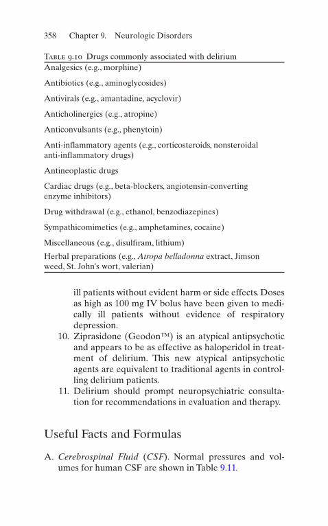
358
ill patients without evident harm or side effects. Doses as high as 100 mg IV bolus have been given to medi-cally ill patients without evidence of respiratory depression.
10. Ziprasidone (Geodon™) is an atypical antipsychotic and appears to be as effective as haloperidol in treat-ment of delirium. This new atypical antipsychotic agents are equivalent to traditional agents in control-ling delirium patients.
11. Delirium should prompt neuropsychiatric consulta-tion for recommendations in evaluation and therapy.
Useful Facts and Formulas
A. Cerebrospinal Fluid (CSF). Normal pressures and vol-umes for human CSF are shown in Table 9.11.
Table 9.10 Drugs commonly associated with deliriumAnalgesics (e.g., morphine)
Antibiotics (e.g., aminoglycosides)
Antivirals (e.g., amantadine, acyclovir)
Anticholinergics (e.g., atropine)
Anticonvulsants (e.g., phenytoin)
Anti-inflammatory agents (e.g., corticosteroids, nonsteroidal anti-inflammatory drugs)
Antineoplastic drugs
Cardiac drugs (e.g., beta-blockers, angiotensin-converting enzyme inhibitors)
Drug withdrawal (e.g., ethanol, benzodiazepines)
Sympathicomimetics (e.g., amphetamines, cocaine)
Miscellaneous (e.g., disulfiram, lithium)
Herbal preparations (e.g., Atropa belladonna extract, Jimson weed, St. John’s wort, valerian)
Chapter 9. Neurologic Disorders

359
The normal composition of the CSF is depicted in Table 9.12.
Additional normal and abnormal values for CSF in humans are depicted in Tables 9.13 and 9.14.
When there are many red blood cells (RBCs) or white blood cells (WBCs) in the CSF, the total protein of the CSF may be corrected utilizing the following formula: Protein actual
Total Protein in CSF
Protein Hct RBC
RBCserum CSF
blood
�� �� ��1
Common CSF abnormalities in patients with multiple scle-rosis are depicted in Table 9.13.
The abnormalities in immunoglobulin G (IgG) production in these patients can be estimated by the IgG index:
Ig G index
CSF IgG CSF albumin
Serum IgG serum albuminnormal� � �
/
/.0 666
B. Cerebral Blood Flow. The cerebral circulation follows the same physiological principles of other circulatory beds, such as Ohm’s law:
Table 9.11 Normal CSF pressures and volumes
CSF pressure Children 3.0–7.5 mmHg
Adults 3.5–13.5 mmHg
Volume
Infants 40–60 mL
Young children
60–100 mL
Older children
80–120 mL
Adult 100–160 mL
Useful Facts and Formulas

360
F
P P
R�
�i o
where F = flow; Pi = input pressure; Po = outflow pressure; R = resistance. The term “Pi –Po” is referred to as the cerebral perfusion pressure (CPP).
Table 9.12 Normal composition of the CSFCSF concentration (mean)
Specific gravity 1.007
Osmolality (mOsm/kg H2O) 289
pH 7.31
PCO2 (mmHg) 50.5
Na+ (mEq/L) 141
K+ (mEq/L) 2.9
Ca++ (mEq/L) 2.5
Mg++ (mEq/L) 2.4
Cl− (mEq/L) 124
Glucose (mg/dL) 61
Protein (mg/dL) 28
Table 9.13 Normal CSF values
CSF parameter NewbornsInfants, older children, and adults
Leukocyte count <32/μL <6/μL
Differential white cell count
<60% polymorphs
<1 polymorph
Proteins <170 mg/dL <45 mg/dL
Glucose >30 mg/dL >45 mg/dL
CSF: blood/glucose ratio
>0.44 >0.5
Chapter 9. Neurologic Disorders

361
The CPP can be estimated by the following formula:
CPP MAP ICP� �
where MAP = mean arterial pressure and ICP = intracranial pressure.
The pressure–volume index (PVI) can be calculated as follows:
PVI p o� �� ���V P P/ log /
where Pp = peak CSF pressure (increase after volume injec-tion and decrease after volume withdrawal).
The cerebral blood flow (CQ) is normally 50 mL/100 g/min and is determined by the Hagen–Poiseuille equation of flow through a tube:
CQ
r�
�� ��� �
K P
L
4
8 �
where Pp = cerebral perfusion pressure (CPP), r = the arterial radius, η = blood viscosity, L = arterial length, and K = constant.
C. Brain Metabolism. Oxygen availability to neural tissue (CDO2) is reflected in the formula:
Table 9.14 CSF abnormalities in multiple sclerosis
Alb (%)
IgG/TP (%)
IgG/Alb (%)
IgG index (%)
Oligoclonal banding of Ig (%)
Multiple sclerosis
25 67 60–73 70–90 85–95
Normal subjects
3 – 3–6 3 0–7
Alb albumin, IgG/TP IgG value/total protein, Ig immunoglobulin
Useful Facts and Formulas
362
CDO CQ PaO2 2� �
where CQ = cerebral blood flow and PaO2 = arterial oxy-gen concentration.
The cerebral metabolic rate (CMRO2) can be calculated as follows:
CMRO CBF AVDO2 2� �
where CBF = cerebral blood flow and AVDO2 = arteriove-nous oxygen content difference.
The oxygen extraction ratio (OER) can be utilized to assess the brain metabolism:
OER SaO SjvO SaO� �2 2 2/
where SaO2 = arterial oxygen saturation and SjvO2 = jugular venous oxygen saturation.
OER CaO CMRO CBF� �2 2 /
where
CaO Hb SaO PO mmHg2 2 21 39 0 003� � �� � � � � ��� ��. .
CMRO CBF CaO CjvO2 2 2� � �� �
The arterial–jugular venous oxygen content difference (AjvDO2) is calculated as follows:
AjvDO CMRO CBF2 2= /
Chapter 9. Neurologic Disorders

363© Springer Nature Switzerland AG 2021J. Varon, Handbook of Critical and Intensive Care Medicine, https://doi.org/10.1007/978-3-030-68270-5_10
Aims of Nutritional Support
A. Preserve tissue mass and decrease usage of endogenous nutrient stores.
B. Decrease catabolism. C. Maintain/improve organ function.
1. Immune 2. Renal 3. Hepatic 4. Muscle
D. Improve wound healing. E. Decrease infection. F. Maintain gut barrier (decrease translocation). G. Decrease morbidity/mortality.
1. Decrease ICU/hospital stay. 2. Decrease hospital costs.
Timing of Nutritional Support
A. Optimal timing remains controversial.
Chapter 10Nutrition
364
1. Some patients tolerate short periods of starvation by using endogenous stores to support body functions.
2. Well-nourished patients (nonstressed) have actually survived without food for 6 weeks (ingesting only water).
3. Hypermetabolic and hypercatabolic critically ill patients can probably only tolerate a few weeks of star-vation before death.
4. There appears to be no benefit of total starvation.
B. Accumulating data suggest that outcome can be improved with early and optimal nutritional support.
1. Early nutritional support blunts the hypercatabolic/hypermetabolic response to injury.
2. In a growing number of studies, patients randomized to receive early vs. delayed feeding had decreased infec-tion rates, fewer complications, and shorter length of stay in the hospital.
3. Animal studies report improved wound healing and improved hepatic function in several injury models with early feeding.
Route of Nutritional Support
A. Parenteral Nutrition
1. NutrientsAmino acids, dextrose, soy-based lipids, vitamins,
electrolytes, minerals, and trace elements (see Table 10.1)
2. Delivery: Via peripheral or central vein 3. PN major complications (mechanical/technical, septic/
infection, and/or metabolic)
(a) Mechanical/technical (improper central line place-ment that may cause pneumothorax, hemothorax, carotid artery perforation, etc. or venous thrombo-sis or an air embolism after access established)
Chapter 10. Nutrition

365
(b) Septic/infection (increased infection rates due to catheter-related sepsis, septic thrombosis, pneumo-nia, abscesses, immune suppression, etc.)
(c) Metabolic derangements (hyperglycemia, hypogly-cemia, electrolyte disturbances, refeeding syn-drome/overfeeding, gut atrophy (i.e., diarrhea, bacterial translocation), liver dysfunction (i.e., fatty infiltration, cholestasis, liver failure, increased liver enzymes)
4. PN indicationsPN is the preferred method of nutrition support
when the GI tract is either not working, not available, or not appropriate.
(a) Nonfunctioning gut (i.e., paralytic ileus), short gut syndrome, bowel obstruction, some fistulas, severe necrotizing pancreatitis when EN is not possible, etc.
5. Additional information in regard to PN
(a) PN may lack of some conditionally essential amino acids that are not stable in solution (i.e., glutamine, cysteine).
Table 10.1 Comparison of nutrients in enteral vs. parenteralNutrient Enteral ParenteralNitrogen source Intact proteins, peptides, or
amino acidsAmino acids
Carbohydrate Simple sugars or complex carbohydrates (i.e., starch and fiber)
Simple sugar (dextrose)
Lipids Long- and medium-chain triglycerides or long-chain fatty acids (ω-3 or ω-6)
Soy-based lipids
Vitamins Present Can be added
Minerals and trace elements
Present Can be added
Route of Nutritional Support

366
(b) Glucose/fat ratio.
(i) Usually 60:40 to 40:60 (ratio of calories from each source)
(ii) Large amounts of glucose (>60% of calories) can:
1. Increase energy expenditure. 2. Increase CO2 production and increase pul-
monary workload (may delay ventilator weaning).
3. Produce liver steatosis. 4. Lead to immune compromise.
B. Enteral Nutrition
1. Nutrients (see Table 10.1)
(a) Nitrogen sources: Amino acids, peptides, or intact proteins (e.g., casein, whey, soy, lactalbumin)
(b) Carbohydrates: Simple sugars or complex carbohy-drates (i.e., starch and fiber)
(c) Lipids: Long- or medium-chain triglycerides, ω-3 or ω-6 long-chain fatty acids
(d) Vitamins (e) Electrolytes (f) Minerals and trace elements
2. Delivery
(a) EN Delivery Methods (Continuous or Intermittent (Bolus) Feedings)
EN may be provided via continuous feeds (i.e., 2–4 h drip) or via intermittent (bolus) feeds (i.e., receive a small dose of formula and then wait a while (i.e., Q4hrs) before taking another dose).
(b) EN Delivery Routes (Gastric or Small Bowel)EN may be infused through either a gastric tube
(i.e., nasogastric tube (NGT), gastrostomy tube (G-tube/PEG tube), small bowel feeding tube (i.e., nasoduodenal tube (ND), nasojejunal tube (NJ), gastrostomy–jejunostomy tube (GJ tube), or jeju-nostomy tube (J tube)).
Chapter 10. Nutrition

367
3. EN Major Complications (Mechanical, GI Complications/Metabolic Derangements)
(a) Mechanical (inability to gain access/misplaced feeding tube) that may lead to bowel perforation, reflux of gastric contents causing aspiration (pneu-monia, chemical pneumonitis, adult respiratory distress syndrome (ARDS)), and high-output proximal fistulas/gastric leakage
(b) GI complications including delayed gastric empty-ing, constipation/diarrhea, intractable nausea/vomiting, malabsorption, overfeeding/underfeed-ing, and metabolic derangements (much less likely than in PN)
4. EN IndicationsEN is the preferred method of nutrition support if
the gut is working.
(a) Inadequate or compromised oral intake due to chemotherapy/radiation, CNS disorders, poor po intake, etc.
(b) Increased nutrient requirements secondary to burns, trauma, sepsis, surgical, or medical stress
(c) Digestive and absorptive disorders (i.e., inflamma-tory bowel disease, pancreatitis, etc.)
(d) Metabolic and excretory disorders (i.e., liver fail-ure, renal failure, glycogen storage disease)
5. Advantages of EN
(a) Physiologic advantages allow nutrients to be metabolized and utilized more effectively in EN vs. PN (i.e., the gut and liver process enteral nutrients before their release into systemic circulation, help-ing to maintain homeostasis of the amino acid pool and skeletal muscle tissue).
(b) Immunologic advantages show that EN helps main-tain optimal gut integrity/function (i.e., mainte-nance of gut barrier, gut-associated immune system, preventing bacterial translocation from the gut, immunoglobulin A (IgA) secretion, mucin layer).
Route of Nutritional Support

368
(c) EN is safer than PN due to EN avoiding central catheter placement-related problems/complica-tions (i.e., catheter sepsis, pneumothorax, etc.).
(d) EN is cheaper than PN and has less side effects compared to PN.
C. Enteral Versus Parenteral Nutrition
1. Enteral nutrition is the preferred method of nutrition support if the gut is working (“if the gut works, use it”).
2. PN is indicated when EN is not possible (i.e., inade-quate small bowel function, etc.).
3. EN is associated with decreased infectious complica-tions in critically ill adults compared to PN.
4. PN is associated with high mortality rates in patients receiving chemotherapy/radiation and/or patients after burn injuries compared to EN.
5. Limited evidence states that early EN decreases hospi-tal length of stay (LOS) in critically ill adult patients compared to PN.
6. Cost of care is cheaper for EN vs. PN. 7. Data in obese patients is limited.
Gastrointestinal Function During Critical Illness
A. Oral nutrition remains the best form of nutritional sup-port; however, in many critically ill patients, this is not possible.
B. Decreased motility of the stomach and colon is common and typically lasts 5–7 days in critically ill patients (longer if the patient remains critically ill).
C. Motility and nutrient absorptive capability of small bowel are usually preserved (even after severe trauma, burns, or major surgery).
D. Bowel sounds are a poor index of small bowel motility in critical care patients.
Chapter 10. Nutrition

369
Nutrient Requirements (Quantity)
A. Energy
1. Caloric content of major nutrients
(a) Lipids provide 9 kcal/g. (b) Carbohydrates provide 4 kcal/g (or 3.4 kcal/g via
parenteral nutrition). (c) Proteins provide 4 kcal/g.
2. Studies show that most critically ill patients expend 25–35 kcal/kg/day.
3. One can estimate resting metabolic rate (RMR) using the Harris–Benedict equation or the Mifflin–St. Jeor equation (MSJ) (see Table 10.2).
4. One can also measure RMR by indirect calorimetry (metabolic cart or by using the Penn State equation (PSU); see Table 10.2).
5. Some recommend adjusting RMR by multiplying by a correction factor (see Table 10.3); however, correction factors frequently overestimate energy needs.
6. We prefer to initially administer 25 kcal/kg/day (see Table 10.4).
(a) ≈20% protein (percent refers to percentage of total daily calories)
(b) ≈30% lipids (c) ≈50% carbohydrates
7. Patients with organ failure/disease states may have increased or decreased needs and should be considered individually.
8. Overfeeding (with either enteral or parenteral nutri-ents) is associated with more adverse side effects than slightly underfeeding during most critical illnesses.
B. Protein
1. The sicker the patient, the higher the protein requirement.
Nutrient Requirements (Quantity)

370
2. Patient with mild to moderate illness are given 0.8–1.2 g/Kg of protein per day.
3. Critically ill patients usually need 1.2–2.0 g/Kg/day. 4. Protein requirements increase in patients with severe
trauma, burns, and protein-losing enteropathies.
C. Water
1. Must be individualized, as needs vary greatly between patients (differences in insensible losses, GI losses, and urine losses).
2. Initially estimate: 1-mL water per kilocalorie of energy in adults.
Table 10.2 Resting metabolic ratio formulasHarris–Benedict equation
Men RMR (kcal/day) = 66 + (13.7 × W) + (5 × H) (6.8 × A)
Women RMR (kcal/day) = 665 + (9.6 × W) + (1.7 × H) (4.7 × A)
Mifflin–St. Jeor equation (MSJ)
Men RMR (kcal/day) = (9.99 × W) + (6.25 × H) + (4.92 × A) + 5
Women RMR kcal day W H
A
/ . .
.
� � � �� � � �� �� �� � �
9 99 6 25
4 92 1615
Penn State equation (PSU)
Mechanically ventilated patients
RMR kcal day MSJ Tmax
VeTot
/ .� � � �� � � �� �� �� � �
0 96 167
31 6212
Older obese mechanically ventilated patients
RMR kcal day MSJ Tmax
VeTot
/ .� � � �� � � �� �� �� � �
0 71 85
64 3085
A age in years, H height in cm, RMR resting metabolic rate, W weight in kg, MSJ Mifflin–St. Jeor, Tmax temperature in degrees Celsius, VeTot minute ventilation
Chapter 10. Nutrition

371
D. Vitamins
1. Fat-soluble vitamins: A, D, E, and K. 2. Water-soluble vitamins: Ascorbic acid (C), thiamine
(B1), riboflavin (B2), niacin, folate, pyridoxine (B6), B12, pantothenic acid, and biotin.
3. Published recommended daily allowances (RDAs) are based on oral intake in healthy individuals.
Table 10.3 Energy expenditure correction factors
Activity factor
Confined to bed 1.2
Out of bed 1.3
Injury factors
Surgery
Minor 1.0–1.1
Major 1.1–1.2
Infection
Mild 1.0–1.2
Moderate 1.2–1.4
Severe 1.4–1.8
Trauma
Skeletal 1.2–1.35
Head injury with steroid therapy
1.6
Blunt 1.15–1.35
Burns (body surface area)
Up to 20% 1.0–1.5
20–40% 1.5–1.85
Over 40% 1.85–1.95
Nutrient Requirements (Quantity)

372
4. Vitamin needs for critically ill patients have not been determined.
5. See Table 10.5 for estimates of nutritional requirements of the vitamins.
6. Commercial enteral formulas generally supply the RDA of the vitamins (if patients receive their caloric needs).
7. An adult parenteral vitamin formulation was approved by the FDA in 1979 and is available for addition to TPN solutions; this should be added just before administra-tion, since degradation can occur.
E. Minerals (Na, K, Ca, PO4, Mg)
1. See Table 10.5 for estimates of daily nutritional require-ments of the minerals.
2. Minerals are present in sufficient quantities in enteral products (special formulas limit electrolytes for renal failure).
3. Must be supplemented in TPN.
Table 10.4 Macronutrient nutritional requirements
Nutrient
Percentage (%) of total calories
Quantity of nutrients
Example for 70-kg patient
Total calories 25 kcal/kg/day
1750 kcal/day
Protein/amino acids
15–25 1.2–2.0 g/kg/day
95 g/day (380 kcal/day) (based on 1.35 g/kg/day)
Carbohydrates 30–65 (50% avg pt)
2–4 g/kg/day (3.13 g/kg/day avg)
219 g/day (875 kcal/day)
Fats 15–30 (30% avg pt)
0.7–1.5 g/kg/day (0.83 g/kg/day avg)
58 g/day (525 kcal/day)
Chapter 10. Nutrition
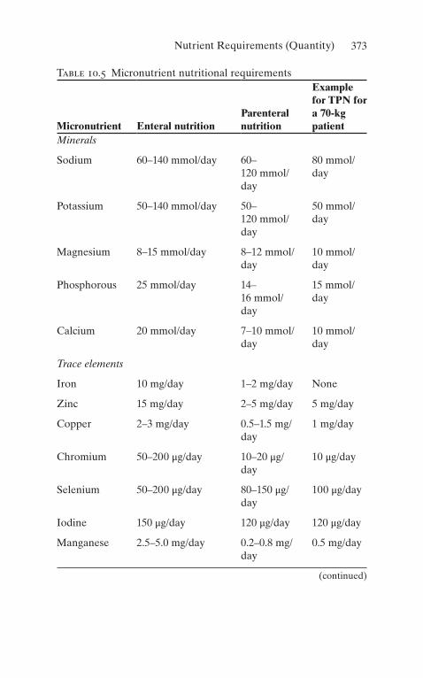
373
Table 10.5 Micronutrient nutritional requirements
Micronutrient Enteral nutritionParenteral nutrition
Example for TPN for a 70-kg patient
Minerals
Sodium 60–140 mmol/day 60–120 mmol/day
80 mmol/day
Potassium 50–140 mmol/day 50–120 mmol/day
50 mmol/day
Magnesium 8–15 mmol/day 8–12 mmol/day
10 mmol/day
Phosphorous 25 mmol/day 14–16 mmol/day
15 mmol/day
Calcium 20 mmol/day 7–10 mmol/day
10 mmol/day
Trace elements
Iron 10 mg/day 1–2 mg/day None
Zinc 15 mg/day 2–5 mg/day 5 mg/day
Copper 2–3 mg/day 0.5–1.5 mg/day
1 mg/day
Chromium 50–200 μg/day 10–20 μg/day
10 μg/day
Selenium 50–200 μg/day 80–150 μg/day
100 μg/day
Iodine 150 μg/day 120 μg/day 120 μg/day
Manganese 2.5–5.0 mg/day 0.2–0.8 mg/day
0.5 mg/day
(continued)
Nutrient Requirements (Quantity)

374
Table 10.5 (continued)
Micronutrient Enteral nutritionParenteral nutrition
Example for TPN for a 70-kg patient
Vitaminsa
Vitamin A RDA = 4000–5000 IU/day
ND 3300 IU/day
Vitamin D RDA = 200–400 IU/day
ND 200 IU/day
Vitamin E RDA = 12–15 IU/day
ND 10 IU/day
Vitamin K RDA = 60–80 μg/day
ND 10 mg/weekb
Thiamine RDA = 1.1–1.4 mg/day
ND 3 mg/day
Riboflavin RDA = 1.2–1.7 mg/day
ND 5 mg/day
Niacin RDA = 13–19 mg/dayc
ND 40 mg/day
Pantothenic acid
4–7 mg/dayc ND 15 mg/day
Pyridoxine RDA = 1.6–2.0 mg/day
ND 4 mg/day
Folic acid RDA = 0.4 mg/day ND 0.4 mg/day
Vitamin B12 RDA = 3 μg/day ND 5 μg/day
Vitamin C RDA = 40 mg/day ND 100 mg/day
Biotin RDA = 30–100 μg/day
ND 60 μg/day
ND not defined, RDA recommended daily allowanceaEnteral requirements should always exceed parenteral require-ments; most recommend supplying one to three times the RDA of each vitamin to patients with critical illnessbNone if anticoagulation usedcRDA not established
Chapter 10. Nutrition

375
F. Trace elements (iron, copper, iodine, zinc, selenium, chro-mium, cobalt, manganese)
1. Needs in critically ill patients have not been deter-mined. (See Table 10.5 for estimates of requirements.)
2. Sufficient quantities are thought to be present in enteral products.
3. Must be supplemented in TPN (all except iron can be added to the solution).
(a) Deficiency states have been reported in long-term TPN patients.
(b) Specifics are best managed by specially trained nutritional support teams.
Role of Specific Nutrients (Quality)
A. Nitrogen Sources
1. Choices
(a) Amino acids (b) Hydrolyzed protein (peptides) (c) Intact proteins
2. Evidence suggests that proteins generated from the diet possess specific physiologic activities (i.e., organic catalyst for structural formation of cells, acting as anti-bodies or controlling cell metabolism).
3. Nitrogen is best delivered as intact protein (if digestion and absorption intact) or hydrolyzed protein (impaired digestion).
4. Protein is absorbed primarily as peptides (60%) and amino acids (33%).
5. Essential amino acid formulas should not be used. 6. Some amino acids become essential during critical
illness.
(a) These are called conditionally essential amino acids.
Role of Specific Nutrients (Quality)

376
(b) Examples include glutamine, cysteine, arginine, and taurine.
7. Some amino acids appear to have specific roles.
(a) Glutamine is a fuel source for rapidly dividing cells, such as the GI tract and immune system (patients with bone marrow transplant, supplementation with glutamine was associated with lower rates of infection).
(b) Arginine is required for optimum wound healing and is important in immune function. (Target patients are critically ill adults and preterm infants.)
(c) Cysteine is needed for synthesis of glutathione. (d) Branched-chain amino acids (BCAA) may
improve mental status in patients with hepatic encephalopathy; it is primarily metabolized by peripheral muscle instead of the liver.
(e) Note that glutamine and cysteine are not stable (or present) in TPN solution.
8. Dietary nucleic acids may be important for immune function.
B. Lipids
1. Linoleic Acid
(a) Essential fatty acid (need 7–12% of total calories supplied as linoleic acid)
(b) ω-6 Polyunsaturated, long-chain fatty acid (immunosuppressive)
(c) Precursor to membrane arachidonic acid
2. ω-3 Polyunsaturated Fatty Acids (PUFA)
(a) Fish oils and linolenic acid. (b) Profound effects upon cell membrane fluidity and
stability. Decrease production of dienoic prosta-glandins (i.e., PGE2), tumor necrosis factor, inter-leukin- 1, and other proinflammatory cytokines.
Chapter 10. Nutrition

377
(c) Supplementation of omega-3 fatty acids in patients with acute lung injury improves oxygenation and shortens length of mechanical ventilation.
3. Medium-Chain Triglycerides
(a) Good energy source (b) Water-soluble (c) Enter circulation via GI tract
4. Short-Chain Fatty Acids (SCFA)
(a) Examples: Butyric and propionic acid (b) Major fuel for the gut (especially the colon) (c) Derived from metabolizable fiber
5. High-Fat Formulas
(a) If the patient is not overfed, these have a little effect on CO2 production (despite being marketed for decreasing the respiratory quotient [RQ]).
(b) Poor GI tolerance.
C. Carbohydrates
1. CHO are the principle source of energy with their form ranging from a starch to simple sugar.
2. Fiber
(a) Metabolizable fiber (i.e., pectin, guar) is converted to SCFA in the colon by bacteria.
(b) Bulk increases stool mass, softens stool, adds body to stool, and provides some stimulation of gut mass.
Monitoring Responses to Nutritional Support
A. Glucose: Levels should be checked upon admission the critical care unit and at minimum every 4 h.
B. Electrolytes: Should be monitored once daily for at least the first week of ICU admission.
C. Visceral proteins:
Monitoring Responses to Nutritional Support

378
1. Prealbumin
(a) Half-life is 2 days. (b) Normal range is 10–40 mg/dL.
2. Transferrin
(a) Half-life is 8–9 days. (b) Normal range is 160–355 mg/dL.
3. Albumin
(a) Half-life is 20 days. (b) Normal range is 3.2–5.0 mg/dL.
D. Visceral protein levels are affected by nutritional intake as well as the disease state (especially presence of inflammation).
E. Increasing levels of visceral proteins suggest that nutri-tional support is adequate.
F. Nitrogen balance:
1. NB is the most direct measurement of actual protein status, by comparing nitrogen output and input.
2. Determined from 12- to 24-h urine collections and measurements of total urinary nitrogen (more accurate than total urea nitrogen), compared to total nitrogen intake.
3. May be inaccurate:
(a) In patients with renal failure (b) If urine is not correctly collected by staff (c) If the patient has increased nitrogen losses in stool
or from wounds (i.e., burns)
4. N-balance = protein intake (g/day)/6.25 – {total urinary nitrogen (g/day) + 2}.
5. Negative nitrogen balance is not necessarily detrimen-tal over the short term (i.e., 1–2 weeks).
6. Improvement in nitrogen balance suggests that nutri-tional support is adequate.
7. Be aware that nitrogen balance may improve as catabo-lism decreases despite inadequate nutritional support.
Chapter 10. Nutrition

379
G. Caloric goals: Caloric needs can be determined using 25 kcal/kg of ideal body weight as an estimate. Another option is indirect calorimetry:
1. Measures oxygen consumption and CO2 production for 15–30 min, estimates energy expenditure, and then extrapolates to 24 h.
2. Keep RQ <1. Values >1 suggest lipogenesis from exces-sive caloric intake; values ≈ 0.7 are found in starvation and reflect fat oxidation.
H. Other Nutritional Parameters Not Generally Useful in the Critically Ill:
1. Weight 2. Skinfold thickness 3. Delayed cutaneous hypersensitivity (DCH) 4. Lymphocyte counts
Nutrition for Specific Disease Processes
A. Acute Renal Failure
1. Use intact protein or peptide formula with moderate fat.
2. Do not restrict protein (it is required for healing and for other organ functions).
3. May limit fluid intake with double-strength formula (2 cal/mL).
4. Watch K, Mg, Ca, and PO4 levels. 5. Consider nutrition with electrolyte restriction depend-
ing on the degree of compromise of renal function.
B. Hepatic Failure
1. Use intact protein or peptide formula. 2. Usually 1.0–1.2 g/kg/day of protein is needed to support
repair and immune function. 3. BCAA may be of value if encephalopathy persists fol-
lowing the use of intact protein or peptide diets.
Nutrition for Specific Disease Processes

380
C. Inflammatory Bowel Disease/Pancreatitis
1. Enteral nutrition is possible if a jejunal tube is placed (endoscopically or radiologically) distal to Treitz ligament.
2. Enteral nutrition should be attempted before initiating TPN.
D. Multiple Organ Failure
1. Nutritional support is usually of marginal value. 2. Nutritional support needs to be started before organ
failure develops.
Nasoduodenal Feeding Tube Placement
A. Used in patients who do not tolerate oral or gastric feeding.
B. Patients with abdominal surgery should have the tube placed during surgery under direct visualization:
1. The anesthesiologist inserts the tube into the stomach. 2. The surgeon locates the tube and directs it into the
duodenum or jejunum. 3. Eliminates need for confirmatory X-rays. 4. Allows immediate feeding upon admission into the
intensive care unit (ICU). 5. Feeding tubes may also be placed into the small bowel
using a gastrostomy or jejunostomy.
C. Tubes placed into the stomach will rarely (5–15%) migrate spontaneously into the small bowel in critically ill patients (due to gastroparesis).
D. Bedside method:
1. Place patient in left lateral decubitus position (if possible).
2. Lubricate the nostril with generic lubricant or 2% vis-cous lidocaine.
Chapter 10. Nutrition

381
3. Insert an eight- to ten-French small-bore feeding tube (containing wire stylet) into the nostril, and gently advance it through the nasopharynx into the esophagus and then the stomach.
4. If resistance is met or the patient coughs, becomes agi-tated, or decreases oxygen saturation, then:
(a) Pull the tube back into the nasopharynx. (b) Repeat step 3 and reinsert the tube into the
stomach. (c) Change the position of the patient’s neck (slightly
flex or extend) before reattempting insertion.
5. Confirm position of the tube in the stomach.
(a) Auscultate over the abdomen. (b) Aspirate gastric contents (pH ≈ 2–5, unless on H2
blocker).
6. Remove wire stylet, and place a 45-degree bend approximately 1 in. from the distal end of the wire.
7. Gently reinsert the wire stylet (should not meet resistance).
8. Slowly advance the tube while rotating it in a clockwise direction.
9. Check the position every 10–15 cm:
(a) Auscultation will reveal higher pitched sounds when the tube is in the pylorus and proximal small bowel.
(b) Bile may be aspirated from the tube in the small bowel.
(c) Bile/small bowel secretions have pH ≈ 6–7. (d) Abdominal X-ray:
1. Can confirm small bowel location 2. May not be cost-effective 3. Will avoid feeding into the lung in a rare case of
misplaced feeding tube
Nasoduodenal Feeding Tube Placement
382
E. With this bedside method, we (faculty, residents, and med-ical students) successfully place >90% of attempted small bowel tubes into the duodenum or jejunum.
F. Aggressive surgical and bedside placement allows us to feed >97% of our critically ill patients enterally within 24–48 h of admission into the ICU.
G. If bedside placement is not possible, place the feeding tube into the small intestine using:
1. Endoscopy 2. Fluoroscopy
Recommendations for TPN Use
1. Use only when enteral nutrition is not possible (e.g., short gut syndrome, chylothorax).
(a) Failure of the stomach to empty is not an indication for TPN but rather for a small bowel feeding tube.
(b) Most patients with diarrhea can be managed with enteral nutrition.
2. Initial TPN orders may be based on recommendations in Tables 10.4 and 10.5.
3. Overall TPN management is best performed by specially trained nutritional support teams.
4. Patient should be monitored for complications associated with TPN (abnormal electrolytes, infections, etc.)
5. For more specifics, the reader is referred to the entire texts written about TPN.
Approach to Enteral Feeding
A. Enteral nutritional support should be initiated within 12–48 h of admission to the ICU.
B. The oral route is preferred (but frequently not possible). C. The gastric route is the second choice and should be tried
before placing a small bowel tube in most patients.
Chapter 10. Nutrition
383
D. Patients at high risk for aspiration or known gastric pare-sis should be fed with a small bowel tube.
E. Feeding formulas should not be diluted. F. Gastric paresis is best assessed and monitored by measur-
ing gastric residuals. Residuals must be checked every 4–6 h and should remain <150 cc:
(a) Greater than 250 cc after second gastric residual may consider promotility agent.
(b) Greater than 500 cc should result in holding feeding and consult dietitian.
G. Routinely flush feeding tube at ordered rate for continu-ous feeds (i.e., 30 cc q4–6 h) and/or before and after bolus feeds.
H. Keep head of bed elevated 30–45° during feeding and 30–60 min after feeding to decrease the risk of aspiration.
I. Feeding should be started at 25–30 mL/h and increased by 25 mL/h every 1–4 h as tolerated by gastric residuals (<150 mL) until the caloric goal (25–30 kcal/kg/day) is achieved.
J. May increase feeds at slower rate (i.e., =10 mL/h every 6–12 h), but often this is not necessary.
K. If the protein goal is not achieved, use a formula with a higher protein/calorie ratio or add protein to the formula.
L. The goal rate of infusion should be met by the third day of therapy (frequently earlier).
M. Monitor nutritional response by measuring visceral pro-tein levels:
1. Prealbumin and transferrin levels should be measured on day 1 and every 3 days thereafter during initial therapy.
2. Increasing levels suggest that the patient is receiving adequate nutritional support.
3. Levels usually normalize in 1–2 weeks if the disease process is controlled and nutritional support is adequate.
Approach to Enteral Feeding

384
4. If levels fail to increase:
(a) Consider underlying infection, inflammation, or other disease processes.
(b) Reevaluate the adequacy of nutritional support. (c) Nitrogen balance and energy balance (i.e., indirect
calorimetry) may be informative. (d) Consult the nutritional support service.
N. Several flow diagrams (for specific patient populations, using enteral products currently on our formulary) are given as examples (see Figs. 10.1, 10.2, and 10.3).
Isolated head injury
Hypermetabolic(acute injury)
NoYes
Yes
Yes
Yes Lowerfat
content
No
No
Fatmalabsorption
?
Evaluate forcause of diarrhea
High nitrogenformula
with fiber
Peptidebased formula
Tolerated× 3 Days
?
cont
inue
feed
ing
Examples: - Reabilan HN - Peptamen - Perative
Examples: - Replete - Nitrolan
Exampls: - Replete with fiber
1. Oral diet2. Standard formula a. Osmolite HN b. Jevity(Fiber)1. Consider nutrition consult
2. Trial of 50:50 mix of Reabilan HN: Criticare HN Peptamen: Criticate HN3. Trial of Criticare HN
High nitrogenformula
No
Impaireddigestion/
absorption orsepsis
Standardformula
Example: - Osmolite
Example: - Jevity
Oraldiet
Standardformula
with fiber
HN
Figure 10.1 Flow diagram for nutritional support in patients with isolated head injury
Chapter 10. Nutrition

385
O. If peptide-based diets are not available, intact protein diets should be used.
P. Formula osmolality:
1. 300–600 mOsm/kg H2O 2. Rarely causes intolerance/diarrhea
Q. Diarrhea is unfortunately encountered in patients on enteral and parenteral nutrition:
1. Generally defined as >300–500 mL stool output per day.
2. Most common etiologies are medications and infec-tions. See Table 10.6 for a partial list of etiologies and suggestions for preliminary workup.
3. Note that many elixir forms of medications contain sorbitol.
Multitrauma/burns
Septic shock
Abdominal surgery
No
No
continue feeding
No
High nitrogenformula
Example: - Nitrolan- Replete
Examples: - Reabilan HN - Perative - Peptamen
Example: - Replete with fiber
High nitrogenplus fiber
1. Oral diet2. Standard nitrogen formula Examples: a. Osmolite HN b. Jevity (fiber)
1. Consider dietary consult2. 50:50 Mix Reabilan HN: Criticare HN Peptamen: Criticare HN3. Criticare HN
*Shock, resuscitation, major abdominal surgery, gut orpancreatic injury, sepsis, acute hypoalbuminemia (<2g/dL)
Yes
Yes Toleraled× 3 Days
?
Fatintolerance
?
Evaluate for causesof diarrhea
YesLower fatcontent
Impaireddigestion orabsorption*
Peptide-basedformula
Figure 10.2 Flow diagram for nutritional support in patients with mul-tiple trauma, burn injury, sepsis/septic shock, or abdominal surgery
Approach to Enteral Feeding
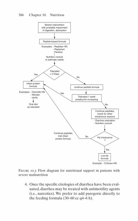
386
4. Once the specific etiologies of diarrhea have been eval-uated, diarrhea may be treated with antimotility agents (i.e., narcotics). We prefer to add paregoric directly to the feeding formula (30–60 cc q4–6 h).
Severe malnutritionwith probable impairmentof digestion, absorption
Peptide-based formula
Nutrition consultto estimate needs
Tolerated× 3 Days
No
continue peptide formula
Tolerated 1 weekprealbumin increasing
Continue peptides;check for other
intolerence reasons
Fat intolerance?
Low-fatformula
Example: - Criticare HN
Continue peptidestrial intact
protein formula
Yes
No
- Diarrhes evaluation- Nutrition consult
No
Yes
Yes
Intact proteinformula
Oral dietas tolerated
Examples: - Reabilan HN - Peptamen - Peralive
Examples: - Osmolite HN - Nitrolan - Jevity
Figure 10.3 Flow diagram for nutritional support in patients with severe malnutrition
Chapter 10. Nutrition

387
Useful Facts and Formulas
A. Nutritional Assessment. The resting metabolic rate (RMR) energy requirements for a patient can be calculated using the Harris–Benedict equation or the Mifflin–St. Jeor equation, using equations and activity/injury factors listed on Table 10.2.
Table 10.6 Etiologies and evaluation of diarrheaCauses Examples WorkupDrugs Sorbitol, antacids,
H2 blockers, antibiotics, lactulose, laxatives, quinidine, theophylline
No specific workup; discontinue any of these medications that are not absolutely necessary
Infections 1. Clostridium difficile 1. Specific stool culture, C. difficile toxin assay, sigmoidoscopy/colonoscopy for evidence of pseudomembranes
2. Infectious diarrhea (e.g., typhoid fever, shigellosis)
2. Fecal leukocytes, culture
3. Others: Bacterial overgrowth, parasites, systemic infection, HIV
3. As relevant (e.g., look for ova and parasites; rarely causes new diarrhea in critically ill)
Osmotic Measure stool osmotic gap (SOG); SOG >100 suggests osmotic diarrhea
Impaction May be secondary to narcotics
Rectal exam
Other causes
Inflammatory bowel disease, pancreatic insufficiency, short gut syndrome
SOG stool osmolality – 2 (stool Na+ + K+)
Useful Facts and Formulas

388
The metabolic rate (MR) can be calculated in patients with a pulmonary artery catheter as follows:
MR kcal h VO mL h L mLkcal L
/ / min min/ /. /
� � � � �� ��
2 60 1 10004 83
where VO2 (mL/min) = Cardiac output (L/min) × [arterial oxygen content (CaO2, mL/L) – mixed venous oxygen con-tent (CmO2, mL/L)].
The prognostic nutritional index (PNI) allows for nutri-tional assessment of the critically ill patient and is calculated as follows:
PNI risk alb TSF tfn
DSH
% % . . .
.� � � � � � � � � � � �
� � �158 16 6 0 78 0 2
5 8
where alb = serum albumin (g/dL), TSF = triceps skin fold (mm), tfn = serum transferrin (mg/dL), and DSH = delayed skin hypersensitivity (1 = anergy, 2 = reactive).
The probability of survival (POS) based on the nutritional status of a critically ill patient can be calculated as follows:
POS alb DSH SEP DIA� � � � � � � � � � � � �0 91 1 0 1 44 0 98 1 09. . . . .
where alb = serum albumin (g/dL), DSH = delayed skin hypersensitivity (1 = anergy, 2 = reactive), SEP = sepsis (1 = no sepsis, 2 = sepsis), and DIA = diagnosis of cancer (1 = no cancer, 2 = cancer).
Another way to calculate the nutritional deficit is by utiliz-ing the index of undernutrition (IOU), as shown in Table 10.7.
The calculation of daily protein requirements (PR) can be done utilizing the following formula:
PR g Patient weight in kg PR for illness in g kg� � � � � �� �/
To determine the nonprotein caloric requirements (NCR):
Chapter 10. Nutrition

389
Tabl
e 10
.7 I
ndex
of
unde
rnut
riti
on
Ass
ayP
oint
s0
510
1520
Alb
umin
(g/
dL)
>3.
53.
1–3.
52.
6–3.
02.
0–2.
5<
2.0
Fat
area
(%
)>
7056
–70
46–5
530
–45
<30
Mus
cle
area
(%
)>
8076
–80
61–7
540
–60
<40
Tran
sfer
rin
(g/L
)>
2.0
1.76
–2.0
1.41
–1.7
51.
0–1.
4<
40
Wei
ght
lost
(%
)0
0–10
11–1
415
–20
>20
Useful Facts and Formulas
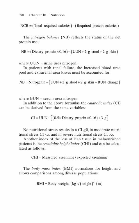
390
NCR Total required calories Required protein calories� � � � � �
The nitrogen balance (NB) reflects the status of the net protein use:
NB Dietary protein UUN g stool g skin� �� � � � �� �0 16 2 2.
where UUN = urine urea nitrogen.In patients with renal failure, the increased blood urea
pool and extrarenal urea losses must be accounted for:
NB Nitrogenin UUN g stool g skin BUN change� � � � �� �2 2
where BUN = serum urea nitrogen.In addition to the above formulas, the catabolic index (CI)
can be derived from the same variables:
CI UUN Dietary protein g� � � �� � ��� ��0 5 0 16 3. .
No nutritional stress results in a CI ≥0, in moderate nutri-tional stress CI <5, and in severe nutritional stress CI >5.
Another index of the loss of lean tissue in malnourished patients is the creatinine height index (CHI) and can be calcu-lated as follows:
CHI Measured creatinine expected creatinine= /
The body mass index (BMI) normalizes for height and allows comparisons among diverse populations:
BMI Body weight kg height m� � � � � � �/
2
Chapter 10. Nutrition

391
B. Fuel Composition. The body uses different sources of fuel. Table 10.8 depicts some of them.
C. Other Formulas. The body surface area (BSA) of a patient can be calculated as follows:
BSA m
Weight in kg height in cm2
0 425 0 72571 84
10 000� � � � � �� � �
. ..
,
The ideal body weight (IBW) for height in males and females can be estimated based on Table 10.9.
The percentage of ideal body weight (%IBW) is calculated as follows:
%IBW
height in cm
IBW�
�� �100
Table 10.8 Normal fuel composition of the human bodyFuel Amount (kg) Calories (kcal)Circulating fuels
Glucose 0.020 80
Free fatty acids (plasma) 0.0003 3
Triglycerides (plasma) 0.003 30
Total 113
Tissue
Fat (adipose triglycerides) 15 141,000
Protein (muscle) 6 24,000
Glycogen (muscle) 0.150 600
Glycogen (liver) 0.075 300
Total 165,900
Useful Facts and Formulas

392
Table 10.9 Ideal body weight in males and femalesHeight in cm Males (weight in kg) Females (weight in kg)145 51.8 47.5
150 54.5 50.4
155 57.2 53.1
160 60.5 56.2
165 63.5 59.5
175 70.1 66.3
180 74.2
185 78.1
Chapter 10. Nutrition

393© Springer Nature Switzerland AG 2021J. Varon, Handbook of Critical and Intensive Care Medicine, https://doi.org/10.1007/978-3-030-68270-5_11
Cancer is becoming the leading cause of death in the United States. Enhanced critical care capabilities have contributed substantially to improved survival. Critical care may be needed on a short-term basis for the complications of the underlying malignancy or of aggressive antineoplastic ther-apy. Postoperative critical care has greatly facilitated major extirpative cancer surgery and is an implicit part of other approaches such as bone marrow transplantation.
Patients with cancer may require ICU care at some point in their illness. This could be directly associated with malig-nancy (i.e., acute pulmonary embolism). In addition, admis-sion to the ICU can be treatment related (i.e., cell toxicity), and it can also be due to a comorbidities, such as COPD, cir-rhosis, or kidney disease exacerbations. The most common cancers seen in the ICU setting are leukemia, lymphoma, and lung cancer. Early admission to the ICU increases the oppor-tunity to prevent or treat cancer-related complications, such as leukostasis, multiple organ dysfunction, tumor lysis syn-drome, and macrophage lysis syndrome.
The present chapter considers different types of cancer patients likely to need and benefit from treatment in the ICU. Clinical judgment regarding the appropriate use of critical care services is required in all patient populations,
Chapter 11Critical Care Oncology

394
not just in patients with cancer. The decision to admit and technologically support critically ill cancer patients should be individualized.
Central Nervous System
A. Altered Mental Status. Alteration in mental status is the most common central nervous system (CNS) presentation for cancer patients in the intensive care unit (ICU). The common differential diagnoses are considered below. If these can be excluded and the patient has not received excessive sedative or narcotic–analgesic agents, the patient should be treated presumptively for sepsis. Altered men-tal status is a reliable, though nonspecific, sign of sepsis, which carries a high mortality rate in cancer patients.
1. Intracranial Mass LesionsA history of headache, nausea, vomiting, or seizure
activity together with papilledema and other signs of raised intracranial pressure suggests an intracranial mass lesion. A moderate increase in intracranial pres-sure by itself is relatively well tolerated; however, when intracranial pressure becomes critical, brain substance will shift in the direction of least resistance, with resul-tant herniation through the tentorium or foramen magnum.
2. Primary Tumors of the CNSThese present with focal neurologic signs, depending
on location. 3. Secondary (Metastatic) Tumors
Approximately 15–30% of secondary tumors will present with new-onset seizures. Common malignan-cies associated with cerebral metastases include breast, lung, kidney, and melanoma.
4. Cerebral HemorrhageCerebral hemorrhage is associated with acute pro-
myelocytic leukemia, as a direct complication of brain metastases or related thrombocytopenia.
Chapter 11. Critical Care Oncology

395
5. Subdural HematomaAcute subdural hematomas present with fluctuation
in the level of consciousness and hemiparesis. 6. Brain Abscess
Brain abscess accounts for 30% of CNS infections in cancer patients.
(a) Clinically apparent raised intracranial pressure and neurologic deficits are late signs.
(b) Usually present with fever, headache, drowsiness, confusion, and seizures.
(c) Typically seen in patients with leukemias or head and neck tumors.
B. Other Causes of Altered Mental Status in Critically Ill Cancer Patients
1. Leptomeningeal Metastases
(a) May present with signs of raised intracranial pres-sure and hydrocephalus.
(b) Acute leukemias, lymphomas, and breast cancer as well as central nervous system carcinomas are frequent causes.
2. Cerebrovascular Accident (CVA)Commonly occurs in cancer patients. As in all
patients, CVA may be thrombotic, hemorrhagic, or embolic in nature.
(a) Most patients present with focal neurologic signs and headaches.
(b) Seizures are common, especially in hemorrhagic CVA.
(c) Embolic CVA in cancer patients may be related to septic emboli, especially in patients with known fungal infection (i.e., aspergillosis).
3. Metabolic EncephalopathiesLethargy, weakness, somnolence, coma, agitation or
psychosis, and focal or generalized seizures can all result from metabolic abnormalities. Lack of focal neu-
Central Nervous System
396
rologic signs suggests a metabolic encephalopathy. Examples include:
(a) Hypercalcemia (see below) (b) Hyponatremia (c) Hypomagnesemia (d) Hypoglycemia (e) Uremia (f) Hyperglycemia (e.g., hyperosmolar nonketotic
acidosis) (g) Wernicke’s encephalopathy (h) Disorders of the porphyrin metabolism
4. Seizures/Postictal StatePatients with primary and secondary tumors (espe-
cially hemispheric) commonly present with seizures.
(a) Differential diagnoses includes CVA, CNS infec-tion, head injury, or narcotic withdrawalCancer: central nervous system as causes of seizures.
(b) In the immediate postictal period, findings may include evidence of tongue biting, loss of bladder/bowel control, and extensor plantar responses.
(c) The presence of lateralized focal signs suggests that seizures may have a focal origin.
(d) Prolonged coma after a generalized seizure or transient hemiparesis (Todd’s paralysis) following a Jacksonian, focal, or generalized seizure is more common in patients with seizures secondary to mass lesions than in those with seizures secondary to other conditions.
5. Cerebral LeukostasisPatients with hyperleukocytosis (defined as a periph-
eral white blood cell [WBC] count >100,000/mm3) may present with blurred vision, dizziness, ataxia, stupor or coma, or an intracranial hemorrhage.
(a) Hemorrhage results from leukostatic plugging of arterioles and capillaries with endothelial cell dam-age, capillary leak, and small vessel disruption.
Chapter 11. Critical Care Oncology
397
(b) Retinal hemorrhages are suggestiveCancer:central nervous system of intracranial hemorrhage, and thus fundoscopic examination should be per-formed frequently.
6. Hyperviscosity Syndrome (HVS)Excessive elevations of serum paraproteins or
marked leukocytosis can result in elevated serum vis-cosity, sludging, and decreased perfusion of the micro-circulation, with stasis. HVS can affect any organ system; however, characteristic clinical findings occur in the lungs and CNS.
(a) Patients may present with visual disturbances or visual loss.
(b) Characteristic retinopathy is present with venous engorgement (with “sausage-link” or “boxcar” seg-mentation), microaneurysms, hemorrhages, exu-dates, and occasionally papilledema.
(c) Similar vascular changes may be seen in the bulbar conjunctivae.
(d) Other clinical findings may include headache, diz-ziness, Jacksonian and generalized seizures, somno-lence, lethargy, coma, and auditory disturbances, including hearing loss.
7. CNS InfectionsPatients with cancer are susceptible to a variety of
CNS infections, including meningitis, brain abscess (see above), and encephalitis.
(a) Meningitis is most frequently encountered in patient(s) with impaired cell-mediated immunity and is typically caused by Cryptococcus neofor-mans or Listeria monocytogenes.
(b) Patients with meningitis present with fever, head-ache, and altered mental status.
(c) All cancer patients with fever and altered mental status should have a lumbar puncture preceded by a computed tomography (CT) scan of the head (if a cerebral mass lesion is suspected).
Central Nervous System
398
(d) Encephalitis is most often caused by herpes viruses (simplex or zoster) or Toxoplasma gondii.
(e) Patients with encephalitis commonly present with signs of meningeal irritation (fever, headache, nuchal rigidity) and evidence of altered mental sta-tus. Confusion may progress to stupor and coma; focal neurologic signs and seizures are common.
C. Spinal Cord Compression. Significant cord compression results from epidural metastases and is most frequently seen in breast, lung, or prostate cancer with disseminated disease.
Classically, the chief complaint is back pain (90% of patients), which may be associated with weakness, auto-nomic dysfunction, sensory disturbances, ataxia, and flexor spasms. The neurologic deficit is determined by the level of the involved spinal cord.
1. Compression from metastases typically arises from three locations:
(a) Vertebral column (85%) (b) Paravertebral spaces (10–15%) (c) Epidural space (rare)
2. The distribution throughout the spine is approximately as follows:
(a) Thoracic (60–70%) (b) Lumbar (20–30%) (c) Cervical (10%)
D. Central Nervous System: Diagnostic Evaluation in the ICU
1. History, physical examination, and careful neurologic evaluation, emphasizing lateralizing signs, fundoscopy, and evidence of raised intracranial pressure.
2. Laboratory tests should include:
(a) Arterial blood gases (b) Serum electrolytes and glucose
Chapter 11. Critical Care Oncology
399
(c) Calcium, magnesium, and phosphorus (d) Renal and hepatic function tests (e) Determination of serum viscosity, especially in
cases of multiple myeloma or other paraprotein- producing tumors
3. Computed TomographyHead CT is the diagnostic test of choice for mass
lesions, midline shift, intracranial hemorrhage, or hydrocephalus.
4. Magnetic Resonance Imaging (MRI)MRI is a sensitive test for detection of intracerebral
metastases and to differentiate between vascular and tumor-related masses. It is also the examination of choice for the evaluation of intramedullary, intradural, and extramedullary spine lesion(s).
5. MyelographyMyelography provides an indirect image of the spi-
nal cord and nerve roots from the foramen magnum to the sacrum. It is the “gold standard” in the evalua-tion of spinal cord involvement by tumor. It is espe-cially helpful for patients with contraindications to MRI (such as patients with orthopedic hardware) and patients with plans for radiosurgery or radiation therapy.
6. Lumbar Puncture (LP)LP is most useful for the diagnosis of meningeal car-
cinomatosis, CNS leukemia, and CNS infections.
E. Central Nervous System: Acute Management in the ICU (See Also Chap. 9, “Neurologic Disorders”)
1. Raised Intracranial Pressure with Impending Herniation
(a) Glucocorticoid therapy will improve neurologic deficits in 70% of patients with symptomatic brain metastases by reduction of vasogenic brain edema. An initial dose of 10-mg dexamethasone may be given intravenously, followed by 16 mg/day in three
Central Nervous System

400
or four divided doses by the most appropriate route. Patients who do not respond to the standard dose may improve when the dose is increased to 100 mg/day.
(b) Osmotherapy with agents such as urea or mannitol is initiated to produce rapid reduction of intracra-nial pressure in patients with known or suspected intracranial metastases showing signs of herniation.
Mannitol 1.5–2.0 g/kg as a 20% solution can be administered by slow intravenous (IV) infusion. The total dose should not exceed 120 g/day. If administered together, furosemide can further potentiate mannitol’s effects but is also associated with risks of dehydration and hypokalemia.
(c) Hyperventilation may be instituted in patients who present with signs of brain herniation. They should be intubated expeditiously and ventilated to main-tain an arterial PCO2 of 25–30 torr (mmHg). However, the use of this technique is controversial. Some authors believe that the beneficial effect of hyperventilation lasts only 6 h. To date, there is no conclusive data that this therapeutic intervention modifies outcome in these patients.
(d) Neurosurgical consultation is needed in the vast majority of patients.
2. Seizures
(a) Position the patient laterally to prevent aspiration and protect the airway.
(b) Correct any metabolic alteration or hypoxemia. (c) If the seizure is sustained, acute control is achieved
with lorazepam (Ativan) 1–10 mg IV, or continu-ous infusion can be used. Alternatively, IV diaze-pam (Valium™) 5–10 mg can be repeated in 5–10 min up to 30 mg. Another useful agent that permits rapid cessation of seizures is the adminis-tration of IV propofol (Diprivan™).
Chapter 11. Critical Care Oncology
401
(d) Long-term seizure control can usually be estab-lished with IV phenytoin (Dilantin™). The loading dose is 15 mg/kg IV (50 mg/min). Fosphenytoin can also be used.
(e) Intracerebral metastases should be treated with corticosteroids, chemotherapy, radiation, or sur-gery as indicated by the specific lesion.
3. Spinal Cord CompressionPalliation is generally accepted as a reasonable goal
in the management of these patients.
(a) Radiotherapy and surgical decompression are the cornerstones of management.
(b) Chemotherapy with nitrogen mustard or cyclo-phosphamide has been effectively used, generally in combination with radiation, for the management of cord compression caused by lymphoma or Hodgkin’s disease.
4. Other Modalities
(a) Leukophoresis is one of the therapeutic options for severe symptomatic leukocytosis with leukostasis.
(b) If hydrocephalus is present, it should be managed by emergent relief and shunting.
(c) Radiotherapy is currently the most commonly employed therapeutic modality for palliation of cerebral metastases.
5. General Supportive Care
(a) Stress ulcer prophylaxis in the form of antacids, sucralfate, or H2-receptor antagonists.
(b) Prophylaxis for deep venous thrombosis (DVT) should include, if no contraindication exists, the use of subcutaneous heparin (or low-molecular- weight heparin) and/or the use of sequential compressive devices (SCDs) on the lower extremities.
Central Nervous System

402
(c) Nutritional support should be provided for reple-tion of malnourished patients, as well as for main-tenance of good nutrition in patients at risk for malnutrition due to cancer or its therapy.
(d) Appropriate antimicrobial therapy (see below). (e) Foley catheterization if urinary retention exists
and aggressive prophylactic bowel regimens to prevent constipation from immobility and auto-nomic dysfunction.
Pulmonary
The lungs are involved commonly in cancer patients, with 75–90% of pulmonary complications being secondary to infection. Noninfectious complications include those due to chemotherapy (i.e., bleomycin), thoracic irradiation, and pul-monary resections. Respiratory failure in cancer patients requiring mechanical ventilation is associated with a 75% mortality rate.
A. Pulmonary Infiltrates. In patients with systemic cancer, the differential diagnoses of pulmonary infiltrates seen on a routine chest film are extensive.
1. Localized infiltrates that are confined to a lobe or seg-ment in a patient with a compatible history most fre-quently represent a bacterial process.
2. Diffuse bilateral infiltrates are more suggestive of opportunistic infection, treatment-induced lung injury, or lymphangitic spread of carcinoma.
3. Bilateral perihilar infiltrates in patients who have rap-idly gained weight support a diagnosis of fluid overload.
4. Pulmonary infiltrates following bone marrow transplantation.
(a) Life-threatening infections generally occur within the first 100 days posttransplant.
Chapter 11. Critical Care Oncology
403
(b) Within the initial 30 days posttransplant, the most common pathogens for pneumonia are bacterial or fungal.
(c) Interstitial pneumonia (diffuse nonbacterial pneu-monia) is the predominant problem following transplantation, with the syndrome consisting of dyspnea, nonproductive cough, hypoxemia, and diffuse bilateral infiltrates and occurring within 30–100 days after transplant.
(d) Cytomegalovirus (CMV) pneumonia comprises the majority of interstitial pneumonitides. The inci-dence of CMV infection appears to be related to the loss of immunity during pretransplant condi-tioning and to the development of graft-versus- host disease.
(e) Bronchiolitis obliterans syndrome as a manifesta-tion of graft versus host disease of the lung.
(f) Pulmonary hypertension. (g) Diffuse alveolar hemorrhage due to infectious and
noninfectious causes.
5. Diagnosis.
(a) Chest X-ray is never diagnostic of any single entity. (b) Cultures of sputum and special stains of tracheo-
bronchial secretions (KOH, India ink) should be obtained routinely. Colonization of the upper respiratory tract as well as the inadequacy of spu-tum production may make identification of the offending organism(s) difficult.
(c) Blood cultures for fungal and bacterial organisms. (d) Viral titers (especially CMV). (e) Daily determination of serum lactate levels may be
of some value in patients with respiratory failure. An increase in the serum lactate level may precede the deterioration of arterial blood gases and the development of diffuse infiltrates typical of adult respiratory distress syndrome (ARDS).
Pulmonary

404
(f) Bronchoscopy with bronchoalveolar lavage (BAL) has a diagnostic sensitivity of 80–90% and is the procedure of choice in cancer patients with diffuse infiltrates.
1. BAL is most helpful in diagnosing opportunis-tic infection (i.e., Pneumocystis carinii (jirove-cii), viruses such as CMV, fungus, and mycobacteria).
2. This procedure is also useful for the diagnosis of intraparenchymal pulmonary hemorrhage.
3. BAL is safe in thrombocytopenic and mechani-cally ventilated patients who may not tolerate transbronchial biopsy.
(g) Open lung biopsy is reserved for selected patients due to its attendant morbidity, discomfort, and financial cost.
6. Management.
(a) Early empirical use of broad-spectrum antibiotics (see Chap. 8, “Infections”).
(b) In patients who remain persistently febrile despite the use of antibiotics, amphotericin B and liposo-mal amphotericin have been shown to reduce the mortality rate due to infection.
(c) Ganciclovir and hyperimmune globulin have been shown to improve survival in patients with intersti-tial pneumonia.
B. Pulmonary Leukostasis. Leukostasis, with obstructed flow in small pulmonary vessels, is the consequence of the intravascular accumulation of immature, rigid myelo-blasts, observed predominantly in acute myelogenous leu-kemia (AML), and chronic myelogenous leukemia (CML) patients in a blast phase. Vascular stasis and distention result in local hypoxia. The release of intracellular enzymes and procoagulants leads to vascular and pulmonary paren-chymal damage.
Chapter 11. Critical Care Oncology

405
1. Signs and Symptoms: Progressive dyspnea and/or altered mental status (see discussion on CNS)
2. Diagnosis
(a) CBC: WBC count is usually >150,000/mm3. (b) Arterial Blood Gases (ABGs): True hypoxemia
develops as a result of impaired pulmonary gas exchange. Spurious low values for PaO2 may be consistently obtained because the large number of blasts consumes oxygen within the ABG specimen itself. The longer the interval between the collec-tion and analysis, the lower the measured PaO2. This may make assessment of gas exchange difficult.
(c) Pulse oximetry may be of benefit to follow the ade-quacy of arterial oxygenation.
(d) Chest X-ray may be normal or show diffuse nodu-lar infiltrates.
3. Management
(a) Myeloblast counts >50,000/mm3 warrant prompt treatment for reduction of the total WBC count to 20–60% within hours of recognition of the syndrome.
(b) Leukapheresis. (c) Chemotherapy (i.e., daunorubicin, cytosine arabi-
noside, hydroxyurea). (d) Adequate hydration. (e) Urate nephropathy prevention should be initiated
with allopurinol and urine alkalinization. (f) Hemodynamic monitoring is suggested. (g) When ARDS results from leukostasis, the follow-
ing should be carried out expeditiously:
1. Fluid resuscitation to restore blood volume. 2. Cardiac output and hemodynamics should be
optimized through volume enhancement and inotropic agents as needed.
Pulmonary

406
3. Pulmonary vasoconstriction should be treated with a combination of volume expansion, ino-tropic agents, and supplemental O2.
4. Mechanical ventilation should be instituted when needed to achieve normal pH, pCO2, and PO2 >60 on nontoxic FiO2 (see Chap. 2, “The Basics of Critical Care”).
5. Consideration for prone position is suggested by the author.
C. Treatment-Induced Lung Injury
1. Chemotherapy-Induced Lung InjuryA large number of chemotherapeutic agents can
produce pulmonary toxicity, either actively or delayed years after therapy. Commonly used agents with known pulmonary toxicity include alkylating agents (i.e., cyclophosphamide, carmustine, chlorambucil, melpha-lan, busulfan), antimetabolites (i.e., methotrexate, aza-thioprine), antitumor antibiotics (i.e., bleomycin, mitomycin), and alkaloids (i.e., vincristine). Pulmonary toxicity may take the following forms:
(a) Noncardiogenic pulmonary edema (ARDS) (b) Chronic pneumonitis and fibrosis (c) Hypersensitivity pneumonitis (i.e., procarbazine,
methotrexate, bleomycin)
2. Radiation-Induced Lung ToxicityRadiation pneumonitis is a clinical syndrome of dys-
pnea, cough, and fever developing in association with indistinct, hazy pulmonary infiltrates that may progress to dense alveolar consolidation following treatment with ionizing radiation.
(a) The likelihood of developing radiation-induced lung injury is influenced by a number of variables including the total dose, fractionation of doses, vol-ume of lung irradiated, and a history of prior irra-diation and chemotherapy.
Chapter 11. Critical Care Oncology
407
(b) Pathophysiology.
1. Direct effect of ionizing particles on alveolar structure.
2. Generation of high-energy oxygen-free radicals in excess of what normal enzymatic systems (peroxidase, superoxide dismutase) can remove.
3. The release of vasoactive substances such as his-tamine and bradykinin affects capillary perme-ability and pulmonary vascular resistance. The resultant pulmonary damage can exceed the area of radiation.
(c) From 5% to 15% of patients develop radiation pneumonitis.
(d) Symptoms may occur 1–6 months following com-pletion of thoracic irradiation.
Cardiovascular
A. Cardiac Tamponade (See Also Chap. 3, “Cardiovascular Disorders”). Cardiac tamponade is a life-threatening con-dition caused by increased intrapericardial pressure, resulting in limitation of ventricular diastolic filling and decreased stroke volume and cardiac output.
1. Common Etiologies in Cancer Patients
(a) Metastatic tumors of the pericardium.
1. Much more commonly produce tamponade than primary tumors of the pericardium.
2. Cause tamponade by either producing effusions or constriction.
3. Cancer of the lungs and breast, lymphoma, leu-kemia, and melanoma account for 80% of meta-static causes of cardiac tamponade.
(b) Primary tumors of the pericardium.
Cardiovascular

408
(c) Postirradiation pericarditis with fibrosis. The peri-cardium is the most frequent site injured by radia-tion. The latent period between radiotherapy and onset of clinical pericardial disease may be years.
(d) Encasement of the heart by the tumor.
2. Clinical Findings
(a) Symptoms are often nonspecific but commonly include sensation of fullness in the chest, pericar-dial pain or interscapular pain, apprehension, dys-pnea, and orthopnea.
(b) Clinical signs include altered mental status, hypo-tension, tachycardia, narrow arterial pulse pres-sure, distant heart tones with diminished apical impulse, tachypnea, oliguria, and diaphoresis. Other signs include the following:
1. Pulsus paradoxus 2. Ewart’s sign (area of dullness at the angle of the
left scapula) 3. Kussmaul’s sign (neck veins bulge on inspiration)
3. Diagnosis
(a) Clinical Suspicion: The key to recognizing tampon-ade is considering the diagnosis.
(b) Chest X-Ray.
1. Large globular heart shadow (“water bottle” configuration). If the pericardial fluid is <250 mL, the cardiac silhouette may be normal.
2. Lung fields are usually clear. 3. Pleural effusions are common associated
findings.
(c) Electrocardiogram (ECG).
1. Sinus tachycardia. 2. Low voltage QRS (<5 mV).
Chapter 11. Critical Care Oncology

409
3. Electrical alternans, which result from the heart oscillating in the filled pericardial sac. Alternation of the QRS complexes is most spe-cific for pericardial effusion.
(d) Echocardiography Makes a Quick and Definitive Diagnosis of Tamponade. Two-dimensional echo is more sensitive than M-mode. Findings include the following:
1. Prolonged diastolic collapse or inversion of right atrial free wall.
2. Early diastolic collapse of the right ventricular free wall.
3. Effusions as small as 30 mL are early detected by echocardiography (seen as an echo-free space).
(e) Pulmonary Artery (Swan–Ganz) Catheterization.
1. Elevated pulmonary capillary wedge and right atrial pressures with a prominent x descent with no significant y descent (“square root sign”)
2. Decreased cardiac output, stroke volume, sys-temic arterial pressure, and mixed venous oxy-gen saturation (SvO2)
3. Equalization of all pressures in diastole
(f) MRI Is Also Diagnostic but Is Expensive and Time-Consuming Compared with Echocardiography.
(g) Diagnostic Pericardiocentesis.
1. Cytology to detect the presence of malignant cells
2. Gram’s stain and acid-fast bacilli (AFB) smear, culture and sensitivity, cell count, and differential
3. Protein and lactic dehydrogenase (LDH) content
Cardiovascular

410
4. Therapy
(a) Therapeutic pericardiocentesis should be per-formed immediately in hemodynamically compro-mised patients.
1. Two-dimensional echocardiography-guided pericardiocentesis is successful in 95% of cases with no major complications.
2. Reaccumulation of fluid is likely to occur in malignant effusions but can be prevented with chemical sclerosis (i.e., tetracycline), radiation therapy, or surgery (i.e., pleuropericardial win-dow or pericardiectomy).
B. Myocardial Tissue Injury
1. Common Etiologies in Cancer Patients
(a) Anthracycline antibiotics (i.e., doxorubicin and daunorubicin).
(b) Mitoxantrone: A total dose >100–140 mg/m2 can cause congestive heart failure and exacerbate pre-existing anthracycline-induced cardiomyopathy.
(c) Cyclophosphamide: Doses >100–120 mg/kg over 2 days can result in congestive heart failure and hemorrhagic myocarditis/pericarditis and necrosis.
(d) Busulfan: The conventional oral daily dose may cause endocardial fibrosis.
(e) Interferons: In conventional doses, interferons may exacerbate underlying cardiac disease.
(f) Mitomycin C: Standard doses can cause myocar-dial damage.
(g) Radiation-induced cardiomyopathy causes a dose- dependent endocardial and myocardial fibrosis, which can result in a restrictive cardiomyopathy.
2. Diagnosis
(a) Endomyocardial Biopsy: Valuable for establishing etiology of cardiac injury in patients who may have
Chapter 11. Critical Care Oncology

411
received chemotherapy and for detecting subclini-cal cardiac damage. The anthracyclines cause char-acteristic degenerative changes in the myocytes.
(b) An ECG-gated blood pool scan for precise mea-surement of ejection fraction and detecting regional and global myocardial dysfunction.
3. TherapyTreatment is the same as for congestive cardiomy-
opathy of any cause. There is no specific therapy directed at radiation- or chemotherapy-induced myo-cardial damage.
C. Cardiac Dysrhythmias
1. Etiology
(a) Anthracycline antibiotics cause dysrhythmias unrelated to the cumulative dose; these effects can be seen hours or days after administration. Commonly observed dysrhythmias include supra-ventricular tachycardia, complete heart block, and ventricular tachycardia. Doxorubicin may also pro-long the QT interval.
(b) Amacrine produces ventricular dysrhythmias. (c) Taxol causes bradycardia and in combination with
cisplatin may produce ventricular tachycardia.
2. Diagnosis and treatment are the same as for rhythm disturbances of other etiologies.
D. Superior Vena Cava (SVC) Syndrome
1. Etiology: Ablation of blood flow from the superior vena cava to the right atrium caused by extravascular compression or intravascular obstruction.
(a) Ninety-five percent of cases are secondary to extrinsic compression of the SVC by mediastinal malignancy (3% from benign disease).
(b) The most common tumors are bronchogenic carci-noma of small cell type (48%) and lymphoma (21%).
Cardiovascular

412
2. Clinical Manifestations
(a) Dyspnea aggravated by lying supine or leaning forward.
(b) Tachypnea and signs of airway obstruction. (c) Signs and symptoms of increased intracranial pres-
sure (i.e., dizziness, headache, visual disturbance, seizure, altered mental status).
(d) Dysphagia, hoarseness. (e) Neck vein distention, facial plethora, and edema. (f) Numerous, dilated, vertically oriented, and tortu-
ous cutaneous venules or veins above the rib cage margin.
(g) Upper body edema, with cyanosis and ruddy complexion.
(h) Immediate causes of death are airway obstruction and intracranial hemorrhage. Thrombosis at the SVC may occur in 30% of these patients.
3. Diagnosis
(a) Clinical suspicion. (b) CT scan with IV contrast is the diagnostic proce-
dure of choice. (c) Transesophageal echocardiography is a safe bed-
side procedure excellent for evaluating the SVC and surrounding structures.
(d) Angiography and radionuclide venography help localize the obstruction.
4. Therapy
(a) Symptomatic relief is the rule. (b) Operative bypass relieves symptoms faster than
radiation and is indicated in patients with life- threatening respiratory compromise or advanced cerebral edema.
(c) Endovascular therapy (stents) has been tried suc-cessfully in many patients.
Chapter 11. Critical Care Oncology

413
(d) Radiation therapy is the mainstay of treatment for most malignant SVC obstruction; although in small cell carcinoma and lymphomas chemotherapy, it is particularly useful.
(e) Temporizing measures may be used in patients without significant airway or neurologic compro-mise and include corticosteroids to decrease cere-bral and laryngeal edema, diuretics, and elevation of the head.
(f) Anticoagulation has no definitive role.
Gastroenterology
A. Neutropenic Enterocolitis (Ileocecal Syndrome or Typhlitis)
1. IncidenceNeutropenic enterocolitis commonly occurs in
patients with hematologic malignancies (leukemia is the most common, with an incidence of 10–40%) receiving chemotherapy.
2. PathophysiologyNeutropenic enterocolitis results from mucosal
ulceration and necrosis of the ileum, cecum, or ascend-ing colon with overgrowth and mural invasion of bacte-ria and/or fungi. Thrombocytopenia may predispose patients to hemorrhage into the bowel wall. Enterocolitis typically presents on the seventh day of severe neutropenia.
3. Clinical Manifestations
(a) Abdominal distention (b) Right-sided abdominal tenderness (c) Watery diarrhea (d) Fever (e) Thrombocytopenia and neutropenia
Gastroenterology

414
4. Diagnosis
(a) Clinical suspicion. (b) Plain radiographs of the abdomen may show ileus
with distended cecum and pneumatosis coli. (c) CT of the abdomen: Thickened bowel wall contain-
ing air. (d) Sigmoidoscopy.
5. Differential Diagnosis
(a) Appendicitis (b) Pseudomembranous colitis (c) Diverticulitis (d) Other acute abdominal disorders
6. Medical Therapy
(a) Nutritional support (b) Nasogastric suction (c) Broad-spectrum antibiotics with anaerobic, gram-
negative, and Clostridium difficile coverage
7. Indications for Surgical Exploration
(a) Perforation (b) Severe bleeding (c) Abscess (d) Uncontrolled sepsis (e) Failure to improve after 2–3 days of intensive con-
servative management
B. Gastrointestinal (GI) Tract Hemorrhage and Perforation
1. GI hemorrhage
(a) The most common cause is hemorrhagic gastritis (32–48%), followed by peptic ulcer disease.
(b) Only 12–17% of bleeding is from the tumor per se (most commonly seen in GI lymphomas).
(c) Less common causes include esophageal varices, Mallory–Weiss tears, Candida esophagitis, and enteritis.
Chapter 11. Critical Care Oncology
415
2. PerforationLymphomas are the most common malignancies to
perforate during chemotherapy. 3. Diagnosis
A standard diagnostic workup should be performed to identify the source of bleeding, with emphasis upon endoscopy.
4. Therapy
(a) Surgical (b) Temporizing modalities to control bleeding
include:
1. Angiography, with or without embolization. 2. Endoscopic intervention. 3. These modalities may also be useful in patients
with carcinomatosis and previously unresect-able disease.
Renal/Metabolic
Many cancer patients develop metabolic abnormalities caused by tumor-produced factors (hormones or locally act-ing substances) or from tumor destruction by antineoplastic therapy.
A. Hypercalcemia
1. Causes of Hypercalcemia in Cancer Patients
(a) Secondary to malignancy 4% (b) Etiologies other than the malignancy 77% (c) With coexistent hyperparathyroidism 2%. (d) Vitamin D intoxication 16% (e) Idiopathic
2. It is the most common metabolic abnormality of can-cer patients (10%).
3. May occur with or without bone metastases.
Renal/Metabolic

416
4. Breast cancer is associated with hypercalcemia in 27–35% of patients. Mechanisms include widespread osteolytic metastases, production of parathyroid-like hormone, prostaglandin E2 (PGE2) (following hor-monal therapy with estrogens or antiestrogens), humoral osteoclast-activating factor, and coexisting primary hyperparathyroidism.
5. Lung cancer is associated with hypercalcemia in 12.5–35% of patients. It is frequently seen in squamous cell carcinoma and is rare in small cell carcinoma. It may occur early or late, with or without bone metastases. Mechanisms include production of osteoclast- activating factor, transforming growth factor alpha, interleukin 1, and tumor necrosis factor.
6. Multiple myeloma produces hypercalcemia in 20–40% of patients. Hypercalcemia develops secondary to extensive osteolytic bone destruction, osteoclast- activating factor, and lymphotoxin. Fifty percent develop renal insufficiency, which can aggravate hypercalcemia.
7. Lymphoma causes hypercalcemia by humoral media-tion and local bone destruction.
8. Head and neck malignancies have an incidence of hypercalcemia of 6%, which is humorally mediated. Hypercalcemia is associated with malignancies of the oropharynx (37%), hypopharynx (24.3%), and tongue (21.5%).
9. Squamous cell, transitional cell, bladder, renal, and ovarian carcinomas may also produce humoral hypercalcemia.
10. Clinical Presentation
(a) Severity of illness depends on the degree of hypercalcemia, concurrent illness or debility, age, and associated metabolic disturbances.
(b) Hypercalcemia of malignancy usually has a rapid onset.
(c) Neuromuscular manifestations often predomi-nate and include lethargy, confusion, stupor, and
Chapter 11. Critical Care Oncology

417
coma (occurs when serum calcium level is >13 mg/dL). Hallucinations and psychosis, weakness, and decreased deep tendon reflexes (DTRs) are also common.
(d) Cardiovascular manifestations include increased cardiac contractility, increased sensitivity to digi-talis, and dysrhythmias.
(e) Renal manifestations include polyuria and poly-dipsia (earliest symptoms), dehydration, decreased glomerular filtration, loss of urinary concentrating ability, and renal insufficiency.
(f) Gastrointestinal signs and symptoms include nau-sea and vomiting, anorexia, obstipation/constipa-tion, ileus, and abdominal pain.
(g) Skeletal involvement is the hallmark of hypercal-cemia from osteolytic metastases or humorally mediated bone resorption resulting in pain, pathologic fractures, deformities, or necrosis.
11. Diagnosis
(a) Laboratory Studies.
1. Total and ionized serum calcium 2. Electrolytes, serum urea nitrogen (BUN), and
creatinine 3. Serum phosphorus and alkaline phosphatase 4. Measures of urinary calcium excretion and
cyclic adenosine monophosphate (cAMP) 5. High alkaline phosphatase level 6. Increased urinary calcium excretion
(b) Radiologic Studies.
1. Radionuclide bone scan 2. Skeletal surveys 3. Baseline chest X-ray
(c) ECGs should be performed looking for charac-teristic changes, including prolonged PR and QRS intervals and shortened QT.
Renal/Metabolic

418
12. TreatmentHypercalcemia is often fatal if left untreated, espe-
cially when symptomatic or if serum calcium is >13 mg/dL. Treatment goals include promoting urinary cal-cium excretion, inhibiting bone resorption, and reduc-ing entry of calcium into the extracellular fluid compartment.
(a) Hydration: To restore intravascular volume and increase the urinary output. 1. Initially, 5–8 L of normal saline IV over the
first 24 h and then adequate IV fluids to main-tain a urine output of 3–4 L/day.
2. Electrolytes should be monitored during nor-mal saline infusion.
3. Monitor urine output and cardiac status to avoid fluid overload.
(b) Diuretics: Loop diuretics, such as furosemide, pro-mote calciuresis by blocking calcium reabsorp-tion in the ascending loop of Henle, and augment the calciuretic effect of normal saline.
1. Furosemide in doses of 40–80 mg IV may be given after adequate hydration.
2. Monitor electrolytes and urine output to avoid overdiuresis.
(c) Inhibitors of bone resorption should be initiated promptly in symptomatic hypercalcemia.
1. Mithramycin is an antitumor antibiotic with a direct toxic effect on osteoclasts. The usual dose is 25 μg/kg IV over 6 h. It generally decreases serum calcium within 6–48 h; it may be repeated if the patient does not respond within 2 days. Use should be restricted to emer-gency treatment of severe hypercalcemia. Complications include thrombocytopenia, myelosuppression, hypotension, and hepatic and renal toxicity.
Chapter 11. Critical Care Oncology
419
2. Disodium etidronate (EHDP) is an analog of pyrophosphate that blocks osteoclastic bone resorption and formation of bone crystals. The dose is 7.5 mg/kg/day in 250 mL saline infused over 2–6 h for 3–7 days, followed by 20 mg/kg/day orally. The onset of action is slow, with nor-mocalcemia achieved in 4–7 days, 75% of the time. EHDP is contraindicated in patients with renal failure.
3. Glucocorticoids (i.e., prednisone) are most effective in hematologic malignancies (espe-cially multiple myeloma) and breast carcinoma and are of little value in solid tumors. These agents lower serum calcium by inhibition of calcium absorption and the action of vitamin D. Prednisone in a dose of 1–2 mg/kg/day has an onset of action in 3–5 days. Adverse effects include GI bleeding, hyperglycemia, and osteopenia.
4. Calcitonin inhibits osteoclastic bone resorp-tion and enhances calcium excretion. The dose is 4–8 IU/kg q6 h IM or SQ. It may lower cal-cium by 2–3 mg/dL over 2–3 h. Adverse reac-tions include nausea and vomiting, flushing, and hypersensitivity reactions (initial skin test-ing is recommended before administration).
(d) Hemodialysis is useful in patients who present with renal failure or who cannot be treated with normal saline diuresis.
(e) Specific antineoplastic therapy should be initiated in patients for whom treatment exists. It is the most effective means of achieving long-term cor-rection of cancer-related hypercalcemia.
B. Tumor Lysis Syndrome. Tumor lysis syndrome is seen when cytotoxic chemotherapy induces rapid tumor cell lysis in patients with a large malignant cell burden of an
Renal/Metabolic

420
exquisitely chemosensitive tumor. Intracellular metabo-lites are released in quantities that exceed the excretory capacity of the kidneys.
1. This syndrome classically occurs in patients with Burkitt’s and non-Hodgkin’s lymphoma, acute lympho-blastic and nonlymphoblastic leukemia, and chronic myelogenous leukemia.
2. It may occur spontaneously in patients with lympho-mas and leukemias or following treatment with chemo-therapy, radiation, glucocorticoids, tamoxifen, and/or interferon.
3. Manifestations
(a) Related to metabolic abnormalities
1. Hyperkalemia: Generalized weakness, irritabil-ity, decreased DTRs, paresthesias, paralysis, car-diac dysrhythmias, and cardiac arrest. The classic ECG changes include peaked T waves, dimin-ished R waves progressing to widened QRS, prolonged PR, loss of P wave, and sine wave pat-tern as terminal event.
2. Hypocalcemia (related to hyperphosphatemia): Muscle spasms, carpopedal spasms, facial gri-macing, laryngeal spasm, irritability, depression, psychosis, intestinal cramps, chronic malabsorp-tion, seizures, and respiratory arrest. Chvostek’s and Trousseau’s signs are present in some patients. ECG reveals a prolonged QT interval.
3. Hyperuricemia: Gouty arthritis, nephrolithiasis, and urate nephropathy.
(b) Precipitation of calcium salts in tissues (c) Acute renal failure
4. Prevention and Treatment (See Table 11.1).
(a) To prevent acute renal failure, patients who are to undergo treatment for malignancies should receive the following:
Chapter 11. Critical Care Oncology

421
Table 11.1 Management of patients at risk for tumor lysis syndromeI. When no metabolic aberration exists:
1. Allopurinol 500 mg/m2 BSA/day; reduce to 200 mg/m2BSA/day, 3 days into chemotherapy
2. Hydration, 3000 mL/m2BSA/day
3. Chemotherapy initiated within 24–48 h of admission
4. Monitor electrolytes, BUN, creatinine, uric acid, calcium, phosphorous every 12–14 h
II. When metabolic aberration exists:
1. Allopurinol initiated as above, reduce dose if hyperuricemia controlled, reduce dose for renal insufficiency
2. Hydration as above, add non-thiazide diuretics as needed
3. Urinary alkalinization (urine pH >7)
Sodium bicarbonate 100 mEq/L IV solution initially, adjust as needed
Discontinue when uric acid is normal
4. Chemotherapy postponed until uric acid controlled or dialysis begun
5. Monitor same studies, every 6–12 h until stable (at least 3–5 days)
6. Replace calcium as Ca++ gluconate by slow IV infusion for symptomatic hypocalcemia or severe ECG changes
7. Treat hyperkalemia with exchange resins, bicarbonate
III. Criteria for hemodialysis in patients unresponsive to the above measures:
1. Serum potassium ≥6 mEq/L
2. Serum uric acid ≥10 mg/dL
3. Serum phosphorus rapidly rising or ≥10 mg/dL
4. Fluid overload
5. Symptomatic hypocalcemia
BSA body surface area
Renal/Metabolic

422
1. Vigorous IV hydration, often with diuretics or renal-dose dopamine to ensure adequate urine output
2. Alkalinization of the urine during the first 1–2 days of cytotoxic therapy to increase the solubility of uric acid
3. Allopurinol to decrease the formation of uric acid
C. Other Common Metabolic Abnormalities in Cancer Patients
1. Syndrome of Inappropriate Secretion of Antidiuretic Hormone (SIADH)
(a) Occurs in 1–2% of cancer patients (b) Common in small cell carcinoma of the lungs as
well as prostatic, pancreatic, ureteral, and bladder carcinomas
(c) Occasionally seen in lymphomas and leukemias
2. Hypoglycemia
(a) Insulinomas: Insulin-secreting, benign, islet cell tumors
(b) Non-islet cell tumors (i.e., mesothelioma, fibrosar-coma, hemangiopericytoma, hepatoma, adrenocor-tical carcinoma, leukemia and lymphoma, pseudomyxoma, pheochromocytoma, anaplastic carcinoma)
Hematology
Cancer itself, antineoplastic therapy, and the acute conditions that occur in cancer patients all result in hematologic abnor-malities. Red blood cells, white blood cells, platelets, and coagulation factors may all be adversely affected quantita-tively, qualitatively, or both. Bleeding and infection are the
Chapter 11. Critical Care Oncology

423
primary life-threatening events in critically ill cancer patients, and they are both the cause and result of hematologic abnor-malities. An extensive discussion of these entities can be found in Chap. 7, “Hematological Disorders.”
Chemotherapy-Induced Hypersensitivity Reactions
A. Etiology and Presentation
1. Asparaginase has the highest incidence of hypersensi-tivity reactions (6–43%).
The incidence is higher when the drug is given intra-venously and as a single agent. Common manifesta-tions include:
(a) Hypotension or hypertension. (b) Laryngospasm and respiratory distress. (c) Agitation. (d) Facial edema. (e) Reactions can be life-threatening and are more
likely to occur after 2 or more weeks of treatment.
2. Cisplatin is the second most common antineoplastic agent causing hypersensitivity reactions (1–20%). Potentially fatal reactions occur in 5% of patients.
3. Alkylating agents are much less commonly a cause of hypersensitivity reactions.
(a) Melphalan causes anaphylactic reactions in approximately 2–3% of patients.
(b) Bleomycin causes febrile illness in 20–25% of patients, which in some cases progresses to a life- threatening syndrome (confusion, chills, respira-tory distress, hypotension), seen especially when administered IV to lymphoma patients.
(c) Doxorubicin may also cause anaphylaxis.
Chemotherapy-Induced Hypersensitivity Reactions

424
B. Therapy
1. Severe Reactions
(a) Stop the antineoplastic drug infusion immediately. (b) Epinephrine 0.5–0.75 mL (1:1000 in 10 mL normal
saline) IV push every 5–15 min. (c) Aminophylline for acute bronchospasm. (d) Diphenhydramine (or other antihistaminic agents)
25–50 mg IV. (e) Hydrocortisone 500 mg IV initially and repeated
every 6 h for prolonged reactions.
Immunocompromise
The patient with cancer (especially while undergoing chemo-therapy) must be considered an immunocompromised host.
A. Types of Immune Defects Recognized in Cancer Patients
1. Defects in Cellular and Humoral Immunity
(a) T-Lymphocyte mononuclear phagocyte defect: Hodgkin’s disease, lymphoma, and cytotoxic chemotherapy
(b) Decreased or absent B-cell function in patients with multiple myeloma and chronic lymphocytic leukemia
2. Neutropenia
(a) Neutropenia is the most common immunologic defect in patients with neoplastic diseases.
(b) The risk for bacteremia and fungal infection increases with absolute neutrophil counts (ANCs) <1000/mm3.
(c) The most common cause of neutropenia is myelo-toxic chemotherapy; neutropenia is also seen with leukemia, aplastic anemia, drug reactions, and when the bone marrow is destroyed by tumor or radiation.
Chapter 11. Critical Care Oncology
425
3. Disruption of the Integument or Mucosal Surfaces
(a) Diagnostic procedures entailing skin puncture and biopsies
(b) Invasive procedures such as the placement of indwelling central venous and pulmonary artery catheters, urinary catheters, or endotracheal tubes
(c) Loss of the physical, chemical, and immunologic barrier functions of the gut lining
4. Hyposplenic or Postsplenectomy States
(a) Decreased host responses to infections from encap-sulated organisms such as S. pneumoniae, Hae-mophilus influenza, and Neisseria meningitides
B. Clinical Evaluation
1. Careful attention to patient’s history of antineoplastic therapies.
2. Investigate recurring infections, exposure to contagious diseases, and recent travel.
3. The presence of fever without an obvious source should be investigated thoroughly by evaluating the following:
(a) Blood, urine, and sputum (b) Indwelling catheters (c) Surgical or other skin wounds (d) Cerebrospinal fluid (CSF) (e) Stool (f) Possibility of undrained collections and abscesses
4. Skin lesions should be inspected carefully. Ecthyma gangrenosum is a characteristic skin lesion associated with bacterial and fungal sepsis.
5. The oral cavity is another potential source in the immu-nocompromised hosts. Sinusitis and periodontitis may be sources, especially in orotracheally or nasotracheally intubated patients and those with nasogastric tubes.
Immunocompromise

426
6. Fundoscopic examination is essential for detection of fungal infection, especially in patients with central venous and urinary catheters.
7. Perianal lesions may cause severe infection. 8. Panculture is indicated in all febrile patients. All
indwelling vascular appliances should be removed and replaced.
The diagnosis and treatment of specific infections in the immunocompromised host are covered in Chap. 8, “Infections.”
Useful Facts and Formulas
A. Basic Oncology Formulas. Although not clinically useful, these formulas allow a better understanding of the onco-genesis process, its complications, and response to therapy.
The rapidly proliferating component of human tumors is known as the growth factor (GF) and is calculated as follows:
GF
Observed fraction of cells in S
Expected fraction of cells in S=
where S = part of cell cycle where DNA synthesis occurs predominantly.
The fraction of cells in the “S” phase can be assessed by titrated thymidine labeling and autoradiography. The fraction of labeled cells is known as the thymidine-labeling index (TLI):
TLI
Number of labeled cells
Total number of cells=
Chapter 11. Critical Care Oncology

427
B. Nutrition in Cancer. Also Refer to Chap. 10, “Nutrition.”
Cancer patients are frequently malnourished and require close nutritional monitoring. To assess the amount of weight loss (percent weight change) in these patients, the following formula is utilized:
Percent weight change
Usual weight Actual weight
Usual weight�
�� ��1100
The evaluation of weight change based on the percent weight change formula is depicted in Table 11.2.
A useful formula in the nutritional assessment of these patients relates to the nitrogen balance:
Nitrogen balanceProtein intake g
h urine urea nitrogen
�� �
� �6 25
24.
��� �4g
The catabolic index (ID) aids in the identification of the amount of “nutritional stress” that these patients have:
CI h urine nitrogen excretiondietary nitrogen g intake
�� � � ���
243_ ���
Table 11.2 Evaluation of weight change based on the percent weight change formula
Significant weight loss (%)
Severe weight loss (%)
7 days 1–2 >2
1 month 5 >5
3 months 7.5 >7.5
6 months 10 >10
Useful Facts and Formulas
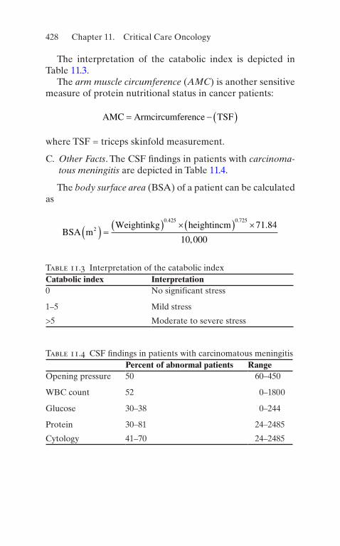
428
The interpretation of the catabolic index is depicted in Table 11.3.
The arm muscle circumference (AMC) is another sensitive measure of protein nutritional status in cancer patients:
AMC Armcircumference TSF� � � �
where TSF = triceps skinfold measurement.
C. Other Facts. The CSF findings in patients with carcinoma-tous meningitis are depicted in Table 11.4.
The body surface area (BSA) of a patient can be calculated as
BSA m
Weightinkg heightincm2
0 425 0 72571 84
10 000� � � � � �� � �
. ..
,
Table 11.4 CSF findings in patients with carcinomatous meningitisPercent of abnormal patients Range
Opening pressure 50 60–450
WBC count 52 0–1800
Glucose 30–38 0–244
Protein 30–81 24–2485
Cytology 41–70 24–2485
Table 11.3 Interpretation of the catabolic indexCatabolic index Interpretation0 No significant stress
1–5 Mild stress
>5 Moderate to severe stress
Chapter 11. Critical Care Oncology

429© Springer Nature Switzerland AG 2021J. Varon, Handbook of Critical and Intensive Care Medicine, https://doi.org/10.1007/978-3-030-68270-5_12
Many patients presenting to an intensive care facility will be pregnant. Many patients will have diseases peculiar to preg-nancy and will need critical care support. Others, however, will have underlying medical diseases (Table 12.1). Some of those diseases will require consideration of the passenger (fetus) who has created many changes in maternal physiol-ogy. The hormonal milieu created by the placenta—proges-terone and to a lesser extent estrogen—is responsible for the multifaceted changes in system function.
One change seen early in pregnancy is in pulmonary func-tion. Table 12.2 depicts these modifications. Another organ system with significant change is the kidney. Table 12.3 reflects the serial changes in function. Table 12.4 demon-strates the differential risk of both acquired and congenital heart disease during pregnancy.
Since many books on obstetrical critical care have been published, and a full review of the many changes and diseases is beyond the scope of this chapter, a disease process that reflects the complexity of the severely ill gravida within the intensive care unit (ICU) has been chosen.
Hemodynamic changes during pregnancy and maternal physiologic changes occurring during labor should be kept in mind (see Tables 12.5 and 12.6).
Chapter 12Critical Care of the Pregnant Patient

430
Pregnancy-Induced Hypertension
A. Definition. Pregnancy-induced hypertension (PIH) is the presence of elevated blood pressure with evidence of end- organ dysfunction, most commonly seen as edema, pro-teinuria, and elevated blood pressure. Table 12.7 presents many of the synonyms for this process. The classification of preeclampsia (PIH) is depicted in Table 12.8.Hypertension during pregnancy is divided in four categories:
• Preeclampsia/eclampsia/HELLP (hemolysis, elevated liver enzymes, low platelets)
• Chronic hypertension (of any cause): Hypertension that predates pregnancy
• Chronic hypertension with superimposed preeclampsia: Chronic hypertension in association with preeclampsia
Table 12.1 Preexistent medical diseases
Asthma
Cardiac disease, NYHA class 3/4
Prosthetic valve replacements
Critical mitral stenosis
Aortic stenosis
Eisenmenger’s syndrome
Cystic fibrosis
Diabetes mellitus (insulin dependent)
Chronic renal failure
Hypertension
Renal, hepatic, cardiac transplants
Systemic lupus erythematous
Thyrotoxicosis/thyroid storm
Treatment of these conditions during pregnancy remains unchanged
Chapter 12. Critical Care of the Pregnant Patient

431
• Gestational hypertension: Blood pressure elevation after 20 weeks of gestation in the absence of proteinuria or the aforementioned systemic findings
B. Diagnosis (See Table 12.9) C. Etiology. In recent years the understanding and manage-
ment of preeclampsia has improved. Despite intensive efforts to find mechanisms and markers that induce PIH, the
Table 12.2 Lung volumes and capacities in pregnancy
DefinitionChange in pregnancy
Respiratory rate (RR)
Number of breaths per minute Unchanged
Vital capacity (Vc)
Maximum amount of air that can be forcibly expired after maximum inspiration (IC + ERV)
Unchanged
Inspiratory capacity (IC)
Maximum amount of air that can be inspired from resting expiratory level (TV + IRV)
Increased 5%
Tidal volume (VT)
Amount of air inspired and expired with normal breath
Increased 30–40%
Inspiratory reserve volume (IRV)
Maximum amount of air that can be inspired at end of normal inspiration
Unchanged
Functional residual capacity (FRC)
Amount of air in lungs at resting expiratory level (ERV + RV)
Decreased 20%
Expiratory reserve volume (ERV)
Maximum amount of air that can be expired from resting expiratory level
Decreased 20%
Residual volume (RV)
Amount of air in lungs after maximum expiration
Decreased 20%
Total lung capacity (TLC)
Total amount of air in lungs at maximal inspiration (VC + RV)
Decreased 5%
Pregnancy-Induced Hypertension

432
Tabl
e 12
.3 S
eria
l cha
nges
in r
enal
hem
odyn
amic
sN
onpr
egna
nt16
wee
ks26
wee
ks29
wee
ks36
wee
ks37
wee
ksE
ffec
tive
ren
al48
0 ±
72
840
± 1
4589
174
8 ±
85
771
± 1
7567
7 ±
82
Pla
sma
flow
(m
L/m
in)
279
Glo
mer
ular
filt
rati
on r
ate
(mL
/min
)99
± 1
814
9 ±
17
152
± 1
814
5 ±
19
150
± 3
213
8 ±
22
Filt
rati
on f
ract
ion
0.21
0.18
0.18
0.19
0.20
0.21
Chapter 12. Critical Care of the Pregnant Patient

433
Table 12.4 Pregnancy risk with cardiac diseaseCategory 1 Low risk during pregnancy
Small left-to-right shunts
Pulmonary stenosis <50 mmHg gradient
Mild mitral/aortic insufficiency
Mild aortic stenosis
Mitral valve prolapse
Rheumatic fever or endocarditis history
Postoperative patients, normal hemodynamics
Category 2 Moderate risk during pregnancy
Large left-to-right shunts, low pulmonary pressure
Moderate pulmonary stenosis
Aortic stenosis (30–60 mmHg gradient)
Mild hypertrophic cardiomyopathy
Cardiac valve prosthesis
Mild mitral stenosis
Palliated cyanotic heart disease
Moderate aortic/mitral regurgitation
Category 3 High risk during pregnancy
Large left-to-right shunts, mild pulmonary hypertension
Severe aortic/pulmonary stenosis
Mild mitral stenosis with atrial fibrillation
Moderate mitral stenosis
Cardiomyopathy in early stages
Moderate-to-severe IHSS
Cyanotic congenital heart disease, unoperated
Mild Epstein’s disease
Postcardiac surgery, mild residual problems
(Continued)
Pregnancy-Induced Hypertension

434
Table 12.5 Hemodynamic changes of pregnancyCardiac output Increases 30–40%
Heart rate Increases 10–15%
Stroke volume Increases
Blood volume Increases 30–40%
Systemic blood pressure Decreases
Pulse pressure Increases
Systemic resistance Decreases
Pulmonary artery pressure No change
Pulmonary resistance Decreases
Myocardial function Improves
Table 12.6 Hemodynamic effects of labor and deliveryCardiac output Increases with contractions
Blood volume Increases
Heart rate Variable
Peripheral resistance No change
Systemic artery pressure Increases
Table 12.4 (Continued)
Category 4 Pregnancy contraindicated
Congestive heart failure
Pulmonary hypertension
Eisenmenger’s syndrome
Severe cyanosis
Advanced coronary artery disease
Marfan’s syndrome
IHSS idiopathic hypertrophic subaortic stenosis
Chapter 12. Critical Care of the Pregnant Patient

435
Table 12.7 Synonyms for pregnancy-induced hypertension
Toxemia of pregnancy
PIH
Preeclampsia
Eclampsia
Peripartum hypertension
EPH (edema, proteinuria, hypertension) gestosis
Table 12.8 Classification of preeclampsiaMild Severea
Blood pressure 130/80–140/95 mmHg
>160/110 mmHg
Absolute Systolic ≥140 mmHg
Diastolic ≥90 mmHg
Relative Systolic increased >30 mmHg
Diastolic increased >15 mmHg
Clinical findings 1 + edema 3–4 + edema
Normal reflexes 3–4 + reflexes
No visual symptoms
Scotoma/papilledema diplopia
May have seizures, altered consciousness
Severe headaches
Severe right upper quadrant abdominal pain or
Tenderness
(Continued)
Pregnancy-Induced Hypertension

436
exact etiology is not fully defined. It is most commonly believed to be an end product of antigen–antibody interac-tion with abnormal ratios of vasoactive agents such as pros-tacyclins and thromboxanes. The relationship of other agents such as lipid peroxides remains under active investigation. Elevations of malondialdehyde (MDA), uric acid, caspase activity, and the percentage of DNA fragmentation have been found in the placentas of women with preeclampsia. These changes implicate the involvement of lipid peroxida-tion and apoptosis in preeclampsia, suggesting oxidative stress. Regardless of the exact etiology, the process reflects a diffuse systemic endothelial dysfunction intimately associ-
Table 12.8 (Continued)
Mild Severea
Congestive heart failure, pulmonary edema
Oliguria <400 cc/24 h
Weight lab >5 lb/week
Proteinuria 300 mg/24 h ≥5 g/day; 3+/4+ semiquantitative
Platelets Normal May be <150,000
Liver function Normal Elevated AST/ALT
Clotting studies Normal May be prolonged
Bilirubin Normal May be elevated
Anticonvulsants Intrapartum: yes Antepartum: yes
Intrapartum: yes
Abdominal pain
Absent May be present in epigastrium or RUQ
ALT alanine aminotransferase, AST aspartate aminotransferase, RUQ right upper quadrantaHELLP syndrome, reflected by hemolysis, elevated liver enzymes, and low platelet count, comprises the greatest risk group for mortal-ity and morbidity
Chapter 12. Critical Care of the Pregnant Patient

437
Table 12.9 Diagnostic criteria for preeclampsiaBlood pressure Greater than or equal to 140 mmHg
systolic or greater than or equal to 90 mmHg diastolic on two occasions at least 4 h apart after 20 weeks of gestation in a woman with a previously normal blood pressure
Greater than or equal to 160 mmHg systolic or greater than or equal to 110 mmHg diastolic, hypertension can be confirmed within a short interval (minutes) to facilitate timely antihypertensive therapy
And
Proteinuria Greater than or equal to 300 mg per 24-h urine collection (of this amount extrapolated from a timed collection)
Or
Protein/creatinine ratio greater than or equal to 0.3a
Dipstick reading of 1+ (used only if other quantitative methods not available)
Or in the absence of proteinuria, new-onset hypertension with the new onset of any of the following
Thrombocytopenia Platelet count less than 100,000/μl
Renal insufficiency Serum creatinine concentrations greater than 1.1 mg/dL or a doubling of the serum creatinine concentration in the absence of other renal disease
Impaired liver function
Elevated blood concentrations of liver transaminases to twice normal concentration
(Continued)
Pregnancy-Induced Hypertension

438
ated with platelet dysfunction. This is seen in the classic HELLP (hemolysis, elevated liver enzymes, low platelet) syndrome, in which many organ systems have reflected the disease, and if assiduous management does not occur, severe maternal morbidity or mortality may occur. Table 12.10 depicts the panorama of disease manifestations. Table 12.11 depicts those subsets of pregnant women at greatest risk for the disease process. Table 12.12 presents the frequency of preeclampsia in the highest-risk populations.
Patients with prior PIH who have underlying chronic hypertension have a 50–75% probability of developing PIH in pregnancy. No method exists to predict the severity or to time the onset of the PIH process.
D. Approach to the Preeclamptic Patient
1. Obtain Patient History
(a) Current gestational age (calculated by LMP, last menstrual period)
(b) Past medical history: renal or chronic hypertensive diseases, systemic lupus erythematosus
(c) Family history of preeclampsia/eclampsia (d) Symptoms of disease
1. Headache, blurred vision, and scotoma 2. Blindness and diplopia 3. Weight gain (>2 lb/week) 4. Right upper quadrant pain, epigastric pain, and
diffuse abdominal pain (ruptured liver) 5. Tetanic contractions (abruptio placentae)
Table 12.9 (Continued)
Pulmonary edema
Cerebral or visual symptoms
Adapted from Hypertension in Pregnancy, Report of the American College of Obstetricians and Gynecologists’ Task Force on Hypertension in Pregnancy. 2013, 122aEach measured as mg/dL
Chapter 12. Critical Care of the Pregnant Patient

439
Table 12.10 Pregnancy- induced hypertension: associated complications
Hypertensive crisis
Pulmonary edema: ARDS
Eclampsia
Intracranial hemorrhage
Amaurosis
Cerebral edema
Acute renal failure
Cortical necrosis
Ruptured liver
Microangiopathic hemolytic anemia
Thrombocytopenia
DIC
HELLP syndrome
ARDS adult respiratory distress syn-drome, DIC disseminated intravascu-lar coagulation, HELLP hemolysis, elevated liver enzymes, low platelets
Table 12.11 Factors asso-ciated with PIH
Nulligravida
Prior preeclampsia/eclampsia
Family history of preeclampsia, eclampsia
Maternal age >40 years
Antiphospholipid antibody syndrome
Multifetal pregnancies
Molar pregnancies
Hydrops fetalis (any etiology)
Chronic hypertension/renal disease
Diabetes mellitus, insulin dependent
Pregnancy-Induced Hypertension

440
6. Nausea and emesis 7. Unconsciousness or seizure activity 8. Vaginal bleeding 9. Fetal movement
2. Physical Examination
(a) Maternal blood pressure, pulse, and respiratory rate.
(b) Fetal heart rate (FHR) by continuous electronic monitoring.
(c) Extensive cardiopulmonary examination. (d) Eyes: Scleral icterus, ecchymoses, and petechiae. (e) Fundoscopic examination: Retinal artery spasm, pap-
illedema, and hemorrhages; acute vasospasm is often seen; arteries may be only 50% diameter of veins.
(f) Abdominal examination:
1. Upper quadrant tenderness 2. Uterus size, tone, softness, and noncontracting
or rigid contracting 3. Distension: Is there suggestion of ascites?
(g) Extremities/face: Evidence of pathological edema. (h) Pelvic exam: Cervical softness, dilatation, position,
effacement, and fetal presentation.
Table 12.12 Frequency of preeclampsia
7% of all pregnancies
70% nulligravidas
30% multigravidas
Twins 30%
Molar pregnancies Up to 70%
Hydrops fetalis Up to 50%
Diabetes mellitus Up to 50%
Chronic hypertension
20%
Prior severe PIH Up to 50%
Chapter 12. Critical Care of the Pregnant Patient

441
(i) Patellar reflexes: Persistent clonus reflects central nervous system (CNS) hyperactivity and signifi-cant potential for seizure activity.
(j) Please note: Blood pressure may be taken in both supine and lateral positions. Arm elevation when the patient is turned to the lateral decubitus results in a fall in blood pressure commensurate with the distance in centimeters above the atrial level, roughly, 13.6 mmHg/10 cm of hydrostatic pressure. This change in pressure is frequently suggested to be the “real blood pressure.” A patient may then be considered to be normal when in fact hypertension exists. Table 12.13 reflects these changes in the best study done in normal pregnant patients. (Mean arterial pressure [MAP] is unchanged.)
The intensivist should remember that because of the vasodilation of pregnancy and decreased systemic vascular resistance (SVR), patients, espe-cially teenagers, with blood pressures (BPs) of 140/90 mmHg or lesser blood pressures may be sig-nificantly hypertensive.
3. Laboratory Evaluation
(a) Type/Rh, indirect Coombs, rapid plasma reagin (RPR), hepatitis B surface antigen (HbsAg), rubella (if not previously obtained).
(b) Complete blood count (CBC), platelets, micro-scopic examination.
(c) Serum urea nitrogen (BUN), creatinine, aspartate aminotransferase (AST), alanine aminotransferase (ALT), glucose, electrolytes, uric acid.
(d) Coagulation studies: prothrombin time (PT), par-tial thromboplastin time (PTT), fibrinogen and fibrin split products (FSPs)).
(e) Plasma oncotic pressure (colloid osmotic pressure [COP]) decreases in pregnancy secondary to hemo-dilution (see Table 12.14).
(f) Urinalysis with rapid screen for proteinuria in intensive care unit (ICU).
Pregnancy-Induced Hypertension

442
Tabl
e 12
.13
Hem
odyn
amic
alt
erat
ions
in r
espo
nse
to p
osit
ion
chan
ge la
te in
thi
rd t
rim
este
r of
pre
gnan
cy
Hem
odyn
amic
par
amet
erP
osit
ion
LL
SUP
SIT
STM
AP
(m
mH
g) H
g90
± 6
90 ±
890
± 8
91 ±
14
CO
(L
/min
)6.
6 ±
1.4
6.0
± 1
.4*
6.2
± 2
.054
± 2
.0*
P (
beat
s/m
in)
82 ±
10
84 ±
10
91 ±
11
107
± 1
7*
SVR
(dy
ne c
m s−
5 )1,
210
± 2
661,
437
± 3
381,
217
± 2
541,
319
± 3
94
PV
R (
dyne
cm
s−
5 )76
± 1
610
1 ±
45
102
± 3
511
7 ±
35*
PC
WP
(m
mH
g) H
g8
± 2
6 ±
34
± 4
4 ±
2
CV
P (
mm
Hg)
Hg
4 ±
33
± 2
1 ±
11
± 2
LVSW
I (g
/m/m
2 /bea
t)43
± 9
40 ±
944
± 5
34 ±
7*
Ada
pted
fro
m C
lark
et
al. A
m J
Obs
tet
Gyn
ecol
199
1;16
4:88
3–88
7C
O c
ardi
ac o
utpu
t, P
pul
se, S
VR
sys
tem
ic v
ascu
lar
resi
stan
ce, P
VR
pul
mon
ary
vasc
ular
res
ista
nce,
PC
WP
pul
mon
ary
capi
llary
wed
ge p
ress
ure,
CV
P c
entr
al v
enou
s pr
essu
re, L
VSW
I le
ft v
entr
icul
ar s
trok
e w
ork
inde
x, L
L le
ft la
tera
l, SU
P
supi
ne, S
IT s
itti
ng, S
T s
tand
ing
* P <
0.0
5, c
ompa
red
wit
h le
ft la
tera
l pos
itio
n
Chapter 12. Critical Care of the Pregnant Patient

443
(g) Chest X-ray only if pulmonary symptoms or physi-cal examinations suggest its need.
(h) Obstetrical ultrasound (in ICU) for fetal age and number, estimated fetal weight, position of fetuses, and placental position.
(i) Continuous FHR monitoring. Fetal well-being is evaluated by a nonstress test or biophysical profile.
(j) Twenty-four-hour urine collection for creatinine clearance and protein excretion.
E. Medical Therapy (See Table 12.15)
1. The presentation of a patient with PIH may range from a mild to a life-threatening disease process. The process can only be ended by delivery. The decision to continue or to deliver the pregnancy will be made by consulta-tion between medical and obstetrical personnel.
2. A true rule is that the disease may rapidly progress. Follow-up of all maternal and fetal biophysical param-eters on a routine basis is required. The frequency will be determined by the disease severity.
3. Most preeclamptic patients are vasoconstricted and hemoconcentrate. After initial therapy, volume expan-sion and hemodilution occurs.
4. Medications: Choice of antihypertensive agents should be based on clinical expertise.
5. MgSO4 (magnesium sulfate) is considered the standard of therapy as a prophylaxis for seizure activity. Magnesium is not an antihypertensive. It should be used for seizure prevention in preeclampsia with severe features and for neuroprotection when delivery is expected before 32 weeks of gestation. The loading
Table 12.14 Colloid osmotic pressure
Nonpregnant 28 mmHg
Pregnant, term 23 mmHg
Postpartum 17 mmHg
Preeclampsia (PIH)
13.7 mmHg
Pregnancy-Induced Hypertension
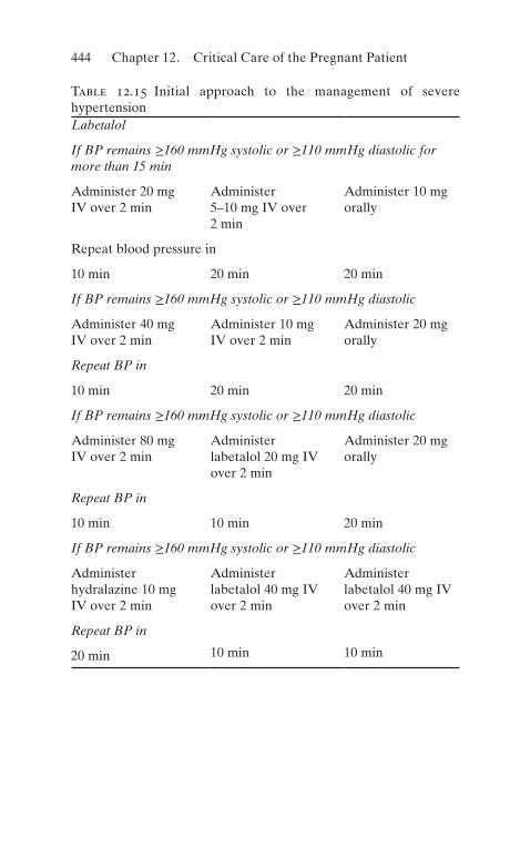
444
Table 12.15 Initial approach to the management of severe hypertensionLabetalol
If BP remains ≥160 mmHg systolic or ≥110 mmHg diastolic for more than 15 min
Administer 20 mg IV over 2 min
Administer 5–10 mg IV over 2 min
Administer 10 mg orally
Repeat blood pressure in
10 min 20 min 20 min
If BP remains ≥160 mmHg systolic or ≥110 mmHg diastolic
Administer 40 mg IV over 2 min
Administer 10 mg IV over 2 min
Administer 20 mg orally
Repeat BP in
10 min 20 min 20 min
If BP remains ≥160 mmHg systolic or ≥110 mmHg diastolic
Administer 80 mg IV over 2 min
Administer labetalol 20 mg IV over 2 min
Administer 20 mg orally
Repeat BP in
10 min 10 min 20 min
If BP remains ≥160 mmHg systolic or ≥110 mmHg diastolic
Administer hydralazine 10 mg IV over 2 min
Administer labetalol 40 mg IV over 2 min
Administer labetalol 40 mg IV over 2 min
Repeat BP in
20 min 10 min 10 min
Chapter 12. Critical Care of the Pregnant Patient

445
dose is 4–6 g MgSO4·7H2O in 100 cc D5 1/4 NS over 15–20 min. A constant infusion of MgSO4 1–2 g/h will be maintained depending on urine output and reflex activity, which are checked on an hourly basis. Table 12.16 lists the potential effects of the magnesium ion and the average serum level at which they may occur. Women receiving magnesium should be moni-tored closely. When MgSO4 is infused, an infusion pump normally will be used to enhance patient safety, thus preventing a massive infusion of MgSO4, which could cause maternal death or severe morbidity. Magnesium toxicity can be treated with 10 ml of 10% calcium glu-conate solution.
6. Medications to avoid in pregnancy-induced hyperten-sion: Angiotensin-converting enzyme (ACE) inhibitors, angiotensin receptor blockers, and mineralocorticoid receptor antagonists should be avoided. ACE inhibitors and angiotensin receptor blockers interfere with fetal renal hemodynamics leading to congenital abnormali-ties in the first trimester and oligohydramnios, kidney injury, and death in the second and third trimesters. Mineralocorticoid receptor antagonists can cause femi-
Table 12.15 (Continued)
If BP remains >160 mmHg systolic or >110 mmHg diastolic
Obtain emergency consultation and treat as recommended
Adapted from American College of Obstetricians and Gynecologists. Committee opinion no. 623: Emergent therapy for acute-onset, severe hypertension during pregnancy and the post-partum period. Obstet Gynecol 2015;125:521–5These algorithms are appropriate for antepartum, intrapartum, and postpartum severe hypertension. Choice of agent should be guided by clinician experience and knowledge of adverse effect. Management should also include physician notification, documenta-tion, and fetal surveillance. Once target is achieved, check BP every 10 min for 1 h, then every 15 min for 1 h, then every 30 min for 1 h, and then hourly for 4 hBP blood pressure, IV intravenous
Pregnancy-Induced Hypertension
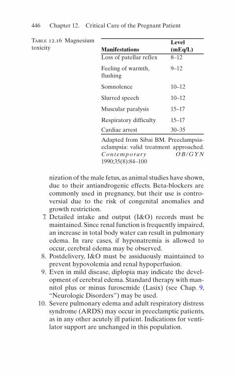
446
nization of the male fetus, as animal studies have shown, due to their antiandrogenic effects. Beta-blockers are commonly used in pregnancy, but their use is contro-versial due to the risk of congenital anomalies and growth restriction.
7. Detailed intake and output (I&O) records must be maintained. Since renal function is frequently impaired, an increase in total body water can result in pulmonary edema. In rare cases, if hyponatremia is allowed to occur, cerebral edema may be observed.
8. Postdelivery, I&O must be assiduously maintained to prevent hypovolemia and renal hypoperfusion.
9. Even in mild disease, diplopia may indicate the devel-opment of cerebral edema. Standard therapy with man-nitol plus or minus furosemide (Lasix) (see Chap. 9, “Neurologic Disorders”) may be used.
10. Severe pulmonary edema and adult respiratory distress syndrome (ARDS) may occur in preeclamptic patients, as in any other acutely ill patient. Indications for venti-lator support are unchanged in this population.
Table 12.16 Magnesium toxicity Manifestations
Level (mEq/L)
Loss of patellar reflex 8–12
Feeling of warmth, flushing
9–12
Somnolence 10–12
Slurred speech 10–12
Muscular paralysis 15–17
Respiratory difficulty 15–17
Cardiac arrest 30–35
Adapted from Sibai BM. Preeclampsia- eclampsia: valid treatment approached. C o n t e m p o ra r y O B / G Y N 1990;35(8):84–100
Chapter 12. Critical Care of the Pregnant Patient
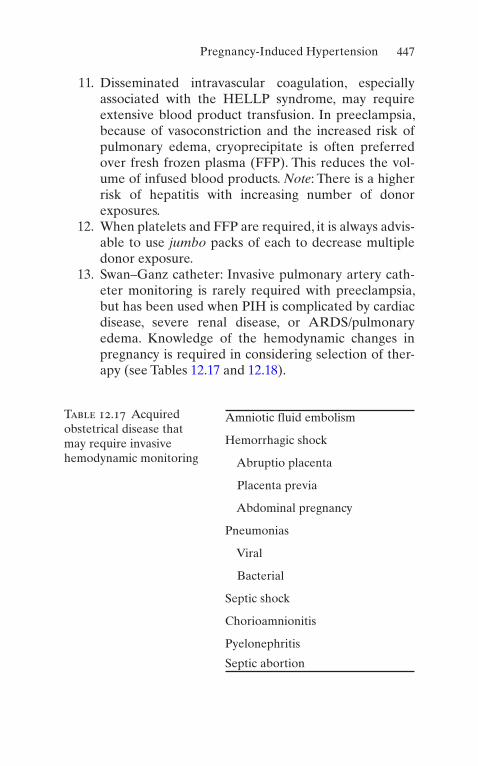
447
11. Disseminated intravascular coagulation, especially associated with the HELLP syndrome, may require extensive blood product transfusion. In preeclampsia, because of vasoconstriction and the increased risk of pulmonary edema, cryoprecipitate is often preferred over fresh frozen plasma (FFP). This reduces the vol-ume of infused blood products. Note: There is a higher risk of hepatitis with increasing number of donor exposures.
12. When platelets and FFP are required, it is always advis-able to use jumbo packs of each to decrease multiple donor exposure.
13. Swan–Ganz catheter: Invasive pulmonary artery cath-eter monitoring is rarely required with preeclampsia, but has been used when PIH is complicated by cardiac disease, severe renal disease, or ARDS/pulmonary edema. Knowledge of the hemodynamic changes in pregnancy is required in considering selection of ther-apy (see Tables 12.17 and 12.18).
Table 12.17 Acquired obstetrical disease that may require invasive hemodynamic monitoring
Amniotic fluid embolism
Hemorrhagic shock
Abruptio placenta
Placenta previa
Abdominal pregnancy
Pneumonias
Viral
Bacterial
Septic shock
Chorioamnionitis
Pyelonephritis
Septic abortion
Pregnancy-Induced Hypertension

448
Tabl
e 12
.18
Ant
ihyp
erte
nsiv
e dr
ugs
in p
regn
ancy
Dru
gO
nset
of
acti
onD
urat
ion
Dos
age
Mec
hani
smSi
de e
ffec
tsC
levi
dipi
ne30
s2
min
IV 1
–2 m
g/h
follo
wed
by
repe
ated
in
crem
enta
l do
ublin
g of
the
do
se a
t 90
s
inte
rval
Art
eria
l va
sodi
lato
rN
one
(Cle
vipr
ex)
The
des
ired
th
erap
euti
c re
spon
se f
or m
ost
pati
ents
occ
ur a
t do
ses
4–6
mg/
h
Hyd
rala
zine
(A
pres
olin
e)15
–20
min
3–6
h5–
10 m
g q3
0 m
inD
irec
t sm
ooth
m
uscl
e re
laxa
tion
Ref
lex
tach
ycar
dia
Hea
dach
e
Chapter 12. Critical Care of the Pregnant Patient

449
Nic
ardi
pine
a (C
arde
ne)
1–5
min
3–6
hIV
dri
p 2
mg/
h,
incr
ease
by
2 m
g/h
each
hou
r; m
ax
dosa
ge 1
0 m
g/h
Vas
odila
tion
, ca+
ch
anne
l blo
cker
Hea
dach
e, n
ause
a, v
omit
ing,
hy
pote
nsio
n
Lab
etal
ol
(Tra
ndat
e)5–
10 m
in3–
6 h
20 m
g by
slo
w
IV in
ject
ion
over
2
min
. May
rep
eat
dose
q10
–15
min
to
ach
ieve
de
sire
d pr
essu
re.
Max
imum
dos
age
300
mg
Bet
a-bl
ocke
r 7:
1 va
sodi
lato
rR
atio
of
beta
- bl
ocke
r to
alp
ha-
bloc
ker
(7:1
) w
ith
IV a
dmin
istr
atio
n
Hyp
oten
sion
, diz
zine
ss, f
atig
ue,
naus
ea, b
ronc
hoco
nstr
icti
on
in s
ome
pati
ents
wit
h as
thm
a.
Feta
l sid
e ef
fect
s m
ay in
clud
e br
adyc
ardi
a, p
oor
tem
pera
ture
co
ntro
l, hy
pogl
ycem
ia, a
nd
decr
ease
d sh
ort-
term
var
iabi
lity
Feno
ldop
am5
min
30–
60 m
in0.
1 μg
/kg
over
1
min
Dop
amin
e-1
agon
ist
Incr
ease
s in
trao
cula
r pr
essu
re,
dose
dep
ende
nt
Tit
rate
to
desi
red
bloo
d pr
essu
re b
y in
crem
ents
0.0
5–0.
1 μg
to
a m
ax
1.6 μg
/kg/
min
.
Pote
ntia
l sul
fite
sen
siti
vity
ti
trat
ed
a A r
educ
tion
of
20%
in M
AP
sho
uld
be m
axim
al in
itia
l tar
get.
Follo
win
g m
ater
nal a
nd f
etal
res
pons
e to
the
rapy
, fur
-th
er d
ecre
ase
in M
AP
may
be
desi
red
Pregnancy-Induced Hypertension

450
14. Development of seizures (eclampsia) may occur pre- or posttreatment with MgSO4. If seizures develop pretherapy, 4 g of MgSO4 (8 cc of MgSO4·7H2O—50% solution) may be rapidly infused. If seizures develop posttreatment, a second dose of MgSO4 may be given, or some physicians choose to give diazepam (Valium™) 2.5–5 mg as an IV push as the agent of choice. If the patient does not awaken and become responsive within 60 min, then the possibility of an intracranial hemor-rhage must be considered and worked up (see Chap. 9, “Neurologic Disorders”).
15. Pulse oximeters and recording dynamaps may be used in many patients. If any concern for an atypical or severe manifestation of PIH is present, an arterial cath-eter should be placed. Repetitive laboratory studies can be drawn, and continuous blood pressure recording can be achieved.
16. Interactive dialogue between all members of the healthcare team will achieve optimum outcome for both mother and child.
F. Major Complications of Preeclampsia/Eclampsia (See Table 12.19)
1. Hypertensive Crisis
(a) BPs >200 systolic or 120 diastolic. (b) May be associated with pulmonary edema, intra-
cranial hemorrhage, or cerebral edema. (c) Rapid treatment is critical (see Table 12.18). An
acute reduction of elevated BP should initially be limited to a 20% reduction in mean arterial pres-sure. A more substantial reduction may create severe uteroplacental hypoperfusion and precipi-tate acute fetal death or asphyxia. This is especially true if the diastolic blood pressure is acutely dropped to 90 mmHg or less.
(d) These patients frequently warrant placement of invasive monitoring systems such as pulmonary artery catheters and peripheral arterial cannulas.
2. Cerebral Edema
Chapter 12. Critical Care of the Pregnant Patient

451
(a) Initiate fluid restriction. (b) Invasive monitoring. (c) IV mannitol 1–2 g/kg of a 20% solution (100 g) of
mannitol in 500 mL of 5% D/W given over 10–20 min followed by a maintenance dose of 50–300 mg/kg IV q6 h is effective. The serum osmo-lality should not be allowed to exceed 330–340 mOsm.
(d) Complications of osmotic agent use include:
1. Osmotic diuresis with dehydration and hypernatremia
2. Rebound increase in intracranial pressure 3. Acute volume expansion
(e) If a patient is intubated, hyperventilation may be used. (See Chap. 9, “Neurologic Disorders.”)
3. Hepatic Rupture
Table 12.19 Features of preeclampsia that is likely to worsenBlood pressure
Systolic blood pressure of 160 mmHg or higher
Diastolic blood pressure of 110 mmHg or higher on two occasions at least 4 h apart, while the patient is on bed rest (unless antihypertensive therapy is initiated before this time)
Platelets Thrombocytopenia (platelet count less than 100,000/μl)
Liver Impaired liver function as indicated by abnormally elevated blood concentrations of liver enzymes (to twice normal concentration), severe persistent right upper quadrant, or epigastric pain unresponsive to medication and not accounted for by alternative diagnoses or both
Kidney Progressive renal insufficiency (serum creatinine concentration greater than 1.1 mg/dl or a doubling of the serum creatinine concentration in the absence of other renal diseases)
Lung Pulmonary edema
Neurologic New-onset cerebral or visual disturbances
Pregnancy-Induced Hypertension

452
(a) Massive intra-abdominal hemorrhage results with need for:
1. Massive blood volume support 2. Corrections of disseminated intravascular coag-
ulation (DIC) 3. Invasive cardiovascular monitoring
(b) Exploration and surgical repair when necessary. Because of liver dysfunction/damage, packing of the rupture site is often accomplished.
(c) Potential for automatic cell saver at operation can reduce total transfusion requirements.
4. Abruptio Placentae
(a) Frequently associated with fetal distress. (b) Often accompanied by coagulopathy with pro-
longed PT, PTT, low fibrinogen, and low platelet count.
(c) Vigorous/massive transfusion support may be required.
(d) Four major complications of hypovolemia and shock in these patients as follows:
1. Acute tubular necrosis. 2. Cortical renal necrosis. 3. Sheehan’s syndrome with acute pituitary
insufficiency. 4. ARDS: This has been a major cause of death in
our obstetrical ICU. G. Therapy
1. Magnesium Sulfate
(a) Distribution: extracellular space, bones, and intra-cellular space.
(b) Unbound to protein. (c) Excretion by the kidney; filtered load excretes T
max for reabsorption in most patients treated. (d) Excretion half-life is ~4 h. (e) When used in normal patients for treatment of pre-
term labor, the earliest manifestations of excessive Ca++ antagonism are ocular symptoms of visual
Chapter 12. Critical Care of the Pregnant Patient
453
disturbance: blurring of vision, diplopia, and diffi-culty in focusing.
(f) MgSO4 does not usually change blood pressure. (g) Measurements of magnesium levels can be achieved
in most clinical labs. There is a poor correlation between levels observed and clinical effect. Therefore, no precise level can be stated to be therapeutic.
H. Antihypertensive Therapy (See Table 12.18)
1. In most circumstances, drug therapy in PIH is reserved for those patients with:
(a) Persistent systolic BPs >180 mmHg (b) Persistent diastolic BPs >110 mmHg (105 mmHg in
some institutions)
2. Before delivery, it is desired to maintain the diastolic blood pressure >90 mmHg. This allows for continued perfusion pressure to provide adequate uteroplacental perfusion.
3. If diastolic blood pressure decreases <90 mmHg, fre-quently, the decreased uteroplacental perfusion will precipitate acute fetal distress, which may progress to an in utero death or to perinatal asphyxia.
4. Postdelivery, an acute, rapid decrease in blood pressure usually means substantial blood loss and not cure of the disease process. Likewise, a nadir of 90 mmHg diastolic blood pressure is desired.
5. Medical control of hypertension is often required only for a short period (usually days). No study has ever demonstrated a beneficial long-term outcome with pro-longed antihypertensive therapy.
6. The use of calcium channel blockers in a setting of MgSO4 therapy should be considered a significant ther-apeutic step that may create an adverse impact on car-diovascular function; therefore, an intensive care setting with knowledgeable personnel (internal medicine, OB/GYN maternal fetal medicine, or OB anesthesia) capa-ble of responding to these problems should be present.
Pregnancy-Induced Hypertension
454
Prevention
1. Administration of low-dose aspirin (60–80 mg) has been examined in meta-analyses of more than 30,000 women, and it appears that there is a slight effect to reduce pre-eclampsia and adverse perinatal outcomes. These findings may be relevant to populations at very high risk.
2. There is no evidence that bed rest, salt restriction, and the use of antioxidants such as vitamins C and E reduce pre-eclampsia risk.
Amniotic Fluid Embolism
A. DefinitionAmniotic fluid embolism (AFE) is the vascular transfer
of amniotic fluid containing lanugo hairs, vernix, meco-nium, and thromboplastic substances to the pulmonary circulation. This is a rare (1 in 40,000 pregnancies) and unpredictable event that is catastrophic and often associ-ated with death. It usually presents during delivery or the immediate postpartum period.
1. It is unpreventable and is most frequent in the second stage of labor.
2. The mortality rate is between 20% and 60%; 25% of deaths occur in the first 60 min post event.
3. Old animal models of disease are not applicable to human pathology.
B. PresentationSudden onset of maternal distress (usually during labor
and delivery or 30 minutes post-partum):
• Neurological (33% of patients)
– Altered mental status – Seizures – Coma
• Respiratory – Respiratory arrest
Chapter 12. Critical Care of the Pregnant Patient

455
– Shortness of breath/dyspnea – Pulmonary edema – Acute respiratory distress syndrome – Cyanosis
• Cardiac
– Sudden cardiovascular collapse – Profound systemic hypotension – Cardiac dysrhythmias – Myocardial infarction
• Hematologic
– Disseminated intravascular coagulation (80% of patients)
– Hemorrhage
C. Predisposing Factors
• Fetal Factors
– Fetal distress – Fetal macrosomia – Intrauterine death – Male gender – Placenta accreta – Placenta previa – Placental abruption – Polyhydramnios – Premature rupture of membranes
• Maternal Factors
– Advanced maternal age >35 – Diabetes – Multiparity
• Obstetrical Factors
– Cervical laceration – Cesarean section – Eclampsia – Induction of labor
Amniotic Fluid Embolism

456
– Instrumental delivery – Oxytocin use – Uterine rupture
D. Symptoms
• Restlessness• Sweating• Anxiety• Coughing• Shortness of breath
E. Laboratory and Diagnostic Studies
1. Coagulation (PT, PTT, D-Dimer, Fibrinogen) 2. Complete Blood Count 3. Arterial Blood Gas 4. Chest Radiography 5. Electrocardiogram 6. Echocardiogram
F. Pathophysiology
1. Pulmonary hypertension; decreased cardiac output. 2. Cor pulmonale with pulmonary edema. 3. Severe hypoxemia and tissue hypoxia. 4. If acute respiratory distress allows survival, thrombo-
plastins yield DIC. 5. Swan–Ganz data reveal predominant left heart failure/
dysfunction: 2° to hypoxic injury.
G. Differential Diagnosis
1. Acute pulmonary embolism 2. Air embolism 3. Myocardial infarction 4. Peripartum cardiomyopathy 5. Acute aspiration of gastric contents 6. Aortic dissection 7. Cardiac dysrhythmia 8. Massive pneumothorax: uni-/bilateral 9. Reaction to local anesthetic
Chapter 12. Critical Care of the Pregnant Patient

457
10. Septic shock 11. Anaphylactic shock: drug induced 12. Postpartum hemorrhage 13. Uterine rupture 14. Placental abruption 15. Eclampsia
H. Acute Treatment
1. Endotracheal tube placement 2. Mechanical ventilation/positive end-expiratory pres-
sure (PEEP) 3. Volume support with or without blood products 4. Peripheral and pulmonary arterial catheterization 5. Central venous pressure (CVP) monitoring 6. No specific drug therapy: vasopressors and/or bron-
chodilators of choice
(a) Phenylepinephrine (b) Vasopressin (c) Milrinone (d) Antifibrinolytic drugs (e) Inhaled nitric oxide or inhaled epoprostenol to
produce pulmonary vasodilation
7. Prompt delivery improves the likelihood of good out-come for the baby and should be undertaken within less or equal to 5 min of maternal cardiac arrest in order to decrease fetal morbidity and improve mater-nal resuscitation by removing aorto-caval compres-sion due to gravis uterus.
8. Therapeutic hypothermia for 24–72 h anecdotally has showed promising results in the context of hypoxemic brain injury.
9. Extracorporeal membrane oxygenation (ECMO). 10. Hemorrhage and coagulopathy:
1. Rapid transfusion of blood products when indicated.
2. If platelet count <50,000/microL, transfuse one to units per 10 mg of body weight.
3. If prolonged PT or PTT, transfuse FFP with goal or normalizing INR.
Amniotic Fluid Embolism

458
4. If <fibrinogen <200 ml/dL, cryoprecipitate and FFP should be administered.
11. Other advanced therapeutic strategies:
(a) Intra-aortic balloon counterpulsation (b) Cardiopulmonary bypass (c) Intraoperative cell salvage (d) Uterine artery embolization (e) Bakri balloon (f) Nonpneumatic antishock garment
I. Hemodynamic Observations in Humans
1. Mild-to-moderate increase in pulmonary artery pressure
2. Variable increase in CVP 3. Elevated pulmonary capillary wedge pressure
J. Biomarkers
1. Zinc coproporphyrin 2. Sialosyl Tn antigen 3. Tryptase 4. Complement factors 5. Cytokines 6. Insulin growth factor binding protein 1
New Biomarkers: 7. Activating A (transforming growth factors) 8. Proopiomelanocortin 9. Endothelin 10. Procollagen type 1 N-terminal propeptide 11. CK13 (rab); CK10/CK13 (Mab)
Useful Facts and Formulas
Uterine oxygen consumption can be calculated by the follow-ing formula:
O Uptake by Gravid Uterus2 = -( )´A V F
Chapter 12. Critical Care of the Pregnant Patient

459
where A = material arterial blood oxygen content, V = uter-ine venous blood oxygen content, and F = uterine blood flow.
The oxygen saturation of the uterine venous blood flow (Sv) is another important parameter to follow and is calcu-lated as follows:
Sv
SaO
O Cap=
-´( )
2
2
V
F
where SaO2 = maternal oxygen saturation, VO2 = oxygen consumption rate, F = uterine blood flow, and O2 Cap = oxy-gen capacity of maternal blood.
If the last menstrual period (LMP) is known, the probable delivery date (DD) can be approximated utilizing Naegele’s rule:
DD First day of LMP days months= + -7 3
The approximate weight gain by a pregnant woman can be calculated after the second trimester as follows:
WG g weeks of gestation= ´225
Occasionally, there is a need for intraperitoneal fetal trans-fusion in a gravid patient. The following formula is used to calculate the volume of red blood cells (RBCs) to be injected into the fetal peritoneal cavity (IPT volume):
IPT volume weeks gestation mL= -( )´’ .20 10
To determine the concentration of donor hemoglobin present in the fetus at any time following an intrauterine transfusion, Bowman’s formula is applied
Hb concentration g dL/
.( ) = ´´
´-0 55
85
120
120
a
b
c
Useful Facts and Formulas

460
where 0.55 = fraction of transfused RBC in the fetal circula-tion, a = amount of donor RBC transfused (grams), b = fetal weight (kg), c = interval (days) from the time of transfusion to the time of calculation, 85 = estimation of blood volume (mL/kg) in the fetus, and 120 = life-span of donor RBC.
The placental transfer of drugs can be calculated as follows:
Q t
C C
D/ =
-( )KA m f
where Q/t = rate of diffusion, K = diffusion constant, A = sur-face area available for exchange, Cm = concentration of free drug in maternal blood, Cf = concentration of free drug in fetal blood, and D = thickness of diffusion barrier.
Chapter 12. Critical Care of the Pregnant Patient

461© Springer Nature Switzerland AG 2021J. Varon, Handbook of Critical and Intensive Care Medicine, https://doi.org/10.1007/978-3-030-68270-5_13
Chronic Obstructive Pulmonary Disease (COPD)
A. Definition. COPD is a disorder characterized by expira-tory flow limitation that does not change markedly over periods of several months of observation. The term COPD includes the following:
1. Chronic bronchitisChronic bronchitis is a clinical diagnosis made when
chronic cough with sputum production is present on most days for at least 3 months of the year for at least 2 consecutive years. Major pathologic findings include airway inflammation and enlargement of the submuco-sal mucus glands.
2. EmphysemaEmphysema is defined pathologically as an abnor-
mal permanent enlargement of the air spaces distal to the terminal bronchiole, accompanied by destruction of their wall without obvious fibrosis. Clinically, it corre-lates with a reduction in the diffusing capacity (DLCO).
3. Various degrees of both chronic bronchitis and emphy-sema coexist in most patients with COPD. The term COPD should not be used for other forms of obstruc-tive lung disease such as bronchiectasis, cystic fibrosis, or major airway obstruction.
Chapter 13Pulmonary Disorders

462
4. COPD can be classified in grades/stages based on the Global Initiative for Chronic Obstructive Lung Disease (GOLD). In prior GOLD criteria, patients used to be classified by FEV1 only: mild/GOLD 1 ≥80%; moder-ate/GOLD 2, 50–79%; severe/GOLD 3, 30–49%; and very severe/GOLD 4, <30% (see Table 13.2). The newer classification takes into consideration the modified Medical Research Council (mMRC) questionnaire and divides patients in grades (A–D) based on symptoms, airflow limitation, and exacerbations history:
(a) Group A: Low risk, less symptoms (old GOLD 1 or 2); 0–1 exacerbation per year; mMRC 0–1
(b) Group B: Low risk, more symptoms (old GOLD 1 or 2); 0–1 exacerbation per year; mMRC >2
(c) Group C: High risk, low symptom burden (old GOLD 3 or 4): ≥2 exacerbations per year; mMRC 0–1
(d) Group D: High risk, more symptoms (old GOLD 3 or 4); ≥2 exacerbations per year; mMRC >2BODE index (Body mass index, airflow obstruc-tionPulmonary disorders:chronic obstructive pul-monary disease, Dyspnea and Exercise capacity), it’s scoring system, helps to predict hospitalization in COPD patient.
B. Etiology and Risk Factors. The pathogenesis of most cases of COPD remains unclear. The main risk factor associated with COPD is cigarette smoking, but most smokers do not develop COPD. Less than 1% of patients with emphy-sema have alpha1-antitrypsin deficiency (serum A1AT <5 μM; normal values, 20–48 μM).
C. Diagnostic Evaluation
1. Clinical Presentation
(a) Cough, sputum production, and dyspnea that usu-ally have been present for several years. Symptoms consistent with severe COPD in a young and/or nonsmoking adult should prompt the consider-ation of other conditions such as alpha1-antitrypsin
Chapter 13. Pulmonary Disorders

463
deficiency, uncontrolled asthma, or other less com-mon causes of obstructive lung disease (i.e., cystic fibrosis, immotile cilia syndrome, Young’s syn-drome [obstructive azoospermia with chronic bronchitis/bronchiectasis], congenital or acquired immunoglobulin deficiency).
(b) During an acute decompensation of COPD, there is an increase in dyspnea and cough, and there are changes in sputum volume, color, and consistency. Physical examination may reveal:
1. Pursed lip breathing. 2. Rapid shallow breathing. 3. Use of respiratory accessory muscles (i.e., ster-
nocleidomastoid, pectoralis, abdominal muscles).
4. Thoracoabdominal paradoxical breathing pattern.
5. Wheezes, coarse crackles, and almost undetect-able breath sounds in severe cases.
6. Increased jugular venous distention Pulmonary disorders: chronic obstructive pulmonary dis-ease, hepatomegaly, peripheral edema, and right-sided S3 and increased P2 sounds are characteristic of patients with cor pulmonale due to severe COPD.
7. Various degrees of changes in mental status may be present and related to hypoxemia, hypercap-nia, infection, and/or drugs. Other conditions that are commonly associated with or precipi-tate a worsening of COPD patients are depicted in Table 13.1.
2. Laboratory Findings
(a) Pulmonary Function Testing (PFT)
1. Spirometry reveals an obstructive pattern: reduction in the ratio of forced expiratory vol-ume in the first second to forced vital capacity (FEV1/FVC ratio; normal for a 50-year-old per-
Chronic Obstructive Pulmonary Disease (COPD)

464
son is 70%). The severity of the expiratory air-flow limitation can be assessed by the FEV1 (as percent of normal predicted according to sex, race, and height). Commonly used values to assign severity of functional impairment are based on the Global Initiative for Chronic Obstructive Pulmonary Disease (GOLD crite-ria) shown in Table 13.2.
Table 13.1 Common conditions associated with COPD decompensationRespiratory infections: viral upper and lower respiratory tract (i.e., pharyngitis, tracheobronchitis, pneumonitis), aspiration, and bacterial pneumonia
Narcotics and sedatives
Inappropriately high fraction of inspired O2 (FiO2) (mainly in “CO2 retainers”)
Heart failure
Excessive diuresis with metabolic alkalosis and compensatory CO2 retention
Pneumothorax (rupture of a bleb)
Hypophosphatemia, hypomagnesemia
Hypermetabolic states (i.e., sepsis, fever)
Table 13.2 Classification of severity of COPD: the GOLD criteriaStage Physiological characteristicsI: mild COPD FeV1/FVC <70%
FeV1 ≥80% of predicted
II: moderate COPD FeV1/FVC <70%
50% ≤ FeV1 <80% of predicted
III: severe COPD FeV1/FVC <70%
30% ≤ FeV1 <50% of predicted
IV: very severe COPD FeV1/FVC <70%
FeV1 <30% of predicted
Chapter 13. Pulmonary Disorders

465
2. In intubated, mechanically ventilated patients, the interrupter technique can be used to diag-nose airflow limitation and assess the improve-ment of expiratory flow in response to bronchodilators.
(b) Radiologic Studies
1. Chest X-ray may demonstrate evidence of emphysema:
(a) Flattening of the diaphragm. (b) Increased retrosternal air space. (c) Irregular areas of vascular attenuation and
radiolucency (bullae: lucent areas in the lung parenchyma >1–2 cm in diameter).
(d) Typical smoker’s emphysema is mainly of apical distribution. Predominant lower lung zones changes are consistent with emphy-sema due to alpha1-antitrypsin deficiency.
2. Computed tomography (CT) of the chest is the most sensitive way to detect emphysema, although it is not routinely recommended as an initial diagnostic test.
3. The roentgenographic features of chronic bron-chitis are nonspecific and may include increased lung markings (“dirty lungs”) and thickening of bronchial walls.
4. Chest X-ray during an acute COPD exacerba-tion can be helpful in the detection of associated processes such as pneumonia, atelectasis, or pneumothorax.
(c) Arterial Blood Gases (ABGs)
1. Various degrees of hypoxemia with increased P(A-a)O2 gradient are typical of COPD patients.
2. Chronic hypercapnia with compensatory meta-bolic alkalosis is seen in severe cases (“CO2 retainers”).
Chronic Obstructive Pulmonary Disease (COPD)

466
3. Finding chronic CO2 retention in moderate COPD with FEV1 >1–1.3 L is unusual and should raise the question of concomitant neuro-muscular or sleep apnea disorders.
4. Common acid–base disturbances seen during an acute exacerbation of COPD include:
(a) Acute respiratory acidosis. (b) Partially compensated respiratory acidosis
(acute-on-chronic). (c) Chronic respiratory acidosis (mild exacer-
bation in “CO2 retainers”). (d) Metabolic alkalosis induced by diuretics or
continuous nasogastric aspiration may be a cause of persistent or worsening hypercap-nia in COPD.
D. Management of Acute COPD Exacerbation
1. Ensure Adequate Oxygenation and Ventilation
(a) For most patients, the goal is to maintain a PaO2 of 55–60 mmHg (arterial oxyhemoglobin saturation of 88–90%). In patients with concomitant coronary artery disease, an arterial saturation >90% is desirable.
1. Spontaneously breathing patients with acute COPD exacerbation can usually achieve those levels using a Venturi mask set to deliver 24–35% O2 (preferred in “mouth breathers”) or a nasal cannula with an O2 flow of 1–2 L/min. (See Chap. 2, “The Basics of Critical Care.”)
2. Some COPD patients will develop or worsen hypercapnia during O2 therapy. A reduction of the hypoxic respiratory drive and a worsening of V/Q mismatch are the underlying mechanisms thought to mediate that response.
(b) Patients with significant acidemia, inadequate PaO2, hypercapnia with changes in mental status,
Chapter 13. Pulmonary Disorders

467
or hemodynamic instability should be assisted with mechanical ventilation (MV).
1. Noninvasive positive-pressure ventilation (NIPPV) as the initial form of ventilatory assis-tance has been reported effective in selected patients with acute respiratory failure. Candidates for NIPPV should:
(a) Tolerate a facial or nasal mask. (b) Cooperate with this form of therapy. (c) Have an intact upper airway function with-
out excessive secretions, regurgitation, or vomiting.
(d) Be hemodynamically stable. (e) It may also be offered to patients requiring
endotracheal intubation who decline inva-sive procedures.
NIPPV cvolume-cycled or pressure- controlled ventilator (i.e., BiPAP in S/T mode inspiratory positive airway pressure [IPAP] 10 cmH2O, expiratory positive air-way pressure [EPAP] 5 cmH2O, rate ten beats per minute). Close observation with ABGs and continuous monitoring of arte-rial O2 saturation (SaO2) are recommended to determine NIPPV efficacy and to avoid delays in endotracheal intubation.
2. When volume-cycled MV is instituted in intu-bated COPD patients, a major goal is to mini-mize dynamic hyperinflation (auto-positive end-expiratory pressure [PEEP]) and its hemo-dynamic consequences. In general, the ventila-tor should be set to lower the mean expiratory flow (VT/Te) through increases in inspiratory flow (i.e., 90 L/min) and expiratory time (reduc-tions in machine rate, I/E ratio, or even sedation that leads to failure to trigger) and reductions in tidal volume (i.e., 5–6 mL/kg). Higher tidal vol-
Chronic Obstructive Pulmonary Disease (COPD)

468
umes increase intrathoracic pressure and increase intrinsic PEEP and may cause barotrauma.
2. Bronchodilators
(a) Inhaled Beta2-Agonists: When delivered by metered dose inhalers (MDI), these drugs are as effective as nebulized in intubated or spontaneously breathing patients (used with a spacer device). Albuterol (Proventil, Ventolin) two to four puffs may be administered initially q20 min × 3, followed by q1–2 h until improvement occurs and then q4–6 h. The dose for albuterol nebulization is 2.5 mg (0.5 cc of 0.5% solution in 2–3 cc of normal saline).
(b) Anticholinergics: Ipratropium bromide (Atrovent) has shown to be as effective as beta2-agonists with potentially fewer side effects. Ipratropium bromide should be added to inhaled albuterol during COPD exacerbations.
3. CorticosteroidsUnconfirmed trials have shown benefits from admin-
istration of steroids in acute COPD exacerbations. Methylprednisolone (Solu-Medrol™) 0.5 mg/kg IV q6 h or prednisone 40–60 mg/day PO for 3 days and then tapered over a 2-week period is recommended.
4. AntibioticsA recognized upper or lower respiratory infection
should be treated adequately. The initial antibiotic regi-men should target likely bacterial pathogens (H. influ-enzae, M. catarrhalis, and S. pneumoniae in most patients) and take into account local patterns of antibi-otic resistance. Empiric antibiotic therapy (i.e., trime-thoprim–sulfamethoxazole, levofloxacin, doxycycline, or amoxicillin for 7–10 days) in acute COPD exacerba-tion has been associated with an earlier resolution and fewer relapses.
5. Correct precipitating or associated problems (Table 13.1).
Chapter 13. Pulmonary Disorders
469
Asthma
A. Definition
1. AsthmaAsthma is a clinical syndrome characterized by
increased responsiveness of the tracheobronchial tree to a variety of stimuli with slowing of forced expiration that changes in severity either spontaneously or as a result of therapy.
2. Status AsthmaticusStatus asthmaticus is a severe episode of asthma that
does not respond to usually effective treatment requir-ing more aggressive therapy for reversal.
3. Some patients may present the asthma–chronic obstructive pulmonary disease overlap syndrome (ACOS). It usually corresponds to an asthmatic smoker who has developed chronic airflow obstruction. Between 10% and 20% of patients with COPD may have an ACOS.
B. Pathophysiology. The key feature of asthma is airway inflammation with hyperresponsiveness leading to airway obstruction and in severe cases to hyperinflation, increased VD/VT, and V/Q mismatch, with subsequent hypoxemia and respiratory insufficiency.
C. Diagnostic Evaluation
1. Clinical PresentationDyspnea, wheezing, and coughing are the most com-
mon symptoms during an asthma attack. Other diag-nostic considerations, especially when a prior history of asthma is absent, should include the following:
(a) Heart failure and ischemia with diastolic dysfunction (b) Aspiration of foreign bodies (c) Epiglottitis and croup (d) Pulmonary embolism (rare)
Table 13.3 shows several adverse prognostic indica-tors obtained by history, physical examination, and routine tests in acute life-threatening asthma.
Asthma
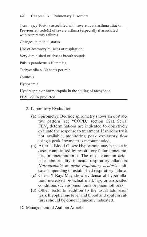
470
2. Laboratory Evaluation
(a) Spirometry: Bedside spirometry shows an obstruc-tive pattern (see “COPD,” section C2a). Serial FEV1 determinations are indicated to objectively evaluate the response to treatment. If spirometry is not available, monitoring peak expiratory flow using a peak flowmeter is recommended.
(b) Arterial Blood Gases: Hypoxemia may be seen in cases complicated by respiratory failure, pneumo-nia, or pneumothorax. The most common acid–base abnormality is acute respiratory alkalosis. Normocapnia or acute respiratory acidosis indi-cates impending or established respiratory failure.
(c) Chest X-Ray: May show evidence of hyperinfla-tion, increased bronchial markings, or associated conditions such as pneumonia or pneumothorax.
(d) Other Tests: In addition to the usual admission tests, theophylline level and blood and sputum cul-tures should be done if clinically indicated.
D. Management of Asthma Attacks
Table 13.3 Factors associated with severe acute asthma attacksPrevious episode(s) of severe asthma (especially if associated with respiratory failure)
Changes in mental status
Use of accessory muscles of respiration
Very diminished or absent breath sounds
Pulsus paradoxus >10 mmHg
Tachycardia >130 beats per min
Cyanosis
Hypoxemia
Hypercapnia or normocapnia in the setting of tachypnea
FEV1 <20% predicted
Chapter 13. Pulmonary Disorders

471
1. Ensure Adequate Oxygenation
(a) Most asthma patients will maintain SaO2 >90–92% during an acute attack with a low concentration of supplemental O2 (Venturi mask or nasal cannula 2 L/min). Monitor patient with pulse oximeter and supplement O2 as necessary.
(b) Mechanical Ventilation: Few patients with severe asthma will not respond to aggressive medical management and will require ventilatory support. The ventilatory strategy in patients with severe air-way obstruction should provide adequate oxygen-ation and at the same time minimize the risk of barotrauma through the use of small tidal volumes (i.e., 5–8 mL/kg) and minute ventilation (even if PaCO2 is allowed to climb: “controlled hypoventi-lation”). As in the case of COPD with expiratory flow limitation, reducing the ventilator’s mean expiratory flow (VT/Te) will improve air trapping and its deleterious effects.
(c) As noted above, NIMV may be useful in these patients:
(i) Continuous positive airway pressure (CPAP): Applied alone likely has several physiological benefits, including direct bronchodilation, off-setting intrinsic PEEP, alveolar recruitment and improvement in ventilation/perfusion mis-match, and reduced work of breathing.
(ii) BiPAP: The pressure gradient generated using BPAP aids in more diffuse dispersal of aero-solized medication and therefore improved bronchodilation.
2. Beta-Adrenergic AgonistsBeta-adrenergic agonists are first-line therapy for
acute asthma episodes. Selective beta2-agonists such as albuterol and terbutaline administered by MDI with a holding chamber or by nebulization titrated to maxi-mum effect are preferred. For dosing see COPD, sec-
Asthma

472
tion D2a. When drug delivery by aerosol is inadequate, SC epinephrine (0.3 mL 1:1000 q20 min × 3 max.) or terbutaline (0.25 mg q20 min × 2 max.) can be used.
3. Anticholinergic AgentsIpratropium bromide 500 mcg by nebulizer every
20 min, for three doses during acute asthma attack, then as needed.
4. CorticosteroidsMethylprednisolone (Solu-Medrol™) 40 mg q6 h IV
or prednisone 60 mg orally q8 h are recommended for the first 36–48 h. Significant clinical benefits are usually present 6 h later. When the patient is stable, a prednisone- tapering program may consist of 60 mg/day for 4 days, reducing the dose to 40 mg/day and then by 10 mg/day every 4 days. At the same time, the patient should be started on inhaled corticosteroids (i.e., triam-cinolone acetonide [Azmacort] six to eight puffs bid).
5. Complicating FactorsTreat any obvious associated precipitant or compli-
cating conditions such as pneumonia and pneumothorax.
6. Other Forms of TherapyOther interventions that have been used in status
asthmaticus but are not considered standard therapy include magnesium sulfate, general anesthetics, and bronchial lavage of thick secretions.
Pulmonary Embolism
A. Clinical Presentation and Risk Factors
1. The clinical findings of pulmonary embolism (PE) are nonspecific. It most commonly presents as the acute onset of dyspnea with or without pleuritic chest pain, minor hemoptysis, tachypnea, and abnormal chest X-ray (although a normal chest X-ray is not uncommon either). Other forms of presentation include the following:
Chapter 13. Pulmonary Disorders
473
(a) Acute cor pulmonale (>40% of circulation compromised)
(b) Insidious onset of dyspnea (recurrent unrecog-nized PEs)
(c) Syncope, wheezing, fever, cough, dysrhythmias, and cardiopulmonary arrest
(d) Asymptomatic
2. PE originates from thrombi in the deep venous system of the lower extremities (deep venous thrombosis [DVT]) in most cases. Important risk factors for venous thromboembolism include the following:
(a) Prolonged immobility or paralysis. (b) Surgery (mainly orthopedic—hip and knee—and
lengthy procedures). (c) Trauma. (d) Malignancy. (e) Congestive heart failure (CHF) and recent myo-
cardial infarction (MI). (f) Advanced age. (g) Obesity. (h) Pregnancy and estrogen therapy. (i) Prior history of DVT/PE: Less often, DVT/PE is
caused by antithrombin III, protein S and protein C deficiencies, or lupus anticoagulant syndrome.
3. Chest X-ray abnormalities may be subtle or even absent.
(a) Pulmonary infiltrates:
1. Only a minority represent pulmonary infarction, and they usually resolve over few days.
2. A pleural-based triangular infiltrate (Hampton’s hump) may be seen with infarction. It usually persists for weeks.
(b) Pleural effusion(s) (c) Elevated hemidiaphragm (d) Platelike atelectasis (e) Oligemia (Westermark’s sign)
Pulmonary Embolism
474
4. Arterial blood gases
(a) Hypoxemia in most cases (but 15% of PEs have PaO2 >80 mmHg)
(b) P(A-a)O2 gradient widened (c) Hypocapnia
5. D-dimerThis test has a good sensitivity and negative predic-
tive value, but poor specificity and positive predictive value.
6. Electrocardiogram (ECG)
(a) Sinus tachycardia, most common EKG finding in 44%.
(b) Nonspecific QRS and ST–T changes (c) New complete right bundle branch block, which
associated with increased mortality (d) Atrial dysrhythmias (multifocal atrial tachycardia
[MAT], atrial flutter) (e) S1–Q3–T3 pattern (only 10% of cases) (f) Pulseless electrical activity in massive PE
7. EchocardiographyUp to 40% of patients with PE have echocardio-
graphic anomalies. Some of these anomalies are:
(a) Increase in RV size (b) Decreased RV function (c) Tricuspid regurgitation (d) RV thrombus
B. Diagnostic Tests
1. Ventilation/Perfusion (V/Q) ScanA normal V/Q scan practically rules out pulmonary
embolism. On the other hand, an abnormal V/Q scan is nonspecific and should be considered in the context of the clinical probability (see Fig. 13.1). The V/Q scan and simultaneous chest X-ray findings are categorized as normal/very low, low, intermediate, or high probabil-ity, as depicted in Table 13.4.V/Q scan can be false posi-tive in embolism, infection, or tumors.
Chapter 13. Pulmonary Disorders

475
2. Lower Extremities Venous Studies (LEs)
(a) Duplex Ultrasound (DU): DU is Doppler ultra-sound combined with real-time two-dimensional ultrasound to study the venous system. When avail-able, it is the method of choice for diagnosing prox-imal DVT (positive predicted value of 94%).
CLINICAL SUSPICION FOR PE
Heparin(if no contraindications)
LEs (duplex unltrasound or IPGs)
Heparin
Pulmonary angiogram
Otherdiagnosis
IVC filter +/– heparindiscuss thrombolytics
Normal(<1% + PE)
Stop
Low probabilityclinical suspicion
Low4% + PE
Low16% + PE
Low56% + PE
Intermediate/high88–96% + PE
Intermediate/high28–66% + PE
Intermediate15% + PE
Intermediate probabilityclinical suspicion
Serial LEsAngiogram?
Serial LEsAngiogram?
Angiogram
Angiogram
Angiogram
Anticoagulation
Observe
High40% + PE
High probabilityclinical suspicion
V/Q scan
(–) (+)
(–) (+)
Hemodynamically unstableAnticoagulation contraindicated
Diffuse lung infiltrates with (–) LEs
Figure 13.1 Diagnostic/treatment approach to pulmonary embolism
Pulmonary Embolism

476
Diagnostic criteria of DVT include the inability to collapse the vein and to visualize the clot. DU can also assess flow augmentation, valvular incompe-tence, and other causes of pain and swelling (i.e., popliteal cysts and hematomas).
(b) Impedance Plethysmography (IPG): Rarely used anymore, IPG determines the changes in electrical impedance of the calf in response to blood volume changes produced by inflating a pneumatic thigh cuff. It is very sensitive for occlusive proximal DVT but insensitive for calf vein thrombosis.
(c) Venography: Once considered the gold standard for the diagnosis of leg DVT and is nowadays less frequently used. Disadvantages include its inva-siveness, cost, and potential allergic reactions to contrast media. Definitive diagnostic findings include filling defects in a well-opacified vein and/or partially occluding defects surrounded by con-trast media.
3. Spiral CT with Intravenous ContrastThis test has the ability to detect alternative pulmo-
nary abnormalities that may explain patient’s condi-tion. The diagnosing accuracy varies from institution to institution, depending on the film’s quality and radiolo-gist’s experience. When these two conditions are favor-able, a CT is a very good diagnostic tool.
4. Pulmonary AngiographyStill considered the gold standard for the diagnosis
of pulmonary embolism. In general, pulmonary arteri-ography is a safe procedure (mortality, <0.2%; morbid-ity, 4%), even in patients with significant pulmonary hypertension. Definitive angiographic signs include the presence of intraluminal filling defects or cutoffs of pul-monary arteries. It is indicated in patients with sus-pected PE and:
(a) Contraindications for anticoagulation (considering IVC filter)
Chapter 13. Pulmonary Disorders

477
Table 13.4 V/Q scan interpretation categoriesNormal and very low probability
No perfusion defects present (ventilation study and/or chest X-ray may be abnormal)
Three small segmental (<25% of a segment) perfusion defects with normal chest X-ray
Low probability
>Three small segmental perfusion defects with normal chest X-ray
Large or moderate segmental perfusion defect involving no >4 segments in one lung and no >3 segments in one lung region with matching ventilation defects and chest X-ray normal or with abnormalities smaller than the perfusion defects
Nonsegmental perfusion defect (small pleural effusion, cardiomegaly, enlarged aorta, mediastinum, hila)
One moderate segmental (>25% to <75%) perfusion defect with normal chest X-ray (mismatch)
Intermediate probability
All V/Q scans not included in the above categories (borderline or difficult to categorize)
High probability
Two large segmental (>75%) perfusion defects without corresponding ventilation or chest X-ray abnormality or smaller ventilation or chest X-ray abnormalities (mismatch)
Two moderate segmental and one large segmental perfusion–ventilation
Two moderate segmental and one large segmental perfusion–ventilation mismatches
Four moderate segmental perfusion–ventilation mismatches
Pulmonary Embolism

478
(b) Hemodynamic instability (considering thrombo-lytic therapy or embolectomy)
(c) High clinical suspicion for PE with other than high- probability V/Q scan and negative leg venous studies
(d) Extensive pulmonary parenchymal disease or CHF
C. Treatment of Acute Thromboembolism
1. AnticoagulationAnticoagulation should be started as soon as the
clinical suspicion for PE is high enough to initiate a diagnostic evaluation.
(a) Heparin: Give an initial bolus of 10,000 U intrave-nously followed by a continuous infusion of 1300 U/h (20,000 U in 500 mL D5W at 33 mL/h). Check the first activated partial thromboplastin time (aPTT) in 6 h, and maintain it between 1.5 and 2.5 times control. Modify heparin infusion accord-ing to the following:
1. If aPTT >2.5 control, stop infusion for 1 h, reduce the dose by 100–200 U/h, and recheck it in 4–6 h.
2. If aPTT is between 1.25 and 1.5 control, increase the dose by 100 U/h, and recheck it in 4–6 h.
3. If aPTT is <1.25 control, rebolus with 5000 U IV, increase the dose by 200 U/h, and recheck it in 4–6 h.
4. Weight-based protocols are available in most hospitals.
In most patients, continue heparin for at least 5 days (provided that Coumadin was started on day 1 or 2). Seven to 10 days of heparin infusion is recommended for patients with massive PE or extensive iliofemoral thrombosis.
Low molecular weight heparin (1 mg/kg IBW q12 h) has been used instead of unfractionated heparin in PE with excellent results (see Chap. 7, “Hematological Disorders”).
Chapter 13. Pulmonary Disorders

479
(b) Coumadin: Oral anticoagulation started on day 1 or 2 at a dose of 5–10 mg/day is recommended. The goal is to prolong the prothrombin time (PT) to an interna-tional normalized ratio (INR) of 2.0–3.0. Coumadin should be continued for at least 3–6 months in most patients. For those with a continuing risk factor or recurrent thromboembolism, anticoagulation should be given indefinitely. In cases where Coumadin may be contraindicated (i.e., pregnancy during the first and third trimester), adjusted-dose SC heparin can be effectively used.
(c) Direct thrombin inhibitors and factor Xa inhibitors: Apixaban, dabigatran, rivaroxaban, and edoxaban are alternative to warfarin for PE treatment.
Apixaban and rivaroxaban can be used without bridging with heparin; the data showed that they also decrease the recurrence of PE and deep venous thrombosis. For apixaban the dose is 10 mg BID X 7 days and then 5 mg BID. Whereas for rivaroxaban, the dose is 15 mg PO BID for 21 days and then 20 mg daily.
To start edoxaban or dabigatran, patient should get heparin for 5–10 days.
Dose for edoxaban is weight based, 60 mg PO daily if the weight is >60 and if less should be given 30 mg PO daily.
Dabigatran is contraindicated; if the creatinine clearance is <30, the dose is 150 PO BID.
2. Thrombolytic TherapyThrombolytic drugs dissolve thrombi by activating
plasminogen to plasmin, which in turn degrades fibrin (see Chap. 7, “Hematologic Disorders”). In contrast to thrombolytic therapy for MI, complete emboli resolu-tion in the pulmonary vessels is not accomplished fre-quently. Although a reduction in PE mortality has not been shown with this form of therapy, it should be con-sidered in patients with acute massive PE and hemody-namic instability without significant risk factors for
Pulmonary Embolism
480
bleeding. The role of thrombolytic therapy in DVT and submassive PE is less well established. Agents used for PE/DVT include the following:
(a) Streptokinase (SK): 250,000 IU loading dose fol-lowed by 100,000 IU/h for 24 h in PE and 48–72 h in DVT
(b) Urokinase (UK): 4400 IU/kg loading dose fol-lowed by 4400 IU/kg/h for 12 h in PE and 24–48 h in DVT
(c) Tissue Plasminogen Activator (tPA): 100 mg over 2 h
The efficacy and bleeding complications of SK, UK, and tPA are equivalent. When using SK or UK, checking a thrombin time or an aPTT q2–4 h into the infusion is recommended to verify a fibri-nolytic state. Heparin should be restarted when the aPTT is <2 times control.
3. IVC FilterIndications for placement of an IVC filter (Greenfield,
Mobin-Uddin, Bird’s nest) include the following:
(a) Contraindications to anticoagulation (b) Acute massive PE (c) Recurrent PE on adequate anticoagulation therapy (d) Chronic thromboembolism with pulmonary
hypertension (e) Following pulmonary embolectomy or
thromboendarterectomy
4. EmbolectomyEmbolectomy may be considered for documented
massive PE with documented occlusion of main pulmo-nary artery.
Catheter embolectomy: Intrapulmonary arterial techniques, using rheolytic embolectomy catheter (i.e., AngioJet embolectomy system), have been used with success in some cases. This procedure has been called by some Varon- Strickman procedure.
Surgical embolectomy: This procedure is advised only if an experienced surgical team is immediately available.
Chapter 13. Pulmonary Disorders
481
Adult Respiratory Distress Syndrome (ARDS)
A. Definition. ARDS is a form of acute lung injury character-ized by a high-permeability (noncardiogenic) pulmonary edema. In clinical practice, it is defined by the presence of the following:
1. Acute respiratory distress in a patient with predispos-ing conditions
2. Diffuse bilateral infiltrates on chest X-ray (pulmonary edema pattern)
3. Hypoxemia (PaO2 <55 mmHg with FiO2 >0.5) 4. Reduced respiratory system static compliance (<40–
50 mL/cmH2O) 5. Low or normal pulmonary artery occlusion pressure
(pulmonary capillary wedge pressure [PCWP] <16 cmH2O)
B. Etiology. ARDS is most commonly associated with:
1. Sepsis 2. Bronchial aspiration of gastric content 3. Trauma 4. Nosocomial pneumonia
Major risk factors for the development of ARDS are listed in Table 13.5.
C. Pathophysiology. The basic abnormality in ARDS is the disruption of the alveolar–capillary barrier. The endothelial injury in ARDS is frequently part of a more generalized permeability defect. An initial exudative phase is followed by proliferation of type II pneumocytes and fibrosis seen as early as the end of the first week.
D. Clinical Presentation
1. ARDS may develop insidiously over hours or even days after the initiating insult (i.e., pneumonia evolving into ARDS). Occasionally, it coincides with the precipi-tating event (i.e., gastric contents aspiration).
Adult Respiratory Distress Syndrome (ARDS)
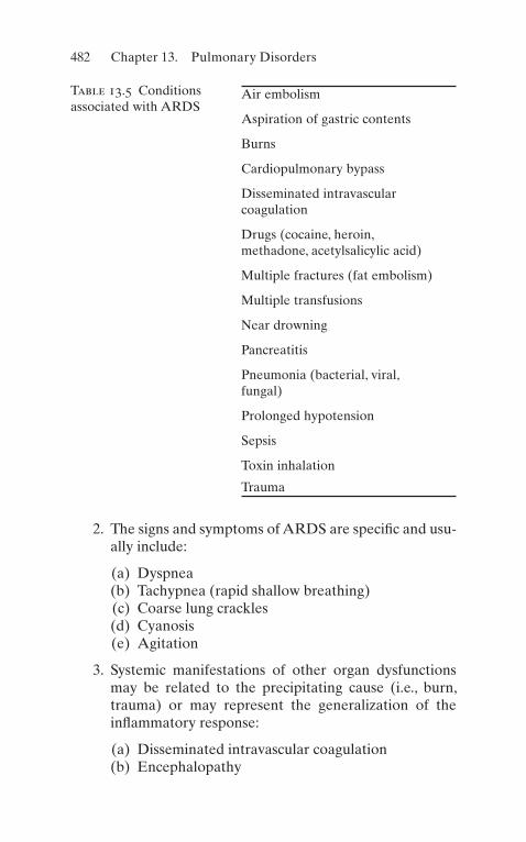
482
2. The signs and symptoms of ARDS are specific and usu-ally include:
(a) Dyspnea (b) Tachypnea (rapid shallow breathing) (c) Coarse lung crackles (d) Cyanosis (e) Agitation
3. Systemic manifestations of other organ dysfunctions may be related to the precipitating cause (i.e., burn, trauma) or may represent the generalization of the inflammatory response:
(a) Disseminated intravascular coagulation (b) Encephalopathy
Table 13.5 Conditions associated with ARDS
Air embolism
Aspiration of gastric contents
Burns
Cardiopulmonary bypass
Disseminated intravascular coagulation
Drugs (cocaine, heroin, methadone, acetylsalicylic acid)
Multiple fractures (fat embolism)
Multiple transfusions
Near drowning
Pancreatitis
Pneumonia (bacterial, viral, fungal)
Prolonged hypotension
Sepsis
Toxin inhalation
Trauma
Chapter 13. Pulmonary Disorders

483
(c) Acute renal failure (d) Acute liver failure (e) Sepsis (gut bacterial translocation)
4. ABGs show marked hypoxemia and hypocapnia with either acute respiratory alkalosis or acute metabolic acidosis. The Berlin criteria have been used to classify ARDS according to the PaO2/FiO2 ratio (see Table 13.6).
5. Despite the chest X-ray appearance of diffuse bilateral infiltrates, chest CT reveals a patchy, nonhomogeneous distribution of affected lung mixed with normal paren-chyma. Small pleural effusions can be seen in ARDS. Usually, the cardiovascular silhouette on chest X-ray is within normal limits.
6. The pulmonary artery occlusion pressure or wedge pressure measured by a balloon-tipped, flow-directed catheter (Swan–Ganz catheter) in the past was used to detect the hydrostatic component of the pulmonary edema (cardiogenic). In pure ARDS, the wedge pres-sure should be <16–20 cmH2O. This is rarely done nowadays.
7. The use of serum procalcitonin level may help deter-mine the patient’s inflammatory state.
E. Management
1. Treatment of the Precipitating Condition(s)Specific treatment for the underlying disorder
should be instituted as soon as possible (i.e., antimicro-bials for infections, sepsis, drainage of abscesses, trans-fusion for hypovolemic shock, etc.).
2. Supportive Care
Table 13.6 Berlin classifi-cation ARDS levels of severity
Classification PaO2/FiO2 (mmHg)Mild 200 to ≤300a
Moderate 100 to ≤200b
Severe ≤100b
aWith PEEP or CPAP ≥5 cmbWith PEEP ≥5 cm H2O
Adult Respiratory Distress Syndrome (ARDS)

484
(a) Ventilatory Support (See Sect. V) (b) Hemodynamic Monitoring and Support
1. The use of pulmonary artery catheter (PA cath-eter or Swan–Ganz catheter) is controversial, as no study has ever proved that this technique improves survival in suspected or established ARDS. However, the information derived from hemodynamic monitoring using this catheter can be used in the following:
(a) Differentiation of cardiogenic vs. noncar-diogenic pulmonary edema.
(b) Management of intravascular volume (avoiding volume overload).
(c) Assessment of the cardiovascular effects of PEEP titration (cardiac index, stroke volume).
(d) Unfortunately, overtreating or confusing parameters is common while using a PA catheter.
2. In severe cases of ARDS, where high levels of extrinsic PEEP or dynamic hyperinflation (auto-PEEP) are necessary to maintain ade-quate oxygenation, a reduced cardiac index should be corrected with the use of inotropes (i.e., dobutamine or dopamine) to maintain an adequate O2 delivery.
(c) Nutritional Support (See Chap. 10, “Nutrition”)
(d) Diagnosis and Treatment of Complications
1. Barotrauma (i.e., tube thoracostomy for pneumothorax)
2. Acute renal failure (i.e., hemodialysis) 3. DIC (i.e., transfusions) 4. Infections: line sepsis, urinary tract infec-
tion (UTI), and cellulitis (i.e., antibiotics, change central lines)
Chapter 13. Pulmonary Disorders

485
3. Other Therapeutic Modalities
(a) Pharmacologic and immunologic agents targeted to arrest a specific step in the inflammatory cas-cade or pathophysiologic process characteristic of ARDS and sepsis have been extensively evaluated without success, i.e., monoclonal antibodies against bacterial lipopolysaccharide and tumor necrosis factor (TNF), soluble interleukin-1 (IL-1) and TNF receptors, prostaglandin E1, pentoxifylline, nonste-roidal anti-inflammatory drugs (NSAIDs) (i.e., ibuprofen), synthetic surfactant mixtures (Exosurf), inhaled nitric oxide, etc.
(b) Extracorporeal oxygenation and CO2 removal (IVOX, ECCO2R, ECMO), as currently imple-mented in some centers.
F. Prognosis
1. The mortality of ARDS has remained unchanged over the past two decades in spite of advances in supportive therapy.
2. Early mortality is usually related to the underlying condition(s); later, mortality is mainly related to multi-ple organ failure rather than pulmonary dysfunction.
3. Most ARDS survivors surprisingly have minimal long- term impairment of lung function (mild restrictive and diffusion capacity [DLCO] defects). Occasionally, reversible airway obstruction may develop.
Acute Respiratory Failure
A. Definition. Acute respiratory failure is the inability to maintain adequate blood oxygenation and/or alveolar ventilation in the absence of an intracardiac shunt. Provided the baseline ABGs are close to predicted nor-mal values, this usually means an acute increase in PaCO2 >50 mmHg with arterial acidemia and/or a PaO2 <55 mmHg while breathing room air.
Acute Respiratory Failure

486
B. Classification and Etiology. Two clinical and pathophysi-ologic distinct types of acute respiratory failure can be described:
1. Hypoxemic Respiratory FailureThe hallmark of this type of respiratory failure is the
inability to adequately oxygenate the blood. The main pathophysiologic mechanisms involved are V/Q mis-match (response to 100% O2) and intrapulmonary shunting (no significant improvement with 100% O2). The patients exhibit a rapid shallow breathing pattern and a low or normal PaCO2. This form of respiratory failure is commonly the result of a diffuse acute lung injury with high-permeability pulmonary edema (ARDS), severe pneumonic infiltrates, or cardiogenic pulmonary edema.
2. Hypercapnic Respiratory Failure (Pump Failure)The hallmark of ventilatory pump failure is hyper-
capnia with acute respiratory acidosis. The P(A-a)O2 gradient is useful in determining if the hypoxemia pres-ent in this form of respiratory failure is due to hypoven-tilation (normal gradient) only or to additional parenchymal lung disease (elevated gradient). The hypercapnia is the result of abnormalities in one or more of the determinants of the PaCO2:
PaCO kVCO E D T2 2 1� �� �/ /V V V
(a) Increased CO2 production (VCO2) in patients with fever, sepsis, agitation, or excessive carbohydrate load, associated with a limited ventilatory capacity (high VD/VT, low VE)
(b) Increased dead space (VD/VT) in severe COPD, cys-tic fibrosis, and severe asthma
(c) Decreased total minute ventilation (VE) due to ven-tilatory pump dysfunction:
1. Decreased Central Respiratory Drive: CVA, drugs (narcotics, sedatives, anesthetics), central hypoventilation, and hypothyroidism
Chapter 13. Pulmonary Disorders

487
2. Abnormal Respiratory Efferents
(a) Spinal Cord: trauma, poliomyelitis, amyo-trophic lateral sclerosis, tetanus, and rabies
(b) Neuromuscular: myasthenia gravis, multiple sclerosis, botulism, Guillain–Barré syn-drome, hypophosphatemia, hypomagnese-mia, drugs (streptomycin, amikacin, neuromuscular blockers), polyneuropathy of critical illness, and bilateral phrenic nerve injury
3. Abnormal Chest Wall and/or Muscles: severe kyphoscoliosis, ankylosing spondylitis, massive obesity, muscular dystrophy, polymyositis, respi-ratory muscles fatigue, and acid maltase deficiency
4. Airways, Upper Airway Obstruction: epiglotti-tis, fixed and variable upper airway obstruction due to tumors, post- extubation, tracheomalacia, and bilateral vocal cord paralysis
C. Management. The management of acute respiratory fail-ure is initially supportive, aimed at the correction of hypoxemia or hypercapnia until specific actions are imple-mented to correct, if possible, the factors that lead to the respiratory failure (i.e., antibiotics for pneumonia; diuret-ics, morphine, nitroglycerin, and afterload-reducing agents for cardiogenic pulmonary edema; naloxone for narcotics overdose).
1. Hypoxemic Respiratory Failure
(a) Patients with V/Q mismatch abnormalities without significant intrapulmonary shunt will usually respond to noninvasive O2 supplementation (i.e., nasal cannula, Venturi mask).
(b) In patients with cardiogenic pulmonary edema, the use of continuous positive airway pressure (CPAP) (5–10 cmH2O) via a face mask in addition to O2 supplementation can be beneficial by reducing the
Acute Respiratory Failure

488
transmural pressure of the left ventricle, and there-fore afterload, but also by decreasing the preload.
(c) The ventilatory management of patients with dif-fuse acute lung injury (i.e., ARDS) requires mechanical ventilation and should be viewed as a balance between adequate oxygenation on one hand and the risk for barotrauma and cardiovascu-lar compromise on the other. The following section pertains to specific aspects of the ventilatory man-agement of ARDS.
1. Adequate oxygenation: For most patients with ARDS, this means a PaO2 of 55–60 mmHg or O2 saturation of 88–90% with a cardiac index >2.5 L/min/m2 and hemoglobin 10 g/dL. Arterial O2 saturation can be increased in ARDS by the following:
(a) Raising the fraction of inspired oxygen (FiO2): To avoid potential O2 toxic effects, it is recommended not to use 100% O2 for more than a few hours and to maintain an FiO2 ≤0.6. A particular effort should be made to decrease FiO2 to the minimum acceptable in patients exposed to drugs that may increase O2 toxicity (i.e., bleomycin, amiodarone).
(b) Increasing the end-expiratory lung volume to recruit collapsed or flooded alveoli. This can be achieved by adding extrinsic PEEP and/or setting the ventilator to create dynamic hyperinflation (auto-PEEP). It is unclear if one strategy is more effective than the other in ARDS. The goal is to max-imize oxygenation while at the same time avoiding hypotension, reduced cardiac pump function, and a plateau pressure >35 cmH2O. With these considerations:
Chapter 13. Pulmonary Disorders

489
(i) Extrinsic PEEP is usually started at a level of 5 cmH2O and titrated up by 2 cmH2O increments to a level of 15–20 cmH2O along with the use of other strategies to minimize baro-trauma (see “Avoiding Barotrauma”).
(ii) Extended-ratio ventilation (prolonged I/E ratio, known as inverse ratio venti-lation when I/E >1:1) is a technique used to increase mean alveolar pres-sure and transpulmonary pressure. It can be implemented with either volume- controlled or pressure- controlled ventilators. Increasing the inspiratory time (see Table 13.7) increases the mean airway pressure (MAP) and allows the recruitment of lung units with long time constants, therefore improving oxygenation. Extended-ratio ventilation is more easily implemented with volume- controlled than pressure-con-trolled ventilators. Both require heavy sedation with or without paralysis, i.e., morphine sulfate, midazolam (Versed), lorazepam (Ativan), or propofol (Diprivan) by continuous infusion with or without cisatracurium (Nimbex). Monitoring of plateau pressure to keep it <35 cmH2O, auto-PEEP (end- expiration occlusion method).
2. Avoiding barotrauma: Barotrauma in the form of extra-alveolar air or worsening of acute lung injury is the result of alveolar overdistention (increased transmural pressure or alveolar pres-sure [Palv]—pleural pressure [Ppl]). Thus, it seems reasonable to avoid lung volumes above total lung capacity (TLC) to prevent lung damage.
Acute Respiratory Failure

490
(a) Because Palv, Ppl, and lung volumes are diffi-cult to determine at the bedside, monitoring the plateau pressure (end-inflation hold pressure) is recommended as the best approximation of the peak alveolar pressure.
(b) A plateau pressure of 35 cmH2O or more, in the absence of significantly decreased chest wall compliance, should be avoided.
(c) Peak airway pressure (Ppeak) reflects not only the elastic but also the flow-resistive pressures of the respiratory system, and it should be used only as a gross estimate for the risk of barotrauma (i.e., high Ppeak may be due to a small endotracheal tube, bron-chospasm, secretions, high inspiratory peak flow, or worsening of lung or chest wall
Table 13.7 Prolonging inspiratory time (Ti) and I/E ratio in volume- controlled ventilators
Reduce inspiratory flow (i.e., to 40 L/min)
Use decelerating inspiratory flow waveform
Add an inspiratory pause (i.e., 0.2–0.5 s)
Increase the percent inspiratory timea
Applying these changes in a stepwise manner will allow progressive exten-sion of Ti and I/E ratio to the degree that is tolerated or needed. Adjustment in tidal volume (VT) as recommended (see below) and monitoring the pla-teau pressure (Pplateau), auto- PEEP, and its hemodynamic effects are required to avoid complicationsaDirect way of setting the I/E ratio (i.e., Siemens Servo Ventilator)
Chapter 13. Pulmonary Disorders

491
compliance) that may or may not mean alveolar overinflation.
(d) Determining the tidal volume (VT): ARDS is a nonhomogeneous process with col-lapsed and flooded areas mixed with rela-tively normal aerated lung, resulting in a reduction in the TLC. Thus, it makes sense to ventilate ARDS patients with smaller than conventional VT. The VT chosen should be one that prevents lung overinfla-tion (i.e., plateau pressure <35 cmH2O) and alveolar derecruitment at the end of expira-tion (inadequate oxygenation). This usually means an initial VT of 5–6 mL/kg.
(e) Setting the respiratory rate: The machine rate should be determined considering the patient’s metabolic demands, intrinsic rate, and desired I/E ratio. It is usually set between 25 and 40 breaths per minute. Even with these rates (plus the low VT used), minute ventilation may not be high enough for ARDS patients. Allowing CO2 retention (i.e., 60 mmHg) and respiratory acidemia (i.e., pH 7.2–7.25) in an effort to limit barotrauma is referred as permissive hypercapnia.
3. Controlling the hemodynamic effects of mechan-ical ventilation: The increase in end-expiratory lung volume and mean alveolar pressure pro-duced by the ventilatory strategies described above can have deleterious hemodynamics con-sequences. It is important to document that a ventilator change aimed at increasing O2 satura-tion does not reduce the total amount of O2 delivered to the tissues via a reduction in car-diac index. In severe cases, the cardiac index should be maintained >2.5 L/min/m2 with the use of inotropic agents (vasopressin 1–6 units per hour if hypotension is present).
Acute Respiratory Failure

492
2. Hypercapnic Respiratory FailureThe main goal in treating patients with hypercapnic
respiratory failure is to improve alveolar ventilation through the use of mechanical ventilation. This is most commonly done through an endotracheal tube using a volume-controlled ventilator (usual initial settings are VT 5–7 mL/kg, A/C mode, rate eight to ten breaths per minute, and FiO2 1.0). ABGs are checked 10–20 min later to detect inadvertent and potentially life-threatening acute alkalosis secondary to overcorrection of the hyper-capnia and to adjust the FiO2. Noninvasive mechanical ventilation has been effective in patients with neuromus-cular conditions. The management of hypercapnic respi-ratory failure in asthma and COPD is discussed above.
Barotrauma
A. Definition. Barotrauma is lung injury that is related to high alveolar pressures (and volumes). In the intensive care unit (ICU) setting, barotrauma specifically refers to positive-pressure ventilator-induced lung damage. Occasionally, a patient may be admitted to the ICU after a diving accident (sudden decompression) or foreign body aspiration (ball-valve mechanism).
B. Clinical manifestations
1. Classic mechanical ventilator-induced barotrauma is manifested by extra-alveolar air in the form of:
(a) Pulmonary interstitial emphysema (PIE) (b) Subpleural air cysts (c) Pneumomediastinum (d) Pneumothorax (PTX) (e) Subcutaneous emphysema (f) Pneumopericardium (g) Pneumoretroperitoneum (h) Pneumoperitoneum (i) Gas emboli (main clinical manifestation in diving
accidents)
Chapter 13. Pulmonary Disorders
493
C. Any suspected barotrauma, plateau air pressure should be controlled under 30 cmH2o as possible, which can achieved by decrease PEEP and/or VT
2. Tension PTX occurs in 30–97% of all PTXs in mechani-cally ventilated patients and is characterized by wors-ening hypoxemia, hypotension, or even cardiovascular collapse with pulseless electrical activity (PEA). Chest X-ray shows lung collapse with hemithorax expansion and contralateral mediastinal shift.
3. Physical examination reveals:
(a) Absent breath sounds, hyperresonance to percus-sion, and decreased chest excursion on the affected side in cases of PTX and distended neck vein.
(b) Crepitation on palpation or auscultation is found in cases of subcutaneous emphysema.
(c) Mediastinal “crunch” in pneumomediastinum. (d) Changes in mental status or neurologic deficits are
usually found in patients with gas embolism.
4. Development or worsening of diffuse lung injury (in the form of noncardiogenic pulmonary edema) also has been associated with positive-pressure ventilation.
D. Pathophysiology
1. Alveolar rupture occurs at the site where alveoli attach to the bronchovascular sheath. From there, extra- alveolar air may dissect the peribronchovascular tis-sues into different planes to produce the clinical manifestations listed above. Alternatively, direct rup-ture of a subpleural cyst into the pleural cavity may also cause PTX.
2. Positive-pressure mechanical ventilation has also shown to produce:
(a) Increased lung microvascular permeability and fil-tration pressure
(b) Alveolar epithelial injury (c) Alteration of surfactant function and turnover
Barotrauma

494
E. Diagnosis
1. A high index of suspicion should be maintained in those patients at risk for barotrauma (i.e., use of high VT, high plateau pressure, peak pressure, PEEP, dynamic hyperinflation, extensive lung damage, and prolonged mechanical ventilation).
2. Chest X-ray will usually confirm a diagnosis of extra- alveolar air. PIE (seen as linear radiolucent streaks) is the first radiologic sign and should alert the physician for the risk of progression to PTX. In case of highly suspicion clinically of PTX, a rapid intervention with needle decompression should be done even before obtaining imaging.
3. The classic radiologic sign of PTX (pleural line sepa-rated from the apicolateral chest wall) may not be pres-ent in ICU patients when an anteroposterior (AP) portable chest X-ray is taken in the supine or semire-cumbent position. In those patients, attention should also be paid to the mediastinal and subpulmonic recesses where air accumulates.
4. Delayed diagnosis lead to mortality of 31–91%
F. Management
1. Extra-alveolar air without PTX is managed conserva-tively. Observation and avoidance of risk factors, if pos-sible, is indicated (see “Avoiding Barotrauma,” above).
2. In general, all PTXs in a mechanically ventilated patient should be treated with tube thoracotomy (see Chap. 15, “Special Techniques”).
3. The use of “prophylactic” tube thoracotomy for high- risk mechanically ventilated patients is controversial and not recommended. Instead, it is advised to follow these patients closely and be prepared for immediate placement of a chest tube should PTX develop.
4. Persistent bronchopleural fistula(s) despite chest tube suction should be managed with the lowest VT, Ppeak, and Pplateau that permit adequate ventilation. High- frequency jet ventilation (HFJV), independent lung
Chapter 13. Pulmonary Disorders
495
ventilation, or surgery should be considered if the air leak is massive and does not respond to the usual management.
5. Recompression in a hyperbaric chamber is indicated for diving accidents resulting in air embolism.
Massive Hemoptysis (Life-Threatening Hemoptysis “Previously Called Massive Hemoptysis”)
A. Definition. Hemoptysis is the expectoration of blood that originates from below the larynx. It is considered massive when the rate of bleeding is at least 400 mL in 6 h or 600 mL in a 24-h period.
B. Etiology. The most common causes of massive hemoptysis are:
1. Tuberculosis (rupture of Rasmussen’s aneurysm) 2. Bronchiectasis (erosion of bronchial vasculature) 3. Bronchogenic carcinoma (invasion of pulmonary
vessels) 4. Lung abscess (destruction of fairly normal vessels due
to inflammation)Other etiologies include bronchial carcinoids, cystic
fibrosis, broncholithiasis, aspergilloma, trauma, bron-chovascular and arteriovenous (AV) fistulas, mitral ste-nosis, and the pulmonary–renal syndrome.
5. Immunological lung disease (invasion pulmonary cap-illaries): like anti-glomerular basement membrane, microscopic polyangiitis, diffuse alveolar hemorrhage, and granulomatosis with polyangiitis
C. Evaluation
1. Differentiate Hemoptysis from HematemesisHemoptysis is usually bright red blood, frothy, with
an alkaline pH. In contrast, hematemesis is usually darker with an acidic pH. At times this differentiation
Massive Hemoptysis (Life-Threatening Hemoptysis…

496
cannot be made easily, because hematemesis may pro-duce blood aspiration into the tracheobronchial tree, which in turn causes “hemoptysis,” and, on the other hand, patients with hemoptysis may swallow blood and vomit it after coughing.
2. Localize the Bleeding SiteLocalization of the bleeding site is important to ade-
quately plan any interventional procedure. Bleeding coming from the upper airways can be excluded by per-forming an ear, nose, and throat (ENT) examination.
(a) Chest X-ray may suggest the bleeding site:
1. Lung masses, apical cavitary lesions, or infiltrates in the chest X-ray should point to these sites as the source of bleeding.
2. A normal chest X-ray is consistent with bleeding arising from the airways.
3. Roentgenograms showing bilateral or diffuse dis-ease are not helpful in pointing out the origin of bleeding.
(b) Bronchoscopy should be performed to further identify the site and cause of bleeding and to achieve tempo-rary control. The type and timing of the procedure depend on the rate of bleeding.
1. When flexible fiber-optic bronchoscopy (FFB) is done, the patient must be intubated with a large endotracheal tube that may be used to tamponade the affected lung if necessary.
2. Rigid bronchoscopy is the preferred temporizing method to evaluate and control a massive bleeding. This procedure should be done under general anes-thesia in the operating room by trained physicians.
(c) Angiography of the bronchial and pulmonary circula-tion is recommended as it can determine the bleeding site and simultaneously provide therapy (i.e., coiling).
Chapter 13. Pulmonary Disorders
497
3. Laboratory DeterminationsObtain ABGs to determine if acidosis exists and to
confirm level of oxygenation (significant abnormal gas exchange). Then obtain a complete blood cell count, PT, partial thromboplastin time (PTT), bleeding time, creatinine, and serum urea nitrogen (BUN), and request blood type and crossmatching.
D. Management
1. Ensure Adequate Ventilation and OxygenationThis should be the main priority if we consider that
the mode of death in massive hemoptysis is asphyxia-tion (as little as 150 cc of blood is needed to fill the airways).
(a) Depending on the rate of bleeding, it may only be necessary to administer supplemental O2 through nasal cannula or face mask, or, on the other extreme, it may be necessary to perform endotra-cheal intubation (single- or double-lumen [Carlen’s tube]) to aspirate the blood and ventilate the patient while definitive therapy is being prepared.
(b) Position the patient in the lateral decubitus position with the bleeding site down.
2. Ensure Stable Hemodynamic Conditions
(a) Obtain adequate venous access. (b) Administer fluids as needed (normal saline or
blood).
3. Suppress Cough
(a) Codeine 60 mg PO q6 h. (b) Sedatives may be added (i.e., midazolam 2 mg or
lorazepam 1 mg, IV q2 h pm).
4. Control the Bleeding Site
(a) Bronchoscopic procedures include:
1. Bronchial packing through a rigid bronchoscope
Massive Hemoptysis (Life-Threatening Hemoptysis…
498
2. Tamponade of airway with a balloon-tipped (Fogarty) catheter through a rigid bronchoscope or alongside a flexible bronchoscope
3. Bronchial lavage with cold saline through a rigid bronchoscope
4. Coagulation of visible lesions with a Neodymium- YAG laser (not helpful in very active, brisk bleeding)
(b) Bronchial artery embolization is the method of choice for patients with massive or submassive hemoptysis and contraindications for surgery. Given its high success rate (90%), embolization has become a first-line treatment for all patients with massive or recurrent hemoptysis.
(c) Surgical resection is recommended for localized lesions that can be removed. It should not be offered to patients with:
1. Metastatic lung cancer 2. Severe pulmonary or cardiovascular status
(d) Correct coagulopathy, if present (i.e., administer FFP and vitamin K if PT is prolonged, and trans-fuse platelets in severe thrombocytopenia).
E. Prognosis
1. Although the underlying medical condition(s) affect the prognosis in massive hemoptysis, the best estimator of mortality is the rate of bleeding. A study by Cracco showed that hemoptysis of 600 cc occurring over:
(a) 4 h has 71% mortality (b) 4–16 h has 45% mortality (c) 16–48 h has 5% mortality
2. Median operative mortality in massive hemoptysis is 17%. Actively bleeding patients at the time of the sur-gery have a higher mortality when compared with non-actively bleeding patients.
Chapter 13. Pulmonary Disorders

499
Upper Airway Obstruction
A. Acute upper airway obstruction can be a life-threatening situation that may lead to cardiac arrest. This situation requires immediate intervention. This is important among the pediatric population, having smaller airways that can quickly progress to complete obstruction.
B. An initial rapid assessment is pertinent to evaluate upper airway patency (audible speech, cough, drooling), respira-tory distress (retractions, nasal flaring), and hypoxemia.
C. Immediate treatment: Chin tilt and lift maneuvers should be attempted, as well as removal of foreign body if sus-pected. Direct laryngoscopy can also be done, preparing patient for emergent intubation. If endotracheal intuba-tion is not successful on the first, but successful ventilation using bag mask, alternative modes may be used such as LMA, Combitube, oral and nasal airways, or emergency surgical intervention (see Chap. 15, “Special Techniques”).
Useful Facts and Formulas
A. Lung Volumes. Normal values for pulmonary volumes and capacities in humans are depicted in Table 13.8.
The vital capacity (VC) is calculated as follows:
VC IRV ERV T� � �V
The residual volume (RV) is calculated as the difference between the functional residual capacity (FRC) and the expi-ratory reserve volume (ERV):
RV FRC EV� �
Alternatively, if the total lung capacity (TLC) and vital capac-ity (VC) are known, the following formula can be utilized:
RV TLC VC� �
Useful Facts and Formulas
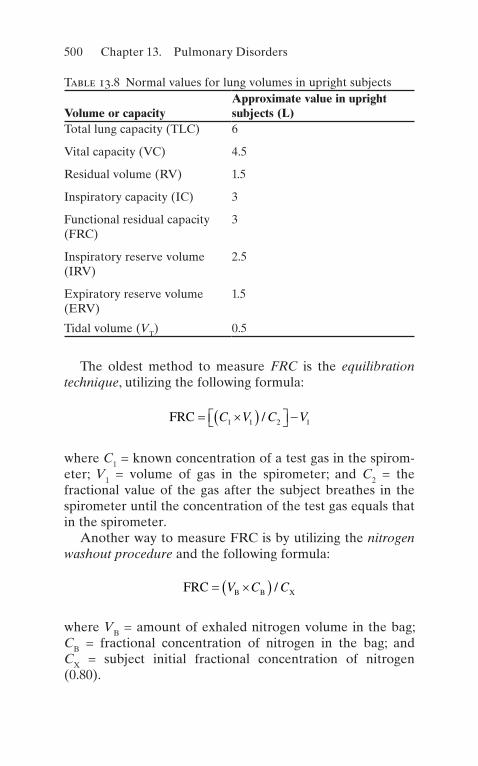
500
The oldest method to measure FRC is the equilibration technique, utilizing the following formula:
FRC � �� ��� �� �C V C V1 1 2 1/
where C1 = known concentration of a test gas in the spirom-eter; V1 = volume of gas in the spirometer; and C2 = the fractional value of the gas after the subject breathes in the spirometer until the concentration of the test gas equals that in the spirometer.
Another way to measure FRC is by utilizing the nitrogen washout procedure and the following formula:
FRC B B X� �� �V C C/
where VB = amount of exhaled nitrogen volume in the bag; CB = fractional concentration of nitrogen in the bag; and CX = subject initial fractional concentration of nitrogen (0.80).
Table 13.8 Normal values for lung volumes in upright subjects
Volume or capacityApproximate value in upright subjects (L)
Total lung capacity (TLC) 6
Vital capacity (VC) 4.5
Residual volume (RV) 1.5
Inspiratory capacity (IC) 3
Functional residual capacity (FRC)
3
Inspiratory reserve volume (IRV)
2.5
Expiratory reserve volume (ERV)
1.5
Tidal volume (VT) 0.5
Chapter 13. Pulmonary Disorders
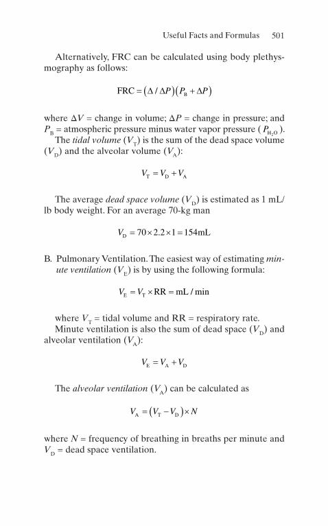
501
Alternatively, FRC can be calculated using body plethys-mography as follows:
FRC B� � � �� �� � �/ P P P
where ΔV = change in volume; ΔP = change in pressure; and PB = atmospheric pressure minus water vapor pressure ( PH O2
).The tidal volume (VT) is the sum of the dead space volume
(VD) and the alveolar volume (VA):
V V VT D A� �
The average dead space volume (VD) is estimated as 1 mL/lb body weight. For an average 70-kg man
VD mL� � � �70 2 2 1 154.
B. Pulmonary Ventilation. The easiest way of estimating min-ute ventilation (VE) is by using the following formula:
V VE T RR mL� � � / min
where VT = tidal volume and RR = respiratory rate.Minute ventilation is also the sum of dead space (VD) and
alveolar ventilation (VA):
V V VE A D� �
The alveolar ventilation (VA) can be calculated as
V V V NA T D� �� ��
where N = frequency of breathing in breaths per minute and VD = dead space ventilation.
Useful Facts and Formulas

502
An alternative method requires knowledge of the CO2 production by the patient. The production of CO2 ( VCO2 ) can be calculated as follows:
VCO A ACO2 2� �V F
where FACO2 = fractional concentration of CO2 in the alveolar gas and VA = VCO2/FACO2.
Dead space ventilation (VD) can be calculated if the minute ventilation (VE) is known:
V VD E PaCO PECO PaCO� �� �� �2 2 2/
The partial pressure of alveolar CO2 (PACO2) is more con-venient for these calculations and for practical purposes:
PACO FACO PB2 2 47� � �� �
In normal lungs, the arterial CO2 (PaCO2) approximates the PACO2. Therefore, the VA formula can be rewritten as
VA VCO PaCO� � �K 2 2/
where K = a factor (0.863) that converts CO2 concentrations to pressure (mmHg).
C. Gas Transport in Blood. The difference between the inspired and expired fractional concentration of O2 repre-sents the oxygen uptake ( VO2 ):
VO F F OI iO E E2 22
� �� � � �� �V V
where VI = volume of gas inhaled; FiO2 = fractional con-centration of inspired oxygen; VE = volume of gas exhaled; and FEO2 = fractional concentration of expired oxygen.
Chapter 13. Pulmonary Disorders

503
The amount of O2 in solution in 100 mL of blood is calcu-lated as (assuming a partial O2 pressure of 70 mmHg)
PO O mL mL2 2760 70 760 2 3 0 21 100/ / . . /� �� � � ��
The PaO2 at which hemoglobin is 50% saturated (P50) can be calculated from the venous pH and arterial blood gases as
P
k
nmm50
122 33�
� �� �antilog normal Hg
log /
where
1 2 1007 4 2 2/ log /.k n� � � �� �� �� �� �antilog PaO SaO SaO
antilog PaO PaO venous pHn�� � � � �� �log log . ..2 0 5 7 47 4 2
n = =Hill constant for hemoglobinA2 7.
The Fick equation for oxygen consumption (VO2) is calcu-lated as follows:
VO CaO CvO2 2 2� �� �Q
where Q = cardiac output (L/min); CaO2 = arterial oxygen content; and CFA ~ vO2 = mixed venous oxygen content.
The volume of carbon dioxide exhaled per unit time (CO2 production or VO2 ) is calculated as follows:
VCO FECO F COE I I2 2 2� �� � � �� �V V
where VE = volume of gas exhaled per unit time; FECO2 = frac-tional concentration of carbon dioxide in the exhaled gas; VI = volume of gas inhaled per unit time; and FICO2 = frac-tional concentration of inspired carbon dioxide.
Useful Facts and Formulas

504
Since the inspired gas usually contains negligible amounts of carbon dioxide, another representation of this formula is
VCO F COE E2 2� �V
D. Pulmonary Circulation. The mean pulmonary artery pres-sure (PAP) can be calculated utilizing the following formula:
PAP PVR PBF PAOP� �� � �
where PVR = pulmonary vascular resistance and PBF = pul-monary blood flow (which typically equals the cardiac out-put). Reorganizing the above formula, the pulmonary vascular resistance (PVR) is then calculated as
PVR Mean PAP PAOP CO� �� � /
where PAOP = pulmonary artery occlusion pressure and CO = cardiac output.
The pressures that surround the vessels in the pulmonary circulation contribute to the transmural pressure (Ptm) repre-sented as
P P Ptm vas is� �
where Pvas = vascular pressure and Pis = perivascular intersti-tial pressure.
When the left atrial pressure (Pla) is available, the driving pressure responsible for producing pulmonary blood flow is then calculated as
P P Q Rpa la vas�� � � �
where Ppa = mean pulmonary arterial pressure; Pla = mean left atrial pressure; Q = pulmonary blood flow; and Rvas = pulmo-nary vascular resistance.
Chapter 13. Pulmonary Disorders

505
The pulmonary vascular compliance (Cvas) can be calcu-lated utilizing the following formula:
C V Pvas vas vas� � �/
where ΔVvas = change in blood volume and ΔPvas = change in vascular pressure.
The blood flow zones in an idealized upright lung with normal pressure differences are depicted in Table 13.9.
E. Mechanics and Gas Flow. The pressure inside the lungs relative to the pressure outside is known as the transpul-monary pressure (TP) and is calculated as
TP alv pl� �P P
where Palv = alveolar pressure and Ppl = pleural pressure.The change in volume (ΔV) for a unit pressure (ΔP) under
conditions of no flow is the static compliance:
Static compliance SC
V
P� � � �
�
Clinically, this formula can be simplified as follows:
Table 13.9 Pulmonary blood flow zones
Blood flow zones PressuresI Palv > Ppa > Ppv
II Ppa > Palv > Ppv
III Ppa > Ppv > Palv
IV Ppa > Ppv > Palv
Palv pressure surrounding the alveo-lar vessels, Ppa mean pulmonary arterial pressure, Ppv mean venous (left atrial) pressure
Useful Facts and Formulas

506
C
Vs
T
Plateau airway pressure PEEP autoPEEP�
� �� �
where VT = tidal volume. Normal Cs value is 100 mL/cmH2O.The dynamic compliance (Cdyn) can be calculated utilizing
the following formula:
CV b
dynT
Plateau airwaypressure PEEP autoPEEP
�
� �� �
� � bb ac
a
2 4
2
�
Normal Cdyn value is 100 mL/cmH2O.The specific compliance (Cspec) is calculated utilizing the
following formula:
C Cspec stat FRC= /
The chest wall compliance (Cw) can be calculated as
C
VW
T
Airway pressure Atmospheric pressure�
�
Another formula that can be used under special circum-stances (i.e., lung transplantation) is the separate lung compli-ance (CX) and is calculated as
C
VX
T
Airway pressure Intrapleural pressure�
�
The type of gas flow in the lung is laminar flow and is described mathematically by the Poiseuille equation:
�P
V
r�8 1
4
��
Chapter 13. Pulmonary Disorders

507
where ΔP = hydrostatic pressure drop; V = gas flow; μ = gas viscosity; 1 = path length; and r = radius of the tube. From this equation, resistance (R = ΔP/V) can be calculated:
R
r�8 1
4
��
On the other hand, the pressure drop during turbulent flow can be calculated utilizing the following formula:
�P
r�
� �� �119 4/
where ΔP = pressure drop during turbulent flow; μ = viscos-ity; and ρ = density.
The Reynolds number (Re) is the ratio of the pressure loss due to density-dependent or inertial flow vs. the pressure loss due to viscous flow. This number is used to predict the nature of a particular flow and is calculated as follows:
Re �
2��rV
A
The airway resistance (Raw) using body plethysmography can be calculated utilizing the following formula:
Raw box alv alv
box
� � ��
�V
V
P
V
P
V
where ΔVbox = volume changes in the box; V = flow; and Palv = alveolar pressure.
The work of the respiratory system (W) can be calculated as
W P V� �
Useful Facts and Formulas
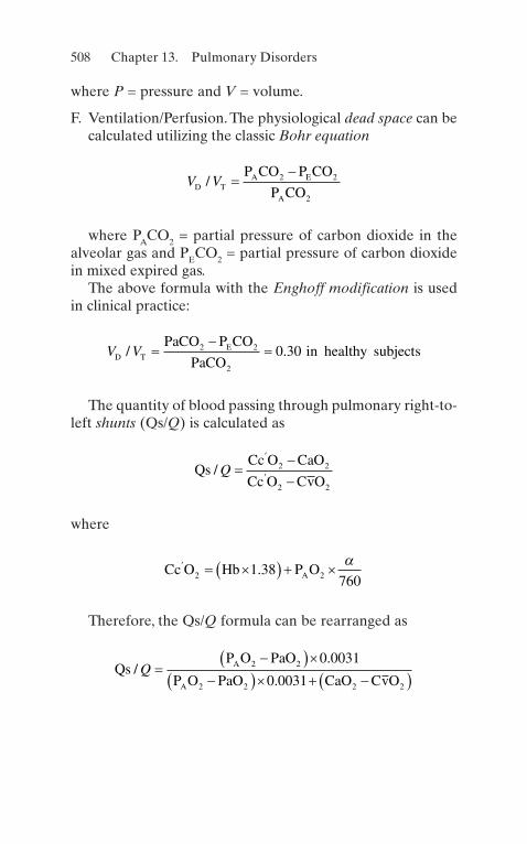
508
where P = pressure and V = volume.
F. Ventilation/Perfusion. The physiological dead space can be calculated utilizing the classic Bohr equation
V VD T
A E
A
P CO P CO
P CO/ �
�2 2
2
where PACO2 = partial pressure of carbon dioxide in the alveolar gas and PECO2 = partial pressure of carbon dioxide in mixed expired gas.
The above formula with the Enghoff modification is used in clinical practice:
V VD T
EPaCO P CO
PaCOin healthy subjects/ .�
��2 2
2
0 30
The quantity of blood passing through pulmonary right-to- left shunts (Qs/Q) is calculated as
Qs
Cc O CaO
Cc O CvO/Q �
��
�
�2 2
2 2
where
Cc O Hb P OA
� � �� � � �2 21 38760
.�
Therefore, the Qs/Q formula can be rearranged as
Qs
P O PaO
P O PaO CaO CvOA
A
/.
.Q �
�� ���� �� � �� �
2 2
2 2 2 2
0 0031
0 0031
Chapter 13. Pulmonary Disorders

509
G. Alveolar Gas Equation. The alveolar air equation is based firmly on Dalton’s law but is expressed in terms that emphasize alveolar O2 and CO2
P O FiO PCO RQA ATM H O2 2 22
� �� � �P P /
where PAO2 = partial pressure of O2 in the alveolus under present conditions and PATM = current, local atmospheric pressure.
PH O2 = vapor pressure of water at body temperature and
100% relative humidity; FiO2 = fraction of inspired O2; PCO2 = partial pressure of CO2 in arterial blood; RQ = respi-ratory quotient.
At sea level, this equation can be simplified to
P O PaCOA 2 2150 1 25� � �.
The arterial oxygen tension (PaO2) corrected for age is cal-culated as
PaO age corrected age in years2 100 1 3� � � � �/
The alveolar-arterial O2 gradient is age-corrected according to the following formula:
Age correction age in years� � �� �� �2 5 0 258. .
Useful Facts and Formulas
511© Springer Nature Switzerland AG 2021J. Varon, Handbook of Critical and Intensive Care Medicine, https://doi.org/10.1007/978-3-030-68270-5_14
Acid–Base Disturbances
An acidosis is any process that tends (in the absence of buff-ering or compensation) to cause the accumulation of hydro-gen ions (H+). If the pH of the blood is lower than normal (<7.35), acidemia is present. Similarly, any process that tends to cause the accumulation of bicarbonate (HCO3) is an alka-losis. An elevated blood pH (>7.45) is referred to as alkale-mia. When the acid–base disturbance arises as a result of changes in the carbon dioxide tension (PCO2) of the blood, a respiratory process is present. Conversely, when the acid–base disturbance occurs as a result of accumulation of non-volatile acids or loss of HCO3 (or substances metabolized to HCO3), a metabolic process is present. Acidosis and alkalosis can be either primary or compensatory for a different acid–base problem. Many ICU patients have double and triple acid–base disorders.
Chapter 14Renal and Fluid–Electrolyte Disorders

512
A. Approach to Acid–Base Disturbances
1. The initial evaluation of acid–base disorders requires the simultaneous examination of arterial blood gases (ABGs) and serum electrolytes.
2. The typical patterns of blood pH, PCO2, and HCO3 in various acid–base problems are listed in Table 14.1.
3. The adequacy of compensation for a primary acid–base abnormality can also be assessed.
(a) For patients with metabolic acidosis, the expected PCO2 can be calculated as
PCO HCO2 3 1 5 8 2= [ ]´( ) + ±( ).
1. If the actual PCO2 is greater than expected, a simultaneous respiratory acidosis is present.
2. If the measured PCO2 is less than expected, a simultaneous respiratory alkalosis is present.
Table 14.1 Systematic evaluation of acid–base disordersCompensated process
Metabolic acidosis pH ↓ PCO2 ↓ HCO3 ↓
Respiratory acidosis pH ↓ PCO2 ↑ HCO3 ↑
Metabolic alkalosis pH ↑ PCO2 ↑ HCO3 ↑
Respiratory alkalosis pH ↑ PCO2 ↓ HCO3 ↓
Uncompensated process
Metabolic acidosis pH ↓ PCO2—HCO3 ↓
Respiratory acidosis pH ↓ PCO2 ↑ HCO3—
Metabolic alkalosis pH ↑ PCO2—HCO3 ↑
Respiratory alkalosis pH ↑ PCO2 ↓ HCO3—
Mixed process
Metabolic/respiratory acidosis pH ↓ PCO2 ↑ HCO3 ↓ Metabolic/respiratory alkalosis pH ↑ PCO2 ↓ HCO3 ↑
Chapter 14. Renal and Fluid–Electrolyte Disorders

513
(b) For patients with metabolic alkalosis, the expected PCO2 can be calculated as
PCO HCO2 3 25 0 7 40 2= [ ]-( )´{ }+ ±( ).
1. Respiratory compensation for a primary meta-bolic alkalosis is limited to a PCO2 of about 55 mmHg. Even this limit may not be attained in patients with chronic pulmonary or hepatic dis-ease or congestive heart failure.
(c) Metabolic compensation for respiratory acid–base disturbances depends in part on the duration of the problem.
1. For patients with chronic respiratory acidosis, the expected [HCO3] can be calculated as
HCO PCO3 2 40 0 35 25 2[ ] = -( )´ + ±( ).
(a) The upper limit for this process is [HCO3] = 55 mEq/L.
(b) If the degree of compensation is inadequate, the possibility of a mixed acid–base distur-bance or a superimposed acute process should be entertained.
2. The expected degree of compensation for acute respiratory acidosis is calculated as
HCO PCO3 2 40 0 10 25 2[ ] = -( )´ + ±( ).
The upper limit of compensation for acute respiratory acidosis is [HCO3] = 30 mEq/L.
3. The expected metabolic response to chronic respiratory alkalosis can be estimated by
HCO PCO3 225 40 0 5 2[ ] = - -( )´ ±( ).
Acid–Base Disturbances
514
Inadequate compensation for chronic respiratory alkalosis should raise the possibility of a superim-posed acute respiratory alkalosis or a mixed acid–base problem.
4. The expected metabolic response to acute respiratory alkalosis can be calculated as
HCO PCO3 225 40 0 25 2[ ] = - -( )´ ±( ).
B. Metabolic Acidosis. The clinical consequences of meta-bolic acidosis are due to abnormalities of intracellular pH, transcellular ionic shifts, or both.
1. Signs and symptoms include tachypnea, depressed car-diac function, fatigue, weakness, and altered mental status.
2. Laboratory findings: hyperkalemia, calciuresis, and changes in carbohydrate and lipid metabolism.
3. Other specific findings may occur due to the etiology of the underlying cause of the acidosis.
4. Metabolic acidoses are classified based on whether the plasma anion gap (AG) is normal or elevated. Its value is calculated as
AG Na Cl HCO nl Eq L= éë ùû - éë ùû + [ ]( ) = -( )+ -
3 10 15 /
5. The differential diagnosis of primary metabolic acido-sis is listed in Table 14.2.
(a) In general, high AG metabolic acidoses are caused by the accumulation of an acid in the plasma whose anion is something other than Cl−. These acids can be endogenous (i.e., lactic acid, keto acids, uremic acids), exogenous (i.e., salicylates), or the endoge-nous metabolic products of exogenous toxins (i.e., paraldehyde, methanol, ethylene glycol).
Chapter 14. Renal and Fluid–Electrolyte Disorders

515
(b) The accumulation of these unmeasured acid anions should be stoichiometrically equal to the observed decrease in [HCO3].
(c) This equality will be preserved when the normal difference is maintained. Thus, if a normal anion gap = 12 mEq/L, and a normal [HCO3] = 25 mEq/L, in a pure high AG metabolic acidosis
Na Cl or
Na Cl
+ -
+ -
éë ùû - éë ùû +( ) =éë ùû - éë ùû( ) = ±
25 12
37 3
;
.
If ([Na+ − [Cl−]) is significantly >37, then a coex-istent metabolic alkalosis should be suspected.
If the difference is significantly <37, then a coexistent normal anion gap acidosis is probably present.
(d) The causes of metabolic acidosis with a normal anion gap are listed in Table 14.2. These so-called hyperchloremic acidoses occur either due to the administration of exogenous acids whose anion is chloride (e.g., HCl, NH4Cl, CaCl2) or the loss of
Table 14.2 Causes of metabolic acidosisElevated anion gap Normal anion gapMethanol Urinary–enteric fistula
Uremia Saline volume expansion
Ketoacidosis Endocrinopathies
Paraldehyde Diarrhea
Lactic acidosis Carbonic anhydrase inhibition
Ethylene glycol Acid–chloride administration
Salicylates Renal tubular acidosis
Mineralocorticoid antagonists
Acid–Base Disturbances

516
body fluid that is relatively low in chloride but high in bicarbonate or its metabolic equivalent.
1. In cases of normal AG metabolic acidosis, mea-surement of the urinary AG can be diagnosti-cally useful. It is calculated as
AGU Na K Cl= + -U U U
The AGU is inversely related to urinary ammonium (NH4
+) excretion. Its value should be less than zero in acidotic patients with gastro-intestinal (GI) bicarbonate losses, since renal ammoniagenesis ought to be well preserved. In patients with acidosis due to urinary bicarbon-ate losses, especially if caused by renal tubular acidosis (RTA), the AGU should be greater than zero.
6. The primary therapy of metabolic acidosis is to treat the underlying disease.
7. The use of exogenous NaHCO3 in the therapy of meta-bolic acidosis (especially lactic acidosis) is somewhat con-troversial, since evidence exists that such treatment can actually accelerate lactate production. However, it is gen-erally accepted that severe acidemia (pH < 7.20) of meta-bolic origin should be corrected with NaHCO3. The amount of bicarbonate required can be calculated by
HCO deficit
body wt kg target actual HCO3
30 5= ( )´ ´ - [ ]( ).
(a) Administration of NaHCO3 as an isotonic solution (i.e., three ampules of NaHCO3 per liter of sterile water or 5% dextrose in water) will reduce the risk of hypernatremia in patients at particular risk.
Chapter 14. Renal and Fluid–Electrolyte Disorders

517
8. Dialysis is sometimes indicated not only for the correc-tion of acidosis but also for the rapid elimination of cer-tain toxins that cause acidosis, such as methanol and ethylene glycol, even if the serum urea nitrogen (BUN) and creatinine are normal.
9. Treatment of chronic metabolic acidosis, especially if caused by RTA, includes oral alkali replacement as well as therapy of any associated electrolyte abnor-malities.
C. Respiratory Acidosis
1. The clinical effects of respiratory acidosis include find-ings compatible with intracellular acidosis in addition to syndromes caused by abnormal pulmonary gas exchange. Of these, the most important is hypoxemic encephalopathy. Signs of chronic pulmonary disease, including cor pulmonale, may be present.
2. Some common causes of respiratory acidosis are listed in Table 14.3.
3. Chronic respiratory acidosis is seldom associated with severe acidemia (pH < 7.20), even if marked hypercap-nia (PCO2 > 100 mmHg) is present. A superimposed acute metabolic or respiratory acidosis should be sus-pected if arterial pH is outside of this expected range.
4. The most important aspect of treatment of acute respi-ratory acidosis is the immediate restoration of effective alveolar gas exchange.
(a) This usually requires mechanical ventilation. (b) If intubation is not immediately available, supple-
mental oxygen and NaHCO3 should be cautiously administered.
5. Treatment of chronic respiratory acidosis depends mainly on the prevention and prompt recognition and therapy of intercurrent complications such as infec-tions and congestive heart failure.
Acid–Base Disturbances

518
Table 14.3 Causes of respiratory acidosis
Central nervous system disorders
Drugs (narcotics, anesthetics, tranquilizers)
Brain stem injury
Primary hypoventilation
Peripheral nervous system disorders
Infectious diseases (botulism, tetanus, polio)
Amyotrophic lateral sclerosis
Guillain–Barré syndrome
Spinal cord/phrenic nerve injury
Organophosphates
Primary muscular disease
Muscular dystrophy
Myasthenia gravis
Severe hypokalemia
Pulmonary disease
Chronic obstructive lung disease
Pneumonia
Pulmonary edema
Smoke inhalation
Pulmonary embolism
Thoracic/upper airway disorders
Chest wall (flail chest, pneumothorax, kyphoscoliosis)
Airway obstruction (laryngospasm, foreign body)
Failure of mechanical ventilator
Chapter 14. Renal and Fluid–Electrolyte Disorders
519
D. Metabolic Alkalosis
1. The clinical features of metabolic alkalosis are nonspe-cific:
(a) Evidence of neuromuscular irritability or latent tetany may be present.
(b) Cardiac dysrhythmias can occur.
2. Hypokalemia and hypochloremia are usually present. 3. The plasma AG is frequently elevated, due partly to a
slight increase in lactate levels but mainly due to release of H+ ions from plasma proteins (especially albumin) as part of the buffering process.
4. Urine pH measurements are not helpful. 5. The most useful biochemical determination is the level
of urinary chloride (UCl), which forms the basis for clas-sification of metabolic alkalosis.
(a) Metabolic alkalosis associated with an UCl < 10 mEq/L is termed chloride responsive, whereas those with a UCl > 20 mEq/L are termed chloride resistant.
(b) In some cases, UCl falls between these levels, in which case, no definite classification is possible.
6. Causes of metabolic alkalosis are listed in Table 14.4. 7. Since the kidney can ordinarily excrete a vast amount
of HCO3, acid loss (or base accumulation) alone does not usually result in a sustained alkalosis.
(a) If coexistent volume depletion is present, alkalosis can be persistent. This is by far the most common pathogenesis for metabolic alkalosis. This combi-nation occurs when body fluids rich in NaCl and poor in NaHCO3 are lost, usually from the intesti-nal tract or kidneys. Rarely, other sites of NaCl loss (e.g., skin losses in cystic fibrosis) may be present.
(b) Extracellular fluid volume contraction causes avid NaCl and NaHCO3 reabsorption in all nephron
Acid–Base Disturbances

520
sites with attendant decreases in UCl and perpetua-tion of the alkalosis.
(c) Hypokalemia occurs due to intracellular K+ shifts and mineralocorticoid-induced urinary K+ losses.
(d) Much less commonly, metabolic alkalosis occurs in the absence of significant volume deple-tion. This situation is most often associated with hypermineralocorticoidism as a primary feature. The excessive mineralocorticoid activity can be either endogenous (e.g., Conn’s syndrome, Cush-
Table 14.4 Causes of metabolic alkalosis
Chloride responsive
Gastrointestinal disorders
Gastric: vomiting, nasogastric suction
Colonic: villous adenoma, chloride diarrhea
Renal disorders
Diuretic therapy
Post-hypercapnic alkalosis
Cystic fibrosis
Chloride resistant
Hypermineralocorticoid states
Severe potassium depletion
Bartter’s syndrome
Miscellaneous
Alkali administration (antacids, transfusions)
Hypercalcemia
Poorly absorbable anion administration (antibiotics)
Chapter 14. Renal and Fluid–Electrolyte Disorders

521
ing’s syndrome) or exogenous (e.g., licorice, chewing tobacco). In these cases, excessive renal reabsorption of NaHCO3 and NaCl occurs in the absence of significant chloride deficits. Therefore, UCl levels remain relatively high.
(e) Severe total body potassium deficits exceeding 1000 mEq may result in the inability to correct metabolic alkalosis, but probably does not cause the disorder in humans. Metabolic alkalosis with high UCl occurs in Bartter’s syndrome, which is characterized by renal wasting of K+ and Cl−, nor-mal blood pressure, and partial response to prosta-glandin inhibition.
8. Some patients present with intermediate levels of chlo-ride excretion (UCl 10–20 mEq/L). Many of these patients will be found to have excessive alkali adminis-tration, especially in the form of antacids or citrate anti-coagulation of blood products.
9. The treatment of metabolic alkalosis depends on its cause.
(a) The underlying source of acid loss or base accumu-lation should be identified and corrected if possible.
(b) Administration of NaCl (and usually KCl as well) is mandatory in patients with chloride-responsive alkalosis.
(c) In some situations, urinary HCO3 excretion can be hastened with the use of the carbonic anhydrase inhibitor acetazolamide. This may be of particular benefit in patients with diuretic-dependent conges-tive heart failure and associated alkalosis.
(d) In rare cases, infusion of HCl or NH4C1 may be necessary.
1. This therapy requires an intensive care unit (ICU) setting and use of a central venous catheter.
Acid–Base Disturbances

522
2. It should be considered only in cases where the arterial pH exceeds 7.60.
3. The target pH for treatment with intravenous acid is 7.55.
4. The amount of acid required can be calculated by
mEq HCl body wt kg actual target HCO= ( )´ ´ - [ ]( )0 5 3.
5. This solution should be infused over a period of 12–24 h with frequent monitoring of blood chemistries and ABGs during therapy.
(e) In patients with severe alkalemia complicating acute or chronic renal failure, hemodialysis with an acid bath solution can be employed.
E. Respiratory Alkalosis
1. Symptoms and signs of respiratory alkalosis include:
(a) Neuromuscular irritability (b) Cardiac dysrhythmias and electrocardiographic
changes of ischemia
2. Laboratory findings include:
(a) Mild hyponatremia, hypokalemia, and hyperchlo-remia
3. Some causes of respiratory alkalosis are listed in Table 14.5.
4. Treatment of respiratory alkalosis depends on the cause.
(a) Supplemental O2 is beneficial in hypoxemic patients.
(b) Rebreathing techniques are effective in patients with hyperventilation due to anxiety.
1. Patients with respiratory alkalosis due to under-lying central nervous system (CNS) disease may
Chapter 14. Renal and Fluid–Electrolyte Disorders

523
require a period of intubation with paralysis and mechanical ventilation if the primary process is not directly treatable.
(c) Drug-induced hyperventilation can sometimes be treated with dialysis and other measures designed to accelerate drug elimination.
Table 14.5 Causes of respiratory alkalosis
Iatrogenic (hemodialysis, mechanical ventilation)
Central stimulation of respiration
Anxiety/pain
Trauma
Infections (meningitis, encephalitis)
Intracranial tumors
Cerebrovascular accidents
Drugs (salicylates, exogenous catecholamines)
Miscellaneous (fever, cirrhosis)
Hypoxemic stimulation of respiration
Pneumonia
Volume overload/pulmonary edema
Pulmonary emboli
Decreased lung compliance
High altitude
Carbon monoxide intoxication
Acid–Base Disturbances

524
Acute Renal Failure/Acute Kidney Injury
Acute renal failure (ARF) [also known as acute kidney injury (AKI)] is defined as a relatively sudden (over hours to days) decrease in renal function leading to serious derangements of body fluid homeostasis. It is usually classified as being due to prerenal, postrenal, or intrinsic renal disorders. Prerenal ARF is caused by renal perfusion defects. Postrenal ARF is caused by obstruction of the urinary tract. Intrinsic ARF is due to parenchymal disease of the kidneys. The diagnosis of ARF can be made if the serum creatinine increased by ≥0.3 mg/dl within 48 hours, increased serum creatinine by ≥1.5 times the baseline, or decreased urine output <0.5 ml/kg/h for 6 hours.
A. Prerenal ARF
1. The causes of prerenal ARF are listed in Table 14.6.
(a) Prerenal ARF also occurs in situations character-ized by renal hypoperfusion despite adequate or even expanded extracellular fluid (ECF) volume. This is due to global or local abnormalities in circu-lation leading to renal insufficiency. Frequent examples include congestive heart failure and decreased peripheral vascular resistance, as may accompany sepsis.
(b) Renal blood flow may be selectively impaired by certain drugs (especially nonsteroidal anti- inflammatory drugs [NSAIDs] and angiotensin inhibitors) or in the hepatorenal syndrome.
(c) Whenever renal blood flow is severely curtailed, ischemic acute renal failure is a possibility.
2. The diagnosis of prerenal ARF can often be made on clinical grounds.
(a) The history and physical examination are crucial. (b) Assessment of serial weights and intake/output
records are valuable, if available. (c) Oliguria is a customary finding.
Chapter 14. Renal and Fluid–Electrolyte Disorders
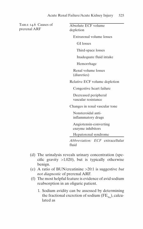
525
(d) The urinalysis reveals urinary concentration (spe-cific gravity >1.020), but is typically otherwise benign.
(e) A ratio of BUN/creatinine >20:1 is suggestive but not diagnostic of prerenal ARF.
(f) The most helpful feature is evidence of avid sodium reabsorption in an oliguric patient.
1. Sodium avidity can be assessed by determining the fractional excretion of sodium (FENa), calcu-lated as
Table 14.6 Causes of prerenal ARF
Absolute ECF volume depletion
Extrarenal volume losses
GI losses
Third-space losses
Inadequate fluid intake
Hemorrhage
Renal volume losses (diuretics)
Relative ECF volume depletion
Congestive heart failure
Decreased peripheral vascular resistance
Changes in renal vascular tone
Nonsteroidal anti- inflammatory drugs
Angiotensin-converting enzyme inhibitors
Hepatorenal syndrome
Abbreviation: ECF extracellular fluid
Acute Renal Failure/Acute Kidney Injury

526
FENa Na creat Na creat= ´( ) ´( )´U P P U/ 100
2. Values <1% suggest prerenal azotemia in the appropriate clinical setting.
(g) In occasional cases when doubt remains, measure-ment of central venous pressure or pulmonary capil-lary wedge pressure may be useful in guiding therapy.
3. Treatment of prerenal ARF depends on correction of the underlying cause and replacement of any volume deficits.
(a) The cause of fluid losses should be identified and treated if possible.
(b) Left ventricular function should be maximized if congestive heart failure (CHF) is playing a role.
(c) Offending drugs should be discontinued. (d) Hepatic transplantation may restore renal function
to normal in patients with hepatorenal syndrome. (e) In addition to treatment of the underlying disease,
volume expansion with appropriate intravenous fluids (usually normal saline or blood products) is required in patients unable to ingest sufficient sodium and water.
B. Postrenal ARF
1. Some causes of postrenal ARF are listed in Table 14.7. 2. The diagnosis of postrenal ARF can be suspected on
history and physical examination.
(a) Alternating polyuria and oligoanuria is suggestive of subtotal urinary obstruction, as is a very large postvoiding residual urine volume.
3. Laboratory tests are usually nonspecific.
(a) The urinalysis may be normal or disclose evidence of hematuria, pyuria, or crystalluria.
Chapter 14. Renal and Fluid–Electrolyte Disorders

527
(b) The best screening test for obstruction is renal ultrasound, which has a specificity of >90%.
(c) In cases in which obstruction is still suspected despite a negative ultrasound, retrograde pyelogra-phy can be performed.
4. Therapy of postrenal ARF largely depends on the site of obstruction.
(a) The coexistence of obstruction and urinary tract infection is a urologic emergency mandating broad- spectrum antibiotic coverage and immediate decompression of the urinary tract.
Table 14.7 Causes of postrenal ARF
Urethral obstruction
Urethral valves
Prostatic hypertrophy
Bladder obstruction
Neurogenic bladder
Bladder tumors
Cystitis
Ureteral obstruction
Intrinsic
Ureteral stones
Papillary necrosis
Extrinsic
Tumors
Retroperitoneal fibrosis
Aortic aneurysm
Pregnancy
Acute Renal Failure/Acute Kidney Injury
528
1. Upper tract disease can be effectively approached with indwelling stents or percutaneous nephros-tomy.
2. Lower urinary obstruction can be relieved with urethral or suprapubic catheterization.
(b) After the patient’s medical condition has stabi-lized, it is often possible to undertake definitive repair of the obstructing lesion. Relief of even long-standing obstruction is generally indicated, since some functional recovery may occur, even weeks or months later.
C. Intrinsic ARF
1. Some causes of intrinsic ARF are listed in Table 14.8. 2. Acute tubular necrosis (ATN) is by far the most com-
mon cause of intrinsic ARF among hospitalized patients.
(a) Multiple drugs and toxins can cause ATN, includ-ing aminoglycoside antibiotics, certain chemother-apeutic agents (i.e., cis-platinum, mithramycin), and radiographic contrast materials.
(b) Ischemia is another major factor causing ATN. (c) ATN is especially common in patients with major
trauma, recent major surgery (particularly vascular operations), sepsis, or crush injury.
3. Acute interstitial nephritis (AIN) is usually caused by exposure to a drug or allergen. Rare idiopathic cases are encountered.
(a) Probably the most common drugs causing AIN are NSAIDs.
(b) Other frequent agents include antibiotics (particu-larly penicillins, cephalosporins, and sulfa deriva-tives), loop and thiazide diuretics, and cimetidine.
(c) Recognition of this entity is especially important, since effective therapy depends on withdrawal of the offending drug.
Chapter 14. Renal and Fluid–Electrolyte Disorders

529
Table 14.8 Causes of intrinsic ARF
Glomerular diseases
Acute glomerulonephritis
Rapidly progressive glomerulonephritis
Tubulointerstitial diseases
Acute tubular necrosis
Drug induced
Ischemic
Acute interstitial nephritis
Allergic/drug induced
Idiopathic
Vascular diseases
Renal artery
Thrombosis/embolus
Dissection
Trauma
Renal microcirculation
Vasculitis
Malignant hypertension
Disseminated intravascular coagulation (DIC)
Thrombotic thrombocytopenic purpura (UP)
Cholesterol atheroemboli
Renal vein thrombosis
Acute Renal Failure/Acute Kidney Injury
530
4. The diagnosis of intrinsic ARF depends heavily on the history and physical examination with subsequent directed laboratory and radiographic evaluation.
(a) Medications in particular should be carefully reviewed.
(b) The urine sediment should be examined; it is valu-able to note that intrinsic ARF is almost invariably accompanied by abnormalities on urinalysis and their absence should raise the suspicion of pre- or postrenal causes.
(c) The presence of urinary eosinophils can be used to support a diagnosis of AIN or cholesterol microemboli.
(d) The FENa is typically >3% in intrinsic ARF. (e) Serologic evaluation of patients with suspected
vasculitis should include antinuclear antibodies (ANA), complements, hepatitis B surface antigen, cryoglobulins, and rheumatoid factor. If there is a suspicion of a pulmonary–renal syndrome (i.e., Goodpasture), anti-basal membrane antibodies should be obtained.
(f) More invasive studies (e.g., angiography, renal biopsy) may be appropriate depending on the clin-ical circumstances.
5. Therapy of intrinsic ARF is largely supportive.
(a) Offending drugs should be identified and with-drawn or substituted if possible.
(b) Control of fluid and electrolyte balance, modi-fication of drug dosages, and dialysis should be entertained.
(c) Specific therapy directed at a particular disease process is possible in occasional circumstances. A major aspect of the management of ARF is rec-ognition of the situations in which it is likely to occur, with appropriate measures to reduce its probability.
Chapter 14. Renal and Fluid–Electrolyte Disorders

531
1. Medications with the least risk of nephrotoxic-ity should be chosen in patients liable to ARF.
2. Volume depletion should be avoided or corrected.
3. Calcium channel blockers may be helpful in ameliorating ARF if given prophylactically (i.e., before angiography or renovascular surgery).
4. The use of hemodialysis acutely is believed by some to accelerate the rate of recovery in patients with ARF. However, hemodialysis has several potential side effects.
6. Despite many advances in the diagnosis and treatment of ARF, mortality remains at least 50% in critically ill patients.
Electrolyte Abnormalities
Certain electrolyte abnormalities that occur frequently pose a serious risk to ICU patients, even if the disorder has been present for a long time.
A. Calcium. Calcium exists in three forms in the circulation: free ionized calcium (iCa++); soluble complexes with phos-phate, citrate, and bicarbonate; and those bound to plasma proteins (principally albumin). Of these, the iCa++ is physi-ologically most important. The normal value for iCa++ is 4.0–4.9 mg/dL, 2.4–2.6 mEq/L, or 1.2–1.3 mmol/L (note difference in units).
Maintenance of normal iCa++ levels depends on the interaction between GI absorption, bone fluxes, and renal excretion, governed by parathyroid hormone (PTH), 1,25-dihydroxyvitamin D (1,25-D3), and calcitonin.
1. Hypercalcemia
(a) Most patients with hypercalcemia are asymptom-atic at the time of diagnosis. However, numerous signs and symptoms may occur depending on the
Electrolyte Abnormalities

532
severity and rate of development of hypercalce-mia. These include:
1. Nausea 2. Constipation 3. Anorexia 4. Pancreatitis 5. Peptic ulcers 6. Renal insufficiency 7. Polyuria 8. Urolithiasis 9. Bone pain 10. Weakness 11. Confusion and coma
(b) Symptomatic patients require urgent treatment. (c) Some causes of hypercalcemia are listed in
Table 14.9. The most common causes of hypercalce-mia are primary hyperthyroidism and malignancy.
(d) Hypercalcemia is a medical emergency whose acute treatment is largely independent of the cause.
(e) Therapy is outlined in Table 14.10.
Table 14.9 Disorders of calcium homeostasisHypercalcemia HypocalcemiaHyperparathyroidism Hypoparathyroidism
Hyperthyroidism Pseudohypoparathyroidism
Acute renal failure Vitamin D deficiency
Malignancy Malignancy
Excessive vitamin A or D Hyperphosphatemia
Granulomatous diseases Pancreatitis
Thiazide diuretics Neonatal tetany
Immobilization Calcium complex formation
Chapter 14. Renal and Fluid–Electrolyte Disorders

533
1. Intravenous (IV) normal saline should be admin-istered at a rate of 150–250 mL/h.
2. Furosemide is indicated to prevent volume over-load and to decrease calcium reabsorption (a minor effect).
3. Calcitonin’s effectiveness is limited by the phe-nomenon of osteoclast escape, which develops within a few days of beginning treatment.
4. Diphosphonates such as etidronate will reduce calcium levels to normal within 5 days in 75% of patients.
5. Mithramycin is the most potent hypocalcemic agent available, but its use is limited by serious renal, hepatic, and bone marrow side effects.
6. In hypercalcemia patients, oral NSAIDs are indicated only in patients with prostaglandin- mediated hypercalcemia.
7. Dialysis with low-calcium dialysate may be nec-essary in patients with refractory hypercalcemia, particularly if renal function is impaired.
Table 14.10 Treatment of hypercalcemiaIV fluids: normal saline 150–250 mL/h (± furosemide 40–80 mg IV q4–6 h)
Corticosteroids: prednisone (or equivalent) 1 mg/kg/day
Calcitonin: 4 U/kg SC q12 h
Etidronate: 7.5 mg/kg/day IV q day × 1–4 days
Mithramycin: 25 μg/kg IV q day × 3–4 days
Indomethacin: 25–50 mg PO q8 h
Dialysis
Electrolyte Abnormalities

534
2. Hypocalcemia
(a) The clinical manifestations of hypocalcemia are usually related to effects on excitable tissues.
1. Neurologic findings include overt or latent tet-any and mental status changes.
2. Cardiovascular manifestations include dysrhyth-mias, hypotension, and decreased myocardial contractility.
(b) Some causes of hypocalcemia are listed in Table 14.9. Chronic kidney disease (CKD) is the most common cause of hypocalcemia (low iCa++).
(c) Hypoalbuminemia can cause a lowering of the total calcium but normal iCa++. The corrected calcium formula is
Ca albumin Cacorr serum= ( ) +0 8 4 00. .
(d) Treatment of hypocalcemia is summarized in Table 14.11.
1. Patients with latent or overt tetany require immediate parenteral replacement. IV calcium should be administered through a central cathe-
Table 14.11 Treatment of hypocalcemiaCalcium bolus: 10–30 mL 10% Ca-gluconate or CaCl2 slow IV push over 15–30 min
Calcium infusion: 40 mL 10% Ca-gluconate in 500 mL D5W IV at 20 mL/h, titrate to desired iCa++
Hyperphosphatemia: CaCO3 650 mg 1–3 tabs PO tid with meals Al(OH)3 gels 30–60 mL PO tid with meals
Hypomagnesemia: 2 mL 50% MgSO4 IV or IM q4–6 h
Oral calcium: CaCO3, Ca-acetate, or Ca-lactate 1–3 tabs qid on an empty stomach
Vitamin D: calcitriol 0.25–0.5 μg q12–24 h
Chapter 14. Renal and Fluid–Electrolyte Disorders

535
ter if possible to avoid the risk of extravasation and skin necrosis.
2. Correction of aggravating electrolyte abnormali-ties (hyperphosphatemia, hypomagnesemia) should take place simultaneously.
3. Oral calcium and vitamin D supplements are sat-isfactory for milder, asymptomatic cases.
B. Magnesium. Magnesium is the second most common intracellular cation (after potassium). Most of its biologic effects depend on its role as a cofactor for intracellular enzymes, particularly adenosine triphosphate (ATP)-dependent systems. Magnesium balance is determined by the relationship between dietary ingestion and renal excretion. Urinary magnesium excretion is increased by sodium and calcium loading, diuretics, and PTH. It is decreased by volume depletion.
1. Hypomagnesemia
(a) Since most magnesium is intracellular, it can be difficult to estimate the magnitude of magnesium depletion from serum levels. However, hypo-magnesemia and simultaneous hypomagnesuria (UMg < 1 mEq/day) strongly suggest depleted body stores.
(b) Symptoms of hypomagnesemia include weakness, anorexia, and nausea. Some physical signs are latent tetany, hyperreflexia, tremors, dysrhythmias, delirium, and coma.
(c) Important associated laboratory abnormalities include hypokalemia and hypocalcemia that may be refractory to treatment until magnesium stores are repleted.
(d) Some common causes of hypomagnesemia are listed in Table 14.12.
1. Gastrointestinal causes include steatorrheic malabsorption, pancreatitis, dietary deficiency, and prolonged diarrhea or vomiting.
Electrolyte Abnormalities
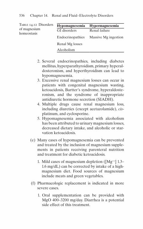
536
2. Several endocrinopathies, including diabetes mellitus, hyperparathyroidism, primary hyperal-dosteronism, and hyperthyroidism can lead to hypomagnesemia.
3. Excessive renal magnesium losses can occur in patients with congenital magnesium wasting, ketoacidosis, Bartter’s syndrome, hyperaldoste-ronism, and the syndrome of inappropriate antidiuretic hormone secretion (SIADH).
4. Multiple drugs cause renal magnesium loss, including diuretics (except acetazolamide), cis- platinum, and cyclosporine.
5. Hypomagnesemia associated with alcoholism has been attributed to urinary magnesium losses, decreased dietary intake, and alcoholic or star-vation ketoacidosis.
(e) Many cases of hypomagnesemia can be prevented and treated by the inclusion of magnesium supple-ments in patients receiving parenteral nutrition and treatment for diabetic ketoacidosis.
1. Mild cases of magnesium depletion ([Mg++] 1.3–1.6 mg/dL) can be corrected by intake of a high- magnesium diet. Food sources of magnesium include meats and green vegetables.
(f) Pharmacologic replacement is indicated in more severe cases.
1. Oral supplementation can be provided with MgO 400–3200 mg/day. Diarrhea is a potential side effect of this treatment.
Table 14.12 Disorders of magnesium homeostasis
Hypomagnesemia HypermagnesemiaGI disorders Renal failure
Endocrinopathies Massive Mg ingestion
Renal Mg losses
Alcoholism
Chapter 14. Renal and Fluid–Electrolyte Disorders

537
2. Parenteral therapy with 1–2 g (2–4 mL) of 50% MgSO4 repeated q4–6 h as needed is sufficient therapy for most patients.
3. Serum magnesium levels and deep tendon reflexes must be monitored closely during treat-ment, since life-threatening hypermagnesemia can occur.
2. Hypermagnesemia
(a) The clinical features of magnesium excess are fre-quently confused with those of hypercalcemia. They include:
1. Nausea 2. Altered mental status 3. Weakness and diminished tendon reflexes 4. Hypotension 5. Dysrhythmias 6. Respiratory paralysis
(b) The most frequent condition predisposing patients to hypermagnesemia is chronic renal failure. Sustained hypermagnesemia only occurs in the set-ting of renal insufficiency, although acute magne-sium excess can occur in the setting of overzealous replacement of magnesium deficiency.
(c) Therapy of hypermagnesemia involves discontinu-ing magnesium intake, infusion of Ca-gluconate 15 mg/kg over 4 h (which acts as a direct magne-sium antagonist), and dialysis with a magnesium- free bath in severe cases.
C. Phosphate. Phosphate is an abundant intracellular anion, critically important in energy metabolism and structural integrity of literally all cells. The overwhelming majority of dietary phosphate is normally absorbed, but a signifi-cant amount (approximately 200 mg/day) is secreted in the stool. Renal excretion eliminates the remainder, about 400–1500 mg/day depending on intake. Urinary phospho-rus excretion is increased by PTH, volume expansion, cor-
Electrolyte Abnormalities

538
ticosteroids, and calcitonin and is decreased by insulin, thyroid hormone, and vitamin D. Movement of phosphate into cells is enhanced during alkalemia, by glucose and some other carbohydrates and by hormones including insulin, epinephrine, and corticosteroids.
1. Hypophosphatemia
(a) Hypophosphatemia is characterized by altered men-tal status, weakness or myopathy progressing to rhab-domyolysis, osteomalacia, dysfunction of all blood cell types, anorexia, respiratory failure, and decreased cardiac contractility. However, most cases of phos-phate depletion are asymptomatic unless severe.
(b) Some causes of hypophosphatemia are listed in Table 14.13.
1. Respiratory and metabolic alkalosis is associ-ated with intracellular uptake of phosphate.
2. Refeeding after prolonged starvation precipi-tates hypophosphatemia, largely due to intracel-lular shifts mediated by glucose and insulin.
3. Administration or excessive activity of other hormones (epinephrine, growth hormone, ste-roids, gastrin, and glucagon) can cause hypo-phosphatemia by similar mechanisms.
4. Malnutrition alone does not typically cause severe hypophosphatemia. However, GI losses due to malabsorption of phosphate or use of phosphorus-binding antacids can lead to marked phosphate depletion.
Table 14.13 Disorders of phosphate homeostasisHypophosphatemia HyperphosphatemiaIntracellular shifts Massive cell lysis
Inadequate ingestion Increased ingestion
GI losses Renal failure
Renal losses
Chapter 14. Renal and Fluid–Electrolyte Disorders
539
5. Excessive renal losses of phosphate are present in Fanconi’s syndrome, after renal transplanta-tion in some patients, following recovery from urinary obstruction or ATN, and with vitamin D-resistant rickets.
(c) The best treatment of hypophosphatemia is often prevention.
1. Phosphate deficiency can be avoided by preven-tive supplementation in patients with malnutri-tion undergoing hyperalimentation or refeeding, or who are chronic alcoholics, on long-term ant-acids, or who have uncontrolled diabetes melli-tus or ketoacidosis.
2. Treatment of established hypophosphatemia depends on its severity and cause.
(a) Patients with mild hypophosphatemia due to intracellular shifts can be followed closely without active intervention unless the serum phosphorus drops <1.5 mg/dL.
(b) Dietary supplementation in the form of increased intake of dairy products is ade-quate in most asymptomatic patients.
(c) Oral supplementation can be provided with sodium and/or potassium phosphate, although diarrhea is often a dose-limiting side effect.
(d) Severe symptomatic hypophosphatemia is treated with elemental phosphorus (as sodium or potassium phosphate) 2 mg/kg IV q6 h until oral repletion can begin, usu-ally at a phosphorus level of 2.0 mg/dL.
(e) Use of parenteral phosphate is relatively contraindicated in patients with oliguric renal failure.
(f) Possible complications of IV phosphorus include dysrhythmias, hyperphosphatemia, hypocalcemia, hyperkalemia, and volume overload.
Electrolyte Abnormalities

540
2. Hyperphosphatemia
(a) Many of the symptoms of hyperphosphatemia can be attributed to the reciprocal fall in iCa++ that gen-erally occurs (see above).
(b) Severe hyperphosphatemia can also cause ARF, particularly in the setting of massive cell lysis. Metastatic calcifications caused by precipitation of Ca-PO4 crystals in essentially any tissue or organ can lead to widespread symptoms and signs.
(c) The causes of hyperphosphatemia are summarized in Table 14.13.
(d) Treatment of hyperphosphatemia involves decreas-ing ingestion with phosphorus-restricted diets as well as increasing elimination via the GI tract and kidneys.
1. Phosphate binders containing magnesium, cal-cium, or aluminum will accelerate stool phos-phorus losses even in patients who are NPO.
2. Magnesium-containing agents should be avoided in patients with renal failure.
3. Renal excretion of phosphate is enhanced by acetazolamide, volume expansion, and alkaline diuresis. Unfortunately, the frequent coexis-tence of renal failure with hyperphosphatemia often makes this route unreliable.
4. Hemodialysis or peritoneal dialysis is effective for acute or chronic hyperphosphatemia.
D. Potassium. Potassium is the most abundant intracellular cation. Only about 2% of total body potassium is in the ECF. Intracellular potassium is responsible for maintain-ing cell volume and resting membrane potential. A num-ber of factors regulate potassium movement into cells. These are summarized in Table 14.14.
1. Hypokalemia
(a) Abnormally low ECF [K+] have widespread patho-physiologic effects. Dominant among these are neuromuscular and cardiac events.
Chapter 14. Renal and Fluid–Electrolyte Disorders

541
1. Neuromuscular problems include GI hypomo-tility, skeletal muscle weakness or paralysis, and rhabdomyolysis.
2. Cardiac manifestations include the appearance of a U wave on the electrocardiogram, ventricu-lar and atrial dysrhythmias, predisposition to digoxin toxicity, and cardiac necrosis.
3. Cellular metabolism and renal function can also be impaired.
(b) The causes of hypokalemia are listed in Table 14.15.
1. Pseudohypokalemia reflects in vitro potassium uptake by leukemic cells in patients with severe leukocytosis (white blood cell [WBC] counts >105/mm3).
2. Intracellular shifts of potassium occur in alkalosis, insulin overdose, use of beta2-agonists, hypokalemic periodic paralysis, and barium poisoning.
3. Patients with anorexia nervosa, alcoholism, or severe dietary restrictions may develop signifi-cant hypokalemia.
Table 14.14 Factors affecting cellular potassium distributionIncreasing K+ Decreasing K+
Alkalosis Acidosis
Insulin Glucagon
Beta2-agonists Alpha-agonists
Hyperosmolarity
Table 14.15 Disorders of potassium homeostasisHypokalemia HyperkalemiaPseudohypokalemia Pseudohyperkalemia
Cellular K+ uptake Cellular K+ loss
Poor dietary K+ intake Excessive K+ intake
GI losses Inadequate renal excretion
Renal losses Cell lysis
Electrolyte Abnormalities
542
4. Excessive GI potassium losses occur in patients with protracted vomiting, diarrhea, and laxative abuse. In the United States, the most common cause of hypokalemia is GI loss from infectious diarrhea.
5. Renal losses occur in patients with renal tubular acidosis, hyperaldosteronism, treatment of dia-betic ketoacidosis, hypomagnesemia, and meta-bolic alkalosis. In addition, numerous drugs cause renal potassium wasting including diuret-ics and antibiotics (penicillins, cephalosporins, aminoglycosides, amphotericin).
(c) Treatment of hypokalemia is imprecise, since the degree of total body K+ depletion is usually impos-sible to calculate.
1. Ongoing potassium losses should be halted if possible and aggravating abnormalities (alkalo-sis, hypomagnesemia) corrected. Hypokalemia in the presence of acidosis suggests severe potassium depletion and requires immediate and aggressive attention.
2. The particular potassium salt employed depends on the specific clinical problem. KCl is preferred in patients with concomitant metabolic alkalosis and is effective in all forms of hypokalemia. Bicarbonate or phosphate salts of potassium may be preferred if acidosis or hypophosphate-mia is present.
3. Oral potassium preparations are effective but can have an unpalatable taste and cause GI irritation.
4. Potassium can be given by slow IV infusion (10–20 mEq/h) through a central or peripheral line; concentrations above 40 mEq/L should be avoided in peripheral IVs to decrease the risk of phlebitis.
5. Frequent monitoring of serum [K+] is essential during parenteral potassium repletion.
Chapter 14. Renal and Fluid–Electrolyte Disorders
543
6. Potassium-sparing diuretics are occasionally useful, but should be used with caution in patients receiving potassium supplements.
2. Hyperkalemia
(a) The principal clinical abnormalities of hyperkale-mia are neuromuscular and cardiac. Weakness, par-esthesias, and paralysis can occur but are usually overshadowed by cardiac disturbances . These include:
1. Progressive electrocardiogram (ECG) appear-ance of peaked T waves.
2. Flattened P-waves. 3. Prolonged PR interval. 4. Widening of the QRS complex. 5. The development of a sine-wave pattern pres-
ages the onset of ventricular fibrillation or asystole.
(b) Some causes of hyperkalemia are presented in Table 14.15.
1. Pseudohyperkalemia is due to in vitro release of potassium from red blood cells, leukocytes, or platelets.
2. Release of potassium from cells contributes to hyperkalemia in acidosis, poorly controlled dia-betes mellitus, beta-blockade, hyperkalemic periodic paralysis, hyperosmolar states, and digi-talis toxicity.
3. Potassium ingestion seldom results in hyperka-lemia if renal function is normal except when excessive parenteral potassium supplements have been administered.
4. Inadequate renal excretion occurs in patients with advanced renal failure, deficiencies of adre-nal hormones, and numerous drugs. These include potassium-sparing diuretics, NSAIDs,
Electrolyte Abnormalities

544
angiotensin-converting enzyme inhibitors, and cyclosporine.
5. Severe hyperkalemia is a frequent finding in patients with massive in vivo hemolysis or tumor lysis syndrome.
(c) The treatment of hyperkalemia is outlined in Table 14.16.
1. Calcium does not affect the serum potassium but rather antagonizes the cardiac toxicity of hyperkalemia.
2. Glucose/insulin and bicarbonate infusions lower serum [K+] by stimulating cellular potassium entry.
3. Kayexalate is used to augment fecal potassium excretion; it is relatively ineffective unless the patient develops diarrhea or loose stools. There are a variety of newer cation exchange resins under investigation showing promising results in removing potassium from the body.
4. Dialysis is extremely effective for life-threaten-ing hyperkalemia.
Table 14.16 Therapy of hyperkalemia10% calcium gluconate: 10–30 mL IV
Onset <5 min
50% dextrose 50 mL + regular insulin 5 U IV q30 min
Onset 15–30 min
NaHCO3 50 mL (50 mEq) IV q30 min × 4 doses
Onset 15–30 min
Kayexalate in sorbitol 30–60 g PO/enema 4–6 h
Onset 1–2 hSodium zirconium cyclosilicateOnset 1 h
Dialysis
Chapter 14. Renal and Fluid–Electrolyte Disorders

545
5. Diuretics and aldosterone analogues are occa-sionally useful adjunctive measures.
6. The use of beta-adrenergic agents (i.e., alb-uterol) via nebulizations has received consider-able attention in recent years for the acute management of hyperkalemia in end-stage renal disease patients.
E. Sodium. Hyponatremia and hypernatremia are disorders of water balance. The osmolarity of a solution depends on the number of dissolved particles per liter. In clinical prac-tice, this can be estimated, or calculated, as follows:
Posm Na glucose BUN= ´ éë ùû + [ ] + [ ]+2 18 2 8/ / .
Of note, a difference >10 mmol/Kg between the esti-mated and measured osmolality suggests an osmolal gap, as a result of unmeasured osmoles such as ethanol, methanol, isopropanol, and ethylene glycol. These substances increase osmolality without changing serum [Na+]. On the other hand, glucose, mannitol, glycine, and maltose increase osmolality but decrease serum [Na+]. It is known as translo-cational hyponatremia due to the movement of water from the intracellular space to the extracellular space.
Solutes restricted to one side of the plasma membrane are termed effective osmoles; changes in the quantity of effective osmoles in a body fluid obligate transmembrane water movement to maintain balance. Freely membrane permeable substances such as urea do not cause water movement and are hence termed ineffective osmoles. The tonicity, or effective osmolarity, of a solution can be calcu-lated by the following:
Eosm Na glucose= ´ éë ùû + [ ]+2 18/
Regulation of plasma osmolarity depends on the inter-play between water ingestion and renal water excretion.
Electrolyte Abnormalities

546
Renal mechanisms of water balance require the adequate delivery of salt and water to distal nephron sites as well as manipulation of tubular water permeability under the influence of antidiuretic hormone (ADH). ADH is secreted by the posterior pituitary in response to hyperto-nicity as well as other nonosmotic stimuli, including hypo-volemia, nausea, pain, and several drugs (i.e., nicotine, narcotics, vincristine, cyclophosphamide, chlorpropamide, and clofibrate). ADH increases renal water permeability, leading to increased water reabsorption and hypertonic urine. Normal thirst is even more responsive to changes in osmolarity than ADH is; consequently, thirst should be considered the primary guardian of plasma tonicity.
1. Hyponatremia
(a) Symptoms of hyponatremia are caused by osmotic movement of water from the ECF into cells.
1. Conditions characterized by hyponatremia with normal or elevated osmolarity (due to accumu-lation of unmeasured osmotically active solutes) are not symptomatic.
2. Brain cells are most sensitive to changes in vol-ume; hence, most symptoms of hyponatremia are neurologic. They include nausea, neuromus-cular irritability, altered mental status, and seizures.
3. The likelihood of symptomatic hyponatremia depends on its severity and the rapidity with which it develops.
(b) Some causes of hyponatremia are listed in Table 14.17.
1. Pseudohyponatremia is defined as a low mea-sured serum sodium despite a normal or elevated plasma osmolarity. Causes include hyperlipidemia, hyperproteinemia, hyperglyce-mia, mannitol infusion, and radiographic con-
Chapter 14. Renal and Fluid–Electrolyte Disorders
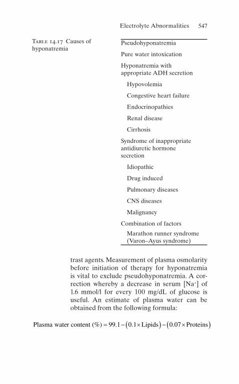
547
trast agents. Measurement of plasma osmolarity before initiation of therapy for hyponatremia is vital to exclude pseudohyponatremia. A cor-rection whereby a decrease in serum [Na+] of 1.6 mmol/l for every 100 mg/dL of glucose is useful. An estimate of plasma water can be obtained from the following formula:
Plasma water content Lipids Proteins( )% . . .= - ´( ) - ´( )99 1 0 1 0 07
Table 14.17 Causes of hyponatremia
Pseudohyponatremia
Pure water intoxication
Hyponatremia with appropriate ADH secretion
Hypovolemia
Congestive heart failure
Endocrinopathies
Renal disease
Cirrhosis
Syndrome of inappropriate antidiuretic hormone secretion
Idiopathic
Drug induced
Pulmonary diseases
CNS diseases
Malignancy
Combination of factors
Marathon runner syndrome (Varon–Ayus syndrome)
Electrolyte Abnormalities
548
If the formula reveals that plasma water is 84% rather than the normal 93% (150 × 0.93 = 140), the concentration of measured sodium would be expected to decrease to 126 mmol/L (150 × 0.84).
2. Hyponatremia due to pure water intoxication is extremely rare due to the efficiency with which the kidneys can excrete even massive water loads.
3. Most cases of true hyponatremia are associated with elevated ADH activity that is provoked by some nonosmotic stimulus.
4. Volume depletion of any cause is a major stimu-lus for ADH release. This includes states of rela-tive volume depletion such as CHF.
5. Hypothyroidism and adrenal insufficiency are also causes of hyponatremia.
6. Many renal diseases including nephrotic syn-drome predispose to hyponatremia due to inad-equate delivery of solute to the distal nephron.
7. Hyponatremia in untreated cirrhosis is caused by excessive ADH secretion and impaired distal sodium delivery.
8. SIADH has multiple causes including drugs, pulmonary diseases, CNS diseases, and cancer. See also Chap. 4
9. Hyponatremia in marathon runners (Varon–Ayus syndrome). This well-described syndrome occurs not only in marathon runners but also in a variety of extreme sports and military exercises.
(c) Symptomatic hyponatremia is a medical emer-gency regardless of duration.
(d) After the plasma osmolarity has been determined, therapy with 3% NaCl should be instituted. The amount of NaCl required to correct hyponatremia to a specified target level is
mEq NaCl weight kg target Na plasma Na= ´ [ ]( )´ -( )+ +0 6.
Chapter 14. Renal and Fluid–Electrolyte Disorders

549
The amount of 3% saline required to achieve this goal is
3 1000 513% /NaCl mL mEq NaCl( ) = ( )´( )
The target [Na+] should be an increase of 20 mEq/L above the actual [Na+] or 130 mEq/L, whichever is lower. The rate of correction is 1.0–1.5 mEq/L/h. These calculations often underesti-mate the actual rate of correction due to ongoing urinary sodium losses.
(e) In addition to hypertonic saline infusion, all IV flu-ids including medications should be given in 0.9% NaCl and a fluid restriction of 1000–1500 mL/day instituted if possible.
(f) Demeclocycline 150–300 mg PO q12h has been used successfully in the treatment of SIADH but is contraindicated in cirrhosis.
(g) Central pontine myelinolysis is a rare complication occurring after correction of chronic (but not acute) hyponatremia if the serum [Na+] is raised by >25 mEq/L during the first 48 h of therapy.
(h) A new agent, conivaptan (Vaprisol™) is indicated in some patients for the treatment of euvolemic and hypervolemic hyponatremia. It is an antago-nist of the vasopressin V1A and V2 receptors. This agent may lower blood pressure and may increase the risk of variceal bleeding in patients with cirrhosis.
2. Hypernatremia
(a) Symptoms of hypernatremia are caused by cellular dehydration, particularly of neurons. They include altered mental status, nausea, seizures, and intra-cranial hemorrhage. Myoclonus, metabolic acido-sis, and hyperglycemia due to peripheral insulin resistance are also common.
Electrolyte Abnormalities

550
(b) Most hypernatremic patients are volume depleted; the finding of volume overload should suggest the possibility of acute salt poisoning.
(c) Causes of hypernatremia are listed in Table 14.18.
1. Diabetes insipidus (DI) can be of central or nephrogenic origin (see Chap. 4).
2. The most common cause of hypernatremia is excessive water loss.
(a) Excessive renal water loss with hypernatre-mia has been reported in patients with renal failure, hypercalcemia, hypokalemia, sickle cell disease, osmotic diuresis, postobstruc-tive diuresis, and drugs (including alcohol, lithium, demeclocycline, oral hypoglyce-mics, and others).
(b) GI water losses due to gastroenteritis are an especially common cause of hypernatremia in children.
(c) Insensible water losses via the skin or respi-ratory tract occur with prolonged exposure to hot climates, thermal burns, and fever.
Table 14.18 Causes of hypernatremia
Diabetes insipidus
H2O losses
Renal
GI
Insensible
Salt poisoning
H2O deprivation
Primary hypodipsia
Mineralocorticoid excess
Chapter 14. Renal and Fluid–Electrolyte Disorders

551
3. Salt poisoning is a rare cause of outpatient hypernatremia but is more common in hospital-ized patients as a complication of hypertonic NaHCO3 therapy for severe acidosis.
4. Water deprivation is a fundamental feature of practically all cases of hypernatremia but is rarely the sole cause of the problem.
5. Entities characterized by increased mineralo-corticoid activity (Conn’s syndrome and Cushing’s syndrome) are sometimes accompa-nied by mild-to-moderate hypernatremia.
(d) The treatment of hypernatremia is replacement of free water deficits and correction of contributing electrolyte problems and hypovolemia, if present.
1. The water deficit is calculated as
H O deficit L weight kg
target Na actual Na2 0 6
1
( ) = ´ ( )´ ( ) ( ) -éë
ùû
+ +
.
/
The target sodium is either 148 mEq/L or a decrease of 20–25 mEq/L in the plasma [Na+] from its initial value whichever is higher. The goal of therapy is to reduce the plasma [Na+] by 1.0–1.5 mEq/L/h.
2. Faster rates of correction of chronic hypernatre-mia may precipitate rehydration seizures.
3. The choice of fluid and route of administration depends on the clinical circumstances. Distilled or tap water given PO or via nasogastric (NG) tube is preferred when feasible. If the enteral route is unavailable, 0.45% NaCl can be admin-istered by peripheral vein without significant risk of hemolysis.
4. Patients with clinical evidence of volume over-load who cannot tolerate the sodium load of 0.45% NaCl should be given distilled water IV through a central catheter.
Electrolyte Abnormalities

552
5. D5W is at least relatively contraindicated in many patients because of the coexistence of insulin resistance and the consequent risk of worsening hyperosmolarity due to non- metabolized dextrose.
6. Diuretics or dialysis can be employed for patients with salt poisoning.
7. Vasopressin analogues are useful in the long-term management of central DI.
Fluid and Electrolyte Therapy
In addition to the more specific treatments outlined above, some general guidelines regarding fluid and electrolyte ther-apy are useful. Administration of fluids and nutrition are essential but frequently overlooked considerations in the care of the ICU patient. Special care must be exercised in the selection and administration of IV fluids to the critically ill with ongoing renal or extrarenal fluid and electrolyte losses. IV fluids are potentially the most dangerous drugs used in the hospital; constant vigilance is required.
A. For essentially all ICU patients, some IV access is necessary. B. Whenever possible, the GI tract should be employed for
maintenance fluids, nutrition, and medications. C. The type of fluid used depends on the clinical situation.
Some general comments can be made:
1. For routine maintenance, crystalloid solutions are employed.
(a) In an otherwise well NPO patient, obligatory water losses amount to about 1000 mL/day.
(b) Sodium losses are minimized by virtually complete renal sodium reclamation, but some urinary potassium excretion continues (30–60 mEq/day) and must be replaced.
Chapter 14. Renal and Fluid–Electrolyte Disorders

553
(c) Excessive protein catabolism and starvation keto-sis can be prevented by inclusion of glucose 150–200 g daily in maintenance fluids.
(d) Supplements of other vitamins and minerals may be necessary if parenteral therapy lasts a week or more.
2. Colloid solutions such as blood products, albumin, and plasma are indicated for the rapid expansion of intra-vascular volume with minimal effects on other compo-nents of the ECF.
Dialysis
ICU patients frequently develop homeostatic abnormalities that cannot be managed conservatively. In such a case, dialy-sis or a related modality becomes necessary.
A. Dialysis is indicated in many different situations.
1. Volume overload manifested by pulmonary edema or severe hypertension unresponsive to diuretics can be effectively treated with dialysis or ultrafiltration.
2. Dialysis is useful for the treatment of several electrolyte abnormalities, including severe acidosis or alkalosis, hyperkalemia, hypo- or hypernatremia, hypercalcemia, hyperphosphatemia, and hypermagnesemia.
3. Symptoms caused by accumulation of uremic toxins are best treated with dialysis.
4. Poisonings with ethylene glycol, methanol, salicylates, and others can be effectively treated with dialysis.
B. The most frequently used dialysis modality in the ICU is hemodialysis (HD) or one of its variants. The choice between HD and peritoneal dialysis (PD) is usually a matter of the physician’s preference, but in some cases a clear preference is evident.
Dialysis

554
1. HD is the therapy of choice in severely catabolic patients due to more efficient removal of urea and other low molecular weight nitrogenous wastes.
(a) A large-bore dual-lumen central venous catheter is required for vascular access.
(b) For standard hemodialysis orders, the physician specifies the type of membrane to be used (more biocompatible membranes such as cellulose ace-tate are preferred in the ICU); the duration of ther-apy; blood flow speed; type of anticoagulation; composition of the dialysate with respect to Na+, K+, Ca++, and HCO3; the desired amount of fluid removal; and any additional medications or treat-ments required (e.g., blood products, antibiotics, or erythropoietin).
(c) The most common complication of HD is hypoten-sion, generally treated with fluid boluses of normal saline or albumin. In some cases, alternate treat-ments must be considered if hypotension is severe. Other serious potential complications of dialysis include dysrhythmias (presumably due to acute electrolyte fluxes) and hypoxemia (caused by membrane-induced complement activation and leukocyte sequestration in pulmonary capillaries as well as a decrease in minute ventilation due to removal of CO2 by dialysis).
2. To accomplish fluid and solute removal in hypotensive patients, several variants of hemodialysis have been developed.
(a) Continuous arteriovenous hemofiltration (CAVH) with or without dialysis is used in many ICUs. It has the specific advantage of allowing removal of large amounts of fluid even in patients with serious hemodynamic compromise. The patient’s own arterial pressure is used to drive ultrafiltration
Chapter 14. Renal and Fluid–Electrolyte Disorders

555
across a highly permeable membrane. Fluid removal may exceed 500 mL/h with CAVH, so pro-vision must be made for a pump to limit ultrafiltra-tion or replacement with an adequate amount of a balanced salt solution. Solute removal can be accelerated by the passing peritoneal dialysate across the ultrafiltrate side of the membrane. CAVH generally requires a femoral arterial access and thus may not be possible in patients with severe vascular disease.
(b) A modification of this technique, known as con-tinuous venovenous hemofiltration (CVVHD), has recently been introduced and is gaining favor. Vascular access is simplified, since an arterial access is not required, but CVVHD does require additional equipment in the form of pumps and alarms.
3. PD has less efficient clearance of low molecular weight solutes than HD; so, PD is generally not first-line ther-apy in hypercatabolic ICU patients.
(a) PD is much better tolerated from a hemodynamic standpoint and may be preferred in unstable patients.
(b) It is technically easier than HD in small children. Access to the peritoneal cavity is obtained with a flexible catheter that can be placed at the bedside.
(c) PD can be done continuously or intermittently with a minimum of equipment and staff. Ultrafiltration is controlled by changing the glucose concentra-tion (and hence osmolarity) of the dialysate.
(d) PD orders should include the number and duration of exchanges; the composition of the dialysate with respect to glucose, sodium, and calcium; and whether any additives such as antibiotics, heparin, insulin, or potassium are to be included.
Dialysis
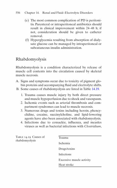
556
(e) The most common complication of PD is peritoni-tis. Parenteral or intraperitoneal antibiotics should result in clinical improvement within 24–48 h; if not, consideration should be given to catheter removal.
(f) Hyperglycemia resulting from absorption of dialy-sate glucose can be managed by intraperitoneal or subcutaneous insulin administration.
Rhabdomyolysis
Rhabdomyolysis is a condition characterized by release of muscle cell contents into the circulation caused by skeletal muscle necrosis.
A. Signs and symptoms occur due to toxicity of pigment glo-bin proteins and accompanying fluid and electrolyte shifts.
B. Some causes of rhabdomyolysis are listed in Table 14.19.
1. Trauma causes muscle injury by both direct pressure and muscle hypoperfusion due to shock and vasospasm.
2. Ischemic events such as arterial thrombosis and com-partment syndromes can lead to muscle necrosis.
3. Numerous drugs and toxins including heroin, phency-clidine, cocaine, succinylcholine, and lipid-lowering agents have also been associated with rhabdomyolysis.
4. Infections due to coxsackie, influenza, and measles viruses as well as bacterial infections with Clostridium,
Table 14.19 Causes of rhabdomyolysis
Trauma
Ischemia
Drugs/toxins
Infections
Excessive muscle activity
Heat stroke
Chapter 14. Renal and Fluid–Electrolyte Disorders
557
Staphylococcus, and Legionella spp. can cause skeletal muscle injury.
5. Excessive muscle activity (seizures, status asthmaticus, marathon running) has been reported as infrequent causes of rhabdomyolysis. In cases of heat stroke, some degree of muscle injury is invariably present.
C. The diagnosis of rhabdomyolysis depends on a thorough history and physical examination.
1. Muscle pain is present in about half the cases. 2. Symptoms or signs related to various electrolyte abnor-
malities may be present. 3. Fever and evidence of ECF volume depletion can be
frequently documented. 4. Urinalysis reveals bloody-appearing urine with a posi-
tive dipstick test for blood in the absence of apparent red blood cells on microscopic examination of the sediment.
5. Common early electrolyte abnormalities include hypo-natremia, hyperkalemia, hypocalcemia, hyperphospha-temia, hypermagnesemia, hyperuricemia, and metabolic acidosis. Hypercalcemia may be a later finding.
6. ARF occurs in about 30% of patients. 7. Intracellular muscle enzymes (creatine kinase and
aldolase) are invariably elevated, often to astronomic levels.
8. Low-grade DIC is present in the overwhelming major-ity of cases; its absence should prompt a consideration of other diagnoses.
9. As muscle groups swell in response to injury, persistent or recurrent muscle injury can occur leading to clinical exacerbation 48–72 h after the initial injury (“second- wave phenomenon”).
D. Principles of treatment are outlined in Table 14.20.
1. Vigorous volume expansion is essential, especially in the initial stages, when 2–3 L/h are frequently necessary. After the patient has stabilized, fluid administration is
Rhabdomyolysis

558
reduced to 300–500 mL/h to maintain brisk urine out-put (>200 mL/h).
2. Producing an alkaline diuresis (urine pH >8) by IV infusion of isotonic NaHCO3 (3 amps NaHCO3 per liter D5W) has been suggested as a possible means of increasing urine myoglobin solubility, but this has not been tested in well-controlled clinical trials.
(a) The use of diuretics to prevent tubular obstruction has some experimental support but has not been validated in practice.
3. Treatment of electrolyte abnormalities as discussed in previous sections is of paramount importance.
4. Dialysis is indicated for the treatment of severe ARF and resistant electrolyte problems.
E. Survival of patients with rhabdomyolysis, given appropri-ate intensive care, is 80–90%.
Useful Facts and Formulas
A. Acid–Base Equations/Facts. The normal relationship between bicarbonate (HCO3−), hydrogen ions (H+), and carbon dioxide is expressed in the Henderson equation
Table 14.20 Treatment of rhabdomyolysis
IV fluids
Normal saline volume expansion
Bicarbonate infusions
Diuretics
Mannitol
Furosemide
Treatment of electrolyte disorders
Renal replacement therapy
Chapter 14. Renal and Fluid–Electrolyte Disorders
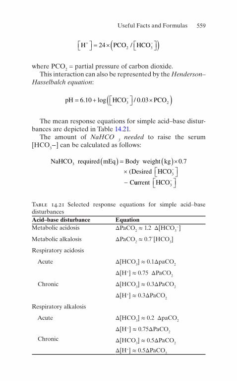
559
H PCO HCO+ -éë ùû = ´ éë ùû( )24 2 3/
where PCO2 = partial pressure of carbon dioxide.This interaction can also be represented by the Henderson–
Hasselbalch equation:
pH HCO PCO= + éë ùû ´( )-6 10 0 033 2. log / .
The mean response equations for simple acid–base distur-bances are depicted in Table 14.21.
The amount of NaHCO 3 needed to raise the serum [HCO3−] can be calculated as follows:
NaHCO required mEq Body weight kg
Desired HCO
C
3
3
0 7( ) = ( )´´ éë ùû-
-
.
(
uurrent HCO3-éë ùû
Table 14.21 Selected response equations for simple acid–base disturbancesAcid–base disturbance EquationMetabolic acidosis ΔPaCO2 ≈ 1.2 Δ[HCO3
−]
Metabolic alkalosis ΔPaCO2 ≈ 0.7"[HCO3]
Respiratory acidosis
Acute Δ[HCO3] ≈ 0.1ΔpaCO2
Δ[H+] ≈ 0.75 ΔPaCO2
Chronic Δ[HCO3] ≈ 0.3ΔPaCO2
Δ[H+] ≈ 0.3ΔPaCO2
Respiratory alkalosis
Acute Δ[HCO3] ≈ 0.2 ΔpaCO2
Δ[H−] ≈ 0.75ΔPaCO2
Chronic Δ[HCO3] ≈ 0.5ΔPaCO2
Δ[H+] ≈ 0.5ΔPaCO2
Useful Facts and Formulas

560
Alternatively, the following formula can be utilized to cal-culate the base deficit in metabolic acidosis:
HCO deficit desired HCO observed HCO
body weight kg3 3 3
0 4
- = -( )´ ( )(. ))
The chloride deficit in the treatment of metabolic alkalosis can be calculated utilizing the following formula:
Cl Deficit mEq weight in kg measured Cl- -( ) = ( ) -( )0 5 103.
B. Renal Function Formulas. The glomerular filtration rate (GFR) can be approximated, adjusted to age based on the following formulas:
< = ( )45 12 490 37years GFR age: . .
³ = ( )45 153 1 07years GFR -- age: .
A formula derived by Cockcroft and Gault is commonly used to estimate creatinine clearance:
Creatinine clearance mLage
Serum creatinine mg dL/ min
/( ) = -
( )
´
140
BBody weight kg( )72
In women, the value obtained from this equation is multi-plied by a factor of 0.85.
This formula can also be adjusted for lean body weight (LBW) calculated from the following:
LBW male kg kg in feet( ) = + >50 2 3 5. / .
LBW female kg kg in feet( ) = + >45 5 2 3 5. . / .
Chapter 14. Renal and Fluid–Electrolyte Disorders

561
Alternatively, the creatinine clearance (Ccr) can be calcu-lated as follows:
C
U V
Pcrcr
cr
=×( )
where Ucr = concentration of creatinine in a timed collection of urine; Pcr = concentration of creatinine in the plasma; and V = urine flow rate (volume divided by period of collection).
Another commonly employed formula to calculate the creatinine clearance is Jelliffe’s formula:
C
Pcrcr
age=
- -( )98 0 8 20.
In this formula, age is rounded to nearest decade. In females, the above result is multiplied by a factor of 0.9.
A more complicated and potentially more accurate way to calculate creatinine clearance is Mawer’s formula:
C
P
Pcr
cr
cr
malesLBW age
( ) =- ´( )éë ùû - ´( )éë ùû
(29 3 0 203 1 0 03
14 4
. . .
. ))
C
P
Pcr
cr
c
femalesLBW age
( ) =- ´( )éë ùû - ´( )éë ùû25 3 0 174 1 0 03
14 4
. . .
. rr( )
Hull’s formula for creatinine clearance is calculated as
C Pcr crage= -( )éë ùû -145 3/
In females the result is multiplied by a factor of 0.85.Ranges for creatinine clearance under selected conditions
are depicted in Table 14.22.
Useful Facts and Formulas

562
C. Selected Electrolytes. The transtubular potassium gradient (TTKG) allows one to estimate the potassium secretory response in the cortical collecting duct. This index corrects for water reabsorption in the cortical and medullary col-lecting ducts:
TTKG Corrected urine K Serum K
Corrected urine KUrine K
osm
=
=
+ +
++
/
/U PPosm
The normal renal conservation of potassium is reflected by a TTKG <2.
The percentage of magnesium retention (MR) can be cal-culated by the following formula:
Postinfusion
hour Preinfusion Postinfusion
MRurin
24
1
- - ´( )
( ) = -%eeMg urineMg CrratiourineCr
Total elementalmagnesiuminfus
++ ++ /
eed´1
The fractional tubular reabsorption of phosphate (TRP) allows for quantification of renal phosphate wasting and is calculated as
TRP p cr= -1 04C C/
where Cp04/Ccr = fractional excretion of phosphate.
Table 14.22 Creatinine clearance values under selected conditions
Condition ValueNormal >100 mL/min
Mild renal failure 40–60 mL/min
Moderate renal failure
10–40 mL/min
Severe renal failure
<10 mL/min
Chapter 14. Renal and Fluid–Electrolyte Disorders
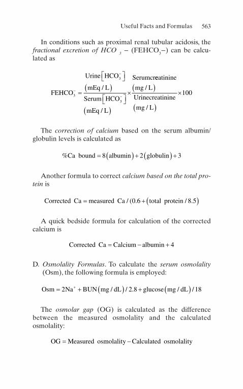
563
In conditions such as proximal renal tubular acidosis, the fractional excretion of HCO 3 − (FEHCO3−) can be calcu-lated as
FEHCO
Urine HCO
mEq L
Serum HCO
mEq L
Serumcr
3
3
3
-
-
-=
éë ùû( )
éë ùû( )
´/
/
eeatinine
mg L
Urinecreatinine
mg L
/
/
( )
( )
´100
The correction of calcium based on the serum albumin/globulin levels is calculated as
%Ca bound albumin globulin= ( ) + ( ) +8 2 3
Another formula to correct calcium based on the total pro-tein is
Corrected Ca measured Ca total protein= + ( )/ ( . / .0 6 8 5
A quick bedside formula for calculation of the corrected calcium is
Corrected Ca Calcium albumin= - + 4
D. Osmolality Formulas. To calculate the serum osmolality (Osm), the following formula is employed:
Osm Na BUN mg dL glucose mg dL= + ( ) + ( )+2 2 8 18/ / . / /
The osmolar gap (OG) is calculated as the difference between the measured osmolality and the calculated osmolality:
OG Measured osmolality Calculated osmolality= -
Useful Facts and Formulas
564
The approximate urine osmolality can be calculated from the following formula:
mOsm Urine specific gravity~ ,-( )´1 40 000
E. Water Balance. To estimate the amount of total body water (TBW), the following formula is frequently employed:
TBW Body weight kg= ( )´60 %
The water deficit of a patient can be estimated by the fol-lowing equation:
Water deficit Body weight in kg PNa= ´ ´ -( )0 6 140 1. /
where PNa = plasma sodium concentration.Alternatively, the free water deficit from the osmolality can
be calculated as
H O deficit L Total body weight kg
normal osm
observed os
2
0 6 1
( ) = ( )
´ -.mm
æ
èç
ö
ø÷
To calculate the free water clearance based on the osmolar clearance, the following formula can be utilized:
Free water clearance Urine volume Osmolar clearance= -
where the osmolar clearance is calculated as
Osmolar clearance
Urine osmolarity urine volume
Plasma osmolari=
´tty
The excess water (EW) of a patient is calculated as
Chapter 14. Renal and Fluid–Electrolyte Disorders

565
EW TBW Actual plasma Na /Desired plasma Na TBW= - éë ùû´+ +
F. Urinary/Renal Indices. The most common urinary indices used in the differential diagnosis of acute renal failure are depicted in Table 14.23.
To calculate the renal failure index (RFI), the following formula is commonly employed:
RFI
UNa
U PCr=
+
/
The fractional excretion of sodium (FENa) is calculated as
FENa
Quantity of Na excreted
Quantity of Na filtered%( ) = ´
+
+ 100
or
Table 14.23 Commonly used urinary indices in acute renal failure
Index PrerenalAcute tubular necrosis
Specific gravity >1.020 <1.010
Urinary osmolality (mOsm/kg H2O)
>500 <350
Uosm/Posm >1.3 <1.1
Urinary Na+ (mEq/L) <20 >40
U/P Cr >40 <20
RFI <1 >1
FENa (%) <1 >1
Abbreviations: Cr creatinine, P plasma, RFI renal failure index, U urine, FENa fractional excretion of sodium
Useful Facts and Formulas

566
FENa
U PNa
U PCr%
/
/( ) = ´+ 100
or
FENa Na
Na Cr Cr
%/
( ) = ´´ ´( )
´U V
P U V P100
or
FENa Na Cr
Na Cr
%( ) = ´´
´U P
P U100
where UNa = urine sodium concentration; V = urine flow rate; PNa = plasma sodium concentration; UCr = urine creatinine concentration; and PCr = plasma creatinine concentration.
G. Hemodialysis Formulas. The following are useful equa-tions in the management of the chronic hemodialysis patient.
The protein catabolic rate (PCR) is calculated as
PCR g kg day
IDBUN
IDinterval/ / .
.( ) = +´ ´
0 220 036 24
where ID BUN = interdialytic rise in blood urea nitrogen (BUN) in mg/dL and ID interval = interdialytic interval in hours.
Alternatively, if blood urea is measured, the PCR can be calculated utilizing the following formula:
PCR g kg day
ID urea
IDinterval/ / .
.( ) = +´ ´
0 220 01 24
where ID urea = interdialytic rise in blood urea in mmol/L.
Chapter 14. Renal and Fluid–Electrolyte Disorders

567
If the patient has a significant urine output, the contribu-tion of the urinary urea excretion must be added to the PCR calculation and is calculated as follows:
Urine contribution to PCRUrine urea N g
ID interval h Body=
( )´( )´
150
wweight kg( )
Alternatively, if urine urea is measured
Urine contribution to PCRUrine urea mmol
ID interval h Bo=
( )´( )´
4 2.
ddy weight kg( )
To calculate the percentage of recirculation during hemodi-alysis, the following formula is utilized:
%Recirculation
A A
A=
--
´2 1
2100
V
where A2 = blood urea or creatinine concentration in arterial blood line after blood pump is stopped; Al = arterial line blood urea or creatinine concentration; and V = venous line urea or creatinine concentration.
The volume of distribution of urea can be calculated as follows:
Males : . . . .V A H W= - + +2 447 0 09516 0 1074 0 3362
Females : . . .V H W= + +2 097 0 1069 0 2466
where V = volume in liters; A = age in years; H = height in centimeters; and W = weight in kilograms.
The calculation of residual renal function for dialysis three times per week
GFR =
´´( ) +
V Ut U U0 25 0 751 2. . )
Useful Facts and Formulas

568
where V = urine volume in interdialytic period; U = urine urea nitrogen or urea concentration; t = interdialytic period in minutes; U1 = postdialysis BUN or blood urea on first dialysis of the week; and U2 = predialysis BUN or blood urea on sec-ond dialysis of the week.
The percent reduction of urea (PRU) can be calculated utilizing the following formula:
PRU
e urea Post urea
Post urea=
-´
Pr100
The urea reduction ratio (URR) is calculated as
URR
Posturea
Preurea= ´ -æ
èç
öø÷100 1
H. Urinalysis. Please refer also to Chap. 20 for additional laboratory values. The most common urinalysis manifesta-tions of renal diseases are depicted in Table 14.24. Urinalysis is one of the key tests to evaluate kidney and urinary tract disease. It should include physical and chem-ical characteristics of urine and a microscopic examina-tion of urine sediment. Physical characteristics are color, odor, turbidity, and relative density. pH, hemoglobin, glu-cose, protein, leukocyte esterase, nitrites, bile pigments, and ketones are included in the chemical characteristics of urine. The microscopic examination is useful to determine the presence of cells, lipids, casts, crystals, and organisms.
Some elements and substances can modify the color of urine in humans, as depicted in Table 14.25.
I. Other Formulas/Facts. To determine whether a patient has aminoaciduria or not, the fractional reabsorption of an amino acid (FRA) is determined utilizing the following formula:
Chapter 14. Renal and Fluid–Electrolyte Disorders

569
Table 14.24 Urinalysis in different conditionsCondition FindingsPrerenal failure SG: >1.015
pH: <6
Prot: Trace to 1+
Sed: Sparse hyaline, fine granular cases or bland
Postrenal failure SG: 1.010
pH: >6
Prot: Trace to 1+
Hb: +
Sed: RBCs, WBCs
Acute tubular necrosis (ATN)
“Muddy” brown urine
SG: 1.010
pH: 6–7
Prot: Trace to 1+
Blood: +
Sed: RBCs, WBCs, RTE cells, RTE casts, pigmented casts
Glomerular diseases
SG: >1.020
pH: >6
Prot: 1–4+
Sed: RBCs, RBC casts, WBC, oval fat bodies, free fat droplets, fatty casts
(continued)
Useful Facts and Formulas
570
FRA Urine Plasma Urine Plasm Cr
A A Cr= -[ ] [ ] ¸[ ] [ ] ´1 100/ / %
The normal urinary excretion of amino acids in patients older than 2 years is depicted in Table 14.26.
When acute renal failure (ARF) is due to uric acid nephropathy (UAN), the following equation is generally >1:
Index
spot urine uric acid mg dL
spot urine creatinine mg dL=
( )( )
=>/
/11 0.
The choice of intravenous fluids varies from patient to patient. When prescribing any intravenous fluid, we must try to use solutions with compositions similar to human plasma.
Clinicians utilizing crystalloids are reminded of the follow-ing formulations:
0.9% saline (normal saline):
Osmolality mOsm L Sodium mEq L= =308 154/ , /
Table 14.24 (continued)Condition Findings
Vascular diseases SG: >1.020, if preglomerular
pH: <6
Prot: Trace to 2+
Sed: RBCs and RBC casts with glomerular involvement
Interstitial diseases
SG: 1.010
pH: 6–7
Prot: Trace to 1+
Sed: WBCs, WBC casts, eosinophils, RBCs, RTE cells
Abbreviations: RBC red blood cells, RTE renal tubular epithelial cells, WBC white blood cells, SG urine specific gravity, Prot protein, Sed urinary sediment
Chapter 14. Renal and Fluid–Electrolyte Disorders

571
Table 14.25 Urine color based on the presence of elements or substancesElements/substances Characteristic colorWhite blood cells Milky white
Precipitated phosphates
Chyle
Bilirubin Yellow/amber
Chloroquine
Sulfasalazine
Nitrofurantoin
Urobilin
Phenazopyridine Brown/red
Hemoglobin myoglobin
Red blood cells
Phenothiazines
Phenytoin
Porphyrins
Beets
Red-colored candies
Melanin Brown/black
Phenol
Methyldopa
Metronidazole
Quinine
Pseudomonas infection Blue/green
Amitriptyline
Methylene blue
Biliverdin
Propofol
Useful Facts and Formulas

572
Compounded sodium lactate (Ringer’s lactate™):
Osmolality mOsm L Sodium mEq L= =273 130/ . /
Multiple electrolyte solution (PlasmaLyte™):
Osmolality mOsm L Sodium mEq L= =294 140/ . /
Table 14.26 Normal urinary excretion of selected amino acids
Amino acidNormal excretion (mg/g of creatinine)
Cystine 18
Lysine 130
Arginine 16
Ornithine 22
Chapter 14. Renal and Fluid–Electrolyte Disorders

573© Springer Nature Switzerland AG 2021J. Varon, Handbook of Critical and Intensive Care Medicine, https://doi.org/10.1007/978-3-030-68270-5_15
Airway Management
A. The first technique in the management of patients with airway problems is manual opening of the airway (i.e., head tilt, chin lift). See Chap. 2.
B. Adjuncts for artificial airway:
1. Oropharyngeal Airways
(a) Oropharyngeal airways are available in a number of different sizes and styles.
(b) These devices routinely should be sized from the angle of the jaw to the central incisors.
(c) Techniques for insertion include the following:
1. The Inverted Technique: The oral airway is placed upside down and rotated to the appropri-ate position after negotiating the tongue.
2. Tongue Depressors Technique: A tongue depres-sor is used to manipulate the base of the tongue to prevent occlusion of the airway by impeachment of the tongue on the end of the oral airway.
2. Nasopharyngeal Airways
Chapter 15Special Techniques

574
(a) Nasopharyngeal airways are also available in a number of sizes, which should be measured from the tragus of the ear to the tip of the nose.
1. Great care should be exercised to ensure that the angled opening of the distal portion of the airway does not traumatize nasal passages, resulting in epistaxis.
2. Well-lubricated nasopharyngeal airways appear to be better tolerated in the alert patient as com-pared to oropharyngeal airways.
3. Laryngeal Mask Airway (LMA)
(a) LMAs are used by anesthesiologists in the operat-ing room on a routine basis. These devices can be utilized when concerns for difficult airway arise. They are quite simple to use.
C. Endotracheal intubation:
1. Common indications for endotracheal intubation are depicted in Table 15.1.
2. Orotracheal intubation:
(a) The oral route is the most common and easily mas-tered approach for tracheal intubation.
(b) Routinely, this technique involves visualization of the glottis, the use of a laryngoscope, and passage of the endotracheal tube into the trachea under direct vision. Table 15.2 lists the essential equipment that should be available for orotra-cheal intubation.
(c) Intubation technique:
Table 15.1 Indications for intubation
1. Ventilation of the patient
2. Airway obstruction
3. Tracheobronchial toilet
4. Airway protection
5. Impending respiratory failure
Chapter 15. Special Techniques

575
1. Positioning of the patient: It is important to align the axis of the trachea, pharynx, and the oral cavity to effect endotracheal intubation. This requires that the axis be aligned by placing the patient’s head in the “sniffing” position. A small pad or folded towel may be used to raise the occiput for proper alignment (see Fig. 15.1a).
2. After proper positioning and ensuring that all the necessary equipment is available, laryngos-copy may be performed by inserting the laryn-
Table 15.2 Equipment necessary for endotracheal intubationOxygenation equipment
Oxygen source
Regulators and tubing
Endotracheal tubes
Appropriate numbers and sizes of endotracheal tubes should be available
A malleable stylet to stiffen the tube for insertion
Silicon jelly as a lubricant
Appropriate volume syringe(s) for cuff inflation
Laryngoscope
Laryngoscope handle with functioning batteries
Straight and curved blades of the sizes necessary for the proposed intubation with functioning light bulbs or fiber- optic tracks
Fixation device for the endotracheal tube
Adhesive tape or commercially available tube-fixation devices
Means of assessment for appropriate position of endotracheal tube
Stethoscope
End-tidal CO2 monitoring device
Pulse oximeter
Airway Management

576
Tongue
Epiglottis
Vocal cord
Tracheal rings
Cuneiform cartilage
Corniculate cartilage
Figure 15.1 Endotracheal intubation. (a) Proper use of the laryngo-scope during intubation. (b) View of the larynx during direct laryn-goscopy. (From Allison EJ Jr. Advanced life support skills. St. Louis: Mosby; 1994. Used with permission)
Chapter 15. Special Techniques

577
goscope into the oropharyngeal airway and examining the airway. Two types of blades are commonly used:
(a) The Miller blade is used to lift the epiglottis to obtain visualization of the tracheal opening.
(b) The Mackintosh blade fits into the vallec-ula, resulting in adherence of the epiglottis to the back of the blade (see Fig. 15.1a). The tongue and other oral contents are dis-placed to the left side (see Fig. 15.1b). The Miller blade is inserted more midline, ele-vating the tongue upward.
(c) Care should be exercised in the use of the laryngoscope. Proper technique is to lift the laryngoscope upward and not to use it as a fulcrum (see Fig. 15.1a, b).
(d) A Sellick maneuver (pressure on the cricoid to help occlude the epiglottis during manipu-lation of the airway) is commonly performed to help prevent aspiration and to stabilize the glottis during the intubation procedure.
3. After identifying the laryngeal opening (see Fig. 15.1b), the trachea is entered under direct visualization by placing the endotracheal tube through the vocal cords.
(a) This can be most easily accomplished by placing the endotracheal tube in the right corner of the mouth, directing the tip into the glottic opening. This technique does not require interruption of the view of the vocal cords during intubation.
(b) Insertion should be stopped when the cuff is displaced 2 cm from the glottic opening (external markings are typically at 21 or 23 cm from the central incisors of average size women and men, respectively).
Airway Management

578
(c) The cuff is blown up to a moderate tension of the pilot balloon and ventilation with 100% oxygen begun.
4. Tube placement is ascertained by auscultation of chest and abdomen, examination of the rise and fall of the chest, condensation of the respi-ratory gas mixture in the endotracheal tube, maintenance of adequate saturation on pulse oximetry, and when available end-tidal CO2 indicators.
5. Newer video laryngoscopes allow for easier visualization of the cords.
3. Nasotracheal intubation:
(a) Nasotracheal intubation can be performed under direct visualization using the laryngoscope.
1. The tube is placed through the nares, and the tip is visualized in the pharynx.
2. McGill forceps can be used to manipulate the end of the endotracheal tube through the vocal cords to achieve proper positioning.
(b) Blind nasal insertion.
1. The location of the endotracheal tube is ascer-tained through auscultation.
2. This technique is reserved for those patients who have spontaneous ventilation.
3. An endotracheal tube of appropriate size is inserted through the naris and advanced to the pharynx.
4. Auscultation using the unaided ear and listening at the nasal end is used as the endotracheal tube is advanced.
5. The tube is inserted through the glottic opening during inspiration, and appropriate position is confirmed as noted above.
Chapter 15. Special Techniques
579
6. Nasal intubation may result in severe epistaxis in patients with coagulopathy or if performed with excessive force.
7. Patients with midface fractures should not be nasally intubated.
8. Sinusitis is a recognized complication of nasal intubation and should be considered when determining the route of intubation.
9. This procedure is nowadays performed rarely.
4. No matter what the route of intubation of the trachea is, all endotracheal tubes should be secured with adhe-sive tape or other securing devices to prevent dislodgment.
5. Common complications of endotracheal intubation:
(a) During Laryngoscopy and Intubation
1. Dental and oral soft tissue trauma 2. Dysrhythmias and hypertension/hypotension 3. Aspiration of gastric contents
(b) While Endotracheal Tube Is in Place
1. Tube obstruction 2. Esophageal intubation 3. Accidental extubation 4. Tracheal mucosal ischemia
(c) Delayed Complications
1. Tracheal stenosis 2. Vocal cord paralysis 3. Laryngeal edema
D. Cricothyroidotomy:
1. The cricothyroid membrane can be identified by palpa-tion below thyroid cartilage.
2. A large-bore (14- or 16-gauge) catheter may be placed through the cricothyroid membrane into the trachea
Airway Management
580
and used to ventilate and oxygenate patients in whom other airway maneuvers are unsuccessful.
3. Free release of air from the catheter will confirm tra-cheal position.
4. The tip of the catheter should be angled inferiorly, and after tracheal penetration with the plastic cannula, it should be advanced.
5. The cannula may be adapted to fit the 15-mm opening of a standard Ambu bag, or alternatively, a portable high-frequency jet ventilator device may be used to provide oxygenation to the patient.
6. Surgical cricothyroidotomy:
(a) Percutaneous technique: A needle is used, passing into the trachea, and a guidewire is positioned in the trachea through the needle. A dilator is then passed, and a cricothyroidotomy tube with an internal obturator is inserted. This can be easily performed at the bedside of an ICU patient.
(b) Surgical technique: A small midline incision is made over the cricothyroid membrane, which is then opened, and an appropriate cannula is placed in the trachea.
Cardioversion/Defibrillation
A. The major indications for the utilization of these tech-niques are covered in Chaps. 2 and 3.
B. Preparation. An appropriately functioning monitor/defi-brillator and conductive pads or gel must be available.
C. Procedure for defibrillation:
1. Institute basic life support, if not already begun. 2. Determine cardiac rhythm. If the patient is not already
placed on a cardiac monitor, then the quick-look capa-bilities of the monitor/defibrillator may be used.
3. Turn on the monitor section of the monitor/defibrillator.
Chapter 15. Special Techniques

581
4. Select the paddle lead for the monitor/defibrillator. 5. Place the paddles on the right upper sternal and left
lateral position or anteroposteriorly. 6. Observe rhythm.
(a) If ventricular fibrillation is observed:
1. Turn on the power to the defibrillator unit, and make sure that the unit is in the defibrillation (defib) mode.
2. Select the appropriate energy level. See Chaps. 2 and 3.
3. Place electrode gel or other conductive media and position the paddles as mentioned above. Firm pressure should be applied.
4. Perform discharge of the defibrillator by simul-taneously depressing both discharge buttons located on the defibrillator paddles.
D. Procedure for Synchronized Cardioversion. After deter-mining cardioversion is appropriate:
1. Turn on the power to the defibrillator unit. 2. Make sure that the defibrillator unit is in the synchro-
nized (sync) mode. 3. Apply conductive gel or other materials to the paddles
and position them as noted above. 4. Confirm that an acceptable electrocardiogram (ECG)
signal is being received from the monitor/defibrillator unit.
5. Be sure that the patient is cleared and untouched. 6. Discharge the energy by depressing both discharge but-
tons located on the defibrillator paddles and by observ-ing the unit to ensure that the shock is delivered.
E. Avoid administering countershocks directly over implanted pacemakers or defibrillators and over nitro-glycerin patches on the surface of the patient’s skin. The potential for serious injury with this device exists. You need to make sure that other rescuers/healthcare provid-ers are clear of the victim, before delivering shocks.
Cardioversion/Defibrillation

582
F. Complications:
1. An adverse rhythm may be produced by administering electrical countershocks.
2. Burns of the skin may result, particularly when poor electrical conduction has been established. The use of gel or other conductive material is mandatory, and firm pressure (approximately 25 lb) should be applied to the paddles.
3. Myocardial injury. 4. Systemic embolization.
Vascular Access
A. Modified Seldinger’s Technique. This technique is a simple method of obtaining access to vascular spaces.
1. After appropriate preparation, draping, and position-ing, a needle is percutaneously placed into the vascular structure. A guidewire with a flexible end (either J or straight) is inserted through the needle and into the lumen of the vessel (see Fig. 15.2).
Skin
a
b
Blood vessel
Single-lumencatheter
Double-lumencatheter
Guide wire
Figure 15.2 Modified Seldinger’s technique for vascular access. (a) Single-lumen catheter. (b) Double-lumen catheter
Chapter 15. Special Techniques

583
2. The needle is subsequently removed, and the catheter is inserted over the extraluminal end of the guidewire and subsequently passed over the wire into the vessel.
3. The catheter is advanced and the guidewire removed. When appropriately positioned, the catheter is secured with suture or tape.
B. Central venous access:
1. The major indications for central venous access are depicted in Table 15.3.
2. No absolute contraindications to central venous access exist. Relative contraindications may include bleeding diathesis and central venous thrombosis.
3. Choice of central cannulation route:
(a) Subclavian, internal jugular, peripherally inserted central catheter (PICC) and femoral routes have all been used extensively for central cannulation.
(b) The specific site chosen is dependent upon the clin-ical circumstances and the skill of the operator.
(c) Subclavian insertion has a higher risk of pneumo-thorax. It also presents a noncompressible vascular puncture site.
(d) There is a small but significant incidence of carotid puncture during internal jugular cannulation.
4. Table 15.4 displays the equipment necessary for central venous cannulation.
5. Internal jugular catheterization (anterior approach):
(a) In the non-emergent setting, informed consent should be obtained.
Table 15.3 Indications for central venous cannulation
Difficult peripheral venous cannulation
Drug administration
Emergency dialysis
Total parental nutrition
Hemodynamic monitoring
Vascular Access

584
(b) Position the patient in a 15–20° Trendelenburg position and remove the headboard of the bed.
(c) Ash, gown, and glove. (d) Prepare the operative site gently with iodophor
solution, and drape the region with sterile towels. (e) The internal jugular vein lies beneath the sterno-
cleidomastoid muscle and slightly in front of the carotid artery, as shown in Fig. 15.3.
(f) In the anterior approach, the carotid artery is pal-pated (left index and middle finger), the puncture site is infiltrated with 1% lidocaine, and a finding needle is passed immediately lateral to the carotid pulsation beneath the medial edge of the sterno-cleidomastoid muscle at the level of the thyroid cartilage. The needle is advanced at an angle of approximately 30° to the skin (directed toward the ipsilateral nipple). The vessel should be encoun-
Table 15.4 Equipment necessary for central venous cannulation
Appropriate intravenous fluid with administration tubing
Prep solution (routinely iodophor)
Sterile towels
10-m1 syringe with Luer- Lok
25-gauge needle for local anesthesia
1% lidocaine
Appropriate size and gauge introducer needle
Spring guidewire
Number 11 blade
Vessel dilator
Selected catheter
Suture material
Chapter 15. Special Techniques

585
tered within 3 cm. When gentle suction on the syringe produces a rush of venous blood, the nee-dle is removed, and the procedure is repeated with the larger gauge introducer needle on a 5- or 10-mm Luer-Lok syringe. Once the venous punc-ture has been achieved, a guidewire is passed into the vessel (a vessel dilator can be used), and the venous catheter is inserted over the wire through a very small skin incision made over the wire. The wire subsequently is removed, and intravenous (IV) extension tubing is attached.
6. Internal jugular catheterization ( middle approach):
(a) With the patient supine in the 20° Trendelenburg position and the patient’s head slightly turned to the contralateral side, local anesthetic is infiltrated at the junction of the sternal and clavicular heads of the sternocleidomastoid muscle. The needle is inserted with an angle of approximately 30° to the skin and again directed toward the ipsilateral nip-
A
BC
Figure 15.3 Central venous cannulation (see text for details)
Vascular Access

586
ple. The vessel should be entered within 2–3 cm of insertion (see Fig. 15.3A). Once vascular access has been obtained, the procedure is repeated with the introducer needle, the guidewire is passed through the needle, and cannulation is completed as noted above.
7. Internal jugular catheterization (posterior approach):
(a) The patient is again positioned in the 20° Trendelenburg position with the head facing the contralateral shoulder. After preparation of the skin and local anesthesia as noted above, the nee-dle is inserted through the skin at the posterolat-eral margin of the sternocleidomastoid muscle (approximately 4 cm above the sternoclavicular junction). This is the approximate point where the external jugular vein crosses the posterior margin of the sternocleidomastoid muscle, a commonly used landmark. The needle is advanced in a caudal and medial direction, aiming at the contralateral nipple (see Fig. 15.3B). Once venous access with the introducer needle has been obtained, a guide-wire is placed into the catheter and cannulation proceeds as noted above.
C. Subclavian vein cannulation:
1. The patient is prepped and positioned in a manner analogous to that for internal jugular vein cannulation; however, a rolled-up towel should be placed longitudi-nal between the scapulas to allow the shoulders to drop back.
2. The patient’s head is turned 45° away from the side of intended placement.
3. The puncture site is identified approximately 1 cm below the inferior margin of the clavicle at the junction of the medial and middle two-thirds (see Fig. 15.3C).
4. Infiltration with 1% lidocaine of the region is accom-plished. In addition, lidocaine is also injected into the periosteum of the clavicle.
Chapter 15. Special Techniques

587
5. The anesthesia needle is removed, and the introducer needle is inserted into the skin at this point. The tip is aimed at the suprasternal notch, passing just beneath the clavicle. The bevel of the needle should be pointed toward the head (cephalad). When free flow of blood is obtained from the introducer needle, the bevel can be rotated 180°, helping to facilitate thoracic placement of the guidewire. The catheter is threaded, the wire is removed, fluid flow is established, and the catheter is then secured.
D. Femoral vein:
1. This approach is easily performed in most patients. 2. The patient is placed supine and knees extended, and
the foot of the anticipated cannulation site is rotated outward 15–30°.
3. The site of insertion is cleaned and prepped, as noted previously, and the region is draped.
4. The insertion point is identified, lying 2–3 cm inferior to the inguinal ligament (1–2 cm medial to the femoral pulse). (The reader is reminded of the navl mnemonic of the structures in this region: nerve, artery, vein, and lymphatics.)
5. As for internal jugular cannulation, a 22-gauge finder needle is commonly used for local anesthesia infiltra-tion as well as localization of the vessel.
6. After the femoral vein has been found, the introducer needle is placed on a syringe and inserted into the fem-oral vein.
7. Next, a flexible guidewire is placed, and the needle is exchanged for the vascular cannula. The catheter is then secured.
E. Intraosseous (IO) access:
1. Gather equipment.
(a) The recommended site for insertion of an IO nee-dle is the anterior tibia with alternative sites includ-ing the distal femur, medial malleolus, and anterior superior iliac spine.
(b) This technique can be used in all ages of patients.
Vascular Access
588
2. Using sterile technique, locate the site of cannulation. Identify the tibial tuberosity by palpation. The site for IO cannulation of the tibia is approximately 1–3 cm below the tibial tuberosity. At this site, the tibia usually is immediately beneath the skin surface and is readily palpable as a flat, smooth surface.
3. Position the patient supine and place sandbag or towel behind the knee.
4. Cleanse the skin over the insertion site with antibacte-rial agent.
5. Palpate the landmarks again and insert the needle through the skin over the flat anteromedial surface of the tibia.
6. Using a gentle but firm twisting motion, advance the needle through the bony cortex of the proximal tibia; direct the needle perpendicular to the long axis of the bone.
(a) Directing the needle at a slight angle of 10° avoids puncturing the epiphyseal plate.
(b) When placing an IO needle in other locations, aim slightly away from the nearest joint space to reduce the risk of injury to the epiphysis or joint.
7. Stop advancing the needle when you feel a sudden decrease in resistance to forward motion.
8. Unscrew the cap and remove the stylet from the nee-dle. Slowly inject 3 cc of normal saline, checking for any signs of increased resistance or increased circumfer-ence of extremity.
(a) Insertion is successful and the needle is clearly in the marrow cavity if:
(i) A sudden decrease in resistance to insertion occurs; the needle can remain upright with-out support.
(ii) Marrow can be aspirated into a syringe. (iii) Fluids flow freely through the needle.
Chapter 15. Special Techniques

589
Arterial Line
1. The common sites for arterial cannulation include the radial, femoral, dorsalis pedis, and axillary, with the radial artery being most frequently used.
2. For radial artery cannulation, many authorities believe the “Allen test” should be performed first. This test is done by occluding both radial and ulnar arteries immediately prox-imal to the palmar crest. Opening and closing of the hands produces blanching of the hands and digits. The tester removes pressure over the ulnar artery and notes the time to return of normal color. Seven seconds are reported as normal, from 7 to 14 s is indeterminate, and >14 s is abnor-mal. However, this may be quite difficult to perform in the intensive care unit (ICU) with an uncooperative patient, and a normal Allen test does not ensure that a vascular complication will not occur.
3. Preparation after informed consent is obtained.
(a) The patient’s arm should be extended with the volar side upward on an arm board or bedside table.
(b) A small towel is placed at the level of the wrist. Placing the hand in dorsiflexion will facilitate cannulation (see Fig. 15.4).
(c) The region of insertion is cleansed as noted above, sterile drapes are placed, and a fine-gauge needle is used to infiltrate a small quantity of 1% lidocaine at the site of insertion.
4. We find a typical 20- or 22-gauge catheter-over-needle technique to be acceptable for radial artery cannulation. Catheter-over-needle technique with guidewire devices is also available, and a traditional Seldinger’s technique may also be used.
(a) The over-needle catheter (usually without syringe) is inserted parallel to the projected course of the radial artery at an angle of 30–45° to the skin.
Arterial Line

590
(b) The needle is advanced slowly until pulsatile blood is obtained.
(c) The plastic catheter is then advanced into the artery, where, if a guidewire is being used, this is placed through the needle into the vessel. The catheter is then advanced.
(d) The needle is removed and appropriate tubing is attached. The catheter is then secured in place.
5. Complications include thrombosis, infection, and aneu-rysm formation (especially in femoral arterial lines).
Pulmonary Artery Catheterization
A. Intravenous access must be obtained using one of the techniques described above.
The introducer sheath is then placed into the desired vessel.
Figure 15.4 Radial arterial cannulation. (From Criner, Critical care study guide, Springer, 2002. Used with permission)
Chapter 15. Special Techniques

591
B. The appropriate monitoring system must be in place, including pressure transducers for all ports that will be monitored.
1. High-pressure tubing must be flushed with appropriate solution.
2. The clinician should observe and test the dynamic response by watching the pressure wave on the bedside monitor while activating the flush device and quickly releasing it.
3. Each lumen of the catheter should be flushed and con-nected to the appropriate monitoring line and the bal-loon inflated to ensure an intact symmetrical balloon.
4. Rapid whipping of the catheter tip will convince the operator that the monitoring system is functioning prop-erly when the catheter is inserted into the introducer.
C. Continuous recording of distal port should be instituted. D. The catheter is advanced approximately 20 cm (in the
adult patient) for either subclavian or internal jugular insertions, at which point a central venous pressure wave-form should be seen (see Fig. 15.5).
E. The balloon is inflated with 1–1.5 cc of air, and the cathe-ter is further advanced into the vascular system.
F. Within 30 or 40 cm of catheter insertion, the right ven-tricular (RV) pressure waveform is usually seen (see Fig. 15.5). This is easily identified by the steep upstroke, typically two to three times larger than the right atrial (RA) pressure.
G. Passage in the PA generally occurs at 40–50 cm of catheter and may be identified by the dicrotic notch of the downslope.
H. A pulmonary artery wedge pressure is usually noted at 50–60 cm of catheter and looks much like an RA wave-form (see Fig. 15.5).
I. Complications include dysrhythmias, valvular damage, knotting of the catheter, atrial or ventricular perforation, air embolism, pulmonary embolism, pulmonary arterial injury, and catheter-related sepsis.
Pulmonary Artery Catheterization

592
Tube Thoracostomy
A. The drainage system should be prepared before the chest tube is placed.
1. All the couplings and tubing should be inspected, and appropriate fluid levels should be maintained.
2. A “three-bottle system” (all of which may be main-tained in a single commercial thoracostomy drainage system) is depicted in Fig. 15.6d. The first bottle is the trap bottle, which collects the fluid emanating from the chest tube itself. The second bottle represents the water-seal bottle. Air is precluded from entering the pleural space through the system by the water in the water-seal bottle. The third bottle represents the manometer bottle. Suction applied to the manometer bottle is regulated by the distance that the center tube
PA
PCWP
RA
RV
Figure 15.5 Pulmonary artery catheterization and waveforms (see text for details). The right ventricular (RV) pressure is followed by the pulmonary artery (PA) and the pulmonary artery occlusion pressure/pulmonary capillary pressure (PAOP/PCWP)
Chapter 15. Special Techniques
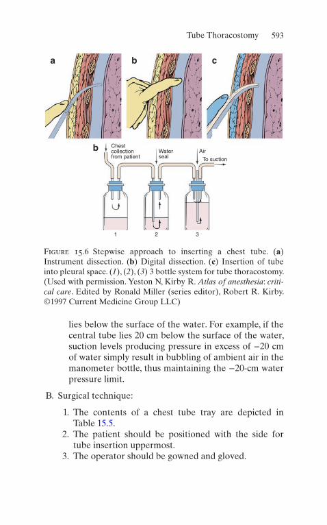
593
lies below the surface of the water. For example, if the central tube lies 20 cm below the surface of the water, suction levels producing pressure in excess of −20 cm of water simply result in bubbling of ambient air in the manometer bottle, thus maintaining the −20-cm water pressure limit.
B. Surgical technique:
1. The contents of a chest tube tray are depicted in Table 15.5.
2. The patient should be positioned with the side for tube insertion uppermost.
3. The operator should be gowned and gloved.
Chestcollectionfrom patient
1 2 3
Waterseal
Air
To suction
a
b
b c
Figure 15.6 Stepwise approach to inserting a chest tube. (a) Instrument dissection. (b) Digital dissection. (c) Insertion of tube into pleural space. (1), (2), (3) 3 bottle system for tube thoracostomy. (Used with permission. Yeston N, Kirby R. Atlas of anesthesia: criti-cal care. Edited by Ronald Miller (series editor), Robert R. Kirby. ©1997 Current Medicine Group LLC)
Tube Thoracostomy

594
4. The chest tube is usually inserted in the anterior axil-lary line in the fifth or sixth intercostal space.
5. The incision site should be prepped and draped, and lidocaine should be infiltrated one intercostal space below the rib of the selected intercostal space of inser-tion. The periosteum, subcutaneous tissue, and pleural space should be infiltrated. Aspiration of fluid or air will confirm infiltrated pleural space.
6. A small skin incision, appropriate to the size of the chest tube, is made with a scalpel over the anesthe-tized rib.
7. The incision into the subcutaneous tissue and muscle at the intercostal space, preferably using the blunt side of the scalpel or trocar.
8. A large clamp with an open end using spreading maneuvers is used until the pleural space is reached (see Fig. 15.6a).
9. The index finger of the operator is used to explore the pleural space to ensure that the lung, dia-phragm, or another structure is not adherent (see Fig. 15.6b).
Table 15.5 Contents of the chest tube tray
Sterile towels and drapes
1% or 2% lidocaine
10-mL syringe
22- and 25-gauge needles
1-0 silk suture with cutting needle (2 packages)
2 large Kelly clamps
2 medium Kelly clamps
Suture scissors
4-in.-square gauze pads
Chest drainage, suction system, and appropriate chest tube
Chapter 15. Special Techniques

595
10. The tube is inserted, generally toward the apex of the pleural space for treatment of pneumothoraces, by grasping the tube with a medium clamp, maneuvering it through the dissected tunnel (see Fig. 15.6c), and ensuring that the last hole of the thoracostomy tube lies within the pleural space.
The tube is fixed at the insertion site with 1-0 silk suture.
(a) A number of techniques are used, but commonly the suture ends are not cut but wrapped around the tube and secured with tape. These may be used to close the wound when the tube is pulled out.
11. The external end of the chest tube is then attached to the system, and the connections are taped over.
12. A sterile dressing with tape is applied to the skin.
C. Complications include tube malposition, empyema or wound infection, blockage of tube by blood or fibrin clot, and lung injuries.
Intra-aortic Balloon Pump (IABP)
A. Indications for the use of IABP:
1. Pump failure
(a) After acute myocardial ischemic event (b) Cardiogenic shock (c) Postcardiac transplant patient (d) In the pre- or postoperative period of cardiac
surgery
2. Acute mitral valvular regurgitation 3. Unstable angina pectoris 4. Others
B. Insertion should normally be accomplished by those with experience. The technique is also dependent upon the par-ticular catheter and approach to be used.
Intra-aortic Balloon Pump (IABP)
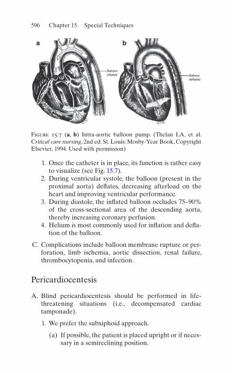
596
1. Once the catheter is in place, its function is rather easy to visualize (see Fig. 15.7).
2. During ventricular systole, the balloon (present in the proximal aorta) deflates, decreasing afterload on the heart and improving ventricular performance.
3. During diastole, the inflated balloon occludes 75–90% of the cross-sectional area of the descending aorta, thereby increasing coronary perfusion.
4. Helium is most commonly used for inflation and defla-tion of the balloon.
C. Complications include balloon membrane rupture or per-foration, limb ischemia, aortic dissection, renal failure, thrombocytopenia, and infection.
Pericardiocentesis
A. Blind pericardiocentesis should be performed in life- threatening situations (i.e., decompensated cardiac tamponade).
1. We prefer the subxiphoid approach.
(a) If possible, the patient is placed upright or if neces-sary in a semireclining position.
Ballooninflated Balloon
deflated
a b
Figure 15.7 (a, b) Intra-aortic balloon pump. (Thelan LA, et al. Critical care nursing, 2nd ed. St. Louis: Mosby-Year Book, Copyright Elsevier, 1994. Used with permission)
Chapter 15. Special Techniques

597
(b) Venous access, continuous ECG monitoring, and blood pressure monitoring should have been estab-lished. Personnel and equipment necessary for car-diac resuscitation must be on hand.
(c) Sedation and analgesia as appropriate to the set-ting should be provided.
(d) The region is prepped and draped. (e) One percent lidocaine local and an 18- or a
20-gauge cardiac or spinal needle attached to a syringe with local anesthetic is prepared.
(f) For ECG monitoring, an alligator clip connected to the V lead of an ECG monitor may be placed on the needle (see Fig. 15.8).
(g) The needle tip is introduced between the xiphoid and left costal margin and directed to the left shoulder.
(h) Continuous gentle suction is applied to the syringe. Intermittently, local anesthetic may be injected, which helps to clear the needle and anesthetize the deeper tissues.
(i) The sensation of pericardial passage will usually be felt by the operator.
(j) Epicardial contact will be recognized by the ECG tracing, showing injury current. If this should occur, the needle should be withdrawn slightly.
2. After entrance into the pericardial cavity, removal of 50 mL of pericardial fluid is usually enough.
3. A pliable soft catheter may be inserted into the pericar-dial space using a guidewire, allowing the needle to be withdrawn. Real-time transthoracic echocardiography allows tracking of the needle tip to ensure correct loca-tion throughout the aspiration procedure.
4. In an alternative approach, the needle is angled toward the right shoulder rather than the left shoulder.
B. Complications include cardiac chamber puncture, dys-rhythmias, pneumothorax, vasovagal reactions, and car-diac arrest.
Pericardiocentesis

598
Therapeutic Hypothermia (TH)
A. Pathophysiology:
1. Reduces cerebral metabolic demand by decreasing the rate of oxygen consumption and reducing ATP demand.
Figure 15.8 Pericardiocentesis. (Sabiston DC, Spencer FC. Gibbon’s surgery of the chest, 4th ed. Philadelphia: WB, Saunders; 1983. Used with permission)
Chapter 15. Special Techniques
599
2. Stabilizes the blood brain barrier and reduces cerebral edema by decreasing permeability to inflammatory cytokines, free radicals, and thrombin.
B. Indications for TH (see also Chap. 2):
1. Cardiac arrest victims with return of spontaneous cir-culation and:
(a) Regardless of presenting rhythm (b) Must have a GCS <8
2. Anoxic–hypoxic encephalopathy of the newborn 3. Cerebrovascular accidents with increased intracranial
pressure 4. Refractory status epilepticus 5. Others (i.e., traumatic brain injury, near drowning, liver
failure, ARDS)
C. This procedure should normally be accomplished by those with experience. The technique is also dependent upon the particular setting and materials available. Core tem-perature must be measured (i.e., bladder, vaginal, esophageal)
1. Goal temperature is 32–36 °C for a minimum of 24 h (in the authors’ experience, 24–72 h is ideal).
2. Cold IV fluids help achieve this temperature faster and should be used in patients without contraindications (i.e., end-stage renal disease).
3. Magnesium sulfate (2 g IV) provides vasodilation and increases rate of temperature drop.
4. Sedative agents and neuromuscular blockers are uti-lized to decrease oxygen consumption and to minimize shivering.
5. A variety of devices exist to provide this technique. The simplest way to perform it is by utilizing ice packs applied to the surface (avoid direct contact with skin). Cooling blankets can be used (the “sandwich tech-nique”). Hydrogel adhesive pads and special thermal wraps can also be utilized. Endovascular cooling can be
Therapeutic Hypothermia (TH)

600
accomplished by catheters that accelerate temperature drop. In neonate, selective brain hypothermia with cooling helmets is commonly utilized. Selective brain hypothermia devices for adults are also available in selected centers.
6. Rewarming is accomplished after 24 h by either stop-ping the cooling device or actively rewarming the patient over a period of 12–24 h. Patients should not be rewarmed faster than 0.1–0.25 °C/h; otherwise, the ben-eficial effects of TH are abated.
D. Complications include dysrhythmias, skin burns, increased infection rate, and rarely coagulopathy.
Bronchoscopy
A. Indications for bronchoscopy in the ICU:
1. Clearance of secretions and mucus plugs 2. To obtain selective samples of tracheobronchial tree 3. Hemoptysis 4. Therapeutic (i.e., tumor destruction, atelectasis
resolution)
B. This procedure should normally be accomplished by those with experience. If the patient has an endotracheal tube in place, the technique is simpler.
C. Complications include dysrhythmias (many premedicate with atropine prior to the procedure), airway injury, and pneumothorax.
Chapter 15. Special Techniques

601© Springer Nature Switzerland AG 2021J. Varon, Handbook of Critical and Intensive Care Medicine, https://doi.org/10.1007/978-3-030-68270-5_16
General Management
A. History. Obtain as accurate a history as possible, including the substance ingested or inhaled and the amount and time of ingestion. Always consider possible ingestion or inhalation of multiple substances.
B. Cardiorespiratory Care. The most important aspects of initial management are basic care of respiratory and car-diovascular function: Maintain a patent airway, ensure adequate respiration (support ventilation when neces-sary), and treat shock if present.
1. Airway
(a) Loss of airway patency and reflexes may lead to obstruction, aspiration, or respiratory arrest.
(b) Maintain proper airway position; suction and use oropharyngeal or nasopharyngeal adjuncts as needed.
(c) Absent or depressed gag reflex in an unconscious or obtunded patient indicates an inability to pro-tect the airway; endotracheal intubation is indicated.
Chapter 16Toxicology

602
2. Respirations
(a) Respiratory failure is the most frequent cause of death in poisoned patients, and usually it is a result of (CNS) central nervous system depression.
(b) Assist ventilation, and perform endotracheal intu-bation as required.
(c) Obtain and follow arterial blood gases.
3. Circulation
(a) Monitor blood pressure, pulse, and cardiac rhythm. (b) Initiate intravenous line. (c) If hypotension is present, administer fluid chal-
lenge with normal saline 10–20 mL/Kg. (d) If hypotension persists, administer vasopressin
1–6 U/h. (e) Other agents such as norepinephrine can also be
used.
C. Gastrointestinal Decontamination. The traditional sequence for gastrointestinal decontamination is empty-ing of stomach by induced emesis or gastric lavage fol-lowed by administration of activated charcoal. However, there is no data to support its use anymore.
1. Induced Emesis with Ipecac
(a) Should not be administered routinely in the man-agement of poisoned patients since there is no evi-dence from clinical studies that ipecac improves outcome. If a clinician, however, decides to use, then administer syrup of ipecac (30 mL for adults, 15 mL for children <5 years old, 10 mL for children <1 year old). Repeat if no emesis in 20 min.
1. Not recommended in children <9 months old. 2. If the second dose does not produce vomiting,
perform gastric lavage.
(b) Contraindications
1. Comatose or seizing patient or anyone with altered mental status
Chapter 16. Toxicology
603
2. Ingestion of corrosive agents or petroleum distillates
3. Ingestion of substance likely to produce coma or seizures rapidly
2. Gastric LavageIntroducing large bore orogastric tube and subse-
quent injection and aspiration of fluid to aspirate pills of toxins. Not usually used due to unclear benefit. No clear indications of using this method. Contraindications: unprotected airway, caustic inges-tion, hydrocarbon ingestion, and patients with risk of GI hemorrhage.
3. Activated Charcoal
(a) It is, by far, the preferred means of gastric decon-tamination. Limits absorption of virtually all ingested substances. Usually thought to be within 1 hour of poison ingestion, however, potential ben-efit if administered after this period.
(b) Administer 1 g/kg orally or via gastric tube after emesis or lavage is completed.
(c) Repeat doses of 15–20 g at 1- or 2-h intervals, or continuous activated charcoal instillation may be useful in some instances (i.e., theophylline overdose).
(d) Contraindicated in corrosive ingestion, ileus, or intestinal obstruction.
4. Cathartics
(a) Frequently used, but their effectiveness is not well established.
(b) Administer along with activated charcoal. (c) Magnesium citrate 10% 3–4 mL/kg or sorbitol
70% 1–2 mL/kg. If no charcoal is present in stool after 6 h, repeat half dose.
5. Whole Bowel Irrigation
(a) Effectiveness is not fully conclusive. May be useful in substantial ingestion of substances poorly
General Management

604
adsorbed by activated charcoal or enteric-coated tablets.
(b) Administer polyethylene glycol electrolyte solu-tion (CoLyte or GoLYTELY) 2 L/h by gastric tube until stool is free of particulate material.
(c) Do not use in unconscious or obtunded patients.
D. Forced Diuresis and Control of Urine pH. May increase urinary excretion of some agents for which renal excre-tion is the major route of elimination. Do not use in patients with renal failure or congestive heart failure.
1. Alkalinization
(a) May be useful in overdose with salicylates and phenobarbital.
(b) Administer sodium bicarbonate 1–2 mEq/kg IV to achieve a urinary pH ≥ 7.0.
(c) Monitor serum pH.
2. Acidification
(a) May be useful in overdose with phencyclidine, amphetamines, quinine, quinidine, strychnine, and cyclic antidepressants.
(b) Administer ammonium chloride 75 mg/kg IV in four to six divided doses to achieve a urinary pH ≤ 6.0.
(c) Do not use in the presence of rhabdomyolysis, myoglobinuria, or hepatic failure.
(d) Monitor serum pH.
E. Hemodialysis. May be useful in severe intoxication with amphetamines, methanol, ethylene glycol, isopropyl alco-hol, lithium, and salicylates. If fluid or acid–base abnor-malities are present, these can also be corrected.
F. Charcoal Hemoperfusion
1. Utilizes an extracorporeal circuit through an activated charcoal column.
2. May be useful in severe intoxication with barbiturates, doxorubicin, digoxin, some beta-blockers, ethchlor-
Chapter 16. Toxicology

605
vynol, meprobamate, phenytoin, salicylates, and theophylline.
3. Potential complications: hypotension and thrombocytopenia.
G. Toxicology Screen
1. Should be used to confirm diagnosis in all cases. 2. Specific levels may be necessary in overdose with cer-
tain substances to guide therapy (i.e., acetaminophen, iron, lithium, methanol, salicylates, theophylline).
H. Poison Control Centers. Notify poison control centers with management questions or complicated or unusual poisonings. In the United States, the telephone number for these centers is 1-800-222-1222 or on the Internet at http://www.aapcc.org
Acetaminophen
Acetaminophen is a widely used analgesic and antipyretic. It is found in combination with other analgesics and in various cold remedies (i.e., Comtrex, Congesprin, Excedrin PM, 4-Way Cold Tablets). Toxicity of significant overdose lies in production of hepatic necrosis. This is probably related to overwhelming of hepatic glutathione capacity to detoxify acetaminophen metabolic products. Toxicity is likely to occur with single ingestions greater than 250 mg/kg or those greater than 12 g over a 24-h period. The diagnosis may be over-looked, especially in patients with alcoholic liver disease.
A. Clinical Effects
1. In the first 24 h after overdose (stage I), patients often manifest nausea, vomiting, diaphoresis, pallor, lethargy, and malaise.
2. From 24 to 72 h after the ingestion (stage II), there is clinical and laboratory evidence of hepatotoxicity. Of those patients that develop hepatic injury, over half will demonstrate ALT/AST elevation within 24 h, and all
Acetaminophen
606
have elevations by 36 h. As stage II progresses, patients develop liver tenderness and right upper quadrant pain. Elevations of prothrombin time (PT) and total bilirubin, oliguria, and renal function abnormalities may become evident.
3. Liver function abnormalities peak from 72 to 96 (stage III) hours after ingestion. Jaundice, confusion (hepatic encephalopathy), a marked elevation in hepatic enzymes, hyperammonemia, and a bleeding diathesis are present. Plasma ALT and AST levels often exceed 10,000 IU/L. Prolongation of the PT/INR is evident, along with hypoglycemia, lactic acidosis, and a total bilirubin concentration above 4.0 mg/dL.
4. Patients who survive stage III enter a recovery phase (stage IV) that usually begins by day 4 and is completed by 7 days after overdose.
B. Diagnostic Studies
1. Obtain serum acetaminophen as soon as 4 h and as late as 24 h following ingestion and plot the level on a nomogram (Fig. 16.1). The nomogram is useful only in acute ingestion.
2. There is good correlation between timed serum and subsequent hepatotoxicity. If the level is in the hepato-toxic range, administer n-acetylcysteine.
3. Obtain prothrombin time and transaminase levels, serum urea nitrogen (BUN), and creatinine.
C. Management
1. Induce emesis or perform gastric lavage. 2. Antidote: n-acetylcysteine (NAC).
(a) Prevents liver injury when administered early fol-lowing intoxication.
(b) In the United States, two NAC treatment regimens are currently approved by the Food and Drug Administration (FDA): The first is a 72-h oral course given as a 140-mg/kg loading dose followed by 17 doses of 70 mg/kg every 4 h (total dose,
Chapter 16. Toxicology
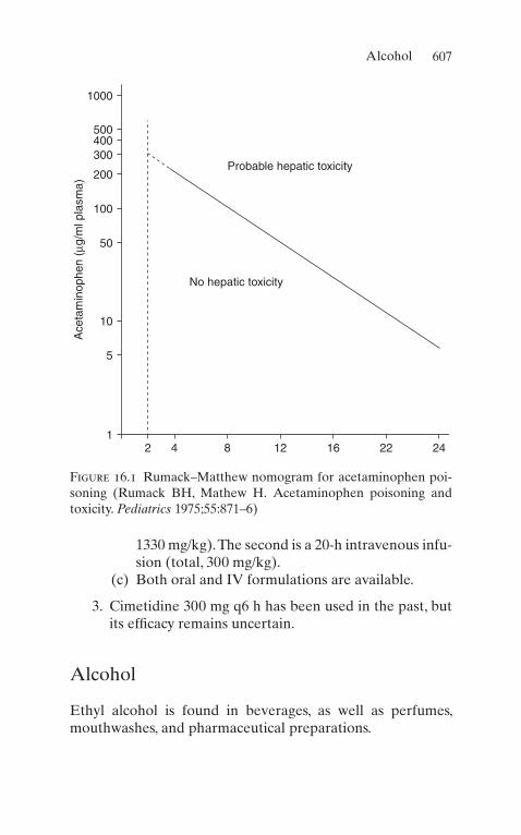
607
1330 mg/kg). The second is a 20-h intravenous infu-sion (total, 300 mg/kg).
(c) Both oral and IV formulations are available.
3. Cimetidine 300 mg q6 h has been used in the past, but its efficacy remains uncertain.
Alcohol
Ethyl alcohol is found in beverages, as well as perfumes, mouthwashes, and pharmaceutical preparations.
12 4 8 12
No hepatic toxicity
Ace
tam
inop
hen
(µg/
ml p
lasm
a)
Probable hepatic toxicity
16 22 24
5
10
50
100
200
300400500
1000
Figure 16.1 Rumack–Matthew nomogram for acetaminophen poi-soning (Rumack BH, Mathew H. Acetaminophen poisoning and toxicity. Pediatrics 1975;55:871–6)
Alcohol

608
A. Overdose
1. Clinical Effects
(a) Variable, depending on the individual: slurred speech, impaired judgment, behavioral changes, combativeness, and ataxia.
(b) At very high levels, somnolence and respiratory depression occur. When death occurs as a result of intoxication, it is usually due to respiratory depression.
(c) Also occurring: cardiac dysrhythmias, hyperten-sion, hypoglycemia, hypomagnesemia, hypophos-phatemia, seizures, hypothermia, rhabdomyolysis, and Wernicke’s encephalopathy.
(d) Alcoholic ketoacidosis: metabolic acidosis with increased anion and osmolar gaps and ketosis. Associated nausea, vomiting, and abdominal pain.
2. Metabolism of AlcoholRate is 12–50 mg/dL/h (average of 20 mg/dL/h).
3. Admission CriteriaAdmission to the intensive care unit may be required
for trauma, seizures, hypothermia, and severe meta-bolic abnormalities.
4. Management
(a) Management is mainly supportive, with observa-tion predominant. Intubate and ventilate if necessary.
(b) Administer IV glucose 50 g and thiamine 100 mg to all obtunded patients after serum glucose level is drawn.
(c) Correct volume depletion as needed. (d) If the patient is violent or very agitated, use physi-
cal restraints as needed. Sedation may be necessary.
(e) Treatment of alcoholic ketoacidosis: Fluid replace-ment with D5%/normal saline. Observe for appear-ance of hypoglycemia, hypophosphatemia, and hypokalemia.
Chapter 16. Toxicology
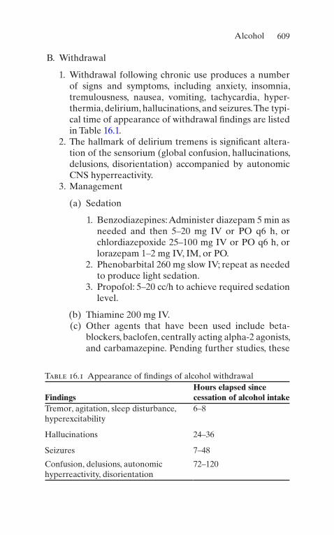
609
B. Withdrawal
1. Withdrawal following chronic use produces a number of signs and symptoms, including anxiety, insomnia, tremulousness, nausea, vomiting, tachycardia, hyper-thermia, delirium, hallucinations, and seizures. The typi-cal time of appearance of withdrawal findings are listed in Table 16.1.
2. The hallmark of delirium tremens is significant altera-tion of the sensorium (global confusion, hallucinations, delusions, disorientation) accompanied by autonomic CNS hyperreactivity.
3. Management
(a) Sedation
1. Benzodiazepines: Administer diazepam 5 min as needed and then 5–20 mg IV or PO q6 h, or chlordiazepoxide 25–100 mg IV or PO q6 h, or lorazepam 1–2 mg IV, IM, or PO.
2. Phenobarbital 260 mg slow IV; repeat as needed to produce light sedation.
3. Propofol: 5–20 cc/h to achieve required sedation level.
(b) Thiamine 200 mg IV. (c) Other agents that have been used include beta-
blockers, baclofen, centrally acting alpha-2 agonists, and carbamazepine. Pending further studies, these
Table 16.1 Appearance of findings of alcohol withdrawal
FindingsHours elapsed since cessation of alcohol intake
Tremor, agitation, sleep disturbance, hyperexcitability
6–8
Hallucinations 24–36
Seizures 7–48
Confusion, delusions, autonomic hyperreactivity, disorientation
72–120
Alcohol

610
medications should not be used routinely for the treatment of alcohol withdrawal.
(d) The use of ethanol to control withdrawal is discour-aged in the setting of acute alcohol withdrawal.
C. Other Alcohols
1. Methanol
(a) Methanol (methyl alcohol) is widely used as a sol-vent, is found in windshield washer solution and antifreeze, and is used as a solid canned fuel.
(b) It produces CNS intoxication similar to that of eth-anol. Other toxic effects include acidosis and reti-nal cell toxicity.
(c) Ethanol acts as a competitive substrate for alcohol dehydrogenase, the enzyme that produces toxic metabolites from methanol.
(d) Treatment
1. Alkalization. Patients with academic fare much worse. In patient with pH below 7.3, sodium bicarbonate infusion should be started at rate of 1 to 2 mEq/Kg.
2. Inhibition of alcohol dehydrogenase (ADH) blocks bio-activation of the parent alcohol to its toxic acid metabolites. Fomepizole and ethanol can accomplish this. Fomepizole is loaded at 15 mg/kg intravenously, followed by 10 mg/kg every 12 h, with adjustments for hemodialysis or after more than 2 days of therapy. Alternatively, IV ethanol (0.6 mg/kg initially and infusion of 66 mg/kg/h to maintain a blood alcohol level of 100 mg/dL). Administer as 10% ethanol diluted in D5%W.
3. Hemodialysis is the best method to rapidly remove methanol and its metabolites. This modality and treatment with ethyl alcohol are indicated with serum methanol levels >50 mg/dL.
2. Isopropyl Alcohol
Chapter 16. Toxicology

611
(a) Isopropyl alcohol is used as a solvent and medici-nally as a rubbing alcohol and a sterilizing agent.
(b) It is more toxic than ethyl alcohol but less than methyl alcohol.
(c) Toxicity may be produced by ingestion of isopropyl alcohol or by inhalation of vapor.
(d) Signs and symptoms of intoxication are similar to methanol, but there is no retinal toxicity. Additional complications: dehydration and hemorrhagic gastritis.
(e) Treatment: There is no role for gastrointestinal decontamination. Activated charcoal is impractical to use, as large quantities are required. Proper main-tenance of fluid balance and blood pressure are essential. Hemodialysis may be used in severe cases.
Angiotensin-Converting Enzyme (ACE) Inhibitors
ACE inhibitors are antihypertensive agents that are in com-mon use, either as individual agents or in combination with a diuretic (see Table 16.2).
A. Toxic Effects. The most common finding is hypotension. Fluid disturbances and electrolyte abnormalities may occur with agents combined with diuretics.
B. Clinical Effects. Dizziness, light-headedness, syncope, and cough.
C. Management:
1. Administer activated charcoal and cathartic. 2. Monitor blood pressure continuously. 3. Treatment is largely supportive. If hypotension is pres-
ent, administer normal saline fluid infusion. Administer dopamine infusion if this does not correct hypotension. Naloxone has been reported to reverse hypotension.
4. In severe overdose, consider hemodialysis in overdose with captopril, enalapril, or lisinopril.
Angiotensin-Converting Enzyme (ACE) Inhibitors
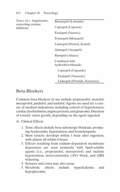
612
Beta-Blockers
Common beta-blockers in use include propranolol, atenolol, metoprolol, pindolol, and nadolol. Agents are used for a vari-ety of medical indications, including control of hypertension, cardiac dysrhythmias, angina pectoris, and glaucoma. Duration of toxicity varies greatly, depending on the agent ingested.
A. Clinical Effects
1. Toxic effects include beta-adrenergic blockade, produc-ing bradycardia, hypotension, and bronchospasm.
2. Most toxicity develops within 1 hour after ingestion, with almost all within 6 hours.
3. Effects resulting from sodium-dependent membrane depression are seen primarily with lipid-soluble agents (i.e., propranolol, metoprolol) and include hypotension, atrioventricular (AV) block, and QRS widening.
4. Seizures and coma may also occur. 5. Metabolic effects include hyperkalemia and
hypoglycemia.
Table 16.2 Angiotensin- converting enzyme inhibitors
Benazepril (Lotensin)
Captopril (Capoten)
Enalapril (Vasotec)
Fosinopril (Monopril)
Lisinopril (Prinivil, Zestril)
Quinapril (Accupril)
Ramipril (Altace)
Combined with hydrochlorothiazide:
Captopril (Capozide)
Enalapril (Vaseretic)
Lisinopril (Prinzide, Zestoretic)
Chapter 16. Toxicology
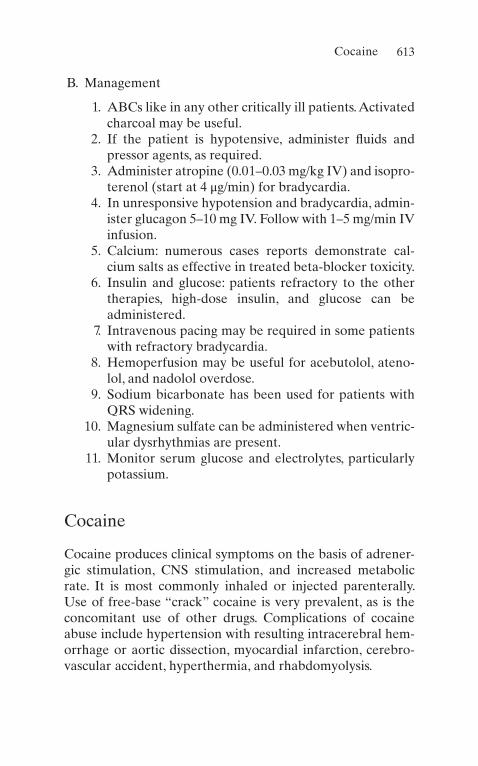
613
B. Management
1. ABCs like in any other critically ill patients. Activated charcoal may be useful.
2. If the patient is hypotensive, administer fluids and pressor agents, as required.
3. Administer atropine (0.01–0.03 mg/kg IV) and isopro-terenol (start at 4 μg/min) for bradycardia.
4. In unresponsive hypotension and bradycardia, admin-ister glucagon 5–10 mg IV. Follow with 1–5 mg/min IV infusion.
5. Calcium: numerous cases reports demonstrate cal-cium salts as effective in treated beta-blocker toxicity.
6. Insulin and glucose: patients refractory to the other therapies, high-dose insulin, and glucose can be administered.
7. Intravenous pacing may be required in some patients with refractory bradycardia.
8. Hemoperfusion may be useful for acebutolol, ateno-lol, and nadolol overdose.
9. Sodium bicarbonate has been used for patients with QRS widening.
10. Magnesium sulfate can be administered when ventric-ular dysrhythmias are present.
11. Monitor serum glucose and electrolytes, particularly potassium.
Cocaine
Cocaine produces clinical symptoms on the basis of adrener-gic stimulation, CNS stimulation, and increased metabolic rate. It is most commonly inhaled or injected parenterally. Use of free-base “crack” cocaine is very prevalent, as is the concomitant use of other drugs. Complications of cocaine abuse include hypertension with resulting intracerebral hem-orrhage or aortic dissection, myocardial infarction, cerebro-vascular accident, hyperthermia, and rhabdomyolysis.
Cocaine

614
A. Clinical Effects
1. The onset of toxic symptoms is within 30–60 min of injection or inhalation.
2. Patients present most often with hypertension, dys-rhythmias, pupillary dilatation, agitation, anxiety, or psychosis.
3. Seizures, hyperthermia, and rhabdomyolysis may occur. 4. Hypertension may produce intracranial hemorrhage or
thoracic aortic rupture. 5. Chest pain may occur and is sometimes of myocardial
origin. Myocardial infarction may occur. Cocaine is dysrhythmogenic and may produce sinus tachycardia, premature ventricular contractions, ventricular tachy-cardia, and ventricular fibrillation.
B. Management
1. ABCs. We recommend avoiding succinylcholine as a rapid sequence intubation agent due to the risk of hyperkalemia and rhabdomyolysis that these patients have.
2. Physical restrains can cause rhabdomyolysis and hyper-thermia, and therefore they should be avoided.
3. Hypertension
(a) Initial treatment with benzodiazepine with appro-priate dose is recommended to reduce stimulation of central nervous system.
(b) In patients with severe hypertension or refractory to benzodiazepines, treat with phentolamine 0.02–0.2 mg/kg.
(c) Beta-adrenergic antagonists (i.e., labetalol) are contraindicated in the treatment of cocaine-related cardiovascular complications as they create unop-posed alpha-adrenergic stimulation and are associ-ated with coronary vasoconstriction and end-organ ischemia.
(d) Although, in general, beta-blockers are contraindi-cated, old reports suggest that esmolol, a selective
Chapter 16. Toxicology

615
beta1-adrenergic blocker with rapid onset and short duration of action coadministered with sodium nitroprusside, should be reserved for severe hypertension that is unresponsive to other treat-ments and/or complicated by aortic dissection. In the author’s opinion, this combination is quite dan-gerous and can cause significant damage.
(e) Short-acting nifedipine should never be used, and verapamil or diltiazem should be avoided in patients with evidence of heart failure or left ven-tricular dysfunction. Calcium channel blockers should not be used as a first-line treatment but may be considered for patients who do not respond to benzodiazepines.
4. Urine toxicology screen will confirm the diagnosis. 5. Cardiac ischemia is a possibility that should be consid-
ered until proven otherwise in patients with cocaine- related chest pain.
Cyanide
Cyanide is a chemical with a variety of industrial uses. Sodium nitroprusside contains cyanide, which is released into solution in increased quantity by exposure to light. The gas hydrogen cyanide is a product of combustion of plastics and a variety of other materials. A third of all smoke inhalation victims have increased cyanide levels.
A. MechanismToxicity is via chemical asphyxia and cyanide blockade
of cellular oxygen utilization. Exposure to small amounts of hydrogen cyanide gas or ingestion of a small quantity of cyanide salt can be rapidly fatal.
B. Clinical Effects
1. Respiratory failure and cardiovascular collapse. Rapidly developing coma and severe lactic acidosis.
2. Syncope, seizures, headache, nausea, and confusion.
Cyanide
616
3. Cyanide poisoning may occur with prolonged IV administration of sodium nitroprusside. Consider this if such patients develop CNS depression, seizures, lactic acidosis, or cardiovascular instability.
C. Management
1. Administer oxygen, maintain airway, and assist respirations.
2. Mouth to mouth resuscitation is contraindicated. 3. Rapidly remove from source and remove clothing 4. Treat hypotension and seizures in standard fashion. 5. If hydroxocobalamin, a form of vitamin B12, is avail-
able, it should be used as an antidote.
(a) Administer sodium thiosulfate, which converts cyanide to thiocyanate, 1.65 mL/kg IV of a 25% solution IV.
(b) Hydroxocobalamin 70 mg/kg IV.
6. If hydroxycobalamin is not available:
(a) Have patient inhale amyl nitrite capsule. (b) Administer sodium nitrite 10 mg/kg. (c) These two produce methemoglobinemia, which
may itself be toxic. (d) Administer sodium thiosulfate, 1.65 mL/kg IV of a
25% solution IV.
7. If cyanide salt is ingested, lavage stomach and adminis-ter activated charcoal.
8. Other cyanide antidotes, dicobalt-EDTA and 4- dimethylaminophenol, are occasionally used when the therapy above are not available.
Cyclic Antidepressants
There is an estimated annual incidence of 500,000 cases of overdose with cyclic antidepressants. Many agents are in common use, including amitriptyline, desipramine, nortripty-line, doxepin, and imipramine (see Table 16.3). They may be
Chapter 16. Toxicology

617
found in medication combinations with other psychotropic agents. Toxicity occurs via anticholinergic effects, alpha- adrenergic blockade, inhibition of norepinephrine reuptake, and inhibition of the fast sodium channel. Some drugs, such as haloperidol, morphine, and disulfiram, may prolong cyclic antidepressant toxicity by interfering with hydroxylation.
A. Clinical Effects
1. Tachycardia, myoclonus, delirium, coma, hyperthermia, pupillary dilatation, hypertension or hypotension, pro-longation of QRS and QT, AV block, and myocardial depression .
2. The most important toxic effects are cardiovascular. A QRS duration >0.12 indicates serious toxicity. Sinus tachycardia is typical; supraventricular tachycardia, ventricular tachycardia, torsades des pointes, and ven-tricular fibrillation may occur. Bradycardia is a poor prognostic sign.
3. Rapid deterioration with the development of cardio-vascular collapse, coma, or seizures may occur. Persistent seizures may produce hypothermia or rhabdomyolysis.
Table 16.3 Cyclic antidepressants
Tricyclic
Amitriptyline (Elavil, Endep, Etrafon, Limbitrol, Triavil)
Amoxapine (Asendin)
Desipramine (Norpramin)
Doxepin (Sinequan)
Imipramine (Tofranil)
Nortriptyline (Pamelor)
Protriptyline (Vivactil)
Trimipramine (Surmontil)
Tetracyclic
Maprotiline (Ludiomil)
Cyclic Antidepressants

618
B. Management
1. Administer activated charcoal 100 g. Do not administer syrup of ipecac. Repeat doses of activated charcoal may be useful, and some have advocated continuous naso-gastric instillation.
2. Continuous electrocardiographic monitoring is essential.
3. Treat hyperthermia (see Chap. 5) and seizures in usual fashion (diazepam 0.1 mg/kg IV per dose as needed or phenytoin 15 mg/kg IV over 30 min).
4. Do not give physostigmine as an antidote, because it may produce seizures.
5. Administer sodium bicarbonate 2–3-mEq/Kg IV bolus in widened QRS (>100 ms), hypotension, or metabolic acidosis. Monitor serum pH and electrolytes. Do not administer in metabolic alkalosis or pulmonary edema.
6. For torsades des pointes, administer magnesium 2–4 g IV or isoproterenol. Do not use procainamide or quinidine.
7. In cardiovascular collapse, administer vasopressin, phenylephrine, or norepinephrine.
8. Treat seizure with benzodiazepines. Avoid the use of phenytoin.
Digoxin
Digoxin is the most frequently used of the cardiac glycosides, being prescribed most commonly for congestive heart failure or dysrhythmias. The mechanism of digitalis toxicity involves interruption of potassium and calcium efflux from myocardial cells by inhibition of sodium and potassium–adenosine tri-phosphatase (potassium–ATPase). Intoxication may be the result of acute accidental or suicidal ingestion or chronic over-dose. The clinical findings are different in these two settings.
A. Clinical Effects
1. Acute Intoxication
Chapter 16. Toxicology

619
(a) Nausea, vomiting, and hyperkalemia (b) Dysrhythmias
1. Bradycardia with sinus and atrioventricular block.
2. Ventricular dysrhythmias are uncommon.
2. Chronic Intoxication
(a) Weakness, visual disturbances, and hypomagnese-mia. Potassium is normal or low.
(b) Dysrhythmias
1. Ventricular tachycardia and fibrillation are common.
2. Accelerated junctional rhythm and paroxysmal atrial tachycardia with block are common, espe-cially in patients with chronic atrial fibrillation.
B. Diagnostic Studies
1. Digoxin level: More useful in chronic intoxication. May be falsely high in acute overdose.
2. Follow serum potassium, magnesium, BUN, and creatinine.
C. Management
1. Administer activated charcoal and cathartic. 2. Digitoxin elimination is enhanced by repeat-dose acti-
vated charcoal. 3. Monitor cardiac rhythm continuously. 4. Hyperkalemia.
(a) If >5.5 mEq/L, administer sodium bicarbonate 1 mEq/kg IV, glucose 0.5 g/kg, and regular insulin 0.1 U/kg IV or polystyrene sulfonate (Kayexalate) 0.5 g/kg PO.
(b) Do not administer calcium.
5. Bradycardia
(a) Atropine 0.5–2.0 mg IV as needed. (b) Cardiac pacing may be required.
Digoxin
620
D. Antidote: Digoxin-Specific Antibody Fragments (Digibind)
1. Digoxin-specific antibodies have a high affinity for digoxin, a lower affinity for digitoxin and other cardiac glycosides.
2. Administer for:
(a) Life-threatening dysrhythmia (b) Evidence of end-organ dysfunction (c) Hyperkalemia
3. Dosage: Each 40-mg vial of digoxin-specific antibodies absorbs 0.6 mg of digoxin. Dosage of antibodies to be administered is to be calculated as follows:
(a) Calculate body load of digoxin: Dose ingested/0.8 or serum drug concentration in ng/mL by mean volume of distribution of digoxin (5.6 L/kg × patient weight) or digitoxin (0.56 L/kg × patient weight) and divided by 1000 to obtain the load in milligrams.
(b) Number of vials required: Body load (mg)/0.6 mg.
4. Administer IV over 30 min. 5. There are no known contraindications. In patients with
preexisting heart disease, withdrawal of inotropic effect is by removal of digitalis from the circulation. Monitor for development of heart failure.
6. Reversal of signs of digitalis intoxication occurs within 30–60 min of administration. Complete removal of digoxin occurs by 3 h.
Narcotics
Narcotics include naturally occurring or synthetic derivatives of opiates. These are used both medically and as drugs of abuse. Commonly used narcotics are found in Table 16.4.
Extremely potent synthetic “designer” opioids are deriva-tives of meperidine and fentanyl (i.e., “China white”) and are also included in this group. Narcotics may be ingested, injected parenterally, or inhaled.
Chapter 16. Toxicology

621
A. Clinical Effects
1. Sedation; miosis; respiratory depression; decreased heart rate, respiratory rate, and blood pressure; dimin-ished bowel sounds; and signs of transcutaneous injec-tion (i.e., “track marks”) may be present. Urine toxicology may confirm diagnosis, but a negative result does not exclude it.
2. In significant overdose: Coma, pinpoint pupils, severe respiratory depression, and apnea.
3. Complications of overdose include rhabdomyolysis and noncardiogenic pulmonary edema.
4. Death is typically due to respiratory failure.
B. Management
1. Maintain and assist ventilation as necessary. 2. Antidote: naloxone (Narcan).
(a) An opioid antagonist that competitively blocks CNS opiate receptors.
Table 16.4 Commonly used narcotic and related agents
Butorphanol (Stadol)
Codeine
Fentanyl
Hydrocodone (Anexsia, Vicodin)
Hydromorphone (Dilaudid)
Meperidine (Demerol)
Methadone
Morphine
Nalbuphine (Nubain)
Oxycodone (Percocet, Percodan, Tylox)
Pentazocine (Talwin)
Propoxyphene (Darvon)
Narcotics

622
(b) Administer 0.4–2.0 mg IV (may also be given IM, subcutaneously, intratracheally). Repeat as needed. No response to total of 10 mg is evidence against narcotic overdose.
(c) Apneic patients should receive higher initial start-ing dose.
(d) Duration of action is 1–4 h. Repeated administra-tion may be required or administer as an infusion of 0.4–0.8 mg/h in 5% dextrose.
(e) Use in opiate-dependent patients may produce narcotic withdrawal syndrome.
(f) Higher doses are frequently required with pentaz-ocine and designer opioids: Begin with an initial dose of 4 mg.
3. If respiratory distress continues, monitor with chest radiograph and arterial blood gases (ABGs).
4. If noncardiogenic pulmonary edema (which may also be produced by naloxone) is present, treat according to the guidelines for treating adult respiratory distress syndrome (ARDS) (see Chap. 13).
Phencyclidine
Phencyclidine (PCP), formerly used as a legal anesthetic agent, is now an illicit drug.
A. Clinical Effects
1. PCP produces hallucinations, alteration of mental sta-tus, and bizarre or violent behavior. Clinical status tends to wax and wane, and severe symptoms may per-sist for as long as 2 weeks.
2. The patient’s level of consciousness ranges from fully alert to comatose.
3. The most common physical findings are nystagmus and hypertension. Pupils may be dilated or miotic.
B. Medical Complications
Chapter 16. Toxicology
623
1. Complications may be due to direct effect of the drug or injury sustained during intoxication.
2. Major complications are indications for intensive care unit admission: seizures, hyperthermia, rhabdomyoly-sis, and acute renal failure.
C. Diagnostic Studies
1. Serum and urine PCP levels correlate poorly with clini-cal effects.
2. Check urine for myoglobin, which may indicate rhabdomyolysis.
D. Management
1. Management is largely supportive. 2. PCP is frequently smoked, so gastric decontamination
is not useful in these cases. However, gastric lavage and activated charcoal may be indicated if large amounts have been ingested. Do not induce emesis.
3. Hemodialysis and charcoal hemoperfusion are not effective in eliminating PCP.
4. Acid diuresis may speed elimination but is rarely indicated.
5. Severe agitation or violence: Utilize physical restraints. May administer haloperidol 10 mg IM or IV or benzodiazepines.
6. Seizures: If persistent, treat with IV benzodiazepines or propofol.
Phenytoin
Phenytoin toxicity may be due to acute overdose or chronic over-ingestion.
A. Clinical Effects
1. Nausea, vomiting, lethargy, ataxia, agitation, irritability, hallucinations, and seizures.
2. Horizontal nystagmus is characteristic of overdose.
Phenytoin
624
3. At very high levels: coma and respiratory arrest. 4. Cardiac toxicity occurs only with iatrogenic IV over-
dose, not with oral ingestion.
B. Diagnostic Studies
1. Phenytoin levels
(a) Therapeutic: 10–20 mg/L. (b) Levels >20 mg/L: nystagmus. (c) Levels >30 mg/L: ataxia. (d) Levels >40 mg/L: lethargy is common.
2. Serum Glucose: hyperglycemia may occur.
C. Management
1. Administer activated charcoal. Multiple doses of char-coal may enhance elimination.
2. Some recommend the use of charcoal hemoperfusion for severe intoxication.
3. Remainder of treatment is supportive. 4. Monitor cardiac rhythm in IV overdose.
Salicylates
Salicylates are used for analgesic, antipyretic, and anti- inflammatory properties and are found in a variety of both prescription and over-the-counter preparations (i.e., Alka- Seltzer, Ascriptin, Bufferin, Excedrin Extra Strength). Poisoning may be the result of acute ingestion or chronic overdose.
A. Clinical Effects
1. The clinical effects result from CNS respiratory stimu-lation, uncoupling of oxidative phosphorylation, and interference with platelet function and bleeding time.
2. Cerebral and pulmonary edema occur by uncertain mechanisms.
3. Acute overdose:
Chapter 16. Toxicology

625
(a) Tachypnea, tinnitus, vomiting, lethargy, respiratory alkalosis, and metabolic acidosis
(b) Severe: Hypoglycemia, hyperthermia, seizures, coma, and pulmonary edema
4. Chronic overdose
(a) Confusion, dehydration, and metabolic acidosis. This presentation may mimic sepsis.
(b) Pulmonary edema is more common than in acute overdose.
B. Diagnostic Studies
1. In acute ingestion, obtain the salicylate level and plot it on a Done nomogram (Fig. 16.2).
(a) Multiple determinations may be necessary with sustained-release preparations. Obtain every 2–3 h for 12 h following ingestion.
(b) Usual therapeutic levels in arthritis patients: 100–300 mg/L (10–30 mg/dL).
(c) A nomogram is not as useful in chronic intoxication.
2. Follow ABGs, serum glucose and electrolytes, and chest radiographs.
C. Management
1. Administer activated charcoal and cathartic. 2. Monitor for development of pulmonary edema. 3. Treat metabolic acidosis with IV sodium bicarbonate
1 mEq/kg to maintain pH at 7.40–7.50. 4. Rehydrate with IV crystalloid solution if dehydration
has resulted from vomiting or hyperventilation. 5. Urinary alkalinization enhances excretion.
(a) Administer D5W containing sodium bicarbonate 100 mEq/L at 200–300 mL/h. Use care in chronic intoxication: Observe for development of pulmo-nary edema.
Salicylates

626
(b) Check urinary pH frequently; maintain it at 6.0–7.0.
(c) Add 30–40-mEq KCl to each liter of IV solution (except in presence of renal failure).
6. Hemodialysis and hemoperfusion are effective in removing salicylates (hemodialysis also corrects fluid and acid–base disturbance). Indications follow:
(a) Acute ingestion with serum levels >1200 mg/L (120 mg/dL) or severe acidosis
(b) Chronic intoxication with serum levels >600 mg/L (60 mg/dL) (see Fig. 16.2)
Mild
0.8
6 12 24
Hours after ingestionDone nomogram
Sal
icyl
ate
plas
ma
conc
entr
atio
n (m
mol
/l)
36 48 60
0.91
2
3
4
5
6
789
10
12
15
0.80.91
2
3
4
5
6
78910
12
15
Moderate
Severe
Figure 16.2 A Done nomogram for acute salicylate poisoning (Done AK. Salicylate intoxication: significance of measurements of salicy-late in blood in cases of acute ingestion. Pediatrics 1960;26:800–7)
Chapter 16. Toxicology

627
Sedatives/Hypnotics
A large number of sedatives are in medical use. The most common are the barbiturates, nonbarbiturate sedative/hyp-notics (i.e., chloral hydrate, meprobamate, paraldehyde), and benzodiazepines (see Table 16.5). The toxic/therapeutic ratio is very high for most benzodiazepines. Oral overdose with 20 times the therapeutic dose of diazepam may occur without significant CNS depression.
A. Clinical Effects
1. The most prominent effect is CNS depression: Lethargy, ataxia, and slurred speech, progressing to coma and respiratory depression.
2. Severe hypothermia, hypotension, and bradycardia may accompany deep coma due to barbiturates.
3. Chloral hydrate may have cardiac effects, including dysrhythmias, hypotension, and myocardial depression.
B. Diagnostic Studies
1. Barbiturates
Serum levels >60–80 mg/L usually produce coma (>20–30 mg/L) in short-acting barbiturates.
2. Benzodiazepines and Others
Serum drug levels are of limited value.
C. Management
1. Airway protection and ventilatory support are paramount.
2. Administer activated charcoal and a cathartic. Repeat- dose activated charcoal decreases the half-life of phe-nobarbital and meprobamate.
3. Urinary alkalinization increases elimination of pheno-barbital (but not other barbiturates) and meprobamate.
Sedatives/Hypnotics
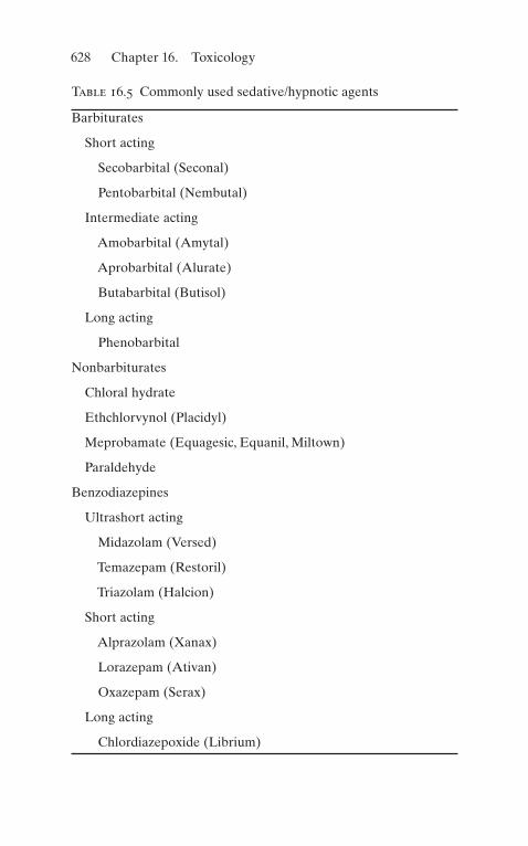
628
Table 16.5 Commonly used sedative/hypnotic agents
Barbiturates
Short acting
Secobarbital (Seconal)
Pentobarbital (Nembutal)
Intermediate acting
Amobarbital (Amytal)
Aprobarbital (Alurate)
Butabarbital (Butisol)
Long acting
Phenobarbital
Nonbarbiturates
Chloral hydrate
Ethchlorvynol (Placidyl)
Meprobamate (Equagesic, Equanil, Miltown)
Paraldehyde
Benzodiazepines
Ultrashort acting
Midazolam (Versed)
Temazepam (Restoril)
Triazolam (Halcion)
Short acting
Alprazolam (Xanax)
Lorazepam (Ativan)
Oxazepam (Serax)
Long acting
Chlordiazepoxide (Librium)
Chapter 16. Toxicology

629
4. Charcoal hemoperfusion may be indicated for severe barbiturate overdose.
5. Benzodiazepine antidote:
(a) Flumazenil (Romazicon): Selective benzodiaze-pine CNS receptor competitive inhibitor. Use to reverse benzodiazepine-induced coma.
(b) Initial recommended dose is 0.2 mg IV over 30 s. Repeat 0.3 mg after 30 s and then 0.5 mg at 1-min intervals.
(c) Most patients respond to cumulative doses of 1–2 mg. Reversal of CNS depression is unlikely if a dose of 5 mg has been given without effect.
(d) Adverse effects: Nausea and vomiting are most common. In patients dependent on benzodiaze-pines, flumazenil may induce withdrawal syndrome (agitation, tachycardia, seizures). Do not use in patients who have concomitant TCA overdose or who are taking chronic TCAs.
(e) The duration of action of a single dose is 1–2 h. If prolonged reversal is needed, give repeated doses or administer as an IV infusion of 0.1–0.5 mg/h.
Theophylline
The mechanism of theophylline toxicity is via release of endogenous catecholamines, stimulation of beta2-receptors, and inhibition of adenosine receptors. Clinical effects may be delayed for several hours following acute ingestion if a
Clorazepate (Tranxene)
Diazepam (Valium)
Flurazepam (Dalmane)
Prazepam (Centrax)
Table 16.5 (continued)
Theophylline

630
sustained- release formulation is involved. Toxicity may be acute or chronic.
A. Clinical Effects
1. Common toxic effects include nausea, vomiting, tremor, tachycardia, and hypotension.
2. Hypokalemia, hyperglycemia, and metabolic acidosis may occur (only in acute intoxication).
3. Seizures and ventricular dysrhythmias may occur, espe-cially with very high serum levels and with chronic intoxication.
B. Diagnostic Studies
1. Serum theophylline level.
(a) Therapeutic: 15–20 mg/L. (b) In acute overdose with level > 100 mg/L, seizures,
hypotension, and ventricular dysrhythmias are common. Seizures may occur at levels of 40–60 mg/L in chronic overdose. Repeat levels q2–4 h during treatment and monitor for 12–16 h.
2. Monitor serum pH, glucose, and potassium levels.
C. Management
1. Administer activated charcoal 100 g and readminister 20–30 g q2–3 h.
2. Treat seizures and ventricular dysrhythmias in standard fashion as required. Magnesium has been successful in some cases. For supraventricular tachycardia or rapid sinus tachycardia, ventricular dysrhythmias, or hypo-tension, administer esmolol 0.05 mg/kg/min or pro-pranolol 0.01–0.03 mg/kg IV. Use with caution if wheezing is present.
3. If theophylline level > 100 mg/L or seizures or dys-rhythmias do not respond to treatment, institute char-coal hemoperfusion.
Chapter 16. Toxicology

631
Crystal Meth
Methamphetamine hydrochloride, known as crystal meth, is the crystalline solid form of methamphetamine, and it also contains pseudoephedrine. It can be smoked, inhaled, injected, or swallowed. Compared to cocaine, it can last up to 12 h.
A. Clinical Effects
1. In CNS acts as a stimulant. The rapid dopamine release creates a feeling of euphoria and pleasure (“rush”).
2. Hypertensive crisis or coronary and cerebral vaso-spasms may occur.
3. Hyperthermia, seizures, and dysrhythmias. 4. Delusions, hallucinations, and psychotic behavior are
long-term effects.
B. Management
1. Airway control, oxygenation, and ventilation support. 2. If ingested orally, activated charcoal can be given. 3. Hyperthermia, hypertension, and metabolic and elec-
trolyte abnormalities must be treated. 4. For agitated patients, droperidol or haloperidol IV may
be used. 5. Labetalol can be used to lower the mean arterial
pressure. 6. For methamphetamine-induced seizures, benzodiaze-
pines IV.
Useful Facts and Formulas
A. Basic Formulas. The therapeutic index (TI) of a drug can be calculated as follows:
TI
LD
ED= 50
50
where LD50 = median lethal dose and ED50 = median effec-tive dose.
Useful Facts and Formulas
632
The margin of safety (MS) of a drug uses the ED99 for the desired effect and the LD1 for the undesired effect:
MS
LD
ED= 1
99
The apparent volume of distribution (Vd) can be calculated by the following equation:
V
CdivDose
=0
where Doseiv = the IV dose and C0 = the extrapolated plasma concentration at time zero.
For those agents that follow a two-compartment model, several Vd exist, and the following formula is used to assess the volume of the central compartment (Vc):
V
A BcivDose
��
where A and B represent disposition constants of a two- compartment model.
In addition, the peripheral compartment (Vp) can be calcu-lated as follows:
V
BpivDose
=
where B is derived from the elimination or equilibrium phase of a two-compartment model.
The total body clearance (Cl) of a drug can be calculated as the sum of clearances by individual organs
Cl Cl Cl Clr h i� � � ��
where Clr = renal clearance; Clh = hepatic clearance; and Cli = intestinal clearance.
B. Osmolality Formulas. To calculate serum osmolality, the following formula is usually applied:
Chapter 16. Toxicology

633
Calc Osmolality mOsm kg Na BUN Glucose. / / . /� � � � �2 2 8 18
The osmolal gap (OG) is useful in several intoxications and is calculated as follows:
OG Measured osmolality calculated osmolality� �
To calculate the contribution to measured osmolality of alcohols (also known as osmol ratios), the alcohol concentra-tion (mg/dL) is divided by the numbers depicted in Table 16.6.
C. Digitalis Intoxication. To treat digitalis poisoning appropri-ately, it is important to assess the digitalis body load:
Body load mg serum digoxin concentration
body weight
� � � � �� �5 6. in kg� � �100
The dose of digitalis antibodies (Digibind) is determined by dividing the body load by 0.6 mg/vial:
Dose number of vials Body load mg mg vial� � � � � � � �0 6. /
Table 16.6 Osmolal ratios of different alcoholsEthanol Ethylene glycol Isopropanol Methanol
4.6 6.2 6.0 3.2
Useful Facts and Formulas
635© Springer Nature Switzerland AG 2021J. Varon, Handbook of Critical and Intensive Care Medicine, https://doi.org/10.1007/978-3-030-68270-5_17
Trauma is the leading cause of death in persons under age 35 and accounts for 10% of all deaths. All trauma patients require sys-temic approach to minimize outcomes. About 140,000 traumatic deaths occur annually. Sepsis, hemorrhage, adult respiratory dis-tress syndrome, and multiple organ system failure are the leading causes of death in trauma patients who survive the initial resus-citation and surgical repair of their injuries. Trauma accounts for approximately 30% of all intensive care unit admissions.
Multisystem Trauma
A. Establishment of Priorities
1. Of highest priority in the initial evaluation are the following:
(a) Airway maintenance (b) Breathing and ventilation (c) Circulation and shock management (d) Disability assessment (e) Exposure with environmental control
2. Secondary evaluation includes vital signs and complete physical (including rectal) examination. Nasogastric and urinary catheters (unless contraindicated) should gener-
Chapter 17Trauma

636
ally be inserted to diagnose gastric or urinary tract hem-orrhage and to allow for monitoring of urinary output.
3. Rapid normalization of vital signs is one of the goals in trauma management.
B. Severity of Injury Scoring Systems
1. The Glasgow Coma Scale (see Table 17.1) is used for assessing neurologic status in head injury.
Table 17.1 Glasgow Coma ScaleScore
Eye opening
Spontaneous 4
To verbal command 3
To pain 2
None 1
Best motor response
Obeys verbal command 6
Localizes painful stimuli 5
Flexion withdrawal from painful stimuli 4
Decorticate (flexion) response to painful stimuli 3
Decerebrate (extension) response to painful stimuli 2
None 1
Best verbal response
Oriented conversation 5
Disoriented conversation 4
Inappropriate words 3
Incomprehensible sounds 2
None 1
Total score 3–15
Score of 8 and less represents severe brain injury
Chapter 17. Trauma

637
2. The trauma score (see Table 17.2) estimates physiologic severity of injury. It combines the Glasgow Coma Scale with other clinical indices of cardiovascular and pulmo-nary function.
C. Airway ManagementUse the LEMON method to assess for injuries, which
includes the following:
• Look for injuries that may distort external and internal structures.
• Evaluate 3-3-2, which refers to intraoral, mandibular, and hyoid-to-hyoid notch distances. These distances might be narrowed by a fracture, hematoma, or other anatomic distortions.
• MALLAMPATI: A calculation of Mallampati score is not always possible in trauma patients but all efforts must be made to visualize as much of the retrophar-ynx and if there is any pooled blood, vomitus, or secretions.
• Obstruction/obesity.• Neck mobility: Stabilization is necessary in all trauma
patients. It is important to keep in mind that neurologic injury from hypoxia is greater than that caused by spi-nal injury.
1. Clear airway of debris or secretions. 2. Avoid chin lift and neck lift/tilt if cervical spine
injury is considered. Obtain cervical spine radio-graphs as soon as possible.
D. Oxygenation and Ventilation
1. If adequate respirations appear to be present, obtain baseline blood gases as soon as possible. Apply 100% O2 by mask. If no spontaneous respirations, assist with bag and mask.
2. Endotracheal intubation:
(a) When necessary, intubation should usually be done by the oral route with manual in-line stabilization.
Multisystem Trauma
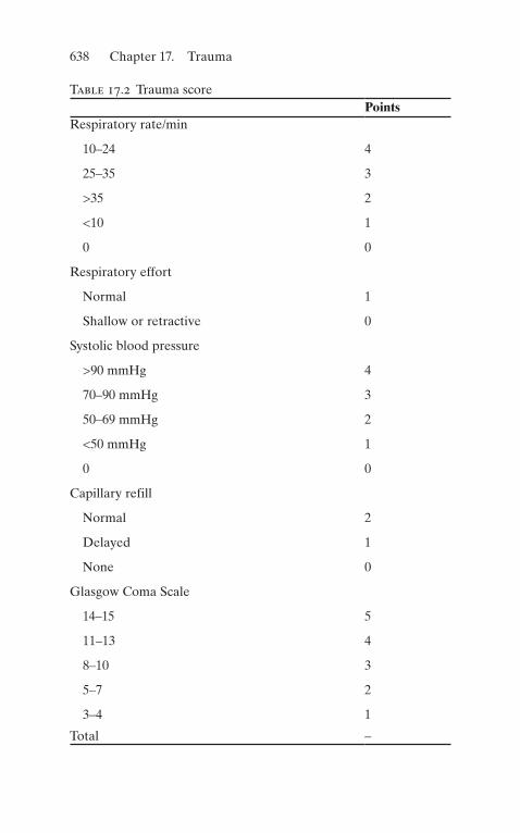
638
Table 17.2 Trauma scorePoints
Respiratory rate/min
10–24 4
25–35 3
>35 2
<10 1
0 0
Respiratory effort
Normal 1
Shallow or retractive 0
Systolic blood pressure
>90 mmHg 4
70–90 mmHg 3
50–69 mmHg 2
<50 mmHg 1
0 0
Capillary refill
Normal 2
Delayed 1
None 0
Glasgow Coma Scale
14–15 5
11–13 4
8–10 3
5–7 2
3–4 1
Total –
Chapter 17. Trauma

639
(b) Rapid sequence induction is often indicated in patients with major trauma, head or facial injury, diminished level of consciousness, and respiratory impairment: preoxygenation, application of cricoid pressure, and administration of induction agents (propofol, most commonly used for induction agent 2 mg/kg). Alternatives for patients with high risk of hypotension with induction are ketamine 1–2 mg/kg and etomidate 0.2 mg/kg IV (vecuronium 0.2 mg/kg IV, followed 3 min later by succinylcho-line 1.0–1.5 mg/kg; succinylcholine is contraindi-cated in penetrating eye injuries and massive crush injury), followed by intubation.
3. When a surgical airway is necessary, cricothyrotomy is the preferred procedure.
4. Most common causes for respiratory compromise ten-sion pneumothorax, open pneumothorax, and flail chest with pulmonary contusion.
E. Circulation and Shock Management
1. Evaluation includes assessment of vital signs, level of consciousness, skin color, character of pulse, and capil-lary refill.
2. Shock in trauma is most commonly due to hypovolemia.
(a) Likely sites for occult hemorrhage: thorax, abdo-men, pelvis, retroperitoneum, and thigh (i.e., long bone fractures).
(b) In addition, conditions producing shock that should be considered include tension pneumo-thorax, cardiac tamponade, myocardial contusion, spinal trauma, pulmonary contusion, and fat or air embolism.
Initial fluid resuscitation: bolus of IV crystal-loids (20 mL/kg isotonic saline)
3. Classification of hemorrhagic shock: see Table 17.3.
Multisystem Trauma
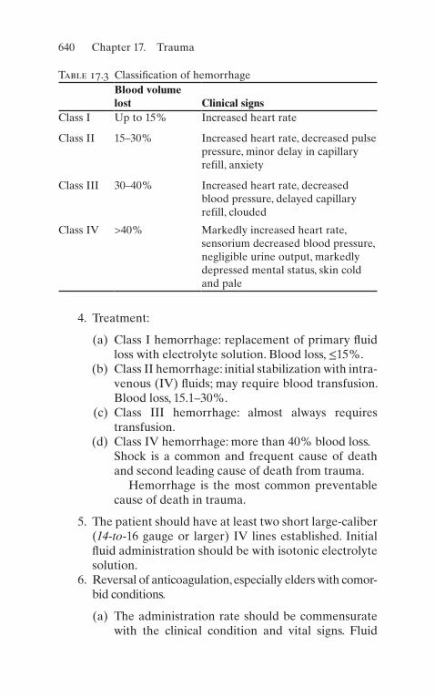
640
4. Treatment:
(a) Class I hemorrhage: replacement of primary fluid loss with electrolyte solution. Blood loss, ≤15%.
(b) Class II hemorrhage: initial stabilization with intra-venous (IV) fluids; may require blood transfusion. Blood loss, 15.1–30%.
(c) Class III hemorrhage: almost always requires transfusion.
(d) Class IV hemorrhage: more than 40% blood loss.Shock is a common and frequent cause of death and second leading cause of death from trauma.
Hemorrhage is the most common preventable cause of death in trauma.
5. The patient should have at least two short large-caliber (14-to-16 gauge or larger) IV lines established. Initial fluid administration should be with isotonic electrolyte solution.
6. Reversal of anticoagulation, especially elders with comor-bid conditions.
(a) The administration rate should be commensurate with the clinical condition and vital signs. Fluid
Table 17.3 Classification of hemorrhageBlood volume lost Clinical signs
Class I Up to 15% Increased heart rate
Class II 15–30% Increased heart rate, decreased pulse pressure, minor delay in capillary refill, anxiety
Class III 30–40% Increased heart rate, decreased blood pressure, delayed capillary refill, clouded
Class IV >40% Markedly increased heart rate, sensorium decreased blood pressure, negligible urine output, markedly depressed mental status, skin cold and pale
Chapter 17. Trauma

641
overload should be avoided, but adequate intra-vascular volume, hematocrit, and tissue perfusion must be achieved.
(b) In the hypovolemic patient, at least 2 L (20 mL/kg in a child) can be rapidly infused and the patient reassessed.
7. If shock persists despite resuscitation with IV fluids, blood replacement is indicated. Blood products in 1:1:1 ratios (PRBC, FFP, and platelets).
Maintain MAP around 65 mmHg or SBP of 90 mmHg is reasonable.
It is important to obtain manual BP measurement in patients with SBP <90 mmHg, as automated BP cuffs often overestimate values significantly in these patients. Furthermore, data suggest that the traditional thresh-old of a SBP <90 to define shock is inaccurate. The appropriate systolic or MAP threshold for defining shock varies by age. A significant proportion of trauma patients with hemorrhagic shock have SBP above 90 mmHg; using a cutoff value of 110 is more appropri-ate for elderly patients.
Patients who require blood transfusion may benefit from treatment with tranexamic acid if given within 3 h of injury.
(a) Maintain a hematocrit of at least 30%. (b) Crossmatched blood is preferable if the patient’s
condition permits. (c) Hypothermia must be controlled during transfusions. (d) If there is insufficient time for a full crossmatch to
be performed, type-specific (ABO and Rh compat-ible) blood should be administered.
(e) In patients in severe, life-threatening shock for whom type-specific blood is not available, adminis-ter type O blood (Rh negative in women of child-bearing age). Subsequent crossmatching may be more difficult, however. Patients with obvious severe bleeding or ongoing loss should be trans-fused immediately with type O blood (women of
Multisystem Trauma

642
childbearing age should be transfused with O neg-ative blood). While mildly unstable patients can be treated with crystalloids, unnecessary infusion of crystalloid should be avoided.
(f) Autotransfusion (especially administration of autologous blood from chest tube drainage) should be employed when feasible.
8. Monitor for possible complications of transfusion.
(a) Hemolytic transfusion reaction: fever, chills, and chest, back, and joint pain. Terminate transfusion, adminis-ter IV fluids and furosemide. Monitor urine output.
(b) Hypothermia may follow massive transfusion with refrigerated blood. Give blood through a warmer if possible. Monitor body temperature with a core probe.
(c) Coagulopathy may result following massive transfu-sion, probably on the basis of quantity and function of platelets, as well as consumption of coagulation fac-tors. Administration of platelets is advised following rapid transfusion of each 10 U of blood. If evidence of coagulopathy exists, consider administration of fresh-frozen plasma and cryoprecipitate.
(d) Banked blood is acidemic and high in potassium and preservative anticoagulant binds calcium. Monitor serum pH, potassium, magnesium, and calcium levels.
9. Monitor urine output: Volume replacement should produce urine output of at least 1–1.5 mL/kg/h.
F. Complications of Hemorrhagic Shock and Volume Resuscitation: peripheral edema, hypothermia, cerebral edema, cardiac dysfunction (usually right ventricular fail-ure), pneumonia, adult respiratory distress syndrome (ARDS), and multisystem organ failure.
G. Cardiac Arrest
1. Perform immediate thoracotomy in patients in extremes, especially with penetrating chest trauma.
Chapter 17. Trauma

643
2. Therapeutic objectives include relief of cardiac tam-ponade, open cardiac massage, control of cardiac inju-ries, vascular control of major vessel or hilar injuries, and aortic occlusion for treatment of shock.
3. In patients with impending arrest, use of resuscitative balloon for occlusion of the aorta (REBOA) to tempo-rize patients with intra-abdominal or retroperitoneal hemorrhage until more definitive surgery or emboliza-tion is possible. It is not suitable for use in patients with suspected thoracic sources or patients in cardiac arrest where thoracotomy is preferred.
Head Trauma
Head injury is a major entity, often encountered in acute care, the head being the most frequently injured part of the body in trauma patients. Over 80,000 persons annually sustain per-manent disabling injuries of the head or spinal cord.
A. Assessment
1. History
(a) Important components are mechanism of injury and loss of consciousness. High-speed trauma (e.g., with ejection from vehicle, impact with windshield) produces a greater chance of significant injury.
(b) Incomplete recollection by the patient of details of injury may imply a transient loss of consciousness. This symptom is not as useful if the patient is intoxicated.
2. Physical Examination
(a) Examine the scalp and face for signs of trauma, such as lacerations, ecchymoses, hemotympanum, and bleeding or clear fluid from the nostrils or ears.
(b) Palpate the spine for tenderness or deformity. Always consider the possibility of concomitant spi-nal cord injury.
(c) Focus on other injuries that affect the airway or produce respiratory or circulatory impairment.
Head Trauma

644
3. Neurologic Examination
(a) This is the best tool for identifying the presence of significant intracranial injury.
1. Assess the mental status (most important aspect of the neurologic examination).
2. Determine focal neurologic deficit, abnormal posturing, and pathologic reflexes. Evaluate brain stem reflexes (light, corneal, gag) and ven-tilatory drive.
3. Pupillary examination is crucial in these patients. 4. The absence of brain stem function usually indi-
cates the need for urgent airway intervention.
(b) Frequent repetition of neurologic examination is necessary, especially within the first 48 h of injury.
(c) The Full Outline of Unresponsiveness (FOUR) score is an alternative scale for the assessment of patients with severe TBI. Advantages over GCS include the ability to grade injury in intubated patients and to assess the brainstem function. They both have equivalent ability to predict long-term outcomes.
4. Glasgow Coma Scale (GCS)
(a) Determine the GCS score (see Table 17.1).
1. Score of 13–15 = mild injury 2. Score of 9–12 = moderate injury 3. Score of 8 or less = severe injury
(b) GCS is of limited usefulness in children <3 years.In severe trauma brain injury, the principal focus is to limit secondary brain injury
B. Management
1. PositionWhen associated injuries permit, elevate the head of
the bed 30–45°, as this reduces intracranial pressure. 2. Airways and Ventilation
Chapter 17. Trauma

645
(a) The highest initial priority is prevention or reduc-tion of secondary injury due to swelling or com-pression of cerebral tissue by cerebral edema and raised intracranial pressure (ICP).
(b) Proper airway management reduces increased ICP. Diminished mental status (particularly a GCS score <9), inability to protect the airway, SpO2 <90% despite the supplemental oxygen, or clinical signs of cerebral herniation are indications for early endo-tracheal intubation. When expertise is not available or an attempt was unsuccessful, bag-mask ventila-tion should be done in conjunction with basic air-way-opening maneuvers or airway adjuncts.
(c) Chin lift and neck lift maneuvers are inappropriate if the patient with head injury is suspected of hav-ing cervical spine injury.
(d) Initial management of increased ICP, in the past, included hyperventilation. Recent guidelines rec-ommend to avoid hyperventilation in acute phase (24–48 h). Hyperventilation induces vasoconstric-tion that may also cause secondary ischemia.
1. It is often recommended to maintain arterial pCO2 between 25 and 30 Torr (mmHg), but there is no uniform agreement on this. PaCO2 of <30 mmHg should be avoided except as a tem-porary measure to reduce ICP. Patients hyper-ventilated to PaCO2 of less than 25 mmHg for 5 days had worse outcomes,
2. Many recommend not to hyperventilate patients with pCO2 <30 mmHg 20 Torr or pH >7.60. Both hypo- and hyperventilation should be avoided. Quantitative capnography may be useful in this situation.
(e) Endotracheal intubation.
1. Nasal intubation is relatively contraindicated; use orotracheal intubation with manual in-line stabilization.
Head Trauma

646
2. Precede intubation with bag-valve mask venti-lation, cricoid pressure, and pharmacologic induction: etomidate 0.3 mg/kg IV and succinyl-choline 1.0–1.5 mg/kg (watch out for patients with renal failure or hyperkalemia, as this may worsen it) or vecuronium 0.1–0.2 mg/kg IV.
(f) When mechanical ventilation is instituted, avoid high levels (>10 cm) of positive end-expiratory pressure (PEEP), as this may increase ICP. Chest physiotherapy may also increase ICP. Use of PEEP 15–20 cm H2O as well as APRV mode when clinically appropriate for managing ARDS in patients with TBI in conjunction with moni-toring ICP.
3. Osmotic Therapy and Diuresis
(a) Reduce increased ICP by reducing intracranial volume.
(b) Mannitol is the osmotic diuretic of choice. It is generally administered in rapidly deteriorating patients. It is often used to arrest neurologic dete-rioration when the patient is being prepared for urgent craniotomy.
1. Give as a 20% solution, 0.25–1.0 g/kg via rapid IV infusion (boluses). Q4H–Q6H as needed.
2. 23.4% NaCl is preferred to mannitol since it is volume depleting particularly in trauma patients with ongoing loss, hemodynamic instability, and renal failure.
3. ICP reduction usually occurs within 10–20 min. 4. Duration is limited to 2–6 h following initial
bolus. Continuous infusion may be required. 5. Monitor blood pressure, serum electrolytes, and
osmolarity. Maintain <320 mmol/L.
(c) Diuretics may be used alone or in combination with osmotic diuretics to reduce intracerebral fluid volume.
Chapter 17. Trauma

647
1. Furosemide is the loop diuretic of choice. Administer an IV bolus of 1 mg/kg. The onset of action is slower than mannitol, but concomitant use enhances the duration of ICP reduction by mannitol and decreases the risk of rebound ICP elevation. Repeated doses may be required.
2. Acetazolamide is a carbonic anhydrase inhibi-tor. It decreases cerebrospinal fluid (CSF) pro-duction. Administer 250 mg qid. Monitor for production of acidosis.
(d) Corticosteroids are frequently administered, but whether they diminish ICP in head trauma is con-troversial. They have been found to be harmful rather than beneficial.
(e) Therapeutic hypothermia has been used in these patients with conflicting results. In the author’s experience, this is a beneficial therapeutic modality.
4. Cardiovascular Support
(a) As a rule, cerebral injury does not produce hypo-tension (except in the agonal state).
(b) Look for an extracranial source if hypotension is present.
(c) Avoid fluid overload, some recommend to moni-tor central venous pressure, but provide adequate intravascular volume and hematocrit. A pulmonary artery catheter is rarely required for monitoring.
C. Monitoring
1. Intracranial Pressure
(a) Indicated in patients with severe head injury with computed tomography (CT) evidence of raised ICP. Monitoring of ICP, however, has not been proven to affect survival.
(b) Monitor by intraventricular catheter, subarach-noid bolt, or extradural pressure sensor.
(c) It is generally advised to maintain ICP at ≥20 mmHg.
Head Trauma

648
(d) Cerebral perfusion pressure (CPP): monitor this (CPP = mean arterial pressure [MAP] – ICP) and maintain at >60 mmHg. If hypotension occurs, ele-vate arterial pressure to maintain CPP above this level, as ischemic damage may occur.
A goal of 60–70 CPP is recommended to improve the survival and favorable outcomes.
2. Intravascular PressureArterial catheterization to allow monitoring of
MAP as well as frequent blood gas determinations. 3. Head trauma may produce syndrome of inappropriate
secretion of antidiuretic hormone (SIADH). This results in hyponatremia with a relatively concentrated urine (see also Chaps. 9 and 14).
4. CRASH-3 trial has shown benefit of tranexamic acid within 3 h of injury. 1 g is transfused over 10 min fol-lowed by IV infusion of 1 g over the next 8 h.
D. Diagnostic Studies
1. Skull Radiographs
(a) Plain skull radiographs may demonstrate skull frac-ture but have poor sensitivity and specificity for identifying intracranial lesions.
(b) May be useful in children <2 years of age, in whom skull fracture may identify risk of hypovolemia due to extracranial bleeding, formation of leptomenin-geal cyst, and child abuse.
2. Computed Tomography
(a) CT is the diagnostic procedure of choice in assess-ing acute head injury. Perform CT without contrast material. CT is indicated in the patient with a decreased level of consciousness (GCS score ≤14), deteriorating mental status, focal neurologic defi-cit, seizures, or persistent vomiting.
Repeat CT scan may be reasonable within 6 h when hematoma is seen on the initial scan.
Chapter 17. Trauma
649
(b) If the GCS score is <9, obtain CT immediately after endotracheal intubation when the patient is hemodynamically stable.
(c) Screen for blunt cerebrovascular injury (BCVI), due to injured carotid and vertebral arteries linked to skull base fractures. This may result in stroke at the time of injury but may be delayed for several hours to days. Antithrombotic therapy may pre-vent subsequent ischemic lesions the identification of BCVI is crucial. Use expanded Denver criteria to identify these patients.
3. Magnetic Resonance Imaging (MRI)
(a) MRI is superior to CT in diffuse axonal injury. (b) Limitations in acute injury: duration of scanning,
interference of monitoring, and life support equip-ment with magnetic field.
4. UltrasoundMay be an option in smaller children with suspected
intraventricular hemorrhage. In addition, vascular blood flow calculations may be useful in the manage-ment of these patients.
Crush Injury
A. Traumatic Asphyxia
1. Mechanism: direct massive thoracoabdominal com-pression
2. Clinical findings: blanching cyanosis of the upper chest, neck, and head, petechiae, edema, and subconjunctival hemorrhage
3. Associated injuries: chest wall injury, pulmonary con-tusion, cardiac contusion, diaphragm rupture, intra-abdominal solid, and hollow viscus injury
4. Sequelae: brachial plexus injury, spinal cord injury, and transient neurologic impairment
Crush Injury

650
B. Abdominal and Pelvic Injury
1. Crush injury accounts for 5% of pelvic fractures. May result in bladder laceration.
2. Crush mechanism to the abdomen results in a high pro-portion of hollow viscus injury.
C. Skeletal Muscle Injury
1. Results in myonecrosis. May produce rhabdomyolysis, hyperkalemia, hyperphosphatemia, hypocalcemia, and myoglobinuria.
2. Sequelae include acute renal failure and disseminated intravascular coagulation.
3. Follow creatine kinase (CK), electrolytes, creatinine, and urine output.
4. CK levels reach a maximum of 24–36 h after injury. The level should decline by 50% each 48 h thereafter. If there is an increase in CK during this time, consider recurrence of muscle necrosis.
5. Treat acute renal failure with fluid infusion, osmotic diuresis, and alkalinization (see Chap. 14).
6. Extremity compartment syndrome in patients with combined arterial and venous injuries. Repeat evalu-ation 24 h following injury repair. Monitor elevation in CK.
Chest Trauma
Chest trauma is the cause of death in up to 20–25% of trauma-related deaths and a quarter of cases of multisystem trauma. Injury may occur to the chest wall, lung, great vessels, and mediastinal viscera.
Most injuries can be initially managed with chest tube insertion and other nonoperative management.
Indications for thoracotomy include cardiac tamponade and massive hemothorax (see below); pulmonary air leak >15–20 L/min; aortic arch, esophageal, tracheal, or major bronchial disruption; systemic air embolism; bullet embolism; and cardiac arrest.
Chapter 17. Trauma

651
A. Chest Wall Trauma
1. Rib fracture is the most common chest wall injury. It is an important indicator of underlying injury.
(a) First to Third Ribs: Increased risk of intratho-racic injury and great vessel and bronchial injury. Diminished pulse or blood pressure in arms or radiographic evidence of mediastinal hematoma (see below) is an indication for arteriography.
(b) Lower Ribs: kidney, liver, and spleen laceration.
2. Flail chest occurs when three or more ribs are fractured in two places or when multiple fractures are associated with sternal fracture.
(a) Clinical significance varies, depending upon the size and location of the flail segment and the extent of underlying pulmonary contusion.
(b) Obtain and follow arterial blood gases (ABGs). (c) Splint thorax with weights, traction, or skeletal
fixation. (d) Patients with severe hypoxemia will require
endotracheal intubation and positive-pressure ventilation. Observe for late development of pneu-mothorax, especially tension pneumothorax, in the mechanically ventilated patient.
3. Sternal fracture is associated with myocardial contu-sion, cardiac rupture and tamponade, and pulmonary contusion. Early surgical fixation is often necessary, and urgent surgery may be indicated when costosternal dislocations produce compromise of the trachea or neurovascular structures at the thoracic inlet.
4. Analgesic methods that may be required for treatment of major chest wall injuries include parenteral opiates, epidural analgesia, and intercostal nerve block.
B. Pneumothorax
1. Usually results from penetrating trauma or blunt trauma with rib fracture. May be caused by positive-pressure ventilation (barotrauma).
Chest Trauma

652
2. Presence of pneumothorax requires 28–40 Fr chest tube insertion. Smaller tubes may be utilized if pneu-mothorax is not accompanied by hemothorax.
3. Open pneumothorax requires covering of chest wall injury with airtight dressing and insertion of chest tube.
4. Tension pneumothorax requires immediate needle decompression and chest tube insertion. Clinical find-ings include unilateral absence of breath sounds, severe dyspnea, tracheal shift, jugular venous distention, cya-nosis, chest pain, and hypoxemia.
C. Hemothorax
1. Initial treatment requires insertion of chest tube to evacuate hemothorax, reexpand lung, and monitor rate of bleeding.
2. Indications for surgical thoracotomy include initial chest tube drainage of >1500 mL or continued bleeding of more than 300 mL/h or 3 mL/kg/h for more than 2–3 h.
D. Major Vessel Injury
1. Common cause of death in major trauma. 2. Consider when there is radiographic evidence of medi-
astinal hematoma: widened mediastinum, aortic knob obliteration, and tracheal or nasogastric tube deviation.
3. Arteriography or CT is required for diagnosis.
E. Cardiac Tamponade
1. Most frequently occurs with penetrating injuries. Suspect in chest trauma with shock and jugular venous distention.
2. Requires thoracotomy and pericardial decompression. Pericardiocentesis may be performed if the diagnosis is uncertain or as a temporizing measure during prepara-tion for thoracotomy.
F. Pulmonary Contusion. It is one of the most common types of lung injury occurring in up to 17% of multiple trauma patients. Management consists of supplemental oxygen
Chapter 17. Trauma
653
administration and mechanical ventilation with the addi-tion of PEEP, if indicated in patients with worsening hypoxemia. Irregular, non-lobular opacification of paren-chyma on chest X-ray.
G. Myocardial Contusion. Management consists of cardiac monitoring, echocardiography, and treatment of dysrhyth-mias, as necessary. Look for injury or ongoing hemorrhage when unexplained tachycardia exists.
Abdominal Trauma
Blunt trauma is the most common mechanism of abdominal injury and has relatively high mortality rates of 10–30%. The patient with blunt trauma may have no abdominal symptoms or have little external evidence of injury.
A. EvaluationQuickly inspect the chest and abdomen for deformities,
contusions, abrasions, and punctures (DCAP), eviscera-tion, and distention.
1. Physical findings may be unreliable if abdominal trauma is complicated by head or other injuries or intoxications.
2. Findings most consistently associated with inter-nal abdominal injury are abdominal tenderness and guarding.
3. Examine thorax for rib fractures, palpate flanks and pelvis, and perform rectal and pelvic examination.
4. Obtain baseline hemogram, blood coagulation screen, and urinalysis.
• The presence of a seat-belt sign, a large bruise or abrasion across the abdomen, is indicative of intra-abdominal injury in approximately 25% of cases.
• Periumbilical bruising (Cullen’s sign) may raise sus-picion for retroperitoneal hemorrhage, but keep in mind that this usually takes several hours to develop.
Abdominal Trauma

654
B. FAST (Focused Abdominal Sonography for Trauma)
• Includes RUQ, LUQ, and the pelvis• Facilitates a timely diagnosis for patient with blunt
abdominal trauma• Helps accurately diagnose hemoperitoneum
C. Diagnostic Peritoneal Lavage (DPL)
1. There is considerable variation in use of DPL from institution to institution.
2. Indications may include equivocal abdominal findings, possible abdominal injury in the face of diminished sen-sation due to head or spinal injury, or alcohol intoxication.
3. The major advantage is the ability to obtain a rapid indication of intraperitoneal hemorrhage.
4. Relative contraindications: previous abdominal sur-gery, significant obesity, pregnancy, and preexisting coagulopathy.
5. Positive lavage consists of aspiration of >10 mL of blood, aspiration of enteric contents, or lavage fluid with red blood cell levels >100,000/mm3, white blood cells >500/mm3, amylase ≤20 IU/L, or bile.
D. Abdominal CT
1. Indicated in stable patients with possible intra-abdom-inal injury and where DPL is being considered but is contraindicated.
2. Advantages include ability to visualize urinary tract and retroperitoneum. Abdominal Ultrasound: Useful to detect free intraperitoneal blood after blunt trauma.
E. Penetrating InjuryOn evisceration cases, gently cover any organ or viscera
protruding from a wound with gauze moistened with saline or water. Do not push back to the abdomen.
1. Antibiotics
(a) Second- or third-generation cephalosporin, e.g., cefoxitin 2 g IV q6 h or ceftazidime 1–2 g IV q8 h
Chapter 17. Trauma

655
(b) Combination of gentamicin (1.5–2.0 mg/kg IV loading and 3 mg/kg/d in three maintenance doses) or tobramycin (l mg/kg q8 h IV) and clindamycin 600–900 mg q8 h IV
F. Indications for Laparotomy
1. Gunshot wounds. 2. Stab wounds with shock, signs of peritoneal irritation,
gastrointestinal bleeding, or evisceration of bowel. 3. Blunt trauma with unstable vital signs, gastrointestinal
bleeding, peritoneal irritation, pneumoperitoneum, and evidence of diaphragmatic injury.
4. There may be a role for laparoscopy in stable patient with penetrating injury, but this method has not been used extensively in this setting.
G. Postoperative Complications
1. Intra-abdominal hemorrhage:
(a) May be due to recurrent bleeding from sites not identified during surgery due to hypotension.
(b) Identify hemostasis deficiencies (thrombocytope-nia, clotting dysfunction), especially if patient has had massive transfusions.
(c) If prothrombin time (PT) and partial thromboplas-tin time (PTT) are prolonged, administer fresh-frozen plasma.
2. Fever: Consider wound infection, necrotizing fasciitis, peritonitis, and intra-abdominal abscess.
3. Missed intra-abdominal injury: diaphragm, biliary tree, duodenum, pancreas, ureter, colon, and rectum.
H. Nonoperative Management
1. May be suitable in patients who remain hemodynami-cally stable following initial resuscitation with 1–2 L of IV fluids. Laparoscopic repair may be another option.
(a) Normal vital signs, urine output >1.0–1.5 mL/h, and no blood requirement.
Abdominal Trauma

656
(b) Patient must be alert. (c) No coagulation defects.
2. CT should establish extent of injury. 3. Suitable injuries include the following:
(a) Isolated splenic trauma with minor capsular tear or parenchymal injury
(b) Stab wounds without shock, peritoneal irritation, or gastrointestinal bleeding
4. Monitoring:
(a) Repeat abdominal examination for signs of perito-neal irritation at least every 4–6 h.
(b) Vital signs monitored every 1–2 h during the first 24 h.
(c) Serial hematocrit every 4 h. Also monitor amylase. (d) Repeat abdominal CT after 12 h of observation
and thereafter as indicated by clinical signs.
I. Urinary Tract Injury
1. Evaluate with IVP or abdominal CT for gross hematu-ria, flank hematoma or mass, and penetrating trauma with suspected urinary injury.
2. Bladder and urethral injury: See pelvic fracture.
Multiple Fractures
A. General Considerations
1. Identify fractures and dislocations and assess distal neurocirculatory function.
2. Complications of fractures include arterial and neural injury, hemorrhage, compartment syndrome, ARDS, fat embolization, infection, and thromboembolism.
3. Fractures of the pelvis and femur are particularly sig-nificant because of hemorrhage potential.
4. Dislocations of the hip and knee require prompt reduc-tion to avoid neurovascular complications.
Chapter 17. Trauma

657
B. Initial Management
1. Immobilize any injured extremity. 2. Stabilize femur fracture in Hare or comparable trac-
tion device.
C. Arterial Injury
1. Arterial injury may be due to transection, arterial spasm, occlusion by hematoma, external compression, and arteriovenous fistula formation.
2. Acute loss of vascular function requires emergent sur-gical exploration or angiography.
D. Compartment Syndrome
1. Circulatory supply can be lost due to increased muscu-lar compartment pressure. Most common in leg but also occurs in forearm.
2. Earliest sign is pain with passive stretching of the extremity. Suspect if there is severe, constant pain despite reduction and immobilization.
3. Measure compartment pressure by inserting a needle connected to a manometric pressure measurement sys-tem into the soft tissue of the extremity involved; should be <30 mmHg in the normotensive pressure.
4. Treatment is fasciotomy.
E. Infection in Open Injuries F. Tetanus prophylaxis should be given, especially for
wounds with deep tissue penetration, devitalized tissue, and burns.
G. IV antibiotics.
(a) IV first-generation cephalosporin (e.g., cefazolin 1 g IV q6 h). Some authors prefer broader coverage.
(b) If the wound is large or heavily contaminated, add gentamicin (1.5–2.0 mg/kg IV loading dose and 3 mg/kg/day in three maintenance doses) or tobramycin (1 mg/kg q8 h IV or 3 mg/kg IV single dose) if the patient has normal renal function.
Multiple Fractures
658
H. Observe for appearance of gas gangrene (appearance of subcutaneous crepitation or soft tissue gas on radiographs).
(a) Occurs within 12–72 h of injury and requires broad-spectrum antibiotics and aggressive wound debridement
(b) Most often occurs in grossly contaminated open frac-tures with soft tissue damage
I. Fat Embolism
1. Occurs most commonly in multiple fractures, especially involving the femur, tibia, and pelvis.
2. Onset of symptoms occurs 1–5 days following injury: tachypnea, hemoptysis, fever, petechiae, and mental status change.
3. Hypoxemia (PaO2 <60 mmHg on room air) is the most consistent finding.
4. Treatment includes correction of hypoxemia and venti-latory support with PEEP used if arterial oxygenation cannot be maintained. Management is similar to that of ARDS (see Chap. 13). Maintain a negative fluid bal-ance. A pulmonary artery catheter may be required for management.
J. Pelvic Fracture. The so-called silent killer as there is up to 1500-mL capacity of blood in a 75-kg man. Pelvic fractures caused by high-energy forces have significant mortality. Hemorrhage is a major cause, as are associated injuries, sepsis, multiple organ failure, and ARDS.
Infection of pelvic hematoma may occur even in closed injuries due to hematogenous spread of bacteria.
1. Evaluation
(a) Physical Examination
1. Palpate the pelvis to determine tenderness and instability.
2. Perform pelvic and rectal examination to ascer-tain open injury and sphincter tone.
3. Blood at urethral meatus or high-riding prostate on rectal examination indicates urethral tear.
Chapter 17. Trauma
659
(b) Radiographs
1. Obtain pelvic radiographs in patients suspected of sustaining pelvic fracture and in patients with multiple trauma.
2. CT is superior in demonstrating certain aspects of pelvic injury. Utilize in the stable patient.
2. Hemorrhage
(a) Usually retroperitoneal, the result of venous injury to pelvic venous plexus. May be massive, exceeding several liters.
(b) Signs of persistent bleeding are an indication for external fixation of unstable pelvic fractures.
(c) Application of pneumatic antishock trousers may be helpful in temporarily controlling hemorrhage (although there is considerable controversy over efficacy). Once antishock trousers are in place, do not remove until vascular access is established and blood pressure stabilized.
(d) Consider arteriography and selective embolization for patients with continued severe hemorrhage.
3. Associated Injuries
(a) Injuries to consider: urinary bladder perforation, vaginal or rectal laceration, and posterior urethral tear.
(b) If signs of urethral injury are present, a retro-grade urethrogram should be used to ascertain integrity of the urethra before urinary catheter insertion.
Spinal Cord Injury
Spinal injury should be considered in any patient with multisystem injury, head or facial injury, and those who are unconscious. Maintain spinal immobilization until radio-graphs exclude spinal injury.
Spinal Cord Injury

660
A. Evaluation
1. Perform a neurologic examination to determine the extent and level of injury.
(a) Examine and document motor function and sen-sory level.
(b) Determine if there is sacral sensory sparing or anal sphincter contraction, signs that cord injury is incomplete.
(c) Sacral reflexes (anal wink, bulbocavernosus reflex) are the first to return after spinal shock, usually within 24 h following injury.
B. Management
1. Endotracheal Intubation
(a) Patients may require endotracheal intubation because of paralysis of respiratory muscles.
(b) If intubation is necessary, it should be done with in-line cervical immobilization.
(c) The orotracheal approach is preferred, preceded by 100% oxygenation by mask and application of gentle cricoid pressure.
(d) Administer thiopental 25–200 mg and vecuronium 0.1–0.2 mg/kg IV or etomidate 0.3–0.6 μg/kg before intubation.
2. Respiratory Care
(a) Monitor breathing with frequent vital capacity measurement. Vital capacity <10 mL/kg is an indi-cation for ventilatory assistance.
(b) Monitor arterial blood gases for hypoxemia or hypercarbia.
3. Corticosteroids
(a) High-dose steroid therapy is generally accepted as reducing secondary injury if started within 8 h of injury.
(b) Administer methylprednisolone initial IV bolus 30 mg/kg followed by 5.4 mg/kg/h for the next 23 h.
Chapter 17. Trauma

661
(c) Naloxone (5.4 mg/kg IV followed by infusion of 4 mg/kg/h for the following 23 h) is frequently used in addition to corticosteroids, though its effective-ness has not been proven. Tirilazad mesylate has been used too.
(d) Methylprednisolone is associated with increased mortality in patients with moderate to severe trau-matic brain injury and should not be given to these patients with both injuries.
4. Neurogenic Shock
(a) Results from injury to descending sympathetic pathways (usually in cervical and thoracic cord injuries) with loss of vasomotor tone and sympa-thetic cardiac innervation.
(b) Result is hypotension due to vasodilation and bra-dycardia, which may last for days to weeks.
(c) Administer IV fluids for initial treatment of hypo-tension. If hypotension persists, administer phenyl-ephrine or dopamine.
(d) Atropine (0.5 mg) or isoproterenol may be given if heart rate <45 beats per minute.
5. Abdominal
(a) Insert a nasogastric tube for ileus and acute gastric dilatation.
(b) In patients with abdominal trauma, consider diag-nostic testing (DPL or CT) to determine intra-abdominal injury.
6. Urologic
(a) A bladder catheter should be in place for at least 4 days following injury or until other injuries are stabilized and urine output is no longer being fol-lowed. After this, intermittent catheterization may be started.
(b) Tape the catheter up over the pubis to prevent ure-thral traction injury.
Spinal Cord Injury

662
7. Traction Immobilization
(a) Traction with Gardner–Wells tongs may be required to immobilize and align cervical injuries.
1. Weight required varies with injury. 2. Perform a neurologic examination, and take
radiographs frequently to determine that the alignment is correct and overdistraction does not occur.
8. Autonomic Hyperreflexia
(a) Autonomic hyperreflexia is the result of increased autonomic nervous system (primarily sympathetic) activity caused by noxious stimulus from below the level of the cord lesion.
(b) Produces paroxysmal hypertension, headache, sweating, bradycardia or tachycardia, and anisocoria.
(c) Most common cause: Bladder distention. Others: Fecal impaction, urinary tract infection, and ureterolithiasis.
(d) Treatment: Treat underlying cause (e.g., catheterize bladder). Anticholinergic drugs may be used.
Therapeutic hypothermia in spinal cord inju-ries. Even though this area is still under inves-tigation, this therapeutic intervention has had remarkable improvements in neurological function when patients are induced early after trauma. See Chap. 15.
Useful Facts and Formulas
A. Hemorrhage. To assess the intravascular volume resuscita-tion needed in a trauma patient, normal blood volumes according to age need to be known (see Table 17.4).
The severity of hemorrhage in a trauma patient can be clas-sified as shown in Table 17.5.
Chapter 17. Trauma

663
To estimate how much whole blood or packed red blood cells (PRBCs) must be administered to change the hemato-crit percentage to a desired amount in a trauma patient, the following formula can be utilized:
Transfusion required mL Desired change in Hct kg factor� � � � �
where Hct = hematocrit and factor = varies with the volume of blood per body weight (adults and children >2 years, a fac-tor of 1 will achieve a Hct of 70% using PRBCs and 1.75 to achieve a Hct of 40% using whole blood).
B. Burns. Several formulas guide the initial fluid resuscita-tion after burn injuries. Below are the most common for-mulas used in clinical practice. In all these formulas, 50% of calculated volume is given during the first 8 h, 25% of calculated volume is given during the second 8 h, and 25% of calculated volume is given during the third 8 h.
Table 17.4 Normal blood vol-umes according to age
Normal blood volumes by ageNewborn 85 mL/kg
Infant 80 mL/kg
Child 75 mL/kg
Adult 70 mL/kg
Table 17.5 Severity of hemorrhage classification, trauma patients
Severity of hemorrhage
Blood pressure (mmHg) Blood loss (cc)
Plasma volume (cc)
Normal 120/80 – 5000
Class I 120/80 <750 4600
Class II 115/80 1000–1250 3800
Class III 90/70 1500–1800 3200
Class IV 60/40 2000–2500 2500
Useful Facts and Formulas
664
The Evans formula can be calculated as follows:
Evans formula mL crystalloid kg burn h
mL crystalloid kg
= 1 24
1
/ /% /
/ /%% /
/
burn h
mL D W h
24
2000 5 24
The Brooke formula and the modified Brooke formula are calculated as follows:
Brooke formula mL crystalloid kg burn h
mL colloid kg
= 1 5 24
0 5
. / /% /
. / //% /
/
burn h
mL D W h
24
2000 245
Modified formula = 2 mL Ringer’ s lactate/kg/% burn/24 hThe Parkland formula is calculated asParkland formula = 4 mL crystalloid/kg/% burn/24 hIn addition to these formulas, the evaporative water losses
in patients with burns need to be calculated and replaced. Evaporative water loss (EWL) is calculated as
EWL mL h BSA burned BSA/ %� � � �� ��25
where BSA = body surface area (Figs. 17.1 and 17.2).Other Trauma Scoring Systems. Several trauma scoring
systems are in use throughout the world. Of them, the abbre-viated injury scale (AIS) is commonly utilized (see Table 17.6).
The trauma score (TS) is another commonly utilized sys-tem and is depicted in Table 17.7.
Chapter 17. Trauma

665
A
Front Back
13%
2% 2%
1%1%
1%
2% 2%
2%2%
B
C
B
C
D
13%
2% 2%
1%1%
2% 2%
2%2%
E
F
E
F
Area
Head (A/D)
Thigh (B/E)
Leg (C/F)
10%
0
3%
2%
9%
1
3%
3%
7%
5
4%
3%
6%
10
5%
3%
By age in years
Figure 17.1 Estimating the burned surface area in children
Useful Facts and Formulas

666
The Rule of 9’s
4.5%
Front Back
1%
4.5% 4.5%4.5%4.5%
18% 18%
9% 9%9% 9%
4.5%
Figure 17.2 Estimating the burned surface area in adults
Table 17.6 The abbreviated injury scaleAIS score Injury severity1 Minor
2 Moderate
3 Serious, non-life-threatening
4 Severe, life-threatening
5 Critical
6 Maximal (correlates with death)
Chapter 17. Trauma

667
The revised trauma score (RTS) eliminates the assessment of capillary refill and respiratory effort and is calculated as follows:
RTS GCS SBP RR coded values
revised score co
� � ��0 9368 0 7326 0 2908. . .
eefficient
Table 17.7 The trauma scoreVariable Measurements ScoreRespiratory rate (bpm) 10–24 4
25–35 3
>35 2
<10 1
0 0
Respiratory effort Shallow 1
Retractions 0
Systolic blood pressure (mm Hg) >90 4
70–90 3
50–69 2
<50 1
0 0
Capillary refill Normal 2
Delayed 1
Absent 0
Glasgow Coma Scale 14–15 5
11–13 4
8–10 3
5–7 2
3–4 1
Abbreviation: bpm beats per minute
Useful Facts and Formulas

668
where GCS = Glasgow Coma Scale; SBP = systolic blood pressure; and RR = respiratory rate.
For children and infants, the pediatric trauma score is uti-lized (see Table 17.8).
C. Neurological Trauma. Within the primary survey, an early neurological trauma evaluation can be accomplished using the AVPU method:
• A = alert• V = responds to verbal stimulation• P = responds to painful stimulation• U = unresponsive
In those patients with severe head injuries and ICP moni-toring, cerebral perfusion pressure (CPP) is commonly uti-lized in management:
CPP MAP ICP= =
where MAP = mean arterial blood pressure and ICP = intra-cranial pressure.
Another useful formula in neurological trauma is that of the calculation of the pressure–volume index (PVI), which is defined as the volume (in milliliters) necessary to raise the cerebrospinal fluid (CSF) pressure by a factor of 10:
Table 17.8 The pediatric trauma scoreVariable +2 +1 −1
Weight (lb) >20 10–20 <10
Airway Normal Maintained Non-maintained
Systolic BP (mmHg)
>90 50–90 <50
CNS function Awake Obtunded Coma
Open wound None Minor Major
Skeletal trauma None Closed Open or multiple
Chapter 17. Trauma
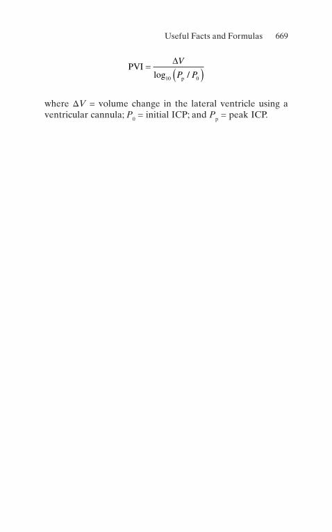
669
PVIp
��
� �V
P Plog /10 0
where ΔV = volume change in the lateral ventricle using a ventricular cannula; P0 = initial ICP; and Pp = peak ICP.
Useful Facts and Formulas

671© Springer Nature Switzerland AG 2021J. Varon, Handbook of Critical and Intensive Care Medicine, https://doi.org/10.1007/978-3-030-68270-5_18
Anaphylaxis
A. Definition. Anaphylaxis is an immediate, generalized, life- threatening reaction resulting from the release of bioac-tive substances from mast cells and basophils. Anaphylaxis can occur in more than one time period. The so-called biphasic anaphylaxis is defined as a recurrence of symp-toms that develops following the “resolution” of the initial anaphylactic reaction. It can occur in up to 20% of ana-phylactic reactions and typically occurs within 8 h after resolution of the initial symptoms. Recurrences up to 72 h later can occur.
B. Etiology. The most common causes of anaphylaxis in med-ical practice are depicted in Table 18.1.
C. Mechanism of Action
1. Immunologic
(a) IgE-dependentActivation of Th2 cells by the causative agent
induces IgE, which binds to the FcεRI receptor on mast cells and/or basophils. The cross-linking of
Chapter 18Allergic and Immunologic Emergencies

672
two or more of the FcεRI receptor initiates an intracellular signaling cascade that leads to degran-ulation of mast cells and basophils, resulting in the release of preformed mediators (i.e. histamine and tryptase). Histamine and tryptase result in allergic symptoms and activate other inflammatory cells that can in turn release additional reaction- inducing
Table 18.1 Common causes of systemic anaphylactic reactions1. Drugs
Antibiotics (i.e., penicillins, fluoroquinolones, cephalosporins, sulfonamides, vancomycin)
Nonsteroidal anti-inflammatory drugs (NSAIDs) Proton pump inhibitors (PPIs)
Local anesthetics (i.e., lidocaine, procaine)
Muscle relaxants
Others (i.e., insulin, protamine)
2. Foods
Nuts and seeds
Fish, shellfish
Milk, eggs
3. Food additives
Aspartame
Monosodium glutamate
4. Diagnostics
Iodinated radiographic materials
5. Insect and snakes (stings and bites)
6. Exercise
7. Others
Latex gloves
Heterologous serum (i.e., tetanus antitoxin)
Chapter 18. Allergic and Immunologic Emergencies

673
mediators (i.e. prostaglandin D2 and cysteinyl leu-kotrienes) which can amplify the allergic reaction. This type of reaction is seen with drugs such as penicillin.
(b) IgE-independentThis type of immunologic anaphylaxis can be
either mediated by IgG antibodies or comple-ment. IgG antibodies bind to FcγRIII receptors ultimately causing degranulation of cells. Similarly, complement components such as C3a, C5a, and C5b-9 can trigger activation and degran-ulation of mast cells and basophils resulting in release of tryptase, histamine, and other media-tors. This can be seen with NSAIDs and radio-graphic materials.
2. Non-immunologic
(a) Result in direct stimulation of mast cell degranulation.
(b) Medications such as quinolones, opioids, vancomy-cin, some radiographic materials, dextrans, and NMBA have been found to directly stimulate mast cells.
D. Clinical Manifestations
1. The onset may vary from individual to individual depending on the sensitivity of the person and the route, quantity, and rate of administration of the allergen.
2. Early signs and symptoms that require a high index of suspicion may include:
(a) Agitation (b) Dizziness (c) Headache (d) Nausea and vomiting (e) Dyspnea (f) Palpitations (g) Confusion
Anaphylaxis

674
3. Cutaneous involvement
(a) Generalized pruritus (b) Flushing (c) Urticaria
4. Upper airway obstruction as a consequence of edema of the larynx and swelling of the tongue and lips (angio-edema). This may result in stridor and suffocation.
5. Respiratory failure (manifestations range from tachy-pnea to apnea) that may be related to the factors men-tioned above as well as bronchoconstriction of the lower airways manifested by wheezing. These patients may also develop acute respiratory distress syndrome (ARDS).
6. Cardiovascular collapse: The pathophysiology is thought to be related to enhanced vascular permeabil-ity, peripheral vasodilation, and intravascular volume depletion. A heart rate increase >20 bpm from baseline and a decrease of mean arterial pressure >20 Torr (mmHg) are characteristic.
7. Dysrhythmias: Both supraventricular and ventricular rhythm disorders have been described in patients with anaphylaxis.
E. Laboratory Findings
1. Do not wait for laboratory data to institute therapy! 2. Patients with anaphylaxis may present with leukocyto-
sis or leukopenia. 3. Thrombocytopenia may appear in severe cases. 4. Elevations in tryptase and histamine can occasionally
be used as a marker for anaphylaxis. Tryptase levels peak about 1–2 hours after symptom onset and normal-ize after 5–6 hours. Histamine has a much shorter half- life, only about 20 minutes, which limits its use in the clinical setting, although its metabolites may be used.
5. Immunoglobulin E (IgE) measurements may not be helpful, because many patients may manifest non-IgE- mediated anaphylaxis.
Chapter 18. Allergic and Immunologic Emergencies
675
F. Management
1. Remove exposure to trigger. 2. ABCs:
Secure the airway, and assist with breathing and cir-culation as with any other patient presenting with a potentially critical illness.
3. Assess mental status and skin often. 4. The drug of choice for patients with acute anaphylaxis
is epinephrine. The dosage is 0.3–0.5 mL of 1:1000 dilu-tion (0.3–0.5 mg) subcutaneously every 10–20 min, intramuscular every 5–10 min, or intravenously as described below. The maximum dose for adults is 0.5 mg and for children is 0.3 mg. Endotracheal administration or intraosseous can be attempted when no other route is available.
5. AntihistaminesTraditionally, H1-receptor antihistamines have been
used, i.e., diphenhydramine (Benadryl) 25–50 mg intra-muscularly (IM), intravenously (IV), or PO q6–8 h. In theory, the combination of H1- and H2-receptor antihis-tamines might be a better chance of preventing further histamine-mediated reactions than H1 blockers alone, i.e., cimetidine (Tagamet) 300 mg IV or PO q6 h.
6. Corticosteroids have an uncertain place in the manage-ment of acute reaction, since there is a 4–6 h latent period before such agents are pharmacologically effective. The current recommended agents are hydro-cortisone (Solu-Cortef) 250 mg IV q6 h or methylpred-nisolone (Solu-Medrol) 50 mg IV q6 h for two to four doses.
7. In cases of severe bronchospasm, the following drugs can be used:
(a) Metaproterenol 0.3 mL (5% solution) in 2.5 mL of saline, inhaled through a nebulizer
(b) Aminophylline loading dose of 6 mg/kg IV over 30 min followed by 0.3–0.9 mg/kg/h
8. In patients with profound hypotension:
Anaphylaxis

676
(a) Adequate IV fluid administration (up to 1 L every 20–30 min as needed).
(b) Epinephrine 1 mL of 1:10.000 or 1:100,000 dilu-tion. It is an error to use the same injection dilution as for intramuscular or subcutaneous (1:1000); it can cause overdose which is presented with ven-tricular dysrhythmias, hypertensive crisis, and pul-monary edema.
(c) Norepinephrine (Levophed): 4 mg in 1 L of D5W at a rate of 2–12 μg (0.5–3 mL)/min.
(d) Glucagon may be particularly useful in patients taking beta-adrenergic blockers. The recommended dose is 1 mg in 1 L of D5W at a rate of 5–15 μg (5–15 mL)/min.
G. Preventive measures for patients at high risk of anaphy-laxis are depicted in Table 18.2.
Stevens–Johnson Syndrome (Erythema Multiforme)
A. Definition. Erythema multiforme (EM) is an erythema-tous maculopapular cutaneous eruption of variable form. When EM grades into a more serious clinical state, the term Stevens–Johnson syndrome (SJS) is used.
B. Etiology. Common causes of EM and SJS are depicted in Table 18.3.
Table 18.2 Anaphylaxis preventive measures for patients at high risk1. Avoid exposure (i.e., trigger foods and medications)
2. Slow administration of suspected agents under medical supervision in adequate facility (i.e., ICU)
3. Optimal management of underlying disorders
4. Short- and long-term desensitization (i.e., penicillin, aspirin)
Chapter 18. Allergic and Immunologic Emergencies
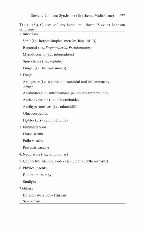
677
Table 18.3 Causes of erythema multiforme/Stevens–Johnson syndrome1. Infections
Viral (i.e., herpes simplex, measles, hepatitis B)
Bacterial (i.e., Streptococcus, Pseudomonas)
Mycobacterial (i.e., tuberculosis)
Spirochetes (i.e., syphilis)
Fungal (i.e., histoplasmosis)
2. Drugs
Analgesics (i.e., aspirin, nonsteroidal anti-inflammatory drugs)
Antibiotics (i.e., sulfonamides, penicillins, tetracycline)
Anticonvulsants (i.e., ethosuximide)
Antihypertensives (i.e., minoxidil)
Glucocorticoids
H2-blockers (i.e., cimetidine)
3. Immunizations
Horse serum
Polio vaccine
Pertussis vaccine
4. Neoplasms (i.e., lymphomas)
5. Connective tissue disorders (i.e., lupus erythematosus)
6. Physical agents
Radiation therapy
Sunlight
7. Others
Inflammatory bowel disease
Sarcoidosis
Stevens–Johnson Syndrome (Erythema Multiforme)

678
C. Clinical Manifestations
1. Prodromal symptoms may include:
(a) Malaise and headache (b) Pharyngitis and rhinorrhea (c) Diarrhea (d) Arthralgias
2. The earliest lesions in EM are often red, edematous papules surrounded by blanching. They enlarge to form small plaques with concentric alterations in color and morphology.
3. The so-called target lesions are areas of central epider-mal necrosis with or without bullae formation.
4. Patients admitted to the ICU with SJS usually present with extensive tissue necrosis and severe fluid depletion.
D. Laboratory Findings
1. Usually nondiagnostic. 2. Skin biopsy reveals a perivascular lymphocytic infil-
trate in the upper dermis, subepidermal bullae forma-tion, and endothelial cell swelling.
E. Management
1. Immediately discontinue suspected drugs or agents as well as all nonessential drugs.
2. The usefulness of systemic corticosteroids in this set-ting is controversial. In the absence of controlled clini-cal trials, some authors recommend beginning therapy with prednisone 1 mg/kg/day (or IV equivalent).
3. Fluid replacement as indicated by severity of the disease.
4. Identify underlying disease and secondary infections and treat them. Antibiotics are only given if an active infection is present. Prophylactic systemic antibiotics are not recommended.
5. Obtain consultation depending on the degree and sites of involvement (i.e., ophthalmology, plastic surgery).
Chapter 18. Allergic and Immunologic Emergencies

679
6. Transfer the patient to a burn unit. 7. Intravenous gamma globulin has been utilized with
conflictive results. 8. Plasmapheresis has been used in some patients with
success.
Angioneurotic Laryngeal Edema
A. Definition. Angioneurotic laryngeal edema (ALE) is characterized by nonpruritic local swelling involving the face, larynx, and skin of the extremities.
B. Etiology
1. AllergyRelated to foods (i.e., fish), drugs (i.e., angiotensin-
converting enzyme [ACE] inhibitors), inhaled sub-stances, and insect stings (i.e., bees)
2. HereditaryCaused by a deficiency in C1-esterase inhibitor
(C1-INH). Autosomal dominant. Precipitating events may include trauma and emotional stress. There are two different types of ALE. Type 1 is caused by a mutant gene, characterized by decreased C1-INH levels as a result of abnormal secretion or intracellular degra-dation. Type 2 is caused by a point mutation which leads to the synthesis of a dysfunctional C1-INH pro-tein. In contrast to type 1, the C1-INH levels in type 2 are found normal or high.
C. Clinical Manifestations
1. Manifestations depend on the location of the edema. 2. Swelling of the face, larynx, and skin of extremities. 3. Depending on the progression, stridor may be a promi-
nent feature with ensuing respiratory distress. 4. Abdominal pain, nausea, and vomiting.
D. Laboratory Findings. Nondiagnostic except in cases of hereditary ALE
Angioneurotic Laryngeal Edema

680
E. Management
1. ABCsSecure the airway, and assist with breathing and cir-
culation as with any other patient presenting with a potentially critical illness.
2. Avoid precipitating allergens. 3. If ALE is thought to be allergic in origin, administer
parenteral epinephrine and antihistamines (as noted in “Anaphylaxis,” above).
4. Intubation is only rarely required for patients with allergic ALE, while in patients with hereditary ALE, the treatment of the acute episode may require urgent intubation or tracheostomy.
5. Other options for treatment for acute attacks include plasma-derived nanofiltered C1-INH 20 U/kg IV, ecal-lantide 30 mg subcutaneous, or icatibant 30 mg subcutaneous.
Chapter 18. Allergic and Immunologic Emergencies

681© Springer Nature Switzerland AG 2021J. Varon, Handbook of Critical and Intensive Care Medicine, https://doi.org/10.1007/978-3-030-68270-5_19
Abbreviations
aPTT Activated partial thromboplastin timeBP Blood pressureIM IntramuscularIV IntravenousNS Normal salinePO By mouthPR By rectumSC SubcutaneousSL Sublingual
• Acetaminophen (Tylenol™):
– Route: PO, PR – Dosage: 325–650 mg q4–6 h (adults), 60 mg/kg/24 h in
divided doses q4–6 h (children) – Daily doses should not exceed 4 g (2 g in chronic alcoholics)
• Acetazolamide (Diamox™): Carbonic anhydrase inhibitor
– Route: PO, IV – Dosage:
Metabolic alkalosis, 250 mg q6–12 hAltitude sickness, 250 mg q6–24 h
Chapter 19Pharmacologic Agents Commonly Used in the ICU

682
• Acetylcysteine (Mucomyst™):
– Route: PO, IV, nebulized – Dosage: For acetaminophen toxicity
PO: Dilute to 5% with cola or other soft drinks. Initial dose is 140 mg/kg and then 70 mg/kg for 17 doses (do not give activated charcoal).IV: Load with 150 mg/kg in 200 mL D5W over 15 min and then 50 mg/kg in 500 mL D5W over 4 h, followed by 100 mg/kg in 100 mL D5W over 16 h.
– Dosage: For contrast-induced nephropathy
600 mg PO; NG q12 h for four doses
• Activated Charcoal (CharcoAid™):
– Route: PO – Dosage: For poisoning
Initial: 30–100 g (1 g/kg) in 250 mL waterMaintenance: 20–40 g q6 h until drug removed from body
• Adenosine (Adenocard™):
– Route: IV – Dosage: 3, 6, 9, 12 mg (fast IV injection)
• Alteplase (Activase™): Tissue plasminogen activator
– Route: IV; t1/2, 5 min; 300–3000 ng/ml in serum initiates systemic lytic state
– Dosage: Acute pulmonary embolism, 100 mg over 2 h. Acute ischemic stroke 0.9 mg/kg. Coronary thromboly-sis: 15 mg IV bolus followed by 0.75 mg/kg over 30 min and 0.5 mg/kg over 1 h
• Amphotericin B (Amphotec™): Macrolide with antifungal activity
– Route: IV. Side effects: Fever – Dosage: 5 mg/kg/day
Chapter 19. Pharmacologic Agents Commonly Used…
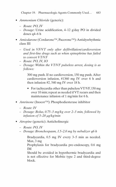
683
• Ammonium Chloride (generic):
– Route: PO, IV – Dosage: Urine acidification, 4–12 g/day PO in divided
doses q4–6 h
• Amiodarone (Cordarone™, Pacerone™): Antidysrhythmic class III
– Used in VF/VT only after defibrillation/cardioversion and first-line drugs such as when epinephrine has failed to convert VT/VF
– Route: PO, IV, IO – Dosage: Within the VT/VF pulseless arrest, dosing is as
follows:
300 mg push. If no cardioversion, 150 mg push. After cardioversion infusion, #1360 mg IV over 6 h and then infusion #2, 540 mg IV over 18 h.
• For tachycardia other than pulseless VT/VF, 150 mg over 10 min; repeat as needed if VT recurs and then maintenance infusion of 1 mg/min for 6 h.
• Amrinone (Inocor™): Phosphodiesterase inhibitor
– Route: IV – Dosage: Bolus, 0.75–3 mg/kg over 2–3 min, followed by
infusion of 5–20 μg/kg/min
• Atropine (generic): Anticholinergic
– Route: PO, IV – Dosage: Bronchospasm, 1.5–2.0 mg by nebulizer q6 h
Bradycardia, 0.5 mg IV every 3–5 min as needed. Max, 3 mgProphylaxis for bradycardia pre-endoscopy, 0.6 mg IMShould be avoided in hypothermic bradycardia and is not effective for Mobitz type 2 and third-degree block.
Chapter 19. Pharmacologic Agents Commonly Used…

684
• Bivalirudin (Angiomax™): Thrombin inhibitor
– Route: IV – Dosage:
Acute coronary syndromes undergoing PTCA/PCI 0.1 mg/kg, followed by 0.25 mg/kg/h.It can be used in patients with history of heparin- induced thrombocytopenia.
• Bretylium (Bretylol™):
– Route: IV, IM – Dosage: Bolus, 5–10 mg/kg over 10–20 min IV, followed
by a continuous infusion of 1–5 mg/min
• Carbicarb (Carbicarb™):
– Route: IV – Dosage: Severe acidosis; initial dose, 1 mEq/kg, followed
by 0.5 mEq/kg (adjust dose as indicated by clinical con-dition and blood pH)
• Ceftaroline (Teflaro™): Antibiotic with extended spec-trum to cover MRSA
– Route: IV, IM – Dosage: 600 mg IV q 12 h. In renal impairment, adjust to
lower dose (i.e., 400 mg IV q 12 h)
• Chlordiazepoxide (Librium™): Benzodiazepine
– Route: PO, IV, IM – Dosage:
15–100 mg/day in three to four divided dosesUsed for anxiety disorders, narcotic withdrawal, and anesthetic premedication
• Chlorpromazine (Thorazine™):
– Route:
PO, PR, IMUsed in patients with severe psychosis with agitation
Chapter 19. Pharmacologic Agents Commonly Used…

685
– Dosage: Severe psychosis with agitation, 25–100 mg IM q1–4 h until control is achieved
• Cisatracurium (Nimbex™):
– Route: IV – Dosage: Bolus, 0.1 mg/kg; infusion, 1–3 mcg/kg/min
• Clevidipine (Cleviprex™): Dihydropyridine L-type, cal-cium channel blocker
– Route: IV – Dosage: Hypertensive crisis, postoperative hypertension
when the oral route is not feasible 1–2 mg/h as start dose and double every 3 min to a maximum of 32 mg/h
– Rapid onset and offset of action t1/2, 2 min – Infusions, 1–2 μg/kg/min and titrated to the desired effect
on BP
• Clonidine (Catapres™): Alpha-adrenergic agonist
– Route: PO, transdermal (in some countries, IM and PR are used).
– Dosage: Hypertensive emergencies and urgencies, 0.2 mg PO and then 0.1 mg/q20 min to 0.8 mg or until BP is controlled.
– Adverse effects. Postural hypotension, worsening of con-gestive heart failure, and bradycardia; sudden discontin-uation may cause withdrawal syndrome.
– Maximal hypotensive effect 1–3 h after PO.
• Dalteparin (Fragmin™): Low molecular weight heparin
– Route: Subcutaneous – Dosage: DVT prophylaxis, 2500–5000 units subcutane-
ous once a day. DVT treatment: 200 units/kg
• Dantrolene (Dantrium™): Inhibits Ca2+ ion release from sarcoplasmic stores by antagonizing ryanodine receptors
– Route: PO, IV – Dosage: Malignant hyperthermia; initial dose, 1–2 mg/kg
IV via rapid infusion; may repeat to total 10 mg/kg, if needed and then 4–8 mg/kg/day
Chapter 19. Pharmacologic Agents Commonly Used…

686
• Daptomycin (Cubicin™): Lipopeptide derived from Streptomyces roseosporus
– Selectively active against aerobic, facultative, and anaero-bic gram-positive bacteria
– Route: IV only; poorly absorbed in PO – Dosage: 4 mg/kg once a day on complicated skin and
soft tissue infections; 6 mg/kg/day in complicated bacte-remia and right-sided endocarditis
– Adverse effects: rhabdomyolysis, neuropathy, and CK elevation
• dDAVP (generic): Synthetic analogue of ADH
– Route: Intranasal, IV, SC – Dosage: Hemostasis, 0.3 μg/kg in NS over 15–30 min – Diabetes insipidus, 0.5–1 mL IV/SC bid
• Diazepam (Valium™): Benzodiazepine
– Route: PO, IV, IM, IR. – Dosage: Status epilepticus, 5–10 mg IV (1–2 mg/min) – t½: Over 24 h. Adverse effects: sedation and respiratory
depression
• Diazoxide (Hyperstat™):
– Route: PO, IV – Dosage: Hypertensive crisis, 1–3 mg/kg IV (max 150 mg)
q5–15 min until BP is controlled. High risk for marked falls in BP
• Digoxin (Lanoxin™):
– Route: PO, IV, IM – Dosage: Digitalization, 0.4–0.6 mg IV (may require up to
1.25 mg total); maintenance, 0.125–0.25 mg/d PO or IV – Adverse effects: Dysrhythmias, nausea, disturbances in
cognitive function, blurred yellow vision, atrial tachycar-dia with AV block, PR prolongation, and ST depression
• Dobutamine (Dobutrex™):
– Route: IV – Dosage: 2–3 μg/kg/min
Chapter 19. Pharmacologic Agents Commonly Used…

687
– Used in CHF with systolic dysfunction. Pharmacologic tolerance may limit infusion efficacy beyond 4 days, and therefore change to type III PDE inhibitor may be necessary.
• Dopamine (Intropin™):
– Route: IV – Dosage: Dopaminergic stimulation, 0.5–2.0 μg/kg/min – Alpha- and beta-dopaminergic effects, >10 μg/kg/min
• Drotrecogin alpha-activated protein C (Xigris™): Inhibits coagulation by proteolytic inactivation of factors Va and VIIIa. Decreases mortality in patient with high risk of death from severe sepsis within 48 h since the onset of organ dysfunction. Currently off the market as recent data suggest increased mortality on those patients receiving this agent
– Route: IV – Dosage: 24 mcg/kg/h for a total of 96 h
• Enoxaparin (Lovenox™):
– Route: Subcutaneous, IV – Dosing: DVT prophylaxis, 40 mg daily; acute DVT treat-
ment, 1 mg/kg (IBW)/q12 h or 1.5 mg/kg/daily – t½: 4 h; relatively contraindicated in patients with impaired
renal excretion. Must adjust dose in such patients – Thrombocytopenia is rare.
• Epinephrine (Epinephrine Injection):
– Route: IV, SC – Dosage:
Beta1 and beta2 effect, 1–4 μg/minAlpha-effect, 4 μg/min
• Epoprostenol (Flolan™)
– Route: IV; can be inhaled – Dosage: 1–2 ng/kg/min
Chapter 19. Pharmacologic Agents Commonly Used…

688
• Ertapenem (Invanz™):
– Route: IV, IM – Dosage: 1 g daily
• Esmolol (Brevibloc™):
– Route: IV – Dosage: Bolus 0.5–1.0 mg/kg, followed by infusion at
50 μg/kg/min; maintenance 50–200 μg/kg/min
• Etomidate (Amidate™):
– Route: IV – Dosage: Intubation: 0.2–0.6 mg/kg
• Fenoldopam (Corlopam™):
– Route: IV – Dosage: 0.1–1.0 μg/kg/min, titrate to achieve desired
blood pressure
• Fentanyl (Sublimaze™):
– Route: IV, IM – Dosage: Sedation/analgesia, 1 μg/kg IV/IM
• Flumazenil (Romazicon™):
– Route: IV – Dosage: 0.3 mg IV
• Furosemide (Lasix™):
– Route: PO, IV, IM – Dosage: 10–120 mg IV/IM, adjusted as necessary until
desired response obtained. May use continuous infusions
• Fosphenytoin (Cerebyx™):
– Route: IV, IM – Dosage: Status epilepticus 15–20 mg/kg (loading dose)
Chapter 19. Pharmacologic Agents Commonly Used…

689
• Glucagon (generic):
– Route: SC, IM, IV – Dosage: Hypoglycemia, 0.5–1.0 mg SC/IM/IV, may
repeat in 15 min – Bradycardia, 1–20 mg/h
• Haloperidol (Haldol™):
– Route: PO, IM, IV (not FDA approved) – Dosage: Acute psychosis, 2–5 mg IM q1–2 h until symp-
toms are controlled
• Heparin (Liquaemin™):
– Route: IV, SC – Dosage: DVT prophylaxis, 5000 U SC q8–12 h – DVT/pulmonary emboli therapy, bolus with 100 U/kg fol-
lowed by a continuous infusion of 800–1200 U/h, titrated to maintain aPTT of 11/2–2 times the control. t½: 1 h. Therapeutic range: 0.3–0.7 in plasma units/ml. aPTT of two to three times the normal value is assumed to be normal.
• Hydralazine (Apresoline™):
– Route: PO, IV – Dosage: 5 mg IV bolus; 5–10 mg IV q6 h maintenance
• Hydromorphone (Dilaudid™)
– Route: IV, PO, IM, SC – Dosage: 0.7–2 mg every 1–2 h as needed for pain
• Imipenem and Cilastatin (Primaxin™):
– Route: IV, IM – Dosage: 500–1000 mg every 6 h. Adjust based on renal
function
• Isoproterenol (Isuprel™):
– Route: IV, SC, PO, inhaled – Dosage: Infusion 1–10 μg/min
Chapter 19. Pharmacologic Agents Commonly Used…

690
• Ketorolac Tromethamine (Toradol™):
– Route: PO, IM – Dosage: Initial dose 30–60 mg IM, then 15–30 mg q6 h
• Labetalol (Normodyne™):
– Route: PO, IV – Dosage: Rapid BP control, IV bolus 5–20 mg (slowly);
repeat after 5 min if needed. Continuous infusion of 1 mg/mL started at 1–2 mg/min and titrated to effect.
• Lepirudin (Refludan™):
– Route: IV – Dosage: Bolus 0.4 mg/kg followed by continuous infu-
sion at 0.15 mg/kg/h with a maximum dose of 44 mg as a bolus and 16.5 mg/h as infusion
• Lidocaine (Xylocaine™):
– Route: IV, IM, SC – Dosage: Bolus 1.0–1.5 mg/kg followed by 1–4 mg/min
• Linezolid (Zyvox):
– Route: PO, IV – Dosage: 600 mg every 12 h
• Lorazepam (Ativan™): Benzodiazepine
– Route: PO, IV, IM – Dosage: 2–10 mg/d in divided doses PO/IV/IM (some
patients may require continuous infusions)
• Mannitol (Osmitrol™):
– Route: IV. – Dosage: For cerebral edema, 0.15–2.0 g/kg IV infusion as
15–25% solution over 30–60 min; max dose is up to 6 g/kg/24 h.
Chapter 19. Pharmacologic Agents Commonly Used…

691
• Meperidine (Demerol™): Narcotic analgesic
– Route: PO, IM, IV, SC – Dosage: 50–150 mg q3–4 h
• Meropenem (Merrem™):
– Route: IV – Dosage: 1–2 g every 8 h
• Midazolam (Versed™): Short-acting benzodiazepine
– Route: IV, IM – Dosage: 1–4 mg q2–6 h
• Morphine (Duramorph™):
– Route: IM, IV, PO, PR – Dosage: 5–10 mg q4–6 h (some patients may require
continuous infusions)
• Naloxone (Narcan™):
– Route: IV, IM, SC – Dosage: 0.4–2.0 mg IV; may repeat up to 10 mg.
Continuous IV infusion at 4–5 μg/kg/min
• Nicardipine (Cardene™):
– Route: IV, PO – Dosage: Hypertensive emergencies 5 mg/h/IV increase
by 2.5 mg/h every 5–15 min to achieve desired blood pressure. The manufacturer recommended a maximum dose of 15 mg/h. In the author’s experience, doses much higher than this can be safely given.
• Nitroglycerin (Nitroglycerin):
– Route: PO, IV, SL, topical – Dosage: 10–400 μg/min IV
Chapter 19. Pharmacologic Agents Commonly Used…
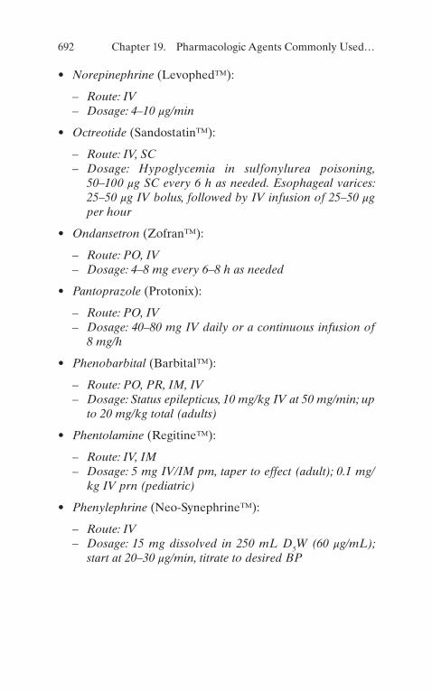
692
• Norepinephrine (Levophed™):
– Route: IV – Dosage: 4–10 μg/min
• Octreotide (Sandostatin™):
– Route: IV, SC – Dosage: Hypoglycemia in sulfonylurea poisoning,
50–100 μg SC every 6 h as needed. Esophageal varices: 25–50 μg IV bolus, followed by IV infusion of 25–50 μg per hour
• Ondansetron (Zofran™):
– Route: PO, IV – Dosage: 4–8 mg every 6–8 h as needed
• Pantoprazole (Protonix):
– Route: PO, IV – Dosage: 40–80 mg IV daily or a continuous infusion of
8 mg/h
• Phenobarbital (Barbital™):
– Route: PO, PR, IM, IV – Dosage: Status epilepticus, 10 mg/kg IV at 50 mg/min; up
to 20 mg/kg total (adults)
• Phentolamine (Regitine™):
– Route: IV, IM – Dosage: 5 mg IV/IM pm, taper to effect (adult); 0.1 mg/
kg IV prn (pediatric)
• Phenylephrine (Neo-Synephrine™):
– Route: IV – Dosage: 15 mg dissolved in 250 mL D5W (60 μg/mL);
start at 20–30 μg/min, titrate to desired BP
Chapter 19. Pharmacologic Agents Commonly Used…

693
• Piperacillin and Tazobactam (Zosyn™):
– Route: IV – Dosage: 2.25–4.5 g IV every 6–8 h
• Procainamide (Procainamide):
– Route: PO, IV – Dosage: 100 mg/min IV to effect or to a total dose of
1000 mg, followed by infusion 2–6 mg/min
• Prochlorperazine (Compazine™):
– Route: PO, IM, IV, PR – Dosage: 5–10 mg PO tid/qid; 5–10 mg IV q3–4 h; 25 mg
PR bid
• Propofol (Diprivan):
– Route: IV – Dosage: 2–15 cc/h by continuous infusion to achieve the
desired level of sedation
• Propranolol (Inderal™):
– Route: PO, IV – Dosage: Titrate 0.5–1.0 mg IV q5 min to effect.
• Protamine (Protamine sulfate™):
– Route: IV – Dosage: 1 mg for each 90 U of lung heparin or 1 mg for
each 115 U of intestinal heparin, by slow injection over 1–3 min; max dose, 50 mg in 10 min
• Rocuronium (Zemuron™):
– Route: IV – Dosage: Intubation 0.6 mg/kg
Chapter 19. Pharmacologic Agents Commonly Used…

694
• Sodium Bicarbonate (Sodium Bicarbonate Injection):
– Route: IV, PO – Dosage: Severe acidosis, initial dose 1 mEq/kg, followed
by 0.5 mEq/kg; adjust dosage as indicated by clinical condition and blood pH.
• Sodium Nitroprusside (Nipride™):
– Route: IV – Dosage: Mix 50 mg in 250 mL D5W. Start at 0.5 μg/kg/
min and titrate to effect. – Thiocyanide and cyanide poisoning common in infu-
sions lasting more than 8 h
• Sodium Polystyrene Sulfonate (Kayexalate™):
– Route: PO, PR – Dosage: 15 g, PO q6–24 h
• Succinylcholine (Anectine™):
– Route: IV, IM – Dosage: 1–1.5 mg/kg IV; 2–4 mg/kg IM (pediatric use
only)
• Thiopental (Pentothal sodium™):
– Route: IV, PR – Dosage: General anesthetic, 2–3 mL 2.5% solution (50–
75 mg) IV q20–40 s until desired effect reached – Seizures, 75–125 mg IV
• Tigecycline (Tygacil™):
– Route: IV – Dosage: initial dose 100 mg and then 50 mg every 12 h
• Trimethaphan (Arfonad):
– Route: IV – Dosage: Start at 0.3 mg/min and titrate to effect.
Chapter 19. Pharmacologic Agents Commonly Used…

695
• Valproic Acid (Depakote™):
– Route: PO, IV – Dosage: Status epilepticus 15–45 mg/kg (loading dose),
infusion 1–4 mg/kg/h
• Vancomycin (Vancocin™):
– Route: PO, IV – Dosage: 500–1000 mg per day in divided doses PO.
2–3 g per day in divided doses IV
• Vasopressin (Pitressin™):
– Route: IM, IV, SC, IO – Dosage: Vasodilatory or septic shock 1–6 units/h
Chapter 19. Pharmacologic Agents Commonly Used…

697© Springer Nature Switzerland AG 2021J. Varon, Handbook of Critical and Intensive Care Medicine, https://doi.org/10.1007/978-3-030-68270-5_20
The most common laboratory values used in the assessment of critically ill patients are presented in this chapter. They have been organized in alphabetical order and according to biologic source where (P) represents plasma, (B) blood, (S) serum, (U) urine, (CSF) cerebrospinal fluid, (RBCs) red blood cells, and (WBCs) white blood cells. These values are not intended to be definitive, since normal ranges vary from hospital to hospital. Both traditional units and system inter-national (SI) units are presented.
• α1-Antitrypsin (S)
– 150–350 mg/dL (dual report) (SI, 1.5–3.5 g/L)
• 17-Ketogenic steroids (as dehydroepiandrosterone) (U)
– Female: 7–12 mg/24 h (SI, 25–40 μmol/day) – Male: 9–17 mg/24 h (SI, 30–60 μmol/day)
• 17-Ketosteroids (as dehydroepiandrosterone) (U)
– Female: 6–17 mg/24 h (SI, 20–60 μmol/day) – Male: 6–20 mg/24 h (SI, 20–70 μmol/day)
• Alanine aminotransferase (ALT) (S)
– 0–35 (35 °C) U/L (SI, 0–35 U/L)
• Albumin (S)
– 4.0–6.0 g/dL (SI, 40–60 g/L)
Chapter 20Common Laboratory Values in the ICU
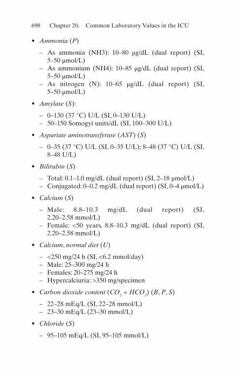
698
• Ammonia (P)
– As ammonia (NH3): 10–80 μg/dL (dual report) (SI, 5–50 μmol/L)
– As ammonium (NH4): 10–85 μg/dL (dual report) (SI, 5–50 μmol/L)
– As nitrogen (N): 10–65 μg/dL (dual report) (SI, 5–50 μmol/L)
• Amylase (S):
– 0–130 (37 °C) U/L (SI, 0–130 U/L) – 50–150 Somogyi units/dL (SI, 100–300 U/L)
• Aspartate aminotransferase (AST) (S)
– 0–35 (37 °C) U/L (SI, 0–35 U/L); 8–48 (37 °C) U/L (SI, 8–48 U/L)
• Bilirubin (S)
– Total: 0.1–1.0 mg/dL (dual report) (SI, 2–18 μmol/L) – Conjugated: 0–0.2 mg/dL (dual report) (SI, 0–4 μmol/L)
• Calcium (S)
– Male: 8.8–10.3 mg/dL (dual report) (SI, 2.20–2.58 mmol/L)
– Female: <50 years, 8.8–10.3 mg/dL (dual report) (SI, 2.20–2.58 mmol/L)
• Calcium, normal diet (U)
– <250 mg/24 h (SI, <6.2 mmol/day) – Male: 25–300 mg/24 h – Females: 20–275 mg/24 h – Hypercalciuria: >350 mg/specimen
• Carbon dioxide content (CO2 + HCO3) (B, P, S)
– 22–28 mEq/L (SI, 22–28 mmol/L) – 23–30 mEq/L (23–30 mmol/L)
• Chloride (S)
– 95–105 mEq/L (SI, 95–105 mmol/L)
Chapter 20. Common Laboratory Values in the ICU

699
• Cholesterol esters, as a fraction of total cholesterol (P)
– 60–75% (SI, 0.60–0.75)
• Complement, C3 (S)
– 70–160 mg/dL (SI, 0.7–1.6 g/L)
• Copper (S)
– 70–140 μg/dL (SI, 11.0–22.0 μmol/L)
• Cholesterol (P)
– <200 mg/dL (dual report) (SI, <5.20 mmol/L)
• Copper (U)
– <40 μg/24 h (SI, <0.6 μmol/day)
• Corticotropin (ACTH) (P)
– 20–100 pg/mL (SI, 4–22 pmol/L)
• Creatine kinase (CK) (S)
– 0–130 (37 °C) U/L (SI, 0–130 U/L)
• Creatine kinase isoenzymes, MB fraction (S)
– >5% in myocardial infarction (SI, >0.05)
• Creatine (U)
– Male: 0–40 mg/24 h (SI, 0–300 μmol/day) – Female: 0–80 mg/24 h (SI, 0–600 μmol/day)
• Creatine (S)
– Male: 0.17–0.50 mg/dL (SI, 10–40 μmol/L) – Female: 0.35–0.93 mg/dL (SI, 30–70 μmol/L)
• Creatinine (U)
– Variable g/24 h (dual report) (SI, variable mmol/day)
• Creatinine (S)
– 0.6–1.2 mg/dL (dual report) (SI, 50–110 μmol/L)
Chapter 20. Common Laboratory Values in the ICU

700
• Creatinine clearance (S, U)
– 75–125 mL/min (dual report) (SI, 1.24–2.08 mL/s)
• Cystine (U)
– 10–100 mg/24 h (SI, 40–420 μmol/day)
• Dehydroepiandrosterone (U)
– Female: 0.2–1.8 mg/24 h (SI, 1–6 μmol/day) – Male: 0.2–2.0 mg/24 h (SI, 1–7 μmol/day)
• Digoxin, therapeutic (P)
– 0.5–2.2 ng/mL (dual report) (SI, 0.6–2.8 mmol/L)
• Erythrocyte sedimentation rate (B)
– Female: 0–30 mm/h (SI, 0–30 mm/h) – Male: 0–20 mm/h (SI, 0–20 mm/h)
• Estradiol: male, >18 years (S)
– 15–40 pg/mL (dual report) (SI, 55–150 pmol/L)
• Ethyl alcohol (P)
– <100 mg/dL (SI, <22 mmol/L) – <80 mg/dL (0.08 g/dL)
• Etiocholanolone
– Female: 0.8–4.0 mg/24 h (SI, 2–14 μmol/day) – Male: 1.4–5.0 mg/14 h (SI, 4–17 μmol/day)
• Fibrinogen (P)
– 200–4300 mg/dL (SI, 2.0–4.0 g/L)
• Follicle-stimulating hormone (FSH) (P)
– Female: 2.0–15.0 mIU/mL (SI, 2–15 IU/L) – Peak production: 20–50 mIU/mL (SI, 20–50 IU/L) – Male: 1.0–10.0 mIU/mL (SI, 1–10 IU/L)
• Follicle-stimulating hormone (FSH) (U)
– Follicular phase: 2–15 IU/24 h (SI, 2–15 IU/day)
Chapter 20. Common Laboratory Values in the ICU
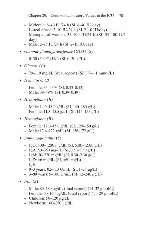
701
– Midcycle: 8–40 IU/24 h (SI, 8–40 IU/day) – Luteal phase: 2–10 IU/24 h (SI, 2–10 IU/day) – Menopausal women: 35–100 IU/24 h (SI, 35–100 IU/
day) – Male: 2–15 IU/24 h (SI, 2–15 IU/day)
• Gamma-glutamyltransferase (GGT) (S)
– 0–30 (30 °C) U/L (SI, 0–30 U/L)
• Glucose (P)
– 70–110 mg/dL (dual report) (SI, 3.9–6.1 mmol/L)
• Hematocrit (B)
– Female: 33–43% (SI, 0.33–0.43) – Male: 39–49% (SI, 0.39–0.49)
• Hemoglobin (B)
– Male: 14.0–18.0 g/dL (SI, 140–180 g/L) – Female: 11.5–15.5 g/dL (SI, 115–155 g/L)
• Hemoglobin (B)
– Female: 12.0–15.0 g/dL (SI, 120–150 g/L) – Male: 13.6–17.2 g/dL (SI, 136–172 g/L)
• Immunoglobulins (S)
– IgG: 500–1200 mg/dL (SI, 5.00–12.00 g/L) – IgA: 50–350 mg/dL (SI, 0.50–3.50 g/L) – IgM: 30–230 mg/dL (SI, 0.30–2.30 g/L) – IgD: <6 mg/dL (SI, <60 mg/L) – IgE: – 0–3 years: 0.5–1.0 U/mL (SI, 1–24 μg/L) – 3–80 years: 5–100 U/mL (SI, 12–240 μg/L)
• Iron (S)
– Male: 80–180 μg/dL (dual report) (14–32 μmol/L) – Female: 60–160 μg/dL (dual report) (11–29 μmol/L) – Children: 50–120 μg/dL – Newborn: 100–250 μg/dL
Chapter 20. Common Laboratory Values in the ICU

702
• Iron-binding capacity (S)
– 250–460 μg/dL (dual report) (SI, 45–82 μmol/L)
• Ketosteroid fractions (U)
– Androsterone: – Female: 240–2300 μg/24 h – Male: 320–5400 μg/24 h – DHEA: – Female: 21–2170 μg/24 h – Male: 21–2170 μg/24 h – Etiocholanolone – Female: 195–1500 μg/24 h – Male: 130–900 μg/24 h – 11 Beta-Hydroxyetiocholanolone – Adults: 14–680 μg/24 h – 11-Oxo-androsterone – Female: 8–87 μg/24 h – Male: 15–111 μg/24 h – 11-Oxo-etiocholanolone – Adults: 78–1165 μg/24 h – Pregnanetriol – Female: 47–790 μg/24 h – Male: 71–1000 μg/24 h
• Lactate dehydrogenase (S)
– 50–150 (37 °C) U/L (SI, 50–150 U/L)
• Lactate dehydrogenase isoenzymes (S)
– LD1: 15–40% (SI, 0.15–0.40) – LD2: 20–45% (SI, 0.20–0.45) – LD3: 15–30% (SI, 0.15–0.30) – LD4 and LD5: 5–20% (SI, 0.05–0.20) – LD1: 10–60 U/L (SI, 10–60 U/L) – LD2: 20–70 U/L (SI, 20–70 U/L) – LD3: 10–45 U/L (SI, 10–45 U/L) – LD4 and LD5: 5–30 U/L (SI, 5–30 U/L)
• Lead, toxic (B)
Chapter 20. Common Laboratory Values in the ICU
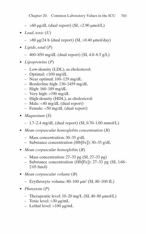
703
– >60 μg/dL (dual report) (SI, >2.90 μmol/L)
• Lead, toxic (U)
– >80 μg/24 h (dual report) (SI, >0.40 μmol/day)
• Lipids, total (P)
– 400–850 mg/dL (dual report) (SI, 4.0–8.5 g/L)
• Lipoproteins (P)
– Low-density (LDL), as cholesterol: – Optimal: <100 mg/dL – Near optimal: 100–129 mg/dL – Borderline high: 130–1459 mg/dL – High: 160–189 mg/dL – Very high: >190 mg/dL – High-density (HDL), as cholesterol: – Male: <40 mg/dL (dual report) – Female: <50 mg/dL (dual report)
• Magnesium (S)
– 1.7–2.4 mg/dL (dual report) (SI, 0.70–1.00 mmol/L)
• Mean corpuscular hemoglobin concentration (B)
– Mass concentration: 30–35 g/dL – Substance concentration (Hb[Fe]): 30–35 g/dL
• Mean corpuscular hemoglobin (B)
– Mass concentration: 27–33 pg (SI, 27–33 pg) – Substance concentration (Hb[Fe]): 27–33 pg (SI, 1.68–
2.05 fmol)
• Mean corpuscular volume (B)
– Erythrocyte volume: 80–100 μm3 (SI, 80–100 fL)
• Phenytoin (P)
– Therapeutic level: 10–20 mg/L (SI, 40–80 μmol/L) – Toxic level: >30 μg/mL – Lethal level: >100 μg/mL
Chapter 20. Common Laboratory Values in the ICU
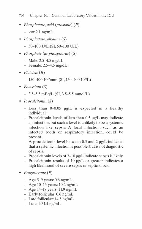
704
• Phosphatase, acid (prostatic) (P)
– <or 2.1 ng/mL
• Phosphatase, alkaline (S)
– 50–100 U/L (SI, 50–100 U/L)
• Phosphate (as phosphorus) (S)
– Male: 2.5–4.5 mg/dL – Female: 2.5–4.5 mg/dL
• Platelets (B)
– 150–400 103/mm3 (SI, 150–400 109/L)
• Potassium (S)
– 3.5–5.5 mEq/L (SI, 3.5–5.5 mmol/L)
• Procalcitonin (S)
– Less than 0–0.05 μg/L is expected in a healthy individual.
– Procalcitonin levels of less than 0.5 μg/L may indicate an infection, but such a level is unlikely to be a systemic infection like sepsis. A local infection, such as an infected tooth or respiratory infection, could be present.
– A procalcitonin level between 0.5 and 2 μg/L indicates that a systemic infection is possible, but is not diagnostic of sepsis.
– Procalcitonin levels of 2–10 μg/L indicate sepsis is likely. – Procalcitonin results of 10 μg/L or greater indicates a
high likelihood of severe sepsis or septic shock.
• Progesterone (P)
– Age 5–9 years: 0.6 ng/mL – Age 10–13 years: 10.2 ng/mL – Age 14–17 years: 11.9 ng/mL – Early follicular: 0.6 ng/mL – Late follicular: 14.5 ng/mL – Luteal: 31.4 ng/mL
Chapter 20. Common Laboratory Values in the ICU

705
– Midcycle: 16.1 ng/mL – Postmenopausal: 0.2 ng/mL
• Protein, total (U)
– <150 mg/24 h (SI, <0.15 g/day)
• Protein, total (S)
– 6–8 g/dL (SI, 60–80 g/L)
• Protein, total (CSF)
– <40 mg/dL (SI, <0.40 g/L)
• Red blood cell count (erythrocytes) (B)
– Female: 4.10–5.101012/L – Male: 4.52–5.901012/L
• Red blood cell count (CSF)
– 0/cu mm (SI, 0 106/L)
• Reticulocyte count (adult) (B)
– 0.5–1.5%
• Sodium (S)
– 135–145 mEq/L (SI, 135–145 mmol/L)
• Sodium ion (S)
– 135–145 mEq/L (SI, 135–145 mmol/L)
• Sodium ion (U)
– Diet-dependent mEq/24 h (SI, 5–25 mmol/day)
• Steroids (U)
– Hydroxycorticosteroids (as cortisol) – 3–12 mg/24 h
• Testosterone (P)
– Female: <0.6 ng/mL (dual report) (SI, <2.0 nmol/L)
Chapter 20. Common Laboratory Values in the ICU

706
– Male: 4.0–8.0 ng/mL (dual report) (SI, 14.0–28.0 nmol/L)
• Thyroxine (T4) (S)
– 4.9–11.7 mg/dL
• Thyroxine-binding globulin (TBG) (S)
– 12–30 mg/L
• Thyroxine, free (S)
– 0.7–1.8 ng/mL
• Triiodothyronine (T3) (S)
– 80–220 ng/dL
• Triglycerides (S)
– Normal: <150 mg/dL – Borderline high: 150–199 mg/dL – High: 200–499 mg/dL – Very high: >500 mg/dL
• Urate (as uric acid) (S)
– Male: 2.5–8 mg/dL
• Female: 1.9–7.5 mg/dL• Urate (as uric acid) (U)
– Diet-dependent g/24 h (SI, diet-dependent mmol/d)
• Urea nitrogen (S)
– 3–20 mg/dL (dual report)
• Urea nitrogen (U)
– 12–20 g/24 h (dual report) (SI, 430–700 mmol/d of urea)
• Urobilinogen (U)
– 0–4.0 mg/24 h (SI, 0.0–6.8 μmol/day)
• White blood cell count (B)
– 40–10 × 109/L
Chapter 20. Common Laboratory Values in the ICU

707
• White blood cell count (CSF)
– 0–5/cu mm (SI, 0–5 106/L)
• Zinc (S)
– 75–120 μg/dL (SI, 11.5–18.5 μmol/L)
• Zinc (U)
– 150–1200 μg/24 h (SI, 2.3–18.3 μmol/day)
20 Common Laboratory Values in the ICU

709© Springer Nature Switzerland AG 2021J. Varon, Handbook of Critical and Intensive Care Medicine, https://doi.org/10.1007/978-3-030-68270-5
Index
AAbbreviated injury scale (AIS),
664Abciximab, 84Abdominal trauma, 653–656ACE inhibitors, 85, 102Acetaminophen, 605, 606, 681Acetazolamide, 681Acetylcysteine, 682Acid–base disturbance, 511–514,
516, 517, 519, 520, 522Acid–base equations/facts, 558,
560Acquired hemolytic disorders,
286Activated charcoal, 682Acute chest syndrome, 287Acute CO poisoning, 214Acute coronary syndrome, 79Acute fulminant hepatic failure
(FHF), 243Acute GI bleeding, 236Acute hyponatremia, 153Acute liver failure, causes of, 244Acute meningitis, 311–314Acute mesenteric ischemia
(AMI)definition, 241epidemiology, 241etiology, 241
history and physical examination, 242
laboratory studies, 242radiologic evaluation,
242, 243risk factors, 242therapy, 243
Acute myocardial infarction (AMI), 107, 276
Acute pancreatitiscomplications, 252definition of, 248grades of, 251
Acute renal failure (ARF), 524–526, 528, 530, 531
Acute respiratory failure, 485–487
Acute tubular necrosis (ATN), 569
Adenosine, 123, 682Adrenal insufficiency
associated laboratory abnormalities, 142, 143
clinical manifestation, 142complications, 145definition of, 141diagnostic eveluation, 143, 144etiology of, 142pathophysiology of, 141, 142therapy, 144

710
Adult respiratory distress syndrome (ARDS), 481–485
AIDS, 317CNS infections, 320, 321enteric pathogens, 320, 321opportunistic infections, 317,
318pulmonary disease, 317, 318,
320treating HIV-Infected
Patients in the ICU, 322Air embolism, 195
clinical presentation, 197complications, 197pathophysiology, 195predisposition, 195treatment, 197, 198
Airway management, 573–575, 577, 578, 580
Airway pressure release ventilation (APRV), 54
Alanine aminotransferase (ALT), 697
Albumin, 378, 388, 697Alcohol, 607–610Alpha-adrenergic blockade, 186Alpha-methylparatyrosine, 187Alteplase, 682Altitude, 231Alveolar air equation, 28, 29,
32–35Aminoglycoside clearance, 330Amiodarone, 683Ammonia, 698Ammonium Chloride, 683Amniotic fluid embolism (AFE),
454–458Amphotericin B, 682Amrinone, 683Amylase, 249, 698Anaphylaxis, 671
clinical manifestations, 673, 674
common causes, 672laboratory findings, 674
management, 675, 676mechanism, 672, 673
Ancillary tests, for brain death, 335, 336
Anemiadefinition of, 257diagnostic evaluation,
257–260etiology, 257therapy
for patient acutely bleeding, 260
for patient not acutely bleeding, 261
Aneurysms, see Aortic dissectionAngioneurotic laryngeal edema
(ALE), 679, 680Angiotensin-converting enzyme
(ACE) inhibitors, 91, 611
Anistreplase anisoylated, 90Antibiotic adjustments, 330Antibiotic kinetics, 329Antibiotic levels, 331Anticoagulants, 83Anticoagulation, 125, 478
DTI, 274Factor Xa (10 subunit a)
inhibitors, 275, 276warfarin, 272, 273
Anticoagulation heparin, 269–272
Antidiuretic hormone (ADH), 546
Antihypertensive drugs, 448–449Antimicrobials, 326–329Antineoplastic therapy, 393, 415Antipyretic medications, 207Antivenin, 223Aortic dissection, 115, 117Aortic insufficiency, 111, 112Aortic stenosis, 109
diagnostic studies, 110etiology, 109pathophysiology, 110physical findings, 110
Index

711
symptoms, 110therapy, 111
Apnea test, 335Arterial blood gases (ABGs),
466, 512Arterial hypoxemia, 72Arterial line, 589, 590Arterial–jugular venous oxygen
content difference (AjvDO2), 362
Arteriodilators (afterload reduction), 102
Arteriovenous Oxygen Content Difference (avDO 2 ), 132
Artifactual hypoglycemia, 180Aspartate aminotransferase
(AST), 698Aspirin, 83Assist/controlled mechanical
ventilation (A/C), 50Asthma–chronic obstructive
pulmonary disease overlap syndrome (ACOS), 469
Atrial fibrillation, 124, 125Atrial flutter, 126Atrioventricular (AV)
Conduction Disturbances, 96
Atrioventricular block, 97Atypical mycobacteria, 331Autoimmune hemolytic anemia
glucose-6-phosphate dehydrogenase deficiency, 289, 290
microangiopathic hemolytic anemia, 289
warm-antibody autoimmune hemolytic anemia, 288
AV conduction blocks, 127Average Volume Assured
Pressure Support (AVAPS), 56
Avian influenza, 303
BBacterial meningitis, 314, 316Balloon Tamponade, 240Barbiturates, 342Barotrauma, 492, 493Benzodiazepines, 207Beta-adrenergic blocking agents,
82Beta-blockers, 91, 102, 105, 174,
612, 613Bi-level positive airway pressure
(BIPAP), 56Bilirubin, 698Bivalirudin, 684Black widow spider, 224–226Blood and blood product
transfusion, 278–282Body surface area (BSA), 132Bone marrow aspiration and
biopsy, 263, 268Bradycardias, 127Brain death
ancillary tests for, 335, 336apnea test, 335clinical determination of, 334cold water caloric test, 335definition, 333determination of, 333legal status of, 333
Brain metabolism, 361Bretylium, 684Bronchoscopy, 600Brown recluse spider, 226, 227Bumetanide (Bumex), 101Burns, 189, 663
clinical presentation, 190complications, 191pathophysiology, 189predisposition, 190treatment, 191–195
CCalcium, 698Calcium channel antagonists, 83Calorimetry, 369, 379
Index
712
Cancercardiovascular, 407–411, 413central nervous system, 394,
397, 399–401chemotherapy induced
hypersensitivity reactions, 423, 424
gastroenterology, 413–415hematology, 422immune compromise, 424–426pulmonary, 402–407renal/metabolic, 415, 416,
418–420, 422useful facts, 426, 428
Carbicarb, 684Carbon monoxide poisoning
clinical presentation, 214, 215complications, 215exogenous sources, 214pathophysiology, 213treatment, 215, 216
Cardiac arrest, in critical care, 17diagnosis, 18differential diagnosis, 18management, 20–25, 27pathogenesis, 18
Cardiac dysrhythmias, 199, 202Cardiac Index (CI), 132Cardiac output, 65
formulas, 133fall in, 39
Cardiac pacemakerscodes, 99definition, 98evaluation, 98
Cardiac tamponade, 108diagnostic studies, 109physical findings, 108symptoms, 108therapy, 109
Cardiac troponins, 81Cardiodynamics, 65–71Cardiogenic shock, 18Cardiomyopathy
dilated congestive cardiomyopathy, 103, 104
hypertrophic cardiomyopathy, 104, 105
restrictive cardiomyopathy, 104
Cardiopulmonary interaction, 72–75
Cardiopulmonary resuscitation, 20
Cardiorespiratory Care, 601Cardiovascular disorders
aortic dissection, 115, 117cardiac pacemakers, 98, 99cardiomyopathies (see
Cardiomyopathy)dysrhythmias (see
Dysrhythmias)hemodynamic parameters,
adult, 137–138hypertensive crises, 128–130infective endocarditis,
120–122myocardial infarction (MI)
(see Myocardial infarction (MI))
myocarditis, 105, 106normal hemodynamic
parameters, adult, 136–137
oxygenation parameters, adult, 139
pericarditis, 106–109shock, 117–120unstable angina pectoris
clinical presentation, 80definition, 79diagnostic studies, 81differential diagnosis, 80nonpharmacologic
therapy, 85pathophysiology, 80pharmacotherapy, 81–84risk factors, 80treatment, 81
useful facts and formulas, 130, 131, 133, 134
valvular heart disease (see Valvular heart disease)
Index

713
Catecholamines, 141Cathartics, 603Ceftaroline, 303, 684Ceftriaxone, 303Central nervous system (CNS),
394–402Centruroides scorpion
envenomation, 218Cerebral blood flow, 359, 361Cerebral ischemic syndromes,
346Cerebral metabolic rate
(CMRO2), 362Cerebral perfusion pressure
(CPP), 668Cerebral resuscitation, 24Cerebral vascular accidents
(CVAs), 344Cerebrospinal fluid (CSF), 647
abnormalities in multiple sclerosis, 361
composition of, 360pressure and volume, 358values, 360
Cerebrovascular disease, 346, 347classification, 344epidemiology, 344rupture of vascular tree,
348–350vascular insufficiency, 344–348
CHADSVASC score, 125Chemotherapy-induced
hypersensitivity reactions, 423
Chest trauma, 642, 650–653Child’s classification of portal
hypertension, 255Chlordiazepoxide, 684Chloride, 698Chlorpromazine, 685Cholesterol (P), 699Chronic hyponatremia, 154Chronic obstructive pulmonary
disease (COPD), 461–468
Chronic progressive neurologic disorders, 355
Cisatracurium, 685Citrate intoxication, 282Clevidipine, 685Clopidogrel, 84Coagulopathy, 282Cocaine, 613–615Cockcroft and Gault formula,
330Cold water caloric test, 335Colony-stimulating factors
(G-CSF and GM-CSF), 263
Comacauses of, 339definition, 336diagnosis, 337–339etiology, 337Glasgow Coma Scale, 338treatment, 338, 340, 341
Community-acquired pneumonia, 301–305
Compliance, 44–46Complicated MI, 93Congestive heart failure (CHF)
chest X-ray, 101classification, 100clinical manifestations, 99, 100definition, 99diagnosis, 100echocardiogram, 101etiology, 99physical examination, 100preload reduction, 101
arteriodilators (afterload reduction), 102
beta-blockers, 102digitalis glycosides, 102diuretic agents, 101inotropic agents, 102
preload reduction nitrates, 101
sodium restriction, 101therapy for, 101
Index

714
Continued seizures, 352Continuous arteriovenous
hemofiltration (CAVH), 554
Continuous motor seizures, 353Continuous positive airway
pressure (CPAP), 56, 471
Controlled mechanical ventilation (CMV), 49
Coombs’ test, 288Copper, 699Coral snakes, 221–223Coronavirus 2019 (COVID-19),
305–307Corticosteroids, 343Corticotropin (ACTH), 699Coumadin™, see WarfarinC-peptide levels, 181Creatinine, 699Creatinine clearance, 330Cricothyroidotomy, 580Critical and intensive care
medicine, 17Critical care, 17
alveolar air equation, 28, 29, 32–35
cardiac arrest and resuscitation, 17
diagnosis, 18differential diagnosis, 18management,
20–25, 27pathogenesis, 18
cardiopulmonary interaction, 72–75
hemodynamics, 63, 64integrated cardiopulmonary
management principles, 76, 77
mechanical ventilation, 42–44, 46
compliance, 44, 45initiation of, 57, 58modes of, 48–57PEEP, 46–48
ventilator management, 58–60
weaning from, 61–63oxygen transport
concepts, 37oxygen consumption,
39–42oxygen delivery, 35, 36physiologic maintenance
of, 38, 39Critical closing pressure (CCP),
47Critical opening pressure (COP),
47Crush injury, 649, 650Cryoprecipitate, 280Cryptococcal meningitis, 320Crystal meth, 631CURB-65, 304Cyanide, 615, 616Cyclic antidepressants, 616–618Cystine, 700
DDalteparin, 83Dantrolene, 207, 685Daptomycin, 686Darbepoetin, 264Decompression illness, 195
clinical presentation, 196, 197complications, 197pathophysiology, 195predisposition, 195treatment, 197, 198
Deep venous thrombosis, 277Defibrillation, 22, 580–582Dehydroepiandrosterone, 700Delirium, 356–358Desmopressin (DDAVP), 149Dexamethasone, 314Diabetes insipidus
causes of, 146clinical manifestation, 146definition of, 145diagnostic approach, 147, 148
Index

715
of pregnancy, 150polyuria, evaluation of, 147therapy, 149, 150
Diabetic ketoacidosis (DKA)complications, 162definition of, 155diagnostic evaluation, 156, 157etiology, 156management
immediately begin volume repletion, 159
initial evaluation, 158institute insulin therapy,
158, 159K+ Supplementation, 160magnesium
supplementation, 161metabolic acidosis, 161patient monitoring, 160phosphate
supplementation, 161pathophysiology, 155
Diagnostic peritoneal lavage (DPL), 654
Dialysis, 553–556Digoxin, 124, 618–620, 686Dilated congestive
cardiomyopathy, 103, 104
Direct thrombin inhibitors (DTI), 274
Disseminated intravascular coagulation (DIC), 282–285
Diuretics, 187Dobutamine, 687Dopamine, 687Doxycycline, 303Drug-induced hypoglycemia, 182Drug therapy, 23Dyskinetic mutism, 337Dysrhythmias, 95, 187
atrial fibrillation, 124, 125atrial flutter, 126AV conduction blocks, 127bradycardias, 127
multifocal atrial tachycardia, 126
supraventricular, 122, 123ventricular tachycardia, 128
EEarly Septic Shock, 308Ecthyma gangrenosum, 425Electrical injuries, 198
admission criteria, 200, 202clinical presentation, 199, 201complications, 200, 202pathophysiology, 198, 201predisposition, 198, 201treatment, 200, 203
Electrocardiograms (ECGs), 81Electrolyte abnormalities,
531–543, 545–552Electrolyte therapy, 552, 553Embolic and thrombotic strokes,
345Embolic stroke, 345Encephalopathy, see Hepatic
encephalopathy (HE)Endocrinologic disorders
adrenal insufficiency, 141–145diabetes insipidus (see
Diabetes insipidus)diabetic ketoacidosis
complications, 162definition of, 155diagnostic evaluation, 156,
157etiology, 156management, 158–161pathophysiology, 155
eiology, 179hyperosmolar nonketotic
comacomplications, 162definition of, 155diagnostic evaluation, 156,
157etiology, 156management, 158–161
Index
716
Endocrinologic disorders (cont.)pathophysiology, 155
hypoglycemia, 177, 178, 180–183
myxedema, 166complications, 169, 170definition and
epidemiology, 162diagnostic evaluation,
164–166differential diagnosis, 166etiology, 163metabolic support, 168pathophysiology, 163risk factors, 164supportive care, 169symptoms, 164thyroid hormone
replacement, 166, 168pheochromocytoma, 183–187sick euthyroid syndrome (see
Sick euthyroid syndrome)
syndrome of inappropriate antidiuretic hormone secretion (SIADH), 151–154
thyrotoxic crisis (see Thyrotoxic crisis)
tight glycemic control in ICU, 162
Endoscopic therapy failure, 240Endoscopic variceal ligation, 240Endotoxemia, 309Endotracheal intubation, 191Enoxaparin, 83, 687Enteral feedings, 12Enteric pathogens, 321Environmental disorders
air embolism, 195clinical presentation, 197complications, 197pathophysiology, 195predisposition, 195treatment, 197, 198
burns, 189
clinical presentation, 190complications, 191pathophysiology, 189predisposition, 190treatment, 191–195
carbon monoxide poisoningclinical presentation, 214,
215complications, 215exogenous sources, 214pathophysiology, 213treatment, 215, 216
decompression illness, 195clinical presentation, 196,
197complications, 197pathophysiology, 195predisposition, 195treatment, 197, 198
electrical injuries, 198admission criteria, 200, 202clinical presenattion, 201clinical presentation, 199complications, 200, 202pathophysiology, 198, 201predisposition, 198, 201treatment, 200, 203
heat exhaustionclinical presentation, 204pathophysiology, 203predisposition, 204treatment, 205
heatstrokeclinical presentation, 205complications, 205pathophysiology, 203predisposition, 204treatment, 206, 207
hypothermia, 208–212scorpion envenomation (see
Scorpion envenomation)
smoke inhalationclinical presentation, 215complications, 215pathophysiology, 213
Index

717
predisposition, 213treatment, 216
snakebite, 219–224spider bite (see Spider bite)
Epoprostenol, 687Eptifibatide, 84Ertapenem, 688Erythema multiforme (EM), 676,
678Erythrocyte sedimentation rate
(B), 700Erythropoietin, 264Escharotomy, 194Esmolol (Brevibloc), 174, 688Esophageal stent, 240Etiocholanolone, 700, 702Evaporative technique, 206
FFactitious hypoglycemia, 181Factor Xa (10 subunit a)
inhibitors, 275, 276Fibrinogen, 700Fibrinolysis, 276–278Fick equation, 40Filgrastim, 264Fluid flow, 130Fluid resuscitation, 193Flumazenil, 340Fresh-frozen plasma, 279Full-thickness burns
clinical presentation, 190pathophysiology, 189
Fulminant hepatic failureclinical stages of, 245complications of, 246, 247diagnostic eveluation,
244–246etiology of, 244management, 247, 248
Functional residual capacity (FRC), 46
Furosemide (Lasix), 101
GGamma-glutamyltransferase
(GGT), 701Gas exchange, 61Gastrointestinal
decontamination, 602Gastrointestinal disorders
AMI (see Acute mesenteric ischemia (AMI))
FHF (see Fulminant hepatic failure (FHF))
GI (see Gastrointestinal (GI) bleeding)
HE (see Hepatic encephalopathy (HE))
pancreatitis (see Pancreatitis)Gastrointestinal (GI) bleeding
active lower GI bleeding, 241classification, 235diagnostic evaluation, 235,
237–239etiology, 235initial ICU management, 239surgical interventions, 240variceal hemorrhage,
management of, 239, 240
Glasgow Coma Scale, 8, 338, 636Glomerular diseases, 569Glucocorticoid synthesis, 141Glucose-6-phosphate
dehydrogenase deficiency, 289, 290
Glycoprotein IIb/IIIa Receptor Inhibitors, 84
Gram-negative bacilli, 316Granulocyte-colony-stimulating
factors (G-CFFs)., 264Guillain–Barré Syndrome (GBS)
clinical manifestation, 353, 354
definition, 353diagnostic evaluation, 354management, 354, 355
Index

718
HHaemophilus meningitis, 315Hagen–Poiseuille equation, 361Haloperidol (Haldol), 357Head trauma, 643
assessment, 643, 644diagnostic studies, 648, 649management, 645–647monitoring, 647, 648
Heat exhaustionclinical presentation, 204pathophysiology, 203predisposition, 204treatment, 205
Heatstrokeclinical presentation, 205complications, 205pathophysiology, 203predisposition, 204treatment, 206, 207
Hematocrit (Hct), 258, 701Hematologic disorders
anemia (see Anemia)anticoagulation (see
Anticoagulation)blood and blood product
transfusion, 278–282disseminated intravascular
coagulation, 282–285fibrinolysis (see Fibrinolysis)hemolytic syndromes,
288–290definition of, 285diagnostic evaluation, 285etiology, 286sickle cell disease, 286–288
hemolytic syndromes, 285leukopenia (see Leukopenia)thrombocytopenia, 266–269
Hematopoietic growth factors, 264
Hemodialysis, 566–568, 604, 610Hemodynamic subsets after
acute MI, 92
Hemodynamics, 63, 64Hemoglobin, 37, 38, 258, 701Hemolytic syndromes
autoimmune hemolytic anemia
glucose-6-phosphate dehydrogenase deficiency, 289, 290
microangiopathic hemolytic anemia, 289
warm-antibody autoimmune hemolytic anemia, 288
definition of, 285diagnostic evaluation, 285etiology, 286laboratory evaluation, 285sickle cell disease, 286–288
Hemolytic uremic syndrome (HUS), 267, 268
Hemorrhage, 640, 662Heparin, 269–272, 689Heparin-induced
thrombocytopenia, 269Hepatic encephalopathy (HE)
definition of, 244diagnostic eveluation,
244–246etiology, 244management, 247, 248
Hepatorenal syndrome, 247Hereditary hemolytic disorders,
286Herpes meningitis/encephalitis,
317Herpes simplex, 321HFmrEF, 100HFpEF, 100HFrEF, 100Human immunodeficiency virus
(HIV), 317, 318Humidity, 228
capacity, 229–230deficit, 228
Index
719
Hydralazine, 689Hydrocortisone, 169Hyperkalemia, 543, 544Hypernatremia, 549, 550Hyperosmolar nonketotic coma
(HNKC)complications, 162definition of, 155diagnostic evaluation,
156, 157etiology, 156management
immediately begin volume repletion, 159
initial evaluation, 158institute insulin therapy,
158, 159K+ supplementation, 160magnesium
supplementation, 161metabolic acidosis, 161patient monitoring, 160phosphate
supplementation, 161pathophysiology, 155
Hypertensive crises, 128–130Hypertonic saline, 342Hypertrophic cardiomyopathy,
104, 105Hyperventilation, 342Hypoglycemia, 187
complications, 183definition of, 177diagnostic evaluation, 178,
180–182etiology, 179pathophysiology, 177, 178symptoms, 178therapy, 182, 183
Hypophosphatemia, 538Hypotension, 311Hypothermia, 208–212Hypothyroidism, 164
IIdarucizumab, 275Immune compromise, 424–426Immunocompromised host,
322–326Immunoglobulins, 701Incomplete neurogenic diabetes
insipidus, 150Infectious diseases
AIDS, 317 (see AIDS)antimicrobials, 326–329community-acquired
pneumonia, 301–305coronavirus 2019, 305, 306immunocompromised host,
322–326meningitis
acute, 311–314bacterial meningitis,
complication of, 316gram-negative bacilli, 316haemophilus, 315herpes meningitis/
encephalitis, 317Listeria, 316meningococcal, 315pneumococcal, 315
pearls for ICU care, 323pneumonia (see Pneumonia
(nosocomial))sepsis, 307–310severe adult respiratory
syndrome, 307toxic shock syndrome, 310,
311Infective endocarditis, 120–122Influenza A/B, avian influenza,
303Inotropic agents, 102Insulin deficiency, 155Insulin-induced hypoglycemia,
183Insulinomas, 183
Index
720
Intensive care unit (ICU)critically ill patient, 4definition of, 1Do not resuscitate and ethical
issues, 14, 15economical impact of, 2flow sheet, 4historical development, 1identification, 7major events over last 24h, 8organization of, 2, 3system review, 13
cardiovascular, 8endocrine, 12, 13gastrointestinal, 10hematology, 12infectious diseases, 11neurologic, 8nutrition, 12psychosocial, 13renal/metabolic, 10respiratory, 9, 10
system-oriented rounds, 5–7teamwork, 3
Intermittent ventilation, 51Interstitial diseases, 570Intestinal transit, 254Intra-aortic balloon pump
(IABP), 596Intracerebral hemorrhage, 350Intracranial hypertension,
341–343Intranasal naloxone, 340Intravenous fluids, 570Intravenous proton pump
inhibitors, 239Iodide Therapy, 174Iron, 701Ischemic stroke, 347Isopropyl alcohol, 611
KKelvin (K) temperature scale, 228Killip classification of acute MI, 92
LLabetalol, 690Laboratory values, 697, 699, 701,
702, 704, 706Lactate dehydrogenase (S), 702Latrodectism, 225Left Ventricular Stroke Work
Index (LVSWI), 132Lenograstim, 264, 265Leukopenia
definitions, 261diagnostic evaluation, 261etiology, 261laboratory evaluation, 263physical examination, 263therapy, 263, 265
Levofloxacin, 302Lidocaine, 690Lipase, 249Lipoproteins, 703Listeria meningitis, 316Liver
dialysis, 248facts, 254transplantation, 248
Loop-acting agents, 101Lorazepam (Ativan), 351, 357,
690Lumbar puncture, 338Lymphocytopenia, 262
MMagnesium, 535, 537, 703Major burns, 193Malignancy, 393, 411Mandatory ventilation, 51Mannitol, 342Massive hemoptysis, 495–498Mean arterial pressure (MAP),
132Mean corpuscular hemoglobin,
703Mean corpuscular volume
(MCV), 259, 703
Index
721
Mechanical ventilation, 3, 17, 42–44, 46
compliance, 44, 45initiation of
clinical criteria for, 57, 58indications for, 57
modes of, 48–57PEEP, 46–48ventilator management,
58–60weaning from, 61–63
Meningitisacute, 311–314bacterial meningitis,
complication of, 316gram-negative bacilli, 316haemophilus, 315herpes meningitis/
encephalitis, 317listeria, 316meningococcal, 315pneumococcal, 315
Meningococcal meningitis, 315Meperidine, 691Meropenem, 691Metabolic alkalosis, 519–522Metolazone (zaroxolyn), 102Metyrosine, 187Microangiopathic hemolytic
anemia (MAHA), 289Midazolam, 691Mineralocorticoid production,
141Mitral regurgitation, 94, 114, 115Mitral stenosis, 112, 113Moderate burns, 193Monoclonal antibodies, 306Monomorphic ventricular
tachycardia, 128Morphine, 84Morphine sulfate, 102M. Tuberculosis, 317Multifocal atrial tachycardia, 126Multiple fractures, 656–659
Multiple organ dysfunction syndrome (MODS), 310
Multisystem trauma, 635–637, 639–643
Myocardial infarction (MI)clinical presentation, 86definition, 85diagnostic studies, 87, 88pathophysiology, 85physical findings, 87risk factors, 86treatment of acute MI, 89,
91–98Myocarditis, 105
etiology, 105physical examination, 106symptoms, 106therapy, 106
Myxedemacomplications, 169, 170definition and epidemiology,
162diagnostic evaluation,
164–166differential diagnosis, 166etiology, 163pathophysiology, 163risk factors, 164symptoms, 164therapy, 166
metabolic support, 168supportive care, 169thyroid hormone
replacement, 166, 168
NN-acetylcysteine, 248Naloxone, 691Narcotics, 620–622Nasogastric (NG) tube, 237Nephrogenic diabetes insipidus,
145, 150
Index

722
Nesiritide, 102Neurogenic diabetes insipidus,
145, 149Neurologic disorders
brain death (see Brain death)cerebrovascular disease, 346
(see Cerebrovascular disease)
coma (see Coma)delirium, 356–358intracranial hypertension,
341–343neuromuscular disorders (see
Neuromuscular disorders)
status epilepticus, 350–353Neuromuscular disorders
chronic progressive neurologic disorders, 355
GBSclinical manifestation, 353,
354definition, 353diagnostic evaluation, 354management, 354, 355
Neutropenia, 262Nicardipine, 691Nitrates, 81, 101Nitroglycerin, 691Noninvasive ventilation, 55Non-Q-wave infarction, 87Norepinephrine, 692Nuclear medicine techniques, 88Nutrition, 194
aims, 363enteral feeding, 383, 385, 386gastrointestinal function, 368monitoring responses,
377–380nasoduodenal feeding tube
placement, 380–382recommendations, TPN, 382requirements, 369, 370, 372,
375role of specific, 375–377
route, 364–368timing, 364useful facts, 388, 390, 391
OOctreotide, 692Ohm’s law, 131, 359Ondansetron, 692Oprelvekin, 264Oseltamivir (tamiflu), 303Osmotic agents, 342Oxygenation, 43, 61Oxygen availability to neural
tissue (CDO2), 361
Oxygen consumption, 39–42, 132Oxygen delivery (ḊO2), 35, 36,
39, 132Oxygen extraction ratio (OER),
362Oxygen saturation, 39Oxygen transport
concepts, 37oxygen consumption, 39–42oxygen delivery, 35, 36physiologic maintenance of,
38, 39
PPancreatic necrosis, 251Pancreatitis
definition, 248diagnostic evaluation,
248–250etiology, 248management, 251–253prognosis, 253, 254
Pantoprazole, 692Paroxysmal supraventricular
tachycardia (PSVT), 96, 122
Partial-thickness burnsclinical presentation, 190pathophysiology, 189
Pegfilgrastim, 264, 265
Index

723
Percutaneous coronary intervention (PCI), 91
Percutaneous transluminal coronary angioplasty (PTCA), 91
Pericardial window, 109Pericardiectomy, 109Pericardiocentesis, 109, 596, 597Pericarditis, 106–109Peripheral beta-adrenergic
activity, 174Persistent hyperglycemia, 183Persistent seizures, 352Phencyclidine (PCP), 319, 622,
623Phentolamine, 692Phenylephrine, 692Phenytoin, 623, 624, 703Pheochromocytoma
definition and epidemiology, 183
diagnostic evaluation, 185, 186differential diagnosis, 186pathophysiology, 184symptoms, 184therapy, 186, 187
Pit viper envenomation, 220, 221Platelet alloimmunization, 282Platelets, 280, 704Pneumococcal meningitis, 315Pneumocystis carinii
(Pneumocystis jirovecii), 317
Pneumonia (nosocomial)clinical manifestations, 299diagnosis, 299etiologic agents, 298predisposing factors, 298prevention, 300, 301treatment options, 300
Pneumonia Severity Index (PSI), 304
Pneumothorax, 652Poiseuille’s law, 131
Polymorphic ventricular tachycardia, 128
Polyuria, evaluation of, 147Portal hypertension, child’s
classification of, 255Positive end-expiratory pressure
(PEEP), 17, 46–48, 343Potassium, 704Prealbumin, 378, 383Preeclampsia, 437–438Pregnancy
cardiac disease, 433–434hemodynamic changes, 434lung volumes, 431preexistent medical diseases,
430prevention, 454useful facts, 458–460
Pregnancy-induced hypertension (PIH), 430, 431, 436, 438, 440, 441, 443, 445, 446, 450–453
Pressurecalculation, 230, 232definition of, 228
Pressure cycled ventilation, 54Pressure regulated volume
control (PRVC), 55Pressure support ventilation
(PSV), 53Pressure–volume index (PVI),
361Primary (accidental)
hypothermia, 208Procainamide, 693Procalcitonin, 704Prochlorperazine, 693Propranolol (Inderal), 174Protamine, 693Pseudohyponatremia, 546Pulmonary artery
catheterization, 591Pulmonary artery (PA) (Swan-
Ganz) catheter, 63
Index
724
Pulmonary cancer, 402, 404–407Pulmonary disorders, 317
acute respiratory failure, 486–492
adult respiratory distress syndrome, 481, 484
asthma, 469–472due to bacteria, 319barotrauma, 492–494chronic obstructive
pulmonary disease, 461, 463, 465, 468
due to Histoplasma capsulatum, 318
due to Legionella sp., 319massive hemoptysis, 495–498due to M. avium–
intracellulare, 320due to M. Tuberculosis, 317due to PCP, 317pulmonary embolism, 473,
474, 476, 478–480upper airway obstruction, 499useful facts and formulas, 499,
500, 502, 503, 505, 507–509
Pulmonary embolism, 276, 473, 474, 476, 478
Pulmonary mechanics, 62Pulmonary vascular resistance
index (PVRI), 132
QQ-Wave MI, 87
RRanson’s criteria, 253Recurrent hypertension, 187Red blood cell count (CSF), 705Refractory septic shock, 308Relative humidity (RH), 228Remdesivir, 306Renal function formulas, 560–562Renal hemodynamics, 432
Respiratory acidosis, 517Respiratory alkalosis, 522Resting metabolic rate (RMR),
387Restrictive cardiomyopathy, 104Resuscitation, in critical care, 17
diagnosis, 18differential diagnosis, 18management, 20–25, 27pathogenesis, 18
Reteplase, 91Reticulocyte count, 259, 705Revised trauma score
(RTS), 667Rhabdomyolysis, 556–558Right ventricular stroke work
index (RVSWI), 132Rocuronium, 693Rupture of vascular tree
intracerebral hemorrhage, 350SAH, 348, 349surgical evacuation of the
hematoma, 350
SSalicylates, 624–626Sclerotherapy, 240Scorpion envenomation, 219
clinical presentation, 217complications, 217pathophysiology, 217predisposition, 217treatment, 218
Secondary hypothermia, 208Sedatives, 627, 629Sepsis, 307–310Serum calcium, 249Severe adult respiratory
syndrome (SARS), 307Shock, 117–120, 189
management, 639Sick euthyroid syndrome
definitions, 175etiology, 176laboratory evaluation, 176
Index

725
normal thyroid hormone physiology, 175, 176
pathophysiology, 176symptoms, 176therapy, 177
Sickle cell crisis, 288Sickle cell disease, 286–288Sickle hemoglobin (Hb S), 286Sinus badycardia, 95Smoke inhalation
clinical presentation, 215complications, 215pathophysiology, 213predisposition, 213treatment, 216
Snakebite, 219–224Sodium nitroprusside, 694Sodium polystyrene sulfonate,
694Somatostatin analogs, 239Spider bite
black widow spider, 224–226brown recluse spider, 226, 227
Spinal cord injury, 659–662Spontaneous hypoglycemia, 182Spyker and Guerrant method,
331Staphylococcus aureus, 316Staphylococcus epidermidis, 316Statins, 85Status epilepticus, 350–353Steroids, 705Stevens–Johnson syndrome
(SJS), 676, 677Stool formulas, 254Streptokinase (streptase), 90Stroke, 344Stroke Index (SI), 132Subarachnoid hemorrhage
(SAH), 348, 349Succinylcholine, 694Sudden cardiac arrest, causes of,
18Supraventricular dysrhythmias,
95, 122, 123
Synchronized Intermittent Mandatory Ventilation (SIMV), 51, 52
Syndrome of inappropriate antidiuretic hormone secretion (SIADH)
clinical manifestations, 152definition of, 151diagnostic evaluation, 152differential diagnosis, 153etiology, 151, 152pathophysiology, 151precautions regarding
therapy, 154therapy, 153, 154
Syphilis, 321System international (SI) units,
697Systemic vascular resistance
index (SVRI), 133
TTachycardia, 186Temperature conversion
calculations, 227Tenecteplase (TNK), 91Testosterone, 705Theophylline, 629, 630Therapeutic hypothermia (TH),
343, 348, 353, 599Thiazides, 101Thrombocytopenia, 266–269Thrombocytopenic purpura
(TTP), 267, 268Thromboembolic prophylaxis,
306Thrombolytic agents, 90Thrombolytic drugs, 479Thrombolytic therapy, 83, 89, 347Thrombotic stroke, 345Thyroid function tests in thyroid
disorders, 167Thyroid storm, see Thyrotoxic
crisis
Index

726
Thyrotoxic crisisdefinition of, 170diagnostic evaluation, 172, 173differential diagnosis, 173etiology, 171pathophysiology, 170symptoms, 171therapy, 173–175
Thyrotoxicosis, 170, 171Tigecycline, 694Tight glycemic control in ICU,
162Tissue plasminogen activator
(rt-PA, Activase), 90Torsade de pointes, 128Total parenteral nutrition (TPN),
12Toxic–metabolic phenomena, 337Toxic shock syndrome, 310, 311Toxoplasmosis, 320Traditional units, 697Transfusion therapy,
complication of, 282acute hemolytic reactions, 281allergic reactions, 281citrate intoxication, 282coagulopathy, 282delayed hemolytic transfusion
reactions, 281disease transmission, 281noncardiogenic pulmonary
edema, 282Transient ischemic attacks
(TIAs), 344Transmural pressures, 69Transtubular potassium gradient
(TTKG), 562Trauma score (TS), 667T
3 resin uptake test, 176Triglycerides, 706Trimethaphan, 694True hyperthyroidism, 170Tube thoracostomy, 592, 594, 595
UUncomplicated MI, 92Unstable angina pectoris
clinical presentation, 80definition, 79diagnostic studies, 81differential diagnosis, 80pathophysiology, 80pharmacotherapy
anticoagulants, 83aspirin, 83beta-adrenergic blocking
agents, 82calcium channel
antagonists, 83clopidogrel, 84glycoprotein IIb/IIIa
Receptor Inhibitors, 84nitrates, 81thrombolytic therapy, 83
risk factors, 80treatment, 81
Upper airway obstruction, 499Urea nitrogen, 706Urinalysis, 568–570Urobilinogen, 706
VValproic acid, 695Valvular heart disease
aortic insufficiency, 111, 112mitral regurgitation, 114, 115mitral stenosis, 112, 113
Valvular heart disease aortic stenosis, 109–111
Vancomycin, 695Variceal hemorrhage,
management of, 239, 240
Varon–ayus syndrome, 548Vascular access, 582–584,
586–588
Index

727
Vascular capacitance, 131Vascular diseases, 570Vascular distensibility, 131Vascular insufficiency, 344–348Vasopressin, 695Venous admixture (Qva/QT), 133Ventricular dysrhythmias, 96Ventricular fibrillation (VF), 18,
210, 211Ventricular septal defect (VSD),
95Ventricular tachycardia, 128Verapamil, 105, 123Volume-cycled ventilation
(VCV), 48, 49
WWarfarin, 272, 273Warm-antibody autoimmune
hemolytic anemia, 288Waterhouse–Friderichsen
syndrome, 315
ZZinc, 707Ziprasidone (Geodon™), 358
Index
Related Documents