Haemorrhagic Septicaemia M.C.L. De Alwis ACIAR Monograph No. 57 Australian Centre for International Agricultural Research Canberra, Australia 1999

Welcome message from author
This document is posted to help you gain knowledge. Please leave a comment to let me know what you think about it! Share it to your friends and learn new things together.
Transcript
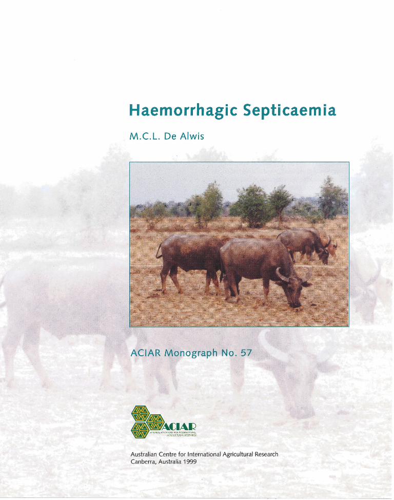
Haemorrhagic Septicaemia
M.C.L. De Alwis
ACIAR Monograph No. 57
Australian Centre for International Agricultural Research Canberra, Australia 1999
The Australian Centre for International Agricultural Research (ACIAR) was established in June 1982 by an Act of the Australian Parliament. Its mandate is to help identify agricultural problems in developing countries and to commission collaborative research between Australian and developing country researchers in fields where Australia has a special research competence.
Where trade names are used this does not constitute endorsement of, nor discrimination against, any product by the Centre.
ACIAR Monograph Series This peer-reviewed series contains the results of original research supported by ACIAR, or material deemed relevant to ACIAR's research objectives. The series is distributed internationally, with an emphasis on developing countries.
De Alwis, M.C.L. 1999. Haemorrhagic Septicaemia. ACiAR Monograph No. 57, x + 141 p.
© Australian Centre for International Agricultural Research, GPO Box 1571, Canberra, ACT, Australia, 2601
ISBN 1 86320269 2
Technical editing and production management: Janet Salisbury, Biotext, Canberra, Australia
Design and art production: Design ONE solutions, Canberra, Australia
Printed by: Brown Prior Anderson, Melbourne
Preface.......... ........... ..... ....... ... .... ..... .. ..... .. .......... vii
Introduction.... ...... ....... ... .. .. .... ....... ... .... ... ... .... .... ix
Chapter 1: Global Distribution
and Economic Importance ........ .. .. .
Overview ................ .............. ........ ... ...... ........ ..... .
1.1 Global Distribution ............ ....... ..... ...... ...
1.2 Economic Importance .... .... ... .... .... .. ....... 2
1.2.1 Asia .... .. ..... ... ... ...... ... ... ..... ...... .... ..... ........ 2
1.2 .2 Africa ..... ...... ....... ... .. .... .. ........ .... ............. 2
1.3 Economic Losses in Asia .. .. .. .. .. .. .. .. .. .... .. 2
1.3.1 South Asia ..... .............. ........ ........ ..... ....... 4
1.3.2 Southeast Asia .... .... ......... .. .. ...... .... .. ... .... 5
1.4 The Role of International Organisations ....... .. ........ ... 6
1.4.1 Office International des Epizooties .. .. .. .... 6
1.4.2 Food and Agriculture Organization ........ . 6
1.4.3 Australian Centre for International Agricultural Research .......... 9
Chapter 2: The Organism:
Classification and Diseases.. .... .... .. 11
Overview. .... ......... .. ...... .... ... .... .. ..... ..... .. .. .. ...... ... . 11
2.1 History and Nomenclature.. .... ....... .. ..... .. 11
2.2 Pasteurella multocida.. .... ........ .. .. .. .... .. ... 12
2.2.1 M orphology and staining.. ...... ......... .... ... 12
2.2 .2
2.2.3
2.2.4
2.2.5
2.2.6
2.2.7
2.2.8
2.2.9
2.3
Growth characteristics and colony morphology.. .. .. ... .. .. .. .... ... .... 12
Serological classification................. .... ..... 14
Designation of serotypes........ .. .. .. .. .. ... .. .. 15
Nonserological tests................. .. ......... .... 17
Cellular components .. .. .... .... .. ... .......... .... 17
Genotype analysis.......... .. .. .. ... ... .. .... .. ..... 19
Biochemical properties...... ...... ... .. .... ....... 20
Virulence to experimental animals.... .. .... . 20
Pasteurella haemolytica .. .... .... .. .. .. .. .. .... .. 20
2.4 Animal Diseases Caused by Pasteurella ............ ...... .. .... .. .. 20
2.4.1 Haemorrhagic septicaemia ... ........ ...... .. ... 20
2.4.2 Bovine pasteurellosis ...... .... .. .. ... ...... .. .. .... 22
2.4.3 Pasteurellosis in sheep and goats .. .. ... .. .... 23
2.4.4 Pasteurellosis in pigs .. .. ...... .. ................ .... 23
2.4.5 Fowl cholera ...... .... ... ... ........... ... .. ...... ..... 23
2.4.6 Other animals ... .. .......... .......... ..... .. ..... ... . 24
Chapter 3: The Disease: Infection,
Clinical Signs and Pathology........ . 25
Overview........... .... .. ....... .... ..... ............ ..... ..... ... .. . 25
3.1 The Disease........ ........ .. .. ... ... .. .... .. ......... . 25
3.2 Source of Infection .. .. .. .... ... .... ... .. .. .. .. .. .. .. 26
3.3 Routes of Infection.. .... ........ .... .... ....... .. .. 26
3.4 Clinical Signs.. ..... ...... .. .......... ...... .. .. .. ... .. 26
3.4.1 Incubation period .... .. ...... ........ ... .. .. .... ..... 27
3.4.2 Duration ... .. .... .... .... .. .... .... .... .. .. ... .. ...... ... 27
3.4.3 Progression of the disease.. .... ...... .. .... .. ... 27
3.4.4 Atypical syndromes................. .. ... .. ..... .. .. 28
3.5 Pathology.... .. .... .. .... ... .. .. ...... ...... ...... ...... 28
3.5.1 Gross pathology....... ... ... ...... .. ...... ..... .. .... 28
3.5.2 Histopathology...... .... .. .. .. .. .. ... .. .. ...... .... ... 30
3.6 Pathogenesis...... .. ..... ........ ........ ... .. .... .. .. 31
3.7 Bacteriology.. .. ...... .. .... ..... .. ............ .. .. .. .. 32
3.8 Recovery................. ................... .... .. ...... . 32
Chapter 4: Epidemiology.... ....... .. .. .... ... .. ..... ..... 33
Overview... ... ........ ... .... .. ...... .... ..... .. ...... .. ............. 33
4.1
4.2
4.3
4.4
4.5
4.5.1
4.5.2
Geographic Distribution of Disease...... .. 33
Seasonal Effects on Distribution.... .. ....... 33
Distribution of Serotypes...... .... .... ..... ..... 34
Host Susceptibility.. .. .. .... .... .. .. .... .. .. ...... .. 34
Morbidity, Mortality and Case Fatality.... .. ..... ...... .. ..... .. ... .. .. ... 36
Rates of morbidity, mortality and case fatality.. .... .. .... ... ... .. .... 36
Factors influenCing morbidity and mortality.... ................ .. ..... 36
iii
4.6 Naturally Acquired Immunity .................. 37 5.10 Molecular Techniques ................... .. ........ 50
4.7 Carrier Status .............................. ... ..... .... 38 5.10.1 Polymerase chain reaction ...... ... ... .. ...... .. . 50
4.7.1 Presence of pasteurellae 5.10.2 Ribotyping and field in carrier animals ..................................... 38 alternation gel electrophoresis ................. 51
4.7.2 Experimental studies on carrier status 5.10.3 Restriction endon uclease analysis ............ 51 and naturally acquired immunity ............ 39 5.11 Antibody Detection in Host Animals ...... 51
4.7.3 Latent and active carriers ..................... .. . 39 5.12 Choice of Diagnostic Tests ..................... 52 4.7.4 Localisation of the
pasteurellae in the tonsils ....................... 41 Chapter 6: Treatment and Control .......... .. ...... 53 4.8 Presumptive Epidemiological Cycle ........ 42
Overview ...... ....... ... ............................... ..... ......... 53
Chapter 5: Diagnosis ....... .. ... ... .. ................ ... ..... 43 6.1 International Classification ........ .... .. ....... 53
6.2 Treatment. ................................. ... .......... 53 Overview ................................................ ..... ..... .. . 43
6.2.1 Antibiotic therapy ...... .. .... .... ... ..... ...... .. .... 54 5.1 Provisional Diagnosis ........ .. .. .... .. ...... .. ... 43
6.2.2 Serum therapy. ..................... .. .. .. ...... .. ..... 54 5.2 Clinical Diagnosis ............. .. ... .. .. .. .. ... ... ... 43
6.3 Prevention and Control .. ...... ...... ... ..... .. ... 55 5.3 Differential Diagnosis ...... .. ........... ... .. ..... 44
6.3.1 Prophylactic measures in 5.4 Samples ................ ................ .. .... .... ...... .. 44 endemic countries ............... .. .. .. .. ... ......... 55 5.4.1 Collection of samples .............................. 44 6.3.2 Preventive measures 5.4.2 Dispatch of diagnostic samples ................ 44 during an outbreak ......................... .. ...... 55
5.5 Laboratory Diagnosis .............................. 45 6.3.3 Prevention of spread across borders ........ 56
5.6 Routine Microbiological Procedures ....... 46 6.4 Eradication ...................... ... ......... ............ 56
5.6.1 Microscopic examination of stained blood smear ........ .. ....... .. ...... ... 46 Chapter 7: Vaccines ........................................... 59
5.6.2 Isolation of pasteurellae .... .. .................... 46 Overview .................................... .. ... ... .. ............... 59 5.7 Conventional Serological Tests ............... 47 7.1 Vaccine Production ................................. 59 5.7.1 Rapid slide agglutination 7.1.1 Selection of a seed culture ........... .. ... ....... 59
test for capsular typing .... .... .. .... ... ... ........ 47 7.1.2 Storage and maintenance
5.7.2 Indirect haemagglutination of seed cultures ................................. .. .... 60 test for capsular typing ............ .. ...... .. ...... 47
7.1.3 Bulk culture media ........... .. ........ ...... .. ..... 60 5.7.3 Agar gel precipitation
test for somatic typing ............................ 48 7.1.4 Culture methods ............................ ......... 61
5.8 Other Serological Tests ...... .. ...... ........ ..... 48 7.1.5 Continuous and batch cultures ........ .. ...... 62
5.8.1 Agar gel precipitation 7.1.6 Inactivation of dense
test for capsular typing .. .. ............ .. .......... 48 cultures and production of vaccines ........ 62
5.8.2 Cou nter-i mm u noelectrophoresis ...... ... .... 48 7.2 Types of Vaccine ........................ ........ ..... 62
5.8.3 Coagglutination test. ............................... 48 7.2.1 Bacterins ........................................ ......... 62
5.8.4 Agglutination test for somatic typing ...... 48 7.2.2 Alum precipitated vaccine ........ .. ........ ..... 63
5.8.5 Enzyme-linked immunosorbent assay ....... 49 7.2.3 Aluminium hydroxide gel vaccine ............ 63
5.9 Nonserological tests .. ............................. 49 7.2.4 Oil adjuvant vaccine ................................ 63
5.9.1 Hyaluronidase production test.. ............... 49 7.3 Quality Testing ...... ...... ......... .. ...... .. ........ 64
iv
7.3.1
7.3.2
7.4
Quality control during manufacture.. ... ... 64
Potency tests....... .... ..... .............. ..... ........ 64
Shelf Life........... .... ........... ........... ...... ..... 65
Chapter 8: Vaccination Programs. ....... .. .... .. .... 67
Overview .... ...................... ... ............. ... ............ ... . 67
8.1
8.2
8.2.1
Rationale. .... ... ...... .. ....... ... ... ...... ......... .... 67
Planning a Vaccination Program.... .... .... . 68
Vaccine quality and administration.......... 68
8.2.2 Vaccination coverage...... .......... ..... ... ...... 68
8.3 Vaccination Schedules... ......................... 69
8.4 Recommended Vaccine and Schedule..... 69
8.5 Evaluation of Vaccination Programs..... .. 70
8.5.1 Adverse reactions .... ... ..... .. .... ...... .. ...... .... 70
8.5.2 Reduction in disease occurrence after vaccination.................... 72
8.6 Measuring Immunity Following Vaccination. ......... ..... ......... ... . 73
8.6.1 Antibody-mediated immunity. ... ...... ... ..... 73
8.6.2 Cell-mediated immunity.. ......... ... .... ..... ... 75
8.7 Vaccination Failures........................... .. ... 75
8.8 Simultaneous and Combined Vaccination... .. . .. .. . .... ........ .... 76
8.8.1 HS and FMD vaccination. ........................ 76
8.8.2 HS and rinderpest vaccination................. 76
8.8.3 HS and blackleg/black quarter vaccination 76
8.9 Vaccination Costs..... ............. .... .... .... ..... 77
Chapter 9: Vaccine Research
and Development... ........................ 79
o . vervlew .............. .................. .............. .............. . 79
9.1 The Ideal Vaccine ... ............ ..... ............... 79
9.2 Improved Vaccines...... .... ... .. ................... 79
9.2.1 Adjuvants other than oil.... .... .......... .... .... 80
9.2 .2 Improved oil adjuvant vaccines .. ............. 80
9.2.3 Vaccines incorporating purified extracts.... .. .... ...... .. ...... ....... . ...... 81
9.2.4 Live vaccines.... ...... ............................ ..... 83
9.3
9.4
Protection between Serotypes................ 86
Future Prospects for Vaccine Development........ ..... ... ........ ..... 86
Chapter 10: Further Investigation................... 87
Overview ........ ... ...... ....... .. ...... ....... ........... .. .. ..... . 87
10.1
10.2
10.3
Pathogenesis..... .. ... .... .... .. .... .... .. .... ... .... 87
Epidemiology. .... ... .... ............. ..... .......... .. 87
Vaccines and Immunology....... ..... .... ...... 88
Appendixes
Appendix 1 Transport and Culture Media. ..... ... .. ...... .. .. ..... ..... 91
Appendix 2 Conventional Serotyping Methods... . ...... ..... .. ..... 95
Appendix 3 Enzyme-linked Immunosorbent Assay .. ....... ........ . 101
Appendix 4 Polymerase Chain Reaction ...... ... .. ...... ...... ..... ...... 105
Appendix 5 DNA Fingerprinting Methods .............. .. ... ..... .. .... . 111
Appendix 6 Preparation of Buffers and Solutions ........ ... ........ 119
Appendix 7 Vaccine Production and Quality Testing ....... ..... ... 121
References ............. .............. .. .......... .. ........ .... ..... 129
v
Preface
The first monograph on haemorrhagic septicaemia (HS) was published by the Food and Agriculture Organization (FAO) in 1962. Two decades later, following a
recommendation of the Third International Workshop on Haemorrhagic Septicaemia (December 1979), this
monograph was revised and updated and a new edition was published in 1982. During the 1980s and 1990s, there has been a considerable amount of research on HS
and a vast amount of new information has become available. A significant feature of this period was the collaborative work by Australian scientists with scientists in many Asian countries, supported by the Australian Centre for International Agricultural Research (ACIAR).
This has led to the introduction of new technologies into haemorrhagic septicaemia research and development. The FAO has also funded a number of projects on HS in the Asian region.
This monograph attempts to capture all the new information generated through these projects - to enrich and add to the available information and present it in a form that will address the needs of a wide readership. It seeks to satisfy multiple roles, including that of a textbook, a review and a laboratory manual. It is, therefore, targeted for veterinary students, practising veterinarians, research workers and also
animal health administrators and policy makers.
I have endeavoured to incorporate into this monograph all accessible published and unpublished information, as well as my own experiences with the disease. The information on enzyme-linked immunosorbent assay (ELISA) and molecular techniques (Appendixes
3-6) was kindly contributed by Dr Kirsty Townsend of the School of Veterinary Science and Animal Production, The University of Queensland . The photographs were contributed by Dr Neil Horadagoda of the Faculty of Veterinary Medicine and Animal Science, University of Peradeniya, and the staff of the Veterinary Research Institute, Peradeniya, Sri Lanka (the FAO Regional Reference Centre for Haemorrhagic Septicaemia) . These contributions, and the interest and support of ACIAR, are gratefully acknowledged.
Dr M .C.L. De Alwis
Veterinary Research Institute PO Box 28, Peradeniya, Sri Lanka
June 1999
vii
viii
The Author
Dr Malcolm De Alwis is Head of the Veterinary Research Institute (VRI) in Sri Lanka. He graduated with a bachelor of veterinary science degree from the University of Peradeniya, Sri Lanka in 1963 and he has a PhD from the University of Liverpool. After a short time as a provincial veterinarian he joined the VRI, where he has worked for the last 33 years, initially as a research officer, then as Head of the Division of
Bacteriology and currently as Head of the VRI. During this time, Dr De Alwis has specialised in bacterial diseases of agricultural animals, particularly haemorrhagic septicaemia.
Introduction
Haemorrhagic septicaemia (HS) is an acute, fatal, septicaemic disease of cattle and buffaloes caused by specific serotypes of the bacterium Pasteurella multocida. The numerous serotypes of P. multocida are associated with a variety of disease syndromes in a wide range of agricultural, domestic and feral animal species. In many instances pasteurella plays a secondary role in the pathogenesis of the disease or acts in combination with other agents. HS, on the other hand, is a primary pasteurellosis and is reproducible in susceptible host species using pure cultures of the causative organism alone. This is comparable to typhoid fever, for example, which is a specific form of salmonellosis caused by a specific strain of salmonella in a specific host species.
HS can be controlled by vaccination and most countries where the disease is endemic resort to routine prophylactic vaccination. The vaccines currently in routine use are satisfactory and strategic vaccination programs have resulted in substantial reduction in the losses. However, research is in progress for the development of an improved vaccine, to overcome some remaining weaknesses in the current vaccines.
HS occurs most commonly in cattle and buffaloes.
Buffaloes are more susceptible than cattle and the disease occurs more frequently in poor husbandry conditions. Clinical symptoms are often not observed but include high temperature, loss of appetite, nasal discharge, increased salivation and laboured breathing, with swellings in the submandibular region. Death usually occurs quickly and mortality is virtually 100% in infected animals. True recovery from clinical disease occurs only if the animal is treated in the very early stages, which is often impossible under prevailing field conditions.
HS has been recorded in almost all parts of the world except for Oceania, including Australia, where it has never occurred. Its distribution is related to climatic
conditions, husbandry practices and the types of animals reared. The disease is of great economic importance in Asia where cattle and buffaloes are abundant and are
vital for draught power and milk production. It is less important in Africa where there are relatively few cattle and buffaloes and because other animal diseases cause relatively more severe economic losses.
Although the conventional host species are cattle and buffaloes, P. multocida has been reported or suspected in Bali cattle, pigs, goats, sheep, horses, donkeys, African buffaloes, camels, yaks and poultry.
Explosive outbreaks of HS may occur when it is first introduced to areas that have previously been free of the disease, or when it occurs as sporadic outbreaks in non endemic areas. In endemic areas, large numbers of animals that survive after an outbreak of disease
become latent carriers. They intermittently shed the organisms but, since the herd immunity is also high, there are no fresh clinical cases. A new outbreak occurs when a shedder comes into contact with a susceptible animal, which may be one born after the previous outbreak or one introduced into the herd from elsewhere. The chance of a fresh outbreak increases with time with an increase in size of the susceptible population.
Movement of animals can precipitate new outbreaks of disease in two ways. Firstly, the animals moved may be carriers and may infect susceptible stock. Secondly, the
animals moved may be susceptible to the disease and become infected from native immune carriers. In either case, explosive outbreaks can result.
Once the first clinical case occurs, more bacteria are shed and disseminated. Their survival in the
environment and transmission to other animals depend on factors such as closeness of contact, hygiene and climate (wet conditions prolong the survival of the causative bacteria outside the animal, making an outbreak more likely). The size of the outbreak depends on the proportion of immune to non immune animals in the herd. Occasional sporadic outbreaks allow the build up of nonimmune animals and a major outbreak may occur. Regular seasonal outbreaks result in much higher herd immunity (through frequent exposure) and outbreaks that do occur therefore tend to be less significant.
Several international organisations have played a significant role in assisting in the control of HS. These are the Food and Agriculture Organization (FAO) of the United Nations, the Office International des Epizooties (OlE, a world organisation for animal health) and the Australian Centre for International Agricultural Research (ACIAR). The assistance provided by these
organisations includes encouragement and funding of
ix
research, development of diagnostic techniques and vaccine production technologies and facilitating control of the disease through strengthening and establishment of diagnostic and vaccine production facilities in the countries where the disease exists.
Outline of monograph
In this monograph an attempt has been made to bring together all the current information on HS. This
includes the global distribution and economic importance of the disease (Chapter 1); a review of the causative organism (Pasteurella) and its relationship to
HS and other animal diseases of economic importance (Chapter 2); and the mode of infection, clinical signs and pathology (Chapter 3), epidemiology (Chapter 4), diagnosis (Chapter 5), and treatment and control (Chapter 6) of HS. Chapters 7, 8 and 9 are devoted to
the types of vaccines currently available, vaccination programs and vaccine research and development,
respectively. Some priority areas for future research are identified in Chapter 10.
The appendixes give details of laboratory techniques for diagnosis and technologies for production and quality control of vaccines. These include simplified methods that can be carried out in any modestly equipped laboratory in the developing countries where the disease is endemic, and sophisticated molecular techniques that are mainly research tools at present but
may, in time, emerge as routine procedures.
x
Global Distribution and Economic Importance
Overview
Global distribution
Haemorrhagic septicaemia (HS) has a wide global
distribution. In most Asian and African countries, it is
endemic. Other countries have recorded a low sporadic
or exceptional occurrence. I n a few other countries the
disease has been suspected clinically but has not been
confirmed by serology. HS has never been reported in
a few countries, including the United Kingdom,
Oceania (including Australia ) and some European
countries.
Economic importance
Most Asian countries rank HS as one of the most
economically important diseases or the most
economically important bacterial disease. The Office
International des Epizooties (OlE) classifies HS with
a number of other bacterial and parasitic diseases as a
List B disease .
1.1 Global Distribution
Haemorrhagic septicaemia (HS) has been recorded
in almost all parts of the world except for Oceania, including Australia, where it has never occurred (see Table 1.1).
It occurs in South and Southeast Asia, including Indonesia, the Philippines, Thailand and Malaysia. The disease was first reported as early as the 1880s in Malaysia (FAa 1991). In Sri Lanka, it was first reported in 1911 but assumed epidemic proportions in the mid-1950s and routine vaccination was introduced in 1957. It is believed to have been introduced into the Philippines in buffaloes (carabaos) imported from Hong Kong in 1902 and in a shipment of cattle imported from Shanghai in 1903. However, the first authentic reports of the disease in the Philippines were from the Visayas and in Bulacan, Luzon. After this, sporadic cases were reported until the mid-1950s when the disease had spread to 40 of the 54 provinces (FAa 1959).
HS has been reported in the Near and Middle East, southern Europe and North, Central and East Africa. There are records of HS in Iran dating back to the 1930s, and a serious study of the disease was made at the Razi Institute in 1935. A saponin adjuvanted vaccine has been used as an immunising agent in Iran since 1938 (FAa 1959). There is a report of HS occurrence in South Africa (Bastianello and Jonker 1981).
In the United States, the disease was reported among bison in national parks in 1912, 1922 and 1965-67. Outbreaks were also reported among dairy cattle in New Jersey in 1969 and among beef calves in California in 1993 (Heddleston et al. 1967; Carter 1982; Blanchard et al. 1993). In summer 1993, an
outbreak was reported among beef calves in New Brunswick, Canada (Rimier and Wilson 1994). Cultures
from these outbreaks are still available in collections and confirm the diagnosis. However, apart from these
sporadic outbreaks, the disease has not become endemic in North America.
HS was recognised in Japan as early as 1923 but has not been recorded since 1954 and was never endemic (Carter 1984; FAO 1959).
The disease has never been reported in Oceania (including Australia), or most of western Europe. There have been some reports suggestive of HS from some Central and South American states, and from southern and eastern European countries (FAO-WHO-OIE Animal Health Yearbook 1994) but these reports are not supported by serotype identification for confirmation . Table 1.1 gives a summary of the global distribution of the disease. It is likely that HS may occur in some South American countries with high water buffalo populations, and where conditions are somewhat similar to those prevailing in tropical Asia.
1.2 Economic Importance
1.2.1 Asia
Asia has 423 million cattle and 145 million buffaloes-33 % and 95 %, respectively, of the world's populations of these species.
Cattle and buffaloes are reared mainly for use as draught animals in the rice fields, but in India and, to a lesser extent, Pakistan, milk production is also important (FAO 1994, 1995). In Asia as a whole, buffaloes contribute 37% of milk production but in India, where the production of milk is the highest in Asia, nearly 50% of the milk is produced from buffaloes.
Where animals are used for draught power, which is a seasonal activity, they are managed in an extensive, free-range system for most of the year. Such conditions are an ideal environment for the spread of diseases such as HS because herds of animals belonging to different owners roam together in common grasslands, drink in common village tanks and are often even paddocked together at night. Such animals are often less well managed, with lower vaccination coverage, than more intensively farmed animals.
2
The relatively high buffalo population in Asia, together with the higher susceptibility of this species to the disease and the high case fatality (see Chapter 4), all highlight the significance of the economic losses caused by HS in the Asian region.
Diseases such as rinderpest have now been effectively controlled or eradicated, while foot-and-mouth disease (FMD) has a low mortality (particularly among animals indigenous to the region). Hence, HS has emerged as a disease of considerable economic importance in the Asian region.
1.2.2 Africa
HS is of less economic importance in the African region than in Asia. This is because Africa has less of the world's cattle and buffalo populations, with only 15% and 2 % for cattle and buffaloes, respectively (FAO 1995). Also, many other animal diseases cause more severe economic losses. These include the African endemic diseases, such as trypanosomiasis (nagana), theilleriosis (East Coast fever) and contagious bovine pleuropneumonia; and also rinderpest and FMD, which are common to both Africa and Asia. Published reports on economic losses in the African region are scarce.
1.3 Economic Losses in Asia
The actual economic losses due to HS are difficult to determine. The prevalence of HS and the morbidity and mortality rates are known in most Asian countries and a few countries have attempted to quantify the losses. However, since the methods used have varied from one country to another, the findings are not strictly comparable.
The problem is that many factors need to be considered in computing economic losses. For example, HS occurs mainly in situations where husbandry practices are poor and animals are reared under a freerange system. In such situations, disease-reporting systems, particularly passive systems that depend on the owner or farmer notifying the relevant authorities, are poorly developed. Thus, a wide discrepancy is
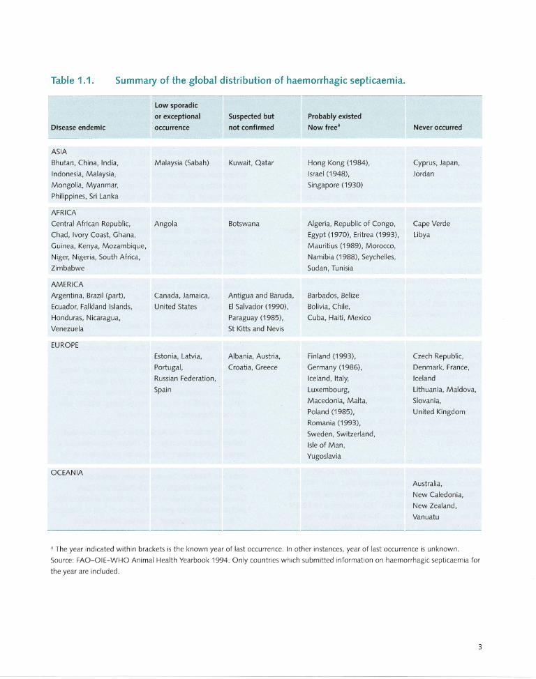
Table 1.1. Summary of the global distribution of haemorrhagic septicaemia.
Disease endemic
ASIA
Bhutan, China, India,
Indonesia, Malaysia,
Mongolia, Myanmar,
Philippines, Sri Lanka
AFRICA
Central African Republic,
Chad, Ivory Coast, Ghana,
Guinea, Kenya, Mozambique,
Niger, Nigeria, South Africa,
Zimbabwe
AMERICA
Argentina, Brazil (part),
Ecuador, Falkland Islands,
Honduras, Nicaragua,
Venezuela
EUROPE
OCEANIA
low sporadic
or exceptional
occurrence
Malaysia (Sabah)
Angola
Canada, Jamaica,
United States
Suspected but
not confirmed
Kuwait, Qatar
Botswana
Antigua and Baruda,
EI Salvador (1990),
Paraguay (1985),
St Kitts and Nevis
Estonia, Latvia, Albania, Austria,
Portugal, Croatia, Greece
Russian Federation,
Spain
Probably existed
Now free"
Hong Kong (1984),
Israel (1948),
Singapore (1930)
Algeria, Republic of Congo,
Egypt (1970), Eritrea (1993),
Mauritius (1989), Morocco,
Namibia (1988), Seychelles,
Sudan, Tunisia
Barbados, Belize
Bolivia, Chile,
Cuba, Haiti, Mexico
Finland (1993),
Germany (1986),
Iceland, Italy,
Luxembourg,
Macedonia, Malta,
Poland (1985),
Romania (1993),
Sweden, Switzerland,
Isle of Man,
Yugoslavia
Never occurred
Cyprus, Japan,
Jordan
Cape Verde
Libya
Czech Republic,
Denmark, France,
Iceland
Lithuania, Maldova,
Siovania,
United Kingdom
Australia,
New Caledonia,
New Zealand,
Vanuatu
a The year indicated within brackets is the known year of last occurrence. In other instances, year of last occurrence is unknown.
Source: FAO-OIE-WHO Animal Health Yearbook 1994. Only countries which submitted information on haemorrhagic septicaemia for
the year are included.
3
bound to exist between actual deaths and reported deaths. This is borne out by the findings of an active surveillance study carried out in Sri Lanka (De Alwis and Vipulasiri 1980) compared to the reported deaths in that country (see Section 1.3.1).
Secondly, losses cannot merely be restricted to the market value of the animal at the time of death. The productive potential of the animal, its reproductive capacity and, in the case of draught power loss, the cost of alternate sources of draught power have to be taken into account. Singh et al. (1987) in India and Ahmed (1996) in Bangladesh attempted to quantify these losses. The Indian study was location-specific, restricted to one village and covered all animal diseases including HS. The Bangladesh study covered several selected diseases in the entire country.
A summary of the economic studies that have been reported in South and Southeast Asian countries is given below. However, all the available information indicates that the actual losses are considerably higher than the values calculated in the studies.
1.3.1 South Asia
India has a sizeable cattle and buffalo population of 192 million and 70 million, respectively. From 1936 to 1944, there were an average of 700 reported outbreaks of HS and 40000 deaths per year. During the 1950s, the average reported deaths per year ranged from 30000 to 60 000. In the 1960s and 1970s this figure dropped to approximately 4000 per year, presumably due to improved control measures (FAO 1991).
Dutta et al. (1990) estimated that in India during the past four decades HS has accounted for 46-55% of all bovine deaths. They also reported that from 1974 to 1986, HS accounted for 6.3 deaths per year for every 100000 bovine population. This amounted to 58.8% of the aggregate of bovine deaths due to the five epidemic diseases FMD, rinderpest, black quarter, anthrax and HS. Before 1939, the corresponding figure ranged from 13.4 to 20.9% . Thus, it appears that HS has increased in economic importance in relation to the other four epidemic diseases.
4
In a location-specific study in India, Singh et al. (1987) computed all of the losses from mortality and productivity, as well as reproductive losses and losses incurred in disease control programs, and concluded that FMD, HS and gastrointestinal parasitism were the most economically important diseases of cattle.
Pakistan ranks HS as a disease of considerable economic importance, with 34.1 % of all deaths in susceptible animals caused by HS (FAO 1979). Pakistan has a cattle population of 17.7 million, and a buffalo population of 18.8 million, the latter being proportionately higher than most other countries in the region. In 1978, annual economic losses from HS were estimated at 1.89 billion Pakistan rupees (PKR) or US$189 million (Chaudhry and Khan 1978; Sheikh et al. 1994). In a study conducted in 10 of the 95 villages in the district of Lahore, Khan et al. (1994) found HS, FMD and gastrointestinal diseases to be the main causes of economic losses. The annual financial loss due to disease was estimated as PKR 19 million, of which PKR 6.8 million was attributed to HS.
Nepal, with 6.3 million cattle and 3.0 million buffalo, ranked HS as the second most important infectious disease after rinderpest and the most important bacterial disease. In a retrospective study of veterinary hospital records maintained in four districts of Nepal during 1985-90, Thakuri et al. (1992) found that parasitic diseases and infectious diseases accounted for 54% and 24% of all cases, respectively, and HS was the most frequent infectious disease.
In 1978 the Food and Agriculture Organization and United Nations Development Program coordinated a survey on the impact of disease on production in the Azad State of Jammu and Kashmir, near the border of India and Pakistan. The survey was carried out as part of the National Program for Livestock and Dairy Development. It showed that nutritional disorders and parasitism were the greatest sources of loss in livestock. Of the bacterial diseases, however, HS and black quarter made the most significant contribution to losses.
In Sri Lanka, an island of 65 000 km2, HS first assumed epidemic proportions in 1955-56, with around 5000 reported deaths from a cattle and buffalo population of around 2.5 million . Thereafter, through various control measures, the losses were reduced to about 1200
reported deaths per year in the early 1980s and had declined further to 269 in 1990 (FAO 1991). These reports were derived from a passive reporting system. Of the 24 administrative districts in the country, the disease is endemic in all of 13 districts and parts of four other districts, covering around two-thirds of the total land area. There has been an active surveillance study in six selected locations within the endemic zone (De Alwis and Vipulasiri 1980) covering 22 297 buffaloes in 803 herds and 20 878 cattle in 870 herds. This study has indicated that 15.2 % of buffaloes and 8.1 % of cattle die of HS each year. When these findings were extrapolated to the entire endemic zone, considering the carcase value alone, a modest estimate of economic loss was 90 million Sri Lankan rupees (SLR), equivalent to US$6 million at that time.
Bangladesh has 23.4 million cattle and 0.8 million buffaloes. When only the production of milk, meat and eggs was considered, the contribution of the livestock sector to the economy was calculated to be about 9%. When the value of draught power, rural transport and the use of dung as fertiliser and domestic fuel were also computed, it was reported to be around 15% . Of the bacterial diseases of cattle and buffaloes present in Bangladesh, black quarter was ranked as the most important, with anthrax and HS equal second in economic importance (Ahmed 1996).
Ahmed (1996) also estimated that of a total economic loss of US$148.6 million each year due to these three diseases, direct losses account for only US$2.3 million. Direct losses include the market value of animals that died and the cost of treatment for those that recovered. Indirect losses in this computation took into account the value of the rice that would have been produced from the land left uncultivated as a result of lack of draught power.
However, it did not include potential productive (milk and meat) and reproductive capacities of the animals. It is also noteworthy that this study used retrospective computerised data, which were probably the product of a passive, routine reporting system. The difference between losses computed from such an information system and an active surveillance study was very evident in a project carried out in Sri Lanka (De Alwis and Vipulasiri 1980).
1.3.2 Southeast Asia
Indonesia has 9.5 million cattle and nearly 3.3 million buffaloes, of which around 97% are used for draught power. The livestock sector contributes around 10% to total agricultural output (ACIAR 1993: Country Reports). Estimates of economic losses due to HS have ranged from US$3500 to US$6000 (FAO 1991; ACiAR 1993: Country Reports). In a study of diseases affecting animals used for draught power in Indonesia (Partoutomo et al. 1985), which included large ruminants and horses, HS was ranked fourth, after fascioliasis, trypanosomiasis and FMD.
Malaysia has a relatively small population of 735 000 cattle and 186000 buffaloes (FAO 1994). In the early years, an average of 28.7 outbreaks per year were reported, with an average mortality of 12.6% per outbreak (FAO 1979). During 1967-76, 287 outbreaks were recorded in peninsular Malaysia with 3605 deaths (FAO 1991). During 1980-89, the losses due to HS were estimated as 2.25 million Malaysian ringgit (US$0.85 million).
The Philippines has 1.67 million cattle and 2.65 million buffaloes (ACIAR 1993). In 1990, the Philippines reported 14331 cases of HS in cattle, with 1057 deaths; and 17 720 cases in buffaloes, with 1725 deaths.
Myanmar lists FMD, HS, black quarter and anthrax as the four most important diseases that cause economic losses in its 11.5 million cattle and buffaloes (of which 6.3 million are used for draught). It has been estimated that 50% of the government's effort in disease control is for the control of HS. Through regular, high vaccination coverage, the disease has been effectively controlled in middle and lower Myanmar. In the northern and eastern regions, however, the disease remains endemic and 20-30 outbreaks are reported each year, with around 1000 deaths (ACIAR 1993).
Thailand has 5.5 million cattle and 4.7 million buffaloes, spread over 513 115 km2 Only 35 outbreaks of HS have been reported, with 3.64 deaths per outbreak. It is believed that losses may have been unreported (FAO 1991; ACIAR 1993).
Vietnam, the Lao People's Democratic Republic (Laos) and Cambodia rank HS as one of the most
5
economically important diseases. In Laos, however, the number of reported deaths has dropped from 16 000 in 1981 to 7500 in 1990. The economic losses from HS were estimated at US$1.4 million in 1990 (FAO 1991, ACIAR 1993).
1.4 The Role of International Organisations
Several international organisations have played a significant role assisting in the control of HS. These are the Food and Agriculture Organization (FAO) of the
United Nations, the Office International des Epizooties (OlE) and the Australian Centre for International Agricultural Research (ACIAR). The assistance provided by these organisations includes encouragement and funding of research, development of diagnostic techniques and vaccine production technologies and facilitating control of the disease through strengthening and establishment of diagnostic and vaccine production facilities in the countries where the disease exists.
1.4.1 Office International des Epizooties
The OlE, or World Organisation for Animal Health, was established in 1924. The main activities of the
organisation are:
• to collect and disseminate to its member countries information on the occurrence, course and treatment of animal diseases;
• to provide guidelines and standards of health regulations applicable to international trade in animals; and
• to promote and coordinate research on pathology, diagnosis, treatment and prevention of animal diseases when international collaboration in such research is desirable.
For the purpose of the above activities animal diseases are classified into two categories, Lists A and B. List A consists of those diseases that spread rapidly, and the scope of which extends beyond national borders. These diseases have particularly serious socioeconomic or public health consequences and are of major
6
importance in the international trade of animals and animal products.
List B includes diseases that are also considered to be of socioeconomic andlor public health importance within countries, and which are also of significance to the international trade in animals and animal products. HS
is included in List B. This classification largely influences the priority status given to livestock diseases by respective governments in endemic countries in their allocation of resources for disease control.
The OlE does not have any regional programs for the controll eradication of HS as for rinderpest and FMD. These two latter diseases are in List A (see above).
Where necessary, however, there are opportunities for regular vaccination against HS linked with FMD vaccination since simultaneous vaccination against both diseases has been shown to be effective.
1.4.2 Food and Agriculture Organization
During the last five decades of this century, the FAO has helped Sri Lanka, Thailand, Zambia and Myanmar to control HS. This assistance has included the services of consultants, training opportunities, equipment and supplies to strengthen national diagnostic and vaccine production laboratories. In Asia, a better focus on the
assistance provided was made through the Animal Production and Health Commission for Asia and the Far East (APHCA) - a regional wing of the FAO, formed in 1973. Initially, the FAO sponsored two international meetings on HS, the first in Manila in 1959 and the second in Kuala Lumpur in 1962. The
Manila meeting was attended by delegates from Burma (Myanmar), Ceylon (Sri Lanka), France, India, Iran, Iraq, Japan, Liberia, Malaya (Malaysia), Pakistan, Philippines, Thailand, United I<ingdom, United States and Venezuela. The second meeting was equally well represented, with delegates from Australia, Burma, Ceylon, France, RepubliC of Guinea, India, Indonesia, Italy, Laos, Malaya, Philippines, Singapore, Sudan, Thailand and the United States. At these two early meetings on HS, a consensus was reached on a number of broad areas, including:
• that increased cooperation be established between research workers in different countries and that the coordination of research and investigational efforts directed towards control of the disease be strengthened;
• that facilities for training in specialised techniques be made available;
• that the diagnostic tests and vaccine production techniques be standardised;
• that due recognition be given to the need for combined vaccines and effective simultaneous vaccination against other diseases present in the region;
• that arrangements be made for collection and dissemination of information relating to the disease within the relevant countries.
It was also agreed that these objectives could be met by the following means:
• designate an institution in the Asian region as a reference centre for HS, and also examine the possibility of recognising such centres in other regions; and
• convene regular meetings on HS, to be held at the invitation of member countries.
After a decision was made by APHCA at its annual sessions in 1978, the third international meeting on HS was held in Colombo, Sri Lanka in December 1979. Prof. RVS. Bain was commissioned to visit all APHCA member countries, to assess the status of the disease and to submit a report, which formed the basis for the meeting. This meeting was attended by delegates from India, Indonesia, Iraq, Malaysia, Nepal, Sri Lanka and Thailand. Country reports were also submitted by Bangladesh and Pakistan.
The recommendations made at this meeting are summarised below.
• That the offer made by the Government of Sri Lanka to establish the regional reference centre in Sri Lanka be considered favourably by the FAO. A special subcommittee recommended that this centre should:
- make available strains of known serotype and immunogenicity as vaccine seed;
- provide a serotyping service for cultures associated with HS in particular and also other pasteurelloses;
- provide national laboratories with reference cultures, reagents and vaccines;
- provide training facilities in diagnostic procedures, including serotyping, vaccine production and testing.
• That further trials be carried out on the double emulsion vaccine, which has an improved syringeability.
• That the monograph on HS published by the FAO in 1963 (Bain 1963) be revised by a group of authors.
• That besides implementing the present knowledge on the disease, further research be encouraged on the following aspects:
- carrier state of serotype 6:B in cattle, buffalo, pigs, sheep, goats and poultry;
- vaccine strain selection;
formulation of media for dense culture production for vaccines, using locally available ingredients; and
- development and testing of vaccines based on newer microbiological concepts.
• That the APHCA secretariat serve as a centre for collection and dissemination of information.
• That in recognition of the usefulness of a uniform system of serotype designation, the capsular typing system of Carter and the somatic typing system of Namioka and Murata be adopted.
In pursuance of the recommendation of the third international meeting, the FAO designated the Veterinary Research Institute, Peradeniya, Sri Lanka as a regional reference centre for HS for the Asian region in 1985. The FAO provided some support to upgrade the facilities at this institute. A reference centre for the African region was in existence in Senegal in West Africa.
7
At the 1986 annual sessions of APHCA, a decision was made to set up an HS subgroup. This subgroup consisted of members from Indonesia, Malaysia, Sri Lanka and Thailand. The main task entrusted to the subgroup was to accelerate and coordinate the work aimed towards improving the quality of the vaccines. Two meetings of the subgroup were held in Bangkok, in September 1987 and February 1990. Priority areas for research and development on vaccines were identified as follows:
• the formulation of improved oil adjuvant vaccines with low viscosity and high stability without affecting potency;
• development of the double emulsion vaccine;
• development of vaccines based on the identification and characterisation of the protective antigens;
• development of live vaccines using strains which are avirulent or are of low virulence; and
• evaluation of all HS vaccines produced in the region at the regional reference centre.
These areas of vaccine research are described in more detail in Chapter 9. The fourth international meeting on HS was held in I<andy, Sri Lanka in February 1991 and was attended by delegates from India, Indonesia, Iran, Laos, Malaysia, Nepal, Philippines, Sri Lanka and Thailand. Additionally, with support received from the British Council and ACIAR, as well as APHCA, it was possible to obtain the participation of several resource personnel. These included two scientists from the United Kingdom, closely associated with the Moredun Research Institute, where considerable advances have been made on pasteurella vaccines, one from the CSIRO, Australia, and one from Japan.
The discussions at this meeting led to the identification of some important areas warranting further research and investigation:
• studies on pathogenesis of HS;
• investigations into the factors that determine virulence of the organism;
• comparison of serotyping systems and adoption of the most appropriate system;
8
• an evaluation of the usefulness of new laboratory techniques such as enzyme-linked immunoserbent assay (ELISA), polyacrylamide gel electrophoresis (PAGE), immunoblotting etc;
• determination of the criteria for declaration of disease-free status to a given area;
• studies on the carrier status of vaccinated animals;
• further confirmation as to whether naturally immune animals (also carriers) are resistant to challenge;
• investigations into thinner emulsions, and non-oil based adjuvants;
• studies on live vaccines and in vivo antigens.
• development of combined vaccines; and
• development of simplified methods for evaluation of vaccines.
In addition to supporting countries that needed assistance and organising scientific meetings, the FAO has produced a number of publications directly on HS or on a broader field including this disease:
• The control of haemorrhagic septicaemia. In: FAO-OIE Animal Health Yearbook 1958.
• Haemorrhagic Septicaemia. R.V.S Bain, FAO Agricultural Studies No. 62, 1963.
• Haemorrhagic Septicaemia of Cattle and Buffaloes. R.v.S Bain, South East Asia Treaty Organisation, Bangkok, Thailand 1961.
• Haemorrhagic Septicaemia. R.v.S Bain, M.C.L. De Alwis G.R. Carter and B.K. Gupta, FAO Animal Production and Health Paper No. 33, 1982.
• Manual of Production of Haemorrhagic Septicaemia Vaccine. R.P. Misra. Nong Teng, Laos, FAO, 1985.
• A Manual of Laboratory Procedures for Selected Diseases of Livestock. G.G. Alton, G.R. Carter, A.c. Kibor and L. Pesti, FAO, 1990.
1.4.3 Australian Centre for International Agricultural Research
ACIAR is a statutory authority established in Australia under an Act of Parliament. It has a mandate to facilitate collaborative research on high priority problems of developing countries that offers good prospects for mutual benefits with Australia. This is achieved by linking Australian research institutions with overseas research groups. In keeping with its mandate, ACIAR has maintained an interest in HS, which is a major animal health problem in the region. During the past decade, ACIAR has funded two projects specifically on HS, another dealing with a variety of diseases including HS, and another in the related field of pasteurellosis in pigs and poultry. A brief description of these projects is given below.
Development of an improved haemorrhagic septicaemia vaccine
This was a collaborative project between the CSIRO Division of Animal Health in Australia and the Veterinary Research Institute in Ipoh, Malaysia. This project broadly aimed at producing improved vaccines and developing and evaluating existing as well as newly developed tests for determining the efficacy of vaccines. Broadly two types of vaccines were developed. These were improved emulsion type vaccines which had low viscosity and easier injectability as well as vaccines based on the identification and characterisation of the cell components with a view to determining the protective antigens (see Chapter 7). Conventional tests such as the indirect haemagglutination test (lHA) and the passive mouse protection test (PMPT) were compared with a newly developed ELISA test and direct challenge.
Diagnosis and control of haemorrhagic septicaemia in Indonesia
This was a collaborative project between the Victorian Institute of Animal Science in Australia, the Research Institute for Veterinary Science in Bogor, Indonesia and the Disease Investigation Centre in Denpassar, Indonesia. This project covered a wide spectrum of areas connected with the diagnosis and control of HS.
• Improvement of techniques to detect the organism, including the development of special transport media and their evaluation in comparison with the conventional culture techniques.
• Development of molecular tests for detection of the organism as well as for characterisation with special emphasis on strain variation.
• Review of management and vaccination strategies.
• Retrospective and prospective studies of the epidemiology of the disease, as well as seroepidemiological studies using the ELISA test.
Establishment of improved methods for the diagnosis and control of livestock diseases in Southeast Asia using ELISA
This project was a collaboration between four institutions: the CSIRO Division of Animal Health and the Victorian Department of Agriculture and Rural Affairs in Australia; the Research Institute for Veterinary Science in Bogor, Indonesia; and University of Pertanian in Malaysia. Amongst other diseases, this project aimed at developing and evaluating the ELISA technique for identification of Pasteurella multocida strains causing HS as well as for detection of antibody against P. multocida in animals.
Control of pasteurellosis in pigs and poultry
This project had five participating institutions. These were Monash University and The University of Queensland in Australia; the Veterinary Research Institute in Peradeniya, Sri Lanka; and the National Institute of Veterinary Research and the National Veterinary Company in Vietnam. Though the project did not directly deal with bovine pasteurellosis, technologies developed may have future applications in the field of HS.
9
International workshops
ACIAR has also organised international meetings to enable scientists working on their research projects to present their findings. During the past decade, two such meetings sponsored by ACIAR were the International Workshop on Pasteurellosis in Production Animals, held in August 1992 (ACIAR 1993), and the International Workshop on Diagnosis and Control of Haemorrhagic Septicaemia in May 1996. Both workshops were held in Bali, Indonesia, with the participation of other scientists in the field as well. These workshops provided an excellent opportunity for those working in the field of HS in the region to exchange their views and share their experiences. Additionally, the participation of scientists from outside the region enabled the infusion of new ideas and technologies into this problem of utmost economic importance to the region.
10
The Organism: Classification and Diseases
Overview
History and nomenclature
Haemorrhagic septicaemia (HS ) is caused by specific
serotypes of the bacterium Pasteurella. The organism is
named after Louis Pasteur in recognition of his
pioneeri ng work in the 1880s. The genus Pasteurella
belongs to a large family of bacteria - the
Pasteurellaceae, which includes two other important
genera, Actinobacillus and Haemophilus. Two members of
the genus Pasteurella - P multocida and P haemolytica -
cause a variety of important diseases in domestic ,
agricultural and wild animals.
Pasteurella multocida
Several methods have been developed to identify
different serotyopes of P multocida. These can be broadly
grouped into methods that identify five 'capsular'
antigens (based on components of the outer cell layer)
and up to 16 'somatic' antigens (based on core
components ). Using a combination of capsular and
somatic typing, the two common HS serotypes
(popularly known as the Asian and African serotypes)
are designated B:2 and E:2 , respectively.
Pasteurella haemolytica
P haemolytica can be distinguished from P multocida by
several biochemical characteristics. Two biotypes have
been identified, based on sugar fermentation (A and T ),
while 16 types have been identified based on capsular
polysaccharides.
2.1 History and Nomenclature
Haemorrhagic septicaemia (HS) is caused by specific serotypes of the bacterium Pasteurella multocida in cattle and buffaloes . According to current classification, the family Pasteurellaceae (Pohl 1981) includes a large group of gram-negative bacteria that are chemoorganotrophic, facultatively anaerobic and fermentative. The family has three genera: Pasteurella (Trevisan 1887), Actinobacillus (Brumpt 1910) and Haemophilus (Winslow et al. 1917). Several other
species that exhibit complex phenotypic and genotypic relationships with these genera are also included. Most members of the family cause disease in mammals (including humans), birds and reptiles.
The first description of the bacterium was in 1878-79
when Rivolta and Revolee reported an outbreak of disease in fowl. About the same time, a disease in
cattle and buffaloes was described by Bollinger in Germany. Its causative organism was isolated by Kitt in 1885. Gaffky described a septicaemic disease in rabbits in 1881 and Loeffler described a similar disease in swine in 1886. It was the German pathologist Hueppe who, in 1886, observed similarities in all of these diseases and also similarities in the bacteria associated
with these disease conditions. Later, in 1887, Oreste and Armani described a similar disease called 'barbone'
in buffaloes in Italy, also caused by a similar organism; they proposed the name Bacillus septicaemiae for this bacterium. However, in 1887, Trevisan proposed the name Pasteurella to commemorate the work of Louis Pasteur on the aetiology of fowl cholera, which is
caused by the same organism. Human infections caused by the same organism were described for the first time in 1920.
Since this bacterium caused disease in many species of animals, species names were initially given according to the host species in which it caused disease. Thus,
11
isolates from cattle were named boviseptica; pigs, suiseptica; poultry, aviseptica; and so on. Subsequently, there were several complex name changes for the organism until 1929 when Topley and Wilson introduced the name Pasteurella septica. In 1939, Rosenbach and Merchant proposed the name Pasteurella multocida. A summary of the historical evolution of the nomenclature of Pasteurella multocida is shown in Table 2.1.
On the basis of phenotypic similarities, more serotypes were added to the genus: Pasteurella haemolytica in 1932, P. pneumotropica in 1950, P. gallinarum in 1955, P. ureae in 1962 and the gas-producing P. aerogenes isolated from pigs in 1974.
More recent classification of the genus Pasteurella is based on genetic relationships, which have been determined by DNA:DNA hybridisation, rRNA:DNA hybridisation and 16S rRNA sequencing. On this basis, 11 species have been identified: Pasteurella multocida (with three subspecies: multocida, septica and gallicida), P. dagmatis, P. gallinarum , P. voluntium, P. stomatis, P. avium, P. langaa, P. anatis and Pasteurella subspecies A and B (Mutters et al. 1989; Sawada 1991; Bisgaard 1994).
Several species that were previously classified under the
genus Pasteurella have now been excluded as they were found to be genetically more closely related to the Actinobacillus group. These are P. ureae, P haemolytica biotypes A and T, P. testudinis, P. aerogenes and P. pneumotropica, biotypes Heyl and Jawetz. Since the genus was redefined on a genetic basis, six new species have been included in the group: P. granulomatis, P. caballi, P. bettii, P. Iymphangitidis, P. mairi and P. trehalosi (Bisgaard 1994).
Two members of the genus Pasteurella are of veterinary importance - P. multocida and P. haemolytica. These species cause a variety of disease syndromes (pasteurelloses) in agricultural, domestic and
wild animals. A brief description of the common pasteurelloses in animals is given in Section 2.4 and the associated organisms, host species and diseases caused are shown in Table 2.7.
12
2.2 Pasteurella multocida
2.2.1 Morphology and staining
P. multocida is a nonmotile, nonspore-forming short rod or coccobacillus, 0.2-0.4 by 0 .6-2.5 mm in size. Repeated laboratory subcultures of old cultures or cultures grown under unfavourable conditions tend to be pleomorphic and longer rods and filamentous forms appear. In tissues, exudates and recently isolated cultures, the organism shows the typical coccobacillary forms . It is a gram-negative organism and, in fresh cultures and animal tissues, gives typical bipolar staining,
particularly with Leishman or methylene blue stain.
2.2.2 Growth characteristics and colony morphology
P. multocida grows in most common laboratory media such as nutrient agar. Special media such as dextrose-starch agar and casein-sucrose-yeast (CSY)
medium support an abundant growth . Blood agar and CSY agar with 5% blood (bovine, sheep) are convenient media for routine laboratory culture. The
optimum growth temperature is 35-37°C. In enriched media at 37°C, colonies 1-3 mm in diameter are produced after 18-24 hours culture.
The organism shows different types of colonies, which
are related to the capsular type. Capsular type A produces the largest colonies, which are translucent, greyish in colour, and mucoid in conSistency. There may be considerable variation in colony size, ranging from rounded, convex, discrete colonies with circular edges to large watery colonies with flowing margins. In this type of colony, the capsules consist, in part, of hyaluronic acid. Occasionally, type D strains may also produce mucoid colonies. Colonies of capsular types D and F and the rounded colonies of type A display a pearl-like iridescence in oblique transmitted light. Colonies of types Band E may also vary in size,
depending on the degree of capsulation. They will range from larger greyish colonies, when freshly isolated or when grown in media containing blood serum, to smaller colonies that give a yellowish-green or bluishgreen iridescence when viewed in transmitted light.
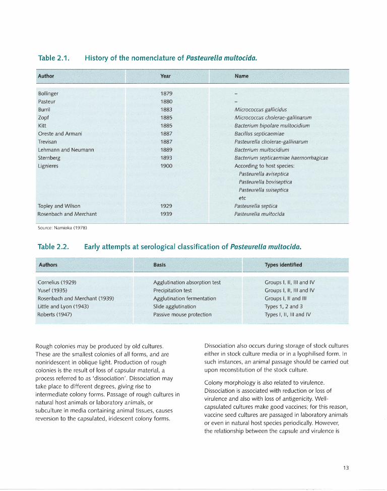
Table 2.1 . History of the nomenclature of Pasteurella multocida.
Author
Bollinger
Pasteur
Burril
lopf
Kitt
Oreste and Armani
Trevisan
Lehmann and Neumann
Sternberg
Lignieres
Topley and Wilson
Rosenbach and Merchant
Source: Namioka (1978)
Year
1879
1880
1883
1885
1885
1887
1887
1889
1893
1900
1929
1939
Name
Micrococcus gallicidus
Micrococcus cho/erae-gallinarum
Bacterium bipolare multocidium
Bacillus septicaemiae
Pasteurella cholerae-gallinarum
Baderium multocidium
Bacterium septicaemiae haemorrhagicae
According to host species:
Pasteurella aviseptica
Pasteurella boviseptica
Pasteurella suiseptica
etc
Pasteurella septica
Pasteurella multocida
Table 2.2. Early attempts at serological classification of Pasteurella multocida.
Authors Basis Types identified
Cornelius (1929)
Yusef (1935)
Rosenbach and Merchant (1939)
Little and Lyon (1943)
Agglutination absorption test
Precipitation test
Agglutination fermentation
Slide agglutination
Groups I, II, III and IV Groups I, II, III and IV Groups I, II and III Types 1, 2 and 3
Types I, II, III and IV Roberts (1947) Passive mouse protection
Rough colonies may be produced by old cultures. These are the smallest colonies of all forms, and are noniridescent in oblique light. Production of rough colonies is the result of loss of capsular material, a process referred to as 'dissociation'. Dissociation may
take place to different degrees, giving rise to intermediate colony forms. Passage of rough cultures in natural host animals or laboratory animals, or subculture in media containing animal tissues, causes
reversion to the capsulated, iridescent colony forms.
Dissociation also occurs during storage of stock cultures either in stock culture media or in a Iyophilised form. In such instances, an animal passage should be carried out upon reconstitution of the stock culture.
Colony morphology is also related to virulence. Dissociation is associated with reduction or loss of virulence and also with loss of antigenicity. Wellcapsulated cultures make good vaccines; for this reason, vaccine seed cultures are passaged in laboratory animals or even in natural host species periodically. However, the relationship between the capsule and virulence is
13
not absolute. There are capsulated variant cultures that are of low virulence or are avirulent, whilst non capsulated strains may be virulent.
Maintenance of stock cultures
Stock cultures may be maintained conveniently in nutrient agar containing 0.75% agar to give a semisolid consistency. Stab cultures should be made, in semisolid nutrient agar in tightly screw-capped bottles to prevent drying on storage. After incubation for 18-20 hours at 37°C they can be stored at room temperature for several years.
For lyophil ising, a confluent growth of an 18-20-hour culture from CSY agar with blood may be washed and suspended in an equal volume of 5% skimmed milk. Alternatively, the WHO medium recommended for Iyophilising brucella cultures, consisting of 2.5% tryptone, 5% sucrose and 1 % glutamate, has been found to be satisfactory.
2.2.3 Serological classification
Early attempts at serological classification of P. multocida date back to the 1920s (Cornelius 1929; Yusef 1935; Little and Lyon 1943). Roberts (1947)
developed a system of serological classification based on passive protection tests in mice. He used antisera prepared in rabbits to protect mice against challenge with a wide range of strains. On the basis of mouse protection, he was able to identify four types, which he designated types I, II, III and IV. This was the first classification to meet some degree of acceptance. Since all HS strains fell into Roberts type I, this designation became fairly well established. Subsequently, Hudson (1954) added a fifth serotype. The basis of these early attempts at serological classification and the types identified are shown in Table 2.2.
Carter used a precipitation test (Carter 1952) and
subsequently an indirect haemagglutination test (Carter 1955) and was able to identify four serological types. These were based on agglutination of human '0'
erythrocytes coated with crude extracts of outer cell components from the bacterial cultures. These crude 'capsular' extracts were supernatants prepared by heating suspensions of the bacteria at 56°C for 30 minutes and removing the cells by centrifugation.
14
He designated these four capsular types A, B, C and D (Carter 1952,1955). The strains that caused HS were
grouped into Carter type B. Subsequently, he found that the strains that caused HS in Africa did not fall strictly into any of these groups, though they were related to type B, and they were included in a separate group designated type E (Carter 1961). Subsequently, however, he found that type C was not a consistent type and it was deleted (Carter 1963).
This method of identifying serotypes has become established as the Carter indirect haemagglutination test (IHA). Three decades later, Rimier and Rhoades
(1987) isolated a consistent type from turkeys which did not fit into any existing serogroups; this was designated serogroup F.
Since fresh human '0' erythrocytes may not always be available in a laboratory, the IHA test has been modified by various workers for practical convenience. Carter and Rappay (1962) used formalinised human '0' cells, which could be stored in a laboratory for long periods. More recently, Sawada et al. (1982) used glutaraldehyde-fixed sheep erythrocytes. The test has now been modified for the detection of antibodies as well, using erythrocytes coated with cell extracts from known reference cultures. Wijewardana et al. (1986a) used fresh sheep erythrocytes, and adopted the test both for identification of serotype and for antibody detection.
In the early 1960s, Namioka and Murata (1961 a) described a simplified and rapid method of identifying the capsular types using a slide agglutination test in which fresh cultures are agglutinated with hyperimmune rabbit sera. Namioka and Murata (1961 b,c, 1964) and Namioka and Bruner (1963) later developed what is described as a 'somatic' typing test, based on releasing core ('somatic') bacterial components by agglutinating acid (HCI)-treated cells with rabbit antiserum. Using this method, 11 somatic types were identified . Type-specific antiserum was produced by a complicated system of absorptions, as shown in Table 2.3. Another drawback to this system is that some cultures undergo autoagglutination after the HCI treatment and therefore are rendered untypeable.
Heddleston et al. (1972) developed an agar gel
precipitation test also for somatic typing. In this test, the antigen used was the supernatant of culture suspensions heated at 100°C for one hour. The
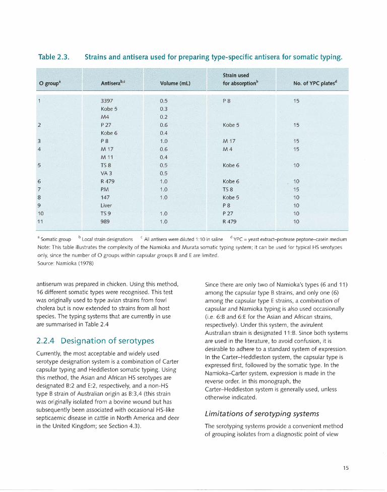
Table 2.3. Strains and antisera used for preparing type-specific antisera for somatic typing.
o group'
2
3
4
5
6
7
8
9
10
11
Antiserab,c
3397
Kobe 5
M4
P 27
Kobe 6
P8
M 17
M 11
TS 8
VA3
R 479
PM
147
Liver
TS 9
989
Strain used
Volume (mL) for absorptionb No. of YPC platesd
0.5 P8 15
0.3
0.2
0.6 Kobe 5 15
0.4
1.0 M 17 15
0.6 M4 15
0.4
0.5 Kobe 6 10
0.5
1.0 Kobe 6 10
1.0 TS 8 15
1.0 Kobe 5 10
P8 10
1.0 P 27 10
1.0 R 479 10
• Somatic group b Local strain designations c All antisera were diluted 1 :10 in saline d YPC = yeast extract-protease peptone-casein medium
Note: This table illustrates the complexity of the Namioka and Murata somatic typing system; it can be used for typical HS serotypes
only, since the number of 0 groups within capsular groups Band E are limited.
Source: Namioka (1978)
antiserum was prepared in chicken. Using this method, 16 different somatic types were recognised. This test was originally used to type avian strains from fowl cholera but is now extended to strains from all host species. The typing systems that are currently in use are summarised in Table 2.4
2.2.4 Designation of serotypes
Currently, the most acceptable and widely used serotype designation system is a combination of Carter capsular typing and Heddleston somatic typing. Using this method, the Asian and African HS serotypes are designated 8:2 and E:2, respectively, and a non-HS type 8 strain of Australian origin as 8:3,4 (this strain was originally isolated from a bovine wound but has subsequently been associated with occasional HS-like septicaemic disease in cattle in North America and deer in the United Kingdom; see Section 4.3).
Since there are only two of Namioka's types (6 and 11) among the capsular type 8 strains, and only one (6) among the capsular type E strains, a combination of capsular and Namioka typing is also used occasionally (i.e. 6:8 and 6:E for the Asian and African strains, respectively). Under this system, the avirulent Australian strain is designated 11 :8. Since both systems are used in the literature, to avoid confusion, it is desirable to adhere to a standard system of expression. In the Carter-Heddleston system, the capsular type is expressed first, followed by the somatic type. In the Namioka-Carter system, expression is made in the reverse order. In this monograph, the Carter-Heddleston system is generally used, unless otherwise indicated.
Limitations of serotyping systems
The serotyping systems provide a convenient method of grouping isolates from a diagnostic point of view
15
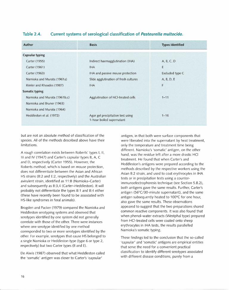
Table 2.4. Current systems of serological classification of Pasteurella multocida.
Author Basis Types identified
Capsular typing
Carter (1955)
Carter (1961)
Carter (1963)
Indirect haemagglutination (lHA)
IHA
A, B, C, 0
E
Namioka and Murata (1961a)
Rimier and Rhoades (1987)
IHA and passive mouse protection
Slide agglutination of fresh cultures
IHA
Excluded type C
A, B, 0, E
F
Somatic typing
Namioka and Murata (1961 b,c)
Namioka and Bruner (1963)
Namioka and Murata (1964)
Heddleston et al. (1972)
Agglutination of HCI-treated cells 1-11
Agar gel precipitation test using 1-hour boiled supematant
1-16
but are not an absolute method of classification of the
species. All of the methods described above have their limitations.
A rough correlation exists between Roberts' types I, II,
III and IV (1947) and Carter's capsular types B, A, C
and D, respectively (Carter 1955). However, the
Roberts method, which is based on mouse protection,
does not differentiate between the Asian and African
HS strains (B:2 and E:2, respectively) and the Australian
avirulent strain, identified as 11 :B (Namioka-Carter)
and subsequently as B:3,4 (Carter-Heddleston). It will
probably not differentiate the types B:1 and B:4 either
(these have recently been found to be associated with
HS-like syndromes in feral animals).
Brogden and Packer (1979) compared the Namioka and
Heddleston serotyping systems and observed that
serotypes identified by one system did not generally
correlate with those of the other. There were instances
where one serotype identified by one method
corresponded to two or more serotypes identified by the
other. For example, serotypes that cause HS belonged to
a single Namioka or Heddleston type (type 6 or type 2, respectively) but two Carter types (B and E) .
De Alwis (1987) observed that what Heddleston called the 'somatic' antigen was closer to Carter's 'capsular'
16
antigen, in that both were surface components that were liberated into the supernatant by heat treatment,
only the temperature and treatment time being different. Namioka's 'somatic' antigen, on the other
hand, was the residue left after a more drastic HCI treatment. He found that when Carter's and
Heddleston's antigens were prepared according to the
methods described by the respective workers using the Asian B:2 strain, and used to coat erythrocytes in IHA
tests or in precipitation tests using a counterimmunoelectrophoresis technique (see Section 5.8.2),
both antigens gave the same results . Further, Carter's antigen (56°C/30-minute supernatant), and the same
antigen subsequently heated to 100°C for one hour,
also gave the same results . These observations
appeared to suggest that the two preparations shared common reactive components. It was also found that
when phenol-water extracts (Westphal type) prepared from HCI-treated cells were coated onto sheep
erythrocytes in IHA tests, the results parallelled Namioka's somatic typing.
These findings led to the conclusion that the so-called
'capsular' and 'somatic' antigens are empirical entities
that serve the need for a convenient practical classification to identify different serotypes associated
with different disease conditions, purely from a
diagnostic point of view. The actual cell structure is much more complex, as becomes evident from a more detailed study of cell components.
In situations where a wide range of isolates of P multocida from a variety of sources are examined, a substantial proportion remain untypeable (Namioka and Bruner 1963; Chandrasekaran et al. 1981; Jones et al. 1988). When strains associated with clinical HS are considered, however, almost 100% of isolates are typeable with group B or E antisera (De Alwis and Panangala 1974; Shigidi and Mustafa 1979). In Sri Lanka, all of 50 isolates associated with clinical HS were typeable, but out of 49 abattoir isolates from tonsils, only 67% were typeable by Carter's capsular typing and 80% by Heddleston's somatic typing (De Alwis and Panagala 1974; Wijewardana et al. 1993). In another study (Wijewardana et al. 1986a), biochemical and serological uniformity was observed among 17 isolates of P. multocida associated with clinical disease, but there was considerable diversity among 23 isolates from the nasopharynx and associated lymph nodes of apparently healthy cattle and buffaloes.
2.2.5 Nonserological tests
Several nonserological tests have been described for the identification of specific types of pasteurellae. The acriflavine test of Carter and Subronto (1973) specifically identifies type D strains. Carter and Rundell (1975) described a hyaluronidase decapsulation test that used a hyaluronidase-producing staphylococcus to identify mucoid type A strains. A hyaluronidase production test has been described by Carter and Chengappa (1981) for the identification of type B strains that cause HS. These tests can be used to supplement the serotyping systems rather than as substitutes for them .
2.2.6 Cellular components
The limitations and the lack of correlation between the different serotyping systems can be understood on the basis of cellular components.
Capsule
The capsule (or outer layer) of the cell is believed to be responsible for the serogroup specificity. It is composed of polysaccharides, lipopolysaccharides (LPS) and a
variety of proteins. Both LPS and polysaccharides are absorbed onto erythrocytes and are believed to playa role in passive haemagglutination. Studies on purified capsular extracts have given somewhat conflicting results and this may be due to the incomplete separation of the constituents in the different purification processes used (Dhanda 1960; Knox and Bain 1960; Bain and Knox 1961; Rimier and Rhoades 1989). More recently, Penn and Nagy (1976) separated a nontoxic polysaccharide capsular antigen by fractional precipitation from aqueous solutions with polar solvents. When tested in comparison with crude capsular extracts, this capsular polysaccharide was nontoxic in chick embryo lethality and rabbit pyrogenicity tests. It was nonimmunogenic in rabbits but produced mouse protective antibodies in cattle. Muniandy et al. (1993), using an improved procedure for purification of polysaccharide, prepared an extract which gave a final yield of 3.5%, with minimal contamination with proteins and nucleic acids and free of LPS. This product was also nontoxic and nonimmunogenic in rabbits. These workers also found that the Penn and Nagy type of polysaccharide extracts, which were contaminated with 20% LPS, displayed a high degree of anti phagocytic activity in an in vitro phagocyte uptake assay.
Prince and Smith (1966a,b), described three antigen complexes - alpha, beta and gammacorresponding to a polysaccharide-protein complex, a serogroup-specific polysaccharide and LPS, respectively (Mosier 1993).
Proteins are believed to be important immunogens and are likely to playa vital role in the protective mechanism. With the exception of a few serogroup A and D strains that produce protein toxins, proteins of P multocida are nontoxic. Johnson et al. (1991) examined a wide range of P multocida strains of different serogroups by electrophoretic techniques and found a high degree of homogeneity in protein profiles among 14 strains associated with HS. Strains of Asian and North American origin (B:2) displayed a major protein band of molecular mass 32 kDa. Strains of African origin (E:2), on the other hand, gave a similar band at 37 kDa. Other bands at 27,45 and 47 kDa were shared by all strains, irrespective of serotype. Using monoclonal antibodies and an immunoblotting technique, Ramdani and Adler (1993) identified protein
17
fractions of 29 and 36 kDa in the cytoplasmic and periplasmic fractions and 42 kDa in the membrane fraction. These fractions were shown to give only variable protection in mice (25-67%; see Section 9.2.3).
The association of outer membrane proteins (OMPs) with protective immunity has been widely investigated . Muniandy and Mukkur (1993) observed that the immunogenicity of certain LPS preparations was due to the presence of contaminating OMPs. Kennett et al . (1993) grew P. multocida serotype B:2 (Malaysian C82) cultures in BHI medium under iron-restricted or ironreplete conditions. An iron chelator (2.2-dipyridyl) was added to the iron-restricted medium, while 0.1 M FeCI3
was added to the iron-replete medium. They found that the OMPs produced under iron-restricted conditions gave a higher degree of protection in mice than OMPs produced under iron-replete conditions. This was attributed to a high molecular mass protein of over 84 kDa, which was expressed in abundance under iron-restricted conditions.
In Sri Lanka, T.G. Wijewardana, Veterinary Research Institute, Peradeniya (personal communication) found a similar protein (116 kDa), which was produced by B:2 strains in vivo and when grown under iron-restricted conditions in vitro but was absent from cultures grown in normal media. The analysis of total membrane proteins by sodium dodecylsulfate polyacrilamide gel electrophoresis (SDS-PAGE), in cells of serotype B:2 strains grown under iron-replete and iron-restricted conditions (Veken et al. 1994, 1996), also revealed different specific protein components that were expressed by the same strain, depending on the culture conditions (see also Section 9.2.3). A variety of protein components of various molecular weights have also been isolated from the Indian vaccine strain P52, by various extraction methods including sonication and precipitation with ammonium sulfate gel . Their immunogenic merits have been tested in rabbits and mice (Pati et al. 1996; Srivastava 1996).
The LPS of P. multocida are similar to those of other gram-negative bacteria. They constitute the 'endotoxins' of the organism, and are the basis of somatic typing. The best LPS preparations are obtained from ethanol-killed cells that have been washed and dried. Killing with formalin alters certain properties of the LPS. Extraction is
18
done by the phenol-water (Westphal) method or the phenol-chloroform-petroleum method (Rimier and Rhoades 1989).
LPS are largely responsible for the toxicity in the HScausing serogroup B:2 and play an important role in the pathogenesis of the disease (Rebers et al. 1967; Rhoades et al. 1967). Purified LPS extracts have been shown to have anti phagocytic activity in in vitro phagocytic uptake assays (Muniandy et al. 1993). Serological relationships exist between LPS of serogroups Band E (Mosier 1993). Electrophoretic analysis of purified LPS preparations has also established relationships between Band E and some type A strains (Rimier 1990). This is not surprising since all strains of both Asian and African origin possess the Namioka somatic antigen type 6 and Heddleston type 2, although in the two serotyping procedures the LPS components used are different (De Alwis 1987). Although crude LPS preparations are associated with immunity, it has been shown that highly purified LPS are nonimmunogenic to mice and rabbits (Muniandy et al. 1993). Muniandy and Mukkur (1993) showed that Westphal extracts were protective in mice, and that treatment with phenol or digestion with proteinase K abolished the protection. This latter observation suggests that the protectivity of crude LPS extracts is due to contaminating proteins.
Common antigens
P. multocida shares common antigens with other closely related gram-negative bacteria. Antigenic relationships with Yersinia paratuberculosis, P. haemolytica, Haemophilus canis, H. influenza,
Actinobacillus lignieresi, Escherichia coli and Neisseria
catarrhalis have been reported (Bain 1963; Prince and Smith 1966b; Schryvers et al. 1986). Cross-protection has been detected in a study of 11 isolates of P. multocida from cases of HS, bovine pneumonia and fowl cholera and belonging to various serotypes (Rimier 1996). A serotype A:5 strain and a fowl cholera strain were found to protect against a number of other strains, irrespective of the disease caused. This protection was attributed to antigen components of molecular weight 20-120 kDa.
Enzymes
P. multocida has been found to produce a number of enzymes. Neuraminidase is produced by members of serogroups A, B, D and E (Rimier and Rhoades 1989). Its activity is found to be highest in strains of serogroup A and D. Activity of neuraminidase of type E was inhibited by homologous antiserum only, whilst that of types Band D were inhibited by antisera against serogroups A, B, D and E.
The production of hyaluronidase and chondroitinase by serotype B:2 associated with HS is well documented (Carter and Chengappa 1980; Rimier 1993; Rimier and Rhoades 1994). Hyaluronidases are enzymes that are normally associated with invasive mechanisms in bacteria, helminths and snake venoms. Type B strains, bearing other somatic antigens, such as the B:3,4 cattle and deer strains, fail to produce hyaluronidase. Whilst it may be concluded that hyaluronidase production is a character exclusively restricted to serotype B:2 strains that cause HS, De Alwis et al. (1996, 1998) described a type B:2 mutant that was of low virulence to mice and rabbits and avirulent to cattle and buffaloes but produced hyaluronidase. No clear relationship has been established between the ability to produce hyaluronidase or any other enzyme and virulence.
Toxins
Serogroups A and D have been found to produce protein toxins, more toxigenic strains being present in serogroup D. These toxins are directly involved in the pathogenesis of disease, as in naturally occurring atrophic rhinitis in swine. No correlation has been found between toxin production and somatic types. Toxins of serogroups A and D are similar, if not identical, and antiserum produced against one neutralises the other (Rimier and Rhoades 1989).
True exotoxins are not produced by strains of the B group associated with HS. Toxic effect (endotoxic shock) can however be produced by injection of culture supernatants (which contain free endotoxins) or endotoxin preparations.
Bacteriocins
Bacteriocins are bacteriocidal proteins produced by many species of bacteria and which are active against members of their own species or closely related species. Production of bacteriocins is believed to be determined by a genetic element. Bacteriocin activity has been demonstrated in bovine and avian strains of P. multocida (Rimier and Rhoades 1989). Thirty-three bovine and bison strains belonging to serotypes A, B and D were tested for bacteriocin activity by Chengappa and Carter (1977); 14 were found to produce bacteriocins. Seventeen strains were susceptible to bacteriocins. Except in special investigations, their role as a diagnostic tool is of limited value . The role of bacteriocins in the pathogenesis of disease has not been investigated.
2.2.7 Genotype analysis
With the advent of molecular biology, it has been possible to characterise isolates on a genotypic basis, providing a firmer and more stable basis than groupings based on phenotypic characteristics. Molecular techniques have now been developed both to identify bacterial types, and for further strain differentiation within serotypes, the latter being useful for epidemiological studies .
Dawkins et al. (1990) described strains of P. multocida that were known to cause HS but gave a negative result in the HS-specific ELISA. When analysed by immunoelectron microscopy, these isolates were shown to express a mixture of phenotypes with less than 10%, and often less than 2 %, of the population expressing HS-associated epitopes (Dawkins et al. 1991 a). The phenotypic dichotomy observed by these workers within individual strains provides a further indication for characterisation on a genetic basis.
Polymerase chain reaction (PCR) tests have been developed to detect strains of P. multocida associated with HS from material having low numbers of the organism (Thomas 1996; Brickell 1996; Natalia 1996; Townsend et al. 1998). A PCR-based fingerprinting method has also been developed for identification of strains causing HS, irrespective of serotype (Townsend et al. 1997a)
19
Ribotyping and field alternation gel electrophoresis are also techniques developed to characterise strains of P. multocida that cause HS (Adamson et al. 1993; Townsend et al. 1997b). A large number of genetic fingerprint profiles have been identified by restriction endonuclease analysis (REA) among isolates of serotype B:2 that cause HS. These profiles are valuable as markers in epidemiological work (Rimier 1997).
These genomic typing methods are discussed in greater detail in Section 5.10; details of the methods are shown in Appendixes 4 and 5.
2.2.8 Biochemical properties
Some of the biochemical properties are stable and consistent within the species but some characters are variable. The consistent characteristics of the species are shown in Tables 2.5 and 2.6.
Classification of the species into groups or biotypes based on biochemical reactions has not been successful. Although some fermentation reaction patterns have been associated with isolates from a particular host species, such relationships have not been consistent enough to form the basis for classification of the species.
2.2.9 Virulence to experimental animals
The type B:2 strains that cause HS are highly virulent for mice and rabbits. The minimum lethal dose (LD50) is between 1 and 10 viable organisms. Guinea-pigs are also susceptible but to a considerably lower extent. Even 0.1 mL of an undiluted broth culture containing around 107 viable bacteria fails to kill consistently. The B:2 serotype is also nonpathogenic to birds.
The type B:3,4 strains show a similar pattern of virulence to laboratory animals. No specific information on type E is available and it is believed that this serotype will act in a similar way to B:2.
2.3 Pasteurella haemolytica
Pasteurella haemolytica is a small, nonmotile, nonspore-forming bacillus, often showing coccobacillary forms, sometimes pleomorphic, with
20
occasional bipolar staining. It can be differentiated from P. multocida by the following characteristics:
• ability to produce a narrow zone of haemolysis on ovine or bovine blood agar;
• ability to grow on McConkey agar, where lactose fermenting strains produce pink colonies; and
• inability to produce indole.
In some strains, haemolysis may be detected only in thinly poured plates, and may be visible after scraping off a colony from the surface of the plate .
Based on sugar fermentation reactions, two distinct biotypes have been identified, designated the A and T types. In the A type, most strains ferment arabinose but not trehalose . The converse is true of the T type.
Serologically, based on an IHA test and differences in capsular polysaccharides, 16 types have been identified. Of these, 12 are distributed within the A biotype and four in the T biotype. Thus, the strains are identified on the basis of biotype and serotype (A 1, A2, T1, T2 etc).
2.4 Animal Diseases Caused by Pasteurella
Specific serotypes of P. multocida and P. haemolytica are associated with disease conditions in cattle, buffaloes, sheep and goats, pigs, poultry and other animals. These diseases and the aetiological agents involved are shown in Table 2.7.
2.4.1 Haemorrhagic septicaemia
HS, which is the subject of this book, is the most economically important of the pasteurelloses (see Chapter 1). It is an acute, fatal septicaemic disease caused in cattle and buffaloes by one of two specific serotypes of P. multocida belonging to the serogroups B or E, commonly in cattle and buffaloes and also in pigs and feral ruminants. Unlike other pasteurelloses, where the pasteurella organism plays a secondary and opportunistic role, HS is a primary pasteurellosis (see Section 3.1 for further details and definition of the disease).
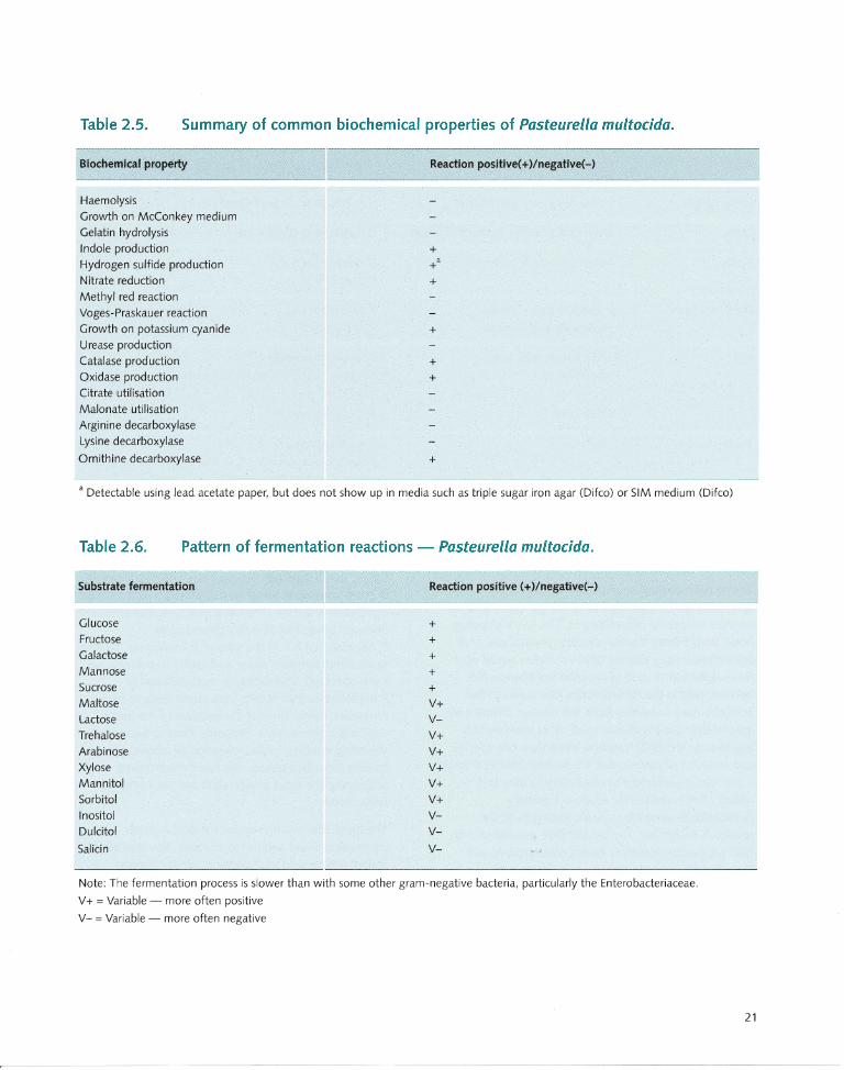
Table 2.5. Summary of common biochemical properties of Pasteurella multocida.
Biochemical property
Haemolysis
Growth on McConkey medium
Gelatin hydrolysis
Indole production
Hydrogen sulfide production
Nitrate reduction Methyl red reaction
Voges-Praskauer reaction
Growth on potassium cyanide
Urease production
Catalase production
Oxidase production Citrate utilisation
Malonate utilisation
Arginine decarboxylase
Lysine decarboxylase
Ornithine decarboxylase
Reaction positive(+)/negative(-)
+ +'
+
+
+
+
+
• Detectable using lead acetate paper, but does not show up in media such as triple sugar iron agar (Difco) or SIM medium (Difco)
Table 2 .6. Pattern of fermentation reactions - Pasteurella multocida.
Substrate fermentation
Glucose Fructose
Galactose
Mannose Sucrose
Maltose
Lactose
Trehalose
Arabinose
Xylose Mannitol
Sorbitol Inositol
Dulcitol
Salicin
Reaction positive (+)/negative(-)
+ + + + + V+
VV+
V+ V+
V+ V+
VVV-
Note: The fermentation process is slower than with some other gram-negative bacteria, particularly the Enterobacteriaceae.
V+ = Variable - more often positive
V- = Variable - more often negative
21
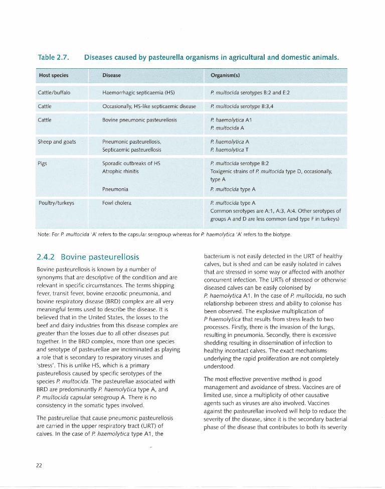
Table 2.7. Diseases caused by pasteurella organisms in agricultural and domestic animals.
Host species
Cattle/buffalo
Cattle
Cattle
Sheep and goats
Pigs
Poultry/turkeys
Disease
Haemorrhagic septicaemia (HS)
Occasionally, HS·like septicaemic disease
Bovine pneumonic pasteurellosis
Pneumonic pasteurellosis,
Septicaemic pasteurellosis
Sporadic outbreaks of HS
Atrophic rhinitis
Pneumonia
Fowl cholera
Organism(s)
P multocida serotypes B:2 and E:2
P multocida serotype B:3,4
P haemolytica A 1
P multocida A
P haemolytica A
P haemolytica T
P multocida serotype B:2
Toxigenic strains of P multocida type D, occasionally,
type A
P multocida type A
P multocida type A
Common serotypes are A:1, A:3, A:4. Other serotypes of
groups A and D are less common (and type F in turkeys)
Note: For P multocida 'A' refers to the capsular serogroup whereas for P haemolytica 'A' refers to the biotype.
2.4.2 Bovine pasteurellosis
Bovine pasteurellosis is known by a number of synonyms that are descriptive of the condition and are relevant in specific circumstances. The terms shipping fever, transit fever, bovine enzootic pneumonia, and bovine respiratory disease (BRD) complex are all very
meaningful terms used to describe the disease. It is believed that in the United States, the losses to the beef and dairy industries from this disease complex are greater than the losses due to all other diseases put together. In the BRD complex, more than one species and serotype of pasteurellae are incriminated as playing a role that is secondary to respiratory viruses and 'stress' . This is unlike HS, which is a primary pasteurellosis caused by specific serotypes of the species P. multocida. The pasteurellae associated with BRD are predominantly P. haemolytica type A, and P. multocida capsular serogroup A. There is no consistency in the somatic types involved .
The pasteurellae that cause pneumonic pasteurellosis are carried in the upper respiratory tract (URT) of calves. In the case of P. haemolytica type A 1, the
22
bacterium is not easily detected in the URT of healthy calves, but is shed and can be easily isolated in calves
that are stressed in some way or affected with another concurrent infection. The URTs of stressed or otherwise diseased calves can be easily colonised by P. haemolytica A 1. In the case of P. multocida, no such relationship between stress and ability to colonise has been observed. The explosive multiplication of P haemolytica that results from stress leads to two processes. Firstly, there is the invasion of the lungs, resulting in pneumonia. Secondly, there is excessive
shedding resulting in dissemination of infection to healthy incontact calves. The exact mechanisms underlying the rapid proliferation are not completely understood.
The most effective preventive method is good management and avoidance of stress. Vaccines are of limited use, since a multiplicity of other causative agents such as viruses are also involved . Vaccines against the pasteurellae involved will help to reduce the
severity of the disease, since it is the secondary bacterial phase of the disease that contributes to both its severity
and fatality. Treatment with appropriate antibiotics helps to reduce the severity of the disease and prevent death. In problem herds where the condition occurs frequently, a knowledge of the antibiotic sensitivity patterns of the strains involved is useful.
2.4.3 Pasteurellosis in sheep and goats
This is probably the most economically irnportant bacterial disease of sheep and goats. The predominant organism that causes pasteurellosis in sheep and goats in temperate climates is P. haemolytica. Biotype A causes pneumonia in all ages of sheep, and septicaemia in young lambs. Biotype T, on the other hand, is associated with a distinct septicaemic syndrome in young adult sheep. P. haemolytica is carried in the nasopharynx and tonsils of apparently healthy sheep. Lambs acquire infection soon after birth, presumably by contact. The carrier rate is low in normal healthy flocks and there is an assortment of serotypes. In flocks undergoing outbreaks, on the other hand, the carrier rate is high and a few specific serotypes dominate. The carrier status has also been found to display seasonal variations.
Predisposing factors undoubtedly playa vital role in most outbreaks. Climatic changes and stressful management practices such as transport, dipping and shearing may precipitate outbreaks. Proper management and the use of vaccines help to reduce the prevalence of the disease.
2.4.4 Pasteurellosis in pigs
Apart from occasional outbreaks of septicaemic disease caused by the HS serotype (B:2), two well-defined syndromes occur in pigs. These are atrophic rhinitis and pneumonia.
Atrophic rhinitis is a disease associated with intensive pig breeding in most parts of the world. It was originally described in Germany nearly 160 years ago, but it was only during the last decade that its complex aetiology and pathogenesis were revealed. Economic losses due to this disease are due not only to deaths, but also to reduced weight gains. The disease is characterised by the atrophy of the nasal turbinates
resulting in a shortening and sometimes twisting of the snout. It is accompanied by sneezing and epistaxis. Two
bacterial organisms have been incriminated in the disease. These are Bordetella bronchiseptica and P. multocida. The former organism is a normal inhabitant of the URT of pigs. Turbinate atrophy that occurs in atrophic rhinitis is preceded by a rapid proliferation of certain toxigenic strains of P. multocida type D. Concurrent infection with B. bronchiseptica or the action of certain irritants creates an environment favourable for such proliferation. Vaccines used in the past were bacterins containing both organisms.
Modern vaccines are a combination of B. bronchiseptica bacterin with pasteurella toxoids prepared from toxigenic strains. Both components play a role in the protective mechanism.
2.4.5 Fowl cholera
This is a disease of considerable economic importance
to the poultry industry in the developed countries of
the world. In the developing countries, where the
poultry industry is rapidly changing from a scenario
dominated by village chicken and smallholder
operations to one of large-scale commercial
undertakings, it has become an emerging disease of
economic importance. In 1986, one estimate gave the
worldwide losses due to fowl cholera as US$200
million . The disease in wild birds is often referred to as
avian cholera or avian pasteurellosis and is considered
to be a threat to the survival of some endangered birds.
The earliest historical record of what is now known to
be fowl cholera dates back to the year 1600. Much of
the earliest work on the role of microorganisms in
infectious diseases, the use of immunising agents and
the contribution of Louis Pasteur in this regard was
related to Pasteur's work on fowl cholera in the 1880s.
Fowl cholera is caused by P. multocida and most
serotypes involved belong to serogroup A. Serotypes
A:1, A:3 and A:4 appear to be the common ones in
most countries, although all of the 16 somatic
serotypes of capsular group A, and some types of
group D, have also been implicated. In turkeys,
capsular serogroup F has been incriminated. Fowl
cholera is a primary pasteurellosis resulting in
septicaemia and death. A chronic form has also been
described and chronically affected birds may serve as
reservoirs of infection.
23
The disease is associated with poor sanitation. The most important control measure is to improve sanitation. Chemotherapeutic agents are used for treatment as well as on a prophylactic basis at low dose levels in situations of high prevalence. The latter use, however, is not encouraged. Vaccines are also used. Polyvalent vaccines containing the commonly occurring
serotypes as bacterins, with or without adjuvants, are available commercially. Their efficacy is variabie .
2.4.6 Other animals
In addition to these diseases of economic importance caused by pasteurella organisms in agricultural and domestic animals, occasional, sporadic outbreaks of disease have been reported in a variety of host species. Some of these are summarised in Table 2.8. Carter (1959) recorded pasteurellae associated with disease
in deer, cats, dogs, horses, mink and monkeys. More recent reports of disease in donkeys and horses (Pavri and Apte 1967), and deer (Jones and Hussaini 1982; Carrigan et al. 1991), are available. Pasteurellosis has also been reported among elephants in Sri Lanka, bison in the United States, camels in Sudan and in a snow leopard in the Himalayas (Carter 1957; De Alwis and
Thambithurai 1965; Bain et al. 1982; De Alwis 1982a; Wickremasuriya and Kendaragama 1982; Chaudhuri et al. 1992).
The types of infections recorded are highly varied and range from septicaemias and respiratory infections, which are the most common forms, to wound infections, abscesses, mastitis, peritonitis and encephalitis. The most common form of infection in the human occurs as a result of animal bites.
Table 2.8. Sporadic incidence of Pasteurella multocida infections in other species.
Animal
Bison
Yak
Deer
Elephant
Camel
Horse
Snow leopard
Cat
Mink
Monkey
24
Country Serogroup
United States B
China B
England, Australia B
Sri Lanka B
Sudan
India, Egypt, Sudan B,D
India F
United States, France
United States A,D
United States A
The Disease: Infection, Clinical Signs and Pathology
Overview
The disease
Haemorrhagic septicaemia is a primary pasteurellosis in
cattle and buffaloes caused by Pasteurella multocida
serotypes B:2 and E:2. More recently, disease syndromes
indistinguishable from HS have been found in other
host species associated with other B serotypes.
Infection
Clinically affected animals and active carrier animals
serve as a source of organisms in outbreaks, and
infection occurs by inhalation or ingestion. Although
the virulence factors responsible for the pathogenesis
are not yet completely understood, recent work has
thrown considerable light on this aspect. There are no
permanent reservoirs of infection outside susceptible
animals but carcases, freshly infected pasture, bedding
etc. may be infective, particularly in moist conditions.
Clinical signs and pathology
Clinical signs, including increased temperature and
respiratory distress, appear after a brief incubation period.
The disease lasts a few hours to a few days and most
animals die with septicaemia in the terminal stages.
Recovery from clinical disease is rare and occurs only when
animals are treated in the initial stages of the disease.
Subcutaneous oedema and widespread petechial
haemorrhages are seen at postmortem examination, as
well as with congested lungs and enlarged lymph nodes.
Pathogenesis and bacteriology
The bacteria appear to multiply in the tonsils. During
the terminal phase of the disease, pasteurellae can be
isolated from blood and rapid multiplication occurs in
the carcase. The nature of the bacterium's virulence is
not fully understood but lipopolysaccharides in the
outer membrane have been implicated, as they inhibit
phagocytosis enabling rapid multiplication. Some
animals can become immune carriers.
3.1 The Disease
Haemorrhagic septicaemia (HS) was previously defined as: an acute, fatal septicaemic disease caused in cattle and buffaloes by one of two specific serotypes of the bacterium Pasteurella multocida.
The two serotypes are popularly known as the Asian and African serotypes, but were designated 6:B and 6:E by the Namioka-Carter system and more recently B:2 and E:2 by the Carter-Heddleston system, respectively (see Section 2.2). Occasionally, these same serotypes cause septicaemic disease in other species (e.g. sheep and goats). In such instances, the term 'septicaemic pasteurellosis' has been used to describe the condition (see Table 2.7).
More recently, a re-examination of the bacterial strains associated with the classical HS syndrome, using modern serotyping or DNA fingerprinting techniques, has indicated that type B strains of P multocida, other than serotype B:2, have also been involved in some instances (Rimier and Wilson 1994). In view of the wider spectrum of serotypes of the Band E groups incriminated in the disease, and the involvement (though rarely) of species of animals other than cattle and buffaloes, it may now be appropriate to give the disease a somewhat broader definition. Hence, HS may now be considered as: an acute, fatal, septicaemic disease caused by strains of P. multocida belonging to the serogroups B or E, commonly in cattle and buffaloes and also in pigs and feral ruminants.
Unlike other pasteurelloses, where the pasteurella organism plays a secondary and opportunistic role, HS is a primary pasteurellosis. It is reproducible experimentally using pure cultures of the causative organism alone, and is preventable by vaccines incorporating the specific serotypes. Thus, HS is a specific form of pasteurellosis, occurring mainly in cattle and buffaloes. This is similar to the situation of typhoid in humans, and pullorum in poultry, which are both caused by a specific strain of salmonella in a specific host species.
25
It must be stressed, however, that only the classical serotypes B:2 and E:2 are capable of producing disease consistently and predictably upon experimental subcutaneous transmission . The other serotypes occasionally encountered are B:1, B:3,4 and B:4 (see Section 2.2). In the author's experience, B:3,4 fails to produce disease consistently, even upon experimental transmission. No information is available on types B:1 and B:4 in this regard but these serotypes have been associated with occasional sporadic outbreaks of disease, indistinguishable from the classical HS
syndrome, mainly in wild animals and, in a few instances, in domestic cattle.
Buffaloes are generally more susceptible than cattle, and young animals are more prone to the disease than adults (De Alwis et al. 1976; De Alwis and Vipulasiri 1980; De Alwis 1981; FAO 1979, 1991). (See Chapter
4 for further details on host and age susceptibility.)
3.2 Source of Infection
In general, P. multocida does not survive long enough outside the animal to become a significant source of infection, although survival may be longer in moist conditions.
Some experiments have shown that the organism can survive in sterilised soil for 2-3 weeks (Bain et aI1982). It was shown in Malaysia, however, that when sterilised earth and mud from rice fields were artificially infected, bacteria could not be recovered after a few hours exposure to sunlight (FAO 1959). When deposited in mud where buffaloes wallow, bacteria could not be
recovered after 24 hours (Bain et al. 1982).
Carcases dumped into rivers and waterways and carried downstream are often incriminated as a likely method of spread of the disease and it is believed that pasteurellae can survive in animal tissues, and perhaps in decomposing carcases, for a few days. Freshly infected pasture, bedding etc. may also be infective. However, no permanent reservoirs of infection have been established outside the animal.
Outbreaks of HS begin when clinically affected or, more likely, carrier animals are introduced into the herd.
Once an outbreak has occurred , decomposing carcases not promptly buried or burnt serve as a source of
26
infection. In experimental transmission, it has been found that large numbers of organisms - in the region of 107 to 1012 colony forming units (CFU) - are required to set up an infection by the natural routes (De Alwis et al. 1990).
How an active carrier animal transmits such a large dose to an incontact animal is not completely understood. It can be speculated that organisms directly shed from a carrier may be more virulent than cultures grown in vitro and used in experimental transmission studies. Alternatively, unknown circumstances may alter the susceptibility of animals so that a smaller infecting dose can cause disease. When isolates from clinical cases and from latent carrier animals were compared, however, no difference in virulence was demonstrated as judged by the median lethal doses (LD50s) for mice (Wijewardana et al. 1986a).
3.3 Routes of Infection
It is believed that the natural routes of infection are by inhalation and/or ingestion, and successful transmission has been made experimentally using intranasal aerosol sprays or oral drenching. However, the dose required to produce clinical disease has not been consistent, and the results obtained with a given dose are not always predictable. Subcutaneous inoculation of bacterial
cultures grown in vitro, with doses ranging from 104 to 107 CFU, has produced more consistent results. In experimental transmission, the route of infection is loosely related to the course of the disease, the clinical syndrome and the extent of pathological lesions. Intranasal infection by aerosols and oral drenching results in a longer course of disease and more profound lesions; subcutaneous inoculation results in rapid onset of disease, a shorter course and less marked pathological lesions.
3.4 Clinical Signs
Figure 3.1 shows a buffalo calf with clinical symptoms of HS. The disease occurs mainly in regions where husbandry practices are primitive and the animals are reared under free-range conditions. In such
circumstances, the animals are not under constant observation and the only reported sign may be sudden
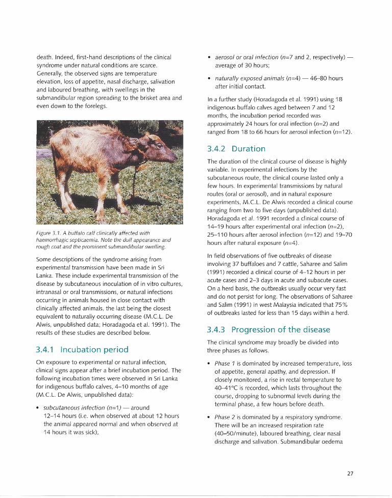
death. Indeed, first-hand descriptions of the clinical syndrome under natural conditions are scarce. Generally, the observed signs are temperature elevation, loss of appetite, nasal discharge, salivation and laboured breathing, with swellings in the submandibular region spreading to the brisket area and even down to the forelegs.
Figure 3.1. A buffalo calf clinically affected with haemorrhagic septicaemia. Note the dull appearance and rough coat and the prominent submandibular swelling.
Some descriptions of the syndrome arising from experimental transmission have been made in Sri Lanka. These include experimental transmission of the disease by subcutaneous inoculation of in vitro cultures, intranasal or oral transmissions, or natural infections occurring in animals housed in close contact with clinically affected animals, the last being the closest equivalent to naturally occurring disease (M.CL. De Alwis, unpublished data; Horadagoda et al. 1991). The results of these studies are described below.
3.4.1 Incubation period
On exposure to experimental or natural infection, clinical signs appear after a brief incubation period. The following incubation times were observed in Sri Lanka for indigenous buffalo calves, 4-10 months of age (M.CL. De Alwis, unpublished data) :
• subcutaneous infection (n=1) - around 12-14 hours (i.e. when observed at about 12 hours the animal appeared normal and when observed at 14 hours it was sick);
• aerosol or oral infection (n=7 and 2, respectively) -average of 30 hours;
• naturally exposed animals (n=4) - 46-80 hours after initial contact.
In a further study (Horadagoda et al. 1991) using 18 indigenous buffalo calves aged between 7 and 12 months, the incubation period recorded was approximately 24 hours for oral infection (n=2) and ranged from 18 to 66 hours for aerosol infection (n=12).
3.4.2 Duration
The duration of the clinical course of disease is highly variable . In experimental infections by the subcutaneous route, the clinical course lasted only a few hours. In experimental transmissions by natural routes (oral or aerosol), and in natural exposure experiments, M.CL. De Alwis recorded a clinical course ranging from two to five days (unpublished data) . Horadagoda et al. 1991 recorded a clinical course of 14-19 hours after experimental oral infection (n=2),
25-110 hours after aerosol infection (n=12) and 19-70 hours after natural exposure (n=4).
In field observations of five outbreaks of disease involving 37 buffaloes and 7 cattle, Saharee and Salim (1991) recorded a clinical course of 4-12 hours in per acute cases and 2-3 days in acute and subacute cases. On a herd basis, the outbreaks usually occur very fast and do not persist for long. The observations of Saharee and Salim (1991) in west Malaysia indicated that 75% of outbreaks lasted for less than 15 days within a herd .
3.4.3 Progression of the disease
The clinical syndrome may broadly be divided into three phases as follows.
• Phase 1 is dominated by increased temperature, loss of appetite, general apathy, and depression. If closely monitored, a rise in rectal temperature to 40-41°( is recorded, which lasts throughout the course, dropping to subnormal levels during the terminal phase, a few hours before death.
• Phase 2 is dominated by a respiratory syndrome. There will be an increased respiration rate (40-50/minute), laboured breathing, clear nasal discharge and salivation . Submandibular oedema
27
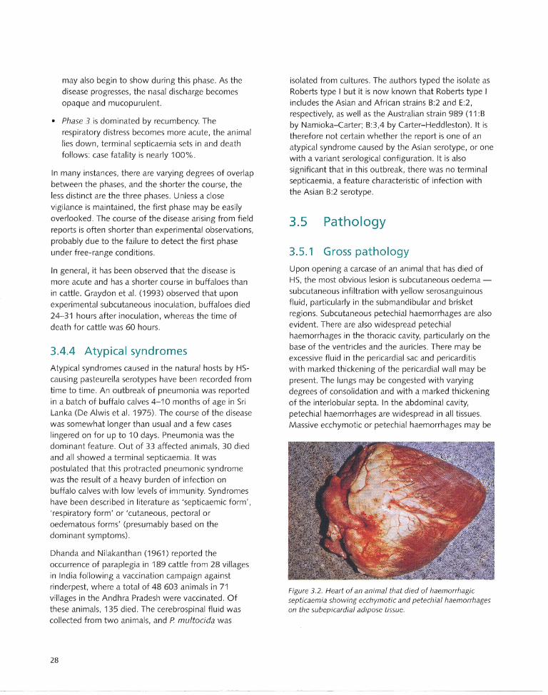
may also begin to show during this phase. As the disease progresses, the nasal discharge becomes opaque and mucopurulent.
• Phase 3 is dominated by recumbency. The respiratory distress becomes more acute, the animal lies down, terminal septicaemia sets in and death follows: case fatality is nearly 100%.
In many instances, there are varying degrees of overlap between the phases, and the shorter the course, the less distinct are the three phases. Unless a close vigilance is maintained, the first phase may be easily overlooked. The course of the disease arising from field reports is often shorter than experimental observations, probably due to the failure to detect the first phase under free-range conditions.
In general, it has been observed that the disease is more acute and has a shorter course in buffaloes than in cattle. Graydon et al. (1993) observed that upon experimental subcutaneous inoculation, buffaloes died 24-31 hours after inoculation, whereas the time of death for cattle was 60 hours.
3.4.4 Atypical syndromes
Atypical syndromes caused in the natural hosts by HScausing pasteurella serotypes have been recorded from time to time. An outbreak of pneumonia was reported in a batch of buffalo calves 4-10 months of age in Sri Lanka (De Alwis et al. 1975). The course of the disease was somewhat longer than usual and a few cases lingered on for up to 10 days. Pneumonia was the dominant feature . Out of 33 affected animals, 30 died and all showed a terminal septicaemia. It was postulated that this protracted pneumonic syndrome was the result of a heavy burden of infection on buffalo calves with low levels of immunity. Syndromes have been described in literature as 'septicaemic form', 'respiratory form' or 'cutaneous, pectoral or oedematous forms' (presumably based on the dominant symptoms) .
Dhanda and Nilakanthan (1961) reported the occurrence of paraplegia in 189 cattle from 28 villages in India following a vaccination campaign against rinderpest, where a total of 48 603 animals in 71 villages in the Andhra Pradesh were vaccinated. Of these animals, 135 died. The cerebrospinal fluid was collected from two animals, and P multocida was
28
isolated from cultures. The authors typed the isolate as Roberts type I but it is now known that Roberts type I includes the Asian and African strains B:2 and E:2, respectively, as well as the Australian strain 989 (11:B by Namioka-Carter; B:3,4 by Carter-Heddleston) . It is therefore not certain whether the report is one of an atypical syndrome caused by the Asian serotype, or one with a variant serological configuration . It is also significant that in this outbreak, there was no terminal septicaemia, a feature characteristic of infection with the Asian B:2 serotype.
3.5 Pathology
3.5.1 Gross pathology
Upon opening a carcase of an animal that has died of HS, the most obvious lesion is subcutaneous oedema -subcutaneous infiltration with yellow serosanguinous fluid, particularly in the submandibular and brisket regions. Subcutaneous petechial haemorrhages are also evident. There are also widespread petechial haemorrhages in the thoracic cavity, particularly on the base of the ventricles and the auricles. There may be excessive fluid in the pericardial sac and pericarditiS with marked thickening of the pericardial wall may be present. The lungs may be congested with varying degrees of consolidation and with a marked thickening of the interlobular septa. In the abdominal cavity, petechial haemorrhages are widespread in all tissues. Massive ecchymotic or petechial haemorrhages may be
Figure 3.2. Heart of an animal that died of haemorrhagic septicaemia showing ecchymotic and petechial haemorrhages on the subepicardial adipose tissue.
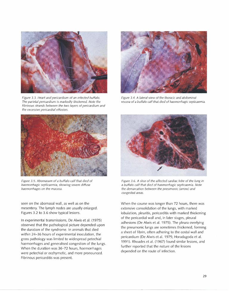
Figure 3.3. Heart and pericardium of an infected buffalo. The parietal pericardium is markedly thickened. Note the fibrinous strands between the two layers of pericardium and the excessive pericardia I effusion.
Figure 3.5. Abomasum of a buffalo calf that died of haemorrhagic septicaemia, showing severe diffuse haemorrhages on the mucosa.
seen on the abomasal wall, as well as on the mesentery. The lymph nodes are usually enlarged. Figures 3.2 to 3.6 show typical lesions.
In experimental transmissions, De Alwis et al. (1975) observed that the pathological picture depended upon the duration of the syndrome. In animals that died within 24-36 hours of experimental inoculation, the gross pathology was limited to widespread petechial haemorrhages and generalised congestion of the lungs. When the duration was 36-72 hours, haemorrhages were petechial or ecchymotic, and more pronounced. Fibrinous pericarditis was present.
Figure 3.4. A lateral view of the thoracic and abdominal viscera of a buffalo calf that died of haemorrhagic septicaemia.
Figure 3.6. A slice of the affected cardiac lobe of the lung in a buffalo calf that died of haemorrhagic septicaemia. Note the demarcation between the pneumonic (arrow) and congested areas.
When the course was longer than 72 hours, there was extensive consolidation of the lungs, with marked lobulation, pleuritis, pericarditis with marked thickening of the pericardial wall and, in later stages, pleural adhesions (De Alwis et al. 1975). The pleura overlying the pneumonic lungs are sometimes thickened, forming a sheet of fibrin, often adhering to the costal wall and pericardium (De Alwis et al. 1975; Horadagoda et al. 1991). Rhoades et al. (1967) found similar lesions, and further reported that the nature of the lesions depended on the route of infection.
29
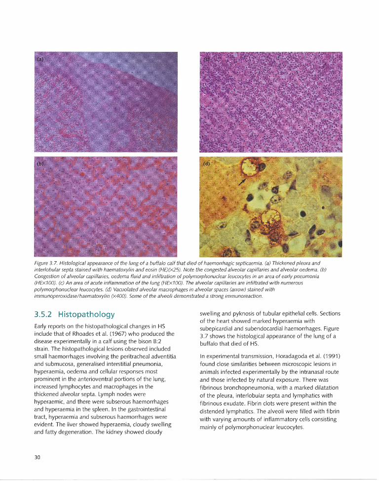
Figure 3.7. Histological appearance of the lung of a buffalo calf that died of haemorrhagic septicaemia. (a) Thickened pleura and interlobular septa stained with haematoxylin and eosin (HE)(X25). Note the congested alveolar capillaries and alveolar oedema. (b) Congestion of alveolar capillaries, oedema fluid and infiltration of polymorphonuclear leucocytes in an area of early pneumonia (HEx100). (c) An area of acute inflammation of the lung (HEx100). The alveolar capillaries are infiltrated with numerous polymorphonuclear leucocytes. (d) Vacuolated alveolar macrophages in alveolar spaces (arrow) stained with immunoperoxidase/haematoxylin (X400). Some of the alveoli demonstrated a strong immunoreaction.
3.5.2 Histopathology
Early reports on the histopathological changes in HS include that of Rhoades et al. (1967) who produced the disease experimentally in a calf using the bison B:2 strain. The histopathological lesions observed included small haemorrhages involving the peritracheal adventitia and submucosa, generalised interstitial pneumonia, hyperaemia, oedema and cellular responses most prominent in the anterioventral portions of the lung, increased lymphocytes and macrophages in the thickened alveolar septa. Lymph nodes were hyperaemic, and there were subserous haemorrhages and hyperaemia in the spleen. In the gastrointestinal tract, hyperaemia and subserous haemorrhages were evident. The liver showed hyperaemia, cloudy swelling and fatty degeneration. The kidney showed cloudy
30
swelling and pyknosis of tubular epithelial cells. Sections of the heart showed marked hyperaemia with subepicardial and subendocardial haemorrhages. Figure 3.7 shows the histological appearance of the lung of a buffalo that died of HS.
In experimental transmission, Horadagoda et al. (1991) found close similarities between microscopic lesions in animals infected experimentally by the intranasal route and those infected by natural exposure. There was fibrinous bronchopneumonia, with a marked dilatation of the pleura, interlobular septa and lymphatics with fibrinous exudate. Fibrin clots were present within the distended lymphatics. The alveoli were filled with fibrin with varying amounts of inflammatory cells consisting
mainly of polymorphonuclear leucocytes.
With immunoperoxidase labelling, it was possible to demonstrate P. multocida antigens in subpleural
lymphatics and interlobular septae, and within blood vessels of visceral organs.
The only available report on the pathological lesions in HS outbreaks occurring naturally in Africa is that of 8astianello and Jonker (1981).
3.6 Pathogenesis
Upon entry of the pasteurella organism into the animal, it is believed that the initial site of multiplication is the tonsillar region. The outcome of this infection depends on an interaction between the virulence of the organism and its rate of multiplication in vivo, on the one hand, and the specific immune mechanisms and nonspecific resistance factors of the host animal, on the other. Thus, the dose of infection is a vital factor and if the organism overcomes the host's defence mechanisms, clinical disease will result. If the defence mechanisms dominate over the organism , what is described as an 'arrested infection' occurs, and the animal becomes an immune carrier. Such animals possess solid immunity, and the
presence of large numbers of such immune animals following an outbreak of disease contributes to 'herd immunity' (De Alwis et al. 1986).
There is currently no evidence that the HS-causing strains of P. multocida produce any exotoxins. It has however been observed that serotype 8:2 strains that cause HS produce hyaluronidase (Carter and Chengappa 1980), whilst a few known 8:2 strains not associated with HS fail to produce this enzyme. It is equally true that other type 8 strains such as 8:3,4, which are known to produce a syndrome indistinguishable from the typical HS, also do not produce hyaluronidase. It is, therefore, uncertain whether this enzyme is of any significance in the pathogenesis.
It is significant that with some type 8 (8:2) and type E strains (E:2), disease can be predictably reproduced experimentally. Other type 8 strains (8:3,4, 8:1, 8:4) have been associated with sporadic outbreaks of disease, but pathogenicity cannot always be demonstrated experimentally. It is still uncertain whether the former strains possess any specific virulence factors, and, if so, whether they are merely phenotypic characters or have a genetic base.
Whatever the virulence factor(s), it is logical to expect it to allow the organism to initially multiply against the defence mechanisms of the body and then produce the lesions that are characteristic of the disease. At present, there is some circumstantial evidence to indicate what these factors might be.
It has been found that the classical Asian serotype (8:2) is capable of causing vacuolation and eventually lysis of macrophages, a property that will diminish phagocytosis and promote multiplication of invading bacteria. This activity of cytoplasmic vacuolation leading to macrophage lysis and death has been demonstrated using HS-causing type 8 strains, but not with the non-HS 8 strains, in a model using mouse peritoneal macrophages and in in vitro studies using a mouse macrophage cell line (Shah et al. 1996). This activity has also been demonstrated using culture supernatants of the same strains, but not for those of the non-HS strains. In the absence of a true exotoxin, it might be expected that free endotoxin would be found in the culture supernatants, a feature also common to other gram-negative bacteria.
Using an ovine mammary neutrophil system and [3Hllabelled type 8 strain of P. multocida, Muniandy et al. (1993) found that capsular polysaccharide extracts known to contain 20% lipopolysaccharides (LPS), potassium thiocyante extracts and Westphal type LPS extracts inhibited phagocytosis. These workers also found that when encapsulated cells and de-encapsulated cells were used, the percentage of de-encapsulated cells phagocytosed was significantly higher than when encapsulated cells of P. multocida were used. These observations indicated that HS-causing strains of P. multocida appeared to possess a factor in their capsule that inhibited the ability of phagocytes to engulf and destroy invading bacterial cells.
It is well established that the endotoxins of gramnegative bacteria consist predominantly of LPS. The toxic effects of the LPS of P. multocida associated with HS have been amply demonstrated (Rebers et al. 1967; Rhoades et al. 1967). These workers prod uced
experimental HS in calves and pigs by different routes using type 8 strains. They also administered endotoxin prepared from this strain to a calf. The symptoms and lesions produced in the calf given endotoxin resembled those of experimental infection. More recently, N.U.
31

Horadagoda et al.t(personal communication 1998)
studied the acute phase response (APR) in buffaloes
infected experimentally with HS. The APR is a
nonspecific, systemic host response that is cytokine
mediated; it results from tissue injury or infection and is
an integral part of the host defence mechanism. The
APR is characterised by changes in body temperature,
endocrine changes and changes in trace mineral levels,
leucocytes and serum proteins. Horadagoda et al.
observed a rapid rise in serum haptoglobin and a more
delayed increase in a1-acid glycoprotein. A rapid rise in
serum iron and zinc was accompanied by a delayed rise
in copper levels. Leucocyte counts dropped progressively
with advancement of the clinical syndrome, reaching
their lowest immediately before death. Serum cortisol
increased progressively, reaching its peak just before
death, or in the terminal moribund stage. Endotoxin
levels in the serum remained unchanged throughout the course of the disease and displayed a spectacular rise
immediately before death. These changes are typical of
the APR in any infection and the terminal rise in endotoxin level coincides with the observed terminal
septicaemia (M.C.L. De Alwis, unpublished data;
Horadagoda et al. 1991). When a group of buffaloes
were injected with endotoxin prepared from an HS
causing strain of P. multocida, the APR was strikingly
similar to that following infection (N .U. Horadagoda et
al. t (personal communication 1998).
Whilst the presence of other virulence factors cannot be
discounted, there is sufficient evidence to implicate LPS as
an entity largely responsible for the disease. The evidence
supports its role in inhibiting phagocytosis, thereby
enabling rapid multiplication , and further reinforces the
role of the capsule in the virulence mechanism (the
inhibitory effect is greater in encapsulated strains) . It also
accounts for the APR and the typical lesions observed in
HS. This hypothesis is further supported by the findings
of Shah et al. (1996) that sera from buffaloes recovering
from HS inhibited the vacuolating cytotoxic activity of
type B strains as well as of culture supernatants known to
contain free endotoxin.
t N.U. Horadagoda (Faculty of Veterinary M edicine and Animal Science, Unive rsity of Peradeniya; ). C. H odgson and C .M. Moon (Moreton Research Inst itute, Edinburgh , UK); PD. Eckersall (Department of Veterinary Clinical Studies, Clasgow Veterinary School, UK); and T.C. Wijewardana (Veterinary Research Institute, Peradeniya, Sri Lanka) .
32
3.7 Bacteriology
Septicaemia is essentially a terminal event and
pasteurellae can only be isolated from blood during the
terminal phase of the disease. At the time of death,
around 105-106 CFU/mL of organisms appear in the
blood. Rapid multiplication occurs in the carcase; under
tropical conditions counts in the range of
1011 _1012 CFU/mL have been recorded in carcases
20-21 hours after death (M.C.L. De Alwis, unpublished
data) . Apart from animals infected by aerosol, the
appearance of pasteurellae in the nasopharynx was inconsistent, and the chances of isolation increased
with the progress of the disease.
In an outbreak of what was described as HS in beef
cattle in Zimbabwe, heavy growths of nearly pure
cultures were made from heart blood, lung, pericardium,
spleen, liver, kidney and brain. Organisms were not
recovered from the foetal tissue (Lane et al. 1992).
3.8 Recovery
Once clinical signs are established in typical field
outbreaks, death invariably occurs. Recoveries may
occur only if the disease is treated in the early stages of
the clinical syndrome. When the disease occurs in an
organised farm, losses can be minimised by regularly
checking the rectal temperatures of all incontact
animals and giving antibiotic treatment to any animals
showing increased temperatures.
Spontaneous recovery without treatment is extremely rare, but it may occur in a few animals towards the end
of an outbreak. Such instances are presumably due to the
animal possessing some degree of immunity at the time
of exposure. In a controlled exposure of buffaloes to
experimental infection with a view to producing carrier
animals, some animals showed early signs, followed by recovery (De Alwis et al. 1990). This phenomenon is
referred to as 'arrested infection', and invariably leads to
an immune carrier state (see Section 4.7).
There are some instances of high recovery rates on
record but these are likely to be due to inconsistencies
in routine disease notification systems.
Epidemiology
Overview
Global distribution
The global distribution of haemorrhagic septicaemia
(HS) includes almost all parts of the world .. The disease
is generally associated with wet, humid weather
conditions, extensive farming practices that do no t allow
for good disease prevention management strategies, and areas where buffaloes and cattle are abundant.
Distribution of serotypes
Of the classical serotypes, B:2 is found in Asia and E:2
in Africa while some Middle Eastern and East African
states record both . A few other B serotypes have bee n
associated with sporadic outbreaks of disease resembling
HS, particularly in wild animals.
Host susceptibility, morbidity and mortality
Of the conventional host species, buffaloes are more
susceptible than cattle. The morbidity is highly variable
and is influenced by a variety of factors. When first
introduced, explosive outbreaks occur. In endemic
situations, few deaths occur at a time, mainly among young adult animals. When clinical disease is
established, however, death invariably occurs and case
fatality is near 100%.
Carriers
A variable proportion of animals in a given population
are latent carriers. These animals intermittently become
active and shed virulent pasteurellae.
Disease outbreaks
New outbreaks occur when a shedder comes into contact with a susceptible animal (e .g. one introduced
from elsewhere or born since the previous outbreak).
Once the first clinical case occurs, more bacteria are
shed and disseminated. The size of an outbreak depends
on the proport ion of immune to nonimmune animals in
the herd .
4.1 Geographic Distribution of Disease
The global distribution of HS (which was described in Chapter 1) is related to climatic conditions, husbandry practices and the types of animals reared . This is shown in Sri Lanka, an island of 65 000 km 2 with a variety of agroclimatic zones and related husbandry practices and distinct endemic and nonendemic areas for HS.
In the hill country, where the climatic conditions are mild (elevation over 1000 m, temperature range 1 a-24°C) and temperate dairy breeds are reared intenSively, the disease is almost nonexistent. In the warmer dry plains, on the other hand, with heavy seasonal rains, where indigenous cattle and buffaloes and zebu cattle are reared under a free-grazing extensive system of management, the disease is endemic. Intermediate areas experience occasional sporadic outbreaks. A similar geographic distribution is also well documented in India (Sharma et al. 1982; Dutta et al. 1990). It is likely that HS may occur in some South American states, with high water buffalo populations, and where conditions are somewhat similar to those prevailing in tropical Asia.
4.2 Seasonal Effects on Distribution
HS is generally associated with wet, humid weather conditions. Massive outbreaks recorded in Sri Lanka (Perumalpillai and Thambiayah 1957; Dassanayake 1957) and in Zambia (Francis et al. 1980) were all associated with heavy rains. In Sudan, outbreaks are reported to occur during rainy seasons (Mustafa et al. 1978). Seasonality has been well documented in India, with the majority of outbreaks occurring during the wet season (from July to September), with the peak in August. The correlation with wet weather conditions
33
was further evident from studies on geographical distribution of the disease in India. The disease occurred more frequently in states that received a higher annual mean rainfall (Dutta et al. 1990; Saini et al. 1991). Sharma et al. (1982) has similarly reported that 83% of all cases in Uttar Pradesh, India occurred during the wet monsoon months of July, August and September. Seasonal occurrence has also been confirmed in Pakistan (Sheikh et al. 1996).
Active surveillance studies in Sri Lanka (De Alwis and
Vipulasiri 1980; De Alwis 1981), however, have shown clearly that outbreaks occur throughout the year. Whilst dry season outbreaks appear to be contained, outbreaks occurring during wet seasons tend to spread, presumably due to the longer survival of the causative organism outside the animal. In Asian countries, there are other wet season associated factors that promote spread of the disease. With the start of the rainy season, considerable movement of animals occurs for draught power to plough the rice fields. The outbreak in Northern Samar in the Philippines in 1993-94 was attributed, amongst other factors, to movement of new animals into the area from Mindanao (Molina et al. 1994).
4.3 Distribution of Serotypes
The classification of Pasteurella multocida serotypes was described in Chapter 2. In Asian countries, the only serotype that has been reported is type B:2. Perreau (1961) first recognised that outbreaks of acute septicaemic pasteurellosis recorded in Africa were typical HS and Carter (1961) showed that the causative organism was a new type of P. multocida, which he designated as type E, to distinguish it from the Asian type B.
The African states of Senegal, Mali, Guinea, Ivory Coast, Nigeria, Cameroons, Central African Republic and Zambia have all reported serotype E:2 (Perreau 1961; Francis et al. 1980). Egypt and Sudan have recorded the presence of both E and B serotypes (Shigidi and Mustafa 1979), and there is one record of
an outbreak caused by the B (Asian) strain in Cameroon (Martrenchar 1993). There are indications that serotype B:2 may be present in some East African countries.
All the North American isolates were originally identified as the Asian serotype (B:2). More recently,
34
Rimier and Wilson (1994) re-examined these isolates using the Carter-Heddleston serotyping scheme and
DNA fingerprinting methods. This revealed that the bison strain isolated from Montana in 1965 was serotype B:3,4, not B:2 as earlier reported. A dairy calf strain isolated in 1969 was also B:3,4. No distinction could be made between these strains by DNA fingerprinting. On re-examination, the 1993 isolate from beef calves in California was confirmed as B:2, whilst the Canadian strain was B:3,4. DNA fingerprinting showed that the latter two strains were
distinct from each other as well as from all other strains in the culture collection. Recently, Rimier (1997) has
examined a global collection of over 200 isolates associated with classical HS and related syndromes, and identified a large number of DNA fingerprint profiles (see Section 5.10.3).
HS is not known to be present in the United Kingdom. However, in 1982 a strain was isolated from deer with a septicaemic disease and was found to be serotype B:3,4 (Jones and Hussaini 1982; Myint et al. 1987; Rimier et al. 1987). Other serotypes associated with sporadic septicaemic pasteurellosis resembling HS in feral ruminants include serotype B:1 isolated from antelope in the United States, and B:4 from bison in Canada. Serotype B:3,4 has also been associated with septicaemic disease in elk in the United States (Rimier 1993). Genetic fingerprinting has established that the
North American B:2 strains possess a DNA profile different from that of the Asian B:2 strains. From an epidemiological standpoint, DNA fingerprinting serves as an additional tool (to serotyping) in strain identification .
4.4 Host Susceptibility
The conventional host species are cattle and buffaloes. There is general agreement that buffaloes are more susceptible than cattle. In Malaysia, it was reported that 73% of all deaths due to HS during the period 1970-79 and 90% during the period 1980-89 were among buffaloes, despite the fact that the buffalo population in Malaysia is half that of cattle (FAa 1979, 1991). The higher losses in buffaloes have also been
quantified and clearly shown in epidemiological studies in Sri Lanka (De Alwis and Vipulasiri 1980; De Alwis 1981). An island-wide study covering endemic as well
as nonendemic areas revealed that mortality in buffaloes was three times higher than in cattle . This difference was more marked in the sporadic outbreaks that occurred in nonendemic areas and less evident in endemic areas, where outbreaks occurred regularly. In the latter case, there were other factors that partly masked the host susceptibility. In contrast to the general results, one of the Sri Lankan investigations indicated that the percentage of cattle and buffalo
herds that experienced outbreaks during a given period was not significantly different. Once the disease was introduced to a herd, however, mortality within a herd was higher in buffaloes. In India, Ramarao et al. (1991)
recorded a higher incidence of HS in districts where the buffalo population was higher. Bali cattle (B05 banteng)
in Indonesia are believed to be highly susceptible to HS (Bain 1979a; Bain et al. 1982).
There is evidence of type B infection in pigs in Malaysia and India (FAO 1959, 1979; Murthy and Kaushik 1965; Pillai et al. 1986). Verma and Sexena (1987) and Verma (1988) established clearly that septicaemic pasteurellosis in pigs in India was caused by serotype B:2. For many years, strains identified as type 6:B have been reported from pigs in Sri Lanka. These isolates have been used to successfully reproduce a typical HS syndrome in cattle (M.CL. De Alwis, unpublished data). Gamage et al. (1995) reported an outbreak of
acute septicaemic pasteurellosis in pigs in a farm in Sri Lanka, caused by serotype B:2 . Thus, although the pig is not a conventional host, it displays susceptibility.
Although type B strains have also been associated with poultry (Gupta 1973; Gupta and Kumar 1973; FAO
1979), it is generally accepted that serotype B:2 is nonpathogenic to poultry.
Sporadic cases of P. multocida infection have been recorded in goats (which are a common small ruminant in the Asian region) in India and Malaysia (FAO 1959, 1979, 1991). The isolates have been identified as
Roberts type I, Carter type B, or serotype 6:B, according to the available serotyping methods in use at the time. Investigations on the susceptibility of goats carried out in Sri Lanka, however, provided ample evidence of the low susceptibility of goats to HS. No natural transmission occurred when goats were housed in close contact with clinically affected buffaloes and the goats
did not even elicit an antibody response. Goats were
also subjected to intranasal as well as subcutaneous inoculation, with doses in the range of 1010 to 1012 colony forming units (CFU). Only 10% of the goats died . Others showed merely a transient rise in temperature and swelling at the site of inoculation (Wijewardana et al. 1986b).
Wijewardana et al. (1986c) did not identify any carriers among 254 goats in an abattoir study. For 494 cattle slaughtered in the same abattoir during the same period, 3.4% were found to be carriers. Loganathan and Chandrasekaran (1995) produced a pneumonic syndrome in transport-stressed, six-month-old goats experimentally infected with the Asian serotype by the
intranasal route and simultaneously given a three-day course of dexamethazone. All of four animals died in five days. A B:3,4 deer isolate was also tested for its pathogenicity to goats. Four goats were inoculated subcutaneously, and two intranasally, with doses
ranging from 109 to 1011 CFU, but showed no signs of disease (M.CL. De Alwis, unpublished data). Thus,
there is no evidence to incriminate the goat as a susceptible host or a reservoir of infection for the Asian
serotype B:2 or serotype B:3,4.
It was also reported that Rahmani sheep in Egypt resisted all attempts at experimental infection by the subcutaneous or intranasal routes, using an organism that caused classical HS in calves by similar methods (Barakat et al. 1976).
There are numerous other species in which septicaemic sporadic disease caused by HS serotypes is recorded . Pavri and Apte (1967) have reported the disease in horses and donkeys in India. Gevedze (1986) reported an acute septicaemic pasteurellosis among horses in Belorussia, believed to have been acquired from cattle.
There is evidence of the disease in the African buffalo Synceru5 nanU5 (Kasali 1972). Feral buffaloes in Sri
Lanka are known to possess very high antibody levels that are normally associated with arrested infections and
often present in survivors from outbreaks (M.CL De Alwis, unpublished data). This supports the suspicion that the disease is present in feral buffaloes in Asia.
HS has been reported among camels in Sudan (Bain et
al. 1982) but Awad et al. (1976) found that camels resisted experimental infection with doses lethal to buffaloes. HS has been well documented in wild
35
elephants in Sri Lanka, associated with outbreaks among cattle and buffaloes in the locality (De Alwis and Thambithurai 1965; De Alwis 1982a; Wickremasuriya and Kendaragama 1982). There are indications that septicaemic disease associated with type B occurs among yak in China and Nepal , although these are unconfirmed. In the United States, septicaemic disease caused by serotype B:1 has been recorded in antelope and that caused by B:3,4 in elk; Canada has recorded disease caused by B:4 in bison (Rimier 1993).
4.5 Morbidity, Mortality and Case Fatality
4.5.1 Rates of morbidity, mortality and case fatality
When HS is first introduced to virgin areas, or when it occurs as sporadic outbreaks in nonendemic areas, morbidity is high. These once-typical, explosive outbreaks no longer occur in most countries where the disease has become endemic. When clinical disease is established, however, mortality invariably occurs and case fatality is nearly 100% . True recovery from clinical disease occurs only if the animal is treated in the very early stages (see Section 3.8). Conditions prevailing in the field often prevent such treatment.
Reports of high recovery rates do exist, however. An analysis of records in India from 1949 to 1987 shows a recovery rate varying between 10% and 50%. Nepal records recovery rates of 60% to 90% and the Philippines over 90% (FAO 1979). These apparently high recovery rates have not been based on scientific study and may be due to routine local reporting systems in which the number said to be affected may be the total number exposed (hence at risk) rather than the number actually with the disease.
In Pakistan, observations from nine districts in Punjab showed an 11 % morbidity with 9% mortality and hence a 78% case fatality for buffaloes. In the same study, the corresponding figures for cattle were 4 %, 2.5% and 62% respectively (Sheikh, et al. 1996). A study of an outbreak in Zimbabwe in a beef cattle farm revealed an unusually high recovery rate with 77% morbidity and only 5% mortality (Lane et al. 1992), but this study had some noteworthy features. Though
36
the clinical picture and pathology resembled HS the isolate had not been serotyped. It was presumed to be type E, based on the location. Further, the outbreak occurred in an organised farm, and intensive antibiotic therapy was given together with a bacterin. All of these factors could influence the normal course and pattern of clinical disease and mortality.
4.5.2 Factors influencing morbidity and mortality
A variety of factors and their interactions influence morbidity and mortality. These have been well documented in two epidemiological studies in Sri Lanka (De Alwis and Vipulasiri 1980; De Alwis 1981).
Host species and age
The susceptibility of different host species was discussed in Section 4.4. Morbidity is higher in buffalo herds or mixed herds with a high buffalo population .
In many Asian countries young animals have been observed to be more susceptible than adults but quantitative estimates are scarce. In one study covering HS endemic areas in Sri Lanka, 77% of HS cattle deaths and 65% of HS buffalo deaths were of animals under two years old. Another study of two outbreaks in neighbouring farms showed that the most vulnerable age group was six months to two years (De Alwis et al. 1976). The age distribution of deaths for one farm in this study is shown in Figure 4.1 . The disease rarely occurred in calves under six months of age. As these studies were carried out in endemic areas, a large percentage of the adult population would have had high antibody levels. It is therefore likely that very young calves (under six months) possessed sufficient levels of maternal immunity.
On a second farm, located in close proximity to the first, vaccination was started immediately. The disease broke out in this farm two days after vaccination, but the mortality was 2.3% of the susceptible population, compared to 8.2% in the first farm, and the peak mortality was in the 6-12-months age group. There has only been one noteworthy case of HS in a very young calf in Sri Lanka (two months of age). This was a calf born to a cow imported from a country free from HS and hence without maternal antibodies (M.C.L. De Alwis , unpublished data).
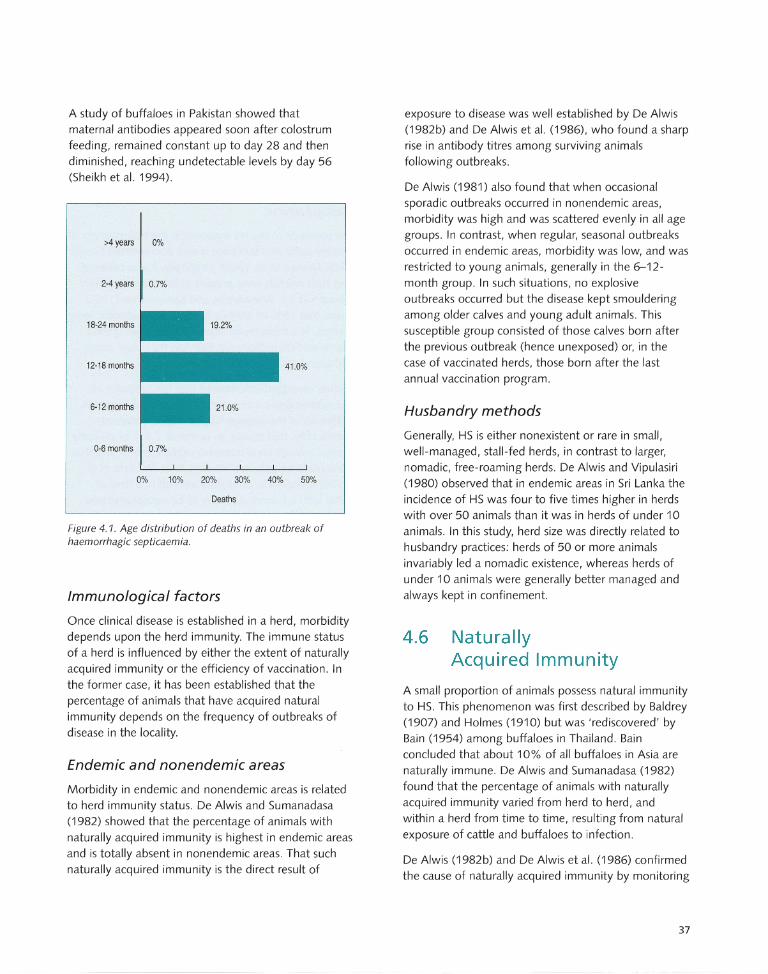
A study of buffaloes in Pakistan showed that maternal antibodies appeared soon after colostrum feeding, remained constant up to day 28 and then diminished, reaching undetectable levels by day 56 (Sheikh et al. 1994).
>4 years 0%
2-4 years 0.7%
18-24 months
12-18 months 41.0%
6-12 months
0-6 months 0.7%
0% 10% 20% 30% 40% 50%
Deaths
Figure 4.1. Age distribution of deaths in an outbreak of haemorrhagic septicaemia.
Immunological factors
Once clinical disease is established in a herd, morbidity depends upon the herd immunity. The immune status of a herd is influenced by either the extent of naturally acquired immunity or the efficiency of vaccination . In the former case, it has been established that the percentage of animals that have acquired natural immunity depends on the frequency of outbreaks of disease in the locality.
Endemic and nonendemic areas
Morbidity in endemic and nonendemic areas is related to herd immunity status. De Alwis and Sumanadasa (1982) showed that the percentage of animals with naturally acquired immunity is highest in endemic areas and is totally absent in nonendemic areas. That such naturally acquired immunity is the direct result of
exposure to disease was well established by De Alwis (1982b) and De Alwis et al. (1986), who found a sharp rise in antibody titres among surviving animals following outbreaks.
De Alwis (1981) also found that when occasional sporadic outbreaks occurred in nonendemic areas, morbidity was high and was scattered evenly in all age groups. In contrast, when regular, seasonal outbreaks occurred in endemic areas, morbidity was low, and was restricted to young animals, generally in the 6-12-month group. In such situations, no explosive outbreaks occurred but the disease kept smouldering among older calves and young adult animals. This susceptible group consisted of those calves born after the previous outbreak (hence unexposed) or, in the case of vaccinated herds, those born after the last annual vaccination program.
Husbandry methods
Generally, HS is either nonexistent or rare in small, well-managed, stall-fed herds, in contrast to larger, nomadic, free-roaming herds. De Alwis and Vipulasiri (1980) observed that in endemic areas in Sri Lanka the incidence of HS was four to five times higher in herds with over 50 animals than it was in herds of under 10 animals. In this study, herd size was directly related to husbandry practices: herds of 50 or more animals invariably led a nomadic existence, whereas herds of under 10 animals were generally better managed and always kept in confinement.
4.6 Naturally Acquired Immunity
A small proportion of animals possess natural immunity to HS. This phenomenon was first described by Baldrey (1907) and Holmes (1910) but was 'rediscovered' by Bain (1954) among buffaloes in Thailand. Bain concluded that about 10% of all buffaloes in Asia are naturally immune. De Alwis and Sumanadasa (1982) found that the percentage of animals with naturally acquired immunity varied from herd to herd, and within a herd from time to time, resulting from natural exposure of cattle and buffaloes to infection.
De Alwis (1982b) and De Alwis et al. (1986) confirmed the cause of naturally acquired immunity by monitoring
37
the antibody status of animals following outbreaks of HS. The latter investigation covered three mixed herds of cattle and buffaloes where vaccination had never been carried out and where outbreaks of HS had occurred a few days before the investigation . During the period immediately after the outbreak , 80%, 88.8% and 92% of the surviving animals in the respective herds developed antibody titres. The former study involved monitoring antibody status in a group of 26 unvaccinated buffalo calves where an outbreak occurred . There was a steep rise in antibody levels from two to four weeks after the outbreak.
De Alwis and Sumanadasa (1982) investigated the immune status of a wider range of animals, in unvaccinated herds. Their sample consisted of 504 animals in endemic areas, 209 from moderate incidence areas where sporadic outbreaks occurred, and 212 from low incidence areas, where the disease is an exceptional occurrence. Around 36% of animals in endemic areas had naturally acquired antibodies, compared with 7.2% and 0.47 % , respectively, for the other two categories.
There are, however, a few instances on record where antibodies against the specific serotype have been detected in situations where the disease does not exist. In a joint project at Michigan State University, United States in 1977-78, M.C.L De Alwis and G.R. Carter (unpublished data) found mouse protective antibodies among cattle with no history of exposure to HS. Similar observations have been made in a broader study in the United States (Sawada et al. 1985), Australia (Bain 1954) and Chad (Bain et al. 1982). In these instances, immunity may have been induced by related serotypes of P. multocida, sharing common antigens.
Once the main cause of naturally acquired immunity was recognised as sublethal exposure to disease, it became possible to understand the different morbidity and mortality patterns in endemic and nonendemic areas.
38
4.7 Carrier Status
4.7.1 Presence of pasteurellae in carrier animals
Nasopharynx
The presence of the HS organism in the nasopharynx of healthy cattle and buffaloes is well documented (Singh 1948; Mohan et al. 1968). Originally, it was believed that such animals were present at low levels in a herd (about 1-2 %) . Wijewantha and Karunaratne (1968) found that 15 % of animals brought to an abattoir were carriers . In a more recent study, Sheikh et al. (1994) examined 500 buffaloes in Pakistan for carrier status and found a 4.6% carrier rate.
Further investigations showed that the presence of nasopharyngeal carrier animals was related to recent outbreaks of the disease. Gupta (1962) observed, significantly, that during an outbreak 7.5% of clinically normal animals were nasopharyngeal carriers, and that none could be detected 40 days later. Mustafa et al. (1978) found 44 % of healthy animals in a herd of cattle with a current outbreak to be nasopharyngeal carriers. In three other herds, unassociated with the disease at the time, the carrier rates were 3.8%,0% and 5.5% respectively. Hiramune and De Alwis (1982) found 22 .7% of animals to be nasopharyngeal carriers in four herds where an outbreak of HS had occurred one week earlier. In herds where the interval between the outbreak and examination was longer, there was a progressive drop in the percentage of nasopharyngeal carrier animals detected, reaching 1.9% in herds examined six weeks after an outbreak.
These findings indicated that the percentage of nasopharyngeal carriers was related to recent exposure to disease. All of these researchers identified carriers by swabbing the nasopharynx through the external nares on one occasion only. Wijewantha and Karunaratne (1968), on the other hand, swabbed the nasopharynx directly, after slaughter, and this procedure probably accounted for the higher detection rate of 15%. Singh (1948) found that his detection rate doubled with direct swabbing postmortem compared with swabbing the external nares in the live animal.
Other sites
In another abattoir study, Wijewardana et al. (1986c) also found that pasteurellae were not confined to the nasopharynx; these researchers were also able to make isolations from the associated lymph nodes of slaughtered animals. Saharee et al. (1993) made similar observations among cattle in Malaysia. In this study, pharyngeal swabbing pre- and post-slaughter gave an isolation rate of only 0.3 %. When retropharyngeal lymph nodes were also cultured, a 13% isolation rate of pasteurellae was recorded, but only 3 % were type B. A three-year survey in Indonesia used a combination of nasopharyngeal swab and lymph node culture. Of over 1500 animals sampled, 2.49% were found to be carriers of Pasteurella spp. (Dartini and Ekaputra 1996) but most of the isolates were type A.
De Alwis et al. (1986) monitored both the nasopharyngeal carrier status and the antibody status in three herds for a few months, starting soon after outbreaks of HS. Upon repeated swabbing, they found that different animals displayed the carrier status on different days of swabbing, and the percentage of animals that were nasopharyngeal positive on at least one occasion ranged from 12 % to 40%. The methods used in different studies and the different results obtained are summarised in Table 4.1. De Alwis et al. (1986) observed that some animals displayed a positive status, then negative, and subsequently positive again, indicating an intermittent appearance of the organism in the nasopharynx. The question therefore arose as to where the organism remained lodged during the interim period of disappearance from the nasopharynx.
4.7.2 Experimental studies on carrier status and naturally acquired immunity
In order to understand more clearly the status of carrier animals, their relationship to naturally acquired immunity and their significance in the epidemiology of the disease, De Alwis et al. (1990) produced carrier animals by controlled experimental infection or natural exposure. Using 57 young buffaloes, these researchers were able to produce a carrier status in 32 animals. These carrier animals were divided into several groups for different investigations. One group was maintained for a period of 360 days; carrier status was determined
by positive nasopharyngeal isolation from swabs inserted through the external nares, and antibody status measured by an indirect haemagglutination test. In most animals, pasteurellae appeared in the nasopharynx for a short period and then disappeared. In others, it reappeared intermittently after long periods of absence. The longest period after which such reappearance occurred was 215 days after initial exposure. All of these carrier animals also showed rising antibody titres, reaching a peak lasting for around 150-180 days, and then declining.
Further groups of immune carrier animals were slaughtered at varying intervals and attempts were made to isolate the organism from 14 different sites. The most consistent site from which isolations were made was the tonsil (20 out of 27 animals) . The longest period after initial exposure when isolations were made from the tonsils was 229 days. In all these experiments, animals were exposed to a streptomycinresistant organism; the resistance factor served as a marker to ensure that the organism recovered was the one used in the initial exposure.
Having established under experimental conditions that the tonsil was the site of localisation of pasteurellae in carrier animals, Wijewardana et al. (1993) examined 103 pairs of tonsils from cattle in an abattoir in an endemic area. Forty-nine isolations of P. multocida were made. The majority (23) were capsular type A; 10 were type B. The other 16 could not be typed by the capsular typing procedure. All but one from this third group could be typed by Heddleston's somatic typing, but none were of somatic type 2 (which is associated with HS) or type 5 (which cross-reacts with type 2). This work, in general, established that, under natural conditions as well as for experimental animals, the tonsil was a site of localisation of all types of pasteurellae.
4.7.3 Latent and active carriers
The field observations and experimental findings outlined above led to the conclusion that two types of carriers exist - latent and active. It appears that in endemic areas, where frequent outbreaks occur, most adult animals harbour pasteurellae in their tonsils. From time to time, active multiplication takes place in the tonsils and the organism spills over into the nasopharynx and is shed in nasal secretions. These latent carriers show high levels of antibody by the IHA
39
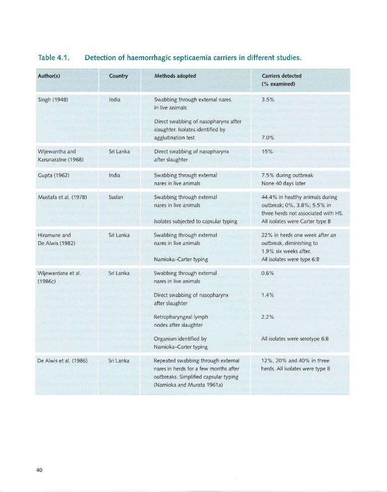
Table 4.1. Detection of haemorrhagic septicaemia carriers in different studies.
Author(s)
Singh (1948)
Wijewantha and
Karunaratne (1968)
Gupta (1962)
Mustafa et al. (1978)
Hiramune and
De Alwis (1982)
Wijewardana et al.
(1986c)
De Alwis et al. (1986)
40
Country
India
Sri Lanka
India
Sudan
Sri Lanka
Sri Lanka
Sri Lanka
Methods adopted
Swabbing through external nares.
in live animals
Direct swabbing of nasopharynx after
slaughter. Isolates identified by
agglutination test
Direct swabbing of nasopharynx
after slaughter
Swabbing through external
nares in live animals
Swabbing through external
nares in live animals
Isolates subjected to capsular typing
Swabbing through external
nares in live animals
Namioka-Carter typing
Swabbing through external
nares in live animals
Direct swabbing of nasopharynx
after slaughter
Retropharyngeallymph
nodes after slaughter
Organism identified by
Namioka-Carter typing
Repeated swabbing through external
nares in herds for a few months after
outbreaks. Simplified capsular typing
(Namioka and Murata 1961 a)
Carriers detected
(% examined)
3.5%
7.0%
15%
7.5% during outbreak
None 40 days later
44.4% in healthy animals during
outbreak; 0%, 3.8%; 5.5% in
three herds not associated with HS.
All isolates were Carter type B
22 % in herds one week after an
outbreak, diminishing to
1 .9 % six weeks after.
All isolates were type 6:B
0.6%
1.4%
2.2%
All isolates were serotype 6:B
12 %, 20% and 40% in three
herds. All isolates were type B
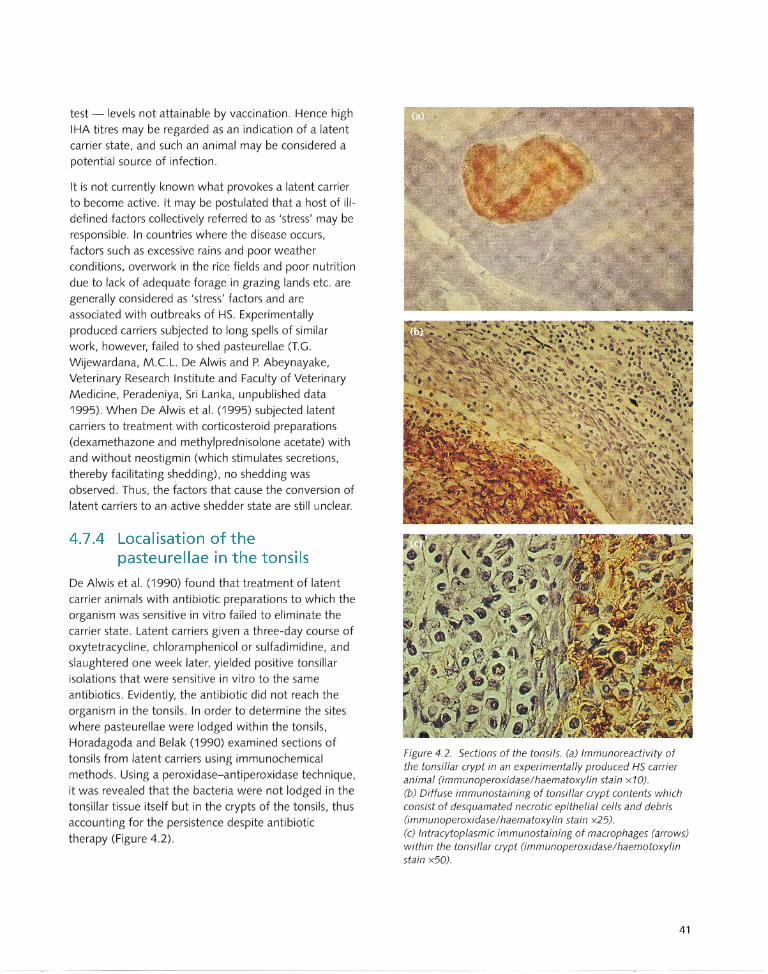
test - levels not attainable by vaccination. Hence high IHA titres may be regarded as an indication of a latent carrier state, and such an animal may be considered a potential source of infection.
It is not currently known what provokes a latent carrier to become active. It may be postulated that a host of illdefined factors collectively referred to as 'stress' may be responsible . In countries where the disease occurs, factors such as excessive rains and poor weather conditions, overwork in the rice fields and poor nutrition
due to lack of adequate forage in grazing lands etc. are generally considered as 'stress' factors and are associated with outbreaks of HS. Experimentally produced carriers subjected to long spells of similar work, however, failed to shed pasteurellae (T.G . Wijewardana, M.C.L. De Alwis and P. Abeynayake, Veterinary Research Institute and Faculty of Veterinary Medicine, Peradeniya, Sri Lanka, unpublished data 1995). When De Alwis et al. (1995) subjected latent carriers to treatment with corticosteroid preparations (dexamethazone and methylprednisolone acetate) with and without neostigmin (which stimulates secretions, thereby facilitating shedding), no shedding was observed. Thus, the factors that cause the conversion of latent carriers to an active shedder state are still unclear.
4.7.4 Localisation of the pasteurellae in the tonsils
De Alwis et al. (1990) found that treatment of latent carrier animals with antibiotic preparations to which the organism was sensitive in vitro failed to eliminate the carrier state. Latent carriers given a three-day course of
oxytetracycline, chloramphenicol or sulfadimidine, and slaughtered one week later, yielded positive tonsillar isolations that were sensitive in vitro to the same antibiotics. Evidently, the antibiotic did not reach the
organism in the tonsils. In order to determine the sites where pasteurellae were lodged within the tonsils, Horadagoda and Belak (1990) examined sections of
tonsils from latent carriers using immunochemical methods. Using a peroxidase-anti peroxidase technique, it was revealed that the bacteria were not lodged in the tonsillar tissue itself but in the crypts of the tonsils, thus accounting for the persistence despite antibiotic therapy (Figure 4.2).
Figure 4.2. Sections of the tonsils. (a) Immunoreactivity of the tonsillar crypt in an experimentally produced HS carrier animal (immunoperoxidase/haematoxylin stain x10). (b) Diffuse immunostaining of tonsillar crypt contents which consist of desquamated necrotic epithelial cells and debris Ommunoperoxidase/haematoxylin stain x25). (c) Intracytoplasmic immunostaining of macrophages (arrows) within the tonsillar crypt (immunoperoxidase/haemotoxylin stain x50).
41
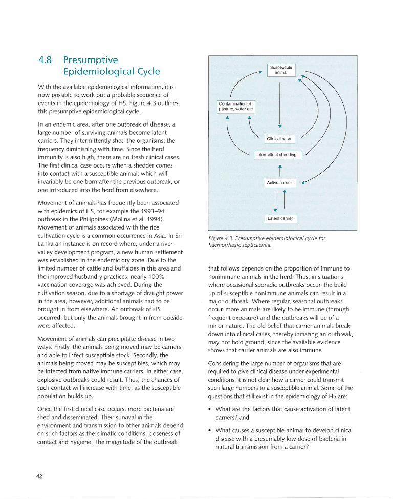
4.8 Presumptive Epidemiological Cycle
With the available epidemiological information, it is now possible to work out a probable sequence of events in the epidemiology of HS. Figure 4.3 outlines this presumptive epidemiological cycle.
In an endemic area, after one outbreak of disease, a large number of surviving animals become latent carriers. They intermittently shed the organisms, the frequency diminishing with time. Since the herd immunity is also high, there are no fresh clinical cases. The first clinical case occurs when a shedder comes into contact with a susceptible animal, which will invariably be one born after the previous outbreak, or one introduced into the herd from elsewhere.
Movement of animals has frequently been associated with epidemics of HS, for example the 1993-94 outbreak in the Philippines (Molina et al. 1994). Movement of animals associated with the rice cultivation cycle is a common occurrence in Asia. In Sri Lanka an instance is on record where, under a river valley development program, a new human settlement was established in the endemic dry zone. Due to the limited number of cattle and buffaloes in this area and the improved husbandry practices, nearly 100% vaccination coverage was achieved. During the cultivation season, due to a shortage of draught power in the area, however, additional animals had to be brought in from elsewhere. An outbreak of HS occurred, but only the animals brought in from outside were affected .
Movement of animals can precipitate disease in two ways. Firstly, the animals being moved may be carriers
. and able to infect susceptible stock. Secondly, the animals being moved may be susceptibles, which may be infected from native immune carriers. In either case, explosive outbreaks could result. Thus, the chances of such contact will increase with time, as the susceptible population builds up.
Once the first clinical case occurs, more bacteria are shed and disseminated. Their survival in the environment and transmission to other animals depend on such factors as the climatic conditions, closeness of contact and hygiene. The magnitude of the outbreak
42
Contamination of pasture. water etc.
~ " 1°""":=. I Intermittent shedding
i I Active carrier
I r T
I Latent carrier
Figure 4.3. Presumptive epidemiological cycle for haemorrhagic septicaemia.
that follows depends on the proportion of immune to nonimmune animals in the herd. Thus, in situations where occasional sporadic outbreaks occur, the build up of susceptible nonimmune animals can result in a major outbreak. Where regular, seasonal outbreaks occur, more animals are likely to be immune (through frequent exposure) and the outbreaks will be of a minor nature. The old belief that carrier animals break down into clinical cases, thereby initiating an outbreak, may not hold ground, since the available evidence shows that carrier animals are also immune .
Considering the large number of organisms that are required to give clinical disease under experimental conditions, it is not clear how a carrier could transmit such large numbers to a susceptible animal. Some of the questions that still exist in the epidemiology of HS are:
• What are the factors that cause activation of latent carriers? and
• What causes a susceptible animal to develop clinical disease with a presumably low dose of bacteria in natural transmission from a carrier?
Diagnosis
Overview
Clinical diagnosis
A clinical diagnosis of haemorrhagic septicaemia (HS)
is based on a combination of clinical signs, gross
pathological lesions and a consideration of relevant
epidemiological parameters and other similar diseases
prevalent in the locality.
Routine laboratory diagnosis
Routine laboratory diagnosis is by culture and serology.
Material for laboratory diagnosis usually consists of
blood or a long bone for bone marrow culture. Pure
cultures are obtained from contaminated material by
mouse inoculation and culture of the mouse blood. A
positive diagnosis is available within 24-48 hours .
Other diagnostic tests
Additional serological, biochemical and molecular
techniques are available as research and
investigational tools.
If reporting is delayed and no material is available for
culture and isolation at the time of investigation,
antibody levels in surviving animals can be assayed
as an indicator of HS infection.
5.1 Provisional Diagnosis
When a suspected outbreak of haemorrhagic septicaemia (HS) is reported, a provisional diagnosis can be made based on clinical signs and, if carcases
are available, on the gross pathological lesions seen on postmortem examination. An investigation of relevant epidemiological parameters is also helpful. It is also important to consider other diseases prevalent in the locality that could account for the clinical signs observed, lesions and the number and pattern of deaths observed.
A provisional diagnosis is important since preventive measures to control the spread of the disease are required immediately, without waiting for laboratory confirmation. At the earliest opportunity, however, appropriate material should be collected, suitably packed and dispatched to the nearest laboratory where facilities are available for diagnostic tests to be performed. A
combination of clinical signs, gross pathological lesions and epidemiological features, coupled with the isolation of the organism and identification of serotype, will help in arriving at a definitive diagnosis.
5.2 Clinical Diagnosis
As outlined in Chapter 3, the disease presents a variety of clinical signs, none of which, when taken individually, is specific for HS. Thus, the clinical picture should be considered as a whole, together with the pathological lesions and the epidemiological findings. As HS occurs mostly in animals reared under poor husbandry conditions, no clinical signs whatever may be observed in the first cases of an outbreak. Thus, the first reports may be of animals found dead suddenly, with no observed illness. Thereafter, upon closer observation, clinical signs will become evident. More advanced cases will show submandibular oedema
spreading to surrounding areas, nasal discharge,
43
salivation and laboured breathing. Those in the early stages may be off their food and, if monitored twice daily, show a marked increase in rectal temperature.
The most obvious postmortem lesions are subcutaneous oedema and petechial haemorrhages, particularly on the base of the ventricle. The next most obvious lesions are those in the lungs, initially congestion, progressively moving towards consolidation and thickening of the interlobular septa giving rise to lobulation (see Section 3.4).
The clinical signs and pathological lesions must be combined with available epidemiological data. The classical epidemics of HS found in earlier records do not occur any longer, particularly in endemic countries, unless the disease is freshly introduced into virgin areas. Thus, the observed morbidity and mortality pattern has to be interpreted against the background of information, such as the species affected, vulnerable age group, endemicity of the location, season and vaccination
history. The presence of any triggering circumstances, such as sudden or seasonal climate changes or
movement of animals, also provides useful clues.
5.3 Differential Diagnosis
HS has to be differentiated from other diseases that present a similar syndrome and that are prevalent in the location where the disease has been reported. If the condition is an acute one characterised by sudden death, other diseases likely to cause sudden deaths, such as anthrax, rinderpest and black quarter, should be taken into account. Equally important are the noninfectious causes of sudden death such as
lightning, snakebites and acute poisoning. The more protracted syndrome dominated by respiratory signs has to be differentiated from other forms of
pasteurellosis caused by serotypes other than groups B and E, or by Pasteurella haemo/ytica. It must be borne in mind that the chronic or subacute pneumonic form
of HS always displays a terminal septicaemia, which may not be a consistent feature with other serotypes.
44
5.4 Samples
5.4.1 Collection of samples
The ideal material for isolation of the specific pathogen is the tissues where it is most likely to be present in largest numbers and which are free, or contain minimal numbers, of other extraneous organisms such as contaminants and postmortem invaders. Tissues of a fresh carcase, preferably blood, fulfil this requirement. If facilities are available at the location of the carcase, a postmortem examination should be immediately carried out and blood drawn directly from the heart.
The gross pathological lesions can be observed at this time. Alternatively, blood may be obtained by puncture of the jugular vein.
In regions where the disease occurs frequently, reporting systems may be suboptimal. Under tropical conditions, decomposition of the carcase may take
place rapidly within a few hours and the blood may contain an abundance of postmortem invaders. It is, however, possible to isolate pasteurellae from blood collected from carcases even 24 hours after death by biological screening using mice. In such situations, it is advantageous to collect and dispatch a long bone, which can be used for culture and biological
examination of bone marrow. This can be done even from animals exhumed a few days after burial.
Material collected from clinically affected animals before death may not give consistent results. Nasal secretions may yield virulent pasteurellae upon culture. Blood will give positive cultures only in the terminal stage immediately before death. Much of the bacterial multiplication takes place in the carcase after death .
It is always better to dispatch to a laboratory material from more than one animal in an outbreak. Care must be taken to avoid collecting material from animals treated with antibiotics.
5.4.2 Dispatch of diagnostic samples
The fragile nature of pasteurellae and their poor survival outside the animal body has already been discussed in Chapter 3. Dhanda (1959a) reported that common contaminants such as Bacillus subtilis and Pseudomonas
a eruginosa , and also B. anthracis, adversely affected the
viability of P. multocida. It has also been reported that when kept on cotton wool swabs and contaminated experimentally with small quantities of B. suhtitis and staphylococci, pure cultures of the HS serotypes of P. multocida, or blood of infected animals, were rapidly overgrown by the contaminants at room temperature. When stored at refrigeration temperature, or when the
swab was kept immersed in a transport medium, overgrowth by contaminants was arrested (De Alwis 1972, 1973). It was further reported that blood swabs collected from a carcase within six hours of death due to HS could be stored for four days at room temperature and up to one week in the refrigerator with consistent viable counts (within 0.5 log units). In swabs collected
24-30 hours after death, rapid overgrowth by contaminants occurred and no pasteurellae could be isolated on the third day. These observations have to be
taken into account in determining methods for dispatch and storage of material during transport to a laboratory.
Blood should be packed in ice or placed in a suitable transport medium. If this is not possible, the swab should be dispatched at room temperature to the
laboratory as quickly as possible. Long bones should be cleaned of all muscle tissue, and sent to the laboratory with minimum delay.
Transport media
Several transport media are now commercially available. The one used by De Alwis (1973) was a simple non nutrient inert medium that contained disodium phosphate, thioglycollic acid and agar (0.4 %)
to give a semisolid consistency, and methylene blue as an indicator. Warner (1996) developed a transport enrichment medium with antibiotic and antifungal additives that promoted the survival of the pasteurellae
and suppressed contaminants. Details of transport media are given in Appendix 1.
5.5 Laboratory Diagnosis
Laboratories in countries where HS is endemic receive a variety of specimen types and quality for diagnosis. Appropriate methods may therefore be needed to deal with these specimens. The serotypes of P. multocida that cause HS are not difficult to isolate, even in a laboratory with modest facilities. A wide range of
laboratory diagnostic tests has been developed over the years. These include:
• culture and biological tests for isolation of the causative organism;
• biochemical and serological tests for identification of the organism and serotype;
• non serological tests for presumptive identification of serotype; and
• molecular methods for strain differentiation within serotypes.
The conventional method of laboratory diagnosis is therefore based on isolation of the organism from animal tissues and identification by biochemical and serological methods. The nonserological tests and molecular techniques are useful investigatory tools but are not substitutes for the conventional tests. In instances where reporting is delayed and no material is available for culture and isolation at the time of investigation, high levels of antibody or, more specifically, rising antibody titres in surviving animals are useful indications in arriving at a presumptive diagnosis (see Section 5.11).
A routine laboratory diagnostic procedure that can be adopted in any laboratory in the endemic countries of Asia and Africa having modest facilities is illustrated in
Figure 5.1 .
If the material is uncontaminated, colonies can be isolated on a direct culture plate within 24 hours. These could be subjected to a rapid slide agglutination test. Based on colonial appearance, direct smear examination from
culture plate and a positive rapid slide agglutination test, the laboratory could issue a positive report.
If the material is contaminated, as is often the case, a pure culture can only be obtained by mouse inoculation and culture of mouse blood. In addition to the above tests, pathogenicity to mice, and a mouse blood smear with an abundance of bipolar staining gram-negative coccobacilli, provide useful clues. Culture of the mouse blood and a rapid slide agglutination test on the culture confirms the diagnosis. Thus, within 24-48 hours, it is possible to issue a positive report from a laboratory. As
a routine, this procedure has proved to be quite satisfactory. If further serotyping is considered
45
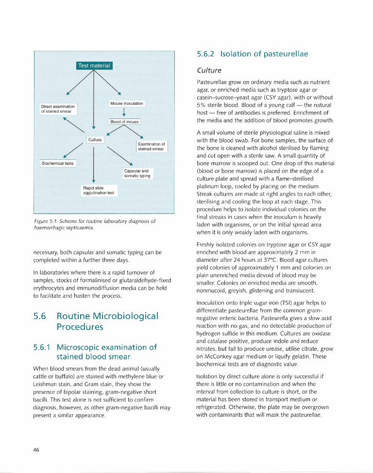
Direct examination of stained smear
I Biochemical tests I
I Mouse inoculation I
Rapid slide agglutination test
Figure 5.1. Scheme for routine laboratory diagnosis of haemorrhagic septicaemia.
necessary, both capsular and somatic typing can be completed within a further three days.
In laboratories where there is a rapid turnover of samples, stocks of formalinised or glutaraldehyde-fixed erythrocytes and immunodiffusion media can be held to facilitate and hasten the process.
5.6 Routine Microbiological Procedures
5.6.1 Microscopic examination of stained blood smear
When blood smears from the dead animal (usually cattle or buffalo) are stained with methylene blue or Leishman stain, and Gram stain, they show the presence of bipolar staining, gram-negative short bacilli. This test alone is not sufficient to confirm diagnosis, however, as other gram-negative bacilli may present a similar appearance.
46
5.6.2 Isolation of pasteurellae
Culture
Pasteurellae grow on ordinary media such as nutrient agar, or enriched media such as tryptose agar or casein-sucrose-yeast agar (CSY agar), with or without 5% sterile blood . Blood of a young calf - the natural host - free of antibodies is preferred . Enrichment of the media and the addition of blood promotes growth .
A small volume of sterile physiological saline is mixed with the blood swab. For bone samples, the surface of the bone is cleaned with alcohol sterilised by flaming and cut open with a sterile saw. A small quantity of bone marrow is scooped out. One drop of this material (blood or bone marrow) is placed on the edge of a culture plate and spread with a flame-sterilised platinum loop, cooled by placing on the medium. Streak cultures are made at right angles to each other, sterilising and cooling the loop at each stage. This procedure helps to isolate individual colonies on the final streaks in cases when the inoculum is heavily laden with organisms, or on the initial spread area when it is only weakly laden with organisms.
Freshly isolated colonies on tryptose agar or CSY agar enriched with blood are approximately 2 mm in diameter after 24 hours at 37"C. Blood agar cultures yield colonies of approximately 1 mm and colonies on plain unenriched media devoid of blood may be smaller. Colonies on enriched media are smooth , nonmucoid, greyish, glistening and translucent.
Inoculation onto triple sugar iron (TSI) agar helps to differentiate pasteurellae from the common gramnegative enteric bacteria. Pasteurella gives a slow acid reaction with no gas, and no detectable production of hydrogen sulfide in this medium. Cultures are oxidase and catalase positive, produce indole and reduce nitrates, but fail to produce urease, utilise citrate, grow on McConkey agar medium or liquify gelatin. These biochemical tests are of diagnostic value.
Isolation by direct culture alone is only successful if there is little or no contamination and when the interval from collection to culture is short, or the material has been stored in transport medium or refrigerated. Otherwise, the plate may be overgrown with contaminants that will mask the pasteurellae.
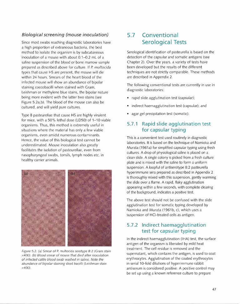
Biological screening (mouse inoculation)
Since most swabs reaching diagnostic laboratories have a high proportion of extraneous bacteria, the best method to isolate the organism is by subcutaneous inoculation of a mouse with about 0.1-0.2 mL of a saline suspension of the blood or bone marrow sample prepared as described above for culture. If P multocida types that cause HS are present, the mouse will die within 24 hours. Smears of the heart blood of the infected mouse will show an abundance of bipolar
staining coccobacilli when stained with Gram, Leishman or methylene blue stains, the bipolar nature being more evident with the latter two stains (see Figure 5.2a,b). The blood of the mouse can also be cultured, and will yield pure cultures.
Type B pasteurellae that cause HS are highly virulent for mice, with a 50% lethal dose (LD50) of 1-10 viable
organisms. Thus, this method is extremely useful in situations where the material has only a few viable organisms, even amidst numerous contaminants.
Hence, the value of this biological test cannot be underestimated. Mouse inoculation also greatly facilitates the isolation of pasteurellae, even from nasopharyngeal swabs, tonsils, lymph nodes etc. in healthy carrier animals.
Figure 5.2. (a) Smear of P. multocida serotype B:2 (Gram stain x400). (b) Blood smear of mouse that died after inoculation of infected cattle blood swab washed in saline. Note the abundance of bipolar staining short bacilli (Leishman stain x400).
5.7 Conventional Serological Tests
Serological identification of pasteurella is based on the detection of the capsular and somatic antigens (see Chapter 2) . Over the years, a variety of tests have been developed but the results of the different
techniques are not strictly comparable. These methods are described in Appendix 2.
The following conventional tests are currently in use in diagnostic laboratories:
• rapid slide agglutination test (capsular);
• indirect haemagglutination test (capsular); and
• agar gel precipitation test (somatic) .
5.7.1 Rapid slide agglutination test for capsular typing
This is a convenient test used routinely in diagnostic laboratories. It is based on the technique of Namioka and Murata (1961 a) for simplified capsular typing using fresh cultures. A drop of physiological saline is placed on a clean slide. A single colony is picked from a fresh culture plate and is mixed with the saline to form a uniform suspension. A loopful of antiserotype B:2 pasteurella hyperimmune sera prepared as described in Appendix 2 is thoroughly mixed with this suspension, gently warming the slide over a flame. A rapid, flaky agglutination appearing within a few seconds, with complete clearing of the background, indicates a positive test.
The above test should not be confused with the slide agglutination test for somatic typing developed by Namioka and Murata (1961 b, c), which uses a suspension of HCI-treated cells as antigen.
5.7.2 Indirect haemagglutination test for capsular typing
In the indirect haemagglutination (IHA) test, the surface antigen of the organism is liberated by mild heat treatment. The cell residue is removed and the supernatant, which contains the antigen, is used to coat erythrocytes. Agglutination of the coated erythrocytes in serial 10-fold dilutions of hyperimmune rabbit antiserum is considered positive. A positive control may be set up using a known reference culture to prepare
47
antigen and coat the cells. An untreated erythrocyte suspension is used as the negative control. The serum used is the same as for the rapid slide agglutination test.
The original test described by Carter (1955) used fresh human '0' erythrocytes. Subsequently, Carter and Rappay (1962) used formalinised human '0' erythrocytes
whilst Sawada et al. (1982) used glutaraldehyde-fixed sheep erythrocytes. Wijewardana et al. (1986a) modified this test using fresh sheep erythrocytes and suspensions of bacterial cells standardised on the basis of turbidity. An antigen titration was also carried out to standardise the amount of antigen used for coating the erythrocytes.
5.7.3 Agar gel precipitation test for somatic typing
The agar gel precipitation test (AGPT) for somatic typing is carried out basically according to the method of Heddleston et al. (1972) using Noble agar medium at a concentration of 0.9% in 8.5% sodium chloride.
For antigen preparation, a concentrated harvest of pasteurellae grown on a culture plate is made in 8.5% saline with 0.3% formalin. Antiserum for somatic typing is prepared in chicken. The test antigen is placed in a central well in the agar, with antisera against various somatic types in the peripheral wells. A positive result is indicated by diffusion bands of antibody-antigen precipitate, which develop overnight.
5.8 Other Serological Tests
In addition to the above conventional tests, which are necessary to confirm a diagnosis, several other
serological tests have been developed.
5.8.1 Agar gel precipitation test for capsular typing
A simple immunodiffusion test has been described for capsular typing (Anon . 1981; Wijewardana et al. 1982). The gel medium consists of 1 % Noble agar in 0.2 M phosphate buffer containing merthiolate at a final concentration of 1 in 10000. As for the indirect haemagglutination test, rabbit hyperimmune sera prepared against whole cells and 56°C/30 minute
supernatant are used as sources of antibody and
48
antigen, respectively. Reference antiserum is usually placed in the centre well and test antigens in peripheral wells. Homologous antigen prepared from a reference strain placed in alternate peripheral wells serves as a positive control, and physiological saline or control serum from an unimmunised rabbit placed in one peripheral well serves as a negative control. Bands develop overnight.
5.8.2 Counter-immunoelectrophoresis
This test was originally developed by Carter and Chengappa (1981). The principle underlying the test, and the reagents used, are the same as for the AGPT. Parallel rows of wells are made on the medium on a slide. The wells on the side of the cathode are loaded with antigen while the wells on the side of the anode are loaded with antiserum. Electrophoresis is for 30 minutes, during which time precipitation bands appear. Results can be obtained much more rapidly by this method than by AGPT.
5.8.3 Coagglutination test
This test was developed by Rimier (1978) . It is used to differentiate between type Band E strains that cause HS by a coagglutination test using antibody-coated staphylococci. The test has no distinct advantage over other methods and has therefore not gained popularity as a routine test in diagnostic laboratories.
5.8.4 Agglutination test for somatic typing
This test was developed in the early 1960s (Namioka and Murata 1961 b,c) and at that time proved to be a useful supplement to Carter's capsular typing in the differentiation of strains associated with various disease conditions. The antigen preparation was made by treating cultures harvested from yeast-protease-cysteine agar plates (see Appendix 1) with normal HCI saline (0.85% saline in normal HCI) overnight. After several washes, the cell residue was suspended in phosphate buffered saline (PBS). Whole cell rabbit hyperimmune serum was used for slide agglutination tests .
When applied to P. multocida in general, this typing method is very complex as it involves a complex system of absorption of sera, in order to avoid cross-reactions
(see Chapter 2). However, since there were only two somatic types detected in capsular type B (6 and 11),
and only one (6) within capsular type E, the identification of the HS strains is relatively easy, and
unabsorbed sera can be used.
To facilitate interpretation, antisera prepared against three reference strains (Asian [6:B], African [6:E] and
Australian [11 :B] also known as strain 989]) are used in
agglutination tests with the three homologous antigens
and the test antigen as shown in Table 5.1.
5.8.5 Enzyme-linked immunosorbent assay
An enzyme-linked immunosorbent assay (ELISA) for
identification of P. multocida that cause HS was
developed by Dawkins et al. (1990) in Australia (HSantigen ELISA; see Appendix 3). These researchers
examined 124 strains, consisting of 58 reference strains
from collections in various laboratories and 66 field
isolates, and reported a specificity of 99% and a sensitivity of 86%. This technique has proved useful for
screening a large number of cultures from a collection, rather than occasional diagnosis of HS from specimens
reaching a laboratory from time to time. In its present form, it has a serious limitation in that it fails to
differentiate between the Asian (B:2) and African (E:2)
types. This is not surprising as the antigen used in this
test was the boiled antigen similar to that used in
Heddleston's somatic typing, where the somatic
antigen 2 was common to both Asian and African
types. From an epidemiological standpoint and for the
purpose of initiating control measures in an outbreak,
it is important to know the serotype.
5.9 Nonserological tests
Several non-immunological tests have been developed
for the rapid identification of strains. These include the
acriflavine flocculation test described by Carter and Subronto (1973) for identification of type D strains,
the hyaluronidase decapsulation test for type A strains
(Carter and Rundell 1975) and the hyaluronidase
production test for the rapid identification of type B
strains (Carter and Chengappa 1981).
5.9.1 Hyaluronidase production test
The production of the enzyme hyaluronidase is a
characteristic of many species of gram-positive
bacteria such as staphylococci, streptococci and
clostridia. Among the gram-negative bacteria, type B strains of P. multocida have been found to produce
appreciable amounts of hyaluronidase. Two methods have been used to demonstrate the production of this
enzyme. In the first method, a hyaluronic acid
producing culture of Streptococcus equi is streaked
across a freshly prepared blood agar plate.
Table 5.1. Somatic typing of pasteurella strains using HCI-treated antigen.
Serum
Asian (6:B)
African (6:E)
Australian (11 B)
Antigen
Asian
African
Australian
Test
Asian
African
Australian
Test
Asian
African
Australian
Test
Result
+ +
?
+
+
?
+ ?
49
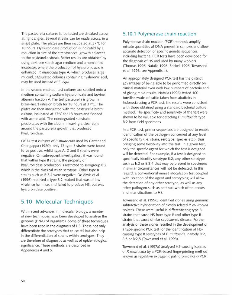
The pasteurella cultures to be tested are streaked across at right angles. Several streaks can be made across, in a single plate. The plates are then incubated at 37°C for 18 hours. Hyaluronidase production is indicated by a reduction in size of the streptococcal growth adjacent to the pasteurella streak. Better results are obtained by using dextrose starch agar medium and a humidified incubator, where the production of hyaluronic acid is enhanced. P multocida type A, which produces large mucoid, capsulated colonies containing hyaluronic acid, may be used instead of S. equi.
In the second method, test cultures are spotted onto a medium containing sodium hyaluronidate and bovine albumin fraction V. The test pasteurella is grown in brain-heart infusion broth for 18 hours at 37°(, The plates are then inoculated with the pasteurella broth culture, incubated at 37°C for 18 hours and flooded with acetic acid. The nondegraded substrate precipitates with the albumin, leaving a clear zone around the pasteurella growth that produced hyaluronidase.
Of 74 test cultures of P. multocida used by Carter and Chengappa (1980), only 13 type B strains were found to be positive, whilst type A, D and E strains were negative. On subsequent investigation, it was found that within type B strains, the property of hyaluronidase production is restricted to serogroup B:2, which is the classical Asian serotype. Other type B strains such as B:3,4 were negative. De Alwis et al. (1996) reported a type B:2 mutant that was of low virulence for mice, and failed to produce HS, but was hyaluronidase positive.
5.10 Molecular Techniques
With recent advances in molecular biology, a number of new techniques have been developed to analyse the genome (DNA) of organisms. Some of these techniques have been used in the diagnosis of HS. These not only differentiate the serotypes that cause HS but also help in the differentiation of strains within serotypes. They are therefore of diagnostic as well as of epidemiological significance. These methods are described in Appendixes 4 and 5.
50
5.10.1 Polymerase chain reaction
Polymerase chain reaction (PCR) methods amplify minute quantities of DNA present in samples and allow accurate detection of specific genetic sequences, including bacteria. PCR tests have been developed for the diagnosis of HS and used by many workers (Thomas 1996; Natalia 1996; Brickell 1996; Townsend et al. 1998; see Appendix 4).
An appropriately designed PCR test has the distinct advantages of being able to be performed directly on clinical material even with low numbers of bacteria and of giving rapid results . Natalia (1996) tested 100 tonsillar swabs of cattle taken from abattoirs in Indonesia using a PCR test; the results were consistent with those obtained using a standard bacterial culture method. The specificity and sensitivity of the test were shown to be valuable for detecting P. multocida type B:2 from field specimens.
In a PCR test, primer sequences are designed to enable identification of the pathogen concerned at any level
of specificity (i.e. strain, serotype, species etc.), thus bringing some flexibility into the test. In a given test, only the specific agent for which the test is designed will be detected. For example, if a test is designed to specifically identify serotype B:2, any other serotype such as E:2 or B:3,4 that may be present in specimens in similar circumstances will not be detected. In this regard, a conventional mouse inoculation test coupled with isolation of the agent and serotyping will allow the detection of any other serotype, as well as any other pathogen such as anthrax, which often occurs in similar situations to HS.
Townsend et al. (1996) identified clones using genomic subtractive hybridisation of closely related P. multocida
isolates. These were useful in differentiating type B strains that cause HS from type E and other type B strains that cause similar septicaemic disease. Further analysis of these clones resulted in the development of a type-specific PCR test for the identification of HScausing type B serotypes of P. multocida, namely B:2, 8:5 or B:2,5 (Townsend et al. 1998).
Townsend et al. (1997a) analysed HS-causing isolates of P multocida by a PCR-based fingerprinting method known as repetitive extragenic palindromic (REP) PCR.
Multiple genomic DNA fragments are amplified by outwardly facing primers based upon the REP consensus sequence, generating complex profiles useful in strain differentiation . The analysis of HScausing P. multocida strains provided evidence of a disease-associated REP profile with a high degree of homogeneity observed among the strains, regardless of whether the capsular serotype was B or E. These profiles were clearly distinct from serologically similar strains that do not cause HS, but exhibited a degree of relatedness to strains that cause clinically similar septicaemic disease such as B:1, B:2, B:3, B:4. These findings have paved the way for a disease-specific test rather than a serotype-specific test.
5.10.2 Ribotyping and field alternation gel electrophoresis
Ribotyping and field alternation gel electrophoresis (FAGE) have also been used to analyse the DNA of strains of P. multocida that cause HS, after digestion with restriction enzymes (Adamson et al. 1993; Townsend et al. 1997b; see Appendix 5).
Analysis of the DNA restriction patterns using these methods has proved useful in differentiating bacterial strains that have been denoted as identical by all previous typing methods. Restriction profiles demonstrated by Asian strains showed a remarkable degree of homogeneity. Asian isolates displayed similar ribotype and FAGE patterns irrespective of the restriction enzyme used, thus indicating that they are epidemiologically related both genetically and phenotypically. FAGE displayed a greater degree of discrimination between strains, compared with ribotyping. It was particularly useful in distinguishing the North American type B:2 strains that produced HS from the Asian strains.
Both ribotyping and FAGE analysis also helped to differentiate between the classical Asian B:2 strains that cause HS and the reportedly avirulent strain designated Izathagar 25, isolated from cases of paraplegia among cattle in India (Dhanda and Nilakanthan 1961). This strain has not been serotyped by the Carter-Heddleston method but has been found to belong to Roberts type I, and has been biochemically differentiated from other strains by its ability to ferment xylose (Dhanda and Sen 1972).
5.10.3 Restriction endonuclease analysis
Restriction endonuclease analysis (REA) of a wide range of cultures of P. multocida associated with HS has been carried out (Wilson et al. 1992; Rimier (1997)(see Apendix 5). The DNA was digested with Hhal and Hpall endonuclease, and examined by agarose gel electrophoresis. Gels were stained with ethidium bromide, and the band patterns (DNA fingerprint profiles) were viewed and photographed in ultraviolet light. Reverse negative photographs of gels were made and scanned to create an image profile. Images of fingerprint profiles were analysed by computer program.
Forty-two different DNA fingerprint profiles were recognised among 190 serotype B:2 strains using Hhal
endonuclease. Twelve DNA fingerprint profiles were found among 35 serotype B:3,4 strains with the same enzyme. Further distinction could be made among B:2 or B:3,4 strains with Hpall endonuclease. For example, treatment with Hpall resulted in seven different DNA fingerprint profiles within 56 serotype B:2 strains that produced a profile designated Hhal 0018. Thus, a strain could be assigned a descriptive identification epithet (DIE) code such as B:2 IHhal 0018 1 Hpall
0016 based on the REA results. Strains were analysed on the basis of associated species and country. Among 64 serotype B:2 strains from Sri Lanka, 24 fingerprint profiles were defined using Hhal endonuclease. Further, among 17 strains designated B:21 Hhal 0018,
originating from animals such as cattle, buffalo, swine and elephant, six Hpall profiles were identified . Similar profiles were found among isolates from swine and buffaloes in India.
5.11 Antibody Detection in Host Animals
As mentioned elsewhere, HS occurs mostly in situations with poor husbandry conditions. In such circumstances, disease-reporting systems are also poorly developed. Deaths usually occur quickly and situations are likely to arise in which, by the time veterinary personnel arrive at the scene, no clinically affected animals or carcases are available for examination.
51
Ample evidence has now been presented to show that, following exposure to natural infection, surviving animals develop high antibody levels as measured by the IHA test, using the 56°C/30 minute supernatant as antigen to coat erythrocytes (De Alwis 1982b, De Alwis et al. 1986). De Alwis (1982b) was able to specifically demonstrate a steep rise in antibody level within two to
four weeks of such exposure. Such high levels of antibody have not been attained by administration of conventional vaccines. Hence, the presence of such levels of IHA antibody following recent deaths in a herd provides reasonable circumstantial evidence that deaths were due to HS.
The test used to detect such an antibody response is important. The HS-antibody ELISA (Johnson et al. 1989; see Appendix 3) may not detect such antibody, since the test uses boiled supernatant as antigen. This was borne out in experiments with a live vaccine when, following vaccination, IHA antibodies continued to rise, as in natural exposure, but the ELISA titres did not
(M .C.L. De Alwis, unpublished data).
A single radial haemolysis test for measuring HS antibodies has been described (Rahman et al. 1987; Rahman and Ashfaque 1991). This test was carried out using a sonicated antigen. In a comparison of this test with the conventional IHA, a high degree of correlation (r=0 .792) was observed. There is no information on the
use of this test for detecting naturally acquired immunity resulting from natural exposure.
52
5.12 Choice of Diagnostic Tests
The choice of diagnostic tests must take into account the nature of the material available, the facilities available in the laboratory and the proximity of the laboratory to the outbreak of disease. Any modest
diagnostic laboratory charged with the responsibility of HS diagnosis should have a basic facility for microscopic examination and bacterial culture. It should also have a supply of hyperimmune rabbit antisera and, ideally, a laboratory animal facility with access to a mouse colony. The most reliable procedure is the mouse inoculation and mouse blood culture followed by rapid slide agglutination. Serotyping by the IHA test and AGPT (Heddleston) may be subsequently carried out in the
same laboratory, or the culture may be passed on to the next higher grade of laboratory where such a capability exists. In situations where serotypes other than B:2 or E:2 are present (such as B:1, B:4, B:3,4), AGPT is the definitive test. The procedure, starting with the mouse
inoculation, detects even small numbers of organisms, even in the presence of contaminants.
Where a PCR facility and technical know-how is available, stale material containing small numbers of organisms can still be used. However, PCR will pick up only the target organism for which the test is designed, and will fail to detect any related serotypes, or other bacteria, such as Bacillus anthracis, which are likely to cause similar sudden death syndromes.
Rapid serological tests such as AGPT using the 56°C/30 minute supernatant as antigen and counterimmunoelectrophoresis detect the capsular serogroup, but not the specific somatic type. The hyaluronidase test is also specific for P multocida B:2 strains that cause HS, and serves as an additional complementary test.
Other molecular tests, besides PCR, such as ribotyping, REA and FAGE, are useful for identifying strains within serotypes. They are therefore useful as markers in epidemiological studies and as research and investigatory
tools rather than as routine diagnostic tests .
Treatment and Control
Overview Haemorrhagic septicaemia (HS) is classified as a List B
disease by the Office International des Epizooties.
Control and treatment of the disease are therefore an
important issue in countries where the disease is
endemic.
Treatment
Treatment with antibiotics is of limited value unless
carried out in the very early stages. Most field cases are
not detected in the early stages, making treatment
ineffective. In an organised farm incontact animals can
be checked regularly and animals showing an increased
temperature can be separated and treated with
antibiotics.
Prevention and control
Vaccination is the most effective method of control, in
conjunction with other measures. Three broad categories
of activities are recommended for prevention:
prophylactic measures in endemic countries; measures
needed in the event of an outbreak; and measures needed
to prevent the spread of disease from endemic to
nonendemic areas.
Eradication
Eradication of haemorrhagic septicaemia is difficult to
achieve because of the existence of latent carriers.
6.1 International Classification
As described in Section 1.4.1, the Office International des Epizooties (OlE), or World Organisation for Animal Health, has classified haemorrhagic septicaemia (HS) as a List B disease (along with most other bacterial and
parasitic diseases that are considered to be of socioeconomic and/or public health importance within countries, and also of significance to the international trade in animals and animal products). List A diseases,
on the other hand, are mainly viral diseases, and include those that spread rapidly and have the scope to spread beyond national borders. The classification presumably influences the priority status given to the disease by governments in endemic countries and the allocation of resources for treatment and control.
6.2 Treatment
As HS is a primary bacterial disease with no other biological agents involved, treatment may appear simple using the wide range of antibiotics currently available. In reality, however, treatment is constrained by a host of practical considerations. It has been found in practice that animals can only be cured if they are treated in the very early stages of the disease. However, as the disease occurs mainly in situations with primitive husbandry practices most field cases escape detection in the early stages, thus rendering treatment ineffective.
In organised farms, however, a practical method of achieving early detection and successfully treating animals is to check the rectal temperatures of all incontact animals regularly once an outbreak has been detected. Any animals showing an increased temperature can be separated and treated with a course of an appropriate antibiotic.
Although not documented, there is considerable information from reports of field outbreaks indicating
53
that antibiotic treatment in the terminal stages accelerates death. This may be because administration of antibiotics to an animal with septicaemia precipitates an endotoxin shock due to release of free endotoxin from the killed bacteria.
6.2.1 Antibiotic therapy
The oldest therapy recommended was intravenous treatment with sulfonamides. Intravenous infusion of sulfadimidine sodium 33.33 % at a dosage of 1 mL per 5 pounds bodyweight (about 2.3 kg) has been practised . However, the large volume of drug to be injected , the practical difficulties of intravenous therapy in the type of animal involved and the consequences of leakage of the drug into the surrounding tissues all weigh heavily against this treatment regimen (De Alwis 1984). De Alwis (1984) observed that intramuscular treatment with streptomycin or oxytetracycline was very effective.
Resistance to antibiotics in HS strains of P. multocida
has not been a major problem. When 10 strains obtained from Malaysia, Indonesia, Thailand, Myanmar, India and Sri Lanka were tested against 10 commonly used antibiotics (penicillin, ampicillin, streptomycin, tetracycline, chloramphenicol, erythromycin, neomycin, sulfadiazine and a sulfonamide-trimethoprim combination), no resistance was recorded apart from partial resistance of the Thai strains to streptomycin (De Alwis 1984).
Bandopadhyay et al. (1991) tested 16 isolates obtained from different outbreaks in the Gujarat State (India) against 19 antibiotics. They found that 14 isolates (87.5%) were resistant to compound sulfonamides. Resistance was also observed to vancomycin (75%), tetracycline (50 %), colistin (37.5%), streptomycin and novobiocin (25%), and nitrofurantoin and carbenicillin (6.25%) . All isolates were sensitive to peniCillin, ampicillin, chloramphenicol, erythromycin, gentamicin, caphalothin, polymyxin Band tilmicosin.
In an outbreak of disease in pigs in India caused by the HS serotype B:2, isolates were found to be sensitive to erythromycin, gentamicin, nalidixic acid and neomycin (Verma 1988). In Pakistan, from the results of a questionnaire survey, it has been concluded that preparations containing amoxicillin, sulfapyrazole, gentamicin and chloramphenicol produced positive
54
clinical results (Sheikh et al. 1996). Abeynayake et al. (1992) tested 27 Sri Lankan isolates against 17 antimicrobial agents. These were mainly strains derived from field outbreaks from cattle and buffaloes during the period 1985-90. Also included were a few strains isolated from clinically normal carrier animals, isolates from species other than cattle and buffaloes, and national reference strains. All of these were of serogroup B:2. All strains tested were sensitive to penicillin, ampicillin, enrofloxacin, chloramphenicol and nitrofurantoin . A majority of isolates were also sensitive to neomycin (26/27), gentamicin (26127),
oxytetracycline (25/26), streptomycin (24125) and sulfonamide-trimethoprim (25127). Variable degrees of resistance were shown to some antibiotics, the number of sensitive strains being oxacillin (17/27),
spiramycin (16127), clindamycin (17127) and sulfamethoxazole (12127) .
Thus, of the antibacterial compounds that are recommended for use in cattle and buffaloes, and that can be conveniently and economically administered, penicillin, ampicillin and oxytetracycline appear to be the most useful. The sensitivity patterns, however, are likely to vary from one country to another depending on the prevailing drug usage practices; a knowledge of the sensitivity patterns of local strains will be use'ful in deciding the drug of choice . It is interesting to note the observation of Bain et al. (1982) that while penicillin was effective against the organisms in vitro, it was ineffective in vivo, a point that must be noted in the choice of an antibiotic.
6.2.2 Serum therapy
Serum therapy is only of theoretical interest. Kheng and Phay (1963) used 60-100 mL of hyperimmune serum for experimental administration to two-year-old (400 Ib [about 180 kg]) buffaloes at varying periods from six hours before to 18 hours after infection. No significant therapeutic effect was recorded.
6.3 Prevention and Control
There are three categories of measures for prevention and control of HS:
• measures to be adopted in endemic countries on a prophylactic basis;
• measures to be taken in the event of an outbreak; and
• measures necessary for prevention of spread across regional or national borders.
6.3.1 Prophylactic measures in endemic countries
Taking into consideration available information on the nature of the disease, the organism, its survival outside the animal, resistance to external agents and relevant epidemiological parameters, the preventive measures shown below should be taken within a country or region where HS is endemic.
• Vaccinate on a routine prophylactic basis. Vaccination is best done two to three months before the high-risk season (in areas where seasonality occurs) so as to ensure peak immunity during the period of maximum risk.
• Establish a good reporting system. This will enable information on suspected outbreaks to reach animal health authorities as quickly as possible. In most endemic countries HS is listed as a notifiable disease.
• Create awareness of the disease among farmers. Educate farmers to recognise signs of the disease.
• Prevent mixing of animals from endemic and nonendemic areas. In endemic areas, a significant proportion of animals are latent carriers and are potential sources of infection. In nonendemic areas, animals are not regularly exposed to infection, lack naturally acquired immunity, are not usually vaccinated, and are highly susceptible. An outbreak originating from an activated carrier in such an instance can be explosive. If contact between such animals is unavoidable, it is of utmost importance that susceptible animals from nonendemic areas are vaccinated at least two weeks before contact with animals from an endemic area.
6.3.2 Preventive measures during an outbreak
In the event of an outbreak of disease occurring, there are equally important measures that should be taken to control further spread.
• Continue vaccination programs. Vaccination is recommended even in the face of an outbreak. In such situations broth bacterins or the alumprecipitated (or aluminium hydroxide gel) vaccine is preferred. Broth bacterin and the oil adjuvant vaccine may be administered at different sites, simultaneously.
• Isolate and treat animals showing clinical signs with a parenteral broad-spectrum antibiotic (relatively easy in organised farms or herds that are paddocked at night or for part of the day) .
• Check the rectal temperature of all immediate incontact animals in the herd. This should be done at least once every morning; those animals showing increased temperatures should be treated as above.
• Search daily for sick animals or carcases of dead animals (free-roaming, nomadic herds).
• Confine herds as much as possible, and prevent movement of animals in and out of diseased premises or villages.
• Take immediate action to carry out postmortem examinations (iocal veterinarians). Make a tentative diagnosis.
• Dispatch specimens to the nearest diagnostic laboratory. Specimens should be stored and transported under appropriate conditions (see Chapter 5).
• Dispose of carcases of dead animals properly. Deep burial or effective incineration is recommended. Often, after animals die, carcases are disowned by farmers. Stray dogs and other scavenging animals can disseminate infection by carrying away portions of infected carcases, and carcases dumped into streams and waterways are important sources of infection .
• Properly dispose of unconsumed fodder, bedding etc. from infected premises. Deep burial or drying and burning should be carried out within the
55
premises. Effluent from cattle sheds, dung etc. should be prevented from being washed away from the premises. Drains carrying such material should be led into a deep protected pit within the premises, or subjected to disinfectant treatment.
• Closely monitor or stop rain-associated activities.
Activities such as ploughing and preparation of fields for rice cultivation cause considerable movement of draught cattle and buffaloes. HS often breaks out during wet seasons.
6.3.3 Prevention of spread across borders
Where animals are moved from an endemic country or region to a nonendemic country or region, such as during imports, certain practical procedures can be observed to eliminate or at least minimise the transfer of the disease.
• Ensure that the animals originate from a region
where no outbreaks of HS have occurred for a
minimum period of one year. How extensive this disease-free 'region' should be will depend on the system of management practised in the region. If animals are confined to farm premises, the region may be as narrow as a radius of half a kilometre. On the other hand, where the animals are on a freerange system, the radius of the disease-free zone may have to be several kilometres.
• Bleed a random sample of animals. This should include the herd or farm of origin and/or other incontact animals.
• Test for the presence of antibody by the IHA test. The presence of high indirect haemagglutination (IHA) antibody titres is an indication of recent exposure to disease and therefore the presence of disease-causing agent in the locality.
• Hold animals under observation for two to three
weeks before transport. During this time repeated attempts should be made to check nasopharyngeal swabs for pasteurellae by mouse inoculation and culture. Blood collected at the beginning and end of such a period should be checked for antibody by the IHA test. Animals harbouring type B pasteurellae and/or showing IHA titres should be eliminated. The
56
detection of any carriers or animals with antibody titres would justify an extended period of pretransport observation for other incontact animals in the group.
• Quarantine animals after transport to the new location. The animals can be held for a similar period of time to that used before transport. During this time, the same procedures should be carried out.
• Vaccinate animals from disease-free locations in
endemic countries. A dose of oil adjuvant vaccine may be given at the end of the quarantine period, followed by a booster three months later. It is equally important to vaccinate all animals in the country of import that are likely to come into contact with animals introduced from endemic countries or regions.
Vaccination is the most important control procedure adopted in all countries where the disease is endemic. Details of the vaccines in use and vaccination procedures are given in Chapters 7, 8 and 9.
6.4 Eradication
No country has ever attempted to eradicate HS. This reflects the belief that the existence of carriers makes eradication too hard. This has been strengthened by recent findings indicating that a larger proportion of animals are carriers than was originally thought and that for most of the time the disease remains latent. The existence of carriers among feral ruminants may be a further factor that will make eradication difficult.
The only known attempt at eradication was made by the Government of Indonesia on a pilot scale on the island of Lombok with a cattle population of 300 000. The program started in 1978 with intensive vaccination campaigns that were targeted to achieve the highest possible coverage in all susceptible species. The coverage actually achieved over a three-year period was 89 % in cattle, 94% in buffaloes, 82% in goats, 93 % in sheep and 80% in pigs. The program was evaluated in 1981, after three annual vaccinations using the oil adjuvant vaccine. During this period, 53 cases of HS were reported. Culture of pharyngeal mucous membrane samples from 220 animals slaughtered in an abattoir yielded positive isolations
from five a.nimals (2.2%). Mass vaccination was continued, and a second evaluation was made in 1985. A total of 450 abattoir samples from cattle, buffaloes, goats and pigs were negative. Also, 103 specimens from animals suspected of HS were negative. This island was declared free of the disease in 1985. However, subsequent evaluations based on culture, serology and field reports indicate that HS is still present in Lombok (Darmadi 1991; Syamsudin 1993).
These observations provide useful indicators of the complexities involved in attempting a total eradication program for HS even on a small island; the implications are much more complex in countries with land borders and a large wildlife population.
57
Vaccines
Overview
Vaccine production
Vaccine production consists of four important steps:
selection and maintenance of seed culture, production
of bulk dense culture, inactivation of dense culture and
formulation of vaccine. Simplified methods of vaccine
production as well as sophisticated technologies are
available.
Types of vaccine
The types of vaccine used against HS are bacterins,
alum preCipitated vaCCine, aluminium hydroxide gel
vaccine, and oil adjuvant vaccine.
Quality testing
All HS vaccines have to be tested for purity, stability
and sterility. Potency tests are usually carried out in
mice. The production technique must be validated in
an experiment using natural host animals.
7.1 Vaccine Production
Haemorrhagic septicaemia (HS) is preventable using vaccines containing the causative bacterial agent. However, pasteurella is a poor immunogen and a large amount of antigen (usually whole bacteria) therefore
has to be injected. This procedure occasionally leads to endotoxic shock. One way of overcoming this problem is to use a suitable adjuvant. Adjuvants potentiate the action of the immunogen, and provide a depot effect, delaying absorption by a slow release mechanism that simulates the administration of multiple doses.
HS vaccine production involves the following stages:
• selection and maintenance of a seed culture;
• preparation of a bulk dense culture;
• inactivation of the dense culture; and
• formulation of the vaccine.
7.1.1 Selection of a seed culture
It is generally agreed that vaccines produced from fresh field isolates are more effective than those produced from seed cultures propagated in vitro in laboratories over long periods. Most countries have therefore used local isolates as seed culture. Bain (1979b) carried out active protection tests in mice using Pasteurella
multocida propagated in bovine cells and as laboratory subcultures. Bacteria grown in vivo gave cross-protection between Asian strains (B:2), African strains (E:2) and the Australian strain 989 (B:3,4), whereas laboratory subcultures gave only homologous protection.
There has been some speculation as to whether strains exist in nature that have special immunogenic merit. The Burmese 'Katha' strain and the Indian P52 strain have been used even in countries other than that of
their origin because they were believed to have special immunogenic properties. In Malaysia, vaccines were
59
produced from five different strains, derived from five
regions within the country, presumably as a safeguard in case different strains had different immunogenic
properties, although there is no evidence for this. De Alwis (1984) carried out a comparative study of strains originating from Malaysia, Indonesia, Thailand,
Myanmar, India and Sri Lanka. He grew strains under the same conditions and then immunised mice with
suspensions of bacteria standardised on a dry weight
basis to examine cross-protection. The results showed
that the density of growth, and therefore the dry weight yield of whole bacteria, varied between strains
and that no consistent immunogenic differences were
demonstrable when the mice were challenged with a local isolate. On the basis of dry weight yield, the
Malaysian strain tested (C 82) conSistently gave the
highest yields.
The variation in dry matter yields when different seed
strains are grown under the same conditions was also shown in the study of Arawwawela et al. (1981) in Sri
Lanka. It is likely that strains that are well capsulated
and therefore contain a full complement of antigens give higher dry matter yields, and may therefore be
better immunogenic strains for vaccine production.
Considering the above information, it appears that it is best to select as the vaccine seed an isolate of known
serotype designation which has been tested and proven
to be immunogenic and which gives high dry weight yields. Once a relationship between dry weight and
turbidity of growth is established, turbidimeter readings
can be used as an index of dry weight (see Appendix 7) .
7.1.2 Storage and maintenance of seed cu Itu res
Once the seed culture has been selected, it is good
practice to passage it in a natural host (a calf) at least
once a year. Before calf passage, the LD50 for mice can be determined to ensure its virulence. It is best to collect
infected blood from either the heart or the main blood
vessels two to three hours after death; by this time there will have been a reasonable multiplication of the
pasteurellae without postmortem invaders. In practice, this procedure is difficult because the time of death of the
infected animal is unpredictable and may occur at night.
An alternative practical method is to collect blood from the jugular vein of the dead calf, culture on a suitable
60
medium, pick one or two well capsulated colonies and inoculate them onto defibrinated ox blood or ox blood collected aseptically into a sterilised vessel containing
sodium citrate or ethylenediamine tetraacetic acid (EDTA)
as anticoagulant. The ox blood should not contain HS antibodies. The inoculated blood is incubated for six to
eight hours (or even overnight). A smear is examined for
purity and for the presence of pasteurellae and the blood is dispensed in 1-mL aliquots into small bottles that are
screw-capped and stored in a deep freezer below -20°C
or in liquid nitrogen . Alternatively, the blood may be divided into O.2-O.5-mL aliquots, Iyophilised and stored
at 3-8°C in a refrigerator. Most laboratories stock a year's
supply and the process is repeated each year. For each batch of vaccine, a new seed vial, or one to two
subcultures in a blood-containing medium, should be
used.
7.1.3 Bulk culture media
Various media have been developed in different countries
to grow dense cultures. Dense suspensions of bacteria
can be obtained by growing the cultures in a solid agar medium in Roux flasks and harvesting the bacteria in
physiological saline. This is a laborious process and before harvesting and pooling each flask has to be examined, at least visually, for contaminants. The method has therefore
not been popular, although it has been used in India and
Iraq (FAa 1979). In India only 2.8% of the total vaccine
production uses the agar-wash method.
Liquid media are the most commonly used, because
they are less time consuming to process. Liquid media consist of a protein as a source of amino acids (digest of casein, beef extract etc.), a fermentable sugar, and
growth promoters such as yeast extract or autodigest of pancreas etc., in a phosphate buffered medium.
Examples of liquid media are given in Appendix 7.
In Sri Lanka, Arawwawela et al. (1981) have produced
a simple and economic medium for dense culture
production. They found that the protein content of the medium could be reduced to one-tenth of the quantity
used before without significantly affecting growth; that
yeast extract was highly effective in increasing the yields; and that the presence of buffer and the addition
of sucrose (or refined cane sugar) increased yields. The
study led to production of a medium that cost about one-third as much as the conventional medium
previously used.
7.1.4 Culture methods
Vaccine production requires bulk production of dense cultures. Either liquid or solid media can be used for this purpose, but seeding and harvesting from solid media are neither practical nor economical in largescale production. Consequently, the following sections focus on liquid culture. Different workers have used different methods to suit the scale of operation within the available facilities, but it is important to maintain an optimum temperature and pH, and a steady supply of air through an aeration process. The main aeration methods used are vortex aeration and sparger aeration.
Vortex aeration
This method uses a vortex tank. A compressor (usually around 0.5 hp) delivers a stream of air at 10-15 pounds pressure through a filter candle or any modern type of commercial in line filter. This stream of sterilised air is passed through the vortex tank, which contains only around 50% of its volume of liquid medium with the necessary nutrient factors for optimal growth and which is sufficiently buffered to minimise pH changes. Rotation of a propeller connected to a motor at the bottom of the tank produces a vortex current in the liquid; the air passes through the vessel, but not through the liquid medium.
The tanks are sterilised with the medium and the air sterilisation assembly in an autoclave at 120°C for about 45 minutes. They are seeded by injection through a rubber diaphragm on the side of the upper portion of the tank. It is best to seed the cultures after aeration has started, so that the positive pressure within the tank prevents air from being sucked in and contaminating the culture. The entire tank is placed in a 37°C incubator room.
Contamination can occur if the tank is understerilised or if air leaks through the seals at the point of entry of the propeller shaft. The former is easily overcome by adequate sterilisation; the latter is more difficult to overcome, because the air in the 3JOC room is normally highly charged with bacteria and even a small volume of air leaking in can provide a significant dose of contaminants when the seed culture is introduced. Such contamination can be minimised by building up a positive pressure within the tank before seeding.
Sparger aeration
In sparger aeration systems, a current of sterile (filtered) air is dispersed through the liquid medium using a filter candle, a block of pumice stone or a fish tank aerator. It is the experience of the author that the finer the dispersion of the air, the better the growth. In such systems, a propeller arrangement may be used to churn the liquid, but it is better to avoid this: a vigorous sparger aeration itself will churn the medium sufficiently.
One of the problems with sparger aeration is the development of froth, which tends to clog the air outlets. Various antifoam agents are available but there is little information on the effect of such agents on the denSity and quality of growth. Alcohols, oils and silicones may be toxic or inhibitory to growth. Silicones and esters of fatty acids are effective and nontoxic, but limited experiments by Bain (unpublished) indicated that sparger cultures treated with silicone or linseed oil were less immunogenic than cultures prepared using other methods. More information in this regard is required.
Two common bulk culture methods use sparger aeration. One uses a simple culture system with a large vessel and the other makes use of a commercial 'fermentor' as the culture vessel and requires more specialised procedures and trained personnel.
Simple culture system. A simple and effective sparger aeration system has been developed in Sri Lanka. A 40-litre vessel containing the medium is placed in a water bath at 37°C, thereby eliminating the need to use an incubator room. Sparger aeration is controlled and excessive froth is collected in a froth trap, while outflowing air is bubbled through a formalin trap. A detailed description of this equipment and its use, which is within reach of even the most modest vaccine-producing laboratory, is described in Appendix 7. The equipment is simple enough for use in a laboratory with very modest facilities . It has, for example, been installed and used successfully in a vaccine laboratory in Cambodia, where it is housed in a shipping container 40 feet long with a width and height of 8 feet (about 12 x 2.5 x 2.5 metres). Bacterial yields of at least 1.5 mg/mL dry weight can be obtained in this system, and the yield compares favourably with that obtained in a sophisticated fermentor.
61
Fermentors. Many Asian countries now make use of industrial fermentation equipment to produce bulk cultures . A fermentor provides a bulk culture vessel in which a constant temperature, pH and oxygen tension can be maintained throughout the growth period with churning of the medium and aeration. It is a closed system, where the medium is sterilised and cooled to 37°C in situ and where seeding can be done and samples withdrawn for testing at any stage without opening the vessel.
Commercially available fermentors range in working capacity from 50 to 500 litres and allow control of temperature, oxygen pressure etc. The manufacturer can build in any special features. A detailed description of the use of a fermentor for vaccine production is given in Appendix 7.
Any laboratory purchasing a fermentor should first determine precisely the specific requirements and scale of operation . It is vital that spare parts, servicing and maintenance facilities are available as well as the general infrastructure facilities needed to operate the fermentor. These factors should be taken into account when preparing specifications for a fermentor. It is also important to train the technicians before a fermentor is purchased.
7.1.5 Continuous and batch cultures
Bulk cultures may be produced by batch culture or continuous culture using the simple sparger aeration system or a fermentor. For batch culture, fresh medium and fresh seed is used for every batch. For continuous cultures, a predetermined volume of harvest is removed, and an equal volume of fresh medium introduced aseptically once the maximum turbidity is reached. Generally, around 25% of the total volume can be harvested every hour and the process can go on for several days unless contamination sets in. In a sense, continuous culture is a form of repeated subculture from the original seed, which is grown in blood. It is the experience of the author that when the process is continued for more than two to three days, the density of the harvest begins to diminish, presumably through loss of capsular material. Batch cultures are therefore preferred.
62
Contamination occurs with most systems; one method of avoiding it is to use a relatively large inoculum and a short incubation . Batch cultures inoculated at 50 mL per litre of medium reach a maximum turbidity in 15-18 hours, at which time growth can be terminated and the entire batch harvested. This enables the pasteurellae to grow rapidly and overcome any contaminants that may be present in small numbers.
7.1.6 Inactivation of dense cultures and production of vaccines
Cultures produced in the way described above are inactivated by the addition of 0.5% formalin (36-40% formaldehyde solution). Once a dense suspension of inactivated bacteria is obtained, it is used to prepare one of the four types of vaccine used against HS (see Section 7.2) .
7.2 Types of Vaccine
There are four different types of vaccine used against HS: broth bacterins; alum precipitated vaccine; aluminium hydroxide gel vaccine; and oil adjuvant vaccines.
7.2.1 Bacterins
Bacterins are the simplest form of vaccine and consist of a suspension of whole cells. The common bacterins consist of inactivated broth cultures and are referred to as broth bacterins. Rarely, agar wash bacterins are produced by harvesting the growth from agar plates or from Roux flasks.
If dense, formalinised suspensions are injected, shock reactions occur in a small percentage of animals, which is presumably due to free endotoxin present in the preparation. The formalinised bacterial suspension (bacterin) should therefore be diluted so as to contain not more than 0.5 mg bacteria (dry weight) per mL and a 3-mL dose may be administered . Another disadvantage is that the antibody response to plain bacterin is poor and only provides rapid immunity for about six weeks. Repeated booster doses therefore need to be given.
7.2.2 Alum precipitated vaccine
Alum precipitated vaccine is the most widely used vaccine in Asia and Africa. It consists of a bacterin to which potash alum has been added to give a final concentration of 1 %. If derived from aerated culture, the density of the bacterin should be adjusted to approximately 0.75 mg dry weight per mL, so that a 3-mL dose will contain a minimum of 2.0 mg bacteria.
The disadvantages of this type of vaccine are that it only provides reliable immunity for three to four months and shock reactions can also occur. It is important to thoroughly mix the contents of the bottle before use because precipitation with alum causes the cells to settle at the bottom. If this precaution is not taken, animals receiving vaccine from the bottom of the bottle will receive a large dose of bacteria, and the chances of shock will be increased.
7.2.3 Aluminium hydroxide gel vaccine
Aluminium hydroxide gel vaccine shares common properties with the alum precipitated vaccine. Aerated cultures are blended with aluminium hydroxide gel at a final concentration of 3 %. Standardisation is required to ensure that a 3-mL dose contains approximately 2 mg of dry bacterial substance. Although six months immunity is sometimes claimed (and twice yearly vaccination is therefore practised), reliable immunity following a single dose at primary vaccination may be similar to the protection obtained with APV (about four months). This is the main type of vaccine used in Thailand.
7.2.4 Oil adjuvant vaccine
The use of oil as an adjuvant (substances that increase the antibody-producing effect of antigens) in vaccines dates back to 1916 when what was described as a lipovaccine was produced against Salmonella typhimurium by emulsifying a suspension of organisms in liquid paraffin, using lanoline as a stabilising agent (Le Moignac and Pinoy 1916). The aim was to reduce the toxicity of the vaccine, but there was also delayed absorption and enhanced immune response. Nearly two decades later, in 1935, the adjuvant properties of a wide range of oils and lanoline were investigated. Subsequently, Daubney (1937) made an oil adjuvant
vaccine against pleuropneumonia in cattle in Kenya and, at Weybridge in England, Gilbert (1943) produced an oil adjuvant brucellosis vaccine. In 1937, Freund demonstrated that the addition of paraffin oil to tubercle bacilli enhanced both lesion formation and sensitisation to tuberculin in experimental animals. In 1948, he and his co-workers studied the enhancement of immune responses to vaccines in various emulsions, without the addition of mycobacteria, and this emulsion is what is now known as the 'Freund's incomplete adjuvant' (Freund et al. 1948).
When oil adjuvant vaccines are injected, a vaccine depot forms at the site of inoculation. This depot serves as a prolonged source of antigen. There is little production of antibody around the depot, the main sites of production being the local lymphatic chain and ultimately the reticulo-endothelial system. In the United States, Talmage and Dixon (1953) injected protein in oil emulsion and showed that absorption took place exponentially from the site of injection with a half-life of about 14 days. The antibody response to the protein antigen in this form remained constant for over 300 days. This contrasted with the antibody response to the same antigen in aqueous solution or alum precipitation, which declined after 10 days.
An emulsion was originally defined as 'a system containing two liquid phases one of which is dispersed as globules in the other'. The liquid that is broken into globules is called the dispersed phase and the liquid surrounding the globule is called the dispersing medium. The two liquids, which must be immiscible or nearly so, are frequently referred to as the internal and external phases respectively. In the oil adjuvant vaccine, the two phases are the dense bacterin (aqueous phase) and the oil. To provide stability to the emulsion, a third entity (an emulsifying agent) is also required. The choice of emulsifying agent used is determined by the type of emulsion required, i.e. water in oil or oil in water.
Oil adjuvant vaccine for HS
An oil adjuvant vaccine for HS was first developed in the 1950s (Bain and Jones 1955). It consists of a water in oil emulsion, where the aqueous phase consists of a dense broth culture and the oil phase a light mineral oil. Various mineral oils have been used and some commercial products that have been used with success are Ondina 17 (Shell) and Marcol52 (Esso). The most
63
economical emulsifying agent to use is probably purified anhydrous lanoline. It is semisolid in consistency at tropical room temperatures (25-30°C), and contributes significantly to the viscosity of the vaccine. Another agent widely used in vaccines is mannite mono-oleate, a commercial preparation of which is available under the brand name Arlacel NM
An improved grade Arlacel A is preferred because it is less irritable to tissues. This product is a clear liquid at tropical temperatures and produces thinner emulsions.
In preparing oil adjuvant vaccines, equal or nearly equal volumes of the bacterial suspension (aqueous phase) and oil need to be used. Thus, the bacterial content (i.e. antigen content) of the bacterin is halved in the emulsified vaccine. If a conveniently administered 3-mL dose is to be used, the minimum requirement of 2 mg dry bacteria should be present in 1.5 mL of bacterin. A bacterial density of 1.5 mg/mL is therefore a reasonable target for dense culture production using aerated cultures. In order to increase the antigen content per unit volume of vaccine, it is logical to increase the proportion of the aqueous phase, but this results in a marked increase in viscosity of the emulsion.
The percentage of the emulsifying agent required to form a stable emulsion to some extent depends on the type of oil used. With Marcol 52, 4 % Arlacel A forms a stable emulsion. A concentration of 4-6% anhydrous lanoline forms stable emulsions with most light mineral oils. Original formulations contained 8-12% lanoline. In order to reduce the viscosity De Alwis (unpublished data) produced emulsions containing equal volumes of bacterin and oil , with the lanoline content ranging from 2 % to 12 % . It was fou nd that a concentration of 4 % was the minimum requirement to form a stable emulsion. For the preparation of the emulsion, a turboemulsifier is generally used. The oil with the melted lanoline is sterilised in a sealed vessel. The propeller of the emulsifier is inserted into the vessel and, while this mixture is thoroughly churned, the bacterin is added gradually over a few minutes. The resulting emulsion should be pure white in colour, and stick evenly to the walls of a glass vessel.
64
7.3 Quality Testing
7.3.1 Quality control during manufacture
Tests for in-process control consist of visual examination of the seed culture upon plating, testing selected colonies for agglutinability and testing the seed flasks and the final harvest for purity and agglutinability. The harvest is tested for sterility after the addition of formalin and standing for six hours at room temperature. In the case of oil adjuvant vaccines, tests should be carried out for stability of emulsion and the emulsification formula must be found to be satisfactory. The best test for stability is to store the vaccine emulsion at the body temperature of the species in which it is to be used for 14 days, though this does not need to be done with every batch of vaccine. It is all right to have a thin layer of oil on the surface, provided the two phases do not separate so that the aqueous phase settles at the bottom, with a white cloudy mass on the top.
7.3.2 Potency tests
Active mouse protection tests (AMPTs) should be carried out using the method of Ose and Muenster (1968) . This test is practical even for testing of every batch of vaccine (Nagarajan et al. 1972; Gupta and Sareen 1976; Chandrasekaran and Yeap 1978; Vipulasiri et al. 1982). A well-prepared vaccine should give 4-6 log units protection in mice.
Other potency tests include the passive mouse protection test (PMPT) using sera of vaccinated cattle to protect mice before challenge (Bain et al. 1982; Gomis et al. 1989). Gomis and co-workers experimented with vaccines with a bacterial content ranging from 0.75 to 4.5 mg dry weight per cattle dose (3 mL). They found that the active mouse protection value did not diminish even at the lowest bacterial concentration (0.75 mg/dose) whilst the passive mouse protection value of cattle sera declined rapidly when doses of less than 1.5 mg were used.
In order to establish a standard, the minimum protective value in AMPTs must be determined. In the original work of Ose and Muenster (1968), the
pasteurella strains used were not those that cause HS. Further, the protection level of 2 log units was fixed quite arbitrarily and was not based on any established relationship between mouse protection and cattle protection. Moreover, for HS vaccines there is as yet no established quantified relationship between active protection in mice and protection in cattle/buffaloes. Gomis et al. (1989) tried to establish such a relationship, but found that active mouse protection in mice did not diminish even at the lowest concentration of bacteria tested. They were, however, successful in establishing a relationship between bacterial content of vaccine and cattle protection. When the bacterial content of a dose of vaccine fell below 1.5 mg, passive mouse protection of serum from vaccinated cattle began to drop. Most well-prepared oil adjuvant vaccines give protection levels ranging from 4 to 6 log units in the AMPT (Chandrasekaran and Yeap 1978; Vipulasiri et al. 1982; Chandrasekaran et al. 1987; Gomis et al. 1989). However, the actual protection level varies with the quality of the seed culture and other factors. For example, Gomis et al. (1989) also noted that low protection levels have been recorded in vaccines even though the bacterial content was adequate at 2.25 mg per dose.
As a routine potency testing procedure, active immunisation and challenge tests in rabbits have been used in Malaysia (Thomas and Saroja 1972) and extensively in India for alum precipitated (Jaiswal et al. 1983) as well as for oil emulsion vaccines (Mittal and Jaiswal 1976; Mittal et al. 1979; Yadev and Ahooja 1983). However, this active protection and challenge test has not been standardised and is not widely used.
For the present, apart from the purity and sterility tests (see Appendix 7), for validation as a vaccine, the oil adjuvant vaccine is required to confer at least 104 units protection in mice and contain a dry bacterial weight of at least 2.0 mg per cattle dose of vaccine, allowing a margin above the minimum level of 1.5 mg determined experimentally (Gomis et al. 1989).
7.4 Shelf Life
A well-prepared emulsion, if appropriately stored, should retain its pure white colour and adhere uniformly to the glass vessel; only a thin layer of oil will separate on the surface. Physical signs of deterioration include discolouration into an off-white colour, oil consistency and failure to stick evenly to a glass surface, and separation of a substantially thick layer of oil on the surface. Unstable emulsions will eventually 'crack', with the aqueous phase settling at the bottom and a white cloudy mass floating on the surface. Tests for stability and potency have indicated that when stored at 4-8°C in a refrigerator, loss of potency is minimal for six months. Thereafter, there is a rapid drop in potency (Vipulasiri et al. 1982; Chandrasekaran et al. 1987). Gomis et al. (1989), experimenting with vaccines containing varying percentages of the emulsifying agent lanoline, found that stability improved with increase in lanoline content. Active mouse protection potency also showed a similar trend up to six months but, whilst higher lanoline content (8%) improved stability for up to one year, potency appeared to drop after six months. Both stability and potency were influenced by storage temperature, 4-8°C being better than room temperature (17-33°C). In general, vaccines can be kept for about six months.
It was found, however, that vaccine formulae with higher amounts of lanoline had a higher viscosity, which made them difficult to inject. Gomis et al. (1989) found that vaccines containing equal volumes of bacterin and oil with 4%, 6% and 8% lanoline had viscosities of 110, 350 and 600 centipoise at 25°C. Thus, thin emulsion vaccines produced with low lanoline contents must be stored at 4-8°C in a refrigerator, and used within six months. Storage at room temperature for even two weeks caused an approximately 20% drop in potency in the low lanoline emulsions, whereas in vaccines containing 8% lanoline, the corresponding drop was less than 3% (Gomis et al. 1989).
There is no information on the influence of storage temperature and time for vaccines containing emulsifying agents other than lanoline, or for the alum precipitated or aluminium hydroxide gel vaccines.
65
Vaccination Programs
Overview
Rationale
The rationale for routine vaccination programs is
evident from active surveillance studies which show
that in endemic areas significant numbers of cattle and
buffalos die from haemorrhagic septicaemia.
Vaccination program
A strategic vaccination program must be based on a
sound knowledge of the epidemiology of HS and the
quality of vaccines used and good vaccination coverage
are essential criteria for success.
Recommended vaccine and schedule
The recommended vaccine is a well-prepared oil
adjuvant vaccine. The vaccination schedule should
include vaccination of pregnant dams and four- to six
month-old calves followed by booster doses at one year
and annually thereafter.
Evaluation of vaccination programs
Vaccination programs can be evaluated by
investigations into adverse reactions, reduction in
occurrence following vaccination and measurement of
the antibody response following vaccination.
Simultaneous vaccination with other diseases
Simultaneous vaccination and the use of combined
vaccines of haemorrhagic septicaemia with foot-and
mouth disease, rinderpest or black quarter have been
found to be effective.
8.1 Rationale
In endemic areas, there are no longer massive epidemics of haemorrhagic septicaemia (HS). Losses occur mainly in a specific age group (young adult animals) and are restricted to a small number each year. Such insidious losses are often not notified or positively diagnosed, yet the cumulative losses over a period of time can be considerable. In nonendemic areas within endemic countries, on the other hand, occasional massive outbreaks occur. Neither of these scenarios appears to justify routine prophylactic vaccination.
The real situation, in terms of economic losses, only becomes apparent when a detailed study is made. In one such study it was found that in the endemic areas of Sri Lanka 15.2 % of buffaloes and 8.1 % of cattle died of HS (De Alwis and Vipulasiri 1980). Similarly, another study of indigenous buffaloes in Sri Lanka (Kumaratileke and Buvanendran 1979) showed that approximately one in every five buffaloes born die before reaching adulthood and that two out of every three such deaths is due to HS.
These findings provide justification for a prophylactic program. Projecting the results to 12 endemic districts in Sri Lanka gives 180000 cattle and buffaloes lost each year. If the carcase value is 500 Sri Lankan rupees per animal, this amounts to an annual loss of 90 million rupees (equivalent to US$6 million). In the early 1980s, the total cost of administering a dose of vaccine to an animal in mass vaccination campaigns was estimated at 1.5 Sri Lankan rupees. Thus the annual cost of vaccinating 1.7 million cattle and buffaloes in these 12 districts would be around 2.5 million rupees (US$166,000) (De Alwis 1983,1986). Hence, the benefits from such vaccination are very evident, even if losses were reduced by only 50%.
With continued vaccination using a good quality vaccine and a high coverage (70% or more), outbreaks
67
will diminish. After several years with freedom from outbreaks, veterinary administrations in many countries are often asked whether there is a need to continue vaccination. The answer is not easy. Where there is no vaccination, the natural epidemiological cycle works, with some young, previously unexposed, animals dying
each year, while others build up immunity. In this situation, the disease smoulders without major flashes . With continued regular vaccination, no outbreaks occur
and the natural cycle will cease to operate. However, animals will become more and more dependent on the vaccine for immunity. If vaccination is then discontinued for a few years, a susceptible stock will build up and if any focus of infection is activated (say through an activated carrier or a new introduction), an explosive outbreak can occur. Veterinary authorities in endemic countries therefore continue routine, prophylactic vaccination, whilst a few countries practise ring vaccination when outbreaks occur.
8.2 Planning a Vaccination Program
In order to achieve a high degree of success, a vaccination program needs to satisfy the following criteria.
8.2.1 Vaccine quality and administration
The quality of vaccine is of utmost importance. A highly potent vaccine with reasonable shelf life and duration of immunity should be available. It should be a sufficiently free-flowing liquid for easy injectability. Oil adjuvant vaccine based on the newer formulae satisfies these requirements to a large extent.
Proper storage facilities should be available. Vaccines should be stored in a refrigerator at 4-8°C. Before use, bottles of vaccine should be placed at room temperature overnight (because it is difficult to inject vaccine removed from the refrigerator just before use). Unused bottles of vaccine should be returned to the refrigerator at the end of each day's vaccination and in any event not later than two to three days.
Proper syringes and needles should be used for administering the oil adjuvant vaccine. Nylon syringes of 5-mL capacity fitted with centre metal nozzles are
68
useful. Lock-type needles should be used so that they will remain with the syringe and not the animal. They should be of 14 or 15 gauge and 1-1.5 inches (about 2.5-3.5 cm) in length . The suitability of the syringes and needles is an important determinant of the number of vaccinations possible in a day, which in turn
determines the cost of a single vaccination.
8.2.2 Vaccination coverage
Good vaccination coverage is very important. It is generally believed that if a coverage of 70% or higher is achieved, the chances of an outbreak are minimised. In most endemic countries, the disease occurs in situations where husbandry practices are poor. In such circumstances there will be a certain proportion of animals that are inaccessible to the animal health services. Steps must therefore be taken to improve the infrastructure so as to improve accessibility, and every effort must be made to attain a near 100% coverage among accessible animals. In a questionnaire-based field study in Sri Lanka (De Alwis 1983,1986), information received from vaccinators revealed that lack of farmer cooperation was the biggest constraint to the program. Farmer education and the actual demonstration of success of vaccination in the field will help to win their cooperation.
It must also be remembered that farmers in most developing countries where the disease is endemic are not primarily livestock farmers. Livestock are a supplementary entity that support their main industry such as cultivation of rice or other crops. They provide draught power, dung (which serves as manure) and some milk and meat. The timing of vaccination programs must therefore take into consideration the crop-related cycle of activities, so that the farmers are free to present the animals for vaccination.
Adequate personnel and mobility should be available so that an entire vaccination program in a given region can be completed within a short period of two to three months. Where free-roaming animals are paddocked for the night, the best time to vaccinate is early morning, before the animals are sent out of the paddocks. A movable cattle crush, which can be dismantled and assembled anywhere, is a useful asset.
8.3 Vaccination Schedules
Different countries have developed different vaccination schedules, depending on the type of vaccine used, ease of access to animals and other practical considerations.
Until 1994, the aluminium hydroxide gel vaccine was the only vaccine used in Thailand, with a twice-yearly vaccination program. About 7 million doses of vaccine were produced annually for the cattle and buffalo population of approximately 10 million and coverage was around 30%. Since 1994 the aluminium hydroxide gel vaccine has been replaced by the oil adjuvant vaccine, with a coverage of around 45% (Atti 1996).
India produces around 70 million doses of vaccine annually. Of this only a small percentage is of the oil adjuvant type, and that is used only for superior stock in organised farms. Elsewhere, once-yearly vaccination is practised, with mass-produced alum precipitated vaccine administered before the rainy season. In Nepal, the principal immunising agent is also the alum precipitated vaccine. Around half a million doses are produced annually and are used for ring vaccination in outbreaks, and not on a prophylactic basis.
Laos produces both oil adjuvant and aluminium hydroxide vaccine, but coverage is less than 20%. Myanmar routinely produces six to seven million doses of alum vaccine for its cattle and buffalo population of approximately 11.5 million. Malaysia uses broth bacterin in the face of outbreaks, followed by the oil adjuvant vaccine two to three weeks later. The latter vaccine is the one used for annual vaccination on a prophylactic basis.
In Indonesia, the oil adjuvant vaccine is the predominant vaccine used. Approximately 10 million doses are produced annually for the susceptible stock of 13 million. In Sri Lanka, currently, the only prophylactic agent used is the oil adjuvant vaccine; mass vaccination programs are carried out over a three-month period from June to August to provide peak immunity during the following wet season. This procedure has resulted in the shift of peak incidence of the disease from October-November to a lesser peak in January-February, reflecting the effects on calves that were too young for vaccination in the June-August period. A small quantity of alum vaccine is always held in stock in Sri Lanka for use during
outbreaks. Cambodia, as yet, has only a modest-scale vaccine laboratory; it produces around one million doses of vaccine of which two-thirds is of the oil adjuvant type and the balance is alum vaccine (FAO 1979; ACiAR 1993 - Country Reports) .
8.4 Recommended Vaccine and Schedule
To date, the recommended prophylactic agent has been a well-prepared oil adjuvant vaccine, with a minimal bacterial dry weight of 2 mg. It should provide a minimum of 4 log units protection in mice. Experimental evaluation of the duration of immunity conferred by such a vaccine has yielded slightly variable results:
• Bain (1956, 1959) reported 8 to 12 months immunity with the oil adjuvant vaccine;
• animals immunised using the oil adjuvant vaccine in India were protected when challenged nine months and 20 days later (Dhanda et al. 1958);
• Geneidy et al. (1967) found that in Egypt the vaccine conferred one year of immunity;
• almost one year of immunity was reported in Malaysia (Thomas et al. 1969);
• in India, immunity lasting two years and four months was claimed for an 'oil adjuvant yeast extract agar wash vaccine' (Dhanda and Sen 1972);
• an experimentally produced vaccine in Thailand conferred one year of immunity (Neramitmansook 1993).
Sri Lanka and Indonesia have found that their oil adjuvant vaccines provide six to nine months protection (FAO 1979; De Alwis et al. 1978). De Alwis et al. (1978) have made an extensive study of the immunity conferred by the alum and oil adjuvant vaccines under different situations in endemic as well as nonendemic areas. Besides lack of standardisation, this study highlighted numerous factors that might influence the observed duration of immunity. When the oil adjuvant vaccine was used, animals in endemic locations were solidly protected from challenge at 12 months. Animals in nonendemic areas, which had been completely free of the disease for at least 10 years previously, gave
69
complete protection to challenge at six months, but only partial protection at nine months and no protection at 12 months.
It is therefore reasonable to conclude that the extended immunity among animals in endemic areas is the result of subsequent exposures that serve as boosters. This was clearly shown in one of the experiments of De Alwis et al. (1978) when immunity levels in experimentally vaccinated animals were boosted when an outbreak occurred among other animals in close proximity to the experimental animals .
De Alwis et al. (1978) also found that when calves ranging from two to five months were vaccinated, the younger calves responded poorly to vaccination, whereas there was a markedly higher response among animals over 3.5 months of age. It is presumed that animals below this age possess maternal immunity that interferes with the development of active immunity, and the calves may also be immunologically incompetent. It has also been observed that calves under six months of age have a very low susceptibility to HS (De Alwis et al. 1976). Based on all these findings it was concluded that when calves of four to six months of age were given primary vaccination with oil adjuvant vaccine and maintained under conditions free from further exposure, they were protected from challenge for six to nine months.
The currently recommended vaccination schedule using oil adjuvant vaccine is therefore as follows:
• vaccination of pregnant dams at six to seven months of pregnancy, to ensure peak antibody levels at the time of parturition and provide maximal maternal immunity;
• primary vaccination of calves at four to six months of age;
• in farm animals, a booster dose six months later and annual vaccination from then on; and
• in free-range animals that cannot be selected and restrained for vaccination, annual vaccination of all animals over four months of age, preferably two to three months before the outbreak season (where such seasonality exists); where possible, all calves under one year of age should be vaccinated six months later (i.e. between two annual mass vaccinations) .
70
Such a schedule, if practised rigidly with a coverage of over 70%, will help to eliminate losses to a considerable degree.
8.5 Evaluation of Vaccination Programs
When a vaccination program is implemented in a defined area over a period of time, the important criteria for measuring its success are:
• the absence of any adverse reactions in vaccinated herds;
• the extent of reduction in occurrence of the disease;
• levels of antibody following vaccination .
These criteria are discussed further below.
8.5.1 Adverse reactions
Post-vaccination shock
Post-vaccination shock reactions have been reported in a small proportion of animals, particularly with dense broth bacterins or the alum precipitated or aluminium hydroxide gel vaccines. The frequency of such reactions ranges from 0.1 % to as high as 10%. The occurrence of reactions is irregular and therefore cannot be reproduced predictably. Shock reactions are virtually absent with the oil adjuvant vaccine, but the inadvertent injection of cracked emulsions has produced reactions (Bain et al. 1982). Shock reactions may occur with some batches of vaccine but not with others. In Thailand, it was once reported that of a batch of 206 animals given the aluminium hydroxide gel vaccine, 16 developed shock reactions (De Alwis 1989).
Animals may show symptoms within a few minutes of vaccination, or sometimes after several hours delay. In the acute fulminating type of reaction, animals show laboured breathing (dyspnoea), sweating, incoordination, prostration, occaSionally violence, severe colicky symptoms, blood stained frothy discharges from the mouth and nostrils, and sometimes anal discharges, followed by death. Less severe cases may merely show dullness, increased respiration, mild dyspnoea, frothing and diarrhoea. On postmortem examination, fatal cases show pulmonary oedema, congestion of the intestines,
endocardial haemorrhages and pericardial and pleural effusions. The entire syndrome resembles one of anaphylactic shock, and is comparable with the picture observed in endotoxic shock.
There are many factors that may account for postvaccination shock reactions. The age of the culture may be important; when very young cultures are used in vaccine production, reactions appear to be more frequent. The post-precipitation procedure may also be important for the alum preCipitated vaccine because if the supernatant is removed after the bacterial cells are precipitated with alum and the cells are washed and resuspended in formalinised saline, the occurrence of shock reactions is virtually eliminated. There are no controlled trials to establish these observations but it seems likely that some surface antigens that are liberated into the medium and found in the supernatant may be causing the reaction. Other extraneous protein material remaining in the growth medium may a.lso be responsible. In most conventional media, which contain approximately 2% of protein material, there is very little decrease in protein during growth, as evidenced by protein estimations on the medium before growth and the supernatant after growth (M.C.L. De Alwis, unpublished data). Thus, a considerable amount of free protein remains with the vaccine, unless the supernatant is replaced by suspending the cells in fresh formalinised saline.
Injectable antihistamines and adrenaline have been used for the treatment of postvaccinial shock with varying degrees of success.
Local reactions
In some animals, following vaccination, lumps appear at the site of inoculation. This occurs very rarely and irregularly. A small proportion of the lumps are abscesses caused by infection entering through the pierced skin. In situations where abscesses occur frequently, it may be necessary to sterilise the skin surface of animals at vaccination (this is not usually necessary unless there are visible deposits of mud or dung). More commonly, the lumps that appear are hard and fibrous, and arise when the vaccine which should be injected into deep intramuscular tissue leaks out into subcutaneous tissues.
In some animals, the administration of the alum precipitated vaccine results in a local reaction that gradually leads to induration resulting in a persistent hard lump (Nagarajan et al. 1975). These authors compared the normal alum vaccine at a pH ranging from 5.0 to 5.4 with a neutralised vaccine. In rabbits, neutralisation of the vaccine prevented local reactions. In cattle and buffaloes, it reduced the reaction but did not eliminate it altogether.
Abortions
There appears to be a belief among livestock farmers that vaccination during pregnancy could result in abortions. With regard to HS vaccination, there is little evidence to prove or refute this belief. M .C.L. De Alwis (unpublished data) vaccinated buffaloes in advanced pregnancy with doses of up to 10 mL of the oil adjuvant vaccine. Twenty buffaloes receiving the highest dose, which is more than three times the recommended dose, did not abort. In fact, they did not produce any obvious adverse reactions, though detailed measurements of physiological parameters were not made.
Physiological reactions
Limited information is available on the effect of vaccination against HS on physiological parameters. Pandey et al. (1987) used what is described as an 'anaculture' of HS produced by the Institute of Biological Products in Mhow, Madhya Pradesh, India on 300 animals consisting of pure and cross-bred Tharpaker cattle and Murrah buffaloes. They observed a 1.2-1.5% rise in the rectal temperature at 24 hours after vaccination, dropping to normal levels at 48 hours except in buffaloes, where it persisted a little longer. The percentage drop in milk yield was 4.8 to 8.3 and the feed intake dropped by 6.2 % at 24 hours after vaccination.
Shah (1998) investigated the effects of administering a single dose (2 mL) and a double dose of oil adjuvant vaccine on five healthy buffalo calves six to eight months of age. He found approximately a 2°C rise in body temperature six hours after vaccination, which had returned to normal levels at 24 hours. The respiratory rate and feed intake remained unaffected. Two calves showed a mild transient depression at six hours after vaccination.
71
8.5.2 Reduction in disease occurrence after vaccination
All countries where HS is endemic agree that using vaccination as a control measure has reduced the occurrence of the disease. Considering the number of 'reported deaths', which might be a rough guide to the trend, India recorded a drop in deaths from between
30000 and 40 000 per year in the 1950s to under 10000 per year in the 1970s; this was attributed to vaccination. Laos reported a reduction of deaths by
around 50% from 1981 to the end of the decade. Sri Lanka recorded a steady reduction from 1170 reported deaths in 1981 to 269 in 1990 (FAO 1979, 1991;
Country Reports).
In order to accurately assess the efficacy of vaccination on disease occurrence, organised studies have to be carried out. The results of two such surveys from Sri Lanka serve as reliable indicators (De Alwis and Vipulasiri 1980; Wijewardana et al. 1995).
The first study covered the th ree years from 1978 to 1980. The recommended vaccination program was prophylactic vaccinations for all cattle and buffalo herds throughout the year. However, this was not achieved in all cases and the actual vaccination status of the animals fell into three categories:
• category A - 36.5% of buffalo herds and 34% of cattle herds received prophylactic vaccinations throughout the year, using an alum precipitated vaccine that was known to give only three to four months of protection;
• category B - 24.4 % of buffalo herds and 15 % of cattle herds were vaccinated only when outbreaks were reported in neighbouring herds;
• category C - in the rest of the herds there was no vaccination, either prophylactic or during outbreaks.
Two parameters were estimated and compared for the three categories of herds: percentage of herds affected with the disease; and mortality in affected herds.
In category A, the percentage of herds affected, in both species (cattle and buffaloes), was lower than those affected in category C, and this difference was
statistically significant. On the other hand, there was no significant difference in mortality between
72
categories A and C. In contrast, for buffaloes in category B, the mortality was significantly less.
A number of interesting interpretations arise from these observations, which can be related to the quality of the program itself. Prophylactic vaccination resulted in a significant reduction in the percentage of herds affected, but not in the mortality in affected herds. Thus, this type of vaccination may have helped to prevent the first case from occurring, but did not stop the spread of disease within the herd after it had been introduced. The first case of disease occurs as a result of a carrier shedding a few organisms, or a few viable organisms being introduced by some means from outside the herd. Vaccination therefore appeared to provide some immunity against this lower level of infection (as with the first case) but did not protect against the heavy burden of infection that occurred when the disease broke out within the herd.
A second interesting observation is the significant
reduction in mortality in category B, where the buffaloes were vaccinated when disease had broken
out in neighbouring herds. This is a reflection of the short duration of immunity, which was at its peak at
the time of outbreak in category B, but not in category A, since buffaloes were prophylactically vaccinated throughout the year with no regard to season, or relationship to outbreaks.
During the 1980s, based on lessons learnt from this
study, the Sri Lankan vaccination strategy was changed. A good-quality oil adjuvant vaccine was substituted for the alum vaccine. In place of vaccination throughout the year, a seasonal intensive
vaccination program was introduced in the endemic areas, during a three-month period immediately before the rainy season. After a 10-year period of implementation of such a prophylactic vaccination program, a new survey was carried out, using the same methods as those used in the previous study for data collection and analysis. In the study area, the prophylactic vaccination coverage was 72.5%. The disease status in the entire area of study before and after the improved control program was implemented was compared, using data from the 1980 and 1995 studies. The percentage of herds affected dropped from 48.4% to 14.7% in buffaloes and from 38.5% to 17.5% in cattle. The mean mortality in outbreaks
dropped from 24.2 % to 3.4 % in buffalo herds and
from 17.8% to 4.0% for cattle. The value of a wellformulated vaccination program based on a sound knowledge of the epidemiology of the disease in a country was very evident from these findings.
8.6 Measuring Immunity Following Vaccination
8.6.1 Antibody-mediated immunity
A variety of tests have been used in different countries to measure immunity in vaccinated animals. These include the passive mouse protection test (PMPT), indirect haemagglutination (IHA) test and antibody enzyme-linked immunosorbent assay (ELISA). The tests have been used to study the duration of immunity in vaccinated animals in controlled experiments, as well as for evaluating herd immunity under natural field conditions. In some instances, attempts have been made to correlate the results of these tests to the outcome of direct challenge.
Passive mouse protection test
The PMPT involves inoculation of mice with sera of vaccinated cattle and challenging them with a defined dose of live, virulent organisms. The tests of different
workers vary as regards number of mice used, volume of serum etc. The tests are controlled by protecting control groups of mice with sera of unvaccinated cattle from the same location and also using prevaccination serum from the same animals. Bain et al. (1982) used 0.5 mL of serum for protection and 100 LD50 of
virulent organisms for challenge. Survival of even one mouse of a group of five was regarded as an index of protection, provided all of an equal number of controls died. De Alwis et al. (1978) used three mice per cattle serum sample tested, with an equal number of controls. Gomis et al. 1988/1989 protected groups of 10 mice with 0.5 mL of pooled sera per mouse and
expressed protection as the percentage surviving (out of 10). Johnson et al. (1993) and Natalia et al. (1993)
used five mice per serum sample with two mice as controls and used 0.2 mL of serum in serial dilutions for protection. Their results were expressed as a protective
index (PI), where PI=log2 serum dilution + proportion of mice surviving. Using the Burmese 'I<atha' strain and a buffalo B strain to immunise cattle, Johnson et al.
found 93 % and 72 % protection, respectively, for homologous challenge, even when the sera were diluted 1 :32. In a series of cross-protection tests using different serotypes of Pasteurella multocida these workers found the PMPT to be highly specific. Diluting the sera helped to make the test more discriminatory, eliminating any low degree of cross-protection. Natalia et al. (1993) found 95% of their vaccinated animals to be PMPT positive using as their cut-off point 20 % protection, which was the smallest measurable protection level, with one of the five mice surviving. Their test sera were derived from cattle immunised with an oil adjuvant vaccine. It may be recalled that originally the PMPT was used for serotyping of strains of P. multocida (Roberts 1947). In this test, undiluted
hyperimmune rabbit serum was used. By this classification, what is now known as the Asian type (B:2), the African type (E:2) and the Australian non-HS type (B:3,4, then known as 11 :B) were grouped
together as Roberts type 1. If serum dilutions had been used, it may have been possible to differentiate between these types.
Whilst many workers stili rely on PMPT for the demonstration of immunity to vaccination, Chandrasekaran et al. (1991, 1993a, 1994b) reported that in buffaloes there was no correlation between results of direct challenge and PMPT. The basis for this absence of a correlation is still obscure. These workers postulated
that one possible explanation may be the absence of specific receptors on the mouse phagocytic cells for the predominant buffalo isotope(s). This theory is supported by the observation that sera from immune mice not only passively protected nonimmune mice but also exhibited a
low dose-response relationship. More recently De Alwis et al. (1998), experimenting with a live aerosol vaccine, using a low virulent type B:2 mutant, found that, at 11 months postvaccination, 40% of the animals showed passive mouse protection; only 20% survived direct challenge one month later. The B:3,4 deer strain live intranasal vaccine developed in Myanmar on the other
hand provided protection to 82.5% passively immunised mice when 40 weeks postvaccination serum was used. All of the cattle survived challenge at 52 weeks.
Thus, the reliability of PMPT as an absolute indicator of protective immunity in all instances (natural exposure and vaccination using live and inactivated preparations) needs to be investigated further.
73
Indirect haemagglutination test
The I HA test, which was originally devised for serological typing of P. multocida (see Section 5.7.2 and Appendix 2), has also been used to detect immunity in vaccinated animals. In animals which have been exposed to natural infection, it has been observed
that there is a strong IHA response, antibodies detectable by this test appearing within 10-14 days of exposure and rising to a peak in three to four weeks (De Alwis 1982b; De Alwis et al. 1990). In vaccinated
animals, however, the IHA antibody response is poor. De Alwis et al . (1978) found that when protection to challenge was 100% (n=6), the percentage of PMPT positives was 60% (n=1 0), whereas only 27% of vaccinates showed IHA antibody (n=26) six months after administration of an oil adjuvant vaccine and reared under conditions free from natural exposure. Chandrasekaran et al. (1993a) found that of 12 buffaloes that resisted direct challenge, only three showed measurable IHA titres. It appears that, whilst the IHA test is effective in detecting antibodies produced in response to natural exposure, its sensitivity in detecting response to inactivated vaccines, with or without adjuvants, is poor (Chandrasekaran et al. 1993a,b, 1994a,b). It is also noteworthy that in the experiments of De Alwis et al. (1998), when all of a
group of 10 young buffaloes were showing IHA titres ranging from 1 in 10 to 1 in 160 (mean 1 in 100) at 11 months after vaccination, only two animals survived challenge one month later. In these studies, a live aerosol vaccine produced from a low virulent type 8:2 mutant was used as an immunising agent. When a similar aerosol vaccine incorporating the deer strain (serotype 8:3,4) was used in Myanmar, 80% of animals (n=46) showed IHA titres to type 8:2 at 30 weeks postvaccination, and none showed IHA antibodies at 40 and 52 weeks. The titres were also higher (approximately 1 in 3000) up to five weeks, dropping to a mean of 1 in 30 at 14 weeks. All the animals (n=8)
survived challenge at 52 weeks.
The fact that 8:3,4 vaccinated cattle developed antibodies detected by 8:2 coated erythrocytes provided evidence that the response detected was to the outer cell components (loosely termed 'capsular').
Thus, the IHA response to 8:2, in animals given a 8:2 mutant vaccine and a 8:3,4 vaccine, are conflicting. With the former, IHA titres perSisted but animals failed
74
to resist challenge. With the latter, IHA titres disappeared early, but animals resisted challenge. Absence of correlation between IHA titres and survival to challenge was also reported by Singh et al. (1994).
The overall information on the IHA response appears to indicate that it is a humoral response directed against the outer cell components, which is more pronounced when the animal is exposed to live organisms, and perhaps in vivo multiplication, than when inactivated antigens are used. Its reliability as an index of
protection in all instances needs to be reassessed.
Enzyme-linked immunosorbent assay
ELISA, which has been developed and applied to HS over the past decade or so, is now available as a screening test for detecting humoral response to vaccination. Once the test is standardised and established in a laboratory, it appears to be more suited than most other tests for screening large numbers of
serum samples. Many workers (Johnson et al. 1988, 1989; Johnson et al. 1993; Chandrasekaran et al. 1993a,b; Natalia et al. 1993) have successfully used the test to measure the response to vaccination (see
Appendix 3).
The test devised by Johnson and his co-workers (HS
antibody ELISA) used a boiled antigen of a P. multocida type 8 strain similar to that used by Heddleston et al. (1972) in their somatic serotyping technique. Such a
preparation would contain crude lipopolysaccharide extracts. They were able to demonstrate high titres in animals immunised with an oil adjuvant vaccine prepared from the homologous strain. Chandrasekaran
et al. (1993a,b), comparing several serological tests and direct challenge in buffaloes, found that whilst PMPT and IHA results did not correlate with protection to direct challenge in buffaloes, there was a marked
correlation between the ELISA titres and survival following challenge. Natalia et al. (1993), on the other hand, using the same HS-antibody ELISA technique to
evaluate immunity in cattle following immunisation with an oil adjuvant vaccine prepared from the same strain, found that there was a significant correlation between HS-antibody ELISA results and PMPT (r=0 .76;
P<0.01). No studies on direct challenge were carried
out by these workers.
Neramitmansook et al. (1990) an HS-antibody ELISA test to measure the immune response in vaccinated cattle and buffaloes and found that the ELISA titres correlated well with the results of direct challenge. Neramitmansook et al. (1993) examined a large number of cattle and buffalo serum samples by HS-antibody ELISA and PMPT. Here too, a close correlation was observed. They concluded that the ELISA was a more suitable test for assessment of immunity, compared with the more laborious and expensive PMPT.
When evaluating the antibody response to vaccination, particularly involving large numbers of animals, most workers consider ELISA to be the most appropriate test. However, it must be noted that the specificity of ELISA in detecting either antigens or antibodies depends on the nature of the antigen preparation. If the supernatant of boiled cells is used (often referred to as boiled antigen), it will fail to differentiate between types B:2 and E:2 since the boiled antigen contains the somatic component 2 (Heddleston et al. 1972). This is particularly important in countries where both strain serotypes exist.
8.6.2 Cell-mediated immunity
It is generally believed that immunity to HS is predominantly humoral. The contributory role of cellmediated immunity (CMI), however, cannot be discounted. This is an area which has hardly been investigated. Verma and Jaiswal (1997) investigated the response to direct challenge, the humoral response and cell-mediated immune response in calves immunised wi th a multiemulsion vaccine. They used t he leucocyte migration inhibition test (LMIT) as an indicator of CMI (Timms 1974). The LMIT was carried out according to the procedure described by Chaturevedi and Sharma (1981). It was found that there was less than 20% migration inhibition during all pre- and post-challenge periods. The mean per cent migration inhibition declined during 24-72 hours followed by an increasing trend at 6 and 10 days. Thus, there was definite evidence of an involvement of a CMI mechanism in HS.
It is significant to note that both humoral and cellular immune mechanisms are involved in P. multocida infection in chicken and turkeys (Dua and Maheswaran 1978). Maheswaran and Theis (1979) reported that
lymphocytes from cattle immunised with various strains of P. multocida showed significantly higher stimulation indices when incubated with homologous antigen, suggesting the involvement of CMI. Thus, the extent to which CMI is involved in the protection merits further investigation.
8.7 Vaccination Failures
Instances of vaccination failures, i.e. outbreaks of disease despite vaccination, are not uncommon. The cause of these failures does not necessarily reflect on the quality of the vaccine. If stringent quality control tests have been carried out and proper methods of storage and handling under tropical conditions have been observed, the 'vaccine quality' factor can be eliminated. Other factors that may cause vaccination failures are described below.
• Vaccination coverage. A particular owner may achieve a high coverage (though not 100%) among his animals. However, if these animals mingle with large numbers of unvaccinated animals in common pastures, they are at risk, particularly the few unvaccinated animals in his herd.
• Age at vaccination. Calves vaccinated too young may succumb to disease up to the age of five to six months. This is due to the presence of maternal immunity (Sheikh et al. 1994), which presumably interferes with the development of an active immune response to the vaccine. De Alwis et al. (1978) found that when calves in the age range of two to five months were vaccinated, the number of calves under 3.5 months that developed detectable antibody titres was low in comparison with 72 % of calves over 3.5 months that responded.
• General condition of animals at the time of vaccination. Weak, undernourished calves may not respond optimally to vaccination. Specific nutritional factors such as deficiencies of vitamin E and selenium are associated with poor immunity and susceptibility to disease.
• Other diseases. Although there is no confirmed evidence, it is suspected that trypanosomiasis and theillerosis diminish the immune response to vaccination (Phan et al. 1996).
75
In the event of vaccination failures, there is a tendency among vaccinators to ascribe the entire blame to the vaccine. It is therefore desirable for all vaccine manufacturing laboratories to retain samples from each batch under optimal conditions; these can be tested and the potency rechecked after the relevant period of storage in case any dispute occurs.
8.8 Simultaneous and Combined Vaccination
The need to administer the vaccine against HS simultaneously with other vaccines arises in situations where the disease coexists with other communicable diseases such as foot-and-mouth disease (FMD), blackleg, anthrax and rinderpest. In tropical countries where these diseases exist, it is not possible to round up the animals and restrain them several times during the year for vaccination. Therefore the ability to administer more than one vaccine simultaneously is an advantage. Simultaneous vaccination against HS and FMD is practised in Malaysia, Thailand and Sri Lanka.
Whilst the practice of simultaneous vaccination has become popular in many countries, production and use of combined vaccines appear to be restricted largely to experimental work. It appears that the technologies required for their production need to be perfected, particularly for large-scale production. Further, the combinations required in different countries vary depending on the priority status given to each disease in the respective countries.
8.8.1 HS and FMD vaccination
The serological response to simultaneous vaccination was studied by Joseph and Hedger (1984) in Malaysia. A total of 104 cattle consisting of calves and adults were
divided into three groups. Two groups were given the HS and the FMD vaccines individually, and the third group was given the two vaccines at two different sites simultaneously. Response to FMD vaccine was estimated by a virus neutralisation test, and that to HS by the PMPT and the IHA test. No difference in response was
observed between the groups. The HS vaccine was of the oil adjuvant type, incorporating a Carter type B strain. The FMD vaccine was a monovalent 0 type, inactivated aluminium hydroxide gel adsorbed vaccine.
76
In Thailand, inactivated aluminium hydroxide gel vaccines against both diseases are administered to cattie, twice yearly. Such practices may be prevalent in other countries as well.
A combined vaccine against HS and FMD was experimentally developed and tested in rabbits (Afzal and Muneer 1990). This water in oil emulsion vaccine
gave protection to rabbits against both diseases comparable with the protection afforded by the two vaccines administered separately to rabbits. Since these two diseases coexist in many Asian countries, the technology for commercial production of such a vaccine will have a vast potential provided that the different serotypes of the FMD virus prevalent in different countries can be incorporated.
8.8.2 HS and rinderpest vaccination
Simultaneous vaccination against HS and rinderpest has been tested in cattle in Egypt, on a limited scale using small groups of four animals. The vaccines used were a live rinderpest vaccine at 200 TClD50 (median tissue culture infective dose) and an inactivated HS vaccine (2 mU. Virus neutralisation and IHA tests were carried out to measure serological response to HS and rinderpest, respectively. There was no difference, whether the vaccines were administered singly or simultaneously (Osman et al. 1990).
8.8.3 HS and blackleg/black quarter
Combined vaccines against HS and other diseases have also been used in many countries. The most common such vaccine is the one combined with blackleg, used mainly in India (Sinha and Prasad 1973; Srivasthawa et al. 1976). Sinha and Prasad tested the immune response to different combinations of various types of HS and black quarter vaccines. These were plain broth bacterin, alum precipitated broth bacterin, alum precipitated agar wash vaccine and oil adjuvant agar wash vaccines for HS together with the black quarter broth bacterin either plain or with the respective adjuvants. Immunity was assessed at three to four weeks by the PMPT and IHA tests for HS and by direct challenge for both diseases. Best results were obtained with the agar wash alum precipitated combined vaccine. This is presumably due to the fact that the agar wash vaccine may have contained a higher
concentration of organisms. Duration of immunity was not studied. If tested a few months later, the oil adjuvant vaccine may have been found to be superior.
Srivasthawa et al. (1976) carried out a similar study,
where several combinations of the two vaccines were tested. In their experiments, whilst the IHA and PMPT were carried out at different intervals, the challenge
test was done on day 79 and day 215 after vaccination. The best performance was with vaccine that contained a suspension of P. multocida type B equivalent to Brown's opacity tube 16 (5 parts), aluminium chloride precipitated black quarter vaccine (10 parts), liquid paraffin (10 parts), and lanoline (1 part), mixed in a Waring blender to give a stable emulsion. Reference is also made to the use of a combined HS and black quarter vaccine in Iran (FAO 1979; Country Report)
8.9 Vaccination Costs
In most countries vaccination is the main disease control method. Thus, to determine the economics of disease control, the cost of vaccination must be weighed against the expected reduction in losses. Many countries have calculated the cost of vaccine production but the method of calculating the cost varies between countries. Information on the cost of delivering the vaccine is scarce. In most countries, HS vaccines are produced by government institutions and administered to animals under national disease control programs, free of charge to the animal owner. The costs of vaccine production reported by various countries are shown in Table 8.1.
In Thailand it was estimated in 1989-89 that a change from the alhydrogel vaccine (two doses per year) to a
single dose of an oil adjuvant vaccine using lanoline increased the total vaccine cost per animal by approximately 75%. These estimates did not take into account the overheads.
The formulation of vaccines significantly affects the cost. For instance, substituting Arlacel A for anhydrous lanoline as the emulsifying agent increases the cost of this component 10-fold. Since the emulsifying agent is the most expensive item in the formulation, this
amounts to a nearly 100% increase in the cost per 3 mL dose of vaccine, not counting the overheads. Few
countries produce HS vaccine for the market. In the Philippines, vaccine produced by the government laboratories is marketed at US$0.11 per dose whereas a private vaccine company markets vaccine at US$0.52 per dose (ACIAR 1993).
In Sri Lanka, HS vaccine is produced centrally and distributed to the provincial veterinary administrations for use in accordance with a national disease control program formulated by the centre, in collaboration with the provinces. The vaccination program is also monitored from the centre. The cost of delivery of the vaccine varies from one province to another. An important determinant of this cost is the number of vaccinations a single vaccinator can do in a day, and this depends primarily on the herd size. For large herds, more animals can be covered per day than for small scattered herds, which involve more travel. The delivery cost has been estimated to range between US$0.08 to 0.1 per animal. Considering the value of animals saved, this expenditure on vaccination is undoubtedly a worthwhile investment. In Thailand it has been estimated that the cost of immunising a single animal against HS (including delivery cost) is 70 Thai baht (approximately US$2.40), including the vaccine cost and the cost of delivery.
77
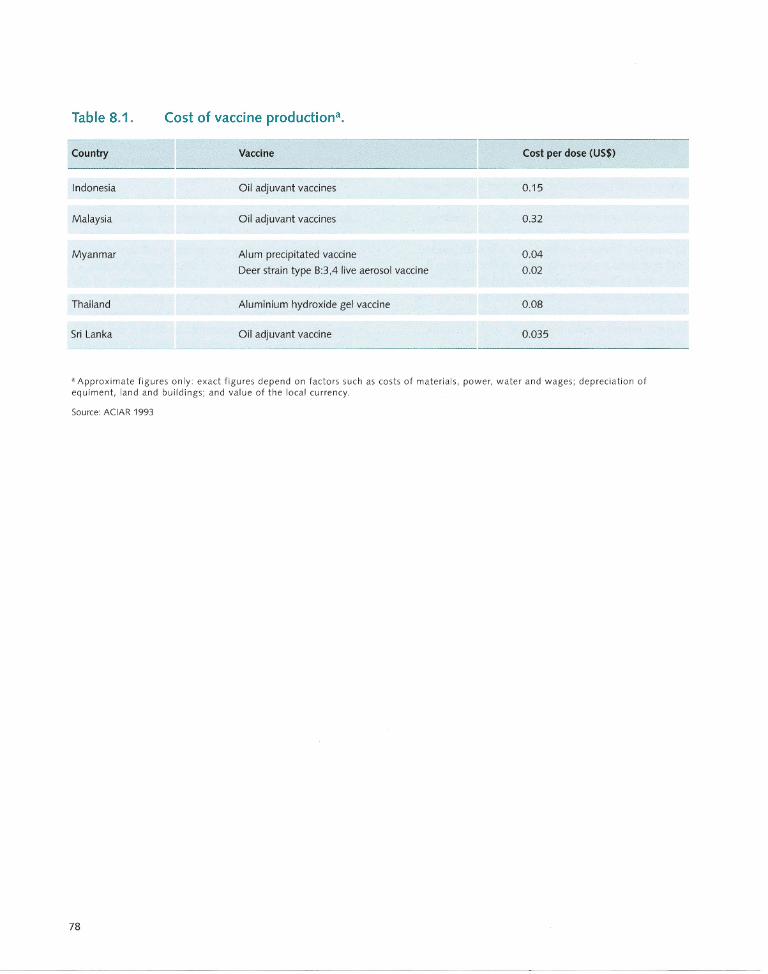
Table 8.1.
Country
Indonesia
Malaysia
Myanmar
Thailand
Sri Lanka
Cost of vaccine productiona.
Vaccine
Oil adjuvant vaccines
Oil adjuvant vaccines
Alum precipitated vaccine
Deer strain type 8:3,4 live aerosol vaccine
Aluminium hydroxide gel vaccine
Oil adjuvant vaccine
Cost per dose (U5$)
0.15
0.32
0.04
0.02
0.08
0.035
a Approximate figures only: exact figures depend on factors such as costs of materials, power, water and wages; depreciation of equ iment, land an d buildin gs; and value of the lo cal currency.
Source: ACIAR 1993
78
Vaccine Research and Development
Overview
Vaccine characteristics
The ideal vaccine must be economical to produce,
stable under tropical conditions and easy to inject. As
well, the production technology in endemic countries
must be sustainable. Such a vaccine should not produce
adverse reactions and should result in a high level and
prolonged duration of immunity.
Vaccine research
Research into new vaccines has been directed towards
the use of other adjuvants, production of oil adjuvant
vaccines of low viscosity, development of vaccines
incorporating purified extracts, identification of
immunogenic components and the development of live
vaccines. Research carried out so far has indicated that
no single component is totally responsible for immunity.
9.1 The Ideal Vaccine
Most countries where haemorrhagic septicaemia (HS) is endemic produce some type of vaccine and carry out vaccination programs. A considerable amount of research has focused on a search for a better vaccine. It is widely recognised that an ideal vaccine should have the following characteristics:
• easy and economical to produce, with sustainable production technology in the endemic countries;
• stable for use in the tropics;
• easy to handle in the field with a consistency that makes it easy to administer;
• no adverse reactions;
• high level of immunity with a minimum delay after vaccination and lasting for at least a year.
None of the vaccines developed so far satisfy all of these requirements. Attempts to find an ideal vaccine have met with varying degrees of success, but none has replaced the existing vaccines for routine
prophylactic use.
9.2 Improved Vaccines
Attempts to produce better HS vaccines can be classified into several groups:
• use of other adjuvants;
• production of oil adjuvant vaccines of low viscosity;
• production of vaccines based on purified extracts of the organism and on the identification of the protective antigens; and
• use of live vaccines.
79

9.2.1 Adjuvants other than oil
In Africa, Bhatty (1973) produced a type 6:E vaccine using 2 % sod ium alginate as adjuvant. Comparative trials showed that this and the oil adjuvant vaccine both provided one year of immunity as indicated by survival on challenge. In Iran, a saponin-lysed vaccine was introduced in the 1930s but was subsequently replaced by a saponin-formalised vaccine (FAO 1979). None of these vaccines have gained widespread acceptance.
9.2.2 Improved oil adjuvant vaccines
There have been several attempts to improve oil adjuvant vaccines by using new formulations to reduce viscosity to improve injectability, concentrating the antigen to permit small doses to be given, and by using double emulsification to produce a thinner, more easily injectable vaccine.
Reduced viscosity
The proportion of lanoline in the vaccine affects viscosity and injectability. The earliest oil adjuvant vaccines used around 8% to 10% lanoline. Experiments in Sri Lanka indicated that stable emulsions could be prepared using equal volumes of a light mineral oil and bacterin, with 4% lanoline, thereby reducing the viscosity. Viscosity has also been reduced by incorporating Arlacei A as the emulsifying agent, together w ith Tween 80. A thin, stable emulsion was formed using 40 parts of broth bacterin, 55 parts of mineral oil (Marcol 52), 4 parts of Arlacel A and 1 part of Tween 80 by weight (Neramitmansook 1993).
Other emulsion formulae have used newer, highly purified mineral oils and emulsifying agents. The enhanced humoral antibody stimulating effect of vitamin E reported by other workers (Tengerdy 1980; Tengerdy et al. 1983) has even led to attempts to replace the mineral oil wit h a-d-tocopherol acetate (vitamin E) (Muneer and Afzal 1989; Muneer et al. 1993) . These workers prepared three stable formulations of oil adjuvant vaccines of low viscosity:
• formula 1 - 30 parts of Marcol 52, 10 parts of M ontan ide 888 and 30 parts of bacterin;
• formula 2 - 63 parts of Marcol 52, seven parts of Arlacel A and 30 parts of bacterin containing 5 % Tween 80;
80
• formula 3 - 63 parts of a-d-tocopherol acetate, 8 parts of Montanide 888 and 30 parts of bacterin.
All three preparations elicited a good antibody response, which was detected with an enzyme-linked immunosorbent assay (ELISA) test. Significantly high antibody levels were maintained with all three vaccines up to 270 days after vaccination, as compared with control animals and those given an alum precipitated bacterin.
Formula 1 gave the best response, with 2 and 3 following in that order. Small groups of buffalo calves given all of the three vaccines resisted challenge 12 months after vaccination. However, there was no enhanced response with formula 3, which contained vitamin E. Shah et al. (1997) used a mineral oil (Marcol 52) as adjuvant, with Span 85 and Tween 85 as emulsifying agents (Bokhout et al. 1981). In an ELISA test using ultrasonicated total membrane preparation as antigen, the principal antibody response to the
vaccine was of the IgG class with only mild IgM responses. The response increased gradually up to day 75, then dropped slightly, and remained constant up to one year. Young buffalo calves one to two years of age, immunised using this vaccine and challenged at 250 days, were completely protected and did not develop any disease symptoms following challenge with 109 viable organisms. It was observed, however,
that control animals when challenged developed characteristic symptoms but recovered within three days. This vaccine is reported to have low viscosity and to be stable for three years at room temperature. The criteria and methods for determination of stability and retention of potency on storage are not described.
Concentration of antigen
Chandrasekaran et al. (1996) produced a low-volume oil adjuvant vaccine. They were able to incorporate a larger quantity of bacterial substance per unit volume by concentrating the antigen. Ten per cent alum was
added to the dense bacterin to give a final concentration of 1.5 %, and the bacterin was left at room temperature for 24 hours. ,I\fter the bacterial cells had settled at the bottom, two-thirds of the
supernatant was siphoned off, leaving a concentrated bacterin. This bacterin was then emulsified with mineral oil (Ondina 15) and lanoline. By this means it was
possible to reduce the dose to 2 mL.
Double emulsion
A significant advancement in the oi l adjuvant vaccine production technology is the development of the double emulsion (also referred to by some workers as multiemulsion) vaccine. Herbert (1965) redispersed globules of water in oi l emulsion through a second external phase using another emulsifier, Tween 80, to stabilise the product. Mittal et al. (1977) applied this technique to the oil adjuvant HS vaccine, prepared from dense bacterin and paraffin oi l using lanoline as the emulSifying agent. The standard oil adjuvant vaccine was re-emulsified using an equal volume of 2 % Tween 80 in 0.3 % formal inised saline. The resulting product was a free-flowing white liquid with a milk-l ike consistency which was stable and more easily injectable than the oil adjuvant vaccine. Its main disadvantage, however, was that the vaccine was diluted 1:1 in the process of the second emulSification, and therefore, in order to ensure that the required amount of dry bacterial mass was administered in every dose, the dose volume had to be doubled. In the initial experiments, the multiple emulsion vaccine gave good levels of protection in mice, rabbits and calves but the duration of immunity in calves was somewhat lower than with the conventional oil adjuvant vaccine (Mittal et al. 1977, 1979). Subsequently, Yadev and Ahooja (1983) prepared multiple emulsion vaccines using conventional vaccine and Tween 80/saline solution in the ratio of 3:3,3:2, and 3:1 respectively. They found that vaccines prepared using the 3: 1 ratio gave results comparable with those of the conventional 011 adjuvant vaccine. Chandrasekaran et al. (1991, 1993b), found a double emulsion vaccine prepared in accordance with the original 1:1 formulation to be as protective as the conventional vaccine up to one year in buffaloes.
In order to prepare a multiemulsion vaccine using a 1:1 ratio of conventional vacci ne to Tween 80/saline, so as to get the favourable consistency without reducing the bacterial content it wi ll be possible to prepare the initial emulsion using a bacterin whose volume is reduced by 50%, using the alum precipitation technique to concentrate the ant igen. Such a vaccine would have the required dry mass of bacteria, t he favourable consistency and low volume for easy injectability and a satisfactory level of efficacy.
Apparently, a large-scale vaccine production technology using the mult iemulsion principle has not
yet been developed , and hence this vaccine still remains at an experimental stage.
9.2.3 Vaccines incorporating purified extracts
Studies on the various cell components of Pasteurella multocida serotypes that cause HS have indicated that the cell capsule is associated with serogroup specific ity, virulence and immunogenicity (see Section 2.2 .6) . This led to the belief that capsular extracts may be superior to whole cells as antigen in vaccines. The earl iest attempt to produce vaccines containing crude capsular extracts was that of Dhanda (1959b). It was believed that the protein components of the capsule were immunogenic and were responsible for protection.
Penn and Nagy (1974) recognised two major antigenic components, the capsular antigen and endotoxin in saline extracts and phenol-water extracts of P. multocida types Band E. The crude capsular extract was purifi ed by solvent fractionation and precipitated with cetyl pyridinium chloride. Vaccines were prepared by filter sterilisation and the addition of 25 % (vol/vol ) sterile aluminium hydroxide gel. Rabbits immunised with this vaccine failed to produce adequate passive protective antibodies, and none of the rabbits su rvived subcutaneous challenge with viable organisms. Calves immunised with t his vaccine, however, developed passive mouse protective antibodies (Penn and Nagy 1974, 1976). Subsequent experiments by these workers showed that cattle immunised with capsular extract vaccine of type E (the endotoxin-f ree, aluminium hydroxide adjuvant) survived challenge; the results of the indirect haemagglutination (IHA) response and the passive mouse protection test (PMPT) response corresponded with the challenge results. The IHA test was carried out using erythrocytes coated with solvent-purified capsular antigens of type E (Nagy and Penn 1976).
More recently, attempts were made to separate in pure form polysaccharides, proteins and lipopolysaccharides of serotype B:2 in order to determ ine the most immunogenic components. M un iandy et al. (1993) , found that capsular polysaccharide in a highly purif ied state was non immunogenic. Muniandy and Mukkur (1993) found that while lipopolysaccharide (LPS) extracts prepared by the Westphal method were protective in mice, the protectivity was abolished by
81
treatment with phenol or digestion with proteinase 1<. Thus, it was concluded that the protectivity of these preparations was actually due to the contaminating outer membrance proteins (OMPs) .
Several attempts have also been made to identify and isolate the immunogenic proteins. Ramdani and Adler (1993) isolated three protein antigens of molecular weights 29,36 and 42 kDa. Attempts to protect mice with these protein antigens have succeeded in achievi ng only 25-67% protection . Of these, the 42 kDa protein is believed to be in the cell membrane.
Pati et al. (1996) prepared OMP extracts from P multocida strain P52 (the Indian vaccine strain of serotype B:2). Using sodium dodecyl sulfate polyacrylamide gel electrophoresis (SDS-PAGE), 10 major po lypeptide bands of molecular weight ranging f rom 88 to 25 kDa were detected. Immunoblots indicated that the 44, 37 and 30 kDa fractions were t he major immunogens. Buffalo calves in two groups were vaccinated using a commercial whole cell oil adjuvant vaccine and an oil adjuvant vaccine prepared f rom the OMP extract. Antibody responses were tested by IHA and ELISA techniques and the protection by PMPT in mice and by direct challenge. Higher titres were observed in the ELISA, as compared with the IHA test, the highest titres being recorded around 21 days after vaccinat ion . In both groups, the highest passive mouse protection was during the period day 21 to day 26 after vaccination. Upon direct challenge, all of five OMP vaccinated buffaloes and two of three buffaloes given the commercial vaccine survived.
Srivastava (1996) prepared a whole cell protein extract by ammon ium sulfate gel precipitation of sonicated extracts of strain P52. The sonicated extract showed 12 protein bands upon analysis by SDS-PAGE and these ranged from 120 to 30 kDa. The ammonium sulfate precipitated extract, on the other hand, yielded a protein-rich material comprising at least 14 polypeptide bands, ranging f rom 91 to 30 kDa. This latter product was tested as a vaccine for immunogenicity in rabbits and mice, compared wi th whole cells, with both vaccines incorporati ng an adjuvant. In rabbits, both vaccines gave 100% protection against challenge with the P52 strain at the dosage tested. In mice, the whole cell vaccine gave 85 .4 % protection, whilst the extract vaccine gave only 68.5% protection. All of these findings demonstrate the role of the protein fractions of the cells in providing some degree of protection.
82
It is also believed that certain OMPs are produced by serotype B:2 strains in vivo but not in ordinary media in vitro. These OMPs may be associated with the solid immunity found in convalescent or naturally immune animals. In vivo, the bacterial pathogens have restricted access to iron in their environment, primarily due to the presence of haemoglobin, transferrin and lactoferrin. Several bacterial pathogens have been shown to have surface receptors that recognise and bind these iron sequestering proteins and mediate iron acquisition from them. Although much work has been done on these high molecular mass OMPs produced in vivo, and in iron-restricted media in vitro, by type A strains of P multocida associated with avian pasteurellosis and bovine pneumonia, the results have been inconclusive (Snipes et al. 1988; Lu et al. 1988, 1991; Abdullahi et al. 1990; Choi-Kim et al. 1991; Ogunnariwo et al. 1991).
Studies with type B:2 strains of P multocida showed that bacteria grown in iron-restricted media gave better protection than those grown in iron-replete medium (Kennett et al. 1993). This superior protection was attributed to a high molecular weight protein fraction (see Section 2.2.6). A study of protein profiles by SDSPAGE and immunoblotting, carried out using serum from buffaloes naturally immune as a result of experimental exposure to a virulent 8:2 strain, recognised a high molecular weight protein fraction (116 kDa), whereas sera from buffaloes immunised with the inactivated oil adjuvant vaccine failed to do so (T.G. Wijewardana, Veterinary Research Institute, Peradeniya; personal communication). Veken et al. (1994, 1996) found that convalescent phase sera from buffaloes reacted against some iron-restricted and other proteins, indicating that these proteins were produced in vivo. A specific 82 kDa high molecular mass bovine transferrin binding iron-regulated outer membrane protein was produced by serotype 8:2, but not by serotype 8:3.4.
It has also been found that both polysaccharide and LPS exhibit anti phagocytic activity in vivo, a factor which may be important in the pathogenesis of HS. The role of endotoxin, which is predominantly LPS, in the pathogenesis of the disease is also well established . Thus, these components may also playa vital role in immunogenicity (see Section 2.2.6).
From the research on vaccines based on identification of immunogenic components of the cell, it can be concluded that no single component is totally responsible for immunity. All fractions, LPS and proteins and polysaccharides evidently contribute towards immunity. The complete set of immunogens is present in whole bacterial cells grown in vivo or cultivated in vitro in a medium that provides all essential in vivo
conditions for the full expression of relevant immunogenic components . It appears that there are many common components in the different types of P. multocida that also playa role in protection.
Rimier (1996) found passive immune cross-protection in mice, using rabbit antisera against different serotypes of P. multocida. Eleven isolates from different host species, including those causing HS, bovine pneumonia and fowl cholera, were tested. A serotype A:5 strain and a fowl cholera strain produced cross-protection against other strains, irrespective of the disease caused. The protection was attributed to antigen bands of 20-120 kDa.
It is also interesting to note the findings of Singh et al. (1994), who immunised three groups of buffaloes, using the P52 strain, a duck strain (1 :A) and a pig pneumonia isolate in an alhydrogel vaccine standardised to contain 4 mg of bacterial protein per dose. Vaccinated buffaloes were challenged 180 days later with the P52 strain. Animals immunised with P52 (i.e. homologous challenge) showed only 75% protection; those immunised with the duck strain gave the highest antibody titres as measured by a tube agglutination test and the IHA test, but gave only 25% protection. Those immunised with the pig pneumonia strain gave high antibody titres and also 100% protection. Unfortunately, the serotype of the culture was not reported.
9.2.4 Live vaccines
It is generally accepted that natural exposure to live organisms (such as in actual infection) produces greater immunity than immunisation through inactivated
vaccines. This has been specifically shown in HS, where the naturally acquired immunity in exposed animals that results from arrested infection has been found to be superior to vaccinial immunity. If we can find an organism that has all the properties of the field strains, including all the antigen components, but has lost its virulence, it will constitute the basis for an ideal live
vaccine. As facilities for Iyophilisation are now available in most countries, the use of a live, Iyophilised vaccine under tropical field conditions is now feasible. Such a vaccine strain should have the following characteristics:
• it must have all, or at least all the protective, antigens present in the field strains;
• it must grow readily in ordinary culture media;
• it should be avirulent to cattle and buffaloes, or may be a low virulence strain (the lethal dose must be
conSiderably higher than the immunising dose);
• it should be stable, particularly in its property of avirulence, and there should be no reversion to virulence;
• it should be able to multiply sufficiently in vivo
following vaccination, producing a full complement of the important immunogens, thereby stimulating an immune response; and
• it should withstand Iyophilisation and its stability following reconstitution should be high.
Strains satisfying these criteria may occur in nature, or
may be produced in a laboratory by exposure to mutagenic agents.
Experimental live vaccines
A procedure for inducing mutations in bacteria using N-methyl-N nitro-N-nitrosoguanidine has been described (Adelberg et al. 1965; Oeschger and Berlyn 1974). Chengappa and Carter (1979) improved on this technique and produced streptomycin-dependent (StrD) mutants of P. multocida and P. haemolytica. Wei and Carter (1978), using this technique, produced StrD
mutants of type B strains of P. multocida associated with HS. These mutants were successfully used to immunise mice and rabbits against challenge wi th homologous strains. De Alwis et al. (1980) produced a
large number of StrD mutants from Sri Lankan strains of P. multocida type B:2 associated with HS. One of these mutants was used to immunise cattle and buffaloes in Sri Lanka. Though an antibody respon se was observed , large numbers of organisms had to be inoculated to produce a response as measured by the PMPT. It was postulated that this may be due to the absence of in vivo multiplication of the organism in the absence of streptomycin (De Alwis and Carter 1980).
83
De Alwis et al. (1980) also produced streptomycinindependent revertants from StrD mutants. On reversion many of these assumed the normal virulence of t he field strains. A few strains, however, were of low virulence. One of these (designated R39) was selected as a possible vaccine candidate. It had almost all the
required characteristics for a live vaccine seed. Biochemically and serologically, it was indistinguishable from the field strains. It even produced hyaluronidase,
a property shared only by type B:2 strains of P. multocida that cause HS. Restriction endonuclease
analysis of this culture indicated a unique DNA fingerprint profile different from all other Sri Lankan B:2 strains (R.B. Rimier, Midwest Area National Animal Disease Center, US Department of Agriculture, Ames, Iowa, personal communication), thus providing
evidence that the loss of virulence is a change at a genetic level.
In SDS-PAGE and immunoblotting with serotype B:2 cu ltures grown in vivo in iron-restricted media, the specific band that represented an aMP of 116 kDa was recognised by sera of naturally immune animals and of R39-vaccinated an imals, but not by sera of cattle given the oil adjuvant vaccine (T.G . Wijewardana, Veterinary Research Institute, Peradeniya, Sri Lanka, personal communication). This mutant showed low virulence for mice and rabbits, the median lethal doses (LD50s) being 107-8 and 104 -5 CFU respectively as compared
with 1-10 organisms of field strains. When injected by the int ramuscu lar route or sprayed intranasally, 107
CFU was sufficient to evoke a good IHA antibody
response, whi lst doses as high as 1010 CFU produced no adverse reactions. The IHA titres persisted for up to a year, at which time they were challenged with a subcutaneous inject ion of a field isolate of serotype B:2. There was a similar IHA response in unvaccinated control animals reared in close contact with vaccinated animals, suggesting lateral t ransmission. However, upon su bcutaneous challenge with virulent strains, only 20% of vaccinated an imals were protected (De Alwis et al. 1998).
The use of this live vaccine was an attempt to simulate the phenomenon of naturall y acqu ired immunity
resu lting from natural exposure to disease. The fact that an imals showing a high IHA titre fai led to resist
su bcutaneous challenge raises some doubt about the validity of this method of challenge as an indicator of
84
ability to resist natural infection. On the other hand, it is also possible that in natural exposure, or exposure to live vaccines, the rising IHA titre is only an indicator of a humoral immune response and the actual protective antibody may not be picked up by this test. This work with StrD mutants and streptomycin-independent
revertants of low virulence has thrown considerable light on the mechanisms involved and opened up areas for future investigations, but has not directly led to the
development of a practical vaccine.
Exposure to the chemical mutagenic agent N-methyl N-nitrosoguanadine has led to the production of temperature-sensitive mutants of P. multocida (Schimmel 1993). After two intratracheal immunisations, it has been possible to prevent death in
calves following challenge, but pneumonic lesions could not be prevented wi th fewer than four immunisations. It is not likely that a practical live vaccine can be developed using this mutant in its present form.
Deer stra in live vaccine
Myint et al. (1987) used a strain of P. multocida from deer as a live vaccine for cattle and buffaloes. The strain (serotype B:3,4) was isolated by Jones and Hussaini (1982) from fallow deer in England after an outbreak of septicaemic disease. The strain was
pathogenic to mice (LD50 less than 10 organ isms), but doses ranging from 105 to 107 viable organisms were found to be safe for cattle. Subcutaneous injections of 107 viable organisms lead to an antibody response
detectable by a mouse protection test wi thin eight days fo llowing vaccination, and vaccinated an imals withstood direct challenge with serotype B:2 nine months later. It was concluded that the protective fractions were in the capsular B complex and that a different somatic antigen served as a marker to differentiate the vaccine strain from the f ield strain .
Further work (Myint and Carter 1989,1990) indicated that when the vaccine strain was administered by the subcutaneous route, it caused deaths in a smal l
proportion of young ani mals, particularly buffaloes. When six buffaloes t hree to f ive months of age were
injected subcutaneously with 1.4x1 07 viable organisms, three developed seve re local reactions and died 72 hours later. Serotype B: 3,4 was recovered from the dead animals. It was concluded that the live B:3,4
vaccine was safe for use as a booster vaccination in adult animals that had been given a primary immunisation with an inactivated vaccine. Subsequently, the vaccine was administered intranasally as an aerosol spray (Carter et al. 1991). Several thousand cattle and buffaloes over six months of age in several village tracts in Myanmar were vaccinated by the subcutaneous or intranasal route. No adverse
reactions were recorded. No outbreaks of HS were recorded in the subsequent year in the vaccinated villages, whereas outbreaks did occur in neighbouring
villages where the live B:3,4 vaccine was not used.
Initial attempts to demonstrate immunity by direct challenge of vaccinated animals picked up from the field were disappointing. Six weeks after B:3,4 intranasally vaccinated animals were challenged using serotype B:2 by the subcutaneous route, only six out of 17 animals survived. In a subsequent controlled trial, however, the ability of intranasal B:3,4 vaccinated cattle to withstand B:2 challenge by the subcutaneous route was demonstrated . In this trial, 52 zebu cattle in a non endemic area in Myanmar were vaccinated intranasally (Myint 1996). Indirect haemagglutination tests using B:2-coated human erythrocytes showed titres in 73% of animals at 10 days and 82.7% of animals at five weeks after vaccination. The IHA titres were high, and comparable with levels achieved when exposed naturally to type B:2. No animals showed IHA titres at 40 weeks after vaccination, but 82.5% of the animals gave
positive reactions in the PMPT. At 12 months after vaccination, 12 animals were challenged with the B:2 strain by subcutaneous injection; all survived, though all four unvaccinated controls died.
The deer strain (B:3,4) was subjected to a host of tests,
in order to assess its safety and efficacy (M.C L. De Alwis, unpublished data; M.CL. De Alwis and A. Myint, unpublished data). Two groups of four buffaloes, four to six months of age, were given 106
and 107 viable organisms by the subcutaneous route. Three animals in each group died and type B:3,4 was recovered. However, the syndrome was more protracted than that produced by strain B:2 in buffaloes under similar circumstances. In five of the six animals that died, death occurred in three to five days. One
animal died on the 10th day. Serotype B:2 under similar circumstances would normally have killed in less than
24 hours. Thirty-nine buffaloes given 100 times the recommended dose of 107 viable organisms by the intranasal route showed no adverse reactions. Of these, 11 were previously unvaccinated animals and their ages ranged from 6 to 12 months.
Since the strain B:3,4 was isolated from a small ruminant (deer), it may have been pathogenic to goats, which are the common small ruminant in the Asian region. Four goats were given 109-1010 CFU by t he subcutaneous route and two were given 4.8 X10¹¹ CFU by the intranasal route; they showed no adverse
reactions.
Of all live experimental vaccines developed so far, this deer strain has the best potential for development into a practical vaccine for routine use. It has been used extensively in Myanmar: in 1995-96, about two-th irds of all animals immunised against HS received the B:3,4 deer strain live Iyophilised vaccine, reconstituted and administered as a nasal spray. However, some conditions should be strictly observed. Firstly, any country using this vaccine should have a good disease
reporting system, and a readily available diagnost ic facility that can isolate and differentiate between
serotypes B:2 and B:3,4. These precaut ions are important because the vaccine strain is a potent ial pathogen, though so far proven to be safe under experimental conditions. Secondly, the vacci ne should
only be given to animals over six months of age, and only by the intranasal route.
Standards for live vaccines
If live vaccines are to be developed and recommended for routine use in the future, certain standards must be applied. The form of presentation, dilution factor, dosage, and so on, should be stipulated, but there should be a margin for handling errors in the f ield in addition to the margin of safety. For example, if 107 CFU is t he required minimum immunising dose, a single dose shou ld contain at least 108 CFU, allowing for one log unit drop in viable count after reconstitution. It is also important that 10 times or, preferably, 100 times (109 or 1010 CFU) the prescribed dose administered by the recommended route should be absolutely safe.
85
Intranasal live vaccines do not need to be given by the veterinary practitioner or paraveterinary personnel such as vaccinators and can be successfully applied by farmers or owners of livestock. In such instances, it is of utmost importance that the instructions for use are printed in a language understood by the farmers of the country; the diluent for reconstitution and the spraying device should be supplied with the vaccine.
9.3 Protection between Serotypes
It was shown by Bain (1979b) that there was crossprotection between Asian and African strains when bacteria grown in vivo were used to immunise mice, and that this cross-protection disappeared, whilst homologous protection remained, with successive subculture. The ability of a type B:3,4 strain originating from deer to protect against type B:2 infection in cattle and buffaloes was amply demonstrated by Myint et al. (1987) and Myint and Carter (1989,1990). More recent work by Shah (1998) indicated that, although cross-protection existed between the Asian, (B:2) and African (E:2) strains, B:2 vaccines gave no protection whatever against challenge with type B:3,4. It must be noted that Myint et al. (1987) used a B:3,4 strain as a live vaccine, whilst Shah (1998) used B:2 and E:2
inactivated adjuvant vaccines. It is interesting to speculate as to whether the cross-protective in vivo antigens referred to by Bain (1979b) would provide the explanation for the cross-protection observed with the live vaccine.
Also of considerable interest are the observations of Singh et al. (1994) who found that a pig pneumonia strain gave 100% protection in buffaloes against challenge with a B:2 strain (Indian P52) and also evoked high antibody titres, whereas the homologous P52 strain gave only 75 % protection. They also reported that a duck strain of serotype 1:A gave 25% protection, and gave a higher antibody titre against the P52 strain, than did the homologous strain. Unfortunately, the serotype of the pig pneumonia strain has not been recorded.
All of these observations merit follow-up investigation, perhaps with a view to identifying a single strain or antigen grouping that will give protection against a wide range of challenge strains.
86
9.4 Future Prospects for Vaccine Development
Two broad areas appear to have emerged for future vaccine research. Newer adjuvants that have been developed for low-viscosity emulsions, including the production of double emulsions, appear to be promising. It is unlikely that a single antigenic component, linked with a specifically identified gene, is responsible for total protection. It is therefore unlikely that a DNA recombinant vaccine will appear within the foreseeable future. Development of better media for bulk culture production also appears to be a promising area. The objective would be to provide culture conditions that would cause the fullest expression of antigenic components, coupled with a new adjuvant. It is important that new emulsions should be tested not only for low viscosity and stability of emulsion, but also for retention of potency under different conditions of storage. In any event, vaccines based on purified extracts will not lend themselves to large-scale production in the countries where the disease exists. A more feasible proposition is a whole cell-based adjuvant vaccine grown in an appropriate growth medium that will permit full expression of all antigen components, coupled with a suitable new adjuvant.
The second area that warrants investigation is that of live vaccine development. Studies on virulence factors and their genomic basis may help to produce suitable avirulent, antigenic mutants by genetic manipulation. Such an organism, that can be administered by any route, will serve as an ideal vaccine candidate.
Further Investigation
Overview Further information on some aspects of haemorrhagic
septicaemia (HS) would be helpful in the control of the
disease. Gaps in the current understanding of the
disease include:
• pathogenesis of the disease (it is not known where
the organism lodges when it enters the body or
where it multiplies during incubation and the early
clinical phase);
• some aspects of epidemiology (the specific
mechanisms that convert a latent carrier to an active
state are unkown); and
• immunology, especially related to vaccine
development.
There are still many grey areas in our knowledge of
haemorrhagic septicaemia (HS). Significant gaps exist
in the understanding of pathogenesis of the disease and
some aspects of the epidemiology and immunology. A
better understanding of these areas will lead to more
effective control of the disease.
10.1 Pathogenesis
It is fairly well established that entry of the infecting organisms is by inhalation or ingestion (see Chapter 3). However, it is uncertain where the organism lodges itself immediately upon entry and where it multiplies during the incubation period and the early clinical phase. It is known that septicaemia is terminal (M.C.L. De Alwis, unpublished data; Horadagoda et al. 1991) and this phase roughly coincides with the rapid rise in endotoxin levels in the blood (N.U. Horadagoda et aI., personal communication, 1998; see page 32). It may be surmised that the initial multiplication takes place in tissues associated with the respiratory tract, but more investigations are needed in this regard .
The initial multiplication may be facilitated by the vacuolating cytotoxic activity (Shah et al. 1996); the polysaccharide and lipopolysaccharide (LPS) components of the cell, which are known to be anti phagocytic (Muniandy et al. 1993), may be responsible for this activity. LPS also accounts for most features of the clinical syndrome (Rebers et al. 1967; Rhoades et al. 1967; Horadagoda et aI., personal communication, 1998). It is not known whether there are any other virulence factors, whether these are merely phenotypic characters, or whether they possess a genetic base.
10.2 Epidemiology
There is ample evidence to support the existence of a large number of latent carriers in animal populations in endemic areas, and to indicate that occasionally, and intermittently, a few become active carriers and shed infective pasteurellae. The specific mechanisms that convert a latent carrier to an active state are still unknown (see Section 4.7.3).
87
Organisms shed by active carriers are believed to be the source of infection in the initiation of outbreaks. In experimental transmission by natural routes of infection, De Alwis et al. (1990) found that large numbers of viable organisms were required to produce infection. Such numbers are unlikely to be transmitted from a shedder to an incontact susceptible animal. For an outbreak to begin there may therefore be mechanisms that increase the virulence of shed organisms or, conversely, diminish the defence mechanisms of the susceptible incontact animals. Such mechanisms merit further investigation in order to obtain a more complete understanding of pathogenesis and epidemiology.
The nature of the naturally immune-cum-carrier state which occurs in survivors from natural outbreaks, a state that is believed to arise from what is described as 'arrested infections', also needs to be defined, as do the effects of vaccination on the immune and carrier status. More evidence is also needed on how such animals respond to challenge by different routes.
10.3 Vaccines and Immunology
Vaccine research was discussed in detail in Chapter 9. Formulae that will give oil emulsions thin enough for easy injection have now been developed, particularly for adjuvant vaccines. Vaccines prepared in accordance with these new formulations need to be validated for potency (as measured by a defined test), stability and shelf life at different storage temperatures. Similar work is necessary with double emulsion vaccines. In the latter case, the technology for large-scale production needs to be developed.
The development of a non-oily adjuvant may also be useful as such a vaccine may be more user friendly than the oil emulsions.
The search for the 'most potent immunogen' has been on for the last decade or so. Although various cell components that contribute towards protection have been identified, no single component appears to be responsible for total protection . There has been much hope that a transferrin-binding protein from the outer membrane of cells could be produced in vivo and in iron-depleted media in vitro. Such a protein would also
88
need to be a major immunogen (Kennett et al. 1993; Veken et al. 1994). More work is necessary to determine whether such a protein is expressed by a wide range of strains responsible for the disease and the extent to which it would contribute to protection. Recognising that immunity following natural exposure to disease is the best, it would be worth studying the components expressed by the organism when multiplying in vivo. It may then be possible to develop a medium and culture conditions for bulk in vitro cultivation of the organism, with the full complement of in vivo antigens. Whatever the adjuvant, a vaccine based on such a culture could be expected to provide better protection than current vaccines.
A live vaccine based on an avirulent strain containing all the protective antigens of the virulent organism would also be a valuable asset. Such a vaccine would need to be presented in a Iyophilised form and should ideally lend itself to use either by injection or by aerosol spray. This is because, in some circumstances, it is considered easier to administer an aerosol spray through the nostrils, although other field workers prefer to administer an intramuscular injection from the rear in semi-wild cattle and buffaloes. Where a live vaccine is used , it should be possible to differentiate between the field strain and the vaccine strain relatively easily.
Some conflicting findings from past investigations need further study. Myint and others have amply demonstrated that serotype B:3,4 deer strain as a live vaccine evokes both an antibody response and protection to challenge with serotype B:2 (Myint et al. 1987; Myint and Carter 1989,1990). Shah (1998), on the other hand, found that vaccines produced against B:2 and E:2 gave cross-protection in mice to challenge with both serotypes, but that there was no protection at all to challenge with B:3,4 serotype. Also of interest are the findings of Singh et al. (1994) on crossprotection between strains. They found that a pig pneumonia strain (serotype not stated) both evoked a higher antibody response and gave better protection to challenge in buffaloes against a B:2 strain P52 than did the homologous P52 strain. Further, a duck strain (serotype 1 :A) gave higher antibody levels (but only 25% protection) when challenged with strain P52. These observations merit further investigation . It may
also be possible to find, or develop by genetic manipulation, a strain that will provide a wide range of protection across species and disease barriers.
Little is known of the role of cell-mediated immunity (CMI) in haemorrhagic septicaema (HS); more is known about Pasteurella multocida infections in chicken and turkeys (Dua and Mahaswaran 1978). The work of Maheswaran and Theis (1979), and that of Verma and Jaiswal (1997), on the role of CMI deserves to be followed up.
The method of assessment of protective immunity also deserves further investigation. The natural routes of infection are ingestion and inhalation, yet in most direct challenge experiments, the subcutaneous route of infection is used. It is not known to what extent local antibodies in the mucous membranes contribute towards immunity to natural infection. In vaccine evaluation experiments, however, the subcutaneous route of challenge is widely used, since by this route the disease can be consistently and predictably reproduced.
To overcome the cumbersome procedures of using animals to assess immunity, it is of utmost importance that a reliable laboratory test that measures protective immunity be developed. Of the tests currently used -indirect haemagglutination, passive mouse protection
and enzyme-linked immunoabsorbent assay (ELISA) -ELISA has been acclaimed by many recent workers as being most closely related to results of direct challenge. Yet ELISA has its limitations, and what it detects can be influenced by the antigen used. Some workers have used a sonicated extract as antigen but most use the boiled antigen of Heddleston (1972); as might be expected, the test then fails to differentiate between a response to the Asian and African serotypes on account of the common somatic antigen.
It is widely believed that, following an outbreak, surviving animals possess a high degree of protective immunity. It is also known that high IHA titres not obtainable with killed vaccines develop within a few days of natural exposure, and persist for about six months. Yet experiments with a live 8:2 mutant vaccine (De Alwis et al. 1998) indicated that a similar persistent IHA response evoked by the mutant was not an indication of protection because the animals that were showing IHA titres failed to withstand challenge. Passive mouse protection tests do not appear to be a reliable indicator of immunity in buffaloes (Chandrasekaran et al. 1991, 1994b). Thus, there is a need to develop a serological test or a laboratory animal model that is capable of reliably measuring immunity to natural infection.
89
Transport and Culture Media
Dispatch of suspected material
Modified Stuart's transport medium
Sodium glycerophosphate 10.0 g
Sodium thioglycollate 0.5 g
Cysteine hydrochloride 0.5 g
Calcium chloride 0.1 g
Methylene blue 0.001 g
Agar 5.0 g
Distilled water 1 litre
pH 7.4 ± 0.2
The above ingredients (or 16 g of the commercial dehydrated medium) are dissolved in 1 litre of distilled
water. The mixture is then dispensed into 7.0-mL screw-capped bottles and sterilised at 121°C for 15 minutes.
Amie's transport medium
Charcoal (pharmaceutical)
Sodium hydrogen phosphate
Potassium dihydrogen phosphate
Potassium chloride
Sodium thioglycollate
Calcium chloride
Magnesium chloride
Agar
Distilled water
pH ------------------------------
10.0 g
1.15 g
0.2 g
0.2 g
1.0 g
0.1 g
0.1 g
4.0 g
1 litre
7.4 ± 0.2
The above ingredients (or 20 g of the commercial dehydrated medium) are dissolved, bottled and sterilised as above.
Transport medium formulated by De Alwis (1973)
This is a simplified, non-nutrient inert medium which preserves the bacteriological status of the material at the time of collection. It is particularly suitable for
contaminated material, because it prevents overgrowth by contaminants.
Disodium hydrogen phosphate
(Na2 HP04. 12 H20)
Thioglycollic acid
Methylene blue (1 % aqueous solution)
Agar powder (bacteriological grade)
Distilled water
16.0 g
1.0 mL
4.0 mL
4.0 g
1 litre
pH 7.4 ±0.2
The medium is boiled in a water bath, dispensed into 7 -mL screw-capped bottles, and sterilised as above. The prepared medium is colourless and semisolid in conSistency. Bottles that turn blue should be discarded. Pasteurellae have been isolated by direct culture seven
days after blood was placed in this medium (the blood came from an animal that died of haemorrhagic septicaemia, and was collected 32 hours after death under tropical conditions) (De Alwis 1973). This was
the longest period tested.
Transport enrichment medium (TEM)
The TEM developed by Warner (1996) inhibits the
growth of bacterial contaminants such as Escherichia
coli, pseudomonas, proteus and gram-positive organisms, as well as fungal contaminants. This improves the chances of isolating Pasteurella
multocida from diseased animals.
The medium consists of a sloppy brain-heart infusion (BHI) agar with added antibiotics. The sloppy agar is made by adding 0.3% agar to BHI broth. The antibiotics should be made as concentrated stock solutions, and the filter sterilised (0.45-j..Im microorganism filter) and stored at -20°C when not in use.
91
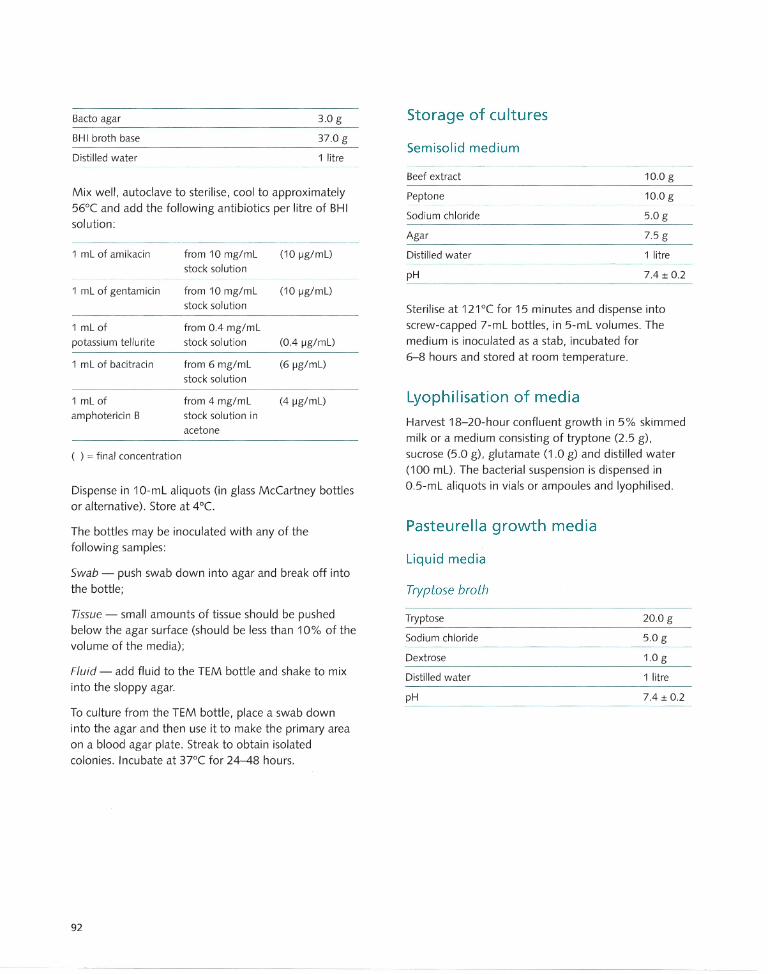
Bacto agar 3.0 g
BHI broth base 37.0 g
Distilled water 1 litre
Mix well, autoclave to sterilise, cool to approximately 56°C and add the following antibiotics per litre of BHI solution:
1 mL of amikacin from 10 mg/mL (10 jJg/mL)
stock solution
1 mL of gentamicin from 10 mg/m L (10 jJg/mL)
stock sol ution
1 mL of from 0.4 mg/mL
potassium tellurite stock solution (0.4 jJg/mL)
1 mL of bacitracin from 6 mg/mL (6 jJg/mL)
stock solution
1 mL of from 4 mg/mL (4 jJg/mL)
amphotericin B stock solution in
acetone
( ) = final concentration
Dispense in 1 O-mL aliquots (in glass McCartney bottles or alternative). Store at 4°C.
The bottles may be inoculated with any of the following samples:
Swab - push swab down into agar and break off into the bottle;
Tissue - small amounts of tissue should be pushed below the agar surface (should be less than 10% of the volume of the media);
Fluid - add fluid to the TEM bottle and shake to mix into the sloppy agar.
To culture from the TEM bottle, place a swab down into the agar and then use it to make the primary area on a blood agar plate. Streak to obtain isolated colonies. Incubate at 37°C for 24-48 hours.
92
Storage of cultures
Semisolid medium
Beef extract 10.0 g
Peptone 10.0 g
Sodium chloride 5.0 g
Agar 7.5 g
Distilled water 1 litre
pH 7.4 ± 0.2
Sterilise at 121°( for 15 minutes and dispense into screw-capped 7-mL bottles, in 5-mL volumes. The medium is inoculated as a stab, incubated for 6-8 hours and stored at room temperature.
Lyophilisation of media
Harvest 18-20-hour confluent growth in 5% skimmed milk or a medium consisting of tryptone (2.5 g), sucrose (5.0 g), glutamate (1.0 g) and distilled water (100 mL). The bacterial suspension is dispensed in O.5-mL aliquots in vials or ampoules and Iyophilised.
Pasteurella growth media
Liquid media
Tryptose broth
Tryptose 20.0 g
Sodium chloride 5.0 g
Dextrose 1.0 g
Distilled water 1 litre
pH 7.4 ± 0.2
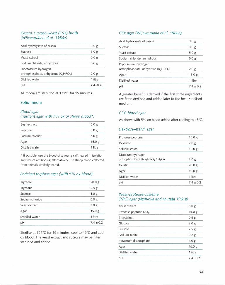
Casein-sucrose-yeast (CSY) broth (Wi;ewardana et al. 1986a)
Acid hydrolysate of casein
Sucrose
Yeast extract
Sodium chloride, anhydrous
Dipotassium hydrogen
orthophosphate, anhydrous (K2HP04)
Distilled water
pH
3.0 g
3.0 g
5.0 g
5.0 g
2.0 g
1 litre
7.4±0.2
All media are sterilised at 121 o( for 15 minutes.
Solid media
Blood agar (nutrient agar with 5% ox or sheep blood*)
Beef extract 5.0 g
Peptone 5.0 g
Sodium chloride 5.0 g
Agar 15.0 g
Distilled water 1 litre
* If possible, use the blood of a young calf, reared in isolation
and free of antibodies; alternatively, use sheep blood collected
from animals similarly reared.
Enriched tryptose agar (with 5 % ox blood)
Tryptose 20.0 g
Tryptone 2.5 g
Sucrose 1.0 g
Sodium chloride 5.0 g
Yeast extract 3.0 g
Agar 15.0 g
Distilled water 1 litre
pH 7.4 ± 0.2
Sterilise at 121°( for 15 minutes, cool to 45°( and add ox blood. The yeast extract and sucrose may be filter sterilised and added.
CSY agar (Wi;ewardana et al. 1986a)
Acid hydrolysate of casein
Sucrose
Yeast extract
Sodium chloride, anhydrous
Dipotassium hydrogen
orthophosphate, anhydrous (K2HP04)
Agar
Distilled water
pH
3.0 g
3.0 g
5.0 g
5.0 g
2.0 g
15.0 g
1 litre
7.4 ± 0.2
A greater benefit is derived if the first three ingredients are filter sterilised and added later to the heat-sterilised medium.
CSY-blood agar
As above with 5% ox blood added after cooling to 45°C.
Dextrose-starch agar
Proteose peptone
Dextrose
Soluble starch
Disodium hydrogen
orthophosphate (Na2HP04 2H20)
Gelatin
Agar
Distilled water
pH
Yeast-protease-cysteine (YPC) agar (Namioka and Murata 1961a)
Yeast extract
Protease peptone N03
L -cysteine
Glucose
Sucrose
Sodium sulfite
Potassium diphosphate
Agar
Distilled water
pH
15.0 g
2.0 g
10.0 g
3.0 g
20.0 g
10.0 g
1 litre
7.4 ± 0.2
5.0 g
15.0 g
0.5 g
2.0 g
2.5 g
0.2 g
4.0 g
15.0 g
1 litre
7.4± 0.2
93
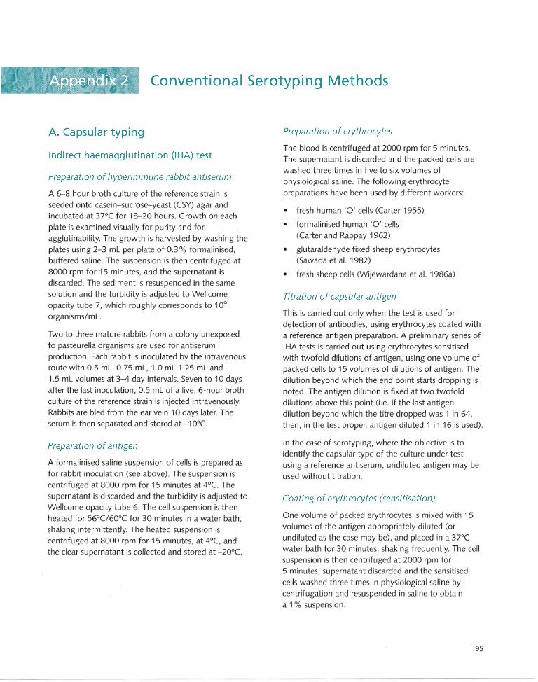
Conventional Serotyping Methods
A. Capsular typing
Indirect haemagglutination (IHA) test
Preparation of hyperimmune rabbit antiserum
A 6-8 hour broth culture of the reference strain is seeded onto casein-sucrose-yeast (CSY) agar and incubated at 37°C for 18-20 hours. Growth on each plate is examined visually for purity and for agglutinability. The growth is harvested by washing the plates using 2-3 mL per plate of 0.3 % formalinised, buffered saline. The suspension is then centrifuged at 8000 rpm for 15 minutes, and the supernatant is discarded. The sediment is resuspended in the same solution and the turbidity is adjusted to Wellcome opacity tube 7, which roughly corresponds to 109
organisms/mL.
Two to three mature rabbits from a colony unexposed to pasteurella organisms are used for antiserum production . Each rabbit is inoculated by the intravenous route with 0.5 mL, 0.75 mL, 1.0 mL 1.25 mL and 1.5 mL volumes at 3-4 day intervals. Seven to 10 days after the last inoculation, 0.5 mL of a live, 6-hour broth culture of the reference strain is injected intravenously. Rabbits are bled from the ear vein 10 days later. The serum is then separated and stored at -1 O°e.
Preparation of antigen
A formalinised saline suspension of cells is prepared as for rabbit inoculation (see above) . The suspension is centrifuged at 8000 rpm for 15 minutes at 4°e. The supernatant is discarded and the turbidity is adjusted to Wellcome opacity tube 6. The cell suspension is then heated for 56°C/60°C for 30 minutes in a water bath, shaking intermittently. The heated suspension is centrifuged at 8000 rpm for 15 minutes, at 4°C, and the clear supernatant is collected and stored at -20°e.
Preparation of erythrocytes
The blood is centrifuged at 2000 rpm for 5 minutes. The supernatant is discarded and the packed cells are washed three times in five to six volumes of physiological saline. The following erythrocyte preparations have been used by different workers:
• fresh human '0' cells (Carter 1955)
• formalinised human '0' cells (Carter and Rappay 1962)
• glutaraldehyde fixed sheep erythrocytes (Sawada et al. 1982)
• fresh sheep cells (Wijewardana et al. 1986a)
Titration of capsular antigen
This is carried out only when the test is used for detection of antibodies, using erythrocytes coated with a reference antigen preparation . A preliminary series of IHA tests is carried out using erythrocytes sensitised with twofold dilutions of antigen, using one volume of packed cells to 15 volumes of dilutions of antigen. The dilution beyond which the end point starts dropping is noted . The antigen dilution is fixed at two twofold dilutions above this point (i.e. if the last antigen dilution beyond which the titre dropped was 1 in 64, then, in the test proper, antigen diluted 1 in 16 is used).
In the case of serotyping, where the objective is to identify the capsular type of the culture under test using a reference antiserum, undiluted antigen may be used without titration .
Coating of erythrocytes (sensitisation)
One volume of packed erythrocytes is mixed with 15 volumes of the antigen appropriately diluted (or undiluted as the case may be), and placed in a 3JOC water bath for 30 minutes, shaking frequently. The cell suspension is then centrifuged at 2000 rpm for 5 minutes, supernatant discarded and the sensitised cells washed three times in physiological saline by centrifugation and resuspended in saline to obtain a 1 % suspension.
95
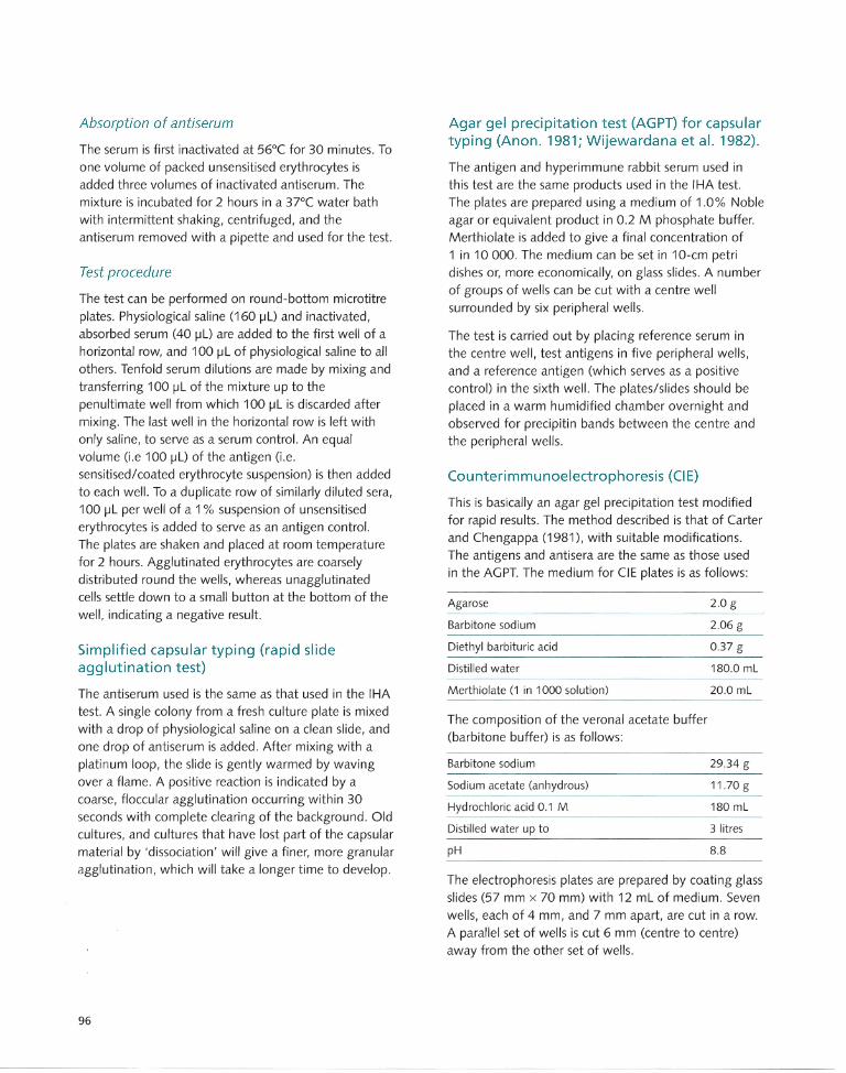
Absorption of antiserum
The serum is first inactivated at 56°C for 30 minutes. To one volume of packed unsensitised erythrocytes is added three volumes of inactivated antiserum. The mixture is incubated for 2 hours in a 37°C water bath with intermittent shaking, centrifuged, and the antiserum removed with a pipette and used for the test.
Test procedure
The test can be performed on round-bottom microtitre plates. Physiological saline (160 IJL) and inactivated, absorbed serum (40 IJL) are added to the first well of a horizontal row, and 100 IJL of physiological saline to all others. Tenfold serum dilutions are made by mixing and transferring 100 IJL of the mixture up to the penultimate well from which 100 IJL is discarded after mixing. The last well in the horizontal row is left with only saline, to serve as a serum control. An equal volume (i.e 100 IJL) of the antigen (i.e. sensitised/coated erythrocyte suspension) is then added to each well. To a duplicate row of similarly diluted sera, 100 IJL per well of a 1 % suspension of unsensitised erythrocytes is added to serve as an antigen control. The plates are shaken and placed at room temperature for 2 hours. Agglutinated erythrocytes are coarsely distributed round the wells, whereas unagglutinated cells settle down to a small button at the bottom of the well, indicating a negative result.
Simplified capsular typing (rapid slide agglutination test)
The antiserum used is the same as that used in the IHA test. A single colony from a fresh culture plate is mixed with a drop of physiological saline on a clean slide, and one drop of antiserum is added . After mixing with a platinum loop, the slide is gently warmed by waving over a flame. A positive reaction is indicated by a coarse, floccular agglutination occurring within 30 seconds with complete clearing of the background . Old cultures, and cultures that have lost part of the capsular material by 'dissociation' will give a finer, more granular agglutination, which will take a longer time to develop.
96
Agar gel precipitation test (AGPT) for capsular typing (Anon. 1981; Wijewardana et al. 1982).
The antigen and hyperimmune rabbit serum used in this test are the same products used in the IHA test. The plates are prepared using a medium of 1.0% Noble agar or equivalent product in 0.2 M phosphate buffer. Merthiolate is added to give a final concentration of 1 in 10000. The medium can be set in 10-cm petri dishes or, more economically, on glass slides. A number of groups of wells can be cut with a centre well surrounded by six peripheral wells.
The test is carried out by placing reference serum in the centre well, test antigens in five peripheral wells, and a reference antigen (which serves as a positive control) in the sixth well. The plates/slides should be placed in a warm humidified chamber overnight and observed for precipitin bands between the centre and the peripheral wells.
(ounterimmunoelectrophoresis «(IE)
This is basically an agar gel precipitation test modified for rapid results. The method described is that of Carter and Chengappa (1981), with suitable modifications. The antigens and antisera are the same as those used in the AGPT. The medium for CIE plates is as follows:
Agarose
Barbitone sodium
Diethyl barbituric acid
Distilled water
Merthiolate (1 in 1000 solution)
The composition of the veronal acetate buffer (barbitone buffer) is as follows:
Barbitone sodium
Sodium acetate (anhydrous)
Hydrochloric acid 0.1 M
Distilled water up to
pH
2.0 g
2.06 g
0.37 g
180.0 mL
20.0 mL
29.34 g
11.70 g
180 mL
3 litres
8.8
The electrophoresis plates are prepared by coating glass slides (57 mm x 70 mm) with 12 mL of medium. Seven wells, each of 4 mm, and 7 mm apart, are cut in a row. A parallel set of wells is cut 6 mm (centre to centre) away from the other set of wells.
The wells on the side of the cathode are loaded with 20-fJL volumes of antigen while an equal volume of antiserum is loaded on to the wells on the side of the anode. Two pairs of wells are kept as controls, one containing the positive reference antiserum against a physiological saline in the antigen well and the other containing a negative rabbit antiserum against positive reference antigen. The electrophoresis tank is filled with the barbitone buffer at pH 8.8. The antigen and antiserum are electrophoresed at 150 V (25 V fcm) for 30 minutes. Plates are examined for precipitin bands between antigen and antiserum wells .
B. Somatic typing
Agar gel precipitation test (Heddleston et al. 1972)
Reference cultures
The 16 reference strains representing the 16 somatic types are used.
Preparation of antigen
A 6-8 hour broth culture is heavily seeded on to dextrose starch agar (DSA) plates, and incubated for 24 hours at 37°e. Each plate is then harvested in 1.0 mL of a solution containing 0.02 M phosphate buffer, 8.5% sodium chloride and 0.3% formalin at pH 7. This thick cell suspension is standardised so as to be equivalent to 25 times the concentration of Wellcome opacity tube 6 or any equivalent turbidimetric measure (i.e 1 drop of suspension mixed with 24 drops of saline should give an opacity equivalent to Wellcome tube 6). The suspension is placed at 1000 e in boiling water in a water bath for one hour. It is then centrifuged at 8000 rpm for 15 minutes. The supernatant is collected and stored at -20°e.
Preparation of antiserum
The growth from a DSA plate is harvested in 2-3 mL of 0.3% formalinised physiological saline. This thick inactivated bacterial suspension is emusified with an equal volume of a light mineral oil containing 10% melted lanoline. The emulsified bacterin is injected
. subcutaneously into the neck of 12-16-week-old
. chicken, in 1.0-mL volumes. Three weeks later, a
further 1.0 mL, divided into two O.5-mL volumes, is injected on each side of the sternum. The birds are exsanguinated 1 week later. The antisera are collected and preserved with 0.01 % thiomerasol and 0.06% phenol. Each serum is tested against the 16 types, and any that cross-react are discarded. It is best to use two to three birds per serotype, and to collect the serum from each bird separately until cross-reacting sera are eliminated .
Preparation of plates
The medium used is as follows:
Noble agar (or equivalent)
Sodium dihydrogen
phosphate (Na H2P04.2H20)
Disodium hydrogen
phosphate (Na2HP04.12H20)
Sodium chloride
Distilled water
Merthiolate (1 in 10000 solution)
1.0 g
0.41g
1.46 g
8.5 g
90 mL
10 mL
The molten medium in approximately 12-mL volumes is coated on to glass slides (57 mm x 70 mm) using a pipette. When set firmly, three patterns of seven wells each are cut, each consisting of one centre and six peripheral wells, each well being 4 mm diameter, spaced at 6 mm centre to centre.
Test procedure
The test antigen is placed in each centre well and five antisera in five peripheral wells, with saline or negative serum control in the other. The slides are then incubated overnight in a warm humid chamber.
All haemorrhagic septicaemia (HS) isolates (Asian and African) will react with antiserum against type 2. Some cross-reactions with type 5 may also be observed.
Namioka-Murata technique (Namioka and Murata 1961 b)
This somatic typing method can be used with suitable modifications to the HS serotypes only.
97
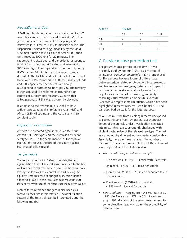
Preparation of antigen
A 6-8 hour broth culture is heavily seeded on to CSY
agar plates and incubated for 24 hours at 37°C. The
growth on each plate is checked for purity and
harvested in 2-3 mL of 0.3% formalinised saline. The suspension is tested for agglutinability by the rapid
slide agglutination test, as a further check. It is then
centrifuged at 8000 rpm for 20 minutes. The
supernatant is discarded, and the pellet is resuspended in 25-30 mL of normal HCi saline and incubated at
37°C overnight. The suspension is then centrifuged at 8000 rpm for 20 minutes, and the supernatant is discarded. The HCi-treated cell residue is then washed
twice with 0.3% formalinised buffered saline at pH 5.0
and 6.0 respectively and the cells are finally
resuspended in buffered saline at pH 7.0. The turbidity
is then adjusted to Wellcome opacity tube 6 or equivalent turbidimetric measure. Cultures that
autoagglutinate at this stage should be discarded.
In addition to the test strain , it is useful to have antigens prepared against reference Asian (6:8) and
African (6:E) HS strains, and the Australian (11 :8) avirulent strain .
Preparation of antiserum
Antisera are prepared against the Asian (6:8) and
African (6:E) serotypes and the Australian avirulent serotype (11 :8) in the same manner as for capsular
typing. Prior to use, the titre of the serum against HCI-treated cells is tested.
Test procedure
The test is carried out in 3.0-mL round-bottomed agglutination tubes. Each test serum is added to the first
well in a horizontal row; serial 10-fold dilutions are done,
leaving the last well as a control with saline only. An
equal volume (0.5 mL) of antigen suspension is then
added to all wells in the row. Each test will consist of
three rows, with sera of the three serotypes given above.
Each of three reference antigens is also used as a
control to facilitate interpretation. The agglutination
pattern of the test strain can be interpreted using the
following matrix:
98
Antisera Antigens
6:8 6:E 11 :8
6:8 + +
6:E + +
11 :8 +
C. Passive mouse protection test The passive mouse protection test (PMPT) was originally used by Roberts (1947) as a method of
serotyping Pasteurella multocida. It is no longer used
for this purpose because it cannot differentiate
between certain related serotypes within a serogroup
and because other serotyping systems are simpler to perform and more discriminatory. However, it is
popular as a method of determining immunity following either vaccination or natural exposure
(Chapter 9) despite some limitations, which have been
highlighted in recent research (see Chapter 10). The
test described below is for the latter purpose.
Mice used must be from a colony hitherto unexposed to pasteurella and free from pasteurella antibodies.
Serum of the animals under investigation is injected
into mice, which are subsequently challenged with
virulent pasteurellae of the relevant serotype. The test
as carried out by different workers varies conSiderably. Essentially, there are three variables: the number of
mice used for each serum sample tested, the volume of
serum injected, and the challenge dose.
• Number of mice per test serum sample
- De Alwis et al. (1978) - 3 mice with 3 controls
- 8ain et al. (1982) - 4-6 mice per sample
- Gomis et al. (1989) - 10 mice per pooled (n=6)
serum sample
- Dawkins et al. (1991 b) Johnson et al.
(1993) - 5 mice and 2 controls
• Serum volume - ranging from 0.5 mL (Bain et al. 1982; De Alwis et al. 1978) to 0.2 mL (Johnson
et al. 1993; dilutions of the serum may be used for
some objectives (e.g. comparing the protectivity of different sera).
• Challenge dose - usually 100 LD50, where the LD50 for mice is 1-10 colony forming units (CFU),
so the normal challenge dose is estimated to contain 100 CFU. This is usually achieved by using 1 mL of a 10-5 or 10-6 dilution of a 6-8-hour CSY
broth culture. As accuracy is not crucial at this stage, a visual judgment can be made to obtain a dose of approximately 100 LD50.
Interpretation
Most workers have accepted survival of at least one of a group of five injected mice (i.e 20% protection) as an index of protection in cattie, provided all of a similar group of control mice die. Johnson et al. (1993) used
a protective index (PI) where PI=log2 serum dilution plus proportion of mice surviving. Where the protectivity of sera from two different groups of cattle or buffaloes is to be compared, a large group of mice (say 10 or 20) can be immunised with pooled group sera, and the percentage protection can be used as the criterion.
In using this test to measure immunity in vaccinated cattle, it is very important to have adequate cattle controls. As far as is possible, the vaccinated group and the unvaccinated control group must be balanced in terms of numbers of animals, age, breed and sex . They should be managed in the same manner and maintained in the same environment for the same
period of time. Sera from both groups should be tested before and after vaccination.
D. Hyaluronidase test
This is a nonserological test for the identification of type B strains specifically associated with HS. A hyaluronic aCid-producing culture is streaked across a plate of dextrose starch agar. Conventionally, hyaluronic acid-producing cultures of Streptococcus equi have been used, but mucoid colonies of P multocida serogroup A, which produce hyaluronic acid capsules, may be used to obtain clearly readable results. The pasteurella cultures to be tested are then streaked across the plate at right angles to the original streak, and incubated at 3JOC for 18 hours. At the point of intersection, where the test culture produces hyaluronidase, the hyaluronic acid-producing culture will show a thinned-out growth. The reading is
facilitated by the use of freshly prepared plates and a humidified incubator.
99
Enzyme-Linked Immunosorbent Assay (ELISA)
In recent years the enzyme-linked immunosorbent assay (ELISA) has proved a useful technology for measuring the interaction of antigen and antibody using a solidphase support such as a polystyrene microtitre plate, tube or stick. ELISA-based systems are widely used in the veterinary field for the detection or diagnosis of many viral, bacterial and parasitic infections. They are rapid, inexpensive and can be automated if a large through-put of samples is required.
ELISA methods have recently been adapted for diagnosis and monitoring of Pasteurella multocida infection and have significant advantages over current methods (such as serotyping and the indirect hemagglutination assay). Using ELISA methods, infection with haemorrhagic septicaemia (HS)-causing serotypes of P. multocida can be diagnosed and monitored in two ways.
• HS-antibody ELISA - detection of circulating antibody to the HS-causing organism (humoral response). A specific antigen (adsorbed to plate) is used to detect homologous antibody in test sera (Klaassen et al. 1985; Johnson et al. 1988, 1989, Chandrasekaran et al. 1994a); or
• HS-antigen ELISA - detection of the P. multocida organisms that possess the HS-specific antigen. An antigen is used in immunisation to raise specific antisera, which are then used to detect antigen in the test sample (Dawkins et al. 1990, 1991 b).
Each of these methods is described in detail in this appendix, as follows:
A. an HS-antibody ELISA test used in the detection of circulating antibodies of HS-causing P. multocida in buffaloes (Chandrasekaran et al. 1994a); and
B. a rapid HS-antigen ELISA method for detection of HS-causing pasteurellae using antisera raised against the heat-stable antigen of somatic type 2 P. multocida (Dawkins et al. 1990).
Equipment
Special equipment
• Titertek Multiskan MC Plate Reader (414-nm filter)
• Plastic (polystyrene) microtitre 96-well plates
Consumables/reagents
(See Appendix 6 for further details of preparation of buffers and solutions)
• Culture media-sheep blood agar (SBA) plates
• Rabbit antipasteurella sera (raised against heatstable antigen from HS-causing pasteurella strain; see Immunisation, below)
• HS-ELISA antigen (heat stable P. multocida antigen; see Antigens, below)
• Phosphate-buffered saline (PBS; 150 mM NaCI/10 mM Na2P04, pH 7.4)
• PBST (0.05% Tween 20 in PBS)
• 2,2'-azino-di-[3-ethylbenzthiazolin sulfonate (6)] (ABTS) substrate in 0.1 M citrate buffer pH 4.2 containing 2.5 mM H20 2 .
• HS-antibody ELISA conjugate (horseradish peroxidase-labelled antibovine IgG)
• HS-antigen ELISA conjugate (horseradish peroxidase-labelled rabbit antisera)
A. Analysis of immune response in buffaloes by HS-antibody ELISA
All classic HS isolates possess the somatic type 2 antigen. Johnson et al. (1989) used the heat-stable antigens responsible for serogroup specificity to design an ELISA system able to measure serum antibody titres to HS-causing pasteurellae. The assay had the potential to assess humoral immunity and provide rapid determination of vaccination efficacy in a large number of animals. However, the usefulness of the techn ique is limited because the assay was based on
101
a crude boiled extract composed largely of lipopolysaccharides (LPS) and is broadly cross-reactive amongst HS-causing P. multocida serotypes (there is no known HS-specific antigen).
However, if the identity of the vaccinal strain is known, the heat-stable antigen is suitable for an ELISA designed to measure immunity to pasteurella in vaccinated animals. Using this approach, the immune response and duration of protection in buffaloes immunised with HS vaccines and the relationship between active protection against HS in vaccinated buffaloes and passive mouse protection test or serum antibody titres was described by Chandrasekaran et al. (1994a,b) . The method used an ELISA in which purified LPS, formalin-killed cells or boiled intact cells of P. multocida type 6:B (B :2) were used as the antigen preparation to coat the plates.
Immun isation
1. Suspend confluent growth from a blood agar plate culture of HS-causing P. multocida in 1 mL of physiological saline containing 0.3 % formalin and then emulsify in Freund's incomplete adjuvant using double adjuvant procedures.
2. Inoculate rabbits (1 mL) intramuscularly, with a secondary inoculation given subcutaneously. All subsequent injections are given subcutaneously using formalin-killed organisms without adjuvant.
3. Bleed rabbits prior to immunisation and 2 weeks after each injection from the central ear artery or the marginal ear vein.
Antigens
Various antigen preparations have been used in ELiSAs for the detection of antibody to HS-causing pasteurellae. These include the heat-stable antigen (Johnson et al. 1989), formalin-killed P. multocida type B:2 (Chandresekaran et al. 1994a), boiled intact P. multocida type B:2 (Chandresekaran et al. 1994a,b) and purified Westphal LPS antigen (Chandresekaran et al. 1994a).
• Heat-stable antigen is prepared using the method described by Heddleston et al. (1972). Briefly, 18-24 hour growth 011 heavily inoculated plates (dextrose starch agar, or blood agar) is harvested in
102
1.0 mL of PBS (pH 7.2). The suspension is boiled for 1 hour at 100°C, and centrifuged to sediment cells. Supernatant is used as antigen for immunisation and preparation of antisera.
• Formalin (0.3 %)-killed intact cells of P. multocida are suspended in and adjusted to an absorbance of 0.25 at 600 nm in 0.1 M sodium carbonate buffer (pH 9.6).
• Boiled intact P. multocida cells are adjusted to 1 x10⁸ colony forming units (CFU) per mL.
• Purified LPS antigen (10 jJg/mL) is prepared by the hot-phenol method (Westphal and Jann 1965).
ELISA
1. Coat the wells of a U-bottom polystyrene microtitre plate with 50 jJL of appropriate pasteurella antigen, usually at a dilution of 1 :300 in PBS. Either cover or place in a moist chamber and hold at 4°C overnight. Flat-bottom microtitre plates are preferred, but many people use U-bottom plates.
2. Wash the plate three times (for 5 minutes each) with PBST.
3. To reduce background levels, some workers use a blocking step following antigen coating, using 200 jJL of either 1 % bovine serum albumin (BSA) at 4°C for 30-60 minutes or 0.1 % gelatin in sodium carbonate buffer for 2 hours at 25°C.
4. Add 100 jJL of test serum to each well, diluted 1 :200 in PBST 20. Each plate should contain the following control wells: no antigen, no conjugate, 110 serum, no substrate, and both positive and negative control sera.
5. Incubate for 1 hour at 37°C.
6. Wash the plate as above with PBST.
7. Add 50 jJL of conjugate (horseradish peroxidaselabelled anti-rabbit IgG) per well, diluted to working strength in PBST. The developing antiserum is produced in sheep using established procedures (Butler and Maxwell 1972) and conjugated to horseradish peroxidase, Sigma type VI (Wilson and Nakane 1978)
8. Incubate for 1 hour at 37°C.
9. Wash the plate as above.
10. Add 100 IJL per well of freshly prepared substrate consisting of 0.1 M citrate/phosphate buffer pH 4.2 containing 1 mM ABTS (Boehringer Mannheim) with 2.5 mM H20 2 . Other substrates, such as ortho-phenylene diamine, have also been used successfully.
11. Incubate at 37°C and read after 30 minutes at 415 nm using a Titertek Multiskan MC Plate Reader (Flow Laboratories) or MR590 MicroELISA Mini Reader (Dynatech Laboratories) .
12. Calculate the titre of the test serum. ELISA titre is assigned as the reciprocal of the highest dilution of serum that gives an absorbance at 414 nm (OD414)
of 0.50.
B. Rapid identification of P. multocida organisms by HS-antigen ELISA
Dawkins et al. (1990) describes the method for rapid identification of the P. multocida organisms responsible for HS using ELISA with a heat-stable antigen of P. multocida isolate 0019 (Insein)(Bain 1955). This isolate was selected for immunisation as it was known experimentally to induce HS and continued to cause the disease following frequent passage through cattle. The HS-antigen ELISA demonstrated a 99% specificity and at least 86 % sensitivity after examination of 124 P. multocida strains of various origins.
The HS-antigen ELISA does not require sample preparation such as is necessary for capsular and somatic typing, nor is it influenced by the wide range of phenotypic variants that have proved problematic for some typing systems. However, the heat-stable antigen used in this assay is basically a crude LPS preparation from the Insein strain ; therefore, the antisera generated following immunisation will detect any P. multocida organism with the type 2 (or closely related type 5) somatic antigen. While it is known that few P. multocida cultures other than those implicated in HS possess the type 2 (or 5) somatic antigen, recognition of the type 5 antigen will result in false positive identification of some isolates, as was demonstrated with the Heddleston type 5 reference strain (ATS5).
Immunisation
1. Harvest an overnight lawn culture of 0019 into PBS (pH 7.2) at a ratio of 1 plate to 1 mL of PBS.
2. Prepare boiled antigen by the method of Heddleston et al. (1972) and emulsify in incomplete Freund's adjuvant.
3. Re-emulsify the primary emulsion in an equal volume of 0.85% NaCI containing 2% Tween 80.
4. Inject the double-adjuvanted immunogen intramuscularly into rabbits (0.5 mL) into two sites; a second injection is given subcutaneously one month later.
5. Boost rabbits twice by injection with 1 mL of boiled antigen at monthly intervals, bleed by needle puncture of the central ear arteriole, and collect serum.
Immunoglobulin precipitation
1. Pool antisera from bleeds following the second immunisation step, dilute 1:1 with PBS and make to a 13 % wtlvol solution of polyethylene glycol (PEG) 6000 (BDH Chemicals) by the dropwise addition of a stock 50% wtlvol PEG solution.
2. Mix the slurry for 30 minutes and collect the precipitate by centrifugation.
3. Dissolve the pellet in PBS to a volume of the original serum plus PBS volume.
4. Repeat the PEG precipitation.
5. Resuspend the final pellet in PBS and dialyse extensively against the same buffer.
Conjugation
1. Dilute the PEG immunoglobulin fraction to 8 mg/mL in PBS.
2. To 4 mL of immunoglobulin, add 800 IJL of sodium carbonate/sodium bicarbonate solution (30 mL of 0.1 M sodium carbonate and 7 mL of 0.1 M sodium bicarbonate, pH 9.5) and stir.
3. Working to a formula of 1 mg of horseradish peroxidase (Boerhinger Mannheim) per 2 mg of protein, dissolve 16 mg of horseradish peroxidase in
103
4 mL of distilled water. Add 800 IJI of 0.1 M sodium-m-periodate to the dissolved horseradish peroxidase.
4. Run the horseradish peroxidase solution through a 20-mL sephadex G-25 column pre-equilibrated to pH 4.4 in 1 mM sodium acetate, and collect the yellow-brown fraction.
5. Add the horseradish peroxidase fraction to the immunoglobulin solution and gently stir for 2 hours at room temperature.
6. Dialyse the conjugated immunoglobulin preparation extensively against PBS at 4°C.
ELISA
The ELISA procedure is as follows (all steps are performed at room temperature with 1 OO-IJL volumes).
1. Coat the polystyrene microtest plates with PBS containing 2 IJg/mL rabbit anti-P. multocida 0019 boiled antigen immunoglobulin fraction and allow to stand overnight.
2. Grow bacteria for assay on sheep blood agar overnight at 37°C.
3. Harvest approximately 20 IJg (20 IJL) weight of bacteria into a microfuge tube containing 1 mL PBS. This produces a solution estimated to contain more than 1010 per mL, which is adopted as the cell concentration for positive control stock suspensions. If the bacteria are to be stored for any length of time, add formalin to give a final concentration of 0.04 % formaldehyde.
4. Dilute harvested bacteria 1 :10 in PBST.
5. Wash the antibody-coated plates three times with PBST.
6. Add the diluted bacteria to the appropriate column giving about 108 bacteria per well. Add each isolate to one column of the microtitre plate, giving octuplets for each sample in order to analyse the variability between wells and provide accurate standardisation of the test. Also include substrate, conjugate and positive control columns.
104
7. Incubate the plate at room temperature for 1 hour; then wash the plate three times in PBST.
8. Add conjugated immunoglobulin preparation (see Conjugation above) at an optimal dilution (1:1000) and incubate for 1 hour.
9. Wash the plates four times with PBST and add a substrate solution of 1 mM freshly prepared ABTS in 0.1 M citrate buffer pH 4.2 containing 2.5 mM
H20 2 ·
10. Allow colour to develop for 1 hour and read the plate with a Titertek Multiskan MC using a 414 nm wavelength. Express the results as ELISA units (optical density x 100).
Polymerase Chain Reaction
The recent development of nucleic acid amplification technologies, particularly the polymerase chain reaction (PCR), which allow amplification and detection of minute quantities of DNA from microorganisms, cells and tissues, has led to an explosion of new diagnostic methods for many infectious diseases. The methods
have the advantage over more conventional methods of being very rapid and extremely sensitive and
specific. Microorganisms can also be specifically detected using PCR analysis directly on clinical samples,
mixed or pure cultures. Although the technology requires a specifically equipped facility with highly trained staff to carry out the assays, PCR analysis is being increaSingly incorporated with ease into laboratories throughout the world.
This appendix describes the general methodology and equipment for PCR tests and the development of PCR assays for identification and characterisation of Pasteurella multocida, as follows:
A. General methods
B. PCR assays specific for P. multocida strains
C. PCR assay for HS-associated type B serotypes of P. multocida
D. PCR assay for the gene region associated with the pathogenicity of HS-associated P. multocida serotype B:2
E. PCR fingerprinting of P. multocida
A. General methods
Equipment
The following laboratory equipment is standard for molecular techniques:
• Centrifuge (microfuge and benchtop centrifuge)
• Water bath(s) (standard temperatures 37°C, 50°C and 65°C)
• Pipettes (ranging in volume from 0.1 I-IL to 1 mL)
• Pipette tips
• Plastic tubes (0.2-I-IL or 0.6-1-11 PCR tubes, 1.5-mL Eppendorf tubes, 35-mL and/or 50-mL Corning tubes)
• Submerged Gel Nucleic Acid Electrophoresis System (mini, midi or maxi) (Bio-Rad Laboratories)
• PowerPac 300 (or 200) system (Bio-Rad Laboratories)
• UV transilluminator and dark room facilities
• Photographic film (Polaroid 31/ 4 x 41/ 4 inches)
• Polaroid camera
• Vortex
Special equipment
• Thermal cycler
• Pipettes (preferably two complete sets, pre- and post-PCR handling)
• Microfuge tubes (PCR tubes of 0.2 or 0.6 mL depending on the thermal cycler specifications; and 1.5 mL tubes)
Consumables
Culture media
• Sheep blood agar (SBA)
• Tryptone soya broth (TSB)
• Heart infusion broth (HIB)
PCR analysis
• Thermostable DNA polymerase (Taq, Tth, Pfu, Pwo etc.) and PCR buffer
• Deoxynucleotide triphosphate set (dATP, dCTP, dGTP and dTIP)
• Oligonucleotide primers (see individual assays for specific sequences)
105
• Magnesium chloride
• Nuclease-free water (Promega preferable but not essential)
• Light mineral oil (see thermal cycler specifications)
• Agarose
• Tris-acetic acid-EDTA (TAE) buffer
• Ethidium bromide
• Loading dye
• DNA markers (either Bacteriophage lambda cut with fcoRI and Hindlll or 100 bp DNA) (the marker should be adequate for all DNA analysis purposes).
Note: Details of buffers and solutions required are shown in Appendix 6.
Template preparation
Crude DNA preparation from swab samples (nasal, oropharyngeal, tonsi/)
1. Inoculate the swab into 2 mL of T5B.
2. Grow on a roller/shaker for about 1-2 hours (or until there is visible growth).
3. Transfer 1 mL into a 1.5-mL Eppendorf tube.
4. Centrifuge at 13 000 rpm for 4 minutes.
5. Discard the supernatant and wash in 500 f-lL of distilled water.
6. Centrifuge at 13 000 rpm for 4 minutes.
7. Discard the supernatant and resuspend in 100 f-lL of distilled water.
8. Transfer 100 f-lL to a PCR tube and overlay with mineral oil.
9. Boil in a PCR machine at 98°C for 20-30 minutes.
10. Centrifuge (place the closed PCR tube in a new Eppendorf tube) at 13 000 rpm for 2 minutes.
11. Transfer the supernatant to a sterile Eppendorf tube and store at -20°C until required.
12. Use 5 f-lL of supernatant in the PCR reaction.
106
Crude DNA preparation from bacterial colonies
1. Harvest bacterial cells (resuspend one to two colonies in 100 f-ll double distilled water) and centrifuge at 13 000 rpm for 2 minutes at room temperature.
2. Resuspend the pellet in 100 f-lL of sterile distilled water and boil at 100°C for 15 minutes.
3. Centrifuge at 13 000 rpm for 2 minutes at room temperature.
4. Transfer the supernatant containing the DNA to a sterile vial and keep at 4°C for routine use or -20°C for long-term storage.
Preparation of DNA from pure cultures
Whole-cell DNA can be isolated by selective precipitation with hexacytrimethylammonium bromide, or by traditional genomic DNA extraction methods (see DNA preparation method for restriction endonuclease analysis - Appendix 5); the DNA concentration is determined by fluorometry or optical densitometry. While purified DNA is required for quantitative analysis, it has been shown that colony PCR (Townsend et al. 1998) or an aliquot of boiled culture supernatant is sufficient for routine amplification.
B. peR assays specific for P. multocida
General method
Townsend et al. (1998) have developed PCR assays for species- and type-specific identification of P multocida
isolates. Oligonucleotide primers designed during the sequencing of a P multocida-specific clone isolated by genomic subtractive hybridisation have contributed to the development of a species-specific PCR assay for the detection of P multocida. The primer pair KMT1 5P6-KMT1T7 specifically amplifies a product of approximately 460 base pairs (bp) in all subspecies of P multocida (subsp. multocida, subsp. gallicida and subsp. septica) and in Pasteurella canis biovar 2 (originally Bisgaard Taxon 13). P canis biovar 2 was once recognised as an atypical P muitocida, and indeed 165 rRNA sequencing indicates that the two species are very closely related (Dewhirst et al. 1993). However, DNA:DNA hybridisation supports the recognition of P canis biovar 2 as a distinct species.
While false positives may occur with isolates from pneumonic cattle and pigs, P. multocida and P. canis biovar 2 can be distinguished on the basis of indole and mannitol fermentation.
Primer sequences
KMT1 SP6 5' - GCTGTAAACGAACTCGCCAC - 3'
KMT1T7 5' - ATCCGCTATTIACCCAGTGG - 3'
peR analysis
The 25-~L PCR reaction mixture consists of a template of DNA «1 colony, 1 ~L culture, or 5 ~L boiled culture supernatant), 3.2 pmol of each primer, 200 ~M of each dNTP, 1 X PCR buffer containing 2 mM MgCI2 and 0.5 Units Taq DNA polymerase with oil overlay.
The thermal cycling parameters are as follows: initial denaturation at 94°C for 5 minutes; 30 cycles of 94°C for 1 minute, 55°C for 1 minute, and 72°C for 1 minute; and a final extension of 72°C for 9 minutes.
Analysis of peR products
Amplified products are separated by electrophoresis in a 2 % agarose gel in 1 x TAE at 4 V fcm for 1 hour, stained with 0.1 ~gfmL ethidium bromide al1d visualised by UV illumination.
Detection of P. multocida-specific DNA in turkeys
Kasten et al. (1997) have described detection of P. multocida-specific DNA in turkey flocks using PCR. P. multocida is known to contain a gene known as psi (for P6-like) that encodes a protein unique to P. multocida and Haemophilus influenzae. Because H. influenzae is not normally isolated from poultry, it was proposed that a PCR assay based on this gene could be used to detect P. multocida from oropharyngeal samples obtained from these birds. While this hypothesis is correct, the identification of H. influenzae limits the assay to veterinary specimens and excludes its use in humal1 diagl10stic pathology. A further limitation is that achieving the specificity of 10 P. multocida organisms (24 fg purified DNA) requires hybridisation of membrane-bound PCR products with the psi gene. Although the number of laboratories with PCR technology is increasing in HS-endemic countries,
routine diagnostic laboratories in these countries are unlikely to have the equipment required for Southern hybridisation.
Primer sequences
(includes BamHI sites at each end)
FWD 5' - TCTGGATCCATGAAAAAACTAACTAAAGTC - 3'
REV 5' - AAGGATCCTIAGTATGCTAACACAGCACGACG - 3'
peR analysis
The PCR reaction mixture consists of 5 ~L template DNA, 25 pmol of each primer, 2.5 mM of each dNTP, 1 x PCR buffer containing 1 mM MgCI2 and 1.25 Units Taq DNA polymerase.
The thermal cycling parameters are: initial denaturation at 94°C for 2 minutes; followed by 35 cycles of 94°C for 1 minute, 50°C for 30 seconds, and 72°C for 3 minutes; and a final extension of 72°C for 7 minutes.
Analysis of peR products
Amplified products are separated by electrophoresis in a 2% agarose gel in 1 x TAE, at 74 V for 2 hours, stained with ethidium bromide and visualised by UV illumination.
c. peR assay for HS-associated type B serotypes of P. multocida
Townsend et al. (1998) described the development of PCR assays for species- and type-specific identification of P. multocida isolates (see above). Oligonucleotide primers designed during the sequencing of an HSassociated type B specific clone isolated by genomic subtractive hybridisation formed the basis of a typespecific PCR assay for the detection of HS-associated type B serotypes of P. multocida. The primer pair KTSP61-KTI72 specifically amplifies a product of approximately 560 bp in all P. multocida isolates possessing the type B capsular antigen and either type 2 or 5 as the dominant somatic antigen. The clone, originally isolated from a reportedly avirulent HS culture, demonstrated significant nucleotide sequence identity to a region in the H. influenzae Rd genome
107
flanking DNA sequences tenuously associated with bacteriophage Mu genes. While this proposed identity indicates that there is potential for cross-reactivity, evidence of nonspecific amplification has not been observed. At present, the assay retains 100% specificity for HS-associated serotypes of P. multocida following examination of a diverse range of bacterial species.
Primer sequences
KTSP61 5' - ATCCGCTAACACACTCTC - 3'
KTT72 5' - AGGCTCGTTTGGATTATGAAG - 3'
PCR analysis
The 25-fJL PCR reaction mixture consists of the following: template DNA « 1 colony, 1 fJL culture, or 5 fJL boiled culture supernatant), 3.2 pmol of each primer, 200 fJM of each dNTP, 1 x PCR buffer containing 2 mM MgCI2 and 0.5 Units Taq DNA polymerase with oil overlay.
The thermal cycling parameters are as follows: initial denaturation at 94°C for 5 minutes; followed by 30 cycles of 94°C for 1 minute, 55°C for 1 minute, and 72°C for 1 minute; and a final extension of 72°C for 9 minutes.
Analysis of peR products
Amplified products are separated by electrophoresis in a 2 % agarose gel in 1 x TAE at 4V fcm for 1 hour, stained with 0.1 fJgfmL ethidium bromide and visualised by UV illumination.
The HS-PCR method can also be used in a multiplex reaction with the PM-PCR (described above in P. multocida-specific PCR assays). To achieve comparable intensities of amplified products from both PCR assays, the concentration of the HS-PCR primers should be doubled to 6.4 fJM . All other parameters are as previously described for each individual assay. The multiplex PMfHS-PCR assay will provide a rapid, efficient and sensitive method for the specific detection of HS-associated type B serotypes of P. multocida.
108
D. peR assay for the gene region associated with the pathogenicity of P. multocida serotype 8:2
Brickell et al. (1998) have developed a PCR test based on a gene region associated with the pathogenicity of P. multocida serotype B:2, the causal agent of HS in Asia. Primers designed from a 16S-23S rRNA intergenic spacer region PCR product unique to type B:2 P. multocida formed the basis of a PCR assay for diagnosis of HS in cattle and buffalo. While this assay generally appears to identify type B:2 P. multocida, inconsistent correlation between PCR amplification and serological designation indicates that further optimisation is required before the assay is incorporated into routine diagnosis in HS-endemic countries. The assay cannot be regarded as 'B:2 specific' since a product was amplified from isolate 0350 (E:2). Nor can it be viewed as 'HS-specific', as 100% detection of all HS-associated serotypes was not demonstrated.
It is also difficult to support the suggestion that sequences adjacent to the unique fragment will identify the phage insertion site and phage-associated virulence determinants if the genetic organisation of this region in P. multocida is similar to that of H. influenzae Rd. The associations between the H. influenzae Rd predicted coding regions and the Mu proteins are tenuous, and do not appear to encode a complete prophage. It is therefore unlikely that any hypothetical virulence-associated determinants once associated with the Mu phage would have survived the integration into the P. multocida genome intact. There is thus no evidence for the claim that this region is in any way virulence associated.
Primer sequences
IPFWD 5' - CGAAAGAAACCCAAGGCGAA - 3'
IPREV 5' - ACAATCGAATAACCGTGAGAC - 3'
PCR analysis
The 20-fJL PCR reaction mixture consists of: template DNA (1 fJL bacterial culture), 0 .25 fJM of each primer, 200 fJM of each dNTP, 1 x PCR buffer containing 1.5 fJM MgCI2 and 2.5 Units Tth DNA polymerase with oil overlay.
The thermal cycling parameters are as follows: 35 cycles of 94°C for 30 seconds, 60°C for 30 seconds, and 72°C for 30 seconds.
Analysis of peR products
Amplified products are separated by electrophoresis in a 1.0% agarose gel in 1 x Tris-boric acid (EDTA), stained with 0.1 f.Jg/mL ethidium bromide and visualised by UV illumination. Positive reactions should produce a product of 334 bp in size.
E. PCR fingerprinting of P. multocida by REP-PCR
Townsend et al. (1997a) have described repetitive extragenic palindromic (REP)-PCR analysis of P. multocida isolates that cause HS. Amplification of multiple genomic DNA fragments from P. multocida isolates by outwardly-directed primers based on the REP consensus sequence generates comparative profiles in a PCR-based fingerprinting method known as REP-PCR. A high degree of homogeneity was observed among HS strains of serotypes Band E, indicating evidence of a disease-associated REP profile that may serve as a novel method for the identification of HS-associated isolates regardless of serotype. REP-PCR profiles of other P. multocida serotypes were highly variable, illustrating the value of this technique for molecular fingerprinting in outbreaks of fowl cholera or atrophic rhinitis. This technique has recently been utilised in a study of P. multocida strains isolated from cases diagnosed as acute septicaemic pasteurellosis in Vietnam (Townsend et al. 1998), with all HS-associated serotypes again displaying homogeneous profiles.
Primer sequences ..-
REP1 R-IDt 3' - CGGNCTACNGCNGCNNNN - 5'
~
REP2-IDt 5' - NCGNCTTATCNGGCCTAC - 3'
peR analysis
The 25-f.JL PCR reaction mixture consists of the following: template DNA «1 colony, 1 f.JL culture, or 5 f.JL boiled culture supernatant), 6.4 pmol of each primer, 200 f.JM of each dNTP, x PCR buffer containing 4 mM MgCI2 and 1.0 Units Taq DNA polymerase.
The thermal cycling parameters are as follows : initial denaturation at 95°C for 7 minutes; followed by 30 cycles of 94°C for 1 minute, 42°C for 1 minute, and 65°C for 8 minutes; and a final extension of 65°C for 16 minutes.
Analysis of peR products
Amplified products are separated by electrophoresis in a 2.0% agarose gel in 1 x TAE at 2 V/cm for 3 hours, stained with 0.1 f.Jg/mL ethidium bromide and visualised by UV illumination .
109
DNA Fingerprinting Methods
Conventional techniques used to classify Pasteurella multocida have been reliant on the phenotypic characteristics of the organism, and have frequently been found to confound rather than clarify the relationship between strains. Molecular techniques have overcome the restrictions of phenotypic classifications by providing an insight into the genetic 'fingerprint' of each strain, and have been used successfully to distinguish phenotypically similar isolates.
Three methods are described in this appendix:
A. restriction endonuclease analysis
B. ribotyping analysis
C. field alternation gel electrophoresis
A. Restriction endonuclease analysis
Restriction endonuclease analysis (REA) involves the cleavage of genomic DNA by restriction endonucleases at specific nucleotide sequences, producing a set of DNA fragments that, when separated by electrophoresis, provide a characteristic banding pattern or fingerprint. Characterisation of the genome by REA has proved effective for accurate identification and epidemiology studies of P. multocida (Snipes et al. 1989; Kim and Nagaraja 1990; Wilson et al. 1992; Wilson et al. 1993). The method involves extraction and purification of genomic DNA, digestion with restriction endonucleases and analysis and visualisation of the DNA fragments ('fingerprint').
Equipment
Standard equipment
As for PCR (see Appendix 4)
Consumablesl reagents
• Culture media
- Sheep blood agar (SBA)
- Tryptone soya broth (TSB)
- Trypticase soy broth containing 1.25% tryptose (TST)
- Heart infusion broth (HIB)
• Cetyltrimethylammonium bromide (CTAB)/NaCI solution
• 5 M sodium chloride (NaCI)
• Tris-EDTA (TE) buffer, pH 8.0
• Lysozyme (10 mg/mL)
• 10% sodium dodecyl sulfate (SDS)
• RNase (100 mg/mL)
• Proteinase K solution (20 mg/mL)
• Equilibrated phenol, pH 7.0
• Phenol-chloroform-isoamyl alcohol (25:24:1 vol/vol)
• Chloroform-isoamyl alcohol (24:1 vol/vol)
• Sodium acetate (3 M)
• Isopropanol
• Ethanol
• Restriction endonucleases and restriction buffers
• Tris-acetic acid-EDTA (TAE) or Tris-boric acid-EDTA (TBE) buffers
• Stop mixture (0.25% bromophenol, 0.25% xylene cyanole, 25 % Ficoll 400)
• Agarose
• Ethidium bromide
Note: Details of buffers and solutions required are shown in Appendix 6.
111
Special equipment for extensive analysis
Computerised restriction fragment length polymorphism equipment for the comparative analysis of a DNA fingerprint database is needed. The database is created through the amalgamation of calibrated and standardised fragment data from multiple photographic images.
Comparison of DNA fingerprints and somatic serotypes of P. multocida
Wilson et al . (1992) compared DNA fingerprints and somatic serotypes of serogroup Band E P. multocida
isolates using REA. This study remains the single most comprehensive characterisation of serogroup Band E isolates of P. multocida using both serologic typing and DNA fingerprinting, with Hhal and Hpal/ determined to be the most informative restriction enzymes for fingerprinting analysis. Thirteen Hhal profiles were recognised among the 54 isolates designated as classic haemorrhagic septicaemia (HS)-causing P. multocida
(serotype B:2 or B:2,5). All 13 serogroup E isolates had identical somatic serotypes and Hhal DNA fingerprint profiles; however, the Hhal profile did not match any fingerprint profile of the reference somatic serotype strains. DNA profiling with the Hpall endonuclease allowed differentiation of the serogroup E isolates, with five distinct profiles observed. It is evident that the Hhal endonuclease yields discriminatory profiles among serogroup B strains; however, comparison among all P. multocida serotypes requires the use of both Hhal
and Hpall DNA profiles to provide definitive classification.
A rapid DNA extraction method was developed to allow the efficient processing of isolates, reducing the time taken to obtain 16 purified DNA samples to 2.5 hours. While discrimination of Hpall and Hhal profiles could be accomplished with the naked eye, the use of computerised restriction fragment length polymorphism equipment enabled precise comparison of multiple genomic profiles with increased resolution of minor fragment differences.
DNA extraction
1. Inoculate a single colony into 5 mL of TST and incubate at 37°C for 18-24 hours.
2. Centrifuge a 1.5-mL aliquot of the 24-hour TST broth culture at 16,000 x g for 4 minutes.
112
3. Discard the supernatant and resuspend the pellet in 1 mL of TE, pH 8.0.
4. Centrifuge as above and decant all but 50 ~L of the supernatant. The residual supernatant and pellet is stored at -70°C until required .
5. Thaw the frozen pellet by adding 350 ~L of TE, and vortex to resuspend cells.
6. Add 150 j..JL of freshly prepared lysozyme solution (10 mg of lysozyme per mL of H20 (preferably double distilled or sterile distilled) to the mixture and place on ice for 15 minutes to lyse the cells.
7. After lysis, add 40 j..JL of 10% SDS and mix for 1 minute, or until the suspension clears.
8. Add 8 ~L of RNase solution (100 mg/mL) to the cleared suspension and mix by inversion for 1 minute.
9. Add 60 j..JL of proteinase K solution (20 mg/mL), mix and incubate at 37°C for 30 minutes.
10. Extract DNA by adding 0.8 mL of equilibrated phenol (pH 7.0) to the mixture. Invert vigorously until a white emulsion is formed, then centrifuge at 16000 x g for 1 minute.
11. Transfer 600 j..JL of aqueous phase to a new microfuge tube containing 150 j..JL TE.
12. Add 0.7 mL of a 1:1 mixture of phenol (pH 7.0) and chloroform-isoamyl alcohol (25:1 vol/vol).
Invert vigorously, then centrifuge at 16 000 x g for 1 minute.
13. Transfer 600 ~I of aqueous phase to a new microfuge tube, and add 0.8 mL of chloroform-isoamyl alcohol (25:1 vol/vol).
Mix by inversion and centrifuge at 16 000 x g for 1 minute.
14. Transfer 425 ~L of aqueous phase to a tube containing 75 j..JL 3 M sodium acetate and mix briefly.
15. Add 1 mL of ethanol (25°C) and invert the tube several times before placing on ice for 10 minutes.
16. Pellet the precipitated DNA by centrifugation at 16 000 x g for 15 minutes; decant and discard the supernatant.
17. Air-dry the pellet or dry in a vacuum concentrator and suspend in 50 iJL of TE.
Another method used for the recovery of high molecular weight DNA removes cell wall debris, polysaccharides and proteins by selective precipitation with CTAB. The CTAB-protein-polysaccharide complex precipitates pellets more effectively than residual protein-polysaccharides in other methods, and therefore will produce a cleaner DNA preparation.
1. Grow the bacterial strain of interest in a 5-mL liquid medium and conditions appropriate for that strain (for P multocida, inoculate 5 mL of TSB or brain-heart infusion with the strain of interest and grow overnight at 37°C).
2. Spin 1.5 mL of culture in a microcentrifuge for 2 minutes and discard the supernatant.
3. Resuspend the pellet in 567 iJL TE buffer by repeated pi petting. Add 30 iJL of 10% SDS and 3 iJL of 20 mg/mL proteinase K to give a final concentration of 100 iJg/mL proteinase K in 0.5 % SDS. Mix thoroughly and incubate for 1 hour at 37°C. The solution should become viscous as the detergent SDS lyses the bacterial cells.
4. Add 100 iJL of 5 M NaCi and mix thoroughly. It is important to keep the salt concentration above 0.5 M at room temperature to prevent the formation of a CTAB-nucleic acid precipitate.
5. Add 80 iJL of CTAB-NACI solution. Mix thoroughly and incubate at 65°C for 10 minutes.
6. Add an approximately equal volume (0.7 to 0.8 mL) of chloroform-isoamyl alcohol, mix thoroughly and spin for 4-5 minutes in a microfuge.
7. Remove aqueous, viscous supernatant to a fresh microfuge tube, leaving the interface behind, and add an equal volume of phenol-chloroform-isoamyl alcohol. Extract thoroughly and spin in a microfuge for 5 minutes.
8. Transfer the supernatant to a fresh tube and add 0.6 volume isopropanol to precipitate the nucleic acids. Invert the tube several times until a stringy white DNA precipitate becomes visible.
9. Pellet the precipitate by spinning in a microfuge briefly at room temperature.
10. Wash the pellet with 70% ethanol to remove residual CTAB and spin for 5 minutes at room temperature. Carefully remove the supernatant and discard. Either air-dry the pellet or briefly dry in a Iyophiliser.
11. Redissolve the pellet in 100 iJ L of TE buffer.
REA analysis
1. Genomic DNA is digested with each restriction endonuclease as recommended by the manufacturer.
2. Stop the reaction after 3 hours by adding 5 iJL of stop mixture (0.25% bromophenol, 0.25% xylene cyanole, 25% Ficoll 400).
3. Electrophorese DNA fragments in a 0.7 % agarose gel in 1 x TBE buffer for 17 hours at 2.4 V /cm.
4. Stain the gel with ethidium bromide, and visualise by UV illumination .
B. Ribotyping analysis Ribotyping involves the use of Southern blot analysis to detect restriction fragment length polymorphisms associated with bacterial ribosomal operon(s). This method has proved valuable for the discrimination of P multocida isolates of similar serotype, particularly among avian strains isolated from fowl cholera outbreaks (Blackall et al. 1995). While serogroup Band E isolates have been analysed by REA, this application is limited by its dependence on computerised analysis of restriction fragment length polymorphisms. The use of labelled ribosomal probes to highlight variation within ribosomal RNA (rRNA) genes simplifies the profiles obtained by REA, and allows visual comparison of banding profiles (Townsend et al. 1997b).
Equipment
Standard equipment
As described for REA, plus
• Hybridisation oven and bottles (or a 65°C circulating water bath)
• Boiling water bath
• PC2 laboratory with radioactive material safety
113
standards, and staff trained in the use and handling of radioactive materials (if radioactive labelling of DNA probe is required)
Consumablesl reagents
• Culture media
- Luria-Bertani media (for propagation of Escherichia coli)
• Ampicillin (or tetracycline, depending on plasmid)
• Nylon membrane (e.g. Hybond-W, Amersham)
• Plasmid DNA extraction and purification kit (Promega, QIAGEN, Boehringer Mannheim) or use conventional methods (alkaline lysis/polyethylene glycol precipitation)
• For conventional plasmid purification:
- glucose buffer (25 mM Tris pH 8.0, 50 mM glucose, 10 mM EDTA)
- lysozyme solution (8 mg/mL in glucose buffer)
- isopropanol
- lysis buffer (0.2 M NaOH with 1 % SDS, make fresh each time)
- potassium acetate solution
- phenol
- chloroform
- 3 M sodium acetate pH 7.4
- TE
• Restriction endonucleases (according to plasmid/insert requirements)
• Nucleic acid labelling system (radioactive or nonradioactive) (e .g. Prime-a-Gene Labeling System, Promega)
• DIG-High Prime DNA labelling and detection kit (Boehringer Mannheim)
• Easytides™ dCTP [a-32 Pl 370 Mbq/mL (DuPont, NEN Research Products, MA, USA)
• Salmon sperm DNA (type III sodium salt)
• Sephadex G-50 (fine)
114
• Spermidine
• X-ray film
Analysis of HS-causing isolates of P. multocida by ribotyping
Townsend et al. (1997b) have described the analysis of HS-causing isolates of p. multocida by ribotyping. Examination of ribosomal hybridisation profiles of P. multocida isolates confirmed the homogeneity of HS isolates originating from Asia described by Johnson et al. (1991), and provided greater discrimination than previous typing methods.
Classification of HS isolates by ribotyping can be generally correlated with geographical origin, with particular regard to Asian and North American HS strains. The limited ability of the phenotypic classification systems to identify individual strains is evident among Carter type E isolates, in which three distinct ribotypes were detected. Interestingly, ribotype classification of African HS isolates varied with restriction endonuclease; however, the degree of diversity remained the same. While the choice of restriction enzyme did not appear to influence Asian HS ribotype determination, Pstl ribotype analysis of Asian HS strains did reveal correlation with virulence, with the reportedly avirulent 0131 possessing a distinct profile. Ribotyping analysis also demonstrated discrepancies among the typing systems, as a lack of correlation between reportedly equivalent reference type strains was observed.
DNA preparation for ribotyping
Extraction and restriction enzyme digestion of DNA for ribotyping was performed as described for REA, above.
Southern blotting
1. Separate DNA restriction fragments by agarose gel electrophoresis (for DNA extraction, digestion and electrophoresis see above methods in REA). Soak agarose gel containing DNA fragments in 0.25 M HCI for 15 minutes, then in 0.4 M NaOH for 30 minutes with gentle shaking to depurinate and denature the DNA.
2. Transfer the DNA fragments to a nylon membrane (Hybond-W, Amersham, UK), by either vacuum or capillary transfer. For vacuum transfer, aTE 80
Transvac™ Vacuum Blotter (Hoeffer Scientific Instruments, San Francisco USA) was used at 1-3 psi for 45 minutes. Capillary transfer is performed according to Sam brook et al. (1989) . Briefly, place gel inverted on a support wrapped in wet blotting (3MM) papers in a container with transfer buffer: 10 x salt sodium citrate (SSC) or 10 x salt sodium phosphate/EDTA (SSPE); see Appendix 6. Surround, but do not cover, gel with Parafilm or Saran Wrap. Place a wet nylon membrane on top of the gel, making sure there are no air bubbles between the membrane and the gel. Wet two pieces of 3MM paper in 2 x SSC and place on top of the wet membrane. Place a stack of paper towels on top, and weigh down with a 500-g weight.
3. Following transfer, immerse the membrane in 0.2 M Tris-HCI pH 7.0/2 x SSC for 10 minutes to neutralise the membrane.
4. Air-dry the membrane, and use for hybridisation or store until required at 4°C, wrapped in plastic to prevent drying.
Alkaline lysis/ polyethylene glycol (PEG) precipitation of plasmid DNA
1. Grow transformed E. coli (E. coli containing the desired plasmid) in 20 mL Luria broth (Luria-Bertani media) supplemented with appropriate antibiotic overnight at 37°C (use increased proportional volumes for large-scale preparation). Appropriate antibiotics are ampicillin (50 fJg/mL of medium) or tetracycline (12.5 fJg/mL of medium) depending on the location of the insert in the plasmid used.
2. Centrifuge cells for 10 minutes at 2000 x g.
3. Resuspend the pellet in 450 fJL of glucose buffer.
4. Add 150 fJL of lysozyme solution and incubate for 5 minutes at room temperature.
5. Transfer the solution to a 15-mL Corex centrifuge tube.
6. Add 1.2 mL of lysis solution (0.2 M NaOH/1 % SDS), mix by inversion, place on ice for 5 minutes and mix.
7. Add 900 fJL of ice-cold potassium acetate solution and mix.
8. Centrifuge for 10 minutes at 12 000 x g at 4°C.
9. Pour supernatant into a new Corex tube, being careful not to transfer any white pellet. Discard the pellet.
10. Add 1.5 mL of isopropanol and mix by vortexing.
11 . Place in a -20°C freezer for 15 minutes.
12. Centrifuge for 15 minutes at 12 000 x g at 4°C.
13. Discard the supernatant and resuspend the pellet in 400 fJL of TE buffer.
14. Transfer the solution to a 1.5-mL microfuge tube.
Plasmid digestion and purification of DNA probe
Digest the plasmid with appropriate restriction endonuclease to generate the fragment to be used as a DNA probe. Prepare and perform the digestion reaction according to the manufacturer's instructions. Following the reaction, electrophorese the sample in a 0.8% agarose gel made with 1 x TAE, containing 1 % ethidium bromide. After electrophoresis , DNA fragments are visualised by UV illumination. Excise the fragment of interest and purify using either glass wool or a commercially available purification kit (e.g. QIAquick Gel Extraction Kit (QIAGEN) or BRESA-CLEANTM kit (Bresatec Ltd).
Purify DNA fragments with glass wool, as follows.
1. Cut off the top of an Eppendorf tube, creating a tube approximately two-thirds of the original size.
2. Pierce a hole with a 19-9auge needle in the bottom of the tube, and insert a 3-mm glass wool plug in the bottom.
3. Place this tube into a second Eppendorf tube, and load the agarose pieces in the top tube.
4. Microfuge the tube at 6000 rpm for 10 minutes, collect the effluent and store at -20°C until required .
Random primer DNA labelling using a_32p dCTP
cDNA probes are labelled using commercial random primer labelling kits (e .g. Promega's Prime-a-Gene® labelling system) . All reagents are supplied at working concentrations and stored at -20°C as directed by the manufacturer. To prepare the nonradioactive dNTP mix, add together equal volumes of each of dATP, dGTP and dTTP. Before the labelling reaction, cDNA template is
115
denatured at 95°C for 5-10 minutes, and then placed immediately on ice. The following reaction mixture is then prepared.
• 30 iJL of denatured DNA (25 ng)
• 10 iJL of 5 x labelling buffer (includes random hexadeoxynucleotides)
• 2 iJL of unlabelled dNTP mix
• 2 iJL of nuclease-free bovine serum albumin (BSA)
• 10 units Klenow fragment (2 iJL)
• 40 iJCi [a-32Pl dCTP (DuPont)
Incubate the reaction at room temperature for 1 hour, then terminate the reaction by adding 2 iJL of 0.5 M EDTA. Before use, purify the radiolabelled probe using a Sephadex G-50 spin column prepared as follows:
1. Insert a 3-mm glass wool plug at the base of a 1-mL disposable syringe, and carefully fill the syringe with autoclaved Sephadex G-50 gel slurry avoiding the insertion of air bubbles.
2. Centrifuge the column encased in a 10-mL centrifuge tube at 1600 rpm for 4 minutes.
3. Discard the eluant, and again fill the column with Sephadex G-50 gel slurry.
4. Centrifuge at 1600 rpm for 4 minutes and discard the eluant.
5. At this stage, spin columns can be kept at 4°C with the tip submerged in 10 mM NaCI, 10 mM Tris-HCI pH 7.5, 1 mM EDTA buffer (STE) and the other end sealed with paraFilm.
6. To purify radiolabelled probes, first wash the spin column with 100 iJL STE and centrifuge at 1600 rpm for 4 minutes.
7. Discard the eluant, and add 50 iJL to the labelled probe, mix and then add the sample to the top of the column.
8. Centrifuge at 1600 rpm for 4 minutes and collect the labelled probe from the bottom of the 10-mL centrifuge tube.
9. Denature the eluted fluid at 100°C for 5 minutes, and chill on ice prior to hybridisation with membrane-bound DNA.
116
DNA.DNA hybridisation using a hybridisation oven
1. Pre-wet the DNA-bound membrane and mesh of similar size in a container with 2 x SSe.
2. Ensure that the membrane overlays the mesh, roll both into a roll and place into a hybridisation bottle (Hybaid Limited, UK).
3. Add 30 mL of 2 x SSC to the bottle and unwind the membrane/mesh by gently rolling the bottle along a flat surface. Release air bubbles trapped between the mesh and membrane by gently tapping the bottle in an upright position.
4. Remove the 2 x SSC and replace with 15 m L of hybridisation buffer (Appendix 6). Use 15 mL for a single membrane in a large bottle, 20 mL for two membranes. Incubate for 1-4 hours at 65°C in a Hybaid™ hybridisation oven (Hybaid Limited, UK).
5. Denature salmon sperm DNA by boiling for 5 minutes, and chill on ice for 7 minutes before adding to hybridisation fluid.
6. Denature the radioactively labelled probe by boiling, and cool for 5 minutes at room temperature. Then add to the hybridisation fluid and hybridise overnight at 65°e.
7. Following hybridisation, wash the membrane twice at 65°C in Wash solution (0.1 x SSPE/0.1 % SDS)
with vigorous shaking for 10 minutes.
8. Blot dry and wrap the membrane in clear plastic film ('cling wrap').
9. Analyse by autoradiography by placing the membrane and a sheet of Cronex IV Medical X-ray film against a Quanta III Cronex intensifying screen (DuPont) in an X-ray cassette.
DNA.DNA hybridisation using a water bath
(If a hybridisation oven is not available, DNA:DNA hybridisation can be performed in heat-sealable bags in a 65°C water bath.)
1. Prepare the prehybridisation solution (about 0.2 mL of fluid is required for each square centimetre of membrane).
2. Pre-wet the DNA-bound membrane in a container
with 6 x SSe.
3. Slip the wet membrane into a heat-sealable bag
and add the appropriate volume of prehybridisation
solution. Squeeze as much air as possible out of the bag, and seal the open end of the bag with a heat
sealer.
4 . Incubate the bag for 1-2 hours submerged in a
65°C water bath.
5. Denature the DNA probe by boiling, and chill
rapidly in ice water.
6. Working quickly, remove the bag from the water
bath, and open the bag by cutting off one corner
with scissors. Add the denatured probe to the
prehybridisation solution and then squeeze as much air as possible from the bag.
7. Reseal the bag with the heat sealer so that as few
bubbles as possible are trapped in the bag.
8. To avoid radioactive contamination of the water
bath, the resealed bag can be sealed inside a second, noncontaminated, bag. Incubate the bag
submerged in the 65°C water bath overnight.
9. Following hybridsation, wash the membrane twice
at 65°C in Wash solution (0.1 x SSPE/O.1 % SDS)
with vigorous shaking for 10 minutes.
10. Blot dry and wrap the membrane in clear plastic film ('cling wrap') .
11. Analyse by autoradiography by placing the
membrane and a sheet of Cronex IV Medical X-ray
film against a Quanta III Cronex intensifying screen (DuPont) in an X-ray cassette.
C. Field alternation gel electrophoresis
Field alternation gel electrophoresis (FAGE) involves the
analysis of large chromosomal DNA fragments (up to 10 megabase pairs in length). This is achieved by
suspending and lysing bacterial cells within a solid
support (agarose), which provides stability for the
chromosomal DNA preventing it from shearing into
small fragments. The method is capable of demonstrating heterogeneity throughout the entire
bacterial genome, providing a complete picture of strain variation. Townsend and Dawkins (1993)
published an extensive review of the technique and its
applications, illustrating the versatility and increasing
importance of FAGE in the field of molecular biology.
The combination of FAGE and restriction endonuclease
digestion of chromosomal DNA can provide important information regarding genetic variability between
strains, allowing the examination of DNA
heterogeneity throughout the entire bacterial genome
without the complexity of REA profiles. FAGE has also
been used successfully to construct physical maps of
bacterial genomes, and to provide definitive estimation of genomic size. However, the greatest worth of this
technique is as an epidemiological tool to determine
the clonality of outbreak strains, because FAGE analysis
has demonstrated greater discrimination than
ribotyping or REA. The spectrum of bacterial species
examined by FAGE has increased dramatically in the past decade, and the technique has become an integral
component of bacterial genetics and epidemiology.
Equipment
Special equipment:
• Pulsed field gel electrophoresis system (PFGE):
- CHEF-DRII or DRill with Pulsewave switcher
(Bio-Rad) (preferred) or
- Gene Navigator (Pharmacia LKB)
Consumables:
• Culture media (SBA and heart infusion broth [HIB])
• Agarose (low melt preparative grade, Bio-Rad)
• Agarose (ultra pure DNA grade, Bio-Rad)
• Molecular weight markers (Promega or Bio-Rad)
• Lambda concatemers or Saccharomyces cerevisiae chromosomes
• EDTA
• Lysozyme
• Chromosomal proteinase K buffer (see Appendix 6)
• Restriction endonucleases and buffers (Iow- to
medium-frequency cutters such as Apal, Smal, or Notl)
117
• BSA (1 mg/mL)
• TAE or TBE
• Ethidium bromide
Analysis of HS-causing isolates of P. multocida by FAGE
Townsend et al. (1997b) described a method for FAGE analysis of HS-causing isolates of P. multocida. FAGE analysis of P. multocida isolates has confirmed the homogeneity of Asian HS-associated serotypes observed following examination of protein, lipopolysaccharides (LPS), ribosomal and repetitive extragenic palindromic (REP)-PCR profiles (Johnson et al. 1991; Townsend et al. 1997a; see Appendix 4). FAGE analysis also exhibited greater discriminatory power than ribotyping by further distinguishing North American isolates of similar ribotype. These cultures were thought to represent re-isolations from the original Buffalo 'B' strain following in vivo passage to retain virulence (Gochenour 1924). Studies by ribotyping (Townsend et al. 1997b) and REA (Wilson et al. 1992) have been unable to distinguish these isolates; however, FAGE analysis demonstrated relatedness but not identity between the cultures.
Preparation of agarose plugs for FACE analysis
1. Grow bacteria to log phase in HIB at 37°e.
2. Pellet bacteria by centrifugation at 3000 rpm for 10 minutes.
3. Resuspend and wash the pellet in 0.05 M EDTA, pH 8.0.
4. Discard the supernatant, and resuspend with an equal volume of 0.05 M EDTA, pH 8.0.
5. Prepare molten 2.4 % low melt temperature agarose (in 0.125 M EDTA, pH 7.5) and cool to 50°e.
6. Mix the agarose and cell suspension in a 1:1 ratio, pipette the mixture into a plug mould and allow to set at room temperature for 30 minutes.
7. Remove samples from the mould and place into chromosomal proteinase K buffer (0.5 M EDTA, pH 8.0; 0.001 M Tris, pH 8.0; 1 % N-Iaurylsarcosine, 1 mg proteinase K/mL of buffer).
118
8. Incubate overnight at 50°e.
9. Wash the plugs with 0.05 M EDTA (pH 8.0) three times at room temperature for 30 minutes each.
10. Store the plugs at 4°C in 0.05 M EDTA, pH 8.0 until required.
Restriction digestion and electrophoresis
1. Rinse plugs twice in double distilled water for 30 minutes at room temperature.
2. Wash twice with 50 x volume of TE pH 8.0 for 30 minutes at room temperature.
3. Wash once in ice-cold double distilled water on ice for 30 minutes.
4. Wash once in 250 fJL 1 x restriction buffer on ice for 30 minutes.
5. Add 40 fJL restriction enzyme and 2 fJL BSA (1 mg/mU and stand on ice for 30 minutes.
6. Incubate overnight at an appropriate temperature for the enzyme.
7. Add 12 fJL 0.5 M EDTA pH 8.0 to terminate the enzyme reaction.
8. Place plugs and molecular weight standards into wells of a 1 % agarose gel, 0.5 x TAE.
9. Remove any trapped air bubbles and seal wells with 0.8% molten agarose.
10. Place gel in an electrophoresis chamber and equilibrate the gel and buffer (0.5 x TAE) at running temperature (14°C) for 30 minutes before the run.
11. Use the following conditions:
- for 7-400 kilobases: 0.5-35.5-second switch ramp, 170 V, 24 hours
- for 0.1-1 megabases:1 0-150-second switch ramp, 180 V, 24 hours
12. Following electrophoresis, stain the gel in 0.5 x TAE with ethidium bromide (1 fJg/mL) for 30 minutes. Destain in double distilled water for 30 minutes, and then visualise with UV illumination.
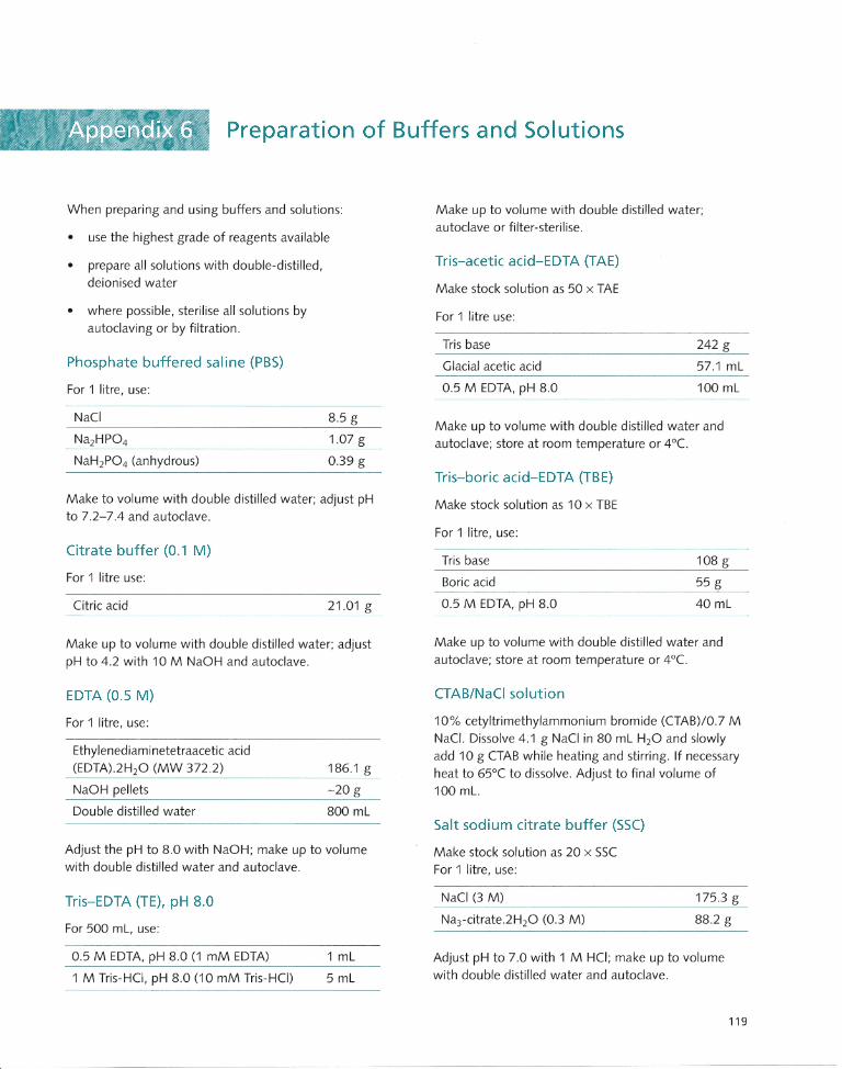
Preparation of Buffers and Solutions
When preparing and using buffers and solutions:
• use the highest grade of reagents available
• prepare all solutions with double-distilled, deionised water
• where possible, sterilise all solutions by autoclaving or by filtration.
Phosphate buffered saline (PBS)
For 1 litre, use:
NaCI 8.5 g
1.07 g
0.39 g
Make to volume with double distilled water; adjust pH to 7.2-7.4 and autoclave.
Citrate buffer (0.1 M)
For 1 litre use:
Citric acid 21.01 g
Make up to volume with double distilled water; adjust pH to 4.2 with 10 M NaOH and autoclave.
EDTA (0.5 M)
For 1 litre, use:
Ethylenediaminetetraacetic acid (EDTA).2H20 (MW 372.2)
NaOH pellets
Double distilled water
186.1 g
-20 g
800 mL
Adjust the pH to 8.0 with NaOH; make up to volume with double distilled water and autoclave.
Tris-EDTA (TE), pH 8.0
For 500 mL, use:
0.5 M EDTA, pH 8.0 (1 mM EDTA) 1 mL
1 M Tris-HCI, pH 8.0 (10 mM Tris-HCI) 5 mL
Make up to volume with double distilled water; autoclave or filter-sterilise.
Tris-acetic acid-EDTA (TAE)
Make stock solution as 50 x TAE
For 1 litre use:
Tris base 242 g
Glacial acetic acid 57.1 mL
0.5 M EDTA, pH 8.0 100 mL
Make up to volume with double distilled water and autoclave; store at room temperature or 4°C.
Tris-boric acid-EDTA (TBE)
Make stock solution as 10 x TBE
For 1 litre, use:
Tris base 108 g
Boric acid 55 g
0.5 M EDTA, pH 8.0 40 mL
Make up to volume with double distilled water and autoclave; store at room temperature or 4°C.
CTAB/NaCI solution
10% cetyltrimethylammonium bromide (CTAB)/0.7 M NaCi. Dissolve 4.1 g NaCI in 80 mL H20 and slowly add 10 g CTAB while heating and stirring. If necessary heat to 65°C to dissolve. Adjust to final volume of 100 mL.
Salt sodium citrate buffer (SSC)
Make stock solution as 20 x SSC For 1 litre, use:
NaCI (3 M) 175.3 g
88.2 g
Adjust pH to 7.0 with 1 M HCI; make up to volume with double distilled water and autoclave.
119
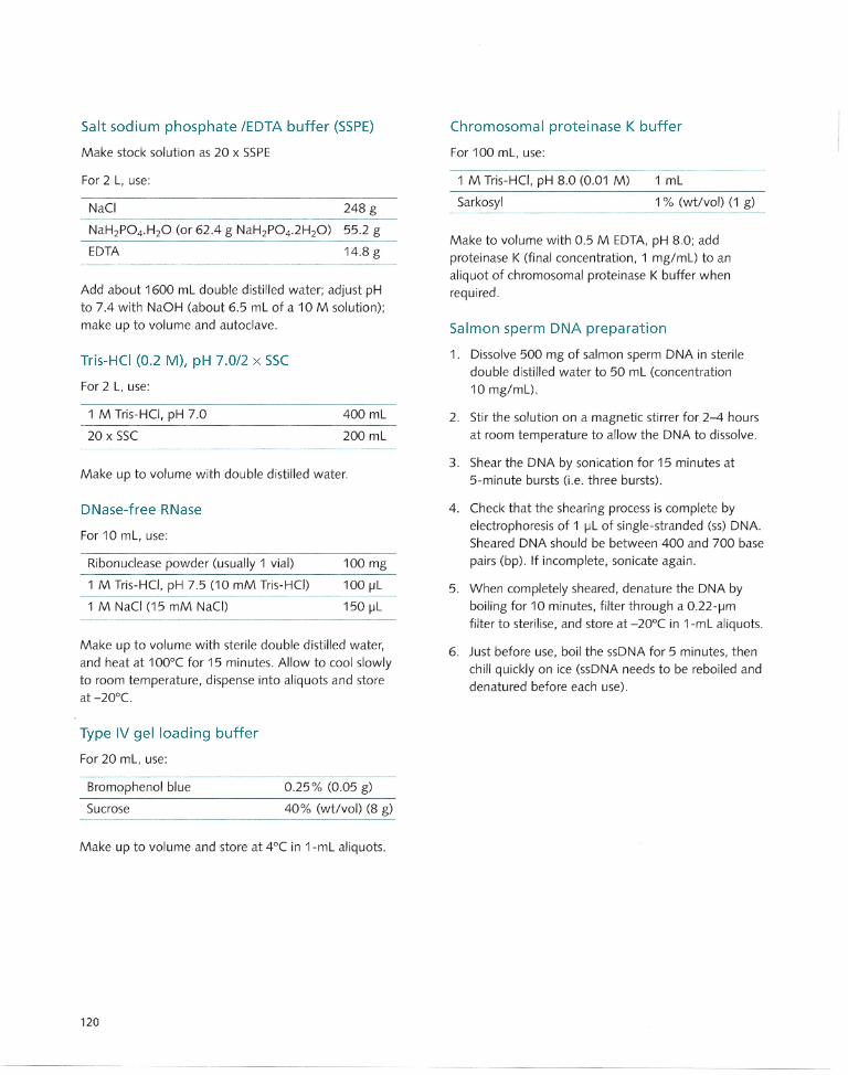
Salt sodium phosphate lEOTA buffer (SSPE)
Make stock solution as 20 x SSPE
For 2 L, use:
NaCI 248 g
NaH2P04 .H 20 (or 62.4 g NaH2P04 .2H20) 55.2 g
EDTA 14.8 g
Add about 1600 mL double distilled water; adjust pH to 7.4 with NaOH (about 6.5 mL of a 10M solution); make up to volume and autoclave.
Tris-HCI (0.2 M), pH 7.0/2 x SSC
For 2 L, use:
1 M Tris-HCI, pH 7.0 400 mL
20 x SSC 200 mL
Make up to volume with double distilled water.
DNase-free RNase
For 10 mL, use:
Ribonuclease powder (usually 1 vial) 100 mg
1 M Tris-HCI, pH 7.5 (10 mM Tris-HCI) 100 jJL
1 M NaCI (15 mM NaCI) 150 jJL
Make up to volume with sterile double distilled water, and heat at 100°C for 15 minutes. Allow to cool slowly to room temperature, dispense into aliquots and store at -20°e.
Type IV gel loading buffer
For 20 mL, use:
Bromophenol blue 0.25% (0.05 g)
Sucrose 40% (wt/vol) (8 g)
Make up to volume and store at 4°C in 1-mL aliquots.
120
Chromosomal proteinase K buffer
For 100 mL, use:
1 M Tris-HCI, pH 8.0 (0.01 M) 1 mL
Sarkosyl 1 % (wtlvol) (1 g)
Make to volume with 0.5 M EDTA, pH 8.0; add proteinase K (final concentration, 1 mg/mL) to an aliquot of chromosomal proteinase K buffer when required.
Salmon sperm DNA preparation
1. Dissolve 500 mg of salmon sperm DNA in sterile double distilled water to 50 mL (concentration 10 mg/mL).
2. Stir the solution on a magnetic stirrer for 2-4 hours at room temperature to allow the DNA to dissolve.
3. Shear the DNA by sonication for 15 minutes at 5-minute bursts (i.e. three bursts) .
4. Check that the shearing process is complete by electrophoresis of 1 jJL of single-stranded (ss) DNA. Sheared DNA should be between 400 and 700 base pairs (bp). If incomplete, sonicate again.
5. When completely sheared, denature the DNA by boiling for 10 minutes, filter through a O.22-jJm filter to sterilise, and store at -20°C in 1-mL aliquots.
6. Just before use, boil the ssDNA for 5 minutes, then chill quickly on ice (ssDNA needs to be reboiled and denatured before each use).
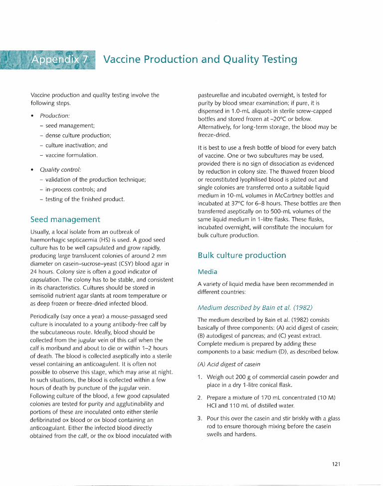
Vaccine Production and Quality Testing
Vaccine production and quality testing involve the following steps.
• Production:
- seed management;
- dense culture production;
- culture inactivation; and
- vaccine formulation.
• Quality control:
- validation of the production technique;
- in-process controls; and
- testing of the finished product.
Seed management
Usually, a local isolate from an outbreak of haemorrhagic septicaemia (HS) is used. A good seed culture has to be well capsulated and grow rapidly, producing large translucent colonies of around 2 mm diameter on casein-sucrose-yeast (CSY) blood agar in 24 hours. Colony size is often a good indicator of capsulation. The colony has to be stable, and consistent in its characteristics. Cultures should be stored in semisolid nutrient agar slants at room temperature or as deep frozen or freeze-dried infected blood.
Periodically (say once a year) a mouse-passaged seed culture is inoculated to a young antibody-free calf by the subcutaneous route. Ideally, blood should be collected from the jugular vein of this calf when the calf is moribund and about to die or within 1-2 hours of death. The blood is collected aseptically into a sterile vessel containing an anticoagulent. It is often not possible to observe this stage, which may arise at night. In such situations, the blood is collected within a few hours of death by puncture of the jugular vein. Following culture of the blood, a few good capsulated colonies are tested for purity and agglutinability and portions of these are inoculated onto either sterile defibrinated ox blood or ox blood containing an anticoagulant. Either the infected blood directly obtained from the calf, or the ox blood inoculated with
pasteurellae and incubated overnight, is tested for purity by blood smear examination; if pure, it is dispensed in 1.0-mL aliquots in sterile screw-capped bottles and stored frozen at -20°C or below. Alternatively, for long-term storage, the blood may be freeze-d ried.
It is best to use a fresh bottle of blood for every batch of vaccine. One or two subcultures may be used, provided there is no sign of dissociation as evidenced by reduction in colony size. The thawed frozen blood or reconstituted Iyophilised blood is plated out and single colonies are transferred onto a suitable liquid medium in 10-mL volumes in McCartney bottles and incubated at 3JOC for 6-8 hours. These bottles are then transferred aseptically on to 500-mL volumes of the same liquid medium in 1-litre flasks. These flasks, incubated overnight, will constitute the inoculum for bulk culture production .
Bulk culture production
Media
A variety of liquid media have been recommended in different countries:
Medium described by Bain et al. (1982)
The medium described by Bain et al. (1982) consists basically of three components: (A) acid digest of casein; (B) autodigest of pancreas; and (C) yeast extract. Complete medium is prepared by adding these components to a basic medium (D), as described below.
(A) Acid digest of casein
1. Weigh out 200 g of commercial casein powder and place in a dry 1-litre conical flask.
2. Prepare a mixture of 170 mL concentrated (10 M) HCI and 110 mL of distilled water.
3. Pour this over the casein and stir briskly with a glass rod to ensure thorough mixing before the casein swells and hardens.
121
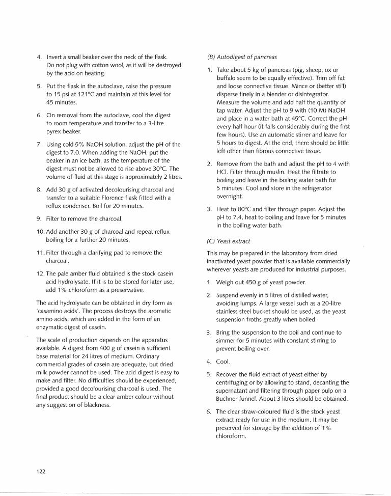
4. Invert a small beaker over the neck of the flask. Do not plug with cotton wool, as it will be destroyed by the acid on heating.
5. Put the flask in the autoclave, raise the pressure to 15 psi at 121°C and maintain at this level for 45 minutes.
6. On removal from the autoclave, cool the digest to room temperature and transfer to a 3-litre pyrex beaker.
7. Using cold 5% NaOH solution, adjust the pH of the digest to 7.0. When adding the NaOH, put the beaker in an ice bath, as the temperature of the digest must not be allowed to rise above 30°C. The volume of fluid at this stage is approximately 2 litres.
8. Add 30 g of activated decolourising charcoal and transfer to a suitable Florence flask fitted with a reflux condenser. Boil for 20 minutes.
9. Filter to remove the charcoal.
10. Add another 30 g of charcoal and repeat reflux boiling for a further 20 minutes.
11. Filter through a clarifying pad to remove the charcoal.
12. The pale amber fluid obtained is the stock casein acid hydrolysate. If it is to be stored for later use, add 1 % chloroform as a preservative.
The acid hydrolysate can be obtained in dry form as 'casamino acids'. The process destroys the aromatic amino acids, which are added in the form of an enzymatic digest of casein.
The scale of production depends on the apparatus available. A digest from 400 g of casein is sufficient base material for 24 litres of medium. Ordinary commercial grades of casein are adequate, but dried milk powder cannot be used . The acid digest is easy to make and filter. No difficulties should be experienced, provided a good decolourising charcoal is used. The final product should be a clear amber colour without any suggestion of blackness.
122
(B) Autodigest of pancreas
1. Take about 5 kg of pancreas (pig, sheep, ox or buffalo seem to be equally effective). Trim off fat and loose connective tissue. Mince or (better still) disperse finely in a blender or disintegrator. Measure the volume and add half the quantity of tap water. Adjust the pH to 9 with (10 M) NaOH and place in a water bath at 45°C. Correct the pH every half hour (it falls conSiderably during the first few hours) . Use an automatic stirrer and leave for 5 hours to digest. At the end, there should be little left other than fibrous connective tissue.
2. Remove from the bath and adjust the pH to 4 with HCI. Filter through muslin. Heat the filtrate to boiling and leave in the boiling water bath for 5 minutes. Cool and store in the refrigerator overnight.
3. Heat to 80°C and filter through paper. Adjust the pH to 7.4, heat to boiling and leave for 5 minutes in the boiling water bath.
(C) Yeast extract
This may be prepared in the laboratory from dried inactivated yeast powder that is available commercially wherever yeasts are produced for industrial purposes.
1. Weigh out 450 g of yeast powder.
2. Suspend evenly in 5 litres of distilled water, avoiding lumps. A large vessel such as a 20-litre stainless steel bucket should be used, as the yeast suspension froths greatly when boiled.
3. Bring the suspension to the boil and continue to simmer for 5 minutes with constant stirring to prevent boiling over.
4. Cool.
5. Recover the fluid extract of yeast either by centrifuging or by allowing to stand, decanting the supernatant and filtering through paper pulp on a Buchner funnel. About 3 litres should be obtained .
6. The clear straw-coloured fluid is the stock yeast extract ready for use in the medium. It may be preserved for storage by the addition of 1 % chloroform.
(0) Basic medium
Tryptone or casitone
Sucrose
Disodium hydrogen phosphate
(Na2 HP04 12 H20)
Potassium dihydrogen phosphate
(KH2 P04 7 H20)
Magnesium sulfate (Mg S04.7H20)
Distilled water
Complete medium
120 g
48 g
172 g
44 g
24 g
5 litres
1. Mix items A, C and D (above) in a large vessel in
the following proportions:
A. acid digest from 400 g of casein
(approximately 4 litres)
C. 5 litres of home made yeast extract
D. 5 litres of basic medium.
Make up the volume to 24 litres with distilled water.
2. Adjust the pH to 7.6 with 10% NaOH solution.
3. Autoclave at 18 psi for 10 minutes.
4. Remove from the autoclave and filter through
paper or pulp.
5. Adjust to pH 7.0 with 10% HCI solution
6. Transfer to a vortex tank and steam for 15 minutes.
After cooling, 1 litre of pancreatic digest (item B) is
added via a seitz filter and the medium is ready for
inoculation .
Note: The production of components A, Band C is
laborious and is not practised in modern production
laboratories. A range of dehydrated media of
bacteriological grade are available instead. Commercial
'casamino acids' (an acid digest of casein,
supplemented with enzymatic digest of casein) may be
used, together with commercial dehydrated yeast
extract of bacteriological grade. There is no simplified
method of preparing item B (autodigest of pancreas).
This ingredient, however is not used in many modern
media, because substitutes that provide comparable
growth promoting effects, such as yeast extract, are
available. Furthermore, the use of material of animal
origin is not encouraged, as this could result in
transmission of pathogens from the species of origin of
the material to the species on which the vaccine is to
be used.
Hence, items A and C can be replaced by 500 g of
casamino acids and 100 g of dehydrated yeast extract,
respectively. Item B can be omitted because adequate
yields can be obtained without it.
Medium used in Malaysia (Thomas 1968)
Peptone (Oxoid) 150 g
Lab Lemco (Oxoid) 150 g
Sodium chloride 75 g
Sodium bicarbonate 75 g
Glucose 30 g
Yeast extract 60 g
Autodigest of pancreas (seitz filtered) 750 mL
Distilled water up to 15 litres
Medium used in Thailand (Pakchong vaccine laboratory, 1988-89)
Tryptose 20 g
Yeast extract 3g
Sodium chloride 5g
Dextrose 1 g
Distilled water 1 litre
pH 7.4
Medium used in Egypt (Geneidy et al. 1967)
Casamino acids 500 g
Casitone (Difco) or tryptone (Oxoid) 120 g
Yeast extract 100 g
Sucrose 45 g
Magnesium sulfate 25 g
Phosphate buffer
Na2HP04 175 g
KH2P04 45 g
Distilled water 25 litres
123
CSY broth medium developed in Sri Lanka (Arawwawela et al. 1981)
Acid hydrolysate of casein ----
Sucrose (refined cane sugar) --- -
Yeast extract -----
2g
6g
6g
Sodium chloride - ----
Dipotassium hydrogen phosphate
_ ____ 5g
8.6 g
(K2HP04 , anhydrous) -----
Potassium dihydrogen phosphate 1.36 g
(KH 2 P04 , anhydrous) ------
Distilled water 1 litre
The first three ingredients are prepared as a
concentrate, filter-sterilised and added to the bulk tank
containing the other ingredients, which have been heat
sterilised.
Bulk culture methods
Various methods are adopted for the production of
dense cultures in bulk. These include the use of vortex
tanks with vortex aeration and sparger aeration
systems. The former system is now obsolete, and
vortex tanks are no longer manufactured. Sparger
aeration can be done using simple, improvised systems,
or by the use of automated fermentors.
Simple sparger aeration system
The inoculum is prepared in 500-mL volumes in 1-litre
flasks. The system requires at least 4-6 such flasks
tested and certified for purity. The medium used is the
CSY broth medium.
A diagrammatic representation of the layout of the
equipment is shown in Figure A7-1. The growth vessel
consists of a 40-litre glass vessel (F), placed in a locally
manufactured water bath (G) consisting of an
aluminium vessel fitted with a heating element and a
thermostat. The three glass tubes passing through the
tight-fitting rubber stopper consist of a filtered air inlet
(D), air outlet (H) and opening (N) for introduction of
presterilised liquids and inoculum. All openings are
controlled by stainless steel clips. The air outlet tube
passes into a foam trap (I) that collects the froth that
passes through the outlet tube; the outlet stream of air
passes out through tube J and bubbles through a 10%
124
formalin solution in flask K (to prevent infective
aerosols) before passing to the exterior through tube L.
The current of air from a compressor is delivered through
tube A. Tube B, controlled by opening or closure of the
clip, helps to control the flow of air through the in-line
filter C before passing through tube D to be dispersed
into small bubbles through a filter candle E, which serves
as a sparger. Tube M, which also passes through an in
line air filter, may be opened only when an additional
current of air is required to pump the sterile filtrate
(yeast, casein hydrolysate and sugar solution) into the tank (F) through tube N and also the inoculum. If the
aeration of the tank is started before the above
operation, the positive pressure built up within the tank will prevent the entry of an air current through tube N
when opened, and will help to avoid contamination.
It is important that the whole assembly, including the
filtration set-up, is adequately sterilised in a large
autoclave at 121 DC for 45 minutes; if contamination
occurs, it is usually due to understerilisation. The use of
a large inoculum (2-3 litres of 6-8-hour culture in the
same medium for 40 litres) and early harvesting
(18-20 hours) help further to obtain a dense growth in
a short time, with no chance for any contaminants to
multiply. The entire process should be carried out in a
relatively clean room.
Preparation of the bulk culture
The inoculum prepared with the seed culture is
introduced into the medium in the 40-litre vessel
through tube N by building up a positive pressure in the flask by introducing sterile air through in-line filter
M. The volume of inoculum (i.e. the number of
500-mL culture flasks used) is about 10% of the
volume of medium used in the bulk culture production.
The temperature of the water bath (G) containing
the 40-litre culture vessel is maintained at 37DC. The
introduced culture in the 40-litre vessel is incubated
for 16-18 hours with adequate fine aeration through
the sparger (E).
At the completion of the incubation, the bulk culture
harvest is obtained. A sample of the harvest is collected
for quality control. The bulk harvest is inactivated by
introducing a calculated volume of formalin to give a
final concentration of 05%, by opening the mouth of
the vessel while aerating. During this process the
L J ~D f,=-==F= ...... o
A
G
Figure A7-1. 'Fermentor' apparatus for bulk culture of P. multocida (see text for explanation of letters A-O).
sterility in the vessel is maintained due to the positive pressure built up in the vessel. The added formalin is thoroughly mixed with the harvest by aeration of the vessel for another 30 minutes.
The inactivated harvest is then transferred into a 50-litre sterile can through tube 0 of the 40-litre vessel. The collected harvest is allowed to stand at room temperature for 24 hours to complete the inactivation process and then stored at 4°C until formulation of the vaccine.
Use of fermentor
A 1 ~O-litre fermentor will have a working capacity of about 80 litres. Assuming that the growth gives just the required density, a batch will yield 80 litres of harvest, giving rise to 160 litres of oil adjuvant vaccine (equivalent to approximately 53 000 doses of vaccine).
Often, the density is higher, and the harvest can be diluted, thereby yielding more vaccine per batch . Most manufacturers of fermentors will modify basic fermentors to suit customer requirements.
Desirable features in a fermentor meant for this purpose are:
• an in situ sterilisation chamber, of adequate capacity for the scale of operation, with ports for inoculation, introduction of filter-sterilised fluids and harvesting, and a few extra ports for use if necessary;
• rapid (3D-minute) cooling system from 121 DC to 37°C; ,
• automatic temperature and pH control systems;
• manual air flow control;
• panel indicating all relevant parameters;
• built-in fluid filtration system; and
• steam generator and compressor to match.
Preparation of the medium
Heat-sterilised components
The heat-sterilised components (sterilised in the chamber in situ) consist of the following:
125
Dipotassium hydrogen phosphate
(K2HP04 , anhydrous)
Potassium dihydrogen phosphate
(KH 2P04 , anhydrous)
Sodium chloride (anhydrous NaCI)
Distilled water
688 g
108.8 g
400g
72 litres
The mixture is sterilised in situ in the fermentor chamber at 121°C for 1 hour. The sterilisation phase is as follows. The chamber temperature is set at 121°C and stirrer speed at 200 rpm. It takes around 45 minutes for this temperature to be reached. This temperature is maintained for 60 minutes, at the end of which it is reset at 37°C. The time taken to cool from 121°C to 37°C is approximately 30 minutes in most fermentors. Thus the sterilisation phase requires a total of 2.25 hours.
Other components
The other components, which are not sterilised by heat, are as follows:
Yeast extract 480 g
Cane sugar 480 g
Casein hyd rolysate 160 g
These are added to 8 litres of distilled water, heated to 65°C and stirred until dissolved.
The mixture is then sterilised by filtration using a builtin sterilisation system with a 10-litre vessel and cartridge-type in-line filters or a 1 O-litre capacity filtration vessel for positive pressure filtration using seitz D9 filter pads or equivalent membrane or cartridgetype filtration systems.
The 8 litres of filter-sterilised solution is transferred to the chamber through a separate port after the chamber temperature has dropped to 37°C.
Inoculation of the medium
Eight litres of the inoculum (pretested for purity and agglutinability) is introduced into the chamber through a separate port under positive pressure. The process takes about 15 minutes. The total volume in the chamber after inoculation and the commencement of incubation is approximately 90 litres.
126
Incubation phase
The chamber is now at 37°C, containing the additives and the inoculum. The stirrer speed is set at 50 rpm. and the air flow is adjusted so as to permit adequate bubbling through the liquid, without undue formation of froth. The total incubation time is 18 hours and the bacterial harvest is obtained at the end of this incubation period.
Inactivation
The bacterial culture is inactivated in the harvest by adding 0.5% formalin (36-40% formaldehyde solution) to a final concentration of 0.5% and stirring at 50 rpm for 5-10 minutes. The harvest is then held at room temperature for 24 hours.
An air pressure of 1 bar is built up in the chamber. A sterile tubing system is connected to the harvesting port. The harvesting port valve is opened and the harvest is collected in sterilised stock cans.
Formulation of the vaccine
The type of formulation depends on what type of vaccine is required . It is important to ensure that the required mass of whole bacteria is present in the vaccine. Each dose of vaccine should contain 2 mg of . bacteria (dry weight). The inactivated harvest must therefore be standardised. First, the dry bacterial mass of a dense harvest must be determined. After this initial determination, a relationship can be determined between dry bacterial mass and the turbidity of the bacterial suspension, and dilutions of it, measured using any standard turbidity measuring system. Thus, if broth bacterin, alum precipitated or aluminium hydroxide gel vaccine is to be prepared, the harvest is standardised to contain 2 mg in 3 mL. For the oil adjuvant vaccine, 2 mg should be contained in 1.5 mL of standardised harvest since a 3-mL dose will contain only half its volume of bacterin.
Alum precipitated vaccine
A 10% hot potash alum solution (i.e 1 litre of potash alum to 99 litres of bacterin) is added to the harvest to give a final concentration of 1 %. The pH is adjusted to 6.5 and the solution allowed to stand overnight.
Aluminium hydroxide gel vaccine.
To the standardised harvest, 3% aluminium hydroxide gel is added to give a final concentration of 0.3 %.
Oil adiuvant vaccine
The original formulation (Bain and Jones 1955) consisted of equal volumes of bacterin and mineral oil with approximately 10% anhydrous lanoline. This vaccine has a high viscosity. Newer formulations that enable less viscous emulsions to be produced contain either lower concentrations of lanoline or other modern adjuvants and emulsifying agents.
Some of the formulations of the oil adjuvant vaccine currently in use are given below.
Formulation used in Sri Lanka
Mineral oil 48 parts
Anhydrous lanoline (BP grade) 4 parts
Bacterin 48 parts
Formulation developed in Thailand (Neramitmansook 1993) -------
Mineral oil (Marcol 52)
Arlacel 'A'
Tween 80
Bacterin
Experimental formulations developed in Pakistan
(Muneer et al. 1993)
(i) Mineral oil (Marcol 52)
Montanide 888
Bacterin
55 parts
4 parts
1 part
40 parts
60 parts
10 parts
30 parts
(ii) Mineral oil (Marcol 52) 63 parts ----.---~--
Arlacel 'A' 7 parts
Bacterin (containing 5% Tween 80) 30 parts
(iii) (medium used by Shah et al. 1997) ---------
Mineral oil (Marcol 52)
Bacterin
Emulsifier
9 parts
8 parts
1 part
The emulsifier used is a blend of Span 85 (sorbitantriolate) and Tween 85 (polyoxyethylene 20 sorbitan triolate) in the ratio of 54:46 (vol/vol).
Emulsification process
Emulsification is carried out using a turbo emulsifier. The oil and emulsifying agent mixture are added to the emusifying vessel and sterilised by heat. The emulsifier is then switched on, and the bacterin is added slowly through a port on a side under positive pressure. If a 40-litre vessel is used with 20 litres of oil-emulsifier mixture, 20 litres of bacterin is added over a period of 8-10 minutes. Emulsification is continued for a further 10 minutes, with sufficient formalin added to give a final concentration of 0.5%. The stability of the emulsion is improved if the cans are left overnight at room temperature and emulsified for a further 10 minutes before bottling. It is also recommended that the bottles of emulsified vaccine are stored for 14 days at 4-8°C, before release for testing.
Quality testing of vaccines
Quality testing can be broadly divided into three categories: validation of the production technique, in-process control tests, and tests with the finished product.
Validation of technique
HS vaccines produced in different countries vary in their production methodology. Variations occur in the composition of the growth medium, the seed culture used , the formulation and the bacterial content. The composition of the growth medium is more important than was recognised in the early years of vaccine production; it is not only the bacterial mass that matters, but also the quality of bacterial growth. There is insufficient information to define the growth medium that facilitates the expression of a full complement of important immunogens, but once a medium is proven to produce effective vaccine, it is important to adhere to it without deviations. Micronutrients in the medium may also playa role in the type of growth and antigens expressed, so it is desirable to standardise even the grades and brands of chemicals and media used.
127
The complete production technique, which consists of growth medium, seed culture and formulation, should be tested for stability, potency (including duration of immunity) and shelf life. Tests for stability should include storage at 3JOC, and measurement of electrical conductivity. Potency should be tested in mice, and there should be a duration-of-immunity test in cattle and buffaloes. As a measure of shelf life, the above tests should be carried out after different periods of storage at the desired temperatures (4°C and tropical room temperatures). If any change is made to the technique, the new technique must be validated in regard to the above parameters.
In-process control tests
The seed culture plate, seed culture flasks and final harvest are tested for purity by smear examination.
As an additional test, the agglutinability of each of the above is tested using rabbit antiserum. Testing for inactivation is carried out with the harvest after addition of formalin and standing for 24 hours. The turbidity of the harvest should be matched against a standard equal to 1.5 mg/mL and adjusted.
Tests with the finished product
Tests for sterility
Tests for sterility and inactivation of the bacterial agent are carried out by plati ng out 0.1 m L of the vaccine on blood agar and Sabouraud's agar and incubating for 24 hours.
Safety test
Ten mice are inoculated, each with 0.5 mL of the vaccine by the subcutaneous route, and observed for 7 days. All mice should survive without showing any adverse effects.
Potency test
Selected batches (approximately one in five) are tested using an active mouse protection test (AMPT). Of 100 mice, 6-8 weeks of age, 50 are given 0.5 mL of vaccine by the subcutaneous route. After 14 days, the vaccination is repeated. One week after the second vaccination, the 50 vaccinated mice as well as the 50 unvaccinated mice are divided into 10 groups of five
128
each. A 6-8 hour culture from a local field strain of Pasteurella multocida is serially diluted to give 10-fold dilutions. For each dilution a group of vaccinated (5) and a group of control (5) mice are given 1.0-mL by the intraperitoneal route. All mice are observed for 7 days.
The median lethal dose (LD50), measured as the dilution of the P multocida suspension tested, for vaccinated and unvaccinated mice is calculated by the method of Karber (1931) as follows:
log LD50 = 0.5 + log H- sum of A 100
where:
H = the highest bacterial concentration (eg -1, if the highest concentration tested was 10-1)
A= the sum of the death rates (%) at each dilution
A minimum difference of 4 log units is required between log LD50 values for vaccinated and control mice. For example, if the log LD50 is calculated to be -1.9 (ie 50% of mice die when the bacterial suspension is diluted 10-19), the log LD50 for control mice should be at least -5.9 (ie 50% of the mice die when the bacterial concentration is diluted 10-59).
When the approximate LD50 is known, the number of mice used can be reduced slightly. The AMPT is preferred over the alternative passive mouse protection test (PMPT) because the latter, which involves vaccination of five cattle or buffaloes and inoculation of mice with vaccinated cattle serum (five mice for each animal vaccinated plus five controls), is both cumbersome and time consuming.
Abdullahi M.Z., Gilmour N.J.L. and Poxton I.R. 1990. Outer
membrane proteins of bovine strains of Pasteurella
multocida type A and their doubtful role as protective
antigens. Journal of Medical Microbiology, 32, 55-61.
Abeynayake P., Wijewardana TG. and Thalagoda SA 1993.
Antimicrobial susceptibility of Pasteurella multocida
isolates. In: B.E. Patten, TL. Spencer, R.B. Johnson, D.
Hoffmann and L. Lehane (eds), Pasteurellosis in
Production Animals, An international workshop held at
Bali, Indonesia, 10-13 August 1992. ACiAR Proceedings
No. 43,193-196.
ACIAR (Australian Centre for International Agricultural
Research) 1993. Pasteurellosis in Production Animals,
B.E. Patten, TL. Spencer, R.B. Johnson, D. Hoffmann and
L. Lehane (eds), An international workshop held at Bali,
Indonesia, 10-13 August 1992, ACIAR Proceedings No.
43, ACIAR, Canberra.
Adamson M.R., Morrow CJ., Johnson R.B., Townsend K.M.,
Dawkins H.J.S. and Spencer TL. 1993. Molecular
epidemiology of Pasteurella multocida using ribotyping.
In: B.E. Patten, TL. Spencer, R.B. Johnson, D. Hoffmann
and L. Lehane (eds), Pasteurellosis in Production Animals,
An international workshop held at Bali, Indonesia, 10-13
August 1992. ACIAR Proceedings No. 43, 69-76.
Adelberg EA, Mandel M. and Chen G.CC 1965. Optimal
conditions for mutagenesis by N-methyl-N'-nitro-N
nitrosoguanadine in Escherichia coli K12. Biochemical
and Biophysical Research Communications, 18, 788-795.
Afzal M. and Muneer R. 1990. Development of a combined
vaccine against haemorrhagic septicaemia and foot and
mouth disease. Pakistan Veterinary Journal, 10,67-69.
Ahmed S. 1996. Status of some bacterial diseases of animals
in Bangladesh. Asian Livestock, 21 (1).
Alton, G.G., Carter, G.R., Kibor, AC and Pesti, L. 1990. A
Manual of Laboratory Procedures for Selected Diseases
of Livestock. Food and Agriculture Organization.
Anon. 1981. Simple serological technique recommended for
haemorrhagic septicaemia diagnosis. Asian Livestock,
6(5),41-42.
Arawwawela CB., De Alwis M.CL. and Vipulasiri AA 1981.
Formulation of a suitable medium for obtaining dense
cultures for haemorrhagic septicaemia vaccine
production. Ceylon Veterinary Journal, 29, 16-19.
Atti Ratchanee 1996. Country Report: Thailand. International
Workshop on Diagnosis and Control of Haemorrhagic
Septicaemia. ACIAR, Indonesia, May 1996.
Awad F.I., Salem AA and Fayed AA 1976. Studies on clinical
signs observed on experimentally infected animals with
Pasteurella multocida type I. Egyptian Journal of
Veterinary Science, 13, 53-56. Abstract.
Bain RVS. 1954. Studies on haemorrhagic septicaemia in
cattle. I. Naturally acquired immunity in Siamese
buffaloes. British Veterinary Journal, 110,481-484.
Bain RVS. 1955. Studies on haemorrhagic septicaemia in
cattle. V: Tests for immunity in vaccinated cattle. British
Veterinary Journal, 111,492-498.
Bain RVS. 1956. Studies on haemorrhagic septicaemia in
cattle. VI: Experiments with oil adjuvant vaccine. British
Veterinary Journal, 112, 115-119.
Bain RVS. 1959. Haemorrhagic septicaemia in cattle.
Observations on some recent work. British Veterinary
Journal, 115,365.
Bain RVS. 1961. Haemorrhagic septicaemia of cattle and
buffaloes. South East Asia Treaty Organisation, Bangkok,
Thailand.
Bain RVS. 1963. Haemorrhagic septicaemia. FAO Agricultural
Studies No. 62. FAO, Rome.
Bain RVS. 1979a. In FAO 1979, 13-19.
Bain RVS. 1979b. Protective antigens of Pasteurella
multocida. In FAO 1979, 27-29.
Bain RVS., De Alwis M.CL., Carter G.R. and Gupta B.K.
1982. Haemorrhagic septicaemia. FAO Animal
Production and Health Paper No. 33. FAO, Rome.
Bain RVS. and Jones R.F 1955. Studies on haemorrhagic
septicaemia of cattle. III: Production of an oil adjuvant
vaccine. British Veterinary Journal, 111, 30-34.
Bain RVS. and Knox K.W. 1961. The antigens of Pasteurella
multocida type I. II: Lipopolysaccharides. Immunology, 4,
122-129.
Baldrey FS.H et al. 1907. Cited by Bain et al. 1982.
129
Bandopadhyay P.K., Tonganokar 5.5. and Singh D.K. 1991.
Characterisation and antibiotic sensitivity of Pasteurella multocida isolates from cases of haemorrhagic
septicaemia. In: Proceedings of the Fourth International Workshop on Haemorrhagic Septicaemia, February
1991, Kandy, Sri Lanka. FAO-APHCA Publication No. 1991/13,65-68.
Barakat A. A. , EI Affendy A., Fayed A.A., Awad H.H., Shawkat
M.E. and Zahran M. 1976. Journal of the Egyptian
Veterinary Medical Association, 35, 95-100.
Bastianello 5.5. and Jonker M.R. 1981. A report on the
occurrence of septicaemia caused by Pasteurella multocida type E in cattle in Southern Africa. Journal of
the South African Veterinary Association, 52, 99-104.
Bhatty M.A. 1973. A preliminary comparison of sodium
alginate and oil adjuvant haemorrhagic septicaemia
vaccines in cattle. Bulletin of Epizootic Diseases of Africa, 21,171-174.
Bisgaard M. 1994. Taxonomy of the family Pasteurellaceae
Pohl 1981. In: W. Donachie, F.A. Lainson and J.C Hodgson (eds), Haemophilus, Actinobacillus and
Pasteurella, Plenum Press, London.
Blackall P. J., Pahoff J. L., Marks D., Fegan N. and Morrow C J. 1995. Characterisation of Pasteurella multocida
isolated from fowl cholera outbreaks on turkey farms.
Australian Veterinary Journal, 72, 135-138.
Blanchard P.C, Stoltz J., Rimier R.B., Wilson M.A., Hays J. and
Montgomery G. 1993. Proceedings of the 36th American
Association of Veterinary Laboratory Diagnosticians
Meeting. p16. Cited by Rimier and Wilson 1994.
Bokhout B.A., van Gallen C. and van der Heijden P.J. 1981. A selected water in oil emulsion: Composition and
usefulness as an immunological adjuvant. Veterinary
Immunology and Immunopathology, 2,491-500.
Bollinger 1878. Cited by Carter and De Alwis 1989.
Bollinger 1879. Cited by Namioka 1978.
Brickell s. 1996. Development of a polymerase chain reaction
(PCR) test specific for isolates of P multocida causing
HS. Paper presented at the International Workshop on
Diagnosis and Control of Haemorrhagic Septicaemia. AClAR, Indonesia, May 1996.
Brickell S.K., Thomas L.M., Long K.A., Pinnaccio M. and Widders P. R. 1998. Development of a PCR test based on
a gene region associated with the pathogenicity of
Pasteurella multocida serotype B:2, the causal agent of
haemorrhagic septicaemia in Asia. Veterinary
Microbiology, 59, 295-307.
130
Brogden K.A. and Packer R.A. 1979. Comparison of
Pasteurella multocida serotyping systems. American
Journal of Veterinary Research, 40, 1332-1335.
Brumpt, E. 1910. Cited by Mutters et al. (1989).
Burril 1883. Cited by Namioka 1978.
Butler J.E. and Maxwell c.F. 1972. Preparation of bovine
immunoglobulins and free secretory component and the
specific antisera. Journal of Dairy Science, 55,151-165.
Carrigan M.J., Dawkins H.J.S, Cockram F.A. and Hansen A.T
1991. Pasteurella multocida septicaemia in fallow deer
(Dama dama). Australian Veterinary Journal, 68,
201-203.
Carter G.R. 1952. Type specific capsular antigens of Pasteurella multocida. Canadian Journal of Medical
Science, 30, 48-53.
Carter G.R. 1955. A haemagglutination test for identification
of serological types. American Journal of Veterinary Research, 16,481-484.
Carter G.R. 1957. Studies on Pasteurella multocida. III: A
serological survey of bovine and porcine strains from
various parts of the world. American Journal of
Veterinary Research, 18,437-440.
Carter G.R. 1959. Studies on Pasteurella multocida. IV:
Serological types from species other than cattle and swine. American Journal of Veterinary Research, 21,
173-175.
Carter G.R. 1961. A new serological type of Pasteurella
multocida from Central Africa. Veterinary Record , 73,
1052.
Carter G.R. 1963. Proposed modification to the serological
classification of Pasteurella multocida. Veterinary Record.
75, 1264.
Carter G.R. 1982. Whatever happened to haemorrhagic
septicaemia? Journal of the American Veterinary Medical Association, 180, 1176-1177.
Carter G.R. 1984. In: Foreign Animal Diseases. US Animal
Health Association, Richmond, Virginia, USA, 146.
Carter G.R. and Chengappa M.M. 1980. Hyaluronidase production by type B Pasteurella multocida from cases of
haemorrhagic septicaemia. Journal of Clinical
Microbiology, 11, 94-96.
Carter G.R. and Chengappa M.M. 1981. Identification of type Band E Pasteurella multocida by
counterimmunoelectrophoresis. Veterinary Record, 108, 145-146.
Carter G.R. and De Alwis M.C.L. 1989. Haemorrhagic
septicaemia. In: C. Adlam and J.M. Rutter (eds),
Pasteurella and pasteurellosis. Academic Press, New York,
37-73.
Carter G.R., Myint A, Van Khar R. and Khan A 1991.
Immunisation of cattle and buffaloes with a live
haemorrhagic septicaemia vaccine. Veterinary Record, 129,203.
Carter G.R. and Rappay D.E. 1962. Formalinised erythrocytes in the haemagglutination test for typing Pasteurella
multocida. British Veterinary Journal, 118, 289-292.
Carter G.R. and Rundell S.w. 1975. Identification of type A
strains of Pasteurella multocida, using staphylococcal hyaluronidase. Veterinary Record, 96, 343.
Carter G.R. and Subronto P. 1973. Identification of type D strains of Pasteurella multocida with acriflavine.
American Journal of Veterinary Research, 34, 293-294.
Chandrasekaran S., Kennett L., Yeap P.c., Muniandy N., Rani
B. and Mukkur T.K.S. 1993a. Relationship between
active protection in buffalo vaccinated against
haemorrhagic septicaemia and passive mouse protection
and serological tests. In: B.E. Patten, T.L. Spencer, R.B.
Johnson, D. Hoffmann and L. Lehane (eds),
Pasteurellosis in Production Animals, An international workshop held at Bali, Indonesia, 10- 13 August 1992.
ACiAR Proceedings No. 43, 215- 218.
Chandrasekaran S., Kennett L., Muniandy N., Rani B. and
Mukkur T.K.S. 1993b. Characterisation of immune
response and duration of immunity in buffalo vaccinated
with cellular haemorrhagic septicaemia vaccines. In: B.E.
Patten, T.L. Spencer, R.B. Johnson, D. Hoffmann and L.
Lehane (eds), Pasteurellosis in Production Animals, An international workshop held at Bali, Indonesia, 10-13
August 1992. ACIAR Proceedings No. 43, 165-169.
Chandrasekaran S., Kennett L., Yeap P.c., Muniandy N. , Rani
B. and Mukkur T.K.S. 1994a. Characterization of immune
response and duration of protection in buffaloes immunized with hemorrhagic septicemia vaccines.
Veterinary Microbiology, 41, 213-219.
Chandrasekaran 5., Kennett L., Yeap P.c., Muniandy N., Rani B. and Mukkur T.K.S. 1994b. Relationship between
active protection in vaccinated buffaloes against
haemorrhagic septicaemia and passive mouse protection
or serum antibody titres. Veterinary Microbiology, 41, 303-309.
Chandrasekaran S., Muniandy N., Kennett L., and Mukkur
T.K.S. 1991. Lack of relationship between active
protection against haemorrhagic septicaemia in buffaloes
and the passive mouse protection test. In: Proceedings of the Fourth International Workshop on Haemorrhagic
Septicaemia, February 1991, Kandy, Sri Lanka.
FAO-APHCA Publication No. 1991/13, 124-126.
Chandrasekaran S. and Yeap P.c. 1978. Safety and potency
testing of hemorrhagic septicemia oil adjuvant vaccine by
mouse protection test. Kajian Veterinar, 10, 28-34.
Chandrasekaran S., Yeap P.c. and Chuink B.H., 1981.
Biochemical and serological studies of Pasteurella
multocida isolated from cattle and buffaloes in Malaysia.
British Veterinary Journal, 137,361-367.
Chandrasekaran S., Yeap P.c. and Saad R. 1987. The effect of
storage conditions on the stability and potency of
haemorrhagic septicaemia oil adjuvant vaccine. Kajian
Veterinar, 19,71-75.
Chandrasekaran S., Yeap P.c. and Sansul Bhari 1996.
Development of a low volume haemorrhagic septicaemia
vaccine and its evaluation in rabbits and cattle.
International Workshop on Diagnosis and Control of
Haemorrhagic Septicaemia. ACIAR, Indonesia, May 1996.
Chaturevedi G.c. and Sharma V.K. 1981. Cell mediated immunoprotection in calves immunised with rough
Salmonella dublin. British Veterinary Journal, 137,
421-430.
Chaudhry NA and Khan B.B. 1978. Estimation of economic
losses due to animal diseases in Pakistan. Final Technical
Report, University of Agriculture, Faisalabad.
Chaudhuri S., Mukherjee S.K., Chatterjee A and Gauguli J.L. 1992. Isolation of Pasteurella multocida F:3,4 from a
stillborn snow leopard. Veterinary Record, 130, 36.
Chengappa M.M. and Carter G.R. 1977. Demonstration of
bacteriocin activity in bovine and bison strains of
Pasteurella multocida. American Journal of Veterinary
Research, 38, 1183- 1185.
Chengappa M.M. and Carter G.R. 1979. An improved
method for obtaining streptomycin dependent mutants from Pasteurella multocida and Pasteurella haemolytica
using N-methyl-N-nitro-N-nitrosoguanadine. American
Journal of Veterinary Research, 40, 449-450.
Choi-Kim K., Maheswaran S.K., Felice L.J. and Molitor T.W.
1991. Relationship between iron regulated outer
membrane proteins and outer membrane proteins in in
vivo, grown Pasteurella multocida. American Journal of
Veterinary Research, 28, 75-92.
131
Cornelius J.T. 1929. An investigation of serological
relationships of twenty six strains of Pasteurella
multocida. Journal of Pathology and Bacteriology, 32, 355-364.
Darmadi P. 1991. Country Report - Indonesia. In:
Proceedings of the Fourth International Workshop on
Haemorrhagic Septicaemia, February 1991, Kandy, Sri Lanka. FAO-APHCA Publication No. 1991/13, FAO
1991,26-29.
Dartini N.L. and Ekaputra I.G.M. 1996. Abattoir survey for
isolation of Pasteurella multocida in the eastern region of
Indonesia. Paper presented at the international
Workshop on Diagnosis and Control of Haemorrhagic
Septicaemia. ACIAR, Indonesia, May 1996.
Dassanayake L. 1957. The haemorrhagic septicaemia outbreak of 1955-56. Ceylon Veterinary Journal, 5, 56-59.
Daubney R. 1937. Annual Report, Department of Agriculture,
Kenya. Cited by Bai n et al. 1982.
Dawkins H.J.S., Hyatt A, Johnson R.B., Spencer TL., Ramdani
and Adler B. 1991 a. Evidence of phenotypic dichotomy
within an individual Pasteurella multocida type strain and among some haemorrhagic septicaemia causing field
isolates. Research in Veterinary Science, 50, 368-370.
Dawkins H.J.S., Johnson R.B ., Spencer TL. and Patten B.E.
1990. Rapid identification of Pasteurella multocida
organisms responsible for haemorrhagic septicaemia
using an enzyme-linked immunosorbent assay (ELISA).
Research in Veterinary Science, 49, 261-267.
Dawkins H.J.S., Ramdani , Johnson R.B. and Spencer TL. 1991 b. Haemorrhagic septicaemia: correlation of vaccinal
antibody responses in mice with protection against Pasteurella multocida strain M1404. Veterinary
Microbiology, 27, 309-326.
De Alwis M.CL. 1972. Bacteriological changes in specimens
during transport. I: Changes occurring in inoculated
cotton wool swabs. Ceylon Veterinary Journal, 20,
54-60.
De Alwis M.CL. 1973. Bacteriological changes in specimens
during transport. II: Effect of using a suitable transport
medium. Ceylon Veterinary Journal, 21,2-6.
De Alwis M.CL. 1981. Mortality among cattle and buffaloes
in Sri Lanka due to haemorrhagic septicaemia. Tropical
Animal Health and Production, 13, 195-202.
De Alwis M.CL. 1982a. Pasteurella multocida serotype 6:B
from an elephant. Sri Lanka Veterinary Journal, 30, 28.
132
De Alwis M.CL. 1982b. The immune status of buffalo calves
exposed to natural infection with haemorrhagic
septicaemia. Tropical Animal Health and Production, 14,
29-30.
De Alwis M.CL. 1983. Economics of control of haemorrhagic
septicaemia in Sri Lanka. Paper presented at seminar on
Animal Disease Control and Livestock Development in
the Asian Region. Commonwealth Veterinary
Association, Hissar, India, December.
De Alwis M.CL. 1984. Haemorrhagic septicaemia in cattle
and buffaloes. Office International des Epizooties Revue
Scientifique et Technique, 3, 707-730.
De Alwis M.CL. 1986. Epidemiology of haemorrhagic
septicaemia and economics of control of the disease. In:
M.R. Jainudeen, M. Mahyuddin and J.E. Huhn (eds),
Proceedings of the Fifth international Conference on
Livestock Production and Diseases in the Tropics.
Malaysia, August 1986.
De Alwis M.CL. 1987. Serological classification of Pasteurella
multocida. Veterinary Record, 121,44.
De Alwis M.CL. 1989. Production and field use of
haemorrhagic septicaemia oil adjuvant vaccine. Consultants report on the second mission;
TCP/THA/8852 (T), Food and Agriculture Organization
(unpublished).
De Alwis M.CL. and Carter G.R. 1980. Preliminary field trials
with a streptomycin dependent vaccine against
haemorrhagic septicaemia. Veterinary Record, 106,
435-437.
De Alwis M.CL., Carter G.R. and Chengappa M.M. 1980.
Production and characterisation of streptomycin dependent mutants of Pasteurella multocida from
bovine haemorrhagic septicaemia. Canadian Journal of
Comparative Medicine, 44, 418-422.
De Alwis M.CL , Gunatileke AAP and Wickremasinghe
WAT 1978. Duration of immunity to haemorrhagic
septicaemia in cattle and buffaloes following
immunisation with alum preCipitated and oil adjuvant
vaccines. Ceylon Veterinary Journal, 26, 35-41.
De Alwis M.CL., Horadagoda N.U., Wijewardana TG.,
Abeynayake P, Vipulasiri AA and Thalagoda SA 1995.
Further studies on the epidemiology and immunology of haemorrhagic septicaemia in buffaloes. in: B.M.O.A
Perera, J.A de S. Siriwardene, N.U. Horadagoda and
M.N.M. Ibrahim (eds), Proceedings of the Regional
Symposium on the Role of the Buffalo in Rural Development in Asia, December 1995, Peradeniya,
Sri Lanka. NARESA (now National Science Foundation),
Colombo, Sri Lanka.
De Alwis M.CL., Jayasekera M.U. and Balasunderam P. 1975.
Pneumonic pasteurellosis in buffalo calves associated
with Pasteurella multocida serotype 6:B. Ceylon Veterinary Journal, 23, 58-60.
De Alwis M.CL., Kodituwakku A.O. and Kodituwakku S.
1976. Haemorrhagic septicaemia: An analysis of two outbreaks of disease among buffaloes. Ceylon Veterinary
Journal, 24, 18-21.
De Alwis M.CL. and Panangala V.S. 1974. A biochemical and
serological study of strains of Pasteurella multocida
associated with haemorrhagic septicaemia in cattle and
buffaloes in Sri Lanka. Ceylon Veterinary Journal, 22, 58-63.
De Alwis M.CL. and Sumanadasa MA 1982. Naturally
acquired immunity to haemorrhagic septicaemia among
cattle and buffaloes in Sri Lanka. Tropical Animal Health and Production, 14, 27-28.
De Alwis M.CL. and Thambithurai V. 1965. A case of
haemorrhagic septicaemia in a wild elephant in Ceylon.
Ceylon Veterinary Journal, 13, 17-19.
De Alwis M.CL. and Vipulasiri A.A. 1980. An epizootiological
study of haemorrhagic septicaemia in Sri Lanka. Ceylon Veterinary Journal, 28, 24-35.
De Alwis M.CL., Wijewardana TG., Gomis A.I.U. and
Vipulasiri A.A. 1990. Persistence of the carrier status in haemorrhagic septicaemia (Pasteurella multocida
serotype 6:B infection) in buffaloes. Tropical Animal
Health and Production, 22, 185-194.
De Alwis M.CL., Wijewardana TG., Sivaram A. and Vipulasiri
AA 1986. The carrier and antibody status of cattle and
buffaloes exposed to haemorrhagic septicaemia: Investigations on survivors following natural outbreaks.
Sri Lanka Veterinary Journal, 34, 33-42.
De Alwis M.CL., Wijewardana TG. and Vipulasiri AA 1996. A
live vaccine against haemorrhagic septicaemia using a
serotype B:2 mutant - Preliminary Communication .
International Workshop on Diagnosis and Control of
Haemorrhagic Septicaemia. ACIAR, Indonesia, May 1996.
De Alwis MCL., Wijewardana TG., Vipulasiri AA and
Thalagoda SA 1998. Mutant Pasteurella multocida B:2
as a live vaccine. Unpublished.
Dewhirst F.E., Paster B.J., Olsen I. and Fraser G.J . 1993.
Phylogeny of the Pasteurellaceae as determined by
comparison of 16S ribosomal ribonucleic acid sequences.
International Journal of Medical Microbiology, Virology,
Parasitology and Infectious Diseases, 279, 35-44.
Dhanda M.R. 1959a. Working Paper (India). In FAO 1959.
Dhanda M.R. 1959b. Immunisation of cattle against
haemorrhagic septicaemia with purified capsular
antigens. Indian Veterinary Journal, 36, 6-8.
Dhanda M.R. 1960. Purification and properties of the soluble
antigen of Pasteurella septica type I. Indian Journal of
Pathology and Bacteriology, 2, 59-62.
Dhanda M.R., Lall J.M. and Seth R.N. 1958. Immunological stud ies on Pasteurella septica. II: Fu rther trials on
adjuvant vaccine. Indian Journal of Veterinary Science
and Animal Husbandry, 28, 139- 156.
Dhanda M.R. and Nilakanthan P.R. 1961. Isolation of
Pasteurella multocida of low virulence from cases of
paraplegia in cattle in the Andhra Pradesh. Indian Veterinary Journal, 38, 339-343.
Dhanda M.R. and Sen G.P. 1972. Haemorrhagic septicaemia
and allied conditions in sheep, goats, pigs and poultry.
Indian Council of Agricultural Research, New Delhi, cited by Townsend et al. 1997b.
Dua S.K. and Maheswaran S.K. 1978. Studies on Pasteurella multocida. IV: Nature of systemic immunity and analysis
of the correlation between levels of immunity by various
fowl cholera vaccines and protection against challenge.
Avian Diseases, 2, 748-764.
Dutta J., Rathore B.S., Mullick S.G., Singh R. and Sharma G.C
1990. Epidemiological studies on occurrence of
haemorrhagic septicaemia in India. Indian Veterinary Journal, 67, 893-899.
FAO (Food and Agriculture Organization) 1959. Report of the
FAO meeting on Haemorrhagic Septicaemia, Manila,
Philippines, Nov-Dec.
FAO 1962. Report of the FAO meeting on Haemorrhagic
septicaemia, Kuala Lumpur, Malaya, Jan- Feb.
FAO 1979. Proceedings of the Third International Workshop
on Haemorrhagic Septicaemia, FAO-APHCA (Animal Production and Health Commission for Asia and the Far
East), December 1979, Colombo, Sri Lanka.
FAO 1991. Proceedings of the Fourth International Workshop
on Haemorrhagic Septicaemia, February 1991, Kandy, Sri
Lanka. FAO-APHCA Publication No. 1991/13.
FAO 1994. Statistical Profile of Livestock Development in the
Asia-Pacific Region. RAPA Publication No. 1994/26.
FAO 1995. Production Yearbook 1995; FAO Statistics Division.
133
FAO-OIE (Food and Agriculture Organization-Office
International des Epizooties) 1958. The control of
haemorrhagic septicaemia. FAO-OIE Animal Health
Yearbook 1958.
FAO- WHO (World Health Organization)-OIE 1994. Yearbook
1994.
Francis B.K.T., Schels H.F. and Carter G.R. 1980. Type E
Pasteurella multocida associated with haemorrhagic
septicaemia in Zambia. Veterinary Record, 107, 135.
Freund J. et al. 1948. Journal of Immunology, 60, 383.
Gaffky 1881. Cited by Bain et al. 1982.
Gamage L.N.A., Wijewardana TG., Bastiansz H.L.G. and
Vipulasiri AA 1995. An outbreak of acute pasteurellosis
in swine caused by serotype B:2 in Sri Lanka. Sri Lanka Veterinary Journal, 4, 15-19.
Geneidy AA, Lofty O. and EI-Affenty A.M. 1967. Control of
haemorrhagic septicaemia with special reference to the oil adjuvant vaccine. Journal of the Arabian Veterinary
Medical Association, 27,121-126.
Gevedze v.1. 1986. Pasteurellosis in horses. Veterinarnaya
Nauka Proizvodstvu, 24, 30-3. Abstract.
Gilbert S.J. 1943. Cited by Bain et al. 1982.
Gochenour W.S. 1924. Haemorrhagic septicaemia studies: The
development of a potent immunising agent (natural aggressin) by the use of highly virulent strains of
haemorrhagic septicaemia organisms. Journal of the
American Veterinary Medical Association, 65, 433-441.
Gomis A.I.U., De Alwis M.CL., Radhakrishnan S. and
Thalagoda SA 1988/1989. Further studies on stability and potency of the haemorrhagic septicaemia oil
adjuvant vaccine. Sri Lanka Veterinary Journal, 36, 9-20.
Graydon R.J., Patten B.E. and Hamid H. 1993. The pathology of experimental haemorrhagic septicaemia in cattle and
buffalo. In: B.E. Patten, TL. Spencer, R.B. Johnson, D.
Hoffmann and L. Lehane (eds), Pasteurellosis in
Production Animals, An international workshop held at
Bali, Indonesia, 10-13 August 1992. ACiAR Proceedings No. 43,105-107.
Gupta B.K. 1962. Studies on the carrier problem in
haemorrhagic septicaemia. Thesis, Punjab University,
Chandigar, India.
Gupta B.K. 1973. Studies on the pathogenicity of Pasteurella
multocida in the development of fowl cholera vaccine.
Thesis, Agra University, India.
134
Gupta B.K. and Kumar S. 1973. Serotyping of Indian isolates
of Pasteurella multocida from healthy poultry. Indian
Journal of Animal Sciences, 48, 301-304.
Gupta B.K. and Sareen R.L. 1976 Evaluation of haemorrhagic septicaemia oil adjuvant vaccine by mouse protection
test. Indian Veterinary Journal, 53, 489-492.
Heddleston K.L., Gallagher J.E. and Rebers PA 1972. Fowl
cholera: Gel diffusion precipitin test for serotyping
Pasteurella multocida from avian species. Avian
Diseases, 16, 925-936.
Heddleston K.L., Rhoades K.R. and Rebers PA 1967.
Experimental pasteurellosis: Comparative studies on 3
strains of Pasteurella multocida from Asia, Africa and
North America. American Journal of Veterinary Research,
28,1003.
Herbert W.J. 1965. Multiple emulsions. A new form of mineral
oil antigen adjuvant. Lancet. 2, 71.
Hiramune T and De Alwis M.CL. 1982. Haemorrhagic
septicaemia carrier status among cattle and buffaloes in
Sri Lanka. Tropical Animal Health and Production, 14, 91-92.
Holmes J.D.E. 1910. Cited by Bain et al. 1982.
Horadagoda N. and Belak K. 1990. Demonstration of
Pasteurella multocida type 6:B (B:2), in formalin fixed
paraffin embedded tissues of buffaloes by peroxidase antiperoxidase (PAP) technique. Acta Veterinaria
Scandinavica, 31,493-495.
Horadagoda N.U., De Alwis M.CL., Wijewardana TG., Belak
K., Gomis A.I.U. and Vipulasiri A.A. 1991. Experimental
haemorrhagic septicaemia in buffalo calves. In FAO 1991, 73-80.
Hudson J.R., 1954. Cited by Rimier and Rhoades 1989.
Hueppe 1886. Cited by Bain et al. 1982.
Jaiswal TN., Gupta B.K. and Mishra S.C 1983. Potency testing of haemorrhagic septicaemia alum preCipitated
vaccine in rabbits. Indian Journal of Comparative Microbiology, Immunology and Infectious Diseases, 4,
152-155.
Johnson R.B., Castro LV, Sleep J., Houge M.M., Dawkins H.J.S. and Spencer TL. 1993. The antibody response of
rabbits and cattle to Pasteurella multocida: Assessment
of cross reactivity between serotypes. In: B.E. Patten, TL.
Spencer, R.B. Johnson, D. Hoffmann and L. Lehane (eds),
Pasteurellosis in Production Animals, An international
workshop held at Bali, Indonesia, 10-13 August 1992.
ACIAR Proceedings No. 43, 159-164.
Johnson R.B., Dawkins H.J.S. and Spencer T.L. 1988.
Strategies for the development of the ELISA assay for
HS. ELISA Technology in Diagnosis and Research. James
Cook University, Townsville, Australia, 244-253.
Johnson R.B., Dawkins H.J.S. and Spencer T.L. 1991.
Electrophoretic profiles of Pasteurella multocida isolates
from animals with haemorrhagic septicaemia. American Journal of Veterinary Research, 52, 1644-1648.
Johnson R.B., Dawkins H.J.S., Spencer T.L., Saharee A.A.,
Bahaman A.R., Ramdani and Patten B.E. 1989.
Evaluation of bovine antibody responses to haemorrhagic
septicaemia vaccine. Research in Veterinary Science, 47, 277-279.
Jones R.J., Clarke J.K. and Stevenson B.J. 1988. A preliminary investigation of New Zealand isolates of Pasteurella
multocida. New Zealand Veterinary Journal, 36, 155-156.
Jones T.O. and Hussaini S.N. 1982. Outbreak of Pasteurella
multocida in fallow deer (Dama dama). Veterinary
Record, 110,451-452.
Joseph P.G. and Hedger R.S. 1984. Serological response of
cattle to simultaneous vaccinations against foot and mouth disease and haemorrhagic septicaemia. Veterinary
Record, 114,494-496.
Karber, G. 1931. Cited by Cruickshank, R. 1965 in Medical
Microbiology, 11th edition, E&S Livingstone Ltd, London.
Kasali O.B. 1972. A case of haemorrhagic septicaemia in an
African buffalo (Syncerus nanus). Bulletin of Epizootic
Diseases of Africa, 20, 203-204.
Kasten R.W., Carpenter T.E., Snipes K.P. and Hirsh D. 1997.
Detection of Pasteurella multocida-specific DNA in
turkey flocks by use of the polymerase chain reaction. Avian Diseases, 41, 676-682.
Kennett L., Muniandy N. and Mukkur T.K.S. 1993.
Comparative protective potential of nonliving intact cells
and purified outer membrane and associated proteins of
Pasteurella multocida type 6:B grown under iron regulated conditions. In: B.E. Patten, T.L. Spencer, R.B.
Johnson, D. Hoffmann and L. Lehane (eds),
Pasteurellosis in Production Animals, An international
workshop held at Bali, Indonesia, 10-13 August 1992.
ACIAR Proceedings No. 43,144-148.
Khan M.A., Yamin M., Khan M.S. and Khan A.G. 1994.
Epidemiological and economical based ranking order of
buffalo and cattle diseases through active disease
surveillance system. Proceedings of the 8th International
Congress on Animal Hygiene, St. Paul, Minnesota, USA.
September 1994. Abstract.
Kheng e.S. and Phay e.H. 1963. Haemorrhagic septicaemia:
Sulphamethazine and immune serum therapy in
buffaloes infected by the nasal spray method. Veterinary
Record, 75, 155-159.
Kim e.J. and Nagaraja K.v. 1990. DNA fingerprinting for
differentiation of field isolates from reference vaccine
strains of Pasteurella multocida in turkeys. American Journal of Veterinary Research, 51,207-210.
Kitt T. 1885. Cited by Mutters et al. 1989.
Klaassen J.M., Bernard B.L. and Di Giacomo R.F. 1985.
Enzyme-linked immunosorbent assay for
immunoglobulin G antibody to Pasteurella multocida in
rabbits. Journal of Clinical Microbiology, 21,617-621.
Knox K.W. and Bain R.v.S. 1960. The antigens of Pasteurella
multocida. I: Capsular polysaccharides. Immunology, 3, 352-362.
Kumaratileke W.L.J.S. and Buvanendran V. 1979. A survey of
production characteristics of indigenous buffaloes in Sri
Lanka. Ceylon Veterinary Journal, 27,10-13.
Lane E.P., Kock N.D., Hill F.WG. and Mohan K. 1992. An
outbreak of haemorrhagic septicaemia (septicaemic
pasteurellosis), in cattle in Zimbabwe. Tropical Animal
Health and Production, 24, 97-102.
Le Moignac and Pinoy 1916. Cited by Bain et al. 1982.
Little P.A. and Lyon B.M. 1943. Demonstration of serological
types within non-haemolytic pasteurellae. American
Journal of Veterinary Research, 4,110-112.
Loeffler 1886. Cited by Bain et al. 1982.
Loganathan e. and Chandrasekaran S. 1995. Experimental
infection of transport stressed and dexamethasone
treated goats with Pasteurella multocida type B6. Journal Veterinar Malaysia, 1,67-69.
Lu YS., Aguila H.N., Lai We. and Pakes S.P. 1991. Antibodies
to outer membrane proteins but not lipopolysaccharide
inhibit pulmonary proliferation of Pasteurella multocida
in mice. Infection and Immunity, 59, 1470-1475.
Lu YS., Gerrity L.W, Aferidis S.J., Watkins L. and Pakes S.P.
1988. Distribution of monoclonal antibody recognised
protective protein immunogen on the outer membranes
of Pasteurella multocida rabbit isolates. Journal of
Clinical Microbiology, 27, 1326-1330.
Maheswaran S.K. and Theis E.S., 1979. Pasteurella multocida
antigen induced in vitro lymphocyte immunostimulation
using whole blood from cattle and turkey. Research in
Veterinary Science, 26, 25.
135
Martrenchar A 1993. Haemorrhagic septicaemia in
Cameroon. Veterinary Record, 133,25-26.
Misra, R.P. 1985. Manual of production of haemorrhagic septicaemia vaccine. Nong Teng, Laos, Food and
Agriculture Organization.
Mittal K.R. and Jaiswal T.N. 1976. Potency testing of
haemorrhagic septicaemia oil adjuvant vaccine in rabbits.
Indian Veterinary Journal, 53, 393-395.
Mittal K.R., Jaiswsal T.N., Gupta B.R. and Gupta B.K. 1977.
Studies on haemorrhagic septicaemia water in oil and
multiple emulsion adjuvant vaccines. 1. Immunity trials in rabbits. Indian Veterinary Journal, 54, 773- 778.
Mittal K.R., Jaiswal T.N. and Gupta B.I<. 1979. Studies on
haemorrhagic septicaemia oil adjuvant and multiple
emulsion adjuvant vaccines. 2. Immunity trials in mice,
rabbits and calves. Indian Veterinary Journal, 56,
449--455.
Mohan K., Singha M.N., Singh R.P. and Gupta G.M. 1968. A
study of immunity against Pasteurella multocida in buffalo calves, and their carrier status. Veterinary Record, 85,155-156.
Molina H.A., Pena E.de La, Camer G. and Padilla M.A. 1994.
An outbreak of haemorrhagic septicaemia among carabao in Northern Samar. Philippine Journal of
Veterinary Medicine, 31, 27-31.
Mosier D. 1993. Prevention and control of pasteurellosis. In:
B.E. Patten, T.L. Spencer, R.B. Johnson, D. Hoffmann and
L. Lehane (eds), Pasteurellosis in Production Animals, An
international workshop held at Bali, Indonesia, 10-13
August 1992. ACIAR Proceedings No. 43 , 121-134.
Muneer R. and Afzal M. 1989. Preliminary studies on
improved oil adjuvant vaccine for haemorrhagic
septicaemia. Revue Scientifique et Technique Office
International des Epizooties, 8, 999- 1004.
Muneer R., Akhtar Sand Afzal M. 1993. Evaluation of three
oil adjuvant vaccines against Pasteurella multocida in
buffalo calves. Revue Scientifique et Technique Office
International des Epizooties, 13, 837-843.
Muniandy N., Edgar J., Woolcock J.B. and Mukkur T.K.S.
1993. Virulence, purification structure and protective
potential of the putative capsular polysaccharide of Pasteurella multocida type 6:B. In: B.E. Patten, T.L.
Spencer, R.B. Johnson, D. Hoffmann and L. Lehane (eds),
Pasteurellosis in Production Animals, An international
workshop held at Bali, Indonesia, 10-13 August 1992.
ACIAR Proceedings No. 43, 47-54.
136
Muniandy N. and Mukkur T.K.S. 1993. Protective potential of
purified lipopolysaccharide versus conjugated
oligosaccharide of Pasteurella multocida type B in mice. In: B.E. Patten, T.L. Spencer, R.B. Johnson, D. Hoffmann
and L. Lehane (eds), Pasteurellosis in Production Animals,
An international workshop held at Bali, Indonesia, 10-13
August 1992. ACIAR Proceedings No. 43,149-155.
Murthy D.K. and Kaushik R.K. 1965. Studies on an outbreak of acute swine pasteurellosis due to Pasteurella
multocida type B (Carter 1955). Veterinary Record, 77, 411-416.
Mustafa A.A., Ghalib H.W. and Shigidi, M.T. 1978. Carrier rate of Pasteurella multocida in cattle associated with an
outbreak of haemorrhagic septicaemia in Sudan. British Veterinary Journal, 134, 375-378.
Mutters R., Mannheim W. and Bisgaard M. 1989. Taxonomy of the group. In C. Adlam and J. Rutter (eds), Pasteurella
and Pasteurellosis, Academic Press, London. Chapter 1,
3-34.
Myint, A 1996. I ntranasal aerosol live vaccine to prevent
haemorrhagic septicaemia. Paper presented at the
International Workshop on Diagnosis and Control of
Haemorrhagic Septicaemia. ACIAR, Indonesia, May
1996.
Myint A and Carter G.R. 1989. Prevention of haemorrhagic
septicaemia in cattle and buffaloes with a live vaccine. Veterinary Record, 124, 508-509.
Myint A. and Carter G.R. 1990. Field use of a live
haemorrhagic septicaemia vaccine. Veterinary Record,
126,648.
Myint A, Carter G.R. and Jones T.O. 1987. The prevention of
experimental haemorrhagic septicaemia with a live vaccine. Veterinary Record, 120,500--502.
Nagarajan v., Sunderam S. and Ramani K. 1972. Evaluation of
the potency of haemorrhagic septicaemia alum
preCipitated vaccine by mouse protection test. Indian Veterinary Journal, 49, 1080-1083.
Nagarajan v., Sunderam S. and Ramani K. 1975.
Neutralisation of the haemorrhagic septicaemia alum preCipitated vaccine and its effect on local reactions.
Indian Veterinary Journal, 52, 800-802.
Nagy LK and Penn c.w. 1976. Protection of cattle against
experimental haemorrhagic septicaemia by the capsular antigens of Pasteurella multocida types Band E.
Research in Veterinary Science, 20, 249-253.
Namioka S. 1978. Pasteurella multocida - Biochemical
characteristics and serotypes. In: Methods in
Microbiology. Academic Press, New York, Chapter 10,
272-292.
Namioka S. and Bruner D.W. 1963. Serological studies on
Pasteurella multocida. IV: Type distribution of organisms
on the basis of their capsular and 0 groups. Cornell Veterinarian, 53,41-43.
Namioka M. and Murata S. 1961 a. Serological studies on
Pasteurella multocida. I: A simplified method for capsular typing of the organisms. Cornell Veterinarian,
51,498-507.
Namioka M. and Murata S. 1961 b. Serological studies on
Pasteurella multocida. II: Characteristics of the somatic
'0' antigen of the organism. Cornell Veterinarian, 51, 507-521.
Namioka M. and Murata S. 1961c. Serological studies on
Pasteurella multocida. III: '0' antigen analysis of cultures isolated from various animals. Cornell Veterinarian, 51,
522-528.
Namioka S. and Murata M. 1964. Serological studies on Pasteurella multocida. V: Some epizootiological findings
resulting from '0' antigen analysis. Cornell Veterinarian,
54, 520-534.
Natalia L. 1996. Abattoir survey for Pasteurella multocida B:2
in Indonesia using conventional and PCR technology.
Paper presented at the International Workshop on
Diagnosis and Control of Haemorrhagic Septicaemia. ACIAR, Indonesia, May 1996.
Natalia L., Patten B. and Syamsudin A 1993. Evaluation of
bovine antibody responses to haemorrhagic septicaemia
vaccine using ELISA and PMPT In: B.E. Patten, TL.
Spencer, R.B. Johnson, D. Hoffmann and L. Lehane (eds),
Pasteurellosis in Production Animals, An international workshop held at Bali, Indonesia, 10- 13 August 1992.
ACIAR Proceedings No. 43, 219-223 .
Neramitmansook P. 1993. Country Report - Thailand. In: B.E.
Patten, TL. Spencer, R.B. Johnson, D. Hoffmann and L. Lehane (eds), Pasteurellosis in Production Animals, An
international workshop held at Bali, Indonesia, 10-13
August 1992. ACIAR Proceedings No. 43, 234-237.
Neramitmansook P., Neramitmansook W. and Carter G.R.
1993. Comparison of enzyme linked immunosorbent
assay and a passive mouse protection test for measuring
protective antibodies against Pasteurella multocida
serotype B in cattle and buffalo In: B.E. Patten, TL .
Spencer, R.B. Johnson, D. Hoffmann and L. Lehane (eds),
Pasteurellosis in Production Animals, An international
workshop held at Bali, Indonesia, 10-13 August 1992. ACIAR Proceedings No. 43, 224-225.
Neramitmansook P., Pramoolsinsap 1., Neramitmansook W., Morgan D.O. and Carter G.R. 1989. Cited by
Neramitmansook et al. 1993.
Neramitmansook P., Rungvetvuthivitaya v., Neramitmansook
W. and Carter G.R. 1990. Cited by Neramitmansook et
al. 1993.
Neumann S., Leeb 1. and Brenig B. 1998. Vergleich zwischen
PCR and Kultureller Untersuchung zum Nachweis von
Infektionen mit Pasteurella multocida beim Hund.
(Comparison between PCR and multibiological assay for the detection of Pasteurella multocida in the dog.)
(German.) Kleintierpraxis, 43, 67-74.
Oeschger M.P. and Berlyn M.K.B. 1974. A simple procedure
for localised mutagenesis using nitrosoguanadine.
Molecular and General Genetics, 134,77-85.
Oggunariwo JA, Alcantara J. and Schryvers AB., 1991.
Evidence for non siderophore mediated acquisition of
transferrin bound iron by Pasteurella multocida.
Microbial Pathogenesis, 11,47-56.
Oreste and Armani 1887. Cited by Bain et al. 1982.
Ose E.E. and Muenster OA 1968. A method of evaluating
vaccines containing Pasteurella multocida. American
Journal of Veterinary Research, 29, 1863-1866.
Osman OA, Abdel Rasheed S., Makeen S., Dimitri RA,
Mouaz M.A and Youssef R.R. 1990. The simultaneous
inoculation of cattle with rinderpest and haemorrhagic
septicaemia vaccines. Assiut Veterinary Medical Journal,
23,57-63. Abstract.
Pandey M.R., Singh N.P. and Yadev R.S. 1987. Effect of
vaccination against haemorrhagic septicaemia on milk
yield, body temperature and feed intake of Tharpakar
cross-bred cows and Murrah buffaloes: A note. Indian Journal of Animal Production and Management, 3,
35-36.
Partoutomo S., Ronohardjo P., Wilson AJ. and Stevenson P.
1985. Review of diseases in Indonesia affecting draught
power in domestic animals. ACIAR Proceedings No. 10,
140-146. Abstract.
Pasteur L. 1880. Cited by Namioka 1978.
Pati U.S., Srivastava S.K., Roy S.c. and More 1. 1996.
Immunogenicity of outer membrane proteins of Pasteurella multocida in buffalo calves . Veterinary
Microbiology, 52, 301 - 311.
Pavri K.M. and Apte V.H. 1967. Isolation of Pasteurella
multocida from a fatal disease of horses and donkeys in
India. Veterinary Record, 80, 437-439.
137
Penn CW. and Nagy L.K. 1974. Capsular and somatic
antigens of Pasteurella multocida types Band E.
Research in Veterinary Science, 16, 251-259.
Penn CW and Nagy LX 1976. Isolation of a protective non
toxic capsular antigen from Pasteurella multocida types
Band E. Research in Veterinary Science, 20, 90-96.
Perreau P. 1961. Cited by Carter and De Alwis 1989.
Perumalpillai C and Thambiayah VS. 1957. Outbreaks of
haemorrhagic septicaemia (Pasteurella multocida type 1
Roberts) in an epidemic form in Ceylon. Ceylon
Veterinary Journal, 5, 24-28.
Ph an TP., Nguyen M., Hamers R. and Wuyts N. 1996. The
effect of trypanosomiasis on immune response of
buffaloes to vaccination against pasteurellosis. Khoa Hoc.
I<y. Thuat Thu, Y 3, 15-20 (Vietnamese).
Piliai AG.R., Katiyar AK., Awadhiya RP. and Vegad J.L. 1986.
An outbreak of pasteurellosis in swine. Indian Veterinary
Journal, 63, 527- 529.
Pohl 1981. Cited by Mutters et al. 1989.
Prince G.H. and Smith J.E. 1966a. Antigenic studies on
Pasteurella multocida using immunodiffusion techniques.
I. Identification and nomenclature of the soluble antigens
of a bovine haemorrhagic septicaemia strain. Journal of
Comparative Pathology, 76, 303- 314.
Prince G.H. and Smith J.E. 1966b. Antigenic studies on
Pasteurella multocida using immunodiffusion techniques.
II. Relationships with other Gram negative bacteria.
Journal of Comparative Pathology, 76, 315-320.
Rahman S.U. and Ashfaque M. 1991. Single radial haemolysis
test for measuring antibodies against Roberts type 1 strain
of Pasteurella multocida. In FAO 1991,1991/13,91-95.
Rahman S.U., Ashfaque M., Muhammed K., Hussein I. and
Irfan M. 1987. Cited by Rahman S.U and Ashfaque M.
1991.
Ramarao D., Rao B.U . and Ramanathan S. 1991. Incidence of
haemorrhagic septicaemia in Andhra Pradesh during
1976-85. Indian Journal of Animal Sciences, 61,
145-149.
Ramdani and Adler B. 1993. Serotype and genus specific
protein antigens of Pasteurella multocida and their role
in immunity to infection. In: B.E. Patten, TL. Spencer,
R.B. Johnson, D. Hoffmann and L. Lehane (eds),
Pasteurellosis in Production Animals, An international
workshop held at Bali, Indonesia, 10- 13 August 1992.
ACIAR Proceedings No. 43, 55-59.
138
Rebers PA, Heddleston K.L. and Rhoades K.R. 1967. Isolation
from Pasteurella multocida of a lipopolysaccharide
antigen with immunising and toxic properties. Journal of
Bacteriology, 93, 7-14.
Rhoades I<.R., Heddleston I<.L and Rebers PA 1967.
Experimental haemorrhagic septicaemia: Gross and
microscopic lesions resulting from acute infections and
from endotoxin administration. Canadian Journal of
Comparative Medicine, 31,226-233.
Rimier R.B. 1978. Coagglutination test for identification of
Pasteurella multocida associated with haemorrhagic
septicaemia. Journal of Clinical Microbiology, 8,
214-218.
Rimier R.B. 1990. Comparisons of Pasteurella multocida
lipopolysaccharides by sodium dodecylsulphate
polyacrilamide gel electrophoresis to determine
relationships between group Band E haemorrhagic
septicaemia strains and serologically related group A
strains. Journal of Clinical Microbiology, 28, 654- 659.
Rimier R.B. 1993. Serology and virulence of haemorrhagic
septicaemia Pasteurella multocida isolated from domestic
and feral ruminants. In: B.E. Patten, TL. Spencer, R.B.
Johnson, D. Hoffmann and L. Lehane (eds),
Pasteurellosis in Production Animals, An international
workshop held at Bali, Indonesia, 10-13 August 1992.
ACiAR Proceedings No. 43, 44-46.
Rimier R.B. 1996. Passive immune cross protection in mice
produced by rabbit antisera against different types of
Pasteurella multocida. Journal of Comparative
Pathology, 114, 347-360.
Rimier R.B. 1997. Restriction endonuclease analysis (REA) of
Pasteurella multocida associated with haemorrhagic
septicaemia. Paper presented at the Fourth International
Meeting on Bacteriological Epidemiological Markers,
Denmark, September 1997.
Rimier R.B. and Rhoades I<.R. 1987. Serogroup F, a new
capsule serogroup of Pasteurella multocida. Journal of
Clinical Microbiology, 25, 615-618.
Rimier R.B . and Rhoades K.R. 1989. Pasteurella multocida.
In: C Adlam and J.M. Rutter (eds), Pasteurella and
Pasteurellosis. Academic Press, London.
Rimier R.B. and Rhoades, K.R. 1994. Hyaluronidase and
chondroitinase activity of Pasteurella multocida serotype
B:2 involved in haemorrhagic septicaemia. Veterinary
Record, 134, 67-68.
Rimier R.B., Rhoades K.R. and Jones 1.0.1987. Serological
and immunological study of Pasteurella multocida strains
that produced septicaemia in a fallow deer. Veterinary
Record, 121,300-301.
Rimier R.B. and Wilson K.R. 1994. Re-examination of
Pasteurella multocida serotypes that caused
haemorrhagic septicaemia in North America. Veterinary
Record, 134,256.
Rivolta and Revolee (1879). Cited by Mutters et al. 1989.
Roberts R.S. 1947. An immunological study of Pasteurella
septica. Journal of Comparative Pathology, 57, 261-278.
Rosenbach e.T. and Merchant T.A. 1939. Journal of
Bacteriology, 37, 69-89. Cited by Mutters et al. 1989.
Ruffolo e.G. and Adler B. 1996. Cloning, sequencing,
expression, and protective capacity of the oma87 gene
encoding the Pasteurella multocida 87 -Kilodalton outer
membrane antigen. Infection and Immunity, 64, 3161-3167.
Saharee A.A. and Salim N. 1991. The epidemiology of
haemorrhagic septicaemia in cattle and buffaloes in
Malaysia. In FAO 1991, 109-112.
Saharee A.A., Salim N.B., Rasedee A. and Jainudeen M.R.
1993. Haemorrhagic septicaemia carriers among cattle
and buffaloes in Malaysia. In: B.E. Patten, T.L. Spencer,
R.B. Johnson, D. Hoffmann and L. Lehane (eds),
Pasteurellosis in Production Animals, An international
workshop held at Bali, Indonesia, 10-13 August 1992. ACIAR Proceedings No. 43, 89-91.
Saini S.S., Sharma D.R., Gill B.S., Kwatra M.S., Singh J.,
Sharma JK, Dhillon S.S. and Ramneek R. 1991. Re
emergence of haemorrhagic septicaemia in Punjab.
I ndian Journal of Animal Sciences, 61, 1178-1180.
Sambrook, J., Fritsch, E.F. and ManiatiS, T. 1989. Molecular
Cloning: A Laboratory Manual. Cold Spring Harbor
Laboratory Press, New York.
Sawada T. 1991. Recent developments on classification of the
genus Pasteurella and on serotyping of Pasteurella
multocida. In FAO 1991, 53-57.
Sawada T., Rimier R.B. and Rhoades K.R. 1982. Indirect
haemagglutination tests that use glutaraldehyde fixed
sheep erythrocytes sensitised with extract antigens for
detection of pasteurella antibody. Journal of Clinical
Microbiology, 15, 752-756.
Sawada T., Rimier R.B. and Rhoades I<.R. 1985. Haemorrhagic
septicaemia: Naturally acquired antibodies against
Pasteurella multocida types Band E in calves in the
United States. American Journal of Veterinary Research,
46, 1247-1250.
Schimmel D. 1993. Isolation, characterisation and vaccination
efficacy of a temperature sensitive mutant of Pasteurella
multocida type B. In: B.E. Patten, T.L. Spencer, R.B.
Johnson, D. Hoffmann and L. Lehane (eds) ,
Pasteurellosis in Production Animals, An international
workshop held at Bali, Indonesia, 10- 13 August 1992.
ACIAR Proceedings No. 43,174-175.
Schryvers A.B., Wong S.S. and Bryan L.E. 1986. Antimicrobial
Agents and Chemotherapy, 30, 559-564
Shah Najmul H. 1998. Haemorrhagic septicaemia in dairy
buffaloes in Pakistan: pathogenesis and prevention.
Ph .D. thesis, December 1998, Vrije University of
Amsterdam.
Shah Najmul H., Biewenga J., Shah Nasir H, and de Graaf F.K.
1996. Vacuolating cytotoxic activity of Pasteurella
multocida causing haemorrhagic septicaemia in buffalo
and cattle. FEMS Microbiology Letters, 143,97-101.
Shah Najmul H., Shah Nasir H. and de Graaf F.K. 1997.
Protection against haemorrhagic septicaemia induced by
vaccination of buffalo calves with an improved oil
adjuvant vaccine. FEMS Microbiology Letters, 155,
203-207.
Sharma S.K., Singh G.R. and Pathak R.e. 1982.
Epidemiological pattern of livestock diseases in the Uttar
Pradesh. II: Haemorrhagic septicaemia. Veterinary
Research Journal, 5, 94-99. Abstract.
Sheikh M.A., Anzam M. and Shakoori A.R. 1996.
Observations on haemorrhagic septicaemia in Pakistan
livestock. Journal of Veterinary Medicine, Series B, 43,
293-304. Abstract.
Sheikh M.A., Yaqoob T., Baig M.S., Mahamood Afzal M. and
Shakoori A.R. 1994. The epidemiology of haemorrhagic
septicaemia of buffaloes in Pakistan. Buffalo Journal, 10, 229-236.
Shigidi M.T.A. and Mustafa A.A. 1979. Biochemical and
serological studies on Pasteurella multocida isolated from
cattle in Sudan. Cornell Veterinarian, 69, 77-84.
Singh N. 1948. Nasal carriers in bovine pasteurellosis. Indian
Journal of Veterinary Science and Animal Husbandry, 18, 77-80.
Singh N., Sambyal DS, Soni G.L. and Sodhi S.S. 1994.
Immunogenicity trials of Pasteurella multocida in buffalo
calves. Buffalo Journal, 10, 221-227.
Singh P., Sissidia BVS. and Kunzru O.N. 1987. An economic
analysis of livestock disease losses. Indian Veterinary
Journal, 64, 227- 230.
139
Sinha AK. and Prasad L.B.M. 1973. An experimental study
with combined vaccines against haemorrhagic
septicaemia and black quarter. British Veterinary Journal,
129,175-183.
Snipes K.P., Hansen L.M. and Hirsch D.C. 1988. Plasma and
iron regulated expression of high molecular weight outer
membrane proteins by Pasteurella multocida. American
Journal of Veterinary Research, 49, 1336-1338.
Snipes K.P., Hirsh D.c., Kasten R.W, Hansen L.M., Hird D.W,
Carpenter T.E. and McCapes R.H. 1989. Use of an rRNA
probe and restriction endonuclease analysis to fingerprint
Pasteurella multocida isolated from turkeys and wildlife.
Journal of Clinical Microbiology, 27,1847-1853.
Srivastava S.K. 1996. Characteristics of whole cell proteins of
Pasteurella multocida serotype B6. Indian Veterinary
Journal, 75, 395-398.
Srivasthawa N.C., Harbola P.c. and Khera S.S. 1976.
Preliminary observations on combined vaccination
against haemorrhagic septicaemia and black quarter.
Indian Veterinary Journal, 53,168-174.
Syamsudin A 1993. Control of haemorrhagic septicaemia in
Indonesia - A short history. In: B.E. Patten, T.L. Spencer,
R.B. Johnson, D. Hoffmann and L. Lehane (eds),
Pasteurellosis in Production Animals, An international
workshop held at Bali, Indonesia, 10-13 August 1992.
ACIAR Proceedings No. 43, 180-181.
Talmage D.W and Dixon F.J. 1953. Cited by Bain et al. 1982.
Tengerdy R.P. 1980. Cited by Muneer et al. 1993.
Tengerdy R.P., Meyer D.L. , Lauerman L.H., Lueker D.C. and
Nockls C.F. 1983. Vitamin E enhanced humoral antibody
response to Clostridium perfringens type D in sheep.
British Veterinary Journal, 139, 147-152.
Thakuri K.C., Mahato S.N. and Thakur R.P. 1992. Diseases of
cattle and buffaloes in the Koshi hills of Nepal. A
retrospective study. Veterinary Review Kathmandu,
41-46. Abstract.
Thomas J. 1968. Studies on haemorrhagic septicaemia oil
adjuvant vaccine I. Methods of production of vaccine.
Kajian Veterinaire, 1, 152-158.
Thomas L. 1996. Analysis of gene sequences as targets for an
HS specific PCR. Paper presented at the International
Workshop on Diagnosis and Control of Haemorrhagic
Septicaemia. ACIAR, Indonesia, May 1996.
140
Thomas J, Omar AR, Fazdil M., Babjee A.M. and Vendergon
X.A. 1969. Studies on haemorrhagic septicaemia oil
adjuvant vaccine. II: Field and laboratory studies. Kajian
Veterinaire, 2, 4-12.
Thomas J. and Saroja S. 1972. Evaluation of haemorrhagic
septicaemia oil adjuvant vaccine in rabbits. Kajian
Veterinaire Malaysia-Singapore, 4, 49-59.
Timms L.M. 1974 Leucocyte migration inhibition as an
indicator of cell mediated immunity in chickens. Avian
Pathology, 3,177.
Topley Wwc. and Wilson GS 1929. The Principles of
Bacteriology and Immunity. (First edition.) Edward
Arnold & Co, London, 1-587.
Townsend K.M. and Dawkins H.JS. 1993. Field alternation gel
electrophoresis-status quo. Journal of Chromatography,
618,223-249.
Townsend K.M., Dawkins H.J.S., Zeng B.J., Watson M.W. and
Papadimitriou J.M. 1996. Cloning of a unique sequence
specific to isolates of B:2 Pasteurella multocida. Research
in Veterinary Science, 61, 199- 205.
Townsend K.M., Dawkins H.J.S. and Papadimitriou J.M.
1997a. REP-PCR analysis of Pasteurella multocida
isolates that cause haemorrhagic septicaemia. Research
in Veterinary Science, 63,151-155.
Townsend K.M., Dawkins H.JS. and Papadimitriou JM.
1997b. Analysis of haemorrhagic septicaemia-causing
isolates of Pasteurella multocida by ribotyping and field
alternation gel electrophoresis (FAG E). Veterinary
Microbiology, 57, 383-395.
Townsend K.M, Frost A.J., Lee C.W., Papadimitriou J.M. and
Dawkins H.J.S. 1998. Development of PCR assays for
species and type specific identification of Pasteurella multocida. Journal of Clinical Microbiology, 36,
1096-1100.
Trevisan, V. (1887). Cited by Mutters et al. 1989.
Veken J.W, Oudega B., Luirink J. and de Graaf F.K. 1994.
Binding of bovine transferrin by Pasteurella multocida
serotype B:2,5, a strain which causes haemorrhagic
septicaemia in buffalo and cattle. FEMS Microbiology
Letters, 115, 253-258.
Veken J.W, Shah N.H., Klaasen P., Bauke O. and de Graaf F.K.
1996. Binding of host iron binding proteins and expression
of iron regulated membrane proteins by different
serotypes of Pasteurella multocida causing haemorrhagic
septicaemia Microbial Pathogenesis, 21,59-64.
Verma N.D. 1988. Pasteurella multocida B:2 in haemorrhagic
septicaemia outbreak in pigs in India. Veterinary Record, 123,63.
Verma R. and Jaiswal TN. 1997. Protection, humoral and cell medicated immune responses in calves immunised with
multiple emulsion haemorrhagic septicaemia vaccine.
Vaccine, 15, 1254-1260.
Verma N.D. and Sexena S.C 1987. An outbreak of swine
pasteurellosis in an organised farm of North Eastern Hills
Region. Indian Journal of Animal Sciences, 57, 528-532.
Vipulasiri AA, Wijewardana Thuia G. and De Alwis M.CL.
1982. A note on the effect of storage temperature and
time on the potency of haemorrhagic septicaemia oil
adjuvant vaccine. Sri Lanka Veterinary Journal, 30,19-21.
Warner S. 1996. Development of a transport enrichment
medium for improved isolation of Pasteurella multocida
from field samples. Paper presented at the International Workshop on Diagnosis and Control of Haemorrhagic
septicaemia. AClAR, Indonesia, May 1996.
Wei B.D. and Carter G.R. 1978. Live streptomycin dependent
Pasteurella multocida vaccine for the prevention of
haemorrhagic septicaemia. American Journal of
Veterinary Research, 35, 1534-1537.
Westphal O. and Jann K. 1965. Bacterial lipopolysaccharide
extraction with phenol-water and further applications of
this procedure. Methods in Carbohydrate Chemistry, 5, 83-91.
Wickremasuriya U.G.J.S. and Kendaragama K.M.T. 1982. A
case of haemorrhagic septicaemia in a wild elephant. Sri
Lanka Veterinary Journal, 30, 34.
Wijewantha E.A and Karunaratne TG. 1968, Studies on the
occurrence of Pasteurella multocida in the nasopharynx of healthy cattle. Cornell Veterinarian, 58, 462-465.
Wijewardana TG., De Alwis M.CL., Athureliya DS and
Vipulasiri AA 1986c. Prevalence of haemorrhagic carriers among cattle and goats in endemic areas in Sri
Lanka. Sri Lanka Veterinary Journal, 34, 16-23.
Wijewardana TG., De Alwis M.CL. and Bastiansz H.L.G.
1986a. Cultural, biochemical and pathogenicity studies
on Pasteurella multocida isolated from carrier animals
and from outbreaks of haemorrhagic septicaemia. Sri Lanka Veterinary Journal , 34, 43- 57.
Wijewardana TG., De Alwis M.CL. and Vipulasiri AA 1982.
An agar gel diffusion test for the rapid identification of
Pasteurella multocida serotype B (Carter). Sri Lanka Veterinary Journal, 30, 12-14.
Wijewardana TG., De Alwis M.CL. and Vipulasiri AA 1986b.
An investigation into the possible role of the goat as a
host in haemorrhagic septicaemia. Sri Lanka Veterinary Journal, 34, 24-32.
Wijewardana TG., Horadagoda N.U., Vipulasiri AA and Thalagoda SA 1993. Isolation and characterisation of
Pasteurella multocida from tonsils of apparently healthy
cattle. In: B.E. Patten, TL. Spencer, R.B. Johnson, D. Hoffmann and L. Lehane (eds), Pasteurellosis in
Production Animals, An international workshop held at Bali, Indonesia, 10-13 August 1992. ACIAR Proceedings
No. 43, 209-214.
Wijewardana TG, Ranawana SSE. and Vipulasiri AA 1995.
An epidemiological survey on haemorrhagic septicaemia in cattle and buffaloes in the dry zone of Sri Lanka.
Sri Lanka Veterinary Journal, 42, 24-5.
Wilson MA, Rimier R.B . and Hoffman U. 1992. Comparison
of DNA fingerprints and somatic serotypes of serogroup
Band E Pasteurella multocida isolates. Journal of Clinical
Microbiology, 30, 1518-1524.
Wilson MA, Morgan M.J. and Barger G.E. 1993. Comparison
of DNA fingerprinting and serotyping for identification of
avian Pasteurella multocida isolates. Journal of Clinical Microbiology, 31, 255-259.
Wilson M.B. and Nakane PI<. 1978. Immunofluorescence and
Related Staining Techniques. Knapp W, Holubar K. and
Wick G. (eds), Elsevier, Amsterdam, 215-224.
Winslow CEA, Brodhurst J., Buchanan R.E., Krumwiede C,
Rogers L.A. and Smith G.H . 1917. Cited by Mutters et al.
1989.
Yadev M.S. and Ahooja M.L. 1983. Immunity trials in mice, rabbits and calves with oil adjuvant and multiemulsion
011 adjuvant vaccine against haemorrhagic septicaemia.
Indian Journal of Animal SCiences, 53, 705-708.
Yusef H.S. 1935. A contribution to the serological classification
of pasteurella strains. Journal of Pathology and
Bacteriology, 41, 203-206.
14 1
Related Documents