299 Review article SWISS MED WKLY 2005;135:299–314 · www.smw.ch Peer reviewed article Haemophagocytic syndromes in adults: current concepts and challenges ahead Urban Emmenegger a , D. J. Schaer b , C. Larroche c , Klaus A. Neftel d a Sunnybrook and Women’s College Health Sciences Centre, Molecular and Cellular Biology, Toronto, Ontario/Canada b Medical Clinic B, Department of Medicine, University Hospital, Zurich, Switzerland c Department of Internal Medicine, CHU Avicenne Hospital, University Paris-XIII, Bobigny, France d Former Head Medical Clinic, Spital Bern-Ziegler, Berne, Switzerland Haemophagocytic syndrome (HS), also re- ferred to as haemophagocytic lymphohistiocytosis or macrophage activation syndrome, comprises a heterogeneous group of disorders featuring sepsis- like characteristics typically combined with haemophagocytosis, hyperferritinemia, hypercy- tokinemia and variable cytopenias, often resulting in fatal multiple organ failure. The availability of widely accepted diagnostic and therapeutic guide- lines for the hereditary, paediatric forms of HS has improved outcome and lead to a better pathophys- iological understanding. Although similar, reactive (secondary) HS in adults are distinct from child- hood forms. Limited awareness of this type of dis- order and the absence of clinical guidelines are to blame for delayed diagnosis and dire prognosis in many cases of HS in adults. Moreover, the under- lying mechanisms of adult HS remain to be unrav- elled yet. We summarise general features of HS and dis- cuss particular characteristics of this disorder in adults. Furthermore, we describe a simple screen- ing and diagnostic algorithm based on serum markers of macrophage activation (ferritin, solu- ble CD163 and soluble CD25) and morphological evidence of haemophagocytosis. Application of this strategy might be instrumental for recruiting patients for clinical studies, early diagnosis and hence improved prognosis. Indeed, there is evi- dence that a subgroup of patients with systemic in- flammatory response syndrome presenting with signs of macrophage activation benefit from early administration of intravenous immunoglobulins. Clinical studies are needed to validate our diagnos- tic approach and to establish well defined prognos- tic and therapeutic algorithms. Finally, we will dis- cuss whether similar processes contribute to HS in adults compared to childhood forms. Key words: haemophagocytic syndrome; macro- phage activation syndrome; haemophagocytic lympho- histiocytosis; haemophagocytosis; ferritin; soluble CD163; soluble CD25; intravenous immunoglobulin; cellular cytotoxicity Macrophages are key players in innate immu- nity and effectors as well as regulators of the adap- tive immune system. The term haemophagocytic syndrome (HS; synonyms: macrophage activation syndrome, haemophagocytic lymphohistiocytosis) describes a heterogeneous group of disorders featuring sepsis-like characteristics typically com- bined with haemophagocytosis and variable cy- topenias, dysfunctional cellular cytotoxicity, hy- perferritinemia, hypercytokinemia, high fever, co- agulation disorders, hepatosplenomegaly and lym- phadenopathy. Eventually, multiple organ failure with high mortality ensues. Haemophagocytosis, the ingestion of cellular blood components and their precursors by macrophages, results from poorly controlled macrophage activity. This is the final common pathway of reduced cellular cyto- toxic activity and/or decreased numbers of natural killer (NK) and cytotoxic T lymphocytes and is the underlying mechanism in most cases [1–3]. The fatality rate is high and depends on the circum- stances under which the HS develops, such as re- lated disorders and triggering events. Diagnostic [4, 5], therapeutic [6, 7] and prognostic [8] guide- lines for childhood HS have been established, and their implementation lead to a better outcome compared to historical controls [9]. In addition, many of the underlying genetic defects were un- ravelled recently [5]. In contrast, HS is less frequently diagnosed in adults [10]. While a considerable level of suspicion regarding the dramatic and rapidly fatal course of Summary No financial support to declare. Introduction

Welcome message from author
This document is posted to help you gain knowledge. Please leave a comment to let me know what you think about it! Share it to your friends and learn new things together.
Transcript

299Review article S W I S S M E D W K LY 2 0 0 5 ; 1 3 5 : 2 9 9 – 3 1 4 · w w w. s m w. c h
Peer reviewed article
Haemophagocytic syndromes in adults: current concepts and challenges aheadUrban Emmeneggera, D. J. Schaerb, C. Larrochec, Klaus A. Nefteld
a Sunnybrook and Women’s College Health Sciences Centre, Molecular and Cellular Biology,Toronto, Ontario/Canada
b Medical Clinic B, Department of Medicine, University Hospital, Zurich, Switzerlandc Department of Internal Medicine, CHU Avicenne Hospital, University Paris-XIII, Bobigny, Franced Former Head Medical Clinic, Spital Bern-Ziegler, Berne, Switzerland
Haemophagocytic syndrome (HS), also re-ferred to as haemophagocytic lymphohistiocytosisor macrophage activation syndrome, comprises aheterogeneous group of disorders featuring sepsis-like characteristics typically combined withhaemophagocytosis, hyperferritinemia, hypercy-tokinemia and variable cytopenias, often resultingin fatal multiple organ failure. The availability ofwidely accepted diagnostic and therapeutic guide-lines for the hereditary, paediatric forms of HS hasimproved outcome and lead to a better pathophys-iological understanding. Although similar, reactive(secondary) HS in adults are distinct from child-hood forms. Limited awareness of this type of dis-order and the absence of clinical guidelines are toblame for delayed diagnosis and dire prognosis inmany cases of HS in adults. Moreover, the under-lying mechanisms of adult HS remain to be unrav-elled yet.
We summarise general features of HS and dis-cuss particular characteristics of this disorder inadults. Furthermore, we describe a simple screen-
ing and diagnostic algorithm based on serummarkers of macrophage activation (ferritin, solu-ble CD163 and soluble CD25) and morphologicalevidence of haemophagocytosis. Application ofthis strategy might be instrumental for recruitingpatients for clinical studies, early diagnosis andhence improved prognosis. Indeed, there is evi-dence that a subgroup of patients with systemic in-flammatory response syndrome presenting withsigns of macrophage activation benefit from earlyadministration of intravenous immunoglobulins.Clinical studies are needed to validate our diagnos-tic approach and to establish well defined prognos-tic and therapeutic algorithms. Finally, we will dis-cuss whether similar processes contribute to HS inadults compared to childhood forms.
Key words: haemophagocytic syndrome; macro-phage activation syndrome; haemophagocytic lympho-histiocytosis; haemophagocytosis; ferritin; soluble CD163;soluble CD25; intravenous immunoglobulin; cellularcytotoxicity
Macrophages are key players in innate immu-nity and effectors as well as regulators of the adap-tive immune system. The term haemophagocyticsyndrome (HS; synonyms: macrophage activationsyndrome, haemophagocytic lymphohistiocytosis)describes a heterogeneous group of disordersfeaturing sepsis-like characteristics typically com-bined with haemophagocytosis and variable cy-topenias, dysfunctional cellular cytotoxicity, hy-perferritinemia, hypercytokinemia, high fever, co-agulation disorders, hepatosplenomegaly and lym-phadenopathy. Eventually, multiple organ failurewith high mortality ensues. Haemophagocytosis,the ingestion of cellular blood components andtheir precursors by macrophages, results frompoorly controlled macrophage activity. This is the
final common pathway of reduced cellular cyto-toxic activity and/or decreased numbers of naturalkiller (NK) and cytotoxic T lymphocytes and is theunderlying mechanism in most cases [1–3]. Thefatality rate is high and depends on the circum-stances under which the HS develops, such as re-lated disorders and triggering events. Diagnostic[4, 5], therapeutic [6, 7] and prognostic [8] guide-lines for childhood HS have been established, andtheir implementation lead to a better outcomecompared to historical controls [9]. In addition,many of the underlying genetic defects were un-ravelled recently [5].
In contrast, HS is less frequently diagnosed inadults [10]. While a considerable level of suspicionregarding the dramatic and rapidly fatal course of
Summary
No financial support to declare.
Introduction
Haemophagocytic syndromes in adults 300
the well-defined hereditary types of HS hasevolved within the paediatrician community, manycases of adult HS are missed initially, resulting ina negative impact on outcome. The limited aware-ness of this type of disorder being one problem, an-other issue is the lack of well defined and widelyaccepted diagnostic guidelines for adults, althoughseveral authors have published diagnostic criteria(eg [11–13]). Moreover, treatment guidelines es-tablished for childhood HS might not necessarilyapply to adults, even though many pathophysio-logical and clinical characteristics are shared.
We summarise basic characteristics of child-hood HS as well as recent new findings regardingHS in general, and focus on how these data areapplicable to adult HS. Besides a simple screen-ing/diagnostic algorithm based on markers ofmacrophage activity and the description of generaltherapeutic concepts, evidence is discussed thatHS might represent a subgroup of patients withsystemic inflammatory response syndrome (SIRS)amenable to high dose intravenous immunoglob-ulin (IVIG) treatment on an emergency basis.
Historical remarks and terminology
The histiocytic system consists of a still grow-ing number of phenotypically distinct cell types(monocytes, macrophages, dendritic cells and theirsubgroups) in various body compartments. Besideselimination of invading pathogens and subsequentantigen presentation, non-inflammatory removalof aging cells or cellular debris represents a centralrole of macrophages in tissue homeostasis. In1952, Farquhar and Claireaux described a his-tiocytic disorder associated with prominenthaemophagocytosis, which they called familialhaemophagocytic reticulosis, nowadays namedfamilial haemophagocytic lymphohistiocytosis(FHL) [14]. As opposed to the rapidly fatal courseof FHL, Chandra et al. realised the potential tran-sient nature of a similar phenomenon in two pa-tients with miliary tuberculosis and a presumedviral infection, respectively [15]. In the years to fol-low, Risdall et al. described the virus-associatedhaemophagocytic syndrome [16], Reiner and Spi-vak the association with various underlying afflic-
tions [17], and Hadchouel et al. the developmentof haemophagocytic syndromes related to rheu-matic disorders [18], later referred to asmacrophage activation syndrome by the samegroup [19]. In the most recent classification of theHistiocyte Society (www.histio.org/society),haemophagocytic syndromes are categorised asprimary (corresponding to FHL, including spo-radic forms) and secondary (also called reactive, eg.infection-associated) forms [5]. However, it is im-portant to note that clinical manifestation of theprimary forms is usually triggered by eg infections[20] and that genetic susceptibility is increasinglyrecognised as a major determinant in the patho-genesis of reactive HS. Throughout this review, wewill use the term HS, which we and others (eg. [8])consider a synonym for macrophage activationsyndrome and haemophagocytic lymphohistiocy-tosis. HS subgroups are specified as proposed byAthreya, eg rheumatic disease-associated HS [21].
Epidemiology
The incidence of HS in children is in the orderof 1 per 1 million children per year in Scandinaviaand Italy, corresponding to 1 affected child per50000 live births [1, 22]. Similar data for adult HSare not available. Although the disorder is gener-ally believed to be less common in adults than inchildren, recent evidence suggests that this mightnot be the case, in particular in patients with SIRS(eg [23]) and rheumatic disorders (eg [10]). In fact,our own experience suggests that clinically signif-
icant HS is increasingly diagnosed in patients withsuggestive clinical presentation upon implementa-tion of a valid screening strategy. Regarding thevarious subgroups, EBV-associated HS (includingHS associated with EBV-related malignancies,mainly non-Hodgkin lymphomas (NHL)) is morecommon in Asian populations. Although a publi-cation or “awareness” bias cannot be excluded,virus strain differences and the genetic backgroundmight be instrumental [24].
Pathophysiology
Much of the knowledge regarding HS hasbeen obtained by the study of FHL and otherhereditary disorders, predisposing to uncontrolledmacrophage activation. Although HS arise spon-taneously eg. in dogs (in particular Bernese Moun-
tain dogs) and cats, these animals are not very suit-able for the study of HS for mostly practical rea-sons [25, 26]. Published mouse and rabbit modelshave many limitations [27–32], but they may con-tribute to refine the pathophysiological under-

S W I S S M E D W K LY 2 0 0 5 ; 1 3 5 : 2 9 9 – 3 1 4 · w w w. s m w. c h 301
A. Conditions associated with HS
Primary immunodeficiencies familial lymphohistiocytosis (FHL1, FHL2, FHL3)
X-linked lymphoproliferative syndrome (XLP)
Chediak-Higashi syndrome
Griscelli syndrome (GS2 only)
severe combined immuno- deficiency (IL-2 receptor g chain)
ICF (variable immunodeficiency, centromeric instability, facial anomalies) syndrome
purine nucleoside phosphorylase deficiency
Wiskott-Aldrich syndrome
Di George/del (22q11) syndrome
Acquired immunodeficiency HIV infection/AIDS
transplantation
chemotherapy
immunosuppressive treatment
Infections in particular Ebstein-Barr virus related disorders
Rheumatic diseases rheumatoid arthritis, in particular systemic juvenile idiopathic arthritis (juvenile-onset Still’s disease) and adult-onset Still’s disease
systemic lupus erythematosus
sarcoidosis
systemic sclerosis
dermatomyositis
chronic infantility neurologic cutaneous and articular syndrome
Malignancies hematological malignancies: Non-Hodgkin lymphomas (mostly T and NK-cell type)
solid tumors
Autoimmune diseases Kawasaki disease
glomerulonehpritides
inflammatory bowel disease
vasculitides
Hashimoto thyreoditis
Dermatological disorders pyoderma gangrenosum
histiocytic cytophagic panniculitis
Inborn errors of metabolism lysinuric protein intolerance
multiple sulphatase deficiency
methylmalonic aciduria
hereditary fructose intolerance
galactosialidosis
galactosaemia
Varied other histiocytoses: Langerhans cell histiocytosis, malignant histiocytosis
Kikuchi’s disease
drug hypersensitivity reactions
B. Triggers of HS
Infectious agents (for details see [33]) viral, in particular Ebstein-Barr virus
bacterial, including atypical bacteria such as mycobacteria
parasitic, in particular visceral leishmaniasis
fungal
Medications non-steroidal antiinflammatory drugs, including aspirin
anti-epileptic drugs (phenytoin, lamotrigine)
methotrexate
gold salts
sulfasalazine
parental nutrition
anti-tumor necrosis factor-a treatment [etarnecept/EnbrelTM]
anti-CD52 treatment [alemtuzumab/CampathTM]
Table 1
Conditions associ-ated with HS andtriggers of HS.

Haemophagocytic syndromes in adults 302
standing and allow testing new therapeutic ap-proaches.
Disorders and triggers linked to HS Various disorders have been more or less con-
vincingly associated with HS as outlined in table1A. In the majority of cases, a trigger can be found(table 1B). The contribution of infectious agentsin HS has recently been extensively reviewed [33].Importantly, iatrogenic manipulations might trig-ger the pathophysiological cascade as well (table1B).
Monocyte and macrophage phenotypes in HSEmminger et al. described the expansion of
CD14dim/CD16bright circulating monocytes in a boywith HS, which together with decreased expres-sion of CD35, CD11b and CD64, corresponds toa rather mature monocyte phenotype [34]. Otherauthors demonstrated activation markers such asOKM1, OKT9, HLA-DR, and the co-expressionof the chemokine receptors CCR6 and CCR7 [35,36]. From spleen preparations, CD14+ macro-phages in adult HS patients were shown to expressMHC class I and II molecules, M-CSF receptors,LFA-1, LFA-3 and ICAM-1 [37]. Though moreextensive immunophenotyping is clearly needed,the majority of reports support the concept of ma-ture, activated macrophages as the main effectorsin HS. Non-specific phagocytic activity of den-dritic cells might constitute a distinct pathway in asubpopulation of HS [38].
Role of NK and cytotoxic T-cells in HSDefects of the cytotoxic effector pathways of
NK and cytotoxic T-cells are fundamental to thecurrent pathophysiological understanding of HS.Based on cytolytic function assessed under variousexperimental conditions and absolute NK/cyto-toxic T-cell numbers, four distinct subgroups ofdefects in cellular cytotoxicity were described inchildren with haemophagocytic lymphohistiocy-tosis [39]. The molecular defects recently identi-
fied as leading to the hereditary HS forms FHL,Griscelli syndrome type II (GS) and Chediak-Hi-gashi syndrome (CHS), all disrupt the secretorycytotoxic pathway at different levels (table 2, [40]).Whereas in patients with FHL-2 perforin expres-sion is barely detected in cytotoxic granules [41],defective granule exocytosis as a consequence ofhMunc13-4 mutations disrupts cytotoxicity inFHL-3 patients [42]. In GS type II, the granulecontent is normal but the release is impaired, be-cause Rab27a is an essential effector of granuleexocytosis [43]. Similarly, mutations in the CHSgene result in an inability to secrete giant granulescontaining lytic proteins [44]. X-linked lympho-proliferative syndrome (XLP) is caused by a muta-tion in the adaptor protein SAP, involved in theregulation of the signal transduction induced bythe members of SLAM family receptors expressedat the surface of T-lymphocytes and NK-cells.SAP defective mice reproduce the human pathol-ogy with uncontrolled viral infection and HS, dys-gammaglobulinemia, but not with lymphoprolif-erative disorders [28, 45, 46].
The molecular basis and functional conse-quences of the often transient and more subtle de-fects of cellular cytotoxic activity observed in somepatients with non-hereditary HS have not yet beenrevealed. Intriguingly, depressed NK cell activityis also found in secondary HS, eg in systemic juve-nile idiopathic arthritis (SJIA) [3, 47].
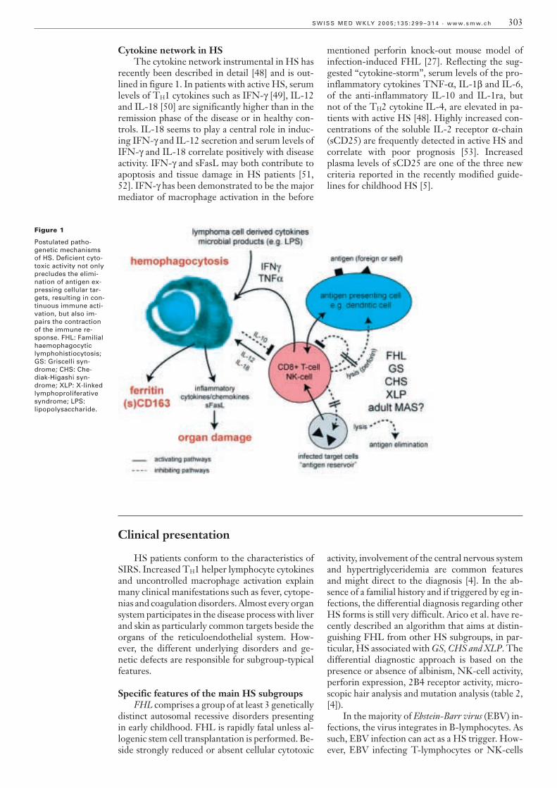
Deficient cytotoxic activity not only precludesthe elimination of antigen expressing cellular tar-gets, resulting in continuous immune activation,but also impairs the contraction of the immuneresponse [40]. Hence, sustained activation of the immune system and deficient negative feed-back mechanism explain the overwhelmingmacrophage activation seen in HS (figure 1). Thismodel has recently gained support by studies ofperforin knock-out mice, which display an over-whelming inflammatory response characterised bylymphocyte-mediated macrophage activation uponLCMV-infection [27].
Disease Human phenotype Gene (location, name) Function of protein Animal model
Familial haemophagocytic HS 9q21.3–22 = FHL-1 FHL-1 3??? Perforin lymphohistiocytosis (FHL) (~10%) FHL-2 3 perforin: lytic KO mice
10q21–22 = FHL-2 enzyme, cytotoxic activity(~20–40%) FHL-3 3 hMunc13–4: 17q25 = FHL-3 (? %) cytolytic granule exocytosis
Griscelli syndrome type 2 Partial albinism, 15q21–22 = RAB27A Melanosome transport, Ashen mice(GS) recurrent infections, cytotoxic granule
HS (accelerated phase) exocytosis
Chediak-Higashi syndrome Partial albinism, 1q42.1–24.2 = CHS1 Lysosome transport and/ Beige mice(CHS) recurrent infections, (human), LYST (mice) or fission
bleeding tendency, HS (accelerated phase),enlarged lysosomes
X-linked lymphoproliferative Fulminant infectious Xq25 = SH2D1A/SAP/ Modulates signaling SAP KO micesyndrome (XLP) mononucleosis with DSHP through SLAM family
HS, lymphomas, (~60–70%) membersdysgammaglobulinemia
KO: knock-out
Table 2
Hereditary disordersand HS.
S W I S S M E D W K LY 2 0 0 5 ; 1 3 5 : 2 9 9 – 3 1 4 · w w w. s m w. c h 303
Cytokine network in HSThe cytokine network instrumental in HS has
recently been described in detail [48] and is out-lined in figure 1. In patients with active HS, serumlevels of TH1 cytokines such as IFN-g [49], IL-12and IL-18 [50] are significantly higher than in theremission phase of the disease or in healthy con-trols. IL-18 seems to play a central role in induc-ing IFN-g and IL-12 secretion and serum levels ofIFN-g and IL-18 correlate positively with diseaseactivity. IFN-g and sFasL may both contribute toapoptosis and tissue damage in HS patients [51,52]. IFN-g has been demonstrated to be the majormediator of macrophage activation in the before
mentioned perforin knock-out mouse model of infection-induced FHL [27]. Reflecting the sug-gested “cytokine-storm”, serum levels of the pro-inflammatory cytokines TNF-a, IL-1b and IL-6,of the anti-inflammatory IL-10 and IL-1ra, butnot of the TH2 cytokine IL-4, are elevated in pa-tients with active HS [48]. Highly increased con-centrations of the soluble IL-2 receptor a-chain(sCD25) are frequently detected in active HS andcorrelate with poor prognosis [53]. Increasedplasma levels of sCD25 are one of the three newcriteria reported in the recently modified guide-lines for childhood HS [5].
Clinical presentation
HS patients conform to the characteristics ofSIRS. Increased TH1 helper lymphocyte cytokinesand uncontrolled macrophage activation explainmany clinical manifestations such as fever, cytope-nias and coagulation disorders. Almost every organsystem participates in the disease process with liverand skin as particularly common targets beside theorgans of the reticuloendothelial system. How-ever, the different underlying disorders and ge-netic defects are responsible for subgroup-typicalfeatures.
Specific features of the main HS subgroupsFHL comprises a group of at least 3 genetically
distinct autosomal recessive disorders presentingin early childhood. FHL is rapidly fatal unless al-logenic stem cell transplantation is performed. Be-side strongly reduced or absent cellular cytotoxic
activity, involvement of the central nervous systemand hypertriglyceridemia are common featuresand might direct to the diagnosis [4]. In the ab-sence of a familial history and if triggered by eg in-fections, the differential diagnosis regarding otherHS forms is still very difficult. Arico et al. have re-cently described an algorithm that aims at distin-guishing FHL from other HS subgroups, in par-ticular, HS associated with GS, CHS and XLP. Thedifferential diagnostic approach is based on thepresence or absence of albinism, NK-cell activity,perforin expression, 2B4 receptor activity, micro-scopic hair analysis and mutation analysis (table 2,[4]).
In the majority of Ebstein-Barr virus (EBV) in-fections, the virus integrates in B-lymphocytes. Assuch, EBV infection can act as a HS trigger. How-ever, EBV infecting T-lymphocytes or NK-cells
Figure 1
Postulated patho-genetic mechanismsof HS. Deficient cyto-toxic activity not onlyprecludes the elimi-nation of antigen ex-pressing cellular tar-gets, resulting in con-tinuous immune acti-vation, but also im-pairs the contractionof the immune re-sponse. FHL: Familialhaemophagocyticlymphohistiocytosis;GS: Griscelli syn-drome; CHS: Che-diak-Higashi syn-drome; XLP: X-linkedlymphoproliferativesyndrome; LPS:lipopolysaccharide.

Haemophagocytic syndromes in adults 304
lead to distinct clinical presentations such aschronic active EBV infection, lymphoproliferative(reactive) disorders and bona fide neoplasias(mostly NHL), which all harbour a high risk of en-suing HS [24]. It is reasonable to assume that theoligo/monoclonal nature of these disorders ex-plains why an aggressive approach including theearly use of etoposide seems to be indicated [54].
Non-EBV infection-associated HS has beenlinked to various viral, bacterial and also protozoaninfections [33], which must be excluded in patientspresenting with HS of unknown etiology. Besidessupportive care and effective treatment of the un-derlying infection whenever possible, the role of im-munosuppressive therapies is less well established inthese patients compared to other forms of HS. Thebenefit of controlling inadequate macrophage activ-ity has to be weighted against the potential risk ofan infection flare. Less immunosuppressive treat-ment strategies such as high dose IVIG might beparticularly appropriate in these patients.
Although the association of rheumatologic dis-orders, in particular SJIA (also called juvenile-onsetStill’s disease), with the development of HS wasfirst described in detail almost 20 years ago [18],larger paediatric series were published only re-cently [55, 56]. The data presented by Sawhney etal. suggest that around 5–10% of children with ju-venile idiopathic arthritis will develop HS duringthe course of their disease [56]. In a recent retro-spective study in adults, we showed that the crite-ria for AOSD were met in 40% of HS patients, andthat AOSD might be identical with HS in at leasta subgroup of these patients [10]. Patients withrheumatic disorders are often exposed to drugswith triggering potential. Moreover, a disease flareis often difficult to separate from a haemophago-cytic episode. Therefore, a (relative) drop of thetypically increased white blood cell count or a de-creasing sedimentation rate (due to hypofibrino-genemia) should direct the attention to an HS [3].The recent finding of reduced NK and T-cell func-tion due to low or ineffective perforin expressionin patients with SJIA, as described in the patho-physiology section of this review [3], might be afirst step towards a unifying concept of HS.
Malignancy-associated HS is most frequentlylinked to NHL, especially of NK and T-cell ori-gin, and characterised by a very poor prognosis[57]. The HS manifestations may be maskedand/or modified by the malignant process or ther-apeutic measures, and diagnosis is therefore oftendelayed. In some cases, HS is the first, often dra-matic presentation of NHL. In contrast to othertypes of HS, which are thought to represent anoverwhelming immune-response to self or foreignantigens, lymphoma-associated HS is suggested toresult from aberrant cytokine secretion by tumour
cells [58]. Rapid control of the malignant processis often not easily achieved and cytostatic treat-ments further increase the risk of infectious com-plications.
Differences between childhood and adult HSSeveral features have to be considered when
HS in children are compared with HS in adults. 1) The maturation state of the immune system may partially explain why the threshold for elicitingHS might be lower in children. Intriguingly, per-forin expression is age dependent. Rukavina et al.showed that perforin expression is very low at birthand increases thereafter. However, children have ahigher percentage of perforin expressing CD4+
lymphocytes compared to adults [59]. 2) The spec-trum of underlying disorders is different for sev-eral reasons. For instance, children carrying per-forin mutations almost exclusively present duringearly childhood, although late-onset FHL caseshave been described and linked to specific muta-tions [60, 61]. Immunodeficiency is mostly ac-quired in adults as opposed to inherited in chil-dren. Malignancy-associated HS is rather rare inchildren although there are exceptions [62]. 3) Thespectrum of triggering insults differs as well. Forexample: EBV infection is usually acquired duringchildhood/adolescence and therefore primary in-fection is rare in adults. 4) Tsuda described a dif-ferent frequency of clinical manifestations such ashepatosplenomegaly, rash and neurological in-volvement, which are all more common in children[13]. With respect to diagnostic parameters, hy-pertriglyceridemia is frequent in children, but rarein adults [63]. 5) Recently, prognostic factors havebeen described for high-risk childhood HS andinclude age <2 years, presence of a hereditary dis-order, underlying EBV infection or malignancy,severe neutropenia and disseminated intravascularcoagulation (DIC), opportunistic infections, cen-tral nervous system involvement, various labora-tory markers and initial treatment response [8]. In the adult setting, age >30 years, DIC, hyperfer-ritinemia, increased b2–microglobulin, combinedanaemia and thrombocytopenia and jaundice havebeen described as prognostic by Kaito [64]. In ourseries, the degree of renal impairment and the development of an acute respiratory distress syn-drome were associated with fatal outcome [10]. Be-cause laboratory data are highly variable, theunderlying disorder (such as the presence of a neo-plastic disorder) is considered prognostically moresignificant by some authors [57]. 6) Treatmentresponse seems to be generally worse in adults forreasons that are not understood in detail [57, 65].However, failure to immunomodulatory drugs assingle treatment are more common in children [7].

S W I S S M E D W K LY 2 0 0 5 ; 1 3 5 : 2 9 9 – 3 1 4 · w w w. s m w. c h 305
Given these differences, the diagnostic crite-ria established by the FHL group of the HistiocyteSociety in 1991 [22] and modified recently [5](comprising the following 8 criteria, of which 5have to be fulfilled: fever, splenomegaly, bicytope-nia, hypertriglyceridemia and/or hypofibrinogen-emia, haemophagocytosis, low/absent NK-cell ac-tivity, hyperferritinemia >500 µg/L and increasedplasma sCD25 [these criteria are not necessarywhen mutations as outlined in table 2 can bedemonstrated]), and the diagnostic algorithm forchildhood HS proposed by Arico [4] cannot beadopted for the adult situation.
Many macrophage activation markers (eg fer-ritin, b2-microglobulin, neuron specific enolase,neopterin, transcobalamin II) and cytokines (egIFN-g, TNF-a, IL-12 and IL-18) are typically in-creased during active HS. However, most of thesemarkers are not routinely available and/or lackspecificity for the monocyte-macrophage lineage.Moreover, literature regarding the utility forscreening and diagnosing HS is scarce. Based onour own experience, we favour a simple, cheap,widely available and reasonably specific screeningstrategy consisting of serum ferritin measurementsin patients presenting in an ominous clinical set-ting as outlined in figure 2 [10, 66]. In case of serum
ferritin levels >10000 mg/L, confirmatory testssuch as the assessment for morphological evidenceof haemophagocytosis and quantification of solu-ble CD163 (sCD163) are indicated [67]. sCD163is supposedly the most specific monocyte-macrophage lineage marker so far, can be mea-sured by an enzyme-linked immunosorbent assay(ELISA) and is dramatically increased in HS. Onlyin rare situations of mainly dendritic cell drivenHS, sCD163 might not be as increased (Schaer DJ,personal observation). Therefore, in the context of a typical clinical presentation combined withhyperferritinemia but absence of an sCD163 in-crease, the measurement of sCD25 has to be con-sidered. Bone marrow analyses are indicated inmost instances not only for diagnostic but also dif-ferential diagnostic purposes regarding underlyingpathologies [68]. Moreover, the quantitative analy-sis of EBV genome in peripheral blood might gov-ern the choice of treatments applied and serialmeasurements reflect response to therapy [69].Despite the scientific interest, the diagnostic valueof cytotoxicity assays and flow cytometric analysisof perforin expression on various leukocyte sub-sets, as well as their potential impact on diseasemanagement in adults, remain to be proven.
In the absence of an established marker for
Screening, diagnostic markers and monitoring of disease activity
serum ferritin > 10’000 µg/L ?
SIRS: >38 °C or <36 °C; heart rate >90/;
hyperventilation; WBC >12 or <4 G/L
cytopenia(s)
underlying disorder increasing risk of MAS
or presence of MAS trigger (see Table 1)
complementary diagnostic procedures
e.g.
infections ? (in particular EBV)
perforin flow cytometry (if available)
soluble CD163 ↑ ? soluble CD25 ↑ ?
morphological evidence ofhemophagocytosis ?
at least daily re-evaluation
(clinically and by serum ferritin)
no
no
Figure 2
Algorithm for thescreening and diag-nosis of HS in adults.SIRS: systemic in-flammatory responsesyndrome; WBC:white blood count.

Haemophagocytic syndromes in adults 306
monitoring macrophage activity/treatment re-sponse, an individualised approach is proposed.Both assessment of haemophagocytic activity andserum ferritin levels have their limitations [10, 66].The value of serial sCD163 measurements needsyet to be addressed prospectively.
In the following, some background informa-tion and data about the advantages and disadvan-tages of the main three parameters outlined in fig-ure 2 are summarised.
HaemophagocytosisDespite the limitations discussed below, mor-
phological evidence of haemophagocytosis is stillconsidered the gold standard in the diagnosis ofHS and can be demonstrated in many organs, inparticular bone marrow, spleen, liver and lymphnodes, but also in body fluids such as peripheralblood, pleural effusions, cerebrospinal fluid andurine. Haemophagocytosis is a physiological pro-cess that might be reinforced in situations such as haemolytic and aplastic anaemia, graft versushost disease, following transfusions and cytotoxictherapies. Whereas haemophagocytosis in thecontext of HS is normally a systemic event, organconfined haemophagocytosis can be found for in-stance in regional lymph nodes (after surgery),lung, spleen and skin (cytophagic histiocytic pan-niculitis) under certain circumstances [70–73]. Atleast for the case of cytophagic histiocytic panni-culitis it is known that the local phenomenon canbecome generalised. Situations with increased“physiological” haemophagocytosis have to beconsidered before making the diagnosis of HS.During HS, haemophagocytosis is found ratherconsistently in organs of the reticuloendothelialsystem. In contrast, central nervous system (in particular in adults as opposed to children) andlung involvement are rare [70]. Importantly,haemophagocytic activity is not found at any giventime in any given organ during the course of HS[74]. Hence, repeated assessment or concomitantanalysis of several organs might be necessary.Moreover, one might question the necessity ofdemonstrating >2% of macrophages showing signsof active haemophagocytosis for the diagnosis ofHS, as outlined by Wong et al. [11]. Markeddyserythropoiesis is not an uncommon phenome-non in HS and can mask the phagocytic process[75]. To summarise, haemophagocytosis is not asine qua non for the diagnosis of HS nor should itssignificance being overestimated in the absence ofother clinical and/or biological signs of over-whelming histiocytic activation.
Serum ferritinAlthough the presence of ferritin in serum has
been described almost 3 decades ago, the biochem-ical characteristics and biological function(s) re-main mostly elusive. Ferritin is composed of 24units of either heavy chain (H, acidic) or light chain(L, basic) subunits with a molecular weight around450 kDa. Under physiological conditions, serum
ferritin is iron poor, predominantly made up of Lchains and >50% glycosylated [76]. The glyco-sylation and other recent findings suggest activesecretion as at least a partial mechanism of serumappearance of this mostly cytosolic protein, as op-posed to passive release during tissue damage [77].
Serum ferritin in HS is acidic (H chain rich)[78] and <20% glycosylated [79, 80]. In a prospec-tive study in active adult HS, we have shown thatthe percentage of glycosylated ferritin is signifi-cantly lower in patients hospitalised in intensivecare units for at least one organ dysfunction versusconventional hospitalisation with no organ dys-function. This suggests that low glycosylated fer-ritin could be a marker of severe HS (Larroche C,unpublished data). The mechanisms leading to thehyperferritinemia seen in HS remain elusive, evenmore considering that ferritin levels can increasewithin hours over a range of several 10000 mg/L[66]. Several hypotheses have been forwarded: a) passive release due to cell damage (yet liver andspleen, ferritin rich target organs of HS, harbourpredominantly basic (L chain) isoferritins, andferritin is disproportionately elevated compared to liver enzymes), b) increased secretion bymacrophages (or hepatocytes) and/or release dur-ing erythrophagocytosis (erythrocytes are H chainrich), and c) decreased clearing due to lower gly-cosylation and/or down-regulation of putative fer-ritin receptors [81]. In a rat in vitro model, Sibilleet al. have shown massive release of ferritin in thesupernatant after ingestion of erythrocytes bymacrophages [82]. Similar findings were reportedrecently for human macrophages after ery-throphagocytosis [83]. Besides iron, IL-1, TNF-a,nitric oxide and reactive oxygen intermediates areimportant regulators of ferritin H chain synthesis,all of which are implicated in the pathophysiologyof HS [84]. In the absence of cytosolic L chains, H chains are secreted in culture media in lens epi-thelial cells [85]. It remains to be seen whether differential expression of L and H chains inmacrophages contributes to the hyperferritinemicstate in HS. The reduced glycosylation of serumferritin in HS might be explained by secretion/release of hypo-glycosylated cytosolic ferritin ordifferential clearing from the bloodstream. Alter-natively, massively increased ferritin expressionmight exceed the glycosylation capacity of the en-doplasmatic reticulum.
The principal role of cytosolic ferritin consistsin regulating the intracellular iron pool. However,the H chain exerts also immunomodulatory activ-ity by stimulating the expression of co-stimulatorymolecules on antigen presenting cells, followed byincreased secretion of IL-10 and decreased secre-tion of IL-2, IL-4 and IFN-g by regulatory T-cells[86]. In addition, Pham et al. described recently theinhibition of TNF-a induced apoptosis by NF-kBmediated ferritin H chain up-regulation [87].
It will be interesting to see whether serum hy-perferritinemia is part of an (inefficient) anti-in-flammatory feedback loop. The recently described

S W I S S M E D W K LY 2 0 0 5 ; 1 3 5 : 2 9 9 – 3 1 4 · w w w. s m w. c h 307
placental immunomodulatory ferritin is not ex-pressed by macrophages (Ch. Moroz, personalcommunication) and unlikely involved in thepathogenesis of HS.
Serum ferritin values >10000 mg/L are consid-ered pathognomic for HS, histiocytic malignan-cies and adult-onset Still’s disease [2, 80, 81, 88].Mostly more moderate hyperferritinemia is a char-acteristic of 1º and 2º haemochromatosis and alsoseen in various conditions such as inflammatorysyndromes (acute phase reaction or chronic in-flammation), due to cytolysis (eg liver necrosis,hematopathies, following cytotoxic treatments,following blood transfusions), related to neo-plasias, and as a key manifestation of the hyperfer-ritinemia/cataract syndrome [89]. In our study, wechose a ferritin cut-off of 10000 mg/L based on theliterature available at that time [66]. Interestingly,Ravelli et al. meanwhile found a serum ferritin>10000 mg/L to be the best marker with respectto specificity and sensitivity in a population of 72childhood cases of HS [2, 90]. It remains to bedemonstrated in prospective studies in adults andmore heterogeneous patient populations whethersimilar specificity and sensitivity is achieved with aferritin cut-off of 10000 mg/L, and whether theferritin threshold can be lowered in order to in-crease sensitivity but without relevant detrimentaleffects on specificity. In particular, a lower ferritinthreshold might be appropriate if combined withthe assessment of more specific markers such assCD163 and sCD25.
Soluble CD163 The hemoglobin-haptoglobin scavenger re-
ceptor CD163 is exclusively expressed on cells ofthe monocyte-macrophage lineage with increasingexpression during terminal macrophage differen-tiation [91–93] or upon glucocorticoid treatment
of monocytes [94]. The tightly restricted expres-sion pattern is even preserved beyond malignanttransformation of myeloid progenitor cells asCD163 expression can only be demonstrated on leukaemic cells with a definite monocyte/macrophage differentiation pattern [95]. Like anumber of other prominent leukocyte surface anti-gens such as L-selectin, CD163 is proteolyticallycleaved from the cell surface upon monocyte-macrophage stimulation with inflammatory medi-ators [96]. Thereby, CD163 cleavage seems to bea conserved response, which is induced by a widerange of inflammatory stimuli such as bacterialendotoxins [97], Fc-receptor cross-linking [98] aswell as direct activation of protein kinase C byphorbol ester [96]. The cleaved extracellular frag-ment of CD163 can be detected and quantified inthe plasma by a recently developed ELISA [99].These biologic properties render sCD163 an in-teresting candidate marker for the specific quan-tification of overall macrophage activity. In a pre-liminary study including 18 episodes of adult HS,we found sCD163 levels which were 10 to 40-foldabove the levels determined in healthy controlsubjects [67]. Interestingly, these levels were alsoclearly above the sCD163 concentrations in pa-tients with severe bacterial sepsis but withoutclinical or laboratory features compatible with HS.Expansion of the macrophage pool or increasedCD163 expression/cleavage during HS may ac-count for these findings. Accordingly, we havefound large accumulations of CD163 expressingmacrophages with active haemophagocytosis insplenic tissue and bone marrow of our patients.Importantly, sCD163 correlates significantly withother established markers of macrophage activa-tion such as ferritin and sCD25 during the clinicalcourse of HS.
Treatment
General concepts Compared to historical controls, the HLH-94
treatment protocol has dramatically changed theoutcome of children with FHL and other HS [9].It allows in most instances to stabilise disease ac-tivity. This permits performing an allogenic stemcell transplantation, which is the only curative op-tion in the case of FHL and HS associated withhereditary immunodeficiencies. Dexamethasone,etoposide/VP16 and cyclosporin A remain thebackbone of the HLH-2004 protocol [5]. Regard-ing EBV-associated HS, Imashuku stresses the im-portance of early administration of etoposide com-bined with immunotherapy, resulting in a high re-sponse rate [100]. For the remainder of HS, thereare no treatment standards.
Prognostic criteria for childhood HS allow tostratify for treatment aggressivity [8]. For low-risk
situations, corticosteroids alone or combined withcyclosporin A and/or intravenous immunoglob-ulin might be appropriate. For high-risk cases,early etoposide is recommended. In refractorycases or relapse, antithymocyte globulin and poly-chemotherapy (such as that used for treatingHodgkin’s disease or NHL) might be beneficial.
Given the lack of a similar stratification systemin adults, treatment is still individualised. More-over, treatment-related side effects have to betaken into consideration even more because of thepotentially transient nature of adult HS, as op-posed to genetically defined HS such as FHL,where the underlying defect persists without stemcell transplantation. Critical points of a successfulmanagement are: early diagnosis by awareness, inparticular in patients at high HS risk (as outlinedin table 1A, 1B and figure 2), appropriate screen-
Haemophagocytic syndromes in adults 308
ing and causative treatment of the underlying dis-order whenever possible, avoidance/removal oftriggering drugs/insults, appropriate supportivemeasures as generally indicated in situations ofinflammatory response syndromes and control ofNK/T-cell and phagocytic activity and the result-ing hypercytokinemia
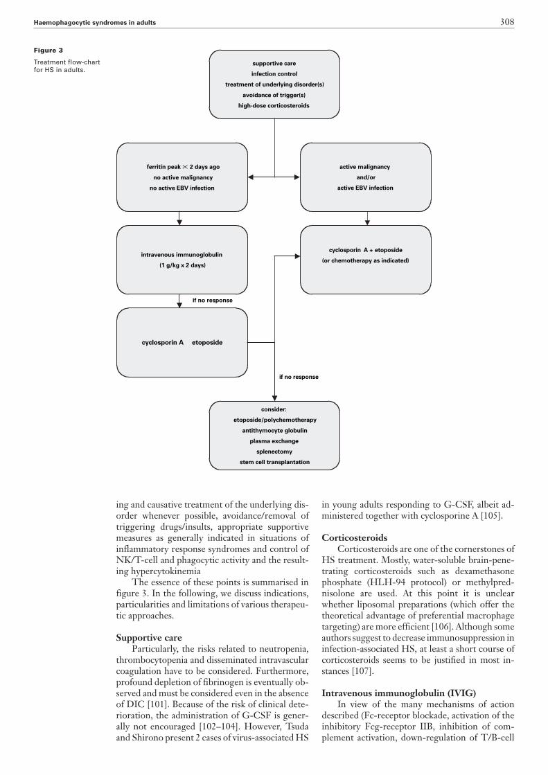
The essence of these points is summarised infigure 3. In the following, we discuss indications,particularities and limitations of various therapeu-tic approaches.
Supportive careParticularly, the risks related to neutropenia,
thrombocytopenia and disseminated intravascularcoagulation have to be considered. Furthermore,profound depletion of fibrinogen is eventually ob-served and must be considered even in the absenceof DIC [101]. Because of the risk of clinical dete-rioration, the administration of G-CSF is gener-ally not encouraged [102–104]. However, Tsudaand Shirono present 2 cases of virus-associated HS
in young adults responding to G-CSF, albeit ad-ministered together with cyclosporine A [105].
CorticosteroidsCorticosteroids are one of the cornerstones of
HS treatment. Mostly, water-soluble brain-pene-trating corticosteroids such as dexamethasonephosphate (HLH-94 protocol) or methylpred-nisolone are used. At this point it is unclearwhether liposomal preparations (which offer thetheoretical advantage of preferential macrophagetargeting) are more efficient [106]. Although someauthors suggest to decrease immunosuppression ininfection-associated HS, at least a short course ofcorticosteroids seems to be justified in most in-stances [107].
Intravenous immunoglobulin (IVIG)In view of the many mechanisms of action
described (Fc-receptor blockade, activation of theinhibitory Fcg-receptor IIB, inhibition of com-plement activation, down-regulation of T/B-cell
supportive care
infection control
treatment of underlying disorder(s)
avoidance of trigger(s)
high-dose corticosteroids
consider:
etoposide/polychemotherapy
antithymocyte globulin
plasma exchange
splenectomy
stem cell transplantation
cyclosporin A etoposide
intravenous immunoglobulin
(1 g/kg x 2 days)
ferritin peak � 2 days ago
no active malignancy
no active EBV infection
cyclosporin A + etoposide
(or chemotherapy as indicated)
active malignancy
and/or
active EBV infection
if no response
if no response
Figure 3
Treatment flow-chartfor HS in adults.
S W I S S M E D W K LY 2 0 0 5 ; 1 3 5 : 2 9 9 – 3 1 4 · w w w. s m w. c h 309
functions, anti-idiotypic suppression, neutralisa-tion of superantigens or infectious agents, neutral-isation of cytokines, enhanced clearance of patho-genetic antibodies), IVIG are indicated at leasttheoretically in a broad range of HS [108–111].The administration of IVIG alone or in combi-nation with other treatment modalities has beendescribed in more than 100 HS case reports andmostly small series. Variable efficacy is demon-strated, which is not astonishing given the hetero-geneity of HS, the different IVIG preparations onthe market, the inherent batch variability, the dif-ferent dosing schedules, and administration timepoints used. However, reports of negative effectson the course of disease are rare [112]. Two largerseries of adult HS treated with IVIG show verypromising results. Larroche et al. describe a globalresponse rate of 59% in a mixed population of in-fection, malignancy and systemic lupus erythe-matosus-associated HS. The mean dose adminis-tered was 1.6 g/kg over a mean of 3 days. Infec-tion-associated HS patients seem to benefit most(response rate of 78% versus 39% of the remain-ing patients). Conversely, IVIG seems to be largelyineffective in lymphoma-associated HS [113]. Wefound a similar response rate of around two-thirdsin a population of rheumatic disease, infection andmalignancy-associated HS. The main predictivefactor for response was the early administration ofIVIG (within 2 days of the ferritin peak). It remainsto be seen whether other markers such as sCD163are of similar value as serum ferritin for early HSdiagnosis, enabling emergency IVIG treatment.Many of our patients were administered 2 � 1 g/kgof IVIG over 2 days without overt toxicity. Simi-larly to Larroche et al., we never observed a sus-tained improvement in patients with malignancy-associated HS [10, 66]. This coincides with thefinding of Imashuku et al. of limited efficacy ofIVIG in EBV-related childhood cases with oligo/monoclonal lymphoproliferation, which probablyalso extends to adult patients [54]. As in our case,IVIG are generally well tolerated. Because of rarereports of renal impairment, renal function has to be monitored and nephrotoxic co-medicationavoided.
Cyclosporin ACSA not only affects early steps of T-cell acti-
vation, but also macrophage (decreased expressionof IL-6, IL-1, TNF-a, inducible nitric oxide syn-thetase, cyclooxygenase-2) and dendritic cell func-tions [114]. It is therefore an obvious choice fortargeting the various cellular players implicated inthe pathogenesis of HS and reducing the resultinghypercytokinemia. When given in combinationwith etoposide, CSA might limit the neutropenicperiod [115]. Moreover, the combination of etopo-side and CSA induces apoptosis in a nasal angio-centric natural killer cell lymphoma-derived cellline and CSA single treatment is effective in T-celllymphoproliferative syndromes [116, 117]. Pa-tients with lymphoproliferative disorders and
NHL-associated HS might therefore benefit inparticular from CSA. The administration of CSAdepends on an appropriate renal function. Hepaticand central nervous side effects might mimic HSmanifestations [8]. There is no consensus regard-ing the moment to start treatment nor for howlong CSA should be administered [55]. Regardingthe efficacy of other calcineurin inhibitors (egFK506/tacrolimus) or mTOR inhibitors (eg siroli-mus/rapamycin) in the setting of HS no data areavailable.
Etoposide/VP16The use of etoposide, a topoisomerase II in-
hibitor with high, though poorly characterised ac-tivity against monocytes, was established as a firstline therapy in the context of the HLH-94 proto-col and EBV-associated HS [6, 54]. RegardingEBV-associated cases, etoposide might act par-tially via blocking EBV-DNA and EBNA synthe-sis [118]. The combination CSA/etoposide mightbe capable to overcome the apoptotic insufficiencyof T-cells implicated in the pathogenesis of FHL[119]. Its use in adult HS unrelated to EBV infec-tion is more rare and mainly considered a secondchoice in refractory cases (eg [120]). The reluc-tance to use etoposide results from the risk, albeitsmall, of secondary malignancies [121]. The re-sulting neutropenia is attenuated by the concur-rent use of CSA [115]. Long-term low dose oraletoposide might be a safe and effective alternative[122].
Antithymocyte globulin (ATG)Although ATG might be equivalent to etopo-
side in situations of refractory disease, costs andpotential side effects (allergic reactions, severe im-munosuppression) limit its use [8].
Plasmapheresis and blood exchangePlasmapheresis and blood exchange have been
described in various case reports and small serieswith mostly positive results (eg [123]). However,whether clearance of cytokines really takes placeand whether this is the key mechanism is ques-tioned [124, 125]. Moreover, Kfoury Baz et al. de-scribe the development of a HS in a patient under-going plasma exchange for thrombotic thrombo-cytopenic purpura and procedure-related cytokinerelease has been suggested [126]. Plasma exchangetechniques might be considered in patients need-ing kidney function replacement procedures forother indications. In most cases reported, the pro-cedure had to be repeated several times.
Anti-TNF-aa treatment (anti-TNF-aaantibody [infliximab, Remicade™], humanised soluble TNF-aa receptor, [etanercept, Enbrel™])
The pathophysiological importance of TNF-a in HS has been outlined above, whereby the onlyminor impact on T-cell activation is a potentiallimitation of anti-TNF-a approaches. Aeberli et

Haemophagocytic syndromes in adults 310
al. describe the rapidly beneficial administration ofinfliximab/etanercept in adult HS associated withAOSD [127]. However, several case reports link theonset of HS with anti-TNF-a blockade (eg [128,129]. This is a major concern in patients with rheu-matic disorders with an increased basal HS risk andwhere anti-TNF-a treatments are used more com-monly. Whether the triggering of HS is indirect viaincreased susceptibility to infections under anti-TNF-a treatment remains to be seen. Meanwhile,it is reasonable to apply such treatments only wheninfections are excluded, if at all [3].
Stem cell transplantation (SCT)Allogenic SCT is the only curative option in
FHL and other hereditary forms of HS. However,it is rarely indicated in adults, eg refractory EBV-associated cases or potentially in the context oftransplantation for an underlying haematologicalneoplasia. Of note, autologous SCT has beenlinked to the development of HS, for instance inchildren with systemic juvenile idiopathic arthri-tis, which might result from depletion of regula-tory T-cells during the conditioning [130]. HSmight worsen if the SCT is performed during ac-tive/refractory disease [131].
VariedThe administration of fludarabine, a purine an-
timetabolite, results in profound immunosuppres-sion with particular impairment of T and NK-cells. A case report demonstrates beneficial effectsin the setting of FHL [132]. Methotrexate was usedfor intrathecal therapy in the HLH-94 protocol [6]
and might be an option in rheumatic disease re-lated HS, given that methotrexate is a standardtreatment of rheumatoid arthritis. However,methotrexate potentially can trigger HS [133]. Da-clizumab, an anti-CD25 antibody, and interferon-a,still have to find their place in the treatment of HS,although the rationale for their use is strong [134,135]. Therapeutic splenectomy is very rarely indi-cated, although successful reports have been pub-lished recently in particular in HIV-related cases(eg [37, 136, 137]. Furthermore, in our experience,splenectomy might be of particular value in life-threatening cases of lymphoma-associated HSwith significant splenic tumor mass, where secre-tion of inflammatory cytokines by the malignantcells is supposed to be the major pathogenic mech-anism [58]. HIV-associated HS seems to be sensi-tive to highly active anti-retroviral therapy (HAART)alone in some instances [138]. However, a moreaggressive approach is probably needed in the ma-jority of cases. To mention that Huang et al. de-scribed recently the triggering of a HIV-associatedHS with initiation of HAART, which the authorsinterpreted within the context of treatment relatedimmune reconstitution [139]. Finally, B-cell di-rected therapy with the anti-CD20 antibody ritu-ximab might be a promising approach in some pa-tients with EBV-related HS. Whether the efficacyof B-cell depletion is only related to the exhaustionof the EBV reservoir or whether B-cell cytokineproduction also contributes to the clinical mani-festation of HS, as recently suggested for somerheumatologic diseases, needs to be revealed [140,141].
Broader implications of the HS concept for SIRS patients?
Many patients admitted to intensive care unitspresent with manifestations defining a SIRS, oftencombined with acquired immunodeficiences in thepresence of potential HS triggers such as infec-tions. There is evidence that HS is not so rare insuch populations and probably highly under-diag-nosed. Indeed, François et al. found HS in 32/599patients admitted to an interdisciplinary intensivecare unit [23]. More important, haemophagocyto-sis was demonstrated in a subgroup of 32/50 (64%)patients presenting with a sepsis syndrome andthrombocytopenia <100000 G/L. A limitation ofusing thrombocytopenia as a screening parameteris the necessary exclusion of patients with haema-tological diseases, previous anticancer treatment,major bleeding and/or prior transfusion, DIC andprevious administration of heparin. Moreover,thrombocytopenia is not a universal finding in HS.In another study, Stephan et al. found evidence ofhaemophagocytosis in bone marrow aspirates of12/20 mechanically ventilated patients with sepsisor septic shock and thrombocytopenia <100000G/L with various surgical (n = 15) and medical (n = 5) conditions, but without pre-existing im-
munodeficiences [142, 143]. Based on two child-hood HS cases, Gauvin et al. concluded recentlythat HS and multiple organ dysfunction syndromesshare pathophysiological traits and might be re-lated to each other [144]. Interestingly, CD14dim/CD16bright monocytes are not only increased in HS, but also in sepsis patients and have higher phagocytic activity than their CD14dim/CD16bright counterpart [142, 145]. Although thepresence of bone marrow haemophagocytosis inICU patients might not necessarily be of clinicalrelevance in every single case [146], we emphasisethe importance of a broad serum ferritin screeningin intensive care unit patients, based on our expe-rience. If a HS is diagnosed by following the algo-rithm outlined in figure 2, rapid administration ofIVIG seems to be beneficial in the majority of pa-tients [10, 66].
Only limited progress has been made in thetreatment of SIRS patients over the last years de-spite growing knowledge of the underlying patho-physiological mechanisms [147]. One major obsta-cle in performing clinical studies is patient hetero-geneity. The administration of IVIG has a small

S W I S S M E D W K LY 2 0 0 5 ; 1 3 5 : 2 9 9 – 3 1 4 · w w w. s m w. c h 311
impact on outcome in unselected SIRS patients(http://www.cochrane.org/cochrane/revabstr/ab001090.htm). However, ferritinemia >10000 mg/L,and thus macrophage hyper-activation, probably
defines a SIRS subgroup with particular high re-sponse rate to IVIG, a hypothesis which remainsto be tested in a prospective, randomised manner.
Conclusions
Although the deciphering of the genetic de-fects underlying various childhood forms of HShas broadened the pathophysiological understand-ing of HS in general and contributed to improveprognosis in childhood cases, much needs to belearnt as to whether and to which extent the diag-nostic and therapeutic concepts established in chil-dren can be adopted for adults. In the absence ofdiagnostic guidelines, HS diagnosis is still toooften missed or delayed in adults. Moreover, ther-apeutic decisions are mostly based on case reportsor small series. We have described a simple screen-ing and diagnostic algorithm, which might becomeinstrumental to recruit patients for clinical studies.Such studies should allow refining the diagnosis ofHS in adults and establishing therapeutic guide-
lines, eg testing the impact of early IVIG adminis-tration in SIRS patients presenting with a HS. Fi-nally, it will be interesting to see whether defectsof the cytotoxic effector pathways contribute to thepathogenesis of HS in adults as well and how thiscan be exploited.
The authors would like to thank K. Arn for help withfigure 1.
Correspondence:Prof. Klaus A. NeftelLuisenstrasse 45CH-3005 [email protected]
References
1 Arico M, Danesino C, Pende D, Moretta L. Pathogenesis ofhaemophagocytic lymphohistiocytosis. Br J Haematol 2001;114:761–9.
2 Ravelli A. Macrophage activation syndrome. Curr OpinRheumatol 2002;14:548–52.
3 Grom AA. Natural killer cell dysfunction: A common pathwayin systemic-onset juvenile rheumatoid arthritis, macrophage ac-tivation syndrome, and hemophagocytic lymphohistiocytosis?Arthritis Rheum 2004;50:689–98.
4 Arico M, Allen M, Brusa S, Clementi R, Pende D, Maccario R,et al. Haemophagocytic lymphohistiocytosis: proposal of a di-agnostic algorithm based on perforin expression. Br J Haema-tol 2002;119:180–8.
5 Henter JI, Tondini C, Pritchard J. Histiocyte disorders. Crit RevOncol Hematol 2004;50:157–74.
6 Henter JI, Arico M, Egeler RM, Elinder G, Favara BE, Fili-povich AH, et al. HLH-94: a treatment protocol for hemo-phagocytic lymphohistiocytosis. HLH study Group of the His-tiocyte Society. Med Pediatr Oncol 1997;28:342–7.
7 Janka GE, Schneider EM. Modern management of childrenwith haemophagocytic lymphohistiocytosis. Br J Haematol2004;124:4–14.
8 Imashuku S, Teramura T, Morimoto A, Hibi S. Recent devel-opments in the management of haemophagocytic lymphohisti-ocytosis. Expert Opin Pharmacother 2001;2:1437–48.
9 Henter JI, Samuelsson-Horne A, Arico M, Egeler RM, ElinderG, Filipovich AH, et al. Treatment of hemophagocytic lympho-histiocytosis with HLH-94 immunochemotherapy and bonemarrow transplantation. Blood 2002;100:2367–73.
10 Emmenegger U, Reimers A, Frey U, Fux C, Bihl F, Semela D,et al. Reactive macrophage activation syndrome: a simplescreening strategy and its potential in early treatment initiation.Swiss Med Wkly 2002;132:230–6.
11 Wong KF, Chan JK. Reactive hemophagocytic syndrome – aclinicopathologic study of 40 patients in an Oriental population.Am J Med 1992;93:177–80.
12 Imashuku S. Differential diagnosis of hemophagocytic syn-drome: underlying disorders and selection of the most effectivetreatment. Int J Hematol 1997;66:135–51.
13 Tsuda H. Hemophagocytic syndrome (HPS) in children andadults. Int J Hematol 1997;65:215–26.
14 Farquhar JW, Claireaux AE. Familial haemophagocytic reticu-losis. Arch Dis Child 1952;27:519–25.
15 Chandra P, Chaudhery SA, Rosner F, Kagen M. Transient his-tiocytosis with striking phagocytosis of platelets, leukocytes, anderythrocytes. Arch Intern Med 1975;135:989–91.
16 Risdall RJ, McKenna RW, Nesbit ME, Krivit W, Balfour HH,Jr., Simmons RL, et al. Virus-associated hemophagocytic syn-drome: a benign histiocytic proliferation distinct from malig-nant histiocytosis. Cancer 1979;44:993–1002.
17 Reiner AP, Spivak JL. Hematophagic histiocytosis. A report of23 new patients and a review of the literature. Medicine (Balti-more) 1988;67:369–88.
18 Hadchouel M, Prieur AM, Griscelli C. Acute hemorrhagic, he-patic, and neurologic manifestations in juvenile rheumatoidarthritis: possible relationship to drugs or infection. J Pediatr1985;106:561–6.
19 Stephan JL, Zeller J, Hubert P, Herbelin C, Dayer JM, PrieurAM. Macrophage activation syndrome and rheumatic disease inchildhood: a report of four new cases. Clin Exp Rheumatol1993;11:451–6.
20 Henter JI, Nennesmo I. Neuropathologic findings and neuro-logic symptoms in twenty-three children with hemophagocyticlymphohistiocytosis. J Pediatr 1997;130:358–65.
21 Athreya BH. Is macrophage activation syndrome a new entity?Clin Exp Rheumatol 2002;20:121–3.
22 Henter JI, Elinder G, Ost A. Diagnostic guidelines for hemo-phagocytic lymphohistiocytosis. The FHL Study Group of theHistiocyte Society. Semin Oncol 1991;18:29–33.
23 Francois B, Trimoreau F, Vignon P, Fixe P, Praloran V, GastinneH. Thrombocytopenia in the sepsis syndrome: role of hemo-phagocytosis and macrophage colony-stimulating factor. Am JMed 1997;103:114–20.
24 Zhang Y, Nagata H, Ikeuchi T, Mukai H, Oyoshi MK, DemachiA, et al. Common cytological and cytogenetic features of Ep-stein-Barr virus (EBV)-positive natural killer (NK) cells and celllines derived from patients with nasal T/NK-cell lymphomas,chronic active EBV infection and hydroa vacciniforme-likeeruptions. Br J Haematol 2003;121:805–14.
25 Weiss DJ, Aird B. Cytologic evaluation of primary and second-ary myelodysplastic syndromes in the dog. Vet Clin Pathol2001;30:67–75.
26 Walton RM, Modiano JF, Thrall MA, Wheeler SL. Bone mar-row cytological findings in 4 dogs and a cat with hemophago-cytic syndrome. J Vet Intern Med 1996;10:7–14.

Haemophagocytic syndromes in adults 312
27 Jordan MB, Hildeman D, Kappler J, Marrack P. An animalmodel of hemophagocytic lymphohistiocytosis (HLH): CD8+T cells and interferon gamma are essential for the disorder.Blood 2004;104:735–43.
28 Yin L, Al-Alem U, Liang J, Tong WM, Li C, Badiali M, et al.Mice deficient in the X-linked lymphoproliferative disease genesap exhibit increased susceptibility to murine gammaher-pesvirus-68 and hypo-gammaglobulinemia. J Med Virol 2003;71:446–55.
29 Ishii E, Yoshida N, Kimura N, Fujimoto J, Mizutani S, Sako M,et al. Clonal dissemination of T-lymphocytes in scid mice fromfamilial hemophagocytic lymphohistiocytosis. Med PediatrOncol 1999;32:201–8.
30 Yoshida N, Ishii E, Oshima K, Yanai F, Ogawa A, Kataoka S, etal. Engraftment and dissemination of T lymphocytes from pri-mary haemophagocytic lymphohistiocytosis in scid mice. Br JHaematol 2003;121:349–58.
31 Hayashi K, Ohara N, Teramoto N, Onoda S, Chen HL, Oka T,et al. An animal model for human EBV-associated hemophago-cytic syndrome: herpesvirus papio frequently induces fatal lym-phoproliferative disorders with hemophagocytic syndrome inrabbits. Am J Pathol 2001;158:1533–42.
32 Hayashi K, Joko H, Koirala TR, Onoda S, Jin ZS, MunemasaM, et al. Therapeutic trials for a rabbit model of EBV-associ-ated Hemophagocytic Syndrome (HPS): effects of vidarabineor CHOP, and development of Herpesvirus papio (HVP)-neg-ative lymphomas surrounded by HVP-infected lymphoprolif-erative disease. Histol Histopathol 2003;18:1155–68.
33 Fisman DN. Hemophagocytic syndromes and infection. EmergInfect Dis 2000;6:601–8.
34 Emminger W, Zlabinger GJ, Fritsch G, Urbanek R.CD14(dim)/CD16(bright) monocytes in hemophagocytic lym-phohistiocytosis. Eur J Immunol 2001;31:1716–9.
35 Goldberg J, Nezelof C. Lymphohistiocytosis: a multi-factorialsyndrome of macrophagic activation clinico-pathological studyof 38 cases. Hematol Oncol 1986;4:275–89.
36 Fleming MD, Pinkus JL, Fournier MV, Alexander SW, Tam C,Loda M, et al. Coincident expression of the chemokine recep-tors CCR6 and CCR7 by pathologic Langerhans cells inLangerhans cell histiocytosis. Blood 2003;101:2473–5.
37 Kereveur A, McIlroy D, Samri A, Oksenhendler E, Clauvel JP,Autran B. Up-regulation of adhesion and MHC molecules onsplenic monocyte/macrophages in adult haemophagocytic syn-drome. Br J Haematol 1999;104:871–7.
38 Schneider EM, Lorenz I, Walther P, Janka-Schaub GE. Naturalkiller deficiency: a minor or major factor in the manifestationof hemophagocytic lymphohistiocytosis? J Pediatr HematolOncol 2003;25:680–3.
39 Schneider EM, Lorenz I, Muller-Rosenberger M, Steinbach G,Kron M, Janka-Schaub GE. Hemophagocytic lymphohistiocy-tosis is associated with deficiencies of cellular cytolysis but nor-mal expression of transcripts relevant to killer-cell-inducedapoptosis. Blood 2002;100:2891–8.
40 Fischer A. Human primary immunodeficiency diseases: a per-spective. Nat Immunol 2004;5:23–30.
41 Stepp SE, Dufourcq-Lagelouse R, Le Deist F, Bhawan S, Cer-tain S, Mathew PA, et al. Perforin gene defects in familial he-mophagocytic lymphohistiocytosis. Science 1999;286:1957–9.
42 Feldmann J, Le Deist F, Fischer A, de Saint Basile G.[Munc13–4 is essential for cytolytic granule fusion]. Med Sci(Paris) 2004;20:144–6.
43 Menasche G, Pastural E, Feldmann J, Certain S, Ersoy F,Dupuis S, et al. Mutations in RAB27A cause Griscelli syndromeassociated with haemophagocytic syndrome. Nat Genet 2000;25:173–6.
44 Baetz K, Isaaz S, Griffiths GM. Loss of cytotoxic T lymphocytefunction in Chediak-Higashi syndrome arises from a secretorydefect that prevents lytic granule exocytosis. J Immunol1995;154:6122–31.
45 Czar MJ, Kersh EN, Mijares LA, Lanier G, Lewis J, Yap G, etal. Altered lymphocyte responses and cytokine production inmice deficient in the X-linked lymphoproliferative disease geneSH2D1A/DSHP/SAP. Proc Natl Acad Sci U S A 2001;98:7449–54.
46 Crotty S, Kersh EN, Cannons J, Schwartzberg PL, Ahmed R.SAP is required for generating long-term humoral immunity.Nature 2003;421:282–7.
47 Villanueva J, Lee S, Giannini EH, Graham TB, Passo MH, Fil-ipovich A, et al. Natural killer cell dysfunction is a distinguish-ing feature of systemic onset juvenile rheumatoid arthritis andmacrophage activation syndrome. Arthritis Res Ther 2005;7:R30–7.
48 Larroche C, Mouthon L. Pathogenesis of hemophagocytic syn-drome (HPS). Autoimmun Rev 2004;3:69–75.
49 Osugi Y, Hara J, Tagawa S, Takai K, Hosoi G, Matsuda Y, et al.Cytokine production regulating Th1 and Th2 cytokines in he-mophagocytic lymphohistiocytosis. Blood 1997;89:4100–3.
50 Takada H, Nomura A, Ohga S, Hara T. Interleukin-18 in he-mophagocytic lymphohistiocytosis. Leuk Lymphoma 2001;42:21–8.
51 Hasegawa D, Kojima S, Tatsumi E, Hayakawa A, Kosaka Y,Nakamura H, et al. Elevation of the serum Fas ligand in patientswith hemophagocytic syndrome and Diamond-Blackfan ane-mia. Blood 1998;91:2793–9.
52 Emmenegger U, Zehnder R, Frey U, Reimers A, Spaeth PJ,Neftel KA. Elevation of soluble Fas and soluble Fas ligand inreactive macrophage activation syndromes. Am J Hematol2000;64:116–9.
53 Imashuku S, Hibi S, Sako M, Ishida Y, Mugishima H, Chen J,et al. Soluble interleukin-2 receptor: a useful prognostic factorfor patients with hemophagocytic lymphohistiocytosis. Blood1995;86:4706–7.
54 Imashuku S, Kuriyama K, Teramura T, Ishii E, Kinugawa N,Kato M, et al. Requirement for etoposide in the treatment ofEpstein-Barr virus-associated hemophagocytic lymphohistio-cytosis. J Clin Oncol 2001;19:2665–73.
55 Stephan JL, Kone-Paut I, Galambrun C, Mouy R, Bader-Meunier B, Prieur AM. Reactive haemophagocytic syndrome inchildren with inflammatory disorders. A retrospective study of24 patients. Rheumatology (Oxford) 2001;40:1285–92.
56 Sawhney S, Woo P, Murray KJ. Macrophage activation syn-drome: a potentially fatal complication of rheumatic disorders.Arch Dis Child 2001;85:421–6.
57 Takahashi N, Chubachi A, Kume M, Hatano Y, Komatsuda A,Kawabata Y, et al. A clinical analysis of 52 adult patients withhemophagocytic syndrome: the prognostic significance of theunderlying diseases. Int J Hematol 2001;74:209–13.
58 Lay JD, Tsao CJ, Chen JY, Kadin ME, Su IJ. Upregulation oftumor necrosis factor-alpha gene by Epstein-Barr virus and ac-tivation of macrophages in Epstein-Barr virus-infected T cellsin the pathogenesis of hemophagocytic syndrome. J Clin Invest1997;100:1969–79.
59 Rukavina D, Laskarin G, Rubesa G, Strbo N, Bedenicki I,Manestar D, et al. Age-related decline of perforin expression inhuman cytotoxic T lymphocytes and natural killer cells. Blood1998;92:2410–20.
60 Allen M, De Fusco C, Legrand F, Clementi R, Conter V,Danesino C, et al. Familial hemophagocytic lymphohistiocyto-sis: how late can the onset be? Haematologica 2001;86:499–503.
61 Clementi R, Emmi L, Maccario R, Liotta F, Moretta L,Danesino C, et al. Adult onset and atypical presentation of he-mophagocytic lymphohistiocytosis in siblings carrying PRF1mutations. Blood 2002;100:2266–7.
62 Veerakul G, Sanpakit K, Tanphaichitr VS, Mahasandana C,Jirarattanasopa N. Secondary hemophagocytic lymphohistio-cytosis in children: an analysis of etiology and outcome. J MedAssoc Thai 2002;85(Suppl 2):S530–41.
63 Tsuda H, Shirono K. Serum lipids in adult patients with hemo-phagocytic syndrome. Am J Hematol 1996;53:285.
64 Kaito K, Kobayashi M, Katayama T, Otsubo H, Ogasawara Y,Sekita T, et al. Prognostic factors of hemophagocytic syndromein adults: analysis of 34 cases. Eur J Haematol 1997;59:247–53.
65 Imashuku S, Kuriyama K, Sakai R, Nakao Y, Masuda S, YasudaN, et al. Treatment of Epstein-Barr virus-associated hemo-phagocytic lymphohistiocytosis (EBV-HLH) in young adults: areport from the HLH study center. Med Pediatr Oncol2003;41:103–9.
66 Emmenegger U, Frey U, Reimers A, Fux C, Semela D, Cotta-gnoud P, et al. Hyperferritinemia as indicator for intravenousimmunoglobulin treatment in reactive macrophage activationsyndromes. Am J Hematol 2001;68:4–10.
67 Schaer DJ, Schleiffenbaum B, Kurrer M, Imhof A, Baechli E,Fehr J, et al. Soluble hemoglobin-haptoglobin scavenger recep-tor CD163 as a lineage specific marker in the reactive hemo-phagocytic syndrome. Eur J Haematol; in press.
68 Florena AM, Iannitto E, Quintini G, Franco V. Bone marrowbiopsy in hemophagocytic syndrome. Virchows Arch 2002;441:335–44.
69 Teramura T, Tabata Y, Yagi T, Morimoto A, Hibi S, ImashukuS. Quantitative analysis of cell-free Epstein-Barr virus genomecopy number in patients with EBV-associated hemophagocyticlymphohistiocytosis. Leuk Lymphoma 2002;43:173–9.

S W I S S M E D W K LY 2 0 0 5 ; 1 3 5 : 2 9 9 – 3 1 4 · w w w. s m w. c h 313
70 Favara BE, Feller AC, Pauli M, Jaffe ES, Weiss LM, Arico M,et al. Contemporary classification of histiocytic disorders. TheWHO Committee On Histiocytic/Reticulum Cell Prolifera-tions. Reclassification Working Group of the Histiocyte Soci-ety. Med Pediatr Oncol 1997;29:157–66.
71 Dascalescu CM, Wendum D, Gorin NC. Littoral-cell angiomaas a cause of splenomegaly. N Engl J Med 2001;345:772–3.
72 Zollner TM, Podda M, Ochsendorf FR, Wolter M, KaufmannR, Boehncke WH. Monitoring of phagocytic activity in histio-cytic cytophagic panniculitis. J Am Acad Dermatol 2001;44:120–3.
73 Nicholls JM, Poon LL, Lee KC, Ng WF, Lai ST, Leung CY, etal. Lung pathology of fatal severe acute respiratory syndrome.Lancet 2003;361:1773–8.
74 Ost A, Nilsson-Ardnor S, Henter JI. Autopsy findings in 27 chil-dren with haemophagocytic lymphohistiocytosis. Histopathol-ogy 1998;32:310–6.
75 Macheta M, Will AM, Houghton JB, Wynn RF. Prominentdyserythropoiesis in four cases of haemophagocytic lymphohis-tiocytosis. J Clin Pathol 2001;54:961–3.
76 Ponka P, Beaumont C, Richardson DR. Function and regula-tion of transferrin and ferritin. Semin Hematol 1998;35:35–54.
77 Ghosh S, Hevi S, Chuck SL. Regulated secretion of glycosy-lated human ferritin from hepatocytes. Blood 2004;103:2369–76.
78 Shimmyozu K, Kadokura N, Itoyama T, Tara M, Maruyama I,Osame M. [Increase of serum acidic isoferritins in patients withhemophagocytic histiocytosis]. Rinsho Ketsueki 1990;31:1036–7.
79 Miyazawa K, Shiota M, Takakuwa Y, Kawanishi Y, Iwabuchi H,Nakano M, et al. [Mechanism of hyperferritinemia in a case ofmalignant histiocytosis]. Nippon Ketsueki Gakkai Zasshi 1990;53:575–81.
80 Lambotte O, Cacoub P, Costedoat N, Le Moel G, Amoura Z,Piette JC. High ferritin and low glycosylated ferritin may alsobe a marker of excessive macrophage activation. J Rheumatol2003;30:1027–8.
81 Van Reeth C, Le Moel G, Lasne Y, Revenant MC, Agneray J,Kahn MF, et al. Serum ferritin and isoferritins are tools for di-agnosis of active adult Still’s disease. J Rheumatol 1994;21:890–5.
82 Sibille JC, Kondo H, Aisen P. Interactions between isolated he-patocytes and Kupffer cells in iron metabolism: a possible rolefor ferritin as an iron carrier protein. Hepatology 1988;8:296–301.
83 Yuan XM, Li W, Baird SK, Carlsson M, Melefors O. Secretionof Ferritin by Iron-laden Macrophages and Influence ofLipoproteins. Free Radic Res 2004;38:1133–42.
84 Torti FM, Torti SV. Regulation of ferritin genes and protein.Blood 2002;99:3505–16.
85 Goralska M, Holley BL, McGahan MC. Identification of amechanism by which lens epithelial cells limit accumulation ofoverexpressed ferritin H-chain. J Biol Chem 2003;278:42920–6.
86 Gray CP, Arosio P, Hersey P. Heavy chain ferritin activates reg-ulatory T cells by induction of changes in dendritic cells. Blood2002;99:3326–34.
87 Pham CG, Bubici C, Zazzeroni F, Papa S, Jones J, Alvarez K, etal. Ferritin Heavy Chain Upregulation by NF-kappaB InhibitsTNFalpha-Induced Apoptosis by Suppressing Reactive OxygenSpecies. Cell 2004;119:529–42.
88 Esumi N, Ikushima S, Hibi S, Todo S, Imashuku S. High serumferritin level as a marker of malignant histiocytosis and virus-associated hemophagocytic syndrome. Cancer 1988;61:2071–6.
89 Damade R, Rosenthal E, Cacoub P. [Hyperferritinemia]. AnnMed Interne (Paris) 2000;151:169–77.
90 Ravelli A, Magni-Manzoni S, Foti T, Besana C, Felici E, TrailL, et al. Macrophage activation syndrome in juvenile idiopathicarthritis: towards the development of diagnostic guidelines [ab-stract]. Arthritis Rheum 2001;44:S166.
91 Zwadlo G, Voegeli R, Osthoff KS, Sorg C. A monoclonal anti-body to a novel differentiation antigen on human macrophagesassociated with the down-regulatory phase of the inflammatoryprocess. Exp Cell Biol 1987;55:295–304.
92 Kristiansen M, Graversen JH, Jacobsen C, Sonne O, HoffmanHJ, Law SK, et al. Identification of the haemoglobin scavengerreceptor. Nature 2001;409:198–201.
93 Schaer DJ, Boretti FS, Hongegger A, Poehler D, Linnscheid P,Staege H, et al. Molecular cloning and characterization of themouse CD163 homologue, a highly glucocorticoid-induciblemember of the scavenger receptor cysteine-rich family. Im-munogenetics 2001;53:170–7.
94 Schaer DJ, Boretti FS, Schoedon G, Schaffner A. Inductionof the CD163–dependent haemoglobin uptake bymacrophages as a novel anti-inflammatory action of glucocor-ticoids. Br J Haematol 2002;119:239–43.
95 Walter RB, Bachli EB, Schaer DJ, Ruegg R, Schoedon G. Ex-pression of the hemoglobin scavenger receptor(CD163/HbSR) as immunophenotypic marker of monocyticlineage in acute myeloid leukemia. Blood 2003;101:3755–6.
96 Droste A, Sorg C, Hogger P. Shedding of CD163, a novel reg-ulatory mechanism for a member of the scavenger receptorcysteine-rich family. Biochem Biophys Res Commun 1999;256:110–3.
97 Hintz KA, Rassias AJ, Wardwell K, Moss ML, MorganelliPM, Pioli PA, et al. Endotoxin induces rapid metallopro-teinase-mediated shedding followed by up-regulation of themonocyte hemoglobin scavenger receptor CD163. J LeukocBiol 2002;72:711–7.
98 Sulahian TH, Hogger P, Wahner AE, Wardwell K, GouldingNJ, Sorg C, et al. Human monocytes express CD163, whichis upregulated by IL-10 and identical to p155. Cytokine2000;12:1312–21.
99 Moller HJ, Peterslund NA, Graversen JH, Moestrup SK.Identification of the hemoglobin scavenger receptor/CD163as a natural soluble protein in plasma. Blood 2002;99:378–80.
100 Imashuku S, Hibi S, Ohara T, Iwai A, Sako M, Kato M, et al.Effective control of Epstein-Barr virus-related hemophago-cytic lymphohistiocytosis with immunochemotherapy. Histi-ocyte Society. Blood 1999;93:1869–74.
101 Ooe K. Pathogenesis of hypofibrinogenemia in familial he-mophagocytic lymphohistiocytosis. Pediatr Pathol 1991;11:657–61.
102 Gilmore GL, DePasquale DK, Fischer BC, Shadduck RK.Enhancement of monocytopoiesis by granulocyte colony-stimulating factor: evidence for secondary cytokine effects invivo. Exp Hematol 1995;23:1319–23.
103 Quesnel B, Catteau B, Aznar V, Bauters F, Fenaux P. Success-ful treatment of juvenile rheumatoid arthritis associatedhaemophagocytic syndrome by cyclosporin A with transientexacerbation by conventional-dose G-CSF. Br J Haematol1997;97:508–10.
104 Wang S, Degar BA, Zieske A, Shafi NQ, Rose MG. Hemo-phagocytosis exacerbated by G-CSF/GM-CSF treatment in apatient with myelodysplasia. Am J Hematol 2004;77:391–96.
105 Tsuda H, Shirono K. Successful treatment of virus-associatedhaemophagocytic syndrome in adults by cyclosporin A sup-ported by granulocyte colony-stimulating factor. Br J Haema-tol 1996;93:572–5.
106 Funauchi M, Ohno M, Yamagata T, Nozaki Y, Kinoshita K,Kanamaru A. Effects of liposteroid on the hemophagocyticsyndrome in systemic lupus erythematosus. Lupus 2003;12:483–5.
107 Dhote R, Simon J, Papo T, Detournay B, Sailler L, AndreMH, et al. Reactive hemophagocytic syndrome in adult sys-temic disease: report of twenty-six cases and literature review.Arthritis Rheum 2003;49:633–9.
108 Rhoades CJ, Williams MA, Kelsey SM, Newland AC. Mono-cyte-macrophage system as targets for immunomodulation byintravenous immunoglobulin. Blood Rev 2000;14:14–30.
109 Samuelsson A, Towers TL, Ravetch JV. Anti-inflammatory ac-tivity of IVIG mediated through the inhibitory Fc receptor.Science 2001;291:484–6.
110 Pricop L, Redecha P, Teillaud JL, Frey J, Fridman WH,Sautes-Fridman C, et al. Differential modulation of stimula-tory and inhibitory Fc gamma receptors on human monocytesby Th1 and Th2 cytokines. J Immunol 2001;166:531–7.
111 Simon HU, Spath PJ. IVIG-mechanisms of action. Allergy2003;58:543–52.
112 Nagasawa M, Okawa H, Yata J. Deleterious effects of highdose gamma-globulin therapy on patients with hemophago-cytic syndrome. Int J Hematol 1994;60:91–3.
113 Larroche C, Bruneel F, Andre MH, Bader-Meunier B,Baruchel A, Tribout B, et al. [Intravenously administeredgamma-globulins in reactive hemaphagocytic syndrome.Multicenter study to assess their importance, by the im-munoglobulins group of experts of CEDIT of the AP-HP].Ann Med Interne (Paris) 2000;151:533–39.
114 Prahalad S, Bove KE, Dickens D, Lovell DJ, Grom AA. Etan-ercept in the treatment of macrophage activation syndrome.J Rheumatol 2001;28:2120–4.

Haemophagocytic syndromes in adults 314
115 Imashuku S, Hibi S, Kuriyama K, Tabata Y, Hashida T, IwaiA, et al. Management of severe neutropenia with cyclosporinduring initial treatment of Epstein-Barr virus-related hemo-phagocytic lymphohistiocytosis. Leuk Lymphoma 2000;36:339–46.
116 Uno M, Tsuchiyama J, Moriwaki A, Noguchi T, MizoguchiK, Ogino T, et al. In vitro induction of apoptosis for nasal an-giocentric natural killer cell lymphoma-derived cell line, NK-YS, by etoposide and cyclosporine A. Br J Haematol 2001;113:1009–14.
117 Battiwalla M, Melenhorst J, Saunthararajah Y, Nakamura R,Molldrem J, Young NS, et al. HLA-DR4 predicts haemato-logical response to cyclosporine in T-large granular lympho-cyte lymphoproliferative disorders. Br J Haematol 2003;123:449–53.
118 Kikuta H, Sakiyama Y. Etoposide (VP-16) inhibits Epstein-Barr virus determined nuclear antigen (EBNA) synthesis. BrJ Haematol 1995;90:971–3.
119 Fadeel B, Orrenius S, Henter JI. Familial hemophagocyticlymphohistiocytosis: too little cell death can seriously damageyour health. Leuk Lymphoma 2001;42:13–20.
120 Ogasawara T, Kawauchi K, Yasuyama M, Ohkawa S. [Success-ful use of etoposide in an elderly patient with chronic recur-rent hemophagocytic syndrome]. Nippon Ronen IgakkaiZasshi 2003;40:160–6.
121 Imashuku S, Teramura T, Kuriyama K, Kitazawa J, Ito E, Mo-rimoto A, et al. Risk of etoposide-related acute myeloidleukemia in the treatment of Epstein-Barr virus-associated he-mophagocytic lymphohistiocytosis. Int J Hematol 2002;75:174–7.
122 Goede JS, Peghini PE, Fehr J. Oral low dose etoposide in thetreatment of macrophage activation syndrome. Blood 2004;103:Abstract #3817.
123 Satomi A, Nagai S, Nagai T, Niikura K, Ideura T, Ogata H,et al. Effect of plasma exchange on refractory hemophagocyticsyndrome complicated with myelodysplastic syndrome. TherApher 1999;3:317–9.
124 Aoki A, Hagiwara E, Ohno S, Ueda A, Tsuji T, Ideguchi H, etal. [Case report of systemic lupus erythematosus patient withhemophagocytic syndrome, treated with plasma exchange,with specific reference to clinical profile and serum cytokinelevels]. Ryumachi 2001;41:945–50.
125 Goto H, Matsuo H, Nakane S, Izumoto H, Fukudome T,Kambara C, et al. Plasmapheresis affects T helper type-1/Thelper type-2 balance of circulating peripheral lymphocytes.Ther Apher 2001;5:494–6.
126 Kfoury Baz EM, Mikati AR, Kanj NA. Reactive hemophago-cytic syndrome associated with thrombotic thrombocytopenicpurpura during therapeutic plasma exchange. Ther Apher2002;6:159–62.
127 Aeberli D, Oertle S, Mauron H, Reichenbach S, Jordi B, Vil-liger PM. Inhibition of the TNF-pathway: use of infliximaband etanercept as remission-inducing agents in cases of ther-apy-resistant chronic inflammatory disorders. Swiss MedWkly 2002;132:414–22.
128 Stern A, Riley R, Buckley L. Worsening of macrophage acti-vation syndrome in a patient with adult onset Still’s diseaseafter initiation of etanercept therapy. J Clin Rheumatol 2001;7:252–6.
129 Ramanan AV, Schneider R. Macrophage activation syndromefollowing initiation of etanercept in a child with systemiconset juvenile rheumatoid arthritis. J Rheumatol 2003;30:401–3.
130 Wulffraat NM, de Kleer IM, Prakken BJ, Kuis W. Stem celltransplantation for autoimmune disorders. Refractory juve-nile idiopathic arthritis. Best Pract Res Clin Haematol 2004;17:277–89.
131 ten Cate R, Brinkman DM, van Rossum MA, Lankester AC,Bredius RG, Egeler MR, et al. Macrophage activationsyndrome after autologous stem cell transplantation for syste-mic juvenile idiopathic arthritis. Eur J Pediatr 2002;161:686–6.
132 Schneider P, Greene V, Kanold J, Vannier JP. Fludarabine inthe treatment of an active phase of a familial haemophagocyticlymphohistiocytosis. Arch Dis Child 2001;84:373.
133 Ravelli A, Caria MC, Buratti S, Malattia C, Temporini F, Mar-tini A. Methotrexate as a possible trigger of macrophage acti-vation syndrome in systemic juvenile idiopathic arthritis. JRheumatol 2001;28:865–7.
134 Estlin EJ, Palmer RD, Windebank KP, Lowry MF, PearsonAD. Successful treatment of non-familial haemophagocyticlymphohistiocytosis with interferon and gammaglobulin.Arch Dis Child 1996;75:432–5.
135 Tomaske M, Amon O, Bosk A, Handgretinger R, SchneiderEM, Niethammer D. Alpha-CD25 antibody treatment in achild with hemophagocytic lymphohistiocytosis. Med PediatrOncol 2002;38:141–2.
136 Fardet L, Blum L, Kerob D, Agbalika F, Galicier L, Dupuy A,et al. Human herpesvirus 8–associated hemophagocytic lym-phohistiocytosis in human immunodeficiency virus-infectedpatients. Clin Infect Dis 2003;37:285–91.
137 Sproat LO, Pantanowitz L, Lu CM, Dezube BJ. Human im-munodeficiency virus-associated hemophagocytosis withiron-deficiency anemia and massive splenomegaly. Clin InfectDis 2003;37:e170–3.
138 Gotoh M, Matsuda J, Gohchi K, Sanaka T, Kawasugi K. Suc-cessful recovery from human immunodeficiency virus (HIV)-associated haemophagocytic syndrome treated with highly ac-tive anti-retroviral therapy in a patient with HIV infection. BrJ Haematol 2001;112:1090.
139 Huang DB, Wu JJ, Hamill RJ. Reactive hemophagocytosis as-sociated with the initiation of highly active antiretroviral the-rapy (HAART) in a patient with AIDS. Scand J Infect Dis2004;36:516–9.
140 Milone M, Tsai DE, Hodinka RL, Silverman LB, Malbran A,Wasik MA, et al. Treatment of primary Epstein-Barr virus in-fection in patients with X-linked lymphoproliferative diseaseusing B-cell directed therapy. Blood 2004;105:994–96.
141 Edwards JC, Szczepanski L, Szechinski J, Filipowicz-Sos-nowska A, Emery P, Close DR, et al. Efficacy of B-cell-tar-geted therapy with rituximab in patients with rheumatoid ar-thritis. N Engl J Med 2004;350:2572–81.
142 Scherberich JE, Nockher WA. CD14++ monocytes,CD14+/CD16+ subset and soluble CD14 as biological mark-ers of inflammatory systemic diseases and monitoring im-munosuppressive therapy. Clin Chem Lab Med 1999;37:209–13.
143 Stephan F, Thioliere B, Verdy E, Tulliez M. Role of hemo-phagocytic histiocytosis in the etiology of thrombocytopeniain patients with sepsis syndrome or septic shock. Clin InfectDis 1997;25:1159–64.
144 Gauvin F, Toledano B, Champagne J, Lacroix J. Reactive he-mophagocytic syndrome presenting as a component of mul-tiple organ dysfunction syndrome. Crit Care Med 2000;28:3341–5.
145 Fingerle G, Pforte A, Passlick B, Blumenstein M, Strobel M,Ziegler-Heitbrock HW. The novel subset of CD14+/CD16+blood monocytes is expanded in sepsis patients. Blood 1993;82:3170–6.
146 Strauss R, Neureiter D, Westenburger B, Wehler M, Kirch-ner T, Hahn EG. Multifactorial risk analysis of bone marrowhistiocytic hyperplasia with hemophagocytosis in critically illmedical patients—a postmortem clinicopathologic analysis.Crit Care Med 2004;32:1316–21.
147 Hotchkiss RS, Karl IE. The pathophysiology and treatmentof sepsis. N Engl J Med 2003;348:138–50.

What Swiss Medical Weekly has to offer:
• SMW’s impact factor has been steadily rising, to the current 1.537
• Open access to the publication viathe Internet, therefore wide audience and impact
• Rapid listing in Medline• LinkOut-button from PubMed
with link to the full text website http://www.smw.ch (direct linkfrom each SMW record in PubMed)
• No-nonsense submission – you submit a single copy of your manuscript by e-mail attachment
• Peer review based on a broad spectrum of international academic referees
• Assistance of our professional statisticianfor every article with statistical analyses
• Fast peer review, by e-mail exchange withthe referees
• Prompt decisions based on weekly confer-ences of the Editorial Board
• Prompt notification on the status of yourmanuscript by e-mail
• Professional English copy editing• No page charges and attractive colour
offprints at no extra cost
Editorial BoardProf. Jean-Michel Dayer, GenevaProf. Peter Gehr, BerneProf. André P. Perruchoud, BaselProf. Andreas Schaffner, Zurich
(Editor in chief)Prof. Werner Straub, BerneProf. Ludwig von Segesser, Lausanne
International Advisory CommitteeProf. K. E. Juhani Airaksinen, Turku, FinlandProf. Anthony Bayes de Luna, Barcelona, SpainProf. Hubert E. Blum, Freiburg, GermanyProf. Walter E. Haefeli, Heidelberg, GermanyProf. Nino Kuenzli, Los Angeles, USAProf. René Lutter, Amsterdam,
The NetherlandsProf. Claude Martin, Marseille, FranceProf. Josef Patsch, Innsbruck, AustriaProf. Luigi Tavazzi, Pavia, Italy
We evaluate manuscripts of broad clinicalinterest from all specialities, including experi-mental medicine and clinical investigation.
We look forward to receiving your paper!
Guidelines for authors:http://www.smw.ch/set_authors.html
All manuscripts should be sent in electronic form, to:
EMH Swiss Medical Publishers Ltd.SMW Editorial SecretariatFarnsburgerstrasse 8CH-4132 Muttenz
Manuscripts: [email protected] to the editor: [email protected] Board: [email protected]: http://www.smw.ch
Swiss Medical Weekly: Call for papersSwiss Medical Weekly
The many reasons why you should choose SMW to publish your research
Official journal ofthe Swiss Society of Infectious diseasethe Swiss Society of Internal Medicinethe Swiss Respiratory Society
Impact factor Swiss Medical Weekly
0 . 7 7 0
1 . 5 3 7
1 . 1 6 2
0
0.2
0.4
0.6
0.8
1
1.2
1.4
1.6
1.8
2
19
95
19
96
19
97
19
98
19
99
20
00
20
02
20
03
20
04
Schweiz Med Wochenschr (1871–2000)
Swiss Med Wkly (continues Schweiz Med Wochenschr from 2001)
Editores Medicorum Helveticorum
Related Documents