1 Proteolytic degradation of the Yap1 transcription factor is regulated by subcellular localization and the E3 ubiquitin ligase Not4 Kailash Gulshan 1 , Bernice Thommandru and W. Scott Moye-Rowley Department of Molecular Physiology and Biophysics, Carver College of Medicine, University of Iowa, Iowa City, 52242. Running head: Proteolytic regulation of Yap1 Address correspondence to: W. Scott Moye-Rowley, Department of Molecular Physiology and Biophysics, Carver College of Medicine, 6-530 Bowen Science Bldg., University of Iowa, Iowa City, IA52242. Phone: 319-335-7874, FAX: 319-335-7330, e-mail: [email protected] 1 Current address: Department of Cell Biology , Cleveland Clinic, Lerner Research Institute, Mailstop NC10, 9500 Euclid Avenue, Cleveland, Ohio 44195. Draft: June 13, 2012 Keywords: Transcription factor, oxidative stress, proteolysis Capsule Background: The transcription factor Yap1 translocates from the cytoplasm to the nucleus upon oxidative stress. Results: Nuclear-localized and DNA-bound Yap1 is degraded by the proteasome. Conclusion: Proteolysis of oxidatively-activated Yap1 helps guarantee rapid inactivation of oxidant-induced gene expression. Significance: Proteolysis of activated transcription factors may be a common theme to ensure highly regulated function. SUMMARY Saccharomyces cerevisiae Yap1 is a transcriptional regulatory protein that serves as a central determinant of oxidative stress tolerance. Activity of this factor is regulated in large part by control of its subcellular location. In the absence of oxidants, Yap1 is primarily located in the cytoplasm. Upon oxidant challenge, Yap1 rapidly accumulates in the nucleus where it activates expression of genes required for oxidative stress tolerance, such as the thioredoxin TRX2. Here we demonstrate that Yap1 degradation is accelerated in response to oxidative stress. Yap1 is folded differently depending on the oxidant used to induce its nuclear localization but is similarly degraded, irrespective of its folded status. Mutant forms of Yap1 that are constitutively trapped in the nucleus are degraded in the absence of an oxidant signal. Degradation requires the ability of the protein to bind DNA and a domain in the amino-terminal region of the factor. Inhibition of the proteasome prevents Yap1 turnover. Screening a variety of mutants involved in ubiquitin-mediated proteolysis demonstrated an important role for the nuclear ubiquitin ligase Not4 in Yap1 degradation. Not4 was found to bind to Yap1 in an oxidant-stimulated fashion. The Candida albicans Yap1 homologue (Cap1) was also degraded after oxidant challenge. These data uncover a new, conserved pathway for regulation of the oxidative stress response that serves to temporally limit the duration of Yap1- dependent transcriptional activation. INTRODUCTION Use of oxygen as a terminal electron acceptor allows for the efficient utilization of carbon as an energy source but also causes cells to constantly produce reactive oxygen species (ROS). ROS can directly damage macromolecules of biological importance and also trigger loss of a physiologically tolerable intracellular redox environment. Cells have developed a variety of means to sense and respond to threats caused by increases in ROS levels. One of the best studied redox-activated transcription factors is the Saccharomyces cerevisiae Yap1 protein (See (1-3) for reviews.). http://www.jbc.org/cgi/doi/10.1074/jbc.M112.384719 The latest version is at JBC Papers in Press. Published on June 15, 2012 as Manuscript M112.384719 Copyright 2012 by The American Society for Biochemistry and Molecular Biology, Inc. by guest on June 21, 2018 http://www.jbc.org/ Downloaded from

Welcome message from author
This document is posted to help you gain knowledge. Please leave a comment to let me know what you think about it! Share it to your friends and learn new things together.
Transcript

1
Proteolytic degradation of the Yap1 transcription factor is regulated by subcellular localization and the E3 ubiquitin ligase Not4
Kailash Gulshan1, Bernice Thommandru and W. Scott Moye-Rowley
Department of Molecular Physiology and Biophysics, Carver College of Medicine, University of Iowa, Iowa City, 52242.
Running head: Proteolytic regulation of Yap1
Address correspondence to: W. Scott Moye-Rowley, Department of Molecular Physiology and Biophysics, Carver College of Medicine, 6-530 Bowen Science Bldg., University of Iowa, Iowa City,
IA52242. Phone: 319-335-7874, FAX: 319-335-7330, e-mail: [email protected]
1Current address: Department of Cell Biology, Cleveland Clinic, Lerner Research Institute, Mailstop NC10, 9500 Euclid Avenue, Cleveland, Ohio 44195.
Draft: June 13, 2012
Keywords: Transcription factor, oxidative stress, proteolysis Capsule Background: The transcription factor Yap1 translocates from the cytoplasm to the nucleus upon oxidative stress. Results: Nuclear-localized and DNA-bound Yap1 is degraded by the proteasome. Conclusion: Proteolysis of oxidatively-activated Yap1 helps guarantee rapid inactivation of oxidant-induced gene expression. Significance: Proteolysis of activated transcription factors may be a common theme to ensure highly regulated function. SUMMARY Saccharomyces cerevisiae Yap1 is a transcriptional regulatory protein that serves as a central determinant of oxidative stress tolerance. Activity of this factor is regulated in large part by control of its subcellular location. In the absence of oxidants, Yap1 is primarily located in the cytoplasm. Upon oxidant challenge, Yap1 rapidly accumulates in the nucleus where it activates expression of genes required for oxidative stress tolerance, such as the thioredoxin TRX2. Here we demonstrate that Yap1 degradation is accelerated in response to oxidative stress. Yap1 is folded differently depending on the oxidant used to induce its nuclear localization but is similarly degraded, irrespective of its folded status. Mutant forms of Yap1 that are constitutively trapped in the nucleus are degraded in the
absence of an oxidant signal. Degradation requires the ability of the protein to bind DNA and a domain in the amino-terminal region of the factor. Inhibition of the proteasome prevents Yap1 turnover. Screening a variety of mutants involved in ubiquitin-mediated proteolysis demonstrated an important role for the nuclear ubiquitin ligase Not4 in Yap1 degradation. Not4 was found to bind to Yap1 in an oxidant-stimulated fashion. The Candida albicans Yap1 homologue (Cap1) was also degraded after oxidant challenge. These data uncover a new, conserved pathway for regulation of the oxidative stress response that serves to temporally limit the duration of Yap1-dependent transcriptional activation. INTRODUCTION Use of oxygen as a terminal electron acceptor allows for the efficient utilization of carbon as an energy source but also causes cells to constantly produce reactive oxygen species (ROS). ROS can directly damage macromolecules of biological importance and also trigger loss of a physiologically tolerable intracellular redox environment. Cells have developed a variety of means to sense and respond to threats caused by increases in ROS levels. One of the best studied redox-activated transcription factors is the Saccharomyces cerevisiae Yap1 protein (See (1-3) for reviews.).
http://www.jbc.org/cgi/doi/10.1074/jbc.M112.384719The latest version is at JBC Papers in Press. Published on June 15, 2012 as Manuscript M112.384719
Copyright 2012 by The American Society for Biochemistry and Molecular Biology, Inc.
by guest on June 21, 2018http://w
ww
.jbc.org/D
ownloaded from
2
Yap1 activates the expression of a large number of different genes encoding a variety of proteins with antioxidant function (4,5). This Yap1-dependent transcriptional network is stimulated in response to several different ROS-generating compounds such as H2O2 and diamide. One of the major routes of Yap1 oxidant regulation occurs at the level of control of the nuclear localization of this transcription factor (6). In the absence of excessive ROS production, Yap1 is found almost exclusively in the cytoplasm. Upon oxidant challenge, Yap1 rapidly moves to and accumulates in the nucleus where it induces its characteristic transcriptional program. This regulated nuclear localization is accomplished by controlling the rate at which Yap1 is exported from the nucleus, a process catalyzed by the nuclear exportin Crm1 (7,8). Two clusters of cysteine residues, referred to cysteine-rich domains (CRD), are essential for the regulation of Yap1 nuclear export in response to redox challenge. Cysteine residues in the C-terminal CRD (c-CRD) can form two pairs of disulfide bonds with cysteine residues in the N-terminal CRD (n-CRD) (9-11). This dually disulfide-bonded form of Yap1 is only formed upon challenge with the oxidant H2O2. H2O2-specific oxidative folding also requires the presence of the Yap1-binding protein Ybp1 and the glutathione peroxidase homologue Gpx3 (11-13). This H2O2-folded form of Yap1 is required to allow recruitment of a specific transcriptional Mediator component called Rox3 to key Yap1 target genes such as the thioredoxin TRX2 (14,15). Only when the properly folded form of Yap1 arrives at TRX2 can the appropriate antioxidant response be induced to detoxify H2O2. Induction of oxidative stress by exposure to oxidants such as diamide or diethylmaleate triggers the same nuclear accumulation of Yap1 but has a number of different molecular features. First, these oxidants seem to act directly on Yap1 to oxidize cysteine residues in the c-CRD that in turn block Crm1 interaction with Yap1 (7,8). Second, no disulfide bond formation is required for diamide- or DEM-induced nuclear localization of Yap1. Finally, no additional protein factors are required for Yap1 nuclear accumulation in response to these oxidants. Loss of cysteines in the c-CRD either by amino acid substitution or deletion leads to Yap1 being constitutively trapped
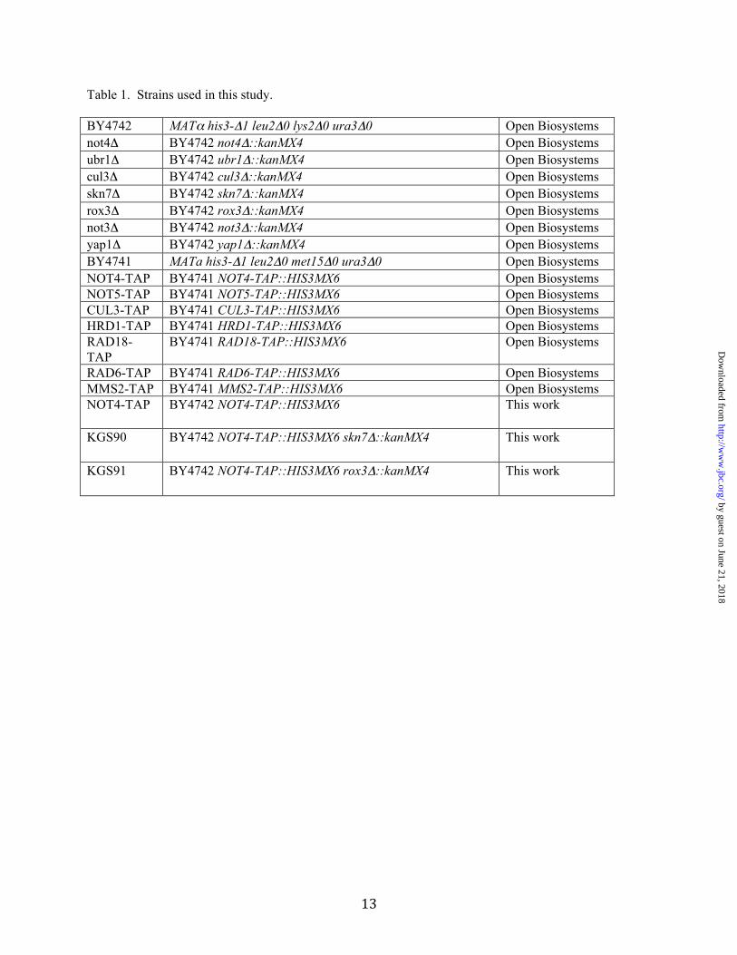
in the nucleus with an accompanying elevation in diamide and DEM resistance. These same constitutively nuclearly-localized Yap1 derivatives fail to support wild-type H2O2 tolerance due to their inability to be properly folded in response to this oxidant. Previous experiments from several labs (6,9,16) found that the steady-state levels of constitutively nuclear Yap1 mutant derivatives were often lower than the wild-type factor. We determined that this reduction was due to an increase in the degradation of Yap1 mutants that were genetically trapped in the nucleus. Examination of the turnover rate of wild-type Yap1 indicated that this protein was strongly destabilized following oxidative stress onset. In this work, we demonstrate that Yap1 is degraded following recruitment to target gene promoters in a manner dependent on the ubiquitin ligase Not4. MATERIALS AND METHODS Yeast strains, plasmids and media. The strains used in this study are listed in Table 1. All strains used in this study were derived from the BY4741 or BY4742 backgrounds. Yeast cells were grown in rich YPD medium (1% yeast extract, 2% peptone, and 2% dextrose) or complete synthetic (SC) medium at 30°C with shaking (17). Yeast transformation was performed using the standard lithium acetate technique (18). Standard β-galactosidase assays were performed for measurements of lacZ expression as described earlier (19). Plasmids have been previously described and include lacZ reporter genes for TRX2 (9), TRP5 (20) and a Yap1 response element placed upstream of CYC1-lacZ (9). Similarly, low-copy-number plasmids expressing either wild-type or C629A mutant forms of Yap1 were produced earlier (9). The pNT13 plasmid producing a green fluorescent protein (GFP)-tagged allele of YAP1 was characterized before (9). Using PCR mutagenesis, a K67A allele of YAP1 was introduced into this same GFP-Yap1-expressing plasmid to ensure that this mutant form of Yap1 was localized to the nucleus in response to oxidative stress. A BY4742 strain containing a not4Δ::kanMX6 allele was re-derived by preparing the not4Δ::kanMX6 allele by PCR and transforming this cassette into fresh BY4742 cells. This newly derived not4Δ::kanMX6 behaved no differently from the the original BY4742 isolate,
by guest on June 21, 2018http://w
ww
.jbc.org/D
ownloaded from

3
reducing any concerns about genetic changes in addition to the loss of NOT4 (data not shown). To generate strains carrying the tandem affinity purification (TAP) tag fusion (21) to NOT4 in skn7Δ and rox3Δ, these strains were transformed with a NOT4-TAP-HIS3MX6 cassette, amplified by using primers NOT4 TAP–For and NOT4 TAP-Rev (Primer details available on request). Genomic DNA prepared from the Open Biosystems NOT4-TAP tag strain was used as template. Cycloheximide chase assay. Cultures were grown to early log phase in rich YPD media or CSM media lacking appropriate amino acid. These cultures (50 ml each) were treated with 1mM H202, 1 mM diethylmaleate, 1mM diamide or left untreated. The cultures were incubated at room temperature for 15 minutes. Cells were quickly washed with water and transferred to fresh media containing 100 µg/ml cycloheximide with or without oxidant as appropriate. For the 0 hr time point, 10 ml of culture was immediately taken out after the addition of cycloheximide. Cultures were further allowed to incubate at 30°C with shaking at 250 rpm. Further samples were collected at indicated time-periods and processed by centrifugation, followed by washing the pellets with 20% TCA and frozen until ready for further steps. The pellets from different time-periods were then resuspended in 200 µl of 12.5% TCA, and lysed with glass beads by vigorous shaking on a Tomy shaker for 20 minutes. The extracts were collected and another 200 µl of 12.5% TCA was added to glass beads and vortexed for 1 min. Extracts were pooled (total volume ~400 µl) and spun down at full speed for 5 min. Pellets were washed with ice-cold acetone and air-dried. 100 µl of 1X Laemmlli buffer (0.0625 M Tris, pH 6.8, 2% SDS, 10% glycerol, 5% β-mercaptoethanol, 0.002% bromophenol blue dye) and 50 µl of 1M tris base were added to the pellets. Samples (40 µl) were then subjected to western blot analysis, using anti-Yap1 antibody (1: 7000 dilution) (22) and anti-rabbit secondary antibody (1: 12000 dilution). Co-Immunoprecipitation. For performing co-immunoprecipitation assays to determine the identity of E3 ligase responsible for degradation of Yap1, spheroplasts were prepared and subjected to oxidant exposure. In brief, 100 ml of cells was
harvested at an OD600 of 1.0 and washed with spheroplast solution I (1 M sorbitol, 10 mM MgCl2, 30 mM dithiothreitol, 100 µg/ml
phenylmethanesulfonyl fluoride, 50 mM K2HPO4), and resuspended in spheroplast solution II (1 M sorbitol, 10 mM MgCl2, 30 mM dithiothreitol, 100 µg/ml phenylmethylsulfonyl fluoride, 50 mM K2HPO4, 25 mM sodium succinate, pH 5.5) containing oxylyticase, and incubated at 30°C for 1 hr on a shaker at 100 rpm. After chilling on ice, cell suspensions were overlaid on a sucrose cushion (20 mM HEPES, 1.2 M sucrose, 0.02%
sodium azide) and pelleted by centrifuging at 4,000 x g for 20 min at 4°C. A portion of the resulting spheroplasts was treated with 1mM H202 for 25 minutes at room temperature, suspended in ICB (lysis) buffer (100 mM potassium acetate, 50 mM KCl, 20 mM PIPES, 200 mM sorbitol, pH 6.8). Protein extracts were clarified by centrifuging lysates at 14,000 rpm for 5 min at 4°C. The lysates were incubated with anti-TAP antibody for 4 h at 4°C on a rotator. Protein A agarose beads, washed and resuspended in the
same buffer, were then added to the lysate and incubated for 2 hours for immunoprecipitation. Finally, the beads were washed, collected by centrifugation and immunoprecipitated proteins
recovered by adding 2x Laemmli buffer (0.125 M Tris, pH 6.8, 4% SDS, 20% glycerol, 10% β-mercaptoethanol, 0.004% bromophenol blue dye). Protein samples were then loaded on 10% polyacrylamide gel and probed with anti-Yap1 antibodies or anti-TAP antibody. RESULTS Rate of Yap1 proteolysis increases after activation. Activation of Yap1-dependent transcriptional regulation occurs primarily due to localization of this transcription factor to the nucleus (See (1-3) for reviews). Several mutants exhibiting constitutive nuclear localization were also observed to accumulate at reduced steady-state levels when evaluated by Western blotting (6,9,16,22). To determine if the decreased accumulation seen in terms of steady-state levels could be explained by enhanced proteolysis, a cycloheximide chase assay was carried out. A strain lacking the Yap1 protein (yap1Δ) was transformed with an empty vector plasmid or the same plasmid expressing with a wild-type or C629A form of Yap1. Conversion of the cysteine at position 629 to alanine led to the resulting
by guest on June 21, 2018http://w
ww
.jbc.org/D
ownloaded from
4
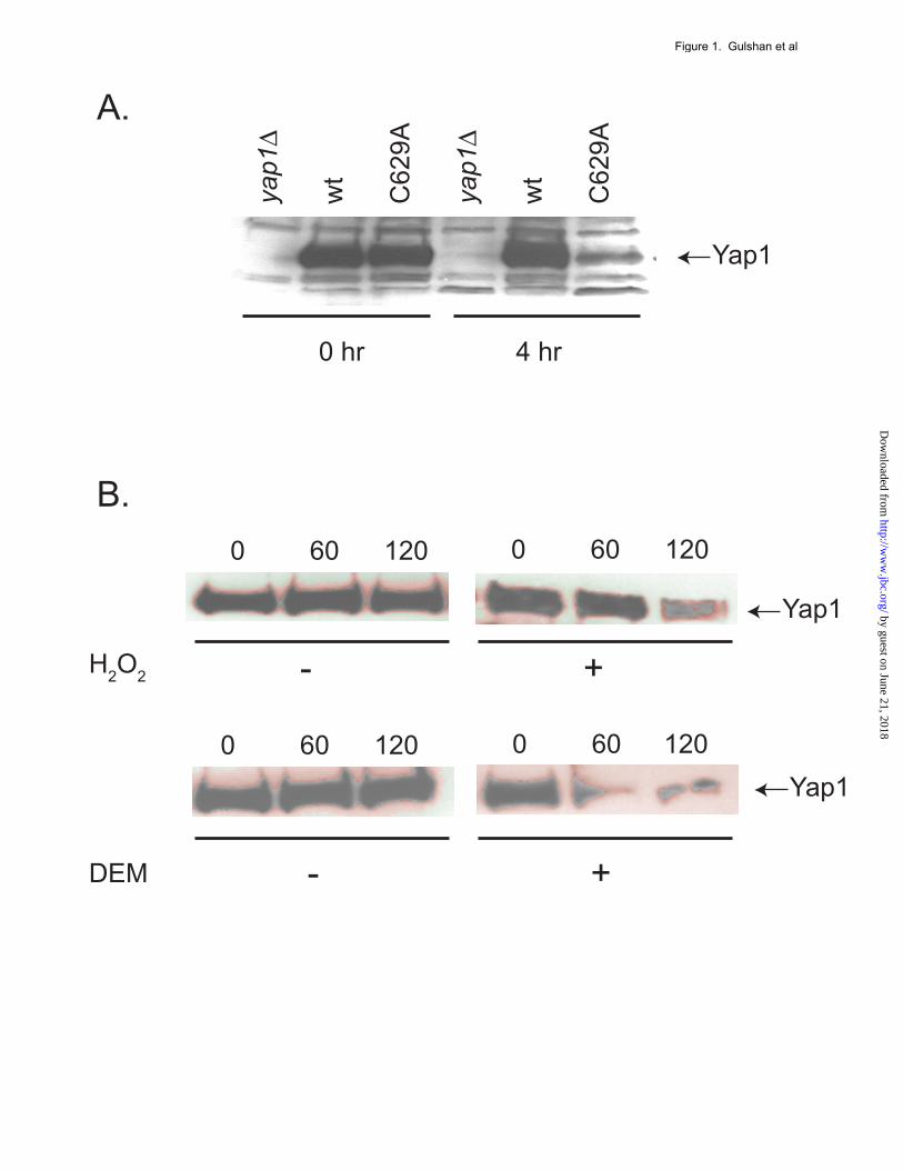
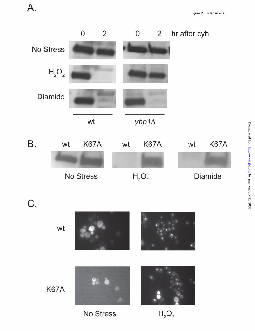
mutant form of Yap1 being constitutively localized to the nucleus, irrespective of oxidant challenge (9). These transformants were grown to mid-log phase (0 hr), 100 µg/ml cycloheximide added and incubation continued for 4 hours. Equal aliquots of cells from these time points were used to prepare whole cell protein extracts that were then analyzed for Yap1 levels using a polyclonal antiserum directed against this transcription factor (22). The C629A form of Yap1 was nearly completely degraded over this time interval (Figure 1A) while wild-type Yap1 did not exhibit significant degradation. A major difference between these two forms of Yap1 occurs at the level of nuclear localization. The wild-type factor resides in the cytoplasm in the absence of oxidative stress but rapidly translocates to the nucleus upon oxidant exposure (6). To determine if the wild-type Yap1 protein exhibited oxidant-triggered instability, wild-type cells were analyzed by the cycloheximide chase protocol described above, either with no stress or with the addition of H2O2 or diethylmaleate (DEM) to induce oxidative stress. Time points were collected and levels of Yap1 assayed by western blotting. The addition of H2O2 caused Yap1 levels to markedly decline within 120 minutes after the onset of oxidative stress (Figure 1B). DEM-induced oxidative stressed induced an even more rapid decline in Yap1 levels with the majority of this transcription factor being eliminated 60 minutes after oxidant exposure. Together, these data suggest that localization of Yap1 to the nucleus leads to degradation of the transcription factor. Nuclear localization and DNA-binding are required for oxidant-induced degradation of Yap1. Previous studies have uncovered unique requirements for Yap1 nuclear translocation in the presence of H2O2. The Yap1-binding protein (Ybp1) is an integral component of Yap1 nuclear localization during H2O2 stress but is dispensable during challenge by oxidants such as DEM or diamide (12,13). Use of a ybp1Δ strain allowed Yap1 to be selectively prevented from entering the nucleus during H2O2 stress but still accumulate in this compartment upon diamide stress. Cycloheximide chase analysis was performed to compare Yap1 degradation in wild-type or isogenic ybp1Δ cells under three different
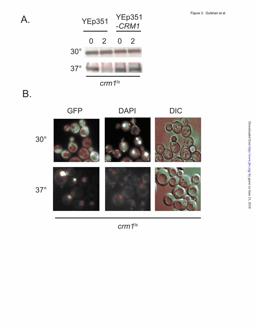
conditions: no stress, H2O2- or diamide-induced oxidative stress (Figure 2A). Loss of Ybp1 completely prevented Yap1 degradation following H2O2 stress but had no effect on the turnover of Yap1 elicited by diamide shock. This finding argues that the presence of H2O2 is not sufficient to cause Yap1 degradation unless the protein also reaches the nucleus. To further explore the basis of oxidant-triggered degradation of Yap1, low-copy-number plasmids expressing either wild-type (wt) or a DNA-binding mutant form (K67A) of Yap1 were introduced into yap1Δ cells. We previously characterized K67A Yap1 as being completely defective in DNA-binding as assessed by an in vitro assay (23). Degradation of wild-type and K67A Yap1 under stressed and nonstressed conditions was evaluated as above. K67A Yap1 was not degraded in response to either H2O2 or diamide stress while the wild-type protein was destabilized by both treatments (Figure 2B). The loss of oxidant-induced instability in the K67A form of Yap1 is consistent with the notion that Yap1 must be able to engage its target promoters in order to exhibit the characteristic degradation during stress This conclusion was supported by analysis of the regulated nuclear localization of the K67A form of Yap1. This mutant protein was normally imported into the nucleus in response to H2O2 treatment (Figure 2C), eliminating any concern that defective nuclear import could explain the observed stability of this mutant Yap1 derivative. The primary determinant of the subcellular localization of Yap1 occurs at the level of nuclear export of this protein. The steady-state distribution of Yap1 is cytoplasmic owing to its relatively slow nuclear import (24) coupled with the rapid nuclear export via the action of the nuclear exportin Crm1 (7,8). A strain containing a temperature-sensitive allele of Crm1 was used to determine if Yap1 was destabilized by trapping the protein in the nucleus. The crm1ts strain was transformed with a low-copy-number plasmid expressing GFP-labeled Yap1 in order to confirm that Yap1 accumulates in the nucleus at the restrictive temperature. In parallel, the crm1ts strain was transformed with a vector plasmid containing or lacking the wild-type CRM1 gene to verify that any effect due to temperature shift in this strain was due to the crm1ts allele.
by guest on June 21, 2018http://w
ww
.jbc.org/D
ownloaded from
5
Appropriate transformants were grown to mid-log phase, then equal fractions maintained at 30° C or shifted to the restrictive temperature of 37° C. After incubation at these temperatures, transformants were analyzed for Yap1 stability by cycloheximide chase or subcellular localization by fluorescence microscopy. Inactivation of Crm1 by growth at the restrictive temperature strongly destabilized Yap1 (Figure 3A). Introduction of wild-type CRM1 into the temperature-sensitive background restored the stability of Yap1. No oxidants were added to these cultures which supports the idea that loss of Yap1 nuclear export is sufficient to cause the protein to be susceptible to proteolysis. GFP-Yap1 was seen to accumulate in the nucleus at the restrictive temperature as expected (Figure 3B). Together, these data suggest that accumulation of a DNA-binding competent form of Yap1 in the nucleus caused by oxidative stress, mutational inactivation of nuclear export (C629A mutation) or by inactivation of Crm1, is sufficient to lead to destabilization of the protein. Evidence for proteasomal degradation of Yap1 and a positive site for oxidant-induced degradation. Analyses of other transcription factors, including the basic region-leucine zipper-containing Gcn4 protein, have demonstrated that the proteasome is the proteolytic system responsible for their degradation (25-27) and the importance of this proteolytic system in control of nuclear gene expression (reviewed in (28)). To determine if the proteasome was similarly required for Yap1 degradation, a cycloheximide chase experiment was carried out in the presence or absence of the proteasome inhibitor MG132. These experiments were performed in a strain lacking the Pdr5 ABC transporter protein as this genetic background has been found to facilitate use of MG132, likely through increasing the uptake of this drug (29). MG132 was added to one culture prior to beginning the cycloheximide chase assay. Oxidative stress was induced with the addition of diethylmaleate and levels of Yap1 assessed as before (Figure 4A). The addition of MG132 produced a profound block in Yap1 degradation after imposition of oxidative stress. This analysis supports the view that the proteasome carries out the proteolytic degradation of Yap1 following oxidant challenge.
Previous studies on Yap1 determined that the steady-state level of deletion derivatives of the protein lacking sequences around the amino-terminal cysteine rich domain (n-CRD) were elevated compared to the wild-type factor (22). The observations above indicate that Yap1 is subject to proteolysis after oxidative stress suggesting that changes in steady-state level of particular mutants might be influenced by changes in the ability of these variants to be degraded. We selected two n-CRD deletion mutants to examine: Δ220-243 and Δ220-335 Yap1. These two n-CRD mutants have very different abilities to function in vivo. The Δ220-243 Yap1 derivative exhibited greater than wild-type resistance to H2O2 and expression of a TRX2-lacZ reporter gene while Δ220-335 Yap1 was unable to correct the H2O2 hypersensitivity or drive normal TRX2 expression in a yap1Δ strain as we documented before (22). Interesting, both of these mutant Yap1 proteins confer greater-than-normal tolerance to diamide or diethylmaleate (22). We focused our attention on the Δ220-243 form of Yap1 since this protein elevated resistance to all tested oxidants. To determine if the increased oxidant resistance conferred by the presence of Δ220-243 Yap1 could be due to a change in its degradation leading to increased accumulation of the protein, we carried out a cycloheximide chase analysis of turnover of this mutant transcription factor. It is important to note that Δ220-243 Yap1 confers both hyper-resistance to H2O2 and also drives very high levels of expression of bona fide Yap1 target genes such as TRX2 (9,14). Δ220-243 Yap1 was very stable following oxidative stress imposed by either H2O2 or diethylmaleate (Figure 4B). This increased stability likely contributes to the observed increment in H2O2 tolerance seen in strains expressing this form of Yap1 (22). Experiments with the Δ220-335 Yap1 also indicated that this form of the transcription factor exhibited enhanced stability after oxidative stress (data not shown). These results suggest that the region around the n-CRD element in Yap1 is an important determinant in regulating the proteolytic turnover of this protein. Identification of ubiquitin proteasomal system components influencing Yap1 activity. Based
by guest on June 21, 2018http://w
ww
.jbc.org/D
ownloaded from
6
on the results described above, we hypothesized that Yap1 degradation would be regulated by ubiquitin-dependent targeting of the protein to the proteasome. To identify proteins that might fulfill this regulatory role, we screened a subset of the collection of haploid disruptant S. cerevisiae mutants selected on the basis of their known role in the ubiqutin proteasomal system (UPS). These UPS mutants were grown to mid-log phase and then tested for the ability to confer resistance to the oxidant H2O2. Since we had already observed that the Δ220-243 Yap1 form of Yap1 exhibited increased stability upon H2O2 challenge (Figure 4), we hypothesized that defects in the machinery responsible for Yap1 degradation would cause a similar phenotype. Mutants were selected based on their previously determined role in the UPS, grown to mid-log phase and then tested for the ability to stabilize Yap1 after cells were treated with H2O2. While a large number of different mutant strains were screened, a summary of these assays will be presented here. Loss of the E3 ubiquitin ligase-encoding gene NOT4 (30)caused a readily detectable increase in stability of Yap1 target gene while loss of other E3 ligases, such as Ubr1 (31) or Cul3 (32), did not exhibit any significant change in Yap1 turnover (Figure 5A). A not3Δ mutant strain was not observed to stabilize Yap1.Work from another group has provided evidence that loss of the ubr1+ gene from Schizosaccharomyces pombe caused increased stability of the Yap1 homologue (Pap1) from this yeast with an associated increase in oxidant resistance (33) but this role does not appear conserved in S. cerevisiae. The UPS has been implicated in degradation of oxidatively damaged proteins previously (reviewed in (34)). To determine if the increased Yap1 stability seen in a not4Δ strain was associated with an accumulation of active Yap1, several reporter gene fusion plasmids were introduced into the strains listed above. A fusion gene corresponding to the thioredoxin-encoding TRX2 promoter fused to E. coli lacZ was used. TRX2 is a well-established target of Yap1 transcriptional regulation and required for normal H2O2 resistance (14). A Yap1 response element (YRE) from the TRX2 promoter fused to a minimal CYC1 promoter-lacZ fusion gene was also tested to examine the specificity of the effect of these mutant backgrounds on Yap1-mediated
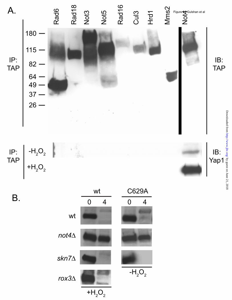
gene activation. Finally, a TRP5-lacZ reporter gene that we have previously demonstrated to be Yap1 independent was used as a control for nonspecific effects on gene expression. These three fusion genes, carried on low-copy-number plasmids, were transformed into isogenic wild-type, not4Δ, ubr1Δ and cul3Δ strains. Transformants were grown to mid-log phase and then left untreated or oxidatively stressed with the inclusion of H2O2 or diamide. Loss of Not4 led to an increase in β-galactosidase from both the YRE-CYC1-lacZ and TRX2-lacZ construct, in the absence and presence of oxidants (Figure 5B). TRP5-lacZ expression was not affected by the presence of the not4Δ allele. This secondary test supports the view that loss of the Not4 E3 enzyme led to an increase in the level of Yap1 with a concomitant elevation in Yap1-mediated transcriptional activation. The UPS is involved in a wide range of different proteolytic events regulating the levels of many different proteins. The impressive spectrum of UPS-regulated proteins makes the possibility that these effects of Not4 on Yap1 might be indirect. To assess the possibility that Not4 directly associates with Yap1, co-immunoprecipitation experiments were carried out. We used a range of different tandem affinity purification (TAP)-tagged strains corresponding to various UPS and Not4-related components. These TAP-tagged strains were grown to mid-log phase, incubated for an additional 25 minutes in the absence or presence of H2O2 and then whole cell protein extracts prepared. TAP-tagged proteins were recovered by immunoprecipitation with anti-TAP antibody as described (35). These immunoprecipitates were electrophoresed through SDS-PAGE and then analyzed by western blotting with anti-TAP or anti-Yap1 antisera (Figure 6A). Yap1 co-precipitated with the ubiquitin ligase Not4. Not4:Yap1 association was stimulated by treatment of the cells with H2O2 prior to carrying out the anti-TAP immunoprecipitation. Interestingly, two other proteins associated with the Ccr-Not complex (Not3 and Not5) did not bind to Yap1 in this assay. Other E3 ligases (Rad18, Cul3, Hrd1) and two different Ubc proteins (Rad6, Mms2) did not co-precipitate Yap1. These data are consistent with Not4 binding to Yap1 and regulating the proteolytic turnover of this transcription factor.
by guest on June 21, 2018http://w
ww
.jbc.org/D
ownloaded from
7
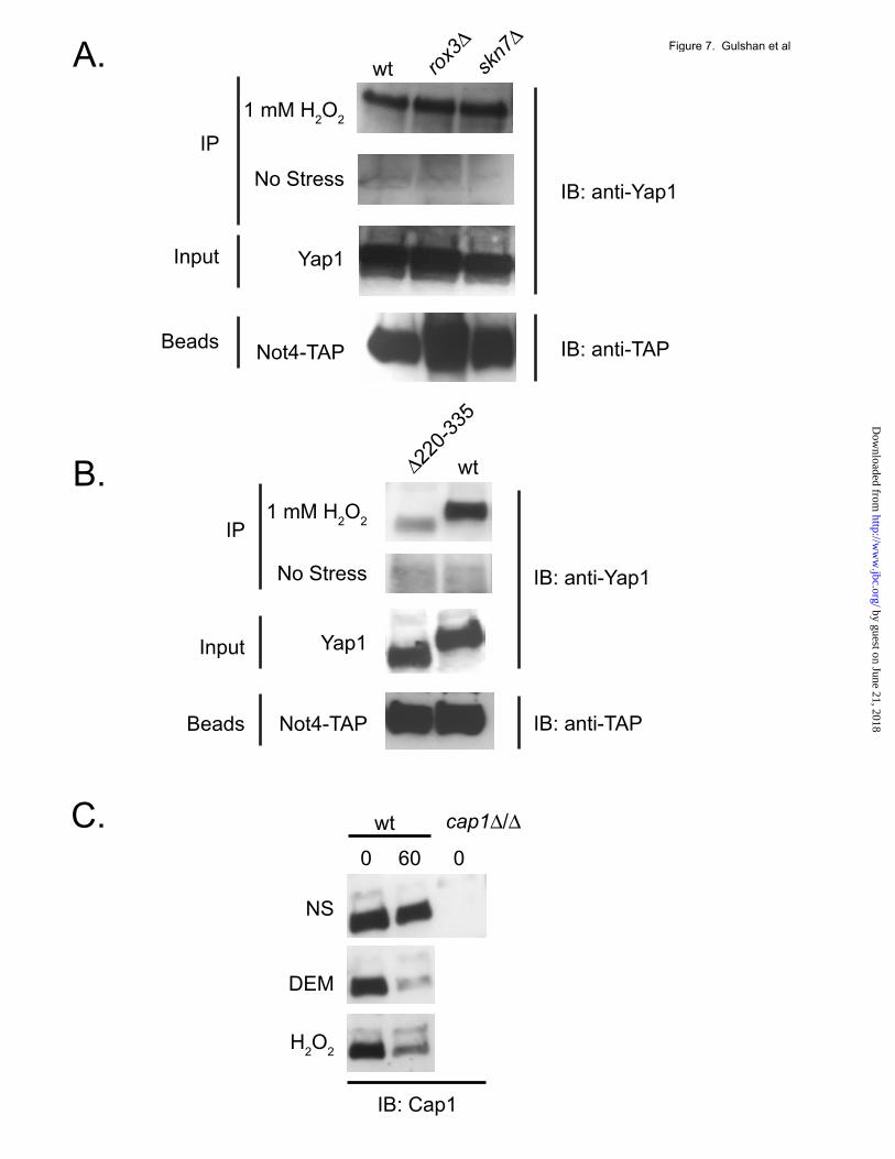
Trans-acting factors and cis-determinants regulating Yap1 degradation. Yap1-mediated transcriptional activation after exposure to H2O2 has previously been shown to require the assistance of two other proteins. The sequence-specific transcription factor Skn7 is an essential contributor both to H2O2 resistance and also to induction of expression of genes like TRX2 that are required for normal H2O2 tolerance (36). Earlier work established that Yap1-dependent recruitment of the transcriptional Mediator component Rox3 occurred only during H2O2-induced folding of this transcription factor and was also required for wild-type resistance to this oxidant (15). The binding experiments above indicated that the ubiquitin ligase Not4 associated with Yap1 in response to H2O2 treatment. Since we have provided data above consistent the view that Yap1 must enter the nucleus prior to its degradation, we tested the contribution of these trans-acting factors to the proteolytic turnover of Yap1 in two different ways. First, we compared a series of isogenic strains lacking Not4, Skn7 or Rox3 for the ability to inducibly degrade Yap1 after challenge with H2O2. Second, a strain expressing the constitutively nuclear C629A Yap1 derivative was tested for its proteolytic degradation in wild-type, not4Δ and skn7Δ strains. As before, we did not treat cells expressing the C629A Yap1 derivative with oxidants. Transformants were analyzed for Yap1 turnover by cycloheximide chase assay as before. Loss of the Not4 ubiquitin ligase led to a profound block in H2O2-triggered turnover of Yap1 (Figure 6B). Similarly, the not4Δ strain also exhibited a strong reduction in the rate of degradation of the constitutively nuclear C629A form of Yap1. Loss of either Skn7 or Rox3 had no significant effect on degradation of wild-type Yap1 after H2O2 stress and a skn7Δ allele did not influence the turnover of C629A Yap1. The interaction of Not4 and Yap1 was also assessed in these mutant backgrounds. A NOT4-TAP allele was integrated into the genome of wild-type, rox3Δ and skn7Δ strains. These strains were then analyzed for H2O2-dependent Not4:Yap1 association as described earlier. Yap1 association with Not4 was unaffected by the absence of these key co-factors required for H2O2-inducible transcription (Figure
7A). This is consistent with the lack of an effect caused by loss of Rox3 or Skn7 on Yap1 turnover seen earlier (Figure 6B). Since we had previously found that deletion derivatives of Yap1 lacking sequence elements around the n-CRD also exhibited increased steady-state protein levels (9,22), the interaction of Not4 with a mutant lacking the entire n-CRD was evaluated using the Not4-TAP association assay. A yap1Δ strain carrying the NOT4-TAP allele was transformed with low-copy-number plasmids expressing either wild-type or Δ220-335 Yap1. The Δ220-335 Yap1 derivative was found to accumulate to higher steady-state levels than wild-type in an earlier study (22). Transformants were grown and then left untreated or challenged with H2O2. The level of Yap1 associated with Not4-TAP was analyzed by co-immunoprecipitation as described above. Loss of the Yap1 region from Δ220-335 reduced the ability of the resulting mutant to associate with Not4 (Figure 7B) by approximately three-fold. This analysis suggests that the region around the n-CRD is critical but not essential for recruitment of Not4 to Yap1 in order to induce the degradation of this transcription factor. Oxidant-induced degradation of the Candida albicans homologue of Yap1. The pathogenic yeast Candida albicans contains a Yap1 homologue called Cap1 (37,38). This transcription factor is inducibly recruited to the nucleus in response to oxidative stress as is Yap1 in S. cerevisiae (39). We produced a polyclonal antibody directed against Cap1 in this earlier study and used this reagent to determine if oxidant exposure also induced Cap1 degradation in C. albicans. Isogenic wild-type and cap1Δ/cap1Δ homozygous strains were grown to mid-log phase, either left unstressed or treated with diethylmaleate or H2O2 followed by incubation in the presence of 100 µg/ml cycloheximide. Equal samples of cells were withdrawn and whole cell protein extracts prepared at the indicated time points. The polyclonal anti-Cap1 antibody was used to assess the levels of Cap1 by western blotting. Treatment of C. albicans with either H2O2 or diethylmaleate induced the degradation of Cap1 (Figure 7C). Cap1 was fairly stable in the absence of oxidative stress as very little degradation was
by guest on June 21, 2018http://w
ww
.jbc.org/D
ownloaded from

8
seen over the 60 minute time period of this experiment. Cap1 is cleared from promoters after oxidative stress in C. albicans in a manner directly analogous to that of Yap1 in S. cerevisiae. DISCUSSION Yap1 is regulated primarily by control of its nuclear localization, which was previously thought to represent a reversible process (40,41). Here we provide the first evidence that the movement of Yap1 into the nucleus, coupled with engagement of a target promoter, represents a one-way trip for this transcription factor. The proteolysis of Yap1 explains previous observations of the diminished expression of this protein when expressed in a constitutively nuclear form (6,9,22). Yap1 derivatives that escape nuclear export by Crm1 bind to YREs and are then subject to proteolysis. Under normal circumstances, ongoing protein synthesis would maintain this pool of Yap1 but when this is blocked (here by the addition of cycloheximide), the rapid degradation of DNA-bound Yap1 can be visualized. Experiments aimed at uncovering the substrates of the ubiquitin ligase Ubr1 in S. pombe have provided evidence that the Pap1 transcription factors is a substrate of this ligase in this fission yeast (33). Given the striking sequence and regulatory conservation between Yap1 and Pap1 (42), we anticipated that S. cerevisiae Ubr1 (ScUbr1) would be involved in controlling Yap1 stability. Surprisingly, we found that ScNot4 rather than ScUbr1 was the ubiquitin ligase linked to control of Yap1 stability in S. cerevisiae. Our determination that ScNot4 can be co-immunoprecipitated with Yap1 (Figure 8) provides data in support of a direct role of ScNot4 in Yap1 degradation. Association of SpUbr1 with Pap1 could not be demonstrated (33), leaving open the possibility that the action of SpUbr1 on Pap1 stability may be indirect. Coupled with these recent data demonstrating the proteolysis of Pap1, our findings that both Yap1 and C. albicans Cap1 are also degraded after oxidative stress suggest that this regulatory feature is likely to be conserved in fungi that express an oxidant-responsive Yap1 homologue. We hypothesize that the regulation of Cap1 will be more similar to that of Yap1 than Pap1 as both S. cerevisiae (12,13) and C. albicans (Janet Quinn, personal communication) express a homologue of the
oxidative folding co-factor Ybp1 while S. pombe does not (data not shown). Experiments are currently underway to determine if the CaNot4 homologue is required for oxidant-induced degradation of Cap1 as is the cognate S. cerevisiae protein. The finding that Yap1 is degraded by proteolysis after it binds to the YRE is consistent with a requirement to ensure that Yap1-mediated gene induction is carefully regulated and rapidly inactivated when no longer necessary. Only oxidatively activated or mutationally altered Yap1 is capable of evading Crm1-dependent export (7,8). Removal of YRE-bound Yap1 by proteolysis requires that appropriately oxidized Yap1 continuously arrives to maintain high level target gene transcription. Once redox conditions have returned to a range in which Yap1 is no longer oxidized, Crm1 export will predominate and promoter-bound Yap1 will be degraded and no longer replenished. The combination of these regulatory mechanisms will ensure tight and rapid control of Yap1-mediated transcriptional activation. The specificity of Yap1 degradation induced by Not4 is tightly linked to the nuclear accumulation and DNA-binding of this transcription factor. Not4 can function in the cytoplasm as the nascent polypeptide complex protein Egd2 gets ubiquitinated by Not4 in cytoplasm and (43) translation arrest products are subject to Not4-dependent turnover (44). The exclusion of Yap1 from the nucleus is unlikely to present an ability to evade Not4. We hypothesize that Yap1 binding to promoter DNA renders the factor sensitive to Not4-dependent degradation. One interesting possibility would be the involvement of the transcriptional Mediator component Cdk8 (Srb10). This cyclin-dependent kinase phosphorylates and triggers the degradation of a number of transcription factors including Gcn4 (27), Ste12 (45), Msn2 (46,47) and Hac1 (48). However, direct examination of Yap1 turnover in cdk8Δ cells failed to detect any alteration in proteolysis (data not shown). Not4 has previously been implicated in regulation of nuclear functions. The DNA polymerase α encoded by CDC17 is degraded in a Not4-dependent manner (49). Strikingly, degradation of the histone demethylase Jhd2 is regulated by Not4 and required to support normal
by guest on June 21, 2018http://w
ww
.jbc.org/D
ownloaded from

9
levels of methylation of histone H3 (50). Although Skn7 is tightly linked to Yap1-dependent oxidative stress transcription and this winged helix transcription factor associates with Not4, we determined that Skn7 degradation was unaffected by loss of Not4 (data not shown).
Previous work from others indicated that Yap1 was the target of proteolysis in a manner regulated by the presence of the calcium-regulated phosphatase calcineurin (51). We tested a cnb1Δ cell and found that loss of calcineurin activity had no influence on Yap1 turnover upon oxidant challenge (data not shown). The cnb1Δ strain also exhibited no increase in oxidant resistance which might be expected if the absence of calcineurin activity caused an increase in Yap1 level. The lack of an effect of calcineurin on Yap1 degradation may be due to the different strain background used here or a selective role for this phosphatase in calcium-triggered degradation.
An important theme illustrated by these findings is the mechanism by which transcription factors are inactivated when their regulatory
influences are no longer required. The S. cerevisiae MATα2 repressor protein is removed from its DNA target site via the Cdc48 AAA ATPase (52). Some transcription factors are thought to cycle on and off their DNA binding sites in a fairly rapid manner that would facilitate their inactivation by the proteasome (53-55). However, given the wide range of transcription factors and differences in their modes of regulation, multiple mechanisms are undoubtedly invoked to modulate their transcriptional influences. Our findings support the view that proteolytic turnover of site-specific transcription factors may be a common avenue used by cells to regulate the function of these important regulatory molecules. Since some transcription factors may exhibit prolonged association with promoter elements beyond the time their respective transcriptional influences are required, active degradation will limit inappropriate gene regulation.
ACKNOWLEDGEMENTS This work was supported in part by GM49825. We thank Rob Piper for helpful discussions. REFERENCES 1. Moye-Rowley, W. S. (2003) Eukaryot Cell 2, 381-389 2. Herrero, E., Ros, J., Belli, G., and Cabiscol, E. (2008) Biochim Biophys Acta 1780, 1217-1235 3. Rodrigues-Pousada, C., Menezes, R. A., and Pimentel, C. (2010) Yeast 27, 245-258 4. Gasch, A. P., Spellman, P. T., Kao, C. M., Carmel-Harel, O., Eisen, M. B., Storz, G., Botstein,
D., and Brown, P. O. (2000) Mol. Biol. Cell 11, 4241-4257 5. Carmel-Harel, O., Stearman, R., Gasch, A. P., Botstein, D., Brown, P. O., and Storz, G. (2001)
Mol Microbiol 39, 595-605 6. Kuge, S., Jones, N., and Nomoto, A. (1997) EMBO J. 16, 1710-1720 7. Kuge, S., Toda, T., Iizuka, N., and Nomoto, A. (1998) Genes to Cells 3, 521-532 8. Yan, C., Lee, L. H., and Davis, L. I. (1998) EMBO J. 17, 7416-7429 9. Coleman, S. T., Epping, E. A., Steggerda, S. M., and Moye-Rowley, W. S. (1999) Mol. Cell.
Biol. 19, 8302-8313 10. Delaunay, A., Isnard, A. D., and Toledano, M. B. (2000) EMBO J. 19, 5157-5166 11. Delaunay, A., Pflieger, D., Barrault, M. B., Vinh, J., and Toledano, M. B. (2002) Cell 111, 471-
481 12. Veal, E. A., Ross, S. J., Malakasi, P., Peacock, E., and Morgan, B. A. (2003) J Biol Chem 278,
30896-30904 13. Gulshan, K., Rovinsky, S. A., and Moye-Rowley, W. S. (2004) Eukaryot Cell 3, 318-330 14. Kuge, S., and Jones, N. (1994) EMBO J. 13, 655-664 15. Gulshan, K., Rovinsky, S. A., Coleman, S. T., and Moye-Rowley, W. S. (2005) J Biol Chem 280,
40524-40533 16. Takeuchi, T., Miyahara, K., Hirata, D., and Miyakawa, T. (1997) FEBS Lett. 416, 339-343
by guest on June 21, 2018http://w
ww
.jbc.org/D
ownloaded from
10
17. Sherman, F., Fink, G., and Hicks, J. (1979) Methods in Yeast Genetics, Cold Spring Harbor Laboratory: Cold Spring Harbor, New York
18. Ito, H., Fukuda, Y., Murata, K., and Kimura, A. (1983) J. Bacteriol. 153, 163-168 19. Guarente, L. (1983) Methods Enzymol. 101, 181-191 20. Katzmann, D. J., Burnett, P. E., Golin, J., Mahe, Y., and Moye-Rowley, W. S. (1994) Mol. Cell.
Biol. 14, 4653-4661 21. Puig, O., Caspary, F., Rigaut, G., Rutz, B., Bouveret, E., Bragado-Nilsson, E., Wilm, M., and
Seraphin, B. (2001) Methods 24, 218-229 22. Wemmie, J. A., Steggerda, S. M., and Moye-Rowley, W. S. (1997) J. Biol. Chem. 272, 7908-
7914 23. Wemmie, J. A., Wu, A.-L., Harshman, K. D., Parker, C. S., and Moye-Rowley, W. S. (1994) J.
Biol. Chem. 269, 14690-14697 24. Isoyama, T., Murayama, A., Nomoto, A., and Kuge, S. (2001) J. Biol. Chem. 276, 21863-21869 25. Kornitzer, D., Raboy, B., Kulka, R. G., and Fink, G. R. (1994) Embo J 13, 6021-6030 26. Meimoun, A., Holtzman, T., Weissman, Z., McBride, H. J., Stillman, D. J., Fink, G. R., and
Kornitzer, D. (2000) Mol Biol Cell 11, 915-927 27. Chi, Y., Huddleston, M. J., Zhang, X., Young, R. A., Annan, R. S., Carr, S. A., and Deshaies, R.
J. (2001) Genes Dev 15, 1078-1092 28. Collins, G. A., and Tansey, W. P. (2006) Curr Opin Genet Dev 16, 197-202 29. Collins, G. A., Gomez, T. A., Deshaies, R. J., and Tansey, W. P. (2010) Yeast 27, 965-974 30. Collart, M. A., and Struhl, K. (1994) Genes Dev 8, 525-537 31. Bartel, B., Wunning, I., and Varshavsky, A. (1990) Embo J 9, 3179-3189 32. Michel, J. J., McCarville, J. F., and Xiong, Y. (2003) J Biol Chem 278, 22828-22837 33. Kitamura, K., Taki, M., Tanaka, N., and Yamashita, I. (2011) Mol Microbiol 80, 739-755 34. Poppek, D., and Grune, T. (2006) Antioxid Redox Signal 8, 173-184 35. Shahi, P., Gulshan, K., Naar, A. M., and Moye-Rowley, W. S. (2010) Mol Biol Cell 21, 2469-
2482 36. Morgan, B. A., Banks, G. R., Toone, W. M., Raitt, D., Kuge, S., and Johnston, L. H. (1997)
EMBO J. 16, 1035-1044 37. Alarco, A. M., Balan, I., Talibi, D., Mainville, N., and Raymond, M. (1997) J. Biol. Chem. 272,
19304-19313 38. Alarco, A. M., and Raymond, M. (1999) J. Bacteriol. 181, 700-708 39. Zhang, X., De Micheli, M., Coleman, S. T., Sanglard, D., and Moye-Rowley, W. S. (2000) Mol.
Microbiol. 36, 618-629 40. Izawa, S., Maeda, K., Sugiyama, K., Mano, J., Inoue, Y., and Kimura, A. (1999) J. Biol. Chem.
274, 28459-28465 41. Kuge, S., Arita, M., Murayama, A., Maeta, K., Izawa, S., Inoue, Y., and Nomoto, A. (2001) Mol.
Cell. Biol. 21, 6139-6150 42. Toda, T., Shimanuki, M., and Yanagida, M. (1991) Genes Dev 5, 60-73 43. Panasenko, O., Landrieux, E., Feuermann, M., Finka, A., Paquet, N., and Collart, M. A. (2006) J
Biol Chem 281, 31389-31398 44. Dimitrova, L. N., Kuroha, K., Tatematsu, T., and Inada, T. (2009) J Biol Chem 284, 10343-10352 45. Nelson, C., Goto, S., Lund, K., Hung, W., and Sadowski, I. (2003) Nature 421, 187-190 46. Lallet, S., Garreau, H., Poisier, C., Boy-Marcotte, E., and Jacquet, M. (2004) Mol Genet
Genomics 272, 353-362 47. Bose, S., Dutko, J. A., and Zitomer, R. S. (2005) Genetics 169, 1215-1226 48. Pal, B., Chan, N. C., Helfenbaum, L., Tan, K., Tansey, W. P., and Gething, M. J. (2007) Mol Biol
Cell 18, 426-440 49. Haworth, J., Alver, R. C., Anderson, M., and Bielinsky, A. K. (2010) Mol Biol Cell 21, 3205-
3219
by guest on June 21, 2018http://w
ww
.jbc.org/D
ownloaded from
11
50. Mersman, D. P., Du, H. N., Fingerman, I. M., South, P. F., and Briggs, S. D. (2009) Genes Dev 23, 951-962
51. Yokoyama, H., Mizunuma, M., Okamoto, M., Yamamoto, J., Hirata, D., and Miyakawa, T. (2006) EMBO Rep 7, 519-524
52. Wilcox, A. J., and Laney, J. D. (2009) Nat Cell Biol 11, 1481-1486 53. Tora, L., and Timmers, H. T. (2010) Trends Biochem Sci 35, 309-314 54. Reid, G., Hubner, M. R., Metivier, R., Brand, H., Denger, S., Manu, D., Beaudouin, J., Ellenberg,
J., and Gannon, F. (2003) Mol Cell 11, 695-707 55. Metivier, R., Penot, G., Hubner, M. R., Reid, G., Brand, H., Kos, M., and Gannon, F. (2003) Cell
115, 751-763 FIGURE LEGENDS Figure 1. Yap1 is degraded after oxidative stress. A. A strain lacking the YAP1 gene (yap1Δ) was transformed with an empty low-copy-number vector (pRS316) or the same vector containing wild-type YAP1 (wt) or the constitutively nuclear form of Yap1 (C629A). Transformants were grown to mid-log phase in SC medium and whole cell protein extracts were prepared after incubation for the indicated times in the presence of 100 µg/ml cycloheximide. Proteins were resolved on SDS-PAGE and the levels of Yap1 detected by the use of an anti-Yap1 polyclonal antiserum (22). The pRS316 transformants are indicated as yap1Δ. B. Wild-type cells were grown in YPD medium to mid-log phase. Aliquots were then incubated an additional 15 minutes with no treatment or with oxidative stress imposed by the addition of 1 mM H2O2 or 1 mM diethylmaleate (DEM). After this incubation, cycloheximide chase was performed as described above followed by western blot analysis of Yap1 levels. Figure 2. Nuclear localization and DNA-binding are required for Yap1 degradation after stress. A. Wild-type (wt) or isogenic ybp1Δ cells were grown in YPD medium and analyzed for Yap1 stability by cycloheximide chase as above. Oxidative stress was imposed by the addition of H2O2 or diamide. B. Low-copy-number plasmids expressing either the wild-type form of Yap1 or a mutant form lacking the ability to bind DNA (K67A) were introduced into a yap1Δ strain. Transformants were grown to mid-log phase and then left untreated or challenged with 1 mM H2O2 or DEM for 15 minutes. Equal aliquots of cells were processed for cycloheximide chase analysis to determine the levels of Yap1 in the cell. C. K67A Yap1 is normally localized to the nucleus upon H2O2 stress. Low-copy-number plasmids expressing GFP-Yap1 (wt) or the GFP-K67A Yap1 were transformed into yap1Δ cells. Transformants were grown to mid-log phase and then H2O2 was added or omitted for 20 minutes. GFP-Yap1 fusion proteins were then visualized by fluorescence microscopy to determine their localization. Figure 3. Blocking Yap1 nuclear export is sufficient to trigger its degradation. A. A strain containing temperature sensitive allele of CRM1 (crm1ts) was transformed with an empty vector (YEp351) or the same plasmid carrying the wild-type CRM1 gene (YEp351-CRM1). Transformants were grown to mid-log phase, then left untreated or shifted to 37° C for 30 min to impose the restrictive temperature. These cells were then used to perform a cycloheximide chase analysis of the levels of Yap1. B. The crm1ts strain was transformed with a low-copy-number plasmid expressing GFP-Yap1 (9). Transformants were labeled with DAPI at 30°C for one hour and then maintained at this temperature or shifted to 37°C for 30 minutes. These cells were visualized by fluorescence to detect GFP-Yap1 (GFP), DNA (DAPI) or Nomarski optics (DIC) Figure 4. Requirements for Yap1 proteolysis. A. A pdr5Δ strain was grown to early log phase. After dividing the culture into two equal aliquots, the proteasomal inhibitor MG132 was added (+) at 100 µM to the indicated sample. Incubation was continued for 3 hours. Then each aliquot was further divided into two and either left untreated or challenged with 1 mM DEM for 15 minutes. After this treatment, the cultures were analyzed for Yap1 turnover by cycloheximide chase assay. B. Transformants expressing the indicated forms of Yap1, along with the empty vector plasmid (denoted as yap1Δ), were grown in SC medium to mid-log phase and then assayed as above by cycloheximide chase analysis for levels of Yap1 remaining after oxidative stress. NS, no stress.
by guest on June 21, 2018http://w
ww
.jbc.org/D
ownloaded from
12
Figure 5. Loss of the ubiquitin ligase-encoding gene NOT4 inhibits Yap1 proteolysis and stimulates Yap1-dependent reporter gene expression. A. Isogenic strains containing the indicated deletion alleles were grown to mid-log phase and H2O2 added for 20 minutes. Cycloheximide was then added to 100 µg/ml and equal aliquots of cells were either removed immediately (0 hours) or incubated for 2 hours. Whole cell protein extracts were prepared and analyzed for Yap1 levels by western blotting with anti-Yap1 antiserum.. B. The strains listed were transformed with low-copy-number plasmids containing gene fusions between E. coli lacZ and a single copy of the TRX2 Yap1 response element (YRE) placed upstream of a truncated CYC1 promoter lacking any other upstream activation sequence, the wild-type TRX2 promoter or the TRP5 promoter. Transformants were grown to mid-log phase, left untreated or challenged with 0.25 mM H2O2 or 0.5 mM diamide for 1 hour. Aliquots of each transformant were collected and processed for measurement of β-galactosidase assay as before (19). Figure 6. Co-immunoprecipitation assay of various ubiquitin proteasomal system and Not4-related components with Yap1. A collection of TAP-tagged alleles of the indicated proteins in an otherwise wild-type S. cerevisiae strain was used to carry out this analysis. These strains were grown to mid-log phase, then either left untreated or stressed with the addition of 1 mM H2O2 for 25 minutes. Whole cell protein extracts were prepared and TAP-tagged proteins recovered by precipitation with anti-TAP antibody. Immunoprecipitates were washed and then subjected to western blotting using anti-TAP and anti-Yap1 antibodies. B. Yap1 turnover is reduced in a not4Δ strain. Left panel: Isogenic wild-type (wt), not4Δ, skn7Δ or rox3Δ strains were grown to early log phase, and then stressed with the addition of 1 mM H2O2 for 15 minutes followed by cycloheximide chase analysis of Yap1 levels. Right panel: The constitutively nuclear form of Yap1 (C629A) was assayed by cycloheximide chase analysis in the absence of H2O2 stress. Figure 7. Regulation of Yap1:Not4 interaction and conserved degradation of C. albicans Yap1 homologue.. A. Isogenic wild-type, rox3Δ and skn7Δ, all containing a NOT4-TAP allele, were grown in rich YPD medium to early log phase. Cells were then left untreated or stressed with 1 mM H2O2 for 25 minutes prior to processing for immunoprecipitation using anti-TAP antibodies. Aliquots of the cell extract (Input) or the washed beads (Beads) were also analyzed as controls. Immunoprecipitates (IP) were eluted from the beads and electrophoresed on SDS-PAGE followed by western blotting (IB) with anti-Yap1 antibody. B. A yap1Δ NOT4-TAP strain was transformed with low-copy-number plasmids expressing either wild-type (wt) or the Δ220-335 form of Yap1. Interaction of these different forms of Yap1 with Not4 was assessed as above. C. Isogenic wild-type C. albicans or homozygous cap1Δ/Δ cells were grown to early log phase in rich YPD medium. The cultures were divided into three and stressed with the addition of 2 mM H2O2 or diethylmaleate (DEM) or left untreated. After 25 minutes, cycloheximide was added to each culture. Equivalent samples were either immediately withdrawn (time 0) or incubated for 1 hour (60). Cells were lysed with TCA, dissolved in 1X Laemmli buffer and run on SDS-PAGE. Cap1 was detected by western blotting with a polyclonal anti-Cap1 antibody (39).
by guest on June 21, 2018http://w
ww
.jbc.org/D
ownloaded from
13
Table 1. Strains used in this study. BY4742 MATα his3-Δ1 leu2Δ0 lys2Δ0 ura3Δ0 Open Biosystems not4Δ BY4742 not4Δ::kanMX4 Open Biosystems ubr1Δ BY4742 ubr1Δ::kanMX4 Open Biosystems cul3Δ BY4742 cul3Δ::kanMX4 Open Biosystems skn7Δ BY4742 skn7Δ::kanMX4 Open Biosystems rox3Δ BY4742 rox3Δ::kanMX4 Open Biosystems not3Δ BY4742 not3Δ::kanMX4 Open Biosystems yap1Δ BY4742 yap1Δ::kanMX4 Open Biosystems BY4741 MATa his3-Δ1 leu2Δ0 met15Δ0 ura3Δ0 Open Biosystems NOT4-TAP BY4741 NOT4-TAP::HIS3MX6 Open Biosystems NOT5-TAP BY4741 NOT5-TAP::HIS3MX6 Open Biosystems CUL3-TAP BY4741 CUL3-TAP::HIS3MX6 Open Biosystems HRD1-TAP BY4741 HRD1-TAP::HIS3MX6 Open Biosystems RAD18-TAP
BY4741 RAD18-TAP::HIS3MX6 Open Biosystems
RAD6-TAP BY4741 RAD6-TAP::HIS3MX6 Open Biosystems MMS2-TAP BY4741 MMS2-TAP::HIS3MX6 Open Biosystems NOT4-TAP BY4742 NOT4-TAP::HIS3MX6
This work
KGS90 BY4742 NOT4-TAP::HIS3MX6 skn7Δ::kanMX4
This work
KGS91 BY4742 NOT4-TAP::HIS3MX6 rox3Δ::kanMX4
This work
by guest on June 21, 2018http://w
ww
.jbc.org/D
ownloaded from
0
0 hr 4 hr
60 120 0 60 120
0 60 120 0 60 120
DEM
wt
C629A
yap1
!
wt
C629A
yap1
!
+
+
H2O2
Yap1
Yap1
Yap1
A.
B.
Figure 1. Gulshan et al
by guest on June 21, 2018http://w
ww
.jbc.org/D
ownloaded from
0
No Stress
hr after cyh
H2O
2
Diamide
B.
C.
A.
No Stress
No Stress
H2O
2
H2O
2
Diamide
wt
wt
wt
K67A
K67A
wt K67A wt K67A
ybp1Δ
2 0 2
Figure 2. Gulshan et al
by guest on June 21, 2018http://w
ww
.jbc.org/D
ownloaded from
0
A.
B.
YEp351
GFP DAPI DIC
30°
37°
30°
37°
2 0 2
YEp351CRM1
crm1ts
crm1ts
Figure 3. Gulshan et al
by guest on June 21, 2018http://w
ww
.jbc.org/D
ownloaded from

MG132A.
B.
DEM
DEMH2O
2NS DEMH
2O
2NS
wt ∆220-243
yap1∆
+-
+-
+-
Figure 4. Gulshan et al
by guest on June 21, 2018http://w
ww
.jbc.org/D
ownloaded from

0
1020
30405060
70
80
90
wt
YRE-CYC1-lacZ TRX2-lacZ TRP5-lacZ
not4∆
ubr1∆cul3∆
wt not4∆yap1∆ ubr1∆ cul3∆ not3∆
NS Diamide
β-G
alac
tosi
dase
Act
ivity
(Uni
ts/O
D60
0)
A.
B.
H2O
2
NS
H2O
2
NS NSDiamideH2O
2
Figure 5. Gulshan et al
by guest on June 21, 2018http://w
ww
.jbc.org/D
ownloaded from
Rad
6
644937
26
IB:Yap1
IB:TAP
IP:TAP
IP:TAP
180
-H2O
2
+H2O
2
115
82R
ad18
Rad
16
Cul
3
Hrd
1
Mm
s2
Not
3
Not
4
Not
5
wt
+H2O
2
0 4
wt
not4∆
skn7∆
rox3∆
C629A
-H2O
2
0 4
A.
B.
Figure 6. Gulshan et al
by guest on June 21, 2018http://w
ww
.jbc.org/D
ownloaded from
IB: anti-Yap1
IB: anti-TAP
IB: anti-Yap1
IB: anti-TAP
1 mM H2O
2
No Stress
Yap1
Not4-TAP
IP
A.
B.
C.
wt
wt∆220-
335
rox3
∆sk
n7∆
Beads
Input
Beads
Input
1 mM H2O
2
No Stress
Yap1
Not4-TAP
IP
cap1∆/∆wt
NS
IB: Cap1
DEM
H2O
2
0 060
Figure 7. Gulshan et al
by guest on June 21, 2018http://w
ww
.jbc.org/D
ownloaded from

Kailash Gulshan, Bernice Thommandru and W. Scott Moye-Rowleylocalization and the E3 ubiquitin ligase Not4
Proteolytic degradation of the Yap1 transcription factor is regulated by subcellular
published online June 15, 2012J. Biol. Chem.
10.1074/jbc.M112.384719Access the most updated version of this article at doi:
Alerts:
When a correction for this article is posted•
When this article is cited•
to choose from all of JBC's e-mail alertsClick here
by guest on June 21, 2018http://w
ww
.jbc.org/D
ownloaded from
Related Documents