Visit the National Academies Press online, the authoritative source for all books from the National Academy of Sciences , the National Academy of Engineering , the Institute of Medicine , and the National Research Council : • Download hundreds of free books in PDF • Read thousands of books online for free • Explore our innovative research tools – try the “Research Dashboard ” now! • Sign up to be notified when new books are published • Purchase printed books and selected PDF files Thank you for downloading this PDF. If you have comments, questions or just want more information about the books published by the National Academies Press, you may contact our customer service department toll- free at 888-624-8373, visit us online , or send an email to [email protected] . This book plus thousands more are available at http://www.nap.edu . Copyright © National Academy of Sciences. All rights reserved. Unless otherwise indicated, all materials in this PDF File are copyrighted by the National Academy of Sciences. Distribution, posting, or copying is strictly prohibited without written permission of the National Academies Press. Request reprint permission for this book . ISBN: 0-309-65706-7, 238 pages, 8 1/2 x 11, (2006) This PDF is available from the National Academies Press at: http://www.nap.edu/catalog/11765.html http://www.nap.edu/catalog/11765.html We ship printed books within 1 business day; personal PDFs are available immediately. Gulf War and Health: Volume 5. Infectious Diseases Abigail E. Mitchell, Laura B. Sivitz, Robert E. Black, Editors, Committee on Gulf War and Health: Infectious Diseases

Welcome message from author
This document is posted to help you gain knowledge. Please leave a comment to let me know what you think about it! Share it to your friends and learn new things together.
Transcript

Visit the National Academies Press online, the authoritative source for all books from the National Academy of Sciences, the National Academy of Engineering, the Institute of Medicine, and the National Research Council: • Download hundreds of free books in PDF • Read thousands of books online for free • Explore our innovative research tools – try the “Research Dashboard” now! • Sign up to be notified when new books are published • Purchase printed books and selected PDF files
Thank you for downloading this PDF. If you have comments, questions or just want more information about the books published by the National Academies Press, you may contact our customer service department toll-free at 888-624-8373, visit us online, or send an email to [email protected]. This book plus thousands more are available at http://www.nap.edu. Copyright © National Academy of Sciences. All rights reserved. Unless otherwise indicated, all materials in this PDF File are copyrighted by the National Academy of Sciences. Distribution, posting, or copying is strictly prohibited without written permission of the National Academies Press. Request reprint permission for this book.
ISBN: 0-309-65706-7, 238 pages, 8 1/2 x 11, (2006)
This PDF is available from the National Academies Press at:http://www.nap.edu/catalog/11765.html
http://www.nap.edu/catalog/11765.html
We ship printed books within 1 business day; personal PDFs are available immediately.
Gulf War and Health: Volume 5. Infectious Diseases
Abigail E. Mitchell, Laura B. Sivitz, Robert E. Black, Editors, Committee on Gulf War and Health: Infectious Diseases
Abigail E. Mitchell, Laura B. Sivitz, Robert E. Black, Editors
Committee on Gulf War and Health: Infectious Diseases
Board on Population Health and Public Health Practice
Copyright © National Academy of Sciences. All rights reserved.
Gulf War and Health: Volume 5. Infectious Diseaseshttp://www.nap.edu/catalog/11765.html
THE NATIONAL ACADEMIES PRESS 500 Fifth Street, NW Washington, DC 20001
NOTICE: The project that is the subject of this report was approved by the Governing Board of the National Research Council, whose members are drawn from the councils of the National Academy of Sciences, the National Academy of Engineering, and the Institute of Medicine. The members of the committee responsible for the report were chosen for their special competences and with regard for appropriate balance.
This study was supported by Contract V101(93)P-2155 between the National Academy of Sciences and the Department of Veterans Affairs. Any opinions, findings, conclusions, or recommendations expressed in this publication are those of the author(s) and do not necessarily reflect the view of the organizations or agencies that provided support for this project.
International Standard Book Number-10: 0-309-10106-9 (Book) International Standard Book Number-13: 978-0-309-10106-6 (Book) International Standard Book Number-10: 0-309-65706-7 (PDF) International Standard Book Number-13: 978-0-309-65706-8 (PDF)
Library of Congress Control Number: 2006934962
Additional copies of this report are available from the National Academies Press, 500 Fifth Street, NW, Lockbox 285, Washington, DC 20055; (800) 624-6242 or (202) 334-3313 (in the Washington metropolitan area); Internet, http://www.nap.edu.
For more information about the Institute of Medicine, visit the IOM home page at www.iom.edu.
Copyright 2007 by the National Academy of Sciences. All rights reserved.
Printed in the United States of America.
The serpent has been a symbol of long life, healing, and knowledge among almost all cultures and religions since the beginning of recorded history. The serpent adopted as a logotype by the Institute of Medicine is a relief carving from ancient Greece, now held by the Staatliche Museen in Berlin.
Copyright © National Academy of Sciences. All rights reserved.
Gulf War and Health: Volume 5. Infectious Diseaseshttp://www.nap.edu/catalog/11765.html
Copyright © National Academy of Sciences. All rights reserved.
Gulf War and Health: Volume 5. Infectious Diseaseshttp://www.nap.edu/catalog/11765.html
The National Academy of Sciences is a private, nonprofit, self-perpetuating society of distinguished scholars engaged in scientific and engineering research, dedicated to the furtherance of science and technology and to their use for the general welfare. Upon the authority of the charter granted to it by the Congress in 1863, the Academy has a mandate that requires it to advise the federal government on scientific and technical matters. Dr. Ralph J. Cicerone is president of the National Academy of Sciences.
The National Academy of Engineering was established in 1964, under the charter of the National Academy of Sciences, as a parallel organization of outstanding engineers. It is autonomous in its administration and in the selection of its members, sharing with the National Academy of Sciences the responsibility for advising the federal government. The National Academy of Engineering also sponsors engineering programs aimed at meeting national needs, encourages education and research, and recognizes the superior achievements of engineers. Dr. Wm. A. Wulf is president of the National Academy of Engineering.
The Institute of Medicine was established in 1970 by the National Academy of Sciences to secure the services of eminent members of appropriate professions in the examination of policy matters pertaining to the health of the public. The Institute acts under the responsibility given to the National Academy of Sciences by its congressional charter to be an adviser to the federal government and, upon its own initiative, to identify issues of medical care, research, and education. Dr. Harvey V. Fineberg is president of the Institute of Medicine.
The National Research Council was organized by the National Academy of Sciences in 1916 to associate the broad community of science and technology with the Academy’s purposes of furthering knowledge and advising the federal government. Functioning in accordance with general policies determined by the Academy, the Council has become the principal operating agency of both the National Academy of Sciences and the National Academy of Engineering in providing services to the government, the public, and the scientific and engineering communities. The Council is administered jointly by both Academies and the Institute of Medicine. Dr. Ralph J. Cicerone and Dr. Wm. A. Wulf are chair and vice chair, respectively, of the National Research Council.
www.national-academies.org
.
Copyright © National Academy of Sciences. All rights reserved.
Gulf War and Health: Volume 5. Infectious Diseaseshttp://www.nap.edu/catalog/11765.html
v
COMMITTEE ON GULF WAR AND HEALTH: INFECTIOUS DISEASES
ROBERT E. BLACK, MD, MPH, Edgar Berman Professor and Chair, Department of International Health, Johns Hopkins University, Bloomberg School of Public Health, Baltimore, MD
MARTIN J. BLASER, MD, Frederick H. King Professor of Internal Medicine, Chair of the Department of Medicine, and Professor of Microbiology, New York University School of Medicine, New York
RICHARD D. CLOVER, MD, Dean and Professor, School of Public Health and Information Sciences, University of Louisville, KY
MYRON S. COHEN, MD, J. Herbert Bate Distinguished Professor of Medicine and Microbiology, Immunology and Public Health, University of North Carolina School of Medicine, Chapel Hill
JERROLD J. ELLNER, MD, Professor and Chair of the New Jersey Medical School at the University of Medicine and Dentistry of New Jersey, Newark
JEANNE MARRAZZO, MD, MPH, Associate Professor, Department of Medicine, University of Washington School of Medicine, Seattle
MEGAN MURRAY, MD, ScD, MPH, Assistant Professor of Epidemiology, Harvard University, School of Public Health, Boston, MA
EDWARD C. OLDFIELD III, MD, Director, Division of Infectious Diseases, Eastern Virginia Medical School, Norfolk
RANDALL R. REVES, MD, MSc, Professor, Division of Infectious Diseases, University of Colorado Health Sciences Center, Denver
EDWARD T. RYAN, MD, Director, Tropical and Geographic Medicine Center, Massachusetts General Hospital, and Associate Professor of Medicine, Harvard Medical School, Boston, MA
STEN H. VERMUND, MD, PhD, Amos Christie Chair and Director, Vanderbilt University Institute for Global Health, and Professor of Pediatrics, Medicine, Preventive Medicine, and Obstetrics and Gynecology, Vanderbilt University School of Medicine, Nashville, TN
DAWN M. WESSON, PhD, Associate Professor, Tulane School of Public Health and Tropical Medicine, New Orleans, LA
Copyright © National Academy of Sciences. All rights reserved.
Gulf War and Health: Volume 5. Infectious Diseaseshttp://www.nap.edu/catalog/11765.html
vi
STAFF
ABIGAIL E. MITCHELL, PhD, Senior Program Officer LAURA B. SIVITZ, MSJ, Senior Program Associate DEEPALI M. PATEL, Senior Program Associate MICHAEL J. SCHNEIDER, MPH, Senior Program Associate PETER JAMES, Research Associate DAMIKA WEBB, Research Assistant DAVID J. TOLLERUD, Program Assistant RENEE WLODARCZYK, Program Assistant NORMAN GROSSBLATT, Senior Editor ROSE MARIE MARTINEZ, ScD, Director, Board on Population Health and Public Health
Practice
Copyright © National Academy of Sciences. All rights reserved.
Gulf War and Health: Volume 5. Infectious Diseaseshttp://www.nap.edu/catalog/11765.html
vii
REVIEWERS
This report has been reviewed in draft form by persons chosen for their diverse perspectives and technical expertise in accordance with procedures approved by the National Research Council’s Report Review Committee. The purpose of this independent review is to provide candid and critical comments that will assist the institution in making its published report as sound as possible and to ensure that the report meets institutional standards of objectivity, evidence, and responsiveness to the study charge. The review comments and draft manuscript remain confidential to protect the integrity of the deliberative process. We wish to thank the following for their review of this report:
Lawrence R. Ash, Professor Emeritus, Department of Epidemiology, University of California, Los Angeles School of Public Health Michele Barry, Tropical Medicine and International Health Programs, Yale University School of Medicine Herbert DuPont, School of Public Health, University of Texas Health Science Center at Houston and St. Luke’s Episcopal Hospital Robert Edelman, Travelers’ Health Clinic, University of Maryland David Hill, National Travel Health Network and Centre, Hospital for Tropical Diseases, London Richard T. Johnson, Department of Neurology, The Johns Hopkins Hospital Arthur Reingold, Division of Epidemiology, University of California, Berkeley Philip K. Russell, Professor Emeritus, Johns Hopkins School of Public Health Mark Wallace, Independent Infectious Diseases Consultant and United States Navy, Retired
Although the reviewers listed above have provided many constructive comments and suggestions, they were not asked to endorse the conclusions or recommendations nor did they see the final draft of the report before its release. The review of this report was overseen by George Rutherford, Institute of Global Health, University of California, San Francisco, and Elaine L. Larson, School of Nursing, Columbia University. Appointed by the National Research Council, they were responsible for making certain that an independent examination of this report was carried out in accordance with institutional procedures and that all review comments were carefully considered. Responsibility for the final content of this report rests entirely with the authoring committee and the institution.
Copyright © National Academy of Sciences. All rights reserved.
Gulf War and Health: Volume 5. Infectious Diseaseshttp://www.nap.edu/catalog/11765.html

Copyright © National Academy of Sciences. All rights reserved.
Gulf War and Health: Volume 5. Infectious Diseaseshttp://www.nap.edu/catalog/11765.html
ix
PREFACE
Infectious diseases have been a problem for military personnel throughout history. The consequences in previous conflicts have ranged from frequent illnesses disrupting daily activities and readiness to widespread deaths. Preventive measures, early diagnosis, and treatment greatly limit the exposures and acute illnesses of troops today in comparison with those in armies of the past, but infections and consequent acute illnesses still occur. In addition, long-term adverse health outcomes of some pathogens are increasingly recognized.
The deployment of about 700,000 US troops to the Persian Gulf region in the Gulf War of 1991 potentially exposed them to pathogens that they had not encountered at home. After returning from that short campaign, some veterans reported symptoms and expressed the concern that they may have been exposed to biologic, chemical, or physical agents during their service in the Persian Gulf. In response to those concerns, the US Department of Veterans Affairs (VA) commissioned the Institute of Medicine (IOM) to review the scientific evidence on possible long-term adverse health outcomes of exposure to specific biologic, chemical, and physical agents and to draw conclusions on the strength of that evidence with regard to delayed and chronic illnesses of the veterans.
The authorizing legislation for the work of IOM included several infectious diseases endemic in the Persian Gulf region. In the charge to our committee, VA asked that we not limit consideration to those diseases but rather include all infectious exposures that had been documented in troops and consider their possible long-term adverse health outcomes. It further requested that the time and geographic dimensions of the committee’s work be widened to include military personnel deployed as part of Operation Enduring Freedom (OEF) in Afghanistan and Operation Iraqi Freedom (OIF) in the Persian Gulf region. OEF began in 2001, and OIF in 2003; they continued as this report went to press. The number of military personnel involved in the more recent conflicts now exceeds that in the 1991 Gulf War. Furthermore, they have remained for much longer periods on the average than in the Gulf War, and many have been deployed for more than one tour in this region. Thus, the potential for exposure to endemic pathogens is greater in these troops than in those deployed to the Gulf War. Because the possible exposures are relatively recent, there has been only a short time to observe long-term adverse health outcomes. The committee needed to rely on observations from the Gulf War, information on infectious diseases in OEF and OIF, and evidence in the scientific literature to allow conclusions to be drawn on possible long-term adverse health outcomes. With further time to observe the possible consequences of infectious exposures, the knowledge base will increase. Given the continuing presence of troops in the areas and the variable nature of infectious diseases, the exposures may change.
Valuable contributions were made to this study by a number of people who shared their expertise on infectious diseases. On behalf of the committee, I thank several of them—K. Craig Hyams, MD, MPH, chief consultant, Occupational and Environmental Health Strategic Healthcare Group, VA; Michael Kilpatrick, MD, deputy director, Deployment Health Support, Department of Defense (DOD); and Alan Magill, MD, science director, Walter Reed Army Institute of Research, for presenting information on infectious diseases that have been diagnosed in military personnel during the Gulf War, OIF, and OEF and Richard Reithinger, PhD,
Copyright © National Academy of Sciences. All rights reserved.
Gulf War and Health: Volume 5. Infectious Diseaseshttp://www.nap.edu/catalog/11765.html
x PREFACE
infectious diseases consultant, for presenting information on infectious diseases that are endemic in southwest and south-central Asia to the committee at its May 26, 2005 meeting. I also thank William Winkenwerder, Jr., MD, MBA, assistant secretary for defense for health affairs, and his staff at DOD’s Deployment Health Support for expeditiously providing information to the committee on DOD health-related policies. Finally, the committee is grateful for the insight provided by representatives of veteran service organizations, veterans, and others who spoke with the committee or sent in written testimony.
I am grateful for the great expertise the committee members brought to bear on this subject. Furthermore, the report would not have been successfully completed without the diligent and expert contributions of the IOM staff, led by Abigail Mitchell and including Laura Sivitz, Deepali Patel, Michael Schneider, Peter James, Damika Webb, David Tollerud, and Renee Wlodarczyk.
Robert E. Black, MD, MPH, Chair
Copyright © National Academy of Sciences. All rights reserved.
Gulf War and Health: Volume 5. Infectious Diseaseshttp://www.nap.edu/catalog/11765.html
xi
CONTENTS
Summary ....................................................................................................................................1
Methodology .........................................................................................................................1 Identifying the Pathogens to Study.....................................................................................2 Development of Conclusions..............................................................................................3
Summary of Conclusions ......................................................................................................4 Sufficient Evidence of a Causal Relationship ....................................................................4 Sufficient Evidence of an Association................................................................................5 Limited or Suggestive Evidence of an Association............................................................6 Inadequate or Insufficient Evidence to Determine Whether an Association Exists ...........6 Limited or Suggestive Evidence of No Association...........................................................7
Department of Defense Policies on Tuberculin Skin Testing and Predeployment and Postdeployment Serum Collection ...................................................................................7
1 Introduction...........................................................................................................................9
Identifying the Infectious Diseases to Study.......................................................................13 The Committee’s Approach to Its Charge ..........................................................................15 Organization of the Report..................................................................................................16 References ...........................................................................................................................16
2 Methodology.......................................................................................................................19
Identifying the Infectious Diseases to Study.......................................................................19 Geographic Boundaries ....................................................................................................19 Infectious Diseases Endemic to Southwest and South-Central Asia
That Have Long-Term Adverse Health Outcomes .....................................................20 Direct Attribution to Military Service in Southwest and South-Central Asia ..................24 Timing of Appearance of Long-Term Adverse Health Outcomes ...................................27 The Infectious Diseases to Be Studied for Strength of Association
with Long-Term Adverse Health Outcomes...............................................................27 Comments on Diseases and Agents of Special Interest
to Gulf War, OEF, and OIF Veterans ..........................................................................28 Review and Evaluation of the Literature ............................................................................29
Selection of the Literature ................................................................................................29 Amassing the Literature....................................................................................................29 Reviewing the Literature ..................................................................................................29
Categories of Strength of Association.................................................................................30 Origin and Evolution of the Categories ............................................................................30 Sufficient Evidence of a Causal Relationship ..................................................................30 Sufficient Evidence of an Association..............................................................................31 Limited or Suggestive Evidence of an Association..........................................................31 Inadequate or Insufficient Evidence to Determine Whether an Association Exists .........31
Copyright © National Academy of Sciences. All rights reserved.
Gulf War and Health: Volume 5. Infectious Diseaseshttp://www.nap.edu/catalog/11765.html
xii CONTENTS
Limited or Suggestive Evidence of No Association.........................................................31 References.........................................................................................................................31
3 Infectious Diseases Endemic to Southwest and South-Central Asia That Have Long-Term Adverse Health Outcomes .............................................................35
References ...........................................................................................................................60
4 Infectious Diseases Diagnosed in US Troops Who Served in the Persian Gulf War, Operation Enduring Freedom, or Operation Iraqi Freedom................................................61
Diarrheal Disease ................................................................................................................62 Enteric Infections in the Gulf War....................................................................................62 Gastroenteritis in Operation Enduring Freedom and Operation Iraqi Freedom...............69
Respiratory Disease.............................................................................................................74 Mild Acute Respiratory Disease in the Gulf War.............................................................74 Severe Acute Respiratory Disease in the Gulf War..........................................................76 Respiratory Disease in Operation Enduring Freedom and Operation Iraqi Freedom ......76
Insect-Borne Diseases .........................................................................................................78 Leishmaniasis ...................................................................................................................78 Malaria..............................................................................................................................82 West Nile Fever ................................................................................................................84
Brucellosis...........................................................................................................................84 Chicken Pox (Varicella)......................................................................................................85 Meningococcal Disease.......................................................................................................85 Nosocomial Infections ........................................................................................................85
Gulf War ...........................................................................................................................85 Operation Enduring Freedom and Operation Iraqi Freedom............................................86
Q Fever................................................................................................................................88 Q Fever Contracted During the Gulf War ........................................................................89 Q Fever Contracted During Operation Enduring Freedom
and Operation Iraqi Freedom.......................................................................................89 Viral Hepatitis .....................................................................................................................90 Tuberculosis ........................................................................................................................90 Department of Defense Medical Databases ........................................................................91 Department of Defense Policy Regarding Predeployment
and Postdeployment Serum Collection ..........................................................................93 References ...........................................................................................................................94
5 Levels of Association Between Select Diseases and Long-Term Adverse Health Outcomes .............................................................................101
Diarrheal Diseases: Campylobacter, Non-typhoid Salmonella, and Shigella Infections .............................103
Campylobacter Infection ................................................................................................103 Nontyphoidal Salmonella Infection................................................................................108 Shigella Infection............................................................................................................110
Copyright © National Academy of Sciences. All rights reserved.
Gulf War and Health: Volume 5. Infectious Diseaseshttp://www.nap.edu/catalog/11765.html
CONTENTS xiii
Brucellosis.........................................................................................................................112 Transmission and Endemicity of Brucellosis .................................................................113 Acute Brucellosis............................................................................................................114 Treatments for Brucellosis and Related Long-Term Toxicity........................................115 Coinfection .....................................................................................................................115 Long-Term Adverse Health Outcomes of Brucellosis ...................................................115
Leishmaniasis....................................................................................................................118 Transmission of Leishmaniasis.......................................................................................119 Endemicity in Southwest and South-Central Asia..........................................................120 Acute Leishmaniasis.......................................................................................................120 Diagnosis of Leishmaniasis ............................................................................................121 Treatments for Leishmaniasis and Related Long-Term Toxicity...................................121 Coinfection by Leishmania Parasite and Human Immunodeficiency Virus ..................122 Long-Term Adverse Health Outcomes of Leishmaniasis ..............................................122
Malaria ..............................................................................................................................123 Transmission of Malaria .................................................................................................124 Endemicity in Southwest and South-Central Asia..........................................................124 Acute Malaria .................................................................................................................125 Treatments for Malaria and Related Long-Term Toxicity .............................................125 Coinfection with Plasmodium Spp. and Human Immunodeficiency Virus ...................126 Long-Term Adverse Health Outcomes of Infection with Plasmodium Spp...................126
Q Fever (Infection by Coxiella burnetii) ..........................................................................129 Transmission of Coxiella burnetii ..................................................................................129 Endemicity in Southwest and South-Central Asia..........................................................130 Acute Q Fever.................................................................................................................130 Diagnosing Q Fever........................................................................................................131 Coinfection with Coxiella burnetii and Human Immunodeficiency Virus ....................131 Long-Term Adverse Health Outcomes of Q Fever ........................................................132
Tuberculosis ......................................................................................................................135 Transmission of Tuberculosis.........................................................................................135 Endemicity in Southwest and South-Central Asia..........................................................137 Risk of Progression from Latent Tuberculosis Infection to Active Tuberculosis ..........137 Treatment for Latent Tuberculosis Infection to Prevent Active Tuberculosis ...............140 Active Tuberculosis ........................................................................................................140 Late Manifestations of Active Tuberculosis...................................................................142 Potential Relationships Between Tuberculosis and Military Service.............................144
West Nile Virus Infection .................................................................................................149 Transmission of West Nile Virus Infection ....................................................................150 Endemicity in Southwest and South-Central Asia..........................................................150 Acute West Nile Fever....................................................................................................151 Diagnosis of West Nile Fever.........................................................................................151 Treatment of West Nile Virus Infection .........................................................................152 Long-Term Adverse Health Outcomes of Infection with West Nile Virus....................152 Recommendation ............................................................................................................155
References .........................................................................................................................155
Copyright © National Academy of Sciences. All rights reserved.
Gulf War and Health: Volume 5. Infectious Diseaseshttp://www.nap.edu/catalog/11765.html
xiv CONTENTS
6 Diseases and Agents of Special Concern to Veterans of the Gulf War, Operation Iraqi Freedom, and Operation Enduring Freedom.....................................................................181
Al Eskan Disease ..............................................................................................................181 Description of Acute Illness ...........................................................................................182 Long-Term Adverse Health Outcomes...........................................................................182 Pathogenesis ...................................................................................................................182 Treatment........................................................................................................................183 Summary.........................................................................................................................183
Idiopathic Acute Eosinophilic Pneumonia........................................................................183 Description of Acute Illness ...........................................................................................183 Long-Term Adverse Health Outcomes...........................................................................183 Pathogenesis ...................................................................................................................184 Treatment........................................................................................................................184 Summary.........................................................................................................................184
Wound and Nosocomial Infections (Including Infections with Acinetobacter Spp.) ......184 Concerns Regarding Acinetobacter baumannii ..............................................................185 Other Wound Infections .................................................................................................186 Other Nosocomial Infections..........................................................................................187 Regional Experiences in Non-Americans.......................................................................188 Summary.........................................................................................................................190
Mycoplasmas ....................................................................................................................190 Mycoplasmas and “Gulf War Illness” ............................................................................191 Summary.........................................................................................................................193
Biologic-Warfare Agents ..................................................................................................193 Summary ...........................................................................................................................194 References .........................................................................................................................194
Appendix Biographical Sketches for Members of the Committee .......................................201
Index ......................................................................................................................................205
Copyright © National Academy of Sciences. All rights reserved.
Gulf War and Health: Volume 5. Infectious Diseaseshttp://www.nap.edu/catalog/11765.html
1
SUMMARY
Thousands of US veterans of the Persian Gulf War have reported an array of unexplained illnesses since the war ended in 1991. Many veterans have believed that the illnesses were associated with their military service in southwest Asia during the war. In response, the US Congress legislated in 1998 that the Department of Veterans Affairs (VA) use a specific procedure to determine the illnesses that warrant presumption of a connection to Gulf War service (Public Law [PL] 105-277, Persian Gulf War Veterans Act). Moreover, VA must financially compensate Gulf War veterans in whom the determined illnesses are diagnosed (PL 105-368, Veterans Programs Enhancement Act). To reach those determinations, the law states, VA must obtain independent evaluations of the scientific evidence of associations between illnesses and exposures to various chemical, physical, and biologic substances connected to military service in southwest Asia during the war. The law instructs VA to obtain the scientific evaluations from the National Academy of Sciences (NAS). NAS assigned the task of evaluating the associations to the Institute of Medicine (IOM).
This report is the fifth volume produced by IOM for VA in response to the congressional mandate.1 A committee of nationally recognized experts in infectious diseases was appointed and charged with evaluating the scientific and medical literature on long-term adverse human health outcomes associated with selected infectious diseases pertinent to Gulf War veterans. The conclusions herein characterize the long-term adverse health outcomes associated with infection by the following pathogens: Brucella species (spp.), the cause of brucellosis; Campylobacter spp., nontyphoidal Salmonella spp. and Shigella spp., which cause diarrheal disease; Coxiella burnetii, the cause of Q fever; Leishmania spp., the cause of leishmaniasis; Mycobacterium tuberculosis, which causes tuberculosis; Plasmodium spp., the cause of malaria; and West Nile virus, the cause of West Nile fever. The committee identified those pathogens through the process outlined below. The committee then developed conclusions by studying the relevant published evidence, deliberating to reach consensus, and responding to a formal process of peer review.2
METHODOLOGY
IOM appointed the Committee on Gulf War and Health: Infectious Diseases in January 2005. The committee considered infections that US troops might have contracted in southwest Asia during the Persian Gulf War. At VA’s request, the committee also examined infections that might have afflicted US military personnel deployed to south-central and southwest Asia for Operation Enduring Freedom (OEF)3 and Operation Iraqi Freedom (OIF).4 Thus, the committee’s deliberations covered infectious diseases known to occur in Saudi Arabia, Kuwait, Iraq, Afghanistan, and most countries along their borders (Yemen, Oman, United Arab Emirates, 1 Earlier IOM reports in this series present conclusions about long-term adverse health outcomes associated with exposure to depleted uranium, pyridostigmine bromide, sarin, vaccines, insecticides, solvents, propellants, combustion products, and fuels. 2 A detailed description of how IOM studies are conducted appears at www.iom.edu/?id=32248. 3 OEF began on October 7, 2001, in Afghanistan. 4 OIF began on March 19, 2003.
Copyright © National Academy of Sciences. All rights reserved.
Gulf War and Health: Volume 5. Infectious Diseaseshttp://www.nap.edu/catalog/11765.html
2 GULF WAR AND HEALTH
Qatar, Bahrain, Jordan, Israel, Lebanon, Syria, Iran, Turkmenistan, Uzbekistan, Tajikistan, Kyrgyzstan, and Pakistan).
Identifying the Pathogens to Study
The committee first identified about 100 naturally occurring pathogens that could potentially have infected US troops during their service in the Gulf War, OEF, or OIF. The identified pathogens comprise viruses, bacteria, helminths, and protozoa that have been reported in southwest and south-central Asia, have historically caused outbreaks of illness in military populations, or have generated particular concern among US veterans of the Persian Gulf War. As required by PL 105-277 and PL 105-368, the pathogens include Escherichia coli, Shigella spp., Leishmania spp., and the Phlebovirus pathogens that cause sand fly fever.
Definition of Long-Term Adverse Health Outcome The committee then developed a set of criteria for determining which infectious diseases
to evaluate for strength of association with specific long-term adverse health outcomes. Long-term adverse health outcomes include secondary diseases or conditions (sequelae) caused by primary diseases, reactivation or recrudescence of diseases, and delayed presentation of diseases. A long-term adverse health outcome, the committee agreed, should have one or more of the following characteristics:
• Significant interruption of normal physical and mental function outside the timeframe of acute
infection. • Persistent organ dysfunction or damage. • Reproductive effects in military personnel, including birth defects in their offspring. In addition, a long-term adverse health outcome could be reversible, related to secondary transmission,5 or both.
Development of Inclusion Criteria Given that definition, the committee identified about 90 infectious diseases that have
long-term adverse health outcomes and that were any of the following:
• Endemic in southwest or south-central Asia during the period in question. • Diagnosed in US troops during the three deployments under study. • Of special concern to Gulf War, OIF, or OEF veterans. • Historically reported among military populations.
Many of the diseases have never been reported in US military personnel in close temporal
relationship to deployment to southwest or south-central Asia for the Gulf War, OEF, or OIF. Even so, the committee could not rule out the possibility that one or more people contracted an unreported disease during deployment. Consequently, the committee created a tabular summary of such diseases’ acute and long-term characteristics.
5 In this context, secondary transmission means the spread of a pathogen directly from a primary human host to one or more other humans.
Copyright © National Academy of Sciences. All rights reserved.
Gulf War and Health: Volume 5. Infectious Diseaseshttp://www.nap.edu/catalog/11765.html
SUMMARY 3
The committee further defined its infections of focus according to the likelihood that the primary infection would be subacute or the infected person would be asymptomatic for days to years, and the adverse health outcome would begin months to years after infection. In such cases, diagnosis of the long-term adverse health outcome during military service in Asia would be unlikely, and such infections were candidates for in-depth review and conclusions. In contrast, military medical personnel would probably diagnose adverse health outcomes that are manifest during the acute illness or shortly after a person’s deployment.
Finally, the committee examined the likelihood that the candidate infections would have occurred specifically during military deployment to southwest and south-central Asia during the three operations in question. The risk of contracting the disease in the theater of operations must have been equal to or greater than the risk of contracting it in the United States. Moreover, given the natural history of the disease or infection, it must have been diagnosed in US troops in appropriate temporal relationship to deployment.
By applying those criteria to the dozens of infectious diseases recognized initially, the committee identified the group that required in-depth evaluation and conclusions: brucellosis, Campylobacter infection, leishmaniasis, malaria, Q fever, salmonellosis, and shigellosis. Two other diseases did not meet all the criteria but still merited in-depth evaluation: tuberculosis and West Nile virus infection.
Tuberculosis (TB) could cause long-term adverse health outcomes in US troops and veterans deployed to southwest and south-central Asia, where TB is highly endemic. TB has a long history of activation and transmission in military settings. Moreover, about 2.5% of military personnel deployed to OEF and OIF and given predeployment and postdeployment skin tests for TB converted from negative to positive; that is, these troops acquired new TB infections during deployment.6 Therefore, although the committee found no published reports of active TB cases among the troops in question, conclusions about the long-term adverse health outcomes of TB infection are quite pertinent.
Unlike TB, West Nile virus (WNV) has been reported in troops deployed to southwest and south-central Asia, where the virus is endemic. The long-term adverse health outcomes associated with WNV infection are usually manifest during the acute illness—a characteristic that disqualified other diseases from comprehensive evaluation in this report. Nevertheless, dramatic changes in the epidemiology of WNV since the mid-1990s led the committee to make an exception for WNV and to review it in depth.
In addition, a small set of biologic agents, infections, and diseases that failed to meet the committee’s inclusion criteria nevertheless raised serious questions that merited discussion: Al Eskan disease, biowarfare agents, idiopathic acute eosinophilic pneumonia, mycoplasmal infection, and wound infection (including wound infection caused by Acinetobacter baumanii, the most notable pathogenic colonizer of wounds during OEF and OIF).
Development of Conclusions
Identifying the Literature to Review and Evaluate
Conducting extensive searches of the biomedical and epidemiologic peer-reviewed literature on the diseases identified for study yielded about 20,000 potentially relevant
6 Kilpatrick ME. 2005. Presentation to IOM Committee on Gulf War and Health: Infectious Diseases. Washington, DC.
Copyright © National Academy of Sciences. All rights reserved.
Gulf War and Health: Volume 5. Infectious Diseaseshttp://www.nap.edu/catalog/11765.html

4 GULF WAR AND HEALTH
references. On closer examination, some 1,200 references appeared to provide the requisite types and quality of scientific evidence for this study.
Assessing the Strength of the Evidence By evaluating the evidence in the published scientific literature, the committee
determined the relationships between each of the nine diseases of interest and specific adverse health outcomes that might appear weeks to years after the primary infection. Those relationships are conceived in terms of the strength of association between the primary infection and a specific long-term adverse health outcome.
The committee framed its conclusions in categories, described below, that qualitatively rank the strength of the evidence of an association. Used by many previous IOM committees, including those in the Gulf War and Health series, this five-tier framework was adapted from the system used by the International Agency for Research on Cancer to evaluate evidence of the carcinogenicity of various agents.
SUMMARY OF CONCLUSIONS
Sufficient Evidence of a Causal Relationship
The evidence is sufficient to conclude that there is a causal relationship between exposure to a specific agent and a specific health outcome in humans. The evidence is supported by experimental data and fulfills the guidelines for sufficient evidence of an association (defined below). The evidence must be biologically plausible and must satisfy several of the guidelines used to assess causality, such as strength of association, a dose–response relationship, consistency of association, and a temporal relationship.
The committee concludes that there is sufficient evidence of a causal relationship between • Coxiella burnettii infection (Q fever) and osteomyelitis. • Malarial infection and
o Ophthalmologic manifestations, particularly retinal hemorrhage and scarring, recognized for the first time months or years after the infection.
o Hematologic manifestations weeks or months later, particularly anemia after falciparum malaria and splenic rupture after vivax malaria.
o Renal disease, especially the nephrotic syndrome that may occur weeks to months after acute infection.
o Late presentation of disease (Plasmodium malariae) or relapse of disease (Plasmodium ovale or Plasmodium vivax) months to years after acute infection.
• Mycobacterium tuberculosis infection and occurrence of active TB months to decades after infection.
Copyright © National Academy of Sciences. All rights reserved.
Gulf War and Health: Volume 5. Infectious Diseaseshttp://www.nap.edu/catalog/11765.html
SUMMARY 5
Sufficient Evidence of an Association
The evidence from available studies is sufficient to conclude that there is an association. A consistent association has been observed between exposure to a specific agent and a specific health outcome in human studies in which chance and bias, including confounding, could be ruled out with reasonable confidence. For example, several high-quality studies report consistent associations and are sufficiently free of bias, including adequate control for confounding.
The committee concludes that there is sufficient evidence of an association between • Brucellosis and
o Arthritis and spondylitis; arthritis usually is manifest within 12 months of the acute illness, and spondylitis might be manifest later.
o Hepatic abnormalities, including granulomatous hepatitis. o Chronic meningitis and meningoencephalitis. o Uveitis. o Orchioepididymitis and infections of the genitourinary system. o Cardiovascular, nervous, and respiratory system infections.
• Campylobacter jejuni infection and Guillain-Barré syndrome (GBS) if GBS is manifest within 2 months of the infection.
• Campylobacter infection and reactive arthritis (ReA) if ReA is manifest within 3 months of the infection; most cases of ReA are manifest within 1 month of the infection.
• Coxiella burnetii infection (Q fever) and o Endocarditis years after primary infection. o Vascular infection years after primary infection. o Chronic hepatitis years after primary infection.
• Plasmodium malariae infection and manifestation of immune-complex glomerulonephritis years to decades later.
• Plasmodium falciparum infection and recrudescence weeks to months after the primary infection, but only in the case of inadequate therapy.
• Nontyphoid Salmonella infection and ReA if ReA is manifest within 3 months of the infection.
• Shigella infection and o Hemolytic-uremic syndrome (HUS) if HUS is manifest within 1 month of the
infection; most cases of HUS are manifest within 10 days of the infection. o ReA if ReA is manifest within 3 months of the infection; most cases of ReA are
manifest within 1 month of the infection. • Active TB and long-term adverse health outcomes due to irreversible tissue damage from
severe forms of pulmonary and extrapulmonary TB. • Visceral leishmaniasis (kala-azar) and
o Delayed presentation of the acute clinical syndrome. o Reactivation of visceral leishmaniasis in the context of future immunosuppression. o Post-kala-azar dermal leishmaniasis (PKDL) if PKDL occurs generally within 2 years
of the initial infection.
Copyright © National Academy of Sciences. All rights reserved.
Gulf War and Health: Volume 5. Infectious Diseaseshttp://www.nap.edu/catalog/11765.html
6 GULF WAR AND HEALTH
• West Nile virus infection and variable physical, functional, or cognitive disability, which may persist for months or years or be permanent.
Limited or Suggestive Evidence of an Association
The evidence from available studies suggests an association between exposure to a specific agent and a specific health outcome in human studies, but the body of evidence is limited by the inability to rule out chance and bias, including confounding, with confidence. For example, at least one high-quality study reports an association that is sufficiently free of bias, including adequate control for confounding. Other corroborating studies provide support for the association, but they were not sufficiently free of bias, including confounding. Alternatively, several studies of less quality show consistent associations, and the results are probably not due to bias, including confounding.
The committee concludes that there is limited or suggestive evidence of an association between
• Brucellosis and
o Myelitis-radiculoneuritis, demyelinating meningovascular syndromes, deafness, sensorineural hearing loss, and GBS.
o Papilledema, optic neuritis, episcleritis, nummular keratitis, and multifocal choroiditis.
o Fatigue, inattention, amnesia, and depression. • Campylobacter jejuni infection and development of uveitis if uveitis is manifest within 1
month of infection. • Coxiella burnetii infection and post-Q-fever chronic fatigue syndrome years after the primary
infection. • Plasmodium falciparum infection and neurologic disease, neuropsychiatric disease, or both
months to years after the acute infection. • Plasmodium vivax and Plasmodium falciparum infections and demyelinating polyneuropathy
and GBS.
Inadequate or Insufficient Evidence to Determine Whether an Association Exists
The evidence from available studies is of insufficient quantity, quality, or consistency to permit a conclusion regarding the existence of an association between exposure to a specific agent and a specific health outcome in humans.
For some potential long-term adverse health outcomes of the nine identified diseases, the evidence of an association is inadequate, insufficient, or both. The committee presents these potential long-term adverse health outcomes and their characteristics in tabular form in the body of the report.
Copyright © National Academy of Sciences. All rights reserved.
Gulf War and Health: Volume 5. Infectious Diseaseshttp://www.nap.edu/catalog/11765.html
SUMMARY 7
Limited or Suggestive Evidence of No Association
Evidence from well-conducted studies is consistent in not showing an association between exposure to a specific agent and a specific health outcome after exposure of any magnitude. A conclusion of no association is inevitably limited to the conditions, magnitudes of exposure, and length of observation in the available studies. The possibility of a very small increase in risk after exposure cannot be excluded.
For many potential long-term adverse health outcomes of the nine identified diseases, there is no evidence of an association. In this report, the committee focused on identifying positive associations between specific infectious diseases and specific long-term adverse health outcomes and did not present the numerous long-term adverse health outcomes for which there is no association.
DEPARTMENT OF DEFENSE POLICIES ON TUBERCULIN SKIN TESTING AND PREDEPLOYMENT AND POSTDEPLOYMENT SERUM COLLECTION
Each branch of the US military has polices regarding tuberculin skin testing and treatment of latent TB infection (LTBI). The most effective way to mitigate TB transmission and activation is to identify and treat for LTBI. In addition, the only way to determine whether military personnel and reservists have become infected with M. tuberculosis during their service is to test all personnel for TB shortly before and after deployment. Such testing would make it possible to trace cases of active TB to periods of military service if that is when infection occurred.
Department of Defense (DOD) policy specifies that predeployment serum specimens for medical examinations will routinely be collected within 1 year of deployment and that postdeployment serum specimens for medical examinations will be collected no later than 30 days after arrival at the demobilization site, home station, or in-patient medical treatment facility. The committee agrees with DOD’s overall policy regarding collection and use of serum specimens. However, for banked serum specimens to be most useful for determining whether infectious exposures occurred during deployment, the predeployment specimens need to be collected before travel. Current policy allows for collection of predeployment serum specimens up to 1 year after deployment. If the collection of serum is not done until after deployment, it would be difficult to ascertain whether any signs of infection found in the “predeployment” specimen are due to exposure during the current deployment or before it.
Copyright © National Academy of Sciences. All rights reserved.
Gulf War and Health: Volume 5. Infectious Diseaseshttp://www.nap.edu/catalog/11765.html

Copyright © National Academy of Sciences. All rights reserved.
Gulf War and Health: Volume 5. Infectious Diseaseshttp://www.nap.edu/catalog/11765.html
9
1
INTRODUCTION
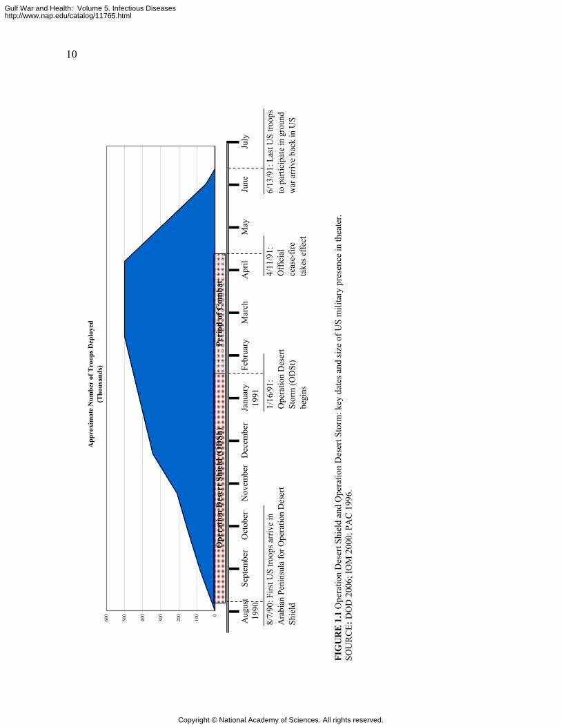
Five days after the Iraqi invasion of Kuwait on August 2, 1990, the United States deployed troops to Operation Desert Shield (ODSh). The United States attacked Iraqi armed forces by air on January 16, 1991, and this marked the beginning of Operation Desert Storm (ODSt). The ground war began on February 24, 1991, and ended 4 days later. The official cease-fire took effect on April 11, 1991, and the last troops to participate in the ground war arrived back in the United States on June 13, 1991. In this report, ODSh and ODSt are also referred to collectively as the Gulf War.
About 697,000 US troops were deployed to the Persian Gulf during ODSh and ODSt. Figure 1.1 depicts the size of the US military presence in the Persian Gulf from August 1990 through June 1991. The war was considered to be a successful military operation, and there were few injuries and deaths.
Shortly after returning to the United States, a number of veterans started reporting a variety of symptoms—fatigue, headache, muscle and joint pain, sleep disturbances, and cognitive difficulties (Persian Gulf Veterans Coordinating Board 1995). The veterans were concerned that they might have been exposed to chemical, biologic, or physical agents during their deployment to the Persian Gulf and that those exposures might be responsible for their unexplained illnesses.
Copyright © National Academy of Sciences. All rights reserved.
Gulf War and Health: Volume 5. Infectious Diseaseshttp://www.nap.edu/catalog/11765.html
Mar
ch19
91Se
ptem
ber
Oct
ober
Nov
embe
rA
ugus
t19
90A
pril
May
June
July
Dec
embe
rJa
nuar
yFe
brua
ry
App
roxi
mat
e N
umbe
r of
Tro
ops D
eplo
yed
(Tho
usan
ds)
0
100
200
300
400
500
600
Peri
od o
f Com
bat
Ope
ratio
n D
eser
t Shi
eld
(OD
Sh)
1/16
/91:
O
pera
tion
Des
ert
Stor
m (O
DSt
) be
gins
4/11
/91:
O
ffici
al
ceas
e-fir
e ta
kes e
ffec t
6/13
/91:
Las
t US
troop
s to
par
ticip
ate
in g
roun
d w
ar a
rriv
e ba
ck in
US
8/7/
90: F
irst U
S tro
ops a
rriv
e in
A
rabi
an P
enin
sula
for O
pera
tion
Des
ert
Shie
ld
FI
GU
RE
1.1
Ope
ratio
n D
eser
t Shi
eld
and
Ope
ratio
n D
eser
t Sto
rm: k
ey d
ates
and
size
of U
S m
ilita
ry p
rese
nce
in th
eate
r. SO
UR
CE:
DO
D 2
006;
IOM
200
0; P
AC
199
6.
10
Copyright © National Academy of Sciences. All rights reserved.
Gulf War and Health: Volume 5. Infectious Diseaseshttp://www.nap.edu/catalog/11765.html
INTRODUCTION 11
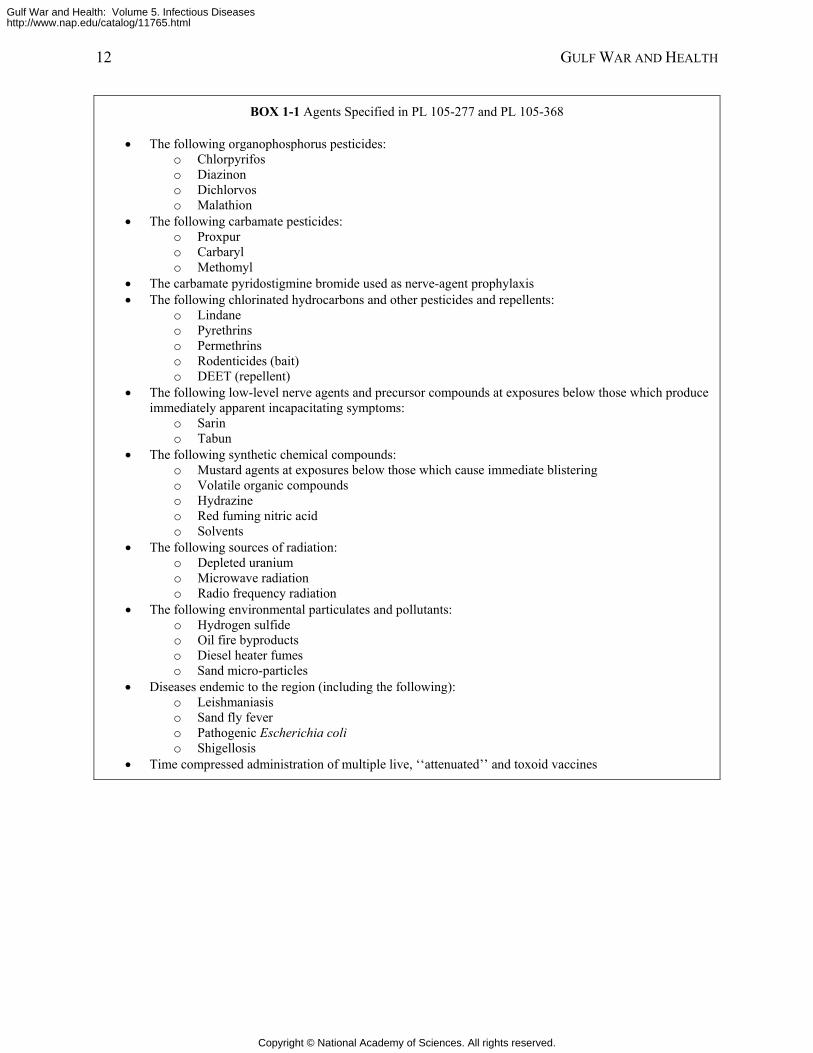
In response to the concerns of the Gulf War veterans about their unexplained illnesses, the US Department of Veterans Affairs (VA) asked the Institute of Medicine (IOM) to conduct a study to evaluate the scientific literature on chemical, biologic, and physical agents to which military personnel in the gulf were potentially exposed and possible long-term adverse health outcomes. In addition, Congress passed two laws in 1998—the Persian Gulf War Veterans Act (PL 105-277) and the Veterans Programs Enhancement Act (PL 105-368)—that called for the review of the scientific literature on specified agents with regard to long-term adverse health outcomes. That legislation directs IOM to study a number of diverse chemical, biologic, and physical agents (listed in Box 1.1). IOM divided the task into several reviews. It has completed four reports: Gulf War and Health, Volume 1: Depleted Uranium, Pyridostigmine Bromide, Sarin, Vaccines (IOM 2000); Gulf War and Health, Volume 2: Insecticides and Solvents (IOM 2003); Gulf War and Health Volume 3: Fuels, Combustion Products, and Propellants (IOM 2005); and Gulf War and Health, Volume 4: Health Effects of Serving in the Gulf War (IOM 2006). The present report is the fifth volume in the series. An additional, related report has also been published: Gulf War and Health: Updated Literature Review of Sarin (IOM 2004).
Since VA asked IOM to conduct the above-mentioned study and PL 105-277 and PL 105-368 were enacted, the United States has again entered into military conflicts in southwest and south-central Asia—Operation Enduring Freedom (OEF) and Operation Iraqi Freedom (OIF). Therefore, VA has asked IOM to make this report relevant to the military personnel serving in OEF and OIF in addition to those who served in the 1991 Gulf War.
Copyright © National Academy of Sciences. All rights reserved.
Gulf War and Health: Volume 5. Infectious Diseaseshttp://www.nap.edu/catalog/11765.html
12 GULF WAR AND HEALTH
BOX 1-1 Agents Specified in PL 105-277 and PL 105-368
• The following organophosphorus pesticides: o Chlorpyrifos o Diazinon o Dichlorvos o Malathion
• The following carbamate pesticides: o Proxpur o Carbaryl o Methomyl
• The carbamate pyridostigmine bromide used as nerve-agent prophylaxis • The following chlorinated hydrocarbons and other pesticides and repellents:
o Lindane o Pyrethrins o Permethrins o Rodenticides (bait) o DEET (repellent)
• The following low-level nerve agents and precursor compounds at exposures below those which produce immediately apparent incapacitating symptoms:
o Sarin o Tabun
• The following synthetic chemical compounds: o Mustard agents at exposures below those which cause immediate blistering o Volatile organic compounds o Hydrazine o Red fuming nitric acid o Solvents
• The following sources of radiation: o Depleted uranium o Microwave radiation o Radio frequency radiation
• The following environmental particulates and pollutants: o Hydrogen sulfide o Oil fire byproducts o Diesel heater fumes o Sand micro-particles
• Diseases endemic to the region (including the following): o Leishmaniasis o Sand fly fever o Pathogenic Escherichia coli o Shigellosis
• Time compressed administration of multiple live, ‘‘attenuated’’ and toxoid vaccines
Copyright © National Academy of Sciences. All rights reserved.
Gulf War and Health: Volume 5. Infectious Diseaseshttp://www.nap.edu/catalog/11765.html
INTRODUCTION 13
IDENTIFYING THE INFECTIOUS DISEASES TO STUDY
In accordance with PL 105-277 and PL 105-368, IOM appointed the Committee on Gulf War and Health: Infectious Diseases and tasked it to review, evaluate, and summarize the peer-reviewed scientific and medical literature on long-term adverse health outcomes associated with selected infectious diseases pertinent to service in the Gulf War. The infectious diseases can include, but are not limited to, pathogenic Escherichia coli infection, shigellosis, leishmaniasis, and sand fly fever.
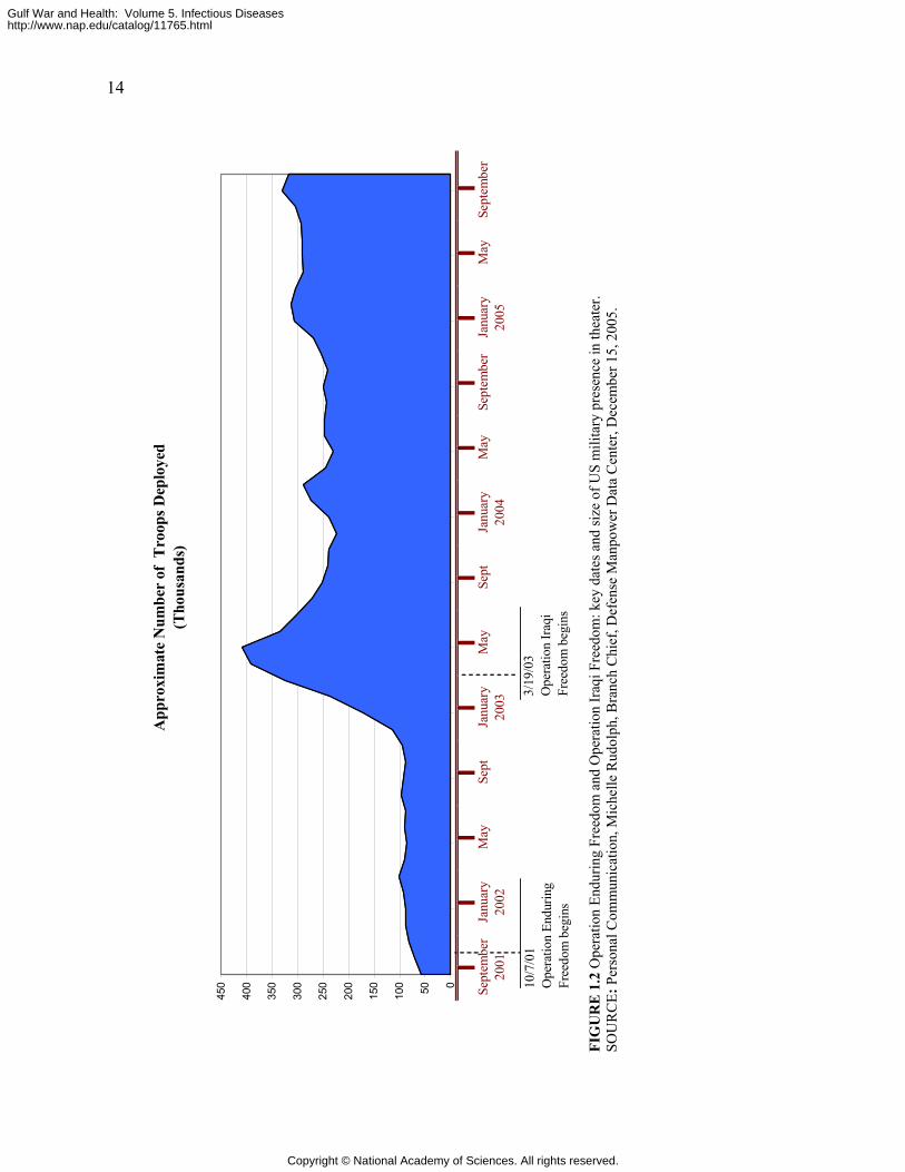
VA is also concerned about potential long-term adverse health outcomes of infectious diseases in veterans of OEF and OIF. As of October 2005, about 1.2 million US troops have been deployed to OEF or OIF (see Figure 1.2). VA asked IOM to evaluate infectious diseases pertinent to service in OEF and OIF.
It should be noted that the charge to IOM was not to determine whether a unique Gulf War syndrome or Gulf War illness exists or to make judgments about whether individual veterans were exposed to specific pathogens. Nor was the charge to focus on broader issues, such as the potential costs of compensation for veterans or policy regarding compensation; such decisions are the responsibility of the secretary of veterans affairs.
Copyright © National Academy of Sciences. All rights reserved.
Gulf War and Health: Volume 5. Infectious Diseaseshttp://www.nap.edu/catalog/11765.html
Sept
embe
rJa
nuar
yM
aySe
ptJa
nuar
yM
ayJa
nuar
yM
aySe
ptJa
nuar
ySe
ptem
ber
2001
2002
2003
2004
2005
May
Sept
embe
r
10/7
/01
Ope
ratio
n En
durin
g Fr
eedo
m b
egin
s
3/19
/03
Ope
ratio
n Ir
aqi
Free
dom
beg
ins
App
roxi
mat
e N
umbe
r of
Tro
ops D
eplo
yed
(Tho
usan
ds)
050100
150
200
250
300
350
400
450
FI
GU
RE
1.2
Ope
ratio
n En
durin
g Fr
eedo
m a
nd O
pera
tion
Iraq
i Fre
edom
: key
dat
es a
nd si
ze o
f US
mili
tary
pre
senc
e in
thea
ter.
SO
UR
CE:
Per
sona
l Com
mun
icat
ion,
Mic
helle
Rud
olph
, Bra
nch
Chi
ef, D
efen
se M
anpo
wer
Dat
a C
ente
r, D
ecem
ber 1
5, 2
005.
14
Copyright © National Academy of Sciences. All rights reserved.
Gulf War and Health: Volume 5. Infectious Diseaseshttp://www.nap.edu/catalog/11765.html
INTRODUCTION 15
THE COMMITTEE’S APPROACH TO ITS CHARGE
A brief overview of how the committee approached its charge is presented here. A more comprehensive explanation is provided in Chapter 2.
The committee identified numerous infectious diseases to which Gulf War, OIF, and OEF military personnel might have been exposed during their deployment. Dozens of infectious diseases are endemic to southwest and south-central Asia, which includes Iraq, Kuwait, and Afghanistan. The committee then determined which of the endemic infectious diseases are known to have long-term adverse health outcomes. To determine which infectious diseases to review in depth, the committee took several factors into account, including which ones were diagnosed in military personnel who served in the Gulf War, OEF, or OIF and in veterans after they returned home, as well as the prevalence of the infectious diseases in southwest and south-central Asia compared with their prevalence in the United States.
Overall, the incidence of infectious diseases among Gulf War military personnel was low (Hyams et al. 1995). Acute diarrheal and acute respiratory diseases were the major causes of morbidity from infectious diseases (Hyams et al. 1995; Hyams et al. 2001). The outbreaks of diarrhea were due primarily to enterotoxigenic Escherichia coli and Shigella sonnei. Some 12 cases of viscerotropic leishmaniasis and 20 cases of cutaneous leishmaniasis were diagnosed in Gulf War military personnel (Hyams et al. 1995; Hyams et al. 2001). Other reported infectious diseases included Q fever (three cases), West Nile fever (one case), and malaria (seven cases) (Hyams et al. 1995; Hyams et al. 2001).
Infectious diseases reported in troops who served in OEF and OIF as of December 2005 are visceral and cutaneous leishmaniasis, malaria, diarrheal disease, respiratory disease, tuberculosis infection (but not active tuberculosis), Q fever, brucellosis, and Acinetobacter baumannii infection (Kilpatrick 2005). Chapter 4 reviews the literature on infectious diseases that have been diagnosed in military personnel during or shortly after returning from the Gulf War, OIF, or OEF.
The committee identified for comprehensive evaluation nine infectious diseases known to have long-term adverse health outcomes that were diagnosed in military personnel who served in the Gulf War, OEF, or OIF. Some information is presented on a number of other infectious diseases as well because they are endemic to southwest and south-central Asia, although there have been no reported cases in military personnel through December 2005. It is possible that military personnel have become infected but that no diagnosis was made either because no acute symptoms were present or because the symptoms were mild and the soldier who had them did not seek medical care. We also present information on diseases and agents of special concern to veterans of the Gulf War, OEF, and OIF (Al Eskan disease, acute eosinophilic pneumonia, Acinetobacter baumannii infection, mycoplasmas, and biological warfare agents).
After determining which infectious diseases it would evaluate, the committee had to identify the relevant literature for review. The committee relied primarily on peer-reviewed published literature in developing its conclusions. It also consulted other material, such as surveillance reports, technical reports, and textbooks, and it obtained additional information from experts in infectious diseases of southwest and south-central Asia, from Deployment Health Support at the Department of Defense (DOD), from Walter Reed Army Institute of Research, from the VA Occupational and Environmental Health Strategic Healthcare Group, and from veteran service organizations and Gulf War veterans. The committee focused on medical and
Copyright © National Academy of Sciences. All rights reserved.
Gulf War and Health: Volume 5. Infectious Diseaseshttp://www.nap.edu/catalog/11765.html
16 GULF WAR AND HEALTH
scientific data on long-term adverse health outcomes related to the infectious diseases it selected for study.
The final step in the committee’s evaluation process was to weigh the evidence on the infectious diseases and their long-term adverse health outcomes and to develop conclusions about the strength of the evidence. The conclusions are assigned to categories of association, which range from sufficient evidence of a causal relationship to insufficient or inadequate evidence of an association.
This report includes discussion of acute diseases with potential long-term adverse health outcomes caused by known pathogens. The committee acknowledges that there might be clinically important pathogens that cannot be detected with available cultivation techniques (Relman 2002). Because the extent to which such pathogens might contribute to acute illnesses in military personnel is unknown, it is not possible to define a relationship between them and an acute illness or long-term adverse health outcome.
ORGANIZATION OF THE REPORT
Chapter 2 lays out the committee’s process for selecting the infectious diseases to study and reviewing and evaluating the evidence on them. Chapter 3 presents, in tabular format, the endemic infectious diseases of southwest and south-central Asia that are known to have long-term adverse health outcomes. Chapter 4 summarizes the body of literature on infectious diseases that have been diagnosed in military personnel serving in the Gulf War, OIF, and OEF. The committee’s comprehensive evaluations of selected infectious diseases are presented in Chapter 5, which also contains the committee’s conclusions. The final chapter, Chapter 6, presents information about diseases and agents of special concern to veterans of the Gulf War, OIF, and OEF that have an infectious component or have been implicated as a cause of “Gulf War illness”.
REFERENCES
DOD (Department of Defense). 2006. US Department of Defense Official Website. [Online].
Available: http://www.defenselink.mil/ [accessed March 2006]. Hyams KC, Hanson K, Wignall FS, Escamilla J, Oldfield EC, 3rd. 1995. The impact of
infectious diseases on the health of US troops deployed to the Persian Gulf during operations Desert Shield and Desert Storm. Clinical Infectious Diseases 20(6):1497-1504.
Hyams KC, Riddle J, Trump DH, Graham JT. 2001. Endemic infectious diseases and biological warfare during the Gulf War: A decade of analysis and final concerns. American Journal of Tropical Medicine and Hygiene 65(5):664-670.
IOM (Institute of Medicine). 2000. Gulf War and Health, Volume 1: Depleted Uranium, Sarin, Pyridostigmine Bromide, Vaccines. Washington, DC: National Academy Press.
IOM. 2003. Gulf War and Health, Volume 2: Insecticides and Solvents. Washington, DC: The National Academies Press.
IOM. 2004. Gulf War and Health: Updated Literature Review of Sarin. Washington, DC: The National Academies Press.
Copyright © National Academy of Sciences. All rights reserved.
Gulf War and Health: Volume 5. Infectious Diseaseshttp://www.nap.edu/catalog/11765.html
INTRODUCTION 17
IOM. 2005. Gulf War and Health, Volume 3: Fuels, Combustion Products, and Propellants. Washington, DC: The National Academies Press.
IOM. 2006. Gulf War and Health, Volume 4: Health Effects of Serving in the Gulf War. Washington, DC: The National Academies Press.
Kilpatrick ME. 2005. Presentation to IOM Committee on Gulf War and Health: Infectious Diseases. Washington, DC.
PAC (Presidential Advisory Committee). 1996. Presidential Advisory Committee on Gulf War Veterans’ Illnesses: Final Report. Washington, DC: US Government Printing Office.
Persian Gulf Veterans Coordinating Board. 1995. Unexplained illnesses among Desert Storm veterans. A search for causes, treatment, and cooperation. Persian Gulf Veterans Coordinating Board. Archives of Internal Medicine 155(3):262-268.
Relman DA. 2002. New technologies, human-microbe interactions, and the search for previously unrecognized pathogens. Journal of Infectious Diseases 186(2 Suppl):S254-S258.
Copyright © National Academy of Sciences. All rights reserved.
Gulf War and Health: Volume 5. Infectious Diseaseshttp://www.nap.edu/catalog/11765.html

Copyright © National Academy of Sciences. All rights reserved.
Gulf War and Health: Volume 5. Infectious Diseaseshttp://www.nap.edu/catalog/11765.html
19
2
METHODOLOGY
This chapter articulates the committee’s approach to its task. Of the dozens of pathogens known to exist in southwest and south-central Asia, the committee identified the ones that are known to cause long-term adverse health outcomes and infected at least one US veteran who served in southwest or south-central Asia in the period 1991-December 2005. The committee then oversaw a formal, comprehensive literature review that identified about 1,200 peer-reviewed studies about the late complications and latent and chronic infections that might be associated with primary infection by each of the pathogens. Those studies constituted the evidence from which the committee drew conclusions about the relationship between each primary infection and specific long-term adverse health outcomes in humans. Finally, the committee ranked the strength of the relationships through the five-category system presented at the end of this chapter.
IDENTIFYING THE INFECTIOUS DISEASES TO STUDY
Geographic Boundaries
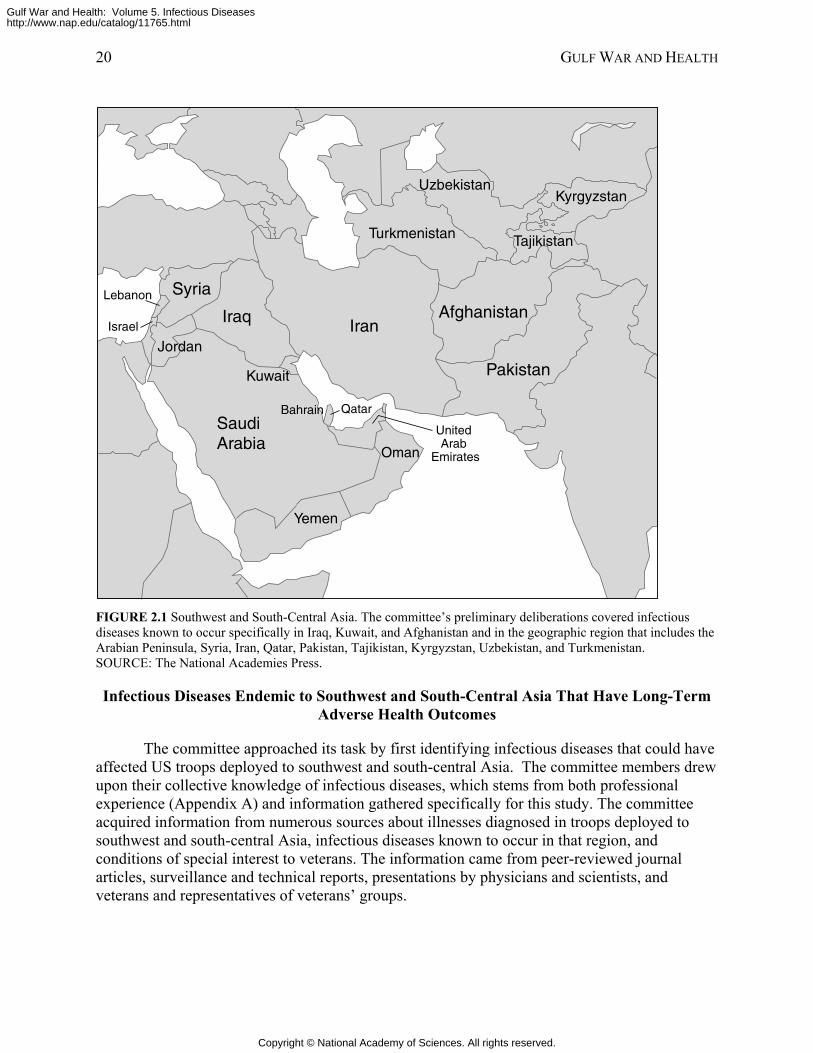
As required by law, the committee considered infectious diseases that might have afflicted US troops who served in the 1991 Gulf War (PL 105-277 and PL 105-368). Additionally, in response to a request by the Department of Veterans’ Affairs, the committee considered infectious diseases that might have afflicted US troops during Operation Enduring Freedom (OEF) or Operation Iraqi Freedom (OIF). Thus, the committee’s preliminary deliberations covered infectious diseases known to occur specifically in Iraq, Kuwait, and Afghanistan and in the geographic region that includes the Arabian Peninsula, Syria, Lebanon, Israel, Iran, Qatar, Pakistan, Tajikistan, Kyrgyzstan, Uzbekistan, and Turkmenistan (Figure 2.1). The term southwest and south-central Asia refers to that region throughout this report.
Copyright © National Academy of Sciences. All rights reserved.
Gulf War and Health: Volume 5. Infectious Diseaseshttp://www.nap.edu/catalog/11765.html
20 GULF WAR AND HEALTH
Saudi Arabia
UzbekistanKyrgyzstan
Tajikistan
Afghanistan
Pakistan
Turkmenistan
IranIraq
Syria
JordanIsrael
Oman
Yemen
Kuwait
Lebanon
QatarBahrain
United Arab
Emirates
Qatar
FIGURE 2.1 Southwest and South-Central Asia. The committee’s preliminary deliberations covered infectious diseases known to occur specifically in Iraq, Kuwait, and Afghanistan and in the geographic region that includes the Arabian Peninsula, Syria, Iran, Qatar, Pakistan, Tajikistan, Kyrgyzstan, Uzbekistan, and Turkmenistan. SOURCE: The National Academies Press.
Infectious Diseases Endemic to Southwest and South-Central Asia That Have Long-Term Adverse Health Outcomes
The committee approached its task by first identifying infectious diseases that could have affected US troops deployed to southwest and south-central Asia. The committee members drew upon their collective knowledge of infectious diseases, which stems from both professional experience (Appendix A) and information gathered specifically for this study. The committee acquired information from numerous sources about illnesses diagnosed in troops deployed to southwest and south-central Asia, infectious diseases known to occur in that region, and conditions of special interest to veterans. The information came from peer-reviewed journal articles, surveillance and technical reports, presentations by physicians and scientists, and veterans and representatives of veterans’ groups.
Copyright © National Academy of Sciences. All rights reserved.
Gulf War and Health: Volume 5. Infectious Diseaseshttp://www.nap.edu/catalog/11765.html
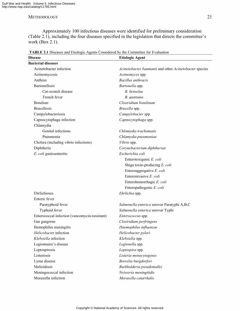
METHODOLOGY 21
Approximately 100 infectious diseases were identified for preliminary consideration (Table 2.1), including the four diseases specified in the legislation that directs the committee’s work (Box 2.1).
TABLE 2.1 Diseases and Etiologic Agents Considered by the Committee for Evaluation Disease Etiologic Agent Bacterial diseases
Acinetobacter infection Acinetobacter baumanii and other Acinetobacter species Actinomycosis Actinomyces spp. Anthrax Bacillus anthracis Bartonellosis Bartonella spp.
Cat-scratch disease B. henselae Trench fever B. quintana
Botulism Clostridium botulinum Brucellosis Brucella spp. Campylobacteriosis Campylobacter spp. Capnocytophaga infection Capnocytophaga spp. Chlamydia
Genital infections Chlamydia trachomatis Pneumonia Chlamydia pneumoniae
Cholera (including vibrio infections) Vibrio spp. Diphtheria Corynebacterium diphtheriae E. coli gastroenteritis Escherichia coli
Enterotoxigenic E. coli Shiga toxin-producing E. coli Enteroaggregative E. coli Enteroinvasive E. coli Enterohemorrhagic E. coli Enteropathogenic E. coli
Ehrlichioses Ehrlichia spp. Enteric fever
Paratyphoid fever Salmonella enterica serovar Paratyphi A,B,C Typhoid fever Salmonella enterica serovar Typhi
Enterococcal infection (vancomycin-resistant) Enterococcus spp. Gas gangrene Clostridium perfringens Hemophilus meningitis Haemophilus influenzae Helicobacter infection Helicobacter pylori Klebsiella infection Klebsiella spp. Legionnaire’s disease Legionella spp. Leptospirosis Leptospira spp. Listeriosis Listeria monocytogenes Lyme disease Borrelia burgdorferi Melioidosis Burkholderia pseudomallei Meningococcal infection Neisseria meningitidis Moraxella infection Moraxella catarrhalis
Copyright © National Academy of Sciences. All rights reserved.
Gulf War and Health: Volume 5. Infectious Diseaseshttp://www.nap.edu/catalog/11765.html
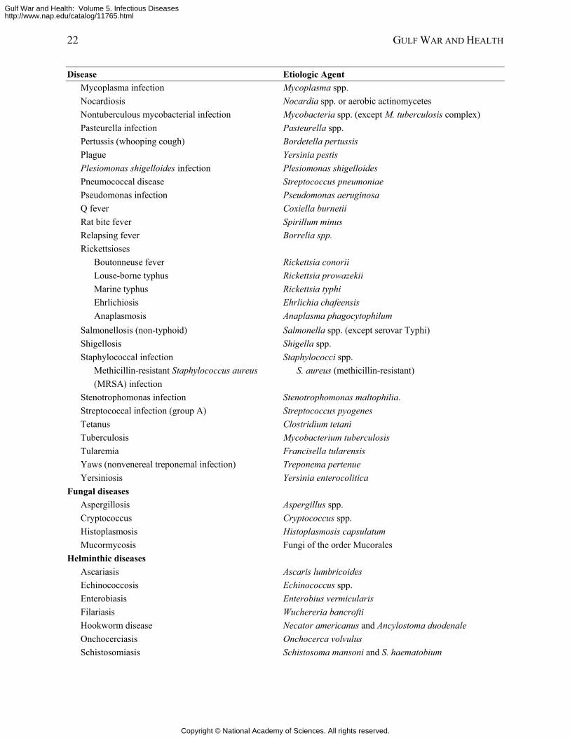
22 GULF WAR AND HEALTH
Disease Etiologic Agent Mycoplasma infection Mycoplasma spp. Nocardiosis Nocardia spp. or aerobic actinomycetes Nontuberculous mycobacterial infection Mycobacteria spp. (except M. tuberculosis complex) Pasteurella infection Pasteurella spp. Pertussis (whooping cough) Bordetella pertussis Plague Yersinia pestis Plesiomonas shigelloides infection Plesiomonas shigelloides Pneumococcal disease Streptococcus pneumoniae Pseudomonas infection Pseudomonas aeruginosa Q fever Coxiella burnetii Rat bite fever Spirillum minus Relapsing fever Borrelia spp. Rickettsioses
Boutonneuse fever Rickettsia conorii Louse-borne typhus Rickettsia prowazekii Marine typhus Rickettsia typhi Ehrlichiosis Ehrlichia chafeensis Anaplasmosis Anaplasma phagocytophilum
Salmonellosis (non-typhoid) Salmonella spp. (except serovar Typhi) Shigellosis Shigella spp. Staphylococcal infection Staphylococci spp.
Methicillin-resistant Staphylococcus aureus S. aureus (methicillin-resistant) (MRSA) infection
Stenotrophomonas infection Stenotrophomonas maltophilia. Streptococcal infection (group A) Streptococcus pyogenes Tetanus Clostridium tetani Tuberculosis Mycobacterium tuberculosis Tularemia Francisella tularensis Yaws (nonvenereal treponemal infection) Treponema pertenue Yersiniosis Yersinia enterocolitica
Fungal diseases Aspergillosis Aspergillus spp. Cryptococcus Cryptococcus spp. Histoplasmosis Histoplasmosis capsulatum Mucormycosis Fungi of the order Mucorales
Helminthic diseases Ascariasis Ascaris lumbricoides Echinococcosis Echinococcus spp. Enterobiasis Enterobius vermicularis Filariasis Wuchereria bancrofti Hookworm disease Necator americanus and Ancylostoma duodenale Onchocerciasis Onchocerca volvulus Schistosomiasis Schistosoma mansoni and S. haematobium
Copyright © National Academy of Sciences. All rights reserved.
Gulf War and Health: Volume 5. Infectious Diseaseshttp://www.nap.edu/catalog/11765.html
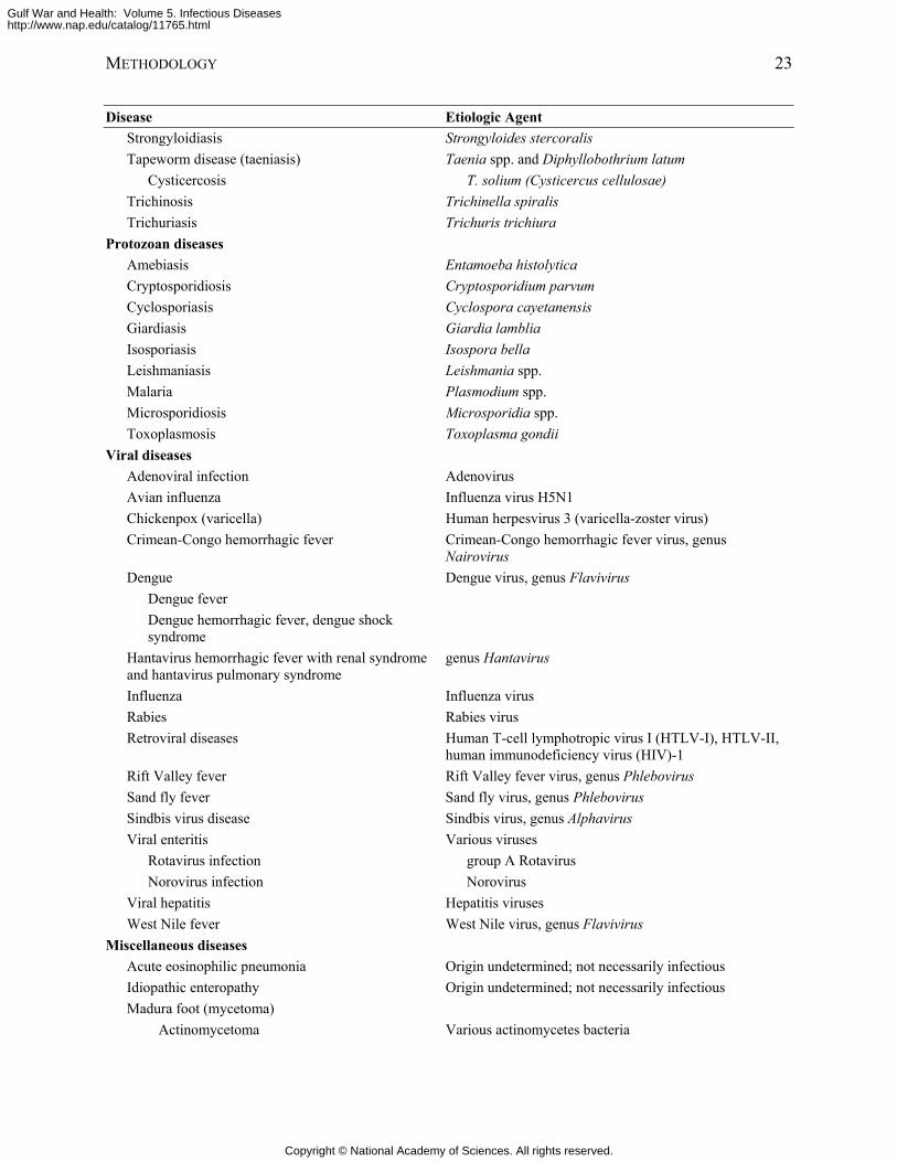
METHODOLOGY 23
Disease Etiologic Agent Strongyloidiasis Strongyloides stercoralis Tapeworm disease (taeniasis) Taenia spp. and Diphyllobothrium latum
Cysticercosis T. solium (Cysticercus cellulosae) Trichinosis Trichinella spiralis Trichuriasis Trichuris trichiura
Protozoan diseases Amebiasis Entamoeba histolytica Cryptosporidiosis Cryptosporidium parvum Cyclosporiasis Cyclospora cayetanensis Giardiasis Giardia lamblia Isosporiasis Isospora bella Leishmaniasis Leishmania spp. Malaria Plasmodium spp. Microsporidiosis Microsporidia spp. Toxoplasmosis Toxoplasma gondii
Viral diseases Adenoviral infection Adenovirus Avian influenza Influenza virus H5N1 Chickenpox (varicella) Human herpesvirus 3 (varicella-zoster virus) Crimean-Congo hemorrhagic fever Crimean-Congo hemorrhagic fever virus, genus
Nairovirus Dengue Dengue virus, genus Flavivirus
Dengue fever Dengue hemorrhagic fever, dengue shock syndrome
Hantavirus hemorrhagic fever with renal syndrome and hantavirus pulmonary syndrome
genus Hantavirus
Influenza Influenza virus Rabies Rabies virus Retroviral diseases Human T-cell lymphotropic virus I (HTLV-I), HTLV-II,
human immunodeficiency virus (HIV)-1 Rift Valley fever Rift Valley fever virus, genus Phlebovirus Sand fly fever Sand fly virus, genus Phlebovirus Sindbis virus disease Sindbis virus, genus Alphavirus Viral enteritis Various viruses
Rotavirus infection group A Rotavirus Norovirus infection Norovirus
Viral hepatitis Hepatitis viruses West Nile fever West Nile virus, genus Flavivirus
Miscellaneous diseases Acute eosinophilic pneumonia Origin undetermined; not necessarily infectious Idiopathic enteropathy Origin undetermined; not necessarily infectious Madura foot (mycetoma)
Actinomycetoma Various actinomycetes bacteria
Copyright © National Academy of Sciences. All rights reserved.
Gulf War and Health: Volume 5. Infectious Diseaseshttp://www.nap.edu/catalog/11765.html
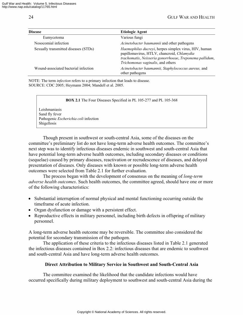
24 GULF WAR AND HEALTH
Disease Etiologic Agent Eumycetoma Various fungi
Nosocomial infection Acinetobacter baumannii and other pathogens Sexually transmitted diseases (STDs) Haemophilus ducreyi, herpes simplex virus, HIV, human
papillomavirus, HTLV, chancroid, Chlamydia trachomatis, Neisseria gonorrhoeae, Treponema pallidum, Trichomonas vaginalis, and others
Wound-associated bacterial infection Acinetobacter baumannii, Staphylococcus aureus, and other pathogens
NOTE: The term infection refers to a primary infection that leads to disease. SOURCE: CDC 2005; Heymann 2004; Mandell et al. 2005.
BOX 2.1 The Four Diseases Specified in PL 105-277 and PL 105-368
Leishmaniasis Sand fly fever Pathogenic Escherichia coli infection Shigellosis
Though present in southwest or south-central Asia, some of the diseases on the committee’s preliminary list do not have long-term adverse health outcomes. The committee’s next step was to identify infectious diseases endemic in southwest and south-central Asia that have potential long-term adverse health outcomes, including secondary diseases or conditions (sequelae) caused by primary diseases, reactivation or recrudescence of diseases, and delayed presentation of diseases. Only diseases with known or possible long-term adverse health outcomes were selected from Table 2.1 for further evaluation.
The process began with the development of consensus on the meaning of long-term adverse health outcomes. Such health outcomes, the committee agreed, should have one or more of the following characteristics: • Substantial interruption of normal physical and mental functioning occurring outside the
timeframe of acute infection. • Organ dysfunction or damage with a persistent effect. • Reproductive effects in military personnel, including birth defects in offspring of military
personnel. A long-term adverse health outcome may be reversible. The committee also considered the potential for secondary transmission of the pathogen.
The application of these criteria to the infectious diseases listed in Table 2.1 generated the infectious diseases contained in Box 2.2: infectious diseases that are endemic to southwest and south-central Asia and have long-term adverse health outcomes.
Direct Attribution to Military Service in Southwest and South-Central Asia
The committee examined the likelihood that the candidate infections would have occurred specifically during military deployment to southwest and south-central Asia during the
Copyright © National Academy of Sciences. All rights reserved.
Gulf War and Health: Volume 5. Infectious Diseaseshttp://www.nap.edu/catalog/11765.html
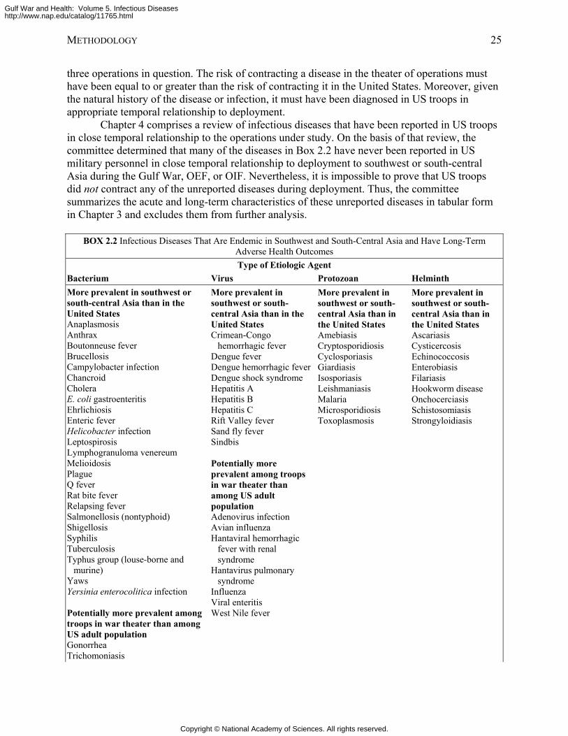
METHODOLOGY 25
three operations in question. The risk of contracting a disease in the theater of operations must have been equal to or greater than the risk of contracting it in the United States. Moreover, given the natural history of the disease or infection, it must have been diagnosed in US troops in appropriate temporal relationship to deployment.
Chapter 4 comprises a review of infectious diseases that have been reported in US troops in close temporal relationship to the operations under study. On the basis of that review, the committee determined that many of the diseases in Box 2.2 have never been reported in US military personnel in close temporal relationship to deployment to southwest or south-central Asia during the Gulf War, OEF, or OIF. Nevertheless, it is impossible to prove that US troops did not contract any of the unreported diseases during deployment. Thus, the committee summarizes the acute and long-term characteristics of these unreported diseases in tabular form in Chapter 3 and excludes them from further analysis.
BOX 2.2 Infectious Diseases That Are Endemic in Southwest and South-Central Asia and Have Long-Term
Adverse Health Outcomes Type of Etiologic Agent
Bacterium Virus Protozoan Helminth More prevalent in southwest or south-central Asia than in the United States Anaplasmosis Anthrax Boutonneuse fever Brucellosis Campylobacter infection Chancroid Cholera E. coli gastroenteritis Ehrlichiosis Enteric fever Helicobacter infection Leptospirosis Lymphogranuloma venereum Melioidosis Plague Q fever Rat bite fever Relapsing fever Salmonellosis (nontyphoid) Shigellosis Syphilis Tuberculosis Typhus group (louse-borne and
murine) Yaws Yersinia enterocolitica infection Potentially more prevalent among troops in war theater than among US adult population Gonorrhea Trichomoniasis
More prevalent in southwest or south-central Asia than in the United States Crimean-Congo
hemorrhagic fever Dengue fever Dengue hemorrhagic fever Dengue shock syndrome Hepatitis A Hepatitis B Hepatitis C Rift Valley fever Sand fly fever Sindbis Potentially more prevalent among troops in war theater than among US adult population Adenovirus infection Avian influenza Hantaviral hemorrhagic
fever with renal syndrome
Hantavirus pulmonary syndrome
Influenza Viral enteritis West Nile fever
More prevalent in southwest or south-central Asia than in the United States Amebiasis Cryptosporidiosis Cyclosporiasis Giardiasis Isosporiasis Leishmaniasis Malaria Microsporidiosis Toxoplasmosis
More prevalent in southwest or south-central Asia than in the United States Ascariasis Cysticercosis Echinococcosis Enterobiasis Filariasis Hookworm disease Onchocerciasis Schistosomiasis Strongyloidiasis
Copyright © National Academy of Sciences. All rights reserved.
Gulf War and Health: Volume 5. Infectious Diseaseshttp://www.nap.edu/catalog/11765.html
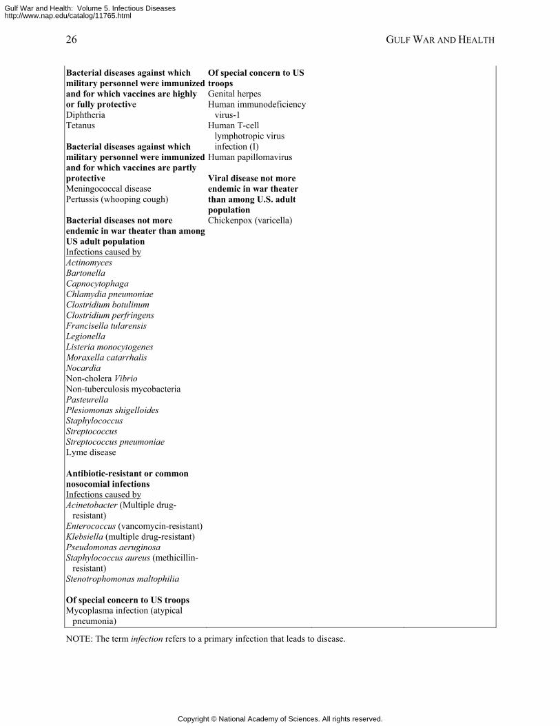
26 GULF WAR AND HEALTH
Bacterial diseases against which military personnel were immunized and for which vaccines are highly or fully protective Diphtheria Tetanus Bacterial diseases against which military personnel were immunized and for which vaccines are partly protective Meningococcal disease Pertussis (whooping cough) Bacterial diseases not more endemic in war theater than among US adult population Infections caused by Actinomyces Bartonella Capnocytophaga Chlamydia pneumoniae Clostridium botulinum Clostridium perfringens Francisella tularensis Legionella Listeria monocytogenes Moraxella catarrhalis Nocardia Non-cholera Vibrio Non-tuberculosis mycobacteria Pasteurella Plesiomonas shigelloides Staphylococcus Streptococcus Streptococcus pneumoniae Lyme disease Antibiotic-resistant or common nosocomial infections Infections caused by Acinetobacter (Multiple drug-
resistant) Enterococcus (vancomycin-resistant) Klebsiella (multiple drug-resistant) Pseudomonas aeruginosa Staphylococcus aureus (methicillin-
resistant) Stenotrophomonas maltophilia Of special concern to US troops Mycoplasma infection (atypical
pneumonia)
Of special concern to US troops Genital herpes Human immunodeficiency
virus-1 Human T-cell
lymphotropic virus infection (I)
Human papillomavirus Viral disease not more endemic in war theater than among U.S. adult population Chickenpox (varicella)
NOTE: The term infection refers to a primary infection that leads to disease.
Copyright © National Academy of Sciences. All rights reserved.
Gulf War and Health: Volume 5. Infectious Diseaseshttp://www.nap.edu/catalog/11765.html
METHODOLOGY 27
Timing of Appearance of Long-Term Adverse Health Outcomes
Next, the committee determined the likelihood that a disease’s long-term adverse health outcomes would have been manifest and diagnosed during a person’s term of military service.
If the onset of adverse health outcomes typically occurs during the acute illness, the committee reasoned, the long-term effects would probably be detected, diagnosed, and reported during the patient’s term of military service. Likewise, diseases that have an acute phase—meaning that signs and symptoms become evident suddenly within hours or days of infection—are readily diagnosed during military service if the individual seeks medical care; in such cases, the physician would probably be cognizant of possible long-term adverse health outcomes. Finally, pre-existing conditions that were mild or latent before deployment but became reactivated or exacerbated during deployment also would probably be diagnosed during the patient’s military service. Therefore, infectious diseases with one or both of the following characteristics were not comprehensively evaluated in this study; instead, their acute and long-term health outcomes are summarized in tabular form in Chapter 3: • Onset of long-term adverse health outcomes typically occurs during the acute illness. • The disease is a preexisting infection that becomes reactivated or exacerbated during
deployment.
In contrast, the committee determined, there are other infectious diseases whose long-term adverse health outcomes were unlikely to be diagnosed during military service in southwest and south-central Asia. Such diseases have the following characteristics:
• The primary infection is subacute or the infected person is asymptomatic for days to years. • Long-term adverse health outcomes begin months to years after infection. Because a subacute infection may go unnoticed and undiagnosed, the committee reasoned, the primary disease might not be reported. Furthermore, if long-term adverse health outcomes begin months or even years after infection, the condition would be more likely to be diagnosed after the person completes military service, and the diagnosing physician may not attribute the patient’s signs and symptoms to an infection acquired during deployment, particularly if the disease is uncommon in the United States, and thus more likely to be misdiagnosed. Infectious diseases that fit those criteria qualified for in-depth study.
The Infectious Diseases to Be Studied for Strength of Association with Long-Term Adverse Health Outcomes
The committee identified nine infectious diseases (Box 2.3) known to have long-term adverse health outcomes and to be potentially acquired by infected veterans during their military deployment to southwest and south-central Asia from 1991 to 2005. The committee conducted comprehensive reviews of the literature on the potential long-term adverse health outcomes of these diseases to determine the strength of association between the primary infection and the health outcome in humans.
Copyright © National Academy of Sciences. All rights reserved.
Gulf War and Health: Volume 5. Infectious Diseaseshttp://www.nap.edu/catalog/11765.html
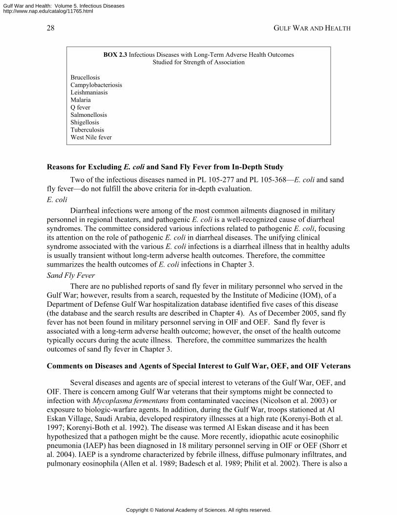
28 GULF WAR AND HEALTH
Reasons for Excluding E. coli and Sand Fly Fever from In-Depth Study Two of the infectious diseases named in PL 105-277 and PL 105-368—E. coli and sand
fly fever—do not fulfill the above criteria for in-depth evaluation. E. coli
Diarrheal infections were among of the most common ailments diagnosed in military personnel in regional theaters, and pathogenic E. coli is a well-recognized cause of diarrheal syndromes. The committee considered various infections related to pathogenic E. coli, focusing its attention on the role of pathogenic E. coli in diarrheal diseases. The unifying clinical syndrome associated with the various E. coli infections is a diarrheal illness that in healthy adults is usually transient without long-term adverse health outcomes. Therefore, the committee summarizes the health outcomes of E. coli infections in Chapter 3. Sand Fly Fever
There are no published reports of sand fly fever in military personnel who served in the Gulf War; however, results from a search, requested by the Institute of Medicine (IOM), of a Department of Defense Gulf War hospitalization database identified five cases of this disease (the database and the search results are described in Chapter 4). As of December 2005, sand fly fever has not been found in military personnel serving in OIF and OEF. Sand fly fever is associated with a long-term adverse health outcome; however, the onset of the health outcome typically occurs during the acute illness. Therefore, the committee summarizes the health outcomes of sand fly fever in Chapter 3.
Comments on Diseases and Agents of Special Interest to Gulf War, OEF, and OIF Veterans
Several diseases and agents are of special interest to veterans of the Gulf War, OEF, and OIF. There is concern among Gulf War veterans that their symptoms might be connected to infection with Mycoplasma fermentans from contaminated vaccines (Nicolson et al. 2003) or exposure to biologic-warfare agents. In addition, during the Gulf War, troops stationed at Al Eskan Village, Saudi Arabia, developed respiratory illnesses at a high rate (Korenyi-Both et al. 1997; Korenyi-Both et al. 1992). The disease was termed Al Eskan disease and it has been hypothesized that a pathogen might be the cause. More recently, idiopathic acute eosinophilic pneumonia (IAEP) has been diagnosed in 18 military personnel serving in OIF or OEF (Shorr et al. 2004). IAEP is a syndrome characterized by febrile illness, diffuse pulmonary infiltrates, and pulmonary eosinophila (Allen et al. 1989; Badesch et al. 1989; Philit et al. 2002). There is also a
BOX 2.3 Infectious Diseases with Long-Term Adverse Health Outcomes Studied for Strength of Association
Brucellosis Campylobacteriosis Leishmaniasis Malaria Q fever Salmonellosis Shigellosis Tuberculosis West Nile fever
Copyright © National Academy of Sciences. All rights reserved.
Gulf War and Health: Volume 5. Infectious Diseaseshttp://www.nap.edu/catalog/11765.html
METHODOLOGY 29
discussion of wound-associated infections (for example, infections caused by Acinetobacter baumannii) that appear to be more prevalent in OIF and OEF personnel than in civilian populations (Davis et al. 2005). Chapter 6 discusses Al Eskan disease, IAEP, wound-associated infections, mycoplasmas, and biologic-warfare agents.
REVIEW AND EVALUATION OF THE LITERATURE
Selection of the Literature
The committee adopted a policy of basing its conclusions primarily on peer-reviewed, published literature. Non-peer-reviewed publications provided additional information for the committee and raised issues that were researched further in the peer-reviewed literature.
Although the process of peer review by fellow professionals ensures high standards of quality, it does not guarantee the validity of a study or the generalizability of its results. Accordingly, committee members read each study critically and considered its relevance and quality.
Amassing the Literature
The committee oversaw a multistep process for amassing a robust collection of scientific literature about the long-term adverse health outcomes of the diseases listed in Box 2.3. The process began with a search of PubMed, a database created and managed by the National Library of Medicine that includes more than 15 million citations of biomedical publications from the 1950s to the present. The PubMed search focused on journal articles published through December 2005 that contain information about late complications, long-term sequelae, and latent infections related to the relevant infectious diseases and etiologic agents identified by the committee. Additional studies were identified from the reference lists of topical technical reports, textbooks, and other documents. Further PubMed searches identified pertinent articles on Al Eskan disease, IAEP, wound-associated infections, mycoplasmas, and biologic-warfare agents. Those initial searches generated about 20,000 articles.
Reviewing the Literature
On closer examination of that large body of literature, a subset of about 1,200 articles appeared to provide the types and quality of scientific evidence that the committee needed to accomplish its task. After securing the full text of those articles, the committee reviewed and assessed them for evidence of associations between primary infections by the etiologic agents of interest and specific long-term adverse health outcomes in humans.
The committee included several types of studies, including epidemiologic studies, case reports, and case series. Ideally, epidemiologic studies should have methodologic details, a control or reference group, reasonable adjustment for confounders, and statistical power to detect effects. Review articles, technical reports, and textbooks were used for background information.
The committee relied heavily on studies that focused on human adult populations. Because the IOM task concerns adults who may have become infected during military service, studies of children were generally excluded unless the studies dealt with reproductive outcomes. One exception is that studies of children were included if they provided information about adult
Copyright © National Academy of Sciences. All rights reserved.
Gulf War and Health: Volume 5. Infectious Diseaseshttp://www.nap.edu/catalog/11765.html
30 GULF WAR AND HEALTH
diseases; for example, a study on cerebral malaria in children might be reviewed if it provided information about the disease in adults.
CATEGORIES OF STRENGTH OF ASSOCIATION
The committee’s goal was to use the evidence in the medical and scientific literature to determine the relationships between the infectious diseases of interest and specific adverse health outcomes that might appear months to years after primary infections. Those relationships, presented in Chapter 5, are conceived in terms of the “strength of association” between a primary infection and a specific long-term adverse health outcome. The committee ranks strength of association qualitatively using a five-tier system, presented below in full.
Origin and Evolution of the Categories
A brief historical overview of the committee’s categories of association will elucidate their scientific roots. The International Agency for Research on Cancer (IARC), part of WHO, established criteria in 1971 to evaluate the human carcinogenic risk posed by chemicals (IARC 1998). First published in 1972, IARC’s evaluations are scientific, qualitative judgments by ad hoc working groups about the evidence of carcinogenicity or noncarcinogenicity provided by the available data. The working groups express their qualitative judgments in terms of five categories of the relative strength of the evidence that a substance or exposure is carcinogenic (IARC 1999a). Agencies in 57 countries use IARC’s published evaluations—a reflection of the widespread acceptance of the categorization scheme as it has been updated and applied to about 900 agents, mixtures, and exposures (IARC 1999b; IARC 2005).
In the early 1990s, an IOM committee adopted IARC’s categories in evaluating the adverse health outcomes of pertussis and rubella vaccines (IOM 1991). Later IOM committees used the categories, with some modifications, in evaluating the safety of childhood vaccines (IOM 1994a), the health outcomes of herbicides used in Vietnam (IOM 1994b; IOM 1996; IOM 1999; IOM 2001; IOM 2003b), and the relationship between indoor pollutants and asthma (IOM 2000a). The present committee’s predecessors also adapted and used the categories in evaluating the health effects of outcomes given to US troops and of chemical exposures that may have occurred during the Gulf War (IOM 2000b; IOM 2003a; IOM 2004; IOM 2005).
The five categories of strength of association used in this report are presented and defined below.
Sufficient Evidence of a Causal Relationship
Evidence from available studies is sufficient to conclude that there is a causal relationship between exposure to a specific agent and a specific health outcome in humans. The evidence includes supporting experimental data and fulfills the guidelines for sufficient evidence of an association (see next category). The association is biologically plausible, and the evidence satisfies several of the guidelines used to assess causality, such as strength of association, dose-response relationship, consistency of association, and a temporal relationship.
Copyright © National Academy of Sciences. All rights reserved.
Gulf War and Health: Volume 5. Infectious Diseaseshttp://www.nap.edu/catalog/11765.html
METHODOLOGY 31
Sufficient Evidence of an Association
Evidence from available studies is sufficient to conclude that there is an association. A consistent association has been observed between exposure to a specific agent and a specific health outcome in several high-quality human studies in which chance and bias, including confounding, could be ruled out with reasonable confidence.
Limited or Suggestive Evidence of an Association
Evidence from available studies suggests an association between exposure to a specific agent and a specific health outcome, but the body of evidence is limited by the inability to rule out chance and bias, including confounding, with confidence. For example, at least one high-quality study that is sufficiently free of bias, including adequate control for confounding, reports an association, while other studies provide support for the association but are not sufficiently free of bias, including confounding. Alternatively, several studies of lesser quality are consistent in showing an association, and the results are probably not due to bias, including confounding.
Inadequate or Insufficient Evidence to Determine Whether an Association Exists
Evidence from available studies is of insufficient quantity, quality, or consistency to permit a conclusion regarding the existence of an association between exposure to a specific agent and a specific health outcome in humans.
Limited or Suggestive Evidence of No Association
Evidence from well-conducted studies is consistent in not showing an association between exposure of any magnitude to a specific agent and a specific health outcome in humans. A conclusion of no association is inevitably limited to the conditions, magnitudes of exposure, and length of observation in the available studies. The possibility of a very small increase in risk after the exposure studied cannot be excluded.
REFERENCES
Allen JN, Pacht ER, Gadek JE, Davis WB. 1989. Acute eosinophilic pneumonia as a reversible
cause of noninfectious respiratory failure. New England Journal of Medicine 321(9):569-574. Badesch DB, King TE Jr., Schwarz MI. 1989. Acute eosinophilic pneumonia: A hypersensitivity
phenomenon? American Review of Respiratory Disease 139(1):249-252. CDC (Centers for Disease Control and Prevention). 2005. Health Information for International
Travel 2005-2006. Centers for Disease Control and Prevention. Atlanta, GA: US Department of Health and Human Services, Public Health Service.
Davis KA, Moran KA, McAllister CK, Gray PJ. 2005. Multidrug-resistant Acinetobacter extremity infections in soldiers. Emerging Infectious Diseases 11(8):1218-1224.
Heymann DL. 2004. Control of Communicable Diseases Manual. Washington, DC: American Public Health Association.
Copyright © National Academy of Sciences. All rights reserved.
Gulf War and Health: Volume 5. Infectious Diseaseshttp://www.nap.edu/catalog/11765.html
32 GULF WAR AND HEALTH
IARC (International Agency for Research on Cancer). 1998. Preamble to the IARC Monographs. [Online]. Available: http://www-cie.iarc.fr/monoeval/background.html [accessed July 19, 2005].
IARC. 1999a. Evaluation. Preamble to the IARC Monographs. [Online]. Available: http://www-cie.iarc.fr/monoeval/eval.html [accessed August 3, 2005].
IARC. 1999b. Objective and Scope. Preamble to the IARC Monographs. [Online]. Available: http://www-cie.iarc.fr/monoeval/objectives.html [accessed August 3, 2005].
IARC. 2005. Monographs on the Evaluation of Carcinogenic Risks to Humans. [Online]. Available: http://www-cie.iarc.fr/ [accessed August 3, 2005].
IOM (Institute of Medicine). 1991. Adverse Effects of Pertussis and Rubella Vaccines. Washington, DC: National Academy Press.
IOM. 1994a. Adverse Events Associated With Childhood Vaccines: Evidence Bearing on Causality. Washington, DC: National Academy Press.
IOM. 1994b. Veterans and Agent Orange: Health Effects of Herbicides Used in Vietnam. Washington, DC: National Academy Press.
IOM. 1996. Veterans and Agent Orange: Update 1996. Washington, DC: National Academy Press.
IOM. 1999. Veterans and Agent Orange: Update 1998. Washington, DC: National Academy Press.
IOM. 2000a. Clearing the Air: Asthma and Indoor Air Exposures. Washington, DC: National Academy Press.
IOM. 2000b. Gulf War and Health, Volume 1: Depleted Uranium, Sarin, Pyridostigmine Bromide, Vaccines. Washington, DC: National Academy Press.
IOM. 2001. Veterans and Agent Orange: Update 2000. Washington, DC: National Academy Press.
IOM. 2003a. Gulf War and Health, Volume 2: Insecticides and Solvents. Washington, DC: The National Academies Press.
IOM. 2003b. Veterans and Agent Orange: Update 2002. Washington, DC: The National Academies Press.
IOM. 2004. Gulf War and Health: Updated Literature Review of Sarin. Washington, DC: The National Academies Press.
IOM. 2005. Gulf War and Health, Volume 3: Fuels, Combustion Products, and Propellants. Washington, DC: The National Academies Press.
Korenyi-Both AL, Korenyi-Both AL, Molnar AC, Fidelus-Gort R. 1992. Al Eskan disease: Desert Storm pneumonitis. Military Medicine 157(9):452-462.
Korenyi-Both AL, Korenyi-Both AL, Juncer DJ. 1997. Al Eskan disease: Persian Gulf syndrome. Military Medicine 162(1):1-13.
Mandell GL, Bennett JE, Dolin R. 2005. Principles and Practice of Infectious Diseases. 6th ed. Philadelphia, PA: Elsevier Churchill Livingstone.
Nicolson GL, Nasralla MY, Nicolson NL, Haier J. 2003. High prevalence of mycoplasma infections in symptomatic (Chronic Fatigue Syndrome) family members of mycoplasma-positive Gulf War illness patients. Journal of the Chronic Fatigue Syndrome 11(2):21-36.
Copyright © National Academy of Sciences. All rights reserved.
Gulf War and Health: Volume 5. Infectious Diseaseshttp://www.nap.edu/catalog/11765.html
METHODOLOGY 33
Philit F, Etienne-Mastroianni B, Parrot A, Guerin C, Robert D, Cordier JF. 2002. Idiopathic acute eosinophilic pneumonia: A study of 22 patients. American Journal of Respiratory and Critical Care Medicine 166(9):1235-1239.
Shorr AF, Scoville SL, Cersovsky SB, Shanks GD, Ockenhouse CF, Smoak BL, Carr WW, Petruccelli BP. 2004. Acute eosinophilic pneumonia among US Military personnel deployed in or near Iraq. Journal of the American Medical Association 292(24):2997-3005.
Copyright © National Academy of Sciences. All rights reserved.
Gulf War and Health: Volume 5. Infectious Diseaseshttp://www.nap.edu/catalog/11765.html

Copyright © National Academy of Sciences. All rights reserved.
Gulf War and Health: Volume 5. Infectious Diseaseshttp://www.nap.edu/catalog/11765.html
35
3
INFECTIOUS DISEASES ENDEMIC TO SOUTHWEST AND SOUTH-CENTRAL ASIA THAT HAVE LONG-TERM ADVERSE HEALTH
OUTCOMES
In Chapter 2, the committee developed an extensive list of infectious diseases that are endemic to southwest and south-central Asia (Table 2.1) and then narrowed the list to diseases or syndromes with known long-term adverse health outcomes (Box 2.2). Although most diseases in that subset have not been reported in military personnel deployed to southwest and south-central Asia, they have historically been diagnosed in local populations and thus pose a theoretical risk to US troops deployed to the region. Also, given the nature and duration of Operation Iraqi Freedom and Operation Enduring Freedom, some of the diseases in Box 2.2 could be diagnosed after a person’s deployment or period of military service.
The committee decided that the most effective way to give additional information on the diseases listed in Box 2.2 would be to present them in tables containing
• A description of the acute syndrome in adults, • A description of the potential long-term adverse health outcomes in adults with clinical
disease, • The frequency with which the long-term adverse health outcomes occur in adults with clinical
disease, • The delay, if any, between acute infection and onset of long-term adverse health outcomes.
Tables 3.1-3.4 categorize the infections of interest by type of pathogen (viral, bacterial, helminthic, or protozoan), and Table 3.5 describes sexually transmitted diseases. The infectious diseases with long-term adverse health outcomes that have been diagnosed in military personnel and that the committee reviewed in depth (see Chapter 5) are also included here.
Copyright © National Academy of Sciences. All rights reserved.
Gulf War and Health: Volume 5. Infectious Diseaseshttp://www.nap.edu/catalog/11765.html
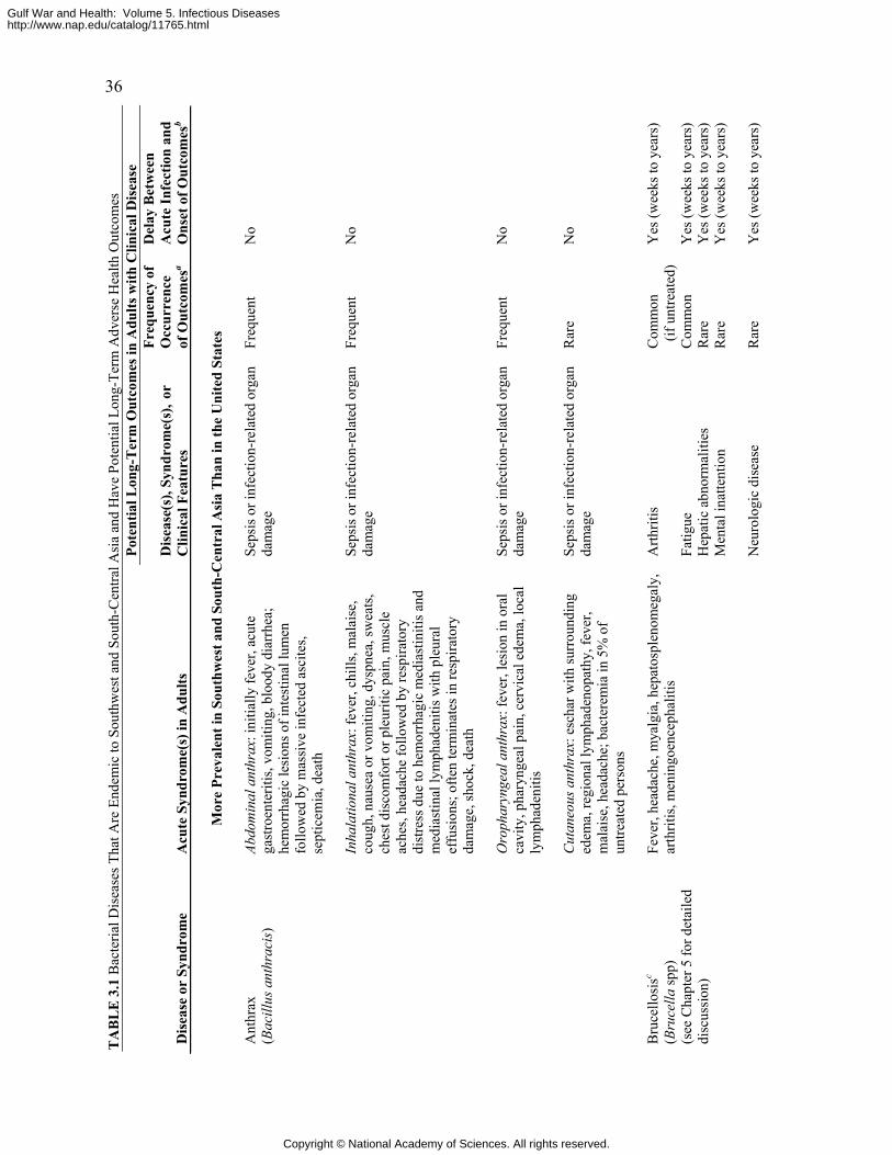
36 T
AB
LE
3.1
Bac
teria
l Dis
ease
s Tha
t Are
End
emic
to S
outh
wes
t and
Sou
th-C
entra
l Asi
a an
d H
ave
Pote
ntia
l Lon
g-Te
rm A
dver
se H
ealth
Out
com
es
Pote
ntia
l Lon
g-T
erm
Out
com
es in
Adu
lts w
ith C
linic
al D
isea
se
Dis
ease
or
Synd
rom
e A
cute
Syn
drom
e(s)
in A
dults
D
isea
se(s
), Sy
ndro
me(
s), o
r C
linic
al F
eatu
res
Freq
uenc
y of
O
ccur
renc
e of
Out
com
esa
Del
ay B
etw
een
A
cute
Infe
ctio
n an
d O
nset
of O
utco
mes
b
Mor
e Pr
eval
ent i
n So
uthw
est a
nd S
outh
-Cen
tral
Asi
a T
han
in th
e U
nite
d St
ates
Abdo
min
al a
nthr
ax: i
nitia
lly fe
ver,
acut
e ga
stro
ente
ritis
, vom
iting
, blo
ody
diar
rhea
; he
mor
rhag
ic le
sion
s of i
ntes
tinal
lum
en
follo
wed
by
mas
sive
infe
cted
asc
ites,
sept
icem
ia, d
eath
Seps
is o
r inf
ectio
n-re
late
d or
gan
dam
age
Freq
uent
No
Inha
latio
nal a
nthr
ax: f
ever
, chi
lls, m
alai
se,
coug
h, n
ause
a or
vom
iting
, dys
pnea
, sw
eats
, ch
est d
isco
mfo
rt or
ple
uriti
c pa
in, m
uscl
e ac
hes,
head
ache
follo
wed
by
resp
irato
ry
dist
ress
due
to h
emor
rhag
ic m
edia
stin
itis a
nd
med
iast
inal
lym
phad
eniti
s with
ple
ural
ef
fusi
ons;
ofte
n te
rmin
ates
in re
spira
tory
da
mag
e, sh
ock,
dea
th
Seps
is o
r inf
ectio
n-re
late
d or
gan
dam
age
Freq
uent
N
o
Oro
phar
ynge
al a
nthr
ax: f
ever
, les
ion
in o
ral
cavi
ty, p
hary
ngea
l pai
n, c
ervi
cal e
dem
a, lo
cal
lym
phad
eniti
s
Seps
is o
r inf
ectio
n-re
late
d or
gan
dam
age
Freq
uent
N
o
Ant
hrax
(B
acill
us a
nthr
acis
)
Cut
aneo
us a
nthr
ax: e
scha
r with
surr
ound
ing
edem
a, re
gion
al ly
mph
aden
opat
hy, f
ever
, m
alai
se, h
eada
che;
bac
tere
mia
in 5
% o
f un
treat
ed p
erso
ns
Seps
is o
r inf
ectio
n-re
late
d or
gan
dam
age
Rar
e N
o
A
rthrit
is
C
omm
on
(if
untre
ated
) Y
es (w
eeks
to y
ears
)
Fatig
ue
Com
mon
Y
es (w
eeks
to y
ears
)
Bru
cello
sisc
(Bru
cella
spp)
(s
ee C
hapt
er 5
for d
etai
led
disc
ussi
on)
Feve
r, he
adac
he, m
yalg
ia, h
epat
ospl
enom
egal
y,
arth
ritis
, men
ingo
ence
phal
itis
Hep
atic
abn
orm
aliti
es
Rar
e Y
es (w
eeks
to y
ears
)
M
enta
l ina
ttent
ion
Rar
e Y
es (w
eeks
to y
ears
)
Neu
rolo
gic
dise
ase
Rar
e Y
es (w
eeks
to y
ears
)
Copyright © National Academy of Sciences. All rights reserved.
Gulf War and Health: Volume 5. Infectious Diseaseshttp://www.nap.edu/catalog/11765.html
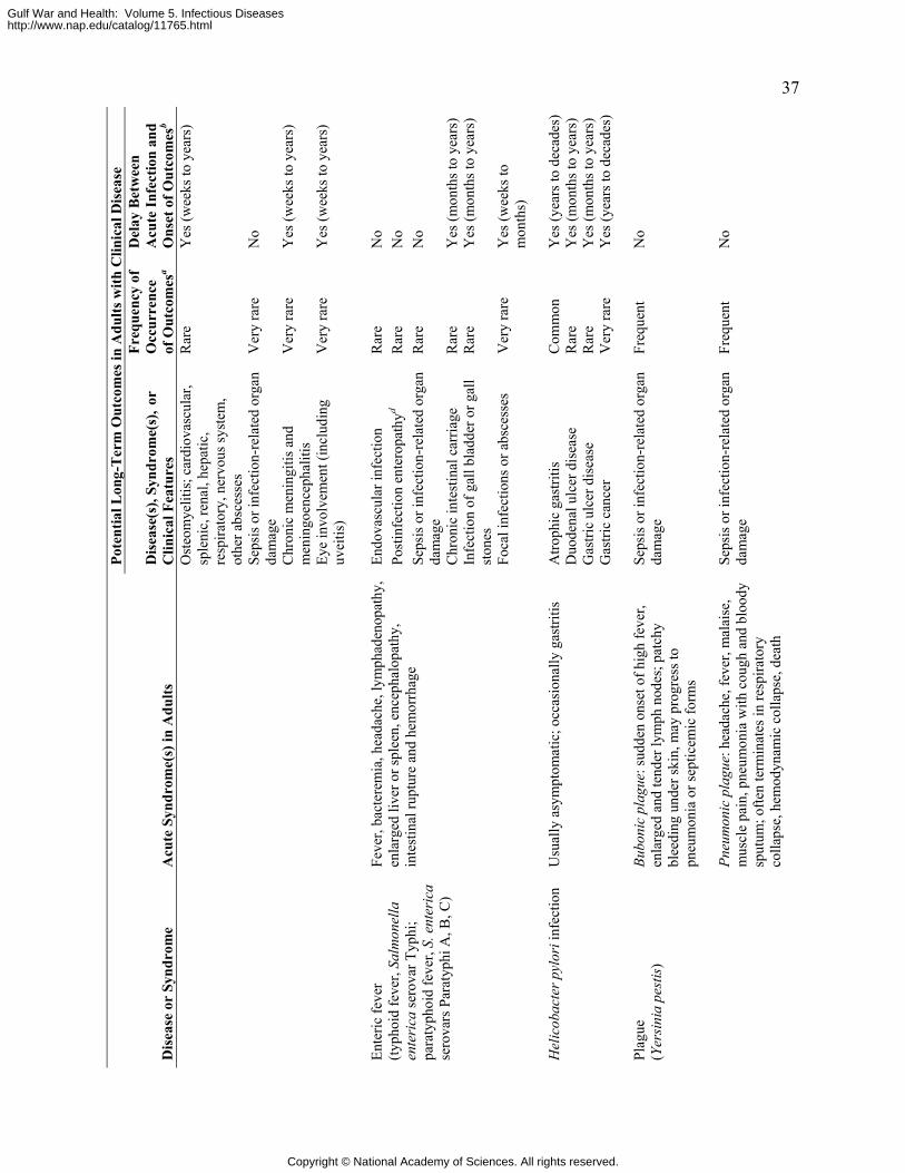
37 Po
tent
ial L
ong-
Ter
m O
utco
mes
in A
dults
with
Clin
ical
Dis
ease
Dis
ease
or
Synd
rom
e A
cute
Syn
drom
e(s)
in A
dults
D
isea
se(s
), Sy
ndro
me(
s), o
r C
linic
al F
eatu
res
Freq
uenc
y of
O
ccur
renc
e of
Out
com
esa
Del
ay B
etw
een
A
cute
Infe
ctio
n an
d O
nset
of O
utco
mes
b
Ost
eom
yelit
is; c
ardi
ovas
cula
r, sp
leni
c, re
nal,
hepa
tic,
resp
irato
ry, n
ervo
us sy
stem
, ot
her a
bsce
sses
Rar
e Y
es (w
eeks
to y
ears
)
Seps
is o
r inf
ectio
n-re
late
d or
gan
dam
age
Ver
y ra
re
No
Chr
onic
men
ingi
tis a
nd
men
ingo
ence
phal
itis
Ver
y ra
re
Yes
(wee
ks to
yea
rs)
Eye
invo
lvem
ent (
incl
udin
g uv
eitis
) V
ery
rare
Y
es (w
eeks
to y
ears
)
En
dova
scul
ar in
fect
ion
Rar
e N
o Po
stin
fect
ion
ente
ropa
thyd
Rar
e N
o Se
psis
or i
nfec
tion-
rela
ted
orga
n da
mag
e R
are
No
Chr
onic
inte
stin
al c
arria
ge
Rar
e Y
es (m
onth
s to
year
s)
Infe
ctio
n of
gal
l bla
dder
or g
all
ston
es
Rar
e Y
es (m
onth
s to
year
s)
Ente
ric fe
ver
(typh
oid
feve
r, Sa
lmon
ella
en
teri
ca se
rova
r Typ
hi;
para
typh
oid
feve
r, S.
ent
eric
a se
rova
rs P
arat
yphi
A, B
, C)
Feve
r, ba
cter
emia
, hea
dach
e, ly
mph
aden
opat
hy,
enla
rged
live
r or s
plee
n, e
ncep
halo
path
y,
inte
stin
al ru
ptur
e an
d he
mor
rhag
e
Foca
l inf
ectio
ns o
r abs
cess
es
Ver
y ra
re
Yes
(wee
ks to
m
onth
s)
A
troph
ic g
astri
tis
Com
mon
Y
es (y
ears
to d
ecad
es)
Duo
dena
l ulc
er d
isea
se
Rar
e Y
es (m
onth
s to
year
s)
Gas
tric
ulce
r dis
ease
R
are
Yes
(mon
ths t
o ye
ars)
Hel
icob
acte
r pyl
ori i
nfec
tion
Usu
ally
asy
mpt
omat
ic; o
ccas
iona
lly g
astri
tis
Gas
tric
canc
er
Ver
y ra
re
Yes
(yea
rs to
dec
ades
)
Plag
ue
(Yer
sini
a pe
stis
) Bu
boni
c pl
ague
: sud
den
onse
t of h
igh
feve
r, en
larg
ed a
nd te
nder
lym
ph n
odes
; pat
chy
blee
ding
und
er sk
in, m
ay p
rogr
ess t
o pn
eum
onia
or s
eptic
emic
form
s
Seps
is o
r inf
ectio
n-re
late
d or
gan
dam
age
Freq
uent
N
o
Pneu
mon
ic p
lagu
e: h
eada
che,
feve
r, m
alai
se,
mus
cle
pain
, pne
umon
ia w
ith c
ough
and
blo
ody
sput
um; o
ften
term
inat
es in
resp
irato
ry
colla
pse,
hem
odyn
amic
col
laps
e, d
eath
Seps
is o
r inf
ectio
n-re
late
d or
gan
dam
age
Freq
uent
N
o
Copyright © National Academy of Sciences. All rights reserved.
Gulf War and Health: Volume 5. Infectious Diseaseshttp://www.nap.edu/catalog/11765.html
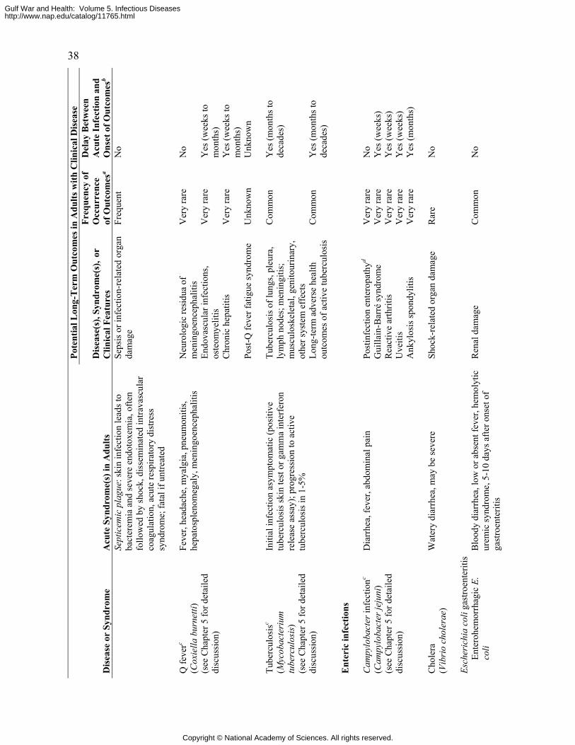
38 Po
tent
ial L
ong-
Ter
m O
utco
mes
in A
dults
with
Clin
ical
Dis
ease
Dis
ease
or
Synd
rom
e A
cute
Syn
drom
e(s)
in A
dults
D
isea
se(s
), Sy
ndro
me(
s), o
r C
linic
al F
eatu
res
Freq
uenc
y of
O
ccur
renc
e of
Out
com
esa
Del
ay B
etw
een
A
cute
Infe
ctio
n an
d O
nset
of O
utco
mes
b
Sept
icem
ic p
lagu
e: sk
in in
fect
ion
lead
s to
bact
erem
ia a
nd se
vere
end
otox
emia
, ofte
n fo
llow
ed b
y sh
ock,
dis
sem
inat
ed in
trava
scul
ar
coag
ulat
ion,
acu
te re
spira
tory
dis
tress
sy
ndro
me;
fata
l if u
ntre
ated
Seps
is o
r inf
ectio
n-re
late
d or
gan
dam
age
Freq
uent
N
o
N
euro
logi
c re
sidu
a of
m
enin
goen
ceph
aliti
s V
ery
rare
N
o
Endo
vasc
ular
infe
ctio
ns,
oste
omye
litis
V
ery
rare
Y
es (w
eeks
to
mon
ths)
C
hron
ic h
epat
itis
Ver
y ra
re
Yes
(wee
ks to
m
onth
s)
Q fe
verc
(Cox
iella
bur
netti
) (s
ee C
hapt
er 5
for d
etai
led
disc
ussi
on)
Feve
r, he
adac
he, m
yalg
ia, p
neum
oniti
s, he
pato
sple
nom
egal
y, m
enin
goen
ceph
aliti
s
Post
-Q fe
ver f
atig
ue sy
ndro
me
Unk
now
n
Unk
now
n
Tu
berc
ulos
is o
f lun
gs, p
leur
a,
lym
ph n
odes
; men
ingi
tis;
mus
culo
skel
etal
, gen
itour
inar
y,
othe
r sys
tem
eff
ects
Com
mon
Y
es (m
onth
s to
deca
des)
Tu
berc
ulos
isc
(Myc
obac
teri
um
tube
rcul
osis
) (s
ee C
hapt
er 5
for d
etai
led
disc
ussi
on)
Initi
al in
fect
ion
asym
ptom
atic
(pos
itive
tu
berc
ulos
is sk
in te
st o
r gam
ma
inte
rfer
on
rele
ase
assa
y); p
rogr
essi
on to
act
ive
tube
rcul
osis
in 1
-5%
Lo
ng-te
rm a
dver
se h
ealth
ou
tcom
es o
f act
ive
tube
rcul
osis
C
omm
on
Yes
(mon
ths t
o de
cade
s)
E
nter
ic in
fect
ions
Post
infe
ctio
n en
tero
path
yd V
ery
rare
N
o G
uilla
in-B
arré
synd
rom
e V
ery
rare
Y
es (w
eeks
) R
eact
ive
arth
ritis
V
ery
rare
Y
es (w
eeks
) U
veiti
s V
ery
rare
Y
es (w
eeks
)
Cam
pylo
bact
er in
fect
ionc
(Cam
pylo
bact
er je
juni
) (s
ee C
hapt
er 5
for d
etai
led
disc
ussi
on)
Dia
rrhe
a, fe
ver,
abdo
min
al p
ain
Ank
ylos
is sp
ondy
litis
V
ery
rare
Y
es (m
onth
s)
C
hole
ra
(Vib
rio
chol
erae
) W
ater
y di
arrh
ea, m
ay b
e se
vere
Sh
ock-
rela
ted
orga
n da
mag
e R
are
No
Es
cher
ichi
a co
li ga
stro
ente
ritis
En
tero
hem
orrh
agic
E.
coli
Blo
ody
diar
rhea
, low
or a
bsen
t fev
er, h
emol
ytic
ur
emic
synd
rom
e, 5
-10
days
afte
r ons
et o
f ga
stro
ente
ritis
Ren
al d
amag
e C
omm
on
No
Copyright © National Academy of Sciences. All rights reserved.
Gulf War and Health: Volume 5. Infectious Diseaseshttp://www.nap.edu/catalog/11765.html
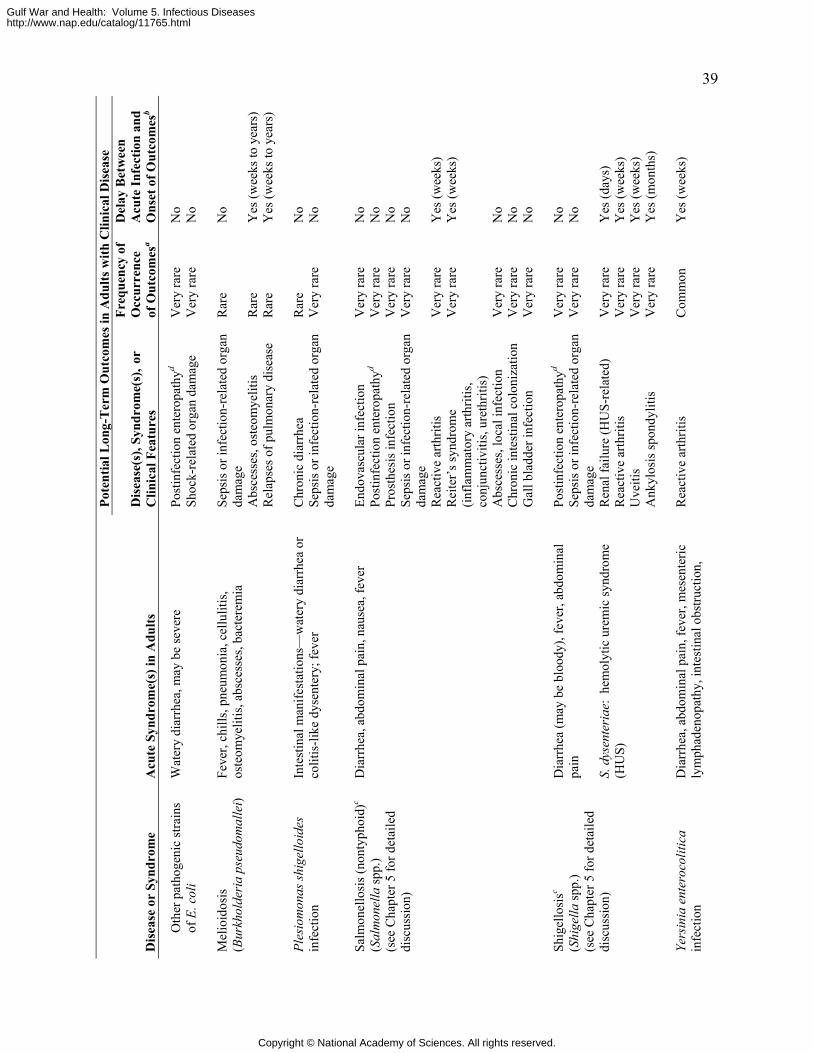
39 Po
tent
ial L
ong-
Ter
m O
utco
mes
in A
dults
with
Clin
ical
Dis
ease
Dis
ease
or
Synd
rom
e A
cute
Syn
drom
e(s)
in A
dults
D
isea
se(s
), Sy
ndro
me(
s), o
r C
linic
al F
eatu
res
Freq
uenc
y of
O
ccur
renc
e of
Out
com
esa
Del
ay B
etw
een
A
cute
Infe
ctio
n an
d O
nset
of O
utco
mes
b
Post
infe
ctio
n en
tero
path
yd V
ery
rare
N
o O
ther
pat
hoge
nic
stra
ins
of E
. col
i W
ater
y di
arrh
ea, m
ay b
e se
vere
Sh
ock-
rela
ted
orga
n da
mag
e V
ery
rare
N
o
Seps
is o
r inf
ectio
n-re
late
d or
gan
dam
age
Rar
e N
o
Abs
cess
es, o
steo
mye
litis
R
are
Yes
(wee
ks to
yea
rs)
Mel
ioid
osis
(B
urkh
olde
ria p
seud
omal
lei)
Feve
r, ch
ills,
pneu
mon
ia, c
ellu
litis
, os
teom
yelit
is, a
bsce
sses
, bac
tere
mia
Rel
apse
s of p
ulm
onar
y di
seas
e R
are
Yes
(wee
ks to
yea
rs)
C
hron
ic d
iarr
hea
R
are
No
Ples
iom
onas
shig
ello
ides
in
fect
ion
Inte
stin
al m
anife
stat
ions
—w
ater
y di
arrh
ea o
r co
litis
-like
dys
ente
ry; f
ever
Se
psis
or i
nfec
tion-
rela
ted
orga
n da
mag
e V
ery
rare
N
o
En
dova
scul
ar in
fect
ion
Ver
y ra
re
No
Post
infe
ctio
n en
tero
path
yd V
ery
rare
N
o Pr
osth
esis
infe
ctio
n V
ery
rare
N
o Se
psis
or i
nfec
tion-
rela
ted
orga
n da
mag
e V
ery
rare
N
o
Rea
ctiv
e ar
thrit
is
Ver
y ra
re
Yes
(wee
ks)
Rei
ter’
s syn
drom
e (in
flam
mat
ory
arth
ritis
, co
njun
ctiv
itis,
uret
hriti
s)
Ver
y ra
re
Yes
(wee
ks)
Abs
cess
es, l
ocal
infe
ctio
n V
ery
rare
N
o C
hron
ic in
test
inal
col
oniz
atio
n V
ery
rare
N
o
Salm
onel
losi
s (no
ntyp
hoid
)c (S
alm
onel
la sp
p.)
(see
Cha
pter
5 fo
r det
aile
d di
scus
sion
)
Dia
rrhe
a, a
bdom
inal
pai
n, n
ause
a, fe
ver
Gal
l bla
dder
infe
ctio
n V
ery
rare
N
o
Post
infe
ctio
n en
tero
path
yd V
ery
rare
N
o Se
psis
or i
nfec
tion-
rela
ted
orga
n da
mag
e
Ver
y ra
re
No
Ren
al fa
ilure
(HU
S-re
late
d)
Ver
y ra
re
Yes
(day
s)
Rea
ctiv
e ar
thrit
is
Ver
y ra
re
Yes
(wee
ks)
Uve
itis
Ver
y ra
re
Yes
(wee
ks)
Shig
ello
sisc
(Shi
gella
spp.
) (s
ee C
hapt
er 5
for d
etai
led
disc
ussi
on)
Dia
rrhe
a (m
ay b
e bl
oody
), fe
ver,
abdo
min
al
pain
S.
dys
ente
riae
: he
mol
ytic
ure
mic
synd
rom
e (H
US)
Ank
ylos
is sp
ondy
litis
V
ery
rare
Y
es (m
onth
s)
Ye
rsin
ia e
nter
ocol
itica
in
fect
ion
Dia
rrhe
a, a
bdom
inal
pai
n, fe
ver,
mes
ente
ric
lym
phad
enop
athy
, int
estin
al o
bstru
ctio
n,
Rea
ctiv
e ar
thrit
is
Com
mon
Y
es (w
eeks
)
Copyright © National Academy of Sciences. All rights reserved.
Gulf War and Health: Volume 5. Infectious Diseaseshttp://www.nap.edu/catalog/11765.html
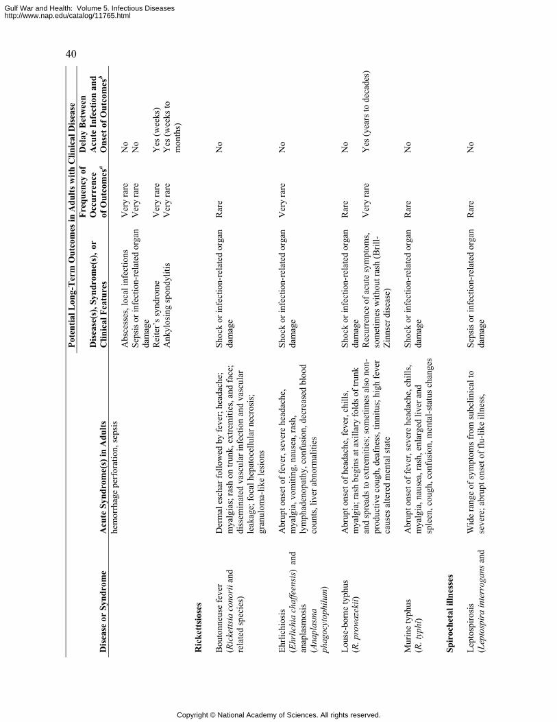
40 Po
tent
ial L
ong-
Ter
m O
utco
mes
in A
dults
with
Clin
ical
Dis
ease
Dis
ease
or
Synd
rom
e A
cute
Syn
drom
e(s)
in A
dults
D
isea
se(s
), Sy
ndro
me(
s), o
r C
linic
al F
eatu
res
Freq
uenc
y of
O
ccur
renc
e of
Out
com
esa
Del
ay B
etw
een
A
cute
Infe
ctio
n an
d O
nset
of O
utco
mes
b
hem
orrh
age
perf
orat
ion,
seps
is
Abs
cess
es, l
ocal
infe
ctio
ns
Ver
y ra
re
No
Seps
is o
r inf
ectio
n-re
late
d or
gan
dam
age
Ver
y ra
re
No
Rei
ter’
s syn
drom
e
Ver
y ra
re
Yes
(wee
ks)
Ank
ylos
ing
spon
dylit
is
Ver
y ra
re
Yes
(wee
ks to
m
onth
s)
R
icke
ttsi
oses
Bou
tonn
euse
feve
r (R
icke
ttsia
con
orii
and
rela
ted
spec
ies)
Der
mal
esc
har f
ollo
wed
by
feve
r; he
adac
he;
mya
lgia
s; ra
sh o
n tru
nk, e
xtre
miti
es, a
nd fa
ce;
diss
emin
ated
vas
cula
r inf
ectio
n an
d va
scul
ar
leak
age;
foca
l hep
atoc
ellu
lar n
ecro
sis;
gr
anul
oma-
like
lesi
ons
Shoc
k or
infe
ctio
n-re
late
d or
gan
dam
age
Rar
e N
o
Eh
rlich
iosi
s (E
hrlic
hia
chaf
feen
sis)
and
an
apla
smos
is
(Ana
plas
ma
phag
ocyt
ophi
lum
)
Abr
upt o
nset
of f
ever
, sev
ere
head
ache
, m
yalg
ia, v
omiti
ng, n
ause
a, ra
sh,
lym
phad
enop
athy
, con
fusi
on, d
ecre
ased
blo
od
coun
ts, l
iver
abn
orm
aliti
es
Shoc
k or
infe
ctio
n-re
late
d or
gan
dam
age
Ver
y ra
re
No
Sh
ock
or in
fect
ion-
rela
ted
orga
n da
mag
e
Rar
e N
o Lo
use-
born
e ty
phus
(R
. pro
waz
ekii)
A
brup
t ons
et o
f hea
dach
e, fe
ver,
chill
s, m
yalg
ia; r
ash
begi
ns a
t axi
llary
fold
s of t
runk
an
d sp
read
s to
extre
miti
es; s
omet
imes
als
o no
n-pr
oduc
tive
coug
h, d
eafn
ess,
tinni
tus;
hig
h fe
ver
caus
es a
ltere
d m
enta
l sta
te
Rec
urre
nce
of a
cute
sym
ptom
s, so
met
imes
with
out r
ash
(Bril
l-Zi
nnse
r dis
ease
)
Ver
y ra
re
Yes
(yea
rs to
dec
ades
)
M
urin
e ty
phus
(R
. typ
hi)
Abr
upt o
nset
of f
ever
, sev
ere
head
ache
, chi
lls,
mya
lgia
, nau
sea,
rash
, enl
arge
d liv
er a
nd
sple
en, c
ough
, con
fusi
on, m
enta
l-sta
tus c
hang
es
Shoc
k or
infe
ctio
n-re
late
d or
gan
dam
age
Rar
e N
o
Sp
iroc
heta
l illn
esse
s Le
ptos
piro
sis
(Lep
tosp
ira
inte
rrog
ans a
nd
Wid
e ra
nge
of sy
mpt
oms f
rom
subc
linic
al to
se
vere
; abr
upt o
nset
of f
lu-li
ke il
lnes
s, Se
psis
or i
nfec
tion-
rela
ted
orga
n da
mag
e R
are
No
Copyright © National Academy of Sciences. All rights reserved.
Gulf War and Health: Volume 5. Infectious Diseaseshttp://www.nap.edu/catalog/11765.html
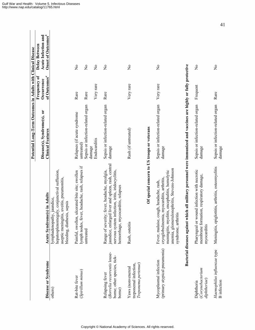
41 Po
tent
ial L
ong-
Ter
m O
utco
mes
in A
dults
with
Clin
ical
Dis
ease
Dis
ease
or
Synd
rom
e A
cute
Syn
drom
e(s)
in A
dults
D
isea
se(s
), Sy
ndro
me(
s), o
r C
linic
al F
eatu
res
Freq
uenc
y of
O
ccur
renc
e of
Out
com
esa
Del
ay B
etw
een
A
cute
Infe
ctio
n an
d O
nset
of O
utco
mes
b ot
hers
) ly
mph
aden
opat
hy, j
aund
ice,
he
pato
sple
nom
egal
y, c
onju
nctiv
al su
ffus
ion,
as
eptic
men
ingi
tis, u
veiti
s, pn
eum
oniti
s, bl
eedi
ng, d
iath
esis
, sep
sis
R
elap
ses (
if ac
ute
synd
rom
e un
treat
ed)
Rar
e N
o
Seps
is o
r inf
ectio
n-re
late
d or
gan
dam
age
Rar
e N
o
Rat
-bite
feve
r (S
piri
llum
min
us)
Pain
ful,
swol
len,
ulc
erat
ed b
ite si
te; s
wol
len
lym
ph n
odes
, fev
er, h
eada
che,
rash
, rel
apse
s if
untre
ated
Endo
card
itis
Ver
y ra
re
No
R
elap
sing
feve
r (B
orre
lia re
curr
entis
lous
e-bo
rne;
oth
er sp
ecie
s, tic
k-bo
rne)
Ran
ge o
f sev
erity
; fev
er, h
eada
che,
mya
lgia
, ja
undi
ce, e
nlar
ged
liver
and
sple
en, r
ash,
cen
tral
nerv
ous s
yste
m in
fect
ion,
iriti
s, iri
docy
cliti
s, he
mor
rhag
e, m
ycoc
ardi
tis, r
elap
ses
Seps
is o
r inf
ectio
n-re
late
d or
gan
dam
age
Rar
e N
o
Y
aws (
nonv
ener
eal
trepo
nem
al in
fect
ion,
Tr
epon
ema
pert
enue
)
Ras
h, o
stei
tis
Ras
h (if
unt
reat
ed)
Ver
y ra
re
No
O
f spe
cial
con
cern
to U
S tr
oops
or
vete
rans
Myc
opla
smal
infe
ctio
n (p
rimar
y at
ypic
al p
neum
onia
) Fe
ver,
mal
aise
, cou
gh, h
eada
che,
rash
, cr
yogl
obul
inem
ia, m
yoca
rditi
s, ar
thrit
is,
men
ingi
tis, m
yelit
is, e
ncep
halit
is, h
emol
ytic
an
emia
, glo
mer
ulon
ephr
itis,
Stev
ens-
John
son
synd
rom
e, a
rthrit
is
Seps
is o
r inf
ectio
n-re
late
d or
gan
dam
age
Ver
y ra
re
No
B
acte
rial
dis
ease
s aga
inst
whi
ch a
ll m
ilita
ry p
erso
nnel
wer
e im
mun
ized
and
vac
cine
s are
hig
hly
or fu
lly p
rote
ctiv
e
Dip
hthe
ria
(Cor
yneb
acte
rium
di
phth
eria
e)
Phar
ynge
al o
r wou
nd in
fect
ion
with
nec
rotic
m
embr
ane
form
atio
n, re
spira
tory
dam
age,
m
yoca
rditi
s
Seps
is o
r inf
ectio
n-re
late
d or
gan
dam
age
Freq
uent
N
o
H
aem
ophi
lus i
nflu
enza
e ty
pe
B in
fect
ion
Men
ingi
tis, e
pigl
ottit
is, a
rthrit
is, o
steo
mye
litis
Se
psis
or i
nfec
tion-
rela
ted
orga
n da
mag
e R
are
No
Copyright © National Academy of Sciences. All rights reserved.
Gulf War and Health: Volume 5. Infectious Diseaseshttp://www.nap.edu/catalog/11765.html
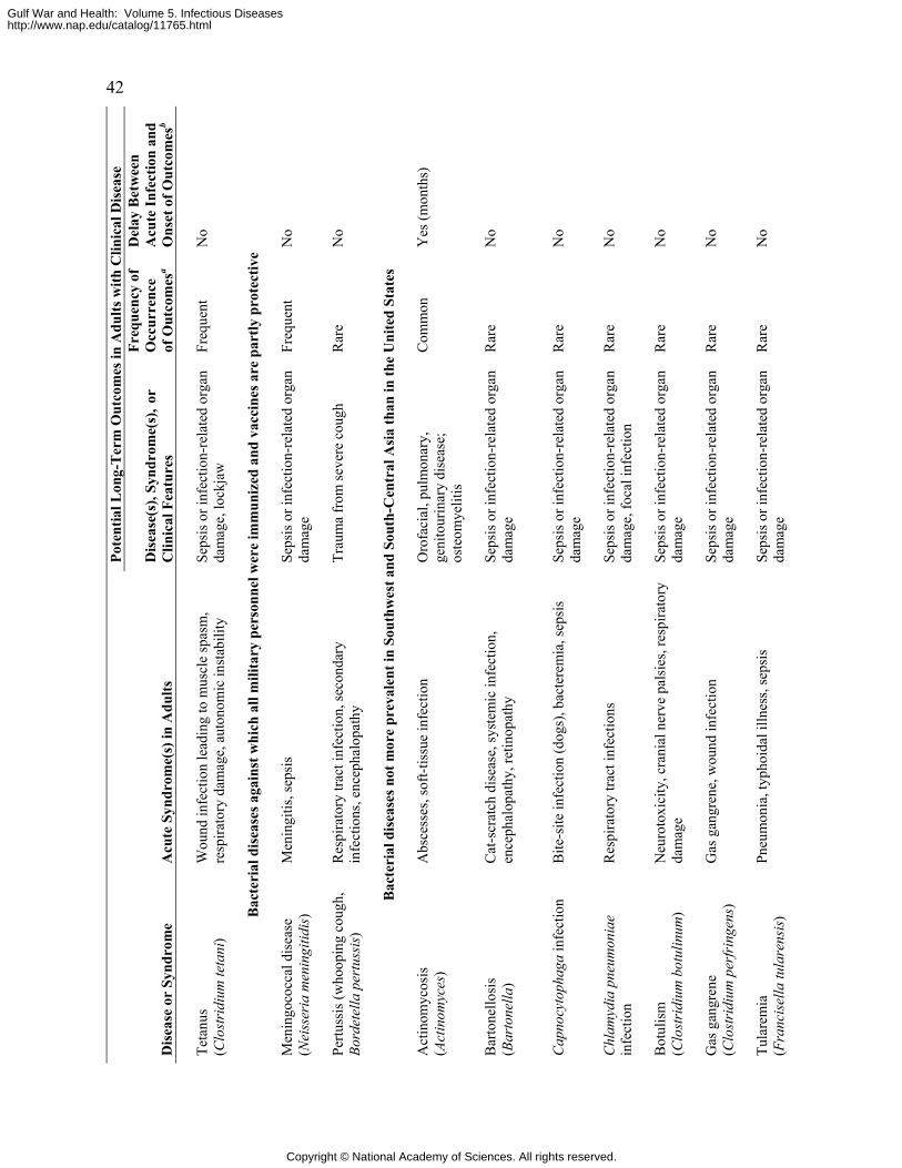
42 Po
tent
ial L
ong-
Ter
m O
utco
mes
in A
dults
with
Clin
ical
Dis
ease
Dis
ease
or
Synd
rom
e A
cute
Syn
drom
e(s)
in A
dults
D
isea
se(s
), Sy
ndro
me(
s), o
r C
linic
al F
eatu
res
Freq
uenc
y of
O
ccur
renc
e of
Out
com
esa
Del
ay B
etw
een
A
cute
Infe
ctio
n an
d O
nset
of O
utco
mes
b
Teta
nus
(Clo
stri
dium
teta
ni)
Wou
nd in
fect
ion
lead
ing
to m
uscl
e sp
asm
, re
spira
tory
dam
age,
aut
onom
ic in
stab
ility
Se
psis
or i
nfec
tion-
rela
ted
orga
n da
mag
e, lo
ckja
w
Freq
uent
N
o
B
acte
rial
dis
ease
s aga
inst
whi
ch a
ll m
ilita
ry p
erso
nnel
wer
e im
mun
ized
and
vac
cine
s are
par
tly p
rote
ctiv
e
Men
ingo
cocc
al d
isea
se
(Nei
sser
ia m
enin
gitid
is)
Men
ingi
tis, s
epsi
s Se
psis
or i
nfec
tion-
rela
ted
orga
n da
mag
e Fr
eque
nt
No
Pe
rtuss
is (w
hoop
ing
coug
h,
Bord
etel
la p
ertu
ssis
) R
espi
rato
ry tr
act i
nfec
tion,
seco
ndar
y in
fect
ions
, enc
epha
lopa
thy
Trau
ma
from
seve
re c
ough
R
are
No
B
acte
rial
dis
ease
s not
mor
e pr
eval
ent i
n So
uthw
est a
nd S
outh
-Cen
tral
Asi
a th
an in
the
Uni
ted
Stat
es
A
ctin
omyc
osis
(A
ctin
omyc
es)
Abs
cess
es, s
oft-t
issu
e in
fect
ion
Oro
faci
al, p
ulm
onar
y,
geni
tour
inar
y di
seas
e;
oste
omye
litis
Com
mon
Y
es (m
onth
s)
B
arto
nello
sis
(Bar
tone
lla)
Cat
-scr
atch
dis
ease
, sys
tem
ic in
fect
ion,
en
ceph
alop
athy
, ret
inop
athy
Se
psis
or i
nfec
tion-
rela
ted
orga
n da
mag
e R
are
No
C
apno
cyto
phag
a in
fect
ion
Bite
-site
infe
ctio
n (d
ogs)
, bac
tere
mia
, sep
sis
Seps
is o
r inf
ectio
n-re
late
d or
gan
dam
age
Rar
e N
o
C
hlam
ydia
pne
umon
iae
infe
ctio
n R
espi
rato
ry tr
act i
nfec
tions
Se
psis
or i
nfec
tion-
rela
ted
orga
n da
mag
e, fo
cal i
nfec
tion
Rar
e N
o
B
otul
ism
(C
lost
ridi
um b
otul
inum
) N
euro
toxi
city
, cra
nial
ner
ve p
alsi
es, r
espi
rato
ry
dam
age
Seps
is o
r inf
ectio
n-re
late
d or
gan
dam
age
Rar
e N
o
G
as g
angr
ene
(Clo
stri
dium
per
frin
gens
)
Gas
gan
gren
e, w
ound
infe
ctio
n Se
psis
or i
nfec
tion-
rela
ted
orga
n da
mag
e R
are
No
Tu
lare
mia
(F
ranc
isel
la tu
lare
nsis)
Pn
eum
onia
, typ
hoid
al il
lnes
s, se
psis
Se
psis
or i
nfec
tion-
rela
ted
orga
n da
mag
e R
are
No
Copyright © National Academy of Sciences. All rights reserved.
Gulf War and Health: Volume 5. Infectious Diseaseshttp://www.nap.edu/catalog/11765.html
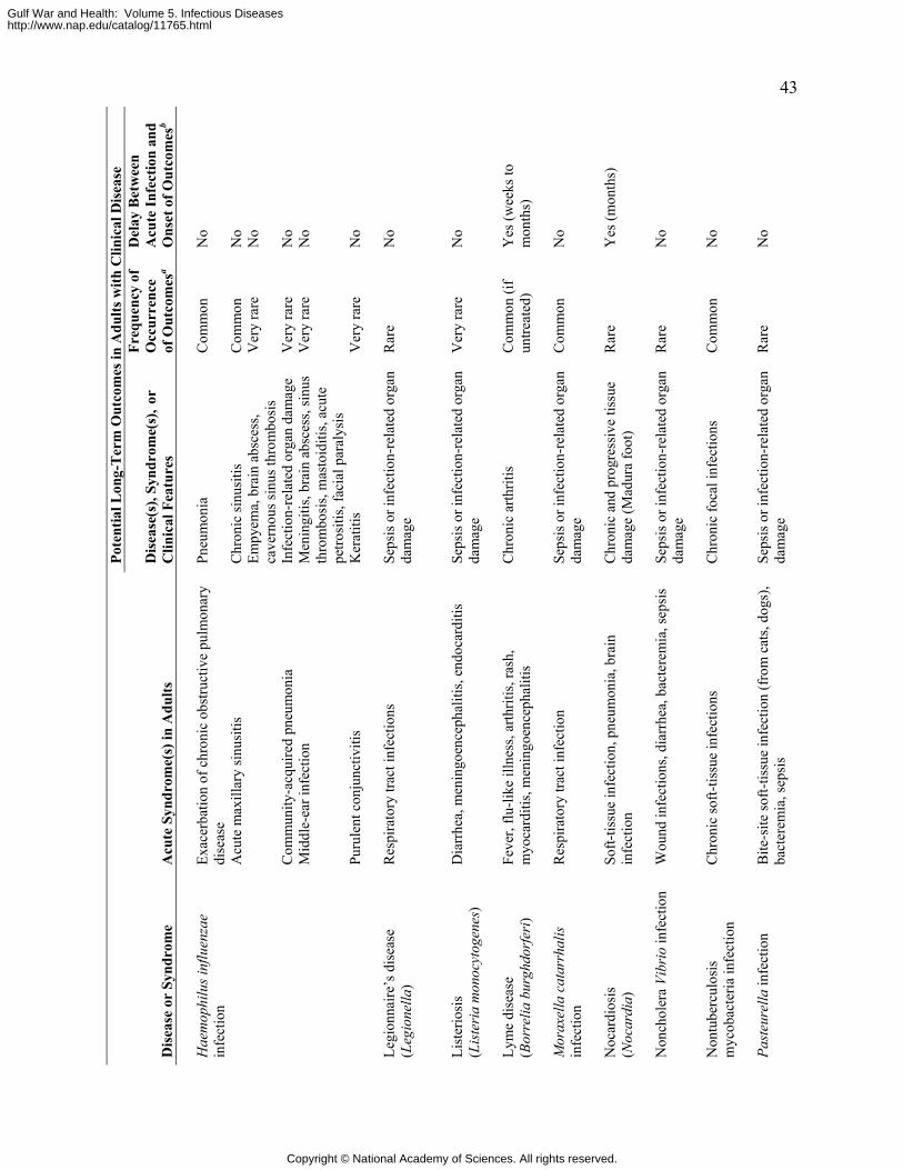
43 Po
tent
ial L
ong-
Ter
m O
utco
mes
in A
dults
with
Clin
ical
Dis
ease
Dis
ease
or
Synd
rom
e A
cute
Syn
drom
e(s)
in A
dults
D
isea
se(s
), Sy
ndro
me(
s), o
r C
linic
al F
eatu
res
Freq
uenc
y of
O
ccur
renc
e of
Out
com
esa
Del
ay B
etw
een
A
cute
Infe
ctio
n an
d O
nset
of O
utco
mes
b
Exac
erba
tion
of c
hron
ic o
bstru
ctiv
e pu
lmon
ary
dise
ase
Pneu
mon
ia
Com
mon
N
o
Chr
onic
sinu
sitis
C
omm
on
No
Acu
te m
axill
ary
sinu
sitis
Em
pyem
a, b
rain
abs
cess
, ca
vern
ous s
inus
thro
mbo
sis
Ver
y ra
re
No
Com
mun
ity-a
cqui
red
pneu
mon
ia
Infe
ctio
n-re
late
d or
gan
dam
age
Ver
y ra
re
No
Mid
dle-
ear i
nfec
tion
Men
ingi
tis, b
rain
abs
cess
, sin
us
thro
mbo
sis,
mas
toid
itis,
acut
e pe
trosi
tis, f
acia
l par
alys
is
Ver
y ra
re
No
Hae
mop
hilu
s inf
luen
zae
infe
ctio
n
Puru
lent
con
junc
tiviti
s K
erat
itis
Ver
y ra
re
No
Le
gion
naire
’s d
isea
se
(Leg
ione
lla)
Res
pira
tory
trac
t inf
ectio
ns
Seps
is o
r inf
ectio
n-re
late
d or
gan
dam
age
Rar
e N
o
Li
ster
iosi
s (L
iste
ria
mon
ocyt
ogen
es)
Dia
rrhe
a, m
enin
goen
ceph
aliti
s, en
doca
rditi
s Se
psis
or i
nfec
tion-
rela
ted
orga
n da
mag
e V
ery
rare
N
o
Ly
me
dise
ase
(B
orre
lia b
urgh
dorf
eri)
Feve
r, flu
-like
illn
ess,
arth
ritis
, ras
h,
myo
card
itis,
men
ingo
ence
phal
itis
Chr
onic
arth
ritis
C
omm
on (i
f un
treat
ed)
Yes
(wee
ks to
m
onth
s)
M
orax
ella
cat
arrh
alis
in
fect
ion
Res
pira
tory
trac
t inf
ectio
n Se
psis
or i
nfec
tion-
rela
ted
orga
n da
mag
e C
omm
on
No
N
ocar
dios
is
(Noc
ardi
a)
Soft-
tissu
e in
fect
ion,
pne
umon
ia, b
rain
in
fect
ion
Chr
onic
and
pro
gres
sive
tiss
ue
dam
age
(Mad
ura
foot
) R
are
Yes
(mon
ths)
N
onch
oler
a Vi
brio
infe
ctio
n W
ound
infe
ctio
ns, d
iarr
hea,
bac
tere
mia
, sep
sis
Seps
is o
r inf
ectio
n-re
late
d or
gan
dam
age
Rar
e N
o
N
ontu
berc
ulos
is
myc
obac
teria
infe
ctio
n C
hron
ic so
ft-tis
sue
infe
ctio
ns
Chr
onic
foca
l inf
ectio
ns
Com
mon
N
o
Past
eure
lla in
fect
ion
Bite
-site
soft-
tissu
e in
fect
ion
(fro
m c
ats,
dogs
), ba
cter
emia
, sep
sis
Seps
is o
r inf
ectio
n-re
late
d or
gan
dam
age
Rar
e N
o
Copyright © National Academy of Sciences. All rights reserved.
Gulf War and Health: Volume 5. Infectious Diseaseshttp://www.nap.edu/catalog/11765.html
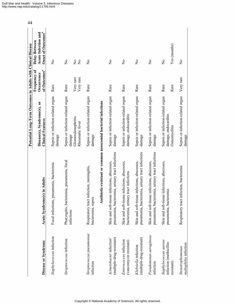
44 Po
tent
ial L
ong-
Ter
m O
utco
mes
in A
dults
with
Clin
ical
Dis
ease
Dis
ease
or
Synd
rom
e A
cute
Syn
drom
e(s)
in A
dults
D
isea
se(s
), Sy
ndro
me(
s), o
r C
linic
al F
eatu
res
Freq
uenc
y of
O
ccur
renc
e of
Out
com
esa
Del
ay B
etw
een
A
cute
Infe
ctio
n an
d O
nset
of O
utco
mes
b
Stap
hylo
cocc
us in
fect
ion
Foca
l inf
ectio
ns, p
neum
onia
, bac
tere
mia
Se
psis
or i
nfec
tion-
rela
ted
orga
n da
mag
e R
are
No
Se
psis
or i
nfec
tion-
rela
ted
orga
n da
mag
e R
are
No
Glo
mer
ulon
ephr
itis
Ver
y ra
re
No
Stre
ptoc
occu
s inf
ectio
n Ph
aryn
gitis
, bac
tere
mia
, pne
umon
ia, f
ocal
in
fect
ions
Rhe
umat
ic fe
ver
Ver
y ra
re
No
St
rept
ococ
cus p
neum
onia
e in
fect
ion
Res
pira
tory
trac
t inf
ectio
n, m
enin
gitis
, ba
cter
emia
, sep
sis
Seps
is o
r inf
ectio
n-re
late
d or
gan
dam
age
Rar
e N
o
A
ntib
iotic
-res
ista
nt o
r co
mm
on n
osoc
omia
l bac
teri
al in
fect
ions
Acin
etob
acte
r inf
ectio
nc
(mul
tiple
-dru
g-re
sist
ant)
Skin
and
soft-
tissu
e in
fect
ions
, abs
cess
es,
pneu
mon
ia, b
acte
rem
ia, u
rinar
y tra
ct in
fect
ions
Se
psis
or i
nfec
tion-
rela
ted
orga
n da
mag
e R
are
No
En
tero
cocc
us in
fect
ion
(van
com
ycin
resi
stan
t) Sk
in a
nd so
ft-tis
sue
infe
ctio
ns, a
bsce
sses
, ba
cter
emia
, urin
ary
tract
infe
ctio
ns
Seps
is o
r inf
ectio
n-re
late
d or
gan
dam
age,
end
ocar
ditis
R
are
No
K
lebs
iella
infe
ctio
n (m
ultip
le-d
rug-
resi
stan
t) Sk
in a
nd so
ft-tis
sue
infe
ctio
ns, a
bsce
sses
, pn
eum
onia
, bac
tere
mia
, urin
ary
tract
infe
ctio
ns
Seps
is o
r inf
ectio
n-re
late
d or
gan
dam
age
Rar
e N
o
Ps
eudo
mon
as a
erug
inos
a in
fect
ion
Skin
and
soft-
tissu
e in
fect
ions
, abs
cess
es,
pneu
mon
ia, b
acte
rem
ia, u
rinar
y tra
ct in
fect
ions
Se
psis
or i
nfec
tion-
rela
ted
orga
n da
mag
e R
are
No
Se
psis
or i
nfec
tion-
rela
ted
orga
n da
mag
e, e
ndoc
ardi
tis
Rar
e N
o St
aphy
loco
ccus
aur
eus
infe
ctio
n (m
ethi
cilli
n-re
sist
ant)
Skin
and
soft-
tissu
e in
fect
ions
, abs
cess
es,
pneu
mon
ia, b
acte
rem
ia
Ost
eom
yelit
is
Rar
e Y
es (m
onth
s)
St
enot
roph
omon
as
mal
toph
ilia
infe
ctio
n R
espi
rato
ry tr
act i
nfec
tion,
bac
tere
mia
Se
psis
or i
nfec
tion-
rela
ted
orga
n da
mag
e V
ery
rare
N
o
Copyright © National Academy of Sciences. All rights reserved.
Gulf War and Health: Volume 5. Infectious Diseaseshttp://www.nap.edu/catalog/11765.html
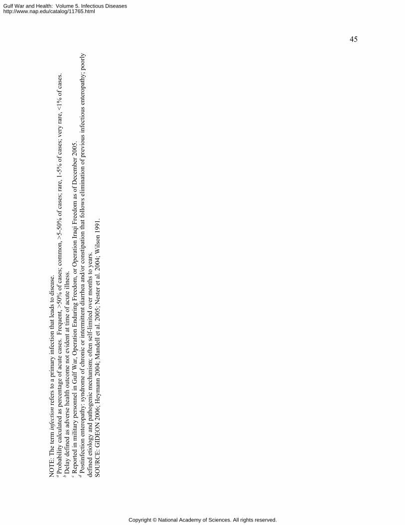
45 N
OTE
: The
term
infe
ctio
n re
fers
to a
prim
ary
infe
ctio
n th
at le
ads t
o di
seas
e.
a Pr
obab
ility
cal
cula
ted
as p
erce
ntag
e of
acu
te c
ases
. Fr
eque
nt, >
50%
of c
ases
; com
mon
, >5-
50%
of c
ases
; rar
e, 1
-5%
of c
ases
; ver
y ra
re, <
1% o
f cas
es.
b D
elay
def
ined
as a
dver
se h
ealth
out
com
e no
t evi
dent
at t
ime
of a
cute
illn
ess.
c Rep
orte
d in
mili
tary
per
sonn
el in
Gul
f War
, Ope
ratio
n En
durin
g Fr
eedo
m, o
r Ope
ratio
n Ir
aqi F
reed
om a
s of D
ecem
ber 2
005.
d P
ostin
fect
ion
ente
ropa
thy:
synd
rom
e of
chr
onic
or i
nter
mitt
ent d
iarr
hea
and/
or c
onst
ipat
ion
that
follo
ws e
limin
atio
n of
pre
viou
s inf
ectio
us e
nter
opat
hy; p
oorly
de
fined
etio
logy
and
pat
hoge
nic
mec
hani
sm; o
ften
self-
limite
d ov
er m
onth
s to
year
s. SO
UR
CE:
GID
EON
200
6; H
eym
ann
2004
; Man
dell
et a
l. 20
05; N
este
r et a
l. 20
04; W
ilson
199
1.
Copyright © National Academy of Sciences. All rights reserved.
Gulf War and Health: Volume 5. Infectious Diseaseshttp://www.nap.edu/catalog/11765.html
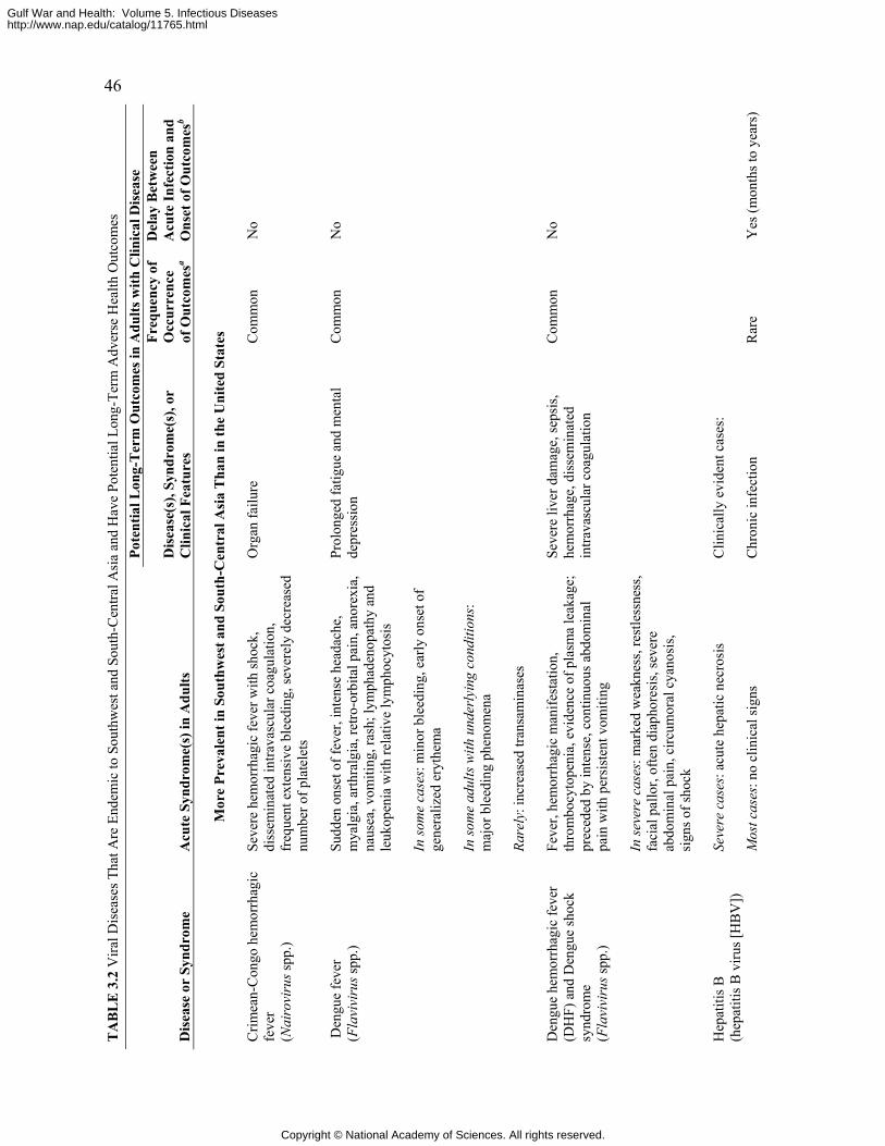
46 T
AB
LE
3.2
Vira
l Dis
ease
s Tha
t Are
End
emic
to S
outh
wes
t and
Sou
th-C
entra
l Asi
a an
d H
ave
Pote
ntia
l Lon
g-Te
rm A
dver
se H
ealth
Out
com
es
Pote
ntia
l Lon
g-T
erm
Out
com
es in
Adu
lts w
ith C
linic
al D
isea
se
Dis
ease
or
Synd
rom
e A
cute
Syn
drom
e(s)
in A
dults
D
isea
se(s
), Sy
ndro
me(
s), o
r C
linic
al F
eatu
res
Freq
uenc
y of
O
ccur
renc
e of
Out
com
esa
Del
ay B
etw
een
Acu
te In
fect
ion
and
Ons
et o
f Out
com
esb
M
ore
Prev
alen
t in
Sout
hwes
t and
Sou
th-C
entr
al A
sia
Tha
n in
the
Uni
ted
Stat
es
C
rimea
n-C
ongo
hem
orrh
agic
fe
ver
(Nai
rovi
rus s
pp.)
Seve
re h
emor
rhag
ic fe
ver w
ith sh
ock,
di
ssem
inat
ed in
trava
scul
ar c
oagu
latio
n,
freq
uent
ext
ensi
ve b
leed
ing,
seve
rely
dec
reas
ed
num
ber o
f pla
tele
ts
Org
an fa
ilure
C
omm
on
No
D
engu
e fe
ver
(Fla
vivi
rus s
pp.)
Sudd
en o
nset
of f
ever
, int
ense
hea
dach
e,
mya
lgia
, arth
ralg
ia, r
etro
-orb
ital p
ain,
ano
rexi
a,
naus
ea, v
omiti
ng, r
ash;
lym
phad
enop
athy
and
le
ukop
enia
with
rela
tive
lym
phoc
ytos
is
In so
me
case
s: m
inor
ble
edin
g, e
arly
ons
et o
f ge
nera
lized
ery
them
a In
som
e ad
ults
with
und
erly
ing
cond
ition
s:
maj
or b
leed
ing
phen
omen
a Ra
rely
: inc
reas
ed tr
ansa
min
ases
Prol
onge
d fa
tigue
and
men
tal
depr
essi
on
Com
mon
N
o
D
engu
e he
mor
rhag
ic fe
ver
(DH
F) a
nd D
engu
e sh
ock
synd
rom
e (F
lavi
viru
s spp
.)
Feve
r, he
mor
rhag
ic m
anife
stat
ion,
th
rom
bocy
tope
nia,
evi
denc
e of
pla
sma
leak
age;
pr
eced
ed b
y in
tens
e, c
ontin
uous
abd
omin
al
pain
with
per
sist
ent v
omiti
ng
In se
vere
cas
es: m
arke
d w
eakn
ess,
rest
less
ness
, fa
cial
pal
lor,
ofte
n di
apho
resi
s, se
vere
ab
dom
inal
pai
n, c
ircum
oral
cya
nosi
s, si
gns o
f sho
ck
Seve
re li
ver d
amag
e, se
psis
, he
mor
rhag
e, d
isse
min
ated
in
trava
scul
ar c
oagu
latio
n
Com
mon
N
o
Se
vere
cas
es: a
cute
hep
atic
nec
rosi
s
Clin
ical
ly e
vide
nt c
ases
:
H
epat
itis B
(h
epat
itis B
viru
s [H
BV
])
Mos
t cas
es: n
o cl
inic
al si
gns
C
hron
ic in
fect
ion
Rar
e Y
es (m
onth
s to
year
s)
Copyright © National Academy of Sciences. All rights reserved.
Gulf War and Health: Volume 5. Infectious Diseaseshttp://www.nap.edu/catalog/11765.html
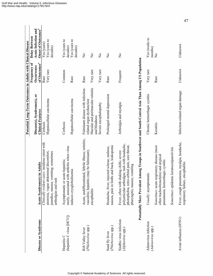
47 Po
tent
ial L
ong-
Ter
m O
utco
mes
in A
dults
with
Clin
ical
Dis
ease
Dis
ease
or
Synd
rom
e A
cute
Syn
drom
e(s)
in A
dults
D
isea
se(s
), Sy
ndro
me(
s), o
r C
linic
al F
eatu
res
Freq
uenc
y of
O
ccur
renc
e of
Out
com
esa
Del
ay B
etw
een
Acu
te In
fect
ion
and
Ons
et o
f Out
com
esb
Cirr
hosi
s R
are
Yes
(yea
rs)
Clin
ical
ly e
vide
nt c
ases
: ins
idio
us o
nset
with
an
orex
ia, v
ague
abd
omin
al d
isco
mfo
rt,
jaun
dice
, nau
sea,
vom
iting
; som
etim
es
arth
ralg
ias a
nd ra
sh
Hep
atoc
ellu
lar c
arci
nom
a V
ery
rare
Y
es (y
ears
to
deca
des)
C
irrho
sis
Com
mon
Y
es (y
ears
to
deca
des)
H
epat
itis C
(h
epat
itis C
viru
s [H
CV
])
Asy
mpt
omat
ic o
r acu
te h
epat
itis;
ra
rely
, ass
ocia
ted
with
arth
ritis
whe
n vi
rus
indu
ces c
ryog
lobu
linem
ia
Hep
atoc
ellu
lar c
arci
nom
a R
are
Yes
(yea
rs to
de
cade
s)
Fu
lmin
ant d
isea
se w
ith in
fect
ion-
rela
ted
orga
n dy
sfun
ctio
n
Rar
e N
o
Mac
ular
and
per
imac
ular
retin
itis
and
blin
dnes
s V
ery
rare
N
o
Rift
Val
ley
feve
r (P
hleb
ovir
us sp
p.)
Acu
te, u
ndiff
eren
tiate
d, fe
brile
illn
ess;
retin
itis;
va
scul
itis;
hep
atiti
s (m
ay b
e fu
lmin
ant);
en
ceph
aliti
s
Seve
re e
ncep
halo
path
y V
ery
rare
N
o
Sand
fly
feve
r (P
hleb
ovir
us sp
p.)
Hea
dach
e, fe
ver,
inje
cted
scle
rae,
mal
aise
, na
usea
, pai
n in
lim
bs a
nd b
ack,
leuk
open
ia
Prol
onge
d m
enta
l dep
ress
ion
Rar
e N
o
Si
ndbi
s viru
s inf
ectio
n (A
lpha
viru
s spp
.) Fe
ver,
ofte
n w
ith sh
akin
g ch
ills;
seve
re
poly
artic
ular
arth
ralg
ia; r
ash
with
hea
dach
e,
phot
opho
bia,
retro
-orb
ital p
ain,
sore
thro
at,
phar
yngi
tis, n
ause
a, v
omiti
ng
Arth
ralg
ia a
nd m
yalg
ia
Freq
uent
N
o
Po
tent
ially
Mor
e Pr
eval
ent A
mon
g T
roop
s in
Sout
hwes
t and
Sou
th-C
entr
al A
sia
Tha
n A
mon
g U
S Po
pula
tion
U
sual
ly: a
sym
ptom
atic
Chr
onic
hem
orrh
agic
cys
titis
V
ery
rare
Y
es (w
eeks
to
mon
ths)
K
erat
itis
Rar
e N
o So
met
imes
: acu
te re
spira
tory
dis
ease
(mos
t of
ten
trach
eobr
onch
itis)
and
aty
pica
l pn
eum
onia
, hem
orrh
agic
cys
titis
Ade
novi
rus i
nfec
tion
(Ade
novi
rus s
pp.)
Som
etim
es: e
pide
mic
ker
atoc
onju
nctiv
itis
A
vian
influ
enza
(H5N
1)
Feve
r, co
ugh
pneu
mon
ia, m
yalg
ia, h
eada
che,
re
spira
tory
failu
re, e
ncep
halit
is
Infe
ctio
n-re
late
d or
gan
dam
age
Unk
now
n U
nkno
wn
Copyright © National Academy of Sciences. All rights reserved.
Gulf War and Health: Volume 5. Infectious Diseaseshttp://www.nap.edu/catalog/11765.html
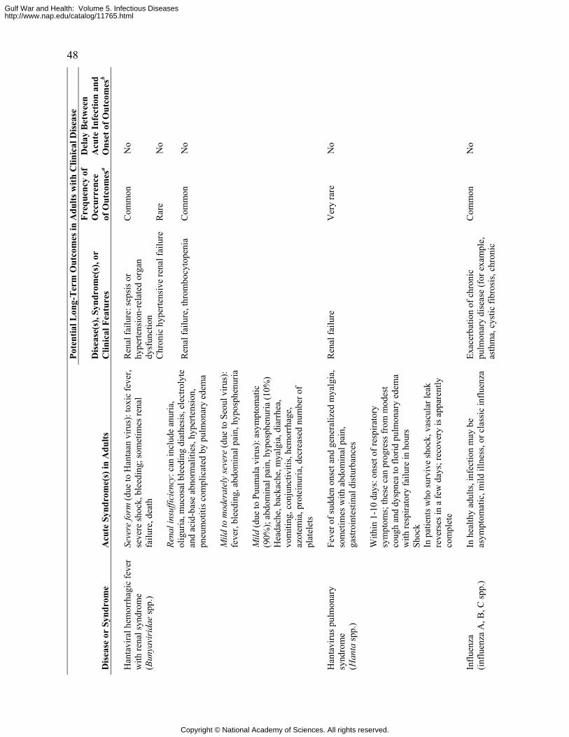
48 Po
tent
ial L
ong-
Ter
m O
utco
mes
in A
dults
with
Clin
ical
Dis
ease
Dis
ease
or
Synd
rom
e A
cute
Syn
drom
e(s)
in A
dults
D
isea
se(s
), Sy
ndro
me(
s), o
r C
linic
al F
eatu
res
Freq
uenc
y of
O
ccur
renc
e of
Out
com
esa
Del
ay B
etw
een
Acu
te In
fect
ion
and
Ons
et o
f Out
com
esb
R
enal
failu
re: s
epsi
s or
hype
rtens
ion-
rela
ted
orga
n dy
sfun
ctio
n
Com
mon
N
o
Chr
onic
hyp
erte
nsiv
e re
nal f
ailu
re
Rar
e N
o
Seve
re fo
rm (d
ue to
Han
taan
viru
s): t
oxic
feve
r, se
vere
shoc
k, b
leed
ing;
som
etim
es re
nal
failu
re, d
eath
Re
nal i
nsuf
ficie
ncy:
can
incl
ude
anur
ia,
olig
uria
, muc
osal
ble
edin
g di
athe
sis,
elec
troly
te
and
acid
-bas
e ab
norm
aliti
es, h
yper
tens
ion,
pn
eum
otiti
s com
plic
ated
by
pulm
onar
y ed
ema
Mild
to m
oder
atel
y se
vere
(due
to S
eoul
viru
s):
feve
r, bl
eedi
ng, a
bdom
inal
pai
n, h
ypos
phen
uria
Han
tavi
ral h
emor
rhag
ic fe
ver
with
rena
l syn
drom
e
(Bun
yavi
rida
e sp
p.)
Mild
(due
to P
uum
ala
viru
s): a
sym
ptom
atic
(9
0%);
abdo
min
al p
ain,
hyp
osph
enur
ia (1
0%)
Hea
dach
e, b
acka
che,
mya
lgia
, dia
rrhe
a,
vom
iting
, con
junc
tiviti
s, he
mor
rhag
e,
azot
emia
, pro
tein
uria
, dec
reas
ed n
umbe
r of
plat
elet
s
Ren
al fa
ilure
, thr
ombo
cyto
peni
a C
omm
on
No
H
anta
viru
s pul
mon
ary
synd
rom
e
(Han
ta sp
p.)
Feve
r of s
udde
n on
set a
nd g
ener
aliz
ed m
yalg
ia,
som
etim
es w
ith a
bdom
inal
pai
n,
gast
roin
test
inal
dis
turb
ance
s W
ithin
1-1
0 da
ys: o
nset
of r
espi
rato
ry
sym
ptom
s; th
ese
can
prog
ress
from
mod
est
coug
h an
d dy
spne
a to
flor
id p
ulm
onar
y ed
ema
with
resp
irato
ry fa
ilure
in h
ours
Sh
ock
In p
atie
nts w
ho su
rviv
e sh
ock,
vas
cula
r lea
k re
vers
es in
a fe
w d
ays;
reco
very
is a
ppar
ently
co
mpl
ete
Ren
al fa
ilure
V
ery
rare
N
o
In
fluen
za
(influ
enza
A, B
, C sp
p.)
In h
ealth
y ad
ults
, inf
ectio
n m
ay b
e as
ympt
omat
ic, m
ild il
lnes
s, or
cla
ssic
influ
enza
Exac
erba
tion
of c
hron
ic
pulm
onar
y di
seas
e (f
or e
xam
ple,
as
thm
a, c
ystic
fibr
osis
, chr
onic
Com
mon
N
o
Copyright © National Academy of Sciences. All rights reserved.
Gulf War and Health: Volume 5. Infectious Diseaseshttp://www.nap.edu/catalog/11765.html
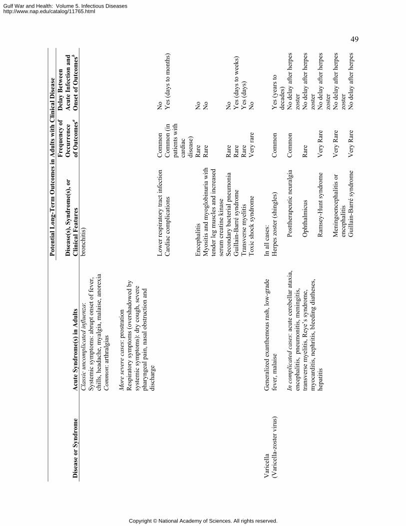
49 Po
tent
ial L
ong-
Ter
m O
utco
mes
in A
dults
with
Clin
ical
Dis
ease
Dis
ease
or
Synd
rom
e A
cute
Syn
drom
e(s)
in A
dults
D
isea
se(s
), Sy
ndro
me(
s), o
r C
linic
al F
eatu
res
Freq
uenc
y of
O
ccur
renc
e of
Out
com
esa
Del
ay B
etw
een
Acu
te In
fect
ion
and
Ons
et o
f Out
com
esb
Cla
ssic
unc
ompl
icat
ed in
fluen
za:
Syst
emic
sym
ptom
s: a
brup
t ons
et o
f fev
er,
chill
s, he
adac
he, m
yalg
ia, m
alai
se, a
nore
xia
Com
mon
: arth
ralg
ias
Mor
e se
vere
cas
es: p
rost
ratio
n R
espi
rato
ry sy
mpt
oms (
over
shad
owed
by
syst
emic
sym
ptom
s): d
ry c
ough
, sev
ere
phar
ynge
al p
ain,
nas
al o
bstru
ctio
n an
d di
scha
rge
bron
chiti
s)
Lo
wer
resp
irato
ry tr
act i
nfec
tion
Com
mon
N
o
Car
diac
com
plic
atio
ns
Com
mon
(in
patie
nts w
ith
card
iac
dise
ase)
Yes
(day
s to
mon
ths)
Ence
phal
itis
Rar
e N
o M
yosi
tis a
nd m
yogl
obin
uria
with
te
nder
leg
mus
cles
and
incr
ease
d se
rum
cre
atin
e ki
nase
Rar
e N
o
Seco
ndar
y ba
cter
ial p
neum
onia
R
are
No
Gui
llain
-Bar
ré sy
ndro
me
Rar
e Y
es (d
ays t
o w
eeks
) Tr
ansv
erse
mye
litis
R
are
Yes
(day
s)
To
xic
shoc
k sy
ndro
me
Ver
y ra
re
No
In
all
case
s:
Her
pes z
oste
r (sh
ingl
es)
Com
mon
Y
es (y
ears
to
deca
des)
Po
stth
erap
eutic
neu
ralg
ia
Com
mon
N
o de
lay
afte
r her
pes
zost
er
Oph
thal
mic
us
Rar
e N
o de
lay
afte
r her
pes
zost
er
Var
icel
la
(Var
icel
la-z
oste
r viru
s)
Gen
eral
ized
exa
nthe
mou
s ras
h, lo
w-g
rade
fe
ver,
mal
aise
In
com
plic
ated
cas
es: a
cute
cer
ebel
lar a
taxi
a,
ence
phal
itis,
pne
umon
itis,
men
ingi
tis,
trans
vers
e m
yelit
is, R
eye’
s syn
drom
e,
myo
card
itis,
neph
ritis
, ble
edin
g di
athe
ses,
hepa
titis
R
amse
y-H
unt s
yndr
ome
Ver
y R
are
No
dela
y af
ter h
erpe
s zo
ster
Men
ingo
ence
phal
itis o
r en
ceph
aliti
s V
ery
Rar
e N
o de
lay
afte
r her
pes
zost
er
Gui
llain
-Bar
ré sy
ndro
me
Ver
y R
are
No
dela
y af
ter h
erpe
s
Copyright © National Academy of Sciences. All rights reserved.
Gulf War and Health: Volume 5. Infectious Diseaseshttp://www.nap.edu/catalog/11765.html
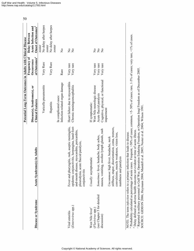
50 Po
tent
ial L
ong-
Ter
m O
utco
mes
in A
dults
with
Clin
ical
Dis
ease
Dis
ease
or
Synd
rom
e A
cute
Syn
drom
e(s)
in A
dults
D
isea
se(s
), Sy
ndro
me(
s), o
r C
linic
al F
eatu
res
Freq
uenc
y of
O
ccur
renc
e of
Out
com
esa
Del
ay B
etw
een
Acu
te In
fect
ion
and
Ons
et o
f Out
com
esb
zost
er
Var
icel
la p
neum
oniti
s V
ery
Rar
e N
o de
lay
afte
r her
pes
zost
er
Hep
atiti
s V
ery
Rar
e N
o de
lay
afte
r her
pes
zost
er
In c
ompl
icat
ed c
ases
: In
fect
ion-
rela
ted
orga
n da
mag
e R
are
No
H
eart
failu
re d
ue to
myo
card
itis
Ver
y ra
re
No
Vira
l ent
eriti
s (E
nter
ovir
us sp
p.)
Feve
r and
pha
ryng
itis,
rash
, ase
ptic
men
ingi
tis,
epid
emic
con
junc
tiviti
s, he
rpan
gina
, han
d-fo
ot-
and-
mou
th d
isea
se, m
yoca
rditi
s, pe
ricar
ditis
, pl
euro
dyni
a, a
cute
flac
cid
para
lysi
s, co
njun
ctiv
itis
Chr
onic
men
ingo
ence
phal
itis
Ver
y ra
re
No
If
sym
ptom
atic
: W
est N
ile n
euro
logi
c di
seas
e V
ery
rare
N
o A
cute
flac
cid
para
lysi
s V
ery
rare
N
o
Wes
t Nile
feve
rc
(Fla
vivi
rus s
pp.)
(see
Cha
pter
5 fo
r det
aile
d di
scus
sion
)
Usu
ally
: asy
mpt
omat
ic
Som
etim
es: f
ever
, hea
dach
e, b
ody
ache
s, na
usea
, vom
iting
, sw
olle
n ly
mph
gla
nds,
rash
U
ncom
mon
: hig
h fe
ver,
head
ache
, nec
k st
iffne
ss, s
tupo
r, di
sorie
ntat
ion,
com
a, tr
emor
s, co
nvul
sion
s, m
uscl
e w
eakn
ess,
visi
on lo
ss,
num
bnes
s and
par
alys
is
Cog
nitiv
e, p
hysi
cal,
or fu
nctio
nal
impa
irmen
t V
ery
rare
N
o
NO
TE: T
he te
rm in
fect
ion
refe
rs to
a p
rimar
y in
fect
ion
that
lead
s to
dise
ase.
a P
roba
bilit
y ca
lcul
ated
as p
erce
ntag
e of
acu
te c
ases
. Fr
eque
nt, >
50%
of c
ases
; com
mon
, >5-
50%
of c
ases
; rar
e, 1
-5%
of c
ases
; ver
y ra
re, <
1% o
f cas
es.
b Del
ay d
efin
ed a
s adv
erse
hea
lth o
utco
me
not e
vide
nt a
t tim
e of
acu
te il
lnes
s. c R
epor
ted
in m
ilita
ry p
erso
nnel
in G
ulf W
ar, O
pera
tion
Endu
ring
Free
dom
, or O
pera
tion
Iraq
i Fre
edom
as o
f Dec
embe
r 200
5.
SOU
RC
E: G
IDEO
N 2
006;
Hey
man
n 20
04; M
ande
ll et
al.
2005
; Nes
ter e
t al.
2004
; Wils
on 1
991.
Copyright © National Academy of Sciences. All rights reserved.
Gulf War and Health: Volume 5. Infectious Diseaseshttp://www.nap.edu/catalog/11765.html
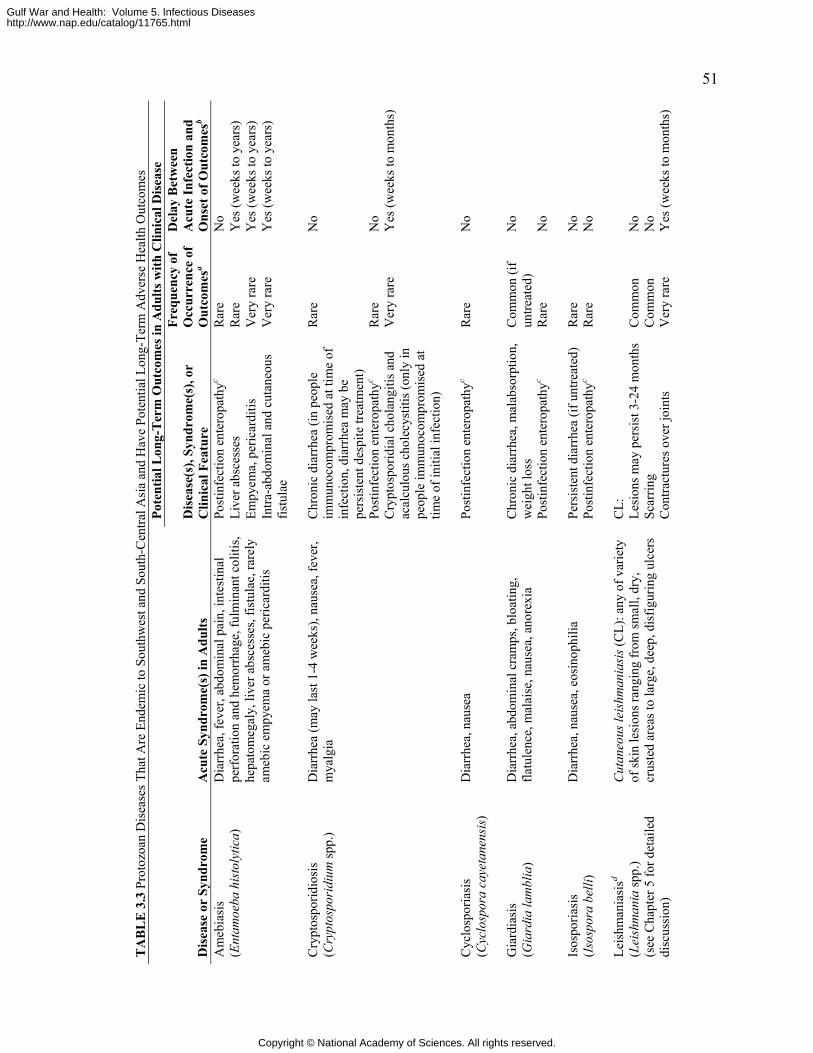
51
TA
BL
E 3
.3 P
roto
zoan
Dis
ease
s Tha
t Are
End
emic
to S
outh
wes
t and
Sou
th-C
entra
l Asi
a an
d H
ave
Pote
ntia
l Lon
g-Te
rm A
dver
se H
ealth
Out
com
es
Pote
ntia
l Lon
g-T
erm
Out
com
es in
Adu
lts w
ith C
linic
al D
isea
se
Dis
ease
or
Synd
rom
e A
cute
Syn
drom
e(s)
in A
dults
D
isea
se(s
), Sy
ndro
me(
s), o
r C
linic
al F
eatu
re
Freq
uenc
y of
O
ccur
renc
e of
O
utco
mes
a
Del
ay B
etw
een
A
cute
Infe
ctio
n an
d O
nset
of O
utco
mes
b
Post
infe
ctio
n en
tero
path
yc R
are
No
Live
r abs
cess
es
Rar
e Y
es (w
eeks
to y
ears
) Em
pyem
a, p
eric
ardi
tis
Ver
y ra
re
Yes
(wee
ks to
yea
rs)
Am
ebia
sis
(Ent
amoe
ba h
isto
lytic
a)
Dia
rrhe
a, fe
ver,
abdo
min
al p
ain,
inte
stin
al
perf
orat
ion
and
hem
orrh
age,
fulm
inan
t col
itis,
hepa
tom
egal
y, li
ver a
bsce
sses
, fis
tula
e, ra
rely
am
ebic
em
pyem
a or
am
ebic
per
icar
ditis
In
tra-a
bdom
inal
and
cut
aneo
us
fistu
lae
Ver
y ra
re
Yes
(wee
ks to
yea
rs)
C
hron
ic d
iarr
hea
(in p
eopl
e im
mun
ocom
prom
ised
at t
ime
of
infe
ctio
n, d
iarr
hea
may
be
pers
iste
nt d
espi
te tr
eatm
ent)
Rar
e N
o
Post
infe
ctio
n en
tero
path
yc R
are
No
Cry
ptos
porid
iosi
s (C
rypt
ospo
ridi
um sp
p.)
Dia
rrhe
a (m
ay la
st 1
-4 w
eeks
), na
usea
, fev
er,
mya
lgia
Cry
ptos
porid
ial c
hola
ngiti
s and
ac
alcu
lous
cho
lecy
stiti
s (on
ly in
pe
ople
imm
unoc
ompr
omis
ed a
t tim
e of
initi
al in
fect
ion)
Ver
y ra
re
Yes
(wee
ks to
mon
ths)
C
yclo
spor
iasi
s (C
yclo
spor
a ca
yeta
nens
is)
Dia
rrhe
a, n
ause
a Po
stin
fect
ion
ente
ropa
thyc
Rar
e N
o
C
hron
ic d
iarr
hea,
mal
abso
rptio
n,
wei
ght l
oss
Com
mon
(if
untre
ated
) N
o G
iard
iasi
s (G
iard
ia la
mbl
ia)
Dia
rrhe
a, a
bdom
inal
cra
mps
, blo
atin
g,
flatu
lenc
e, m
alai
se, n
ause
a, a
nore
xia
Post
infe
ctio
n en
tero
path
yc R
are
No
Pe
rsis
tent
dia
rrhe
a (if
unt
reat
ed)
Rar
e N
o Is
ospo
riasi
s (I
sosp
ora
belli
) D
iarr
hea,
nau
sea,
eos
inop
hilia
Po
stin
fect
ion
ente
ropa
thyc
Rar
e N
o
Leis
hman
iasi
sd (L
eish
man
ia sp
p.)
(see
Cha
pter
5 fo
r det
aile
d
Cut
aneo
us le
ishm
ania
sis (
CL)
: any
of v
arie
ty
of sk
in le
sion
s ran
ging
from
smal
l, dr
y,
crus
ted
area
s to
larg
e, d
eep,
dis
figur
ing
ulce
rs
CL:
Le
sion
s may
per
sist
3-2
4 m
onth
s Sc
arrin
g
Com
mon
C
omm
on
No
No
disc
ussi
on)
C
ontra
ctur
es o
ver j
oint
s V
ery
rare
Y
es (w
eeks
to m
onth
s)
Copyright © National Academy of Sciences. All rights reserved.
Gulf War and Health: Volume 5. Infectious Diseaseshttp://www.nap.edu/catalog/11765.html
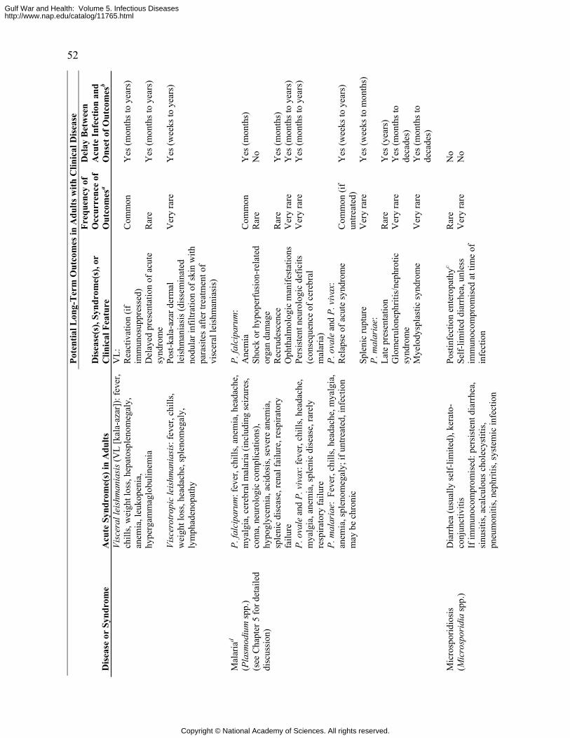
52 Po
tent
ial L
ong-
Ter
m O
utco
mes
in A
dults
with
Clin
ical
Dis
ease
Dis
ease
or
Synd
rom
e A
cute
Syn
drom
e(s)
in A
dults
D
isea
se(s
), Sy
ndro
me(
s), o
r C
linic
al F
eatu
re
Freq
uenc
y of
O
ccur
renc
e of
O
utco
mes
a
Del
ay B
etw
een
A
cute
Infe
ctio
n an
d O
nset
of O
utco
mes
b
V
L:
R
eact
ivat
ion
(if
imm
unos
uppr
esse
d)
Com
mon
Y
es (m
onth
s to
year
s)
D
elay
ed p
rese
ntat
ion
of a
cute
sy
ndro
me
Rar
e Y
es (m
onth
s to
year
s)
Visc
eral
leis
hman
iasi
s (V
L [k
ala-
azar
]): f
ever
, ch
ills,
wei
ght l
oss,
hepa
tosp
leno
meg
aly,
an
emia
, leu
kope
nia,
hy
perg
amm
aglo
bulin
emia
Vi
scer
otro
pic
leis
hman
iasi
s: fe
ver,
chill
s, w
eigh
t los
s, he
adac
he, s
plen
omeg
aly,
ly
mph
aden
opat
hy
Post
-kal
a-az
ar d
erm
al
leis
hman
iasi
s (di
ssem
inat
ed
nodu
lar i
nfilt
ratio
n of
skin
with
pa
rasi
tes a
fter t
reat
men
t of
visc
eral
leis
hman
iasi
s)
Ver
y ra
re
Yes
(wee
ks to
yea
rs)
P.
falc
ipar
um:
Ane
mia
C
omm
on
Yes
(mon
ths)
Sh
ock
or h
ypop
erfu
sion
-rel
ated
or
gan
dam
age
Rar
e N
o
Rec
rude
scen
ce
Rar
e Y
es (m
onth
s)
Oph
thal
mol
ogic
man
ifest
atio
ns
Ver
y ra
re
Yes
(mon
ths t
o ye
ars)
P. fa
lcip
arum
: fev
er, c
hills
, ane
mia
, hea
dach
e,
mya
lgia
, cer
ebra
l mal
aria
(inc
ludi
ng se
izur
es,
com
a, n
euro
logi
c co
mpl
icat
ions
), hy
pogl
ycem
ia, a
cido
sis,
seve
re a
nem
ia,
sple
nic
dise
ase,
rena
l fai
lure
, res
pira
tory
fa
ilure
P.
ova
le a
nd P
. viv
ax: f
ever
, chi
lls, h
eada
che,
m
yalg
ia, a
nem
ia, s
plen
ic d
isea
se, r
arel
y re
spira
tory
failu
re
Pers
iste
nt n
euro
logi
c de
ficits
(c
onse
quen
ce o
f cer
ebra
l m
alar
ia)
Ver
y ra
re
Yes
(mon
ths t
o ye
ars)
P. o
vale
and
P. v
ivax
:
R
elap
se o
f acu
te sy
ndro
me
Com
mon
(if
untre
ated
) Y
es (w
eeks
to y
ears
) P.
mal
aria
e: F
ever
, chi
lls, h
eada
che,
mya
lgia
, an
emia
, spl
enom
egal
y; if
unt
reat
ed, i
nfec
tion
may
be
chro
nic
Sple
nic
rupt
ure
Ver
y ra
re
Yes
(wee
ks to
mon
ths)
P.
mal
aria
e:
Late
pre
sent
atio
n R
are
Yes
(yea
rs)
Glo
mer
ulon
ephr
itis/
neph
rotic
sy
ndro
me
Ver
y ra
re
Yes
(mon
ths t
o de
cade
s)
Mal
aria
d (P
lasm
odiu
m sp
p.)
(see
Cha
pter
5 fo
r det
aile
d di
scus
sion
)
Mye
lody
spla
stic
synd
rom
e V
ery
rare
Y
es (m
onth
s to
deca
des)
Post
infe
ctio
n en
tero
path
yc R
are
No
Mic
rosp
orid
iosi
s (M
icro
spor
idia
spp.
) D
iarr
hea
(usu
ally
self-
limite
d), k
erat
o-co
njun
ctiv
itis
If im
mun
ocom
prom
ised
: per
sist
ent d
iarr
hea,
si
nusi
tis, a
calc
ulou
s cho
lecy
stiti
s, pn
eum
oniti
s, ne
phrit
is, s
yste
mic
infe
ctio
n
Self-
limite
d di
arrh
ea, u
nles
s im
mun
ocom
prom
ised
at t
ime
of
infe
ctio
n
Ver
y ra
re
No
Copyright © National Academy of Sciences. All rights reserved.
Gulf War and Health: Volume 5. Infectious Diseaseshttp://www.nap.edu/catalog/11765.html
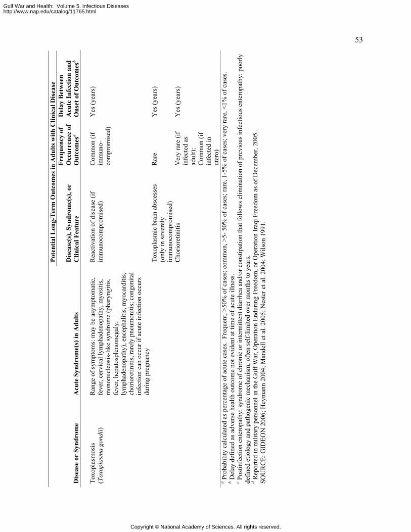
53 Po
tent
ial L
ong-
Ter
m O
utco
mes
in A
dults
with
Clin
ical
Dis
ease
Dis
ease
or
Synd
rom
e A
cute
Syn
drom
e(s)
in A
dults
D
isea
se(s
), Sy
ndro
me(
s), o
r C
linic
al F
eatu
re
Freq
uenc
y of
O
ccur
renc
e of
O
utco
mes
a
Del
ay B
etw
een
A
cute
Infe
ctio
n an
d O
nset
of O
utco
mes
b
To
xopl
asm
osis
(T
oxop
lasm
a go
ndii)
R
ange
of s
ympt
oms:
may
be
asym
ptom
atic
, fe
ver,
cerv
ical
lym
phad
enop
athy
, myo
sitis
, m
onon
ucle
osis
-like
synd
rom
e (p
hary
ngiti
s, fe
ver,
hepa
tosp
leno
meg
aly,
ly
mph
aden
opat
hy),
ence
phal
itis,
myo
card
itis,
chor
iore
tiniti
s, ra
rely
pne
umon
itis;
con
geni
tal
infe
ctio
n ca
n oc
cur i
f acu
te in
fect
ion
occu
rs
durin
g pr
egna
ncy
Rea
ctiv
atio
n of
dis
ease
(if
imm
unoc
ompr
omis
ed)
Com
mon
(if
imm
uno-
com
prom
ised
)
Yes
(yea
rs)
Toxo
plas
mic
bra
in a
bsce
sses
(o
nly
in se
vere
ly
imm
unoc
ompr
omis
ed)
Rar
e Y
es (y
ears
)
Cho
riore
tiniti
s V
ery
rare
(if
infe
cted
as
adul
t);
Com
mon
(if
infe
cted
in
uter
o)
Yes
(yea
rs)
a Pro
babi
lity
calc
ulat
ed a
s per
cent
age
of a
cute
cas
es.
Freq
uent
, >50
% o
f cas
es; c
omm
on, >
5- 5
0% o
f cas
es; r
are,
1-5
% o
f cas
es; v
ery
rare
, <1%
of c
ases
. b D
elay
def
ined
as a
dver
se h
ealth
out
com
e no
t evi
dent
at t
ime
of a
cute
illn
ess.
c Pos
tinfe
ctio
n en
tero
path
y: sy
ndro
me
of c
hron
ic o
r int
erm
itten
t dia
rrhe
a an
d/or
con
stip
atio
n th
at fo
llow
s elim
inat
ion
of p
revi
ous i
nfec
tious
ent
erop
athy
; poo
rly
defin
ed e
tiolo
gy a
nd p
atho
geni
c m
echa
nism
; ofte
n se
lf-lim
ited
over
mon
ths t
o ye
ars.
d Rep
orte
d in
mili
tary
per
sonn
el in
the
Gul
f War
, Ope
ratio
n En
durin
g Fr
eedo
m, o
r Ope
ratio
n Ir
aqi F
reed
om a
s of D
ecem
ber,
2005
. SO
UR
CE:
GID
EON
200
6; H
eym
ann
2004
; Man
dell
et a
l. 20
05; N
este
r et a
l. 20
04; W
ilson
199
1.
Copyright © National Academy of Sciences. All rights reserved.
Gulf War and Health: Volume 5. Infectious Diseaseshttp://www.nap.edu/catalog/11765.html
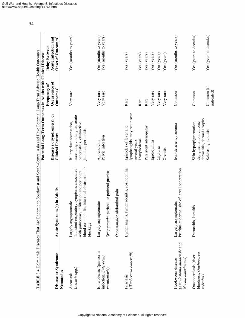
54
TA
BL
E 3
.4 H
elm
inth
ic D
isea
ses T
hat A
re E
ndem
ic to
Sou
thw
est a
nd S
outh
-Cen
tral A
sia
and
Hav
e Po
tent
ial L
ong-
Term
Adv
erse
Hea
lth O
utco
mes
Po
tent
ial L
ong-
Ter
m O
utco
mes
in A
dults
with
Clin
ical
Dis
ease
Dis
ease
or
Synd
rom
e A
cute
Syn
drom
e(s)
in A
dults
D
isea
se(s
), Sy
ndro
me(
s), o
r C
linic
al F
eatu
re
Freq
uenc
y of
O
ccur
renc
e of
O
utco
mes
a
Del
ay B
etw
een
A
cute
Infe
ctio
n an
d O
nset
of O
utco
mes
b
Nem
atod
es
Asc
aria
sis
(Asc
aris
spp.
) La
rgel
y as
ympt
omat
ic
Tran
sien
t res
pira
tory
sym
ptom
s ass
ocia
ted
with
pul
mon
ary
infil
tratio
n an
d pe
riphe
ral
bloo
d eo
sino
phili
a, in
test
inal
obs
truct
ion
or
bloc
kage
Bili
ary
duct
obs
truct
ion,
as
cend
ing
chol
angi
tis, a
cute
pa
ncre
atiti
s, ob
stru
ctiv
e ja
undi
ce, p
erito
nitis
Ver
y ra
re
Yes
(mon
ths t
o ye
ars)
A
ppen
dici
tis
Ver
y ra
re
Yes
(mon
ths t
o ye
ars)
En
tero
bias
is (
pinw
orm
in
fect
ion,
Ent
erob
ius
verm
icul
aris
)
Larg
ely
asym
ptom
atic
Sy
mpt
omat
ic: p
eria
nal o
r per
inea
l pru
ritus
O
ccas
iona
lly: a
bdom
inal
pai
n
Pelv
ic in
fect
ion
Ver
y ra
re
Yes
(mon
ths t
o ye
ars)
Ep
isod
es o
f fev
er a
nd
lym
phan
gitis
, may
recu
r ove
r se
vera
l yea
rs
Rar
e Y
es (y
ears
)
Lym
phed
ema
Rar
e Y
es (y
ears
) Pe
rsis
tent
ade
nopa
thy
Rar
e Y
es (y
ears
) Ep
idid
ymiti
s V
ery
rare
Y
es (y
ears
) C
hylu
ria
Ver
y ra
re
Yes
(yea
rs)
Fila
riasi
s (W
uche
reri
a ba
ncro
fti)
Lym
phan
gitis
, lym
phad
eniti
s, eo
sino
phili
a
Orc
hitis
V
ery
rare
Y
es (y
ears
)
Hoo
kwor
m d
isea
se
(Anc
ylos
tom
a du
oden
ale
and
Nec
ata
amer
ican
us)
Larg
ely
asym
ptom
atic
Pr
uritu
s at d
erm
al si
te o
f lar
val p
enet
ratio
n Ir
on-d
efic
ienc
y an
emia
Com
mon
Yes
(mon
ths t
o ye
ars)
O
ncho
cerc
iasi
s (riv
er
blin
dnes
s, O
ncho
cerc
a vo
lvul
us)
Der
mat
itis,
kera
titis
Sk
in h
yper
pigm
enta
tion,
de
pigm
enta
tion,
chr
onic
de
rmat
itis,
derm
al a
troph
y
Com
mon
Y
es (y
ears
to d
ecad
es)
Scle
rosi
ng k
erat
itis
Com
mon
(if
untre
ated
) Y
es (y
ears
to d
ecad
es)
Copyright © National Academy of Sciences. All rights reserved.
Gulf War and Health: Volume 5. Infectious Diseaseshttp://www.nap.edu/catalog/11765.html
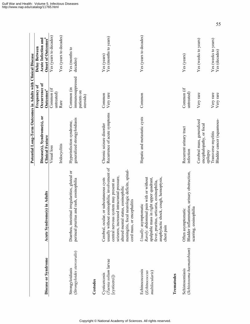
55 Po
tent
ial L
ong-
Ter
m O
utco
mes
in A
dults
with
Clin
ical
Dis
ease
Dis
ease
or
Synd
rom
e A
cute
Syn
drom
e(s)
in A
dults
D
isea
se(s
), Sy
ndro
me(
s), o
r C
linic
al F
eatu
re
Freq
uenc
y of
O
ccur
renc
e of
O
utco
mes
a
Del
ay B
etw
een
A
cute
Infe
ctio
n an
d O
nset
of O
utco
mes
b
Vis
ual l
oss
Com
mon
(if
untre
ated
) Y
es (y
ears
to d
ecad
es)
Irid
ocyc
litis
R
are
Yes
(yea
rs to
dec
ades
)
Stro
ngyl
oidi
asis
(S
tron
gylo
ides
ster
cora
lis)
Dia
rrhe
a, in
test
inal
irre
gula
ritie
s, gl
utea
l or
perin
eal p
rurit
us a
nd ra
sh, e
osin
ophi
lia
Hyp
erin
fect
ion
synd
rom
e,
gene
raliz
ed st
rong
yloi
dias
is
Com
mon
(in
imm
unos
uppr
esse
d pa
tient
s on
ster
oids
)
Yes
(mon
ths t
o de
cade
s)
C
esto
des
C
hron
ic se
izur
e di
sord
er
Com
mon
Y
es (y
ears
) C
ystic
erco
sis
(Tae
nia
soliu
m la
rvae
[c
ystic
erci
])
Cer
ebra
l, oc
ular
, or s
ubcu
tane
ous c
ysts
us
ually
with
out e
osin
ophi
lia; i
nvol
vem
ent o
f ce
ntra
l ner
vous
syst
em m
ay p
rese
nt a
s se
izur
es, i
ncre
ased
intra
cran
ial p
ress
ure,
al
tere
d m
enta
l sta
tus,
eosi
noph
ilic
men
ingi
tis, f
ocal
neu
rolo
gic
defic
its, s
pina
l-co
rd m
ass,
or e
ncep
halit
is
Rec
urre
nce
of a
cute
sym
ptom
s V
ery
rare
Y
es (m
onth
s to
year
s)
Ec
hino
cocc
osis
(E
chin
ococ
cus
mul
tiloc
ular
is)
Usu
ally
: asy
mpt
omat
ic
Rare
ly: a
bdom
inal
pai
n w
ith o
r with
out
apal
pabl
e m
ass i
n rig
ht u
pper
qua
dran
t, fe
ver,
prur
itus,
urtic
aria
, eos
inop
hilia
, an
aphy
lact
ic sh
ock,
cou
gh, h
emop
tysi
s, ch
est p
ain
Hep
atic
and
met
asta
tic c
ysts
C
omm
on
Yes
(yea
rs to
dec
ades
)
T
rem
atod
es
Schi
stos
omia
sis
(Sch
isto
som
a ha
emat
obiu
m)
Ofte
n as
ympt
omat
ic
Bla
dder
infla
mm
atio
n, u
rinar
y ob
stru
ctio
n,
scar
ring,
eos
inop
hilia
Rec
urre
nt u
rinar
y tra
ct
infe
ctio
n
Com
mon
(if
untre
ated
) Y
es (y
ears
)
Cer
ebra
l mas
s, ge
nera
lized
en
ceph
alop
athy
, or f
ocal
ep
ileps
y
Ver
y ra
re
Y
es (w
eeks
to y
ears
)
Tran
sver
se m
yelit
is
Ver
y ra
re
Yes
(wee
ks to
yea
rs)
Bla
dder
can
cer (
squa
mou
s-V
ery
rare
Y
es (d
ecad
es)
Copyright © National Academy of Sciences. All rights reserved.
Gulf War and Health: Volume 5. Infectious Diseaseshttp://www.nap.edu/catalog/11765.html
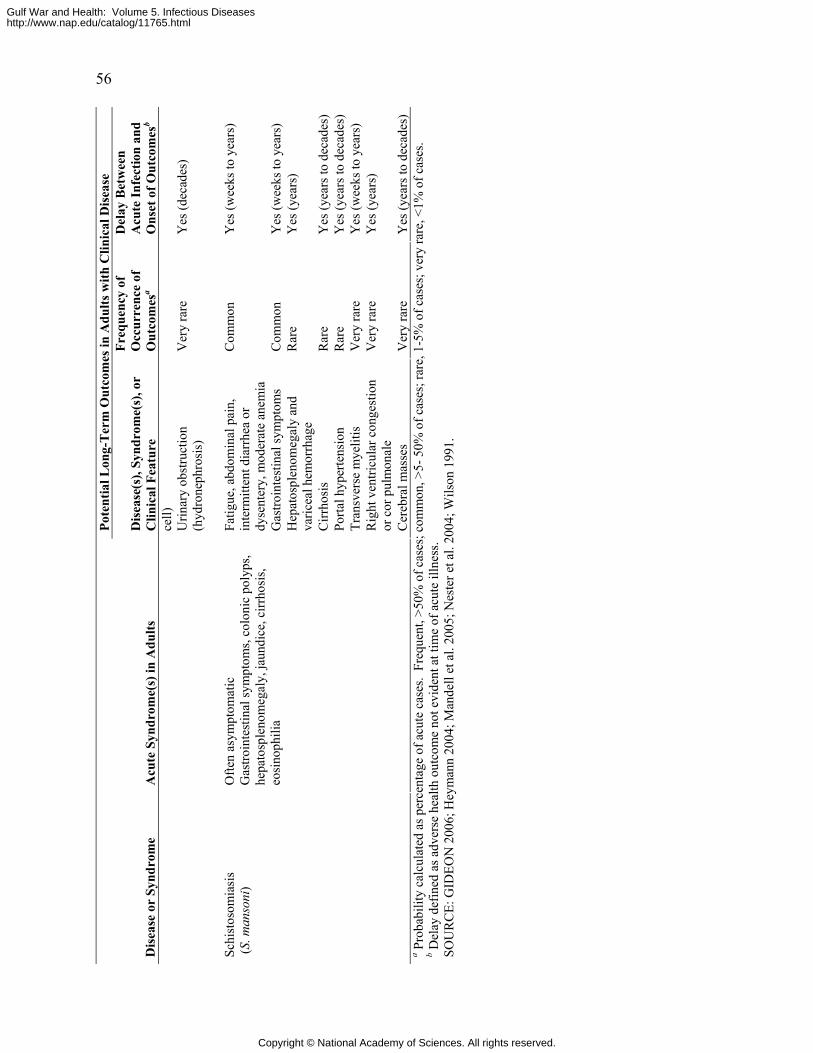
56 Po
tent
ial L
ong-
Ter
m O
utco
mes
in A
dults
with
Clin
ical
Dis
ease
Dis
ease
or
Synd
rom
e A
cute
Syn
drom
e(s)
in A
dults
D
isea
se(s
), Sy
ndro
me(
s), o
r C
linic
al F
eatu
re
Freq
uenc
y of
O
ccur
renc
e of
O
utco
mes
a
Del
ay B
etw
een
A
cute
Infe
ctio
n an
d O
nset
of O
utco
mes
b
cell)
Urin
ary
obst
ruct
ion
(hyd
rone
phro
sis)
V
ery
rare
Y
es (d
ecad
es)
Fa
tigue
, abd
omin
al p
ain,
in
term
itten
t dia
rrhe
a or
dy
sent
ery,
mod
erat
e an
emia
Com
mon
Yes
(wee
ks to
yea
rs)
Gas
troin
test
inal
sym
ptom
s C
omm
on
Yes
(wee
ks to
yea
rs)
Hep
atos
plen
omeg
aly
and
varic
eal h
emor
rhag
e R
are
Y
es (y
ears
)
Cirr
hosi
s R
are
Yes
(yea
rs to
dec
ades
) Po
rtal h
yper
tens
ion
R
are
Yes
(yea
rs to
dec
ades
) Tr
ansv
erse
mye
litis
V
ery
rare
Y
es (w
eeks
to y
ears
) R
ight
ven
tricu
lar c
onge
stio
n or
cor
pul
mon
ale
Ver
y ra
re
Yes
(yea
rs)
Schi
stos
omia
sis
(S. m
anso
ni)
Ofte
n as
ympt
omat
ic
Gas
troin
test
inal
sym
ptom
s, co
loni
c po
lyps
, he
pato
sple
nom
egal
y, ja
undi
ce, c
irrho
sis,
eosi
noph
ilia
Cer
ebra
l mas
ses
Ver
y ra
re
Yes
(yea
rs to
dec
ades
) a P
roba
bilit
y ca
lcul
ated
as p
erce
ntag
e of
acu
te c
ases
. Fr
eque
nt, >
50%
of c
ases
; com
mon
, >5-
50%
of c
ases
; rar
e, 1
-5%
of c
ases
; ver
y ra
re, <
1% o
f cas
es.
b Del
ay d
efin
ed a
s adv
erse
hea
lth o
utco
me
not e
vide
nt a
t tim
e of
acu
te il
lnes
s. SO
UR
CE:
GID
EON
200
6; H
eym
ann
2004
; Man
dell
et a
l. 20
05; N
este
r et a
l. 20
04; W
ilson
199
1.
Copyright © National Academy of Sciences. All rights reserved.
Gulf War and Health: Volume 5. Infectious Diseaseshttp://www.nap.edu/catalog/11765.html
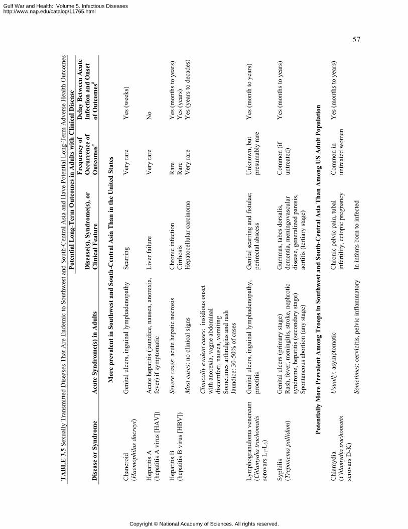
57
TA
BL
E 3
.5 S
exua
lly T
rans
mitt
ed D
isea
ses T
hat A
re E
ndem
ic to
Sou
thw
est a
nd S
outh
-Cen
tral A
sia
and
Hav
e Po
tent
ial L
ong-
Term
Adv
erse
Hea
lth O
utco
mes
Po
tent
ial L
ong-
Ter
m O
utco
mes
in A
dults
with
Clin
ical
Dis
ease
Dis
ease
or
Synd
rom
e A
cute
Syn
drom
e(s)
in A
dults
D
isea
se(s
), Sy
ndro
me(
s), o
r C
linic
al F
eatu
re
Freq
uenc
y of
O
ccur
renc
e of
O
utco
mes
a
Del
ay B
etw
een
Acu
te
Infe
ctio
n an
d O
nset
of
Out
com
esb
M
ore
prev
alen
t in
Sout
hwes
t and
Sou
th-C
entr
al A
sia
Tha
n in
the
Uni
ted
Stat
es
C
hanc
roid
(H
aem
ophi
lus d
ucre
yi)
Gen
ital u
lcer
s, in
guin
al ly
mph
aden
opat
hy
Scar
ring
Ver
y ra
re
Yes
(wee
ks)
H
epat
itis A
(h
epat
itis A
viru
s [H
AV
])
Acu
te h
epat
itis (
jaun
dice
, nau
sea,
ano
rexi
a,
feve
r) if
sym
ptom
atic
Li
ver f
ailu
re
Ver
y ra
re
No
Se
vere
cas
es: a
cute
hep
atic
nec
rosi
s C
hron
ic in
fect
ion
Rar
e Y
es (m
onth
s to
year
s)
C
irrho
sis
Rar
e Y
es (y
ears
) M
ost c
ases
: no
clin
ical
sign
s H
epat
ocel
lula
r car
cino
ma
Ver
y ra
re
Yes
(yea
rs to
dec
ades
)
Hep
atiti
s B
(hep
atiti
s B v
irus [
HB
V])
Clin
ical
ly e
vide
nt c
ases
: in
sidi
ous o
nset
w
ith a
nore
xia,
vag
ue a
bdom
inal
di
scom
fort,
nau
sea,
vom
iting
So
met
imes
arth
ralg
ias a
nd ra
sh
Jaun
dice
: 30-
50%
of c
ases
Ly
mph
ogra
nulo
ma
vene
reum
(C
hlam
ydia
trac
hom
atis
se
rova
rs L
1-L3)
Gen
ital u
lcer
s, in
guin
al ly
mph
aden
opat
hy,
proc
titis
G
enita
l sca
rrin
g an
d fis
tula
e;
perir
ecta
l abs
cess
U
nkno
wn,
but
pr
esum
ably
rare
Y
es (m
onth
to y
ears
)
Sy
phili
s (T
repo
nem
a pa
llidu
m)
Gen
ital u
lcer
s (pr
imar
y st
age)
R
ash,
feve
r, m
enin
gitis
, stro
ke, n
ephr
otic
sy
ndro
me,
hep
atiti
s (se
cond
ary
stag
e)
Spon
tane
ous a
borti
on (a
ny st
age)
Gum
mas
, tab
es d
orsa
lis,
dem
entia
, men
ingo
vasc
ular
di
seas
e, g
ener
aliz
ed p
ares
is,
aorti
tis (t
ertia
ry st
age)
Com
mon
(if
untre
ated
) Y
es (m
onth
s to
year
s)
Po
tent
ially
Mor
e Pr
eval
ent A
mon
g T
roop
s in
Sout
hwes
t and
Sou
th-C
entr
al A
sia
Tha
n A
mon
g U
S A
dult
Popu
latio
n
Chl
amyd
ia
(Chl
amyd
ia tr
acho
mat
is
sero
vars
D-K
)
Usu
ally
: asy
mpt
omat
ic
Chr
onic
pel
vic
pain
, tub
al
infe
rtilit
y, e
ctop
ic p
regn
ancy
So
met
imes
: cer
vici
tis, p
elvi
c in
flam
mat
ory
In in
fant
s bor
n to
infe
cted
Com
mon
in
untre
ated
wom
en
Yes
(mon
ths t
o ye
ars)
Copyright © National Academy of Sciences. All rights reserved.
Gulf War and Health: Volume 5. Infectious Diseaseshttp://www.nap.edu/catalog/11765.html
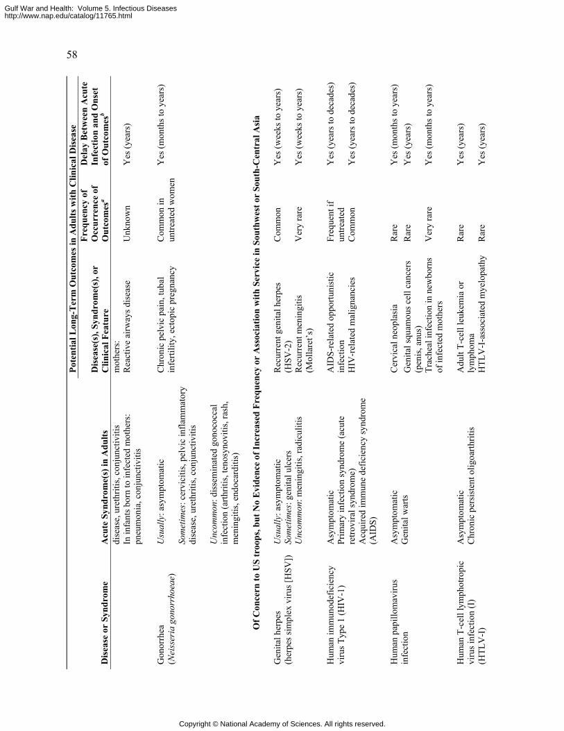
58 Po
tent
ial L
ong-
Ter
m O
utco
mes
in A
dults
with
Clin
ical
Dis
ease
Dis
ease
or
Synd
rom
e A
cute
Syn
drom
e(s)
in A
dults
D
isea
se(s
), Sy
ndro
me(
s), o
r C
linic
al F
eatu
re
Freq
uenc
y of
O
ccur
renc
e of
O
utco
mes
a
Del
ay B
etw
een
Acu
te
Infe
ctio
n an
d O
nset
of
Out
com
esb
dise
ase,
ure
thrit
is, c
onju
nctiv
itis
mot
hers
:
In
infa
nts b
orn
to in
fect
ed m
othe
rs:
pneu
mon
ia, c
onju
nctiv
itis
Rea
ctiv
e ai
rway
s dis
ease
U
nkno
wn
Yes
(yea
rs)
U
sual
ly: a
sym
ptom
atic
So
met
imes
: cer
vici
tis, p
elvi
c in
flam
mat
ory
dise
ase,
ure
thrit
is, c
onju
nctiv
itis
Gon
orrh
ea
(Nei
sser
ia g
onor
rhoe
ae)
Unc
omm
on: d
isse
min
ated
gon
ococ
cal
infe
ctio
n (a
rthrit
is, t
enos
ynov
itis,
rash
, m
enin
gitis
, end
ocar
ditis
)
Chr
onic
pel
vic
pain
, tub
al
infe
rtilit
y, e
ctop
ic p
regn
ancy
C
omm
on in
un
treat
ed w
omen
Y
es (m
onth
s to
year
s)
O
f Con
cern
to U
S tr
oops
, but
No
Evi
denc
e of
Incr
ease
d Fr
eque
ncy
or A
ssoc
iatio
n w
ith S
ervi
ce in
Sou
thw
est o
r So
uth-
Cen
tral
Asi
a R
ecur
rent
gen
ital h
erpe
s (H
SV-2
) C
omm
on
Yes
(wee
ks to
yea
rs)
Gen
ital h
erpe
s (h
erpe
s sim
plex
viru
s [H
SV])
U
sual
ly: a
sym
ptom
atic
So
met
imes
: gen
ital u
lcer
s U
ncom
mon
: men
ingi
tis, r
adic
uliti
s R
ecur
rent
men
ingi
tis
(Mol
lare
t’s)
Ver
y ra
re
Yes
(wee
ks to
yea
rs)
A
IDS-
rela
ted
oppo
rtuni
stic
in
fect
ion
Freq
uent
if
untre
ated
Y
es (y
ears
to d
ecad
es)
Hum
an im
mun
odef
icie
ncy
viru
s Typ
e 1
(HIV
-1)
Asy
mpt
omat
ic
Prim
ary
infe
ctio
n sy
ndro
me
(acu
te
retro
vira
l syn
drom
e)
Acq
uire
d im
mun
e de
ficie
ncy
synd
rom
e (A
IDS)
HIV
-rel
ated
mal
igna
ncie
s C
omm
on
Yes
(yea
rs to
dec
ades
)
C
ervi
cal n
eopl
asia
R
are
Yes
(mon
ths t
o ye
ars)
G
enita
l squ
amou
s cel
l can
cers
(p
enis
, anu
s)
Rar
e Y
es (y
ears
) H
uman
pap
illom
aviru
s in
fect
ion
Asy
mpt
omat
ic
Gen
ital w
arts
Trac
heal
infe
ctio
n in
new
born
s of
infe
cted
mot
hers
V
ery
rare
Y
es (m
onth
s to
year
s)
A
dult
T-ce
ll le
ukem
ia o
r ly
mph
oma
Rar
e Y
es (y
ears
) H
uman
T-c
ell l
ymph
otro
pic
viru
s inf
ectio
n (I
) (H
TLV
-I)
Asy
mpt
omat
ic
Chr
onic
per
sist
ent o
ligoa
rthrit
is
HTL
V-I
-ass
ocia
ted
mye
lopa
thy
Rar
e Y
es (y
ears
)
Copyright © National Academy of Sciences. All rights reserved.
Gulf War and Health: Volume 5. Infectious Diseaseshttp://www.nap.edu/catalog/11765.html
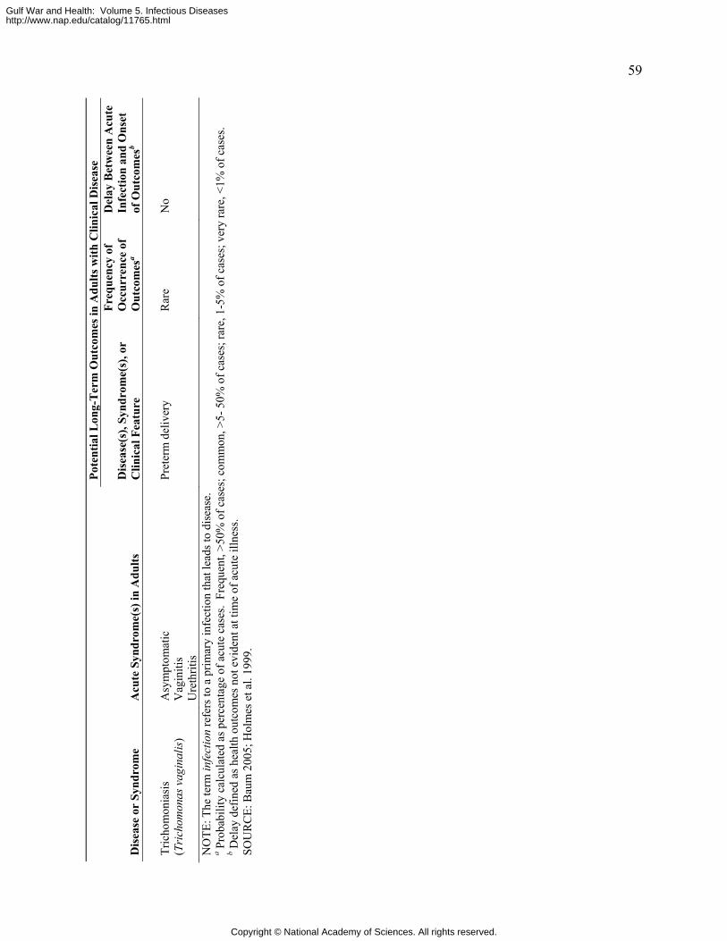
59 Po
tent
ial L
ong-
Ter
m O
utco
mes
in A
dults
with
Clin
ical
Dis
ease
Dis
ease
or
Synd
rom
e A
cute
Syn
drom
e(s)
in A
dults
D
isea
se(s
), Sy
ndro
me(
s), o
r C
linic
al F
eatu
re
Freq
uenc
y of
O
ccur
renc
e of
O
utco
mes
a
Del
ay B
etw
een
Acu
te
Infe
ctio
n an
d O
nset
of
Out
com
esb
Tr
icho
mon
iasi
s (T
rich
omon
as v
agin
alis
) A
sym
ptom
atic
V
agin
itis
Ure
thrit
is
Pret
erm
del
iver
y R
are
No
NO
TE: T
he te
rm in
fect
ion
refe
rs to
a p
rimar
y in
fect
ion
that
lead
s to
dise
ase.
a P
roba
bilit
y ca
lcul
ated
as p
erce
ntag
e of
acu
te c
ases
. Fr
eque
nt, >
50%
of c
ases
; com
mon
, >5-
50%
of c
ases
; rar
e, 1
-5%
of c
ases
; ver
y ra
re, <
1% o
f cas
es.
b Del
ay d
efin
ed a
s hea
lth o
utco
mes
not
evi
dent
at t
ime
of a
cute
illn
ess.
SOU
RC
E: B
aum
200
5; H
olm
es e
t al.
1999
.
Copyright © National Academy of Sciences. All rights reserved.
Gulf War and Health: Volume 5. Infectious Diseaseshttp://www.nap.edu/catalog/11765.html
60 GULF WAR AND HEALTH: INFECTIOUS DISEASES
REFERENCES
Baum S. 2005. Introduction to Mycoplasma diseases. Mycoplasma Diseases. In: Mandell G,
Bennett J, Dolin R, Editors. Principles and Practice of Infectious Diseases. 6th ed. Philadelphia, PA: Churchill Livingstone. Pp. 2269-2271.
GIDEON. 2006. Global Infectious Disease and Epidemiology Network. [Online]. Available: http://www.gideononline.com [accessed April 12, 2006].
Heymann DL. 2004. Control of Communicable Diseases Manual. Washington, DC: American Public Health Association.
Holmes KK, Sparling PF, Mardh P, Lemon SM, Stamm WE, Piot P, Wasserheit JN. 1999. Sexually Transmitted Diseases. 3rd ed. New York: McGraw-Hill.
Mandell GL, Bennett JE, Dolin R. 2005. Principles and Practice of Infectious Diseases. 6th ed. Philadelphia, PA: Elsevier Churchill Livingstone.
Nester EW, Anderson DG, Roberts JCE, Pearsall NN, Nester MT. 2004. Microbiology: A Human Perspective. 4th ed. New York: McGraw-Hill.
Wilson ME. 1991. A World Guide to Infections: Diseases, Distribution, Diagnosis. New York: Oxford University Press.
Copyright © National Academy of Sciences. All rights reserved.
Gulf War and Health: Volume 5. Infectious Diseaseshttp://www.nap.edu/catalog/11765.html
61
4
INFECTIOUS DISEASES DIAGNOSED IN US TROOPS WHO SERVED IN THE PERSIAN GULF WAR, OPERATION ENDURING
FREEDOM, OR OPERATION IRAQI FREEDOM
Infectious diseases have accompanied war throughout recorded history; the clinical aspects of Operation Desert Shield (ODSh), Operation Desert Storm (ODSt), Operation Iraqi Freedom (OIF), and Operation Enduring Freedom (OEF) have been no different. Although medical and epidemiologic personnel in the US military can anticipate troops’ exposure to many pathogens and mitigate their effects, naturally-occurring pathogens infected some troops during these operations. This chapter summarizes information about the infectious diseases and pathogens identified in US troops who served or are serving in ODSh, ODSt, OIF, or OEF. That information comes from several sources, including published scientific literature, medical surveillance monthly reports published by the Army Medical Surveillance Activity, the Centers for Disease Control and Prevention (CDC), and infectious disease experts at the Department of Defense (DOD) and the Department of Veterans Affairs. In Chapter 5, the committee evaluates the published scientific literature about the possible long-term adverse health outcomes of nine of the diseases discussed in this chapter.
Thriving on the troops’ crowded and sometimes unsanitary living conditions, microbial pathogens have caused primarily diarrheal illnesses and acute upper respiratory infections during ODSt, ODSh, OEF, and OIF (Hyams et al. 2001a; Paparello et al. 1993; Richards et al. 1993a; Thornton et al. 2005; Wasserman et al. 1997). Smaller numbers of military personnel have had various insect-borne diseases, nosocomial infections, brucellosis, chickenpox, meningococcal disease, and Q fever.
Even this chapter’s comprehensive review of public documents may not capture the full burden of infectious disease on US troops who have served in southwest and south-central Asia. Military medical investigators’ primary mission is to apply their findings to maintain troops’ health and they might not always publish summary reports in medical journals. In addition, field commanders may be reluctant to report illnesses perceived as trivial (such as vomiting and diarrhea) even when an outbreak of disease interferes with military operations (Matson 2005). Finally, a new policy purveyed by the DOD restricts the publication of some kinds of medical information that enemy combatants could use to gain an advantage over US troops (Department of the Army 2005b).
Copyright © National Academy of Sciences. All rights reserved.
Gulf War and Health: Volume 5. Infectious Diseaseshttp://www.nap.edu/catalog/11765.html
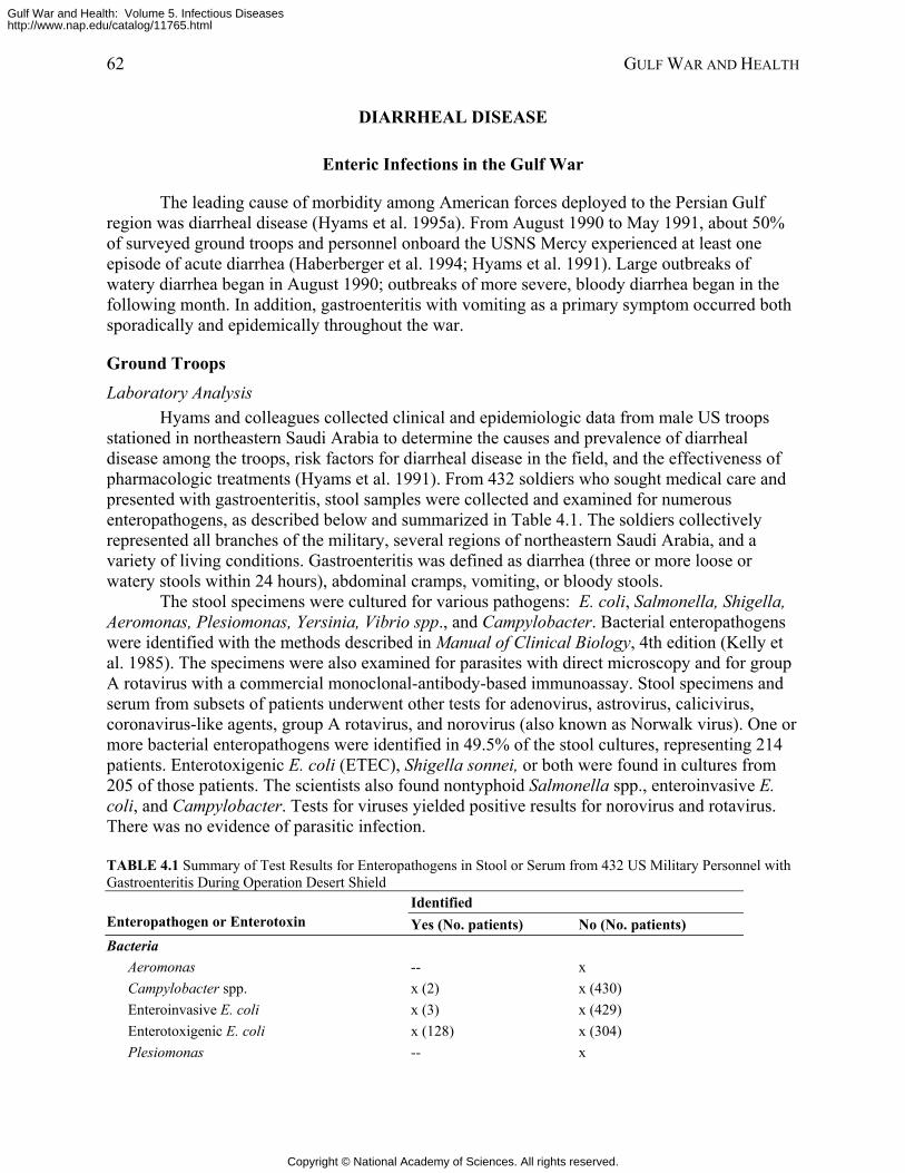
62 GULF WAR AND HEALTH
DIARRHEAL DISEASE
Enteric Infections in the Gulf War
The leading cause of morbidity among American forces deployed to the Persian Gulf region was diarrheal disease (Hyams et al. 1995a). From August 1990 to May 1991, about 50% of surveyed ground troops and personnel onboard the USNS Mercy experienced at least one episode of acute diarrhea (Haberberger et al. 1994; Hyams et al. 1991). Large outbreaks of watery diarrhea began in August 1990; outbreaks of more severe, bloody diarrhea began in the following month. In addition, gastroenteritis with vomiting as a primary symptom occurred both sporadically and epidemically throughout the war.
Ground Troops
Laboratory Analysis Hyams and colleagues collected clinical and epidemiologic data from male US troops
stationed in northeastern Saudi Arabia to determine the causes and prevalence of diarrheal disease among the troops, risk factors for diarrheal disease in the field, and the effectiveness of pharmacologic treatments (Hyams et al. 1991). From 432 soldiers who sought medical care and presented with gastroenteritis, stool samples were collected and examined for numerous enteropathogens, as described below and summarized in Table 4.1. The soldiers collectively represented all branches of the military, several regions of northeastern Saudi Arabia, and a variety of living conditions. Gastroenteritis was defined as diarrhea (three or more loose or watery stools within 24 hours), abdominal cramps, vomiting, or bloody stools.
The stool specimens were cultured for various pathogens: E. coli, Salmonella, Shigella, Aeromonas, Plesiomonas, Yersinia, Vibrio spp., and Campylobacter. Bacterial enteropathogens were identified with the methods described in Manual of Clinical Biology, 4th edition (Kelly et al. 1985). The specimens were also examined for parasites with direct microscopy and for group A rotavirus with a commercial monoclonal-antibody-based immunoassay. Stool specimens and serum from subsets of patients underwent other tests for adenovirus, astrovirus, calicivirus, coronavirus-like agents, group A rotavirus, and norovirus (also known as Norwalk virus). One or more bacterial enteropathogens were identified in 49.5% of the stool cultures, representing 214 patients. Enterotoxigenic E. coli (ETEC), Shigella sonnei, or both were found in cultures from 205 of those patients. The scientists also found nontyphoid Salmonella spp., enteroinvasive E. coli, and Campylobacter. Tests for viruses yielded positive results for norovirus and rotavirus. There was no evidence of parasitic infection.
TABLE 4.1 Summary of Test Results for Enteropathogens in Stool or Serum from 432 US Military Personnel with Gastroenteritis During Operation Desert Shield
Identified Enteropathogen or Enterotoxin Yes (No. patients) No (No. patients) Bacteria
Aeromonas -- x Campylobacter spp. x (2) x (430) Enteroinvasive E. coli x (3) x (429) Enterotoxigenic E. coli x (128) x (304) Plesiomonas -- x
Copyright © National Academy of Sciences. All rights reserved.
Gulf War and Health: Volume 5. Infectious Diseaseshttp://www.nap.edu/catalog/11765.html
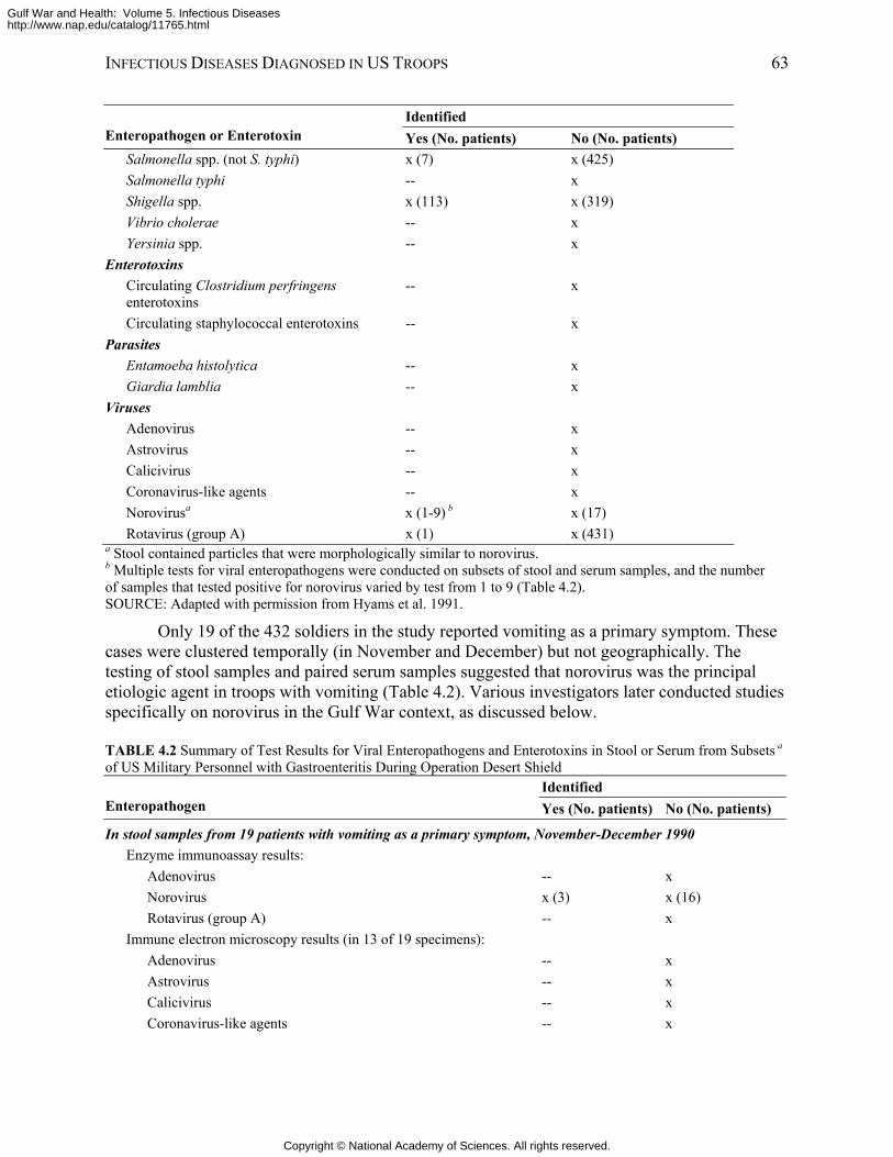
INFECTIOUS DISEASES DIAGNOSED IN US TROOPS 63
Identified Enteropathogen or Enterotoxin Yes (No. patients) No (No. patients)
Salmonella spp. (not S. typhi) x (7) x (425) Salmonella typhi -- x Shigella spp. x (113) x (319) Vibrio cholerae -- x Yersinia spp. -- x
Enterotoxins Circulating Clostridium perfringens enterotoxins
-- x
Circulating staphylococcal enterotoxins -- x Parasites
Entamoeba histolytica -- x Giardia lamblia -- x
Viruses Adenovirus -- x Astrovirus -- x Calicivirus -- x Coronavirus-like agents -- x Norovirusa x (1-9) b x (17) Rotavirus (group A) x (1) x (431)
a Stool contained particles that were morphologically similar to norovirus. b Multiple tests for viral enteropathogens were conducted on subsets of stool and serum samples, and the number of samples that tested positive for norovirus varied by test from 1 to 9 (Table 4.2). SOURCE: Adapted with permission from Hyams et al. 1991.
Only 19 of the 432 soldiers in the study reported vomiting as a primary symptom. These cases were clustered temporally (in November and December) but not geographically. The testing of stool samples and paired serum samples suggested that norovirus was the principal etiologic agent in troops with vomiting (Table 4.2). Various investigators later conducted studies specifically on norovirus in the Gulf War context, as discussed below.
TABLE 4.2 Summary of Test Results for Viral Enteropathogens and Enterotoxins in Stool or Serum from Subsets a of US Military Personnel with Gastroenteritis During Operation Desert Shield
Identified Enteropathogen Yes (No. patients) No (No. patients)
In stool samples from 19 patients with vomiting as a primary symptom, November-December 1990 Enzyme immunoassay results:
Adenovirus -- x Norovirus x (3) x (16) Rotavirus (group A) -- x
Immune electron microscopy results (in 13 of 19 specimens): Adenovirus -- x Astrovirus -- x Calicivirus -- x Coronavirus-like agents -- x
Copyright © National Academy of Sciences. All rights reserved.
Gulf War and Health: Volume 5. Infectious Diseaseshttp://www.nap.edu/catalog/11765.html
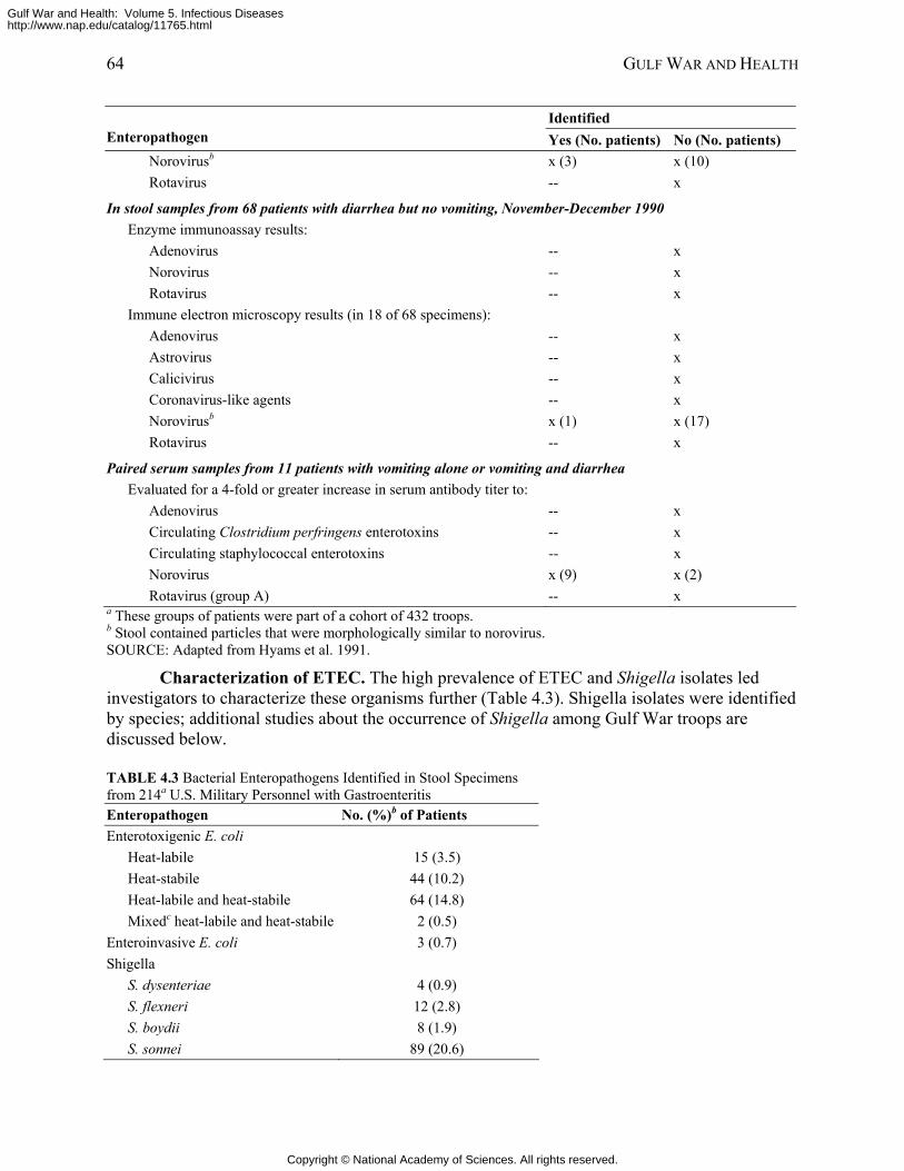
64 GULF WAR AND HEALTH
Identified Enteropathogen Yes (No. patients) No (No. patients)
Norovirusb x (3) x (10) Rotavirus -- x
In stool samples from 68 patients with diarrhea but no vomiting, November-December 1990 Enzyme immunoassay results:
Adenovirus -- x Norovirus -- x Rotavirus -- x
Immune electron microscopy results (in 18 of 68 specimens): Adenovirus -- x Astrovirus -- x Calicivirus -- x Coronavirus-like agents -- x Norovirusb x (1) x (17) Rotavirus -- x
Paired serum samples from 11 patients with vomiting alone or vomiting and diarrhea Evaluated for a 4-fold or greater increase in serum antibody titer to:
Adenovirus -- x Circulating Clostridium perfringens enterotoxins -- x Circulating staphylococcal enterotoxins -- x Norovirus x (9) x (2) Rotavirus (group A) -- x
a These groups of patients were part of a cohort of 432 troops. b Stool contained particles that were morphologically similar to norovirus. SOURCE: Adapted from Hyams et al. 1991.
Characterization of ETEC. The high prevalence of ETEC and Shigella isolates led investigators to characterize these organisms further (Table 4.3). Shigella isolates were identified by species; additional studies about the occurrence of Shigella among Gulf War troops are discussed below.
TABLE 4.3 Bacterial Enteropathogens Identified in Stool Specimens from 214a U.S. Military Personnel with Gastroenteritis Enteropathogen No. (%)b of Patients Enterotoxigenic E. coli
Heat-labile 15 (3.5) Heat-stabile 44 (10.2) Heat-labile and heat-stabile 64 (14.8) Mixedc heat-labile and heat-stabile 2 (0.5)
Enteroinvasive E. coli 3 (0.7) Shigella
S. dysenteriae 4 (0.9) S. flexneri 12 (2.8) S. boydii 8 (1.9) S. sonnei 89 (20.6)
Copyright © National Academy of Sciences. All rights reserved.
Gulf War and Health: Volume 5. Infectious Diseaseshttp://www.nap.edu/catalog/11765.html
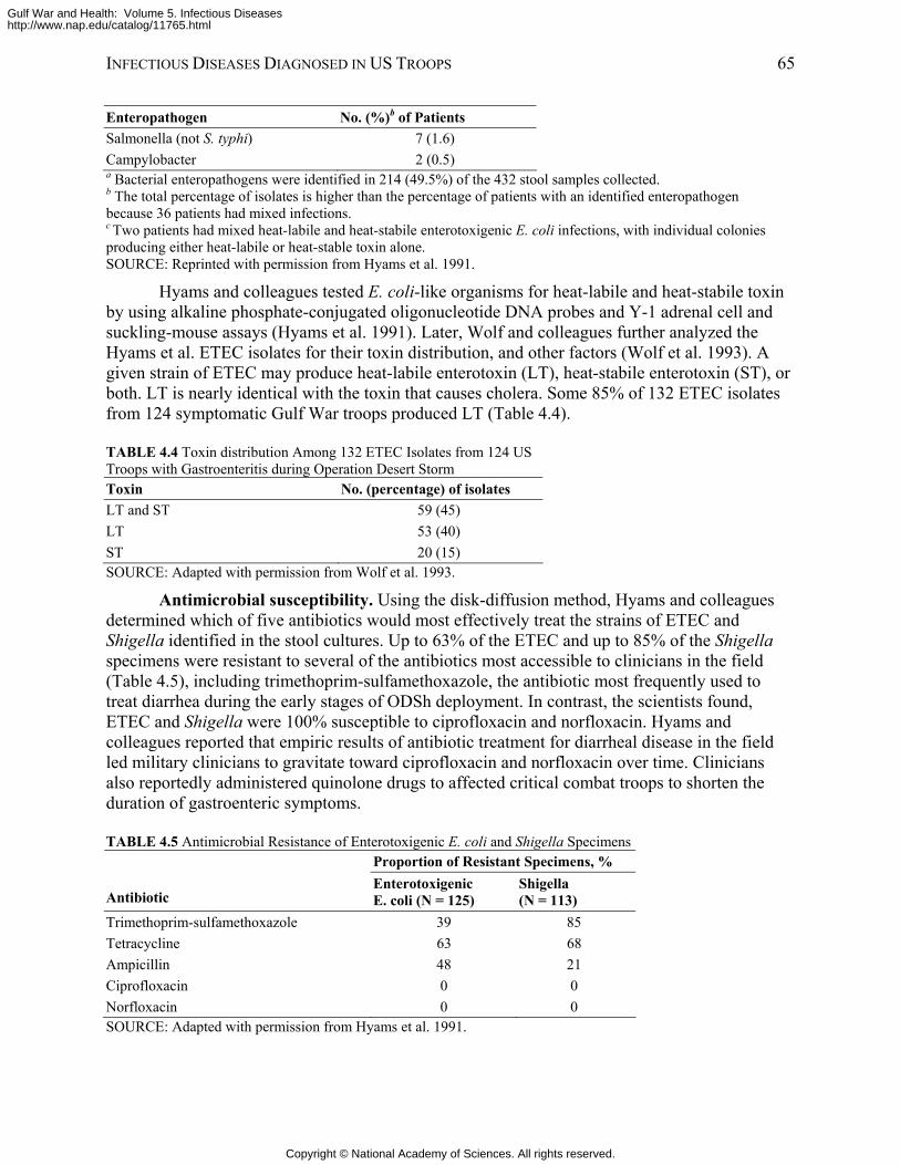
INFECTIOUS DISEASES DIAGNOSED IN US TROOPS 65
Enteropathogen No. (%)b of Patients Salmonella (not S. typhi) 7 (1.6) Campylobacter 2 (0.5) a Bacterial enteropathogens were identified in 214 (49.5%) of the 432 stool samples collected. b The total percentage of isolates is higher than the percentage of patients with an identified enteropathogen because 36 patients had mixed infections. c Two patients had mixed heat-labile and heat-stabile enterotoxigenic E. coli infections, with individual colonies producing either heat-labile or heat-stable toxin alone. SOURCE: Reprinted with permission from Hyams et al. 1991.
Hyams and colleagues tested E. coli-like organisms for heat-labile and heat-stabile toxin by using alkaline phosphate-conjugated oligonucleotide DNA probes and Y-1 adrenal cell and suckling-mouse assays (Hyams et al. 1991). Later, Wolf and colleagues further analyzed the Hyams et al. ETEC isolates for their toxin distribution, and other factors (Wolf et al. 1993). A given strain of ETEC may produce heat-labile enterotoxin (LT), heat-stabile enterotoxin (ST), or both. LT is nearly identical with the toxin that causes cholera. Some 85% of 132 ETEC isolates from 124 symptomatic Gulf War troops produced LT (Table 4.4).
TABLE 4.4 Toxin distribution Among 132 ETEC Isolates from 124 US Troops with Gastroenteritis during Operation Desert Storm Toxin No. (percentage) of isolates LT and ST 59 (45) LT 53 (40) ST 20 (15) SOURCE: Adapted with permission from Wolf et al. 1993.
Antimicrobial susceptibility. Using the disk-diffusion method, Hyams and colleagues determined which of five antibiotics would most effectively treat the strains of ETEC and Shigella identified in the stool cultures. Up to 63% of the ETEC and up to 85% of the Shigella specimens were resistant to several of the antibiotics most accessible to clinicians in the field (Table 4.5), including trimethoprim-sulfamethoxazole, the antibiotic most frequently used to treat diarrhea during the early stages of ODSh deployment. In contrast, the scientists found, ETEC and Shigella were 100% susceptible to ciprofloxacin and norfloxacin. Hyams and colleagues reported that empiric results of antibiotic treatment for diarrheal disease in the field led military clinicians to gravitate toward ciprofloxacin and norfloxacin over time. Clinicians also reportedly administered quinolone drugs to affected critical combat troops to shorten the duration of gastroenteric symptoms.
TABLE 4.5 Antimicrobial Resistance of Enterotoxigenic E. coli and Shigella Specimens
Proportion of Resistant Specimens, %
Antibiotic Enterotoxigenic E. coli (N = 125)
Shigella (N = 113)
Trimethoprim-sulfamethoxazole 39 85 Tetracycline 63 68 Ampicillin 48 21 Ciprofloxacin 0 0 Norfloxacin 0 0 SOURCE: Adapted with permission from Hyams et al. 1991.
Copyright © National Academy of Sciences. All rights reserved.
Gulf War and Health: Volume 5. Infectious Diseaseshttp://www.nap.edu/catalog/11765.html
66 GULF WAR AND HEALTH
Epidemiologic Analysis To learn the prevalence of and risk factors for diarrheal disease among US troops
stationed in northeastern Saudi Arabia during ODSh, Hyams and colleagues administered an epidemiologic survey to 2,022 personnel from all branches of the military in October-December 1990 (Hyams et al. 1991). After an average of 2 months in Saudi Arabia, 57% of those surveyed had suffered at least one episode of diarrhea. The symptoms of diarrheal disease had led 22% of all respondents to seek medical care, and had prevented 20% of all respondents from performing their duties. Thirty-two percent of those surveyed had experienced two or more separate episodes of diarrhea. In some units, the attack rate was 5-10% per week.
A univariate analysis of potential risk factors for the transmission of diarrheal disease during ODSh suggested an association between an episode of diarrhea and eating salad, dining in a mess hall, and drinking from a canteen. (A laboratory study of 12 heads of lettuce obtained from food-distribution facilities in September 1990 found coliform bacteria in all 12; ETEC was identified in two (Hyams et al. 1991).) No association was found between an episode of diarrhea and obtaining food from local vendors, eating in a local restaurant, or drinking bottled water. A multivariate analysis of these risk factors and an evaluation of published research on the transmission of Shigella indicated that flies and relatively poor personal hygiene probably accounted for the spread of ETEC and Shigella.
The disabling effect of repeated outbreaks of diarrheal disease in US forces during ODSh despite the best available preventive measures led Hyams and colleagues to call for the development of a vaccine to protect troops (Hyams et al. 1991). DOD is supporting development of such vaccines (Stephens and Nataro 2004). Shigella
The presence of immunoglobulin A (IgA) and immunoglobulin G (IgG) anti-Shigella lipopolysaccharide (LPS) in predeployment serum did not offer protective immunity to infection by Shigella spp. among US ground troops who participated in the Persian Gulf War, Hyams and colleagues reported (Hyams et al. 1995b). The investigators reached that conclusion by studying a cohort of 883 combat troops and support personnel in three Marine Corps units who were flown directly to Saudi Arabia in late December 1990 and directly back to the United States in May 1991. Initially stationed in Saudi Arabia, and then relocated to Kuwait, the subjects lived in remote, rugged, desert camps. US military personnel prepared most of their food, which came from the United States except for local fresh produce. The subjects drank both locally produced bottled water and water purified by reverse-osmosis (Hyams et al. 1993).
The investigators obtained serum samples from all members of the three units who were accessible during the week before their deployment and the 2 two days after their return (827 subjects). Paired serum samples were tested for antibodies to both S. sonnei and S. flexneri. Epidemiologic questionnaires were also administered to this cohort before and after deployment.
Among the 827 subjects, 18% seroconverted during ODSh and ODSt; that underscored earlier findings that troops deployed to ODSh and ODSt faced a considerable risk of Shigella infection. The study revealed the absence of an association between seroconversion and the occurrence of diarrheal symptoms. Overall, 60% of the cohort reported one or more episodes of diarrhea, and 18% reported diarrhea with fever. In contrast, many troops who seroconverted were asymptomatic.
Because S. sonnei LPS cross-reacts with the LPS of Plesiomonas shigelloides, some of the high concentrations of serum antibodies observed in samples from the 827 marines might not have been the result of exposure to Shigella spp. To determine whether exposure to Shigella led
Copyright © National Academy of Sciences. All rights reserved.
Gulf War and Health: Volume 5. Infectious Diseaseshttp://www.nap.edu/catalog/11765.html
INFECTIOUS DISEASES DIAGNOSED IN US TROOPS 67
to persistently high antibody concentrations in some subjects and seroconversions in others, Mikhail and colleagues examined how a subset of the paired serum samples reacted to four Shigella invasion plasmids, which cross-react with just one enteroinvasive strain of E. coli (Mikhail et al. 1996). They also used ELISA to detect antibodies to S. sonnei LPS. In their report, the authors noted that antibodies to LPS and invasion plasmid antigens in serum increase and decrease within 4 months during naturally acquired Shigella infections.
Only 12 sets of serum samples were large enough to use for this experiment (six from seroconverters and six with persistently high concentrations of antibodies to S. sonnei LPS). By using Western blot, the investigators observed antibody reactions to numerous invasion plasmid antigens both before and after deployment in serum from troops with persistently high concentrations of antibodies to S. sonnei LPS, which suggest that they had been exposed to S. sonnei before deployment and were repeatedly exposed to it during deployment. In the postdeployment serum from troops who seroconverted, the scientists observed IgA and IgG recognition of additional invasion plasmid antigens and increased concentrations of antibodies to S. sonnei LPS—even in two soldiers who were asymptomatic for diarrheal illness throughout the war. The authors interpreted those results as an indication that troops who seroconverted had been exposed repeatedly to S. sonnei in the field. Norovirus
Norovirus (NV) and Norwalk-like viruses caused both sporadic cases and outbreaks of acute gastroenteritis among ground troops and shipboard personnel throughout the Gulf War. Brief and debilitating, NV gastroenteritis usually causes acute vomiting, diarrhea, nausea, and abdominal cramps that last 1-2 days. Some people never develop symptoms even after direct challenge, but others are repeatedly susceptible to symptomatic infection. All infected people shed highly contagious NV in stools from as early as 15 hours after exposure to as late as 14 days after. Studies of NV infections among military personnel indicate that crowding is the most important risk factor for transmission (McCarthy et al. 2000).
After the Gulf War, Hyams and colleagues demonstrated the incidence of NV infection among troops deployed to Saudi Arabia and Kuwait from late December 1990 through May 1991 (Hyams et al. 1993). Using the paired serum samples from the 883-troop cohort described above, the investigators used ELISA to measure antibody activity to recombinant NV particles. The investigators defined evidence of infection as a 4-fold or greater increase in titer of anti-NV antibodies from predeployment serum to postdeployment serum.
Matching the ELISA results with the subjects’ clinical symptoms, as reported in the aforementioned postdeployment epidemiologic questionnaire, the investigators obtained the results displayed in Table 4.6. After adjusting for oversampling of subjects with vomiting, the investigators estimated that NV infected 6% of the study population. The scientists could not determine the specific sources of infection, although they enumerated the probable opportunities for person-to-person spread of NV: rapid deployment of massive numbers of soldiers, overcrowding, and rugged desert living conditions that included communal temporary latrines and bathing facilities.
To aid the development of a vaccine against NV and Norwalk-like virus for the US military, Lew and colleagues compared the published genetic sequence of NV with sequences of NV strains extracted from three stool specimens from US troops who developed gastroenteritis while deployed to Saudi Arabia for ODSh (Lew et al. 1994).
Copyright © National Academy of Sciences. All rights reserved.
Gulf War and Health: Volume 5. Infectious Diseaseshttp://www.nap.edu/catalog/11765.html
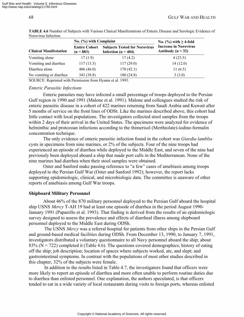
68 GULF WAR AND HEALTH
TABLE 4.6 Number of Subjects with Various Clinical Manifestations of Enteric Disease and Serologic Evidence of Norovirus Infection
No. (%) with Complaint
Clinical Manifestation Entire Cohort (n = 883)
Subjects Tested for Norovirus Infection (n = 404)
No. (%) with ≥ 4-fold Increase in Norovirus Antibody (n = 32)
Vomiting alone 17 (1.9) 17 (4.2) 4 (23.5) Vomiting and diarrhea 117 (13.3) 117 (29.0) 14 (12.0) Diarrhea alone 406 (46.0) 170 (42.1) 11 (6.5) No vomiting or diarrhea 343 (38.8) 100 (24.8) 3 (3.0) SOURCE: Reprinted with Permission from Hyams et al. 1993.
Enteric Parasitic Infections Enteric parasites may have infected a small percentage of troops deployed to the Persian
Gulf region in 1990 and 1991 (Malone et al. 1991). Malone and colleagues studied the risk of enteric parasitic disease in a cohort of 422 marines returning from Saudi Arabia and Kuwait after 5 months of service on the front lines of ODSt. Like the marines described above, this cohort had little contact with local populations. The investigators collected stool samples from the troops within 2 days of their arrival in the United States. The specimens were analyzed for evidence of helminthic and protozoan infections according to the thimerisol (Merthiolate)-iodine-formalin concentration technique.
The only evidence of enteric parasitic infection found in the cohort was Giardia lamblia cysts in specimens from nine marines, or 2% of the subjects. Four of the nine troops had experienced an episode of diarrhea while deployed to the Middle East, and seven of the nine had previously been deployed aboard a ship that made port calls in the Mediterranean. None of the nine marines had diarrhea when their stool samples were obtained.
Oster and Sanford make passing reference to “a few” cases of amebiasis among troops deployed to the Persian Gulf War (Oster and Sanford 1992); however, the report lacks supporting epidemiologic, clinical, and microbiologic data. The committee is unaware of other reports of amebiasis among Gulf War troops.
Shipboard Military Personnel About 46% of the 870 military personnel deployed to the Persian Gulf aboard the hospital
ship USNS Mercy T-AH 19 had at least one episode of diarrhea in the period August 1990-January 1991 (Paparello et al. 1993). That finding is derived from the results of an epidemiologic survey designed to assess the prevalence and effects of diarrheal illness among shipboard personnel deployed to the Middle East during ODSh.
The USNS Mercy was a referral hospital for patients from other ships in the Persian Gulf and ground-based medical facilities during ODSh. From December 13, 1990, to January 7, 1991, investigators distributed a voluntary questionnaire to all Navy personnel aboard the ship; about 83% (N = 722) completed it (Table 4.6). The questions covered demographics; history of eating off the ship; job description; location of spaces where subjects worked, ate, and slept; and gastrointestinal symptoms. In contrast with the populations of most other studies described in this chapter, 32% of the subjects were female.
In addition to the results listed in Table 4.7, the investigators found that officers were more likely to report an episode of diarrhea and more often unable to perform routine duties due to diarrhea than enlisted personnel. One explanation, the authors speculated, is that officers tended to eat in a wide variety of local restaurants during visits to foreign ports, whereas enlisted
Copyright © National Academy of Sciences. All rights reserved.
Gulf War and Health: Volume 5. Infectious Diseaseshttp://www.nap.edu/catalog/11765.html
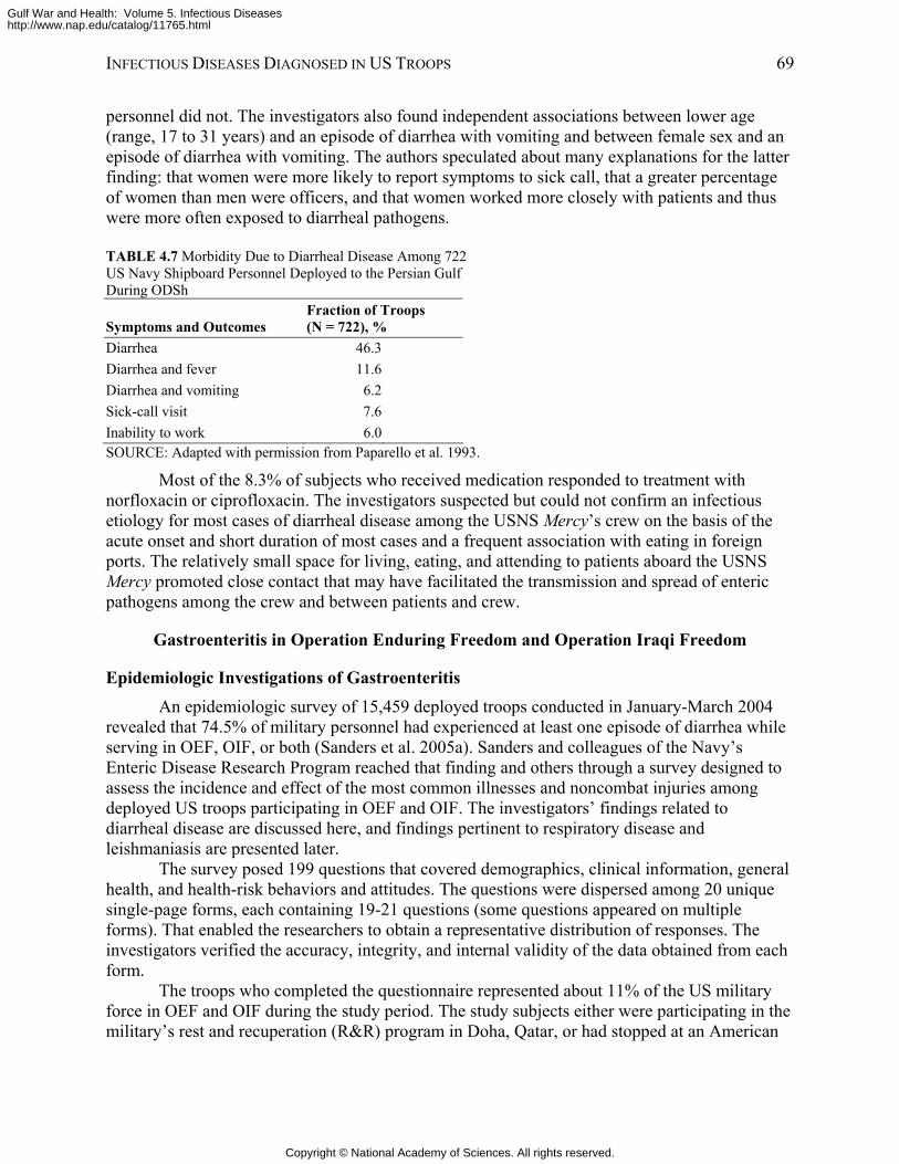
INFECTIOUS DISEASES DIAGNOSED IN US TROOPS 69
personnel did not. The investigators also found independent associations between lower age (range, 17 to 31 years) and an episode of diarrhea with vomiting and between female sex and an episode of diarrhea with vomiting. The authors speculated about many explanations for the latter finding: that women were more likely to report symptoms to sick call, that a greater percentage of women than men were officers, and that women worked more closely with patients and thus were more often exposed to diarrheal pathogens.
TABLE 4.7 Morbidity Due to Diarrheal Disease Among 722 US Navy Shipboard Personnel Deployed to the Persian Gulf During ODSh
Symptoms and Outcomes Fraction of Troops (N = 722), %
Diarrhea 46.3 Diarrhea and fever 11.6 Diarrhea and vomiting 6.2 Sick-call visit 7.6 Inability to work 6.0 SOURCE: Adapted with permission from Paparello et al. 1993.
Most of the 8.3% of subjects who received medication responded to treatment with norfloxacin or ciprofloxacin. The investigators suspected but could not confirm an infectious etiology for most cases of diarrheal disease among the USNS Mercy’s crew on the basis of the acute onset and short duration of most cases and a frequent association with eating in foreign ports. The relatively small space for living, eating, and attending to patients aboard the USNS Mercy promoted close contact that may have facilitated the transmission and spread of enteric pathogens among the crew and between patients and crew.
Gastroenteritis in Operation Enduring Freedom and Operation Iraqi Freedom
Epidemiologic Investigations of Gastroenteritis An epidemiologic survey of 15,459 deployed troops conducted in January-March 2004
revealed that 74.5% of military personnel had experienced at least one episode of diarrhea while serving in OEF, OIF, or both (Sanders et al. 2005a). Sanders and colleagues of the Navy’s Enteric Disease Research Program reached that finding and others through a survey designed to assess the incidence and effect of the most common illnesses and noncombat injuries among deployed US troops participating in OEF and OIF. The investigators’ findings related to diarrheal disease are discussed here, and findings pertinent to respiratory disease and leishmaniasis are presented later.
The survey posed 199 questions that covered demographics, clinical information, general health, and health-risk behaviors and attitudes. The questions were dispersed among 20 unique single-page forms, each containing 19-21 questions (some questions appeared on multiple forms). That enabled the researchers to obtain a representative distribution of responses. The investigators verified the accuracy, integrity, and internal validity of the data obtained from each form.
The troops who completed the questionnaire represented about 11% of the US military force in OEF and OIF during the study period. The study subjects either were participating in the military’s rest and recuperation (R&R) program in Doha, Qatar, or had stopped at an American
Copyright © National Academy of Sciences. All rights reserved.
Gulf War and Health: Volume 5. Infectious Diseaseshttp://www.nap.edu/catalog/11765.html
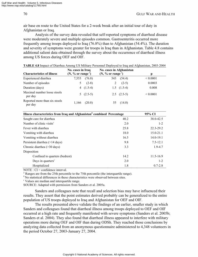
70 GULF WAR AND HEALTH
air base en route to the United States for a 2-week break after an initial tour of duty in Afghanistan or Iraq.
Analysis of the survey data revealed that self-reported symptoms of diarrheal disease were moderately severe and multiple episodes common. Gastroenteritis occurred more frequently among troops deployed to Iraq (76.8%) than to Afghanistan (54.4%). The duration and severity of symptoms were greater for troops in Iraq than in Afghanistan. Table 4.8 contains additional salient data obtained through the survey about the occurrence of diarrheal illness among US forces during OEF and OIF.
TABLE 4.8 Impact of Diarrhea Among US Military Personnel Deployed to Iraq and Afghanistan, 2003-2004
Characteristics of illness No. cases in Iraq (N, % or range a)
No. cases in Afghanistan (N, % or range a) p
Experienced diarrhea 7,553 (76.8) 543 (54.4) < 0.0001 Number of episodes 5 (2-8) 2 (2-5) 0.0003 Duration (days) 4 (1.5-4) 1.5 (1.5-4) 0.008 Maximal number loose stools
per day 5 (2.5-5) 2.5 (2.5-5) < 0.0001
Reported more than six stools per day 1,166 (20.8) 55 (14.0)
Illness characteristics from Iraq and Afghanistanb combined Percentage 95% CI Sought care for diarrhea 40.2 38.0-42.5 Number of clinic visitsc 2.0 1-2 Fever with diarrhea 25.8 22.3-29.2 Vomiting with diarrhea 18.0 15.0-21.1 Vomiting without diarrhea 16.5 14.0-19.1 Persistent diarrhea (>14 days) 9.8 7.5-12.1 Chronic diarrhea (>30 days) 3.3 1.9-4.7 Disposition
Confined to quarters (bedrest) 14.2 11.5-16.9 Days in quartersc 2.0 1-2 Hospitalized 1.8 0.7-2.8
NOTE : CI = confidence interval. a Ranges are from the 25th percentile to the 75th percentile (the interquartile range). b No statistical differences in these characteristics were observed between sites. c Values are median and interquartile range. SOURCE: Adapted with permission from Sanders et al. 2005a.
Sanders and colleagues note that recall and selection bias may have influenced their results. They assert that the point estimates derived probably can be generalized to the entire population of US troops deployed to Iraq and Afghanistan for OEF and OIF.
The results presented above validate the findings of an earlier, smaller study in which Sanders and colleagues found that diarrheal illness among troops deployed to OEF and OIF occurred at a high rate and frequently manifested with severe symptoms (Sanders et al. 2005b; Sanders et al. 2004). They also found that diarrheal illness appeared to interfere with military operations more during OEF and OIF than during ODSh. They reached those conclusions by analyzing data collected from an anonymous questionnaire administered to 4,348 volunteers in the period October 27, 2003-January 27, 2004.
Copyright © National Academy of Sciences. All rights reserved.
Gulf War and Health: Volume 5. Infectious Diseaseshttp://www.nap.edu/catalog/11765.html
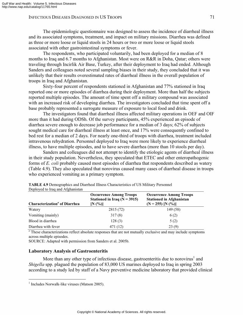
INFECTIOUS DISEASES DIAGNOSED IN US TROOPS 71
The epidemiologic questionnaire was designed to assess the incidence of diarrheal illness and its associated symptoms, treatment, and impact on military missions. Diarrhea was defined as three or more loose or liquid stools in 24 hours or two or more loose or liquid stools associated with other gastrointestinal symptoms or fever.
The respondents, who participated voluntarily, had been deployed for a median of 8 months to Iraq and 6.7 months to Afghanistan. Most were on R&R in Doha, Qatar; others were traveling through Incirlik Air Base, Turkey, after their deployment to Iraq had ended. Although Sanders and colleagues noted several sampling biases in their study, they concluded that it was unlikely that their results overestimated rates of diarrheal illness in the overall population of troops in Iraq and Afghanistan.
Sixty-four percent of respondents stationed in Afghanistan and 77% stationed in Iraq reported one or more episodes of diarrhea during their deployment. More than half the subjects reported multiple episodes. The amount of time spent off a military compound was associated with an increased risk of developing diarrhea. The investigators concluded that time spent off a base probably represented a surrogate measure of exposure to local food and drink.
The investigators found that diarrheal illness affected military operations in OEF and OIF more than it had during ODSh. Of the survey participants, 45% experienced an episode of diarrhea severe enough to decrease job performance for a median of 3 days; 62% of subjects sought medical care for diarrheal illness at least once, and 17% were consequently confined to bed rest for a median of 2 days. For nearly one-third of troops with diarrhea, treatment included intravenous rehydration. Personnel deployed to Iraq were more likely to experience diarrheal illness, to have multiple episodes, and to have severe diarrhea (more than 10 stools per day).
Sanders and colleagues did not attempt to identify the etiologic agents of diarrheal illness in their study population. Nevertheless, they speculated that ETEC and other enteropathogenic forms of E. coli probably caused most episodes of diarrhea that respondents described as watery (Table 4.9). They also speculated that norovirus caused many cases of diarrheal disease in troops who experienced vomiting as a primary symptom.
TABLE 4.9 Demographics and Diarrheal Illness Characteristics of US Military Personnel Deployed to Iraq and Afghanistan
Characterizationa of Diarrhea
Occurrence Among Troops Stationed in Iraq (N = 3915) [N (%)]
Occurrence Among Troops Stationed in Afghanistan (N = 255) [N (%)]
Watery 2815 (72) 149 (58) Vomiting (mainly) 317 (8) 6 (2) Blood in diarrhea 128 (3) 5 (2) Diarrhea with fever 471 (12) 23 (9) a These characterizations reflect absolute responses that are not mutually exclusive and may include symptoms across multiple episodes. SOURCE: Adapted with permission from Sanders et al. 2005b.
Laboratory Analysis of Gastroenteritis More than any other type of infectious disease, gastroenteritis due to norovirus1 and
Shigella spp. plagued the population of 83,000 US marines deployed to Iraq in spring 2003 according to a study led by staff of a Navy preventive medicine laboratory that provided clinical
1 Includes Norwalk-like viruses (Matson 2005).
Copyright © National Academy of Sciences. All rights reserved.
Gulf War and Health: Volume 5. Infectious Diseaseshttp://www.nap.edu/catalog/11765.html
72 GULF WAR AND HEALTH
and diagnostic support (Thornton et al. 2005). Located in a city about 175 km south of Baghdad, the laboratory collected and analyzed stool samples and other clinical specimens from 30 First Marine Expeditionary Force battalion aid stations and several other medical stations in the area. Thornton and colleagues published the results of their analysis of 129 stool specimens collected from April 24 to June 1, 2003, from 33 medical stations scattered across south-central Iraq.
The authors reported that large outbreaks of nausea, vomiting, and diarrhea lasting 24-48 hours occurred in the First Marine Expeditionary Force from early April though the middle of May, suggesting a viral etiology for most cases of gastroenteritis during that period. Febrile dysentery predominated in that population beginning in the middle of May.
Numerous factors placed the troops of the First Marine Expeditionary Force at risk for gastroenteritis, the authors wrote. The primitive nature of the camps, overcrowding, and filth flies in latrines and dining facilities characterized the troops’ unsanitary living environment. Local ambient temperatures climbed to 40°C during the period of study. Some troops broke military rules to eat locally prepared food, particularly grilled chicken. Finally, a relatively small number of microorganisms are capable of causing shigellosis and norovirus gastroenteritis in humans. In an editorial commentary on Thornton’s report, Matson postulates that robustly healthy people who become dehydrated during daily activity may face a greater risk of severe outcome when infected with norovirus (Matson 2005).
The investigators used plating, biochemical identification, antigen serologic testing, fluorescent antibody antigen detection, and enzyme immunoassay to evaluate stool specimens diagnostically for enteropathogens. Specifically, they tested specimens for pathogenic E. coli, Campylobacter spp., Salmonella spp., Shigella spp., Giardia spp., and Cryptosporidium. Suspect E. coli was tested for heat-labile and heat-stabile enterotoxins; additionally, investigators plated stool with gross blood or fecal leukocytes on sorbitol-MacConkey agar to detect enterohemorrhagic E. coli. The antimicrobial susceptibility of detected bacteria was evaluated with the disk-diffusion method.
Reverse-transcriptase polymerase chain reaction (PCR) was used to detect norovirus. In addition, the authors sequenced the RNA polymerase genes of detected norovirus to distinguish strains, compare the strains with each other and with known strains, and analyze the occurrence of norovirus strains by military unit and timing.
Table 4.10 illustrates the results of the laboratory-based diagnostic evaluation. One or more enteropathogens were detected in 57 of the 129 stool samples (44%). Norovirus was detected in 23% of the specimens; the investigators found 21 norovirus strains among 30 agent-positive patients. The most frequently detected bacterial enteropathogens were Shigella sonnei and Shigella flexneri; they were isolated from a total of 20% of the specimens. The presence of fecal leukocytes in 43 of 109 stool samples (39%) indicated inflammatory diarrhea, the investigators concluded.
Most of the bacterial isolates tested by Thornton and colleagues were doxycycline-resistant (Table 4.10), and several bacterial enteropathogens identified in prior studies of military populations were observed less frequently in this study population. For those reasons, both the authors and Matson (the editorial commentator) postulated that the mandatory dose of doxycycline (100 mg/day) taken by personnel in the First Marine Expeditionary Force for antimalarial prophylaxis may also have reduced the potential impact of bacterial enteric pathogens.
Extrapolating their results to the entire First Marine Expeditionary Force, Thornton and colleagues conservatively estimated that several thousand cases of norovirus illness occurred in
Copyright © National Academy of Sciences. All rights reserved.
Gulf War and Health: Volume 5. Infectious Diseaseshttp://www.nap.edu/catalog/11765.html
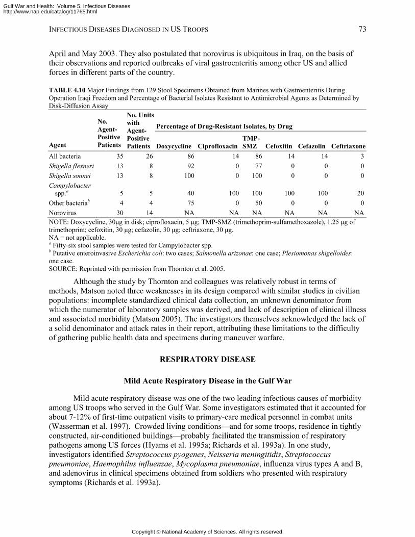
INFECTIOUS DISEASES DIAGNOSED IN US TROOPS 73
April and May 2003. They also postulated that norovirus is ubiquitous in Iraq, on the basis of their observations and reported outbreaks of viral gastroenteritis among other US and allied forces in different parts of the country.
TABLE 4.10 Major Findings from 129 Stool Specimens Obtained from Marines with Gastroenteritis During Operation Iraqi Freedom and Percentage of Bacterial Isolates Resistant to Antimicrobial Agents as Determined by Disk-Diffusion Assay
Percentage of Drug-Resistant Isolates, by Drug
Agent
No. Agent-Positive Patients
No. Units with Agent-Positive Patients Doxycycline Ciprofloxacin
TMP-SMZ Cefoxitin Cefazolin Ceftriaxone
All bacteria 35 26 86 14 86 14 14 3Shigella fIexneri 13 8 92 0 77 0 0 0Shigella sonnei 13 8 100 0 100 0 0 0Campylobacter
spp.a 5 5 40 100 100 100 100 20Other bacteriab 4 4 75 0 50 0 0 0Norovirus 30 14 NA NA NA NA NA NANOTE: Doxycycline, 30μg in disk; ciprofloxacin, 5 μg; TMP-SMZ (trimethoprim-sulfamethoxazole), 1.25 μg of trimethoprim; cefoxitin, 30 μg; cefazolin, 30 μg; ceftriaxone, 30 μg. NA = not applicable. a Fifty-six stool samples were tested for Campylobacter spp. b Putative enteroinvasive Escherichia coli: two cases; Salmonella arizonae: one case; Plesiomonas shigelloides: one case. SOURCE: Reprinted with permission from Thornton et al. 2005.
Although the study by Thornton and colleagues was relatively robust in terms of methods, Matson noted three weaknesses in its design compared with similar studies in civilian populations: incomplete standardized clinical data collection, an unknown denominator from which the numerator of laboratory samples was derived, and lack of description of clinical illness and associated morbidity (Matson 2005). The investigators themselves acknowledged the lack of a solid denominator and attack rates in their report, attributing these limitations to the difficulty of gathering public health data and specimens during maneuver warfare.
RESPIRATORY DISEASE
Mild Acute Respiratory Disease in the Gulf War
Mild acute respiratory disease was one of the two leading infectious causes of morbidity among US troops who served in the Gulf War. Some investigators estimated that it accounted for about 7-12% of first-time outpatient visits to primary-care medical personnel in combat units (Wasserman et al. 1997). Crowded living conditions—and for some troops, residence in tightly constructed, air-conditioned buildings—probably facilitated the transmission of respiratory pathogens among US forces (Hyams et al. 1995a; Richards et al. 1993a). In one study, investigators identified Streptococcus pyogenes, Neisseria meningitidis, Streptococcus pneumoniae, Haemophilus influenzae, Mycoplasma pneumoniae, influenza virus types A and B, and adenovirus in clinical specimens obtained from soldiers who presented with respiratory symptoms (Richards et al. 1993a).
Copyright © National Academy of Sciences. All rights reserved.
Gulf War and Health: Volume 5. Infectious Diseaseshttp://www.nap.edu/catalog/11765.html
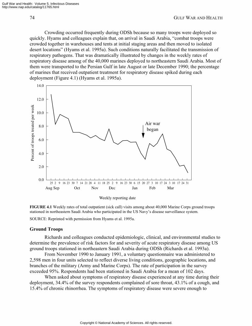
74 GULF WAR AND HEALTH
Crowding occurred frequently during ODSh because so many troops were deployed so quickly. Hyams and colleagues explain that, on arrival in Saudi Arabia, “combat troops were crowded together in warehouses and tents at initial staging areas and then moved to isolated desert locations” (Hyams et al. 1995a). Such conditions naturally facilitated the transmission of respiratory pathogens. That was dramatically illustrated by changes in the weekly rates of respiratory disease among of the 40,000 marines deployed to northeastern Saudi Arabia. Most of them were transported to the Persian Gulf in late August or late December 1990; the percentage of marines that received outpatient treatment for respiratory disease spiked during each deployment (Figure 4.1) (Hyams et al. 1995a).
0.0
2.0
4.0
6.0
8.0
10.0
12.0
14.0
Perc
ent o
f tr
oops
trea
ted
per
wee
k
Weekly reporting date
Aug Sep Oct Nov Dec Jan Feb Mar25 2 9 2316 30 7 14 21 28 4 11 18 25 2 9 16 23 30 6 15 20 27 3 10 17 24 3 10 17 24 31
Air warbegan
FIGURE 4.1 Weekly rates of total outpatient (sick call) visits among about 40,000 Marine Corps ground troops stationed in northeastern Saudi Arabia who participated in the US Navy’s disease surveillance system.
SOURCE: Reprinted with permission from Hyams et al. 1995a.
Ground Troops Richards and colleagues conducted epidemiologic, clinical, and environmental studies to
determine the prevalence of risk factors for and severity of acute respiratory disease among US ground troops stationed in northeastern Saudi Arabia during ODSh (Richards et al. 1993a).
From November 1990 to January 1991, a voluntary questionnaire was administered to 2,598 men in four units selected to reflect diverse living conditions, geographic locations, and branches of the military (Army and Marine Corps). The rate of participation in the survey exceeded 95%. Respondents had been stationed in Saudi Arabia for a mean of 102 days.
When asked about symptoms of respiratory disease experienced at any time during their deployment, 34.4% of the survey respondents complained of sore throat, 43.1% of a cough, and 15.4% of chronic rhinorrhea. The symptoms of respiratory disease were severe enough to
Copyright © National Academy of Sciences. All rights reserved.
Gulf War and Health: Volume 5. Infectious Diseaseshttp://www.nap.edu/catalog/11765.html
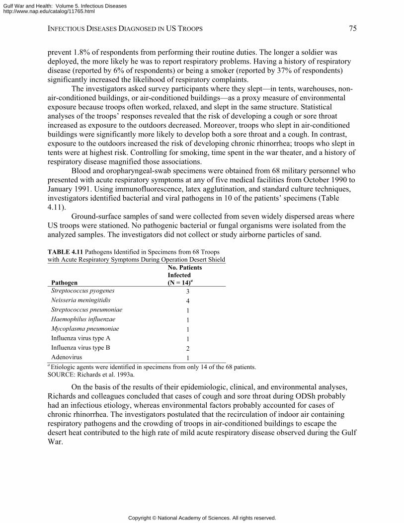
INFECTIOUS DISEASES DIAGNOSED IN US TROOPS 75
prevent 1.8% of respondents from performing their routine duties. The longer a soldier was deployed, the more likely he was to report respiratory problems. Having a history of respiratory disease (reported by 6% of respondents) or being a smoker (reported by 37% of respondents) significantly increased the likelihood of respiratory complaints.
The investigators asked survey participants where they slept—in tents, warehouses, non-air-conditioned buildings, or air-conditioned buildings—as a proxy measure of environmental exposure because troops often worked, relaxed, and slept in the same structure. Statistical analyses of the troops’ responses revealed that the risk of developing a cough or sore throat increased as exposure to the outdoors decreased. Moreover, troops who slept in air-conditioned buildings were significantly more likely to develop both a sore throat and a cough. In contrast, exposure to the outdoors increased the risk of developing chronic rhinorrhea; troops who slept in tents were at highest risk. Controlling for smoking, time spent in the war theater, and a history of respiratory disease magnified those associations.
Blood and oropharyngeal-swab specimens were obtained from 68 military personnel who presented with acute respiratory symptoms at any of five medical facilities from October 1990 to January 1991. Using immunofluorescence, latex agglutination, and standard culture techniques, investigators identified bacterial and viral pathogens in 10 of the patients’ specimens (Table 4.11).
Ground-surface samples of sand were collected from seven widely dispersed areas where US troops were stationed. No pathogenic bacterial or fungal organisms were isolated from the analyzed samples. The investigators did not collect or study airborne particles of sand.
TABLE 4.11 Pathogens Identified in Specimens from 68 Troops with Acute Respiratory Symptoms During Operation Desert Shield
Pathogen
No. Patients Infected (N = 14)a
Streptococcus pyogenes 3 Neisseria meningitidis 4 Streptococcus pneumoniae 1 Haemophilus influenzae 1 Mycoplasma pneumoniae 1 Influenza virus type A 1 Influenza virus type B 2 Adenovirus 1
a Etiologic agents were identified in specimens from only 14 of the 68 patients. SOURCE: Richards et al. 1993a.
On the basis of the results of their epidemiologic, clinical, and environmental analyses, Richards and colleagues concluded that cases of cough and sore throat during ODSh probably had an infectious etiology, whereas environmental factors probably accounted for cases of chronic rhinorrhea. The investigators postulated that the recirculation of indoor air containing respiratory pathogens and the crowding of troops in air-conditioned buildings to escape the desert heat contributed to the high rate of mild acute respiratory disease observed during the Gulf War.
Copyright © National Academy of Sciences. All rights reserved.
Gulf War and Health: Volume 5. Infectious Diseaseshttp://www.nap.edu/catalog/11765.html
76 GULF WAR AND HEALTH
Shipboard Military Personnel The reported risk of upper respiratory disease was lower among US ground troops than
among Navy personnel who served aboard the 1,000-bed hospital ship USNS Mercy T-AH 19 during ODSh (Paparello et al. 1993). That was one conclusion of an epidemiologic study designed to assess the prevalence and impact of upper respiratory disease among shipboard personnel deployed to the Middle East during ODSh.
As described above, the USNS Mercy was a referral hospital for patients from other ships in the region and ground-based medical facilities during ODSh. From December 13, 1990-January 7, 1991, investigators distributed a voluntary questionnaire to all Navy personnel aboard the ship; about 83% (n = 722) completed it.
Among the surveyed population, 79% reported at least one upper respiratory complaint during their deployment. The respondents’ symptoms generally were mild but persistent. Cold-like symptoms with fever were reported by 27.9%, cough by 53.2%, sore throat by 49.1%, and chronic rhinorrhea by 16.6%. The severity of such symptoms prevented 7.4% of respondents from performing their duties.
No significant association existed between smoking and respiratory complaints. Female sex and a history of respiratory disease were independently associated with cold-like symptoms plus fever and inability to perform duties. Investigators could not determine the reason for the sex-based association.
Pathogens probably caused the majority of respiratory disease aboard the USNS Mercy on the basis of the nature and short duration of reported symptoms, the investigators concluded. Moreover, they postulated that the relatively small total space for living, eating, and attending to patients promoted close contact that probably facilitated the transmission and spread of respiratory pathogens among the crew and between patients and crew.
Severe Acute Respiratory Disease in the Gulf War
More than 1,800 US military personnel deployed to the Persian Gulf region developed respiratory disease severe enough to require hospitalization of a day or more (Smith et al. 2004). Among those patients, 214 were diagnosed with pneumonia (etiologic agent unspecified), 90 with acute sinusitis, 102 with chronic sinusitis, and 81 with bronchitis; 678 cases were diagnosed as asthma, and the remaining cases of respiratory disease were not identified. The committee is unaware of published literature about the details of these cases other than what appears in this chapter.
Any of more than 50 viruses, bacteria, fungi, parasites, rickettsiae, chlamydiae, and mycoplasmas can cause pneumonia (Donowitz and Mandell 2000). A smaller array of viral, bacterial, and fungal organisms can cause acute sinusitis; however, such noninfectious agents as allergens and toxins may also instigate this disease. Acute bronchitis usually has a viral etiology, although it is also associated with Bordetella pertussis, Mycoplasma pneumoniae, and Chlamydia pneumoniae (strain TWAR) (Gwaltney 2000a). The pathogenesis of chronic sinus disease is poorly understood; at least six genera of bacteria have been cultured from patients with this diagnosis (Gwaltney 2000b).
Respiratory Disease in Operation Enduring Freedom and Operation Iraqi Freedom
About 70% of military personnel deployed to Iraq and Afghanistan in 2003 and the first quarter of 2004 contracted a respiratory infection during their tour of duty, according to the
Copyright © National Academy of Sciences. All rights reserved.
Gulf War and Health: Volume 5. Infectious Diseaseshttp://www.nap.edu/catalog/11765.html

INFECTIOUS DISEASES DIAGNOSED IN US TROOPS 77
epidemiologic survey of 15,459 deployed troops described above (Sanders et al. 2005a). A cold or cough characterized nearly all self-reported cases of respiratory illness in that study (Table 4.12). A minority of subjects—2.6%—reported that they had been diagnosed with pneumonia; most of the cases were mild enough to obviate the need for hospitalization. A separate study found that more than 60 cases of pneumonia (severe and mild) occurred among US troops in Iraq from March 1, 2003 to August 20, 2003 (Anderson et al. 2005).
Nineteen deployed troops were hospitalized with acute bilateral pneumonitis with increased concentrations of eosinophils in March-August 2003 (CDC 2003a). All patients required intubation and mechanical ventilation; two ultimately died. The severity of this illness, its unknown cause, and its association with service in southwest and south-central Asia led the military to begin conducting special surveillance for severe acute pneumonia among troops deployed to OEF or OIF.
As of September 2003, laboratory evidence of infection with a pathogen had been identified in four of the 19 patients, as outlined in Table 4.13. In Chapter 6, the committee addresses idiopathic acute eosinophilic pneumonia (a syndrome whose etiology remains unknown).
TABLE 4.12 Impact of Respiratory Illness Among US Military Personnel Deployed to Iraq or Afghanistan, 2003-2004
Percentage of Troops
95% Confidence Interval
No. of respiratory infections (cough or cold) during deployment
None 30.9 27.6-34.41 19.1 16.4-22.22-3 35.6 32.3-39.2>3 14.4 12.0-17.2
Sought medical care for respiratory infection 17.0 14.2-19.8Received medicine from provider for respiratory infection
17.8 14.9-20.7
Self-medicated for respiratory infection 29.3 26.2-32.5Experienced allergy attack 22.5 19.4-25.6Experienced asthma attack 3.6 2.2-5.0Developed pneumonia 2.6 1.4-3.8Started or restarted smoking 47.6 41.7-53.6No. of packs per day
None 61.0 57.2-64.7½ 17.5 14.6-20.41 14.5 11.8-17.2>1 7.0 5.1-9.0
Smoke Iraqi cigarettes 72.2 66.8-77.5SOURCE: Adapted with permission from Sanders et al. 2005a.
Copyright © National Academy of Sciences. All rights reserved.
Gulf War and Health: Volume 5. Infectious Diseaseshttp://www.nap.edu/catalog/11765.html
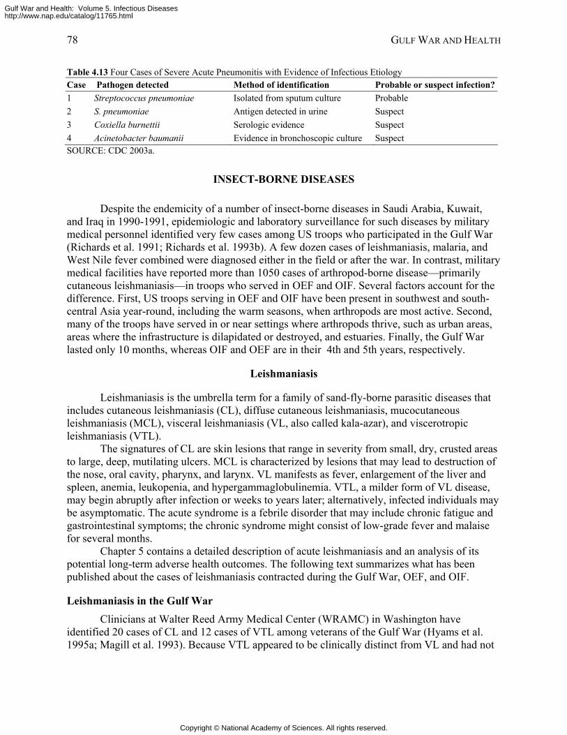
78 GULF WAR AND HEALTH
Table 4.13 Four Cases of Severe Acute Pneumonitis with Evidence of Infectious Etiology Case Pathogen detected Method of identification Probable or suspect infection?1 Streptococcus pneumoniae Isolated from sputum culture Probable 2 S. pneumoniae Antigen detected in urine Suspect 3 Coxiella burnettii Serologic evidence Suspect 4 Acinetobacter baumanii Evidence in bronchoscopic culture Suspect SOURCE: CDC 2003a.
INSECT-BORNE DISEASES
Despite the endemicity of a number of insect-borne diseases in Saudi Arabia, Kuwait, and Iraq in 1990-1991, epidemiologic and laboratory surveillance for such diseases by military medical personnel identified very few cases among US troops who participated in the Gulf War (Richards et al. 1991; Richards et al. 1993b). A few dozen cases of leishmaniasis, malaria, and West Nile fever combined were diagnosed either in the field or after the war. In contrast, military medical facilities have reported more than 1050 cases of arthropod-borne disease—primarily cutaneous leishmaniasis—in troops who served in OEF and OIF. Several factors account for the difference. First, US troops serving in OEF and OIF have been present in southwest and south-central Asia year-round, including the warm seasons, when arthropods are most active. Second, many of the troops have served in or near settings where arthropods thrive, such as urban areas, areas where the infrastructure is dilapidated or destroyed, and estuaries. Finally, the Gulf War lasted only 10 months, whereas OIF and OEF are in their 4th and 5th years, respectively.
Leishmaniasis
Leishmaniasis is the umbrella term for a family of sand-fly-borne parasitic diseases that includes cutaneous leishmaniasis (CL), diffuse cutaneous leishmaniasis, mucocutaneous leishmaniasis (MCL), visceral leishmaniasis (VL, also called kala-azar), and viscerotropic leishmaniasis (VTL).
The signatures of CL are skin lesions that range in severity from small, dry, crusted areas to large, deep, mutilating ulcers. MCL is characterized by lesions that may lead to destruction of the nose, oral cavity, pharynx, and larynx. VL manifests as fever, enlargement of the liver and spleen, anemia, leukopenia, and hypergammaglobulinemia. VTL, a milder form of VL disease, may begin abruptly after infection or weeks to years later; alternatively, infected individuals may be asymptomatic. The acute syndrome is a febrile disorder that may include chronic fatigue and gastrointestinal symptoms; the chronic syndrome might consist of low-grade fever and malaise for several months.
Chapter 5 contains a detailed description of acute leishmaniasis and an analysis of its potential long-term adverse health outcomes. The following text summarizes what has been published about the cases of leishmaniasis contracted during the Gulf War, OEF, and OIF.
Leishmaniasis in the Gulf War Clinicians at Walter Reed Army Medical Center (WRAMC) in Washington have
identified 20 cases of CL and 12 cases of VTL among veterans of the Gulf War (Hyams et al. 1995a; Magill et al. 1993). Because VTL appeared to be clinically distinct from VL and had not
Copyright © National Academy of Sciences. All rights reserved.
Gulf War and Health: Volume 5. Infectious Diseaseshttp://www.nap.edu/catalog/11765.html
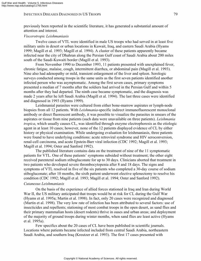
INFECTIOUS DISEASES DIAGNOSED IN US TROOPS 79
previously been reported in the scientific literature, it has generated a substantial amount of attention and interest. Viscerotropic Leishmaniasis
Twelve cases of VTL were identified in male US troops who had served in at least five military units in desert or urban locations in Kuwait, Iraq, and eastern Saudi Arabia (Hyams 1999; Magill et al. 1993; Magill et al. 1994). A cluster of these patients apparently became infected near the city of Dhahran along the Persian Gulf coast of Saudi Arabia about 200 miles south of the Saudi-Kuwaiti border (Magill et al. 1993).
From November 1990 to December 1993, 11 patients presented with unexplained fever, chronic fatigue, malaise, cough, intermittent diarrhea, or abdominal pain (Magill et al. 1993). Nine also had adenopathy or mild, transient enlargement of the liver and spleen. Serologic surveys conducted among troops in the same units as the first seven patients identified another infected person who was asymptomatic. Among the first seven cases, primary symptoms presented a median of 7 months after the soldiers had arrived in the Persian Gulf and within 5 months after they had departed. The ninth case became symptomatic, and the diagnosis was made 2 years after he left Saudi Arabia (Magill et al. 1994). The last three cases were identified and diagnosed in 1993 (Hyams 1999).
Leishmanial parasites were cultured from either bone-marrow aspirates or lymph-node biopsies from all 12 patients. With Leishmania-specific indirect immunofluorescent monoclonal antibody or direct fluorescent antibody, it was possible to visualize the parasites in smears of the aspirates or tissue from nine patients (such data were unavailable on three patients). Leishmania tropica, which usually causes CL, was identified through enzyme electrophoresis as the etiologic agent in at least 10 cases; however, none of the 12 patients displayed evidence of CL by either history or physical examination. While undergoing evaluation for leishmaniasis, three patients were found to have underlying conditions: acute retroviral syndrome and HIV seroconversion, renal-cell carcinoma, and acute Epstein-Barr viral infection (CDC 1992; Magill et al. 1993; Magill et al. 1994; Oster and Sanford 1992).
The published literature contains data on the treatment of nine of the 11 symptomatic patients for VTL. One of these patients’ symptoms subsided without treatment; the other eight received parenteral sodium stibogluconate for up to 30 days. Clinicians aborted that treatment in two patients who developed severe thrombocytopenia after 8 and 18 days. The signs and symptoms of VTL resolved in five of the six patients who completed a 30-day course of sodium stibogluconate; after 10 months, the sixth patient underwent elective splenectomy to resolve his condition (CDC 1992; Magill et al. 1993; Magill et al. 1994; Oster and Sanford 1992). Cutaneous Leishmaniasis
On the basis of the experience of allied forces stationed in Iraq and Iran during World War II, the US military anticipated that troops would be at risk for CL during the Gulf War (Hyams et al. 1995a; Martin et al. 1998). In fact, only 20 cases were recognized and diagnosed (Martin et al. 1998). The very low rate of infection has been attributed to several factors: use of insecticides and repellents; stationing of most combat troops in the open desert, as sand flies and their primary mammalian hosts (desert rodents) thrive in oases and urban areas; and deployment of the majority of ground troops during winter months, when sand flies are least active (Hyams et al. 1995a).
Few specifics about the 20 cases of CL have been published in scientific journals. Locations where patients became infected included from central Saudi Arabia, northeastern Saudi Arabia, and southern Iraq (Kreutzer et al. 1993). The first 17 cases presented with
Copyright © National Academy of Sciences. All rights reserved.
Gulf War and Health: Volume 5. Infectious Diseaseshttp://www.nap.edu/catalog/11765.html
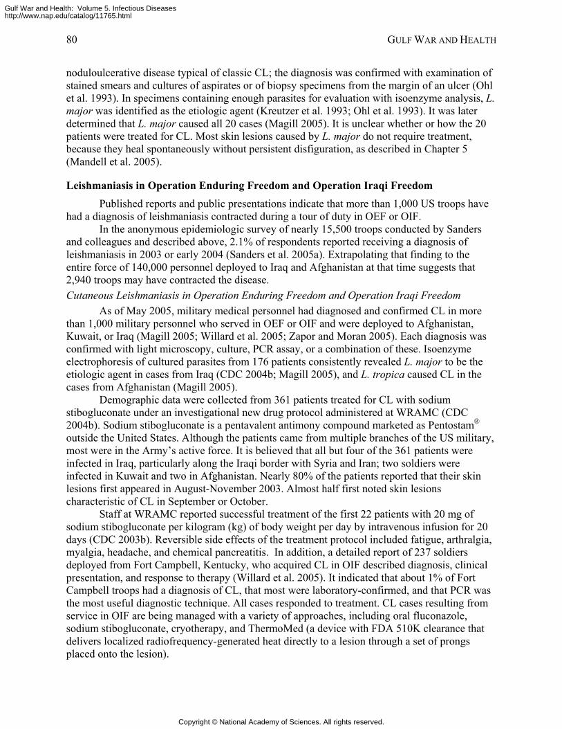
80 GULF WAR AND HEALTH
noduloulcerative disease typical of classic CL; the diagnosis was confirmed with examination of stained smears and cultures of aspirates or of biopsy specimens from the margin of an ulcer (Ohl et al. 1993). In specimens containing enough parasites for evaluation with isoenzyme analysis, L. major was identified as the etiologic agent (Kreutzer et al. 1993; Ohl et al. 1993). It was later determined that L. major caused all 20 cases (Magill 2005). It is unclear whether or how the 20 patients were treated for CL. Most skin lesions caused by L. major do not require treatment, because they heal spontaneously without persistent disfiguration, as described in Chapter 5 (Mandell et al. 2005).
Leishmaniasis in Operation Enduring Freedom and Operation Iraqi Freedom Published reports and public presentations indicate that more than 1,000 US troops have
had a diagnosis of leishmaniasis contracted during a tour of duty in OEF or OIF. In the anonymous epidemiologic survey of nearly 15,500 troops conducted by Sanders
and colleagues and described above, 2.1% of respondents reported receiving a diagnosis of leishmaniasis in 2003 or early 2004 (Sanders et al. 2005a). Extrapolating that finding to the entire force of 140,000 personnel deployed to Iraq and Afghanistan at that time suggests that 2,940 troops may have contracted the disease. Cutaneous Leishmaniasis in Operation Enduring Freedom and Operation Iraqi Freedom
As of May 2005, military medical personnel had diagnosed and confirmed CL in more than 1,000 military personnel who served in OEF or OIF and were deployed to Afghanistan, Kuwait, or Iraq (Magill 2005; Willard et al. 2005; Zapor and Moran 2005). Each diagnosis was confirmed with light microscopy, culture, PCR assay, or a combination of these. Isoenzyme electrophoresis of cultured parasites from 176 patients consistently revealed L. major to be the etiologic agent in cases from Iraq (CDC 2004b; Magill 2005), and L. tropica caused CL in the cases from Afghanistan (Magill 2005).
Demographic data were collected from 361 patients treated for CL with sodium stibogluconate under an investigational new drug protocol administered at WRAMC (CDC 2004b). Sodium stibogluconate is a pentavalent antimony compound marketed as Pentostam® outside the United States. Although the patients came from multiple branches of the US military, most were in the Army’s active force. It is believed that all but four of the 361 patients were infected in Iraq, particularly along the Iraqi border with Syria and Iran; two soldiers were infected in Kuwait and two in Afghanistan. Nearly 80% of the patients reported that their skin lesions first appeared in August-November 2003. Almost half first noted skin lesions characteristic of CL in September or October.
Staff at WRAMC reported successful treatment of the first 22 patients with 20 mg of sodium stibogluconate per kilogram (kg) of body weight per day by intravenous infusion for 20 days (CDC 2003b). Reversible side effects of the treatment protocol included fatigue, arthralgia, myalgia, headache, and chemical pancreatitis. In addition, a detailed report of 237 soldiers deployed from Fort Campbell, Kentucky, who acquired CL in OIF described diagnosis, clinical presentation, and response to therapy (Willard et al. 2005). It indicated that about 1% of Fort Campbell troops had a diagnosis of CL, that most were laboratory-confirmed, and that PCR was the most useful diagnostic technique. All cases responded to treatment. CL cases resulting from service in OIF are being managed with a variety of approaches, including oral fluconazole, sodium stibogluconate, cryotherapy, and ThermoMed (a device with FDA 510K clearance that delivers localized radiofrequency-generated heat directly to a lesion through a set of prongs placed onto the lesion).
Copyright © National Academy of Sciences. All rights reserved.
Gulf War and Health: Volume 5. Infectious Diseaseshttp://www.nap.edu/catalog/11765.html
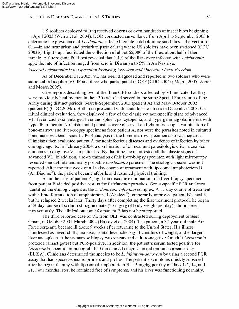
INFECTIOUS DISEASES DIAGNOSED IN US TROOPS 81
US soldiers deployed to Iraq received dozens or even hundreds of insect bites beginning in April 2003 (Weina et al. 2004). DOD conducted surveillance from April to September 2003 to determine the prevalence of Leishmania-infected female phlebotomine sand flies—the vector for CL—in and near urban and periurban parts of Iraq where US soldiers have been stationed (CDC 2003b). Light traps facilitated the collection of about 65,000 of the flies, about half of them female. A fluorogenic PCR test revealed that 1.4% of the flies were infected with Leishmania spp.; the rate of infection ranged from zero in Diwaniya to 5% in An Nasiriya. Visceral Leishmaniasis in Operation Enduring Freedom and Operation Iraqi Freedom
As of December 31, 2005, VL has been diagnosed and reported in two soldiers who were stationed in Iraq during OIF and three who participated in OEF (CDC 2004a; Magill 2005; Zapor and Moran 2005).
Case reports describing two of the three OEF soldiers affected by VL indicate that they were previously healthy men in their 30s who had served in the same Special Forces unit of the Army during distinct periods: March-September, 2003 (patient A) and May-October 2002 (patient B) (CDC 2004a). Both men presented with acute febrile illness in December 2003. On initial clinical evaluation, they displayed a few of the classic yet non-specific signs of advanced VL: fever, cachexia, enlarged liver and spleen, pancytopenia, and hypergammaglobulinemia with hypoalbuminemia. No leishmanial parasites were observed on light microscopic examination of bone-marrow and liver-biopsy specimens from patient A, nor were the parasites noted in cultured bone marrow. Genus-specific PCR analysis of the bone-marrow specimen also was negative. Clinicians then evaluated patient A for noninfectious diseases and evidence of infection by other etiologic agents. In February 2004, a combination of clinical and parasitologic criteria enabled clinicians to diagnose VL in patient A. By that time, he manifested all the classic signs of advanced VL. In addition, a re-examination of his liver-biopsy specimen with light microscopy revealed one definite and many probable Leishmania parasites. The etiologic species was not reported. After the first week of a 14-day course of treatment with liposomal amphotericin B (AmBisome®), the patient became afebrile and resumed physical training.
As in the case of patient A, light microscopic examination of a liver-biopsy specimen from patient B yielded positive results for Leishmania parasites. Genus-specific PCR analyses identified the etiologic agent as the L. donovani-infantum complex. A 15-day course of treatment with a lipid formulation of amphotericin B (Abelcet®) temporarily improved patient B’s health, but he relapsed 2 weeks later. Thirty days after completing the first treatment protocol, he began a 28-day course of sodium stibogluconate (20 mg/kg of body weight per day) administered intravenously. The clinical outcome for patient B has not been reported.
The third reported case of VL from OEF was contracted during deployment to Seeb, Oman, in October 2001-March 2002 (Halsey et al. 2004). The patient, a 37-year-old male Air Force sergeant, became ill about 9 weeks after returning to the United States. His illness manifested as fever, chills, malaise, frontal headache, significant loss of weight, and enlarged liver and spleen. A bone-marrow biopsy was smear- and culture-negative for adult Leishmania protozoa (amastigotes) but PCR-positive. In addition, the patient’s serum tested positive for Leishmania-specific immunoglobulin G in a novel enzyme-linked immunosorbent assay (ELISA). Clinicians determined the species to be L. infantum-donovani by using a second PCR assay that had species-specific primers and probes. The patient’s symptoms quickly subsided after he began therapy with liposomal amphotericin B at 3 mg/kg per day on days 1-5, 14, and 21. Four months later, he remained free of symptoms, and his liver was functioning normally.
Copyright © National Academy of Sciences. All rights reserved.
Gulf War and Health: Volume 5. Infectious Diseaseshttp://www.nap.edu/catalog/11765.html
82 GULF WAR AND HEALTH
The two soldiers deployed to Baghdad who contracted VL presented with fever, enlarged liver and spleen, cytopenia, and hypergammaglobulinemia (Weina et al. 2004). On presentation, one had been deployed for 11 months, and the other had left Iraq 7 months earlier. Examination of bone-marrow biopsies from both patients revealed Leishmania parasites. The patients tested positive for Leishmania in an rK39 serologic test; and, their serum yielded titers of 1:1,024 or greater in a Leishmania immunofluorescent antibody test. Using a PCR assay with species-specific primers, clinicians were able to determine the species—L. infantum-donovani—in one case. The treatment protocols and their outcomes were not published.
Malaria
Very few cases of malaria have been reported in US veterans of the Gulf War, OEF, and OIF. That is not surprising because in 1990-1991, malaria had been eliminated from northeastern Saudi Arabia, where most US troops were stationed, and no indigenous malaria transmission occurred in Kuwait, Bahrain, or Qatar (Hyams et al. 1995a; Oldfield et al. 1991). Malaria due to Plasmodium vivax (vivax malaria) occurred in small numbers in northern Iraq during the late 1980s to 1991 (Oldfield et al. 1991). In the wake of the Gulf War, however, Iraq experienced a serious malaria epidemic; by 2000, vivax malaria had become a serious problem in that country (Schlagenhauf 2003). Moreover, the disease is endemic in many parts of Afghanistan.
Publications during the last 15 years about the threat of vivax malaria to US and allied forces in southwest and south-central Asia sound several consistent themes: the seriousness of the disease, shortcomings of chemical and personal countermeasures, and suboptimal rates of compliance with those countermeasures among troops of many nationalities. According to a 1995 report by the Army Medical Surveillance Activity, “after operations in highly endemic areas, sporadic cases [of malaria] may be expected despite compliance with all prevention guidelines” (MSMR 1995).
Malaria in the Gulf War Vivax malaria existed in the Euphrates River valley of Iraq in 1990 and 1991 (Young et
al. 1992). Seven cases of vivax malaria were reported among US troops who crossed into southern Iraq, where coalition forces operated briefly (Hyams et al. 1995a). No information was given on complications in those troops.
Malaria in Operation Enduring Freedom and Operation Iraqi Freedom As of May 2005, 52 cases of vivax malaria had been reported in US troops who served
either exclusively in Afghanistan or in both Afghanistan and Iraq (Kilpatrick 2005). It is believed that all 52 infections were contracted in Afghanistan, although Plasmodium vivax is endemic in areas of both countries (Wallace et al. 2002). None of the patients was diagnosed with malaria prior to leaving the war theater; this is not surprising, because vivax malaria is known to incubate in human hosts and may relapse up to 5 years after initial infection (Boecken and Bronnert 2005; Johnson 2004).
Thirty-eight of the 52 reported cases of vivax malaria occurred in a 725-man Army Ranger task force deployed to eastern Afghanistan in June-September 2002. Kotwal and colleagues, the primary-care clinicians for these rangers, collected and later analyzed data from the patients during their evaluation, treatment, and followup. In addition, a retrospective anonymous survey was administered to the whole task force in July 2003 to ascertain compliance
Copyright © National Academy of Sciences. All rights reserved.
Gulf War and Health: Volume 5. Infectious Diseaseshttp://www.nap.edu/catalog/11765.html
INFECTIOUS DISEASES DIAGNOSED IN US TROOPS 83
with malarial countermeasures. Retrospective analysis led the authors to conclude that the 38 rangers became infected while working at two specific forward-operating bases during summer 2002 (Kotwal et al. 2005).
The antimalarial chemoprophylaxis prescribed for this Army Ranger task force consisted of 250-mg mefloquine tablets ingested weekly beginning 2 weeks before deployment and ending 4 weeks after deployment. To prevent the late onset of malaria, the troops also were instructed to ingest one 15-mg primaquine tablet daily for 2 weeks after deployment. In addition, it is expected that all US soldiers at risk of malaria are trained and supplied to minimize their exposure to mosquitoes by impregnating their uniforms and bed nets with permethrin, wearing the uniforms properly, using the bed nets, and frequently coating exposed skin with insect repellent that contains 33% DEET (Johnson 2004; Kotwal et al. 2005).
The first three rangers to become symptomatic had vivax malaria diagnosed in March and April 2003. Fifteen more rangers fell ill and had diagnoses in May; the remaining 20 cases came to light in June-November 2003 (Lay 2005). The attack rate for the 725-person task force was 52.4 cases per 1,000 soldiers.
P. vivax infection causes flu-like symptoms that are often severe and debilitating (Boecken and Bronnert 2005; Spudick et al. 2005). Most of the infected rangers presented with fever; many also complained of chills, headache, muscle aches, or nausea. A complete blood-cell count obtained for 31 of the patients demonstrated that most had mild to moderate anemia and thrombocytopenia. The attending physicians based their initial diagnoses on those clinical signs and symptoms. Each case was confirmed with microscopic visualization of malaria parasitemia in the patient’s red blood cells on laboratory-prepared blood smears. A median of 233 days (range, 1-399 days) elapsed between these soldiers’ return from the theater of war and confirmation of their diagnoses.
Two rangers relapsed after completing their first treatment regimen. One of those cases was complicated by life-threatening acute respiratory distress syndrome (ARDS) during the primary attack; the patient relapsed three times from June to December 2003. The multiple relapses indicated infection with primaquine-tolerant P. vivax (Spudick et al. 2005). Pulmonary complications, such as ARDS, might occur with vivax malaria more frequently than is generally recognized.
The results of the anonymous postdeployment survey reported by Kotwal and colleagues indicate that at least 72% of the 725-member task force complied poorly with most of the malarial countermeasures described above (Kotwal et al. 2005). Some 52% followed the US Army guidelines for mefloquine, 41% for primaquine, and 31% for both; 82% reportedly treated their uniforms with permethrin, but only 29% routinely applied DEET to exposed skin. Delayed presentation of vivax malaria is well described, primarily in people who fail to take primaquine as terminal prophylaxis after returning from malaria-endemic areas of the Middle East (Gasser et al. 1991).
Only 14 cases of malaria were reported in US troops in 2004, a 63% decrease from the year before (Lay 2005). All the cases were caused by P. vivax infection and were contracted in Afghanistan (Kilpatrick 2005), and they presented sporadically from February to November.
Kotwal and colleagues suggest that continuously educating field troops about the importance of countermeasures and having leaders monitor and enforce the use of chemoprophylaxis and personal protective measures might further reduce the occurrence of malaria among US forces (Kotwal et al. 2005). At the same time, they and others note several shortcomings of the countermeasures themselves (Boecken and Bronnert 2005; Kotwal et al.
Copyright © National Academy of Sciences. All rights reserved.
Gulf War and Health: Volume 5. Infectious Diseaseshttp://www.nap.edu/catalog/11765.html
84 GULF WAR AND HEALTH
2005; Spudick et al. 2005). Avoiding mosquito bites in the field is difficult and somewhat impractical. For example, nighttime patrols, use of night-vision devices, and vigilance during dusk and dawn place troops at higher risk of exposure to nocturnally active Anopheles mosquitoes, the vectors of malarial parasites, in endemic areas. Permethrin-impregnated bed nets have no utility for soldiers who are at work at night.
West Nile Fever
West Nile virus belongs to the Japanese encephalitis virus antigenic complex in the genus Flavivirus of the family Flaviviridae. Mosquitoes transmit West Nile virus, which was first isolated in 1937 from a febrile woman in the West Nile Province of Uganda. Although 80% of infected people are asymptomatic, those who develop clinical disease usually present with a nonspecific febrile illness lasting 3-6 days. Chapter 5 contains an in-depth discussion of this disease.
West Nile Fever in the Gulf War Only one person who served in the Gulf War had a diagnosis of West Nile fever due to an
infection contracted during the war (Richards et al. 1991). The patient was hospitalized with acute fever, debility, and arthralgias; these symptoms subsided without therapy after 4 days. Clinicians at the US Navy Forward Laboratory in Saudi Arabia tested the patient’s serum with ELISA for immunoglobin M (IgM) and IgG antibodies to the etiologic agents of nine viral and rickettsial diseases: Congo-Crimean hemorrhagic fever, dengue fever, hantaviral disease (Hantaan virus), Q fever, Rift Valley fever, Sindbis, sand fly fever, typhus, and West Nile fever. The serum tested IgM-positive for West Nile virus, but IgM- and IgG-negative for the other arboviruses.
A separate study designed to determine the incidence of insect-borne infections among Gulf War troops identified 30 marines who had been infected with a flavivirus before deployment to Saudi Arabia (Richards et al. 1993b). In the study, an epidemiologic questionnaire was administered and a blood sample obtained from each of 865 marines just before deployment and immediately after. The serum samples were initially screened with ELISA for Crimean-Congo hemorrhagic fever, Rift Valley fever, sand fly fever, Sindbis, West Nile fever, and rickettsiae in the typhus and spotted-fever groups. Moderately increased titers of IgG to West Nile virus in the predeployment and postdeployment serum of 30 marines led investigators to test those blood samples for antibodies to other flaviviruses (for example, St. Louis encephalitis, dengue, and yellow fever viruses). Sera from those marines were reactive to the other flaviviruses.
West Nile Fever in Operation Enduring Freedom and Operation Iraqi Freedom
As of December 2005, there were no reported cases of West Nile fever in military personnel deployed to OEF or OIF.
BRUCELLOSIS
Brucellosis is a serious zoonotic disease endemic in many parts of the world including southwest and south-central Asia (Mandell et al. 2005). The etiologic agent, Brucella spp., has numerous mammalian reservoirs; infected animal hosts shed the bacteria in their milk and urine.
Copyright © National Academy of Sciences. All rights reserved.
Gulf War and Health: Volume 5. Infectious Diseaseshttp://www.nap.edu/catalog/11765.html
INFECTIOUS DISEASES DIAGNOSED IN US TROOPS 85
Humans can contract the disease by ingesting unpasteurized dairy products, by way of infected aerosols inhaled or inoculated into the conjunctival sac of the eyes, and through direct contact between animals or their secretions and cut or abraded skin.
Among all US soldiers who participated in the Gulf War, OEF, and OIF, only one case of brucellosis has been diagnosed (Andrews 2004). An Army helicopter pilot became ill in July 2004 about a week after he completed a 5-month tour of duty in Iraq and returned to his unit in Würzburg, Germany. His initial symptoms of malaise, intermittent chills and fevers as high as 103.9°F, and profuse sweating worsened during the next 5 days despite unspecified “symptomatic treatment” and a day of hospitalization. The patient was readmitted to Würzburg Army Hospital on day 5. Antibodies to B. abortus were identified through laboratory analysis of an unspecified tissue specimen. In addition, a Brucella isolate later identified as B. melitensis was isolated from the patient’s blood. Medical personnel then diagnosed brucellosis. The case report on this patient lacks a description of the course of treatment administered, but the report notes that the patient completed a 14-day course of primaquine after redeploying (presumably to Iraq). The report also notes that the patient had taken chloroquine as malaria prophylaxis but missed his last weekly dose. Epidemiologic questioning of the patient identified only one possible source of the infection: observing the slaughter of a sheep in Iraq.
CHICKENPOX (VARICELLA)
Seventy-five US military personnel who served in ODSh or ODSt were hospitalized for chickenpox during their deployment (Smith et al. 2004). Although it is typically benign in children, chickenpox may cause a more severe disease in adults that might include pneumonitis, hepatitis, and encephalitis. Indeed, the disease kills one in 5,000 infected adults (Heymann 2004). A common complication is bacterial suprainfection, usually of the skin (Military Vaccine Agency 2005b). In 1995, the US Food and Drug Administration licensed a vaccine for this disease.
MENINGOCOCCAL DISEASE
Infectious diseases reportedly caused only one death among US troops deployed to ODSh or ODSt: a fatal case of meningococcal meningitis (Hyams et al. 2001a; Writer et al. 1996). No further information has been published about the death. The literature mentions a second, nonfatal case of meningococcal disease during ODSh or ODSt (Hyams et al. 1995a), but validating evidence is absent. The committee is unaware of other published literature on cases or outbreaks of meningococcal disease among US troops deployed to southwest and south-central Asia during the conflicts discussed in this report.
NOSOCOMIAL INFECTIONS
Gulf War
The committee is unaware of any reports of nosocomial infection among US troops deployed to the Persian Gulf region for ODSh or ODSt. About 470 military personnel who served in those operations suffered nonmortal wounds (Department of Defense 2005).
Copyright © National Academy of Sciences. All rights reserved.
Gulf War and Health: Volume 5. Infectious Diseaseshttp://www.nap.edu/catalog/11765.html
86 GULF WAR AND HEALTH
Operation Enduring Freedom and Operation Iraqi Freedom
More than 16,600 military personnel deployed to OEF and OIF have suffered nonmortal wounds (Defense Manpower Data Center 2006). Many of these troops lost limbs or parts of limbs as a result of explosions that caused traumatic or surgical amputation (Davis et al. 2005). Soil- and water-dwelling bacteria, including Acinetobacter baumanii, have colonized and infected many soldiers’ amputation sites and other blast wounds (Zapor and Moran 2005). Indeed, orthopedic war wounds have become the most common reason for infectious-disease consultations since September 2001 at WRAMC (Zapor and Moran 2005). Cultured specimens from wounds have often implicated a multiple-drug-resistant strain of A. baumanii (Zapor and Moran 2005).
A. baumanii has long been a cause of nosocomial infections. Infected battle injuries from OEF and OIF, however, have increased the rate of bloodstream infections by A. baumanii by more than 2,000% at two major US military hospitals (Table 4.14) (CDC 2004c). Moreover, some strains of A. baumanii have demonstrated resistance to all but one antimicrobial agent in the present pharmacologic arsenal—colistin, a toxic drug (CDC 2004c).
The active-duty military personnel described below, who were generally young and healthy before sustaining war wounds, usually recovered from A. baumanii infection after receiving medical treatment.
TABLE 4.14 Increase in Incidence of A. baumannii Bloodstream Infections at Two Military Hospitals, 2000-2004 No. Cases
Hospital pre-2003 January 2003-August 2004 Percentage Increase in No. Cases
Walter Reed Army Medical Center 2 (2001-2002) 45 2,150
Landstuhl Regional Medical Center 1 (2000-2002) 33 3,200 SOURCE: Scott et al. 2004.
Case Series 1: Brooke Army Medical Center From March 1, 2003, to May 31, 2004, Brooke Army Medical Center (BAMC), in
Houston, Texas admitted 151 injured active-duty soldiers who had been deployed to OEF or OIF (Davis et al. 2005). Davis and colleagues report that all patients but one had previously been admitted to Landstuhl Regional Medical Center (LRMC) in Landstuhl, Germany; three had also been admitted to a second US Army medical facility before BAMC.
Of the tissue samples obtained from 84 of those patients, 48 (32%) tested positive for Acinetobacter calcoaceticus-baumanii complex; 30 patients (63% of those colonized) were clinically infected, and 23 met the case definition for either Acinetobacter osteomyelitis or Acinetobacter wound infection (Table 4.15). None had been diagnosed with Acinetobacter spp. before evacuation from Iraq or Afghanistan.
All but one of the 23 patients with Acinetobacter osteomyelitis or Acinetobacter wound infection received parenteral antimicrobial drug treatment selected to match the susceptibility of the infecting A. baumanii strain (Table 4.16). The primary therapeutic challenge was antimicrobial resistance. Most patients with Acinetobacter osteomyelitis underwent a 6- to 8-week course of drug therapy complemented by multiple surgical debridements of necrotic bone; one patient was treated with surgical debridement alone. Drug therapy for 10 of the osteomyelitis patients involved two antimicrobials, usually imipenem and amikacin. In contrast, patients with
Copyright © National Academy of Sciences. All rights reserved.
Gulf War and Health: Volume 5. Infectious Diseaseshttp://www.nap.edu/catalog/11765.html
INFECTIOUS DISEASES DIAGNOSED IN US TROOPS 87
Acinetobacter wound infection received just one antimicrobial, generally for 10-16 days. All patients responded to therapy. Their followup had lasted a mean of 9 months (range, 1-23 months) at the time the report was published.
TABLE 4.15 Type of Acinetobacter Infection in 23 Wounded Soldiers Admitted to BAMC, March 2003-May 2004, and Therapies Received Type of Infection No. cases Acinetobacter osteomyelitis 15
Acinetobacter osteomyelitis with bacteremia 3
Acinetobacter burn infection 2
Acinetobacter deep wound infection 3 SOURCE: Davis et al. 2005.
TABLE 4.16 Antimicrobial Drug Susceptibilities for 38 Isolates of Acinteobacter calcoaceticu-baumannii Complex Recovered from Wound or Blood Cultures
Antimicrobial Drug Percentage of Susceptible Isolates
Amikacin 48Amoxicillin/clavulanate 9Ampicillin/sulbactam 50Cefepime 14Cefotetan 3Ceftazidime 12Ceftriaxone 6Ciprofloxacin 11Colistina 100Gentamicin 8Imipenem 89Tobramycin 14Trimethoprim/sulfamethoxazole 29a Colistin susceptibility evaluated in three multiple-drug-resistant isolates. SOURCE: Davis et al. 2005.
Case Series 2: Walter Reed Army Medical Center and Landstuhl Regional Medical Center Scott and colleagues report the identification of A. baumannii bloodstream infections in
102 patients at five military hospitals where active-duty military personnel injured in OEF or OIF received treatment (CDC 2004c). The cases were identified from January 1, 2002, to August 31, 2004. Table 4.17 displays the number of infected patients per hospital, their age range, and the subset who that been admitted with (mostly traumatic) injuries associated with OEF or OIF. At both WRAMC and LRMC, clinicians identified a significant percentage of A. baumannii bloodstream infections associated with activities in OEF or OIF within 48 hours of admission (62% at WRAMC and 67% at LRMC). The 78 A. baumanii isolates obtained from patients at WRAMC and LRMC manifested varied levels of resistance to antimicrobial agents commonly used to treat such infections (Table 4.18).
Copyright © National Academy of Sciences. All rights reserved.
Gulf War and Health: Volume 5. Infectious Diseaseshttp://www.nap.edu/catalog/11765.html
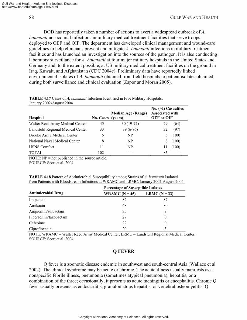
88 GULF WAR AND HEALTH
DOD has reportedly taken a number of actions to avert a widespread outbreak of A. baumanii nosocomial infections in military medical treatment facilities that serve troops deployed to OEF and OIF. The department has developed clinical management and wound-care guidelines to help clinicians prevent and mitigate A. baumanii infections in military treatment facilities and has launched an investigation into the sources of the pathogen. It is also conducting laboratory surveillance for A. baumanii at four major military hospitals in the United States and Germany and, to the extent possible, at US military medical treatment facilities on the ground in Iraq, Kuwait, and Afghanistan (CDC 2004c). Preliminary data have reportedly linked environmental isolates of A. baumanii obtained from field hospitals to patient isolates obtained during both surveillance and clinical evaluation (Zapor and Moran 2005).
TABLE 4.17 Cases of A. baumanii Infection Identified in Five Military Hospitals, January 2002-August 2004
Hospital No. CasesMedian Age (Range) (years)
No. (%) Casualties Associated with OEF or OIF
Walter Reed Army Medical Center 45 30 (19-72) 29 (64) Landstuhl Regional Medical Center 33 39 (6-86) 32 (97) Brooke Army Medical Center 5 NP 5 (100) National Naval Medical Center 8 NP 8 (100) USNS Comfort 11 NP 11 (100) TOTAL 102 --- 85 --- NOTE: NP = not published in the source article. SOURCE: Scott et al. 2004.
TABLE 4.18 Pattern of Antimicrobial Susceptibility among Strains of A. baumanii Isolated from Patients with Bloodstream Infections at WRAMC and LRMC, January 2002-August 2004
Percentage of Susceptible Isolates Antimicrobial Drug WRAMC (N = 45) LRMC (N = 33) Imipenem 82 87 Amikacin 48 80 Ampicillin/sulbactam 35 8 Piperacillin/tazobactam 27 0 Cefepime 22 0 Ciprofloxacin 20 3 NOTE: WRAMC = Walter Reed Army Medical Center, LRMC = Landstuhl Regional Medical Center. SOURCE: Scott et al. 2004.
Q FEVER
Q fever is a zoonotic disease endemic in southwest and south-central Asia (Wallace et al. 2002). The clinical syndrome may be acute or chronic. The acute illness usually manifests as a nonspecific febrile illness, pneumonia (sometimes atypical pneumonia), hepatitis, or a combination of the three; occasionally, it presents as acute meningitis or encephalitis. Chronic Q fever usually presents as endocarditis, granulomatous hepatitis, or vertebral osteomyelitis. Q
Copyright © National Academy of Sciences. All rights reserved.
Gulf War and Health: Volume 5. Infectious Diseaseshttp://www.nap.edu/catalog/11765.html
INFECTIOUS DISEASES DIAGNOSED IN US TROOPS 89
fever frequently goes undiagnosed in humans; many cases are presumably subclinical or asymptomatic.
Cattle, sheep, and goats are the main reservoirs of the disease, which is caused by the bacterium Coxiella burnetii. A single organism can constitute an infectious dose. C. burnetii most frequently infects humans who inhale infected aerosolized body fluids of infected animals; the consumption of raw milk from C. burnetii-infected animals also has caused Q fever in humans.
Three cases of Q fever were reported in US troops who participated in the Gulf War (Hyams et al. 1995a), and 10 cases have been diagnosed in troops deployed to Iraq (Kilpatrick 2005).
Q Fever Contracted During the Gulf War
A case report about one of the three Gulf War troops who had a diagnosis of Q fever has been published (Ferrante and Dolan 1993). A 51-year-old man who had been involved in the ground war in ODSt presented with focal neurologic deficits suggestive of transient ischemic attacks in a crescendo pattern. Two weeks before the onset of symptoms, the patient had returned from Saudi Arabia, where he frequently worked around camels and had once driven a vehicle past a flock of sheep.
The first evaluation of the patient’s cerebrospinal fluid (CSF) yielded an abnormal profile consistent with aseptic meningitis; however the sample was culture-negative for bacteria, fungi, and mycobacteria. Clinicians obtained two more CSF samples at 10 and 14 days; serology from the last puncture yielded a positive C. burnetii IgM indirect fluorescent antibody (IFA) titer consistent with recent Q fever infection. CDC confirmed that result, and Q fever meningoencephalitis was diagnosed. It is believed that the patient contracted the disease in Saudi Arabia through exposure to infected camels or sheep.
Clinicians treated the patient with a 21-day course of oral doxycyline (100 mg twice a day). Six weeks after the first lumbar puncture, serology for C. burnetii IgM IFA titer was negative. No further neurologic sequelae occurred during 12 months of followup.
Q Fever Contracted During Operation Enduring Freedom and Operation Iraqi Freedom
Ten cases of Q fever have been diagnosed in troops serving in OIF as of December 2005. All the patients became infected in northern Iraq (Kilpatrick 2005). We summarize here a report of about eight of the 10 cases; we are unaware of reports in the public literature about the other two patients.
During an investigation of a cluster of 19 cases of severe pneumonia in US troops serving in Iraq, extensive serologic testing for possible infectious etiologies revealed that three patients had positive antibody titers for C. burnetii by immunofluorescence assay (Anderson et al. 2005). The three patients’ predeployment serum samples were negative in the same test. The results led investigators to examine whether C. burnetii accounted for other cases of pneumonia among military personnel deployed to Iraq during the same period (March 1-August 20, 2003).
Predeployment and postdeployment serum samples were available for 22 of 62 soldiers who had pneumonia diagnosed in Iraq. Serum was tested by immunofluorescence assay for phase I and phase II antibody to C. burnetii. Five of the 22 soldiers had seroconverted to C. burnetii before their postdeployment serum draws, leading the investigators to conclude that they also had contracted Q fever in the period March 1-August 20, 2003.
Copyright © National Academy of Sciences. All rights reserved.
Gulf War and Health: Volume 5. Infectious Diseaseshttp://www.nap.edu/catalog/11765.html
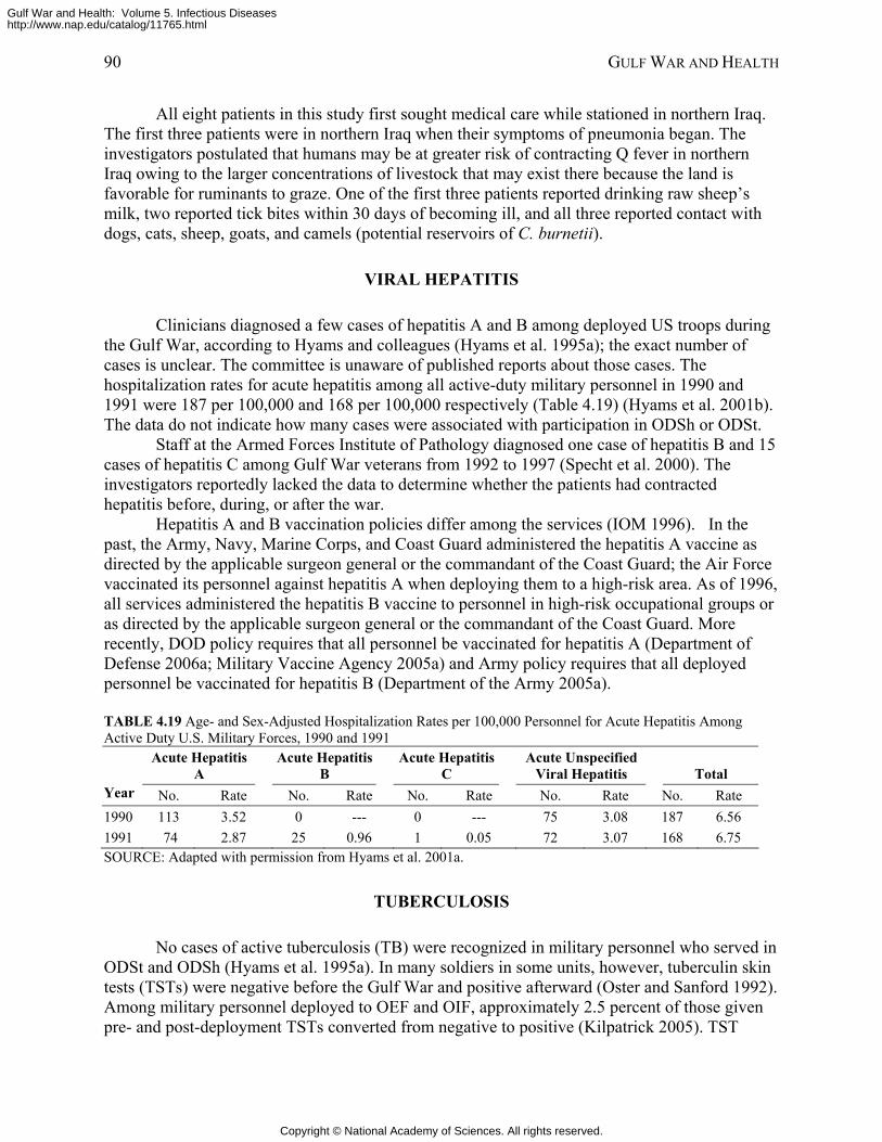
90 GULF WAR AND HEALTH
All eight patients in this study first sought medical care while stationed in northern Iraq. The first three patients were in northern Iraq when their symptoms of pneumonia began. The investigators postulated that humans may be at greater risk of contracting Q fever in northern Iraq owing to the larger concentrations of livestock that may exist there because the land is favorable for ruminants to graze. One of the first three patients reported drinking raw sheep’s milk, two reported tick bites within 30 days of becoming ill, and all three reported contact with dogs, cats, sheep, goats, and camels (potential reservoirs of C. burnetii).
VIRAL HEPATITIS
Clinicians diagnosed a few cases of hepatitis A and B among deployed US troops during the Gulf War, according to Hyams and colleagues (Hyams et al. 1995a); the exact number of cases is unclear. The committee is unaware of published reports about those cases. The hospitalization rates for acute hepatitis among all active-duty military personnel in 1990 and 1991 were 187 per 100,000 and 168 per 100,000 respectively (Table 4.19) (Hyams et al. 2001b). The data do not indicate how many cases were associated with participation in ODSh or ODSt.
Staff at the Armed Forces Institute of Pathology diagnosed one case of hepatitis B and 15 cases of hepatitis C among Gulf War veterans from 1992 to 1997 (Specht et al. 2000). The investigators reportedly lacked the data to determine whether the patients had contracted hepatitis before, during, or after the war.
Hepatitis A and B vaccination policies differ among the services (IOM 1996). In the past, the Army, Navy, Marine Corps, and Coast Guard administered the hepatitis A vaccine as directed by the applicable surgeon general or the commandant of the Coast Guard; the Air Force vaccinated its personnel against hepatitis A when deploying them to a high-risk area. As of 1996, all services administered the hepatitis B vaccine to personnel in high-risk occupational groups or as directed by the applicable surgeon general or the commandant of the Coast Guard. More recently, DOD policy requires that all personnel be vaccinated for hepatitis A (Department of Defense 2006a; Military Vaccine Agency 2005a) and Army policy requires that all deployed personnel be vaccinated for hepatitis B (Department of the Army 2005a).
TABLE 4.19 Age- and Sex-Adjusted Hospitalization Rates per 100,000 Personnel for Acute Hepatitis Among Active Duty U.S. Military Forces, 1990 and 1991
Acute Hepatitis A
Acute Hepatitis B
Acute Hepatitis C
Acute Unspecified Viral Hepatitis Total
Year No. Rate No. Rate No. Rate No. Rate No. Rate 1990 113 3.52 0 --- 0 --- 75 3.08 187 6.56 1991 74 2.87 25 0.96 1 0.05 72 3.07 168 6.75 SOURCE: Adapted with permission from Hyams et al. 2001a.
TUBERCULOSIS
No cases of active tuberculosis (TB) were recognized in military personnel who served in ODSt and ODSh (Hyams et al. 1995a). In many soldiers in some units, however, tuberculin skin tests (TSTs) were negative before the Gulf War and positive afterward (Oster and Sanford 1992). Among military personnel deployed to OEF and OIF, approximately 2.5 percent of those given pre- and post-deployment TSTs converted from negative to positive (Kilpatrick 2005). TST
Copyright © National Academy of Sciences. All rights reserved.
Gulf War and Health: Volume 5. Infectious Diseaseshttp://www.nap.edu/catalog/11765.html
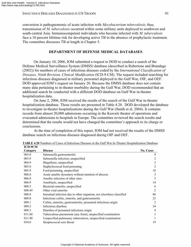
INFECTIOUS DISEASES DIAGNOSED IN US TROOPS 91
conversion is pathognomonic of acute infection with Mycobacterium tuberculosis; thus, transmission of M. tuberculosis occurred within some military units deployed to southwest and south-central Asia. Immunocompetent individuals who become infected with M. tuberculosis face a 10 percent lifetime risk for developing active TB in the absence of prophylactic treatment. The committee discusses TB at length in Chapter 5.
DEPARTMENT OF DEFENSE MEDICAL DATABASES
On January 10, 2006, IOM submitted a request to DOD to conduct a search of the Defense Medical Surveillance System (DMSS) database (described in Rubertone and Brundage (2002)) for numbers of cases of infectious diseases coded by the International Classification of Diseases, Ninth Revision, Clinical Modification (ICD-9-CM). The request included searching for infectious diseases diagnosed in military personnel deployed to the Gulf War, OIF, and OEF. DOD approved IOM’s request on January 20. Because the DMSS database does not contain many data pertaining to in-theater morbidity during the Gulf War, DOD recommended that an additional search be conducted with a different DOD database on Gulf War in-theater hospitalization data.
On June 2, 2006, IOM received the results of the search of the Gulf War in-theater hospitalization database. Those results are presented in Table 4.20. DOD developed the database to investigate in-theater hospitalizations during the Gulf War (Smith et al. 2004). It contains records from almost 20,000 admissions occurring in the Kuwaiti theater of operations and evacuated admissions to hospitals in Europe. The committee reviewed the search results and determined that the results would not have changed the committee’s approach to its charge or conclusions.
At the time of completion of this report, IOM had not received the results of the DMSS database search on infectious diseases diagnosed during OIF and OEF.
TABLE 4.20 Numbers of Cases of Infectious Diseases in the Gulf War In-Theater Hospitalization Database ICD-9CM Category Disease No. Cases 003.0 Salmonella gastroenteritis 9 003.9 Salmonella infection, unspecified 2 004.9 Shigellosis, unspecified 3 005.0 Staphylococcal food poisoning 2 005.9 Food poisoning, unspecified 18 006.0 Acute amebic dysentery without mention of abscess 3 006.8 Amebic infection of other sites 3 006.9 Amebiasis, unspecified 14 008.5 Bacterial enteritis, unspecified 6 008.69 Other viral enteritis 2 008.8 Intestinal infection due to other organism, not elsewhere classified 86 009.0 Infectious colitis, enteritis, and gastroenteritis 23 009.1 Colitis, enteritis, gastroenteritis, presumed infectious origin 2 009.2 Infectious diarrhea 34 009.3 Diarrhea of presumed infectious origin 4 011.60 Tuberculous pneumonia (any form), unspecified examination 1 011.90 Unspecified pulmonary tuberculosis, unspecified examination 1 034.0 Streptococcal sore throat 19
Copyright © National Academy of Sciences. All rights reserved.
Gulf War and Health: Volume 5. Infectious Diseaseshttp://www.nap.edu/catalog/11765.html
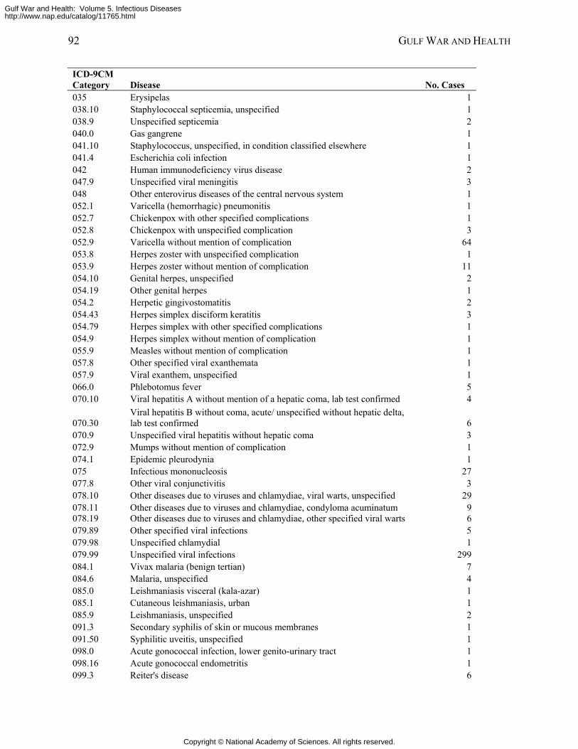
92 GULF WAR AND HEALTH
ICD-9CM Category Disease No. Cases 035 Erysipelas 1 038.10 Staphylococcal septicemia, unspecified 1 038.9 Unspecified septicemia 2 040.0 Gas gangrene 1 041.10 Staphylococcus, unspecified, in condition classified elsewhere 1 041.4 Escherichia coli infection 1 042 Human immunodeficiency virus disease 2 047.9 Unspecified viral meningitis 3 048 Other enterovirus diseases of the central nervous system 1 052.1 Varicella (hemorrhagic) pneumonitis 1 052.7 Chickenpox with other specified complications 1 052.8 Chickenpox with unspecified complication 3 052.9 Varicella without mention of complication 64 053.8 Herpes zoster with unspecified complication 1 053.9 Herpes zoster without mention of complication 11 054.10 Genital herpes, unspecified 2 054.19 Other genital herpes 1 054.2 Herpetic gingivostomatitis 2 054.43 Herpes simplex disciform keratitis 3 054.79 Herpes simplex with other specified complications 1 054.9 Herpes simplex without mention of complication 1 055.9 Measles without mention of complication 1 057.8 Other specified viral exanthemata 1 057.9 Viral exanthem, unspecified 1 066.0 Phlebotomus fever 5 070.10 Viral hepatitis A without mention of a hepatic coma, lab test confirmed 4
070.30 Viral hepatitis B without coma, acute/ unspecified without hepatic delta, lab test confirmed 6
070.9 Unspecified viral hepatitis without hepatic coma 3 072.9 Mumps without mention of complication 1 074.1 Epidemic pleurodynia 1 075 Infectious mononucleosis 27 077.8 Other viral conjunctivitis 3 078.10 Other diseases due to viruses and chlamydiae, viral warts, unspecified 29 078.11 Other diseases due to viruses and chlamydiae, condyloma acuminatum 9 078.19 Other diseases due to viruses and chlamydiae, other specified viral warts 6 079.89 Other specified viral infections 5 079.98 Unspecified chlamydial 1 079.99 Unspecified viral infections 299 084.1 Vivax malaria (benign tertian) 7 084.6 Malaria, unspecified 4 085.0 Leishmaniasis visceral (kala-azar) 1 085.1 Cutaneous leishmaniasis, urban 1 085.9 Leishmaniasis, unspecified 2 091.3 Secondary syphilis of skin or mucous membranes 1 091.50 Syphilitic uveitis, unspecified 1 098.0 Acute gonococcal infection, lower genito-urinary tract 1 098.16 Acute gonococcal endometritis 1 099.3 Reiter's disease 6
Copyright © National Academy of Sciences. All rights reserved.
Gulf War and Health: Volume 5. Infectious Diseaseshttp://www.nap.edu/catalog/11765.html
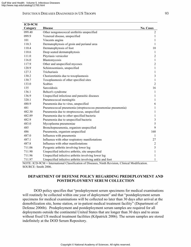
INFECTIOUS DISEASES DIAGNOSED IN US TROOPS 93
ICD-9CM Category Disease No. Cases 099.40 Other nongonococcal urethritis unspecified 2 099.9 Venereal disease, unspecified 1 101 Vincents angina 1 110.3 Dermatophytosis of groin and perianal area 1 110.4 Dermatophytosis of foot 10 110.6 Deep seated dermatophytosis 1 111.0 Pityriasis versicolor 2 116.0 Blastomycosis 1 117.9 Other and unspecified mycoses 2 120.9 Schistosomiasis, unspecified 1 127.3 Trichuriasis 1 130.2 Chorioretinitis due to toxoplasmosis 3 130.7 Toxoplasmosis of other specified sites 1 133.0 Scabies 2 135 Sarcoidosis 16 136.1 Behcet's syndrome 1 136.9 Unspecified infectious and parasitic diseases 1 320.1 Pneumococcal meningitis 1 480.9 Pneumonia due to virus, unspecified 6 481 Pneumococcal pneumonia (streptococcus pneumoniae pneumonia) 1 482.30 Pneumonia due to streptococcus, unspecified 1 482.89 Pneumonia due to other specified bacteria 1 482.9 Pneumonia due to unspecified bacteria 3 483.0 Mycoplasma pneumoniae 5 485 Bronchopneumonia, organism unspecified 9 486 Pneumonia, organism unspecified 168 487.0 Influenza with pneumonia 1 487.1 Influenza with other respiratory manifestations 27 487.8 Influenza with other manifestations 2 711.06 Pyogenic arthritis involving lower leg 5 711.90 Unspecified infective arthritis, site unspecified 1 711.96 Unspecified infective arthritis involving lower leg 1 711.97 Unspecified infective arthritis involving ankle and foot 1
NOTE: ICD-9CM = International Classification of Diseases, Ninth Revision, Clinical Modification. SOURCE: Smith 2006.
DEPARTMENT OF DEFENSE POLICY REGARDING PREDEPLOYMENT AND POSTDEPLOYMENT SERUM COLLECTION
DOD policy specifies that “predeployment serum specimens for medical examinations will routinely be collected within one year of deployment” and that “postdeployment serum specimens for medical examinations will be collected no later than 30 days after arrival at the demobilization site, home station, or in-patient medical treatment facility” (Department of Defense 2006b). Predeployment and postdeployment serum samples are required for all deployments outside the continental United States that are longer than 30 days and to areas without fixed US medical treatment facilities (Kilpatrick 2006). The serum samples are stored indefinitely at the DOD Serum Repository.
Copyright © National Academy of Sciences. All rights reserved.
Gulf War and Health: Volume 5. Infectious Diseaseshttp://www.nap.edu/catalog/11765.html
94 GULF WAR AND HEALTH
Routine testing of the serum samples is not conducted except testing for HIV (Kilpatrick 2006). However, the serum samples have been used for research studies (for example, on Lyme disease, Helicobacter pylori infection, and leishmaniasis), and for operational studies (for example, on severe acute pneumonia cases during OIF, malaria among Marines in Liberia, and WNV).
The committee agrees with DOD’s overall policy regarding collection and use of serum specimens. However, for banked serum specimens to be most useful for determining whether infectious exposures occurred during deployment, the predeployment specimens need to be collected before travel. Current policy allows for collection of predeployment serum specimens up to a year after deployment. If specimens are not collected until after deployment, it would be difficult to ascertain whether any signs of infection found in the “predeployment” specimens are due to exposure during the current deployment or before it.
REFERENCES
Anderson AD, Smoak B, Shuping E, Ockenhouse C, Petruccelli B. 2005. Q fever and the US military. Emerging Infectious Diseases 11(8):1320-1322.
Andrews RB. 2004. Brucellosis in a soldier who recently returned from Iraq. Medical Surveillance Monthly Report 10(4):30.
Boecken GH, Bronnert J. 2005. Pathogenesis and management of a late manifestation of vivax malaria after deployment to Afghanistan: Conclusions for NATO Armed Forces Medical Services. Military Medicine 170(6):488-491.
CDC (Centers for Disease Control and Prevention). 1992. Viscerotropic leishmaniasis in persons returning from Operation Desert Storm—1990-1991. Morbidity and Mortality Weekly Report 41(8):131-134.
CDC. 2003a. Severe Acute Pneumonitis Among Deployed US Military Personnel—Southwest Asia, March—August 2003. Morbidity and Mortality Weekly Report 52(36):857-859.
CDC. 2003b. Cutaneous leishmaniasis in US military personnel—Southwest/Central Asia, 2002-2003. Morbidity and Mortality Weekly Report 52(42):1009-1012.
CDC. 2004a. Two cases of visceral leishmaniasis in US military personnel—Afghanistan, 2002-2004. Morbidity and Mortality Weekly Report 53(12):265-268.
CDC. 2004b. Update: Cutaneous leishmaniasis in US military personnel—Southwest/Central Asia, 2002-2004. Morbidity and Mortality Weekly Report 53(12):264-265.
CDC. 2004c. Acinetobacter baumannii infections among patients at military medical facilities treating injured US service members, 2002-2004. Morbidity and Mortality Weekly Report 53(45):1063-1066.
Davis KA, Moran KA, McAllister CK, Gray PJ. 2005. Multidrug-resistant Acinetobacter extremity infections in soldiers. Emerging Infectious Diseases 11(8):1218-1224.
Defense Manpower Data Center. Military Casualty Information. [Online]. Available: http://www.dior.whs.mil/mmid/casualty/castop.htm [accessed March 2006].
Copyright © National Academy of Sciences. All rights reserved.
Gulf War and Health: Volume 5. Infectious Diseaseshttp://www.nap.edu/catalog/11765.html
INFECTIOUS DISEASES DIAGNOSED IN US TROOPS 95
Department of the Army. 2005a. Medical Screening of Personnel Mobilized for Deployment or Deployed in Support of Contingency Operations (Predeployment Screening Requirements). Falls Church, VA: Office of the Surgeon General.
Department of the Army. 2005b. Release of Actionable Medical Information Policy Memorandum. Fort Sam Houston, TX: Headquarters, United States Army Medical Command.
DOD (Department of Defense). 2005. Principal Wars in Which the United States Participated US Military Personnel Serving and Casualties A. [Online]. Available: http://www.dior.whs.mil/mmid/casualty/WCPRINCIPAL.pdf [accessed March 2006].
DOD. 2006a. Individual Medical Readiness. Department of Defense Instruction. Washington, DC.
DOD. 2006b. Policy for Pre- and Post-Deployment Serum Collection. Washington, DC: Assistant Secretary of Defense.
Donowitz GR, Mandell GL. 2000. Acute pneumonia. In: Mandell GL, Bennett JE, Dolin R, Editors. Principles and Practice of Infectious Diseases. 5th ed. New York: Churchill Livingstone. Pp. 717-743.
Ferrante MA, Dolan MJ. 1993. Q fever meningoencephalitis in a soldier returning from the Persian Gulf War. Clinical Infectious Diseases 16(4):489-496.
Gasser RA Jr, Magill AJ, Oster CN, Tramont EC. 1991. The threat of infectious disease in Americans returning from Operation Desert Storm. New England Journal of Medicine 324(12):859-864.
Gwaltney JM, Jr. 2000a. Acute Bronchitis. In: Mandell GL, Bennett JE, Dolin R, Editors. Principles and Practice of Infectious Diseases. 5th ed. New York: Churchill Livingstone. Pp. 703-706.
Gwaltney JM, Jr. 2000b. Sinusitis. In: Mandell GL, Bennett JE, Dolin R, Editors. Principles and Practice of Infectious Diseases. 5th ed. New York: Churchill Livingstone. Pp. 676-686.
Haberberger RL, Scott DA, Thornton SA, Hyams KC. 1994. Diarrheal disease aboard a US Navy ship after a brief port visit to a high risk area. Military Medicine 159(6):445-448.
Halsey ES, Bryce LM, Wortmann GW, Weina PJ, Ryan JR, DeWitt CC. 2004. Visceral leishmaniasis in a soldier returning from Operation Enduring Freedom. Military Medicine 169(9):699-701.
Heymann DL. 2004. Control of Communicable Diseases Manual. Washington, DC: American Public Health Association.
Hyams KC. 1999. Gulf War Syndrome: Potential role of infectious diseases. Current Opinion in Infectious Diseases 12(5):439-443.
Hyams KC, Bourgeois AL, Merrell BR, Rozmajzl P, Escamilla J, Thornton SA, Wasserman GM, Burke A, Echeverria P, Green KY, et al. 1991. Diarrheal disease during Operation Desert Shield. New England Journal of Medicine 325(20):1423-1428.
Copyright © National Academy of Sciences. All rights reserved.
Gulf War and Health: Volume 5. Infectious Diseaseshttp://www.nap.edu/catalog/11765.html
96 GULF WAR AND HEALTH
Hyams KC, Malone JD, Kapikian AZ, Estes MK, Xi J, Bourgeois AL, Paparello S, Hawkins RE, Green KY. 1993. Norwalk virus infection among Desert Storm troops. Journal of Infectious Diseases 167(4):986-987.
Hyams KC, Hanson K, Wignall FS, Escamilla J, Oldfield EC, 3rd. 1995a. The impact of infectious diseases on the health of US troops deployed to the Persian Gulf during operations Desert Shield and Desert Storm. Clinical Infectious Diseases 20(6):1497-1504.
Hyams KC, Malone JD, Bourgeois AL, Hawkins R, Hale TL, Murphy JR. 1995b. Serum antibody to lipopolysaccharide antigens of Shigella species among US military peronnel deployed to Saudi Arabia and Kuwait during Operation Desert Storm. Clinical and Diagnostic Laboratory Immunology 2(6):700-703.
Hyams KC, Riddle J, Trump DH, Graham JT. 2001a. Endemic infectious diseases and biological warfare during the Gulf War: A decade of analysis and final concerns. American Journal of Tropical Medicine and Hygiene 65(5):664-670.
Hyams KC, Smith TC, Riddle J, Trump DH, Gray G. 2001b. Viral hepatitis in the US military: A study of hospitalization records from 1974 to 1999. Military Medicine 166(10):862-865.
IOM (Institute of Medicine). 1996. Interaction of Drugs, Biologics, and Chemicals in US Military Forces: Current and Future Issues. Washington, DC: National Academy Press.
Johnson KE. 2004. Malaria, US Army, 2003. Medical Surveillance Monthly Report 10(1):6-8.
Kelly MT, Brenner DJ, Farmer JJIII. 1985. Enterobacteriaceae. In: Lennette EH, Balows A, Hausler WJ, Jr., Shadomy HJ, Editors. Manual of Clinical Microbiology. 4th ed. Washington, DC: American Society for Microbiology. Pp. 263-277.
Kilpatrick ME. 2005. Presentation to IOM Committee on Gulf War and Health: Infectious Diseases. Washington, DC.
Kilpatrick ME. 2006. Deployment Health Support Office of the Assistant Secretary of Defense, Health Affairs. Personal Communication.
Kotwal RS, Wenzel RB, Sterling RA, Porter WD, Jordan NN, Petruccelli BP. 2005. An outbreak of malaria in US Army Rangers returning from Afghanistan. Journal of the American Medical Association 293(2):212-216.
Kreutzer RD, Grogl M, Neva FA, Fryauff DJ, Magill AJ, Aleman-Munoz MM. 1993. Identification and genetic comparison of leishmanial parasites causing viscerotropic and cutaneous disease in soldiers returning from Operation Desert Storm. American Journal of Tropical Medicine and Hygiene 49(3):357-363.
Lay JC. 2005. Malaria, US Army, 2004. Medical Surveillance Monthly Report 11(1):7-10.
Lew JF, Kapikian AZ, Jiang X, Estes MK, Green KY. 1994. Molecular characterization and expression of the capsid protein of a Norwalk-like virus recovered from a Desert Shield troop with gastroenteritis. Virology 200(1):319-325.
Magill AJ. 2005. Leishmaniasis in Veterans of Desert Storm and Iraqi Freedom. Presentation to IOM Committee on Gulf War and Health: Infectious Diseases. Washington, DC.
Copyright © National Academy of Sciences. All rights reserved.
Gulf War and Health: Volume 5. Infectious Diseaseshttp://www.nap.edu/catalog/11765.html
INFECTIOUS DISEASES DIAGNOSED IN US TROOPS 97
Magill AJ, Grogl M, Gasser RA Jr, Sun W, Oster CN. 1993. Visceral infection caused by Leishmania tropica in veterans of Operation Desert Storm. New England Journal of Medicine 328(19):1383-1387.
Magill AJ, Grogl M, Johnson SC, Gasser RA Jr. 1994. Visceral infection due to Leishmania tropica in a veteran of Operation Desert Storm who presented 2 years after leaving Saudi Arabia. Clinical Infectious Diseases 19(4):805-806.
Malone JD, Paparello S, Thornton S, Mapes T, Haberberger R, Hyams KC. 1991. Parasitic infections in troops returning from Operation Desert Storm. New England Journal of Medicine 325(20):1448-1449.
Mandell GL, Bennett JE, Dolin R. 2005. Principles and Practice of Infectious Diseases. 6th ed. Philadelphia, PA: Elsevier Churchill Livingstone.
Martin S, Gambel J, Jackson J, Aronson N, Gupta R, Rowton E, Perich M, McEvoy P, Berman J, Magill A, Hoke C. 1998. Leishmaniasis in the United States military. Military Medicine 163(12):801-807.
Matson DO. 2005. Norovirus gastroenteritis in US Marines in Iraq. Clinical Infectious Diseases 40(4):526-527.
McCarthy M, Estes MK, Hyams KC. 2000. Norwalk-like virus infection in military forces: Epidemic potential, sporadic disease, and the future direction of prevention and control efforts. Journal of Infectious Diseases 181(2 Suppl):S387-S391.
Mikhail MM, Mansour MM, Oyofo BA, Malone JD. 1996. Immune response to Shigella sonnei in US Marines. Infection and Immunity 64(9):3942-3945.
Military Vaccine Agency. 2005a. Hepatitis A Infection and Hepatitis A Vaccine. Military Vaccine (MILVAX) Agency, Office of The Army Surgeon General, US Army.
Military Vaccine Agency. 2005b. Chickenpox Vaccination Program: Questions and Answers. Military Vaccine (MILVAX) Agency, Office of The Army Surgeon General, US Army.
MSMR (Medical Surveillance Monthly Report). 1995. Malaria in active duty soldiers. Medical Surveillance Monthly Report 1(4):9.
Ohl CA, Hyams KC, Malone JD, Oldfield E 3rd. 1993. Leishmaniasis among Desert Storm veterans: A diagnostic and therapeutic dilemma. Military Medicine 158(11):726-729.
Oldfield EC 3rd, Wallace MR, Hyams KC, Yousif AA, Lewis DE, Bourgeois AL. 1991. Endemic infectious diseases of the Middle East. Reviews of Infectious Diseases 13(3 Suppl)S199-S217.
Oster CN, Sanford JP. 1992. Febrile illness in a Desert Storm veteran. Hospital Practice (Office Edition) 27(11):145-148, 151, 155-160.
Paparello SF, Garst P, Bourgeois AL, Hyams KC. 1993. Diarrheal and respiratory disease aboard the hospital ship, USNS-Mercy T-AH 19, during Operation Desert Shield. Military Medicine 158(6):392-395.
Richards AL, Hyams KC, Merrell BR, Dasch GA, Woody JN, Ksiazek TG, LeDuc JW, Watts DM. 1991. Medical aspects of Operation Desert Storm. New England Journal of Medicine 325(13):970-971.
Copyright © National Academy of Sciences. All rights reserved.
Gulf War and Health: Volume 5. Infectious Diseaseshttp://www.nap.edu/catalog/11765.html
98 GULF WAR AND HEALTH
Richards AL, Hyams KC, Watts DM, Rozmajzl PJ, Woody JN, Merrell BR. 1993a. Respiratory disease among military personnel in Saudi Arabia during Operation Desert Shield. American Journal of Public Health 83(9):1326-1329.
Richards AL, Malone JD, Sheris S, Weddle JR, Rossi CA, Ksiazek TG, LeDuc JW, Dasch GA, Hyams KC. 1993b. Arbovirus and rickettsial infections among combat troops during Operation Desert Shield/Desert Storm. Journal of Infectious Diseases 168(4):1080-1081.
Rubertone MV, Brundage JF. 2002. The Defense Medical Surveillance System and the Department of Defense serum repository: Glimpses of the future of public health surveillance. American Journal of Public Health 92(12):1900-1904.
Sanders JW, Putnam SD, Riddle MS, Tribble DR, Jobanputra NK, Jones JJ, Scott DA, Frenck RW. 2004. The epidemiology of self-reported diarrhea in operations Iraqi freedom and enduring freedom. Diagnostic Microbiology and Infectious Disease 50(2):89-93.
Sanders JW, Putnam SD, Frankart C, Frenck RW, Monteville MR, Riddle MS, Rockabrand DM, Sharp TW, Tribble DR. 2005a. Impact of illness and non-combat injury during operations iraqi freedom and enduring freedom (Afghanistan). American Journal of Tropical Medicine and Hygiene 73(4):713-719.
Sanders JW, Putnam SD, Riddle MS, Tribble DR. 2005b. Military importance of diarrhea: Lessons from the Middle East. Current Opinion in Gastroenterology 21(1):9-14.
Schlagenhauf P. 2003. Malaria in Iraq—the pitfalls of Plasmodium vivax prophylaxis. Lancet Infectious Diseases 3(8):460.
Scott P, Artis D, Uzonna J, Zaph C. 2004. The development of effector and memory T cells in cutaneous leishmaniasis: The implications for vaccine development. Immunological Reviews 201:318-338.
Smith TC. 2006. Results of IOM Requested Database Search for Infectious Disease Cases During the Gulf War. San Diego, CA: Department of Defense Center for Deployment Health Research, Naval Health Research Center.
Smith TC, Corbeil TE, Ryan MA, Heller JM, Gray GC. 2004. In-theater hospitalizations of US and allied personnel during the 1991 Gulf War. American Journal of Epidemiology 159(11):1064-1076.
Specht CS, Lewin-Smith MR, Kalasinsky VF, Peterson MR, Mullick FG. 2000. The surgical pathology and cytopathology of US Persian Gulf War military veterans: Identification of diseases endemic to the theater of operations. Archives of Pathology and Laboratory Medicine 124(9):1299-1301.
Spudick JM, Garcia LS, Graham DM, Haake DA. 2005. Diagnostic and therapeutic pitfalls associated with primaquine-tolerant Plasmodium vivax. Journal of Clinical Microbiology 43(2):978-981.
Stephens I, Nataro JP. 2004. Prevention of enteric diseases. Advances in Experimental Medicine and Biology 549:71-82.
Thornton SA, Sherman SS, Farkas T, Zhong W, Torres P, Jiang X. 2005. Gastroenteritis in US Marines during Operation Iraqi Freedom. Clinical Infectious Diseases 40(4):519-525.
Copyright © National Academy of Sciences. All rights reserved.
Gulf War and Health: Volume 5. Infectious Diseaseshttp://www.nap.edu/catalog/11765.html
INFECTIOUS DISEASES DIAGNOSED IN US TROOPS 99
Wallace MR, Hale BR, Utz GC, Olson PE, Earhart KC, Thornton SA, Hyams KC. 2002. Endemic infectious diseases of Afghanistan. Clinical Infectious Diseases 34(5 Suppl):S171-S207.
Wasserman GM, Martin BL, Hyams KC, Merrill BR, Oaks HG, McAdoo HA. 1997. A survey of outpatient visits in a United States Army forward unit during Operation Desert Shield. Military Medicine 162(6):374-379.
Weina PJ, Neafie RC, Wortmann G, Polhemus M, Aronson NE. 2004. Old world leishmaniasis: An emerging infection among deployed US military and civilian workers. Clinical Infectious Diseases 39(11):1674-1680.
Willard RJ, Jeffcoat AM, Benson PM, Walsh DS. 2005. Cutaneous leishmaniasis in soldiers from Fort Campbell, Kentucky returning from Operation Iraqi Freedom highlights diagnostic and therapeutic options. Journal of the American Academy of Dermatology 52(6):977-987.
Wolf MK, Taylor DN, Boedeker EC, Hyams KC, Maneval DR, Levine MM, Tamura K, Wilson RA , Echeverria P. 1993. Characterization of enterotoxigenic Escherichia coli isolated from US troops deployed to the Middle East. Journal of Clinical Microbiology 31(4):851-856.
Writer JV, DeFraites RF, Brundage JF. 1996. Comparative mortality among US military personnel in the Persian Gulf region and worldwide during Operations Desert Shield and Desert Storm. Journal of the American Medical Association 275(2):118-121.
Young RC Jr, Rachal RE, Huguley JW 3rd. 1992. Environmental health concerns of the Persian Gulf War. Journal of the National Medical Association 84(5):417-424.
Zapor MJ, Moran KA. 2005. Infectious diseases during wartime. Current Opinion in Infectious Diseases 18(5):395-399.
Copyright © National Academy of Sciences. All rights reserved.
Gulf War and Health: Volume 5. Infectious Diseaseshttp://www.nap.edu/catalog/11765.html

Copyright © National Academy of Sciences. All rights reserved.
Gulf War and Health: Volume 5. Infectious Diseaseshttp://www.nap.edu/catalog/11765.html
101
5
LEVELS OF ASSOCIATION BETWEEN SELECT DISEASES AND LONG-TERM ADVERSE HEALTH OUTCOMES
Only 10% of the roughly 90 infectious diseases endemic in southwest and south-central Asia are likely to pose a long-term health risk to exposed US military personnel. As noted in Chapter 3, the long-term adverse health outcomes of most diseases endemic in the region would usually become apparent during or immediately after the acute illness, and many of the health outcomes are rare. However, nine of the infectious diseases meet the inclusion criteria outlined in Box 5.1 and discussed in Chapter 2. Those nine diseases and their associated long-term adverse health outcomes are the subject of this chapter (Table 5.1).
Following the paradigm of past Institute of Medicine Committees on Gulf War and Health, the committee determined the strength of association between each infection1 and specific long-term adverse health outcomes in humans. For every health outcome discussed in this chapter, there is limited or suggestive evidence of an association, sufficient evidence of an association, or sufficient evidence of a causal relationship with the infectious disease. Several delayed long-term adverse health outcomes of the nine diseases are listed in Chapter 3 but not reviewed here; the committee determined that there is inadequate or insufficient evidence of an association between these health outcomes and the infectious diseases. To reach its conclusions, the committee assessed the available evidence published in the biomedical literature about the long-term adverse outcomes of the diseases on human health.
BOX 5.1 Inclusion Criteria
The committee used these questions to evaluate the dozens of infectious diseases endemic in southwest and south-central Asia or commonly found among troops in wartime (Table 2.1). If the answer to every question was yes, a disease met the criteria for in-depth evaluation in this chapter.
1. Was the infection or disease diagnosed in US troops in appropriate temporal relationship to deployment to the Gulf War, Operation Enduring Freedom, or Operation Iraqi Freedom, given the natural history of the disease?
2. Is the risk of contracting the disease during deployment in southwest or south-central Asia equal to or greater than the risk of contracting it in the United States?
3. Does the disease have a known or suspected long-term adverse health outcome? 4. Would there be a delay between the infection or the end of the acute illness and the onset of the long-term
adverse health outcome?
1 In this context, the term infection refers to a primary infection that leads to disease.
Copyright © National Academy of Sciences. All rights reserved.
Gulf War and Health: Volume 5. Infectious Diseaseshttp://www.nap.edu/catalog/11765.html
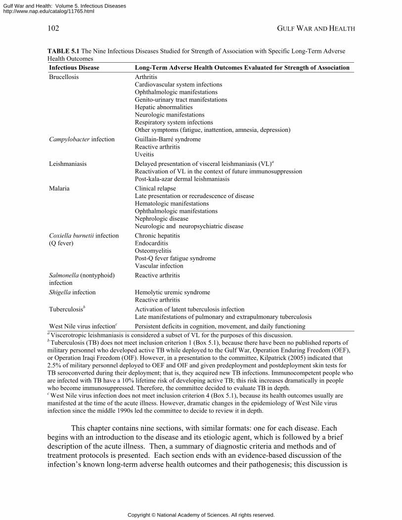
102 GULF WAR AND HEALTH
TABLE 5.1 The Nine Infectious Diseases Studied for Strength of Association with Specific Long-Term Adverse Health Outcomes Infectious Disease Long-Term Adverse Health Outcomes Evaluated for Strength of Association Brucellosis Arthritis
Cardiovascular system infections Ophthalmologic manifestations Genito-urinary tract manifestations Hepatic abnormalities Neurologic manifestations Respiratory system infections Other symptoms (fatigue, inattention, amnesia, depression)
Campylobacter infection Guillain-Barré syndrome Reactive arthritis Uveitis
Leishmaniasis Delayed presentation of visceral leishmaniasis (VL)a Reactivation of VL in the context of future immunosuppression Post-kala-azar dermal leishmaniasis
Malaria Clinical relapse Late presentation or recrudescence of disease Hematologic manifestations Ophthalmologic manifestations Nephrologic disease Neurologic and neuropsychiatric disease
Coxiella burnetii infection (Q fever)
Chronic hepatitis Endocarditis Osteomyelitis Post-Q fever fatigue syndrome Vascular infection
Salmonella (nontyphoid) infection
Reactive arthritis
Shigella infection Hemolytic uremic syndrome Reactive arthritis
Tuberculosisb Activation of latent tuberculosis infection Late manifestations of pulmonary and extrapulmonary tuberculosis
West Nile virus infectionc Persistent deficits in cognition, movement, and daily functioning a Viscerotropic leishmaniasis is considered a subset of VL for the purposes of this discussion. b Tuberculosis (TB) does not meet inclusion criterion 1 (Box 5.1), because there have been no published reports of military personnel who developed active TB while deployed to the Gulf War, Operation Enduring Freedom (OEF), or Operation Iraqi Freedom (OIF). However, in a presentation to the committee, Kilpatrick (2005) indicated that 2.5% of military personnel deployed to OEF and OIF and given predeployment and postdeployment skin tests for TB seroconverted during their deployment; that is, they acquired new TB infections. Immunocompetent people who are infected with TB have a 10% lifetime risk of developing active TB; this risk increases dramatically in people who become immunosuppressed. Therefore, the committee decided to evaluate TB in depth. c West Nile virus infection does not meet inclusion criterion 4 (Box 5.1), because its health outcomes usually are manifested at the time of the acute illness. However, dramatic changes in the epidemiology of West Nile virus infection since the middle 1990s led the committee to decide to review it in depth.
This chapter contains nine sections, with similar formats: one for each disease. Each begins with an introduction to the disease and its etiologic agent, which is followed by a brief description of the acute illness. Then, a summary of diagnostic criteria and methods and of treatment protocols is presented. Each section ends with an evidence-based discussion of the infection’s known long-term adverse health outcomes and their pathogenesis; this discussion is
Copyright © National Academy of Sciences. All rights reserved.
Gulf War and Health: Volume 5. Infectious Diseaseshttp://www.nap.edu/catalog/11765.html
ASSOCIATIONS BETWEEN DISEASES AND LONG-TERM ADVERSE HEALTH OUTCOMES 103
the basis of the committee’s conclusions about the strength of association between the primary infection and each long-term adverse health outcome.
DIARRHEAL DISEASES: CAMPYLOBACTER, NON-TYPHOID SALMONELLA, AND SHIGELLA INFECTIONS
Among the many pathogens known to have caused diarrheal disease among US troops deployed to the Gulf War, Operation Enduring Freedom (OEF), or Operation Iraqi Freedom (OIF), three merit an examination of their potential long-term, adverse outcomes to veterans’ health: Campylobacter, Shigella, and Salmonella.
Campylobacter Infection
Campylobacter infections are common causes of acute diarrheal illnesses in humans globally (Blaser 2005). The committee examined three potential long-term adverse health outcomes of Campylobacter infection: Guillain-Barré syndrome, reactive arthritis, and uveitis.
The most common pathogenic Campylobacter species is C. jejuni, but disease may also be caused by other species, especially C. coli, C. upsaliensis, C. lari, and C. fetus. The typical illness is acute diarrheal disease lasting 2-5 days accompanied by abdominal pain and fever. The illness responds well to antibiotic treatment but often is self-limited. Campylobacter occasionally causes an acute systemic infection.
Transmission of Campylobacter Campylobacter species (spp.) infect humans most often through contaminated food or
water. Drinking untreated water is a major risk factor for both sporadic and epidemic campylobacteriosis (Allos 2001; Blaser 2005). Foodborne infections occur chiefly after the consumption of improperly heated foods of animal origin; common vehicles include unpasteurized milk and undercooked chicken. Among wild and domesticated animals, Campylobacter spp. may be normal flora or pathogens (Blaser 2005). Rarely, the bacteria are transmitted by person-to-person contact; this occurs chiefly from the handling of feces of incontinent people, such as infants, who are infected.
People suffering from an enteric illness may be infected with two or more bacterial, viral, protozoan, or helminthic pathogens. Some laboratory analyses of stool specimens from deployed troops who had a diagnosis of diarrheal illness found dual infections in a subset of patients, as described in Chapter 4.
Endemicity in Southwest and South-Central Asia
Campylobacter is a common cause of acute diarrhea in southwest and south-central Asia (Wilson 1991). In the United States, the bacteria frequently instigate both sporadic diarrhea and outbreaks (Wilson 1991).
Acute Illness Patients with Campylobacter infections often present with a short prodrome of symptoms
consisting chiefly of headache, myalgias, back pain, and fever. Within 24 hours, the illness centers on the gastrointestinal tract, producing abdominal pain and diarrhea (either may come
Copyright © National Academy of Sciences. All rights reserved.
Gulf War and Health: Volume 5. Infectious Diseaseshttp://www.nap.edu/catalog/11765.html
104 GULF WAR AND HEALTH
first). Common characteristics of the abdominal pain are unlocalized cramping that may be so severe as to mimic acute appendicitis; however, diarrhea predominates over abdominal pain in most patients.
On the first day of diarrheal illness, the patient usually has four to 20 loose stools, and 25% of them may contain visible blood. Laboratory examination of stool specimens usually reveals gross or microscopic blood in all and leukocytes in 70%. Fever continues from the prodrome and persists for 24-48 hours.
Symptoms usually begin to recede after 48 hours and resolve during the next few days. In rare cases, the illness may last longer. In the absence of antibiotic treatment, relapse occurs in about 20% of cases; relapses are usually milder than the initial episodes.
Some people with Campylobacter infections are bacteremic (Mandell et al. 2005); this condition represents either a primary bacteremia or, rarely, the seeding of a distant organ (Blaser et al. 1986). Diagnosis During and After Acute Illness
Diagnosis of the acute illness is based on culture of feces and, rarely, of blood. Culture-based tests even in the acute phase can have false-negative results, especially in infection by non-jejuni species, because Campylobacter spp. are difficult to grow in culture. Alternatively, the bacteria can be detected with polymerase-chain-reaction (PCR) assay of genetic material from stool specimens. Antibody testing, which is not commercially available, is less reliable because of the diversity of Campylobacter strains, the time required for a response to occur, and differences in magnitudes of responses among hosts.
Infected people shed Campylobacter in stool for a mean of 2-3 weeks after the onset of symptoms; virtually no immunocompetent hosts are still shedding the organism after 8 weeks (Karmali and Fleming 1979; Svedhem and Kaijser 1980; Taylor et al. 1988). Thus, a culture or PCR test conducted more than 2 months after an acute episode of Campylobacter enteric disease would rarely be positive. After 2 months have elapsed, there is no reliable diagnostic test for exposure to Campylobacter in people who manifest diseases that could be late adverse health outcomes of a Campylobacter infection. Treatment of Acute Illness
Fluid and electrolyte replacement is the treatment of choice for diarrheal illnesses. In patients who are still symptomatic at the time of diagnosis, antimicrobial treatment is recommended, particularly with fluoroquinolones and macrolides. Clinicians should be cognizant of Campylobacter’s growing resistance to those antimicrobials; the degree of resistance will reflect the use of antimicrobials in animal farming and in the local human population.
Long-Term Adverse Health Outcomes of Campylobacter Infection On occasion, infection by Campylobacter spp. leads to long-term adverse health
outcomes. The most serious health outcome associated with campylobacteriosis is Guillain-Barré syndrome (GBS). Reactive arthritis appears to occur after campylobacteriosis at a frequency greater than the background frequency. There is some evidence that uveitis is associated with Campylobacter infection. Guillain-Barré Syndrome
The first report of an association between Campylobacter jejuni infection and GBS was published in 1982 (Rhodes and Tattersfield 1982). Numerous scientists have since investigated
Copyright © National Academy of Sciences. All rights reserved.
Gulf War and Health: Volume 5. Infectious Diseaseshttp://www.nap.edu/catalog/11765.html
ASSOCIATIONS BETWEEN DISEASES AND LONG-TERM ADVERSE HEALTH OUTCOMES 105
the relationship between the two diseases and have published more than 200 reports in peer-reviewed journals. By the year 2000, those investigations had established that infection by C. jejuni causes about 30% of all cases of GBS (Allos 1997; Dingle et al. 2001; McCarthy and Giesecke 2001; Nachamkin 2002; Nachamkin et al. 1998; Nachamkin et al. 2001; Sinha et al. 2004; Tam et al. 2003). A number of other infectious diseases are also associated with GBS.
GBS is a severe acute neurologic disease characterized by ascending paralysis with involvement of motor neurons and sometimes sensory neurons (Rhodes and Tattersfield 1982). Developing over a period of days, the symptoms of GBS may lead to paralysis of the respiratory muscles and death; however, with rapid supportive care, the fatality rate has been reduced from more than 10% to less than 5%. Between 10 and 20% of affected persons have permanent neurologic deficits, such as persistent muscle weakness and contractures. Most patients with GBS require hospitalization, and more than 20% require ventilatory support at some time during their illness. Recommended treatment should be started immediately and may include plasmapheresis and intravenous administration of immunoglobulins.
Approximately 0.01-0.03% of US patients who suffer acute gastrointestinal disease due to C. jejuni will develop GBS (Allos 1997; Tauxe and Blake 1992). The risk of developing GBS during the 2 months after a symptomatic episode of C. jejuni infection is about 100 times greater than the risk in the general population (McCarthy and Giesecke 2001). The symptoms of GBS usually are manifested 7-28 days after the onset of gastrointestinal symptoms (Allos 1997; McCarthy and Giesecke 2001). There is no association between the severity of C. jejuni-induced gastrointestinal illness and the risk of developing GBS (Allos 2001).
Rigorous serologic and culture studies have found and validated evidence of recent infection by C. jejuni in high percentages of patients with GBS. Several studies, including at least two case-control studies, showed that GBS patients were more likely than controls to have increased titers of antibodies to C. jejuni (Liu et al. 2003; Mishu et al. 1993). They demonstrated important trends and associations in populations but are neither standardized nor sufficiently accurate to be used for conclusive diagnosis in an individual patient. In another line of inquiry, seven independent studies found that 8-50% (mean, 30%) of stool specimens obtained from patients with GBS at the onset of symptoms were culture-positive for C. jejuni (Enders et al. 1993; Gruenewald et al. 1991; Hariharan et al. 1996; Kuroki et al. 1993; Rees et al. 1995; Ropper 1988; Speed et al. 1984). A positive culture is sufficient for diagnosis of Campylobacter-induced GBS but may be falsely negative, depending on the accuracy of the cultural procedures used, timing after symptom onset, clinical status, and antibiotic use.
There are several types of GBS, including acute inflammatory demyelinating polyneuropathy (AIDP), acute motor axonal neuropathy (AMAN), and Miller-Fisher syndrome (MFS). Antecedent Campylobacter infections have been linked with AMAN and MFS (Dingle et al. 2001; Kuwabara et al. 2004; Nachamkin et al. 1998); their association with AIDP is controversial (Kuwabara et al. 2004; Nachamkin et al. 1998).
Molecular mimicry is believed to play a role in the nerve damage that occurs in Campylobacter-associated GBS (Nachamkin et al. 1998). Although the mechanism is unknown, some molecular structures on the surface of particular strains of Campylobacter appear to mimic either the glycolipids of peripheral nerves or specific proteins found in myelin (Allos 2001).
The committee concludes that there is sufficient evidence of an association between Campylobacter jejuni infection and GBS, if the GBS is manifested within 2 months of the infection.
Copyright © National Academy of Sciences. All rights reserved.
Gulf War and Health: Volume 5. Infectious Diseaseshttp://www.nap.edu/catalog/11765.html
106 GULF WAR AND HEALTH
Reactive Arthritis Reactive arthritis (ReA), an acute nonpurulent form of arthritis, is a complication of
many infectious diseases that affect parts of the body distinct from those involved in the acute illness (Yu and Kuipers 2003). The disease chiefly follows urogenital or diarrheal infections by multiple etiologic agents, including Campylobacter. ReA that occurs after an episode of campylobacteriosis usually is manifested within several weeks of the acute gastrointestinal illness (Blaser 2000).
The clinical manifestations ReA range from isolated transient monoarthritis to severe multisystem disease. Although it can be highly inflammatory and severe, ReA usually is moderate in intensity. Patients often manifest such constitutional symptoms as fatigue, malaise, fever, and weight loss. The arthritis typically is asymmetric and additive, with new joints becoming involved over days or weeks. Joints of the lower extremities suffer most. Tendinitis is common, as are urogenital, ocular, and mucocutaneous lesions. Rarely, ReA is associated with aortic insufficiency and cardiac conduction abnormalities. Reiter’s syndrome—the triad of arthritis, urethritis, and conjunctivitis—makes up just one portion of the ReA spectrum and is more closely associated with Shigella and Chlamydia trachomatis infections than with Campylobacter.
ReA following infections by various agents occurs most often, although not exclusively, in people who have the gene that encodes a histocompatibility antigen called HLA-B27. Between 30% and 85% of ReA patients have the HLA-B27 gene. However, only 8% of healthy people have the HLA-B27 gene, and only about 20% of them will develop ReA if they contract the triggering infections (NIH 2002). People who are 18-40 years old are at greatest risk for ReA. Men and women are equally likely to contract ReA from enterically-acquired infections; in contrast, ReA from sexually-acquired infections predominantly affects men.
Long-term followup studies of patients who have ReA suggest that some joint symptoms persist for months in 10-60% of cases and that acute symptoms commonly recur (Hannu et al. 2004a; Hannu et al. 2002; Rees et al. 2004). Up to 25% of affected people must change or curtail their work because of joint symptoms. The symptoms of ReA usually last 1-21 weeks and occasionally up to a year (Skirrow and Blaser 2002). Symptoms that persist beyond a year tend to be mild and nondeforming.
ReA is a clinical diagnosis, but the finding of HLA-B27 positivity is helpful. Treatment is symptomatic and uses primarily anti-inflammatory agents, including nonsteriodal anti-inflammatory agents, especially indomethacin.
Population-based studies have provided the most convincing evidence of an association between Campylobacter infection and ReA. Two such studies found that 7% and 1.8% of patients with laboratory-confirmed Campylobacter infection later developed ReA (Hannu et al. 2002; Rees et al. 2004). They validated the results of three independently conducted rheumatologic surveys administered after distinct outbreaks of Campylobacter infection (Bremell et al. 1991; Eastmond et al. 1983; Hannu et al. 2004a). The surveys found that 0.7-2.6% of adults infected with Campylobacter later developed ReA. The scientific literature also contains reports of at least 40 sporadic cases of ReA associated with Campylobacter infection (Hannu et al. 2002). The disparate geographic locations of the studies—including Finland and California—indicate that the association of Campylobacter with ReA is a general, not local, phenomenon.
The pathogenesis of bacteria-induced ReA is poorly understood. Campylobacter organisms invade such host cells as monocytes and dendritic cells, which transport the bacteria
Copyright © National Academy of Sciences. All rights reserved.
Gulf War and Health: Volume 5. Infectious Diseaseshttp://www.nap.edu/catalog/11765.html
ASSOCIATIONS BETWEEN DISEASES AND LONG-TERM ADVERSE HEALTH OUTCOMES 107
through the bloodstream to multiple locations, including joints (Yu and Kuipers 2003). How Campylobacter and other ReA-causing bacteria survive persistently in joint cells remains unknown, as does the viability of Campylobacter organisms in those cells. Yu and Kuipers (2003) present a plausible hypothesis for the mechanism by which Campylobacter organisms induce joint-specific inflammation: that macrophages present antigenic peptides to CD8+ T lymphocytes through histocompatibility antigen HLA-B27. The T-cell receptor of CD8+ T lymphocytes is specific for both foreign and self peptides carried by HLA-B27. The process may activate CD8+ T lymphocytes and produce the initial inflammatory response. The mechanism of sustained inflammatory response is unknown.
Despite the ambiguous pathogenesis of postinfection ReA, the weight of epidemiologic evidence convincingly illustrates that a small percentage of people infected by Campylobacter spp. later develop ReA.
The committee concludes that there is sufficient evidence of an association between Campylobacter infection and reactive arthritis (ReA), if the ReA is manifested within 3 months of the infection. Most cases of ReA are manifested within a month of the infection.
Uveitis Uveitis is an inflammation inside the eye that affects the uvea. Known causes of uveitis
include autoimmune disorders, infection, and exposure to toxins (MedlinePlus Medical Encyclopedia 2006). In many cases, the cause is unknown.
Three case reports describe uveitis after C. jejuni infection (Hannu et al. 2004b; Howard et al. 1987; Lever et al. 1984). The first report involves one of 350 patients who contracted C. jejuni infection in an outbreak in Finland in August 2000 (Hannu et al. 2004b). The subject of the report, a 72-year-old woman who had gastritis, developed pain and mucopurulent exudation in her left eye without marked redness after the C. jejuni outbreak. Although C. jejuni infection was not confirmed with a stool culture, it was “epidemiologically highly probable” that her prior gastrointestinal symptoms were caused by C. jejuni (Hannu et al. 2004b). About 3 weeks after the acute illness, the woman sought medical attention for the eye symptoms, and mild acute anterior uveitis was diagnosed. An HLA-B27 antigen test was negative. She was treated with local corticosteroid drops and corticosteroid-antibiotic ointment. The condition resolved about 2 months after the acute illness. In a second case report, a previously healthy 39-year-old woman with a culture-confirmed C. jejuni infection developed redness and pain in her eyes about 4 weeks after the gastritis resolved (Howard et al. 1987). The eye condition was diagnosed as nonspecific anterior uveitis. The eye inflammation was treated and resolved over a period of 2 weeks. An HLA-B27 antigen test was negative. In the third case report, acute anterior uveitis was reported in a 34-year-old woman who had a culture-confirmed C. jejuni infection (Lever et al. 1984). She also had hypogammaglobulinemia and chronic diarrhea. No information was given on how the uveitis was treated, how long after onset of the infection the uveitis developed, or how long it took the condition to resolve.
The committee concludes that there is limited or suggestive evidence of an association between C. jejuni infection and uveitis, if the uveitis is manifested within a month of the infection.
Copyright © National Academy of Sciences. All rights reserved.
Gulf War and Health: Volume 5. Infectious Diseaseshttp://www.nap.edu/catalog/11765.html
108 GULF WAR AND HEALTH
Nontyphoidal Salmonella Infection
The genus Salmonella comprises commensal and pathogenic bacteria found in humans, mammals, reptiles, birds, and insects worldwide. These gram-negative, largely motile bacilli are highly adaptable facultative anaerobes 2-3 µm long that reside mainly in the intestines of their hosts. Salmonellae are classified in two species, S. enterica and S. bongori; the former is divided into six subspecies and more than 2,500 serotypes (or serovars) according to their somatic, surface, and flagellar antigens and their habitats (Box 5.1) (Center for Infectious Disease Research and Policy 2006; Pegues et al. 2005).
BOX 5.1 Classification of Salmonella
Salmonella enterica subspecies enterica (I) subspecies salmae (II) subspecies arizonae (IIIa) subspecies diarizonae (IIIb) subspecies houtenae (IV) subspecies indica (VI)
Salmonella bongori
SOURCE: Pegues et al. 2005.
Salmonella enterica serotypes Typhi and Paratyphi cause life-threatening typhoid fever and paratyphoid fever (typhoidal salmonellosis), respectively. Those diseases’ severity, short incubation period, and other salient characteristics would lead to rapid detection, diagnosis, and treatment in deployed US military personnel (CDC 2005b; Olsen et al. 2003). In contrast, uncomplicated infection with nontyphoidal salmonellae causes an array of generally milder illnesses that appear similar to other diarrheal diseases and usually resolve without medical attention. Therefore, the committee devotes attention exclusively to infection with nontyphoidal salmonellae in this chapter.
Transmission of Nontyphoidal Salmonellae Nontyphoidal salmonellae are most commonly transmitted by the ingestion of
contaminated food, especially food of animal origin. Food derived from infected animals that is uncooked, inadequately cooked, unpasteurized, or inadequately pasteurized may transmit the bacteria to humans. Alternatively, such products may cross-contaminate other food that then becomes a vehicle for transmission. Outbreaks of salmonellosis also have arisen from the consumption of fresh produce contaminated with human or animal feces containing salmonellae (Pegues et al. 2005).
Drinking contaminated water infrequently leads to transmission of nontyphoidal salmonellae to humans (Pegues et al. 2005). Exposure to salmonella-infected pets, especially reptiles, can lead to transmission to humans. Rarely, transmission occurs through the transfusion of tainted blood products (Wilson 1991).
Copyright © National Academy of Sciences. All rights reserved.
Gulf War and Health: Volume 5. Infectious Diseaseshttp://www.nap.edu/catalog/11765.html
ASSOCIATIONS BETWEEN DISEASES AND LONG-TERM ADVERSE HEALTH OUTCOMES 109
Endemicity in Southwest and South-Central Asia Salmonella spp. is present in all countries (Wilson 1991). The centralized production and
wide distribution of manufactured foods in developed nations periodically facilitates large outbreaks of salmonellosis (Pegues et al. 2005).
Acute Illness
Salmonella Gastroenteritis Gastroenteritis is the most common syndrome of infection with nontyphoidal Salmonella.
Some 60-80% of cases occur sporadically. After an incubation period of 6-72 hours, patients experience sudden onset of diarrhea, nausea, and sometimes vomiting. Those symptoms are frequently accompanied by fever, headache, abdominal pain, and chills. Myalgia is sometimes reported. Rarely, patients manifest pseudoappendicitis or mimicry of the intestinal changes of inflammatory bowel disease (Heymann 2004; Pegues et al. 2005).
Microscopic examination of stool specimens during the acute phase reveals neutrophils and sometimes red blood cells.
Salmonella gastroenteritis is usually self-limited. Fever commonly resolves within 48-72 hours after onset. Diarrhea usually resolves within 3-7 days, after 10 days at most; however, patients continue to shed the agent in stool for 4-5 weeks, depending on the serotype of Salmonella. Patients who receive antimicrobial therapy may shed for longer periods (Pegues et al. 2005).
Severe Salmonella gastroenteritis leads to dehydration and hospitalization in 2.2 cases per million in the US population. The disease causes about 580 deaths per year in the United States, primarily in elderly or immunocompromised people (Pegues et al. 2005). Salmonella Bacteremia
Bacteremia occurs in 1-4% of immunocompetent patients who have Salmonella gastroenteritis. Any serotype of the agent may be responsible. Among adults, the risk of bacteremia is greater for Salmonella-infected people who are immunocompromised (Pegues et al. 2005). Diagnosis of Acute Illness
Salmonella infection may be microbiologically confirmed by plating freshly passed stool samples onto a primary culture medium. Selenate-based enrichment broths can facilitate the recovery of low numbers of organisms. Rapid immunoglobulin M (IgM) antibody-based serologic tests may supplement stool culture (Pegues et al. 2005). Treatment of Acute Illness
Uncomplicated gastroenteritis may be treated simply with ingestion of oral rehydration solution to replace water and electrolytes. Antibiotics are indicated in adults who are debilitated; who have HIV infection, continued fever, or high fever; or who manifest extraintestinal infection. Ciprofloxacin, ampicillin, or amoxicillin may be administered to adults. Trimethoprim-sulfamethoxazole and chloramphenicol may be effective for treating people who have microbial-resistant strains (Heymann 2004).
Coinfection with Nontyphoidal Salmonellae and Human Immunodeficiency Virus Salmonellosis is sometimes the first manifestation of HIV infection. People with HIV are
at much higher risk than the general population for salmonellosis, and the risk of Salmonella
Copyright © National Academy of Sciences. All rights reserved.
Gulf War and Health: Volume 5. Infectious Diseaseshttp://www.nap.edu/catalog/11765.html
110 GULF WAR AND HEALTH
bacteremia is 20-100 times greater. Salmonella bacteremia often recurs in HIV-infected people; indeed, such recurrence is a criterion for the classification of AIDS by the Centers for Disease Control and Prevention (CDC) (CDC 1992; Heymann 2004; Kim et al. 2004; Pegues et al. 2005).
Long-Term Adverse Health Outcome of Nontyphoidal Salmonella Infection As discussed above, ReA is an acute nonpurulent form of arthritis that complicates
infections at other sites of the body. The most commonly affected joints are the knees and ankles (Locht et al. 2002). If ReA follows an acute episode of nontyphoidal Salmonella infection, it is manifested 1-2 weeks after the gastrointestinal illness. The reported incidence of ReA among cases of acute nontyphoidal Salmonella infection ranges from only 1% to as high as 29% (Buxton et al. 2002; Dworkin et al. 2001; Hannu and Leirisalo-Repo 1988; Lee et al. 2005; Leirisalo-Repo et al. 1997; Locht et al. 1993; Locht et al. 2002; Maki-Ikola and Granfors 1992; Maki-Ikola et al. 1991; Maki-Ikola et al. 1992; Mattila et al. 1994; Mattila et al. 1998; Nikkari et al. 1999; Sinha et al. 2003; Thomas and Hedayati 1986; Thomson et al. 1994; Thomson et al. 1992; Thomson et al. 1995). Factors that influence the incidence include older age, longer duration of diarrhea, and the presence of HLA-B27.
The duration of symptoms is variable, ranging from months to years (Lee et al. 2005; Leirisalo-Repo et al. 1997; Mattila et al. 1994; Thomson et al. 1995). Antibiotic treatment for the diarrheal illness does not affect the severity of ReA or its duration (Locht et al. 1993; Mattila et al. 1998). Ankylosing spondylitis occasionally follows ReA.
ReA is a clinical diagnosis, but the presence of HLA-B27 is helpful. Symptom-based treatment involves primarily the administration of anti-inflammatory agents.
The committee concludes that there is sufficient evidence of an association between nontyphoidal Salmonella infection and reactive arthritis (ReA) if the ReA is manifested within 3 months of the infection.
Shigella Infection
Like Campylobacter and nontyphoidal Salmonella infections, Shigella infections are common causes of acute diarrheal illnesses in humans globally (Halpern et al. 1989; Shears 1996; Taylor et al. 1991) and have been diagnosed in US troops during the Gulf War, OEF, and OIF. Occasionally, Shigella infections lead to long-term adverse health outcomes, notably ReA and hemolytic uremic syndrome. Each adverse health outcome appears to occur after an episode of shigellosis at frequencies greater than background rates.
Transmission of Shigella Infection Humans are the reservoir for the four known species of Shigella: S. dysenteriae, S.
flexneri, S. boydii, and S. sonnei. They are transmitted by the fecal-oral route and through fecal contamination of unpurified water, or uncooked or undercooked food. Person-to-person transmission is common and is facilitated by lack of hand-washing facilities and inadequate supply of potable water. In military camps, where sewerage is not regular, shigellosis may become epidemic. Although Shigella spp. occasionally infects other primates, such infections have little impact on transmission among humans.
Copyright © National Academy of Sciences. All rights reserved.
Gulf War and Health: Volume 5. Infectious Diseaseshttp://www.nap.edu/catalog/11765.html
ASSOCIATIONS BETWEEN DISEASES AND LONG-TERM ADVERSE HEALTH OUTCOMES 111
Endemicity in Southwest and South-Central Asia Shigella is endemic, hyperendemic, or epidemic in locales with minimal sanitation.
Shigellosis is well recognized in southwest and south-central Asia. S. flexneri and S. dysenteriae are more common in southwest and south-central Asia than in the United States, where S. sonnei dominates.
Acute Illness Shigella infection causes an acute diarrheal illness. Symptoms are constitutional; they
frequently include malaise and fever, and they immediately involve abdominal bloating, cramping, and diarrhea.
During shigellosis, diarrhea may be nonbloody and watery or bloody; the latter condition is generally termed dysentery. Laboratory examination of stool specimens usually reveals numerous leukocytes. The number of loose stools can range from several per day to more than 20 on the worst day of the illness. Fever and constitutional symptoms typically peak during the period of most severe diarrheal symptoms. The diarrhea may be accompanied by tenesmus, or painful straining while defecating. The illness usually is self-limiting, and patients recover within a week. In the absence of antibiotic treatment, however, shigellosis can be severe or even, rarely, fatal (Bennish 1991). Diagnosis of Acute Illness
Diagnosis is based on culture of fecal specimens and very rarely blood. When PCR methods are available, they can be equally valuable. People with acute shigellosis remain culture-positive for up to 4 weeks. Beyond that timeframe, culture is inadequate to confirm or refute any relationship of symptoms with Shigella. Treatment of Acute Illness
Treatment of all acute gastrointestinal infections must be based first on fluid replacement. The use of antibiotics is recommended because it shortens the duration of shigellosis and the likelihood of transmission to other hosts (Bhattacharya and Sur 2003). Resistance to sulfonamides, chloramphenicol, and tetracyclines is nearly universal, and resistance to ampicillin and trimethoprim-sulfamethoxazole is frequent. Treatment with fluoroquinolines or azithromycin is successful, even in short courses (1-3 days). The use of antimotility agents may induce more severe disease and is contraindicated.
Long-Term Adverse Health Outcomes of Shigella Infection
Reactive Arthritis As discussed above, ReA is an acute nonpurulent form of arthritis that complicates
infections at other sites of the body. If ReA follows an acute episode of shigellosis, it is usually manifested 2-3 weeks after the gastrointestinal illness (Calin and Fries 1976; Chen et al. 2002; Finch et al. 1986; Good 1979; Noer 1966; Sieper et al. 1993; Simon et al. 1981). It is most common after S. flexneri infection; it also follows infection by S. dysenteriae (Good 1979) but rarely S. sonnei (Good 1979; Kaslow RA 1979; Lewis 1982; Simon et al. 1981). Ankylosing spondylitis occasionally follows ReA and may be considered as a consequence of Shigella-induced ReA. The symptoms of ReA cause up to 25% of affected people to change or curtail their work. Followup studies suggest that some joint symptoms persist in 30-60% of patients for up to a year, but most patients recover within a few months (Calin and Fries 1976; Rongnoparat
Copyright © National Academy of Sciences. All rights reserved.
Gulf War and Health: Volume 5. Infectious Diseaseshttp://www.nap.edu/catalog/11765.html
112 GULF WAR AND HEALTH
and Panpanit 1987). ReA after shigellosis is rare: in studies of Israeli soldiers with acute diarrheal illnesses, 336 cases of shigellosis were documented from 1993 to 1997 in the field units under surveillance, and none of the subjects developed ReA or ankylosing spondylitis (Bloom et al. 1994).
The most conclusive evidence regarding the incidence of ReA due to Shigella infection comes from the recent population-based study of Hannu et al. (2005). Of 278 patients with Shigella-positive stool cultures, 7% had ReA, and an additional 2% developed other reactive musculoskeletal symptoms; one of the 597 controls had ReA. In the Shigella-positive patients, the odds ratio for developing ReA was 16.2 (95% CI, 2.1-123.9). Some 36% of the ReA patients were HLA-B27-positive. Several additional studies and case reports support the findings of Hannu et al. (Chen et al. 2002; Davies et al. 1969; Finch et al. 1986; Lauhio et al. 1988; Neithercut et al. 1984; Noer 1966; Sieper et al. 1993; Simon et al. 1981).
The committee concludes that there is sufficient evidence of an association between Shigella infection and reactive arthritis (ReA), if the ReA is manifested within 3 months of the infection. Most cases of ReA will be manifested within 1 month of the infection.
Hemolytic Uremic Syndrome Acute shigellosis may lead to hemolytic uremic syndrome (HUS), a life-threatening
disease that afflicts primarily young children and the elderly (Ilnyckyj et al. 2003; Okhuysen et al. 2004). HUS is defined as a clinical triad of hemolysis, thrombocytopenia, and renal dysfunction. It is usually manifested within 6-10 days of the onset of shigellosis; more rarely, people with shigellosis-associated HUS can come to clinical attention as late as 30 days after the onset of enteritis (Nathoo et al. 1995; Parsonnet and Griffin 1993).
Shiga toxins produced by some Shigella strains (particularly S. dysenteriae) cause HUS by damaging endothelial cells, especially in the kidneys. The damage leads to microangiopathy, which results in microangiopathic hemolytic anemia, renal failure, and systemic illness.
There have been many published cases of HUS that occurred after shigellosis. For example, HUS occurred after Shigella infection in two of 42 US tourists to Mexico in 1988 (Parsonnet et al. 1989), 40 of 320 (12%) patients in Bangladesh admitted to a hospital (Rahaman et al. 1975), nine of 241 (4%) patients in Bangladesh (Koster et al. 1978), and seven of 36 (19%) patients in South Africa (Bloom et al. 1994).
The committee concludes that there is sufficient evidence of an association between Shigella infection and hemolytic uremic syndrome (HUS), if HUS is manifested within 1 month of the infection. Most cases of HUS will be manifested within 10 days of the infection.
BRUCELLOSIS
Human brucellosis is a chronic intracellular infectious process that involves Brucella spp. and the human reticuloendothelial system. The process may harm any organ in the human body. Up to 10% of people infected with brucellae may develop chronic disease, which is often due to relapses after partial therapy or to disease progression after undiagnosed and untreated acute disease. Although brucellosis occurs sporadically in many countries, it is endemic in areas of southwest and south-central Asia. The committee discusses below the clinical spectrum of
Copyright © National Academy of Sciences. All rights reserved.
Gulf War and Health: Volume 5. Infectious Diseaseshttp://www.nap.edu/catalog/11765.html
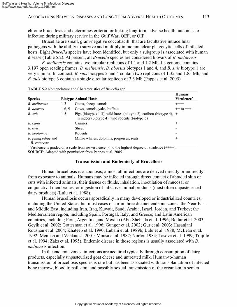
ASSOCIATIONS BETWEEN DISEASES AND LONG-TERM ADVERSE HEALTH OUTCOMES 113
chronic brucellosis and determines criteria for linking long-term adverse health outcomes to infection during military service in the Gulf War, OEF, or OIF.
Brucellae are small, gram-negative coccobacilli that are facultative intracellular pathogens with the ability to survive and multiply in mononuclear phagocytic cells of infected hosts. Eight Brucella species have been identified, but only a subgroup is associated with human disease (Table 5.2). At present, all Brucella species are considered biovars of B. melitensis.
B. melitensis contains two circular replicons of 1.1 and 1.2 Mb. Its genome contains 3,197 open reading frames. B. melitensis, B. abortus biotypes 1 and 4, and B. suis biotype 1 are very similar. In contrast, B. suis biotypes 2 and 4 contain two replicons of 1.35 and 1.85 Mb, and B. suis biotype 3 contains a single circular replicon of 3.3 Mb (Pappas et al. 2005).
TABLE 5.2 Nomenclature and Characteristics of Brucella spp.
Species Biotype Animal Hosts Human Virulencea
B. melitensis 1-3 Goats, sheep, camels ++++ B. abortus 1-6, 9 Cows, camels, yaks, buffalo ++ to +++ B. suis 1-5 Pigs (biotypes 1-3), wild hares (biotype 2), caribou (biotype 4),
reindeer (biotype 4), wild rodents (biotype 5) +
B. canis Canines + B. ovis Sheep - B. neotomae Rodents - B. pinnipediae and
B. cetaceae Minke whales, dolphins, porpoises, seals +
a Virulence is graded on a scale from no virulence (-) to the highest degree of virulence (++++). SOURCE: Adapted with permission from Pappas et al. 2005.
Transmission and Endemicity of Brucellosis
Human brucellosis is a zoonosis; almost all infections are derived directly or indirectly from exposure to animals. Humans may be infected through direct contact of abraded skin or cuts with infected animals, their tissues or fluids, inhalation, inoculation of mucosal or conjunctival membranes, or ingestion of infective animal products (most often unpasteurized dairy products) (Lulu et al. 1988).
Human brucellosis occurs sporadically in many developed or industrialized countries, including the United States, but most cases occur in three distinct endemic zones: the Near East and Middle East, including Iran, Iraq, Kuwait, Saudi Arabia, Israel, Jordan, and Turkey; the Mediterranean region, including Spain, Portugal, Italy, and Greece; and Latin American countries, including Peru, Argentina, and Mexico (Abo-Shehada et al. 1996; Bodur et al. 2003; Geyik et al. 2002; Gottesman et al. 1996; Gungor et al. 2002; Gur et al. 2003; Hasanjani Roushan et al. 2004; Khateeb et al. 1990; Lubani et al. 1989b; Lulu et al. 1988; McLean et al. 1992; Memish and Venkatesh 2001; Mousa et al. 1987; Norton 1984; Tasova et al. 1999; Trujillo et al. 1994; Zaks et al. 1995). Endemic disease in those regions is usually associated with B. melitensis infection.
In the endemic zones, infections are acquired typically through consumption of dairy products, especially unpasteurized goat cheese and untreated milk. Human-to-human transmission of brucellosis species is rare but has been associated with transplantation of infected bone marrow, blood transfusion, and possibly sexual transmission of the organism in semen
Copyright © National Academy of Sciences. All rights reserved.
Gulf War and Health: Volume 5. Infectious Diseaseshttp://www.nap.edu/catalog/11765.html
114 GULF WAR AND HEALTH
(Goossens et al. 1983; Ruben et al. 1991). Brucellosis probably is endemic in Afghanistan, but data on its occurrence there are sparse.
Between 100 and 200 US cases of human brucellosis were reported annually to CDC during the 1990s. Brucellosis cases in the United States have begun to shift from people who are occupationally exposed to animals and animal products (such as butchers, abattoir workers, veterinarians, and farmers) to people who ingest unpasteurized goat-milk products imported from Latin America (Chomel et al. 1994; Taylor and Perdue 1989). The disease is 8 times more prevalent at the US-Mexico border than elsewhere in the United States (Doyle and Bryan 2000; Fosgate et al. 2002). In the United States, cattle-associated B. abortus has been the etiologic agent of human brucellosis acquired directly from animals, and B. melitensis the agent of human brucellosis acquired from dairy products (CDC 1986; Spink 1954).
Acute Brucellosis
The acute form of human brucellosis is usually manifested 2-4 weeks after infection as a nonspecific febrile illness accompanied by profuse sweating, headache, malaise, arthralgia, arthritis, myalgia, back pain, or a combination of these. Hematologic abnormalities may include anemia, leukopenia, thrombocytopenia, and clotting disorders that are usually mild and resolve with therapy (Crosby et al. 1984; Pappas et al. 2004). Severe thrombocytopenia is rare (Young et al. 2000). Brucellosis in animals (especially that caused by B. abortus) is associated with spontaneous abortion. Although brucellosis may result in human abortion, it may be no more common than abortion that occurs during any infectious process (Khan et al. 2001; Makhseed et al. 1998).
Diagnosis of Acute Brucellosis The diagnosis of brucellosis should be considered in the appropriate clinical setting with
appropriate demographic risk factors. Laboratory analysis may disclose mild leukopenia, thrombocytopenia, and anemia with minimally to moderately abnormal liver-function tests. Definitive diagnosis involves recovering organisms, usually from blood or bone marrow. Culturing of bone marrow is the most sensitive method of diagnosis (Gotuzzo et al. 1986). In rare cases, brucellae may also be recovered from synovial fluid, cerebral spinal fluid, urine, or biopsy samples (Gotuzzo et al. 1986). Rapid automated bacterial identification systems may occasionally misidentify brucellae, for instance, as Moraxella phenylpyruvica (Roiz et al. 1998). PCR and other molecular techniques may be used, but they are not yet used widely in clinical settings (Colmenero et al. 2002; Fox et al. 1998; Morata et al. 2001; Queipo-Ortuno et al. 1997). If microbiologic cultures are negative, diagnosis of human brucellosis usually involves serologic analysis.
A number of serologic tests for diagnosing brucellosis exist (Al Dahouk et al. 2003; Young 1991). The most widely used is a serum agglutination test (SAT), which measures IgM and immunoglobulin G (IgG) brucella antibody titers. SAT titers above 1:160 are diagnostic for brucellosis in the appropriate clinical setting (Young 1991). A 2-merceptoethanol assay can increase the specificity of the SAT by distinguishing IgG from IgM responses (Baldi et al. 1996). Drawbacks of the SAT include cross-reactivity and inability to diagnose B. canis infection. In some people with brucellosis, an SAT response will not occur. Blocking antibodies may be present, or a Coombs test may be positive (Pascual et al. 1988). An enzyme-linked immunosorbent assay (ELISA) specific for brucella has higher sensitivity and specificity than the
Copyright © National Academy of Sciences. All rights reserved.
Gulf War and Health: Volume 5. Infectious Diseaseshttp://www.nap.edu/catalog/11765.html
ASSOCIATIONS BETWEEN DISEASES AND LONG-TERM ADVERSE HEALTH OUTCOMES 115
SAT (Almuneef and Memish 2003; Ariza et al. 1992; Khateeb et al. 1990; Lulu et al. 1988) and may be positive when other tests are negative. In evaluating people for neurobrucellosis when the SAT is negative, an ELISA should be performed (Araj et al. 1988). If neurobrucellosis is considered and serum antibody tests and microbiologic cultures are negative, cerebral spinal fluid can be evaluated for the presence of antibrucella antibodies (Kochar et al. 2000a; McLean et al. 1992).
Treatments for Brucellosis and Related Long-Term Toxicity
Treatment of people for brucellosis usually involves administration of tetracyclines (usually doxycycline) with rifampin for 6 weeks (WHO 1986). However, a regimen of oral doxycycline for 6 weeks and streptomycin for 2-3 weeks is more effective (Solera et al. 1995). Streptomycin may be replaced with gentamicin. Administration of aminoglycoside antibiotics is associated with renal and cranial VIIIth nerve toxicity, although if aminoglycosides are appropriately administered during short-course therapy, such complications are rare and often transient.
Coinfection
Although Brucella spp. are intracellular pathogens, there has been no apparent increase in morbidity and mortality during coinfection with brucellae and other intracellular pathogens or infections that disrupt the cellular immune system, such as HIV infection.
Long-Term Adverse Health Outcomes of Brucellosis
Acute brucellosis may be a nonspecific flu-like illness, so a specific diagnosis might not be made. People with untreated brucellosis are at risk for the relapsing and chronic health outcomes described below. In addition, antimicrobial therapy is not 100% effective, and even treated people are at risk for relapse and chronic disease. Clinical manifestations due to relapsing or chronic brucellosis usually are evident within 2-6 months of acute illness and if untreated can persist for years or decades (Spink 1951). Manifestations may be protean and nonspecific. Focal infections have also been reported up to 30 years after probable acute disease (Ariza et al. 2001; Colmenero et al. 2002; Martin et al. 1961; Mousa et al. 1986; Norton 1984; Williams and Crossley 1982; Zinneman et al. 1961).
Diagnosis of Chronic Brucellosis
Diagnosis during chronic brucellosis is similar to that during acute disease. During chronic brucellosis, bacteriologic confirmation may include detecting the organism in a bone marrow sample or in a focal infectious process or abscess. Serologic evaluation is usually positive. Isolated involvement of the central nervous system is rare and is usually diagnosed with serologic analysis or antibody analysis of cerebral spinal fluid.
Major Manifestations of Chronic Brucellosis The major manifestations of relapsing or chronic brucellosis include the following
conditions and organ systems.
Copyright © National Academy of Sciences. All rights reserved.
Gulf War and Health: Volume 5. Infectious Diseaseshttp://www.nap.edu/catalog/11765.html
116 GULF WAR AND HEALTH
Arthritis Bone and joint complications are the most common manifestation of chronic and
relapsing brucellosis, occurring in 10-80% of cases in various studies (Mousa et al. 1987; Tasova et al. 1999; Zaks et al. 1995). Arthritis is usually peripheral and monoarticular and often involves the knee or hip; however, some patients develop polyarthritis (Geyik et al. 2002; Gotuzzo et al. 1982; Gotuzzo et al. 1987; Hasanjani Roushan et al. 2004). Peripheral arthritis may be infectious (in which case it is usually monoarticular, and the organism may be recovered from the joint) or reactive (in which case involvement is often polyarticular or pauciarticular, and the organism will not be recovered from the joint) (Bravo et al. 2003). Sacroiliitis is the second-most frequent articular lesion (Alarcon et al. 1981; Ariza et al. 1993; Khateeb et al. 1990); it is usually unilateral. Spondylitis may affect 5-10% of patients with Brucella arthritis (Ariza et al. 1985; Gotuzzo et al. 1982; Namiduru et al. 2004; Solera et al. 1999). Radiographic features may include the presence of lytic and blastic lesions, erosion of the anterior superior part of the vertebral body (a “parrot peak” sign) (Ibero et al. 1997), and spondylodiscitis. Postinfection spondyloarthritis, bursitis, tenosynovitis, and infection of joint prostheses have also been reported (Weil et al. 2003). Although any joint might be involved during brucellosis, arthritis of the hips and knees is most common during acute disease and is usually manifested within 12 months of infection; involvement of the axial skeletal system and spondylitis are most common during chronic disease; and sacroiliitis might occur during either acute disease or chronic disease (Akritidis and Pappas 2001; Ariza et al. 1985; Colmenero et al. 1996; Doganay et al. 1993; Gotuzzo et al. 1987; Mousa et al. 1987; Namiduru et al. 2004; Norton 1984).
The committee concludes that there is sufficient evidence of an association between brucellosis and arthritis and spondylitis. Arthritis is usually manifested within 12 months of the acute illness; spondylitis might be manifested later.
Hepatic Involvement Human brucellosis is often associated with changes in liver function and has been
associated with granulomatous hepatitis (Harrington et al. 1982; Lulu et al. 1988; Williams and Crossley 1982). Hepatomegaly may be present (Lulu et al. 1988), but cirrhosis has not been reported. Chronic abscesses of the liver and spleen may occur (Ariza et al. 2001; Colmenero et al. 2002; Vallejo et al. 1996).
The committee concludes that there is sufficient evidence of an association between brucellosis and hepatic abnormalities, including granulomatous hepatitis.
Neurologic Involvement Neurobrucellosis has been reported in 1-5% of adults who have Brucella infections (al
Deeb et al. 1989; Bashir et al. 1985; Bouza et al. 1987; Young 1983). It usually involves meningitis or meningoencephalitis that is often chronic (al Deeb et al. 1989; Bashir et al. 1985; Bodur et al. 2003; Bouza et al. 1987; McLean et al. 1992; Mousa et al. 1986; Pascual et al. 1988). Fever, headache, nuchal rigidity, and altered consciousness may occur (Bodur et al. 2003; Gokul et al. 2000). Evaluation of cerebrospinal fluid usually reveals lymphocytic pleocytosis, increased protein concentration, and normal or moderately decreased glucose (Pascual et al. 1988). Microbiologic cultures of cerebrospinal fluid are positive for brucellae in 10-20% of cases. Rare brain or epidural abscesses, myelitis-radiculoneuritis, demyelinating meningovascular syndromes, deafness, sensorineural hearing loss, and GBS have been reported (Dalrymple-Champneys 1950; Kochar et al. 2000a; Lubani et al. 1989a; McLean et al. 1992;
Copyright © National Academy of Sciences. All rights reserved.
Gulf War and Health: Volume 5. Infectious Diseaseshttp://www.nap.edu/catalog/11765.html
ASSOCIATIONS BETWEEN DISEASES AND LONG-TERM ADVERSE HEALTH OUTCOMES 117
Mousa et al. 1986; Oliveri et al. 1996; Riestra-Castaneda et al. 1996; Thomas et al. 1993). The diagnosis of neurobrucellosis may be made even in the setting of negative microbiologic cultures of cerebrospinal fluid and negative serologic assays if specific antibodies are found in the cerebrospinal fluid (Kochar et al. 2000a; Sanchez-Sousa et al. 1990).
The committee concludes that there is sufficient evidence of an association between brucellosis and chronic meningitis and meningoencephalitis and between brucellosis and infection of the nervous system.
The committee concludes that there is limited or suggestive evidence of an association between brucellosis and myelitis-radiculoneuritis, demyelinating meningovascular syndromes, deafness, sensorineural hearing loss, and Guillain-Barré syndrome
Ophthalmologic Involvement Anterior-posterior uveitis is the most common ocular manifestation of brucellosis (al-
Kaff 1995; Gungor et al. 2002; Rolando et al. 1985a; Rolando et al. 1985b; Rolando et al. 1987; Tabbara 1990). Papilledema, optic neuritis, episcleritis, nummular keratitis, and multifocal choroiditis have also been reported (Gungor et al. 2002; Lyall 1973; McLean et al. 1992; Rabinowitz et al. 2005; Rolando et al. 1985b; Rolando et al. 1987; Walker et al. 1992). Without proper treatment, secondary glaucoma, cataracts, and retinal detachment may occur (Rabinowitz et al. 2005; Rolando et al. 1985a).
The committee concludes that there is sufficient evidence of an association between brucellosis and uveitis.
The committee concludes that there is limited or suggestive evidence of an association between brucellosis and papilledema, optic neuritis, episcleritis, nummular keratitis, and multifocal choroiditis.
Genitourinary Tract Manifestations Orchioepididymitis may occur in up to 20% of men with brucellosis (Ibrahim et al. 1988;
Memish and Venkatesh 2001; Navarro-Martinez et al. 2001; Papatsoris et al. 2002). It is most often unilateral and accompanied by normal urine sediment (Navarro-Martinez et al. 2001). Pyelonephritis and chronic renal abscesses have been reported in association with brucellosis (Zinneman et al. 1961).
The committee concludes that there is sufficient evidence of an association between brucellosis and orchioepididymitis and between brucellosis and local infections of the genitourinary system (for example, pyelonephritis or renal abscesses).
Cardiovascular System Infections Endocarditis causes the majority of Brucella-related deaths even though it occurs in less
than 2% of chronic cases (al-Harthi 1989). Involvement of the aortic valve is most common, and pericarditis and mycotic aneurysms of blood vessels may occur (McLean et al. 1992).
The committee concludes that there is sufficient evidence of an association between brucellosis and cardiovascular system infections.
Copyright © National Academy of Sciences. All rights reserved.
Gulf War and Health: Volume 5. Infectious Diseaseshttp://www.nap.edu/catalog/11765.html
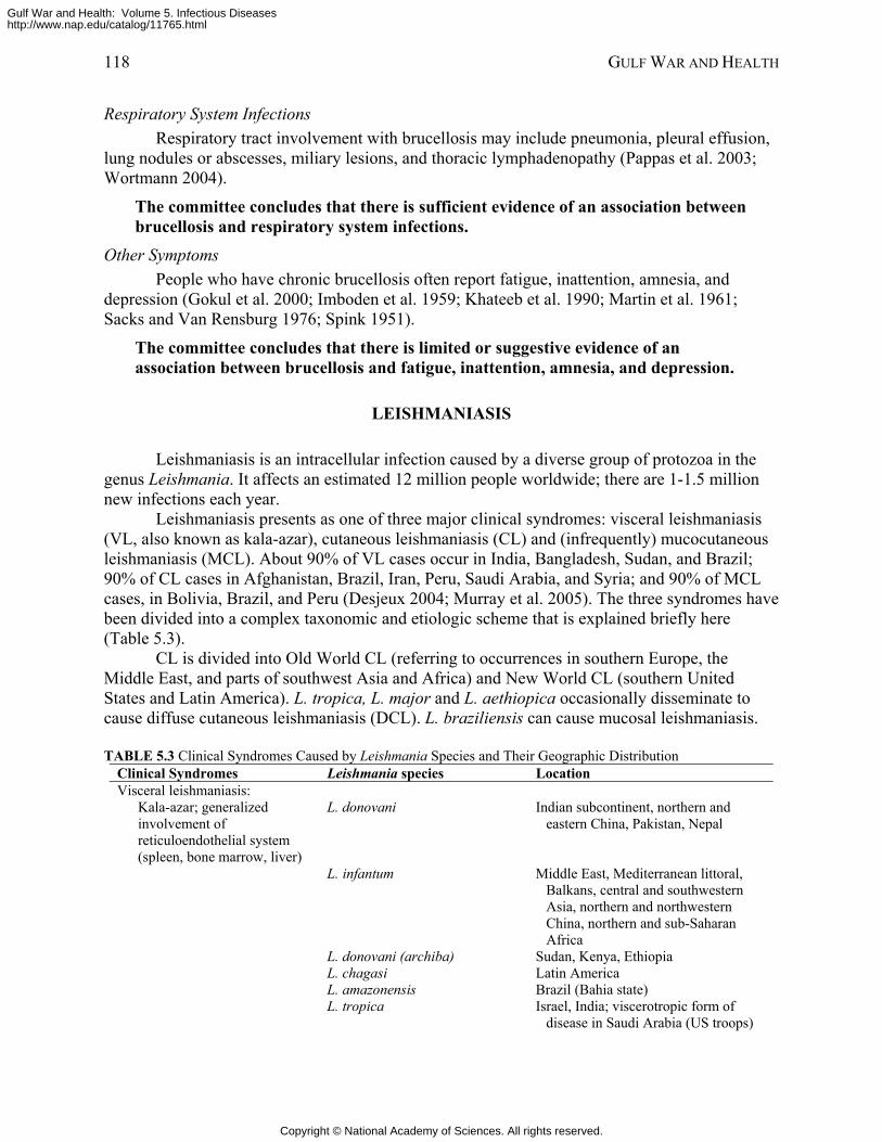
118 GULF WAR AND HEALTH
Respiratory System Infections Respiratory tract involvement with brucellosis may include pneumonia, pleural effusion,
lung nodules or abscesses, miliary lesions, and thoracic lymphadenopathy (Pappas et al. 2003; Wortmann 2004).
The committee concludes that there is sufficient evidence of an association between brucellosis and respiratory system infections.
Other Symptoms People who have chronic brucellosis often report fatigue, inattention, amnesia, and
depression (Gokul et al. 2000; Imboden et al. 1959; Khateeb et al. 1990; Martin et al. 1961; Sacks and Van Rensburg 1976; Spink 1951).
The committee concludes that there is limited or suggestive evidence of an association between brucellosis and fatigue, inattention, amnesia, and depression.
LEISHMANIASIS
Leishmaniasis is an intracellular infection caused by a diverse group of protozoa in the genus Leishmania. It affects an estimated 12 million people worldwide; there are 1-1.5 million new infections each year.
Leishmaniasis presents as one of three major clinical syndromes: visceral leishmaniasis (VL, also known as kala-azar), cutaneous leishmaniasis (CL) and (infrequently) mucocutaneous leishmaniasis (MCL). About 90% of VL cases occur in India, Bangladesh, Sudan, and Brazil; 90% of CL cases in Afghanistan, Brazil, Iran, Peru, Saudi Arabia, and Syria; and 90% of MCL cases, in Bolivia, Brazil, and Peru (Desjeux 2004; Murray et al. 2005). The three syndromes have been divided into a complex taxonomic and etiologic scheme that is explained briefly here (Table 5.3).
CL is divided into Old World CL (referring to occurrences in southern Europe, the Middle East, and parts of southwest Asia and Africa) and New World CL (southern United States and Latin America). L. tropica, L. major and L. aethiopica occasionally disseminate to cause diffuse cutaneous leishmaniasis (DCL). L. braziliensis can cause mucosal leishmaniasis.
TABLE 5.3 Clinical Syndromes Caused by Leishmania Species and Their Geographic Distribution
Clinical Syndromes Leishmania species Location Visceral leishmaniasis:
Kala-azar; generalized involvement of reticuloendothelial system (spleen, bone marrow, liver)
L. donovani Indian subcontinent, northern and eastern China, Pakistan, Nepal
L. infantum Middle East, Mediterranean littoral, Balkans, central and southwestern Asia, northern and northwestern China, northern and sub-Saharan Africa
L. donovani (archiba) Sudan, Kenya, Ethiopia L. chagasi Latin America L. amazonensis Brazil (Bahia state) L. tropica Israel, India; viscerotropic form of
disease in Saudi Arabia (US troops)
Copyright © National Academy of Sciences. All rights reserved.
Gulf War and Health: Volume 5. Infectious Diseaseshttp://www.nap.edu/catalog/11765.html
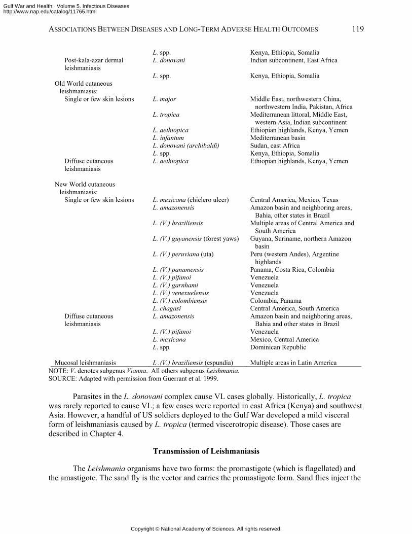
ASSOCIATIONS BETWEEN DISEASES AND LONG-TERM ADVERSE HEALTH OUTCOMES 119
L. spp. Kenya, Ethiopia, Somalia Post-kala-azar dermal leishmaniasis
L. donovani Indian subcontinent, East Africa
L. spp. Kenya, Ethiopia, Somalia Old World cutaneous
leishmaniasis:
Single or few skin lesions L. major Middle East, northwestern China, northwestern India, Pakistan, Africa
L. tropica Mediterranean littoral, Middle East, western Asia, Indian subcontinent
L. aethiopica Ethiopian highlands, Kenya, Yemen L. infantum Mediterranean basin L. donovani (archibaldi) Sudan, east Africa L. spp. Kenya, Ethiopia, Somalia Diffuse cutaneous leishmaniasis
L. aethiopica Ethiopian highlands, Kenya, Yemen
New World cutaneous
leishmaniasis:
Single or few skin lesions L. mexicana (chiclero ulcer) Central America, Mexico, Texas L. amazonensis Amazon basin and neighboring areas,
Bahia, other states in Brazil L. (V.) braziliensis Multiple areas of Central America and
South America L. (V.) guyanensis (forest yaws) Guyana, Suriname, northern Amazon
basin L. (V.) peruviana (uta) Peru (western Andes), Argentine
highlands L. (V.) panamensis Panama, Costa Rica, Colombia L. (V.) pifanoi Venezuela L. (V.) garnhami Venezuela L. (V.) venexuelensis Venezuela L. (V.) colombiensis Colombia, Panama L. chagasi Central America, South America Diffuse cutaneous leishmaniasis
L. amazonensis Amazon basin and neighboring areas, Bahia and other states in Brazil
L. (V.) pifanoi Venezuela L. mexicana Mexico, Central America L. spp. Dominican Republic
Mucosal leishmaniasis L .(V.) braziliensis (espundia) Multiple areas in Latin America
NOTE: V. denotes subgenus Vianna. All others subgenus Leishmania. SOURCE: Adapted with permission from Guerrant et al. 1999.
Parasites in the L. donovani complex cause VL cases globally. Historically, L. tropica was rarely reported to cause VL; a few cases were reported in east Africa (Kenya) and southwest Asia. However, a handful of US soldiers deployed to the Gulf War developed a mild visceral form of leishmaniasis caused by L. tropica (termed viscerotropic disease). Those cases are described in Chapter 4.
Transmission of Leishmaniasis
The Leishmania organisms have two forms: the promastigote (which is flagellated) and the amastigote. The sand fly is the vector and carries the promastigote form. Sand flies inject the
Copyright © National Academy of Sciences. All rights reserved.
Gulf War and Health: Volume 5. Infectious Diseaseshttp://www.nap.edu/catalog/11765.html
120 GULF WAR AND HEALTH
promastigote form of the parasite into humans. Infection is then established with the amastigote form, which is harbored in human macrophages.
Two transmission cycles have been described. In the zoonotic cycle, dogs are the primary animal reservoir, and humans are an occasional host when they are infected by the bite of the sand fly. In south-central Asia (Afghanistan), great gerbils (Rhombomys opimus) are the vertebrate hosts of L. major and thus determine the clinical distribution of associated CL. In the anthroponotic cycle, humans are the sole reservoir, and sand flies remain the critical vector. Phlebotomus papatasi is the sand fly species that transmits L. major throughout most of the Middle East and is present in south-central Asia. Phlebotomus sergenti was recently identified as the species responsible for transmission of L. tropica in Afghanistan (Wallace et al. 2002).
Sand fly bites are exceedingly common in the Middle East. In August 1943, sand fly fever (caused by a phlebovirus) occurred at a peak rate of 235 per 1,000 military personnel deployed to the Persian Gulf (Hertig and Sabin 1964). Because sand flies are most active during warm months, however, there is seasonal variation in the risk of infection. Only 31 cases of leishmaniasis were diagnosed among 697,000 troops deployed during the Gulf War, and deployment to the open desert during cooler weather was thought to be a partial reason for the low incidence of the disease (Cope et al. 1996). Even in areas that are important foci of Leishmania infection, the prevalence of sand fly-caused infection with Leishmania spp. is unpredictable (Fryauff et al. 1993).
Finally, humans have acquired leishmaniasis through parenteral exposure (because of contaminated injection equipment and blood products) and through sexual contact, but those cases are rare.
Endemicity in Southwest and South-Central Asia
Southwest Asia and south-central Asia are home to Old World CL and VL (Oldfield et al. 1991). The potential for anthroponotic acquisition of CL is especially high in Kabul, Afghanistan, where 270,000 persons (in a population of 2 million) were estimated to be infected in 1996 (World Health Organization as cited in Hewitt et al. 1998). Some 4,700 cases of CL were reported in northern Syria in 1999, an increase from the 3,900 cases reported in 1998 (WHO 2002); most CL in the Middle East is caused by L. major.
Acute Leishmaniasis
Old World CL has an incubation period of 2 weeks to 2 months. The most common etiologic agent is L. major, which causes papular lesions that can ulcerate (Wallace et al. 2002). Most (90-95%) CL lesions heal spontaneously, and they rarely cause persistent disfiguration. L. recidivans can cause a chronic cutaneous (“ring”) lesion.
VL has an incubation period of 2-4 months, although it has been reported to be as long as 2 years. Most infected persons remain asymptomatic during the acute phase. When VL evolves to the clinically evident form, classic symptoms include fever, weight loss, weakness, diarrhea, dysentery, and abdominal swelling. The typical triad of diagnostic findings consists of anemia, fever, and hepatosplenomegaly. Complications of the acute infection arise typically from superimposed bacterial infection, sometimes exacerbated by the neutropenia that can result from bone marrow infiltration. Cytokine disruption is probably critical in determining the clinical presentation and in mediating the outcome of infection, even with treatment (Murray et al. 2005). The predominant cell-mediated immune response is characterized by activity of Th1-type CD4+
Copyright © National Academy of Sciences. All rights reserved.
Gulf War and Health: Volume 5. Infectious Diseaseshttp://www.nap.edu/catalog/11765.html
ASSOCIATIONS BETWEEN DISEASES AND LONG-TERM ADVERSE HEALTH OUTCOMES 121
cells and associated interferon-γ-induced macrophage activation. That response sets the stage for control, but probably not uniform eradication, of the parasite. Macrophage defenses required to kill Leishmania have been extensively studied, as have the pathogen’s antiphagocytic defenses (Cunningham 2002; Teixeira et al. 2006).
Of the twelve people who had viscerotropic leishmaniasis caused by L. tropica in the Gulf War, one was asymptomatic, and the remainder had a mixed picture involving many of the classic features of VL (Hyams et al. 1995; Magill et al. 1994). Those presentations were distinguished from typical VL in that anemia was typically the sole hematologic sign, and most patients had modest increases in liver enzymes. Three of the patients had an underlying disease of relevance: HIV, acute infection with Epstein-Barr virus, and renal-cell cancer (Hyams et al. 1995).
Diagnosis of Leishmaniasis
Several methods have been used to diagnose the various forms of leishmaniasis. Most CL is diagnosed on the basis of its classic clinical appearance, although if the lesion is atypical, prolonged, or not responsive to therapy, biopsy may be performed at the margin of the lesion. PCR is increasingly used in this setting, especially because misdiagnosis may occur (many lesions clinically diagnosed as CL are bacterial in origin). PCR was the mainstay of diagnosis in a recent description of 237 cases of CL acquired in OIF (Willard et al. 2005). Skin testing based on antigens of L. major demonstrates prior infection with Leishmania spp. and is usually positive in active CL caused by L. major.
VL is often diagnosed on the basis of histopathologic detection of amastigotes in biopsy or aspirate of bone marrow, spleen, or lymph nodes. Indirect immunofluorescent monoclonal antibody can also be applied to those tissues. Biopsy samples can be directly cultured, and isoenzyme analysis used for further speciation. Serum antibody testing, often used in assessment of persons with suspected VL, is most commonly performed with the direct agglutination test. However, the performance of this test is highly variable; in fact, serology was negative in a number of the viscerotropic cases identified in Gulf War soldiers. Available serologic tests are based on L. major antigens, so the relevance to viscerotropic leishmaniasis (caused by L. tropica) is unclear. Finally, some investigators have reported that urine-based assays that detect either Leishmania antigen (Sundar et al. 2005) or Leishmania-specific IgG (Islam et al. 2002) were valuable in diagnosing VL.
Treatments for Leishmaniasis and Related Long-Term Toxicity
Most cases of CL will resolve without specific medical therapy. Oral azoles (fluconazole and ketoconazole), cryotherapy, or paromomycin ointment may hasten resolution. Under study is a device called ThermoMed that delivers radiofrequency-generated heat directly to a lesion through a set of prongs placed on the lesion; the device has Food and Drug Administration 510K clearance as of this writing.
Systemic treatment is always indicated for VL. The mainstay of therapy has been pentavalent antimonials, including sodium stibogluconate and meglumine antimonite (Aronson et al. 1998; Murray 2000; Murray 2004). Liposomal amphotericin B was traditionally reserved for antimony-treatment failures, but it is increasingly used as first-line therapy and has been the regimen of choice for soldiers who acquired VL in OEF. Antimonials are not well tolerated in the acute treatment period. Gastrointestinal intolerance, bone marrow suppression, and
Copyright © National Academy of Sciences. All rights reserved.
Gulf War and Health: Volume 5. Infectious Diseaseshttp://www.nap.edu/catalog/11765.html
122 GULF WAR AND HEALTH
hepatotoxicity occur in up to 50% of patients (and are usually reversible). Pancreatitis and abnormalities of cardiac repolarization also occur; the latter is generally unassociated with arrhythmia and resolves within two months after completion of treatment. At least one case of laryngeal edema has been reported to be associated with antimony therapy. Oral miltefosine has also been used for treatment for VL and CL. None of these drugs appears to be associated with long-term toxicity.
Coinfection by Leishmania Parasite and Human Immunodeficiency Virus
VL is estimated to be the third-most common opportunistic infection in HIV-infected persons in southern Europe (Choi and Lerner 2002). The association emphasizes immune control of the organism and reactivation of quiescent infection in the setting of reduced cell-mediated immune response. Indeed, Leishmania infection might reactivate in patients with CD4 counts below 200/µL (Choi and Lerner 2002). The World Health Organization (WHO) estimates that 25-70% of adult VL cases in southern Europe now occur in HIV-infected patients and that AIDS increases the risk of VL by a factor of 100-1,000 (Choi and Lerner 2002). Clinically, leishmaniasis in HIV-infected persons is characterized by atypical presentations (including pulmonary disease, lingual and esophageal ulcerations, and fever of unknown origin), reduced rates of treatment response, progression from cutaneous to visceral disease, higher rates of death, and reduced sensitivity of serologic tests.
Long-Term Adverse Health Outcomes of Leishmaniasis
Cutaneous Leishmaniasis Infections with L. major have not led to viscerotropic infection, parenteral or vertical
transmission, or presentation as an opportunistic infection associated with HIV. Old World CL as a rule resolves spontaneously and rarely causes chronic scarring. All of the numerous cases of CL that have occurred in soldiers involved in OIF (CDC 2003b; CDC 2004b) have reportedly responded to relatively short courses of sodium stibogluconate (Weina et al. 2004; Willard et al. 2005). However, some cases have been associated with large lesions and long duration. Given the difficulty in diagnosis, unrecognized CL has the potential to cause substantial cosmetic problems.
DCL is not as responsive to therapy as CL and can cause progressive disfiguration and destruction of skin and soft tissue.
Visceral Leishmaniasis
The organisms responsible for VL also infect monocytes and macrophages; however, in contrast with L. major, they may establish latency in these cells. This phenomenon results in a demonstrable risk of recurrence in the setting of immunosuppression induced by chemotherapy, transplantation-related processes, or HIV infection (Basset et al. 2005). As discussed above, immune control of VL involves primarily CD4+ T-cell activity (Th1-type response). Conversely, VL promotes formation of Th2-type cytokines, which can inhibit control of the disease. VL is itself an immunosuppressive disease, partly because of infiltration of reticuloendothelium of liver, spleen, and bone marrow and because it has been associated with polyclonal B-cell activation and increased production of numerous autoantibodies. One case report of GBS that predated the clinical appearance of VL by about a month has been reported;
Copyright © National Academy of Sciences. All rights reserved.
Gulf War and Health: Volume 5. Infectious Diseaseshttp://www.nap.edu/catalog/11765.html
ASSOCIATIONS BETWEEN DISEASES AND LONG-TERM ADVERSE HEALTH OUTCOMES 123
the authors postulated that the parasite could mediate autoimmune damage to peripheral nerve myelin (Fasanaro et al. 1991).
Because L. infantum has been responsible for most cases of HIV-related VL (Russo et al. 2003), it might be particularly likely to persist in macrophages and monocytes. This organism was identified in one of the two cases of VL acquired in Afghanistan (CDC 2004a) but has not been identified in veterans of other conflicts. Of those two cases, one was diagnosed 14 months after deployment ended in Afghanistan, and the patient had symptoms of clinical recurrence. In addition, VL is estimated to be the third-most common opportunistic infection in HIV-infected persons in southern Europe, as detailed above (Russo et al. 2003).
Because the period of latent infection with VL organisms can be long (10 years is commonly cited), immune suppression can allow reactivation of a latent infection. In the description of the viscerotropic cases that occurred in the Gulf War, the authors stated that “if L. tropica is also capable of surviving in a latent state, visceral leishmaniasis will need to be included in the differential diagnoses of illness in veterans of Operation Desert Storm for years to come” (Magill et al. 1993). Although chronic infection is clearly plausible, no systematic studies have investigated the possibility prospectively, in part because there is no accurate and noninvasive screening test for the infection (Ohl et al. 1993). However, intensive evaluation among 150 Gulf War veterans with complaints was unable to identify prior or current infection with Leishmania spp. (Hyams et al. 1995).
Post-kala-azar dermal leishmaniasis (PKDL) is a well-documented long-term adverse health outcome of VL that occurs on the Indian subcontinent and in east Africa (Zijlstra et al. 2003). On the basis of the Indian experience, this health outcome may develop in 5-10% of patients several years after apparently successful treatment for VL (Zijlstra et al. 2003). PKDL has been mistaken for leprosy, and patients with this presentation remain infectious (Zijlstra et al. 2003). Nerve involvement (as is seen in leprosy) has been reported rarely with PKDL (El Hassan et al. 1992; Khandpur et al. 2004).
The committee concludes that
• There is sufficient evidence of an association between infection with an etiologic agent of visceral leishmaniasis (VL) and delayed presentation of the acute clinical syndrome.
• There is sufficient evidence of an association between infection with an etiologic agent of VL and the reactivation of VL in the context of future immunosuppression.
• There is sufficient evidence of an association between VL and development of post-kala-azar dermal leishmaniasis (PKDL) if PKDL occurs generally within 2 years of the initial infection.
MALARIA Human malaria is caused by infection with one or more of four species in the genus
Plasmodium: P. falciparum, P. vivax, P. ovale, and P. malariae. Although estimates vary, there are probably 350-500 million clinical episodes of malaria each year and 0.7-2.7 million deaths (Breman 2001; WHO 2003). Malaria occurs worldwide in tropical and subtropical regions, typically affecting poor and developing areas most severely. P. falciparum predominates in tropical areas; P. vivax, in temperate regions. The two other species are less frequently
Copyright © National Academy of Sciences. All rights reserved.
Gulf War and Health: Volume 5. Infectious Diseaseshttp://www.nap.edu/catalog/11765.html
124 GULF WAR AND HEALTH
encountered: P. malariae is found worldwide, and the geographic range of P. ovale is limited mostly to tropical Africa, the Middle East, southeast Asia, and the western Pacific.
Transmission of Malaria
Malaria infection occurs when a Plasmodium-infected Anopheles mosquito feeds on a susceptible human host, delivering sporozoites that initially invade hepatocytes and mature into merozoites that then invade erythrocytes. The cycle is completed when a competent female Anopheles mosquito feeds on a parasitemic human, obtaining gametocytes that then initiate infection in the mosquito. Many Anopheles species are potential vectors of malaria in different parts of the world, so mosquito species-specific behaviors, including host feeding preference and daily activity patterns, tend to result in varied regional transmission patterns. Often, several mosquito species will combine to constitute an overall vector profile for a region. In tropical areas, transmission intensity is often linked to rainy seasons—typically one major and another less severe. In temperate or seasonally arid regions, a single transmission period is evident (Guerrant et al. 1999).
Endemicity in Southwest and South-Central Asia
The best recent estimates of overall malaria morbidity and mortality in southwest and south-central Asia are about 6 million cases and 59,000 deaths per year (RBM 2005a). Afghanistan and Yemen alone account for an estimated 5.5 million of all cases, on the basis of 2004 data (RBM 2005b). In the malaria-endemic countries of Tajikistan, Azerbaijan, Armenia, Georgia, Kyrgyzstan, and Uzbekistan, malaria occurred at a rate of 0.11 case per 1,000 population in 1990-2003 (RBM 2005h). In contrast, the case rate was about three per 1,000 during the same period in southwest Asia, Afghanistan, and Pakistan combined (RBM 2005h).
About 70% of all infections are caused by P. vivax, but this varies regionally. P. malariae is not reported to be endemic in most parts of southwest or south-central Asia and is rare in areas where it has been reported. Diagnosis and reporting in some areas, such as Iraq and Afghanistan, have been hindered in recent years because of war-related interruptions to the public-health infrastructure. Transmission is highly seasonal and peaks in late July to September.
In Iraq, malaria is endemic in Duhok, Erbil, Ninawa, Sulaimaniya, Tamim, and Basrah provinces. Some 362 cases were recorded in Iraq in 2003. The disease is due exclusively to P. vivax; peak transmission takes place in May-November. The main vectors are A. sacharovi, A. superpictus, A. maculipennis, A. stephensi, and A. pulcherrimus. Most of the cases occur in the northern governorates, mainly in the Zakho district in Dohuk, where four of the five vector species reside (RBM 2005e).
Malaria is endemic in Afghanistan in all areas below 2,000 m in altitude. Afghanistan reported about 600,000 cases in 2003, 93% of which were caused by P. vivax and 7% by P. falciparum (Kolaczinski et al. 2005). Estimates of the rates of feeding of infective vectors on humans in eastern Afghanistan indicated that A. stephensi would contribute 76% of infective bites and A. fluviatilis and A. culicifacies 7% and 3%, respectively. Because of chloroquine resistance, numbers of P. falciparum infections in eastern Afghanistan have increased from 1% of all infections in 1970 to 20% in 2002 (Kolaczinski et al. 2005; RBM 2005c).
Saudi Arabia tends to have equal percentages of infection with P. vivax and P. falciparum but low case totals (1,700 cases in 2003). The primary vector in Saudi Arabia is A. arabiensis (RBM 2005g).
Copyright © National Academy of Sciences. All rights reserved.
Gulf War and Health: Volume 5. Infectious Diseaseshttp://www.nap.edu/catalog/11765.html
ASSOCIATIONS BETWEEN DISEASES AND LONG-TERM ADVERSE HEALTH OUTCOMES 125
Pakistan reported more than 125,000 laboratory-confirmed cases in 2003, 4 million probable cases, and 14 deaths. Of the laboratory-confirmed cases, almost 70% were caused by P. vivax. The primary mosquito vectors of malaria in Pakistan are A. culicifacies and A. stephensi (RBM 2005f).
In Iran, three provinces in the southeastern corner account for most of the 23,000 cases reported in 2003, 21% of which were caused by P. falciparum. Primary mosquito vectors include A. fluviatilis, A. stephensi, and A. culicifacies (RBM 2005d).
Acute Malaria
All four Plasmodium species can cause cyclic fevers, particularly in naïve populations. Known as malarial paroxysms, the cycles are characterized by rapid onset of high fever with chills followed by rapid resolution, often with intense diaphoresis. The cycles are associated with erythrocyte lysis that occurs at the end of the erythrocytic cycle of infection. The classical (but infrequently observed) periodic attacks occur every second day with the "tertian" parasites (P. falciparum, P. vivax, and P. ovale) and every third day with the "quartan" parasite (P. malariae).
Among populations in endemic areas, the development of partial immunity leads to milder illness and even asymptomatic infections. However, the immune response does not block repeated infections or infections with multiple strains or species. In temperate climates, the long latent phase with P. vivax and P. ovale appears to provide the opportunity for the resumption of transmission when the mosquito season returns in the next year (Guerrant et al. 1999).
Malaria is diagnosed with microscopic examination of blood smears stained with Giemsa or Wright’s stain. An experienced technician can diagnose most cases with examination of routine blood smears (thin smears), but examination of thick smears is more sensitive in detecting those with less severe parasitemia. The key to diagnosis is recognizing the potential for malaria in a potentially exposed person who has fever, anemia, and thrombocytopenia. Deaths from malaria in travelers returning to the United States, most notably with P. falciparum, continue to occur, often in association with delays in diagnosis and in effective therapy (Newman et al. 2004). Other diagnostic techniques have been developed, including fluorescence microscopy, immunologic diagnosis of falciparum malaria with antibodies to the protein HRP2, DNA probes specifically for P. falciparum, and PCR methods (Amino et al. 2005; Berry et al. 2005; Wilson et al. 2005).
Treatments for Malaria and Related Long-Term Toxicity
Resistance to chloroquine and multiple-drug resistance are major problems with P. falciparum in most of Africa, Asia, and South America. Drug-resistant P. falciparum has also been found in the Middle East and southwest Asia, including Iraq (Guerrant et al. 1999). Resistance to chloroquine is an emergent problem with P. vivax in some parts of Asia, Oceania, and South America (Kurcer et al. 2006).
Antimalarial drugs have well-documented acute adverse effects on the skin, gastrointestinal tract, central nervous system, and other organ systems, but evidence of long-term adverse health outcomes is sparse (Taylor and White 2004). Moderate to severe neuropsychiatric complications have been reported in association with mefloquine, doxycycline, combined chloroquine and proguanil, and combined atovaquone and proguanil (Schlagenhauf et al. 2003; Taylor and White 2004). Although retinopathy associated with high-dose long-term chloroquine use has been described, it has rarely been associated with modern prophylaxis. Additional
Copyright © National Academy of Sciences. All rights reserved.
Gulf War and Health: Volume 5. Infectious Diseaseshttp://www.nap.edu/catalog/11765.html
126 GULF WAR AND HEALTH
discussion on the health effects of antimalarial drugs can be found in the section on ophthalmologic complications below.
Coinfection with Plasmodium Spp. and Human Immunodeficiency Virus
There is clear evidence that HIV infection, particularly with lower CD4+ cell counts, is associated with an increased risk of malaria, higher levels of parasitemia, and higher mortality (Butcher 2005). Nevertheless, malaria and HIV coinfection in a soldier deployed to southwest or south-central Asia would be highly improbable, given the low rates of HIV in southwest and south-central Asia, segregation of US military troops from civilian populations, periodic HIV screening of troops and removal of soldiers testing HIV-positive from overseas service, and low malaria transmission rates.
Long-Term Adverse Health Outcomes of Infection with Plasmodium Spp.
Infection by Plasmodium potentially has long-term repercussions for human health. Long-term adverse health outcomes of infection can be manifested in neurologic, neuropsychiatric, ophthalmologic, hematologic, or renal disease. In addition, malaria itself can present after a latency of months to years or may break out anew because of undertreatment.
The epidemiology of malaria in Afghanistan and Iraq suggests that P. vivax is the principal threat to US troops deployed to OEF and OIF. P. falciparum was not known to circulate in Iraq in 1991-2005, and, although reported in Afghanistan, it is far less prevalent than P. vivax. Although P. falciparum predominates in Saudi Arabia, only seven cases of malaria were reported in US troops during the Persian Gulf War, as described in Chapter 4. P. ovale and P. malariae are rare in southwest and south-central Asia. Hematologic Complications
Splenic rupture is a well-described complication of malaria, particularly that caused by P. vivax. It can occur weeks to months after the acute infection. Hyperreactive malaria syndrome or tropical splenomegaly can be noted months or years after malarial infection (Metha et al. 1996).
Anemia is a principal complication of malaria. It is expected as an acute event, but it may be detected months or even years after the infection. Only in cases of fulminant falciparum malaria is anemia so severe as to be debilitating or life-threatening. Repeated hemolysis, presumably due to subcurative treatments, is reported as a chronic complication (Metha et al. 1996).
The committee concludes that there is sufficient evidence of a causal relationship between malaria infection and hematologic manifestations weeks or months later, particularly splenic rupture after vivax malaria and anemia after falciparum malaria.
Ophthalmologic Complications A number of case reports describe the complication of retinal hemorrhage associated with
severe cerebral malaria and following vivax malaria. Permanent visual loss may result from this complication (Choi et al. 2004). The capillary permeability associated most notably with falciparum malaria has been associated with retinal hemorrhage and edema, with occasional serious visual impairment, in adults and children (Beare et al. 2003; Hidayat et al. 1993; Kochar
Copyright © National Academy of Sciences. All rights reserved.
Gulf War and Health: Volume 5. Infectious Diseaseshttp://www.nap.edu/catalog/11765.html
ASSOCIATIONS BETWEEN DISEASES AND LONG-TERM ADVERSE HEALTH OUTCOMES 127
et al. 2000b; Lewallen 1998; Tripathi et al. 1995). Retinal manifestations may be noted months or years after the acute malaria infection (Biswas et al. 1996).
Malaria-associated chronic ophthalmologic disorders include side effects of the chronic prophylactic use of antimalarial drugs, such as hydroxychloroquine and chloroquine (Balo et al. 1996; Easterbrook 1999; Lozier and Friedlaender 1989; Niemeyer and Fruh 1989; Portnoy and Callen 1983; Ruiz and Saatci 1991; Tzekov 2005; Wei et al. 2001). The associations have been recognized for decades (Begue 1964; Bernstein 1967; Giles and Henderson 1965; Rubin 1968; Sugiyama et al. 1967). Chloroquine-based prophylaxis is not being used for US troops deployed to OEF and OIF. Oxidative stress has been cited as a possible contributing etiology (Toler 2004). Rynes and Bernstein (1993) highlight the relative rarity of the retinal complications and the need for long-term administration of the drugs.
The committee concludes that there is sufficient evidence of a causal relationship between malaria infection and ophthalmologic manifestations, particularly retinal hemorrhage and scarring, recognized for the first time months or years after the infection.
Neurologic and Neuropsychiatric Complications Neurologic complications, particularly cerebral malaria due to P. falciparum, are
characterized by confusion, clouding of consciousness progressing to coma, and seizures. Cerebral malaria is due largely to sequestration of infected red blood cells in the cerebral circulation (Renia et al. 2006), but coma can also be caused by such other malaria complications as hypoglycemia, uremia, or hypoxia due to pulmonary edema (Idro et al. 2005). Cerebral malaria is fatal in 15-20% of cases, and residual neurologic deficits have been reported in 1-3% of adults and 10% of children (Bajiya and Kochar 1996). It is notable that over 97% of afflicted adults who survive the cerebral episode of falciparum malaria are left without detectable chronic sequelae.
A postmalaria neurologic syndrome has been described in people who were treated for malaria due to P. falciparum (Falchook et al. 2003). The manifestations include confusion, psychosis, seizures, and a fine tremor (Malviya et al. 2005; Meier et al. 2004). There may be an associated magnetic resonance imaging finding of enhancement of nonspecific white-matter lesions (Dey et al. 2001).
Two other postmalaria neurologic complications have been described in case reports and case series. Acute inflammatory demyelinating polyneuropathy and Guillain-Barré syndrome have been reported after falciparum malaria and less frequently after vivax malaria (Chakravarty et al. 2004; Shubhakaran and Sharma 2003). Onset of neurologic symptoms can occur during the acute stage of the illness or days to weeks after the end of the acute illness (Shubhakaran and Sharma 2003; Kanjalkar et al. 1999). Cerebellar ataxia, often with tremors, has also been described after falciparum malaria, possibly resulting from demyelinating lesions in the cerebellum (Metha et al. 1996; Senanayake and de Silva 1994). All reports of patients who experienced acute inflammatory demyelinating polyneuropathy and cerebellar ataxia described complete recovery within months of onset (Chakravarty et al. 2004; Kanjalkar et al. 1999), although the natural history of these disorders after other conditions has been associated with slow recovery and persistent neurologic deficits in some of those affected (Kanjalkar et al. 1999).
One report dealt with neurologic deficits among veterans who had experienced cerebral malaria from P. falciparum during the Vietnam War (Varney et al. 1997). Veterans with a self-reported history of cerebral malaria were found to have a greater frequency of neuropsychiatric
Copyright © National Academy of Sciences. All rights reserved.
Gulf War and Health: Volume 5. Infectious Diseaseshttp://www.nap.edu/catalog/11765.html
128 GULF WAR AND HEALTH
symptoms than veterans who suffered combat wounds. The report raises questions about the potential for cerebral malaria to produce subtle, persistent neurologic deficits that may not have been apparent in examinations conducted during routine medical treatment and followup (Shamo 2001).
The committee concludes that there is limited or suggestive evidence of an association between Plasmodium vivax and Plasmodium falciparum infections and demyelinating polyneuropathy and Guillain-Barré syndrome.
The committee concludes that there is limited or suggestive evidence of an association between Plasmodium falciparum infection and neurologic disease, neuropsychiatric disease, or both, months to years after the acute infection.
Renal Complications Chronic untreated P. malariae infection can be manifested with chronic
glomerulonephritis even years after the onset of infection (Eiam-Ong 2003; Kibukamusoke 1986). In contrast, the nephrotic syndrome and acute glomerulonephritis are far more common near the onset of infection (days to weeks later), may be associated with any malaria infection, and would be manifested after months or years only very rarely.
The committee concludes that there is sufficient evidence of an association between Plasmodium malariae infection and the manifestation of immune-complex glomerulonephritis years to decades later.
The committee concludes that there is sufficient evidence of a causal relationship between malaria infection and renal disease, especially the nephrotic syndrome that may occur weeks to months after acute infection.
Relapse and Recrudescence of Malaria Some P. vivax and P. ovale parasites remain dormant as hypnozoites in the liver for
months after primary infection. The latent period is generally 6-11 months (Mandell et al. 2005), although one report found the latent period to be less than 4 months (Oh et al. 2001).
At the end of their dormancy, P. vivax or P. ovale hypnozoites initiate the same process that occurs during acute malaria, generating tissue schizonts that rupture and release merozoites into the bloodstream. When the merozoites invade and lyse red blood cells, the patient experiences a relapse with acute symptoms resembling de novo infection (Mandell et al. 2005). Such relapses have been described as occurring periodically but irregularly almost always within 2 years after primary infection (Eliades et al. 2005; Shute et al. 1977).
Thus, relapse of malaria may occur after either symptomatic or asymptomatic infection by P. vivax and P. ovale, particularly in people who are taking such prophylactic antimalarials as chloroquine that neither prevent Plasmodium spp. from infecting hepatocytes nor eliminate Plasmodium hypnozoites (Guerrant et al. 1999). In contrast, treatment with primaquine mitigates hepatic infection, reducing the risk of relapse after primary infection by P. vivax or P. ovale (Baird 2005; Shanks and Edstein 2005; Taylor and White 2004). The diagnosis of persistent hepatic infection by P. vivax or P. ovale can be made only if relapse occurs and blood smears or other suitable diagnostics are confirmatory.
The phenomenon of persistent latent hepatic infection does not occur with P. falciparum or P. malariae. However, there are other mechanisms whereby both these species can lead to hepatic disease months or years after the acute infection. Delayed recurrence or delay in onset
Copyright © National Academy of Sciences. All rights reserved.
Gulf War and Health: Volume 5. Infectious Diseaseshttp://www.nap.edu/catalog/11765.html

ASSOCIATIONS BETWEEN DISEASES AND LONG-TERM ADVERSE HEALTH OUTCOMES 129
several months after exposure may occur with P. falciparum if drug-resistant parasites are inadequately treated (Guerrant et al. 1999). That would be expected to occur weeks to months later, rather than years later. P. malariae may lead to chronic, low-level parasitemia that may be difficult to detect and may persist for many decades. The unusual cases of truly chronic malaria due to P. malariae may require immunodiagnostic techniques or repeated smears to detect the parasite because of low levels of parasitemia.
The committee concludes that there is sufficient evidence of a causal relationship between malaria infection and relapse of disease (Plasmodium vivax or Plasmodium ovale) or late presentation of disease (Plasmodium malariae) months to years after acute infection.
The committee concludes that there is sufficient evidence of an association between infection by Plasmodium falciparum and recrudescence weeks to months after the primary infection, but only in the case of inadequate therapy.
Q FEVER (INFECTION BY COXIELLA BURNETII)
Coxiella burnetii is the etiologic agent of the zoonosis Q fever, which was first described in abattoir workers in Australia in 1935 (Derrick 1937). The organism has since been demonstrated to have a worldwide distribution and has been isolated in a wide variety of animal and arthropod species. Remarkable for its heterogeneity, it is a highly pleomorphic gram-negative coccobacillus that uses multiple routes of transmission.
In vertebrate hosts, C. burnetii targets the host macrophage, where it survives as an obligate intracellular pathogen in the harsh acidic environment of the phagolysosome. The bacteria exhibits phase variation on passage through cell culture; from the phase I virulent stage observed in natural and animal infections, it can shift to an avirulent phase II stage after repeated passage through cell culture. Under adverse conditions, C. burnetii undergoes sporulation, yielding an atypical spore-like form that can survive extreme environmental conditions. It is highly infectious, producing disease after infection with a single organism.
C. burnetii infection causes a wide array of acute and chronic presentations in humans, as described below. Nonetheless, only about 40% of people infected with it report clinical symptoms. About 7% of the general US population is seropositive for C. burnetii (McQuiston and Childs 2002).
Transmission of Coxiella burnetii
Most human cases of Q fever result from the inhalation of aerosols contaminated with C. burnetii of animal origin (Raoult et al. 2005). Infected aerosols may be generated by domesticated farm animals—especially cattle, sheep, and goats—but can also arise from cats, dogs, and birds. Although the organism is not known to cause overt disease in animals, it is shed in milk, urine, feces, and especially amniotic fluid and products of conception. The placenta of an infected ewe may contain up to a billion infectious doses of C. burnetii per gram of tissue; thus, the parturition of livestock can generate highly infectious aerosols.
Most humans who become infected with C. burnetii are exposed through direct contact with farm animals, domesticated animals, or animals in abattoirs. However, several outbreaks of Q fever appear to have been caused by C. burnetii aerosols transported by wind (Tissot-Dupont
Copyright © National Academy of Sciences. All rights reserved.
Gulf War and Health: Volume 5. Infectious Diseaseshttp://www.nap.edu/catalog/11765.html

130 GULF WAR AND HEALTH
et al. 2004) or through fomites, such as contaminated straw used in industrial packaging (van Woerden et al. 2004). For example, the largest outbreak of Q fever ever reported in the UK occurred in 1989 in large, metropolitan Birmingham (West Midlands) probably as a consequence of the windborne spread of C. burnetii spores from farms outside the city (Hawker et al. 1998; Smith et al. 1993).
Less common routes of C. burnetii transmission include the ingestion of infectious raw milk, direct inoculation with contaminated material, and tick bites. Even rarer are reports of transmission within households, through sexual contact, and through blood transfusion (Milazzo et al. 2001).
Endemicity in Southwest and South-Central Asia
Most countries have reported C. burnetii infections (Wilson 1991). Q fever is widespread in Iran, Afghanistan, and Pakistan and is common in the Arabian Peninsula and Syria (Wilson 1991). Studies conducted in Turkey and Oman have demonstrated that 8-12% of the adult populations of those countries have been exposed to the organism, and rates are higher among those who work with animals (Cetinkaya et al. 2000; Scrimgeour et al. 2003). Several clinical reports document the frequency of Q fever among Israelis; one study found that almost 6% of 346 patients who has a diagnosis of community-acquired pneumonia had laboratory evidence consistent with C. burnetii infection (Oren et al. 2005; Siegman-Igra et al. 1997).
Acute Q Fever
Acute Q fever occurs within 10-17 days after exposure to contaminated aerosols. Patients most frequently present with pneumonia, hepatitis, or a self-limited, influenza-like febrile illness. The clinical presentation of Q fever appears to vary geographically; for instance, C. burnetii-induced pneumonia is more common in eastern Canada, and C. burnetii-induced hepatitis predominates in Spain. The acute phase usually lasts 1-3 weeks and resolves without specific therapy or adverse health outcomes.
Coxiella burnetii Pneumonia The symptoms of Q fever pneumonia include prominent headache, cough, pleuritic chest
pain, and fever (Tissot-Dupont et al. 1992). Radiographic findings can vary widely, although nonsegmental and segmental pleural-based opacities are a common feature. Chest films of patients who have been exposed to parturient cats often show multiple rounded opacities (Gordon et al. 1984). Although some patients with Q fever pneumonia develop acute respiratory distress syndrome, the vast majority of patients’ symptoms resolve without adverse health outcomes.
Coxiella burnetii Hepatitis
Q fever hepatitis is characterized by mildly increased transaminases, thrombocytopenia, and frequent autoantibodies. Liver biopsy often reveals a highly specific histology known as a doughnut granuloma (Travis et al. 1986).
Copyright © National Academy of Sciences. All rights reserved.
Gulf War and Health: Volume 5. Infectious Diseaseshttp://www.nap.edu/catalog/11765.html
ASSOCIATIONS BETWEEN DISEASES AND LONG-TERM ADVERSE HEALTH OUTCOMES 131
Atypical Presentations of Acute Q Fever Unusual presentations of acute Q fever include aseptic meningitis, meningoencephalitis,
peripheral neuropathy, GBS, myocarditis, pericarditis, thyroiditis, bone marrow necrosis, erythema nodosum, glomerulonephritis, and orchitis. Q fever in pregnancy can lead to miscarriage and neonatal death (Raoult et al. 2002).
Treatment of Acute Q Fever and Related Long-Term Toxicity Although acute Q fever usually resolves spontaneously, antibiotic treatment can reduce
the duration of symptoms and may diminish the risk of complications. The treatment of choice is tetracycline or doxycycline given for 7-14 days. Alternative antibiotic regimens include chloramphenicol, quinolones, rifampin, and trimethoprim. In vitro efficacy of erythromycin is poor, but there is some evidence of clinical efficacy in vivo (Raoult 2003).
Possible long-term toxicity of tetracycline use includes nervous and sensory system effects. Benign intracranial hypertension has been described in children and adults on tetracycline and doxycycline (Digre 2003; Gardner et al. 1995; Lochhead and Elston 2003); this complication has resulted in visual-field loss (Digre and Corbett 2001; Gardner et al. 1995; Lochhead and Elston 2003).
Diagnosing Q Fever
The diagnosis of Q fever should be considered in patients who have an appropriate clinical presentation and substantial animal exposure. Nonspecific laboratory findings include increased erythrocyte sedimentation rate, low platelet counts, increased liver enzymes, and multiple transient autoantibodies.
Specific diagnosis of Q fever is complicated. Growth of C. burnetii in culture is not only difficult, but also fraught with biosafety hazards because of its high infectivity and tendency to aerosolize. Most cases of C. burnetii infection are diagnosed serologically. Acute infection is accompanied by a rise in IgM antibody to phase II antigens followed by an IgG response to phase II antigen. In contrast, chronic infection is characterized by high titers of IgA and IgM to phase I and II antigens. IgM antibodies can remain increased for long periods and are not indicative of recent infection (Fournier et al. 1998).
Current methods of antibody detection include indirect immunofluorescence assay (IFA), ELISA, and the less sensitive and less specific complement-fixation assay. Indirect immunofluorescence is now considered to be the reference for serologic diagnosis. Acute infection can be diagnosed on the basis of a 4-fold rise in titer in paired serum samples. Single IFA titers of 1:50 IgM and 1:200 IgG to phase II antigen are considered diagnostic of acute infection, and a titer of 1:800 IgG to phase I antigen is considered diagnostic of chronic infection. Probes that use DNA amplification with PCR are now available to identify C. burnetii in blood, urine, and tissue samples (Parker et al. 2006).
Coinfection with Coxiella burnetii and Human Immunodeficiency Virus
Relatively little is known about C. burnetii infection in HIV patients. In principle, as an intracellular pathogen with long-term persistence in human hosts, C. burnetii might be expected to cause more frequent and more severe infections in the immunocompromised state. Indeed, Raoult et al. (1993) noted a 10-fold increase in the incidence of Q fever among HIV-seropositive
Copyright © National Academy of Sciences. All rights reserved.
Gulf War and Health: Volume 5. Infectious Diseaseshttp://www.nap.edu/catalog/11765.html
132 GULF WAR AND HEALTH
patients in France. Later studies have yielded conflicting results (Madariaga et al. 2004; Montes et al. 1995; Raoult et al. 1993).
Long-Term Adverse Health Outcomes of Q Fever
C. burnetii persists in circulating monoctyes and bone marrow of healthy people who had a diagnosis of Q fever and recovered from the acute illness.
Complications of Acute Q Fever About 2% of patients with acute Q fever manifest neurologic involvement. Long-term
neurologic deficits have been described in that population: motor weakness, blurred vision, residual paresthesia, sensory loss, peripheral neuropathy, and behavioral changes (Bernit et al. 2002; Drancourt et al. 1991; Ferrante and Dolan 1993; Raoult et al. 2005). There are case reports of other rare neurologic deficits. Although the neurologic deficits can be long-term, onset occurs during the acute syndrome. Thus, the association between acute Q fever with neurologic involvement and long-term neurologic deficits is self-evident.
Chronic Sequelae of Coxiella burnetii Infection The scientific literature contains evidence of five chronic syndromes associated with C.
burnetii infection: post-Q fever chronic fatigue syndrome, culture-negative endocarditis, vascular infection, chronic hepatitis, and osteomyelitis. In general, older age and immunosuppression appear to be risk factors for the development of chronic Q fever (Fenollar et al. 2001). There also appear to be risk factors specific to particular syndromes. Although infection with C. burnetii may be chronic, chronic Q fever itself is rarely reported and usually occurs among those with pre-existing abnormalities of cardiac valves or endovascular grafts.
The largest case series to date reviewed 74,202 suspect cases referred to the French National Reference Center for Rickettsial Diseases during a 14-year period (1985-1998) (Raoult et al. 2000). Serum samples were initially screened with the IFA assay for reactive IgM and IgA antibodies to C. burnetii. Samples that tested positive underwent a second IFA assay to determine antibody titers; a phase II IgG titer of at least 200:1 and a phase II IgM titer of at least 50:1 indicated recent Q fever. With that method, investigators identified 7,543 probable cases. To confirm them, the reference center collected, tested, and cultured additional serum, blood, or tissue samples from the patients. C. burnetii was detected with the IFA assay in the samples of 1,383 cases whose serum had IgG titers of at least 800:1. Clinical data on the confirmed cases indicated that 1,070 of the patients suffered acute Q fever and 313 chronic Q fever. Raoult and colleagues reported the clinical and epidemiologic characteristics of these cases (Raoult et al. 2000). The committee drew on their findings (Table 5.4) and others to reach conclusions about the strength of association between C. burnetii infection and the five long-term adverse health outcomes noted above.
Copyright © National Academy of Sciences. All rights reserved.
Gulf War and Health: Volume 5. Infectious Diseaseshttp://www.nap.edu/catalog/11765.html
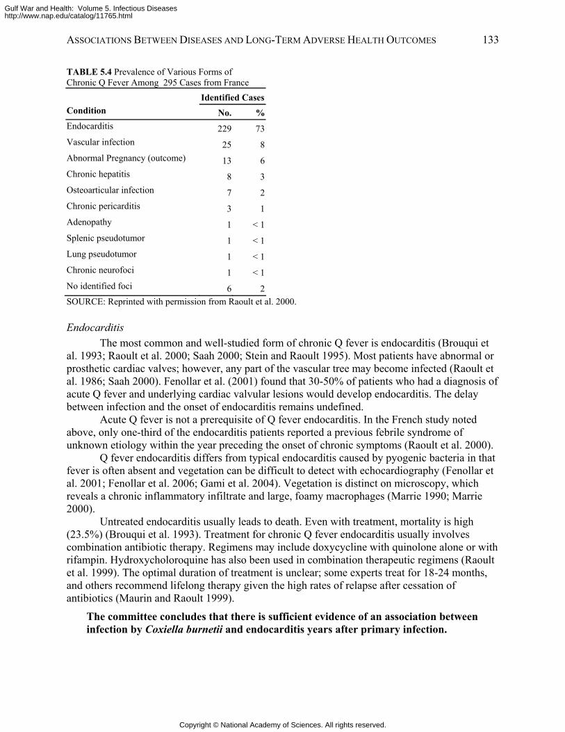
ASSOCIATIONS BETWEEN DISEASES AND LONG-TERM ADVERSE HEALTH OUTCOMES 133
TABLE 5.4 Prevalence of Various Forms of Chronic Q Fever Among 295 Cases from France
SOURCE: Reprinted with permission from Raoult et al. 2000.
Endocarditis The most common and well-studied form of chronic Q fever is endocarditis (Brouqui et
al. 1993; Raoult et al. 2000; Saah 2000; Stein and Raoult 1995). Most patients have abnormal or prosthetic cardiac valves; however, any part of the vascular tree may become infected (Raoult et al. 1986; Saah 2000). Fenollar et al. (2001) found that 30-50% of patients who had a diagnosis of acute Q fever and underlying cardiac valvular lesions would develop endocarditis. The delay between infection and the onset of endocarditis remains undefined.
Acute Q fever is not a prerequisite of Q fever endocarditis. In the French study noted above, only one-third of the endocarditis patients reported a previous febrile syndrome of unknown etiology within the year preceding the onset of chronic symptoms (Raoult et al. 2000).
Q fever endocarditis differs from typical endocarditis caused by pyogenic bacteria in that fever is often absent and vegetation can be difficult to detect with echocardiography (Fenollar et al. 2001; Fenollar et al. 2006; Gami et al. 2004). Vegetation is distinct on microscopy, which reveals a chronic inflammatory infiltrate and large, foamy macrophages (Marrie 1990; Marrie 2000).
Untreated endocarditis usually leads to death. Even with treatment, mortality is high (23.5%) (Brouqui et al. 1993). Treatment for chronic Q fever endocarditis usually involves combination antibiotic therapy. Regimens may include doxycycline with quinolone alone or with rifampin. Hydroxycholoroquine has also been used in combination therapeutic regimens (Raoult et al. 1999). The optimal duration of treatment is unclear; some experts treat for 18-24 months, and others recommend lifelong therapy given the high rates of relapse after cessation of antibiotics (Maurin and Raoult 1999).
The committee concludes that there is sufficient evidence of an association between infection by Coxiella burnetii and endocarditis years after primary infection.
Identified CasesCondition No. %Endocarditis 229 73Vascular infection 25 8Abnormal Pregnancy (outcome) 13 6Chronic hepatitis 8 3Osteoarticular infection 7 2Chronic pericarditis 3 1Adenopathy 1 < 1Splenic pseudotumor 1 < 1Lung pseudotumor 1 < 1Chronic neurofoci 1 < 1No identified foci 6 2
Copyright © National Academy of Sciences. All rights reserved.
Gulf War and Health: Volume 5. Infectious Diseaseshttp://www.nap.edu/catalog/11765.html
134 GULF WAR AND HEALTH
Vascular Infection
Vascular infections can occur in aneurysm and vascular grafts and are often accompanied by a nonspecific illness characterized by weight loss and fever. The authors of a recent case series suggest that the incidence of C. burnetii vascular infection is underestimated and recommend that C. burnetii serologic tests be routinely carried out in cases of unexplained illness in patients with a history of underlying vascular disease (Fournier et al. 1998).
The committee concludes that there is sufficient evidence of an association between infection by Coxiella burnetii and vascular infection years after primary infection.
Chronic Hepatitis Several investigators have documented isolated chronic hepatitis as an infrequent
manifestation of chronic Q fever (Raoult et al. 2000; Saah 2000; Turck et al. 1976; Yebra et al. 1988). It presents with mildly increased liver enzymes; granulomatous hepatitis is histologically typical when liver biopsy is performed. Patients whose acute Q fever is manifested as hepatitis or who have underlying alcoholic cirrhosis may be more likely to develop this health outcome (Raoult et al. 2000). The time between acute infection and diagnosis of chronic hepatitis may be as long as 2 years (Yebra et al. 1988). This health outcome appears to account for less than 5% of all chronic manifestations of C. burnetii infection.
The committee concludes that there is sufficient evidence of an association between infection by Coxiella burnetii and chronic hepatitis years after primary infection.
Osteomyelitis Osteomyelitis is another rare manifestation of chronic Q fever. Of the 313 people with
confirmed chronic Q fever as identified by Raoult et al., seven had osteomyelitis. Only one of those had an earlier documented acute infection with C. burnettii, although several reported a febrile illness within the previous year (Raoult et al. 2000). Nourse et al. identified three additional cases and described 11 previously reported cases; almost half the cases were in children, and nearly all the patients had contact with farm animals (Nourse et al. 2004). In summary, chronic Q fever sometimes is manifested as osteomyelitis, which may occur with a previously diagnosed Q fever illness or in the context of a known history of acute Q fever.
The committee concludes that there is sufficient evidence of a causal relationship between exposure to Coxiella burnetii and osteomyelitis.
Post-Q Fever Fatigue Syndrome A post-Q fever chronic fatigue syndrome has been described in several populations of
exposed patients (Ayres et al. 1998; Hatchette et al. 2003; Marmion et al. 1996; 2005; Wildman et al. 2002). Five years after the previously mentioned outbreak in the West Midlands, UK, 42.4% of those with diagnosed Q fever reported symptoms of chronic fatigue compared with only 26% of a control group (Ayres et al. 1998). A second study documented high levels of fatigue in exposed subjects 10 years after exposure (Wildman et al. 2002). Twenty-seven months after an outbreak of Q fever in Newfoundland, 52% of patients had persistent symptoms that hampered their activities to the same extent as type 2 diabetes mellitus and coronary arterial disease affected cohorts of Americans (Hatchette et al. 2003).
Those studies have limitations that prevent the scientific community—including the investigators themselves—from definitively confirming the existence of a post-Q fever fatigue syndrome. For instance, the outbreak of Q fever in Newfoundland led farms to close, leaving
Copyright © National Academy of Sciences. All rights reserved.
Gulf War and Health: Volume 5. Infectious Diseaseshttp://www.nap.edu/catalog/11765.html
ASSOCIATIONS BETWEEN DISEASES AND LONG-TERM ADVERSE HEALTH OUTCOMES 135
many people unemployed; this socioeconomic factor may have confounded the study results (Hatchette et al. 2003). Data on comorbidity were unavailable, followup serologic data were incomplete, and the study also may have been limited by participation bias: subjects who continued to have symptoms may have been more likely to participate in both questionnaire surveys.
Later studies have reported differences in immune-response genes among those who report post-Q fever fatigue compared with those who are unaffected (Helbig et al. 2005; Helbig et al. 2003). One hypothesis is that after C. burnetii infection, Coxiella may persist universally in the bone marrow and be regulated by the host’s immune response. A subset of patients with subtle differences in their immune response may later develop post-Q fever fatigue. That and other hypotheses are under active investigation in Australia and the UK.
The committee concludes that there is limited or suggestive evidence of an association between infection by Coxiella burnetii and post-Q fever chronic fatigue syndrome years after primary infection.
TUBERCULOSIS
The unique properties and history of tuberculosis (TB) led the committee to approach this section differently from the rest of the chapter in two ways. First, initial infection with Mycobacterium tuberculosis (TB infection) is usually asymptomatic, the onset of the disease (TB) is almost always delayed, and relapse of TB may occur years after successful treatment. Thus, TB infection has the potential for delayed long-term adverse health outcomes both because of the onset of clinically evident TB months to decades after initial infection and because of the long-term consequences of acute disease.
Second, TB has a long history of occurrence and transmission in military settings and remains a cause of potential delayed adverse health outcomes in US troops and veterans of the Gulf War, OEF, and OIF—especially those who are or were deployed to regions where TB is highly endemic. The committee discusses TB in the military at the end of this section.
TB is a chronic necrotizing granulomatous infection caused primarily by the acid-fast bacillus Mycobacterium tuberculosis. An obligate aerobe, M. tuberculosis grows best in such tissues with high oxygen tension as the apices of the lung and the renal cortex; this explains why most infections are manifested as pulmonary disease. M. bovis, a related organism, causes a substantial number of TB cases in regions where milk is not routinely pasteurized and where M. bovis-infected cattle are not identified and destroyed.
Transmission of Tuberculosis
TB is transmitted primarily through exposure to airborne M. tuberculosis. When an infected person coughs, sneezes, yells, or sings, microscopic droplets containing M. tuberculosis are expelled into the air. Heavier particles quickly settle out of the air, and lighter ones remain suspended, often for several hours. Inhaled droplets of 1-5 µm in diameter are small enough to reach the alveoli, where the mycobacteria colonize and infect the lung tissue of their new hosts (IOM 2000).
Copyright © National Academy of Sciences. All rights reserved.
Gulf War and Health: Volume 5. Infectious Diseaseshttp://www.nap.edu/catalog/11765.html
136 GULF WAR AND HEALTH
Detection of Tuberculosis Transmission The human immune system usually does not recognize M. tuberculosis as a foreign body
until 2-6 weeks after inoculation. During that lag time, the organisms proliferate, spreading from the lungs to the lymphatics and disseminating in the bloodstream. Once the human immune system mounts its primary response to M. tuberculosis, further growth and proliferation of the pathogen are usually suppressed; most people maintain a latent TB infection (LTBI) that is believed to persist as a benign condition for life unless progression to active TB develops. The initial infection with M. tuberculosis in adults is usually asymptomatic and results in long-lasting cell-mediated immunity to purified protein derivative (PPD) of M. tuberculosis. Only 1-2% of recently infected people will be found to have active TB (CDC 2000a), so the detection of transmission is based largely on the diagnosis of LTBI.
Diagnosing Latent Tuberculosis Infection Recently acquired LTBI is detected by conversion of a tuberculin skin test (TST) from
negative to positive 2-10 weeks after exposure (CDC 2000a). In development are gamma-interferon release assays that measure cell-mediated immunity to M. tuberculosis protein products more specifically than the TST. The US Food and Drug Administration in 2004 approved the QuantiFERON®-TB GOLD in vitro assay by Cellestis Inc. for diagnosing LTBI (FDA 2005). While noting the need for further research, CDC has recommended that the QuantiFERON-TB GOLD be used in place of the TST (CDC 2005d). The new assay is more specific than TST because it uses antigens that are absent from bacillus Calmette-Guérin (BCG) vaccines and nontuberculous mycobacteria (such antigens can cross-react with the TST to produce false-positive results). In addition, the QuantiFERON-TB GOLD requires only a single draw of blood. Its main limitation is expense. The QuantiFERON-TB GOLD could be used as a confirmatory assay, particularly in TST-positive, BCG-vaccinated people.
Risk Factors for Transmission TB is not a highly infectious disease, so most transmission occurs in such places where
people have close and frequent contact such as households and closed community settings. Occasionally, however, infection follows brief, casual contact in airplanes, buses, hospitals, or prisons. Outbreaks of tuberculosis in closed populations where there is crowding, poor air exchange, or both, may lead to substantial transmission. Exposure to corpses who had active TB has also been identified as posing a high risk for the transmission of the disease.
For US military personnel, the risk of becoming infected with M. tuberculosis depends on occupation, living quarters, exposure to TB-endemic populations, chance exposure in an epidemic setting, and other factors. Cases of TB among active-duty military personnel have the potential to cause extensive TB infection (defined by a positive TST) and outbreaks of active TB among deployed troops, especially shipboard personnel.
The closed shipboard environment and extended periods at sea increase the risk of TB transmission to a level at or above that for most household contacts (Kelley 2005). Notable outbreaks of TB occurred aboard Navy ships in 1966, 1987, and 1998 (Kelley 2005). The most recent of those outbreaks occurred after a US marine with acid-fast bacilli (AFB) smear-positive cavitary pulmonary disease was deployed to a US Navy amphibious ship. More than 18% of the crew and 25% of embarked marines—696 people—converted to TST-positive (Kelley 2005).
Copyright © National Academy of Sciences. All rights reserved.
Gulf War and Health: Volume 5. Infectious Diseaseshttp://www.nap.edu/catalog/11765.html
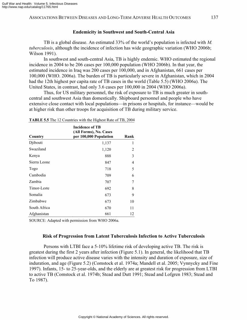
ASSOCIATIONS BETWEEN DISEASES AND LONG-TERM ADVERSE HEALTH OUTCOMES 137
Endemicity in Southwest and South-Central Asia
TB is a global disease. An estimated 33% of the world’s population is infected with M. tuberculosis, although the incidence of infection has wide geographic variation (WHO 2006b; Wilson 1991).
In southwest and south-central Asia, TB is highly endemic. WHO estimated the regional incidence in 2004 to be 206 cases per 100,000 population (WHO 2006b). In that year, the estimated incidence in Iraq was 200 cases per 100,000, and in Afghanistan, 661 cases per 100,000 (WHO. 2006a). The burden of TB is particularly severe in Afghanistan, which in 2004 had the 12th highest per capita rate of TB cases in the world (Table 5.5) (WHO 2006a). The United States, in contrast, had only 3.6 cases per 100,000 in 2004 (WHO 2006a).
Thus, for US military personnel, the risk of exposure to TB is much greater in south-central and southwest Asia than domestically. Shipboard personnel and people who have extensive close contact with local populations—in prisons or hospitals, for instance—would be at higher risk than other troops for acquisition of TB during military service.
TABLE 5.5 The 12 Countries with the Highest Rate of TB, 2004
Country
Incidence of TB (All Forms), No. Cases per 100,000 Population Rank
Djibouti 1,137 1Swaziland 1,120 2Kenya 888 3Sierra Leone 847 4Togo 718 5Cambodia 709 6Zambia 707 7Timor-Leste 692 8Somalia 673 9Zimbabwe 673 10South Africa 670 11Afghanistan 661 12
SOURCE: Adapted with permission from WHO 2006a.
Risk of Progression from Latent Tuberculosis Infection to Active Tuberculosis
Persons with LTBI face a 5-10% lifetime risk of developing active TB. The risk is greatest during the first 2 years after infection (Figure 5.1). In general, the likelihood that TB infection will produce active disease varies with the intensity and duration of exposure, size of induration, and age (Figure 5.2) (Comstock et al. 1974a; Mandell et al. 2005; Vynnycky and Fine 1997). Infants, 15- to 25-year-olds, and the elderly are at greatest risk for progression from LTBI to active TB (Comstock et al. 1974b; Stead and Dutt 1991; Stead and Lofgren 1983; Stead and To 1987).
Copyright © National Academy of Sciences. All rights reserved.
Gulf War and Health: Volume 5. Infectious Diseaseshttp://www.nap.edu/catalog/11765.html
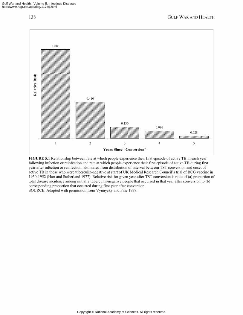
138 GULF WAR AND HEALTH
1.000
0.410
0.1300.086
0.028
1 2 3 4 5
Years Since "Conversion"
Rel
ativ
e R
isk
FIGURE 5.1 Relationship between rate at which people experience their first episode of active TB in each year following infection or reinfection and rate at which people experience their first episode of active TB during first year after infection or reinfection. Estimated from distribution of interval between TST conversion and onset of active TB in those who were tuberculin-negative at start of UK Medical Research Council’s trial of BCG vaccine in 1950-1952 (Hart and Sutherland 1977). Relative risk for given year after TST conversion is ratio of (a) proportion of total disease incidence among initially tuberculin-negative people that occurred in that year after conversion to (b) corresponding proportion that occurred during first year after conversion. SOURCE: Adapted with permission from Vynnycky and Fine 1997.
Copyright © National Academy of Sciences. All rights reserved.
Gulf War and Health: Volume 5. Infectious Diseaseshttp://www.nap.edu/catalog/11765.html
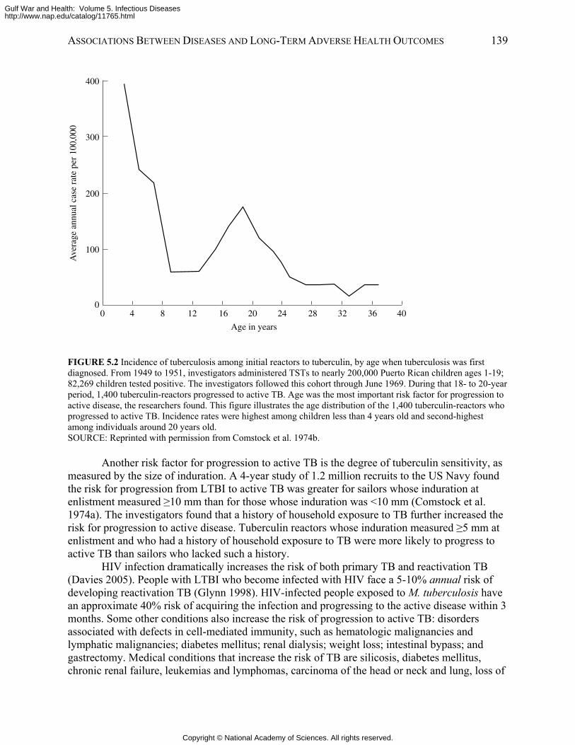
ASSOCIATIONS BETWEEN DISEASES AND LONG-TERM ADVERSE HEALTH OUTCOMES 139
400
300
200
100
0
Ave
rage
ann
ual c
ase
rate
per
100
,000
Age in years
0 4 8 12 16 20 24 28 32 36 40
FIGURE 5.2 Incidence of tuberculosis among initial reactors to tuberculin, by age when tuberculosis was first diagnosed. From 1949 to 1951, investigators administered TSTs to nearly 200,000 Puerto Rican children ages 1-19; 82,269 children tested positive. The investigators followed this cohort through June 1969. During that 18- to 20-year period, 1,400 tuberculin-reactors progressed to active TB. Age was the most important risk factor for progression to active disease, the researchers found. This figure illustrates the age distribution of the 1,400 tuberculin-reactors who progressed to active TB. Incidence rates were highest among children less than 4 years old and second-highest among individuals around 20 years old. SOURCE: Reprinted with permission from Comstock et al. 1974b.
Another risk factor for progression to active TB is the degree of tuberculin sensitivity, as
measured by the size of induration. A 4-year study of 1.2 million recruits to the US Navy found the risk for progression from LTBI to active TB was greater for sailors whose induration at enlistment measured ≥10 mm than for those whose induration was <10 mm (Comstock et al. 1974a). The investigators found that a history of household exposure to TB further increased the risk for progression to active disease. Tuberculin reactors whose induration measured ≥5 mm at enlistment and who had a history of household exposure to TB were more likely to progress to active TB than sailors who lacked such a history.
HIV infection dramatically increases the risk of both primary TB and reactivation TB (Davies 2005). People with LTBI who become infected with HIV face a 5-10% annual risk of developing reactivation TB (Glynn 1998). HIV-infected people exposed to M. tuberculosis have an approximate 40% risk of acquiring the infection and progressing to the active disease within 3 months. Some other conditions also increase the risk of progression to active TB: disorders associated with defects in cell-mediated immunity, such as hematologic malignancies and lymphatic malignancies; diabetes mellitus; renal dialysis; weight loss; intestinal bypass; and gastrectomy. Medical conditions that increase the risk of TB are silicosis, diabetes mellitus, chronic renal failure, leukemias and lymphomas, carcinoma of the head or neck and lung, loss of
Copyright © National Academy of Sciences. All rights reserved.
Gulf War and Health: Volume 5. Infectious Diseaseshttp://www.nap.edu/catalog/11765.html
140 GULF WAR AND HEALTH
at least 10% of ideal body weight, gastrectomy and jejunoileal bypass, radiation therapy, treatment with tumor-necrosis-factor inhibitors, immunosuppression associated with organ transplantation, and corticosteroid therapy. In many cases the stimulating factor is unknown.
Treatment for Latent Tuberculosis Infection to Prevent Active Tuberculosis
To mitigate the risk of LTBI’s becoming active TB, such infections are treated with isoniazid for 9 months. Completing the treatment regimen reduces the risk of active TB by 70-90% (CDC 2000b), but asymptomatic people frequently fail to comply with the regimen.
Active Tuberculosis
Primary Tuberculosis vs Reactivation Tuberculosis If a chest x-ray picture is taken during initial TB infection, it often shows features of a
condition called primary TB: patchy alveolar opacities in the middle- and lower-lung fields, common with unilateral hilar adenopathy. Occasionally, patients with primary TB have fever, nonproductive cough, dyspnea, and—rarely—erythema nodosum. Compression by enlarged lymph nodes may lead to upper- or middlelung collapse. Primary TB generally resolves without treatment. In some patients, however, the immune system cannot contain the infection, and active disease develops, as discussed below. Patients who recover from primary TB (including pleural disease)—particularly those with prior pleuritis—remain at risk for recurrence of active TB.
Historically, a distinction has been made between primary TB occurring at the time of initial TB infection and the more typical adult manifestation of disease, called reactivation TB, developing later. Yet the overlapping temporal and clinical features of the two forms often blur the distinctions between them. One reason for the apparent overlap is uncertainty as to when the primary infection occurred. Therefore, to be consistent with US diagnostic standards, the committee’s discussion of active TB below pertains to both primary and reactivation TB (CDC 2000a).
Diagnosing Active Tuberculosis The standard approach to diagnosing active TB is through an AFB smear of expectorated
sputum. The presence of such mycobacteria as M. tuberculosis in a bodily secretion or tissue specimen can be visually confirmed with the so-called acid-fast test, which exploits the unique properties of the mycobacterial cell envelope. Cells in a specimen are first stained with red carbol fuchsin, then washed with an acidic alcohol solution. The wash decolorizes almost all organisms except mycobacteria because mycobacterial cell envelopes contain mycolic acid, high-molecular-weight lipids, and waxes that prevent the wash from penetrating the cell.
About half of patients with newly diagnosed pulmonary TB have AFB-positive smears. In addition to establishing the likely diagnosis, AFB-positive smears signal highly infectious cases that must be managed through strict isolation. Smears are more likely to be negative in patients with minimal TB or noncavitary TB.
Cultures are performed on such specialized media as Lowenstein-Jensen (an egg-based media), Middlebrook 7H10 (an agar-based media), and Middlebrook 7H102 (a liquid-based media) (CDC 2000a). Using a combination of solid and liquid media will yield positive results
Copyright © National Academy of Sciences. All rights reserved.
Gulf War and Health: Volume 5. Infectious Diseaseshttp://www.nap.edu/catalog/11765.html
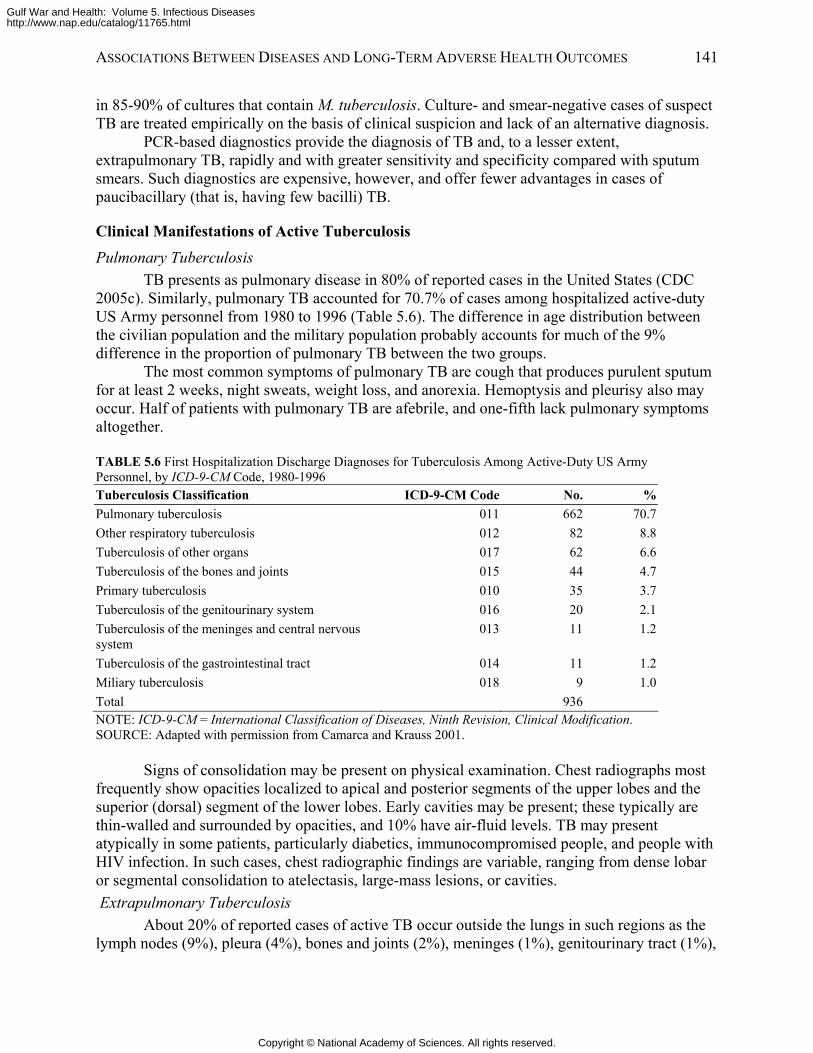
ASSOCIATIONS BETWEEN DISEASES AND LONG-TERM ADVERSE HEALTH OUTCOMES 141
in 85-90% of cultures that contain M. tuberculosis. Culture- and smear-negative cases of suspect TB are treated empirically on the basis of clinical suspicion and lack of an alternative diagnosis.
PCR-based diagnostics provide the diagnosis of TB and, to a lesser extent, extrapulmonary TB, rapidly and with greater sensitivity and specificity compared with sputum smears. Such diagnostics are expensive, however, and offer fewer advantages in cases of paucibacillary (that is, having few bacilli) TB.
Clinical Manifestations of Active Tuberculosis
Pulmonary Tuberculosis TB presents as pulmonary disease in 80% of reported cases in the United States (CDC
2005c). Similarly, pulmonary TB accounted for 70.7% of cases among hospitalized active-duty US Army personnel from 1980 to 1996 (Table 5.6). The difference in age distribution between the civilian population and the military population probably accounts for much of the 9% difference in the proportion of pulmonary TB between the two groups.
The most common symptoms of pulmonary TB are cough that produces purulent sputum for at least 2 weeks, night sweats, weight loss, and anorexia. Hemoptysis and pleurisy also may occur. Half of patients with pulmonary TB are afebrile, and one-fifth lack pulmonary symptoms altogether.
TABLE 5.6 First Hospitalization Discharge Diagnoses for Tuberculosis Among Active-Duty US Army Personnel, by ICD-9-CM Code, 1980-1996 Tuberculosis Classification ICD-9-CM Code No. %Pulmonary tuberculosis 011 662 70.7Other respiratory tuberculosis 012 82 8.8Tuberculosis of other organs 017 62 6.6Tuberculosis of the bones and joints 015 44 4.7Primary tuberculosis 010 35 3.7Tuberculosis of the genitourinary system 016 20 2.1Tuberculosis of the meninges and central nervous system
013 11 1.2
Tuberculosis of the gastrointestinal tract 014 11 1.2Miliary tuberculosis 018 9 1.0Total 936 NOTE: ICD-9-CM = International Classification of Diseases, Ninth Revision, Clinical Modification. SOURCE: Adapted with permission from Camarca and Krauss 2001.
Signs of consolidation may be present on physical examination. Chest radiographs most frequently show opacities localized to apical and posterior segments of the upper lobes and the superior (dorsal) segment of the lower lobes. Early cavities may be present; these typically are thin-walled and surrounded by opacities, and 10% have air-fluid levels. TB may present atypically in some patients, particularly diabetics, immunocompromised people, and people with HIV infection. In such cases, chest radiographic findings are variable, ranging from dense lobar or segmental consolidation to atelectasis, large-mass lesions, or cavities. Extrapulmonary Tuberculosis
About 20% of reported cases of active TB occur outside the lungs in such regions as the lymph nodes (9%), pleura (4%), bones and joints (2%), meninges (1%), genitourinary tract (1%),
Copyright © National Academy of Sciences. All rights reserved.
Gulf War and Health: Volume 5. Infectious Diseaseshttp://www.nap.edu/catalog/11765.html
142 GULF WAR AND HEALTH
and peritoneal cavity (1%) (CDC 2005c). Disseminated extrapulmonary TB, known as miliary TB, consists of 1-3 mm nodules throughout the lungs and other tissues. Tuberculosis Pleurisy
Primary TB is sometimes manifested as an infection of the pleural space, and TB pleurisy may develop later as a progression of LTBI to pleural TB with or without pulmonary TB. The typical presentation is acute onset of fever, cough, and pleuritic chest pains, although there may be a chronic course characterized by fever, general malaise, and loss of up to 10% of body weight. The pleural effusions usually are small to moderate. Concurrent parenchymal disease occurs in one-third to one-half of cases. Diagnosing TB pleurisy usually requires a pleural biopsy, which has a diagnostic yield of 85-95%. The initial TST is negative one-third of the time. Although pleural fluid is exudative, it usually tests negative with an AFB smear and, in 75% of cases, in cultures.
The committee concludes that there is sufficient evidence of a causal relationship between infection with Mycobacterium tuberculosis and occurrence of active tuberculosis months to decades after infection.
Coinfection with Tuberculosis and Human Immunodeficiency Virus HIV alters the clinical manifestations of TB. For example, pulmonary TB may occur in a
lower lobe in a noncavitary fashion. There may be hilar or mediastinal adenopathy, pleural disease, or a normal chest x-ray picture. Extrapulmonary TB is more common among patients coinfected with HIV than among other patients, and it may occur with pulmonary disease or alone. The interactions between chemotherapeutic drugs for TB with antiretroviral drugs for HIV challenge the clinician to treat TB-HIV coinfections effectively.
Treatment for Active Tuberculosis Treatment for drug-sensitive active TB consists of isoniazid, rifampin, pyrazinamide, and
ethambutol for 2 months followed by isoniazid and rifampin for 4 months. That regimen is 95% effective, although about 5% of patients will experience relapses of active TB, most of them in the first 1-2 years after treatment completion (CDC 2003a).
Multiple-drug-resistant TB is more difficult to cure. Regimens should be tailored to known drug susceptibility of the isolates. If an isolate is resistant to isoniazid and rifampin but susceptible to other drugs, treatment regimens would consist of ethambutol, pyrazinamide, and levofloxacin for 12-18 months and an injectible aminoglycoside for the first 2-3 months.
Late Manifestations of Active Tuberculosis
Even after highly effective treatment for acute TB, affected tissues and organs may be functionally impaired or destroyed. Functional impairment may range from imperceptible to severe. The paucity of data on long-term health outcomes of acute TB and the ability of M. tuberculosis to infect and damage virtually any organ of the body challenged the committee to address the late manifestations of this disease comprehensively. This discussion addresses the notable late manifestations of acute TB and the committee’s conclusions about the strength of the association between acute and long-term adverse health outcomes.
Copyright © National Academy of Sciences. All rights reserved.
Gulf War and Health: Volume 5. Infectious Diseaseshttp://www.nap.edu/catalog/11765.html
ASSOCIATIONS BETWEEN DISEASES AND LONG-TERM ADVERSE HEALTH OUTCOMES 143
Late Manifestations of Pulmonary Tuberculosis It is well accepted in the biomedical community that disability occurs after pulmonary
TB. The most common adverse health outcomes are chronic disabling scarring of the lungs, long-term pulmonary dysfunction, secondary infection of residual cavities, empyema, bronchiectasis, bronchopleural fistulas, and aspergilloma. Rarely, scar carcinoma occurs. Despite the centuries-old history of TB, however, the published data on long-term manifestations are inadequate for the committee to comment on their duration, range, or severity of adverse health outcomes.
A number of studies have compared pulmonary function tests (PFTs) at diagnosis of and after treatment for active TB. For instance, a study of 25 TB patients in Canada found abnormal PFTs (reduced 1-second forced expiratory volume and forced vital capacity) in individuals with cavitary TB but not in those with noncavitary TB (Long et al. 1998). Another study, of 74 TB patients in South Africa, showed a restrictive lung function pattern in 57% of cases and an obstructive lung function pattern in 11%. After treatment for TB, 53% of patients still had abnormal PFTs (Plit et al. 1998). Although neither study commented on the potential disability associated with the findings, none of the patients had residual impairment of oxygenation at rest.
Multiple case reports have been published of patients who have developed “scar cancer”, lung cancer associated with lung scars from TB or other causes (Ardies 2003). The risk of cancer after TB has not been quantified, nor has the percentage of TB patients left with pulmonary scarring. One report from Japan indicated a 2% prevalence of lymphoma of the pleural space among patients with chronic pyothorax, which is associated with tuberculosis (Iuchi et al. 1987). Because pyothorax is a rare complication of TB, the authors could not quantify the overall risk of cancer posed by TB. In summary, the available data are insufficient for the committee to determine whether an association exists between cancer and active TB. If such an association does exist, cancer is undoubtedly a very rare consequence of TB.
Late Manifestations of Extrapulmonary Tuberculosis The long-term adverse health outcomes of extrapulmonary TB occur with varied
frequency depending on the site of active disease. The more common forms of extrapulmonary TB, pleural and lymphatic disease, rarely have long-term adverse health outcomes. Two of the less common forms, TB meningitis and skeletal TB, are more likely to result in irreversible tissue damage. In general, estimates of the long-term prognoses for uncommon forms of TB are based on relatively small series of patients. Tuberculosis Meningitis and Long-Term Neurologic Disability
It is well accepted in the biomedical community that TB meningitis is associated with long-term neurologic outcomes. The extent of disability depends on the duration and severity of acute symptoms, the age of the patient, and the neurologic deficits (Dube et al. 1992; Kennedy and Fallon 1979). Of TB meningitis patients with stupor or dense paraplegia or hemiplegia, about half either die or recover with severe residual neurologic deficits (Kennedy and Fallon 1979). Until the 1990s, most reports of outcomes for adult patients with TB meningitis focused on mortality. As the recovery rate increased, more studies about the long-term prognoses for these patients emerged. Table 5.7 summarizes the findings of seven studies about long-term neurologic deficits of TB meningitis.
Copyright © National Academy of Sciences. All rights reserved.
Gulf War and Health: Volume 5. Infectious Diseaseshttp://www.nap.edu/catalog/11765.html
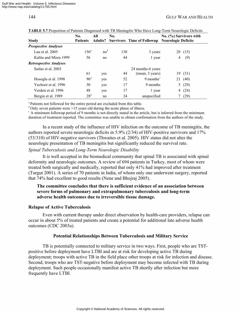
144 GULF WAR AND HEALTH
TABLE 5.7 Proportion of Patients Diagnosed with TB Meningitis Who Have Long-Term Neurologic Deficits
Study No. Patients
All Adults?
No. Survivors Time of Followup
No. (%) Survivors with Neurologic Deficits
Prospective Analyses Lau et al. 2005 156a nob 130 3 years 20 (15) Kalita and Misra 1999 56 no 44 1 year 4 (9)
Retrospective Analyses Sutlas et al. 2003
61 yes 4424 months-6 years
(mean, 3 years) 19 (31)
Hosoglu et al. 1998 96a yes 52 9 monthsc 21 (40) Yechoor et al. 1996 30 yes 17 9 months 5 (29) Verdon et al. 1996 48 yes 17 1 year 4 (24) Bergin et al. 1989 28a no 24 unspecified 7 (29)
a Patients not followed for the entire period are excluded from this table. b Only seven patients were >15 years old during the acute phase of illness. c A minimum followup period of 9 months is not directly stated in the article, but is inferred from the minimum duration of treatment reported. The committee was unable to obtain confirmation from the authors of the study.
In a recent study of the influence of HIV infection on the outcome of TB meningitis, the authors reported severe neurologic deficits in 5.9% (2/34) of HIV-positive survivors and 17% (53/310) of HIV-negative survivors (Thwaites et al. 2005). HIV status did not alter the neurologic presentation of TB meningitis but significantly reduced the survival rate. Spinal Tuberculosis and Long-Term Neurologic Disability
It is well accepted in the biomedical community that spinal TB is associated with spinal deformity and neurologic outcomes. A review of 694 patients in Turkey, most of whom were treated both surgically and medically, reported that only 41% had improved after treatment (Turgut 2001). A series of 70 patients in India, of whom only one underwent surgery, reported that 74% had excellent to good results (Nene and Bhojraj 2005).
The committee concludes that there is sufficient evidence of an association between severe forms of pulmonary and extrapulmonary tuberculosis and long-term adverse health outcomes due to irreversible tissue damage.
Relapse of Active Tuberculosis Even with current therapy under direct observation by health-care providers, relapse can
occur in about 5% of treated patients and create a potential for additional late adverse health outcomes (CDC 2003a).
Potential Relationships Between Tuberculosis and Military Service
TB is potentially connected to military service in two ways. First, people who are TST-positive before deployment have LTBI and are at risk for developing active TB during deployment; troops with active TB in the field place other troops at risk for infection and disease. Second, troops who are TST-negative before deployment may become infected with TB during deployment. Such people occasionally manifest active TB shortly after infection but more frequently have LTBI.
Copyright © National Academy of Sciences. All rights reserved.
Gulf War and Health: Volume 5. Infectious Diseaseshttp://www.nap.edu/catalog/11765.html
ASSOCIATIONS BETWEEN DISEASES AND LONG-TERM ADVERSE HEALTH OUTCOMES 145
Promotion of Tuberculin Skin Testing The most effective way to mitigate TB transmission and activation is to identify and treat
LTBI. That is a compelling argument for testing all military personnel for TB before and after every deployment. Table 5.8 summarizes the policies of each branch of the military regarding TSTs and treatment for LTBI.
Available data suggest that prior M. tuberculosis infection and exposure in the theater of operations contribute about equally to the prevalence of LTBI and the risk of disease among US military personnel. The Department of Defense (DOD) estimates that 2.5% of military personnel deployed to southwest and south-central Asia during OEF and OIF acquired new M. tuberculosis infections during deployments (Kilpatrick 2005). In comparison, the prevalence of TST reactivity among young adults entering the Navy in 1997 and 1998 was 3.5% (Smith et al. 2002); among military police who participated in refugee and humanitarian operations in Guantanamo Bay in 1995, it was 3.7% (Kortepeter and Krauss 2001). No case of active TB has been recognized in troops deployed to the Persian Gulf during Operation Desert Shield or Operation Desert Storm (Hyams et al. 1995).
Copyright © National Academy of Sciences. All rights reserved.
Gulf War and Health: Volume 5. Infectious Diseaseshttp://www.nap.edu/catalog/11765.html
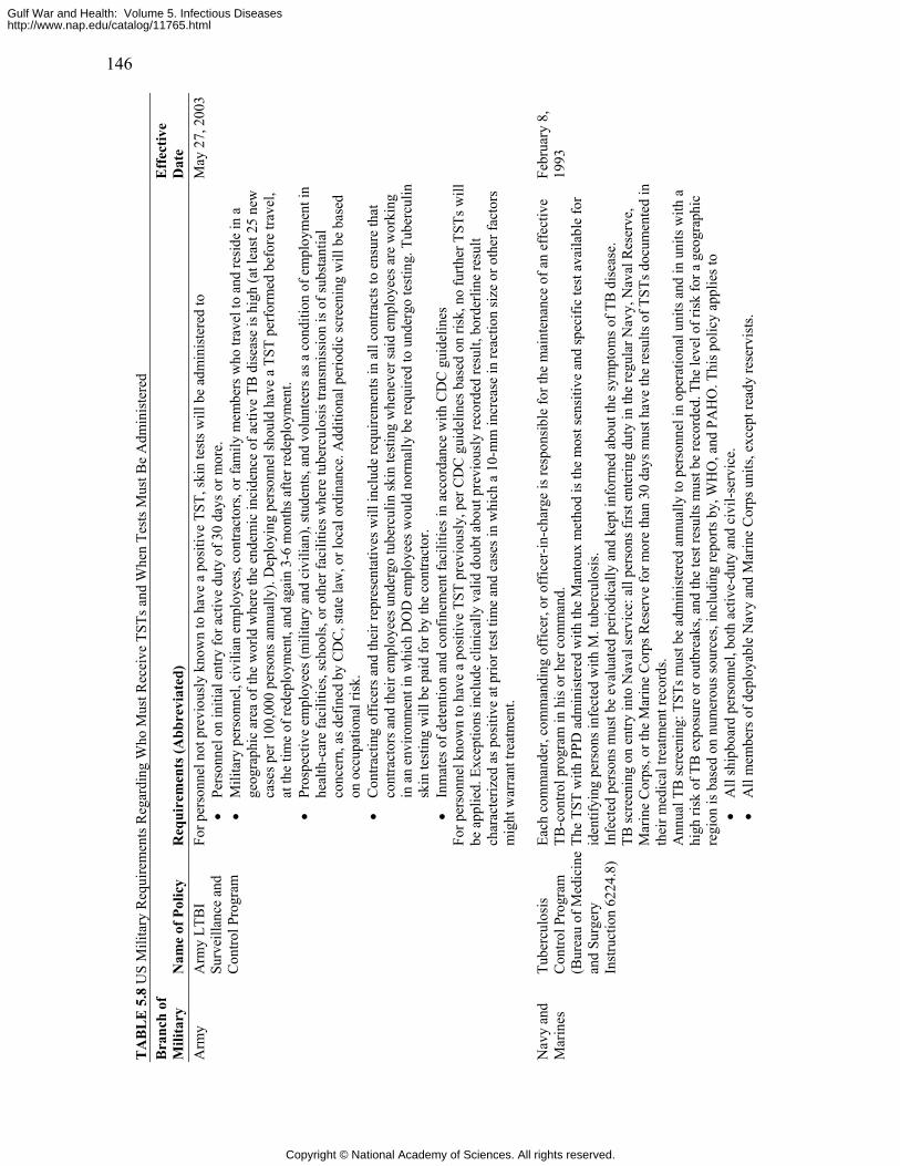
146
TA
BL
E 5
.8 U
S M
ilita
ry R
equi
rem
ents
Reg
ardi
ng W
ho M
ust R
ecei
ve T
STs a
nd W
hen
Test
s Mus
t Be
Adm
inis
tere
d B
ranc
h of
M
ilita
ry
Nam
e of
Pol
icy
Req
uire
men
ts (A
bbre
viat
ed)
Eff
ectiv
e D
ate
A
rmy
Arm
y LT
BI
Surv
eilla
nce
and
Con
trol P
rogr
am
For p
erso
nnel
not
pre
viou
sly
know
n to
hav
e a
posi
tive
TST,
skin
test
s will
be
adm
inis
tere
d to
•
Pers
onne
l on
initi
al e
ntry
for a
ctiv
e du
ty o
f 30
days
or m
ore.
•
Mili
tary
per
sonn
el, c
ivili
an e
mpl
oyee
s, co
ntra
ctor
s, or
fam
ily m
embe
rs w
ho tr
avel
to a
nd re
side
in a
ge
ogra
phic
are
a of
the
wor
ld w
here
the
ende
mic
inci
denc
e of
act
ive
TB d
isea
se is
hig
h (a
t lea
st 2
5 ne
w
case
s per
100
,000
per
sons
ann
ually
). D
eplo
ying
per
sonn
el sh
ould
hav
e a
TST
perf
orm
ed b
efor
e tra
vel,
at th
e tim
e of
rede
ploy
men
t, an
d ag
ain
3-6
mon
ths a
fter r
edep
loym
ent.
•
Pros
pect
ive
empl
oyee
s (m
ilita
ry a
nd c
ivili
an),
stud
ents
, and
vol
unte
ers a
s a c
ondi
tion
of e
mpl
oym
ent i
n he
alth
-car
e fa
cilit
ies,
scho
ols,
or o
ther
faci
litie
s whe
re tu
berc
ulos
is tr
ansm
issi
on is
of s
ubst
antia
l co
ncer
n, a
s def
ined
by
CD
C, s
tate
law
, or l
ocal
ord
inan
ce. A
dditi
onal
per
iodi
c sc
reen
ing
will
be
base
d on
occ
upat
iona
l ris
k.
• C
ontra
ctin
g of
ficer
s and
thei
r rep
rese
ntat
ives
will
incl
ude
requ
irem
ents
in a
ll co
ntra
cts t
o en
sure
that
co
ntra
ctor
s and
thei
r em
ploy
ees u
nder
go tu
berc
ulin
skin
test
ing
whe
neve
r sai
d em
ploy
ees a
re w
orki
ng
in a
n en
viro
nmen
t in
whi
ch D
OD
em
ploy
ees w
ould
nor
mal
ly b
e re
quire
d to
und
ergo
test
ing.
Tub
ercu
lin
skin
test
ing
will
be
paid
for b
y th
e co
ntra
ctor
. •
Inm
ates
of d
eten
tion
and
conf
inem
ent f
acili
ties i
n ac
cord
ance
with
CD
C g
uide
lines
Fo
r per
sonn
el k
now
n to
hav
e a
posi
tive
TST
prev
ious
ly, p
er C
DC
gui
delin
es b
ased
on
risk,
no
furth
er T
STs w
ill
be a
pplie
d. E
xcep
tions
incl
ude
clin
ical
ly v
alid
dou
bt a
bout
pre
viou
sly
reco
rded
resu
lt, b
orde
rline
resu
lt ch
arac
teriz
ed a
s pos
itive
at p
rior t
est t
ime
and
case
s in
whi
ch a
10-
mm
incr
ease
in re
actio
n si
ze o
r oth
er fa
ctor
s m
ight
war
rant
trea
tmen
t.
May
27,
200
3
Nav
y an
d M
arin
es
Tube
rcul
osis
C
ontro
l Pro
gram
(B
urea
u of
Med
icin
e an
d Su
rger
y In
stru
ctio
n 62
24.8
)
Each
com
man
der,
com
man
ding
off
icer
, or o
ffic
er-in
-cha
rge
is re
spon
sibl
e fo
r the
mai
nten
ance
of a
n ef
fect
ive
TB-c
ontro
l pro
gram
in h
is o
r her
com
man
d.
The
TST
with
PPD
adm
inis
tere
d w
ith th
e M
anto
ux m
etho
d is
the
mos
t sen
sitiv
e an
d sp
ecifi
c te
st a
vaila
ble
for
iden
tifyi
ng p
erso
ns in
fect
ed w
ith M
. tub
ercu
losi
s. In
fect
ed p
erso
ns m
ust b
e ev
alua
ted
perio
dica
lly a
nd k
ept i
nfor
med
abo
ut th
e sy
mpt
oms o
f TB
dis
ease
. TB
scre
enin
g on
ent
ry in
to N
aval
serv
ice:
all
pers
ons f
irst e
nter
ing
duty
in th
e re
gula
r Nav
y, N
aval
Res
erve
, M
arin
e C
orps
, or t
he M
arin
e C
orps
Res
erve
for m
ore
than
30
days
mus
t hav
e th
e re
sults
of T
STs d
ocum
ente
d in
th
eir m
edic
al tr
eatm
ent r
ecor
ds.
Ann
ual T
B sc
reen
ing:
TST
s mus
t be
adm
inis
tere
d an
nual
ly to
per
sonn
el in
ope
ratio
nal u
nits
and
in u
nits
with
a
high
risk
of T
B e
xpos
ure
or o
utbr
eaks
, and
the
test
resu
lts m
ust b
e re
cord
ed. T
he le
vel o
f ris
k fo
r a g
eogr
aphi
c re
gion
is b
ased
on
num
erou
s sou
rces
, inc
ludi
ng re
ports
by,
WH
O, a
nd P
AH
O. T
his p
olic
y ap
plie
s to
•
All
ship
boar
d pe
rson
nel,
both
act
ive-
duty
and
civ
il-se
rvic
e.
• A
ll m
embe
rs o
f dep
loya
ble
Nav
y an
d M
arin
e C
orps
uni
ts, e
xcep
t rea
dy re
serv
ists
.
Febr
uary
8,
1993
Copyright © National Academy of Sciences. All rights reserved.
Gulf War and Health: Volume 5. Infectious Diseaseshttp://www.nap.edu/catalog/11765.html
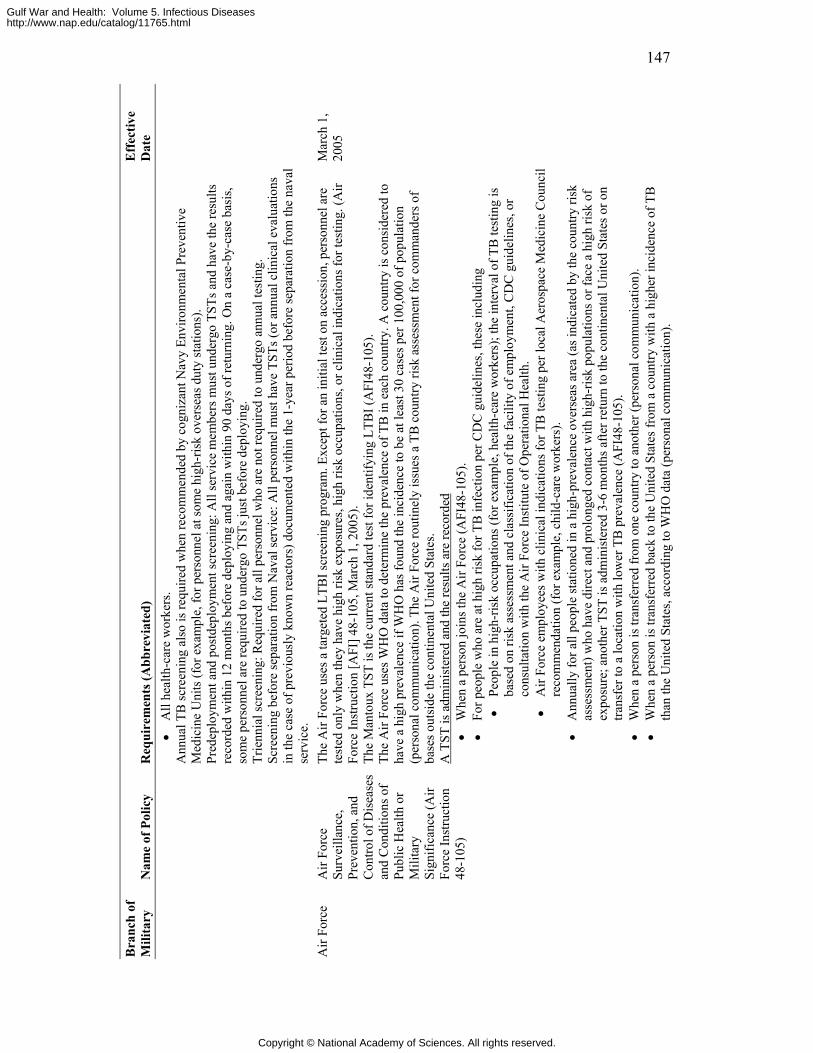
147
Bra
nch
of
Mili
tary
N
ame
of P
olic
y R
equi
rem
ents
(Abb
revi
ated
) E
ffec
tive
Dat
e
• A
ll he
alth
-car
e w
orke
rs.
Ann
ual T
B sc
reen
ing
also
is re
quire
d w
hen
reco
mm
ende
d by
cog
niza
nt N
avy
Envi
ronm
enta
l Pre
vent
ive
Med
icin
e U
nits
(for
exa
mpl
e, fo
r per
sonn
el a
t som
e hi
gh-r
isk
over
seas
dut
y st
atio
ns).
Pred
eplo
ymen
t and
pos
tdep
loym
ent s
cree
ning
: All
serv
ice
mem
bers
mus
t und
ergo
TST
s and
hav
e th
e re
sults
re
cord
ed w
ithin
12
mon
ths b
efor
e de
ploy
ing
and
agai
n w
ithin
90
days
of r
etur
ning
. On
a ca
se-b
y-ca
se b
asis
, so
me
pers
onne
l are
requ
ired
to u
nder
go T
STs j
ust b
efor
e de
ploy
ing.
Tr
ienn
ial s
cree
ning
: Req
uire
d fo
r all
pers
onne
l who
are
not
requ
ired
to u
nder
go a
nnua
l tes
ting.
Sc
reen
ing
befo
re se
para
tion
from
Nav
al se
rvic
e: A
ll pe
rson
nel m
ust h
ave
TSTs
(or a
nnua
l clin
ical
eva
luat
ions
in
the
case
of p
revi
ousl
y kn
own
reac
tors
) doc
umen
ted
with
in th
e 1-
year
per
iod
befo
re se
para
tion
from
the
nava
l se
rvic
e.
Air
Forc
e A
ir Fo
rce
Surv
eilla
nce,
Pr
even
tion,
and
C
ontro
l of D
isea
ses
and
Con
ditio
ns o
f Pu
blic
Hea
lth o
r M
ilita
ry
Sign
ifica
nce
(Air
Forc
e In
stru
ctio
n 48
-105
)
The
Air
Forc
e us
es a
targ
eted
LTB
I scr
eeni
ng p
rogr
am. E
xcep
t for
an
initi
al te
st o
n ac
cess
ion,
per
sonn
el a
re
test
ed o
nly
whe
n th
ey h
ave
high
risk
exp
osur
es, h
igh
risk
occu
patio
ns, o
r clin
ical
indi
catio
ns fo
r tes
ting.
(Air
Forc
e In
stru
ctio
n [A
FI] 4
8-10
5, M
arch
1, 2
005)
. Th
e M
anto
ux T
ST is
the
curr
ent s
tand
ard
test
for i
dent
ifyin
g LT
BI (
AFI
48-1
05).
Th
e A
ir Fo
rce
uses
WH
O d
ata
to d
eter
min
e th
e pr
eval
ence
of T
B in
eac
h co
untry
. A c
ount
ry is
con
side
red
to
have
a h
igh
prev
alen
ce if
WH
O h
as fo
und
the
inci
denc
e to
be
at le
ast 3
0 ca
ses p
er 1
00,0
00 o
f pop
ulat
ion
(per
sona
l com
mun
icat
ion)
. The
Air
Forc
e ro
utin
ely
issu
es a
TB
cou
ntry
risk
ass
essm
ent f
or c
omm
ande
rs o
f ba
ses o
utsi
de th
e co
ntin
enta
l Uni
ted
Stat
es.
A T
ST is
adm
inis
tere
d an
d th
e re
sults
are
reco
rded
•
Whe
n a
pers
on jo
ins t
he A
ir Fo
rce
(AFI
48-1
05).
• Fo
r peo
ple
who
are
at h
igh
risk
for T
B in
fect
ion
per C
DC
gui
delin
es, t
hese
incl
udin
g •
Peop
le in
hig
h-ris
k oc
cupa
tions
(for
exa
mpl
e, h
ealth
-car
e w
orke
rs);
the
inte
rval
of T
B te
stin
g is
ba
sed
on ri
sk a
sses
smen
t and
cla
ssifi
catio
n of
the
faci
lity
of e
mpl
oym
ent,
CD
C g
uide
lines
, or
cons
ulta
tion
with
the
Air
Forc
e In
stitu
te o
f Ope
ratio
nal H
ealth
.
• A
ir Fo
rce
empl
oyee
s with
clin
ical
indi
catio
ns fo
r TB
test
ing
per l
ocal
Aer
ospa
ce M
edic
ine
Cou
ncil
reco
mm
enda
tion
(for
exa
mpl
e, c
hild
-car
e w
orke
rs).
• A
nnua
lly fo
r all
peop
le st
atio
ned
in a
hig
h-pr
eval
ence
ove
rsea
s are
a (a
s ind
icat
ed b
y th
e co
untry
risk
as
sess
men
t) w
ho h
ave
dire
ct a
nd p
rolo
nged
con
tact
with
hig
h-ris
k po
pula
tions
or f
ace
a hi
gh ri
sk o
f ex
posu
re; a
noth
er T
ST is
adm
inis
tere
d 3-
6 m
onth
s afte
r ret
urn
to th
e co
ntin
enta
l Uni
ted
Stat
es o
r on
trans
fer t
o a
loca
tion
with
low
er T
B p
reva
lenc
e (A
FI48
-105
). •
Whe
n a
pers
on is
tran
sfer
red
from
one
cou
ntry
to a
noth
er (p
erso
nal c
omm
unic
atio
n).
• W
hen
a pe
rson
is tr
ansf
erre
d ba
ck to
the
Uni
ted
Stat
es fr
om a
cou
ntry
with
a h
ighe
r inc
iden
ce o
f TB
th
an th
e U
nite
d St
ates
, acc
ordi
ng to
WH
O d
ata
(per
sona
l com
mun
icat
ion)
.
Mar
ch 1
, 20
05
Copyright © National Academy of Sciences. All rights reserved.
Gulf War and Health: Volume 5. Infectious Diseaseshttp://www.nap.edu/catalog/11765.html
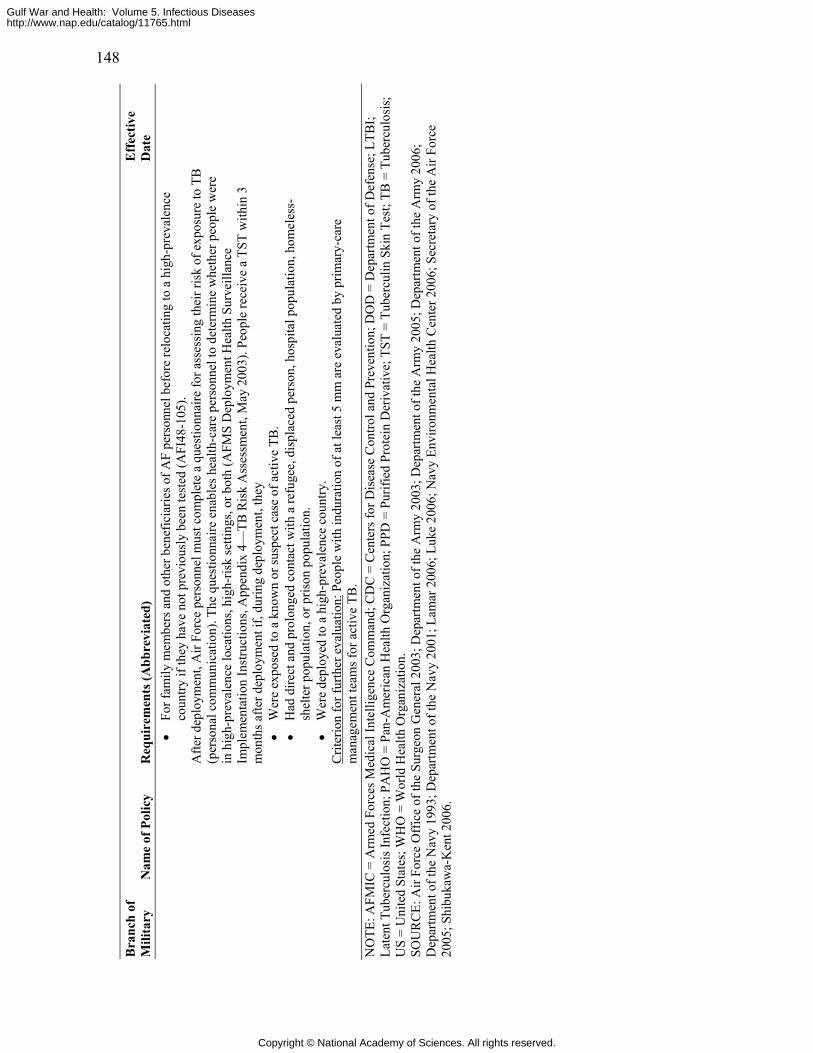
148
Bra
nch
of
Mili
tary
N
ame
of P
olic
y R
equi
rem
ents
(Abb
revi
ated
) E
ffec
tive
Dat
e
• Fo
r fam
ily m
embe
rs a
nd o
ther
ben
efic
iarie
s of A
F pe
rson
nel b
efor
e re
loca
ting
to a
hig
h-pr
eval
ence
co
untry
if th
ey h
ave
not p
revi
ousl
y be
en te
sted
(AFI
48-1
05).
Afte
r dep
loym
ent,
Air
Forc
e pe
rson
nel m
ust c
ompl
ete
a qu
estio
nnai
re fo
r ass
essi
ng th
eir r
isk
of e
xpos
ure
to T
B
(per
sona
l com
mun
icat
ion)
. The
que
stio
nnai
re e
nabl
es h
ealth
-car
e pe
rson
nel t
o de
term
ine
whe
ther
peo
ple
wer
e in
hig
h-pr
eval
ence
loca
tions
, hig
h-ris
k se
tting
s, or
bot
h (A
FMS
Dep
loym
ent H
ealth
Sur
veill
ance
Im
plem
enta
tion
Inst
ruct
ions
, App
endi
x 4—
TB R
isk
Ass
essm
ent,
May
200
3). P
eopl
e re
ceiv
e a
TST
with
in 3
m
onth
s afte
r dep
loym
ent i
f, du
ring
depl
oym
ent,
they
•
Wer
e ex
pose
d to
a k
now
n or
susp
ect c
ase
of a
ctiv
e TB
. •
Had
dire
ct a
nd p
rolo
nged
con
tact
with
a re
fuge
e, d
ispl
aced
per
son,
hos
pita
l pop
ulat
ion,
hom
eles
s-sh
elte
r pop
ulat
ion,
or p
rison
pop
ulat
ion.
•
Wer
e de
ploy
ed to
a h
igh-
prev
alen
ce c
ount
ry.
Crit
erio
n fo
r fur
ther
eva
luat
ion:
Peo
ple
with
indu
ratio
n of
at l
east
5 m
m a
re e
valu
ated
by
prim
ary-
care
m
anag
emen
t tea
ms f
or a
ctiv
e TB
.
NO
TE: A
FMIC
= A
rmed
For
ces M
edic
al In
telli
genc
e C
omm
and;
CD
C =
Cen
ters
for D
isea
se C
ontro
l and
Pre
vent
ion;
DO
D =
Dep
artm
ent o
f Def
ense
; LTB
I; La
tent
Tub
ercu
losi
s Inf
ectio
n; P
AH
O =
Pan
-Am
eric
an H
ealth
Org
aniz
atio
n; P
PD =
Pur
ified
Pro
tein
Der
ivat
ive;
TST
= T
uber
culin
Ski
n Te
st; T
B =
Tub
ercu
losi
s;
US
= U
nite
d St
ates
; WH
O =
Wor
ld H
ealth
Org
aniz
atio
n.
SOU
RC
E: A
ir Fo
rce
Off
ice
of th
e Su
rgeo
n G
ener
al 2
003;
Dep
artm
ent o
f the
Arm
y 20
03; D
epar
tmen
t of t
he A
rmy
2005
; Dep
artm
ent o
f the
Arm
y 20
06;
Dep
artm
ent o
f the
Nav
y 19
93; D
epar
tmen
t of t
he N
avy
2001
; Lam
ar 2
006;
Luk
e 20
06; N
avy
Envi
ronm
enta
l Hea
lth C
ente
r 200
6; S
ecre
tary
of t
he A
ir Fo
rce
2005
; Shi
buka
wa-
Ken
t 200
6.
Copyright © National Academy of Sciences. All rights reserved.
Gulf War and Health: Volume 5. Infectious Diseaseshttp://www.nap.edu/catalog/11765.html
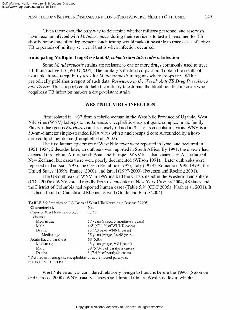
ASSOCIATIONS BETWEEN DISEASES AND LONG-TERM ADVERSE HEALTH OUTCOMES 149
Given those data, the only way to determine whether military personnel and reservists have become infected with M. tuberculosis during their service is to test all personnel for TB shortly before and after deployment. Such testing would make it possible to trace cases of active TB to periods of military service if that is when infection occurred.
Anticipating Multiple Drug-Resistant Mycobacterium tuberculosis Infection Some M. tuberculosis strains are resistant to one or more drugs commonly used to treat
LTBI and active TB (WHO 2004). The military’s medical corps should obtain the results of available drug-susceptibility tests for M. tuberculosis in regions where troops are. WHO periodically publishes a report of such data, Resistance in the World: Anti-TB Drug Prevalence and Trends. Those reports could help the military to estimate the likelihood that a person who acquires a TB infection harbors a drug-resistant strain.
WEST NILE VIRUS INFECTION
First isolated in 1937 from a febrile woman in the West Nile Province of Uganda, West Nile virus (WNV) belongs to the Japanese encephalitis virus antigenic complex in the family Flaviviridae (genus Flavivirus) and is closely related to St. Louis encephalitis virus. WNV is a 50-nm-diameter single-stranded RNA virus with a nucleocapsid core surrounded by a host-derived lipid membrane (Campbell et al. 2002).
The first human epidemics of West Nile fever were reported in Israel and occurred in 1951-1954; 2 decades later, an outbreak was reported in South Africa. By 1991, the disease had occurred throughout Africa, south Asia, and Europe. WNV has also occurred in Australia and New Zealand, but cases there were poorly documented (Wilson 1991). Later outbreaks were reported in Tunisia (1997), the Czech Republic (1997), Italy (1998), Romania (1996, 1999), the United States (1999), France (2000), and Israel (1997-2000) (Petersen and Roehrig 2001).
The US outbreak of WNV in 1999 marked the virus’s debut in the Western Hemisphere (CDC 2005e). WNV spread rapidly from its epicenter in New York City; by 2004, 48 states and the District of Columbia had reported human cases (Table 5.9) (CDC 2005a; Nash et al. 2001). It has been found in Canada and Mexico as well (Gould and Fikrig 2004).
TABLE 5.9 Statistics on US Cases of West Nile Neurologic Disease,a 2005
Characteristic No. Cases of West Nile neurologic
disease 1,165
Median age 57 years (range, 3 months-98 years) Male 665 (57.1 % of WNND cases) Deaths 85 (7.3 % of WNND cases)
Median age 75 years (range, 36-98 years) Acute flaccid paralysis 68 (5.8%)
Median age 53 years (range, 9-84 years) Male 39 (57.4% of paralysis cases) Deaths 5 (7.4 % of paralysis cases)
a Defined as meningitis, encephalitis, or acute flaccid paralysis. SOURCE:CDC 2005a.
West Nile virus was considered relatively benign to humans before the 1990s (Solomon and Cardosa 2000). WNV usually causes a self-limited illness, West Nile fever, which is
Copyright © National Academy of Sciences. All rights reserved.
Gulf War and Health: Volume 5. Infectious Diseaseshttp://www.nap.edu/catalog/11765.html
150 GULF WAR AND HEALTH
manifested as fever with a variety of other conditions, including rash, arthralgia, myalgia, headaches, and gastrointestinal symptoms.
Since the 1990s, however, there have been reports of increased incidence and severity of WNV illness (Solomon and Cardosa 2000). New neurologic and ophthalmologic manifestations of West Nile encephalitis have been recognized each year since the virus first reached North America in 1999 (Cunha 2004). A small but significant proportion of cases of West Nile neurologic disease (WNND) have led to death, particularly among the elderly. These recent, marked changes in the epidemiology of WNV illness led the committee to include it in this chapter even though the long-term adverse health outcomes of WNV usually are manifest during the acute illness.
Alarm triggered by the sudden change in the incidence and severity of WNV illness must be tempered by the understanding that severe WNV disease remains rare. Only 0.7% of people who become infected with West Nile virus in the United States develop severe neurologic disease, and more than one-third of these recover fully within a year (Klee et al. 2004; Mostashari et al. 2001). About 20% of infected people develop traditional, self-limited West Nile fever, and about 80% are asymptomatic, whereas only 1 in 150 develops neurologic manifestations.
Transmission of West Nile Virus Infection
Although WNV is found in several species of mosquitoes, the vast majority of infections are transmitted by Culex spp. (Campbell et al. 2002). These highly ornithophilic vectors transmit the virus among its natural reservoir: birds. Detected in more than 275 species of birds, WNV is particularly virulent for the family Corvidae, which includes crows and jays. The virus amplifies itself in birds’ bloodstream to a trillion or more virions per milliliter. Mammals are end-stage hosts and may develop disease but do not develop high enough viremia to contribute significantly to the virus’s epidemic spread.
WNV is transmissible from human to human through blood transfusions, transplanted organs, the placenta, and breastfeeding (CDC 2002). Between June and December 2003, WNV nucleic acid amplification testing (NAT) was performed on about 6 million units of blood, which resulted in the removal of at least 818 viremic blood donations. However, even with NAT testing, there were 6 cases of transfusion-associated WNV infection due to low levels of virus not detected by the testing method (minipools from 6-16 donations were used rather than individual testing) (CDC 2004c).
Endemicity in Southwest and South-Central Asia
WNV has been reported in Afghanistan, Pakistan, Iran, and other countries in southwest and south-central Asia (Arsen'eva 1982; Hubalek and Halouzka 1999; Naficy and Saidi 1970; Sugamata et al. 1988; Wilson 1991). In Afghanistan, antibodies to WNV were found in Kunduz, Heart, Bamyan, and Helmand provinces (Arsen'eva 1982). In neighboring Pakistan, 50-65% of the population of Karachi reportedly had antibodies to WNV in 1983 and 1985; new infections were identified in 13% of the population during those years (Sugamata et al. 1988). Similarly, a serum survey conducted in northeastern Iran in the late 1960s found that 30% of surveyed subjects had antibodies to WNV (Naficy and Saidi 1970).
Copyright © National Academy of Sciences. All rights reserved.
Gulf War and Health: Volume 5. Infectious Diseaseshttp://www.nap.edu/catalog/11765.html
ASSOCIATIONS BETWEEN DISEASES AND LONG-TERM ADVERSE HEALTH OUTCOMES 151
Acute West Nile Fever
Most persons infected with WNV are asymptomatic. A seroepidemiologic study of 677 people who lived in New York City during the 1999 outbreak found that 80% of seropositive subjects never developed symptoms (Mostashari et al. 2001). After an incubation period of 2-14 days, 20% of infected subjects developed a nonspecific febrile illness that lasted 3-6 days. Nausea, vomiting, myalgia, and headache are typical symptoms. A generalized maculopapular rash may occur in up to 20% of patients.
About 1 in 150 symptomatic patients in New York City developed WNND (Mostashari et al. 2001), which is often manifested as meningitis (WNM) or encephalitis (WNE) and sometimes as acute flaccid paralysis (AFP). Patients with WNND frequently have movement disorders with tremor, myoclonus, or Parkinsonism (Sejvar et al. 2003). Muscle weakness is also common. Investigators have reported paresis in about 50% of WNND cases and complete flaccid paralysis in 10%; the latter cases lack deep tendon reflexes and mimick GBS. Seizures and focal neurologic findings have been uncommon.
The development of WNND has been directly correlated with age. Of those over 65 years old, 1 in 50 developed WNM or WNE vs 1 in 300 of those under 65 (Mostashari et al. 2001). In fact, in those over 80 years old, the risk of symptomatic neurologic disease was 43 times higher than in those under 19.
West Nile fever without meningitis is more likely in younger patients. Among those with neurologic involvement, meningitis is more common in younger patients (mean age, 35 years), and encephalitis is more common in older patients (mean age, 70 years) (Sejvar et al. 2003).
Patients with West Nile fever who do not have neurologic manifestations might have residual fatigue, muscle weakness, and headache that can persist for months after resolution of the acute febrile illness (Watson et al. 2004). Of 98 patients with laboratory-confirmed West Nile infection but no clinical evidence of WNM, WNE, or AFP, 96% had fatigue for a median of 36 days, 61% had muscle weakness for a median of 28 days, and 71% had headache for a median of 10 days. The median time for recovery to a point that the patients considered “back to normal” was 60 days.
Diagnosis of West Nile Fever
For patients with acute symptomatic WNV infection, relative lymphocytopenia (less then 20%) is common. The cerebrospinal fluid (CSF) reveals a mild lymphocytic pleocytosis with a mean of 38 white cells/mm3 (range, 0-525) (Nash et al. 2001). Up to one-third may have more then 50% neutrophils on initial evaluation of the CSF. Increased protein with a mean of 104 mg/dL (range, 38-899) can be found. CSF glucose is usually normal.
Imaging studies of the brain usually are normal on computed tomography without evidence of inflammation. Even magnetic resonance imaging scan reveals enhancement of the meninges or periventricular areas in only about 30% of people (Nash et al. 2001). Electromyography reveals a motor axonal polyneuropathy with sparing of the sensory fibers very similar to the findings in poliomyelitis. WNV has a propensity to involve the anterior horn cells of the spinal cord in a manner very similar to poliomyelitis.
Acute infection is diagnosed by demonstration of WNV IgM in serum, which has been found in close to 100% of patients (Tardei et al. 2000). In one study, CSF samples from 94% of patients were WNV-IgM positive (Nash et al. 2001). No cases had virus isolation from CSF, and
Copyright © National Academy of Sciences. All rights reserved.
Gulf War and Health: Volume 5. Infectious Diseaseshttp://www.nap.edu/catalog/11765.html
152 GULF WAR AND HEALTH
only 57% of CSF samples and 14% of serum samples were positive with PCR. Patients with WNV may have persistent IgM antibodies for WNV. In a study by Roehrig et al. (2003), seven of 12 patients with serial samples had IgM persistently positive for WNV for 500 or more days.
Prior infection can be detected with measurement of WNV IgG. However, in a survey of 865 deployed front-line troops, 30 had both predeployment and postdeployment IgG antibodies against WNV. There was no evidence of acquisition of infection during deployment: there were no fourfold rises between predeployment and postdeployment samples, and no IgM antibodies were detected. Infection with dengue virus and prior yellow fever virus vaccine may result in detection of cross-reactive antibodies and make interpretation of serologic tests difficult. The above 30 persons’ serum samples were also reactive to St. Louis encephalitis, dengue, and yellow fever viruses. Because of the high cross-reactivity with St. Louis encephalitis virus, dengue and yellow fever viruses, confirmation of a positive WNV IgG requires testing with the plaque-reduction neutralization test, which requires a biosafety level 3 facility (Gea-Banacloche et al. 2004). Prior WNV can be confidently diagnosed if the WNV neutralizing-antibody titers are 4 times higher than all the other flavivirus titers.
Treatment of West Nile Virus Infection
There is no known effective treatment for WNV infection. Ribavirin, a guanosine analogue with broad-spectrum antiviral activity, has been shown to have activity against WNV in vitro (Jordan et al. 2000). Ribavirin also has concentrations in CSF that are 70% of those in serum. Ribavirin has been used successfully to treat related viruses including LaCrosse encephalitis, Hantaan, Lassa fever, and hepatitis C viruses (Jordan et al. 2000). However, in an outbreak in Israel, patients treated with ribavirin had a higher mortality than those who were not treated (Petersen and Roehrig 2001). The poor outcomes could have been due to patient selection, with sicker patients being treated with ribavirin, inasmuch as this was a nonrandomized study. If it is effective, the predicted required dose would be high, around 4 g intravenously every day, similar to that for treatment of Lassa fever. Interferon has also been noted to have in vitro activity against WNV and has been used in individual cases (Kalil et al. 2005).
A chimeric WNV vaccine with a type 4 dengue virus backbone (an attenuated deletion mutant) with an attached WNV envelop glycoprotein has been tested in Rhesus monkeys with development of high levels of neutralizing antibodies which protected them from infection (Platonov 2001). Clinical trials in humans are in progress.
Long-Term Adverse Health Outcomes of Infection with West Nile Virus
The scientific community is just beginning to unveil the long-term adverse health outcomes of WNV infection. Two teams of investigators have conducted long-term followup studies of the self-reported health outcomes of people who suffered acute episodes of W est Nile fever, WNM, WNE, or West Nile meningoencephalitis (Gottfried et al. 2005; Klee et al. 2004). All cases were diagnosed clinically and confirmed with laboratory analysis. A rigorous study by Klee and colleagues reports the health status at 6, 12, and 18 months of 42 New York City residents whose acute illnesses were manifested in 1999 and required hospitalization in all but two cases. A less rigorous study by Gottfried and colleagues reports the health status at 12 months of 24 Tennessee residents whose acute illnesses were diagnosed and reported to the state’s Department of Health in 2002; all but two of those cases had been hospitalized. About
Copyright © National Academy of Sciences. All rights reserved.
Gulf War and Health: Volume 5. Infectious Diseaseshttp://www.nap.edu/catalog/11765.html
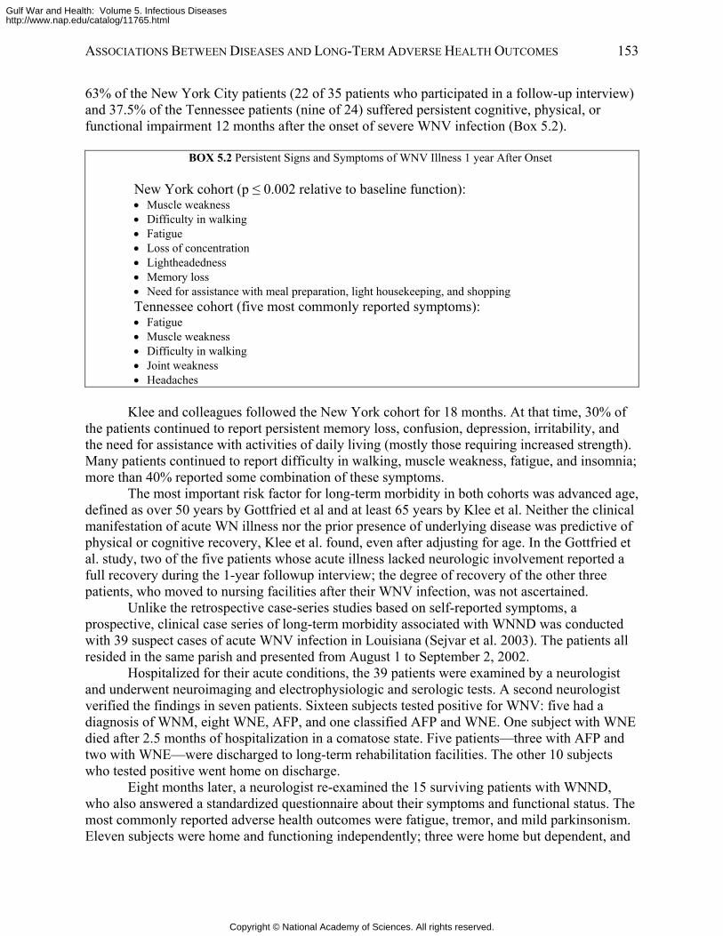
ASSOCIATIONS BETWEEN DISEASES AND LONG-TERM ADVERSE HEALTH OUTCOMES 153
63% of the New York City patients (22 of 35 patients who participated in a follow-up interview) and 37.5% of the Tennessee patients (nine of 24) suffered persistent cognitive, physical, or functional impairment 12 months after the onset of severe WNV infection (Box 5.2).
BOX 5.2 Persistent Signs and Symptoms of WNV Illness 1 year After Onset
New York cohort (p ≤ 0.002 relative to baseline function): • Muscle weakness • Difficulty in walking • Fatigue • Loss of concentration • Lightheadedness • Memory loss • Need for assistance with meal preparation, light housekeeping, and shopping Tennessee cohort (five most commonly reported symptoms): • Fatigue • Muscle weakness • Difficulty in walking • Joint weakness • Headaches
Klee and colleagues followed the New York cohort for 18 months. At that time, 30% of
the patients continued to report persistent memory loss, confusion, depression, irritability, and the need for assistance with activities of daily living (mostly those requiring increased strength). Many patients continued to report difficulty in walking, muscle weakness, fatigue, and insomnia; more than 40% reported some combination of these symptoms.
The most important risk factor for long-term morbidity in both cohorts was advanced age, defined as over 50 years by Gottfried et al and at least 65 years by Klee et al. Neither the clinical manifestation of acute WN illness nor the prior presence of underlying disease was predictive of physical or cognitive recovery, Klee et al. found, even after adjusting for age. In the Gottfried et al. study, two of the five patients whose acute illness lacked neurologic involvement reported a full recovery during the 1-year followup interview; the degree of recovery of the other three patients, who moved to nursing facilities after their WNV infection, was not ascertained.
Unlike the retrospective case-series studies based on self-reported symptoms, a prospective, clinical case series of long-term morbidity associated with WNND was conducted with 39 suspect cases of acute WNV infection in Louisiana (Sejvar et al. 2003). The patients all resided in the same parish and presented from August 1 to September 2, 2002.
Hospitalized for their acute conditions, the 39 patients were examined by a neurologist and underwent neuroimaging and electrophysiologic and serologic tests. A second neurologist verified the findings in seven patients. Sixteen subjects tested positive for WNV: five had a diagnosis of WNM, eight WNE, AFP, and one classified AFP and WNE. One subject with WNE died after 2.5 months of hospitalization in a comatose state. Five patients—three with AFP and two with WNE—were discharged to long-term rehabilitation facilities. The other 10 subjects who tested positive went home on discharge.
Eight months later, a neurologist re-examined the 15 surviving patients with WNND, who also answered a standardized questionnaire about their symptoms and functional status. The most commonly reported adverse health outcomes were fatigue, tremor, and mild parkinsonism. Eleven subjects were home and functioning independently; three were home but dependent, and
Copyright © National Academy of Sciences. All rights reserved.
Gulf War and Health: Volume 5. Infectious Diseaseshttp://www.nap.edu/catalog/11765.html
154 GULF WAR AND HEALTH
one was still undergoing rehabilitation. The five patients with WNM functioned at normal or nearly normal levels, according to the results of Barthel and modified Rankin scoring systems. Five patients with severe WNE also had recovered premorbid levels of functioning without residual disability; two WNE patients relied on walkers.
The three patients with AFP were faring poorly 8 months after onset. All continued to experience profound muscle weakness; they required wheelchairs and had difficulty in accomplishing such daily activities as grooming and housekeeping. Clinical findings and electrodiagnostic data on two of them suggested a poliomyelitis-like syndrome with involvement of anterior horn cells of the spinal cord. Electromyographic data suggested chronic denervation and permanent loss of motor axons in affected limbs. Long-Term Prognosis of West Nile Virus-Positive Patients with Focal Neurologic Deficits
A number of studies have been conducted to elucidate the outcomes of patients infected with West Nile virus who develop focal neurologic deficits, especially AFP. Saad and colleagues (2005) reviewed all cases of AFP related to WNV reported in the English-language literature from January 1999 to March 2004 whose clinical characteristics were described in sufficient detail (53 subjects, including the three described above); they added three cases of their own. Forty of the 56 subjects survived the acute phase of disease had a known long-term health outcome. All 40 suffered some degree of persistent neurologic impairment or weakness at the time of long-term followup. As a case in point, the authors noted a survivor who remained quadriplegic and ventilator-dependent after 20 months of followup.
In cases of WNV-induced focal neurologic deficits, the rate and degree of recovery of muscle strength appears to vary by limb and patient; the initial severity of paralysis may not predict the final outcome (Cao et al. 2005). Cao and colleagues reached those conclusions by measuring the muscle strength and overall motor function of 11 subjects for 6-21 months after the onset of AFP. A 36-year-old woman paralyzed in one leg recovered minimal strength during the 21-month period. In contrast, a 44-year-old man with severe four-limb paralysis who was hospitalized for respiratory distress started to walk within 1 month and recovered full strength in all limbs after 9 months (with decreased endurance). Between those extremes, a third patient became paralyzed to various degrees in four limbs and was partially recovered at 21 months. A small case-control study suggested a correlation between the estimated numbers of surviving motor units in a muscle and the degree of improvement of muscle strength (Cao et al. 2005). Pathologic Plausibility of Long-Term Neurologic Deficits in Patients with West Nile Neurologic Disease
Neurophysiologic, radiologic, and pathologic studies in humans and animals indicate that the underlying mechanism of WNV AFP is damage to the anterior horn cells of the spinal cord akin to the damage caused by poliomyelitis virus (Saad et al. 2005). That suggests that most patients with WNV AFP will not recover completely.
As Klee et al. (2004) noted, WNV infection is clinically similar to St. Louis encephalitis. Patients with the latter disease have reported disability up to 5 years after the acute illness. Persistent symptoms of St. Louis encephalitis have included fatigue, headache, nervousness, inability to concentrate, depression, and problems with gait and balance throughout the convalescent period of 6 months to 3 years.
The committee concludes that there is sufficient evidence of an association between acute West Nile virus infection and variable levels of physical, functional, or cognitive disability that may persist for months, years, or permanently.
Copyright © National Academy of Sciences. All rights reserved.
Gulf War and Health: Volume 5. Infectious Diseaseshttp://www.nap.edu/catalog/11765.html
ASSOCIATIONS BETWEEN DISEASES AND LONG-TERM ADVERSE HEALTH OUTCOMES 155
Recommendation
It has been just 10 years since the potential long-term adverse health outcomes of WNV infection became an international public-health concern. The body of evidence on which this committee can base its conclusions is small. Future investigators will be able to conduct more robust studies on the long-term adverse health outcomes of patients who suffer acute infection by WNV, and create a broader foundation for conclusions about the acute illness and its long-term adverse health outcomes. The committee recommends that the Department of Veterans Affairs (VA) periodically review the literature on long-term adverse health outcomes of WNV infection to supplement the conclusions of this committee.
REFERENCES
Abo-Shehada MN, Odeh JS, Abu-Essud M, Abuharfeil N. 1996. Seroprevalence of brucellosis
among high risk people in northern Jordan. International Journal of Epidemiology 25(2):450-454.
Air Force Office of the Surgeon General. 2003. AFMS Deployment Health Surveillance Implementation Instructions.
Akritidis N, Pappas G. 2001. Ascites caused by brucellosis: A report of two cases. Scandinavian Journal of Gastroenterology 36(1):110-112.
Al Dahouk S, Tomaso H, Nockler K, Neubauer H, Frangoulidis D. 2003. Laboratory-based diagnosis of brucellosis—a review of the literature. Part II: Serological tests for brucellosis. Clinica y Laboratorio 49(11-12):577-589.
al Deeb SM, Yaqub BA, Sharif HS, Phadke JG. 1989. Neurobrucellosis: Clinical characteristics, diagnosis, and outcome. Neurology 39(4):498-501.
al-Harthi SS. 1989. The morbidity and mortality pattern of Brucella endocarditis. International Journal of Cardiology 25(3):321-324.
al-Kaff AS. 1995. Ocular brucellosis. International Ophthalmology Clinics 35(3):139-145. Alarcon GS, Bocanegra TS, Gotuzzo E, Hinostroza S, Carrillo C, Vasey FB, Germain BF,
Espinoza LR. 1981. Reactive arthritis associated with brucellosis: HAL studies. Journal of Rheumatology 8(4):621-625.
Allos BM. 1997. Association between Campylobacter infection and Guillain-Barre syndrome. Journal of Infectious Diseases 176 (2 Suppl):S125-S128.
Allos BM. 2001. Campylobacter jejuni infections: Update on emerging issues and trends. Clinical Infectious Diseases 32(8):1201-1206.
Almuneef M, Memish ZA. 2003. Prevalence of Brucella antibodies after acute brucellosis. Journal of Chemotherapy 15(2):148-151.
Amino R, Menard R, Frischknecht F. 2005. In vivo imaging of malaria parasites—Recent advances and future directions. Current Opinion in Microbiology 8(4):407-414.
Araj GF, Lulu AR, Khateeb MI, Saadah MA, Shakir RA. 1988. ELISA versus routine tests in the diagnosis of patients with systemic and neurobrucellosis. APMIS: Acta Pathologica, Microbiologica et Immunologica Scandinavica 96(2):171-176.
Copyright © National Academy of Sciences. All rights reserved.
Gulf War and Health: Volume 5. Infectious Diseaseshttp://www.nap.edu/catalog/11765.html
156 GULF WAR AND HEALTH
Ardies CM. 2003. Inflammation as cause for scar cancers of the lung. Integrative Cancer Therapies 2(3):238-246.
Ariza J, Gudiol F, Valverde J, Pallares R, Fernandez-Viladrich P, Rufi G, Espadaler L, Fernandez-Nogues F. 1985. Brucellar spondylitis: A detailed analysis based on current findings. Reviews of Infectious Diseases 7(5):656-664.
Ariza J, Pellicer T, Pallares R, Foz A, Gudiol F. 1992. Specific antibody profile in human brucellosis. Clinical Infectious Diseases 14(1):131-140.
Ariza J, Pujol M, Valverde J, Nolla JM, Rufi G, Viladrich PF, Corredoira JM, Gudiol F. 1993. Brucellar sacroiliitis: Findings in 63 episodes and current relevance. Clinical Infectious Diseases 16(6):761-765.
Ariza J, Pigrau C, Canas C, Marron A, Martinez F, Almirante B, Corredoira JM, Casanova A, Fabregat J, Pahissa A. 2001. Current understanding and management of chronic hepatosplenic suppurative brucellosis. Clinical Infectious Diseases 32(7):1024-1033.
Aronson NE, Wortmann GW, Johnson SC, Jackson JE, Gasser RA Jr, Magill AJ, Endy TP, Coyne PE, Grogl M, Benson PM, Beard JS, Tally JD, Gambel JM, Kreutzer RD, Oster CN. 1998. Safety and efficacy of intravenous sodium stibogluconate in the treatment of leishmaniasis: Recent US military experience. Clinical Infectious Diseases 27(6):1457-1464.
Arsen'eva LP. 1982. Natural-focus zoonoses in Afghanistan: A review of the literature. Meditsinskaia Parazitologiia i Parazitarnye Bolezni 51(3):54-59.
Ayres JG, Flint N, Smith EG, Tunnicliffe WS, Fletcher TJ, Hammond K, Ward D, Marmion BP. 1998. Post-infection fatigue syndrome following Q fever. QJM 91(2):105-123.
Baird JK. 2005. Effectiveness of antimalarial drugs. New England Journal of Medicine 352(15):1565-1577.
Bajiya HN, Kochar DK. 1996. Incidence and outcome of neurological sequelae in survivors of cerebral malaria. Journal of the Association of Physicians of India 44(10):679-681.
Baldi PC, Miguel SE, Fossati CA, Wallach JC. 1996. Serological follow-up of human brucellosis by measuring IgG antibodies to lipopolysaccharide and cytoplasmic proteins of Brucella species. Clinical Infectious Diseases 22(3):446-455.
Balo KP, Mensah A, Mihluedo H. 1996. Chloroquine maculopathy and prevention of malaria. Journal Francais d Ophtalmologie 19(12):770-776.
Bashir R, Al-Kawi MZ, Harder EJ, Jinkins J. 1985. Nervous system brucellosis: Diagnosis and treatment. Neurology 35(11):1576-1581.
Basset D, Faraut F, Marty P, Dereure J, Rosenthal E, Mary C, Pratlong F, Lachaud L, Bastien P, Dedet JP. 2005. Visceral leishmaniasis in organ transplant recipients: 11 new cases and a review of the literature. Microbes and Infection 7(13):1370-1375.
Beare NA, Lewis DK, Kublin JG, Harding SP, Zijlstra EE, Molyneux ME. 2003. Retinal changes in adults with cerebral malaria. Annals of Tropical Medicine and Parasitology 97(3):313-315.
Begue JJ. 1964. Ocular complications observed during synthetic antimalarial treatment. Clinique 59:287-291.
Bennish ML. 1991. Potentially lethal complications of shigellosis. Reviews of Infectious Diseases 13 (4 Suppl):S319-S324.
Copyright © National Academy of Sciences. All rights reserved.
Gulf War and Health: Volume 5. Infectious Diseaseshttp://www.nap.edu/catalog/11765.html
ASSOCIATIONS BETWEEN DISEASES AND LONG-TERM ADVERSE HEALTH OUTCOMES 157
Bergin PS, Haas LF, Miller DH. 1989. Tuberculous meningitis at Wellington Hospital 1962-88. New Zealand Medical Journal 102(878):554-556.
Bernit E, Pouget J, Janbon F, Dutronc H, Martinez P, Brouqui P, Raoult D. 2002. Neurological involvement in acute Q fever: A report of 29 cases and review of the literature. Archives of Internal Medicine 162(6):693-700.
Bernstein HN. 1967. Chloroquine ocular toxicity. Survey of Ophthalmology 12(5):415-447. Berry A, Fabre R, Benoit-Vical F, Cassaing S, Magnaval JF. 2005. Contribution of PCR-based
methods to diagnosis and management of imported malaria. Medecine Tropicale 65(2): 176-183.
Bhattacharya SK, Sur D. 2003. An evaluation of current shigellosis treatment. Expert Opinion on Pharmacotherapy 4(8):1315-1320.
Biswas J, Fogla R, Srinivasan P, Narayan S, Haranath K, Badrinath V. 1996. Ocular malaria. A clinical and histopathologic study. Ophthalmology 103(9):1471-1475.
Blaser MJ. 2000. Campylobacter jejuni and related species. In: Mandell GL, Bennett JE, Dolin R, Editors. Principles and Practice of Infectious Diseases. 5th ed. New York: Churchill Livingstone. Pp. 2276-2285.
Blaser MJ. 2005. Infections due to Campylobacter and related species. In: Kasper DL, Braunwald E, Fauci A, Hauser SL, Longo DL, Jameson JL, Editors. Harrison's Principles of Internal Medicine. 16th ed. New York: McGraw-Hill. Pp. 907-909.
Blaser MJ, Perez GP, Smith PF, Patton C, Tenover FC, Lastovica AJ, Wang WI. 1986. Extraintestinal Campylobacter jejuni and Campylobacter coli infections: Host factors and strain characteristics. Journal of Infectious Diseases 153(3):552-559.
Bloom PD, MacPhail AP, Klugman K, Louw M, Raubenheimer C, Fischer C. 1994. Haemolytic-uraemic syndrome in adults with resistant Shigella dysenteriae type I. Lancet 344(8916):206.
Bodur H, Erbay A, Akinci E, Colpan A, Cevik MA, Balaban N. 2003. Neurobrucellosis in an endemic area of brucellosis. Scandinavian Journal of Infectious Diseases 35(2):94-97.
Bouza E, Garcia de la Torre M, Parras F, Guerrero A, Rodriguez-Creixems M, Gobernado J. 1987. Brucellar meningitis. Reviews of Infectious Diseases 9(4):810-822.
Bravo MJ, Colmenero Jde D, Alonso A, Caballero A. 2003. HLA-B*39 allele confers susceptibility to osteoarticular complications in human brucellosis. Journal of Rheumatology 30(5):1051-1053.
Breman JG. 2001. The ears of the hippopotamus: Manifestations, determinants, and estimates of the malaria burden. American Journal of Tropical Medicine and Hygiene 64(1-2 Suppl):1-11.
Bremell T, Bjelle A, Svedhem A. 1991. Rheumatic symptoms following an outbreak of campylobacter enteritis: A five year follow up. Annals of the Rheumatic Diseases 50(12):934-938.
Brouqui P, Dupont HT, Drancourt M, Berland Y, Etienne J, Leport C, Goldstein F, Massip P, Micoud M, Bertrand A, et al. 1993. Chronic Q fever. Ninety-two cases from France, including 27 cases without endocarditis. Archives of Internal Medicine 153(5):642-648.
Butcher GA. 2005. T-cell depletion and immunity to malaria in HIV-infections. Parasitology 130(Pt 2):141-150.
Copyright © National Academy of Sciences. All rights reserved.
Gulf War and Health: Volume 5. Infectious Diseaseshttp://www.nap.edu/catalog/11765.html
158 GULF WAR AND HEALTH
Buxton JA, Fyfe M, Berger S, Cox MB, Northcott KA. 2002. Reactive arthritis and other sequelae following sporadic Salmonella typhimurium infection in British Columbia, Canada: A case control study. Journal of Rheumatology 29(10):2154-2158.
Calin A, Fries JF. 1976. An “experimental” epidemic of Reiter's syndrome revisited. Follow-up evidence on genetic and environmental factors. Annals of Internal Medicine 84(5):564-566.
Camarca MM, Krauss MR. 2001. Active tuberculosis among US Army personnel, 1980 to 1996. Military Medicine: International Journal of AMSUS 166(5):452-456.
Campbell GL, Marfin AA, Lanciotti RS, Gubler DJ. 2002. West Nile virus. The Lancet Infectious Diseases 2(9):519-529.
Cao NJ, Ranganathan C, Kupsky WJ, Li J. 2005. Recovery and prognosticators of paralysis in West Nile virus infection. Journal of the Neurological Sciences 236(1-2):73-80.
CDC (Centers for Disease Control and Prevention). 1986. Annual summary 1984. Reported morbidity and mortality in the United States. Morbidity and Mortality Weekly Report 33(54):1-135.
CDC. 1992. 1993 revised classification system for HIV infection and expanded surveillance case definition for AIDS among adolescents and adults. Morbidity and Mortality Weekly Report 41(RR-17):1-19.
CDC. 2000a. Diagnostic Standards and Classification of Tuberculosis in Adults and Children. This official statement of the American Thoracic Society and the Centers for Disease Control and Prevention was adopted by the ATS Board of Directors, July 1999. This statement was endorsed by the Council of the Infectious Disease Society of America, September 1999. American Journal of Respiratory and Critical Care Medicine 161(4 Pt 1):1376-1395.
CDC. 2000b. Targeted tuberculin testing and treatment of latent tuberculosis infection. American Thoracic Society. Morbidity and Mortality Weekly Report: Recommendations and Reports 49(RR-6):1-51.
CDC. 2002. Update: Investigations of West Nile virus infections in recipients of organ transplantation and blood transfusion. Morbidity and Mortality Weekly Report 51(37):833-836.
CDC. 2003a. Treatment of tuberculosis. Morbidity and Mortality Weekly Report: Recommendations and Reports 52(RR-11):1-77.
CDC. 2003b. Cutaneous leishmaniasis in US military personnel—Southwest/Central Asia, 2002-2003. Morbidity and Mortality Weekly Report 52(42):1009-1012.
CDC. 2004a. Two cases of visceral leishmaniasis in US military personnel—Afghanistan, 2002-2004. Morbidity and Mortality Weekly Report 53 (12):265-268.
CDC. 2004b. Update: Cutaneous leishmaniasis in US military personnel—Southwest/Central Asia, 2002-2004. Morbidity and Mortality Weekly Report 53(12):264-265.
CDC. 2004c. Update: West Nile virus screening of blood donations and transfusion-associated transmission—United States, 2003. Morbidity and Mortality Weekly Report 53(13):281-284.
CDC. 2005a. Statistics, Surveillance, and Control, 2005 West Nile Virus Activity in the United States. [Online]. Available: http://www.cdc.gov/ncidod/dvbid/westnile/ surv&controlCaseCount05_detailed.htm [accessed April 26, 2006].
Copyright © National Academy of Sciences. All rights reserved.
Gulf War and Health: Volume 5. Infectious Diseaseshttp://www.nap.edu/catalog/11765.html
ASSOCIATIONS BETWEEN DISEASES AND LONG-TERM ADVERSE HEALTH OUTCOMES 159
CDC. 2005b. Typhoid Fever: Fequently Asked Questions. [Online]. Available: http://www.cdc.gov/ncidod/dbmd/diseaseinfo/files/typhoid_fever_FAQ.pdf [accessed April 26, 2006].
CDC. 2005c. Reported Tuberculosis in the United States, 2004. US Department of Health and Human Services.
CDC. 2005d. Guidelines for the investigation of contacts of persons with infectious tuberculosis. Recommendations from the National Tuberculosis Controllers Association and CDC. Morbidity and Mortality Weekly Report: Recommendations and Reports 54(RR-15):1-47.
CDC. 2005e. West Nile Virus Activity—United States, January 1—December 1, 2005. Morbidity and Mortality Weekly Report 54(49):1253-1256.
Center for Infectious Disease Research and Policy, University of Minnesota. 2006. Salmonellosis. [Online]. Available: http://www.cidrap.umn/edu/cidrap/content/fs/food-disease/causes/salmoview.html [accessed April 26, 2006].
Cetinkaya B, Kalender H, Ertas HB, Muz A, Arslan N, Ongor H, Gurcay M. 2000. Seroprevalence of coxiellosis in cattle, sheep and people in the east of Turkey. Veterinary Record 146(5):131-136.
Chakravarty A, Ghosh B, Bhattacharyya R, Sengupta S, Mukherjee S. 2004. Acute inflammatory demyelinating polyneuropathy following plasmodium vivax malaria. Neurology India 52(1):130-131.
Chen M, Delpech V, O'Sullivan B, Donovan B. 2002. Shigella sonnei: Another cause of sexually acquired reactive arthritis. International Journal of STD and AIDS 13(2):135-136.
Choi CM, Lerner EA. 2002. Leishmaniasis: Recognition and management with a focus on the immunocompromised patient. American Journal of Clinical Dermatology 3(2):91-105.
Choi HJ, Lee SY, Yang H, Bang JK. 2004. Retinal haemorrhage in vivax malaria. Transactions of the Royal Society of Tropical Medicine and Hygiene 98(6):387-389.
Chomel BB, DeBess EE, Mangiamele DM, Reilly KF, Farver TB, Sun RK, Barrett LR. 1994. Changing trends in the epidemiology of human brucellosis in California from 1973 to 1992: A shift toward foodborne transmission. Journal of Infectious Diseases 170(5):1216-1223.
Colmenero JD, Reguera JM, Martos F, Sanchez-De-Mora D, Delgado M, Causse M, Martin-Farfan A, Juarez C. 1996. Complications associated with Brucella melitensis infection: A study of 530 cases. Medicine 75(4):195-211.
Colmenero JD, Queipo-Ortuno MI, Maria Reguera J, Angel Suarez-Munoz M, Martin-Carballino S, Morata P. 2002. Chronic hepatosplenic abscesses in Brucellosis. Clinico-therapeutic features and molecular diagnostic approach. Diagnostic Microbiology and Infectious Disease 42(3):159-167.
Comstock GW, Edwards LB, Livesay VT. 1974a. Tuberculosis morbidity in the US Navy: Its distribution and decline. American Review of Respiratory Disease 110(5):572-580.
Comstock GW, Livesay VT, Woolpert SF. 1974b. The prognosis of a positive tuberculin reaction in childhood and adolescence. American Journal of Epidemiology 99(2):131-138.
Cope SE, Schultz GW, Richards AL, Savage HM, Smith GC, Mitchell CJ, Fryauff DJ, Conlon JM, Corneil JA, Hyams KC. 1996. Assessment of arthropod vectors of infectious diseases in areas of US troop deployment in the Persian Gulf. American Journal of Tropical Medicine and Hygiene 54(1):49-53.
Copyright © National Academy of Sciences. All rights reserved.
Gulf War and Health: Volume 5. Infectious Diseaseshttp://www.nap.edu/catalog/11765.html
160 GULF WAR AND HEALTH
Crosby E, Llosa L, Miro Quesada M, Carrillo C, Gotuzzo E. 1984. Hematologic changes in brucellosis. Journal of Infectious Diseases 150(3):419-424.
Cunha BA. 2004. Differential diagnosis of West Nile encephalitis. Current Opinion in Infectious Diseases 17(5):413-420.
Cunningham AC. 2002. Parasitic adaptive mechanisms in infection by leishmania. Experimental and Molecular Pathology 72(2):132-141.
Dalrymple-Champneys W. 1950. Undulant fever, a neglected problem. Lancet 1:429-777. Davies NE, Haverty JR, Boatwright M. 1969. Reiter's disease associated with shigellosis.
Southern Medical Journal 62(8):1011-1014. Davies PD. 2005. Risk Factors for Tuberculosis. Monaldi Archives of Chest Disease 63(1):37-
45. Department of the Army. 2003. Army Latent Tuberculosis Infection (LTBI) Surveillance and
Control Program. Office of the Surgeon General. Department of the Army. 2005. Army Regulation 40-5 Medical Services: Preventive Medicine.
[Online]. Available: http://www.usapa.army.mil/pdffiles/r40_5.pdf [accessed May 8, 2006]. Department of the Army. 2006. Personnel Policy Guidance (PPG) of Operations Iraqi Freedom
(OIF), Enduring Freedom (OEF) and Noble Eagle (ONE). [Online]. Available: http://www.armyg1.army.mil/MilitaryPersonnel/ppg.asp [accessed May 8, 2006].
Department of the Navy. 1993. Bumed Instruction 6224.8 Change Transmittal: Tuberculosis Control Program. [Online]. Available: http://www.nehc.med.navy.mil/nepmu2/pmttoolbox/ IMMUNIZATIONS1_files/Instructions%20&%20Notices/BUMEDINST%206224.8%20CH-1.pdf [accessed May 8, 2006].
Department of the Navy. 2001. BUMED, Tuberculosis Control Program - Clarification. [Online]. Available: http://www.nehc.med.navy.mil/nepmu2/pmttoolbox/ IMMUNIZATIONS1_files/Immunization%20Messages/TB%20clar%204-01%20241350Z.txt [accessed May 8, 2006].
Derrick E. 1937. Q Fever, a new fever entity: Clinical features, diagnosis and laboratory investigation. Medical Journal of Australia 2:281-299.
Desjeux P. 2004. Leishmaniasis: Current situation and new perspectives. Comparative Immunology, Microbiology and Infectious Diseases 27(5):305-318.
Dey AB, Trikha I, Banerjee M, Jain R, Nagarkar KM . 2001. Acute disseminated encephalomyelitis—another cause of post malaria cerebellar ataxia. Journal of the Association of Physicians of India 49:756-758.
Digre KB. 2003. Not so benign intracranial hypertension. British Medical Journal (Clinical Research Ed.) 326(7390):613-614.
Digre KB, Corbett JJ. 2001. Idiopathic intracranial hypertension (pseudotumor cerebri): A reappraisal. The Neurologist 7:2-68.
Dingle KE, Van Den Braak N, Colles FM, Price LJ, Woodward DL, Rodgers FG, Endtz HP, Van Belkum A, Maiden MC. 2001. Sequence typing confirms that Campylobacter jejuni strains associated with Guillain-Barre and Miller-Fisher syndromes are of diverse genetic lineage, serotype, and flagella type. Journal of Clinical Microbiology 39(9):3346-3349.
Doganay M, Aygen B, Inan M, Ozbakir O. 1993. Brucella peritonitis in a cirrhotic patient with ascites. European Journal of Medicine 2(7):441-442.
Copyright © National Academy of Sciences. All rights reserved.
Gulf War and Health: Volume 5. Infectious Diseaseshttp://www.nap.edu/catalog/11765.html
ASSOCIATIONS BETWEEN DISEASES AND LONG-TERM ADVERSE HEALTH OUTCOMES 161
Doyle TJ, Bryan RT. 2000. Infectious disease morbidity in the US region bordering Mexico, 1990-1998. Journal of Infectious Diseases 182(5):1503-1510.
Drancourt M, Raoult D, Xeridat B, Milandre L, Nesri M, Dano P. 1991. Q fever meningoencephalitis in five patients. European Journal of Epidemiology 7(2):134-138.
Dube MP, Holtom PD, Larsen RA. 1992. Tuberculous meningitis in patients with and without human immunodeficiency virus infection. American Journal of Medicine 93(5):520-524.
Dworkin MS, Shoemaker PC, Goldoft MJ, Kobayashi JM. 2001. Reactive arthritis and Reiter's syndrome following an outbreak of gastroenteritis caused by Salmonella enteritidis. Clinical Infectious Diseases 33(7):1010-1014.
Easterbrook M. 1999. Detection and prevention of maculopathy associated with antimalarial agents. International Ophthalmology Clinics 39(2):49-57.
Eastmond CJ, Rennie JA, Reid TM. 1983. An outbreak of Campylobacter enteritis—A rheumatological followup survey. Journal of Rheumatology 10(1):107-108.
Eiam-Ong S. 2003. Malarial nephropathy. Seminars in Nephrology 23(1):21-33. El Hassan AM, Ali MS, Zijlstra E, Eltoum IA, Ghalib HW, Ahmed HM. 1992. Post-kala-azar
dermal leishmaniasis in the Sudan: Peripheral neural involvement. International Journal of Dermatology 31(6):400-403.
Eliades MJ, Shah S, Nguyen-Dinh P, Newman RD, Barber AM, Nguyen-Dinh P, Roberts JM, Mali S, Parise ME, Barber AM, Steketee R. 2005. Malaria surveillance—United States, 2003. Morbidity and Mortality Weekly Report: Surveillance Summaries 54(2):25-40.
Enders U, Karch H, Toyka KV, Michels M, Zielasek J, Pette M, Heesemann J, Hartung HP. 1993. The spectrum of immune responses to Campylobacter jejuni and glycoconjugates in Guillain-Barre syndrome and in other neuroimmunological disorders. Annals of Neurology 34(2):136-144.
Falchook GS, Malone CM, Upton S, Shandera WX. 2003. Postmalaria neurological syndrome after treatment of Plasmodium falciparum malaria in the United States. Clinical Infectious Diseases 37(2):e22-e24.
Fasanaro AM, Scoleri G, Pizza V, Gaeta GB, Fasanaro A. 1991. Guillain-Barre syndrome as presenting manifestation of visceral leishmaniasis. Lancet 338(8775):1142.
FDA (Food and Drug Administration). 2005. PMA Final Decisions Rendered for December 2004. [Online]. Available: http://fda.gov/cdrh/pma/pmadec04.html [accessed May 8, 2006].
Fenollar F, Fournier PE, Carrieri MP, Habib G, Messana T, Raoult D. 2001. Risks factors and prevention of Q fever endocarditis. Clinical Infectious Diseases 33(3):312-316.
Fenollar F, Thuny F, Xeridat B, Lepidi H, Raoult D. 2006. Endocarditis after acute Q fever in patients with previously undiagnosed valvulopathies. Clinical Infectious Diseases 42(6): 818-821.
Ferrante MA, Dolan MJ. 1993. Q fever meningoencephalitis in a soldier returning from the Persian Gulf War. Clinical Infectious Diseases 16(4):489-496.
Finch M, Rodey G, Lawrence D, Blake P. 1986. Epidemic Reiter's syndrome following an outbreak of shigellosis. European Journal of Epidemiology 2(1):26-30.
Fosgate GT, Carpenter TE, Chomel BB, Case JT, DeBess EE, Reilly KF. 2002. Time-space clustering of human brucellosis, California, 1973-1992. Emerging Infectious Diseases 8(7):672-678.
Copyright © National Academy of Sciences. All rights reserved.
Gulf War and Health: Volume 5. Infectious Diseaseshttp://www.nap.edu/catalog/11765.html
162 GULF WAR AND HEALTH
Fournier PE, Casalta JP, Piquet P, Tournigand P, Branchereau A, Raoult D. 1998. Coxiella burnetii infection of aneurysms or vascular grafts: Report of seven cases and review. Clinical Infectious Diseases 26(1):116-121.
Fox KF, Fox A, Nagpal M, Steinberg P, Heroux K. 1998. Identification of Brucella by ribosomal-spacer-region PCR and differentiation of Brucella canis from other Brucella spp. pathogenic for humans by carbohydrate profiles. Journal of Clinical Microbiology 36(11):3217-3222.
Fryauff DJ, Modi GB, Mansour NS, Kreutzer RD, Soliman S, Youssef FG. 1993. Epidemiology of cutaneous leishmaniasis at a focus monitored by the multinational force and observers in the northeastern Sinai Desert of Egypt. American Journal of Tropical Medicine and Hygiene 49(5):598-607.
Gami AS, Antonios VS, Thompson RL, Chaliki HP, Ammash NM. 2004. Q fever endocarditis in the United States. Mayo Clinic Proceedings 79(2):253-257.
Gardner K, Cox T, Digre KB. 1995. Idiopathic intracranial hypertension associated with tetracycline use in fraternal twins: Case reports and review. Neurology 45(1):6-10.
Gea-Banacloche J, Johnson RT, Bagic A, Butman JA, Murray PR, Agrawal AG. 2004. West Nile virus: Pathogenesis and therapeutic options. Annals of Internal Medicine 140(7):545-553.
Geyik MF, Gur A, Nas K, Cevik R, Sarac J, Dikici B, Ayaz C. 2002. Musculoskeletal involvement of brucellosis in different age groups: A study of 195 cases. Swiss Medical Weekly 132(7-8):98-105.
Giles CL, Henderson JW. 1965. The ocular toxicity of chloroquine therapy. American Journal of the Medical Sciences 249:230-235.
Glynn, JR. Resurgence of tuberculosis and the impact of HIV infection. British Medical Bulletin 54(3): 579-593.
Gokul BN, Paul A, Hussein I. 2000. Neurobrucellosis. Saudi Medical Journal 21(6):577-580. Good AE. 1979. Shigellae and Reiter's syndrome. Annals of the Rheumatic Diseases 38 (1
Suppl):S119-S122. Goossens H, Marcelis L, Dekeyser P, Butzler JP. 1983. Brucella melitensis: Person-to-person
transmission? Lancet 1(8327):773. Gordon JD, MacKeen AD, Marrie TJ, Fraser DB. 1984. The radiographic features of epidemic
and sporadic Q fever pneumonia. Journal of the Canadian Association of Radiologists 35(3):293-296.
Gottesman G, Vanunu D, Maayan MC, Lang R, Uziel Y, Sagi H, Wolach B. 1996. Childhood brucellosis in Israel. Pediatric Infectious Disease Journal 15(7):610-615.
Gottfried K, Quinn R, Jones T. 2005. Clinical description and follow-up investigation of human West Nile virus cases. Southern Medical Journal 98(6):603-606.
Gotuzzo E, Alarcon GS, Bocanegra TS, Carrillo C, Guerra JC, Rolando I, Espinoza LR. 1982. Articular involvement in human brucellosis: A retrospective analysis of 304 cases. Seminars in Arthritis and Rheumatism 12(2):245-255.
Gotuzzo E, Carrillo C, Guerra J, Llosa L. 1986. An evaluation of diagnostic methods for brucellosis—the value of bone marrow culture. Journal of Infectious Diseases 153(1): 122-125.
Copyright © National Academy of Sciences. All rights reserved.
Gulf War and Health: Volume 5. Infectious Diseaseshttp://www.nap.edu/catalog/11765.html
ASSOCIATIONS BETWEEN DISEASES AND LONG-TERM ADVERSE HEALTH OUTCOMES 163
Gotuzzo E, Seas C, Guerra JG, Carrillo C, Bocanegra TS, Calvo A, Castaneda O, Alarcon GS. 1987. Brucellar arthritis: A study of 39 Peruvian families. Annals of the Rheumatic Diseases 46(7):506-509.
Gould LH, Fikrig E. 2004. West Nile virus: A growing concern? Journal of Clinical Investigation 113(8):1102-1107.
Gruenewald R, Ropper AH, Lior H, Chan J, Lee R, Molinaro VS. 1991. Serologic evidence of Campylobacter jejuni/coli enteritis in patients with Guillain-Barre syndrome. Archives of Neurology 48(10):1080-1082.
Guerrant RL, Walker DH, Weller PF. 1999. Tropical Infectious Diseases: Principles, Pathogens, and Practice. 1st ed. Philadelphia, PA: Churchill Livingstone.
Gungor K, Bekir NA, Namiduru M. 2002. Ocular complications associated with brucellosis in an endemic area. European Journal of Ophthalmology 12(3):232-237.
Gur A, Geyik MF, Dikici B, Nas K, Cevik R, Sarac J, Hosoglu S. 2003. Complications of brucellosis in different age groups: A study of 283 cases in southeastern Anatolia of Turkey. Yonsei Medical Journal 44(1):33-44.
Halpern Z, Dan M, Giladi M, Schwartz I, Sela O, Levo Y. 1989. Shigellosis in adults: Epidemiologic, clinical, and laboratory features. Medicine 68(4):210-217.
Hannu T, Leirisalo-Repo M. 1988. Clinical picture of reactive salmonella arthritis. Journal of Rheumatology 15(11):1668-1671.
Hannu T, Mattila L, Rautelin H, Pelkonen P, Lahdenne P, Siitonen A, Leirisalo-Repo M. 2002. Campylobacter-triggered reactive arthritis: A population-based study. Rheumatology 41(3):312-318.
Hannu T, Kauppi M, Tuomala M, Laaksonen I, Klemets P, Kuusi M. 2004a. Reactive arthritis following an outbreak of Campylobacter jejuni infection. Journal of Rheumatology 31(3):528-30.
Hannu T, Sihto-Kauppi K, Kotaniemi K, Kauppi M. 2004b. Acute anterior uveitis in association with an outbreak of Campylobacter jejuni infection. Scandinavian Journal of Rheumatology 33(1):55-57.
Hannu T, Mattila L, Siitonen A, Leirisalo-Repo M. 2005. Reactive arthritis attributable to Shigella infection: A clinical and epidemiological nationwide study. Annals of the Rheumatic Diseases 64(4):594-598.
Hariharan H, Naseema K, Kumaran C, Shanmugam J, Nair MD, Radhakrishnan K. 1996. Detection of Campylobacter jejuni/C.coli infection in patients with Guillain-Barre syndrome by serology and culture. New Microbiologica 19(3):267-271.
Harrington PT, Gutierrez JJ, Ramirez-Ronda CH, Quinones-Soto R, Bermudez RH, Chaffey J. 1982. Granulomatous hepatitis. Reviews of Infectious Diseases 4(3):638-655.
Hart PD, Sutherland I. 1977. BCG and vole bacillus vaccines in the prevention of tuberculosis in adolescence and early adult life. British Medical Journal 2(6082):293-295.
Hasanjani Roushan MR, Mohrez M, Smailnejad Gangi SM, Soleimani Amiri MJ, Hajiahmadi M. 2004. Epidemiological features and clinical manifestations in 469 adult patients with brucellosis in Babol, Northern Iran. Epidemiology and Infection 132(6):1109-1114.
Copyright © National Academy of Sciences. All rights reserved.
Gulf War and Health: Volume 5. Infectious Diseaseshttp://www.nap.edu/catalog/11765.html
164 GULF WAR AND HEALTH
Hatchette TF, Hayes M, Merry H, Schlech WF, Marrie TJ. 2003. The effect of C. burnetii infection on the quality of life of patients following an outbreak of Q fever. Epidemiology and Infection 130(3):491-495.
Hawker JI, Ayres JG, Blair I, Evans MR, Smith DL, Smith EG, Burge PS, Carpenter MJ, Caul EO, Coupland B, Desselberger U, Farrell ID, Saunders PJ, Wood MJ. 1998. A large outbreak of Q fever in the West Midlands: Windborne spread into a metropolitan area? Communicable Disease and Public Health 1(3):180-187.
Helbig KJ, Heatley SL, Harris RJ, Mullighan CG, Bardy PG, Marmion BP. 2003. Variation in immune response genes and chronic Q fever. Concepts: Preliminary test with post-Q fever fatigue syndrome. Genes and Immunity 4(1):82-85.
Helbig K, Harris R, Ayres J, Dunckley H, Lloyd A , Robson J, Marmion BP. 2005. Immune response genes in the post-Q-fever fatigue syndrome, Q fever endocarditis and uncomplicated acute primary Q fever. QJM 98(8):565-574.
Hertig M, Sabin AB. 1964. Sandfly fever (pappataci, Phlebotomus, three-day fever). In: Hoff EC, Editor. Preventive Medicine in World War II: Communicable Diseases. Volume 7. Office of the Surgeon General, US Department of the Army. Pp. 109-174.
Hewitt S, Reyburn H, Ashford R, Rowland M. 1998. Anthroponotic cutaneous leishmaniasis in Kabul, Afghanistan: Vertical distribution of cases in apartment blocks. Transactions of the Royal Society of Tropical Medicine and Hygiene 92(3):273-274.
Heymann DL. 2004. Control of Communicable Diseases Manual. Washington, DC: American Public Health Association.
Hidayat AA, Nalbandian RM, Sammons DW, Fleischman JA, Johnson TE. 1993. The diagnostic histopathologic features of ocular malaria. Ophthalmology 100(8):1183-1186.
Hosoglu S, Ayaz C, Geyik MF, Kokoglu OF, Ceviz A. 1998. Tuberculous meningitis in adults: An eleven-year review. International Journal of Tuberculosis and Lung Disease 2(7):553-557.
Howard RS, Sarkies NJ, Sanders MD. 1987. Anterior uveitis associated with Campylobacter jejuni infection. Journal of Infection 14(2):186-187.
Hubalek Z, Halouzka J. 1999. West Nile fever—A reemerging mosquito-borne viral disease in Europe. Emerging Infectious Diseases 5(5):643-650.
Hyams KC, Hanson K, Wignall FS, Escamilla J, Oldfield EC, 3rd. 1995. The impact of infectious diseases on the health of US troops deployed to the Persian Gulf during operations Desert Shield and Desert Storm. Clinical Infectious Diseases 20(6):1497-1504.
Ibero I, Vela P, Pascual E. 1997. Arthritis of shoulder and spinal cord compression due to Brucella disc infection. British Journal of Rheumatology 36(3):377-381.
Ibrahim AI, Awad R, Shetty SD, Saad M, Bilal NE. 1988. Genito-urinary complications of brucellosis. British Journal of Urology 61(4):294-298.
Idro R, Jenkins NE, Newton CR. 2005. Pathogenesis, clinical features, and neurological outcome of cerebral malaria. Lancet Neurology 4(12):827-840.
Ilnyckyj A, Balachandra B, Elliott L, Choudhri S, Duerksen DR. 2003. Post-traveler's diarrhea irritable bowel syndrome: A prospective study. American Journal of Gastroenterology 98(3):596-599.
Copyright © National Academy of Sciences. All rights reserved.
Gulf War and Health: Volume 5. Infectious Diseaseshttp://www.nap.edu/catalog/11765.html
ASSOCIATIONS BETWEEN DISEASES AND LONG-TERM ADVERSE HEALTH OUTCOMES 165
Imboden JB, Canter A, Cluff LE, Trever RW. 1959. Brucellosis. III. Psychologic aspects of delayed convalescence. AMA Archives of Internal Medicine 103(3):406-414.
IOM (Institute of Medicine). 2000. Ending Neglect: The Elimination of Tuberculosis in the United States. Washington, DC: National Academy Press.
Islam MZ, Itoh M, Shamsuzzaman SM, Mirza R, Matin F, Ahmed I, Shamsuzzaman Choudhury AK, Hossain MA, Qiu XG, Begam N, Furuya M, Leafasia JL, Hashiguchi Y, Kimura E. 2002. Diagnosis of visceral leishmaniasis by enzyme-linked immunosorbent assay using urine samples. Clinical and Diagnostic Laboratory Immunology 9(4):789-794.
Iuchi K, Ichimiya A, Akashi A, Mizuta T, Lee YE, Tada H, Mori T, Sawamura K, Lee YS, Furuse K, et al. 1987. Non-Hodgkin’s lymphoma of the pleural cavity developing from long-standing pyothorax. Cancer 60(8):1771-1775.
Jordan I, Briese T, Fischer N, Lau JY, Lipkin WI. 2000. Ribavirin inhibits West Nile virus replication and cytopathic effect in neural cells. Journal of Infectious Diseases 182(4):1214-1217.
Kalil AC, Devetten MP, Singh S, Lesiak B, Poage DP, Bargenquast K, Fayad P, Freifeld AG. 2005. Use of interferon-alpha in patients with West Nile encephalitis: Report of 2 cases. Clinical Infectious Diseases 40(5):764-766.
Kalita J, Misra UK. 1999. Outcome of tuberculous meningitis at 6 and 12 months: A multiple regression analysis. International Journal of Tuberculosis and Lung Disease 3(3):261-265.
Kanjalkar M, Karnad DR, Narayana RV, Shah PU. 1999. Guillain-Barre syndrome following malaria. Journal of Infection 38(1):48-50.
Karmali MA, Fleming PC. 1979. Campylobacter enteritis in children. Journal of Pediatrics 94(4):527-533.
Kaslow RA RRCA. 1979. Search for Reiter’s syndrome after an outbreak of Shigella sonnei dysentery. Journal of Rheumatology 6:562-566.
Kelley PW. 2005. Military Preventive Medicine: Mobilization and Deployment. Volume 2. Washington, DC: Walter Reed Army Medical Center.
Kennedy DH, Fallon RJ. 1979. Tuberculous meningitis. Journal of the American Medical Association 241(3):264-268.
Khan MY, Mah MW, Memish ZA. 2001. Brucellosis in pregnant women. Clinical Infectious Diseases 32(8):1172-1177.
Khandpur S, Ramam M, Sharma VK, Salotra P, Singh MK, Malhotra A. 2004. Nerve involvement in Indian post kala-azar dermal leishmaniasis. Acta Dermato-Venereologica 84:245-246.
Khateeb MI, Araj GF, Majeed SA, Lulu AR. 1990. Brucella arthritis: A study of 96 cases in Kuwait. Annals of the Rheumatic Diseases 49(12):994-998.
Kibukamusoke JW. 1986. The hazard of malarial nephropathy. Parasitology Today 2(4): 119-121.
Kilpatrick ME. 2005. Presentation to IOM Committee on Gulf War and Health: Infectious Diseases. Washington, DC.
Kim AY, Goldberg MB, Rubin RH. 2004. Salmonella infections. In: Gorbach SL, Bartlett JG, Blacklow NR, Editors. Infectious Diseases. 3rd ed. Philadelphia, PA: Lippincott Williams and Wilkins. Pp. 618-633.
Copyright © National Academy of Sciences. All rights reserved.
Gulf War and Health: Volume 5. Infectious Diseaseshttp://www.nap.edu/catalog/11765.html
166 GULF WAR AND HEALTH
Klee AL, Maidin B, Edwin B, Poshni I, Mostashari F, Fine A, Layton M, Nash D. 2004. Long-term prognosis for clinical West Nile virus infection. Emerging Infectious Diseases 10(8):1405-1411.
Kochar DK, Agarwal N, Jain N, Sharma BV, Rastogi A, Meena CB. 2000a. Clinical profile of neurobrucellosis—a report on 12 cases from Bikaner (north-west India). Journal of the Association of Physicians of India 48(4):376-380.
Kochar DK, Shubhakaran, Kumawat BL, Vyas SP. 2000b. Prognostic significance of eye changes in cerebral malaria. Journal of the Association of Physicians of India 48(5):473-477.
Kolaczinski J, Graham K, Fahim A, Brooker S, Rowland M. 2005. Malaria control in Afghanistan: Progress and challenges. Lancet 365(9469):1506-1512.
Kortepeter MG, Krauss MR. 2001. Tuberculosis infection after humanitarian assistance, Guantanamo Bay, 1995. Military Medicine 166(2):116-120.
Koster F, Levin J, Walker L, Tung KS, Gilman RH, Rahaman MM, Majid MA, Islam S, Williams RC Jr. 1978. Hemolytic uremic syndrome after shigellosis. Relation to endotoxemia and circulating immune complexes. New England Journal of Medicine 298(17):927-933.
Kurcer MA, Simsek Z, Kurcer Z. 2006. The decreasing efficacy of chloroquine in the treatment of Plasmodium vivax malaria, in Sanliurfa, south-eastern Turkey. Annals of Tropical Medicine and Parasitology 100(2):109-113.
Kuroki S, Saida T, Nukina M, Haruta T, Yoshioka M, Kobayashi Y, Nakanishi H. 1993. Campylobacter jejuni strains from patients with Guillain-Barre syndrome belong mostly to Penner serogroup 19 and contain beta-N-acetylglucosamine residues. Annals of Neurology 33(3):243-247.
Kuwabara S, Ogawara K, Misawa S, Koga M, Mori M, Hiraga A, Kanesaka T, Hattori T, Yuki N. 2004. Does Campylobacter jejuni infection elicit “demyelinating” Guillain-Barre syndrome? Neurology 63(3):529-533.
Lamar J. 2006. Navy Environmental Health Center. Personal communication. Lau KK, Yu IT, Chan AC, Wong LK, Tam CM, Sheng B, Li HL. 2005. A registry of
tuberculous meningitis in Hong Kong. International Journal of Tuberculosis and Lung Disease 9(12):1391-1397.
Lauhio A, Lahdevirta J, Janes R, Kontiainen S, Repo H. 1988. Reactive arthritis associated with Shigella sonnei infection. Arthritis and Rheumatism 31(9):1190-1193.
Lee AT, Hall RG, Pile KD. 2005. Reactive joint symptoms following an outbreak of Salmonella typhimurium phage type 135a. Journal of Rheumatology 32(3):524-527.
Leirisalo-Repo M, Helenius P, Hannu T, Lehtinen A, Kreula J, Taavitsainen M, Koskimies S. 1997. Long-term prognosis of reactive salmonella arthritis. Annals of the Rheumatic Diseases 56(9):516-520.
Lever AM, Dolby JM, Webster AD, Price AB. 1984. Chronic campylobacter colitis and uveitis in patient with hypogammaglobulinaemia. British Medical Journal (Clinical Research Ed.) 288(6416):531.
Lewallen S. 1998. The fundus in severe malaria. Archives of Ophthalmology 116(4):542-543. Lewis RB. 1982. The absence of reactive arthritis after Shigella sonnei infection. Arthritis and
Rheumatism 25(10):1267.
Copyright © National Academy of Sciences. All rights reserved.
Gulf War and Health: Volume 5. Infectious Diseaseshttp://www.nap.edu/catalog/11765.html
ASSOCIATIONS BETWEEN DISEASES AND LONG-TERM ADVERSE HEALTH OUTCOMES 167
Liu GF, Wu ZL, Wu HS, Wang QY, Zhao-Ri GT, Wang CY, Liang ZX, Cui SL, Zheng JD. 2003. A case-control study on children with Guillain-Barre syndrome in North China. Biomedical and Environmental Sciences 16(2):105-111.
Lochhead J, Elston JS. 2003. Doxycycline induced intracranial hypertension. British Medical Journal (Clinical Research Ed.) 326(7390):641-642.
Locht H, Kihlstrom E, Lindstrom FD. 1993. Reactive arthritis after Salmonella among medical doctors—Study of an outbreak. Journal of Rheumatology 20(5):845-848.
Locht H, Molbak K, Krogfelt KA. 2002. High frequency of reactive joint symptoms after an outbreak of Salmonella enteritidis. Journal of Rheumatology 29(4):767-771.
Long R, Maycher B, Dhar A, Manfreda J, Hershfield E, Anthonisen N. 1998. Pulmonary tuberculosis treated with directly observed therapy: Serial changes in lung structure and function. Chest 113(4):933-943.
Lozier JR, Friedlaender MH. 1989. Complications of antimalarial therapy. International Ophthalmology Clinics 29(3):172-178.
Lubani MM, Dudin KI, Araj GF, Manandhar DS, Rashid FY. 1989a. Neurobrucellosis in children. Pediatric Infectious Disease Journal 8(2):79-82.
Lubani MM, Dudin KI, Sharda DC, Ndhar DS, Araj GF, Hafez HA, al-Saleh QA, Helin I, Salhi MM. 1989b. A multicenter therapeutic study of 1100 children with brucellosis. Pediatric Infectious Disease Journal 8(2):75-78.
Luke T. 2006. Department of the Navy Bureau of Medicine and Surgery. Personal communication.
Lulu AR, Araj GF, Khateeb MI, Mustafa MY, Yusuf AR, Fenech FF. 1988. Human brucellosis in Kuwait: A prospective study of 400 cases. Quarterly Journal of Medicine 66(249):39-54.
Lyall M. 1973. Ocular brucellosis. Transactions of the Ophthalmological Societies of the United Kingdom 93(0):689-697.
Madariaga MG, Pulvirenti J, Sekosan M, Paddock CD, Zaki SR. 2004. Q fever endocarditis in HIV-infected patient. Emerging Infectious Diseases 10(3):501-504.
Magill AJ, Grogl M, Gasser RA Jr, Sun W, Oster CN. 1993. Visceral infection caused by Leishmania tropica in veterans of Operation Desert Storm. New England Journal of Medicine 328(19):1383-1387.
Magill AJ, Grogl M, Johnson SC, Gasser RA Jr. 1994. Visceral infection due to Leishmania tropica in a veteran of Operation Desert Storm who presented 2 years after leaving Saudi Arabia. Clinical Infectious Diseases 19(4):805-806.
Makhseed M, Harouny A, Araj G, Moussa MA, Sharma P. 1998. Obstetric and gynecologic implication of brucellosis in Kuwait. Journal of Perinatology 18(3):196-199.
Maki-Ikola O, Granfors K. 1992. Salmonella-triggered reactive arthritis. Scandinavian Journal of Rheumatology 21(6):265-270.
Maki-Ikola O, Leirisalo-Repo M, Kantele A, Toivanen P, Granfors K. 1991. Salmonella-specific antibodies in reactive arthritis. Journal of Infectious Diseases 164(6):1141-1148.
Maki-Ikola O, Yli-Kerttula U, Saario R, Toivanen P, Granfors K. 1992. Salmonella specific antibodies in serum and synovial fluid in patients with reactive arthritis. British Journal of Rheumatology 31(1):25-29.
Copyright © National Academy of Sciences. All rights reserved.
Gulf War and Health: Volume 5. Infectious Diseaseshttp://www.nap.edu/catalog/11765.html
168 GULF WAR AND HEALTH
Malviya G, Sinha MK, Bhattacharya SK. 2005. Post-malarial neuropsychiatric syndrome. Journal of the Association of Physicians of India 53:227.
Mandell GL, Bennett JE, Dolin R. 2005. Principles and Practice of Infectious Diseases. 6th ed. Philadelphia, PA: Elsevier Churchill Livingstone
Marmion BP, Shannon M, Maddocks I, Storm P, Penttila I. 1996. Protracted debility and fatigue after acute Q fever. Lancet 347(9006):977-978.
Marmion BP, Storm PA, Ayres JG, Semendric L, Mathews L, Winslow W, Turra M, Harris RJ. 2005. Long-term persistence of Coxiella burnetii after acute primary Q fever. QJM 98(1): 7-20.
Marrie TJ. 1990. Q Fever: The Disease. Volume 1. Boca Raton, FL: CRC Press. Marrie TJ. 2000. Coxiella burnetti (Q Fever). In: Mandell GL, Bennett JE, Dolin R, Editors.
Principles and Practice of Infectious Diseases. 5th ed. New York: Churchill Livingstone. Pp. 2043-2049.
Martin WJ, Nichols DR, Beahrs OH. 1961. Chronic localized brucellosis. Archives of Internal Medicine 107:43.
Mattila L, Leirisalo-Repo M, Koskimies S, Granfors K, Siitonen A. 1994. Reactive arthritis following an outbreak of Salmonella infection in Finland. British Journal of Rheumatology 33(12):1136-1141.
Mattila L, Leirisalo-Repo M, Pelkonen P, Koskimies S, Granfors K, Siitonen A. 1998. Reactive arthritis following an outbreak of Salmonella Bovismorbificans infection. Journal of Infection 36(3):289-295.
Maurin M, Raoult D. 1999. Q fever. Clinical Microbiology Reviews 12(4):518-553. McCarthy N, Giesecke J. 2001. Incidence of Guillain-Barre syndrome following infection with
Campylobacter jejuni. American Journal of Epidemiology 153(6):610-614. McLean DR, Russell N, Khan MY. 1992. Neurobrucellosis: Clinical and therapeutic features.
Clinical Infectious Diseases 15(4):582-590. McQuiston JH, Childs JE. 2002. Q fever in humans and animals in the United States. Vector
Borne Zoonotic Disease 2(3):179-191. MedlinePlus Medical Encyclopedia. 2006. MedlinePlus Medical Encyclopedia: Uveitis.
[Online]. Available: http://www.nlm.nih.gov/medlineplus/print/ency/article/001005.htm [accessed May 1, 2006].
Meier CR, Wilcock K, Jick SS. 2004. The risk of severe depression, psychosis or panic attacks with prophylactic antimalarials. Drug Safety 27(3):203-213.
Memish ZA, Venkatesh S. 2001. Brucellar epididymo-orchitis in Saudi Arabia: A retrospective study of 26 cases and review of the literature. BJU International 88(1):72-76.
Metha SR, Joshi V, Lazar AI. 1996. Unusual acute and chronic complications of malaria. Journal of the Association of Physicians of India 44(7):451-453.
Milazzo A, Hall R, Storm PA, Harris RJ, Winslow W, Marmion BP. 2001. Sexually transmitted Q fever. Clinical Infectious Diseases 33(3):399-402.
Mishu B, Ilyas AA, Koski CL, Vriesendorp F, Cook SD, Mithen FA, Blaser MJ. 1993. Serologic evidence of previous Campylobacter jejuni infection in patients with the Guillain-Barre syndrome. Annals of Internal Medicine 118(12):947-953.
Copyright © National Academy of Sciences. All rights reserved.
Gulf War and Health: Volume 5. Infectious Diseaseshttp://www.nap.edu/catalog/11765.html
ASSOCIATIONS BETWEEN DISEASES AND LONG-TERM ADVERSE HEALTH OUTCOMES 169
Montes M, Cilla G, Marimon JM, Diaz de Tuesta JL, Perez-Trallero E. 1995. Coxiella burnetii infection in subjects with HIV infection and HIV infection in patients with Q fever. Scandinavian Journal of Infectious Diseases 27(4):344-346.
Morata P, Queipo-Ortuno MI, Reguera JM, Miralles F, Lopez-Gonzalez JJ, Colmenero JD. 2001. Diagnostic yield of a PCR assay in focal complications of brucellosis. Journal of Clinical Microbiology 39(10):3743-3746.
Mostashari F, Bunning ML, Kitsutani PT, Singer DA, Nash D, Cooper MJ, Katz N, Liljebjelke KA, Biggerstaff BJ, Fine AD, Layton MC, Mullin SM, Johnson AJ, Martin DA, Hayes EB, Campbell GL. 2001. Epidemic West Nile encephalitis, New York, 1999: Results of a household-based seroepidemiological survey. Lancet 358(9278):261-264.
Mousa AR, Koshy TS, Araj GF, Marafie AA, Muhtaseb SA, Al-Mudallal DS, Busharetulla MS. 1986. Brucella meningitis: Presentation, diagnosis and treatment—a prospective study of ten cases. Quarterly Journal of Medicine 60(233):873-885.
Mousa AR, Muhtaseb SA, Almudallal DS, Khodeir SM, Marafie AA. 1987. Osteoarticular complications of brucellosis: A study of 169 cases. Reviews of Infectious Diseases 9(3):531-543.
Murray HW. 2000. Treatment of visceral leishmaniasis (kala-azar): A decade of progress and future approaches. International Journal of Infectious Diseases 4(3):158-177.
Murray HW. 2004. Treatment of visceral leishmaniasis in 2004. American Journal of Tropical Medicine and Hygiene 71(6):787-794.
Murray HW, Berman JD, Davies CR, Saravia NG. 2005. Advances in leishmaniasis. Lancet 366(9496):1561-1577.
Nachamkin I. 2002. Chronic effects of Campylobacter infection. Microbes and Infection 4(4):399-403.
Nachamkin I, Allos BM, Ho T. 1998. Campylobacter species and Guillain-Barre syndrome. Clinical Microbiology Reviews 11(3):555-567.
Nachamkin I, Engberg J, Gutacker M, Meinersman RJ, Li CY, Arzate P, Teeple E, Fussing V, Ho TW, Asbury AK, Griffin JW, McKhann GM, Piffaretti JC. 2001. Molecular population genetic analysis of Campylobacter jejuni HS: 19 associated with Guillain-Barre syndrome and gastroenteritis. Journal of Infectious Diseases 184(2):221-226.
Naficy K, Saidi S. 1970. Serological survey on viral antibodies in Iran. Tropical and Geographical Medicine 22(2):183-188.
Namiduru M, Karaoglan I, Gursoy S, Bayazit N, Sirikci A. 2004. Brucellosis of the spine: Evaluation of the clinical, laboratory, and radiological findings of 14 patients. Rheumatology International 24(3):125-129.
Nash D, Mostashari F, Fine A, Miller J, O'Leary D, Murray K, Huang A, Rosenberg A, Greenberg A, Sherman M, Wong S, Layton M. 2001. The outbreak of West Nile virus infection in the New York City area in 1999. New England Journal of Medicine 344(24):1807-1814.
Nathoo KJ, Sanders JA, Siziya S, Mucheche C. 1995. Haemolytic uraemic syndrome following Shigella dysenteriae type 1 outbreak in Zimbabwe: A clinical experience. Central African Journal of Medicine 41(9):267-274.
Copyright © National Academy of Sciences. All rights reserved.
Gulf War and Health: Volume 5. Infectious Diseaseshttp://www.nap.edu/catalog/11765.html
170 GULF WAR AND HEALTH
Navarro-Martinez A, Solera J, Corredoira J, Beato JL, Martinez-Alfaro E, Atienzar M, Ariza J. 2001. Epididymoorchitis due to Brucella mellitensis: A retrospective study of 59 patients. Clinical Infectious Diseases 33(12):2017-2022.
Navy Environmental Health Center. 2006. Preventive Medicine - Tuberculosis. [Online]. Available: http://www-nehc.med.navy.mil/prevmed/PM/PM_Tuberculosis.htm [accessed May 8, 2006].
Neithercut WD, Hudson MA, Smith CC. 1984. Can erythema nodosum and reactive arthritis be a sequel to Shigella flexneri gastroenteritis? Scottish Medical Journal 29(3):197-199.
Nene A, Bhojraj S. 2005. Results of nonsurgical treatment of thoracic spinal tuberculosis in adults. The Spine Journal 5(1):79-84.
Newman RD, Parise ME, Barber AM, Steketee RW. 2004. Malaria-related deaths among US travelers, 1963-2001. Annals of Internal Medicine 141(7):547-555.
Niemeyer G, Fruh B. 1989. Examination strategies in the diagnosis of drug-induced retinal damage. Klinische Monatsblatter Fur Augenheilkunde 194(5):355-358.
NIH (National Institutes of Health). 2002. Questions and Answers About Reactive Arthritis. National Institute of Arthritis and Musckuloskeletal Diseases (NIAMS), Public Health Service, US Department of Health and Human Services.
Nikkari S, Rantakokko K, Ekman P, Mottonen T, Leirisalo-Repo M, Virtala M, Lehtonen L, Jalava J, Kotilainen P, Granfors K, Toivanen P. 1999. Salmonella-triggered reactive arthritis: Use of polymerase chain reaction, immunocytochemical staining, and gas chromatography-mass spectrometry in the detection of bacterial components from synovial fluid. Arthritis and Rheumatism 42(1):84-89.
Noer HR. 1966. An “experimental” epidemic of Reiter’s syndrome. Journal of the American Medical Association 198(7):693-698.
Norton WL. 1984. Brucellosis and rheumatic syndromes in Saudi Arabia. Annals of the Rheumatic Diseases 43(6):810-815.
Nourse C, Allworth A, Jones A, Horvath R, McCormack J, Bartlett J, Hayes D, Robson JM. 2004. Three cases of Q fever osteomyelitis in children and a review of the literature. Clinical Infectious Diseases 39(7):e61-e66.
Oh MD, Shin H, Shin D, Kim U, Lee S, Kim N, Choi MH, Chai JY, Choe K. 2001. Clinical features of vivax malaria. American Journal of Tropical Medicine and Hygiene 65(2):143-146.
Ohl CA, Hyams KC, Malone JD, Oldfield EC 3rd. 1993. Leishmaniasis among Desert Storm veterans: A diagnostic and therapeutic dilemma. Military Medicine 158(11):726-729.
Okhuysen PC, Jiang ZD, Carlin L, Forbes C, DuPont HL. 2004. Post-diarrhea chronic intestinal symptoms and irritable bowel syndrome in North American travelers to Mexico. American Journal of Gastroenterology 99(9):1774-1778.
Oldfield EC 3rd, Wallace MR, Hyams KC, Yousif AA, Lewis DE, Bourgeois AL. 1991. Endemic infectious diseases of the Middle East. Reviews of Infectious Diseases 13(3 Suppl):S199-S217.
Oliveri R, Matera G, Foca A, Zappia M, Aguglia U, Quattrone A. 1996. Polyradiculoneuropathy with cerebrospinal fluid albuminocytological dissociation due to neurobrucellosis. Clinical Infectious Diseases 23(4):833-834.
Copyright © National Academy of Sciences. All rights reserved.
Gulf War and Health: Volume 5. Infectious Diseaseshttp://www.nap.edu/catalog/11765.html
ASSOCIATIONS BETWEEN DISEASES AND LONG-TERM ADVERSE HEALTH OUTCOMES 171
Olsen SJ, Bleasdale SC, Magnano AR, Landrigan C, Holland BH, Tauxe RV, Mintz ED, Luby S. 2003. Outbreaks of typhoid fever in the United States, 1960-99. Epidemiology and Infection 130(1):13-21.
Oren I, Kraoz Z, Hadani Y, Kassis I, Zaltzman-Bershadsky N, Finkelstein R. 2005. An outbreak of Q fever in an urban area in Israel. European Journal of Clinical Microbiology and Infectious Diseases 24(5):338-341.
Papatsoris AG, Mpadra FA, Karamouzis MV, Frangides CY. 2002. Endemic brucellar epididymo-orchitis: A 10-year experience. International Journal of Infectious Diseases 6(4):309-313.
Pappas G, Bosilkovski M, Akritidis N, Mastora M, Krteva L, Tsianos E. 2003. Brucellosis and the respiratory system. Clinical Infectious Diseases 37(7):e95-e99.
Pappas G, Kitsanou M, Christou L, Tsianos E. 2004. Immune thrombocytopenia attributed to brucellosis and other mechanisms of Brucella-induced thrombocytopenia. American Journal of Hematology 75(3):139-141.
Pappas G, Akritidis N, Bosilkovski M, Tsianos E. 2005. Brucellosis. New England Journal of Medicine 352(22):2325-2336.
Parker NR, Barralet JH, Bell AM. 2006. Q fever. Lancet 367(9511):679-688. Parsonnet J, Griffin PM. 1993. Hemolytic uremic syndrome: Clinical picture and bacterial
connection. Current Clinical Topics in Infectious Diseases 13:172-187. Parsonnet J, Greene KD, Gerber AR, Tauxe RV, Vallejo Aguilar OJ, Blake PA. 1989. Shigella
dysenteriae type 1 infections in US travellers to Mexico, 1988. Lancet 2(8662):543-545. Pascual J, Combarros O, Polo JM, Berciano J. 1988. Localized CNS brucellosis: Report of 7
cases. Acta Neurologica Scandinavica 78(4):282-289. Pegues DA, Ohl ME, Miler SI. 2005. Salmonella species, including Salmonella typhi. In:
Mandell GL, Bennett JE, Dolin R, Editors. Principles and Practice of Infectious Diseases. 6th ed. Philadelphia, PA: Elsevier. Pp. 2636-2654.
Petersen LR, Roehrig JT. 2001. West Nile virus: A reemerging global pathogen. Emerging Infectious Diseases 7(4):611-614.
Platonov AE. 2001. West Nile encephalitis in Russia 1999-2001: Were we ready? Are we ready? Annals of the New York Academy of Sciences 951:102-116.
Plit ML, Anderson R, Van Rensburg CE, Page-Shipp L, Blott JA, Fresen JL, Feldman C. 1998. Influence of antimicrobial chemotherapy on spirometric parameters and pro-inflammatory indices in severe pulmonary tuberculosis. European Respiratory Journal 12(2):351-356.
Portnoy JZ, Callen JP. 1983. Ophthalmologic aspects of chloroquine and hydroxychloroquine therapy. International Journal of Dermatology 22(5):273-278.
Queipo-Ortuno MI, Morata P, Ocon P, Manchado P, Colmenero JD. 1997. Rapid diagnosis of human brucellosis by peripheral-blood PCR assay. Journal of Clinical Microbiology 35 (11):2927-2930.
Rabinowitz R, Schneck M, Levy J, Lifshitz T. 2005. Bilateral multifocal choroiditis with serous retinal detachment in a patient with Brucella infection: Case report and review of the literature. Archives of Ophthalmology 123(1):116-118.
Copyright © National Academy of Sciences. All rights reserved.
Gulf War and Health: Volume 5. Infectious Diseaseshttp://www.nap.edu/catalog/11765.html
172 GULF WAR AND HEALTH
Rahaman MM, JamiulAlam AK, Islam MR, Greenough WB 3rd. 1975. Shiga bacillus dysentery associated with marked leukocytosis and erythrocyte fragmentation. Johns Hopkins Medical Journal 136(2):65-70.
Raoult D. 2003. Use of macrolides for Q fever. Antimicrobial Agents and Chemotherapy 47(1):446.
Raoult D, Piquet P, Gallais H, de Micco C, Drancourt M, Casanova P. 1986. Coxiella burnetii infection of a vascular prosthesis. New England Journal of Medicine 315(21):1358-1359.
Raoult D, Levy PY, Dupont HT, Chicheportiche C, Tamalet C, Gastaut JA, Salducci J. 1993. Q fever and HIV infection. AIDS 7(1):81-86.
Raoult D, Houpikian P, Tissot-Dupont H, Riss JM, Arditi-Djiane J, Brouqui P. 1999. Treatment of Q fever endocarditis: Comparison of 2 regimens containing doxycycline and ofloxacin or hydroxychloroquine. Archives of Internal Medicine 159(2):167-173.
Raoult D, Tissot-Dupont H, Foucault C, Gouvernet J, Fournier PE, Bernit E, Stein A, Nesri M, Harle JR, Weiller PJ. 2000. Q fever 1985-1998. Clinical and epidemiologic features of 1,383 infections. Medicine 79(2):109-123.
Raoult D, Fenollar F, Stein A. 2002. Q fever during pregnancy: Diagnosis, treatment, and follow-up. Archives of Internal Medicine 162(6):701-704.
Raoult D, Marrie T, Mege J. 2005. Natural history and pathophysiology of Q fever. The Lancet Infectious Diseases 5(4):219-226.
RBM (Roll Back Malaria). 2005a. Roll Back Malaria - Epidemiology. [Online]. Available: http://www.emro.who.int/rbm/Epidemiology-current.htm [accessed May 4, 2006].
RBM. 2005b. Roll Back Malaria - Regional Office for the Eastern Mediterranean: Epidemiological Situation. [Online]. Available: http://www.emro.who.int/rbm/epidemiology-2004.htm [accessed July 17, 2006].
RBM. 2005c. Roll Back Malaria - World Malaria Report: Afghanistan Country Profile. [Online]. Available: http://www.rbm.who.int/wmr2005/profiles/afghanistan.pdf [accessed May 4, 2006].
RBM. 2005d. Roll Back Malaria - World Malaria Report: Iran Country Profile. [Online]. Available at: http://www.rbm.who.int/wmr2005/profiles/iran.pdf [accessed May 5, 2006].
RBM. 2005e. Roll Back Malaria - World Malaria Report: Iraq Country Profile. [Online]. Available: http://www.rbm.who.int/wmr2005/profiles/iraq.pdf [accessed May 4, 2006].
RBM. 2005f. Roll Back Malaria - World Malaria Report: Pakistan Country Profile. [Online]. Available: http://www.rbm.who.int/wmr2005/profiles/pakistan.pdf [accessed May 4, 2006].
RBM. 2005g. Roll Back Malaria - World Malaria Report: Saudi Arabia Country Profile. [Online]. Available: http://www.rbm.who.int/wmr2005/profiles/saudiarabia.pdf [accessed May 4, 2006].
RBM. 2005h. Roll Back Malaria - World Malaria Report 2005 - Malaria Control, By Region. [Online]. Available: http://rbm.who.int/wmr2005/html/2-2.htm [accessed June 27, 2006].
Rees JH, Soudain SE, Gregson NA, Hughes RA. 1995. Campylobacter jejuni infection and Guillain-Barre syndrome. New England Journal of Medicine 333(21):1374-1379.
Rees JR, Pannier MA, McNees A, Shallow S, Angulo FJ, Vugia DJ. 2004. Persistent diarrhea, arthritis, and other complications of enteric infections: A pilot survey based on California FoodNet surveillance, 1998-1999. Clinical Infectious Diseases 38(3 Suppl):S311-S17.
Copyright © National Academy of Sciences. All rights reserved.
Gulf War and Health: Volume 5. Infectious Diseaseshttp://www.nap.edu/catalog/11765.html
ASSOCIATIONS BETWEEN DISEASES AND LONG-TERM ADVERSE HEALTH OUTCOMES 173
Renia L, Potter SM, Mauduit M, Rosa DS, Kayibanda M, Deschemin JC, Snounou G, Gruner AC. 2006. Pathogenic T cells in cerebral malaria. International Journal for Parasitology 36(5):547-554.
Rhodes KM, Tattersfield AE. 1982. Guillain-Barre syndrome associated with Campylobacter infection. British Medical Journal (Clinical Research Ed.) 285(6336):173-174.
Riestra-Castaneda R, Gonzalez-Garrido A, Gonzalez-Cornejo S. 1996. Brucellosis and polyneuroradiculomyeloencephalitis. A case report. Archives of Medical Research 27(3):331-333.
Roehrig JT, Nash D, Maldin B, Labowitz A, Martin DA, Lanciotti RS, Campbell GL. 2003. Persistence of virus-reactive serum immunoglobulin m antibody in confirmed west nile virus encephalitis cases. Emerging Infectious Diseases 9(3):376-379.
Roiz MP, Peralta FG, Valle R, Arjona R. 1998. Microbiological diagnosis of brucellosis. Journal of Clinical Microbiology 36(6):1819.
Rolando I, Carbone A, Gotuzzo E, Carillo CP. 1985a. Circulating immune complexes in the pathogenesis of human brucellar uveitis. Chibret International Journal of Ophthalmology 3:30-38.
Rolando I, Carbone A, Haro D, Gotuzzo E, Carrillo C. 1985b. Retinal detachment in chronic brucellosis. American Journal of Ophthalmology 99(6):733-734.
Rolando I, Tobaru L, Hinostruza S. 1987. Clinical manifestations of 25 Brucella uveitis. Ophthalmic Practice 5:12-17.
Rongnoparat C, Panpanit R. 1987. Hemolytic uremic syndrome associated with shigellosis: Report of two cases. Southeast Asian Journal of Tropical Medicine and Public Health 18(2):226-228.
Ropper AH. 1988. Campylobacter diarrhea and Guillain-Barre syndrome. Archives of Neurology 45(6):655-656.
Ruben B, Band JD, Wong P, Colville J. 1991. Person-to-person transmission of Brucella melitensis. Lancet 337(8732):14-15.
Rubin M. 1968. Prolonged pharmacotherapy and the eye. A symposium. The antimalarials and the tranquilizers. Diseases of the Nervous System 29(3):S67-S76.
Ruiz RS, Saatci OA. 1991. Chloroquine and hydroxychloroquine retinopathy: How to follow affected patients. Annals of Ophthalmology 23(8):290-291.
Russo R, Laguna F, Lopez-Velez R, Medrano FJ, Rosenthal E, Cacopardo B, Nigro L. 2003. Visceral leishmaniasis in those infected with HIV: Clinical aspects and other opportunistic infections. Annals of Tropical Medicine and Parasitology 97(1 Suppl):99-105.
Rynes RI, Bernstein HN. 1993. Ophthalmologic safety profile of antimalarial drugs. Lupus 2 (1 Suppl):S17-S19.
Saad M, Youssef S, Kirschke D, Shubair M, Haddadin D, Myers J, Moorman J. 2005. Acute flaccid paralysis: The spectrum of a newly recognized complication of West Nile virus infection. Journal of Infection 51(2):120-127.
Saah AJ. 2000. Introduction to rickettsioses and ehrlichioses. In: Mandell GL, Bennett JE, Dolin R, Editors. Principles and Practice of Infectious Diseases. 5th ed. New York: Churchill Livingstone. Pp. 2033-2035.
Copyright © National Academy of Sciences. All rights reserved.
Gulf War and Health: Volume 5. Infectious Diseaseshttp://www.nap.edu/catalog/11765.html
174 GULF WAR AND HEALTH
Sacks N, Van Rensburg AJ. 1976. Clinical aspects of chronic brucellosis. South African Medical Journal 50(19):725-728.
Sanchez-Sousa A, Torres C, Campello MG, Garcia C, Parras F, Cercenado E, Baquero F. 1990. Serological diagnosis of neurobrucellosis. Journal of Clinical Pathology 43(1):79-81.
Schlagenhauf P, Tschopp A, Johnson R, Nothdurft HD, Beck B, Schwartz E, Herold M, Krebs B, Veit O, Allwinn R, Steffen R. 2003. Tolerability of malaria chemoprophylaxis in non-immune travellers to sub-Saharan Africa: Multicentre, randomised, double blind, four arm study. British Medical Journal (Clinical Research Ed.) 327(7423):1078.
Scrimgeour EM, Al-Ismaily SI, Rolain JM, Al-Dhahry SH, El-Khatim HS, Raoult D. 2003. Q Fever in human and livestock populations in Oman. Annals of the New York Academy of Sciences 990:221-225.
Secretary of the Air Force. 2005. Air Force Instruction 48-105: Surveillance, Prevention, and Control of Diseases and Conditions of Public Health or Military Significance.
Sejvar JJ, Haddad MB, Tierney BC, Campbell GL, Marfin AA, Van Gerpen JA, Fleischauer A, Leis AA, Stokic DS, Petersen LR. 2003. Neurologic manifestations and outcome of West Nile virus infection. Journal of the American Medical Association 290(4):511-515.
Senanayake N, de Silva HJ. 1994. Delayed cerebellar ataxia complicating falciparum malaria: A clinical study of 74 patients. Journal of Neurology 241(7):456-459.
Shamo FJ. 2001. Malaria in Iraq. Meditsinskaia Parazitologiia i Parazitarnye Bolezni (1):46-47. Shanks GD, Edstein MD. 2005. Modern malaria chemoprophylaxis. Drugs 65(15):2091-2110. Shears P. 1996. Shigella infections. Annals of Tropical Medicine and Parasitology 90(2):105-
114. Shibukawa-Kent R. 2006. Air Force Institute for Operational Health. Personal communication. Shubhakaran, Sharma CM. 2003. Acute inflammatory demyelinating polyneuropathy with P.
falciparum malaria. Journal of the Association of Physicians of India 51:223-224. Shute PG, Lupascu G, Branzei P, Maryon M, Constantinescu P, Bruce-Chwatt LJ, Draper CC,
Killick-Kendrick R, Garnham PC. 1977. A strain of Plasmodium vivax characterized by prolonged incubation: The effect of numbers of sporozoites on the length of the prepatent period. Transactions of the Royal Society of Tropical Medicine and Hygiene 70(5-6):474-481.
Siegman-Igra Y, Kaufman O, Keysary A, Rzotkiewicz S, Shalit I. 1997. Q fever endocarditis in Israel and a worldwide review. Scandinavian Journal of Infectious Diseases 29(1):41-49.
Sieper J, Braun J, Wu P, Hauer R, Laitko S. 1993. The possible role of Shigella in sporadic enteric reactive arthritis. British Journal of Rheumatology 32(7):582-585.
Simon DG, Kaslow RA, Rosenbaum J, Kaye RL, Calin A. 1981. Reiter’s syndrome following epidemic shigellosis. Journal of Rheumatology 8(6):969-973.
Sinha R, Aggarwal A, Prasad K, Misra R. 2003. Sporadic enteric reactive arthritis and undifferentiated spondyloarthropathy: Evidence for involvement of Salmonella typhimurium. Journal of Rheumatology 30(1):105-113.
Sinha S, Prasad KN, Pradhan S, Jain D, Jha S. 2004. Detection of preceding Campylobacter jejuni infection by polymerase chain reaction in patients with Guillain-Barre syndrome. Transactions of the Royal Society of Tropical Medicine and Hygiene 98(6):342-346.
Copyright © National Academy of Sciences. All rights reserved.
Gulf War and Health: Volume 5. Infectious Diseaseshttp://www.nap.edu/catalog/11765.html
ASSOCIATIONS BETWEEN DISEASES AND LONG-TERM ADVERSE HEALTH OUTCOMES 175
Skirrow MB, Blaser MJ. 2002. Campylobacter jejuni. In: Blaser MJ, Smith PD, Raudin JI, Greenberg HB, Guerrant RL, Editors. Infections of the Gastrointestinal Tract. 2nd ed. Philadelphia, PA: Lippincott. Pp. 719-739.
Smith B, Ryan MA, Gray GC, Polonsky JM, Trump DH. 2002. Tuberculosis infection among young adults enlisting in the United States Navy. International Journal of Epidemiology 31(5):934-939.
Smith DL, Ayres JG, Blair I, Burge PS, Carpenter MJ, Caul EO, Coupland B, Desselberger U, Evans M, Farrell ID, et al. 1993. A large Q fever outbreak in the West Midlands: Clinical aspects. Respiratory Medicine 87(7):509-516.
Solera J, Rodriguez-Zapata M, Geijo P, Largo J, Paulino J, Saez L, Martinez-Alfaro E, Sanchez L, Sepulveda MA, Ruiz-Ribo MD. 1995. Doxycycline-rifampin versus doxycycline-streptomycin in treatment of human brucellosis due to Brucella melitensis. The GECMEI Group. Grupo de Estudio de Castilla-la Mancha de Enfermedades Infecciosas. Antimicrobial Agents and Chemotherapy 39(9):2061-2067.
Solera J, Lozano E, Martinez-Alfaro E, Espinosa A, Castillejos ML, Abad L. 1999. Brucellar spondylitis: Review of 35 cases and literature survey. Clinical Infectious Diseases 29(6):1440-1449.
Solomon T, Cardosa MJ. 2000. Emerging arboviral encephalitis. Newsworthy in the West but much more common in the East. British Medical Journal (Clinical Research Ed.) 321(7275):1484-1485.
Speed B, Kaldor J, Cavanagh P. 1984. Guillain-Barre syndrome associated with Campylobacter jejuni enteritis. Journal of Infection 8(1):85-86.
Spink WW. 1951. What is chronic brucellosis? Annals of Internal Medicine 35(2):358-374. Spink WW. 1954. Family studies on brucellosis. American Journal of the Medical Sciences
227(2):128-133. Stead WW, Dutt AK. 1991. Tuberculosis in elderly persons. Annual Review of Medicine 42:
267-276. Stead WW, Lofgren JP. 1983. Does the risk of tuberculosis increase in old age? Journal of
Infectious Diseases 147(5):951-955. Stead WW, To T. 1987. The significance of the tuberculin skin test in elderly persons. Annals of
Internal Medicine 107(6):837-842. Stein A, Raoult D. 1995. Q fever endocarditis. European Heart Journal 16(B Suppl):19-23. Sugamata M, Ahmed A, Miura T, Takasu T, Kono R, Ogata T, Kimura-Kuroda J, Yasui K.
1988. Seroepidemiological study of infection with West Nile virus in Karachi, Pakistan, in 1983 and 1985. Journal of Medical Virology 26(3):243-247.
Sugiyama T, Okazaki T, Yabe Y, Suda H, Saito T. 1967. Basic and clinical study on chloroquine therapy of rheumatic diseases. Eye disorders due to chloroquine. Saishin Igaku. Recent Medicine 22(10):2331-2335.
Sundar S, Agrawal S, Pai K, Chance M, Hommel M. 2005. Detection of leishmanial antigen in the urine of patients with visceral leishmaniasis by a latex agglutination test. American Journal of Tropical Medicine and Hygiene 73(2):269-271.
Sutlas PN, Unal A, Forta H, Senol S, Kirbas D. 2003. Tuberculous meningitis in adults: Review of 61 cases. Infection 31(6):387-391.
Copyright © National Academy of Sciences. All rights reserved.
Gulf War and Health: Volume 5. Infectious Diseaseshttp://www.nap.edu/catalog/11765.html
176 GULF WAR AND HEALTH
Svedhem A, Kaijser B. 1980. Campylobacter fetus subspecies jejuni: A common cause of diarrhea in Sweden. Journal of Infectious Diseases 142(3):353-359.
Tabbara KF. 1990. Brucellosis and nonsyphilitic treponemal uveitis. International Ophthalmology Clinics 30(4):294-296.
Tam CC, Rodrigues LC, O'Brien SJ. 2003. Guillain-Barre syndrome associated with Campylobacter jejuni infection in England, 2000-2001. Clinical Infectious Diseases 37(2):307-310.
Tardei G, Ruta S, Chitu V, Rossi C, Tsai TF, Cernescu C. 2000. Evaluation of immunoglobulin M (IgM) and IgG enzyme immunoassays in serologic diagnosis of West Nile Virus infection. Journal of Clinical Microbiology 38(6):2232-2239.
Tasova Y, Saltoglu N, Sahin G, Aksu HS. 1999. Osteoarthricular involvement of brucellosis in Turkey. Clinical Rheumatology 18(3):214-219.
Tauxe RV, Blake PA. 1992. Epidemic cholera in Latin America. Journal of the American Medical Association 267(10):1388-1390.
Taylor JP, Perdue JN. 1989. The changing epidemiology of human brucellosis in Texas, 1977-1986. American Journal of Epidemiology 130(1):160-165.
Taylor WR, White NJ. 2004. Antimalarial drug toxicity: A review. Drug Safety 27(1):25-61. Taylor DN, Echeverria P, Pitarangsi C, Seriwatana J, Bodhidatta L, Blaser MJ. 1988. Influence
of strain characteristics and immunity on the epidemiology of Campylobacter infections in Thailand. Journal of Clinical Microbiology 26(5):863-868.
Taylor DN, Bodhidatta L, Echeverria P. 1991. Epidemiologic aspects of shigellosis and other causes of dysentery in Thailand. Reviews of Infectious Diseases 13(4):S226-S230.
Teixeira MJ, Teixeira CR, Andrade BB, Barral-Netto M, Barral A. 2006. Chemokines in host-parasite interactions in leishmaniasis. Trends in Parasitology 22(1):32-40.
Thomas MF, Hedayati H. 1986. Reactive arthropathy (Reiter's syndrome) after salmonellosis: Report of two cases and review of the literature. Journal of the American Osteopathic Association 86(8):504-507.
Thomas R, Kameswaran M, Murugan V, Okafor BC. 1993. Sensorineural hearing loss in neurobrucellosis. Journal of Laryngology and Otology 107(11):1034-1036.
Thomson GT, Chiu B, De Rubeis D, Falk J, Inman RD. 1992. Immunoepidemiology of post-Salmonella reactive arthritis in a cohort of women. Clinical Immunology and Immunopathology 64(3):227-232.
Thomson GT, Alfa M, Orr K, Thomson BR, Olson N. 1994. Secretory immune response and clinical sequelae of Salmonella infection in a point source cohort. Journal of Rheumatology 21(1):132-137.
Thomson GT, DeRubeis DA, Hodge MA, Rajanayagam C, Inman RD. 1995. Post-Salmonella reactive arthritis: Late clinical sequelae in a point source cohort. American Journal of Medicine 98(1):13-21.
Copyright © National Academy of Sciences. All rights reserved.
Gulf War and Health: Volume 5. Infectious Diseaseshttp://www.nap.edu/catalog/11765.html
ASSOCIATIONS BETWEEN DISEASES AND LONG-TERM ADVERSE HEALTH OUTCOMES 177
Thwaites GE, Duc Bang N, Huy Dung N, Thi Quy H, Thi Tuong Oanh D, Thi Cam Thoa N, Quang Hien N, Tri Thuc N, Ngoc Hai N, Thi Ngoc Lan N, Ngoc Lan N, Hong Duc N, Ngoc Tuan V, Huu Hiep C, Thi Hong Chau T, Phuong Mai P, Thi Dung N, Stepniewska K, Simmons CP, White NJ, Tinh Hien T, Farrar JJ. 2005. The influence of HIV infection on clinical presentation, response to treatment, and outcome in adults with Tuberculous meningitis. Journal of Infectious Diseases 192(12):2134-2141.
Tissot-Dupont H, Raoult D, Brouqui P, Janbon F, Peyramond D, Weiller PJ, Chicheportiche C, Nezri M, Poirier R. 1992. Epidemiologic features and clinical presentation of acute Q fever in hospitalized patients: 323 French cases. American Journal of Medicine 93(4):427-434.
Tissot-Dupont H, Amadei MA, Nezri M, Raoult D. 2004. Wind in November, Q fever in December. Emerging Infectious Diseases 10(7):1264-1269.
Toler SM. 2004. Oxidative stress plays an important role in the pathogenesis of drug-induced retinopathy. Experimental Biology and Medicine (Maywood, NJ) 229(7):607-615.
Travis LB, Travis WD, Li CY, Pierre RV. 1986. Q fever. A clinicopathologic study of five cases. Archives of Pathology and Laboratory Medicine 110(11):1017-1020.
Tripathi BM, Dube S, Biseria S. 1995. Ophthalmic involvement in severe malaria. Journal of the Association of Physicians of India 43(6):441-442.
Trujillo IZ, Zavala AN, Caceres JG, Miranda CQ. 1994. Brucellosis. Infectious Disease Clinics of North America 8(1):225-241.
Turck WP, Howitt G, Turnberg LA, Fox H, Longson M, Matthews MB, Das Gupta R. 1976. Chronic Q fever. Quarterly Journal of Medicine 45(178):193-217.
Turgut M. 2001. Spinal tuberculosis (Pott's disease): Its clinical presentation, surgical management, and outcome. A survey study on 694 patients. Neurosurgical Review 24(1): 8-13.
Tzekov R. 2005. Ocular toxicity due to chloroquine and hydroxychloroquine: Electrophysiological and visual function correlates. Documenta Ophthalmologica 110(1):111-120.
Vallejo JG, Stevens AM, Dutton RV, Kaplan SL. 1996. Hepatosplenic abscesses due to Brucella melitensis: Report of a case involving a child and review of the literature. Clinical Infectious Diseases 22(3):485-489.
van Woerden HC, Mason BW, Nehaul LK, Smith R, Salmon RL, Healy B, Valappil M, Westmoreland D, de Martin S, Evans MR, Lloyd G, Hamilton-Kirkwood M, Williams NS. 2004. Q fever outbreak in industrial setting. Emerging Infectious Diseases 10(7):1282-9.
Varney NR, Roberts RJ, Springer JA, Connell SK, Wood PS. 1997. Neuropsychiatric sequelae of cerebral malaria in Vietnam veterans. Journal of Nervous and Mental Disease 185(11):695-703.
Verdon R, Chevret S, Laissy JP, Wolff M. 1996. Tuberculous meningitis in adults: Review of 48 cases. Clinical Infectious Diseases 22(6):982-988.
Vynnycky E, Fine PE. 1997. The natural history of tuberculosis: The implications of age-dependent risks of disease and the role of reinfection. Epidemiology and Infection 119(2):183-201.
Walker J, Sharma OP, Rao NA. 1992. Brucellosis and uveitis. American Journal of Ophthalmology 114(3):374-375.
Copyright © National Academy of Sciences. All rights reserved.
Gulf War and Health: Volume 5. Infectious Diseaseshttp://www.nap.edu/catalog/11765.html
178 GULF WAR AND HEALTH
Wallace MR, Hale BR, Utz GC, Olson PE, Earhart KC, Thornton SA, Hyams KC. 2002. Endemic infectious diseases of Afghanistan. Clinical Infectious Diseases 34(Suppl 5):S171-S207.
Watson JT, Pertel PE, Jones RC, Siston AM, Paul WS, Austin CC, Gerber SI. 2004. Clinical characteristics and functional outcomes of West Nile Fever. Annals of Internal Medicine 141(5):360-365.
Wei LC, Chen SN, Ho CL, Kuo YH, Ho JD. 2001. Progression of hydroxychloroquine retinopathy after discontinuation of therapy: Case report. Chang Gung Medical Journal 24(5):329-334.
Weil Y, Mattan Y, Liebergall M, Rahav G. 2003. Brucella prosthetic joint infection: A report of 3 cases and a review of the literature. Clinical Infectious Diseases 36(7):e81-e86.
Weina PJ, Neafie RC, Wortmann G, Polhemus M, Aronson NE. 2004. Old world leishmaniasis: An emerging infection among deployed US military and civilian workers. Clinical Infectious Diseases 39(11):1674-1680.
WHO (World Health Organization). 1986. Joint FAO/WHO expert committee on brucellosis. World Health Organization Technical Report Series 740:1-132.
WHO. 2002. Strategic Direction for Research: Leishmaniasis. Geneva: World Health Organization.
WHO. 2003. Africa Malaria Report 2003. [Online]. Available: http://www.rbm.who.int/amd2003/amr2003/amr_toc.htm [accessed May 2, 2005].
WHO. 2004. Anti-Tuberculosis Drug Resistance in the World Report No. 3. Geneva: World Health Organization.
WHO. 2006a. WHO Report 2006: Global Tuberculosis Control Surveillance, Planning, Financing. Geneva: World Health Organization.
WHO. 2006b. Tuberculosis Fact Sheet No 104. [Online]. Available: http://www.who.int/mediacentre/factsheets/fs104/en/print.html [accessed May 1, 2006].
Wildman MJ, Smith EG, Groves J, Beattie JM, Caul EO, Ayres JG. 2002. Chronic fatigue following infection by Coxiella burnetii (Q fever): Ten-year follow-up of the 1989 UK outbreak cohort. QJM 95(8):527-538.
Willard RJ, Jeffcoat AM, Benson PM, Walsh DS. 2005. Cutaneous leishmaniasis in soldiers from Fort Campbell, Kentucky returning from Operation Iraqi Freedom highlights diagnostic and therapeutic options. Journal of the American Academy of Dermatology 52(6):977-987.
Williams RK, Crossley K. 1982. Acute and chronic hepatic involvement of brucellosis. Gastroenterology 83(2):455-458.
Wilson ME. 1991. A World Guide to Infections: Diseases, Distribution, Diagnosis. New York: Oxford University Press.
Wilson PE, Alker AP, Meshnick SR. 2005. Real-time PCR methods for monitoring antimalarial drug resistance. Trends in Parasitology 21(6):278-283.
Wortmann G. 2004. Pulmonary manifestations of other agents: Brucella, Q fever, tularemia and smallpox. Respiratory Care Clinics of North America 10(1):99-109.
Yebra M, Marazuela M, Albarran F, Moreno A. 1988. Chronic Q fever hepatitis. Reviews of Infectious Diseases 10(6):1229-1230.
Copyright © National Academy of Sciences. All rights reserved.
Gulf War and Health: Volume 5. Infectious Diseaseshttp://www.nap.edu/catalog/11765.html
ASSOCIATIONS BETWEEN DISEASES AND LONG-TERM ADVERSE HEALTH OUTCOMES 179
Yechoor VK, Shandera WX, Rodriguez P, Cate TR. 1996. Tuberculous meningitis among adults with and without HIV infection. Experience in an urban public hospital. Archives of Internal Medicine 156(15):1710-1716.
Young EJ. 1983. Human brucellosis. Reviews of Infectious Diseases 5(5):821-842. Young EJ. 1991. Serologic diagnosis of human brucellosis: Analysis of 214 cases by
agglutination tests and review of the literature. Reviews of Infectious Diseases 13(3):359-372. Young EJ, Tarry A, Genta RM, Ayden N, Gotuzzo E. 2000. Thrombocytopenic purpura
associated with brucellosis: Report of 2 cases and literature review. Clinical Infectious Diseases 31(4):904-909.
Yu D, Kuipers JG. 2003. Role of bacteria and HLA-B27 in the pathogenesis of reactive arthritis. Rheumatic Diseases Clinics of North America 29(1):21-36, v-vi.
Zaks N, Sukenik S, Alkan M, Flusser D, Neumann L, Buskila D. 1995. Musculoskeletal manifestations of brucellosis: A study of 90 cases in Israel. Seminars in Arthritis and Rheumatism 25(2):97-102.
Zijlstra EE, Musa AM, Khalil EA, el-Hassan IM, el-Hassan AM. 2003. Post-kala-azar dermal leishmaniasis. The Lancet Infectious Dieases 3(2):87-98.
Zinneman HH, Glenchur H, Hall WH. 1961. Chronic renal brucellosis. New England Journal of Medicine 265:872-875.
Copyright © National Academy of Sciences. All rights reserved.
Gulf War and Health: Volume 5. Infectious Diseaseshttp://www.nap.edu/catalog/11765.html

Copyright © National Academy of Sciences. All rights reserved.
Gulf War and Health: Volume 5. Infectious Diseaseshttp://www.nap.edu/catalog/11765.html
181
6
DISEASES AND AGENTS OF SPECIAL CONCERN TO VETERANS OF THE GULF WAR, OPERATION IRAQI FREEDOM, AND OPERATION
ENDURING FREEDOM
Several diseases and agents have been reported in the published literature or in the popular press to have infectious components and to have caused illnesses in veterans of the Gulf War, Operation Iraqi Freedom (OIF), and Operation Enduring Freedom (OEF). This chapter provides information on each of those diseases and agents—Al Eskan disease, idiopathic acute eosinophilic pneumonia, wound and nosocomial infections (for example, infections caused by Acinetobacter baumannii), mycoplasmas, and biologic-warfare agents.
AL ESKAN DISEASE
In the early 1980s, King Khalid of Saudi Arabia attempted to settle Bedouins in a group of villages, including one in Riyadh called Al Eskan (Korenyi-Both et al. 1992). However, the villages were never used until the US military came to the region for Operation Desert Shield (ODSh) and Operation Desert Storm (ODSt). The 316th Station Hospital personnel lived in Al Eskan village from January 12 until March 12, 1991. Korenyi-Both and colleagues observed among the troops a vague systemic illness (causing primarily respiratory symptoms) that they termed Al-Eskan disease or Desert Storm pneumonitis (Korenyi-Both et al. 1997; Korenyi-Both et al. 1992; Korenyi-Both et al. 2000). Their investigations ascribe the illness to an immune response to sand-particle exposure (Korenyi-Both et al. 1997; Korenyi-Both et al. 2000). However, the hypotheses and conclusions of those researchers have not been uniformly accepted and have generated considerable debate (Clooman et al. 2000; Kilpatrick 2000).
During ODSh and ODSt, about 697,000 US troops were deployed. It is not possible to determine the exact number of troops affected by Al Eskan disease. However, data on respiratory illnesses in troops are available; those data are summarized in detail in Chapter 4. Respiratory symptoms were more common in those with a history of lung disease, smoking, and longer deployment and they were more common in those with less outdoor exposure and most prominent in personnel who slept in air-conditioned facilities.
Among the 282 316th Station Hospital personnel who lived in the Al Eskan village, the prevalence of respiratory illness was 43% (Korenyi-Both et al. 1992). During the period September-March of 1992, the marines reported respiratory illness in 2.3% of troops, and the Air Force reported 2.6%. A brigade of a separate mechanized infantry (1,800 soldiers) conducted training in the same region of Saudi Arabia over five summer seasons and reported respiratory
Copyright © National Academy of Sciences. All rights reserved.
Gulf War and Health: Volume 5. Infectious Diseaseshttp://www.nap.edu/catalog/11765.html
182 GULF WAR AND HEALTH
illness in 0.2% of the soldiers. Al Eskan disease or a similar illness has not been reported in troops deployed to OIF or OEF.
Description of Acute Illness
Al Eskan disease was first reported in 1992 (Korenyi-Both et al. 1992). The disease is characterized by sudden or insidious onset of chills, fever, sore throat, hoarseness, nausea and vomiting, and generalized malaise and then respiratory tract complaints, including increasingly severe dry cough or expectoration of tan sputum. Some patients experience symptoms of gastroenteritis. Physical findings are minimal, and x-ray pictures on occasion reveal “atypical pneumonitis”. The disease appears to be self-limited, and less than 1% of patients with the complaints had a relapse. Systematic description and precise case definition of Al Eskan disease are unavailable.
Long-Term Adverse Health Outcomes
No data link Al Eskan disease to any specific chronic illness. In their initial report, Korenyi-Both et al. (1992) indicated that most patients had recovered within 6 weeks and that the relapse rate was less than 1%. They argued later that exposure to sand particles can serve as a source of pneumoconiosis and can stimulate a severe and perhaps chronic allergic immune response (Korenyi-Both et al. 1997; Korenyi-Both et al. 2000). They refer to such a chronic immune response as the “second phase of Al Eskan disease”, which they imply might explain some of the health problems noted in Gulf War veterans (Korenyi-Both et al. 1997).
Pathogenesis
Military personnel deployed to the Persian Gulf are inevitably exposed to sand. Working at the Armed Forces Institute of Pathology, Irey (1994) reported birefringent sand particles in the lungs of some of 86 casualties from the Kuwait theater of operations. However, the author found no long-term lung inflammation.
Korenyi-Both et al. demonstrated that although many sand grains were agglomerated, 18% of the sample included dispersed particles in the range of 0.1-0.25 µm; such particles would be expected to bypass lung defenses (Korenyi-Both et al. 1992). The sand material was extremely rich in calcium and silicon. Sand from Iraq had a calcium-to-silicon ratio of 4.2:1, and sand from Kuwait had a ratio of 3.75:1 (Korenyi-Both et al. 1997). Both the size of the sand grains and their composition differ considerably from those of sand samples harvested from other sites (for example, sand taken from Hawaii). Cultures of the sand showed some filamentous fungi, yeast, and staphylococcal species. No mycobacteria or chlamydia specimens were recovered. Contamination of sand with weapons of chemical warfare has been proposed but not studied (Korenyi-Both et al. 2000).
Korenyi-Both et al. have argued that Al Eskan disease is most likely a form of acute silicosis aggravated by the pulmonary immune response and perhaps other genetic and environmental factors (Korenyi-Both et al. 1997; Korenyi-Both et al. 1992; Korenyi-Both et al. 2000). However, there are no clinical data to support that hypothesis and no reports of chronic lung disease consistent with silicosis in veterans.
Copyright © National Academy of Sciences. All rights reserved.
Gulf War and Health: Volume 5. Infectious Diseaseshttp://www.nap.edu/catalog/11765.html
DISEASES OF SPECIAL CONCERN 183
Treatment
Korenyi-Both et al. (1992) indicate that cephalosporin antibiotics and expectorants were useful and that no response to the quinolone antibiotic ciprofloxacin was observed. Supporting data were not presented.
Summary
There is no evidence that the syndrome or disease observed in troops in Al Eskan village was caused by a communicable microbial pathogen. Indeed, Koryeni-Both et al. have argued that the disease is caused by exposure to the unique sand dust of the central and eastern Arabian Peninsula and in particular to the silica in the sand. They note that given the sand-mediated damage to helicopters in the fields and silicosis in Somali camels, sand-mediated disease in humans would be expected. More than 13 years have passed since the initial description of Al Eskan disease appeared in the literature, but little progress has been made in linking chronic respiratory diseases in military personnel to exposure to Persian Gulf sand.
IDIOPATHIC ACUTE EOSINOPHILIC PNEUMONIA
Idiopathic acute eosinophilic pneumonia (IAEP) is a syndrome characterized by a febrile illness, diffuse pulmonary infiltrates, and pulmonary eosinophila (Allen et al. 1989; Badesch et al. 1989; Philit et al. 2002). Patients with IAEP have no history of asthma, allergy, or chronic lung disease and no discernable infection. Relapse is uncommon after recovery.
Severe pneumonia was reported in 19 military personnel deployed in OIF, 10 of whom had IAEP (Shorr et al. 2004). Prospective surveillance from March 2003 to March 2004 led to detection of eight additional cases of IAEP (Shorr et al. 2004). Twelve patients required mechanical ventilation, and two died. Given that 183,000 personnel were deployed in Iraq during the study period, the incidence rate of IAEP was calculated as 9.1/100,000 person-years. Of the 18 patients, 15 were in the Army, two in the Navy, and one in the Marines; 16 were men. The peak incidence of IAEP was in the summer months.
Description of Acute Illness
Patients with IAEP present with fever, diffuse pulmonary infiltrates, cough, shortness of breath, and, not infrequently, respiratory failure. The case definition of IAEP requires recovery of pulmonary eosinophils in high concentration in bronchial lavage (Allen et al. 1989; Badesch et al. 1989; Philit et al. 2002). In six lavage specimens recovered from military recruits, eosinophils made up 24-75% of the cells recovered (Shorr et al. 2004). Peripheral blood eosinophilia may or may not be present and may increase during the course of illness (Shorr et al. 2004). Lung biopsies reveal acute and organizing alveolar damage with eosinophils filling alveolar and interstitial air spaces (Tazelaar et al. 1997).
Long-Term Adverse Health Outcomes
Most IAEP patients who survive the acute illness make a complete recovery. Twelve of 16 military IAEP survivors were evaluated 1-4 months after diagnosis; none required corticosteroid therapy (Shorr et al. 2004). Three patients reported mild residual dyspnea and one
Copyright © National Academy of Sciences. All rights reserved.
Gulf War and Health: Volume 5. Infectious Diseaseshttp://www.nap.edu/catalog/11765.html
184 GULF WAR AND HEALTH
had wheezing. Pulmonary-function tests showed that forced vital capacity was 97% of predicted, and forced expiratory volume 94% of predicted. However, mean carbon monoxide diffusing capacity was 82% of predicted.
Pathogenesis
In many cases, IAEP has been associated with cigarette smoking and exposure to dust (Badesch et al. 1989; Pope-Harman et al. 1996; Rom et al. 2002). Shorr et al. (2004) found that the most common exposures in the IAEP-diagnosed military patients were cigarette-smoking (100%), exposure to dust or sand (94%), convoy operations (70%), and exposure to the local population (71%). However, cases were different from controls only in their tobacco exposure. All the patients were smokers and 14% were new smokers, whereas only 67% of controls were smokers and only two of seventy-two controls were new to smoking (OR 1.22, p < 0.001). Other investigators have related cigarette-smoking to IAEP (Badesch et al. 1989; Shintani et al. 2000; Watanabe et al. 2002).
Treatment
Corticosteroids are the mainstay of therapy for IAEP and most patients respond quickly to it (Allen et al. 1989; Badesch et al. 1989; Philit et al. 2002). Some patients with IAEP require mechanical ventilation.
Summary
Eighteen soldiers deployed to OIF developed IAEP. By definition, no causative pathogens were detected or implied by the immune response of soldiers with IAEP (Allen et al. 1989; Shorr et al. 2004). Toxocara canis and other helminthic pathogens known to produce eosinophilic pneumonia were specifically excluded (Roig et al. 1992; Shorr et al. 2004). Survey results failed to identify a common source of environmental, drug, or toxin exposure (Shorr et al. 2004). Rapid detection of this condition is essential for a positive outcome. IAEP would not be expected to have long-term adverse health outcomes.
WOUND AND NOSOCOMIAL INFECTIONS (INCLUDING INFECTIONS WITH ACINETOBACTER SPP.)
Soldiers can experience a wide variety of exposures to pathogens from explosives or combat (wound infections) or in health-care settings (nosocomial infections). Trends in casualty rates in modern US military warfare indicate rising wounded-to-killed ratios in the most recent wars (Department of Defense, 2005). Military personnel who might have been killed in an earlier era may now live to be hospitalized because of the use of body armor, better helmets, and more rapid emergency care. These soldiers with serious wounds can carry organisms of environmental origin (for example, from soil or water) into the hospital setting. Organisms of environmental origin that are prevalent in wound infections can colonize fomites and be transmitted to others via hospital personnel.
Nosocomial infections in military hospitals may have different microbial profiles from those in civilian hospitals in that they represent soil or water organisms prevalent in wounds suffered in explosions or combat. Nosocomial organisms that are familiar in civilian settings can
Copyright © National Academy of Sciences. All rights reserved.
Gulf War and Health: Volume 5. Infectious Diseaseshttp://www.nap.edu/catalog/11765.html
DISEASES OF SPECIAL CONCERN 185
also be seen in soldiers, given the conditions prevalent in intensive-care units (ICUs) and hospital wards when universal precautions are not adhered to.
Concerns Regarding Acinetobacter baumannii
One condition that is more prevalent in OEF and OIF troops than in civilian settings is infection with Acinetobacter calcoaceticus-baumannii complex, a well-recognized cause of wound infection in general and among military troops in particular (CDC 2004; Davis et al. 2005). The complex is also a cause of nosocomially-acquired infection when wounded, infected soldiers are intermingled with other patients in the ICU, emergency room, or hospital ward. Acinetobacter spp. infection has been discovered in wounds from OEF and OIF and in European and American hospitals because of nosocomial transmission (CDC 2004; Davis et al. 2005; Joly-Guillou 2005). It is likely that wound infections become a nidus for nosocomial transmission to others, particularly in an ICU setting, because of suboptimal handwashing by hospital personnel (Joly-Guillou 2005). A. baumannii is the species isolated most often.
Acinetobacter spp. infection was described decades ago as a cause of postsurgical urinary tract infections, but in the early 21st century is seen more often as an extremity wound infection, a respiratory tract infection, or bacteremia (CDC 2004; Davis et al. 2005; Joly-Guillou 2005). The human body louse has been reported to be a likely vector (La Scola and Raoult 2004). Multiple-drug-resistant A. baumannii has been reported in troops deployed in OIF and OEF (CDC 2004; Davis et al. 2005; Zapor and Moran 2005), in Israelis hospitalized in Tel-Aviv (Abbo et al. 2005), in patients in a Brazilian tertiary referral hospital (Reis et al. 2003), and in South Korean hospital patients (Lee et al. 2003). Environmental sources are ubiquitous, including soil and river water worldwide, including in the United States (Ash et al. 2002). Examples of extremity infections include osteomyelitis, postburn lesions, open fractures, and deep wounds. The origin of Acinetobacter spp. infection can therefore be the original soil contamination due to the injury, a hospital, or, very rarely, a community source unrelated to a known wound. A patient’s history and epidemiologic circumstances can indicate which source is most likely to be responsible.
Although most Acinetobacter spp. infections are not life-threatening, multiple-drug-resistant strains are now prevalent among US military troops returning from OEF and OIF (CDC 2004; Davis et al. 2005). Extended use of combination antibiotics to which the organisms are sensitive was successful in curing all patients in a case series of 23 infected US soldiers reported in 2005 (Davis et al. 2005). Among the 38 isolates obtained from these 23 men, susceptibility varied from 3% to 29% for amoxicillin-clavulanate, cefepime, cefotetan, ceftazidime, ceftriaxone, ciprofloxacin, gentamicin, tobramycin, and trimethoprim and sulfamethoxazole. About half the 38 isolates were sensitive to amikacin and to ampicillin and sulbactam. Imipenem was effective against 89% of the multiple-drug-resistant strains. Colistin was effective against 100%, but only three isolates were tested (Davis et al. 2005). To minimize the risk of nosocomial A. baumannii spread, Iraqi-based US military facilities now isolate new wound patients until results of colonization swabs are known (Davis et al. 2005). Earlier generation antibiotics that are not in widespread current use (including colistin and polymyxin B) have been administered to multiple-drug-resistant A. baumannii patients. However, A. baumannii resistant to polymyxin B was reported in Brazil in 2003 (Reis et al. 2003).
The Brooke Army Medical Center experience in San Antonio suggested a median of 6 days and a maximum of 12 days between an OIF- or OEF-acquired war injury and the presentation of Acinetobacter spp. in a defined wound or bone infection (bone, draining
Copyright © National Academy of Sciences. All rights reserved.
Gulf War and Health: Volume 5. Infectious Diseaseshttp://www.nap.edu/catalog/11765.html
186 GULF WAR AND HEALTH
purulence, or wound) (Davis et al. 2005). Of blood, urine, wound, or sputum specimens obtained from March 2003 to May 2004, 145 of 24,114 (0.6%) were positive for Acinetobacter spp. Among those sampled were 237 active-duty patients with injuries, 151 of whom had been deployed to OEF or OIF. Blood, wound, sputum, urine, and skin cultures were obtained on 84 of those deployed soldiers, and 48 of them (32%) were Acinetobacter spp.-positive. Thirty of the 237 patients were judged to have either wound injuries or related osteomyelitis; the wound or bone infections represented 63% of the culture positives, 36% of all OEF- or OIF-deployed men who were cultured and hospitalized, and 20% of all those with injuries who had been deployed to OEF or OIF. Those results demonstrate that Acinetobacter spp. is a common cause of wound infection or related osteomyelitis in men hospitalized for their war-related injuries from OEF and OIF. That no soldier had more than 12 days between injury and infection is informative, although a larger series would be needed to assess more accurately what a maximal incubation period might be.
Another contemporaneous case series of 102 patients with A. baumannii bacteremia was published; the cases presented in 2002-2004 at the Landstuhl Regional Medical Center (which accounted for about 78% of the patients), Walter Reed Army Medical Center (WRAMC), Brooke Army Medical Center (BAMC), National Naval Medical Center, and the US Navy hospital ship Comfort (Joly-Guillou 2005). The typical patient was a male soldier who experienced a traumatic injury in Iraq. In this multihospital series and the BAMC series, A. baumannii bacteremia was common in OEF and OIF returnees who were hospitalized for injuries, but it was rare before the start of OEF and OIF (CDC 2004; Davis et al. 2005; Zapor and Moran 2005). No late manifestations (months after injury) were reported in either case series (CDC 2004; Davis et al. 2005).
Death from A. baumannii is unusual. The only four deaths at WRAMC from 2003 to 2004 attributable to A. baumannii were in immunosuppressed patients whose ages were 35 years (renal transplantation and nosocomial pneumonia), 72 years (prolonged hospitalization with congestive heart failure), 78 years (diabetes and prior malignancy), and 84 years (in nursing home, mental status changes, and nosocomial pneumonia) (Zapor and Moran 2005). Fifty-three multiple-drug-resistant A. baumannii cases were seen at WRAMC in the 2003-2004 period, 34 in civilians and 19 in active-duty personnel. Zapor and Moran assert that successful reduction of risk to noncombatants and combatants alike who share hospital wards with infected combatants will require more rigorous universal precautions with thorough education of staff, patients, and family members.
Emerging infectious diseases, by definition, may arise from unanticipated sources. A previously unrecognized Acinetobacter-like organism from dog and cat bites was reported in 2002 (Kaiser et al. 2002). It is possible that organisms will emerge from southwest and south-central Asia that are not recognized as threats to soldiers or civilians. Hospital-based microbiologic and epidemiologic surveillance should be conducted on newly recognized organisms, as was done with the reports of drug-resistant A. baumannii in US military hospitals (CDC 2004; Davis et al. 2005).
Other Wound Infections
Nearly any war-theater injury, whether combat-derived or otherwise, may result in infection. The risk of infection is inherent in military service, training, readiness activities, transport, or combat (Zapor and Moran 2005). Men and women deployed to OEF and OIF face the risk of being injured by explosive devices of many types, including improvised explosives,
Copyright © National Academy of Sciences. All rights reserved.
Gulf War and Health: Volume 5. Infectious Diseaseshttp://www.nap.edu/catalog/11765.html
DISEASES OF SPECIAL CONCERN 187
mortars, and grenades. Torso injuries are less common than in prior conflicts because of widespread use of body armor, but it does not protect the extremities or head. Infections of skin and orthopedic wounds of the extremities are the most common reported causes of inpatient consultations for OEF and OIF returnees at WRAMC (Zapor and Moran 2005).
A BAMC wound-bacteriology survey was conducted in 2004 at a Combat Support Hospital in Baghdad, Iraq (Zapor and Moran 2005). It covered 49 soldiers who had 61 wounds, primarily blast injuries of the extremities. Eighteen of the soldiers (with 20 wounds) underwent wound lavage, had antibiotics administered at the time of the injury, or both. Of the 40 bacteria obtained from 30 wounds, most were obtained from soldiers before they received antibiotics. Gram-positive commensal skin bacteria, such as Staphylococcus spp. and Micrococcus spp. were found in 93% of isolates. Less common were gram-negative bacterial genera, such as Pseudomonas, Chryseobacterium, and Escherichia. Two isolates demonstrated broad antibiotic resistance; both were methicillin-resistant Staphylococcus aureus. To reconcile the differences in the bacteriologic profiles noted in this unpublished survey with those at stateside military hospitals (the latter see more Acinetobacter spp. and extended-spectrum β-lactamase-producing lactose-fermenters), Zapor and Moran recommended larger field hospital surveys from multiple locations, using best-practice sampling and microbiologic methods (Davis et al. 2005; Zapor and Moran 2005).
Wound infections occur shortly after the wounds themselves, with exceptions, such as infections associated with chronic osteomyelitis that are rare with modern medical care. Therefore, making an epidemiologic link to service in the war theater is rarely difficult. Current military medical practices include surgical debridement of wounds, probing of deep tissues, and cultures of wounds, bone, deep tissues, skin, and other fluids to find and treat infection. Such aggressive management prevents chronic osteomyelitis in the vast majority of wounded soldiers. If a stateside civilian, military, or Department of Veterans Affairs (VA) medical facility encounters chronic osteomyelitis, it is the one clear example of an infection that may result from underdetection and undertreatment or from hospital acquisition. That condition can theoretically manifest far from the war and later, although it will be rare, as judged from the near absence of modern case reports. Each case must be evaluated as to the epidemiologic, clinical, and microbiologic characteristics of the infectious disease to judge whether it is linked to the war or is community-acquired.
Other Nosocomial Infections
Many potential nosocomial organisms may go unrecognized if an outbreak is not apparent and not investigated. Observant clinical providers may reveal outbreaks that might otherwise be missed. For example, a nebulizer from a local manufacturer in Saudi Arabia caused an outbreak of Burkholderia cepacia in US National Guard troops deployed in the Middle East (Balkhy et al. 2005). US manufacturing adhering to Food and Drug Administration requirements would have been expected to virtually eliminate contaminated respiratory products for US troops, but the overseas pharmaceutical plant that made the inhalant medication was not under such scrutiny. Another example is a keratoconjunctivitis outbreak caused by adenovirus type 8 in troops in a hospital setting, but that may have been mistaken for a community-acquired organism if seen out of the context of the outbreak (Colon 1991). Given the relatively short time between exposure and symptoms, most nosocomial conditions would be associated temporally with active military duty in southwest and south-central Asia and would not present any confusion for
Copyright © National Academy of Sciences. All rights reserved.
Gulf War and Health: Volume 5. Infectious Diseaseshttp://www.nap.edu/catalog/11765.html
188 GULF WAR AND HEALTH
stateside medical staff if the troops have not been disbursed; if they have been, the nosocomial origin of the condition might be masked by the “isolated” cases seen by many practitioners.
Other causes of nosocomial infections in OEF- and OIF-deployed troops and civilian military employees include those familiar in civilian settings, such as infections caused by methicillin-resistant Staphylococcus aureus (LaMar et al. 2003) and anaerobes (Brook and Frazier 1993). The origin of those infections (southwest and south-central Asia) is evident from the temporal association with deployment, as was the case in the 1991 Gulf War with Irish troops (Humphreys et al. 1988) and in the present conflicts among US troops (CDC 2004; Davis et al. 2005; Zapor and Moran 2005).
Returning soldiers may serve as a nidus of organisms that can infect others in the same hospital or rehabilitation unit. A number of prevention research projects have evaluated colonization rates, including those among family members of returning soldiers (Fishbain et al. 2003; Kenner et al. 2003). Unrecognized sources of contamination of the hands of hospital workers are also being investigated, for example, computer keyboards in an ICU (Bures et al. 2000). Nosocomial risks that have been recognized in US military facilities also may be of importance in the field setting, depending on the specific circumstances of the field hospital or clinic (Blatt et al. 1993; Conger et al. 2004; Cumberland and Jones 1987; John 1977; Lamarque et al. 1992).
Given the rarity of chronic infections related to wounds, the committee believes that unrecognized wound or nosocomial infections will pose a diagnostic dilemma for returning veterans only in the most unusual circumstances, such as a late-presenting osteomyelitis. A penetrating injury of an extremity (from stepping on a pressure-detonated mine) resulted in chronic osteomyelitis and later squamous-cell carcinoma in a Vietnam veteran in 1987 (Coy 1994). Cultures taken 20 years after the injury (and after 18 years of draining of a sinus tract) grew Bacteroides fragilis, Proteus vulgaris, P. aeruginosa, and Enterococcus faecalis; the patient had been treated with cleocin, erythromycin, and tobramycin (no culture sensitivity results were presented) (Coy 1994). No details were given as to why those complications were manifest and so unsuccessfully managed.
Regional Experiences in Non-Americans
A number of reports of A. baumannii and other wound infections have come from countries neighboring Iraq or Afghanistan. The reports may provide lessons that inform the care of US troops, especially in the evolution of antibiotic resistance in environmentally acquired A. baumannii.
In 2002, A. baumannii infection occurred in 21 patients in a trauma ICU in Qatar; the outbreak was attributed to poor infection-control management and environmental contamination (El Shafie et al. 2004). The organism was sensitive only to amikacin among 17 antibiotics tested, including the carbapenems (El Shafie et al. 2004). In a series of 36 patients infected with 38 strains of A. baumannii in January 2000-August 2001 in a Turkish teaching hospital, only the carbapenems and colistin were fully efficacious against all strains in the laboratory (Ayan et al. 2003). A devastating earthquake in the Marmara region of Turkey in 1999 resulted in the hospitalization of 630 trauma victims at one hospital, of whom 240 were hospitalized for more than 48 hours. Of the 240, 41 patients (17%) had 43 nosocomial infection episodes that resulted in analysis of 143 culture specimens. In the 48 specimens with positive results (34% of specimens), A. baumannii was most common (31%), followed by S. aureus (19%), P. aeruginosa (15%), E. coli (13%), Klebsiella pneumoniae (13%), other Pseudomonas spp. (6%),
Copyright © National Academy of Sciences. All rights reserved.
Gulf War and Health: Volume 5. Infectious Diseaseshttp://www.nap.edu/catalog/11765.html
DISEASES OF SPECIAL CONCERN 189
and Stenotrophomonas (now called Xanthomonas) maltophilia (4%) (Oncul et al. 2002). Mortality was high, and antibiotic resistance was common, including methicillin resistance in all nine S. aureus strains and resistance to all tested antibiotics, such as carbapenems, in two A. baumannii strains and one P. aeruginosa strain (Oncul et al. 2002).
In a 1999 outbreak, 12 of 170 (7%) ICU patients in a Turkish hospital acquired A. baumannii infection; of 25 strains isolated, all were carbapenem-resistant, and the ICU had to be closed and disinfected because of environmental contamination and continuing transmission (Aygun et al. 2002). Wound infections in a Saudi Arabian hospital were assessed in the hot summer months of an unspecified year, possibly 1994 or 1995; of 2331 wounds, 193 (8%) were infected with 283 bacterial strains, and the most prevalent organisms were S. aureus (35% of strains), E. coli (31%), P. aeruginosa (25%), and Klebsiella spp. (10%) (Abussaud 1996). Neonatal ICUs have also experienced multiple-drug-resistant Acinetobacter spp. infections in the Middle East; one series of seven Saudi neonates (of whom three died) demonstrated sensitivity only to imipenem and resistance to 12 other antibiotics tested (probably in 2002 or 2003) (Manzar 2004).
One hundred and fifty-seven patients (96% men) at a military hospital in Turkey in 1994-1999 were admitted because of maxillofacial fractures (Ortakoglu et al. 2004). The precipitating events were from traffic accidents (44%), combat (27%), falls (17%), work accidents (10%), and sports (3%). Infectious complications occurred in local wounds and with osteomyelitis due to delayed primary treatment or delayed evacuation. Organisms of concern were not detailed, nor were the treatment experiences of the infected patients.
In two ICUs in Saudi Arabia and Kuwait where gram-negative bacterial isolates were studied in 1994-1995, A. baumannii isolates made up 42 of 207 isolates from 172 patients; they were much more common in Kuwaiti isolates (33%) than in Saudi isolates (8%) (Rotimi et al. 1998). Detailed susceptibility testing suggested that all 42 A. baumannii isolates were sensitive to imipenem (both sites) and that all 33 isolates in Kuwait were sensitive to ciprofloxacin and 89% (eight of nine isolates) in Saudi Arabia (Rotimi et al. 1998).
War in Lebanon in 1984 was associated with A. baumannii infection in 36 patients with isolates obtained from sputum, wounds, blood, urine, ulcer swab, or vaginal swab (Matar et al. 1992); the organisms were largely sensitive to minocycline, imipenem, and ciprofloxacin at that time.
Osteomyelitis was common in 210 patients with maxillofacial injuries seen at the Mostafa-Khomeini Hospital in Tehran, Iran, during the 1981-1986 Iran-Iraq war (Akhlaghi and Aframian-Farnad 1997). Missile or blast hits accounted for 94% of cases, and motor-vehicle accidents 6%. Twenty-four persons (11%) had infectious complications: eight with mandibular and one with maxillary osteomyelitis, one with cervical abscess, six with foreign-body infections (four in silicone implants), and eight with other infections. The authors attributed the high incidence of osteomyelitis to the inability to evacuate and promptly treat patients with wounds, something that will occur only rarely in US military troops (such as in capture after injury with later release). No organisms or treatment approaches were presented in the Iranian series, although the surgical antibiotics used were limited to cephalotin, gentamycin, ampicillin, and penicillin.
Afghan guerrilla combatants and civilians seen in a Pakistani hospital in 1985-1987 also had very high wound- and bone-infection rates, which were attributed to the long time between injury and medical attention (Bhatnagar et al. 1992). In 1274 patient records reviewed, about 50% of the patients had musculoskeletal injuries. Comminuted fractures and foreign bodies were
Copyright © National Academy of Sciences. All rights reserved.
Gulf War and Health: Volume 5. Infectious Diseaseshttp://www.nap.edu/catalog/11765.html
190 GULF WAR AND HEALTH
each seen in 6% of patients. Wound infections were seen in 14% of the men, and chronic osteomyelitis in 11%, most often in the femur or tibia. No microbiologic data or infection-treatment outcomes were reported.
Those experiences from non-Americans in Afghanistan, Iraq, and neighboring nations suggest that the experience of US military with A. baumannii and combat-related wound infections in southwest and south-central Asia is not unique.
Summary
Both wound infections and nosocomial infections are hazards for US personnel deployed to OEF and OIF. Given modern medical and surgical treatment and the ability to evacuate injured military personnel rapidly, most infections will be seen within days or weeks of wounds. Longer-term adverse health outcomes are possible but unlikely.
MYCOPLASMAS
Mycoplasmas are ubiquitous microorganisms found as commensal colonizers and as pathogens in plants, insects, and animals. They are the smallest known free-living organisms (150-250 nm) (Baum 2005; Murray et al. 2005). They are pleomorphic and filamentous and have a deformable membrane, which allows them to pass through filters that retain bacteria. They are fastidious and difficult to culture on cell-free media; at the same time, because of their common presence as nonpathogenic colonizers, they are common contaminants of cell cultures. The propensity for contamination of cell cultures can lead to false conclusions about the association of mycoplasmas with a variety of clinical syndromes (Baum 2005). Furthermore, the major antigenic determinants of mycoplasmas are glycolipids and proteins in the cell membrane, which are serologically cross-reactive with bacteria and human tissues (Murray et al. 2005). Mycoplasmas lack a cell wall, so they are resistant to antibiotics that inhibit cell-wall synthesis, such as penicillins, cephalosporins, and glycopeptides, for example, vancomycin. However, they have been shown to be sensitive to a variety of antibiotics that act at sites other than the cell wall, such as doxycycline, clindamycin, and quinolones (Hayes et al. 1993).
Taxonomically, mycoplasmas are assigned to their own class, Mollicutes. Mycoplasmas that can infect humans are members of the family Mycoplasmataceae. Sixteen species of mycoplasma have been found to colonize humans, and five of them have been associated with disease. Mycoplasma pneumoniae is a common cause of tracheobronchitis and pneumonia and can cause outbreaks in crowded settings such as would be found in military deployments (McDonough et al. 1996). M. pneumoniae has also been associated with numerous extrapulmonary manifestations, including a variety of rashes, cardiac abnormalities, aseptic meningitis and meningoencephalitis, and arthralgias. M. hominis has been associated with a variety of genitourinary infections (primarily pelvic inflammatory disease). M. fermentans (incognitus strain) and M. penetrans have been associated with a severe multisystem disease in both healthy people and people with AIDS (Lo et al. 1989). Culture of M. fermentans on cell-free media (which decrease the risk of contamination) has been extremely difficult, and this has led to controversy over whether the organisms are true pathogens or merely contaminants. M. fermentans has been found in the blood of 11% of HIV-seropositive patients but not in seronegative patients (Hawkins et al. 1992). Although Montagnier, codiscoverer of HIV, at one time postulated that M. fermentans and other mycoplasmas were cofactors for progression to
Copyright © National Academy of Sciences. All rights reserved.
Gulf War and Health: Volume 5. Infectious Diseaseshttp://www.nap.edu/catalog/11765.html
DISEASES OF SPECIAL CONCERN 191
AIDS, no increase in prevalence of M. fermentans was seen with later-stage AIDS (Cotton 1990; Hawkins et al. 1992). The authors speculated that mycoplasmas may survive as colonizers of mucosal surfaces for many years and that acquisition may be related to high-risk sexual behaviors associated with acquisition of HIV infection.
Mycoplasmas and “Gulf War Illness”
There have been no reports of cases of M. hominis infection in troops deployed to southwest and south-central Asia. There are no published reports of cases of M. pneumoniae infection; however, search results from a Department of Defense Gulf War hospitalization database identified 5 cases of this infection (see Chapter 4). Nicolson and Rosenberg-Nicolson have suggested that many of the symptoms of “Gulf War Illness” (GWI) could be explained by “aggressive pathogenic mycoplasma infections, such as Mycoplasma fermentans or Mycoplasma penetrans, and they should be treatable with multiple courses of antibiotics, such as doxycycline (100-200 mg/day) or macrolides” (Nicolson and Rosenberg-Nicolson 1995). Nicolson et al. (2003) hypothesized that the source of the infections may have been contamination of the multiple vaccines received by troops before and during deployment. They noted a study by Steele (2000) that found chronic symptoms consistent with GWI in 34.2% of Gulf War veterans who reported receiving vaccines during the war, 11.5% of Gulf War-era veterans (people in the military in 1990-1991 who did not serve in the Gulf War) who reported receiving vaccines, and 3.7% of Gulf War-era veterans who did not receive vaccines. Steele suggested that vaccines used during the war may have contributed to GWI.
Nicolson et al. (2003) noted anecdotally that 55 of 73 Gulf War veterans with whom they spoke “indicated that they had good responses to doxycycline and eventually returned to normal duty.” Nicolson et al. referred to an article by Lo et al. (1991), who discovered M. fermentans (incognitus strain), as evidence of the ability of the organism to cause chronic infections. Nicolson et al. further suggested that the presence of similar symptoms in family members supported the possibility of a transmissible agent. Nicolson and Nicolson (1996) reported a sampling of veterans with GWI and 21 healthy controls with a gene-tracking technique. The technique was designed by the authors to detect hybridization signals of M. fermentans DNA in nuclear fractions from the blood cells of subjects. Of 30 subjects, 14 had evidence of infection of leukocytes with this test method (65% were infected with M. fermentans only); 11 of the 14 responded to multiple cycles of antibiotics to which mycoplasmas are sensitive. Four of the successfully treated veterans were retested; results were negative for M. fermentans gene sequences. Further studies by Nicolson et al. (2002) using polymerase chain reaction (PCR) to detect mycoplasma found a 9-fold increase in mycoplasma infections and an 18-fold increase in M. fermentans compared with healthy control subjects. Other investigators have found similar rates of positivity in patients with chronic fatigue syndrome who had no exposure to multiple vaccinations or deployment to the Gulf War (Teixeira et al. 2006). Using their gene-tracking technique, Nicolson and Nicolson (1996) claim to have detected “highly unusual DNA sequences” that “included a portion of a retrovirus genome (the HIV-1 env gene), but not all of the genes that make up the virus.” They speculated further that “the presence of the viral envelope gene in the mycoplasma could be due to genetic manipulation, or much less likely natural causes,” and they went on to say that “the mycoplasmas that we have found in Gulf War veterans are not naturally occurring organisms, or to be more specific, they could have been genetically manipulated to be more invasive and pathogenic (potent biological weapons).”
Copyright © National Academy of Sciences. All rights reserved.
Gulf War and Health: Volume 5. Infectious Diseaseshttp://www.nap.edu/catalog/11765.html
192 GULF WAR AND HEALTH
Independent attempts to confirm the results of Nicolson and colleagues have been unsuccessful. Gray et al. (1999) studied serum from symptomatic and asymptomatic Gulf War veterans who had given prewar and postwar blood samples. They used immunoblot banding for M. fermentans at the University of Alabama Diagnostic Mycoplasma Laboratory, and none of the banding profiles was associated with reported symptoms in veterans. The study revealed that 10.9% of Gulf War veterans and 9.3% of nondeployed veterans who served in the military during the same period as the Gulf War veterans had prewar antibodies to M. fermentans. Of those without pre-existing antibodies, 19.2% of Gulf War veterans and 13.7% of nondeployed veterans developed serologic evidence of new M. fermentans infections.
A matched case-control study was conducted to determine the prevalence of M. fermentans antibodies in military personnel before and after Gulf War deployment and seroconversion rates in veterans with and without complaints of GWI (Lo et al. 2000). The study found a predeployment prevalence of M. fermentans-specific antibodies of 4.8% in veterans with GWI and 5.2% in controls; no difference in rates of seroconversion (1.1% in GWI cases and 1.2% in controls) during deployment was found. Lo et al. noted that their serologic test has been shown to be highly sensitive and specific and that most patients, including immunocompromised patients with AIDS, produce detectable species-specific antibodies to M. fermentans. That specificity suggests that the results are not an artifact of intracellular infections that do not yield antibody responses. Lo et al. also noted that “it is difficult to assess the validity and specificity of the NGT [nuclear gene tracking] testing [of Nicolson and colleagues, as discussed above], as there is no precedent for identifying any viral, mycoplasmal, or bacterial infection in clinical specimens using this uncommon technique.”
A report prepared for the US Senate Committee on Veterans Affairs Special Investigation Unit on Persian Gulf War Illness stated in part that “M. fermentans has been at times suspected of causing various diseases in humans and, therefore, the center of some controversy” but that “this organism is considered to be a member of the normal human flora” (Dybvig, 1998). Dybvig noted that the NGT method used by Nicolson and colleagues was “an inappropriate diagnostic method for detection of M. fermentans” and that neither the specificity nor the sensitivity of the test had been established. He noted further that “M. fermentans DNA resides within the mycoplasmal cell and would not be present in the material assayed by this procedure, namely, host nucleoprotein.” Dybvig also wrote that genetic engineering of M. fermentans was not technically feasible at the time of his report and certainly did not occur before the Gulf War.
Because of the conflicting data related to M. fermentans infections and their possible association with GWI and the suggestion of possible benefits of treatment with doxycycline, the VA conducted a randomized placebo-controlled trial to determine whether doxycycline given at 200 mg/day for 12 months could improve functional status of persons with GWI (Donta et al. 2004). In the trial, 491 deployed Gulf War veterans with GWI and detectable mycoplasma DNA in the blood were randomized to receive either doxycycline or a placebo for 12 months. Of the participants, 324 (66%) had an M. fermentans infection, 197 (40.1%) had an M. genitalium infection, and 53 (10.8%) had an M. pneumoniae infection, either singly or in combination as detected with PCR testing. Although a higher fraction of doxycycline participants than controls showed improvement at 3 months (21.5% vs 9.9%), there was no statistically significant difference at 9, 12, and 18 months. Overall, there was no statistically significant difference between the doxycycline-treated and placebo groups on the primary outcome measures. There was a trend toward fewer unscheduled clinic visits and hospitalizations among doxycycline-treated veterans than placebo subjects, but this was not related to the presence of mycoplasma
Copyright © National Academy of Sciences. All rights reserved.
Gulf War and Health: Volume 5. Infectious Diseaseshttp://www.nap.edu/catalog/11765.html
DISEASES OF SPECIAL CONCERN 193
infections. Rates of mycoplasma positivity decreased significantly during the 18-month study but did not differ between treatment and placebo groups. Specifically, 55% of the doxycycline-treated participants and 58.2% of the placebo subjects had negative results on tests for any mycoplasma species at 6 months of treatment, and 90% and 86.6%, respectively, had negative results at 18 months. Participants in the doxycycline group had a higher incidence of nausea and photosensitivity. The accompanying editorial by Wesseley (2004) noted that “we are fortunate that it was large enough and conducted diligently enough to give an unequivocal answer for both its primary and secondary end points. Doxycycline treatment has no effect on the health of symptomatic Gulf War veterans.”
Summary
Several studies by Nicolson and colleagues report a link between M. fermentans and health problems in Gulf War veterans (Nicolson et al. 2002; Nicolson et al. 2003; Nicolson and Rosenberg-Nicolson 1995; Nicolson and Nicolson 1996). However, other investigators were not able to duplicate their work and there are concerns about the NGT technique used by Nicolson et al. (Dybvig, 1998; Gray et al. 1999; Lo et al. 2000). In addition, a well-conducted randomized placebo-controlled trial in which doxycycline was administered to veterans with GWI and mycoplasma infection did not improve the health status of the treated veterans (Donta et al. 2004). After reviewing the evidence on mycoplasmas, the committee believes that mycoplasma infection is not related to the symptoms reported by Gulf War veterans. Mycoplasmas are known to cause other types of acute and long-term adverse health outcomes, as noted in Table 3.1.
BIOLOGIC-WARFARE AGENTS
Biologic warfare (BW) is defined as the use of microorganisms or toxic products derived from microorganisms to inflict mass casualties in military and civilian populations (Horn 2003). Living microorganisms can multiply in a living target host and cause adverse health effects but require an incubation period of 24 hours to 6 weeks between infection and the appearance of symptoms (Rosenbloom et al. 2002). Toxins cannot reproduce themselves but are more lethal and act relatively quickly.
At the time of the Gulf War, Iraq had an active BW program. Iraq’s BW program probably began sometime in the middle 1970s with studies on Clostridium botulinum, bacillus spores, and influenza virus (Leitenberg 2001; Roffey et al. 2002). In the middle 1980s, the program began “in earnest”, and as many as 30 agents might have been investigated for potential use as biologic weapons (Roffey et al. 2002; Zilinskas 1997).
Iraq conducted intensive study of five bacterial strains (four strains of Bacillus anthracis and one of Clostridium perfringens), one fungal strain (wheat cover smut), five viruses (Congo-Crimean hemorrhagic virus, yellow fever virus, enterovirus 17, human rotavirus, and camelpox virus), and four toxins (aflatoxin, botulinum toxin, ricin, and tricothecenes) (Zilinskas 1997). Bacillus anthracis, aflatoxin, botulinum toxin, and possibly ricin were weaponized (Roffey et al. 2002; Zilinskas 1997). Iraq is reported to have manufactured almost 10,000 L of botulinum toxin during the 1980s and 1990s (Han and Zunt 2003).
Iraq developed bombs, missile warheads, aerosol generators, and helicopter and jet spray systems for dispersal of BW agents (Leitenberg 2001). Iraqi sources reported that aflatoxin, botulinum toxin, and Bacillus anthracis were loaded in missiles and air-delivery bombs in
Copyright © National Academy of Sciences. All rights reserved.
Gulf War and Health: Volume 5. Infectious Diseaseshttp://www.nap.edu/catalog/11765.html
194 GULF WAR AND HEALTH
preparation for the Gulf War (Roffey et al. 2002). The comprehensive report of the special adviser to the director of central intelligence on Iraq’s weapons of mass destruction states that “at a meeting of the Iraqi leadership immediately prior to the Gulf War in 1991, Saddam Husayn personally authorized the use of BW weapons against Israel, Saudi Arabia, and US forces” (CIA 2004). No evidence was found that Iraq deployed any weapons containing BW agents (Roffey et al. 2002; Zilinskas 1997).
After the Gulf War, Iraq was supposed to destroy all biologic agents developed for BW purposes. However, the United Nations Special Commission was not able to confirm that the destruction was complete (Zilinskas 1997). The Central Intelligence Agency reports that Iraq probably abandoned its BW program in 1995, although some BW-related seed stocks were discovered by US forces during OIF (CIA 2004).
Of the four BW agents that Iraq reportedly weaponized—aflatoxin, botulinum toxin, Bacillus anthracis, and ricin—only anthrax is a living microorganism and capable of multiplying in infected people. Although it is infectious, Bacillus anthracis has little potential for person-to-person transmission (Cieslak and Eitzen 2000). Aflatoxin, botulinum toxin, and ricin are toxins derived from microorganisms and cannot replicate.
SUMMARY
Al Eskan disease, mycoplasma infection, and exposure to BW agents have long been of concern to veterans of the Gulf War and have been proposed as possible causes of the veterans’ health problems. IAEP and A. baumannii infections have been diagnosed in a number of military personnel serving in OIF and OEF. Having reviewed the data, the committee does not believe that Al Eskan disease and IAEP are caused by infectious organisms. Al Eskan disease might be caused by exposure to silica in the sand; the long-term adverse health outcomes of this disease are unknown. The committee does not expect that people who survive IAEP will experience long-term health outcomes related to that illness. Long-term adverse health outcomes from A. baumannii infections are unlikely to occur, given modern medical and surgical treatments. The evidence does not support mycoplasma infections as a cause of the symptoms reported by Gulf War veterans. No evidence has been found that Iraq deployed anthrax-containing weapons.
REFERENCES
Abbo A, Navon-Venezia S, Hammer-Muntz O, Krichali T, Siegman-Igra Y, Carmeli Y. 2005. Multidrug-resistant Acinetobacter baumannii. Emerging Infectious Diseases 11(1):22-29.
Abussaud MJ. 1996. Incidence of wound infection in three different departments and the antibiotic sensitivity pattern of the isolates in a Saudi Arabian hospital. Acta Microbiologica et Immunologica Hungarica 43(4):301-305.
Akhlaghi F, Aframian-Farnad F. 1997. Management of maxillofacial injuries in the Iran-Iraq War. Journal of Oral and Maxillofacial Surgery 55(9):927-930.
Allen JN, Pacht ER, Gadek JE, Davis WB. 1989. Acute eosinophilic pneumonia as a reversible cause of noninfectious respiratory failure. New England Journal of Medicine 321(9):569-574.
Copyright © National Academy of Sciences. All rights reserved.
Gulf War and Health: Volume 5. Infectious Diseaseshttp://www.nap.edu/catalog/11765.html

DISEASES OF SPECIAL CONCERN 195
Ash RJ, Mauck B, Morgan M. 2002. Antibiotic resistance of gram-negative bacteria in rivers, United States. Emerging Infectious Diseases 8(7):713-716.
Ayan M, Durmaz R, Aktas E, Durmaz B. 2003. Bacteriological, clinical and epidemiological characteristics of hospital-acquired Acinetobacter baumannii infection in a teaching hospital. Journal of Hospital Infection 54(1):39-45.
Aygun G, Demirkiran O, Utku T, Mete B, Urkmez S, Yilmaz M, Yasar H, Dikmen Y, Ozturk R. 2002. Environmental contamination during a carbapenem-resistant Acinetobacter baumannii outbreak in an intensive care unit. Journal of Hospital Infection 52(4):259-262.
Badesch DB, King TE Jr, Schwarz MI. 1989. Acute eosinophilic pneumonia: A hypersensitivity phenomenon? American Review of Respiratory Disease 139(1):249-252.
Balkhy HH, Cunningham G, Francis C, Almuneef MA, Stevens G, Akkad N, Elgammal A, Alassiri A, Furukawa E, Chew FK, Sobh M, Daniel D, Poff G, Memish ZA. 2005. A National Guard outbreak of Burkholderia cepacia infection and colonization secondary to intrinsic contamination of albuterol nebulization solution. American Journal of Infection Control 33(3):182-188.
Baum S. 2005. Introduction to mycoplasma diseases. Mycoplasma diseases. In: Mandell G, Bennett J, Dolin R, Editors. Principles and Practice of Infectious Diseases. 6th ed. Philadelphia, PA: Churchill Livingstone. Pp. 2269-2271.
Bhatnagar MK, Curtis MJ, Smith GS. 1992. Musculoskeletal injuries in the Afghan war. Injury 23(8):545-548.
Blatt SP, Parkinson MD, Pace E, Hoffman P, Dolan D, Lauderdale P, Zajac RA, Melcher GP. 1993. Nosocomial Legionnaires’ disease: Aspiration as a primary mode of disease acquisition. American Journal of Medicine 95(1):16-22.
Brook I, Frazier EH. 1993. Anaerobic osteomyelitis and arthritis in a military hospital: A 10-year experience. American Journal of Medicine 94(1):21-28.
Bures S, Fishbain JT, Uyehara CF, Parker JM, Berg BW. 2000. Computer keyboards and faucet handles as reservoirs of nosocomial pathogens in the intensive care unit. American Journal of Infection Control 28(6):465-471.
CDC (Centers for Disease Control and Prevention). 2004. Acinetobacter baumannii infections among patients at military medical facilities treating injured U.S. service members, 2002-2004. Morbidity and Mortality Weekly Report 53(45):1063-1066.
CIA (Central Intelligence Agency). 2004. Comprehensive Report of the Special Advisor to the DCI on Iraq's WMD. [Online]. Available: http://www.odci.gov/cia/reports/iraq_wmd_2004/ chap6.html [accessed September 1, 2005].
Cieslak TJ, Eitzen EM Jr. 2000. Bioterrorism: Agents of concern. Journal of Public Health Management and Practice 6(4):19-29.
Clooman CC, Tenglin R, Butler F, Leitch RA. 2000. Six degrees of Kevin Bacon—Al Eskan disease and “dirty dust” Military Medicine 165(9):iv-v.
Colon LE. 1991. Keratoconjunctivitis due to adenovirus type 8: Report on a large outbreak. Annals of Ophthalmology 23(2):63-65.
Copyright © National Academy of Sciences. All rights reserved.
Gulf War and Health: Volume 5. Infectious Diseaseshttp://www.nap.edu/catalog/11765.html
196 GULF WAR AND HEALTH
Conger NG, O'Connell RJ, Laurel VL, Olivier KN, Graviss EA, Williams-Bouyer N, Zhang Y, Brown-Elliott BA, Wallace RJ Jr. 2004. Mycobacterium simae outbreak associated with a hospital water supply. Infection Control and Hospital Epidemiology 25(12):1050-1055.
Cotton P. 1990. Cofactor question divides codiscoverers of HIV. Journal of the American Medical Association 264(24):3111-3112.
Coy J. 1994. Combat injury with chronic osteomyelitis complicated by squamous cell carcinoma. Military Medicine 159(10):665-667.
Cumberland NS, Jones KP. 1987. Hospital acquired native valve endocarditis caused by Acinetobacter calcoaceticus and treated with imipenem/cilastin. Journal of the Royal Army Medical Corps 133(3):156-158.
Davis KA, Moran KA, McAllister CK, Gray PJ. 2005. Multidrug-resistant Acinetobacter extremity infections in soldiers. Emerging Infectious Diseases 11(8):1218-1224.
DOD (Department of Defense). 2005. Principal Wars in Which the United States Participated U.S. Military Personnel Serving and Casualties A/. [Online]. Available: http://www.dior.whs.mil/mmid/casualty/WCPRINCIPAL.pdf [accessed 2006].
Donta ST, Engel CC Jr, Collins JF, Baseman JB, Dever LL, Taylor T, Boardman KD, Kazis LE, Martin SE, Horney RA, Wiseman AL, Kernodle DS, Smith RP, Baltch AL, Handanos C, Catto B, Montalvo L, Everson M, Blackburn W, Thakore M, Brown ST, Lutwick L, Norwood D, Bernstein J, Bacheller C, Ribner B, Church LW , Wilson KH, Guduru P, Cooper R, Lentino J, Hamill RJ, Gorin AB, Gordan V, Wagner D, Robinson C, DeJace P, Greenfield R, Beck L, Bittner M, Schumacher HR, Silverblatt F, Schmitt J, Wong E, Ryan MA, Figueroa J , Nice C, Feussner JR. 2004. Benefits and harms of doxycycline treatment for Gulf War veterans’ illnesses: A randomized, double-blind, placebo-controlled trial. Annals of Internal Medicine 141(2):85-94.
Dybvig K. 1998. Mycoplasma and illness. Report of the Special Investigation Unit on Gulf War Illnesses. Washington, DC: United States Senate Committee on Veterans' Affairs. Pp. 216-225.
El Shafie SS, Alishaq M, Leni Garcia M. 2004. Investigation of an outbreak of multidrug-resistant Acinetobacter baumannii in trauma intensive care unit. Journal of Hospital Infection 56(2):101-105.
Fishbain JT, Lee JC, Nguyen HD, Mikita JA, Mikita CP, Uyehara CF, Hospenthal DR. 2003. Nosocomial transmission of methicillin-resistant Staphylococcus aureus: A blinded study to establish baseline acquisition rates. Infection Control and Hospital Epidemiology 24(6):415-421.
Gray GC, Kaiser KS, Hawksworth AW, Watson HL. 1999. No serologic evidence of an association found between Gulf War service and Mycoplasma fermentans infection. American Journal of Tropical Medicine and Hygiene 60(5):752-757.
Han MH, Zunt JR. 2003. Bioterrorism and the nervous system. Current Neurology and Neuroscience Reports 3(6):476-482.
Copyright © National Academy of Sciences. All rights reserved.
Gulf War and Health: Volume 5. Infectious Diseaseshttp://www.nap.edu/catalog/11765.html
DISEASES OF SPECIAL CONCERN 197
Hawkins RE, Rickman LS, Vermund SH, Carl M. 1992. Association of mycoplasma and human immunodeficiency virus infection: Detection of amplified Mycoplasma fermentans DNA in blood. Journal of Infectious Diseases 165(3):581-585.
Hayes MM, Foo HH, Kotani H, Wear DJ, Lo SC. 1993. In vitro antibiotic susceptibility testing of different strains of Mycoplasma fermentans isolated from a variety of sources. Antimicrobial Agents and Chemotherapy 37(11):2500-2503.
Horn JK. 2003. Bacterial agents used for bioterrorism. Surgical Infections 4(3):281-287.
Humphreys H, Bourke S, Dooley C, McKenna D, Power B, Keane CT, Sweeney EC, O'Morain C. 1988. Effect of treatment on Campylobacter pylori in peptic disease: A randomised prospective trial. Gut 29(3):279-283.
Irey NS. 1994. Kuwait Casualties: Morphologic and Toxicologic Findings: The Persian Gulf Experience and Health. NIH Technology Assessment Statement April 27-29.
John JF Jr. 1977. Nosocomial infection rates at a General Army Hospital. American Journal of Surgery 134(3):381-384.
Joly-Guillou ML. 2005. Clinical impact and pathogenicity of Acinetobacter. Clinical Microbiology and Infection 11(11):868-873.
Kaiser RM, Garman RL, Bruce MG, Weyant RS, Ashford DA. 2002. Clinical significance and epidemiology of NO-1, an unusual bacterium associated with dog and cat bites. Emerging Infectious Diseases 8(2):171-174.
Kenner J, O'Connor T, Piantanida N, Fishbain J, Eberly B, Viscount H, Uyehara C, Hospenthal D. 2003. Rates of carriage of methicillin-resistant and methicillin-susceptible Staphylococcus aureus in an outpatient population. Infection Control and Hospital Epidemiology 24(6):439-444.
Kilpatrick ME. 2000. Al Eskan disease and “dirty dust”. Military Medicine 165(11):iii.
Korenyi-Both AL, Korenyi-Both AL, Molnar AC, Fidelus-Gort R. 1992. Al Eskan disease: Desert Storm pneumonitis. Military Medicine 157(9):452-462.
Korenyi-Both AL, Korenyi-Both AL, Juncer DJ. 1997. Al Eskan disease: Persian Gulf syndrome. Military Medicine 162(1):1-13.
Korenyi-Both AL, Sved L, Korenyi-Both GE, Juncer DJ, Korenyi-Both AL, Szekely A. 2000. The role of the sand in chemical warfare agent exposure among Persian Gulf War veterans: Al Eskan disease and “dirty dust”. Military Medicine 165(5):321-336.
La Scola B, Raoult D. 2004. Acinetobacter baumannii in human body louse. Emerging Infectious Diseases 10(9):1671-1673.
LaMar JE, Carr RB, Zinderman C, McDonald K. 2003. Sentinel cases of community-acquired methicillin-resistant Staphylococcus aureus onboard a naval ship. Military Medicine 168(2):135-138.
Lamarque D, Garin D, Flechaire AO, Martignago C, Chapalain JC, Bartoli M. 1992. Incidence of nosocomial infections in a military hospital. Presse Medicale 21(24):1110-1113.
Copyright © National Academy of Sciences. All rights reserved.
Gulf War and Health: Volume 5. Infectious Diseaseshttp://www.nap.edu/catalog/11765.html
198 GULF WAR AND HEALTH
Lee K, Lee WG, Uh Y, Ha GY, Cho J, Chong Y. 2003. VIM- and IMP-type metallo-beta-lactamase-producing Pseudomonas spp. and Acinetobacter spp. in Korean hospitals. Emerging Infectious Diseases 9(7):868-871.
Leitenberg M. 2001. Biological weapons in the twentieth century: A review and analysis. Critical Reviews in Microbiology 27(4):267-320.
Lo SC, Dawson MS, Newton PB 3rd, Sonoda MA, Shih JW, Engler WF, Wang RY, Wear DJ. 1989. Association of the virus-like infectious agent originally reported in patients with AIDS with acute fatal disease in previously healthy non-AIDS patients. American Journal of Tropical Medicine and Hygiene 41(3):364-376.
Lo SC, Buchholz CL, Wear DJ, Hohm RC, Marty AM. 1991. Histopathology and doxycycline treatment in a previously healthy non-AIDS patient systemically infected by Mycoplasma fermentans (incognitus strain). Modern Pathology 4(6):750-754.
Lo SC, Levin L, Ribas J, Chung R, Wang RY, Wear D, Shih JW. 2000. Lack of serological evidence for Mycoplasma fermentans infection in army Gulf War veterans: A large scale case-control study. Epidemiology and Infection 125(3):609-616.
Manzar S. 2004. Outbreak of multidrug resistant Acinetobacter in the neonatal intensive care unit. Saudi Medical Journal 25(7):961-963.
Matar GM, Gay E, Cooksey RC, Elliott JA, Heneine WM, Uwaydah MM, Matossian RM, Tenover FC. 1992. Identification of an epidemic strain of Acinetobacter baumannii using electrophoretic typing methods. European Journal of Epidemiology 8(1):9-14.
McDonough C, Benjamin C, Gray GC. 1996. Select Bibliography of Mycoplasma Pneumoniae Citations with Military Relevance. Bethesda, MD: Naval Medical Research and Development Command.
Murray PR, Rosenthal KS, Pfaller MA. 2005. Mycoplasma and Ureaplasma. Medical Microbiology. 5th Edition. New York: Mosby Publishing.
Nicolson GL, Nicolson NL. 1996. Diagnosis and treatment of mycoplasmal infections in Persian Gulf War illness-CFIDS patients. International Journal of Occupational Medicine, Immunology and Toxicology 5:69-75.
Nicolson GL, Rosenberg-Nicolson NL. 1995. Doxycycline treatment and Desert Storm. Journal of the American Medical Association 273(8):618-619.
Nicolson GL, Nasralla MY, Haier J, Pomfret J. 2002. High frequency of systemic mycoplasmal infections in Gulf War veterans and civilians with Amyotrophic Lateral Sclerosis (ALS). Journal of Clinical Neuroscience 9(5):525-529.
Nicolson GL, Nasralla MY, Nicolson NL, Haier J. 2003. High prevalence of mycoplasma infections in symptomatic (Chronic Fatigue Syndrome) family members of mycoplasma-positive Gulf War illness patients. Journal of the Chronic Fatigue Syndrome 11(2):21-36.
Oncul O, Keskin O, Acar HV, Kucukardali Y, Evrenkaya R, Atasoyu EM, Top C, Nalbant S, Ozkan S, Emekdas G, Cavuslu S, Us MH, Pahsa A, Gokben M. 2002. Hospital-acquired infections following the 1999 Marmara earthquake. Journal of Hospital Infection 51(1): 47-51.
Copyright © National Academy of Sciences. All rights reserved.
Gulf War and Health: Volume 5. Infectious Diseaseshttp://www.nap.edu/catalog/11765.html
DISEASES OF SPECIAL CONCERN 199
Ortakoglu K, Gunaydin Y, Aydintug YS, Bayar GR. 2004. An analysis of maxillofacial fractures: A 5-year survey of 157 patients. Military Medicine 169(9):723-727.
Philit F, Etienne-Mastroianni B, Parrot A, Guerin C, Robert D, Cordier JF. 2002. Idiopathic acute eosinophilic pneumonia: A study of 22 patients. American Journal of Respiratory and Critical Care Medicine 166(9):1235-1239.
Pope-Harman AL, Davis WB, Allen ED, Christoforidis AJ, Allen JN. 1996. Acute eosinophilic pneumonia. A summary of 15 cases and review of the literature. Medicine 75(6):334-342.
Reis AO, Luz DA, Tognim MC, Sader HS, Gales AC. 2003. Polymyxin-resistant Acinetobacter spp. isolates: What is next? Emerging Infectious Diseases 9(8):1025-1027.
Richards AL, Hyams KC, Watts DM, Rozmajzl PJ, Woody JN, Merrell BR. 1993. Respiratory disease among military personnel in Saudi Arabia during Operation Desert Shield. American Journal of Public Health 83(9):1326-1329.
Roffey R, Tegnell A, Elgh F. 2002. Biological warfare in a historical perspective. Clinical Microbiology and Infection 8(8):450-454.
Roig J, Romeu J, Riera C, Texido A, Domingo C, Morera J. 1992. Acute eosinophilic pneumonia due to toxocariasis with bronchoalveolar lavage findings. Chest 102(1):294-296.
Rom WN, Weiden M, Garcia R, Yie TA, Vathesatogkit P, Tse DB, McGuinness G, Roggli V, Prezant D. 2002. Acute eosinophilic pneumonia in a New York City firefighter exposed to World Trade Center dust. American Journal of Respiratory and Critical Care Medicine 166 (6):797-800.
Rosenbloom M, Leikin JB, Vogel SN, Chaudry ZA. 2002. Biological and chemical agents: A brief synopsis. American Journal of Therapeutics 9(1):5-14.
Rotimi VO, al-Sweih NA, Feteih J. 1998. The prevalence and antibiotic susceptibility pattern of gram-negative bacterial isolates in two ICUs in Saudi Arabia and Kuwait. Diagnostic Microbiology and Infectious Disease 30(1):53-59.
Shintani H, Fujimura M, Ishiura Y, Noto M. 2000. A case of cigarette smoking-induced acute eosinophilic pneumonia showing tolerance. Chest 117(1):277-279.
Shorr AF, Scoville SL, Cersovsky SB, Shanks GD, Ockenhouse CF, Smoak BL, Carr WW, Petruccelli BP. 2004. Acute eosinophilic pneumonia among US Military personnel deployed in or near Iraq. Journal of the American Medical Association 292(24):2997-3005.
Steele L. 2000. Prevalence and patterns of Gulf War illness in Kansas veterans: Association of symptoms with characteristics of person, place, and time of military service. American Journal of Epidemiology 152(10):992-1002.
Tazelaar HD, Linz LJ, Colby TV, Myers JL, Limper AH. 1997. Acute eosinophilic pneumonia: Histopathologic findings in nine patients. American Journal of Respiratory and Critical Care Medicine 155(1):296-302.
Teixeira MJ, Teixeira CR, Andrade BB, Barral-Netto M, Barral A. 2006. Chemokines in host-parasite interactions in leishmaniasis. Trends in Parasitology 22(1):32-40.
Copyright © National Academy of Sciences. All rights reserved.
Gulf War and Health: Volume 5. Infectious Diseaseshttp://www.nap.edu/catalog/11765.html
200 GULF WAR AND HEALTH
Watanabe K, Fujimura M, Kasahara K, Yasui M, Myou S, Kita T, Watanabe A, Nakao S. 2002. Acute eosinophilic pneumonia following cigarette smoking: A case report including cigarette-smoking challenge test. Internal Medicine 41(11):1016-1020.
Wessely S. 2004. The long aftermath of the 1991 Gulf War. Annals of Internal Medicine 141(2):155-156.
Zapor MJ, Moran KA. 2005. Infectious diseases during wartime. Current Opinion in Infectious Diseases 18(5):395-399.
Zilinskas RA. 1997. Iraq's biological weapons. The past as future? Journal of the American Medical Association 278(5):418-424.
Copyright © National Academy of Sciences. All rights reserved.
Gulf War and Health: Volume 5. Infectious Diseaseshttp://www.nap.edu/catalog/11765.html
201
APPENDIX
BIOGRAPHICAL SKETCHES FOR MEMBERS OF THE COMMITTEE
Robert E. Black, MD, MPH (Chair), is Edgar Berman Professor and chair of the Department of International Health at Johns Hopkins University Bloomberg School of Public Health. Dr. Black serves on a number of advisory boards related to international health and is chairman of both the Child Health and Nutrition Research Initiative for the Global Forum for Health Research and the Child Health Epidemiology Reference Group for the World Health Organization and United Nations Children’s Fund. He received his MD from Hahnemann Medical College and his MPH from the University of California, Los Angeles. Dr. Black has served on the Board on Global Health and many National Academies committees, most recently on the Committee on the Middle East Regional Infectious Disease Research Program. He is an IOM member.
Martin J. Blaser, MD, is Frederick H. King Professor of Internal Medicine, chair of the Department of Medicine, and professor of microbiology at New York University School of Medicine. Dr. Blaser is president of the Infectious Diseases Society of America and cofounder and member of the Board of Directors of the Foundation for Gastrointestinal Mucosal Pathogens. He is a member of the editorial boards of numerous journals on infectious disease. He received his MD from New York University.
Richard D. Clover, MD, is professor and dean of the School of Public Health and Information Sciences at the University of Louisville. In addition, he is director of the Center for Deterrence of Biowarfare and Bioterrorism and associate vice president for health affairs and health informatics at the University of Louisville. Dr. Clover has been on several advisory panels and committees, including many on immunization practices for the Centers for Disease Control and Prevention. He received his MD from the University of Oklahoma.
Myron S. Cohen, MD, is J. Herbert Bate Distinguished Professor of Medicine and Microbiology, Immunology and Public Health at the University of North Carolina (UNC) School of Medicine. Dr. Cohen is the chief of the Division of Infectious Diseases and director of the UNC Center for Infectious Diseases. He has received numerous professional honors and awards and has served on committees for the Centers for Disease Control and Prevention and the International Society for Sexually Transmitted Diseases Research. He received his MD from Rush Medical College. Dr. Cohen served on the IOM Committee on HIV Prevention Strategies.
Jerrold J. Ellner, MD, is professor and chair of the New Jersey Medical School at the University of Medicine and Dentistry of New Jersey. Dr. Ellner has served on numerous advisory panels and committees for the National Institutes of Health and is a founding member and codirector of the Academic Alliance for AIDS Care and Prevention in Africa. He received his MD from Johns Hopkins University. Dr. Ellner served on the IOM Committee on the Evaluation of the Department of Veterans Affairs Uniform Case Assessment Protocol.
Jeanne Marrazzo, MD, MPH, is medical director of the Seattle STD/HIV Prevention and Training Center and associate professor in the Division of Allergy and Infectious Diseases in the
Copyright © National Academy of Sciences. All rights reserved.
Gulf War and Health: Volume 5. Infectious Diseaseshttp://www.nap.edu/catalog/11765.html
202 GULF WAR AND HEALTH
Department of Medicine at the University of Washington School of Medicine. She is a member of several national committees related to sexually transmitted infections and HIV/AIDS, including the Expert Consultants’ Group for the Centers for Disease Control and Prevention’s STD Treatment Guidelines and the Curriculum Committee for the Advancing HIV Prevention Initiative of the CDC. She also serves on the Institutional Review Board for the Program for Appropriate Technology in Health. Dr. Marrazzo received her MD from Jefferson Medical College and her MPH from the University of Washington.
Megan Murray MD, ScD, MPH, is assistant professor of epidemiology at the Harvard University School of Public Health and an infectious-disease physician at Massachusetts General Hospital. Her research focuses on using molecular and genomic epidemiology and mathematical modeling to understand the transmission dynamics of tuberculosis. She received her MD, ScD, and MPH from Harvard University.
Edward C. Oldfield III, MD, is professor of medicine, microbiology and molecular cell biology and director of the Division of Infectious Diseases at Eastern Virginia Medical School. Dr. Oldfield is chair of the Infection Control Committee and a hospital epidemiologist at Sentara Norfolk General Hospital. His clinical interests include tropical and travel medicine. He received his MD from the University of Virginia Medical School.
Randall R. Reves, MD, MSc, is professor of medicine in the Division of Infectious Diseases at the University of Colorado Health Sciences Center. Dr. Reves is medical director of the Denver Metro Tuberculosis Clinic of the Denver Public Health Department and is a member of the National Tuberculosis Controllers Association, of which he was president in 2003. His research experience includes participation in multicenter clinical research in the Centers for Disease Control and Prevention Tuberculosis Trials Consortium and Tuberculosis Epidemiologic Studies Consortium. He received his MD from the University of Texas Medical Branch and his MSc in Epidemiology from the London School of Hygiene and Tropical Medicine.
Edward T. Ryan, MD, is associate professor of medicine at the Harvard Medical School and director of the Tropical and Geographic Medicine Center at Massachusetts General Hospital. Dr. Ryan is an active member of two National Institute of Allergy and Infectious Diseases (NIAID) committees and has served on several other committees for NIAID, the American Society of Tropical Medicine and Hygiene, and the Centers for Disease Control and Prevention. His research focuses on enteric infections and the development of vaccines that protect against such infections. He received his MD from Harvard University.
Sten Vermund, MD, PhD, is professor of pediatrics, medicine, preventive medicine, and obstetrics and gynecology at the Vanderbilt University School of Medicine. He holds the Amos Christie Chair in Global Health and serves as director of the Vanderbilt Institute for Global Health. Dr. Vermund is an infectious-disease epidemiologist and pediatrician with substantial research and training experience overseas. From 1988 to 1994, Dr. Vermund was chief of the Vaccine Trials and Epidemiology Branch, Division of AIDS, at the National Institute of Allergy and Infectious Diseases. His work in HIV vaccine clinical-trial preparedness led to the 1994 Superior Service Award, the highest civilian honor in the Public Health Service. Dr. Vermund works on HIV prevention and care with support from the National Institutes of Health and the Centers for Disease Control and Prevention.
Dawn M. Wesson, PhD, is an associate professor in the Department of Tropical Medicine and Parasitology at Tulane School of Public Health and Tropical Medicine and director of the
Copyright © National Academy of Sciences. All rights reserved.
Gulf War and Health: Volume 5. Infectious Diseaseshttp://www.nap.edu/catalog/11765.html
APPENDIX 203
training program in Vector-Borne Infectious Diseases at Tulane University. Her research interests include the ecology of mosquito- and other insect-transmitted diseases and development of novel control and prevention strategies for those diseases. Dr. Wesson is past president of the Louisiana Mosquito Control Association, in which she remains a member of the Board of Directors, and past chair of the Medical and Veterinary Entomology Section of the Entomological Society of America. She received her PhD in medical entomology from the University of Notre Dame.
Copyright © National Academy of Sciences. All rights reserved.
Gulf War and Health: Volume 5. Infectious Diseaseshttp://www.nap.edu/catalog/11765.html

Copyright © National Academy of Sciences. All rights reserved.
Gulf War and Health: Volume 5. Infectious Diseaseshttp://www.nap.edu/catalog/11765.html
205
INDEX
A
Acinetobacter baumannii, 3, 29 concerns regarding, 185–186 Active tuberculosis, 140–142 clinical manifestations of active TB, 141–142 coinfection with TB and HIV, 142 diagnosis, 140–141 primary TB vs reactivation TB, 140 relapse, 144 sufficient evidence of a causal relationship involving, 4, 142, 144 sufficient evidence of an association involving, 5 treatment, 142 Acute Al Eskan disease, 182 Acute brucellosis, 114–115 diagnosis, 114–115 Acute Campylobacter infection, 103–104 diagnosis during and after, 104 Guillain-Barré syndrome, 104–105 long-term adverse health outcomes, 104–107 reactive arthritis, 106–107 treatment, 104 uveitis, 107 Acute flaccid paralysis (AFP), 151, 154 Acute idiopathic acute eosinophilic pneumonia, 183 Acute inflammatory demyelinating polyneuropathy (AIDP), 105, 127–128 Acute leishmaniasis, 120–121 Acute malaria, 125 Acute motor axonal neuropathy (AMAN), 105 Acute nontyphoidal Salmonella infection, 109 diagnosis, 109 Salmonella bacteremia, 109 Salmonella gastroenteritis, 109 treatment, 109 Acute Q fever, 130–131 atypical presentations, 131 Coxiella burnetii hepatitis, 130–131 Coxiella burnetii pneumonia, 130 treatment for, and related long-term toxicity, 131 Acute respiratory distress syndrome (ARDS), 83
Copyright © National Academy of Sciences. All rights reserved.
Gulf War and Health: Volume 5. Infectious Diseaseshttp://www.nap.edu/catalog/11765.html
206 GULF WAR AND HEALTH
Acute Shigella infection, 111 diagnosis, 111 treatment, 111 Acute West Nile fever, 151 Adverse health outcomes. See Long-term adverse health outcomes AFP. See Acute flaccid paralysis AIDP. See Acute inflammatory demyelinating polyneuropathy AIDS, 190. See also Coinfections with HIV Al Eskan disease, 28–29, 181–183 description of acute illness, 182 long-term adverse health outcomes, 182 pathogenesis, 182 treatment, 183 AMAN. See Acute motor axonal neuropathy Amnesia, limited or suggestive evidence of an association involving, 6 Ankylosing spondylitis, 110–111 Anopheles mosquito, 124 Anthrax, 194 Antimicrobial susceptibility, 65 ARDS. See Acute respiratory distress syndrome Armed Forces Institute of Pathology, 85, 182 Army Medical Surveillance Activity, 61, 82 Arthritis in chronic brucellosis, 116 sufficient evidence of an association involving, 5, 116 Attributions to military service in southwest and south-central Asia, 24–26
B
Background, 9–16 charge to the Committee on Gulf War and Health: Infectious Diseases, 13, 15–16 identifying infectious diseases to study, 13–14 Bacterial diseases, 2, 21–22, 25–26, 36–44 antibiotic-resistant or common nosocomial infections, 44 endemic to southwest and south-central Asia that have long-term adverse health outcomes,
36–44 more prevalent in southwest and south-central Asia than in the US, 36–41 not more prevalent in southwest and south-central Asia than in the US, 42–44 of special concern to US troops or veterans, 41 against which all military personnel were immunized and vaccines are highly or fully
protective, 41 against which all military personnel were immunized and vaccines are partly protective, 42 BAMC. See Brooke Army Medical Center Benign intracranial hypertension, 131 Biologic-warfare (BW) agents, 29, 193–194 Brooke Army Medical Center (BAMC), 86–87, 185–186
Copyright © National Academy of Sciences. All rights reserved.
Gulf War and Health: Volume 5. Infectious Diseaseshttp://www.nap.edu/catalog/11765.html
INDEX 207
Brucellosis, 1, 3, 112–118 acute, 114–115 chronic, 115–118 coinfection, 115 diagnosis in US troops serving in the Gulf War, OEF, or OIF, 84–85 limited or suggestive evidence of an association involving, 6, 117 long-term adverse health outcomes, 115–118 sufficient evidence of an association involving, 5, 116–117 transmission and endemicity, 113–114 treatment for, and related long-term toxicity, 115 BW. See Biologic-warfare agents
C
Campylobacter infection, 1, 3, 103–107 acute illness, 103–104 endemicity in southwest and south-central Asia, 103 limited or suggestive evidence of an association involving, 6, 107 transmission, 103 Cardiovascular system infections in chronic brucellosis, 117 sufficient evidence of an association involving, 5, 117 Centers for Disease Control and Prevention (CDC), 61, 110 Chicken pox (varicella), diagnosis in US troops serving in the Gulf War, OEF, or OIF, 85 Choroiditis, multifocal, 6, 117 Chronic brucellosis, 115–118 arthritis, 116 cardiovascular system infections, 117 diagnosis, 115 genitourinary tract manifestations, 117 hepatic involvement, 116 major manifestations, 115–118 neurologic involvement, 116–117 ophthalmologic involvement, 117 other symptoms, 118 respiratory system infections, 118 Chronic hepatitis, sufficient evidence of an association involving, 5, 134 Chronic osteomyelitis, 187 Chronic sequelae of Coxiella burnetii infection, 132–135 chronic hepatitis, 134 endocarditis, 133 osteomyelitis, 134 post-Q fever fatigue syndrome, 134–135 vascular infection, 134 CL. See Cutaneous leishmaniasis Clinical manifestations of active TB, 141–142
Copyright © National Academy of Sciences. All rights reserved.
Gulf War and Health: Volume 5. Infectious Diseaseshttp://www.nap.edu/catalog/11765.html
208 GULF WAR AND HEALTH
extrapulmonary, 141–142 pulmonary, 141 tuberculosis pleurisy, 142 Coinfections with brucellosis, 115 Coinfections with HIV active tuberculosis, 142 Coxiella burnetii, 131–132 leishmaniasis, 122 malaria, 126 nontyphoidal Salmonella infection, 109–110 Plasmodium spp., 126 Combat Support Hospital, 187 Committee on Gulf War and Health: Infectious Diseases, 1 approach to its charge, 15–16 charge to, 13 Complications, of acute Q fever, 132 Conclusions, 4–7 inadequate or insufficient evidence to determine whether an association exists, 6 limited or suggestive evidence of an association, 6 limited or suggestive evidence of no association, 7 sufficient evidence of a causal relationship, 4 sufficient evidence of an association, 5–6 Coxiella burnetii infections chronic sequelae of, 1, 132–135 hepatitis, 130–131 limited or suggestive evidence of an association involving, 6, 135 pneumonia, 130 sufficient evidence of a causal relationship involving, 4, 134 sufficient evidence of an association involving, 5, 133–134 Cutaneous leishmaniasis (CL), 118 in the Gulf War, 79–80 in OEF and OIF, 80–81
D
Deafness, limited or suggestive evidence of an association involving, 6, 117 Demyelinating meningovascular syndromes, limited or suggestive evidence of an association
involving, 6, 117 Demyelinating polyneuropathy acute inflammatory, 105, 127–128 limited or suggestive evidence of an association involving, 6, 128 Department of Defense (DOD), 7, 61, 88, 91, 145 Deployment Health Support, 15 medical databases, 28, 91–93, 191 policies on tuberculin skin testing, 7 policy regarding predeployment and postdeployment serum collection, 7, 93–94
Copyright © National Academy of Sciences. All rights reserved.
Gulf War and Health: Volume 5. Infectious Diseaseshttp://www.nap.edu/catalog/11765.html
INDEX 209
Serum Repository, 93 Department of Veterans Affairs (VA), 1, 11, 19, 61, 155, 187 Occupational and Environmental Health Strategic Healthcare Group, 15 Depression, limited or suggestive evidence of an association involving, 6 Desert Storm pneumonitis, 181 Detection, of tuberculosis transmission, 136 Development of conclusions, 3–4 of inclusion criteria, 2–3 Diagnosis active TB, 140–141 acute brucellosis, 114–115 acute Campylobacter infection, 104 acute nontyphoidal Salmonella infection, 109 acute Shigella infection, 111 chronic brucellosis, 115 latent tuberculosis infection, 136 leishmaniasis, 121 Q fever, 131 West Nile fever, 151–152 Diarrheal diseases, 1, 62–73, 103–112 Campylobacter infection, 103–107 diagnosis in US troops serving in the Gulf War, OEF, or OIF, 62–73 enteric infections in the Gulf War, 62–69 gastroenteritis in OEF and OIF, 69–73 nontyphoidal Salmonella infection, 108–110 Shigella infection, 110–112 Diseases, unreported, 25 Diseases and etiologic agents considered by the Committee for evaluation, 21–24 bacterial diseases, 21–22 fungal diseases, 22 helminthic diseases, 22–23 miscellaneous diseases, 23–24 protozoan diseases, 23 viral diseases, 23 Diseases and etiologic agents of special interest to Gulf War, OEF, and OIF veterans, 181–194 Al Eskan disease, 181–183 bacterial diseases, 41 biologic-warfare agents, 193–194 comments on, 28–29 idiopathic acute eosinophilic pneumonia, 183–184 mycoplasmas, 190–193 sexually transmitted diseases, 57–58 wound and nosocomial infections, 184–185 Diseases excluded from in-depth study Escherichia coli, 28
Copyright © National Academy of Sciences. All rights reserved.
Gulf War and Health: Volume 5. Infectious Diseaseshttp://www.nap.edu/catalog/11765.html
210 GULF WAR AND HEALTH
reasons for, 28 sand fly fever, 28 DOD. See Department of Defense
E
ELISA. See Enzyme-linked immunosorbent assay testing Encephalitis St. Louis, 149 West Nile, 151 Endemicity in southwest and south-central Asia, 2, 15, 25–26, 120 brucellosis, 113–114 Campylobacter infection, 103 malaria, 124–125 nontyphoidal Salmonella infection, 109 Q fever, 130 Shigella infection, 111 tuberculosis, 137 West Nile virus infection, 150 Endocarditis, 117 sequelae of Coxiella burnetii infection, 133 sufficient evidence of an association involving, 5, 133 Enteric Disease Research Program (US Navy), 69 Enteric infections in the Gulf War, 62–69 among ground troops, 62–68 among shipboard military personnel, 68–69 Enterotoxigenic Escherichia coli (ETEC), 2 characterization of, 64–65 Enzyme-linked immunosorbent assay (ELISA) testing, 67, 84, 114–115, 131 Epidemiologic investigations among ground troops in the Gulf War, 66 of gastroenteritis in OEF and OIF, 69–71 Episcleritis, limited or suggestive evidence of an association involving, 6, 117 Escherichia coli pathogenic, 13 reasons for excluding from in-depth study, 28 ETEC. See Enterotoxigenic Escherichia coli Etiology. See Diseases and etiologic agents Exclusions. See Diseases excluded from in-depth study Extrapulmonary TB, 141–142 late manifestations, 143–144
F
Fatigue and inattention, limited or suggestive evidence of an association involving, 6
Copyright © National Academy of Sciences. All rights reserved.
Gulf War and Health: Volume 5. Infectious Diseaseshttp://www.nap.edu/catalog/11765.html
INDEX 211
FDA. See Food and Drug Administration First Marine Expeditionary Force, 72 Focal neurologic deficits, West Nile virus-positive patients with, 154 Food and Drug Administration (FDA), 85, 136, 187 Fungal diseases, 22
G
Gastroenteritis in OEF and OIF, 69–73 epidemiologic investigations, 69–71 laboratory analysis, 71–73 GBS. See Guillain-Barré syndrome Genitourinary tract manifestations, in chronic brucellosis, 117 Geographic boundaries, 19–20 Giardia lamblia, 68 Glomerulonephritis, immune-complex, 5 Ground troops in the Gulf War with enteric infections, 62–68 with mild acute respiratory disease, 74–75 Guillain-Barré syndrome (GBS), 127–128 in acute Campylobacter infection, 104–105 limited or suggestive evidence of an association involving, 6, 117, 128 sufficient evidence of an association involving, 5, 105 Gulf War, 1–3, 11, 15, 103 instance of leishmaniasis, 78–80 malaria in, 82 nosocomial infections in, 85 Q fever contracted during, 89 severe acute respiratory disease in, 76 West Nile fever in, 84 Gulf War and Health series, 4, 11, 101 "Gulf War Illness" (GWI), 13 and mycoplasmas, 191–193
H
Helminthic diseases, 2, 22–23, 25 endemic to southwest and south-central Asia that have long-term adverse health outcomes,
53–55 Hematologic complications of infection with Plasmodium spp., 126 sufficient evidence of a causal relationship involving, 4, 126 Hemolytic uremic syndrome (HUS), 112 sufficient evidence of an association involving, 5, 112 Hemoptysis, 141
Copyright © National Academy of Sciences. All rights reserved.
Gulf War and Health: Volume 5. Infectious Diseaseshttp://www.nap.edu/catalog/11765.html
212 GULF WAR AND HEALTH
Hepatic abnormalities in chronic brucellosis, 116 sufficient evidence of an association involving, 5, 116 Hepatitis. See Chronic hepatitis HIV. See Coinfections with HIV HLA-B27 gene, 106–107 HUS. See Hemolytic uremic syndrome
I
IAEP. See Idiopathic acute eosinophilic pneumonia IARC. See International Agency for Research on Cancer ICD. See International Classification of Diseases, Ninth Revision, Clinical Modification
(ICD-9-CM) Idiopathic acute eosinophilic pneumonia (IAEP), 28–29, 183–184 acute, 183 long-term adverse health outcomes, 183–184 pathogenesis, 184 treatment, 184 Immune-complex glomerulonephritis, sufficient evidence of an association involving, 5, 128 Immunofluorescence assay (IFA), 131–132 Inadequate or insufficient evidence to determine whether an association exists, 6, 31 Infectious diseases diagnosed in US troops who served in the Gulf War, OEF, or OIF, 61–94 brucellosis, 84–85 chicken pox (varicella), 85 and Department of Defense Medical Databases, 91–93 and Department of Defense policy regarding predeployment and postdeployment serum
collection, 93–94 diarrheal diseases, 62–73 insect-borne diseases, 78–84 meningococcal disease, 85 nosocomial infections, 85–88 Q fever, 88–90 respiratory disease, 73–78 tuberculosis, 90–91 viral hepatitis, 90 Infectious diseases endemic to southwest and south-central Asia that have long-term adverse
health outcomes, 20–24, 35–60 bacterial diseases, 36–44 helminthic diseases, 53–55 protozoan diseases, 50–52 sexually transmitted diseases, 56–58 viral diseases, 45–49 Infectious diseases identified for study, 13–14, 19–29 direct attribution to military service in southwest and south-central Asia, 24–26 diseases and agents of special interest to Gulf War, OEF, and OIF veterans, 28–29
Copyright © National Academy of Sciences. All rights reserved.
Gulf War and Health: Volume 5. Infectious Diseaseshttp://www.nap.edu/catalog/11765.html
INDEX 213
endemic to southwest and south-central Asia that have long-term adverse health outcomes, 20–24
geographic boundaries, 19–20 for strength of association with long-term adverse health outcomes, 27–28 timing of appearance of long-term adverse health outcomes, 27 Infectious diseases to be studied for strength of association with long-term adverse health
outcomes, 27–28 Insect-borne diseases, 78–84 diagnosis in US troops serving in the Gulf War, OEF, or OIF, 78–84 leishmaniasis, 78–82 malaria, 82–84 West Nile fever, 84 Institute of Medicine (IOM), 1, 4, 11, 13, 28, 30, 91, 101 International Agency for Research on Cancer (IARC), 4, 30 International Classification of Diseases, Ninth Revision, Clinical Modification (ICD-9-CM),
91–93, 141 IOM. See Institute of Medicine
K
Keratitis, nummular, 6, 117 Kuwait, Iraqi invasion of, 9
L
Laboratory analysis among ground troops in the Gulf War, 62–65 of enteric infections in the Gulf War, 62–65 of gastroenteritis in OEF and OIF, 71–73 Landstuhl Regional Medical Center (LRMC), 86–88, 186 Late manifestations of active tuberculosis, 142–144 extrapulmonary tuberculosis, 143–144 pulmonary tuberculosis, 143 relapse of active TB, 144 spinal tuberculosis and long-term neurologic disability, 144 tuberculosis meningitis and long-term neurologic disability, 143–144 Latent tuberculosis infection (LTBI), 7, 137, 142, 145 diagnosis, 136 Leishmaniasis, 3, 13, 78–82, 118–123 acute, 120–121 coinfection with HIV, 122 diagnosis, 121 endemicity in southwest and south-central Asia, 120 in the Gulf War, 78–80 long-term adverse health outcomes, 122–123
Copyright © National Academy of Sciences. All rights reserved.
Gulf War and Health: Volume 5. Infectious Diseaseshttp://www.nap.edu/catalog/11765.html

214 GULF WAR AND HEALTH
in OEF and OIF, 80–82 transmission, 119–120 treatment for, and related long-term toxicity, 121–122 Levels of association between select diseases and long-term adverse health outcomes, 101–155 brucellosis, 112–118 diarrheal diseases, 103–112 leishmaniasis, 118–123 malaria, 123–129 Q fever, 129–135 tuberculosis, 135–149 West Nile virus infection, 149–155 Limited or suggestive evidence of an association, 6, 31 involving amnesia, 6 involving brucellosis, 6, 117 involving Campylobacter infection, 6, 107 involving Coxiella burnetii infections, 6, 135 involving deafness, 6, 117 involving demyelinating meningovascular syndromes, 6, 117 involving demyelinating polyneuropathy, 6, 128 involving depression, 6 involving episcleritis, 6, 117 involving fatigue and inattention, 6 involving Guillain-Barré syndrome, 6, 117, 128 involving multifocal choroiditis, 6, 117 involving myelitis-radiculoneuritis, 6, 117 involving neurologic and neuropsychiatric complications, 6, 128 involving nummular keratitis, 6, 117 involving optic neuritis, 6, 117 involving papilledema, 6, 117 involving Plasmodium falciparum infections, 6, 128 involving Plasmodium vivax infections, 6, 128 involving post-Q fever fatigue syndrome, 6, 135 involving sensorineural hearing loss, 6, 117 involving uveitis, 6, 107 Limited or suggestive evidence of no association, 7, 31 Literature amassing, 29 review and evaluation of, 3–4, 29–30 selection of, 29 Long-term adverse health outcomes, 24 of acute Campylobacter infection, 104–107 of Al Eskan disease, 182 of brucellosis, 115–118 definition, 2 of idiopathic acute eosinophilic pneumonia, 183–184 of infection with Plasmodium spp., 126–129
Copyright © National Academy of Sciences. All rights reserved.
Gulf War and Health: Volume 5. Infectious Diseaseshttp://www.nap.edu/catalog/11765.html
INDEX 215
of infection with West Nile virus, 152–154 of leishmaniasis, 122–123 of nontyphoidal Salmonella infection, 110 of Q fever, 132–135 of Shigella infection, 111–112 of visceral leishmaniasis, 122–123 Long-term neurologic disability in patients with West Nile neurologic disease, 154 in spinal tuberculosis, 144 in tuberculosis, 143–144 Long-term toxicity. See Treatments with related long-term toxicity LRMC. See Landstuhl Regional Medical Center LTBI. See Latent tuberculosis infection
M
Malaria (infection by Plasmodium spp), 1, 3, 82–84, 123–129 acute, 125 coinfection with HIV, 126 endemicity in southwest and south-central Asia, 124–125 in the Gulf War, 82 long-term adverse health outcomes, 126–129 in OEF and OIF, 82–84 relapse and recrudescence, 128–129 sufficient evidence of a causal relationship involving, 4, 126, 128–129 transmission, 124 treatment for, and related long-term toxicity, 125–126 Meningitis sufficient evidence of an association involving, 5, 117 tuberculosis and long-term neurologic disability, 143–144 West Nile, 151 Meningococcal disease, diagnosis in US troops serving in the Gulf War, OEF, or OIF, 85 Meningoencephalitis, 152 sufficient evidence of an association involving, 5, 117 Methodology, 1–4, 19–31 categories of strength of association, 30–31 development of conclusions, 3–4 identifying the infectious diseases to study, 19–29 identifying the pathogens to study, 2–3 reasons for excluding two diseases from in-depth study, 28 review and evaluation of the literature, 29–30 MFS. See Miller-Fisher syndrome Mild acute respiratory disease in the Gulf War, 73–76 among ground troops, 74–75 among shipboard military personnel, 76 Miller-Fisher syndrome (MFS), 105
Copyright © National Academy of Sciences. All rights reserved.
Gulf War and Health: Volume 5. Infectious Diseaseshttp://www.nap.edu/catalog/11765.html
216 GULF WAR AND HEALTH
Multiple drug-resistant Mycobacterium tuberculosis infection, anticipating, 149 Mycobacterium tuberculosis infection, 1, 7 anticipating multiple drug-resistant, 149 sufficient evidence of a causal relationship involving, 4, 142 Mycoplasmas, 28–29, 190–193 and "Gulf War Illness," 191–193 Myelitis-radiculoneuritis, limited or suggestive evidence of an association involving, 6, 117
N
National Academy of Sciences (NAS), 1 National Library of Medicine, 29 National Naval Medical Center, 186 National Reference Center for Rickettsial Diseases (France), 132 Neurologic and neuropsychiatric complications. See also Long-term neurologic disability of chronic brucellosis, 116–117 of infection with Plasmodium spp., 127–128 limited or suggestive evidence of an association involving, 6, 128 sufficient evidence of an association involving, 5, 117 of West Nile disease, 150–151 NGT. See Nuclear gene tracking Nontyphoidal Salmonella infection, 1, 108–110 acute illness, 109 coinfection with nontyphoidal Salmonellae and HIV, 109–110 endemicity in southwest and south-central Asia, 109 long-term adverse health outcomes, 110 sufficient evidence of an association involving, 5, 110 transmission, 108 Norovirus (NV), 71 among ground troops in the Gulf War, 67 Nosocomial infections, 85–88, 184–185 antibiotic-resistant or common, 44 diagnosis in US troops serving in the Gulf War, OEF, or OIF, 85–88 Nuclear gene tracking (NGT), 192–193 Nummular keratitis, limited or suggestive evidence of an association involving, 6, 117 NV. See Norovirus
O
ODSh. See Operation Desert Shield ODSt. See Operation Desert Storm OEF. See Operation Enduring Freedom OIF. See Operation Iraqi Freedom Operation Desert Shield (ODSh), 9, 61, 85, 90, 181 Operation Desert Storm (ODSt), 9, 61, 85, 89–90, 181
Copyright © National Academy of Sciences. All rights reserved.
Gulf War and Health: Volume 5. Infectious Diseaseshttp://www.nap.edu/catalog/11765.html
INDEX 217
Operation Enduring Freedom (OEF) and Operation Iraqi Freedom (OIF), 1–3, 11, 19, 35, 61, 78, 86–88, 90, 103
Brooke Army Medical Center, 86–87 Landstuhl Regional Medical Center, 87–88 leishmaniasis in, 80–82 malaria in, 82–84 nosocomial infections in, 86–88 Q fever contracted during, 89–90 Walter Reed Army Medical Center, 87–88 West Nile fever in, 84 Ophthalmologic complications of chronic brucellosis, 117 of infection with Plasmodium spp., 126–127 sufficient evidence of a causal relationship involving, 4, 127 Optic neuritis, limited or suggestive evidence of an association involving, 6, 117 Orchioepididymitis, sufficient evidence of an association involving, 5, 117 Osteomyelitis, 189 chronic, 187 sequelae of Coxiella burnetii infection, 134 sufficient evidence of a causal relationship involving, 4, 134
P
Papilledema, limited or suggestive evidence of an association involving, 6, 117 Pathogenesis of Al Eskan disease, 182 of idiopathic acute eosinophilic pneumonia, 184 Pathogens, identified for study, 2–3 Persian Gulf War. See Gulf War Persian Gulf War Veterans Act, 1, 11–12, 24 PFTs. See Pulmonary function tests PKLD. See Post-kala-azar dermal leishmaniasis Plasmodium falciparum infections limited or suggestive evidence of an association involving, 6, 128 sufficient evidence of an association involving, 5, 129 Plasmodium malariae infections, sufficient evidence of an association involving, 5, 128 Plasmodium vivax infections, limited or suggestive evidence of an association involving, 6, 128 Pleurisy, 141 Post-kala-azar dermal leishmaniasis (PKDL), sufficient evidence of an association involving, 5,
123 Post-Q fever fatigue syndrome limited or suggestive evidence of an association involving, 6, 135 sequelae of Coxiella burnetii infection, 134–135 Potential relationships between tuberculosis and military service, 144–149 anticipating multiple drug-resistant Mycobacterium tuberculosis infection, 149 promotion of tuberculin skin testing, 145–149
Copyright © National Academy of Sciences. All rights reserved.
Gulf War and Health: Volume 5. Infectious Diseaseshttp://www.nap.edu/catalog/11765.html
218 GULF WAR AND HEALTH
Pre-existing conditions, 27 Prevalence in southwest and south-central Asia, 2 bacterial diseases, 36–44 sexually transmitted diseases, 56–57 viral diseases, 45–49 Primary infections, 3 Primary TB, vs reactivation TB, 140 Protozoan diseases, 2, 23, 25 endemic to southwest and south-central Asia that have long-term adverse health outcomes,
50–52 PubMed, 29 Pulmonary function tests (PFTs), 143 Pulmonary TB, 141 late manifestations of, 143 Pyothorax, 143
Q
Q fever (infection by Coxiella burnetii), 3, 88–90, 129–135 acute, 130–131 coinfection with HIV, 131–132 diagnosis in US troops serving in the Gulf War, OEF, or OIF, 88–90, 131 endemicity in southwest and south-central Asia, 130 long-term adverse health outcomes, 132–135 transmission of Coxiella burnetii, 129–130
R
ReA. See Reactive arthritis Reactivation TB, vs primary TB, 140 Reactive arthritis (ReA), 110–112 in acute Campylobacter infection, 106–107 sufficient evidence of an association involving, 5, 107, 110, 112 Regional experiences in non-Americans, with wound and nosocomial infections, 188–190 Reiter's syndrome, 106 Relapse and recrudescence, of malaria, 128–129 Renal complications of infection with Plasmodium spp., 128 sufficient evidence of a causal relationship involving, 4 Resistance in the World: Anti-TB Drug Prevalence and Trends, 149 Respiratory system infections in chronic brucellosis, 118 diagnosis in US troops serving in the Gulf War, OEF, or OIF, 73–78 sufficient evidence of an association involving, 5, 118 Reviewing the literature, 29–30
Copyright © National Academy of Sciences. All rights reserved.
Gulf War and Health: Volume 5. Infectious Diseaseshttp://www.nap.edu/catalog/11765.html
INDEX 219
S
Salmonella infection, 3, 108–110. See also Nontyphoidal Salmonella infection acute illness, 109 bacteremia, 109 coinfection with HIV, 109–110 endemicity in southwest and south-central Asia, 109 gastroenteritis, 109 long-term adverse health outcomes, 110 transmission, 108 Sand fly fever, 13 reasons for excluding from in-depth study, 28 Sensorineural hearing loss, limited or suggestive evidence of an association involving, 6, 117 Serum agglutination test (SAT), 114 Severe acute respiratory disease in the Gulf War, 76 Sexually transmitted diseases, 56–58 Shigella infection, 1, 3, 13, 71, 110–112 acute illness, 111 among ground troops in the Gulf War, 66–67 endemicity in southwest and south-central Asia, 111 long-term adverse health outcomes, 111–112 sufficient evidence of an association involving, 5, 112 transmission, 110 Shipboard military personnel enteric infections in the Gulf War among, 68–69 mild acute respiratory disease in the Gulf War among, 76 Silicosis, 182–183 Southwest and south-central Asia. See also Endemicity in southwest and south-central Asia;
Prevalence in southwest and south-central Asia map of, 20 Special interest diseases. See Diseases and agents of special interest to Gulf War, OEF, and OIF
veterans Spinal tuberculosis, and long-term neurologic disability, 144 Spondylitis, 116 ankylosing, 110–111 sufficient evidence of an association involving, 5, 116 St. Louis encephalitis virus, 149, 154 Strength of association categories, 30–31 inadequate or insufficient evidence to determine whether an association exists, 6, 31 limited or suggestive evidence of an association, 6, 31 limited or suggestive evidence of no association, 7, 31 sufficient evidence of a causal relationship, 4, 30 sufficient evidence of an association, 5–6, 31 Strength of the evidence, assessing, 4 Subacute infections, 27 Sufficient evidence of a causal relationship, 4, 30
Copyright © National Academy of Sciences. All rights reserved.
Gulf War and Health: Volume 5. Infectious Diseaseshttp://www.nap.edu/catalog/11765.html
220 GULF WAR AND HEALTH
involving active TB, 4, 142, 144 involving Coxiella burnetii infections, 4, 134 involving hematologic complications, 4, 126 involving malarial infection, 4, 126, 128–129 involving Mycobacterium tuberculosis infection, 4, 142 involving ophthalmologic complications, 4, 127 involving osteomyelitis, 4, 134 involving renal complications, 4 Sufficient evidence of an association, 5–6, 31 involving active TB, 5 involving arthritis and spondylitis, 5, 116 involving brucellosis, 5, 116–117 involving cardiovascular system infections, 5, 117 involving chronic hepatitis, 5, 134 involving Coxiella burnetii infections, 5, 133–134 involving endocarditis, 5, 133 involving Guillain-Barré syndrome, 5, 105 involving hemolytic uremic syndrome, 5, 112 involving hepatic abnormalities, 5, 116 involving immune-complex glomerulonephritis, 5, 128 involving meningitis and meningoencephalitis, 5, 117 involving neurologic and neuropsychiatric complications, 5, 117 involving nontyphoidal Salmonella infection, 5, 110 involving orchioepididymitis, 5, 117 involving Plasmodium falciparum infections, 5, 129 involving Plasmodium malariae infections, 5, 128 involving post-kala-azar dermal leishmaniasis, 5, 123 involving reactive arthritis, 5, 107, 110, 112 involving respiratory system infections, 5, 118 involving Shigella infection, 5, 112 involving uveitis, 5, 117 involving vascular infection, 5, 134 involving visceral leishmaniasis, 5, 123 involving West Nile virus infection, 6, 154
T
TB. See Tuberculosis Timing of appearance of long-term adverse health outcomes, 27 Toxicity. See Treatments with related long-term toxicity Transmission of brucellosis, 113–114 of Campylobacter infection, 103 of Coxiella burnetii, 129–130 of leishmaniasis, 119–120 of malaria, 124
Copyright © National Academy of Sciences. All rights reserved.
Gulf War and Health: Volume 5. Infectious Diseaseshttp://www.nap.edu/catalog/11765.html
INDEX 221
of nontyphoidal Salmonella infection, 108 of Shigella infection, 110 of tuberculosis, 135–136 of West Nile virus infection, 150 Treatments of active TB, 142 of acute Campylobacter infection, 104 of acute nontyphoidal Salmonella infection, 109 of acute Shigella infection, 111 of Al Eskan disease, 183 of idiopathic acute eosinophilic pneumonia, 184 of latent tuberculosis infection, 140 of West Nile virus infection, 152 Treatments with related long-term toxicity of acute Q fever, 131 of brucellosis, 115 of leishmaniasis, 121–122 of malaria, 125–126 Tuberculin skin testing (TSTs) Department of Defense policies on, 7 promotion of, 145–149 Tuberculosis meningitis, and long-term neurologic disability, 143–144 Tuberculosis pleurisy, 142 Tuberculosis (TB), 1, 3, 135–149 active, 140–142 diagnosis in US troops serving in the Gulf War, OEF and OIF, 90–91 endemicity in southwest and south-central Asia, 137 late manifestations, 142–144 potential relationships with military service, 144–149 risk of progression from latent tuberculosis infection to active tuberculosis, 137–140 transmission, 135–136 treatment for latent tuberculosis infection to prevent active tuberculosis, 140
U
Unreported diseases, 25 US Navy Forward Laboratory, 83 US Senate Committee on Veterans Affairs, Special Investigation Unit on Persian Gulf War Illness,
192 US troops levels of, 10, 14 living conditions of, 61 personal hygiene among, 66 USNS Comfort, 186 USNS Mercy, 62, 68, 76 Uveitis
Copyright © National Academy of Sciences. All rights reserved.
Gulf War and Health: Volume 5. Infectious Diseaseshttp://www.nap.edu/catalog/11765.html
222 GULF WAR AND HEALTH
in acute Campylobacter infection, 107 limited or suggestive evidence of an association involving, 6, 107 sufficient evidence of an association involving, 5, 117
V
VA. See Department of Veterans Affairs Varicella, 85 Vascular infection sequelae of Coxiella burnetii infection, 134 sufficient evidence of an association involving, 5, 134 Veterans Programs Enhancement Act, 1, 11–12, 24 Viral diseases, 2, 23, 25–26, 45–49 endemic to southwest and south-central Asia that have long-term adverse health outcomes,
45–49 more prevalent in southwest and south-central Asia than in the US, 45–46 potentially more prevalent among troops in southwest and south-central Asia than in the US
population, 46–49 Viral hepatitis, diagnosis in US troops serving in the Gulf War, OEF, or OIF, 90 Visceral leishmaniasis (VL), 78, 118 in OEF and OIF, 81–82 sufficient evidence of an association involving, 5, 123 Viscerotropic leishmaniasis (VTL), 78 in the Gulf War, 79 VL. See Visceral leishmaniasis VTL. See Viscerotropic leishmaniasis
W
Walter Reed Army Institute of Research, 15 Walter Reed Army Medical Center (WRAMC), 78, 80, 87–88, 186–187 West Nile encephalitis (WNE), 151 West Nile meningitis (WNM), 151 West Nile neurologic disease (WNND), 150–151, 153 West Nile virus (WNV) infection, 1, 3, 84, 149–155 acute, 151 diagnosis, 151–152 endemicity in southwest and south-central Asia, 150 in the Gulf War, 84 long-term adverse health outcomes, 152–154 in OEF and OIF, 84 sufficient evidence of an association involving, 6, 154 transmission, 150 treatment, 152 WHO. See World Health Organization
Copyright © National Academy of Sciences. All rights reserved.
Gulf War and Health: Volume 5. Infectious Diseaseshttp://www.nap.edu/catalog/11765.html
INDEX 223
WNE. See West Nile encephalitis WNM. See West Nile meningitis WNND. See West Nile neurologic disease WNV. See West Nile virus infection World Health Organization (WHO), 30, 149 Wound infections, 29, 184–187 concerns regarding Acinetobacter baumannii, 185–186 regional experiences in non-Americans, 188–190 Wounded-to-killed ratios, 184 WRAMC. See Walter Reed Army Medical Center
Z
Zoonotic diseases, 84, 88, 113, 120
Copyright © National Academy of Sciences. All rights reserved.
Gulf War and Health: Volume 5. Infectious Diseaseshttp://www.nap.edu/catalog/11765.html

Copyright © National Academy of Sciences. All rights reserved.
Gulf War and Health: Volume 5. Infectious Diseaseshttp://www.nap.edu/catalog/11765.html
Related Documents