Guiding Neurosurgery by Evidence
Oct 26, 2014
Welcome message from author
This document is posted to help you gain knowledge. Please leave a comment to let me know what you think about it! Share it to your friends and learn new things together.
Transcript

Guiding Neurosurgery by Evidence
Progress in NeurologicalSurgeryVol. 19
Series Editor
L. Dade Lunsford Pittsburgh, Pa.
Guiding Neurosurgeryby Evidence
Basel · Freiburg · Paris · London · New York ·
Bangalore · Bangkok · Singapore · Tokyo · Sydney
Volume Editor
Bruce E. Pollock Rochester, Minn.
6 figures and 23 tables, 2006
Bruce E. Pollock, MDDepartment of Neurological Surgery, Mayo Clinic
200 First Street SW
Rochester, Minn., USA
Bibliographic Indices. This publication is listed in bibliographic services, including Current Contents® and
Index Medicus.
Disclaimer. The statements, options and data contained in this publication are solely those of the individ-
ual authors and contributors and not of the publisher and the editor(s). The appearance of advertisements in the
book is not a warranty, endorsement, or approval of the products or services advertised or of their effectiveness,
quality or safety. The publisher and the editor(s) disclaim responsibility for any injury to persons or property
resulting from any ideas, methods, instructions or products referred to in the content or advertisements.
Drug Dosage. The authors and the publisher have exerted every effort to ensure that drug selection and
dosage set forth in this text are in accord with current recommendations and practice at the time of publication.
However, in view of ongoing research, changes in government regulations, and the constant flow of information
relating to drug therapy and drug reactions, the reader is urged to check the package insert for each drug for
any change in indications and dosage and for added warnings and precautions. This is particularly important when
the recommended agent is a new and/or infrequently employed drug.
All rights reserved. No part of this publication may be translated into other languages, reproduced or
utilized in any form or by any means electronic or mechanical, including photocopying, recording, microcopying,
or by any information storage and retrieval system, without permission in writing from the publisher.
© Copyright 2006 by S. Karger AG, P.O. Box, CH–4009 Basel (Switzerland)
www.karger.com
Printed in Switzerland on acid-free paper by Reinhardt Druck, Basel
ISSN 0079–6492
ISBN-10: 3–8055–8130–0
ISBN-13: 978–3–8055–8130–1
Library of Congress Cataloging-in-Publication Data
Guiding neurosurgery by evidence / volume editor Bruce
E. Pollock.
p. ; cm. – (Progress in neurological surgery,
ISSN 0079-6492 ; v.
19)
Includes bibliographical references and index.
ISBN 3-8055-8130-0 (hard cover : alk. paper)
1. Nervous system–Surgery. 2. Evidence-based
medicine. I. Pollock,
Bruce E. II. Series.
[DNLM: 1. Neurosurgical Procedures. 2. Evidence-
Based Medicine. W1
PR673 v.19 2006 / WL 368 G947 2006]
RD593.G85 2006
617.4�8–dc22
2006013728

To Kristen, for whom all the evidence shows
how lucky I am to share my life with her.


VII
Contents
IX Series Editor’s NoteLunsford, L.D. (Pittsburgh, Pa.)
XI PrefacePollock, B.E. (Rochester, Minn.)
1 Evidence-Based Medicine for Neurosurgeons:Introduction and MethodologyLinskey, M.E. (Orange, Calif.)
54 Evaluation of Epidemiologic Evidence for Primary Adult Brain Tumor Risk Factors Using Evidence-Based MedicineFisher, J.L. (Columbus, Ohio); Schwartzbaum, J.A. (Columbus, Ohio/Stockholm);
Wrensch, M.; Berger, M.S. (San Francisco, Calif.)
80 Benign Adult Brain Tumors: An Evidence-Based Medicine ReviewAghi, M.; Barker, F.G., II (Boston, Mass.)
97 Pediatric NeurosurgeryMaher, C.O. (Boston, Mass.); Cohen-Gadol, A.A.;
Raffel, C. (Rochester, Minn.)
107 Cerebrovascular-EndovascularCockroft, K.M. (Hershey, Pa.); Rosenwasser, R.H. (Philadelphia, Pa.)
123 Evidence-Based Guidelines in Lumbar Spine SurgeryResnick, D.K. (Madison, Wisc.); Groff, M.C. (Indianapolis, Ind.)
135 Spine: Minimally Invasive TechniquesGerszten, P.C.; Welch, W.C. (Pittsburgh, Pa.)
152 An Evidence-Based Medicine Review of Stereotactic Radiosurgery Pollock, B.E. (Rochester, Minn.)
171 Evidenced-Based Guidelines for Traumatic Brain InjuriesMarion, D.W. (Wakefield, Mass.)
197 Treatment of Chronic Pain with NeurostimulationBirknes, J.K.; Sharan, A.; Rezai, A.R. (Philadelphia, Pa.)
208 Author Index
209 Subject Index
Contents VIII
I am indebted to Bruce Pollock for agreeing to sponsor this superb text on
evidence-based medicine as it applies to the field of neurological surgery.
Dr. Pollock has put together a tremendous team of experts, and the enclosed
volume should be must reading for all neurosurgeons as well as trainees. We all
try to practice some form of evidence-based medicine. We all try to resist at the
same time the concept of cookbook medicine. In a series of well-documented
and erudite chapters beginning with Dr. Mark Linskey, the authors outline the
pros and cons of an evidence-based medicine approach. Primary foci include
brain tumors, pediatric neurosurgery, cerebrovascular and endovascular surgery,
spine disease, radiosurgery, traumatic brain injury, and chronic pain manage-
ment. These chapters cover a large component of modern-day neurosurgery.
The authors rightfully show the potential value of evidence-based medicine
while emphasizing the absence of a clear-cut prospective documentation that
the application of its principles has a measurable impact on the delivery of med-
ical care for individual patients or populations at large. Neurosurgeons, how-
ever, must take note of the many recent advances in health care delivery and
technology, and strive to understand the rationale of current procedures and
approaches. A commitment to understanding evidence-based medicine helps.
L. Dade Lunsford, MD
IX
Series Editor’s Note

XI
Preface
The history of medicine is marked by a series of important changes that
have advanced its science and benefited patients worldwide. Progress is notable
in our understanding of a vast array of pathologic states, the medical and surgi-
cal treatment of these diseases, and innovative technologies that constantly per-
mit patients to be managed more effectively. Despite the significant changes
that have occurred in our delivery of medical care, for the most part, medical
decision making has been rooted in the subjective opinions of individual or
groups of physicians based largely on local traditions and anecdotal experience.
Evidence-based medicine (EBM) arose as a philosophical alternative to this
dogmatic approach to medical care, and has attempted to reduce the importance
of intuition and unsystematic clinical experience to permit a more detached,
objective basis for clinical decision making. The field of EBM has developed
from the 1970s until the present due to advancements in epidemiology, biosta-
tistics, and information technology. The science of EBM recognizes that the
quality of data in the medical literature can be ranked with information derived
from randomized clinical trials (RCT) having the greatest validity, and that
lower sources of information need to be assessed based on the rules of evi-
dence. When multiple RCTs are available and all provide the same conclusion,
then guidelines can be developed to assist physicians about appropriate health
care for individual patients. In practice, EBM defines the question of interest,
guides a search of the appropriate medical literature, aids in a critical method-
ological assessment of the data available, and then applies these findings to aid
in diagnosis and treatment of patients.
The field of neurological surgery takes great pride in the development and
incorporation of novel technologies allowing the treatment of a wide variety of
conditions including cerebrovascular disease, neuro-oncology, spinal patholo-
gies, and functional disorders. However, the exponential growth of information
that must be deciphered by each practicing neurosurgeon makes it incumbent
that they learn the basic methods of EBM so that they can effectively prioritize
the published literature and condense its contents into a more understandable
and useful form. Yet, despite an appreciation that RCT represent the ‘gold stan-
dard’ of medical evidence, a variety of reasons exist that limit the practical abil-
ity of neurosurgeons to perform RCTs for each situation. First, and particularly
relevant to neurosurgery, is that the condition of interest may be rare. Second,
for benign tumors such as meningiomas or vestibular schwannomas, the suc-
cess of an operation in preventing tumor recurrence or progression may not be
evident for 10 or more years after surgery. Thus, the information derived from
case series (level 4 evidence) may be the best available data to base clinical
decision making for patients with benign tumors and extended life expectan-
cies. Third, few patients are willing to participate in randomized trials in which
one group has open surgery whereas the other group is managed by a less inva-
sive method such as endovascular therapy or stereotactic radiosurgery. For these
and many other reasons, neurosurgeons most often have to base their daily deci-
sion making on rather poor quality evidence.
The goal of this book is to provide a succinct review of contemporary neu-
rosurgical practice when evaluated by EBM standards. The first chapter intro-
duces the reader to the concept and principles of EBM. The subsequent chapters
address the topics of brain tumor epidemiology, benign adult brain tumors,
pediatric neurosurgery, endovascular treatment of cerebrovascular disorders,
lumbar spine surgery, minimally invasive spine surgery, stereotactic radio-
surgery, trauma, and the treatment of chronic pain disorders by neurostimula-
tion. Each chapter summarizes the available literature and grades it according to
the quality of the evidence. In addition, the book highlights not only the useful-
ness of EBM in neurosurgical practice, but also its limitations with regard to
neurosurgical disorders that are frequently rare and therefore impossible to
evaluate in RCTs. It is hoped that this book will be worthwhile for neurological
surgeons and neurologists, both practicing physicians and residents in training.
Bruce E. Pollock, MDEditor
Preface XII
Pollock BE (ed): Guiding Neurosurgery by Evidence.
Prog Neurol Surg. Basel, Karger, 2006, vol 19, pp 1–53
Evidence-Based Medicine forNeurosurgeons: Introduction andMethodology
Mark E. Linskey
Department of Neurological Surgery, University of California,
Irvine and UCI Medical Center, Orange, Calif., USA
AbstractEvidence-based medicine is a tool of considerable value for medicine and neurosurgery
that provides a secure base for clinical practice and practice improvement, but is not without
inherent drawbacks, weaknesses and limitations. EBM finds answers to only those questions
open to its techniques, and the best available evidence can be a far cry from scientific truth.
With the support and backing of governmental agencies, professional medical societies, the
AAMC, the ACGME, and the ABMS, EBM is likely here to stay. The fact that: (1) EBM phi-
losophy and critical appraisal techniques have become fully integrated into the training and
culture of our younger colleagues, (2) that maintenance of certification will require individ-
uals to demonstrate personal evidence based practice based on tracking and critical analysis
of personal practice outcomes as part of the performance-based learning and improvement
competency, and (3) that the progressively growing national healthcare expenditures will
necessitate increasing basis of reimbursement and funding based on evidence-based effec-
tiveness and guidelines, all point to the likelihood that complete immersion of neurosurgical
practice in EBM is inevitable. This article thoroughly explores the history of EBM in medi-
cine in general and in neurosurgery in particular. Emphasis is placed on identifying the leg-
islative and regulatory motive forces at work behind its promulgation and the role that
organized medicine has taken to facilitate and foster its acceptance and implementation. An
accounting of resources open to neurosurgeons, and a detailed description EBM clinical
decision-making methodology is presented. Special emphasis is placed on outlining the
methodology as well as the limitations of meta-analyses, randomized clinic trials, and clini-
cal practice parameter guidelines. Commonly perceived objections, as well as substantive
problems and limitations of EBM assumptions, tools, and approaches both for individual
clinical practice and health policy design and implementation are explored in detail.
Copyright © 2006 S. Karger AG, Basel
Linskey 2
Background
Four important movements in modern medicine began to converge in the
1970s and gradually came to be called ‘evidence-based medicine’ (EBM). The
first, begun in the 1950s and 1960s and led by a British epidemiologist named
Archie Cochrane, was a call to collect, collate, and summarize all data from
randomized clinical trials (RCTs) in obstetrics and gynecology in one location
for use in clinical decision making. Dr. Cochrane noted that most obstetrics and
gynecology physicians were unaware of RCT clinical research results and had
not implemented findings into practice, as well as the fact that physicians in the
field were continuing to perform RCTs on questions that had already been
answered years ago (a data implementation gap) [1]. Dr. Cochrane’s efforts led to
the first comprehensive database of RCT results in medicine, covering obstet-
rics. This was expanded in 1992 to include most of medicine and came to be
known as the Cochrane Collaboration [2], which published its first CD-ROM of
systematic reviews of clinical trials in 1995.
The second movement centered upon advancements in the science of clin-
ical epidemiology, particularly in biostatistics with an emphasis on RCTs. This
led to the development of a peer-review literature, evidence-based approach to
medical education and learning in the 1970s and 1980s at McMasters University
in Canada [3–6]. This effort overlapped with the clinical guidelines develop-
ment movement in Canada and the US and the clinical outcomes movement in
the US in the 1980s [7–13].
The actual term ‘evidence-based medicine’ was coined at McMasters
University in Canada in 1991 [14] and appeared in print for the first time in
1992 [15]. It refers to a philosophical approach towards clinical decision
making (EBM) and establishment of healthcare policy (evidence-based health-
care, EBC) that emphasizes original clinical research in the peer-review litera-
ture as the source of ‘evidence’. It establishes ‘rules of evidence’ or a hierarchy
of strength of evidence based upon analysis of methodological rigor of the pub-
lished studies, and emphasizes the priority and primacy of data from RCTs and
meta-analysis of RCTs in making clinical, guidelines, and healthcare policy
decisions regarding therapy.
The development of EBM and its rapid popularization and proliferation from
the 1990s through today would not have been possible without significant
progress in information technology, electronic literature archiving and indexing,
the development of the Internet, as well as embracement of, and investment in, the
approach and philosophy by governmental agencies and organized medicine. The
National Library of Medicine (NLM) at the National Institute of Health (NIH)
in the US began to collect and collate medical literature into a single database in
the 1960s. By 1964, the Medical Literature Analysis and Retrieval System
EBM for Neurosurgeons 3
(MEDLARS) became operational. By 1971 online access to a subset of infor-
mation in MEDLARS became available through MEDLINE. By 1986, the first
PC-based user-friendly software for accessing MEDLARS (Grateful Med) was
introduced by the NLM. In 1997 free web-based access to MEDLINE via PubMed
became available. PubMed is the search software developed by the NLM’s National
Center for Biotechnology Information. MEDLINE is currently available and
searchable at the NLM via free PubMed access or using proprietary subscription
MEDLINE software interfaces such as Ovid (Ovid Technologies, New York,
N.Y., USA). MEDLINE currently includes citations from as early as 1966,
although the early citations often do not have abstracts and are not as well indexed
for search purposes. It currently contains citations from more than 4,300 biomed-
ical journals published in the US and in 70 foreign countries.
Description
EBM de-emphasizes intuition, unsystematic clinical experience, and patho-
physiologic rationale as sufficient grounds for clinical decision making [15–18].
It emphasizes the skills of problem defining, literature searching, critical method-
ological assessment and prioritization, and the application of original clinical
research findings published in the medical literature to individual clinical and
general healthcare decisions [15, 19–22]. Heavily based in clinical epidemiology,
EBM divides clinical questions and primary clinical studies into those that address
therapy, harm, diagnosis, and prognosis [3, 4]. While editorials, personal com-
mentary, and general review articles are not considered ‘studies’, and carry no
more weight than expert opinion, certain secondary publications are recognized
as having impact. Secondary integrative overview publications of evidentiary
value include systematic reviews, practice guidelines, decision analysis sum-
maries, and economic analyses (e.g. cost-effective analysis).
For therapeutic questions, EBM insists upon the priority and primacy of data
derived from RCTs, particularly when the statistical power of the study is large as
a result of being a ‘mega-RCT’ (�1,000 patients), or a meta-analysis of multiple
RCTs. That is not to say that EBM does not recognize non-RCT data, or that RCT
data is available for all relevant questions. According to Sackett, EBM is
‘the conscientious, explicit and judicious use of current best evidence in making deci-
sions about the care of individual patients’. [This involves] ‘integrating individual clinical
expertise with the best available external clinical evidence from systematic research’ [23].
Data from systematic observational non-RCT studies is still evidence; it is
just of a lower quality. With EBM, evidence from clinical studies needs to be
assessed according to ‘rules of evidence’ where studies are ranked according to a
Linskey 4
hierarchy based on the degree of bias inherent in the study design and the degree
of methodological rigor of the individual study [21]. EBM requires careful
examination of the evidence using a set of formal rules applied in an explicit
manner, and then applying the evidence to decision making along with an under-
standing of the decision-making context and the patient’s personal values [24].
There is always evidence, it just may come from the bottom of the hierarchy.
Nonetheless, it is clear that EBM in its purist sense does exclude certain
traditional influences on medical decision making and policy making. Common
sense inferences, reasoning from basic science pathophysiologic principles in
the absence of confirmatory clinical empirical evidence, nonsystematic and
nonquantitative summaries of personal experience, and the opinion of ‘experts’
are all considered suspect and fail to qualify as ‘evidence’ within the EBM
model. Personal and expert opinions are only considered to reach the lowest
rung of the EBM evidence rank hierarchy if they are based on an experience
that has been systematically tabulated and objectively quantified in such a way
that the opinion rendered can be directly supported independently by referral to
objectively verifiable data.
Legislative-Regulatory Motive Forces
In the US, government EBM efforts have largely centered around the NIH
(through the NLM), the Food and Drug Administration (FDA), and the Depart-
ment of Health and Human Services (HHS), through the Agency for Healthcare
Research and Quality (AHRQ). It is likely only a matter of time before these ini-
tiatives are linked to reimbursement priorities through the Center for Medicare
and Medicaid Services (CMS – formerly HCFA, also within HHS).
In addition to MEDLINE, the NLM maintains a government-run database
and website of clinical trials (ClinicalTrials.gov) primarily as an information
resource for patients. Stimulated by a resolution in June 2004 from the American
Medical Association (AMA), a move is now underway to introduce legislation
that will expand this database and integrate it with the FDA by requiring all
drug companies to list the existence of clinical drug trials and their subsequent
results in the database [25].
The Agency for Health Care Policy and Research (AHCPR) was established
as a Public Health Service agency within the Department of HHS in December
1989 under Public Law 101-239 [26]. It was tasked with promoting quality of
healthcare, reducing its cost, improving patient safety, decreasing medical
errors, and broadening access to essential services by supporting outcomes stud-
ies, and implementing their findings through the dissemination of clinical guide-
lines [27]. During healthcare reform debate 1993–1994, President Clinton’s
EBM for Neurosurgeons 5
proposal would have expanded the government’s role to include analysis of
national outcomes data and the promulgation of resultant guidelines [27, 28].
The Clinton proposal would have established a National Quality Management
Program as a public authority for this large scale outcomes analysis repository
and oversight effort. Since 1996, with the collapse of the Clinton national health-
care initiative, the AHCPR has largely restricted its activities to funding EBM
research and disseminating the reports of the research findings. In 1999 the
name of the agency was changed to the AHRQ [29], eliminating the perception
of a direct influence on federal healthcare policies.
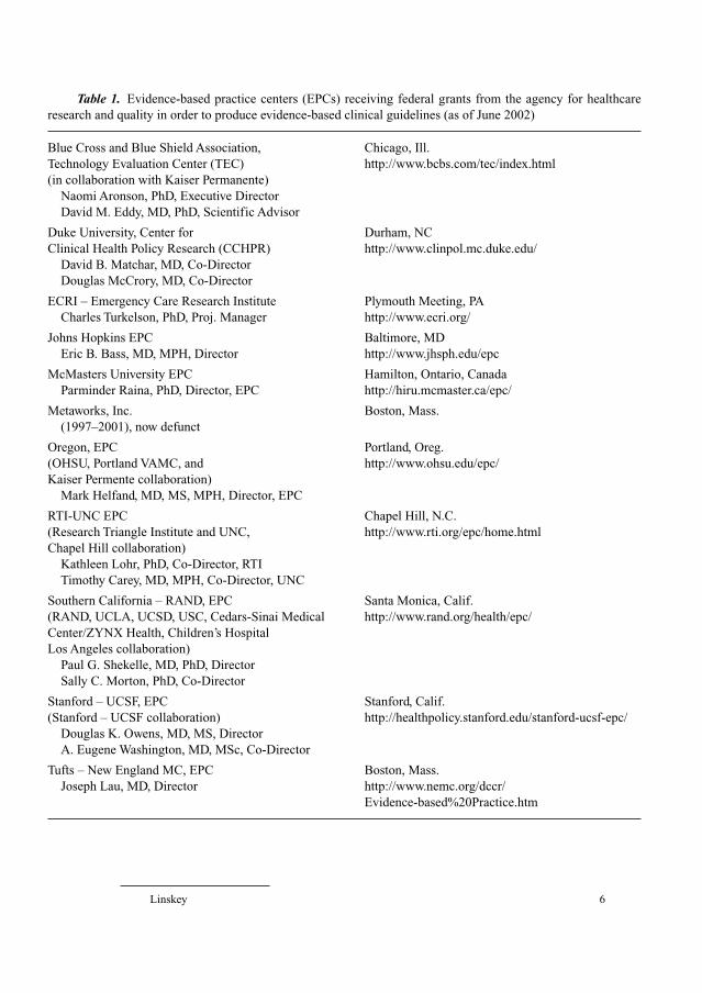
As of 2004, the agency has a budget of USD 269.9 million, �80% of
which is currently awarded as research grants to the 13 extramural Evidence-
Based Practice Centers (EPCs) listed in table 1. The AHRQ maintains the
National Guideline Clearinghouse (NGC) for evidence-based clinical practice
guidelines in a joint initiative with the AMA and America’s Health Insurance
Plans (AHIP – formerly the American Association of Health Plans).
In countries which have nationalized health services and centrally
managed and rationed healthcare, such as the United Kingdom, the degree of
support, investment, and integration of EBM into EBC has been more direct,
intrusive, and far-reaching. The National Health Service (NHS) Research and
Development initiative under Sir Michael Peckham was launched in 1991 [30].
Under pressure to invest in effective procedures and disinvest in ineffective
ones [31, 32], the initiative was designed ‘to secure a knowledge-based health
service in which clinical, managerial, and policy decisions are based on sound
and pertinent information about research findings and scientific developments’
[33]. This has led to a new system of management intended to provide quality in
healthcare (clinical governance) that explicitly requires that funded medical
treatments be evidence-based [34].
In 1999, the NHS launched the National Institute for Clinical Excellence
(NICE) which is responsible for providing patients, health professionals and the
public with authoritative, robust and reliable evidence-based guidance on current
‘best practices’ in relation to new and existing health technologies [35]. Since
January 2002, the NHS has been obliged to provide funding and resources for
health professional-prescribed medicines and treatments recommended by NICE
through its technology appraisal work program [36]. The implication is that
medicines and treatments not specifically recommended by NICE will only be
funded as resources permit at the discretion of each local NHS authority.
NICE is also charged with establishing and maintaining clinical guidelines
for the NHS [37]. This effort began in 1999 [38]. The first NICE clinical guide-
line was published in April 2001. Unlike guidelines published by the US NGC,
NICE guidelines are required to resolve the conflict between pre-existing associ-
ation and stakeholder guidelines and take into consideration cost-effectiveness
Linskey 6
Table 1. Evidence-based practice centers (EPCs) receiving federal grants from the agency for healthcare
research and quality in order to produce evidence-based clinical guidelines (as of June 2002)
Blue Cross and Blue Shield Association, Chicago, Ill.
Technology Evaluation Center (TEC) http://www.bcbs.com/tec/index.html
(in collaboration with Kaiser Permanente)
Naomi Aronson, PhD, Executive Director
David M. Eddy, MD, PhD, Scientific Advisor
Duke University, Center for Durham, NC
Clinical Health Policy Research (CCHPR) http://www.clinpol.mc.duke.edu/
David B. Matchar, MD, Co-Director
Douglas McCrory, MD, Co-Director
ECRI – Emergency Care Research Institute Plymouth Meeting, PA
Charles Turkelson, PhD, Proj. Manager http://www.ecri.org/
Johns Hopkins EPC Baltimore, MD
Eric B. Bass, MD, MPH, Director http://www.jhsph.edu/epc
McMasters University EPC Hamilton, Ontario, Canada
Parminder Raina, PhD, Director, EPC http://hiru.mcmaster.ca/epc/
Metaworks, Inc. Boston, Mass.
(1997–2001), now defunct
Oregon, EPC Portland, Oreg.
(OHSU, Portland VAMC, and http://www.ohsu.edu/epc/
Kaiser Permente collaboration)
Mark Helfand, MD, MS, MPH, Director, EPC
RTI-UNC EPC Chapel Hill, N.C.
(Research Triangle Institute and UNC, http://www.rti.org/epc/home.html
Chapel Hill collaboration)
Kathleen Lohr, PhD, Co-Director, RTI
Timothy Carey, MD, MPH, Co-Director, UNC
Southern California – RAND, EPC Santa Monica, Calif.
(RAND, UCLA, UCSD, USC, Cedars-Sinai Medical http://www.rand.org/health/epc/
Center/ZYNX Health, Children’s Hospital
Los Angeles collaboration)
Paul G. Shekelle, MD, PhD, Director
Sally C. Morton, PhD, Co-Director
Stanford – UCSF, EPC Stanford, Calif.
(Stanford – UCSF collaboration) http://healthpolicy.stanford.edu/stanford-ucsf-epc/
Douglas K. Owens, MD, MS, Director
A. Eugene Washington, MD, MSc, Co-Director
Tufts – New England MC, EPC Boston, Mass.
Joseph Lau, MD, Director http://www.nemc.org/dccr/
Evidence-based%20Practice.htm
EBM for Neurosurgeons 7
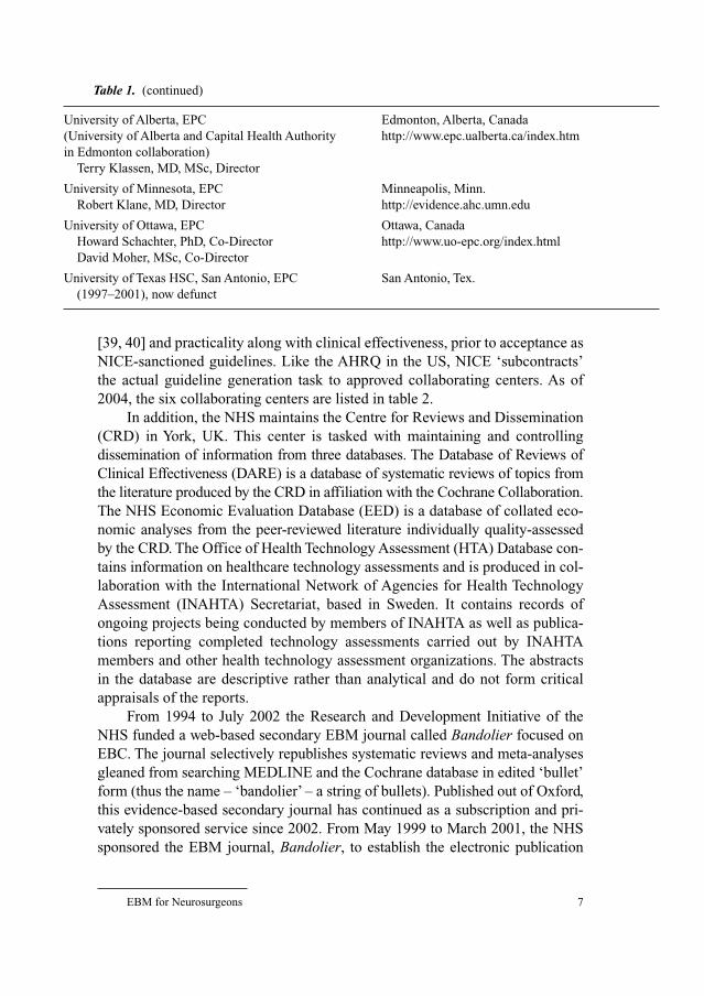
[39, 40] and practicality along with clinical effectiveness, prior to acceptance as
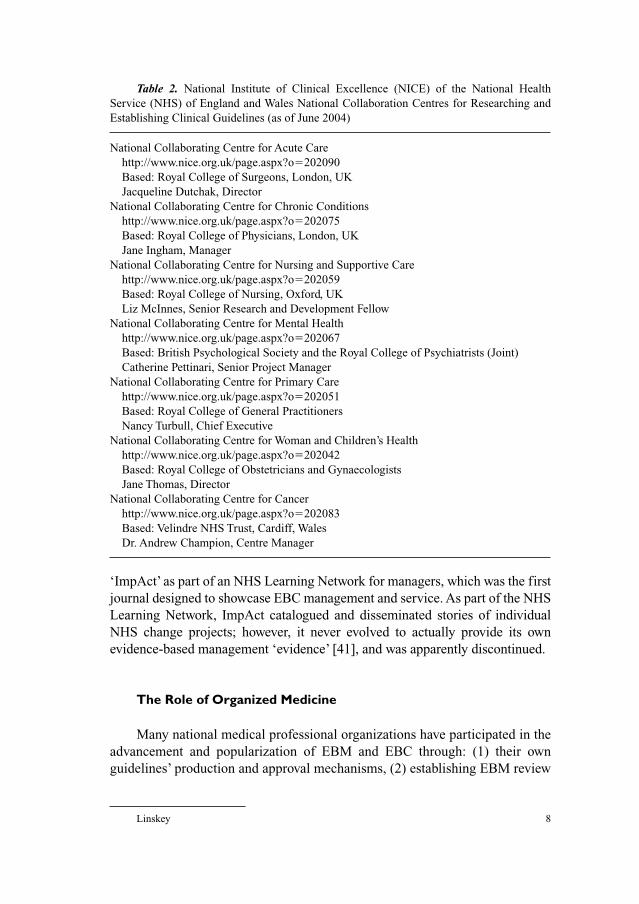
NICE-sanctioned guidelines. Like the AHRQ in the US, NICE ‘subcontracts’
the actual guideline generation task to approved collaborating centers. As of
2004, the six collaborating centers are listed in table 2.
In addition, the NHS maintains the Centre for Reviews and Dissemination
(CRD) in York, UK. This center is tasked with maintaining and controlling
dissemination of information from three databases. The Database of Reviews of
Clinical Effectiveness (DARE) is a database of systematic reviews of topics from
the literature produced by the CRD in affiliation with the Cochrane Collaboration.
The NHS Economic Evaluation Database (EED) is a database of collated eco-
nomic analyses from the peer-reviewed literature individually quality-assessed
by the CRD. The Office of Health Technology Assessment (HTA) Database con-
tains information on healthcare technology assessments and is produced in col-
laboration with the International Network of Agencies for Health Technology
Assessment (INAHTA) Secretariat, based in Sweden. It contains records of
ongoing projects being conducted by members of INAHTA as well as publica-
tions reporting completed technology assessments carried out by INAHTA
members and other health technology assessment organizations. The abstracts
in the database are descriptive rather than analytical and do not form critical
appraisals of the reports.
From 1994 to July 2002 the Research and Development Initiative of the
NHS funded a web-based secondary EBM journal called Bandolier focused on
EBC. The journal selectively republishes systematic reviews and meta-analyses
gleaned from searching MEDLINE and the Cochrane database in edited ‘bullet’
form (thus the name – ‘bandolier’ – a string of bullets). Published out of Oxford,
this evidence-based secondary journal has continued as a subscription and pri-
vately sponsored service since 2002. From May 1999 to March 2001, the NHS
sponsored the EBM journal, Bandolier, to establish the electronic publication
Table 1. (continued)
University of Alberta, EPC Edmonton, Alberta, Canada
(University of Alberta and Capital Health Authority http://www.epc.ualberta.ca/index.htm
in Edmonton collaboration)
Terry Klassen, MD, MSc, Director
University of Minnesota, EPC Minneapolis, Minn.
Robert Klane, MD, Director http://evidence.ahc.umn.edu
University of Ottawa, EPC Ottawa, Canada
Howard Schachter, PhD, Co-Director http://www.uo-epc.org/index.html
David Moher, MSc, Co-Director
University of Texas HSC, San Antonio, EPC San Antonio, Tex.
(1997–2001), now defunct
Linskey 8
‘ImpAct’ as part of an NHS Learning Network for managers, which was the first
journal designed to showcase EBC management and service. As part of the NHS
Learning Network, ImpAct catalogued and disseminated stories of individual
NHS change projects; however, it never evolved to actually provide its own
evidence-based management ‘evidence’ [41], and was apparently discontinued.
The Role of Organized Medicine
Many national medical professional organizations have participated in the
advancement and popularization of EBM and EBC through: (1) their own
guidelines’ production and approval mechanisms, (2) establishing EBM review
Table 2. National Institute of Clinical Excellence (NICE) of the National Health
Service (NHS) of England and Wales National Collaboration Centres for Researching and
Establishing Clinical Guidelines (as of June 2004)
National Collaborating Centre for Acute Care
http://www.nice.org.uk/page.aspx?o�202090
Based: Royal College of Surgeons, London, UK
Jacqueline Dutchak, Director
National Collaborating Centre for Chronic Conditions
http://www.nice.org.uk/page.aspx?o�202075
Based: Royal College of Physicians, London, UK
Jane Ingham, Manager
National Collaborating Centre for Nursing and Supportive Care
http://www.nice.org.uk/page.aspx?o�202059
Based: Royal College of Nursing, Oxford, UK
Liz McInnes, Senior Research and Development Fellow
National Collaborating Centre for Mental Health
http://www.nice.org.uk/page.aspx?o�202067
Based: British Psychological Society and the Royal College of Psychiatrists (Joint)
Catherine Pettinari, Senior Project Manager
National Collaborating Centre for Primary Care
http://www.nice.org.uk/page.aspx?o�202051
Based: Royal College of General Practitioners
Nancy Turbull, Chief Executive
National Collaborating Centre for Woman and Children’s Health
http://www.nice.org.uk/page.aspx?o�202042
Based: Royal College of Obstetricians and Gynaecologists
Jane Thomas, Director
National Collaborating Centre for Cancer
http://www.nice.org.uk/page.aspx?o�202083
Based: Velindre NHS Trust, Cardiff, Wales
Dr. Andrew Champion, Centre Manager
EBM for Neurosurgeons 9
processes for national presentations and journal article submissions, (3) publi-
cation of EBM manuals and/or resources, and (4) sponsorship of continuing
medical education in EBM techniques. Three of them (the American College of
Physicians – ACP, the British Medical Association – BMA, and the AMA) have
gone even further through sponsorship of secondary EBM journals and/or data-
bases of systematic reviews of clinical trials, and advocacy efforts to influence
governmental EBC policies and programs.
The ACP, through its journal, the Annals of Internal Medicine, established
the first secondary EBM journal in 1991 (called the ACP Journal Club) that
summarized new publications of high relevance and methodological rigor [42].
This journal reviewed �40 journals in English on topics central to general
internal medicine. The ACP Journal Club, initially published as a bimonthly
supplement to the Annals of Internal Medicine, has been an independent publi-
cation since November 1994. From 1996 though 2001 [43], the ACP also pro-
duced an annual CD-ROM entitled ‘Best Evidence’, a compendium of reviews
from the ACP Journal Club, the journal, Evidence-Based Medicine (jointly
sponsored with the BMA), and Diagnostic Strategies for Common MedicalProblems. Best Evidence was replaced by ACP Journal Club online service in
2001 [43].
The AMA, through its journal, Journal of the American Medical Asso-ciation (JAMA), over the period of 10 years, sponsored the publication of a
series of 25 ‘user’s guides to the medical literature’ produced by the Evidence-
Based Medicine Working Group [20, 44–75]. Along with the ACP, they also
compiled these articles for publication into two manuals [76, 77]. The AMA
currently sponsors and maintains the NGC for evidence-based clinical practice
guidelines in a joint initiative with the AHRQ and AHIP. They are also actively
involved in promoting expansion of the existing government clinical trials data-
base to include all previous and ongoing drug trials [25].
The British Medical Association has been extremely active in developing
and promoting EBM and EBC through their publishing arm, the British Medical
Journal (BMJ) Publishing Group. The BMJ Publishing Group publishes the EBM
secondary journal, Evidence-Based Medicine. Originally started in 1996 as a joint
initiative with the ACP and jointly edited at McMasters University and at the
Centre for Evidence-Based Medicine at Oxford in the UK, the journal ‘EBM’
took a similar secondary review approach as the ACP Journal Club but broadened
coverage to include surgery, obstetrics, pediatrics, family medicine and psychia-
try, in addition to general internal medicine [16, 78]. Similar to the AMA, the
BMJ Publishing Group has also published a compendium of EBM journal articles
from the BMJ into a more easily accessible manual [79]. The BMJ Publishing
Group also manages their own online EBM secondary source for primary care
called ‘Clinical Evidence’.
Linskey 10
Organized neurosurgery has made efforts in the EBM arena with mixed
results. In the mid-1990s, the American Association of Neurological Surgeons
(AANS) and the Congress of Neurological Surgeons (CNS) established a Joint
Committee on the Assessment of Quality (formerly known as the Quality
Assessment Committee) under Robert Florin. This joint committee oversaw four
subcommittees, two of which were the Outcomes Committee and the Guidelines
Committee.
Attempts at establishing two national neurosurgery outcomes studies (one
on intracranial aneurysms and the other on carotid endarterectomy) [80–82]
came to naught and were discontinued. Difficulties with inadequate organiza-
tional infrastructure and expertise to manage these projects led the AANS to create
a joint venture with Outcomes Sciences (Boston, Mass., USA) called Neuro-
Knowledge (Boston, Mass., USA), for the purpose of potentially contracting
for future neurosurgery outcomes analysis studies [83].
The Joint Committee on Assessment of Quality was dissolved fairly recently.
The Outcomes Subcommittee has ceased to exist. The Guidelines Committee
has been systematically working with each AANS/CNS joint section to establish
guidelines based on diagnosis-specific section priorities. The Guidelines Sub-
committee has become two separate committees – the AANS Guidelines
Committee within the Science Division of the AANS, and the CNS Guidelines
Development Committee. The Department of Education and Practice Manage-
ment of the AANS maintains a repository of clinical guidelines pertinent to neu-
rosurgical diagnoses at http://www.aans.org/practice/guideliens/aans.asp. The
listing includes guidelines from agencies and organizations other than the AANS
or CNS, but as of June 2004, this site appeared to be in need of updating.
The Accreditation Council for Graduate Medical Education (ACGME), the
Association of American Medical Colleges (AAMC), and the American Board
of Medical Specialties (ABMS) have all agreed to establish a core of six compe-
tencies for training and assessing physicians. These competencies have profound
implications for educational curricula for medical school and residency training,
board certification, and maintenance of certification as a life-long educational/
recertification process. The ACGME endorsed the six competencies on
September 28, 1999, after a review process by an eleven member Outcome
Project Advisory Group spanning the period January 1998 through February
1999. The review process was not evidence-based. Implementation for residency
training programs was initiated in July 2002. The Group on Educational Affairs
(GEA) within the AAMC is currently working on integrating the six competen-
cies into medical school curricula through their Competencies Across the
Continuum of Health Education (CACHE) project. The ABMS, including the
American Board of Neurological Surgeons (ABNS), have structured their main-
tenance of certification processes around the same six competencies [84].
EBM for Neurosurgeons 11
One of the six competencies is practice-based learning and improvement.
Ideally, this competency is intended as a specific link to EBM through ongoing
self-assessment of outcome results for the individual physician linked to empiric
analysis of the success or failure of performance improvement efforts. The
specifics of realistic and practically achievable requirements and compliance
criteria have yet to be completely worked out.
Resources
Many books and manuals have now been published to assist neurosurgeons
with establishing an evidence-based practice and instituting an evidence-based
approach to teaching and making individual clinical decisions [3, 4, 76, 77, 79,
85–89]. While an introductory chapter to a book cannot hope to serve as a detailed
textbook, the reader is invited to explore these referenced works for details and
more in-depth descriptions and explanations.
While the books and manuals referenced above are the best place to start,
even more detail can be had by exploring original articles published on specific
topics within EBM. Examples include:
(1) An introduction to applying the EBM Working Group User’s Guides and
articles on general EBM theory [16–23, 44, 45]
(2) How to evaluate and use articles about therapy or prevention [45, 46]
(3) How to evaluate and use articles about a diagnostic test [47, 48]
(4) How to evaluate and use articles about harm [49]
(5) How to evaluate and use articles about prognosis [50]
(6) How to evaluate and use an overview article [51]
(7) How to evaluate and use articles about clinical decision analysis and clin-
ical decision rules [52, 53]
(8) How to evaluate and use guidelines or healthcare/treatment recommen-
dations [54–56, 64, 65]
(9) How to evaluate and use articles on health services outcomes or utiliza-
tion review [57, 58]
(10) How to evaluate and use articles assessing quality of life [59]
(11) How to evaluate and use articles on the economics of practice [40, 60,
61, 90]
(12) How to best apply the EBM Working Group User’s Guides and published
evidence to the care of a specific patient [62, 69, 73, 75]
(13) How to evaluate and use articles on disease probability for establishing
and working through a differential diagnosis [63]
(14) How to evaluate and use articles on the clinical manifestations of a dis-
ease to assist with establishing a diagnosis [74]
Linskey 12
(15) How to make maximal effective use of electronic health information
resources and computer-based clinical decision support systems [66, 70]
(16) How to assess the applicability applying of drug class effects versus indi-
vidual drug effect, as well as surrogate endpoints from RCTs [67, 68]
(17) How to evaluate and use qualitative study results [72, 73].
Many additional resources exist. In addition to books, manuals, and indi-
vidual articles, there are now more than ten peer-reviewed EBM journals in
publication (table 3). Websites that serve as a source of EBM information, links
to other pertinent and useful websites, sources of EBM tools, repositories of
electronic EBM Journals or collections of systematic reviews, and repositories
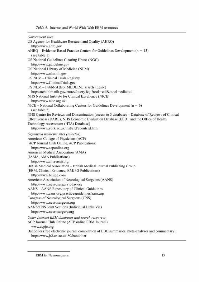
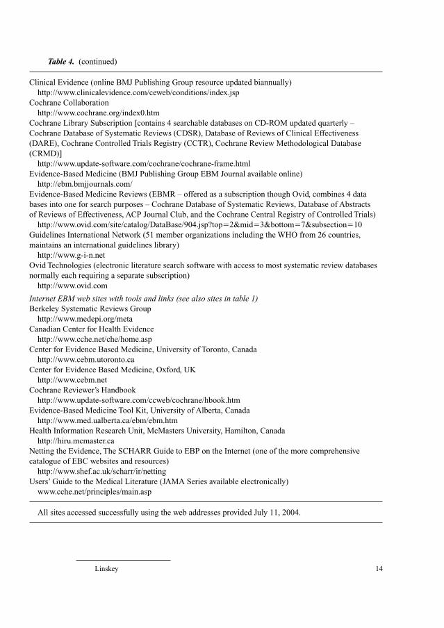
for clinical guidelines abound. A noninclusive and selective listing of pertinent
websites with their internet addresses is presented in table 4.
EBM Clinical Decision Methodology
EBM is rooted in five linked ideas: (1) that clinical decisions should be
based on best available clinical evidence, (2) that the specific clinical problem
of interest should determine the type of evidence sought, (3) that the evidence
discovered through searching should be sorted and assessed using epidemio-
logic and biostatistical criteria in order to identify the best evidence, (4) that
the conclusions arrived at should be put into action, and (5) that the result of
the decision should be objectively evaluated [21]. There are four basic steps to
taking an evidence-based approach: (1) formulizing a clear clinical question
from a patient’s problem, (2) effectively searching the literature for relevant
Table 3. Evidence-based journals (a selected list)
ACP Journal Club (printed and electronic versions)
Bandolier (printed and electronic versions)
Bandolera (authorized Spanish language version of Bandolier)
Effective Health Care BulletinsEffectiveness MattersEvidenceEvidence Based Health CareEvidence Based Medicine (printed and electronic versions)
Evidence Based Medicine – Edition Française (auth. French language version
of EBM)
Evidence Based Mental Health (printed and electronic versions)
Evidence Based NursingJournal of Family Practice POEMs (Patient-Oriented Evidence that Matters)
New Zealand Evidence Based Healthcare Bulletin
EBM for Neurosurgeons 13
Table 4. Internet and World Wide Web EBM resources
Government sitesUS Agency for Healthcare Research and Quality (AHRQ)
http://www.ahrq.gov
AHRQ – Evidence-Based Practice Centers for Guidelines Development (n � 13)
(see table 1)
US National Guidelines Clearing House (NGC)
http://www.guideline.gov
US National Library of Medicine (NLM)
http://www.nlm.nih.gov
US NLM – Clinical Trials Registry
http://www.ClinicalTrials.gov
US NLM – PubMed (free MEDLINE search engine)
http://ncbi.nlm.nih.gov/entrez/query.fcgi?tool�cdl&otool�cdlotool
NHS National Institute for Clinical Excellence (NICE)
http://www.nice.org.uk
NICE – National Collaborating Centers for Guidelines Development (n � 6)
(see table 2)
NHS Centre for Reviews and Dissemination [access to 3 databases – Database of Reviews of Clinical
Effectiveness (DARE), NHS Economic Evaluation Database (EED), and the Office of Health
Technology Assessment (HTA) Database]
http://www.york.ac.uk/inst/crd/aboutcrd.htm
Organized medicine sites (selected)American College of Physicians (ACP)
(ACP Journal Club Online, ACP Publications)
http://www.acponline.org
American Medical Association (AMA)
(JAMA, AMA Publications)
http://www.ama-assn.org
British Medical Association – British Medical Journal Publishing Group
(EBM, Clinical Evidence, BMJPG Publications)
http://www.bmjpg.com
American Association of Neurological Surgeons (AANS)
http://www.neurosurgerytoday.org
AANS – AANS Repository of Clinical Guidelines
http://www.aans.org/practice/guidelines/aans.asp
Congress of Neurological Surgeons (CNS)
http://www.neurosurgeon.org
AANS/CNS Joint Sections (Individual Links Via)
http://www.neurosurgery.org
Other Internet EBM databases and search resourcesACP Journal Club Online (ACP online EBM Journal)
www.acpjc.org
Bandolier (free electronic journal compilation of EBC summaries, meta-analyses and commentary)
http://www.jr2.ox.ac.uk:80/bandolier
Linskey 14
Clinical Evidence (online BMJ Publishing Group resource updated biannually)
http://www.clinicalevidence.com/ceweb/conditions/index.jsp
Cochrane Collaboration
http://www.cochrane.org/index0.htm
Cochrane Library Subscription [contains 4 searchable databases on CD-ROM updated quarterly –
Cochrane Database of Systematic Reviews (CDSR), Database of Reviews of Clinical Effectiveness
(DARE), Cochrane Controlled Trials Registry (CCTR), Cochrane Review Methodological Database
(CRMD)]
http://www.update-software.com/cochrane/cochrane-frame.html
Evidence-Based Medicine (BMJ Publishing Group EBM Journal available online)
http://ebm.bmjjournals.com/
Evidence-Based Medicine Reviews (EBMR – offered as a subscription though Ovid, combines 4 data
bases into one for search purposes – Cochrane Database of Systematic Reviews, Database of Abstracts
of Reviews of Effectiveness, ACP Journal Club, and the Cochrane Central Registry of Controlled Trials)
http://www.ovid.com/site/catalog/DataBase/904.jsp?top�2&mid�3&bottom�7&subsection�10
Guidelines International Network (51 member organizations including the WHO from 26 countries,
maintains an international guidelines library)
http://www.g-i-n.net
Ovid Technologies (electronic literature search software with access to most systematic review databases
normally each requiring a separate subscription)
http://www.ovid.com
Internet EBM web sites with tools and links (see also sites in table 1)Berkeley Systematic Reviews Group
http://www.medepi.org/meta
Canadian Center for Health Evidence
http://www.cche.net/che/home.asp
Center for Evidence Based Medicine, University of Toronto, Canada
http://www.cebm.utoronto.ca
Center for Evidence Based Medicine, Oxford, UK
http://www.cebm.net
Cochrane Reviewer’s Handbook
http://www.update-software.com/ccweb/cochrane/hbook.htm
Evidence-Based Medicine Tool Kit, University of Alberta, Canada
http://www.med.ualberta.ca/ebm/ebm.htm
Health Information Research Unit, McMasters University, Hamilton, Canada
http://hiru.mcmaster.ca
Netting the Evidence, The SCHARR Guide to EBP on the Internet (one of the more comprehensive
catalogue of EBC websites and resources)
http://www.shef.ac.uk/scharr/ir/netting
Users’ Guide to the Medical Literature (JAMA Series available electronically)
www.cche.net/principles/main.asp
All sites accessed successfully using the web addresses provided July 11, 2004.
Table 4. (continued)

EBM for Neurosurgeons 15
clinical articles, (3) evaluating (critically appraising) the evidence for its valid-
ity and usefulness, and (4) implementing the findings in clinical practice [22].
In general, Sackett et al. [23] identifies two broad forms of questions:
background and foreground. Background questions are general knowledge ques-
tions about the patient, the diagnosis or the treatment (i.e. why, what, when,
where, who and how?) Foreground questions focus on very specific informa-
tion needs for decision making. These needs may relate to the patient, the main
intervention under consideration, alternative interventions under consideration,
or a clinical outcome of interest. It is with the latter type of question that an
EBM approach is most likely to have impact. Neurosurgery is an intervention-
and action-oriented medical subspecialty. As a result, while decisions regarding
prevention and diagnosis (e.g. testing) are important, decisions regarding inter-
ventions (that rely on assessments of prognosis and harm) tend to be more inter-
esting and relevant to the majority of our decisions. To benefit both the patient
and the clinician, the question must be well built – which means, both relevant
to the patient’s problem, and phrased in a way that directs the subsequent search
to relevant and precise answers [20, 88, 89].
In general, effectively searching the literature for relevant clinical articles
has become much faster and more efficient with the use of electronic search
engines for the NLM MEDLINE database [66, 70]. On the other hand, search-
ing by hand for clinical studies published in books or book chapters and articles
published prior to 1966 has become more difficult and less efficient. Many of
us have forgotten where the medical school library is located as we have become
more focused and reliant on our desktop computers for search access. Many
medical centers have witnessed deterioration in their medical libraries through
neglect and funding reductions as the number of library visitors and the need
for librarian services diminish. As MEDLINE searches have become common
place, many of us have forgotten (or have never been taught!) how to search
journal articles using Index Medicus.
Even electronic literature searching is becoming more complicated if com-
prehensiveness is sought or desired. Many clinical trials, systematic reviews, and
other secondary overviews not published in journals indexed in MEDLINE are
now listed in searchable databases separate from MEDLINE. These require
access to subscription CD-ROMs or subscription electronic search engines [e.g.
ACP Journal Club, Clinical Evidence from BMJ Publishing Group, Cochrane
Database of Systematic Reviews (CDSR), NHS Database of Reviews of Clinical
Effectiveness (DARE), Cochrane Controlled Trials Registry (CCTR), Cochrane
Review Methodological Database (CRMD), and Evidence-Based Medicine
Reviews (EBMR) from Ovid Technologies] (see table 4). Many evidence-based
clinical guidelines (another form of secondary overview) are also not listed in
MEDLINE, but require separate searches of electronic guidelines databases such
Linskey 16
as those maintained in the US (NGC), in Great Britain (NICE), and at other inter-
national sites (e.g. the Guidelines International Network) (see table 4).
The evaluation of the evidence gleaned from a proper search involves
asking three questions: (1) are the results valid (i.e. a methodology and rigor of
adherence to methodology assessment)?, (2) what are the results? (i.e. a preci-
sion, utility, as well as magnitude of effect assessment)?, and (3) will the results
help me care for my patient? (i.e. a cost/benefit assessment, a local clinical care
context assessment, and an individual patient appropriateness assessment) [22,
88, 89]. Many searches will lead to a plethora of clinical evidence, and many
studies may come to conflicting conclusions. Obviously, not all conclusions can
be correct. An EBM approach to sorting through the confusion involves the
ranking of evidence from clinical studies according to the type of study design
and the methodological rigor followed in each individual study as the first step.
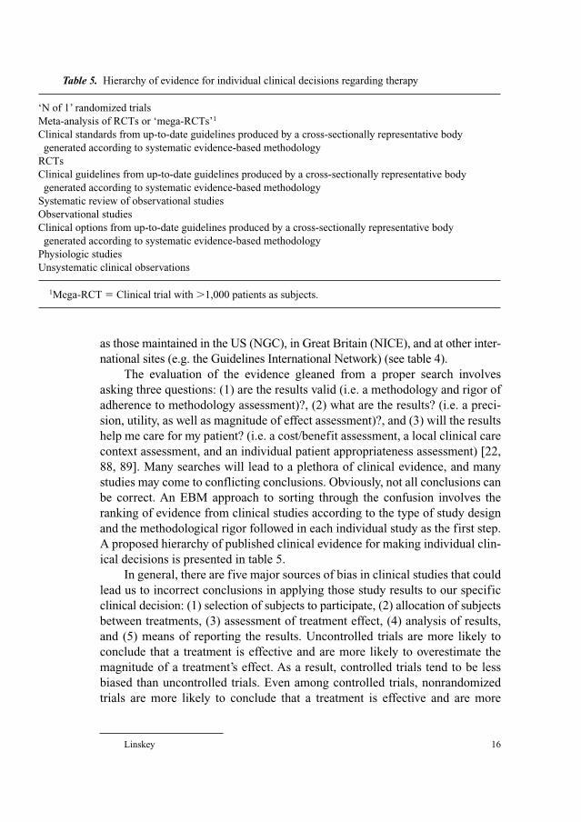
A proposed hierarchy of published clinical evidence for making individual clin-
ical decisions is presented in table 5.
In general, there are five major sources of bias in clinical studies that could
lead us to incorrect conclusions in applying those study results to our specific
clinical decision: (1) selection of subjects to participate, (2) allocation of subjects
between treatments, (3) assessment of treatment effect, (4) analysis of results,
and (5) means of reporting the results. Uncontrolled trials are more likely to
conclude that a treatment is effective and are more likely to overestimate the
magnitude of a treatment’s effect. As a result, controlled trials tend to be less
biased than uncontrolled trials. Even among controlled trials, nonrandomized
trials are more likely to conclude that a treatment is effective and are more
Table 5. Hierarchy of evidence for individual clinical decisions regarding therapy
‘N of 1’ randomized trials
Meta-analysis of RCTs or ‘mega-RCTs’1
Clinical standards from up-to-date guidelines produced by a cross-sectionally representative body
generated according to systematic evidence-based methodology
RCTs
Clinical guidelines from up-to-date guidelines produced by a cross-sectionally representative body
generated according to systematic evidence-based methodology
Systematic review of observational studies
Observational studies
Clinical options from up-to-date guidelines produced by a cross-sectionally representative body
generated according to systematic evidence-based methodology
Physiologic studies
Unsystematic clinical observations
1Mega-RCT � Clinical trial with �1,000 patients as subjects.
EBM for Neurosurgeons 17
likely to overestimate the magnitude of a treatment’s effect [91]. The random-
ization process in RCTs theoretically should also take care of allocation biases
that we have yet to think of or realize might be important. RCTs currently hold
the pinnacle position for evidence hierarchy for these reasons.
Meta-analysis of RCTs and ‘mega-RCTs’ (trials with �1,000 subjects)
hold a special place in the hierarchy because they include a clinical research
subject size that maximizes statistical power, thus limiting the chance of a type 2
error (concluding that two therapies are the same or that a therapy is no better
than placebo when, in fact, they differ by a small percentage in outcome pro-
bability). While meta-analysis of RCTs or a mega-RCT holds the pinnacle
evidence position for clinical guideline considerations or healthcare policy
decisions for a population, there is still an even more superior study for making
a decision in an individual patient. This ultimate level of evidence for a clinical
decision in an individual patient is a randomized, crossover 1 patient ‘N of 1
trial’ [92]. This type of trial eliminates all selection and allocation bias and the
results, by definition, are always 100% applicable to your patient and their cir-
cumstances. The only bias that remains in this type of study for neurosurgery is
the potential for placebo effect if the intervention(s) cannot be blinded to either
the patient or the doctor. Unfortunately, a PubMed search July 11, 2004, for ‘N
of 1 trial’ and ‘neurosurgery’ did not yield a single example of this type of trial
published for our specialty (unpubl. data).
Whether or not the results of your search evaluation apply to your particular
patient requires an analysis of the similarities and differences between your
patient and those accepted for inclusion in the studies in question [62, 69, 73, 75,
88, 89, 93, 94]. In general, you should ask yourself if your patient is so different
from those included in the study that its results cannot be applied to him or her.
Assuming your patient is not significantly different from the patients ana-
lyzed in the RCTs, the decision to apply those results in your particular circum-
stances requires additional analysis. It requires an assessment of: (1) whether or
not the intervention or treatment is feasible or logistically possible in your cir-
cumstances, (2) a judgment regarding the likely magnitude of the probable effect
of the intervention against the risk of harm, and (3) a full accounting of the
patient’s individual input regarding the impact of their own personal values, pri-
orities, and desires on the choice before both of you. Formal clinical decision
analyses are not suitable for this purpose because they use a group’s strength of
preference for different treatment options, which may not be applicable to an
individual patient [95–97]. Again, the quality of the relevant evidence will
impact upon setting your own threshold magnitude where the strength of effect
will impress you and influence your decision making.
‘Because of biases we describe in case-control studies, . . . you might not become
impressed with their ROs [risk odds] until they reached 4 or more (some of our
Linskey 18
colleagues would relax these guides for a serious adverse effect and set them even
higher for a trivial one). Since cohort studies are less subject to bias, you might be
impressed by RR [relative risks] of 3 or more in them. And because randomized trials
are relatively free of bias, any RR whose confidence interval excludes unity is impres-
sive and warrants further consideration’ [98].
Objective statistics from clinical trials (e.g. odds ratios or risk odds and
relative risks) can be converted into statistics that are more useful at the bedside
and more intuitive for individual patient and physician decision making. These
statistics include effect size (the difference in outcomes between intervention and
control groups divided by the standard deviation), the number needed to treat
(number of patients needed to treat to prevent one bad outcome), the number
needed to harm (number of patients needed to be treated to produce one episode
of harm), and likelihood ratios (for studies on diagnostic tests) [20, 88, 89].
Feasibility and logistical assessment include assessing whether or not the
treatment in question is available in your setting. It does little good to conclude
that a given patient with a ruptured aneurysms and subarachnoid hemorrhage
would likely be better served by endovascular coiling of an unruptured aneurysm
based on RCT data [96], if endovascular coiling is not available at your medical
center and the patient is too unstable for safe transfer to another medical center
that has the technology and expertise available. Likewise, it does little good to
conclude that your patient with an inoperable single brain metastasis of suitable
size should receive stereotactic radiosurgery in addition to whole brain radiother-
apy for both length of life and quality of life benefits [97] if stereotactic radio-
surgery is not available in your region. It also includes an honest and objective
assessment of your likely results with an intervention compared with the success
and morbidity rates of the surgeons published in the RCT. In one example, Swales
[98] noted that the ‘proven’ advantage of surgery as a treatment for carotid steno-
sis was entirely negated by the 9.8% complication rate in community practice
(study complication rate was 3.7%). In the absence of systematically acquired and
analyzed objective data regarding your own outcomes for a particular interven-
tion, one should at least take into account one’s ongoing volume for the procedure
in question. There are now many studies showing that surgeon case volume is
clearly related to successful outcomes and lower complication rates for many neu-
rosurgical diagnoses [99–107]. In the absence of the recommended technology, a
significant personal ongoing case volume for infrequent or technically challeng-
ing procedures, or evidence that your personal outcome results are in line with
those reported in the RCTs, one should consider referral of the patient if clinical
safety concerns allow, and if the patient and family are agreeable.
As just stated, assessing the situation-specific probabilities of harm
versus benefit for an intervention requires an objective knowledge and ongoing
analysis of one’s own success and morbidity and mortality rates for a given
EBM for Neurosurgeons 19
procedure. This component of evidence-based practice has been formalized by
the AAMC, the ACGME, and the ABMS as ‘practice-based learning and
improvement’, and is now one of the six ‘core competencies’ upon which we
will all be assessed from now on. Without an ongoing objective and systematic
analysis of your own results (usually through a prospective database), your
perceptions, recollections, or estimations of your own clinical experience fall a
level in the EBM hierarchy to the level of inadequately substantiated opinion.
Practice-based learning and improvement fits neatly in EBM for individual
clinical decisions, and indeed is a requisite for its proper application [84, 108].
Ultimately, clinical care of an individual patient requires multiple simultane-
ous and sequential decisions, and not every question that needs to be answered
to guide those decisions can be answered with high quality evidence from the
top of the hierarchy listed and ranked in table 5. A useful analogy here is one
developed by Slawson and colleagues [109], which they refer to as a ‘clinical
jazz’ approach. Just as a form of jazz music takes a defined piece of music
(defined portions – evidence-based) and intersperses these defined segments
with improvisational inspiration to create a beautiful, overall new, and individ-
ual creation. Clinical expertise and experience must fill in to provide guidance
in equivocal situations between segments of the overall care plan which are
firmly based on highest quality evidence.
It is also clear that certain aspects of decision making remain an art rather
than a science. Even Straus and Sackett [94] admits that the ‘optimal intelligi-
ble method of eliciting patients’ preferences and providing decision support in a
busy clinical setting is still to be determined’. This is particularly true for surgical
interventions. In one landmark study, the attractiveness of surgery to patients
was measurably greater when outcomes were framed in terms of probability of
living rather than in terms of probability of dying, even when the figures simply
reflected the inverse of one another [110].
Meta-Analysis Methodology
Meta-analysis of RCT’s holds an important position in the hierarchy of evi-
dence ranking of EVD (tables 5, 6). As a result, it deserves special focus and
attention in any introduction to EBM and EBM methodology.
The educator and psychologist, Gene Glass and colleagues [111–113],
introduced the concept and the term meta-analysis in 1976 as a means to quanti-
tatively aggregate independent research studies. It was not originally described
as a means of assessing RCTs. As clinical epidemiology has advanced, the
original descriptions and methods have come to more closely resemble what
are now referred to as systematic reviews, which include nonrandomized
Linskey 20
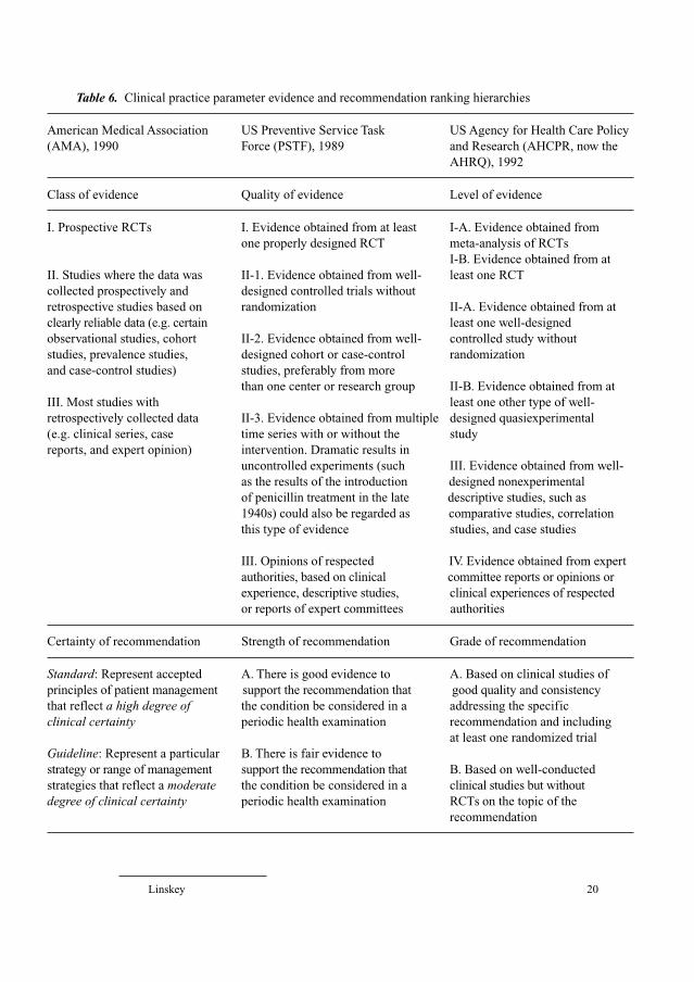
Table 6. Clinical practice parameter evidence and recommendation ranking hierarchies
American Medical Association US Preventive Service Task US Agency for Health Care Policy
(AMA), 1990 Force (PSTF), 1989 and Research (AHCPR, now the
AHRQ), 1992
Class of evidence Quality of evidence Level of evidence
I. Prospective RCTs I. Evidence obtained from at least I-A. Evidence obtained from
one properly designed RCT meta-analysis of RCTs
I-B. Evidence obtained from at
II. Studies where the data was II-1. Evidence obtained from well- least one RCT
collected prospectively and designed controlled trials without
retrospective studies based on randomization II-A. Evidence obtained from at
clearly reliable data (e.g. certain least one well-designed
observational studies, cohort II-2. Evidence obtained from well- controlled study without
studies, prevalence studies, designed cohort or case-control randomization
and case-control studies) studies, preferably from more
than one center or research group II-B. Evidence obtained from at
III. Most studies with least one other type of well-
retrospectively collected data II-3. Evidence obtained from multiple designed quasiexperimental
(e.g. clinical series, case time series with or without the study
reports, and expert opinion) intervention. Dramatic results in
uncontrolled experiments (such III. Evidence obtained from well-
as the results of the introduction designed nonexperimental
of penicillin treatment in the late descriptive studies, such as
1940s) could also be regarded as comparative studies, correlation
this type of evidence studies, and case studies
III. Opinions of respected IV. Evidence obtained from expert
authorities, based on clinical committee reports or opinions or
experience, descriptive studies, clinical experiences of respected
or reports of expert committees authorities
Certainty of recommendation Strength of recommendation Grade of recommendation
Standard: Represent accepted A. There is good evidence to A. Based on clinical studies of
principles of patient management support the recommendation that good quality and consistency
that reflect a high degree of the condition be considered in a addressing the specific
clinical certainty periodic health examination recommendation and including
at least one randomized trial
Guideline: Represent a particular B. There is fair evidence to
strategy or range of management support the recommendation that B. Based on well-conducted
strategies that reflect a moderate the condition be considered in a clinical studies but without
degree of clinical certainty periodic health examination RCTs on the topic of the
recommendation
EBM for Neurosurgeons 21
observational studies, rather than modern meta-analyses. Unfortunately, authors
sometimes use the terms ‘systematic review’ and ‘meta-analysis’ interchange-
ably [51] and there are no universally agreed-upon definitions of meta-analysis,
per se [114]. Both systematic reviews and meta-analysis involve a systematic
and quantitative review of smaller studies, ultimately yielding aggregate statisti-
cal results that take into account the statistically weighted contribution of each
contributing study.
What is clear is that when most EBM practitioners refer to a meta-analysis,
they are usually referring to a quantitative systematic review of RCTs [15, 51,
115–117]. Evidentiary-worthy meta-analyses employ a rigorous system for trial
search and search quality control, rigorous criteria for selecting RCTs that share
compatible selection criteria for inclusion, interventions and study endpoints, and
rigorous statistical methodology for aggregating the results into the formation of
a single new quantitative estimate of the effect of the interaction or risk factor.
They also include formal analyses to assess for heterogeneity among included
RCTs. Meta-analysis requires all the scientific rigor of an RCT. Detailed descrip-
tions of methodology for meta-analysis of RCTs and for systematic reviews that
include observational studies along with RCTs can now be found from multiple
sources [51, 118–129]. Cumulative meta-analysis is a special form of meta-analysis
that allows retrospective statistical definition of the minimum number of studies
after which the question should have been considered closed [130]. Only a few
meta-analyses of diagnostic testing [131–133], disease prevalence [133] and of
RCTs currently exist for neurosurgery [134–142].
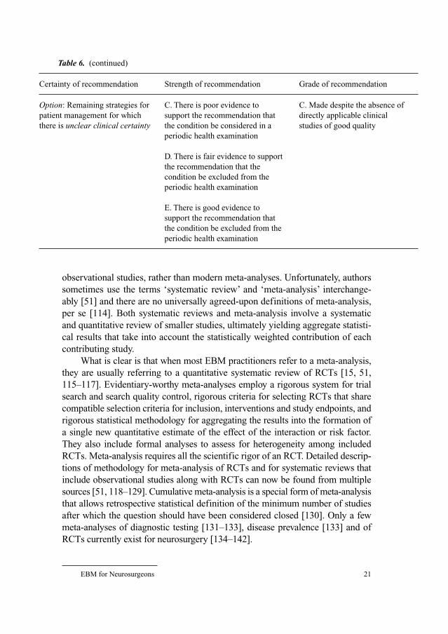
Table 6. (continued)
Certainty of recommendation Strength of recommendation Grade of recommendation
Option: Remaining strategies for C. There is poor evidence to C. Made despite the absence of
patient management for which support the recommendation that directly applicable clinical
there is unclear clinical certainty the condition be considered in a studies of good quality
periodic health examination
D. There is fair evidence to support
the recommendation that the
condition be excluded from the
periodic health examination
E. There is good evidence to
support the recommendation that
the condition be excluded from the
periodic health examination
Linskey 22
What is also clear is that a traditional review (e.g. ‘case report and review
of the literature’, an expert review with selective reference citation, or a review
of case reports and/or case series rather than clinical studies) is neither a sys-
tematic review nor a meta-analysis, as the terms are used in EBM. In order to be
a systematic review or a meta-analysis, the literature search must be systemati-
cally inclusive and the inclusion selection judgments independently confirmed
and verified. For secondary overview studies of therapies or interventions, the
publications included must be actual studies that arrive at a quantitative effect
statistic (e.g. an odds ratio for a case-control study or a relative risk for either a
controlled observational study or an RCT). Traditional reviews result in more
type 2 errors (failing to reject the null hypothesis) than systematic reviews or
meta-analyses [143].
Clinical Practice Parameter Methodology
Clinical practice guidelines are defined as ‘systematically developed state-
ments to assist practitioner and patient decisions about appropriate healthcare
for specific individual circumstances’ [144]. An advantage of utilizing guide-
lines in clinical decision making over sole reliance on RCT results is that they
take professional experience into account in an aggregate and more systematic
manner, rather than on an individual or ad hoc basis [117]. Not only are more
‘experts’ involved in the consensus process (diluting out outliers in opinion) but
in an evidence-based guideline development process, the opinions solicited are
the experts’ opinions about the collected evidence in the literature, rather than
simply their own personal opinion regarding the subject.
Not all guidelines are equivalent in quality. The US NGC currently includes
guidelines that have been formed through expert consensus alongside those based
in systematic evidence-based methodology. It also includes guidelines that have
been created by special interest and advocacy groups, subspecialty organizations,
insurance companies, private consulting firms, cross-representative panels
designed to include representatives from all potential stakeholders, and EPCs.
Many of these guidelines conflict with one another, and there is currently no
means of resolving or adjudicating these conflicts other than individual providers
or oversight organizations making their own decision(s) as to which should take a
position of supremacy or authority. The NHS approach to this problem is to only
recognize guidelines produced by their National Collaborating Centers (table 2).
These centers are each individually tasked with establishing representative panels,
following validated guidelines methodology, surveying existing guidelines rele-
vant to the topic and resolving any apparent conflict, and including both cost-effec-
tiveness [39, 40, 90] and practicality as part of the final recommendation process.
EBM for Neurosurgeons 23
According to Woolf [145], there are three main methods of guideline devel-
opment – informal consensus, formal consensus, and evidence-linked develop-
ment. From the standpoint of EBM, only the latter have evidentiary status for
EBM decision making. Indeed, the US Institute of Medicine hopes to eventually
restrict the use of the term ‘guideline’ to systematically developed advisory
statements created according to validated methodology [144]. Some consider
consensus guidelines as intellectually suspect by reflecting expert opinion, which
when promulgated as a ‘guideline’can formalize unsound practice [146]. Without
strict adherence to systematic and validated methodology, panelists may be pool-
ing ignorance as much as distilling wisdom [147]. Some guidelines are of
questionable quality and there have been calls for guidelines on how to devise
guidelines [148]. The use of guidelines is never a substitute for the exercise of
professional judgment.
Construction of guidelines involves, first, a systematic means of identifying
evidence and ranking the relative strengths, or quality of each study as evidence,
and then, second, achieving panel agreement on a strength of recommendation
linked to the analysis of the strength of evidence for each intervention in question.
Both steps are critically important and have their own drawbacks and limitations.
This two-step process evolved over several years in the late 1980s and early 1990s,
with similar strategies taken by the US Preventive Services Task Force in 1989
[12], the AMA in 1990 [149–151], the US Agency for Health Care Policy and
Research (now the AHRQ) in 1992 [13], and the Canadian Task Force on the
Periodic Examination in 1994 [10, 11]. The hierarchical evidence rankings and
strengths of recommendation for these schemas are outlined in table 6. It can be
disconcerting to realize that the majority of neurosurgical practice ranks only
Type C or an ‘Option’ recommendation.
The ultimate validity of any guideline is critically related to three key fac-
tors: (1) the composition of the guideline panel and its process, (2) the identifi-
cation and synthesis of the evidence, and (3) method of guideline construction
applied [152, 153]. The panel composition is crucial, both for ultimate accept-
ance of the guidelines by practicing physicians and for its critical influence on
the recommendation step of guideline construction. Successful introduction of
a guideline requires that all key disciplines contribute to its development to
ensure ownership and support [154].
Panelists’ recommendations can differ even when analyzing the same data.
In general, studies have observed that US experts tend to be more action ori-
ented than those from the UK, surgeons tend to be more certain about surgery
than nonsurgeons, and generalists tend to be more conservative than specialists
[147, 155–158]. Guidelines produced by advocacy groups and subspecialty
societies tend to be most problematic and suspect, due to both problems with
unbalanced panel representation and methodological concerns. There is a
Linskey 24
natural tendency for advocacy groups to use evidence selectively for their cause
[159]. Panels that overrepresent certain disciplines or exclude other key disci-
plines or dissenting voices may be seen as less credible [154]. Recommendations
made by specialists sometimes are more influenced by the specialty to which they
belong, rather than by the scientific evidence [160]. In addition, a recent survey of
the methodological quality of guidelines produced by scientific societies indi-
cates that even basic methodological principles are often being overlooked [161].
Ultimately, the quality and effectiveness of resultant guidelines depend at
least as much on the quality of the consensus development involved in deciding
the strength of recommendation (the second step of guidelines’ construction),
as on the quality of the evidence base [154]. Strength of recommendations is a
complex topic that implies value judgments on top of methodological assessments
of evidence. It should incorporate subjective considerations such as patient- or
setting-specific applicability, trade-offs among risks, benefits, and costs [162].
Strong evidence for an intervention should not always translate into equally
strong recommendations for use.
Detailed descriptions of methodology for constructing clinical practice
parameters using systematic and validated evidence-based methodology can
now be found from multiple sources [10–13, 27, 54–56, 149–151, 163–173]. As
practice parameter construction methodologies have evolved, they have begun
to take into account more than just the type of study design in assigning studies
a level or strength of evidence score (table 7). Many methodologies now also
include criteria for assessing the quality of the study [168, 169, 172, 173], as
well as the consistency of results [56, 167, 172, 173]. Very few schemas cur-
rently take into account heterogeneity among studies [56].
Table 7. Criteria within scales classifying levels of evidence for clinical practice parameter
development
Guidelines construction Levels of Study Quality of Consistency
schema evidence, n design study of results
AMA [149] 3 X
Canadian Task Force [10, 11] 4 X
US PSTF [12] 5 X
AHCPR (now AHRQ) [13] 5 X
Guyatt et al. (EBM Working Group) [56] 6 X X
Eccles et al. [167] 6 X
Hadorn et al. [168] 7 X X
Ball et al. [173] 10 X X X
Liddle et al. [169] 5 X X X
Jovell and Navarro-Rubio [172] 9 X X X
EBM for Neurosurgeons 25
As outlined in the section above entitled, ‘The Role of Organized Medicine’,
organized neurosurgery has made efforts to construct clinical practice parameters
based on evidence-based methodology [151]. The methodology chosen has been
a modification of the original AMA approach [149–151]. Production of guidelines
with a broadly representative panel based on systematic evidence methodology
can take up to 3 years and cost hundreds of thousands of dollars [151].
The most successful and familiar effort to most neurosurgeons are the
guidelines for the management of severe head injury [174], and the guidelines
for the management of acute cervical spine and spinal cord injuries [175], from
the Joint Section on Neurotrauma and Critical Care and the Joint Section on
Disorders of the Spine and Peripheral Nerves, respectively. Additional joint
section-sponsored guidelines include the guidelines for the acute medical man-
agement of severe traumatic brain injury in infants, children, and adolescents
(Joint Section on Pediatric Neurosurgery and the Joint Section on Neurotrauma
and Critical Care) [176] and the low grade glioma guidelines (Joint Section on
Tumors) [177]. Additional section-driven evidence-based guidelines projects
are reportedly continuing and slowly progressing across Joint Sections includ-
ing initiatives focusing on penetrating cerebral injury, spinal fusion, glioblas-
toma multiforme, and cerebral metastases, among others.
Objections and Perceived Problems
EBM has not been embraced with open arms by everyone in medicine. Its
degree of penetration across the country, within geographical regions, and among
individual practices is highly variable at the present time, leading Sackett and
Rosenberg [16] to remark, ‘The future is already here; it just isn’t evenly distrib-
uted yet.’ However, I suspect that without the consistent support of, and investment
in EBM by governmental agencies and organized medicine, it is not clear that its
impact would have even penetrated to current levels. Many publications exist crit-
icizing different aspects of the EBM approach [117, 154, 178–217], including a
rather scathing editorial in 1995 from all the editors at a major medical journal
[218]. Some of the criticisms are humorous [219] and some sound a somewhat
bitter tone [193, 199, 201, 204, 205]. Even certain professional epidemiologists
have some reservations [193, 206, 211]. The majority of critical articles have
some kernel of truth to them and/or some constructive criticism. We will explore
problems of style or perception in this section and more substantial limitations,
problems, and constructive criticisms in the section that follows.
Disrespect and Conspiracy TheoriesUnlike homeopathy, chiropractic, and early osteopathy efforts, allopathic
medicine has always taken great pride in being strongly science-, and
Linskey 26
knowledge-based. Methodical evaluation and publication of clinical observa-
tions and experience were central to the Oslerian and Halstedtian traditions that
flourished at Johns Hopkins and elsewhere in the late 1800s [220]. The land-
mark Flexner report [221] in 1910 transformed medical training by insisting on
strong anchorage of medicine, and medical training, in the basic sciences.
Rather than acknowledging a long and rich history of scientific approach to
medicine, and presenting EBM as another stepwise improvement in a continu-
ally evolving process, many early advocates of EBM presented EBM as a
revolutionary concept, or a radical ‘paradigm shift’ [15, 19]. Some in medicine
felt that this approach was somewhat self-righteous, self-serving, and even dis-
respectful [218].
Many also felt EBM advocates were disingenuous in their approach, sug-
gestive of a suspect underlying political or quasireligious agenda. The noted
that early EBM advocates purposefully chose clever and highly emotive termi-
nology that on the one hand, made EBM seem very desirable (what right think-
ing individual would not want to base medicine on evidence?), and on the other
suggested that all that had gone before, as well as any people who disagreed
with them, were not scientifically valid. If EBM exists, then non-EBM must
also exist [180, 198, 199, 201, 204, 206, 210, 213, 218]!
Elitism and ArroganceRegardless of the correctness and/or utility of the EBM approach, many
have reacted negatively to what has been perceived to be an arrogant or
academically elite tone in EBM publications [193, 199, 205]. Examples
include published suggestions that the arguments of one legitimate critic were
‘amusing’, ‘circular’, ‘good theatre’, and of ‘ephemeral interest’ [222], or that
practitioners who did not have the time to search and evaluate the primary lit-
erature themselves ‘were at the mercy of the throw-away journals, drug
“detailers” (pharmaceutical representatives), and traditional review articles’
[88, p 13], or even published statements suggesting that doctors are more
influenced by how many dinners were funded by the drug/technology company
and how many consultancies they had had in making a choice than on the
strength of evidence [24, discussion section, p 1212]. Some noted that leading
EBM experts tend to be academic clinical epidemiologists who either no
longer see patients as clinicians, or only see patients on an occasional acade-
mic sessional basis, who have allocated time in which to perform the multiple,
time-consuming, and relatively complex steps of the EBM technique [117,
204, 205]. As physicians who may no longer be directly and personally respon-
sible for individual patient outcomes (or only on an occasional basis), they run
the risk of being out-of-touch with the realities of day-to-day clinical practice
[117, 204, 205].
EBM for Neurosurgeons 27
‘Cookbook Medicine’Some critics suggest that the EBM movement is an attempt to achieve a
perceived ideal of standardized practice among doctors [198]. To many, this
agenda seems most transparent in the clinical guidelines movement. In a worst
case scenario, they foresee doctors reduced to performing ‘cookbook medicine’
involving technical application of clinical guidelines based largely on economic
criteria [197]. The fact that the standards might be externally chosen and imposed,
thus reducing physician autonomy, seems particularly ominous, onerous, and
objectionable [187, 197].
NaivetéMany point out that the practice of EBM decision making on a case-by-case
basis can be very time consuming. They point out that most published EBM clini-
cal scenarios used for illustration purposes are overly simplistic and usually only
involve one formulated question for investigation [204]. Even for a single ques-
tion, the assumption that there is usually one best way of doing things is an over-
simplification of reality and may not be correct in many cases [213, 223]. EBM
advocates may be naive in not recognizing the time constraints on modern clinical
practice [214, 224] and not sufficiently acknowledging that clinical scenarios tend
to be complex with multiple problems and questions in play at any given time
[181, 191, 196, 204]. It is also pointed out that even if individual interventions have
evidence to support their use, the more realistic scenario of using of multiple indi-
vidual interventions in series, parallel, or a combination is not necessarily itself
evidence-based, since the effects of interactions are unstudied [215]. To some
EBM is merely an oversimplified attempt to convert healthcare into a series of
technical problems to be solved through techniques derived using a narrow theo-
retical science approach. An approach which makes spurious claims of certainty in
an uncertain world [197, 206]. Many would agree with Naylor [189] who stated,
‘Clinical medicine seems to consist of a few things we know, a few things we think
we know (but probably don’t), and lots of things we don’t know at all.’
Resistance to ChangeIt goes without saying that change can be unwelcome and resisted, particu-
larly if the person perceiving pressure to change is happy and comfortable in
the current state of affairs, if the change leads to time inefficiency and/or reduced
income, if the change is perceived as reduction in autonomy, if the change
requires retraining or education in new unfamiliar techniques and terminology, or
if the change threatens their power base or self-image. Each of these is a potential
negative perception regarding transition to EBM practice, depending on the
individual concerned. Similar sources and levels of resistance accompanied the
transition to a quality management or continuous performance improvement
Linskey 28
approach within health systems as embraced and endorsed by the Joint
Commission on Accreditation of Healthcare Organizations (JCAHO) [225, 226].
At least some resistance may relate its potential impact on traditional med-
ical and academic hierarchies where experience, rank, and seniority along with
charisma usually are the route to being clothed in the mantle of ‘expert’. In the
traditional medical world, expertise remains in the realm of the expert, and eso-
tericism is reinforced by exclusion of ‘pretenders’ from the discourse [187].
With EBM, the evidence carries the authority rather than the doctor. EBM lev-
els the intellectual playing field in an academic department – everyone’s clini-
cal opinion counts equally regardless of rank or experience [224]. Knowledge
based on a scientific discourse is democratic and open to debate, while knowl-
edge based on expertise tends to be oligarchic and closed [187]. Tanenbaum
[196] has gone so far as to remark that the EBM movement has sought to sepa-
rate ‘expertise from expert and knowledge from knower, and to locate the dis-
tillation of medical truth outside the clinical encounter’.
Substantive Problems and Limitations
The preceding section explored several objections to the expertise and
authority, as well as style and approach of EBM advocates. Other objections
centered around suspect motives of the EBM movement, the immaturity of a
new concept still under development and evolution, and potential undesirable
downstream implications of the movement. To some extent, these observations
and concerns are objections to the messengers themselves or potential down-
stream effects of the message, rather than to the actual substance of the mes-
sage. In this section, we look at substantive problems and limitations inherent
within the EBM concept.
Lack of Proven EfficacyJust like quality management and continuous quality improvement [226],
EBM has been accepted and implemented without significant objective ‘evi-
dence’ that it leads to improved clinical outcomes for individual patients or bet-
ter health for populations [201, 204, 213, 216, 227]. In this respect, the ready
acceptance and implementation of EBM by organized medicine and govern-
mental agencies within the six competencies for medical education and mainte-
nance of competency, and as the most legitimate basis for developing clinical
practice parameters represent a huge investment based primarily on an unsub-
stantiated value judgment and hope.
That is not to say that EBM is entirely untested. In the 1980s, EBM was
tested as a medical education strategy at McGill University in Canada in an
EBM for Neurosurgeons 29
RCT. In the short-term, medical students taught EBM principles and techniques
made better and more informed clinical decisions then their traditionally
trained counterparts [5]. A follow-up study demonstrated that the EBM-trained
group of physicians were better able to stay up-to-date and adjust their practice
decisions to emerging clinical studies as long as 15 years after graduation [6].
Thus, while inefficient at rapidly building a baseline fund of core knowledge, it
is a useful technique for identifying and evaluating new advances and discover-
ies, and clearly has a role in lifelong continuing education.
In respect to clinical outcomes, the impact of EBM has not been easy to
assess [22]. The attempt by the NHS in Great Britain to produce an electronic
journal to document this transformation (ImpAct published by Bandolier), only
led to individual change stories and narratives, rather than studies of efficacy for
EBC. The ImpAct experiment was apparently discontinued in 2001. While the
impact of evidence-based practice parameter development on patient outcomes
remains relatively understudied [228], there have been at least some suggestions
of a beneficial impact. In a review by Grimshaw and Russell [152], only 17 of 91
studies on guidelines implications examined their subsequent impact on out-
comes. Of those 17, 12 (70.6%) suggested a significant positive impact.
Literature as the Primary Source for Evidentiary KnowledgeA fundamental tenet of the EBM philosophy is that the peer-reviewed
literature is the primary source or ‘data mine’ for identifying new clinical
evidence. Unfortunately, neither the scientific peer-reviewed literature, nor our
current means of accessing and searching this literature, are without certain
limitations and drawbacks.
As Haynes [229] has pointed out, most scientific journals exist mainly to
foster communication among researchers, and thus are a poor source of informa-
tion for practicing clinicians. In addition, the journal manuscript review process
is notoriously unreliable [230]. Even in the most reputable journals only about
10% of the papers will be original research or review articles that are ready for
use [231]. Williamson et al. [232], in a review of reviews, estimated that only
6% of the literature published since 1970 is scientifically sound.
Probably the major systematic bias that exists in the scientific peer-review
literature is publication bias. Publication bias has two forms. The first is the
failure of negative or null studies to be submitted, or to be accepted for publica-
tion [118, 160, 233–242]. Studies with significant findings are inherently more
important for building the reputation and promoting academic advancement of
researchers (more likely to be submitted for publication), are inherently more
interesting to journal editors (more likely to be accepted for publication), and
are less likely to be blocked from publication under contractual authority by
sponsoring corporations. The second form of publication is the tendency for the
Linskey 30
results of a single RCT to be published in multiple journals (duplicate publica-
tion), to be published at multiple periods of follow-up, or to be broken up for
separate publication of multiple endpoints. Each of these latter situations can
lead to ‘double counting’ in subsequent meta-analysis of RCTs or an individual
EBM critical appraisal if this duplication is not recognized.
By definition, the peer-reviewed literature excludes books and book chap-
ters. Yet book chapters can be an important source of clinical evidence in the
form of RCT and other clinical study publication, as well as secondary eviden-
tiary publications (e.g. quantitative systematic reviews, meta-analyses if RCTs,
and cost-effective analyses). For neurosurgery, this takes on particular sig-
nificance for systematic and quantitative analysis of surgical case series. For
example, arguably the largest and most comprehensive microsurgical clinical
experience, which has been systematically and quantitatively analyzed over a
lifetime, has been that of M. Gazi Yasargil [243]. Professor Yasargil published
this data in six remarkable books published over a period of 13 years [244–249].
When asked why he chose to communicate this information in book form rather
than a series of journal articles, he replied that he had become increasingly frus-
trated with the unpredictability of acceptance of surgical case series articles in
peer-review journals over time [Gazi Yasargil, pers. commun.].
Mining the literature data mine has become increasingly dependent on the
MEDLINE database as well as electronic search software. Unfortunately, MED-
LINE and related information technology databases are not comprehensive, not
well indexed, and are not consistently indexed across time periods [192, 250].
Within MEDLINE, medical subject headings (MeSH headings) evolved and
developed over time, and older segments of the database are not updated for
cross-indexing with newer MeSH headings. New and established journals are
continually being added to MEDLINE. MEDLINE only indexes journals back
as far as 1966, but these early issues are only available for the first subset(s) of
journals admitted to the database. While the database currently contains citations
from more than 4,300 biomedical journals published in the US and in 70 foreign
countries, journals from the US and Western Europe, as well as those published
in English, tend to be differentially represented. It is also difficult for a clinician
to look up possible therapies of which they are unaware.
As a result, relying solely on MEDLINE searches to identify clinical evi-
dence can be problematic. Even restricting searches to RCTs, and relying on
very thorough and systematic search strategies, MEDLINE searches have been
shown to miss approximately one half of published trials [250–253].
Meta-AnalysisMeta-analysis holds a special place in the EBM evidentiary hierarchy, consid-
ered to hold greater statistical power and authority than all but ‘mega’-individual
EBM for Neurosurgeons 31
RCTs. Problems with meta-analysis stem both from imprecise or incorrect appli-
cation of the term and from limitation of the procedure itself.
While advocates of EBM only place meta-analysis of RCTs ahead of evalu-
ation of individual RCTs, the original definition of ‘meta-analysis’ (and that still
subscribed to by many), also includes statistically analyzed quantitative system-
atic reviews that include observational, non-RCTs in this category [51, 111–114].
However, even a systematic review for therapeutic interventions is intended
to be a review of clinical trials. It is clear that the term ‘meta-analysis’ was
never intended as a fashionable replacement or descriptor term for a traditional
review of case reports and small case series. Unfortunately, despite publication
of several excellent meta-analyses of RCTs in the main American neurosurgery
journals [134–142], our own journal reviewers and editors have compounded this
confusion in our specialty by allowing nonclinical trial traditional reviews to be
published, labeled or described as ‘meta-analysis’ [254–257].
Limitations of the meta-analysis methodology include difficulties with
ensuring a thorough search for trials, ensuring reproducible and nonbiased
trial selection procedures, and accounting for heterogeneity among trials included
in the meta-analysis. Since MEDLINE searches only identify about one half of
RCTs [250–253], careful searching outside of MEDLINE is a requisite. This
includes, searching electronic databases of secondary systematic reviews, search-
ing the pre-1996 peer-review literature, searching through known books on the
topic and more general textbooks, searching the bibliographies of each identified
trial, and searching for unpublished sources by searching clinic trial registries,
reviewing abstracts from professional meetings, and personally contacting promi-
nent researchers in the field. Unfortunately, while including unpublished RCTs
reduces publication bias, it allows entry of trials that may suffer from poor
methodological rigor, since they have not survived the peer-review process.
The reviewers themselves can also introduce significant secondary selec-
tion bias. This can occur as a result of failing to review the non-English litera-
ture, failing to have more than one reviewer and either having a biased strategy
for including or excluding RCTs from the review, or leaving out an assessment
of interobserver reproducibility for inclusion selection for the trial into the
meta-analysis [258]. As a result, two or more meta-analysis done at the same
time with the same access to the literature can reach different and even contra-
dictory conclusions [259, 260].
Heterogeneity between the RCTs concerning inclusion criteria, samples,
conditions, interventions, endpoints, or narrowness of focus are a major potential
problem that can skew meta-analysis results [261, 262]. Heterogeneity among
RCTs within a meta-analysis can be further compounded by the reviewing analyst
who can intentionally, or unintentionally, scale or transform study results in
ways that alter the apparent degree of heterogeneity [263]. Heterogeneity within a
Linskey 32
meta-analysis of RCTs is usually assessed graphically using a technique called
a funnel plot [118, 241]. However, techniques such as funnel plots have been crit-
icized for being chronically underpowered statistically [114].
These drawbacks and limitations inherent in meta-analysis methodology
help explain why meta-analysis has occasionally led to results that conflicted
with those of subsequent larger RCTs [258, 264–269]. A classic example is the
lack of effect on mortality of Mg and nitrates in acute myocardial infarction
despite promising meta-analysis predictions [270, 271].
Meta-analysis can also amplify the perceived impact of chance effects.
Even if the odds of statistical significance from chance effect is �5% (p �0.05), it is never zero for any given RCT. The classic example is homeopathy. A
meta-analysis exists on ‘good quality’ RCTs showing that homeopathy might
be of value for a wide range of conditions [272]. The problem is that any phar-
macologic activity of an ‘infinite dilution’ is pathophysiologically impossible.
Since a randomized trial of ‘infinite dilutions’ versus ‘solution only’ is a choice
between two physiologic placebos, positive trials of homeopathy likely result
from chance effects within individual RCTs, subsequently further amplified via
meta-analysis [211].
The disagreement rate between subsequent large scale RCTs and previous
meta-analysis of smaller RCTs is estimated to be 10–23% and is larger than one
would expect from chance alone [258]. In one study of 40 outcomes in 12 sub-
sequent large RCTs, the outcomes were not predicted by previous meta-analysis
of smaller RCTs 35% of the time [268]. The positive predictive value of meta-
analysis has been estimated to be only 50–60%, particularly if observational,
non-RCTs are included in the ‘systematic review’ [273].
While attractive as a lower cost and efficient alternative to expensive
and time-consuming mega-RCTs, meta-analysis clearly suffers from all the
methodological drawbacks inherent in individual RCTs (see next section), and
has potential to magnify those deficiencies to a degree that can lead to erroneous
results. These drawbacks are disproportionately amplified if observational non-
RCTs are included in the systematic review. Some have suggested that meta-
analysis be downgraded to a weak tool whose main value is in the generation of
hypotheses to be tested by specific mega-RCTs [203]. A more reasonable
approach would be to recognize that meta-analysis with the intent of producing
levels of evidence sufficient for evidentiary decisions (at either the individual
decision or the guideline level) needs to be restricted to RCTs, and that it needs
to be approached with all the thoroughness and rigor demanded of an RCT.
Inherent Limitations of Prospective RCTsRCTs are extremely expensive and time consuming to perform [117].
The costs, logistical difficulties, and time required to complete enrollment,
EBM for Neurosurgeons 33
follow-up, and analysis are directly proportional to the number of patients
enrolled, the expensiveness of the interventions and studies required to assess
outcomes, and the length of time required to meaningfully assess the endpoint
of interest. For new interventions compared against no therapy or placebo using
standard statistical design, the number of patients needed in each study arm to
demonstrate 5–50% improvements in an outcome endpoint per baseline inci-
dence of that endpoint are given in table 8 [274]. Table 8 clearly illustrates that,
unless the incidence of the outcome endpoint is very high in the control group,
or the effect of the intervention is very dramatic in magnitude, the number of
patients required can be daunting, or even prohibitive.
A simple perusal of table 8 will lead to the obvious conclusion that rare
diagnoses and uncommon interventions are not likely to ever be studied using
RCT methodology. As a result, there will never be this level of evidence for
those diseases or interventions. Unfortunately, many neurosurgical diseases and
interventions fall into this category. Additional reasons that RCTs may not be
possible to perform include: (1) that a treatment has already been standardized
at the institutional or third party payer level, (2) that the treatment may have
already become regionalized beyond local influence, or (3) that the physician’s
or patient’s perception of the treatment is too favorable compared with the alter-
native, to permit randomization [211].
Only interventions for conditions without preexisting effective therapies
will have the luxury of being tested against placebo treatments or the natural
history of the disease, and thus have the advantage for testing for larger treat-
ment effects (e.g. 30–50% reductions in undesirable endpoints). Once standard
of care (SOC) is established by RCT, alternative treatments for the same disease
are usually tested against SOC rather than natural history or placebo. The truth is
that there are often only small differences (e.g. 5–20%) in effectiveness between
bona fide therapeutic options (i.e. those based on a coherent pathophysiologic
rationale, those that have stood the test of time in accepted clinical practice, and
those which have a basic science research foundation) [217]. As a result, clini-
cal studies of additional or newer treatments are more likely to require mega-
RCTs to demonstrate improved outcomes, or to result in null studies in smaller
RCTs, and thus are less likely to be published or to be recognized as at least
equivalent therapies (when in fact they may be superior). There is a clear advan-
tage to being first.
RCTs also suffer from selection bias on the front end that can significantly
call into question their external validity and generalizability for day-to-day clin-
ical practice. The inclusion/exclusion criteria of RCTs lead to the selection of
less complicated patients that may not reflect those seen in routine clinical prac-
tice [154, 182, 196, 199, 202, 203]. In some cases trials have included fewer
than 10% of otherwise eligible patients [275]. In order for RCT results to be
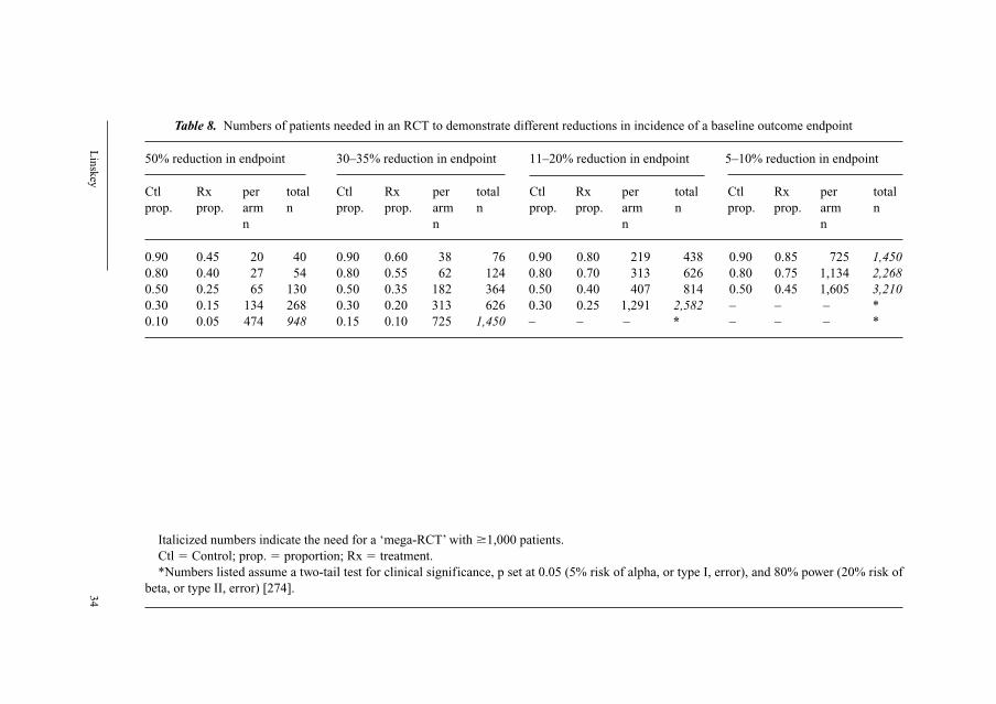
Lin
skey
34
Table 8. Numbers of patients needed in an RCT to demonstrate different reductions in incidence of a baseline outcome endpoint
50% reduction in endpoint 30–35% reduction in endpoint 11–20% reduction in endpoint 5–10% reduction in endpoint
Ctl Rx per total Ctl Rx per total Ctl Rx per total Ctl Rx per total
prop. prop. arm n prop. prop. arm n prop. prop. arm n prop. prop. arm n
n n n n
0.90 0.45 20 40 0.90 0.60 38 76 0.90 0.80 219 438 0.90 0.85 725 1,4500.80 0.40 27 54 0.80 0.55 62 124 0.80 0.70 313 626 0.80 0.75 1,134 2,2680.50 0.25 65 130 0.50 0.35 182 364 0.50 0.40 407 814 0.50 0.45 1,605 3,2100.30 0.15 134 268 0.30 0.20 313 626 0.30 0.25 1,291 2,582 – – – *
0.10 0.05 474 948 0.15 0.10 725 1,450 – – – * – – – *
Italicized numbers indicate the need for a ‘mega-RCT’ with �1,000 patients.
Ctl � Control; prop. � proportion; Rx � treatment.
*Numbers listed assume a two-tail test for clinical significance, p set at 0.05 (5% risk of alpha, or type I, error), and 80% power (20% risk of
beta, or type II, error) [274].
EBM for Neurosurgeons 35
valid in the clinic setting for decision-making purposes, your patient must at
least resemble the trial patients in terms of age, sex, pathological diagnosis,
severity of disease, social class, dose and duration of treatment, concomitant
ongoing diseases and treatments, and nursing care [276, 277].
In addition, RCTs often rely on measuring surrogate endpoints for out-
comes rather than the actual outcome of greatest clinical interest. This may be
because the surrogate endpoint comes about sooner than the endpoint it is sup-
posed to predict (thus shortening the length of the RCT and reducing its cost),
because it is easier to quantify, or because the surrogate outcome has a higher
baseline incidence than the endpoint alternative, thus requiring fewer patients to
demonstrate a significant change in a RCT (see table 8). Unfortunately, surrogate
endpoints are secondary predictive outcome variables that can be misleading
[203].
While selection bias still exists in RCT due to select population sampling
for study, randomization is our best methodological means of reducing subse-
quent bias once the study is underway. Unfortunately, even randomization does
not entirely eliminate bias during the treatment phase. Even in an RCT, it may
be the case that a researcher’s theoretical persuasion leads to their favorite ther-
apy being administered in their study with more fidelity and enthusiasm than
those to which it is compared [217]. Evidence for this assertion comes from two
sources. First, studies of RCTs show that the treatment group ends up being
repeatedly smaller, and statistically significantly smaller, than the placebo group
by the time of analysis [278]. This suggests that patients are more often removed
from the treatment group prior to analysis despite attempts at ‘blinding’ and
‘intent to treat’ modeling. Second, RCTs with outside sponsors more often
report statistically significant advances than unaligned grant-supported studies
[279].
Beyond considerations of practical feasibility, external validity, and resid-
ual bias within individual RCTs, lies an even greater concern. Namely, that by
placing so much emphasis on the primacy of RCTs, EBM may be introducing
a systematic bias into clinical decision making, guideline methodology, and
healthcare policy decisions [199]. The effort and money required for RCTs is
more likely to be spent on problems that are inherently interesting to clinical
researchers and/or sponsoring corporations at the expense of less interesting
alternatives that may be more important to our patients or the public as a whole
[117, 185, 199, 280]. Pharmacological interventions will likely have an advan-
tage over alternative treatments due to greater chance of corporate sponsorship,
and easier introduction of patient and physician blinding compared with hands-
on therapies or interventions [185, 211]. Ease and power of statistical analysis
will favor easily quantifiable endpoints over equally important, but difficult to
quantify endpoints, such as pain and quality of life [181, 185, 195, 196, 199,
Linskey 36
209, 281]. Easier to measure endpoints will have an advantage over more diffi-
cult to measure (more expensive and/or time-consuming) endpoints [181, 185,
195, 209, 281]. Also, given the cost and effort involved, there is a tendency to
study acute (rather than subacute or chronic) disease processes [185, 282], to
study curative versus palliative interventions, and to measure short-term surro-
gate endpoints that may not always predict the more important longer-term out-
comes of interest.
As a result there will always likely be a greater quantity of RCT evidence
available in the literature for acute and/or fashionable disease processes treated
with economically lucrative and/or fashionable interventions assessed using
easily measured and quantifiable endpoints, than for chronic diseases, non-
corporately-marketable interventions, or interventions assessed using difficult-to-
quantify and/or measure endpoints. There will likely never be RCT evidence for
sufficiently rare diseases, interventions, or procedures [184]. There will likely
be more published evidence available for initially effective treatments (mea-
sured against placebo or natural history) than for subsequent potentially equiv-
alent therapies (null studies compared with SOC). Relying on RCTs as best
evidence of effectiveness can disadvantage older (often cheaper) treatments that
have not been as rigorously evaluated as newer (often more expensive) treat-
ments [154]. On the other hand, many newer interventions, particularly those in
rapidly evolving fields or utilizing rapidly evolving technology (even if spec-
tacularly effective), are unlikely to have been studied by RCT [192, 199].
Methodological Design and Rigor as Sole Arbiter for Levels of ‘Evidence’Methodological design is an important concern for assessing a clinical
study in order to avoid the error of allowing bias to lead us to accept incorrect
conclusions. Within EBM, RCTs are recognized as the methodology of choice
to limit the misleading influence of bias. However, while excellent for studying
therapeutic interventions, the RCT is a poor methodology for studying other
epidemiological questions. Furthermore, there may be an inflection point where
strength of effect should outweigh strength of methodology in assessing the
level of evidence, or where the quality of a given study should lead it to carry
more weight than a study of poor quality utilizing a less biased methodology.
RCT design is a poor methodology for answering or exploring epidemio-
logical questions of potential disease etiology, pathophysiologic causality,
therapeutic side effects, or describing new diseases [211]. Observational study
designs are often the most informative methodology for exploring etiologic and
pathophysiologic clinical research. The case-control study is the most potent
research tool for studying side effects of interventions. Case series or even case
reports remain superior for communicating new, unique or strange observations
EBM for Neurosurgeons 37
that may turn out to describe new diseases and/or lend insight into new treatment
strategies or pathophysiologic understanding. RCTs tend to be most appropriate
in remedial situations (when disease is already present) and less applicable in
studying disease prevention (especially if baseline population is heterogeneous)
[117]. Applying a methodologically based hierarchy for levels of evidence where
RCTs remain at the apex in all these areas of medical inquiry may not be appro-
priate, unless the secondary analysis of the evidence (clinical decision and clini-
cal practice parameter recommendation) adequately takes this into account.
There is a major difference between strength of effect for an intervention
and strength of evidence supporting the use of that intervention [55]. Indeed
there are interventions where the magnitude of effect is so strong with lower
methodology analysis that the effect is very unlikely to be accountable by bias,
and where failure to act on an individual case level or strongly recommend at a
clinical practice parameter level is probably inappropriate. The introduction of
penicillin in the 1930s and 1940s is a classic example. The dramatic effect of
penicillin was obvious at the case report and case series levels. Whole inpatient
infectious disease wards emptied as a result. It would not have been appropriate
or desirable to await RCT testing before recommending penicillin as standard
therapy for pneumonia. The introduction and application of nocturnal continuous
positive airway pressure for hypersomnolence and sleep deprivation syndrome
related to sleep apnea is another example [283, 284]. For neurosurgery, a good
example is earliest possible, rather than delayed, intervention for patients with
epidural hematoma and pupillary asymmetry [285, 286].
A hierarchy of levels of evidence based on methodology also fails to ade-
quately account for rigor and quality within individual studies [206]. It is not at
all clear that a poorly performed RCT deserves consideration as stronger evi-
dence than a very thorough and well-done observational study. There may in
fact be significant overlap between the categories such that the levels should be
considered overlapping ‘roofing shingles’ rather than discrete and separate ‘rungs
on a ladder’. Mechanisms for adequately and consistently accounting for this
issue are not yet in place.
From Guidelines to Polices and from EBM to EBCAt the public health and health management level, EBC is an attractive
philosophy for providing an objective and science-based rationale for health-
care policies. Whether one is dealing with a formally nationalized health ser-
vice such as Great Britain, or a US CMS service structured on a sustainable
growth rate formula (where a relatively fixed amount of reimbursement money
is allocated internally by shifting relative value units), we have entered an era of
healthcare rationing. EBM has a potential role to play in making rationing less
crude, arbitrary, and political [197]. Under this model funding can reflect
Linskey 38
guideline-driven priorities, and priority funding can be reserved for interven-
tions with evidence of clinical effectiveness. Unfortunately, rationing implies
cost-cutting and not just preferential funding, and EBM can be used as a cost-
cutting tool [185, 201, 203].
The field of logic includes description of a classic logical fallacy where
absence of evidence is interpreted as evidence of absence. This logical pitfall is
particularly germane as a warning for attempts at EBC. Absence of proof of
effectiveness of a healthcare intervention is very different from proof of lack of
an effect [215, 287], and the presence of formal evidence for efficacy does not
necessarily mean that this treatment option is superior to alternatives for which
evidence (or evidence of an equivalent level) does not exist [185]. It is impor-
tant to ensure that lack of evidence of effectiveness should lead to presentation
of the treatment as an option rather than elimination of the treatment from fund-
ing or approval [164].
Much of accepted medical practice has never been validated in RCTs and
there will always be plausible interventions for which no evidence is (yet) avail-
able [211, 288]. As a result, there will be large gaps or holes in any EBM clini-
cal practice parameter effort attempting to codify clinical practice. Practitioners
who limit themselves only to what is provable will preclude the use of many
useful treatments [198], but it is not sufficient reason to withhold potentially
beneficial intervention from patients [211].
RCTs tend to focus on quantitative and measurable endpoints. However, in
healthcare not all that is measurable is of value, and not all that is of value can
be measured [181, 198, 204]. Indeed it is a short step from a judgment that an
intervention is ‘without substantial evidence’ to ‘without substantial value’ [181].
As a result, incorrectly interpreted or applied, some fear that EBM has the
potential to devalue the unquantifiable in medicine [194, 198]. There is also a
large difference between statistical significance and clinical significance, espe-
cially when the studied is evaluating a surrogate endpoint rather than the clini-
cal endpoint directly relevant to clinical decision making and patient priority, or
when the magnitude of effect is quite small despite statistical significance in an
RCT [154]. Based on the systematic bias introduced by focus on RCT evidence
discussed above, EBM could also bias healthcare and reimbursement policies
against palliative care interventions, interventions for chronic diseases, thera-
pies for rare diseases or uncommon therapeutic interventions, as well as non-
corporately-marketable therapies (see section entitled ‘Inherent Limitations of
Prospective Randomized Clinical Trials’).
Meta-analysis can also be misapplied with negative effects for funding and
approval for health interventions. There have been several reports of the nega-
tive experience of having an incomplete or out-of-date meta-analysis, or meta-
analyses performed by people without the subject matter expertise germane to
EBM for Neurosurgeons 39
the clinical condition in question, being adopted uncritically and effecting clin-
ical decisions/policies [283, 289].
There is currently little strong evidence that guidelines substantially influ-
ence practice [117, 228, 290, 291]. Part of the reason may be that physicians
tend not to think clinically algorithmically but rely more on pattern recognition
as the most important part of differentiating problems [292, 293]. It may be that
algorithmic protocols and guidelines are more suited to management planning
than individual differential decision making [288]. Clinicians continue to rely
on personal experience over research data. They tend to be realists rather than
empiricists. Clinical trial evidence is considered only one of many forms of
knowledge which can influence decision making. They tend to endorse and refer
to guidelines most often for decisions that exist on the periphery of their expe-
rience [186]. Only when sufficient evidence has accumulated to challenge their
original belief do physicians tend to alter what they believe [186].
In addition, not all medical questions are scientific in their nature, and
many nonmedical factors effect decision making [204], including patient
choice. Social or psychological components of the patient’s problem may justify
‘under’- or ‘over’-management of the problem to achieve the individual goal of
care [288]. Cultural differences in physician and patient beliefs, priorities, and
expectations may need to be taken into account [294].
Even establishing EBM clinical practice parameters for every area of med-
icine will not likely, in-and-of-itself, be sufficient to transform healthcare into
EBC. Assumptions that variation in healthcare is predominantly due to variations
in approach of the physicians to the consultation rather than to effects of the
environment surrounding the clinical consultation (e.g. economic constraints,
resource shortages/limitations, regulatory constraints) are probably incorrect, or
at least a significant oversimplification [203, 223]. Change must involve the
environment of work and not just the individual physician [288]. Third party
authorization limitations, economic constraints and counterproductive incen-
tives may compete with the dictates of evidence.
Conclusions
Properly understood and employed, EBM is a tool of considerable value
for medicine and neurosurgery. EBM is not a discipline onto itself within med-
icine [218] or a paradigm shift in the true Kuhnian sense [295]. Rather, it is a
‘further developing and professionalizing the prevailing paradigm through a
refinement of concepts that increasingly lessens their resemblance to their usual
common-sense prototypes’ [15, 19, 295]. It provides a secure base for clinical
practice and practice improvement.
Linskey 40
While less efficient than textbooks and didactic curricula as a means of
rapidly acquiring a workable fund of clinical knowledge during medical and
neurosurgical training, EBM critical appraisal training is an effective tool for
keeping up-to-date with rapidly changing management strategies as part of life-
long learning and practice evolution. EBM is very effective for identifying gaps
in clinical knowledge which can help us identify areas that need clinical research
attention. However, as a closed, reductive, and deductive system, it tends to be
relatively sterile for generating innovation, and for identifying potential strate-
gies for filling these gaps. It is the soundest basis for developing and establish-
ing clinical practice parameters.
With the support and backing of governmental agencies, professional med-
ical societies, the AAMC, the ACGME, and the ABMS, EBM is likely here to
stay. While some have argued that RCTs, meta-analysis, and guidelines have
peaked in popularity, and are already on the decline [117], this is probably not
correct. One can liken any perceived lull in penetration and impact to the time
necessary for consolidation of an established beachhead, just prior to the defin-
itive breakout and complete penetration across the countryside.
The fact that (1) EBM philosophy and critical appraisal techniques have
become fully integrated into the training and culture of our younger colleagues
(who will comprise an ever-increasing percentage of neurosurgical practition-
ers), (2) the fact that maintenance of certification will require individuals to
demonstrate personal evidence-based practice based on tracking and critical
analysis of personal practice outcomes as part of the performance-based learning
and improvement competency, and (3) the fact that the progressively growing
national healthcare expenditures (now approaching 20% of our gross national
product) will necessitate increasing basis of reimbursement and funding based
on evidence-based effectiveness and guidelines, all point to the likelihood that
complete immersion of neurosurgical practice in EBM is inevitable. Widespread
small-area variation in clinical practice and perceived high levels of avoidable
error will also likely drive this effort. The EBM movement has convinced busi-
nessmen, politicians, bureaucrats and consumers that probabilistic knowledge
of medical effectiveness is the means to better healthcare [196]. The fact that as
of 2002, Wiebe and Demaerschalk [296] found most evidence-based care infor-
mation sources substantially lacking in coverage of the neurosciences and
therefore of limited use to clinicians in this field, simply reflects that neuro-
surgery has been relatively insulated to this point compared with other areas of
medicine.
The transition to EBM within neurosurgery will not likely be easy or pro-
ceed without reluctance and resistance. Studies of innovation and change sug-
gest that innovators pick things up regardless (�2.5% of the population), early
adopters need written methods, scientific argument and credible sources (the
EBM for Neurosurgeons 41
majority preferring personal sources, opinion leaders, peer activities and rein-
forcement by social network), and late adopters usually require regulations,
laws, incentives or sanctions, practical resources and provisions [297, 298].
Each of these latter possibilities likely lie on our horizon.
The healthcare policy implications of EBM and EBC are very real and
deserve the careful attention of both individual neurosurgeons and our national
organizations. Developing clinical accountability and a scientific rationale for
rationing decisions and healthcare priorities is a rational managerial effort, but it
will likely proceed at the expense of individual professional autonomy. Guidelines
are important for advancing our field and the optimal care of our patients, but
could become instruments of control. In differing circumstances of uncertainty,
information (evidence) can be used for many purposes including ammunition in
the struggle for influence to force or leverage decisions in favor of one stake-
holder over others. It thus becomes critical that we continue to promote a strict
and defensible evidence-based methodology for our own guidelines efforts, and
that we keep pace with other medical professions and/or stakeholders who
would establish ‘guidelines’ that might affect policies or healthcare decisions
regarding neurosurgical diseases and interventions.
As outlined in this chapter, EBM is not without weaknesses and limitations,
and the issues and controversies involved need to be understood by every practic-
ing neurosurgeon. EBM finds answers to only those questions open to its tech-
niques, and the best available evidence can be a far cry from scientific truth. EBM
should not rob us of the ability to confidently function in a world of uncertainty
that is inescapable in all branches of healthcare [184]. Yet knowing that the treat-
ment prescribed is backed by solid evidence, and that we can proceed with clear a
conscience, grants us a special form of freedom [203]. According to Naylor [189]:
‘good clinical medicine must always blend the art of uncertainty with the science of
probability’ [but the] ‘blend should be weighted heavily towards science, whenever and
wherever sound evidence is brought to light’.
In surgery, expertise can never be fully separated from the expert. No liter-
ature review or number of RCTs or meta-analyses performed can ever make one
an expert at clipping or coiling aneurysms, removing skull base tumors, or stabi-
lizing a complicated spine deformity. In 1996, Sackett et al. [23] highlighted the
importance of both evidence and clinical expertise, valuing the two equally:
‘without clinical expertise, practice risks becoming tyrannized by evidence but without
best available evidence, practice risks becoming rapidly out of date’.
Argument and counterargument by a mix of methodologic and biologic rea-
soning have always been the hallmark of medical progress [211]. To some extent
this dichotomy reflects the old empiricist-rationalist debate [197], and within the
tension between the two philosophies lies the fertile ground of innovation and
Linskey 42
new ideas. Clinical common sense is part of the art of medicine. By applying
common sense along with empiric evidence, we effectively step outside the sys-
tem to perceive a truth that is not apparent solely working deductively within a
complex system [194, 207].
References
1 Cochrane AL: 1931–1971: a critical review, with particular reference to the medical profession; in
Medicines for the Year 2000. London, Office of Health Economics, 1979, pp 2–12.
2 Chalmers I, Dickersin K, Chalmers TC: Getting to grips with Archie Cochrane’s agenda. BMJ
1992;305:786–788.
3 Sackett DL, Haynes RB, Guyatt GH, Tugwell P: Clinical Epidemiology: a Basic Science for
Clinical Medicine. Boston, Little, Brown, 1985.
4 Sackett DL, Haynes RB, Guyatt GH, Tugwell P: Clinical Epidemiology: a Basic Science for
Clinical Medicine, ed 2. Boston, Little, Brown, 1991.
5 Bennett KJ, Sackett DI, Haynes RB, Neufeld VR, Tugwell P, Roberts R: A controlled trial of teach-
ing critical appraisal of the clinical literature to medical students. JAMA 1987;257:2451–2454.
6 Shin JH, Haynes RB, Johnston ME: Effect of problem-based, self-directed undergraduate educa-
tion on life-long learning. CMAJ 1993;148:969–976.
7 Brook RH, Davies-Avery A, Greenfield S, Harris LJ, Lelah T, Solomon NE, Ware JE Jr: Assessing
the quality of medical care using outcome measures: an overview of the method. Med Care
1977;15(suppl):1–165.
8 Ellwood P: Shattuck lecture – outcomes management. A technology of patient experience. N Engl
J Med 1988;318:1549–1556.
9 Harbaugh RE: Clinical outcomes analysis; in Linskey ME, Rutigliano MJ (eds): Quality and Cost
in Neurological Surgery. Philadelphia, Lippincott William & Wilkins, 2001, pp 198–209.
10 Canadian Task Force on the Periodic Health Examination: The periodic health examination.
CMAJ 1979;121:1193–1254.
11 Canadian Task Force on the Periodic Health Examination: The Canadian Guide to Clinical
Preventive Health Care. Ottawa, Communication Group, 1994.
12 United States Preventive Services Task Force: Guide to Clinical Preventive Services. An
Assessment of the Effectiveness of 169 Interventions. Baltimore, Williams & Wilkins, 1989.
13 Acute Pain Management Guideline Panel: Acute Pain Management: Operative or Medical
Procedures and Trauma. Rockville, US Department of Health and Human Services, Public Health
Service, Agency for Health Care Policy and Research, 1992, AHCPR publication 92-0032.
14 Guyatt GH: Evidence-based medicine. Ann Intern Med 1991;114(ACP J Club Suppl 2):A16.
15 Evidence-Based Medicine Working Group: Evidence-based medicine. A new approach to teach-
ing the practice of medicine. JAMA 1992;268:2420–2425.
16 Sackett DL, Rosenberg WM: On the need for evidence-based medicine. Health Econ 1995;4:
249–254.
17 Sackett DL, Rosenberg WM: On the need for evidence-based medicine. J Public Health Med
1995;17:330–334.
18 Sackett DL, Rosenberg WM: The need for evidence-based medicine. J R Soc Med 1995;88:620–624.
19 Crawley L: Evidence-based medicine: a new paradigm for the patient. JAMA 1993;269:1253.
20 Oxman AD, Sackett DL, Guyatt GH: Users’ guides to the medical literature. I. How to get started.
Evidence-Based Medicine Working Group. JAMA 1993;270:2093–2095.
21 Davidoff F, Haynes B, Sackett D, Smith R: Evidence based medicine. BMJ 1995;310:1085–1086.
22 Rosenberg W, Donald A: Evidence based medicine: an approach to clinical problem-solving. BMJ
1995;310:1122–1126.
23 Sackett DL, Rosenberg WM, Gray JA, Haynes RB, Richardson WS: Evidence based medicine:
what it is and what it isn’t. BMJ 1996;312:71–72.
24 Montori VM, Guyatt GH: What is evidence-based medicine and why should it be practiced?
Respir Care 2001;46:1201–1214.

EBM for Neurosurgeons 43
25 Meier B: Three senators seeking details for a drug-trial database. NY Times, July 9, 2004.
26 Omnibus Budget Reconciliation Act of 1989, Public Law 101-239.
27 Field MJ, Lohr KN; Committee on Clinical Practice Guidelines, Institute of Medicine: Guidelines
for Clinical Practice. From Development to Use. Washington, National Academy Press, 1992.
28 White House Domestic Policy Council: The President’s Health Security Plan. New York, Times
Books, 1993.
29 Healthcare Research and Quality Act of 1999, Public Law 106-129.
30 Fletcher A: The NHS research and development programme and its impact on health care library
and information resource centres. Health Libr Rev 1995;12:243–247.
31 National Health Service Executive: Priorities and Planning Guidance for the NHS: 1995–96.
EL(94)55. Leeds, NHS Executive, 1994.
32 National Health Service Executive: Priorities and Planning Guidance for the NHS: 1996–97.
Leeds, NHS Executive, 1995.
33 National Health Service Executive: Research and Development in the New NHS: Functions and
Responsibilities. London, NHS, October 1994.
34 Scally G, Donaldson LJ: The NHS’s 50 anniversary. Clinical governance and the drive for quality
improvement in the new NHS in England. BMJ 1998;317:61–65.
35 Department of Health Press Release: Government keeps its promise for fairer, faster treatment for
patients: the National Institute for Clinical Excellence opens for business. London, Media Centre,
Department of Health, March 31, 1999.
36 National Institute for Clinical Excellence. Information about the work of NICE. http://www.nice.
org.uk/page.aspx?o � 113677, accessed July 10, 2002.
37 Rawlins M: In pursuit of quality: the National Institute for Clinical Excellence. Lancet
1999;353:1079–1082.
38 National Institute for Clinical Excellence Press Release: NICE establishes guidelines advisory
committee. London, NICE, NHS, May 23, 2000.
39 Rutigliano MJ: Cost effectiveness analysis: a review. Neurosurgery 1995;37:436–443.
40 Rutigliano MJ: Cost effectiveness analysis: theory, basis, and, methods; in Linskey ME, Rutigliano
MJ (eds): Quality and Cost in Neurological Surgery. Philadelphia, Lippincott Williams & Wilkins,
2001, pp 285–294.
41 Stoelwinder JU: EBM in healthcare: management and policy. Med J Aust 2001;174:644–646.
42 Haynes RB: The origins and aspirations of ACP Journal Club. ACP J Club 1991;114:A18.
43 Haynes RB: So long, best evidence. Hello ACP Journal Club Online. ACP J Club 2001
(Nov–Dec);135:A13.
44 Guyatt GH, Rennie D: Users’ guides to the medical literature. JAMA 1993;270:2096–2097.
45 Guyatt GH, Sackett DL, Cook DJ: Users’ guides to the medical literature. II. How to use an article
about therapy or prevention. A. Are the results of the study valid? JAMA 1993;270:2598–2601.
46 Guyatt GH, Sackett DL, Cook DJ: Users’ guides to the medical literature. II. How to use an article
about therapy or prevention. B. What were the results and will they help me in caring for my
patients. JAMA 1994;271:59–63.
47 Jaeschke R, Guyatt G, Sackett DL: Users’ guides to the medical literature. III. How to use an arti-
cle about a diagnostic test. A. Are the results of the study valid? JAMA 1994;271:389–391.
48 Jaeschke R, Guyatt GH, Sackett DL: Users’ guides to the medical literature. III. How to use an
article about a diagnostic test. B. What are the results and will they help me in caring for my
patients? JAMA 1994;271:703–707.
49 Levine M, Walter S, Lee H, Haines T, Holbrook A, Moyer V: Users’ guides to the medical litera-
ture. IV. How to use an article about harm. Evidence-Based Medicine Working Group. JAMA
1994;271:1615–1619.
50 Laupacis A, Wells G, Richardson WS, Tugwell P: Users’ guides to the medical literature. V. How to
use an article about prognosis. Evidence-Based Medicine Working Group. JAMA 1994;272:234–237.
51 Oxman AD, Cook DJ, Guyatt GH: Users’ guides to the medical literature. VI. How to use an
overview. Evidence-Based Medicine Working Group. JAMA 1994;272:1367–1371.
52 Richardson WS, Detsky AS: Users’ guides to the medical literature. VII. How to use a clinical
decision analysis. A. Are the results of the study valid? Evidence-Based Medicine Working Group.
JAMA 1995;273:1292–1295.

Linskey 44
53 Richardson WS, Detsky AS: Users’ guides to the medical literature. VII. How to use a clinical
decision analysis. B. What are the results and will they help me in caring for my patients?
Evidence-Based Medicine Working Group. JAMA 1995;273:1610–1613.
54 Hayward RSA, Wilson MC, Tunis SR, Bass EB, Guyatt G: Users’ guides to the medical literature.
VIII. How to use clinical practice guidelines. A. Are the recommendations valid. Evidence-Based
Medicine Working Group. JAMA 1995;274:570–574.
55 Wilson MC, Hayward RS, Tunis SR, Bass EB, Guyatt G: Users’ guides to the medical literature.
VIII. How to use clinical practice guidelines. B. What are the recommendations and will they help
you in caring for your patients? Evidence-Based Medicine Working Group. JAMA 1995;274:
1630–1632.
56 Guyatt GH, Sackett DL, Sinclair JC, Hayward R, Cook DJ, Cook RJ: Users’ guides to the medical
literature. IX. A method for grading health care recommendations. Evidence-Based Medicine
Working Group. JAMA 1995;274:1800–1804.
57 Naylor CD, Guyatt GH: Users’ guides to the medical literature. X. How to use an article reporting
variations in the outcomes of health services. The Evidence-Based Medicine Working Group.
JAMA 1996;275:554–558.
58 Naylor CD, Guyatt GH: Users’ guides to the medical literature. XI. How to use an article about a
clinical utilization review. Evidence-Based Medicine Working Group. JAMA 1996;275:1435–1439.
59 Guyatt GH, Naylor CD, Juniper E, Heyland DK, Jaeschke R, Cook DJ: Users’ guides to the med-
ical literature. XII. How to use articles about health-related quality of life. Evidence-Based
Medicine Working Group. JAMA 1997;277:1232–1237.
60 Drummond MF, Richardson WS, O’Brien BJ, Levine M, Heyland D: Users’ guides to the medical
literature. XIII. How to use an article on economic analysis of clinical practice. A. Are the results
of the study valid? Evidence-Based Medicine Working Group. JAMA 1997;227:1552–1557.
61 O’Brien BJ, Heyland D, Richardson WS, Levine M, Drummond MF: Users’ guides to the medical
literature. XIII. How to use an article on economic analysis of clinical practice. B. What are the
results and will they help me in caring for my patients? Evidence-Based Medicine Working
Group. JAMA 1997;277:1802–1806.
62 Dans AL, Dans LF, Guyatt GH, Richardson S: Users’ guides to the medical literature. XIV. How to
decide on the applicability of clinical trial results to your patient. Evidence-Based Medicine
Working Group. JAMA 1998;279:545–549.
63 Richardson WS, Wilson MC, Guyatt GH, Cook DJ, Nishikawa J: Users’ guides to the medical lit-
erature. XV. How to use an article about disease probability for differential diagnosis. Evidence-
Based Medicine Working Group. JAMA 1999;281:1214–1219.
64 Guyatt GH, Sinclair J, Cook DJ, Glasziou P: Users’ guides to the medical literature. XVI. How to
use a treatment recommendation. Evidence-Based Medicine Working Group and the Cochrane
Applicability Methods Working Group. JAMA 1999;281:1836–1843.
65 Barratt A, Irwig L, Glasziou P, Cumming RG, Raffle A, Hicks N, Gray JA, Guyatt GH: Users’
guides to the medical literature. XVII. How to use guidelines and recommendations about screen-
ing. Evidence-Based Medicine Working Group. JAMA 1999;281:2029–2034.
66 Randolph AG, Haynes RB, Wyatt JC, Cook DJ, Guyatt GH: Users’ guides to the medical literature.
XVIII. How to use an article evaluating the clinical impact of a computer-based clinical decision
support system. JAMA 1999;282:67–74.
67 Bucher HC, Guyatt GH, Cook DJ, Holbrook A, McAlister FA: Users’ guides to the medical literature.
XIX. Applying clinical trial results. A. How to use an article measuring the effect of an intervention on
surrogate end points. Evidence-Based Medicine Working Group. JAMA 1999;282:771–778.
68 McAlister FA, Laupacis A, Wells GA, Sackett DL: Users’ guides to the medical literature. XIX.
Applying clinical trial results. B. Guidelines for determining whether a drug is exerting (more
than) a class effect. Evidence-Based Medicine Working Group. JAMA 1999;282:1371–1377.
69 McAlister FA, Straus SE, Guyatt GH, Haynes RB: Users’ guides to the medical literature. XX.
Integrating research evidence with the care of the individual patient. Evidence-Based Medicine
Working Group. JAMA 2000;283:2829–2836.
70 Hunt DL, Jaeschke R, McKibbon KA: Users’ guides to the medical literature. XXI. Using elec-
tronic health information resources in evidence-based practice. Evidence-Based Medicine
Working Group. JAMA 2000;283:1875–1879.

EBM for Neurosurgeons 45
71 McGinn TG, Guyatt GH, Wyer PC, Naylor CD, Stiell IG, Richardson WS: Users’ guides to the
medical literature. XXII. How to use articles about clinical decision rules. Evidence-Based
Medicine Working Group. JAMA 2000;284:79–84.
72 Giacomini MK, Cook DJ: Users’ guides to the medical literature. XXIII. Qualitative research in
health care. A. Are the results of the study valid? Evidence-Based Medicine Working Group.
JAMA 2000;284:357–362.
73 Giacomini MK, Cook DJ: Users’ guides to the medical literature. XXIII. Qualitative research in
health care. B. What are the results and how do they help me care for my patients? Evidence-Based
Medicine Working Group. JAMA 2000;284:478–482.
74 Richardson WS, Wilson MC, Williams JW Jr, Moyer VA, Naylor CD: Users’ guides to the medical
literature. XXIV. How to use an article on the clinical manifestations of disease. Evidence-Based
Medicine Working Group. JAMA 2000;284:869–875.
75 Guyatt GH, Haynes RB, Jaeschke RZ, Cook DJ, Green L, Naylor CD, Wilson MC, Richardson WS:
Users’ guides to the medical literature. XXV. Evidence-based medicine: principles for applying
the users’ guides to patient care. Evidence-Based Medicine Working Group. JAMA 2000;284:
1290–1296.
76 Guyatt G, Rennie D: Users’ Guides to the Medical Literature: Manual for Evidence-Based
Clinical Practice. Evidence-Based Medicine Working Group. Chicago, AMA Press, 2002.
77 Guyatt G, Rennie D: Users’ Guides to the Medical Literature: Essentials of Evidence-Based
Clinical Practice. Evidence-Based Medicine Working Group. Chicago, AMA Press, 2002.
78 Davidoff F, Case K, Fried PW: Evidence-based medicine: why all the fuss? Ann Intern Med
1995;122:727.
79 Haines A, Donald A: Getting Research Findings into Practice. London, British Medical Journal
Publishing Group, 1998.
80 Florin RE: Outcomes measurement project started. AANS Bull 1997(fall);6/4:11.
81 Staff: AANS/CNS outcomes committee plans national pilot study on aneurysms. AANS Bull
1998(winter);7/1:7.
82 Harbaugh RE: Outcomes initiatives. AANS Bull 1999(spring);8/1:14–15.
83 Seaver MJ: Neuro-knowledge research partnership. AANS Bull 2003(spring);12/1:12–13.
84 Piepgras DG: Certification of neurosurgeons: a transition from neurosurgical certification and
recertification to maintenance of certification. Report of the American Board of Neurological
Surgeons. Clin Neurosurg 2003;99:1085–1088.
85 Gehlbach SH: Interpreting the Medical Literature: Practical Epidemiology for Clinicians, ed 2.
New York, Macmillan, 1988.
86 Hulley SB, Cummings SR, Browner SW, Newman TB, Hearst N: Designing Clinical Research: An
Epidemiologic Approach. Baltimore, Williams & Wilkins, 1988.
87 Riegelman RK, Hirsch RP: Studying a Study and Testing a Test: How to Read the Medical
Literature, ed 2. Boston, Little Brown, 1989.
88 Sackett DL, Richardson WS, Rosenberg W, Haynes RB: Evidence Based Medicine: How to
Practice and Teach EBM. New York, Churchill Livingstone, 1997.
89 Sackett DL, Straus SE, Richardson WS, Rosenberg W, Haynes RB: Evidence-Based Medicine:
How to Practice and Teach EBM, ed 2. Edinburgh, Churchill Livingstone, 2000.
90 King JT Jr: Quantifying neurosurgical outcomes: how do we measure the effectiveness in cost-
effective analysis?; in Linskey ME, Rutigliano MJ (eds): Quality and Cost in Neurological
Surgery. Philadelphia, Lippincott Williams & Wilkins, 2001, pp 316–324.
91 Ioannidis JPA, Haidich AB, Pappa M, Pantazis N, Kokori SI, Tektonidou MG, Contopoulos-
Ioannidis DG, Lau J: Comparison of evidence of treatment effects in randomized and nonrandom-
ized studies. JAMA 2001;2286:821–830.
92 Guyatt G, Sackett D, Taylor DW, Chong J, Roberts R, Pugsley S: Determining optimal therapy-
randomized trials in individual patients. N Engl J Med 1986;314:889–892.
93 Glasziou P, Guyatt GH, Dans AL, Dans LF, Straus S, Sackett DL: Applying the results of trials and
systematic reviews to individual patients. ACP J Club 1998;129:A15–A16.
94 Straus SE, Sackett DL: Applying evidence to the individual patient. Ann Oncol 1999;10:29–32.
95 Llewellyn-Thomas HA: Investigating patients’ preferences for different treatment options. Can J
Nurs Res 1997;29:45–64.
Linskey 46
96 Molyneux A, Kerr R, Stratton I, Sandercock P, Clark M, Shrimpton J, Holman R: International
Subarachnoid Aneurysm Trial (ISAT) of neurosurgical clipping versus endovascular coiling in
2143 patients with ruptured intracranial aneurysms: a randomized trial. Lancet 2002;360:
1267–1274.
97 Andrews DW, Scott CB, Sperduto PW, Flanders AE, Gaspar LE, Schell MC, Werner-Wasik M,
Demas W, Ryu J, Bahary JP, Souhami L, Rotman M, Mehta MP, Curran WJ Jr: Whole brain radia-
tion therapy with or without stereotactic radiosurgery boost for patients with one to three brain
metastases: phase III results of the RTOG 9508 randomised trial. Lancet 2004;363:1665–1672.
98 Swales J: The national health service and the science of evaluation: two anniversaries. Health
Trends 1998;30:20–22.
99 Feasby TE, Quan H, Ghali WA: Hospital and surgeon determinants of carotid endarterectomy out-
comes. Arch Neurol 2002;59:1877–1881.
100 Cowan JA Jr, Dimick JB, Leveque JC, Thompson BG, Upchurch GR Jr, Hoff JT: Impact of
provider volume on mortality after intracranial tumor resection. Neurosurgery 2003;52:48–54.
101 Ruby ST, Robinson D, Lynch JT, Mark H: Outcome analysis of carotid endarterectomy in
Connecticut: the impact of volume and specialty. Ann Vasc Surg 1996;10:22–26.
102 Barker FG 2nd, Amin-Hanjani S, Butler WE, Ogilvy CS, Carter BS: In-hospital mortality and
morbidity after surgical treatment of unruptured intracranial aneurysms in the United States,
1996–2000: effect of hospital and surgeon volume. Neurosurgery 2003;52:995–1009.
103 Barker FG 2nd: Craniotomy for the resection of metastatic brain tumors in the U.S., 1988–2000:
decreasing mortality and the effect of provider caseload. Cancer 2004;100:999–1007.
104 Hoh BL, Rabinov JD, Pryor JC, Carter BS, Barker FG 2nd: In-hospital morbidity and mortality
after endovascular treatment of unruptured intracranial aneurysms in the United States, 1996–2000:
effect of hospital and physician volume. Am J Neuroradiol 2003;24:1409–1420.
105 Kalkanis SN, Eskander EN, Carter BS, Barker FG 2nd: Microvascular decompression surgery in
the United States, 1996 to 2000; mortality rates, morbidity rates, and the effects of hospital and
surgeon volumes. Neurosurgery 2003;52:1251–1262.
106 Smith ER, Butler WE, Barker FG 2nd: Craniotomy for resection of pediatric brain tumors in the
United States, 1988 to 2000: effects of provider caseloads and progressive centralization and spe-
cialization of care. Neurosurgery 2004;54:553–565.
107 Smith ER, Butler WE, Barker FG 2nd: In-hospital mortality rates after ventriculoperitoneal shunt
procedures in the United States, 1998 to 2000: relation to hospital and surgeon volume of care.
J Neurosurg 2004;100(suppl 2):90–97.
108 Manning PR: Practice-based learning and improvement: a dream that can become a reality.
J Contin Educ Health Prof 2003;23(suppl 1):S6–9.
109 Shaughnessy AF, Slawson DC, Becker L: Clinical jazz: harmonizing clinical experience and
evidence-based medicine. J Fam Pract 1998;47:425–428.
110 McNeil BJ, Pauker SG, Sox HC Jr, Tversky A: On the elicitation of preferences for alternative
therapies. N Engl J Med 1982;306:1259–1262.
111 Glass GV: Primary, secondary, and meta-analysis of research. Educ Res 1976;5:3–8.
112 Glass G, Smith ML: Meta-analysis of research on class size and achievement. Educ Eval Policy
Anal 1979;1:2–16.
113 Glass G, McGaw B, Smith ML: Meta-Analysis in Social Research. Beverly Hills, Sage, 1981.
114 Naylor CD: The case for failed meta-analyses. J Eval Clin Pract 1995;1:127–130.
115 Haines SJ, Walters BC: What is metanalysis? Surg Neurol 1994;41:381–388.
116 Chalmers I, Altman DG: Systematic Reviews. London, BMJ Publishing, 1995.
117 Maisonneuve H, Ojasoo T: From the life-cycles of clinical evidence to the learning curve of clini-
cal experience. J Eval Clin Pract 1999;5:417–421.
118 Light RJ, Pillemer DB: Summing up: the Science of Reviewing Research. Cambridge, Harvard
University Press, 1984.
119 Hedges LV, Olkin I: Statistical Methods for Meta-Analysis. Orlando, Academic Press, 1985.
120 L’Abbe KA, Detsky AS, O’Rourke K: Meta-analysis in clinical research. Ann Intern Med
1987;107:224–233.
121 Sacks HS, Berrier J, Reitman D, Anacona-Berk VA, Chalmers TC: Meta-analyses of randomized
controlled trials. N Engl J Med 1987;316:450–455.
EBM for Neurosurgeons 47
122 Hunter JE, Schmidt FL: Methods of Meta-Analysis: Correcting Error and Bias in Research
Findings. Newbury Park, Sage, 1990.
123 Dickersin K, Berlin JA: Meta-analysis: state-of-the-science. Epidemiol Rev 1992;14:154–176.
124 Sackett DL, Oxman AD: The Cochrane Collaboration Handbook. Oxford, Cochrane Collaboration,
1994.
125 Clarke MF, Stewart LA: Systematic reviews of randomized controlled trials: the need for complete
data. J Eval Clin Pract 1995;1:119–126.
126 Jones DR: Meta-analysis: weighing the evidence. Stat Med 1995;14:137–149.
127 Olkin I: Meta-analysis: reconciling the results of independent studies. Stat Med 1995;14:457–472.
128 Spitzer WO: The Potsdam International Consultation on Meta-Analysis. J Clin Epidemiol
1995;48:1–171.
129 Cook DJ, Sackett DL, Spitzer WO: Methodologic guidelines for systematic reviews of randomized
controlled trials in health care from the Potsdam Consultation on Meta-Analysis. J Clin Epidemiol
1995;48:167–171.
130 Lau J, Antman EM, Jimenez-Silva J, Kupelnick B, Mosteller F, Chalmers TC: Cumulative meta-
analysis of therapeutic trials for myocardial infarction. N Engl J Med 1992;327:248–254.
131 Hofman PAM, Nelemans P, Kemerink, GJ, Wilmink JT: Value of radiological diagnosis of skull
fracture in the management of mild head injury: meta-analysis. J Neurol Neurosurg Psychiatry
2000;68:416–422.
132 Chappell ET, Moure FC, Good MC: Comparison of computed tomographic angiography with digi-
tal subtraction angiography in the diagnosis of cerebral aneurysms: a meta-analysis. Neurosurgery
2003;52:624–631.
133 Cloft HJ, Kallmes DF, Kallmes MH, Goldstein JH, Jensen ME, Dion JE: Prevalence of cerebral
aneurysms in patients with fibromuscular dysplasia: a reassessment. J Neurosurg 1998;88:436–440.
134 Langley JM, LeBlanc JC, Drake J, Milner R: Efficacy of antimicrobial prophylaxis in placement
of cerebrospinal fluid shunts: meta-analysis. Clin Infect Dis 1993;17:98–103.
135 Barker FG 2nd: Efficacy of prophylactic antibiotics for craniotomy: a meta-analysis. Neurosurgery
1994;35:484–492.
136 Di Mascio R, Marchioli R, Tognoni G: From pharmacological promises to controlled clinical tri-
als to meta-analysis and back: the case of nimodipine in cerebrovascular disorders. Clin Trials
Metaanal 1994;29:57–79.
137 Haines SJ, Walters BC: Antibiotic prophylaxis for cerebrospinal fluid shunts: a metanalysis.
Neurosurgery 1994;34:87–92.
138 Barker FG 2nd, Ogilvy CS: Efficacy of prophylactic nimodipine for delayed ischemic deficit after
subarachnoid hemorrhage: a meta-analysis. J Neurosurg 1996;84:405–414.
139 Barker FG 2nd: Efficacy of prophylactic antibiotics in cerebrospinal shunt surgery: a meta-analysis.
Neurosurgery 1998;43:694.
140 Prabhu VC, Kaufman HH, Voelker JL, Aronoff SC, Niewiadomska-Bugaj M, Mascaro S, Hobbs GR:
Prophylactic antibiotics with intracranial pressure monitors and external ventricular drains: a review
of the evidence. Surg Neurol 1999;52:226–237.
141 Farace E, Alves W: Do women fare worse: a metaanalysis of gender differences in traumatic brain
injury outcome. J Neurosurg 2000;93:539–545.
142 Barker FG 2nd: Efficacy of prophylactic antibiotic therapy in spinal surgery: a meta-analysis.
Neurosurgery 2002;51:391–401.
143 Cooper HM, Rosenthal R: Statistical versus traditional procedures for summarizing research find-
ings. Psychol Bull 1980;87:442–449.
144 Field MJ, Lohr KN, Institute of Medicine, Committee to Advise the Public Health Service on Clinical
Practice Guidelines: Clinical Practice Guidelines: Directions for a New Program. Washington,
National Academy Press, 1990.
145 Woolf SH: Practice guidelines, a new reality in medicine. II. Methods of developing guidelines.
Arch Intern Med 1992;152:946–952.
146 Delamothe T: Wanted: guidelines that doctors will follow. Implementation is the problem. BMJ
1993;307:218.
147 Scott EA, Black N: When does consensus exist in expert panels? J Public Health Med
1991;13:35–39.

Linskey 48
148 Jackson R, Feder G: Guidelines for clinical guidelines: a simple, pragmatic strategy for guideline
development. BMJ 1998;317:427–428.
149 AMA: Office of Quality Insurance and Healthcare Organizations’ Attributes to Guideline
Development of Practice Parameters. Chicago, AMA, 1990.
150 Rosenberg J, Greenberg MK: Practice parameters: strategies for survival into the nineties.
Neurology 1992;42:1110–1115.
151 Walters BC: Clinical practice parameter development in neurosurgery; in Bean JR (ed):
Neurosurgery in Transition: The Socioeconomic Transformation of Neurological Surgery.
Baltimore, Williams & Wilkins, 1998, pp 99–111.
152 Grimshaw JM, Russell IT: Effect of clinical guidelines on medical practice: a systematic review of
rigorous evaluations. Lancet 1993;342:1317–1322.
153 Grimshaw J, Russell I: Achieving health gain through clinical guidelines. I. Developing scientifi-
cally valid guidelines. Qual Health Care 1993;2:243–248.
154 Grimshaw J, Eccles M, Russell I: Developing clinically valid practice guidelines. J Eval Clin Pract
1995;1:37–48.
155 Brook RH, Kosecoff JB, Park RE, Chassin MR, Winslow CM, Hampton JR: Diagnosis and treat-
ment of coronary disease: comparison of doctors’ attitudes in the USA and the UK. Lancet
1988;i:750–753.
156 Kahn KL, Park RE, Brook RH, Chassin MR, Kosecoff J, Fink A, Keesey JW, Solomon DH: The
effect of comorbidity on appropriateness ratings for two gastrointestinal procedures. J Clin
Epidemiol 1988;41:115–122.
157 Leape LL, Park RE, Kahan JP, Brook RH: Group judgments of appropriateness: the effect of panel
composition. Qual Assur Health Care 1992;4:151–159.
158 Fraser GM, Pilpel D, Hollis S, Kosecoff J, Brook RH: Indications for cholecystectomy: the results
of a consensus panel approach. Qual Assur Health Care 1993;5:75–80.
159 Larson EB: Evidence-based medicine: is translating evidence into practice a solution to the cost-
quality challenges facing medicine? Jt Comm J Qual Improv 1999;25:480–485.
160 Chalmers TC, Frank CS, Reitman D: Minimizing the three stages of publication bias. JAMA
1990;263:1392–1395.
161 Shaneyfelt TM, Mayo-Smith MF, Rothwangl J: Are guidelines following guidelines? The method-
ological quality of clinical practice guidelines in the peer-reviewed medical literature. JAMA
1999;281:1950–1951.
162 Liberati A, Buzzetti R, Grilli R, Magrini N, Minozzi S: Which guidelines can we trust? Assessing
strength of evidence behind recommendations for clinical practice. West J Med 2001;174:
262–265.
163 Woolf SH, Battista R, Anderson GM, Logan AG, Wang E: Assessing the clinical effectiveness of
preventive maneuvers: analytic principles and systematic methods in reviewing evidence and
developing clinical practice recommendations. A report by the Canadian Task Force on the
Periodic Health Examination. J Clin Epidemiol 1990;43:891–905.
164 Eddy DM: A Manual for Assessing Health Practices and Designing Practice Policies: the Explicit
Approach. Philadelphia, American College of Physicians, 1992.
165 United States Congress, Office of Technology Assessment: Identifying Health Technologies That
Work. Searching for Evidence. Washington, United States Government Printing Office, 1994,
publication No OTA-H-608.
166 Field MJ, Institute of Medicine, Committee on Methods for Setting Priorities for Guidelines
Development, Division of Health Care Services: Setting Priorities for Clinical Practice
Guidelines. Washington, National Academy Press, 1995.
167 Eccles M, Clapp Z, Grimshaw J, Adams PC, Higgins B, Purves I, Russell I: North of England
evidence based guidelines development project: methods of guideline development. BMJ
1996;312:760–762.
168 Hadorn DC, Baker D, Hodges JS, Hicks N: Rating the quality of evidence for clinical practice
guidelines. J Clin Epidemiol 1996;49:749–754.
169 Liddle J, Williams M, Irwig L: Method for evaluating research and guideline evidence. Sydney,
New South Wales Health Department, 1996. http://www.health.nsw.gov.au/public-health/crcp/
publications/general/merge.html

EBM for Neurosurgeons 49
170 United States Preventive Services Task Force: Guide to Clinical Preventive Services, ed 2.
Baltimore, Williams & Wilkins, 1996.
171 Woolf SH, DiGuiseppi CG, Atkins D, Kamerow DB: Developing evidence-based clinical practice
guidelines: lessons learned by the US Preventive Services Task Force. Annu Rev Public Health
1996;17:511–538.
172 Jovell AL, Navarro-Rubio MD: Evaluacion de la evidencia cientifica. Med Clin (Barc) 1997;105:
740–743.
173 Ball C, Phillips B, Sackett D, Badenoch D, Straus S, Haynes B, Dawes M: Levels of Evidence and
Grades of Recommendation. Oxford, Oxford Centre for Evidence-Based Medicine, 1998. http://
www.cebm.net/levels_of_evidence.asp
174 Brain Trauma Foundation. Guidelines for severe head injury management. J Neurotrauma
1996;13:639–731.
175 Section on Disorders of the Spine and Peripheral Nerves: Guidelines for the management of acute
cervical spine and spinal cord injuries. Neurosurgery 2002;50(suppl 3):1–199.
176 Adelson PD, et al., American Association for Surgery of Trauma: Guidelines for the acute medical
management of severe traumatic brain injury in infants, children, and adolescents. Pediatr Crit
Care Med 2003;4(suppl 3):S1–S75.
177 Low Grade Glioma Guidelines Team: Practice parameters in adults with suspected or known
supratentorial nonoptic pathway low-grade glioma. Neurosurg Focus 1998;4:article 10.
178 Tanenbaum SJ: What physicians know. N Engl J Med 1993;329:1268–1271.
179 Tanenbaum SJ: Knowing and acting in medical practice: the epistemological politics of outcomes
research. J Health Polit Policy Law 1994;19:27–44.
180 Blau JN: Evidence-based medicine. Lancet 1995;346:1300.
181 Bradley F, Field J: Evidence-based medicine. Lancet 1995;346:838–839.
182 Charlton BG: Evidence based medicine: megatrials are subordinate to medical science. BMJ
1995;311:257.
183 Clarke MJ, Stewart LA: Systematic reviews of randomized controlled trials: the need for complete
data. J Eval Clin Pract 1995;1:119–126.
184 Jones GW, Sagar SM: Evidence based medicine. No guidance is provided for situations for which
evidence is lacking. BMJ 1995;311:258.
185 Hope T: Evidence based medicine and ethics. J Med Ethics 1995;21:259–260.
186 Hunter DJ: Effective practice. J Eval Clin Pract 1995;1:131–134.
187 Marshall T: Evidence-based medicine. Lancet 1995;346:1171–1172.
188 Naylor CD: The case for failed meta-analyses. J Eval Clin Pract 1995;1:127–130.
189 Naylor CD: Grey zones of clinical practice: some limits to evidence-based medicine. Lancet
1995;345:840–842.
190 Norman G: Evidence-based medicine. Lancet 1995;346:1300.
191 O’Rourke MF: The move towards evidence-based medicine. Med J Aust 1995;163:332–333.
192 Rigge M: Consumer audit. A speech to the second National Medical and Dental Audit Conference,
4 April 1995. J Eval Clin Pract 1995;1:135–140.
193 Shahar E: Evidence-based medicine. Lancet 1995;346:1172.
194 Sleigh JW: Evidence-based medicine and Kurt Godel. Lancet 1995;346:1172.
195 Smith BH: Evidence-based medicine: quality cannot always be quantified. BMJ 1995;311:258.
196 Tanenbaum S: Getting there from here: evidentiary quandaries of the US outcomes movement.
J Eval Clin Pract 1995;1:97–103.
197 Hunter DJ: Rationing and evidence-based medicine. J Eval Clin Pract 1996;2:5–8.
198 Polychronis A, Miles A, Bentley P: Evidence-based medicine: reference? Dogma? Neologism?
New orthodoxy? J Eval Clin Pract 1996;2:1–3.
199 Polychronis A, Miles A, Bentley P: The protagonists of ‘evidence-based medicine’: arrogant,
seductive and controversial. J Eval Clin Pract 1996;2:9–12.
200 Blau JN: Evidence-based medicine. J Eval Clin Pract 1997;3:149–151.
201 Charlton BG: Restoring the balance: evidence-based medicine put in its place. J Eval Clin Pract
1997;3:87–98.
202 Feinstein AR, Horwitz RI: Problems in the ‘evidence’ of ‘evidence-based medicine’. Am J Med
1997;103:529–535.

Linskey 50
203 Hampton JR: Evidence-based medicine, practice variations and clinical freedom. J Eval Clin Pract
1997;3:123–131.
204 Miles A, Bentley P, Polychronis A, Grey J: Evidence-based medicine: why all the fuss? This is
why. J Eval Clin Pract 1997;3:83–86.
205 Morgan WKC: On evidence, embellishment and efficacy. J Eval Clin Pract 1997;3:117–122.
206 Shahar E: A Popperian perspective of the term ‘evidence-based medicine’. J Eval Clin Pract
1997;3:109–116.
207 Sleigh JW: Logical limits of randomized controlled trials. J Eval Clin Pract 1997;3:145–148.
208 Black D: The limitations of evidence. J R Coll Physicians Lond 1998;32:23–26.
209 Kerridge I, Lowe M, Henry D: Ethics and evidence based medicine. BMJ 1998;316:1151–1153.
210 Tonelli MR: The philosophical limits of evidence-based medicine. Acad Med 1998;73:
1234–1240.
211 Vandenbroucke JP: Observational research and evidence-based medicine: what should we teach
young physicians? J Clin Epidemiol 1998;51:467–472.
212 Clarke JB: Evidence-based practice: a retrograde step? The importance of pluralism in evidence
generation for the practice of health care. J Clin Nurs 1999;8:89–94.
213 Goodman NW: Who will challenge evidence-based medicine? J R Coll Physicians Lond
1999;33:249–251.
214 Lalwani SI, Olive DL: Problems with evidence-based medicine. J Am Assoc Gynecol Laparosc
1999;6:237–240.
215 Van Weel C, Knottnerus JA: Evidence-based interventions and comprehensive treatment. Lancet
1999;353:916–918.
216 Goodman NW: In my view . . . Criticizing evidence-based medicine. Thyroid 2000;10:157–160.
217 Messer SB: Empirically supported treatments: cautionary notes. Medscape Gen Med 2002;4:13.
http://www.medscape.com/viewarticle/445082.
218 Editorial: Evidence-based medicine, in its place. Lancet 1995;346:785.
219 Grahame-Smith D: Evidence based medicine: Socratic dissent. BMJ 1995;310:1126–1127.
220 Osler W: The Principles and Practice of Medicine: Designed for the Use of Practitioners and
Students of Medicine. New York, Appleton, 1982.
221 Flexner A: Medical Education in the United States and Canada: a Report to the Carnegie
Foundation for the Advancement of Teaching. New York, Merrymount Press, 1910.
222 Sackett D: Evidence-based medicine. Lancet 1995;346:1171–1172.
223 Aveyard P: Evidence-based medicine and public health. J Eval Clin Pract 1997;3:139–144.
224 Grimes DA: Introducing evidence-based medicine into a department of obstetrics and gynecology.
Obstet Gynecol 1995;86:451–457.
225 Linskey ME: The total quality movement: a paradigm shift in leadership and management philos-
ophy for neurological surgery and health care in general; in Linskey ME, Rutigliano MJ (eds):
Quality and Cost in Neurological Surgery. Philadelphia, Lippincott Williams & Wilkins, 2001,
pp 3–31.
226 Linskey ME: Obstacles to implementing a quality philosophy in neurological surgery; in Linskey
ME, Rutigliano MJ (eds): Quality and Cost in Neurological Surgery. Philadelphia, Lippincott
Williams & Wilkins, 2001, pp 260–284.
227 Leung GM: Evidence-based practice revisited. Asia Pac J Public Health 2001;13:116–121.
228 Grol R: Between evidence-based practice and total quality management: the implementation of
cost-effective care. Int J Qual Health Care 2000;12:297–304.
229 Haynes RB: Loose connections between peer-reviewed clinical journals and clinical practice. Ann
Intern Med 1990;113:724–728.
230 Whitehurst GJ: Interrater agreement for journal manuscript reviews. Am Psychol 1984;39:22–28.
231 Haynes RB, Sackett DL, Gray JAM, Cook DL, Guyatt GH: Transferring evidence from research
into practice. 2. Getting the evidence straight. Evid Based Med 1997;2:4–6.
232 Williamson JW, Goldschmidt PG, Colton T: The quality of medical literature: an analysis of vali-
dation assessments; in Bailar JC 3rd, Mosteller F (eds): Medical Uses of Statistics. Waltham,
NEJM Books, 1986, pp 370–391.
233 Rosenthal R: The file drawer problem and tolerance for null results. Psychol Bull 1979;86:
638–641.

EBM for Neurosurgeons 51
234 Begg CB, Berlin JA: Publication bias: a problem in interpreting medical data. J R Stat Soc
1988;151:419–463.
235 Dickersin K, Chan S, Chalmers TC, Sacks HS, Smith H Jr: Publication bias and clinical trials.
Control Clin Trials 1987;8:343–353.
236 Dickersin K: The existence of publication bias and risk factors for its occurrence. JAMA
1990;263:1385–1389.
237 Easterbrook PJ, Berlin JA, Gopalan R, Matthews DR: Publication bias in clinical research. Lancet
1991;337:867–872.
238 Dickersin K, Min YI, Meinert CL: Factors influencing publication of research results. Follow-up
of applications submitted to two institutional review boards. JAMA 1992;267:374–378.
239 Dickersin K, Min YI: NIH clinical trials and publication bias. Online J Curr Clin Trials 1993;Doc
No 50.
240 Dickersin K, Min YI: Publication bias: the problem that won’t go away. Ann NY Acad Sci
1993;703:135–146.
241 Egger M, Davey-Smith G, Schneider M, Minder C: Bias in meta-analysis detected by a simple,
graphical test. BMJ 1997;315:629–634.
242 Sterne JA, Egger M, Smith GD: Systematic reviews in health care: investigating and dealing with
publication and other biases in meta-analysis. BMJ 2001;323:101–105.
243 Apuzzo MLJ: Legacies. Man of the century 1950–1999, M Gazi Yasargil (series of 4 articles).
Neurosurgery 1999;45:1010–1092.
244 Yasargil MG: Microneurosurgery. New York, Thieme, 1984, vol 1.
245 Yasargil MG: Microneurosurgery. New York, Thieme, 1984, vol 2.
246 Yasargil MG: Microneurosurgery. New York, Thieme, 1987, vol 3A.
247 Yasargil MG: Microneurosurgery. New York, Thiemeg, 1988, vol 3B.
248 Yasargil MG: Microneurosurgery. New York, Thieme, 1994, vol 4A.
249 Yasargil MG: Microneurosurgery. New York, Thieme, 1996, vol 4B.
250 Dickersin K, Scherer R, Lefebvre C: Identifying relevant studies for systematic reviews. BMJ
1994;309:1286–1291.
251 Bernstein F: The retrieval of randomized clinical trials in liver diseases from the medical litera-
ture: manual versus MEDLARS searches. Control Clin Trials 1988;9:23–31.
252 Dickersin K, Hewitt P, Mutch L, Chalmers I, Chalmers TC: Perusing the literature: comparison of
MEDLINE searching with a perinatal trials database. Control Clin Trials 1985;6:306–317.
253 Poynard T, Conn HO: The retrieval of randomized clinical trials in liver disease from the medical
literature. A comparison of MEDLARS and manual methods. Control Clin Trials 1985;6:
271–279.
254 Awad IA, Little JR, Akarawi WP, Ahl J: Intracranial dural arteriovenous malformations: factors
predisposing to an aggressive neurological course. J Neurosurg 1990;72:839–850.
255 Canavero S, Pagni CA, Duca S, Bradac GB: Spinal intramedullary cavernous angiomas: a litera-
ture metaanalysis. Surg Neurol 1994;41:381–388.
256 King JT Jr, Berlin JA, Flamm ES: Morbidity and mortality from elective surgery for asympto-
matic, unruptured, intracranial aneurysms: a meta-analysis. J Neurosurg 1994;81:837–842.
257 Lucas CP, Zabramski JM, Spetzler RF, Jacobowitz R: Treatment for intracranial dural arteriove-
nous malformations: a meta-analysis from the English language literature. Neurosurgery
1997;40:1119–1132.
258 Ioannidis JPA, Cappelleri JC, Lau J: Issues in comparisons between meta-analyses and large trials.
JAMA 1998;279:1089–1093.
259 Bailar JC 3rd: The promise and problems of meta-analysis. N Engl J Med 1997;337:559–561.
260 Naylor CD: Meta-analysis and the meta-epidemiology of clinical research. BMJ 1997;315:
617–619.
261 Eysenck HJ: Meta-analysis or best-evidence synthesis? J Eval Clin Pract 1995;1:29–36.
262 Thompson SG: Why sources of heterogeneity in meta-analysis should be investigated. BMJ
1994;309:1351–1355.
263 Basinski AS, Naylor CD: Further thoughts on the importance of models in the assessment of clin-
ical evidence. J Clin Epidemiol 1992;45:729–732.
264 Egger M, Smith GD: Misleading meta-analysis. BMJ 1995;310:752–754.

Linskey 52
265 Villar J, Carroli G, Belizan JM: Predictive ability of meta-analyses of randomized controlled trials.
Lancet 1995;345:772–776.
266 Cappelleri JC, Ioannidis JP, Schmid CH, de Ferranti SD, Aubert M, Chalmers TC, Lau J: Large tri-
als vs meta-analysis of smaller trials: how do their results compare? JAMA 1996;276:1332–1338.
267 Charlton BG: Statistical malpractice. J R Coll Physicians Lond 1996;30:112–114.
268 LeLorier J, Gregoire G, Benhaddad A, Lapierre J, Derderian F: Discrepancies between meta-
analyses and subsequent large randomized, controlled trials. N Engl J Med 1997;337:536–542.
269 Lau J, Ioannidis JP, Schmid CH: Summing up evidence: one answer is not always enough. Lancet
1998;351:123–127.
270 Teo KK, Yusuf S, Collins R, Held PH, Peto R: Effects of intravenous magnesium in suspected
acute myocardial infarction: an overview of randomized trials. BMJ 1991;303:1499–1503.
271 ISIS-4 (Fourth International Study of Infarct Survival Collaborative Group): A randomized facto-
rial trial assessing early oral captopril oral mononitrate, and intravenous magnesium sulphate in
50,050 patients with suspected acute myocardial infarction. Lancet 1995;345:669–685.
272 Linde K, Clausius N, Ramirez G, Melchart D, Eitel F, Hedges LV, Jonas WB: Are the clinical
effects of homeopathy placebo effects? A meta-analysis of placebo-controlled trials. Lancet 1997;
350:834–843.
273 Kjaergard LL, Villumsen J, Gluud C: Reported methodologic quality and discrepancies between
large and small randomized trials in meta-analyses. Ann Intern Med 2001;135:982–989.
274 Fleiss JL: Statistical Methods for Rates and Proportions, ed 2. New York, Wiley, 1981, pp 260–280.
275 Julian D: Trials and tribulations. Cardiovasc Res 1994;28:598–603.
276 Charlton BG: Megatrials are based on a methodological mistake. Br J Gen Pract 1996;46:
429–431.
277 Charlton DG: The future of clinical research: from megatrials towards methodological rigour and
representative sampling. J Eval Clin Pract 1996;2:159–169.
278 Altman DG, Dore CJ: Randomisation and baseline comparisons in clinical trials. Lancet 1990;
335:149–153.
279 Davidson RA: Source of funding and outcome of clinical trials. J Gen Intern Med 1986;1:
155–158.
280 Ioannidis JPA, Lau J: Uncontrolled pearls, controlled evidence, meta-analysis and the individual
patient. J Clin Epidemiol 1998;51:709–711.
281 Frankford DM: Scientism and economism in the regulation of health care. J Health Polit Policy
Law 1994;19:773–799.
282 Liberati A, Pistotti V, Telaro E: Evidence-based medicine and practice: what’s new for the physi-
cians? Forum 1999;9:361–371.
283 Stradling JR, Davies RJ: The unacceptable face of evidence-based medicine. J Eval Clin Pract
1997;3:99–103.
284 Sanders MH, Moore SE, Eveslag J: CPAP via nasal mask: a treatment for occlusive sleep apnea.
Chest 1983;83:144–145.
285 Bricolo A, Pasut L: Extradural hematoma: toward zero mortality. A prospective study.
Neurosurgery 1984;14:8–12.
286 Cohen J, Montero A, Israel Z: Prognosis and clinical relevance of anisocoria-craniotomy latency
for epidural hematoma in comatose patients. J Trauma 1996;41:120–122.
287 Royal College of General Practitioners: The Development and Implementation of Clinical
Guidelines: Report of the Clinical Guidelines Working Group. Report from Practice 26. London,
Royal College of General Practitioners, 1995.
288 Purves I: Computerised guidelines in primary health care: reflections and implications. Stud
Health Technol Inform 1995;16:57–74.
289 Griffiths M: Evidence based medicine. Must be applied critically. BMJ 1995;311:257.
290 Bero LA, Grilli R, Grimshaw JM, Harvey E, Oxman AD, Thomson MA: Closing the gap between
research and practice: an overview of systematic reviews of interventions to promote the imple-
mentation of research findings. The Cochrane Effective Practice and Organization of Care Review
Group. BMJ 1998;317:465–468.
291 National Health Service Centre for Reviews and Dissemination: Getting evidence into practice.
Eff Health Care 1999;5:1–15.

EBM for Neurosurgeons 53
292 Style A: Intuition and problem solving. J R Coll Gen Pract 1979;29:71–74.
293 Schmidt HG, Norman GR, Boshuizen HP: A cognitive perspective on medical expertise: theory
and implication. Acad Med 1990;65:611–621.
294 Clare A: National variations in medical practice. BMJ 1989;298:1334.
295 Kuhn TS: The Structure of Scientific Revolutions. Chicago, University of Chicago Press, 1970.
296 Wiebe S, Demaerschalk B: Evidence based care in the neurosciences. Can J Neurol Sci 2002;29:
115–119.
297 Grol R: Implementing guidelines in general practice care. Qual Health Care 1992;1:184–191.
298 Haines A, Jones R: Implementing findings of research. BMJ 1994;308:1488–1492.
Mark E. Linskey, MD, Associate Professor and Chairman
Department of Neurological Surgery
Bldg 56, Suite 400, UCI Medical Center
101 The City Drive South
Orange, CA 92868-3298 (USA)
Tel. �1 714 456 6966, Fax �1 714 456 8212, E-Mail [email protected]
Pollock BE (ed): Guiding Neurosurgery by Evidence.
Prog Neurol Surg. Basel, Karger, 2006, vol 19, pp 54–79
Evaluation of Epidemiologic Evidencefor Primary Adult Brain Tumor RiskFactors Using Evidence-Based Medicine
James L. Fishera,b, Judith A. Schwartzbaumb,c,f, Margaret Wrenschd,Mitchel S. Bergere
aThe Arthur G. James Cancer Hospital and Richard J. Solove Research Institute,bComprehensive Cancer Center, cDivision of Epidemiology and Biometrics,
School of Public Health, The Ohio State University, Columbus, Ohio, dDepartments
of Neurological Surgery and Epidemiology and Biostatistics, eDepartment of
Neurological Surgery and Brain Tumor Research Center, University of California,
San Francisco, Calif., USA; fInstitute of Environmental Medicine, Karolinska
Institute, Stockholm, Sweden
AbstractWe evaluate genetic, behavioral, developmental and experiential risk factors for pri-
mary adult brain tumors (primarily, astrocytoma and meningioma) using a systematic set of
principles adapted from evidence-based medicine standards. In addition to ionizing radia-
tion, rare mutations in highly penetrant genes associated with certain diseases/syndromes,
and epilepsy and seizures (which probably result from, rather than cause, adult brain
tumors), only the unexplained observation of familial aggregation of astrocytoma has been
consistently shown. There is promising renewed interest in associations between infections,
allergic conditions and adult brain tumor risk. Our knowledge of the causes of adult brain
tumors is limited and should be expanded by results from large, well-designed studies of
novel potential risk factors and potential interactions between known and suspected risk
factors.
Copyright © 2006 S. Karger AG, Basel
The purpose of the present review is to evaluate epidemiologic literature
concerning risk factors for primary adult brain tumors (ABT) using a standard,
systematic set of rules for evaluation. Principles from evidence-based medicine
are useful in this regard because they are based on unambiguous guidelines, and
because they are understood, accepted and used by many clinicians, researchers,
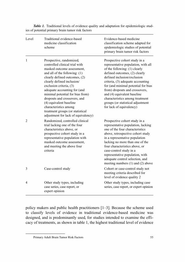
Primary Adult Brain Tumor Risk Factors 55
policy makers and public health practitioners [1–3]. Because the scheme used
to classify levels of evidence in traditional evidence-based medicine was
designed, and is predominantly used, for studies intended to examine the effi-
cacy of treatments, as shown in table 1, the highest traditional level of evidence
Table 1. Traditional levels of evidence quality and adaptation for epidemiologic stud-
ies of potential primary brain tumor risk factors
Level Traditional evidence-based Evidence-based medicine
medicine classification classification scheme adapted for
scheme epidemiologic studies of potential
primary brain tumor risk factors
1 Prospective, randomized, Prospective cohort study in a
controlled clinical trial with representative population, with all
masked outcome assessment, of the following: (1) clearly
and all of the following: (1) defined outcomes, (2) clearly
clearly defined outcomes, (2) defined inclusion/exclusion
clearly defined inclusion/ criteria, (3) adequate accounting
exclusion criteria, (3) for (and minimal potential for bias
adequate accounting for (and from) dropouts and crossovers,
minimal potential for bias from) and (4) equivalent baseline
dropouts and crossovers, and characteristics among treatment
(4) equivalent baseline groups (or statistical adjustment
characteristics among for lack of equivalency)
treatment groups (or statistical
adjustment for lack of equivalency)
2 Randomized, controlled clinical Prospective cohort study in a
trial lacking one of the four representative population, lacking
characteristics above, or one of the four characteristics
prospective cohort study in a above, retrospective cohort study
representative population with in a representative population
masked outcome assessment, lacking no more than one of the
and meeting the above four four characteristics above, or
criteria case-control study in a
representative population, with
adequate control selection, and
meeting numbers (1) and (2) above
3 Case-control study Cohort or case-control study not
meeting criteria described for
level of evidence quality 2
4 Other study types, including Other study types, including case
case series, case report, or series, case report, or expert opinion
expert opinion
Fisher/Schwartzbaum/Wrensch/Berger 56
quality (level 1) is assigned to a well-designed prospective, randomized, con-
trolled, clinical trial. However, the primary goal of analytic epidemiology is to
understand the causes or risk factors for diseases. Because it is unethical to
assign study participants to receive a potentially harmful exposure, clinical tri-
als are not used to evaluate factors that may increase ABT risk. As a result, there
is no ‘level 1’ level of evidence quality, per se, for ABT risk factors (although,
in the future, randomized trials of potentially preventive factors such as nons-
teroidal anti-inflammatory drugs may be conducted) [4]. Epidemiologic studies
usually compare either ABT risk in participants with and without certain char-
acteristics (cohort studies), or the histories of participants with and without
ABT (case-control studies). Therefore, a well-designed cohort study, in which
groups of participants with and without the potential risk factor are followed
over time for the occurrence of ABT, may be considered the highest level of
obtainable evidence. As shown in table 1, for purposes of the present evalua-
tion, we adapted the levels of evidence quality scheme to consider our epidemi-
ologic goal of understanding the causes of ABT. The primary difference in this
adapted scheme is the removal of the randomized, controlled, clinical trial; the
level of evidence from the well-designed cohort study is, in our adapted
scheme, the superior source of evidence. In addition, it should be noted that
because a case-control study utilizing incident cases only differs from a cohort
study by the method of sampling controls or noncases, results from a well-
designed case-control study often provide nearly as high a level of evidence as
a well-designed cohort study. In this review, the relevant distinction between a
cohort and case-control study is that results from a cohort study can provide
evidence that the potential cause preceded the effect (or ABT), while results
from a case-control study cannot address temporality. Studies described in this
review are cohort and case-control studies.
We evaluate the epidemiologic evidence for ABT risk factors as follows: (1)
identify epidemiologic studies that address the associations of interest by search-
ing Medline for reports of relevant epidemiologic and laboratory studies, (2) for
each study, examine the estimated magnitude and precision of the effect (usually
a risk or odds ratio), and its validity based on the relative strengths and weakness
of the study design, and potential sources of error and bias in the analysis, (3)
assign a level of evidence quality to each study (descriptions of levels of evi-
dence are shown in table 1, adapted from Miyasaki et al. [2]), and (4) assign a
grade of recommendation to evaluate the potential association between the risk
factor and ABT risk (table 2). Grades of recommendation are intended to sum-
marize the overall degree of evidence for the potential association and are based
on the following: homogeneity of results from original epidemiologic studies,
magnitude of the potential effect, level of evidence for each epidemiologic study,
and thoroughness of the epidemiologic literature in addressing the potential
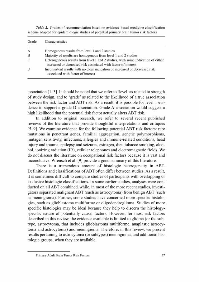
Primary Adult Brain Tumor Risk Factors 57
association [1–3]. It should be noted that we refer to ‘level’ as related to strength
of study design, and to ‘grade’ as related to the likelihood of a true association
between the risk factor and ABT risk. As a result, it is possible for level 1 evi-
dence to support a grade D association. Grade A association would suggest a
high likelihood that the potential risk factor actually alters ABT risk.
In addition to original research, we refer to several recent published
reviews of the literature that provide thoughtful interpretations and critiques
[5–9]. We examine evidence for the following potential ABT risk factors: rare
mutations in penetrant genes, familial aggregation, genetic polymorphisms,
mutagen sensitivity, infections, allergies and immune-related conditions, head
injury and trauma, epilepsy and seizures, estrogen, diet, tobacco smoking, alco-
hol, ionizing radiation (IR), cellular telephones and electromagnetic fields. We
do not discuss the literature on occupational risk factors because it is vast and
inconclusive. Wrensch et al. [9] provide a good summary of this literature.
There is a tremendous amount of histologic heterogeneity in ABT.
Definitions and classifications of ABT often differ between studies. As a result,
it is sometimes difficult to compare studies of participants with overlapping or
exclusive histologic classifications. In some earlier studies, analyses were con-
ducted on all ABT combined, while, in most of the more recent studies, investi-
gators separated malignant ABT (such as astrocytoma) from benign ABT (such
as meningioma). Further, some studies have concerned more specific histolo-
gies, such as glioblastoma multiforme or oligodendroglioma. Studies of more
specific histologies may be ideal because they help to discern the histology-
specific nature of potentially causal factors. However, for most risk factors
described in this review, the evidence available is limited to glioma (or the sub-
type, astrocytoma, that includes glioblastoma multiforme, anaplastic astrocy-
toma and astrocytoma) and meningioma. Therefore, in this review, we present
results pertaining to astrocytoma (or subtypes) meningioma, and additional his-
tologic groups, when they are available.
Table 2. Grades of recommendation based on evidence-based medicine classification
scheme adapted for epidemiologic studies of potential primary brain tumor risk factors
Grade Characteristics
A Homogenous results from level 1 and 2 studies
B Majority of results are homogenous from level 1 and 2 studies
C Heterogeneous results from level 1 and 2 studies, with some indication of either
increased or decreased risk associated with factor of interest
D Inconsistent results with no clear indication of increased or decreased risk
associated with factor of interest
Fisher/Schwartzbaum/Wrensch/Berger 58
ABT are thought to develop through the progressive accumulation of genetic
alterations that permit cells to evade normal regulatory mechanisms and escape
destruction by the immune system. The epidemiologic information available con-
cerning genetic factors and ABT risk results from studies relating to the follow-
ing: (1) ABT risk associated with rare mutations in penetrant genes, (2) patterns
of ABT in families (suggesting potential inheritance), and (3) genetic polymor-
phism or variability. These are discussed separately.
Rare Mutations in Penetrant Genes
Several diseases/syndromes associated with rare mutations in highly pene-
trant genes increase ABT risk. However, in a population-based study of 500
adults with glioma in San Francisco, Calif. [10], less than 1% had a known
hereditary syndrome. While it is thought that genetic predisposition is influen-
tial in relatively few brain tumors (5–10%) [11], the proportion may be under-
estimated because some hereditary syndromes are not readily diagnosed and
because patients with ABT are not routinely referred to a clinical geneticist.
In general, there is strong evidence that some genetic diseases or syn-
dromes increase risk of ABT [9, 12]. Among these, the following have been
associated with an increased ABT risk: tuberous sclerosis complex, neurofibro-
matosis types 1 and 2 [13], nevoid basal cell carcinoma syndrome, syndromes
related to adenomatous polyps and Li-Fraumeni cancer family syndrome [9, 12].
Inherited p53 germline mutations, characteristic of Li-Fraumeni syndrome, are
important in the development of many cancers, including ABT [14–16]. In
addition, germline p53 mutations have been more frequently found in patients
who have multifocal glioma, glioma and another primary malignancy, or a fam-
ily history of cancer than in patients with other brain tumors [17].
Overall grade of recommendation: A, for association between geneticdiseases/syndromes related to rare mutations in highly penetrant genes andABT risk.
Familial Aggregation
Although a few epidemiologic studies have addressed the potential familial
aggregation of meningioma [18–20], results are limited, compared to those con-
cerning glioma and its most common subtype, astrocytic tumors. Among menin-
gioma patients, the standardized incidence ratio (SIR) for having a parent with
any nervous system cancer was 2.5 [19], and 3.1 for having a parent with menin-
gioma [18]. Findings from epidemiologic studies of familial aggregation and
Primary Adult Brain Tumor Risk Factors 59
glioma risk are relatively consistent, although the estimates of the magnitude of
increased risk as a result of first degree relation are widely variable [10, 18, 19,
21–29]. Five case-control studies suggest the presence of familial aggregation of
glioma or glioma subtypes [10, 21, 22, 28, 29], while one case-control study sug-
gests otherwise [27]. The finding of an increased astrocytoma or glioma risk as a
result of familial aggregation is also supported by the analyses of six Swedish
cohorts, although these studies contain overlapping populations [18, 19, 23, 25,
26, 30]. Malmer et al. [26] suggest that approximately 2% of glioma cases may be
explained by an autosomal recessive gene, although a polygenic model could
not be rejected, and that approximately 5% of glioma cases are familial. In a
population-based case-control study, Malmer et al. [24] report that, among 37
familial glioma cases (compared to 58 sporadic glioma controls with no family
history of glioma), familial cases had more p53-negative tumors. Segregation
analyses of families of more than 600 adult patients with glioma showed that a
polygenic model best explained the pattern of occurrence of brain tumors [31].
Because it is possible that common environmental exposures experienced
by families, in addition to their genetic characteristics, may result in a greater
familial ABT risk, several studies have tried to determine whether genetic or
environmental factors explain the increased familial risk. Grossman et al. [32]
showed that ABT occur in families with no known predisposing hereditary dis-
ease and that the pattern of occurrence in many families suggests environmen-
tal causes. However, results from a cohort study conducted by Malmer et al.
[25] suggest that first-degree relatives, and not spouses, have a significantly
increased ABT risk. Therefore, it is likely that genetic characteristics play a role
in familial aggregation of ABT.
Although the strength of evidence for familial aggregation of glioma or its
subtypes varies among studies, evidence for the presence of familial aggrega-
tion has been consistently observed in both cohort and case-control studies.
Evidence pertaining to familial aggregation of meningioma is limited and
should be validated through additional studies; however, there is weak evidence
for the presence of familial aggregation of meningioma.
Overall grade of recommendation: A, for presence of familial aggregationof astrocytoma evaluated separately or for glioma as a whole.
Overall grade of recommendation: C, for presence of familial aggregationof meningioma.
Genetic Polymorphisms
Available evidence suggests that only a small proportion of primary brain
tumors result from effects of inherited rare mutations in highly penetrant genes;
Fisher/Schwartzbaum/Wrensch/Berger 60
therefore, investigators have turned their attention to polymorphisms in genes
that might influence susceptibility to brain tumors in concert with environmen-
tal exposures. Genetic alterations that affect oxidative metabolism, detoxifica-
tion of carcinogens, DNA stability and repair, or immune response are candidates,
which might plausibly confer genetic susceptibility to ABT.
Results from case-control studies of genes that detoxify carcinogens are
inconsistent. For example, case-control studies suggest both that cytochrome
P450 2D6 (CYP2D6) increases both astrocytoma (OR � 4.17, p � 0.0043) and
meningioma (OR � 4.90, p � 0.0132) risks [33], and that individuals with a
poor metabolizer CYP2D6 variant allele are at no greater ABT risk [34]. In two
additional case-control studies, Trizna et al. [35] found no association between
the null genotype of CYP1A1 and risk of glioma in adults, but De Roos et al.
[36] found that CYP2E1 RsaI variant was associated with glioma (OR � 1.4,
95% CI: 0.9–2.4) and acoustic neuroma (OR � 2.3, 95% CI: 1.0–5.3) risks,
especially among younger cases.
Detoxifying glutathione S-transferase genes have also been extensively
studied and also produce mixed results. Glutathione transferase theta (GSTT1)
null genotype appears to increase astrocytoma (OR � 2.67, p � 0.0005) and
meningioma (OR � 4.52, p � 0.0001) risks [33]. Results reported by Hand
et al. [37] support these findings for astrocytoma, especially high grade astrocy-
toma, but results reported by Kelsey et al. [34] suggest no association between
the GSTT1 null genotype and astrocytoma risk. However, the oligoden-
droglioma risk was associated with the GSTT1 null genotype (OR � 3.2, 95%
CI: 1.1–9.2). Trizna et al. [35] and De Roos et al. [36] report no associations
between the null genotype of GSTT1 and glioma risk; however, results reported
by De Roos et al. suggest that GSTT1 null genotype increases meningioma risk
(OR � 1.5, 95% CI: 1.0–2.3), and that the GSTP1 105 Val/Val genotype is
associated with increased glioma risk (OR � 1.8, 95% CI: 1.2–2.7), with evi-
dence of a dose-response trend with an increasing number of variant alleles.
Most recently, Wrensch et al. [38] found little evidence for the association of
glutathione S-transferase polymorphism with glioma histologic variants.
Several additional polymorphisms have been evaluated for their relation-
ship with glioma. Chen et al. [39] showed that AA or AC versus CC genotype in
nucleotide 8092 of ERCC1 increased the oligoastrocytoma risk (OR � 4.6,
95% CI: 1.6–13.2). Caggana et al. [40] found the AA genotype (C to A poly-
morphism [R156R]) of ERCC2 was more prevalent than the CC or CA geno-
types in cases with glioblastoma multiforme, astrocytoma, or oligoastrocytoma
than in controls, and the association was strongest for oligoastrocytoma
(OR � 3.2, 95% CI: 1.1–9.5). Inconsistencies among genetic polymorphism
studies may result from false-positive associations based on inadequate sample
sizes [41] and from confounding by genes with similar functions not accounted
Primary Adult Brain Tumor Risk Factors 61
for in the analyses. Another possibility may be that study populations consist of
different proportions of types of tumors, and genetic risk for certain subtypes
could be masked by a lack of risk among other subtypes. When these issues are
addressed, the potential interaction between genetic polymorphisms with other
genetic characteristics and environmental factors can be properly evaluated.
Overall grade of recommendation: C, for association between geneticpolymorphisms and ABT risk.
Mutagen Sensitivity
Bondy et al. [42] found that lymphocyte mutagen sensitivity to gamma
radiation appears to increase glioma risk. Lymphocytes from glioma patients,
compared to those from matched controls, are more sensitive to gamma radia-
tion [42]. Bondy et al. [42] observed a greater frequency of chromatid breaks
per cell among glioma patients, compared to controls, and mutagen sensitivity
was found to be associated with increased glioma risk (OR � 2.09, 95% CI:
1.43–3.06). Further, there was evidence of a statistically significant dose-
response trend between frequency of chromatid breaks and glioma risk [42].
However, because Bondy et al. used the case-control design, it is not possible to
determine whether chromatid breaks increased ABT risk or represent a sys-
temic effect of the tumors themselves [43]. To definitively establish temporal-
ity, further studies of mutagen sensitivity need to be conducted on blood from
glioma patients collected before glioma initiation and development.
Overall grade of recommendation: B, for association between mutagensensitivity and glioma risk.
Infections
Infection with viruses, such as simian virus 40 (SV40) and varicella zoster
virus (VZV), and nonviral infectious agents, such as Toxoplasma gondii, or
immunity to these agents might influence ABT risk, although the potential risk
from these agents has been inadequately addressed in epidemiologic studies.
Between 1955 and 1963, an unknown proportion of all inactivated and live
polio vaccines distributed was contaminated with SV40 [44]. In Germany, chil-
dren were followed over a 20-year period, and those inoculated with the polio
vaccine contaminated with SV40 had higher occurrences of glioblastoma mul-
tiforme, medulloblastoma, and some less common brain tumor types than those
not given the contaminated vaccine [45]; in the US, on the other hand, no dif-
ference in ABT risk was found for major ABT types (glioma and meningioma)
Fisher/Schwartzbaum/Wrensch/Berger 62
between the two groups of children [46], but one study reported that the inci-
dence of ependymoma was 37% greater among the children receiving the cont-
aminated vaccine [44]. Results from one case-control study suggest that prior
infection with VZV, either based on self-report [47] or serologic evidence [48],
may be inversely associated with adult glioma risk. In addition, there is mixed
evidence from laboratory studies concerning the involvement of JC virus
[49–54], BK virus [49, 50, 52], SV40 [49, 50, 52] and human herpes virus 6
(HHV-6) [55] in the development of ABT. Results from a nested case-control
study conducted by Rollison et al. [56] indicated that infection with either JC or
BK virus, or SV40, as measured in sera of patients between 1 and 22 years prior
to ABT diagnosis, did not significantly increase subsequent ABT risk. The
potential role of these viruses in the development of ABT is not fully under-
stood, and should be more thoroughly examined. The potential association
between infection with HIV and ABT risk has not been addressed in epidemio-
logic studies, although it is possible that the incidence of ABT among those
infected with HIV is greater than that of the general population [57].
A history of cold or influenza infection appears to decrease glioma risk
[58, 59]. In a case-control study, Schlehofer et al. [59] found a 30% reduction in
glioma risk from a self-report of a history of colds or influenza infections
(RR � 0.72, 95% CI: 0.61–0.85). In another case-control study, Fisher et al.
found that treatment for at least one cold or influenza infection between 2 and 5
years prior to diagnosis decreases glioma risk approximately 3-fold (OR � 0.39,
95% CI: 0.18–0.86) [58]. These results should be validated in cohort studies with
serologic or symptom-based confirmation of infection.
Antibodies to T. gondii have been associated with meningioma in a case-
control study [60], and to astrocytoma in another study [61], but not to glioma,
based on the results of Ryan et al. [60].
The findings concerning infections and ABT risk are limited, inconsistent
or based exclusively on results from case-control studies. The strongest epi-
demiologic evidence of potential association between infection and ABT risk
would be submitted from a cohort study in which serologic measurement of
viral or bacterial exposure was ascertained prior to ABT development.
Overall grade of recommendation: C, for association between viral andbacterial infections and ABT risk.
Allergies and Immune-Related Conditions
Several recent reports have suggested that a history of allergies and
immune-related conditions, such as asthma, eczema and rheumatoid arthritis,
decrease the risk of glioma. Although the mechanism governing such potential
Primary Adult Brain Tumor Risk Factors 63
protection has not been identified, there is speculation that it may arise from the
anti-inflammatory effects of cytokines involved in allergic and autoimmune
disease [62–65]. Five case-control studies have reported decreases in glioblas-
toma multiforme, glioma or astrocytoma risk from allergies and immune-
related conditions [22, 59, 62, 65, 66], while results from one case-control
study suggest no difference in glioma risk [21]. For example, Brenner et al. [62]
reported an inverse association between glioma risk and history of any allergy
(OR � 0.67, 95% CI: 0.52–0.86) or autoimmune disease (OR � 0.49, 95% CI:
0.35–0.69). No such decrease in risk for meningioma [59, 62, 66] was
observed. A problem with case-control association between reported allergy
status and glioma is that due to the low survival probability from glioblastoma
mutiforme many proxy respondents were used to ascertain information on
allergic conditions. Confirming the suggestion that proxy reports may not be
reliable, Schwartzbaum et al. [63] found a correlation between whether a proxy
respondent was used and the presence or absence of allergic conditions.
Specifically, proxy respondents reported fewer allergic conditions than did self-
reporting respondents. However, in a cohort study where information on aller-
gic conditions was obtained on the average at least 19 years before brain tumor
diagnosis, Schwartzbaum et al. [63] report results from the first series of cohort
studies, consistent with an inverse association between allergies and glioma risk
(hazard ratio [HR] � 0.45, 95% CI: 0.19–1.07), although not among low grade
glioma, and between immune-related hospital discharges and glioma risk
(HR � 0.46, 95% CI: 0.14–1.49). Further suggesting that the association between
allergic conditions and glioma risk is not a reporting artifact, Schwartzbaum
et al. found that genetic polymorphisms associated with an increased risk of
allergic conditions appear to decrease glioma risk (unpubl. observation).
Although the majority of results indicate an inverse association between aller-
gies and immune-related conditions and glioma risk, further studies are needed
to provide solid evidence.
Overall grade of recommendation: B, for inverse association betweenallergies, immune-related conditions and glioma, but not meningioma, risk.
Head Injury or Trauma
Head injury and trauma have been examined as potential ABT risk factors
in several epidemiologic studies. Compared to glioma, there is slightly stronger
evidence that head injury and trauma may increase meningioma risk. Results
from four case-control studies suggest that head injury and trauma increase
meningioma risk [67–70]. Among males, Preston-Martin et al. [69] found that
meningioma was more common among those who reported having ever had a
Fisher/Schwartzbaum/Wrensch/Berger 64
head injury (OR � 1.5, 95% CI: 0.9–2.6), and that, among those with a latency
of 15–24 years, meningioma risk was much greater (OR � 5.4, 95% CI:
1.7–16.6). However, these results are in conflict with the results of three cohort
studies, which suggest no increase in meningioma risk from head injury or
trauma [71–73]. Results from the three cohort studies provide much stronger
evidence because it is likely that there were differences between cases and con-
trols in the reporting of previous head injury and trauma [9, 72]. Hu et al. [74]
found an increase in glioma risk from head trauma requiring medical attention
(OR � 4.1, 95% CI: 2.5–10.3), and Hochberg et al. [75] found a large increase
in glioblastoma multiforme risk from severe head injury. However, results from
four additional case-control studies, and two cohort studies suggested no such
association [9, 29, 68, 69, 72, 73, 76]. A large Danish cohort study provides
good evidence that there is little, if any, association between head injury and
trauma and either meningioma or glioma risk. Inskip et al. [72] and Wrensch
et al. [9] found that, after excluding injuries occurring within the year prior to
diagnosis (which could have resulted in tumor detection) neither glioma risk
(SIR � 1.0, 95% CI: 0.8–1.2) nor meningioma risk (SIR � 1.2, 95% CI:
0.8–1.7) was elevated. Risk of some intravascular tumors was elevated.
However, given that intravascular tumors are far less common than meningioma
or glioma, the increase in overall ABT risk from head injury and trauma is prob-
ably slight, if existent.
Overall grade of recommendation: D, for association between head injuryor trauma and both meningioma and glioma risk.
Overall grade of recommendation: B, for association between head injuryor trauma and intravascular tumor risk.
Epilepsy and Seizures
Seizures prior to ABT diagnoses are common [77]. There is relatively con-
sistent evidence from case-control studies that epilepsy or seizure disorders are
associated with increased meningioma risk, although not all findings have been
statistically significant [59, 66, 78, 79]. The evidence concerning epilepsy or
seizure disorders and glioma risk is somewhat stronger. Results from four case-
control studies [10, 59, 66, 79] and three cohort studies [78, 80, 81] indicate an
increase in glioma risk from a history of epilepsy or seizure. Lamminpaa et al.
[78] found an excess of both meningioma and glioma associated with a history
of prescription for antiepileptic medication. Schlehofer et al. [59] found an
increased glioma risk associated with a history of epilepsy (RR � 6.55, 95%
CI: 3.40–12.63), but the relative risk diminished when including only those with
epilepsy lasting at least 20 years. Importantly, for glioma, but not meningioma,
Primary Adult Brain Tumor Risk Factors 65
risk has been shown to increase with proximity to diagnosis and decrease with
duration of epilepsy, suggesting that epilepsy is not a cause but rather a result,
an early effect, of ABT [79].
Overall grade of recommendation: A, for association between epilepsy andseizures and both meningioma and glioma risk, although epilepsy and seizuresprobably result from rather than cause ABT.
Estrogen, Reproductive and Menstrual Factors
Epidemiologic evidence indicates that the sex hormone, estrogen, may be
associated with increased meningioma risk. Meningioma is approximately twice
as common in females [82, 83]. In addition, some meningioma express proges-
terone receptors [84–86], and this expression occurs to a greater degree among
females [86]. Studies of meningioma risk and menopausal status, age at menar-
che, and parity have produced some results supporting the notion that greater
exposure to endogenous estrogen increases meningioma risk and some that have
supported the notion that lesser exposure increases meningioma risk. Results
from a cohort study suggest that postmenopausal women who have never used
estrogen replacement therapy are at greater meningioma risk compared to pre-
menopausal women (RR � 2.48, 95% CI: 1.29–4.77), and the risk remains sig-
nificantly greater comparing postmenopausal women who have used estrogen
replacement therapy to premenopausal women (RR � 1.86, 95% CI: 1.07–3.24)
[87]. These results are supported by those of Schlehofer et al. [88] who found that
postmenopausal women had a reduced risk of meningioma (RR � 0.58, 95% CI:
0.18–1.90) compared to premenopausal women. Concerning age at menarche,
Jhawar et al. [87] found that later age at menarche (after age 14 years) increased
meningioma risk (OR � 1.97, 95% CI: 1.06–3.66). Further, there was an incr-
eased risk of meningioma among parous women compared to nulliparous women
(RR � 2.39, 95% CI: 0.76–7.53). However, findings from two case-control stud-
ies suggest no association between parity and meningioma risk [88, 89]. On the
whole, there is consistency among results concerning increased meningioma risk
among premenopausal women, that is, at a given age, women who are still pre-
menopausal are at higher risk than women who are postmenopausal, suggesting
that increased exposure to menstrual hormones might increase risk. However,
results concerning parity and age at menarche suggest that estrogen or other
reproductive or menstrual hormones might decrease meningioma risk. One
would expect parous women and women who started menarche at older ages to
have lower meningioma risk, if estrogen increases risk, because parous women
and women who started menarche at older ages are exposed to endogenous
estrogen for a shorter amount of time. Further study is required to understand
Fisher/Schwartzbaum/Wrensch/Berger 66
hormone-related factors, especially because the findings presented here are statis-
tically significant and opposite to those expected under the hypothesis that estro-
gen influences meningioma risk. It is also possible that menstrual and reproductive
factors alone are not sufficient to accurately classify lifetime estrogen or other
hormonal exposure.
Estrogen may also be associated with decreased astrocytoma risk. Astro-
cytoma is approximately 40% more common among males [9]. A study of the
incidence of astrocytoma subtypes occurring in the State of New York suggests
that, for glioblastoma multiforme, the protective effect of female sex occurs
between the approximate ages of menarche and menopause, and that this pro-
tection decreases in postmenopausal age groups [90]. Schlehofer et al. [88]
report that postmenopausal women whose menopause was not surgically
induced are at greater risk of glioma and acoustic neuroma, although the find-
ing was not statistically significant. However, like the association with menin-
gioma, the association between estrogen and astrocytoma risk may not be
straightforward. For example, results from two case-control studies suggest that
astrocytoma risk [91] and risk of the subtypes glioma [89] and glioblastoma
multiforme [91] are lower among parous women. Schlehofer et al. [88] report
no association between parity and glioma risk.
For both meningioma and astrocytoma, estrogen, and perhaps other hor-
mones, may alter risks. However, the results do not support the notion that the
longer the duration of exogenous estrogen exposure the greater the risk. Cohort
studies of ABT risk among women who have and have not taken estrogen
replacement therapy should be conducted.
Overall grade of recommendation: B, for associations between reproduc-tive and menstrual factors and estrogen and both meningioma and astrocytomarisk, although the associations are not straightforward.
Diet
Three groups of diet-related risk factors have emerged as potentially
affecting ABT risk. These are N-nitroso compounds, antioxidant intake and cal-
cium intake. They are discussed separately.
N-Nitroso CompoundsN-nitroso compounds (especially nitrosamides) are potent neurocarcinogens.
Assessing exposure to N-nitroso compounds is difficult because they are com-
mon in both endogenous and exogenous sources, including food. Vegetables that
are high in nitrites also contain vitamins that may block the formation of
N-nitroso compounds. Cured meats contain nitrites, which are precursors of
Primary Adult Brain Tumor Risk Factors 67
N-nitroso compounds. Results from epidemiologic studies concerning ABT risk
associated with cured meat consumption are mixed. Results from a case-control
study of cured meat consumption and meningioma risk suggest an increase ABT
risk, but only for females [92], while two case-controls studies report conflicting
evidence of the effect of cured meat consumption on glioma risk, one reporting no
effect [93], and one reporting that male cases, but not female cases, were more
likely than controls to report higher levels of cured meat consumption [94].
However, as suggested by Schwartzbaum et al. [95], it is possible that energy
intake and �-tocopherol modify the association between cured meat consumption
and glioma risk. (Cured meats have a high-energy and low-�-tocopherol content.)
A recent meta-analysis [96] was conducted of nine studies addressing the possible
association between cured meat consumption and glioma risk, and although
an increase in glioma risk was found from cured meat consumption (pooled
RR � 1.48, 95% CI: 1.20–1.83), Huncharek et al. point out that individual studies
failed to adjust for potential confounding by energy intake. In a small-area eco-
logic study of England, a higher incidence of ABT was found in areas with greater
nitrate content in drinking water [97].
Antioxidant IntakeOxidative stress results from excessive accumulation of reactive oxygen
species, and can be caused by inadequate dietary antioxidant intake. Antioxidants
are abundant in a diet high in fruits and vegetables. Consumption of greater
amounts of vitamin C was inversely associated with glioma risk in two case-
control studies [94, 98], although the findings were statistically significant only
among males in the study conducted by Lee et al. [94]. Schwartzbaum et al. [95]
found lower levels of vitamin C intake among glioma cases compared to controls.
The results reported by Chen et al. [39], however, suggest no association between
vitamin C intake and glioma risk. Dietary intake of higher levels of vitamin A or
provitamin A has been inversely related to glioma risk in each of three case-
control studies in which it was examined [93, 94, 98], although not all findings
were statistically significant. Higher levels of dietary vitamin E intake were found
to be protective for glioma in one case-control study [98], but not in two other
studies [93, 94]. Schwartzbaum et al. [95] found lower levels of vitamin E (both
�- and �-tocopherol) intake among glioma cases compared to controls.
Calcium IntakeDietary calcium may decrease glioma risk through increasing apoptosis,
promoting DNA repair, and decreasing the production of parathyroid hormone
[99]. Tedeschi-Blok et al. [99] found greater levels of calcium intake to protect
against glioma, but only among females. The results of another case-control
study suggest that calcium is protective for all ABT combined [98].
Fisher/Schwartzbaum/Wrensch/Berger 68
Evidence related to dietary factors is inconsistent and remains limited.
Further study is needed to examine these factors and their potential interaction
with one another, as well as other potential ABT risk factors.
Overall grade of recommendation: C, for association between consump-tion of N-nitroso compounds, and antioxidant and calcium intakes and ABTrisk.
Tobacco Smoking
Tobacco smoking has been evaluated as a potential risk factor for ABT
because some carcinogenic components contained in tobacco smoke, including
nitroso compounds, penetrate the blood-brain barrier. A case-control study
showed no increase in glioblastoma multiforme risk from tobacco smoking
[22]. However, results from two case-control studies suggest that there may be a
difference between males and females in the association between tobacco
smoking and glioma risk. In one study, males, but not females, who reported a
history of having ever smoked were at greater risk of glioma [100], while
results from another study suggest that among men only, cases were almost 2
times more likely to report smoking unfiltered cigarettes [94]. Tobacco smok-
ing has been shown to increase meningioma risk, but only in females [101].
Results from most studies suggest that tobacco smoking does not strongly con-
tribute to ABT risk, although smoking unfiltered, but not filtered, cigarettes
may increase glioma risk [94]. Overall, the results are inconsistent and the mag-
nitudes of the effect estimates are modest.
Overall grade of recommendation: D, for association between filtered cig-arette smoking and glioma and meningioma risk. C, for association betweenunfiltered cigarette smoking and glioma risk.
Alcohol
Results from seven case-control studies addressing alcohol consumption
and ABT risk are inconsistent, with five studies suggesting no increase in ABT
risk [22, 66, 100, 101, 102], and two suggesting modest increases in risk [74,
94]. Alcohol consumption has not been shown to affect meningioma risk [66,
101, 102], and glioma risk was found to be increased from higher levels of alco-
hol consumption in one study [74, 94], while, in four other studies, there was no
increase in glioma risk [66, 100, 102], or glioblastoma multiforme risk [22].
Overall, the results are inconsistent and the magnitudes of the positive results
are modest.
Primary Adult Brain Tumor Risk Factors 69
Overall grade of recommendation: D, for association between alcohol con-sumption and glioma and meningioma risk.
Ionizing Radiation
Therapeutic IR may be the strongest modifiable ABT risk factor [8, 9]. IR
used to treat tinea capitis and skin hemangioma in children or infants has been
associated with relative risks of 18 for nerve sheath tumors, 10 for menin-
gioma, and 3 for glioma [8, 9]. Twelve reports of original studies concerning
exposure to IR were reviewed. Results from two studies concerning exposure
to cosmic IR from occupation as a pilot or aircrew/cockpit crew member
revealed no elevation in ABT risk [103, 104]. Results from two cohort studies
indicated that occupation involving working with nuclear materials increases
ABT risk, apparently as a result of IR exposure [105, 106]. However, it is diffi-
cult to rule out the possibility that these results are confounded by chemical
exposures occurring in nuclear industry occupations. Exposure to nondental
X-rays of the head and neck did not increase glioma risk in one case-control
study [76], but both radiotherapy of the head and neck and diagnostic X-rays
were found to increase ABT risk in another case-control study [107]. Prior
radiotherapy is relatively common (17%) among patients with glioblastoma
multiforme [108], and results from several studies have suggested increases in
risk of glioma and other ABT from a history of radiotherapy for acute lym-
phoblastic leukemia as children [109, 110]. Second primary brain tumors also
occur more frequently than expected, especially among patients treated with
radiotherapy [111].
Studies of ABT risk and dental X-rays are less abundant. Results from one
case-control study suggest that only exposure to full-mouth (and not bitewing,
lateral cephalometric, or panoramic) X-rays performed 15–40 years prior to
diagnosis increases meningioma risk [112]. These results are supported by
another study suggesting that the meningioma risk is more strongly associated
with exposures from dental X-rays taken in the more distant past.
A study of survivors of the atomic bombing of Hiroshima showed a high
incidence of meningioma correlating with the dose of radiation to the brain
[113]. The incidence increased with closer proximity to the hypocenter.
Japanese studies of atomic bomb survivors have not shown an increased risk of
ABT among those who were exposed in utero [8].
On the whole, results from studies of the association between IR exposure
and ABT risk are homogenous.
Overall grade of recommendation: A, for association between IR andglioma and meningioma risk.
Fisher/Schwartzbaum/Wrensch/Berger 70
Cellular Telephones
Because of public concern over possible health effects associated with
cellular telephone use, several studies have addressed the potential association
between use of cellular telephones and ABT risk. Of these, one mortality study
[114], four case-control studies [115–118] and one retrospective cohort study
[119] suggest that there is no association between cellular telephone use and
ABT risk. Further, results from these studies, in general, suggest that there is
no increase in ABT risk from longer duration of cellular telephone use and that
there is no specific anatomic site or ABT histology affected by these devices.
However, three reports provide additional important findings. Results reported
by Hardell et al. [107] suggest an increase in ABT risk from ipsilateral (same
side) use of cellular telephones for areas of the brain with the greatest potential
for microwave exposure from cellular telephone use (temporal, temporopari-
etal and occipital areas). Moreover, ipsilateral radiofrequency exposure was
associated with an increase in malignant ABT risk, although the associa-
tion was stronger for analog, versus digital, radiofrequency; for astrocytoma,
risk was nearly double [120]. (Analog cellular telephone radiofrequency
signals operate in the range of 800–900 MHz, while newer digital phones
operate in the range of 1,600–2,000 MHz.) Further, similar results con-
cerning ipsilateral cellular telephone use were reported in a recent case-control
study, and statistically significant increases in ABT risk, including astrocy-
toma risk, were found for both analog and digital devices, although, again,
the finding was stronger for analog devices [121]. There has been no known
association reported for use of cellular telephones on the opposite side of
the brain.
Although results are inconsistent, if there is an increase in ABT risk from
cellular telephone use, it may be specific to ipsilateral use, especially from ana-
log telephones, and especially for anatomic areas of the brain near the device.
Although results from most known studies of cellular telephones and ABT, on
the whole, suggest no increase in ABT risk, it is important to continue this line
of study because: (1) the use of such devices is increasingly common, and (2) it
is possible that adverse health effects from this exposure may result from long-term exposure, and due to the recent increase in cellular telephone usage, it is
possible that results from many studies have not accounted for the possible long
lag time between exposure and ABT, especially slow-growing ABT. In addition,
changes in cellular telephone technology, such as the predominance of digital,
versus analog, telephones, as well as an overall increase in the duration of
usage, could not be addressed in many studies.
Overall grade of recommendation: C, for association between cellulartelephone use and ABT risk.
Primary Adult Brain Tumor Risk Factors 71
Electromagnetic Fields
Concern over exposure to low-frequency electric and magnetic fields
(EMF) has stimulated considerable public and scientific attention. Primarily, this
interest arises from residential studies showing increased risks of brain tumors
and leukemia in children whose homes have high EMF exposures. However, for
ABT, exposure to EMF has, for the most part, been evaluated through occupa-
tional studies of workers presumably exposed to greater levels of EMF. In
general, workers exposed to greater levels of EMF have higher incidence and
mortality rates of ABT. Twenty-one reports of original occupational studies were
reviewed. In general, results from each of the 11 case-control studies addressing
occupational EMF exposure and ABT risk suggest an increase in ABT risk asso-
ciated with greater levels of EMF exposure, although not every increase in risk
was statistically significant [122–132]. Results from nested case-control and
cohort studies have yielded inconsistent results, with five suggesting no associa-
tion [133–137], and five suggesting an increase in ABT risk from occupational
exposure to EMF [124, 138–141]. In studies of specific histologic types, risk for
the following types of ABT was increased with occupational exposure to EMF:
astrocytoma [124, 126], glioma [122, 132], or glioblastoma multiforme [131,
140]. The relationship between occupational EMF exposure and ABT risk may
not be straightforward. For example, results from a recent study suggest that
occupational exposure to high levels of EMF may be associated with glioma, but
not meningioma, risk in the presence of carcinogenic chemicals [140]. That is,
EMF may modify the association between occupational chemical exposures
and glioma, but not meningioma, risk. Residential exposure to EMF has been
examined by Wrensch et al. [142], and results from this study did not support an
association between residential exposure to EMF and ABT risk. No causal rela-
tionship has been established between exposure to EMF, either occupational or
residential, and ABT risk.
Studies of exposure to EMF and ABT risk may be flawed by the difficulty
of assessing the potentially imperceptible and ubiquitous exposure, and by not
knowing the period during which exposure assessment should be determined,
given the absence of available information about the time period between expo-
sure and ABT risk. It is likely that studies of EMF exposure and ABT risk have
been flawed by large exposure misclassification. There is no established and
accepted mechanism of action by which exposure to EMF may increase ABT
risk. There is inconsistency of results across studies, and an apparent lack of
dose-response (although it is possible that a threshold value exists above which
ABT risk is affected).
Overall grade of recommendation: C, for association between EMF expo-sure and ABT risk.
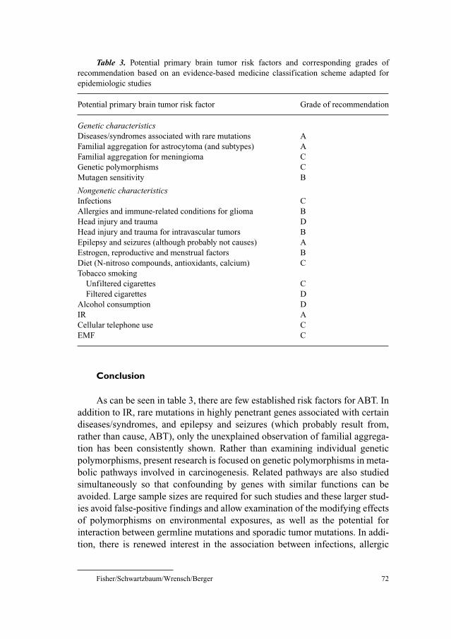
Fisher/Schwartzbaum/Wrensch/Berger 72
Conclusion
As can be seen in table 3, there are few established risk factors for ABT. In
addition to IR, rare mutations in highly penetrant genes associated with certain
diseases/syndromes, and epilepsy and seizures (which probably result from,
rather than cause, ABT), only the unexplained observation of familial aggrega-
tion has been consistently shown. Rather than examining individual genetic
polymorphisms, present research is focused on genetic polymorphisms in meta-
bolic pathways involved in carcinogenesis. Related pathways are also studied
simultaneously so that confounding by genes with similar functions can be
avoided. Large sample sizes are required for such studies and these larger stud-
ies avoid false-positive findings and allow examination of the modifying effects
of polymorphisms on environmental exposures, as well as the potential for
interaction between germline mutations and sporadic tumor mutations. In addi-
tion, there is renewed interest in the association between infections, allergic
Table 3. Potential primary brain tumor risk factors and corresponding grades of
recommendation based on an evidence-based medicine classification scheme adapted for
epidemiologic studies
Potential primary brain tumor risk factor Grade of recommendation
Genetic characteristicsDiseases/syndromes associated with rare mutations A
Familial aggregation for astrocytoma (and subtypes) A
Familial aggregation for meningioma C
Genetic polymorphisms C
Mutagen sensitivity B
Nongenetic characteristicsInfections C
Allergies and immune-related conditions for glioma B
Head injury and trauma D
Head injury and trauma for intravascular tumors B
Epilepsy and seizures (although probably not causes) A
Estrogen, reproductive and menstrual factors B
Diet (N-nitroso compounds, antioxidants, calcium) C
Tobacco smoking
Unfiltered cigarettes C
Filtered cigarettes D
Alcohol consumption D
IR A
Cellular telephone use C
EMF C
Primary Adult Brain Tumor Risk Factors 73
conditions and ABT risk. Our knowledge of the causes of ABT is limited and
should be expanded by results from large, well-designed studies of novel poten-
tial risk factors and potential interactions between known and suspected risk
factors.
References
1 Jenicek M: Epidemiology, evidenced-based medicine, and evidence-based public health.
J Epidemiol 1997;7:187–197.
2 Miyasaki JM, Martin W, Suchowersky O, Weiner WJ, Lang AE: Practice parameter: initiation of
treatment for Parkinson’s disease: an evidence-based review: report of the Quality Standards
Subcommittee of the American Academy of Neurology. Neurology 2002;58:11–17.
3 Spector R, Vesell ES: The pursuit of clinical truth: role of epidemiology/observation studies.
J Clin Pharmacol 2000;40:1205–1210.
4 Sivak-Sears N, Schwartzbaum J, Miike R, Moghadassi M, Wrensch M: Case-control study of non-
steroidal anti-inflammatory drug use and glioblastoma multiforme. Am J Epidemiol, in press.
5 Aldape KD, Okcu MF, Bondy ML, Wrensch M: Molecular epidemiology of glioblastoma. Cancer J
2003;9:99–106.
6 Gurney JG, Kadan-Lottick N: Brain and other central nervous system tumors: rates, trends, and
epidemiology. Curr Opin Oncol 2001;13:160–166.
7 Inskip PD, Linet MS, Heineman EF: Etiology of brain tumors in adults. Epidemiol Rev 1995;17:
382–414.
8 Preston-Martin S: Epidemiology of primary CNS neoplasms. Neurol Clin 1996;14:273–290.
9 Wrensch M, Minn Y, Chew T, Bondy M, Berger MS: Epidemiology of primary brain tumors: cur-
rent concepts and review of the literature. Neurooncology 2002;4:278–299.
10 Wrensch M, Lee M, Miike R, Newman B, Barger G, Davis R, Wiencke J, Neuhaus J: Familial and
personal medical history of cancer and nervous system conditions among adults with glioma and
controls. Am J Epidemiol 1997;145:581–593.
11 Narod SA, Stiller C, Lenoir GM: An estimate of the heritable fraction of childhood cancer. Br J
Cancer 1991;63:993–999.
12 Bondy M, Wiencke J, Wrensch M, Kyritsis AP: Genetics of primary brain tumors: a review.
J Neurooncol 1994;18:69–81.
13 Huson SM, Harper PS, Compston DA: Von Recklinghausen neurofibromatosis. A clinical and
population study in south-east Wales. Brain 1988;111:1355–1381.
14 Akashi M, Koeffler HP: Li-Fraumeni syndrome and the role of the p53 tumor suppressor gene in
cancer susceptibility. Clin Obstet Gynecol 1998;41:172–199.
15 Malkin D: p53 and the Li-Fraumeni syndrome. Cancer Genet Cytogenet 1993;66:83–92.
16 Nichols KE, Malkin D, Garber JE, Fraumeni JFJ, Li FP: Germ-line p53 mutations predispose to a
wide spectrum of early-onset cancers. Cancer Epidemiol Biomarkers Prev 2001;10:83–87.
17 Kyritsis AP, Bondy ML, Xiao M, Berman EL, Cunningham JE, Lee PS, Levin VA, Saya H:
Germline p53 gene mutations in subsets of glioma patients. J Natl Cancer Inst 1994;86:344–349.
18 Hemminki K, Li X: Familial risks in nervous system tumors. Cancer Epidemiol Biomarkers Prev
2003;12:1137–1142.
19 Hemminki K, Li X, Collins VP: Parental cancer as a risk factor for brain tumors (Sweden). Cancer
Causes Control 2001;12:195–199.
20 Paunu N, Pukkala E, Laippala P, Sankila R, Isola J, Miettinen H, Simola KOJ, Helen P, Helin H,
Haapasalo H: Cancer incidence in families with multiple glioma patients. Int J Cancer 2002;97:
819–822.
21 Cicuttini FM, Hurley SF, Forbes A, Donnan GA, Salzberg M, Giles GG, McNeil JJ: Association of
adult glioma with medical conditions, family and reproductive history. Int J Cancer 1997;71:
203–207.
Fisher/Schwartzbaum/Wrensch/Berger 74
22 Hochberg F, Toniolo P, Cole P, Salcman M: Nonoccupational risk indicators of glioblastoma in
adults. J Neurooncol 1990;8:55–60.
23 Malmer B, Gronberg H, Bergenheim AT, Lenner P, Henriksson R: Familial aggregation of astrocy-
toma in northern Sweden: an epidemiological cohort study. Int J Cancer 1999;81:366–370.
24 Malmer B, Brannstrom T, Andersson U, Bergh K, Gronberg H, Henriksson R: Does a low fre-
quency of P53 and Pgp expression in familial glioma compared to sporadic controls indicate bio-
logical differences? Anticancer Res 2002;22:3949–3954.
25 Malmer B, Henriksson R, Gronberg H: Familial brain tumours – genetics or environment? A
nationwide cohort study of cancer risk in spouses and first-degree relatives of brain tumour
patients. Int J Cancer 2003;106:260–263.
26 Malmer B, Iselius L, Holmberg E, Collins A, Henriksson R, Gronberg H: Genetic epidemiology of
glioma. Br J Cancer 2001;84:429–434.
27 Schlehofer B, Blettner M, Becker N, Martinsohn C, Wahrendorf J: Medical risk factors and the
development of brain tumors. Cancer 1992;69:2541–2547.
28 Wrensch MR, Barger GR: Familial factors associated with malignant gliomas. Genet Epidemiol
1990;7:291–301.
29 Zampieri P, Meneghini F, Grigoletto F, Gerosa M, Licata C, Casentini L, Longatti PL, Padoan A,
Mingrino S: Risk factors for cerebral glioma in adults: a case-control study in an Italian popula-
tion. J Neurooncol 1994;19:61–67.
30 Malmer B, Henriksson R, Gronberg H: Different aetiology of familial low-grade and high-grade
glioma? A nationwide cohort study of familial glioma. Neuroepidemiology 2002;21:279–286.
31 de Andrade M, Barnholtz JS, Amos CI, Adatto P, Spencer C, Bondy ML: Segregation analysis of
cancer in families of glioma patients. Genet Epidemiol 2001;20:258–270.
32 Grossman SA, Osman M, Hruban R, Piantadosi S: Central nervous system cancers in first-degree
relatives and spouses. Cancer Invest 1999;17:299–308.
33 Elexpuru-Camiruaga J, Buxton N, Kandula V, Dias PS, Campbell D, McIntosh J, Broome J, Jones P,
Inskip A, Alldersea J: Susceptibility to astrocytoma and meningioma: influence of allelism at
glutathione S-transferase (GSTT1 and GSTM1) and cytochrome P-450 (CYP2D6) loci. Cancer
Res 1995;55:4237–4239.
34 Kelsey KT, Wrensch M, Zuo ZF, Miike R, Wiencke JK: A population-based case-control study of
the CYP2D6 and GSTT1 polymorphisms and malignant brain tumors. Pharmacogenetics
1997;7:463–468.
35 Trizna Z, de Andrade M, Kyritsis AP, Briggs K, Levin VA, Bruner JM, Wei Q, Bondy ML: Genetic
polymorphisms in glutathione S-transferase mu and theta, N-acetyltransferase, and CYP1A1 and
risk of gliomas. Cancer Epidemiol Biomarkers Prev 1998;7:553–555.
36 De Roos AJ, Rothman N, Inskip PD, Linet MS, Shapiro WR, Selker RG, Fine HA, Black PM,
Pittman GS, Bell DA: Genetic polymorphisms in GSTM1, -P1, -T1, and CYP2E1 and the risk of
adult brain tumors. Cancer Epidemiol Biomarkers Prev 2003;12:14–22.
37 Hand PA, Inskip A, Gilford J, Alldersea J, Elexpuru-Camiruaga J, Hayes JD, Jones PW, Strange RC,
Fryer AA: Allelism at the glutathione S-transferase GSTM3 locus: interactions with GSTM1 and
GSTT1 as risk factors for astrocytoma. Carcinogenesis 1996;17:1919–1922.
38 Wrensch M, Kelsey KT, Liu M, Miike R, Moghadassi M, Aldape K, McMillan A, Wiencke J:
Glutathione-S-transferase and adult glioma. Cancer Epidemiol Biomarkers Prev 2004;13:
461–467.
39 Chen P, Wiencke J, Aldape K, Kesler-Diaz A, Miike R, Kelsey K, Lee M, Liu J, Wrensch M:
Association of an ERCC1 polymorphism with adult-onset glioma. Cancer Epidemiol Biomarkers
Prev 2000;9:843–847.
40 Caggana M, Kilgallen J, Conroy JM, Wiencke JK, Kelsey KT, Miike R, Chen P, Wrensch MR:
Associations between ERCC2 polymorphisms and gliomas. Cancer Epidemiol Biomarkers Prev
2001;10:355–360.
41 Wacholder S, Chanock S, Garcia-Closas M, El Ghormli L, Rothman N: Assessing the probability
that a positive report is false: an approach for molecular epidemiology studies. J Natl Cancer Inst
2004;96:434–442.
42 Bondy ML, Kyritsis AP, Gu J, de Andrade M, Cunningham J, Levin VA, Bruner JM, Wei Q:
Mutagen sensitivity and risk of gliomas: a case-control analysis. Cancer Res 1996;56:1484–1486.
Primary Adult Brain Tumor Risk Factors 75
43 Nersesyan AK: Re: Gamma-radiation sensitivity and risk of glioma. J Natl Cancer Inst 2002;
94:949.
44 Fisher SG, Weber L, Carbone M: Cancer risk associated with simian virus 40 contaminated polio
vaccine. Anticancer Res 1999;19:2173–2180.
45 Geissler E, Staneczek W: SV40 and human brain tumors. Arch Geschwulstforsch 1988;58:
129–134.
46 Strickler HD, Rosenberg PS, Devesa SS, Hertel J, Fraumeni JFJ, Goedert JJ: Contamination of
poliovirus vaccines with simian virus 40 (1955–1963) and subsequent cancer rates. JAMA
1998;279:292–295.
47 Wrensch M, Weinberg A, Wiencke J, Masters H, Miike R, Barger G, Lee M: Does prior infection
with varicella-zoster virus influence risk of adult glioma? Am J Epidemiol 1997;145:594–597.
48 Wrensch M, Weinberg A, Wiencke J, Miike R, Barger G, Kelsey K: Prevalence of antibodies to
four herpesviruses among adults with glioma and controls. Am J Epidemiol 2001;154:161–165.
49 Caldarelli-Stefano R, Boldorini R, Monga G, Meraviglia E, Zorini EO, Ferrante P: JC virus in
human glial-derived tumors. Hum Pathol 2000;31:394–395.
50 Croul S, Otte J, Khalili K: Brain tumors and polyomaviruses. J Neurovirol 2003;9:173–182.
51 Del Valle L, Gordon J, Assimakopoulou M, Enam S, Geddes JF, Varakis JN, Katsetos CD, Croul S,
Khalili K: Detection of JC virus DNA sequences and expression of the viral regulatory protein
T-antigen in tumors of the central nervous system. Cancer Res 2001;61:4287–4293.
52 Dorries K, Loeber G, Meixensberger J: Association of polyomaviruses JC, SV40, and BK with
human brain tumors. Virology 1987;160:268–270.
53 Krynska B, Otte J, Franks R, Khalili K, Croul S: Human ubiquitous JCV(CY) T-antigen gene
induces brain tumors in experimental animals. Oncogene 1999;18:39–46.
54 Tretiakova A, Krynska B, Gordon J, Khalili K: Human neurotropic JC virus early protein deregu-
lates glial cell cycle pathway and impairs cell differentiation. J Neurosci Res 1999;55:588–599.
55 Cuomo L, Trivedi P, Cardillo MR, Gagliardi FM, Vecchione A, Caruso R, Calogero A, Frati L,
Faggioni A, Ragona G: Human herpesvirus 6 infection in neoplastic and normal brain tissue.
J Med Virol 2001;63:45–51.
56 Rollison DEM, Helzlsouer KJ, Alberg AJ, Hoffman S, Hou J, Daniel R, Shah KV, Major EO:
Serum antibodies to JC virus, BK virus, simian virus 40, and the risk of incident adult astrocytic
brain tumors. Cancer Epidemiol Biomarkers Prev 2003;12:460–463.
57 Blumenthal DT, Raizer JJ, Rosenblum MK, Bilsky MH, Hariharan S, Abrey LE: Primary intracra-
nial neoplasms in patients with HIV. Neurology 1999;52:1648–1651.
58 Fisher J, Schwartzbaum J, Johnson C: Cold/influenza infection, influenza vaccination, and risk of
adult glioma (abstract). Am J Epidemiol 2000;151:116.
59 Schlehofer B, Blettner M, Preston-Martin S, Niehoff D, Wahrendorf J, Arslan A, Ahlbom A,
Choi WN, Giles GG, Howe GR, Little J, Menegoz F, Ryan P: Role of medical history in brain
tumour development. Results from the international adult brain tumour study. Int J Cancer 1999;82:
155–160.
60 Ryan P, Hurley SF, Johnson AM, Salzberg M, Lee MW, North JB, McNeil JJ, McMichael AJ:
Tumours of the brain and presence of antibodies to Toxoplasma gondii. Int J Epidemiol 1993;22:
412–419.
61 Schuman LM, Choi NW, Gullen WH: Relationship of central nervous system neoplasms to
Toxoplasma gondii infection. Am J Publ Health Nations Health 1967;57:848–856.
62 Brenner AV, Linet MS, Fine HA, Shapiro WR, Selker RG, Black PM, Inskip PD: History of
allergies and autoimmune diseases and risk of brain tumors in adults. Int J Cancer 2002;99:
252–259.
63 Schwartzbaum J, Jonsson F, Ahlbom A, Preston-Martin S, Lonn S, Soderberg KC, Feychting M:
Cohort studies of association between self-reported allergic conditions, immune-related diagnoses
and glioma and meningioma risk. Int J Cancer 2003;106:423–428.
64 Venge P: Monitoring the allergic inflammation. Allergy 2004;59:26–32.
65 Wiemels JL, Wiencke JK, Sison JD, Miike R, McMillan A, Wrensch M: History of allergies
among adults with glioma and controls. Int J Cancer 2002;98:609–615.
66 Ryan P, Lee MW, North B, McMichael AJ: Risk factors for tumors of the brain and meninges:
results from the Adelaide Adult Brain Tumor Study. Int J Cancer 1992;51:20–27.

Fisher/Schwartzbaum/Wrensch/Berger 76
67 Phillips LE, Koepsell TD, van Belle G, Kukull WA, Gehrels J-A, Longstreth WTJ: History of head
trauma and risk of intracranial meningioma: population-based case-control study. Neurology
2002;58:1849–1852.
68 Preston-Martin S, Mack W, Henderson BE: Risk factors for gliomas and meningiomas in males in
Los Angeles County. Cancer Res 1989;49:6137–6143.
69 Preston-Martin S, Pogoda JM, Schlehofer B, Blettner M, Howe GR, Ryan P, Menegoz F, Giles GG,
Rodvall Y, Choi NW, Little J, Arslan A: An international case-control study of adult glioma and
meningioma: the role of head trauma. Int J Epidemiol 1998;27:579–586.
70 Preston-Martin S, Yu MC, Henderson BE, Roberts C: Risk factors for meningiomas in men in Los
Angeles County. J Natl Cancer Inst 1983;70:863–866.
71 Annegers JF, Laws ERJ, Kurland LT, Grabow JD: Head trauma and subsequent brain tumors.
Neurosurgery 1979;4:203–236.
72 Inskip PD, Mellemkjaer L, Gridley G, Olsen JH: Incidence of intracranial tumors following hos-
pitalization for head injuries (Denmark). Cancer Causes Control 1998;9:109–116.
73 Nygren C, Adami J, Ye W, Bellocco R, af Geijerstam JL, Borg J, Nyren O: Primary brain tumors
following traumatic brain injury – a population-based cohort study in Sweden. Cancer Causes
Control 2001;12:733–737.
74 Hu J, Johnson KC, Mao Y, Guo L, Zhao X, Jia X, Bi D, Huang G, Liu R: Risk factors for glioma
in adults: a case-control study in northeast China. Cancer Detect Prev 1998;22:100–108.
75 Hochberg F, Toniolo P, Cole P: Head trauma and seizures as risk factors of glioblastoma.
Neurology 1984;34:1511–1514.
76 Wrensch M, Miike R, Lee M, Neuhaus J: Are prior head injuries or diagnostic X-rays associated
with glioma in adults? The effects of control selection bias. Neuroepidemiology 2000;19:
234–244.
77 Pace A, Bove L, Innocenti P, Pietrangeli A, Carapella CM, Oppido P, Raus L, Occhipinti E,
Jandolo B: Epilepsy and gliomas: incidence and treatment in 119 patients. J Exp Clin Cancer Res
1998;17:479–482.
78 Lamminpaa A, Pukkala E, Teppo L, Neuvonen PJ: Cancer incidence among patients using
antiepileptic drugs: a long-term follow-up of 28,000 patients. Eur J Clin Pharmacol 2002;58:
137–141.
79 Schwartzbaum J, Jonsson F, Ahlbom A, Preston-Martin S, Malmer B, Lonn S, Soderberg KC,
Feychting M: Prior hospitalization for epilepsy, diabetes, and stroke may result from preclinical
systemic effects of astrocytoma and meningioma. Submitted.
80 Olsen JH, Boice JDJ, Jensen JP, Fraumeni JFJ: Cancer among epileptic patients exposed to anti-
convulsant drugs. J Natl Cancer Inst 1989;81:803–808.
81 Shirts SB, Annegers JF, Hauser WA, Kurland LT: Cancer incidence in a cohort of patients with
seizure disorders. J Natl Cancer Inst 1986;77:83–87.
82 Bondy M, Ligon BL: Epidemiology and etiology of intracranial meningiomas: a review. J Neurooncol
1996;29:197–205.
83 Longstreth WTJ, Dennis LK, McGuire VM, Drangsholt MT, Koepsell TD: Epidemiology of
intracranial meningioma. Cancer 1993;72:639–648.
84 Horsfall DJ, Goldsmith KG, Ricciardelli C, Skinner JM, Tilley WD, Marshall VR: Steroid hor-
mone and epidermal growth factor receptors in meningiomas. Aust NZ J Surg 1989;59:881–888.
85 Punnonen R, Kuurne T: Estrogen and progestin receptors in intracranial tumors. Horm Res
1987;27:74–77.
86 Yu ZY, Wrange O, Haglund B, Granholm L, Gustafsson JA: Estrogen and progestin receptors in
intracranial meningiomas. J Steroid Biochem 1982;16:451–456.
87 Jhawar BS, Fuchs CS, Colditz GA, Stampfer MJ: Sex steroid hormone exposures and risk for
meningioma. J Neurosurg 2003;99:848–853.
88 Schlehofer B, Blettner M, Wahrendorf J: Association between brain tumors and menopausal
status. J Natl Cancer Inst 1992;84:1346–1349.
89 Lambe M, Coogan P, Baron J: Reproductive factors and the risk of brain tumors: a population-
based study in Sweden. Int J Cancer 1997;72:389–393.
90 McKinley BP, Michalek AM, Fenstermaker RA, Plunkett RJ: The impact of age and sex on the
incidence of glial tumors in New York state from 1976 to 1995. J Neurosurg 2000;93:932–939.
Primary Adult Brain Tumor Risk Factors 77
91 Cantor KP, Lynch CF, Johnson D: Reproductive factors and risk of brain, colon, and other malig-
nancies in Iowa (United States). Cancer Causes Control 1993;4:505–511.
92 Preston-Martin S, Henderson BE: N-Nitroso compounds and human intracranial tumours. IARC
Sci Publ 1984;57:887–894.
93 Chen H, Ward MH, Tucker KL, Graubard BI, McComb RD, Potischman NA, Weisenburger DD,
Heineman EF: Diet and risk of adult glioma in eastern Nebraska, United States. Cancer Causes
Control 2002;13:647–655.
94 Lee M, Wrensch M, Miike R: Dietary and tobacco risk factors for adult onset glioma in the San
Francisco Bay Area (California, USA). Cancer Causes Control 1997;8:13–24.
95 Schwartzbaum JA, Fisher JL, Goodman J, Octaviano D, Cornwell DG: Hypotheses concerning
roles of dietary energy, cured meat, and serum tocopherols in adult glioma development.
Neuroepidemiology 1999;18:156–166.
96 Huncharek M, Kupelnick B, Wheeler L: Dietary cured meat and the risk of adult glioma: a meta-
analysis of nine observational studies. J Environ Pathol Toxicol Oncol 2003;22:129–137.
97 Barrett JH, Parslow RC, McKinney PA, Law GR, Forman D: Nitrate in drinking water and the
incidence of gastric, esophageal, and brain cancer in Yorkshire, England. Cancer Causes Control
1998;9:153–159.
98 Hu J, La Vecchia C, Negri E, Chatenoud L, Bosetti C, Jia X, Liu R, Huang G, Bi D, Wang C: Diet
and brain cancer in adults: a case-control study in northeast China. Int J Cancer 1999;81:20–23.
99 Tedeschi-Blok N, Schwartzbaum J, Lee M, Miike R, Wrensch M: Dietary calcium consumption
and astrocytic glioma: the San Francisco Bay Area Adult Glioma Study, 1991–1995. Nutr Cancer
2001;39:196–203.
100 Hurley SF, McNeil JJ, Donnan GA, Forbes A, Salzberg M, Giles GG: Tobacco smoking and alco-
hol consumption as risk factors for glioma: a case-control study in Melbourne, Australia.
J Epidemiol Community Health 1996;50:442–446.
101 Hu J, Little J, Xu T, Zhao X, Guo L, Jia X, Huang G, Bi D, Liu R: Risk factors for meningioma in
adults: a case-control study in northeast China. Int J Cancer 1999;83:299–304.
102 Preston-Martin S, Mack W: Gliomas and meningiomas in men in Los Angeles County: investiga-
tion of exposures to N-nitroso compounds. IARC Sci Publ 1991;105:197–203.
103 Grayson JK, Lyons TJ: Brain cancer, flying, and socioeconomic status: a nested case-control study
of USAF aircrew. Aviat Space Environ Med 1996;67:1152–1154.
104 Gundestrup M, Storm HH: Radiation-induced acute myeloid leukaemia and other cancers in com-
mercial jet cockpit crew: a population-based cohort study. Lancet 1999;354:2029–2031.
105 Alexander V, DiMarco JH: Reappraisal of brain tumor risk among U.S. nuclear workers: a 10-year
review. Occup Med 2001;16:289–315.
106 Loomis DP, Wolf SH: Mortality of workers at a nuclear materials production plant at Oak Ridge,
Tennessee, 1947–1990. Am J Ind Med 1996;29:131–141.
107 Hardell L, Mild KH, Pahlson A, Hallquist A: Ionizing radiation, cellular telephones and the risk
for brain tumours. Eur J Cancer Prev 2001;10:523–529.
108 Hodges LC, Smith JL, Garrett A, Tate S: Prevalence of glioblastoma multiforme in subjects with
prior therapeutic radiation. J Neurosci Nurs 1992;24:79–83.
109 Salvati M, Artico M, Caruso R, Rocchi G, Orlando ER, Nucci F: A report on radiation-induced
gliomas. Cancer 1991;67:392–397.
110 Shapiro S, Mealey JJ, Sartorius C: Radiation-induced intracranial malignant gliomas. J Neurosurg
1989;71:77–82.
111 Salminen E, Pukkala E, Teppo L: Second cancers in patients with brain tumours – impact of treat-
ment. Eur J Cancer 1999;35:102–105.
112 Longstreth WTJ, Phillips LE, Drangsholt M, Koepsell TD, Custer BS, Gehrels J-A, van Belle G:
Dental X-rays and the risk of intracranial meningioma: a population-based case-control study.
Cancer 2004;100:1026–1034.
113 Shintani T, Hayakawa N, Hoshi M, Sumida M, Kurisu K, Oki S, Kodama Y, Kajikawa H, Inai K,
Kamada N: High incidence of meningioma among Hiroshima atomic bomb survivors. J Radiat
Res 1999;40:49–57.
114 Rothman KJ, Loughlin JE, Funch DP, Dreyer NA: Overall mortality of cellular telephone cus-
tomers. Epidemiology 1996;7:303–305.

Fisher/Schwartzbaum/Wrensch/Berger 78
115 Auvinen A, Hietanen M, Luukkonen R, Koskela R-S: Brain tumors and salivary gland cancers
among cellular telephone users. Epidemiology 2002;13:356–359.
116 Hardell L, Nasman A, Pahlson A, Hallquist A, Hansson Mild K: Use of cellular telephones and the
risk for brain tumours: a case-control study. Int J Oncol 1999;15:113–116.
117 Inskip PD, Tarone RE, Hatch EE, Wilcosky TC, Shapiro WR, Selker RG, Fine HA, Black PM,
Loeffler JS, Linet MS: Cellular-telephone use and brain tumors. N Engl J Med 2001;344:79–86.
118 Muscat JE, Malkin MG, Thompson S, Shore RE, Stellman SD, McRee D, Neugut AI, Wynder EL:
Handheld cellular telephone use and risk of brain cancer. JAMA 2000;284:3001–3007.
119 Johansen C, Boice JJ, McLaughlin J, Olsen J: Cellular telephones and cancer – a nationwide
cohort study in Denmark. J Natl Cancer Inst 2001;93:203–207.
120 Hardell L, Mild KH, Carlberg M: Case-control study on the use of cellular and cordless phones
and the risk for malignant brain tumours. Int J Radiat Biol 2002;78:931–936.
121 Hardell L, Mild KH, Carlberg M: Further aspects on cellular and cordless telephones and brain
tumours. Int J Oncol 2003;22:399–407.
122 De Roos AJ, Stewart PA, Linet MS, Heineman EF, Dosemeci M, Wilcosky T, Shapiro WR, Selker
RG, Fine HA, Black PM, Inskip PD: Occupation and the risk of adult glioma in the United States.
Cancer Causes Control 2003;14:139–150.
123 Floderus B, Persson T, Stenlund C, Wennberg A, Ost A, Knave B: Occupational exposure to elec-
tromagnetic fields in relation to leukemia and brain tumors: a case-control study in Sweden.
Cancer Causes Control 1993;4:465–476.
124 Floderus B, Stenlund C, Persson T: Occupational magnetic field exposure and site-specific cancer
incidence: a Swedish cohort study. Cancer Causes Control 1999;10:323–332.
125 Lin RS, Dischinger PC, Conde J, Farrell KP: Occupational exposure to electromagnetic fields and
the occurrence of brain tumors. An analysis of possible associations. J Occup Med 1985;27:
413–419.
126 Mack W, Preston-Martin S, Peters JM: Astrocytoma risk related to job exposure to electric and
magnetic fields. Bioelectromagnetics 1991;12:57–66.
127 Preston-Martin S, Lewis S, Winkelmann R, Borman B, Auld J, Pearce N: Descriptive epidemiol-
ogy of primary cancer of the brain, cranial nerves, and cranial meninges in New Zealand,
1948–88. Cancer Causes Control 1993;4:529–538.
128 Rodvall Y, Ahlbom A, Stenlund C, Preston-Martin S, Lindh T, Spannare B: Occupational exposure
to magnetic fields and brain tumours in central Sweden. Eur J Epidemiol 1998;14:563–569.
129 Speers MA, Dobbins JG, Miller VS: Occupational exposures and brain cancer mortality: a pre-
liminary study of east Texas residents. Am J Ind Med 1988;13:629–638.
130 Spinelli JJ, Band PR, Svirchev LM, Gallagher RP: Mortality and cancer incidence in aluminum
reduction plant workers. J Occup Med 1991;33:1150–1155.
131 Villeneuve PJ, Agnew DA, Johnson KC, Mao Y: Brain cancer and occupational exposure to mag-
netic fields among men: results from a Canadian population-based case-control study. Int J
Epidemiol 2002;31:210–217.
132 Zheng T, Cantor KP, Zhang Y, Keim S, Lynch CF: Occupational risk factors for brain cancer: a
population-based case-control study in Iowa. J Occup Environ Med 2001;43:317–324.
133 Johansen C, Olsen JH: Risk of cancer among Danish electricity workers. A cohort study. Ugeskr
Laeger 1999;161:2079–2085.
134 Minder CE, Pfluger DH: Leukemia, brain tumors, and exposure to extremely low frequency elec-
tromagnetic fields in Swiss railway employees. Am J Epidemiol 2001;153:825–835.
135 Sahl JD, Kelsh MA, Greenland S: Cohort and nested case-control studies of hematopoietic cancers
and brain cancer among electric utility workers. Epidemiology 1993;4:104–114.
136 Sorahan T, Nichols L, van Tongeren M, Harrington JM: Occupational exposure to magnetic fields
relative to mortality from brain tumours: updated and revised findings from a study of United
Kingdom electricity generation and transmission workers, 1973–97. Occup Environ Med 2001;58:
626–630.
137 Tynes T, Jynge H, Vistnes AI: Leukemia and brain tumors in Norwegian railway workers, a nested
case-control study. Am J Epidemiol 1994;139:645–653.
138 Fear NT, Roman E, Carpenter LM, Newton R, Bull D: Cancer in electrical workers: an analysis of
cancer registrations in England, 1981–87. Br J Cancer 1996;73:935–939.

Primary Adult Brain Tumor Risk Factors 79
139 Grayson JK: Radiation exposure, socioeconomic status, and brain tumor risk in the US Air Force:
a nested case-control study. Am J Epidemiol 1996;143:480–486.
140 Navas-Acien A, Pollan M, Gustavsson P, Floderus B, Plato N, Dosemeci M: Interactive effect of
chemical substances and occupational electromagnetic field exposure on the risk of gliomas and
meningiomas in Swedish men. Cancer Epidemiol Biomarkers Prev 2002;11:1678–1683.
141 Savitz DA, Cai J, van Wijngaarden E, Loomis D, Mihlan G, Dufort V, Kleckner RC, Nylander-
French L, Kromhout H, Zhou H: Case-cohort analysis of brain cancer and leukemia in electric util-
ity workers using a refined magnetic field job-exposure matrix. Am J Ind Med 2000;38:417–425.
142 Wrensch M, Yost M, Miike R, Lee G, Touchstone J: Adult glioma in relation to residential power
frequency electromagnetic field exposures in the San Francisco Bay area. Epidemiology 1999;10:
523–527.
James L. Fisher, PhD
The Arthur G. James Cancer Hospital and Richard J. Solove Research Institute
2050 Kenny Road Suite 940
Columbus, OH 43221 (USA)
Tel. �1 614 293 9644, Fax �1 614 293 6667, E-Mail [email protected]
Pollock BE (ed): Guiding Neurosurgery by Evidence.
Prog Neurol Surg. Basel, Karger, 2006, vol 19, pp 80–96
Benign Adult Brain Tumors:An Evidence-Based Medicine Review
Manish Aghi, Fred G. Barker II
Neurosurgical Service, Massachusetts General Hospital, Boston, Mass., USA
AbstractBackground: Benign adult brain tumors can be managed conservatively or using
surgery, radiation, or medicines. While randomized comparisons assessing tumor recur-
rence, quality of life, or survival are the ideal means of comparing treatments, it can be dif-
ficult to recruit patients to such trials and lengthy follow-up periods are needed because of
the slowly progressive natural history of these tumors. Methods: Review of the literature
on benign adult brain tumors using evidence-based standards and focusing on menin-
giomas, pituitary adenomas, and vestibular schwannomas, which together represent the
majority of WHO grade 1 adult brain tumors. Results: Nearly all studies of benign adult
brain tumors were of relatively poor quality (level 3 or poorer). These studies enable grade
C recommendations. The safety of meningioma surgery in the elderly varies with institu-
tion, radiosurgery is a reliable alternative to surgery in small to medium-sized menin-
giomas, and the efficacy of drugs in therapy of meningiomas recurring after surgery is
difficult to interpret due to a lack of uniform criteria in the studies. Radiosurgery is effec-
tive in nonfunctional pituitary adenomas recurring after surgery, while phototherapy is a
newer treatment modality with potential benefits in pituitary adenomas that fail surgery or
radiation. Vestibular schwannomas can be conservatively managed, but there are no reliable
predictors of growth, so serial imaging is important. Radiosurgery has proven to be a reli-
able alternative to surgery for small to medium-sized vestibular schwannomas, but follow-
up has been relatively short in most studies to date. Conclusions: While randomized
clinical trials comparing conservative management, surgery, radiation, and medical man-
agement of benign adult benign tumors are unlikely to occur, there is some level 3 evidence
that can assist in their treatment.
Copyright © 2006 S. Karger AG, Basel
Each year, slightly more than 40,000 individuals in the United States are
diagnosed with brain tumors [1]. Of these, 43% are benign brain tumors [1],
although an accurate accounting is difficult to obtain because most state cancer
Benign Adult Brain Tumors 81
registries historically have collected data only on primary malignant brain tumors.
Benign adult brain tumors comprise both intra-axial and extra-axial tumors
and include epidermoid, dermoid, hemangioblastoma, colloid cyst, subependy-
moma, pleomorphic xanthoastrocytoma, schwannoma, pituitary adenoma, cran-
iopharyngioma and meningioma. Here we review the literature for treatment
studies on meningiomas, pituitary adenomas, and vestibular schwannomas using
evidence-based standards. Because these three neoplasms collectively account
for 40% of all intracranial neoplasms [2–4], they have generated enough studies
on treatment options that some recommendations can be made based on evi-
dence-based standards. Unfortunately the paucity of randomized clinical trials in
this field leads to an inability to make high grade recommendations. In addition,
with benign tumors, the success of a treatment in preventing recurrence or pro-
gression may not be evident for 10 or more years after surgery. Thus, informa-
tion derived from case series (level 4 evidence) or case-control (level 3 evidence)
studies is often the best available data on which to base clinical decisions for
patients with benign brain tumors. Despite these constraints, the studies
described in this chapter provide some important insight into treatment options
for these three common benign brain tumors.
Meningiomas
Meningiomas represent approximately 15% of adult intracranial neo-
plasms [3]. Up to one third of meningiomas diagnosed during life are asympto-
matic [5], a number that will likely grow as imaging technologies improve and
the use of cranial imaging in patients with minimal symptoms increases. As a
result, a common challenge facing the neurosurgeon is the advisability of surgi-
cal resection of asymptomatic meningiomas. Level 3 evidence addressing this
issue came from a retrospective record review identifying 504 meningiomas
diagnosed at 27 hospitals in a region of Japan between 1989 and 1996 [6]. Of
these meningiomas, 39% were asymptomatic, a proportion that was increasing
over time. Asymptomatic meningiomas were more common in older patients
and female patients. Surgery was performed on 87 of 196 asymptomatic menin-
giomas. In 20 of the 63 conservatively managed meningiomas, the tumor
increased in size during the follow-up period. The tendency to increase in size
did not depend on initial size, age, or gender; T2 bright tumors were more likely
to grow; and calcified tumors were less likely to grow during follow-up.
Morbidity occurred in 11.4% of asymptomatic patients who underwent surgery,
including 23.3% of patients older than 70 years, compared to 3.5% of patients
younger than 70. The study did not support surgery in elderly patients with
asymptomatic meningiomas.
Aghi/Barker 82
Surgical resection has long been the preferred treatment modality for
symptomatic meningiomas. In 1957, Simpson [7] published a landmark paper
documenting the direct correlation between degree of meningioma resection
and later tumor recurrence. He reported a 9% recurrence rate after ‘complete
resection’ of the tumor and its neoplastic dural base (grade 1 resection), a 19%
recurrence rate when the tumor was resected and the dural base only coagulated
(grade 2), a 29% recurrence rate when the tumor itself was removed but the
dura could not be excised (grade 3), and an approximately 40% recurrence rate
when only subtotal resection was performed (grade 4). Simpson’s recurrence
rates may have been an underestimate, because his study preceded the availabil-
ity of CT or MRI. Subsequent studies found a 70% rate of tumor progression
after subtotal resection alone [8], and 16 and 20% recurrence rates after grade 1
and 2 resections [9]. Given this recurrence rate after grade 1 resections, in 1986
Borovich and colleagues [10, 11] recommended adding another grade, ‘grade 0’,
defined by an excision in which a 2-cm-wide resection of normal dural margin
occurred. In a subsequent level 4 study, a retrospective review of grade 0
removal of convexity meningiomas in 37 patients between 1982 and 1992 at
three different centers found no recurrences and no increase in morbidity with
the more aggressive procedure [12]. Of course, comparisons to other studies
describing grade 1 resections cannot be made because those series may have
contained a mix of grade 0 and grade 1 resections.
Many meningiomas occur in older persons, with a peak incidence at 60
years [13]. Because surgery often carries an elevated level of risk in elderly
patients, surgeons need evidence to determine the role of age in deciding
whether to resect a meningioma. In a case-control study, a prospectively
acquired database was used to identify 114 patients undergoing meningioma
resection by a single surgeon [14]. The patients were divided into those 65 and
over and those under 65, with each group having 57 patients. More than 90% of
the meningiomas in each group were symptomatic. Medical and surgical com-
plication rates were 7.0 and 5.2% in elderly patients, compared to 8.8 and 3.5%
in younger patients (p � 0.05). Mortality within the first 30 postoperative days
was zero for the younger patients, and 1/57 in the older group (p � 0.05). This
study provided level 3 evidence confirming the safety of meningioma surgery
in elderly patients. Of note, significantly less morbidity was found in this study
than in previous studies of meningioma surgery in the elderly [14], suggesting
that the advisability of meningioma surgery in the elderly may vary from center
to center.
More recently, stereotactic radiosurgery has been used as an alternative to
surgical excision for some patients with small- to moderate-sized meningiomas.
There has been only one study comparing the efficacy of radiosurgery to that of
surgical resection for intracranial meningiomas. A retrospective cohort design
Benign Adult Brain Tumors 83
was used to compare outcomes between 528 patients who underwent surgical
resection versus 170 patients who underwent radiosurgery for intracranial
meningiomas from 1990 to 1997 at the Mayo Clinic [15]. While surgery patients
were retrospectively identified, information on radiosurgery patients was
retrieved from a prospectively maintained computer database. Tumor recurrence
or progression occurred in 12% of patients in the surgical resection group, more
frequently than the 2% rate in the radiosurgery group (p � 0.05). The 3- and 7-
year actuarial progression free survival for patients having Simpson Grade 1
surgical resections were 100 and 96%, comparable to the 100 and 95% obtained
with radiosurgery (p � 0.94). On the other hand, Simpson Grade 2 resections
produced 91 and 82% 3- and 7-year progression-free survivals, less than
obtained with radiosurgery (p � 0.05). This study provides level 3 evidence
supporting radiosurgery in the management of patients with small- to moder-
ate-sized meningiomas without symptomatic mass effect, especially those for
which an incomplete resection is anticipated.
Other studies have focused on medical treatment of unresectable or recur-
rent meningiomas, but are difficult to interpret because of varying endpoints
used and varying duration of follow-up. Because 70% of meningiomas express
the progesterone receptor and meningiomas grow more rapidly when exposed
to progesterone during pregnancy, antiprogesterone agents have been investi-
gated in the treatment of unresectable meningiomas. One of these antiproges-
terone agents, mifepristone, was studied in one of the few randomized trials in
benign brain tumors. Results of a phase III randomized placebo-controlled
study of mifepristone for unresectable histologically confirmed meningioma
which had appeared or progressed within 2 years were presented in 2001 [16].
Eighty patients received mifepristone, and 80 received placebo. The two groups
were comparable in age, gender, and use of prior radiation. Progression was
defined as anatomic growth or neurologic deterioration. There was no signifi-
cant difference in response, with a median time to progression of 10 months
with mifepristone and 12 months with placebo (p � 0.44).
Interferon-�, a putative angiostatic agent, was studied in 11 patients with
postoperative residual meningioma [17]. In this study, the endpoint was aver-
age percentage change in the methionine uptake ratio on positron emission
tomography (PET) per patient over serial PET exams taken every 2–4 weeks
after initiating treatment, with a mean of 5.4 PET studies per patient over a mean
observation period of 2 years. Nine of 12 patients were responders, defined
as experiencing a reduction in methionine uptake. The responders averaged
30.4% reduction in methionine uptake. The 3 nonresponders averaged a 1.8%
increase in methionine uptake. While this study represents the only one cited
here in which a medical agent proved beneficial in meningioma therapy, the
lack of a control group and the unique reliance on PET scan data rather than the
Aghi/Barker 84
more commonly used MRI volume make data from this study difficult to
interpret.
A prospective phase II study of temozolomide for treatment-resistant
recurrent meningioma enrolled 16 patients with meningiomas that had recurred
after surgery and progressed following radiotherapy [18]. Patients received 10-
week cycles of temozolomide. No patient demonstrated a neuroradiographic
complete or partial response, defined by volume on MRI. Time to tumor pro-
gression ranged from 2.5 to 5.0 months; survival ranged from 4 to 9 months. As
a result, this level 4 study was terminated following the enrollment of the first
16 patients. Given the poor survival in this group and the limitation to patients
whose tumors progressed following radiotherapy, these patients may represent a
subset of WHO grade I meningiomas with a particularly poor prognosis com-
pared to the patients included in other studies. A phase II clinical trial in the
United States sponsored by the Southwest Oncology Group with principal
investigators at Johns Hopkins is underway to study hydroxyurea in treating up
to 38 patients with recurrent or histologically confirmed unresectable benign
meningioma who experienced disease progression after radiotherapy [19].
Pituitary Adenomas
Pituitary adenomas account for 10–15% of intracranial neoplasms, placing
them third in frequency behind gliomas and meningiomas [2]. Transsphenoidal
resection has been the mainstay of pituitary adenoma treatment since it was
first reported in 1907 by Schloffer [20]. Over the last 20 years, medicine has
replaced surgery as initial treatment for nearly half of the pituitary adenomas
that secrete prolactin. In 1985, a prospective multicenter trial investigated the
effects of dopamine agonist bromocriptine in reducing the size of prolactin-
secreting macroadenomas with extrasellar extension [21]. Prolactin levels were
normalized in 18 of 27 patients. In 13 patients (46%), tumor size was reduced
by greater than 50%, in 5 patients (18%) by about 50%, and in 9 patients (36%)
by 10–25%. In this series, in the 4 patients in whom bromocriptine was stopped
after 1 year, tumor regrowth occurred in 3 patients. Treatment was well toler-
ated. In the long term, only about 20% of patients can stop bromocriptine and
maintain normal prolactin levels [22]. Between 5 and 10% of patients with pro-
lactinomas do not respond to bromocriptine [23]. Although not compared by a
randomized trial, results with bromocriptine treatment compare favorably to
results with transsphenoidal surgery. A summary of surgical results from 34
published series shows that 73.7% of microadenomas and 32.4% of macroade-
nomas were reported to have normal prolactin levels 1–12 weeks following
surgery [24]. As a result, prolactin testing and bromocriptine therapy of
Benign Adult Brain Tumors 85
prolactinomas have become standard before attempting surgical resection of
pituitary masses except in unusual circumstances.
Nearly one fourth of pituitary adenomas are nonfunctional tumors [25].
In one retrospective study, patients with nonfunctioning pituitary adenomas
treated between 1961 and 1996 at a single institution were analyzed to compare
the response to radiation alone in 38 patients versus surgery followed by radiation
in 97 patients [26]. Recurrence, tumor progression, and postoperative hypopitu-
itarism did not differ between the two groups. This study concluded that non-
functioning pituitary adenomas could be safely treated with radiation alone,
especially for patients unable to tolerate surgery.
Because they fail to develop symptoms from tumor hormone secretion
when their tumors are still small, patients with nonfunctional adenomas present
with larger tumors that are more likely to exhibit dural invasion, leading to
reported postoperative radiographic recurrence rates ranging from 23 to 46% [27,
28]. The management of patients with nonfunctioning adenomas that undergo
incomplete surgical removal was investigated in a study in which 119 patients
with residual or recurrent postoperative adenoma after surgery at a single insti-
tution were retrospectively reviewed [29]. There were 68 patients who did not
undergo adjunctive radiation, and 51 patients who underwent gamma knife
surgery within 1 year of surgery. Regrowth of residual tumor occurred in 47.1%
of nonradiated and 3.9% of radiated patients (p � 0.001). This led to level 3
evidence supporting gamma knife treatment for incompletely removed nonfunc-
tional pituitary adenomas. While the two groups were balanced with respect to
demographics, tumor size prior to surgery, and duration of follow-up, tumor
size after surgery was not stated.
Hormonally active pituitary adenomas that are incompletely resected are a
concern even when they do not undergo growth of the residual tumor, because
of the dangers posed by continuing excess hormone secretion. This is particu-
larly true for patients with acromegaly, because untreated acromegaly leads to
illness and premature death due to heart disease and possibly development of
malignant tumors [30]. One group compared the results of radiosurgery to frac-
tionated radiation therapy for patients with recurrent acromegaly after surgery
[31]. Using a retrospective review of records at a Swiss center, 50 patients who
underwent fractionated radiation (median dose 40 Gy) from 1973 to 1992 were
compared to 16 patients having radiosurgery (median tumor margin dose 25 Gy)
between 1994 and 1996 [31]. The radiation therapy group was followed for con-
siderably longer, but the groups were otherwise similar with regard to demo-
graphics, tumor size, and pretreatment levels of growth hormone and insulin-like
growth factor I. Patients having radiosurgery more commonly achieved bio-
chemical remission (p � 0.0001); the mean time to endocrine normalization was
1.4 years after radiosurgery compared to 7.1 years after fractionated radiation
Aghi/Barker 86
therapy. This study provided level 3 evidence in favor of radiosurgery over frac-
tionated radiation for patients with recurrent acromegaly.
The possibility of reducing the incidence of incomplete surgical removal of
pituitary adenomas by using intraoperative MRI was investigated in 39 patients
with pituitary macroadenomas treated at the University of Cincinnati between
1998 and 2000 [32]. Twenty-two of 29 tumors were nonfunctional. In 10 of 29
patients, intraoperative MRI using a 0.3-tesla vertical-field open magnet deter-
mined that the endpoint for extent of resection had been achieved after the first
resection attempt, while the other 19 patients had unacceptable residual tumor on
intraoperative MRI and underwent continued surgery, resulting in the achieve-
ment of the planned endpoint for extent of resection in all 29 surgeries. Operative
time was extended in all cases by at least 20 min. Gross total resection was
defined as removal of all tumor on MRI, recognizing that 94% of all macro-
adenomas demonstrate some degree of dural invasion [33], and occurred in 16
tumors overall and in 10 of the 19 tumors that underwent continued exploration
after MRI. While the findings are encouraging for decreasing the incidence of
residual postoperative tumor, determining whether the use of intraoperative MRI
decreases long-term recurrence will require a longer follow-up in a larger group
of patients.
Because of the side effects of radiotherapy for pituitary adenomas, which
can include progressive pituitary failure, radiation necrosis of the optic appara-
tus or hypothalamus, and induction of a second CNS tumor [34–38], a British
group conducted a phase I/II trial investigating systemic preoperative adminis-
tration of the photosensitizing drug Photofrin followed by photodynamic
therapy 48 and 24 h before surgery in the treatment of 12 patients with recurrent
pituitary adenomas (8 nonfunctional, 4 secretory tumors) that had already under-
gone surgery and radiotherapy [39]. All 7 tumors measured at 6 months exhib-
ited volume reduction on MRI (average 66% of preoperative volume). The only
toxicity was skin sensitivity to direct sunlight early in the study in 1 patient.
Although nonrandomized, the results suggest that this modality may offer some
benefit to patients who have exhausted other therapeutic options.
Vestibular Schwannomas (Acoustic Neuromas)
Because of their relatively slow growth rate, the challenging nature of their
microsurgical resection, and documented good outcomes with nonsurgical
radiation-based approaches, the management of vestibular schwannomas (also
called acoustic neuromas) is one of the more controversial topics in neuro-
surgery. Unfortunately, differences in short- and long-term outcomes between
treatment options have only been investigated in studies producing type III or
Benign Adult Brain Tumors 87
IV evidence. A recent literature review found 111 articles reporting outcomes
after acoustic neuroma management from 1977 to 2000, with 78 concerning
surgery (73 type III, 5 type IV), 20 concerning radiosurgery (12 type III, 8 type
IV), and 9 concerning conservative management (7 type III, 2 type IV) [40].
The first decision to be made is whether active treatment is called for,
because conservative management (‘watch and wait’) is an appropriate course
for many patients. The rationale for conservative management is that certain vesti-
bular schwannomas may undergo a prolonged period of no growth or very slow
growth, and the risks and side effects of surgery or radiation might be avoidable
in this population. However, factors that identify which tumors will grow are
poorly defined. A recent review of published studies attempted to identify which
tumors are appropriate for conservative management [41]. The review analyzed
21 studies published between 1989 and 2003, comprising 1,345 patients fol-
lowed from 0.1 to 18.0 years. The average age was 62 years, consistent with
the fact that advanced age was the most common selection criterion authors
advanced for conservative management. Interestingly, the studies varied signifi-
cantly in the way in which tumor growth was assessed, underscoring the need for
uniform criteria in studies of conservative management or active treatments for
vestibular schwannomas. While nearly all studies relied on MRI, 5 studies mea-
sured only the extracanicular dimension, 7 studies measured both the intra- and
extracanicular dimension, and the remainder did not specify which portion was
measured. Seven studies used the single greatest tumor diameter as a measure of
tumor size, while 10 studies used the average of two or more dimensions.
Thirteen studies used linear measurements only, while only 3 studies attempted
volumetric estimates using either three dimensions or the square of two dimen-
sions. Because of the limited use of volumetric estimates, the authors of the
review compiled linear data from the studies, finding that the initial tumor size
of conservatively managed tumors was 12 mm on average, consistent with con-
servative management being applied to small tumors. The average growth rate
was 2 mm in largest dimension per year, with a range of 0–10 mm per year.
Hearing was preserved over follow-up in 49%, and lost in 51%. In 10 studies
comprising 620 patients, no predictive factors could be identified for tumor
growth. In 4 studies comprising 255 patients, positive growth at 1 year predicted
future growth. One study of 119 patients concluded that an initial tumor size
greater than 20 mm predicted earlier tumor growth. No studies succeeded in
identifying predictive factors for hearing loss. Twenty percent of the patients
who were initially followed later required either radiation or surgery due to
growth or development of additional symptoms, the nature of which was not
consistently specified. The authors compared these pooled findings to those in
64 of their own patients who were managed conservatively between 1995 and
2002. Their results were in general agreement with the literature review, including
Aghi/Barker 88
a 22% failure rate of conservative treatment. They concluded that conservative
management is an acceptable option for properly selected patients, primarily
older patients with smaller tumors that constituted the majority of conservatively
managed patients in these studies, while acknowledging the selection bias in
the group of patients for whom conservative management was recommended.
However, given the lack of reliable predictive factors for growth, conservatively
managed patients all warrant serial imaging and close follow-up.
Once conservative management is no longer an option, or for patients who
desire active treatment immediately upon diagnosis, the next choice becomes
surgery versus radiosurgery or fractionated stereotactic radiotherapy. Four stud-
ies used a retrospective cohort methodology to compare outcomes after radio-
surgery to surgical resection for vestibular schwannoma patients. The first study
compared 87 patients with unoperated vestibular schwannoma with a mean diam-
eter of 3 cm or less managed with radiosurgery versus surgery during 1990–1991
at the University of Pittsburgh [42]. Patients in the surgical group were younger
(51 vs. 62 years, p � 0.001), but tumor sizes were similar. At a median follow-up
of 36 months, patients having radiosurgery were more likely to have normal
facial function and preservation of useful hearing. Hospital length of stay, return to
independent functioning, and treatment expense were less with radiosurgery
(p � 0.001). Another study compared 53 surgical patients at a center in Rotterdam
to 92 radiosurgery patients at an institute in Sweden between 1990 and 1995
[43]. Radiosurgery was more cost-effective, and radiosurgery patients self-
reported greater levels of physical function. Another study reviewed 96 vestibu-
lar schwannoma patients having radiosurgery versus microsurgery from 1993 to
2000 at a Houston hospital [44]. Patients having surgery were younger, had
larger tumors, and a longer median follow-up compared to the radiosurgery
group. Radiosurgery proved more effective at hearing preservation (58 vs. 14%,
p � 0.01). Patients undergoing microsurgery had longer hospital stays (p �0.01) and more perioperative complications (48 vs. 5%, p � 0.01). A fourth
study compared 97 patients with small- to medium-sized vestibular schwanno-
mas having radiosurgery from 1992 to 1998 with 110 vestibular schwannoma
patients having surgery from 1983 to 1990 at a French hospital [45]. Once again,
radiosurgery patients were older. New facial weakness was more common in the
surgical group (37 vs. 0%). Hearing preservation was more common in the
radiosurgery group (70 vs. 38%). These four studies provide consistent level 3
evidence showing that, in the short term, radiosurgery provides better outcomes
than surgical resection for patients with small- to medium-sized vestibular
schwannomas that have not undergone previous surgery.
Theoretically, fractionation of the radiotherapy dose should reduce dam-
age to late-responding neural tissues such as cranial nerves, brainstem, and
cerebellum. A comparison of fractionated to single fraction radiotherapy was
Benign Adult Brain Tumors 89
attempted by a German group, who compared results in treating vestibular
schwannomas with stereotactic LINAC-based convergent beam radiosurgery
versus fractionated stereotactic conformation beam radiotherapy [46]. All 21
treated patients experienced no further tumor growth. Four of 9 single dose-
treated patients developed side effects (temporary trigeminal and facial pares-
thesia, hearing deterioration, and edema), while patients receiving fractionated
radiation showed no side effects. Later, a Canadian group retrospectively stud-
ied vestibular schwannoma patients at a single institution treated with single-
fraction stereotactic radiosurgery (45 patients receiving a single dose of 12 Gy
usually prescribed to the 80% isodose line) or fractionated stereotactic radio-
therapy (27 patients receiving 45 Gy in 25 fractions during 5 weeks prescribed
to the 90% isodose line). The two groups were comparable in duration of
median follow-up (26–27 months), age, and tumor size [47]. Both groups had
100% tumor control rate at the end of follow-up. Hearing preservation was not
compared. While the single-fraction group had a 95.6% facial nerve preserva-
tion rate and the fractionated group had 100% facial preservation at the end of
follow-up, the difference was not significant, indicating that a larger series of
patients would need to be compared in order to determine if fractionated radia-
tion was more effective at cranial nerve preservation.
When a decision is made to surgically treat a vestibular schwannoma, the
surgical approach becomes the next decision to be made. For patients with
minimal preoperative hearing (usually associated with larger tumors), the
translabyrinthine approach can be considered, offering a more anterior corridor
of access than the retrosigmoid approach, resulting in minimal cerebellar
retraction. One study retrospectively compared outcomes in 17 patients oper-
ated on through a retrosigmoid approach to 81 patients operated on through the
translabyrinthine route [48]. Mean ages and tumor sizes did not vary between
the groups. One year after tumor removal via the retrosigmoid approach, 10 of
17 patients (59%) had good (House-Brackmann grade I–II) facial function and
2 (12%) had poor (grade V–VI) function. In the translabyrinthine group, 54
(68%) of 79 patients (2 patients had subtotal total tumor removal) had good
facial nerve function at the end of the 1-year follow-up, and 13 (17%) continued
to have poor facial function. The difference between these groups was not sta-
tistically significant (p � 0.05). Hearing was preserved in 4 (24%) of the 17
patients in the retrosigmoid group, and none in the translabyrinthine group.
For patients in whom hearing preservation is desired, the choice is between
the middle fossa or retrosigmoid approach. A synthetic review performed in
2004 first compiled a case study of patient data entered into a prospectively
designed database at the Seattle Ear Clinic, then combining this data with simi-
lar data from 11 other institutions to compare the two approaches [49]. The
review focused on studies in which a percentage of tumor removal was reported
Aghi/Barker 90
and patients with partial removals were excluded. Median facial nerve results
for all institutions were significantly better with the retrosigmoid approach
(grade I facial nerve function rates were 95% for retrosigmoid, 81% for middle
fossa), with a significant difference (Wilcoxon rank sum test, p � 0.014).
Median hearing results trended towards better outcome with the middle fossa
approach (preoperative hearing class maintained in 48% of middle fossa cases,
39% of retrosigmoid cases), but the difference was not significant (Wilcoxon
rank sum test, p � 0.5), due in part to the fact that the reporting institution had
an equal or greater effect on outcome than the choice of surgical approach.
Despite progress in functional outcome after surgical resection and a
reduced incidence of major complications such as brainstem injury, cerebrospinal
fluid (CSF) leaks with their associated risk of meningitis remain an important
complication in the surgical management of vestibular schwannomas. A synthetic
review of studies published between 1985 and 2004 found that CSF leak occurred
in 10.6% of 2,273 retrosigmoid surgeries, 9.5% of 3,118 translabyrinthine sur-
geries, and 10.6% of 573 middle fossa surgeries, indicating that the surgical
approach did not influence the risk of CSF leak [50]. Meningitis was significantly
associated with CSF leak (p � 0.05). Age and tumor size were not associated
with CSF leak.
While the goal of vestibular schwannoma surgery is tumor removal while
preserving cranial nerve function, the goal of radiation treatments for vestibular
schwannoma is tumor control. As a result, potential radiosurgery failures
include younger patients whose tumors recur at an older age, a concern because
long-term tumor control with radiosurgery remains to be investigated, and
patients of any age in whom short-term control fails to be achieved. The chal-
lenges of surgery in the latter group were investigated in a case-control series
comparing surgery of 9 patients with vestibular schwannomas that grew or
caused new symptoms after radiotherapy to surgery of 9 nonirradiated control
subjects matched for age, sex, tumor size (2.6 cm in irradiated tumors, 2.8 cm in
nonirradiated tumors), and surgical approach [51]. The same surgeon per-
formed all operations in this series. Acknowledging the bias inherent in the fact
that the surgeon was always aware of which tumors were irradiated, the authors
reported that surgical removal was found to be significantly more difficult in
radiated vestibular schwannomas due to fibrosis and adhesion to adjacent nerv-
ous structures, particularly at the porus acusticus. Excessive scarring hindered
identification of the facial nerve and added uncertainty as to the completeness
of tumor removal. While gross total resection based on the surgeon’s operative
characterization was achieved in all nonirradiated tumors, in five of eight irra-
diated tumors, the final stages of dissection led to a point where scar and tumor
could not be distinguished from one another, leading to uncertainty about the
extent of tumor removal. Operative time was 501 min in the irradiated group
Benign Adult Brain Tumors 91
compared to 407 min in the nonirradiated group (p � 0.04). While preoperative
facial nerve function was slightly worse in the irradiated group, House-
Brackmann grade 2 versus 1, the difference was not significant (p � 0.25).
Postoperative facial nerve function was, however, significantly worse in the
irradiated group, grade 4 versus 2 (p � 0.05), although the effect of the differ-
ing preoperative scores was not subtracted out. The authors conclude that, while
poorer outcomes occur with surgical resection of irradiated vestibular schwan-
nomas, surgical salvage of acoustic neuromas after failed radiation therapy is
feasible.
In a retrospective analysis of 70 patients with acoustic neuromas, brain-
stem auditory evoked potentials showed gradual reversible loss [52]. Two thirds
of these patients eventually suffered from anacusis as a result of tumor removal
[52]. Based on the hypothesis that disturbed microcirculation secondary to
nerve edema and vasospasm of the vasa nervorum causes hearing loss in these
patients, a German group initiated a prospective randomized trial of vasoactive
medications in the postoperative period after surgical removal of acoustic neu-
romas [53]. In all 41 patients, the cochlear nerve was preserved during surgery.
Twenty patients received no postoperative medication other than dexametha-
sone, while 21 patients received a nimodipine-soaked gelfoam pad during
surgery, followed by intravenous calcium channel blocker nimodipine immedi-
ately after the surgical procedure and a 6% solution of intravenous hydroxyethyl
starch (HES) 24 h after surgery. Nimodipine and HES were given for an average
of 9 days. Both groups were comparable in age, tumor size, and preoperative
hearing. In the steroid-only group, 70% had documented postoperative anacusis
(30% immediate, 40% delayed) 3 months after surgery, exceeding the 33.3%
incidence of postoperative anacusis (23.8% immediate, 9.5% delayed) in
patients receiving HES and nimodipine, with a significant difference (p �0.05). No significant side effects were reported. The authors recommend using
vasoactive mediations to improve hearing outcome following neurosurgical
removal of vestibular schwannomas.
Like the other benign adult brain tumors described above, double-blind
studies to evaluate therapies for acoustic neuroma have not yet been under-
taken, but could contribute much to their management. Although radiother-
apy alone is inappropriate treatment in most circumstances for larger tumors
(�3 cm), which currently require surgical decompression, there are decisions to
be made in the management of smaller vestibular schwannomas in which either
option could be ethically undertaken in a clinical trial. For example, smaller
tumors (�1 cm) at diagnosis could ethically be randomized prospectively into
no treatment or radiosurgery arms. It would also be ethical to randomize tumors
of intermediate size with evidence of radiographic growth into radiosurgery
or surgery groups. However, a randomized study would still be difficult to
Aghi/Barker 92
perform because of patient preferences and difficult to interpret because the
experience of each individual surgical team influences outcome.
Neurofibromatosis Type 2
Patients with neurofibromatosis type 2 (NF2) develop benign brain tumors
throughout life, including bilateral vestibular schwannomas, typically seen at
presentation, in addition to other intracranial tumors such as schwannomas and
meningiomas. The management of vestibular schwannomas in these patients is
particularly challenging. Not only are they at risk for complete deafness and
other associated functional disabilities caused by bilateral tumors such as facial
weakness, but many NF2 patients harbor other intracranial neoplasms that may
further complicate management. Additional challenges arise from the findings
that vestibular schwannomas in NF2 patients have a shorter doubling time (29.2
months) than sporadic vestibular schwannomas (35.2 months) [54]; left- and
right-sided vestibular schwannomas in NF2 patients with bilateral vestibular
schwannomas grow at similar rates [55]; and vestibular schwannomas in NF2
patients tend to invade rather than displace the cochlear nerve [56]. These findings
make conservative management of vestibular schwannomas in NF2 patients
particularly challenging, although a large case series attempting to identify fac-
tors that predict tumor growth during conservative management is lacking in
NF2 vestibular schwannomas.
Given the particular importance of hearing preservation in this population
with bilateral risks and given the benefit of radiosurgery in short-term hearing
preservation described above, radiation has been investigated in the management
of vestibular schwannomas associated with NF2. This was done through a pair of
case series studies providing level 4 evidence. The first study retrospectively
reviewed the experience at the University of Pittsburgh between 1987 and 1997
in treating 40 NF2 patients with 45 vestibular schwannomas treated with stereo-
tactically guided radiosurgery using the gamma knife [56]. Thirteen patients had
undergone a median of two prior resections. The mean tumor volume at radio-
surgery was 4.8 ml, and the mean tumor margin dose was 15 Gy (range 12–20 Gy).
The overall tumor control rate was 98%. During a median follow-up of 36
months, 16 tumors (36%) regressed, 28 (62%) remained unchanged, and 1 (2%)
grew. Three patients underwent surgical resection after radiosurgery. Useful
hearing was preserved in 43% of patients, and normal facial nerve function was
preserved in 81% of patients. The authors conclude that, while many NF2
vestibular schwannomas become large enough to require surgical decompres-
sion, radiosurgery offers effective control with moderate hearing preservation
compared to surgery and good preservation of facial function for smaller
Benign Adult Brain Tumors 93
vestibular schwannomas or for larger ones that have undergone subtotal resec-
tion. Another study 1 year later retrospectively analyzed 20 NF2 patients with
bilateral vestibular schwannomas treated unilaterally with stereotactic gamma
knife radiosurgery at a center in Japan [57]. The tumor regression rate was 60%
at 36 months. Tumors contralateral to the treated tumor were enlarged in 40% of
patients. Hearing preservation occurred in one third of patients. Facial nerve
deterioration occurred in 10%. The authors conclude that, given the similar
growth rates of bilateral tumors in NF2 patients, the contralateral tumors in these
unilaterally treated patients offer internal controls validating the benefits of radio-
surgery in NF2 patients. Although hearing preservation was difficult, facial nerve
preservation was common.
Improved neurofunctional outcome may occur if fractionated stereotactic
radiosurgery is used to treat vestibular schwannomas in NF2 patients. One study
of fractionated stereotactic radiosurgery in vestibular schwannoma treatment at a
single German institution included 41 patients with sporadic vestibular schwan-
nomas and 10 NF2 patients with bilateral tumors [58]. All NF2 patients had
undergone treatment of the contralateral side (9 with surgery, 1 with radio-
surgery) with subsequent hearing loss and facial nerve weakness. Mean tumor
volume in NF2 patients was 16.0 cm3, almost twice that of non-NF2 tumors. One
of 10 NF2 patients experienced moderate worsening of facial dysfunction. Hearing
preservation in NF2 patients was 56% at 2 and 5 years, compared to 100% at 2
and 5 years in non-NF2 vestibular schwannomas (p � 0.0002). Although lacking
matched controls, the authors conclude that fractionated stereotactic radiosurgery
offers good short-term control of vestibular schwannomas in NF2, with less risk
to hearing and facial function than surgery or radiosurgery.
Conclusions
While anatomically, histologically, and clinically distinct, benign adult brain
tumors share the fact that evidence from randomized trials is mostly lacking. This
is a challenge that affects all of neuro-oncology, particularly those nonaggressive
tumors which must be followed for a decade or more before an appropriate end-
point can be reached. Using the existing evidence, which is level 3 or below, a bet-
ter understanding of the advantages of different treatment options can be obtained.
References
1 CBTRUS: Statistical Report: Primary Brain Tumors in the United States, 1997–2001. Hinsdale,
Central Brain Tumor Registry of the United States, 2004.
2 Monson JP: The epidemiology of endocrine tumours. Endocr Relat Cancer 2000;7:29–36.
Aghi/Barker 94
3 Wara WM, Sheline GE, Newman H, Townsend JJ, Boldrey EB: Radiation therapy of menin-
giomas. Am J Roentgenol Radium Ther Nucl Med 1975;123:453–458.
4 Harner SG, Laws ER Jr: Clinical findings in patients with acoustic neurinoma. Mayo Clin Proc
1983;58:721–728.
5 Drummond KJ, Zhu JJ, Black PM: Meningiomas: updating basic science, management, and outcome.
Neurologist 2004;10:113–130.
6 Kuratsu J, Kochi M, Ushio Y: Incidence and clinical features of asymptomatic meningiomas. J
Neurosurg 2000;92:766–770.
7 Simpson D: The recurrence of intracranial meningiomas after surgical treatment. J Neurochem
1957;20:22–39.
8 Condra KS, Buatti JM, Mendenhall WM, Friedman WA, Marcus RB Jr, Rhoton AL: Benign
meningiomas: primary treatment selection affects survival. Int J Radiat Oncol Biol Phys 1997;39:
427–436.
9 Mathiesen T, Lindquist C, Kihlstrom L, Karlsson B: Recurrence of cranial base meningiomas.
Neurosurgery 1996;39:2–9.
10 Borovich B, Doron Y, Braun J, Guilburd JN, Zaaroor M, Goldsher D, et al: Recurrence of intracra-
nial meningiomas: the role played by regional multicentricity. 2. Clinical and radiological aspects.
J Neurosurg 1986;65:168–171.
11 Borovich B, Doron Y: Recurrence of intracranial meningiomas: the role played by regional multi-
centricity. J Neurosurg 1986;64:58–63.
12 Kinjo T, al-Mefty O, Kanaan I: Grade zero removal of supratentorial convexity meningiomas.
Neurosurgery 1993;33:394–399.
13 Rohringer M, Sutherland GR, Louw DF, Sima AA: Incidence and clinicopathological features of
meningioma. J Neurosurg 1989;71:665–672.
14 Black P, Kathiresan S, Chung W: Meningioma surgery in the elderly: a case-control study assess-
ing morbidity and mortality. Acta Neurochir (Wien) 1998;140:1013–1017.
15 Pollock BE, Stafford SL, Utter A, Giannini C, Schreiner SA: Stereotactic radiosurgery provides
equivalent tumor control to Simpson Grade 1 resection for patients with small- to medium-size
meningiomas. Int J Radiat Oncol Biol Phys 2003;55:1000–1005.
16 Grunberg SM RC, Townsend J, Ahmadi J, Feun L, Fredericks R, Russell C, Kabbinavar F, Barger GR,
Stelzer KJ: Phase III double-blind randomized placebo-controlled study of mifepristone (RU) for
the treatment of unresectable meningioma (abstract 222). American Society of Clinical Oncology
(ASCO) 37th Annual Meeting, San Francisco, 2001, p 56a.
17 Muhr C, Gudjonsson O, Lilja A, Hartman M, Zhang ZJ, Langstrom B: Meningioma treated with
interferon-alpha, evaluated with [(11)C]-L-methionine positron emission tomography. Clin
Cancer Res 2001;7:2269–2276.
18 Chamberlain MC, Tsao-Wei DD, Groshen S: Temozolomide for treatment-resistant recurrent
meningioma. Neurology 2004;62:1210–1212.
19 Swinnen L, Barger G: Phase II study of hydroxyurea in patients with unresectable benign menin-
gioma. Protocol ID SWOG-S9811, 2005; www.clinicaltrials.gov
20 Schloffer H: Erfolgreiche Operation eines Hypophysentumors auf nasalem Wege. Wien Klin
Wochenschr 1907;20:621–624.
21 Molitch ME, Elton RL, Blackwell RE, Caldwell B, Chang RJ, Jaffe R, et al: Bromocriptine as
primary therapy for prolactin-secreting macroadenomas: results of a prospective multicenter
study. J Clin Endocrinol Metab 1985;60:698–705.
22 Passos VQ, Souza JJ, Musolino NR, Bronstein MD: Long-term follow-up of prolactinomas: nor-
moprolactinemia after bromocriptine withdrawal. J Clin Endocrinol Metab 2002;87:3578–3582.
23 Colao A, Lombardi G: Growth-hormone and prolactin excess. Lancet 1998;352:1455–1461.
24 Molitch ME: Medical management of prolactin-secreting pituitary adenomas. Pituitary 2002;5:
55–65.
25 Katznelson L, Alexander JM, Klibanski A: Clinical review 45: clinically nonfunctioning pituitary
adenomas. J Clin Endocrinol Metab 1993;76:1089–1094.
26 Shihadeh ED, Ryu S, Bogart JA, Chung CT, Rogers M: Outcome of radiation therapy alone and
post-operative radiation for non-functioning pituitary adenoma (abstract 589). American Society
of Clinical Oncology (ASCO) 35th Annual Meeting, Atlanta, 1999, p 153a.

Benign Adult Brain Tumors 95
27 Woollons AC, Hunn MK, Rajapakse YR, Toomath R, Hamilton DA, Conaglen JV, et al: Non-
functioning pituitary adenomas: indications for postoperative radiotherapy. Clin Endocrinol (Oxf)
2000;53:713–717.
28 Losa M, Franzin A, Mangili F, Terreni MR, Barzaghi R, Veglia F, et al: Proliferation index of non-
functioning pituitary adenomas: correlations with clinical characteristics and long-term follow-up
results. Neurosurgery 2000;47:1313–1319.
29 Picozzi P, Losa M, Mortini P, Valle MA, Franzin A, Attuati L, et al: Radiosurgery and the preven-
tion of regrowth of incompletely removed nonfunctioning pituitary adenomas. J Neurosurg
2005;102(suppl):71–74.
30 Swearingen B, Barker FG 2nd, Katznelson L, Biller BM, Grinspoon S, Klibanski A, et al: Long-
term mortality after transsphenoidal surgery and adjunctive therapy for acromegaly. J Clin
Endocrinol Metab 1998;83:3419–3426.
31 Landolt AM, Haller D, Lomax N, Scheib S, Schubiger O, Siegfried J, et al: Stereotactic radio-
surgery for recurrent surgically treated acromegaly: comparison with fractionated radiotherapy.
J Neurosurg 1998;88:1002–1008.
32 Bohinski RJ, Warnick RE, Gaskill-Shipley MF, Zuccarello M, van Loveren HR, Kormos DW, et al:
Intraoperative magnetic resonance imaging to determine the extent of resection of pituitary
macroadenomas during transsphenoidal microsurgery. Neurosurgery 2001;49:1133–1144.
33 Selman WR, Laws ER Jr, Scheithauer BW, Carpenter SM: The occurrence of dural invasion in
pituitary adenomas. J Neurosurg 1986;64:402–407.
34 al-Mefty O, Kersh JE, Routh A, Smith RR: The long-term side effects of radiation therapy for
benign brain tumors in adults. J Neurosurg 1990;73:502–512.
35 Bieri S, Sklar C, Constine L, Bernier J: Late effects of radiotherapy on the neuroendocrine system.
Cancer Radiother 1997;1:706–716.
36 Guy J, Mancuso A, Beck R, Moster ML, Sedwick LA, Quisling RG, et al: Radiation-induced optic
neuropathy: a magnetic resonance imaging study. J Neurosurg 1991;74:426–432.
37 Plataniotis GA, Kouvaris JR, Vlahos L, Papavasiliou C: Radiation therapy alone for growth
hormone-producing pituitary adenomas. Acta Oncol 1998;37:97–99.
38 Lillehei KO, Kirschman DL, Kleinschmidt-DeMasters BK, Ridgway EC: Reassessment of the
role of radiation therapy in the treatment of endocrine-inactive pituitary macroadenomas.
Neurosurgery 1998;43:432–439.
39 Marks PV, Belchetz PE, Saxena A, Igbaseimokumo U, Thomson S, Nelson M, et al: Effect of pho-
todynamic therapy on recurrent pituitary adenomas: clinical phase I/II trial – an early report. Br J
Neurosurg 2000;14:317–325.
40 Nikolopoulos TP, O’Donoghue GM: Acoustic neuroma management: an evidence-based medicine
approach. Otol Neurotol 2002;23:534–541.
41 Smouha EE, Yoo M, Mohr K, Davis RP: Conservative management of acoustic neuroma: a meta-
analysis and proposed treatment algorithm. Laryngoscope 2005;115:450–454.
42 Pollock BE, Lunsford LD, Kondziolka D, Flickinger JC, Bissonette DJ, Kelsey SF, et al: Outcome
analysis of acoustic neuroma management: a comparison of microsurgery and stereotactic radio-
surgery. Neurosurgery 1995;36:215–229.
43 van Roijen L, Nijs HG, Avezaat CJ, Karlsson G, Linquist C, Pauw KH, et al: Costs and effects of
microsurgery versus radiosurgery in treating acoustic neuroma. Acta Neurochir (Wien) 1997;139:
942–948.
44 Karpinos M, Teh BS, Zeck O, Carpenter LS, Phan C, Mai WY, et al: Treatment of acoustic neuroma:
stereotactic radiosurgery vs. microsurgery. Int J Radiat Oncol Biol Phys 2002;54:1410–1421.
45 Regis J, Pellet W, Delsanti C, Dufour H, Roche PH, Thomassin JM, et al: Functional outcome
after gamma knife surgery or microsurgery for vestibular schwannomas. J Neurosurg 2002;97:
1091–1100.
46 Becker G, Schlegel W, Major J, Grote EH, Bamberg M: Stereotactic convergent beam radiosurgery
versus stereotactic conformation beam radiotherapy. Acta Neurochir Suppl 1995;63:44–51.
47 Chung HT, Ma R, Toyota B, Clark B, Robar J, McKenzie M: Audiologic and treatment outcomes
after linear accelerator-based stereotactic irradiation for acoustic neuroma. Int J Radiat Oncol Biol
Phys 2004;59:1116–1121.

Aghi/Barker 96
48 Mamikoglu B, Esquivel CR, Wiet RJ: Comparison of facial nerve function results after
translabyrinthine and retrosigmoid approach in medium-sized tumors. Arch Otolaryngol Head
Neck Surg 2003;129:429–431.
49 Mangham CA Jr: Retrosigmoid versus middle fossa surgery for small vestibular schwannomas.
Laryngoscope 2004;114:1455–1461.
50 Selesnick SH, Liu JC, Jen A, Newman J: The incidence of cerebrospinal fluid leak after vestibular
schwannoma surgery. Otol Neurotol 2004;25:387–393.
51 Limb CJ, Long DM, Niparko JK: Acoustic neuromas after failed radiation therapy: challenges of
surgical salvage. Laryngoscope 2005;115:93–98.
52 Neu M, Strauss C, Romstock J, Bischoff B, Fahlbusch R: The prognostic value of intraoperative
BAEP patterns in acoustic neurinoma surgery. Clin Neurophysiol 1999;110:1935–1941.
53 Strauss C, Bischoff B, Neu M, Berg M, Fahlbusch R, Romstock J: Vasoactive treatment for hear-
ing preservation in acoustic neuroma surgery. J Neurosurg 2001;95:771–777.
54 Ogawa K, Kanzaki J, Ogawa S, Yamamoto M, Ikeda S, Shiobara R: The growth rate of acoustic
neuromas. Acta Otolaryngol Suppl 1991;487:157–163.
55 Mautner VF, Baser ME, Thakkar SD, Feigen UM, Friedman JM, Kluwe L: Vestibular schwannoma
growth in patients with neurofibromatosis type 2: a longitudinal study. J Neurosurg 2002;96:
223–228.
56 Subach BR, Kondziolka D, Lunsford LD, Bissonette DJ, Flickinger JC, Maitz AH: Stereotactic
radiosurgery in the management of acoustic neuromas associated with neurofibromatosis type 2.
J Neurosurg 1999;90:815–822.
57 Kida Y, Kobayashi T, Tanaka T, Mori Y: Radiosurgery for bilateral neurinomas associated with
neurofibromatosis type 2. Surg Neurol 2000;53:383–390.
58 Fuss M, Debus J, Lohr F, Huber P, Rhein B, Engenhart-Cabillic R, et al: Conventionally fraction-
ated stereotactic radiotherapy (FSRT) for acoustic neuromas. Int J Radiat Oncol Biol Phys
2000;48:1381–1387.
Manish Aghi, MD
Neurosurgical Service
Massachusetts General Hospital
White Building, Room 502, 55 Fruit Street
Boston, MA 02114 (USA)
Tel. �1 617 726 2241, Fax �1 617 726 4814, E-Mail [email protected]
Pollock BE (ed): Guiding Neurosurgery by Evidence.
Prog Neurol Surg. Basel, Karger, 2006, vol 19, pp 97–106
Pediatric Neurosurgery
Cormac O. Maher a, Aaron A. Cohen-Gadol b, Corey Raffel b
aDepartment of Neurosurgery, Children’s Hospital Boston, Boston, Mass., bDepartment of Neurosurgery, Mayo Clinic, Rochester, Minn., USA
AbstractRandomized controlled trials of neurosurgical procedures involving children have been
organized infrequently; as a consequence, the majority of pediatric neurosurgical practice is
not supported by class I data. Furthermore, many trials that have been reported suffer from
serious methodological shortcomings such as insufficient power and poor statistical analysis.
Finally, several trials of neurosurgical techniques that are frequently performed on children
have either excluded children from participation or include an insufficient number of chil-
dren to draw strong conclusions. Despite these shortcomings, pediatric neurosurgery, like all
fields in medicine, is gradually moving towards a more stringent evidence-based medicine
standard. This chapter will attempt to summarize the recent progress that has been made in
this area.
Copyright © 2006 S. Karger AG, Basel
Hydrocephalus
Hydrocephalus is the most common disease that is treated by pediatric
neurosurgeons. As such, it has also been the subject of the largest number of
clinical trials (table 1). Two recent trials of ventriculoperitoneal shunt place-
ment have evaluated the role of valve type and endoscopic-assisted placement
of ventricular catheters. The Pediatric Shunt Design trial compared a large num-
ber of patients that were randomized according to valve type [1]. This trial
demonstrated a lack of difference between three commonly available and uti-
lized valve models. The Endoscopic Shunt Insertion trial compared a large
group of patients that were randomized to ventriculoperitoneal shunt placement
either with or without endoscopic assistance during proximal catheter position-
ing [2]. These authors found that endoscopic shunt placement did not decrease
the incidence of shunt failure compared with the standard technique.
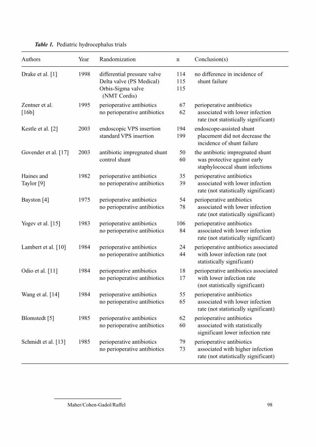
Maher/Cohen-Gadol/Raffel 98
Table 1. Pediatric hydrocephalus trials
Authors Year Randomization n Conclusion(s)
Drake et al. [1] 1998 differential pressure valve 114 no difference in incidence of
Delta valve (PS Medical) 115 shunt failure
Orbis-Sigma valve 115
(NMT Cordis)
Zentner et al. 1995 perioperative antibiotics 67 perioperative antibiotics
[16b] no perioperative antibiotics 62 associated with lower infection
rate (not statistically significant)
Kestle et al. [2] 2003 endoscopic VPS insertion 194 endoscope-assisted shunt
standard VPS insertion 199 placement did not decrease the
incidence of shunt failure
Govender et al. [17] 2003 antibiotic impregnated shunt 50 the antibiotic impregnated shunt
control shunt 60 was protective against early
staphylococcal shunt infections
Haines and 1982 perioperative antibiotics 35 perioperative antibiotics
Taylor [9] no perioperative antibiotics 39 associated with lower infection
rate (not statistically significant)
Bayston [4] 1975 perioperative antibiotics 54 perioperative antibiotics
no perioperative antibiotics 78 associated with lower infection
rate (not statistically significant)
Yogev et al. [15] 1983 perioperative antibiotics 106 perioperative antibiotics
no perioperative antibiotics 84 associated with lower infection
rate (not statistically significant)
Lambert et al. [10] 1984 perioperative antibiotics 24 perioperative antibiotics associated
no perioperative antibiotics 44 with lower infection rate (not
statistically significant)
Odio et al. [11] 1984 perioperative antibiotics 18 perioperative antibiotics associated
no perioperative antibiotics 17 with lower infection rate
(not statistically significant)
Wang et al. [14] 1984 perioperative antibiotics 55 perioperative antibiotics
no perioperative antibiotics 65 associated with lower infection
rate (not statistically significant)
Blomstedt [5] 1985 perioperative antibiotics 62 perioperative antibiotics
no perioperative antibiotics 60 associated with statistically
significant lower infection rate
Schmidt et al. [13] 1985 perioperative antibiotics 79 perioperative antibiotics
no perioperative antibiotics 73 associated with higher infection
rate (not statistically significant)

Pediatric Neurosurgery 99
Many studies have evaluated the utility of perioperative antimicrobial prophy-
laxis for shunt placement. In a review of the literature, Langley et al. [3] identified
12 published trials, including an aggregate of 1,359 patients, with random alloca-
tion to antibiotic prophylaxis versus no antibiotic prophylaxis for shunt surgery
[4–16]. Most of these trials enrolled children only or all age groups. Langley et al.
found that, when examined individually, only 1 of the 12 published trials found that
antibiotics reduced the risk of shunt infection to a statistically significant degree
and one trial [13] actually reported a higher infection rate in the antibiotic-treated
group. A meta-analysis of these 12 trials, however, found that antibiotic use led to
a very significant decrease in the risk of infection. This demonstrates the value of
meta-analysis in the neurosurgical literature. We may suppose that most of the
studies failed to show ‘significance’ when evaluated individually because of insuf-
ficient power arising from small sample sizes, a problem that is endemic in neuro-
surgical trials.
The use of antibiotic-impregnated shunts in an attempt to decrease the rate
of shunt infections is relatively new and has been the subject of only one controlled
trial to date [17]. This study suggests that the use of antibiotic-impregnated shunts
reduces the incidence of early staphylococcal shunt infections.
Much of the data on the management of external ventricular drains are
derived from trials that are mostly or entirely composed of adult patients [18,
19]. Consequently, the application of these data to the pediatric population is a
matter of debate.
Table 1. (continued)
Authors Year Randomization n Conclusion(s)
Djindjian et al. [7] 1986 perioperative antibiotics 30 perioperative antibiotics
no perioperative antibiotics 30 associated with lower infection
rate (not statistically significant)
Reider et al. [12] 1987 perioperative antibiotics 31 perioperative antibiotics
no perioperative antibiotics 32 associated with lower infection
rate (not statistically significant)
Blum et al. [6] 1989 perioperative antibiotics 50 perioperative antibiotics
no perioperative antibiotics 50 associated with lower infection
rate (not statistically significant)
Walters et al. [16] 1992 perioperative antibiotics 130 perioperative antibiotics
no perioperative antibiotics 113 associated with lower infection
rate (not statistically significant)
VPS � Ventriculoperitoneal shunt.
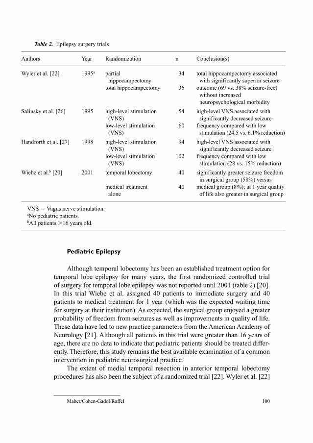
Maher/Cohen-Gadol/Raffel 100
Pediatric Epilepsy
Although temporal lobectomy has been an established treatment option for
temporal lobe epilepsy for many years, the first randomized controlled trial
of surgery for temporal lobe epilepsy was not reported until 2001 (table 2) [20].
In this trial Wiebe et al. assigned 40 patients to immediate surgery and 40
patients to medical treatment for 1 year (which was the expected waiting time
for surgery at their institution). As expected, the surgical group enjoyed a greater
probability of freedom from seizures as well as improvements in quality of life.
These data have led to new practice parameters from the American Academy of
Neurology [21]. Although all patients in this trial were greater than 16 years of
age, there are no data to indicate that pediatric patients should be treated differ-
ently. Therefore, this study remains the best available examination of a common
intervention in pediatric neurosurgical practice.
The extent of medial temporal resection in anterior temporal lobectomy
procedures has also been the subject of a randomized trial [22]. Wyler et al. [22]
Table 2. Epilepsy surgery trials
Authors Year Randomization n Conclusion(s)
Wyler et al. [22] 1995a partial 34 total hippocampectomy associated
hippocampectomy with significantly superior seizure
total hippocampectomy 36 outcome (69 vs. 38% seizure-free)
without increased
neuropsychological morbidity
Salinsky et al. [26] 1995 high-level stimulation 54 high-level VNS associated with
(VNS) significantly decreased seizure
low-level stimulation 60 frequency compared with low
(VNS) stimulation (24.5 vs. 6.1% reduction)
Handforth et al. [27] 1998 high-level stimulation 94 high-level VNS associated with
(VNS) significantly decreased seizure
low-level stimulation 102 frequency compared with low
(VNS) stimulation (28 vs. 15% reduction)
Wiebe et al.b [20] 2001 temporal lobectomy 40 significantly greater seizure freedom
in surgical group (58%) versus
medical treatment 40 medical group (8%); at 1 year quality
alone of life also greater in surgical group
VNS � Vagus nerve stimulation.aNo pediatric patients.bAll patients �16 years old.
Pediatric Neurosurgery 101
randomized 70 patients, all undergoing anterior temporal lobectomy for seizures,
according to the length of hippocampal removal. This group found that resec-
tion of hippocampus to the level of the tectal plate (‘total hippocampectomy’)
resulted in a better seizure outcome compared with removal of the hippocam-
pus to the level of the cerebral peduncle (‘partial hippocampectomy’). Although
no children were included in this trial, its conclusions are likely to extend to the
pediatric population.
The First International Vagus Nerve Stimulation Study Group demonstrated
the effectiveness of vagal nerve stimulation in the treatment of partial seizures
[23–26]. In this well-designed study, patients were randomized to receive either
high-level or low-level vagus nerve stimulation. In this way, these authors were
able to bypass the difficulties that are associated with randomizing for surgery
versus no surgery. In the second randomized-controlled trial of vagus nerve
stimulation for refractory epilepsy, Handforth et al. [27] also compared high-
level to low-level stimulation and found a benefit with the former. Despite the
frequent application of this treatment to children with refractory epilepsy, pedi-
atric patients comprised only a minority of patients enrolled in these trials.
Pediatric Brain Tumors
Most of the trials concern the utility of chemotherapy or radiation therapy
and do not randomize according to surgical procedure. Nevertheless, most
contemporary trials evaluating the effectiveness of adjunctive therapies do
make a surgical procedure (biopsy, partial resection, subtotal resection or gross
total resection) a mandatory condition of enrollment.
Several trials that were focused on randomizing patients according to adjunc-
tive postoperative treatments have enrolled patients regardless of the extent of
surgical resection. Although surgical treatment was not randomized or controlled
in these studies, they informed current care guidelines for the surgical treatment
of pediatric brain tumors. For example, in a randomized controlled trial examin-
ing postoperative chemotherapy without irradiation following surgical resection
of ependymoma, Grill et al. [28] found a significant difference in outcomes
between subtotal and gross totally resected ependymomas. Similarly, Wolff et al.
[29] have reported on a randomized controlled trial evaluating the role of specific
chemotherapy regimens in children with grade 3 or 4 gliomas. Although surgery
was not randomized, these authors found that the extent of surgical resection was
the most important prognostic factor.
Wisoff et al. [30, 31] performed a post hoc analysis of the Children’s
Cancer Group trial number CCG-945 which was conceived in order to compare
the efficacy of two different chemotherapy regimens. These authors found that,
Maher/Cohen-Gadol/Raffel 102
among patients enrolled in this trial, those undergoing more extensive resec-
tions exhibited longer survival times and progression-free survivals. Although
this study was based on a prospectively accrued patient population, it is subject
to a variety of biases due to the retrospective analysis [32]. For instance, the
determination of the extent of resection as well as the definition of groups for
comparison are both subject to bias. In addition, the extent of surgery is not ran-
domized in any of these studies, therefore even stringent statistical analysis may
not eliminate important confounding variables.
Spasticity
The two most utilized neurosurgical treatments for spasticity, baclofen
pumps and dorsal rhizotomy, have both been the subject of randomized trials.
Armstrong et al. [33] have reported on a series of patients with baclofen pumps
in which all study patients were selected as a result of a trial of intrathecal
baclofen injections compared with intrathecal saline injections. Although the
study patients were selected for pump implantation based on positive results of
this double-blind trial, the utility of baclofen pump placement was reported as a
case series without a control group. Other trials of baclofen pumps have pri-
marily studied adults [34, 35].
The utility of selective dorsal rhizotomy has been the subject of three ran-
domized controlled trials [36–38]. Although sample sizes were small, each of
these trials were well-organized and suggested a potential beneficial effect for
this operation. The report of McLaughlin et al. [36], the largest of the three
studies, suggested that the decrease in spasticity in the surgical arm of the trial
was not functionally important. The differing results may be a result of varia-
tions in physical therapy regimens between the studies. Further clarification
may be added by either a larger trial or meta-analysis [39].
Fetal Surgery
Since the publication of initial results from the International Fetal Surgery
Registry, a nonrandomized prospective clinical database of fetal surgery
procedures, in utero surgery for the correction of hydrocephalus and myelo-
meningocele has remained controversial [40]. Farmer et al. [41] have reported
on a case control study evaluating different methods of in utero myelomeningo-
cele repair, preferring ‘open’ techniques to fetoscopic methods. This study,
however, did not address the central question in fetal surgery: does in utero
repair have any additional benefit compared with postnatal repair? The first
Pediatric Neurosurgery 103
trial that was organized to address this question failed for methodological rea-
sons [42, 43]. Currently, patients are being accrued for the Management of
Myelomeningocele Study (MOMS). MOMS is a prospective, randomized trial
for in utero surgical repair of myelomeningocele [44]. Fetuses enrolled in this
ongoing trial are randomly assigned to either in utero repair of the myelo-
meningocele at between 19 and 25 weeks’ gestation or cesarean delivery fol-
lowed by standard surgical treatment [44]. The primary endpoints of this trial
are the need for a shunt procedure at 1 year and mortality. Results are expected
in approximately 2006 [44].
Pediatric Head Injuries
Pediatric head injury management has been the subject of two recent trials.
In a prospective randomized trial of 102 children following moderate to severe
head injury, Young et al. [45] showed that prophylactic treatment with phenytoin
did not reduce the rate of early (within 48 h) posttraumatic seizures. The patients
in this trial were not selected or stratified according to established risks for post-
traumatic seizures such as the presence of a large contusion, therefore the con-
clusions may not apply to patients that are thought to be at increased risk.
In an attempt to evaluate the role of decompressive craniectomy in children
with elevated intracranial pressure following head injury, Taylor et al. [46] ran-
domized children with elevated intracranial pressure following trauma to either
craniectomy or medical management alone. Although the craniectomy did
appear to be beneficial, the small number of patients (27) that were randomized
limits the information that may be derived from this trial.
Conclusions
Clearly, the vast majority of current pediatric neurosurgical practice is not
supported by evidence-based medicine standards. Rather, practice patterns have
been established that are based mostly on the uncontrolled reported experiences
with consecutive patient series. These reports are, for the most part, uncontrolled
and analyzed retrospectively.
This reliance on class III evidence within the field of pediatric neuro-
surgery does appear to be slowly shifting in favor of an increased willingness to
organize and participate in randomized clinical trials. In the area of pediatric
brain tumor treatments, our progress towards proper trial methodology has been
accelerated by the involvement of multicenter and multispecialty clinical con-
sortia such as and the Children’s Oncology Group [COG; a merger of the
Maher/Cohen-Gadol/Raffel 104
Pediatric Oncology Group (POG) and the Children’s Cancer Study Group]. The
evidence in favor of antibiotic prophylaxis during shunting procedures is simi-
larly robust. Although the publication of the shunt design trial results represents
an important step, many other important questions regarding the management
of hydrocephalus remain unanswered. The management of pediatric head injury
is an area that should be the subject of more clinical trials in the coming years.
There are several obstacles to the organization of more randomized con-
trolled trials in pediatric neurosurgery. For instance, even in the absence of class
I data, many pediatric neurosurgical interventions are firmly established as the
standard of care for their respective conditions. A trial comparing an accepted
surgical therapy with no therapy or another potentially harmful therapy is prac-
tically difficult and, in some cases, ethically prohibited. The anxiety that inves-
tigators and their institutions may feel about any such studies is invariably
heightened when children are the study subjects. Many of the best trials in pedi-
atric neurosurgery have been carried out to investigate new techniques, as in the
case of dorsal rhizotomy, or new equipment, as in vagal nerve stimulators or
baclofen pumps.
References
1 Drake JM, Kestle JR, Milner R, Cinalli G, Boop F, Piatt J Jr, Haines S, Schiff SJ, Cochrane DD,
Steinbok P, MacNeil N: Randomized trial of cerebrospinal fluid shunt valve design in pediatric
hydrocephalus. Neurosurgery 1998;43:294–303.
2 Kestle JR, Drake JM, Cochrane DD, Milner R, Walker ML, Abbott R, Boop FA: Lack of benefit of
endoscopic ventriculoperitoneal shunt insertion: a multicenter randomized trial. J Neurosurg
2003;98:284–290.
3 Langley JM, LeBlanc JC, Drake J, Milner R: Efficacy of antimicrobial prophylaxis in placement
of cerebrospinal fluid shunts: meta-analysis. Clin Infect Dis 1993;17:98–103.
4 Bayston R: Antibiotic prophylaxis in shunt surgery. Dev Med Child Neurol Suppl 1975;35:
99–103.
5 Blomstedt GC: Results of trimethoprim-sulfamethoxazole prophylaxis in ventriculostomy and
shunting procedures: a double-blind randomized trial. J Neurosurg 1985;62:694–697.
6 Blum J, Schwartz M, Voth D: Antibiotic single-dose prophylaxis of shunt infections. Neurosurg
Rev 1989;12:239–244.
7 Djindjian M, Fevrier MJ, Otterbein G, Soussy JC: Oxacillin prophylaxis in cerebrospinal fluid
shunt procedures: results of a randomized open study in 60 hydrocephalic patients. Surg Neurol
1986;25:178–180.
8 Goumnerova L, Hoffman H, Walters B, Humphreys R, Hendrick B: Perioperative antibiotic pro-
phylaxis in shunt surgery (abstract No. 10); in Scientific Program of the Annual Meeting of the
American Association of Neurological Surgeons. Washington, American Association of Neuro-
logical Surgeons, 1989.
9 Haines SJ, Taylor F: Prophylactic methicillin for shunt operations: effects on incidence of shunt
malfunction and infection. Childs Brain 1982;9:10–22.
10 Lambert M, MacKinnon AE, Vaishnav A: Comparison of two methods of prophylaxis against CSF
shunt infection. Z Kinderchir 1984;39(suppl 2):109–110.
11 Odio C, Mohs E, Sklar FH, Nelson JD, McCracken GH Jr: Adverse reactions to vancomycin used
as prophylaxis for CSF shunt procedures. Am J Dis Child 1984;138:17–19.
Pediatric Neurosurgery 105
12 Reider MJ, Frewen TC, Del Maestro RF, Coyle A, Lovell S: The effect of cephalothin prophylaxis
on postoperative ventriculoperitoneal shunt infections. Can Med Assoc J 1987;136:935–938.
13 Schmidt K, Gjerris F, Osgaard O, et al: Antibiotic prophylaxis in cerebrospinal fluid shunting:
a prospective randomized trial in 152 hydrocephalic patients. Neurosurgery 1985;17:1–5.
14 Wang EEL, Prober CG, Hendrick BE, Hoffman HJ, Humphreys RP: Prophylactic sulfamethoxa-
zole and trimethoprim in ventriculoperitoneal shunt surgery: a double-blind, randomized, placebo-
controlled trial. JAMA 1984;251:1174–1177.
15 Yogev R, Shinco F, McLone D: Prophylaxis for ventriculo-peritoneal shunt surgery with nafcillin
alone or in combination with rifampin (abstract No. 664); in Program and Abstracts of the 23rd
Interscience Conference on Antimicrobial Agents and Chemotherapy. Washington, American
Society for Microbiology, 1983.
16 Walters BC, Goumnerova L, Hoffman HJ, Hendrick EB, Humphreys RP, Levinton C: A random-
ized controlled trial of perioperative rifampin/trimethoprim in cerebrospinal fluid shunt surgery.
Childs Nerv Syst 1992;8:253–257.
16b Zentner J, Gilsbach J, Felder T: Antibiotic prophylaxis in cerebrospinal fluid shunting: a prospec-
tive randomized trial in 129 patients. Neurosurg Rev 1995;18:169–172.
17 Govender ST, Nathoo N, Van Dellen JR: Evaluation of an antibiotic-impregnated shunt system for
the treatment of hydrocephalus. J Neurosurg 2003;99:831–839.
18 Pfausler B, Spiss H, Beer R, Kampfl A, Engelhardt K, Schober M, Schumutzhard E: Treatment of
staphylococcal ventriculitis associated with external cerebrospinal fluid drains: a prospective
randomized trial of intravenous compared with intraventricular vancomycin therapy. J Neurosurg
2003;98:1040–1044.
19 Wong GKC, Poon WS, Wai S, Yu LM, Lyon D, Lam JMK: Failure of regular external ventricular
drain exchange to reduce cerebrospinal fluid infection: result of a randomized controlled trial.
J Neurol Neurosurg Psychiatry 2002;73:759–761.
20 Wiebe S, Blume WT, Girvin JP, Eliasziw M: A randomized, controlled trial of surgery for temporal-
lobe epilepsy. N Engl J Med 2001;345:311–318.
21 Engel J, Wiebe S, French J, Sperling M, Williamson P, Spencer D, Gumnit R, Zahn C, Westbrook E,
Enos B: Practice parameter: temporal lobe and localized neocortical resections for epilepsy.
Neurology 2003;60:538–547.
22 Wyler AR, Hermann BP, Somes G: Extent of medial temporal resection on outcome from anterior
temporal lobectomy: a randomized prospective study. Neurosurgery 1995;37:982–990.
23 Ben-Menachem E, Manon-Espaillat R, Ristanovic R, et al: Vagus nerve stimulation for treatment
of partial seizures. 1. A controlled study of effect on seizures. Epilepsia 1994;35:614–624.
24 Ramsay RE, Uthman BM, Augustinsson LE, Upton ARM, Naritoku D, Willis J, Treig T, Barolat G,
Wernicke JF: Vagus nerve stimulation for treatment of partial seizures. 2. Safety, side effects, and
tolerability. Epilepsia 1994;35:627–636.
25 Salinsky MC, Uthman BM, Ristanovic RK, Wernicke JF, Tarver WB; Vagus Nerve Stimulation
Study Group: Vagus nerve stimulation for the treatment of medically intractable seizures: results
of a 1-year open-extension trial. Arch Neurol 1996;53:1176–1180.
26 Salinsky M, Wernicke JF, Rutecki P, Allen J; The Vagus Nerve Stimulation Study Group: A ran-
domized controlled trial of chronic vagus nerve stimulation for treatment of medically intractable
seizures. Neurology 1995;45:224–230.
27 Handforth A, DeGiorgio CM, Schachter SC, et al: Vagus nerve stimulation therapy for partial-
onset seizures: a randomized active control trial. Neurology 1998;51:48–55.
28 Grill J, LeDeley M-C, Gambarelli D, Raquin M-A, Couanet D, Pierre-Kahn A, Habrand J-L, Doz F,
Frappaz D, Gentet J-C, Edan C, Chastagner P, Kalifa C: Postoperative chemotherapy without irra-
diation for ependymoma in children under 5 years of age: a multicenter trial of the French Society
of Pediatric Oncology. J Clin Oncol 2001;19:1288–1296.
29 Wolff JEA, Gnekow AK, Kortmann R-D, Pietsch T, Urban C, Graf N, Kuhl J: Preradiation
chemotherapy for pediatric patients with high-grade glioma. Cancer 2002;94:264–271.
30 Finlay JL, Bayett JM, Yates AJ, et al: Randomized phase III trial in childhood high-grade astrocy-
toma comparing vincristine, lomustine, and prednisone with eight-drugs-in-1-day regimen. J Clin
Oncol 1995;13:112–123.
Maher/Cohen-Gadol/Raffel 106
31 Wisoff JH, Boyett JM, Berger MS, et al: Current neurosurgical management and the impact of the
extent of resection in the treatment of malignant gliomas of childhood: a report of the Children’s
Cancer Group Trial No. CCG-945. J Neurosurg 1998;89:52–59.
32 Kreth FW, Muacevic A: Resection of malignant gliomas of childhood (letter). J Neurosurg
1999;90:1147.
33 Armstrong RW, Steinbok P, Cochrane DD, Kube SD, Fife SE, Farrell K: Intrathecally adminis-
tered baclofen for treatment of children with spasticity of cerebral origin. J Neurosurg 1997;87:
409–414.
34 Ordia JI, Fischer E, Adamski E: Chronic intrathecal delivery of baclofen by a programmable pump
for the treatment of severe spasticity. J Neurosurg 1996;85:452–457.
35 Middel B, Kuipers-Upmeijer H, Bouma J, Staal M, Oenema D, Postma T, Terpstra S, Stewart R:
Effect of intrathecal baclofen delivered by an implanted programmable pump on health related
quality of life in patients with severe spasticity. J Neurol Neurosurg Psychiatry 1997;63:204–209.
36 McLaughlin JF, Bjornson KF, Astley SJ, Graubert C, Hays RM, Roberts TS, Price R, Temkin N:
Selective dorsal rhizotomy: efficacy and safety in an investigator-masked randomized clinical
trial. Dev Med Child Neurol 1998;40:220–232.
37 Steinbock P, Reiner AM, Beauchamp R, Armstrong RW, Cochrane DD: A randomized clinical trial
to compare selective dorsal rhizotomy plus physiotherapy with physiotherapy alone in children
with spastic diplegic cerebral palsy. Dev Med Child Neurol 1997;39:178–184.
38 Wright FV, Sheil EMH, Drake JM, Wedge JH, Naumann S: Evaluation of selective dorsal rhizo-
tomy for the reduction of spasticity in cerebral palsy: a randomized controlled trial. Dev Med
Child Neurol 1998;40:239–247.
39 Rosenbaum PL: Selective dorsal rhizotomy studies (letter). Dev Med Child Neurol 1998;40:717.
40 Manning FA, Harrison MR, Rodeck C: Catheter shunts for fetal hydronephrosis and hydro-
cephalus. N Engl J Med 1986;315:336–340.
41 Farmer DL, von Koch CS, Peacock WJ, Danielpour M, Gupta N, Lee H, Harrison MR: In utero
repair of myelomeningocele. Arch Surg 2003;138:872–878.
42 Clewell WH, Johnson ML, Meier PR, Newkirk JB, Zide SL, Hendee RW, Bowes WA Jr, Hecht F,
O’Keefe D, Henry GP, Shikes RH: A surgical approach to the treatment of fetal hydrocephalus.
N Engl J Med 1982;306:1320–1325.
43 Michejda M, Queenan JT, McCullough D: Present status of intrauterine treatment of hydro-
cephalus and its future. Am J Obstet Gynecol 1986;155:873–882.
44 Sutton LN, Adzick NS, Johnson MP: Fetal surgery for myelomenigocele. Childs Nerv Syst
2003;19:587–591.
45 Young KD, Okada PJ, Sokolove PE, Palchak MJ, Panacek EA, Baren JM, Huff KR, McBride DQ,
Inkelis SH, Lewis RJ: A randomized, double-blinded, placebo-controlled trial of phenytoin for the
prevention of early posttraumatic seizures in children with moderate to severe blunt head injury.
Ann Emerg Med 2004;43:435–446.
46 Taylor A, Butt W, Rosenfeld J, Shann F, Ditchfield M, Lewis E, Klug G, Wallace D, Henning R,
Tibballs J: A randomized trial of very early decompressive craniectomy in children with traumatic
brain injury and sustained intracranial hypertension. Childs Nerv Syst 2001;17:154–162.
Corey Raffel, MD, PhD
Mayo Clinic, 200 First St., SW
Rochester, MN 55905 (USA)
Tel. �1 507 284 8167, E-Mail [email protected]
Pollock BE (ed): Guiding Neurosurgery by Evidence.
Prog Neurol Surg. Basel, Karger, 2006, vol 19, pp 107–122
Cerebrovascular-Endovascular
Kevin M. Cockrofta, Robert H. Rosenwasserb
aDepartment of Neurosurgery, Penn State Hershey Medical Center, Hershey, Pa.,bDepartment of Neurological Surgery, Thomas Jefferson University Hospital,
Philadelphia, Pa., USA
AbstractThis chapter will review the current status of scientific knowledge to support evidence-
based medicine guidelines for the endovascular treatment of cerebrovascular disease. Three
major areas of cerebrovascular disease will be examined, (1) occlusive cerebrovascular dis-
ease, (2) vascular malformations and (3) intracranial aneurysms. Levels of evidence vary in
each area and the reasons for this variation as well as the challenges that may limit further
investigations are discussed.
Copyright © 2006 S. Karger AG, Basel
As a relatively new area of subspecialization within neurosurgery, endovas-
cular neurosurgery represents an area ripe for scientific investigation. However,
while numerous questions within the field remain unanswered, several obstacles
exist that may prevent many investigations from ever reaching fruition. As with
‘open’ cerebrovascular surgery, endovascular neurosurgery involves three major
disease categories: (1) occlusive cerebrovascular disease, (2) vascular malforma-
tions and (3) intracranial aneurysms. Within each group there are varying degrees
of scientific data to support evidence-based medicine guidelines. This chapter
will consider the current state of evidence-based medicine in each of these three
areas and then examine some of the issues and limitations facing further scientific
study in these areas.
Occlusive Cerebrovascular Disease: Carotid Artery Stenosis
The mainstay of the open surgical treatment of occlusive cerebrovascu-
lar disease, the carotid endarterectomy (CEA), may arguably be the most
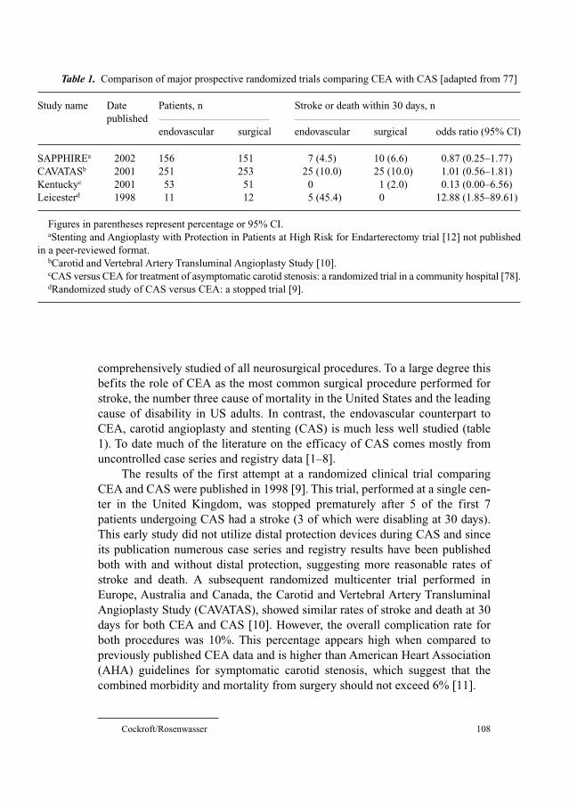
Cockroft/Rosenwasser 108
comprehensively studied of all neurosurgical procedures. To a large degree this
befits the role of CEA as the most common surgical procedure performed for
stroke, the number three cause of mortality in the United States and the leading
cause of disability in US adults. In contrast, the endovascular counterpart to
CEA, carotid angioplasty and stenting (CAS) is much less well studied (table
1). To date much of the literature on the efficacy of CAS comes mostly from
uncontrolled case series and registry data [1–8].
The results of the first attempt at a randomized clinical trial comparing
CEA and CAS were published in 1998 [9]. This trial, performed at a single cen-
ter in the United Kingdom, was stopped prematurely after 5 of the first 7
patients undergoing CAS had a stroke (3 of which were disabling at 30 days).
This early study did not utilize distal protection devices during CAS and since
its publication numerous case series and registry results have been published
both with and without distal protection, suggesting more reasonable rates of
stroke and death. A subsequent randomized multicenter trial performed in
Europe, Australia and Canada, the Carotid and Vertebral Artery Transluminal
Angioplasty Study (CAVATAS), showed similar rates of stroke and death at 30
days for both CEA and CAS [10]. However, the overall complication rate for
both procedures was 10%. This percentage appears high when compared to
previously published CEA data and is higher than American Heart Association
(AHA) guidelines for symptomatic carotid stenosis, which suggest that the
combined morbidity and mortality from surgery should not exceed 6% [11].
Table 1. Comparison of major prospective randomized trials comparing CEA with CAS [adapted from 77]
Study name Date Patients, n Stroke or death within 30 days, n
published
endovascular surgical endovascular surgical odds ratio (95% CI)
SAPPHIREa 2002 156 151 7 (4.5) 10 (6.6) 0.87 (0.25–1.77)
CAVATASb 2001 251 253 25 (10.0) 25 (10.0) 1.01 (0.56–1.81)
Kentuckyc 2001 53 51 0 1 (2.0) 0.13 (0.00–6.56)
Leicesterd 1998 11 12 5 (45.4) 0 12.88 (1.85–89.61)
Figures in parentheses represent percentage or 95% CI. aStenting and Angioplasty with Protection in Patients at High Risk for Endarterectomy trial [12] not published
in a peer-reviewed format.bCarotid and Vertebral Artery Transluminal Angioplasty Study [10].cCAS versus CEA for treatment of asymptomatic carotid stenosis: a randomized trial in a community hospital [78].dRandomized study of CAS versus CEA: a stopped trial [9].
Cerebrovascular-Endovascular 109
More recent work using distal protection during CAS has yielded more
favorable results. In the Carotid Revascularization using Endarterectomy or
Stenting Systems (CARESS) trial, a multicenter phase I trial comparing CAS
with distal protection and CEA, 397 patients were treated, 254 with CEA and
143 with CAS. Baseline characteristics between the two groups were similar
except that CAS patients were almost 3 times as likely to have undergone a pre-
vious CEA as compared to patients in the CEA group. A history of a previous
carotid stent was also more common in the CAS group. In the end there was no
significant difference in the 30-day rates of stroke and all-cause mortality
between the two groups, suggesting that CAS may be appropriate in patients
with a previous history of a carotid artery procedure.
Although as yet not published in a peer-reviewed format, the results of the
SAPPHIRE (Stenting and Angioplasty with Protection in Patients at High Risk
for Endarterectomy) trial, an industry-sponsored prospective randomized trial,
has lent support to the concept that CAS may be better than CEA for ‘high-risk’
patients [12]. In this trial’s cohort of mostly asymptomatic patients, the 30-day
risk of stroke, myocardial infarction (MI) and death was 3.8, 2.6 and 0.6%,
respectively, in the CAS group and 5.3, 7.3 and 2.0% in the CEA group. When
assessed separately, none of the differences in these individual results reached
statistical significance. However, when combined into a single endpoint, analy-
sis yielded a significantly reduced adverse event risk for CAS as compared to
CEA. Obviously the major factor in this difference is the rate of MI, which
turns out to be largely made up of subendocardial (non-Q wave) infarctions.
Whether such ‘chemical MIs’ are in fact clinically significant or simply markers
of high cardiac risk status remains controversial. Questions have also been
raised as to the appropriateness of the high-risk criteria used. One Mayo Clinic
retrospective review of 323 CEA patients, selected based on the SAPPHIRE
high-risk criteria, reported overall rates of stroke, MI and death as 1.65, 0.83
and 1.65%, respectively, rates which were not much different to SAPPHIRE’s
CAS group [13]. In addition, it has been argued that the use of regional anes-
thesia reduces the risk of cardiopulmonary morbidity and mortality after CEA
in ‘high-risk’ patients [14–16], thus bringing the overall risk of CEA in high-
risk patients in line with that of CAS in the SAPPHIRE trial.
While controversy still exists regarding the indications for CAS, techno-
logical advances and early trial results are leading to an increased acceptance of
distal protection devices. During the early stages of the Endarterectomy versus
Angioplasty in Patients with Symptomatic Severe Carotid Stenosis (EVA-3S)
trial, an analysis of the first 80 patients randomized to CAS prompted the trial’s
data safety monitoring committee to recommend stopping the performance of
unprotected CAS after an almost 4-fold increase in the 30-day stroke rate was
found in the unprotected CAS group as compared to the protected CAS patients
Cockroft/Rosenwasser 110
[17]. All current CAS trials now include the use of a distal protection device,
either as an option or a requirement.
At this point there is no class I evidence to suggest that CAS is a better alter-
native to CEA in either asymptomatic patients or symptomatic patients that are eli-
gible for CEA. The preponderance of evidence would seem to suggest that CAS
may be a lower risk than CEA in patients with medical contraindications to surgi-
cal endarterectomy or with local surgical risk factors such as postradiation-induced
stenosis, history of prior CEA and/or a high bifurcation. From a scientific stand-
point, the inability to consistently demonstrate a significant difference (or even
equivalence) between the two methods of treatment, the heterogeneity of previous
studies and the evolution of angioplasty/stent technology certainly support further
prospective randomized trials. However, market forces and public perceptions are
rapidly shaping clinical practice to the point that these ongoing studies may be in
jeopardy of failing to meet recruitment goals. As such the strongest indication for
CAS at the present time, may as part of a randomized clinical trial such as CREST
in the US, CAVATAS II in the UK, EVA-3S in France or SPACE (Stent Protected
Angioplasty versus Carotid Endarterectomy) in Germany.
Occlusive Cerebrovascular Disease: Acute Stroke
Based largely on the 1995 publication of the National Institute of Neurological
Disorders and Stroke (NINDS) study of intravenous (IV) thrombolysis for acute
ischemic stroke and the subsequent Food and Drug Administration (FDA)
approval of recombinant tissue plasminogen activator (rt-PA) for this indica-
tion, AHA guidelines now suggest that patients with a defined stroke onset of
within 3 h should be considered for IV rt-PA [18]. Since the NINDS study
established a 3-hour time period for efficacy with IV rt-PA, various attempts
have been made to extend this therapeutic window by demonstrating efficacy
for IV thrombolysis up to 6 h after the onset of ischemia.
While data supporting the extension of the therapeutic window for IV
thrombolysis have been mixed, evidence mainly from the Prolyse in Acute
Cerebral Thromboembolism (PROACT) studies suggests that intra-arterial (IA)
thrombolysis may be beneficial up to 6 h after the onset of ischemia. The initial
PROACT study published in 1998 randomized 49 patients with early middle
cerebral artery (MCA) territory ischemia to either IA recombinant prouroki-
nase (r-proUK) or placebo [19]. The recanalization rate, the primary endpoint,
was significantly higher in the r-proUK group as compared to the placebo-treated
group. The r-proUK group also demonstrated a reduction in 90-day mortality
from 43 to 27%, although the small number of patients in the study prevented
Cerebrovascular-Endovascular 111
this difference from reaching statistical significance. Not unexpectedly, there
was a higher incidence of hemorrhagic transformation in the r-proUK group, 15
versus 7%. However, this difference was also not statistically significant.
PROACT II, involving 180 patients from 54 centers and published the follow-
ing year, did demonstrate a significant improvement in outcome at 90 days for
patients treated with r-proUK and heparin as compared to those treated with
heparin alone [20]. Recanalization rates remained significantly better for
r-proUK treated patients, although overall mortality was similar. Again intrac-
erebral hemorrhage was more common in the treatment group, but this differ-
ence was still not statistically significant. In the vertebrobasilar system, acute
thromboocclusive disease carries a particularly poor prognosis. Although not as
extensively studied, results from case series of IA thrombolysis in the posterior
circulation have been promising and appear to suggest an even larger therapeu-
tic window [21, 22].
Various means of mechanical thrombolysis have been tried in an effort to
promote improved clot disruption while minimizing the dose of thrombolytic
agent and thereby hopefully reducing the rate of secondary hemorrhage
[23–28]. The recently published results of the Mechanical Embolus Removal in
Cerebral Ischemia (MERCI) study [29] have also been encouraging in this
regard. The study was a phase I device trial for the Merci Retrieval System, a
corkscrew-like device designed for mechanical embolectomy in the cerebral
vasculature. Successful recanalization with the device alone was achieved in 12
of 28 patients (43%) and with the addition of IA rt-PA in 18 patients (64%).
There were 12 asymptomatic and no symptomatic intracranial hemorrhages.
Only one procedure-related technical complication occurred and this was of no
clinical consequence. The device is now FDA approved and a registry of post-
approval patients will be maintained.
In addition to IA thrombolysis in the acute phase of stroke, other endovas-
cular techniques, namely intracranial angioplasty and/or stenting, are becoming
increasingly popular as preventative treatments. Relatively high rates of techni-
cal success in patients with refractory symptomatic intracranial atherosclerotic
disease have been reported [30–35]. Improved clinical outcome, on the other
hand, has been less frequent and complications have been problematic. In the
vertebrobasilar circulation in particular, complication rates as high as 25–38%
have been published [22, 36–39]. Although promising, at this point more
research is still needed to clarify the efficacy of intracranial angioplasty/stent-
ing in stroke prevention for patients with persistent ischemic symptoms sec-
ondary to atherosclerotic disease in the cerebral circulation.
Current clinical research in acute stroke continues to focus on methods of
extending the therapeutic window for thrombolysis, either with IV or IA admin-
istration alone or in combination. The Emergency Management of Stroke
Cockroft/Rosenwasser 112
(EMS) Bridging Trial, a pilot, placebo-controlled multicenter study randomized
35 patients to IV and IA rt-PA, or placebo and IA rt-PA within 3 h [40]. The
study demonstrated improved recanalization in the IV/IA group as compared to
the placebo/IA group. The numbers of intracerebral hemorrhages were similar
in the two groups. As a phase I study the trial was underpowered to detect effi-
cacy and no statistically significant difference in clinical outcome was seen
between the two groups. Further studies are underway to better assess the safety
and efficacy of such ‘bridging’ regimens.
At present, the existing evidence suggests that IA thrombolysis (rt-PA or uroki-
nase) may be indicated in patients with signs and symptoms of cerebral ischemia
and large intracranial vessel occlusion within 6 h of symptom onset. Mechanical
clot disruption/retrieval may be a useful adjunct to chemical thrombolysis in select
patients and may be a reasonable alternative in other patients who are ineligible for
chemical thrombolysis or who fall just outside the 6-hour treatment window.
Vascular Malformations
For the most part endovascular embolization is employed as an adjunctive
therapy in the treatment of CNS vascular malformations. For the purposes of
this chapter we will examine the evidence for the endovascular embolization of
cerebral (pial) AVMs either for cure or as an adjunct to microsurgical resection
or stereotactic radiosurgery.
While microsurgical removal may provide an immediate cure for accessible
lesions, resection of malformations with a large nidus, deep feeding vessels,
and/or high-flow shunts may carry a relatively high risk of morbidity. In such
patients, it is theorized that the added benefits of endovascular treatment out-
weigh the small additional risks and compare favorably with the risks of surgery
alone. This conclusion stems largely from a variety of nonrandomized, mostly ret-
rospective case series, the results of which have shown improved outcomes, short-
ened operative time and reduced blood loss in patients with preoperative
embolization as compared to patients with microsurgical resection alone [41–49].
The largest prospective randomized controlled trial to assess endovascular
embolization of AVMs was actually an industry-sponsored equivalency trial
designed to support FDA approval for N-butyl cyanoacrylate (NBCA) in the
treatment of intracranial AVMs. Published in 2002, the study compared NBCA
with the then ‘standard of care’ polyvinyl alcohol (PVA) particles for the preop-
erative embolization of cerebral AVMs. The study demonstrated equivalence
for both agents, at least in terms of the percentage of nidus reduction and num-
ber of pedicles embolized [50]. Interestingly, many centers that commonly used
‘glue’ to embolize AVMs as an ‘off-label’ indication were so convinced of its
Cerebrovascular-Endovascular 113
superiority that they did not take part in the study because participating patients
would have to be randomized. This problem of investigator bias is common-
place in clinical trials and in some instances may result in insufficient scientific
evidence ever being generated to support a product or device use. In the case of
NBCA, however, equivalency was enough to allow for FDA approval and clini-
cal practice combined with market forces have led NBCA embolization to
almost completely supplant PVA embolization for cerebral AVMs.
The majority of studies would suggest that cerebral/pial AVMs are rarely
cured by endovascular embolization alone and there are no prospective random-
ized studies comparing cure and complication rates for surgical resection versus
endovascular embolization for AVMs amenable to either treatment. Making a
judgment based on ‘cure rates’ from published series is probably inappropriate.
Since embolization evolved primarily as a therapeutic adjunct, many published
series suffer from considerable referral bias, whereby only ‘large’ AVMs inca-
pable of being treated with radiosurgery or open microsurgery alone are referred
for embolization and smaller lesions with only one or two feeding pedicles are
treated without endovascular intervention. In addition, the lack of a widely
accepted endovascular grading scale makes comparison between various studies
problematic. In general, most studies of intracranial AVMs, not specifically
selected for endovascular treatment alone, report cure rates with embolization of
between 5 and 10% [44, 47, 51, 52]. Gobin et al. [53] found a cure rate of 11.2%
in a cohort of 125 patients scheduled for radiosurgery who had undergone
embolization initially as an ‘adjunctive’ therapy. In contrast, other authors have
reported much higher rates of cure with endovascular therapy when patients
were selected specifically for embolization as a primary modality. After select-
ing a subgroup of patients on the basis of angiographic features that they felt
were likely to promote endovascular obliteration, Valavanis and Yasargil [54]
noted a cure rate of 74% (or 35% of their overall series) with embolization alone.
Of course none of these studies included a contemporaneous control group,
much less prospective randomization.
Endovascular embolization is also commonly used as an adjunct to stereo-
tactic radiosurgery. In patients with AVMs located in eloquent cortex or deep
structures, stereotactic radiosurgery may be a preferable alternative to micro-
surgical resection where the risk of morbidity and mortality may be unaccept-
ably high. In such patients, endovascular embolization may be employed to
reduce the size of the AVM prior to radiosurgery or to eliminate certain angio-
graphic features, such as intranidal aneurysms, that may provide an elevated
risk while the patient is awaiting AVM obliteration after radiosurgery.
It is well known that the rate of AVM cure after stereotactic radiosurgery
decreases as the volume of the AVM being treated increases [55–62].
Case series have shown higher cure rates for patients undergoing radiosurgical
Cockroft/Rosenwasser 114
treatment with AVM volumes below 10 cm3 or average diameter less than 3 cm
[53, 60, 63]. Therefore, the role of endovascular embolization in this setting is
to reduce the nidus size, such that a cure after radiosurgery will be more likely
[56, 58, 64]. This view is, however, somewhat controversial and, although less
of a problem with NBCA, some centers have reported apparent recanalization
of previously embolized portions of AVM [60, 63, 65]. For those patients in
whom the lesion does not proceed to obliteration after stereotactic radiosurgery,
repeated embolization or surgical resection may still be employed, often with
greater success [66, 67].
Although only complete elimination of the AVM constitutes a true cure,
palliative treatment may be employed in selected cases. Specifically, patients who
are symptomatic with large and/or deep-seated AVMs that are unlikely to be
cured with any combination of modalities may benefit from subtotal endovascu-
lar embolization. In patients with repeated hemorrhages, embolization may be
used to eliminate angiographic risk factors for hemorrhage, such as intranidal
aneurysms. For those with intractable headaches or progressive neurological
deficits, the benefits of partial treatment are less certain. Nonetheless, emboliza-
tion to reduce the arteriovenous shunt, and thereby decrease the amount of ‘steal’
and/or venous hypertension associated with a lesion, has been reported to cause
clinical improvement [68, 69]. Overall, though, the concept that partial treatment
of an AVM is at all beneficial is still controversial, and a similar or worse natural
history in incompletely treated patients has certainly been reported [70, 71].
In summary, there is class I evidence to suggest that preoperative glue
embolization with NBCA is equivalent to PVA embolization for cerebral AVMs
and there is class II evidence supporting a role for glue embolization as an
adjunct to microsurgical and radiosurgical treatment of pial AVMs. Evidence
for endovascular embolization as a palliative treatment for pial AVMs is minimal
and inconsistent. Given the current widespread use of NBCA and in-grained
patterns of multimodality treatment, it is unlikely that another prospective ran-
domized trial, this time designed to show efficacy for embolization (adjunctive
or otherwise) over microsurgical or radiosurgical treatment alone, will ever be
performed.
Endovascular Aneurysm Coiling
Perhaps the most controversial recent development in cerebrovascular neu-
rosurgery has been the introduction and swift progression to widespread use of
the Guglielmi Detachable Coil (GDC) for the treatment of intracranial
aneurysms. Originally approved by the FDA in 1991 under an Investigational
Device Exemption (IDE), over 250,000 patients worldwide have now been
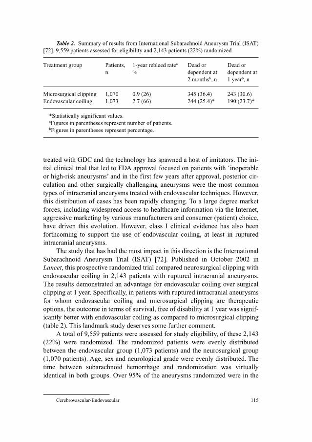
Cerebrovascular-Endovascular 115
treated with GDC and the technology has spawned a host of imitators. The ini-
tial clinical trial that led to FDA approval focused on patients with ‘inoperable
or high-risk aneurysms’ and in the first few years after approval, posterior cir-
culation and other surgically challenging aneurysms were the most common
types of intracranial aneurysms treated with endovascular techniques. However,
this distribution of cases has been rapidly changing. To a large degree market
forces, including widespread access to healthcare information via the Internet,
aggressive marketing by various manufacturers and consumer (patient) choice,
have driven this evolution. However, class I clinical evidence has also been
forthcoming to support the use of endovascular coiling, at least in ruptured
intracranial aneurysms.
The study that has had the most impact in this direction is the International
Subarachnoid Aneurysm Trial (ISAT) [72]. Published in October 2002 in
Lancet, this prospective randomized trial compared neurosurgical clipping with
endovascular coiling in 2,143 patients with ruptured intracranial aneurysms.
The results demonstrated an advantage for endovascular coiling over surgical
clipping at 1 year. Specifically, in patients with ruptured intracranial aneurysms
for whom endovascular coiling and microsurgical clipping are therapeutic
options, the outcome in terms of survival, free of disability at 1 year was signif-
icantly better with endovascular coiling as compared to microsurgical clipping
(table 2). This landmark study deserves some further comment.
A total of 9,559 patients were assessed for study eligibility, of these 2,143
(22%) were randomized. The randomized patients were evenly distributed
between the endovascular group (1,073 patients) and the neurosurgical group
(1,070 patients). Age, sex and neurological grade were evenly distributed. The
time between subarachnoid hemorrhage and randomization was virtually
identical in both groups. Over 95% of the aneurysms randomized were in the
Table 2. Summary of results from International Subarachnoid Aneurysm Trial (ISAT)
[72], 9,559 patients assessed for eligibility and 2,143 patients (22%) randomized
Treatment group Patients, 1-year rebleed ratea Dead or Dead or
n % dependent at dependent at
2 monthsb, n 1 yearb, n
Microsurgical clipping 1,070 0.9 (26) 345 (36.4) 243 (30.6)
Endovascular coiling 1,073 2.7 (66) 244 (25.4)* 190 (23.7)*
*Statistically significant values.aFigures in parentheses represent number of patients.bFigures in parentheses represent percentage.
Cockroft/Rosenwasser 116
anterior circulation, with the majority of these being in the anterior cerebral
artery distribution (50%). Only 14% were located in the MCA distribution, and
only 2.7% were in the posterior circulation. Nonprocedural bleeding from the
target aneurysm was higher in the neurosurgical group, where 23 patients bled
prior to a definitive surgical management as compared to 14 in the endovascu-
lar group. An assessment of clinical outcome in 1,906 patients at 2 months
revealed that 25.4% of patients were dead or dependent in the endovascular
group compared to 36.4% in the neurosurgical group (p � 0.0001). At 1 year,
data from 1,594 patients demonstrated a 23.7% rate of death and disability in
the endovascular group versus a rate of 30.6% in the neurosurgical group
(p � 0.0019). This difference represents an approximately 22% relative-risk
reduction or an absolute-risk reduction of 6.9% for endovascular treatment over
microsurgical clipping at 1 year.
This 22% reduction in the relative risk was widely reported in the popular
press and has contributed in no small way to the increased popularity of
endovascular techniques for the treatment of intracranial aneurysms. However,
from a scientific point of view there are several features of this trial, which
should preclude the generalization of its findings to the treatment of all
intracranial aneurysms, both ruptured and unruptured. Most importantly, the
patients enrolled in ISAT do not constitute a random sample of patients with
intracranial aneurysms. Rather these patients were selected from among
patients with a subarachnoid hemorrhage and an intracranial aneurysm that was
felt by the treating physicians to be equally amenable to either microsurgical
clipping or endovascular coiling. MCA aneurysms were underrepresented as
the consensus among most practitioners continues to be that these are difficult
to successfully treat by endovascular means. Similarly, posterior circulation
aneurysms, heavily represented in the initial trial for the FDA approval of GDC,
are now almost exclusively treated by endovascular means and as such even
fewer posterior circulation aneurysms were randomized.
Interestingly, at 1 year, some secondary endpoints actually favored surgery,
posttreatment rebleeding was 2.9 times more likely in the endovascular group,
death was 2.8 times more likely in the endovascular group and endovascular
patients were 4 times more likely to require additional treatment. In addition,
1-year case-fatality rates were not significantly different. However, the primary
endpoint of death and disability included all these parameters and showed an
overall benefit to endovascular therapy.
ISAT has also been criticized on a variety of other levels. The fact that
only 2,143 (22%) of a total of 9,559 screened patients were randomized and
the finding that the majority of those not randomized underwent surgical treat-
ment for their aneurysm have led some to speculate on a systemic selection
bias. The study participants are primarily European, with a handful of Canadian
Cerebrovascular-Endovascular 117
groups and only one US center. This distribution of study centers, combined
with memories of the International Cooperative Study [73] where early
surgery after aneurysmal subarachnoid hemorrhage improved outcome in
North America but not in Europe, has prompted concerns over the generaliz-
ability of the study’s results to North American patients. The relatively high 1-
year rebleed rate in the surgical group (0.9% as compared to 2.7% in the GDC
group) has also raised concerns, as this surgical rebleed rate does not appear to
reflect the North American experience. In addition, ISAT has been criticized
for the lack of adjudication of the participating neurosurgeons’ operative expe-
rience, including perioperative morbidity and mortality. Some of the surgeons
who participated in the trial had clipped fewer than 10 aneurysms per year.
Also of concern, the major finding of ISAT is only statistically significant
when the primary outcome of death and disability is defined as a modified
Rankin Score (mRS) 3–6. If mRS 2 (‘some restriction in lifestyle’) is included
in the disabled group or if mRS 3 (‘significant lifestyle restriction’) is removed
from the disability group, then the difference in the primary outcome between
the endovascular and surgical groups is no longer statistically significant.
Finally, since previous case series have shown relatively high rates of aneurysm
recurrence after coiling, the use of a 1-year outcome has been criticized as
inappropriately short.
Criticism aside, to date ISAT provides the best class I evidence comparing
endovascular coiling with microsurgical clipping for ruptured intracranial
aneurysms. The trial outcome supports the conclusion that endovascular treat-
ment for selected patients with ruptured aneurysms may yield a better outcome
than surgical treatment, at least at 1 year. Although not specifically looked at in
the first ISAT report, various other nonrandomized cohort studies have reported
a shorter length of stay and lower hospital charges for patients with aneurysms
treated with endovascular coiling as opposed to surgical clipping [74–76].
These same studies have also shown comparatively better outcomes in patients
with unruptured aneurysms treated by endovascular methods.
Efforts to resolve some of the controversial issues raised by ISAT and other
retrospective studies have led to the consideration of a North American trial. The
North American Trial for Unruptured and Ruptured Aneurysms (NATURE),
supported by both the American Association of Neurological Surgeons and the
Congress of Neurological Surgeons is being considered for funding by the
National Institutes of Health. However, the final status of such a trial is still
uncertain. As with NBCA glue embolization, the widespread use of endovascu-
lar coiling, combined with the already well-established treatment patterns for
certain aneurysms as well as the strongly held beliefs of many practitioners and
patients, will likely preclude randomization of all types of intracranial
aneurysms and may hamper recruitment even in a trial of more limited scope.
Cockroft/Rosenwasser 118
Conclusions
Endovascular neurosurgery continues to be a field in the midst of rapid
growth and development. As the indications for endovascular treatment expand,
new devices become available and existing therapies improve; the need for
good clinical evidence to support treatment decisions will be as important as
ever. However, as we have seen the reasons why medical evidence exists or does
not exist for certain procedures are extensive and complex. As medicine
becomes increasingly driven by technology and that technology is marketed
directly to patients and supplied by industry rather than by academic or govern-
mental developments, it may become increasingly difficult to obtain class I evi-
dence for a given procedure or treatment. In many instances, clinicians and
consumers alike will be left to base their management decisions on the careful
evaluation of uncontrolled case series, retrospective cohort studies or industry-
sponsored equivalency trials.
References
1 Yadav JS, Roubin GS, Iyer SJ, Vitek J, King P, Jordan WD, Fisher WS: Elective stenting of the
extracranial carotid arteries. Circulation 1997;95:376–381.
2 Roubin GS, Yadav JS, Iyer SS, Vitek JJ: Carotid stent-supported angioplasty: neurovascular inter-
vention to prevent stroke. Am J Cardiol 1996;78:8–12.
3 Iyer SS, Roubin GS, Yadav JS, Diethrich EB, Whorley MH, Shawl FA: Angioplasty and stenting
for extracranial carotid stenosis: multicenter experience. Circulation 1996;94:1–58.
4 Theron JG, Payelle GG, Coskun O, Huet JF, Guimaraens L: Carotid artery stenosis treatment with
protected balloon angioplasty and stent placement. Radiology 1996;201:627–636.
5 Vozzi CR, Rodriguez AO, Paolantonio D, Smith JA, Wholey MH: Extracranial carotid angioplasty
and stenting: initial results and short-term follow-up. Tex Heart Inst J 1997;24:167–172.
6 Iyer SS, Roubin GS, Yadav JS, Vitek J, Parks JA, Wadlington V: Elective carotid stenting.
J Endovasc Surg 1996;3:105–106.
7 Kachel R: Results of balloon angioplasty in carotid arteries. J Endovasc Surg 1996;3:22–30.
8 Qureshi AI, Fareed M, Suri K, Wadsworth DC, Dulin J, Hopkins LN: Multicenter study of the fea-
sibility and safety of using the Memotherm carotid arterial stent for extracranial carotid artery
stenosis. J Neurosurg 2002;96:830–836.
9 Naylor AR, Bolia A, Abbot R, Pye IF, Smith J, Lennard N, Lloyd AJ, London NJM, Bell PRF:
Randomized study of carotid angioplasty and stenting versus carotid endarterectomy: a stopped
trial. J Vasc Surg 1998;28:326–334.
10 Endovascular versus surgical treatment in patients with carotid stenosis in the Carotid and
Vertebral Artery Transluminal Angioplasty Study (CAVATAS): a randomized trial. Lancet
2001;357:1729–1737.
11 Biller J, Feinberg WM, Castaldo JE, Whittemore AD, Harbaugh RE, Dempsey RJ, Caplan LR,
Kresowik TF, Matchar DB, Toole J, Easton JD, Adams HP, Brass LM, Hobson R, Brott T, Sternau L:
Guidelines for carotid endarterectomy: a statement for healthcare professionals from a
special writing group of the Stroke Council, American Heart Association. Stroke 1998;29:
554–562.
12 Yadav JS: Stenting and angioplasty with protection in patients at high risk for endarterectomy
(SAPPHIRE study). American Heart Association Scientific Sessions, Chicago, Nov 19, 2002.

Cerebrovascular-Endovascular 119
13 Mozes G, Sullivan TM, Torres-Russoto DR, Bower TC, Hoskin TL, Sampaio SM, Gloviczki P,
Panneton JM, Noel AA, Cherry KJ: Carotid endarterectomy in SAPPHIRE-eligible high-risk
patients: implications for selecting patients for carotid angioplasty and stenting. J Vasc Surg
2004;39:958–965.
14 Sbarigia E, Dario Vizza C, Antonini M, Speziale F, Maritti M, Fiorani B, Fedele F: Locoregional ver-
sus general anesthesia in carotid surgery: is there an impact on perioperative myocardial ischemia?
Results of a prospective monocentric randomized trial. J Vasc Surg 1999;30: 131–138.
15 Harbaugh RE, Magnadottir HB: Carotid endarterectomy in high risk patients. Neurol Res
2002;24:S66–70.
16 Magnadottir HB, Lightdale N, Harbaugh RE: Clinical outcome for patients at high risk who
underwent carotid endarterectomy with regional anesthesia. Neurosurgery 1999;45:786–791.
17 Mas JL, Chatellier G, Beyssen B: Carotid angioplasty and stenting with and without cerebral pro-
tection: clinical alert from the Endarterectomy versus Angioplasty in Patients with Symptomatic
Severe Carotid Stenosis (EVA-3S) trial. Stroke 2004;35:e18–20.
18 Tissue plasminogen activator for acute ischemic stroke. The National Institute of Neurological
Disorders and Stroke rt-PA Stroke Study Group. N Engl J Med 1995;333:1581–1587.
19 del Zoppo GJ, Higashida RT, Furlan AJ, Pessin MS, Rowley HA, Gent M: PROACT: a phase II
randomized trial of recombinant pro-urokinase by direct arterial delivery in acute middle cerebral
artery stroke. PROACT Investigators. Prolyse in Acute Cerebral Thromboembolism. Stroke
1998;29:1255–1256.
20 Furlan AJ, Higashida RT, Wechsler LR, Gent M, Rowley HA, Kase C, Pessin MS, Ahuja A,
Callahan F, Clark WM, Silver F, Rivera F: Intra-arterial prourokinase for acute ischemic stroke. The
PROACT II study: a randomized controlled trial. Prolyse in Acute Cerebral Thromboembolism.
JAMA 1999;282:2003–2011.
21 Schellinger PD, Fiebach JB, Mohr A, Ringleb PA, Jansen O, Hacke W: Thrombolytic therapy for
ischemic stroke – a review. II. Intra-arterial thrombolysis, vertebrobasilar stroke, phase IV trials,
and stroke imaging. Crit Care Med 2001;29:1819–1825.
22 Wijdicks EF, Nichols DA, Thielen KR, Fulgham JR, Brown RD, Meissner I, Meyer FB, Piepgras
DG: Intra-arterial thrombolysis in acute basilar artery thromboembolism: the initial Mayo Clinic
experience. Mayo Clin Proc 1997;72:1005–1013.
23 Berlis A, Lutsep H, Barnwell S, Norbash A, Wechsler L, Jungreis CA, Woolfenden A, Redekop G,
Hartmann M, Schumacher M: Mechanical thrombolysis in acute ischemic stroke with endovascu-
lar photoacoustic recanalization. Stroke 2004;35:1112–1126.
24 Sorimachi T, Fujii Y, Tsuchiya N, Nashimoto T, Harada A, Ito Y, Tanaka R: Recanalization by
mechanical embolus disruption during intra-arterial thrombolysis in the carotid territory. AJNR
Am J Neuroradiol 2004;25:1391–1402.
25 Daffertshofer M, Hennerici M: Ultrasound in the treatment of ischaemic stroke. Lancet Neurol
2003;2:283–290.
26 Kerber CW, Barr JD, Berger RM, Chopko BW: Snare retrieval of intracranial thrombus in patients
with acute stroke. J Vasc Interv Radiol 2002;13:1269–1274.
27 Qureshi AI, Siddiqui AM, Suri MF, Kim SH, Ali Z, Yahia AM, Lopes DK, Boulos AS, Ringer AJ,
Saad M, Guterman LR, Hopkins LN: Aggressive mechanical clot disruption and low-dose intra-
arterial third-generation thrombolytic agent for ischemic stroke: a prospective study.
Neurosurgery 2002;51:1319–1329.
28 Ringer AJ, Qureshi AI, Fessler RD, Guterman LR, Hopkins LN: Angioplasty of intracranial occlu-
sion resistant to thrombolysis in acute ischemic stroke. Neurosurgery 2001;48:1282–1288.
29 Gobin YP, Starkman S, Duckwiler GR, Grobelny T, Kidwell CS, Jahan R, Pile-Spellman J, Segal A,
Vinuela F, Saver JL: MERCI 1: a phase 1 study of mechanical embolus removal in cerebral
ischemia. Stroke 2004;35:2848–2854.
30 Lee TH, Kim DH, Lee BH, Kim HJ, Choi CH, Park KP, Jung DS, Kim S, Moon TY: Preliminary
results of endovascular stent-assisted angioplasty for symptomatic middle cerebral artery stenosis.
AJNR Am J Neuroradiol 2005;26:166–174.
31 de Rochemont Rdu M, Turowski B, Buchkremer M, Sitzer M, Zanella FE, Berkefeld J: Recurrent
symptomatic high-grade intracranial stenoses: safety and efficacy of undersized stents – initial
experience. Radiology 2004;231:45–49.

Cockroft/Rosenwasser 120
32 Kim JK, Ahn JY, Lee BH, Chung YS, Chung SS, Kim OJ, Kim WC, Joo JY: Elective stenting for
symptomatic middle cerebral artery stenosis presenting as transient ischaemic deficits or stroke
attacks: short term arteriographical and clinical outcome. J Neurol Neurosurg Psychiatry 2004;75:
847–851.
33 Lee JH, Kwon SU, Lee JH, Suh DC, Kim JS: Percutaneous transluminal angioplasty for sympto-
matic middle cerebral artery stenosis: long-term follow-up. Cerebrovasc Dis 2003;15:90–97.
34 Alazzaz A, Thornton J, Aletich V, Debrun G, Ausman J, Charbel F: Intracranial percutaneous
transluminal angioplasty for arteriosclerotic stenosis. Arch Neurol 2000;57:1625–1630.
35 Marks MP, Marcellus M, Norbash AM, Steinberg GK, Tong D, Albers GW: Outcome of angio-
plasty for atherosclerotic intracranial stenosis. Stroke 1999;30:1065–1069.
36 Gress DR, Smith WS, Dowd CF, Van Halbach V, Finley RJ, Higashida RT: Angioplasty for
intracranial symptomatic vertebrobasilar ischemia. Neurosurgery 2002;51:23–27.
37 Levy EI, Horowitz MB, Koebbe CJ, Jungreis CC, Pride GL, Dutton K, Purdy PD: Transluminal
stent-assisted angioplasty of the intracranial vertebrobasilar system for medically refractory, pos-
terior circulation ischemia: early results. Neurosurgery 2001;48:1215–1221.
38 Levy EI, Hanel RA, Bendok BR, Boulos AS, Hartney ML, Guterman LR, Qureshi AI, Hopkins LN:
Staged stent-assisted angioplasty for symptomatic intracranial vertebrobasilar artery stenosis.
J Neurosurg 2002;97:1294–1301.
39 Rasmussen PA, Perl J, Barr JD, Markarian GZ, Katzan I, Sila C: Stent-assisted angioplasty of
intracranial vertebrobasilar atherosclerosis: an initial experience. J Neurosurg 2000;92:771–778.
40 Lewandowski CA, Frankel M, Tomsick TA, Broderick J, Frey J, Clark W, Starkman S, Grotta J,
Spilker J, Khoury J, Brott T: Combined intravenous and intra-arterial r-TPA versus intra-arterial
therapy of acute ischemic stroke: Emergency Management of Stroke (EMS) Bridging Trial. Stroke
1999;30:2598–2605.
41 Jafar JJ, David AJ, Berenstein A, Choi IS, Kupersmith MJ: The effect of embolization with
N-butyl-cyanoacrylate prior to surgical resection of cerebral arteriovenous malformations.
J Neurosurg 1993;78:60–69.
42 Spetzler RF, Martin NA, Carter LP, Flom RA, Raudzens PA, Wilkinson E: Surgical management
of large AVM’s by staged embolization and operative excision. J Neurosurg 1987;67:17–28.
43 Pasqualin A, Scienza R, Cioffi F, Barone G, Benati A, Beltramello A, Da Pian R: Treatment of
cerebral arteriovenous malformations with a combination of preoperative embolization and
surgery. Neurosurgery 1991;3:358–368.
44 Deruty R, Pelissou-Guyotat I, Mottolese C, Bascoulergue Y, Amat D: The combined management
of cerebral arteriovenous malformations. Experience with 100 cases and review of the literature.
Acta Neurochir (Wien) 1993;123:101–112.
45 Grzyska U, Westphal M, Zanella F: A joint protocol for the neurosurgical and neuroradiologic
treatment of cerebral arteriovenous malformations: indications, technique and results in 76 cases.
Surg Neurol 1993;40:476–484.
46 DeMerritt JS, Pile-Spellman J, Mast H: Outcome analysis of preoperative embolizations with N-butyl
cyanoacrylate in cerebral arteriovenous malformations. AJNR Am J Neuroradiol 1995;16:1801–1807.
47 Fournier D, TerBrugge KG, Willinsky R, Lasjaunias P, Montanera W: Endovascular treatment of
intracerebral arteriovenous malformations: experience in 49 cases. J Neurosurg 1995;75:228–233.
48 Debrun GM, Aletich V, Ausman JI, Charbel F, Dujovny M: Embolization of the nidus of brain arte-
riovenous malformations with n-butyl cyanoacrylate. Neurosurgery 1997;40:112–121.
49 Pelz DM, Fox AJ, Vinuela F, Drake CC, Ferguson GG: Preoperative embolization of brain AVMs
with isobutyl-2 cyanoacrylate. AJNR Am J Neuroradiol 1988;9:757–764.
50 NBCA Trial Investigators: N-Butyl cyanoacrylate embolization of cerebral arteriovenous malfor-
mations: results of a prospective, randomized, multi-center trial. AJNR Am J Neuroradiol
2002;23:748–755.
51 Hurst RW, Berenstein A, Kupersmith MJ, Madrid M, Flamm ES: Deep central arteriovenous mal-
formations of the brain: the role of endovascular treatment. J Neurosurg 1995;82:190–195.
52 Connors JJ, Wojak JC: Intracranial arteriovenous malformations: general considerations; in
Connors JJ, Wojak JC (eds): Interventional Neuroradiology: Strategies and Practical Techniques.
Philadelphia, Saunders, 1999, pp 227–240.
Cerebrovascular-Endovascular 121
53 Gobin YP, Laurent A, Merienne L, Schlienger M, Aymard A, Houdart E, Casasco A, Lefkopoulos
D, George B, Merland JJ: Treatment of brain arteriovenous malformations by embolization and
radiosurgery. J Neurosurg 1996;85:19–28.
54 Valavanis A, Yasargil M: The endovascular treatment of brain arteriovenous malformations. Adv
Tech Stand Neurosurg 1998;24:131–214.
55 Steinberg G, Fabrikant J, Marks M, Levy R, Frankel K, Phillips M, Shuer L, Silverberg G:
Stereotactic heavy-charged-particle Bragg-peak radiation for intracranial arteriovenous malfor-
mations. N Engl J Med 1990;323:96–101.
56 Lunsford LD, Kondziolka D, Flickinger JC, Bissonette DJ, Jungreis CA, Maitz AH, Horton JA,
Coffey RJ: Stereotactic radiosurgery for arteriovenous malformations of the brain. J Neurosurg
1991;75:512–524.
57 Colombo F, Pozza F, Chierego G, Casentini L, De Luca G, Francescon P: Linear accelerator radio-
surgery of cerebral arteriovenous malformations: an update. Neurosurgery 1994;34:14–21.
58 Friedman WA, Bova FJ, Mendenhall WM: Linear accelerator radiosurgery for arteriovenous mal-
formations: the relationship of size to outcome. J Neurosurg 1995;82:180–189.
59 Karlsson B, Lindquest C, Steiner L: Prediction of obliteration after gamma knife surgery for cere-
bral arteriovenous malformations. Neurosurgery 1997;40:425–431.
60 Pollock BE, Flickinger JC, Lunsford LD, Maitz AH, Kondziolka D: Factors associated with suc-
cessful arteriovenous malformation radiosurgery. Neurosurgery 1998;42:1239–1244.
61 Schlienger M, Atlan D, Lefkopoulos D, Merienne L, Touboul E, Missir O, Nataf F, Mammar H,
Platoni K, Grandjean P, Foulquier JN, Huart J, Oppenheim C, Meder JF, Houdart E, Merland JJ:
Linac radiosurgery for cerebral arteriovenous malformations: results in 169 patients. Int J Radiat
Oncol Biol Phys 2000;46:1135–1142.
62 Friedman WA, Bova FJ, Bollampally S, Bradshaw P: Analysis of factors predictive of success or
complications in arteriovenous malformation radiosurgery. Neurosurgery 2003;52:296–307.
63 Mathis JA, Barr JD, Horton JA, Jungreis CA, Lunsford LD, Kondziolka DS, Vincent D, Pentheny S:
The efficacy of particulate embolization combined with stereotactic radiosurgery for treatment of
large arteriovenous malformations of the brain. Am J Neuroradiol 1995;16:299–306.
64 Steiner L, Lindquist C, Adler JR, Torner JC, Alves W, Steiner M: Clinical outcome of radiosurgery
for cerebral arteriovenous malformations. J Neurosurg 1992;77:1–8.
65 Pollock BE, Kondziolka D, Lunsford LD, Bissonette DJ, Flickinger JC: Repeat stereotactic radio-
surgery of arteriovenous malformations: factors associated with incomplete obliteration.
Neurosurgery 1996;38:318–324.
66 Marks MP, Lane B, Steinberg GK, Fabrikant JI, Levy RP, Frankel KA, Phillips MH: Endovascular
treatment of cerebral arteriovenous malformations following radiosurgery. AJNR Am J
Neuroradiol 1993;14:197–303.
67 Steinberg GK, Chang SD, Levy RP, Marks MP, Frankel K, Marcellus M: Surgical resection of
large incompletely treated intracranial arteriovenous malformations following stereotactic radio-
surgery. J Neurosurg 1996;84:920–928.
68 Kusske JA, Kelly WA: Embolization and reduction of the ‘steal’ syndrome in cerebral arteriove-
nous malformations. J Neurosurg 1974;40:313–321.
69 Fox AJ, Girvin JP, Vinuela F, Drake CG: Rolandic arteriovenous malformations: improvement in
limb function by IBC embolization. AJNR Am J Neuroradiol 1985;6:575–582.
70 Pollock BE, Flickinger JC, Lunsford LD, Bissonette DJ, Kondziolka D: Hemorrhage after
stereotactic radiosurgery of cerebral arteriovenous malformations. Neurosurgery 1996;38:
659–661.
71 Han PP, Ponce FA, Spetzler RF: Intention-to-treat analysis of Spetzler-Martin grades IV and V
arteriovenous malformations: natural history and treatment paradigm. J Neurosurg 2003;98:3–7.
72 Molyneux A, Kerr R, Stratton I, Sandercock P, Clarke M, Shrimpton J, Holman R, International
Subarachnoid Aneurysm Trial (ISAT) Collaborative Group: International Subarachnoid Aneurysm
Trial (ISAT) of neurosurgical clipping versus endovascular coiling in 2143 patients with ruptured
intracranial aneurysms: a randomised trial. Lancet 2002;360:1267–1274.
73 Kassel NF, Torner JC, Jane JA, Haley ECJ, Adams HP: The international cooperative study on the
timing of aneurysm surgery. 2. Surgical results. J Neurosurg 1990;73:37–47.
Cockroft/Rosenwasser 122
74 Johnston SC, Dudley RA, Gress DR, Ono L: Surgical and endovascular treatment of unruptured
cerebral aneurysms at university hospitals. Neurology 1999;52:1799–1805.
75 Johnston SC, Wilson CV, Highashida RT, Dowd CF, McDermott MW, Applebury CB, Farley TL,
Gress DR: Endovascular and surgical treatment of unruptured cerebral aneurysms: comparison of
risks. Ann Neurol 2000;49:682–684.
76 Johnston SC, Zhao S, Dudley RA, Berman MF, Gress DR: Treatment of unruptured cerebral
aneurysms in California. Stroke 2001;32:597–605.
77 Coward LF, Featherstone RL, Brown MM: Percutaneous transluminal angioplasty and stenting for
carotid artery stenosis. Cochrane Database Syst Rev 2004;2:CD000515.
78 Brooks WH, McClure RR, Jones MR, Coleman TL, Breathitt L: Carotid angioplasty and stenting
versus carotid endarterectomy for treatment of asymptomatic carotid stenosis: a randomized trial
in a community hospital. Neurosurgery 2004;54:318–324.
Robert H. Rosenwasser, MD, FACS, Professor and Chairman
Department of Neurological Surgery
909 Walnut Street, Third Floor
Philadelphia, PA 19107 (USA)
Tel. �1 215 503 7004, Fax �1 215 503 7038, E-Mail [email protected]
Pollock BE (ed): Guiding Neurosurgery by Evidence.
Prog Neurol Surg. Basel, Karger, 2006, vol 19, pp 123–134
Evidence-Based Guidelines in Lumbar Spine Surgery
Daniel K. Resnicka, Michael C. Groff b
aDepartment of Neurological Surgery, University of Wisconsin, Madison, Wisc.,bDepartment of Neurological Surgery, University of Indiana, Indianapolis, Ind., USA
AbstractLumbar fusion is a commonly performed procedure for the treatment of painful insta-
bility of the spine, usually manifest as chronic low back pain. The safety, efficacy, and cost of
these procedures have been questioned in the professional and lay press. Recently, evidence
based medicine techniques have been used to investigate the role of lumbar fusion for the
treatment of a variety of spinal disorders. This chapter describes the general principles and
procedures used for the development of evidence based guidelines for the performance of
lumbar fusion.
Copyright © 2006 S. Karger AG, Basel
The number of lumbar fusion procedures performed in the United States
has increased substantially over the last several years and exhibited an upswing
in the late 1990s [1]. There are distinct regional differences in the rate of fusions
performed per 1,000 patients, a fact that has been interpreted in support of the
hypothesis that fusion is overused. Recent editorials in the popular press [2] and
general medical literature [1] have strongly condemned a perceived overutiliza-
tion of lumbar fusion and have suggested that the increase in the frequency of
fusion surgery noted over the last decade is a result of financial incentives to
surgeons and instrumentation companies [2]. This condemnation is largely
based on an apparent lack of evidence to support the role of fusion for the treat-
ment of low back pain. Indeed, Gibson et al. [3] , in the 1999 Cochrane review
stated: ‘There is no scientific evidence on the effectiveness of any form of sur-
gical decompression or fusion for degenerative lumbar spondylosis compared
Resnick/Groff 124
with natural history, placebo, or conservative management.’Third party payors,
plaintiffs attorneys, journalists, and politicians have responded to such state-
ments in a predictable fashion.
This chapter will focus on a review of the medical evidence available
concerning the role of lumbar fusion for the treatment of chronic low back
pain in patients without neurological deficit or significant spinal deformity.
As such, the chapter is limited in scope. However, it is hoped that through this
example the reader will be better able to understand the true strengths and
limitations of evidence-based literature reviews as they apply to surgery of
the lumbar spine.
Evidence-Based Medicine and the Low Back Pain Patient
The phrase ‘evidence-based medicine’ refers to the practice of medicine
based upon the best available information in the literature. Evidence-based med-
icine does not refer to the ‘ideal’ or ‘correct’ practice of medicine. Literature
cannot be interpreted in the absence of common sense and clinical experience.
A frequently cited example of the inappropriate application of evidence-based
medicine techniques is the assertion that there is no scientific evidence on the
effectiveness of parachute use for life preservation following falls from aircraft
[4]. Indeed, no randomized controlled trial or even a well-designed cohort
comparison has ever been performed to provide such evidence. A less fanciful
example of the limitations of literature-based guidelines concerns the evacua-
tion of symptomatic intracranial epidural hematomas. The surgical head injury
guidelines recommend removal of such hematomas at an option level [5] (sup-
ported only by low-quality or controversial evidence). This situation exists sim-
ply because no ethical surgeon would withhold available treatment from a
patient with a symptomatic epidural hematoma, so no control group exists for
comparison. Therefore, just because a treatment is not supported by high-quality
medical evidence does not mean that a treatment has no value.
Other potential difficulties faced while trying to derive meaningful conclu-
sions from the literature include the pace of technological development and the
limitations imposed by the application of standardized outcome measures.
Evolving technologies and techniques are, by definition, new and evolving.
Therefore, the use of such techniques is not generally supported by high-quality
medical evidence. When high-quality studies are available, they are almost
always funded by the manufacturer of the device in question and run by investi-
gators with a financial stake in the outcome of the study.
A number of commonly used, responsive, validated, and reliable outcome
measures are available for the assessment of outcomes following lumbar
Evidence-Based Guidelines in Lumbar Spine Surgery 125
fusion [6–19]. The use of such outcome measures allows for a comparison of
different treatment strategies. Theoretically, if one treatment works better than
another, then the outcomes achieved with the superior treatment should be
measurably different from those achieved with the inferior treatment.
Depending upon how the outcome measure is utilized, however, this may not
always be the case. The limitations of commonly employed outcome measures
may be illustrated by considering the advantages and disadvantages of mini-
mally incisional technologies for the performance of lumbar fusion. It is a
fairly standard practice for editorial boards to require a certain length of
patient follow-up in clinical series. For example, the journal Spine generally
requires a clinical manuscript to describe at least a 2-year follow-up following
fusion procedures in order to be considered for publication. If a functional out-
come measure is applied to patients who have undergone fusion procedures
two or more years following surgery, any potential short-term benefit related
to a minimally incisional approach would not be detected. Therefore, the
potential advantages of a minimally incisional approach (shorter hospital stay,
less acute pain) would not be reflected as an improvement in functional out-
come (see fig. 1).
Similarly, lumbar fusion surgery is not performed on patients with normal
lumbar spinal anatomy. In order for a fusion procedure to be contemplated,
some evidence of abnormality, usually a form of instability must be demon-
strated. Patients with instability of the spine are different from their peers in that
they have ‘bad backs’. Surgical treatment directed at a single level may well
provide temporary amelioration of symptoms, however over time the strength
of this beneficial effect may deteriorate due to the natural history of degenera-
tive spine disease. Therefore a beneficial effect noted 1, 2 or 5 years following
surgery may not be present 10 years following surgery. In this case, an outcome
study performed 10 years following fusion surgery would fail to demonstrate
that surgery had a beneficial effect (fig. 1). Does this truly mean that the
patients did not benefit from the procedure? Additionally, outcome measures
appropriate for one population undergoing lumbar fusion may not be appropri-
ate for other populations undergoing fusion. For example, return to work rate
may be a valid, reliable, and responsive outcome measure for patients undergo-
ing anterior lumbar interbody fusion. Is this outcome measure appropriate when
we are studying the results of fusion versus no fusion in patients undergoing
decompression for lumbar stenosis?
Another significant limitation encountered in the interpretation of the
medical literature relates to the definition of a clinically relevant outcome
measure. For example, in a study of the use of bone morphogenetic proteins
(BMP) as a substitute for autograft, the authors note a ‘significant’ decrease in
blood loss in the BMP group [20–22]. The magnitude of this decrease was
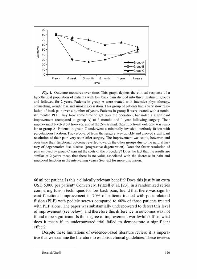
Resnick/Groff 126
66 ml per patient. Is this a clinically relevant benefit? Does this justify an extra
USD 5,000 per patient? Conversely, Fritzell et al. [23], in a randomized series
comparing fusion techniques for low back pain, found that there was signifi-
cant functional improvement in 70% of patients treated with posterolateral
fusion (PLF) with pedicle screws compared to 60% of those patients treated
with PLF alone. The paper was substantially underpowered to detect this level
of improvement (see below), and therefore this difference in outcomes was not
found to be significant. Is this degree of improvement worthwhile? If so, what
does it mean if an underpowered trial failed to demonstrate a significant
effect?
Despite these limitations of evidence-based literature review, it is impera-
tive that we examine the literature to establish clinical guidelines. These reviews
Fig. 1. Outcome measures over time. This graph depicts the clinical response of a
hypothetical population of patients with low back pain divided into three treatment groups
and followed for 2 years. Patients in group A were treated with intensive physiotherapy,
counseling, weight loss and smoking cessation. This group of patients had a very slow reso-
lution of back pain over a number of years. Patients in group B were treated with a nonin-
strumented PLF. They took some time to get over the operation, but noted a significant
improvement (compared to group A) at 6 months and 1 year following surgery. Their
improvement leveled out however, and at the 2-year mark their functional outcome was simi-
lar to group A. Patients in group C underwent a minimally invasive interbody fusion with
percutaneous fixation. They recovered from the surgery very quickly and enjoyed significant
resolution of their pain very soon after surgery. The improvement was static, however, and
over time their functional outcome reverted towards the other groups due to the natural his-
tory of degenerative disc disease (progressive degeneration). Does the faster resolution of
pain enjoyed by group C warrant the costs of the procedure? Does the fact that the results are
similar at 2 years mean that there is no value associated with the decrease in pain and
improved function in the intervening years? See text for more discussion.
0
10
20
30
40
50
60
70
80
90
Preop 6 week 3 month 6 month 1 year 2 years
Time
Out
com
e
Group AGroup BGroup C
Evidence-Based Guidelines in Lumbar Spine Surgery 127
provide snapshots of the state of the literature regarding a particular topic. The
guidelines produced are reflections of the peer-reviewed literature and provide
valuable guidance regarding what is truly known on a particular treatment or
diagnostic test. They serve to improve the literature itself through critique and
grading of individual papers and through the suggestion of future research direc-
tions designed to fill noticeable gaps in our collective knowledge base. These
techniques are also being used by agencies outside of medicine to determine
which procedures are paid for, which procedures are within the ‘standard of
care’, and which devices are approved for use. If we physicians are not inti-
mately familiar with the strengths, weaknesses, and conclusions reached in our
own literature, we will forfeit our ability to participate in the formulation of
health care policy.
Is Lumbar Fusion an Effective Treatment for Low Back Pain?
There are multiple indications for fusion, multiple techniques for the
achievement of fusion, a variety of diagnostic tests to determine eligibility for
fusion procedures, and numerous methods of assessing outcome. The literature
concerning lumbar fusion has generally been regarded as a morass of low-qual-
ity heterogeneous reports describing different procedures performed on differ-
ent patient populations. Some authors have concluded that there is, in fact, no
evidence to support the use of fusion as a treatment for painful degenerative dis-
ease of the spine [3]. In January 2003, the AANS/CNS Joint Section on
Disorders of the Spine and Peripheral Nerves was charged by the leadership of
the CNS to develop evidence-based guidelines for the performance of lumbar
fusion. The section responded by funding a guideline effort utilizing similar
methodology to the cervical spine injury guidelines published in March 2002
[24]. These guidelines were produced through a collaborative effort between the
spine section and the North American Spine Society and are scheduled for pub-
lication in the near future. These guidelines consist of 17 separate papers deal-
ing with specific aspects of lumbar fusion for degenerative disease. Although a
full discussion of the guidelines is beyond the scope of this chapter, the process
will be illustrated through a consideration of the surgical management of low
back pain.
The strongest medical evidence, here labeled as ‘class I medical evidence’,
in support for a given treatment is derived from well-designed and appropri-
ately powered randomized controlled clinical trials. If a randomized controlled
clinical trial is poorly designed or underpowered, the quality of the evidence
that it provides is downgraded to class II or class III medical evidence. When
trials of similar quality provide conflicting conclusions, the design of the study
Resnick/Groff 128
is examined closely in order to determine which study is better designed. The
COHORT group has published a set of criteria for the grading of clinical trials
[25] which allows for a rational application of this principle.
There have been two randomized controlled clinical trials published that
describe a comparison between the efficacy of surgery (fusion) and nonsurgical
management of chronic low back pain due to degenerative disease of the lum-
bar spine at L4–L5, L5–S1, or both levels. Fritzell et al. [26] published the
results of a multicenter randomized controlled trial from the Swedish Lumbar
Spine Study Group in 2001. These authors assumed that very few patients
would improve with conservative care and that a modest proportion of patients
treated surgically would improve. They performed a power analysis based upon
this premise in order to have an 80% power to detect a significant difference in
the effect of surgery versus the effect of nonsurgical treatment (in other words, they
determined how much of an improvement they thought would be clinically rel-
evant, and figured out how many patients they needed to include in order to be
able to detect that degree of improvement 80% of the time). In this study, 294
patients with disabling back pain who were felt to be surgical candidates were
randomized to conservative care (physical therapy supplemented with educa-
tion and other pain-relieving technologies at the discretion of the treating physi-
cian), or one of three surgical treatment arms. Patients were required to have
suffered from back pain for at least 2 years and to have radiographic and clini-
cal evidence of spondylosis at L4–5, L5–S1, or both levels. The groups were
comparable in all demographic variables measured with the exception of a higher
incidence of medical comorbidity in the surgical group. Patients were followed
for 2 years with intermediate evaluations at 6 months and at 1 year following
onset of treatment. Outcomes were assessed using multiple well-validated out-
come measures including pain visual analogue scales, the Oswestry Low Back
Pain Questionnaire, the Million Visual Analogue Scale, the General Function
Scale (GFS), Work Status, a patient satisfaction survey, and an independent
functional assessment by a second spinal surgeon [26].
Follow-up was achieved in 98% of patients. Appropriate statistical analysis
was performed based upon the type of data derived from the different outcome
measures. The surgical group did significantly better in terms of pain relief,
degree of disability as measured by the Oswestry, Million, and GFS, return to
work status, and degree of satisfaction reported by the patients and by the inde-
pendent observer. Statistical analysis was rigorous, employing ‘intention to treat’
as well as a ‘worst case’ scenarios. In short, all primary outcome measures eval-
uated in the study were significantly improved in the surgical group compared to
the nonsurgical group [26]. This study is therefore felt to provide class I evidence
demonstrating that lumbar fusion is associated with better outcomes than stan-
dard conservative care for appropriately selected patients.
Evidence-Based Guidelines in Lumbar Spine Surgery 129
The study of Fritzell et al. [26] was criticized by proponents of various
nonsurgical therapies. For example, Mooney [27] commented that the study
was unfairly biased against conservative care because the patients had already
failed a trial of the same type of therapy prior to entry in the study. This criti-
cism appears to be valid, given the a priori assumptions made by the Fritzell
group in their initial power analysis. This criticism does not, however, diminish
the finding that patients treated with lumbar fusion have superior clinical out-
comes compared to similar patients treated with usual medical care or those left
to suffer the natural history of disabling low back pain.
In 2003, Brox et al. [28] conducted a smaller (i.e. less powerful) random-
ized study evaluating the relative efficacy of instrumented PLF versus a spe-
cific protocol of cognitive intervention and physical therapy. The primary
outcome measure used was a modified Oswestry Disability Index (modified for
the Norwegian population) [29]. Secondary outcome measures included pain
visual analogue scales, daily use of medication, GFS, Waddel’s Fear Avoidance
Belief Questionnaire, and a patient satisfaction score. Outcomes were assessed
by physical therapists or rehabilitation physicians at 1 year following initiation
of treatment.
Patients enrolled in the surgical arm were treated with instrumented PLF.
The patients enrolled in the physiotherapy arm underwent a program specifically
designed for patients with low back pain that was felt to be more effective than
standard conservative care based on a pilot study performed by the authors [30].
This program included significant cognitive therapy designed to lower patient
fear as well as supervised physiotherapy averaging 25 h per week for 8 weeks.
Because of the intensity of the program, most patients stayed at the treatment
center in patient hotels. This intensive course was followed by a home program
based on the exercises prescribed in the supervised portion. In addition, patients
in the physiotherapy group were offered individual consultations, lessons, group
therapy sessions, and participation in peer-led discussion groups.
Sixty-four patients were randomized, 37 to surgery and 27 to physiotherapy.
There were more men randomized to the surgical group, otherwise the groups
were comparable. The 1-year follow-up rate was 97%. Both groups improved sig-
nificantly from baseline on all outcome measures. The improvement in the pri-
mary outcome measure, the modified ODI, in the surgical group was 15.6 and the
improvement in the physiotherapy group was 13.3. There were very large confi-
dence intervals noted in this as well as other outcome measures assessed. The dif-
ference in the degree of improvement between the surgical and physiotherapy
group was not found to be significant. The surgical group did do significantly bet-
ter in terms of relief of lower limb pain and tended to do better than the physio-
therapy group in terms of improvement in back pain, emotional distress, and
overall success ratings by both the patient and the independent observer. The
Resnick/Groff 130
physiotherapy group scored better fear avoidance activity and work as well as in
fingertip-floor distance. Nonsignificant trends were also seen in favor of the
physiotherapy group in terms of the GFS and life satisfaction score [28].
The authors interpret their findings as demonstrating an equivalence
between their program of physiotherapy and lumbar fusion. Given the small
size of the study groups and the very large confidence intervals reported in the
paper, the evidence provided by the paper is considered to provide class III evi-
dence concerning the relative efficacy of fusion versus intensive physiotherapy.
The paper does not address the utility of fusion as a means to alter the natural
history of low back pain and is significantly underpowered to detect any differ-
ences between any treatments that are even remotely similar (see below). The
relevance of the paper may be further questioned given the intensity of the
treatment used in the physiotherapy group. It is doubtful that such a program is
available to the vast majority of patients treated for low back pain.
Sample Size, Clinically Relevant Effect, and Pedicle Screws
The importance of sample size and the definition of ‘clinically relevant
effect’ cannot be overstated. A large randomized controlled clinical study may
demonstrate a ‘statistically significant effect’ of a treatment modality. If the
sample size is large enough, a small difference in outcomes may reach signifi-
cance (see fig. 2). Consider the NASCIS spinal cord injury studies [31–35]. In
these studies, large numbers of patients were enrolled and a beneficial effect of
methylprednisolone on clinical outcome measured with the ASIA scale was
identified (in a subgroup of patients). The magnitude of the improvement was
small, however, and the use of methylprednisolone was associated with an
increased risk of complications [31–35]. Is the small potential benefit of
methylprednisolone use worth the increased risk of complications? Not all clini-
cians believe so [36–38]. Here is where the clinician must make a judgement as
to the clinical importance of a 4-point improvement in the ASIA scale versus an
increased risk of sepsis. Conversely, a substantial beneficial effect may not be
recognized if sample sizes are too small (fig. 2).
Previously, we discussed the study by Fritzell et al. [26] which examined the
role of lumbar fusion. These authors performed a power analysis to determine how
many patients they would need to include in their study in order to have a reason-
able chance of detecting a significant effect. They assumed that the control patients
would do very poorly and that the treated patients would do moderately well.
They made several assumptions as to what degree of Oswestry or GFS improve-
ment would be considered relevant and were able to demonstrate a significant
effect between the surgical and nonsurgical arms [26]. These same authors then
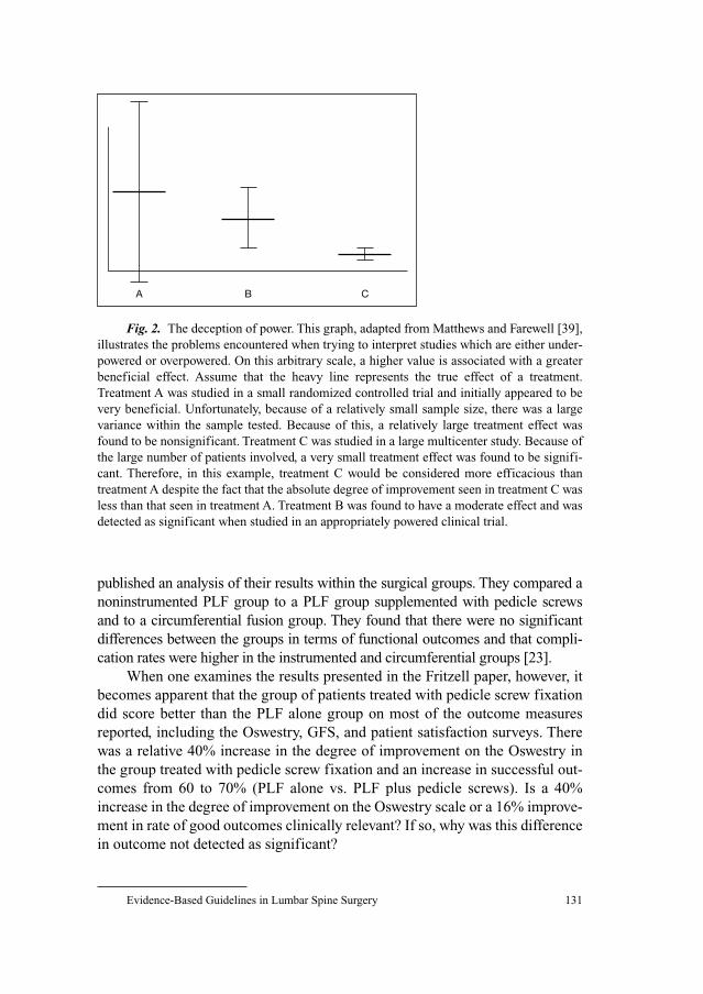
Evidence-Based Guidelines in Lumbar Spine Surgery 131
published an analysis of their results within the surgical groups. They compared a
noninstrumented PLF group to a PLF group supplemented with pedicle screws
and to a circumferential fusion group. They found that there were no significant
differences between the groups in terms of functional outcomes and that compli-
cation rates were higher in the instrumented and circumferential groups [23].
When one examines the results presented in the Fritzell paper, however, it
becomes apparent that the group of patients treated with pedicle screw fixation
did score better than the PLF alone group on most of the outcome measures
reported, including the Oswestry, GFS, and patient satisfaction surveys. There
was a relative 40% increase in the degree of improvement on the Oswestry in
the group treated with pedicle screw fixation and an increase in successful out-
comes from 60 to 70% (PLF alone vs. PLF plus pedicle screws). Is a 40%
increase in the degree of improvement on the Oswestry scale or a 16% improve-
ment in rate of good outcomes clinically relevant? If so, why was this difference
in outcome not detected as significant?
Fig. 2. The deception of power. This graph, adapted from Matthews and Farewell [39],
illustrates the problems encountered when trying to interpret studies which are either under-
powered or overpowered. On this arbitrary scale, a higher value is associated with a greater
beneficial effect. Assume that the heavy line represents the true effect of a treatment.
Treatment A was studied in a small randomized controlled trial and initially appeared to be
very beneficial. Unfortunately, because of a relatively small sample size, there was a large
variance within the sample tested. Because of this, a relatively large treatment effect was
found to be nonsignificant. Treatment C was studied in a large multicenter study. Because of
the large number of patients involved, a very small treatment effect was found to be signifi-
cant. Therefore, in this example, treatment C would be considered more efficacious than
treatment A despite the fact that the absolute degree of improvement seen in treatment C was
less than that seen in treatment A. Treatment B was found to have a moderate effect and was
detected as significant when studied in an appropriately powered clinical trial.
A B C
Resnick/Groff 132
The problem here is that the Fritzell study was designed to detect a differ-
ence between a group of patients who enjoyed a moderate improvement and a
group of patients who did not improve much at all. While the authors were able
to detect just such a difference between the surgical and nonsurgical arms, the
study was underpowered to detect differences between a group of patients who
enjoyed a moderate improvement and a group of patients who had a better
improvement. A power analysis reveals that in order to have an reasonable
chance (80%) of detecting a statistically significant difference between a group
of patients who achieve a good outcome 60% of the time and another group of
patients who achieve a good outcome 70% of the time, over 350 patients are
required in each group (http://calculators.stat.ucla.edu/powercalc). Playing with
the numbers, it is possible to calculate that the Fritzell study had only a 42%
chance of detecting an effect of this magnitude. Therefore, should we interpret
the negative results in the Fritzell study as definitive evidence that the addition of
pedicle screws does not improve outcome? The answer is no. The absence of a
positive effect in an underpowered study cannot be interpreted as anything
except circumstantial evidence (class III) regarding the lack of a treatment effect.
There are multiple examples of these types of design flaws in the literature
concerning lumbar fusion. Unfortunately for the spine surgeon and the patient with
low back pain, these design flaws create the impression that many of the proce-
dures we do are not effective. Third party payors, politicians, and our patients are
demanding justification for the potentially risky and certainly expensive proce-
dures that we are performing on otherwise healthy individuals. There are really no
ethical issues preventing the performance of appropriately designed randomized
controlled studies to examine the relative efficacy of various fusion procedures to
noninstrumented PLF in the many subpopulations of patients undergoing fusion
for low back pain. The challenge is to determine the right procedure for a given
patient population, define a clinically relevant difference in outcome using reliable
and valid outcome measures, design a study with adequate power, and perform the
study in an era of burdensome HIPAA regulations and public scrutiny.
Improving the Literature
In order to improve our literature, we must design studies that are geared
towards answering reasonable questions. We need to study techniques in spe-
cific patient populations and compare these techniques to ‘gold standard’ tech-
niques or to the natural history of the disease process. For example, the use of
interbody techniques as a treatment for low back pain is probably not best stud-
ied in the elderly patient with stenosis and degenerative spondylolisthesis.
Conversely, noninstrumented PLF has been found to be an effective treatment
Evidence-Based Guidelines in Lumbar Spine Surgery 133
for low back pain and may be an appropriate control group for studies looking
at interbody techniques in the younger low back pain population. Clinical
insight combined with expertise in clinical trial design will be required in order
to provide high-quality medical evidence to support the procedures we perform
to improve the quality of life for our patients.
References
1 Deyo RA, Nachemson A, Mirza SK: Spinal-fusion surgery – the case for restraint. N Engl J Med
2004;350:722–726.
2 Abelson R, Peterson M: An operation to ease back pain bolsters the bottom line, too. New York
Times, Dec 31, 2003.
3 Gibson JN, Grant IC, Waddell G: The Cochrane review of surgery for lumbar disc prolapse and
degenerative lumbar spondylosis. Spine 1999;24:1820–1832.
4 Smith GCS, Pell JP: Parachute use to prevent death and major trauma related to gravitational chal-
lenge: systematic review of randomised controlled trials. BMJ 2003;327:1459–1461.
5 Bullock MR, Chestnut R, Ghajar J, Gordon D, Hartl R, Newell DW, Servadei F, Walters BC, Wilberger JE:
Surgical Management of Acute Epidural Hematoma. Neurosurgery 2006;58(suppl 2):7–15.
6 Deyo RA: Comparative validity of the sickness impact profile and shorter scales for functional
assessment in low back pain. Spine 1986;11:951–954.
7 Deyo RA, Andersson G, Bombardier C, Cherkin DC, Keller RB, Lee CK, Liang MH, Lipscomb B,
Shekelle P, Spratt KF, et al: Outcome measures for studying patients with low back pain. Spine
1994;19(18 suppl):2032S–2036S.
8 Fairbank JC, Couper J, Davies JB, O’Brien JP: The Oswestry low back pain disability question-
naire. Physiotherapy 1980;66:271–273.
9 Harper AC, Harper DA, Lambert LJ, de Klerk NH, Andrews HB, Ross FM, Straker LJ, Lo SK:
Development and validation of the Curtin Back Screening Questionnaire (CBSQ): a discrimina-
tive disability measure. Pain 1995;60:73–81.
10 Lawlis GF, Cuencas R, Selby D, McCoy CE: The development of the Dallas Pain Questionnaire:
an assessment of the impact of spinal pain on behavior. Spine 1989;14:511–516.
11 Leclaire R, Blier F, Fortin L, Proulx R: A cross-sectional study comparing the Oswestry and
Roland-Morris Functional Disability scales in two populations of patients with low back pain of
different levels of severity. Spine 1997;22:68–71.
12 Manniche C, Asmussen K, Lauritsen B, Vinterberg H, Kreiner S, Jordan A: Low back pain rating
scale: validation of a tool for assessment of low back pain. Pain 1994;57:317–326.
13 Million R, Hall W, Nilsen KH, Baker RD, Jayson MI: Assessment of the progress of the back-pain
patient. Spine 1982;7:204–208.
14 Roland M, Morris R: A study of the natural history of back pain. I. Development of a reliable and
sensitive measure of disability in low-back pain. Spine 1983;8:141–144.
15 Ruta DA, Garratt A, Wardlaw D, Russell IT: Developing a valid and reliable measure of health
outcome for patients with low back pain. Spine 1994;19:1887–1896.
16 Salen BA, Spangfort E, Nygren AL, Nordemar R: The Disability Rating Index: an instrument for
the assessment of disability in clinical settings. J Clin Epidemiol 1994;47:1423–1435.
17 Stucki G, Daltroy L, Liang MH, Lipson SJ, Fossel AH, Katz JN: Measurement properties of a self-
administered outcome measure in lumbar spinal stenosis. Spine 1996;21:796–803.
18 Waddell G, Main CJ: Assessment of severity in low-back disorders. Spine 1984;9:204–208.
19 Ware J Jr, Kosinski M, Keller SD: A 12-item short-form health survey: construction of scales and
preliminary tests of reliability and validity. Med Care 1996;34:220–233.
20 Burkus JK, Heim SE, Gornet MF, Zdeblick TA: Is INFUSE bone graft superior to autograft bone?
An integrated analysis of clinical trials using the LT-CAGE lumbar tapered fusion device. J Spinal
Disord Tech 2003;16:113–122.

Resnick/Groff 134
21 Burkus JK, Transfeldt EE, Kitchel SH, Watkins RG, Balderston RA: Clinical and radiographic
outcomes of anterior lumbar interbody fusion using recombinant human bone morphogenetic
protein-2. Spine 2002;27:2396–2408.
22 Burkus JK, Dorchak JD, Sanders DL: Radiographic assessment of interbody fusion using recom-
binant human bone morphogenetic protein type 2. Spine 2003;28:372–377.
23 Fritzell P, Hagg O, Wessberg P, Nordwall A, Swedish Lumbar Spine Study Group: Chronic low
back pain and fusion: a comparison of three surgical techniques: a prospective multicenter ran-
domized study from the Swedish lumbar spine study group. Spine 2002;27:1131–1141.
24 Hadley MN, Walters BC, Grabb PA, Oyesiku NM, Przybylski GJ, Resnick DK, Ryken TC:
Guidelines for the management of acute cervical spine and spinal cord injuries. Neurosurgery
2002;50(suppl):S2–S6.
25 Moher D, Schulz KF, Altman DG: The CONSORT statement: revised recommendations for
improving the quality of reports of parallel-group randomised trials. Lancet 2001;357:1192–1194.
26 Fritzell P, Hagg O, Wessberg P, Nordwall A, Swedish Lumbar Spine Study Group: Lumbar fusion
versus nonsurgical treatment for chronic low back pain: a multicenter randomized controlled trial
from the Swedish Lumbar Spine Study Group. Spine 2001;26:2521.
27 Mooney V: Point of view. Spine 2001;26:2532–2533.
28 Brox IJ, Sorensen R, Friis A, Nygaard O, Indahl A, Keller A, Ingebrigtsen T, Eriksen HR, Holm I,
Koller AK, Riise R, Reikeras O: Randomized clinical trial of lumbar instrumented fusion and
cognitive intervention and exercises in patients with chronic low back pain and disc degeneration.
Spine 2003;28:1913–1921.
29 Holm I, Friis A, Storheim K: Measuring self reported functional status and pain in patients with
chronic low back pain by postal questionnaires: a reliability study. Spine 2003;28:828–833.
30 Brox IJ, Friis A, Holm I: Patients with chronic degenerative spinal disease: can conservative treat-
ment reduce the waiting list for surgery? Tidsskr Nor Laegeforen 1999;119:1784–1787.
31 Bracken MB, Shepard MJ, Collins WF, et al: A randomized, controlled trial of methylprednisolone
or naloxone in the treatment of acute spinal-cord injury. Results of the Second National Acute
Spinal Cord Injury Study. N Engl J Med 1990;322:1405–1411.
32 Bracken MB, Shepard MJ, Collins WF Jr, Holford TR, Baskin DS, Eisenberg HM, et al:
Methylprednisolone or naloxone treatment after acute spinal cord injury: 1-year follow-up data.
Results of the second National Acute Spinal Cord Injury Study. J Neurosurg 1992;76:23–31.
33 Bracken MB, Shepard MJ, Holford TR, et al: Administration of methylprednisolone for 24 or 48
hours or tirilazad mesylate for 48 hours in the treatment of acute spinal cord injury. Results of the
Third National Acute Spinal Cord Injury Randomized Controlled Trial. National Acute Spinal
Cord Injury Study. JAMA 1997;277:1597–1604.
34 Bracken MB, Shepard MJ, Holford TR, et al: Methylprednisolone or tirilazad mesylate adminis-
tration after acute spinal cord injury: 1-year follow-up. Results of the third National Acute Spinal
Cord Injury randomized controlled trial. J Neurosurg 1998;89:699–706.
35 Bracken MB, Holford TR: Neurological and functional status 1 year after acute spinal cord injury:
estimates of functional recovery in National Acute Spinal Cord Injury Study II from results mod-
eled in National Acute Spinal Cord Injury Study III. J Neurosurg 2002;96(3 suppl):259–266.
36 Hurlbert RJ: Methylprednisolone for acute spinal cord injury: an inappropriate standard of care.
J Neurosurg 2000;93(1 suppl):1–7.
37 Hurlbert RJ: The role of steroids in acute spinal cord injury: an evidence-based analysis. Spine
2001;26(24 suppl):S39–46.
38 Hurlbert RJ, Moulton R: Why do you prescribe methylprednisolone for acute spinal cord injury?
A Canadian perspective and a position statement. Can J Neurol Sci 2002;29:236–239.
39 Matthews DE, Farewell VT: Using and Understanding Medical Statistics, ed 3. Basel, Karger, 1996.
Daniel K. Resnick, MD
K4/834 Clinical Science Center
600 Highland Avenue
Madison, WI 53792 (USA)
Tel. �1 608 263 9651, Fax �1 608 263 1728, E-Mail [email protected]
Pollock BE (ed): Guiding Neurosurgery by Evidence.
Prog Neurol Surg. Basel, Karger, 2006, vol 19, pp 135–151
Spine: Minimally Invasive Techniques
Peter C. Gerszten, William C. Welch
Department of Neurological Surgery, University of Pittsburgh School of Medicine,
UPMC Health System, Pittsburgh, Pa., USA
AbstractMinimally invasive spine surgery decompression, arthrodesis, and instrumentation
techniques are now being applied in a wide variety of percutaneous, laparoscopic and mini-
mal access procedures. There is currently little longitudinal long-term data on these proce-
dures to document their efficacy, indications, limitations or complications as compared to
standard open techniques. Further complicating such direct comparisons is that widely used
spine outcomes instruments often do not capture the relative benefits of these new proce-
dures. It is only through randomized trials that the potential benefits of these procedures be
substantiated in order to justify the sometimes significant increased costs associated with
them.
Copyright © 2006 S. Karger AG, Basel
There has been a gradual development over time of minimally invasive
surgical techniques applied to the field of spine surgery [1]. Such techniques
follow a natural trend in surgery to minimize the injury to normal tissue while
obtaining the same or a better surgical outcome. The new wave in recent years
of minimally invasive spinal procedures is not a revolution, but rather an evolu-
tion of familiar, time-proven operative techniques [2]. Such an attitude towards
these new techniques helps to understand the relative paucity of rigorous evidence-
based outcomes assessments of these techniques prior to their widespread
adoption.
By marrying modern medical technology to traditional spinal approaches,
the classical goals of open spinal surgery can now be effectively and repro-
ducibly accomplished through much smaller corridors and with far less iatro-
genic damage to the vital dorsal musculoligamentous complex [2]. Innovation
in surgical treatment reflects either the application of existing knowledge, tech-
niques, or technology in new ways or the acquisition and application of new
Gerszten/Welch 136
knowledge to fundamentally redefine the management of a condition [3]. Both
types of innovations will be reviewed in this chapter. For example, the use of the
endoscope in spinal procedures is based upon its prior success in surgery else-
where in the body and is an example of the former type of innovation. On the
other hand, the techniques of vertebroplasty and kyphoplasty represent the lat-
ter form of innovation.
There is still some debate over the definition of ‘minimally invasive spine
surgery’. Indeed, Fessler [4] points out that many of these procedures being
developed do not rely on new and untested technologies to achieve their surgi-
cal results. For example, after the target has been reached, procedures such as
microendoscopic discectomy involve the use of standard surgical instruments
and techniques. In this respect, the crucial aspect of the surgical procedure is
not minimal at all; it is exactly the same as the equivalent open procedure.
However, ‘minimal access’ techniques have been used to access the surgical tar-
get. Therefore, ‘minimal access spine surgery’ is probably a more accurate term
to describe this family of procedures [4].
Not only is there some debate over the term ‘minimally invasive’, there is
also disagreement regarding the definition of what actually constitutes
‘surgery’. Many of these minimally invasive techniques, especially those that
are percutaneous such as vertebroplasty and intradiscal electrothermal therapy
(IDET), are more commonly performed by interventional radiologists and anes-
thesiologists than they are by spine orthopedic and neurosurgeons. The proce-
dures included in this chapter are those that are considered to be commonly
performed by surgeons who operate on the spine.
Outcome Evaluation of Minimally Invasive Techniques
Outcome Comparison
Well-established reliable and validated outcome instruments and tech-
niques already exist for the proper evidence-based evaluation of most surgical
spinal techniques [5, 6]. In contrast, most evaluations of minimally invasive
spinal surgery techniques have focused on variables such as decreases in oper-
ating times, blood loss, postoperative pain, medication use, length of hospital
stays, and costs (see table 1) [4]. The improved ‘quality’ of these surgical tech-
niques can be measured in many ways, including decreased risk related to the
procedure, more reliable and/or easier achievement of the surgical objective,
superior short-term and/or more durable long-term outcomes, less pain and
quicker recovery, and more efficient achievement of the surgical objective, with
respect to the resources consumed [3]. However, standard disease-specific
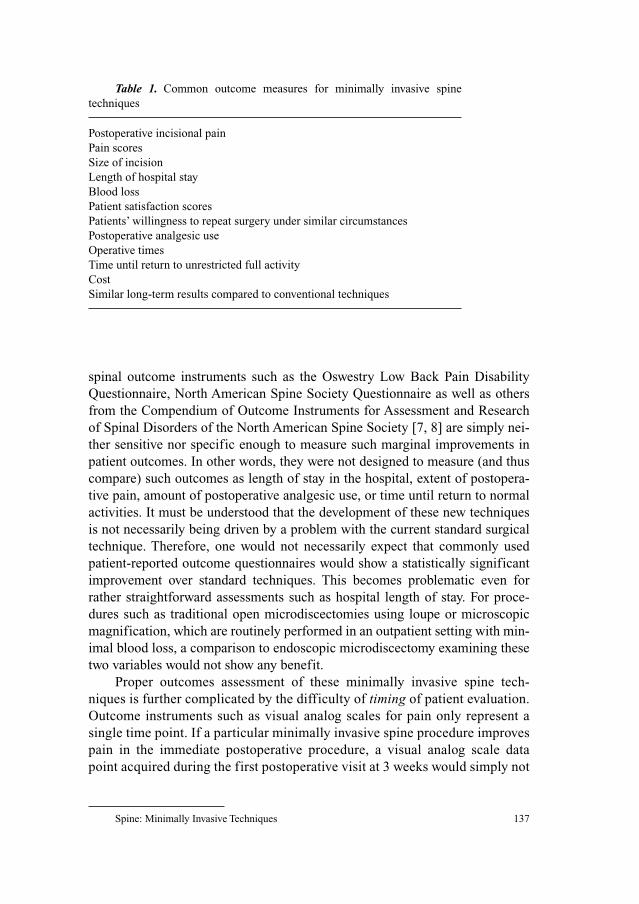
Spine: Minimally Invasive Techniques 137
spinal outcome instruments such as the Oswestry Low Back Pain Disability
Questionnaire, North American Spine Society Questionnaire as well as others
from the Compendium of Outcome Instruments for Assessment and Research
of Spinal Disorders of the North American Spine Society [7, 8] are simply nei-
ther sensitive nor specific enough to measure such marginal improvements in
patient outcomes. In other words, they were not designed to measure (and thus
compare) such outcomes as length of stay in the hospital, extent of postopera-
tive pain, amount of postoperative analgesic use, or time until return to normal
activities. It must be understood that the development of these new techniques
is not necessarily being driven by a problem with the current standard surgical
technique. Therefore, one would not necessarily expect that commonly used
patient-reported outcome questionnaires would show a statistically significant
improvement over standard techniques. This becomes problematic even for
rather straightforward assessments such as hospital length of stay. For proce-
dures such as traditional open microdiscectomies using loupe or microscopic
magnification, which are routinely performed in an outpatient setting with min-
imal blood loss, a comparison to endoscopic microdiscectomy examining these
two variables would not show any benefit.
Proper outcomes assessment of these minimally invasive spine tech-
niques is further complicated by the difficulty of timing of patient evaluation.
Outcome instruments such as visual analog scales for pain only represent a
single time point. If a particular minimally invasive spine procedure improves
pain in the immediate postoperative procedure, a visual analog scale data
point acquired during the first postoperative visit at 3 weeks would simply not
Table 1. Common outcome measures for minimally invasive spine
techniques
Postoperative incisional pain
Pain scores
Size of incision
Length of hospital stay
Blood loss
Patient satisfaction scores
Patients’ willingness to repeat surgery under similar circumstances
Postoperative analgesic use
Operative times
Time until return to unrestricted full activity
Cost
Similar long-term results compared to conventional techniques
Gerszten/Welch 138
capture that improvement. What might have been a significant improvement
for the patient regarding their quality of life in the immediate postoperative
period would not have been captured during a standard outcomes assessment
of the technique. Therefore, not only might the instruments not be sensitive to
relative improvements in patient outcomes, great attention must be placed
upon when in the perioperative period outcome data were collected from the
patient.
One final issue that makes an evidence-based evaluation difficult for these
techniques is that there is often no gold standard medical or surgical treatment
for many of these disease processes. Class I outcome data for the traditional
surgical approach to many of these disorders is lacking [9]. In addition, there is
already a tremendous variation in the technique used by surgeons for even the
most common of spinal surgical procedures such as the microdiscectomy. Such
a wide variation in technique precludes the evaluation of large numbers of
patients treated by different surgeons, even at the same institution, in any given
published series. Most published data on these minimally invasive techniques
represent class 5 case series technical reports that focus on a description of
methodology and complication avoidance rather than quality outcomes assess-
ment [9]. The benefits to the patients are usually implied to the reader as an
obvious result of the perceived improvement in technique.
Cost Comparisons
Cost evaluations for these minimally invasive spine techniques are incred-
ibly complex and difficult to analyze. It is very difficult to accurately calculate
the financial benefits, if any, of these procedures. There is sometimes a shift in
cost back to the hospital such as longer operating times or capital equipment
purchases that are often not considered in the published reports of these tech-
niques. One must take into account both direct as well as indirect costs when
evaluating cost effectiveness. Proper cost-effective analyses require the acquisi-
tion of preference-based measurements such as quality-adjusted life years [5].
Preference-based measurements such as standard gamble and time trade-off
scaling methods are rarely obtained when evaluating minimally invasive spine
techniques. Other quality of life outcome instrument simply do not allow for
cost:benefit and cost-effectiveness analyses.
Aside from direct financial costs, many of these techniques require exten-
sive investments of time and effort on the part of the surgeons in gaining the
required technical skills (usually in animal or cadaver laboratory setting),
and negotiating a substantial clinical experience ‘learning curve’. Such impor-
tant variables and ‘costs’ (in terms of acquisition of appropriate skills and
Spine: Minimally Invasive Techniques 139
equipment) are almost impossible to capture when comparing these new tech-
niques to more standard open ones.
Efficacy and Effectiveness
It cannot be validly concluded that the results reported in the literature by
an expert in one of these techniques, even using the most rigorous standards,
are generalizable for the average practitioner. These published results simply
represent the unique experience of the authors, with their selection processes,
their skills, their judgment, and their assessments [3]. This is the difference
between efficacy and effectiveness. Efficacy reflects the level of benefit
expected when health care services are applied under ‘ideal’ conditions of use.
In contrast, effectiveness concerns the level of benefit when services are ren-
dered under ordinary circumstances by average practitioners for typical patients
[10]. Efficacy indicates the outcomes that can ultimately be achieved with a
given health care service, and effectiveness reveals the outcomes that are
presently reached. In other words, the patient outcomes demonstrated by a very
experienced surgeon might not translate well in the hands of a less experienced
surgeon. Complication rates might also be much higher. Furthermore, these
same ‘experts’ who have developed and mastered these new techniques would
be the least willing to perform a quality comparative outcome evaluation to
more standard open techniques that would require their patients to agree to a
randomization procedure.
Furthermore, there will always be a tendency toward bias in the reported
literature on such techniques. The concept of ‘bias’ is especially important in an
evidence-based evaluation of minimally invasive spine surgery for several
unique reasons [11]. There is a clear ‘selection bias’ with respect to the selec-
tion of manuscripts addressing these ‘hot topics’ to be published in the neuro-
surgical and spine literature. There is also a ‘selection bias’ in the selection of
the ‘best’ patients to be included in a study. Given that such technologies are
often industry developed, there is also a ‘commercial bias’, including the
intended or unintended inclusion or exclusion of data, manuscripts, or concepts
on the basis of monetary or financial interest. The companies that develop these
advanced technologies are more often interested in safety concerns than patient
outcomes. They often rely more upon perceived benefits targeted at surgeons
that will allow them to successfully market their product. Finally, there is
significant ‘personal bias’ involved in the publication of the results of these
new techniques that can intentionally or inadvertently lead to personal gain
or self-aggrandizement and that can thus adversely influence the scientific
literature [11].
Gerszten/Welch 140
One final problem that adds to the complexity for the proper evidence-
based evaluation of these techniques must be mentioned. The ‘current’ use of
any of these techniques represents a ‘moving target’. The technology often is
evolving so quickly that by the time a series of patients is published by a lead-
ing authority in the field, that authority has often gone on to use the next gener-
ation of technology available. Therefore, by the time an article is published and
distributed, the methodologies described might no longer be the ‘state-of-the-
art’ or even available for more widespread use. Patient selection criteria might
also have changed. Patients’ outcomes might correspondingly not be the same
as well. This is a problem that is common among the surgical subspecialties
applying minimally invasive technology to their field.
Selected Minimally Invasive Spinal Procedures
Any division of minimally invasive surgery of the spine is somewhat arbi-
trary and by nature cannot be all-inclusive. This chapter will try to evaluate
some of the more commonly used techniques that have been evaluated in the
peer-reviewed literature. As previously discussed, given the incredible variation
in the ways in which these techniques are performed, dogmatic statements
either supporting or refuting their benefits are impossible to make. Class I or
class II evidence is currently lacking for many of these procedures [9]. The evo-
lution and advancements in minimally invasive spinal surgery over the last 15
years has essentially mirrored that of traditional open procedures during the last
century. Fundamental to the growth of posterior minimally invasive spinal tech-
niques is the attempt to minimize iatrogenic injury associated with standard
spinal open exposures.
Posterior Cervical Procedures
The effectiveness of posterior cervical laminoforaminotomy for decom-
pression for the lateral recess and neural foramen has been well documented
[12–14]. The use of a microendoscopic system for posterior cervical lamino-
foraminotomy was developed to overcome the limitations of the open proce-
dure, namely a limited surgical view, difficulty in resecting uncovertebral
osteophytes, limited visualization of the distal foramen, and often generous
epidural venous bleeding [2]. Several class III and class IV studies have been
published. Adamson [15] described his technique in 100 cases. He reported a
97% excellent or good result. Khoo and Gravori [2] reported a class III prospective
nonrandomized comparison in 60 patients with open versus tubular endoscopic
Spine: Minimally Invasive Techniques 141
foraminotomies after a mean follow-up of 4.6 months. Both studies found
shortened postoperative length of stays, decreased blood loss, minimal postop-
erative narcotic pain requirements, and a quicker return to unrestricted full
activity in the endoscopic patient group. Symptomatic improvement occurred
in 92% of patients based upon their visual analog pain score and their Prolo
score [16].
Percutaneous and Endoscopic Discectomy
Overall, posterior minimally invasive lumbar procedures can be divided
into two broad categories: (1) posterolateral or transforaminal intradiscal proce-
dures and (2) posterior minimal access extradiscal procedures [2]. All percuta-
neous discectomy techniques share in common the primary goal of removing a
portion of the nucleus pulposus whether it be by heat ablation or laser energy
via a small-bore cannula such that a symptomatic compressed nerve root is ade-
quately decompressed. The first successful ‘percutaneous nucleotomy’ was
reported by Hijikata et al. in 1989 [17, 18]. Numerous percutaneous techniques
have been evaluated with level 3 evidence, including percutaneous nucleotomy
[18–22], percutaneous laser disc decompression using the YAG laser [19–21],
and percutaneous plasma discectomy (‘nucleoplasty’) using the Coblation
Spine Wand (ArthroCare, Sunnyvale, Calif., USA) (fig. 1) [23–25].
Another technique known as intradiscal electrothermal annuloplasty (or
‘IDET’) (Smith & Nephew Endoscopy, Andover, Mass., USA) has been devel-
oped that involves the percutaneous insertion of a thermal resistance probe with
controlled heating of the disc material. IDET was developed as a minimally
invasive procedure for the treatment of pain due to degenerative disc disease
[26]. The procedure has been used in the lumbar spine of patients who have
failed conservative treatment regimens and who might otherwise be candidates
for a spinal fusion procedure. There have been multiple level 3 peer-reviewed
articles that have presented favorable clinical outcomes associated with the
IDET procedure [27–33]. In addition, a randomized, double-blind, placebo-
controlled trial evaluating the efficacy of IDET for the treatment of chronic
discogenic low back pain with 6-month outcome data has been successfully
performed and published [34].
More work has been published on percutaneous chemonucleolysis than
any other percutaneous discectomy technique and level I and level II evidence
supports its use [35–38]. A double-blind study reported by Gogan and Fraser
[39] demonstrated that 80% of the chymopapain-treated patients considered the
injections to be successful compared with 34% of the saline-treated group, at 10
years (p � 0.0006). Javid [40] reported the results of a comparison between
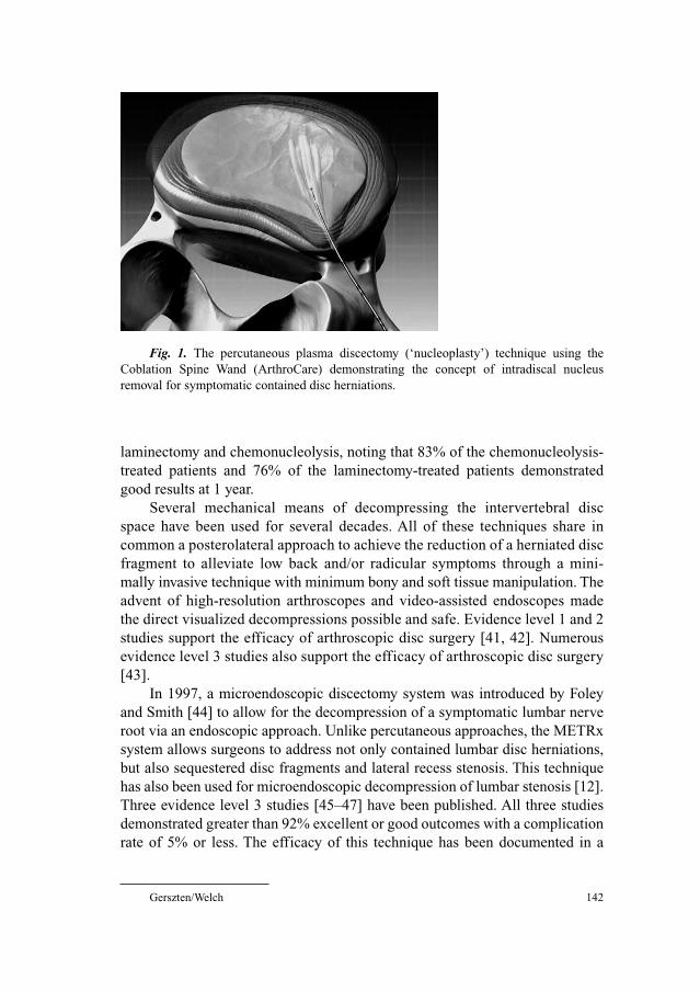
Gerszten/Welch 142
laminectomy and chemonucleolysis, noting that 83% of the chemonucleolysis-
treated patients and 76% of the laminectomy-treated patients demonstrated
good results at 1 year.
Several mechanical means of decompressing the intervertebral disc
space have been used for several decades. All of these techniques share in
common a posterolateral approach to achieve the reduction of a herniated disc
fragment to alleviate low back and/or radicular symptoms through a mini-
mally invasive technique with minimum bony and soft tissue manipulation. The
advent of high-resolution arthroscopes and video-assisted endoscopes made
the direct visualized decompressions possible and safe. Evidence level 1 and 2
studies support the efficacy of arthroscopic disc surgery [41, 42]. Numerous
evidence level 3 studies also support the efficacy of arthroscopic disc surgery
[43].
In 1997, a microendoscopic discectomy system was introduced by Foley
and Smith [44] to allow for the decompression of a symptomatic lumbar nerve
root via an endoscopic approach. Unlike percutaneous approaches, the METRx
system allows surgeons to address not only contained lumbar disc herniations,
but also sequestered disc fragments and lateral recess stenosis. This technique
has also been used for microendoscopic decompression of lumbar stenosis [12].
Three evidence level 3 studies [45–47] have been published. All three studies
demonstrated greater than 92% excellent or good outcomes with a complication
rate of 5% or less. The efficacy of this technique has been documented in a
Fig. 1. The percutaneous plasma discectomy (‘nucleoplasty’) technique using the
Coblation Spine Wand (ArthroCare) demonstrating the concept of intradiscal nucleus
removal for symptomatic contained disc herniations.
Spine: Minimally Invasive Techniques 143
prospective multicenter trial [48]. These series also showed a significant reduc-
tion in operative time, reduced hospital stays, and return to work time compared
to reports from the open discectomy literature.
Thoracoscopic Approaches
Thoracoscopic endoscopic procedures were first described by Kux [49] in
1951, who used urological endoscopes for the treatment of tuberculous disease.
Thoracoscopic spinal surgery is a technique that provides full, direct access to
the ventral thoracic spine. Class III evidence supports this technique for remov-
ing benign intrathoracic paraspinal neurogenic tumors, some corpectomies,
spinal deformity correction, and central herniated thoracic discs. Its morbidity
rate appears to be lower than that associated with open thoracotomy, and it
improves patient comfort and cosmetic results and shortens recovery [50, 51].
Outcomes assessment was based upon patients’ report of pain relief, mean
operative time, length of hospitalization, blood loss, complications, and
patients’ willingness to repeat surgery under similar circumstances.
Sympathectomy for the treatment of hyperhydrosis and pain syndromes of
the upper extremities has recently evolved from invasive open procedures to
endoscopic procedures by taking advantage of minimally invasive thoraco-
scopic techniques [52]. There are multiple evidence level 5 studies that report
successful outcomes using thoracoscopic techniques for minimally invasive
thoracic sympathectomy in reducing overall procedure-related morbidity com-
pared to previous open thoracotomy or posterior paraspinal approaches [53,
54]. These techniques have been further refined with reduced invasiveness
using smaller and fewer incisions through biportal and uniportal exposure and
simplified sympathectomy procedures to shorten hospital stay and further
reduce morbidity [52, 55–59]. The class III evidence is based upon a binary
response for outcomes assessment, evaluation of hospital length of stay, and the
occurrence of complications [52, 57].
Thoracoscopic spinal surgery has been used with limited experience
to achieve spinal decompression, reconstruction, and stabilization [2, 60].
Retrospectively evaluated level 5 data conclude that complete anterior thoraco-
scopically assisted reconstruction of thoracic and thoracolumbar fractures can
be safely and effectively accomplished, thereby reducing the pain and morbidity
associated with conventional thoracotomy and thoracolumbar approaches. The
learning curve was felt by the authors to be quite steep. Video-assisted thoracic
spine surgery demonstrated numerous benefits over open thoracotomy. These
included decreased postoperative pain, faster recovery times, shorter hospital
stays, less postoperative pulmonary complications, and reduction of shoulder
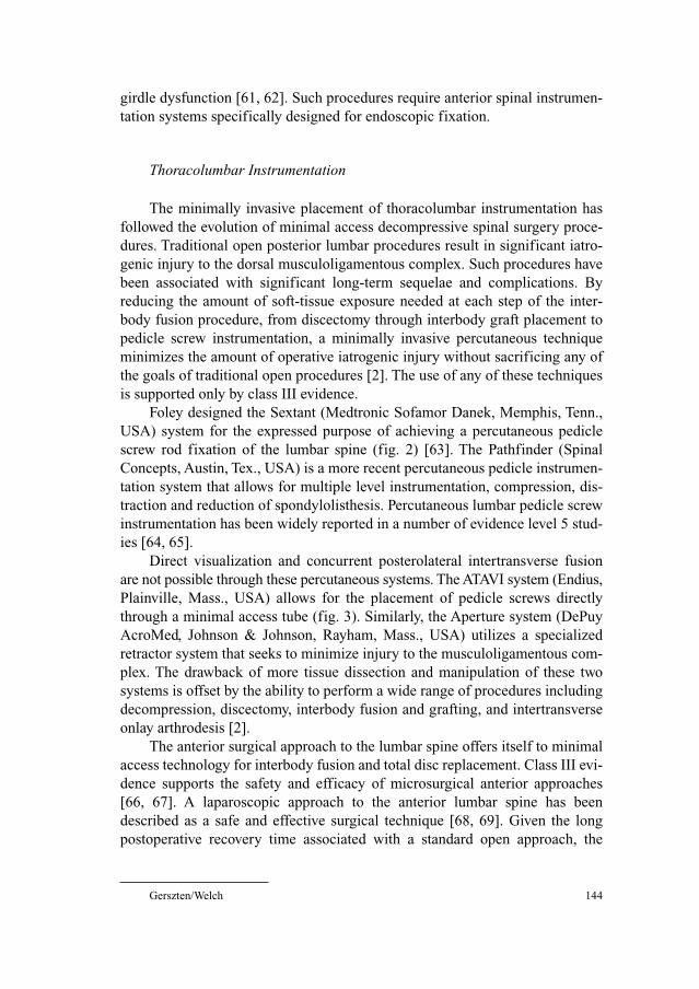
Gerszten/Welch 144
girdle dysfunction [61, 62]. Such procedures require anterior spinal instrumen-
tation systems specifically designed for endoscopic fixation.
Thoracolumbar Instrumentation
The minimally invasive placement of thoracolumbar instrumentation has
followed the evolution of minimal access decompressive spinal surgery proce-
dures. Traditional open posterior lumbar procedures result in significant iatro-
genic injury to the dorsal musculoligamentous complex. Such procedures have
been associated with significant long-term sequelae and complications. By
reducing the amount of soft-tissue exposure needed at each step of the inter-
body fusion procedure, from discectomy through interbody graft placement to
pedicle screw instrumentation, a minimally invasive percutaneous technique
minimizes the amount of operative iatrogenic injury without sacrificing any of
the goals of traditional open procedures [2]. The use of any of these techniques
is supported only by class III evidence.
Foley designed the Sextant (Medtronic Sofamor Danek, Memphis, Tenn.,
USA) system for the expressed purpose of achieving a percutaneous pedicle
screw rod fixation of the lumbar spine (fig. 2) [63]. The Pathfinder (Spinal
Concepts, Austin, Tex., USA) is a more recent percutaneous pedicle instrumen-
tation system that allows for multiple level instrumentation, compression, dis-
traction and reduction of spondylolisthesis. Percutaneous lumbar pedicle screw
instrumentation has been widely reported in a number of evidence level 5 stud-
ies [64, 65].
Direct visualization and concurrent posterolateral intertransverse fusion
are not possible through these percutaneous systems. The ATAVI system (Endius,
Plainville, Mass., USA) allows for the placement of pedicle screws directly
through a minimal access tube (fig. 3). Similarly, the Aperture system (DePuy
AcroMed, Johnson & Johnson, Rayham, Mass., USA) utilizes a specialized
retractor system that seeks to minimize injury to the musculoligamentous com-
plex. The drawback of more tissue dissection and manipulation of these two
systems is offset by the ability to perform a wide range of procedures including
decompression, discectomy, interbody fusion and grafting, and intertransverse
onlay arthrodesis [2].
The anterior surgical approach to the lumbar spine offers itself to minimal
access technology for interbody fusion and total disc replacement. Class III evi-
dence supports the safety and efficacy of microsurgical anterior approaches
[66, 67]. A laparoscopic approach to the anterior lumbar spine has been
described as a safe and effective surgical technique [68, 69]. Given the long
postoperative recovery time associated with a standard open approach, the
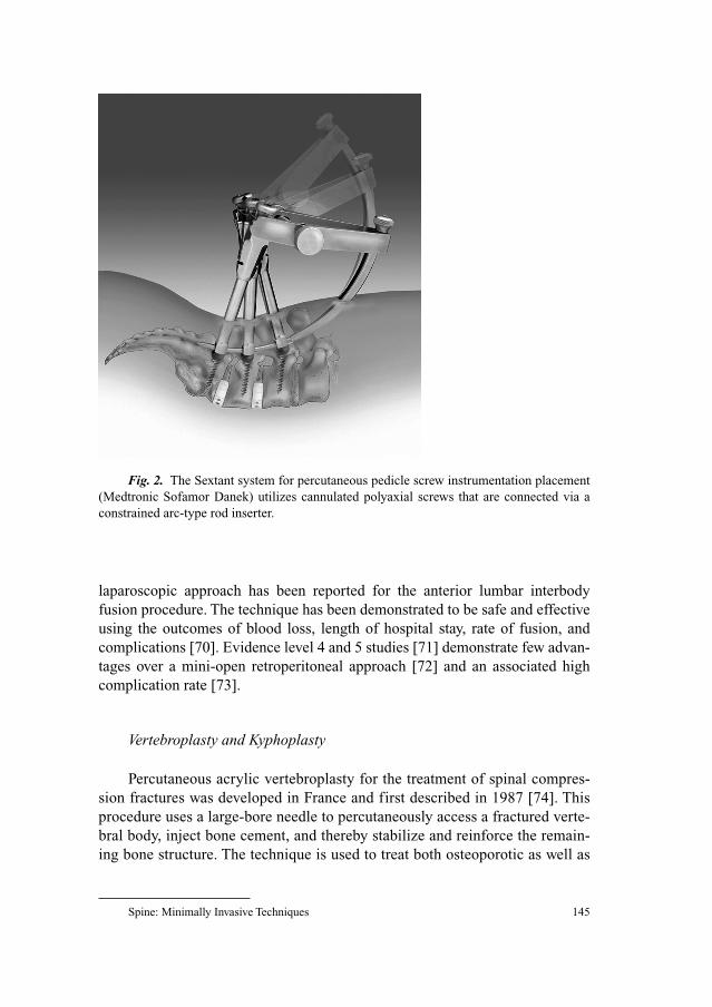
Spine: Minimally Invasive Techniques 145
laparoscopic approach has been reported for the anterior lumbar interbody
fusion procedure. The technique has been demonstrated to be safe and effective
using the outcomes of blood loss, length of hospital stay, rate of fusion, and
complications [70]. Evidence level 4 and 5 studies [71] demonstrate few advan-
tages over a mini-open retroperitoneal approach [72] and an associated high
complication rate [73].
Vertebroplasty and Kyphoplasty
Percutaneous acrylic vertebroplasty for the treatment of spinal compres-
sion fractures was developed in France and first described in 1987 [74]. This
procedure uses a large-bore needle to percutaneously access a fractured verte-
bral body, inject bone cement, and thereby stabilize and reinforce the remain-
ing bone structure. The technique is used to treat both osteoporotic as well as
Fig. 2. The Sextant system for percutaneous pedicle screw instrumentation placement
(Medtronic Sofamor Danek) utilizes cannulated polyaxial screws that are connected via a
constrained arc-type rod inserter.
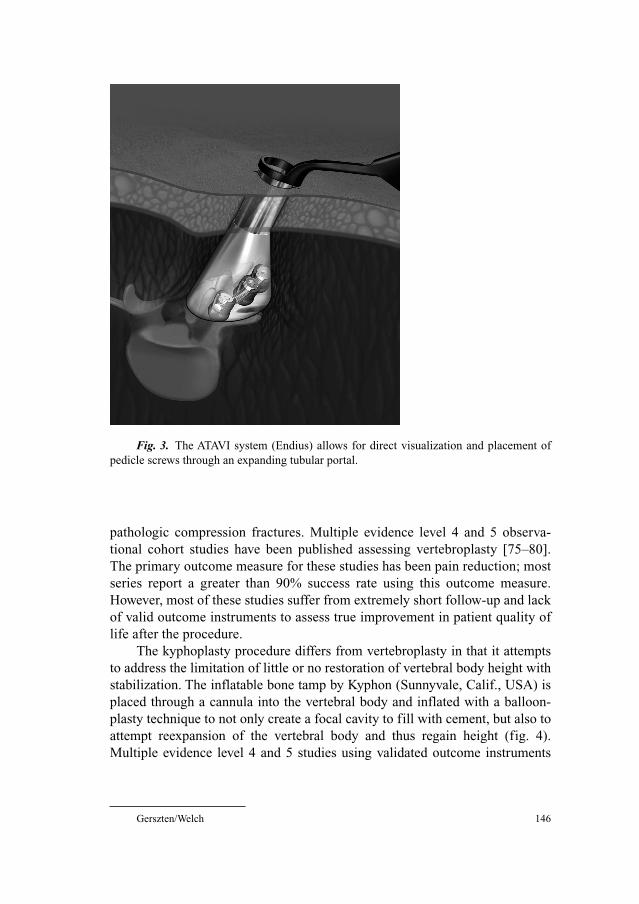
Gerszten/Welch 146
pathologic compression fractures. Multiple evidence level 4 and 5 observa-
tional cohort studies have been published assessing vertebroplasty [75–80].
The primary outcome measure for these studies has been pain reduction; most
series report a greater than 90% success rate using this outcome measure.
However, most of these studies suffer from extremely short follow-up and lack
of valid outcome instruments to assess true improvement in patient quality of
life after the procedure.
The kyphoplasty procedure differs from vertebroplasty in that it attempts
to address the limitation of little or no restoration of vertebral body height with
stabilization. The inflatable bone tamp by Kyphon (Sunnyvale, Calif., USA) is
placed through a cannula into the vertebral body and inflated with a balloon-
plasty technique to not only create a focal cavity to fill with cement, but also to
attempt reexpansion of the vertebral body and thus regain height (fig. 4).
Multiple evidence level 4 and 5 studies using validated outcome instruments
Fig. 3. The ATAVI system (Endius) allows for direct visualization and placement of
pedicle screws through an expanding tubular portal.
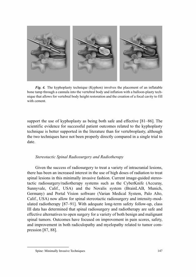
Spine: Minimally Invasive Techniques 147
support the use of kyphoplasty as being both safe and effective [81–86]. The
scientific evidence for successful patient outcomes related to the kyphoplasty
technique is better supported in the literature than for vertebroplasty, although
the two techniques have not been properly directly compared in a single trial to
date.
Stereotactic Spinal Radiosurgery and Radiotherapy
Given the success of radiosurgery to treat a variety of intracranial lesions,
there has been an increased interest in the use of high doses of radiation to treat
spinal lesions in this minimally invasive fashion. Current image-guided stereo-
tactic radiosurgery/radiotherapy systems such as the CyberKnife (Accuray,
Sunnyvale, Calif., USA) and the Novalis system (BrainLAB, Munich,
Germany) and Portal Vision software (Varian Medical System, Palo Alto,
Calif., USA) now allow for spinal stereotactic radiosurgery and intensity-mod-
ulated radiotherapy [87–91]. With adequate long-term safety follow-up, class
III data has determined that spinal radiosurgery and radiotherapy are safe and
effective alternatives to open surgery for a variety of both benign and malignant
spinal tumors. Outcomes have focused on improvement in pain scores, safety,
and improvement in both radiculopathy and myelopathy related to tumor com-
pression [87, 88].
Fig. 4. The kyphoplasty technique (Kyphon) involves the placement of an inflatable
bone tamp through a cannula into the vertebral body and inflation with a balloon-plasty tech-
nique that allows for vertebral body height restoration and the creation of a focal cavity to fill
with cement.
Gerszten/Welch 148
Conclusions
Minimally invasive surgical decompression, arthrodesis, and instrumenta-
tion techniques are now being applied in a wide variety of percutaneous, laparo-
scopic and minimal access procedures ranging from discectomy to multilevel
laminectomy, to posterolateral in situ fusion, to posterior lumbar interbody
fusion and transforaminal lumbar interbody fusion. It is crucial, however, to
realize that there is little to no longitudinal long-term data on these procedures
to document their efficacy, indications, limitations and complications as com-
pared to standard open techniques [2]. As such, evidence level 1 and 2 random-
ized trials are required to ultimately prove these benefits and also to justify the
sometimes significant increased costs of these minimally invasive surgical
procedures.
It is clear that the present wave of minimally invasive surgery procedures
represents an important shift in the practice of contemporary spinal surgery.
With subsequent advances in biological, regenerative, dynamic stabilization,
and radiation-targeting technologies, the access and instrumentation techniques
developed for present-day minimally invasive surgical procedures will also
form the basis for delivering these future technologies to the spine with a mini-
mum of iatrogenic injury [2]. Evidence-based medicine standards will con-
stantly struggle to stay abreast of this quickly advancing subspecialty.
References
1 Jaikumar S, Kim DH, Kam AC: History of minimally invasive spine surgery. Neurosurgery
2002;51:1–14.
2 Khoo LT, Gravori T: Minimally invasive posterior spinal decompression and fusion procedures.
SpineLine 2003;5:9.
3 McCormick PC: Preface. Neurosurgery 2002;51:v–vi.
4 Fessler RG: Foreward. Neurosurgery 2002;51:iii–iv.
5 Gerszten PC: Outcomes research: a review. Neurosurgery 1998;43:1148–1151.
6 King J, Joseph T: Quality of life assessment with the medical outcomes study short form-36
among patients with cervical spondylotic myelopathy. Neurosurgery 2003;52:113–121.
7 Daltroy L, Cats-Baril W, Katz J, et al: The North American spine society lumbar spine outcome
assessment instrument: reliability and validity tests. Spine 1996;21:741–749.
8 Fairbank J, Cooper J, Davies J, et al: The Oswestry low back pain disability questionnaire.
Physiotherapy 1980;66:271–273.
9 Haines SJ: Evidence-based neurosurgery. Neurosurgery 2003;52:36–47.
10 Lohr KN: Outcome measurement: concepts and questions. Inquiry 1988;25:37–50.
11 Benzel EC: Preface. Neurosurgery 2002;51:vi.
12 Fessler R, Guiot B, Khoo L: A minimally invasive technique for decompression of the lumbar
spine. Spine 2002;27:432–438.
13 Murphey F, Simmons JC, Brunson B: Surgical treatment of laterally ruptured cervical discs:
review of 648 cases, 1939–1972. J Neurosurg 1973;38:679–683.
14 Zeidman S, Ducker T: Posterior cervical laminoforaminotomy for radiculopathy: review of 172
cases. Neurosurgery 1993;33:356–362.
Spine: Minimally Invasive Techniques 149
15 Adamson TE: Microendoscopic posterior cervical laminoforaminotomy for unilateral radiculopa-
thy: results of a new technique in 100 cases. J Neurosurg Spine 2001;95:51–57.
16 Fessler RG, Khoo LT: Minimally invasive cervical microendoscopic foraminotomy: an initial clin-
ical experience. Neurosurgery 2002;51:37–45.
17 Hijikata S: Percutaneous nucleotomy: a new concept technique and 12 years’ experience. Clin
Orthop 1989;238:9–23.
18 Maroon J, Onik G, Vidovich D: Percutaneous discectomy for lumbar disc herniation. Neurosurg
Clin N Am 1993;4:125–134.
19 Casper G, Hartman V, Mullins L: Percutaneous laser disc decompression with the holmium: YAG
laser. J Clin Laser Med Surg 1995;13:195–203.
20 Choy D: Percutaneous laser disc decompression (PLDD): twelve years’ experience with 752 pro-
cedures in 518 patients. J Clin Laser Med Surg 1998;16:325–331.
21 Gangi A, Dietemann J, Ide C, et al: Percutaneous laser disk decompression under CT and fluoro-
scopic guidance: indications, techniques and clinical experience. Radiographics 1996;16:89–96.
22 Kotilainen E, Valtonen S: Long-term outcome of patients who underwent percutaneous
nucleotomy for lumbar disc herniation: results after a mean follow-up of 5 years. Acta Neurochir
(Wien) 1998;140:108–113.
23 Andreula C, Muto M, Leonardi M: Interventional spine procedures. Eur J Radiol 2004;50:
112–119.
24 Chen Y, Lee S, Saenz Y, et al: Histologic findings of disc, end plant and neural elements after
coblation of nucleus pulposus: an experimental nucleoplasty study. Spine 2003;3:466–470.
25 Dworkin G: Advanced concepts in interventional spine care. J Am Osteopath Assoc 2002;102:
8–11.
26 Heary RF: Intradiscal electrothermal annuloplasty: the IDET procedure; in Resnick DK, Haid RW Jr:
Surgical Management of Low Back Pain. The American Association of Neurological Surgeons,
Rolling Meadows, 2001, pp 143–150.
27 Derby R, Eek B, Chen Y, et al: Intradiscal electrothermal annuloplasty (IDET): a novel approach
for treating chronic discogenic back pain. Neuromodulation 2000;3:82–88.
28 Freedman BA, Cohen SP, Kuklo TR, et al: Intradiscal electrothermal therapy (IDET) for chronic
low back pain in active-duty soldiers: 2-year follow-up. Spine J 2003;3:502–509.
29 Gerszten PC, Welch WC, McGrath PM, et al: A prospective outcomes study of patients undergo-
ing intradiscal electrothermy (IDET) for chronic low back pain. Pain Physician 2002;5:360–364.
30 Karasek M, Bogduk N: Twelve-month follow-up of a controlled trial on intradiscal thermal annu-
loplasty for back pain due to internal disc disrupture. Spine 2000;25:2601–2607.
31 McGraw JK, Silber JS: Intradiscal electrothermal therapy for the treatment of discogenic back
pain. Appl Radiol 2001;30:11–16.
32 Saal JA, Saal JS: Intradiscal electrothermal treatment for chronic discogenic low back pain: a
prospective outcome study with minimum 1-year follow-up. Spine 2000;25:2622–2627.
33 Wetzel FT, McNally TA, Phillips FM: Intradiscal electrothermal therapy used to manage chronic
discogenic low back pain new directions and interventions. Spine 2002;27:2621–2626.
34 Pauza K, Howell S, Dreyfuss P, et al: A randomized, placebo controlled trial of intradiscal elec-
trothermal therapy for the treatment of discogenc low back pain. Spine J 2004;4:27–35.
35 Kim Y-S, Chin D-K, Cho Y-E, et al: Predictors of successful outcome for lumbar chemonucleolysis:
analysis of 3,000 cases during the past 14 years. Neurosurgery 2002;51:123–128.
36 Mansfield F, Polivy K, Boyd R, et al: Long-term results of chymopapain injections. Clin Orthop
1996;206:67–69.
37 Maroon J, Abla A: Microdiscectomy versus chemonucleolysis. Neurosurgery 1985;16:644–649.
38 Tregonning G, Transfelds E, McCulloch J, et al: Chymopapain versus conventional surgery for
lumbar disc herniation: 10-year results of treatment. J Bone Joint Surg Br 1991;73B:481–486.
39 Gogan W, Fraser R: Chymopapain: a 10-year, double blind study. Spine 1992;17:388–394.
40 Javid M: Chemonucleolysis versus laminectomy: a cohort comparison of effectiveness and
charges. Spine 1995;20:2016–2022.
41 Hermantin F, Peters TM, Quartararo L, et al: A prospective, randomized study comparing the
results of open discectomy with those of video-assisted arthroscopic microdiscectomy. J Bone
Joint Surg Am 1999;81A:958–965.
Gerszten/Welch 150
42 Mayer H, Brock M: Percutaneous endoscopic discectomy: surgical technique and preliminary
results compared to microsurgical discectomy. J Neurosurg 1993;78:216–225.
43 Kambin P: Arthroscopic microdiscectomy. Arthroscopy 1992;8:287–295.
44 Foley K, Smith M: Microendoscopic discectomy. Tech Neurosurg 1997;3:301–307.
45 Foley K, Smith M, Rampersaud Y: Microendoscopic approach to far-lateral lumbar disc hernia-
tion. Neurosurg Focus 1999;7:article 5.
46 Hilton D: Microdiscectomy with a minimally invasive tubular retractor; in Perez-Cruet M, Fessler R
(eds): Outpatient Spinal Surgery. St Louis, Quality Medical Publishing, 2002, pp 159–170.
47 Perez-Cruet M, Foley K, Isaacs R, et al: Microendoscopic lumbar discectomy: technical note.
Neurosurgery 2002;51:129–136.
48 Foley K, Smith M, Rampersaud Y: Microendoscopic discectomy; in Schmidek H (ed): Operative
Neurosurgical Techniques: Indications, Methods, and Results. Philadelphia, Saunders, 2000.
49 Kux E: The endoscopic approach to the vegetative nervous system and its therapeutic possibilities.
Dis Chest 1951;20:139–147.
50 Dickman C, Rosenthal D, Karahalios DG, Paramore C, et al: Thoracic vertebrectomy and recon-
struction using a microsurgical thorascopic approach. Neurosurgery 1996;38:279–293.
51 Han PP, Kenny K, Dickman CA: Thoracoscopic approaches to the thoracic spine: experience with
241 surgical procedures. Neurosurgery 2002;51:88–95.
52 Johnson JP, Patel NP: Uniportal and biportal endoscopic thoracic sympathectomy. Neurosurgery
2002;51:79–83.
53 Drott C, Gothberg G, Claes G: Endoscopic procedures of the upper thoracic sympathetic chain: a
review. Arch Surg 1993;128:237–241.
54 Kux E: Thoracic endoscopic sympathectomy in palmar and axillary hyperhidrosis. Arch Surg
1978;113:264–266.
55 Dumont P, Hamm A, Skrobala D, et al: Bilateral thoracoscopy for sympathectomy in the treatment
of hyperhidrosis. Eur J Surg 1997;11:774–775.
56 Lardinois D, Ris H: Minimally invasive video-endoscopic sympathectomy by use of a transaxil-
lary single port approach. Eur J Cardiothorac Surg 2002;21:67–70.
57 Lin T-S, Kuo S-J, Chou M-C: Uniportal endoscopic thoracic sympathectomy for treatment of pal-
mar and axillary hyperhidrosis: analysis of 2000 cases. Neurosurgery 2002;51:84–87.
58 Reardon P, Preciado A, Scarborough T, et al: Outpatient endoscopic thoracic sympathectomy using
2-mm instruments. Surg Endosc 1999;13:1139–1142.
59 Wahlig JB, Welch WC, Weigel TL, et al: Microinvasive transaxillary thoracoscopic sympathec-
tomy: technical note. Neurosurgery 2000;46:1254–1258.
60 Horn EM, Henn JS, Lemole GM Jr, et al: Thoracoscopic placement of dual-rod instrumentation in
thoracic spinal trauma. Neurosurgery 2004;54:1150–1154.
61 Beisse R, Potulski M, Buhren V: Endoscopic techniques for the management of spinal trauma. Eur
J Trauma 2001;27:275–291.
62 Regan J, Mack M, Picetti G 3rd: A comparison of video-assisted thoracoscopic surgery (VATS)
with open thoracotomy in thoracic spinal surgery. Today’s Ther Trends 1994;11:203–218.
63 Foley K, Gupta S, Justis J, et al: Percutaneous pedicle screw fixation of the lumbar spine.
Neurosurg Focus 2001;10:1–8.
64 Lowery G, Kulkarni S: Posterior percutaneous spine instrumentation. Eur Spine J 2000;9:216–130.
65 Wiesner L, Kothe R, Ruther W: Anatomic evaluation of two different techniques for the percuta-
neous insertion of pedicle screws in the lumbar spine. Spine 1999;24:1599–1603.
66 Mathews H, Evans M, Molligan H, et al: Laparoscopic discectomy with anterior lumbar interbody
fusion. Spine 1995;20:1797–1802.
67 Mayer HM, Wiechert K: Microsurgical anterior approaches to the lumbar spine for interbody
fusion and total disc replacement. Neurosurgery 2002;51:159–165.
68 Lieberman I, Willsher P, Litwin D, et al: Transperitoneal laparoscopic exposure for lumbar inter-
body fusion. Spine 2000;25:509–515.
69 Regan J, McAfee PC, Guyer R, et al: Laparoscopic fusion of the lumbar spine in a multicenter
series of the first 34 patients. Surg Laparosc Endosc 1996;6:459–468.
70 Liu JC, Ondra SL, Angelos P, et al: Is laparoscopic anterior lumbar interbody fusion a useful min-
imally invasive procedure? Neurosurgery 2002;51:155–158.
Spine: Minimally Invasive Techniques 151
71 Regan JJ, Yuan H, McAfee PC: Laparoscopic fusion of the lumbar spine: minimally invasive spine
surgery: a prospective multicenter study evaluating open and laparoscopic lumbar fusion. Spine
1999;24:402–411.
72 Dewald C, Millikan K, Hammerberg K, et al: An open, minimally invasive approach to the lumbar
spine. Am Surg 1999;65:61–68.
73 McAfee P, Regan J, Zdeblick TA, et al: The incidence of complications in endoscopic anterior tho-
racolumbar spinal reconstructive surgery. Spine 1995;20:1623–1632.
74 Galibert P, Deramond H: Note préliminaire sur le traitement des angiomes vertébraux par
vertébroplastie acrylique percutanée. Neurochirurgie 1987;33:166–168.
75 Barr JD, Barr MS, Lemley TJ, et al: Percutaneous vertebroplasty for pain relief and spinal stabi-
lization. Spine 2000;25:923–928.
76 Cortet B, Cotten A, Boutry N, et al: Percutaneous vertebroplasty in the treatment of osteoporotic
vertebral compression fractures: an open prospective study. J Rheumatol 1999;26:2222–2228.
77 Deramond H, Depriester C, Galibert P, et al: Percutaneous vertebroplasty with polymethyl-
methacrylate. Radiol Clin North Am 1998;36:533–546.
78 Jensen ME, Evans AJ, Mathis JM, et al: Percutaneous polymethylmethacrylate vertebroplasty in
the treatment of osteoporotic vertebral body compression fractures: technical aspects. Am J
Neuroradiol 1997;18:1897–1904.
79 Peters KR, Guiot BH, Martin PA, et al: Vertebroplasty for osteoporotic compression fracture: cur-
rent practice and evolving techniques. Neurosurgery 2002;51:96–103.
80 Wong W, Reiley MA, Garfin S: Vertebroplasty/kyphoplasty. J Womens Imaging 2000;2:117–124.
81 Berlemann U, Franz T, Orler R, Heini PF: Kyphoplasty for treatment of osteoporotic vertebral
fractures: a prospective non-randomized study. Eur Spine J 2004;13:496–501.
82 Coumans J: Kyphoplasty for vertebral compression fractures: 1-year clinical outcomes from a
prospective study. J Neurosurg 2003;99:44–50.
83 Dudeney S: Kyphoplasty in the treatment of osteolytic vertebral compression fractures as a result
of multiple myeloma. J Clin Oncol 2002;20:2382–2387.
84 Ledlie J, Renfro M: Balloon kyphoplasty: one-year outcomes in vertebral body height restoration,
chronic pain and activity levels. J Neurosurg Spine 2003;98:36–42.
85 Lieberman I, Dudeney S, Reinhardt M: Initial outcome and efficacy of ‘kyphoplasty’ in the treat-
ment of painful osteoporotic vertebral compression fractures. Spine 2001;26:1631–1638.
86 Rhyne A 3rd, Banit D, Laxer E, Odum S, Nussman D: Kyphoplaty: report of eighty-two thora-
columbar osteoporotic vertebral fractures. J Orthop Trauma 2004;18:294–299.
87 Bilsky MH: Intensity-modulated stereotactic radiotherapy or paraspinal tumors: a preliminary
report. Neurosurgery 2004;54:823–831.
88 Gerszten PC, Ozhasoglu C, Burton S, et al: CyberKnife frameless stereotactic radiosurgery for
spinal lesions: clinical experience in 125 cases. Neurosurgery 2004;55:89–99.
89 Klimo P Jr, Schmidt MH: Surgical management of spinal metastases. Oncologist 2004;9:188–196.
90 Ryu S, Yin FF, Rock J, et al: Image-guided and intensity-modulated radiosurgery for patients with
spinal metastasis. Cancer 2003;97:2013–2018.
91 Ryu SI, Chang SD, Kim DH, et al: Image-guided hypo-fractionated stereotactic radiosurgery to
spinal lesions. Neurosurgery 2001;49:838–846.
Peter C. Gerszten, MD, MPH
Department of Neurological Surgery, Presbyterian University Hospital
Suite B-400, 200 Lothrop St.
Pittsburgh, PA 15213 (USA)
Tel. �1 412 647 0958, Fax �1 412 647 0989, E-Mail [email protected]
Pollock BE (ed): Guiding Neurosurgery by Evidence.
Prog Neurol Surg. Basel, Karger, 2006, vol 19, pp 152–170
An Evidence-Based Medicine Review ofStereotactic Radiosurgery
Bruce E. Pollock
Departments of Neurological Surgery and Radiation Oncology,
Mayo Clinic College of Medicine, Rochester, Minn., USA
AbstractBackground: Stereotactic radiosurgery has been increasingly utilized to manage a wide
variety of indications including vascular malformations, benign and malignant tumors, and
functional disorders. Methods: Review of the recent literature on stereotactic radiosurgery by
evidence-based standards. Results: The vast majority of published papers on stereotactic
radiosurgery is of rather poor quality (level 3 or below). Two studies provide level 1 evidence
showing an improvement in local tumor control or survival for patients with 1–3 brain metas-
tases having radiosurgery in conjunction with whole brain radiation therapy when compared to
patients having whole brain radiation therapy alone. One randomized trial found no improve-
ment in facial pain outcomes for trigeminal neuralgia patients having a longer segment of the
nerve irradiated. Conclusion: For a variety of reasons it is unlikely that randomized clinical
trials will be performed to evaluate the clinical usefulness of stereotactic radiosurgery.
Nonetheless, the preponderance of level 3 information supports the role of radiosurgery as
either an adjunct or alternative to surgical resection or fractionated radiation therapy.
Copyright © 2006 S. Karger AG, Basel
Lars Leksell [1, 2] of the Karolinska Institute in Stockholm, Sweden con-
ceived the concept of stereotactic radiosurgery (SRS) as a less invasive method
to create closed skull, ablative lesions, primarily for functional procedures. SRS
combined stereotactic localization techniques developed in neurosurgery with
radiation physics to distribute energy (X-rays, gamma rays, protons) to an
imaging defined target. As described by Leksell, the radiation was delivered in
a single procedure (dose) to the intended target with a steep dose falloff. During
the 1960s and early 1970s, Leksell and his colleagues, primarily radiobiologist
Börje Larsson, tried a variety of devices and they eventually decided that a
EBM Review of Stereotactic Radiosurgery 153
fixed cobalt-60 device was the best solution and the original Gamma Knife®
(Elekta Instruments, Norcross, Ga., USA) was developed. Inspired by the work
of Leksell, other investigators, including Jacob Fabrikant, Raymond Kjellberg,
Ken Winston, Jay Loeffler, Osvaldo Betti, Federico Colombo, William Freidman,
and Frank Bova (to name only a few), were working simultaneously on systems
that used heavy particles from cyclotrons or X-rays from modified linear accel-
erators as the energy sources for radiosurgery.
Progress in both neuroimaging and dose planning software significantly
improved patient outcomes after radiosurgery. Radiosurgery is now commonly
accepted as the best management for many patients with brain tumors and vas-
cular malformations. It is a multidisciplinary surgical procedure that requires
dedicated technology and facilities. Whereas once SRS existed only in aca-
demic centers, today more than 200 hospitals and outpatient facilities perform
radiosurgery in the United States alone. It is estimated that more than 50,000
patients underwent radiosurgery last year. SRS is now an important part in the
training of neurosurgical and radiation oncology residents.
This chapter will review the published information on radiosurgery of vascu-
lar malformations, tumors, and functional disorders according to evidence-based
medicine (EBM) guidelines. Specifically, the papers covered will refer to single-
session procedures (radiosurgery) as opposed to procedures utilizing image-
guided, multiple-session radiation delivery (stereotactic radiation therapy) [3].
Application of EBM to Radiosurgery
EBM is the conscientious, explicit and judicious use of the current best
evidence in making decisions about the care of individual patients [4]. In the
late 1970s, Suzanne Fletcher and Dave Sackett generated the idea of ‘levels of
evidence’ to rank the validity of evidence of preventive healthcare measures and
linked them to ‘grades of recommendations’ for different interventions [5].
Briefly, the most valid information (level 1) is obtained when a particular ther-
apy has been studied with multiple randomized clinical trials (RCT) with little
variation in the direction or magnitude of the results. In situations where con-
sistent level 1 studies exist, a grade A recommendation can be made for that
particular therapy. Levels 2, 3, 4 and 5 refer to cohort studies, case-control
series, case series, and expert opinions, respectively. Grade B recommendation
can be made with consistent level 2 or 3 studies (or extrapolation from level 1
studies), grade C recommendations can be made from level 4 studies (or extrap-
olation from level 2 or 3 studies), and grade D recommendations can be made
from level 5 studies (or inconsistent or inconclusive studies of any level). By
incorporating the best available external evidence together with our clinical
Pollock 154
expertise and consideration of an individual’s life situation and preferences, a
physician is able to employ an EBM practice.
A variety of reasons exist that limit the practical ability of neurosurgeons
to perform RCTs for each situation. First, and particularly relevant to neuro-
surgery, is that the condition of interest may be rare. Even in settings were the
magnitude of effectiveness between different treatments is large, a sufficient
number of patients must be enrolled to show this difference in a statistically
meaningful way. Second, for benign tumors such as meningiomas or vestibular
schwannomas (VS), the success of an operation in preventing tumor recurrence
or progression may not be evident for 10 or more years after surgery. Thus, the
information derived from case series (level 4 evidence) may be the best avail-
able data to base clinical decision making for patients with benign tumors and
extended life expectancies. Third, and particularly relevant to radiosurgery, is
the fact that few patients are willing to participate in randomized trials in which
one group has open surgery whereas the other undergoes radiosurgery. In fact,
more and more patients have decided based upon their own research before an
official consultation with a neurosurgeon that they want one particular proce-
dure, and they seek out physicians and centers with expertise in that operation.
For such self-educated patients, the concept of allowing chance to determine
whether they undergo a craniotomy with a several day hospital stay or a proce-
dure done as an outpatient under local anesthesia is inconceivable. So although
an RCT comparing outcomes after surgical resection and radiosurgery for VS
or brain metastases would likely yield critical information, the ‘trial ability’ of
such proposed studies is low. For these and many other reasons, clinicians most
often have to base their decision making on rather poor quality evidence.
Benign Tumors
SRS has become an accepted treatment option for patients with menin-
giomas [6–13], VS [14–23], non-VS [24–27], and pituitary adenomas [28–36].
Each year, thousands of patients worldwide undergo radiosurgery for these
benign tumor types. In many respects, patients with benign tumors are ideal
candidates for radiosurgery. First, unlike malignant gliomas, these tumors
rarely invade the adjacent tissue and therefore focused approaches such as
radiosurgery can be used to completely treat the entire tumor burden. Second,
benign tumors are typically well visualized by magnetic resonance imaging
(MRI). This permits a clear delineation between the tumor and nearby struc-
tures so unnecessary radiation exposure can be minimized. Third, radiosurgery
of benign tumors makes radiobiologic sense [37]. For benign tumors, both the
target and the adjacent nervous system act as late responding tissues due to their
EBM Review of Stereotactic Radiosurgery 155
slow rate of proliferation. Consequently, dose fractionation adds little theoreti-
cal benefit compared to conformal, single fraction radiation delivery.
Despite these factors supporting benign tumor radiosurgery, the available
papers on radiosurgery of benign tumors had primarily level 4 evidence regarding
its relative effectiveness compared to other treatments. For example, Nikolopoulos
and O’Donoghue [38] reviewed papers published in English over 23 years on the
management VS patients. Of the 111 papers examined, 18% (20 studies) per-
tained to VS radiosurgery. Overall, 91% of the papers had either level 3 or 4
evidence; no paper provided level 1 or 2 evidence to support surgical resection,
radiosurgery, or observation. They concluded that the overall quality of data
available on VS management was poor, and that efforts should be made to
improve the quality of evidence on this topic. Unfortunately, the situation for
radiosurgery of meningiomas, nonacoustic schwannomas, and pituitary adeno-
mas is no better with the overwhelming number of studies being individual case
series (level 4 evidence (table 1)).
Vestibular SchwannomasThe best management of patients with VS is one of the most controversial
topics in neurosurgery [39–41]. Four studies have used a retrospective cohort
methodology to compare outcomes after radiosurgery to surgical resection for
VS patients [17, 20, 22, 23]. Pollock et al. [20] compared 87 patients with uni-
lateral, unoperated VS with a mean diameter of 3 cm or less managed during
1990 and 1991 at the University of Pittsburgh. The patients having surgical
resection were younger (51 vs. 62 years, p � 0.001); tumor sizes were similar.
At a median follow-up of 36 months, patients having radiosurgery were more
likely to have normal facial movement and preservation of ‘useful hearing’.
Hospital length of stay, return to independent functioning, and direct treatment
charges were less with radiosurgery (p � 0.001). Van Roijen et al. [23] com-
pared 53 VS patients having surgical resection at the University Hospital
Rotterdam to 92 patients having radiosurgery at the Karolinska Institute in
Sweden. Both patient groups were treated from 1990 to 1995. Although the
inclusion criteria were similar in both groups (no prior treatment, unilateral
tumor, and extrameatal diameter less than 3 cm), it is not stated whether the
patients were consecutively treated. The response rate to the mailed question-
naire was 92% for both groups. Overall, radiosurgery was more cost-effective
with regard to both direct and indirect costs. Using the Short-Form 36 to meas-
ure activities of daily living, patients having radiosurgery had higher scores in
the physical function (p � 0.05), role physical (p � 0.02) and mental health
(p � 0.01) domains. Radiosurgical patients also scored higher according to the
EuroQol classification compared to the microsurgery group (0.89 vs. 0.77,
p � 0.01).
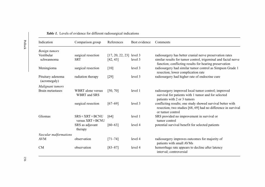
Pollo
ck
156
Table 1. Levels of evidence for different radiosurgical indications
Indication Comparison group References Best evidence Comments
Benign tumorsVestibular surgical resection [17, 20, 22, 23] level 3 radiosurgery has better cranial nerve preservation rates
schwannoma SRT [42, 43] level 3 similar results for tumor control, trigeminal and facial nerve
function; conflicting results for hearing preservation
Meningioma surgical resection [10] level 3 radiosurgery had similar tumor control as Simpson Grade 1
resection; lower complication rate
Pituitary adenoma radiation therapy [29] level 3 radiosurgery had higher rate of endocrine cure
(acromegaly)
Malignant tumorsBrain metastases WBRT alone versus [50, 70] level 1 radiosurgery improved local tumor control; improved
WBRT and SRS survival for patients with 1 tumor and for selected
patients with 2 or 3 tumors
surgical resection [67–69] level 3 conflicting results; one study showed survival better with
resection; two studies [68, 69] had no difference in survival
or tumor control
Gliomas SRS�XRT�BCNU [64] level 1 SRS provided no improvement in survival or
versus XRT�BCNU tumor control
SRS as adjuvant [60–63] level 4 potential survival benefit for selected patients
therapy
Vascular malformationsAVM observation [71–74] level 4 radiosurgery improves outcomes for majority of
patients with small AVMs
CM observation [83–87] level 4 hemorrhage rate appears to decline after latency
interval; controversial
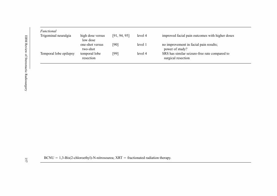
EB
M R
evie
w o
f Ste
reota
ctic
Radio
surg
ery
157
FunctionalTrigeminal neuralgia high dose versus [91, 94, 95] level 4 improved facial pain outcomes with higher doses
low dose
one-shot versus [90] level 1 no improvement in facial pain results;
two-shot power of study?
Temporal lobe epilepsy temporal lobe [99] level 4 SRS has similar seizure-free rate compared to
resection surgical resection
BCNU � 1,3-Bis(2-chloroethyl)-N-nitrosourea; XRT � fractionated radiation therapy.
Pollock 158
Karpinos et al. [17] reviewed 96 patients with unilateral VS having radio-
surgery or microsurgery from 1993 to 2000 at Memorial Hospital in Houston,
Tex., USA. Patients having surgery were younger and had larger tumors com-
pared to the radiosurgery group. Also, the microsurgical group had a longer
median follow-up (48 vs. 24 months). In this comparison, radiosurgery was
more effective at preservation of measurable hearing (58 vs. 14%, p � 0.01),
and had lower rates of trigeminal (12 vs. 22%, p � 0.01) and facial neuropathies
(0 vs. 35%, p � 0.01). Patients having microsurgery had longer hospital stays
(2–16 vs. 1–2 days, p � 0.01) and more perioperative complications (48 vs. 5%,
p � 0.01). Regis et al. [22] compared 97 VS patients having radiosurgery from
1992 to 1998 with 110 VS patients having surgical resection from 1983 to 1990
at the Timone Hospital in Marseille, France. Patients had to have unilateral
tumors and no previous therapy for their VS. For statistical purposes, only
patients with small- to medium-sized tumors (excluding purely intracanalicular
and large tumors with brainstem displacement) were included. In this study,
more men had radiosurgery (46 vs. 35%) and patients having radiosurgery were
older (61 vs. 52 years). Using their own questionnaire and other objective data,
new facial weakness was more common in the surgical group (37 vs. 0%). For
patients with Gardner-Robertson class 1 hearing before treatment, preservation
of class 1 or 2 hearing was more common in the radiosurgery group (70 vs.
38%). The mean time away from work was 7 days after radiosurgery and 130
days after surgical resection. The data available to compare radiosurgery and
surgical resection in these four papers remains rather poor quality (level 3).
However, the results in each study are consistent and show that in short-term
follow-up, radiosurgery provides better functional outcomes than surgical resec-
tion for patients with unilateral, unoperated small- to medium-sized VS.
Two studies have compared the results of SRS to fractionated stereotactic
radiotherapy (SRT) [42, 43]. Andrews et al. [42] reviewed 125 VS patients having
radiosurgery (n � 69) or fractionated SRT (n � 56) from 1994 to 2000. The
tumor margin dose for radiosurgery was 12 Gy; patients having SRT received a
total dose of 50.4 Gy delivered in 28 fractions. The mean follow-up was just
over 2 years in both groups. No difference was noted in tumor control, new
trigeminal deficits, or new facial weakness. Patients having SRT were more
likely to retain functional hearing compared to patients having radiosurgery.
Meijer et al. [43] compared VS patients having either fractionated SRT (either 4
or 5 Gy � 5 fractions) of linac-based radiosurgery (10 or 12.5 Gy at 80% iso-
dose line) between 1992 and 2000. Interestingly, patients were assigned to SRT
if they had teeth (n � 80), whereas patients without teeth who could not use
their relocatable stereotactic guide underwent radiosurgery using a conventional
stereotactic headframe (n � 49). Patients having radiosurgery were older (63 vs.
49 years, p � 0.001); tumor sizes were similar between the groups. At a mean
EBM Review of Stereotactic Radiosurgery 159
follow-up of 33 months, there was no difference in 5-year tumor control, facial
movement, or hearing preservation. Patients having radiosurgery were more
likely to develop new facial numbness (8 vs. 2%, p � 0.05). Therefore, much
like the comparisons of surgical resection and radiosurgery for patients with
VS, the best available information to compare radiosurgery and SRT for VS
patients is level 3 evidence. The two papers on this topic show that patient out-
comes are essentially equivalent over a short follow-up interval. With regard to
hearing preservation, the results were conflicting with one paper showing
higher rates of hearing preservation with SRT [42], whereas the other found
similar hearing outcomes [43].
MeningiomasOnly one study has compared the results of radiosurgery to surgical resec-
tion for patients with intracranial meningiomas [10]. Similar to the VS compar-
ison papers, Pollock et al. employed a retrospective cohort design to compare
surgical resection and radiosurgery as the primary management for adult patients
with benign meningiomas with an average tumor diameter of less than 35 mm.
Between 1990 and 1997, 198 patients met these criteria and were analyzed for
tumor recurrence or progression. The mean follow-up was 64 months. Tumor
resections per Simpson grade were 1 (n � 57, 42%), 2 (n � 57, 42%), or 3–4
(n � 22, 16%). The mean margin and maximum radiation doses at radiosurgery
were 17.7 and 34.9 Gy, respectively. Tumor recurrence/progression was more
frequent in the surgical resection group (12%) compared to the radiosurgical
group (2%; p � 0.04). No difference was detected in the 3- and 7-year actuarial
progression-free survival (PFS) for patients having Simpson Grade 1 resections
(100 and 96%) or radiosurgery (100 and 95%; p � 0.94). Radiosurgery pro-
vided a higher PFS rate compared to patients having Simpson Grade 2 (3- and
7-year PFS, 91 and 82%; p � 0.05) or Grade 3–4 resection (3- and 7-year PFS,
68 and 34%; p � 0.001). Subsequent tumor treatments were more common
after surgical resection (15 vs. 3%, p � 0.02). Complications occurred in 10%
of patients after radiosurgery compared to 22% of patients having tumor resec-
tion (p � 0.06). Therefore, in this study, the PFS rate after radiosurgery was
equivalent to a Simpson Grade 1 resection, and was superior to Grades 2 and
3–4. Therefore, this single study supports radiosurgery (level 3 evidence) as the
preferred management for the majority of patients with small- to moderate-
sized meningiomas without symptomatic mass effect.
Pituitary AdenomasLandolt et al. [29] compared the results of radiosurgery to fractionated
radiation therapy for patients with recurrent acromegaly after prior surgery.
Fifty patients having fractionated radiation therapy (median dose, 40 Gy) from
Pollock 160
1973 to 1992 were compared to 16 patients having radiosurgery (median tumor
margin dose, 25 Gy) between 1994 and 1996. Patient demographics, tumor size,
and pretreatment growth hormone and insulin-like growth factor I were similar
between the two treatment groups. The follow-up interval of patients having
radiation therapy was significantly longer (7.5 vs. 1.4 years, p � 0.0001).
Patients having radiosurgery more commonly achieved biochemical remission
(p � 0.0001); the mean time to endocrine normalization was 1.4 years after
radiosurgery compared to 7.1 years after fractionated radiation therapy. Thus,
similar to other studies comparing benign tumor radiosurgery to other treatment
options, this single study supports radiosurgery (level 3 evidence) over fraction-
ated radiation therapy for patients with recurrent acromegaly.
Malignant Tumors
SRS is especially attractive to patients with malignant brain tumors because
of its minimally invasive nature and the fact that no recovery period is required
after the procedure is completed. However, although numerous papers have exam-
ined the usefulness of radiosurgery for patients with brain metastases [44–59] and
gliomas [60–65], the relative role that radiosurgery should play in the treatment of
these two indications is likely to be quite different because of their distinct
cellular architecture. Brain metastases typically are focal collections of malig-
nant cells with a clear separation between the tumor and the adjacent brain.
Conversely, gliomas are generally infiltrative with indistinct edges and regions
of tumor mixed with brain. Moreover, brain metastases are well-visualized on
gadolinium-enhanced MRI; high-grade gliomas are often made up of a com-
bination of enhancing and nonenhancing regions. Therefore, it has become
apparent that radiosurgery can be used as the primary management for many
patients with brain metastases, whereas radiosurgery is most often employed as
an adjunct to surgical resection, radiation therapy, and chemotherapy for glioma
patients.
Brain MetastasesFor many decades the standard of care for patients with metastatic brain
disease was whole-brain radiation therapy (WBRT). Despite advances in the
detection and treatment of brain metastases, the median survival for patients
treated with WBRT alone is approximately 4–6 months, and tumor recurrence/
progression is common if the patient survives more than 1 year. Radiosurgery
has been used to improve local tumor control rates, and most studies have found
that over 80% of tumors treated with radiosurgery do not progress. Other fac-
tors that make radiosurgery an attractive option are that multiple tumors can be
EBM Review of Stereotactic Radiosurgery 161
treated in a single session, and tumors in deep brain locations who are consid-
ered poor candidates for surgical resection can be effectively and safely treated.
Questions that remain unanswered regarding brain metastasis radiosurgery
include the appropriate roles of WBRT and radiosurgery [58, 66], and the rela-
tive indications of surgical resection versus radiosurgery for patients with brain
metastases [67–71].
Kondziolka et al. [50] performed an RCT of 27 patients to compare sur-
vival and tumor control for patients having WBRT or WBRT and radiosurgery.
They found that combined WBRT and SRS significantly improved local tumor
control for patients with r2–4 brain metastases compared to patients receiving
WBRT alone. No difference was noted in patient survival, although a difference
was detected between patients who had WBRT alone in comparison to patients
who later had salvage SRS or those who had initial WBRT and SRS. Andrews
and colleagues [70] recently reported a prospective randomized RTOG trial
(RTOG 95-08) of WBRT versus WBRT plus radiosurgery for patients with 1–3
brain metastases. WBRT plus radiosurgery provided a survival advantage com-
pared to WBRT alone in the following patient groups: (1) patients with a single
brain metastasis, (2) patients with 2 or 3 metastases and RPA class I, (3) patients
with 2 or 3 metastases under the age of 50 years, and (4) patients with 2 or 3 metas-
tases and non-small cell lung cancer or any squamous carcinoma. Furthermore,
all subsets of patients in the WBRT � SRS group were more likely to have a
stable or improved performance status, improved local control and reduced
steroid dependence compared to the WBRT alone group. Systemic disease
remained the primary cause of death in both groups. Adverse events and the
rate of reoperation were comparable in the two groups. Reoperation pathology
showed necrosis in all patients in the WBRT � SRS arm and viable tumor in all
patients in the WBRT alone arm. Consequently, these two studies provide level
1 evidence that radiosurgery improves local tumor control and is associated
with better survival rates in subsets of brain metastases patients.
Three retrospective studies have compared the results of surgical resection
to radiosurgery for patients with a single brain metastasis [67–69]. Bindal et al.
[67] compared 13 patients having radiosurgery to a matched group of 62
patients having surgical resection. The median survival for the radiosurgery
patients was 7.5 months compared to 16.4 months in the microsurgical group
(p � 0.002). The difference in survival was attributed to progression of the
treated tumor in the radiosurgical group, not systemic progression or develop-
ment of new brain metastatic disease. Muacevic et al. [68] compared 52
patients having microsurgery plus WBRT to 56 patients having only radio-
surgery. No difference was noted in survival (53 vs. 43%, p � 0.19), local
tumor control (75 vs. 83%, p � 0.49), or neurologic death rates (37 vs. 39%,
p � 0.80) 1 year after treatment. Perioperative morbidity and mortality were
Pollock 162
similar. O’Neill et al. [69] compared 74 patients having microsurgery to 23
patients having radiosurgery. No difference was noted in 1-year survival (62 vs.
56%, p � 0.15); local control was significantly better for the radiosurgery
patients (100 vs. 85%, p � 0.02). On the basis of the two larger and more recent
papers, patient survival and local tumor control were similar between the micro-
surgery and radiosurgery groups. However, the potential biases in these papers
are substantial and it is difficult to draw any firm conclusions regarding the best
treatment for this heterogenous patient population.
GliomasRadiosurgery can be used as part of the initial management of patients
with high-grade gliomas [64] or as an adjunct for patients with recurrent tumors
after completion of conventional therapy [60–63, 65]. Souhami et al. [64] pre-
sented the results of the RTOG protocol 93-05. This was an RCT evaluating
upfront radiosurgery followed by radiation therapy and BCNU chemotherapy
compared to radiation therapy and BCNU chemotherapy for adult patients with
supratentorial glioblastoma multiforme less than 4 cm in diameter. Between
1994 and 2000, 203 patients were randomized and 186 were included in the
final analysis. No improvement in survival was noted for patients having radio-
surgery (median survival, 14.1 vs. 13.7 months, p � 0.53). In addition, patterns
of failure and quality of life determinations were similar between the two
groups. This trial showed that when used in the upfront management of patients
with supratentorial glioblastoma multiforme, radiosurgery did not lead to
improved survival or quality of life (level 1 evidence).
Other studies have retrospectively examined the use of radiosurgery typi-
cally for recurrent high-grade gliomas after completion of radiation therapy
and chemotherapy [60–63, 65]. These studies generally have shown a survival
benefit to subgroups of these patients including younger age, better perfor-
mance status, RTOG class, and histology. However, these studies are subject to
significant selection bias and provide rather poor support (level 4 evidence)
regarding the role of radiosurgery as an adjunct therapy for recurrent high-
grade gliomas.
Vascular Malformations
Arteriovenous MalformationsArteriovenous malformation (AVM) radiosurgery has been performed for
more than 30 years [71–74]. The postradiosurgical obliteration rates, the risk of
radiation-related complications, and the chance of a postradiosurgical hemorrhage
have each been thoroughly analyzed with the following conclusions. (1) AVM
EBM Review of Stereotactic Radiosurgery 163
obliteration correlates with the radiation dose delivered to the margin of the mal-
formation [75, 76]. Assuming that the radiation is well targeted, the chance of
AVM cure is approximately 70, 80 and 90% for radiation doses of 16, 18 and
20 Gy, respectively. (2) The likelihood of radiation-related complications after
AVM radiosurgery relates to some measure of the radiation dose to the surround-
ing tissue (most commonly used is the 12-Gy volume), and the location of the
AVM [77]. (3) Radiosurgery does not increase the bleeding rate of AVMs [78–80].
Several papers have attempted to compare the results of surgical resection
and radiosurgery for patients with small- to medium-sized AVMs [81, 82]. In
each study, the conclusion was that surgical resection provided higher cure rate
with less treatment-associated morbidity. However, closer inspection of the
patient characteristics in each group shows that the groups are not directly com-
parable. For example, approximately one third of patients in most AVM series
have AVMs located in the basal ganglia, thalamus, or brainstem, whereas fewer
than 10% of patients in microsurgical series have deep AVMs [73]. As a result,
the conclusions drawn from these studies are fundamentally flawed and are of
little use. Unfortunately, there is little hope that an RCT will ever be performed
to compare these two techniques for the reasons previously discussed.
Nonetheless, patient outcomes after radiosurgery do compare favorably to his-
torical controls on the natural history of untreated AVMs. So although the best
evidence to support AVM radiosurgery is level 4, few clinicians dispute that it
plays an important role in the management of AVM patients.
Cavernous MalformationsCavernous malformation (CM) radiosurgery remains controversial.
Problems related to the assessment of the efficacy of CM radiosurgery are 2-
fold. First, the incidence and natural history of these lesions remain poorly
understood. Second, unlike AVM radiosurgery where obliteration can be con-
firmed with angiography, CMs often do not change appearance after radio-
surgery on MRI and it is the clinical course of the patient that is followed to
determine whether radiosurgery has reduced either their risk of bleeding or new
neurologic events. Numerous studies have documented a decline in the annual
bleeding risk after the first several years [83–87]. However, recent observations
that CMs tend to bleed in ‘clusters’ followed by more quiescent periods creates
doubt that radiosurgery has any effect on hemorrhage risk for these patients
[88]. Moreover, it has been noted that the risk of radiation-related complica-
tions is greater for patients with CMs compared to patients with AVMs, even
when lesion size, location, and radiation dose are comparable [83, 86, 87].
Therefore, although the case series data suggest that radiosurgery reduces the
hemorrhage rate of CMs (level 4 evidence), the indications for CM radio-
surgery remain unclear.
Pollock 164
Functional Disorders
When Lars Leksell [2] conceived the idea of radiosurgery, he believed it
could be used to noninvasively create precise lesions within the brain to treat
functional disorders. In fact, a review of the first 762 cases performed with the
Gamma Knife at the Karolinska Institute showed that 63 (8%) were for trigem-
inal neuralgia. With the advent of MRI, functional radiosurgery has seen a
rebirth with it being commonly used to treat trigeminal neuralgia [89–97] and
patients with temporal lobe epilepsy [98, 99].
Trigeminal NeuralgiaThe reemergence of trigeminal neuralgia radiosurgery can be traced to the
publication by Kondziolka et al. [92] in 1996. That study was a multicenter,
prospective dose escalation study of patients with idiopathic trigeminal neural-
gia. The radiosurgical target was the trigeminal nerve just as it left the brain-
stem; a single 4-mm isocenter was used to irradiate this structure. At a mean
follow-up of 18 months, more than half of the patients became pain-free. The
mean time to pain relief was 1 month. Only 3 patients (6%) developed new
facial numbness.
Based on the encouraging results of that paper, trigeminal neuralgia radio-
surgery has become quite popular and is commonly referred to as the least inva-
sive surgery available for patients with medically unresponsive trigeminal
neuralgia. A number of techniques have been used to improve facial pain
outcomes including dose escalation [91, 94, 95] and irradiation of a longer seg-
ment of the trigeminal nerve [90]. Although the data on higher dose radio-
surgery for trigeminal neuralgia has been derived from retrospective studies
(level 4 evidence), the published results consistently show that more patients
are relieved of their face pain at doses greater than 85 Gy compared to less than
80 Gy. Unfortunately, the incidence of trigeminal dysfunction also appears to
increase at higher radiation doses.
The University of Pittsburgh and the Mayo Clinic conducted an RCT com-
paring one- versus two-shot radiosurgery for trigeminal neuralgia [90]. Eighty-
seven patients with idiopathic trigeminal neuralgia were randomized to receive
either one-shot (n � 44) or two-shot (n � 43) plans with a maximum dose of
75 Gy. No difference was noted in pain control with a median follow-up of 26
months. The development of facial numbness or paresthesias correlated with
the length of irradiated nerve. Although the findings of this paper provide level
1 evidence stating that there was no benefit to patients with the two-shot tech-
nique, the majority of recent papers on trigeminal neuralgia radiosurgery have
found a clear association between the onset of facial numbness and pain relief
after radiosurgery [94, 96]. A larger study in which a longer segment of nerve is
EBM Review of Stereotactic Radiosurgery 165
irradiated may very well show that this technique provides better facial pain
outcomes.
EpilepsyOver the years an improvement in seizure frequency has often been noted
after radiosurgery for patients with epilepsy related to AVMs, CMs, and hypo-
thalamic hamartomas. Regis et al. [99] have studied using radiosurgery to treat
patients with drug-resistant mesial temporal lobe epilepsy (MTLE). Sixteen
patients had been followed for more than 2 years. Thirteen patients (81%) were
seizure free. The median time from radiosurgery to seizure cessation was 10.5
months. Three patients had transient radiation-related complications that required
treatment with corticosteroids. Three patients (19%) developed new visual field
deficits. This pilot work had prompted a great deal of interest in the use radio-
surgery for MTLE. A prospective NIH-funded study is ongoing to more thor-
oughly investigate the safety and efficacy of MTLE radiosurgery.
References
1 Leksell L: The stereotactic method and radiosurgery of the brain. Acta Chir Scand 1951;107:
316–319.
2 Leksell L: Stereotactic radiosurgery. J Neurol Neurosurg Psychiatry 1983;46:797–803.
3 Pollock BE, Lunsford LD: A call to define stereotactic radiosurgery. Neurosurgery 2004;55:
1371–1373.
4 Sackett DL, Rosenberg WM, Gray JA, Haynes RB, Richardson WS: Evidence based medicine:
what it is and what it isn’t. BMJ 1996;312:71–72.
5 The periodic health examination. Canadian Task Force on the Periodic Health Examination. Can
Med Assoc J 1979;121:1193–1254.
6 Hakim R, Alexander E 3rd, Loeffler JS, Shrieve DC, Wen P, Fallon MP, Steig PE, Black PM:
Results of linear accelerator-based radiosurgery for intracranial meningiomas. Neurosurgery
1998;42:446–453.
7 Kondziolka D, Levy EI, Niranjan A, Flickinger JC, Lunsford LD: Long-term outcomes after
meningioma radiosurgery: physician and patient perspectives. J Neurosurg 1999;91:44–50.
8 Lee JYK, Niranjan A, McInerney J, Kondziolka D, Flickinger JC, Lunsford LD: Stereotactic
radiosurgery providing long-term tumor control of cavernous sinus meningiomas. J Neurosurg
2002;97:65–72.
9 Nicolato A, Foroni R, Alessandrini F, Bricolo A, Gerosa M: Radiosurgical treatment of cavernous
sinus meningiomas: experience with 122 treated patients. Neurosurgery 2002;51:1153–1161.
10 Pollock BE, Stafford SL, Utter A, Giannini C, Schreiner SA: Stereotactic radiosurgery provides
equivalent tumor control to a Simpson Grade 1 resection for patients with small- to medium-sized
meningiomas. Int J Radiat Oncol Biol Phys 2003;55:1000–1005.
11 Shafron DH, Friedman WA, Buatti JM, Bova FJ, Mendenhall WM: Linac radiosurgery for benign
meningiomas. Int J Radiat Oncol Biol Phys 1999;43:321–327.
12 Shin M, Kurita H, Sasaki T, Kawamoto S, Tago M, Kawahara N, Morita A, Ueki K, Kirino T:
Analysis of treatment outcome after stereotactic radiosurgery for cavernous sinus meningiomas.
J Neurosurg 2001;95:435–439.
13 Stafford SL, Pollock BE, Foote RL, Gorman DA, Schomberg PJ, Leavitt JA: Meningioma radio-
surgery: tumor control, outcomes, and complications in 190 consecutive patients. Neurosurgery
2001;49:1029–1038.

Pollock 166
14 Flickinger JC, Kondziolka D, Niranjan A, Lunsford LD: Results of acoustic neuroma radio-
surgery: an analysis of 5 years’ experience using current methods. J Neurosurg 2001;94:1–6.
15 Foote KD, Freidman WA, Buatti JM, Meeks SL, Bova FJ, Kubilis PS: Analysis of risk factors asso-
ciated with radiosurgery for vestibular schwannoma. J Neurosurg 2001;95:440–449.
16 Iwai Y, Yamanaka K, Shiotani M, Uyama T: Radiosurgery for acoustic neuromas: results of low-
dose treatment. Neurosurgery 2003;53:282–287.
17 Karpinos M, The BS, Zeck O, Carpenter LS, Phan C, Mai W, Lu HH, Chiu JK, Butler EB,
Gormley WB, Woo SY: Treatment of acoustic neuroma: stereotactic radiosurgery vs. micro-
surgery. Int J Radiat Oncol Biol Phys 2002;54:1410–1421.
18 Kondziolka D, Lunsford LD, McLaughlin MR, Flickinger JC: Long-term outcomes after radio-
surgery for acoustic neuromas. N Engl J Med 1998;339:1426–1433.
19 Petit JH, Hudes RS, Chen TT, Eisenberg HM, Simard JM, Chin LS: Reduced-dose radiosurgery
for vestibular schwannomas. Neurosurgery 2001;49:1299–1306.
20 Pollock BE, Lunsford LD, Kondziolka D, Flickinger JC, Bissonette DJ, Kelsey SF, Jannetta PJ:
Outcome analysis of acoustic neuroma management: a comparison of microsurgery and stereotac-
tic radiosurgery. Neurosurgery 1995;36:215–223.
21 Prasad D, Steiner M, Steiner L: Gamma surgery for vestibular schwannoma. J Neurosurg 2000;92:
745–759.
22 Regis J, Pellet W, Delsanti C, Dufour H, Roche PH, Thomassin JM, Zanaret M, Peragut JC:
Functional outcome after gamma knife surgery or microsurgery for vestibular schwannomas.
J Neurosurg 2002;97:1091–1100.
23 Van Roijen L, Nijs HGT, Avezaat CJJ, Karlsson B, Lindquist C, Pauw KH, Rutten FFH: Costs and
effects of microsurgery versus radiosurgery in treating acoustic neuromas. Acta Neurochir (Wien)
1997;139:942–948.
24 Huang CF, Kondziolka D, Flickinger JC, Lunsford LD: Stereotactic radiosurgery for trigeminal
schwannomas. Neurosurgery 1999;45:11–16.
25 Mabanta SR, Buatti JM, Friedman WA, Meeks SL, Mendenhall WM, Bova FJ: Linear accelera-
tor radiosurgery for nonacoustic schwannomas. Int J Radiat Oncol Biol Phys 1999;43:545–548.
26 Muthukumar N, Kondziolka D, Lunsford LD, Flickinger JC: Stereotactic radiosurgery for jugular
foramen schwannomas. Surg Neurol 1999;52:172–179.
27 Pollock BE, Foote RL, Stafford SL: Stereotactic radiosurgery: the preferred management for
patients with non-vestibular schwannomas? Int J Radiat Oncol Biol Phys 2002;52:1002–1007.
28 Ikeda H, Jokura H, Yoshimoto T: Transsphenoidal surgery and adjuvant gamma knife treatment for
growth hormone-secreting pituitary adenoma. J Neurosurg 2001;95:285–291.
29 Landolt AM, Haller D, Lomax N, Scheib S, Schubiger O, Siegfreid T, Wellis G: Stereotactic radio-
surgery for recurrent surgically treated acromegaly: comparison with fractionated radiotherapy.
J Neurosurg 1998;88:1002–1008.
30 Landolt AM, Haller D, Lomax N, Scheib S, Schubiger O, Siegfreid T, Wellis G: Octreotide may act
as a radioprotective agent in acromegaly. J Clin Endocrinol Metab 2000;85:1287–1289.
31 Landolt AM, Lomax N: Gamma knife radiosurgery for prolactinomas. J Neurosurg 2000;93(suppl 3):
14–18.
32 Pollock BE, Nippoldt TB, Stafford SL, Foote RL, Abboud CF: Results of stereotactic radiosurgery
for patients with hormone producing pituitary adenomas: factors associated with endocrine nor-
malization. J Neurosurg 2002;97:525–530.
33 Pollock BE, Young WF Jr: Stereotactic radiosurgery for patients with ACTH-producing pituitary
adenomas after prior adrenalectomy. Int J Radiat Oncol Biol Phys 2002;54:839–841.
34 Pollock BE, Carpenter PC: Stereotactic radiosurgery as an alternative to fractionated radiation
therapy for patients with recurrent or residual nonfunctioning pituitary adenomas. Neurosurgery
2003;53:1086–1094.
35 Sheehan JM, Vance ML, Sheehan JP, Ellegala DB, Laws ER Jr: Radiosurgery for Cushing’s dis-
ease after failed transsphenoidal surgery. J Neurosurg 2000;93:738–742.
36 Sheehan JP, Kondziolka D, Flickinger JC, Lunsford LD: Radiosurgery for residual or recurrent
nonfunctioning pituitary adenoma. J Neurosurg 2002;97(suppl 5):408–414.
37 Linskey ME: Stereotactic radiosurgery versus stereotactic radiotherapy for patients with vestibular
schwannomas: a Leksell Gamma Knife Society 2000 debate. J Neurosurg 2000;93(suppl 3):90–95.

EBM Review of Stereotactic Radiosurgery 167
38 Nikolopoulos TP, O’Donoghue GM: Acoustic neuroma management: an evidence-based medicine
approach. Otol Neurotol 2002;23:534–541.
39 Kaylie DM, McMenomey SO: Microsurgery vs. gamma knife radiosurgery for the treatment of
vestibular schwannomas. Arch Otolarynolgol Head Neck Surg 2003;129:903–906.
40 Pollock BE, Lunsford LD, Noren G: Vestibular schwannoma management in the next century:
a radiosurgical perspective. Neurosurgery 1998;43:475–483.
41 Sekhar LN, Gormley WB, Wright DC: The best treatment for vestibular schwannoma (acoustic
neuroma): microsurgery or radiosurgery? Am J Otol 1996;17:676–682.
42 Andrews DW, Suarez O, Goldman HW, Downes MB, Bednarz G, Corn BW, Werner-Wasik M,
Rosenstock J, Curran WJ Jr: Stereotactic radiosurgery and fractionated stereotactic radiotherapy
for the treatment of acoustic schwannomas: comparative observations of 125 patients treated at
one institution. Int J Radiat Oncol Biol Phys 2001;50:1265–1278.
43 Meijer OW, Vandertop WP, Baayen JC, Slotman BJ: Single-fraction vs. fractionated linac-based
stereotactic radiosurgery for vestibular schwannoma: a single-institution study. Int J Radiat Oncol
Biol Phys 2003;56:1390–1396.
44 Alexander E 3rd, Moriarty TM, Davis RB, Wen PY, Fine HA, Black PMcL, Kooy HM, Loeffler JS:
Stereotactic radiosurgery for the definitive, noninvasive treatment of brain metastases. J Natl
Cancer Inst 1995;87:34–40.
45 Auchter RM, Lamond JP, Alexander E 3rd, Buatti JM, Chappell R, Friedman WA, Knisella TJ,
Levin AB, Noyes WR, Schultz CJ, Loeffler JS, Mehta MP: A multiinstitutional outcome and prog-
nostic factor analysis of radiosurgery for resectable single brain metastasis. Int J Radiat Oncol
Biol Phys 1996;35:27–35.
46 Brown PD, Brown CA, Pollock BE, Gorman DA, Foote RL: Stereotactic radiosurgery for patients
with radioresistant brain metastases. Neurosurgery 2002;51:656–667.
47 Buatti JM, Friedman WA, Bova FJ, Mendenhall WM: Treatment selection factors for stereotactic
radiosurgery of intracranial metastases. Int J Radiat Oncol Biol Phys 1995;32:1161–1166.
48 Flickinger JC, Kondziolka D, Lunsford LD, Coffey RJ, Goodman ML, Shaw EG, Hudgins WR,
Weiner R, Harsh GR, Sneed PK, Larson DA: A multi-institutional experience with stereotactic
radiosurgery for solitary brain metastasis. Int J Radiat Oncol Biol Phys 1994;28:797–802.
49 Joseph J, Adler JR, Cox RS, Hancock SL: Linear accelerator-based stereotactic radiosurgery
for brain metastases: the influence of number of lesions on survival. J Clin Oncol 1996;14:
1085–1092.
50 Kondziolka D, Patel A, Lunsford LD, Kassam A, Flickinger JC: Stereotactic radiosurgery plus
whole brain radiotherapy versus radiotherapy alone for patients with multiple brain metastases. Int
J Radiat Oncol Biol Phys 1999;45:427–434.
51 Lavine SD, Petrovich Z, Cohen-Gadol AA, Marsi LS, Morton DL, O’Day SJ, Essner R, Zelman V,
Yu C, Luxton G, Apuzzo MLJ: Gamma knife radiosurgery for metastatic melanoma: an analysis
of survival, outcome, and complications. Neurosurgery 1999;44:59–66.
52 Matsuo T, Shibata S, Yasunga A, Iwanaga M, Mori K, Shimizu T, Hayashi N, Ochi M, Hayashi K:
Dose optimization and indications of Linac radiosurgery for brain metastases. Int J Radiat Oncol
Biol Phys 1999;45:931–939.
53 Mori Y, Kondziolka D, Lunsford LD, Logan T, Flickinger JC: Stereotactic radiosurgery for brain
metastases from renal cell carcinoma. Cancer 1998;83:344–353.
54 Nussbaum ES, Djalilian HR, Cho KH, Hall WA: Brain metastases: histology, multiplicity, surgery
and survival. Cancer 1996;78:1781–1788.
55 Pirzkall A, Debrus J, Lohr F, Fuss M, Rhein B, Engenhart-Cabillic R, Wannenmacher M:
Radiosurgery alone or in conjunction with whole-brain radiotherapy for brain metastases. J Clin
Oncol 1998;16:3563–3569.
56 Pollock BE, Brown PD, Foote RL, Stafford SL, Schomberg PJ: Properly selected patients with
multiple brain metastases may benefit from aggressive treatment of their intracranial disease.
J Neurooncol 2003;61:73–80.
57 Schoeggl A, Kitz K, Ertel A, Reddy M, Bavinski G, Schneider B: Prognostic factor analysis for
multiple brain metastases after gamma knife radiosurgery: results in 97 patients. J Neurooncol
1999;42:169–175.

Pollock 168
58 Sneed PK, Lamborn KR, Forstner JM, McDermott MW, Chang S, Park E, Gutin PH, Phillips TL,
Wara WM, Larson DA: Radiosurgery for brain metastases: is whole brain radiotherapy necessary.
Int J Radiat Oncol Biol Phys 1999;43:549–558.
59 Weltman E, Salvajoli JV, Brandt RA, Hanriot RM, Prisco FE, Cruz JC, De Oliveria SR, Wajsbrot
DB: Radiosurgery for brain metastases: a score index for predicting prognosis. Int J Radiat Oncol
Biol Phys 2000;46:1155–1161.
60 Graves EE, Nelson SJ, Vigneron DB, Chin C, Verhay L, McDermott M, Larson D, Sneed PK,
Chang S, Prados MD, Lamborn K, Dillon WP: A preliminary study on the prognostic value of
proton magnetic resonance spectroscopic imaging in gamma knife radiosurgery of recurrent
malignant gliomas. Neurosurgery 2000;46:319–326.
61 Kondziolka D, Flickinger JC, Bissonette DJ, Bozik M, Lunsford LD: Survival benefit of stereo-
tactic radiosurgery for patients with malignant glial neoplasms. Neurosurgery 1997;41:776–783.
62 Nwokedi EC, DiBiase SJ, Jabbour S, Herman J, Amin P, Chin LS: Gamma knife stereotactic radio-
surgery for patients with glioblastoma multiforme. Neurosurgery 2002;50:41–46.
63 Shrieve DC, Alexander E 3rd, Black PM, Wen PY, Fine HA, Kooy HM, Loeffler JS: Treatment of
patients with primary glioblastoma multiforme with standard postoperative radiotherapy and
radiosurgical boost: prognostic factors and long-term outcome. J Neurosurg 1999;90:72–77.
64 Souhami L, Seiferheld W, Brachman D, Podgorsak EB, Werner-Wasik M, Lustig R, Schultz CJ,
Sause W, Okunieff P, Buckner J, Zamorano L, Mehta MP, Curran WJ Jr: Randomized comparison
of stereotactic radiosurgery followed by conventional radiotherapy with carmustine to conventional
radiotherapy with carmustine for patients with glioblastoma multiforme: report of the Radiation
Therapy Oncology Group 93-05 protocol. Int J Radiat Oncol Biol Phys 2004:60:853–860.
65 Shaw EG, Scott C, Souhami L, Dinapoli R, Kline R, Loeffler J, Farnan N: Single dose radiosurgi-
cal treatment of recurrent previously irradiated primary tumors and brain metastases: final report
of RTOG protocol 90-05. Int J Radiat Oncol Biol Phys 2000;47:291–298.
66 Shaw EG: Radiotherapeutic management of multiple brain metastases: ‘3000 in 10’ whole brain
radiation is no longer a ‘no brainer’. Int J Radiat Oncol Biol Phys 1999;45:253–254.
67 Bindal AK, Bindal RK, Hess KR, Shiu A, Hassenbusch SJ, Shi WM, Sawaya R: Surgery versus
radiosurgery in the treatment of brain metastasis. J Neurosurg 1996;84:748–754.
68 Muacevic A, Kreth FW, Horstmann GA, Schmid-Elsaesser R, Wowra B, Steiger HJ, Reulen HJ:
Surgery and radiotherapy compared to gamma knife radiosurgery in the treatment of solitary cere-
bral metastases of small diameter. J Neurosurg 1999;91:35–43.
69 O’Neill BP, Iturria NJ, Link MJ, Pollock BE, Ballman KV, O’Fallon JR: A comparison of surgical
resection and stereotactic radiosurgery in the treatment of solitary brain metastases. Int J Radiat
Oncol Biol Phys 2003;55:1169–1176.
70 Andrews DW, Scott CB, Sperduto PW, Flanders AE, Gaspar LE, Schell MC, Werner-Wasik M,
Demas W, Ryu J, Bahary J, Souhami L, Rotman M, Mehta MP, Curran WJ Jr: Whole brain radia-
tion therapy with or without stereotactic radiosurgery boost for patients with one to three brain
metastases: phase III results of the RTOG 9508 randomised trial. Lancet 2004;363:1665–1672.
71 Friedman WA, Bova FJ, Bollampally S, Bradshaw P: Analysis of factors predictive of success or
complications in arteriovenous malformation radiosurgery. Neurosurgery 2003;52:296–308.
72 Kjellberg RN, Hanamura T, Davis KR, Lyons SL, Adams RD: Bragg-peak proton-beam therapy
for arteriovenous malformations. N Engl J Med 1983;309:269–274.
73 Pollock BE, Flickinger JC: A proposed radiosurgery-based grading system for arteriovenous mal-
formations. J Neurosurg 2002;96:79–85.
74 Pollock BE, Gorman DA, Coffey RJ: Patient outcomes after arteriovenous malformation radiosurgical
management: results based on a 5- to 14-year follow-up study. Neurosurgery 2003;52:1291–1297.
75 Flickinger JC, Pollock BE, Kondziolka D, Lunsford LD: A dose-response analysis of arteriove-
nous malformation obliteration by radiosurgery. Int J Radiat Onc Biol Phys 1996;36:873–879.
76 Karlsson B, Lindquist C, Steiner L: Prediction of obliteration after gamma knife surgery for cere-
bral arteriovenous malformations. Neurosurgery 1997;40:425–431.
77 Flickinger JC, Kondziolka D, Lunsford LD, Liscak R, Phuong LK, Pollock BE: Development of a
model to predict permanent symptomatic post-radiosurgery injury for arteriovenous malformation
patients. Int J Radiat Oncol Biol Phys 2000;46:1143–1148.

EBM Review of Stereotactic Radiosurgery 169
78 Friedman WA, Blatt DL, Bova FJ, Buatti JM, Mendenhall WM, Kubilis PS: The risk of hemor-
rhage after radiosurgery for arteriovenous malformations. J Neurosurg 1996;84:912–919.
79 Karlsson B, Lax I, Söderman M: Risk of hemorrhage during the 2-year latency period following
gamma knife radiosurgery for arteriovenous malformations. Int J Radiat Oncol Biol Phys 2001;
49:1045–1051.
80 Pollock BE, Flickinger JC, Lunsford LD, Bissonette DJ, Kondziolka D: Hemorrhage risk after
stereotactic radiosurgery of cerebral arteriovenous malformations. Neurosurgery 1996;38:
652–661.
81 Pikus HJ, Beach ML, Harbaugh RE: Microsurgical treatment of arteriovenous malformations:
analysis and comparison with stereotactic radiosurgery. J Neurosurg 1998;88:641–646.
82 Porter PJ, Shin AY, Detsky AS, Lefaive L, Wallace MC: Surgery versus stereotactic radiosurgery
for small, operable cerebral arteriovenous malformations: a clinical and cost comparison.
Neurosurgery 1997;41:757–766.
83 Amin-Hanjabi S, Ogilvy CS, Candia GJ, Lyons S, Chapman PH: Stereotactic radiosurgery for cav-
ernous malformations: Kjellberg’s experience with proton beam therapy in 98 cases at the Harvard
cyclotron. Neurosurgery 1998;42:1229–1238.
84 Chang SD, Levy RP, Adler JR Jr, Martin DP, Krakovitz PR, Steinberg GK: Stereotactic radio-
surgery of angiographically occult vascular malformations: 14-year experience. Neurosurgery
1998;43:213–221.
85 Hasegawa T, McInerney J, Kondziolka D, Lee JY, Flickinger JC, Lunsford LD: Long-term results
after stereotactic radiosurgery for patients with cavernous malformations. Neurosurgery
2002;50:1190–1197.
86 Karlsson B, Kihlström L, Lindquist C, Ericson K, Steiner L: Radiosurgery for cavernous malfor-
mations. J Neurosurg 1998;88:293–297.
87 Pollock BE, Garces YI, Stafford SL, Foote RL, Schomberg PJ, Link ML: Stereotactic radiosurgery
of cavernous malformations. J Neurosurg 2000;93:987–991.
88 Barker FG 2nd, Amin-Hanjabi S, Butler WE, Lyons S, Ojemann RG, Chapman PH, Ogilvy CS:
Temporal clustering of hemorrhages from untreated cavernous malformations of the central ner-
vous system. Neurosurgery 2001;49:15–25.
89 Brisman R: Gamma knife radiosurgery with a dose of 75 to 76.8 Gy for trigeminal neuralgia.
J Neurosurg 2004;100:848–854.
90 Flickinger JC, Pollock BE, Kondziolka D, Phuong LK, Foote RL, Stafford SL, Lunsford LD: What
length should be irradiated? A randomized blinded study of radiosurgery to different lengths of
trigeminal nerve for trigeminal neuralgia. Int J Radiat Oncol Biol Phys 2001;51:449–454.
91 Goss BW, Frighetto L, DeSalles AAF, Smith Z, Solberg T, Selch M: Linear accelerator radio-
surgery using 90 Gy for essential trigeminal neuralgia: results and dose volume histogram analysis.
Neurosurgery 2003;53:823–830.
92 Kondziolka D, Lunsford LD, Flickinger JC, Young RF, Vermeulen S, Duma CM, Jacques DB,
Rand RW, Regis J, Peragut JC, Manera L, Epstein MH, Lindquist C: Stereotactic radiosurgery
for trigeminal neuralgia: a multiinstutional study using the gamma unit. J Neurosurg 1996;84:
940–945.
93 Maesawa S, Salame C, Flickinger JC, Pirris S, Kondziolka D, Lunsford LD: Clinical outcomes
after stereotactic radiosurgery for idiopathic trigeminal neuralgia. J Neurosurg 2001;94:14–20.
94 Massager N, Lorenzoni J, Devriendt D, Desmedt F, Brotchi J, Levivier M: Gamma knife surgery
for idiopathic trigeminal neuralgia performed using a far-anterior cisternal target and a high dose
of radiation. J Neurosurg 2004;100:597–605.
95 Pollock BE, Phuong LK, Foote RL, Stafford SL, Gorman DA: High-dose trigeminal neuralgia
radiosurgery associated with increased risk of trigeminal dysfunction. Neurosurgery 2001;49:
58–64.
96 Pollock BE, Phuong LK, Gorman DA, Foote RL, Stafford SL, Schomberg PJ: Stereotactic radio-
surgery for idiopathic trigeminal neuralgia. J Neurosurg 2002;97:347–353.
97 Smith ZA, DeSalles AAF, Frighetto L, Goss B, Lee SP, Selch M, Wallace RE, Cabatan-Awang C,
Solberg T: Dedicated linear accelerator radiosurgery for the treatment of trigeminal neuralgia.
J Neurosurg 2003;99:511–516.

Pollock 170
98 Kondziolka D: Functional radiosurgery. Neurosurgery 1999;44:12–22.
99 Regis J, Bartolomei F, Rey M, Hayashi M, Chauvel P, Peragut JC: Gamma knife surgery for mesial
temporal lobe epilepsy. J Neurosurg 2000;93(suppl 3):141–146.
Bruce E. Pollock, MD
Department of Neurological Surgery, Mayo Clinic
200 First Street SW
Rochester, MN 55905 (USA)
Tel. �1 507 284 5317, Fax �1 507 284 5206, E-Mail [email protected]
Pollock BE (ed): Guiding Neurosurgery by Evidence.
Prog Neurol Surg. Basel, Karger, 2006, vol 19, pp 171–196
Evidenced-Based Guidelines forTraumatic Brain Injuries
Donald W. Marion
Fusion Medical Education, LLC, Wakefield, Mass., USA
AbstractAn enormous amount of clinical and basic science brain injury research has been
undertaken during the last several decades in an effort to improve outcomes following severe
traumatic brain injury, but to date there still are no new therapies that have been clearly
shown to be beneficial. There is, however, increasing evidence to suggest that evidence-
based, protocol-driven, acute care can lead to improved outcomes. Evidence based guidelines
for the medical and surgical management of severe brain injury, and for penetrating and pedi-
atric brain injury, as well as for the pre-hospital management of brain injury, have all been
published. In this chapter the conclusions of those guidelines is reviewed. In addition, the
studies that demonstrate improved outcomes as a result of implementation of the guidelines
are summarized.
Copyright © 2006 S. Karger AG, Basel
Nearly a century ago surgeons discovered that evacuation of posttrau-
matic hematomas could, in some cases at least, lead to survival and even
good outcomes. But by the 1950s it also was recognized that many of those
with severe traumatic brain injury (TBI) died of intracranial hypertension
due to brain swelling. Since that time there has been extensive research
aimed at refining intracranial pressure (ICP) monitoring techniques, defin-
ing the physiologic and molecular causes of posttraumatic brain swelling,
and identifying therapies that ameliorate brain swelling and lead to improved
outcomes. Much of the research conducted during the past 20 years has
focused on a relatively small number of molecular mechanisms identified in
rodent models of TBI to be critical intermediates of secondary brain
injury. Drugs were developed that could effectively block some of these
Marion 172
mechanisms of secondary injury (in laboratory models), and those drugs
were tested in large-scale multicenter clinical trials. To date numerous clini-
cal trials have been completed but none have shown improved outcomes as a
result of the novel treatment.
Because of the lack of therapies proven to reduce brain swelling, many
neurosurgeons and other clinicians involved in the care of TBI patients adopted
therapeutic biases based on personal experience, incomplete literature reviews,
or the views of their mentors. This led to a tremendous degree of variability of
practice throughout the US. A survey of US trauma centers conducted by the
Brain Trauma Foundation in 1991 found that there was significant disagree-
ment about the need for ICP monitoring, role of steroids, and role of barbiturates
for patients with severe TBI [1]. As a result, there was considerable controversy
and confusion within hospital intensive care units because of inconsistent treat-
ment of the TBI patients depending on who was responsible for their care
during a particular day or week.
The rationale for the development and implementation of evidence-based
guidelines in TBI care is 2-fold: first, that there are ‘best practices’, and sec-
ond, that consistent care within and among hospitals treating TBI patients will
lead to improved outcomes. The strength of guidelines in part relies on the
availability of good quality studies that document improved outcomes with the
use of one particular treatment compared to the management of the patient
without that treatment, or with the use of an alternative treatment. It is gener-
ally accepted that such studies should be prospective randomized controlled
clinical trials of large numbers of patients. But even in the absence of such
‘class I’ studies, a group of lesser quality studies that are consistent in their
findings can provide support for guidelines. While such studies may not be
used to support a standard of care for a particular treatment recommendation,
they can be used to craft guidelines or options. In some cases, support for best
practices must rely on indirect evidence, or intuitive science. For example,
intracranial hypertension is known to be an important proximate cause for neu-
rologic morbidity and mortality following TBI. It is logical to conclude that
treatment of intracranial hypertension cannot be effectively accomplished
unless the clinician knows what the ICP is. Most clinicians consider the reduc-
tion of ICP an important indicator of the effectiveness of treatment. In addi-
tion, it is believed that despite a lack of prospective randomized clinical trials
in support of specific treatment recommendations, the creation of guidelines
supported by the best available literature is justified because they promote
consistent acute care of TBI patients. Consistent protocol-driven care helps to
establish a team approach, eliminates confusion, and is thereby likely to lead to
improved outcomes.
Evidenced-Based Guidelines for Traumatic Brain Injuries 173
History of Contemporary TBI Guideline Development
During the early 1990s a small group of neurosurgeons who were interested
in neurotrauma agreed that there was a need to standardize care of patients with
severe TBI according to best practice guidelines. They believed that the wide
variability of care provided by neurosurgeons throughout the US was likely
responsible for preventable death and disability in a significant proportion of
TBI patients. At the same time, the Agency for Health Care Policy and Research
(AHCPR), Department of Health and Human Services, had made the same con-
clusion for all of medicine and was promoting the development of evidence-
based guidelines for all medical specialties [2]. The neurosurgeons embraced the
methodology for development of evidence-based guidelines developed by
AHCPR and, with the financial support of the Brain Trauma Foundation, pro-
duced the Guidelines for the Management of Severe Head Injury. Using the
AHCPR methodology, 11 neurosurgeons with an interest in TBI selected 13 top-
ics considered most important for the care of TBI patients. Each neurosurgeon
was assigned a topic and proceeded with a thorough literature search to identify
all related peer-reviewed journal articles (primarily English language) that were
published over the last 40 years. The studies were then categorized as class I
(prospective randomized controlled clinical trials), class II (nonrandomized
cohort studies, randomized controlled clinical trials with significant design
flaws, or case-controlled studies), or class III (expert opinion, case reports, most
retrospective studies). Recommendations were formulated for each topic, and
considered as ‘standards’ if supported by one or more class I studies, ‘guide-
lines’ if supported by two or more class II studies, or ‘options’ if supported only
by class III studies. A draft document was produced with a separate chapter for
each topic, and the document was widely circulated to all medical specialties and
organizations with a stake in the care of TBI patients for their input. A final doc-
ument was produced after incorporating the suggestions of these organizations.
The document was endorsed by the American Association of Neurological
Surgeons and disseminated free of charge to all practicing neurosurgeons in
North America in 1995 [3]. Neurosurgeons, as well as trauma surgeons and
critical care intensivists, quickly adopted the guidelines and, according to subse-
quent surveys, often changed their practice to conform to these recommen-
dations. As per an agreement with the American Association of Neurological
Surgeons the Guidelines were updated in 2000, and a section was added on prog-
nostic indicators following severe TBI [4].
As a result of the success of this document there was renewed interest in
developing evidence-based guidelines for TBI issues not specifically addressed
in the Guidelines for the Management of Severe Head Injuries. To date, guidelines
have also been completed for the prehospital management of TBI patients [5],
Marion 174
the management of penetrating brain injuries [6], and the management of pedi-
atric head injuries [7]. Guidelines for the surgical management of TBI are in
press. The authors of these more recent guidelines have faithfully followed the
same methodology as was used for development of the original head injury
guidelines.
In this chapter the Guidelines for the Management of Severe Traumatic
Brain Injury and Early Indicators of Prognosis in Severe Traumatic Brain
Injury, Pre-Hospital Management of Traumatic Brain Injury, Management and
Prognosis of Penetrating Brain Injury, Guidelines for the Acute Management of
Severe Traumatic Brain Injury in Infants, Children, and Adolescents, and the
Surgical Management of TBI are summarized by presenting the recommenda-
tions of each document. The authors of the guidelines are listed in alphabetical
order (by agreement of the authors) in the sections describing the recommenda-
tions of each of the Guidelines documents. Several problems with guidelines’
development are then addressed, followed by a summary of several recent stud-
ies that have looked at the impact of the guidelines on change in practice, and
on outcomes following severe TBI. Numerous guidelines for the management
of mild TBI have appeared over the last 10 years, including sports-related con-
cussion guidelines. These guidelines are not covered in this chapter because
most are not truly evidence-based.
Guidelines for the Management of Severe Head Injury, and EarlyIndicators of Prognosis in Severe Traumatic Brain Injury
This document is the updated version of the original Guidelines document,
and was published in 2000 in a special edition of the Journal of Neurotrauma[4]. The authors of the section on the Management of Severe Head Injury are
M. Ross Bullock, Randall M. Chesnut, Guy L. Clifton, Jamshid Ghajar, Donald
W. Marion, Raj K. Narayan, David W. Newell, Lawrence H. Pitts, Michael J.
Rosner, Beverly C. Walters, and Jack E. Wilberger (table 1). For the Section on
Early Indicators of Prognosis in Severe Traumatic Brain Injury, the authors are
Randall M. Chesnut, Jamshid Ghajar, Andrew I.R. Maas, Donald W. Marion,
Franco Servadei, Graham M. Teasdale, Andreas Unterberg, Hans von Holst,
and Beverly C. Walters (table 2). The document includes an extensive section
on the methodology for evidence-based guideline development. Each subse-
quent chapter covers a specific topic relevant to the acute care of patients with
severe TBI. The chapters are written in the same format, with a section on
recommendations followed by sections entitled overview, process, scientific
foundation, summary, key issues for future investigation, evidentiary tables,
and a complete bibliography of studies cited. The recommendations section
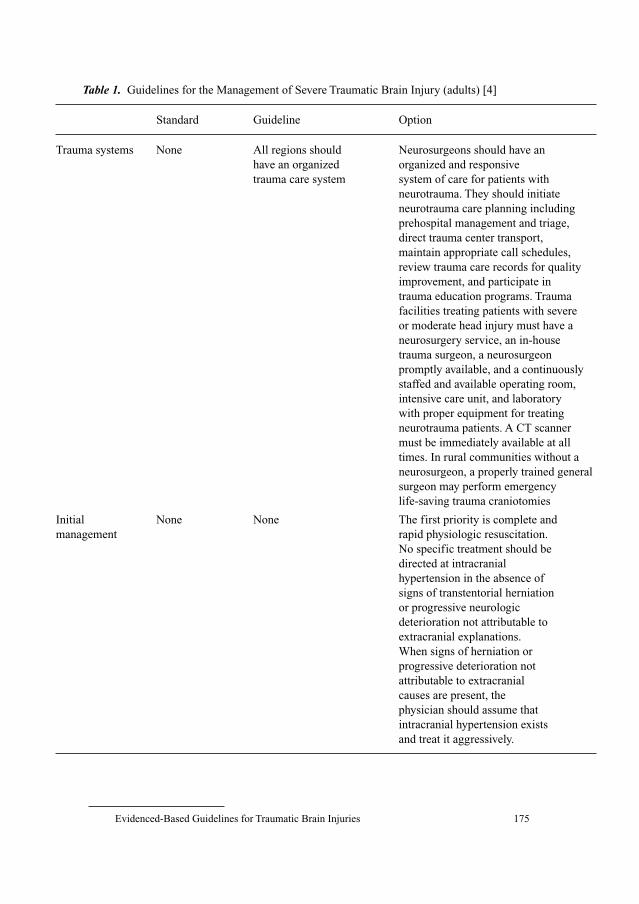
Evidenced-Based Guidelines for Traumatic Brain Injuries 175
Table 1. Guidelines for the Management of Severe Traumatic Brain Injury (adults) [4]
Standard Guideline Option
Trauma systems None All regions should Neurosurgeons should have an
have an organized organized and responsive
trauma care system system of care for patients with
neurotrauma. They should initiate
neurotrauma care planning including
prehospital management and triage,
direct trauma center transport,
maintain appropriate call schedules,
review trauma care records for quality
improvement, and participate in
trauma education programs. Trauma
facilities treating patients with severe
or moderate head injury must have a
neurosurgery service, an in-house
trauma surgeon, a neurosurgeon
promptly available, and a continuously
staffed and available operating room,
intensive care unit, and laboratory
with proper equipment for treating
neurotrauma patients. A CT scanner
must be immediately available at all
times. In rural communities without a
neurosurgeon, a properly trained general
surgeon may perform emergency
life-saving trauma craniotomies
Initial None None The first priority is complete and
management rapid physiologic resuscitation.
No specific treatment should be
directed at intracranial
hypertension in the absence of
signs of transtentorial herniation
or progressive neurologic
deterioration not attributable to
extracranial explanations.
When signs of herniation or
progressive deterioration not
attributable to extracranial
causes are present, the
physician should assume that
intracranial hypertension exists
and treat it aggressively.
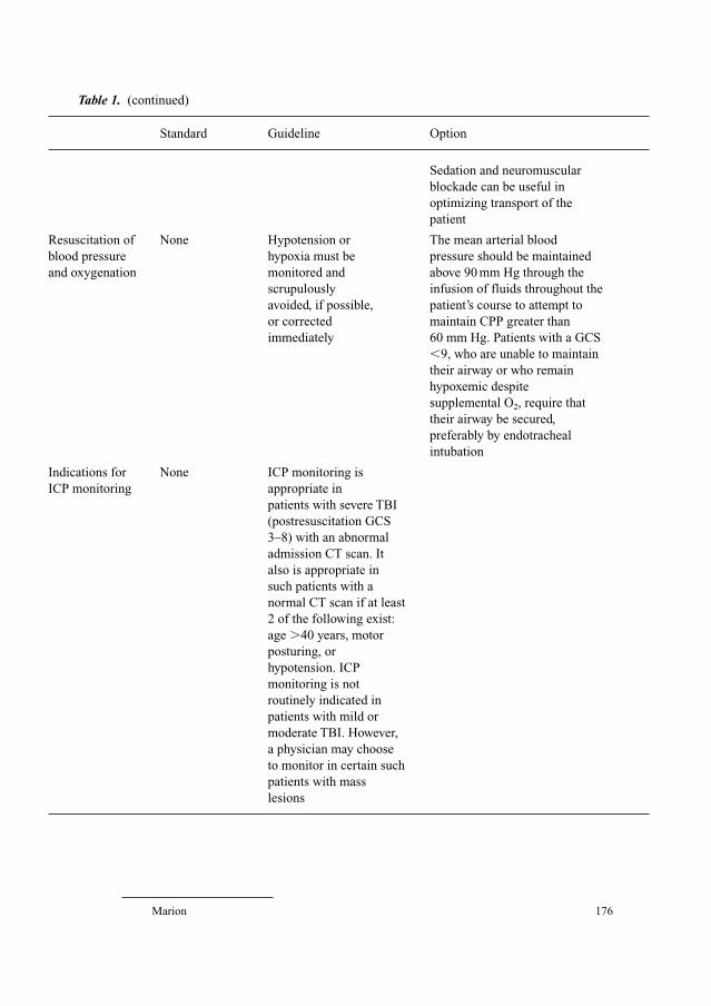
Marion 176
Sedation and neuromuscular
blockade can be useful in
optimizing transport of the
patient
Resuscitation of None Hypotension or The mean arterial blood
blood pressure hypoxia must be pressure should be maintained
and oxygenation monitored and above 90 mm Hg through the
scrupulously infusion of fluids throughout the
avoided, if possible, patient’s course to attempt to
or corrected maintain CPP greater than
immediately 60 mm Hg. Patients with a GCS
�9, who are unable to maintain
their airway or who remain
hypoxemic despite
supplemental O2, require that
their airway be secured,
preferably by endotracheal
intubation
Indications for None ICP monitoring is
ICP monitoring appropriate in
patients with severe TBI
(postresuscitation GCS
3–8) with an abnormal
admission CT scan. It
also is appropriate in
such patients with a
normal CT scan if at least
2 of the following exist:
age �40 years, motor
posturing, or
hypotension. ICP
monitoring is not
routinely indicated in
patients with mild or
moderate TBI. However,
a physician may choose
to monitor in certain such
patients with mass
lesions
Table 1. (continued)
Standard Guideline Option
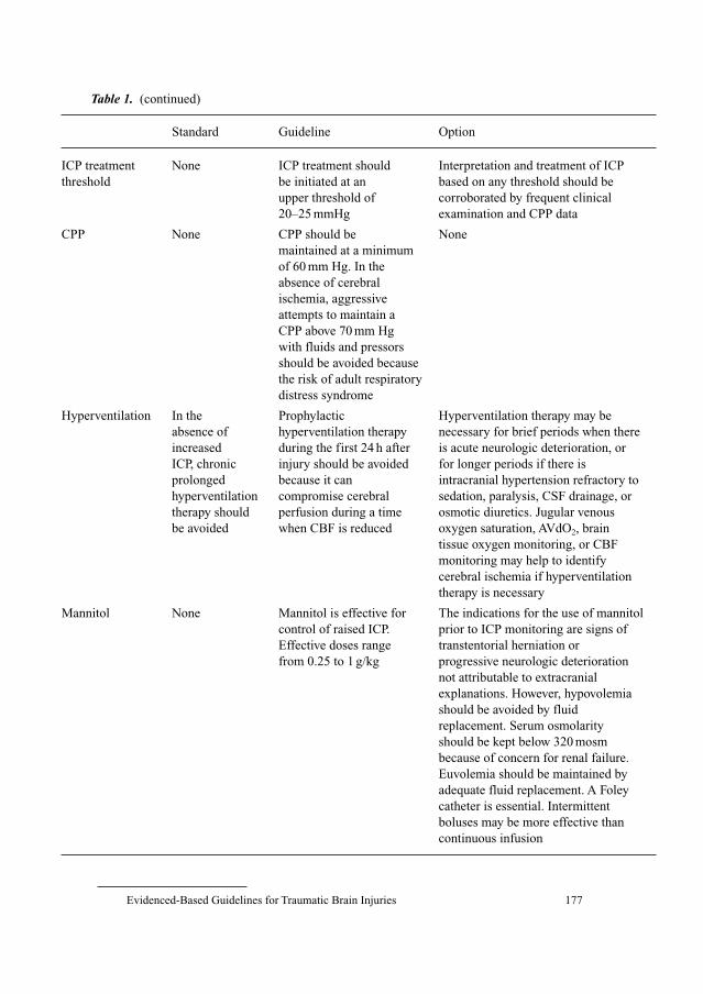
Evidenced-Based Guidelines for Traumatic Brain Injuries 177
ICP treatment None ICP treatment should Interpretation and treatment of ICP
threshold be initiated at an based on any threshold should be
upper threshold of corroborated by frequent clinical
20–25 mmHg examination and CPP data
CPP None CPP should be None
maintained at a minimum
of 60 mm Hg. In the
absence of cerebral
ischemia, aggressive
attempts to maintain a
CPP above 70 mm Hg
with fluids and pressors
should be avoided because
the risk of adult respiratory
distress syndrome
Hyperventilation In the Prophylactic Hyperventilation therapy may be
absence of hyperventilation therapy necessary for brief periods when there
increased during the first 24 h after is acute neurologic deterioration, or
ICP, chronic injury should be avoided for longer periods if there is
prolonged because it can intracranial hypertension refractory to
hyperventilation compromise cerebral sedation, paralysis, CSF drainage, or
therapy should perfusion during a time osmotic diuretics. Jugular venous
be avoided when CBF is reduced oxygen saturation, AVdO2, brain
tissue oxygen monitoring, or CBF
monitoring may help to identify
cerebral ischemia if hyperventilation
therapy is necessary
Mannitol None Mannitol is effective for The indications for the use of mannitol
control of raised ICP. prior to ICP monitoring are signs of
Effective doses range transtentorial herniation or
from 0.25 to 1 g/kg progressive neurologic deterioration
not attributable to extracranial
explanations. However, hypovolemia
should be avoided by fluid
replacement. Serum osmolarity
should be kept below 320 mosm
because of concern for renal failure.
Euvolemia should be maintained by
adequate fluid replacement. A Foley
catheter is essential. Intermittent
boluses may be more effective than
continuous infusion
Table 1. (continued)
Standard Guideline Option
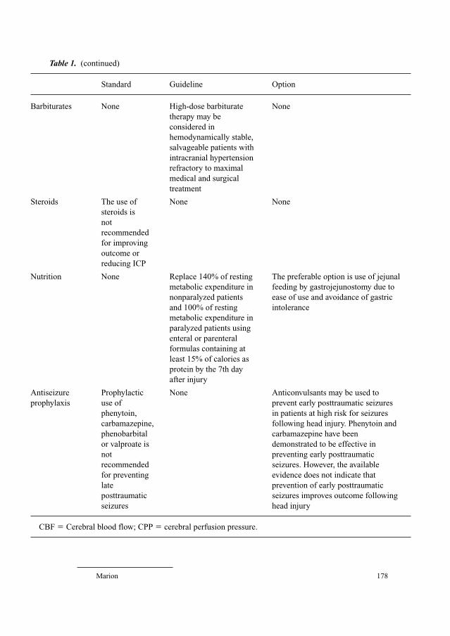
Marion 178
Barbiturates None High-dose barbiturate None
therapy may be
considered in
hemodynamically stable,
salvageable patients with
intracranial hypertension
refractory to maximal
medical and surgical
treatment
Steroids The use of None None
steroids is
not
recommended
for improving
outcome or
reducing ICP
Nutrition None Replace 140% of resting The preferable option is use of jejunal
metabolic expenditure in feeding by gastrojejunostomy due to
nonparalyzed patients ease of use and avoidance of gastric
and 100% of resting intolerance
metabolic expenditure in
paralyzed patients using
enteral or parenteral
formulas containing at
least 15% of calories as
protein by the 7th day
after injury
Antiseizure Prophylactic None Anticonvulsants may be used to
prophylaxis use of prevent early posttraumatic seizures
phenytoin, in patients at high risk for seizures
carbamazepine, following head injury. Phenytoin and
phenobarbital carbamazepine have been
or valproate is demonstrated to be effective in
not preventing early posttraumatic
recommended seizures. However, the available
for preventing evidence does not indicate that
late prevention of early posttraumatic
posttraumatic seizures improves outcome following
seizures head injury
CBF � Cerebral blood flow; CPP � cerebral perfusion pressure.
Table 1. (continued)
Standard Guideline Option
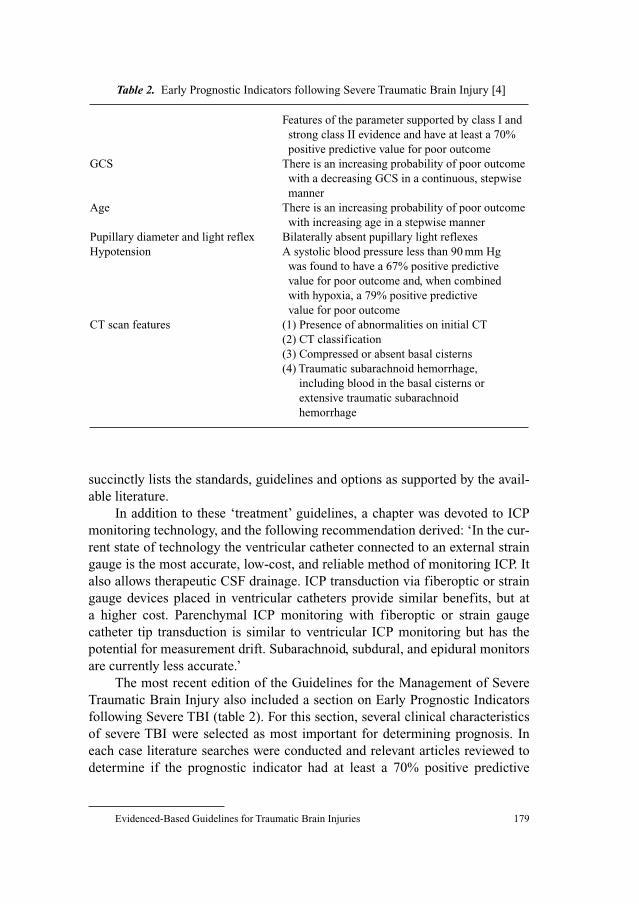
Evidenced-Based Guidelines for Traumatic Brain Injuries 179
succinctly lists the standards, guidelines and options as supported by the avail-
able literature.
In addition to these ‘treatment’ guidelines, a chapter was devoted to ICP
monitoring technology, and the following recommendation derived: ‘In the cur-
rent state of technology the ventricular catheter connected to an external strain
gauge is the most accurate, low-cost, and reliable method of monitoring ICP. It
also allows therapeutic CSF drainage. ICP transduction via fiberoptic or strain
gauge devices placed in ventricular catheters provide similar benefits, but at
a higher cost. Parenchymal ICP monitoring with fiberoptic or strain gauge
catheter tip transduction is similar to ventricular ICP monitoring but has the
potential for measurement drift. Subarachnoid, subdural, and epidural monitors
are currently less accurate.’
The most recent edition of the Guidelines for the Management of Severe
Traumatic Brain Injury also included a section on Early Prognostic Indicators
following Severe TBI (table 2). For this section, several clinical characteristics
of severe TBI were selected as most important for determining prognosis. In
each case literature searches were conducted and relevant articles reviewed to
determine if the prognostic indicator had at least a 70% positive predictive
Table 2. Early Prognostic Indicators following Severe Traumatic Brain Injury [4]
Features of the parameter supported by class I and
strong class II evidence and have at least a 70%
positive predictive value for poor outcome
GCS There is an increasing probability of poor outcome
with a decreasing GCS in a continuous, stepwise
manner
Age There is an increasing probability of poor outcome
with increasing age in a stepwise manner
Pupillary diameter and light reflex Bilaterally absent pupillary light reflexes
Hypotension A systolic blood pressure less than 90 mm Hg
was found to have a 67% positive predictive
value for poor outcome and, when combined
with hypoxia, a 79% positive predictive
value for poor outcome
CT scan features (1) Presence of abnormalities on initial CT
(2) CT classification
(3) Compressed or absent basal cisterns
(4) Traumatic subarachnoid hemorrhage,
including blood in the basal cisterns or
extensive traumatic subarachnoid
hemorrhage
Marion 180
value for determining poor outcome. If so, the details regarding use of the prog-
nostic indicator that were most closely associated with prognosis were defined.
In addition to information about the positive predictive values for each of
these characteristics, the prognosis guidelines included recommendations for
how the parameter should be measured, when it should be measured, and who
should measure it. The section on CT scan features includes subsections detail-
ing the prognostic value of specific CT findings, focusing on the status of the
basal cisterns, traumatic subarachnoid hemorrhage, midline shift, and intracra-
nial lesions.
Prehospital Management of Traumatic Brain Injury
The Guidelines for Prehospital Management of Traumatic Brain Injury
were published in 2002 in the Journal of Neurotrauma by the Brain Trauma
Foundation, with financial assistance provided by a grant from the National
Highway Traffic Safety Administration [5] (table 3). The authors of the docu-
ment were Edward J. Gabriel, Jamshid Ghajar, Andrew Jagoda, Peter T. Pons,
Thomas Scalea, and Beverly C. Walters. Their specialties included emergency
medical technologist, emergency medicine, trauma surgery, and neurosurgery.
The authors of these guidelines focused on recommendations for prehospital
assessment of the TBI patient and transport decisions, as well as treatment rec-
ommendations. However, the assessment category presented a challenge in
terms of formulating meaningful recommendations. For example, the relevant
question regarding clinical assessment measures is whether or not the measure
is reliable, while the relevant question for a treatment is whether or not it is effi-
cacious. A methodology had to be developed to weigh the significance of the
literature supporting clinical assessment measures. This methodology, which
is thoroughly described in the Guidelines document, focused on reliability.
Reliability means that different people with different backgrounds that make an
observation will see the same thing most of the time. To support the use of a
clinical assessment, good-quality clinical studies must allow determination of
the sensitivity, specificity, and the positive or negative predictive value of the
test. For the purposes of the methods used in this document, the authors decided
that the most important aspect of the clinical assessment test was its positive
predictive value, or the number of patients who had the clinical sign or prog-
nostic indicator and had a poor outcome. The authors arbitrarily decided that a
recommendation for use of a clinical assessment test had to be supported by
clinical studies that found a positive predictive value of 70% or greater.
These guidelines are divided into two sections: Assessment and Treatment
and Hospital Transport Decisions. Conclusions regarding assessment tests, and
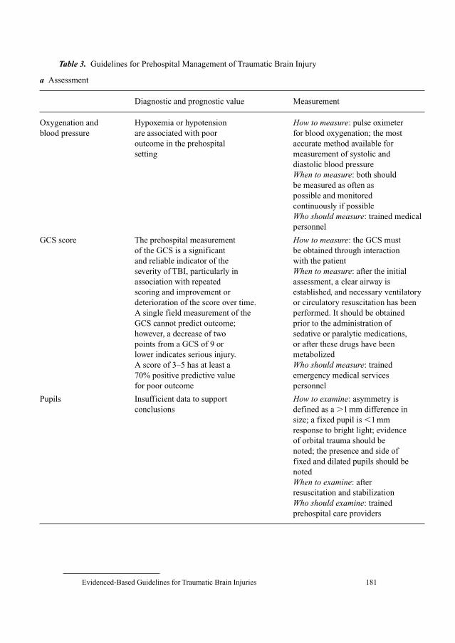
Evidenced-Based Guidelines for Traumatic Brain Injuries 181
Table 3. Guidelines for Prehospital Management of Traumatic Brain Injury
a Assessment
Diagnostic and prognostic value Measurement
Oxygenation and Hypoxemia or hypotension How to measure: pulse oximeter
blood pressure are associated with poor for blood oxygenation; the most
outcome in the prehospital accurate method available for
setting measurement of systolic and
diastolic blood pressure
When to measure: both should
be measured as often as
possible and monitored
continuously if possible
Who should measure: trained medical
personnel
GCS score The prehospital measurement How to measure: the GCS must
of the GCS is a significant be obtained through interaction
and reliable indicator of the with the patient
severity of TBI, particularly in When to measure: after the initial
association with repeated assessment, a clear airway is
scoring and improvement or established, and necessary ventilatory
deterioration of the score over time. or circulatory resuscitation has been
A single field measurement of the performed. It should be obtained
GCS cannot predict outcome; prior to the administration of
however, a decrease of two sedative or paralytic medications,
points from a GCS of 9 or or after these drugs have been
lower indicates serious injury. metabolized
A score of 3–5 has at least a Who should measure: trained
70% positive predictive value emergency medical services
for poor outcome personnel
Pupils Insufficient data to support How to examine: asymmetry is
conclusions defined as a �1 mm difference in
size; a fixed pupil is �1 mm
response to bright light; evidence
of orbital trauma should be
noted; the presence and side of
fixed and dilated pupils should be
noted
When to examine: after
resuscitation and stabilization
Who should examine: trained
prehospital care providers
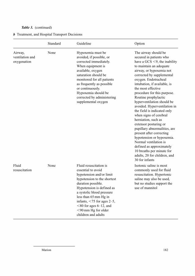
Marion 182
Table 3. (continued)
b Treatment, and Hospital Transport Decisions
Standard Guideline Option
Airway, None Hypoxemia must be The airway should be
ventilation and avoided, if possible, or secured in patients who
oxygenation corrected immediately. have a GCS �9, the inability
When equipment is to maintain an adequate
available, oxygen airway, or hypoxemia not
saturation should be corrected by supplemental
monitored for all patients oxygen. Endotracheal
as frequently as possible intubation, if available, is
or continuously. the most effective
Hypoxemia should be procedure for this purpose.
corrected by administering Routine prophylactic
supplemental oxygen hyperventilation should be
avoided. Hyperventilation in
the field is indicated only
when signs of cerebral
herniation, such as
extensor posturing or
pupillary abnormalities, are
present after correcting
hypotension or hypoxemia.
Normal ventilation is
defined as approximately
10 breaths per minute for
adults, 20 for children, and
30 for infants
Fluid None Fluid resuscitation is Isotonic saline is most
resuscitation essential to avoid commonly used for fluid
hypotension and/or limit resuscitation. Hypertonic
hypotension to the shortest saline may also be used,
duration possible. but no studies support the
Hypotension is defined as use of mannitol
a systolic blood pressure
less than 65 mm Hg in
infants, �75 for ages 2–5,
�80 for ages 6–12, and
�90 mm Hg for older
children and adults
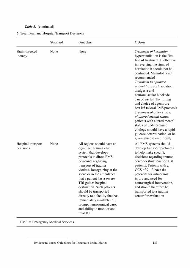
Evidenced-Based Guidelines for Traumatic Brain Injuries 183
Brain-targeted None None Treatment of herniation:
therapy hyperventilation is the first
line of treatment. If effective
in reversing the signs of
herniation it should not be
continued. Mannitol is not
recommended
Treatment to optimize patient transport: sedation,
analgesia and
neuromuscular blockade
can be useful. The timing
and choice of agents are
best left to local EMS protocols
Treatment of other causes of altered mental status:
patients with altered mental
status of undetermined
etiology should have a rapid
glucose determination, or be
given glucose empirically
Hospital transport None All regions should have an All EMS systems should
decisions organized trauma care develop transport protocols
system that develops to help make specific
protocols to direct EMS decisions regarding trauma
personnel regarding center destinations for TBI
transport of trauma patients. Patients with a
victims. Recognizing at the GCS of 9–13 have the
scene or in the ambulance potential for intracranial
that a patient has a severe injury and need for
TBI guides hospital neurosurgical intervention,
destination. Such patients and should therefore be
should be transported transported to a trauma
directly to a facility that has center for evaluation
immediately available CT,
prompt neurosurgical care,
and ability to monitor and
treat ICP
EMS � Emergency Medical Services.
Table 3. (continued)
b Treatment, and Hospital Transport Decisions
Standard Guideline Option
Marion 184
recommendations regarding treatment or transport, are then provided for sev-
eral specific topics considered most important in the prehospital setting. For the
assessment guidelines, the diagnostic and prognostic value of assessment mea-
sures are provided as well as recommendations on how to measure it, when to
measure it, and who should measure it. For the section on treatment and trans-
port decisions, the ‘standards, guidelines and options’ format is utilized.
Guidelines for the Management of Penetrating Brain Injury
In 2001 guidelines entitled Management and Prognosis of Penetrating Brain
Injury were published as a supplement to the Journal of Trauma [6] (table 4). The
authors were Bizhan Aarabi, Tord D. Alden, Randall M. Chesnut, J. Hunter
Downs 3rd, James M. Ecklund, Howard M. Eisenberg, Elana Farace, Robert E.
Florin, John A. Jane, Jr., Mark D. Krieger, Andrew I.R. Maas, Raj K. Narayan,
Alexander A. Potapov, Andres M. Salazar, Mark E. Shaffrey, and Beverly C.
Walters. The format for development of these guidelines was similar to that used
for the previous TBI guidelines documents. In addition to these treatment rec-
ommendations, the report also includes a section on prognostic indicators. The
authors examined the relationship between Glasgow Coma Scale (GCS), pupil
size and reactivity, age, hypotension and CT findings with outcome.
Pediatric Brain Injury
In 2003 the Guidelines for the Acute Management of Severe Traumatic
Brain Injury in Infants, Children, and Adolescents were published as a supple-
ment to Pediatric Critical Care Medicine [7] (table 5). Authors of this docu-
ment were P. David Adelson, Susan L. Bratton, Nancy A. Carney, Randall M.
Chesnut, Hugo E.M. du Coudray, Brahm Goldstein, Patrick M. Kochanek,
Helen C. Miller, Michael D. Partington, Nathan R. Selden, Craig R. Warden,
and David W. Wright. The methodology used for the development of these
guidelines were the same as those used to develop the Guidelines for the
Management of Severe Head Injury (Adults). Fifteen topics relevant to the
acute care of children with severe TBI were addressed. These guidelines refer to
children below the age of 18 years who have sustained a severe TBI and have a
postresuscitation GCS of 3–8. They do not refer to victims of asphyxiation,
drowning, or birth trauma. In the source document the authors also have
included a comparison of their findings with those of the adult guidelines
(Indications from Adult Guidelines) as a fourth entry in the Recommendations
section. The document is otherwise written in a format very similar to the adult
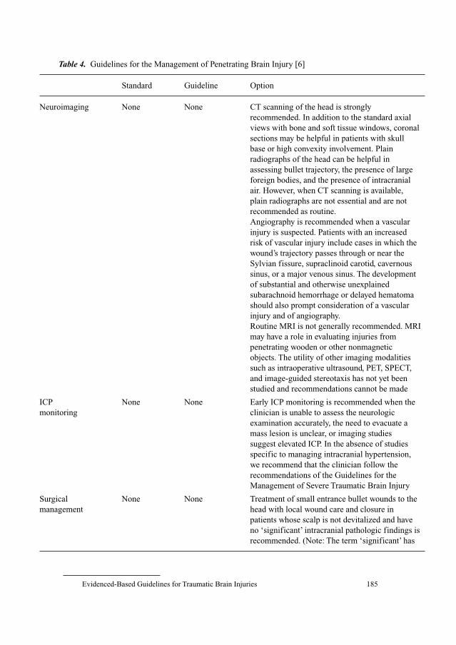
Evidenced-Based Guidelines for Traumatic Brain Injuries 185
Table 4. Guidelines for the Management of Penetrating Brain Injury [6]
Standard Guideline Option
Neuroimaging None None CT scanning of the head is strongly
recommended. In addition to the standard axial
views with bone and soft tissue windows, coronal
sections may be helpful in patients with skull
base or high convexity involvement. Plain
radiographs of the head can be helpful in
assessing bullet trajectory, the presence of large
foreign bodies, and the presence of intracranial
air. However, when CT scanning is available,
plain radiographs are not essential and are not
recommended as routine.
Angiography is recommended when a vascular
injury is suspected. Patients with an increased
risk of vascular injury include cases in which the
wound’s trajectory passes through or near the
Sylvian fissure, supraclinoid carotid, cavernous
sinus, or a major venous sinus. The development
of substantial and otherwise unexplained
subarachnoid hemorrhage or delayed hematoma
should also prompt consideration of a vascular
injury and of angiography.
Routine MRI is not generally recommended. MRI
may have a role in evaluating injuries from
penetrating wooden or other nonmagnetic
objects. The utility of other imaging modalities
such as intraoperative ultrasound, PET, SPECT,
and image-guided stereotaxis has not yet been
studied and recommendations cannot be made
ICP None None Early ICP monitoring is recommended when the
monitoring clinician is unable to assess the neurologic
examination accurately, the need to evacuate a
mass lesion is unclear, or imaging studies
suggest elevated ICP. In the absence of studies
specific to managing intracranial hypertension,
we recommend that the clinician follow the
recommendations of the Guidelines for the
Management of Severe Traumatic Brain Injury
Surgical None None Treatment of small entrance bullet wounds to the
management head with local wound care and closure in
patients whose scalp is not devitalized and have
no ‘significant’ intracranial pathologic findings is
recommended. (Note: The term ‘significant’ has
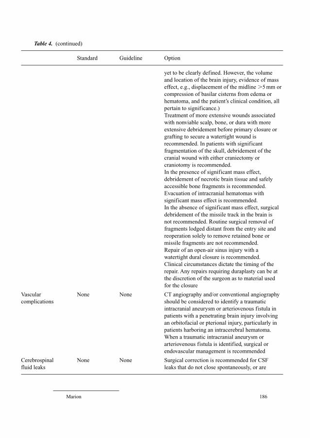
Marion 186
Table 4. (continued)
Standard Guideline Option
yet to be clearly defined. However, the volume
and location of the brain injury, evidence of mass
effect, e.g., displacement of the midline �5 mm or
compression of basilar cisterns from edema or
hematoma, and the patient’s clinical condition, all
pertain to significance.)
Treatment of more extensive wounds associated
with nonviable scalp, bone, or dura with more
extensive debridement before primary closure or
grafting to secure a watertight wound is
recommended. In patients with significant
fragmentation of the skull, debridement of the
cranial wound with either craniectomy or
craniotomy is recommended.
In the presence of significant mass effect,
debridement of necrotic brain tissue and safely
accessible bone fragments is recommended.
Evacuation of intracranial hematomas with
significant mass effect is recommended.
In the absence of significant mass effect, surgical
debridement of the missile track in the brain is
not recommended. Routine surgical removal of
fragments lodged distant from the entry site and
reoperation solely to remove retained bone or
missile fragments are not recommended.
Repair of an open-air sinus injury with a
watertight dural closure is recommended.
Clinical circumstances dictate the timing of the
repair. Any repairs requiring duraplasty can be at
the discretion of the surgeon as to material used
for the closure
Vascular None None CT angiography and/or conventional angiography
complications should be considered to identify a traumatic
intracranial aneurysm or arteriovenous fistula in
patients with a penetrating brain injury involving
an orbitofacial or pterional injury, particularly in
patients harboring an intracerebral hematoma.
When a traumatic intracranial aneurysm or
arteriovenous fistula is identified, surgical or
endovascular management is recommended
Cerebrospinal None None Surgical correction is recommended for CSF
fluid leaks leaks that do not close spontaneously, or are
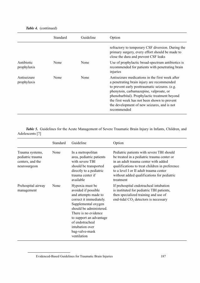
Evidenced-Based Guidelines for Traumatic Brain Injuries 187
refractory to temporary CSF diversion. During the
primary surgery, every effort should be made to
close the dura and prevent CSF leaks
Antibiotic None None Use of prophylactic broad-spectrum antibiotics is
prophylaxis recommended for patients with penetrating brain
injuries
Antiseizure None None Antiseizure medications in the first week after
prophylaxis a penetrating brain injury are recommended
to prevent early posttraumatic seizures. (e.g.
phenytoin, carbamazepine, valproate, or
phenobarbital). Prophylactic treatment beyond
the first week has not been shown to prevent
the development of new seizures, and is not
recommended
Table 4. (continued)
Standard Guideline Option
Table 5. Guidelines for the Acute Management of Severe Traumatic Brain Injury in Infants, Children, and
Adolescents [7]
Standard Guideline Option
Trauma systems, None In a metropolitan Pediatric patients with severe TBI should
pediatric trauma area, pediatric patients be treated in a pediatric trauma center or
centers, and the with severe TBI in an adult trauma center with added
neurosurgeon should be transported qualifications to treat children in preference
directly to a pediatric to a level I or II adult trauma center
trauma center if without added qualifications for pediatric
available treatment
Prehospital airway None Hypoxia must be If prehospital endotracheal intubation
management avoided if possible is instituted for pediatric TBI patients,
and attempts made to then specialized training and use of
correct it immediately. end-tidal CO2 detectors is necessary
Supplemental oxygen
should be administered.
There is no evidence
to support an advantage
of endotracheal
intubation over
bag-valve-mask
ventilation
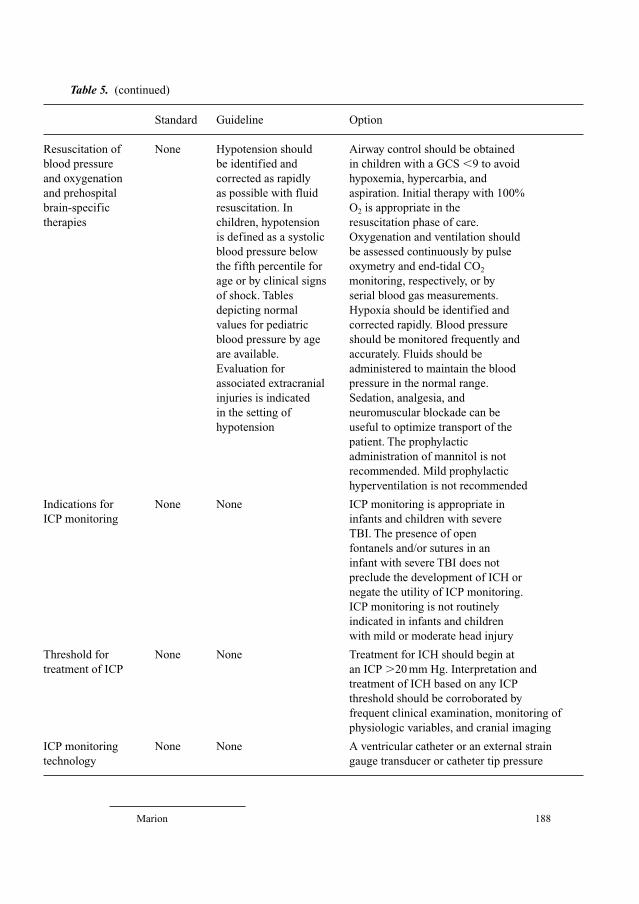
Marion 188
Table 5. (continued)
Standard Guideline Option
Resuscitation of None Hypotension should Airway control should be obtained
blood pressure be identified and in children with a GCS �9 to avoid
and oxygenation corrected as rapidly hypoxemia, hypercarbia, and
and prehospital as possible with fluid aspiration. Initial therapy with 100%
brain-specific resuscitation. In O2 is appropriate in the
therapies children, hypotension resuscitation phase of care.
is defined as a systolic Oxygenation and ventilation should
blood pressure below be assessed continuously by pulse
the fifth percentile for oxymetry and end-tidal CO2
age or by clinical signs monitoring, respectively, or by
of shock. Tables serial blood gas measurements.
depicting normal Hypoxia should be identified and
values for pediatric corrected rapidly. Blood pressure
blood pressure by age should be monitored frequently and
are available. accurately. Fluids should be
Evaluation for administered to maintain the blood
associated extracranial pressure in the normal range.
injuries is indicated Sedation, analgesia, and
in the setting of neuromuscular blockade can be
hypotension useful to optimize transport of the
patient. The prophylactic
administration of mannitol is not
recommended. Mild prophylactic
hyperventilation is not recommended
Indications for None None ICP monitoring is appropriate in
ICP monitoring infants and children with severe
TBI. The presence of open
fontanels and/or sutures in an
infant with severe TBI does not
preclude the development of ICH or
negate the utility of ICP monitoring.
ICP monitoring is not routinely
indicated in infants and children
with mild or moderate head injury
Threshold for None None Treatment for ICH should begin at
treatment of ICP an ICP �20 mm Hg. Interpretation and
treatment of ICH based on any ICP
threshold should be corroborated by
frequent clinical examination, monitoring of
physiologic variables, and cranial imaging
ICP monitoring None None A ventricular catheter or an external strain
technology gauge transducer or catheter tip pressure
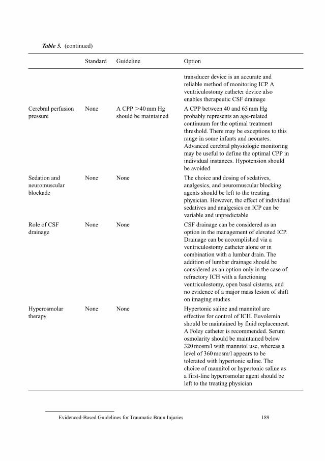
Evidenced-Based Guidelines for Traumatic Brain Injuries 189
transducer device is an accurate and
reliable method of monitoring ICP. A
ventriculostomy catheter device also
enables therapeutic CSF drainage
Cerebral perfusion None A CPP �40 mm Hg A CPP between 40 and 65 mm Hg
pressure should be maintained probably represents an age-related
continuum for the optimal treatment
threshold. There may be exceptions to this
range in some infants and neonates.
Advanced cerebral physiologic monitoring
may be useful to define the optimal CPP in
individual instances. Hypotension should
be avoided
Sedation and None None The choice and dosing of sedatives,
neuromuscular analgesics, and neuromuscular blocking
blockade agents should be left to the treating
physician. However, the effect of individual
sedatives and analgesics on ICP can be
variable and unpredictable
Role of CSF None None CSF drainage can be considered as an
drainage option in the management of elevated ICP.
Drainage can be accomplished via a
ventriculostomy catheter alone or in
combination with a lumbar drain. The
addition of lumbar drainage should be
considered as an option only in the case of
refractory ICH with a functioning
ventriculostomy, open basal cisterns, and
no evidence of a major mass lesion of shift
on imaging studies
Hyperosmolar None None Hypertonic saline and mannitol are
therapy effective for control of ICH. Euvolemia
should be maintained by fluid replacement.
A Foley catheter is recommended. Serum
osmolarity should be maintained below
320 mosm/l with mannitol use, whereas a
level of 360 mosm/l appears to be
tolerated with hypertonic saline. The
choice of mannitol or hypertonic saline as
a first-line hyperosmolar agent should be
left to the treating physician
Table 5. (continued)
Standard Guideline Option
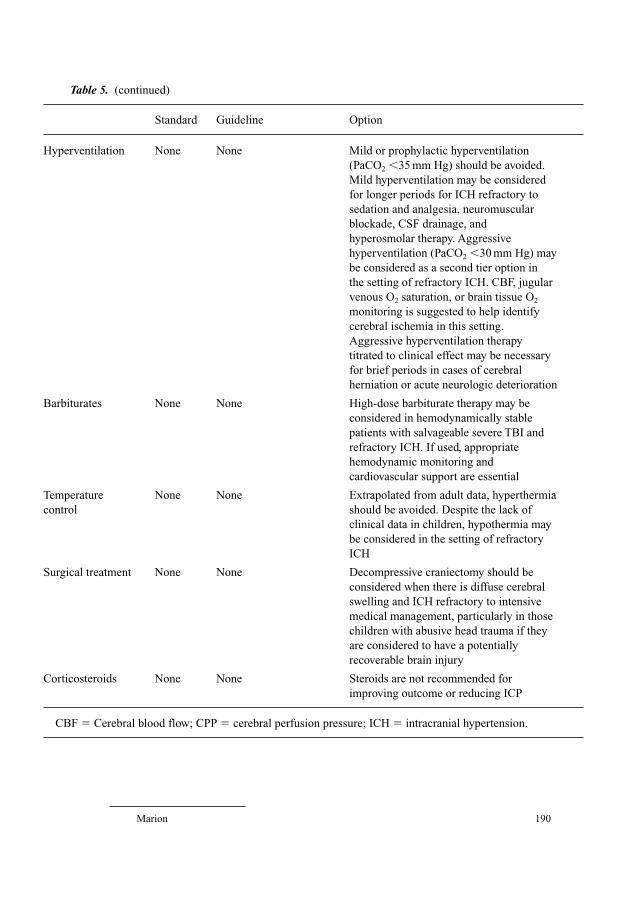
Marion 190
Hyperventilation None None Mild or prophylactic hyperventilation
(PaCO2 �35 mm Hg) should be avoided.
Mild hyperventilation may be considered
for longer periods for ICH refractory to
sedation and analgesia, neuromuscular
blockade, CSF drainage, and
hyperosmolar therapy. Aggressive
hyperventilation (PaCO2 �30 mm Hg) may
be considered as a second tier option in
the setting of refractory ICH. CBF, jugular
venous O2 saturation, or brain tissue O2
monitoring is suggested to help identify
cerebral ischemia in this setting.
Aggressive hyperventilation therapy
titrated to clinical effect may be necessary
for brief periods in cases of cerebral
herniation or acute neurologic deterioration
Barbiturates None None High-dose barbiturate therapy may be
considered in hemodynamically stable
patients with salvageable severe TBI and
refractory ICH. If used, appropriate
hemodynamic monitoring and
cardiovascular support are essential
Temperature None None Extrapolated from adult data, hyperthermia
control should be avoided. Despite the lack of
clinical data in children, hypothermia may
be considered in the setting of refractory
ICH
Surgical treatment None None Decompressive craniectomy should be
considered when there is diffuse cerebral
swelling and ICH refractory to intensive
medical management, particularly in those
children with abusive head trauma if they
are considered to have a potentially
recoverable brain injury
Corticosteroids None None Steroids are not recommended for
improving outcome or reducing ICP
CBF � Cerebral blood flow; CPP � cerebral perfusion pressure; ICH � intracranial hypertension.
Table 5. (continued)
Standard Guideline Option
Evidenced-Based Guidelines for Traumatic Brain Injuries 191
guidelines, and includes overview, process, scientific foundation, summary, and
key issues for future investigation sections. For each topic the search strategies
are clearly described, and evidentiary tables are included.
Guidelines for the Surgical Management of Severe Traumatic Brain Injury
This document has been submitted for publication. The authors are Ross
M. Bullock, Randall Chesnut, Jamshid Ghajar, David Gordon, Roger Hartl,
David W. Newell, Franco Servadei, Beverly C. Walters, and Jack Wilberger, and
production of the document was supported by the Brain Trauma Foundation
(table 6). For these guidelines the authors focused on three issues that were con-
sidered most relevant for deciding whether or not to operate: indications for
surgery, timing, and the type of operation (method).
Why Are There So Few Standards?
Four of the Guidelines documents summarized above contain recommen-
dations for specific treatments classified as standards, guidelines, or options,
based on the strength of the evidence supporting the recommendation. The four
documents list a total of 38 treatment recommendations, but in only 3 of those
recommendations was there evidence sufficient to support recommending the
treatment as a standard. All three of the standards are in the Guidelines for the
Management of Severe Traumatic Brain Injury, and recommend against the use
of treatments: prophylactic hyperventilation, steroids, and anticonvulsants. No
treatment standards could be recommended for any of the pediatric, prehospital,
or penetrating TBI guidelines. And evidence sufficient to support recommenda-
tions at the level of a guideline was available for only 16 of the 38 topics.
In defining which treatments for severe TBI were supported by class I evi-
dence, (standards) and which were not, a goal of the original Guidelines work-
group was to determine which treatments needed to be tested in prospective
randomized clinical trials. In this and the subsequent Guidelines documents, a
section entitled ‘Key Issues for Future Investigation’ was included at the end of
each chapter to provide details for what the authors thought was needed to
establish a standard for a particular diagnostic test or therapy. It was hoped that
such explicit direction would lead to numerous clinical trials that would provide
the class I evidence needed to elevate guidelines or options to standards. But
such research has not been forthcoming.
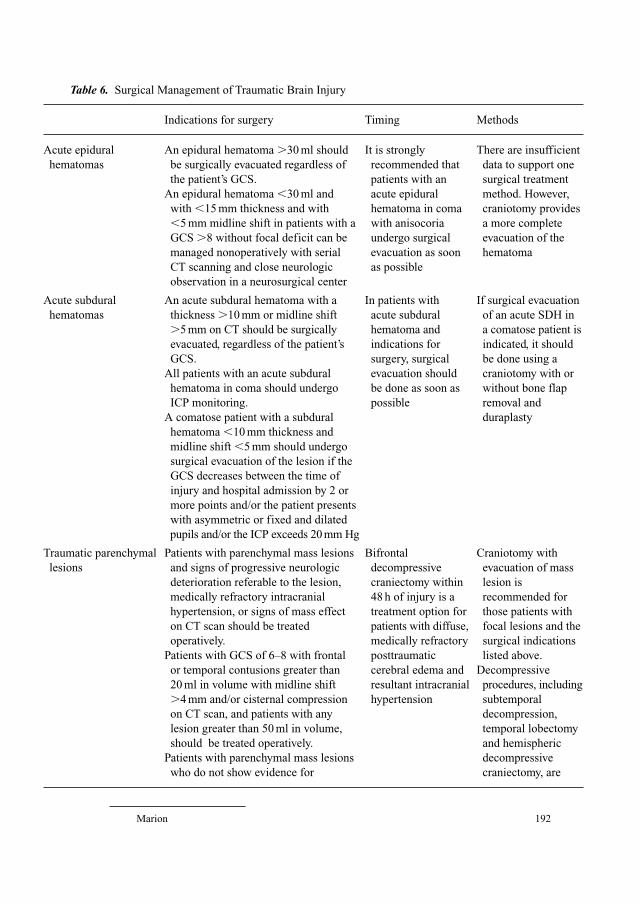
Marion 192
Table 6. Surgical Management of Traumatic Brain Injury
Indications for surgery Timing Methods
Acute epidural An epidural hematoma �30 ml should It is strongly There are insufficient
hematomas be surgically evacuated regardless of recommended that data to support one
the patient’s GCS. patients with an surgical treatment
An epidural hematoma �30 ml and acute epidural method. However,
with �15 mm thickness and with hematoma in coma craniotomy provides
�5 mm midline shift in patients with a with anisocoria a more complete
GCS �8 without focal deficit can be undergo surgical evacuation of the
managed nonoperatively with serial evacuation as soon hematoma
CT scanning and close neurologic as possible
observation in a neurosurgical center
Acute subdural An acute subdural hematoma with a In patients with If surgical evacuation
hematomas thickness �10 mm or midline shift acute subdural of an acute SDH in
�5 mm on CT should be surgically hematoma and a comatose patient is
evacuated, regardless of the patient’s indications for indicated, it should
GCS. surgery, surgical be done using a
All patients with an acute subdural evacuation should craniotomy with or
hematoma in coma should undergo be done as soon as without bone flap
ICP monitoring. possible removal and
A comatose patient with a subdural duraplasty
hematoma �10 mm thickness and
midline shift �5 mm should undergo
surgical evacuation of the lesion if the
GCS decreases between the time of
injury and hospital admission by 2 or
more points and/or the patient presents
with asymmetric or fixed and dilated
pupils and/or the ICP exceeds 20 mm Hg
Traumatic parenchymal Patients with parenchymal mass lesions Bifrontal Craniotomy with
lesions and signs of progressive neurologic decompressive evacuation of mass
deterioration referable to the lesion, craniectomy within lesion is
medically refractory intracranial 48 h of injury is a recommended for
hypertension, or signs of mass effect treatment option for those patients with
on CT scan should be treated patients with diffuse, focal lesions and the
operatively. medically refractory surgical indications
Patients with GCS of 6–8 with frontal posttraumatic listed above.
or temporal contusions greater than cerebral edema and Decompressive
20 ml in volume with midline shift resultant intracranial procedures, including
�4 mm and/or cisternal compression hypertension subtemporal
on CT scan, and patients with any decompression,
lesion greater than 50 ml in volume, temporal lobectomy
should be treated operatively. and hemispheric
Patients with parenchymal mass lesions decompressive
who do not show evidence for craniectomy, are

Evidenced-Based Guidelines for Traumatic Brain Injuries 193
Table 6. (continued)
Indications for surgery Timing Methods
neurologic compromise have controlled treatment options for
ICP, and no significant signs of mass patients with
effect on CT scan may be managed refractory intracranial
nonoperatively with intensive hypertension and
monitoring and serial imaging diffuse parenchymal
injury with clinical
and radiographic
evidence for
impending
transtentorial
herniation
Posterior fossa mass Patients with mass effect on CT scan In patients with Suboccipital
lesions or with neurologic dysfunction or indications for craniectomy is the
deterioration referable to the lesion surgical predominant
should undergo operative intervention. intervention, method reported for
Mass effect on CT scan is defined as evacuation should evacuation of
distortion, dislocation, or obliteration be performed as posterior fossa
of the fourth ventricle, compression soon as possible mass lesions, and
or loss of visualization of the basal since these is therefore
cisterns, or the presence of obstructive patients can recommended
hydrocephalus. deteriorate rapidly,
Patients with lesions and no significant thus worsening
mass effect on CT scan who have no their prognosis
signs of neurologic dysfunction may
be managed by close observation and
serial imaging
Depressed skull Patients with open (compound) skull Early operation is Elevation and
fractures fractures depressed greater than the recommended to debridement are
thickness of the skull should undergo reduce the recommended as
operative intervention to prevent incidence of the surgical method
infection. infection of choice.
Such patients may be treated Primary bone
nonoperatively if there is no clinical fragment
or radiographic evidence of dural replacement is a
penetration, significant intracranial surgical option in
hematoma, depression �1 cm, frontal the absence of
sinus involvement, gross cosmetic wound infection at
deformity, wound infection, the time of surgery.
pneumocephalus, or gross wound All management
contamination. strategies for open
Nonoperative management of closed depressed fractures
(simple) depressed skull fractures should include
is a treatment option antibiotics
Marion 194
There are several reasons why investigators have been reluctant to conduct
the research prescribed in the Guidelines documents. The most common is a
strong clinical bias: clinical experience and/or a preponderance of basic science
evidence supports the use of a particular treatment or diagnostic test. As a
result, many clinicians consider unethical a randomized clinical trial in which
half the patients would not receive the treatment or diagnostic test. The follow-
ing fall into this category:
• Blood pressure resuscitation (measurement of the effect of hypotension)
• ICP (measurement of the effect of intracranial hypertension)
• Mannitol
• Nutrition (measurement of the effect of withholding nutritional supple-
mentation)
• Physiologic monitoring such as blood pressure, ICP (measure the effect of
treatment without monitoring)
A second problem is the cost of doing clinical trials for TBI. Most trauma
centers admit less than 40 patients with severe TBI/year. Clinical TBI trials with
adequate power usually require 400 or more subjects, so either a large number of
contributing centers must be included, or the trial must extend for a prohibitive
period of time. The most commonly accepted outcome measure is the Glasgow
Outcome Scale score obtained at 6 months after injury. However, the accurate,
timely, and independent measurement of 6-month outcomes is very expensive.
Unless money is available for tracking study subjects and traveling when neces-
sary to test them, unacceptably high attrition rates undermine the conclusions of
the study. These and other issues have led many to believe that the clinical trials
needed to elevate options and guidelines to standards are unlikely to be done.
Evidence that Guidelines for the Management of Severe TraumaticBrain Injury Have Influenced Outcomes
Since publication of the original Guidelines document several studies have
examined changes in practice as a result of the Guidelines, and the impact of the
Guidelines on outcomes following severe TBI. A survey of North American neu-
rosurgeons published in 2000 queried these individuals about their use of specific
treatments that were discussed in the Guidelines document [8]. Compared to a
survey published in 1995 [1], the responding neurosurgeons indicated that there
was a 55% increase in the use of ICP monitoring, a 47% decrease in the use of
prophylactic hyperventilation therapy, and a 45% decrease in the use of steroids.
Recent studies also suggest that the Guidelines have led to improved out-
comes [9–13]. The most important of these studies was published by Fakhry
et al. [9] in the March 2004 issue of the Journal of Trauma. It describes the
Evidenced-Based Guidelines for Traumatic Brain Injuries 195
outcomes for 830 patients with severe TBI who were cared for either before,
during, or after institution of evidence-based guidelines at the Inova Fairfax
Hospital in Falls Church, Va., USA. During 1991–1994 (before guidelines)
219 patients with severe TBI were admitted. Guidelines were introduced in
1995–1996 but there was poor compliance, and during that time 188 patients
with severe TBI were admitted. From 1997 to 2000 compliance with the guide-
lines was high, and during that time 423 patients with severe TBI were admit-
ted. The admission GCS score for the ‘before guidelines group’ was slightly
higher than for the other two groups, but in every other way the three groups
were similar. Compared to the before guidelines group, the ‘after guidelines
group’ had a shorter ICU stay by 1.8 days, and shorter length of stay in the acute
care hospital by 5.4 days, in both cases statistically significant differences. The
mortality rate for the after guidelines group was 4% lower that for the before
guidelines group. The good recovery rate (mild, moderate or no disability at dis-
charge) for the after guidelines group was 61.5% compared to 43.3% for the
before guidelines group, also a statistically significant difference. To control for
general improvements in critical care over this 10-year span, the authors studied
similar outcomes in a group of 1,060 trauma patients that did not have TBI who
were treated at their hospital from 1991 to 2000. Demographics and injury
severity scores were similar except for the absence of TBI. During this period of
time the ICU length of stay actually increased by 3.5 days, and the length of
acute care hospitalization and mortality rates remained the same for the trauma
patients without head injuries. The authors therefore concluded that improved
outcomes for patients with severe TBI most likely were the result of the imple-
mentation and use of evidence-based guidelines. Similar conclusions were
derived from a study of 93 TBI patients published by Palmer et al. [10]. They
compared outcomes for 37 patients treated before implementation of the
Guidelines with the outcomes for 56 patients treated after they began using the
Guidelines. They found that implementation of the Guidelines led to a 9.13
times higher odds ratio of a good outcome.
It might be expected that the impact of a particular set of guidelines on
practice would be diminished in inverse proportion to the weight of evidence
supporting the recommendations. However, it appears that the documents have
provided a basis on which to develop consistent treatment protocols at individ-
ual hospitals [14–16]. This trend alone has been an important step forward and
helps to improve the care provided to TBI patients.
References
1 Ghajar J, Hariri RJ, Narayan RK, et al: Survey of critical care management of comatose, head-
injured patients in the United States. Crit Care Med 1995;23:560–567.

Marion 196
2 US Department of Health and Human Services. Interagency Head Injury Task Force Report.
Washington, US Department of Health and Human Services, 1989.
3 Bullock R, Chesnut RM, Clifton G, Ghajar J, Marion DW, Narayan RK, Newell DW, Pitts LH,
Rosner MJ, Walters BC, Wilberger JE: Guidelines for the management of severe head injury. J
Neurotrauma 1996;13:638–817.
4 Brain Trauma Foundation: American Association of Neurological Surgeons and Joint Section on
Neurotrauma and Critical Care: Guidelines for the management of severe traumatic brain injury,
and early prognostic indicators of prognosis in severe traumatic brain injury. J Neurotrauma
2000;17:451–627.
5 Gabriel EJ, Ghajar J, Jagoda A, Pons PT, Scalea T, Walters BC; Brain Trauma Foundation: Guidelines
for prehospital management of traumatic brain injury. J Neurotrauma 2002;19:111–174.
6 Pruitt BA Jr: Management and prognosis of penetrating brain injury. J Trauma 2001;51(suppl 2):
S1–S86.
7 Adelson PD, Bratton SL, Carney NA, Chesnut RM, du Coudray HE, Goldstein B, Kochanek PM,
Miller HC, Partington MP, Selden NR, Warden CR, Wright DW; American Association for
Surgery of Trauma; Child Neurology Society; International Society for Pediatric Neurosurgery;
International Trauma Anesthesia and Critical Care Society; Society of Critical Care Medicine;
World Federation of Pediatric Intensive and Critical Care Societies: Guidelines for the acute med-
ical management of severe traumatic brain injury in infants, children, and adolescents. Pediatr Crit
Care Med 2003;4(3 suppl):S1–S71.
8 Marion DW, Spiegel TP: Changes in the management of severe traumatic brain injury: 1991–1997.
Crit Care Med 2000;28:16–18.
9 Fakhry SM, Trask AL, Waller MA, Watts DD; IRTC Neurotrauma Task Force: Management of
brain-injured patients by an evidence-based medicine protocol improves outcomes and decreases
hospital charges. J Trauma 2004;56:492–499.
10 Palmer S, Bader MK, Qureshi A, Palmer J, Shaver T, Borzatta M, Stalcup C: The impact on out-
comes in a community hospital setting of using the AANS traumatic brain injury guidelines.
Americans Associations for Neurologic Surgeons. J Trauma 2001;50:657–664
11 Naredi S, Koskinen LO, Grande PO, Nordstrom CH, Nellgard B, Rydenhag B, Vegfors M:
Treatment of traumatic head injury – U.S./European guidelines or the Lund concept. Crit Care
Med 2003;31:2713–2714.
12 Citerio G, Stocchetti N, Cormio M, Beretta L, Galli D, Pesenti A: Application of guidelines for
severe head trauma: data from an Italian database. Eur J Emerg Med 2003;10:68–72.
13 Bulger EM, Nathens AB, Rivara FP, Moore M, MacKenzie EJ, Jurkovich GJ; Brain Trauma
Foundation: Management of severe head injury: institutional variations in care and effect on out-
come. Crit Care Med 2002;30:1870–1876.
14 Mcilvoy L, Spain DA, Raque G, Vitaz T, Boaz P, Meyer K: Successful incorporation of the Severe
Head Injury Guidelines into a phased-outcome clinical pathway. J Neurosci Nurs 2001;33:72–78, 82.
15 Sesperez J, Wilson S, Jalaludin B, Seger M, Sugrue M: Trauma case management and clinical
pathways: prospective evaluation of their effect on selected patient outcomes in five key trauma
conditions. J Trauma 2001;50:643–649.
16 Littlejohns LR, Bader MK: Guidelines for the management of severe head injury: clinical appli-
cation and changes in practice. Crit Care Nurse 2001;21:48–65.
Donald W. Marion, MD
35 High Rock Road
Wayland, MA 01778 (USA)
Tel. �1 508 358 4551, Fax �1 508 358 5510, E-Mail [email protected]
Pollock BE (ed): Guiding Neurosurgery by Evidence.
Prog Neurol Surg. Basel, Karger, 2006, vol 19, pp 197–207
Treatment of Chronic Pain withNeurostimulation
John K. Birknes, Ashwini Sharan, Ali R. Rezai
Departments of Neurosurgery and Neurology, Thomas Jefferson University,
Philadelphia, Pa., USA
AbstractChronic pain conditions are a complex and multifactorial problem generally requiring a
multidisciplinary-type approach. The central nervous system at some point clearly becomes
involved in the processing of these painful conditions with an integration of complex
changes in neurophysiology and behavior. Many ablative techniques have been employed in
the past to interrupt these signals. However, the results were often temporary and symptoms
tended to recur. The more modern approach has suggested that modulation of the nervous
elements may be a more resilient approach for treating such chronic pain disorders. We are
realizing that many of these pain conditions are also dynamic and evolving, and as such need
a similar treatment modality. Neurostimulation, thus, provides the ability of therapeutically
dosing electrical current in a variety of pulse forms, amplitudes, pulse widths, and frequen-
cies, to affect that system. Furthermore, it is not destructive, it is reversible, and it can be
remotely adjusted and programmed over time; clear advantages to previous surgical thera-
pies. This chapter reports on the current evidence for the use of neurostimulation (i.e. spinal
cord stimulation, motor cortex stimulation and deep brain stimulation) in the treatment of
chronic pain conditions.
Copyright © 2006 S. Karger AG, Basel
The therapeutic use of electrical stimulation for pain relief is an ancient
art. The Egyptians and the Greeks used electric eels to apply shock therapy and
the Romans applied the torpedo fish to treat maladies such as cephalgia and
arthralgia [1–3].
Subsequently, the modern learning elucidated the anatomy of the pain
tracts and it became evident that electrical stimulation of the nervous system
could be predictably used for therapeutic benefits. In the mid 1900s, neurosur-
geons were routinely applying electrical stimulation to the brain to treat and to
Birknes/Sharan/Rezai 198
study movement, psychiatric and pain disorders. In fact in 1954, Heath [4] and
Pool [5] both reported on implantation of temporary electrodes in the septum
pellucidum to treat patients with schizophrenia and pain from metastatic
carcinoma. Many other targets have since been enthusiastically explored for
the treatment of patients with chronic pain including the thalamus, caudate,
cingulate, or periaqueductal grey. With the renewed interest in chronic deep
brain stimulation (DBS), it is hopeful that physicians may again study and
offer patients an opportunity to interrupt the pain circuits in the deep cerebral
targets.
The further use of central nervous system stimulation developed with the
introduction of the gate theory for pain control by Melzack and Wall [6]. They
noted that stimulation of large myelinated fibers of peripheral nerves resulting
in paresthesias blocked the activity in small nociceptive projections. Shealy [7]
and Shealy and Cady [8] applied this knowledge in 1967 by inserting the first
dorsal column stimulator in a human suffering from terminal metastatic cancer.
The therapeutic use of electrical stimulation developed further when Shealy, in
collaboration with Long, prompted Hagfers and Maurer to independently pro-
duce the first two solid state transcutaneous electrical nerve stimulators [7, 8].
(Actually, transcutaneous electrical nerve stimulators were originally developed
as a screening tool for spinal cord stimulators.) Subsequently, electrodes have
been implanted via a laminectomy in the subarachnoid space, between the two
layers of the dura or in the epidural space, both dorsal or ventral to the spinal
cord and later the percutaneous technique was introduced [9–14]. More
recently motor cortex stimulation (MCS) as well as DBS have been used in the
treatment of various pain conditions.
Chronic pain conditions are becoming an increasing problem with growing
costs generally requiring a multidisciplinary-type approach. The central ner-
vous system at some point clearly becomes involved in the processing of these
painful conditions with an integration of complex changes in neurophysiology
and behavior. Many ablative techniques have been employed in the past to inter-
rupt these signals. However, the results were often temporary and they tended to
recur.
The more modern approach has suggested that modulation of the nervous
elements may be a more resilient approach for treating such chronic pain disor-
ders. We are realizing that many of these pain conditions are also dynamic and
evolving, and as such need a similar treatment modality. Neurostimulation,
thus, provides the ability of therapeutically dosing electrical current in a variety
of pulse forms, amplitudes, pulse widths, and frequencies, to affect that system.
Furthermore, it is not destructive, it is reversible, and it can be remotely
adjusted and programmed over time. These are clear advantages to previous
surgical therapies.
Treatment of Chronic Pain with Neurostimulation 199
Spinal Cord Stimulation
Spinal cord stimulation (SCS) has been utilized for a variety of pain condi-
tions. The most common indications include failed back surgery syndrome
(FBSS), complex regional pain syndrome (CRPS), ischemic limb pain, and
angina. SCS is particularly indicated with any type of ‘neuropathic’ pain.
Indications have been extended to cover the treatment of intractable pain due to
other causes including cervical neuritis pain, spinal cord injury pain, posther-
petic neuralgia, neurogenic thoracic outlet syndrome, and temporomandibular
joint syndrome refractory to multiple surgical interventions.
Although a large body of work has been published, the exact mechanisms
of action of SCS remain unclear. The computer modeling work of Coburn and
colleagues [15–17] and, more recently, of Holsheimer and colleagues [18–20]
has shed some light, at least theoretically, on the distribution of the electrical
fields within the spinal structures. It is clear that stimulation on the dorsal
aspect of the epidural space creates complex electrical fields which affect a
large number of structures. We do not know whether activating afferents within
the peripheral nerve, dorsal columns, or supralemniscal pathways share equiva-
lent mechanisms of action. Additionally, there may be antidromic action poten-
tials passing caudally in the dorsal columns to activate spinal segmental
mechanisms in the dorsal horns as well as action potentials ascending in the
dorsal columns activating cells in the brainstem, which in turn might activate
descending inhibition. At the chemical level, animal studies suggest that the
SCS triggers the release of serotonin, substance P, and GABA within the dorsal
horn [21–23].
Failed Back Surgery SyndromeTwo prospective controlled studies were found. Marchand et al. [24] exam-
ined patients who had undergone at least one prior surgery for chronic back
pain secondary to trauma. Each of these patients was currently using an SCS
and they acted as their own controls. Eight patients participated in 4 testing ses-
sions each (2 sets of 2). In the first set of 2 sessions noxious stimulation and
analgesia were tested; during one session the stimulator was on the patient’s
normal settings while during the other session it was nonfunctional. The second
set of two sessions evaluated discrimination of visual and noxious heat stimuli
with the stimulator on for one session and off for the other. Pain scores were
significantly reduced with SCS compared to placebo stimulation.
North et al. [25] conducted a prospective study randomizing patients with
FBSS. One group underwent repeat back surgery while the other underwent
SCS surgery. All patients presented with standard clinical and radiographic cri-
teria for surgical intervention. After 6 months, patients were permitted to cross
Birknes/Sharan/Rezai 200
over to the alternative therapy if they were dissatisfied with their results. Ten of
15 patients crossed over from back surgery to SCS (67%), while only 2 of 12
patients crossed over from SCS to back surgery (17%; p � 0.018).
Eight other prospective studies reporting on over 300 patients exist on SCS
for back and leg pain [26–33]. When including all studies (retrospective,
prospective with and without controls) successful treatment of FBSS with SCS
was observed in 62% of patients (n � 747). Successful treatment was defined
as greater than 50% reduction in pain or significant reduction in visual ana-
logue scale (VAS) score [34].
CRPS I and IICRPS I is also known as reflex sympathetic dystrophy. One prospective
controlled study and three prospective studies without matched controls were
found in the literature. Kemler et al. [35] examined 54 patients with CRPS I
(reflex sympathetic dystrophy) and randomized them, using a 2:1 ratio, to
receive either SCS with physical therapy (PT; n � 36) or PT alone (n � 18).
Twenty-four of the 36 (67%) patients in the SCS group experienced significant
relief with percutaneous stimulation and thus received a permanent implant.
The remaining 12 patients did not undergo surgery but their data was included
in the SCS group in the final analysis.
Pain and quality of life measurements were obtained at 1, 3 and 6 months.
At 6 months a significant improvement was demonstrated in the SCS group
with the VAS reduced by 2.4 cm compared with a 0.2-cm increase in the PT
group (p � 0.0001). No functional improvement was observed in either group.
With long-term follow-up at 2 years, this significance was unchanged with a
VAS reduction in SCS group of 2.1 cm and a 0-cm change in the PT group.
Three prospective studies without matched controls were discovered (total
of 50 subjects) [36–38]. Two of the studies reported success rates with an 84%
overall success rate. The third study by Calvillo et al. [38] reported a significant
improvement in pain scores (VAS) and a �50% reduction in narcotic use by
44% of subjects. In eight retrospective studies the overall success rate was 84%
(192 patients) [34].
Ischemic Limb PainTwo controlled prospective randomized studies exist. Klomp et al. [39]
randomized 120 patients, with critical painful limb ischemia, to receive either
best medical therapy alone or SCS in conjunction with best medical therapy. At
a mean follow-up of 19 months, there was no significant difference in pain
score improvement between the two groups. The SCS group did, however,
report a significant reduction in pain medication in the short term. Jivegard
et al. [40] authored a similar study where 51 patients were randomized to
Treatment of Chronic Pain with Neurostimulation 201
receive either oral medication alone or SCS with oral medication. Conversely,
they reported a significant improvement in pain scores of the SCS-treated
group over the non-SCS group (p � 0.01).
Four prospective studies without matched controls [41–44] in the literature
reveal an overall success rate of 78% (n � 271). Analysis of seven retrospective
studies found an overall success rate of 76% (n � 308) [34].
Angina PainThree prospective controlled studies in the literature address SCS for
angina. Hautvast et al. [45] implanted an SCS in patients with stable angina
pectoris and randomized them. One group’s SCS was inactivated while the
other group was instructed to use the stimulator 3 times per day for 1 h and with
any angina attack. At 6 weeks, a significant reduction in both the number of
angina attacks and nitrate consumption was observed in the functioning SCS
group. Additionally these patients exhibited an increased exercise duration.
Mannheimer et al. [46] randomized 104 patients accepted for coronary
artery bypass graft (CABG) to receive either CABG (n � 51) or SCS (n � 53).
Both groups experienced a significant reduction in both the number of angina
attacks and the consumption of nitrates. There was no significant intergroup
difference regarding these parameters. Interestingly the CABG group was
found to have a significantly higher mortality rate.
De Jongste et al. [47] randomized 17 patients with angina to an active
treatment group (i.e. SCS implantation) and a control group. The control group
was followed for 2 months, at the end of which period, they too received SCS
implantation. Both groups were followed for a total of 12 months. This study
also revealed a significant reduction in the incidence of angina attacks and in
the consumption of nitrates (p � 0.05).
Five additional studies are reported to be prospective but without matched
controls [48–52]. Each of these revealed significant benefit from spinal cord
stimulation. The benefit indices ranged from reduction in angina attacks,
decrease nitrate consumption, decrease in NYHA grade and improvement in
NHP grade.
MCS/Precentral Stimulation
In 1991, Tsubokawa et al. [53] first reported on their experience using
epidural MCS to treat 12 patients with deafferentation pain. A chronic stimulating
electrode was placed epidurally such that stimulation of the underlying cortex pro-
duced motor contractions in the painful region. Experimental work by Tsubokawa
et al. [54–56] demonstrated that thalamic hyperactivity could be reduced by
Birknes/Sharan/Rezai 202
chronic sensorimotor cortex stimulation and this established the foundation for the
clinical treatment. Since then, many other groups have independently reported on
the treatment of chronic pain conditions via cortical targets [57–71].
The central idea underlying the therapeutic effect of MCS is activation of
nonnociceptive sensory neurons which are believed to exert an inhibitory effect
on their nociceptive counterparts. This type of interaction may be present at
multiple levels of the somatosensory pathway along the peripheral and central
nervous systems. Further, induction of motor contractions in the area of the
pain often may result in pain relief.
As is true with most therapies available for these different chronic pain
conditions, the results vary tremendously and depend largely on the differing
definitions of success. MCS has been most consistently used for trigeminal
neuropathic pain and poststroke or central pain. Nguyen et al. [72–74] reported
on 22 patients with trigeminal pain where 13 obtained marked improvement
and 5 obtained satisfactory improvement. Only 4 were not improved. Ebel et al.
[75] reported sustained good to excellent relief in 3 of 7 patients over time with
trigeminal pain. Meyerson et al. [64] have reported between 60 and 90% pain
relief on 5 patients with trigeminal neuropathic pain. Tsubokawa et al. [53] ini-
tially reported on the treatment of central pain with MCS with 8 of 12 patients
having continued effect after 1 year of therapy. Many others have reported on
their experience with similar patients. Katayama et al. [63] noted satisfactory
results in 2 of 3 patients with brainstem infarcts. Mertens et al. [76] also noted
60% excellent or good relief in poststroke pain. An analysis of the literature by
Nguyen et al. [72] has revealed 52% success (82 of 159 patients) for central
pain. Expert opinion suggests a role of MCS for neuropathic trigeminal pain
and poststroke pain [77].
Presently, there are no prospective trials using MCS and there are only 23
major cases series reported in the literature [77]. The largest case series included
32 patients and the majority of reports include less than 10 subjects. In general, the
body of literature regarding this technique is plagued by a lack of controlled and
blinded studies, a lack of uniformly classified diseases, and reporting on a small
series of patients. However, the world experience with this therapy still remains
young but standardization of the technique will likely bear out its merits in time.
Deep Brain Stimulation
As previously mentioned, Heath [4] and Pool [5] in 1954 both reported on
the implantation of temporary electrodes in the septum pellucidum to treat
patients with schizophrenia and pain from metastatic carcinoma. The technique
of DBS was then described in the ventroposterolateral thalamic nucleus by
Treatment of Chronic Pain with Neurostimulation 203
Mazars et al. [78] in 1960. Other targets were subsequently explored. Periaqueduc-
tal gray and periventricular gray matter (PAG/PVG) stimulation was described
by multiple groups [79–81].
The exact mechanism of pain modulation by DBS is unknown. PAG/PVG
stimulation is, however, felt to result in stimulation of an opioid-dependent
pathway. Thalamic stimulation may modulate the abnormal firing patterns in
the thalamic neurons secondary to deafferentation.
Currently, the two most common targets for DBS for pain include the
periventricular grey matter or the ventrocaudalis thalamus. As a generalization,
patients with neuropathic pain should undergo paresthesia-producing stimula-
tion with implantation in ventrocaudal (Vc) thalamus while those patients with
nociceptive pain should undergo PAG/PVG stimulation. Many patients will
inevitably have mixed components of nociceptive and neuropathic pain and
thus both Vc and PAG/PVG stimulation trials may be indicated. The internal
capsule and medial lemniscus have also been successfully used.
Since the therapy of DBS for pain has been practiced for decades, many case
series have been published [81–89]. It seems that patients with nociceptive pain as
in cancer pain and FBSS respond best to DBS. Published series of PAG/PVG
stimulation have reported treatment of cancer pain and FBSS with success rates
ranging from 25 to 100% and 30 to 80%, respectively. Additionally, brachial
plexus injuries, peripheral neuropathies, and phantom limb pain appear to
respond to Vc DBS. Although the use of DBS for chronic pain precedes SCS and
MCS, no prospective study has evaluated its use.
Conclusion
The treatment of chronic pain remains difficult. Neurostimulation for
chronic pain is definitely a treatment option. There is currently insufficient
class 1 evidence available to assess the full benefits of SCS, MCS, and DBS.
All three therapies have reported around 50% relief of pain. Future studies will
have to be designed with randomization of patients, placebo or sham stimula-
tion, and uniformity in terms of classification of pain types before full under-
standing and defining of the benefits of each modality.
References
1 Stillings D: A survey of the history of electrical stimulation for pain to 1900. Med Instrum
1975;9:255–259.
2 Kellaway P: The part played by electric fish in the early history of bioelectricity and electrother-
apy. Bull Hist Med 1946;20:112–137.

Birknes/Sharan/Rezai 204
3 Schiller F: The history of algology, algotherapy, and the role of inhibition. Hist Philos Life Sci
1990;12:27–50.
4 Heath R: Studies in Schizophrenia: a Multidisciplinary Approach to Mind-Brain Relationships.
Cambridge, Harvard University Press, 1954.
5 Pool J: Psychosurgery in older people. J Am Geriatr Soc 1954;2:456–465.
6 Melzack R, Wall P: Pain mechanisms: a new theory. Science 1965;150:971–979.
7 Shealy CN: Dorsal column stimulation: optimization of application. Surg Neurol 1975;4:142–145.
8 Shealy C, Cady R: Historical perspective of pain management; in Weiner RS (ed): Pain Management:
a Practical Guide for Clinicians. Boca Raton, St Lucie Press, 1998, pp 7–15.
9 Hoppenstein R: Electrical stimulation of the ventral and dorsal columns of the spinal cord for
relief of chronic intractable pain. Surg Neurol 1975;4:187–194.
10 Larson S, Sances A, Cusick J, Meyer G, Swiontek T: A comparison between anterior and posterior
implant systems. Surg Neurol 1975;4:180–186.
11 Larson SJ, Sances A Jr, Riegel DH, Meyer GA, Dallmann DE, Swiontek T: Neurophysiological
effects of dorsal column stimulation in man and monkey. J Neurosurg 1974;41:217–223.
12 Lazorthes Y, Verdie J, Arbus L: Stimulation analgésique médullaire antérieure et postérieure par
technique d’implantation percutanée. Acta Neurochir 1978;40:253–276.
13 Dooley DM: Spinal cord stimulation. AORN J 1976;23:1209–1212.
14 Dooley D: Percutaneous electrical stimulation of the spinal cord. Assoc Neurol Surg, Bal Harbour,
1975.
15 Coburn B: Electrical stimulation of the spinal cord: two-dimensional finite element analysis with
particular reference to epidural electrodes. Med Biol Eng Comput 1980;18:573–584.
16 Coburn B: A theoretical study of epidural electrical stimulation of the spinal cord. II. Effects on
long myelinated fibers. IEEE Trans Biomed Eng 1985;32:978–986.
17 Coburn B, Sin WK: A theoretical study of epidural electrical stimulation of the spinal cord. I.
Finite element analysis of stimulus fields. IEEE Trans Biomed Eng 1985;32:971–977.
18 Holsheimer J, Struijk JJ: How do geometric factors influence epidural spinal cord stimulation? A
quantitative analysis by computer modeling. Stereotact Funct Neurosurg 1991;56:234–249.
19 Holsheimer J, Barolat G, Struijk JJ, He J: Significance of the spinal cord position in spinal cord
stimulation. Acta Neurochir Suppl 1995;64:119–124.
20 Holsheimer J, Wesselink WA: Effect of anode-cathode configuration on paresthesia coverage in
spinal cord stimulation. Neurosurgery 1997;41:654–660.
21 Linderoth B, Stiller CO, Gunasekera L, O’Connor WT, Ungerstedt U, Brodin E: Gamma-
aminobutyric acid is released in the dorsal horn by electrical spinal cord stimulation: an in vivo
microdialysis study in the rat. Neurosurgery 1994;34:484–489.
22 Linderoth B: Dorsal column stimulation and pain: experimental studies of putative neurochemical
and neurophysiological mechanisms. Stockholm, Kongl Carolinska Medico Chirurgiska Institutet,
1992.
23 Foreman RD, Beall JE, Coulter JD, Willis WD: Effects of dorsal column stimulation on primate
spinothalamic tract neurons. J Neurophysiol 1976;39:534–546.
24 Marchand S, Bushnell MC, Molina-Negro P: The effects of dorsal column stimulation on mea-
sures of clinical and experimental pain in man. Pain 1991;45:249–257.
25 North RB, Kidd D, Piantadosi S: Spinal cord stimulation versus reoperation for failed back
surgery syndrome: a prospective, randomized study design. Acta Neurochir Suppl (Wien) 1995;64:
106–108.
26 Leibrock LG, Meilman P, Cuka D, et al: Spinal cord stimulation in the treatment of chronic back
and lower extremity pain syndromes. Nebr Med J 1984;69:180–183.
27 LeDoux MS, Langford KH: Spinal cord stimulation for the failed back syndrome. Spine
1993;18:191–194.
28 Burchiel KJ, Anderson VC, Brown FD, et al: Prospective, multicenter study of spinal cord stimu-
lation for relief of chronic back and extremity pain. Spine 1996;21:2786–2793.
29 Barolat G, Oakley JC, Law JD, et al: Epidural spinal cord stimulation with a multiple electrode
paddle lead is effective in treating intractable low back pain. Neuromodulation 2001;4:59–66.
30 Kavar B, Rosenfeld JV, Hutchinson A: The efficacy of spinal cord stimulation for chronic pain.
J Clin Neurosci 2000;7:409–413.

Treatment of Chronic Pain with Neurostimulation 205
31 Ohnmeiss DD, Rashbaum RF, Bogdanffy GM: Prospective outcome evaluation of spinal cord
stimulation in patients with intractable leg pain. Spine 1996;21:1344–1351.
32 Shatin D, Mullett K, Hults G: Totally implantable spinal cord stimulation for chronic pain: design
and efficacy. Pacing Clin Electrophysiol 1986;9:577–583.
33 Rainov NG, Heidecke V, Burkert W: Short test-period spinal cord stimulation for failed back
surgery syndrome. Minim Invasive Neurosurg 1996;39:41–44.
34 Cameron T: Safety and efficacy of spinal cord stimulation for the treatment of chronic pain: a 20-
year literature review. J Neurosurg Spine 2004;100:254–267.
35 Kemler MA, Barendse GA, Van Kleef M: Spinal cord stimulation in patients with chronic reflex
sympathetic dystrophy. N Engl J Med 2000;343:618–624.
36 Ebel H, Balogh A, Volz M: Augmentative treatment of chronic deafferentation pain syndromes
after peripheral nerve lesions. Minim Invasive Neurosurg 2000;43:44–50.
37 Oakley J, Weiner RL: Spinal cord stimulation for complex regional pain syndrome: a prospective
study of 19 patients at 2 centers. Neuromodulation 1999;2:47–50.
38 Calvillo O, Racz G, Didie J, et al: Neuroaugmentation in the treatment of complex regional pain
syndrome of the upper extremity. Acta Orthop Belg 1998;64:57–63.
39 Klomp HM, Spincemaille GH, Steyerberg EW: Spinal-cord stimulation in critical limb ischaemia:
a randomised trial. ESES Study Group. Lancet 1999;353:1040–1044.
40 Jivegard LEH, Augustinsson LE, Holm J: Effects of spinal cord stimulation (SCS) in patients with
inoperable severe lower limb ischaemia: a prospective randomised controlled study. J Vasc
Endovasc Surg 1995;9:421–425.
41 Graber JN, Lifson A: The use of spinal cord stimulation for severe limb-threatening ischemia: a
preliminary report. Ann Vasc Surg 1987;1:578–582.
42 Horsch S, Claeys L: Epidural spinal cord stimulation in the treatment of severe peripheral arterial
occlusive disease. Ann Vasc Surg 1994;8:468–474.
43 Rickman S, Wuebbels BH, Holloway GA Jr: Spinal cord stimulation for relief of ischemic pain in
end-stage arterial occlusive disease. J Vasc Nurs 1994;12:14–20.
44 Petrakis E, Sciacca V: Spinal cord stimulation in critical limb ischemia of the lower extremities:
our experience. J Neurosurg Sci 1999;43:285–293.
45 Hautvast RW, DeJongste MJ, Staal MJ: Spinal cord stimulation in chronic intractable angina pec-
toris: a randomized, controlled efficacy study. Am Heart J 1998;136:1114–1120.
46 Mannheimer C, Eliasson T, Augustinsson L: Electrical stimulation versus coronary artery bypass
surgery in severe angina pectoris: the ESBY study. Circulation 1998;97:1157–1163.
47 de Jongste MJL, Hautvast RWM, Hillege HL: Efficacy of spinal cord stimulation as adjuvant ther-
apy for intractable angina pectoris: a prospective, randomized clinical study. Working Group on
Neuroradiology. J Am Coll Cardiol 1994;23:1592–1597.
48 Eliasson T, Jern S, Augustinsson L, et al: Safety aspects of spinal cord stimulation in severe angina
pectoris. Coron Artery Dis 1994;5:845–850.
49 Sanderson JE, Ibrahim B, Waterhouse D, et al: Spinal electrical stimulation for intractable angina –
long-term clinical outcome and safety. Eur Heart J 1994;15:810–814.
50 Andersen C: Complications in spinal cord stimulation for treatment of angina pectoris. Differences
in unipolar and multipolar percutaneous inserted electrodes. Acta Cardiol 1997;52:325–333.
51 Bagger JP, Jensen BS, Johannsen G: Long-term outcome of spinal cord electrical stimulation in
patients with refractory chest pain. Clin Cardiol 1998;21:286–288.
52 Vulink NCC, Overgaauw DM, Jessurun GAJ, et al: The effects of spinal cord stimulation on quality of
life in patients with therapeutically chronic refractory angina pectoris. Neuromodulation 1999;2:33–40.
53 Tsubokawa T, Katayama Y, Yamamoto T, Hirayama T, Koyama S: Chronic motor cortex stimula-
tion for the treatment of central pain. Acta Neurochir Suppl (Wien) 1991;52:137–139.
54 Tsubokawa T, Katayama Y, Yamamoto T, Hirayama T, Koyama S: Chronic motor cortex stimula-
tion in patients with thalamic pain. J Neurosurg 1993;78:393–401.
55 Tsubokawa T, Katayama Y, Yamamoto T, Hirayama T, Koyama S: Treatment of thalamic pain by
chronic motor cortex stimulation. Pacing Clin Electrophysiol 1991;14:131–134.
56 Tsubokawa T, Katayama Y: Motor cortex stimulation in persistent pain management; in
Gildenberg PL, Tasker RR (eds): Textbook of Stereotactic and Functional Neurosurgery. New
York, McGraw Hill, 1998, pp 1547–1556.
Birknes/Sharan/Rezai 206
57 Canavero S, Bonicalzi V, Castellano G, Perozzo P, Massa-Micon B: Painful supernumerary phan-
tom arm following motor cortex stimulation for central poststroke pain. Case report. J Neurosurg
1999;91:121–123.
58 Canavero S, Bonicalzi V: Cortical stimulation for central pain. J Neurosurg 1995;83:1117.
59 Ebel H, Rust D, Tronnier V, Boker D, Kunze S: Chronic precentral stimulation in trigeminal neu-
ropathic pain. Acta Neurochir Suppl (Wien) 1996;138:1300–1306.
60 Franzini A, Ferroli P, Servello D, Broggi G: Reversal of thalamic hand syndrome by long-term
motor cortex stimulation. J Neurosurg 2000;93:873–875.
61 Fujii M, Ohmoto Y, Kitahara T, Sugiyama S, Uesugi S, Yamashita T, Shiroyama Y, Ito H: Motor
cortex stimulation therapy in patients with thalamic pain (in Japanese). No Shinkei Geka 1997;25:
315–319.
62 Hosobuchi Y: Motor cortical stimulation for control of central deafferentation pain. Adv Neurol
1993;63:215–217.
63 Katayama Y, Tsubokawa T, Yamamoto T: Chronic motor cortex stimulation for central deaf-
ferentation pain: experience with bulbar pain secondary to Wallenberg syndrome. Stereotact Funct
Neurosurg 1994;62:295–299.
64 Meyerson BA, Lindblom U, Linderoth B, Lind G, Herregodts P: Motor cortex stimulation as treat-
ment of trigeminal neuropathic pain. Acta Neurochir Suppl (Wien) 1993;58:150–153.
65 Nguyen JP, Keravel Y, Feve A, Uchiyama T, Cesaro P, Le Guerinel C, Pollin B: Treatment of deaf-
ferentation pain by chronic stimulation of the motor cortex: report of a series of 20 cases. Acta
Neurochir Suppl 1997;68:54–60.
66 Nguyen JP, Lefaucher JP, Le Guerinel C, Eizenbaum JF, Nakano N, Carpentier A, Brugieres P,
Pollin B, Rostaing S, Keravel Y: Motor cortex stimulation in the treatment of central and neuro-
pathic pain. Arch Med Res 2000;31:263–265.
67 Rainov NG, Fels C, Heidecke V, Burkert W: Epidural electrical stimulation of the motor cortex in
patients with facial neuralgia. Clin Neurol Neurosurg 1997;99:205–209.
68 Roux FE, Ibarrola D, Lazorthes Y, Berry I: Chronic motor cortex stimulation for phantom limb
pain: a functional magnetic resonance imaging study: technical case report. Neurosurgery
2001;48:681–688.
69 Saitoh Y, Shibata M, Hirano S, Hirata M, Mashimo T, Yoshimine T: Motor cortex stimulation for
central and peripheral deafferentation pain. Report of eight cases. J Neurosurg 2000;92:150–155.
70 Nandi D, Smith H, Owen S, Joint C, Stein J, Aziz T: Peri-ventricular grey stimulation versus motor
cortex stimulation for post stroke neuropathic pain. J Clin Neurosci 2002;9:557–561.
71 Yamamoto T, Katayama Y, Hirayama T, Tsubokawa T: Pharmacological classification of central
post-stroke pain: comparison with the results of chronic motor cortex stimulation therapy. Pain
1997;72:5–12.
72 Nguyen J, Lefaucheur J, Keravel Y: Motor cortex stimulation; in Burchiel K (ed): Electrical
Stimulation and the Relief of Pain. Amsterdam, Elsevier Science, 2003, pp 197–209.
73 Nguyen J, Keravel Y, Feve A, et al: Treatment of deafferentation pain by chronic stimulation of the
motor cortex: report of a series of 20 cases. Acta Neurochir Suppl 1997;68:54–60.
74 Nguyen JP: Motor cortex stimulation. 10th World Congress on Pain, San Diego, 2002.
75 Ebel H, Rust D, Tronnier V, et al: Chronic precentral stimulation in trigeminal neuropathic pain.
Acta Neurochir Suppl (Wien) 1996;138:1300–1306.
76 Mertens P, Nuti C, Sindou M, et al: Precentral cortex stimulation for the treatment of central neu-
ropathic pain. Stereotact Funct Neurosurg 1999;73:122–125.
77 Sharan AD, Rosenow JM, Turbay M, Testerman R, Rezai AR: Precentral stimulation for chronic
pain. Neurosurg Clin N Am 2003;14:1–7.
78 Mazars G, Roge R, Mazars Y: Stimulation of the spinothalamic fasciculus and their bearing on the
pathophysiology of pain. Rev Neurol 1960;103:136–138.
79 Richardson D, Akil H: Pain reduction by electrical brain stimulation in man. 1. Acute administra-
tion in periaqueductal and periventricular sites. J Neurosurg 1977;47:178–183.
80 Richardson D, Akil H: Pain reduction by electrical brain stimulation in man. 2. Chronic self-
administration in the periventricular gray matter. J Neurosurg 1977;47:184–194.
81 Hosobuchi Y, Adams J, Linchitz R: Pain relief by electrical stimulation of the central gray matter
in humans and its reversal by naloxone. Science 1977;197:183–186.

Treatment of Chronic Pain with Neurostimulation 207
82 Richardson D, Akil H: Long term results of periventricular gray self-stimulation. Neurosurgery
1977;1:199–202.
83 Young R, Kroening R, Fulton W, et al: Electrical stimulation of the brain in treatment of chronic
pain. J Neurosurg 1985;62:389–396.
84 Turnball I, Shulman R, Woodhusst W: Thalamic stimulation for neuropathic pain. J Neurosurg
1980;52:486–493.
85 Meyerson B, Boethius J, Carlsson A: Alleviation of malignant pain by electrical stimulation in the
periventricular region: pain relief as related to stimulation sites. Adv Pain Res Ther 1979;3:
525–533.
86 Levy R, Lamb S, Adams J: Treatment of chronic pain by deep brain stimulation: long term follow-
up and review of the literature. Neurosurgery 1987;21:885–893.
87 Kumar K, Toth C, Nath R: Deep brain stimulation for intractable pain: a 15-year experience.
Neurosurgery 1997;40:736–746.
88 Tasker R, Vilela Filho O: Deep brain stimulation for neuropathic pain. Stereotact Funct Neurosurg
1995;65:122–124.
89 Dieckmann G, Witzmann A: Initial and long-term results of deep brain stimulation for chronic
intractable pain. Appl Neurophys 1982;45:167–172.
Ashwini D. Sharan, MD
Assistant Professor of Neurosurgery and Neurology
Thomas Jefferson University
909 Walnut Street, 3rd Floor
Philadelphia, PA 19107 (USA)
Tel. �1 215 955 7000, Fax �1 215 503 7007

208
Aghi, M. 80
Barker, F.G., II 80
Berger, M.S. 54
Birknes, J.K. 197
Cockroft, K.M. 107
Cohen-Gadol, A.A. 97
Fisher, J.L. 54
Gerszten, P.C. 135
Groff, M.C. 123
Linskey, M.E. 1
Lunsford, L.D. IX
Maher, C.O. 97
Marion, D.W. 171
Pollock, B.E. XI, 152
Raffel, C. 97
Resnick, D.K. 123
Rezai, A.R. 197
Rosenwasser, R.H. 107
Schwartzbaum, J.A. 54
Sharan, A. 197
Welch, W.C. 135
Wrensch, M. 54
Author Index
209
Accreditation Council for Graduate Medical
Education (ACGME), evidence-based
medicine promotion 10, 19, 40
Acoustic neuroma, see Vestibular
schwannoma
Adult brain tumors, see Brain tumors
Agency for Health Care Policy and
Research (AHCPR), functions 4, 5
Agency for Healthcare Research and
Quality (AHRQ)
functions 4, 5
grant recipients 6, 7
Alcohol, adult brain tumor risk analysis
68, 69
Allergy, adult brain tumor risk analysis
62, 63
American Association of Neurological
Surgeons (AANS), evidence-based
medicine promotion 10
American Board of Medical Specialties
(ABMS), evidence-based medicine
promotion 10, 19, 40
American College of Physicians (ACP),
evidence-based medicine promotion 9
American Medicine Association (AMA),
evidence-based medicine promotion 9
Angina, spinal cord stimulation
management of pain 201
Antioxidants, intake and adult brain tumor
risk analysis 67
Aperture system, minimally invasive spine
surgery 144
Arteriovenous malformation (AVM)
endovascular neurosurgery 112–114
stereotactic radiosurgery 162, 163
Association of American Medical Colleges
(AAMC), evidence-based medicine
promotion 10, 19, 40
ATAVI system, minimally invasive spine
surgery 144
Bandolier, scope 7, 8
Bone morphogenetic protein (BMP),
lumbar spine fusion utilization 125,
126
Brain tumors
adult risk factors
alcohol 68, 69
allergy and autoimmune disease 62, 63
cellular telephone 70
dietary factors
antioxidants 67
calcium 67, 68
N-nitroso compounds 66, 67
electromagnetic fields 71
epilepsy 64, 65
estrogen 65, 66
evidence-based medicine methodology
in evaluation 54–58
familial aggregation 58, 59
gene mutations and syndromes 58
gene polymorphisms 59–61
infection 61, 62
mutagen sensitivity 61
prospects for study 72, 73
radiation therapy 69
smoking 68
traumatic brain injury 63, 64
Subject Index
Subject Index 210
benign adult brain tumor management
epidemiology 81
meningioma 81–84
neurofibromatosis type 2 92, 93
pituitary adenoma 84–86
vestibular schwannoma 86–92
pediatric neurosurgery 101, 102
stereotactic radiosurgery
benign tumors
meningioma 82, 83, 159
overview 154, 155
pituitary adenoma 159, 160
vestibular schwannoma 155–159
malignant tumors
glioma 162
metastases 160–162
overview 160
British Medical Journal Publishing Group,
evidence-based medicine promotion 9
Bromocriptine, evidence-based medicine in
pituitary adenoma management 84
N-butyl cyanoacrylate (NBCA),
arteriovenous malformation management
112–114
Calcium, intake and adult brain tumor risk
analysis 67, 68
CARESS study 109
Carotid artery stenosis (CAS)
angioplasty and stenting 107–110
endarterectomy 107–110
CAVATAS trial 108
Cavernous malformation (CM), stereotactic
radiosurgery 163
Cellular telephone, adult brain tumor risk
analysis 70
Clinical practice guidelines, see alsospecific conditions
definition 22
development methodology 23–25
evidence-based health care guideline
establishment limitations 37–39
quality 22, 24
Clinical trials
bias sources 16, 17, 33, 35
databases 4
meta-analysis 3, 17, 19–22
patient number and statistical power 33,
34
randomized controlled trial limitations in
evidence-based medicine 32, 33, 35,
36
Complex regional pain syndrome (CRPS),
spinal cord stimulation management of
pain 200
Congress of Neurological Surgeons (CNS),
evidence-based medicine promotion 10
Deep brain stimulation (DBS)
chronic pain indications 202
historical perspective 197, 198
mechanism of action 202
Discectomy, minimally invasive spine
surgery 141–143
Effectiveness, definition 139
Efficacy, definition 139
Electromagnetic fields, adult brain tumor
risk analysis 71
Endovascular neurosurgery
aneurysm coiling 114–117
carotid artery stenosis 107–110
disease categories 107
stroke management 110–112
vascular malformations 112–114
Epilepsy
adult brain tumor risk analysis 64, 65
pediatric neurosurgery 100, 102
stereotactic radiosurgery 165
Estrogen, adult brain tumor risk analysis 65, 66
Evidence-based medicine (EBM)
barriers
cookbook medicine perception 27
disrespect and conspiracy theories 25,
26
efficacy proof 28, 29
elitism and arrogance 26
evidence-based health care guideline
establishment 37–39
literature as knowledge source 29, 30
meta-analysis 30–32
methodological design and rigor as
arbiter of levels of evidence 36
Brain tumors (continued)
Subject Index 211
naïveté 27
randomized controlled trials 32, 33,
35, 36
resistance to change 27, 28
clinical decision methodology 12, 15–19
clinical expertise importance 41, 42
clinical practice guidelines 22–25
definition 3, 4, 39, 124
hierarchy of evidence 16, 19, 55
historical perspective 2
legislative-regulatory motive forces 4, 5,
7, 8
literature databases 2, 3, 15
organizations in promotion 8–11
policy implications 41
resources for neurosurgeons
Internet 13, 14
journals 12
overview 11, 12
trends in neurosurgery 40
Fetal surgery 102, 103
Glioma
adult risk factors, see Brain tumors
stereotactic radiosurgery 162
Guglielmi detachable coil (GDC),
intracranial aneurysm management
115–117
Head injury, see Traumatic brain injury
Hydrocephalus, pediatric neurosurgery 97,
99, 100
Interferon-�, meningioma management 83
International Network of Agencies for
Health Technology Assessment
(INAHTA), functions 7
Intracranial aneurysms coiling 114–118
Intracranial pressure, see Traumatic brain
injury
Intradiscal electrothermal annuloplasty
(IDET), minimally invasive spine surgery
141
ISAT study 115–117
Ischemic limb pain, spinal cord stimulation
management 200, 201
Kyphoplasty, minimally invasive spine
surgery 146, 147
Lumbar spine fusion
bone morphogenetic protein utilization
125, 126
clinical trials 127–130
evidence-based medicine and low back
pain 124–127
frequency 123, 124
indications 125
outcome measures 124, 125
pedicle screw fixation 131, 132
posterolateral fusion 126, 129, 131, 132
sample size in studies 130
study design improvements 132, 133
Meningioma
evidence-based medicine in adult brain
tumor management 81–84
risk factor analysis, see Brain tumors
stereotactic radiosurgery 159
Merci Retrieval System, acute stroke
management 111
Meta-analysis
clinical trials 3, 17
evidence-based medicine limitations 30–32
methodology 19–22
METRx system, discectomy 142
Minimally invasive spine surgery
definition 136
discectomy 141–143
kyphoplasty 146, 147
outcome evaluation
comparison of outcomes 136–138
cost comparisons 138, 139
efficacy and effectiveness 139, 140
posterior cervical laminoforaminotomy
140, 141
prospects 148
stereotactic radiosurgery 147
thoracolumbar instrumentation 144, 145
thoracoscopic techniques 143, 144
vertebroplasty 145, 146
Motor cortex stimulation (MCS)
chronic pain 201, 202
historical perspective 197, 198
Subject Index 212
National Institute for Clinical Excellence
(NICE), functions 5, 7
NATURE study 117
Neurofibromatosis type 2
evidence-based medicine in adult brain
tumor management 92, 93
vestibular schwannoma management in
adults 92, 93
Neurostimulation, see Deep brain
stimulation; Motor cortex stimulation;
Spinal cord stimulation
Neurosurgery, see Brain tumors;
Endovascular neurosurgery; Lumbar
spine fusion; Minimally invasive spine
surgery; Pediatric neurosurgery;
Traumatic brain injury
N-nitroso compounds, intake and adult
brain tumor risk analysis 66, 67
Pain, see Neurostimulation
Pediatric neurosurgery
brain tumors 101, 102
clinical trial limitations 103, 104
epilepsy 100, 102
fetal surgery 102, 103
hydrocephalus 97, 99, 100
spasticity 102
traumatic brain injury 103
Percutaneous chemonucleolysis, minimally
invasive spine surgery 141, 142
Pituitary adenoma
evidence-based medicine in adult brain
tumor management 84–86
stereotactic radiosurgery 159, 160
Precentral stimulation, see Motor cortex
stimulation
PROACT study 110, 111
Radiation therapy, see also Whole-brain
radiation therapy
adult brain tumor risk analysis 69
pituitary adenoma management 86
spine 147
vestibular schwannoma management 88,
89
Radiosurgery, see Stereotactic radiosurgery
Randomized controlled trial, see Clinical
trials
Recombinant tissue plasminogen activator
(rt-PA), acute stroke management
110–112
SAPPHIRE study 109
Selection bias, clinical trials 16, 33, 35
Smoking, adult brain tumor risk analysis 68
Spasticity, pediatric neurosurgery 102
Spinal cord stimulation (SCS)
angina pain 201
complex regional pain syndrome 200
failed back surgery syndrome 199,
200
historical perspective 197, 198
indications 199
ischemic limb pain 200, 201
mechanism of action 199
Spine surgery, see Lumbar spine fusion;
Minimally invasive spine surgery
Stereotactic radiosurgery
arteriovenous malformation 162, 163
brain tumors
benign tumors
meningioma 82, 83, 159
overview 154, 155
pituitary adenoma 159, 160
vestibular schwannoma 155–159
malignant tumors
glioma 162
metastases 160–162
overview 160
cavernous malformation 163
epilepsy 165
evidence-based medicine application
153–156
historical perspective 152, 153
spine 147
trigeminal neuralgia 164, 165
vestibular schwannoma management
88
Stroke, acute stroke management 110–112
Temozolomide, meningioma management
84
Temporal lobectomy, pediatric neurosurgery
100, 101
Traumatic brain injury (TBI)
adults brain tumor risk analysis 63, 64
Subject Index 213
clinical practice guidelines
adults 175–178
development
historical perspective 173, 174
rationale 172
outcomes 194, 195
pediatrics 184, 187–190
penetrating brain injury 184–187
prehospital management 180–184
research prospects 191, 194
surgery 191–193
pediatric neurosurgery 103
prognostic indicators 174, 179, 180
treatment goals 171, 172
Trigeminal neuralgia, stereotactic
radiosurgery 164, 165
Vertebroplasty, minimally invasive spine
surgery 145, 146
Vestibular schwannoma
evidence-based medicine in adult brain
tumor management 86–92
neurofibromatosis type 2 management in
adults 92, 93
stereotactic radiosurgery 155–159
Whole-brain radiation therapy (WBRT),
brain metastases 160, 161

Related Documents