1 Guideline on the diagnosis and treatment of sclerosing diseases of the skin Developed by the Guideline Subcommittee of the European Dermatology Forum Subcommittee members: Members of the EDF Guideline Committee: Chairman of EDF Guideline Committee: Authors: R. Knobler, 1 P. Moinzadeh, 2 A. Cozzio, 3 M. Cutolo, 4 C. Denton, 5 L. Frasin, 6 A. Gabrielli, 7 N. Hunzelmann, 2 A. Kreuter, 8 L. Mouthon, 9 F. Ronglioletti, 10 L. Rudnicka, 11 V. Smith, 12 T. Krieg 2 1 Department of Dermatology, Medical University of Vienna, Vienna, Austria 2 Department of Dermatology, University of Cologne, Cologne, Germany 3 Department of Dermatology, University Hospital Zürich, Zürich, Switzerland 4 Research Laboratories and Clinical Academic Division of Rheumatology at the University Medical School of Genova, Italy 5 Department of Rheumatology, Royal Free Hospital, London, UK 6 Department of Dermatology, Pediatric Dermatology, Lecco Hospital, Lecco, Italy 7 Department of Clinical Sciences and Molecular, University Polytechnic, Ancona, Italy 8 Department of Dermatology, Venerology and Allergology, HELIOS St. Elisabeth Hospital, Oberhausen, Germany

Welcome message from author
This document is posted to help you gain knowledge. Please leave a comment to let me know what you think about it! Share it to your friends and learn new things together.
Transcript
1
Guideline on the diagnosis and treatment of
sclerosing diseases of the skin
Developed by the Guideline Subcommittee of the European Dermatology Forum
Subcommittee members:
Members of the EDF Guideline Committee:
Chairman of EDF Guideline Committee:
Authors:
R. Knobler,1 P. Moinzadeh,2 A. Cozzio,3 M. Cutolo,4 C. Denton,5 L. Frasin,6 A. Gabrielli,7
N. Hunzelmann,2 A. Kreuter,8 L. Mouthon,9 F. Ronglioletti,10 L. Rudnicka,11 V. Smith,12
T. Krieg2
1Department of Dermatology, Medical University of Vienna, Vienna, Austria
2Department of Dermatology, University of Cologne, Cologne, Germany
3Department of Dermatology, University Hospital Zürich, Zürich, Switzerland
4Research Laboratories and Clinical Academic Division of Rheumatology at the University
Medical School of Genova, Italy
5Department of Rheumatology, Royal Free Hospital, London, UK
6Department of Dermatology, Pediatric Dermatology, Lecco Hospital, Lecco, Italy
7Department of Clinical Sciences and Molecular, University Polytechnic, Ancona, Italy
8Department of Dermatology, Venerology and Allergology, HELIOS St. Elisabeth Hospital,
Oberhausen, Germany
2
9Department of Internal Medicine, National Referral Center for Rare Autoimun and Systemic
Diseases Hospital Cochim, Paris, France
10Department of Dermatology, University of Cagliari, Cagliari, Italy
11Department of Dermatology, Medical University of Warsaw, Warsaw, Poland
12Department of Rheumatology, Ghent University Hospital, Ghent University, Ghent, Belgium
Co-Authors:
E. Aberer, M. Bagot, G. Bali, D. Belz, L. Borradori, J.D. Bouaziz, A.K. Braae Olesen,
I. Foeldvari, C. Frances, K. Hofoed, A. Jalili, U. Just, V.-M. Kähäri, S. Karpati, D. Krasowska,
M. Mogensen, M. Olszewska, C. Orteu, J. Panelius, A. Parodi, A. Petit, C. Pfeiffer,
P. Quaglino, A. Ranki, J. Sanchez, J. Seneschal, A. Skrok, M. Sticherling, G. Strauss,
C. Sunderkötter, A. Taieb, A. Tanew, F. Trautinger, P. Wolf, M. Worm, N.J. Wutte
Disclosures:
R. Knobler:
P. Moinzadeh:
A. Cozzio:
M. Cutolo has received funds for research (to University of Genova) from Actelion, BMS,
Horizon, and Mundipharma.
C. Denton has received consulting and speaker fees from Actelion, GSK, Bayer, Roche,
Inventiva and research funds from Actelion, Roche and CSL Behring.
L. Frasin:
A. Gabrielli:
N. Hunzelmann:
A. Kreuter has no disclosures to declare.
L. Mouthon:
F. Ronglioletti:
L. Rudnicka:
V. Smith is Senior Clinical Investigator of the Research Foundation – Flanders (Belgium)
(FWO). She also has consultancy relationships and/or has received research funding and/or
speaker fees from Actelion Pharmaceuticals Ltd., Boehringer Ingelheim, Roche/Genentech,
Galapagos NV, and Merck Sharp & Dohme.
T. Krieg:
3
Table of Contents
List of abbreviations ................................................................................................................... 6
I Localized scleroderma (morphea) ........................................................................................... 7
Introduction ............................................................................................................................ 7
Epidemiology .......................................................................................................................... 7
Pathogenesis ........................................................................................................................... 7
Potential trigger factors of localized scleroderma............................................................... 8
Clinical classification .......................................................................................................... 8
Association with other autoimmune diseases ................................................................... 11
Clinical course, disease activity, and recurrence rates ...................................................... 12
Diagnostic procedures .......................................................................................................... 12
Laboratory parameters ...................................................................................................... 12
Histopathology of localized scleroderma .......................................................................... 13
Clinical scores ................................................................................................................... 13
Radiologic examination .................................................................................................... 14
Technical outcome measures ............................................................................................ 14
Differential diagnoses ........................................................................................................... 15
Specifics of juvenile localized scleroderma ...................................................................... 15
Treatment .............................................................................................................................. 16
Topical therapy ................................................................................................................. 16
Systemic therapy ............................................................................................................... 18
UV phototherapy ............................................................................................................... 20
Physiotherapy .................................................................................................................... 22
Surgical therapy ................................................................................................................ 22
References ............................................................................................................................ 27
II Scleromyxedema ................................................................................................................. 36
Introduction .......................................................................................................................... 36
Epidemiology ........................................................................................................................ 36
Pathogenesis ......................................................................................................................... 36
Clinical manifestation ........................................................................................................... 36
Cutaneous manifestations ................................................................................................. 37
Extracutaneous manifestations .......................................................................................... 37
Associated disorders ......................................................................................................... 39
Clinical course .................................................................................................................. 39
Diagnostic procedures .......................................................................................................... 40
Histopathology .................................................................................................................. 40
Differential diagnosis ........................................................................................................... 41
Scleroderma ...................................................................................................................... 41
Scleredema ........................................................................................................................ 41
Nephrogenic systemic fibrosis/dermopathy ...................................................................... 41
Localized lichen myxedematosus ..................................................................................... 41
Treatment .............................................................................................................................. 42
First-line therapy ............................................................................................................... 42
Second-line therapies ........................................................................................................ 44
Prognosis and follow-up ....................................................................................................... 47
Summary and recommendations .......................................................................................... 47
References ............................................................................................................................ 51
III Systemic sclerosis .............................................................................................................. 55
Introduction .......................................................................................................................... 55
Clinical manifestation and classification .............................................................................. 56
Diagnostic procedures .......................................................................................................... 57

4
Antinuclear antibodies ...................................................................................................... 57
Capillaroscopy .................................................................................................................. 57
Organ involvement and diagnostic work-up ........................................................................ 57
Raynaud’s phenomenon .................................................................................................... 57
Skin fibrosis ...................................................................................................................... 58
Digital ulceration .............................................................................................................. 58
Calcinosis cutis ................................................................................................................. 59
Musculoskeletal system .................................................................................................... 59
Pulmonary involvement .................................................................................................... 60
Gastrointestinal involvement ............................................................................................ 61
Cardiac involvement ......................................................................................................... 61
Renal involvement ............................................................................................................ 61
General recommendation for a regular diagnostic work-up in patients with SSc ............ 62
Treatment .............................................................................................................................. 62
Therapy for skin involvement ........................................................................................... 62
Therapy for musculoskeletal involvement ........................................................................ 66
Therapy for pulmonary involvement ................................................................................ 66
Therapy for gastrointestinal involvement ......................................................................... 67
Therapy for renal involvement .......................................................................................... 67
General recommendations for disease management ......................................................... 68
References ............................................................................................................................ 78
IV Nephrogenic systemic fibrosis ........................................................................................... 84
Definition .............................................................................................................................. 84
Epidemiology ........................................................................................................................ 84
Pathogenesis ......................................................................................................................... 84
Clinical manifestation ........................................................................................................... 85
Diagnostic procedures .......................................................................................................... 85
Treatment .............................................................................................................................. 86
Conclusions .......................................................................................................................... 86
References ............................................................................................................................ 90
V Systemic sclerosis overlap syndromes ................................................................................ 93
Introduction .......................................................................................................................... 93
Epidemiology ........................................................................................................................ 93
Pathogenesis ......................................................................................................................... 94
Clinical manifestations ......................................................................................................... 94
Raynaud’s phenomenon .................................................................................................... 94
Skin sclerosis .................................................................................................................... 95
Calcinosis cutis ................................................................................................................. 95
Gastrointestinal involvement ............................................................................................ 95
Lung fibrosis and myocardial involvement ...................................................................... 95
Pulmonary arterial hypertension ....................................................................................... 95
Clinical characteristics of systemic sclerosis overlap syndromes ........................................ 95
Systemic sclerosis and myositis ........................................................................................ 95
Systemic sclerosis and rheumatoid arthritis ...................................................................... 96
Systemic sclerosis and systemic lupus erythematosus ...................................................... 96
Systemic sclerosis and Sjögren’s syndrome ..................................................................... 97
Mixed connective tissue disease ....................................................................................... 97
Diagnostic procedures .......................................................................................................... 98
Muscle involvement (myositis/myopathy)........................................................................ 98
Sjögren’s symptoms .......................................................................................................... 98
Joint involvement .............................................................................................................. 98

5
Kidney involvement .......................................................................................................... 98
Treatment .............................................................................................................................. 99
Systemic glucocorticoids .................................................................................................. 99
Methotrexate ..................................................................................................................... 99
Mycophenolat mofetil ....................................................................................................... 99
Azathioprine ...................................................................................................................... 99
Cyclophosphamide ............................................................................................................ 99
Bioimmunomodulatry agents .......................................................................................... 100
Therapeutic approaches ...................................................................................................... 100
Systemic sclerosis and myositis ...................................................................................... 100
Systemic sclerosis and rheumatoid arthritis .................................................................... 100
Systemic sclerosis and systemic lupus erythematosus .................................................... 100
Mixed connective tissue disease ..................................................................................... 101
Systemic sclerosis and Sjögren’s overlap syndrome ...................................................... 101
References .......................................................................................................................... 107
VI Scleredema ....................................................................................................................... 111
Introduction ........................................................................................................................ 111
Epidemiology ...................................................................................................................... 111
Pathogenesis ....................................................................................................................... 112
Clinical manifestations ....................................................................................................... 112
Cutaneous manifestations ............................................................................................... 112
Extracutaneous manifestations ........................................................................................ 112
Associated disorders ....................................................................................................... 113
Clinical course .................................................................................................................... 114
Diagnostic procedures ........................................................................................................ 114
Histopathology ................................................................................................................ 115
Diagnostic criteria ........................................................................................................... 115
Patient history ................................................................................................................. 115
Physical examination ...................................................................................................... 116
Skin biopsy ...................................................................................................................... 116
Complementary investigations ........................................................................................ 116
Additional tests ............................................................................................................... 117
Differential diagnosis ...................................................................................................... 117
Treatment ............................................................................................................................ 119
Prognosis and follow-up ..................................................................................................... 120
Summary and recommendation .......................................................................................... 121
References .......................................................................................................................... 125
6
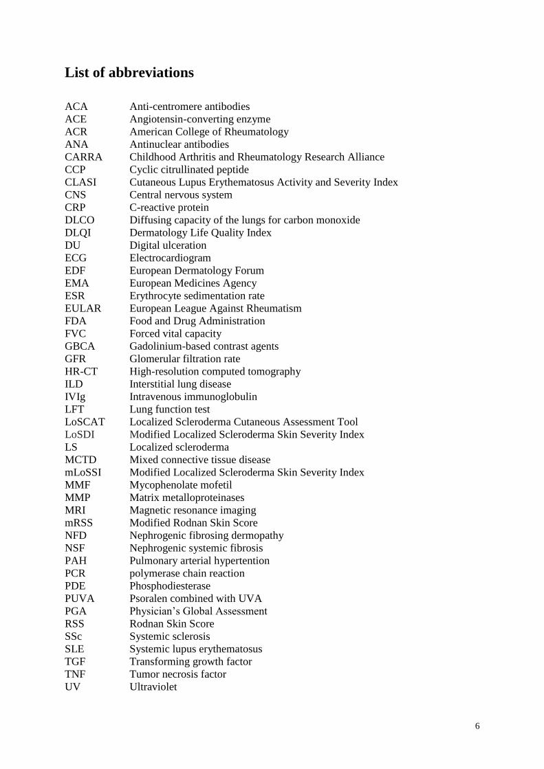
List of abbreviations
ACA Anti-centromere antibodies
ACE Angiotensin-converting enzyme
ACR American College of Rheumatology
ANA Antinuclear antibodies
CARRA Childhood Arthritis and Rheumatology Research Alliance
CCP Cyclic citrullinated peptide
CLASI Cutaneous Lupus Erythematosus Activity and Severity Index
CNS Central nervous system
CRP C-reactive protein
DLCO Diffusing capacity of the lungs for carbon monoxide
DLQI Dermatology Life Quality Index
DU Digital ulceration
ECG Electrocardiogram
EDF European Dermatology Forum
EMA European Medicines Agency
ESR Erythrocyte sedimentation rate
EULAR European League Against Rheumatism
FDA Food and Drug Administration
FVC Forced vital capacity
GBCA Gadolinium-based contrast agents
GFR Glomerular filtration rate
HR-CT High-resolution computed tomography
ILD Interstitial lung disease
IVIg Intravenous immunoglobulin
LFT Lung function test
LoSCAT Localized Scleroderma Cutaneous Assessment Tool
LoSDI Modified Localized Scleroderma Skin Severity Index
LS Localized scleroderma
MCTD Mixed connective tissue disease
mLoSSI Modified Localized Scleroderma Skin Severity Index
MMF Mycophenolate mofetil
MMP Matrix metalloproteinases
MRI Magnetic resonance imaging
mRSS Modified Rodnan Skin Score
NFD Nephrogenic fibrosing dermopathy
NSF Nephrogenic systemic fibrosis
PAH Pulmonary arterial hypertention
PCR polymerase chain reaction
PDE Phosphodiesterase
PUVA Psoralen combined with UVA
PGA Physician’s Global Assessment
RSS Rodnan Skin Score
SSc Systemic sclerosis
SLE Systemic lupus erythematosus
TGF Transforming growth factor
TNF Tumor necrosis factor
UV Ultraviolet
7
I Localized scleroderma (morphea)
Introduction
Localized scleroderma (LS) comprises a spectrum of sclerotic diseases that primarily affect the
skin. Depending on the respective subtype, LS can also involve adjacent tissues such as the fat,
fascia, muscle and bone.1 Debate continues as to whether the term “localized scleroderma” or
“morphea” should be used for the disease because “localized scleroderma” or “circumscribed
scleroderma” might be confused with “systemic scleroderma”, resulting in unnecessary patient
concern. However, this will change over time because consensus has been reached to abandon
systemic scleroderma for the term “systemic sclerosis”.2 Nevertheless, especially in Europe, the
term LS is used as a heading for the whole spectrum of subtypes, whereas morphea is mainly
used for the plaque type of the disease. In contrast to systemic sclerosis, LS does not affect
internal organs such as the lungs, heart, kidneys or gastrointestinal tract. Although LS and
systemic sclerosis (SSc) share similar pathogenetic pathways, both diseases rarely coexist, and
transition from LS to SSc does not occur.
Epidemiology
LS is a rare disease that seems to be most frequent in white individuals, but may affect people
of all ethnic backgrounds.3–5 To date, only a few adequate epidemiologic studies on LS have
been conducted, with incidence ranging from 0.4 to 2.7 per 100.000 people.6,7 LS occurs more
often in women than men, at a ratio of 2.6–6 to 1.8 The disease may manifest at all ages, but the
peak age of incidence differs depending on the LS subtype. The most frequent subtype of LS
(morphea) usually appears in adults between 40 and 50 years of age, whereas linear subtypes
primarily present in childhood between 2 and 14 years of age.3 Other, rarer subtypes of LS have
a peak incidence in the third and fourth decade of life.
Pathogenesis
The hallmark feature of LS is overproduction of collagen and increased extracellular matrix
deposition. Its exact initiation remains unknown. It has been hypothesized that certain stimuli,
for example infections, trauma, radiation, or drugs, might cause microvascular injuries and
induce T cell activation that subsequently result in a release of various adhesion molecules.3
Up-regulation of some of these adhesion molecules (e.g. vascular cell adhesion molecule-1 and
intercellular adhesion molecule-1) might induce T cell activation, which, in turn, activates the
8
release of key player pro-fibrotic cytokines, such as transforming growth factor-beta (TGFβ)
and its signal transducers called SMAD proteins, platelet-derived growth factor, connective
tissues growth factor, and interleukin 4, 6, and 8.9–12 This pro-fibrotic pathway additionally
includes a spectrum of chemokines that significantly contribute to skin sclerosis.13,14
Ultimately, and similarly to SSc, activation of all of these pro-inflammatory and pro-fibrotic
signals leads to excessive collagen production and decrease of matrix metalloproteinases
(MMP) responsible for collagen degradation.15
Potential trigger factors of localized scleroderma
Although much is known about the early inflammatory phase and the molecular mechanisms
involved in the fibroblastic reaction of LS, little is known about the potential triggers of the
disease. Among infectious agents, Borrelia organisms have been extensively studied on both
sides of the Atlantic. Whereas high rates of Borrelia infections, some of which were detected
using highly sensitive new detection techniques such as focus-floating microscopy, have been
reported in LS patients from Europe, a variety of studies based on polymerase chain reaction
(PCR) from northern Europe or from the United States failed to demonstrate an association.16–
18 Thus, the pathogenetic role of Borrelia in LS remains unclear. Among the drugs that have
been reported to induce LS, most evidence exists for bleomycin, D-penicillamine, vitamin K1,
and L-5-hydroxytryptophane plus carbidopa. Recently, balicatib, an inhibitor of the osteoclastic
enzyme cathepsin K used for osteoporosis, has been reported to induce LS.19 Few reports exist
on radiation-induced LS, which primarily occurs in women with breast cancer.20,21 Clinically,
radiation-induced LS might be indistinguishable from chronic radiodermatitis, but
histopathologic analysis usually discerns both conditions. Finally, among the triggers of LS,
mechanical injuries and traumata have been reported in case series and large cohort studies,
with the highest association in facial subtypes of childhood LS.4,5,22
Clinical manifestation
Clinical classification
To date, no uniformly accepted classification for LS exists. A widely accepted classification
was published in 1995 that distinguishes plaque, generalized, deep, bullous, and linear types as
the five main groups of LS.23 However, this classification raises some concerns. First, it
includes diseases that are not uniformly accepted to belong to the LS spectrum, such as
extragenital lichen sclerosus. Secondly, bullous lesions can appear in all different LS subtypes
due to the characeristic subepidermal edema and damage of the basement membrane zone.
9
Thirdly, there are patients, especially children, who present with more than one subtype of LS.
Thus, an alternative classification scheme was published in 2006 to overcome these
weaknesses.24 A German group of experts proposed a classification (Table 1) that considers the
extent and depth of fibrosis, and refers to the treatment of the respective subtypes.1
<TABLE 1>
Limited types of LS
Plaque-morphea (the classical plaque type of LS) is the most frequent subtype of LS, especially
in adults. In the early active phase, plaque-morphea usually presents with oval-shaped lesions
surrounded by an erythematous border (the so-called “lilac ring”). In the later stage of disease,
morphea lesions become hard and sclerotic in the center, with a whitish or ivory color. Older
lesions may also become atrophic, hypo-, or hyperpigmented and, depending on the location of
fibrosis, may also lead to hair loss and loss of the skin appendages. Plaque-morphea is
frequently located on the trunk, especially the submammary region, the transitional area
between the hip and inguinal regions or in areas with repeated trauma such as pressure from
clothing.
Guttate morphea is a rare subtype of morphea that presents with multiple yellowish or whitish,
small sclerotic lesions with a shiny surface. Guttate morphea is predominantly located on the
trunk. Early inflammatory lesions may simply present as erythematous maculae. Clinically and
histopathologically, guttate morphea might be difficult to distinguish from extragenital lichen
sclerosus.
Atrophoderma of Pasini and Pierini is possibly an early abortive type of morphea. The recently
described term “superficial morphea” seems to be synonymous with atrophoderma of Pasini
and Pierini.25,26 The clinical presentation of this subtype of LS, which frequently manifests in
childhood, is characterized by symmetrical, single or multiple, sharply demarcated,
hyperpigmented, non-indurated patches that are located on the trunk or extremities.
Generalized types of LS
Generalized localized scleroderma is a more severe variant of LS. According to Laxer and
Zulian, generalized localized scleroderma is defined as the presence of four or more indurated
plaques of more than 3 cm in diameter, involving at least two of the seven anatomic sites (head-
10
neck, each extremity, anterior trunk, and posterior trunk).24 The trunk is commonly affected
and skin lesions are often distributed symmetrically and tend to coalesce.
A unique and very rare variant of the generalized type of LS is “disabling pansclerotic
morphea.” Disabling pansclerotic morphea, predominantly occurring in childhood, and may
lead to extensive involvement of the skin, fat tissue, fascia, muscle, and bone, with only limited
tendency of fibrosis to regress. Disabling pansclerotic morphea often results in severe
contractures and poorly healing, large ulcerations and skin necroses.
Linear types of LS
Linear localized scleroderma is the most common subtype of LS in childhood. Linear LS is
characterized by longitudinally arranged linear, band-like lesions that are predominantly
located on the extremities. Evidence indicates that linear LS may follow the lines of Blaschko.27
In mild disease, the lesions may heal with residual hyperpigmentation. However, depending on
the extent of the fibrotic process, linear LS may lead to severe growth retardation, muscle
atrophy, flexion contractures, myositis and myalgia, arthritis and arthalgia, and psychologic
disability.
LS “en coup de sabre” is a subtype located on the frontoparietal region of the head, usually
ranging paramedian from the eyebrows into the hair-bearing scalp where it might cause scarring
alopecia. Involvement of the underlying central nervous system (CNS; e.g. seizures, migraine,
and headache) and abnormal ophthalmologic findings (e.g. uveitis) can occur.
Several authors have speculated that progressive facial hemiatrophy (also called Parry–
Romberg syndrome) and LS “en coup de sabre” are variants of the same condition.1,5,28,29
Progressive facial hemiatrophy is clinically characterized by a primary atrophy of the
subcutaneous tissue, muscle, and bone. Skin fibrosis is usually absent. It often occurs in
childhood or adolescence, and may result in severe facial asymmetry. Occurrence of
simultaneous linear LS “en coup de sabre” and progressive facial hemiatrophy is quite frequent,
with a reported coincidence of up to 40%.30 In the classification proposed in this article,
progressive facial hemiatrophy is listed under the linear subtypes of LS (Table 1), although with
exclusive involvement of extracutaneous structures it may also be classified as a “deep subtype”
of LS.
11
Deep type of LS
The deep type of LS (also called deep morphea) is the rarest variant, affecting less than 5% of
patients. In deep morphea, the fibrotic process mainly affects the deeper layers of the connective
tissue (i.e. fat tissue, fascia, and underlying muscle). Deep morphea lesions are typically
arranged symmetrically and predominantly located on the extremities.
Mixed type of LS
Mixed types of LS predominantly affect children, occurring in up to 15% of patients with
juvenile LS. Mixed types often consist of linear LS and morphea (plaques type of LS) or a
combination of linear and generalized LS.5
Eosinophilic fasciitis
Eosinophilic fasciitis (or Shulman syndrome) is considered by many experts to be a special
subtype belonging to the spectrum of LS.1. A mechanical trauma often precedes the first
manifestation of the disease. Clinically, eosinophilic fasciitis predominantly affects the
extremities and presents with a rapid onset of symmetrical swelling of the skin. In the later stage
of disease, lesions become more indurated and fibrotic, leading to the typical “peau d’orange”
like appearance. A distinctive clinical finding in later stages of eosinophilic fasciitis is that
cutaneous veins might appear depressed compared with the surrounding tissue (called “negative
vein sign”).
Association with other autoimmune diseases
Several reports of familiar clustering and increased rates of other autoimmune diseases (e.g.
Hashimoto thyreoiditis, alopecia areata, vitiligo, and type-1 diabetes) in patients with LS
suggest a possible genetic component.5 However, in contrast to SSc, susceptibility genes for LS
are still unknown. In a study including 245 patients with LS, 17.6% had other rheumatic or
autoimmune diseases. This rate is four times higher than in the general population. Patients with
generalized LS had the highest rate of associated autoimmune diseases (45.9%).31 Another
study that retrospectively evaluated 472 patients with LS for other autoimmune diseases found
other autoimmune diseases in 8.1%.32
Some decades ago, the coexistence of LS and lichen sclerosus (predominantly extragenital) was
reported in several case reports and small case series.33,34 In 2012, a prospective study from
France including 76 patients with LS showed that 38% of them had concomitant genital lichen
12
sclerosus; mostly patients with limited LS (morphea) and generalized LS were affected. This
high rate of genital lichen sclerosus in patients with LS was later confirmed by a larger
retrospective German study.35
Clinical course, disease activity, and recurrence rates
To date, only limited data are available on the long-term clinical course of LS. A recent
retrospective analysis including 344 patients with adult or juvenile LS from the Netherlands
demonstrated that about one quarter of the patients experienced a reactivation of disease.
Univariate analysis demonstrated that the age at onset of disease was a risk factor for recurrent
disease; relapses occurred significantly more often in pediatric LS (27%) compared with adult
disease (17%). Moreover, disease subtype was another risk factor; 37% of patients with linear
LS of the limbs (either solitary or as part of mixed type of LS) experienced a relapse, whereas
recurrences in the other subtypes occurred less frequently (17%). The two most frequent
subtypes in adults (morphea/plaque type and generalized LS) had recurrence rates of 16% and
25%, respectively. Importantly, this study also showed that disease relapses can occur after
years of quiescent disease; the median time between disease remission and first recurrence was
26 months in juvenile and 27 months in adult LS, respectively.36 In the study of Saxton-Daniels
et al. regarding long-term outcome of pediatric cases, 89% of the pediatric onset cases
developed new or expanded lesion over time.37 Time to recurrence of activity ranged from 6 to
18 years from initial disease onset.
Diagnostic procedures
Laboratory parameters
Depending on the clinical subtype, a high incidence of autoimmune phenomena has been
reported in LS patients (e.g. serum antinuclear antibodies, most of them with a homogenous
pattern).4,31,38 Moreover, active childhood LS might be associated with anti-histone antibodies,
hypergammaglobulinemia, and eosinophilia.39 In patients with linear LS of the extremities with
concomitant joint involvement, increased levels of rheumatoid factor may be present, and do
sometimes correlate with the clinical degree of arthritis activity.40 Several other antibodies (e.g.
anti-topoisomerase II alpha, anti-U1-small-nuclear-ribonucleoprotein, and anti-U3-small-
nuclear-ribonucleo-protein), and anti-MMP antibodies have been evaluated in LS, but their
specific role remains to be elucidated.41–43
13
In daily practice, blood screening in patients with LS who are considered for systemic therapy
should include blood differential and serum chemistry (Table 2). Routine screening for
antinuclear antibodies is not recommended. Additional diagnostics (e.g. screening for
antibodies against extractable nuclear antigens) should be only performed to confirm or exclude
systemic sclerosis.
Controversy exists about the pathogenetic role of Borrelia burgdorferi in LS (see Potential
trigger factors of LS, above). Accordingly, a general blood screening for Borrelia in patients
with LS is not generally recommended and should only be performed in clinically suspicious
cases.
<TABLE 2>
Histopathology of localized scleroderma
LS and SSc share the same histopathologic features. Thus, by histopathology, it is neither
possible to distinguish between LS and SSc nor to differentiate among different LS subtypes.
In general, two phases of LS can be recognized, an early inflammatory and a late fibrotic
stage.1,44 Early skin lesions of LS are characterized by thickened collagen bundles within the
reticular dermis that run parallel to the skin surface, and by the presence of dense inflammatory
infiltrates between the collagen bundles, and around blood vessels and sweat glands.
Lymphocytes predominate the inflammatory infiltrates, but plasma cells, histiocytes, and
eosinophilic granulocytes might be present as well. The overlying epidermis might be either
unaffected or thin and atrophic. In the late fibrotic stage, the lesional skin becomes relatively
avascular, and often there is only little evidence of ongoing inflammation. Late lesions usually
contain collagen fibers that are tightly packed and highly eosinophilic. Sweat glands are
atrophic or absent. Collagen may replace fat cells in the subcutaneous tissue. Physicians should
ensure that the biopsy excision is sufficiently deep as some LS subtypes may primarily involve
the subcutis or underlying fascia and muscle.
Clinical scores
Due to the difficulties of defining clinical improvement in LS, clinical scores were not available
for a long period of time. The Rodnan Skin Score (RSS) and its later revised version (the so-
called “modified RSS) are validated and widely used clinical tools in SSc.45 Both of these scores
are inappropriate for the measurement of LS skin involvement due to the overweight of certain
14
anatomic areas (e.g. face), which are usually spared in LS. In 2009, the first validated skin score
for LS, called the modified Localized Scleroderma Skin Severity Index (mLoSSI) was
introduced. This score evaluates erythema, skin thickness and development of new skin lesions
or lesional extension in 18 anatomic regions, and has demonstrated a high interrater
agreement.46 The same group of researchers later introduced a score for skin damage in LS,
called the Localized Scleroderma Skin Damage Index (LoSDI).47 Consequently, it was
recommended to combine the mLoSSI, LoSDI, and the Physician’s Global Assessment (PGA)
to measure both activity and damage in LS. This composes the Localized Scleroderma
Cutaneous Assessment Tool (LoSCAT), a combined score that is modeled after a well
established tool for cutaneous lupus erythematosus, the Cutaneous Lupus Erythematosus
Activity and Severity Index (CLASI). LoSCAT, which is similar to the CLASI, could become
a standard tool to evaluate skin affection in LS.
Patient quality of life can be evaluated with the Dermatology Life Quality Index (DLQI) or the
Hospital Anxiety and Depression Scale.
Radiologic examination
Morphea, the most common LS subtype in adults, usually affects the skin only and therefore
does not require further radiologic examination. In contrast, patients with LS “en coup de sabre”
and progressive facial hemiatrophy often suffer from neurologic symptoms (e.g. migraine,
headache, and epilepsy). In these cases, cranial magnetic resonance imaging (MRI) should be
considered to detect potential involvement of the CNS because subcortical calcifications and
brain atrophy are common.4,22 In special cases ophthalmologists or oral surgeons should be
consulted about abnormalities that have to be corrected. Despite such abnormalities of the CNS,
many patients are asymptomatic. In addition, MRI and computed tomography studies might be
helpful for surgical planning (e.g. in LS “en coup de sabre” type), and to detect muscle, joint or
bone involvement, for instance in linear LS of the extremities. MRI should be considered in
cases with linear LS of the extremities that might have concomitant arthritis.
Technical outcome measures
A variety of technical procedures have been reported in clinical trials on LS, for example,
ultrasound scanning, cutometer, durometer, thermography, laser Doppler flowmetry, and a
computerized skin score. In most of the studies, these procedures were used as secondary
outcome measures.
15
Differential diagnoses
A variety of differential diagnoses should be considered in LS.48 In daily routine, the physicians’
pivotal challenge is to differentiate LS from SSc.3 Typical facial (e.g. telangiectasia, beak-
shaped nose, and microstomia) and vascular (e.g. Raynaud’s phenomenon, pitting scars, and
digital ulcers) features of SSc, as well as highly specific serum antibodies (e.g. anti-centromere
antibodies and anti-Scl-70 antibodies) are absent in LS.44
The most relevant differential diagnoses for limited LS (morphea) are extragenital lichen
sclerosus and acrodermatitis chronica atrophicans, for generalized LS chronic graft versus host
disease, SSc, and nephrogenic systemic fibrosis, and for linear LS lupus erythematosus
profundus and other types of panniculitis. All differential diagnoses with respect to LS subtypes
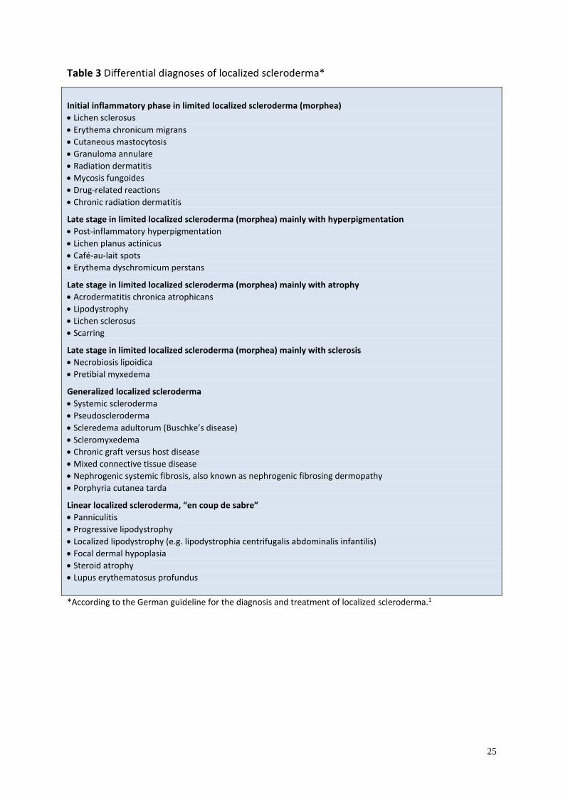
and stage of disease are summarized in Table 3.
<TABLE 3>
Specifics of juvenile localized scleroderma
Whereas limited types of LS most commonly occur in adults, linear subtypes predominate in
children. A study including 65 patients with juvenile LS revealed that linear subtypes may
follow the lines of Blaschko. It was hypothesized that in linear LS, susceptible cells are present
in a mosaic state and that exposure to some trigger factors finally result in the initiation of
disease.27 Clinical course of disease is often more severe in juvenile LS compared with adult
linear LS, and may lead to considerable atrophy of the skin, fat tissue, fascia, and muscle. This
might finally result in substantial functional, physical, and mental disability. It has been shown
that 30–50% of patients with linear LS experience osteoarticular complications (e.g. arthritis)
on the affected extremity.49–51 Both linear LS “en coup de sabre” and progressive facial
hemiatrophy mainly occur in childhood. It seems that both conditions belong to the same
spectrum of disease, with overlapping clinical features. In contrast to other subtypes of LS,
linear LS “en coup de sabre” and progressive facial hemiatrophy have a more insidious clinical
course, and the active stage of disease persists usually longer than in other subtypes of LS.
Neurologic symptoms are frequent and may include epileptic seizures, neuropsychiatric
symptoms, headaches, and mental or behavioral disorders.28,52,53 Ophthalmologic changes are
common in juvenile LS and might manifest as uveitis, dysfunction of the eye muscles, and loss
of eyebrows or eyelashes.
16
“Disabling pansclerotic morphea,” a rare subtype of generalized LS, usually manifests before
the age of 14, and is obligatorily associated with affection of extracutaneous structures. It
frequently results in disturbance of growth and cachexia.
Abnormal blood findings are frequent in juvenile LS. In the active stage of generalized LS,
blood eosinophilia is frequent. Moreover, an elevated rheumatoid factor, increased blood
sedimentation rate, hypergammaglobulinemia (increased IgA and IgM in active stages of LS
and increased IgG in severe disease with contractures), as well as elevated antinuclear, anti-
histone, and single-stranded DNA antibodies might be present.54
In order to prevent persistent damage, effective systemic therapy should be initiated in the
active stage of all linear types of juvenile LS as early as possible. Similarly to adult LS, subtype
and extent of disease have an influence on the respective therapy. Concomitant physiotherapy
should be considered in subtypes with (potential) restriction of motion. Surgical interventions
should only be performed in the inactive stage of disease.55 The same is true for aesthetic-
reconstructive interventions in linear LS “en coup de sabre” and progressive facial hemiatrophy.
Treatment
Although no causal treatment for LS exists, a variety of therapeutic options are available,
especially for the active phase of disease. In general, treatment options for LS might be divided
into topical and systemic therapy as well as ultraviolet (UV) phototherapy. The extent and
severity of LS should be taken into account before initiating the respective therapy. For
example, topical and UV phototherapy are usually appropriate in limited types of LS that are
restricted to the skin, whereas generalized, linear or deep types usually require systemic
treatment. Hereafter, all treatment options that have been reported for LS are summarized.
Moreover, a treatment algorithm is provided that incorporates the subtype, severity, and extent
of LS (Fig. 1). When evaluating the treatment efficacy it should be remembered that reduction
of skin sclerosis starts 8–12 weeks after initiation of therapy, at the earliest. None of the
therapies mentioned below are officially licensed in Europe.
<FIG. 1>
Topical therapy
17
Topical glucocorticoids
Although no well-performed studies exist on the use of topical glucocorticoids, they are the
mainstay of topical treatment in LS. Therapy with moderate-to-high potent glucocorticoids
should be performed in the active phase of disease, and their application should be restricted to
a total of 3 months. Longer application of topical glucocorticoids should be given as interval
therapy. In order to increase the efficacy, an application under occlusion might be considered.
Intralesional glucocorticoid therapy might be performed in LS “en coup de sabre,” with
injections into the active margin.
Topical calcipotriol
To date, two uncontrolled studies have been conducted on the use of topical calcipotriol in LS,
one of which administered calcipotriol 0.005% along with low-dose UVA1 phototherapy.56 In
both studies, administration was performed twice daily. In the monotherapy study, calcipotriol
0.005% was applied under occlusion.57
Calcipotriol 0.005% should be considered for active inflammatory superficial types of LS with
a low degree of sclerosis. Treatment should be performed twice daily (under occlusion) for a
minimum of 3 months.
Topical calcineurin inhibitors
Following two open studies on topical tacrolimus 0.1% ointment in LS, a recent double-blind,
placebo (petroleum emollient)-controlled pilot study has shown that topical tacrolimus
significantly improves LS.58–60 Outcome measures in this study were the changes of surface
area, a clinical score for erythema, induration, dyspigmentation, telangiectasia, atrophy, and a
durometer score.58 Early inflammatory lesions resolved and late sclerotic lesions softened,
whereas no effects were seen on pre-existing skin atrophy.
Thus, tacrolimus ointment might be an effective treatment option for active LS lesions. To date,
no studies on pimecrolimus for LS have been conducted.
Imiquimod
In case reports and small case series, the topical immune response modifier imiquimod has been
reported to significantly improve abnormal pigmentation, sclerosis, and erythema in LS.61–63
18
The mechanism of imiquimod action in LS might be explained by induction of interferon-γ
which inhibits TGF-β, thereby possibly exhibiting a broad anti-fibrotic effect.
However, based on these small case series, imiquimod cannot be recommended for LS until
more valid data are available.
Intralesional interferon-γ
A double-blinded, placebo-controlled trial demonstrated no significant improvement of
intralesional interferon-γ compared with the placebo group. Accordingly, intralesional
interferon-γ cannot be recommended for the treatment of LS.64
Systemic therapy
Systemic glucocorticoids
Similarly to topical gucocorticoids, there is a paucity of data on systemic glucocorticoids,
although they are widely used agents in LS, particularly in linear, generalized, and deep
subtypes. In the only published uncontrolled study on 17 patients with LS (glucocorticoid
dosage: 0.5–1.0 mg/kg body weight daily), a marked improvement was noticed in nearly all of
the patients.65 However, about one third of patients experienced recurrences after finishing
therapy. Systemic gucocorticoids are safe and effective in active lesions of LS, and should be
considered in patients with severe disease, especially in those forms affecting extracutaneous
structures (e.g. fat tissue, fascia, muscle, and bone). Moreover, systemic glucocorticoids are the
first-line treatment option in eosinophilic fasciitis.66 Treatment should be planned for a
sufficient duration, as clinical effects are sometimes seen at the earliest 3 months after onset.
Methotrexate
Among systemic treatment of LS, best evidence exists for the use of methotrexate. To date, one
placebo-controlled multi-center trial, as well as three prospective and four retrospective studies
have been published.49,67–73 In the placebo-controlled study, a total of 70 children with active
LS (46 patients in the methotrexate group and 24 in the placebo group) were included to receive
methotrexate orally (15 mg/m², maximum 20 mg) or placebo. Moreover, oral prednisone
(1 mg/kg/day, maximum 50 mg) was added in both arms for 3 months. The computerized
scoring system, as well as infrared thermography were used as outcome measures. In both arms,
a reduction of the clinical score was observed within the first 6 months. However, at the end of
19
the study at month 12, a significant decrease of the clinical score as well as infrared
thermography was only observed in the methotrexate group.67
In the three prospective studies that included 34 patients (24 adults and 10 children), a
combination of high-dosage intravenous methylprednisolone and methotrexate (adults
15 mg/weeks; children 0.3 mg /kg/week) was used, and outcome measures were a non-
validated clinical score and ultrasound scanning. All adults and nine of the 10 children
experienced a significant improvement under therapy.49,68,69 In the four retrospective studies, a
total of 119 patients were included (52 patients with methotrexate monotherapy and 67 patients
with a combination of methotrexate and systemic glucocorticoids). In 97% of patients, a clinical
improvement was observed.70–73 Importantly, it was shown in another study that 28% of patients
with juvenile LS experienced a relapse after treatment with methotrexate.74
In the studies mentioned above, different dosages of methotrexate and systemic glucocorticoids
were used. In 2012, the “Childhood Arthritis and Rheumatology Research Alliance” (CARRA)
recommended three different treatment regimens for juvenile LS: 1) methotrexate
monotherapy; 2) pulse methotrexate and glucocorticoid therapy with methylprednisolone given
intravenously; 3) pulse methotrexate and glucocorticoid therapy with prednisone given orally.75
These recommendations have been incorporated in the treatment algorithm (Fig. 1) of this
guideline.
Mycophenolate mofetil
In 2009, a small case series of seven methotrexate-resistant LS patients treated with
mycophenolate mofetil (MMF) showed improvement of skin sclerosis and inflammation, as
documented with infrared thermography and clinical scoring.76,77 In vitro studies have shown
that MMF inhibits the proliferation of lymphocytes, but also of other cell types, including
smooth muscle cells and fibroblasts, indicating that it has direct anti-fibrotic properties in
addition to its well-known immunosuppressive effects.78 These preliminary observations make
MMF an interesting new candidate for further clinical studies. According to CARRA, MMF
should be considered as a second-line therapy if methotrexate has failed. It is noteworthy that
in several countries (e.g. Germany) health insurers sometimes deny re-imbursement for this off-
label use.
Calcitriol
20
A randomized controlled study that included 20 patients with LS demonstrated that a 9-month
therapy with oral calcitriol (0.75 µg/daily for 6 months, followed by 1.25 µg/daily for
3 months) failed to achieve any significant improvement compared with placebo.79 Therefore,
oral calcitriol cannot be recommended for LS.
D-penicillamine
Although the efficacy of D-penicillamine has been reported in a small case series of LS patients,
no significant differences were found between high-dose (750–1000 mg daily) and low-dose
therapy (125 mg daily) in SSc.80,81 Given the poor evidence level of efficacy and the
problematic side-effect profile of D-penicillamine, it cannot be recommended for the treatment
of LS.
Penicillin
For decades penicillin has been used for the treatment of LS because LS can manifest after an
infection with Borrelia. Although penicillin has anti-inflammatory properties, direct anti-
fibrotic effects have so far not been demonstrated. Accordingly, the efficacy of penicillin in LS
remains unproven.
Miscellaneous
Numerous other systemic therapies have been used in cases of LS, including cyclosporine,
azathioprine, chloroquine and hydroxychloroquine, phenytoin, colchicine, retinoids,
extracorporeal photopheresis, plasmapheresis, intravenous immunoglobulin, abatacept,
infliximab, rituximab, and imatinib.82–87 These treatments should be reserved for single severe
cases with contraindications or failure to standard therapy.
UV phototherapy
Within the last two decades, the vast majority of clinical studies on LS came from the field of
photodermatology.88 One of the rationales for using UV phototherapy in sclerotic skin diseases
is the fact that UV can induce interstitial MMP.89,90 The first experience of the successful use
of UV phototherapy in LS was in 1994.91 Since then, much information has been gained on the
entire spectrum of anti-fibrotic and anti-inflammatory effects of UV phototherapy in skin
sclerosis.92 In addition, UV phototherapy leads to apoptosis of dermal T cells, depletion of
Langerhans cells, and to modulation of several pro-inflammatory cytokines.88 The exact
mechanism of action of UV therapy in sclerotic skin diseases remain to be determined. Because
21
longer wavelengths in the UVA range (320–400 nm) penetrate deeper into the dermis compared
with UVB (280–320 nm), most studies have focused on UVA. Before initiating UV
phototherapy in LS, it should be considered that UV rays only penetrate into the deep dermis.
Therefore, UV phototherapy (in combination with topical treatment, e.g. topical glucocorticoids
or topical vitamin D analogs) is an effective treatment option for limited disease restricted to
the skin, but not in LS subtypes affecting deeper structures (e.g. fat tissue, fascia, muscle, or
bone). Such subtypes require systemic therapy. However, it is also known that UV can act
indirectly by modulating cytokine release in keratinocytes.
UV irradiation has a major role in the pathogenesis of skin cancer due to its capacity to induce
immunosuppression and DNA damage. However, the dosages and duration of UVA irradiation
used in the treatment of sclerotic skin diseases are most likely too low to induce any significant
skin damage, though there may be an absolute safe threshold dose.93
PUVA phototherapy
In order to avoid the well-known side effects of oral application of 8-methoxypsoralen, psoralen
combined with UVA (PUVA) was mainly applied in LS as bath PUVA phototherapy. Besides
several case reports, two retrospective case series exist on bath PUVA phototherapy.94,95 In the
larger study published in 2013, 28 patients were treated with bath PUVA three times per week.
In 39% of patients, a complete clearance of all lesions was observed, 50% experienced clinical
improvement, and 10% had no response.95 Moreover, a small case series of four patients treated
with cream PUVA phototherapy showed similar encouraging results.96 PUVA phototherapy is
usually performed 2–3 times per week for a total of 30 irradiations.
Broadband UVA
Three prospective studies have been published on the use of broadband UVA (320–400 nm) in
LS. Among those, the largest study included 63 patients.97–99 The three dosages used in this
study (5, 10, and 20 J/cm² for a total of 20 irradiations each) showed similar efficacy. Controlled
studies comparing broadband-UVA with other UV modalities are lacking.
UVA1 phototherapy
In the area of phototherapy, the most robust data exist for UVA1. Three different dosages of
UVA1 can be distinguished: low-dose UVA1 (10–29 J/cm²), medium-dose UVA1 (30–
59 J/cm²), and high-dose UVA1 (60–130 J/cm²). All regimens have been used in LS, and the
22
first report was published in 1991.100 The first prospective study on UVA1 phototherapy in LS
demonstrated that high-dose UVA1 is highly effective, but low-dose UVA1 failed to show any
substantial effects in LS.101 Nevertheless, several prospective studies performed some years
later showed that low-dose and medium-dose UVA1 are effective as well.56,102–108 To date, only
one randomized controlled study has been performed that compared low-dose UVA1, medium-
dose UVA1, and narrow-band UVB phototherapy in a collective of 64 LS patients. All three
UV regimens significantly improved the skin scores, with medium-dose UVA1 being
significantly better than narrow-band UVB.109 Whether patients with darker skin respond less
to UVA1 phototherapy is still a matter of debate.110,111 Moreover, it has been shown that within
3 years, about 50% of patients treated with UVA1 experience recurrences after therapy.112 In
these cases, a second cycle of UVA1 phototherapy should be considered. UVA1 is usually
performed 3–5 times per week for a minimum of 30 irradiations.
Physiotherapy
Studies on physiotherapy in LS are lacking. Nevertheless, physiotherapy is an important
component in the multimodal treatment concept for LS, and is frequently performed in daily
practice. In particular, linear, generalized, deep, and mixed types of LS should be treated with
physiotherapy. It should not be performed in the active, inflammatory stage of disease. Massage
and lymphatic drainage can be added to systemic therapy in patients with sclerotic stage disease.
Physiotherapy is usually performed once or twice per week for at least 3 months.
Surgical therapy
Surgical therapy is predominantly indicated in linear types of LS. It is important that surgical
interventions are only considered in the inactive stage of disease in order to minimize the risk
of reactivation. If signs for disease activity occur, perioperative immunosuppressive therapy
should be considered. In linear LS of the limbs, epiphysiodesis of the healthy extremity can be
considered in order to adjust leg length inequality. This procedure should be performed by an
experienced pediatric orthopedist.
Plastic surgical interventions might be considered for cosmetic reasons in linear LS “en coup
de sabre” or progressive facial hemiatrophy.
23
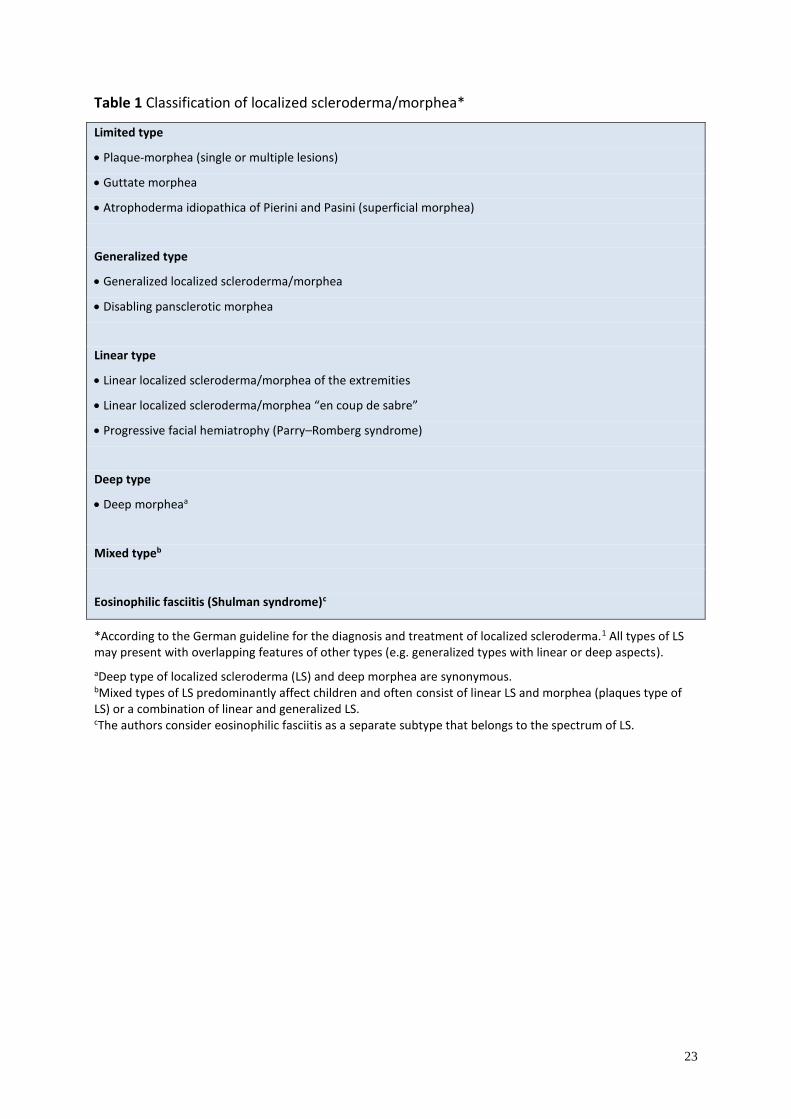
Table 1 Classification of localized scleroderma/morphea*
Limited type
• Plaque-morphea (single or multiple lesions)
• Guttate morphea
• Atrophoderma idiopathica of Pierini and Pasini (superficial morphea)
Generalized type
• Generalized localized scleroderma/morphea
• Disabling pansclerotic morphea
Linear type
• Linear localized scleroderma/morphea of the extremities
• Linear localized scleroderma/morphea “en coup de sabre”
• Progressive facial hemiatrophy (Parry–Romberg syndrome)
Deep type
• Deep morpheaa
Mixed typeb
Eosinophilic fasciitis (Shulman syndrome)c
*According to the German guideline for the diagnosis and treatment of localized scleroderma.1 All types of LS may present with overlapping features of other types (e.g. generalized types with linear or deep aspects).
aDeep type of localized scleroderma (LS) and deep morphea are synonymous. bMixed types of LS predominantly affect children and often consist of linear LS and morphea (plaques type of LS) or a combination of linear and generalized LS. cThe authors consider eosinophilic fasciitis as a separate subtype that belongs to the spectrum of LS.
24
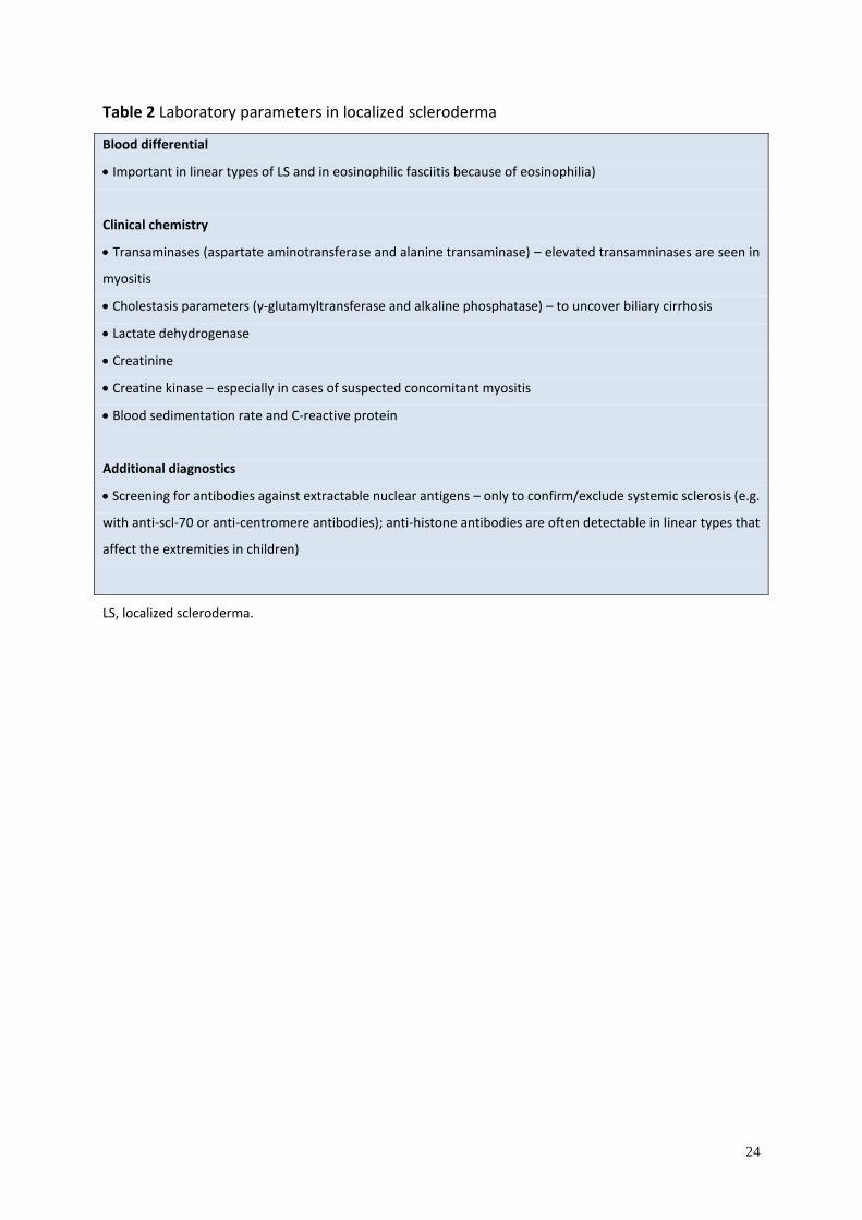
Table 2 Laboratory parameters in localized scleroderma
Blood differential
• Important in linear types of LS and in eosinophilic fasciitis because of eosinophilia)
Clinical chemistry
• Transaminases (aspartate aminotransferase and alanine transaminase) – elevated transamninases are seen in
myositis
• Cholestasis parameters (γ-glutamyltransferase and alkaline phosphatase) – to uncover biliary cirrhosis
• Lactate dehydrogenase
• Creatinine
• Creatine kinase – especially in cases of suspected concomitant myositis
• Blood sedimentation rate and C-reactive protein
Additional diagnostics
• Screening for antibodies against extractable nuclear antigens – only to confirm/exclude systemic sclerosis (e.g.
with anti-scl-70 or anti-centromere antibodies); anti-histone antibodies are often detectable in linear types that
affect the extremities in children)
LS, localized scleroderma.
25
Table 3 Differential diagnoses of localized scleroderma*
Initial inflammatory phase in limited localized scleroderma (morphea)
• Lichen sclerosus
• Erythema chronicum migrans
• Cutaneous mastocytosis
• Granuloma annulare
• Radiation dermatitis
• Mycosis fungoides
• Drug-related reactions
• Chronic radiation dermatitis
Late stage in limited localized scleroderma (morphea) mainly with hyperpigmentation
• Post-inflammatory hyperpigmentation
• Lichen planus actinicus
• Café-au-lait spots
• Erythema dyschromicum perstans
Late stage in limited localized scleroderma (morphea) mainly with atrophy
• Acrodermatitis chronica atrophicans
• Lipodystrophy
• Lichen sclerosus
• Scarring
Late stage in limited localized scleroderma (morphea) mainly with sclerosis
• Necrobiosis lipoidica
• Pretibial myxedema
Generalized localized scleroderma
• Systemic scleroderma
• Pseudoscleroderma
• Scleredema adultorum (Buschke’s disease)
• Scleromyxedema
• Chronic graft versus host disease
• Mixed connective tissue disease
• Nephrogenic systemic fibrosis, also known as nephrogenic fibrosing dermopathy
• Porphyria cutanea tarda
Linear localized scleroderma, “en coup de sabre”
• Panniculitis
• Progressive lipodystrophy
• Localized lipodystrophy (e.g. lipodystrophia centrifugalis abdominalis infantilis)
• Focal dermal hypoplasia
• Steroid atrophy
• Lupus erythematosus profundus
*According to the German guideline for the diagnosis and treatment of localized scleroderma.1
26
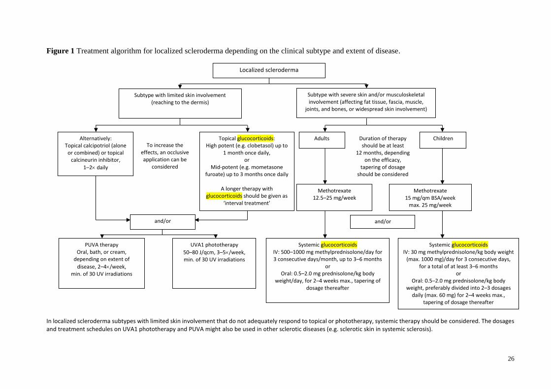
Figure 1 Treatment algorithm for localized scleroderma depending on the clinical subtype and extent of disease.
In localized scleroderma subtypes with limited skin involvement that do not adequately respond to topical or phototherapy, systemic therapy should be considered. The dosages and treatment schedules on UVA1 phototherapy and PUVA might also be used in other sclerotic diseases (e.g. sclerotic skin in systemic sclerosis).
Localized scleroderma
Subtype with limited skin involvement (reaching to the dermis)
Subtype with severe skin and/or musculoskeletal involvement (affecting fat tissue, fascia, muscle,
joints, and bones, or widespread skin involvement)
Alternatively: Topical calcipotriol (alone
or combined) or topical calcineurin inhibitor,
1–2 daily
Topical glucocorticoids: High potent (e.g. clobetasol) up to
1 month once daily, or
Mid-potent (e.g. mometasone furoate) up to 3 months once daily
A longer therapy with
glucocorticoids should be given as ‘interval treatment’
PUVA therapy Oral, bath, or cream,
depending on extent of
disease, 2–4/week, min. of 30 UV irradiations
UVA1 phototherapy
50–80 J/qcm, 3–5/week, min. of 30 UV irradiations
Systemic glucocorticoids IV: 500–1000 mg methylprednisolone/day for 3 consecutive days/month, up to 3–6 months
or Oral: 0.5–2.0 mg prednisolone/kg body
weight/day, for 2–4 weeks max., tapering of dosage thereafter
Systemic glucocorticoids IV: 30 mg methylprednisolone/kg body weight
(max. 1000 mg)/day for 3 consecutive days, for a total of at least 3–6 months
or Oral: 0.5–2.0 mg prednisolone/kg body
weight, preferably divided into 2–3 dosages daily (max. 60 mg) for 2–4 weeks max.,
tapering of dosage thereafter
and/or and/or
Methotrexate 12.5–25 mg/week
Methotrexate 15 mg/qm BSA/week
max. 25 mg/week
Adults Children Duration of therapy should be at least
12 months, depending on the efficacy,
tapering of dosage should be considered
To increase the effects, an occlusive
application can be considered
27
References
1. Kreuter A, Krieg T, Worm M, et al. AWMF Guideline no. 013/066. Diagnosis and therapy
of circumscribed scleroderma. J Dtsch Dermatol Ges 2009: 7(Suppl 6): 1–14.
2. Aringer M, Müller-Ladner U, Burkhardt H, et al. Common German language nomenclature
for systemic sclerosis. [Article in German]. Z Rheumatol 2015; 74: 100–103.
3. Fett N, Werth VP. Update on morphea: part I. Epidemiology, clinical presentation, and
pathogenesis. J Am Acad Dermatol 2011: 64: 217–228.
4. Christen-Zaech S, Hakim MD, Afsar FS, Paller AS. Pediatric morphea (localized
scleroderma): review of 136 patients. J Am Acad Dermatol 2008: 59: 385–396.
5. Zulian F, Athreya BH, Laxer R, et al. Juvenile localized scleroderma: clinical and
epidemiological features in 750 children. An international study. Rheumatology (Oxford)
2006: 45: 614–620.
6. Peterson LS, Nelson AM, Su WP, Mason T, O’Fallon WM, Gabriel SE. The epidemiology
of morphea (localized scleroderma) in Olmsted County 1960–1993. J Rheumatol 1997: 24:
73–80.
7. Murray KJ, Laxer RM. Scleroderma in children and adolescents. Rheum Dis Clin North
Am 2002: 28: 603–624.
8. Silman A, Jannini S, Symmons D, Bacon P. An epidemiological study of scleroderma in
the West Midlands. Br J Rheumatol 1988: 27: 286–290.
9. Leask A, Abraham DJ. TGF-beta signaling and the fibrotic response. FASEB J 2004: 18:
816–827.
10. Higley H, Persichitte K, Chu S, Waegell W, Vancheeswaran R, Black C.
Immunocytochemical localization and serologic detection of transforming growth factor
beta 1. Association with type I procollagen and inflammatory cell markers in diffuse and
limited systemic sclerosis, morphea, and Raynaud’s phenomenon. Arthritis Rheum 1994:
37: 278–288.
11. Ihn H, Sato S, Fujimoto M, Kikuchi K, Takehara K. Demonstration of interleukin-2,
interleukin-4 and interleukin-6 in sera from patients with localized scleroderma. Arch
Dermatol Res 1995: 287: 193–197.
12. Kreuter A, Hyun J, Skrygan M, et al. Ultraviolet A1-induced downregulation of human
beta-defensins and interleukin-6 and interleukin-8 correlates with clinical improvement in
localized scleroderma. Br J Dermatol 2006: 155: 600–607.
13. Yamamoto T. Chemokines and chemokine receptors in scleroderma. Int Arch Allergy
Immunol 2006: 140: 345–356.
28
14. Gambichler T, Skrygan M, Labanski AA, Kolios AG, Altmeyer P, Kreuter A. Significantly
increased CCL5/RANTES and CCR7 mRNA levels in localized scleroderma. Regul Pept
2011: 170: 4–6.
15. Varga J, Abraham D. Systemic sclerosis: a prototypic multisystem fibrotic disorder. J Clin
Invest 2007: 117: 557–567.
16. Eisendle K, Grabner T, Zelger B. Morphoea: a manifestation of infection with Borrelia
species? Br J Dermatol 2007: 157: 1189–1198.
17. Colome-Grimmer MI, Payne DA, Tyring SK, Sanchez RL. Borrelia burgdorferi DNA and
Borrelia hermsii DNA are not associated with morphea or lichen sclerosus et atrophicus in
the southwestern United States. Arch Dermatol 1997: 133: 1174.
18. Dillon WI, Saed GM, Fivenson DP. Borrelia burgdorferi DNA is undetectable by
polymerase chain reaction in skin lesions of morphea, scleroderma, or lichen sclerosus et
atrophicus of patients from North America. J Am Acad Dermatol 1995: 33: 617–620.
19. Peroni A, Zini A, Braga V, Colato C, Adami S, Girolomoni G. Drug-induced morphea:
report of a case induced by balicatib and review of the literature. J Am Acad Dermatol
2008: 59: 125–129.
20. Bleasel NR, Stapleton KM, Commens C, Ahern VA. Radiation-induced localized
scleroderma in breast cancer patients. Australas J Dermatol 1999: 40: 99–102.
21. Davis DA, Cohen PR, McNeese MD, Duvic M. Localized scleroderma in breast cancer
patients treated with supervoltage external beam radiation: radiation port scleroderma. J
Am Acad Dermatol 1996: 35: 923–927.
22. Sommer A, Gambichler T, Bacharach-Buhles M, von Rothenburg T, Altmeyer P, Kreuter
A. Clinical and serological characteristics of progressive facial hemiatrophy: a case series
of 12 patients. J Am Acad Dermatol 2006: 54: 227–233.
23. Peterson LS, Nelson AM, Su WP. Classification of morphea (localized scleroderma). Mayo
Clin Proc 1995: 70: 1068–1076.
24. Laxer RM, Zulian F. Localized scleroderma. Curr Opin Rheumatol 2006: 18: 606–613.
25. Jacobson L, Palazij R, Jaworsky C. Superficial morphea. J Am Acad Dermatol 2003: 49:
323–325.
26. Jablonska S, Blaszczyk M. Is superficial morphea synonymous with atrophoderma Pasini-
Pierini? J Am Acad Dermatol 2004: 50: 979–980.
27. Weibel L, Harper JI. Linear morphoea follows Blaschko’s lines. Br J Dermatol 2008: 159:
175–181.
29
28. Tollefson MM, Witman PM. En coup de sabre morphea and Parry-Romberg syndrome: a
retrospective review of 54 patients. J Am Acad Dermatol 2007: 56: 257–263.
29. Blaszczyk M, Krolicki L, Krasu M, Glinska O, Jablonska S. Progressive facial
hemiatrophy: central nervous system involvement and relationship with scleroderma en
coup de sabre. J Rheumatol 2003: 30: 1997–2004.
30. Orozco-Covarrubias L, Guzman-Meza A, Ridaura-Sanz C, Carrasco Daza D, Sosa-de-
Martinez C, Ruiz-Maldonado R. Scleroderma ‘en coup de sabre’ and progressive facial
hemiatrophy. Is it possible to differentiate them? J Eur Acad Dermatol Venereol 2002: 16:
361–366.
31. Leitenberger JJ, Cayce RL, Haley RW, Adams-Huet B, Bergstresser PR, Jacobe HT.
Distinct autoimmune syndromes in morphea: a review of 245 adult and pediatric cases.
Arch Dermatol 2009: 145: 545–550.
32. Kreuter A, Wischnewski J, Terras S, Altmeyer P, Stücker M, Gambichler T. Coexistence
of lichen sclerosus and morphea: a retrospective analysis of 472 patients with localized
scleroderma from a German tertiary referral center. J Am Acad Dermatol 2012; 67: 1157–
1162.
33. Uitto J, Santa Cruz DJ, Bauer EA, Eisen AZ. Morphea and lichen sclerosus et atrophicus.
Clinical and histopathologic studies in patients with combined features. J Am Acad
Dermatol 1980; 3: 271–279.
34. Tremaine R, Adam JE, Orizaga M. Morphea coexisting with lichen sclerosus et atrophicus.
Int J Dermatol 1990; 29: 486–489.
35. Kreuter A, Kryvosheyeva Y, Terras S, et al. Association of autoimmune diseases with
lichen sclerosus in 532 male and female patients. Acta Derm Venereol 2013; 93: 238–241.
36. Mertens JS, Seyger MM, Kievit W, et al. Disease recurrence in localized scleroderma: a
retrospective analysis of 344 patients with paediatric- or adult-onset disease. Br J Dermatol
2015; 172: 722–728.
37. Saxton-Daniels S, Jacobe HT. An evaluation of long-term outcomes in adults with
pediatric-onset morphea. Arch Dermatol 2010; 146: 1044–1045.
38. Takehara K, Sato S. Localized scleroderma is an autoimmune disorder. Rheumatology
(Oxford) 2005: 44: 274–279.
39. Vierra E, Cunningham BB. Morphea and localized scleroderma in children. Semin Cutan
Med Surg 1999: 18: 210–225.
40. Sato S, Fujimoto M, Kikuchi K, Ihn H, Tamaki K, Takehara K. Soluble CD4 and CD8 in
serum from patients with localized scleroderma. Arch Dermatol Res 1996: 288: 358–362.
30
41. Hayakawa I, Hasegawa M, Takehara K, Sato S. Anti-DNA topoisomerase IIalpha
autoantibodies in localized scleroderma. Arthritis Rheum 2004: 50: 227–232.
42. Tomimura S, Ogawa F, Iwata Y, et al. Autoantibodies against matrix metalloproteinase-1
in patients with localized scleroderma. J Dermatol Sci 2008: 52: 47–54.
43. Yimane K, Ihn H, Kubo M, Asano Y, Yazawa N, Tamaki K. Anti-U3 snRNP antibodies in
localised scleroderma. Ann Rheum Dis 2001: 60: 1157–1158.
44. Krieg T, Takehara K. Skin disease: a cardinal feature of systemic sclerosis. Rheumatology
(Oxford) 2009: 48(Suppl 3): iii14–18.
45. Clements P, Lachenbruch P, Siebold J, et al. Inter and intraobserver variability of total skin
thickness score (modified Rodnan TSS) in systemic sclerosis. J Rheumatol 1995: 22: 1281–
1285.
46. Arkachaisri T, Vilaiyuk S, Li S, et al. The localized scleroderma skin severity index and
physician global assessment of disease activity: a work in progress toward development of
localized scleroderma outcome measures. J Rheumatol 2009: 36: 2819–2829.
47. Arkachaisri T, Vilaiyuk S, Torok KS, Medsger TA Jr. Development and initial validation
of the localized scleroderma skin damage index and physician global assessment of disease
damage: a proof-of-concept study. Rheumatology (Oxford) 2010: 49: 373–381.
48. Chung L, Lin J, Furst DE, Fiorentino D. Systemic and localized scleroderma. Clin
Dermatol 2006: 24: 374–392.
49. Uziel Y, Krafchik BR, Silverman ED, Thorner PS, Laxer RM. Localized scleroderma in
childhood: a report of 30 cases. Semin Arthritis Rheum 1994; 23: 328–340.
50. Marzano AV, Menni S, Parodi A, et al. Localized scleroderma in adults and children.
Clinical and laboratory investigations on 239 cases. Eur J Dermatol 2003; 13: 171–176.
51. Zulian F, Vallongo C, Woo P, et al. Localized scleroderma in childhood is not just a skin
disease. Arthritis Rheum 2005; 52: 2873–2881.
52. Holland KE, Steffes B, Nocton JJ, Schwabe MJ, Jacobson RD, Drolet BA. Linear
scleroderma en coup de sabre with associated neurologic abnormalities. Pediatrics 2006;
117: e132–136.
53. Stone J. Parry-Romberg syndrome: a global survey of 205 patients using the Internet.
Neurology 2003; 61: 674–676.
54. Arkachaisri T, Fertig N, Pino S, Medsger TA Jr. Serum autoantibodies and their clinical
associations in patients with childhood- and adult-onset linear scleroderma. A single-center
study. J Rheumatol 2008; 35: 2439–2444.
31
55. Uziel Y, Feldman BM, Krafchik BR, Yeung RS, Laxer RM. Methotrexate and
corticosteroid therapy for pediatric localized scleroderma. J Pediatr 2000; 136: 91–95.
56. Kreuter A, Gambichler T, Avermaete A, et al. Combined treatment with calcipotriol
ointment and low-dose ultraviolet A1 phototherapy in childhood morphea. Pediatr
Dermatol 2001: 18: 241–245.
57. Cunningham BB, Landells ID, Langman C, Sailer DE, Paller AS. Topical calcipotriene for
morphea/linear scleroderma. J Am Acad Dermatol 1998: 39: 211–215.
58. Kroft EB, Groeneveld TJ, Seyger MM, de Jong EM. Efficacy of topical tacrolimus 0.1%
in active plaque morphea: randomized, double-blind, emollient-controlled pilot study. Am
J Clin Dermatol 2009: 10: 181–187.
59. Mancuso G, Berdondini RM. Localized scleroderma: response to occlusive treatment with
tacrolimus ointment. Br J Dermatol 2005: 152: 180–182.
60. Stefanaki C, Stefanaki K, Kontochristopoulos G, et al. Topical tacrolimus 0.1% ointment
in the treatment of localized scleroderma. An open label clinical and histological study. J
Dermatol 2008: 35: 712–718.
61. Dytoc M, Ting PT, Man J, Sawyer D, Fiorillo L. First case series on the use of imiquimod
for morphoea. Br J Dermatol 2005: 153: 815–820.
62. Campione E, Paternò EJ, Diluvio L, Orlandi A, Bianchi L, Chimenti S. Localized morphea
treated with imiquimod 5% and dermoscopic assessment of effectiveness. J Dermatolog
Treat 2009; 20: 10–13.
63. Pope E, Doria AS, Theriault M, Mohanta A, Laxer RM. Topical imiquimod 5% cream for
pediatric plaque morphea: a prospective, multiple-baseline, open-label pilot study.
Dermatology 2011; 223: 363–369.
64. Hunzelmann N, Anders S, Fierlbeck G, et al. Double-blind, placebo-controlled study of
intralesional interferon gamma for the treatment of localized scleroderma. J Am Acad
Dermatol 1997: 36: 433–435.
65. Joly P, Bamberger N, Crickx B, Belaich S. Treatment of severe forms of localized
scleroderma with oral corticosteroids: follow-up study on 17 patients. Arch Dermatol 1994:
130: 663–664.
66. Michet CJ Jr, Doyle JA, Ginsburg WW. Eosinophilic fasciitis: report of 15 cases. Mayo
Clin Proc 1981: 56: 27–34.
67. Zulian F, Martini G, Vallongo C, et al. Methotrexate treatment in juvenile localized
scleroderma: a randomized, double-blind, placebo-controlled trial. Arthritis Rheum 2011:
63: 1998–2006.
32
68. Seyger MM, van den Hoogen FH, de Boo T, de Jong EM. Low-dose methotrexate in the
treatment of widespread morphea. J Am Acad Dermatol 1998: 39: 220–225.
69. Kreuter A, Gambichler T, Breuckmann F, et al. Pulsed high-dose corticosteroids combined
with low-dose methotrexate in severe localized scleroderma. Arch Dermatol 2005: 141:
847–852.
70. Weibel L, Sampaio MC, Visentin MT, Howell KJ, Woo P, Harper JI. Evaluation of
methotrexate and corticosteroids for the treatment of localized scleroderma (morphoea) in
children. Br J Dermatol 2006: 155: 1013–1020.
71. Fitch PG, Rettig P, Burnham JM, et al. Treatment of pediatric localized scleroderma with
methotrexate. J Rheumatol 2006; 33: 609–614.
72. Cox D, O’Regan G, Collins S, Byrne A, Irvine A, Watson R. Juvenile localised
scleroderma: a retrospective review of response to systemic treatment. Ir J Med Sci 2008;
177: 343–346.
73. Kroft EB, Creemers MC, van den Hoogen FH, Boezeman JB, de Jong EM. Effectiveness,
side-effects and period of remission after treatment with methotrexate in localized
scleroderma and related sclerotic skin diseases: an inception cohort study. Br J Dermatol
2009; 160: 1075–1082.
74. Mirsky L, Chakkittakandiyil A, Laxer RM, O’Brien C, Pope E. Relapse after systemic
treatment in paediatric morphoea. Br J Dermatol 2012; 166: 443–445.
75. Li SC, Torok KS, Pope E, et al. Development of consensus treatment plans for juvenile
localized scleroderma: a roadmap toward comparative effectiveness studies in juvenile
localized scleroderma. Arthritis Care Res (Hoboken). 2012; 64: 1175–1185.
76. Martini G, Ramanan AV, Falcini F, Girschick H, Goldsmith DP, Zulian F. Successful
treatment of severe or methotrexate-resistant juvenile localized scleroderma with
mycophenolate mofetil. Rheumatology (Oxford) 2009: 48: 1410–1413.
77. Mertens JS, Marsman D, van de Kerkhof PC, et al. Use of mycophenolate mofetil in
patients with severe localized scleroderma resistant or intolerant to methotrexate. Acta
Derm Venereol 2015; doi: 10.2340/00015555-2297.
78. Roos N, Poulalhon N, Farge D, Madelaine I, Mauviel A, Verrecchia F. In vitro evidence
for a direct anti-fibrotic role of the immunosuppressive drug mycophenolate mofetil. J
Pharmacol Exp Ther 2007: 321: 583–589.
79. Hulshof MM, Bouwes Bavinck JN, Bergman W, et al. Double-blind, placebo-controlled
study of oral calcitriol for the treatment of localized and systemic scleroderma. J Am Acad
Dermatol 2000: 43: 1017–1023.
33
80. Falanga V, Medsger TA Jr. D-penicillamine in the treatment of localized scleroderma. Arch
Dermatol 1990: 126: 609–612.
81. Clements PJ, Furst DE, Wong WK, et al. High-dose versus low-dose D-penicillamine in
early diffuse systemic sclerosis: analysis of a two-year, double-blind, randomized,
controlled clinical trial. Arthritis Rheum 1999: 42: 1194–1203.
82. Chimenti MS, Teoli M, Di Stefani A, Giunta A, Esposito M, Perricone R. Resolution with
rituximab of localized scleroderma occurring during etanercept treatment in a patient with
rheumatoid arthritis. Eur J Dermatol 2013; 23: 273–274.
83. Diab M, Coloe JR, Magro C, Bechtel MA. Treatment of recalcitrant generalized morphea
with infliximab. Arch Dermatol 2010: 146: 601–604.
84. Moinzadeh P, Krieg T, Hunzelmann N. Imatinib treatment of generalized localized
scleroderma (morphea). J Am Acad Dermatol 2010: 63: 102–104.
85. Stausbøl-Grøn B, Olesen AB, Deleuran B, Deleuran MS. Abatacept is a promising
treatment for patients with disseminated morphea profunda: presentation of two cases. Acta
Derm Venereol 2011; 91: 686–688.
86. Cribier B, Faradji T, Le Coz C, Oberling F, Grosshans E. Extracorporeal
photochemotherapy in systemic sclerosis and severe morphea. Dermatology 1995: 191:
25–31.
87. Peter RU, Ruzicka T, Eckert F. Low-dose cyclosporine A in the treatment of disabling
morphea. Arch Dermatol 1991: 127: 1420–1421.
88. Gambichler T, Terras S, Kreuter A. Treatment regimens, protocols, dosage, and indications
for UVA1 phototherapy: facts and controversies. Clin Dermatol 2013; 31: 438–454.
89. Stein B, Rahmsdorf HJ, Steffen A, Litfin M, Herrlich P. UV-induced DNA damage is an
intermediate step in UV-induced expression of human immunodeficiency virus type 1,
collagenase, c-fos, and metallothionein. Mol Cell Biol 1989: 9: 5169–5181.
90. Scharffetter K, Wlaschek M, Hogg A, et al. UVA irradiation induces collagenase in human
dermal fibroblasts in vitro and in vivo. Arch Dermatol Res 1991: 283: 506–511.
91. Kerscher M, Volkenandt M, Meurer M, Lehmann P, Plewig G, Rocken M. Treatment of
localised scleroderma with PUVA bath photochemotherapy. Lancet 1994: 343: 1233.
92. Breuckmann F, Gambichler T, Altmeyer P, Kreuter A. UVA/UVA1 phototherapy and
PUVA photochemotherapy in connective tissue diseases and related disorders: a research
based review. BMC Dermatol 2004; 4: 11.
93. Dawe RS. There are no safe exposure limits for phototherapy. Br J Dermatol 2010; 163:
209–210.
34
94. Kerscher M, Meurer M, Sander C, et al. PUVA bath photochemotherapy for localized
scleroderma: evaluation of 17 consecutive patients. Arch Dermatol 1996: 132: 1280–1282.
95. Pavlotsky F, Sakka N, Lozinski A, Barzilai A. Bath psoralen-UVA photochemotherapy for
localized scleroderma: experience from a single institute. Photodermatol Photoimmunol
Photomed 2013; 29: 247–252.
96. Grundmann-Kollmann M, Ochsendorf F, Zollner TM, et al. PUVA-cream
photochemotherapy for the treatment of localized scleroderma. J Am Acad Dermatol 2000:
43: 675–678.
97. El-Mofty M, Zaher H, Bosseila M, Yousef R, Saad B. Low-dose broad-band UVA in
morphea using a new method for evaluation. Photodermatol Photoimmunol Photomed
2000: 16: 43–49.
98. El-Mofty M, Mostafa W, Esmat S, et al. Suggested mechanisms of action of UVA
phototherapy in morphea: a molecular study. Photodermatol Photoimmunol Photomed
2004: 20: 93–100.
99. El-Mofty M, Mostafa W, El-Darouty M, et al. Different low doses of broad-band UVA in
the treatment of morphea and systemic sclerosis. Photodermatol Photoimmunol Photomed
2004: 20: 148–156.
100. Kerscher M, Dirschka T, Volkenandt M. Treatment of localised scleroderma by UVA1
phototherapy. Lancet 1995: 346: 1166.
101. Stege H, Berneburg M, Humke S, et al. High-dose UVA1 radiation therapy for localized
scleroderma. J Am Acad Dermatol 1997: 36: 938–944.
102. Kerscher M, Volkenandt M, Gruss C, et al. Low-dose UVA phototherapy for treatment of
localized scleroderma. J Am Acad Dermatol 1998; 38: 21–26.
103. Camacho NR, Sanchez JE, Martin RF, Gonzalez JR, Sanchez JL. Medium-dose UVA1
phototherapy in localized scleroderma and its effect in CD34-positive dendritic cells. J Am
Acad Dermatol 2001: 45: 697–699.
104. de Rie MA, Enomoto DN, de Vries HJ, Bos JD. Evaluation of medium-dose UVA1
phototherapy in localized scleroderma with the cutometer and fast Fourier transform
method. Dermatology 2003: 207: 298–301.
105. Tuchinda C, Kerr HA, Taylor CR, et al. UVA1 phototherapy for cutaneous diseases: an
experience of 92 cases in the United States. Photodermatol Photoimmunol Photomed 2006;
22: 247–253.
35
106. Sator PG, Radakovic S, Schulmeister K, Honigsmann H, Tanew A. Medium-dose is more
effective than low-dose ultraviolet A1 phototherapy for localized scleroderma as shown by
20-MHz ultrasound assessment. J Am Acad Dermatol 2009: 60: 786–791.
107. Andres C, Kollmar A, Mempel M, Hein R, Ring J, Eberlein B. Successful ultraviolet A1
phototherapy in the treatment of localized scleroderma: a retrospective and prospective
study. Br J Dermatol 2010: 162: 445–447.
108. Su O, Onsun N, Onay HK, et al. Effectiveness of medium-dose ultraviolet A1 phototherapy
in localized scleroderma. Int J Dermatol 2011; 50: 1006–1013.
109. Kreuter A, Hyun J, Stucker M, Sommer A, Altmeyer P, Gambichler T. A randomized
controlled study of low-dose UVA1, medium-dose UVA1, and narrowband UVB
phototherapy in the treatment of localized scleroderma. J Am Acad Dermatol 2006: 54:
440–447.
110. Wang F, Garza LA, Cho S; et al. Effect of increased pigmentation on the antifibrotic
response of human skin to UV-A1 phototherapy. Arch Dermatol 2008; 144: 851–858.
111. Jacobe HT, Cayce R, Nguyen J. UVA1 phototherapy is effective in darker skin: a review
of 101 patients of Fitzpatrick skin types I-V. Br J Dermatol 2008; 159: 691–696.
112. Vasquez R, Jabbar A, Khan F, Buethe D, Ahn C, Jacobe H. Recurrence of morphea after
successful ultraviolet A1 phototherapy: a cohort study. J Am Acad Dermatol 2014; 70:
481–488.
36
II Scleromyxedema
Introduction
Scleromyxedema, also known as generalized and sclerodermoid lichen myxedematosus or
Arndt–Gottron disease, is a primary cutaneous mucinosis characterized by a generalized
papular and sclerodermoid cutaneous eruption that usually occurs in association with
monoclonal gammopathy.1,2 Affected patients develop numerous waxy firm papules and
plaques that demonstrate mucin deposition, increased fibroblast proliferation, and fibrosis on
histologic examination. Systemic manifestations may involve the cardiovascular,
gastrointestinal, pulmonary, musculoskeletal, renal, or nervous systems, and may lead to
significant morbidity and mortality.
Epidemiology
Scleromyxedema is a rare disease that usually affects middle-aged adults between the ages of
30 and 80 years with no race or sex predominance.3 In a multicenter retrospective study of 30
patients with scleromyxedema, the mean age of affected patients was 59 years.3 This illness has
rarely been reported in infants and young children.
Pathogenesis
The pathogenesis of scleromyxedema is unknown. The true significance of the associated
monoclonal gammopathy and the underlying plasma cell clone is unclear. The main hypothesis
is that circulating cytokines such as interleukin (IL)-1, tumor necrosis factor (TNF)-alpha, and
transforming growth factor (TGF)-beta, which are known to stimulate glycosaminoglycan
synthesis and fibroblast proliferation in the skin, could play a role.1,2,4 Clinical remission of
scleromyxedema following autologous stem cell transplantation suggests that the bone marrow
may be a source of these circulating factors.5
However, paraprotein levels usually do not correlate with the severity of disease, disease
progression, or the response to treatment.3 Only on an anecdotal basis has the complete
resolution of skin lesions coincided with the normalization of the bone marrow and the
disappearance of the paraprotein.6
Clinical manifestation
37
The clinical manifestations of scleromyxedema include both cutaneous and extracutaneous
features.
Cutaneous manifestations
The characteristic skin finding in scleromyxedema is a widespread eruption of 2 to 3 mm, firm,
waxy, closely-spaced, dome-shaped or flat-topped papules involving the hands, forearms, head,
neck, upper trunk, and thighs. Papules are often arranged in a strikingly linear array and the
surrounding skin is shiny and indurate (i.e. sclerodermoid) in appearance. Rarely, non-tender
subcutaneous nodules are present. The glabella is typically involved with deep, longitudinal
furrows that produce a characteristic leonine face. Deep furrowing is also typically evident on
the trunk or limbs and is called the “Shar-Pei sign.” Erythema, edema, and a brownish
discoloration may be seen in the involved areas; pruritus is not uncommon.
Eyebrow, axillary, and pubic hair may be sparse in patients with scleromyxedema. The mucous
membranes are spared. As the condition progresses, erythematous and infiltrated plaques may
appear with skin stiffening, sclerodactyly, and decreased motility of the mouth and joints. On
the proximal interphalangeal joints, a central depression surrounded by an elevated rim (due to
skin thickening) can be seen and is referred to as the “doughnut sign.” Unlike scleroderma,
telangiectasias and calcinosis are absent and the Raynaud’s phenomenon occurs rarely.
Extracutaneous manifestations
Patients with scleromyxedema can have systemic manifestations, including neurologic,
rheumatologic, cardiovascular, gastrointestinal, pulmonary, and renal manifestations of the
disease. In a multicenter retrospective study of 30 patients with scleromyxedema, the most
common extracutaneous manifestations were neurologic abnormalities (30% of patients),
rheumatologic abnormalities (25% of patients), and cardiac abnormalities (22% of patients).3
Neurologic
Neurologic symptoms may involve the peripheral nervous system (e.g. carpal tunnel syndrome
or peripheral sensory and motor neuropathy). Carpal tunnel syndrome is thought to be due to
either deposition of glycosaminoglycans in the carpal tunnel or to a direct toxic effect in the
median nerve.7 The central nervous system (CNS) can also be involved (e.g. memory loss,
vertigo, gait problems, stroke, seizures, psychosis).8,9 The dermato-neuro syndrome is a rare,
38
and potentially lethal (acute neurologic complication characterized by fever, confusion,
dysarthria, lethargy, convulsions, and coma).9,10
Rheumatologic
Rheumatologic manifestations are characterized by arthralgia or arthritis of the peripheral
joints, especially of the hands, with non-inflammatory synovial fluids.11 A severe destructive
polyarthritis resembling rheumatoid arthritis has also been reported.12 Proximal or generalized
weakness due to inflammatory myopathy and fibromyalgia is common and usually occurs
several months or years after the onset of skin involvement.4,13 In these patients, muscle biopsy
reveals a necrotizing and vacuolar myopathy; interstitial inflammatory infiltrates are found
uncommonly and may cause confusion with polymyositis. A few cases of true dermatomyositis
have been described in association with scleromyxedema.14 Spontaneous or interferon alfa-
induced rhabdomyolysis is an additional rare finding.15,16
Cardiovascular
Cardiovascular abnormalities with congestive heart failure, myocardial ischemia, heart block,
and pericardial effusion may occur.3,17,18 Valvular myocardial mucin deposition has been
described in a case report.19
Gastrointestinal
Dysphagia is the most common gastrointestinal manifestation and is related to esophageal
dysmotility mainly localized to the upper esophagus.20 Dysphagia is most commonly found in
patients with an associated myopathy. Nasal regurgitation may also occur.20
Respiratory
Dyspnea on exertion is the most common pulmonary finding, due to obstructive or restrictive
pathology.20–22 In addition, hoarseness and aspiration may occur due to laryngeal involvement
with decreased epiglottis and vocal cord mobility.23
Renal
Involvement of the kidney, characterized by a scleroderma renal crisis-like acute renal failure,
is a rare event.24
Ocular
39
Infrequently, corneal opacities and ectropion are seen.
Associated disorders
Scleromyxedema is associated with paraproteinemia. The monoclonal gammopathy is usually
IgG with a predominance of lambda light chains over kappa light chains.3,20,21 Less frequently,
a different paraproteinemia is detected. In a retrospective study of 26 patients with
scleromyxedema evaluated at a single academic center between 1966 and 1990, alternative
paraproteinemias were detected in three patients (IgM-kappa, IgA-kappa, or IgA-lambda), and
a further three patients had no evidence of a paraproteinemia.20 Patients with scleromyxedema
in the absence of paraproteinemia are considered to have an atypical form of the disease.
A mild plasmacytosis may be found in the bone marrow of patients with scleromyxedema.
However, the disease is estimated to progress to multiple myeloma in less than 10% of cases.4
Anecdotal associations with hematologic malignancies (such as Hodgkin and non-Hodgkin
lymphomas, Waldenström’s macroglobulinemia, and myelomonocytic leukemia) or visceral
carcinomas have been reported;3,25–27 however, no clear association with any specific non-
iatrogenic neoplasm has been identified. Most malignancies in these patients are iatrogenic and
associated with the use of melphalan treatment.20
Clinical course
Scleromyxedema follows a chronic, progressive, and sometimes unpredictable course.2
Depending on the rapidity of onset and the degree of involvement, patients may be either
initially asymptomatic or may notice that skin becomes thick and hard, and that the face shows
a diffuse induration and coarsening in the forehead lines and in lateral portions of the chin. As
the disease progresses (usually over the course of years and occasionally over the course of
several months), a diffuse sclerodermoid induration with overlying papules, sclerodactyly, and
decreased motility of the mouth and joints occurs. Our experience suggests that spontaneous
resolution does not occur; however, at least one case of apparent spontaneous resolution has
been reported.28
Systemic consequences of scleromyxedema may result in death.3 In a case series in which
follow-up was available for 21 patients with scleromyxedema (mean follow-up time
33.5 months, range 2 months to 11 years), at the end of follow-up, five patients died (23.8%),
whereas 12 patients were alive with disease and four patients were alive without disease.3 Death
40
was caused by extracutaneous complications of scleromyxedema including dermato-neuro
syndrome (two patients) and myocardial insufficiency due to endocardial mucin deposition (one
patient) or by an associated myeloid leukemia (one patient) or Hodgkin lymphoma (one
patient).
Diagnostic procedures
Histopathology
Scleromyxedema is characterized by a triad of microscopic features that includes:29,30
• a diffuse deposit of mucin composed primarily of hyaluronic acid in the upper and mid-
reticular dermis; the presence of mucin can be confirmed with an Alcian blue stain (pH
2.5) or a colloidal iron stain and hyaluronidase digestion;
• an increase in collagen deposition;
• a marked proliferation of irregularly arranged fibroblasts.
A rare interstitial, granuloma annulare-like pattern has been described in cutaneous biopsy
specimens from patients with scleromyxedema.31 This histologic pattern is characterized by a
diffuse, interstitial proliferation of blue-gray histiocytes, giant cells, and lymphocytes within
the papillary and mid-reticular dermis forming loose granulomas among collagen fibers and
mucin deposits.
Histologic specimens from extracutaneous sites may demonstrate mucin filling endocardium
walls of myocardial blood vessels as well as the interstitium of the kidney, lungs, pancreas,
adrenal glands, and nerves.18 Lymph node involvement with infiltration by numerous
fibroblasts surrounded by mucin and collagen deposits has been observed.32
The diagnosis of scleromyxedema is based upon the recognition of the following
clinicopathologic criteria:
• generalized papular and sclerodermoid eruption;
• microscopic triad, including mucin deposition, fibrosis, and fibroblast proliferation;
• monoclonal gammopathy;
• absence of thyroid disorder.
41
Atypical forms of scleromyxedema include scleromyxedema in the absence of monoclonal
gammopathy or scleromyxedema demonstrating an interstitial granulomatous-like pattern on
histopathology.
Differential diagnosis
The major disorders to be considered in the differential diagnosis of scleromyxedema are
localized scleroderma (LS), systemic scleroderma (systemic sclerosis [SSc]), scleredema, and
nephrogenic systemic fibrosis/nephrogenic fibrosing dermopathy (NSF, NFD).33
Scleroderma
Although patients with scleromyxedema may have symptoms that mimic scleroderma, such as
sclerodactyly, the Raynaud’s phenomenon (rarely), and esophageal dysmotility, clinical and
laboratory features distinguish the two diseases. The presence of diffuse, waxy papules in linear
arrays and in a characteristic distribution that includes the glabella and posterior auricular area,
the involvement of the middle portion of the back (always spared in scleroderma), and the
presence of an IgG monoclonal gammopathy all favor a diagnosis of scleromyxedema.
Histologically, dermal mucin deposition is absent in LS and SSc.
Scleredema
The histologic findings of scleromyxedema and scleredema differ. The fibroblast proliferation
that is evident in histologic specimens of scleromyxedema is absent in scleredema, whereas
scleredema shows increased thicknes of the dermis and deeper collagen deposition with mucin
deposition in the spaces (fenestrations) between collagen bundles.
Nephrogenic systemic fibrosis/dermopathy
NSF and NFD can have a similar histologic appearance to scleromyxedema, with findings of
mucin and fibroblastic proliferation in biopsy specimens. Clinical correlation is useful for
distinguishing the two diseases. Unlike scleromyxedema, facial involvement (common in
scleromyxedema), and monoclonal gammopathy are not features of nephrogenic systemic
fibrosis. Further details for NSF can be found in section IV of this guideline.
Localized lichen myxedematosus
In localized lichen myxedematosus the following features of scleromyxedema are absent:
sclerotic features, systemic involvement, and monoclonal gammopathy. In the past, the terms
42
“papular mucinosis,” “lichen myxedematosus,” and “scleromyxedema” were often used
indiscriminately. Although scleromyxedema and the localized type of lichen myxedematosus,
including subtypes such as acral persistent papular mucinosis, discrete lichen myxedematosus,
papular mucinosis of infancy, and nodular lichen myxedematosus, belong to the same disease
spectrum, it is important to make a distinction between the two disorders because of differences
in prognosis and the approach to therapy.1,2 Historically, most patients reported in the literature
to have lichen myxedematosus or papular mucinosis without specification of the disease
subtype appear to have had scleromyxedema with monoclonal gammopathy. Occasionally,
patients have overlapping or atypical features and fall in between scleromyxedema and
localized lichen myxedematosus.2
Treatment
No randomized trials have evaluated therapies for scleromyxedema, and data are primarily
limited to case reports and case series due to rarity of the disease. No specific treatment appears
to be uniformly effective, and the relative efficacies of the treatments that have been utilized
remain unclear.
First-line therapy
Systemic therapy is the treatment method of choice for patients with scleromyxedema. Case
reports and case series have documented improvement in the cutaneous and extracutaneous
signs and symptoms of scleromyxedema during intravenous immunoglobulin (IVIg) therapy,
with a generally favorable tolerability profile.3,34 IVIg should furthermore be considered the
treatment of choice in refractory cases of scleromyxedema with either fast deterioration of skin
symptoms, the dermato-neuro syndrome, or life-threatening involvement of internal organs.
Initial duration of treatment
As with the other conditions, the use of IVIg is initially recommended over a period of
6 months. If there is no response to treatment after this time, treatment should be discontinued.
Interval between infusions
The initial interval between infusions should be 4 weeks. The interval between the individual
bolus infusions can then be increased gradually to 6 weeks. Any additional increase in the
interval is not recommended, as the half-life time of IVIg is about 21 days.
43
Dosing
Most experience in scleromyxedema exists with the standard dose of 2 g per kg body weight.
This should be adopted as the standard recommendation.
Treatment period
Treatment should be administered over a period of 2 days. In the case of severe organ
involvement, such as kidney or heart involvement in particular, or in patients at risk of renal
involvement, with concomitant diuretics, diabetes, hypertension, obesity or in elderly patients,
the treatment period should be increased to 5 days.
Evaluation of treatment efficacy
The focus lies on the clinical evaluation of treatment efficacy. As skin involvement is present
in nearly all cases and responds very well to treatment with IVIg, it should be used as an
indicator of response. Therefore re-evaluation after three cycles is recommended. In isolated
cases, clinical response to CNS or internal organ involvement can be used as an additional
indicator of response in scleromyxedema.
Long-term therapy
Relapse has been documented in several cases after discontinuation of IVIg. If a relapse is
severe or life-threatening, long-term therapy can be recommended in exceptional cases.3,35–37
The mechanism through which IVIg improves scleromyxedema is unclear. Suggested
mechanisms underlying the immunomodulatory effects of IVIg include neutralization of
circulating autoantibodies by anti-idiotype antibodies, functional blockade and modulation of
Fc fragment receptors at the surface of macrophages, and inhibition of fibrosis via modulation
of the production of cytokines and cytokine antagonists.38
Side-effects
Drawbacks of IVIg treatment are its high cost and the time-consuming administration. IVIg
treatment is well tolerated. Side-effects such as skin rash, arthralgia, myalgia, fever, headache,
thoracic or abdominal pain, nausea, and tachycardia may occur. Severe adverse events related
to IVIg treatment are rare and include anaphylactic shock in patients with IgA deficiency and
anti-IgA antibodies, renal insufficiency in at-risk patients, aseptic meningitis, hemolytic
anemia, and thrombosis. Myocardial ischemia and death secondary to suspected myocardial
44
infarction39 have been reported in scleromyxedema patients with known cardiac risk factors
during treatment with IVIg. However, the side-effects experienced by patients receiving IVIg
for scleromyxedema generally have been mild and self-limiting, and vanish after slowing down
the infusion rate.
Second-line therapies
When IVIg treatment is not an option or yields an insufficient response, thalidomide (or
lenalidomide) and systemic glucocorticoids are the next-line options for treatment. Thalidomide
and systemic glucocorticoids can be given alone or in combination therapy with IVIg.40–43
Thalidomide
The mechanism of action of thalidomide in scleromyxedema is unknown. The
immunomodulatory effects of thalidomide on pro-inflammatory and pro-fibrotic cytokines and
the antiangiogenic properties of thalidomide may contribute to inhibition of fibrosis.
Treatment with thalidomide should begin at a dose of 50 to 100 mg per day. The dose is slowly
increased according to clinical response and tolerance up to 150 to 400 mg per day. Once a
satisfactory response is achieved, the lowest dose effective for maintaining improvement is used
for maintenance therapy.
Teratogenicity and irreverislble peripheral neuropathy are side-effects of thalidomide that can
limit the use of this therapy. Patients should be monitored for the development of peripheral
neuropathy during treatment. In the United States, patient and provider participation in the
System for Thalidomide Education and Prescribing Safety, a program aimed to prevent the use
of thalidomide during pregnancy, is required for the use of this medication. Other potential
adverse effects of thalidomide include drowsiness, constipation, thrombosis, and leukopenia.
A few case reports have documented the use of lenalidomide, a thalidomide derivative with a
more favorable side-effect profile, for scleromyxedema. Lenalidomide (25 mg per day for
3 weeks per month) appeared beneficial when used in combination with IVIg.44
Systemic glucocorticoids
45
Systemic glucocorticoids have been used for scleromyxedema in conjunction with
chemotherapeutic agents or as monotherapy. It is postulated that benefit from systemic
glucocorticoids may result from immunosuppressive and anti-fibrotic effects of these agents.
Data on the efficacy of systemic glucocorticoids in scleromyxedema are limited to case reports.
Prednisone (0.5 to 1 mg/kg/day), prednisolone (0.3 to 0.5 mg/kg/day), and oral high-dose
dexamethasone (40 mg once daily for 4 days per week during three consecutive weeks each
month) have been associated with improvement in cutaneous manifestations of
scleromyxedema in individual patients.45–47 The associated paraproteinemia may or may not
improve in patients in whom systemic glucocorticoid therapy induces remission of
scleromyxedema. Failure of systemic glucocorticoid therapy to improve scleromyxedema has
also been reported.
Severe and refractory disease
Patients who do not improve with the therapies above may benefit from interventions aimed at
treating the associated plasma cell dyscrasia. Examples of the therapeutic options typically
reserved for these patients include autologous stem cell transplantation, melphalan, and
bortezomib with dexamethasone.48 Data are limited on the efficacy of these therapies for
cutaneous and extracutaneous manifestations of scleromyxedema. In addition, the response to
these treatments is variable and relapse may occur. Thus, the risks associated with these
therapies must be considered carefully prior to treatment.
Autologous stem cell transplantation
Multiple cases of scleromyxedema treated with autologous stem cell transplantation have been
reported since the initial report of a complete remission in 2001.5 In a review of 17 reported
cases of scleromyxedema treated with autologous stem cell transplantation published between
2001 and 2011, complete remission (resolution of all clinical symptoms, skin abnormality, and
serum paraprotein) was achieved in 10 patients (59%) and partial remission was achieved in
five patients (29%).49 However, only two of the complete responders remained in remission
after follow-up periods that ranged from 14 to >60 months.
Melphalan
Although melphalan was often considered a first-line treatment for scleromyxedema in the past,
the potential for drug-related serious adverse events limits the use of this agent. A review of 17
46
patients who received melphalan for scleromyxedema (1 to 4 mg per day or cyclic therapy) at
a single medical center found that although 12 patients had improvement of skin disease with
therapy, improvement was temporary in eight patients and nine patients died of hematologic
malignancy or septic complications that were considered to be related to therapy.20 Therefore,
melphalan is not recommended for scleromyxedema.
Bortezomib and dexamethasone
Combination therapy with bortezomib and dexamethasone has been associated with rapid
improvement in cutaneous manifestations and constitutional symptoms of scleromyxedema in
case reports, including a patient who relapsed after autologous stem cell transplantation.48
Other therapies
Case reports have documented clinical improvement in patients treated with topical
betamethasone and topical dimethyl sulfoxide, topical and intralesional glucocorticoid therapy,
oral isotretinoin, acitretin, interferon-alfa, hydroxychloroquine, cyclosporine, and
chemotherapeutic agents, including cyclophosphamide, methotrexate, chlorambucil, and 2-
chlorodeoxyadenosine. The efficacies of these agents for scleromyxedema remain to be
confirmed.
UVA-1 or PUVA phototherapy, Grenz ray, and total skin electron-beam therapy have also been
reported to improve scleromyxedema in case reports. These therapies do not have an impact on
paraproteinemia and systemic involvement.
Dermato-neuro syndrome
The approach to patients with dermato-neuro syndrome is not standardized and various
treatments have seemed to yield benefit in case reports. Examples include IVIg,50 systemic
glucocorticoids plus plasmapheresis or IVIg, systemic glucocorticoids plus cyclophosphamide
and plasmapheresis, melphalan plus IVIg and bortezomib plus dexamethasone.8–10 The most
suitable choice appears to be IVIg associated with systemic glucocorticoids tapered according
to the efficacy.
Cosmetic interventions
47
Case reports suggest that facial disfigurement can be treated with dermabrasion plus surgery or
carbon dioxide laser with good cosmetic results. These procedures do not affect systemic
manifestations of scleromyxedema.
A treatment algorithm for scleromyxedema is shown in Fig. 1.
Prognosis and follow-up
Scleromyxedema is a disease with an unpredictable but usually progressive and disabling
course in the absence of successful treatment. Even when therapy is successful, long-term
maintenance therapy is usually required as relapse commonly occurs upon the discontinuation
of treatment. Death may result from complications of extracutaneous involvement or adverse
effects of therapy.
Because of the various cutaneous and extracutaneous manifestations of scleromyxedema, a
multidisciplinary team is often needed for the optimal management of these patients. Depending
on the clinical manifestations, dermatologists, hematologists, cardiologists, pulmonologists,
gastroenterologists, hand surgeons, and other specialists can be valuable for managing affected
patients.
The unpredictable course of scleromyxedema, the variable response to treatment, and the
common occurrence of relapse demand close, long-term follow-up of these patients. We usually
reassess patients once per month with a full skin examination, review of systems, and re-
evaluation of the therapeutic regimen. Serologic studies, including assessment of the status of
the associated monoclonal gammopathy, are not useful for monitoring disease activity.
Patients should be cautioned that development of neurologic symptoms (e.g. dysarthria) and
flu-like illness may be the initial signs of dermato-neuro syndrome. Patients with such
symptoms should be admitted to the hospital for close observation and evaluation.
Summary and recommendations
• Scleromyxedema is a rare skin disease characterized by generalized papular skin eruptions.
It is often associated with monoclonal gammopathy and may have accompanying systemic
48
features. The disorder typically affects adults. There is no sex predilection. The
pathogenesis of scleromyxedema is unknown.
• The cutaneous manifestations of scleromyxedema consist of widespread waxy papules and
indurated plaques (Table 1). Progressive cutaneous involvement can lead to decreased
motility of the mouth and joints. Extracutaneous involvement in scleromyxedema can
present with a variety of manifestations. Neurologic, musculoskeletal, cardiac,
gastrointestinal, respiratory, or renal abnormalities may develop.
• The clinical course of scleromyxedema is chronic and progressive. Cutaneous and
extracutaneous involvement can lead to significant morbidity. Death may result from
complications related to extracutaneous involvement or adverse effects of therapy.
• The diagnosis of scleromyxedema is based upon recognition of consistent clinical,
pathologic, and laboratory findings. The presence of the following features is supportive
of the diagnosis:
o generalized papular and sclerodermoid eruption;
o microscopic triad, including mucin deposition, fibrosis, and fibroblast proliferation;
o monoclonal gammopathy;
o absence of thyroid disorder.
• There are no randomized controlled trials on the treatment of scleromyxedema. The
available data consist primarily of case reports and case series.
• Patients with scleromyxedema generally require systemic therapy. High-dose IVIg as
initial treatment (Grade 2C) is suggested. Thalidomide or other TNF blockers and systemic
glucocorticoids are alternative treatment options that may also be used in conjunction with
IVIg therapy.
• Patients who do not respond to IVIg, thalidomide, TNF blockers, or systemic
glucocorticoids may benefit from other therapies. Examples of treatment options for severe
and refractory disease include autologous stem cell transplantation, melphalan, and
bortezomib plus dexamethasone. The risk–benefit ratios of treatment must be carefully
considered prior to therapy.
• Recurrence of scleromyxedema is common after withdrawal of an effective therapy. Long-
term maintenance treatment is usually required, and close clinical follow-up is necessary.
<TABLE 1>
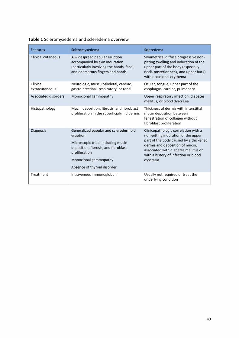
49
Table 1 Scleromyxedema and scleredema overview
Features Scleromyxedema Scleredema
Clinical cutaneous A widespread papular eruption accompanied by skin induration (particularly involving the hands, face), and edematous fingers and hands
Symmetrical diffuse progressive non-pitting swelling and induration of the upper part of the body (especially neck, posterior neck, and upper back) with occasional erythema
Clinical extracutaneous
Neurologic, musculoskeletal, cardiac, gastrointestinal, respiratory, or renal
Ocular, tongue, upper part of the esophagus, cardiac, pulmonary
Associated disorders Monoclonal gammopathy Upper respiratory infection, diabetes mellitus, or blood dyscrasia
Histopathology Mucin deposition, fibrosis, and fibroblast proliferation in the superficial/mid dermis
Thickness of dermis with interstitial mucin deposition between fenestration of collagen without fibroblast proliferation
Diagnosis Generalized papular and sclerodermoid eruption
Microscopic triad, including mucin deposition, fibrosis, and fibroblast proliferation
Monoclonal gammopathy
Absence of thyroid disorder
Clinicopathologic correlation with a non-pitting induration of the upper part of the body caused by a thickened dermis and deposition of mucin, associated with diabetes mellitus or with a history of infection or blood dyscrasia
Treatment Intravenous immunoglobulin Usually not required or treat the underlying condition
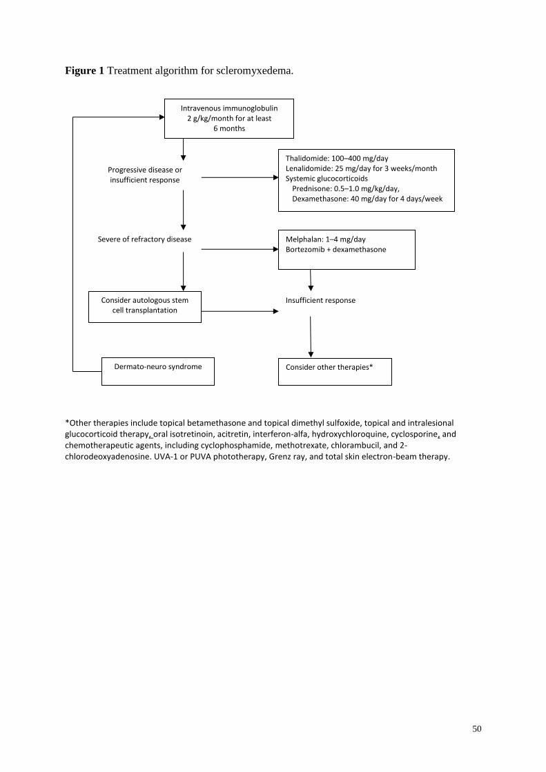
50
Figure 1 Treatment algorithm for scleromyxedema.
*Other therapies include topical betamethasone and topical dimethyl sulfoxide, topical and intralesional glucocorticoid therapy, oral isotretinoin, acitretin, interferon-alfa, hydroxychloroquine, cyclosporine, and chemotherapeutic agents, including cyclophosphamide, methotrexate, chlorambucil, and 2-chlorodeoxyadenosine. UVA-1 or PUVA phototherapy, Grenz ray, and total skin electron-beam therapy.
Intravenous immunoglobulin 2 g/kg/month for at least
6 months
Thalidomide: 100–400 mg/day Lenalidomide: 25 mg/day for 3 weeks/month Systemic glucocorticoids
Prednisone: 0.5–1.0 mg/kg/day, Dexamethasone: 40 mg/day for 4 days/week
Melphalan: 1–4 mg/day Bortezomib + dexamethasone
Consider autologous stem cell transplantation
Consider other therapies* Dermato-neuro syndrome
Progressive disease or insufficient response
Severe of refractory disease
Insufficient response
51
References
1 Rongioletti F, Rebora A. Mucinoses. In: Dermatology (Bolognia J, Jorizzo JL, Schaffer
JV, et al., eds), 3rd edn. Philadelphia: Elsevier, 2012; 1: 687.
2. Rongioletti F. Lichen myxedematosus (papular mucinosis): new concepts and perspectives
for an old disease. Semin Cutan Med Surg 2006; 25: 100–104.
3. Rongioletti F, Merlo G, Cinotti E, et al. Scleromyxedema: a multicenter study of
characteristics, comorbidities, course, and therapy in 30 patients. J Am Acad Dermatol
2013; 69: 66–72.
4. Cokonis Georgakis CD, Falasca G, Georgakis A, Heymann WR. Scleromyxedema. Clin
Dermatol 2006; 24: 493–497.
5. Feasel AM, Donato ML, Duvic M. Complete remission of scleromyxedema following
autologous stem cell transplantation. Arch Dermatol 2001; 137: 1071.
6. Yeung CK, Loong F, Kwong YL. Scleromyxoedema due to a plasma cell neoplasm: rapid
remission with bortezomib, thalidomide and dexamethasone. Br J Haematol 2012; 157:
411.
7. Berger JR, Dobbs MR, Terhune MH, Maragos WF. The neurologic complications of
scleromyxedema. Medicine 2001; 80: 313.
8. Rongioletti F, Hazini A, Rebora A. Coma associated with scleromyxoedema and interferon
alfa therapy. Full recovery after steroids and cyclophosphamide combined with
plasmapheresis. Br J Dermatol 2001; 144: 1283.
9. Fleming KE, Virmani D, Sutton E, et al. Scleromyxedema and the dermato-neuro
syndrome: case report and review of the literature. J Cutan Pathol 2012; 39: 508.
10. Rey JB, Luria RB. Treatment of scleromyxedema and the dermatoneuro syndrome with
intravenous immunoglobulin. J Am Acad Dermatol 2009; 60: 1037.
11. Espinosa A, De Miguel E, Morales C, et al. Scleromyxedema associated with arthritis and
myopathy: a case report. Clin Exp Rheumatol 1993; 11: 545.
12. Jamieson TW, De Smet AA, Stechschulte DJ. Erosive arthropathy associated with
scleromyxedema. Skeletal Radiol 1985; 14: 286.
13. Helfrich DJ, Walker ER, Martinez AJ, Medsger TA Jr. Scleromyxedema myopathy: case
report and review of the literature. Arthritis Rheum 1988; 31: 1437.
14. Launay D, Hatron PY, Delaporte E, et al. Scleromyxedema (lichen myxedematosus)
associated with dermatomyositis. Br J Dermatol 2001; 144: 359.
15. Rothe MJ, Rivas R, Gould E, Kerdel FA. Scleromyxedema and severe myositis. Int J
Dermatol 1989; 28: 657.
52
16. Ozdag F, Akar A, Eroglu E, Erbil H. Acute rhabdomyolysis during the treatment of
scleromyxedema with interferon alfa. J Dermatolog Treat 2001; 12: 167.
17. Pomann JJ, Rudner EJ. Scleromyxedema revisited. Int J Dermatol 2003; 42: 31.
18. De Simone C, Castriota M, Carbone A, et al. Cardiomyopathy in scleromyxedema: report
of a fatal case. Eur J Dermatol 2010; 20: 852.
19. Morris-Jones R, Staughton RC, Walker M, et al. Lichen myxoedematosus with associated
cardiac abnormalities. Br J Dermatol 2001; 144: 594.
20. Dinneen AM, Dicken CH. Scleromyxedema. J Am Acad Dermatol 1995; 33: 37.
21. Blum M, Wigley FM, Hummers LK. Scleromyxedema: a case series highlighting long-
term outcomes of treatment with intravenous immunoglobulin (IVIG). Medicine 2008; 87:
10.
22. Le Moigne M, Mazereeuw-Hautier J, Bonnetblanc JM, et al. [Clinical characteristics,
outcome of scleromyxoedema: a retrospective multicentre study]. Ann Dermatol Venereol
2010; 137: 782.
23. Rapp MF, Guram M, Konrad HR, et al. Laryngeal involvement in scleromyxedema: a case
report. Otolaryngol Head Neck Surg 1991; 104: 362.
24. Lee YH, Sahu J, O’Brien MS, et al. Scleroderma renal crisis-like acute renal failure
associated with mucopolysaccharide accumulation in renal vessels in a patient with
scleromyxedema. J Clin Rheumatol 2011; 17: 318.
25. Chan JC, Trendell-Smith NJ, Yeung CK. Scleromyxedema: a cutaneous paraneoplastic
syndrome associated with thymic carcinoma. J Clin Oncol 2012; 30: e27.
26. Alfadley A, Al Hoqail I, Al Eisa A. Scleromyxedema: possible association with seminoma.
J Am Acad Dermatol 2000; 42: 875.
27. Giménez Garcia R, Garcia SG, Suarez Vilela D, Moro Sanchez MJ. Scleromyxedema
associated with non-Hodgkin lymphoma. Int J Dermatol 1989; 28: 670.
28. Hardie RA, Hunter JA, Urbaniak S, Habeshaw JA. Spontaneous resolution of lichen
myxoedematosus. Br J Dermatol 1979; 100: 727.
29. Rongioletti F, Rebora A. Cutaneous mucinoses: microscopic criteria for diagnosis. Am J
Dermatopathol 2001; 23: 257.
30. Rongioletti F, Rebora A. Updated classification of papular mucinosis, lichen
myxedematosus, and scleromyxedema. J Am Acad Dermatol 2001; 44: 273
31. Rongioletti F, Cozzani E, Parodi A. Scleromyxedema with an interstitial granulomatous-
like pattern: a rare histologic variant mimicking granuloma annulare. J Cutan Pathol 2010;
37: 1084.
53
32. Delyon J, Bézier M, Rybojad M, et al. Specific lymph node involvement in
scleromyxedema: a new diagnostic entity for hypermetabolic lymphadenopathy. Virchows
Arch 2013; 462: 679.
33. Nashel J, Steen V. Scleroderma mimics. Curr Rheumatol Rep 2012; 14: 39–46.
34. Bidier M, Zschoche C, Gholam P, et al. Scleromyxoedema: clinical follow-up after
successful treatment with high-dose immunoglobulins reveals different long-term
outcomes. Acta Derm Venereol 2012; 92: 408.
35. Sroa N, Campbell S, Bechtel M. Intravenous immunoglobulin therapy for
scleromyxedema: a case report and review of literature. J Drugs Dermatol 2010; 9: 263.
36. Gholam P, Hartmann M, Enk A. Arndt-Gottron scleromyxoedema: successful therapy with
intravenous immunoglobulins. Br J Dermatol 2007; 157: 1058.
37. Körber A, Franckson T, Grabbe S, Dissemond J. Successful therapy of scleromyxoedema
Arndt-Gottron with low-dose intravenous immunoglobulin. J Eur Acad Dermatol Venereol
2007; 21: 553.
38. Samuelsson A, Towers TL, Ravetch JV. Anti-inflammatory activity of IVIG mediated
through the inhibitory Fc receptor. Science 2001; 291: 484.
39. Binitha MP, Nandakumar G, Thomas D. Suspected cardiac toxicity to intravenous
immunoglobulin used for treatment of scleromyxedema. Indian J Dermatol Venereol
Leprol 2008; 74: 248.
40. Sansbury JC, Cocuroccia B, Jorizzo JL, et al. Treatment of recalcitrant scleromyxedema
with thalidomide in 3 patients. J Am Acad Dermatol 2004; 51: 126.
41. Guarenti I, Sebastiani V, Pinto G, et al. Successful treatment of scleromyxedema with oral
thalidomide. Int J Dermatol 2013; 52: 631.
42. Martins A, Paiva Lopes MJ, Tavares Belo R, Rodrigues JC. Scleromyxedema –
thalidomide therapy. J Eur Acad Dermatol Venereol 2008; 22: 622.
43. Efthimiou P, Blanco M. Intravenous gammaglobulin and thalidomide may be an effective
therapeutic combination in refractory scleromyxedema: case report and discussion of the
literature. Semin Arthritis Rheum 2008; 38: 188.
44. Brunet-Possenti F, Hermine O, Marinho E, et al. Combination of intravenous
immunoglobulins and lenalidomide in the treatment of scleromyxedema. J Am Acad
Dermatol 2013; 69: 319.
45. Rayson D, Lust JA, Duncan A, Su WP. Scleromyxedema: a complete response to
prednisone. Mayo Clin Proc 1999; 74: 481.
54
46. Lin YC, Wang HC, Shen JL. Scleromyxedema: an experience using treatment with
systemic corticosteroid and review of the published work. J Dermatol 2006; 33: 207.
47. Horn KB, Horn MA, Swan J, et al. A complete and durable clinical response to high-dose
dexamethasone in a patient with scleromyxedema. J Am Acad Dermatol 2004; 51: S120.
48. Cañueto J, Labrador J, Román C, et al. The combination of bortezomib and dexamethasone
is an efficient therapy for relapsed/refractory scleromyxedema: a rare disease with new
clinical insights. Eur J Haematol 2012; 88: 450.
49. Bos R, de Waal EG, Kuiper H, et al. Thalidomide and dexamethasone followed by
autologous stem cell transplantation for scleromyxoedema. Rheumatology 2011; 50: 1925.
50 Gholam P, Hartmann M, Enk A. Arndt-Gottron scleromyxoedema: successful therapy with
intravenous immunoglobulins. Br J Dermatol 2007; 157: 1058–1060.
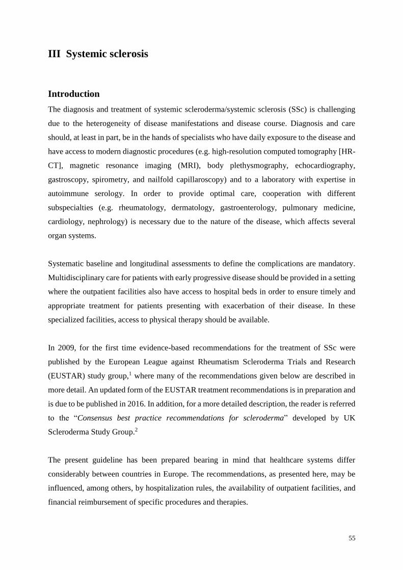
55
III Systemic sclerosis
Introduction
The diagnosis and treatment of systemic scleroderma/systemic sclerosis (SSc) is challenging
due to the heterogeneity of disease manifestations and disease course. Diagnosis and care
should, at least in part, be in the hands of specialists who have daily exposure to the disease and
have access to modern diagnostic procedures (e.g. high-resolution computed tomography [HR-
CT], magnetic resonance imaging (MRI), body plethysmography, echocardiography,
gastroscopy, spirometry, and nailfold capillaroscopy) and to a laboratory with expertise in
autoimmune serology. In order to provide optimal care, cooperation with different
subspecialties (e.g. rheumatology, dermatology, gastroenterology, pulmonary medicine,
cardiology, nephrology) is necessary due to the nature of the disease, which affects several
organ systems.
Systematic baseline and longitudinal assessments to define the complications are mandatory.
Multidisciplinary care for patients with early progressive disease should be provided in a setting
where the outpatient facilities also have access to hospital beds in order to ensure timely and
appropriate treatment for patients presenting with exacerbation of their disease. In these
specialized facilities, access to physical therapy should be available.
In 2009, for the first time evidence-based recommendations for the treatment of SSc were
published by the European League against Rheumatism Scleroderma Trials and Research
(EUSTAR) study group,1 where many of the recommendations given below are described in
more detail. An updated form of the EUSTAR treatment recommendations is in preparation and
is due to be published in 2016. In addition, for a more detailed description, the reader is referred
to the “Consensus best practice recommendations for scleroderma” developed by UK
Scleroderma Study Group.2
The present guideline has been prepared bearing in mind that healthcare systems differ
considerably between countries in Europe. The recommendations, as presented here, may be
influenced, among others, by hospitalization rules, the availability of outpatient facilities, and
financial reimbursement of specific procedures and therapies.
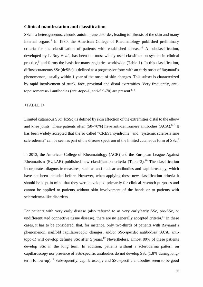
56
Clinical manifestation and classification
SSc is a heterogeneous, chronic autoimmune disorder, leading to fibrosis of the skin and many
internal organs.3 In 1980, the American College of Rheumatology published preliminary
criteria for the classification of patients with established disease.4 A subclassification,
developed by LeRoy et al., has been the most widely used classification system in clinical
practice,5 and forms the basis for many registries worldwide (Table 1). In this classification,
diffuse cutaneous SSc (dcSSc) is defined as a progressive form with an early onset of Raynaud’s
phenomenon, usually within 1 year of the onset of skin changes. This subset is characterized
by rapid involvement of trunk, face, proximal and distal extremities. Very frequently, anti-
topoisomerase-1 antibodies (anti-topo-1, anti-Scl-70) are present.6–8
<TABLE 1>
Limited cutaneous SSc (lcSSc) is defined by skin affection of the extremities distal to the elbow
and knee joints. These patients often (50–70%) have anti-centromere antibodies (ACA).6–8 It
has been widely accepted that the so called “CREST syndrome” and “systemic sclerosis sine
scleroderma” can be seen as part of the disease spectrum of the limited cutaneous form of SSc.9
In 2013, the American College of Rheumatology (ACR) and the European League Against
Rheumatism (EULAR) published new classification criteria (Table 2).10 The classification
incorporates diagnostic measures, such as anti-nuclear antibodies and capillaroscopy, which
have not been included before. However, when applying these new classification criteria it
should be kept in mind that they were developed primarily for clinical research purposes and
cannot be applied to patients without skin involvement of the hands or to patients with
scleroderma-like disorders.
For patients with very early disease (also referred to as very early/early SSc, pre-SSc, or
undifferentiated connective tissue disease), there are no generally accepted criteria.11 In these
cases, it has to be considered, that, for instance, only two-thirds of patients with Raynaud’s
phenomenon, nailfold capillaroscopic changes, and/or SSc-specific antibodies (ACA, anti-
topo-1) will develop definite SSc after 5 years.12 Nevertheless, almost 80% of these patients
develop SSc in the long term. In addition, patients without a scleroderma pattern on
capillaroscopy nor presence of SSc-specific antibodies do not develop SSc (1.8% during long-
term follow-up).12 Subsequently, capillaroscopy and SSc-specific antibodies seem to be good
57
prognostic predictors for the disease. Therefore, it is recommended that patients with suspected
early SSc are referred to centers that are experienced in SSc diagnosis and care.
Diagnostic procedures
Antinuclear antibodies
Autoantibodies targeting characteristic nuclear antigens are one of the hallmarks of SSc. The
frequency of detection of antinuclear antibodies (ANA) in SSc patients in a recent study
approached 95%,8 which corresponds well with ANA frequencies of between 85% and 99%
reported in the literature. In this study, 86.6% of the ANA-positive patients had SSc-specific
antibodies, 96.4% of which were detecting five antigens (i.e. centromere, topoisomerase-1,
RNA polymerase III, PM/Scl, U1-RNP) (Table 3). It is generally well accepted that the SSc-
specific antibodies described above are largely mutually exclusive. Coincidences in individual
patients do occur but are rare.
For a more detailed description of autoantibodies linked to overlap syndromes, please see
section V (Systemic sclerosis overlap syndromes).
<TABLE 3>
Capillaroscopy
Capillaroscopy (e.g. videocapillaroscope, stereomicroscope, or dermatoscope) is a well-
established, non-invasive technique for the identification of changes in the nailfold capillary
that differentiate primary Raynaud’s phenomenon from SSc.
For a detailed review the reader is referred to the article by Cutolo et al.13
Organ involvement and diagnostic work-up
Raynaud’s phenomenon
Raynaud’s phenomenon is characterized by a vasospasm resulting in blanching, cyanosis, and
then reactive hyperemia (triphasic). Raynaud’s phenomenon is present in more than 90% of
patients. It typically affects the hands, less commonly the feet, but may also involve the tongue,
ears, and nose. Cold exposure is the usual trigger, but emotional stress may evoke the same
symptoms.
58
Primary Raynaud’s phenomenon is mainly caused by functional disturbances, whereas in
secondary Raynaud’s phenomenon in the context of SSc, there is also involvement of structural
alterations in digitate arteries. These combined changes are considered to be major causes for
the formation of ulcers. To distinguish primary from secondary Raynaud’s phenomenon,
nailfold capillaroscopy and the analysis of autoantibodies are required. Additional laboratory
and radiologic examinations may become necessary in order to exclude other factors that may
contribute to the symptoms of Raynaud’s phenomenon.14
Skin fibrosis
At the onset of the disease, particularly in the diffuse form, patients tend to have swollen fingers
and hands over extended periods of time, so called “puffy hands.” Sclerotic changes follow
later on, finally leading to dermatogenic contractures and sclerodactyly. Perioral plication and
microstomia are typical features of the face, as is a mask-like stiffness.
The best and validated tool to measure the progress of the skin sclerosis is the modified Rodnan
Skin Score (mRSS). At 17 different anatomical areas, the skin score is evaluated by manual
palpation. The skin score is 0 for uninvolved skin, 1 for mild thickening, 2 for moderate
thickening, and 3 for severe thickening. Subsequently, the sum will be used as the total skin
score. The mRSS is feasible, reliable, and has been validated for initial and follow-up skin
evaluation. The administration of this simple method requires some experience, and a careful
teaching process is warranted.15
Skin involvement and its rate of progression are thought to reflect the severity of internal organ
involvement. However, in later disease stages, internal organ involvement may progress while
skin fibrosis of the trunk and proximal extremities will diminish.
Fibrosis may be accompanied by additional symptoms such as hair loss, diminished sweating,
hyperpigmentation, depigmentation, or severe pruritus.
Digital ulceration
Among patients with SSc, 15–25% have active digital ulceration (DU) and 35% have or have
had DUs in the past, although this number varies considerably between centers and studies.16–
19 Analysis of registry data indicates that the extent of skin sclerosis, male sex, presence of
pulmonary arterial hypertention, involvement of the esophagus, presence of anti-topo-1 (but
59
not anti-centromere) antibodies, early age at onset of Raynaud’s phenomenon, and elevated
erythrocyte sedimentation rate could be independent risk factors.16,18 History of DU when
patients first present has been shown to predict the occurrence of DUs at follow-up, and is
associated with cardiovascular worsening and decreased survival.20
Ulcers that occur on the fingertip are thought to be exclusively due to ischemia, whereas ulcers
over the extensor surfaces of the proximal and distal interphalangeal joints have a mixed
etiology. They are usually due to a combination of poor perfusion, stretched fibrotic skin, and
trauma. DUs are complicated by secondary infection, osteomyelitis, gangrene, and amputation.
Acro-osteolysis may further complicate wound healing. Recurring ulcers lead to chronic use of
pain relievers and antibiotics, and eventually to hospitalization either for treatment of active
DUs or for surgery (amputation).21
Contributory causes, such as coexisting large vessel disease, should be excluded. In addition,
differential diagnoses, such as vasculitis, thrombangitis, or arteriosclerotic vascular disease,
should be ruled out. Calcinosis cutis should be distinguished from superficial ulceration, but is
a possible risk factor for DU.
Calcinosis cutis
Calcinosis cutis is marked by subcutaneous calcium carbonate deposits, which appear in all
subtypes of SSc and most frequently on the acral parts of the body. They may induce superficial
erosions and cause intense pain for the patient. Calcinosis cutis is an important differential
diagnosis to DUs and can be excluded via X-ray of the affected body parts.
Musculoskeletal system
Arthralgia and musculoskeletal pain are among the most frequent complaints in SSc and may
lead to secondary fibromyalgia. Tendon friction rubs are a typical sign of an inflammatory,
progressive form of the disease. Muscle weakness and a varying increase in serum creatine
kinase levels are quite common and can indicate the presence of an SSc-myositis overlap
syndrome (i.e. Scl syndrome, anti-synthetase syndrome, mixed connective tissue disease). In
these cases, magnetic resonance imaging and a muscle biopsy to determine the type of myositis
should be considered.
60
Inflammatory arthritis can occur in up to 10% of patients and raises the suspicion of the
presence of an SSc overlap syndrome (SSc-rheumatoid arthritis). In these cases, rheumatoid
factors and anti-cyclic citrullinated peptide (CCP) antibodies (ACPA) (Table 3) should be
determined and a rheumatologic work-up initiated. A more detailed description of the diagnosis
and treatment can be found in section V (Systemic sclerosis overlap syndromes).
Pulmonary involvement
Interstitial lung disease
Interstitial lung disease (ILD) affects up to 65% of SSc patients to varying degrees. The typical
presentation is a predominantly bibasilar pattern. While some patients develop a rapid decline
of forced vital capacity (FVC) within the first 3 years, others may remain remarkably stable or
may even experience improvement.22 In early disease, inflammatory alveolitis may precede
and/or accompany interstitial fibrosis, leading to loss of pulmonary function as evidenced by
decreased diffusing capacity of the lungs for carbon monoxide (DLCO) and decreased FVC in
more severe cases. Most often the ILD corresponds to a non-specific interstitial pneumonitis.
The majority of patients will present with symptoms such as dyspnea, a dry cough, and reduced
exercise tolerance. Chest X-ray can be useful but is a relatively insensitive method for the
detection of ILD. Chest HR-CT has a markedly higher diagnostic sensitivity and is the
recommended diagnostic tool to determine the extent and distribution of ILD. The sensitivity
of HR-CT is superior when compared with lung function testing (LFT).23 LFT should include
spirometry, body plethysmography, and DLCO (corrected for hemoglobin). LFT should be
performed every 6 months, or more frequently if the patient is developing a loss in FVC and/or
a decrease in transfer factor (DLCO).
Pulmonary hypertension
Pulmonary arterial hypertension (PAH) occurs in about 15% of patients, and develops
particularly in patients with long disease duration and anti-centromere antibodies. PAH is
associated with significant mortality and is among the most common causes of death in SSc.24
All SSc patients should be evaluated for possible PAH in line with current recommendations,
and referred for specialist management. Annual screening on symptoms (unexplained or
progressive dyspnea, syncope, signs of right heart failure) and by echocardiography are strongly
recommended in all SSc patients,1 and are part of the current recommendations of cardiologic
and pulmonary societies (see 2015 Guidelines of the European Society of Cardiology25).
61
Gastrointestinal involvement
The gastrointestinal tract is frequently involved, with 80% of patients having esophageal
involvement and 40–70% having involvement of the stomach, small intestine, and large
intestine.7,26 In longstanding disease (i.e. >10 years), upper gastrointestinal involvement occurs
in nearly all patients. The most common symptoms are heartburn, esophageal dysfunction in
the upper gastrointestinal tract, diarrhea due to bacterial overgrowth, and fecal incontinence in
the distal tract. Barrett’s esophagus is a late sequel of reflux disease and requires surveillance
according to the respective guidelines.27
Rarely, telangiectasias may also be present on the mucosa, representing a potential source of
occult intestinal bleeding. The standard diagnostic procedure is endoscopy.
Cardiac involvement
The nature and severity of cardiac disease depends on the extent of myocardial fibrosis, and on
the extent to which concurrent fibrosis of the lung and thickening and fibrosis of the small
pulmonary arteries place an additional burden on the circulation. Myocarditis and pericarditis
can be observed in a subset of patients and may lead to diagnostic uncertainty. Risk factors for
cardiac involvement are diffuse disease, particularly with rapid progression, and signs of
inflammation such as tendon friction rubs. Patchy myocardial fibrosis contributes to diastolic
dysfunction and to a diminished left ventricular ejection fraction.
Arrhythmias are quite common in SSc. In patients with the diffuse form of SSc, severe forms
of arrhythmias are considered an important source of mortality.28 As regular electrocardiogram
is relatively insensitive, there should be a low threshold to use Holter monitoring.
Renal involvement
Acute renal crisis is a serious and potentially fatal SSc complication. It occurs most likely in
patients with the progressive, diffuse form with a disease duration of less than 4 years. The
presence of anti-RNA polymerase III antibodies is considered a particular risk factor and is
detected in about one third of cases.29 Thus, regular control of blood pressure (at least twice
weekly/home monitoring) is recommended to detect acute renal involvement early on.
Glucocorticoids in higher doses exceeding 15 mg prednisone equivalents should be avoided
due to their long-term side-effects and association with renal crisis.29
62
In a small subset of patients, normotensive acute renal crisis will develop. In these cases,
patients often present with signs of thrombotic microangiopathy. Chronic renal involvement in
SSc is associated with a slowly progressive obliterative vasculopathy. Urinary protein excretion
has been determined in several studies as a major independent risk factor for mortality.30
Therefore, urinary protein excretion should be determined at least annually.
General recommendation for a regular diagnostic work-up in patients with SSc
After an initial baseline assessment (Table 4), at least annual, life-long, follow-up of patients is
recommended due to the chronic nature of the disease. In patients with progressive disease,
corresponding with disease activity, patients should be followed more frequently. The annual
work-up should include a thorough clinical investigation including mRSS and the following
diagnostic measures: lung function test with plethysmography including DLCO, blood
pressure, electrocardiography, echocardiography, Erythrocyte sedimentation rate/C-reactive
protein, complete blood count, clinical chemistry (liver function, creatinine, urea) and urinary
protein.
Particularly in patients with an increased risk for renal crisis (progressive diffuse disease, anti-
RNA polymerase III antibodies), frequent blood pressure measurements are recommended
(preferably home monitoring) (Table 5).
<TABLE 4>
<TABLE 5>
Treatment
Therapy for skin involvement
Treatment of Raynaud’s phenomenon
Avoidance of cold exposure and the constant protection against cold is paramount. Heated
gloves, shoes, and pockets are usual measures. Furthermore, paraffin baths, heated seed pillows,
therapy balls, and physical therapy are recommended.31 Smoking should be stopped. Beta-
blocker treatment should be substituted, if feasible.
These lifestyle measures should be supported by pharmacologic therapy (Fig. 1). First-line
therapy consists of calcium antagonists such as nifedipine or amlodipine. Large meta-analyses
63
have revealed that calcium antagonists reduce the severity and frequency of Raynaud’s attacks.
The dosage should be increased carefully. Recent controlled studies indicated that PDE-5
inhibitors (i.e. sildenafil, vardenafil) may also be effective in the treatment of Raynaud’s
phenomenon, by reducing the severity and frequency of attacks.32–34 However, these drugs have
not been licensed for this indication. Selective serotonin reuptake inhibitors, such as fluoxetine,
have shown benefit in some patients,35 and angiotensin-converting enzyme (ACE) inhibitors or
angiotensin-receptor antagonists may also be considered.36
<FIGURE 1>
An improvement of severe Raynaud’s phenomenon has been demonstrated following
intravenously administered iloprost.37,38 A dosage of 0.5–2 ng/kg/min for 3–6 hours on at least
five consecutive days at monthly intervals is generally recommended.1,39 The most frequent
side-effects are headaches, low blood pressure, and cutaneous flushing. To minimize these side-
effects, a slow daily increase of the dosage, depending on the individual patient’s condition, is
necessary.39
Digital (palmar) sympathectomy (with or without botulinum toxin injection) may be considered
in severe and/or refractory cases.
Treatment of digital ulceration
Avoidance of cold exposure and cessation of smoking are accompanying measures. Beta-
blocker treatment should be substituted, if feasible. A modified algorithm as published by
Riemekasten et al.40 is shown in Fig. 2.
Infections, especially those that affect deep adjacent structures, should be treated with
antibiotics in order to prevent osteomyelitis and avoid amputation.41 If possible, the antibiotic
therapy should be combined with a vasodilatory therapy to improve perfusion of the involved
area. Sufficient analgesic therapy is recommended to improve quality of life and to reduce pain-
induced vasoconstriction. Adequate wound care and regular clinical inspection are mandatory,
in order to prevent infections, gangrene or necrosis.41 In the case of dry, superficial ulcers, non-
occlusive wound care is recommended. The use of a protective wound dressing (i.e. alginate)
is advised when deep ulcers are present in order to protect the wound from sources of infection
64
and to support granulation. Wound care includes a thorough cleaning and disinfection of the
wound with sodium chloride, antiseptics or wound cleansing solutions.
Two randomized controlled trials demonstrated that intravenous iloprost is efficacious in
healing digital ulcers in SSc. It should be administered at a dosage of 0.5–2 ng/kg per minute
for 3–6 hours for at least five consecutive days.1,39 The recommended treatment duration varies
between 3 and 14 days, and is in part influenced by restrictions in the respective national
healthcare system.39
A recent meta-analysis of several randomized controlled trials indicated that PDE-5 inhibitors
improve healing of digital ulcers.42 Therefore, PDE-5 inhibitors can be considered for the
treatment of active digital ulcers.
Bosentan is a non-selective endothelin receptor antagonist that demonstrated efficacy in the
prevention of digital ulcers in two randomized and controlled studies (RAPIDS-1 and -2) in
SSc patients.43–45 A significant reduction in the number of new ulcers was revealed, particularly
in patients with multiple ulcers. Side-effects consist of possible liver toxicity, teratogenicity,
and reduced effectiveness of oral contraceptive pills through interference with the cytochrome
P450 system.1,42 Bosentan does not affect healing of active DUs.
Digital (palmar) sympathectomy (with or without botulinum toxin injection) may be considered
in severe and/or refractory cases.36
Treatment of skin fibrosis
Therapy for skin sclerosis should be guided by the phase of the fibrotic process (early phase vs.
late phase), the disease activity, and the progression of the fibrosis. General measures include
skin protection from cold and trauma, skin care with moisturising creams, lymph drainage, and
active physiotherapy for the prevention of contractures. These general measures may suffice in
mild, non-progressing forms of fibrosis.
In the early phase with limited skin involvement and LS, UVA1 or photochemotherapy (PUVA)
should be considered. Similarly to the successful treatment of LS with UVA modalities, a
number of uncontrolled studies have indicated a beneficial effect on fibrosis in SSc.46–48
However, controlled studies are still lacking. Pruritus often occurs in fibrotic skin, and may
65
respond to standard therapy and phototherapy. For further details, the reader is referred to Fig.
1 in the LS section, However, longer treatment durations may be needed.
Photopheresis (extracorporeal photochemotherapy) has shown promise in several controlled
studies.49,50 It can be used as second-line or adjuvant therapy. It is recommended that it should
be applied in early progressive disease, preferably of less than 2 years’ duration. For more
details, the reader is referred to the 2014 EDF guideline.51
The systemic use of glucocorticoids, which is considered a standard therapy for most
autoimmune diseases, plays no role in the therapy of fibrosis in patients with SSc.1 More
importantly, it is well known that glucocorticoids in a dose of >15 mg are associated with a
higher incidence of renal crisis.29
The best data for systemic therapy of progressive skin fibrosis are available for methotrexate.
In two randomized, controlled studies it was shown that methotrexate decreased skin fibrosis
in early diffuse SSc. Positive effects on other organs such as the lung could not be shown.52,53
A dosage of 10–15 mg per week for 6–12 months is generally recommended. Higher dosages
may be considered. The use of mycophenolate mofetil (MMF) is recommended by the
EUSTAR study group as second-line therapy following methotrexate.1,54 The recommended
standard dosage varies at about 1–2 g per day for at least 12 months.1,54
An improvement of skin sclerosis was demonstrated for cyclophosphamide in the scleroderma
lung study.1,55 The use of cyclophosphamide is recommended after failure of methotrexate and
MMF due to high rates of side-effects.54 As renewed deterioration of mRSS and lung
involvement were observed during follow-up in the scleroderma lung study, a continuation of
immunosuppression with MMF or azathioprine after cyclophosphamide therapy is
recommended by some experts. An algorithm for the treatment of SSc skin fibrosis is shown in
Fig. 3.
Treatment of calcinosis cutis
Various therapeutic strategies have been investigated, but there is currently no evidence of an
effective therapy for calcinosis cutis. Ectopic calcifications or calcinosis that compromise blood
circulation or cause symptoms may be removed surgically or by the use of carbon dioxide laser.
66
Surgical excision seems to be the best option after failure of conservative treatment attempts.
However, surgery should only be performed in cases of urgent medical indication.56–58
Treatment of telangiectasias
Telangiectasia may appear in the face, the hands (even on the palms), and the mucosa of patients
with SSc.59,60 Laser (i.e. potassium titanyl phosphate or flashlamp pulsed dye laser) or intense
pulsed light therapy is the treatment of choice to remove telangiectasias.59,61 Cosmetics are often
used to cover the affected area.
Therapy for musculoskeletal involvement
For detailed treatment recommendations, the reader is referred to Section V (Systemic sclerosis
overlap syndromes).
Therapy for pulmonary involvement
Treatment of lung fibrosis
ILD in many patients is relatively mild and has a low rate of progression. However, particulary
in patients with progressive diffuse disease, a severe reduction in FVC can ensue and the
progressive lung fibrosis is recognized as a major cause of mortality.22 It is therefore crucial to
identify patients with risk for ILD and to identify patients with a significant progression as
measured by a reduction of FVC (>5% in 6 months or >10% in 1 year) or DLCO (>15% in
1 year). Patients with ILD should be considered for early treatment, when the disease is active
and the damage is not yet irreversible. Another component of therapy should be adequate
treatment of reflux disease, as this may prevent progression of ILD.62
The best available data exist for cyclophosphamide, which showed a modest, statistically
significant benefit in a randomized, controlled, double-blind trial on both lung and skin
fibrosis.55 As the follow-up data of this trial indicated a renewed progression of fibrosis, several
groups recommend the prolongation of immunosuppression after 6 or 12 pulses of
cyclophosphamide by the use of azathioprine or MMF.54
Two randomized controlled trials and a number of uncontrolled studies have shown that
hematopoietic stem cell transplantation improves lung function and skin fibrosis compared with
standard immunosuppressive treatment.63,64 Transplantation can result in rapid (over months)
and sustained improvement of mRSS and FVC. However, in the first year, a significantly
67
increased mortality was observed in the transplantation arm.63 Careful selection of SSc patients
for transplantation is mandatory.
Treatment of pulmonary arterial hypertension
Drugs targeting different aspects of vascular pathology have become available in recent years
and have dramatically changed therapy of PAH. The diagnosis and therapy of PAH belong in
the hands of an experienced cardiologist/pulmonologist with special expertise in right heart
disease. The primary task of the dermatologist taking care of an SSc patient will be to initiate
regular (i.e. at least annual) echocardiography, and to have a high clinical suspicion for this
complication (see 2015 guidelines of the European Society of Cardiology25).
Therapy for gastrointestinal involvement
Standard treatment for gastrointestinal reflux disease and the prevention of esophageal ulcers
and strictures is proton pump inhibitors (i.e. pantoprazole 40 mg/day). The majority of patients
require maintenance therapy. Second-line options are H2-blockers and antacids, in addition to
appropriate lifestyle changes.1,65
Telangiectasias may occur and cause gastrointestinal bleeding (i.e. gastric antral venous
ectasia), which should be treated by endoscopic coagulation.
Prokinetic dopamine agonists may be used for dysphagia and reflux (e.g. metoclopramide,
octreotide).66 Bacterial overgrowth and fungal infections (e.g. candida esophagitis) can be
managed by intermittent antimicrobial therapy and antimycotics.67 Anti-diarrheal agents (e.g.
loperamide) or laxatives may be used for the symptomatic management of diarrhea or
constipation that often alternate as clinical problems. Parenteral nutrition should be considered
for patients with severe weight loss refractory to enteral supplementation. For a more detailed
overview, the reader is referred to the consensus best practice pathway of the UK scleroderma
study group.27
Therapy for renal involvement
Acute renal crisis was the major cause of death before the advent of ACE-inhibitor therapy.
Prompt recognition of scleroderma renal crisis and initiation of therapy with an ACE inhibitor
offers the best opportunity for a good outcome. Other anti-hypertensive agents may be
68
considered for managing refractory hypertension in conjunction with ACE in scleroderma renal
crisis.
General recommendations for disease management
In order to tailor treatment to the individual patient, it is important to determine disease subset,
organ involvement, and disease activity. In recent years, the organ-based approach has brought
forward significant pharmacologic advancements, changing remarkably the prognosis and life
quality of patient subgroups (Table 6).
Multidisciplinary care of SSc patients should aim beyond the treatment of classic organ
involvement. Quality of life is increasingly acknowledged in clinical studies and has to be
addressed. The psychosocial well-being of SSc patients is often severely affected by the
impression of disfigurement (e.g. from telangiectasias, microstomia, contractures), and patients
should be appropriately counseled. This also applies to the treatment of chronic pain and
depression/anxiety. It has been shown that pain is an important indicator of sexual dysfunction
among women with SSc.68 Similarly, erectile dysfunction in male patients is markedly
underdiagnosed and undertreated.69 Involvement of the masticatory organ may be significant
and lead to remarkable deterioration of life quality. Sicca syndrome, gingivitis, tooth decay,
and osteolysis/necrosis all contribute to a deterioration of oral health-related quality of life.
Adjunctive therapy such as physiotherapy and respiratory therapy should be considered early
in the course of organ involvement. Small open controlled trials suggest that manual lymphatic
drainage may improve hand function in SSc.
Modern comprehensive disease management in SSc patients should be directed at the
underlying disease process and the resulting organ complications, and should also consider the
associated physical and psychological consequences.
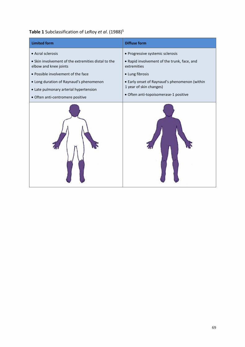
69
Table 1 Subclassification of LeRoy et al. (1988)5
Limited form Diffuse form
• Acral sclerosis
• Skin involvement of the extremities distal to the elbow and knee joints
• Possible involvement of the face
• Long duration of Raynaud’s phenomenon
• Late pulmonary arterial hypertension
• Often anti-centromere positive
• Progressive systemic sclerosis
• Rapid involvement of the trunk, face, and extremities
• Lung fibrosis
• Early onset of Raynaud’s phenomenon (within 1 year of skin changes)
• Often anti-topoisomerase-1 positive
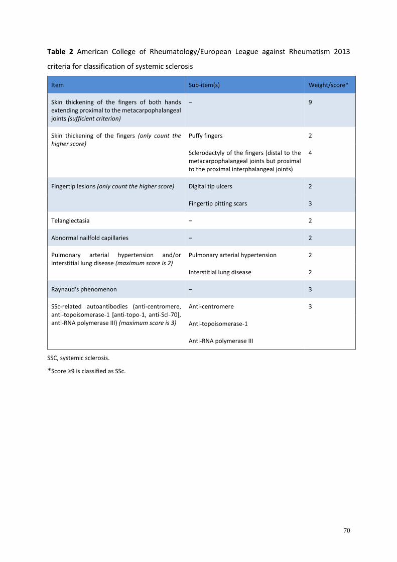
70
Table 2 American College of Rheumatology/European League against Rheumatism 2013
criteria for classification of systemic sclerosis
Item Sub-item(s) Weight/score*
Skin thickening of the fingers of both hands extending proximal to the metacarpophalangeal joints (sufficient criterion)
– 9
Skin thickening of the fingers (only count the higher score)
Puffy fingers 2
Sclerodactyly of the fingers (distal to the metacarpophalangeal joints but proximal to the proximal interphalangeal joints)
4
Fingertip lesions (only count the higher score) Digital tip ulcers 2
Fingertip pitting scars 3
Telangiectasia – 2
Abnormal nailfold capillaries – 2
Pulmonary arterial hypertension and/or interstitial lung disease (maximum score is 2)
Pulmonary arterial hypertension 2
Interstitial lung disease 2
Raynaud's phenomenon – 3
SSc-related autoantibodies (anti-centromere, anti-topoisomerase-1 [anti-topo-1, anti-Scl-70], anti-RNA polymerase III) (maximum score is 3)
Anti-centromere 3
Anti-topoisomerase-1
Anti-RNA polymerase III
SSC, systemic sclerosis.
*Score ≥9 is classified as SSc.
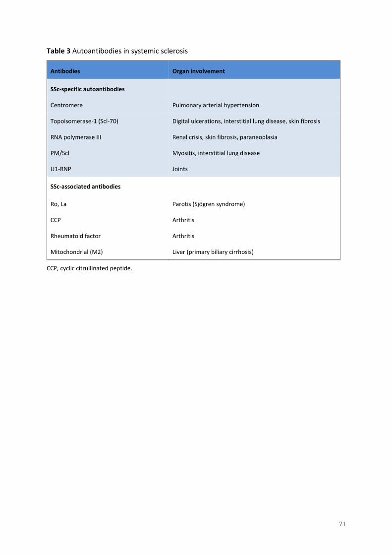
71
Table 3 Autoantibodies in systemic sclerosis
Antibodies Organ involvement
SSc-specific autoantibodies
Centromere Pulmonary arterial hypertension
Topoisomerase-1 (Scl-70) Digital ulcerations, interstitial lung disease, skin fibrosis
RNA polymerase III Renal crisis, skin fibrosis, paraneoplasia
PM/Scl Myositis, interstitial lung disease
U1-RNP Joints
SSc-associated antibodies
Ro, La Parotis (Sjögren syndrome)
CCP Arthritis
Rheumatoid factor Arthritis
Mitochondrial (M2) Liver (primary biliary cirrhosis)
CCP, cyclic citrullinated peptide.
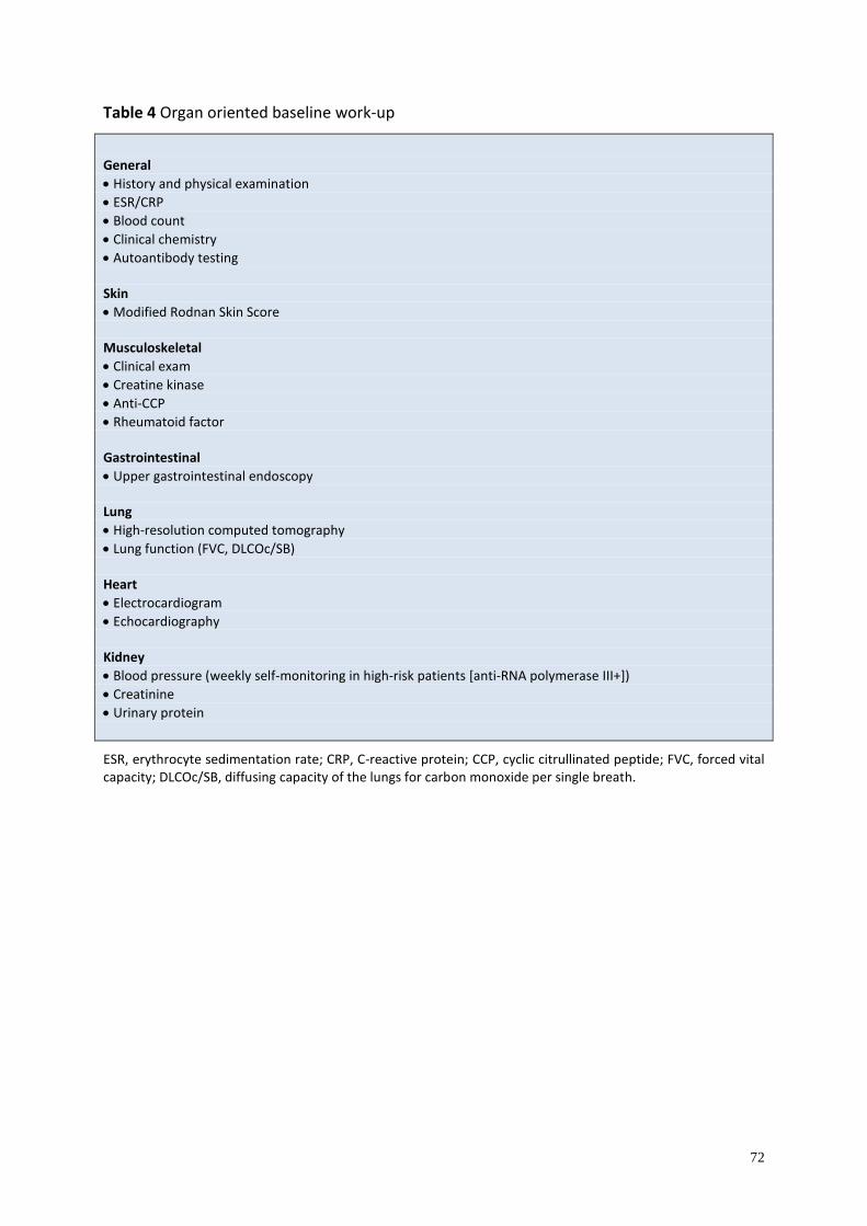
72
Table 4 Organ oriented baseline work-up
General
• History and physical examination
• ESR/CRP
• Blood count
• Clinical chemistry
• Autoantibody testing Skin
• Modified Rodnan Skin Score Musculoskeletal
• Clinical exam
• Creatine kinase
• Anti-CCP
• Rheumatoid factor Gastrointestinal
• Upper gastrointestinal endoscopy Lung
• High-resolution computed tomography
• Lung function (FVC, DLCOc/SB) Heart
• Electrocardiogram
• Echocardiography Kidney
• Blood pressure (weekly self-monitoring in high-risk patients [anti-RNA polymerase III+])
• Creatinine
• Urinary protein
ESR, erythrocyte sedimentation rate; CRP, C-reactive protein; CCP, cyclic citrullinated peptide; FVC, forced vital capacity; DLCOc/SB, diffusing capacity of the lungs for carbon monoxide per single breath.
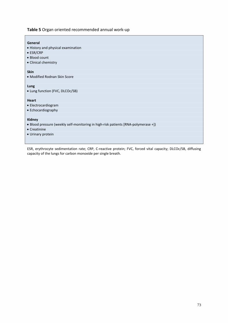
73
Table 5 Organ oriented recommended annual work-up
General
• History and physical examination
• ESR/CRP
• Blood count
• Clinical chemistry Skin
• Modified Rodnan Skin Score Lung
• Lung function (FVC, DLCOc/SB) Heart
• Electrocardiogram
• Echocardiography Kidney
• Blood pressure (weekly self-monitoring in high-risk patients [RNA-polymerase +])
• Creatinine
• Urinary protein
ESR, erythrocyte sedimentation rate; CRP, C-reactive protein; FVC, forced vital capacity; DLCOc/SB, diffusing capacity of the lungs for carbon monoxide per single breath.
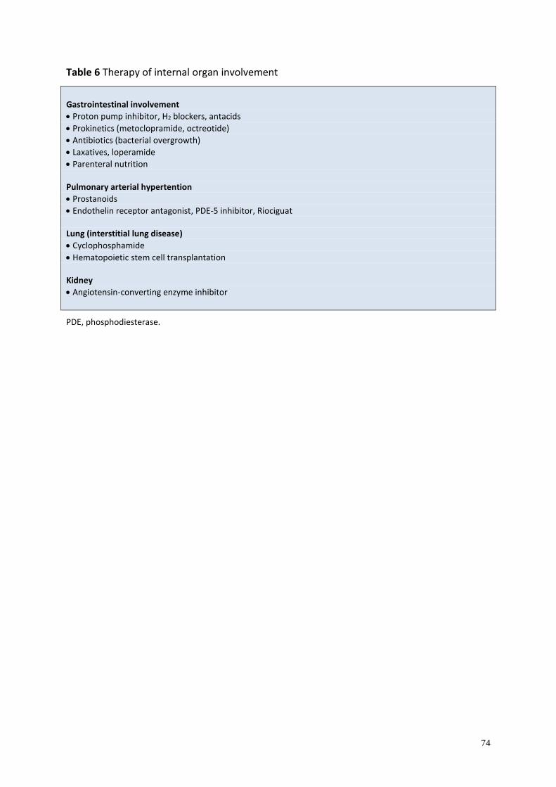
74
Table 6 Therapy of internal organ involvement
Gastrointestinal involvement
• Proton pump inhibitor, H2 blockers, antacids
• Prokinetics (metoclopramide, octreotide)
• Antibiotics (bacterial overgrowth)
• Laxatives, loperamide
• Parenteral nutrition Pulmonary arterial hypertention
• Prostanoids
• Endothelin receptor antagonist, PDE-5 inhibitor, Riociguat Lung (interstitial lung disease)
• Cyclophosphamide
• Hematopoietic stem cell transplantation Kidney
• Angiotensin-converting enzyme inhibitor
PDE, phosphodiesterase.
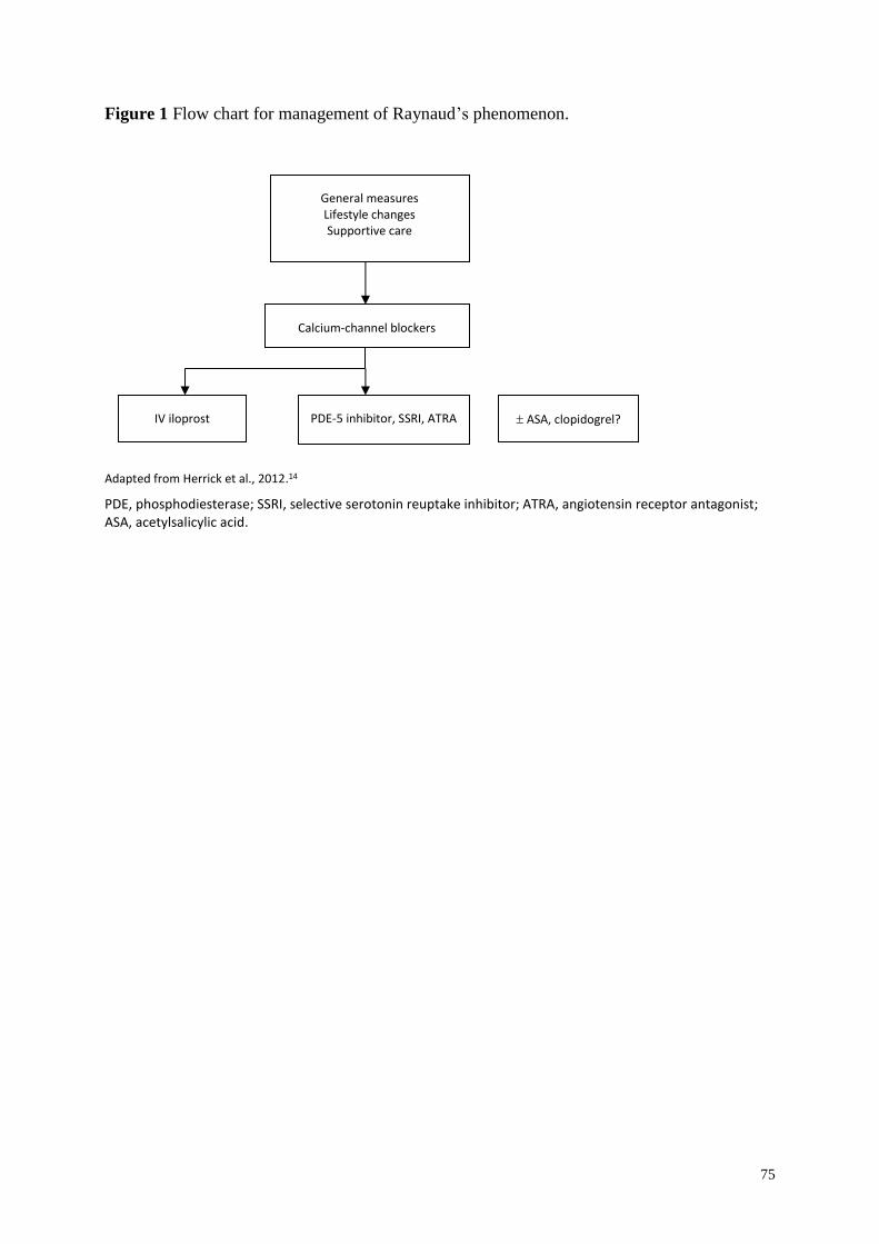
75
Figure 1 Flow chart for management of Raynaud’s phenomenon.
Adapted from Herrick et al., 2012.14
PDE, phosphodiesterase; SSRI, selective serotonin reuptake inhibitor; ATRA, angiotensin receptor antagonist; ASA, acetylsalicylic acid.
General measures Lifestyle changes Supportive care
Calcium-channel blockers
ASA, clopidogrel? PDE-5 inhibitor, SSRI, ATRA IV iloprost
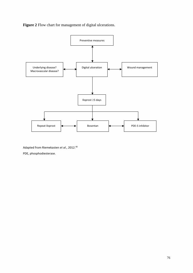
76
Figure 2 Flow chart for management of digital ulcerations.
Adapted from Riemekasten et al., 2012.40
PDE, phosphodiesterase.
Preventive measures
Underlying disease? Macrovascular disease?
Digital ulceration Wound management
Iloprost 5 days
Repeat iloprost Bosentan PDE-5 inhibitor
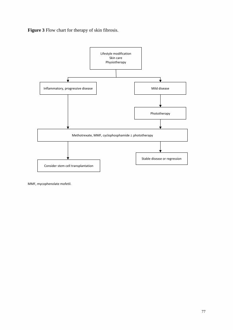
77
Figure 3 Flow chart for therapy of skin fibrosis.
MMF, mycophenolate mofetil.
Lifestyle modification Skin care
Physiotherapy
Inflammatory, progressive disease Mild disease
Methotrexate, MMF, cyclophosphamide phototherapy
Stable disease or regression
Consider stem cell transplantation
Phototherapy
78
References
1. Kowal-Bielecka O, Landewé R, Avouac J, Chwiesko S, Miniati I, et al. EULAR
recommendations for the treatment of systemic sclerosis: a report from the EULAR
Scleroderma Trials and Research group (EUSTAR). Ann Rheum Dis 2009; 68: 620–628.
2. UK Scleroderma Study group. Consensus best practice recommendations for scleroderma.
Available at: http://www.scleroderma-royalfree.org.uk/UKSSG.html (last accessed 28
February 2016)
3. Gabrielli A, Avvedimento EV, Krieg T. Scleroderma. N Engl J Med 2009; 360: 1989–
2003.
4. Masi AT and Subcommittee for Scleroderma Criteria of the American Rheumatism
Association Diagnostic and Therapeutic Criteria Committee (1980). Preliminary criteria
for the classification of systemic sclerosis (scleroderma). Arthritis Rheum 1980; 23: 581–
590.
5. LeRoy EC, Black C, Fleischmajer R, et al. Scleroderma (systemic sclerosis): classification,
subsets and pathogenesis. J Rheumatol 1988; 15: 202–205.
6. Mayes MD, Lacey JV Jr, Beebe-Dimmer J, et al. Prevalence, incidence, survival, and
disease characteristics of systemic sclerosis in a large US population. Arthritis Rheum
2003; 48: 2246–2255.
7. Walker UA, Tyndall A, Czirják L, et al. Clinical risk assessment of organ manifestations
in systemic sclerosis – a report from the EULAR Scleroderma Trials and Research
(EUSTAR) group data base. Ann Rheum Dis 2007; 66: 754–763.
8. Mierau R, Moinzadeh P, Riemekasten G, et al Frequency of disease-associated and other
nuclear autoantibodies in patients of the German network for systemic scleroderma:
correlation with characteristic clinical features. Arthritis Res Ther 2011; 13: R172.
9. Poormoghim H, Lucas M, Fertig N, Medsger TA Jr. Systemic sclerosis sine scleroderma:
demographic, clinical, and serologic features and survival in forty-eight patients. Arthritis
Rheum 2000; 43: 444–451.
10. van den Hoogen F, Khanna D, Fransen J, Johnson SR, Baron M, Tyndall A, et al. 2013
classification criteria for systemic sclerosis: an American College of Rheumatology/
European League against Rheumatism collaborative initiative. Ann Rheum Dis 2013; 72:
1747–1755.
11. Valentini G. Undifferentiated connective tissue disease at risk for systemic sclerosis (SSc)
(so far referred to as very early/early SSc or pre-SSc). Autoimmun Rev 2015; 14: 210–213.
79
12. Koenig M, Joyal F, Fritzler MJ, et al. Autoantibodies and microvascular damage are
independent predictive factors for the progression of Raynaud’s phenomenon to systemic
sclerosis: a twenty-year prospective study of 586 patients, with validation of proposed
criteria for early systemic sclerosis. Arthritis Rheum 2008; 58: 3902–3912.
13. Cutolo M, Sulli A, Smith V. How to perform and interpret capillaroscopy. Best Pract Res
Clin Rheumatol 2013; 27: 237–248.
14. Herrick AL. The pathogenesis, diagnosis and treatment of Raynaud phenomenon. Nat Rev
Rheumatol 2012; 8: 469–479
15. Czirják L, Foeldvari I, Müller-Ladner U. Skin involvement in systemic sclerosis.
Rheumatology 2008; 47(Suppl 5): v44–45.
16. Khimdas S, Harding S, Bonner A, et al. Associations with digital ulcers in a large cohort
of systemic sclerosis: results from the Canadian Scleroderma Research Group registry.
Arthritis Care Res 2011; 63:142–149.
17. Mouthon L, Mestre-Stanislas C, Bérezné A, et al. Impact of digital ulcers on disability and
health-related quality of life in systemic sclerosis. Ann Rheum Dis 2010; 69: 214–217
18. Sunderkoetter, Herrgott I, Brückner C, et al. Comparison of patients with and without
digital ulcers in systemic sclerosis: detection of possible risk factors. Br J Dermatol 2009;
160: 835–843.
19. Ennis H, Vail A, Wragg E, et al. A prospective study of systemic sclerosis-related digital
ulcers: prevalence, location, and functional impact. Scand J Rheumatol 2013; 42: 483–486
20. Mihai C, Landewé R, van der Haijde D, et al. Digital ulcers predict a worse disease course
in patients with systemic sclerosis. Ann Rheum Dis 2015; doi: 10.1136/annrheumdis-2014-
205897.
21. Hachulla E, Clerson P, Launay D, et al. Natural history of ischemic digital ulcers in
systemic sclerosis: single-center retrospective longitudinal study. J Rheumatol 2007; 34:
2423–2430.
22. Solomon JJ, Olson AL, Fischer A, Bull T, Brown KK, Raghu G. Scleroderma lung disease.
Eur Respir Rev 2013; 22: 6–19.
23. Hoffmann-Vold AM, Aaløkken TM, Lund MB, et al. Predictive value of serial high-
resolution computed tomography analyses and concurrent lung function tests in systemic
sclerosis. Arthritis Rheumatol 2015; 67: 2205–2212.
24. Steen VD, Medsger TA Jr. Changes in causes of death in systemic sclerosis, 1972–2002.
Ann Rheum Dis 2007; 66: 940–944.
80
25. The Joint Task Force for the Diagnosis and Treatment of Pulmonary Hypertension of the
European Society of Cardiology (ESC) and the European Respiratory Society (ERS). 2015
ESC/ERS Guidelines for the diagnosis and treatment of pulmonary hypertension. Eur
Respir J 2015; 46: 1855–1856
26. Hunzelmann N, Genth E, Krieg T, et al. The registry of the German Network for Systemic
Scleroderma: frequency of disease subsets and patterns of organ involvement.
Rheumatology 2008; 47: 1185–1192.
27. Hansi N, Thoua N, Carulli M, et al. Consensus Best Practice pathway of the UK
Scleroderma Study Group: gastrointestinal manifestations of systemic sclerosis. Clin Exp
Rheumatol 2014; 32(Suppl 86): S-214–221.
28. Vacca A, Meune C, Gordon J, et al. Scleroderma Clinical Trial Consortium Cardiac
Subcommittee. Cardiac arrhythmias and conduction defects in systemic sclerosis.
Rheumatology 2014; 53: 1172–1177.
29. Mouthon L, Bussone G, Berezné A, Noël LH, Guillevin L. Scleroderma renal crisis. J
Rheumatol 2014; 41: 1040–1048.
30. Fransen J, Popa-Diaconu D, Hesselstrand R, et al. Clinical prediction of 5-year survival in
systemic sclerosis: validation of a simple prognostic model in EUSTAR centres. Ann
Rheum Dis 2011; 70: 1788–1792
31. Sticherling M. Systemic sclerosis - focus on dermatological aspects. Part 2: diagnostics,
therapy. J Dtsch Dermatol Ges 2012; 10: 783–791.
32. Fries R, Shariat K, von Wilmowsky H, Bohm M. Sildenafil in the treatment of Raynaud’s
phenomenon resistant to vasodilatory therapy. Circulation 2005; 112: 2980–2985.
33. Caglayan E, Huntgeburth M, Karasch T, et al. Phosphodiesterase type 5 inhibition is a
novel therapeutic option in Raynaud disease. Arch Intern Med 2006; 166: 231–233.
34. Roustit M, Blaise S, Allanore Y, Carpentier PH, Caglayan E, Cracowski JL.
Phosphodiesterase-5 inhibitors for the treatment of secondary Raynaud’s phenomenon:
systematic review and meta-analysis of randomised trials. Ann Rheum Dis 2013; 72: 1696–
1699.
35. Coleiro B, Marshall SE, Denton CP, et al. Treatment of Raynaud’s phenomenon with the
selective serotonin reuptake inhibitor fluoxetine. Rheumatology 2001; 40: 1038–1043.
36. Hughes M, Ong VH, Anderson ME, et al. Consensus best practice pathway of the UK
Scleroderma Study Group: digital vasculopathy in systemic sclerosis. Rheumatology 2015;
54: 2015–2024.
81
37. Wigley FM, Wise RA, Seibold JR, et al. Intravenous iloprost infusion in patients with
Raynaud phenomenon secondary to systemic sclerosis. A multicenter, placebo-controlled,
double-blind study. Ann Intern Med 1994; 120: 199–206.
38. Pope J, Fenlon D, Thompson A, et al. Iloprost and cisaprost for Raynaud’s phenomenon in
progressive systemic sclerosis. Cochrane Database Syst Rev 2000; (2): CD000953.
39. Bali G, Aberer E. Iloprost therapy in systemic sclerosis. Hautarzt 2003; 54: 845–851.
40. Riemekasten G, Hoffmann U, Sunderkötter C, Weiss N, Kuhn A; angiologisch-
dermatologisch-rheumatologische DU-Expertenboard. Management of digital ulcers in
patients with systemic sclerosis. Dtsch Med Wochenschr 2012; 137: 34–40.
41. Herrick AL. Contemporary management of Raynaud’s phenomenon and digital ischaemic
complications. Curr Opin Rheumatol 2011; 23: 555–561
42. Tingey T, Shu J, Smuczek J, Pope J. Meta-analysis of healing and prevention of digital
ulcers in systemic sclerosis. Arthritis Care Res 2013; 65: 1460–1471.
43. Korn JH, Mayes M, Matucci Cerinic M, et al. Digital ulcers in systemic sclerosis:
prevention by treatment with bosentan, an oral endothelin receptor antagonist. Arthritis
Rheum 2004; 50: 3985–3993.
44. Seibold JR, Denton CP, Furst DE, et al. Bosentan prevents occurrence but does not speed
healing of digital ulcers in patients with systemic sclerosis (SSc). Arthritis Rheum 2005;
52(Suppl 9): 552.
45. Matucci-Cerinic M, Denton CP, Furst DE, et al Bosentan treatment of digital ulcers related
to systemic sclerosis: results from the RAPIDS-2 randomised, double-blind, placebo-
controlled trial. Ann Rheum Dis 2011; 70: 32–38.
46. Morita A, Sakakibara S, Sakakibara N, Yamauchi R, Tsuji T. Successful treatment of
systemic sclerosis with topical PUVA. J Rheumatol 1995; 22: 2361–2365.
47. Kreuter A, Breuckmann F, Uhle A, et al. Low-dose UVA1 phototherapy in systemic
sclerosis: effects on acrosclerosis. J Am Acad Dermatol 2004; 50: 740–747.
48. Connolly KL, Griffith JL, McEvoy M, Lim HW. Ultraviolet A1 phototherapy beyond
morphea: experience in 83 patients. Photodermatol Photoimmunol Photomed 2015; 31:
289–295.
49. Rook AH, Freundlich B, Jegasothy BV, et al. Treatment of systemic sclerosis with
extracorporeal photochemotherapy. Results of a multicenter trial. Arch Dermatol 1992;
128: 337–346.
50. Knobler RM, French LE, Kim Y, et al. A randomized, double-blind,placebo-controlled
trial of photopheresis in systemic sclerosis. J Am Acad Dermatol 2006; 54: 793–799.
82
51. Knobler R, Berlin G, Calzavara-Pinton P, et al. Guidelines on the use of extracorporeal
photopheresis. J Eur Acad Dermatol Venereol 2014; 28(Suppl 1): 1–37.
52. van den Hoogen FH, Boerbooms AM, Swaak AJ, Rasker JJ, van Lier HJ, van de Putte LB.
Comparison of methotrexate with placebo in the treatment of systemic sclerosis: a 24 week
randomized double-blind trial, followed by a 24 week observational trial. Br J Rheumatol
1996; 35: 364–372.
53. Pope JE, Bellamy N, Seibold JR, et al. A randomized, controlled trial of methotrexate
versus placebo in early diffuse scleroderma. Arthritis Rheum 2001; 44: 1351–1358.
54. Walker KM, Pope J, participating members of the Scleroderma Clinical Trials Consortium
(SCTC); Canadian Scleroderma Research Group (CSRG). Treatment of systemic sclerosis
complications: what to use when first-line treatment fails-a consensus of systemic sclerosis
experts. Semin Arthritis Rheum 2012; 42: 42–55.
55. Tashkin DP, Elashoff R, Clements PJ, et al. Cyclophosphamide versus placebo in
scleroderma lung disease. N Engl J Med 2006; 354: 2655–2666.
56. Balin SJ, Wetter DA, Andersen LK, Davis MD. Calcinosis cutis occurring in association
with autoimmune connective tissue disease: the Mayo Clinic experience with 78 patients,
1996–2009. Arch Dermatol 2012; 148: 455–462.
57. Wu JJ, Metz BJ. Calcinosis cutis of juvenile dermatomyositis treated with incision and
drainage. Dermatol Surg 2008; 34: 575–577.
58. Saddic N, Miller JJ, Miller OF 3rd, Clarke JT. Surgical debridement of painful fingertip
calcinosis cutis in CREST syndrome. Arch Dermatol 2009; 145: 212–213.
59. Murray AK, Moore TL, Richards H, Ennis H, Griffiths CE, Herrick AL. Pilot study of
intense pulsed light for the treatment of systemic sclerosis-related telangiectases. Br J
Dermatol 2012; 167: 563–569.
60. Halachmi S, Gabari O, Cohen S, Koren R, Amitai DB, Lapidoth M. Telangiectasis in
CREST syndrome and systemic sclerosis: correlation of clinical and pathological features
with response to pulsed dye laser treatment. Lasers Med Sci 2014; 29: 137–140.
61. Dinsdale G, Murray A, Moore T, et al. A comparison of intense pulsed light and laser
treatment of telangiectases in patients with systemic sclerosis: a within-subject randomized
trial. Rheumatology 2014; 53: 1422–1430.
62. Lee JS, Collard HR, Anstrom KJ, et al. Anti-acid treatment and disease progression in
idiopathic pulmonary fibrosis: an analysis of data from three randomized controlled trials.
Lancet Respir Med 2013; 1: 369–376.
83
63. Van Laar JM, Farge D, Sont JK, et al. Autologous hematopoietic stem cell transplantation
vs intravenous pulse cyclophosphamide in diffuse cutaneous systemic sclerosis: a
randomized clinical trial. JAMA 2014; 311: 2490–2498.
64. Burt RK, Shah SJ, Dill K, et al. Autologous non-myeloablative haemopoietic stem-cell
transplantation compared with pulse cyclophosphamide once per month for systemic
sclerosis (ASSIST): an open-label, randomised phase 2 trial. Lancet 2011; 378: 498–506.
65. Ntoumazios SK, Voulgari PV, Potsis K, Koutis E, Tsifetaki N, Assimakopoulos DA.
Esophageal involvement in scleroderma: gastroesophageal reflux, the common problem.
Semin Arthritis Rheum 2006; 36: 173–181.
66. Nikou GC, Toumpanakis C, Katsiari C, Charalambopoulos D, Sfikakis PP. Treatment of
small intestinal disease in systemic sclerosis with octreotide: a prospective study in seven
patients. J Clin Rheumatol 2007; 13: 119–123.
67. Frech TM, Khann D, Maranian P, et al. Probiotics for the treatment of systemic sclerosis-
assocated gastrointestinal bloating/distention. Clin Exp Rheumatol 2011; 29(2 Suppl 65):
S22–25.
68. Knafo R, Haythornthwaite JA, Heinberg L, Wigley FM, Thombs BD. The association of
body image dissatisfaction and pain with reduced sexual function in women with systemic
sclerosis. Rheumatology 2011; 50: 1125–1130.
69. Foocharoen C, Tyndall A, Hachulla E, et al. Erectile dysfunction is frequent in systemic
sclerosis and associated with severe disease: a study of the EULAR Scleroderma Trial and
Research group. Arthritis Res Ther 2012; 14: R37
84
IV Nephrogenic systemic fibrosis
Definition
Nephrogenic fibrosing dermopathy, a dermatologic form of the generic term nephrogenic
systemic fibrosis (NSF), is a relatively new disease entity. It was first reported in 2000 and is
believed to be seen almost only in patients with moderate-to-severe kidney failure, particularily
patients on dialysis.1 It was linked to the usage of gadolinium-based contrast agents (GBCAs)
in magnetic resonance imaging (MRI), which were adopted in the late 1990s for use in patients
with impaired renal function, as it was widely accepted that these agents were not nephrotoxic.2
Epidemiology
Depending on the type of gadolinium used for the imaging process, the incidence rate of NSF
may vary and, for gadodiamide, it has been estimated to be between 3% and 7% in patients with
renal insufficiency.3 Accumulating reports on clinically relevant fibrosing processes led to the
release in 2006 of an alert by the US Food and Drug Administration (FDA) regarding the use
of GBCA in patients with renal insufficiency.4 Based on multicenter retrospective reviews5,6
and a European Medicines Agency (EMA) report,7, important risk factors for NSF have been
identified (Table 1). Other incriminated factors such as erythropoietin, which gained
widespread use at the time NSF emerged, or hepatic insufficiency, could not be confirmed. The
adapted, selective use of GBCA thereafter led to a reduction in the incidence of NSF to zero –
or almost zero.10 However, as there is no mandatory reporting system for NSF, and given that
the only NSF registry (with over 380 reported cases) was last updated back in June 2013,11 the
decline in the number of publications reporting new cases has to be taken as a surrogate marker
for the assumed reduction in incidence.
<TABLE 1>
Pathogenesis
It has been proposed that excess GBCA in patients with renal insufficiency undergoing MRI
may be deposited in the tissue upon transmetallation. GBCAs include lanthanides, which were
reported to induce profibrogenic processes decades ago.12,13 More recently, chelated
gadodiamide and gadopentetate forms of GBCA specifically have been shown to increase the
release of profibrotic cytokines and growth factors in macrophages/monocytes in vitro within
85
minutes upon receptor-mediated cellular uptake.14 The exact mechanism of increased collagen
bundle deposition in skin and other organs has not yet been fully understood.
On routine light microscopy, depending on the disease severity, a deep biopsy may show
fibrocyte proliferation ranging from subtle proliferation of dermal fibrocytes in early lesions to
florid proliferation. Thick collagen bundles with surrounding clefts are a prominent finding,
with a variable increase in dermal mucin and elastin. Immunohistochemical staining shows
CD34+ dermal dendritic cells. Gadolinium may be visualized with special testing but is not
diagnostic.15
Clinical manifestation
NSF is a rare differential diagnosis of other sclerosing skin processes that may occur in patients
with impaired renal function, such as scleromyxedema, lipodermatosclerosis, eosinophilic
fasciitis, or localized and systemic sclerosis. Initial symptoms include hyperpigmented skin
areas and papules, which may coalesce to patches and plaques with a peau d’orange appearance.
NSF commonly forms symmetrical lesions, which are predominantly located on the lower legs,
and develops within the first 2–8 weeks after exposure to GBCA.10 Pain and pruritus are
frequent symptoms, but unlike in eosinophilic fasciitis, fever, arthritis and malaise are
uncommon.16 Unlike systemic sclerosis, Raynaud's phenomenon is typically absent. Systemic
involvement has been described (scleral plaques, muscle fibrosis and induration, flexion
contractures, fibrosis of vessel walls of internal organs such as lung and kidney, calcification
of the soft tissue). The sclerosing process may proceed within days or weeks, but delayed onset
of NSF has been described up to 10 years after gadolinium uptake.17 NSF has been documented
in all age groups, including in children.18
Diagnostic procedures
There is no specific test available for the diagnosis of NSF. Abnormal creatinine and increased
blood urea nitrogen are to be considered in the context of the pre-existing renal insufficiency.
Antinuclear antibodies and rheumatoid factors are typically negative, and there is no association
with paraproteinemia. Some patients show eosinophilia in the peripheral blood. Girardi et al.,
proposed a scoring system that has been tested on the reported cases in the NSF registry.19
(Table 2, Fig. 1). The variety of clinical findings in NSF are classified into major (patterned
plaques of the skin, joint contractures, cobblestone appearance of the skin, peau d’orange) and
minor (linear banding of the skin, superficial plaque/patch, dermal papules, scleral plaques in
86
patients aged <45 years) clinical criteria, and a clinicopathologic scoring system has been
proposed in order to allow the diagnostic of NSF.19 As the incidence of NSF appears to
diminish, evaluation of this scoring system will be a difficult task. Nevertheless, the use of this
score will aid the standardization of diagnostic procedures for NSF, and may be helpful to
differentiate between borderline cases of NSF and other sclerosing skin disorders.
<TABLE 2>
<Fig. 1>
Treatment
Established NSF lesions do not respond to systemic or local glucocorticoid treatment or to other
immunosuppressive drugs. Other approaches such as extracorporeal photopheresis, UVA1
phototherapy, plasmapheresis, or imatinib mesylate have been used with inconsistent clinical
improvement.20–27 Based on the published data, no specific therapeutic recommendation can be
made. Reconstitution of renal function is considered the best therapeutic approach.28 Prevention
consists of avoidance of gadolinium-containing contrast agents in patients with an estimated
glomerular filtration rate of <30 mL/min. If clinical conditions require the use of gadolinium,
then, in order to reduce the risk of NSF development, and based on the recommendations of the
FDA and EMA, low-risk gadolinium media should be the contrast agents of choice. Doses of
GBCA should be reduced to the minimum effective dosage for imaging.7,8 Based on the
dialysability of GBCA, it is recommended that at least one full 4-hour dialysis session is
performed after GBCA-based MRI in patients with renal insufficiency; this should remove 97%
of the GBCA that was present peior to dialysis. Three full sessions of dialysis increase the
GBCA clearance up to 99.7%.29
Conclusions
NSF is an iatrogenic condition observed in patients with end-stage renal failure and is associated
with gadolinium exposure. No treatments with proven efficacy based on randomised controlled
trials are available. Avoidance of high-risk GBCA is the key prophylactic measure.
Prophylactic measures have led to a significant drop in incidence of NSF.30
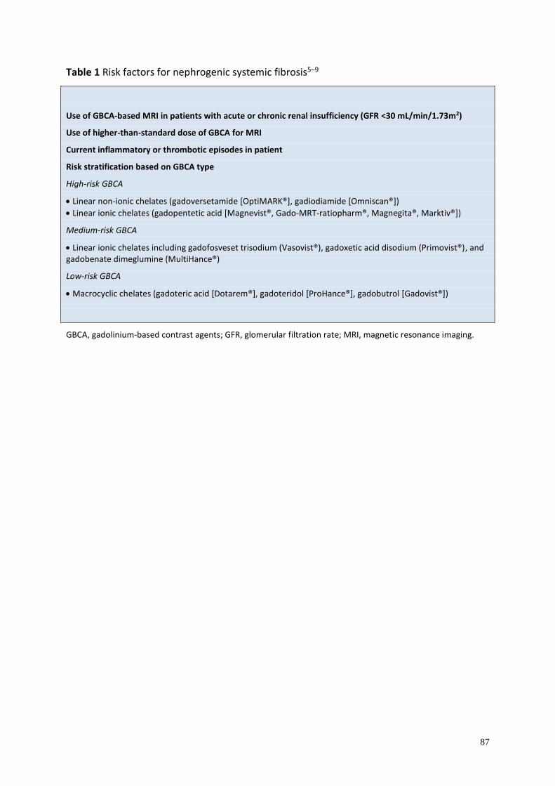
87
Table 1 Risk factors for nephrogenic systemic fibrosis5–9
Use of GBCA-based MRI in patients with acute or chronic renal insufficiency (GFR <30 mL/min/1.73m2)
Use of higher-than-standard dose of GBCA for MRI
Current inflammatory or thrombotic episodes in patient
Risk stratification based on GBCA type
High-risk GBCA
• Linear non-ionic chelates (gadoversetamide [OptiMARK®], gadiodiamide [Omniscan®])
• Linear ionic chelates (gadopentetic acid [Magnevist®, Gado-MRT-ratiopharm®, Magnegita®, Marktiv®])
Medium-risk GBCA
• Linear ionic chelates including gadofosveset trisodium (Vasovist®), gadoxetic acid disodium (Primovist®), and gadobenate dimeglumine (MultiHance®)
Low-risk GBCA
• Macrocyclic chelates (gadoteric acid [Dotarem®], gadoteridol [ProHance®], gadobutrol [Gadovist®])
GBCA, gadolinium-based contrast agents; GFR, glomerular filtration rate; MRI, magnetic resonance imaging.

88
Table 2 Girardi score for diagnosis of NSF19
Clinical findings
Major criteria
• Patterned plaques
• Joint contractures
• “Cobblestoning”
• Marked induration/Peau d’orange
Minor criteria
• Puckering/linear banding
• Superficial plaque/patch
• Dermal papules
• Scleral plaques (age <45 years)
Histologic findings
• Increased dermal cellularity (score +1)
• CD34+ cells with tram-tracking (score +1)
• Thick and thin collagen bundles (score +1)
• Preserved elastic fibers (score -1 if absent)
• Septal involvement (score +1)
• Osseous metaplasia (score +3)
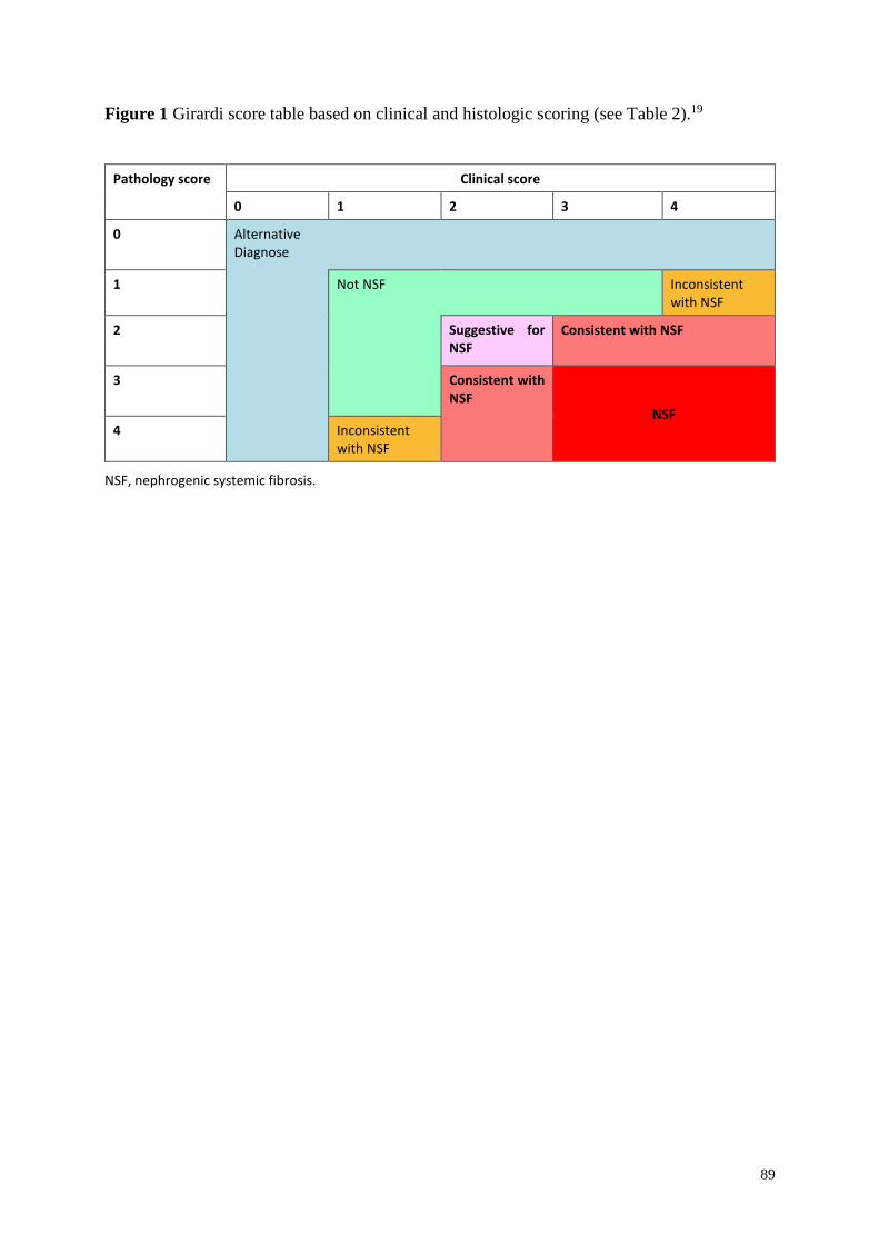
89
Figure 1 Girardi score table based on clinical and histologic scoring (see Table 2).19
Pathology score Clinical score
0 1 2 3 4
0 Alternative Diagnose
1 Not NSF Inconsistent with NSF
2
Suggestive for NSF
Consistent with NSF
3
Consistent with NSF
NSF 4 Inconsistent
with NSF
NSF, nephrogenic systemic fibrosis.
90
References
1. Gathings RM, Reddy R, Santa Cruz D, Brodell RT. Gadolinium-associated plaques: a new,
distinctive clinical entity. JAMA Dermatol 2015; 151: 316–319.
2. Prince MR, Arnoldus C, Frisoli JK. Nephrotoxicity of high-dose gadolinium compared
with iodinated contrast. J Magn Reson Imaging 1996; 6: 162–166.
3. Thomsen HS, Marckmann P. Extracellular Gd-CA: differences in prevalence of NSF. Eur
J Radiol 2008; 66: 180–183.
4. US Food and Drug administration. Public Health Advisory – Gadolinium-containing
contrast agents for magnetic resonance imaging (MRI). Available at:
http://www.fda.gov/Drugs/DrugSafety/PostmarketDrugSafetyInformationforPatientsandP
roviders/ucm053112.htm. (Last accessed 5 October 2015.)
5. Prince MR, Zhang H, Morris M, et al. Incidence of nephrogenic systemic fibrosis at two
large medical centers. Radiology 2008; 248: 807–816.
6. Wertman R, Altun E, Martin DR, et al. Risk of nephrogenic systemic fibrosis: evaluation
of gadolinium chelate contrast agents at four American universities. Radiology 2008; 248:
799–806.
7. European Medicines Agency. Assessment report for Gadolinium-containing contrast
agents. Available at:
http://www.ema.europa.eu/docs/en_GB/document_library/Referrals_document/gadoliniu
m_31/WC500099538.pdf. (Last accessed 5 October 2015.)
8. Khawaja AZ, Cassidy DB, Al Shakarchi J, McGrogan DG, Inston NG, Jones RG.
Revisiting the risks of MRI with Gadolinium based contrast agents-review of literature and
guidelines. Insights Imaging 2015; 6: 553–558.
9. Sadowski EA, Bennett LK, Chan MR, et al. Nephrogenic systemic fibrosis: risk factors
and incidence estimation. Radiology 2007; 243: 148–157.
10. Thomsen HS, Morcos SK, Almen T, et al. Nephrogenic systemic fibrosis and gadolinium-
based contrast media: updated ESUR Contrast Medium Safety Committee guidelines. Eur
Radiol 2013; 23: 307–318.
11. The International Center for Nephrogenic Systemic Fibrosis Research (ICNSFR).
Available at: http://www.icnfdr.org/. (Last accessed 5 October 2015.)
12. Evans CH, Drouven BJ. The promotion of collagen polymerization by lanthanide and
calcium ions. Biochem J 1983; 213: 751–758.
13. Drouven BJ, Evans CH. Collagen fibrillogenesis in the presence of lanthanides. J Biol
Chem 1986; 261: 11792–11797.
91
14. Newton BB, Jimenez SA. Mechanism of NSF: new evidence challenging the prevailing
theory. J Magn Reson Imaging 2009; 30: 1277–1283.
15. Galan A, Cowper SE, Bucala R. Nephrogenic systemic fibrosis (nephrogenic fibrosing
dermopathy). Curr Opin Rheumatol 2006; 18: 614–617.
16. Cowper SE. Nephrogenic fibrosing dermopathy: the first 6 years. Curr Opin Rheumatol
2003; 15: 785–790.
17. Larson KN, Gagnon AL, Darling MD, Patterson JW, Cropley TG. Nephrogenic systemic
fibrosis manifesting a decade after exposure to gadolinium. JAMA Dermatol 2015; 151:
1117–1120.
18. Jan F, Segal JM, Dyer J, LeBoit P, Siegfried E, Frieden IJ. Nephrogenic fibrosing
dermopathy: two pediatric cases. J Pediatr 2003; 143: 678–681.
19. Girardi M, Kay J, Elston DM, Leboit PE, Abu-Alfa A, Cowper SE. Nephrogenic systemic
fibrosis: clinicopathological definition and workup recommendations. J Am Acad
Dermatol 2011; 65: 1095–1106 e7.
20. Mathur K, Morris S, Deighan C, Green R, Douglas KW. Extracorporeal photopheresis
improves nephrogenic fibrosing dermopathy/nephrogenic systemic fibrosis: three case
reports and review of literature. J Clin Apher 2008; 23: 144–150.
21. Tran KT, Prather HB, Cockerell CJ, Jacobe H. UV-A1 therapy for nephrogenic systemic
fibrosis. Arch Dermatol 2009; 145: 1170–1174.
22. Tsagalis G, Psimenou E, Laggouranis A. Combination treatment with plasmapheresis and
sirolimus does not seem to benefit nephrogenic systemic fibrosis. Int J Artif Organs 2008;
31: 913–914.
23. Elmholdt TR, Buus NH, Ramsing M, Olesen AB. Antifibrotic effect after low-dose
imatinib mesylate treatment in patients with nephrogenic systemic fibrosis: an open-label
non-randomized, uncontrolled clinical trial. J Eur Acad Dermatol Venereol 2013; 27: 779–
784.
24. Gilliet M, Cozzio A, Burg G, Nestle FO. Successful treatment of three cases of nephrogenic
fibrosing dermopathy with extracorporeal photopheresis. Br J Dermatol 2005; 152: 531–
536.
25. Lauchli S, Zortea-Caflisch C, Nestle FO, Burg G, Kempf W. Nephrogenic fibrosing
dermopathy treated with extracorporeal photopheresis. Dermatology 2004; 208: 278–280.
26. Richmond H, Zwerner J, Kim Y, Fiorentino D. Nephrogenic systemic fibrosis: relationship
to gadolinium and response to photopheresis. Arch Dermatol 2007; 143: 1025–1030.
92
27. Kintossou R, D'Incan M, Chauveau D, et al. [Nephrogenic fibrosing dermopathy treated
with extracorporeal photopheresis: role of gadolinium?] Ann Dermatol Venereol 2007;
134: 667–671.
28. Cuffy MC, Singh M, Formica R, et al. Renal transplantation for nephrogenic systemic
fibrosis: a case report and review of the literature. Nephrol Dial Transplant 2011; 26:
1099–1101.
29. Gheuens E, Daelemans R, Mesens S. Dialysability of gadoteric acid in patients with end-
stage renal disease undergoing hemodialysis. Invest Radiol 2014; 49: 505–508.
30. Bennett CL, Qureshi ZP, Sartor AO, et al. Gadolinium-induced nephrogenic systemic
fibrosis: the rise and fall of an iatrogenic disease. Clin Kidney J 2012; 5: 82–88.
93
V Systemic sclerosis overlap syndromes
Introduction
Systemic sclerosis (SSc) overlap syndrome is a term used to describe a very heterogeneous
group of patients with features of different connective tissue diseases, combined with clinical
signs of SSc.1–3. To date, no firm classification criteria for SSc overlap syndromes have been
established, but they are generally considered when musculoskeletal involvement or features of
other rheumatic diseases are significantly greater than usually found in general SSc patients.4,5
Other autoimmune rheumatic disorders are classified depending on internationally accepted
classification systems.6–10 Most SSc overlap syndromes appear to encompass a subtype of SSc
similar to limited cutaneous SSc (lcSSc), but with more frequent involvement of the
musculoskeletal system than in lcSSc or diffuse cutaneous (dcSSc), and an apparently earlier
onset of lung fibrosis or heart involvement.5.
Epidemiology
SSc overlap syndromes represent the third major subgroup of SSc, and epidemiologic studies
report divergent frequencies (incidence and prevalence rates are not reported yet) of overlap
subgroups, ranging between 9% and 38% (Table 1).1,2,4,5
The most common SSc overlap syndromes are SSc and myositis (polymyositis or
dermatomyositis), SSc and rheumatoid arthritis, SSc and Sjögren’s, and SSc and systemic lupus
erythematosus (SLE) overlap syndromes.4 Pakozdi et al. reported recently that 20% of SSc
patients attending the Centre for Rheumatology at the Royal Free Hospital (London, UK) had
features overlapping with other rheumatologic diseases. Of these 43% overlapped with
polymyositis/dermatomyositis, 8% with SLE, 17% with Sjögren’s syndrome, and 32% with
rheumatoid arthritis.4 The German Network for Systemic Scleroderma (DNSS) reported that
10% of the registered patients suffered from SSc overlap syndromes.5
A recent meta-analysis has revealed that the mean age at diagnosis of patients with SSc overlap
syndromes was 47.6 years (SD 2.6), and that it was found more often in European patients than
in patients from North America.11

94
Balbir-Gurman reported that the overall mortality in their SSc overlap cohort did not differ from
other SSc patients.1 Depending on different geographical regions/centers, a wide range of
frequencies of SSc overlap syndromes have been reported (Table 1).
<TABLE 1>
Pathogenesis
To date, the pathogenesis of SSc overlap syndromes remains unclear. The question of why some
patients develop only one connective tissue disease and other patients have a combination of
clinical features of different rheumatic diseases has not yet been answered. A common or
overlapping genetic susceptibility possibly plays an important role. Genetic studies have shown
the existence of some susceptibility genes, which predispose to multiple autoimmune
diseases.11 Koumakis et al. reported that a regulatory gene located in the TNFAIP3 region is
associated with a higher risk of developing SSc polyautoimmunity.11,16
Clinical manifestations
Clinical features of SSc overlap syndrome are very heterogeneous. Patients usually present with
skin sclerosis typical of lcSSc, although organ manifestations clearly separate these patients as
distinct subset.5 A German study showed that patients suffering from SSc overlap syndromes
developed an involvement of the musculoskeletal system significantly earlier and more often
than patients with dcSSc and lcSSc. In addition, they interestingly developed lung fibrosis and
heart involvement significantly earlier and more often than lcSSc patients, but still less
frequently and later than dcSSc patients.5
Therefore, the identification of these patients is essential for clarifying prognosis and
facilitating therapeutic options. The clinical signs include both cutaneous and extracutaneous
features, depending on the overlapping connective tissue disease (CTD), and often overlap
between the different overlap forms, especially regarding vasculopathy, gastrointestinal and
cardiopulmonary involvement.
For more details on thje following conditions, please refer to Section III (Systemic sclerosis).
Raynaud’s phenomenon
95
Raynaud’s phenomenon is also a very common feature in patients with SSc overlap syndromes.3
Some SSc overlap patients also develop digital ulcerations but significantly less often compared
with lcSSc and dcSSc patients.5
Skin sclerosis
The skin sclerosis in patients with SSc overlap syndromes can be generalized, similar to the
diffuse form of SSc, but more frequently it is only located below the elbow and knee joints,
which is similar to the limited form of SSc.4,5
Calcinosis cutis
Calcinosis cutis can be also observed in patients with SSc overlap syndromes. It is associated
with longer disease duration, positive anti-centromere and anti-PM/Scl antibodies, and occurs
usually over pressure points (acral or next to joints).17
Gastrointestinal involvement
As in SSc the involvement of the gastrointestinal tract is probably the most common internal
organ system involved (approx. 50–60%).5,14
Lung fibrosis and myocardial involvement
Lung fibrosis and myocardial involvement are significantly less frequent than in patients with
diffuse SSc, but significantly more frequent than in limited forms of SSc.5
Pulmonary arterial hypertension
Pulmonary arterial hypertention (PAH) occurs less frequently in patients with SSc overlap
syndromes than in patients with dcSSc, but similarly to those with the limited form of SSc.5
Clinical characteristics of systemic sclerosis overlap syndromes
Systemic sclerosis and myositis
Myositis is the most frequent systemic involvement in patients with SSc overlap syndromes. In
some SSc patients, muscle weakness, pain, and atrophy result from disuse secondary to joint
contractures, dermatogenous contractures, or chronic disease. However, significantly more
patients with SSc overlap syndromes present with myositis, characterized by proximal muscle
weakness with no loss of reflexes or sensitivity, myalgia, increased creatine kinase serum levels,
and later atrophy of muscles. Patients suffering from SSc-myositis overlap syndrome may
96
develop myositis simultaneously, before, or in already established SSc.1 Some patients may
show cutaneous symptoms of dermatomyositis. The limited extent of skin thickening is still the
most frequent form in patients with SSc overlap syndromes.1,4,5
Recent studies have shown that an increased proportion of patients also develop lung
fibrosis,5,18 which is in line with a high percentage (up to 30%) of interstitial lung disease (ILD)
in patients with dermatomyositis. Patients with SSc-myositis overlap syndromes have a higher
risk of developing a diffuse interstitial myocardial fibrosis, which may lead to diastolic
dysfunctions as well as restricted contractibility of the myocardium. These patients typically
present symptoms, such as cardiac arrhythmia, paroxysmal tachycardia, incomplete or complete
right-heart blocks, finally leading to heart insufficiency. The frequency of lung and
gastrointestinal involvement varies among studies, ranging between 32.0% and 78.1%.2
It is well established that patients suffering from the SSc-myositis overlap syndrome (except
those with antibody to PM/Scl) have a worse prognosis due to an increased risk of myocardial
involvement compared with patients with only SSc.18 SSc-myositis overlap syndromes may be
associated with specific autoantibodies, including PM/Scl, anti-Ku, anti-U2RNP, and anti-
U5snRNP (Table 2).1,19 Patients, carrying the antibody to PM/Scl are usually younger, have
limited skin involvement, and suffer from arthritis and a benign course of ILD,1 which is also
the reason for their better survival.15 Positive antibodies against Ku are more characteristic for
patients suffering from muscle involvement as well as severe ILD (Table 1).23
<TABLE 2>
Systemic sclerosis and rheumatoid arthritis
Joint involvement is reported to be the second most frequent manifestation in patients with
musculoskeletal involvement and overlap syndromes.4 These patients may present with typical
clinical symptoms (usually limited skin involvement, morning stiffness, arthritis), together with
high titers of anti-cyclic citrullinated peptides (CCP/ACPA) and/or higher rheumatoid factors
(SSc-RA overlap syndrome). However, it is often very difficult to distinguish between SSc
patients with mild, sero-negative arthralgia and the significant arthritis associated with SSc-RA
overlap syndrome.
Systemic sclerosis and systemic lupus erythematosus
97
This subtype is a very rare condition.24 Patients often have a fatal course of the disease due to
a higher risk of developing polyserositis, pancreatitis, avascular bone necrosis, PAH, lung
involvement, lupus gromelunephritis, skin rashes, and leukencephalopathy.1 It is also difficult
to distinguish whether the patient suffers from a lupus-nephritis or a scleroderma renal crisis.
Depending on the reason for renal failure, patients need a different therapeutic strategy to
improve renal function. Skin lesions can be a major esthetic disturbing factor, because of the
predilection for the face. These patients usually have a combination of SSc-associated
antibodies and double-stranded DNA antibodies.
Systemic sclerosis and Sjögren’s syndrome
This SSc overlap syndrome was first described in 1965 by Bloch et al.25 Xerostomia and
xerophthalmia are very common in patients suffering from SSc (68–83%), but only 14–20% of
SSc patients really fulfill the criteria of Sjögren’s syndrome,26 so that the diagnosis of SSc/SS
overlap syndromes is always a challenge.27 It is defined by a lymphocytic infiltration of the
salivary glands. Patients with SSc-SS overlap syndrome show a limited form of skin
involvement (83.6% vs 16.4%) and a very low frequency of lung involvement.1 Antibodies
against Ro are very likely in SSc-SS overlap syndromes, often together with anti-centromere
antibodies (ACA).4
Mixed connective tissue disease
Mixed connective tissue disease (MCTD) was first described by Sharp et al. in 1972.28 These
patients present clinical symptoms typically found in patients with myositis, SLE, inflammatory
arthritis (RA) and SSc. Typical for this condition are puffy fingers (50%), polyarthritis (65%),
Raynaud’s phenomenon (53%), sclerodactyly (35%), muscle involvement, and esophageal
involvement.20,29 and the occurrence of high antinuclear antibodies titers with high levels of
U1snRNP antibodies, which helps to differentiate MCTD from other connective tissue diseases.
Arthralgia occurs in approximately 60% of patients, and muscle disease is present in 80–90%
of cases with proximal muscle involvement and elevation of serum creatine kinase levels.29
Cardiovascular involvement (lung fibrosis and especially PAH) is less frequent, but is a major
contributor to a poor outcome/prognosis.20
SSc may also occur together with other organ-specific autoimmune diseases, such as
autoimmune hepatitis/primary biliary cirrhosis, autoimmune thyroiditis, sarcoidosis, and
antiphospholipid syndrome (Table 3).
98
<TABLE 3>
Diagnostic procedures
Muscle involvement (myositis/myopathy)
Typical clinical symptoms include a symmetrical proximal muscle weakness, muscle pain,
and/or muscle atrophy with intact reflexes and sensitivity. Serologic tests usually show an
elevation of serum creatine phosphokinase (≥4-fold) and acute phase parameters in blood (e.g.
C-reactive protein and erythrocyte sedimentation rate. An electromyography, magnetic
resonance imaging (MRI), and muscle biopsy will help to identify affected muscles.6,7,14,30
Sjögren’s symptoms
Due to a reduced glandular function, patients with SSc-Sjögren’s overlap syndrome suffer from
dry mouth (xerostomia) and dry eyes (xerophthalmia). In addition, these patients also typically
show anti-Ro and anti-La antibodies, often together with anti-centromere antibodies. Further
diagnostics include functional tests for ocular and oral sicca symptoms, together with a
glandular biopsy.31
Joint involvement
A rheumatologic examination is essential to identify rheumatoid arthritis. Joint involvement
can be due to dermatogenous contractures or inflammation. It is recommended to examine the
rheumatoid factor and anti-CCP antibodies in the serum of affected patients. X-ray, ultrasound
of affected joints, as well as MRI scans can be helpful tools to identify inflammation areas and
damage of the joints.14
Kidney involvement
Creatinine clearance, urine analysis to control proteinuria and hematuria, as well as regular
blood pressure tests are necessary for the early identification of renal involvement.14,32 In
patients with SSc-SLE overlap syndromes it may be necessary to perform a kidney biopsy to
distinguish between renal failures due to lupus nephritis33 (see also the ACR/EULAR guidelines
on SLE) or scleroderma renal crisis32 (see also Section III – Systemic sclerosis).32
99
For more details on diagnostic procedures and SSc-associated organ manifestations/
complications see Section III (Systemic sclerosis).
<FIG. 1>
Treatment
There have been major advances in treating many of the organ-specific complications of SSc
and overlapping diseases. See also Section III (Systemic sclerosis).
Systemic glucocorticoids
Systemic glucocorticoids can be used for musculoskeletal involvement together with other
immunosuppressive agents. The use of high-dose glucocorticoids should be used with caution
due to the increased risk of renal crisis in SSc patients with diffuse extent of skin involvement.2
Methotrexate
Methotrexate is a well-known immunosuppressive agent that has been used in adults and
children, with well-documented side-effects. Methotrexate is still a first-line therapy in many
autoimmune diseases. It is the treatment of choice in patients with SSc-myositis and SSc-RA
overlap syndromes.34,35
Mycophenolat mofetil
MMF is a well-tolerated immunosuppressive agent, which is recommended as long-term
therapy in scleroderma and has successfully been applied in several overlap syndromes.
Azathioprine
This immunosuppressive agent is usually well tolerated and has been used successfully in
patients with MCTD as well as patients with SSc-SLE overlap. However, compared with MMF,
side-effects seem to be more pronounced and the response to the therapy more limited.
Cyclophosphamide
Cyclophosphamide is often used for lung involvement in patients with SSc,36 and also SSc-
myositis overlap or SSc-SLE overlap syndromes, in case of lupus nephritis. Cyclophosphamide
100
should be used for musculoskeletal involvement as a second-line immunosuppressive therapy
after other treatments (methotrexate, MMF) have failed or cannot be used due to defined side-
effects. As in other autoimmune diseases, it can be used as intravenous pulse or oral treatment.
Bioimmunomodulatry agents
Only limited information is available for the use of intravenous immunoglobulin (IVIg),
rituximab, and anti-tumor necrosis factor (TNF) in the treatment of overlap syndromes.
Therapeutic approaches
Systemic sclerosis and myositis
In this group of patients, treatment is mainly directed against muscle inflammation, alveolitis,
and skin sclerosis (Fig. 2).
Glucocorticoid therapy (not in patients with a higher risk for renal crisis (see Section III –
Systemic sclerosis), methotrexate (not in case of alveolitis), azathioprine, IVIg,
cyclophosphamide, and rituximab (in patients with uncontrolled myositis) may be helpful
agents.
Agents of choice in mild cases are methotrexate together with low-dose glucocorticoids. In
severe cases, IVIg can be added. In patients with a refractory course of the disease,
cyclophosphamide (also known to improve skin and lung involvement), MMF (also known to
improve skin thickening), or rituximab (also known to improve skin and lung involvement) can
be tried to improve clinical symptoms.1,37–39
Systemic sclerosis and rheumatoid arthritis
These patients are usually treated with hydroxychloroquine, possibly together with
methotrexate and low-dose glucocorticoids. If this therapeutic strategy is not effective,
tocilizumab, rituximab as well as anti-TNF agents should be considered. All these treatments
have to be used with caution, in the context of serious infections, tuberculosis, and fibrosis.
For further details see Section III (Systemic sclerosis) and ACR/EULAR guidelines on
rheumatoid arthritis.40
Systemic sclerosis and systemic lupus erythematosus
101
Treatment in patients with cutaneous lesions due to SLE should start with topical glucocorticoid
therapy, together with UV skin protection. The topical treatment can be combined with
hydroxychloroquine together with low-dose glucocorticoids. In severe cases,
cyclophosphamide or MMF can be initiated. The treatment of renal involvement differs
between a lupus- and a scleroderma-associated renal failure (cyclophosphamide vs vasoactive
treatment with ace inhibitors and iloprost).
For further details see Section III (Systemic sclerosis) and EULAR/ACR guidelines on
rheumatoid arthritis.40
Mixed connective tissue disease
Patients with MCTD usually respond well to systemic glucocorticoid and immunosuppressive
therapy with several classical agents. But some long-term studies have shown that a group of
patients with MCTD develop more severe organ manifestations and need a more aggressive
therapeutic strategy. Inflammatory features (elevated temperature, serositis, pleuritis, myositis,
and arthritis) respond well to glucocorticoid treatment, while symptoms, such as sclerotic skin
changes and cardiopulmonary involvement need immunosuppressive/cytotoxic drugs.29,41 The
most frequently used drugs are hydroxychloroquine, methotrexate, and cyclophosphamide.29
Systemic sclerosis and Sjögren’s overlap syndrome
Clinical features such as the xerostomia can usually be improved by using various antiseptic
mouth rinse and saliva substitutes. Xerophthalmia can be improved by using artificial tear
drops.42 This topical treatment should be combined with hydroxychloroquine and low-dose
glucocorticoids. In severe cases, cyclophosphamide, azathioprine, or rituximab have shown to
be effective in open-label studies.43
For further details see Section III (Systemic sclerosis) and guidelines for Sjögren’s syndrome.44
<Fig. 2>
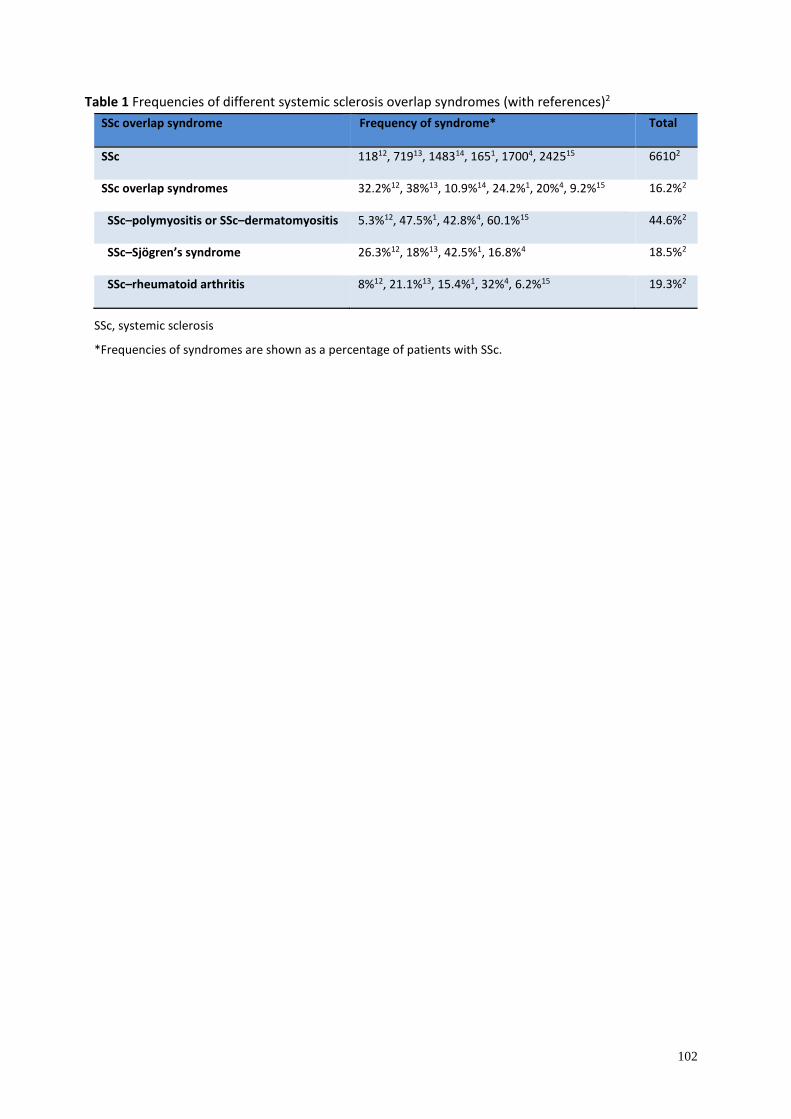
102
Table 1 Frequencies of different systemic sclerosis overlap syndromes (with references)2
SSc overlap syndrome Frequency of syndrome* Total
SSc 11812, 71913, 148314, 1651, 17004, 242515 66102
SSc overlap syndromes 32.2%12, 38%13, 10.9%14, 24.2%1, 20%4, 9.2%15 16.2%2
SSc–polymyositis or SSc–dermatomyositis 5.3%12, 47.5%1, 42.8%4, 60.1%15 44.6%2
SSc–Sjögren’s syndrome 26.3%12, 18%13, 42.5%1, 16.8%4 18.5%2
SSc–rheumatoid arthritis 8%12, 21.1%13, 15.4%1, 32%4, 6.2%15 19.3%2
SSc, systemic sclerosis
*Frequencies of syndromes are shown as a percentage of patients with SSc.
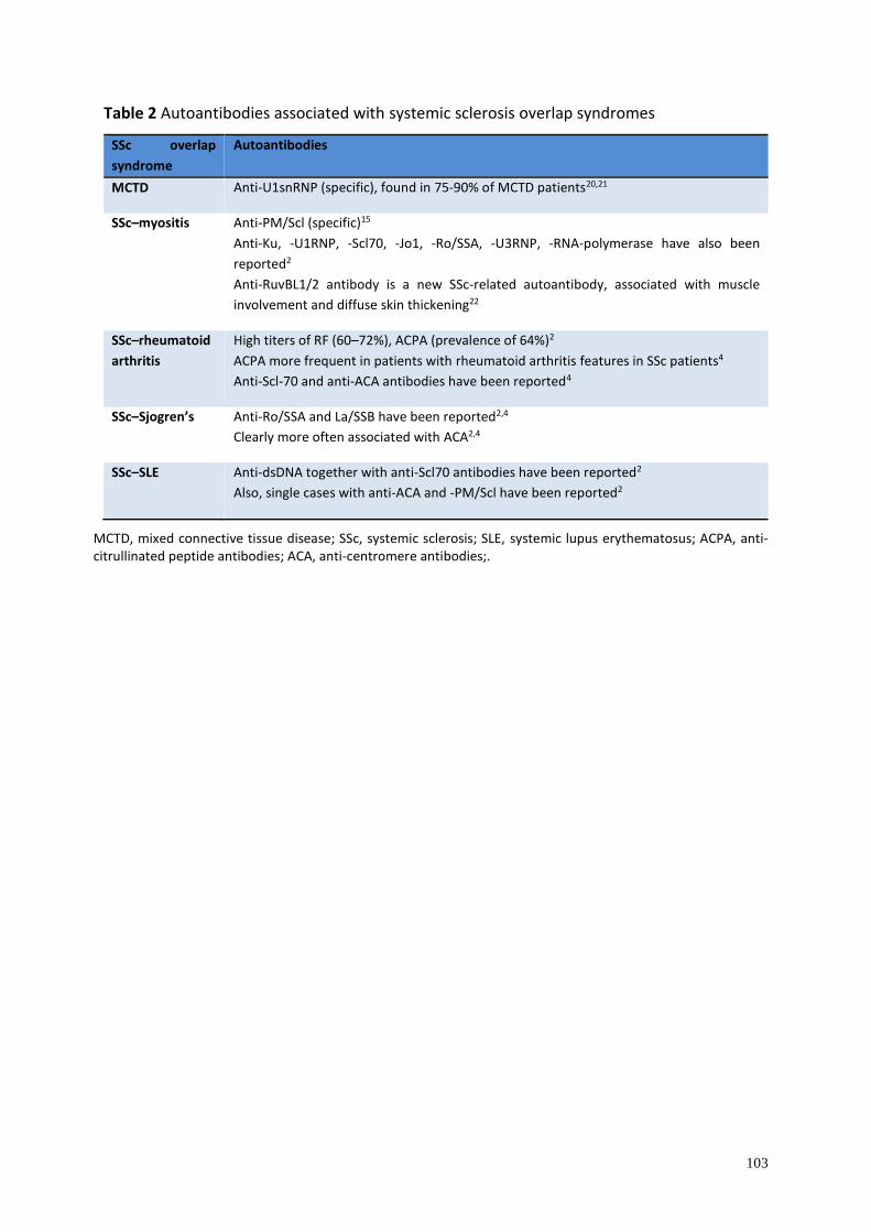
103
Table 2 Autoantibodies associated with systemic sclerosis overlap syndromes
SSc overlap
syndrome
Autoantibodies
MCTD Anti-U1snRNP (specific), found in 75-90% of MCTD patients20,21
SSc–myositis Anti-PM/Scl (specific)15
Anti-Ku, -U1RNP, -Scl70, -Jo1, -Ro/SSA, -U3RNP, -RNA-polymerase have also been
reported2
Anti-RuvBL1/2 antibody is a new SSc-related autoantibody, associated with muscle
involvement and diffuse skin thickening22
SSc–rheumatoid
arthritis
High titers of RF (60–72%), ACPA (prevalence of 64%)2
ACPA more frequent in patients with rheumatoid arthritis features in SSc patients4
Anti-Scl-70 and anti-ACA antibodies have been reported4
SSc–Sjogren’s Anti-Ro/SSA and La/SSB have been reported2,4
Clearly more often associated with ACA2,4
SSc–SLE Anti-dsDNA together with anti-Scl70 antibodies have been reported2
Also, single cases with anti-ACA and -PM/Scl have been reported2
MCTD, mixed connective tissue disease; SSc, systemic sclerosis; SLE, systemic lupus erythematosus; ACPA, anti-citrullinated peptide antibodies; ACA, anti-centromere antibodies;.
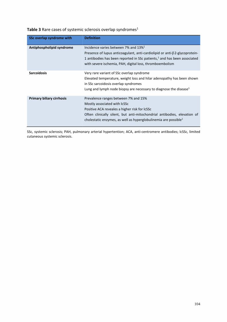
104
Table 3 Rare cases of systemic sclerosis overlap syndromes1
SSc overlap syndrome with Definition
Antiphospholipid syndrome Incidence varies between 7% and 13%1
Presence of lupus anticoagulant, anti-cardiolipid or anti-2-glycoprotein-
1 antibodies has been reported in SSc patients,1 and has been associated
with severe ischemia, PAH, digital loss, thromboembolism
Sarcoidosis Very rare variant of SSc overlap syndrome
Elevated temperature, weight loss and hilar adenopathy has been shown
in SSc sarcoidosis overlap syndromes
Lung and lymph node biopsy are necessary to diagnose the disease1
Primary biliary cirrhosis Prevalence ranges between 7% and 15%
Mostly associated with lcSSc
Positive ACA reveales a higher risk for lcSSc
Often clinically silent, but anti-mitochondrial antibodies, elevation of
cholestatic enzymes, as well as hyperglobulinemia are possible1
SSc, systemic sclerosis; PAH, pulmonary arterial hypertention; ACA, anti-centromere antibodies; lcSSc, limited cutaneous systemic sclerosis.
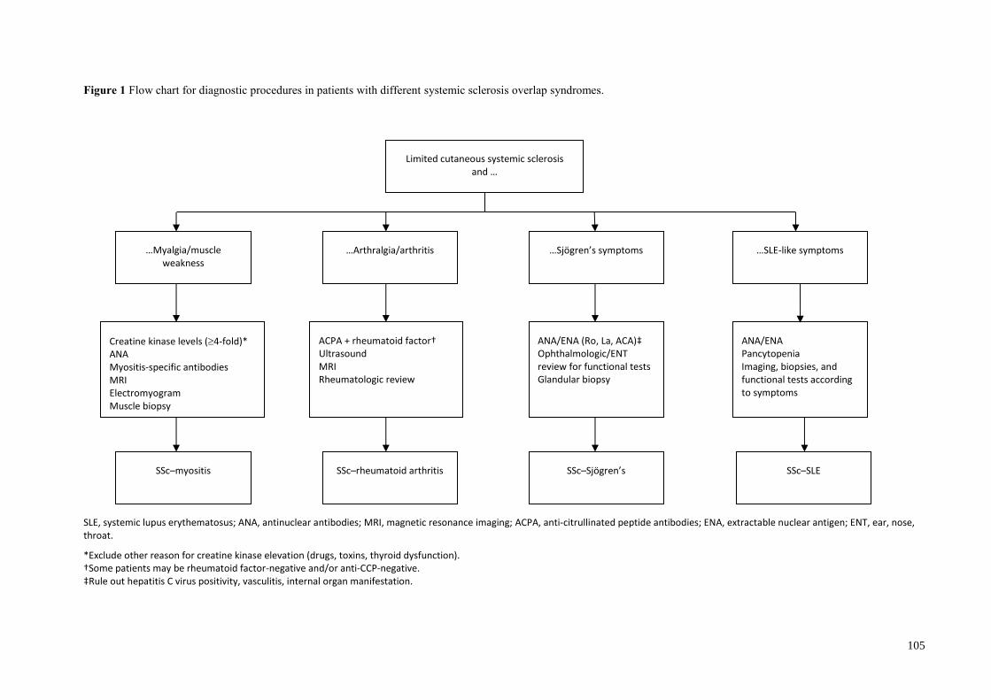
105
Figure 1 Flow chart for diagnostic procedures in patients with different systemic sclerosis overlap syndromes.
SLE, systemic lupus erythematosus; ANA, antinuclear antibodies; MRI, magnetic resonance imaging; ACPA, anti-citrullinated peptide antibodies; ENA, extractable nuclear antigen; ENT, ear, nose, throat.
*Exclude other reason for creatine kinase elevation (drugs, toxins, thyroid dysfunction). †Some patients may be rheumatoid factor-negative and/or anti-CCP-negative. ‡Rule out hepatitis C virus positivity, vasculitis, internal organ manifestation.
Limited cutaneous systemic sclerosis and …
…Myalgia/muscle weakness
…Arthralgia/arthritis …Sjögren’s symptoms
Creatine kinase levels (4-fold)* ANA Myositis-specific antibodies MRI Electromyogram Muscle biopsy
ACPA + rheumatoid factor† Ultrasound MRI Rheumatologic review
ANA/ENA (Ro, La, ACA)‡ Ophthalmologic/ENT review for functional tests Glandular biopsy
SSc–myositis SSc–rheumatoid arthritis SSc–Sjögren’s
…SLE-like symptoms
ANA/ENA Pancytopenia Imaging, biopsies, and functional tests according to symptoms
SSc–SLE
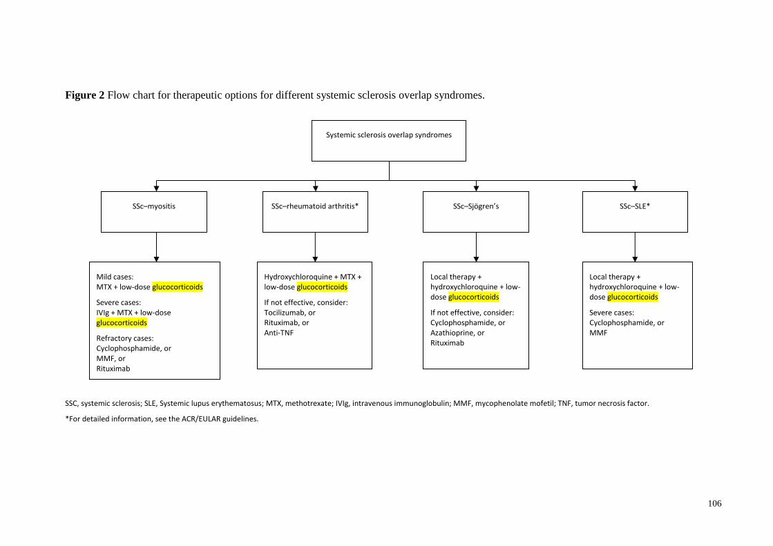
106
Figure 2 Flow chart for therapeutic options for different systemic sclerosis overlap syndromes.
SSC, systemic sclerosis; SLE, Systemic lupus erythematosus; MTX, methotrexate; IVIg, intravenous immunoglobulin; MMF, mycophenolate mofetil; TNF, tumor necrosis factor.
*For detailed information, see the ACR/EULAR guidelines.
Systemic sclerosis overlap syndromes
SSc–myositis SSc–rheumatoid arthritis* SSc–Sjögren’s
Mild cases: MTX + low-dose glucocorticoids
Severe cases: IVIg + MTX + low-dose glucocorticoids
Refractory cases: Cyclophosphamide, or MMF, or Rituximab
Hydroxychloroquine + MTX + low-dose glucocorticoids
If not effective, consider: Tocilizumab, or Rituximab, or Anti-TNF
Local therapy + hydroxychloroquine + low-dose glucocorticoids
If not effective, consider: Cyclophosphamide, or Azathioprine, or Rituximab
SSc–SLE*
Local therapy + hydroxychloroquine + low-dose glucocorticoids
Severe cases: Cyclophosphamide, or MMF
107
References
1. Balbir-Gurman A, Braun-Moscovici Y. Scleroderma overlap syndrome. Isr Med Assoc J
2011; 13: 14–20.
2. Iaccarino L, Gatto M, Bettio S, et al. Overlap connective tissue disease syndromes.
Autoimmun Rev. 2013; 12: 363–373.
3. Jury EC, D'Cruz D, Morrow WJ. Autoantibodies and overlap syndromes in autoimmune
rheumatic disease. J Clin Pathol 2001; 54: 340–347.
4. Pakozdi A, Nihtyanova S, Moinzadeh P, Ong VH, Black CM, Denton CP. Clinical and
serological hallmarks of systemic sclerosis overlap syndromes. J Rheumatol 2011; 38:
2406–2409.
5. Moinzadeh P, Aberer E, Ahmadi-Simab K, et al. Disease progression in systemic sclerosis-
overlap syndrome is significantly different from limited and diffuse cutaneous systemic
sclerosis. Ann Rheum Dis 2015; 74: 730–737.
6. Bohan A, Peter JB. Polymyositis and dermatomyositis (second of two parts). New Engl J
Med 1975; 292: 403–407.
7. Bohan A, Peter JB. Polymyositis and dermatomyositis (first of two parts). New Engl J Med
1975; 292: 344–347.
8. Kay J, Upchurch KS. ACR/EULAR 2010 rheumatoid arthritis classification criteria.
Rheumatology 2012; 51(Suppl 6): vi5–9.
9. Shiboski SC, Shiboski CH, Criswell L, et al. American College of Rheumatology
classification criteria for Sjogren's syndrome: a data-driven, expert consensus approach in
the Sjogren's International Collaborative Clinical Alliance cohort. Arthritis Care Res 2012;
64: 475–487.
10. Tan EM, Cohen AS, Fries JF, et al. The 1982 revised criteria for the classification of
systemic lupus erythematosus. Arthritis Rheum 1982; 25: 1271–1277.
11. Elhai M, Avouac J, Kahan A, Allanore Y. Systemic sclerosis at the crossroad of
polyautoimmunity. Autoimmun Rev 2013; 12: 1052–1057.
12. Caramaschi P, Biasi D, Volpe A, Carletto A, Cecchetto M, Bambara LM. Coexistence of
systemic sclerosis with other autoimmune diseases. Rheumatol Int 2007; 27: 407–410.
13. Hudson M, Rojas-Villarraga A, Coral-Alvarado P, et al. Polyautoimmunity and familial
autoimmunity in systemic sclerosis. J Autoimmun 2008; 31: 156–159.
14. Hunzelmann N, Genth E, Krieg T, et al. The registry of the German Network for Systemic
Scleroderma: frequency of disease subsets and patterns of organ involvement.
Rheumatology 2008; 47: 1185–1192.
108
15. Koschik RW 2nd, Fertig N, Lucas MR, Domsic RT, Medsger TA Jr. Anti-PM-Scl antibody
in patients with systemic sclerosis. Clin Exp Rheumatol 2012; 30(2 Suppl 71): S12–16.
16. Koumakis E, Dieude P, Avouac J, Kahan A, Allanore Y. Familial autoimmunity in
systemic sclerosis – results of a French-based case-control family study. J Rheumatol 2012;
39: 532–538.
17. Valenzuela A, Chung L. Calcinosis: pathophysiology and management. Curr Opin
Rheumatol 2015; 27: 542–548.
18. Bhansing KJ, Lammens M, Knaapen HK, van Riel PL, van Engelen BG, Vonk MC.
Scleroderma-polymyositis overlap syndrome versus idiopathic polymyositis and systemic
sclerosis: a descriptive study on clinical features and myopathology. Arthritis Res Ther
2014; 16: R111.
19. Mahler M, Raijmakers R. Novel aspects of autoantibodies to the PM/Scl complex: clinical,
genetic and diagnostic insights. Autoimmun Rev 2007; 6: 432–437.
20. Tani C, Carli L, Vagnani S, et al. The diagnosis and classification of mixed connective
tissue disease. J Autoimmun 2014; 48–49: 46–49.
21. Habets WJ, de Rooij DJ, Salden MH, Verhagen AP, van Eekelen CA, van de Putte LB, et
al. Antibodies against distinct nuclear matrix proteins are characteristic for mixed
connective tissue disease. Clinical and experimental immunology. 1983;54(1):265-76.
Epub 1983/10/01.
22. Kaji K, Fertig N, Medsger TA Jr, et al. Autoantibodies to RuvBL1 and RuvBL2: a novel
systemic sclerosis-related antibody associated with diffuse cutaneous and skeletal muscle
involvement. Arthritis Care Res 2014; 66: 575–584.
23. Rigolet A, Musset L, Dubourg O, et al. Inflammatory myopathies with anti-Ku antibodies:
a prognosis dependent on associated lung disease. Medicine 2012; 91: 95–102.
24. Lin HK, Wang JD, Fu LS. Juvenile diffuse systemic sclerosis/systemic lupus
erythematosus overlap syndrome – a case report. Rheumatol Int 2012; 32: 1809–1811.
25. Bloch KJ, Buchanan WW, Wohl MJ, Bunim JJ. Sjoegren's Syndrome. A clinical,
pathological, and serological study of sixty-two cases. Medicine 1965; 44: 187–231.
26. Ramos-Casals M, Brito-Zeron P, Font J. The overlap of Sjogren's syndrome with other
systemic autoimmune diseases. Semin Arthritis Rheum 2007; 36: 246–255.
27. Avouac J, Sordet C, Depinay C, et al. Systemic sclerosis-associated Sjogren's syndrome
and relationship to the limited cutaneous subtype: results of a prospective study of sicca
syndrome in 133 consecutive patients. Arthritis Rheum 2006; 54: 2243–2249.
109
28. Sharp GC, Irvin WS, Tan EM, Gould RG, Holman HR. Mixed connective tissue disease –
an apparently distinct rheumatic disease syndrome associated with a specific antibody to
an extractable nuclear antigen (ENA). Am J Med 1972; 52: 148–159.
29. Ortega-Hernandez OD, Shoenfeld Y. Mixed connective tissue disease: an overview of
clinical manifestations, diagnosis and treatment. Best Pract Res Clin Rheumatol 2012; 26:
61–72.
30. Akesson A, Fiori G, Krieg T, van den Hoogen FH, Seibold JR. Assessment of skin, joint,
tendon and muscle involvement. Clin Exp Rheumatol 2003; 21(3 Suppl 29): S5–8.
31. Santiago ML, Seisdedos MR, Garcia Salinas RN, Catalan Pellet A, Villalon L, Secco A.
Usefulness of antibodies and minor salivary gland biopsy in the study of sicca sindrome in
daily clinical practice. Reumatol Clin 2015; 11: 156–160.
32. Steen VD, Mayes MD, Merkel PA. Assessment of kidney involvement. Clin Exp
Rheumatol 2003; 21(3 Suppl 29): S29–31.
33. Kistler AD. Lupusnephritis. Ther Umsch 2015; 72: 171–177.
34. Kowal-Bielecka O, Distler O. Use of methotrexate in patients with scleroderma and mixed
connective tissue disease. Clin Exp Rheumatol 2010; 28(5 Suppl 61): S160–163.
35. Fendler C, Braun J. Use of methotrexate in inflammatory myopathies. Clin Exp Rheumatol
2010; 28(5 Suppl 61): S164–167.
36. Walker KM, Pope J. Expert agreement on EULAR/EUSTAR recommendations for the
management of systemic sclerosis. J Rheumatol 2011; 38(7): 1326–1328.
37. Levy Y, Amital H, Langevitz P, et al. Intravenous immunoglobulin modulates cutaneous
involvement and reduces skin fibrosis in systemic sclerosis: an open-label study. Arthritis
Rheum 2004; 50: 1005–1007.
38. Levine TD. Rituximab in the treatment of dermatomyositis: an open-label pilot study.
Arthritis Rheum 2005; 52: 601–607.
39. Mok CC, Ho LY, To CH. Rituximab for refractory polymyositis: an open-label prospective
study. J Rheumatol 2007; 34: 1864–1868.
40 van den Hoogen F, Khanna D, Fransen J, Johnson SR, Baron M, Tyndall A, et al. 2013
classification criteria for systemic sclerosis: an American College of Rheumatology/
European League against Rheumatism collaborative initiative. Ann Rheum Dis 2013; 72:
1747–1755.
41. Lundberg IE. The prognosis of mixed connective tissue disease. Rheum Dis Clin North Am
2005; 31: 535–547, vii-viii.

110
42. Feltsan T, Stanko P, Mracna J. Sjogren's syndrome in present. Bratisl Lek Listy 2012; 113:
514–516.
43. Carubbi F, Cipriani P, Marrelli A, et al. Efficacy and safety of rituximab treatment in early
primary Sjogren’s syndrome: a prospective, multicenter, follow-up study. Arthritis Res
Ther 2013; 15: R172.
44. Vitali C, Bootsma H, Bowman SJ, et al. Classification criteria for Sjogren’s syndrome: we
actually need to definitively resolve the long debate on the issue. Ann Rheum Dis 2013; 72:
476–478.
111
VI Scleredema
Introduction
Scleredema (scleredema adultorum, scleredema of Buschke) is a rare scleromucinous
connective tissue disease of unknown etiology. The disease is characterized by firm edema of
the trunk, head, shoulders, and sometimes the thighs, but with hands and feet spared.1 The
disease was first described back in 1752 by Curzio.2 However, in 1902 Abraham Buschke
named it “scler-oedem,” when he presented the case of a patient developing skin thickening
after influenza.3 Buschke also added “adultorum” to the name, suggesting the adult age of
affected patients. To date, the majority of patients described in the literature have been younger
than 20 years.1 Therefore, currently, scleredema adultorum is most commonly called
“scleredema” or “scleredema of Buschke.”
Three types of scleredema can been distinguished.4 They are associated with different preceding
or underlying conditions. Type 1 usually follows a febrile episode/infection. Type 2 is
associated with paraproteinemias (including multiple myeloma). Type 3 was named scleredema
diabeticorum by Krakowski,5 because of its strict association with diabetes mellitus (type 1 and
2). The pathologic effect of skin hardening in different clinical types of scleredema is the result
of excessive production of mucin (high molecular weight, heavily glycosylated proteins) and
collagens by fibroblasts in the reticular dermis.1
Epidemiology
Scleredema is a very rare condition. Its exact prevalence and incidence are unknown. Thus,
many cases may be unreported. To our knowledge, there is no racial or ethnic predilection to
the disease. Scleredema occurs in individuals of all ages, ranging from infancy to adulthood.
Type 1 scleredema, which constitutes 55% of the total number of cases, affects mainly
children.6 Type 2 scleredema occurs in 25% of cases, whereas type 3 scleredema is observed in
about 20% of scleredema cases.2 We do not know the proportion of type 1 and 2 diabetes or
multiple myeloma patients who develop scleredema. More than 50% of patients are aged under
20 years,7 and they mostly suffer from types 1 or 2 scleredema. In types 1 and 2 scleredema,
women are affected almost twice as frequently as men. The male to female ratio in type 3
scleredema is considered to be 10:1.2,6,8
112
Pathogenesis
The pathogenesis of scleredema remains unknown. Scleredema is a heterogeneous syndrome
with different concomitant conditions and mechanisms. The excessive production of mucin and
collagen may be provoked by diverse stimuli, including infections and inflammatory processes,
drugs, toxins, genetic mutations, immunoglobulins and cytokines, and genetic factors.9,10 The
pathogenesis of the diabetic type of scleredema is considered to be associated with ischemia,
hyperinsulinism, or autoimmunity.11 Chronic hyperglycemia is believed to stimulate fibroblast
proliferation and extracellular matrix components synthesis. It was documented that scleredema
is also associated with an abnormal expression of extracellular protein genes (type 1 and type
3 collagens, fibronectin) in the lesions.12 However, non-enzymatic glycosylation progressively
damages collagen in the connective tissue by altering the mechanism of collagen fiber
degradation. The lack of lymphocytic infiltrates in the lesions excludes a T-cell-mediated
etiology.
Clinical manifestations
The clinical symptoms of scleredema include cutaneous and extracutaneous findings, which are
present especially in types 2 and 3 scleredema.
Cutaneous manifestations
In the early stages, scleredema manifests as a woody hardening of the skin of the neck, which
later spreads to shoulders and the upper part of the trunk. It may affect the face and occasionally
the thighs, but it rarely affects the hands and feet.11,13,14 The lesions are ill-defined, non-pitting,
indurated plaques. The affected skin wrinkles or takes on a “peau d’orange” appearance when
pinched. This induration may occasionally follow a transient erythematous eruption.10,15,16 In
addition, a diffuse pigmentation of the skin was reported in a patient with type 2 scleredema, as
melanogenesis seems to be connected with paraproteinemia.17 The skin appendages are usually
preserved. However, the loss of eccrine glands, causing frequent heat attacks, was observed in
one patient.18 Cellulitis and delayed wound healing have also been reported.11,19
Extracutaneous manifestations
Although scleredema classically manifests as skin thickening, the extracutaneous involvement
of different internal organs is possible, leading to many potentially life-threatening
complications. Systemic manifestations occur more commonly in types 2 and 3; they are rare
in type 1 scleredema.20 Limitations in the movement of extremities, difficulties in opening the
113
mouth and eyes, and difficulties in breathing or even restrictive lung disease are the most
common symptoms. Lesions in various locations may lead to different complications.
Lesions in the eye and periorbital region lead to blepharoptosis, exophthalmos, chemosis,
conjunctivitis, corneal ulcer, keratitis, restricted eye movement, and ophthalmoplegia.8,10
Involvement of the tongue, the upper part of the esophagus, ocular muscles, pharynx, parotid
glands, and vocal cords in the larynx is less common. Involvement of the esophagus can cause
dysphagia.20–22 These complications are known to have caused aspiration of food and aspiration
pneumonia.23
Cardiac involvement may result in congestive heart failure, myocarditis, diastolic gallop,
arrhythmia, and repolarization abnormalities. Hepatomegaly and splenomegaly may be found
in some patients with scleredema.2 Scleredema may be associated with pleura and lung
involvement, leading to pulmonary restrictive disease, which can also be the consequence of
the involvement of the skin and subcutaneous structures of the trunk, or even death.24,25 In some
patients, these abnormalities may be secondary to skin induration of the trunk.
A very strong relationship between diabetic scleredema and obstructive sleep apnea syndrome
has been documented.25
Musculoskeletal involvement is associated with dismotility and limitation of motion.2 Some
authors have reported bone marrow infiltration with calvarial sclerosis or with osteopoikilosis,
as well as the infiltration of nerve tissue.2,10
Associated disorders
Scleredema is a heterogeneous disorder that may be associated with various conditions. Type 1
scleredema usually follows an infection, especially streptococcal respiratory tract infection.
However, other infective agents that have been reported are: influenza, measles, mumps,
chicken-pox, cytomegalovirus, diphtheria, encephalitis, mycoplasma pneumonia, and dental
abscesses.4,6,10,16,26,27
Type 2 scleredema is often associated with paraproteinemia, which is present in 25% of
scleredema patients, compared with 0.5% prevalence of paraproteins expected in the general
population.28 Associated conditions include monoclonal gammopathy,6,29–31 multiple
114
myeloma,32–36 and amyloidosis.28 Multiple myeloma is commonly not present at the time of
scleredema onset. Some patients with scleredema develop multiple myeloma after a few years
of asymptomatic monoclonal gammopathy. Other associated diseases include primary
hyperparathyroidism,37,38 rheumatoid arthritis,39,40 ankylosing spondylitis,29 Sjögren’s
syndrome,40 dermatomyositis,41 Waldenstrom’s macroglobulinemia, anaphylactoid purpura,
primary biliary cirrhosis,42 IgA deficiency,43 and HIV infection.8,10 Cases of concomitant
neoplasms have been reported, such as malignant insulinoma,44 gall bladder carcinoma,45
carcinoid tumor,46 and adrenocorticotropic hormone-producing pituitary tumor.47
Diabetic scleredema (type 3) is associated with type 1 and type 2 diabetes mellitus. However,
it is associated with insulin resistance and hyperglycemia, and therefore other endocrinopathies
with insulin resistance (not only diabetes) could be present. In addition, in cases of
adrenocortical tumors or pituitary adenomas, which are functional, scleredema symptoms refer
to the metabolic status of hypercortisolism and diabetes/diabetic tendencies.47
Clinical course
The clinical course of scleredema depends on the type. Type 1 scleredema, which is the classic
“Buschke” scleredema type, is preceded by a febrile illness (fever over 38°C during 3–4 weeks).
The onset of scleredema type 1 is abrupt but the prognosis is good and in most cases it resolves
in a few months to 2 years.6 There have been reports of rare cases of persistent scleredema type
1 persisting for 10 years. Systemic involvement is uncommon.
Type 2 scleredema is associated with paraproteinemias with no infection in the patient’s history.
This type is slowly progressive with a non-resolving course.4,6 Systemic involvement is likely
to occur with serious complications.
Type 3 scleredema (diabetic scleredema) is associated with type 1 or 2 diabetes mellitus. The
risk factors for scleredema type 3 are: male sex, long course of diabetes, poor metabolic control,
treatment with insulin, and presence of diabetes-specific complications (especially
microangiopathy). Other risk factors of diabetic scleredema are hypertension and
obesity.11,25,48,49 The course of disease is insidious, slowly progressing and non-resolving as in
type 2, but occasionally it is self-limited. Some cases are complicated by systemic involvement.
Diagnostic procedures
115
Histopathology
The following histopathologic findings are characteristic for scleredema.
• The epidermis is usually not involved.
• The most characteristic finding is increased thickness of the dermis (up to four times thicker
than normal). It is due to enlarged collagen bundles in deep reticular dermis and the
presence of wide, clear spaces between them. These fenestrations are filled with mucin.
Mucin deposits represent non-sulfated acid mucopolysaccharides, mainly hyaluronic acid.
• The subcutaneous tissue is also affected – fat is replaced by coarse collagen fibers.10
• Accumulation of mucopolysaccharides is easily found when stained with Alcian blue dye,
colloidal iron or toluidine blue. However, the absence of glycosaminoglycan deposits is
possible, and therefore this does not exclude the diagnosis.10,50,51
• Appendages are usually preserved, unlike in scleroderma. However, some authors have
reported the loss of eccrine glands.49,50
The diagnosis of scleredema is made clinically, with the definitive diagnosis confirmed by
histopathology.49,52,53
Diagnostic criteria
The diagnosis of scleredema is based upon the recognition of the following criteria:
• typical woody thickening of the skin, which spares acral locations (hands and feet are
usually not involved);
• increased thickness of the dermis in the microscopic evaluation with the accumulation of
mucopolysaccharides;
• history of a preceding infection, underlying diabetes or paraproteinemia.
In type 2 scleredema no associated diseases (paraproteinemia) may be present at the time of
diagnosis of scleredema. It is suggested to distinguish type 2a and 2b scleredema. Type 2a
fulfills all criteria of type 2 scleredema, but with no associated lymphoproliferative disorder.
Type 2b is scleredema associated with a lymphoproliferative disorder.
Patient history
The patient is asked about preceding infections. Moreover, the symptoms of malignancies or of
diabetes/glucose intolerance associated with other endocrinopathies should be carefully
considered. Identification of possible systemic complications requires questions about
116
difficulties in movement, fatigue (muscle or heart involvement), dysphagia (mainly
involvement of the upper part of the esophagus), respiratory problems, and neurologic
symptoms (e.g. paresthesia, pain).
Physical examination
A full skin examination is performed. The induration of the skin in characteristic locations
(neck, the upper part of the trunk, shoulders, face) and spared hands and feet suggest the
diagnosis of scleredema. A modified Rodnan scale (as in scleroderma or scleromyxedema) may
be used to evaluate the severity of skin involvement and to document its activity. In addition, a
durometer or an ultrasonography measurement of skin thickness may be performed in order to
evaluate the severity and to monitor the disease.54
Skin biopsy
A skin biopsy is required to confirm the diagnosis and to exclude other sclerosis-like disorders.
A 4 or 5 mm punch biopsy is sufficient. A mucin stain will be positive. Direct
immunofluorescence is negative and has little, if any, value for differential diagnosis.
Complementary investigations
At the time of diagnosis, blood tests mainly aim at identifying a lymphoproliferative disorder
in patients without a recent history of infection and without a history of diabetes. However, as
the disease is very rare, it is recommended that these tests are performed in all patients.
Leukocyte count (lymphocytes), serum protein electrophoresis, and serum and urine
immunofixation must be performed in order to screen for monoclonal gammopathy.49 In cases
of monoclonal gammopathy, or clinical evidence of enlarged lymph nodes, additional
investigations should be discussed, including cytofluorometry analysis (looking for B cell
lymphoproliferation), chest and abdomino-pelvic computed tomography scan ± positron
emission tomography scan ± lumbar and dorsal magnetic resonance imaging ±
myelogram/osteomedullar biopsy (Table 1).
<TABLE 1>
During follow-up, in patients with diabetes, fasting glycemia and HBA1c must be monitored.
In patients with type 2, with or without identified lymphoproliferation, leukocyte count
(lymphocytes), serum protein electrophoresis, and serum and urine immunofixation must be
117
performed every year, in association with a complete physical examination looking for lymph
node enlargement and/or hepato-splenomegaly (Table 2).
<TABLE 2>
Other laboratory test may be needed in differential diagnosis to exclude other conditions,
depending on the clinical presentation. Antinuclear antibodies (ANA), if performed, are
negative. This test may facilitate differential diagnosis with systemic sclerosis (SSc).
Additional tests
High-frequency ultrasonography may be performed to monitor the activity and severity of skin
involvement. In cases of systemic involvement, specific diagnostic examinations are required
(e.g. pulmonary function tests, ultrasonography of internal organs, including the heart, liver or
spleen, esophageal manometry, radiography/ultrasonography of bones/joints).
Differential diagnosis
Scleredema may cause diagnostic difficulties, as the differential diagnosis includes various
diseases. The characteristic thickness of the dermis and the accumulation of
mucopolysaccharides distinguish scleredema from other sclerotic disorders.10 Two main
disorders that require a differential diagnosis are: scleroderma (SSc) and scleromyxedema.
SSc (scleroderma)
Clinical and histopathologic differences allow for the differentiation. Skin thickening in SSc
typically begins with involvement of the finger tips, progressing to involve the hands and feet,
which are spared in scleredema. Other typical clinical findings of SSc, such as Raynaud’s
phenomenon, abnormal nailfold capillaries, and ANA, are absent in scleredema.
Histopathology distinguishes the two diseases, and there are no deposits of mucin in SSc.
Scleromyxedema
Clinically and histopathologically, scleromyxedema is very close to scleredema. However, the
induration of the skin progresses acrally and typically forms characteristic large folds or firm
papules, which are absent in scleredema. Systemic complications are common in both diseases.
The association with monoclonal gammopathy or multiple myeloma is present, similarly to
scleredema type 2. Mucin deposits are likely to be present.
118
Scleromyositis
Scleromyositis differs from scleredema by its typical clinical symptoms and common presence
of ANA, especially PM/Scl. The clinical presentation of edema, which is correlated with heart
or renal failure, is different from scleredema. However, sometimes there may be some problems
with differentiation. Edema is usually non-solid, “pitting.” Due to hydrostatic pressure, edema
is likely to occur in acral locations. The patient has symptoms of heart/renal failure.
Histopathologically, edema and scleredema are different. Similar clinical presentation and
differences (as found in edema) may also occur in the course of lymphedema.
Myxedema
Myxedema is associated with thyroid dysfunction, and is ruled out serologically and clinically,
by exclusion of thyroid function abnormalities.
Eosinophilic fasciitis
A typical woody induration in areas corresponding to the anatomic localization of the fascia.
Typically these are the trunk and extremities, usually sparing the finger tips. Carpal tunnel
syndrome may coexist. Eosinophilia, if present, may facilitate differential diagnosis.
Histopathology distinguishes the two disorders; however, the biopsy should be sufficiently deep
to reach the fascia. Mucin deposits are not present.
Cutaneous amyloidosis
The term “amyloidosis” is used for a group of disorders with accumulation of various insoluble
proteins (amyloid). Amyloidosis can be ruled out/confirmed with a microscopic examination.
Histopathologically, amyloidosis manifests as characteristic amyloid deposits found in the
affected tissues when stained with Congo red dye.
Lymphedema
Lymphedema refers to edema, which is usually most strongly expressed acrally, affecting the
extremities. The removal or damage to lymph nodes is common in the medical history of the
patient. Lymphedema differs from scleredema histopathologically. The typical findings include
keratinocyte hyperproliferation, condensed dermal collagen, and mononuclear perivascular
infiltrate that increases with lymphedema stage. There is no accumulation of
mucopolysaccharides.
119
Radiotherapy-induced skin thickening
Radiotherapy-induced skin thickening can be confirmed or excluded by history of preceding
radiation treatment. Lesions are usually limited to the area exposed to radiotherapy. Mucin
deposits are not present. Mylona et al. reported scleredema after a radiation treatment.55
Graft-versus-host disease
Graft-versus-host disease can be confirmed or excluded by history of preceding treatment.
Examination of skin biopsy for mucin deposits is negative.
Treatment
The treatment will focus on the underlying condition. Thus, when a potential cause is identified,
the priority will be the treatment of the cause (Table 3).
In diabetic patients, the control of diabetes is mandatory. If not already prescribed, insulin may
be necessary. In addition, diabetes should be controlled. Overweight patients should be given
advise on how to lose weight.
If an infection is identified it may be treated with appropriate anti-infectious agents. However,
scleredema type 1 does not usually require treatment, as it is self-limited and usually resolves
in few of months to 2 years.
If a lymphoproliferative disorder is identified, there is a need for discussions with the
hematologist in order to treat the lymphoproliferative disease itself. Thus, it was reported that
scleredema may improve after the treatment of a multiple myeloma.32
In the absence of an etiology, if the patient has severe involvement, a treatment can be proposed.
Unfortunately, the number of patients reported in the literature to benefit from a specific
treatment is very small, and on that basis it is very difficult to make evidence-based medical
recommendations. However, based on the available literature, the expert recommendation is to
use medium-to-high dose ultraviolet light therapy (UVA1 or PUVA) as a first-line treatment.56–
60 If the condition fails to improve, or if PUVA is not available, methotrexate is recommended
as a second-line treatment. If methotrexate fails, based on a risk–benefit approach, the following
treatments can be proposed:
120
• cyclosporine A61–63
• glucocorticoids (systemic or intralesional)64
• electron-beam radiotherapy6,65–67
• extracorporeal photopheresis68
• prostaglandin E169
• intravenous immunoglobulin70
• high-dose penicillin71
• hyaluronidase intralesionally72
• factor XIII infusion73
• radiotherapy74
• cyclophosphamide.36
A significant number of case reports indicate that the most beneficial treatment method is
photochemotherapy (UVA1, PUVA or narrow-band UVB). The mechanism of the
improvement remains unclear.49,75 Lack of randomized controlled trials in scleredema creates a
difficulty in drawing conclusions about the long-term efficacy, optimum dose, and best
treatment regimens.76
In addition to different systemic treatment modalities, non-pharmacologic treatments can be
proposed, such as physiotherapy, in order to increase the range of motion of joints and/or
improve restrictive respiratory insufficiency (Table 3).11
<TABLE 3>
Prognosis and follow-up
During follow-up, the efficacy of treatments can be assessed using the modified Rodnan skin
score, Health Assessment Questionnaire, range of motion of involved joints, and the
Dermatology Life Quality Index (Table 4).
Type 1 scleredema associated with a preceding infection is characterized by a good prognosis
and even spontaneous resolution. The active phase lasts 2–8 weeks and is followed by a
resolution in a couple of months to 2 years.77 Scleredema type 1 lesions persisting for 10 years
are uncommon.78
121
Unlike type 1, type 2, which is associated with blood dyscrasia, should be carefully followed
up. The prognosis is not good; the lesions are persistent with possible systemic involvement
leading to life-threatening complications. If only monoclonal gammopathy of unspecified
significance is present, the risk of multiple myeloma or another related malignancy is about 1%
per year. Therefore, careful follow-up of patients is required.17 The treatment of underlying
diseases is crucial; however, this may not be satisfactory in some type 2 scleredema cases.
Diabetic scleredema has a poor prognosis, with a chronic progressive course and systemic
complications. It also requires follow-up of patients. Monitoring blood glucose and metabolic
control are beneficial in some cases. Sleep apnea syndrome is common, and specific diagnostic
tests are necessary to confirm the disorder. As diabetic scleredema is under-recognized, there
is a need for appropriate education.64
<TABLE 4>
Summary and recommendation
• Scleredema adultorum is a connective tissue disorder characterized by the thickening of
the skin. The characteristic location of woody indurated areas is the upper part of the body.
In contrast to systemic sclerosis, it never affects the acral parts of extremities.
• There are three types of the disease. Type 1 usually follows a febrile episode/infection.
Type 2 is associated with paraproteinemias. Type 3 is associated with diabetes.
• There is no racial or ethnic predilection. Male to female ratio is 1:2 in type 1 and 2
scleredema, but 10:1 in type 3 scleredema.
• The pathogenesis of scleredema is unknown. Various conditions and mechanisms are
related to the excessive production of mucin and collagen, causing the thickening of the
dermis.
• The clinical symptoms of scleredema include cutaneous and extracutaneous findings,
especially in types 2 and 3 scleredema. Decreased mobility of the affected tissues
commonly causes movement limitations or even respiratory insufficiency.
• The clinical course of scleredema depends on its type. Three clinical types of scleredema
are associated with different stimuli which may evoke scleredema of Buschke.
• Skin biopsy identifies mucin deposits.
• Type 1 scleredema is preceded by a febrile illness. It is self-limited with a spontaneous
resolution in a few months to 2 years.
122
• Type 2 scleredema, associated with paraproteinemias (2a with identified paraproteinemia
at the time of diagnosis, type 2b with paraproteinemia identified during follow-up), is
slowly progressive with systemic involvement and a poor response to treatment.
• Type 3 scleredema (diabetic scleredema) is associated with diabetes. The course of disease
is insidious, slowly progressing, and non-resolving as in type 2, but it is sometimes self-
limited. Systemic complications are likely to occur.
• The diagnosis of scleredema is made clinically. A histopathologic examination is
performed to confirm a definitive diagnosis. It is made in cases of woody indurations of
the skin with increased thickness of the reticular dermis and the accumulation of
mucopolysaccharides found when stained with Alcian blue. Patient follow-up in types 2
and 3 scleredema is needed to screen for paraproteinemias and systemic complications or
to monitor the metabolic status of the patient with diabetic scleredema.
• Scleredema type 1 does not usually require treatment, as it is self-limited and usually
resolves in a short period of time. In types 2 and 3 scleredema, the treatment of an
underlying condition is needed. Better glucose control has been proven to be beneficial in
some cases. No specific therapy of scleredema is available, although numerous methods
have been proposed with variable results. The recommended first-line treatment is UV-
based management in monotherapy. If this fails, methotrexate is recommended.
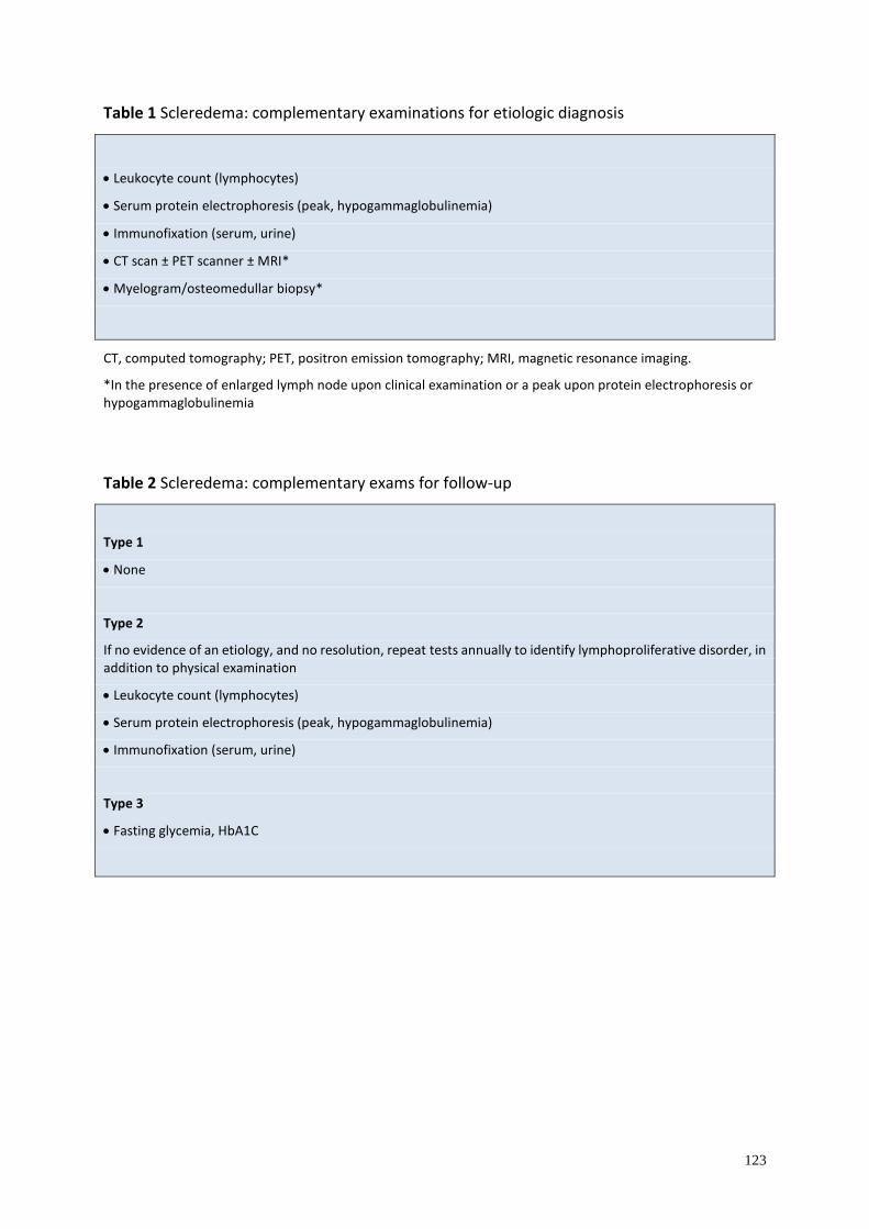
123
Table 1 Scleredema: complementary examinations for etiologic diagnosis
• Leukocyte count (lymphocytes)
• Serum protein electrophoresis (peak, hypogammaglobulinemia)
• Immunofixation (serum, urine)
• CT scan ± PET scanner ± MRI*
• Myelogram/osteomedullar biopsy*
CT, computed tomography; PET, positron emission tomography; MRI, magnetic resonance imaging.
*In the presence of enlarged lymph node upon clinical examination or a peak upon protein electrophoresis or hypogammaglobulinemia
Table 2 Scleredema: complementary exams for follow-up
Type 1
• None
Type 2
If no evidence of an etiology, and no resolution, repeat tests annually to identify lymphoproliferative disorder, in addition to physical examination
• Leukocyte count (lymphocytes)
• Serum protein electrophoresis (peak, hypogammaglobulinemia)
• Immunofixation (serum, urine)
Type 3
• Fasting glycemia, HbA1C
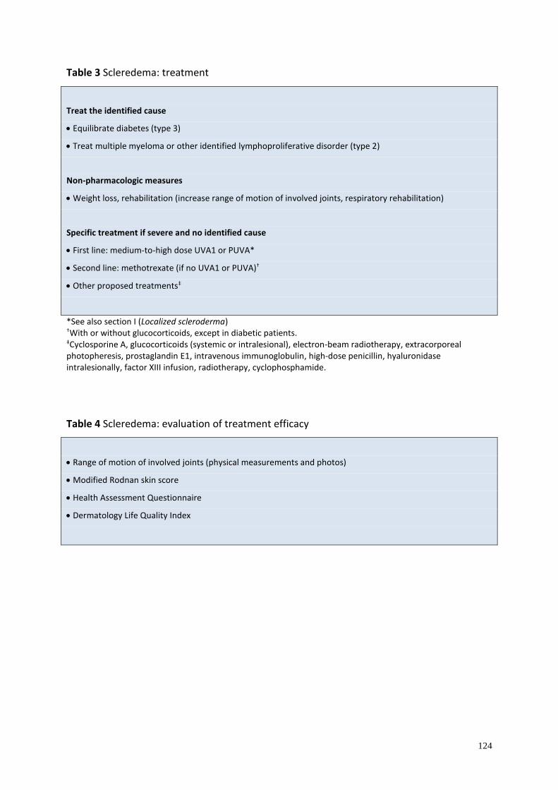
124
Table 3 Scleredema: treatment
Treat the identified cause
• Equilibrate diabetes (type 3)
• Treat multiple myeloma or other identified lymphoproliferative disorder (type 2)
Non-pharmacologic measures
• Weight loss, rehabilitation (increase range of motion of involved joints, respiratory rehabilitation)
Specific treatment if severe and no identified cause
• First line: medium-to-high dose UVA1 or PUVA*
• Second line: methotrexate (if no UVA1 or PUVA)†
• Other proposed treatments‡
*See also section I (Localized scleroderma) †With or without glucocorticoids, except in diabetic patients. ‡Cyclosporine A, glucocorticoids (systemic or intralesional), electron-beam radiotherapy, extracorporeal photopheresis, prostaglandin E1, intravenous immunoglobulin, high-dose penicillin, hyaluronidase intralesionally, factor XIII infusion, radiotherapy, cyclophosphamide.
Table 4 Scleredema: evaluation of treatment efficacy
• Range of motion of involved joints (physical measurements and photos)
• Modified Rodnan skin score
• Health Assessment Questionnaire
• Dermatology Life Quality Index
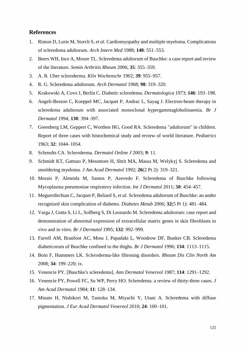
125
References
1. Rimon D, Lurie M, Storch S, et al. Cardiomyopathy and multiple myeloma. Complications
of scleredema adultorum. Arch Intern Med 1988; 148: 551–553.
2. Beers WH, Ince A, Moore TL. Scleredema adultorum of Buschke: a case report and review
of the literature. Semin Arthritis Rheum 2006; 35: 355–359.
3. A. B. Uber scleroderma. Klin Wochenschr 1902; 39: 955–957.
4. R. G. Scleredema adultorum. Arch Dermatol 1968; 98: 319–320.
5. Krakowski A, Covo J, Berlin C. Diabetic scleredema. Dermatologica 1973; 146: 193–198.
6. Angeli-Besson C, Koeppel MC, Jacquet P, Andrac L, Sayag J. Electron-beam therapy in
scleredema adultorum with associated monoclonal hypergammaglobulinaemia. Br J
Dermatol 1994; 130: 394–397.
7. Greenberg LM, Geppert C, Worthen HG, Good RA. Scleredema "adultorum" in children.
Report of three cases with histochemical study and review of world literature. Pediatrics
1963; 32: 1044–1054.
8. Schmults CA. Scleroderma. Dermatol Online J 2003; 9: 11.
9. Schmidt KT, Gattuso P, Messmore H, Shrit MA, Massa M, Welykyj S. Scleredema and
smoldering myeloma. J Am Acad Dermatol 1992; 26(2 Pt 2): 319–321.
10. Morais P, Almeida M, Santos P, Azevedo F. Scleredema of Buschke following
Mycoplasma pneumoniae respiratory infection. Int J Dermatol 2011; 50: 454–457.
11. Meguerditchian C, Jacquet P, Beliard S, et al. Scleredema adultorum of Buschke: an under
recognized skin complication of diabetes. Diabetes Metab 2006; 32(5 Pt 1): 481–484.
12. Varga J, Gotta S, Li L, Sollberg S, Di Leonardo M. Scleredema adultorum: case report and
demonstration of abnormal expression of extracellular matrix genes in skin fibroblasts in
vivo and in vitro. Br J Dermatol 1995; 132: 992–999.
13. Farrell AM, Branfoot AC, Moss J, Papadaki L, Woodrow DF, Bunker CB. Scleredema
diabeticorum of Buschke confined to the thighs. Br J Dermatol 1996; 134: 1113–1115.
14. Boin F, Hummers LK. Scleroderma-like fibrosing disorders. Rheum Dis Clin North Am
2008; 34: 199–220; ix.
15. Venencie PY. [Buschke's scleredema]. Ann Dermatol Venereol 1987; 114: 1291–1292.
16. Venencie PY, Powell FC, Su WP, Perry HO. Scleredema: a review of thirty-three cases. J
Am Acad Dermatol 1984; 11: 128–134.
17. Minato H, Nishikori M, Tanioka M, Miyachi Y, Utani A. Scleredema with diffuse
pigmentation. J Eur Acad Dermatol Venereol 2010; 24: 100–101.
126
18. Lin IC, Chiu HY, Chan JY, Lin SJ. Extensive scleredema adultorum with loss of eccrine
glands. J Am Acad Dermatol 2014; 71: e99–e101.
19. Rees R, Moore A, Nanney L, King L. Scleredema adultorum: the surgical implications of
a rare dermatologic disorder. Plast Reconstr Surg 1983; 72: 90–93.
20. Mohanasundaram K, Kumarasamy S, Kumar R, Rajendran CP. Scleredema adultorum of
Buschke: with unusual manifestations in a young female. Indian J Dermatol Venereol
Leprol 2012; 78: 503–505.
21. Ioannidou DI, Krasagakis K, Stefanidou MP, Karampekios S, Panayiotidis J, Tosca AD.
Scleredema adultorum of Buschke presenting as periorbital edema: a diagnostic challenge.
J Am Acad Dermatol 2005; 52(2 Suppl 1): 41–44.
22. Brenner M, Herzinger T, Berking C, Plewig G, Degitz K. Phototherapy and
photochemotherapy of sclerosing skin diseases. Photodermatol Photoimmunol Photomed
2005; 21: 157–165.
23. Wright RA, Bernie H. Scleredema adultorum of Buschke with upper esophageal
involvement. Am J Gastroenterol 1982; 77: 9–11.
24. Sansom JE, Sheehan AL, Kennedy CT, Delaney TJ. A fatal case of scleredema of Buschke.
Br J Dermatol 1994; 130: 669–670.
25. Ray V, Boisseau-Garsaud AM, Ray P, et al. [Obesity persistent scleredema: study of 49
cases]. Ann Dermatol Venereol 2002; 129: 281–285.
26. Venencie PY, Powell FC, Su WP. Scleredema and monoclonal gammopathy: report of two
cases. Acta Derm Venereol 1984; 64: 554–556.
27. Schmults C, Sidhu G, Urbanek RW. Aquagenic syringeal acrokeratoderma. Dermatol
Online J 2003; 9: 27.
28. Dziadzio M, Anastassiades CP, Hawkins PN, et al. From scleredema to AL amyloidosis:
disease progression or coincidence? Review of the literature. Clin Rheumatol 2006; 25: 3–
15.
29. Chang HK, Kim YC, Kwon BS. Widespread scleredema accompanied with a monoclonal
gammopathy in a patient with advanced ankylosing spondylitis. J Korean Med Sci 2004;
19: 481–483.
30. Paz RA, Badra RE, Marti HM, Maxit MJ. [Systemic Buschke's scleredema with
cardiomyopathy, monoclonal IgG kappa gammopathy and amyloidosis. Case report with
autopsy]. Medicina (B Aires) 1998; 58(5 Pt 1): 501–503.
127
31. McFadden N, Ree K, Soyland E, Larsen TE. Scleredema adultorum associated with a
monoclonal gammopathy and generalized hyperpigmentation. Arch Dermatol 1987; 123:
629–632.
32. Szturz P, Adam Z, Vasku V, et al. Complete remission of multiple myeloma associated
scleredema after bortezomib-based treatment. Leuk Lymphoma 2013; 54: 1324–1326.
33. Santos-Juanes J, Osuna CG, Iglesias JR, De Quiros JF, del Rio JS. Treatment with
chemotherapy of scleredema associated with Ig A myeloma. Int J Dermatol 2001; 40: 720–
721.
34. Grudeva-Popova J, Dobrev H. Biomechanical measurement of skin distensibility in
scleredema of Buschke associated with multiple myeloma. Clin Exp Dermatol 2000; 25:
247–249.
35. Valente L, Velho GC, Farinha F, Bernardo A, Ribeiro P, Massa A. [Scleredema, acanthosis
nigricans and IgA/Kappa multiple myeloma]. Ann Dermatol Venereol 1997; 124: 537–539.
36. Salisbury JA, Shallcross H, Leigh IM. Scleredema of Buschke associated with multiple
myeloma. Clin Exp Dermatol 1988; 13: 269–270.
37. Jacob N, Gleichmann U, Stadler R. [Scleredema adultorum in secondary
hyperparathyroidism]. Hautarzt 2002; 53: 121–125.
38. Berk MA, Lorincz AL. Scleredema adultorum of Buschke and primary
hyperparathyroidism. Int J Dermatol 1988; 27: 647–649.
39. Ranganathan P. Infliximab-induced scleredema in a patient with rheumatoid arthritis. J
Clin Rheumatol 2005; 11: 319–322.
40. Miyagawa S, Dohi K, Tsuruta S, Shirai T. Scleredema of Buschke associated with
rheumatoid arthritis and Sjogren's syndrome. Br J Dermatol 1989; 121: 517–520.
41. Marill FG, Grould P. [Scleredema, 1st manifestation of a dermatomyositis]. Bull Soc Fr
Dermatol Syphiligr 1968; 75: 561–564.
42. Goss F, Krawietz W, Luderschmidt C, Pape GR. [Scleredema adultorum Buschke and
primary biliary cirrhosis in a 58-year-old patient]. Internist 1984; 25: 130–134.
43. Theodoridis A, Capetanakis J. Scleredema of Buschke with IgA deficiency. Acta Derm
Venereol 1979; 59: 182–183.
44. Matsunaga J, Hara M, Tagami H. Scleredema of Buschke associated with malignant
insulinoma. Br J Dermatol 1992; 126: 527–528.
45. Manchanda Y, Das S, Sharma VK, Srivastava DN. Scleredema associated with carcinoma
of the gall bladder. Br J Dermatol 2005; 152: 1373–1374.
128
46. Yu JI, Park W, Lee KK, Park W. Scleredema adultorum of Buschke associated with a
carcinoid tumor. Int J Dermatol 2009; 48: 784–786.
47. Oyama N, Togashi A, Kaneko F, Yamamoto T. Two cases of scleredema with pituitary-
adrenocortical neoplasms: an underrecognized skin complication. J Dermatol 2012; 39:
193–195.
48. Leung CS, Chong LY. Scleredema in Chinese patients: a local retrospective study and
general review. Hong Kong Med J 1998; 4: 31–35.
49. Martin C, Requena L, Manrique K, Manzarbeitia FD, Rovira A. Scleredema diabeticorum
in a patient with type 2 diabetes mellitus. Case Rep Endocrinol 2011; 2011: 560273.
50. Rongioletti F, Rebora A. Cutaneous mucinoses: microscopic criteria for diagnosis. Am J
Dermatopathol 2001; 23: 257–267.
51. Vereecken P, Lutz R, De Dobbeleer G, Heenen M. Nonpitting induration of the back.
Scleredema adultorum of Buschke type III. Arch Dermatol 1997; 133: 649, 652.
52. Cole HG, Winkelmann RK. Acid mucopolysaccharide staining in scleredema. J Cutan
Pathol 1990; 17: 211–213.
53. Cole GW, Headley J, Skowsky R. Scleredema diabeticorum: a common and distinct
cutaneous manifestation of diabetes mellitus. Diabetes Care 1983; 6: 189–192.
54. Cole GW, Handler SJ, Burnett K. The ultrasonic evaluation of skin thickness in
scleredema. J Clin Ultrasound 1981; 9: 501–503.
55. Mylona E, Golfinopoulou S, Skarmea A, et al. Post-radiation scleredema adultorum and
diffuse eosinophilic fasciitis in the same patient. Scand J Rheumatol 2011; 40: 240–241.
56. Kokpol C, Rajatanavin N, Rattanakemakorn P. Successful treatment of scleredema
diabeticorum by combining local PUVA and colchicine: a case report. Case Rep Dermatol
2012; 4: 265–268.
57. Nakajima K, Iwagaki M, Ikeda M, Kodama H. Two cases of diabetic scleredema that
responded to PUVA therapy. J Dermatol 2006; 33: 820–822.
58. Grundmann-Kollmann M, Ochsendorf F, Zollner TM, Spieth K, Kaufmann R, Podda M.
Cream PUVA therapy for scleredema adultorum. Br J Dermatol 2000; 142: 1058–1059.
59. Hager CM, Sobhi HA, Hunzelmann N, et al. Bath-PUVA therapy in three patients with
scleredema adultorum. J Am Acad Dermatol 1998; 38(2 Pt 1): 240–242.
60. Yoshimura J, Asano Y, Takahashi T, et al. A case of scleredema adultorum successfully
treated with narrow-band ultraviolet B phototherapy. Mod Rheumatol 2014; doi:
10.3109/14397595.2013.875640.
129
61. Mattheou-Vakali G, Ioannides D, Thomas T, Lazaridou E, Tsogas P, Minas A.
Cyclosporine in scleredema. J Am Acad Dermatol 1996; 35: 990–991.
62. Dogramaci AC, Inan MU, Atik E, Gokce C. Scleredema diabeticorum partially treated with
low-dose methotrexate: a report of five cases. Balkan Med J 2012; 29: 218–221.
63. Breuckmann F, Appelhans C, Harati A, Rotterdam S, Altmeyer P, Kreuter A. Failure of
low-dose methotrexate in the treatment of scleredema diabeticorum in seven cases.
Dermatology 2005; 211: 299–301.
64. Rongioletti F, Kaiser F, Cinotti E, et al. Scleredema. A multicentre study of characteristics,
comorbidities, course and therapy in 44 patients. J Eur Acad Dermatol Venereol 2015; 29:
2399–2404.
65. Lee MW, Choi JH, Sung KJ, Moon KC, Koh JK. Electron beam therapy in patients with
scleredema. Acta Derm Venereol 2000; 80: 307–308.
66. Tamburin LM, Pena JR, Meredith R, Soong VY. Scleredema of Buschke successfully
treated with electron beam therapy. Arch Dermatol 1998; 134: 419–422.
67. Pilgrim U, Vogel A, Meves C. [Connective tissue fibers in scleromyxedema and
scleredema adultorum. A quantitative electron microscopic study (author's transl)].
Dermatologica 1973; 147: 46–63.
68. Stables GI, Taylor PC, Highet AS. Scleredema associated with paraproteinaemia treated
by extracorporeal photopheresis. Br J Dermatol 2000; 142: 781–783.
69. Alsaeedi SH, Lee P. Treatment of scleredema diabeticorum with tamoxifen. J Rheumatol
2010; 37: 2636–2637.
70. Eastham AB, Femia AN, Velez NF, Smith HP, Vleugels RA. Paraproteinemia-associated
scleredema treated successfully with intravenous immunoglobulin. JAMA Dermatol 2014;
150: 788–789.
71. Krasagakis K, Hettmannsperger U, Trautmann C, Tebbe B, Garbe C. Persistent scleredema
of Buschke in a diabetic: improvement with high-dose penicillin. Br J Dermatol 1996; 134:
597–598.
72. Frank H. [Experience with ACTH and hyaluronidase treatment of a case of scleredema
adultorum Buschke, Mayr type]. Hautarzt 1954; 5: 514–515.
73. Venturi C, Zendri E, Santini M, Grignaffini E, Ricci R, De Panfilis G. Scleredema of
Buschke: remission with factor XIII treatment. Int J Tissue React 2004; 26: 25–28.
74. Bowen AR, Smith L, Zone JJ. Scleredema adultorum of Buschke treated with radiation.
Arch Dermatol 2003; 139: 780–784.

130
75. Kroft EB, de Jong EM. Scleredema diabeticorum case series: successful treatment with
UV-A1. Arch Dermatol 2008; 144: 947–948.
76. Kroft EB, Berkhof NJ, van de Kerkhof PC, Gerritsen RM, de Jong EM. Ultraviolet A
phototherapy for sclerotic skin diseases: a systematic review. J Am Acad Dermatol 2008;
59: 1017–1030.
77. Parmar RC, Bavdekar SB, Bansal S, Doraiswamy A, Khambadkone S. Scleredema
adultorum. J Postgrad Med 2000; 46: 91–93.
78. Pitarch G, Torrijos A, Martinez-Aparicio A, Vilata JJ, Fortea JM. [Scleredema of Buschke
associated with diabetes mellitus. Study of four cases]. Actas Dermosifiliogr 2005; 96: 46–
49.
Related Documents