Guideline for the Management of Hypertensive Disorders of Pregnancy 2014 Lowe SA, Bowyer L, Lust K, McMahon LP, Morton M, North RA, Paech M. Said JM. These are the recommendations of a multidisciplinary working party convened by the Society of Obstetric Medicine of Australia and New Zealand. They reflect current medical literature and the clinical experience of members of the working party.

Welcome message from author
This document is posted to help you gain knowledge. Please leave a comment to let me know what you think about it! Share it to your friends and learn new things together.
Transcript

Guideline for the Management of
Hypertensive Disorders of Pregnancy
2014
Lowe SA, Bowyer L, Lust K, McMahon LP, Morton M, North RA, Paech M. Said JM.
These are the recommendations of a multidisciplinary working party convened by the Society
of Obstetric Medicine of Australia and New Zealand. They reflect current medical literature
and the clinical experience of members of the working party.

1
CONTENTS
Section
Page
Abbreviations
2
1.
Definition of hypertension in pregnancy
3
2.
Recording blood pressure in pregnancy
4
3.
Classification of hypertensive disorders in pregnancy
5
4.
Investigation of new onset hypertension after 20 weeks
9
5.
Management of preeclampsia and gestational hypertension
11
6.
Eclampsia
18
7.
Fetal Surveillance in hypertensive diseases of pregnancy
19
8.
Resolution of preeclampsia and gestational hypertension
21
9.
Chronic hypertension in pregnancy
22
10.
Anaesthetic considerations
26
11.
Preconception management and prophylaxis
28
12.
Prevention of preeclampsia
30
13.
Longterm consequences
33
14.
Auditing outcomes
34
15.
References
36

2
ABBREVIATIONS
ABPM Ambulatory blood pressure monitoring
AFV Amniotic fluid volume
ALT Alanine transaminase
AOR Adjusted odds ratio
APPT Activated partial thromboplastin time
AST Aspartate transaminase
BW Birth weight
CI Confidence Interval
ECG Electrocardiogram
FBC Full blood count
FGR Fetal growth restriction
HELLP Haemolysis, elevated liver enzymes and low platelet syndrome
Hr Hour(s)
INR International normalised ratio
ISSHP International Society for the Study of Hypertension in Pregnancy
IU International units
IV Intravenous
K1 Korotkoff sound 1
K2 Korotkoff sound 2
Kg kilogram
LDA Low dose aspirin
LDH Lactate dehydrogenase
LFT Liver function tests
mcg microgram
mg milligram
min minute
mL millilitre
NICU Neonatal intensive care
NPV Negative predictive value
PCR Protein/creatinine ratio
PlGF Placental growth factor
RDS Respiratory distress syndrome
RR Relative risk
SFlt1 soluble fms like tyrosine kinase-1
SGA Small for gestational age
UEC Urea, electrolytes and creatinine
umol/L Micromole/litre
U/S Ultrasound
VEGF Vascular endothelial growth factor
VTE Venous thromboembolism

3
GUIDELINES FOR THE MANAGEMENT OF HYPERTENSIVE
DISORDERS OF PREGNANCY 2014
Lowe SA, Bowyer L, Lust K, McMahon L, Morton M, North RA, Paech M. Said J.
These are the recommendations of a multidisciplinary working party convened by the Society of
Obstetric Medicine of Australia and New Zealand. They reflect current medical literature and the
clinical experience of members of the working party.
1. Definition of hypertension in pregnancy
Normal pregnancy is characterized by a fall in blood pressure, detectable in the first trimester and
usually reaching a nadir in the second trimester. Blood pressure rises towards pre-conception levels
by term.
Hypertension in pregnancy is defined as:
Systolic blood pressure greater than or equal to 140 mmHg and/or
Diastolic blood pressure greater than or equal to 90 mmHg (Korotkoff 5)
These measurements should be confirmed by repeated readings over several hours. Elevations of
both systolic and diastolic blood pressures have been associated with adverse maternal and fetal
outcome and therefore both are important (1).There are several reasons to support the blood
pressure readings above as diagnostic of hypertension in pregnancy:
Perinatal mortality rises with diastolic blood pressures above 90 mmHg (2)
Readings above this level were beyond two standard deviations of mean blood pressure in a
New Zealand cohort of normal pregnant women (3)
The chosen levels are consistent with International guidelines and correspond with the
current diagnosis of hypertension outside of pregnancy
Detecting a rise in blood pressure from ‘booking’ or preconception blood pressure (> 30/15 mmHg),
rather than relying on an absolute value has in the past been considered useful in diagnosing
preeclampsia in women who do not reach blood pressures of 140 or 90 mmHg. Available evidence
does not support the notion that these women have an increased risk of adverse outcomes (4, 5).
Nevertheless such a rise may be significant in some pregnant women, particularly in the presence of
hyperuricaemia, proteinuria or a small for gestational age (SGA) infant and these women warrant
closer monitoring.
4
Severe hypertension in pregnancy
This guideline recommends antihypertensive treatment for all pregnant women with blood pressure
greater than or equal to 160mm Hg systolic or 110 mm Hg diastolic. Severe hypertension requiring
urgent treatment is defined as a systolic blood pressure greater than or equal to 170 mmHg with or
without diastolic blood pressure greater than or equal to 110 mmHg. This represents a level of blood
pressure above which the risk of maternal morbidity and mortality is increased (6, 7). This degree of
hypertension requires urgent assessment and management. Increasing evidence exists that cerebral
perfusion pressure is altered in pregnant women making them more susceptible to cerebral
haemorrhage, posterior reversible encephalopathy syndrome and hypertensive encephalopathy (8,
9). It is universally agreed that severe hypertension should be lowered promptly, albeit carefully, to
prevent such complications (7, 10-13). [See Section 5]
2. Recording blood pressure in pregnancy
Accurate blood pressure measurement is important as the level of blood pressure may result in
changes in clinical management (14). The woman should be seated comfortably with her legs
resting on a flat surface and her arm resting at the level of her heart. In labour, the blood pressure
may be measured in lateral recumbency. Supine posture should be avoided because of the supine
hypotension syndrome. The variation in blood pressure between arms is usually less than 10 mmHg,
with 8% and 2% of pregnant women having an inter-arm difference of at least 10 mm Hg for
systolic and diastolic blood pressure, respectively (15).
The systolic blood pressure is accepted as the first sound heard (K1) and the diastolic blood
pressure the disappearance of sounds completely (K5) (16). Where K5 is absent, K4 (muffling)
should be accepted. Correct cuff size is important for accurate blood pressure recording. A large
cuff with an inflatable bladder covering 80% of the arm circumference should be used if the upper
arm circumference is greater than 33 cm but less than 44 cm and a thigh cuff used if the upper arm
circumference is greater than 44 cm (14, 17). This helps to minimise over-diagnosis of hypertension
during pregnancy. The rate of deflation of the cuff should be ≤2 mm per second to avoid
underestimating systolic blood pressure (18).
Measurement Devices
Mercury sphygmomanometers remain the gold standard for measurement of blood pressure in
pregnancy, however, occupational health concerns are limiting their availability. Self initiated home
blood pressure monitors have provided major advantages for treatment and diagnosis of
hypertension in the general community and are now widely used by both pregnant women and their
clinicians. While automated devices may give similar mean blood pressure values to those obtained
with mercury sphygmomanometry, there is wide intra-individual error and their accuracy may be
further compromised in preeclamptic women (19-21). Only a few automated blood pressure
monitors (Microlife WatchBP Home, Microlife 3BTO-A and Omron T9P have been validated for
use in normotensive or mildly hypertensive pregnant women (22-24). In women with preeclampsia,
especially those with severe hypertension, the accuracy of both Microlife 3BTO-A and Omron M7
declined and these devices cannot be recommended for use in preeclampsia (25).
Nonmercury auditory sphygmomanometers present an option with appropriately trained observers
(26). Aneroid sphygmomanometers may be used but are also prone to error. Each unit should
maintain a mercury sphygmomanometer for validation of automated and aneroid devices.
Healthcare providers must ensure that devices for measuring blood pressure are properly validated,
maintained and regularly recalibrated according to manufacturers’ instructions as recommended by
the British Hypertension Society Society (27) To establish the role of automated blood pressure
monitors in hypertensive complications in pregnancy requires further studies comparing the impact
5
of blood pressure measurement with automated devices to mercury sphygmomanometers on
important clinical outcomes (28, 29).
Twenty four hour Ambulatory Blood Pressure Monitoring (ABPM)
Normal blood pressure ranges for values recorded by ABPM have been established for different
stages of pregnancy (30). The major role of ABPM is in identifying women with white coat
hypertension, thereby avoiding inappropriate intervention (31). ABPM is useful in the evaluation of
early hypertension (before 20 weeks gestation) where approximately one third of these women
will be shown to have “white coat” or “office” hypertension (32). About half of these women with
white coat hypertension will not require antihypertensive medication in pregnancy, while the other
half will develop true hypertension (ABPM confirmed). ABPM is less useful in screening for white
coat hypertension in the second half of pregnancy (33).
Twenty four hour ABPM has poor sensitivity and specificity when used to identify women at risk of
developing hypertension later in pregnancy (33). In women with hypertensive disorders in
pregnancy, ABPM has modest prognostic value in predicting adverse outcomes but further research
is necessary to define its role in clinical management of hypertensive disorders in pregnancy.
3. Classification of hypertensive disorders in pregnancy
This classification of the hypertensive disorders in pregnancy reflects the pathophysiology of the
constituent conditions as well as the risks and potential outcomes for both mother and baby. The
following (or very similar) clinical classification has been adopted by numerous National and
International bodies, differing predominantly in whether they require proteinuria or not for the
diagnosis of preeclampsia (11, 13, 34, 35). Internationally, the latest ISSHP guideline no longer
requires proteinuria for the diagnosis of preeclampsia, leaving only the British NICE guideline with
this requirement (13, 35).
The classification is as follows:
Preeclampsia – eclampsia
Gestational hypertension
Chronic hypertension
-essential
-secondary
-white coat
Preeclampsia superimposed on chronic hypertension
Preeclampsia is a multi-system disorder unique to human pregnancy characterised by hypertension
and involvement of one or more other organ systems and/or the fetus. Raised blood pressure is
commonly but not always the first manifestation. Proteinuria is the most commonly recognised
additional feature after hypertension but should not be considered mandatory to make the clinical
diagnosis. As this classification is based on clinical data, it is possible that women with another
condition will sometimes be classified incorrectly as having preeclampsia during pregnancy.Note a
This is not usually a clinical problem as the diagnosis of preeclampsia should lead to increased
observation and vigilance which is appropriate for conditions which may mimic preeclampsia.
6
A diagnosis of preeclampsia can be made when hypertension arises after 20 weeks gestation Note
b
and is accompanied by one or more of the following signs of organ involvement:
Renal involvement
Significant proteinuria – a spot urine protein/creatinine ratio ≥ 30mg/mmol Note
c
Serum or plasma creatinine > 90 μmol/L Note d
Oliguria: <80mL/4 hr Note
e
(Urate is not included as a diagnostic feature Note f
)
Haematological involvement
Thrombocytopenia <100,000 /µL Note g
Haemolysis: schistocytes or red cell fragments on blood film, raised bilirubin, raised lactate
dehydrogenase >600mIU/L, decreased haptoglobin
Disseminated intravascular coagulation Note h
Liver involvement
Raised serum transaminases Note
i
Severe epigastric and/or right upper quadrant pain.
Neurological involvement
Convulsions (eclampsia)
Hypereflexia with sustained clonus
Persistent, new headache
Persistent visual disturbances (photopsia, scotomata, cortical blindness, posterior reversible
encephalopathy syndrome, retinal vasospasm)
Stroke
Pulmonary oedema
Fetal growth restriction (FGR) Note
j
Controversies in classifying the severity of preeclampsia
A number of features of preeclampsia are recognised to significantly increase the risk of adverse
maternal and fetal outcomes and are sometimes used to classify severe preeclampsia.(12, 13, 34,
36). The natural history of preeclampsia is to progress at an unpredictable rate, at least until
delivery, and therefore all women with preeclampsia should be closely monitored.
The classification of severe preeclampsia would ideally allow the identification of those women and
babies at increased risk of adverse maternal/fetal outcomes and/or requiring more intensive
monitoring and/or treatment. A number of classification systems and surveys have attempted to
identify which features are predictive. One study reported that features that had previously been
recommended as indicators of severe disease were neither sensitive nor specific in identifying
women and/or babies at particular risk (37). The recent ISSHP statement suggested there was
general consensus that factors determining severity include difficulty in controlling blood pressure
and deteriorating clinical condition including HELLP syndrome, impending eclampsia, worsening
thrombocytopenia or worsening fetal growth restriction while there is less concern regarding
increasing proteinuria (12).

7
Alterations in circulating angiogenic factors [increased soluble fms like tyrosine kinase-1 (sFlt1) or
soluble endoglin and reduced placental growth factor (PlGF)] are pathophysiologically important in
the development of preeclampsia and may have a potential role in diagnosis (38). Changes in these
angiogenic factors are detectable both prior to and at the time of onset of hypertension in women
with preeclampsia. Measurement of PlGF alone, or in combination with sFlt1, are currently not part
of the classification of hypertensive disorders of pregnancy (see Section 3).
NOTES
a. Other rare disorders may present with some of the features of preeclampsia (22). Disorders such
as acute fatty liver of pregnancy, hemolytic uremic syndrome, thrombotic thrombocytopenic
purpura, exacerbation of systemic lupus erythaematosus or cholecystitis may need to be
excluded.
b. Rarely preeclampsia presents before 20 weeks gestation; usually in the presence of a
predisposing factor such as hydatidiform mole, multiple pregnancy, fetal triploidy, severe renal
disease or antiphospholipid antibody syndrome.
c. The measurement and interpretation of proteinuria in hypertensive disorders of pregnancy has
been recently reviewed (39, 40). Dipstick testing is not accurate to confirm or exclude significant
proteinuria (≥300mg/24 hours): sensitivities of 22-82% have been reported (41-44). This is
improved slightly with automated dipstick testing but even this will miss more than half the
patients with significant proteinuria (45, 46). The presence of 2+ or 3+ proteinuria or repeated
+1 dipstick testing increases both sensitivity and specificity and, therefore, should be assumed
to represent significant proteinuria until proven otherwise by confirmatory tests.
Twenty four hour urine protein has been the historic gold standard for quantifying proteinuria in
pregnancy although its accuracy is affected by numerous factors such as adequacy and accuracy
of collection and variations in protein excretion.
A spot urine protein/creatinine cut-off level of 30 mg/mmol equates to a 24-h urine protein
>300 mg per day and at this level has adequate sensitivity and specificity to be used as a ‘rule
out’ value below which true proteinuria is unlikely to be present (47). This is the recommended
method and cut-off for diagnosing proteinuria in pregnancy.
In practise, dipstick testing is simple, cheap and an appropriate screening test but spot urine PCR
is recommended for confirmation or exclusion of proteinuria when preeclampsia is suspected
(47).
In women with underlying renal disease, particularly with pre-existing hypertension, the
interpretation of proteinuria is difficult and preeclampsia should not be diagnosed until other
features are present.
d. Serum/plasma creatinine usually falls in normal pregnancy and levels even at the upper end of
the normal range (70-100 umol/L) may indicate impaired renal function. Other guidelines have
used cut-offs up to >100-110umol/L to indicate renal impairment in preeclampsia. (10, 11, 13,
39). Serum/plasma creatinine (along with other parameters) is an indicator of adverse maternal
outcome in preeclampsia particularly in the presence of proteinuria (36).
e. Oliguria is generally defined as urine output <500mL/24 hrs. By the time the pregnant
preeclamptic women has been observed for 24 hrs, significant renal impairment may have
occurred; hence this guideline recommends observation of urinary output over 4 hrs.
8
Intrapartum and in the immediate postpartum period, oliguria is common and physiological and
does not require fluid therapy unless the serum/plasma creatinine is rising.
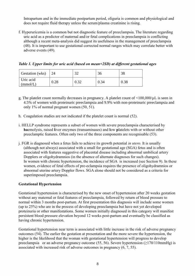
f. Hyperuricemia is a common but not diagnostic feature of preeclampsia. The literature regarding
uric acid as a predictor of maternal and/or fetal complications in preeclampsia is conflicting
although a recent meta-analysis did suggest its usefulness in the management of preeclampsia
(48). It is important to use gestational corrected normal ranges which may correlate better with
adverse events (49).
Table 1. Upper limits for uric acid (based on mean+2SD) at different gestational ages
Gestation (wks) 24 32 36 38
Uric acid
(mmol/L) 0.28 0.32 0.34 0.38
g. The platelet count normally decreases in pregnancy. A platelet count of <100,000/µL is seen in
4.5% of women with proteinuric preeclampsia and 9.9% with non-proteinuric preeclampsia and
only 1% of normal pregnant women (50, 51).
h. Coagulation studies are not indicated if the platelet count is normal (52).
i. HELLP syndrome represents a subset of women with severe preeclampsia characterised by
haemolysis, raised liver enzymes (transaminases) and low platelets with or without other
preeclamptic features. Often only two of the three components are recognisable (53).
j. FGR is diagnosed when a fetus fails to achieve its growth potential in utero. It is usually
(although not always) associated with a small for gestational age (SGA) fetus and is often
associated with features suggestive of placental disease including abnormal umbilical artery
Dopplers or oligohydramnios (in the absence of alternate diagnoses for such changes).
In women with chronic hypertension, the incidence of SGA is increased (see Section 9). In these
women, evidence of fetal effects of pre-eclampsia requires the presence of oligohydramnios or
abnormal uterine artery Doppler flows. SGA alone should not be considered as a criteria for
superimposed preeclampsia.
Gestational Hypertension
Gestational hypertension is characterised by the new onset of hypertension after 20 weeks gestation
without any maternal or fetal features of preeclampsia, followed by return of blood pressure to
normal within 3 months post-partum. At first presentation this diagnosis will include some women
(up to 25%) who are in the process of developing preeclampsia but have not yet developed
proteinuria or other manifestations. Some women initially diagnosed in this category will manifest
persistent blood pressure elevation beyond 12 weeks post-partum and eventually be classified as
having chronic hypertension.
Gestational hypertension near term is associated with little increase in the risk of adverse pregnancy
outcomes (54). The earlier the gestation at presentation and the more severe the hypertension, the
higher is the likelihood that the woman with gestational hypertension will progress to develop
preeclampsia or an adverse pregnancy outcome (55, 56). Severe hypertension (≥170/110mmHg) is
associated with increased risk of adverse outcomes in pregnancy (6, 7, 55).

9
Chronic Hypertension
This category includes essential hypertension as well as hypertension secondary to a range of
conditions. Essential hypertension is defined by a blood pressure greater than or equal to 140
mmHg systolic and/or 90mmHg diastolic confirmed before pregnancy or before 20 completed
weeks gestation without a known cause. It may also be diagnosed in women presenting early in
pregnancy taking antihypertensive medications where no secondary cause for hypertension has been
determined. Some women with apparent essential hypertension may have white coat hypertension
(raised blood pressure in the presence of a clinical attendant but normal blood pressure otherwise as
assessed by ambulatory or home blood pressure monitoring). These women appear to have a lower
risk of superimposed preeclampsia than women with true essential hypertension but are still at an
increased risk compared with normotensive women (32).
Important secondary causes of chronic hypertension in pregnancy include:
Chronic kidney disease e.g. glomerulonephritis, reflux nephropathy, and adult polycystic
kidney disease.
Renal artery stenosis
Systemic disease with renal involvement e.g. diabetes mellitus, systemic lupus erythaematosus.
Endocrine disorders e.g. phaeochromocytoma, Cushing’s syndrome and primary
hyperaldosteronism.
Coarctation of the aorta.
In the absence of any of the above conditions it is likely that a woman with high blood pressure in
the first half of pregnancy has essential hypertension. It is not possible to investigate these disorders
fully during pregnancy, and complete appraisal may need to be deferred until after delivery.
Preeclampsia superimposed on chronic hypertension
Pre-existing hypertension is a strong risk factor for the development of preeclampsia (57, 58).
Superimposed preeclampsia is diagnosed when a woman with chronic hypertension develops one or
more of the systemic features of preeclampsia after 20 weeks gestation. Worsening or accelerated
hypertension should increase surveillance for preeclampsia but is not diagnostic. Similarly, SGA
occurs more frequently in women with chronic hypertension and evidence of fetal effect other than
SGA eg. oligohydramnios or abnormal uterine artery Doppler flows is required to diagnose
superimposed preeclampsia
In women with pre-existing proteinuria, the diagnosis of superimposed preeclampsia is often
difficult as pre-existing proteinuria normally increases during pregnancy. In such women substantial
increases in proteinuria and hypertension should raise suspicion of preeclampsia and therefore
justifies closer surveillance but the diagnosis is not secure without the development of other
maternal systemic features or fetal effects with or without SGA ie the presence of oligohydramnios
or abnormal uterine artery Doppler flows.
4. Investigation of new onset hypertension after 20 weeks gestation
Any woman presenting with new hypertension after 20 weeks gestation should be assessed for signs
and symptoms of preeclampsia. Initially, assessment and management in a day assessment unit may
be appropriate. If features of preeclampsia are detected, admission to hospital is indicated. The
presence of severe hypertension, headache, epigastric pain, oliguria or nausea and vomiting are
ominous signs which should lead to urgent admission and management, as should any concern
about fetal wellbeing (59, 60).

10
The following investigations should be performed in all women with new onset hypertension after
20 weeks gestation:
Spot urine PCR
Full blood count
Creatinine, electrolytes, urate
Liver function tests
Ultrasound assessment of fetal growth, amniotic fluid volume and umbilical artery Doppler
assessment.
The clinical utility of measuring PlGF alone or in combination with sFlt1, remains unclear
(61, 62). A recent study has demonstrated that among women with suspected preeclampsia
before 35 weeks gestation, a low plasma PlGF accurately identified those who are at high
risk of requiring delivery within 14 days [sensitivity 0.96; CI 0.89-0.99 and NPV 0.98; CI
0.93-0.995] (63). Further research is required before implementing this prognostic test into
routine clinical practice.
NOTES
Blood test abnormalities should be interpreted using pregnancy-specific ranges, some of
which are gestation dependent.
If features of preeclampsia are present, additional investigations should include:
o Urinalysis for protein and urine microscopy on a carefully collected mid-stream
urine sample.
o If there is thrombocytopenia or a falling hemoglobin, investigations for disseminated
intravascular coagulation and/or haemolysis (coagulation studies, blood film, LDH,
fibrinogen) are indicated.
Patients with severe, early onset preeclampsia warrant investigation for associated
conditions e.g. systemic lupus erythaematosus, underlying renal disease or antiphospholipid
syndrome. The timing of these investigations will be guided by the clinical features.
Although a very rare disorder, undiagnosed phaeochromocytoma in pregnancy is potentially
fatal and may present as preeclampsia (64, 65) . In the presence of very labile or severe
hypertension, measurement of fasting plasma free metanephrines/normetanephrines or 24
hour urinary catecholamines should be undertaken.
Amongst women referred for assessment of new onset hypertension, a number will have
normal blood pressure and investigations. These women are considered to have transient or
labile hypertension. Repeat assessment should be arranged within 3-7 days as some of these
women will subsequently develop preeclampsia (66).
Subsequent investigation and management will be based on the results of ongoing blood pressure
measurement and these investigations (Tables 2 and 7) (67-69).
Ongoing investigation of women with hypertension in pregnancy
At each assessment following the detection of hypertension in pregnancy, the clinician should
systematically review the woman’s symptoms, examination, laboratory investigations and fetal
wellbeing.
Further laboratory assessment of women with hypertension in pregnancy should be based on the
following recommendations: Table 2.

11
Table 2: Ongoing investigation of women with hypertension in pregnancy
Modality Frequency
Chronic hypertension
Assess for proteinuria*
Preeclampsia bloods**
Each visit
If sudden increase in BP or new
proteinuria
Gestational hypertension Assess for proteinuria
Preeclampsia bloods
1-2x/week
Weekly
Preeclampsia Assess for proteinuria
Preeclampsia bloods
At time of diagnosis: if non-
proteinuric repeat daily*
Twice weekly or more frequent
if unstable
*Urinalysis by dipstick followed by spot urine PCR if ≥1+ proteinuria (see Section 3.) Once
significant proteinuria has been detected, there is no established role for serial testing as the severity
or progress of proteinuria should not alter management decisions.
** FBC, Electrolytes and creatinine, LFT and coagulation studies only if indicated
5. Management of preeclampsia and gestational hypertension
Preeclampsia is a progressive disorder that will inevitably worsen if pregnancy continues. Current
therapy does not ameliorate the placental pathology nor alter the pathophysiology or natural history
of preeclampsia. Delivery is the definitive management and is followed by resolution, generally
over a few days but sometimes much longer. Obstetric consultation is mandatory in all women with
severe preeclampsia. In those women with preeclampsia presenting at extreme preterm gestations (<
32 weeks), consultation with a tertiary institute should be arranged since the neonate may require
intensive care after delivery. Every effort should be made to transfer a woman with very preterm
preeclampsia to a unit with appropriate neonatal and maternal care facilities prior to delivery.
Timing of delivery
Timing of delivery is dependent upon the severity of the maternal disease and the gestation at which
the preeclampsia or gestational hypertension presents (Table 3). Immediate management refers to
delivery planned within 48 hours, usually after blood pressure stabilisation and corticosteroid
administration to accelerate fetal pulmonary maturity. Expectant management refers to prolongation
of the pregnancy beyond these 48 hours with maternal and fetal monitoring.
Table 3. Timing of delivery and gestation of presentation of preeclampsia
Gestation
at onset
Previable <236
weeks 24-31
6 weeks 32-36
6 37+0 onwards
Delivery
plan
Consult with Tertiary
institution: likely to
need termination of
pregnancy or extreme
preterm delivery.
High risk patient
Consult and transfer
to Tertiary institution:
likely to need preterm
delivery. Aim to
prolong pregnancy
where possible
Aim to prolong
pregnancy where
possible, deliver
in institution with
appropriate
Paediatric care
Plan delivery
on best day in
best way

12
Fetal mortality and morbidity is strongly associated with gestational age at delivery. Prolongation of
pregnancy in the presence of preeclampsia carries no benefit for the mother but is desirable at early
gestations to improve the fetal prognosis (70-72). When the onset of preeclampsia occurs at a pre-
viable gestation( i.e.< 24 weeks’ gestation) there is little to be gained from prolonging the
pregnancy with serious maternal morbidity rates of 65-71% and high perinatal mortality rates of
greater than 80% (73-75). The onus remains on the clinician to advise termination of pregnancy,
particularly in resource poor settings (76).
The management of women with preeclampsia below 32-34 weeks gestation should be restricted to
those centres with appropriate experience and expertise and appropriate neonatal intensive care
facilities. Clear “endpoints” for delivery should be defined for each patient (Table 4), such that the
decision to terminate the pregnancy is based on agreed criteria. In many cases, the timing of
delivery will be based upon a number of factors, maternal and/or fetal rather than a single absolute
indication for delivery.
Table 4. Indications for delivery in women with preeclampsia or gestational hypertension
Maternal Fetal
Gestational age ≥ 37 weeks Placental abruption
Inability to control hypertension Severe FGR
Deteriorating platelet count Non-reassuring fetal status
Intravascular haemolysis
Deteriorating liver function
Deteriorating renal function
Persistent neurological symptoms
Persistent epigastric pain, nausea or
vomiting with abnormal LFTs
Pulmonary edema
In cases of preterm preeclampsia before 34 weeks, delivery should be delayed for at least 24-48
hours, if maternal and fetal status permit, to allow fetal benefit from antenatal corticosteroids
administered for lung maturation. Additionally, at early gestations, magnesium sulphate
administered antenatally may provide neonatal neuroprotection (77). Unfortunately up to 40% of
women presenting with preeclampsia at less than 34 weeks gestation are ineligible for expectant
care (78). A number of trials have shown that 25-41% of women managed expectantly with
preeclampsia will develop severe morbidity including HELLP syndrome, abruption, pulmonary
edema and eclampsia and that the mean duration of prolongation is less than 12 days (70, 79-81).
The lower the gestation, the less the mean duration of prolongation (72).
Continuation of pregnancy carries fetal risk and some stillbirths will occur despite careful
monitoring (82). These trials have excluded women with the HELLP variant of preeclampsia and
with other evidence of severe morbidity.
In the presence of HELLP syndrome, expectant management is harmful with a 6.3% incidence of
maternal death and an increased risk of placental abruption (78). In such cases, delivery should be
planned as soon as feasible.
Preeclampsia presenting in the late preterm period, 34-366 weeks gestation, is associated with
increasing risk of SGA neonates with a higher risk of delivery via Caesarean section, respiratory
distress syndrome and longer neonatal intensive care admissions (83, 84). Therefore, antenatal
13
steroid prophylaxis may be beneficial in this group.
At mature gestational age, delivery should not be delayed in the case of severe preeclampsia. Even
so, it is important to control severe hypertension and other maternal derangements before subjecting
the woman to the stresses of delivery.
The HYPITAT study was a multicentre, unblinded study comparing outcomes after induction of
labour and expectant monitoring in pregnant women with gestational hypertension or mild
preeclampsia between 36 and 41 weeks’ gestation (85). The study reported that immediate
induction of labour was associated with a reduction in the incidence of severe hypertension, without
an increase in the Caesarean section rate. No significant difference was seen in important clinical
morbidity outcomes such as HELLP, thromboembolism, eclampsia or placental abruption and costs
were not increased (86, 87). This management approach was beneficial even in those women
whose cervix was unfavourable for induction of labour (88).
Critics of this active approach to milder disease suggest this is unnecessary intervention in a group
of women and babies with generally good outcomes. In this group, there was no difference in
health-related quality of life up to 6 months postpartum between expectant or immediate
management (89-91). We await the publication of two recently completed randomised trials to
inform this debate. (HYPITAT-II and Mild Preeclampsia Near Term: Deliver or Deliberate. In
women with gestational hypertension who are at low risk of adverse outcomes, an expectant
management approach beyond 37 weeks should be considered (13).
A team approach, involving obstetrician, midwife, neonatologist, anaesthetist and physician
provides the best chance of achieving a successful outcome for mother and baby. Regular and
ongoing reassessment of both the maternal and fetal condition is required. Careful daily assessment
for clinical symptoms and signs should be complemented by regular blood and urine tests as
indicated (Table 2).
The only controlled studies of bed rest for preeclampsia have shown no significant maternal or fetal
benefit (92). However, admission to hospital allows close supervision of both mother and fetus as
progress of the disorder is unpredictable. Outpatient monitoring may be appropriate in milder cases
after a period of initial observation.
Treatment for mild-moderate hypertension
Antihypertensive treatment should be commenced in all women with a systolic blood pressure of
greater than or equal to 160mm Hg or a diastolic blood pressure greater than or equal to 110 mm Hg
because of the risk of intracerebral haemorrhage and eclampsia(6, 7, 11, 13).
There is controversy regarding the need to treat mild to moderate hypertension in women with
preeclampsia. Antihypertensive therapy does not prevent preeclampsia (RR 0.99; 95% CI 0.84–
1.18) or the associated adverse perinatal outcomes, but it decreases by half the incidence of
development of severe hypertension among women with mild hypertension (RR 0.52; 95% CI
0.41–0.64) (24 trials, 2815 women) (93). Approximately 10 women need to be treated with an
antihypertensive drug to prevent an episode of severe hypertension (93).
Uncontrolled hypertension is a frequent trigger for delivery and control of hypertension may allow
prolongation of pregnancy. In addition, as summarised in the Cochrane review above, it is possible
that treatment of even mild-moderate hypertension may lead to a clinically relevant reduction in the
risk of preeclampsia and fetal or neonatal death, particularly early pregnancy loss (93).
Arguments against treatment include that there is little risk to the mother in having relatively mild
hypertension for a short time (usually only a few days or at the most weeks), that fetal perfusion is
dependent upon adequate maternal blood pressure and that lowering blood pressure suppresses an

14
important sign of the severity or progression of preeclampsia. There is no clear effect of
antihypertensive treatment on the risk of neonatal death, preterm birth or SGA, placental abruption,
Caesarean section or admission to the neonatal nursery (93). A large randomised trial of “tight”
versus “less tight” control of blood pressure in women with non-severe high blood pressure in
pregnancy has been completed and results are awaited [http://www.utoronto.ca/cmicr/chips].
In the absence of compelling evidence, treatment of mild to moderate hypertension in the range
140-160/90-100 mm Hg should be considered an option and will reflect local practice. Above these
levels, treatment should be considered mandatory.
Antihypertensive therapy
In terms of lowering blood pressure in preeclampsia, a number of drugs have demonstrated safety
and efficacy (Table 5). First line drugs include methyldopa, labetalol and oxprenolol (55-57).
Second line agents are hydralazine, nifedipine and prazosin (58-61). These same agents may be
used for treating gestational or chronic hypertension.
Table 5. Guidelines for selecting antihypertensive drug treatment in pregnancy
Drug Dose Action Contraindications Practise Points
Methyl
dopa
Clonidine
250-750mg
tds
75-300µg tds
Central Depression Slow onset of action over 24
hours, dry mouth, sedation,
depression, blurred vision
Withdrawal effects: rebound
hypertension
Labetalol
Oxprenolol
100-400mg
q8h
20-160 mg
q8h
Blocker with
mild alpha
vasodilator
effect
Blocker with
intrinsic
sympathomim
etic activity
Asthma, chronic
airways limitation
Bradycardia, bronchospasm,
headache, nausea, scalp
tingling (labetalol only) which
usually resolves within 24
hours
Nifedipine 20mg -60 mg
slow release
bd
Ca channel
antagonist
Aortic stenosis Severe headache in first 24
hours
Flushing, tachycardia,
peripheral oedema,
constipation
Prazosin 0.5-5 mg q8h blocker Orthostatic
hypotension
especially after first
dose
Hydralazine 25-50 mg q8h Vasodilator Flushing, headache, nausea,
lupus-like syndrome
Angiotensin converting enzyme (ACE) inhibitors and angiotensin receptor blockers are
contraindicated in pregnancy. Their use in the third trimester has been associated with fetal death
and neonatal renal failure. All of the drugs in Table 4 along with enalapril, captopril and quinapril
are considered compatible with breastfeeding (94) .
15
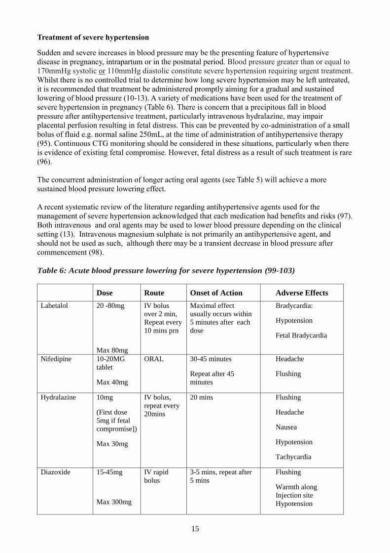
Treatment of severe hypertension
Sudden and severe increases in blood pressure may be the presenting feature of hypertensive
disease in pregnancy, intrapartum or in the postnatal period. Blood pressure greater than or equal to
170mmHg systolic or 110mmHg diastolic constitute severe hypertension requiring urgent treatment.
Whilst there is no controlled trial to determine how long severe hypertension may be left untreated,
it is recommended that treatment be administered promptly aiming for a gradual and sustained
lowering of blood pressure (10-13). A variety of medications have been used for the treatment of
severe hypertension in pregnancy (Table 6). There is concern that a precipitous fall in blood
pressure after antihypertensive treatment, particularly intravenous hydralazine, may impair
placental perfusion resulting in fetal distress. This can be prevented by co-administration of a small
bolus of fluid e.g. normal saline 250mL, at the time of administration of antihypertensive therapy
(95). Continuous CTG monitoring should be considered in these situations, particularly when there
is evidence of existing fetal compromise. However, fetal distress as a result of such treatment is rare
(96).
The concurrent administration of longer acting oral agents (see Table 5) will achieve a more
sustained blood pressure lowering effect.
A recent systematic review of the literature regarding antihypertensive agents used for the
management of severe hypertension acknowledged that each medication had benefits and risks (97).
Both intravenous and oral agents may be used to lower blood pressure depending on the clinical
setting (13). Intravenous magnesium sulphate is not primarily an antihypertensive agent, and
should not be used as such, although there may be a transient decrease in blood pressure after
commencement (98).
Table 6: Acute blood pressure lowering for severe hypertension (99-103)
Dose Route Onset of Action Adverse Effects
Labetalol 20 -80mg
Max 80mg
IV bolus
over 2 min,
Repeat every
10 mins prn
Maximal effect
usually occurs within
5 minutes after each
dose
Bradycardia:
Hypotension
Fetal Bradycardia
Nifedipine 10-20MG
tablet
Max 40mg
ORAL 30-45 minutes
Repeat after 45
minutes
Headache
Flushing
Hydralazine 10mg
(First dose
5mg if fetal
compromise])
Max 30mg
IV bolus,
repeat every
20mins
20 mins Flushing
Headache
Nausea
Hypotension
Tachycardia
Diazoxide 15-45mg
Max 300mg
IV rapid
bolus
3-5 mins, repeat after
5 mins
Flushing
Warmth along
Injection site
Hypotension

16
Persistent or refractory severe hypertension may require repeated doses of these agents or even an
intravenous infusion of labetalol 20-160 mg/hr or hydralazine 10-20 mg/hr, titrated to the blood
pressure response. Infusions of sodium nitroprusside or glyceryl trinitrate are also effective but are
recommended rarely, e.g. when other treatments have failed and delivery is imminent. Sodium
nitroprusside may cause fetal cyanide and thiocyanate toxicity and transient fetal bradycardia. Such
infusions may be considered with intra-arterial blood pressure monitoring in a high dependency
care environment if the usual medications have failed to control the blood pressure, but only so as to
effect safe operative delivery or short term postpartum blood pressure control and not for prolonged
use.
The most important consideration in choice of antihypertensive agent is that the unit has experience
and familiarity with that agent.
It is recommended that protocols for the management of severe hypertension should be readily
accessible in all obstetric units.
Thomboprophylaxis
Large population studies have confirmed that preeclampsia is an independent risk factor for venous
thromboembolism (VTE) occurring in pregnancy or the puerperium. Some studies report increased
risk both during pregnancy and postpartum while others only report an increased risk in the
postpartum period [AOR range between 2.8 and 16] (104-107). The presence of additional risk
factors for VTE, including nephrotic range proteinuria, increases this risk further (108-114).
All women should undergo risk factor assessment for VTE in early pregnancy (108). This
assessment should be repeated if a pregnant woman is admitted to hospital or develops a
complication. Hospitalised women are generally less mobile and mechanical thromboprophylaxis
such as graduated compression stocking should be considered (115). Preeclampsia is considered a
major risk factor for VTE and pharmacological prophylaxis is indicated in a woman who has 2
major or 1 major and 2 minor risk factors as recommended in the Australian guidelines, unless there
are surgical contraindications (108). Each maternity unit should have clear protocols regarding
timing of thromboprophylaxis in relation to insertion and withdrawal of epidural and spinal canulae
(108, 109, 116).
Fluid management
Although maternal plasma volume is often reduced in women with preeclampsia, there is no
maternal or fetal benefit to maintenance fluid therapy (117). The choice between colloid and
crystalloid remains controversial as previous trials generally excluded pregnant women. (118).
Administration of fluid at a rate greater than normal requirements should only be considered for:
1. Women with severe preeclampsia immediately prior to parenteral hydralazine, regional
anaesthesia or immediate delivery: 250 mL bolus (95, 119).
2. Initial management in women with oliguria where there is a suspected or confirmed deficit in
intravascular volume: 300 mL challenge, repeat with careful assessment (119).
As vascular permeability is increased in women with preeclampsia, administration of large volumes
of intravenous fluid before or after delivery may cause pulmonary edema and worsen peripheral
edema (120). This tendency is further aggravated by hypoalbuminemia. Appropriate blood product
replacement is necessary when there has been haemorrhage, as in cases of placental abruption.
Post-partum oliguria is a regular accompaniment of preeclampsia and care must be taken to avoid
its’ overtreatment. Persistent oliguria beyond 24 hours post-partum with rising plasma creatinine
suggests the possibility of post partum renal failure. There is no evidence that fluid manipulation is
17
able to prevent this rare complication.
Monitoring in a high dependency care unit is ideal for these cases because of the risk of pulmonary
edema as mentioned above. Invasive monitoring should only be considered when there is
developing renal failure or pulmonary edema. In view of the reduced plasma volume in most
women with preeclampsia, diuretics should not be used in the absence of pulmonary edema.
Haematological and hepatic manifestations
Thrombocytopenia is the commonest haematologic abnormality seen in preeclampsia; the lower
limit of the normal platelet count in pregnancy is approximately 140x109/L but as a mild reduction
in platelet count may occur in normal pregnancy (gestational thrombocytopenia), the cut-off for an
abnormal platelet count in preeclampsia is 100x109/L. Serial monitoring of the platelet count is
essential in preeclampsia as the count may fall rapidly (See Table 3). When thrombocytopenia is
detected, recheck the platelet count more frequently ie at least daily or twice daily. A progressive
decline in platelet count is an indication for delivery (Table 4).
The risk of peripartum bleeding complications is not significantly increased until the platelet count
falls below 50x109/L. Even so, there are concerns with central neuraxial anaesthetic and analgesic
techniques at higher levels (50-75x109/L), and surgical bleeding may be increased even with
moderate thrombocytopenia.
Platelet transfusion is the only rapidly effective treatment for severe thrombocytopenia and this may
be necessary at the time of Caesarean delivery or in the case of postpartum haemorrhage, wound or
vulval haematoma or other bleeding. Fresh frozen plasma may be required for management of
coagulopathy indicated by active bleeding and a prolonged APTT and INR. In this setting,
fibrinogen levels should also be measured and cryoprecipitate administered if levels are low.
Intravascular haemolysis may occur and should be checked for with appropriate laboratory tests,
FBC, blood film, LDH and haptoglobins, as this is an indication for expedient delivery.
Epigastric, right upper quadrant pain or even chest pain in a woman with preeclampsia often
represents hepatic involvement (121). The pain responds poorly to analgesia but both the pain and
associated increases in liver enzymes (AST, ALT) may subside (albeit temporarily) after blood
pressure lowering, particularly with vasodilators. If the cause of epigastric or right upper quadrant
pain is not clear, close ongoing assessment is required, with careful review of all indicators of
maternal and fetal wellbeing (as above). Imaging of the liver and gallbladder is only indicated if
other pathologies eg cholelithiasis, require exclusion.
The combination of haemolysis, elevated liver enzymes and low platelets has been coined HELLP
syndrome (122). Whether HELLP syndrome is a separate entity or just a clinical syndrome of
severe preeclampsia complications remains open, but from a clinical perspective should be
managed as severe preeclampsia (123). The presence of “ELLP” (elevated liver enzymes with low
platelets) occurs more frequently than HELLP (haemolysis, elevated liver enzymes and low
platelets).
Steroid therapy (other than for fetal lung maturation) is not indicated for the management of
thrombocytopenia or hepatic dysfunction in women with preeclampsia, even with HELLP
syndrome (124, 125). These abnormalities recover spontaneously postpartum within a few days of
delivery, without specific treatment (126-128). If abnormalities worsen or show no improvement
after 72 hours post partum, differential diagnoses such as thrombotic thrombocytopenic purpura or
antiphospholipid syndrome should be considered, and appropriate therapy instituted.
18
6. Eclampsia
A recent Australian study demonstrated that eclampsia remains rare in Australia (in singleton
pregnancies 8.6/10,000) equivalent to 0.1% of all births (129). Classically, headache, visual
disturbance or an altered level of consciousness are considered the symptoms of imminent
eclampsia. However, there are no reliable clinical markers that predict eclampsia and conversely,
the presence of neurological symptoms and/or signs is rarely associated with seizures (130).
Seizures may occur antenatally, intra-partum or postnatally, usually within 24 hours of delivery but
occasionally later. Hypertension and proteinuria may be absent prior to the seizure and not all
women will have warning symptoms such as headache, visual disturbances or epigastric pain (131).
The further from delivery that the seizure occurs, the more carefully should other diagnoses be
considered. Cerebral venous thrombosis in particular may occur in the first few days of the
puerperium. It should be remembered that eclampsia is not the commonest cause of seizures in
pregnancy and the differential diagnosis includes epilepsy and other medical problems that must be
considered carefully, particularly when typical features of severe preeclampsia are lacking.
Management of eclampsia
Comprehensive protocols for the management of eclampsia (and severe hypertension) should be
available in all appropriate areas.
There are four main aspects to care of the woman who sustains eclampsia.
1. Resuscitation
These seizures are usually self-limiting. Resuscitation requires assuring a patent airway, oxygen
by mask and institution of intravenous access. Intravenous diazepam (2mg/min to maximum of
10mg) or clonazepam (1-2mg over 2-5 mins) may be given whilst the magnesium sulphate is
being prepared if the seizure is prolonged.
2. Prevention of further seizures
Following appropriate resuscitation, treatment should be commenced with magnesium sulphate
given as a 4g loading dose (diluted in normal saline over 15-20 minutes) followed by an
infusion of (1-2g/hr, diluted in normal saline). Prediluted magnesium sulphate should be
available in all appropriate areas for this purpose (4g/100ml normal saline). In the event of a
further seizure, a further 2-4g of magnesium sulphate is given IV over 10 minutes. Magnesium
sulphate is usually given as an intravenous loading dose although the intramuscular route is
equally effective. Monitoring should include blood pressure, respiratory rate, urine output,
oxygen saturation and deep tendon reflexes. Magnesium sulphate by infusion should continue
for 24 hours after the last fit (132, 133). Serum magnesium levels do not need to be measured
routinely unless renal function is compromised.
Magnesium sulphate is excreted via the kidneys and extreme caution should be used in women
with oliguria or renal impairment. Serum magnesium concentration should be closely monitored
in this situation. Magnesium is not universally successful and the recurrence rate of seizures
despite appropriate magnesium therapy is 10-15% (60).
3. Control of hypertension [See Section 3]
Control of severe hypertension to levels below 160/100 mmHg is essential as the threshold for
further seizures is lowered after eclampsia, likely in association with vasogenic brain edema. In
addition, the danger of cerebral haemorrhage is real.
19
4. Delivery
Arrangements for delivery should be decided once the woman’s condition is stable. In the
meantime, close fetal monitoring should be maintained. There is no role, with currently available
treatment, for continuation of pregnancy once eclampsia has occurred, even though many
women may appear to be stable after control of the situation has been achieved.
Prevention of eclampsia in the woman with preeclampsia
The drug of choice for the prevention of eclampsia is magnesium sulphate, given as a 4g loading
dose (diluted in normal saline) followed by an infusion of 1g/hour (133). Although there is good
evidence for the efficacy of this therapy, the case for its routine administration in women with
preeclampsia in countries with low maternal and perinatal mortality rates is less than compelling. In
some units, the presence of symptoms or signs such as persistent headache, hypereflexia with
clonus, evidence of liver involvement or severe hypertension are considered indications for
prophylaxis with magnesium sulphate although these symptoms have poor positive and negative
predictability for eclampsia (130). It is appropriate for individual units to determine their own
protocols and monitor outcomes.
7. Fetal Surveillance
Adverse perinatal outcome is increased in women with all subcategories of hypertensive disease in
pregnancy as compared to normotensive women (134, 135). This increase in adverse outcomes is
greatest in those with early gestation at onset of disease, severe hypertension and/or chronic
hypertension with superimposed preeclampsia and is predominantly related to an increase in the
rate of FGR (134-137). Balancing the fetal risks of FGR with the neonatal risks of prematurity is
particularly important in early onset disease (138).
Although fetal surveillance is commonly recommended and performed in women with hypertensive
disease in pregnancy, there is no established consensus on how this should be performed (11, 13,
34, 139). A recent randomized controlled trial evaluating expectant management versus labour
induction in women with hypertensive disease after 36 weeks included maternal reporting of fetal
movements, electronic fetal monitoring and “ultrasound examination” as part of the protocol for
expectantly managed pregnancies, but did not specify the nature or frequency of this monitoring.
(85). The frequency, intensity, and modality of fetal evaluation will depend on individual pregnancy
(maternal and fetal) characteristics. Individual obstetric units should devise their own protocols for
monitoring the fetus in pregnancies complicated by hypertension. In compiling such protocols, the
following issues should be considered.
1. Accurate dating of pregnancy is important for women with chronic hypertension or those at high
risk of preeclampsia
2. Symphysis-fundal height measurement is a poor screening tool for detection of SGA (140).
Therefore, ultrasound should be performed by an experienced operator to assess fetal size,
amniotic fluid volume and umbilical artery Doppler flows in all women with hypertension in
pregnancy. Assessing growth trends by serial ultrasound is recommended if pregnancy
continues.
3. Umbilical artery Doppler flow is the only fetal surveillance modality that has been shown by
systematic review to reduce the need for fetal interventions, improve neonatal outcome and
predict adverse perinatal outcome (141, 142). Severe early onset SGA should be monitored at
institutions experienced in advanced fetal Doppler waveform analysis. Absent or reversed end
diastolic flow is unlikely to occur within 7-10 days after a normal umbilical artery Doppler
waveform analysis. Umbilical artery Doppler flow studies have limited value after 36 weeks
gestation.
20
4. Although numerous observational studies have suggested improved outcome in the high-risk
pregnancy monitored using protocols that included Biophysical Profile, cardiotocography, and
combinations of both, none of these has shown significant benefit in systematic reviews (143-
147).
5. No fetal testing can predict an acute obstetric event such as placental abruption or cord accident
6. Fetal surveillance via a Day Assessment Unit is associated with good perinatal outcome in
women with various obstetric complications, including women with well controlled
hypertension (148, 149)
7. An appropriately grown fetus in the third trimester in women with well-controlled chronic
hypertension without superimposed preeclampsia is associated generally with a good perinatal
outcome. Fetal monitoring using methods other than continued surveillance of fetal growth and
amniotic fluid volume in the third trimester is unlikely to be more successful in preventing
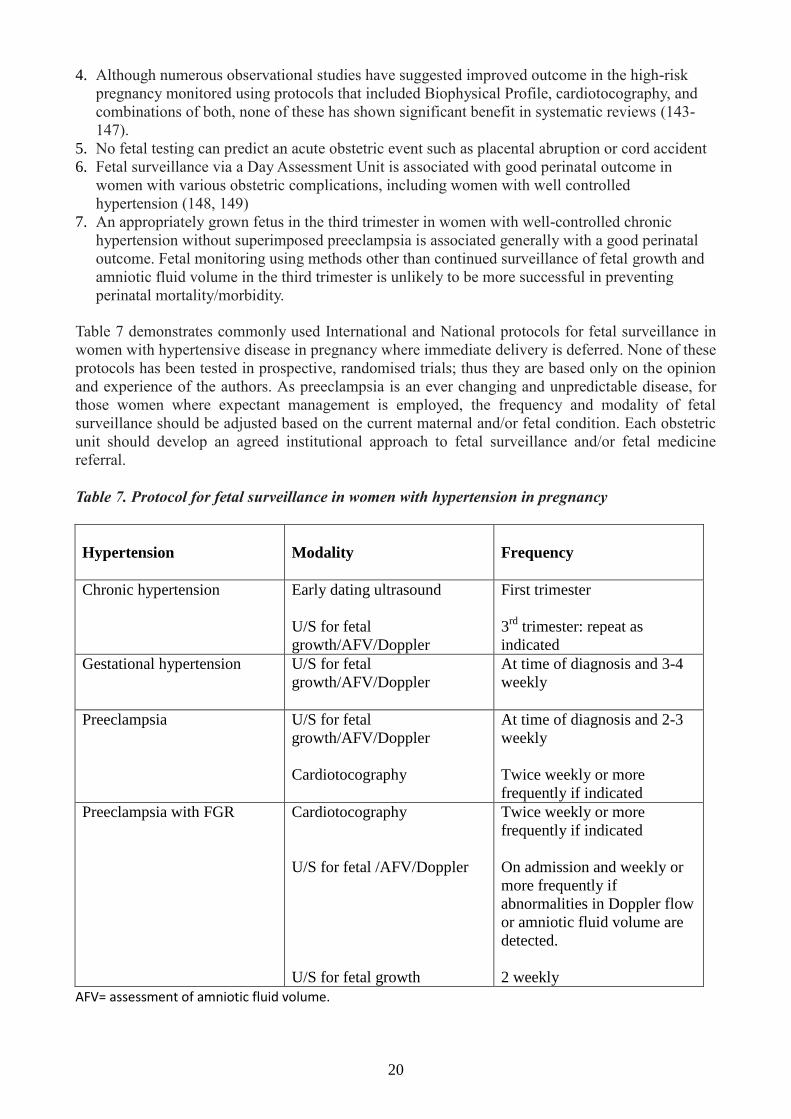
perinatal mortality/morbidity. Table 7 demonstrates commonly used International and National protocols for fetal surveillance in
women with hypertensive disease in pregnancy where immediate delivery is deferred. None of these
protocols has been tested in prospective, randomised trials; thus they are based only on the opinion
and experience of the authors. As preeclampsia is an ever changing and unpredictable disease, for
those women where expectant management is employed, the frequency and modality of fetal
surveillance should be adjusted based on the current maternal and/or fetal condition. Each obstetric
unit should develop an agreed institutional approach to fetal surveillance and/or fetal medicine
referral.
Table 7. Protocol for fetal surveillance in women with hypertension in pregnancy
Hypertension
Modality Frequency
Chronic hypertension
Early dating ultrasound
U/S for fetal
growth/AFV/Doppler
First trimester
3rd
trimester: repeat as
indicated
Gestational hypertension
U/S for fetal
growth/AFV/Doppler
At time of diagnosis and 3-4
weekly
Preeclampsia U/S for fetal
growth/AFV/Doppler
Cardiotocography
At time of diagnosis and 2-3
weekly
Twice weekly or more
frequently if indicated
Preeclampsia with FGR Cardiotocography
U/S for fetal /AFV/Doppler
U/S for fetal growth
Twice weekly or more
frequently if indicated
On admission and weekly or
more frequently if
abnormalities in Doppler flow
or amniotic fluid volume are
detected.
2 weekly
AFV= assessment of amniotic fluid volume.

21
Antenatal Corticosteroid administration
Contrary to popular belief, accelerated fetal lung maturation does not occur in preeclampsia (149).
A systematic review has shown that a single course of antenatal corticosteroid given to women
expected to deliver preterm reduces the risk of neonatal death, respiratory distress syndrome (RDS),
cerebrovascular haemorrhage, necrotizing enterocolitis, respiratory support, and intensive care
admission (150). This systematic review showed that infants born to pregnancies complicated by
hypertensive disease of pregnancy, treated with corticosteroids, had significantly reduced risk of
neonatal death, RDS, and cerebrovascular haemorrhage. The optimal choice of steroid
(Betamethasone or Dexamethasone), mode of administration or timing of dosage regime (12 hourly
versus 24 hourly dosage) remains uncertain, and the advantages or disadvantages of these choices
have not been specifically investigated in the setting of hypertensive diseases (151). There is
insufficient evidence to support antenatal corticosteroids for those pregnancies that have reached 34
weeks gestation (150), however, a single randomized trial demonstrated a small benefit of antenatal
corticosteroids when given to mothers undergoing a term (37 to 39 weeks gestation) elective
Caesarean section and follow up of children enrolled in this study has not demonstrated any longer
term adverse sequelae (152, 153). The use of antenatal steroids beyond 34 weeks should be
considered based on individual patient circumstances (154). In women with hypertensive disorders
of pregnancy undergoing planned Caesarean section after 34 weeks gestation, urgent delivery
should not be delayed purely for the benefits of corticosteroid therapy.
The administration of further courses of corticosteroid in women who remain undelivered and still
at risk of preterm birth after an initial course of corticosteroids remains controversial. Several
studies have reported short term improvements in neonatal outcomes including respiratory distress,
but these are not translated into improved short term childhood outcomes. (155, 156) Concerns also
remain about potential longer term outcomes. Ongoing follow up studies are underway. If repeat
doses of corticosteroids are considered necessary, the protocol described by Crowther et al should
be employed (157-159).
Antenatal Magnesium Sulphate administration for fetal neuroprotection
There is now established Level I evidence that Magnesium sulphate should be administered to
women requiring preterm delivery for the purposes of fetal neuroprotection (77). Maternally
administered Magnesium sulphate has been shown to significantly reduce the risk of cerebral palsy
(RR 0.69, 95% CI 0.54-0.87). The National Health and Medical Research Council endorsed
Australian national guidelines recommend administration of Magnesium sulphate for all women at
risk of preterm delivery prior to 30 weeks (160). In the setting of preeclampsia, Magnesium
sulphate may be considered on maternal grounds as seizure prophylaxis (see Section 6), however
strong consideration should be given to its use even in the setting of milder degrees of hypertensive
disease where delivery is indicated prior to 30 weeks. There is less certain evidence concerning the
benefits of administration beyond 30 weeks gestation but ongoing trials may help to clarify this.
8. Resolution of preeclampsia and gestational hypertension
After delivery, all clinical and laboratory derangements of preeclampsia recover, but there is often a
delay of several days, and sometimes longer, in return to normality (161). On the first day or two
after delivery, liver enzyme elevations and thrombocytopenia will often worsen before they improve
(162). Non-steroidal anti-inflammatory drugs are therefore contraindicated as they may adversely
affect hypertension, renal function and platelet function. Hypertension may persist for days, weeks
or even up to three months and will require monitoring and slow withdrawal of antihypertensive
therapy. Resolution is still assured if the diagnosis was preeclampsia and there is no other
underlying medical disorder.

22
The woman and her family are often overwhelmed and distressed from their experience and
appropriate management post partum should include psychological and family support. Engaged
patient advocacy organizations include the Australian Action on Pre-eclampsia (AAPEC) and New
Zealand Action on Pre-eclampsia (NZ APEC) groups.
All women who develop preeclampsia and gestational hypertension are at risk of these disorders in
future pregnancies and should be referred for review by a clinician with expertise in the
management of hypertensive disorders of pregnancy before embarking upon another pregnancy.
[See Sections 11 and 12]
9. Chronic hypertension in pregnancy
A substantial number of pregnancies (0.2–5%) are complicated by pre-existing hypertension and the
prevalence in western societies is likely to increase due to the advancing age of the prospective
mother at conception and the rising tide of obesity (163-165). The diagnosis can be difficult in
women whose blood pressure before pregnancy or early in the first trimester is unknown as the
physiological fall in blood pressure in the second trimester can obscure pre-existing hypertension
and very rarely, preeclampsia can present before 20 weeks’ gestation.
Pregnancy outcome
Adverse outcomes of pregnancy are more common in women with pre-existing hypertension. In one
prospective study of women with chronic hypertension, the risk of superimposed preeclampsia was
associated with a previous history of preeclampsia (AOR 1.95 [95%CI 1.25-3.04]) or the presence
of one or more other risk factors, such as obesity or diabetes (58). Smoking was also associated
with an increased AOR for superimposed preeclampsia (1.82 [1.02-3.04]). Table 8 shows the rate of
complications in this cohort of 822 women with chronic hypertension, with and without
superimposed preeclampsia. Absolute blood pressure levels in pregnancy do not appear to correlate
with poor outcome except when hypertension is uncontrolled in the first trimester, at which time
both fetal and maternal morbidity and mortality are markedly increased (166).
Table 8 Outcomes of pregnancy in women with chronic hypertension (58)
Outcome
All No
preeclampsia
With
superimposed
preeclampsia
Preeclampsia 22%*
Preeclampsia <34/40 9.7%
Preterm birth <37/40 15% 51%
Preterm birth <34/40 7% 23%
Caesarean section 50% 44% 70%
SGA 27% 21% 48%
BW <2.5kg 20% 13% 44%
Need for additional antihypertensive
medication
Oral 24%
Parental 4%
LDA: low-dose aspirin;* On LDA: 28%; no LDA: 21% [NS] SGA: <10th centile using customised growth
charts, BW = birth weight,

23
White coat hypertension in early pregnancy is common and not necessarily a benign condition, as
40% of these women progress to persistent hypertension after 20 weeks’ (gestational hypertension)
and 8% to preeclampsia (32, 167). The risks of severe hypertension, preterm delivery and NICU
admission appear to be intermediate between normotension and either pre-existing or gestational
hypertension.
The significance of masked hypertension (measured blood pressure of < 140/90 mmHg and
ambulatory blood pressure of ≥ 135/85mm Hg) is uncertain. Its incidence appears to be relatively
common, at least 20%, in patients presenting as hypertensive in early pregnancy. Outcomes in
patients presenting after 20 weeks’ appear to equate with gestational hypertension patients (168).
The woman with chronic hypertension, whether essential or secondary, should be observed
frequently during the pregnancy by an obstetrician and by a physician familiar with the
management of hypertension in pregnancy. The frequency of review will be determined by such
factors as how successfully blood pressure is controlled, the number of agents used, associated
disorders (e.g. renal disease, proteinuria) and by the gestation. Vigilance is also required for the
woman with white-coat hypertension.
Investigation
More than 95% of women with pre-existing hypertension will have essential hypertension; however
a detailed history, careful examination, and relevant laboratory tests are essential to ascertain both
potential secondary causes or end-organ damage (169). This should preferably be performed prior
to pregnancy, but if this is not possible, investigations should concentrate on those conditions likely
to affect pregnancy management or outcome.
Common baseline tests include:
Urine
• Urinalysis for protein. If proteinuria is evident on dipstick analysis, a ‘spot’ urine
protein:creatinine ratio should be obtained
• Microscopy of centrifuged urinary sediment for white and red blood cells (including red
cell morphology) and for casts
• Mid-stream urine culture
Blood
• Serum electrolytes and creatinine, uric acid and full blood examination, and fasting
blood glucose
• ECG
• Renal ultrasound
• Screening for phaeochromocytoma if indicated: fasting free plasma metanephrines and
normetanephrines
NOTE
Plasma and urinary aldosterone, cortisol and renin measurements are unable to be interpreted
with confidence in pregnancy. Expert advice should be sought if Conn’s or Cushing’s syndrome
is suspected.
End-organ effects of hypertension (retinal assessment, albuminuria, renal function and
echocardiogram) should be considered and sought, particularly when hypertension is severe,
longstanding, or when it has not previously been detected.
Clinical and laboratory monitoring (Table 2)
Women with chronic hypertension are at high risk of developing preeclampsia and close monitoring
for its maternal and fetal manifestations is necessary. In addition to standard antenatal care, the
following additional monitoring is indicated:

24
Appraisal for features of superimposed preeclampsia (including proteinuria) at each visit after
20 weeks’ gestation
Appropriate and timely assessment of fetal growth and wellbeing
Laboratory assessment for secondary hypertension (as detailed above) if other symptoms or
features are suggestive. It should be remembered that hypertension per se is associated with an
increased risk of preeclampsia.
It is unusual for pre-existing hypertension alone (unless not previously recognized) to result in
severe hypertension, especially in the first half of pregnancy. Before 20 weeks’ gestation,
consideration of secondary causes of hypertension should be entertained and investigated. After 20
weeks’, superimposed preeclampsia must be strongly suspected.
Admission to hospital or to a day assessment unit is recommended for women with worsening
hypertension or proteinuria at any stage of pregnancy. This enables appropriate assessment and
management of maternal and fetal status and facilitates discussion by all involved in the woman’s
care.
Treatment
Non-pharmacological treatment:
There is insufficient evidence to advise appropriately on the efficacy or safety of non-
pharmacological interventions for managing pre-existing hypertension during pregnancy. This
applies to maintenance of salt restriction, calorie restriction in obese women, ‘heart-healthy’ diets,
exercise, a reduction in stress (e.g. meditation) or workload, or bed rest. Some of these measures are
untried but are unlikely to inflict harm on either the gestation or the mother and advice regarding
their continuance or avoidance should be on a case-by-case basis. Other measures, such as salt
restriction and bed rest have not been shown to improve maternal or fetal outcomes. Furthermore, it
is possible that lowering blood pressure may increase the risk of SGA; results of current studies are
awaited.
Unless severe or poorly controlled (see below), most women with pre-existing hypertension,
without other complications or co-morbidities, can be managed at home, incorporating hospital day
units or outpatient clinics.
Antihypertensive therapy:
The pharmacological management of ongoing chronic hypertension should follow the
principles outlined above for gestational hypertension and preeclampsia. [Section 5]
Many women with chronic hypertension will have a physiological fall in blood pressure in
the first half of pregnancy that may allow them to reduce or cease antihypertensive
therapy. In some cases, antihypertensive therapy should be ceased prior to conception
because they are considered unsuitable for other reasons (170-173):
ACE-inhibitors and Angiotensin Receptor Blockers are nephrotoxic for the fetus in late
pregnancy. Earlier reports also suggested ACE-inhibitors might be associated with, particularly,
cardiovascular malformations although more recent data suggests this is unlikely (174, 175).
When used for prolonged periods in pregnancy, atenolol and other highly selective beta blocker
drugs are associated with fetal growth restriction (112, 176, 177).
There is no compelling evidence that antihypertensive therapies are associated with adverse
neurodevelopment
Diuretics may restrict the natural plasma volume expansion of pregnancy
25
Although treatment of chronic hypertension is associated with a significant reduction in severe
hypertension, it has not been shown to alter the risk of superimposed preeclampsia, preterm
delivery, placental abruption or perinatal death. Treatment reduces the risk of severe
hypertension (RR 0.52; 95% CI 0.41-0.64), but it is unknown whether this affects perinatal
outcomes (10, 93).
The appropriate blood pressure target for women with chronic hypertension and other conditions
such as diabetes mellitus, chronic kidney disease or cardiovascular disease may be lower than for
women without such complications, but there is insufficient data currently to state whether this
provides benefit either to the mother or the fetus. Extrapolating from non-pregnancy data may or
may not be valid and the decision to aim for levels between 130-140 mmHg systolic and 80-90
mmHg diastolic during pregnancy will depend on multiple factors that at this stage must be
weighed individually.
In the third trimester of pregnancy, chronic hypertension frequently becomes more difficult to
manage and an increase in antihypertensive therapy should be anticipated. Such increases are not
always associated with the development of preeclampsia.
Timing of delivery
Pre-existing hypertension during pregnancy is associated with up to a 3-fold risk of perinatal death
compared with singleton, normotensive pregnancies (165, 178). The risk may be highest from 39
weeks’ gestation, indicating appropriate monitoring of these women to the end of the pregnancy is
mandatory. As in all such cases, these data must not be construed to imply that intervention before
term will necessarily reduce mortality or otherwise favourably affect outcome.
Post partum management
First six weeks:
In many women with pre-existing hypertension (with or without superimposed preeclampsia), blood
pressure is often unstable immediately after delivery and may require a medication adjustment.
Non-steroidal inflammatory drugs should not be given postpartum if the hypertension is difficult to
control, there is evidence of acute kidney injury or CKD, or in the setting of thrombocytopenia
(179).
Women with chronic hypertension, a long duration of antihypertensive treatment in pregnancy,
higher maximum systolic and diastolic blood pressures, higher body mass index, or occurrence of
preterm preeclampsia are more likely to have sustained hypertension postpartum (exceeding 6
weeks) (161, 180). Blood pressure usually stabilises in the first two months following pregnancy
and treatment and appraisal should be based on the assumption that levels will decline.
Severe postpartum hypertension requires the same management approach as during pregnancy.
Postpartum, women with co-morbidities should be treated according to standard guidelines for their
specific medical condition when not pregnant e.g. <130/80 mmHg for albuminuric diabetic women.
This should be a longer-term target and may not be achievable whilst the patient is hospitalised.
Conversely, a reduction in medication may be required within one to two weeks of delivery if the
woman’s antihypertensive therapy was augmented in relation to the pregnancy.
Women with pre-existing hypertension who did not require treatment during the pregnancy often
need treatment postpartum (181). Those at highest risk appear to be preterm deliveries and
multiparous women with elevated serum urate concentrations.

26
All agents mentioned earlier (including the ACE inhibitors enalapril, captopril and quinapril) are
compatible with breast feeding. Clonidine has been found to accumulate significantly in neonatal
serum, although the significance is undetermined (182) .
After six weeks:
Follow-up after 6 weeks is required to ensure resolution of pregnancy-related changes and ascertain
the need for ongoing care, particularly further investigation and management of renal disease. In
women whose blood pressure control before pregnancy remains uncertain, it is important to ensure
normalization of blood pressure (and albuminuria) postpartum. Women with persistent hypertension
not previously assessed should undergo routine work-up according to standard regimens.
Advice regarding future lifestyle and optimization of risk factors in subsequent pregnancies may be
required. This is particularly relevant for women who are obese, have cardiovascular risk factors,
secondary hypertension, or end-organ disease.
10. Anaesthetic considerations in hypertensive disorders of pregnancy
Whenever possible an anaesthetist should be informed about a woman with severe preeclampsia,
preferably well prior to labour or operative delivery, because appropriate anaesthetic management is
associated with reduction in both fetal and maternal morbidity (183). Relevant issues include
anaesthetic risk assessment, blood pressure control, fluid management, eclampsia prophylaxis and
planning of analgesia or anaesthesia (184-186).
Fluid management
Fluid management is a challenging area in preeclampsia and there is no clear evidence regarding
optimal type or volume of fluid (185, 187). Fluid therapy aims to maintain organ perfusion in the
setting of vasoconstriction, endothelial dysfunction and either left ventricular systolic or more often
diastolic dysfunction. Intravenous fluid should be administered incrementally in small volumes (e.g.
crystalloid 250 mL) while monitoring maternal hemodynamic parameters, urine output and fetal
heart rate, because over-hydration contributes to maternal mortality from pulmonary oedema and
adult respiratory distress syndrome (188). Particular caution is necessary in women with oliguria,
renal impairment or pulmonary oedema, in whom the left ventricle may adapt less well to volume
load (189).
Fluid loading is not mandatory prior to regional analgesia during labour when low-dose local
anaesthetic and opioid methods are used (190). Prior to regional anaesthesia for operative delivery,
intravenous crystalloid loading is ineffective in preventing hypotension (191). Colloid is of modest
effect but renal dysfunction, allergic reactions and coagulation disturbance are potential
consequences. Prevention or treatment of hypotension with drugs such as ephedrine, phenylephrine
or metaraminol is effective and appears safe in preeclamptic women-see below (192, 193).
Anaesthetic technique
Vaginal birth
During labour and childbirth, epidural analgesia is a useful adjunct to antihypertensive therapy for
blood pressure control and improves renal and utero-placental blood flow (194, 195). When
relatively contraindicated (e.g. severe thrombocytopenia, coagulopathy or sepsis), fentanyl or
remifentanil patient-controlled intravenous analgesia is preferred. Although ephedrine usually does
not cause rebound hypertension, occasionally vasopressors and epidural adrenaline [epinephrine]
cause worrisome blood pressure elevation.
27
Caesarean birth
Sufficient preoperative preparation time reduces the risk of anaesthesia and other complications in
women with preeclampsia (188, 196). Anti-hypertensive therapy and eclampsia prophylaxis should
be instituted [See Section 5 and 6]. Regional anaesthesia is preferred to general anaesthesia for
Caesarean birth, especially as airway problems, including laryngeal oedema, may be increased (197,
198). However, well-conducted general anaesthesia is also suitable and can be indicated in the
presence of severe fetal compromise, pulmonary oedema, maternal hemodynamic instability,
increased intraspinal haematoma risk (e.g. placental abruption induced coagulopathy, severe
thrombocytopenia) or after eclampsia when altered consciousness or neurological deficit persists
(199, 200).
Emergency operative delivery is associated with increased maternal morbidity, so early anaesthetic
notification by the obstetrician and in-utero resuscitation provide additional time for assessment,
planning and establishment of regional anaesthesia. When a well-functioning epidural catheter is
present, conversion to epidural anaesthesia can be achieved only marginally less rapidly than
establishing general anaesthesia (201, 202). Prophylaxis against pulmonary aspiration is
recommended using clear antacid and ranitidine, with or without metoclopramide. Skilled
anaesthetic assistance is mandatory, as is left lateral tilt on a pelvic displacement wedge or table tilt
to minimise aortocaval compression. Oxytocin should be given slowly in small doses of less than 5
IU to minimise its’ significant hemodynamic effects (203). Drugs that are ideally avoided in severe
preeclampsia include ergometrine and ketamine (hypertensive episodes) and the non-steroidal anti-
inflammatory drugs including COX-2 specific inhibitors (impaired renal function or hypertension)
(188).
Attenuation of pressor responses at general anaesthesia for Caesarean birth
Laryngoscopy and tracheal intubation present a particularly dangerous time for the hypertensive
woman, especially if the intracranial pressure is elevated or the blood pressure is inadequately
controlled (188, 196). The transient but severe hypertension that typically accompanies intubation
can cause myocardial ischaemia, cerebral haemorrhage or pulmonary oedema, all of which are
important causes of maternal death. Attenuation of pressor responses, aiming to maintain systolic
blood pressure < 180 mmHg, is best achieved with drugs such as remifentanil 1 mcg/kg; or
magnesium sulphate 30 mg/kg combined with alfentanil 7.5 mcg/kg (204-206). Fentanyl 2.5
mcg/kg, alfentanil 10 mcg/kg or magnesium 40 mg/kg are partially effective (207). Neuromuscular
block must be monitored closely after intravenous magnesium administration (208). Other options,
including at the time of extubation, are beta-blockers such as esmolol or vasodilators such as
glyceryl trinitrate (209).
Regional anaesthesia for Caesarean birth
All the regional anaesthetic techniques (spinal, epidural or combined spinal-epidural) appear safe
provided meticulous attention is paid to cautious fluid management, prevention of aortocaval
compression and minimisation of hypotension. Spinal anaesthesia with usual drug doses is now a
recommended technique (185, 210-212). Cardiac output is well maintained and spinal anaesthesia is
associated with less hypotension and lower vasopressor requirements than when used for healthy
parturients (210). Combined spinal-epidural anaesthesia appears to offer further advantages in
specific cases (185).
Low dose aspirin therapy is not a contraindication to regional techniques, which in the absence of
clinical bleeding are considered of very low risk if the platelet count is >75 x109/L (52) . Platelet
counts of < 50 x109/L are considered a contraindication unless there are compelling reasons to avoid
general anaesthesia. Within the range 50-75 x109/L an individual assessment, considering patient
risks, coagulation status and if available platelet function tests; and risk reduction strategies (an

28
experienced operator, single-shot spinal anaesthesia or insertion of a flexible tip epidural catheter)
are advised.
Post-Caesarean analgesia can be achieved with many options, but the non-steroidal anti-
inflammatory drugs and those which reduce the seizure threshold (tramadol, pethidine, meperidine)
are best avoided in women at risk of eclampsia.
Admission to an Intensive Therapy Unit
Anaesthetists are an important speciality group within critical care teams. Women who develop
organ failure require intensive monitoring and medical management, either in a high dependency or
intensive care setting. Indications for admission include severe pulmonary oedema, sepsis,
intractable hypertension, anuria or renal failure, seizures, massive blood loss with disseminated
intravascular coagulation, neurological impairment requiring ventilation (eg intracerebral
haemorrhage or infarction, cerebral oedema) and critical intra-abdominal pathology.
Invasive monitoring
Direct intra-arterial blood pressure monitoring is often extremely useful in hypertensive women,
during anaesthesia and operative delivery as well as in critical care, but obtaining arterial access
should not delay treatment of acute severe hypertension. Central venous pressure correlates poorly
with pulmonary capillary wedge pressure, so although it may provide trend monitoring and a central
catheter allows safer administration of potent vasoactive drugs, central venous pressure monitoring
is seldom used as an indicator of intravascular volume status (213). Pulmonary artery catheters for
assessment of left ventricular preload can cause serious complications, are not of proven outcome
benefit in preeclampsia, and are consequently rarely used. There is increasing support for the value
of echocardiography and more dynamic measures of cardiac output, such as devices based on pulse
contour analysis or pulse power algorithms (184, 185, 214, 215) .
11. Preconception management and prophylaxis
Risk factors for hypertensive disorders of pregnancy
It is likely that development of preeclampsia requires a combination of underlying susceptibility
and a triggering event. Many susceptibility factors for preeclampsia have been identified (see Table
9). The absolute risk for an individual will be determined by the presence or absence of these and
other predisposing or protective factors but to date no adequately accurate predictive tool, using
either clinical or laboratory markers, has been developed (216). Such a tool applied early in
pregnancy would allow management that might modify outcomes. When considering prophylactic
treatment or stratification of women to high or low risk models of antenatal care, these risk factors
should be assessed for each woman.

29
Table 9. Risk factors associated with preeclampsia (216-218)
Risk Factor Unadjusted Relative
Risk [95% CI]
Nulliparity 2.9 [1.3-6.6]
Multiple pregnancy 2.9 [1.3-6.6]
Previous history of preeclampsia 7.2 [5.9-8.8]
Family history of preeclampsia 2.9 [1.7-4.9]
Overweight BMI 25-29.9* 1.7 [1.2-2.4]
Obese BMI >30* 2.7 [1.7-4.4]
Age ≥ 40 2.0 [1.3-2.9]
Systolic BP>130mmHg before 20 weeks 2.4 [1.8-3.2]
Diastolic BP >80mmHg before 20 weeks 1.4 [1.0-1.9]
Antiphospholipid syndrome 9.7 [4.3-21.8]
Pre-existing diabetes 3.6 [2.5-5]
Other risk factors
Underlying renal disease
Chronic autoimmune disease
Interpregnancy interval >10 years
Other rare risk factors include fetal hydrops, fetoplacental triploidy and gestational trophopblastic
disease may cause severe, early onset preeclampsia.
In a prospective study of 3529 nulliparous women, 5.3% developed preeclampsia and the risk of
preeclampsia was increased when more than one risk factor was present (217). For example, the
rate of preeclampsia when the systolic blood pressure was >120 mmHg at 15 weeks (n=310) was
14% (95%CI 10-18). The rate of preeclampsia in women with a higher systolic blood pressure at 15
weeks was further increased if she was obese [16% (95%CI 10-18), had a family history of
preeclampsia [20% (95%CI 11-33)], or whose own birth weight was <2500g [33% (95%CI 17-55)].
Of note, the combinations of risk factors conferring the greatest risks of preeclampsia occurred in
fewer women and comprised a small proportion of all nulliparas who develop preeclampsia. A small
number of factors have been identified that are protective for preeclampsia. These include
miscarriage with the same partner in nulliparous women, high fruit intake, smoking and taking
greater than 12 months to conceive (217) . The independent detrimental effects of smoking on fetal
growth should be emphasized. Interestingly, this protective effect is not seen in women with chronic
hypertension (58).
Inherited thrombophilias e.g., Factor V Leiden were suggested to be strongly associated with
preeclampsia on the basis of early case control studies however recent large prospective cohort
studies, and a systematic review of these cohort studies do not support an association between the
common Factor V Leiden and Prothrombin gene mutations and preeclampsia (219, 220). Testing for
inherited thrombophilias is therefore no longer recommended following a preeclamptic pregnancy
(108, 221, 222).
The association between periodontal disease and preeclampsia remains controversial with a recent
meta-analysis concluding that there is a modest association between periodontal disease and
preeclampsia on the basis of several case-control studies, however there is significant heterogeneity
in methodological rigor between studies (223).

30
The value of measuring angiogenic and anti-angiogenic factors in maternal serum during pregnancy
(such as endoglin and placental growth factor) or using biophysical parameters such as uterine
artery Doppler velocimetry to predict later development of preeclampsia remains controversial
(224). The recognition of the association between these markers and the subsequent development of
preeclampsia has provided valuable insights into the pathogenesis of this condition, but the clinical
utility and cost effectiveness of using these markers as early pregnancy screening tests to predict
preeclampsia remains uncertain (225-230).These tests appear to perform best in the prediction of
preterm or early onset disease but this only represents a small proportion (25 % and 10% of women
with preeclampsia deliver before 37 and 34 weeks, respectively) of the overall burden of disease
(231-233).
To be cost effective, there must either be an effective treatment available to prevent development of
the disease and its associated morbidity, or it must allow diversion of limited antenatal resources
away from those considered at low risk (224, 226). At present, there are insufficient data to support
the beneficial effects of routine marker testing and as such they are not currently recommended in
clinical practice (234, 235). It is certainly possible, however, that ongoing refinements of testing
protocols may improve the efficacy of these tests in predicting preeclampsia in the near future.
Recurrence of preeclampsia
Women who have experienced hypertension in a previous pregnancy are at increased risk in any
future pregnancies (13, 236, 237). They should receive appropriate counselling and prophylaxis if
the risk is considered significant (see Section 11).
Table 10. Based on data summarized in Reference (see Appendix G) (13)
Recurrence risk in subsequent pregnancies
Gestational hypertension Preeclampsia
Previous gestational
hypertension 16-47% 2-7%
Previous proteinuric
preeclampsia 13-53% 16%
Severe preeclampsia
<34 weeks
<28 weeks
25%
55%
12. Prevention of preeclampsia
A number of agents have been studied for their ability to reduce the risk of preeclampsia and
improve maternal and fetal outcomes. These include antiplatelet agents, vitamins, calcium and
heparin.
Antiplatelet agents
Studies of the pathophysiology of preeclampsia have previously identified an imbalance between
the circulating prostaglandins, prostacyclin and thromboxane. The hypothesis leading to the aspirin
trials was the ability of low-dose aspirin to correct this imbalance by inhibiting platelet aggregation
and dilating blood vessels. However, further studies have suggested the anti-inflammatory action of
aspirin could also be important (238).

31
Prophylactic therapy with anti-platelet agents has been the subject of a large number of studies and
various systematic reviews (239-242). Over 37,000 women have participated in randomised trials of
anti-platelet agents to prevent preeclampsia, with the majority of trials using aspirin 50-150mg.
Table 11. Effects of antiplatelet agents on risk of preeclampsia: summarised in Reference (243)
Population RR [95%CI] for
preeclampsia NNT [CI]
Primary prevention 0.90 [0.84-0.97]
Low risk women 0.93 [0.81–1.08]
All at risk 0.83 [0.77-0.89] 72 [52-119}
High risk women 0.75 [0.66-0.85] 19 [13-34]
Recurrent
preeclampsia
0.86 [0.77-0.97]
Aspirin dose >75
mg/day
0.64 [0.51-0.80]
Of clinical importance, there was a 14% reduction in stillbirth, neonatal or infant death (RR 0.86
95%CI 0.76-0.98) and a 10% reduction in SGA infants (RR 0.90 95%CI 0.83-0.98).
Anti-platelet treatment commenced before 20 weeks (RR 0.87 95%CI 0.79-0.96) but not at 20
weeks, reduced the risk of preeclampsia. In a meta-analysis of 34 studies including 11,348 women,
the reduction in the risk of preeclampsia was demonstrated to be significant only if aspirin was
commenced prior to 16 weeks gestation (RR 0.47, 95% CI 0.34-0.65) compared to commencement
after 16 weeks (RR 0.81, 95% CI 0.63-1.03). However, this meta-analysis was confounded by the
risk status of women included, with the studies where treatment commenced before16 weeks (9
trials of 734 women) comprising of higher risk women (21% of controls developed preeclampsia).
In contrast, preeclampsia occurred in 8% of controls in trials where treatment commenced at ≥16
weeks (n=10,584), indicating the lower risk status of participants. Of importance, the individual
patient data meta-analysis found no difference in the rate of bleeding complications such as
antepartum and postpartum haemorrhage or placental abruption between treatment and placebo
groups.
In translating these results into clinical practice, the underlying risk of preeclampsia in the
population being treated must be taken into consideration. If the baseline risk is 8%, treating 114
women will prevent one case of preeclampsia. In a population with a 20% risk of preeclampsia, the
number needed to treat to prevent one case of preeclampsia is 50. In view of this potential benefit,
and the relative absence of maternal or neonatal complications, low dose aspirin is indicated for
women with at least moderate to high risk of preeclampsia ie secondary prevention of preeclampsia
in women at increased risk and in women with significantly increased risk in their first pregnancy
(see Section 11 (13). In most cases, aspirin may be ceased at 37 weeks gestation although
continuation beyond this period is not unsafe (239).
Calcium supplements
Low calcium intake may cause high blood pressure by stimulating either parathyroid hormone or
renin release, thereby increasing intracellular calcium in vascular smooth muscle and leading to
vasoconstriction (244). A possible mode of action for calcium supplementation is that it reduces
parathyroid hormone release and intra-cellular calcium, and so reduces smooth muscle contractility.
Calcium might also have an indirect effect on smooth muscle function by increasing magnesium
levels. Recent evidence indicates that calcium supplementation affects uteroplacental blood flow
by lowering the resistance index in uterine and umbilical arteries (245). Calcium supplementation in
32
the second half of pregnancy appears to reduce blood pressure directly rather than preventing the
endothelial damage associated with preeclampsia (246).
The use of calcium supplementation has been demonstrated to significantly reduce the risk of
preeclampsia, particularly in high risk women and those with low dietary calcium intake (247). It
has also been shown to reduce the risk of preterm birth (247). There was no significant effect on
fetal and neonatal outcomes including low birth weight, fetal growth restriction, stillbirth or death
before discharge from hospital. Calcium supplementation (1.5g/day) should therefore be offered to
women with moderate to high risk of preeclampsia, particularly those with a low dietary calcium
intake (247).
Heparins
There has been considerable interest in the potential role of prophylactic heparin in preventing
preeclampsia in women at risk of preeclampsia. Several recent RCTs have provided encouraging
results. Much of this interest has been based on the apparent association between inherited
thrombophilias and adverse pregnancy outcomes with one trial reporting a significant reduction in
recurrent, early onset (<34 weeks) hypertensive disease in thrombophilic women who received
weight adjusted daily dalteparin injections compared to those who received standard care (248).
Rey et al reported a significant reduction in the rate of recurrent preeclampsia and fetal growth
restriction in non-thrombophilic women with daily dalteparin injections (5000IU) (OR 0.15, 95%
CI 0.03-0.70) (249). Likewise Kupferminc et al reported a significant reduction in the overall rate of
pregnancy complications (including severe preeclampsia) in a non-randomised study (250).
However, not all studies have demonstrated such improvements. Martinelli et al recently reported
the findings of an RCT investigating the role of nadroparin in preventing placenta-mediated adverse
pregnancy events and concluded that antenatal prophylaxis with this drug did not prevent adverse
pregnancy events (251).
Despite the comparative safety of low molecular weight heparins during pregnancy, the current data
do not support widespread use of these agents during pregnancy for the purposes of prevention of
adverse pregnancy outcomes (other than perhaps in the specific case of antiphospholipid antibody
syndrome (109, 252, 253). Further studies to determine the efficacy of low molecular weight
heparins in specific at-risk patient population eg previous early onset preeclampsia, are required.
Other Therapies
Markers of oxidative stress are present in the placenta and maternal circulation of women with
preeclampsia suggesting it may play a role in the disorder. Randomised, placebo controlled trials of
antioxidants Vitamins C and E failed to demonstrate any significant effect on the incidence of
preeclampsia (254-257). Of concern, a number of adverse effects were seen including an increased
risk of stillbirth and of birthweight <2.5kg but there were fewer fetal deaths due to immaturity.
Prophylactic antioxidant therapy with vitamins C and E is therefore not recommended (254-259).
Preeclampsia shares pathogenic similarities with adult cardiovascular diseases as well as many risk
factors. A recent review has summarised a number of excellent studies that have demonstrated that
prior to the onset of preeclampsia there is a rise in circulating antiangiogenic factors including sFlt-
1 and sEng and a reduced level of important angiogenic factors including PlGF and vascular
endothelial growth factor (260). Modification of these factors is a strategy now being pursued to
prevent or reduce the severity of preeclampsia in the future. Several studies are examining the
effectiveness of pravastatin, a 3-hydroxy-3-methylglutaryl-coenzyme (HMG-CoA) reductase
inhibitor which may act on this pathway or by activating the heme oxygenase-1/carbon monoxide
(HO-1/CO) pathway, protecting the endothelium and reducing the inflammatory and oxidative
insults (261, 262).

33
Recent observational studies have suggested that supplementation with multivitamins containing
folic acid during pregnancy is associated with a reduced risk of preeclampsia (263). Folic acid may
reduce the risk of preeclampsia by improving placental and systemic endothelial function or by
lowering blood homocysteine levels (263). Randomized, controlled trials are underway to address
this potential therapy (Canadian FACT Trial).
13. Long-term consequences
Women who have been diagnosed with either preeclampsia or gestational hypertension are at
increased risk of subsequent hypertension and cardiovascular disease. Several systematic reviews
and meta-analyses have determined that after a diagnosis of preeclampsia the relative risks for
developing hypertension, cardiovascular disease and cerebrovascular disease are significantly
increased (Table 12) (264-266). One meta-analysis did not find evidence that preeclampsia
associated with pre-term delivery was associated with any additional risk for cardiovascular disease.
However, a prospective, population based, cohort study found that women with pre-term
(<37weeks gestation) preeclampsia and no subsequent pregnancies had a 9.4 fold increased risk of
cardiovascular death (267). Women with term preeclampsia and no subsequent pregnancies had a
3.4 fold increased risk of cardiovascular death. Women with term preeclampsia who went on to
have further pregnancies only had a 1.5 fold increase in cardiovascular death, suggesting that
women who only have one preeclamptic pregnancy may have health problems that discourage
further pregnancies.
These associations are likely to reflect a common cause for preeclampsia and cardiovascular
disease, or an effect of preeclampsia on vascular disease development, or both. It has been
estimated that life style interventions after preeclampsia will decrease cardiovascular risk by 4-13%
(268). We recommend counselling women who have had preeclampsia that they will benefit from
avoiding smoking, maintaining a healthy weight, exercising regularly and eating a healthy diet. It is
recommended that all women with previous preeclampsia or hypertension in pregnancy have an
annual blood pressure check and regular (5 yearly or more frequent if indicated) assessment of other
cardiovascular risk factors including serum lipids and blood glucose.
Not only has preeclampsia been shown to be a risk factor for adverse cardiovascular outcomes, it
has also been linked with increased risks of developing deep vein thrombosis, end stage renal
disease, type II diabetes and hypothyroidism (Table 12) (269). Given that preeclampsia is more
common in women with renal disease it is no surprise that end-stage renal disease is more common
years after preeclampsia. Preeclampsia does not appear to influence a woman’s risk of developing
cancer (265).

34
Table 12: Risk of developing subsequent disease after preeclampsia. (265, 266, 269)
Medical Condition
Relative Risk [95% CI]
Chronic Hypertension 3.70 [2.70-5.05]
Ischaemic Heart Disease 2.16 [1.86-2.52]
Cerebrovascular Disease 1.81 [1.45-2.27]
Peripheral Vascular Disease 1.87 [0.94-3.73]
Deep Vein Thrombosis 1.79 [1.37-2.33]
End Stage Renal Disease 4.3 [3.3-5.6]
Type II Diabetes 1.86 [1.22-2.84]
Elevated TSH 1.7 [1.1-1.7]
All Cancer 0.96 [0.73-1.27]
Cognitive functioning also appears to be affected after severe preeclampsia and eclampsia. Three to
eight months after severe preeclampsia, women have measurably impaired memory which is
unrelated to scores of depression, anxiety or attention (270). Women who have had eclampsia self
report more cognitive failures and impaired vision several years after pregnancy compared to those
women who had preeclampsia or normal pregnancies (271, 272).
Children born to a pregnancy complicated by preeclampsia have increased cardiovascular risk
factors from an early age. A systematic review of 18 studies looking at cardiovascular risk factors in
the offspring of pregnancies affected by preeclampsia found an increase in systolic blood pressure
of 2.39 mmHg, an increase in diastolic blood pressure of 1.35 mmHg and an increase of 0.62 kg/m2
in BMI (273). There is also weak, inconsistent evidence that hypertensive disorders of pregnancy
may be associated with an increase in adverse paediatric neurodevelopmental effects, such as
inattention and externalizing behaviours (274, 275). Further research in this area is required.
14. Auditing outcomes
The preceding guidelines aim to optimise the outcome of pregnancies complicated by preeclampsia
and other hypertensive disorders of pregnancy. To quantify these outcomes, it is appropriate for all
hospitals managing such patients to monitor and review their outcome data. The indicators listed
below are those that may be useful to assess various management strategies within and between
hospitals. Rigorous data collection is required to ensure the reliability of reported results. Strict
diagnostic criteria for the diagnosis of preeclampsia/eclampsia, gestational hypertension and
chronic hypertensive disorders should be utilised as defined in this document.
Selected maternal and fetal/neonatal clinical indicators for women with hypertensive
disorders of pregnancy (276).
1. Maternal mortality: death during pregnancy or within 42 days of delivery.
2. Composite severe adverse maternal outcome: one or more of the following morbidities
Cardiovascular: positive inotrope support or myocardial infarction
Hepatic: failure or haematoma/rupture
Renal: Dialysis or transplantation

35
Neurological: Glascow coma score <13 or stroke or cortical blindness or 2 or more seizures
Respiratory: requirement of ≥50% FI02 for >1 hr or intubation or pulmonary edema
Haematological: transfusion of ≥10 units blood products
Death
3. Perinatal mortality: death during the perinatal period ie 20 completed weeks of gestation to 28
days after birth.
4. Rate of admission of term babies to neonatal intensive care units
It is recommended that measurement and analysis of some or all of these and other locally
appropriate clinical indicators should form the basis of regular audits and quality improvement
strategies.

36
15. REFERENCES
1. Seligman S. Which blood pressure? BJOG: An International Journal of Obstetrics & Gynaecology. 1987;94(6):497-8. 2. Macgillivray I. The Hypertensive Diseases of Pregnancy. L M, editor. London: WB Saunders; 1983. 3. Stone P, Cook D, Hutton J, Purdie G, Murray H, Harcourt L. Measurements of blood pressure, oedema and proteinuria in a pregnant population of New Zealand. ANZJOG. 1995;35(1):32-7. 4. North RA, Taylor RS, Schellenberg JC. Evaluation of a definition of pre-eclampsia. BJOG: An International Journal of Obstetrics & Gynaecology. 1999;106(8):767-73. 5. Levine RJ, Ewell MG, Hauth JC, Curet LB, Catalano PM, Morris CD, et al. Should the definition of preeclampsia include a rise in diastolic blood pressure of >/=15 mm Hg to a level <90 mm Hg in association with proteinuria? Am J OG. 2000;183(4):787-92. 6. Martin JN, Jr., Thigpen BD, Moore RC, Rose CH, Cushman J, May W. Stroke and severe preeclampsia and eclampsia: a paradigm shift focusing on systolic blood pressure. Obstetrics & Gynecology. 2005;105(2):246-54. 7. Cantwell R, Clutton-Brock T, Cooper G, Dawson A, Drife J, Garrod D, et al. Saving Mothers' Lives: Reviewing maternal deaths to make motherhood safer: 2006-2008. The Eighth Report of the Confidential Enquiries into Maternal Deaths in the United Kingdom. BJOG: An International Journal of Obstetrics & Gynaecology. 2011;118 Suppl 1:1-203. 8. Wagner SJ, Acquah LA, Lindell EP, Craici IM, Wingo MT, Rose CH, et al. Posterior reversible encephalopathy syndrome and eclampsia: pressing the case for more aggressive blood pressure control. Mayo Clinic Proceedings. 2011;86(9):851-6. 9. Williams KP, Galerneau F, Wilson S. Changes in cerebral perfusion pressure in puerperal women with preeclampsia. Obstetrics & Gynecology. 1998;92(6):1016-9. 10. Anonymous. Report of the National High Blood Pressure Education Program Working Group on High Blood Pressure in Pregnancy. Am J OG. 2000;183(1):S1-S22. 11. Magee LA PA, Helewa M et al. Diagnosis, evaluation, and management of the hypertensive disorders of pregnancy. Pregnancy Hypertension: An International Journal of Women's Cardiovascular Health. 2014;In Press. 12. Tranquilli AL, Brown MA, Zeeman GG, Dekker G, Sibai BM. The definition of severe and early-onset preeclampsia. Statements from the International Society for the Study of Hypertension in Pregnancy (ISSHP). Pregnancy Hypertension: An International Journal of Women's Cardiovascular Health. 2013;3(1):44-7. 13. NICE. Hypertension in pregnancy: the management of hypertensive disorders during pregnancy. . National Institute for Health and Clinical Excellence. 2012(Clinical guideline 107). 14. Chancellor J, Thorp JM, Jr. Blood pressure measurement in pregnancy. BJOG : an international journal of obstetrics and gynaecology. 2008;115(9):1076-7. 15. Poon LC, Kametas N, Strobl I, Pachoumi C, Nicolaides KH. Inter-arm blood pressure differences in pregnant women. BJOG : an international journal of obstetrics and gynaecology. 2008;115(9):1122-30. 16. Shennan A, Gupta M, Halligan A, Taylor DJ, de Swiet M. Lack of reproducibility in pregnancy of Korotkoff phase IV as measured by mercury sphygmomanometry. Lancet. 1996;347(8995):139-42. 17. Pickering TG, Hall JE, Appel LJ, Falkner BE, Graves J, Hill MN, et al. Recommendations for blood pressure measurement in humans and experimental animals: Part 1: blood pressure measurement in humans: a statement for professionals from the Subcommittee of Professional and Public Education of the American Heart Association Council on High Blood Pressure Research. Hypertension. 2005;45(1):142-61.

37
18. Reinders LW, Mos CN, Thornton C, Ogle R, Makris A, Child A, et al. Time poor: rushing decreases the accuracy and reliability of blood pressure measurement technique in pregnancy. Hypertension in Pregnancy. 2006;25(2):81-91. 19. Brown MA, Robinson A, Bowyer L, Buddle ML, Martin A, Hargood JL, et al. Ambulatory blood pressure monitoring in pregnancy: what is normal? Am J OG. 1998;178(4):836-42. 20. Gupta M, Shennan AH, Halligan A, Taylor DJ, de Swiet M. Accuracy of oscillometric blood pressure monitoring in pregnancy and pre-eclampsia. BJOG : an international journal of obstetrics and gynaecology. 1997;104(3):350-5. 21. Lo C, Taylor RS, Gamble G, McCowan L, North RA. Use of automated home blood pressure monitoring in pregnancy: is it safe?[see comment]. Am J OG. 2002;187(5):1321-8. 22. Chung Y, de Greeff A, Shennan A. Validation and compliance of a home monitoring device in pregnancy: microlife WatchBP home. Hypertension in pregnancy : official journal of the International Society for the Study of Hypertension in Pregnancy. 2009;28(3):348-59. 23. Reinders A, Cuckson AC, Lee JT, Shennan AH. An accurate automated blood pressure device for use in pregnancy and pre-eclampsia: the Microlife 3BTO-A. BJOG: An International Journal of Obstetrics & Gynaecology. 2005;112(7):915-20. 24. Brown MA, Roberts L, Davis G, Mangos G. Can we use the Omron T9P automated blood pressure monitor in pregnancy? Hypertension in pregnancy : official journal of the International Society for the Study of Hypertension in Pregnancy. 2011;30(2):188-93. 25. Nouwen E, Snijder M, van Montfrans G, Wolf H. Validation of the Omron M7 and Microlife 3BTO-A blood pressure measuring devices in preeclampsia. Hypertension in pregnancy : official journal of the International Society for the Study of Hypertension in Pregnancy. 2012;31(1):131-9. 26. Stergiou GS, Giovas PP, Gkinos CP, Tzamouranis DG. Validation of the A&D UM-101 professional hybrid device for office blood pressure measurement according to the International Protocol. Blood Pressure Monitoring. 2008;13(1):37-42. 27. Society NCGCicwtBH. Clinical management of primary hypertension in adults. 2011. p. NICE clinical guideline 127. 28. Wilton A, De Greef A, Shennan A. Rapid assessment of blood pressure in the obstetric day unit using Microlife MaM technology. Hypertension in pregnancy : official journal of the International Society for the Study of Hypertension in Pregnancy. 2007;26(1):31-7. 29. Brown MA, Roberts LM, Mackenzie C, Mangos G, Davis GK. A prospective randomized study of automated versus mercury blood pressure recordings in hypertensive pregnancy (PRAM Study). Hypertension in pregnancy : official journal of the International Society for the Study of Hypertension in Pregnancy. 2011;31(1):107-19. 30. Head GA, McGrath BP, Mihailidou AS, Nelson MR, Schlaich MP, Stowasser M, et al. Ambulatory blood pressure monitoring in Australia: 2011 consensus position statement. J Hypertens. 2012;30(2):253-66. 31. Bellomo G, Narducci PL, Rondoni F, Pastorelli G, Stangoni G, Angeli G, et al. Prognostic value of 24-hour blood pressure in pregnancy. JAMA. 1999;282(15):1447-52. 32. Brown MA, Mangos G, Davis G, Homer C. The natural history of white coat hypertension during pregnancy. BJOG : an international journal of obstetrics and gynaecology. 2005;112(5):601-6. 33. Brown MA, Davis GK, McHugh L. The prevalence and clinical significance of nocturnal hypertension in pregnancy. J Hypertens. 2001;19(8):1437-44. 34. Bulletins--Obstetrics ACoP. ACOG practice bulletin. Diagnosis and management of preeclampsia and eclampsia. Number 33, January 2002. Obstetrics & Gynecology. 2002;99(1):159-67. 35. Tranquilli AL, Dekker G, Magee L, Roberts J, Sibai BM, Steyn W, et al. The classification, diagnosis and management of the hypertensive disorders of pregnancy: A revised statement from the ISSHP. Pregnancy Hypertension: An International Journal of Women's Cardiovascular Health. 2014.
38
36. von Dadelszen P, Menzies JM, Payne B, Magee LA, Group PS. Predicting adverse outcomes in women with severe pre-eclampsia. Seminars in Perinatology. 2011;33(3):152-7. 37. Menzies J, Magee LA, Macnab YC, Ansermino JM, Li J, Douglas MJ, et al. Current CHS and NHBPEP criteria for severe preeclampsia do not uniformly predict adverse maternal or perinatal outcomes. Hypertension in Pregnancy. 2007;26(4):447-62. 38. Maynard SE, Karumanchi SA. Angiogenic factors and preeclampsia. Seminars in Nephrology. 2011;31(1):33-46. 39. Lindheimer MD, Kanter D. Interpreting abnormal proteinuria in pregnancy: the need for a more pathophysiological approach. Obstetrics and Gynecology. 2010;115(2 Pt 1):365-75. 40. Ritchie A, Brown MA. Proteinuria in pregnancy: from bench to bedside. . Fetal and Maternal Medicine Review. 2010;21(1):1-23. 41. Kuo VS, Koumantakis G, Gallery ED, Kuo VS, Koumantakis G, Gallery ED. Proteinuria and its assessment in normal and hypertensive pregnancy. Am J OG. 1992;167(3):723-8. 42. Meyer NL, Mercer BM, Friedman SA, Sibai BM. Urinary dipstick protein: a poor predictor of absent or severe proteinuria. Am J OG. 1994;170(1 Pt 1):137-41. 43. Waugh J, Bell SC, Kilby M, Lambert P, Shennan A, Halligan A. Effect of concentration and biochemical assay on the accuracy of urine dipsticks in hypertensive pregnancies. Hypertension in Pregnancy. 2001;20(2):205-17. 44. Brown MA, Buddle ML. Inadequacy of dipstick proteinuria in hypertensive pregnancy. Australian & New Zealand Journal of Obstetrics & Gynaecology. 1995;35(4):366-9. 45. Dwyer BK, Gorman M, Carroll IR, Druzin M. Urinalysis vs urine protein-creatinine ratio to predict significant proteinuria in pregnancy. Journal of Perinatology. 2008;28(7):461-7. 46. Phelan LK, Brown MA, Davis GK, Mangos G. A prospective study of the impact of automated dipstick urinalysis on the diagnosis of preeclampsia. Hypertension in pregnancy : official journal of the International Society for the Study of Hypertension in Pregnancy. 2004;23(2):135-42. 47. Cote AM, Brown MA, Lam E, von Dadelszen P, Firoz T, Liston RM, et al. Diagnostic accuracy of urinary spot protein:creatinine ratio for proteinuria in hypertensive pregnant women: systematic review. BMJ. 2008;336(7651):1003-6. 48. Koopmans CM, van Pampus MG, Groen H, Aarnoudse JG, van den Berg PP, Mol BW. Accuracy of serum uric acid as a predictive test for maternal complications in pre-eclampsia: bivariate meta-analysis and decision analysis. European Journal of Obstetrics, Gynecology, & Reproductive Biology. 2009;146(1):8-14. 49. Lind T, Godfrey KA, Otun H, Philips PR. Changes in serum uric acid concentrations during normal pregnancy. British Journal of Obstetrics & Gynaecology. 1984;91(2):128-32. 50. Rodeghiero F, Stasi R, Gernsheimer T, Michel M, Provan D, Arnold DM, et al. Standardization of terminology, definitions and outcome criteria in immune thrombocytopenic purpura of adults and children: report from an international working group. Blood. 2009;113(11):2386-93. 51. Homer CS, Brown MA, Mangos G, Davis GK. Non-proteinuric pre-eclampsia: a novel risk indicator in women with gestational hypertension. Journal of Hypertension. 2008;26(2):295-302. 52. Sharma SK, Philip J, Whitten CW, Padakandla UB, Landers DF. Assessment of changes in coagulation in parturients with preeclampsia using thromboelastography. Anesthesiology. 1999;90(2):385-90. 53. Haram K, Svendsen E, Abildgaard U. The HELLP syndrome: clinical issues and management. A Review. BMC Pregnancy & Childbirth. 2009;9:8. 54. Gofton EN, Capewell V, Natale R, Gratton RJ. Obstetrical intervention rates and maternal and neonatal outcomes of women with gestational hypertension. Am J OG. 2001;185(4):798-803. 55. Buchbinder A, Sibai BM, Caritis S, Macpherson C, Hauth J, Lindheimer MD, et al. Adverse perinatal outcomes are significantly higher in severe gestational hypertension than in mild preeclampsia. Am J OG. 2002;186(1):66-71. 56. Saudan P, Brown MA, Buddle ML, Jones M. Does gestational hypertension become pre-eclampsia? BJOG: An International Journal of Obstetrics & Gynaecology. 1998;105(11):1177-84.

39
57. Nelson-Piercy C. Preeclampsia: the women at risk. In: Crichtley H MA, Poston L, Wa;ker J., editor. Preeclampsia. London: RCOG Press; 2003. p. 342-53. 58. Chappell LC, Enye S, Seed P, Briley AL, Poston L, Shennan AH. Adverse Perinatal Outcomes and Risk Factors for Preeclampsia in Women With Chronic Hypertension: A Prospective Study. Hypertension. 2008;51(4):1002-9. 59. Witlin AG, Saade GR, Mattar F, Sibai BM. Risk factors for abruptio placentae and eclampsia: analysis of 445 consecutively managed women with severe preeclampsia and eclampsia. Am J OG. 1999;180(6 Pt 1):1322-9. 60. Martin JN, Jr., May WL, Magann EF, Terrone DA, Rinehart BK, Blake PG. Early risk assessment of severe preeclampsia: admission battery of symptoms and laboratory tests to predict likelihood of subsequent significant maternal morbidity. Am J OG. 1999;180(6 Pt 1):1407-14. 61. Benton SJ, Hu Y, Xie F, Kupfer K, Lee SW, Magee LA, et al. Angiogenic factors as diagnostic tests for preeclampsia: a performance comparison between two commercial immunoassays. Am J OG. 2011;205(5):469.e1-8. 62. Verlohren S, Stepan H, Dechend R. Angiogenic growth factors in the diagnosis and prediction of pre-eclampsia. Clinical Science. 2012;122(2):43-52. 63. Chappell LC, Duckworth S, Seed PT, Griffin M, Myers J, Mackillop L, et al. Diagnostic accuracy of placental growth factor in women with suspected preeclampsia: a prospective multicenter study. Circulation. 2013;128(19):2121-31. 64. Grodski S, Jung C, Kertes P, Davies M, Banting S. Phaeochromocytoma in pregnancy. Internal medicine journal. 2006;36(9):604-6. 65. Hudsmith JG, Thomas CE, Browne DA. Undiagnosed phaeochromocytoma mimicking severe preeclampsia in a pregnant woman at term.Int J Obstet Anesth. 2006;15(3):240-5. 66. Lee-Ann Hawkins T, Brown MA, Mangos GJ, Davis GK. Transient gestational hypertension: Not always a benign event. Pregnancy Hypertension: An International Journal of Women's Cardiovascular Health. 2012;2(1):22-7. 67. Knudsen UB, Kronborg CS, von Dadelszen P, Kupfer K, Lee S-W, Vittinghus E, et al. A single rapid point-of-care placental growth factor determination as an aid in the diagnosis of preeclampsia. Pregnancy Hypertension: An International Journal of Women's Cardiovascular Health. 2012;2(1):8-15. 68. Sibiude J, Guibourdenche J, Dionne MD, Le Ray C, Anselem O, Serreau R, et al. Placental growth factor for the prediction of adverse outcomes in patients with suspected preeclampsia or intrauterine growth restriction. PloS one. 2012;7(11):e50208. 69. Rana S, Powe CE, Salahuddin S, Verlohren S, Perschel FH, Levine RJ, et al. Angiogenic Factors and the Risk of Adverse Outcomes in Women With Suspected Preeclampsia. Circulation. 2012;125(7):911-9. 70. Hall DR, Odendaal HJ, Steyn DW, Grove D. Expectant management of early onset, severe pre-eclampsia: maternal outcome. BJOG : an international journal of obstetrics and gynaecology. 2000;107(10):1252-7. 71. Bombrys AE, Barton JR, Nowacki EA, Habli M, Pinder L, How H, et al. Expectant management of severe preeclampsia at less than 27 weeks' gestation: maternal and perinatal outcomes according to gestational age by weeks at onset of expectant management. Am J OG. 2008;199(3):247.e1-6. 72. Bombrys AE, Barton JR, Habli M, Sibai BM. Expectant management of severe preeclampsia at 27(0/7) to 33(6/7) weeks' gestation: maternal and perinatal outcomes according to gestational age by weeks at onset of expectant management. American Journal of Perinatology. 2009;26(6):441-6. 73. Belghiti J, Kayem G, Tsatsaris V, Goffinet F, Sibai BM, Haddad B. Benefits and risks of expectant management of severe preeclampsia at less than 26 weeks gestation: the impact of gestational age and severe fetal growth restriction. Am J OG. 2011;205(5):465.e1-6. 74. Budden A, Wilkinson L, Buksh MJ, McCowan L. Pregnancy outcome in women presenting with pre-eclampsia at less than 25 weeks gestation. Australian & New Zealand Journal of Obstetrics & Gynaecology. 2006;46(5):407-12.
40
75. Gaugler-Senden IPM, Huijssoon AG, Visser W, Steegers EAP, de Groot CJM. Maternal and perinatal outcome of preeclampsia with an onset before 24 weeks' gestation. Audit in a tertiary referral center. European Journal of Obstetrics, Gynecology, & Reproductive Biology. 2006;128(1-2):216-21. 76. Abdel-Hady E-S, Fawzy M, El-Negeri M, Nezar M, Ragab A, Helal AS. Is expectant management of early-onset severe preeclampsia worthwhile in low-resource settings? Archives of Gynecology & Obstetrics. 2010;282(1):23-7. 77. Doyle LW, Crowther CA, Middleton P, Marret S, Rouse D. Magnesium sulphate for women at risk of preterm birth for neuroprotection of the fetus. Cochrane Database of Systematic Reviews. 2009(1):CD004661. 78. Magee LA, Yong PJ, Espinosa V, Cote AM, Chen I, von Dadelszen P. Expectant management of severe preeclampsia remote from term: a structured systematic review. Hypertension in Pregnancy. 2009;28(3):312-47. 79. Hall DR, Odendaal HJ, Steyn DW. Expectant management of severe pre-eclampsia in the mid-trimester. European journal of obstetrics, gynecology, and reproductive biology. 2001;96(2):168-72. 80. Hall DR, Odendaal HJ, Kirsten GF, Smith J, Grove D. Expectant management of early onset, severe pre-eclampsia: perinatal outcome. BJOG : an international journal of obstetrics and gynaecology. 2000;107(10):1258-64. 81. Sibai BM, Mercer BM, Schiff E, Friedman SA. Aggressive versus expectant management of severe preeclampsia at 28 to 32 weeks' gestation: a randomized controlled trial. Am J OG. 1994;171(3):818-22. 82. Ganzevoort W, Rep A, Bonsel GJ, De Vries JI, Wolf H, investigators P. Dynamics and incidence patterns of maternal complications in early-onset hypertension of pregnancy. BJOG : an international journal of obstetrics and gynaecology. 2007;114(6):741-50. 83. Langenveld J, Ravelli ACJ, van Kaam AH, van der Ham DP, van Pampus MG, Porath M, et al. Neonatal outcome of pregnancies complicated by hypertensive disorders between 34 and 37 weeks of gestation: a 7 year retrospective analysis of a national registry. Am J OG. 2011;205(6):540.e1-7. 84. Habli M, Levine RJ, Qian C, Sibai B. Neonatal outcomes in pregnancies with preeclampsia or gestational hypertension and in normotensive pregnancies that delivered at 35, 36, or 37 weeks of gestation. Am J OG. 2007;197(4):406.e1-7. 85. Koopmans CM, Bijlenga D, Groen H, Vijgen SM, Aarnoudse JG, Bekedam DJ, et al. Induction of labour versus expectant monitoring for gestational hypertension or mild pre-eclampsia after 36 weeks' gestation (HYPITAT): a multicentre, open-label randomised controlled trial. Lancet. 2009;374(9694):979-88. 86. van der Tuuk K, Koopmans CM, Groen H, Mol BW, van Pampus MG, group Hs. Impact of the HYPITAT trial on doctors' behaviour and prevalence of eclampsia in the Netherlands. BJOG: An International Journal of Obstetrics & Gynaecology. 2011;118(13):1658-60. 87. Vijgen SMC, Koopmans CM, Opmeer BC, Groen H, Bijlenga D, Aarnoudse JG, et al. An economic analysis of induction of labour and expectant monitoring in women with gestational hypertension or pre-eclampsia at term (HYPITAT trial). BJOG: An International Journal of Obstetrics & Gynaecology. 2010;117(13):1577-85. 88. Tajik P, van der Tuuk K, Koopmans CM, Groen H, van Pampus MG, van der Berg PP, et al. Should cervical favourability play a role in the decision for labour induction in gestational hypertension or mild pre-eclampsia at term? An exploratory analysis of the HYPITAT trial. BJOG: An International Journal of Obstetrics & Gynaecology. 2012;119(9):1123-30. 89. Bewley S, Shennan A. HYPITAT and the fallacy of pregnancy interruption. Lancet. 2010;375(9709):119; author reply -20. 90. Moriarty T. An economic analysis of induction of labour and expectant monitoring in women with gestational hypertension or pre-eclampsia at term (HYPITAT trial). BJOG: An International Journal of Obstetrics & Gynaecology. 2011;118(6):763; author reply 4.

41
91. Bijlenga D, Boers KE, Birnie E, Mol B-WJ, Vijgen SCM, Van der Post JAM, et al. Maternal health-related quality of life after induction of labor or expectant monitoring in pregnancy complicated by intrauterine growth retardation beyond 36weeks. Quality of Life Research. 2011;20(9):1427-36. 92. Gulmezoglu AM HG. Bed rest in hospital for suspected impaired fetal growth Oxford: Cochrane Library; 1999 [cited 2013]. 93. Abalos E, Duley L, Steyn DW, Henderson-Smart DJ. Antihypertensive drug therapy for mild to moderate hypertension during pregnancy. Cochrane Database of Systematic Reviews. 2007(1):CD002252. 94. Beardmore KS, Morris JM, Gallery ED. Excretion of antihypertensive medication into human breast milk: a systematic review. Hypertension in pregnancy : official journal of the International Society for the Study of Hypertension in Pregnancy. 2002;21(1):85-95. 95. Wallenburg H. Hemodynamics in hypertensive pregancy. In: PC R, editor. Handbook of Hypertension. 10: Elsevier Science Publisheres 1988. p. 91-5. 96. Baggio MR, Martins WP, Calderon AC, Berezowski AT, Marcolin AC, Duarte G, et al. Changes in fetal and maternal Doppler parameters observed during acute severe hypertension treatment with hydralazine or labetalol: a randomized controlled trial. Ultrasound in Medicine & Biology. 2011;37(1):53-8. 97. Duley L MS, Jones L. 2013, Issue 7. Art. No.: CD001449. DOI: 10.1002/14651858.CD001449.pub3. . Drugs for treatment of very high blood pressure during pregnancy. . Cochrane Database of Systematic Reviews. 2013(7). 98. Cotton DB, Gonik B, Dorman KF. Cardiovascular alterations in severe pregnancy-induced hypertension: acute effects of intravenous magnesium sulfate. Am J OG. 1984;148(2):162-5. 99. Magee LA, Cham C, Waterman EJ, Ohlsson A, von Dadelszen P. Hydralazine for treatment of severe hypertension in pregnancy: meta-analysis. BMJ. 2003;327(7421):955-60. 100. Hennessy A, Thornton CE, Makris A, Ogle RF, Henderson-Smart DJ, Gillin AG, et al. A randomised comparison of hydralazine and mini-bolus diazoxide for hypertensive emergencies in pregnancy: the PIVOT trial. Australian & New Zealand Journal of Obstetrics & Gynaecology. 2007;47(4):279-85. 101. Walters BN, Redman CW. Treatment of severe pregnancy-associated hypertension with the calcium antagonist nifedipine. BJOG: An International Journal of Obstetrics & Gynaecology. 1984;91(4):330-6. 102. Visser W, Wallenburg HC. A comparison between the haemodynamic effects of oral nifedipine and intravenous dihydralazine in patients with severe pre-eclampsia. J Hypertens. 1995;13(7):791-5. 103. Scardo JA, Vermillion ST, Hogg BB, Newman RB. Hemodynamic effects of oral nifedipine in preeclamptic hypertensive emergencies. Am J OG. 1996;175(2):336-8; discussion 8-40. 104. Jacobsen AF, Skjeldestad FE, Sandset PM. Incidence and risk patterns of venous thromboembolism in pregnancy and puerperium--a register-based case-control study. Am J OG. 2008;198(2):233.e1-7. 105. Kane EV, Calderwood C, Dobbie R, Morris C, Roman E, Greer IA. A population-based study of venous thrombosis in pregnancy in Scotland 1980-2005. European journal of obstetrics, gynecology, and reproductive biology. 2013;169(2):223-9. 106. Won HS, Kim do Y, Yang MS, Lee SJ, Shin HH, Park JB. Pregnancy-induced hypertension, but not gestational diabetes mellitus, is a risk factor for venous thromboembolism in pregnancy. Korean circulation journal. 2011;41(1):23-7. 107. Sultan AA, Tata LJ, West J, Fiaschi L, Fleming KM, Nelson-Piercy C, et al. Risk factors for first venous thromboembolism around pregnancy: a population-based cohort study from the United Kingdom. Blood. 2013;121(19):3953-61.

42
108. McLintock C, Brighton T, Chunilal S, Dekker G, McDonnell N, McRae S, et al. Recommendations for the prevention of pregnancy-associated venous thromboembolism. Australian & New Zealand Journal of Obstetrics & Gynaecology. 2012;52(1):3-13. 109. Bates SM, Greer IA, Middeldorp S, Veenstra DL, Prabulos AM, Vandvik PO, et al. VTE, thrombophilia, antithrombotic therapy, and pregnancy: Antithrombotic Therapy and Prevention of Thrombosis, 9th ed: American College of Chest Physicians Evidence-Based Clinical Practice Guidelines. Chest. 2012;141(2 Suppl):e691S-736S. 110. Gynaecology RCoOa. Reducing the risk of thrombosis and embolism during pregnancy and the puerperium. . Green top guideline 2009.111. Gallery ED, Hunyor SN, Gyory AZ. Plasma volume contraction: a significant factor in both pregnancy-associated hypertension (pre-eclampsia) and chronic hypertension in pregnancy. The Quarterly journal of medicine. 1979;48(192):593-602.
112. Gallery ED, Ross MR, Gyory AZ. Antihypertensive treatment in pregnancy: analysis of different responses to oxprenolol and methyldopa. British medical journal. 1985;291(6495):563-6. 113. Gigante A, Barbano B, Sardo L, Martina P, Gasperini ML, Labbadia R, et al. Hypercoagulability and Nephrotic Syndrome. Current vascular pharmacology. 2012. 114. Pincus KJ, Hynicka LM. Prophylaxis of thromboembolic events in patients with nephrotic syndrome. The Annals of pharmacotherapy. 2013;47(5):725-34. 115. James A. Thromboembolism in pregnancy. Obstetrics & Gynecology. 2011;118(3):718-29. 116. Harrop-Griffiths W, Cook T, Gill H, Hill D, Ingram M, Makris M, et al. Regional anaesthesia and patients with abnormalities of coagulation. Anaesthesia. 2013;68(9):966-72. 117. Ganzevoort W, Rep A, Bonsel GJ, Fetter WP, van Sonderen L, De Vries JI, et al. A randomised controlled trial comparing two temporising management strategies, one with and one without plasma volume expansion, for severe and early onset pre-eclampsia. BJOG : an international journal of obstetrics and gynaecology. 2005;112(10):1358-68. 118. Perel P, Roberts I. Colloids versus crystalloids for fluid resuscitation in critically ill patients. Cochrane Database of Systematic Reviews. 2012;6:CD000567. 119. Anthony J SL. Fluid management in preeclampsia. Obstet Med. 2013;6(3):100-4. 120. Brown MA, Zammit VC, Lowe SA. Capillary permeability and extracellular fluid volumes in pregnancy-induced hypertension. Clin Sci (Lond). 1989;77(6):599-604. 121. Walters BN. Preeclamptic angina--a pathognomonic symptom of preeclampsia. Hypertension in Pregnancy. 2011;30(2):117-24. 122. Weinstein L. Syndrome of hemolysis, elevated liver enzymes, and low platelet count: a severe consequence of hypertension in pregnancy. Am J OG. 1982;142(2):159-67. 123. Abildgaard U, Heimdal K. Pathogenesis of the syndrome of hemolysis, elevated liver enzymes, and low platelet count (HELLP): a review. European Journal of Obstetrics, Gynecology, & Reproductive Biology. 2013;166(2):117-23. 124. Matchaba P, Moodley J. Corticosteroids for HELLP syndrome in pregnancy. The Cochrane database of systematic reviews. 2004(1):CD002076. 125. Woudstra DM, Chandra S, Hofmeyr GJ, Dowswell T. Corticosteroids for HELLP (hemolysis, elevated liver enzymes, low platelets) syndrome in pregnancy. Cochrane Database of Systematic Reviews. 2010(9):CD008148. 126. Fonseca JE, Mendez F, Catano C, Arias F. Dexamethasone treatment does not improve the outcome of women with HELLP syndrome: a double-blind, placebo-controlled, randomized clinical trial. Am J OG. 2005;193(5):1591-8. 127. Barrilleaux PS, Martin JN, Jr., Klauser CK, Bufkin L, May WL. Postpartum intravenous dexamethasone for severely preeclamptic patients without hemolysis, elevated liver enzymes, low platelets (HELLP) syndrome: a randomized trial. Obstetrics and Gynecology. 2005;105(4):843-8. 128. Mould S, Paruk F, Moodley J. High-dose dexamethasone in the treatment of HELLP syndrome. International journal of gynaecology and obstetrics: the official organ of the International Federation of Gynaecology and Obstetrics. 2006;93(2):140-1.
43
129. Thornton C, Dahlen H, Korda A, Hennessy A. The incidence of preeclampsia and eclampsia and associated maternal mortality in Australia from population-linked datasets: 2000-2008 Am J O G. 2013 [cited 208 6]. 476.e1-5]. 130. Douglas KA, Redman CW. Eclampsia in the United Kingdom. BMJ. 1994;309(6966):1395-400. 131. Knight M, Ukoss. Eclampsia in the United Kingdom 2005. BJOG: An International Journal of Obstetrics & Gynaecology. 2007;114(9):1072-8. 132. Anonymous. Which anticonvulsant for women with eclampsia? Evidence from the Collaborative Eclampsia Trial.[Erratum appears in Lancet 1995 Jul 22;346(8969):258]. Lancet. 1995;345(8963):1455-63. 133. Altman D, Carroli G, Duley L, Farrell B, Moodley J, Neilson J, et al. Do women with pre-eclampsia, and their babies, benefit from magnesium sulphate? The Magpie Trial: a randomised placebo-controlled trial. Lancet. 2002;359(9321):1877-90. 134. Bakker R, Steegers EAP, Hofman A, Jaddoe VWV. Blood Pressure in Different Gestational Trimesters, Fetal Growth, and the Risk of Adverse Birth Outcomes: The Generation R Study. A J Epid. 2011;174(7):797-806. 135. Vreeburg SA, Jacobs DJ, Dekker GA, Heard AR, Priest KR, Chan A. Hypertension during pregnancy in South Australia, part 2: risk factors for adverse maternal and/or perinatal outcome - results of multivariable analysis. ANZJOG. 2004;44(5):410-8. 136. Ferrer RL, Sibai BM, Mulrow CD, Chiquette E, Stevens KR, Cornell J. Management of mild chronic hypertension during pregnancy: a review. Obstetrics and Gynecology. 2000;96(5 Pt 2):849-60. 137. McCowan LM, Buist RG, North RA, Gamble G. Perinatal morbidity in chronic hypertension. BJOG: An International Journal of Obstetrics & Gynaecology. 1996;103(2):123-9. 138. Churchill D, Duley L, Thornton JG, Jones L. Interventionist versus expectant care for severe pre-eclampsia between 24 and 34 weeks' gestation. Cochrane Database of Systematic Reviews. 2013;7(CD003106). 139. Sibai BM. Diagnosis and management of gestational hypertension and preeclampsia. Obstetrics & Gynecology. 2003;102(1):181-92. 140. Hepburn M, Rosenberg K. An audit of the detection and management of small-for-gestational age babies. BJOG: An International Journal of Obstetrics & Gynaecology. 1986;93(3):212-6. 141. Alfirevic Z, Neilson JP. Doppler ultrasonography in high-risk pregnancies: systematic review with meta-analysis. Am J OG. 1995;172(5):1379-87. 142. Gonzalez JM, Stamilio DM, Ural S, Macones GA, Odibo AO. Relationship between abnormal fetal testing and adverse perinatal outcomes in intrauterine growth restriction. Am J OG. 2007;196(5):e48-51. 143. Grivell RM, Alfirevic Z, Gyte GML, Devane D. Antenatal cardiotocography for fetal assessment. Cochrane Database of Systematic Reviews. 2012;12(CD007863). 144. Lalor JG, Fawole B, Alfirevic Z, Devane D. Biophysical profile for fetal assessment in high risk pregnancies. Cochrane Database of Systematic Reviews. 2008;1(CD000038). 145. Phelan JP. The nonstress test: a review of 3,000 tests. Am J OG. 1981;139(1):7-10. 146. Boehm FH, Salyer S, Shah DM, WK V. Improved outcome of twice weekly nonstress testing. Obstetrics and Gynecology. 1986;67(4):566-8. 147. Maning FA. Fetal biophysical profile. Obstetrics & Gynecology Clinics of North America. 1999;26(4):557-77. 148. Turnbull DA, Wilkinson C, Gerard K, Shanahan M, Ryan P, Griffith EC, et al. Clinical, psychosocial, and economic effects of antenatal day care for three medical complications of pregnancy: a randomised controlled trial of 395 women. Lancet. 2004;363(9415):1104-9. 149. Abramovici D, Friedman SA, Mercer BM, Audibert F, Kao L, Sibai BM. Neonatal outcome in severe preeclampsia at 24 to 36 weeks' gestation: Does the HELLP (hemolysis, elevated liver enzymes, and low platelet count) syndrome matter? Am J OG. 1999;180(1):221-5.
44
150. Roberts D, Dalziel SR. Antenatal corticosteroids for accelerating fetal lung maturation for women at risk of preterm birth. Cochrane Database of Systematic Reviews. 2007;4(CD004454). 151. Brownfoot FC, Gagliardi DI, Bain E, Middleton P, Crowther CA. Different corticosteroids and regimens for accelerating fetal lung maturation for women at risk of preterm birth. Cochrane Database of Systematic Reviews. 2013;8(CD006764). 152. Stutchfield PR, Whitaker R, Gliddon AE, Hobson L, Kotecha S, Doull IJM. Behavioural, educational and respiratory outcomes of antenatal betamethasone for term caesarean section (ASTECS trial). Archives of Disease in Childhood - Fetal and Neonatal Edition. 2013;98(3):F195-F200. 153. Stutchfield P, Whitaker R, Russell I, Antenatal Steroids for Term Elective Caesarean Section Research T. Antenatal betamethasone and incidence of neonatal respiratory distress after elective caesarean section: pragmatic randomised trial. BMJ. 2005;331(7518):662. 154. Alexandros S, George M, Stefania P, PA IJ. Corticosteroids for preventing neonatal respiratory morbidity after elective caesarean section at term. Cochrane Database of Systematic Reviews. 2009;4(CD006614). 155. Murphy KE, Hannah ME, Willan AR, Hewson SA, Ohlsson A, Kelly EN, et al. Multiple courses of antenatal corticosteroids for preterm birth (MACS): a randomised controlled trial. The Lancet. 2008;372(9656):2143-51. 156. Crowther CA, McKinlay CJD, Middleton P, Harding JE. Repeat doses of prenatal corticosteroids for women at risk of preterm birth for improving neonatal health outcomes. Cochrane Database of Systematic Reviews. 2011;6(CD003935). 157. Stiles AD. Prenatal corticosteroids--early gain, long-term questions. NEJM. 2007;357(12):1248-50. 158. Crowther CA, Doyle LW, Haslam RR, Hiller JE, Harding JE, Robinson JS. Outcomes at 2 Years of Age after Repeat Doses of Antenatal Corticosteroids. NEJM. 2007;357(12):1179-89. 159. Crowther CA, Haslam RR, Hiller JE, Doyle LW, Robinson JS, Australasian Collaborative Trial of Repeat Doses of Steroids Study G. Neonatal respiratory distress syndrome after repeat exposure to antenatal corticosteroids: a randomised controlled trial. Lancet. 2006;367(9526):1913-9. 160. The Antenatal Magnesium Sulphate for Neuroprotection Guideline Development Panel. Antenatal magnesium sulphate prior to preterm birth for neuroprotection of the fetus, infant and child: National clinical practice guidelines. 2010;The University of Adelaide, 2010(http://www.adelaide.edu.au/arch/MagnesiumSulphate2010.pdf). 161. Berks D, Steegers EA, Molas M, Visser W. Resolution of hypertension and proteinuria after preeclampsia. Obstetrics & Gynecology. 2009;114(6):1307-14. 162. Makkonen N, Harju M, Kirkinen P. Postpartum recovery after severe pre-eclampsia and HELLP-syndrome. J Perinat Med. 1996;24(6):641-9. 163. Hutcheon JA, Lisonkova S, Joseph KS. Epidemiology of pre-eclampsia and the other hypertensive disorders of pregnancy. Best Practice & Research in Clinical Obstetrics & Gynaecology. 2011;25(4):391-403. 164. Roberts CL, Algert CS, Morris JM, Ford JB, Henderson-Smart DJ. Hypertensive disorders in pregnancy: a population-based study. Medical Journal of Australia. 2005;182(7):332-5. 165. Ahmad AS, Samuelsen SO. Hypertensive disorders in pregnancy and fetal death at different gestational lengths: a population study of 2 121 371 pregnancies. BJOG: An International Journal of Obstetrics & Gynaecology. 2012;119(12):1521-8. 166. Sibai BM. Treatment of hypertension in pregnant women. NEJM. 1996;335(4):257-65. 167. Mancia G, Bombelli M, Facchetti R, Madotto F, Quarti-Trevano F, Polo Friz H, et al. Long-term risk of sustained hypertension in white-coat or masked hypertension. Hypertension. 2009;54(2):226-32. 168. Trudel X, Brisson C, Larocque B, Milot A. Masked hypertension: different blood pressure measurement methodology and risk factors in a working population. Journal of Hypertension. 2009;27(8):1560-7.
45
169. Committee). NHFoANBPaVDA. Guide to management of hypertension 2008. Updated December 2010. 2008 [cited 2013]. Available from: http://www.heartfoundation.org.au/SiteCollectionDocuments/HypertensionGuidelines2008to2010Update.pdf. 170. Caton AR, Bell EM, Druschel CM, Werler MM, Lin AE, Browne ML, et al. Antihypertensive medication use during pregnancy and the risk of cardiovascular malformations. Hypertension. 2009;54(1):63-70. 171. Nakhai-Pour HR, Rey E, Berard A. Antihypertensive medication use during pregnancy and the risk of major congenital malformations or small-for-gestational-age newborns. Birth Defects Research Part B, Developmental and Reproductive Toxicology. 2010;89(2):147-54. 172. Tranquilli AL, Giannubilo SR. Use and safety of calcium channel blockers in obstetrics. Current Medicinal Chemistry. 2009;16(26):3330-40. 173. Yakoob MY, Bateman BT, Ho E, Hernandez-Diaz S, Franklin JM, Goodman JE, et al. The risk of congenital malformations associated with exposure to beta-blockers early in pregnancy: a meta-analysis. Hypertension. 2013;62(2):375-81. 174. Li DK, Yang C, Andrade S, Tavares V, Ferber JR. Maternal exposure to angiotensin converting enzyme inhibitors in the first trimester and risk of malformations in offspring: a retrospective cohort study. BMJ.343:d5931. 175. Cooper WO, Hernandez-Diaz S, Arbogast PG, Dudley JA, Dyer S, Gideon PS, et al. Major congenital malformations after first-trimester exposure to ACE inhibitors. NEJM. 2006;354(23):2443-51. 176. Butters L, Kennedy S, Rubin PC. Atenolol in essential hypertension during pregnancy. BMJ. 1990;301(6752):587-9. 177. Lip GY, Beevers M, Churchill D, Shaffer LM, Beevers DG. Effect of atenolol on birth weight. Am J Cardiol. 1997;79(10):1436-8. 178. Hutcheon JA, Lisonkova S, Magee LA, Von Dadelszen P, Woo HL, Liu S, et al. Optimal timing of delivery in pregnancies with pre-existing hypertension. BJOG: An International Journal of Obstetrics & Gynaecology. 2011;118(1):49-54. 179. Makris A, Thornton C, Hennessy A. Postpartum hypertension and nonsteroidal analgesia. Am J OG. 2004;190(2):577-8. 180. Bramham K, Nelson-Piercy C, Brown MJ, Chappell LC. Postpartum management of hypertension. BMJ. 2013;346:f894. 181. Firoz T, Melnik T. Postpartum evaluation and long term implications. Best Practice & Research in Clinical Obstetrics & Gynaecology. 2011;25(4):549-61. 182. Hartikainen-Sorri AL, Heikkinen JE, Koivisto M. Pharmacokinetics of clonidine during pregnancy and nursing. Obstetrics & Gynecology.69(4):598-600. 183. Walker JJ. Pre-eclampsia. Lancet. 2000;356(9237):1260-5. 184. Dennis AT. Management of pre-eclampsia: issues for anaesthetists. Anaesthesia. 2012;67(9):1009-20. 185. Dyer RA, Piercy JL, Reed AR. The role of the anaesthetist in the management of the pre-eclamptic patient. Current Opinion in Anaesthesiology. 2007;20(3):168-74. 186. Mortl MS, MC Key issues in assessing , managing and treating patients presenting with severe preeclampsia.Int J Obstet Anesth. 2000;9(1):39-44. 187. Engelhardt T, MacLennan FM. Fluid management in pre-eclampsia.Int J Obstet Anesth. 1999;8(4):253-9. 188. Clutton-Brock T. Maternal deaths from anaesthesia. An extract from Why Mothers Die 2000-2002, the Confidential Enquiries into Maternal Deaths in the United Kingdom: Chapter 17: Trends in intensive care. B J Anaesth. 2005;94(4):424-9. 189. Tihtonen K, Koobi T, Yli-Hankala A, Huhtala H, Uotila J. Maternal haemodynamics in pre-eclampsia compared with normal pregnancy during caesarean delivery. BJOG : an international journal of obstetrics and gynaecology. 2006;113(6):657-63.

46
190. Hofmeyr G, Cyna A, Middleton P. Prophylactic intravenous preloading for regional analgesia in labour. The Cochrane database of systematic reviews. 2004(4):CD000175. 191. Morgan PJ, Halpern SH, Tarshis J. The effects of an increase of central blood volume before spinal anesthesia for cesarean delivery: a qualitative systematic review. Anesthesia and Analgesia. 2001;92(4):997-1005. 192. Berends N, Teunkens A, Vandermeersch E, Van de Velde M. A randomized trial comparing low-dose combined spinal-epidural anesthesia and conventional epidural anesthesia for cesarean section in severe preeclampsia. Acta Anaesth Belgica. 2005;56(2):155-62. 193. Riley ET. Editorial I: Spinal anaesthesia for Caesarean delivery: keep the pressure up and don't spare the vasoconstrictors. B J Anaesth. 2004;92(4):459-61. 194. Ramos-Santos E, Devoe LD, Wakefield ML, Sherline DM, Metheny WP. The effects of epidural anesthesia on the Doppler velocimetry of umbilical and uterine arteries in normal and hypertensive patients during active term labor. Obstetrics and Gynecology. 1991;77(1):20-6. 195. Giles WB, Lah FX, Trudinger BJ. The effect of epidural anaesthesia for caesarean section on maternal uterine and fetal umbilical artery blood flow velocity waveforms. BJOG: An International Journal of Obstetrics & Gynaecology. 1987;94(1):55-9. 196. Neilson J. Preeclampsia and eclampsia. The Confidential Enquiry into Maternal and Child Health (CEMACH) Saving mothers lives: Reviewing maternal deaths to make motherhood safer 2003-2005 London: CEMACH; 2007. 197. Munnur U, de Boisblanc B, Suresh MS. Airway problems in pregnancy. Critical Care Medicine. 2005;33(10 Suppl):S259-68. 198. Russell R. Failed intubation in obstetrics: a self-fulfilling prophecy?Int J Obstet Anesth. 2007;16(1):1-3. 199. Wallace DH, Leveno KJ, Cunningham FG, Giesecke AH, Shearer VE, Sidawi JE. Randomized comparison of general and regional anesthesia for cesarean delivery in pregnancies complicated by severe preeclampsia. Obstetrics and Gynecology. 1995;86(2):193-9. 200. Dyer RA, Els I, Farbas J, Torr GJ, Schoeman LK, James MF. Prospective, randomized trial comparing general with spinal anesthesia for cesarean delivery in preeclamptic patients with a nonreassuring fetal heart trace. Anesthesiology. 2003;99(3):561-9; discussion 5A-6A. 201. Popham P, Buettner A, Mendola M. Anaesthesia for emergency caesarean section, 2000-2004, at the Royal Women's Hospital, Melbourne. Anaesthesia and Intensive Care. 2007;35(1):74-9. 202. Allam J, Malhotra S, Hemingway C, Yentis SM. Epidural lidocaine-bicarbonate-adrenaline vs levobupivacaine for emergency Caesarean section: a randomised controlled trial. Anaesthesia. 2008;63(3):243-9. 203. Stephens LC, Bruessel T. Systematic review of oxytocin dosing at caesarean section. Anaesthesia & Intensive Care. 2012;40(2):247-52. 204. O'Hare R, McAtamney D, Mirakhur RK, Hughes D, Carabine U. Bolus dose remifentanil for control of haemodynamic response to tracheal intubation during rapid sequence induction of anaesthesia. B J Anaesth. 1999;82(2):283-5. 205. Alanoglu Z, Ates Y, Yilmaz AA, Tuzuner F. Is there an ideal approach for rapid-sequence induction in hypertensive patients? Journal of Clinical Anesthesia. 2006;18(1):34-40. 206. Ashton WB, James MF, Janicki P, Uys PC. Attenuation of the pressor response to tracheal intubation by magnesium sulphate with and without alfentanil in hypertensive proteinuric patients undergoing caesarean section. B J Anaesth. 1991;67(6):741-7. 207. Rout CC, Rocke DA. Effects of alfentanil and fentanyl on induction of anaesthesia in patients with severe pregnancy-induced hypertension. B J Anaesth. 1990;65(4):468-74. 208. Ramanathan J, Sibai BM, Pillai R, Angel JJ. Neuromuscular transmission studies in preeclamptic women receiving magnesium sulfate. Am J OG. 1988;158(1):40-6. 209. Liu PL, Gatt S, Gugino LD, Mallampati SR, Covino BG. Esmolol for control of increases in heart rate and blood pressure during tracheal intubation after thiopentone and succinylcholine. Canadian Anaesthetists' Society Journal. 1986;33(5):556-62.

47
210. Visalyaputra S, Rodanant O, Somboonviboon W, Tantivitayatan K, Thienthong S, Saengchote W. Spinal versus epidural anesthesia for cesarean delivery in severe preeclampsia: a prospective randomized, multicenter study. Anesthesia and Analgesia. 2005;101(3):862-8, table of contents. 211. Aya AG, Vialles N, Tanoubi I, Mangin R, Ferrer JM, Robert C, et al. Spinal anesthesia-induced hypotension: a risk comparison between patients with severe preeclampsia and healthy women undergoing preterm cesarean delivery. Anesthesia and Analgesia. 2005;101(3):869-75, table of contents. 212. Henke VG, Bateman BT, Leffert LR. Focused review: spinal anesthesia in severe preeclampsia. Anesthesia & Analgesia. 2013;117(3):686-93. 213. Young P, Johanson R. Haemodynamic, invasive and echocardiographic monitoring in the hypertensive parturient. Best Prac Res Clin Obstet Gynaecol. 2001;15(4):605-22. 214. Martin SR, Foley MR. Intensive care in obstetrics: an evidence-based review. Am J OG. 2006;195(3):673-89. 215. Dennis AT. Transthoracic echocardiography in obstetric anaesthesia and obstetric critical illness.Int J Obstet Anesth. 2011;20(2):160-8. 216. Duckitt K, Harrington D. Risk factors for pre-eclampsia at antenatal booking: systematic review of controlled studies. BMJ. 2005;330(7491):565. 217. North RA, McCowan LM, Dekker GA, Poston L, Chan EH, Stewart AW, et al. Clinical risk prediction for pre-eclampsia in nulliparous women: development of model in international prospective cohort. BMJ. 2011;342:d1875. 218. Milne F, Redman C, Walker J, Baker P, Bradley J, Cooper C, et al. The pre-eclampsia community guideline (PRECOG): how to screen for and detect onset of pre-eclampsia in the community. BMJ. 2005;330(7491):576-80. 219. Rodger MA, Betancourt MT, Clark P, Lindqvist PG, Dizon-Townson D, Said J, et al. The Association of Factor V Leiden and Prothrombin Gene Mutation and Placenta-Mediated Pregnancy Complications: A Systematic Review and Meta-analysis of Prospective Cohort Studies. PLoS Med. 2010;7(6):e1000292. 220. Kupferminc MJ, Eldor A, Steinman N, Many A, Bar-Am A, Jaffa A, et al. Increased frequency of genetic thrombophilia in women with complications of pregnancy. NEJM. 1999;340(1):9-13. 221. Bates SM, Greer IA, Middeldorp S, Veenstra DL, Prabulos A-M, Vandvik PO. VTE, Thrombophilia, Antithrombotic Therapy, and Pregnancy. Chest. 2012;141(2 suppl):e691S-e736S. 222. American College of Obstetricians and Gynecologists Women's Health Care Physicians. ACOG Practice Bulletin No. 138: Inherited thrombophilias in pregnancy. Obstetrics and Gynecology. 2013;122(3):706-17. 223. Sgolastra F, Petrucci A, Severino M, Gatto R, Monaco A. Relationship between periodontitis and pre-eclampsia: a meta-analysis. PloS one. 2013;19(8):e71387. 224. Cetin I, Huppertz B, Burton G, Cuckle H, Gonen R, Lapaire O, et al. Pregenesys pre-eclampsia markers consensus meeting: What do we require from markers, risk assessment and model systems to tailor preventive strategies? Placenta. 2011;32, Supplement 1(0):S4-S16. 225. Romero R, Nien JK, Espinoza J, Todem D, Fu W, Chung H, et al. A longitudinal study of angiogenic (placental growth factor) and anti-angiogenic (soluble endoglin and soluble vascular endothelial growth factor receptor-1) factors in normal pregnancy and patients destined to develop preeclampsia and deliver a small for gestational age neonate. Journal of Maternal-Fetal and Neonatal Medicine. 2008;21(1):9-23. 226. Hyde C, Thornton S. Does screening for pre-eclampsia make sense? BJOG: an International Journal of Obstetrics & Gynaecology. 2013;120(10):1168-70. 227. Levine RJ, Lam C, Qian C, Yu KF, Maynard SE, Sachs BP, et al. Soluble endoglin and other circulating antiangiogenic factors in preeclampsia. NEJM. 2006;355(10):992-1005. 228. Venkatesha S, Toporsian M, Lam C, Hanai J-i, Mammoto T, Kim YM, et al. Soluble endoglin contributes to the pathogenesis of preeclampsia. Nat Med. 2006;12(6):642-9.

48
229. Maynard SE, Min J-Y, Merchan J, Lim K-H, Li J, Mondal S, et al. Excess placental soluble fms-like tyrosine kinase 1 (sFlt1) may contribute to endothelial dysfunction, hypertension, and proteinuria in preeclampsia. The Journal of Clinical Investigation. 2003;111(5):649-58. 230. Schneuer FJ, Nassar N, Guilbert C, Tasevski V, Ashton AW, Morris JM, et al. First trimester screening of serum soluble fms-like tyrosine kinase-1 and placental growth factor predicting hypertensive disorders of pregnancy. Pregnancy Hypertension: An International Journal of Women's Cardiovascular Health. 2013;3(4):215-21. 231. Poon LCY, Kametas NA, Maiz N, Akolekar R, Nicolaides KH. First-Trimester Prediction of Hypertensive Disorders in Pregnancy. Hypertension. 2009;53(5):812-8. 232. Myatt L, Clifton RG, Roberts JM, Spong CY, Wapner RJ, Thorp JM, Jr., et al. Can changes in angiogenic biomarkers between the first and second trimesters of pregnancy predict development of pre-eclampsia in a low-risk nulliparous patient population? BJOG: An International Journal of Obstetrics & Gynaecology. 2013;120(10):1183-91. 233. Myers JE, Kenny LC, McCowan LM, Chan EH, Dekker GA, Poston L, et al. Angiogenic factors combined with clinical risk factors to predict preterm pre-eclampsia in nulliparous women: a predictive test accuracy study. BJOG: An International Journal of Obstetrics & Gynaecology. 2013;120(10):1215-23. 234. Kleinrouweler CE, Wiegerinck MMJ, Ris-Stalpers C, Bossuyt PMM, van der Post JAM, von Dadelszen P, et al. Accuracy of circulating placental growth factor, vascular endothelial growth factor, soluble fms-like tyrosine kinase 1 and soluble endoglin in the prediction of pre-eclampsia: a systematic review and meta-analysis. BJOG: an International Journal of Obstetrics & Gynaecology. 2012;119(7):778-87. 235. Kane SC DSCF, Brennecke SP. Recent developments in early pregnancy screening: are we getting closer to the Holy Grail. MJA. 2014. p. 140-1. 236. Brown MA, Mackenzie C, Dunsmuir W, Roberts L, Ikin K, Matthews J, et al. Can we predict recurrence of pre-eclampsia or gestational hypertension? BJOG : an international journal of obstetrics and gynaecology. 2007;114(8):984-93. 237. McDonald SD, Best C, Lam K. The recurrence risk of severe de novo pre-eclampsia in singleton pregnancies: a population-based cohort. BJOG : an international journal of obstetrics and gynaecology. 2009;116(12):1578-84. 238. Roberts JM, Catov JM. Aspirin for pre-eclampsia: compelling data on benefit and risk. The Lancet. 2007;369(9575):1765-6. 239. Askie LM, Duley L, Henderson-Smart DJ, Stewart LA, Group PC. Antiplatelet agents for prevention of pre-eclampsia: a meta-analysis of individual patient data. Lancet. 2007;369(9575):1791-8. 240. Bujold E, Roberge S, Lacasse Y, Bureau M, Audibert F, Marcoux S, et al. Prevention of preeclampsia and intrauterine growth restriction with aspirin started in early pregnancy: a meta-analysis. Obstetrics & Gynecology. 2010;116(2 Pt 1):402-14. 241. Roberge S, Giguere Y, Villa P, Nicolaides K, Vainio M, Forest JC, et al. Early administration of low-dose aspirin for the prevention of severe and mild preeclampsia: a systematic review and meta-analysis. American Journal of Perinatology. 2012;29(7):551-6. 242. Duley L H-SD, Meher S, King JF. . Antiplatelet agents for preventing pre-eclampsia and its complications.: Cochrane Database of Systematic Reviews; 2007. 243. Duley L. Pre-eclampsia, eclampsia, and hypertension. Clinical evidence. 2011;2011. 244. Belizan JM, Villar J, Repke J. The relationship between calcium intake and pregnancy-induced hypertension: up-to-date evidence. Am J OG. 1988;158(4):898-902. 245. Carroli G, Merialdi M, Wojdyla D, Abalos E, Campodonico L, Yao SE, et al. Effects of calcium supplementation on uteroplacental and fetoplacental blood flow in low-calcium-intake mothers: a randomized controlled trial. Am J OG. 2010;202(1):45.e1-9. 246. Hofmeyr GJ MZ, Nikodem VC, Mangesi L, Ferreira S, Singata M, et al. Calcium supplementation during pregnancy for preventing hypertensive disorders is not associated with
49
changes in platelet count, urate, and urinary protein: a randomized control trial.. Hypertens Preg. 2008;27(3):299-304. 247. Hofmeyr GJ, Lawrie TA, Atallah ÁN, Duley L. Calcium supplementation during pregnancy for preventing hypertensive disorders and related problems. Cochrane Database of Systematic Reviews. 2010;8(CD001059). 248. de Vries JI, van Pampus MG, Hague WM, Bezemer PD, Joosten JH, Investigators F. Low-molecular-weight heparin added to aspirin in the prevention of recurrent early-onset pre-eclampsia in women with inheritable thrombophilia: the FRUIT-RCT. Journal of Thrombosis & Haemostasis. 2012;10(1):64-72. 249. Rey E, Garneau P, David M, Gauthier R, Leduc L, Michon N, et al. Dalteparin for the prevention of recurrence of placental-mediated complications of pregnancy in women without thrombophilia: a pilot randomized controlled trial. Journal of Thrombosis & Haemostasis. 2009;7(1):58-64. 250. Kupferminc MJ, Rimon E, Many A, Sharon M, Lessing JB, Gamzu R. Low molecular weight heparin treatment during subsequent pregnancies of women with inherited thrombophilia and previous severe pregnancy complications. Journal of Maternal-Fetal & Neonatal Medicine. 2011;24(8):1042-5. 251. Martinelli I, Ruggenenti P, Cetin I, Pardi G, Perna A, Vergani P, et al. Heparin in pregnant women with previous placenta-mediated pregnancy complications: a prospective, randomized, multicenter, controlled clinical trial. Blood. 2012;119(14):3269-75. 252. Empson M, Lassere M, Craig J, Scott J. Prevention of recurrent miscarriage for women with antiphospholipid antibody or lupus anticoagulant. The Cochrane database of systematic reviews. 2005(2):CD002859. 253. Kaandorp S, Di Nisio M, Goddijn M, Middeldorp S. Aspirin or anticoagulants for treating recurrent miscarriage in women without antiphospholipid syndrome. Cochrane Database of Systematic Reviews. 2009(1):Art. No.: CD004734. DOI: 10.1002/14651858.CD004734.pub3. 254. Villar J, Purwar M, Merialdi M, Zavaleta N, thi Nhu Ngoc N, Anthony J, et al. World Health Organisation multicentre randomised trial of supplementation with vitamins C and E among pregnant women at high risk for pre-eclampsia in populations of low nutritional status from developing countries. BJOG: an International Journal of Obstetrics & Gynaecology. 2009;116(6):780-8. 255. Poston L, Briley AL, Seed PT, Kelly FJ, Shennan AH, Vitamins in Pre-eclampsia Trial C. Vitamin C and vitamin E in pregnant women at risk for pre-eclampsia (VIP trial): randomised placebo-controlled trial. Lancet. 2006;367(9517):1145-54. 256. Rumbold A, Duley L, Crowther CA, Haslam RR. Antioxidants for preventing pre-eclampsia. Cochrane Database of Systematic Reviews. 2008;1(CD004227). 257. Roberts JM, Myatt L, Spong CY, Thom EA, Hauth JC, Leveno KJ, et al. Vitamins C and E to Prevent Complications of Pregnancy-Associated Hypertension. NEJM. 2010;362(14):1282-91. 258. Rumbold AR, Crowther CA, Haslam RR, Dekker GA, Robinson JS, Group AS. Vitamins C and E and the risks of preeclampsia and perinatal complications. NEJM. 2006;354(17):1796-806. 259. Conde-Agudelo A, Romero R, Kusanovic JP, Hassan SS. Supplementation with vitamins C and E during pregnancy for the prevention of preeclampsia and other adverse maternal and perinatal outcomes: a systematic review and metaanalysis. Am J OG. 2011;204(6):503.e1-.e12. 260. Laresgoiti-Servitje E, Gomez-Lopez N. The Pathophysiology of Preeclampsia Involves Altered Levels of Angiogenic Factors Promoted by Hypoxia and Autoantibody-Mediated Mechanisms. Biology of Reproduction. 2012;87(2):36, 1-7. 261. Costantine MM, Cleary K. Pravastatin for the prevention of preeclampsia in high-risk pregnant women. Obstetrics and Gynecology. 2013;121(2 Pt 1):349-53. 262. http://www.controlled-trials.com/ISRCTN23410175. 263. Wen SW, Chen XK, Rodger M, White RR, Yang Q, Smith GN, et al. Folic acid supplementation in early second trimester and the risk of preeclampsia. Am J OG. 2008;198(1):45 e1-7.

50
264. Brown MC, Best KE, Pearce MS, Waugh J, Robson SC, Bell R. Cardiovascular disease risk in women with pre-eclampsia: systematic review and meta-analysis. European Journal of Epidemiology. 2013;28(1):1-19. 265. Bellamy L, Casas JP, Hingorani AD, Williams DJ. Pre-eclampsia and risk of cardiovascular disease and cancer in later life: systematic review and meta-analysis. BMJ. 2007;335(7627):974. 266. McDonald SD, Malinowski A, Zhou Q, Yusuf S, Devereaux PJ. Cardiovascular sequelae of preeclampsia/eclampsia: a systematic review and meta-analyses. American Heart Journal. 2008;156(5):918-30. 267. Skjaerven R, Wilcox AJ, Klungsoyr K, Irgens LM, Vikse BE, Vatten LJ, et al. Cardiovascular mortality after pre-eclampsia in one child mothers: prospective, population based cohort study. BMJ. 2012;345:e7677. 268. Berks D, Hoedjes M, Raat H, Duvekot JJ, Steegers EA, Habbema JD. Risk of cardiovascular disease after pre-eclampsia and the effect of lifestyle interventions: a literature-based study. BJOG: An International Journal of Obstetrics & Gynaecology. 2013;120(8):924-31. 269. Williams D. Long-term complications of preeclampsia. Seminars in Nephrology. 2011;31(1):111-22. 270. Brusse I, Duvekot J, Jongerling J, Steegers E, De Koning I. Impaired maternal cognitive functioning after pregnancies complicated by severe pre-eclampsia: a pilot case-control study. Acta Obstetricia et Gynecologica Scandinavica. 2008;87(4):408-12. 271. Aukes AM, Wessel I, Dubois AM, Aarnoudse JG, Zeeman GG. Self-reported cognitive functioning in formerly eclamptic women. Am J OG. 2007;197(4):365.e1-6. 272. Wiegman MJ, de Groot JC, Jansonius NM, Aarnoudse JG, Groen H, Faas MM, et al. Long-term visual functioning after eclampsia. Obstetrics & Gynecology. 2012;119(5):959-66. 273. Davis EF, Lazdam M, Lewandowski AJ, Worton SA, Kelly B, Kenworthy Y, et al. Cardiovascular risk factors in children and young adults born to preeclamptic pregnancies: a systematic review. Pediatrics. 2012;129(6):e1552-61. 274. Robinson M, Mattes E, Oddy WH, de Klerk NH, Li J, McLean NJ, et al. Hypertensive diseases of pregnancy and the development of behavioral problems in childhood and adolescence: the Western Australian Pregnancy Cohort Study. Journal of Pediatrics. 2009;154(2):218-24. 275. Whitehouse AJ, Robinson M, Newnham JP, Pennell CE. Do hypertensive diseases of pregnancy disrupt neurocognitive development in offspring? Paediatric and Perinatal Epidemiology. 2012;26(2):101-8. 276. Menzies J, Magee LA, Li J, MacNab YC, Yin R, Stuart H, et al. Instituting surveillance guidelines and adverse outcomes in preeclampsia. Obstetrics and Gynecology. 2007;110(1):121-7.
Related Documents